KR20180012749A - EGFR-directed CAR therapy for glioblastoma - Google Patents
EGFR-directed CAR therapy for glioblastoma Download PDFInfo
- Publication number
- KR20180012749A KR20180012749A KR1020177031930A KR20177031930A KR20180012749A KR 20180012749 A KR20180012749 A KR 20180012749A KR 1020177031930 A KR1020177031930 A KR 1020177031930A KR 20177031930 A KR20177031930 A KR 20177031930A KR 20180012749 A KR20180012749 A KR 20180012749A
- Authority
- KR
- South Korea
- Prior art keywords
- cells
- cell
- egfr
- car
- isolated
- Prior art date
Links
- 208000005017 glioblastoma Diseases 0.000 title claims description 126
- 238000002560 therapeutic procedure Methods 0.000 title claims description 21
- 210000004027 cell Anatomy 0.000 claims abstract description 670
- 108010019670 Chimeric Antigen Receptors Proteins 0.000 claims abstract description 150
- 206010028980 Neoplasm Diseases 0.000 claims abstract description 143
- 238000000034 method Methods 0.000 claims abstract description 115
- 201000011510 cancer Diseases 0.000 claims abstract description 52
- 206010006187 Breast cancer Diseases 0.000 claims abstract description 43
- 208000026310 Breast neoplasm Diseases 0.000 claims abstract description 41
- 210000004556 brain Anatomy 0.000 claims abstract description 39
- 239000000203 mixture Substances 0.000 claims abstract description 27
- 208000003174 Brain Neoplasms Diseases 0.000 claims abstract description 23
- 206010027476 Metastases Diseases 0.000 claims abstract description 12
- 230000009401 metastasis Effects 0.000 claims abstract description 12
- 238000009396 hybridization Methods 0.000 claims abstract description 9
- 210000000822 natural killer cell Anatomy 0.000 claims description 92
- 239000013598 vector Substances 0.000 claims description 90
- 108090000765 processed proteins & peptides Proteins 0.000 claims description 74
- 108090000623 proteins and genes Proteins 0.000 claims description 71
- 102000004196 processed proteins & peptides Human genes 0.000 claims description 65
- 229920001184 polypeptide Polymers 0.000 claims description 59
- 210000001744 T-lymphocyte Anatomy 0.000 claims description 58
- 210000000130 stem cell Anatomy 0.000 claims description 58
- 150000001413 amino acids Chemical class 0.000 claims description 48
- 102000004169 proteins and genes Human genes 0.000 claims description 48
- 150000007523 nucleic acids Chemical class 0.000 claims description 46
- 241000282414 Homo sapiens Species 0.000 claims description 43
- 239000000427 antigen Substances 0.000 claims description 41
- 102000036639 antigens Human genes 0.000 claims description 41
- 108091007433 antigens Proteins 0.000 claims description 41
- 102000040430 polynucleotide Human genes 0.000 claims description 40
- 108091033319 polynucleotide Proteins 0.000 claims description 40
- 239000002157 polynucleotide Substances 0.000 claims description 40
- 230000027455 binding Effects 0.000 claims description 37
- 230000011664 signaling Effects 0.000 claims description 35
- 239000012634 fragment Substances 0.000 claims description 31
- 102000039446 nucleic acids Human genes 0.000 claims description 31
- 108020004707 nucleic acids Proteins 0.000 claims description 31
- 210000004881 tumor cell Anatomy 0.000 claims description 29
- 101000914514 Homo sapiens T-cell-specific surface glycoprotein CD28 Proteins 0.000 claims description 28
- 102100027213 T-cell-specific surface glycoprotein CD28 Human genes 0.000 claims description 28
- 238000004519 manufacturing process Methods 0.000 claims description 25
- 239000013612 plasmid Substances 0.000 claims description 19
- 108091028043 Nucleic acid sequence Proteins 0.000 claims description 18
- 230000001177 retroviral effect Effects 0.000 claims description 18
- 238000002347 injection Methods 0.000 claims description 17
- 239000007924 injection Substances 0.000 claims description 17
- 241000282326 Felis catus Species 0.000 claims description 16
- 241000282472 Canis lupus familiaris Species 0.000 claims description 15
- 241000700584 Simplexvirus Species 0.000 claims description 15
- 230000003834 intracellular effect Effects 0.000 claims description 15
- 239000013603 viral vector Substances 0.000 claims description 15
- 241000713666 Lentivirus Species 0.000 claims description 14
- 238000007917 intracranial administration Methods 0.000 claims description 14
- 239000003550 marker Substances 0.000 claims description 14
- 230000000295 complement effect Effects 0.000 claims description 13
- 210000003527 eukaryotic cell Anatomy 0.000 claims description 12
- ZHNUHDYFZUAESO-UHFFFAOYSA-N Formamide Chemical compound NC=O ZHNUHDYFZUAESO-UHFFFAOYSA-N 0.000 claims description 10
- 239000000872 buffer Substances 0.000 claims description 10
- 241001529936 Murinae Species 0.000 claims description 9
- 241000124008 Mammalia Species 0.000 claims description 8
- 102100022153 Tumor necrosis factor receptor superfamily member 4 Human genes 0.000 claims description 8
- 230000002401 inhibitory effect Effects 0.000 claims description 8
- 210000004962 mammalian cell Anatomy 0.000 claims description 8
- 102100027207 CD27 antigen Human genes 0.000 claims description 7
- 101000914511 Homo sapiens CD27 antigen Proteins 0.000 claims description 7
- 101710165473 Tumor necrosis factor receptor superfamily member 4 Proteins 0.000 claims description 7
- 230000012010 growth Effects 0.000 claims description 7
- 238000011534 incubation Methods 0.000 claims description 7
- 241001529453 unidentified herpesvirus Species 0.000 claims description 7
- 241000283073 Equus caballus Species 0.000 claims description 6
- 206010059282 Metastases to central nervous system Diseases 0.000 claims description 6
- 239000003937 drug carrier Substances 0.000 claims description 6
- 238000009093 first-line therapy Methods 0.000 claims description 6
- 238000000746 purification Methods 0.000 claims description 6
- 239000000243 solution Substances 0.000 claims description 6
- 241000700588 Human alphaherpesvirus 1 Species 0.000 claims description 5
- 210000004102 animal cell Anatomy 0.000 claims description 5
- 238000001990 intravenous administration Methods 0.000 claims description 5
- 210000001236 prokaryotic cell Anatomy 0.000 claims description 5
- 102000009465 Growth Factor Receptors Human genes 0.000 claims description 4
- 108010009202 Growth Factor Receptors Proteins 0.000 claims description 4
- 238000002512 chemotherapy Methods 0.000 claims description 4
- 230000008472 epithelial growth Effects 0.000 claims description 4
- 239000007787 solid Substances 0.000 claims description 4
- 206010055113 Breast cancer metastatic Diseases 0.000 claims description 3
- 241000283086 Equidae Species 0.000 claims description 3
- 230000001394 metastastic effect Effects 0.000 claims description 3
- 206010061289 metastatic neoplasm Diseases 0.000 claims description 3
- 238000001356 surgical procedure Methods 0.000 claims description 3
- 238000001959 radiotherapy Methods 0.000 claims description 2
- 241000701161 unidentified adenovirus Species 0.000 claims description 2
- 241000283690 Bos taurus Species 0.000 claims 4
- 210000005260 human cell Anatomy 0.000 claims 4
- 238000011319 anticancer therapy Methods 0.000 claims 1
- 230000000973 chemotherapeutic effect Effects 0.000 claims 1
- 125000002924 primary amino group Chemical group [H]N([H])* 0.000 claims 1
- 238000009094 second-line therapy Methods 0.000 claims 1
- 238000009095 third-line therapy Methods 0.000 claims 1
- 238000011282 treatment Methods 0.000 abstract description 30
- 238000013459 approach Methods 0.000 abstract description 16
- 210000002865 immune cell Anatomy 0.000 abstract description 13
- 238000012546 transfer Methods 0.000 abstract description 6
- 230000001093 anti-cancer Effects 0.000 abstract description 2
- 230000036210 malignancy Effects 0.000 abstract description 2
- 238000010837 poor prognosis Methods 0.000 abstract description 2
- 102000001301 EGF receptor Human genes 0.000 description 243
- 108060006698 EGF receptor Proteins 0.000 description 242
- 230000014509 gene expression Effects 0.000 description 73
- 241000699670 Mus sp. Species 0.000 description 63
- 108010087914 epidermal growth factor receptor VIII Proteins 0.000 description 57
- 235000001014 amino acid Nutrition 0.000 description 51
- 229940024606 amino acid Drugs 0.000 description 47
- 235000018102 proteins Nutrition 0.000 description 44
- 102100025884 Zinc finger protein GLIS2 Human genes 0.000 description 34
- 210000001519 tissue Anatomy 0.000 description 32
- 241000700605 Viruses Species 0.000 description 28
- 101000946843 Homo sapiens T-cell surface glycoprotein CD8 alpha chain Proteins 0.000 description 27
- 102100034922 T-cell surface glycoprotein CD8 alpha chain Human genes 0.000 description 26
- 238000003556 assay Methods 0.000 description 26
- 230000004083 survival effect Effects 0.000 description 24
- 241000699666 Mus <mouse, genus> Species 0.000 description 23
- 239000012636 effector Substances 0.000 description 23
- 125000003275 alpha amino acid group Chemical group 0.000 description 21
- 230000000694 effects Effects 0.000 description 21
- 230000003013 cytotoxicity Effects 0.000 description 20
- 231100000135 cytotoxicity Toxicity 0.000 description 20
- 238000001727 in vivo Methods 0.000 description 20
- 238000002474 experimental method Methods 0.000 description 19
- 208000032839 leukemia Diseases 0.000 description 19
- 208000037265 diseases, disorders, signs and symptoms Diseases 0.000 description 18
- 238000000684 flow cytometry Methods 0.000 description 17
- 125000003729 nucleotide group Chemical group 0.000 description 17
- 201000010099 disease Diseases 0.000 description 16
- -1 ICOS Proteins 0.000 description 15
- 102100024952 Protein CBFA2T1 Human genes 0.000 description 15
- 239000005089 Luciferase Substances 0.000 description 14
- 238000005516 engineering process Methods 0.000 description 14
- 238000000338 in vitro Methods 0.000 description 14
- 238000004806 packaging method and process Methods 0.000 description 14
- 239000006228 supernatant Substances 0.000 description 14
- 230000008685 targeting Effects 0.000 description 14
- 108091032973 (ribonucleotides)n+m Proteins 0.000 description 13
- 238000002965 ELISA Methods 0.000 description 13
- 208000032612 Glial tumor Diseases 0.000 description 13
- 206010018338 Glioma Diseases 0.000 description 13
- 108060001084 Luciferase Proteins 0.000 description 13
- 208000015181 infectious disease Diseases 0.000 description 13
- 239000012528 membrane Substances 0.000 description 13
- 108020004999 messenger RNA Proteins 0.000 description 13
- 239000002773 nucleotide Substances 0.000 description 13
- 210000000056 organ Anatomy 0.000 description 13
- 230000004913 activation Effects 0.000 description 12
- 230000008827 biological function Effects 0.000 description 12
- 230000001472 cytotoxic effect Effects 0.000 description 12
- 230000006870 function Effects 0.000 description 12
- 230000028327 secretion Effects 0.000 description 12
- 241001430294 unidentified retrovirus Species 0.000 description 12
- 108020004414 DNA Proteins 0.000 description 11
- 241000894007 species Species 0.000 description 11
- 210000002784 stomach Anatomy 0.000 description 11
- 241001465754 Metazoa Species 0.000 description 10
- 210000004369 blood Anatomy 0.000 description 10
- 239000008280 blood Substances 0.000 description 10
- 238000003501 co-culture Methods 0.000 description 10
- 239000003623 enhancer Substances 0.000 description 10
- 238000003364 immunohistochemistry Methods 0.000 description 10
- 230000019491 signal transduction Effects 0.000 description 10
- 238000010361 transduction Methods 0.000 description 10
- 230000004614 tumor growth Effects 0.000 description 10
- 102000001708 Protein Isoforms Human genes 0.000 description 9
- 108010029485 Protein Isoforms Proteins 0.000 description 9
- 210000003719 b-lymphocyte Anatomy 0.000 description 9
- 210000001185 bone marrow Anatomy 0.000 description 9
- 210000004698 lymphocyte Anatomy 0.000 description 9
- 230000035515 penetration Effects 0.000 description 9
- 239000000523 sample Substances 0.000 description 9
- 230000001225 therapeutic effect Effects 0.000 description 9
- 230000003612 virological effect Effects 0.000 description 9
- 102100021569 Apoptosis regulator Bcl-2 Human genes 0.000 description 8
- 108091012583 BCL2 Proteins 0.000 description 8
- WZUVPPKBWHMQCE-UHFFFAOYSA-N Haematoxylin Chemical compound C12=CC(O)=C(O)C=C2CC2(O)C1C1=CC=C(O)C(O)=C1OC2 WZUVPPKBWHMQCE-UHFFFAOYSA-N 0.000 description 8
- 238000004113 cell culture Methods 0.000 description 8
- 239000011651 chromium Substances 0.000 description 8
- 150000001875 compounds Chemical class 0.000 description 8
- 238000012217 deletion Methods 0.000 description 8
- 230000037430 deletion Effects 0.000 description 8
- 238000011081 inoculation Methods 0.000 description 8
- 230000000670 limiting effect Effects 0.000 description 8
- 239000000463 material Substances 0.000 description 8
- 239000002953 phosphate buffered saline Substances 0.000 description 8
- 238000010186 staining Methods 0.000 description 8
- UCSJYZPVAKXKNQ-HZYVHMACSA-N streptomycin Chemical compound CN[C@H]1[C@H](O)[C@@H](O)[C@H](CO)O[C@H]1O[C@@H]1[C@](C=O)(O)[C@H](C)O[C@H]1O[C@@H]1[C@@H](NC(N)=N)[C@H](O)[C@@H](NC(N)=N)[C@H](O)[C@H]1O UCSJYZPVAKXKNQ-HZYVHMACSA-N 0.000 description 8
- 230000026683 transduction Effects 0.000 description 8
- 241000283707 Capra Species 0.000 description 7
- 108091007741 Chimeric antigen receptor T cells Proteins 0.000 description 7
- 241000725303 Human immunodeficiency virus Species 0.000 description 7
- 206010035226 Plasma cell myeloma Diseases 0.000 description 7
- 230000001965 increasing effect Effects 0.000 description 7
- 238000003757 reverse transcription PCR Methods 0.000 description 7
- 238000001890 transfection Methods 0.000 description 7
- 108090000331 Firefly luciferases Proteins 0.000 description 6
- DHMQDGOQFOQNFH-UHFFFAOYSA-N Glycine Chemical compound NCC(O)=O DHMQDGOQFOQNFH-UHFFFAOYSA-N 0.000 description 6
- 101000716102 Homo sapiens T-cell surface glycoprotein CD4 Proteins 0.000 description 6
- 102100034343 Integrase Human genes 0.000 description 6
- 108010076504 Protein Sorting Signals Proteins 0.000 description 6
- 102100036011 T-cell surface glycoprotein CD4 Human genes 0.000 description 6
- 208000003721 Triple Negative Breast Neoplasms Diseases 0.000 description 6
- 238000006243 chemical reaction Methods 0.000 description 6
- 230000009089 cytolysis Effects 0.000 description 6
- 238000004090 dissolution Methods 0.000 description 6
- 238000001943 fluorescence-activated cell sorting Methods 0.000 description 6
- 201000000050 myeloid neoplasm Diseases 0.000 description 6
- 239000000546 pharmaceutical excipient Substances 0.000 description 6
- 230000001105 regulatory effect Effects 0.000 description 6
- 238000012360 testing method Methods 0.000 description 6
- 238000013518 transcription Methods 0.000 description 6
- 230000035897 transcription Effects 0.000 description 6
- 208000022679 triple-negative breast carcinoma Diseases 0.000 description 6
- 108091035707 Consensus sequence Proteins 0.000 description 5
- 206010025323 Lymphomas Diseases 0.000 description 5
- 108020004511 Recombinant DNA Proteins 0.000 description 5
- 108091093126 WHP Posttrascriptional Response Element Proteins 0.000 description 5
- 108010004469 allophycocyanin Proteins 0.000 description 5
- 238000005415 bioluminescence Methods 0.000 description 5
- 230000029918 bioluminescence Effects 0.000 description 5
- 239000007975 buffered saline Substances 0.000 description 5
- 150000001720 carbohydrates Chemical class 0.000 description 5
- 235000014633 carbohydrates Nutrition 0.000 description 5
- 230000001413 cellular effect Effects 0.000 description 5
- 239000003795 chemical substances by application Substances 0.000 description 5
- 230000001086 cytosolic effect Effects 0.000 description 5
- 230000034994 death Effects 0.000 description 5
- 230000001419 dependent effect Effects 0.000 description 5
- 238000001514 detection method Methods 0.000 description 5
- 108700004025 env Genes Proteins 0.000 description 5
- 238000009472 formulation Methods 0.000 description 5
- 210000000265 leukocyte Anatomy 0.000 description 5
- 244000005700 microbiome Species 0.000 description 5
- 210000000440 neutrophil Anatomy 0.000 description 5
- 239000003755 preservative agent Substances 0.000 description 5
- 239000000047 product Substances 0.000 description 5
- 102100035360 Cerebellar degeneration-related antigen 1 Human genes 0.000 description 4
- 108090000695 Cytokines Proteins 0.000 description 4
- 102000004127 Cytokines Human genes 0.000 description 4
- 239000006144 Dulbecco’s modified Eagle's medium Substances 0.000 description 4
- 206010061598 Immunodeficiency Diseases 0.000 description 4
- 208000029462 Immunodeficiency disease Diseases 0.000 description 4
- 108060003951 Immunoglobulin Proteins 0.000 description 4
- 108010038807 Oligopeptides Proteins 0.000 description 4
- 102000015636 Oligopeptides Human genes 0.000 description 4
- 229930182555 Penicillin Natural products 0.000 description 4
- JGSARLDLIJGVTE-MBNYWOFBSA-N Penicillin G Chemical compound N([C@H]1[C@H]2SC([C@@H](N2C1=O)C(O)=O)(C)C)C(=O)CC1=CC=CC=C1 JGSARLDLIJGVTE-MBNYWOFBSA-N 0.000 description 4
- FAPWRFPIFSIZLT-UHFFFAOYSA-M Sodium chloride Chemical compound [Na+].[Cl-] FAPWRFPIFSIZLT-UHFFFAOYSA-M 0.000 description 4
- 239000002671 adjuvant Substances 0.000 description 4
- 238000004458 analytical method Methods 0.000 description 4
- 230000000259 anti-tumor effect Effects 0.000 description 4
- 239000001506 calcium phosphate Substances 0.000 description 4
- 229910000389 calcium phosphate Inorganic materials 0.000 description 4
- 235000011010 calcium phosphates Nutrition 0.000 description 4
- 210000000170 cell membrane Anatomy 0.000 description 4
- 238000002648 combination therapy Methods 0.000 description 4
- 239000002299 complementary DNA Substances 0.000 description 4
- 231100000433 cytotoxic Toxicity 0.000 description 4
- 230000006378 damage Effects 0.000 description 4
- 239000003085 diluting agent Substances 0.000 description 4
- LOKCTEFSRHRXRJ-UHFFFAOYSA-I dipotassium trisodium dihydrogen phosphate hydrogen phosphate dichloride Chemical compound P(=O)(O)(O)[O-].[K+].P(=O)(O)([O-])[O-].[Na+].[Na+].[Cl-].[K+].[Cl-].[Na+] LOKCTEFSRHRXRJ-UHFFFAOYSA-I 0.000 description 4
- 238000009826 distribution Methods 0.000 description 4
- YQGOJNYOYNNSMM-UHFFFAOYSA-N eosin Chemical compound [Na+].OC(=O)C1=CC=CC=C1C1=C2C=C(Br)C(=O)C(Br)=C2OC2=C(Br)C(O)=C(Br)C=C21 YQGOJNYOYNNSMM-UHFFFAOYSA-N 0.000 description 4
- 230000008029 eradication Effects 0.000 description 4
- 239000013604 expression vector Substances 0.000 description 4
- PCHJSUWPFVWCPO-UHFFFAOYSA-N gold Chemical compound [Au] PCHJSUWPFVWCPO-UHFFFAOYSA-N 0.000 description 4
- 239000010931 gold Substances 0.000 description 4
- 229910052737 gold Inorganic materials 0.000 description 4
- 210000003958 hematopoietic stem cell Anatomy 0.000 description 4
- 230000007813 immunodeficiency Effects 0.000 description 4
- 102000018358 immunoglobulin Human genes 0.000 description 4
- 238000011503 in vivo imaging Methods 0.000 description 4
- 230000004068 intracellular signaling Effects 0.000 description 4
- 239000007788 liquid Substances 0.000 description 4
- 239000002609 medium Substances 0.000 description 4
- 238000010369 molecular cloning Methods 0.000 description 4
- 239000013642 negative control Substances 0.000 description 4
- 229940049954 penicillin Drugs 0.000 description 4
- 239000008194 pharmaceutical composition Substances 0.000 description 4
- 229920001282 polysaccharide Polymers 0.000 description 4
- 230000008569 process Effects 0.000 description 4
- 230000002829 reductive effect Effects 0.000 description 4
- 210000000952 spleen Anatomy 0.000 description 4
- 229960005322 streptomycin Drugs 0.000 description 4
- 238000006467 substitution reaction Methods 0.000 description 4
- 235000000346 sugar Nutrition 0.000 description 4
- 150000008163 sugars Chemical class 0.000 description 4
- 208000024891 symptom Diseases 0.000 description 4
- 231100000057 systemic toxicity Toxicity 0.000 description 4
- 239000012096 transfection reagent Substances 0.000 description 4
- 238000013519 translation Methods 0.000 description 4
- 238000002054 transplantation Methods 0.000 description 4
- QORWJWZARLRLPR-UHFFFAOYSA-H tricalcium bis(phosphate) Chemical compound [Ca+2].[Ca+2].[Ca+2].[O-]P([O-])([O-])=O.[O-]P([O-])([O-])=O QORWJWZARLRLPR-UHFFFAOYSA-H 0.000 description 4
- 230000002747 voluntary effect Effects 0.000 description 4
- 101710094648 Coat protein Proteins 0.000 description 3
- IGXWBGJHJZYPQS-SSDOTTSWSA-N D-Luciferin Chemical compound OC(=O)[C@H]1CSC(C=2SC3=CC=C(O)C=C3N=2)=N1 IGXWBGJHJZYPQS-SSDOTTSWSA-N 0.000 description 3
- FBPFZTCFMRRESA-KVTDHHQDSA-N D-Mannitol Chemical compound OC[C@@H](O)[C@@H](O)[C@H](O)[C@H](O)CO FBPFZTCFMRRESA-KVTDHHQDSA-N 0.000 description 3
- WQZGKKKJIJFFOK-QTVWNMPRSA-N D-mannopyranose Chemical compound OC[C@H]1OC(O)[C@@H](O)[C@@H](O)[C@@H]1O WQZGKKKJIJFFOK-QTVWNMPRSA-N 0.000 description 3
- 229920002307 Dextran Polymers 0.000 description 3
- 102000004190 Enzymes Human genes 0.000 description 3
- 108090000790 Enzymes Proteins 0.000 description 3
- 241000713730 Equine infectious anemia virus Species 0.000 description 3
- 241000206602 Eukaryota Species 0.000 description 3
- WQZGKKKJIJFFOK-GASJEMHNSA-N Glucose Natural products OC[C@H]1OC(O)[C@H](O)[C@@H](O)[C@@H]1O WQZGKKKJIJFFOK-GASJEMHNSA-N 0.000 description 3
- 208000009889 Herpes Simplex Diseases 0.000 description 3
- 241000282412 Homo Species 0.000 description 3
- 101000581981 Homo sapiens Neural cell adhesion molecule 1 Proteins 0.000 description 3
- 102000008100 Human Serum Albumin Human genes 0.000 description 3
- 108091006905 Human Serum Albumin Proteins 0.000 description 3
- 241000701024 Human betaherpesvirus 5 Species 0.000 description 3
- 102000008394 Immunoglobulin Fragments Human genes 0.000 description 3
- 108010021625 Immunoglobulin Fragments Proteins 0.000 description 3
- 102100034353 Integrase Human genes 0.000 description 3
- 102100037850 Interferon gamma Human genes 0.000 description 3
- 108010074328 Interferon-gamma Proteins 0.000 description 3
- KZSNJWFQEVHDMF-BYPYZUCNSA-N L-valine Chemical compound CC(C)[C@H](N)C(O)=O KZSNJWFQEVHDMF-BYPYZUCNSA-N 0.000 description 3
- 229930195725 Mannitol Natural products 0.000 description 3
- 241000714177 Murine leukemia virus Species 0.000 description 3
- 102100027347 Neural cell adhesion molecule 1 Human genes 0.000 description 3
- 241000288906 Primates Species 0.000 description 3
- 108010092799 RNA-directed DNA polymerase Proteins 0.000 description 3
- 239000012980 RPMI-1640 medium Substances 0.000 description 3
- 229930006000 Sucrose Natural products 0.000 description 3
- CZMRCDWAGMRECN-UGDNZRGBSA-N Sucrose Chemical compound O[C@H]1[C@H](O)[C@@H](CO)O[C@@]1(CO)O[C@@H]1[C@H](O)[C@@H](O)[C@H](O)[C@@H](CO)O1 CZMRCDWAGMRECN-UGDNZRGBSA-N 0.000 description 3
- KZSNJWFQEVHDMF-UHFFFAOYSA-N Valine Natural products CC(C)C(N)C(O)=O KZSNJWFQEVHDMF-UHFFFAOYSA-N 0.000 description 3
- 241000251539 Vertebrata <Metazoa> Species 0.000 description 3
- 230000009286 beneficial effect Effects 0.000 description 3
- WQZGKKKJIJFFOK-VFUOTHLCSA-N beta-D-glucose Chemical compound OC[C@H]1O[C@@H](O)[C@H](O)[C@@H](O)[C@@H]1O WQZGKKKJIJFFOK-VFUOTHLCSA-N 0.000 description 3
- 230000015572 biosynthetic process Effects 0.000 description 3
- 230000000903 blocking effect Effects 0.000 description 3
- 230000037396 body weight Effects 0.000 description 3
- 210000004899 c-terminal region Anatomy 0.000 description 3
- 239000000969 carrier Substances 0.000 description 3
- 238000012258 culturing Methods 0.000 description 3
- 210000001151 cytotoxic T lymphocyte Anatomy 0.000 description 3
- 230000007402 cytotoxic response Effects 0.000 description 3
- 101150030339 env gene Proteins 0.000 description 3
- 230000002068 genetic effect Effects 0.000 description 3
- 239000008103 glucose Substances 0.000 description 3
- 150000004676 glycans Chemical class 0.000 description 3
- 210000002443 helper t lymphocyte Anatomy 0.000 description 3
- 229910052739 hydrogen Inorganic materials 0.000 description 3
- 238000003384 imaging method Methods 0.000 description 3
- 230000001900 immune effect Effects 0.000 description 3
- 238000002955 isolation Methods 0.000 description 3
- 230000002147 killing effect Effects 0.000 description 3
- 239000003446 ligand Substances 0.000 description 3
- 210000004185 liver Anatomy 0.000 description 3
- 210000004072 lung Anatomy 0.000 description 3
- 238000002826 magnetic-activated cell sorting Methods 0.000 description 3
- 239000000594 mannitol Substances 0.000 description 3
- 235000010355 mannitol Nutrition 0.000 description 3
- 239000011159 matrix material Substances 0.000 description 3
- 238000005259 measurement Methods 0.000 description 3
- 230000004048 modification Effects 0.000 description 3
- 238000012986 modification Methods 0.000 description 3
- 210000000581 natural killer T-cell Anatomy 0.000 description 3
- 229920001542 oligosaccharide Polymers 0.000 description 3
- 230000002018 overexpression Effects 0.000 description 3
- 239000002245 particle Substances 0.000 description 3
- 230000037361 pathway Effects 0.000 description 3
- 210000003819 peripheral blood mononuclear cell Anatomy 0.000 description 3
- 239000005017 polysaccharide Substances 0.000 description 3
- 230000001124 posttranscriptional effect Effects 0.000 description 3
- 230000003389 potentiating effect Effects 0.000 description 3
- 230000035755 proliferation Effects 0.000 description 3
- 230000005855 radiation Effects 0.000 description 3
- 230000010076 replication Effects 0.000 description 3
- 230000004044 response Effects 0.000 description 3
- 230000002441 reversible effect Effects 0.000 description 3
- 150000003839 salts Chemical class 0.000 description 3
- 239000000126 substance Substances 0.000 description 3
- 239000005720 sucrose Substances 0.000 description 3
- 150000005846 sugar alcohols Polymers 0.000 description 3
- 230000009885 systemic effect Effects 0.000 description 3
- 239000004474 valine Substances 0.000 description 3
- 239000003981 vehicle Substances 0.000 description 3
- 238000001262 western blot Methods 0.000 description 3
- YBJHBAHKTGYVGT-ZKWXMUAHSA-N (+)-Biotin Chemical compound N1C(=O)N[C@@H]2[C@H](CCCCC(=O)O)SC[C@@H]21 YBJHBAHKTGYVGT-ZKWXMUAHSA-N 0.000 description 2
- 102000040650 (ribonucleotides)n+m Human genes 0.000 description 2
- NFGXHKASABOEEW-UHFFFAOYSA-N 1-methylethyl 11-methoxy-3,7,11-trimethyl-2,4-dodecadienoate Chemical compound COC(C)(C)CCCC(C)CC=CC(C)=CC(=O)OC(C)C NFGXHKASABOEEW-UHFFFAOYSA-N 0.000 description 2
- KDCGOANMDULRCW-UHFFFAOYSA-N 7H-purine Chemical compound N1=CNC2=NC=NC2=C1 KDCGOANMDULRCW-UHFFFAOYSA-N 0.000 description 2
- 208000030507 AIDS Diseases 0.000 description 2
- 244000105975 Antidesma platyphyllum Species 0.000 description 2
- 239000004475 Arginine Substances 0.000 description 2
- 102100038080 B-cell receptor CD22 Human genes 0.000 description 2
- 241000167854 Bourreria succulenta Species 0.000 description 2
- 102000000844 Cell Surface Receptors Human genes 0.000 description 2
- 108010001857 Cell Surface Receptors Proteins 0.000 description 2
- 241000282693 Cercopithecidae Species 0.000 description 2
- VYZAMTAEIAYCRO-UHFFFAOYSA-N Chromium Chemical compound [Cr] VYZAMTAEIAYCRO-UHFFFAOYSA-N 0.000 description 2
- VYZAMTAEIAYCRO-BJUDXGSMSA-N Chromium-51 Chemical compound [51Cr] VYZAMTAEIAYCRO-BJUDXGSMSA-N 0.000 description 2
- 108091026890 Coding region Proteins 0.000 description 2
- 238000007399 DNA isolation Methods 0.000 description 2
- 108010014303 DNA-directed DNA polymerase Proteins 0.000 description 2
- 102000016928 DNA-directed DNA polymerase Human genes 0.000 description 2
- KCXVZYZYPLLWCC-UHFFFAOYSA-N EDTA Chemical compound OC(=O)CN(CC(O)=O)CCN(CC(O)=O)CC(O)=O KCXVZYZYPLLWCC-UHFFFAOYSA-N 0.000 description 2
- 238000012286 ELISA Assay Methods 0.000 description 2
- 102100025137 Early activation antigen CD69 Human genes 0.000 description 2
- 108700024394 Exon Proteins 0.000 description 2
- WSFSSNUMVMOOMR-UHFFFAOYSA-N Formaldehyde Chemical compound O=C WSFSSNUMVMOOMR-UHFFFAOYSA-N 0.000 description 2
- 239000004471 Glycine Substances 0.000 description 2
- 229920000209 Hexadimethrine bromide Polymers 0.000 description 2
- 101000884305 Homo sapiens B-cell receptor CD22 Proteins 0.000 description 2
- 101000934374 Homo sapiens Early activation antigen CD69 Proteins 0.000 description 2
- 108010061833 Integrases Proteins 0.000 description 2
- PIWKPBJCKXDKJR-UHFFFAOYSA-N Isoflurane Chemical compound FC(F)OC(Cl)C(F)(F)F PIWKPBJCKXDKJR-UHFFFAOYSA-N 0.000 description 2
- ODKSFYDXXFIFQN-BYPYZUCNSA-P L-argininium(2+) Chemical compound NC(=[NH2+])NCCC[C@H]([NH3+])C(O)=O ODKSFYDXXFIFQN-BYPYZUCNSA-P 0.000 description 2
- ROHFNLRQFUQHCH-YFKPBYRVSA-N L-leucine Chemical compound CC(C)C[C@H](N)C(O)=O ROHFNLRQFUQHCH-YFKPBYRVSA-N 0.000 description 2
- COLNVLDHVKWLRT-QMMMGPOBSA-N L-phenylalanine Chemical compound OC(=O)[C@@H](N)CC1=CC=CC=C1 COLNVLDHVKWLRT-QMMMGPOBSA-N 0.000 description 2
- ROHFNLRQFUQHCH-UHFFFAOYSA-N Leucine Natural products CC(C)CC(N)C(O)=O ROHFNLRQFUQHCH-UHFFFAOYSA-N 0.000 description 2
- 238000000719 MTS assay Methods 0.000 description 2
- 231100000070 MTS assay Toxicity 0.000 description 2
- 238000000636 Northern blotting Methods 0.000 description 2
- 238000010222 PCR analysis Methods 0.000 description 2
- 241000233805 Phoenix Species 0.000 description 2
- 108091028664 Ribonucleotide Proteins 0.000 description 2
- 206010039491 Sarcoma Diseases 0.000 description 2
- 238000002105 Southern blotting Methods 0.000 description 2
- 108091008874 T cell receptors Proteins 0.000 description 2
- 102000016266 T-Cell Antigen Receptors Human genes 0.000 description 2
- 241001492404 Woodchuck hepatitis virus Species 0.000 description 2
- TVXBFESIOXBWNM-UHFFFAOYSA-N Xylitol Natural products OCCC(O)C(O)C(O)CCO TVXBFESIOXBWNM-UHFFFAOYSA-N 0.000 description 2
- 230000002159 abnormal effect Effects 0.000 description 2
- 238000007792 addition Methods 0.000 description 2
- VREFGVBLTWBCJP-UHFFFAOYSA-N alprazolam Chemical compound C12=CC(Cl)=CC=C2N2C(C)=NN=C2CN=C1C1=CC=CC=C1 VREFGVBLTWBCJP-UHFFFAOYSA-N 0.000 description 2
- WNROFYMDJYEPJX-UHFFFAOYSA-K aluminium hydroxide Chemical compound [OH-].[OH-].[OH-].[Al+3] WNROFYMDJYEPJX-UHFFFAOYSA-K 0.000 description 2
- 210000000612 antigen-presenting cell Anatomy 0.000 description 2
- 230000000890 antigenic effect Effects 0.000 description 2
- 239000002246 antineoplastic agent Substances 0.000 description 2
- 239000003963 antioxidant agent Substances 0.000 description 2
- ODKSFYDXXFIFQN-UHFFFAOYSA-N arginine Natural products OC(=O)C(N)CCCNC(N)=N ODKSFYDXXFIFQN-UHFFFAOYSA-N 0.000 description 2
- 210000003651 basophil Anatomy 0.000 description 2
- 239000011324 bead Substances 0.000 description 2
- 230000008901 benefit Effects 0.000 description 2
- 239000011230 binding agent Substances 0.000 description 2
- 239000012472 biological sample Substances 0.000 description 2
- 210000005013 brain tissue Anatomy 0.000 description 2
- 210000000234 capsid Anatomy 0.000 description 2
- 230000020411 cell activation Effects 0.000 description 2
- 230000030833 cell death Effects 0.000 description 2
- 230000006037 cell lysis Effects 0.000 description 2
- 238000005119 centrifugation Methods 0.000 description 2
- 239000002738 chelating agent Substances 0.000 description 2
- 239000003153 chemical reaction reagent Substances 0.000 description 2
- 235000019693 cherries Nutrition 0.000 description 2
- 229910052804 chromium Inorganic materials 0.000 description 2
- 238000010367 cloning Methods 0.000 description 2
- 239000000356 contaminant Substances 0.000 description 2
- 238000007796 conventional method Methods 0.000 description 2
- 230000001461 cytolytic effect Effects 0.000 description 2
- 229940127089 cytotoxic agent Drugs 0.000 description 2
- 238000002784 cytotoxicity assay Methods 0.000 description 2
- 231100000263 cytotoxicity test Toxicity 0.000 description 2
- 239000008367 deionised water Substances 0.000 description 2
- 229910021641 deionized water Inorganic materials 0.000 description 2
- 210000004443 dendritic cell Anatomy 0.000 description 2
- 239000005547 deoxyribonucleotide Substances 0.000 description 2
- 125000002637 deoxyribonucleotide group Chemical group 0.000 description 2
- 229940079593 drug Drugs 0.000 description 2
- 239000003814 drug Substances 0.000 description 2
- 230000008030 elimination Effects 0.000 description 2
- 238000003379 elimination reaction Methods 0.000 description 2
- 108010048367 enhanced green fluorescent protein Proteins 0.000 description 2
- 108010078428 env Gene Products Proteins 0.000 description 2
- 210000003979 eosinophil Anatomy 0.000 description 2
- 230000002496 gastric effect Effects 0.000 description 2
- 238000012239 gene modification Methods 0.000 description 2
- 230000005017 genetic modification Effects 0.000 description 2
- 235000013617 genetically modified food Nutrition 0.000 description 2
- RWSXRVCMGQZWBV-WDSKDSINSA-N glutathione Chemical compound OC(=O)[C@@H](N)CCC(=O)N[C@@H](CS)C(=O)NCC(O)=O RWSXRVCMGQZWBV-WDSKDSINSA-N 0.000 description 2
- KWIUHFFTVRNATP-UHFFFAOYSA-N glycine betaine Chemical compound C[N+](C)(C)CC([O-])=O KWIUHFFTVRNATP-UHFFFAOYSA-N 0.000 description 2
- 239000001963 growth medium Substances 0.000 description 2
- 235000009424 haa Nutrition 0.000 description 2
- 238000007490 hematoxylin and eosin (H&E) staining Methods 0.000 description 2
- 239000001257 hydrogen Substances 0.000 description 2
- 230000028993 immune response Effects 0.000 description 2
- 229940072221 immunoglobulins Drugs 0.000 description 2
- 238000009169 immunotherapy Methods 0.000 description 2
- 230000001939 inductive effect Effects 0.000 description 2
- 230000002458 infectious effect Effects 0.000 description 2
- 239000003112 inhibitor Substances 0.000 description 2
- 230000000977 initiatory effect Effects 0.000 description 2
- 230000010354 integration Effects 0.000 description 2
- 230000003993 interaction Effects 0.000 description 2
- 229960002725 isoflurane Drugs 0.000 description 2
- FZWBNHMXJMCXLU-BLAUPYHCSA-N isomaltotriose Chemical compound O[C@@H]1[C@@H](O)[C@H](O)[C@@H](CO)O[C@@H]1OC[C@@H]1[C@@H](O)[C@H](O)[C@@H](O)[C@@H](OC[C@@H](O)[C@@H](O)[C@H](O)[C@@H](O)C=O)O1 FZWBNHMXJMCXLU-BLAUPYHCSA-N 0.000 description 2
- 210000003292 kidney cell Anatomy 0.000 description 2
- 150000002632 lipids Chemical class 0.000 description 2
- 238000001325 log-rank test Methods 0.000 description 2
- 210000002540 macrophage Anatomy 0.000 description 2
- 230000003211 malignant effect Effects 0.000 description 2
- VXYRWKSIAWIQMG-UHFFFAOYSA-K manganese(2+) N-[2-(sulfidocarbothioylamino)ethyl]carbamodithioate triphenylstannyl acetate Chemical compound [Mn++].[S-]C(=S)NCCNC([S-])=S.CC(=O)O[Sn](c1ccccc1)(c1ccccc1)c1ccccc1 VXYRWKSIAWIQMG-UHFFFAOYSA-K 0.000 description 2
- 210000002901 mesenchymal stem cell Anatomy 0.000 description 2
- HEBKCHPVOIAQTA-UHFFFAOYSA-N meso ribitol Natural products OCC(O)C(O)C(O)CO HEBKCHPVOIAQTA-UHFFFAOYSA-N 0.000 description 2
- 238000007431 microscopic evaluation Methods 0.000 description 2
- 210000001616 monocyte Anatomy 0.000 description 2
- 150000002772 monosaccharides Chemical class 0.000 description 2
- 230000007935 neutral effect Effects 0.000 description 2
- 230000003472 neutralizing effect Effects 0.000 description 2
- 238000007899 nucleic acid hybridization Methods 0.000 description 2
- 150000002482 oligosaccharides Chemical class 0.000 description 2
- 238000001543 one-way ANOVA Methods 0.000 description 2
- 230000003287 optical effect Effects 0.000 description 2
- 239000012188 paraffin wax Substances 0.000 description 2
- 239000008188 pellet Substances 0.000 description 2
- COLNVLDHVKWLRT-UHFFFAOYSA-N phenylalanine Natural products OC(=O)C(N)CC1=CC=CC=C1 COLNVLDHVKWLRT-UHFFFAOYSA-N 0.000 description 2
- 239000013600 plasmid vector Substances 0.000 description 2
- 108700004029 pol Genes Proteins 0.000 description 2
- 229920001223 polyethylene glycol Polymers 0.000 description 2
- 238000002360 preparation method Methods 0.000 description 2
- 230000002035 prolonged effect Effects 0.000 description 2
- 238000011002 quantification Methods 0.000 description 2
- 102000005962 receptors Human genes 0.000 description 2
- 108020003175 receptors Proteins 0.000 description 2
- 230000009467 reduction Effects 0.000 description 2
- 230000008929 regeneration Effects 0.000 description 2
- 238000011069 regeneration method Methods 0.000 description 2
- 230000000717 retained effect Effects 0.000 description 2
- 239000002336 ribonucleotide Substances 0.000 description 2
- 125000002652 ribonucleotide group Chemical group 0.000 description 2
- 102200033501 rs387907005 Human genes 0.000 description 2
- 238000007873 sieving Methods 0.000 description 2
- 230000009870 specific binding Effects 0.000 description 2
- 239000003381 stabilizer Substances 0.000 description 2
- 230000004936 stimulating effect Effects 0.000 description 2
- 230000000638 stimulation Effects 0.000 description 2
- 239000013589 supplement Substances 0.000 description 2
- 210000001541 thymus gland Anatomy 0.000 description 2
- 239000003053 toxin Substances 0.000 description 2
- 231100000765 toxin Toxicity 0.000 description 2
- 108700012359 toxins Proteins 0.000 description 2
- 230000002103 transcriptional effect Effects 0.000 description 2
- 238000012762 unpaired Student’s t-test Methods 0.000 description 2
- XLYOFNOQVPJJNP-UHFFFAOYSA-N water Chemical compound O XLYOFNOQVPJJNP-UHFFFAOYSA-N 0.000 description 2
- 230000004580 weight loss Effects 0.000 description 2
- 239000000811 xylitol Substances 0.000 description 2
- 235000010447 xylitol Nutrition 0.000 description 2
- HEBKCHPVOIAQTA-SCDXWVJYSA-N xylitol Chemical compound OC[C@H](O)[C@@H](O)[C@H](O)CO HEBKCHPVOIAQTA-SCDXWVJYSA-N 0.000 description 2
- 229960002675 xylitol Drugs 0.000 description 2
- HDTRYLNUVZCQOY-UHFFFAOYSA-N α-D-glucopyranosyl-α-D-glucopyranoside Natural products OC1C(O)C(O)C(CO)OC1OC1C(O)C(O)C(O)C(CO)O1 HDTRYLNUVZCQOY-UHFFFAOYSA-N 0.000 description 1
- CZWUESRDTYLNDE-UHFFFAOYSA-N (2z)-2-[(2e,4e,6e)-7-[1-(5-carboxypentyl)-3,3-dimethyl-5-sulfoindol-1-ium-2-yl]hepta-2,4,6-trienylidene]-1-ethyl-3,3-dimethylindole-5-sulfonate Chemical compound CC1(C)C2=CC(S([O-])(=O)=O)=CC=C2N(CC)\C1=C/C=C/C=C/C=C/C1=[N+](CCCCCC(O)=O)C2=CC=C(S(O)(=O)=O)C=C2C1(C)C CZWUESRDTYLNDE-UHFFFAOYSA-N 0.000 description 1
- 102000010400 1-phosphatidylinositol-3-kinase activity proteins Human genes 0.000 description 1
- OWEGMIWEEQEYGQ-UHFFFAOYSA-N 100676-05-9 Natural products OC1C(O)C(O)C(CO)OC1OCC1C(O)C(O)C(O)C(OC2C(OC(O)C(O)C2O)CO)O1 OWEGMIWEEQEYGQ-UHFFFAOYSA-N 0.000 description 1
- 125000001917 2,4-dinitrophenyl group Chemical group [H]C1=C([H])C(=C([H])C(=C1*)[N+]([O-])=O)[N+]([O-])=O 0.000 description 1
- KZDCMKVLEYCGQX-UDPGNSCCSA-N 2-(diethylamino)ethyl 4-aminobenzoate;(2s,5r,6r)-3,3-dimethyl-7-oxo-6-[(2-phenylacetyl)amino]-4-thia-1-azabicyclo[3.2.0]heptane-2-carboxylic acid;hydrate Chemical compound O.CCN(CC)CCOC(=O)C1=CC=C(N)C=C1.N([C@H]1[C@H]2SC([C@@H](N2C1=O)C(O)=O)(C)C)C(=O)CC1=CC=CC=C1 KZDCMKVLEYCGQX-UDPGNSCCSA-N 0.000 description 1
- 125000000972 4,5-dimethylthiazol-2-yl group Chemical group [H]C([H])([H])C1=C(N=C(*)S1)C([H])([H])[H] 0.000 description 1
- FWMNVWWHGCHHJJ-SKKKGAJSSA-N 4-amino-1-[(2r)-6-amino-2-[[(2r)-2-[[(2r)-2-[[(2r)-2-amino-3-phenylpropanoyl]amino]-3-phenylpropanoyl]amino]-4-methylpentanoyl]amino]hexanoyl]piperidine-4-carboxylic acid Chemical compound C([C@H](C(=O)N[C@H](CC(C)C)C(=O)N[C@H](CCCCN)C(=O)N1CCC(N)(CC1)C(O)=O)NC(=O)[C@H](N)CC=1C=CC=CC=1)C1=CC=CC=C1 FWMNVWWHGCHHJJ-SKKKGAJSSA-N 0.000 description 1
- LIZDKDDCWIEQIN-UHFFFAOYSA-N 6-[2-[5-(3-ethyl-1,1-dimethyl-6,8-disulfobenzo[e]indol-2-ylidene)penta-1,3-dienyl]-1,1-dimethyl-6,8-disulfobenzo[e]indol-3-ium-3-yl]hexanoate Chemical compound C1=CC2=C(S(O)(=O)=O)C=C(S(O)(=O)=O)C=C2C(C2(C)C)=C1N(CC)\C2=C\C=C\C=C\C1=[N+](CCCCCC([O-])=O)C2=CC=C(C(=CC(=C3)S(O)(=O)=O)S(O)(=O)=O)C3=C2C1(C)C LIZDKDDCWIEQIN-UHFFFAOYSA-N 0.000 description 1
- 102100031126 6-phosphogluconolactonase Human genes 0.000 description 1
- 108010029731 6-phosphogluconolactonase Proteins 0.000 description 1
- 208000024893 Acute lymphoblastic leukemia Diseases 0.000 description 1
- 208000014697 Acute lymphocytic leukaemia Diseases 0.000 description 1
- 101500002116 Agrotis ipsilon Tachykinin-related peptide 6 Proteins 0.000 description 1
- HJCMDXDYPOUFDY-WHFBIAKZSA-N Ala-Gln Chemical compound C[C@H](N)C(=O)N[C@H](C(O)=O)CCC(N)=O HJCMDXDYPOUFDY-WHFBIAKZSA-N 0.000 description 1
- 102000002260 Alkaline Phosphatase Human genes 0.000 description 1
- 108020004774 Alkaline Phosphatase Proteins 0.000 description 1
- GUBGYTABKSRVRQ-XLOQQCSPSA-N Alpha-Lactose Chemical compound O[C@@H]1[C@@H](O)[C@@H](O)[C@@H](CO)O[C@H]1O[C@@H]1[C@@H](CO)O[C@H](O)[C@H](O)[C@H]1O GUBGYTABKSRVRQ-XLOQQCSPSA-N 0.000 description 1
- 101100067974 Arabidopsis thaliana POP2 gene Proteins 0.000 description 1
- 108010011485 Aspartame Proteins 0.000 description 1
- 206010003571 Astrocytoma Diseases 0.000 description 1
- 208000023275 Autoimmune disease Diseases 0.000 description 1
- 108091008875 B cell receptors Proteins 0.000 description 1
- 208000010839 B-cell chronic lymphocytic leukemia Diseases 0.000 description 1
- NTTIDCCSYIDANP-UHFFFAOYSA-N BCCP Chemical compound BCCP NTTIDCCSYIDANP-UHFFFAOYSA-N 0.000 description 1
- 241000894006 Bacteria Species 0.000 description 1
- 102100026189 Beta-galactosidase Human genes 0.000 description 1
- 101710201279 Biotin carboxyl carrier protein Proteins 0.000 description 1
- 101710180532 Biotin carboxyl carrier protein of acetyl-CoA carboxylase Proteins 0.000 description 1
- 102000017420 CD3 protein, epsilon/gamma/delta subunit Human genes 0.000 description 1
- 108050005493 CD3 protein, epsilon/gamma/delta subunit Proteins 0.000 description 1
- 101150013553 CD40 gene Proteins 0.000 description 1
- 102100035793 CD83 antigen Human genes 0.000 description 1
- 102100037904 CD9 antigen Human genes 0.000 description 1
- 241000282465 Canis Species 0.000 description 1
- 102100025466 Carcinoembryonic antigen-related cell adhesion molecule 3 Human genes 0.000 description 1
- 201000009030 Carcinoma Diseases 0.000 description 1
- 108090000994 Catalytic RNA Proteins 0.000 description 1
- 102000053642 Catalytic RNA Human genes 0.000 description 1
- ZAMOUSCENKQFHK-UHFFFAOYSA-N Chlorine atom Chemical compound [Cl] ZAMOUSCENKQFHK-UHFFFAOYSA-N 0.000 description 1
- 241000251730 Chondrichthyes Species 0.000 description 1
- 241000254173 Coleoptera Species 0.000 description 1
- 108010047041 Complementarity Determining Regions Proteins 0.000 description 1
- 102100031673 Corneodesmosin Human genes 0.000 description 1
- 206010011732 Cyst Diseases 0.000 description 1
- GUBGYTABKSRVRQ-CUHNMECISA-N D-Cellobiose Chemical compound O[C@@H]1[C@@H](O)[C@H](O)[C@@H](CO)O[C@H]1O[C@@H]1[C@@H](CO)OC(O)[C@H](O)[C@H]1O GUBGYTABKSRVRQ-CUHNMECISA-N 0.000 description 1
- FBPFZTCFMRRESA-JGWLITMVSA-N D-glucitol Chemical compound OC[C@H](O)[C@@H](O)[C@H](O)[C@H](O)CO FBPFZTCFMRRESA-JGWLITMVSA-N 0.000 description 1
- 102000004163 DNA-directed RNA polymerases Human genes 0.000 description 1
- 108090000626 DNA-directed RNA polymerases Proteins 0.000 description 1
- 241000702421 Dependoparvovirus Species 0.000 description 1
- 101100278318 Dictyostelium discoideum dohh-2 gene Proteins 0.000 description 1
- SHIBSTMRCDJXLN-UHFFFAOYSA-N Digoxigenin Natural products C1CC(C2C(C3(C)CCC(O)CC3CC2)CC2O)(O)C2(C)C1C1=CC(=O)OC1 SHIBSTMRCDJXLN-UHFFFAOYSA-N 0.000 description 1
- 206010061818 Disease progression Diseases 0.000 description 1
- 101150039808 Egfr gene Proteins 0.000 description 1
- 102000009024 Epidermal Growth Factor Human genes 0.000 description 1
- 102400001368 Epidermal growth factor Human genes 0.000 description 1
- 101800003838 Epidermal growth factor Proteins 0.000 description 1
- 102100024785 Fibroblast growth factor 2 Human genes 0.000 description 1
- 108090000379 Fibroblast growth factor 2 Proteins 0.000 description 1
- 229930091371 Fructose Natural products 0.000 description 1
- 239000005715 Fructose Substances 0.000 description 1
- RFSUNEUAIZKAJO-ARQDHWQXSA-N Fructose Chemical compound OC[C@H]1O[C@](O)(CO)[C@@H](O)[C@@H]1O RFSUNEUAIZKAJO-ARQDHWQXSA-N 0.000 description 1
- 101710177291 Gag polyprotein Proteins 0.000 description 1
- 108010010803 Gelatin Proteins 0.000 description 1
- 108700028146 Genetic Enhancer Elements Proteins 0.000 description 1
- 206010056740 Genital discharge Diseases 0.000 description 1
- 241000713813 Gibbon ape leukemia virus Species 0.000 description 1
- 101100118916 Gibbon ape leukemia virus env gene Proteins 0.000 description 1
- 101100282052 Gibbon ape leukemia virus gag gene Proteins 0.000 description 1
- 201000010915 Glioblastoma multiforme Diseases 0.000 description 1
- 108010018962 Glucosephosphate Dehydrogenase Proteins 0.000 description 1
- WHUUTDBJXJRKMK-UHFFFAOYSA-N Glutamic acid Natural products OC(=O)C(N)CCC(O)=O WHUUTDBJXJRKMK-UHFFFAOYSA-N 0.000 description 1
- BCCRXDTUTZHDEU-VKHMYHEASA-N Gly-Ser Chemical compound NCC(=O)N[C@@H](CO)C(O)=O BCCRXDTUTZHDEU-VKHMYHEASA-N 0.000 description 1
- HTTJABKRGRZYRN-UHFFFAOYSA-N Heparin Chemical compound OC1C(NC(=O)C)C(O)OC(COS(O)(=O)=O)C1OC1C(OS(O)(=O)=O)C(O)C(OC2C(C(OS(O)(=O)=O)C(OC3C(C(O)C(O)C(O3)C(O)=O)OS(O)(=O)=O)C(CO)O2)NS(O)(=O)=O)C(C(O)=O)O1 HTTJABKRGRZYRN-UHFFFAOYSA-N 0.000 description 1
- 241000700721 Hepatitis B virus Species 0.000 description 1
- 241000700586 Herpesviridae Species 0.000 description 1
- 102100026122 High affinity immunoglobulin gamma Fc receptor I Human genes 0.000 description 1
- 241001272567 Hominoidea Species 0.000 description 1
- 101000946856 Homo sapiens CD83 antigen Proteins 0.000 description 1
- 101000738354 Homo sapiens CD9 antigen Proteins 0.000 description 1
- 101000914337 Homo sapiens Carcinoembryonic antigen-related cell adhesion molecule 3 Proteins 0.000 description 1
- 101100118549 Homo sapiens EGFR gene Proteins 0.000 description 1
- 101000913074 Homo sapiens High affinity immunoglobulin gamma Fc receptor I Proteins 0.000 description 1
- 101001032602 Homo sapiens Homeobox protein goosecoid Proteins 0.000 description 1
- 101000777628 Homo sapiens Leukocyte antigen CD37 Proteins 0.000 description 1
- 101000917858 Homo sapiens Low affinity immunoglobulin gamma Fc region receptor III-A Proteins 0.000 description 1
- 101000917839 Homo sapiens Low affinity immunoglobulin gamma Fc region receptor III-B Proteins 0.000 description 1
- 101000934338 Homo sapiens Myeloid cell surface antigen CD33 Proteins 0.000 description 1
- 101001109503 Homo sapiens NKG2-C type II integral membrane protein Proteins 0.000 description 1
- 101001012157 Homo sapiens Receptor tyrosine-protein kinase erbB-2 Proteins 0.000 description 1
- 101000738771 Homo sapiens Receptor-type tyrosine-protein phosphatase C Proteins 0.000 description 1
- 101000617823 Homo sapiens Solute carrier organic anion transporter family member 6A1 Proteins 0.000 description 1
- 101000934341 Homo sapiens T-cell surface glycoprotein CD5 Proteins 0.000 description 1
- 101000914484 Homo sapiens T-lymphocyte activation antigen CD80 Proteins 0.000 description 1
- 101000851376 Homo sapiens Tumor necrosis factor receptor superfamily member 8 Proteins 0.000 description 1
- 108010001336 Horseradish Peroxidase Proteins 0.000 description 1
- 241000701074 Human alphaherpesvirus 2 Species 0.000 description 1
- 241000282620 Hylobates sp. Species 0.000 description 1
- 206010020751 Hypersensitivity Diseases 0.000 description 1
- 235000003332 Ilex aquifolium Nutrition 0.000 description 1
- 241000209027 Ilex aquifolium Species 0.000 description 1
- 101710203526 Integrase Proteins 0.000 description 1
- 102100025390 Integrin beta-2 Human genes 0.000 description 1
- 108010002350 Interleukin-2 Proteins 0.000 description 1
- 108091092195 Intron Proteins 0.000 description 1
- 238000010824 Kaplan-Meier survival analysis Methods 0.000 description 1
- 238000012313 Kruskal-Wallis test Methods 0.000 description 1
- XUJNEKJLAYXESH-REOHCLBHSA-N L-Cysteine Chemical compound SC[C@H](N)C(O)=O XUJNEKJLAYXESH-REOHCLBHSA-N 0.000 description 1
- QNAYBMKLOCPYGJ-REOHCLBHSA-N L-alanine Chemical compound C[C@H](N)C(O)=O QNAYBMKLOCPYGJ-REOHCLBHSA-N 0.000 description 1
- CKLJMWTZIZZHCS-REOHCLBHSA-N L-aspartic acid Chemical compound OC(=O)[C@@H](N)CC(O)=O CKLJMWTZIZZHCS-REOHCLBHSA-N 0.000 description 1
- WHUUTDBJXJRKMK-VKHMYHEASA-N L-glutamic acid Chemical compound OC(=O)[C@@H](N)CCC(O)=O WHUUTDBJXJRKMK-VKHMYHEASA-N 0.000 description 1
- HNDVDQJCIGZPNO-YFKPBYRVSA-N L-histidine Chemical compound OC(=O)[C@@H](N)CC1=CN=CN1 HNDVDQJCIGZPNO-YFKPBYRVSA-N 0.000 description 1
- AGPKZVBTJJNPAG-WHFBIAKZSA-N L-isoleucine Chemical compound CC[C@H](C)[C@H](N)C(O)=O AGPKZVBTJJNPAG-WHFBIAKZSA-N 0.000 description 1
- KDXKERNSBIXSRK-YFKPBYRVSA-N L-lysine Chemical compound NCCCC[C@H](N)C(O)=O KDXKERNSBIXSRK-YFKPBYRVSA-N 0.000 description 1
- FFEARJCKVFRZRR-BYPYZUCNSA-N L-methionine Chemical compound CSCC[C@H](N)C(O)=O FFEARJCKVFRZRR-BYPYZUCNSA-N 0.000 description 1
- QIVBCDIJIAJPQS-VIFPVBQESA-N L-tryptophane Chemical compound C1=CC=C2C(C[C@H](N)C(O)=O)=CNC2=C1 QIVBCDIJIAJPQS-VIFPVBQESA-N 0.000 description 1
- GUBGYTABKSRVRQ-QKKXKWKRSA-N Lactose Natural products OC[C@H]1O[C@@H](O[C@H]2[C@H](O)[C@@H](O)C(O)O[C@@H]2CO)[C@H](O)[C@@H](O)[C@H]1O GUBGYTABKSRVRQ-QKKXKWKRSA-N 0.000 description 1
- 206010024305 Leukaemia monocytic Diseases 0.000 description 1
- 102100031586 Leukocyte antigen CD37 Human genes 0.000 description 1
- 102100029185 Low affinity immunoglobulin gamma Fc region receptor III-B Human genes 0.000 description 1
- 108010064548 Lymphocyte Function-Associated Antigen-1 Proteins 0.000 description 1
- 208000031422 Lymphocytic Chronic B-Cell Leukemia Diseases 0.000 description 1
- KDXKERNSBIXSRK-UHFFFAOYSA-N Lysine Natural products NCCCCC(N)C(O)=O KDXKERNSBIXSRK-UHFFFAOYSA-N 0.000 description 1
- 239000004472 Lysine Substances 0.000 description 1
- 101710125418 Major capsid protein Proteins 0.000 description 1
- 229920002774 Maltodextrin Polymers 0.000 description 1
- 239000005913 Maltodextrin Substances 0.000 description 1
- GUBGYTABKSRVRQ-PICCSMPSSA-N Maltose Natural products O[C@@H]1[C@@H](O)[C@H](O)[C@@H](CO)O[C@@H]1O[C@@H]1[C@@H](CO)OC(O)[C@H](O)[C@H]1O GUBGYTABKSRVRQ-PICCSMPSSA-N 0.000 description 1
- 101710175625 Maltose/maltodextrin-binding periplasmic protein Proteins 0.000 description 1
- 102000018697 Membrane Proteins Human genes 0.000 description 1
- 108010052285 Membrane Proteins Proteins 0.000 description 1
- 102100024193 Mitogen-activated protein kinase 1 Human genes 0.000 description 1
- 208000034578 Multiple myelomas Diseases 0.000 description 1
- 101000946846 Mus musculus T-cell surface glycoprotein CD8 alpha chain Proteins 0.000 description 1
- 102100038895 Myc proto-oncogene protein Human genes 0.000 description 1
- 101710135898 Myc proto-oncogene protein Proteins 0.000 description 1
- 102100025243 Myeloid cell surface antigen CD33 Human genes 0.000 description 1
- 230000006051 NK cell activation Effects 0.000 description 1
- 102100022683 NKG2-C type II integral membrane protein Human genes 0.000 description 1
- 102000048850 Neoplasm Genes Human genes 0.000 description 1
- 108700019961 Neoplasm Genes Proteins 0.000 description 1
- 241000772415 Neovison vison Species 0.000 description 1
- 208000012902 Nervous system disease Diseases 0.000 description 1
- 208000015914 Non-Hodgkin lymphomas Diseases 0.000 description 1
- 108020004711 Nucleic Acid Probes Proteins 0.000 description 1
- 108090001074 Nucleocapsid Proteins Proteins 0.000 description 1
- 241001195348 Nusa Species 0.000 description 1
- 108091034117 Oligonucleotide Proteins 0.000 description 1
- 241000283973 Oryctolagus cuniculus Species 0.000 description 1
- 206010033128 Ovarian cancer Diseases 0.000 description 1
- 206010061535 Ovarian neoplasm Diseases 0.000 description 1
- 108091007960 PI3Ks Proteins 0.000 description 1
- 241001494479 Pecora Species 0.000 description 1
- 208000002151 Pleural effusion Diseases 0.000 description 1
- 208000006664 Precursor Cell Lymphoblastic Leukemia-Lymphoma Diseases 0.000 description 1
- 241000713711 Primate lentivirus group Species 0.000 description 1
- 229940096437 Protein S Drugs 0.000 description 1
- CZPWVGJYEJSRLH-UHFFFAOYSA-N Pyrimidine Chemical compound C1=CN=CN=C1 CZPWVGJYEJSRLH-UHFFFAOYSA-N 0.000 description 1
- 102100033810 RAC-alpha serine/threonine-protein kinase Human genes 0.000 description 1
- 108091034057 RNA (poly(A)) Proteins 0.000 description 1
- 108091030071 RNAI Proteins 0.000 description 1
- 238000011530 RNeasy Mini Kit Methods 0.000 description 1
- MUPFEKGTMRGPLJ-RMMQSMQOSA-N Raffinose Natural products O(C[C@H]1[C@@H](O)[C@H](O)[C@@H](O)[C@@H](O[C@@]2(CO)[C@H](O)[C@@H](O)[C@@H](CO)O2)O1)[C@@H]1[C@H](O)[C@@H](O)[C@@H](O)[C@@H](CO)O1 MUPFEKGTMRGPLJ-RMMQSMQOSA-N 0.000 description 1
- 101000946847 Rattus norvegicus T-cell surface glycoprotein CD8 alpha chain Proteins 0.000 description 1
- 102100030086 Receptor tyrosine-protein kinase erbB-2 Human genes 0.000 description 1
- 102100037422 Receptor-type tyrosine-protein phosphatase C Human genes 0.000 description 1
- 102000007056 Recombinant Fusion Proteins Human genes 0.000 description 1
- 108010008281 Recombinant Fusion Proteins Proteins 0.000 description 1
- 108091081062 Repeated sequence (DNA) Proteins 0.000 description 1
- 206010038997 Retroviral infections Diseases 0.000 description 1
- 241000714474 Rous sarcoma virus Species 0.000 description 1
- 101100123851 Saccharomyces cerevisiae (strain ATCC 204508 / S288c) HER1 gene Proteins 0.000 description 1
- 108010071390 Serum Albumin Proteins 0.000 description 1
- 102000007562 Serum Albumin Human genes 0.000 description 1
- 241000713880 Spleen focus-forming virus Species 0.000 description 1
- 229920002472 Starch Polymers 0.000 description 1
- 108010090804 Streptavidin Proteins 0.000 description 1
- 101710172711 Structural protein Proteins 0.000 description 1
- 230000024932 T cell mediated immunity Effects 0.000 description 1
- 206010042971 T-cell lymphoma Diseases 0.000 description 1
- 208000027585 T-cell non-Hodgkin lymphoma Diseases 0.000 description 1
- 102100025244 T-cell surface glycoprotein CD5 Human genes 0.000 description 1
- 102100027222 T-lymphocyte activation antigen CD80 Human genes 0.000 description 1
- 102100036407 Thioredoxin Human genes 0.000 description 1
- GYDJEQRTZSCIOI-UHFFFAOYSA-N Tranexamic acid Chemical compound NCC1CCC(C(O)=O)CC1 GYDJEQRTZSCIOI-UHFFFAOYSA-N 0.000 description 1
- 108091023040 Transcription factor Proteins 0.000 description 1
- 102000040945 Transcription factor Human genes 0.000 description 1
- 108700029229 Transcriptional Regulatory Elements Proteins 0.000 description 1
- 101710195626 Transcriptional activator protein Proteins 0.000 description 1
- 101710150448 Transcriptional regulator Myc Proteins 0.000 description 1
- 206010052779 Transplant rejections Diseases 0.000 description 1
- HDTRYLNUVZCQOY-WSWWMNSNSA-N Trehalose Natural products O[C@@H]1[C@@H](O)[C@@H](O)[C@@H](CO)O[C@@H]1O[C@@H]1[C@H](O)[C@@H](O)[C@@H](O)[C@@H](CO)O1 HDTRYLNUVZCQOY-WSWWMNSNSA-N 0.000 description 1
- QIVBCDIJIAJPQS-UHFFFAOYSA-N Tryptophan Natural products C1=CC=C2C(CC(N)C(O)=O)=CNC2=C1 QIVBCDIJIAJPQS-UHFFFAOYSA-N 0.000 description 1
- 102100040245 Tumor necrosis factor receptor superfamily member 5 Human genes 0.000 description 1
- 102100036857 Tumor necrosis factor receptor superfamily member 8 Human genes 0.000 description 1
- MUPFEKGTMRGPLJ-UHFFFAOYSA-N UNPD196149 Natural products OC1C(O)C(CO)OC1(CO)OC1C(O)C(O)C(O)C(COC2C(C(O)C(O)C(CO)O2)O)O1 MUPFEKGTMRGPLJ-UHFFFAOYSA-N 0.000 description 1
- 108091023045 Untranslated Region Proteins 0.000 description 1
- 108700005077 Viral Genes Proteins 0.000 description 1
- 108010031318 Vitronectin Proteins 0.000 description 1
- 238000010521 absorption reaction Methods 0.000 description 1
- 239000002253 acid Substances 0.000 description 1
- 230000003213 activating effect Effects 0.000 description 1
- 239000013543 active substance Substances 0.000 description 1
- 239000000654 additive Substances 0.000 description 1
- 230000000996 additive effect Effects 0.000 description 1
- 235000004279 alanine Nutrition 0.000 description 1
- 230000007815 allergy Effects 0.000 description 1
- HDTRYLNUVZCQOY-LIZSDCNHSA-N alpha,alpha-trehalose Chemical compound O[C@@H]1[C@@H](O)[C@H](O)[C@@H](CO)O[C@@H]1O[C@@H]1[C@H](O)[C@@H](O)[C@H](O)[C@@H](CO)O1 HDTRYLNUVZCQOY-LIZSDCNHSA-N 0.000 description 1
- WQZGKKKJIJFFOK-PHYPRBDBSA-N alpha-D-galactose Chemical compound OC[C@H]1O[C@H](O)[C@H](O)[C@@H](O)[C@H]1O WQZGKKKJIJFFOK-PHYPRBDBSA-N 0.000 description 1
- 125000000539 amino acid group Chemical group 0.000 description 1
- 230000003321 amplification Effects 0.000 description 1
- 208000007502 anemia Diseases 0.000 description 1
- 230000033115 angiogenesis Effects 0.000 description 1
- 230000000692 anti-sense effect Effects 0.000 description 1
- 230000006023 anti-tumor response Effects 0.000 description 1
- 230000009831 antigen interaction Effects 0.000 description 1
- 230000006907 apoptotic process Effects 0.000 description 1
- 206010003246 arthritis Diseases 0.000 description 1
- 239000000605 aspartame Substances 0.000 description 1
- IAOZJIPTCAWIRG-QWRGUYRKSA-N aspartame Chemical compound OC(=O)C[C@H](N)C(=O)N[C@H](C(=O)OC)CC1=CC=CC=C1 IAOZJIPTCAWIRG-QWRGUYRKSA-N 0.000 description 1
- 235000010357 aspartame Nutrition 0.000 description 1
- 229960003438 aspartame Drugs 0.000 description 1
- 235000003704 aspartic acid Nutrition 0.000 description 1
- 210000001130 astrocyte Anatomy 0.000 description 1
- 238000000594 atomic force spectroscopy Methods 0.000 description 1
- 230000035578 autophosphorylation Effects 0.000 description 1
- 230000001580 bacterial effect Effects 0.000 description 1
- CXQCLLQQYTUUKJ-ALWAHNIESA-N beta-D-GalpNAc-(1->4)-[alpha-Neup5Ac-(2->8)-alpha-Neup5Ac-(2->3)]-beta-D-Galp-(1->4)-beta-D-Glcp-(1<->1')-Cer(d18:1/18:0) Chemical compound O[C@@H]1[C@@H](O)[C@H](OC[C@H](NC(=O)CCCCCCCCCCCCCCCCC)[C@H](O)\C=C\CCCCCCCCCCCCC)O[C@H](CO)[C@H]1O[C@H]1[C@H](O)[C@@H](O[C@]2(O[C@H]([C@H](NC(C)=O)[C@@H](O)C2)[C@H](O)[C@@H](CO)O[C@]2(O[C@H]([C@H](NC(C)=O)[C@@H](O)C2)[C@H](O)[C@H](O)CO)C(O)=O)C(O)=O)[C@@H](O[C@H]2[C@@H]([C@@H](O)[C@@H](O)[C@@H](CO)O2)NC(C)=O)[C@@H](CO)O1 CXQCLLQQYTUUKJ-ALWAHNIESA-N 0.000 description 1
- 108010005774 beta-Galactosidase Proteins 0.000 description 1
- OQFSQFPPLPISGP-UHFFFAOYSA-N beta-carboxyaspartic acid Natural products OC(=O)C(N)C(C(O)=O)C(O)=O OQFSQFPPLPISGP-UHFFFAOYSA-N 0.000 description 1
- GUBGYTABKSRVRQ-QUYVBRFLSA-N beta-maltose Chemical compound OC[C@H]1O[C@H](O[C@H]2[C@H](O)[C@@H](O)[C@H](O)O[C@@H]2CO)[C@H](O)[C@@H](O)[C@@H]1O GUBGYTABKSRVRQ-QUYVBRFLSA-N 0.000 description 1
- 229960003237 betaine Drugs 0.000 description 1
- 230000003115 biocidal effect Effects 0.000 description 1
- 230000004791 biological behavior Effects 0.000 description 1
- 230000004071 biological effect Effects 0.000 description 1
- 230000005540 biological transmission Effects 0.000 description 1
- 238000001574 biopsy Methods 0.000 description 1
- 229960002685 biotin Drugs 0.000 description 1
- 235000020958 biotin Nutrition 0.000 description 1
- 239000011616 biotin Substances 0.000 description 1
- 210000001124 body fluid Anatomy 0.000 description 1
- 239000010839 body fluid Substances 0.000 description 1
- 230000003139 buffering effect Effects 0.000 description 1
- 239000005018 casein Substances 0.000 description 1
- BECPQYXYKAMYBN-UHFFFAOYSA-N casein, tech. Chemical compound NCCCCC(C(O)=O)N=C(O)C(CC(O)=O)N=C(O)C(CCC(O)=N)N=C(O)C(CC(C)C)N=C(O)C(CCC(O)=O)N=C(O)C(CC(O)=O)N=C(O)C(CCC(O)=O)N=C(O)C(C(C)O)N=C(O)C(CCC(O)=N)N=C(O)C(CCC(O)=N)N=C(O)C(CCC(O)=N)N=C(O)C(CCC(O)=O)N=C(O)C(CCC(O)=O)N=C(O)C(COP(O)(O)=O)N=C(O)C(CCC(O)=N)N=C(O)C(N)CC1=CC=CC=C1 BECPQYXYKAMYBN-UHFFFAOYSA-N 0.000 description 1
- 235000021240 caseins Nutrition 0.000 description 1
- 230000015556 catabolic process Effects 0.000 description 1
- 230000012292 cell migration Effects 0.000 description 1
- 230000009087 cell motility Effects 0.000 description 1
- 230000004663 cell proliferation Effects 0.000 description 1
- 238000002737 cell proliferation kit Methods 0.000 description 1
- 230000003833 cell viability Effects 0.000 description 1
- 230000007969 cellular immunity Effects 0.000 description 1
- 230000008859 change Effects 0.000 description 1
- 238000012512 characterization method Methods 0.000 description 1
- 239000012707 chemical precursor Substances 0.000 description 1
- 108700010039 chimeric receptor Proteins 0.000 description 1
- 102000021178 chitin binding proteins Human genes 0.000 description 1
- 108091011157 chitin binding proteins Proteins 0.000 description 1
- 239000000460 chlorine Substances 0.000 description 1
- 229910052801 chlorine Inorganic materials 0.000 description 1
- 208000032852 chronic lymphocytic leukemia Diseases 0.000 description 1
- 239000007979 citrate buffer Substances 0.000 description 1
- 238000003776 cleavage reaction Methods 0.000 description 1
- 239000012881 co-culture medium Substances 0.000 description 1
- 238000012761 co-transfection Methods 0.000 description 1
- 210000001072 colon Anatomy 0.000 description 1
- 208000029742 colonic neoplasm Diseases 0.000 description 1
- 238000004737 colorimetric analysis Methods 0.000 description 1
- 238000007398 colorimetric assay Methods 0.000 description 1
- 238000011284 combination treatment Methods 0.000 description 1
- 230000021615 conjugation Effects 0.000 description 1
- 239000013068 control sample Substances 0.000 description 1
- 238000011443 conventional therapy Methods 0.000 description 1
- 230000001186 cumulative effect Effects 0.000 description 1
- 208000031513 cyst Diseases 0.000 description 1
- XUJNEKJLAYXESH-UHFFFAOYSA-N cysteine Natural products SCC(N)C(O)=O XUJNEKJLAYXESH-UHFFFAOYSA-N 0.000 description 1
- 235000018417 cysteine Nutrition 0.000 description 1
- 210000000805 cytoplasm Anatomy 0.000 description 1
- 238000013501 data transformation Methods 0.000 description 1
- 230000007812 deficiency Effects 0.000 description 1
- 238000006731 degradation reaction Methods 0.000 description 1
- 238000000432 density-gradient centrifugation Methods 0.000 description 1
- 238000010586 diagram Methods 0.000 description 1
- 230000004069 differentiation Effects 0.000 description 1
- QONQRTHLHBTMGP-UHFFFAOYSA-N digitoxigenin Natural products CC12CCC(C3(CCC(O)CC3CC3)C)C3C11OC1CC2C1=CC(=O)OC1 QONQRTHLHBTMGP-UHFFFAOYSA-N 0.000 description 1
- SHIBSTMRCDJXLN-KCZCNTNESA-N digoxigenin Chemical compound C1([C@@H]2[C@@]3([C@@](CC2)(O)[C@H]2[C@@H]([C@@]4(C)CC[C@H](O)C[C@H]4CC2)C[C@H]3O)C)=CC(=O)OC1 SHIBSTMRCDJXLN-KCZCNTNESA-N 0.000 description 1
- 150000002016 disaccharides Chemical class 0.000 description 1
- 231100000676 disease causative agent Toxicity 0.000 description 1
- 230000005750 disease progression Effects 0.000 description 1
- 208000035475 disorder Diseases 0.000 description 1
- 231100000276 dose-dependent cytotoxicity Toxicity 0.000 description 1
- 231100000673 dose–response relationship Toxicity 0.000 description 1
- 230000007783 downstream signaling Effects 0.000 description 1
- 239000000975 dye Substances 0.000 description 1
- 210000001671 embryonic stem cell Anatomy 0.000 description 1
- 206010014599 encephalitis Diseases 0.000 description 1
- 230000002255 enzymatic effect Effects 0.000 description 1
- 229940116977 epidermal growth factor Drugs 0.000 description 1
- 210000002919 epithelial cell Anatomy 0.000 description 1
- 150000002148 esters Chemical class 0.000 description 1
- 150000002170 ethers Chemical class 0.000 description 1
- 238000011156 evaluation Methods 0.000 description 1
- 230000001747 exhibiting effect Effects 0.000 description 1
- 210000004700 fetal blood Anatomy 0.000 description 1
- MHMNJMPURVTYEJ-UHFFFAOYSA-N fluorescein-5-isothiocyanate Chemical compound O1C(=O)C2=CC(N=C=S)=CC=C2C21C1=CC=C(O)C=C1OC1=CC(O)=CC=C21 MHMNJMPURVTYEJ-UHFFFAOYSA-N 0.000 description 1
- 239000007850 fluorescent dye Substances 0.000 description 1
- 230000005714 functional activity Effects 0.000 description 1
- 230000002538 fungal effect Effects 0.000 description 1
- 230000004927 fusion Effects 0.000 description 1
- 230000005021 gait Effects 0.000 description 1
- 229930182830 galactose Natural products 0.000 description 1
- 238000000574 gas--solid chromatography Methods 0.000 description 1
- 229920000159 gelatin Polymers 0.000 description 1
- 239000008273 gelatin Substances 0.000 description 1
- 235000019322 gelatine Nutrition 0.000 description 1
- 235000011852 gelatine desserts Nutrition 0.000 description 1
- 230000009368 gene silencing by RNA Effects 0.000 description 1
- 238000001415 gene therapy Methods 0.000 description 1
- 102000034356 gene-regulatory proteins Human genes 0.000 description 1
- 108091006104 gene-regulatory proteins Proteins 0.000 description 1
- 210000004602 germ cell Anatomy 0.000 description 1
- 230000004022 gliogenesis Effects 0.000 description 1
- 235000013922 glutamic acid Nutrition 0.000 description 1
- 239000004220 glutamic acid Substances 0.000 description 1
- 208000035474 group of disease Diseases 0.000 description 1
- 230000003394 haemopoietic effect Effects 0.000 description 1
- 230000036541 health Effects 0.000 description 1
- 229920000669 heparin Polymers 0.000 description 1
- 229960002897 heparin Drugs 0.000 description 1
- 208000006454 hepatitis Diseases 0.000 description 1
- 231100000283 hepatitis Toxicity 0.000 description 1
- 239000000833 heterodimer Substances 0.000 description 1
- HNDVDQJCIGZPNO-UHFFFAOYSA-N histidine Natural products OC(=O)C(N)CC1=CN=CN1 HNDVDQJCIGZPNO-UHFFFAOYSA-N 0.000 description 1
- 229940088597 hormone Drugs 0.000 description 1
- 239000005556 hormone Substances 0.000 description 1
- 230000004727 humoral immunity Effects 0.000 description 1
- 210000004408 hybridoma Anatomy 0.000 description 1
- 125000001165 hydrophobic group Chemical group 0.000 description 1
- 210000002861 immature t-cell Anatomy 0.000 description 1
- 210000001822 immobilized cell Anatomy 0.000 description 1
- 210000000987 immune system Anatomy 0.000 description 1
- 230000000984 immunochemical effect Effects 0.000 description 1
- 230000003308 immunostimulating effect Effects 0.000 description 1
- 230000001771 impaired effect Effects 0.000 description 1
- 238000002513 implantation Methods 0.000 description 1
- 230000008595 infiltration Effects 0.000 description 1
- 238000001764 infiltration Methods 0.000 description 1
- 239000004615 ingredient Substances 0.000 description 1
- 230000005764 inhibitory process Effects 0.000 description 1
- 238000011221 initial treatment Methods 0.000 description 1
- 210000005007 innate immune system Anatomy 0.000 description 1
- CDAISMWEOUEBRE-GPIVLXJGSA-N inositol Chemical compound O[C@H]1[C@H](O)[C@@H](O)[C@H](O)[C@H](O)[C@@H]1O CDAISMWEOUEBRE-GPIVLXJGSA-N 0.000 description 1
- 229960000367 inositol Drugs 0.000 description 1
- 230000000297 inotrophic effect Effects 0.000 description 1
- 230000002601 intratumoral effect Effects 0.000 description 1
- 238000010253 intravenous injection Methods 0.000 description 1
- AGPKZVBTJJNPAG-UHFFFAOYSA-N isoleucine Natural products CCC(C)C(N)C(O)=O AGPKZVBTJJNPAG-UHFFFAOYSA-N 0.000 description 1
- 229960000310 isoleucine Drugs 0.000 description 1
- 238000002372 labelling Methods 0.000 description 1
- 101150066555 lacZ gene Proteins 0.000 description 1
- 239000000832 lactitol Substances 0.000 description 1
- 235000010448 lactitol Nutrition 0.000 description 1
- VQHSOMBJVWLPSR-JVCRWLNRSA-N lactitol Chemical compound OC[C@H](O)[C@@H](O)[C@@H]([C@H](O)CO)O[C@@H]1O[C@H](CO)[C@H](O)[C@H](O)[C@H]1O VQHSOMBJVWLPSR-JVCRWLNRSA-N 0.000 description 1
- 229960003451 lactitol Drugs 0.000 description 1
- 239000008101 lactose Substances 0.000 description 1
- 230000003902 lesion Effects 0.000 description 1
- 108020001756 ligand binding domains Proteins 0.000 description 1
- 230000007774 longterm Effects 0.000 description 1
- 238000003670 luciferase enzyme activity assay Methods 0.000 description 1
- 238000004020 luminiscence type Methods 0.000 description 1
- 210000005265 lung cell Anatomy 0.000 description 1
- 208000037841 lung tumor Diseases 0.000 description 1
- 206010025135 lupus erythematosus Diseases 0.000 description 1
- 210000001165 lymph node Anatomy 0.000 description 1
- 239000006166 lysate Substances 0.000 description 1
- 239000012139 lysis buffer Substances 0.000 description 1
- 229920002521 macromolecule Polymers 0.000 description 1
- 239000000845 maltitol Substances 0.000 description 1
- 235000010449 maltitol Nutrition 0.000 description 1
- VQHSOMBJVWLPSR-WUJBLJFYSA-N maltitol Chemical compound OC[C@H](O)[C@@H](O)[C@@H]([C@H](O)CO)O[C@H]1O[C@H](CO)[C@@H](O)[C@H](O)[C@H]1O VQHSOMBJVWLPSR-WUJBLJFYSA-N 0.000 description 1
- 229940035436 maltitol Drugs 0.000 description 1
- 229940035034 maltodextrin Drugs 0.000 description 1
- 229960001855 mannitol Drugs 0.000 description 1
- 230000008774 maternal effect Effects 0.000 description 1
- 210000001806 memory b lymphocyte Anatomy 0.000 description 1
- 239000002207 metabolite Substances 0.000 description 1
- MYWUZJCMWCOHBA-VIFPVBQESA-N methamphetamine Chemical compound CN[C@@H](C)CC1=CC=CC=C1 MYWUZJCMWCOHBA-VIFPVBQESA-N 0.000 description 1
- 229930182817 methionine Natural products 0.000 description 1
- 239000011325 microbead Substances 0.000 description 1
- 238000000386 microscopy Methods 0.000 description 1
- 230000005012 migration Effects 0.000 description 1
- 238000013508 migration Methods 0.000 description 1
- 238000002156 mixing Methods 0.000 description 1
- 230000037230 mobility Effects 0.000 description 1
- 238000012544 monitoring process Methods 0.000 description 1
- 201000006894 monocytic leukemia Diseases 0.000 description 1
- 210000005087 mononuclear cell Anatomy 0.000 description 1
- 238000010172 mouse model Methods 0.000 description 1
- 229940126619 mouse monoclonal antibody Drugs 0.000 description 1
- 230000035772 mutation Effects 0.000 description 1
- 230000016864 negative regulation of biological process Effects 0.000 description 1
- 210000005036 nerve Anatomy 0.000 description 1
- 230000000926 neurological effect Effects 0.000 description 1
- 230000007171 neuropathology Effects 0.000 description 1
- 238000003199 nucleic acid amplification method Methods 0.000 description 1
- 239000002853 nucleic acid probe Substances 0.000 description 1
- 238000002515 oligonucleotide synthesis Methods 0.000 description 1
- 238000011275 oncology therapy Methods 0.000 description 1
- 238000012858 packaging process Methods 0.000 description 1
- 238000012856 packing Methods 0.000 description 1
- 230000003071 parasitic effect Effects 0.000 description 1
- 238000007911 parenteral administration Methods 0.000 description 1
- 230000036961 partial effect Effects 0.000 description 1
- 230000001575 pathological effect Effects 0.000 description 1
- 230000000149 penetrating effect Effects 0.000 description 1
- 210000005259 peripheral blood Anatomy 0.000 description 1
- 239000011886 peripheral blood Substances 0.000 description 1
- 230000002093 peripheral effect Effects 0.000 description 1
- 230000035699 permeability Effects 0.000 description 1
- 239000000825 pharmaceutical preparation Substances 0.000 description 1
- 238000000053 physical method Methods 0.000 description 1
- 108010089520 pol Gene Products Proteins 0.000 description 1
- 101150088264 pol gene Proteins 0.000 description 1
- 230000008488 polyadenylation Effects 0.000 description 1
- 238000006116 polymerization reaction Methods 0.000 description 1
- 239000013641 positive control Substances 0.000 description 1
- 230000016919 positive regulation of biological process Effects 0.000 description 1
- 239000002243 precursor Substances 0.000 description 1
- 230000002335 preservative effect Effects 0.000 description 1
- 230000002265 prevention Effects 0.000 description 1
- 208000030266 primary brain neoplasm Diseases 0.000 description 1
- 238000004393 prognosis Methods 0.000 description 1
- 230000017854 proteolysis Effects 0.000 description 1
- MUPFEKGTMRGPLJ-ZQSKZDJDSA-N raffinose Chemical compound O[C@H]1[C@H](O)[C@@H](CO)O[C@@]1(CO)O[C@@H]1[C@H](O)[C@@H](O)[C@H](O)[C@@H](CO[C@@H]2[C@@H]([C@@H](O)[C@@H](O)[C@@H](CO)O2)O)O1 MUPFEKGTMRGPLJ-ZQSKZDJDSA-N 0.000 description 1
- 210000003370 receptor cell Anatomy 0.000 description 1
- 239000013074 reference sample Substances 0.000 description 1
- 210000003289 regulatory T cell Anatomy 0.000 description 1
- 238000011160 research Methods 0.000 description 1
- 238000002271 resection Methods 0.000 description 1
- PYWVYCXTNDRMGF-UHFFFAOYSA-N rhodamine B Chemical compound [Cl-].C=12C=CC(=[N+](CC)CC)C=C2OC2=CC(N(CC)CC)=CC=C2C=1C1=CC=CC=C1C(O)=O PYWVYCXTNDRMGF-UHFFFAOYSA-N 0.000 description 1
- 108020004418 ribosomal RNA Proteins 0.000 description 1
- 108091092562 ribozyme Proteins 0.000 description 1
- 102220192602 rs145106685 Human genes 0.000 description 1
- 235000002020 sage Nutrition 0.000 description 1
- 230000007017 scission Effects 0.000 description 1
- 238000012216 screening Methods 0.000 description 1
- CDAISMWEOUEBRE-UHFFFAOYSA-N scyllo-inosotol Natural products OC1C(O)C(O)C(O)C(O)C1O CDAISMWEOUEBRE-UHFFFAOYSA-N 0.000 description 1
- 238000000926 separation method Methods 0.000 description 1
- 230000008054 signal transmission Effects 0.000 description 1
- 239000002356 single layer Substances 0.000 description 1
- 239000011780 sodium chloride Substances 0.000 description 1
- 239000002904 solvent Substances 0.000 description 1
- 125000006850 spacer group Chemical group 0.000 description 1
- 230000002269 spontaneous effect Effects 0.000 description 1
- 230000000087 stabilizing effect Effects 0.000 description 1
- 239000008107 starch Substances 0.000 description 1
- 235000019698 starch Nutrition 0.000 description 1
- 238000007619 statistical method Methods 0.000 description 1
- 238000002198 surface plasmon resonance spectroscopy Methods 0.000 description 1
- 208000011580 syndromic disease Diseases 0.000 description 1
- 230000002195 synergetic effect Effects 0.000 description 1
- 238000007910 systemic administration Methods 0.000 description 1
- JGVWCANSWKRBCS-UHFFFAOYSA-N tetramethylrhodamine thiocyanate Chemical compound [Cl-].C=12C=CC(N(C)C)=CC2=[O+]C2=CC(N(C)C)=CC=C2C=1C1=CC=C(SC#N)C=C1C(O)=O JGVWCANSWKRBCS-UHFFFAOYSA-N 0.000 description 1
- MPLHNVLQVRSVEE-UHFFFAOYSA-N texas red Chemical compound [O-]S(=O)(=O)C1=CC(S(Cl)(=O)=O)=CC=C1C(C1=CC=2CCCN3CCCC(C=23)=C1O1)=C2C1=C(CCC1)C3=[N+]1CCCC3=C2 MPLHNVLQVRSVEE-UHFFFAOYSA-N 0.000 description 1
- 108060008226 thioredoxin Proteins 0.000 description 1
- 229940094937 thioredoxin Drugs 0.000 description 1
- 239000003106 tissue adhesive Substances 0.000 description 1
- 238000004448 titration Methods 0.000 description 1
- 238000011200 topical administration Methods 0.000 description 1
- 230000000699 topical effect Effects 0.000 description 1
- 230000001988 toxicity Effects 0.000 description 1
- 231100000419 toxicity Toxicity 0.000 description 1
- 239000011573 trace mineral Substances 0.000 description 1
- 235000013619 trace mineral Nutrition 0.000 description 1
- 230000005026 transcription initiation Effects 0.000 description 1
- 238000003146 transient transfection Methods 0.000 description 1
- 230000007704 transition Effects 0.000 description 1
- 230000005641 tunneling Effects 0.000 description 1
- 125000001493 tyrosinyl group Chemical group [H]OC1=C([H])C([H])=C(C([H])=C1[H])C([H])([H])C([H])(N([H])[H])C(*)=O 0.000 description 1
- 238000005199 ultracentrifugation Methods 0.000 description 1
- 241000701447 unidentified baculovirus Species 0.000 description 1
- VBEQCZHXXJYVRD-GACYYNSASA-N uroanthelone Chemical compound C([C@@H](C(=O)N[C@H](C(=O)N[C@@H](CS)C(=O)N[C@@H](CC(N)=O)C(=O)N[C@@H](CS)C(=O)N[C@H](C(=O)N[C@@H]([C@@H](C)CC)C(=O)NCC(=O)N[C@@H](CC=1C=CC(O)=CC=1)C(=O)N[C@@H](CO)C(=O)NCC(=O)N[C@@H](CC(O)=O)C(=O)N[C@@H](CCCNC(N)=N)C(=O)N[C@@H](CS)C(=O)N[C@@H](CCC(N)=O)C(=O)N[C@@H]([C@@H](C)O)C(=O)N[C@@H](CCCNC(N)=N)C(=O)N[C@@H](CC(O)=O)C(=O)N[C@@H](CC(C)C)C(=O)N[C@@H](CCCNC(N)=N)C(=O)N[C@@H](CC=1C2=CC=CC=C2NC=1)C(=O)N[C@@H](CC=1C2=CC=CC=C2NC=1)C(=O)N[C@@H](CCC(O)=O)C(=O)N[C@@H](CC(C)C)C(=O)N[C@@H](CCCNC(N)=N)C(O)=O)C(C)C)[C@@H](C)O)NC(=O)[C@H](CO)NC(=O)[C@H](CC(O)=O)NC(=O)[C@H](CC(C)C)NC(=O)[C@H](CO)NC(=O)[C@H](CCC(O)=O)NC(=O)[C@@H](NC(=O)[C@H](CC=1NC=NC=1)NC(=O)[C@H](CCSC)NC(=O)[C@H](CS)NC(=O)[C@@H](NC(=O)CNC(=O)CNC(=O)[C@H](CC(N)=O)NC(=O)[C@H](CC(C)C)NC(=O)[C@H](CS)NC(=O)[C@H](CC=1C=CC(O)=CC=1)NC(=O)CNC(=O)[C@H](CC(O)=O)NC(=O)[C@H](CC=1C=CC(O)=CC=1)NC(=O)[C@H](CO)NC(=O)[C@H](CO)NC(=O)[C@H]1N(CCC1)C(=O)[C@H](CS)NC(=O)CNC(=O)[C@H]1N(CCC1)C(=O)[C@H](CC=1C=CC(O)=CC=1)NC(=O)[C@H](CO)NC(=O)[C@@H](N)CC(N)=O)C(C)C)[C@@H](C)CC)C1=CC=C(O)C=C1 VBEQCZHXXJYVRD-GACYYNSASA-N 0.000 description 1
- OPUPHQHVRPYOTC-UHFFFAOYSA-N vgf3hm1rrf Chemical compound C1=NC(C(=O)C=2C3=CC=CN=2)=C2C3=NC=CC2=C1 OPUPHQHVRPYOTC-UHFFFAOYSA-N 0.000 description 1
- 230000035899 viability Effects 0.000 description 1
- 238000005406 washing Methods 0.000 description 1
- 238000012447 xenograft mouse model Methods 0.000 description 1
Images

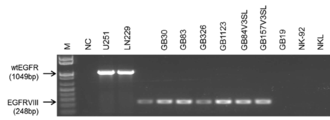
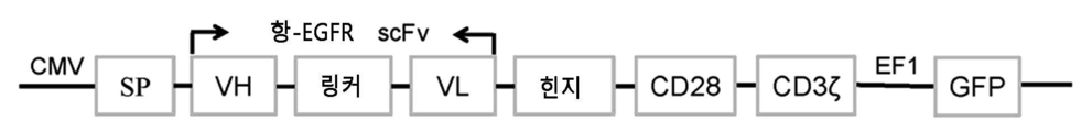
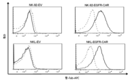
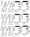

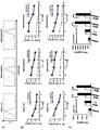
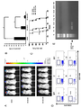
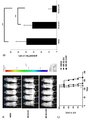
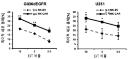


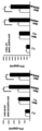

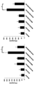
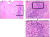


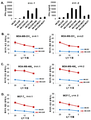
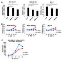
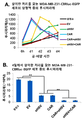


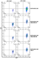

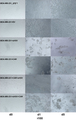
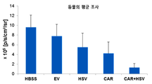
Classifications
-
- C—CHEMISTRY; METALLURGY
- C07—ORGANIC CHEMISTRY
- C07K—PEPTIDES
- C07K14/00—Peptides having more than 20 amino acids; Gastrins; Somatostatins; Melanotropins; Derivatives thereof
- C07K14/435—Peptides having more than 20 amino acids; Gastrins; Somatostatins; Melanotropins; Derivatives thereof from animals; from humans
- C07K14/705—Receptors; Cell surface antigens; Cell surface determinants
- C07K14/70503—Immunoglobulin superfamily
- C07K14/7051—T-cell receptor (TcR)-CD3 complex
-
- A—HUMAN NECESSITIES
- A61—MEDICAL OR VETERINARY SCIENCE; HYGIENE
- A61K—PREPARATIONS FOR MEDICAL, DENTAL OR TOILETRY PURPOSES
- A61K39/00—Medicinal preparations containing antigens or antibodies
- A61K39/395—Antibodies; Immunoglobulins; Immune serum, e.g. antilymphocytic serum
-
- A—HUMAN NECESSITIES
- A61—MEDICAL OR VETERINARY SCIENCE; HYGIENE
- A61K—PREPARATIONS FOR MEDICAL, DENTAL OR TOILETRY PURPOSES
- A61K35/00—Medicinal preparations containing materials or reaction products thereof with undetermined constitution
- A61K35/12—Materials from mammals; Compositions comprising non-specified tissues or cells; Compositions comprising non-embryonic stem cells; Genetically modified cells
- A61K35/14—Blood; Artificial blood
- A61K35/17—Lymphocytes; B-cells; T-cells; Natural killer cells; Interferon-activated or cytokine-activated lymphocytes
-
- A—HUMAN NECESSITIES
- A61—MEDICAL OR VETERINARY SCIENCE; HYGIENE
- A61K—PREPARATIONS FOR MEDICAL, DENTAL OR TOILETRY PURPOSES
- A61K39/00—Medicinal preparations containing antigens or antibodies
- A61K39/0005—Vertebrate antigens
- A61K39/0011—Cancer antigens
- A61K39/001102—Receptors, cell surface antigens or cell surface determinants
- A61K39/001103—Receptors for growth factors
- A61K39/001104—Epidermal growth factor receptors [EGFR]
-
- A—HUMAN NECESSITIES
- A61—MEDICAL OR VETERINARY SCIENCE; HYGIENE
- A61K—PREPARATIONS FOR MEDICAL, DENTAL OR TOILETRY PURPOSES
- A61K39/00—Medicinal preparations containing antigens or antibodies
- A61K39/46—Cellular immunotherapy
- A61K39/461—Cellular immunotherapy characterised by the cell type used
- A61K39/4613—Natural-killer cells [NK or NK-T]
-
- A—HUMAN NECESSITIES
- A61—MEDICAL OR VETERINARY SCIENCE; HYGIENE
- A61K—PREPARATIONS FOR MEDICAL, DENTAL OR TOILETRY PURPOSES
- A61K39/00—Medicinal preparations containing antigens or antibodies
- A61K39/46—Cellular immunotherapy
- A61K39/463—Cellular immunotherapy characterised by recombinant expression
- A61K39/4631—Chimeric Antigen Receptors [CAR]
-
- A—HUMAN NECESSITIES
- A61—MEDICAL OR VETERINARY SCIENCE; HYGIENE
- A61K—PREPARATIONS FOR MEDICAL, DENTAL OR TOILETRY PURPOSES
- A61K39/00—Medicinal preparations containing antigens or antibodies
- A61K39/46—Cellular immunotherapy
- A61K39/464—Cellular immunotherapy characterised by the antigen targeted or presented
- A61K39/4643—Vertebrate antigens
- A61K39/4644—Cancer antigens
- A61K39/464402—Receptors, cell surface antigens or cell surface determinants
- A61K39/464403—Receptors for growth factors
- A61K39/464404—Epidermal growth factor receptors [EGFR]
-
- A—HUMAN NECESSITIES
- A61—MEDICAL OR VETERINARY SCIENCE; HYGIENE
- A61P—SPECIFIC THERAPEUTIC ACTIVITY OF CHEMICAL COMPOUNDS OR MEDICINAL PREPARATIONS
- A61P35/00—Antineoplastic agents
-
- C—CHEMISTRY; METALLURGY
- C07—ORGANIC CHEMISTRY
- C07K—PEPTIDES
- C07K14/00—Peptides having more than 20 amino acids; Gastrins; Somatostatins; Melanotropins; Derivatives thereof
- C07K14/435—Peptides having more than 20 amino acids; Gastrins; Somatostatins; Melanotropins; Derivatives thereof from animals; from humans
- C07K14/705—Receptors; Cell surface antigens; Cell surface determinants
- C07K14/70503—Immunoglobulin superfamily
- C07K14/70521—CD28, CD152
-
- C—CHEMISTRY; METALLURGY
- C07—ORGANIC CHEMISTRY
- C07K—PEPTIDES
- C07K16/00—Immunoglobulins [IGs], e.g. monoclonal or polyclonal antibodies
- C07K16/18—Immunoglobulins [IGs], e.g. monoclonal or polyclonal antibodies against material from animals or humans
- C07K16/28—Immunoglobulins [IGs], e.g. monoclonal or polyclonal antibodies against material from animals or humans against receptors, cell surface antigens or cell surface determinants
- C07K16/2863—Immunoglobulins [IGs], e.g. monoclonal or polyclonal antibodies against material from animals or humans against receptors, cell surface antigens or cell surface determinants against receptors for growth factors, growth regulators
-
- C—CHEMISTRY; METALLURGY
- C07—ORGANIC CHEMISTRY
- C07K—PEPTIDES
- C07K16/00—Immunoglobulins [IGs], e.g. monoclonal or polyclonal antibodies
- C07K16/18—Immunoglobulins [IGs], e.g. monoclonal or polyclonal antibodies against material from animals or humans
- C07K16/28—Immunoglobulins [IGs], e.g. monoclonal or polyclonal antibodies against material from animals or humans against receptors, cell surface antigens or cell surface determinants
- C07K16/30—Immunoglobulins [IGs], e.g. monoclonal or polyclonal antibodies against material from animals or humans against receptors, cell surface antigens or cell surface determinants from tumour cells
-
- C—CHEMISTRY; METALLURGY
- C12—BIOCHEMISTRY; BEER; SPIRITS; WINE; VINEGAR; MICROBIOLOGY; ENZYMOLOGY; MUTATION OR GENETIC ENGINEERING
- C12N—MICROORGANISMS OR ENZYMES; COMPOSITIONS THEREOF; PROPAGATING, PRESERVING, OR MAINTAINING MICROORGANISMS; MUTATION OR GENETIC ENGINEERING; CULTURE MEDIA
- C12N15/00—Mutation or genetic engineering; DNA or RNA concerning genetic engineering, vectors, e.g. plasmids, or their isolation, preparation or purification; Use of hosts therefor
- C12N15/09—Recombinant DNA-technology
- C12N15/63—Introduction of foreign genetic material using vectors; Vectors; Use of hosts therefor; Regulation of expression
- C12N15/79—Vectors or expression systems specially adapted for eukaryotic hosts
- C12N15/85—Vectors or expression systems specially adapted for eukaryotic hosts for animal cells
- C12N15/86—Viral vectors
-
- C—CHEMISTRY; METALLURGY
- C12—BIOCHEMISTRY; BEER; SPIRITS; WINE; VINEGAR; MICROBIOLOGY; ENZYMOLOGY; MUTATION OR GENETIC ENGINEERING
- C12N—MICROORGANISMS OR ENZYMES; COMPOSITIONS THEREOF; PROPAGATING, PRESERVING, OR MAINTAINING MICROORGANISMS; MUTATION OR GENETIC ENGINEERING; CULTURE MEDIA
- C12N5/00—Undifferentiated human, animal or plant cells, e.g. cell lines; Tissues; Cultivation or maintenance thereof; Culture media therefor
- C12N5/06—Animal cells or tissues; Human cells or tissues
- C12N5/0602—Vertebrate cells
- C12N5/0634—Cells from the blood or the immune system
- C12N5/0636—T lymphocytes
-
- C—CHEMISTRY; METALLURGY
- C12—BIOCHEMISTRY; BEER; SPIRITS; WINE; VINEGAR; MICROBIOLOGY; ENZYMOLOGY; MUTATION OR GENETIC ENGINEERING
- C12N—MICROORGANISMS OR ENZYMES; COMPOSITIONS THEREOF; PROPAGATING, PRESERVING, OR MAINTAINING MICROORGANISMS; MUTATION OR GENETIC ENGINEERING; CULTURE MEDIA
- C12N5/00—Undifferentiated human, animal or plant cells, e.g. cell lines; Tissues; Cultivation or maintenance thereof; Culture media therefor
- C12N5/06—Animal cells or tissues; Human cells or tissues
- C12N5/0602—Vertebrate cells
- C12N5/0634—Cells from the blood or the immune system
- C12N5/0646—Natural killers cells [NK], NKT cells
-
- A—HUMAN NECESSITIES
- A61—MEDICAL OR VETERINARY SCIENCE; HYGIENE
- A61K—PREPARATIONS FOR MEDICAL, DENTAL OR TOILETRY PURPOSES
- A61K39/00—Medicinal preparations containing antigens or antibodies
- A61K2039/51—Medicinal preparations containing antigens or antibodies comprising whole cells, viruses or DNA/RNA
- A61K2039/515—Animal cells
- A61K2039/5156—Animal cells expressing foreign proteins
-
- A—HUMAN NECESSITIES
- A61—MEDICAL OR VETERINARY SCIENCE; HYGIENE
- A61K—PREPARATIONS FOR MEDICAL, DENTAL OR TOILETRY PURPOSES
- A61K39/00—Medicinal preparations containing antigens or antibodies
- A61K2039/51—Medicinal preparations containing antigens or antibodies comprising whole cells, viruses or DNA/RNA
- A61K2039/515—Animal cells
- A61K2039/5158—Antigen-pulsed cells, e.g. T-cells
-
- A—HUMAN NECESSITIES
- A61—MEDICAL OR VETERINARY SCIENCE; HYGIENE
- A61K—PREPARATIONS FOR MEDICAL, DENTAL OR TOILETRY PURPOSES
- A61K39/00—Medicinal preparations containing antigens or antibodies
- A61K2039/80—Vaccine for a specifically defined cancer
-
- C—CHEMISTRY; METALLURGY
- C07—ORGANIC CHEMISTRY
- C07K—PEPTIDES
- C07K2317/00—Immunoglobulins specific features
- C07K2317/60—Immunoglobulins specific features characterized by non-natural combinations of immunoglobulin fragments
- C07K2317/62—Immunoglobulins specific features characterized by non-natural combinations of immunoglobulin fragments comprising only variable region components
- C07K2317/622—Single chain antibody (scFv)
-
- C—CHEMISTRY; METALLURGY
- C07—ORGANIC CHEMISTRY
- C07K—PEPTIDES
- C07K2319/00—Fusion polypeptide
- C07K2319/01—Fusion polypeptide containing a localisation/targetting motif
- C07K2319/03—Fusion polypeptide containing a localisation/targetting motif containing a transmembrane segment
-
- C—CHEMISTRY; METALLURGY
- C07—ORGANIC CHEMISTRY
- C07K—PEPTIDES
- C07K2319/00—Fusion polypeptide
- C07K2319/33—Fusion polypeptide fusions for targeting to specific cell types, e.g. tissue specific targeting, targeting of a bacterial subspecies
-
- C—CHEMISTRY; METALLURGY
- C12—BIOCHEMISTRY; BEER; SPIRITS; WINE; VINEGAR; MICROBIOLOGY; ENZYMOLOGY; MUTATION OR GENETIC ENGINEERING
- C12N—MICROORGANISMS OR ENZYMES; COMPOSITIONS THEREOF; PROPAGATING, PRESERVING, OR MAINTAINING MICROORGANISMS; MUTATION OR GENETIC ENGINEERING; CULTURE MEDIA
- C12N2740/00—Reverse transcribing RNA viruses
- C12N2740/00011—Details
- C12N2740/10011—Retroviridae
- C12N2740/15011—Lentivirus, not HIV, e.g. FIV, SIV
- C12N2740/15041—Use of virus, viral particle or viral elements as a vector
- C12N2740/15043—Use of virus, viral particle or viral elements as a vector viral genome or elements thereof as genetic vector
Landscapes
- Health & Medical Sciences (AREA)
- Life Sciences & Earth Sciences (AREA)
- Chemical & Material Sciences (AREA)
- Immunology (AREA)
- Organic Chemistry (AREA)
- General Health & Medical Sciences (AREA)
- Genetics & Genomics (AREA)
- Medicinal Chemistry (AREA)
- Engineering & Computer Science (AREA)
- Cell Biology (AREA)
- Biochemistry (AREA)
- Zoology (AREA)
- Biomedical Technology (AREA)
- Microbiology (AREA)
- Biophysics (AREA)
- Molecular Biology (AREA)
- Proteomics, Peptides & Aminoacids (AREA)
- Bioinformatics & Cheminformatics (AREA)
- Animal Behavior & Ethology (AREA)
- Pharmacology & Pharmacy (AREA)
- Public Health (AREA)
- Veterinary Medicine (AREA)
- Biotechnology (AREA)
- Wood Science & Technology (AREA)
- Epidemiology (AREA)
- Mycology (AREA)
- Gastroenterology & Hepatology (AREA)
- Toxicology (AREA)
- General Engineering & Computer Science (AREA)
- Hematology (AREA)
- Dermatology (AREA)
- Oncology (AREA)
- Chemical Kinetics & Catalysis (AREA)
- Nuclear Medicine, Radiotherapy & Molecular Imaging (AREA)
- General Chemical & Material Sciences (AREA)
- Virology (AREA)
- Plant Pathology (AREA)
- Physics & Mathematics (AREA)
- Medicines Containing Material From Animals Or Micro-Organisms (AREA)
- Micro-Organisms Or Cultivation Processes Thereof (AREA)
Abstract
교아종(GB)은 가장 적극적 원발성 뇌 악성종양이고; 뇌 전이, 예를 들면, 유방암 뇌 전이(BCBM)는 또한 공격적이고 불량한 예후와 연관되어 있다. 키메라 항원 수용체(CAR)-변형된 면역 세포의 양자 전이는 유망한 항암 접근법으로 부상되었지만, 여전히 뇌암을 치료하기 위한 CAR-조작된 세포의 잠재적 유용성은 검토되어 있지 않았다. 본 발명은 뇌암, 예를 들면, GB 및 BCBM을 포함하는 다양한 암의 치료에서 CAR 발현 세포를 사용하는 조성물 및 방법을 제공한다.Hybridization (GB) is the most active primary brain malignancy; Brain metastasis, for example, breast cancer brain metastasis (BCM) is also associated with aggressive and poor prognosis. Although the proton transfer of chimeric antigen receptor (CAR) -transfected immune cells has emerged as a promising anticancer approach, the potential utility of CAR-engineered cells to treat brain cancer has not yet been investigated. The present invention provides compositions and methods that use CAR-expressing cells in the treatment of various cancers including brain cancer, e. G., GB and BCBM.

Description
관련 출원의 상호-참조Cross-reference of related application - reference
본 출원은 2015년 4월 6일자로 출원된 미국 가출원 제62/143,744호에 대한 35 U.S.C. § 119(e) 하의 우선권을 주장하고, 이의 내용은 이의 전체가 본원에서 참조로서 도입된다.This application claims the benefit of U.S. Provisional Application No. 62 / 143,744, filed April 6, 2015, Priority under § 119 (e), the content of which is hereby incorporated by reference in its entirety.
배경기술Background technology
뇌암은 위험하고 치료가 곤란하다. 교아세포종(glioblastoma: GB)은 가장 통상적 및 가장 적극적 원발성 뇌 종양이다. 뇌의 암은 또한 또 다른 공급원으로부터 전이의 결과인 경우에 위험을 갖고; 예를 들면, 유방암뇌 전이(BCBM)은 전이성 유방암을 갖는 환자에서 통상적이다. 화학요법, 방사선 및 외과수술 절제에 의해서도, GB 환자의 전체 중앙 생존기간은 단지 14.6개월이다[참조: Morgan, R.A. et al. (2012) Hum Gene Ther. 23:1043-1053]. BCBM은 또한 이용가능한 치료에 대해 고도로 내성이고, 환자의 불량한 예후를 나타낸다. 종래의 치료법은 일반적으로 특이성을 결여하고, 주변 뇌 실질 및 전신 조직에 대한 손상, 이들의 사용을 제한하는 인자를 유발할 수 있다[참조: Imperato, J.P. et al. (1990) Ann Neurol. 28:818-822]. GB에 대한 면역-기반 요법은 국재화된 및 종양 침윤 세포 둘 다를 표적화할 가능성이 있는, 지속가능한 항-종양 반응의 잠재적 장기간 잇점이 있는 종래 치료법에 대한 유망한 대체물이다[참조: Mellman, I. et al. (2011) Nature 480:480-489]. 상피 성장 인자 수용체(Epidermal Growth Factor Receptor: EGFR)는 GB를 포함하는 다양한 종양에서 중요한 역할을 담당한다. EGFR은 GB에서 가장 빈번하게 증폭된 유전자이고, 정상 뇌 조직에서 이의 발현은 검출 불가능하거나 극히 낮다[참조: Parsons, D.W. et al. (2008) Science 321:1807-1812; Salomon, D.S. et al. (1995) Crit. Rev Oncol Hematol. 19:183-232]. EGFR에 대한 리간드의 결합은 수용체 호모- 및 헤테로이량체 형성을 유도하고, 몇몇 중요한 티로신 잔기의 자가포스포릴화는 Ras/Raf/MEK/ERK 경로, PLCγ-PKC 경로 및 PI3K/AKT 경로를 포함하는 몇몇 세포내 하류 신호전달 경로의 활성화를 유도하여 세포 증식, 운동성 및 생존을 제공한다[참조: Zandi, R. et al. (2007) Cell Signal 19:2013-2023]. 대략 20-40%의 EGFR-증폭된 종양은 EGFR 변이체 III 돌연변이체(EGFRvIII)를 포함하고, 이는 세포외 리간드-결합 도메인에서 엑손 2-7의 결실을 함유한다[참조: Aldape, K.D. et al. (2004) Journal of Neuropathology and Experimental Neurology 63:700-707; Biernat, W. et al. (2004) Brain Pathol. 14:131-136; Fan, Q.W. et al. (2013) Cancer Cell 24:438-449; Sugawa, N. et al. (1990) Proc Natl Acad Sci. USA 87:8602-8606]. 이러한 돌연변이체 형태는 종양-촉진 신호전달 경로를 활성화하는 리간드의 부재하에 구조적 활성화를 나타낸다[참조: Ohno, M. et al. (2010) Cancer Sci. 101:2518-2524]. Brain cancer is dangerous and difficult to treat. Glioblastoma (GB) is the most common and most active primary brain tumor. Cancer of the brain is also at risk if it is the result of metastasis from another source; For example, breast cancer brain metastasis (BCBM) is common in patients with metastatic breast cancer. By chemotherapy, radiation and surgical resection, the overall median survival time for GB patients is only 14.6 months (Morgan, R.A. et al. (2012) Hum Gene Ther. 23: 1043-1053). BCBM is also highly resistant to available treatments and exhibits poor prognosis in patients. Conventional treatments generally result in lack of specificity, damage to peripheral brain parenchyma and systemic tissue, factors limiting their use (see Imperato, J.P. et al. (1990) Ann Neurol. 28: 818-822). Immune-based therapy for GB is a promising alternative to conventional therapy with potential long-term benefits of a sustainable anti-tumor response, potentially targeting both localized and tumor-infiltrating cells (Mellman, I. et al. (2011) Nature 480: 480-489. Epidermal Growth Factor Receptor (EGFR) plays an important role in various tumors including GB. EGFR is the most frequently amplified gene in GB and its expression in normal brain tissue is either undetectable or extremely low (Parsons, D.W. et al. (2008) Science 321: 1807-1812; Salomon, D.S. et al. (1995) Crit. Rev Oncol Hematol. 19: 183-232). The binding of the ligand to EGFR induces receptor homo- and heterodimer formation and autophosphorylation of several important tyrosine residues leads to the formation of the Ras / Raf / MEK / ERK pathway, the PLCγ-PKC pathway and the PI3K / AKT pathway Induce activation of intracellular downstream signaling pathways to provide cell proliferation, motility and survival (Zandi, R. et al. (2007) Cell Signal 19: 2013-2023]. Approximately 20-40% of EGFR-amplified tumors contain the EGFR mutant III mutant (EGFRvIII), which contains a deletion of exons 2-7 in the extracellular ligand-binding domain (Aldape, K.D. et al. (2004) Journal of Neuropathology and Experimental Neurology 63: 700-707; Biernat, W. et al. (2004) Brain Pathol. 14: 131-136; Fan, Q.W. et al. (2013) Cancer Cell 24: 438-449; Sugawa, N. et al. (1990) Proc Natl Acad Sci. USA 87: 8602-8606. Such mutant forms exhibit structural activation in the absence of a ligand that activates the tumor-promoted signaling pathway (Ohno, M. et al. (2010) Cancer Sci. 101: 2518-2524].
교아세포종(GB)은 가장 적극적 원발성 뇌 악성종양이다. 키메라 항원 수용체(CAR)-변형된 면역 세포의 양자 전이는 유망한 항암 접근법으로 부상하고 있지만, GB를 치료하기 위한 CAR-조작된 천연 킬러(NK) 세포의 잠재적 유용성은 연구되어 있지 않다. 대략 50%의 GB 환자의 종양은 야생형 EGFR(wt EGFR)을 발현하고, 소수의 경우에는 wtGFFR 및 돌연변이체 형태 EGFRvIII을 발현하지만; 이전에 보고된 CAR T 세포는 단지 EGFRvIII의 표적화에 초점을 맞추고 있다.Glioblastoma (GB) is the most active primary brain malignancy. Although the proton transfer of chimeric antigen receptor (CAR) -transformed immune cells has emerged as a promising anticancer approach, the potential utility of CAR-engineered natural killer (NK) cells for the treatment of GB has not been studied. Tumors of approximately 50% of GB patients express wild-type EGFR (wt EGFR), minority cases wtGFFR and mutant form EGFRvIII; Previously reported CAR T cells have focused only on the targeting of EGFRvIII.
본 발명의 양태는, (a) 항원 결합 도메인; (b) 힌지 도메인; (c) 막관통 도메인; 및 (d) 세포내 도메인을 포함하는 키메라 항원 수용체(CAR)에 관한 것이다. 이러한 발명은, (a) 야생형 및/또는 돌연변이체 상피 성장 인자 수용체(EGFR)("wt EGFR 및 돌연변이체 EGFR") 둘 다를 인식하는 항-EGFR 항체의 항원 결합 도메인; (b) 힌지 도메인 폴리펩티드; (c) 공자극 폴리펩티드; 및 (d) CD3 제타(zeta) 신호전달 도메인을 포함하거나, 또는 달리는 이들로 본질적으로 이루어지거나, 또는 이들로 이루어지는, 신규한 CAR-상피 성장 인자 수용체("EGFR") 키메라 항원 수용체(CAR)를 제공한다. 한 가지 양태에서, 공자극 분자는 세포내 도메인 및 막관통 도메인을 포함한다. 비-제한적 예는 CD8, 4-1BB 공자극 신호전달 영역, CD28 공자극 분자, OX40, ICOS 및 CD27을 포함한다. 한 가지 양태에서, 항-EGFR 항체의 항원 결합 도메인은 항-EGFR 중쇄(HC) 가변 영역 및 항-EGFR 경쇄(LC) 가변 영역을 포함한다. 추가의 양태에서, EGFR CAR은 항-EGFR HC 가변 영역 및 항-EGFR LC 가변 영역 사이에 위치된 링커 폴리펩티드를 추가로 포함하거나, 또는 달리는 이들로 본질적으로 이루어지거나, 또는 추가로 이들로 이루어진다.An embodiment of the present invention is directed to a method of producing a polypeptide comprising: (a) an antigen binding domain; (b) a hinge domain; (c) membrane penetration domain; And (d) a chimeric antigen receptor (CAR) comprising an intracellular domain. This invention provides a kit comprising (a) an antigen binding domain of an anti-EGFR antibody that recognizes both a wild type and / or mutant epithelial growth factor receptor (EGFR) ("wt EGFR and mutant EGFR"); (b) a hinge domain polypeptide; (c) a co-stimulatory polypeptide; ("EGFR") chimeric antigen receptor (CAR), which consists essentially of, or consists of, (d) a CD3 zeta signaling domain comprising, to provide. In one embodiment, the co-stimulatory molecule comprises an intracellular domain and a transmembrane domain. Non-limiting examples include CD8, 4-1BB co-stimulatory signal transduction domain, CD28 co-stimulatory molecule, OX40, ICOS, and CD27. In one embodiment, the antigen binding domain of an anti-EGFR antibody comprises an anti-EGFR heavy chain (HC) variable region and an anti-EGFR light chain (LC) variable region. In a further embodiment, the EGFR CAR consists essentially of, or additionally consists of, those further comprising, or running, the linker polypeptide located between the anti-EGFR HC variable region and the anti-EGFR LC variable region.
EGFR CAR을 코딩하는 폴리뉴클레오티드 및 이들의 상보체 및 각각의 등가물이 추가로 본원에 개시되어 있다. 폴리뉴클레오티드 및/또는 EGFR CAR을 함유하는 벡터 및 숙주 세포가 본원에서 제공된다. 벡터는 플라스미드 또는 벡터이다. 세포는 원핵세포 또는 진핵세포일 수 있다. 한 가지 양태에서, 세포는 T 세포 또는 NK 세포이다. 세포는 확장될 수 있고, 따라서 세포의 확장된 모집단이 본원에서 추가로 제공된다. 세포는, 한 가지 양태에서, T 세포 등의 세포의 활성화된 모집단이다.Polynucleotides encoding EGFR CARs and their complement and their respective equivalents are further disclosed herein. Polynucleotides and / or vectors containing EGFR CAR and host cells are provided herein. The vector is a plasmid or vector. Cells can be prokaryotic or eukaryotic. In one embodiment, the cell is a T cell or an NK cell. The cells can be expanded, and thus an expanded population of cells is additionally provided herein. A cell is, in one embodiment, an activated population of cells, such as T cells.
이들 조성물의 진단적 및 치료적 용도가 본원에서 추가로 제공된다. 예를 들면, 이들 세포를 본원에 기재된 바와 같은 EGFR CAR을 포함하는 유효량의 단리된 세포와 접촉시킴으로써 wt EGFR 또는 돌연변이체 EGFR, 또는 wt EGFR 및 돌연변이체 EGFR을, EGFR을 발현하는 세포 또는 줄기 세포의 표면 상에서 발현하는 세포 및/또는 종양의 하나 이상의 성장을 억제시키는 방법이 제공된다. 세포는 종양 세포, 예를 들면, 교아세포종 또는 암 줄기 세포, 예를 들면, 교아세포종 줄기 세포일 수 있다. 접촉은 시험관내 또는 생체내일 수 있다. 시험관내에서 실시하는 경우, 상기 방법은 신규한 요법 또는 병용 요법을 위한 스크리닝에 유용하다. 생체내에서, 상기 방법은 EGFR을 발현하는 암, 예를 들면, 뇌암, 예를 들면, 교아세포종 또는 뇌에서 또 다른 암(예: 유방암)의 전이를 앓고 있는 환자의 치료에 유용하다.Diagnostic and therapeutic uses of these compositions are further provided herein. For example, by contacting these cells with an effective amount of isolated cells comprising EGFR CAR as described herein, either wt EGFR or mutant EGFR, or wt EGFR and mutant EGFR can be used to treat EGFR expressing cells or stem cells There is provided a method of inhibiting growth of one or more cells and / or tumors expressing on a surface. The cell may be a tumor cell, for example, a glioblastoma or cancer stem cell, for example, a glioblastoma stem cell. The contact can be in vitro or in vivo. When performed in vitro, the methods are useful for screening for novel therapies or combination therapies. In vivo, the method is useful for the treatment of patients with cancer expressing EGFR, for example, brain cancer such as glioblastoma or another cancer in the brain (e.g. breast cancer).
또 다른 양태에서, 본 발명은, EGFR 또는 돌연변이체 EGFR, 또는 wt EGFR 및 돌연변이체 EGFR을 이를 필요로 하는 대상체의 세포 표면 상에서 발현하는 하나 이상의 세포의 성장을 억제시키는 방법을 제공한다. 이 방법은, 본원에 기재된 바와 같은 EGFR CAR을 포함하는 유효량의 단리된 세포를 대상체에게 투여하는 것을 포함하거나, 또는 달리는 이들로 본질적으로 이루어지거나, 또는 추가로 이들로 이루어진다. EGFR을 발현하는 세포는 종양 세포 또는 암 줄기 세포, 예를 들면, 교아세포종 또는 교아세포종 줄기 세포일 수 있다. EGFR CAR을 발현하는 단리된 세포는 상기 세포를 수용하는 대상체에 대해 자기유래(autologous)일 수 있다. 추가로, 본원에 기재된 바와 같은 EGFR CAR을 포함하는 유효량의 단리된 세포를 대상체에게 투여함으로써, 이를 필요로 하는 대상체에서 암을 치료하는 방법(여기서, 상기 암 세포는 wt EGFR 또는 돌연변이체 EGFR, 또는 wt EGFR 및 돌연변이체 EGFR을 발현한다)이 제공된다. 이들 방법의 단리된 세포는 NK 세포 및/또는 T-세포일 수 있고, 치료되는 대상체에 대해 자기유래일 수 있다. 추가의 양태에서, 세포는, 치료하는 의사에 의해 결정되는 임의의 적절한 경로에 의해 투여되고, 제한 없이, 두개내 주사, 정맥내 투여를 포함한다.In another aspect, the invention provides a method of inhibiting the growth of one or more cells expressing EGFR or a mutant EGFR, or a wt EGFR and a mutant EGFR on the cell surface of a subject in need thereof. The method comprises, or alternatively consists essentially of, or consists of administering to the subject an effective amount of an isolated cell comprising an EGFR CAR as described herein. The cells expressing EGFR may be tumor cells or cancer stem cells, for example, glioblastoma or glioblastoma stem cells. Isolated cells expressing EGFR CAR may be autologous to a subject receiving the cell. In addition, there is provided a method of treating cancer in a subject in need thereof, comprising administering to the subject an effective amount of an isolated cell comprising an EGFR CAR as described herein, wherein said cancer cell is a wt EGFR or a mutant EGFR, wt < / RTI > EGFR and the mutant EGFR). The isolated cells of these methods may be NK cells and / or T-cells and may be self-evident for the subject being treated. In a further embodiment, the cells are administered by any appropriate route as determined by the treating physician, and include, without limitation, intracranial injection, intravenous administration.
추가로, 본원에 기재된 EGFR CAR 및 EGFR 단백질 또는 이의 단편 및/또는 돌연변이체 EGFR 단백질 또는 이의 단편을 포함하는 단리된 복합체가 제공된다. 또 다른 양태에서, 본원에 기재된 바와 같은 EGFR CAR을 포함하는 단리된 복합체, 및 wt EGFR 또는 돌연변이체 EGFR, 또는 wt EGFR 및 돌연변이체 EGFR을 발현하는 세포가 제공된다.In addition, isolated complexes comprising the EGFR CAR and EGFR proteins described herein or fragments and / or mutant EGFR proteins or fragments thereof are provided. In another embodiment, isolated complexes comprising EGFR CAR as described herein, and cells expressing wt EGFR or mutant EGFR, or wt EGFR and mutant EGFR, are provided.
본 발명의 추가의 양태는, 본원에 기재된 EGFR CAR 발현 세포, 예를 들면, T 세포 또는 NK 세포, 및 종양용해성 단순 헤르페스 바이러스를 사용한 조성물 및 병용 암 요법에 관한 것이다. 일부 실시형태는, EGFR CAR 발현 세포 및 종양용해성 헤르페스 심플렉스 둘 다를 이를 필요로 하는 환자에게 투여하는, 종양 또는 암의 치료 방법에 관한 것이다. 일부 실시형태에서, EGFR CAR 발현 세포는 종양용해성 단순 헤르페스 바이러스와 동시에 투여되고; 다른 실시형태에서, EGFR CAR 발현 세포는 종양용해성 단순 헤르페스 바이러스의 투여 전후에 투여된다.A further aspect of the present invention relates to compositions and methods of combined cancer therapy using the EGFR CAR-expressing cells described herein, such as T cells or NK cells, and tumor-solving herpes simplex virus. Some embodiments relate to methods of treating tumors or cancers in which both EGFR CAR-expressing cells and tumor-soluble herpes simplex are administered to a patient in need thereof. In some embodiments, the EGFR CAR expressing cells are administered concurrently with the tumor-solving herpes simplex virus; In another embodiment, the EGFR CAR expressing cells are administered before or after the administration of the tumor-solving herpes simplex virus.
하나 이상의 상기 조성물을 포함하는 키트가 추가로 제공된다. 이들을 진단학적으로 또는 치료학적으로 사용하는 지침이 추가로 제공된다.A kit comprising one or more of the above compositions is additionally provided. Additional guidance is available on the use of these for diagnostic or therapeutic purposes.
도 1A-1B는 GB 및 GB 줄기 세포 상에서 EGFR의 발현을 나타낸다. (도 1A) 신경교종(glioma) 세포주(U251, LN229 및 Gli36dEGFR) 및 신경교종 줄기 세포(GB30, GB83, GB326, GB1123, GB84V3SL, GB157V3SL 및 GB19) 상에서 표면 EGFR 발현은 유세포 계수법에 의해 모니터링했다. NK 세포주 NKL 및 NK-92를 음성 대조군으로 사용한다. (도 1B) 도 1A에 명시된 신경교종 세포주 및 신경교종 줄기 세포에서 RT-PCR에 의한 EGFR mRNA 발현의 측정. 유동 플롯 및 데이터는 3개 독립적 실험의 대표예이다.
도 2A-2B는 EGFR-특이적 CAR의 생성 및 CAR-형질도입된 NK 세포 상에서 이의 발현의 검출을 나타낸다. (도 2A) EGFR-CAR 렌티바이러스 작제물의 모식도. (도 2B) EGFR-CAR 작제물(각각 NK-92-EGFR-CAR 및 NKL-EGFR-CAR) 또는 공 벡터 작제물(EV)로 형질도입된 NK-92 및 NKL 세포의 표면 상에서 키메라 EGFR scFv의 발현. 세포를 GFP 발현에 의해 FACS-분류하고, 이어서 세포를 항-마우스 Fab 항체(실선) 또는 IgG 이소형 대조군(점선)으로 염색한 후에 유세포 계수에 의해 분석했다. SP, 신호 펩티드; VH, 중쇄; VL, 경쇄; ScFv, 단일 사슬 가변 단편. 유동 플롯 및 데이터는 3개 독립적 실험의 대표예이다.
도 3A-3B는 EGFR-CAR-변형된 NK-92 및 NKL 세포가 EGFR+ GB 세포주 세포를 인식하고 사멸시키는 것을 나타낸다. (도 3A) 표준 크롬-51 방출 검정을 사용하여 Gli36dEGFR, LN229 또는 U251에 대한 공 벡터(EV)-형질도입된 NK-92 및 NKL 세포의 세포독성 활성. (도 3B) ELISA 검정을 사용하여 Gli36dEGFR, LN229 또는 U251 세포의 부재 또는 존재하에 공 벡터 EV-형질도입된 또는 EGFR-CAR-형질도입된 NK-92 및 NKL 세포의 IFN-γ 방출. 3개 독립적 실험의 대표적 데이터가 제시되어 있다. *p < 0.05; **p < 0.01.
도 4A-4B는 EGFR-CAR-변형된 NK-92 및 NKL 세포가 EGFR+ GB 줄기 세포의 증강된 용해를 나타냄을 보여준다. (도 4A) 크롬-51 방출 검정을 사용하여 GB1123, GB30, GB157V3SL 및 GB84V3SL GB 줄기 세포에 대한 NK-92-EV 또는 NK-92-EGFR-CAR 세포(상부 패널) 및 NKL-EV 또는 NKL-EGFR-CAR 세포(하부 패널)의 세포독성 활성. (도 4B) GB 줄기 세포와 공-배양하는 경우, NK-92-EGFR-CAR 또는 NK-92-EV 세포(상부 패널) 및 NKL-EV 또는 NKL-EGFR-CAR 세포(하부 패널)에 의한 IFN-γ 분비의 ELISA 분석. 3개 독립적 실험의 대표적 데이터가 제시되어 있다. *p < 0.05; **p < 0.01.
도 5A-5C는 NK-92-EGFR-CAR 세포의 증강된 표적 인식이 세포 표면 상에서 EGFR의 발현에 의존적임을 나타낸다. (도 5A) 공 벡터 또는 wt EGFR 또는 EGFRvIII을 함유하는 벡터로 형질감염시킨 GB19 세포의 표면 상에서 EGFR 발현을 검출하기 위해 항-EGFR 항체(실선) 또는 IgG 이소형 대조군(점선)을 사용한 유세포 계수. (도 5B) 도 5A에 제시된 GB19, GB19-wt EGFR 및 GB19-EGFRvIII 세포에 대한 NK-92-EV 또는 NK-92-EGFR-CAR(상부 패널) 및 NKL-EV 또는 NKL-EGFR-CAR(하부 패널)의 세포독성. GB19 세포를 4시간 동안 다양한 효과기/표적(E/T) 비율로 NK-92 또는 NKL 세포와 함께 배양했다. 종양 용해는 크롬-51 방출 검정을 사용하여 측정했다. (도 5C) NKL-EV 또는 NKL-EGFR-CAR(좌측) 및 NK-92-EGFR-CAR 또는 NK-92-EV 세포(우측)을 24시간 동안 동일한 수의 GB19-벡터 또는 GB19-EGFR 세포와 함께 배양했다. 이어서, 상청액은 ELISA를 사용하여 IFN-γ 분비의 측정을 위해 수집했다. 유동 플롯 및 데이터는 3개 독립적 실험의 대표예이다. *p < 0.05, **p < 0.01.
도 6A-6E는 NK-92-EGFR-CAR 세포가 동소성 인간 신경교종 줄기 세포의 생체내 성장을 억제하고 교아세포종-함유 마우스의 생존을 연장시키고 기타 기관 및 조직으로 이행하지 않고서 뇌에서 국지화하는 것을 나타낸다. (도 6A) GB30 종양을 갖는 마우스의 뇌 생물발광 이미지. NSG 마우스를 정립 주사에 의해 루시퍼라제-발현 GB30 세포로 접종시켰다(0일). 접종 7일 후, 마우스를 공 벡터-형질도입된 NK-92 세포(NK-92-EV), EGFR-CAR-형질도입된 NK-92 세포(NK-92-EGFR-CAR) 또는 행크 평형 염 용액(HBSS; 음성 대조군)으로 1회 두개내 주입했다. (도 6B) 도 6A의 마우스당 초당 양자 단위의 정량화 요약. *는 p < 0.05를 나타낸다. (도 6C) NK-92-EGFR-CAR 세포로 처리된 GB30-담지 마우스는, 카플란-마이어(Kaplan-Meier) 생존 곡선에 의해 측정하는 경우, NK-92-EV 세포 또는 HBSS(** p < 0.01)와 비교하여 현저히 증가된 전체 생존을 나타냈다(각 그룹에 대해 n = 5). (도 6D) GB30-담지 마우스 내로 CAR NK 세포의 두개내 주사 3일 후에 간, 폐, 혈액, 비장, 골수(BM) 및 뇌에서 유세포 계수법에 의해 CD56+CD3- 인간 EGFR-CAR NK-92 세포의 존재의 측정. (도 6E) GB30-담지 마우스 내로 CAR NK 세포의 두개내 주사 3일 후에 간, 폐, 혈액, 비장, 골수(BM) 및 뇌에서 RT-PCR에 의해 EGFR-CAR 발현의 측정. NC = 음성 대조군(DNA 주형을 첨가하지 않음); PC = 양성 대조군, EGFR-CAR NK-92 세포. *p < 0.05, **p < 0.01.
도 7A-7C는 EGFR-CAR-형질도입된 NK-92 세포가 wt EGFR-발현 GB 종양 성장을 억제시키고 동소성 이종이식 GB 모델에서 종양-담지 마우스의 생존을 연장시키는 것을 나타낸다. (도 7A) U251 종양을 갖는 마우스의 뇌 생물발광 이미지. NSG 마우스에게 정립 주사(0일)에 의해 105 루시퍼라제-발현 U251 세포를 두개내 이식했다. 접종 10일, 40일 및 70일 후, 마우스에게 NK-92-EV 세포, NK-92-EGFR-CAR 세포 또는 음성 대조군으로서 HBSS를 두개내 주입했다. 마우스의 뇌 생물발광 이미지를 100일에 촬영했다. (도 7B) 도 7A로부터의 마우스당 초당 양자 단위의 정량화 요약. **는 p < 0.01을 나타낸다. (도 7C) NK-92-EGFR-CAR 세포로 처리된 U251-담지 마우스는, 카플란-마이어 생존 곡선에 의해 측정하는 경우, EK-92-EV 세포(* p < 0.05) 또는 HBSS(** p < 0.01)로 처리한 마우스와 비교하여 현저히 증가된 전체 생존을 나타낸다(각 그룹에 대해 n = 5).
도 8A-8B는 EGFR-CAR 일차 NK 세포가 EGFR+ GB 세포 및 환자-유래 GB 줄기 세포의 증강된 근절을 나타냄을 보여준다. (도 8A) 위-형질도입된 일차 NK 세포(일차 NK-EV)와 비교하여 EGFR+ Gli36dEGFR 및 U251 GB 세포주에 대해 증가된 세포독성 활성을 나타낸 EGFR-CAR-변형된 일차 NK 세포(일차 NK-CAR). (도 8B) 위-형질도입된 일차 NK 세포(일차 NK-EV)와 비교하여 EGFR+-환자-유래된 GB30 및 GB157V3SL 줄기 세포에 대해 증강된 세포독성을 나타낸 EGFR-CAR-변형된 NK 세포(일차 NK-CAR). 제시된 데이터는, 상이한 건강한 공여체로부터 단리한 NK 세포를 조사한, 유사한 결과를 갖는 3개 실험의 대표예이다. *p < 0.05; **p < 0.01.
도 9A-9C는 NK-92-EGFR-CAR 세포의 증강된 표적 인식이 세포 표면 상에서 EGFR의 발현에 의존적임을 나타낸다. (도 9A) 공 벡터(EV; 좌측), wt EGFR(중앙) 또는 EGFRvIII(우측)으로 형질도입된 293T 세포의 항-EGFR 항체(실선) 또는 IgG 이소형 대조군(점선)을 사용한 유세포 계수 분석. (도 9B) 293T-EV(좌측), 293T-EGFR(중앙) 및 293T-EGFRvIII(우측) 세포에 대한 NK-92-EV 또는 NK-92-EGFR-CAR(상부 패널) 및 NKL-EV 또는 NKL-EGFR-CAR(하부 패널)의 세포독성. 293T 세포를 4시간 동안 다양한 효과기/표적(E/T) 비율에서 NK 세포와 함께 배양했다. 종양 용해는 크롬-51 방출 검정을 사용하여 측정했다. (도 9C) 24시간 동안 표적 세포 및 효과기 세포의 공배양 후, 공-배양물로부터의 상청액은 ELISA를 사용하여 IFN-γ 분비에 대해 측정했다. 제시된 데이터는 유사한 결과를 갖는 3개 실험의 대표예이다. *p < 0.05; **p < 0.01.
도 10A-10B는 NK-92-EGFR-CAR 세포의 효과가 EGFR 차단 항체(Ab)에 의해 둔화되는 것을 나타낸다. (도 10A) EGFR-특이적 모노클로날 항체 528 또는 IgG-정합 이소형 대조군 항체로 전처리한 GB30 세포(좌측) 또는 U251 세포(우측)에 대한 NK-92-EV 또는 NK-92-EGFR-CAR의 세포독성. 표적 세포를 4시간 동안 다양한 효과기/표적(E/T) 비율에서 전처리된 NK-92-EV 또는 NK-92-EGFR-CAR 세포와 함께 배양했다. (도 10B) 24시간 동안 표적 세포 및 효과기 세포의 공-배양 후, 공-배양물로부터의 상청액은 ELISA를 사용하여 IFN-γ 분비에 대해 측정했다. 제시된 데이터는 유사한 결과를 갖는 3개 실험의 대표예이다. *p < 0.05; **p < 0.01. Ab= EGFR-특이적 모노클로날 항체 528; 이소= IgG-정합된 이소형 대조군 항체.
도 11은 NK-92-EGFR-CAR 세포가 종양내 주사 후에 종양 영역에 위치되어 있음을 나타낸다. NK-92-EGFR-CAR 세포를 GB30 이식 7일 후에 마우스 뇌에 종양내 주사했다. 마우스 뇌를 3일 후에 수거하고, 파라핀으로 매립하고, 헤마톡실린 및 에오신(H&E) 염색을 위해 처리했다. H&E 염색은 NK-92-EGFR-CAR 세포가 유일하게 종양 영역 내부에 존재했음을 나타냈다. 20×, 40× 및 100×의 확대가 제시되었다(물체: 2×, 4× 또는 10×; 접안렌즈: 10X).
도 12A-12B는 유방암 세포주 및 조직에서 EGFR의 발현을 나타낸다. 유방암 세포주(MDA-MB-231, MDA-MB-468 및 MCF-7)의 세포 표면 상에서 EGFR의 발현은 유세포 계수에 의해 검출했다(도 12A). 헤마톡실린 및 에오신(HE) 염색 및 EGFR 발현의 면역조직화학(IHC)은 일차 유방암 및 뇌 전이를 갖는 환자의 종양 조직에 대해 수행했다(도 12B).
도 13A-13E은 EGFR-CAR NK-92 세포가 유방암 세포주의 EGFR 양성 세포를 인식하고 이를 용해하는 것을 입증한다. EGFR-CAR-형질도입된 NK-92 세포 상에서 EGFR scFv의 발현은 염소 항-마우스 F(ab')2 폴리클로날 항체를 사용한 유세포 계수법에 의해 측정했다(도 13A). MDA-MB-231, MDA-MB-468 또는 MCF-7 세포의 부재 또는 존재하에 공 벡터(EV)-형질도입된 또는 EGFR-CAR-형질도입된 NK-92 세포에 의한 IFN-γ 방출은 표준 ELISA 검정을 사용하여 측정했다. **P < 0.01 (도 13B). MDA-MB-231(도 13C), MDA-MB-468(도 13D) 또는 MCF-7(도 13E) 세포에 대한 공 벡터(EV)-형질도입된 또는 EGFR-CAR-형질도입된 NK-92 세포의 세포독성 활성은 표준 크롬-51 방출 검정을 사용하여 수행했다. (E, 효과 세포; T, 표적 세포)
도 14A-14D는, EGFR+ 유방암 세포로 자극시키는 경우, EGFR-CAR 일차 NK 세포의 증강된 세포독성 및 INF-γ 생성을 나타낸다. MDA-MB-231, MDA-MB-468 또는 MCF-7 세포의 부재 또는 존재하에 공 벡터(EV)-형질도입된 또는 EGFR-CAR-형질도입된 일차 NK 세포에 의한 IFN-γ 방출은 표준 ELISA 검정을 사용하여 측정했다(도 14A). MDA-MB-231(도 14B), MDA-MB-468(도 14C) 또는 MCF-7(도 14D) 세포에 대한 공 벡터(EV)-형질도입된 또는 EGFR-CAR-형질도입된 일차 NK 세포의 세포독성 활성은 표준 크롬-51 방출 검정을 사용하여 수행했다. (E, 효과 세포; T, 표적 세포)
도 15A-15C는, oHSV-1 단독이 유방암 세포주 종양 세포를 용해 및 근절시킬 수 있음을 나타낸다. 48시간 동안 공-배양 후, 유방암 세포주(MDA-MB-231, MDA-MB-468 또는 MCF-7)에 대한 oHSV-1의 용량-의존적 세포독성은 MTS에 의해 검출되었다. *P < 0.05; **P < 0.01(도 15A). 도 15B는, 상이한 기간 동안 이들의 공-배양 후, 유방암 세포주, 즉 MDA-MB-231, MDA-MB-468 또는 MCF-7에 대한 oHSV-1 세포독성의 MTS 검정을 나타낸다. 도 15C는 MDA-MB-231-CBRluc-EGFP 세포 및 oHSV-1의 공-배양 배지에서 루시퍼라제 수준의 측정을 나타낸다.
도 16A-16B는 EGFR-CAR NK-92 세포 및 oHSV-1의 병용 치료가 시험관내에서 유방암 종양 세포의 보다 효율적 근절을 제공하는 것을 나타낸다. 종양 세포를 CAR 세포 단독, oHSV-1 단독, 4시간 동안 EGFR-CAR NK-92 세포, 이어서 oHSV-1(CAR + oHSV), 또는 4시간 동안 oHSV, 이어서 EGFR-CAR NK-92 세포(oHSV + CAR)로 처리했다. CBRluc-EGFP를 발현하는 MDA-MB-231 종양 세포의 근절은 상이한 시점에서 상청액으로 루시퍼라제 방출에 의해 측정했다(도 16A). 순서와 무관하게, oHSV-1(CAR + oHSV 또는 oHSV + CAR)와 조합된 EGFR-CAR NK-92 세포는, 공-배양 4일 후에 MDA-MB-231-CBRluc-EGFP 세포에 잔류하는 루시퍼라제의 상대 광 단위에 의해 측정된 바와 같이, EGFR-CAR NK-92 세포 단독(CAR) 또는 oHSV-1 단독(oHSV)보다 많은 MDA-MB-231 종양 세포를 근절시켰다. **P < 0.01(도 16B). 데이터는 3개 독립적 실험의 대표예이다.
도 17A-17B는 EGFR-CAR 형질도입된 NK-92 세포가 종양-담지 마우스의 연장된 생존과 함께 MDA-MB-231 종양 성장을 억제시키는 것을 나타낸다. BCBN 종양을 갖는 마우스의 뇌 생물발광 이미지. NSG 마우스에게 정립 주사에 의해 MDA-MB-231-CBRluc-EGFP 세포를 접종시켰다(0일). 접종 10일 후, 마우스에게 EGFR-CAR NK-92, oHSV-1, NK-92-EV 또는 HBSS를 1회 두개내 주입했다. 조합 처리 그룹의 마우스에게 15일에 oHSV-1을 주사했다. MDA-MB-231-CBRluc-EGFP 세포를 사용한 접종 4주 후에, 마우스에게 D-루시페린을 복강내 주입하고, 생체내 이미지 시스템을 사용하여 이미지화했다(도 17A). MDA-MB-231-CBRluc-EGFP 종양-담지 마우스에게 EGFR-CAR NK-92 세포, 이어서 oHSV-1 주사(CAR + oHSV), EGFR-CAR NK-92 세포 단독(CAR), oHSV-1 단독 또는 HBSS 대조군으로 종양내 처리했다. 그 결과, oHSV-1 주사를 수반한 EGFR-CAR NK-92 세포는, 카플란-마이어 생존 곡선에 의해 측정된 바와 같이, 나머지 처리보다 현저히 증가된 전체 생존을 나타냈다(각 그룹에 대해 n = 5)(도 17B).
도 18은 인간 일차 NK 세포에서 렌티바이러스의 형질도입 효율을 나타낸다. GFP(+) 세포의 비율은, 인간 일차 NK 세포를 EGFR-CAR 렌티바이러스로 감염시킨 후, 유세포 계수에 의해 측정했다.
도 19는 유방암 세포에 대한 반응으로 EGFR-CAR NK-92 세포 상에서 활성화 마커의 발현의 변화를 나타낸다. EGFR-CAR NK-92 세포에서 CD27 및 CD69의 표면 발현은 밤새 유방암 세포(MDA-MB-231, MDA-MB-468 및 MCF-7)와의 공-배양 후에 유세포 계수에 의해 측정했다.
도 20A-20B은 oHSV 단독에 의한 유방암 세포의 용해를 입증한다. 4일 동안 공-배양 후에 oHSV에 의한 유방암 세포주(MDA-MB-231)의 용해는 현미경하에 밝은 이미지에 의해 입증되었다(도 20A). EGFR-CAR NK92 세포를 4일 동안 oHSV-1으로 처리하고, 현미경 조사는 oHSV-1이 EGFR-CAR NK-92 세포의 증식 및 생존능에 명백한 효과가 없었음을 나타냈다(도 20B).
도 21은 oHSV, EGFR-CAR NK-92 세포 및 이들의 조합에 의한 유방암 세포주(MDA-MB-231)의 용해를 나타낸다. "MDA-MB-231+CAR+oHSV"는 4시간 동안 EGFR-CAR NK-92 세포의 처리, 이어서 oHSV-1 처리를 나타낸다. "MDA-MB-231+oHSV+CAR"는 4시간 동안 oHSV-1의 처리, 이어서 EGFR-CAR NK-92 세포의 처리를 나타낸다.
도 22는 비히클, NK-92 세포, oHSV, EGFR-CAR NK-92 세포, 및 oHSV와 EGFR-CAR NK-92 세포의 조합으로 처리한 두개내 이종이식 GB 종양으로부터 방출된 광자의 정량화를 나타낸다. Figures 1A-1B show the expression of EGFR on GB and GB stem cells. (Fig. 1A) expressed on the surface EGFR glioma (glioma) cell lines (U251, LN229 and Gli36dEGFR) and glioma stem cells (GB30, GB83, GB326, GB1123 , GB84V3SL, GB157V3SL and GB19) was monitored by flow cytometric counting method. NK cell lines NKL and NK-92 are used as negative control. ( Fig. 1B ) Measurement of EGFR mRNA expression by RT-PCR in glioma cell line and glioma stem cells as shown in Fig. 1A. The flow plot and data are representative of three independent experiments.
Figures 2A-2B show the generation of EGFR-specific CAR and the detection of its expression on CAR-transduced NK cells. ( FIG. 2A ). A schematic diagram of an EGFR-CAR lentiviral construct. ( Fig. 2B ) of chimeric EGFR scFv on the surface of NK-92 and NKL cells transduced with EGFR-CAR constructs (NK-92-EGFR-CAR and NKL-EGFR- Expression. Cells were FACS-sorted by GFP expression followed by staining with anti-mouse Fab antibody (solid line) or IgG isotype control (dotted line) and then analyzed by flow cytometry. SP, signal peptide; VH, heavy chain; VL, light chain; ScFv, single chain variable fragment. The flow plot and data are representative of three independent experiments.
Figures 3A-3B show that EGFR-CAR-modified NK-92 and NKL cells recognize and kill EGFR + GB cell line. ( FIG. 3A ) Cytotoxic activity of empty vector (EV) -transduced NK-92 and NKL cells against Gli36dEGFR, LN229 or U251 using standard chrome-51 release assays. ( FIG. 3B ) IFN-γ release of empty vector EV-transduced or EGFR-CAR-transduced NK-92 and NKL cells in the absence or presence of Gli36dEGFR, LN229 or U251 cells using an ELISA assay. Representative data from three independent experiments are presented. * p < 0.05; ** p < 0.01.
Figures 4A-4B show that EGFR-CAR-modified NK-92 and NKL cells exhibit enhanced lysis of EGFR + GB stem cells. (FIG. 4A) using the released black chrome -51 GB1123, GB30, and GB157V3SL GB84V3SL GB NK-92-EV or NK-92-EGFR-CAR cells for stem cells (top panel) and NKL-EV-EGFR or NKL The cytotoxic activity of -CAR cells (lower panel). ( FIG. 4B ) . When co-cultured with GB stem cells, IFN-induced by NK-92-EGFR-CAR or NK-92-EV cells (upper panel) and NKL-EV or NKL-EGFR- ELISA analysis of -g secretion. Representative data from three independent experiments are presented. * p < 0.05; ** p < 0.01.
Figures 5A-5C show that enhanced target recognition of NK-92-EGFR-CAR cells is dependent on the expression of EGFR on the cell surface. EGFR antibody (solid line) or an IgG isotype control (dotted line) to detect EGFR expression on the surface of GB19 cells transfected with the vector containing the EGFR or EGFRvIII vector ( Fig. 5A ). ( FIG. 5B ) NK-92-EV or NK-92-EGFR-CAR (top panel) and NKL-EV or NKL-EGFR-CAR (top panel) for GB19, GB19- wt EGFR and GB19- Panel). GB19 cells were cultured with NK-92 or NKL cells at various effector / target (E / T) ratios for 4 hours. Tumor lysis was measured using a chrome-51 release assay. ( Figure 5C ) NKL-EV or NKL-EGFR-CAR (left) and NK-92-EGFR-CAR or NK-92- Cultured together. The supernatants were then collected for determination of IFN-y secretion using ELISA. The flow plot and data are representative of three independent experiments. * p < 0.05, ** p < 0.01.
Figures 6A-6E show NK-92-EGFR-CAR Indicating that cells inhibit the in vivo growth of autologous human glioma stem cells and prolong the survival of glioblastoma-bearing mice and localize in the brain without transfer to other organs and tissues. ( Fig. 6A ) Brain bioluminescent image of mouse with GB30 tumor. NSG mice were inoculated with luciferase-expressing GB30 cells by sieving injection (day 0). After 7 days of inoculation, mice were injected intraperitoneally with NK-92 transfected NK-92 cells (NK-92-EV), EGFR-CAR- transfected NK-92 cells (NK-92- EGFR- (HBSS; negative control). ( Figure 6B ) Quantification summary of quantum per second per mouse in Figure 6A. * Indicates p < 0.05. ( Fig. 6C ) . GB30-bearing mice treated with NK-92-EGFR-CAR cells exhibited a significantly higher survival on NK-92-EV cells or HBSS (** p < 0.0 > 0.01) < / RTI > ( n = 5 for each group). ( FIG. 6D ) CD56 + CD3 - human EGFR-CAR NK-92 cells were obtained by flow cytometry in liver, lung, blood, spleen, bone marrow (BM) and brain three days after intracranial injection of CAR NK cells into GB30- Of the presence of. ( FIG. 6E ) Measurement of EGFR-CAR expression by RT-PCR in liver, lung, blood, spleen, bone marrow (BM) and
Figures 7A-7C show that EGFR-CAR-transduced NK-92 cells inhibit wt EGFR-expressing GB tumor growth and prolong survival of tumor-bearing mice in a homotypic xenograft GB model. ( Fig. 7A ) Brain bioluminescent image of mouse with U251 tumor. NSG mice were intracranially transplanted with 10 5 luciferase-expressing U251 cells by sieving (day 0). After 10, 40 and 70 days of inoculation, mice were injected intracranially with NK-92-EV cells, NK-92-EGFR-CAR cells or HBSS as a negative control. A brain bioluminescent image of the mouse was taken at 100 days. ( Fig. 7B ) Quantitation summary quantitatively per second per mouse from Figure 7A. ** indicates p < 0.01. (Fig. 7C) NK-92-EGFR- CAR U251- bearing mice treated with the cells, Kaplan-Meier survival curve when measured by, EK-92-EV cells (* p <0.05), or HBSS (** p ≪ 0.01) ( n = 5 for each group).
Figures 8A-8B show that EGFR-CAR primary NK cells exhibit enhanced eradication of EGFR + GB cells and patient-derived GB stem cells. ( Figure 8A ) EGFR-CAR-modified primary NK cells (primary NK-CAR) showing increased cytotoxic activity against EGFR + Gli36dEGFR and U251 GB cell lines compared to stomach-transfected primary NK cells (primary NK-EV). ( Figure 8B ) Modified EGFR-CAR-modified NK cells (primary NK-CAR) showing enhanced cytotoxicity against EGFR + -patient-derived GB30 and GB157V3SL stem cells compared to stomach-transfected primary NK cells (primary NK-EV) ). The data presented are representative of three experiments with similar results, examining isolated NK cells from different healthy donors. * p < 0.05; ** p < 0.01.
Figures 9A-9C show that enhanced target recognition of NK-92-EGFR-CAR cells is dependent on expression of EGFR on the cell surface. ( Figure 9A ) Analysis of flow cytometry using anti-EGFR antibody (solid line) or IgG isotype control (dotted line) of 293T cells transduced with empty vector (EV; left), wt EGFR (center) or EGFRvIII (right). ( FIG. 9B ) NK-92-EV or NK-92-EGFR-CAR (top panel) for 293T-EV (left), 293T-EGFR (center) and 293T-EGFRvIII - Cytotoxicity of EGFR-CAR (lower panel). 293T cells were cultured with NK cells at various effector / target (E / T) ratios for 4 hours. Tumor lysis was measured using a chrome-51 release assay. ( Fig. 9C ) . After co-cultivation of the target cells and effector cells for 24 hours, the supernatant from the co-cultures was measured for IFN- [gamma] secretion using ELISA. The presented data are representative of three experiments with similar results. * p < 0.05; ** p < 0.01.
Figures 10A-10B show that the effect of NK-92-EGFR-CAR cells is slowed by the EGFR blocking antibody (Ab). ( Figure 10A ) Cytotoxicity of NK-92-EV or NK-92-EGFR-CAR to GB30 cells (left) or U251 cells (right) pretreated with EGFR-specific monoclonal antibody 528 or IgG-matched isotype control antibody. Target cells were incubated with pretreated NK-92-EV or NK-92-EGFR-CAR cells at various effector / target (E / T) ratios for 4 hours. ( Fig. 10B ) . After co-culture of target cells and effector cells for 24 hours, supernatants from co-cultures were measured for IFN-y secretion using ELISA. The presented data are representative of three experiments with similar results. * p < 0.05; ** p < 0.01. Ab = EGFR-specific monoclonal antibody 528; Iso = IgG-matched isotype control antibody.
Figure 11 shows that NK-92-EGFR-CAR cells are located in the tumor area after intratumoral injection. NK-92-EGFR-CAR cells were injected into the
Figures 12A-12B show the expression of EGFR in breast cancer cell lines and tissues. Expression of EGFR on the cell surface of breast cancer cell lines (MDA-MB-231, MDA-MB-468 and MCF-7) was detected by flow cytometry (Fig. 12A). Immunohistochemistry (IHC) of hematoxylin and eosin (HE) staining and EGFR expression was performed on tumor tissues of patients with primary breast cancer and brain metastases (FIG. 12B).
Figures 13A-13E demonstrate that EGFR-CAR NK-92 cells recognize and dissolve EGFR-positive cells in breast cancer cell lines. Expression of EGFR scFv on EGFR-CAR-transduced NK-92 cells was measured by flow cytometry using a goat anti-mouse F (ab ') 2 polyclonal antibody (Figure 13A). IFN- [gamma] release by co-vector (EV) -transfected or EGFR-CAR-transduced NK-92 cells in the absence or presence of MDA-MB-231, MDA-MB- ELISA assay. ** P < 0.01 (degrees 13B). MDA-MB-231 (Fig. 13C), MDA-MB-468 ( Fig. 13D) or MCF-7 (Fig. 13E) of the ball cell vector (EV) - transduction or the EGFR-transfected with NK-92 introduced CAR- The cytotoxic activity of the cells was carried out using a standard chrome-51 release assay. (E, effect cells; T, target cells)
14A-14D show enhanced cytotoxicity and INF-y production of EGFR-CAR primary NK cells when stimulated with EGFR + breast cancer cells. IFN- [gamma] release by primary vector (EV) -transduced or EGFR-CAR-transduced primary NK cells in the absence or presence of MDA-MB-231, MDA-MB-468 or MCF- (Fig. 14A). MDA-MB-231 (degree 14B), MDA-MB-468 14C) or MCF-7 The cytotoxic activity of primary vector (EV) -transduced or EGFR-CAR-transduced primary NK cells against 14D cells was performed using the standard chrome-51 release assay. (E, effect cells; T, target cells)
Figures 15A-15C show that oHSV-1 alone can dissolve and eradicate breast cancer cell tumor cells. After co-culture for 48 hours, dose-dependent cytotoxicity of oHSV-1 to breast cancer cell lines (MDA-MB-231, MDA-MB-468 or MCF-7) was detected by MTS. * P < 0.05; ** P < 0.01 ( FIG. 15A ). Figure 15B shows the MTS assay of oHSV-1 cytotoxicity to breast cancer cell lines, MDA-MB-231, MDA-MB-468 or MCF-7, after their co-culture for different periods. Figure 15C shows the measurement of luciferase levels in co-culture medium of MDA-MB-231-CBRluc-EGFP cells and oHSV-1.
Figures 16A-16B show that the combined treatment of EGFR-CAR NK-92 cells and oHSV-1 provides more efficient eradication of breast cancer tumor cells in vitro. The tumor cells were treated with CAR cells alone, oHSV-1 alone, EGFR-CAR NK-92 cells for 4 hours, oHSV-1 (CAR + oHSV) or oHSV for 4 hours, followed by EGFR- CAR). Eradication of MDA-MB-231 tumor cells expressing CBRluc-EGFP was measured by luciferase release into supernatant at different time points ( Fig. 16A ). Regardless of the order, EGFR-CAR NK-92 cells in combination with oHSV-1 (CAR + oHSV or oHSV + CAR) were treated with luciferase remained on MDA-MB-231-CBRluc-
Figures 17A-17B show that EGFR-CAR transduced NK-92 cells inhibit MDA-MB-231 tumor growth with prolonged survival of tumor-bearing mice. Brain bioluminescence images of mice with BCBN tumors. NSG mice were inoculated with MDA-MB-231-CBRluc-EGFP cells by sc injection (day 0). Ten days after inoculation, mice were injected intracutaneously with EGFR-CAR NK-92, oHSV-1, NK-92-EV or HBSS once. Mice in the combination treatment group were injected with oHSV-1 on
18 shows the transduction efficiency of lentivirus in human primary NK cells. The ratio of GFP (+) cells was determined by inflow of human primary NK cells with EGFR-CAR lentivirus followed by flow cytometry.
Figure 19 shows the expression of activation markers on EGFR-CAR NK-92 cells in response to breast cancer cells. Surface expression of CD27 and CD69 on EGFR-CAR NK-92 cells was measured by flow cytometry after overnight co-culture with breast cancer cells (MDA-MB-231, MDA-MB-468 and MCF-7).
Figures 20A- demonstrate the dissolution of breast cancer cells by oHSV alone. The dissolution of oHSV-induced breast cancer cell line (MDA-MB-231) after co-culture for 4 days was evidenced by a bright image under a microscope 20A). EGFR-CAR NK92 cells were treated with oHSV-1 for 4 days and microscopic examination showed that oHSV-1 had no apparent effect on the proliferation and viability of EGFR-CAR NK-92 cells (Fig. 20B).
Figure 21 shows the dissolution of the breast cancer cell line (MDA-MB-231) by oHSV, EGFR-CAR NK-92 cells and combinations thereof. "MDA-MB-231 + CAR + oHSV" represents treatment of EGFR-CAR NK-92 cells for 4 hours followed by oHSV-1 treatment. "MDA-MB-231 + oHSV + CAR" represents treatment of oHSV-1 for 4 hours followed by treatment of EGFR-CAR NK-92 cells.
Figure 22 shows the quantification of photons emitted from intracranial xenograft GB tumors treated with vehicle, NK-92 cells, oHSV, EGFR-CAR NK-92 cells, and a combination of oHSV and EGFR-CAR NK-92 cells.
본 개시는 기재된 특정 양태로 한정되지 않고, 이는 물론 달라질 수 있는 것으로 이해되어야 한다. 또한, 본원에서 사용되는 용어는 특정 양태를 단지 기재하기 위한 것으로 이해되어야 하고, 본 발명의 범위가 첨부된 특허청구범위에 의해서만 한정될 것이기 때문에 한정하는 것을 의도하는 것은 아니다.It is to be understood that this disclosure is not limited to the particular embodiments described, It is also to be understood that the terminology used herein is for the purpose of describing particular embodiments only and is not intended to be limiting as the scope of the invention will be limited only by the appended claims.
달리 정의하지 않는 한, 본원에 사용된 모든 기술적 및 과학적 용어는 본 기술이 속하는 기술분야의 통상의 지식을 가진 자에 의해 통상 이해되는 것과 동일한 의미를 갖는다. 본원에 기재된 것들과 유사하거나 동등한 임의의 방법 및 재료가 본 기술의 실시 또는 시험에 사용될 수 있지만, 바람직한 방법, 장치 및 재료가 이제 기재된다. 본원에 인용된 모든 기술적 및 특허 간행물은 이들의 전체가 참조로서 본원에 도입된다. 본원의 어떠한 것도, 본 발명이 선행 발명에 의해 이러한 개시보다 선행하는 자격을 부여하는 것을 인정하는 것으로 해석되어서는 않된다.Unless otherwise defined, all technical and scientific terms used herein have the same meaning as commonly understood by one of ordinary skill in the art to which this technology belongs. Although any methods and materials similar or equivalent to those described herein can be used in the practice or testing of the technology, the preferred methods, devices, and materials are now described. All technical and patent publications cited herein are hereby incorporated by reference in their entirety. Nothing herein is to be construed as an admission that the invention is the proviso of the preceding disclosure by way of the preceding invention.
본 기술의 실시는, 달리 명시하지 않는 한, 당업자의 범위 내에 있는, 조직 배양, 면역학, 분자 생물학, 미생물학, 세포 생물학 및 재조합 DNA의 종래 기술을 사용할 것이다. 예를 들면, 문헌[참조: Sambrook and Russell eds. (2001) Molecular Cloning: A Laboratory Manual, 3rd edition; the series Ausubel et al. eds. (2007) Current Protocols in Molecular Biology; the series Methods in Enzymology (Academic Press, Inc., N.Y.); MacPherson et al. (1991) PCR 1: A Practical Approach (IRL Press at Oxford University Press); MacPherson et al. (1995) PCR 2: A Practical Approach; Harlow and Lane eds. (1999) Antibodies, A Laboratory Manual; Freshney (2005) Culture of Animal Cells: A Manual of Basic Technique, 5th edition; Gait ed. (1984) Oligonucleotide Synthesis; U.S. Patent No. 4,683,195; Hames and Higgins eds. (1984) Nucleic Acid Hybridization; Anderson (1999) Nucleic Acid Hybridization; Hames and Higgins eds. (1984) Transcription and Translation; Immobilized Cells and Enzymes (IRL Press (1986)); Perbal (1984) A Practical Guide to Molecular Cloning; Miller and Calos eds. (1987) Gene Transfer Vectors for Mammalian Cells (Cold Spring Harbor Laboratory); Makrides ed. (2003) Gene Transfer and Expression in Mammalian Cells; Mayer and Walker eds. (1987) Immunochemical Methods in Cell and Molecular Biology (Academic Press, London); and Herzenberg et al. eds (1996) Weir’s Handbook of Experimental Immunology].The practice of the present technology will employ conventional techniques of tissue culture, immunology, molecular biology, microbiology, cell biology and recombinant DNA within the scope of those skilled in the art, unless otherwise specified. See, for example, Sambrook and Russell eds. (2001) Molecular Cloning: A Laboratory Manual , 3rd edition; the series Ausubel et al . eds. (2007) Current Protocols in Molecular Biology ; the series Methods in Enzymology (Academic Press, Inc., NY); MacPherson et al . (1991) PCR 1: A Practical Approach (IRL Press at Oxford University Press); MacPherson et al . (1995) PCR 2: A Practical Approach ; Harlow and Lane eds. (1999) Antibodies, A Laboratory Manual ; Freshney (2005) Culture of Animal Cells: A Manual of Basic Technique , 5th edition; Gait ed. (1984) Oligonucleotide Synthesis ; U.S. Pat. 4,683,195; Hames and Higgins eds. (1984) Nucleic Acid Hybridization ; Anderson (1999) Nucleic Acid Hybridization ; Hames and Higgins eds. (1984) Transcription and Translation; Immobilized Cells and Enzymes (IRL Press (1986)); Perbal (1984) A Practical Guide to Molecular Cloning ; Miller and Calos eds. (1987) Gene Transfer Vectors for Mammalian Cells (Cold Spring Harbor Laboratory); Makrides ed. (2003) Gene Transfer and Expression in Mammian Cells ; Mayer and Walker eds. (1987) Immunochemical Methods in Cell and Molecular Biology (Academic Press, London); and Herzenberg et al . eds (1996) Weir ' s Handbook of Experimental Immunology .
모든 수치 표시, 예를 들면, 범위를 포함하는 pH, 온도, 시간, 농도 및 분자량은, 경우에 따라, 1.0 또는 0.1의 증분으로 (+) 또는 (-)로 달라지거나, 또는 +/-15%, 또는 10%, 또는 5%, 또는 2%의 변화로 달라지는 근사치이다. 명시적으로 언급되지 않더라도, 모든 수치 표시는 용어 "약"이 선행되는 것으로 이해되어야 한다. 또한, 명시적으로 언급되지 않더라도, 본원에 기재된 시약은 단순히 예시이고, 이의 등가물은 당해 기술분야에 공지되어 있는 것으로 이해되어야 한다.The pH, temperature, time, concentration, and molecular weight, including range, may vary (+) or (-) in increments of 1.0 or 0.1, , Or 10%, or 5%, or 2%. All numerical representations, although not explicitly stated, should be understood to be preceded by the term "about ". Also, although not explicitly mentioned, the reagents described herein are merely illustrative, and equivalents thereof should be understood as known in the art.
명시적 인용이 없고 달리 의도하지 않는 한, 본 기술이 폴리펩티드, 단백질, 폴리뉴클레오티드 또는 항체와 관련되는 경우, 이의 등가물 또는 생물학적 등가물도 본 기술의 범위 내인 것으로 의도된다.Unless expressly stated otherwise, and unless otherwise desired, equivalents or biological equivalents thereof are also contemplated as being within the purview of the art if the technology relates to polypeptides, proteins, polynucleotides or antibodies.
정의Justice
명세서 및 특허청구범위에 사용된 바와 같이, 단수형 "a", "an" 및 "the"는, 문맥이 달리 지시하지 않는 한, 복수의 언급을 포함한다. 예를 들면, 용어 "세포"는 이의 혼합물을 포함하여 복수의 세포를 포함한다.As used in the specification and claims, the singular forms "a," an, and "the" include plural references unless the context clearly dictates otherwise. For example, the term "cell" includes a plurality of cells including a mixture thereof.
본원에 사용된 바와 같이, 용어 "동물"은 살아있는 다세포 척추 생물체, 예를 들면, 포유동물 및 조류를 포함하는 범주를 지칭한다. 용어 "포유동물"은 인간 및 비-인간 포유동물 둘 다를 포함한다.As used herein, the term "animal" refers to a category including live multicellular vertebrate organisms, such as mammals and birds. The term "mammal" includes both human and non-human mammals.
용어 "대상체", "숙주", "개체" 및 "환자"는 인간 및 척추 대상체, 예를 들면, 인간, 동물, 비-인간 영장류, 개, 고양이, 양, 마우스, 말 및 소를 지칭하기 위해 본원에서 호환적으로 사용된다.The terms "subject," "host," "subject," and "patient" refer to human and vertebrate subjects such as humans, animals, non-human primates, dogs, cats, sheep, mice, And is used interchangeably herein.
본원에 사용된 바와 같이, "항체"는, 집합적으로, 예를 들면 및 제한 없이, IgA, IgD, IgE, IgG 및 IgM을 포함하는 면역글로불린 또는 면역글로불린-유사 분자, 이들의 조합, 및 임의의 척추동물, 예를 들면, 포유동물(예: 인간, 염소, 래빗 및 마우스), 및 비-포유동물 종(예: 상어 면역글로불린)에서 면역 반응 동안 생성된 유사한 분자를 지칭한다. 달리 구체적으로 언급하지 않는 한, 용어 "항체"는, 다른 분자(예를 들면, 생물학적 샘플에서 다른 분자에 대한 결합 상수보다 적어도 103 M-1 큰, 적어도 104 M-1 큰 또는 적어도 105 M-1 큰 목적하는 분자에 대한 결합 상수를 갖는 항체 및 항체 단편)에 대한 결합의 실질적 배제로 목적하는 분자(또는 목적하는 고도의 유사한 분자의 그룹)에 특이적으로 결합하는 무손상 면역글로불린 및 "항체 단편" 또는 "항체 결합 단편"을 포함한다. 용어 "항체"는 또한 유전적으로 조작된 형태, 예를 들면, 키메라 항체(예를 들면, 인간화된 뮤린 항체), 이종접합체 항체(예를 들면, 이중특이적 항체)를 포함한다[참조: Pierce Catalog and Handbook, 1994-1995 (Pierce Chemical Co., Rockford, Ill.); Kuby, J., Immunology, 3rd Ed., W.H. Freeman & Co., New York, 1997].As used herein, an "antibody" collectively refers to immunoglobulin or immunoglobulin-like molecules, including, for example and without limitation IgA, IgD, IgE, IgG and IgM, Refers to similar molecules produced during the immune response in vertebrates such as mammals (e.g., humans, goats, rabbits and mice), and non-mammalian species (e.g., shark immunoglobulins). The term "antibody", on the other molecules (e. G., At least 10 than the binding constant for the other molecules in the biological sample 3 M -1 greater, at least 10 4 M -1 or greater, at least 10 5, unless specifically stated otherwise M -1 large object intact immunoglobulin specifically binding to the molecule (or the desired group of similar molecules of that height) of interest to substantially exclude the binding to the antibodies and antibody fragments having a binding constant for the molecule) and that Quot; antibody fragment "or" antibody binding fragment ". The term "antibody" also includes genetically engineered forms such as chimeric antibodies (e.g., humanized murine antibodies), heterozygous antibodies (e. G., Bispecific antibodies) and Handbook, 1994-1995 (Pierce Chemical Co., Rockford, Ill.); Kuby, J., Immunology , 3 rd Ed., WH Freeman & Co., New York, 1997].
항체 구조의 관점에서, 면역글로불린은 디설파이드 결합에 의해 상호접속된 중(H)쇄 및 경(L)쇄를 갖는다. 2개 타입의 경쇄, 즉 람다(λ) 및 카파(λ)가 있다. 항체 분자의 기능적 활성을 결정하는 5개의 주요 중쇄 부류(또는 이소형)가 있다: IgM, IgD, IgG, IgA 및 IgE. 각각의 중쇄 및 경쇄는 불변 영역 및 가변 영역(상기 영역은 또한 "도메인"으로 공지되어 있다)을 함유한다. 조합하여, 중쇄 및 경쇄 가변 영역은 항원에 특이적으로 결합한다. 경쇄 및 중쇄 가변 영역은, 또한 "상보성-결정 영역" 또는 "CDR"로서 불리우는 3개의 초가변 영역에 의해 차단된 "프레임워크" 영역을 함유한다. 프레임워크 영역 및 CDR의 범위는 정의되어 있다[참조: Kabat et al., Sequences of Proteins of Immunological Interest, U.S. Department of Health and Human Services, 1991, 이는 본원에서 참조로서 도입됨]. 카뱃 데이터베이스는 온라인으로 관리되고 있다. 상이한 경쇄 또는 중쇄의 프레임워크 영역의 서열은 종 내에서 비교적 보존되어 있다. 항체의 프레임워크 영역, 즉 구성 경쇄 및 중쇄의 조합된 프레임워크 영역은 주로 β-시트 구조를 채용하고, CDR은 β-시트 구조의 일부를 연결하고 일부 경우에 이를 형성하는 루프를 형성한다. 따라서, 프레임워크 영역은, 쇄내, 비공유 상호작용에 의해 정확한 배향으로 CDR을 위치시키는 스캐폴드를 형성하도록 작용한다.In view of the antibody structure, immunoglobulins have a middle (H) chain and a light chain (L) chain interconnected by a disulfide bond. There are two types of light chains, lambda (lambda) and kappa (lambda). There are five major heavy chain classes (or isoforms) that determine the functional activity of antibody molecules: IgM, IgD, IgG, IgA, and IgE. Each heavy and light chain contains a constant region and a variable region (the region is also known as a "domain"). In combination, the heavy and light chain variable regions specifically bind to the antigen. The light and heavy chain variable regions also contain "framework" regions blocked by three hypervariable regions, also referred to as "complementarity-determining regions" or "CDRs. The framework regions and the extent of the CDRs are defined (see Kabat et al ., Sequences of Proteins of Immunological Interest , US Department of Health and Human Services, 1991, which is incorporated herein by reference). The Kabat database is being managed online. Sequences of framework regions of different light or heavy chains are relatively conserved in species. The framework regions of the antibody, i. E. The combined framework regions of constitutive light and heavy chains, predominantly employ a? -Sheet structure and CDRs form loops connecting and, in some cases, forming part of the? -Sheet structure. Thus, the framework region serves to form a scaffold that locates the CDR in a precise orientation by in-chain, non-covalent interactions.
CDR은 항원의 에피토프에 대한 결합에 주로 관여한다. 각 쇄의 CDR은 전형적으로 CDR1, CDR2 및 CDR3로서 지칭되고, N-말단으로부터 순차로 넘버링되고, 또한 전형적으로 특정 CDR이 위치되어 있는 쇄에 의해 동정된다. 따라서, VH CDR3은 이것이 발견되는 항체의 중쇄의 가변 도메인에 위치되어 있고, VL CDR1은 이것이 발견되는 항체의 경쇄의 가변 도메인으로부터의 CDR1이다. EGFR에 결합하는 항체는 특이적 VH 영역 및 VL 영역 서열, 및 따라서 특이적 CDR 서열을 가질 것이다. 상이한 특이성(즉, 상이한 항원에 대한 상이한 조합 부위)를 갖는 항체는 상이한 CDR을 갖는다. 항체마다 상이한 CDR이 있지만, CDR 내의 제한된 수의 아미노산 위치만이 항원 결합에 직접 관여한다. CDR 내의 이들 위치는 특이성 결정 잔기(SDR)로서 불리운다.CDRs are primarily involved in binding to the epitope of the antigen. The CDRs of each chain are typically referred to as CDR1, CDR2 and CDR3, are numbered sequentially from the N-terminus, and are typically identified by a chain in which a particular CDR is located. Thus, V H CDR3 is located in the variable domain of the heavy chain of the antibody in which it is found, and V L CDR1 is CDR1 from the variable domain of the light chain of the antibody in which it is found. Antibodies that bind to EGFR will have specific V H and V L region sequences, and thus specific CDR sequences. Antibodies with different specificities (i. E., Different combining sites for different antigens) have different CDRs. Although there are different CDRs per antibody, only a limited number of amino acid positions in the CDR are directly involved in antigen binding. These positions in the CDR are referred to as specificity determining residues (SDR).
본원에 사용된 바와 같이, 용어 "항원"은 특정 체액 또는 세포 면역의 생성물, 예를 들면, 항체 분자 또는 T-세포 수용체에 의해 특이적으로 결합될 수 있는 화합물, 조성물 또는 물질을 지칭한다. 항원은, 예를 들면, 합텐, 단순한 중간 대사물, 당(예: 올리고당), 지질 및 호르몬, 및 거대분자, 예를 들면, 복합 탄수화물(예: 다당류), 인지질 및 단백질을 포함하는 임의 타입의 분자일 수 있다. 항원의 일반적 카테고리는, 이로써 한정되지 않지만, 바이러스 항원, 세균 항원, 진균 항원, 원생동물 및 기타 기생충 항원, 종양 항원, 자가면역 질환, 알러지 및 이식편 거부, 독소 및 기타 다양한 항원을 포함한다.As used herein, the term "antigen" refers to a compound, composition or substance that can be specifically bound by a product of a particular body fluid or cell immunity, e.g., an antibody molecule or T-cell receptor. Antigens may be of any type, including, for example, hapten, simple intermediate metabolites, sugars such as oligosaccharides, lipids and hormones, and macromolecules such as complex carbohydrates such as polysaccharides, Lt; / RTI > General categories of antigens include, but are not limited to, viral antigens, bacterial antigens, fungal antigens, protozoan and other parasitic antigens, tumor antigens, autoimmune diseases, allergies and graft rejection, toxins and various other antigens.
본원에 사용된 바와 같이, 용어 "항원 결합 도메인"은, 항원 표적에 특이적으로 결합할 수 있는 임의의 단백질 또는 폴리펩티드 도메인을 지칭한다.As used herein, the term "antigen binding domain" refers to any protein or polypeptide domain that is capable of specifically binding an antigenic target.
용어 "키메라 항원 수용체"(CAR)는, 본원에 사용된 바와 같이, 항원에 결합할 수 있는 세포외 도메인, 세포외 도메인이 유래되는 폴리펩티드와는 상이한 폴리펩티드로부터 유래된 막관통 도메인, 및 적어도 하나의 세포내 도메인을 포함하는 융합된 단백질을 지칭한다. "키메라 항원 수용체(CAR)"는 종종 "키메라 수용체", "T-바디" 또는 "키메라 면역 수용체(CIR)"로 불리운다. "항원에 결합할 수 있는 세포외 도메인"은 특정 항원에 결합할 수 있는 임의의 올리고펩티드 또는 폴리펩티드를 의미한다. "세포내 도메인"은 세포에서 생물학적 프로세스의 활성화 또는 억제를 유발하는 신호를 전달하는 도메인으로 기능하는 것으로 공지된 임의의 올리고펩티드 또는 폴리펩티드를 의미한다. "막관통 도메인"은, 세포외 및 신호전달 도메인을 연결하도록 기능할 수 있는 세포 막을 신장시키는 것으로 공지된 임의의 올리고펩티드 또는 폴리펩티드를 의미한다. 키메라 항원 수용체는, 세포외 및 막관통 도메인 사이에서 링커로 작용하는 "힌지 도메인"을 임의로 포함할 수 있다. 각각의 도메인의 성분을 코딩하는 비제한적 예시적 폴리뉴클레오티드 서열은 본원에 개시되어 있다. 예를 들면:The term "chimeric antigen receptor" (CAR), as used herein, includes an extracellular domain capable of binding an antigen, a transmembrane domain derived from a polypeptide that is different from the polypeptide from which the extracellular domain is derived, Quot; refers to a fused protein comprising an intracellular domain. A "chimeric antigen receptor (CAR)" is often referred to as a "chimeric receptor", "T-body" or "chimeric immunoreceptor (CIR)". "An extracellular domain capable of binding to an antigen" means any oligopeptide or polypeptide capable of binding to a particular antigen. By "intracellular domain" is meant any oligopeptide or polypeptide known to function as a domain that carries a signal that causes activation or inhibition of biological processes in the cell. "Membrane penetration domain" means any oligopeptide or polypeptide known to elongate a cell membrane capable of functioning to connect extracellular and signaling domains. The chimeric antigen receptor may optionally comprise a "hinge domain" acting as a linker between extracellular and transmembrane domains. Non-limiting exemplary polynucleotide sequences encoding the components of each domain are disclosed herein. For example:
힌지 도메인: IgG1 중쇄 힌지 서열: CTCGAGCCCAAATCTTGTGACAAAACTCACACATGCCCACCGTGCCCGHinge domain: IgG1 heavy chain hinge sequence: CTCGAGCCCAAATCTTGTGACAAAACTCACACATGCCCACCGTGCCCG
막관통 도메인: CD28 막관통 영역: TTTTGGGTGCTGGTGGTGGTTGGTGGAGTCCTGGCTTGCTATAGCTTGCTAGTAACAGTGGCCTTTATTATTTTCTGGGTGMembrane penetration domain: CD28 membrane penetration region: TTTTGGGTGCTGGTGGTGGTTGGTGGAGTCCTGGCTTGCTATAGCTTGCTAGTAACAGTGGCCTTTATTATTTTCTGGGTG
세포내 도메인: 4-1BB 공자극 신호전달 영역: AAACGGGGCAGAAAGAAACTCCTGTATATATTCAAACAACCATTTATGAGACCAGTACAAACTACTCAAGAGGAAGATGGCTGTAGCTGCCGATTTCCAGAAGAAGAAGAAGGAGGATGTGAACTGIntracellular domain: 4-1BB co-stimulatory signaling region: AAACGGGGCAGAAAGAAACTCCTGTATATATTCAAACAACCATTTATGAGACCAGTACAAACTACTCAAGAGGAAGATGGCTGTAGCTGCCGATTTCCAGAAGAAGAAGAAGGAGGATGTGAACTG
세포내 도메인: CD28 공자극 신호전달 영역: AGGAGTAAGAGGAGCAGGCTCCTGCACAGTGACTACATGAACATGACTCCCCGCCGCCCCGGGCCCACCCGCAAGCATTACCAGCCCTATGCCCCACCACGCGACTTCGCAGCCTATCGCTCCIntracellular domain: CD28 co-stimulatory signaling region: AGGAGTAAGAGGAGCAGGCTCCTGCACAGTGACTACATGAACATGACTCCCCGCCGCCCCGGGCCCACCCGCAAGCATTACCAGCCCTATGCCCCACCACGCGACTTCGCAGCCTATCGCTCC
세포내 도메인: CD3 제타 신호전달 영역: AGAGTGAAGTTCAGCAGGAGCGCAGACGCCCCCGCGTACCAGCAGGGCCAGAACCAGCTCTATAACGAGCTCAATCTAGGACGAAGAGAGGAGTACGATGTTTTGGACAAGAGACGTGGCCGGGACCCTGAGATGGGGGGAAAGCCGAGAAGGAAGAACCCTCAGGAAGGCCTGTACAATGAACTGCAGAAAGATAAGATGGCGGAGGCCTACAGTGAGATTGGGATGAAAGGCGAGCGCCGGAGGGGCAAGGGGCACGATGGCCTTTACCAGGGTCTCAGTACAGCCACCAAGGACACCTACGACGCCCTTCACATGCAGGCCCTGCCCCCTCGCTAAMy domain cells: CD3 zeta signaling domain: AGAGTGAAGTTCAGCAGGAGCGCAGACGCCCCCGCGTACCAGCAGGGCCAGAACCAGCTCTATAACGAGCTCAATCTAGGACGAAGAGAGGAGTACGATGTTTTGGACAAGAGACGTGGCCGGGACCCTGAGATGGGGGGAAAGCCGAGAAGGAAGAACCCTCAGGAAGGCCTGTACAATGAACTGCAGAAAGATAAGATGGCGGAGGCCTACAGTGAGATTGGGATGAAAGGCGAGCGCCGGAGGGGCAAGGGGCACGATGGCCTTTACCAGGGTCTCAGTACAGCCACCAAGGACACCTACGACGCCCTTCACATGCAGGCCCTGCCCCCTCGCTAA
각 예시적 도메인 성분의 추가 실시형태는, 상기 개시된 핵산 서열에 의해 코딩된 단백질과 적어도 70%, 또는 적어도 80% 아미노산 서열 동일성, 바람직하게는 90% 서열 동일성, 보다 바람직하게는 적어도 95% 서열 동일성을 공유하는 유사한 생물학적 기능을 갖는 기타 단백질을 포함한다. 추가로, 이러한 도메인의 비-제한적 예는 본원에 제공되어 있다.A further embodiment of each exemplary domain component comprises a nucleic acid sequence that has at least 70%, or at least 80% amino acid sequence identity, preferably 90% sequence identity, more preferably at least 95% sequence identity with a protein encoded by the disclosed nucleic acid sequence ≪ / RTI > and other proteins having similar biological functions. In addition, non-limiting examples of such domains are provided herein.
약어 "EGFR"은 상피 성장 인자 수용체를 나타낸다. EGFR은 또한 ErbB-1 및 HER1로서 공지되어 있다. 이는 세포 표면 수용체의 상피 성장 인자 계열의 세포 표면 수용체이다. 용어 "EGFR"은 또한 이러한 명칭과 연관된 특정 단백질 단편 및 본원에 개시된 EGFR의 임의의 이소형과 적어도 70%, 또는 적어도 80% 아미노산 서열 동일성, 바람직하게는 90% 서열 동일성, 보다 바람직하게는 적어도 95% 아미노산 서열 동일성을 공유하는 유사한 생물학적 기능을 갖는 임의의 기타 분자를 지칭한다. 이소형 1은 정식 서열이고; 따라서, 다음과 같은 모든 위치 정보는 하기 개시된 아미노산 서열을 지칭한다.The abbreviation "EGFR" refers to the epithelial growth factor receptor. EGFR is also known as ErbB-1 and HER1. It is a cell surface receptor of epidermal growth factor family of cell surface receptors. The term "EGFR" also encompasses at least 70%, or at least 80% amino acid sequence identity, preferably 90% sequence identity, more preferably at least 95 amino acid sequence identity with any particular isoform of EGFR disclosed herein, Quot; refers to any other molecule having a similar biological function that shares% amino acid sequence identity.
EGFR 이소형 1, 유니프롯(Uniprot) P00533-1: SPSTSRTPLLSSLSATSNNSTVACIDRNGLQSCPIKEDSFLQRYSSDPTGALTEDSIDDTFLPVPEYINQSVPKRPAGSVQNPVYHNQPLNPAPSRDPHYQDPHSTAVGNPEYLNTVQPTCVNSTFDSPAHWAQKGSHQISLDNPDYQQDFFPKEAKPNGIFKGSTAENAEYLRVAPQSSEFIGA
결합 부위는, 이로써 한정되지 않지만, 위치 745 및 855를 포함하고; 활성 부위는, 이로써 한정되지 않지만, 위치 837을 포함하고; 목적하는 기타 부위는, 이로써 한정되지 않지만, 위치 1016을 포함한다. EGFR 이소형 2(유니프롯 P00533-2)는 위치 404 내지 405에서 FL의 LS로의 치환을 갖고, 위치 406 내지 1210의 영역을 결여한다. EGFR 이소형 4(유니프롯 P00533-4)는 위치 628에서 C의 S로의 치환을 갖고, 위치 629 내지 1210의 영역을 결여한다. EGFR 이소형 3(유니프롯 P00533-3)은 위치 628 내지 705에서 상이하고, 하기 서열에 따라, 위치 706 내지 1210의 영역을 결여한다.The binding site includes, but is not limited to, positions 745 and 855; The active site includes, but is not limited to, position 837; Other regions of interest include, but are not limited to, position 1016. EGFR isoform 2 (Uniproth P00533-2) has a substitution of the LS of FL at positions 404 to 405, and lacks the region at positions 406 to 1210. EGFR isoform 4 (Uniproth P00533-4) has a substitution of C to S at position 628 and lacks the region of positions 629 to 1210. EGFR isoform 3 (Uniproth P00533-3) is different at positions 628 to 705 and lacks regions at positions 706 to 1210 according to the following sequence.
EGFR 이소형 3, 유니프롯 P00533-3:
MRPSGTAGAALLALLAALCPASRALEEKKVCQGTSNKLTQLGTFEDHFLSLQRMFNNCEVVLGNLEITYVQRNYDLSFLKTIQEVAGYVLIALNTVERIPLENLQIIRGNMYYENSYALAVLSNYDANKTGLKELPMRNLQEILHGAVRFSNNPALCNVESIQWRDIVSSDFLSNMSMDFQNHLGSCQKCDPSCPNGSCWGAGEENCQKLTKIICAQQCSGRCRGKSPSDCCHNQCAAGCTGPRESDCLVCRKFRDEATCKDTCPPLMLYNPTTYQMDVNPEGKYSFGATCVKKCPRNYVVTDHGSCVRACGADSYEMEEDGVRKCKKCEGPCRKVCNGIGIGEFKDSLSINATNIKHFKNCTSISGDLHILPVAFRGDSFTHTPPLDPQELDILKTVKEITGFLLIQAWPENRTDLHAFENLEIIRGRTKQHGQFSLAVVSLNITSLGLRSLKEISDGDVIISGNKNLCYANTINWKKLFGTSGQKTKIISNRGENSCKATGQVCHALCSPEGCWGPEPRDCVSCRNVSRGRECVDKCNLLEGEPREFVENSECIQCHPECLPQAMNITCTGRGPDNCIQCAHYIDGPHCVKTCPAGVMGENNTLVWKYADAGHVCHLCHPNCTYGPGNESLKAMLFCLFKLSSCNQSNDGSVSHQSGSPAAQESCLGWIPSLLPSEFQLGWGGCSHLHAWPSASVIITASSCHMRPSGTAGAALLALLAALCPASRALEEKKVCQGTSNKLTQLGTFEDHFLSLQRMFNNCEVVLGNLEITYVQRNYDLSFLKTIQEVAGYVLIALNTVERIPLENLQIIRGNMYYENSYALAVLSNYDANKTGLKELPMRNLQEILHGAVRFSNNPALCNVESIQWRDIVSSDFLSNMSMDFQNHLGSCQKCDPSCPNGSCWGAGEENCQKLTKIICAQQCSGRCRGKSPSDCCHNQCAAGCTGPRESDCLVCRKFRDEATCKDTCPPLMLYNPTTYQMDVNPEGKYSFGATCVKKCPRNYVVTDHGSCVRACGADSYEMEEDGVRKCKKCEGPCRKVCNGIGIGEFKDSLSINATNIKHFKNCTSISGDLHILPVAFRGDSFTHTPPLDPQELDILKTVKEITGFLLIQAWPENRTDLHAFENLEIIRGRTKQHGQFSLAVVSLNITSLGLRSLKEISDGDVIISGNKNLCYANTINWKKLFGTSGQKTKIISNRGENSCKATGQVCHALCSPEGCWGPEPRDCVSCRNVSRGRECVDKCNLLEGEPREFVENSECIQCHPECLPQAMNITCTGRGPDNCIQCAHYIDGPHCVKTCPAGVMGENNTLVWKYADAGHVCHLCHPNCTYGPGNESLKAMLFCLFKLSSCNQSNDGSVSHQSGSPAAQESCLGWIPSLLPSEFQLGWGGCSHLHAWPSASVIITASSCH
EGFRvIII은 교아세포종 다형태(GB)를 갖는 상당한 비율의 환자에서 발현되는 것으로 보고된 EGFR의 돌연변이 형태이다. 문헌[참조: Gan et al. 205350-5370]은 돌연변이 형태가 또한 다른 종양에서 발현되는 것을 보고한다. 용어 "돌연변이체 EGFR"은 이러한 명칭과 연관된 EGFRvIII 또는 특정의 단백질 단편, 본원에 제시된 EGFRvIII 또는 본원에 추가로 정의된 이의 등가물과 적어도 70%, 또는 적어도 80% 아미노산 서열 동일성, 바람직하게는 90% 서열 동일성, 보다 바람직하게는 95% 아미노산 서열 동일성을 공유하는 유사한 생물학적 기능을 갖는 임의의 분자를 지칭할 수도 있다. EGFRvIII is a mutated form of EGFR reported to be expressed in a substantial proportion of patients with glioblastoma multiforme (GB). Gt; al., ≪ / RTI > Gan et al. 205350-5370] reports that mutant forms are also expressed in other tumors. The term "mutant EGFR" refers to a polypeptide having at least 70%, or at least 80% amino acid sequence identity, preferably 90% sequence identity with an EGFRvIII or a specific protein fragment associated with this name, EGFRvIII as set forth herein, May refer to any molecule having a similar biological function that shares identity, more preferably 95% amino acid sequence identity.
EGFRvIII, 유니프롯 P00533[30-297]:EGFRvIII, Uniproth P00533 [30-297]:
MRPSGTAGAA LLALLAALCP ASRALEEKKV CQGTSNKLTQ LGTFEDHFLS LQRMFNNCEV VLGNLEITYV QRNYDLSFLK TIQEVAGYVL IALNTVERIPLENLQIIRGN MYYENSYALA VLSNYDANKT GLKELPMRNL QEILHGAVRF SNNPALCNVE SIQWRDIVSS DFLSNMSMDF QNHLGSCQKC DPSCPNGSCWGAGEENCQKL TKIICAQQCS GRCRGKSPSD CCHNQCAAGC TGPRESDCLV CRKFRDEATC KDTCPPLMLY NPTTYQMDVN PEGKYSFGAT CVKKCPRNYV VTDHGSCVRA CGADSYEMEE DGVRKCKKCE GPCRKVCNGI GIGEFKDSLS INATNIKHFK NCTSISGDLH ILPVAFRGDS FTHTPPLDPQ ELDILKTVKE ITGFLLIQAW PENRTDLHAF ENLEIIRGRT KQHGQFSLAV VSLNITSLGL RSLKEISDGD VIISGNKNLC YANTINWKKL FGTSGQKTKI ISNRGENSCK ATGQVCHALC SPEGCWGPEP RDCVSCRNVS RGRECVDKCN LLEGEPREFV ENSECIQCHP ECLPQAMNIT CTGRGPDNCI QCAHYIDGPH CVKTCPAGVM GENNTLVWKY ADAGHVCHLC HPNCTYGCTG PGLEGCPTNG PKIPSIATGM VGALLLLLVV ALGIGLFMRR RHIVRKRTLR RLLQERELVE PLTPSGEAPN QALLRILKET EFKKIKVLGS GAFGTVYKGL WIPEGEKVKI PVAIKELREATSPKANKEIL DEAYVMASVD NPHVCRLLGI CLTSTVQLIT QLMPFGCLLDYVREHKDNIG SQYLLNWCVQ IAKGMNYLED RRLVHRDLAA RNVLVKTPQHVKITDFGLAK LLGAEEKEYH AEGGKVPIKW MALESILHRI YTHQSDVWSYGVTVWELMTF GSKPYDGIPA SEISSILEKG ERLPQPPICT IDVYMIMVKC WMIDADSRPK FRELIIEFSK MARDPQRYLV IQGDERMHLPLMDEEDMDDV VDADEYLIPQ QGFFSSPSTS RTPLLSSLSA TSNNSTVACIDRNGLQSCPI KEDSFLQRYS SDPTGALTED SIDDTFLPVP EYINQSVPKRPAGSVQNPVY HNQPLNPAPS RDPHYQDPHS TAVGNPEYLN TVQPTCVNSTFDSPAHWAQK GSHQISLDNP DYQQDFFPKE AKPNGIFKGS TAENAEYLRVAPQSSEFIGAMRPSGTAGAA LLALLAALCP ASRALEEKKV CQGTSNKLTQ LGTFEDHFLS LQRMFNNCEV VLGNLEITYV QRNYDLSFLK TIQEVAGYVL IALNTVERIPLENLQIIRGN MYYENSYALA VLSNYDANKT GLKELPMRNL QEILHGAVRF SNNPALCNVE SIQWRDIVSS DFLSNMSMDF QNHLGSCQKC DPSCPNGSCWGAGEENCQKL TKIICAQQCS GRCRGKSPSD CCHNQCAAGC TGPRESDCLV CRKFRDEATC KDTCPPLMLY NPTTYQMDVN PEGKYSFGAT CVKKCPRNYV VTDHGSCVRA CGADSYEMEE DGVRKCKKCE GPCRKVCNGI GIGEFKDSLS INATNIKHFK NCTSISGDLH ILPVAFRGDS FTHTPPLDPQ ELDILKTVKE ITGFLLIQAW PENRTDLHAF ENLEIIRGRT KQHGQFSLAV VSLNITSLGL RSLKEISDGD VIISGNKNLC YANTINWKKL FGTSGQKTKI ISNRGENSCK ATGQVCHALC SPEGCWGPEP RDCVSCRNVS RGRECVDKCN LLEGEPREFV ENSECIQCHP ECLPQAMNIT CTGRGPDNCI QCAHYIDGPH CVKTCPAGVM GENNTLVWKY ADAGHVCHLC HPNCTYGCTG PGLEGCPTNG PKIPSIATGM VGALLLLLVV ALGIGLFMRR RHIVRKRTLR RLLQERELVE PLTPSGEAPN QALLRILKET EFKKIKVLGS GAFGTVYKGL WIPEGEKVKI PVAIKELREATSPKANKEIL DEAYVMASVD NPHVCRLLGI CLTSTVQLIT QLMPFGCLLDYVREHKDNIG SQYLLNWCVQ IAKGMNYLED RRLVHRDLAA RNVLVKTPQHVKITDFGLAK LLGAEEKEYH AEGGKVPIKW MALESILHRI YTHQSDVWSYGVTVWELMTF GSKPY DGIPA SEISSILEKG ERLPQPPICT IDVYMIMVKC WMIDADSRPK FRELIIEFSK MARDPQRYLV IQGDERMHLPLMDEEDMDDV VDADEYLIPQ QGFFSSPSTS RTPLLSSLSA TSNNSTVACIDRNGLQSCPI KEDSFLQRYS SDPTGALTED SIDDTFLPVP EYINQSVPKRPAGSVQNPVY HNQPLNPAPS RDPHYQDPHS TAVGNPEYLN TVQPTCVNSTFDSPAHWAQK GSHQISLDNP DYQQDFFPKE AKPNGIFKGS TAENAEYLRVAPQSSEFIGA
용어 "돌연변이체 EGFR"은 또한, 이로써 한정되지 않지만, 하기 돌연변이 중의 하나 이상을 갖는 변이체를 포함하는 EGFR의 임의의 이소형의 천연 변이체를 지칭할 수 있다: 위치 98에서 R에서 Q로, 위치 266에서 P에서 R로, 위치 428에서 G에서 D로, 위치 521에서 R에서 K로, 위치 674에서 V에서 I로, 위치 709에서 E에서 A로, 위치 709에서 E에서 G로, 위치 709에서 E에서 K로, 위치 719에서 G에서 A로, 위치 719에서 G에서 C로, 위치 719에서 G에서 D로, 위치 719에서 G에서 S로, 위치 724에서 G에서 S로, 위치 734에서 E에서 K로, 위치 746 내지 752에서 ELREATS에서 D로, 위치 746 내지 751에서 ELREAT에서 A로, 위치 746 내지 750에서 결실, 위치 746에서 결실, 위치 747 내지 751에서 결실, 위치 747 내지 749에서 결실, 위치 747에서 L에서 F로, 위치 748에서 R에서 P로, 위치 752 내지 759에서 결실, 위치 768에서 S에서 I로, 위치 769에서 V에서 M으로, 위치 787에서 Q에서 R로, 위치 790에서 T에서 M으로, 위치 833에서 L에서 V로, 위치 834에서 V에서 L로, 위치 835에서 H에서 L로, 위치 838에서 L에서 V로, 위치 858에서 L에서 M으로, 위치 858에서 L에서 R로, 위치 861에서 L에서 Q로, 위치 873에서 G에서 E로, 위치 962에서 R에서 G로, 위치 988에서 H에서 P로, 위치 1034에서 L에서 R로, 위치 1210에서 A에서 V로, 및/또는 특정 위치 중의 임의의 하나에서 상이한 아미노산 치환 또는 결실.The term "mutant EGFR" can also refer to native variants of any isoform of EGFR, including, but not limited to, mutants having one or more of the following mutations: R at Q at position 98, position 266 From G to D at position 428, R to K at position 521, V to I at position 674, E to A at position 709, E to G at position 709, E K at position 719, G to D at position 719, G to D at position 719, G to D at position 719, G to S at position 719, G to S at position 719, Deletion at positions 746 to 750; deletion at position 746; deletion at positions 747 to 751; deletion at positions 747 to 749; position 747 to 754; deletion at positions 746 to 752; L to F at position 748, R to P at position 748, deletion at positions 752 to 759, S to I, V to M at position 769, Q to R at position 787, T to M at position 790, L to V at position 833, V to L at position 834, L at position 838, L to V at position 858, L to M at position 858, L to R at position 858, L to Q at position 861, G to E at position 873, R to G at position 962 , H to P at position 988, L to R at position 1034, A to V at position 1210, and / or at any one of the specific positions.
"조성물"은 전형적으로 활성제, 예를 들면, 화합물 또는 조성물, 및 천연-존재 또는 비-천연-존재 담체, 불활성(예를 들면, 검출가능한 시약 또는 표지) 또는 활성제, 예를 들면, 애주번트, 희석제, 결합제, 안정화제, 완충제, 염, 친지성 용매, 보존제, 애주번트 등의 조합을 의도하고, 약제학적으로 허용되는 담체를 포함한다. 담체는 또한, 단독으로 1 내지 99중량% 또는 용적%의 조합으로 포함하는, 단독으로 또는 조합하여 존재할 수 있는, 약제학적 부형제 및 첨가제 단백질, 펩티드, 아미노산, 지질 및 탄수화물(예: 단당류, 디-, 트리-, 테트라-올리고사카라이드 및 올리고사카라이드를 포함하는 당; 유도체화된 당, 예를 들면, 알디톨, 알돈산, 에스테르화된 당 등; 및 다당류 또는 당 고분자)를 포함한다. 예시적 단백질 부형제는 혈청 알부민, 예를 들면, 인간 혈청 알부민(HSA), 재조합 인간 알부민(rHa), 젤라틴, 카세인 등을 포함한다. 완충 성능으로 또한 기능할 수 있는 대표적 아미노산/항체 성분은 알라닌, 아르기닌, 글리신, 아르기닌, 베타인, 히스티딘, 글루탐산, 아스파르트산, 시스테인, 리신, 류신, 이소류신, 발린, 메티오닌, 페닐알라닌, 아스파르탐 등을 포함한다. 탄수화물 부형제는 또한 이러한 기술의 범주 내에 있는 것으로 의도되고, 이의 예는, 이로써 한정되지 않지만, 단당류, 예를 들면, 프럭토즈, 말토즈, 갈락토즈, 글루코즈, D-만노즈, 소보즈 등; 이당류, 예를 들면, 락토즈, 수크로즈, 트레할로즈, 셀로비오즈 등; 다당류, 예를 들면, 라피노즈, 멜레지토즈, 말토덱스트린, 덱스트란, 전분 등; 및 알디톨, 예를 들면, 만니톨, 크실리톨, 말티톨, 락티톨, 크실리톨, 솔비톨(글루시톨) 및 미오이노시톨을 포함한다. "Compositions" are typically meant to encompass active agents, such as compounds or compositions, and natural-occurring or non-natural-occurring carriers, inert (such as detectable reagents or labels) But are not limited to, pharmaceutically acceptable carriers intended for the combination of diluents, binders, stabilizers, buffers, salts, lipophilic solvents, preservatives, adjuvants and the like. The carrier may also contain pharmaceutically excipients and additive proteins, peptides, amino acids, lipids and carbohydrates (such as monosaccharides, di- or polyhydric alcohols), which may be present singly or in combination, , Sugars including tri-, tetra-oligosaccharides and oligosaccharides; derivatized sugars such as alditol, aldonic acid, esterified sugars and the like; and polysaccharides or sugar polymers). Exemplary protein excipients include serum albumin, for example, human serum albumin (HSA), recombinant human albumin (rHa), gelatin, casein, and the like. Representative amino acid / antibody components that can also function in buffering performance include alanine, arginine, glycine, arginine, betaine, histidine, glutamic acid, aspartic acid, cysteine, lysine, leucine, isoleucine, valine, methionine, phenylalanine, aspartame . Carbohydrate excipients are also contemplated as being within the scope of this technique, examples of which include, but are not limited to, monosaccharides such as fructose, maltose, galactose, glucose, D-mannose, Disaccharides such as lactose, sucrose, trehalose, cellobiose and the like; Polysaccharides such as raffinose, meleletose, maltodextrin, dextran, starch and the like; And alditols, such as mannitol, xylitol, maltitol, lactitol, xylitol, sorbitol (glucitol) and myoinositol.
본원에 사용된 용어 "컨센서스 서열"은, 복수 서열의 시리즈를 정렬시킴으로써 결정되고 복수 서열의 상응하는 각각의 위치에 아미노산 또는 염기의 우세한 선택을 나타내는 이상화된 서열을 정의하는 아미노산 또는 핵산 서열을 지칭한다. 복수 서열 시리즈의 서열에 따라, 시리즈를 위한 컨센서스 서열은 0, 1, 소수 또는 보다 많은 치환에 의해 각각의 서열이 상이할 수 있다. 또한, 복수 서열의 시리즈의 서열에 따라, 하나 이상의 컨센서스 서열이 시리즈에 대해 결정될 수 있다. 컨센서스 서열의 생성은 집중 수학적 분석으로 수행되었다. 다양한 소프트웨어 프로그램을 사용하여 컨센서스 서열을 결정할 수 있다.The term "consensus sequence " as used herein refers to an amino acid or nucleic acid sequence that is determined by aligning a series of multiple sequences and defines an idealized sequence representing the dominant choice of amino acid or base at each corresponding position of the multiple sequences . Depending on the sequence of the multiple sequence series, the consensus sequence for the series may differ in each sequence by 0, 1, a few or more substitutions. Also, depending on the sequence of the series of multiple sequences, more than one consensus sequence may be determined for the series. Generation of consensus sequences was performed by intensive mathematical analysis. Various software programs can be used to determine the consensus sequence.
본원에 사용된 바와 같이, 용어 "CD8 α 힌지 도메인"은 이러한 명칭과 연관된 특정의 단백질 단편, 및 본원에 제시된 CD8 α 힌지 도메인 서열과 적어도 70%, 또는 적어도 80% 아미노산 서열 동일성, 바람직하게는 90% 서열 동일성, 보다 바람직하게는 적어도 95% 서열 동일성을 공유하는 유사한 생물학적 기능을 갖는 임의의 기타 분자를 지칭한다. 인간, 마우스 및 기타 종에 대한 CD8 α 힌지 도메인의 예시적 서열은 문헌[참조: Pinto, R.D. et al. (2006) Vet. Immunol. Immunopathol. 110:169-177]에 제공되어 있다. CD8 α 힌지 도메인과 연관된 서열은 문헌[참조: Pinto, R.D. et al. (2006) Vet. Immunol. Immunopathol. 110:169-177]에 제공되어 있다. 이의 비제한적 예는 다음을 포함한다: As used herein, the term "CD8 alpha hinge domain" refers to a specific protein fragment associated with this designation and at least 70%, or at least 80% amino acid sequence identity with the CD8 alpha hinge domain sequence provided herein, Refers to any other molecule having a similar biological function that shares at least 95% sequence identity, more preferably at least 95% sequence identity. Exemplary sequences of the CD8 alpha hinge domain for human, mouse, and other species can be found in Pinto, R. D. et al. et al. (2006) Vet. Immunol. Immunopathol. 110: 169-177. Sequences associated with the CD8 alpha hinge domain are described in Pinto, R.D. et al. (2006) Vet. Immunol. Immunopathol. 110: 169-177. Non-limiting examples thereof include the following:
인간 CD8 α 힌지 도메인, PAKPTTTPAPRPPTPAPTIASQPLSLRPEACRPAAGGAVHTRGLDFACDIY Human CD8 alpha hinge domain, PAKPTTTPAPRPPTPAPTIASQPLSLRPEACRPAAGGAVHTRGLDFACDIY
마우스 CD8 α 힌지 도메인, KVNSTTTKPVLRTPSPVHPTGTSQPQRPEDCRPRGSVKGTGLDFACDIY Mouse CD8 alpha hinge domain, KVNSTTTKPVLRTPSPVHPTGTSQPQRPEDCRPRGSVKGTGLDFACDIY
고양이 CD8 α 힌지 도메인, PVKPTTTPAPRPPTQAPITTSQRVSLRPGTCQPSAGSTVEASGLDLSCDIYCat CD8 alpha hinge domain, PVKPTTTPAPRPPTQAPITTSQRVSLRPGTCQPSAGSTVEASGLDLSCDIY
본원에 사용된 바와 같이, 용어 "CD8 α 막관통 도메인"은 이러한 명칭과 연관된 특정의 단백질 단편, 및 본원에 제시된 CD8 α 막관통 도메인 서열과 적어도 70%, 또는 적어도 80% 아미노산 서열 동일성, 바람직하게는 90% 서열 동일성, 보다 바람직하게는 적어도 95% 서열 동일성을 공유하는 유사한 생물학적 기능을 갖는 임의의 기타 분자를 지칭한다. 인간-T-세포 표면 당단백질 CD8 α 쇄(NCBI 참조 서열: NP_001759.3)의 아미노산 위치 183 내지 203, 또는 마우스 T-세포 표면 당단백질 CD8 α 쇄(NCBI 참조 서열: NP_001074579.1)의 아미노산 위치 197 내지 217, 및 랫트 T-세포 표면 당단백질 CD8 α 쇄(NCBI 참조 서열: NP_ 113726.1)의 아미노산 위치 190 내지 210과 연관된 단편 서열은 CD8 α 막관통 도메인의 추가의 예시적 서열을 제공한다. 각각의 수록된 NCBI와 연관된 서열은 다음과 같이 제공된다:As used herein, the term "CD8 alpha transmembrane domain" refers to a specific protein fragment associated with this name and at least 70%, or at least 80% amino acid sequence identity with the CD8 alpha transmembrane domain sequence provided herein, Quot; refers to any other molecule having a similar biological function that shares 90% sequence identity, more preferably at least 95% sequence identity. The amino acid position 183 to 203 of the human T-cell surface glycoprotein CD8 alpha chain (NCBI reference sequence: NP_001759.3) or the amino acid position of the mouse T-cell surface glycoprotein CD8 alpha chain (NCBI reference sequence: NP_001074579.1) 197-217, and the fragment sequence associated with the amino acid positions 190-210 of the rat T-cell surface glycoprotein CD8 alpha chain (NCBI reference sequence: NP 113726.1) provide additional exemplary sequences of the CD8 alpha transmembrane domain. Sequences associated with each contained NCBI are provided as follows:
인간 CD8 α 막관통 도메인: IYIWAPLAGTCGVLLLSLVIT Human CD8 alpha membrane-through domain: IYIWAPLAGTCGVLLLSLVIT
마우스 CD8 α 막관통 도메인: IWAPLAGICVALLLSLIITLIMouse CD8 α membrane penetration domain: IWAPLAGICVALLLSLIITLI
랫트 CD8 α 막관통 도메인: IWAPLAGICAVLLLSLVITLIRat CD8 α membrane-through domain: IWAPLAGICAVLLLSLVITLI
본원에 사용된 바와 같이, 용어 "4-1BB 공자극 신호전달 영역"은, 이러한 명칭과 연관된 특정의 단백질 단편, 및 본원에 제시된 4-1BB 공자극 신호전달 영역과 적어도 70%, 또는 적어도 80% 아미노산 서열 동일성, 바람직하게는 90% 서열 동일성, 보다 바람직하게는 적어도 95% 서열 동일성을 공유하는 유사한 생물학적 기능을 갖는 임의의 기타 분자를 지칭한다. 4-1BB 공자극 신호전달 영역의 예시적 서열은 미국 공보 제20130266551A1호(미국 특허출원 제13/826,258호로 출원됨)에 제공되어 있다. 미국 특허출원 제13/826,258호에 개시된 연관된 4-1BB 공자극 신호전달 영역의 서열은 다음과 같이 개시되어 있다:As used herein, the term " 4-1BB co-stimulatory signaling region "refers to a specific protein fragment associated with this designation, and at least 70%, or at least 80% Refers to any other molecule having a similar biological function that shares amino acid sequence identity, preferably 90% sequence identity, and more preferably at least 95% sequence identity. An exemplary sequence of the 4-1BB co-stimulatory signaling region is provided in U.S. Publication No. 20130266551A1 (U.S. Patent Application No. 13 / 826,258). The sequence of the related 4-1BB co-stimulation signaling region disclosed in U.S. Patent Application No. 13 / 826,258 is disclosed as follows:
4-1BB 공자극 신호전달 영역: KRGRKKLLYIFKQPFMRPVQTTQEEDGCSCRFPEEEEGGCEL4-1BB co-stimulus signaling region: KRGRKKLLYIFKQPFMRPVQTTQEEDGCSCRFPEEEEGGCEL
본원에 사용된 바와 같이, 용어 "CD28 막관통 도메인"은 이러한 명칭과 연관된 특정의 단백질 단편, 및 본원에 제시된 CD28 막관통 도메인 서열과 적어도 70%, 또는 적어도 80% 아미노산 서열 동일성, 적어도 90% 서열 동일성, 또는 적어도 95% 서열 동일성을 공유하는 유사한 생물학적 기능을 갖는 임의의 기타 분자를 지칭한다. GenBank 수탁 번호: XM_006712862.2 and XM_009444056.1과 연관된 단편 서열은 CD28 막관통 도메인의 추가의 비제한적 예시적 서열을 제공한다. 각각의 수록된 수탁 번호와 연관된 서열은 상기 본원에 언급된 서열을 다음과 같이 제공한다. As used herein, the term "CD28 membrane penetration domain" refers to a specific protein fragment associated with this designation and at least 70%, or at least 80% amino acid sequence identity with the CD28 transmembrane domain sequence provided herein, at least 90% Refers to any other molecule having a similar biological function that shares identity, or at least 95% sequence identity. GenBank accession numbers: XM_006712862.2 and XM_009444056.1 provide additional, non-limiting, exemplary sequences of the CD28 transmembrane domain. The sequence associated with each deposited accession number provides the sequence referred to herein above as follows.
본원에 사용된 바와 같이, 용어 "CD28 공자극 신호전달 영역"은 이러한 명칭과 연관된 특정의 단백질 단편, 및 본원에 제시된 CD28 공자극 신호전달 영역 서열과 적어도 70%, 또는 적어도 80% 아미노산 서열 동일성, 바람직하게는 90% 서열 동일성, 보다 바람직하게는 적어도 95% 서열 동일성을 공유하는 유사한 생물학적 기능을 갖는 임의의 기타 분자를 지칭한다. CD28 공자극 영역은 막관통 도메인 및 세포내 도메인을 포함한다. CD28 공자극 신호전달 도메인의 예시적 서열은 문헌[참조: 미국 특허 제5,686,281호; Geiger, T. L. et al., Blood 98: 2364-2371 (2001); Hombach, A. et al., J Immunol 167: 6123-6131 (2001); Maher, J. et al. Nat Biotechnol 20: 70-75 (2002); Haynes, N. M. et al., J Immunol 169: 5780-5786 (2002); Haynes, N. M. et al., Blood 100: 3155-3163 (2002)]에 제공되어 있다. 비제한적 예는 하기 CD28 서열의 잔기 114-220 및 이의 등가물을 포함한다: MLRLLLALNL FPSIQVTGNK ILVKQSPMLV AYDNAVNLSC KYSYNLFSRE FRASLHKGLDSAVEVCVVYG NYSQQLQVYS KTGFNCDGKL GNESVTFYLQ NLYVNQTDIY FCKIEVMYPPPYLDNEKSNG TIIHVKGKHL CPSPLFPGPS KPFWVLVVVG GVLACYSLLVTVAFIIFWVR SKRSRLLHSD YMNMTPRRPG PTRKHYQPYA PPRDFAAYRS.As used herein, the term "CD28 co-stimulatory signal transduction domain" refers to a specific protein fragment associated with this designation and at least 70%, or at least 80% amino acid sequence identity with the CD28 co- Refers to any other molecule having a similar biological function that shares at least 90% sequence identity, more preferably at least 95% sequence identity. The CD28 co-stimulatory region includes the transmembrane domain and the intracellular domain. Exemplary sequences of the CD28 co-stimulatory signaling domain are described in U.S. Patent No. 5,686,281; Geiger, T. L. et al., Blood 98: 2364-2371 (2001); Hombach, A. et al., J Immunol 167: 6123-6131 (2001); Maher, J. et al. Nat Biotechnol 20: 70-75 (2002); Haynes, N. M. et al., J Immunol 169: 5780-5786 (2002); Haynes, N. M. et al., Blood 100: 3155-3163 (2002). Non-limiting examples include residues 114-220 and their equivalents in the sequence to CD28: MLRLLLALNL FPSIQVTGNK ILVKQSPMLV AYDNAVNLSC KYSYNLFSRE FRASLHKGLDSAVEVCVVYG NYSQQLQVYS KTGFNCDGKL GNESVTFYLQ NLYVNQTDIY FCKIEVMYPPPYLDNEKSNG TIIHVKGKHL CPSPLFPGPS KPFWVLVVVG GVLACYSLLVTVAFIIFWVR SKRSRLLHSD YMNMTPRRPG PTRKHYQPYA PPRDFAAYRS.
본원에 사용된 바와 같이, 용어 "ICOS 공자극 신호전달 영역"은 이러한 명칭과 연관된 특정의 단백질 단편, 및 본원에 제시된 ICOS 공자극 신호전달 영역 서열과 적어도 70%, 또는 적어도 80% 아미노산 서열 동일성, 바람직하게는 90% 서열 동일성, 보다 바람직하게는 적어도 95% 서열 동일성을 공유하는 유사한 생물학적 기능을 갖는 임의의 기타 분자를 지칭한다. ICOS 공자극 신호전달 영역의 비제한적 예시적 서열은 미국 특허공보 제2015/0017141A1호에 제공되어 있고, 예시적 폴리뉴클레오티드 서열은 하기 제공된다.As used herein, the term "ICOS co-stimulatory signaling region" refers to a specific protein fragment associated with this designation and at least 70%, or at least 80% amino acid sequence identity with the ICOS co- Refers to any other molecule having a similar biological function that shares at least 90% sequence identity, more preferably at least 95% sequence identity. Non-limiting exemplary sequences of the ICOS co-stimulatory signaling region are provided in U.S. Patent Publication No. 2015 / 0017141A1, and exemplary polynucleotide sequences are provided below.
ICOS 공자극 신호전달 영역: ICOS Borehole Signal Transmission Area:
acaaaaaaga agtattcatc cagtgtgcac gaccctaacg gtgaatacat gttcatgaga gcagtgaaca cagccaaaaa atccagactc acagatgtga ccctaacaaaaaaga agtattcatc cagtgtgcac gaccctaacg gtgaatacat gttcatgaga gcagtgaaca cagccaaaaa atccagactc acagatgtga cccta
본원에 사용된 바와 같이, 용어 "OX40 공자극 신호전달 영역"은 이러한 명칭과 연관된 특정의 단백질 단편, 및 본원에 제시된 OX40 공자극 신호전달 영역 서열과 적어도 70%, 또는 적어도 80% 아미노산 서열 동일성, 또는 달리는 90% 서열 동일성, 또는 달리는 적어도 95% 서열 동일성을 공유하는 유사한 생물학적 기능을 갖는 임의의 기타 분자를 지칭한다. OX40 공자극 신호전달 영역의 비제한적 예시적 서열은 미국 특허공보 제2012/20148552A1호에 개시되어 있고, 하기 제공된 예시적 서열을 포함한다.As used herein, the term "OX40 co-stimulatory signaling region" refers to a specific protein fragment associated with this designation and at least 70%, or at least 80% amino acid sequence identity with the OX40 co- Refers to any other molecule having a similar biological function that shares 90% sequence identity, or 90% sequence identity, or at least 95% sequence identity. Non-limiting exemplary sequences of the OX40 spindle stimulating signaling region are disclosed in U.S. Patent Publication No. 2012 / 20148552A1 and include the exemplary sequences provided below.
OX40 공자극 신호전달 영역, 서열번호 72: OX40 Blank Stimulation Signal Transduction Domain, SEQ ID NO: 72:
AGGGACCAG AGGCTGCCCC CCGATGCCCA CAAGCCCCCT GGGGGAGGCA GTTTCCGGAC CCCCATCCAA GAGGAGCAGG CCGACGCCCA CTCCACCCTG GCCAAGATCAGGGACCAG AGGCTGCCCC CCGATGCCCA CAAGCCCCCT GGGGGAGGCA GTTTCCGGAC CCCCATCCAA GAGGAGCAGG CCGACGCCCA CTCCACCCTG GCCAAGATC
본원에 사용된 바와 같이, 용어 "CD3 제타 신호전달 도메인"은 이러한 명칭과 연관된 특정의 단백질 단편, 및 본원에 제시된 CD3 제타 신호전달 도메인 서열과 적어도 70%, 또는 달리는 적어도 80% 아미노산 서열 동일성, 바람직하게는 90% 서열 동일성, 보다 바람직하게는 적어도 95% 서열 동일성을 공유하는 유사한 생물학적 기능을 갖는 임의의 기타 분자를 지칭한다. CD3 제타 신호전달 도메인의 예시적 서열은 미국 특허공보 제US 13/826,258호에 제공되어 있다. CD3 제타 신호전달 도메인과 연관된 서열은 다음과 같이 수록되어 있다:As used herein, the term "CD3 zeta signaling domain" refers to a specific protein fragment associated with this name and at least 70%, or at least 80% amino acid sequence identity with the CD3 zeta signaling domain sequence provided herein, Quot; refers to any other molecule having a similar biological function that shares 90% sequence identity, more preferably at least 95% sequence identity. An exemplary sequence of the CD3 zeta signaling domain is provided in US Patent Publication No. US 13 / 826,258. Sequences associated with the CD3 zeta signaling domain are listed as follows:
RVKFSRSADAPAYQQGQNQLYNELNLGRREEYDVLDKRRGRDPEMGGKPRRKNPQEGLYNELQKDKMAEAYSEIGMKGERRRGKGHDGLYQGLSTATKDTYDALHMQALPPRRVKFSRSADAPAYQQGQNQLYNELNLGRREEYDVLDKRRGREMEMGGKPRRKNPQEGLYNELQKDKMAEAYSEIGMKGERRRGKGHDGLYQGLSTATKDTYDALHMQALPPR
본원에 사용된 바와 같이, 용어 "B 세포"는 양자 면역계의 체액 면역성에서 림파구의 타입을 지칭한다. B 세포는 원칙적으로 항체를 제조하도록 기능하고, 항원 제시 세포로서 작용하고, 사이토킨을 방출하고, 항원 상호작용에 의한 활성화 후에 기억 B 세포를 발달시킨다. B 세포는 세포 표면 상에 B-세포 수용체의 존재에 의해 기타 림파구, 예를 들면, T 세포와 구별된다. B 세포는 상업적으로 입수가능한 공급원으로부터 단리 또는 수득할 수 있다. 상업적으로 이용가능한 B 세포주의 비제한적 예는 세포주 AHH-1(ATCC® CRL-8146™), BC-1(ATCC® CRL-2230™), BC-2(ATCC® CRL-2231™), BC-3(ATCC® CRL-2277™), CA46(ATCC® CRL-1648™), DG-75 [D.G.-75](ATCC® CRL-2625™), DS-1(ATCC® CRL-11102™), EB-3 [EB3](ATCC® CCL-85™), Z-138(ATCC #CRL-3001), DB(ATCC CRL-2289), Toledo(ATCC CRL-2631), Pfiffer(ATCC CRL-2632), SR(ATCC CRL-2262), JM-1(ATCC CRL-10421), NFS-5 C-1(ATCC CRL-1693); NFS-70 C10(ATCC CRL-1694), NFS-25 C-3(ATCC CRL-1695) 및 SUP-B15(ATCC CRL-1929)를 포함한다. 추가의 예는, 이로써 한정되지 않지만, 미분화 및 거대 세포 림프종으로부터 유래된 세포주, 예를 들면, DEL, DL-40, FE-PD, JB6, Karpas 299, Ki-JK, Mac-2A Ply1, SR-786, SU-DHL-1, -2, -4,-5,-6,-7,-8,-9,-10 및 -16, DOHH-2, NU-DHL-1, U-937, Granda 519, USC-DHL-1, RL; 호지킨 림프종, 예를 들면, DEV, HD-70, HDLM-2, HD-MyZ, HKB-1, KM-H2, L 428, L 540, L1236, SBH-1, SUP-HD1, SU/RH-HD-l을 포함한다. 이러한 상업적으로 이용가능한 세포주의 비제한적 예시적 공급원은 아메리칸 타입 컬쳐 컬렉션(The American Type Culture Collection, 또는 ATCC, (www.atcc.org/)) 및 미생물 및 세포 배양물의 독일 컬렉션(the German Collection of Microorganisms and Cell Cultures (https://www.dsmz.de/))을 포함한다.As used herein, the term "B cell" refers to the type of lymphocytes in the humoral immunity of the proton immune system. B cells function in principle to produce antibodies, act as antigen presenting cells, release cytokines, and develop memory B cells after activation by antigen interaction. B cells are distinguished from other lymphocytes, such as T cells, by the presence of B-cell receptors on the cell surface. B cells can be isolated or obtained from commercially available sources. Non-limiting examples of commercially available B cell lines include cell lines AHH-1 (ATCC® CRL-8146 ™), BC-1 (ATCC® CRL-2230 ™), BC- 3 (ATCC® CRL-2277 ™), CA46 (ATCC® CRL-1648 ™), DG-75 [DG-75] -3 [EB3] (ATCC® CCL-85 ™), Z-138 (ATCC # CRL-3001), DB (ATCC CRL-2289), Toledo (ATCC CRL- 2631), Pfiffer (ATCC CRL-2262), JM-1 (ATCC CRL-10421), NFS-5 C-1 (ATCC CRL-1693); NFS-70 C10 (ATCC CRL-1694), NFS-25 C-3 (ATCC CRL-1695) and SUP-B15 (ATCC CRL-1929). Additional examples include, but are not limited to, cell lines derived from undifferentiated and giant cell lymphomas such as DEL, DL-40, FE-PD, JB6, Karpas 299, Ki- DHL-1, -2, -4, -5, -6, -7, -8, -9 and -16, DOHH-2, NU-DHL-1, U-937, Granda 519, USC-DHL-1, RL; RH, L-428, L540, L1236, SBH-1, SUP-HD1, SU / RH- HD-1. Non-limiting exemplary sources of such commercially available cell lines are the American Type Culture Collection (ATCC, www.atcc.org/) and the German Collection of Microorganisms and Cell Cultures (https://www.dsmz.de/).
본원에 사용된 바와 같이, 용어 "T 세포"는 흉선에서 성숙하는 림파구의 타입을 지칭한다. T 세포는 세포-매개된 면역성에 중요한 역할을 담당하고, 세포 표면 상에 T-세포 수용체의 존재에 의해 기타 림파구, 예를 들면, B 세포와 구별된다. T-세포는 상업적으로 이용가능한 공급원으로부터 단리되거나 수득될 수 있다. "T 세포"는, T-헬퍼 세포(CD4+ 세포), 세포독성 T-세포(CD8+ 세포), 천연 킬러 T-세포, T-조절 세포(Treg) 및 감마-델타 T 세포를 포함하여 CD3을 발현하는 모든 타입의 면역 세포를 포함한다. "세포독성 세포"는, 세포독성 반응을 매개할 수 있는, CD8+ T 세포, 천연-킬러(NK) 세포 및 호중구를 포함한다. 상업적으로 이용가능한 T-세포주의 비제한적 예는 세포주 BCL2 (AAA) Jurkat(ATCC® CRL-2902™), BCL2 (S70A) Jurkat(ATCC® CRL-2900™), BCL2 (S87A) Jurkat(ATCC® CRL-2901™), BCL2 Jurkat(ATCC® CRL-2899™), Neo Jurkat(ATCC® CRL-2898™), TALL-104 세포독성 인간 T 세포주(ATCC # CRL-11386)를 포함한다. 추가의 예는, 이로써 한정되지 않지만, 성숙 T-세포주, 예를 들면, 데글리스(Deglis), EBT-8, HPB-MLp-W, HUT 78, HUT 102, Karpas 384, Ki 225, My-La, Se-Ax, SKW-3, SMZ-1 및 T34; 및 미성숙 T-세포주, 예를 들면, ALL-SIL, Be13, CCRF-CEM, CML-T1, DND-41, DU.528, EU-9, HD-Mar, HPB-ALL, H-SB2, HT-1, JK-T1, Jurkat, Karpas 45, KE-37, KOPT-K1, K-T1, L-KAW, Loucy, MAT, MOLT-1, MOLT 3, MOLT-4, MOLT 13, MOLT-16, MT-1, MT-ALL, P12/Ichikawa, Peer, PER0117, PER-255, PF-382, PFI-285, RPMI-8402, ST-4, SUP-T1 내지 T14, TALL-1, TALL-101, TALL-103/2, TALL-104, TALL-105, TALL-106, TALL-107, TALL-197, TK-6, TLBR-1, -2, -3 및 -4, CCRF-HSB-2(CCL-120.1), J.RT3-T3.5(ATCC TIB-153), J45.01(ATCC CRL-1990), J.CaM1.6(ATCC CRL-2063), RS4;11(ATCC CRL-1873), CCRF-CEM(ATCC CRM-CCL-119); 및 피부 T-세포 림프종 세포주, 예를 들면, HuT78(ATCC CRM-TIB-161), MJ[G11](ATCC CRL-8294), HuT102(ATCC TIB-162)를 포함한다. 이로서 한정되지 않지만, REH, NALL-1, KM-3, L92-221을 포함하는 눌 백혈병 세포주는 기타 백혈병 및 림프종, 예를 들면, K562 적백혈병, THP-1 단핵구 백혈병, U937 백혈병, HEL 적백혈병, HL60 백혈병, HMC-1 백혈병, KG-1 백혈병, U266 골수종으로부터 유래된 세포주이다. 이러한 상업적으로 입수가능한 세포주에 대한 비제한적 예시적 공급원은 아메리칸 타입 컬쳐 컬렉션(The American Type Culture Collection, 또는 ATCC, (www.atcc.org/)) 및 미생물 및 세포 배양물의 독일 컬렉션(the German Collection of Microorganisms and Cell Cultures (https://www.dsmz.de/))을 포함한다.As used herein, the term "T cell" refers to a type of lymphocyte that matures in the thymus. T cells play an important role in cell-mediated immunity and are distinguished from other lymphocytes, such as B cells, by the presence of T-cell receptors on the cell surface. T-cells may be isolated or obtained from commercially available sources. "T cells" express CD3, including T-helper cells (CD4 + cells), cytotoxic T-cells (CD8 + cells), natural killer T-cells, T-regulated cells (Tregs) and gamma- Lt; RTI ID = 0.0 > immune < / RTI > "Cytotoxic cells" include CD8 + T cells, natural killer (NK) cells and neutrophils capable of mediating cytotoxic responses. Non-limiting examples of commercially available T-cell lines include cell lines BCL2 (AAA) Jurkat (ATCC® CRL-2902 ™), BCL2 (S70A) Jurkat (ATCC® CRL-2900 ™), BCL2 (S87A) Jurkat -2901 ™), BCL2 Jurkat (ATCC® CRL-2899 ™), Neo Jurkat (ATCC® CRL-2898 ™), TALL-104 cytotoxic human T cell line (ATCC # CRL-11386). Additional examples include but are not limited to mature T-cell lines such as Deglis, EBT-8, HPB-MLp-W, HUT 78, HUT 102, Karpas 384, Ki 225, , Se-Ax, SKW-3, SMZ-1 and T34; And immature T-cell lines such as ALL-SIL, Be13, CCRF-CEM, CML-T1, DND-41, DU.528, EU-9, HD-Mar, HPB- 1, JK-T1, Jurkat, Karpas 45, KE-37, KOPT-K1, K-T1, L-KAW, Loucy, MAT, MOLT-1, MOLT 3, MOLT-4, MOLT 13, MOLT- 1, MT-ALL, P12 / Ichikawa, Peer, PER0117, PER-255, PF-382, PFI-285, RPMI-8402, ST-4, SUP-T1 to T14, TALL- TALL-105, TALL-106, TALL-107, TALL-197, TK-6, TLBR-1, -2, -3 and -4, CCRF-HSB- (ATCC CRL-1873), CCRF (ATCC CRL-1873), JCT1 (ATCC CRL- - CAM (ATCC CRM-CCL-119); And skin T-cell lymphoma cell lines such as HuT78 (ATCC CRM-TIB-161), MJ [G11] (ATCC CRL-8294), HuT102 (ATCC TIB-162). Null leukemia cell lines including, but not limited to, REH, NALL-1, KM-3 and L92-221 are also useful for the treatment of other leukemias and lymphomas such as K562 red leukemia, THP-1 monocytic leukemia, U937 leukemia, HEL leukemia , HL60 leukemia, HMC-1 leukemia, KG-1 leukemia, U266 myeloma. Non-limiting exemplary sources for such commercially available cell lines are the American Type Culture Collection (ATCC, www.atcc.org/) and the German Collection of Microorganisms and Cell Cultures Microorganisms and Cell Cultures (https://www.dsmz.de/).
본원에 사용된 바와 같이, 천연 킬러 세포로서 또한 공지된 용어 "NK 세포"는 골수에서 기원하고 자연 면역계에서 중요한 역할을 담당하는 림파구의 종류를 지칭한다. NK 세포는, 세포 표면 상에 항체 및 주요 조직접합성 복합체의 부재하에도 바이러스-감염된 세포, 종양 세포 또는 기타 스트레스 세포에 대하여 신속한 면역 반응을 제공한다. NK-92(ATCC, CRL-2407) 및 NKL(문헌[참조: Robertson et al. (1996) 24(3):406-414]에 기재됨)은 NK 세포의 구체적 예이다. NK 세포는 상업적으로 이용가능한 공급원으로부터 단리하거나 수득할 수 있다. 상업적 NK 세포주의 비-제한적 예는 세포주 NK-92(ATCC® CRL-2407™), NK-92MI(ATCC® CRL-2408™)를 포함한다. 추가의 예는, 이로써 한정되지 않지만, NK 세포주 HANK1, KHYG-1, NKL, NK-YS, NOI-90 및 YT를 포함한다. 이러한 상업적으로 이용가능한 세포주에 대한 비제한적 예시적 공급원은 아메리칸 타입 컬쳐 컬렉션(The American Type Culture Collection, 또는 ATCC, (www.atcc.org/)) 및 미생물 및 세포 배양물의 독일 컬렉션(the German Collection of Microorganisms and Cell Cultures (https://www.dsmz.de/))을 포함한다.As used herein, the term "NK cell" also known as a natural killer cell refers to a species of lymphocyte originating from the bone marrow and having an important role in the innate immune system. NK cells provide a rapid immune response against virus-infected cells, tumor cells or other stress cells in the absence of antibodies and major tissue-adhesive complexes on the cell surface. NK-92 (ATCC, CRL-2407) and NKL (Robertson et al. (1996) 24 (3): 406-414) are specific examples of NK cells. NK cells can be isolated or obtained from commercially available sources. Non-limiting examples of commercial NK cell lines include cell lines NK-92 (ATCC® CRL-2407 ™), NK-92MI (ATCC® CRL-2408 ™). Additional examples include, but are not limited to, the NK cell lines HANK1, KHYG-1, NKL, NK-YS, NOI-90 and YT. Non-limiting exemplary sources for such commercially available cell lines are the American Type Culture Collection (ATCC, www.atcc.org/) and the German Collection of Microorganisms and Cell Cultures Microorganisms and Cell Cultures (https://www.dsmz.de/).
본원에 사용된 바와 같이, 용어 "핵산 서열" 및 "폴리뉴클레오티드"는 임의 길이의 뉴클레오티드의 고분자 형태, 리보뉴클레오티드 또는 데옥시리보뉴클레오티드를 지칭하기 위해 호환적으로 사용된다. 따라서, 이 용어는, 이로써 한정되지 않지만, 단일-, 이중- 또는 다중-사슬 DNA 또는 RNA, 게놈 DNA, cDNA, DNA-RNA 하이브리드, 또는 퓨린 및 피리미딘 염기 또는 기타 천연, 화학적 또는 생화학적으로 변형된, 비-천연 또는 유도체화된 뉴클레오티드 염기를 포함하는 고분자를 포함한다.As used herein, the terms "nucleic acid sequence" and "polynucleotide" are used interchangeably to refer to polymeric forms, ribonucleotides or deoxyribonucleotides, of a nucleotide of any length. Thus, the term includes but is not limited to single-, double- or multi-chain DNA or RNA, genomic DNA, cDNA, DNA-RNA hybrids, or purine and pyrimidine bases or other natural, Non-natural or derivatized nucleotide bases.
핵산 서열에 적용되는 용어 "코딩하는"은, 이의 천연 상태에서 또는 당해 기술분야의 숙련가에게 공지된 방법에 의해 조작되는 경우, 폴리펩티드 및/또는 이의 단편에 대해 mRNA를 생성하도록 전사 및/또는 번역될 수 있는 폴리펩티드를 "코딩"하는 것으로 언급되는 폴리뉴클레오티드를 지칭한다. 안티센스 사슬은 이러한 핵산의 상보체이고, 코딩 서열은 이로부터 추정될 수 있다.The term "coding ", as applied to the nucleic acid sequence, refers to a nucleic acid sequence that, when manipulated in its natural state or by methods known to those skilled in the art, is transcribed and / or translated to produce mRNA for the polypeptide and / Refers to a polynucleotide that is referred to as "coding" a polypeptide that is capable of. The antisense chain is a complement of such a nucleic acid, and the coding sequence can be deduced therefrom.
본원에 사용된 바와 같이, 용어 신호 펩티드 또는 신호 폴리펩티드는 새롭게 합성된 분비 또는 막 폴리펩티드 또는 단백질의 N-말단 단부에 통상 존재하는 아미노산 서열을 의도한다. 이는 세포 막을 가로질로 또는 세포 막 내로 폴리펩티드를 지시하도록 작용하고, 이어서 후속적으로 제거된다. 이의 예는 당해 기술분야에 공지되어 있다. 비-제한적 예는 미국 특허 제8,853,381호 및 제5,958,736호에 기재된 것들이다.As used herein, the term signal peptide or signal polypeptide is intended to refer to an amino acid sequence usually present at the N-terminal end of a newly synthesized secretion or membrane polypeptide or protein. Which acts to direct the polypeptide across the cell membrane or into the cell membrane, and is subsequently subsequently removed. Examples of which are known in the art. Non-limiting examples are those described in U.S. Patent Nos. 8,853,381 and 5,958,736.
본원에 사용된 바와 같이, 용어 "벡터"는, 이로써 한정되지 않지만, 플라스미드, 바이러스, 코스미드, 파지, BAC, YAC 등을 포함하는, 상이한 숙주 사이의 전송을 위해 설계된 핵산 작제물을 지칭한다. 일부 실시형태에서, 플라스미드 벡터는 상업적으로 이용가능한 벡터로부터 제조할 수 있다. 기타 실시형태에서, 바이러스 벡터는 바쿨로바이러스, 레트로바이러스, 아데노바이러스, AAV 등으로부터 당해 기술분야에 공지된 기술에 따라 생성할 수 있다. 한 가지 실시형태에서, 바이러스 벡터는 렌티바이러스 벡터이다.As used herein, the term "vector" refers to nucleic acid constructs designed for transmission between different hosts, including, but not limited to, plasmids, viruses, cosmids, phage, BAC, YAC, In some embodiments, the plasmid vector may be prepared from a commercially available vector. In other embodiments, the viral vector may be generated according to techniques known in the art from baculovirus, retrovirus, adenovirus, AAV, and the like. In one embodiment, the viral vector is a lentiviral vector.
본원에 사용된 용어 "프로모터"는 유전자 등의 코딩 서열의 발현을 조절하는 임의의 서열을 지칭한다. 프로모터는, 예를 들면, 구성적, 유도성, 억제성 또는 조직-특이적일 수 있다. "프로모터"는, 전사 개시 및 속도가 조절되는 폴리뉴클레오티드 서열의 영역인 조절 서열이다. 이는, 조절 단백질 및 분자가 결합할 수 있는 유전적 요소, 예를 들면, RNA 폴리머라제 및 기타 전사 인자를 함유할 수 있다.The term "promoter" as used herein refers to any sequence that regulates expression of a coding sequence such as a gene. The promoter may be, for example, constitutive, inducible, inhibitory or tissue-specific. A "promoter" is a regulatory sequence that is a region of a polynucleotide sequence whose transcription initiation and rate are regulated. It may contain genetic elements, such as RNA polymerases and other transcription factors, to which regulatory proteins and molecules may bind.
본원에 사용된 바와 같이, 용어 "단리된 세포"는 일반적으로 조직의 다른 세포로부터 실질적으로 분리되는 세포를 지칭한다. "면역 세포"는, 예를 들면, 골수에서 생성된 조혈 줄기 세포(HSC)로부터 유래하는 백혈구 세포(백혈구), 림파구(T 세포, B 세포, 천연 킬러(NK) 세포) 및 골수-유래 세포(호중구, 호산구, 호염기구, 단핵구, 마크로파지, 수지상 세포)를 포함한다. "T 세포"는 T-헬퍼 세포(CD4+ 세포), 세포독성 T-세포(CD8+ 세포), 천연 킬러 T-세포, T-조절 세포(Treg) 및 감마-델타 T 세포를 포함하여 CD3을 발현하는 모든 타입의 면역 세포를 포함한다. "세포독성 세포"는, 세포가 세포독성 반응을 매개할 수 있는 CD8+ T 세포, 천연-킬러(NK) 세포 및 호중구를 포함한다.As used herein, the term "isolated cell" refers generally to a cell that is substantially isolated from other cells of the tissue. "Immune cells" include, for example, leukocyte cells (leukocytes), lymphocytes (T cells, B cells, natural killer (NK) cells) and bone marrow-derived cells derived from hematopoietic stem cells (HSC) Neutrophils, eosinophils, basophils, monocytes, macrophages, dendritic cells). "T cells" include those that express CD3, including T-helper cells (CD4 + cells), cytotoxic T-cells (CD8 + cells), natural killer T- cells, T- It contains all types of immune cells. "Cytotoxic cells" include CD8 + T cells, natural killer (NK) cells, and neutrophils in which cells can mediate cytotoxic responses.
본원에 사용된 바와 같이, 용어 "단리된 세포"는 일반적으로 조직의 다른 세포로부터 실질적으로 분리되는 세포를 지칭한다. 이 용어는 원핵세포 및 진핵세포를 포함한다.As used herein, the term "isolated cell" refers generally to a cell that is substantially isolated from other cells of the tissue. The term includes prokaryotic and eukaryotic cells.
"면역 세포"는, 예를 들면, 골수에서 생성된 조혈 줄기 세포(HSC)로부터 유래하는 백혈구 세포(백혈구), 림파구(T 세포, B 세포, 천연 킬러(NK) 세포) 및 골수-유래 세포(호중구, 호산구, 호염기구, 단핵구, 마크로파지, 수지상 세포)를 포함한다. "T 세포"는 T-헬퍼 세포(CD4+ 세포), 세포독성 T-세포(CD8+ 세포), 천연 킬러 T-세포, T-조절 세포(Treg) 및 감마-델타 T 세포를 포함하여 CD3을 발현하는 모든 타입의 면역 세포를 포함한다. "세포독성 세포"는, 세포가 세포독성 반응을 매개할 수 있는 CD8+ T 세포, 천연-킬러(NK) 세포 및 호중구를 포함한다."Immune cells" include, for example, leukocyte cells (leukocytes), lymphocytes (T cells, B cells, natural killer (NK) cells) and bone marrow-derived cells derived from hematopoietic stem cells (HSC) Neutrophils, eosinophils, basophils, monocytes, macrophages, dendritic cells). "T cells" include those that express CD3, including T-helper cells (CD4 + cells), cytotoxic T-cells (CD8 + cells), natural killer T- cells, T- It contains all types of immune cells. "Cytotoxic cells" include CD8 + T cells, natural killer (NK) cells, and neutrophils in which cells can mediate cytotoxic responses.
키메라 항원 수용체 세포의 생성에 적용되는 용어 "형질도입하다" 또는 "형질도입"은 외래 뉴클레오티드 서열을 세포 내로 도입하는 프로세스를 지칭한다. 일부 실시형태에서, 이러한 형질도입은 벡터를 통해 수행된다.The term "transduction" or "transduction" as applied to the production of chimeric antigen receptor cells refers to the process of introducing an exogenous nucleotide sequence into a cell. In some embodiments, such transduction is carried out via a vector.
본원에 사용된 바와 같이, 세포와 관련하여, 용어 "자기유래"는, 단리되고 동일한 대상체(수용자 또는 숙주)로 다시 주입되는 세포를 지칭한다. "동종"은 비-자기유래 세포를 지칭한다.As used herein, with respect to a cell, the term " self-derived "refers to a cell that is isolated and reinjected into the same subject (recipient or host). "Homologous" refers to non-self derived cells.
"유효량" 또는 "유효한 양"은, 포유동물 또는 기타 대상체의 치료를 위해 투여되는 경우, 질환에 대한 이러한 치료를 달성하기에 충분한 약제의 양 또는 2개 이상의 약제의 조합된 양을 지칭한다. "유효량"은 약제(들), 질환 및 이의 중증도 및 치료되는 대상체의 연령, 체중 등에 따라 달라질 것이다."Effective amount" or "effective amount" refers to an amount of the agent sufficient to achieve such treatment for the disease, or a combined amount of two or more agents, when administered for the treatment of a mammal or other such subject. The "effective amount" will vary depending on the agent (s), the disease and its severity and the age, weight, etc. of the subject being treated.
본원에 사용된 바와 같이, 용어 "암"은, 신체의 다른 부분으로 침입 또는 확산할 가능성을 갖는 이상 세포를 수반하는 질환 그룹 중의 어느 하나를 지칭한다. 암은 대상체 신체의 상이한 부분에 영향을 미칠 수 있다. 암의 비-제한적 예는 유방암, 난소암, 백혈병, 림프종, 교아세포종 및 성상세포종을 포함한다. 특정 실시형태에서, 뇌에서 발생하는 신체의 다른 부분으로부터 뇌 및/또는 암의 전이에서 기원하는 암은 본 발명과 관련된다. 예를 들면, "교아세포종"(GB)은 고도로 악성인 성상세포로부터 발생하는 종양이고, 이는 세포와 혈관신생의 신속한 재생 때문인 것으로 의심된다. "고형 종양"은 낭포 또는 액체 영역을 통상 함유하지 않는 조직의 이상 매쓰이다. 고형 종양은 양성 또는 악성일 수 있다. 고형 종양의 상이한 타입은 이들을 형성하는 세포의 타입에 대해 명명된다. 고형 종양의 예는 육종, 암종 및 림파종을 포함한다.As used herein, the term "cancer " refers to any of a group of diseases involving abnormal cells that have the potential to invade or diffuse to other parts of the body. Cancer can affect different parts of the subject's body. Non-limiting examples of cancer include breast cancer, ovarian cancer, leukemia, lymphoma, glioblastoma, and astrocytoma. In certain embodiments, cancers originating from metastasis of the brain and / or cancer from other parts of the body that occur in the brain are associated with the present invention. For example, "glioblastoma" (GB) is a tumor originating from highly malignant astrocytes, which is suspected to be due to rapid regeneration of cells and angiogenesis. A "solid tumor" is an abnormal mass of tissue that normally does not contain a cyst or a liquid area. Solid tumors may be benign or malignant. Different types of solid tumors are named for the type of cells that form them. Examples of solid tumors include sarcomas, carcinomas, and plantations.
본원에 사용된 바와 같이, 용어 "포함하는"은 조성물 및 방법이 인용된 요소를 포함하는 것을 의미하는 것으로 의도되지만, 다른 것을 배제하는 것은 아니다. 조성물 및 방법을 정의하기 위해 사용되는 경우, "본질적으로 이루어지는"은 의도된 사용을 위한 조합에 대해 임의의 중요한 기타 필수 요소를 배제하는 것을 의미한다. 예를 들면, 본원에 정의된 요소로 본질적으로 이루어진 조성물은 단리 및 정제 방법으로부터 미량의 오염물 및 약제학적으로 허용되는 담체, 예를 들면, 인산염 완충된 염수, 보존제 등을 배제하지 않는다. "이루어진"은 본원에 개시된 조성물을 투여하기 위한 기타 성분 및 실질적 방법 단계의 미량 원소 이상을 배제하는 것을 의미한다. 이들 이행 용어의 각각에 의해 정의된 양태는 본 발명의 범위 내이다.As used herein, the term "comprising" is intended to mean that the compositions and methods include the recited elements, but not the other. Composition, " and " consisting essentially of " when used to define a composition and method, is meant to exclude any other essential elements of the combination for the intended use. For example, compositions consisting essentially of the elements defined herein do not exclude trace contaminants and pharmaceutically acceptable carriers, such as phosphate buffered saline, preservatives, etc., from isolation and purification methods. "Made-up" means excluding trace elements from other components and the actual method steps for administering the compositions disclosed herein. Embodiments defined by each of these transition terms are within the scope of the present invention.
본원에 사용된 바와 같이, 용어 "검출가능한 마커"는 검출가능한 신호를 직접 또는 간접적으로 생성할 수 있는 적어도 하나의 마커를 지칭한다. 이러한 마커의 비-망라적 목록은, 예를 들면, 비색, 형광, 발광에 의해 검출가능한 신호를 생성하는 효소, 예를 들면, 호세라디쉬 퍼옥시다제, 알칼리 포스파타제, β-갈라토시다제, 글루코즈-6-포스페이트 데하이드로게나제, 발색단, 예를 들면, 형광, 발광 염료, 전자 현미경 또는 전자적 특성, 예를 들면, 전도도, 전류계, 전압전류, 임피던스에 의해 검출되는 전자 밀도를 갖는 그룹, 검출가능한 분자, 예를 들면, 이들의 물리적 및/또는 화학적 특성에서 검출가능한 변형을 유도하기에 충분한 크기를 갖는 분자를 포함하고, 이러한 검출은 광학적 방법, 예를 들면, 회절, 표면 플라스몬 공명, 표면 변화, 접촉각 변화 또는 물리적 방법, 예를 들면, 원자간력 분광법, 터널 효과, 또는 방사선활성 분자, 예를 들면, 32P, 35S 또는 125I에 의해 달성할 수 있다.As used herein, the term "detectable marker" refers to at least one marker capable of directly or indirectly producing a detectable signal. A non-exhaustive list of such markers includes, for example, enzymes that produce signals detectable by colorimetry, fluorescence, luminescence, such as horseradish peroxidase, alkaline phosphatase, beta-galactosidase, glucose 6-phosphate dehydrogenase, a group having an electron density detected by a chromophore such as fluorescence, a luminescent dye, an electron microscope or an electronic characteristic such as a conductivity, an ammeter, a voltage current and an impedance, Molecules having a size sufficient to induce a detectable strain in a molecule, e. G., Their physical and / or chemical properties, and such detection can be carried out by an optical method, such as diffraction, surface plasmon resonance, , Contact angle changes or physical methods, such as atomic force spectroscopy, tunneling effects, or radiation active molecules, such as 32 P, 35 S or 125 I.
본원에 사용된 바와 같이, 용어 "정제 마커"는 정제 또는 동정에 유용한 적어도 하나의 마커를 지칭한다. 이러한 마커의 비-망라적 목록은 His, lacZ, GST, 말토즈-결합 단백질, NusA, BCCP, c-myc, CaM, FLAG, GFP, YFP, 체리, 티오레독신, 폴리(NANP), V5, Snap, HA, 키틴-결합 단백질, Softag 1, Softag 3, Strep 또는 S-단백질을 포함한다. 적합한 직접 또는 간접 형광 마커는 FLAG, GFP, YFP, RFP, dTomato, 체리, Cy3, Cy 5, Cy 5.5, Cy 7, DNP, AMCA, 비오틴, 디곡시게닌, 탐라(Tamra), 텍사스 레드(Texas Red), 로다민, 알렉사 플루오르(Alexa fluors), FITC, TRITC 또는 임의의 기타 형광 염료 또는 합텐을 포함한다.As used herein, the term "purification marker" refers to at least one marker useful for purification or identification. The non-exhaustive list of such markers includes His, lacZ, GST, maltose-binding protein, NusA, BCCP, c-myc, CaM, FLAG, GFP, YFP, cherry, thioredoxin, Snap, HA, chitin-binding protein,
본원에 사용된 바와 같이, 용어 "발현"은 폴리뉴클레오티드를 mRNA로 전사시키는 프로세스 및/또는 전사된 mRNA를 후속적으로 펩티드, 폴리펩티드 또는 단백질로 번역하는 프로세스를 지칭한다. 폴리뉴클레오티드가 게놈 DNA로부터 유래하는 경우, 발현은 진핵세포에서 mRNA의 스플라이싱을 포함할 수 있다. 유전자의 발현 수준은 세포 또는 조직 샘플에서 mRNA 또는 단백질의 양을 측정함으로써 결정할 수 있다. 한 가지 양태에서, 하나의 샘플로부터 유전자의 발현 수준은 대조군 또는 참조 샘플로부터 유전자의 발현 수준과 직접 비교할 수 있다. 또 다른 양태에서, 하나의 샘플로부터 유전자의 발현 수준은 화합물의 투여 후에 동일한 샘플로부터 유전자의 발현 수준과 직접 비교할 수 있다.As used herein, the term "expression" refers to the process of transferring polynucleotides into mRNA and / or the process of subsequently transcribing the transcribed mRNA into peptides, polypeptides or proteins. If the polynucleotide is derived from genomic DNA, expression may involve splicing of mRNA in eukaryotic cells. The level of expression of a gene can be determined by measuring the amount of mRNA or protein in a cell or tissue sample. In one embodiment, the level of expression of a gene from one sample can be directly compared to the level of expression of the gene from a control or reference sample. In another embodiment, the level of expression of a gene from one sample can be directly compared to the level of expression of the gene from the same sample after administration of the compound.
본원에 사용된 바와 같이, "상동성" 또는 "동일한", 퍼센트 "동일성" 또는 "유사성"은, 2개 이상의 핵산 또는 폴리펩티드 서열의 문맥에서 사용되는 경우, 동일하거나 특정한 비율의 동일한 뉴클레오티드 또는 아미노산 잔기를 갖는, 예를 들면, 특정한 영역에 걸쳐 적어도 60% 동일성, 바람직하게는 적어도 65%, 70%, 75%, 80%, 85%, 90%, 91%, 92%, 93%, 94%, 95%, 96%, 97%, 98%, 99% 또는 그 이상의 동일성을 갖는 2개 이상의 서열 또는 아서열을 지칭한다(예를 들면, 본원에 기재된 항체를 코딩하는 뉴클레오티드 서열 또는 본원에 기재된 항체의 아미노산 서열). 상동성은 비교 목적을 위해 정렬시킬 수 있는 각각의 서열에서 위치를 비교함으로써 결정할 수 있다. 비교된 서열 중의 위치가 동일한 염기 또는 아미노산에 의해 점유되는 경우, 분자는 당해 위치에서 상동성이다. 서열 사이의 상동성 정도는 서열에 의해 공유된 정합 또는 상동성 위치의 수의 함수이다. 정렬 및 퍼센트 상동성 또는 서열 동일성은 당해 기술분야에 공지된 소프트웨어 프로그램, 예를 들면, 문헌[참조: Current Protocols in Molecular Biology (Ausubel et al., eds. 1987) Supplement 30, section 7.7.18, Table 7.7.1]에 기재된 것들을 사용하여 측정할 수 있다. 바람직하게는, 디펄트 파라미터가 정렬에 사용된다. 바람직한 정렬 프로그램은 디펄트 파라미터를 사용하는 BLAST이다. 특히, 바람직한 프로그램은 하기 디펄트 파라미터를 사용하는 BLASTN 및 BLASTP이다: 유전 코드 = 표준; 필터 = 없음; 사슬 = 양방; 컷오프 = 60; 예상 = 10; 매트릭스 = BLOSUM62; 기재 = 50 서열; 분류 = HIGH SCORE; 데이터베이스 = 비-중복, GenBank + EMBL + DDBJ + PDB + GenBank CDS 번역 + SwissProtein + SPupdate + PIR. 이들 프로그램의 상세는 하기 인터넷 주소에서 발견할 수 있다: ncbi.nlm.nih.gov/cgi-bin/BLAST. 용어 "상동성" 또는 "동일성", 퍼센트 "동일성" 또는 "유사성"은 또한 시험 서열의 상보체를 지칭하거나 이에 적용될 수 있다. 이 용어는 또한 치환을 갖는 것들 뿐만 아니라 결실 및/또는 부가를 갖는 서열을 포함한다. 본원에 기재된 바와 같이, 바람직한 알고리즘은 갭 등을 고려할 수 있다. 바람직하게는, 동일성은 적어도 약 25개 아미노산 또는 뉴클레오티드 길이의 영역, 보다 바람직하게는 적어도 50 내지 100개 아미노산 또는 뉴클레오티드 길이의 영역에 걸쳐 존재한다. "무관계" 또는 "비-상동성" 서열은 본원에 개시된 서열 중의 하나와 40% 미만의 동일성, 또는 25% 미만의 동일성을 공유한다.As used herein, "homology" or "identical", percent "identity" or "similarity" when used in the context of two or more nucleic acid or polypeptide sequences refers to the same or a specific percentage of the same nucleotide or amino acid residue For example, at least 60% identity, preferably at least 65%, 70%, 75%, 80%, 85%, 90%, 91%, 92%, 93%, 94% Refers to two or more sequences or subsequences having an identity of 95%, 96%, 97%, 98%, 99% or more (e.g., a nucleotide sequence encoding an antibody described herein or an antibody described herein Amino acid sequence). Homology can be determined by comparing positions in each sequence that can be aligned for comparison purposes. When the positions in the compared sequences are occupied by the same base or amino acid, the molecules are homologous at that position. The degree of homology between sequences is a function of the number of matched or homologous positions shared by the sequences. Alignment and percent homology or sequence identity can be determined using software programs known in the art such as, for example, Current Protocols in Molecular Biology (Ausubel et al ., Eds. 1987)
문구 "제1 선택" 또는 제2 선택" 또는 "제3 선택"은 환자에 의해 수용된 치료 순서를 지칭한다. 제1 라인 요법(line therapy) 섭생은 최초로 제공된 치료이고, 제2 또는 제3 라인 요법은 각각 제1 라인 요법 또는 제2 라인 요법 후에 제공된다. 국립 암 연구소는 제1 라인 요법을 "질환 또는 병태에 대한 제1 치료"로서 정의한다. 암을 갖는 환자에서, 1차 치료는 수술, 화학요법, 방사선 요법 또는 이들 요법의 조합일 수 있다. 제1 라인 요법은 또한 당해 기술분야의 숙련가에게 "1차 요법 및 1차 치료"로서 지칭된다[참조: National Cancer Institute website at www.cancer.gov, last visited on May 1, 2008]. 전형적으로, 환자는, 환자가 제1 라인 요법에 대해 양성 임상적 또는 부임상적 반응을 나타내지 않거나 제1 라인 요법이 중단되었기 때문에 후속 화학요법이 제공된다.The phrase "first choice" or second choice "or" third choice "refers to the treatment sequence accepted by the patient. Line therapy is the first treatment provided, The National Cancer Institute defines the first line therapy as "first treatment for a disease or condition. &Quot; In patients with cancer, the primary treatment is surgery, Chemotherapy, radiotherapy, or a combination of these therapies.
한 가지 양태에서, 항체 또는 이의 단편의 용어 "등가물" 또는 "생물학적 등가물"은 ELISA 또는 기타 적합한 방법에 의해 측정된 바와 같이 이의 에피토프 단백질 또는 이의 단편에 선택적으로 결합하는 항체의 능력을 의미한다. 생물학적 등가물 항체는, 이로써 한정되지 않지만, 참조 항체와 동일한 에피토프에 결합하는 항체, 펩티드, 항체 단편, 항체 변이체, 항체 유도체 및 항체 모방체를 포함한다.In one embodiment, the term "equivalent" or "biological equivalent " of an antibody or fragment thereof refers to the ability of an antibody to selectively bind to its epitope protein or fragment thereof as determined by ELISA or other suitable method. Biologically equivalent antibodies include, but are not limited to, antibodies, peptides, antibody fragments, antibody variants, antibody derivatives and antibody mimetics that bind to the same epitope as the reference antibody.
이는, 명시적 인용 없이 추측되고 달리 의도되지 않는 한, 본 발명이 폴리펩티드, 단백질, 폴리뉴클레오티드 또는 항체와 관련되는 경우, 등가물 또는 이의 생물학적 등가물은 본 발명의 범위 내인 것으로 의도된다. 본원에 사용된 바와 같이, 용어 "이의 생물학적 등가물"은, 참조 단백질, 항체, 폴리펩티드 또는 핵산을 지칭하는 경우, "이의 등가물"과 동의어인 것으로 의도되고, 목적하는 구조 또는 기능을 여전히 유지하면서 최소 상동성을 갖는 것들을 의도한다. 본원에서 구체적으로 열거하지 않는 한, 이는 본원에 언급된 임의의 폴리뉴클레오티드, 폴리펩티드 또는 단백질이 또한 이의 등가물을 포함하는 것으로 의도된다. 예를 들면, 등가물은 적어도 약 70% 상동성 또는 동일성, 또는 적어도 80% 상동성 또는 동일성, 및 달리는 적어도 약 85% 또는 달리는 적어도 약 90%, 또는 달리는 적어도 약 95%, 또는 달리는 98% 퍼센트 상동성 또는 동일성을 의도하고, 참조 단백질, 폴리펩티드 또는 핵산에 대해 실질적으로 등가의 생물학적 활성을 나타낸다. 또는, 폴리뉴클레오티드를 지칭하는 경우, 이의 등가물은 참조 폴리뉴클레오티드 또는 이의 상보체에 대해 엄격한 조건하에 하이브리드화하는 폴리뉴클레오티드이다.It is contemplated that equivalents or biological equivalents thereof, where the invention relates to polypeptides, proteins, polynucleotides or antibodies, are contemplated to be within the scope of the present invention, unless explicitly quoted and otherwise conjectured. As used herein, the term "biological equivalent thereof" when referring to a reference protein, antibody, polypeptide or nucleic acid is intended to be synonymous with "its equivalent" They are intended to have the same sex. Unless specifically enumerated herein, it is intended that any polynucleotides, polypeptides or proteins mentioned herein also include equivalents thereof. For example, equivalents include at least about 70% homology or identity, or at least about 80% homology or identity, and at least about 85% running or at least about 90% running, or at least about 95% running, or 98% Homology or identity, and exhibits substantially equivalent biological activity against the reference protein, polypeptide or nucleic acid. Alternatively, when referring to a polynucleotide, its equivalent is a polynucleotide that hybridizes under stringent conditions to a reference polynucleotide or its complement.
또 다른 서열에 대한 "서열 동일성"의 특정 퍼센트(예를 들면, 80%, 85%, 90% 또는 95%)를 갖는 폴리뉴클레오티드 또는 폴리뉴클레오티드 영역(또는 폴리펩티드 또는 폴리펩티드 영역)은, 정렬되는 경우, 염기(또는 아미노산)의 퍼센트가 2개 서열을 비교할 때에 동일한 것을 의미한다. 정렬 및 퍼센트 상동성 또는 서열 동일성은 당해 기술분야에 공지된 소프트웨어 프로그램, 예를 들면, 문헌[참조: Current Protocols in Molecular Biology (Ausubel et al., eds. 1987) Supplement 30, section 7.7.18, Table 7.7.1]에 기재된 것들을 사용하여 결정할 수 있다. 바람직하게는, 디펄트 파라미터가 정렬에 사용된다. 바람직한 정렬 파라미터는 디펄트 파라미터를 사용하는 BLAST이다. 특히, 바람직한 프로그램은 하기 디펄트 파라미터를 사용하는 BLASTN 및 BLASTP이다: 유전 코드 = 표준; 필터 = 없음; 사슬 = 양방; 컷오프 = 60; 예상 = 10; 매트릭스 = BLOSUM62; 기재 = 50 서열; 분류 = HIGH SCORE; 데이터베이스 = 비-중복, GenBank + EMBL + DDBJ + PDB + GenBank CDS 번역 + SwissProtein + SPupdate + PIR. 이들 프로그램의 상세는 하기 인터넷 주소에서 발견할 수 있다: ncbi.nlm.nih.gov/cgi-bin/BLAST. A polynucleotide or polynucleotide region (or polypeptide or polypeptide region) having a certain percentage (e.g., 80%, 85%, 90% or 95%) of "sequence identity" to another sequence, Percentage of base (or amino acid) means the same when comparing two sequences. Alignment and percent homology or sequence identity can be determined using software programs known in the art such as, for example, Current Protocols in Molecular Biology (Ausubel et al., Eds. 1987)
"하이브리드화"는, 하나 이상의 폴리뉴클레오티드가 반응하여 뉴클레오티드 잔기의 염시 사이의 수소 결합을 통해 안정화되는 복합체를 형성하는 반응을 지칭한다. 수소 결합은 왓슨-크릭 염기 짝짓기, 호그스타인 결합 또는 임의의 기타 서열-특이적 방식으로 발생할 수 있다. 복합체는 이중쇄 구조를 형성하는 2개의 사슬, 복수-사슬 복합체를 형성하는 3개 이상의 사슬, 단일-자가 하이브리드화 사슬, 또는 이들의 임의의 조합을 포함할 수 있다. 하이브리드화 반응은 보다 광범위한 프로세스의 단계, 예를 들면, PCR 반응의 개시, 또는 리보자임에 의한 폴리뉴클레오티드의 효소적 절단을 구성할 수 있다."Hybridization" refers to a reaction in which one or more polynucleotides react to form a complex that is stabilized through hydrogen bonding between the salt groups of the nucleotide residues. Hydrogen bonding may occur in Watson-Crick base pairing, hoggastin binding, or in any other sequence-specific manner. The complex may comprise two chains forming a double-stranded structure, three or more chains forming a multi-chain complex, a single-auth hybridization chain, or any combination thereof. Hybridization reactions can constitute steps of a wider process, for example, initiation of a PCR reaction, or enzymatic cleavage of a polynucleotide by a ribozyme.
엄격한 하이브리드화 조건의 예는 약 25℃ 내지 약 37℃의 인큐베이션 온도; 약 6× SSC 내지 약 10× SSC의 하이브리드화 완충액 농도; 약 0% 내지 약 25%의 포름아미드 농도; 및 약 4× SSC 내지 약 8× SSC의 세척 용액을 포함한다. 중간 하이브리드화 조건의 예는 약 40℃ 내지 약 50℃의 인큐베이션 온도; 약 9× SSC 내지 약 2× SSC의 완충액 농도; 약 30% 내지 약 50%의 포름아미드 농도; 및 약 5× SSC 내지 약 2× SSC의 세척 용액을 포함한다. 고 엄격성 조건의 예는 약 55℃ 내지 약 68℃의 인큐베이션 온도; 약 1× SSC 내지 약 0.1× SSC의 완충액 농도; 약 55% 내지 약 75%의 포름아미드 농도; 및 약 1× SSC, 0.1× SSC 또는 탈이온수의 세척 용액을 포함한다. 일반적으로, 하이브리드화 인큐베이션 시간은 1, 2 또는 그 이상의 세척 단계와 함께 5분 내지 24시간이고, 세척 인큐베이션 시간은 약 1, 2 또는 15분이다. SSC는 0.15M NaCl 및 15mM 시트레이트 완충액이다. 기타 완충 시스템을 사용한 SSC의 등가물이 사용될 수 있는 것으로 이해된다.Examples of stringent hybridization conditions include an incubation temperature of about 25 캜 to about 37 캜; A hybridization buffer concentration of about 6 x SSC to about 10 x SSC; From about 0% to about 25% formamide concentration; And a wash solution of about 4 x SSC to about 8 x SSC. Examples of intermediate hybridization conditions include an incubation temperature of about 40 째 C to about 50 째 C; A buffer concentration of about 9 x SSC to about 2 x SSC; From about 30% to about 50% formamide concentration; And a wash solution of about 5 x SSC to about 2 x SSC. Examples of high stringency conditions include an incubation temperature of about 55 ° C to about 68 ° C; A buffer concentration of about 1 x SSC to about 0.1 x SSC; From about 55% to about 75% formamide concentration; And a wash solution of about 1 x SSC, 0.1 x SSC or deionized water. Generally, the hybridization incubation time is from 5 minutes to 24 hours with one, two or more wash steps, and the wash incubation time is approximately one, two or fifteen minutes. SSC is 0.15 M NaCl and 15 mM citrate buffer. It is understood that the equivalent of SSC using other buffer systems can be used.
"종양 조직 타입에 상응하는 정상 세포"는 종양 조직과 동일한 조직 타입의 정상 세포를 지칭한다. 비-제한적 예는 폐 종양을 갖는 환자의 정상 폐 세포, 또는 결장 종양을 갖는 환자의 정상 결장 세포이다."Normal cell corresponding to tumor tissue type" refers to normal cells of the same tissue type as tumor tissue. Non-limiting examples are normal lung cells of patients with lung tumors, or normal colon cells of patients with colon tumors.
본원에 사용된 용어 "단리된"은 기타 물질을 실질적으로 포함하지 않는 분자 또는 생물 또는 세포 물질을 지칭한다. 한 가지 양태에서, 용어 "단리된"은, 천연 공급원에 존재하는 다른 DNA 또는 RNA, 또는 단배질 또는 폴리펩티드, 또는 세포 또는 세포 기관, 또는 조직 또는 기관으로부터 각각 분리된, 핵산, 예를 들면, DNA 또는 RNA, 또는 단백질 또는 폴리펩티드(예를 들면, 항체 또는 이의 유도체), 또는 세포 또는 세포 기관, 또는 조직 또는 기관을 지칭한다. 용어 "단리된"은 또한, 재조합 DNA 기술에 의해 생성되는 경우, 세포 물질, 바이러스 물질 또는 배양 배지, 또는 화학적으로 합성되는 경우, 화학적 전구체 또는 기타 화학물질을 실질적으로 함유하지 않는 핵산 또는 펩티드를 지칭한다. 더욱이, "단리된 핵산"은, 단편으로서 천연에 존재하지 않고 천연 상태로 발견되지 않는 핵산 단편을 포함하는 것을 의미한다. 용어 "단리된"은 기타 세포 단백질로부터 단리되는 폴리펩티드를 지칭하기 위해 본원에서 사용되고, 정제된 및 재조합 폴리펩티드를 포함하는 것을 의미한다. 용어 "단리된"은 또한 다른 세포 또는 조직으로부터 단리되는 세포 또는 조직을 지칭하기 위해 본원에서 사용되고, 배양된 및 조작된 세포 또는 조직을 포하하는 것을 의미한다.The term "isolated " as used herein refers to a molecule or organism or cell material that is substantially free of other materials. In one embodiment, the term "isolated" refers to a nucleic acid, e. G. DNA, separated from another DNA or RNA, or a monolayer or polypeptide, or a cell or cell organ, or tissue or organ, Or RNA, or a protein or polypeptide (e. G., An antibody or derivative thereof), or a cell or cellular organ, or tissue or organ. The term "isolated " also refers to a nucleic acid or peptide that is substantially free of chemical precursors or other chemicals when produced by recombinant DNA technology, such as a cell material, viral material or culture medium, or, if chemically synthesized do. Furthermore, "isolated nucleic acid" means that the fragment contains nucleic acid fragments that are not found in nature and are not found in nature. The term "isolated " is used herein to refer to a polypeptide that is isolated from other cellular proteins and is meant to include purified and recombinant polypeptides. The term "isolated " is also used herein to refer to cells or tissues that are isolated from other cells or tissues, and refers to cells and tissues that are cultured and manipulated.
본원에 사용된 바와 같이, 용어 "모노클로날 항체"는, B-림파구의 단일 클론에 의해, 또는 단일 항체의 경쇄 및 중쇄 유전자가 형질감염된 세포에 의해 생성된 항체를 지칭한다. 모노클로날 항체는 당해 기술분야의 숙련가에게 공지된 방법, 예를 들면, 골수종 세포와 면역 비장 세포와의 융합으로부터 하이브리드 항체-형성 세포를 제조함으로써 생성된다. 모노클로날 항체는 인간화된 모노클로날 항체를 포함한다.As used herein, the term "monoclonal antibody" refers to an antibody produced by a single clone of B-lymphocytes, or by cells transfected with light and heavy chain genes of a single antibody. Monoclonal antibodies are produced by methods known to those skilled in the art, for example, by preparing hybrid antibody-forming cells from the fusion of myeloma cells and immunosuppressed cells. Monoclonal antibodies include humanized monoclonal antibodies.
용어 "단백질", "펩티드" 및 "폴리펩티드"는 2개 이상의 아단위 아미노산, 아미노산 유사체 또는 펩티드모방체의 화합물을 지칭하기 위해 호환적으로 및 이들의 최광의로 사용된다. 아단위는 펩티드 결합에 의해 연결될 수 있다. 또 다른 양태에서, 아단위는 다른 결합, 예를 들면, 에스테르, 에테르 등에 의해 연결될 수 있다. 단백질 또는 펩티드는 적어도 2개의 아미노산을 함유해야 하고, 단백질 또는 펩티드 서열을 포함할 수 있는 아미노산의 최대 수에는 제한이 없다. 본원에 사용된 바와 같이, 용어 "아미노산"은, 글리신 및 D 및 L 광학 이성체, 아미노산 유사체 및 펩티드모방체를 포함하는 천연 및/또는 비천연 또는 합성 아미노산을 지칭한다.The terms "protein "," peptide ", and "polypeptide" are used interchangeably and broadly to refer to compounds of two or more subunit amino acids, amino acid analogs or peptide mimetics. Subunits can be linked by peptide bonds. In another embodiment, the subunit may be linked by other bonds, e. G., Esters, ethers, and the like. The protein or peptide must contain at least two amino acids, and there is no limit to the maximum number of amino acids that can comprise a protein or peptide sequence. As used herein, the term "amino acid" refers to natural and / or non-natural or synthetic amino acids including glycine and D and L optical isomers, amino acid analogs and peptide mimetics.
용어 "폴리뉴클레오티드" 및 "올리고뉴클레오티드"는 호환적으로 사용되고, 임의 길이의 뉴클레오티드의 고분자 형태, 데옥시리보뉴클레오티드 또는 리보뉴클레오티드 또는 이의 유사체를 지칭한다. 폴리뉴클레오티드는 임의의 3차원 구조를 가질 수 있고, 공지되거나 미지의 임의의 기능을 수행할 수 있다. 다음은 폴리뉴클레오티드의 비제한적 예이다: 유전자 또는 유전자 단편(예를 들면, 프로브, 프라이머, EST 또는 SAGE 태그), 엑손, 인트론, 메신저 RNA(mRNA), 전이 RNA, 리보솜 RNA, RNAi, 리보자입, cDNA, 재조합 폴리뉴클레오티드, 분지된 폴리뉴클레오티드, 플라스미드, 벡터, 임의 서열의 단리된 DNA, 임의 서열의 단리된 RNA, 핵산 프로브 및 프라이머. 폴리뉴클레오티드는 변형된 뉴클레오티드, 예를 들면, 메틸화된 뉴클레오티드 및 뉴클레오티드 유사체를 포함할 수 있다. 존재하는 경우, 뉴클레오티드 구조에 대한 변형은 폴리뉴클레오티드의 조립 전후에 부여할 수 있다. 뉴클레오티드의 서열은 비-뉴클레오티드 성분에 의해 차단될 수 있다. 폴리뉴클레오티드는 중합 후에, 예를 들면, 표지 성분과의 접합에 의해 추가로 변형시킬 수 있다. 이 용어는 또한 이중 및 단일 쇄 분자 둘 다를 지칭한다. 달리 명시되거나 요구되지 않는 한, 폴리뉴클레오티드인 이러한 기술의 임의의 양태는 이중 쇄 형태를 구성하는 것으로 공지되거나 예측된 이중-쇄 형태 및 각각의 2개 상보성 단일-쇄 형태 둘 다를 포함한다.The terms "polynucleotide" and "oligonucleotide" are used interchangeably and refer to a polymeric form of a nucleotide of any length, deoxyribonucleotide or ribonucleotide or analog thereof. Polynucleotides can have any three-dimensional structure and can perform any known or unknown function. The following are non-limiting examples of polynucleotides: genes or gene fragments (e.g., probes, primers, EST or SAGE tags), exons, introns, messenger RNAs (mRNAs), transcriptional RNAs, ribosomal RNAs, RNAi, a cDNA, a recombinant polynucleotide, a branched polynucleotide, a plasmid, a vector, an arbitrary sequence of isolated DNA, an arbitrary sequence of isolated RNA, a nucleic acid probe and a primer. Polynucleotides may include modified nucleotides, such as methylated nucleotides and nucleotide analogs. When present, modifications to the nucleotide structure may be given before or after assembly of the polynucleotide. The sequence of the nucleotide can be blocked by the non-nucleotide component. The polynucleotide can be further modified after polymerization, for example by conjugation with a labeling component. The term also refers to both double and single chain molecules. Unless otherwise specified or required, any aspect of this technique that is a polynucleotide includes both a double-stranded form known and predicted to constitute a double-stranded form and both of the two complementary single-stranded forms.
본원에 사용된 바와 같이, 용어 "정제된"은 절대적 순도를 필요로 하지 않고; 오히려, 이는 상대적 용어로서 의도된다. 따라서, 예를 들면, 정제된 핵산, 펩티드, 단백질, 생물학적 복합체 또는 기타 활성 화합물은 단백질 또는 기타 오염물로부터 완전히 또는 부분적으로 단리된 것이다. 일반적으로, 본 발명 내에서 사용하기 위한 실질적으로 정제된 펩티드, 단백질, 생물학적 복합체 또는 기타 활성 화합물은, 치료학적 투여를 위한 완전한 약제학적 제형으로 약제학적 담체, 부형제, 완충액, 흡수 증강제, 안정화제, 보존제, 애주번트 또는 기타 보조-성분과 함께 펩티드, 단백질, 생물학적 복합체 또는 기타 활성 화합물의 혼합 또는 제형화 전에 제제에 존재하는 모든 거대분자 종의 80% 이상을 포함한다. 보다 전형적으로, 펩티드, 단백질, 생물학적 복합체 또는 기타 활성 화합물은 기타 제형 성분과의 혼합 전에 정제된 제제에 존재하는 모든 거대분자 종의 90% 초과, 종종 95% 초과를 나타내도록 정제된다. 다른 경우에, 정제된 제제는 본질적으로 균질할 수 있고, 여기서 다른 거대분자 종은 통상의 기술에 의해 검출할 수 없다.As used herein, the term "purified" does not require absolute purity; Rather, it is intended as a relative term. Thus, for example, purified nucleic acids, peptides, proteins, biological complexes or other active compounds are completely or partially isolated from proteins or other contaminants. In general, substantially purified peptides, proteins, biological complexes or other active compounds for use in the present invention may be formulated as pharmaceutical preparations for oral administration, including pharmaceutical carriers, excipients, buffers, absorption enhancers, stabilizers, Or more than 80% of all macromolecular species present in the formulation prior to admixture or formulation of the peptide, protein, biological complex or other active compound with a preservative, adjuvant or other co-ingredient. More typically, peptides, proteins, biological complexes or other active compounds are purified to exhibit greater than 90%, often greater than 95%, of all macromolecular species present in the purified formulation prior to mixing with other formulation components. In other cases, the purified formulation may be essentially homogeneous, wherein other macromolecular species can not be detected by conventional techniques.
본원에 사용된 바와 같이, 용어 "특이적 결합"은 적어도 106M의 결합 친화도를 갖는 항체와 항원 사이의 접촉을 의미한다. 특정 양태에서, 항체는 적어도 약 107M, 및 바람직하게는 108M, 109M, 1010M, 1011M 또는 1012M의 친화도로 결합한다.As used herein, the term "specific binding" refers to the contact between an antigen and an antibody having a binding affinity of at least 10 6 M. In certain embodiments, the antibody binds with an affinity of at least about 10 7 M, and preferably 10 8 M, 10 9 M, 10 10 M, 10 11 M, or 10 12 M.
본원에 사용된 바와 같이, 용어 "재조합 단백질"은 재조합 DNA 기술에 의해 생성되는 폴리펩티드를 지칭하고, 여기서, 일반적으로, 폴리펩티드를 코딩하는 DNA는, 이종성 단백질을 생성하기 위해 숙주 세포의 형질전환에 또한 사용되는 적합한 발현 벡터 내로 삽입된다.As used herein, the term "recombinant protein" refers to a polypeptide produced by recombinant DNA technology, wherein, in general, the DNA encoding the polypeptide is also used to transform a host cell to produce a heterologous protein Is inserted into a suitable expression vector to be used.
본원에 사용된 바와 같이, 대상체에서 질환의 "치료하는" 또는 "치료"는 (1) 소인이 있거나 질환의 증상을 나타내지 않는 대상체에서 증상 또는 질환이 발생하는 것을 예방하는 것; (2) 질환을 억제하거나 이의 진행을 저지하는 것; 또는 (3) 질환 또는 질환 증상을 개선하거나 이의 퇴행을 유도하는 것을 포함한다. 당해 기술분야에서 이해되는 바와 같이, "치료"는 임상 결과를 포함하는 유익한 또는 목적하는 결과를 수득하는 접근법이다. 본 발명의 목적을 위해, 유익한 또는 목적하는 결과는, 검출가능하거나 검출불가능하든지, 이로써 한정되지 않지만, 하나 이상의 증상의 경감 또는 개선, 병태(질환 포함) 정도의 감소, 병태(질환 포함)의 안정화된(즉, 악화되지 않음) 상태, 병태(질환 포함)의 지연 또는 감속, 병태(질환 포함)의 진행, 관해 또는 완화, 상태 및 관해(부부적 또는 전체) 중의 하나 이상을 포함한다.As used herein, "treating" or "treating" a disease in a subject means: (1) preventing the occurrence of a symptom or disease in a subject having or without exhibiting symptoms of the disease; (2) inhibiting the disease or inhibiting its progression; Or (3) ameliorating or inducing regression of a disease or disorder symptom. As will be appreciated in the art, "treatment" is an approach to obtaining beneficial or desired results, including clinical results. For the purposes of the present invention, beneficial or desired outcomes include, but are not limited to, the ability to detect or not detect, alleviate or ameliorate one or more symptoms, reduce the degree of disease (including disease), stabilize A delay or deceleration of a condition (including a disease), progression, remission or alleviation of a condition (including a disease), a condition, and a remission (both partial or total).
본원에 사용된 바와 같이, 세포, 조직 또는 기관과 관련하여 용어 "과발현하다"는 대조군 세포, 대조군 조직 또는 기관에서 생성되는 양보다 큰 양으로 단백질을 발현한다. 과발현되는 단백질은 숙주 세포에 대해 내인성이거나 숙주 세포에 대해 외인성일 수 있다.As used herein, the term "over-expressing " in relation to a cell, tissue or organ expresses a protein in an amount greater than that produced in a control cell, control tissue or organ. The over expressed protein may be endogenous to the host cell or exogenous to the host cell.
본원에 사용된 바와 같이, 용어 "링커 서열"은, 1 내지 10회, 또는 달리는 약 8회, 또는 달리는 약 6회, 또는 달리는 약 5회, 또는 4회 또는 달리는 3회, 또는 달리는 2회 반복될 수 있는, 1 내지 10개 또는 달리는 8개 아미노산, 또는 달리는 6개 아미노산, 또는 달리는 5개 아미노산을 포함하는 임의의 아미노산 서열에 관한 것이다. 링커 서열의 비제한적 예는 당해 기술분야에 공지되어 있고, 예를 들면, GGGGSGGGGSGGGG; 트리펩티드 EFM; 또는 Glu-Phe-Gly-Ala-Gly-Leu-Val-Leu-Gly-Gly-Gln-Phe-Met이다.As used herein, the term "linker sequence" refers to a sequence of 1 to 10 repeats, or about 8 repeats, or about 6 repeats, or about 5 repeats, or 4 or 3 repeats, Or 8 amino acids running, or 6 amino acids running, or 5 amino acids running. Non-limiting examples of linker sequences are known in the art and include, for example, GGGGSGGGGSGGGG; Tripeptide EFM; Or Glu-Phe-Gly-Ala-Gly-Leu-Val-Leu-Gly-Gly-Gln-Phe-Met.
본원에 사용된 바와 같이, 용어 "인핸서"는 발현되는 핵산 서열과 관련하여 이의 위치 및 배향과 무관하게 핵산 서열의 전사를 증대, 개선 또는 개량하는 서열 요소를 나타낸다. 인핸서는 단일 프로모터로부터 또는 동시에 하나 이상의 프로모터로부터 전사를 증강시킬 수 있다. 전사를 개선시키는 이러한 기능이 유지 또는 실질적으로 유지(예를 들면, 야생형 활성, 즉 전장 서열의 활성의 적어도 70%, 적어도 80%, 적어도 90% 또는 적어도 95%)되는 한, 야생형 인핸서 서열의 임의의 절단된, 돌연변이된 또는 달리 변형된 변이체도 또한 상기 정의 내에 있다.As used herein, the term "enhancer " refers to a sequence element that enhances, improves, or improves the transcription of a nucleic acid sequence regardless of its location and orientation relative to the nucleic acid sequence being expressed. Enhancers can augment transcription from a single promoter or from one or more promoters at the same time. As long as such function of improving transcription is maintained or substantially retained (e.g., at least 70%, at least 80%, at least 90% or at least 95% of the activity of the wild-type activity, Truncated, mutated or otherwise modified variants of < / RTI >
본원에 사용된 바와 같이, 용어 "WPRE" 또는 "우드척 간염 바이러스(WHP) 전사후 조절 요소"는 이러한 명칭과 연관된 특정의 뉴클레오티드 단편, 및 본원에 제시된 WPRE 서열과 적어도 70%, 또는 달리는 적어도 80% 아미노산 서열 동일성, 바람직하게는 90% 서열 동일성, 보다 바람직하게는 적어도 95% 서열 동일성을 공유하는 유사한 생물학적 기능을 갖는 임의의 기타 분자를 지칭한다. 예를 들면, WPRE는 우드척 간염 바이러스 게놈 서열(GenBank Accession No. J04514)에 존재하는 인간 간염 B 바이러스 전사후 조절 요소(HBVPRE)와 유사한 영역을 지칭하고, 이러한게놈 서열의 위치 1093 내지 1684의 592개 뉴클레오티드는 전사후 조절 영역에 상응한다[참조: Journal of Virology, Vol. 72, p.5085-5092, 1998]. 레트로바이러스 벡터를 사용한 분석은 목적하는 유전자의 3'-말단 비번역된 영역 내로 삽입된 WPRE가 생성된 단백질의 양을 5 내지 8배 증가시키는 것을 나타냈다. 또한, WPRE의 도입은 mRNA 분해를 억제하는 것이 보고되었다[참조: Journal of Virology, Vol. 73, p.2886-2892, 1999]. 광의에서, mRNA를 안정화시킴으로써 아미노산 번역의 효율을 증가시키는 WPRE 등의 요소는 또한 인핸서인 것으로 생각된다.As used herein, the term " WPRE "or" Woodchuck hepatitis virus (WHP) post-transcriptional regulatory element "refers to a particular nucleotide fragment associated with this name and at least 70% Refers to any other molecule having a similar biological function that shares% amino acid sequence identity, preferably 90% sequence identity, and more preferably at least 95% sequence identity. For example, WPRE refers to a region similar to the human hepatitis B virus transcriptional post-transcriptional regulatory element (HBVPRE) present in the woody hepatitis virus genome sequence (GenBank Accession No. J04514), and 592 The dog nucleotides correspond to post-transcriptional regulatory regions (Journal of Virology, Vol. 72, pp. 5085-5092, 1998]. Analysis using retroviral vectors showed that the WPRE inserted into the 3'-terminal untranslated region of the gene of interest increased the amount of protein produced by 5 to 8 times. In addition, the introduction of WPRE has been reported to inhibit mRNA degradation (Journal of Virology, Vol. 73, p.2886-2892, 1999]. In the broad sense, an element such as WPRE that increases the efficiency of amino acid translation by stabilizing mRNA is also considered to be an enhancer.
바이러스와 관련하여 사용된 용어 "종양용해성"은 암 세포를 우선적으로 감염시키고 사멸시키는 바이러스를 기재한다. "헤르페스 바이러스"는, 이로써 한정되지 않지만, 단순 헤르페스 바이러스(HSV-1 및 HSV-2와 같음)를 포함하는 헤르페스비리다에(herpesviridae) 과의 임의의 멤버를 지칭한다. 비제한적 예시적 종양용해성 헤르페스 바이러스 및 이의 용도는 본원에 기재되어 있다. 예를 들면, G207, HSV1716, NV1020, 탈리모겐 라헤르파르벡(Talimogene laherparvec)("T-VEC" 또는 "Oncovex-GMCSF")은 모두 임상 시험에서 검증되어 있다[참조: Varghese et al. (2002) Cancer Gene Therapy. 9(12):967-978].The term " tumor-soluble "used in connection with viruses describes viruses that preferentially infect and kill cancer cells. "Herpes virus" refers to any member of the herpesviridae , including, but not limited to, herpes simplex virus (such as HSV-1 and HSV-2). Non-limiting exemplary tumor-soluble herpesviruses and their uses are described herein. For example, G207, HSV1716, NV1020, Talimogene laherparvec ("T-VEC" or "Oncovex-GMCSF") have all been validated in clinical trials (Varghese et al. (2002) Cancer Gene Therapy . 9 (12): 967-978).
약어의 목록List of acronyms
CAR: 키메라 항원 수용체CAR: chimeric antigen receptor
IRES: 내부 리보솜 도입 부위IRES: internal ribosome introduction site
MFI: 평균 형광 강도MFI: average fluorescence intensity
MOI: 감염 중복도MOI: Infection redundancy
PBMC: 말초 혈액 단핵구 세포PBMC: Peripheral Blood Mononuclear Cells
PBS: 인산염 완충액 염수PBS: phosphate buffered saline
scFv: 단일 사슬 가변 단편scFv: single chain variable fragment
GB: 교아세포종GB: glioblastoma
BCBM: 유방암 뇌 전이BCBM: Breast cancer brain metastasis
oHSV: 종양용해성 단순 헤르페스 바이러스oHSV: Tumor Soluble Herpes Simplex Virus
발명을 실시하는 방법METHOD OF CARRYING THE INVENTION
키메라chimera 항원 수용체 및 이의 용도 Antigenic receptors and their uses
조성물Composition
본 발명은, 세포외 및 세포내 도메인을 포함하거나, 또는 이들로 본질적으로 이루어지거는, 야생형 및/또는 돌연변이체 EGFR에 결합하는 키메라 항원 수용체(CAR)을 제공한다. 세포외 도메인은, 달리는 항원 결합 도메인으로 지칭되는 표적-특이적 결합 요소를 포함한다. 세포내 도메인 또는 세포질 도메인은 공자극 신호전달 영역 및 제타 쇄 부분을 포함한다. CAR은 최대 300개 아미노산, 바람직하게는 10 내지 100개 아미노산, 보다 바람직하게는 25 내지 50개 아미노산의 스페이서 도메인을 임의로 추가로 포함할 수 있다. 한 가지 양태에서, 본 발명은, (a) 야생형 및/또는 돌연변이체 상피 성장 인자 수용체(EGFR)("wt EGFR 및/또는 돌연변이체 EGFR") 중의 어느 하나 또는 둘 다를 인식하는 항-EGFR 항체의 항원 결합 도메인; (b) 힌지 도메인 폴리펩티드; (c) 공자극 분자 또는 폴리펩티드; 및 (d) CD3 제타 신호전달 도메인을 포함하거나 또는 이들로 본질적으로 이루어지는 키메라 항원 수용체(CAR)를 제공한다.The present invention provides a chimeric antigen receptor (CAR) that binds to wild-type and / or mutant EGFR, which comprises or consists essentially of extracellular and intracellular domains. The extracellular domain includes a target-specific binding element, referred to as the running antigen binding domain. An intracellular domain or cytoplasmic domain includes a co-stimulatory signal transduction domain and a tetrascle portion. CAR may optionally further comprise a spacer domain of up to 300 amino acids, preferably 10 to 100 amino acids, more preferably 25 to 50 amino acids. In one embodiment, the invention provides a method of treating a patient suffering from (a) an anti-EGFR antibody that recognizes either or both of the wild type and / or mutant epithelial growth factor receptor (EGFR) ("wt EGFR and / or mutant EGFR" Antigen binding domain; (b) a hinge domain polypeptide; (c) a co-stimulatory molecule or polypeptide; And (d) a chimeric antigen receptor (CAR) comprising or consisting essentially of the CD3 zeta signaling domain.
항원 결합 도메인. 특정 양태에서, 본 발명은, 야생형 및/또는 돌연변이체 EGFR에 특이적인 항원 결합 도메인을 포함하거나, 또는 달리는 이들로 본질적으로 이루어지거나, 또는 이들로 이루어지는, CAR을 제공한다. 한 가지 양태에서, 돌연변이체 EGFR은 EGFRvIII이다. 일부 실시형태에서, 항원 결합 도메인은 wt 및/또는 돌연변이체 EGFR 항체를 포함하거나, 또는 달리는 이들로 본질적으로 이루어지거나, 또는 이들로 이루어진다. 추가의 실시형태에서, 항원 결합 도메인은, 항원 결합 도메인 및 항-EGFR 항체를 또한 포함하거나, 또는 달리는 이들로 본질적으로 이루어지거나, 또는 이들을 포함하는, 항-EGFR 항체의 중쇄 가변 영역 및 경쇄 가변 영역을 포함하거나, 또는 달리는 이들로 본질적으로 이루어지거나, 또는 이들을 포함한다. 항-EGFR 항체를 위한 경쇄 및 중쇄 가변 영역은 당해 기술분야에 공지되어 있고, 상기 기재되어 있다. Antigen binding domain. In certain embodiments, the present invention provides CARs that consist essentially of, or consist of, those that comprise, or that run, an antigen binding domain specific for wild type and / or mutant EGFR. In one embodiment, the mutant EGFR is EGFRvIII. In some embodiments, the antigen binding domain comprises, consists essentially of, or consists of wt and / or mutant EGFR antibodies. In a further embodiment, the antigen binding domain comprises a heavy chain variable region and a light chain variable region of an anti-EGFR antibody, consisting essentially of, or comprising, an antigen binding domain and an anti-EGFR antibody, Or consists essentially of, or includes, any of these. The light and heavy chain variable regions for anti-EGFR antibodies are known in the art and are described above.
일부 실시형태에서, 항체의 중쇄 가변 영역은,In some embodiments, the heavy chain variable region of the antibody comprises,
Q V Q L Q Q S G S E M A R P G A S V K L P C K A S G D T F T S Y W M H W V K Q R H G H G P E W I G N I Y P G S G G T N Y A E K F K N K V T L T V D R S S R T V Y M H L S R L T S E D S A V Y Y C T R S G G P Y F F D Y W G Q G T T L T V S S, 또는 이의 등가물, 또는 Q V Q L Q Q S G M E R P G S V K L P C K S G D T F T S Y W M H W V G H G H G P E W I G N I G G G G P E W I G G I G G G G P E W I G G I G G G P E W I G I G G H G P E W I G N I G G H G P E W I N G Y G G P E W I G I G G H G P E W V G Q H G H G H W V G Q H S G M E W V G Q H S G M E W V G Q H S G
폴리펩티드:Polypeptide:
gacattctaatgacccaatctccactctccctgcctgtcagtcttggagatcaagcctcctacctgcaaaggccaggccagtctccaaagctcctgatctacaaagtttccgaccgattttacctgcaaaggccaggccagtctccaaagctcctgatctacaaagtttccgaccgattttctggggtcccagacaggttcagtggcagtggatcagggacagatttcacactcaagatcagcagagtagaggctgaggatctgggaatttattactgctttcaaggttcacatattcctcccacgttcggaggggggaccaagctggaaatcaaacgtgcggcc에 의해 코딩된 폴리뉴클레오티드 또는 이의 등가물을 포함하거나, 또는 달리는 이들로 본질적으로 이루어지거나, 또는 이들로 이루어진다. It includes a polynucleotide or their equivalents encoded by the gacattctaatgacccaatctccactctccctgcctgtcagtcttggagatcaagcctcctacctgcaaaggccaggccagtctccaaagctcctgatctacaaagtttccgaccgattttacctgcaaaggccaggccagtctccaaagctcctgatctacaaagtttccgaccgattttctggggtcccagacaggttcagtggcagtggatcagggacagatttcacactcaagatcagcagagtagaggctgaggatctgggaatttattactgctttcaaggttcacatattcctcccacgttcggaggggggaccaagctggaaatcaaacgtgcggcc, or running or essentially composed of thereof, or consists of these.
폴리펩티드 또는 이의 각각의 등가물은 카복시 말단에서 추가의 50개 아미노산, 또는 달리는 약 40개 아미노산, 또는 달리는 약 30개 아미노산, 또는 달리는 약 20개 아미노산, 또는 달리는 약 10개 아미노산, 또는 달리는 약 5개 아미노산, 또는 달리는 약 4, 또는 3, 또는 2 또는 1개 아미노산을 수반할 수 있다.The polypeptide or each of its equivalents is an additional 50 amino acids at the carboxy terminus or about 40 amino acids running, or about 30 amino acids running, or about 20 amino acids running, or about 10 amino acids running, or about 5 amino acids running , Or about 4 or 3, or 2 or 1 amino acids running.
다른 양태에서, LC 가변 영역은, In another embodiment, the LC variable region is a < RTI ID = 0.0 >
D I L M T Q S P L S L P V S L G D Q A S I S C R S S Q N I V H N N G I T Y L E W Y L Q R P G Q S P K L L I Y K V S D R F S G V P D R F S G S G S G T D F T L K I S R V E A E D L G I Y Y C F Q G S H I P P T F G G G T K L E I K R A A, 또는 이의 등가물, 또는D I L M T Q S P L S L P V S L D Q A S I S C R S S Q N I V N N G I T Y L E Y R Q R Q Q P K L I Y K R S Q S P K L I Y Q R S Q S P K L I Y R Q P Q Q P K L I Y Q R P Q Q P K L I Y Q R P Q Q P K L L Q R L Q Q Q Q Q Q Q Q Q Q Q Q Q Q Q Q Q Q Q Q Q Q Q Q Q Q Q Q Q Q Q Q Q Q Q Q Q Q Q Q Q Q Q Q Q Q Q Q Q Q Q Q Q Q Q Q Q Q Q Q Q Q Q Q Q Q Q Q Q Q Q Q Q Q Q Q Q Q Q Q Q Q
폴리뉴클레오티드:Polynucleotides:
caggtccagctgcagcagtctgggtctgagatggcgaggcctggagcttcagtgaagctgccctgcaaggcttctggcgacacattcaccagttactggatgcactgggtgaagcagaggcatggacatggccctgagtggatcggaaatatttatccaggtagtggtggtactaactacgctgagaagttcaagaacaaggtcactctgactgtagacaggtcctcccgcacagtctacatgcacctcagcaggctgacatctgaggactctgcggtctattattgtacaagatcggggggtccctacttctttgactactggggccaaggcaccactctcacagtctcctcc에 의해 코딩된 폴리펩티드, 또는 이의 등가물을 포함하거나, 또는 달리는 이들로 본질적으로 이루어지거나, 또는 이들로 이루어진다.Comprising a polypeptide, encoded by the caggtccagctgcagcagtctgggtctgagatggcgaggcctggagcttcagtgaagctgccctgcaaggcttctggcgacacattcaccagttactggatgcactgggtgaagcagaggcatggacatggccctgagtggatcggaaatatttatccaggtagtggtggtactaactacgctgagaagttcaagaacaaggtcactctgactgtagacaggtcctcccgcacagtctacatgcacctcagcaggctgacatctgaggactctgcggtctattattgtacaagatcggggggtccctacttctttgactactggggccaaggcaccactctcacagtctcctcc or their equivalents, or running or essentially composed of thereof, or consists of these.
폴리펩티드 또는 이의 각각의 등가물은 카복시-말단에서 추가의 50개 아미노산, 또는 달리는 약 40개 아미노산, 또는 달리는 약 30개 아미노산, 또는 달리는 약 20개 아미노산, 또는 달리는 약 10개 아미노산, 또는 달리는 약 5개 아미노산, 또는 달리는 약 4개, 3개, 또는 2개 또는 1개 아미노산을 수반할 수 있다.The polypeptide or each of its equivalents may have an additional 50 amino acids at the carboxy terminus or about 40 amino acids running, or about 30 amino acids running, or about 20 amino acids running, or about 10 amino acids running, or about 5 running Amino acids, or the like, can carry about 4, 3, or 2 or 1 amino acids.
이의 등가물은, CAR에 대해 적어도 80% 아미노산 동일성을 갖는 폴리펩티드, 또는 CAR을 코딩하는 폴리뉴클레오티드의 상보체에 대해 고 엄격성의 조건하에 하이브리드화하는 폴리뉴클레오티드에 의해 코딩된 폴리펩티드를 포함하고, 여기서 고 엄격성의 조건은 약 55℃ 내지 약 68℃의 인큐베이션 온도; 약 1× SSC 내지 약 0.1× SSC의 완충액 농도; 약 55% 내지 약 75%의 포름아미드 농도; 및 약 1×SSC, 0.1× SSC 또는 탈이온수의 세척 용액을 포함한다.An equivalent thereof comprises a polypeptide encoded by a polynucleotide that hybridizes under high stringency conditions to a polypeptide having at least 80% amino acid identity to CAR, or a complement of a polynucleotide encoding CAR, wherein the polypeptide has a high stringency The conditions include an incubation temperature of about 55 ° C to about 68 ° C; A buffer concentration of about 1 x SSC to about 0.1 x SSC; From about 55% to about 75% formamide concentration; And a wash solution of about 1 x SSC, 0.1 x SSC or deionized water.
대안적 실시형태는 다른 EGFR 항체 CDR로부터의 적절한 CDR과 함께 LC 가변 영역으로부터의 하나 이상의 CDR(예: CDR1, CDR2, CDR3) 및 이의 각각의 등가물을 포함한다. 따라서 및 일례로서, LC 가변 영역으로부터의 CDR1 및 CDR2는 또 다른 항-EGFR 항체의 LC 가변 영역으로부터의 CDR3과 조합될 수 있고, 일부 양태에서 카복시-말단에서 추가의 50개 아미노산, 또는 달리는 약 40개 아미노산, 또는 달리는 약 30개 아미노산, 또는 달리는 약 20개 아미노산, 또는 달리는 약 10개 아미노산, 또는 달리는 약 5개 아미노산, 또는 달리는 약 4개, 또는 3개, 또는 2개 또는 1개 아미노산을 포함할 수 있다.Alternative embodiments include one or more CDRs (e.g., CDRl, CDR2, CDR3) and their respective equivalents from the LC variable region with appropriate CDRs from other EGFR antibody CDRs. Thus, and as an example, CDR1 and CDR2 from the LC variable region can be combined with CDR3 from the LC variable region of another anti-EGFR antibody, and in some embodiments additional 50 amino acids at the carboxy-terminus, or about 40 Or up to about 30 amino acids, or about 20 amino acids running, or about 10 amino acids running, or about 5 amino acids running, or about 4, or 3, or 2 or 1 amino acids running can do.
막관통 도메인. 막관통 도메인은 천연 공급원 또는 합성 공급원으로부터 유래할 수 있다. 공급원이 천연인 경우, 도메인은 임의의 막-결합된 또는 막관통 단백질로부터 유래할 수 있다. 본 발명에서 특정 용도의 막관통 영역은 CD8, CD28, CD3, CD45, CD4, CD5, CDS, CD9, CD16, CD22, CD33, CD37, CD64, CD80, CD86, CD134, CD137, CD154, TCR로부터 유래할 수 있다. 달리는, 막관통 도메인은 합성적일 수 있고, 이 경우에는 류신 및 발린 등의 주로 소수성 잔기를 포함할 것이다. 바람직하게는, 페닐알라닌, 트립토판 및 발린의 트리플렛은 합성 막관통 도메인의 각 말단에서 발견할 수 있다. 임의로, 바람직하게는 2 내지 10개 아미노산 길이의 짧은 올리고- 또는 폴리펩티드 링커는 CAR의 막관통 도메인과 세포질 신호전달 도메인 사이의 결합을 형성할 수 있다. 글리신-세린 더블렛은 특히 적합한 링커를 제공한다. 한 가지 양태에서, 막관통 도메인은 CD28 막관통 도메인이고, 이의 비-제한적 예는 상기 기재되어 있다. 또 다른 양태에서, 막관통 도메인은, CD8α 힌지 도메인에 연결되는 CD8α 막관통 도메인이다. Just penetrating domain . The transmembrane domain can be from a natural or synthetic source. If the source is native, the domain may be derived from any membrane-bound or membrane-penetrating protein. The transmembrane domain of a particular use in the present invention may be derived from CD8, CD28, CD3, CD45, CD4, CD5, CDS, CD9, CD16, CD22, CD33, CD37, CD64, CD80, CD86, CD134, CD137, CD154, . The running, transmembrane domain can be synthetic, and in this case will contain predominantly hydrophobic residues such as leucine and valine. Preferably, triplets of phenylalanine, tryptophan and valine can be found at each end of the synthetic membrane penetration domain. Optionally, short oligo- or polypeptide linkers, preferably between 2 and 10 amino acids in length, can form a bond between the transmembrane domain of CAR and the cytosolic signaling domain. Glycine-serine doublet provides a particularly suitable linker. In one embodiment, the transmembrane domain is a CD28 transmembrane domain, non-limiting examples of which are described above. In another embodiment, the transmembrane domain is a CD8a transmembrane domain linked to a CD8 alpha hinge domain.
세포질 도메인. CAR의 세포질 도메인 또는 세포내 신호전달 도메인은 CAR이 위치되어 있는 면역 세포의 종래의 효과기 기능 중의 적어도 하나의 활성화에 관여한다. 세포내 신호전달 도메인은, 효과기 기능 신호를 전달하고 면역 세포가 이의 특정 기능을 실행하도록 지시하는 단백질의 부분을 지칭한다. 전체 신호전달 도메인 또는 이의 절단된 부분은, 절단된 부분이 효과기 기능 신호를 전달하기에 충분한 한 사용할 수 있다. 이의 유도체 또는 변이체 뿐만 아니라 TCR 및 공-수용체의 세포질 서열은 CAR에서 사용하기 위한 세포내 신호전달 도메인으로 기능할 수 있다. 본 발명에서 특정 용도의 세포내 신호전달 도메인은 CD8α, CD28, FcR, TCR, CD3, CDS, CD22, CD79a, CD79b, CD66d로부터 유래할 수 있다. TCR을 통해 생성된 신호는 T 세포의 전체 활성화를 위해 단독으로 불충분하기 때문에, 제2 또는 공-자극 신호가 요구될 수 있다. 따라서, 이로써 한정되지 않지만, CD8α, CD27, CD28, 4- IBB(CD137), OX40, CD30, CD40, PD-1, ICOS, 림파구 기능-연관 항원-1(LFA-1), CD2, CD7, LIGHT, NKG2C, B7-H3, 또는 CD83과 특이적으로 결합하는 리간드를 포함하는 공-자극 신호전달 분자의 세포내 영역이 CAR의 세포질 도메인에 또한 포함될 수 있다. 한 가지 양태에서, 세포내 도메인은 CD28 세포내 도메인이다. 또 다른 양태에서, 이는 CD8α 세포내 도메인이다. Cytoplasmic domain . The cellular domain of the CAR or the intracellular signaling domain is involved in the activation of at least one of the conventional effector functions of the immune cell in which the CAR is located. An intracellular signaling domain refers to a portion of a protein that carries an effector function signal and directs the immune cell to perform its specific function. The entire signal transduction domain or truncated portion thereof can be used as long as the truncated portion is sufficient to carry effector function signals. Its derivatives or variants as well as the cytoplasmic sequences of TCR and co-receptor can serve as intracellular signaling domains for use in CAR. Intracellular signaling domains for particular uses in the present invention may be derived from CD8a, CD28, FcR, TCR, CD3, CDS, CD22, CD79a, CD79b, CD66d. A second or co-stimulus signal may be required because the signal generated through the TCR is solely insufficient for total activation of the T cell. (LFA-1), CD2, CD7, LIGHT (SEQ ID NO: 1), and the like, including, but not limited to, CD8a, CD27, CD28, 4- IBB (CD137), OX40, CD30, CD40, PD- , NKG2C, B7-H3, or a ligand that specifically binds to CD83 can also be included in the cytoplasmic domain of CAR. In one embodiment, the intracellular domain is a CD28 intracellular domain. In another embodiment, it is a CD8 alpha intracellular domain.
일부 실시형태에서, 키메라 항원 수용체의 세포 활성화 모이어티는, CD8 단백질, CD28 단백질, 4-1BB 단백질 및 CD3-제타 단백질로 이루어진 그룹으로부터 선택된 하나 이상의 단백질 또는 이의 단편을 포함하거나, 또는 달리는 이들로 본질적으로 이루어지거나, 또는 추가로 이들로 이루어진 T-세포 신호전달 도메인이다.In some embodiments, the cellular activation moiety of the chimeric antigen receptor comprises, or consists essentially of, one or more proteins or fragments thereof selected from the group consisting of CD8 protein, CD28 protein, 4-1BB protein and CD3- Or a T-cell signaling domain comprising the same.
특정 실시형태에서, CAR은 EGFR에 결합하는 항체의 항원 결합 도메인, 힌지 도메인, 공자극 신호전달 영역 및 CD3 제타 신호전달 도메인을 포함하거나, 또는 달리는 이들로 본질적으로 이루어지거나, 또는 이들로 이루어진다. 추가의 실시형태에서, 공자극 신호전달 영역은 CD28 공자극 신호전달 영역 및/또는 4-1BB 공자극 신호전달 영역 중의 어느 하나 또는 둘 다를 포함한다. CAR은 신호 폴리펩티드를 추가로 포함할 수 있다.In certain embodiments, the CAR consists essentially of, or consists of, the antigen binding domain, hinge domain, co-stimulatory signaling domain and CD3 zeta signaling domain of an antibody that binds to EGFR. In a further embodiment, the co-stimulatory signal transduction domain comprises either or both of the CD28 co-stimulatory signal transduction domain and / or the 4-1BB co-stimulatory signal transduction domain. The CAR may further comprise a signaling polypeptide.
일부 실시형태에서, CAR은 검출가능한 마커 또는 정제 마커를 추가로 포함할 수 있다.In some embodiments, the CAR may further comprise a detectable marker or a purification marker.
또한, 본원에는 EGFR CAR 및 EGFR 단백질 또는 이의 단편 및/또는 돌연변이체 EGFR 단백질 또는 이의 단편을 포함하는 단리된 복합체가 제공된다. 추가로, 본원에 기재된 바와 같은 CAR을 포함하는 단리된 복합체, wt EGFR 및/또는 돌연변이체 EGFR을 발현하는 세포가 제공된다.Also provided herein are isolated complexes comprising EGFR CAR and EGFR proteins or fragments thereof and / or mutant EGFR proteins or fragments thereof. In addition, isolated complexes comprising CAR as described herein, cells expressing wt EGFR and / or mutant EGFR are provided.
폴리뉴클레오티드 및 숙주 세포Polynucleotides and host cells
본 발명은 또한 상기 기재된 바와 같은 EGFR CAR을 코딩하는 단리된 핵산 및 이들 서열의 상보체를 제공한다. 핵산은 DNA, RNA 또는 변형된 핵산일 수 있다. 핵산은 벡터, 예를 들면, 플라스미드 또는 바이러스 벡터, 예를 들면, 렌티바이러스 벡터, 아데노바이러스 벡터, 또는 아데노-연관된 바이러스 벡터에 도입될 수 있다. 한 가지 양태에서, 벡터는 pCDH 벡터이다.The present invention also provides isolated nucleic acids encoding EGFR CAR as described above and complement of these sequences. The nucleic acid may be DNA, RNA or a modified nucleic acid. The nucleic acid may be introduced into a vector, for example, a plasmid or viral vector, for example, a lentiviral vector, an adenoviral vector, or an adeno-associated viral vector. In one embodiment, the vector is a pCDH vector.
본 발명은 또한 본원에 기재된 바와 같은 EGFR CAR 또는 상기 CAR을 코딩하는 폴리뉴클레오티드 또는 이들의 상보체 또는 상기 폴리뉴클레오티드 또는 이들의 상보체를 포함하는 벡터를 포함하는 단리된 숙주 세포(원핵 또는 진핵)를 제공한다. 원핵세포의 비제한적 예는 T 세포, NK 세포, 줄기 세포, 293 T 세포, NK-92 세포 및 NKL 세포를 포함한다. 세포는 임의의 종, 예를 들면, 동물, 포유동물, 인간, 뮤린, 말, 소, 고양이 또는 개로부터 유래할 수 있다. 세포는 치료되는 환자로부터 단리될 수 있다. 따라서, 이들은 자기유래 또는 동종이계일 수 있다. 형질전환된 단리된 세포는 진단 또는 치료학적으로 사용될 수 있다.The present invention also encompasses isolated host cells (prokaryotic or eukaryotic) comprising a vector comprising EGFR CAR or a polynucleotide or a complement thereof or a polynucleotide or a complement thereof, as described herein to provide. Non-limiting examples of prokaryotic cells include T cells, NK cells, stem cells, 293 T cells, NK-92 cells and NKL cells. The cell can be from any species, for example, an animal, a mammal, a human, a murine, a horse, a cat, a dog or a dog. Cells can be isolated from the patient being treated. Thus, they may be self-derivatized or homologous. The transformed isolated cells can be used diagnostically or therapeutically.
단리된 핵산은 검출가능한 마커 또는 정제 마커를 추가로 포함할 수 있다. 핵산은 담체, 예를 들면, 고체 지지체 또는 약제학적으로 허용되는 담체와 조합될 수 있다. 이들은 본원에 기재된 CAR을 제조하는데 유용하다.The isolated nucleic acid may further comprise a detectable marker or a purification marker. The nucleic acid may be combined with a carrier, for example, a solid support or a pharmaceutically acceptable carrier. They are useful for preparing the CARs described herein.
CAR을 제조하는 방법How to make CAR
본 발명의 양태는 EGFR CAR을 포함하는 단리된 세포 및 이러한 세포를 생산하는 방법에 관한 것이다. 세포는 원핵세포 또는 진핵세포이다. 한 가지 양태에서, 세포는 T 세포 또는 NK 세포이다. 진핵세포는 임의의 바람직한 종, 예를 들면, 동물 세포, 포유동물 세포, 예를 들면, 인간, 말, 고양이 또는 개 세포로부터 유래할 수 있다.An aspect of the invention relates to isolated cells comprising EGFR CAR and methods of producing such cells. Cells are prokaryotes or eukaryotes. In one embodiment, the cell is a T cell or an NK cell. Eukaryotic cells can be derived from any desired species, for example animal cells, mammalian cells, such as human, horse, cat or dog cells.
특정의 실시형태에서, 단리된 세포는, 야생형 EGFR 및/또는 돌연변이체 EGFR을 특이적으로 인식하는 항체의 항원 결합 도메인, 힌지 도메인, 공자극 분자(예: CD28 공자극 신호전달 영역 및/또는 4-1BB 공자극 신호전달 영역) 및 CD3 제타 신호전달 도메인을 포함하거나, 또는 달리는 이들로 본질적으로 이루어지거나, 또는 추가로 이들로 이루어지는 외인성 CAR을 포함하거나, 또는 달리는 이들로 본질적으로 이루어지거나, 또는 추가로 이들로 이루어진다. 특정 실시형태에서, 단리된 세포는 T-세포, 예를 들면, 동물 T-세포, 포유동물 T-세포, 고양이 T-세포, 개 T-세포 또는 인간 T-세포이다. 특정 실시형태에서, 단리된 세포는 NK-세포, 예를 들면, 동물 NK-세포, 포유동물 NK-세포, 고양이 NK-세포, 개 NK-세포 또는 인간 NK-세포이다.In certain embodiments, the isolated cell comprises an antigen binding domain, hinge domain, co-stimulatory molecule (e. G., A CD28 co-stimulatory signal transduction domain and / or a CD28 co-stimulatory molecule) of an antibody that specifically recognizes wild-type EGFR and / or mutant EGFR Or alternatively consisting essentially of, or alternatively consisting of, the exogenous CAR comprising the CD3 zeta signaling domain and the CD3 zeta signaling domain, . In certain embodiments, the isolated cell is a T-cell, for example, an animal T-cell, a mammalian T-cell, a cat T-cell, a dog T-cell or a human T-cell. In certain embodiments, the isolated cell is an NK-cell, for example, an animal NK-cell, a mammalian NK-cell, a cat NK-cell, a dog NK-cell or a human NK-cell.
특정 실시형태에서, EGFR CAR 발현 세포를 생산하는 방법은 (i) 세포 또는 단리된 세포의 모집단을 항-EGFR CAR을 코딩하는 핵산 서열로 형질도입하는 것을 포함하거나, 또는 달리는 이들로 본질적으로 이루어진다. 세포는 본원에 기재된 바이러스 벡터를 사용하거나 문헌[참조: Riet, et al. (2103) Meth. Mol. Biol. 969:187-201 entitled "Nonviral RNA transfection to transiently modify T cell with chimeric antigen receptors for adoptive therapy"]에 기재된 기술을 사용하여 형질도입시킬 수 있다. 추가의 양태에서, 상기 방법은 EGFR 결합 도메인을 발현하는 선택된 세포에 의해 CAR을 발현하는 세포를 선택하는 것을 추가로 포함한다. 선택은 세포 표면 상에서 발현된 EGFR 결합 도메인을 선택적으로 인식하고 이에 결합하는 항-EGFR 항체의 사용에 의해 달성할 수 있다. 일부 실시형태에서, 단리된 세포는, EGFR CAR T-세포를 생성하는, T-세포, 동물 T-세포, 포유동물 T-세포, 고양이 T-세포, 개 T-세포 또는 인간 T-세포이다. 특정 실시형태에서, 단리된 세포는, EGFR CAR NK-세포를 생성하는, NK-세포, 예를 들면, 동물 NK-세포, 포유동물 NK-세포, 고양이 NK-세포, 개 NK-세포 또는 인간 NK-세포이다.In certain embodiments, a method of producing an EGFR CAR-expressing cell comprises (i) transfecting a population of cells or isolated cells with a nucleic acid sequence encoding an anti-EGFR CAR, or consists essentially of those running. Cells can be obtained using viral vectors as described herein or by the methods described in Riet, et al. (2103) Meth. Mol. Biol. 969: 187-201 entitled " Nonviral RNA transfection to transiently modify T cell with chimeric antigen receptors for adoptive therapy ". In a further embodiment, the method further comprises selecting a cell expressing CAR by a selected cell expressing an EGFR binding domain. Selection can be accomplished by the use of anti-EGFR antibodies that selectively recognize and bind to the EGFR binding domain expressed on the cell surface. In some embodiments, the isolated cell is a T-cell, an animal T-cell, a mammalian T-cell, a cat T-cell, a dog T-cell or a human T-cell that produces EGFR CAR T-cells. In certain embodiments, the isolated cell is an NK-cell, such as an animal NK-cell, a mammalian NK-cell, a cat NK-cell, a canine NK-cell, or a human NK-cell that produces EGFR CAR NK- - It is a cell.
T 또는 NK 세포의 공급원. 본 발명의 T 세포의 확장 및 유전적 변형 전에, T 세포의 공급원을 대상체 또는 배양물로부터 수득할 수 있다. T 세포는, 말초 혈액 단핵 세포, 골수, 림프절 조직, 제대혈, 흉선 조직, 감염 부위의 조직, 복수, 흉수, 비장 조직 및 종양을 포함하는, 대상체의 다수의 공급원으로부터 수득할 수 있다. T or NK cells. Prior to expansion and genetic modification of T cells of the invention, a source of T cells may be obtained from the subject or culture. T cells can be obtained from a number of sources of a subject, including peripheral blood mononuclear cells, bone marrow, lymph node tissue, cord blood, thymus tissue, tissue at the site of infection, pleural effusion, spleen tissue and tumors.
관련 세포를 단리하는 방법은 당해 기술분야에 공지되어 있고, 본 발명에 용이하게 적용할 수 있다. 예시적 방법은 하기 실시예에 기재되어 있다. 본 발명과 관련하여 사용하는 단리 방법은, 이로서 한정되지 않지만, Life Technologies Dynabeads® 시스템; STEMcell Technologies EasySep™, RoboSep™, RosetteSep™, SepMate™; Miltenyi Biotec MACS™ 세포 분리 키트, 및 기타 상업적으로 이용가능한 분리 및 단리 키트를 포함한다. 면역 세포의 특정 아집단은 비드, 또는 고유 세포 표면 마커에 특이적인 이러한 키트에서 이용가능한 기타 결합제의 사용을 통해 단리할 수 있다. 예를 들면, MACS™ CD4+ 및 CD8+ MicroBeads를 사용하여 CD4+ 및 CD8+ T-세포를 단리할 수 있다.Methods for isolating related cells are known in the art and are readily applicable to the present invention. Exemplary methods are described in the Examples below. Isolated methods for use in connection with the present invention include, but are not limited to, the Life Technologies Dynabeads® system; STEMcell Technologies EasySep ™, RoboSep ™, RosetteSep ™, SepMate ™; Miltenyi Biotec MACS (TM) cell separation kits, and other commercially available isolation and isolation kits. Certain subpopulations of immune cells can be isolated through the use of beads, or other binding agents available in these kits specific for unique cell surface markers. For example, CD4 + and CD8 + T-cells can be isolated using MACS (TM) CD4 + and CD8 + MicroBeads.
또는, 세포는, 이로써 한정되지 않지만, T-세포의 경우, 세포주 BCL2 (AAA) Jurkat(ATCC® CRL-2902™), BCL2 (S70A) Jurkat(ATCC® CRL-2900™), BCL2 (S87A) Jurkat(ATCC® CRL-2901™), BCL2 Jurkat(ATCC® CRL-2899™), Neo Jurkat(ATCC® CRL-2898™); 및 NK 세포의 경우, 세포주 NK-92(ATCC® CRL-2407™), NK-92MI(ATCC® CRL-2408™)를 포함하는 상업적으로 이용가능한 세포 배양에 의해 수득할 수 있다.Alternatively, the cell may be, but is not limited to, the cell line BCL2 (AAA) Jurkat (ATCC® CRL-2902 ™), BCL2 (S70A) Jurkat (ATCC® CRL-2900 ™), BCL2 (ATCC® CRL-2901 ™), BCL2 Jurkat (ATCC® CRL-2899 ™), Neo Jurkat (ATCC® CRL-2898 ™); And in the case of NK cells, commercially available cell cultures including cell lines NK-92 (ATCC® CRL-2407 ™), NK-92MI (ATCC® CRL-2408 ™).
벡터. CAR은 벡터를 사용하여 제조할 수 있다. 예시적 벡터의 제조 및 상기 벡터를 사용한 CAR 발현 세포의 생성은 하기 실시예에 상세히 언급되어 있다. 요약하면, CAR을 코딩하는 천연 또는 합성 핵산의 발현은 CAR 폴리펩티드 또는 이의 일부를 코딩하는 핵산을 프로모터에 작동적으로 연결시키고 작제물을 발현 벡터에 도입함으로써 전형적으로 달성된다. 벡터는 복제 및 통합 진핵생물에 적합할 수 있다. Vector illustration . CAR can be manufactured using vectors. The preparation of exemplary vectors and the production of CAR-expressing cells using such vectors are described in detail in the following examples. In summary, expression of a natural or synthetic nucleic acid encoding CAR is typically accomplished by operatively linking a nucleic acid encoding a CAR polypeptide or a portion thereof to a promoter and introducing the construct into an expression vector. The vector may be suitable for cloning and integrated eukaryotes.
벡터 및/또는 핵산을 포함하는 세포를 생산하는 방법은 당해 기술분야에 공지되어 있다[참조: Sambrook et al. (2001, Molecular Cloning: A Laboratory Manual, Cold Spring Harbor Laboratory, New York)]. 외인성 핵산을 숙주 세포에 도입하거나 달리는 세포를 본 발명의 억제제에 노출시키는데 사용된 방법과 무관하게, 숙주 세포에서 재조합 DNA 서열의 존재를 확인하기 위해 다양한 검정을 수행할 수 있다. 이러한 검정은, 예를 들면, 당해 기술분야의 숙련가에게 공지된 "분자 생물학" 검정, 예를 들면, 서던 및 노던 블롯팅, RT-PCR 및 PCR; "생화학적" 검정, 예를 들면, 면역학적 수단(ELISA 및 웨스턴 블롯)에 의해 또는 본 발명의 범위 내에 포함되는 약제를 동정하기 위해 본원에 기재된 검정에 의해 특정 펩티드의 존재 또는 부재의 검출을 포함한다.Methods for producing cells, including vectors and / or nucleic acids, are known in the art (see Sambrook et al. (2001, Molecular Cloning: A Laboratory Manual, Cold Spring Harbor Laboratory, New York). Regardless of the method used to introduce the exogenous nucleic acid into the host cell or to expose the running cell to the inhibitor of the present invention, various assays can be performed to confirm the presence of the recombinant DNA sequence in the host cell. Such assays include, for example, "molecular biology" assays known to those skilled in the art, such as Southern and Northern blotting, RT-PCR and PCR; Includes the detection of the presence or absence of a particular peptide by "biochemical" assays, for example, by immunological means (ELISA and Western blot) or by assays described herein to identify agents included within the scope of the present invention do.
일부 실시형태에서, 단리된 핵산 서열은, wt 및 돌연변이체 EGFR 중의 어느 하나 또는 둘 다를 인식하는 항-EGFR 항체의 항원 결합 도메인, CD28 공자극 신호전달 영역 및/또는 4-1BB 공자극 신호전달 영역 및 CD3 제타 신호전달 도메인을 포함하거나, 또는 달리는 이들로 본질적으로 이루어지거나, 또는 추가로 이들로 이루어지는 CAR을 코딩한다. 특정한 실시형태에서, 단리된 핵산 서열은, 항-EGFR 항체의 항원 결합 도메인을 코딩하는 서열, 이어서, 힌지 영역, CD28 공자극 신호전달 영역 및/또는 4-1BB 공자극 신호전달 영역, 이어서 CD3 제타 신호전달 도메인을 포함하거나, 또는 달리는 이들로 본질적으로 이루어지거나, 또는 추가로 이들로 이루어진다. 추가의 양태에서, 항원 결합 도메인은, 링커 폴리펩티드에 의해 임의로 연결되는 HC 가변 영역 및 LC 가변 영역을 포함한다.In some embodiments, the isolated nucleic acid sequence comprises an antigen binding domain, a CD28 co-stimulatory signaling region, and / or a 4-1BB co-stimulatory signaling region of an anti-EGFR antibody that recognizes either or both wt and mutant EGFR And the CD3 zeta signaling domain, or consists essentially of, or alternatively consists of, these. In a particular embodiment, the isolated nucleic acid sequence comprises a sequence encoding the antigen binding domain of an anti-EGFR antibody, followed by a hinge region, a CD28 co-stimulatory signaling region and / or a 4-1BB co- stimulatory signaling region, Or consists essentially of, or additionally consists of, those that comprise, or that run, the signaling domain. In a further embodiment, the antigen binding domain comprises an HC variable region and an LC variable region, optionally linked by a linker polypeptide.
일부 실시형태에서, 단리된 핵산은 항생제 내성을 부여하는 폴리뉴클레오티드를 포함한다.In some embodiments, the isolated nucleic acid comprises a polynucleotide that confers antibiotic resistance.
일부 실시형태에서, 단리된 핵산 서열은 벡터에 포함된다. 특정 실시형태에서, 벡터는 플라스미드이다. 다른 실시형태에서, 벡터는 바이러스 벡터이다. 특정 실시형태에서, 벡터는 렌티바이러스 벡터이다.In some embodiments, the isolated nucleic acid sequence is included in a vector. In certain embodiments, the vector is a plasmid. In another embodiment, the vector is a viral vector. In certain embodiments, the vector is a lentiviral vector.
예시적 벡터의 제조 및 상기 벡터를 사용한 CAR 발현 세포의 생성은 하기 실시예에 상세히 기재되어 있다. 요약하면, CAR을 코딩하는 천연 또는 합성 핵산의 발현은, CAR 폴리펩티드 또는 이의 일부를 코딩하는 핵산을 프로모터에 작동적으로 연결시키고 상기 작제물을 발현 벡터에 도입함으로써 전형적으로 달성된다. 벡터는 복제 및 통합 진핵생물에 적합할 수 있다. 벡터 및/또는 외인성 핵산을 포함하는 세포를 생산하는 방법은 당해 기술분야에 공지되어 있다[참조: Sambrook et al. (2001, Molecular Cloning: A Laboratory Manual, Cold Spring Harbor Laboratory, New York)]. The preparation of exemplary vectors and the production of CAR-expressing cells using these vectors are described in detail in the following examples. In summary, the expression of a natural or synthetic nucleic acid encoding CAR is typically accomplished by operatively linking a nucleic acid encoding a CAR polypeptide or a portion thereof to a promoter and introducing the construct into an expression vector. The vector may be suitable for cloning and integrated eukaryotes. Methods for producing cells comprising vectors and / or exogenous nucleic acids are known in the art (see Sambrook et al. (2001, Molecular Cloning: A Laboratory Manual, Cold Spring Harbor Laboratory, New York).
한 가지 양태에서, 용어 "벡터"는, 비-분화 및/또는 느린-분화 세포를 감염 및 형질전환시키고 표적 세포 게놈 내로 통합시키는 능력을 보유하는 재조합 벡터를 의도한다. 몇몇 양태에서, 벡터는 야생형 바이러스로부터 유래하거나 이에 기반한다. 추가의 양태에서, 벡터는 야생형 렌티바이러스로부터 유래하거나 이에 기반한다. 이의 예는, 제한 없이, 인간 면역결핍 바이러스(HIV), 말 전염성 빈혈 바이러스(EIAV), 유인원 면역결핍 바이러스(SIV) 및 고양이 면역결핍 바이러스(FIV)를 포함한다. 또는, 기타 레트로바이러스도 벡터 골격, 이러한 뮤린 백혈병 바이러스(MLV)를 위한 기초로서 사용될 수 있음이 의도된다. 본 발명에 따르는 바이러스 벡터는 특정 바이러스의 성분으로 한정될 필요는 없는 것이 명백하다. 바이러스 벡터는 2개 이상의 상이한 바이러스로부터 유래하는 성분들을 포함할 수 있고, 또한 합성 성분을 포함할 수 있다. 벡터 성분을 조작하여, 표적 세포 특이성 등의 목적하는 특성을 수득할 수 있다.In one embodiment, the term "vector" is intended to refer to a recombinant vector that possesses the ability to infect, transform and integrate non-differentiated and / or slow-differentiated cells into the target cell genome. In some embodiments, the vector is derived from or based on a wild-type virus. In a further embodiment, the vector is derived or based on a wild-type lentivirus. Examples thereof include, without limitation, human immunodeficiency virus (HIV), horses infectious anemia virus (EIAV), apes immunodeficiency virus (SIV) and cat immune deficiency virus (FIV). Alternatively, it is contemplated that other retroviral vector skeletons may be used as the basis for such murine leukemia virus (MLV). It is evident that the viral vectors according to the invention need not be limited to the components of a particular virus. The viral vector may comprise components derived from two or more different viruses and may also comprise a synthetic component. By manipulating the vector components, desired properties such as target cell specificity can be obtained.
본 발명의 재조합 바이러스 벡터는 영장류 및 비-영장류로부터 유래한다. 영장류 렌티바이러스의 예는 인간 면역결핍 바이러스(HIV), 인간 후천성 면역결핍 증후군(AIDS) 및 유인원 면역결핍 바이러스(SIV)의 원인 물질을 포함한다. 비-영장류 렌티바이러스 그룹은 원형 "슬로우 바이러스" 비스나/마에디 바이러스(VMV), 또한 관련된 염소 관절염-뇌염 바이러스(CAEV), 말 감염성 빈혈 바이러스(EIAV) 및 보다 최근에 기재된 고양이 면역결핍 바이러스(FIV) 및 소 면역결핍 바이러스(BIV)를 포함한다. 종래 기술의 재조합 렌티바이러스 벡터는 당해 기술분야에 공지되어 있다[참조: 미국 특허 제6,924,123호; 제7,056,699호; 제7,07,993호; 제7,419,829호 및 제7,442,551호, 본원에서 참조로서 도입됨].The recombinant viral vectors of the present invention are derived from primates and non-primates. Examples of primate lentiviruses include human immunodeficiency virus (HIV), human acquired immunodeficiency syndrome (AIDS), and causative agents of the ape immunodeficiency virus (SIV). The non-primate lentivirus group consists of a circular "slow virus" bisna / maedi virus (VMV), also associated chlorine arthritis-encephalitis virus (CAEV), equine infectious anemia virus (EIAV) and the more recently described cats immunodeficiency virus FIV) and small immunodeficiency virus (BIV). Prior art recombinant lentiviral vectors are known in the art (see U.S. Patent No. 6,924,123; 7,056,699; 7,07,993; 7,419, 829 and 7,442, 551, incorporated herein by reference.
미국 특허 제6,924,123호는, 특정 레트로바이러스 서열이 표적 세포 게놈 내로의 통합을 촉진하는 것을 개시한다. 이 특허는 각 레트로바이러스 게놈이 비리온 단백질 및 효소를 코딩하는 gag, pol 및 env라 불리우는 유전자를 포함하는 것을 교시한다. 이들 유전자는 긴 말단 반복체(LTR)로 불리우는 영역에 의해 양 말단에서 인접하고 있다. LTR은 프로바이러스 통합 및 전사에 관여한다. 이들은 또한 인핸서-프로모터 서열로서 작용한다. 달리 말하면, LTR은 바이러스 유전자의 발현을 조절할 수 있다. 레트로바이러스 RNA의 캡시드형성은 바이러스 게놈의 5' 말단에 위치된 psi 서열에 의해 발생한다. LTR 자체는 U3, R 및 U5로 불리우는 3개 요소로 분할될 수 있는 동일한 서열이다. U3은 RNA의 3' 말단에 특유한 서열로부터 유래한다. R은 RNA의 양 말단에서 반복된 서열로부터 유래하고, U5는 RNA의 5' 말단에 특유한 서열로부터 유래한다. 3개 요소의 크기는 상이한 레트로바이러스 중에서 상당히 달라질 수 있다. 바이러스 게놈의 경우, 폴리 (A) 부가(종결)의 부위는 우측 측면 LTR 중의 R과 U5 사이의 경계에 존재한다. U3은 레트로바이러스의 대부분의 전사 조절 요소를 함유하고, 이는 세포에 대해 반응성인 프로모터 및 다중 인핸서 서열 및 일부 경우에 바이러스 전사 활성화인자 단백질을 포함한다.U.S. Patent No. 6,924,123 discloses that certain retroviral sequences facilitate integration into the target cell genome. This patent teaches that each retroviral genome contains a gene called gag, pol, and env that encode virion proteins and enzymes. These genes are contiguous at both ends by a region called long terminal repeat (LTR). LTR is involved in professional viral integration and transcription. They also serve as enhancer-promoter sequences. In other words, LTR can regulate the expression of viral genes. Capsid formation of retroviral RNA is caused by the psi sequence located at the 5 'end of the viral genome. The LTR itself is the same sequence that can be divided into three elements called U3, R and U5. U3 is derived from a sequence unique to the 3 'end of RNA. R is derived from repeated sequences at both ends of the RNA, and U5 is derived from a sequence unique to the 5 'end of the RNA. The size of the three elements can vary considerably among different retroviruses. In the case of the viral genome, the site of poly (A) addition (termination) is at the border between R and U5 in the right side LTR. U3 contains most of the transcriptional regulatory elements of retroviruses, including promoter and multiple enhancer sequences that are reactive to the cell and, in some cases, viral transcriptional activator protein.
구조 유전자 gag, pol 및 env 자체와 관련하여, gag는 바이러스의 내부 구조 단백질을 코딩한다. gag 단백질은 성숙 단백질 MA(매트릭스), CA(캡시드) 및 NC(뉴클레오캡시드)로 단백질분해에 의해 처리된다. pol 유전자는 역전사효소(RT)를 코딩하고, 이는 게놈의 복제를 매개하는 DNA 폴리머라제, 연관된 RNase H 및 인테그라제(IN)를 함유한다.Regarding the structural genes gag, pol and env itself, gag encodes the internal structural proteins of the virus. The gag protein is processed by proteolysis with mature proteins MA (matrix), CA (capsid) and NC (nucleocapsid). The pol gene encodes a reverse transcriptase (RT), which contains a DNA polymerase, an associated RNase H, and an integrase (IN) that mediate replication of the genome.
바이러스 벡터 입자를 생성하기 위해, 벡터 RNA 게놈을 숙주 세포에서 이를 코딩하는 DNA 작제물로부터 발현시킨다. 벡터 게놈에 의해 코딩되지 않은 입자의 성분은 숙주 세포에서 발현된 추가의 핵산 서열(gag/pol 및 env 유전자 중의 어느 하나 또는 둘 다를 통상 포함하는 "팩키징 시스템")에 의해 일시적으로 제공된다. 바이러스 벡터 입자의 생성에 요구된 서열의 세트는 일시적 형질감염에 의해 숙주 세포에 도입될 수 있거나, 이들은 숙주 세포 게놈 내로 통합될 수 있거나, 이들은 혼합 방식으로 제공될 수 있다. 수반된 기술은 당해 기술분야의 숙련가에게 공지되어 있다.To generate viral vector particles, the vector RNA genome is expressed from a DNA construct encoding it in the host cell. The components of the particles that are not encoded by the vector genome are transiently provided by additional nucleic acid sequences expressed in the host cell (a "packaging system " typically comprising either or both of the gag / pol and env genes). The set of sequences required for the production of viral vector particles may be introduced into the host cell by transient transfection, or they may be integrated into the host cell genome, or they may be provided in a mixed manner. The accompanying techniques are known to those skilled in the art.
본 발명에서 사용하기 위한 레트로바이러스 벡터는, 이로써 한정되지 않지만, 하기를 포함한다: Invitrogen’s pLenti 시리즈 버젼 4, 6 및 6.2 "ViraPower" 시스템. Lentigen Corp.사 제조; pHIV-7-GFP, City of Hope Research Institute사에 의해 형성 및 사용된 lab; "Lenti-X" 렌티바이러스 벡터, pLVX, Clontech사에 의해 제조; pLKO.1-puro, Sigma-Aldrich사에 의해 제조; pLemiR, Open Biosystems사에 의해 제조; 및 pLV, Charite Medical School사에 의해 형성 및 사용된 lab, Institute of Virology (CBF), Berlin, Germany.Retroviral vectors for use in the present invention include, but are not limited to, Invitrogen's
외인성 핵산을 숙주 세포에 도입하거나 달리는 세포를 본 발명의 억제제에 노출시키는데 사용된 방법과 무관하게, 숙주 세포에서 재조합 DNA 서열의 존재를 확인하기 위해, 다양한 검정을 수행할 수 있다. 이러한 검정은, 예를 들면, 당해 기술분야의 숙련가에게 공지된 "분자 생물학" 검정, 예를 들면, 서던 및 노던 블롯팅, RT-PCR 및 PCR; "생화학적" 검정, 예를 들면, 면역학적 수단(ELISA 및 웨스턴 블롯)에 의해 또는 본 발명의 범위 내에 포함되는 약제를 동정하기 위해 본원에 기재된 검정에 의해 특정 펩티드의 존재 또는 부재의 검출을 포함한다.Regardless of the method used to introduce the exogenous nucleic acid into the host cell or to expose the running cell to the inhibitor of the present invention, various assays can be performed to confirm the presence of the recombinant DNA sequence in the host cell. Such assays include, for example, "molecular biology" assays known to those skilled in the art, such as Southern and Northern blotting, RT-PCR and PCR; Includes the detection of the presence or absence of a particular peptide by "biochemical" assays, for example, by immunological means (ELISA and Western blot) or by assays described herein to identify agents included within the scope of the present invention do.
팩키징 벡터 및 세포주. CAR은 팩키징 벡터 및 세포주를 사용하여 레트로바이러스 팩키징 시스템 내로 팩키징될 수 있다. 팩키징 플라스미드는, 이로써 한정되지 않지만, 레트로바이러스 벡터, 렌티바이러스 벡터, 아데노바이러스 벡터 및 아데노-연관된 바이러스 벡터를 포함한다. 팩키징 벡터는, 세포 내로 유전 물질의 전달을 촉진시키는 요소 및 서열을 함유한다. 예를 들면, 레트로바이러스 작제물은, 복제 부적격 레트로바이러스를 팩키징하는데 요구되는 모든 비리온 단백질을 일시적으로 코딩하고, 복제-적격 헬퍼 바이러스의 생성 없이, 복제-부적격 레트로바이러스 벡터를 높은 역가로 팩키징할 수 있는 비리온 단백질을 생성하기 위한 복제-부적격 레트로바이러스 게놈으로부터 유래된 적어도 하나의 레트로바이러스 헬퍼 DNA 서열을 포함하는 팩키징 플라스미드이다. 레트로바이러스 DNA 서열은 바이러스의 바이러스 5' LTR의 천연 인핸서 및/또는 프로모터를 코딩하는 영역을 결여하고, 헬퍼 게놈의 팩키징에 관여하는 psi 기능 서열 및 3' LTR 둘 다를 결여하지만, 외래 폴리아데닐화 부위, 예를 들면, SV40 폴리아데닐화 부위, 및 바이러스 생성이 요구되는 세포 타입에서 효율적 전사를 지시하는 외래 인핸서 및/또는 프로모터를 코딩한다. 레트로바이러스는 백혈병 바이러스, 예를 들면, 몰로니 뮤린 백혈병 바이러스(MMLV), 인간 면역결핍 바이러스(HIV) 또는 긴팔 원숭이 백혈병 바이러스(GALV)이다. 외래 인핸서 및 프로모터는 인간 사이토메갈로바이러스(HCMV) 즉시 초기(IE) 인핸서 및 프로모터, 몰로니 뮤린 육종 바이러스(MMSV)의 인핸서 및 프로모터(U3 영역), 라우스 육종 바이러스(FSV)의 U3 영역, 비장 포커스 형성 바이러스(SFFV)의 U3 영역, 또는 천연 몰로니 뮤린 백혈병 바이러스(MMLV) 프로모터에 연결된 HCMV IE 인핸서일 수 있다. 레트로바이러스 팩키징 플라스미드는, 예를 들면, 제1 헬퍼 서열이 에코트로픽 MMLV 또는 GALV의 gag 및 pol 단백질을 코딩하는 cDNA를 함유하고 제2 헬퍼 서열이 env 단백질을 코딩하는 cDNA를 함유하는 경우, 플라스미드 기반 발현 벡터에 의해 코딩된 2개의 레트로바이러스 헬퍼 DNA 서열로 이루어질 수 있다. 숙주 범위를 결정하는 env 유전자는 이종영양성, 암포트로픽, 에코트로픽, 폴리트로픽 (밍크 포커스 형성) 또는 10A1 뮤린 백혈병 바이러스 env 단백질, 또는 긴팔 원숭이 백혈병 바이러스(GALV env 단백질), 인간 면역결핍 바이러스 env (gp160) 단백질, 수포성 구내염 바이러스(VSV) G 단백질, 인간 T 세포 백혈병(HTLV) 타입 I 및 II env 유전자 생성물, 상술된 env 유전자의 하나 이상의 조합물로부터 유래된 키메라 엔벨로프 유전자, 또는 상술된 env 유전자 생성물의 세포질 및 막관통 및 목적하는 표적 세포 상에서 특이적 표면 분자에 대해 지시된 모노클로날 항체를 코딩하는 키메라 엔벨로프 유전자로부터 유래할 수 있다. Packing vector and cell line . CAR can be packaged into a retroviral packaging system using a packaging vector and cell line. The packaging plasmids include, but are not limited to, retroviral vectors, lentiviral vectors, adenoviral vectors, and adeno-associated viral vectors. The packaging vector contains elements and sequences that facilitate delivery of the genetic material into the cell. For example, a retroviral construct may be used to transiently code all virion proteins required to package replication ineligible retroviruses, to avoid replication-competent helper virus production, Is a packaging plasmid comprising at least one retroviral helper DNA sequence derived from a clone-incompatible retroviral genome to generate a virion protein that can be made from a non-inactivated retroviral genome. The retroviral DNA sequence lacks the region encoding the natural enhancer and / or promoter of the viral < RTI ID = 0.0 >5'LTR< / RTI > and lacks both the psi functional sequence involved in packaging the helper genome and the 3'LTR, For example, an SV40 polyadenylation site, and a foreign enhancer and / or promoter that directs efficient transcription in cell types in which viral production is desired. Retroviruses are leukemia viruses, for example, Molonimylin Leukemia Virus (MMLV), Human Immunodeficiency Virus (HIV) or Giblectomy Monkey Leukemia Virus (GALV). The exogenous enhancers and promoters may be selected from the group consisting of human cytomegalovirus (HCMV) immediate early (IE) enhancers and promoters, enhancers and promoters of Molonimirin sarcoma virus (MMSV) (U3 region), U3 region of Rous sarcoma virus (FSV) (SFFV), or an HCMV IE enhancer linked to a native Molonimylin Leukemia Virus (MMLV) promoter. The retrovirus packaging plasmid may be a plasmid-based plasmid-based plasmid, for example, where the first helper sequence contains cDNA encoding the ectotropic MMLV or GALV gag and pol proteins and the second helper sequence contains cDNA encoding the env protein And two retroviral helper DNA sequences coded by an expression vector. The env gene determining the host range can be selected from the group consisting of a heterozygous, amphotropic, amotropic, polytropic (mink focus forming) or 10A1 murine leukemia virus env protein or gibbon monkey leukemia virus (GALV env protein), human immunodeficiency virus env ) Env gene product, a chimeric envelope gene derived from one or more combinations of the env genes described above, or the env gene product (s) described above (e. G. From the chimeric envelope gene encoding the monoclonal antibody directed against the specific surface molecule on the cytoplasm and membrane penetration of the target cell and the desired target cell.
팩키징 프로세스에서, EGFR을 발현하는 팩키징 플라스미드 및 레트로바이러스 벡터는, 고역가 재조합 레트로바이러스-함유 상청액을 생성하기 위해, 바이러스를 생성할 수 있는 포유동물 세포의 제1 모집단, 예를 들면, 인간 배아 신장 세포, 예를 들면, 293 세포(ATCC No. CRL1573, ATCC, Rockville, Md.) 내로 일시적으로 공-형질감염시킨다. 본 발명의 또 다른 방법에서, 이러한 일시적으로 형질감염된 제1 세포 모집단은, 이어서, 표적 세포에 높은 효율로 외래 유전자를 형질도입하기 위해, 포유동물 표적 세포, 예를 들면, 인간 림파구와 함께 공-배양한다. 본 발명의 또 다른 방법에서, 상술된 일시적으로 형질감염된 제1 세포 모집단으로부터의 상청액은, 표적 세포에 외래 유전자를 높은 효율로 형질도입하기 위해, 포유동물 표적 세포, 예를 들면, 인간 림파구 또는 조혈 줄기 세포와 함께 배양한다.In a packaging process, a packaging plasmid and a retroviral vector expressing EGFR are introduced into a first population of mammalian cells capable of producing a virus, such as a human embryonic kidney cell, to produce a high-recombinant recombinant retrovirus-containing supernatant , E. G., 293 cells (ATCC No. CRL 1573, ATCC, Rockville, Md.). In another method of the invention, such transiently transfected primary cell populations are then used to express the foreign gene in mammalian target cells, e. G. Human lymphocytes, in order to transduce the foreign gene with high efficiency into the target cell. Lt; / RTI > In another method of the present invention, the supernatant from the above-described transiently transfected primary cell population can be used to express a mammalian target cell, e. G., Human lymphocytes or hematopoietic < It is cultured with stem cells.
또 다른 양태에서, 팩키징 플라스미드는, 바이러스를 생성할 수 있는 포유동물 세포의 제1 모집단, 예를 들면, 인간 배아 신장 세포(예: 293 세포)에서 안정하게 발현된다. 레트로바이러스 또는 렌티바이러스 벡터는 선택가능한 마커를 사용한 공-형질감염 또는 슈도타입 바이러스를 사용한 감염에 의해 세포 내로 도입된다. 두 경우에, 벡터가 통합된다. 달리는, 벡터는 에피좀으로 유지된 플라스미드에 도입될 수 있다. 고역가 재조합 레트로바이러스-함유 상청액이 생성된다.In another embodiment, the packaging plasmid is stably expressed in a first population of mammalian cells capable of producing the virus, for example human embryonic kidney cells (e.g., 293 cells). Retrovirus or lentiviral vectors are introduced into cells by co-transfection using selectable markers or by infection with pseudotyped virus. In both cases, the vectors are integrated. Running, vectors can be introduced into plasmids maintained in episomes. A high-tandem recombinant retrovirus-containing supernatant is produced.
T 또는 NK 세포의 활성화 및 확장. 목적하는 CAR을 발현시키기 위해 T 세포 또는 NK 세포의 유전자 변형 전후에, T 세포 또는 NK 세포는 문헌[참조: 미국 특허 제6,352,694호; 제6,534,055호; 제6,905,680호; 제6,692,964호; 제5,858,358호; 제6,887,466호; 제6,905,681호; 제7,144,575호; 제7,067,318호; 제7,172,869호; 제7,232,566호; 제7,175,843호; 제5,883,223호; 제6,905,874호; 제6,797,514호; 제6,867,041호]에 기재된 것과 같은 일반적으로 공지된 방법을 사용하여 일반적으로 활성화 및 확장시킬 수 있다. 관련 T 세포를 활성화시키는 방법은 당해 기술분야에 공지되어 있고, 본 발명에 용이하게 적용할 수 있고; 예시적 방법은 하기 실시예에 기재되어 있다. 생체외에서 EGFR 항원을 사용한 자극은 선택된 CAR 발현 T-세포 모집단을 활성화 및 확장시킬 수 있다. 또는, T-세포는 EGFR 항원과의 상호작용에 의해 생체내에서 활성화시킬 수 있다. 따라서, 추가의 양태에서, 본 발명은 EGFR CAR을 발현하는 세포의 단리된 확장된 모집단을 제공한다. Activation and expansion of T or NK cells . Before or after genetic modification of T cells or NK cells to express the desired CAR, T cells or NK cells are described in U.S. Patent No. 6,352,694; 6,534,055; 6,905,680; 6,692,964; 5,858, 358; 6,887, 466; 6,905,681; 7,144,575; 7,067,318; 7,172,869; 7,232, 566; 7,175,843; 5,883,223; 6,905, 874; 6,797,514; Can be generally activated and extended using generally known methods such as those described in U.S. Patent No. 6,867,041. Methods of activating related T cells are known in the art and are readily applicable to the present invention; Exemplary methods are described in the Examples below. Stimulation with EGFR antigen in vitro can activate and expand selected CAR expression T cell populations. Alternatively, T-cells can be activated in vivo by interaction with EGFR antigens. Thus, in a further aspect, the invention provides an isolated expanded population of cells expressing EGFR CAR.
본 발명과 관련하여 사용하기 위한 단리 방법은, 이로써 한정되지 않지만, Life Technologies Dynabeads® 시스템 활성화 및 확장 키트; BD Biosciences Phosflow™ 활성화 키트, Miltenyi Biotec MACS™ 활성화/확장 키트, 및 관련 세포의 활성화 모이어티에 특이적인 기타 상업적으로 이용가능한 세포 키트를 포함한다. 면역 세포의 특정 아모집단은 이러한 키트에서 이용가능한 비드 또는 기타 약제의 사용을 통해 활성화 또는 확장시킬 수 있다. 예를 들면, α-CD3/α-CD28 Dynabeads®를 사용하여 단리된 T-세포를 활성화 및 확장시킬 수 있다.Isolated methods for use in connection with the present invention include, but are not limited to, Life Technologies Dynabeads System Activation & Expansion Kit; BD Biosciences Phosflow ™ Activation Kit, Miltenyi Biotec MACS ™ Activation / Expansion Kit, and other commercially available cell kits specific for the activation moiety of the relevant cell. Specific populations of immune cells can be activated or expanded through the use of beads or other drugs available in these kits. For example, α-CD3 / α-CD28 Dynabeads® can be used to activate and expand isolated T-cells.
상기 개시된 바와 같이, 키메라 항원 수용체는 항원 인식 모이어티 및 세포 활성화 모이어티를 포함한다. 본 발명의 양태는 야생형 및 돌연변이체 EGFR에 특이적인 항원 결합 도메인을 포함하는 키메라 항원 수용체(CAR)에 관한 것이다.As disclosed above, chimeric antigen receptors include antigen recognition moieties and cell activation moieties. An aspect of the invention relates to a chimeric antigen receptor (CAR) comprising an antigen binding domain specific for wild-type and mutant EGFR.
사용 방법How to use
치료 적용. 본 발명의 CAR T-세포 및 NK 세포는 EGFR을 발현하는 종양 및 암, 예를 들면, 교아세포종을 치료하기 위해 사용할 수 있다. 본 발명의 EGFR CAR 세포는 단독으로 또는 희석제, 공지된 항암 치료제(화학치료제, 수술 및/또는 방사선) 및/또는 기타 성분, 예를 들면, 면역자극성인 사이토킨 또는 기타 세포 모집단과 조합하여 투여할 수 있다. 본 발명의 방법은 제1-선택, 제2-선택, 제3-선택 또는 제4-라인 요법일 수 있다. Treatment application. The CAR T-cells and NK cells of the present invention can be used to treat tumors and cancers that express EGFR, such as glioblastoma. EGFR CAR cells of the invention may be administered alone or in combination with diluents, known chemotherapeutic agents (chemotherapeutic agents, surgery and / or radiation) and / or other components, such as immunostimulatory cytokines or other cellular populations have. The method of the present invention may be a first-choice, a second-choice, a third-choice or a fourth-line therapy.
본 발명의 방법 양태는 이를 필요로 하는 대상체에서 종양의 성장을 치료하고/하거나 이를 필요로 하는 암 환자를 치료하는 방법에 관한 것이다. 일부 실시형태에서, 종양은 고형 종양이다. 일부 실시형태에서, 종양/암은 교아세포종 또는 교아세포종 줄기 세포이다. 특정 실시형태에서, 이들 방법은 본원에 기재된 유효량의 단리된 세포, 예를 들면, EGFR CAR을 포함하는 단리된 세포를 대상체 또는 환자에게 투여하는 것을 포함하거나, 또는 달리는 이들로 본질적으로 이루어지거나, 또는 추가로 이들로 이루어진다. 추가의 실시형태에서, 단리된 세포는 T-세포 또는 NK 세포이다. 일부 실시형태에서, 단리된 세포는 치료되는 대상체 또는 환자에 대해 자기유래이다. 추가의 양태에서, 종양은 EGFR 항원을 발현하고, 대상체는 본원에 기재된 것과 같은 진단에 의한 요법을 위해 선택되었다. 추가의 양태에서, 단리된 세포는 교아세포종 또는 교아세포종 줄기 세포의 성장을 억제하기 위해 대상체의 뇌에 직접 주사 또는 주입된다.A method aspect of the invention relates to a method of treating a cancer patient for treating and / or treating the growth of a tumor in a subject in need thereof. In some embodiments, the tumor is a solid tumor. In some embodiments, the tumor / cancer is a glioblastoma or glioblastoma stem cell. In certain embodiments, the methods comprise, or consist essentially of, administering to the subject or patient an isolated cell comprising an effective amount of an isolated cell, e. G., EGFR CAR, as described herein, or Further comprising these. In a further embodiment, the isolated cell is a T-cell or an NK cell. In some embodiments, the isolated cell is autologous to the subject or patient being treated. In a further embodiment, the tumor expresses an EGFR antigen, and the subject has been selected for diagnostic therapy as described herein. In a further embodiment, the isolated cells are injected or injected directly into the brain of the subject to inhibit the growth of glioblastoma or glioblastoma stem cells.
본 발명의 EGFR CAR을 포함하는 약제학적 조성물은 치료 또는 예방되는 질환에 적절한 방식으로 투여할 수 있다. 임의의 적절한 투여 방법, 예를 들면, 직접 주사 및/또는 정맥내 주사 등의 전신으로 사용할 수 있다. 적절한 용량이 임상 시험에 의해 결정될 수 있지만, 투여 양 및 빈도는 환자의 상태 및 환자 질환의 타입 및 중증도와 같은 요인에 의해 결정될 것이다. 종양의 성장을 억제하는 방법은, 이로서 한정되지 않지만, 인간, 개, 고양이, 말 및 기타 종을 포함하는 대상체에 적용할 수 있다.The pharmaceutical composition comprising the EGFR CAR of the present invention can be administered in a manner suitable for the disease to be treated or prevented. Any suitable method of administration, such as direct injection and / or intravenous injection, may be used throughout the body. Appropriate doses can be determined by clinical trials, but the dosage and frequency will be determined by factors such as the condition of the patient and the type and severity of the patient ' s disease. Methods of inhibiting tumor growth can be applied to subjects including, but not limited to, humans, dogs, cats, horses, and other species.
한 가지 실시형태에서, 본 발명은 환자로부터 단리된 종양 샘플을 유효량의 EGFR 항체와 접촉시키는 단계, 종양 샘플에 결합된 임의의 항체의 존재를 검출하는 단계(여기서, 종양 샘플에 결합된 항체의 존재는 환자가 EGFR CAR 요법에 반응할 가능성이 있음을 나타내고 종양 샘플에 결합된 항체의 부재는 환자가 EGFR CAR 요법에 반응할 가능성이 없음을 나타낸다.)를 포함하거나, 또는 달리는 이들로 본질적으로 이루어지거나, 또는 추가로 이들로 이루어지는, 환자가 EGFR CAR 요법에 반응할 가능성이 있는지 또는 없는지를 결정하는 방법을 제공한다. 또 다른 실시형태에서, 상기 방법은 유효량의 EGFR CAR 요법을, EGFR CAR 요법에 반응할 가능성이 있는 것으로 측정된 환자에게 투여하는 것을 추가로 포함한다. 이 방법에서, 환자는 뇌암을 앓고 있을 수 있다. 일부 실시형태에서, 이러한 뇌암은 교아세포종 또는 또 다른 암(예: 유방암)의 전이이다. 한 가지 양태에서, 샘플은 암을 갖거나, 암을 갖는 것으로 의심되거나, 암을 가질 위험에 있는 대상체로부터 수득된다.In one embodiment, the invention provides a method of treating cancer, comprising contacting an isolated tumor sample from a patient with an effective amount of an EGFR antibody, detecting the presence of any antibody bound to the tumor sample, wherein the presence Indicates that the patient is likely to respond to EGFR CAR therapy and the absence of antibody bound to the tumor sample indicates that the patient is not likely to respond to EGFR CAR therapy) , Or further comprising, a method of determining whether a patient is or is likely to respond to EGFR CAR therapy. In another embodiment, the method further comprises administering an effective amount of EGFR CAR therapy to a patient determined to be potentially responsive to EGFR CAR therapy. In this way, the patient may be suffering from brain cancer. In some embodiments, such brain cancer is metastasis of glioblastoma or another cancer (e. G., Breast cancer). In one embodiment, the sample is obtained from a subject having a cancer, suspected of having a cancer, or at risk of having a cancer.
한 가지 양태에서, 검출은 면역조직화학(IHC), 웨스턴 블롯팅, 유세포 분석법 또는 ELISA 중의 하나 이상을 포함하거나, 또는 달리는 이들로 본질적으로 이루어지거나, 또는 추가로 이들로 이루어진다.In one embodiment, the detection comprises, consists essentially of, or additionally comprises, or consists of, one or more of immunohistochemistry (IHC), Western blotting, flow cytometry or ELISA.
한 가지 양태에서, 상기 방법은 생물학적 샘플을 대상체로부터 단리하는 것을 포함하거나, 또는 달리는 이들로 본질적으로 이루어지거나, 또는 추가로 이들로 이루어진다. 추가의 양태에서, T 세포 또는 NK 세포는 EGFR CAR을 코딩하는 단리된 핵산으로 형질도입된 대상체로부터 단리되고, 형질도입된 세포의 확장된 모집단은 본원에 기재된 방법을 사용하여 대상체에게 투여하기 위해 제조된다.In one embodiment, the method comprises isolating a biological sample from a subject, or consists essentially of, or additionally consists of those running. In a further embodiment, the T cell or NK cell is isolated from a subject transfected with an isolated nucleic acid encoding EGFR CAR, and an expanded population of transduced cells is prepared for administration to a subject using the methods described herein do.
한 가지 양태에서, 대상체는 포유동물, 예를 들면, 인간 환자이다.In one embodiment, the subject is a mammal, for example a human patient.
조성물 및 담체Composition and carrier
본 발명의 추가의 양태는 담체 및 하나 이상의 생성물, 예를 들면, EGFR CAR, EGFR CAR을 포함하는 단리된 세포, EGFR CAR을 코딩하는 단리된 핵산 또는 이들의 상보체 및/또는 단리된 핵산을 포함하는 벡터를 포함하는 조성물에 관한 것이다. 담체는 고체 담체 또는 액체 담체, 예를 들면, 약제학적으로 허용되는 담체일 수 있다.A further aspect of the invention includes a carrier and at least one product, such as isolated cells comprising EGFR CAR, EGFR CAR, isolated nucleic acid encoding EGFR CAR, or a complement thereof and / or an isolated nucleic acid ≪ / RTI > The carrier may be a solid or liquid carrier, for example, a pharmaceutically acceptable carrier.
요약하면, 본 발명의 약제학적 조성물은, 이로써 한정되지 않지만, 한 가지 양태에서, 본원에 기재된 세포 모집단을 하나 이상의 약제학적으로 또는 생리학적 허용되는 담체, 희석제 또는 부형제 또는 고체 지지체와 함께 포함하는 청구된 조성물 중의 어느 하나를 포함한다. 이러한 조성물은 완충액, 예를 들면, 중성 완충 식염수, 인산염 완충 식염수 등; 탄수화물, 예를 들면, 글루코즈, 만노즈, 수크로즈 또는 덱스트란, 만니톨; 단백질; 폴리펩티드 또는 아미노산, 예를 들면, 글리신; 항산화제; 킬레이트제, 예를 들면, EDTA 또는 글루타티온; 애주번트(예: 수산화알루미늄); 및 보존제를 포함할 수 있다. 본 발명의 조성물은 경구, 정맥내, 국소, 장용 및/또는 비경구 투여를 위해 제형화할 수 있다. 특정 실시형태에서, 본 발명의 조성물은 정맥내 투여를 위해 제형화된다.To summarize, the pharmaceutical compositions of the present invention can be used to treat a variety of conditions, including, but not limited to, in one aspect, a claim comprising a cell population described herein in combination with one or more pharmaceutical or physiologically acceptable carriers, diluents or excipients or solid supports RTI ID = 0.0 > of < / RTI > Such compositions include buffers such as neutral buffered saline, phosphate buffered saline, and the like; Carbohydrates such as glucose, mannose, sucrose or dextran, mannitol; protein; Polypeptides or amino acids, such as glycine; Antioxidants; Chelating agents such as EDTA or glutathione; Adjuvant (e.g. aluminum hydroxide); And preservatives. The compositions of the present invention may be formulated for oral, intravenous, topical, enteral and / or parenteral administration. In certain embodiments, the compositions of the invention are formulated for intravenous administration.
요약하면, 본 발명의 약제학적 조성물은, 이로써 한정되지 않지만, 본원에 기재된 청구된 조성물 중의 어느 하나를 하나 이상의 약제학적으로 또는 생리학적으로 허용되는 담체, 희석제 또는 부형제와 함께 포함한다. 이러한 조성물은 완충액, 예를 들면, 중성 완충 식염수, 인산염 완충 염수 등; 탄수화물, 예를 들면, 글루코즈, 만노즈, 수크로즈 또는 덱스트란, 만니톨; 단백질; 폴리펩티드 또는 아미노산, 예를 들면, 글리신; 항산화제; 킬레이트제, 예를 들면, EDTA 또는 글루타티온; 애주번트(예: 수산화알루미늄); 및 보존제를 포함할 수 있다. 본 발명의 조성물은 바람직하게는 정맥내 투여를 위해 제형화된다.To summarize, the pharmaceutical compositions of the present invention include, but are not limited to, any of the claimed compositions described herein in combination with one or more pharmaceutically or physiologically acceptable carriers, diluents or excipients. Such compositions include buffers such as neutral buffered saline, phosphate buffered saline, and the like; Carbohydrates such as glucose, mannose, sucrose or dextran, mannitol; protein; Polypeptides or amino acids, such as glycine; Antioxidants; Chelating agents such as EDTA or glutathione; Adjuvant (e.g. aluminum hydroxide); And preservatives. The composition of the present invention is preferably formulated for intravenous administration.
본 발명의 약제학적 조성물은 치료 또는 예방되는 질환에 적절한 방식으로 투여할 수 있다. 적절한 용량은 임상 시험에 의해 결정될 수 있지만, 투여 양 및 빈도는 환자의 상태 및 환자 질환의 타입 및 중증도 등의 요인에 의해 결정될 것이다.The pharmaceutical composition of the present invention may be administered in a manner suitable for the disease to be treated or prevented. Appropriate doses can be determined by clinical trials, but the dosage and frequency will be determined by factors such as the condition of the patient and the type and severity of the patient ' s disease.
병용 요법Combination therapy
본 발명의 양태는 본원에 개시된 EGFR CAR 발현 세포 및 종양용해성 단순 헤르페스 바이러스를 수반하는 병용 요법에 관한 것이다. 임상 연구는, 종양 용해성 헤르페스 바이러스가, 단독으로 투여하는 경우, 제한된 효과로 작용하는 것을 입증했다. 본원에 개시된 실험 및 방법은 EGFR CAR 발현 세포에 대한 유망한 결과를 입증한다. 본 출원인은 EGFR CAR 발현 세포 및 종양용해성 헤르페스 심플렉스 둘 다의 투여가 상승작용 효과를 갖는 것을 밝혀냈다. 이론에 구속되는 것은 아니지만, EGFR CAR 발현 세포는 암 세포가 종양용해성 헤르페스 바이러스에 의한 감염 및 파괴에 더욱 접근가능하게 하는 종양 조직 구조의 파괴를 촉진시킬 수 있다.An aspect of the invention relates to combination therapies involving EGFR CAR expressing cells and tumor-solving herpes simplex viruses disclosed herein. Clinical studies have demonstrated that tumor-solving herpesvirus acts as a limited effect when administered alone. The experiments and methods disclosed herein demonstrate promising results for EGFR CAR expressing cells. The Applicant has found that the administration of both EGFR CAR expressing cells and tumor-soluble herpes simplex has a synergistic effect. Without being bound by theory, EGFR CAR-expressing cells can promote the destruction of tumor tissue structures that make cancer cells more accessible for infection and destruction by tumor-tolerant herpesviruses.
일부 실시형태에서, 병용 요법에서 사용하기 위해 의도되는 EGFR CAR 발현 세포는, T 세포 또는 NK 세포로부터 임의로 선택된 단리된 면역 세포이다. 일부 실시형태에서, 이들 EGFR CAR 발현 세포는 조사된다. 일부 실시형태에서, EGFR CAR 발현 세포는 1 내지 10000 cGy, 예를 들면, (이로써 한정되지 않지만), 약 1 cGy, 5 cGy, 10 cGy, 50 cGy, 100 cGy, 500 cGy, 1000 cGy, 5000 cGy 또는 10000 cGy 초과 또는 이하의 선량으로 조사된다. 일부 실시형태에서, 조사 선량은 EGFR CAR 발현 세포의 증식을 최적화하고, 동시에 이들의 전체 활성을 약 12, 24, 36, 48, 60, 72, 84, 96시간 또는 그 이상 유지한다. 일부 실시형태에서, EGFR CAR 발현 세포는 oHSV를 근절 또는 제거하지 않도록 변형된다(조사 또는 기타 수단을 통해).In some embodiments, the EGFR CAR expressing cells contemplated for use in combination therapy are isolated immune cells, optionally selected from T cells or NK cells. In some embodiments, these EGFR CAR expressing cells are examined. In some embodiments, the EGFR CAR-expressing cells have a concentration of 1 to 10000 cGy, such as, but not limited to, about 1 cGy, 5 cGy, 10 cGy, 50 cGy, 100 cGy, 500 cGy, 1000 cGy, Or a dose greater than or equal to 10000 cGy. In some embodiments, the irradiation dose optimizes the proliferation of EGFR CAR expressing cells while at the same time keeping their total activity at about 12, 24, 36, 48, 60, 72, 84, 96 hours or more. In some embodiments, EGFR CAR expressing cells are modified (by irradiation or other means) so as not to eradicate or eliminate oHSV.
일부 실시형태에서, 종양용해성 단순 헤르페스 바이러스는 임상 시험을 수행하고 상기 본원에 개시되어 있는 하나 이상의 종양용해성 헤르페스 바이러스이다. 일부 실시형태에서, 종양용해성 단순 헤르페스 바이러스는 HSV-1 또는 HSV-1로부터 유래한다.In some embodiments, the tumor-solvable herpes simplex virus is one or more tumor-susceptible herpesviruses that have undergone clinical testing and are disclosed herein. In some embodiments, the tumor-solving herpes simplex virus is derived from HSV-1 or HSV-1.
일부 실시형태에서, EGFR CAR 발현 세포는 종양용해성 단순 헤르페스 바이러스의 투여와 동시에, 전에 또는 후에 투여된다. 일부 실시형태에서, EGFR CAR 발현 세포는 최초로 투여된다. 다른 실시형태에서, 종양용해성 단순 헤르페스 바이러스는 최초로 투여된다. 일부 실시형태에서, EGFR CAR 발현 세포는 단일 또는 복수 용량으로 투여된다. 일부 실시형태에서, 종양용해성 단순 헤르페스 바이러스는 단일 또는 복수 용량으로 투여된다. 한 가지 실시형태에서, EGFR CAR 발현 세포는 최초로 투여되고, 이어서 1일 이상에 걸쳐 종양용해성 단순 헤르페스 바이러스가 투여된다.In some embodiments, the EGFR CAR expressing cells are administered concurrently with, prior to, or after the administration of the tumor-solving herpes simplex virus. In some embodiments, EGFR CAR expressing cells are administered first. In another embodiment, the tumor-solving herpes simplex virus is administered first. In some embodiments, the EGFR CAR expressing cells are administered in single or multiple doses. In some embodiments, the tumor-solving herpes simplex virus is administered in single or multiple doses. In one embodiment, the EGFR CAR expressing cells are first administered, followed by the administration of the tumor-solving herpes simplex over one or more days.
키트Kit
추가로, 본원에는 본원에 기재된 하나 이상의 EGFR CAR, EGFR CAR을 코딩하는 단리된 핵산 또는 이들의 상보체, 상기 핵산을 포함하는 벡터, 본원에 기재된 단리된 세포, 또는 본원에 기재된 단리된 복합체를 포함하는 키트가 제공된다.Additionally, the present invention encompasses one or more EGFR CARs described herein, an isolated nucleic acid encoding EGFR CAR, or a complement thereof, a vector comprising the nucleic acid, an isolated cell as described herein, or an isolated complex as described herein Is provided.
하기 실시예는 본 발명을 예시하기 위한 것이고 한정하는 것을 의도하지 않는다.The following examples are intended to illustrate the invention and are not intended to be limiting.
실시예 1 EGFR CAR 발현 세포의 생성 및 특성화Example 1 Production and Characterization of EGFR CAR Expressing Cells
세포 배양Cell culture
인간 GB 세포주[Gli36DeltaEGFR (Gli36dEGFR), U251 및 LN229] 및 인간 환자-유래된 GB 줄기 세포(GB1123, GB30, GB83, and GB326)[참조: Mao, P. et al. (2013) Proc Natl Acad Sci. USA 110:8644-8649]를 이 연구에 사용했다. GB84V3SL, GB157V3SL 및 GB19는 또한 동일한 프로토콜을 사용하여 GB 환자로부터 확립되었다[참조: Mao, P. et al. (2013) Proc Natl Acad Sci. USA 110:8644-8649]. 모든 GB 세포주, 293T 세포 및 푀닉스(Phoenix) 세포는 10% FBS, 페니실린(100U/ml) 및 스트렙토마이신(100㎍/ml)이 보충된 DMEM(Invitrogen, Grand Island, NY)에서 배양했다. GB 줄기 세포는 bFGF(20ng/ml), EGF(20ng/ml), Glutamax(1:100), B27(1:100), 헤파린(5㎍/ml), 페니실린(100U/ml) 및 스트렙토마이신(100㎍/ml)이 보충된 DMEM-F-12 배지에서 배양했다. 상기 항체 및 사이토킨 스톡의 모든 스톡은 인비트로겐(Invitrogen)으로부터 구입했다. 인간 NK 세포주 NK-92 및 NKL은 20% FBS, 페니실린(100U/ml), 스트렙토마이신(100㎍/ml) 및 150IU/mL 재조합 인간 (rh) IL-2(Hoffman-La Roche Inc., Nutley, NJ)이 보충된 RPMI-1640(Invitrogen)에서 유지했다.Human GB cell lines [Gli36DeltaEGFR (Gli36dEGFR), U251 and LN229] and human patient-derived GB stem cells (GB1123, GB30, GB83, and GB326) (Mao, P. et al. (2013) Proc Natl Acad Sci. USA 110: 8644-8649] was used in this study. GB84V3SL, GB157V3SL, and GB19 were also established from GB patients using the same protocol (Mao, P. et al. (2013) Proc Natl Acad Sci. USA 110: 8644-8649. All GB cell lines, 293T cells and Phoenix cells were cultured in DMEM (Invitrogen, Grand Island, NY) supplemented with 10% FBS, penicillin (100 U / ml) and streptomycin (100 μg / ml). GB embryonic stem cells were transfected with bFGF (20 ng / ml), EGF (20 ng / ml), Glutamax (1: 100), B27 (1: 100), heparin (5 ug / ml),
마우스mouse
6 내지 8주령 NOD.Cg-Prkdc scid Il2rg tm1Wjl /SzJ 마우스(NSG) 마우스는 잭슨 라보라토리즈(Jackson Laboratories)(Bar Harbor, ME)로부터 수득했다. 모든 동물 작업은 오하이오 주립대 동물 관리 및 실험 위원회(The Ohio State University Animal Care and Use Committee)에 의해 승인되었고, 방법은 승인된 가이드라인에 따라 수행했다. 마우스는 GB 질환 진행에 대해 빈번하게 모니터링하고, 이들이 신경학적 손상 및 명백한 체중 감량과 함께 빈사 상태로 되는 경우에 희생시켰다.6 to 8 weeks NOD.Cg- Prkdc scid Il2rg tm1Wjl / SzJ mouse (NSG) mice were obtained from Jackson Labo gelato Leeds (Jackson Laboratories) (Bar Harbor, ME). All animal work was approved by the Ohio State University Animal Care and Use Committee, and the method was followed in accordance with approved guidelines. Mice are frequently monitored for progression to GB disease and sacrificed when they become impaired with neurological damage and apparent weight loss.
항-EGFR CAR 렌티바이러스 작제물의 생성Generation of anti-EGFR CAR lentiviral constructs
항-EGFR 단일 사슬 가변 단편(scFv)는, wt EGFR 및 EGFRvIII 둘 다를 인식하는 마우스 모노클로날 항체 528을 생성하는 하이브리도마 세포주로부터 유래했다[참조: Hayashi, H. et al. (2004) Cancer Immunology, Immuno요법:CII 53:497-509; Humphrey, P.A. et al. (1990) Proc Natl Acad Sci. USA 87:4207-4211]. 중쇄(VH) 및 경쇄(VL)의 가변 영역을 위한 코딩 도메인 서열을 별도로 증폭시키고, PCR 반응을 중첩시킴으로써 링커를 사용하여 조립했다. VH-링커-VL 단편은 레트로바이러스 벡터로부터 절개된 CD28-CD3ζ 부분과 프레임으로 도입했다[참조: Pule, M.A. et al. (2005) Mol Ther. 12:933-941]. 이어서, 전체 항-EGFR-scFv-CD28-CD3ζ 단편은 pCDH-EGFR-scFv-CD28-CD3ζ(pCDH-EGFR-CAR) 작제물을 생성하기 위해 pCDH-CMV-MCS-EF1-copGFP(pCDH, System Biosciences, Mountain View, CA)로 명명된 렌티바이러스 벡터에 연결시켰다.Anti-EGFR single chain variable fragment (scFv) was derived from a hybridoma cell line producing mouse monoclonal antibody 528 that recognizes both wt EGFR and EGFRvIII (Hayashi, H. et al. (2004) Cancer Immunology, Immuno Therapy: CII 53: 497-509; Humphrey, P.A. et al. (1990) Proc Natl Acad Sci. USA 87: 4207-4211]. The coding domain sequences for the variable regions of the heavy chain (VH) and light chain (VL) were separately amplified and assembled using a linker by overlapping the PCR reactions. The VH-Linker-VL fragment was introduced as a frame with the CD28-CD3ζ portion cleaved from the retroviral vector (Pule, M.A. et al. (2005) Mol Ther. 12: 933-941. The entire anti-EGFR-scFv-CD28-CD3ζ fragment was then ligated into pCDH-CMV-MCS-EF1-copGFP (pCDH, System Biosciences) to generate pCDH-EGFR-scFv-CD28- , Mountain View, Calif.).
렌티바이러스 생성 및 NK 세포의 형질도입Lentivirus production and transfection of NK cells
NK 세포(NK-92 및 NKL)의 감염을 위한 렌티바이러스를 생성하기 위해, 293T 세포는 인산칼슘 형질감염 시약(Promega, Madison, WI)을 사용하여 팩키징 작제물 pCMV-VSVG 및 pCMV-DR9와 함께 상술한 pCDH-EGFR-scFv-CD28-CD3ζ 플라스미드 또는 위 pCDH 벡터로 공-형질감염시켰다. 형질감염 및 감염 절차는 사전에 공개된 프로토콜로부터 변형시켰다[참조: Chu, J. et al. (2014) Leukemia 28:917-927].293T cells were incubated with the packaging constructs pCMV-VSVG and pCMV-DR9 using calcium phosphate transfection reagent (Promega, Madison, Wis.) To generate lentiviruses for infection of NK cells (NK-92 and NKL) And co-transfected with the above-described pCDH-EGFR-scFv-CD28-CD3? Plasmid or the above pCDH vector. Transfection and infection procedures were modified from previously published protocols (Chu, J. et al. (2014) Leukemia 28: 917-927].
wt EGFR 또는 EGFRvIII을 안정하게 발현하는 GB19 줄기 세포의 생성Generation of GB19 stem cells stably expressing wt EGFR or EGFRvIII
푀닉스(Phoenix) 세포는 인산칼슘 형질감염 시약(Promega, Madison, WI)을 사용하여 Sara3 팩키징 플라스미드와 함께 pBABE-wt EGFR(wt EGFR 발현 작제물), 또는 pBABE-EGFRvIII(EGFRvIII 발현 작제물) 또는 pBABE 공 벡터로 공-형질감염시켰다. 형질감염 2일 후, 감염성 상청액을 수거하여 폴리브렌(8㎍/mL)의 존재하에 GB19 줄기 세폴르 감염시켰다. 레트로바이러스는 GB19 줄기 세포의 감염을 위해 혈청-비함유 DMEM-F12 배지에서 생성했고, 이어서 GFP 양성 GB19-wt EGFR 또는 GB19-EGFRvIII 세포는 FACS Aria II 세포 선별기(BD Biosciences, San Jose, CA)를 사용하여 선별했다.Phoenix cells were transfected with pBABE-wt EGFR (wt EGFR expression construct) or pBABE-EGFRvIII (EGFRvIII expression construct) with the Sara3 packaging plasmid using calcium phosphate transfection reagent (Promega, Madison, and co-transfected with pBABE vector. Two days after transfection, the infectious supernatant was collected and subjected to GB19 stem cell infection in the presence of polybrene (8 [mu] g / ml). Retroviruses were generated in serum-free DMEM-F12 medium for infection of GB19 stem cells, followed by GFP-positive GB19-wt EGFR or GB19-EGFRvIII cells in a FACS Aria II cell sorter (BD Biosciences, San Jose, CA) .
유세포 분석법Flow cytometry
EGFR-CAR의 NK 세포 표면 발현을 평가하기 위해, 형질도입된 NK 세포를 4% BSA를 함유하는 PBS로 세척하고, 문헌[참조: Chu, J. et al. (2014) Leukemia 28:917-927]에 기재된 바와 같이, 비오틴-표지된 염소 항-마우스 F(ab')2 폴리클로날 항체, 또는 이소형 대조군으로서 정상 폴리클로날 염소 IgG 항체(Jackson ImmunoResearch, West Grove, PA)로 배양했다. 이어서, 세포를 다시 세척하고, 알로피코시아닌 (APC)-접합된 스트렙트아비딘(Jackson ImmunoResearch, West Grove, PA)으로 염색했다. GB 세포의 표면 상에서 wt EGFR 및/또는 EGFRvIII 발현을 측정하기 위해, 세포는, 야생형 EGFR 및 이의 돌연변이체 형태(EGFRvIII) 둘 다를 인식하는 마우스 모노클로날 항-인간 EGFR(클론 H11, DAKO, Carpinteria, CA)로 배양한 다음, APC 접합된 염소 항-마우스 IgG 이차 항체로 염색시켰다.To evaluate the expression of NK cell surface of EGFR-CAR, transfected NK cells were washed with PBS containing 4% BSA and cultured in accordance with the method described in Chu, J. et al. Mouse anti-mouse F (ab ') 2 polyclonal antibody, or a normal polyclonal goat IgG antibody (Jackson ImmunoResearch, Inc., USA) as an isotype control, as described in (2014) Leukemia 28: 917-927 West Grove, Pa.). Cells were then washed again and stained with allophycocyanin (APC) -linked streptavidin (Jackson ImmunoResearch, West Grove, PA). To determine wt EGFR and / or EGFRvIII expression on the surface of GB cells, the cells were incubated with mouse monoclonal anti-human EGFR (clone H11, DAKO, Carpinteria, Calif.), Which recognizes both wild-type EGFR and its mutant form (EGFRvIII) CA), and then stained with an APC-conjugated goat anti-mouse IgG secondary antibody.
역전사 PCRReverse transcription PCR
신경교종 세포에서 EGFR mRNA 발현을 검출하기 위해, RNA를 RNeasy 미니 키트(Qiagen, Hilden, Germany)로 세포주로부터 추출하고, NanoDrop(Thermo Fisher, Wilmington, DE)로 정량화했다. 역전사물은 M-MLV 역전사 효소(Invitrogen, Grand Island, NY)를 사용하여 생성하고, PCR은 GoTaq® Flexi DNA 폴리머라제(Promega, Madison, WI)로 수행했다. 전방 프라이머는 TGACTCCGTCCAGTATTGATCG이고, 역방 프라이머는 GCCCTTCGCACTTCTTACACTT이다. PCR 반응 파라미터는 95℃ 5분, 95℃ 40초에서 35사이클, 50℃ 40초, 72℃ 1분, 및 10분 동안 72℃에서 최종 신장. To detect EGFR mRNA expression in glioma cells, RNA was extracted from the cell line with an RNeasy mini kit (Qiagen, Hilden, Germany) and quantified with NanoDrop (Thermo Fisher, Wilmington, DE). Reverse transcripts were generated using M-MLV reverse transcriptase (Invitrogen, Grand Island, NY) and PCR was performed with GoTaq® Flexi DNA polymerase (Promega, Madison, Wis.). The forward primer is TGACTCCGTCCAGTATTGATCG and the reverse primer is GCCCTTCGCACTTCTTACACTT. PCR reaction parameters were 95 ° C for 5 minutes, 95 ° C for 40 seconds at 35 cycles, 50 ° C for 40 seconds, 72 ° C for 1 minute, and final elongation at 72 ° C for 10 minutes.
세포독성 검정Cytotoxicity assay
표준 4-h 51Cr 방출 검정은 이전에 기재된 바와 같이 수행했다[참조: Yu, J. et al. (2010) Blood 115:274-281]. 요약하면, 표적 세포를 51Cr로 표지하고, 4시간 동안 37℃에서 96-웰 V-하부 플레이트의 웰에서 다양한 효과기/표적 비율(E/T)로 변형된 NK 세포와 공-배양했다. 상청액을 수거하고, 액체 신틸레이션 칵테일을 함유하는 신틸레이션 바이얼에 옮기고(Fisher Scientific, Waltham, MA), 51Cr의 방출을 베크만 액체 신틸레이션 계수기 LS-6500로 측정했다. 완전 배지 또는 1% SDS에서 배양한 표적 세포를 사용하여 각각 자발적 또는 최대 51Cr 방출을 측정했다. 특이적 세포 용해의 퍼센트는 하기 표준 식을 사용하여 계산했다: 100 × (cpm 실험 방출 - cpm 자발적 방출) / (cpm 최대 방출 - cpm 자발적 방출).Standard 4-h 51 Cr release assays were performed as previously described (Yu, J. et al. (2010) Blood 115: 274-281]. Briefly, target cells were labeled with 51 Cr and co-incubated with NK cells modified at various effector / target ratios (E / T) in wells of 96-well V-bottom plates at 37 ° C for 4 hours. The supernatant was collected, transferred to a scintillation vial containing liquid scintillation cocktail (Fisher Scientific, Waltham, Mass.) And the release of 51 Cr was measured with a Beckman liquid scintillation counter LS-6500. Spontaneous or maximal 51 Cr release was measured using target cells cultured in complete medium or 1% SDS, respectively. The percentage of specific cell lysis was calculated using the following standard equation: 100 x (cpm experimental release - cpm voluntary release) / (cpm maximum release - cpm voluntary release).
IFN-γ 방출 검정IFN-γ release assay
1×10⁶ 표적 세포를 24시간 동안 96-웰 V-하부 플레이트에서 동일한 수의 NK 효과기 세포로 배양했다. 세포-비함유 상청액은 제조업자의 프로토콜에 따라 R&D 시스템즈(Minneapolis, MN)의 키트를 사용하여 효소-연결된 면역흡착 검정(ELISA)에 의해 IFN-γ 분비에 대해 검정했다. 도면에 도시된 데이터는 유사한 결과를 갖는 3개 대표적 실험 중의 하나로부터 삼중 웰의 평균 값을 나타낸다.1 x 10 < 6 > target cells were cultured in the same number of NK effector cells in 96-well V-bottom plates for 24 hours. Cell-free supernatants were assayed for IFN- [gamma] secretion by enzyme-linked immunosorbent assay (ELISA) using a kit from R & D Systems (Minneapolis, MN) according to the manufacturer's protocol. The data shown in the figure represent the mean value of triplet wells from one of three representative experiments with similar results.
NSG 마우스에서 동소성 인간 GB30 이종이식편의 치료Treatment of autologous human GB30 xenograft in NSG mice
GB30 GB 줄기 세포는 이전에 기재된 바와 같이 Pinco-pGL3-luc/GFP 바이러스 발현 반딧불 루시퍼라제(FFL)로 레트로바이러스에 형질도입했다[참조: He, S. et al. (2013) Blood 121:4663-4671]. GFP 양성 세포는 FACS Aria II 세포 선별기(BD Biosciences, San Jose, CA)를 사용하여 선별하고, "GB30-FFL" 세포로 명명했다. NSG 마우스를 마취시키고, 정위 장치에 고정시키고, 버 홀을 3.25mm의 깊이로 브레그마으로부터 2mm 측면 및 1mm 전방에서 천공하고, 2㎕ 행크 완충된 염 용액(HBSS) 중의 5×104 GB30-FFL 세포를 이식했다. 7일에서, 마우스에게 2×10⁶ 효과기 세포(비-조사됨), 즉 5㎕ BHSS 중의 EGFR-CAR-형질도입된 NK-92 세포(NK-92-EGFR-CAR) 또는 위-형질도입된 NK-92 세포(NK-92-EV)를 두개내 주사했다. 5㎕ HBSS 단독으로 처리한 마우스를 대조군으로 사용했다. 마우스를 매일 모니터링하고, 나환율의 징후를 나타낼 때에 안락사시켰다. GB30-FFL 세포 접종 2주 후, 마우스에게 D-루시페린(150mg/체중 kg; Gold Biotechnology, St. Louis, MO)을 복강내(i.p) 주입하고, 이소플루란으로 마취시키고, 리빙 이미지 소프트웨어(PerkinElmer)로 생체내 이미지 시스템(IVIS-100, PerkinElmer, Waltham, MA)를 사용하여 이미지화했다.GB30 GB stem cells were transduced into retroviruses with the Pinco-pGL3-luc / GFP virus-expressing firefly luciferase (FFL) as previously described (He, S. et al. (2013) Blood 121: 4663-4671]. GFP-positive cells were selected using a FACS Aria II cell sorter (BD Biosciences, San Jose, Calif.) And named as "GB30-FFL" cells. NSG mice were anesthetized, fixed in a stereotaxic apparatus, and burr holes were drilled 2 mm lateral and 1 mm anterior from the brema at a depth of 3.25 mm and resuspended in 5 x 10 < 4 > GB30-FFL in 2 l Hank buffered saline (HBSS) Cells were transplanted. On
NSG 마우스에서 동소성 인간 U251 이종이식편의 치료Treatment of autologous human U251 xenograft in NSG mice
U251 GB 세포를 이전에 기재된 바와 같이 Pinco-pGL3-luc/GFP 바이러스 발현 반딧불 루시퍼라제(FFL)로 레트로바이러스에 형질도입했다[참조: He, S. et al. (2013) Blood 121:4663-4671]. GFP 양성 세포는 FACS Aria II 세포 선별기(BD Biosciences, San Jose, CA)를 사용하여 선별하고, "U251-FFL" 세포로 명명했다. NSG 마우스를 마취시키고, 정위 장치에 고정시키고, 버 홀을 3.25mm의 깊이로 브레그마으로부터 2mm 측면 및 1mm 전방에서 천공하고, 2㎕ 행크 완충된 염 용액(HBSS) 중의 105 U251-FFL 세포를 0일에 접종했다. 10일, 40일, 70일에서, 마우스에게 2×10⁶ 효과기 세포, 즉 5㎕ HBSS 중의 EGFR-CAR-형질도입된 NK-92 세포(NK-92-EGFR-CAR) 또는 위-형질도입된 NK-92 세포(NK-92-EV)를 두개내 주입했다. 5㎕ HBSS 단독으로 처리한 마우스를 대조군으로 사용했다. 마우스를 매일 모니터링하고, 나환율의 징후를 나타내는 경우에 안락사시켰다. U251-FFL 세포 접종 100일에서, 마우스에게 D-루시퍼라제(150mg/체중 kg; Gold Biotechnology, St. Louis, MO)를 복강내(i.p.) 주입하고, 이소플루란으로 마취시키고, 리빙 이미지 소프트웨어(PerkinElmer)로 생체내 이미징 시스템(IVIS-100, PerkinElmer, Waltham Massachusetts, USA)을 사용하여 이미지화했다.U251 GB cells were transfected into retroviruses with the Pinco-pGL3-luc / GFP virus expressing firefly luciferase (FFL) as previously described (He, S. et al. (2013) Blood 121: 4663-4671]. GFP-positive cells were selected using a FACS Aria II cell sorter (BD Biosciences, San Jose, Calif.) And named as "U251-FFL" cells. NSG mice were anesthetized, fixed in a stereotaxic apparatus, and burrs were punctured 2 mm lateral and 1 mm anteriorly from the brema at a depth of 3.25 mm and 10 5 U251-FFL cells in 2 l Hank buffered saline solution (HBSS) It was inoculated on the 0th day. In 10 days, 40 days, 70 days,
NK-92-EGFR-CAR 세포의 기관 및 조직 분포 검정Examination of organ and tissue distribution of NK-92-EGFR-CAR cells
2×10⁶ NK-92 EGFR-CAR 세포는 종양 이식 7일 후에 3개 GB30-담지 마우스에게 두개내 주사했다. 3일 후, 모든 마우스를 희생시키고, 간, 폐, 비장, 골수, 혈액 및 뇌를 수거했다. 기관 또는 조직의 절반은 DNA 단리 키트(Invitrogen, Carlsbad, CA)를 사용한 게놈 DNA 단리에 사용하고, PCR을 프라이머로 수행하여 EGFR-CAR scFv 영역을 증폭시켰다. PCR 전방 프라이머는 AGGTCACTCTGACTGTAGACA이었고, 역방 프라이머는 GTTCATGTAGTCACTGTGCAG이었다. PCR 반응 파라미터는 95℃ 5분, 95℃ 40초에서 35사이클, 55℃ 40초, 72℃ 1분, 및 10분 동안 72℃에서 최종 신장이었다.2 × 10 6 NK-92 EGFR-CAR cells were injected intranasally into three GB30-bearing mice seven days after tumor implantation. Three days later, all mice were sacrificed and liver, lung, spleen, bone marrow, blood and brain were collected. Half of the organs or tissues were used for genomic DNA isolation using a DNA isolation kit (Invitrogen, Carlsbad, Calif.), And PCR was performed as a primer to amplify the EGFR-CAR scFv region. The PCR forward primer was AGGTCACTCTGACTGTAGACA and the reverse primer was GTTCATGTAGTCACTGTGCAG. The PCR reaction parameters were a final elongation at 95 DEG C for 5 minutes, 35 cycles at 95 DEG C for 40 seconds, 55 DEG C for 40 seconds, 72 DEG C for 1 minute, and 72 DEG C for 10 minutes.
기관 또는 조직의 다른 절반을 사용하여, 퍼콜(GE Healthcare, Pittsburgh, PA)에서 밀도 구배 원심분리를 사용하여 단핵 면역 세포를 단리했다. 수집된 면역 세포를 V450 마우스 항-인간 CD3e 및 APC 마우스 항-인간 CD56 항체(eBioscience, San Diego, CA)로 표면 염색하여 특이적 기관 또는 조직 중의 NK-92-EGFR-CAR 세포의 존재를 측정했다.Mononuclear cells were isolated using density gradient centrifugation in Percoll (GE Healthcare, Pittsburgh, Pa.) Using the other half of the organ or tissue. The collected immune cells were surface stained with V450 mouse anti-human CD3e and APC mouse anti-human CD56 antibody (eBioscience, San Diego, Calif.) To determine the presence of NK-92-EGFR-CAR cells in specific organs or tissues .
통계학적 분석Statistical analysis
언페어드(Unpaired) 스튜던트 t 시험을 사용하여, 정규 분포된 연속 종점에 대해 2개 독립적 그룹을 비교했다. 일방향 ANOVA는 3개 이상의 독립적 그룹이 비교되는 경우에 사용했다. 비-정규 분포된 종점, 예를 들면, 생체내 생물발광 강도에 대해, 크루스칼-발리스(Kruskal-Wallis) 시험을 사용하여 NK-92-EGFR-CAR 그룹의 중앙치를 NK-92-EV 및 HBSS-처리된 그룹과 비교했다. 생존 데이터를 위해, 카플란-마이어(Kaplan-Meier) 곡선을 플롯팅하고, 로그-랭크 시험을 사용하여 비교했다. 모든 시험은 양면이다. P 값은 본페로니 방법을 사용하여 복수의 비교를 위해 조정했다. 0.05 미만의 P 값은 유의한 것으로 간주했다.Using the Unpaired Student's t test, two independent groups were compared for a normally distributed consecutive endpoint. One-way ANOVA was used when three or more independent groups were compared. The median value of the NK-92-EGFR-CAR group was determined using the Kruskal-Wallis test for non-normally distributed endpoints, for example, in vivo bioluminescence intensity, HBSS-treated group. For survival data, Kaplan-Meier curves were plotted and compared using a log-rank test. All tests are two-sided. P values were adjusted for multiple comparisons using this Feroni method. A P value of less than 0.05 was considered significant.
wT EGFR 또는 EGFRvIII을 발현하는 293T 세포주의 생성. 푀닉스 세포는 인산칼슘 형질감염 시약(Promega, Madison, WI, USA)을 사용하여 Sara3 팩키징 플라스미드와 함께 pBABE-wt EGFR(wt EGFR 발현 작제물), pBABE-EGFRvIII(EGFRvIII 발현 작제물), 또는 pBABE 공 벡터로 공-형질감염시켰다. 형질감염 2일 후, 상청액을 수거하여, 폴리브렌(8㎍/mL)의 존재하에 293T 세포를 감염시켰다. GFP 양성 세포는 FACS Aria II 세포 선별기(BD Biosciences, San Jose, CA, USA)를 사용하여 선별했다. Production of 293T cell line expressing wT EGFR or EGFRvIII. The pNABE-wt EGFR (wt EGFR expression construct), pBABE-EGFRvIII (EGFRvIII expression construct), or pBABE-EGFRvIII expression constructs were used together with the Sara3 packaging plasmid using calcium phosphate transfection reagent (Promega, Madison, WI, USA) Lt; RTI ID = 0.0 > vector. ≪ / RTI > Two days after transfection, the supernatant was harvested and infected with 293T cells in the presence of polybrene (8 mu g / mL). GFP-positive cells were selected using a FACS Aria II cell sorter (BD Biosciences, San Jose, Calif., USA).
일차 NK 세포의 렌티바이러스 생성 및 형질도입. 렌티바이러스를 이전에 기재된 바와 같이 생성하고[참조: Chu, J. et al. (2014) Leukemia 28:917-927], 90분 동안 4℃에서 20,000g으로 초원심분리에 의해 농축시키고, 1×PBS로 재현탁시켰다. 인간 일차 NK 세포는, 이전에 기재된 바와 같이(He, S. et al. (2013) Blood 121:4663-4671), 건강한 공여체의 말초 혈액 백혈구로부터 단리하고(American Red Cross, Columbus, Ohio), 2시간 동안 2000rpm 및 32℃에서 연속 3회 원심분리에 의해 렌티바이러스(MOI = 2.5)로 감염시키고(회차 사이에 온화한 재현탁), GFP 양성 세포는 FACS Aria II 세포 선별기(BD Biosciences, San Jose, CA, USA)로 선별했다. 표준 4-h 51Cr 방출 검정을 수앵하여 U251 및 Gli36dEGFR GB 세포주 및 GB30 및 GB157V3SL 환자-유래된 GB 줄기 세포에 대해 EGFR-CAR 플라스미드 또는 대조군 벡터로 형질도입된 일차 NK 세포의 세포독성을 평가했다. Lentiviral production and transduction of primary NK cells. Lentiviruses were generated as previously described (Chu, J. et al. (2014) Leukemia 28: 917-927], concentrated by ultracentrifugation at 20,000 g at 4 [deg.] C for 90 minutes, and resuspended in 1x PBS. Human primary NK cells were isolated from peripheral blood leukocytes of healthy donors (American Red Cross, Columbus, Ohio), 2 < RTI ID = 0.0 > GFP positive cells were infected with lentivirus (MOI = 2.5) by centrifugation three times consecutively at 2000 rpm and 32 캜 for a period of time (gentle resumption between rounds), and FACS Aria II cell sorter (BD Biosciences, San Jose, Calif. , USA). Standard 4-h 51 Cr release assays were performed to assess cytotoxicity of primary NK cells transduced with EGFR-CAR plasmids or control vectors against U251 and Gli36dEGFR GB cell lines and GB30 and GB157V3SL patient-derived GB stem cells.
항체 차단 검정. GB30 줄기 세포 및 U251 세포주를 30분 동안 37℃에서 10㎍/ml EGFR 중화 항체(클론 528, EMD Millipore, Billerica, MA) 또는 이소형-정합된 항체로 전처리했다. 이어서, 표준 4-h 51Cr 방출 검정을 수행하여, 528 항체- 또는 IgG-전처리된 표적 세포에 대한 NK-92-EGFR-CAR 세포 및 대조군 세포의 세포독성을 평가했다. IFN-γ 분비는 또한, 528 항체- 또는 IgG로 전처리한 1×10⁶ 표적 세포를 24시간 동안 96-웰 V-하부 플레이트에서 동일한 수의 NK-92-EGFR-CAR 세포 또는 대조군 세포로 배양했다. Antibody blocking assay. GB30 stem cells and U251 cell lines were pre-treated with 10 [mu] g / ml EGFR neutralizing antibody (clone 528, EMD Millipore, Billerica, Mass.) Or isotype-matched antibody at 37 ° C for 30 minutes. A standard 4-h 51Cr release assay was then performed to assess the cytotoxicity of NK-92-EGFR-CAR cells and control cells against 528 antibody- or IgG-pretreated target cells. IFN- [gamma] secretion was also induced by incubating 1 x 10 < 6 > target cells pretreated with 528 antibody- or IgG with the same number of NK-92-EGFR-CAR cells or control cells in 96-well V- .
GB30-담지 마우스의 뇌 절편의 헤마톡실린 및 에오신(H&E) 염색. NSG 마우스에게 0일에 5×104 GB30 세포를 두개내 주사했다. 7일에서, 마우스에게 5㎕ HBSS 중의 2×10⁶ NK-92-EGFR-CAR 세포를 두개내 주사했다. 10일에서, 마우스를 희생시키고, 뇌 조직을 수거하고, 24시간 동안 10% 포르말린에 침지시키고, 이어서 뇌를 파라핀에 매립하고, H&E 염색을 위해 처리했다. Hematoxylin and eosin (H & E) staining of brain slices of GB30-bearing mice. NSG mice were injected intratracheally with 5 × 10 4 GB30 cells on
결과result
GB 세포주 및 환자-유래된 GB 줄기 세포 상에서 EGFR 또는 EGFRvIII의 발현Expression of EGFR or EGFRvIII on GB cell line and patient-derived GB stem cells
GB 세포주 및 환자-유래된 GB 줄기 세포의 패널 상에서 wt EGFR 또는 EGFRvIII의 표면 발현을 평가하기 위해, 무손상 세포는 wt EGFR 및 EGFRvIII 둘 다를 인식하는 EGFR-특이적 항체로 염색하고, 이어서, 유세포 계수 분석을 수행했다. 도 1A에 제시된 바와 같이, EGFR은 GB 세포주(Gli36dEGFR, U251, and LN229), 환자-유래된 GB 간엽(MES) 줄기 세포(GB30, GB83, GB1123 및 GB326) 및 GB 전신경(PN) 줄기 세포(GB84V3S and GB157V3SL)의 표면 상에서 발현되었다. wt EGFR 또는 EGFRvIII 전사물이 이들 세포에서 발현되는지를 추가로 평가하기 위해, 출원인은 특이적 프라이머로 RT-PCR을 수행했고, wt EGFR mRNA가 2개의 GB 세포주, 즉 U251 및 LN229에서 발현되는 것을 관찰했다. EGFRvIII mRNA는 GB 세포주 Gli36dEGFR, 및 출원인 영국 환자로부터 생성한 6개 GB 줄기 세포(Mao, P. et al. (2013) Proc Natl Acad Sci. USA 110:8644-8649): GB30, GB83, GB1123, GB326, GB84V3SL 및 GB157V3SL에서 검출할 수 있었다(도 1B). 대조적으로, 하나의 GB 줄기 세포, GB19(전신경), 및 2개의 NK 세포주, NK-92 및 NKL은 유세포 계수 분석 또는 RT-PCR에 의해 검출가능한 EGFR 발현을 갖지 않았다(도 1A, 1B).To assess surface expression of wt EGFR or EGFRvIII on a panel of GB cell lines and patient-derived GB stem cells, intact cells were stained with an EGFR-specific antibody recognizing both wt EGFR and EGFRvIII, Analysis was performed. As shown in FIG. 1A, EGFR is expressed in GB cell lines (Gli36dEGFR, U251, and LN229), patient-derived GB mesenchymal stem cells (GB30, GB83, GB1123 and GB326) GB84V3S and GB157V3SL). To further assess whether wt EGFR or EGFRvIII transcripts were expressed in these cells, Applicants performed RT-PCR with specific primers and observed that wt EGFR mRNA was expressed in two GB cell lines, U251 and LN229 did. EGFRvIII mRNA was obtained from GB cell line Gli36dEGFR and 6 GB stem cells from the applicant UK patient (Mao, P. et al. (2013) Proc Natl Acad Sci. USA 110: 8644-8649): GB30, GB83, GB1123, GB326 , GB84V3SL and GB157V3SL ( Fig. 1B ). In contrast, one GB stem cell, GB19 (anterior nerve), and two NK cell lines, NK-92 and NKL, did not have detectable EGFR expression by flow cytometry analysis or RT-PCR ( Fig. IA, IB ).
EGFR-CAR을 발현하는 NK-92 및 NKL NK 세포의 생성Generation of NK-92 and NKL NK cells expressing EGFR-CAR
2세대 EGFR-특이적 CAR 작제물은 pCDH 렌티바이러스 벡터 골격에서 생성했다. 이 작제물은 순차로 신호 펩티드(SP), 중쇄 가변 영역(VH), 링커, 경쇄 가변 영역(VL), 힌지, CD28 막관통 및 세포내 도메인, 및 CD3ζ 신호전달 모이어티를 함유한다(도 2A). NK-92 및 NKL 세포주를 EGFR-CAR 작제물로 형질도입하여 각각 NK-92 EGFR-CAR 및 NKL EGFR-CAR 세포를 생성했다. 형질도입된 세포는 벡터에 의해 발현된 GFP의 발현에 대해 선별했다. 형질도입된 NK-92 및 NKL 세포 상에서 EGFR-CAR의 세포 표면 발현을 검증하기 위해, 유세포 계수 분석은 항-EGFR의 scFv 부분을 인식하는 염소 항-마우스 F(ab')2 항체를 사용하여 수행했다. 도 2B의 데이터는 공 벡터로 형질도입된 것들과 비교하여 EGFR-CAR-형질도입된 NK-92 및 NKL 세포에서 세포 표면 EGFR-CAR 발현의 명백한 증가를 나타냈고, 후자는 검출 불가능한 EGFR-CAR 발현을 가졌다.Second generation EGFR-specific CAR constructs were generated in the pCDH lentivirus vector skeleton. Is small and constructs containing a signal peptide (SP), the heavy chain variable region (VH), the linker, the light chain variable region (VL), hinge, CD28 membrane penetration and intracellular domain, and CD3ζ signaling moiety sequentially (Fig. 2A ). NK-92 and NKL cell lines were transfected with EGFR-CAR constructs to generate NK-92 EGFR-CAR and NKL EGFR-CAR cells, respectively. The transduced cells were screened for the expression of GFP expressed by the vector. To validate the cell surface expression of EGFR-CAR on transfected NK-92 and NKL cells, flow cytometry analysis was performed using a goat anti-mouse F (ab ') 2 antibody recognizing the scFv portion of the anti-EGFR did. The data in Figure 2B showed a clear increase in expression of cell surface EGFR-CAR in EGFR-CAR-transfected NK-92 and NKL cells compared to those transduced with empty vector, while the latter showed an undetectable EGFR-CAR expression Respectively.
EGFREGFR ++ GB 세포주에 대한 EGFR-CAR-변형된 NK 세포 세포독성 및 IFN-γ 생성의 효력 Effects of EGFR-CAR-modified NK cell cytotoxicity and IFN-y production on GB cell lines
EGFR-CAR- 및 위-형질도입된 NK 세포의 세포독성은 효과기 세포 대 표적 세포의 상이한 비율에서 표준 크롬 방출 검정을 사용하여 GB 세포에 대해 평가했다. 도 3A-3B는 대조군 NK-92-EV 세포와 비교하여 EGFR+ Gli36dEGFR, U251 및 LN229 세포에 대해 NK-92 EGFR-CAR 세포의 현저히 증강된 세포독성을 나타낸다(도 3A, 상부 패널). 유사한 데이터는 EGFR-CAR로 형질도입된 NKL 세포를 사용한 실험에서 관찰되었다(도 3A, 하부 패널). 또한, EGFR-CAR로 형질도입된 인간 일차 NK 세포는 EGFR+ Gli36dEGFR 및 U251 GB 세포주에 대해 대조군 세포보다 현저히 보다 강력한 세포독성을 나타냈다(도 8A). 관찰된 증강된 세포용해 활성이 IFN-γ 분비의 유사한 현저한 증가를 수반하는지를 측정하기 위해, 출원인은 24시간 동안 EGFR-CAR NK-92 세포를 EGFR+ 신경교종 세포(Gli36dEGFR, U251 및 LN229) 세포와 공배양했고, ELISA에 의해 IFN-γ 생성을 측정했다. 도 3B에 제시된 바와 같이, EGFR-CAR-변형된 및 위-형질도입된 NK-92 또는 NKL 세포는, 단독 배양하는 경우, 낮거나 무시할 수 있는 수준의 IFN-γ를 자발적으로 생성했다. 이들 세포를 EGFR+ 신경교종 세포(Gli36dEGFR, U251 및 LN229)와 함께 배양하는 것은 EGFR-CAR 및 위-형질도입된 NK-92 또는 NKL 세포주에서 IFN-γ를 유도했고, 유의적으로 높은 수준의 IFN-γ는 각각 위-형질도입된 NK-92 또는 NKL 세포보다 EGFR-CAR-변형된 NK-92 또는 NKL 세포에 의해 생성되었다(도 3B). 이들 결과는 상술된 세포독성 데이터와 일치하고, EGFR-CAR를 사용한 변형은 EGFR+ 신경교종 세포에 반응하여 NK 세포 효과기 기능을 유의적으로 증강시킬 수 있음을 함께 나타낸다.Cytotoxicity of EGFR-CAR- and stomach-transfected NK cells was assessed against GB cells using standard chromium release assays in different ratios of effector cells versus target cells. Figures 3A-3B show significant enhanced cytotoxicity of NK-92 EGFR-CAR cells against EGFR + Gli36dEGFR, U251 and LN229 cells compared to control NK-92-EV cells ( Figure 3A, top panel ). Similar data was observed in experiments with NKL cells transduced with EGFR-CAR ( FIG. 3A, bottom panel ). In addition, human primary NK cells transduced with EGFR-CAR showed significantly more cytotoxicity than EGFR + Gli36dEGFR and U251 GB cell lines than control cells ( Fig. 8A ). To determine whether the observed enhanced cytolytic activity was accompanied by a similar significant increase in IFN- [gamma] secretion, the applicant applied EGFR-CAR NK-92 cells to EGFR + glioma cells (Gli36dEGFR, U251 and LN229) cells for 24 hours Co-cultured and IFN-y production was measured by ELISA. As shown in FIG. 3B , EGFR-CAR-modified and stomach transfected NK-92 or NKL cells spontaneously produced low or negligible levels of IFN-y when singly cultured. Culturing these cells with EGFR + glioma cells (Gli36dEGFR, U251 and LN229) induced IFN-y in EGFR-CAR and stomach-transfected NK-92 or NKL cell lines, and significantly higher levels of IFN -γ were produced by EGFR-CAR-modified NK-92 or NKL cells, respectively, than stomach-transfected NK-92 or NKL cells ( FIG. 3B ). These results are in agreement with the cytotoxicity data described above and indicate that modification with EGFR-CAR can significantly enhance NK cell effector function in response to EGFR + glioma cells.
GB 환자로부터 유래된 EGFREGFR from GB patients ++ GB 줄기 세포에 대한 EGFR-CAR-변형된 NK 세포 세포독성 및 IFN-γ 생성의 효력 Effects of EGFR-CAR-modified NK cell cytotoxicity and IFN-y production on GB stem cells
이어서, 출원인은 내인성 EGFR 단백질의 표면 발현을 갖는 환자-유래된 GB 줄기 세포를 용해시키는 EGFR-CAR-변형된 NK-92 및 NKL 세포의 능력을 평가했다. EGFR-CAR-형질도입된 NK-92 세포는, 위-형질도입된 NK 세포와 비교하는 경우, EGFR+ GB 간엽 줄기 세포(GB1123 및 GB30) 및 GB 전신경 줄기 세포(GB157V3SL 및 GB84V3SL)를 사멸시키는 유의적으로 증강된 능력을 입증했다(도 4A, 상부 패널). 유사한 데이터는 EGFR-CAR로 형질도입된 NKL 세포를 사용하여 반복한 실험에서 관찰되었다(도 4A, 하부 패널). EGFR-CAR로 형질도입된 일차 NK 세포는 또한, EGFRvIII를 발현하는, 환자-유래된 줄기 세포 GB30 및 GB157V3SL에 대해 대조군 세포보다 유의적으로 보다 강력한 세포독성을 나타냈다(도 8B). 마찬가지로, EGFR-CAR-형질도입된 NK-92 및 NKL 세포는, EGFR+ GB 줄기 세포와 공-배양하고 위-형질도입된 NK-92(도 4B, 상부 패널) 및 NKL 세포(도 4B, 하부 패널)과 비교하는 경우, 유의적으로 많은 IFN-γ를 생성했다. 이들 결과는 EGFR-CAR에 의한 NK 세포의 변형이, 비변형된 NK 세포 대조군과 비교하여, EGFR+ GB 줄기 세포에 대해 NK 세포 세포독성 및 IFN-γ 생성을 유의적으로 증강시킬 수 있음을 나타낸다.The applicant then evaluated the ability of EGFR-CAR-modified NK-92 and NKL cells to dissolve patient-derived GB stem cells with surface expression of endogenous EGFR protein. EGFR-CAR-transduced NK-92 cells killed EGFR + GB mesenchymal stem cells (GB1123 and GB30) and GB precursor stem cells (GB157V3SL and GB84V3SL) when compared to stomach transfected NK cells ( Fig. 4A, upper panel ). Similar data was observed in repeated experiments using NKL cells transduced with EGFR-CAR ( Fig. 4A, bottom panel ). Primary NK cells transduced with EGFR-CAR also showed significantly more cytotoxicity than the control cells for patient-derived stem cells GB30 and GB157V3SL expressing EGFRvIII ( Fig. 8B ). Similarly, the EGFR-CAR- transduced NK-92 and NKL cells, EGFR + GB stem cells and co-culturing the above-transduction of NK-92 (Fig. 4B, upper panel) and NKL cells (Figure 4B, bottom Panel ), significantly more IFN-y was produced. These results indicate that modification of NK cells by EGFR-CAR can significantly enhance NK cell cytotoxicity and IFN-y production in EGFR + GB stem cells compared to unmodified NK cell control .
NK-92-EGFR-CAR 세포의 증강된 세포독성 및 IFN-γ 생성은 표적 세포 상에서 EGFR 표면 발현에 의존한다.Enhanced cytotoxicity and IFN-y production of NK-92-EGFR-CAR cells is dependent on EGFR surface expression on target cells.
이어서, 본 출원인은 GB 세포에 의해 유발된 EGFR-CAR-형질도입된 NK-92 또는 NKL 세포에서 증강된 세포용해 활성 및 IFN-γ 생성이 세포 표면 EGFR 항원 발현에 의존하는지를 조사했다. 시험된 GB 세포 중에서, GMB19 줄기 세포만이 이들의 표면 상에서 EGFR 또는 EGFRvIII을 발현하지 않았다. 따라서, 본 출원인은 이러한 세포주를 사용하여, GB19 세포에서 강제된 wt EGFR 또는 EGFRvII 과발현이 EGFR-CAR-형질도입된 NK-92 또는 NKL 세포에 대한 이들의 감수성을 변화시키기에 충분한지를 조사했다. 이러한 목적을 위해, 본 출원인은 레트로바이러스 감염에 의해 GB19 세포 상에서 wt EGFR 또는 EGFRvIII을 과발현시켰고, 유세포 계수 분석에 의해 확인했다(도 5A). EGFR 발현을 결여하는 표적 GB19 세포 또는 위-형질도입된 NK-92 및 NKL 효과기 세포와 비교하는 경우, EGFR 또는 EGFRvIII을 외인적으로 과발현하는 GB19 세포에 대해 EGFR-CAR-변형된 NK-92 및 -NKL 세포의 세포독성 활성의 유의한 증가가 있었다(도 5B). 마찬가지로, EGFR-CAR-형질도입된 NK-92 및 NKL 세포는, EGFR- 또는 EGFRvIII-과발현 GB19 세포와 공-배양하고 EGFR의 과발현을 결여하는 표적 세포 또는 위-형질도입된 NK-92 세포 및 NKL 효과기 세포와 비교하는 경우, 유의적으로 높은 수준의 IFN-γ를 분비했다(도 5C). 이들 결과는 NK-92-EGFR-CAR 세포에 의한 wt EGFR- 또는 EGFRvIII-과발현 GB 세포의 증가된 인식 및 제거가 EGFR-의존적 방식으로 발생하는 것을 시사한다. 또한, 이들 결과는, 293T 세포에서 강제된 EGFR 발현이 공 벡터로 형질도입된 NK-92 세포와 비교하여 NK-92-EGFR-CAR 세포의 증가된 세포독성 및 IFN-γ 생성을 제공했음을 나타내는 확인 실험과 일치했다(도 9A-9C). 더욱이, 본 출원인은 EGFR 중화 항체(scFv 기원과 동일한 클론)으로 GB30 및 U251 세포를 전처리하고, 이어서 전처리된 종양 세포를 위 세포 또는 NK-92-EGFR-CAR 세포와 공-배양했다. 크롬-51 방출 검정은 EGFR 차단 항체가, 이소형-정합된 대조군 항체와 비교하는 경우, NK-92-EGFR-CAR에서 세포독성 및 IFN-γ 생성 둘다의 증강을 둔화시켰음을 나타냈고, 본 출원인이 NK-92-EGFR-CAR 세포에 대해 동정한 효과가 EGFR-의존적임을 추가로 확인시켜 준다.Next, Applicants investigated whether the enhanced cytolytic activity and IFN-y production in EGFR-CAR-transduced NK-92 or NKL cells induced by GB cells depend on cell surface EGFR antigen expression. Of the GB cells tested, only GMB19 stem cells did not express EGFR or EGFRvIII on their surface. Thus, Applicants have used this cell line to investigate whether the forced wt EGFR or EGFRvII overexpression in GB19 cells is sufficient to alter their susceptibility to EGFR-CAR-transduced NK-92 or NKL cells. For this purpose, Applicants overexpressed wt EGFR or EGFRvIII on GB19 cells by retroviral infection and confirmed by flow cytometry analysis (Fig. 5A). When transformed with EGFR-CAR-modified NK-92 and - EGFR-EGFRvIII against exogenously overexpressing GB19 cells when compared to target GB19 cells lacking EGFR expression or stomach-transfected NK-92 and NKL effector cells, There was a significant increase in the cytotoxic activity of NKL cells ( Fig. 5B) . Likewise, EGFR-CAR-transduced NK-92 and NKL cells co-cultured with EGFR- or EGFRvIII-overexpressing GB19 cells and expressed in target cells or stomach-transfected NK-92 cells lacking EGFR overexpression and NK- When compared to effector cells, they secreted significantly higher levels of IFN-y ( Fig. 5C) . These results suggest that increased recognition and elimination of wt EGFR- or EGFRvIII-overexpressing GB cells by NK-92-EGFR-CAR cells occurs in an EGFR-dependent manner. These results also confirm that forced EGFR expression in 293T cells provided increased cytotoxicity and IFN-y production of NK-92-EGFR-CAR cells as compared to NK-92 cells transfected with empty vector ( FIGS. 9A-9C ). Furthermore, Applicants pre-treat GB30 and U251 cells with EGFR neutralizing antibodies (the same clone as the scFv origin) and then co-culture pre-treated tumor cells with stomach or NK-92-EGFR-CAR cells. The chromium-51 release assay showed that the EGFR blocking antibody slowed the enhancement of both cytotoxicity and IFN-y production in NK-92-EGFR-CAR when compared to an isotype-matched control antibody, Further confirming that the effect identified for this NK-92-EGFR-CAR cell is EGFR-dependent.
NK-92-EGFR-CAR 세포는 GB 종양 성장을 억제하고, 2개 동소성 이종이식편 GB 모델에서 종양-담지 마우스의 생존을 연장시킨다.NK-92-EGFR-CAR cells inhibit GB tumor growth and prolong the survival of tumor-bearing mice in two GB monoclonal xenograft models.
NK-92-EGFR-CAR 세포의 잠재적 치료 적용을 추가로 평가하기 위해, 본 출원인은 생체내에서 이들의 항종양 활성을 조사했다. 본 출원인은, NSG 마우스의 뇌 내로 반딧불 루시퍼라제(GB30-FFL)을 발현하도록 유전자 조작된 EGFRvIII-발현 GB30 신경교종 줄기 세포를 두개내 이식함으로써 동소성 신경교종을 확립했다. 반딧불 루시퍼라제의 발현은 생체내 생물발광 이미지화를 통해 종양 성장을 모니터링하는 것을 가능하게 했다. 잠재적 전신 독성을 최소화하기 위해, 본 출원인은 종양 세포 이식 7일 후에 NK-92-EGFR-CAR을 종양내 주사했다. 도 6A-6B에 제시된 바와 같이, EGFR-CAR- 또는 위-형질도입된 NK-92 세포를 제공받은 마우스는, HBSS로 주사한 것들과 비교하여 생물발광 이미지화에 의해 측정된 바와 같이, 유의적으로 감소된 종양 성장을 가졌다. 그러나, 중요하게는, 종양 성장의 감소는 위-형질도입된 NK-92 세포로 처리한 것들보다 NK-92-EGFR-CAR 세포로 처리한 마우스에서 유의적으로 보다 컸다. 이들 데이터와 일치하여, NK-92-EGFR-CAR 세포로 처리한 마우스는, 위-형질도입된 NK-92 세포 또는 HBSS로 처리한 마우스보다 유의적으로 장기간 생존했다(NK-92-EGFR-CAR- 및 NK-92-EV-처리된 마우스 사이에서 38일 대 23일의 중앙 생존기간, p < 0.01; NK-92-EGFR-CAR- 및 HBSS-처리딘 마우스 사이에서 38일 대 17일의 중앙 생존기간, p < 0.01)(도 6C). wt EGFR-발현 GB 종양에 대한 NK-92-EGFR-CAR 세포의 치료 효력을 추가로 평가하기 위해, 본 출원인은 wt EGFR-발현 U251-FFL 세포를 NSG 마우스 내로 두개내 이식함으로써 동소성 GB 모델을 확립했다. 본 출원인은 종양 세포 이식 10일, 40일 및 70일 후에 NK-92-EGFR-CAR 세포, NK-92-EV 세포 또는 비히클 대조군으로서 HBSS를 종양내 주사했다. 도 7A-7B에 제시된 바와 같이, EGFR-CAR 세포를 종양내 주사한 마우스는, HBSS 또는 NK-92-EV 세포를 주입한 것들과 비교하여, 유의적으로 감소된 종양 양(burden)을 가졌다. 더욱이, NK-92-EGFR-CAR 세포로 처리한 마우스는 NK-92-EV 세포 또는 BHSS를 제공받은 것들보다 유의적으로 장기간 생존했다(NK-92-EGFR-CAR- 및 NK-92-EV 세포-처리된 마우스 사이에서 187일 대 150일의 중앙 생존기간, p < 0.05; NK-92-EGFR-CAR- 및 HBSS-처리된 마우스 사이에서 187일 대 138일의 중앙 생존기간, p < 0.01)(도 7C). 함께 고려하면, NK-92-EGFR-CAR 세포는 생체내에서 wt EGFR- 또는 EGFRvIII-발현 GB를 효율적으로 표적화하고 제거할 수 있다.To further evaluate the potential therapeutic applications of NK-92-EGFR-CAR cells, Applicants investigated their antitumor activity in vivo. The Applicant has established an autosomal glioma by intracranially transplanting EGFRvIII-expressing GB30 glioma germ cells engineered to express firefly luciferase (GB30-FFL) into the brain of NSG mice. The expression of firefly luciferase has enabled the monitoring of tumor growth through in vivo bioluminescence imaging. To minimize potential systemic toxicity, Applicants injected NK-92-EGFR-CAR intratumorally after 7 days of tumor cell transplantation. As shown in FIGS. 6A-6B , mice receiving EGFR-CAR- or stomach-transfected NK-92 cells were significantly (p <0.05) determined, as measured by bioluminescence imaging as compared to those injected with HBSS Had reduced tumor growth. Significantly, however, the reduction in tumor growth was significantly greater in mice treated with NK-92-EGFR-CAR cells than those treated with gastric-transfected NK-92 cells. Consistent with these data, mice treated with NK-92-EGFR-CAR cells survived significantly longer than those treated with gastric-transfected NK-92 cells or HBSS (NK-92-EGFR-CAR Median survival time of 38 days versus 23 days between NK-92- and NK-92-EV-treated mice, p <0.01; between the NK-92-EGFR-CAR- and HBSS- Survival time, p < 0.01) ( Fig. 6C ). To further assess the therapeutic effect of NK-92-EGFR-CAR cells on wt EGFR-expressing GB tumors, Applicants have determined that the wt EGFR-expressing U251-FFL cells are intracranially implanted intranasally into NSG mice, I have established. Applicants injected tumor cells with NK-92-EGFR-CAR cells, NK-92-EV cells or HBSS as a vehicle control after 10, 40 and 70 days of tumor cell transplantation. As shown in Figures 7A-7B , mice injected intratumorally with EGFR-CAR cells had a significantly reduced tumor burden compared to those injected with HBSS or NK-92-EV cells. Furthermore, mice treated with NK-92-EGFR-CAR cells survived significantly longer than those receiving NK-92-EV cells or BHSS (NK-92-EGFR-CAR- and NK-92-EV cells - median survival time of 187 days for 150 days between the treated mice, p <0.05; NK-92 -EGFR-CAR- and the median survival time of 187 days for 138 days between HBSS- treated mice, p <0.01) ( Fig. 7C ). Taken together, NK-92-EGFR-CAR cells can efficiently target and remove wt EGFR- or EGFRvIII-expressing GB in vivo.
두개내 주사 후에 NK-92-EGFR-CAR 세포 이동의 평가Evaluation of NK-92-EGFR-CAR cell migration after intracranial injection
NK-92-EGFR-CAR 세포의 두개내 주사의 안전성을 평가하기 위해, 본 출원인은 먼저 두개내 주사 후의 전신 세포 분포를 분석했다. 본 출원인은, GB30-담지 마우스 내로 이들의 두개내 주사 3일 후에 수거한 다양한 기관 및 조직에서 NK-92-EGFR-CAR 세포의 분포를 평가하기 위해 유세포 계수 분석 및 보다 감수성 PCR 접근법을 수행했다. 도 6D에 제시된 바와 같이, CD56+ 세포는 뇌에서 단독으로 동정될 수 있고, 뇌에서 전체 면역 침윤 세포의 10.2%만을 구성할 수 있다. 유사하게는, PCR 분석은 뇌 이외에 시험된 임의의 기관 부위에서 EGFR-CAR 발현을 검출할 수 없었다(도 6E). 함께, 이들 데이터는 인간 GB의 동소성 마우스 모델에서 NK-92-EGFR-CAR 세포의 두개내 투여가 효과기 세포의 두개외 이동 없이 수행될 수 있음을 시사한다. 이어서, 본 출원인은, NK-92-EGFR-CAR 세포를 종양내 주입한 GB30-담지 마우스의 뇌 절편의 헤마톡실린 및 에오신(H&E) 염색을 수행했다. 이 결과는 NK-92-EGFR-CAR 세포가 종양 내부에만 분포함을 나타냈다(도 11). To evaluate the safety of intracranial injection of NK-92-EGFR-CAR cells, Applicants first analyzed the distribution of systemic cells after intracranial injection. Applicants performed flow cytometric analysis and a more sensitive PCR approach to evaluate the distribution of NK-92-EGFR-CAR cells in various organs and tissues collected 3 days after their intracranial injection into GB30-bearing mice. As shown in Figure 6D , CD56 + cells can be identified solely in the brain and can constitute only 10.2% of total immune infiltrating cells in the brain. Similarly, PCR analysis failed to detect EGFR-CAR expression in any organ site tested in addition to the brain ( Figure 6E ). Together, these data suggest that intratracheal administration of NK-92-EGFR-CAR cells in a human GB monoclonal mouse model can be performed without extracorporeal migration of effector cells. Next, Applicants performed hematoxylin and eosin (H & E) staining of brain slices of GB30-bearing mice injected intratumorally with NK-92-EGFR-CAR cells. This result indicated that NK-92-EGFR-CAR cells were present only in the tumor interior ( Fig. 11 ).
논의Argument
본 연구에서, 본 출원인은, 인간 GB를 두개내 표적화하기 위해 EGFR-CAR-변형된 인간 NK-92 세포를 사용하는 GB에 대한 신규한 및 유망한 전략을 개발했다. GB 환자의 종양은 wt EGFR 또는 wt EGFR과 EGFRvIII 둘 다를 발현하고[참조: Fan, Q.W. et al. (2013) Cancer Cell 24:438-449), 이는 이러한 표면 단백질의 두 형태를 표적화하는 것이 보다 광범위한 적용을 가질 수 있거나 단지 하나를 표적화하는 것보다 더욱 효과적일 수 있음을 시사한다. 본 출원인은 또한 본 출원인의 동소성 임상전 모델에서 EGFR-CAR-변형된 NK-92 세포의 두개내 주사의 생체내 효력 및 안전성을 입증했다.In this study, Applicants have developed a novel and promising strategy for GB using EGFR-CAR-modified human NK-92 cells for intracranial targeting of human GB. Tumors in GB patients express both wt EGFR or wt EGFR and EGFRvIII (Fan, Q.W. et al. (2013) Cancer Cell 24: 438-449), suggesting that targeting two forms of such surface proteins may have wider application or may be more effective than just targeting one. Applicants have also demonstrated in vivo efficacy and safety of intracranial injection of EGFR-CAR-modified NK-92 cells in the Applicant's homotopic model.
CAR T 세포는 난치성 만성 림파구성 백혈병 및 급성 림프아구성 백혈병의 치료에 효과적으로 사용되어 왔고, 이들 고도로 약물-내성 종양을 위한 강력한 신규 치료법을 나타낸다[참조: Porter, D.L. et al. (2011) N Engl J Med. 365:725-733; Grupp, S.A. et al. (2013) N Engl J Med. 368:1509-1518; Brentjens, R.J. et al. (2013) Sci Transl Med. 5:177ra138; Papapetrou, E.P. et al. (2011) Nature Biotechnology 29:73-78]. 또한, 몇몇 연구는 GB를 치료하기 위해 CAR T 세포의 사용을 입증했다[참조: Morgan, R.A. et al. (2012) Hum Gene Ther. 23:1043-1053; Ohno, M. et al. (2010) Cancer Sci. 101:2518-2524; Ahmed, N. et al. (2010) Clin Cancer Res. 16:474-485]. 그러나, 이들 연구는 EGFRvIII의 표적화에 단지 초점을 맞추었다. 더욱이, CAR T 세포는 사이토킨-관련된 부작용 및 종양 용해 증후군을 유발할 수 있고[참조: Grupp, S.A. et al. (2013) N Engl J Med. 368:1509-1518; Morgan, R.A. et al. (2010) Mol Ther. 18:843-851], 이는 실질적 독성 및 환자의 사망을 일으킬 수 있다. 또한, 자기유래 CAR T 세포의 생성은 고가의 시간-소모적 접근법이다. 따라서, GB 치료를 위해 wt EGFR 및 EGFRvIII 둘 다를 표적화하기 위한 CAR NK 세포의 이용은 우수한 대체 접근법이다.CAR T cells have been used effectively in the treatment of refractory chronic lymphocytic leukemia and acute lymphoblastic leukemia and represent a powerful new therapy for these highly drug-resistant tumors (Porter, D. L. et al. et al. (2011) N Engl J Med. 365: 725-733; Grupp, S.A. et al. (2013) N Engl J Med. 368: 1509-1518; Brentjens, R.J. et al. (2013) Sci Transl Med. 5: 177-1388; Papapetrou, E.P. et al. (2011) Nature Biotechnology 29: 73-78]. In addition, some studies have demonstrated the use of CAR T cells to treat GB (Morgan, R.A. et al. (2012) Hum Gene Ther. 23: 1043-1053; Ohno, M. et al. (2010) Cancer Sci. 101: 2518-2524; Ahmed, N. et al. (2010) Clin Cancer Res. 16: 474-485. However, these studies have focused only on the targeting of EGFRvIII. Moreover, CAR T cells can cause cytokine-related side effects and tumor dissolution syndrome (see: Grupp, SA, et al. et al. (2013) N Engl J Med. 368: 1509-1518; Morgan, R.A. et al. (2010) Mol Ther. 18: 843-851], which can cause substantial toxicity and patient death. In addition, the production of autologous CAR T cells is an expensive, time-consuming approach. Thus, the use of CAR NK cells to target both wt EGFR and EGFRvIII for GB therapy is an excellent alternative approach.
CAR-조작된 NK 세포주(예를 들면, 본 연구에 사용된 NK-92)는 상이한 GB 환자를 광범위하게 치료하기 위한 기성품, 재생가능한 생성물을 잠재적으로 제공할 수 있다. 본 출원인의 본 연구는 GB에 대한 CAR-변형된 NK-92 세포의 이용을 조사하는 최초의 것이다. CAR-변형된 NK-92 세포는 기타 암을 효과적으로 표적화하는 것으로 밝혀졌다. 예를 들면, 에세르 등(Esser et al.)은 디시알로강글리오시드 GD2에 대해 CAR을 안정하게 발현시키기 위해 NK-92 세포를 조작했고, 이는 교아세포종(NB) 세포에서 고도로 발현된다[참조: Esser, R. et al. (2012) J Cell Mol Med. 16:569-581]. NK-92 세포 상에서 GD2-CAR의 발현은, 모체 NK-92 세포에 의한 사멸에 대해 내성이 있는 GD2-발현 NB 세포의 특이적 제거를 증강시켰고, 이는 NB에 대한 GD2-CAR NK 세포의 잠재적 임상 유용성을 입증한다. 본 출원인은 다발성 골수종(MM) 세포를 표적화하기 위해 CS1-CAR-발현 NK-92 세포를 개발했고, 이들 조작된 NK-92 세포가, 시험관내에서 MM 세포와 공-배양되는 경우, 증강된 세포용해 및 IFN-γ 생성을 나타낼 수 있음을 입증했다[참조: Chu, J. et al. (2014) Leukemia 28:917-927]. 중요하게는, 적극적 동소성 MM 이종이식편 모델에서, CS1-CAR-변형된 NK-92 세포의 양자 전이는 생체내에서 인간 IM9 MM 세포의 파종을 효율적으로 방해했고, 또한 IM9-담지 마우스의 전체 생존기간을 유의적으로 연장시켰다[참조: Chu, J. et al. (2014) Leukemia 28:917-927. CAR-engineered NK cell lines (e. G., NK-92 used in this study) can potentially provide ready-made, renewable products for broadly treating different GB patients. Applicants' study was the first to investigate the use of CAR-modified NK-92 cells for GB. CAR-modified NK-92 cells were found to effectively target other cancers. For example, Esser et al. Engineered NK-92 cells to stably express CAR on dsialo ganglioside GD2, which is highly expressed in glioblastoma (NB) cells : Esser, R. et al. (2012) J Cell Mol Med. 16: 569-581]. The expression of GD2-CAR on NK-92 cells augments the specific elimination of GD2-expressing NB cells resistant to death by maternal NK-92 cells, which is a potential clinical test for GD2-CAR NK cells against NB Prove usefulness. Applicants have developed CS1-CAR-expressing NK-92 cells to target multiple myeloma (MM) cells and, when these engineered NK-92 cells co-cultured with MM cells in vitro, Lysis and IFN-y production (Chu, J. et al. (2014) Leukemia 28: 917-927] . Importantly, in an aggressively homogenous MM xenograft model, proton transfer of CS1-CAR-modified NK-92 cells effectively prevented the inoculation of human IM9 MM cells in vivo, and also the overall survival of IM9-bearing mice The duration was significantly prolonged (Chu, J. et al. (2014) Leukemia 28: 917-927.
본 출원인의 지식으로, 이는 GB 치료용으로 wt EGFR 및 EGFRvIII 둘 다를 표적화하는 CAR 면역 세포의 가능성을 조사하는 최초의 연구이다. 본 출원인은, wt EGFR 또는 EGFRvIII 중의 어느 하나를 발현하는 GB 세포를 시험관내에서 효율적으로 용해하는 일차 NK 세포 뿐만 아니라, EGFR-CAR-조작된 NK-92 및 NKL 세포를 생성했다. 두 분자는 신경교형성에 기여 역할을 담당한다. EGFRvIII-발현 세포는 전형적으로 동일한 환자에서 wt EGFR-발현 세포와 공존한다. 비에르나트(Biernat)는 EGFR+ GB 생검의 40%(20개 중의 8개)가 EGFRvIII에 대해 양성을 나타냈지만, 우성 EGFRvIII 증폭으로는 단지 15%(20개 중의 8개)만을 나타냈음을 보고했고, 이는 제한된 효과가 EGFRvIII의 선택적 표적화에 기반한 치료 접근법에 대해 유일하게 달성될 수 있음을 시사한다[참조: Biernat, W. et al. (2004) Brain Pathol. 14:131-136]. 또 다른 연구에서, 58개 GB 종양 중에서, 83%(48/58)는 IHC에 의해 wt EGFR의 경우에 염색되었다. 추가로, 이들 샘플의 19%(11/58)는 wt EGFR 및 EGFRvIII에 대해 이중 양성이었다[참조: Fan, Q.W. et al. (2013) Cancer Cell 24:438-449]. 이들 2개 연구는 실질적 부분의 GB 환자가 wt EGFR-발현 및 EGFRvIII-발현 세포를 갖는 것을 입증한다. 따라서, wt EGFR 및 EGFRvIII을 선택적으로 표적화하는 GB를 치료하기 위한 치료 접근법과 비교하여, 본원에 기재된 EGFR-CAR NK 요법은 wt EGFR 및 EGFRvIII 둘 다를 표적화한다. 상술한 바와 같이, EGFRvIII-특이적 CAR T 세포는 시험관내 및 생체내 모두에서 EGFRvIII 발현 GB에 대해 우수한 항-종양 활성을 나타낸다[참조: Morgan, R.A. et al. (2012) Hum Gene Ther. 23:1043-1053; Ohno, M. et al. (2010) Cancer Sci. 101:2518-2524; Sampson, J.H. et al. (2014) Clin Cancer Res. 20:972-984]. 본 출원인은 wt EGFR 및 EGFRvIII 둘 다를 표적화하는 접근법이 wt EGFR-발현 종양 세포 단독을 갖는 GB 환자 뿐만 아니라 EGFR 발현 종양 세포 및 EGFRvIII-발현 종양 세포 둘 다를 갖는 GB 환자를 치료하는데 효과적이라고 생각한다. 이러한 목적을 위해, 본 출원인은 wt EGFR 및 EGFRvIII 둘 다를 표적화하는 EGFR-CAR을 발현하도록 NK-92 세포를 조작했고, wt EGFR 또는 EGFRvIII을 발현하는 GB 세포가 시험관내 및/또는 생체내에서 효율적으로 제거될 수 있음을 입증했다.With the applicant's knowledge, this is the first study to investigate the possibility of CAR immune cells targeting both wt EGFR and EGFRvIII for GB therapy. Applicants have produced EGFR-CAR-engineered NK-92 and NKL cells as well as primary NK cells that efficiently dissolve GB cells expressing either wt EGFR or EGFRvIII in vitro. Both molecules play a role in gliogenesis. EGFRvIII-expressing cells typically coexist with wt EGFR-expressing cells in the same patient. Biernat reported that 40% (8 out of 20) of EGFR + GB biopsies showed positive for EGFRvIII, but only 15% (8 out of 20) for dominant EGFRvIII amplification, Suggesting that a limited effect can be achieved solely for a therapeutic approach based on selective targeting of EGFRvIII (Biernat, W. et al. (2004) Brain Pathol. 14: 131-136). In another study, of the 58 GB tumors, 83% (48/58) were stained for wt EGFR by IHC. In addition, 19% (11/58) of these samples were double-positive for wt EGFR and EGFRvIII (Fan, QW et al. (2013) Cancer Cell 24: 438-449]. These two studies demonstrate that substantial portion of GB patients have wt EGFR-expressing and EGFRvIII-expressing cells. Thus, the EGFR-CAR NK regimen described herein targets both wt EGFR and EGFRvIII, as compared to a therapeutic approach to treating GB that selectively targets wt EGFR and EGFRvIII. As described above, EGFRvIII-specific CAR T cells exhibit excellent anti-tumor activity against EGFRvIII expression GB both in vitro and in vivo (Morgan, RA et al. (2012) Hum Gene Ther. 23: 1043-1053; Ohno, M. et al. (2010) Cancer Sci. 101: 2518-2524; Sampson, JH et al. (2014) Clin Cancer Res. 20: 972-984). Applicants believe that the approach of targeting both wt EGFR and EGFRvIII is effective in treating GB patients with both EGFR expressing tumor cells and EGFRvIII-expressing tumor cells as well as GB patients with wt EGFR-expressing tumor cells alone. For this purpose, Applicants have engineered NK-92 cells to express EGFR-CAR targeting both wt EGFR and EGFRvIII, and found that GB cells expressing wt EGFR or EGFRvIII efficiently and in vitro and / or in vivo Can be removed.
신경교종 세포 상에서 EGFRvIII 뿐만 아니라 wt EGFR을 표적화하는 잠재적 과제는 정상 조직 상에 wt EGFR의 존재이다. EGFRvIII-특이적 CAR T 세포의 전신 주입은, EGFRvIII이 wt EGFR을 고도로 발현할 수 있는 정상 상피 세포 상에서 발현되지 않기 때문에, 비교적 안전한 것으로 생각된다. 그러나, NK-92-EGFR-CAR 또는 일차 NK 세포의 전신 투여는 정상 wt EGFR-발현 세포에 대한 이들의 영향에 기인하여 심각한 전신 독성을 일으킬 수 있다. 이러한 위험을 최소화하기 위해, 본 출원인은 이들의 이동성을 제한하기 위해 NK-92-EGFR-CAR 세포를 두개내에 주사했고, 이들 CAR 세포가 뇌 이외의 조직에서 검출 불가능한 상태로 유지됨을 입증했다. 이러한 전략의 추가의 지지에서, 최근의 연구는 wt EGFR 및 EGFRvIII 둘 다를 표적화하는 EGFR 항체로부터의 독소-접합된 scFv의 두개내 전달이 wt EGFR을 발현하거나 wt EGFR 및 EGFRvIII을 공발현하는 두개내 교아세포종 이종이식편에 대해 강력한-항종양 효과를 제공하는 것을 나타낸다. 이러한 국소 투여는 명백한 전신 독성을 유발하지 않았다[참조: Chandramohan, V. et al. (2013) Clin Cancer Res. 19:4717-4727]. 또한, 본 출원인의 H&E 염색은 또한 뇌 중의 이식된 종양 내에 주사된 NK-92-EGFR-CAR 세포가 종양 영역에 존재하는 것을 나타냈다. 그러나, NK-92 세포는 림프종 세포주이기 때문에, 장래 임상 적용을 위해, CAR로 무장된 NK-92 세포는 환자 내로의 주입 전에 조사되어야 한다. 몇몇 임상전 연구는 10 Gy 조사를 제공받은 NK-92-CAR 세포가, 비-조사된 세포와 비교하는 경우, 시험관내 및 생체내에서 유사한 효과를 나타냈음을 입증했다[참조: Uherek, C. et al. (2002) Blood 100:1265-1273; Schonfeld, K. et al. (2015) Mol Ther. 23:330-338]. 또는, EGFR-CAR 일차 NK 세포는, 이들이 또한 GB 세포를 근절하는데 강력하다는 것을 본 출원인이 밝힌 바와 같이, 고려되어야 한다. 중요하게는, 막-결합된 IL-15(mbIL15) 또는 막-결합된 IL-21(mbIL21)을 발현하는 유전자-조작된 인공 항원-제시 세포(aAPC)는 일차 NK 세포를 성공적으로 확장하기 위해 최근에 사용되어 왔다[참조: Jordan, C.T. (2009) Cell Stem Cell 4:203-205; Brennan, C. et al. (2009) PLoS One 4:e7752]. The potential task of targeting wt EGFR as well as EGFRvIII on glioma cells is the presence of wt EGFR on normal tissue. Systemic injection of EGFRvIII-specific CAR T cells is considered to be relatively safe, since EGFRvIII is not expressed on normal epithelial cells capable of highly expressing wt EGFR. However, systemic administration of NK-92-EGFR-CAR or primary NK cells can cause severe systemic toxicity due to their effect on normal wt EGFR-expressing cells. To minimize this risk, Applicants injected two NK-92-EGFR-CAR cells to limit their mobility and demonstrated that these CAR cells remained undetectable in tissues other than the brain. In support of this additional strategy, recent studies have shown that intratracheal delivery of toxin-conjugated scFvs from EGFR antibodies that target both wt EGFR and EGFRvIII expresses wt EGFR or coexpresses wt EGFR and EGFRvIII, Lt; RTI ID = 0.0 > xenograft < / RTI > xenografts. This topical administration did not induce obvious systemic toxicity (Chandramohan, V. et al. (2013) Clin Cancer Res. 19: 4717-4727). In addition, H & E staining of Applicants also showed that injected NK-92-EGFR-CAR cells were present in the tumor area in transplanted tumors in the brain. However, since NK-92 cells are lymphoma cell lines, for future clinical applications, CAR-armed NK-92 cells should be examined prior to injection into the patient. Several preclinical studies have demonstrated that NK-92-CAR cells provided with 10 Gy irradiation showed similar effects in vitro and in vivo when compared to non-irradiated cells (Uherek, C. et al . (2002) Blood 100: 1265-1273; Schonfeld, K. et al. (2015) Mol Ther. 23: 330-338]. Alternatively, EGFR-CAR primary NK cells should be considered, as the Applicant has shown, that they are also potent at eradicating GB cells. Significantly, gene-engineered artificial antigen-presenting cells (aAPCs) expressing membrane-bound IL-15 (mbIL15) or membrane-bound IL-21 (mbIL21) Has been used recently [Jordan, CT (2009) Cell Stem Cell 4: 203-205; Brennan, C. et al. (2009) PLoS One 4: e7752].
중요하게는, 본 출원인의 연구에서, 본 출원인은 시험된 대부분의 환자-유래된 GB 줄기 세포에 대한 내인성 EGFRvIII의 고도 발현을 관찰했고, 이는 대조군 벡터로 형질도입된 NK-92 세포에 의한 것보다 NK-92-EGFR-CAR 세포에 의한 용해에 대해 이들을 더욱 감수성이게 한다. GB 줄기 세포는, 일차 GB의 생물학적 및 병리학적 특성을 반영하고 전체 종양 모집단의 자가-재생, 복수-계대 분화 및 재생을 받는 능력을 유지하는 종양 세포의 아모집단이다[참조: Jordan, C.T. (2009) Cell Stem Cell 4:203-205]. GB 줄기 세포는 죵양의 개시, 전파, 재발 및 화학적- 및 방사선-내성에 관여하는 것으로 생각된다. GB 줄기 세포를 표적화하는 것은 GB 재발의 예방에 중요한 양태인 것으로 생각된다. 실제로, 본 출원인의 생체내 데이터는 NK-92-EGFR-CAR 세포의 1회 투여가 EGFRvIII을 발현하는 환자-유래된 GB 줄기 세포로 이식된 마우스의 생존기간을 유의적으로 연장시키는 것을 나타낸다. 따라서, 본 출원인은, 시험관내 및 2개의 이종이식편 마우스 모델에서 GB 세포 및 GB 줄기 세포를 표적화하기 위해 NK-92-EGFR-CAR 세포의 사용을 입증하는 본 출원인의 연구가 임상에서 재발된 인간 GB의 잠재적 치료와 매우 관련될 것으로 생각한다.Importantly, in Applicants' study, Applicants observed a high expression of endogenous EGFRvIII on most of the patient-derived GB stem cells tested, compared to the NK-92 cells transduced with the control vector Making them more susceptible to lysis by NK-92-EGFR-CAR cells. GB stem cells are a population of tumor cells that reflects the biological and pathological characteristics of primary GB and maintains the ability to undergo self-renewal, multi-passage differentiation and regeneration of the entire tumor population (Jordan, C. T. et al. (2009) Cell Stem Cell 4: 203-205]. GB stem cells are thought to be involved in the initiation, propagation, recurrence, and chemical- and radiation-tolerance of lupus. Targeting GB stem cells is thought to be an important mode for the prevention of GB recurrence. Indeed, Applicants in vivo data indicate that a single administration of NK-92-EGFR-CAR cells significantly prolongs the survival time of mice transplanted with patient-derived GB stem cells expressing EGFRvIII. Thus, Applicants have found that Applicants' work demonstrating the use of NK-92-EGFR-CAR cells to target GB cells and GB stem cells in vitro and in two xenograft mouse models has been shown to be beneficial to human GB Of the patients.
GB는 상이한 분자 아형으로 구성되어 있다. 간엽(MES) 및 전신경(PN) GB는 GB의 가장 중요한 아형으로 동정되어 있다[참조: Mao, P. et al. (2013) Proc Natl Acad Sci. USA 110:8644-8649; Brennan, C. et al. (2009) PLoS One 4:e7752; Phillips, H.S. et al. (2006) Cancer Cell 9:157-173; Verhaak, R.G. et al. (2010) Cancer Cell 17:98-110]. 본 출원인의 실험에서, 본 출원인이 시험한 모든 MES 및 3개 PN GB 줄기 세포(GSC) 중의 2개는 유세포 계수에 의해 EGFR 발현에 대해 양성이었다. PCR 분석은 이들 GB 줄기 세포 모두가 EGFRvIII을 발현하고 GB 비-줄기 세포주가 통상 wt EGFR(Gli36dEGFR에서 EGFRvIII의 이소성 과발현 제외)만을 발현하는 것을 추가로 입증했다. 이들 발견과 일치하여, 본 출원인의 시험관내 데이터는 EGFR-CAR NK 세포가, 시험관내에서 PN 또는 MES EGFR+ GSC로 배양하는 경우, 증강된 IFN-γ 분비 및 세포독성을 나타내는 것을 입증한다. MES GSC는 시험관내에서 보다 적극적이고, 생체내에서 두개내 이종이식편을 신속하게 생성시킬 수 있다[참조: Mao, P. et al. (2013) Proc Natl Acad Sci. USA 110:8644-8649]. MES GSC는 또한, PN GSC와 비교하여, 조사에 대해 현저한 내성을 나타낸다[참조: Mao, P. et al. (2013) Proc Natl Acad Sci. USA 110:8644-8649]. 본 출원인의 생체내 데이터는 NK-92-EGFR-CAR 세포가 공격적 GB30 MES GSC 이종이식편 모델에서 유효성을 나타냈음을 입증했다.GB consists of different molecular subtypes. Mesenchymal (MES) and exocrine (PN) GB have been identified as the most important subtypes of GB (Mao, P. et al. (2013) Proc Natl Acad Sci. USA 110: 8644-8649; Brennan, C. et al. (2009) PLoS One 4: e7752; Phillips, HS et al. (2006) Cancer Cell 9: 157-173; Verhaak, RG et al. (2010) Cancer Cell 17: 98-110. In our experiments, two of the MES and three PN GB stem cells (GSC) tested by the Applicant were positive for EGFR expression by flow cytometry. PCR analysis further demonstrated that all of these GB stem cells expressed EGFRvIII and GB non-stem cell lines normally expressed only wt EGFR (except for ectopic overexpression of EGFRvIII in Gli36dEGFR). Consistent with these findings, Applicant ' s in vitro data demonstrate that EGFR-CAR NK cells exhibit enhanced IFN-y secretion and cytotoxicity when cultured in vitro with PN or MES EGFR + GSC. MES GSCs are more active in vitro and can rapidly produce intracranial xenografts in vivo (Mao, P. et al. (2013) Proc Natl Acad Sci. USA 110: 8644-8649. MES GSC also exhibits significant resistance to irradiation compared to PN GSC (Mao, P. et al. (2013) Proc Natl Acad Sci. USA 110: 8644-8649. The applicant's in vivo data demonstrated that NK-92-EGFR-CAR cells showed efficacy in an aggressive GB30 MES GSC xenograft model.
결론적으로, 본 출원인은 GB에서 wt EGFR 및 EGFRvIII 둘 다를 표적화하는 CAR NK 세포를 성공적으로 생성했다. 이러한 EGFR-CAR로 무장한 NK 세포는 시험관내 및/또는 생체내에서 GB 세포 및/또는 이들의 줄기 세포를 효율적으로 및 특이적으로 인식하고 근절시킨다. 본 출원인의 연구는 재발된 GB의 치료를 위한 EGFR-CAR-변형된 NK-92 세포의 임상적 적용을 지지하고, 이는 단독으로 또는 다른 접근법과 조합하여 국소 투여할 수 있다.In conclusion, Applicants successfully generated CAR NK cells targeting both wt EGFR and EGFRvIII in GB. Such EGFR-CAR-armed NK cells efficiently and specifically recognize and eradicate GB cells and / or their stem cells in vitro and / or in vivo. Applicant's work supports the clinical application of EGFR-CAR-modified NK-92 cells for the treatment of relapsed GB, which can be administered topically, either alone or in combination with other approaches.
실험 2 EGFR CAR 발현 세포 및 oHSV의 조합
세포 배양Cell culture
인간 유방암 세포주 MDA-MB-231, MDA-MB-468 및 MCF-7, 또한 293T 및 푀닉스 세포를 DMEM(Invitrogen, Grand Island, NY)에서 배양하고, 10% FBS, 페니실린(100U/ml) 및 스트렙토마이신(100㎍/ml)(모두 Invitrogen사 제조)을 보충했다. 인간 NK 세포주 NK-92 및 일차 NK 세포(컬럼부스의 아메리칸 적십자로부터 수득)를 20% FBS, 페니실린(100U/ml), 스트렙토마이신(100㎍/ml) 및 200IU/mL 재조합 인간 (rh) IL-2(Gold Biotechnology, MO)가 보충된 RPMI-1640(Invitrogen)에 유지시켰다.Human breast cancer cell lines MDA-MB-231, MDA-MB-468 and MCF-7, and also 293T and Xenix cells were cultured in DMEM (Invitrogen, Grand Island, NY) and resuspended in 10% FBS, penicillin Streptomycin (100 占 퐂 / ml) (all manufactured by Invitrogen) was supplemented. Human NK cell line NK-92 and primary NK cells (obtained from the American Red Cross in Column Booth) were incubated with 20% FBS, penicillin (100 U / ml), streptomycin (100 ug / ml) and 200 IU / ml recombinant human 2 (Gold Biotechnology, MO) supplemented with RPMI-1640 (Invitrogen).
마우스mouse
6 내지 8주령 NOD.Cg-Prkdc scid Il2rg tm1Wjl /SzJ (NSG) 마우스는 잭슨 라보라토리즈(Jackson Laboratories)(Bar Harbor, ME)로부터 수득했다. 모든 동물 작업은 오하이오 주립 동물 관리 및 실험 위원회에 의해 승인되었다. 마우스는 질환 진행에 대해 매일 모니터링하고, 이들이 신경학적 장애를 가져 빈사 상태로 되거나 명백한 체중 감량을 나타낸 경우에 희생시켰다.6 to 8 weeks NOD.Cg- Prkdc scid Il2rg tm1Wjl / SzJ ( NSG) mice were obtained from Jackson Labo gelato Leeds (Jackson Laboratories) (Bar Harbor, ME). All animal work was approved by the Ohio State Animal Care and Experiment Committee. Mice were monitored daily for disease progression and were sacrificed when they had a neurological disorder and either died or showed apparent weight loss.
EGFR-CAR 렌티바이러스 작제물의 생성Generation of EGFR-CAR lentiviral constructs
항-EGFR 일본 쇄 가변 단편(scFv)은, wt EGFR 및 EGFRvIII 둘 다에 대한 특이적 모노클로날 항체를 코딩하는 DNA 서열로부터 유래했다[참조: Soffietti et al. (2002) J. Neurol. 249(10):1357-1369]. VH-링커-VL 단편은 레트로바이러스 벡터로부터 절개된 CD28-CD3ζ 부분과 프레임으로 도입했다. 이어서, 전체 항-EGFR-scFv-CD28-CD3ζ 단편은 pCDH-EGFR-scFv-CD28-CD3ζ(pCDH-EGFR-CAR) 작제물을 생성하기 위해 pCDH-CMV-MCS-EF1-copGFP(System Biosciences, Mountain View, CA)로서 명명된 렌티바이러스 벡터에 연결시켰다.The anti-EGFR DNA chain variable fragment (scFv) was derived from a DNA sequence encoding a specific monoclonal antibody to both wt EGFR and EGFRvIII (See Soffietti et al. (2002) J. Neurol. 249 (10): 1357-1369). The VH-Linker-VL fragment was introduced into the frame with the CD28-CD3ζ portion cleaved from the retroviral vector. The total anti-EGFR-scFv-CD28-CD3ζ fragment was then ligated into pCDH-CMV-MCS-EF1-copGFP (System Biosciences, Mountain View, CA).
렌티바이러스 생성 및 NK-92 세포의 형질도입:Lentivirus production and transfection of NK-92 cells:
NK-92 세포의 감염을 위한 렌티바이러스를 생성하기 위해, 293T 세포는 인산칼슘 형질감염 시약(Promega, Madison, WI)을 사용하여 팩키징 작제물 pCMV-VSVG 및 pCMV-DR9와 함께 상술한 pCDH-EGFR-scFv-CD28-CD3ζ 플라스미드 또는 모 pCDH 벡터로 공-형질감염시켰다. 형질감염 및 감염 절차는 문헌[참조: Chu et al. (2014) Official Journal of the AACR, 20(15):3989-4000]의 프로토콜로부터 변형시켰다.In order to generate lentiviruses for infection of NK-92 cells, 293T cells were transfected with the pCDH-EGFR (SEQ ID NO: 2) described above with the packaging constructs pCMV-VSVG and pCMV-DR9 using calcium phosphate transfection reagent (Promega, Madison, WI) -scFv-CD28-CD3? plasmid or the parent pCDH vector. Transfection and infection procedures are described in Chu et al. (2014) Official Journal of the AACR, 20 (15): 3989-4000.
CBRluc-EGFP를 안정하게 발현하는 MDA-MB-231 세포의 생성:Generation of MDA-MB-231 cells stably expressing CBRluc-EGFP:
CBRluc-EGFP를 안정하게 발현하는 MDA-MB-231 세포는 문헌[참조: Chu etl al. (2014)]의 프로토콜에 따라 ΔU3CBRluc-EGFP 벡터(JF DiPersio 박사로부터의 관대한 기증물)를 사용한 레트로바이러스 형질감염에 의해 생성했다. 이어서, EGFP 양성 유방암 세포는 FACS Aria II 세포 선별기(BD Biosciences, San Jose, CA)를 사용하여 선별하고, 확장시켜, MDA-MB-231-CBRluc-EGFP 세포를 수득했다.MDA-MB-231 cells stably expressing CBRluc-EGFP are described in Chu et al. (A generous donation from Dr. JF DiPersio) according to the protocol of the < RTI ID = 0.0 > U. < / RTI > EGFP-positive breast cancer cells were then screened and expanded using a FACS Aria II cell sorter (BD Biosciences, San Jose, Calif.) To give MDA-MB-231-CBRluc-EGFP cells.
유세포 계수 분석:Flow cytometry analysis:
유방암 세포의 표면 상에서 EGFR 발현을 측정하기 위해, 세포는 마우스 모노클로날 항-인간 EGFR(클론 H11, DAKO) 항체로 배양한 다음, APC-접합된 염소 항-마우스 IgG 이차 항체로 염색시켰다. EGFR-CAR의 표면 발현은 문헌[참조: Chu et al. (2014)]에 기재된 유세포 계수에 의해 평가했다.To measure EGFR expression on the surface of breast cancer cells, cells were incubated with mouse monoclonal anti-human EGFR (clone H11, DAKO) antibody and stained with APC-conjugated goat anti-mouse IgG secondary antibody. Surface expression of EGFR-CAR is described in Chu et al. (2014)].
헤마톡실린 및 에오신(HE) 염색 및 면역조직화학(IHC) 검정:Hematoxylin and eosin (HE) staining and immunohistochemistry (IHC) assays:
일차 유방암 및 뇌 전이를 갖는 환자로부터 종양 조직의 파라핀-매립된 절편을 IHC를 위해 HE 또는 항-야생형-EGFR 항체(1:2000, DAK-H1-WT; Agilent Technologies, Santa Clara, CA)로 염색시켰다. 자동화 면역염색기(BenchMark XT, Ventana Medical Systems, Tucson, AZ)를 제조업자의 지시에 따라 사용했다. 절편을 가시화하고, 레이카 레이저 공초점 현미경(SP5Wetzlar, Germany)으로 사진 촬영했다. Paraffin-embedded sections of tumor tissue from patients with primary breast cancer and brain metastases were stained with HE or anti-wild-type-EGFR antibodies (1: 2000, DAK-H1-WT; Agilent Technologies, Santa Clara, CA) for IHC . An automated immunochromator (BenchMark XT, Ventana Medical Systems, Tucson, AZ) was used according to the manufacturer's instructions. The sections were visualized and photographed with a Leica laser confocal microscope (SP5 Wetzlar, Germany).
세포독성 검정Cytotoxicity assay
표준 4-h 51Cr 방출 검정은 이전에 기재된 바와 같이 수행했다[참조: Yu, J. et al. (2010) Blood 115:274-281]. 특이적 세포 용해의 퍼센트는 하기 표준 식을 사용하여 게산했다: 100 × (cpm 실험 방출 - cpm 자발적 방출) / (cpm 최대 방출 - cpm 자발적 방출).Standard 4-h 51 Cr release assays were performed as previously described (Yu, J. et al. (2010) Blood 115: 274-281]. The percentage of specific cell lysis was calculated using the following standard equation: 100 x (cpm experimental release - cpm voluntary release) / (cpm maximum release - cpm voluntary release).
IFN-γ 방출 검정IFN-γ release assay
1×10⁶ 표적 세포를 24시간 동안 96-웰 V-하부 플레이트에서 동일한 수의 NK 효과기 세포로 배양했다. 세포-비함유 상청액은 제조업자의 프로토콜에 따라 R&D 시스템즈(Minneapolis, MN)의 키트를 사용하여 효소-연결된 면역흡착 검정(ELISA)에 의해 IFN-γ 분비에 대해 검정했다. 도면에 도시된 데이터는 유사한 결과는 갖는 3개 대표적 실험 중의 하나로부터 삼중 웰의 평균 값을 나타낸다.1 x 10 < 6 > target cells were cultured in the same number of NK effector cells in 96-well V-bottom plates for 24 hours. Cell-free supernatants were assayed for IFN- [gamma] secretion by enzyme-linked immunosorbent assay (ELISA) using a kit from R & D Systems (Minneapolis, MN) according to the manufacturer's protocol. The data shown in the figure represent the mean value of the triplet well from one of three representative experiments with similar results.
MTS 검정MTS Black
유방암 세포주 세포(5×10³)를 96웰 평저 배양 플레이트에 씨딩하고, 10% FBS를 함유하는 DMEM 배지에서 37℃로 배양했다. 처리 말기에, 세포 생존능은 제조업자의 지시에 따라 신속한 테르라졸ㄹ리움 기반 MTS(3-(4,5-디메틸티아졸-2-일)-5-(3-카복시메톡시페닐)-2-(4-설포페닐)-2H-테트라졸리움, 내부 염) 비색 검정(CellTiter 96 세포 증식 검정 키트; Promega, Madison, WI)를 사용하여 측정했다[참조: Hayon et al. (2003) Leuk. Lymphoma 44(11):1957-1962]. 모든 실험은 3개 별개의 경우에 대해 적어도 삼중으로 수행했다.Breast cancer cell lines (5 x 10 < 3 >) were seeded in 96-well flat culture plates and cultured at 37 [deg.] C in DMEM medium containing 10% FBS. At the end of treatment, the cell viability was assessed by rapid titration of the terrazolium-based MTS (3- (4,5-dimethylthiazol-2-yl) -5- (3-carboxymethoxyphenyl) -2- 4-sulfophenyl) -2H-tetrazolium, internal salt) was measured using a colorimetric assay (CellTiter 96 cell proliferation assay kit; Promega, Madison, Wis.) (Hayon et al. (2003) Leuk. Lymphoma 44 (11): 1957-1962]. All experiments were performed at least triplicate for three separate cases.
루시퍼라제 검정Lucifer Laje Black
MDA-MB-231-CBRluc-EGFP 세포(5×10³)를 96웰 평저 배양 플레이트에 씨딩하고, 상이한 처리로 10% FBS를 함유하는 DMEM 배지에서 37℃로 배양했다. 상이한 시점에서, 20μL의 배양 배지는 문헌[참조: Fu et al. (2010) PLoS One. 5(7):e11867]에 기재된 바오 같이 듀얼-글로 루시퍼라제 검정 시스템(Promega)를 사용한 루시퍼라제 검정을 위해 직접 수집했다. 4일에서, 세포 펠렛을 PBS로 2회 세정하고, 이어서 30μL의 1× 수동 용해 완충액(Promega)으로 용해시켰다. 용해물은 원심분리(13,000rpm, 1분)에 의해 펠렛화하고, 상청액을 수집하여 루시퍼라제 활성을 측정했다.MDA-MB-231-CBRluc-EGFP cells (5 x 10 3 ) were seeded into 96-well flat culture plates and cultured at 37 ° C in DMEM medium containing 10% FBS with different treatments. At different time points, 20 [mu] L of culture medium was prepared as described in Fu et al. (2010) PLoS One. 5 (7): < RTI ID = 0.0 > e11867. ≪ / RTI > On
NSG 마우스에서 유방암 뇌 침윤의 치료Treatment of breast cancer brain infiltration in NSG mice
NSG 마우스를 마취시키고, 정위 장치에서 고정시키고, 2 μL 행크 완충된 염 용액(HBSS) 중의 1×10⁵ MDA-MB-231-CBRluc-EGFP 세포를 0일에 마우스 뇌에 주사하고, 버 홀을 3.25mm의 깊이로 우측 브레그마으로부터 2mm 측면 및 1mm 전방에서 천공했다. 10일에서, 마우스에게 2×10⁶ 효과기 세포, 즉 5μL HBSS 중의 EGFR-CAR-형질도입된 NK-92 세포(NK-92-EGFR-CAR) 또는 공 벡터-형질도입된 NK-92 세포(NK-92-EV)를 종양내 주사했다. oHSV-1 단독 그룹은 5μL HBSS 중의 2×10⁵ 플라크-형성 단위(pfu) oHSV-1(rQNestin34.5)을 종양내 주사했다[참조: Kambara et al. (2005) Cancer Res. 65(7):2832-2839]. 5μl HBSS로 처리한 마우스를 대조군으로 사용했다. 15일에서, CAR + oHSV-1 처리 그룹 중의 마우스에게 2×10⁵ pfu oHSV-1을 종양내 주사했다. 마우스를 매일 모니터링하고, 이들이 나환율의 징후를 나타낸 경우에 안락사시켰다. MDA-MB-231-CBRluc-EGFP 세포를 사용한 접종 4주 후에, 마우스에게 D-루시페린(150mg/체중 kg; Gold Biotechnology, St. Louis, MO, USA)을 복강내(i.p) 주입하고, 이소플루란으로 마취시키고, 리빙 이미지 소프트웨어(PerkinElmer)를 갖는 생체내 이미징 시스템(IVIS-100, PerkinElmer, Waltham Massachusetts, USA)을 사용하여 이미지화했다. NSG mice were anesthetized, fixed in a stereotaxic apparatus and injected into mouse brain at
통계statistics
언페어드(Unpaired) 스튜던트 t 시험을 사용하여, 데이터 변환의 존재 또는 부재하에 정규 분포된 연속 종점에 대해 2개 독립적 그룹을 비교했다. 일방향 ANOVA는 3개 이상의 독립적 그룹을 비교하기 위해 사용했다. 생존 데이터를 위해, 카플란-마이어(Kaplan-Meier) 분석을 상요하여 생존 함수를 평가하고, 로그-랭크 시험을 사용하여 2개 그룹 사이의 생존기간을 비교했다. 모든 시험은 양면이다. P 값은 홀름(Holm) 절차를 사용하여 복수의 비교를 위해 조정했다. 0.05 미만의 P 값은 통계학적으로 유의한 것으로 간주했다.Using the Unpaired Student's t test, two independent groups were compared for normally distributed contiguous endpoints in the presence or absence of data transformation. One-way ANOVA was used to compare three or more independent groups. For survival data, the survival function was evaluated by using Kaplan-Meier analysis and the survival time between the two groups was compared using a log-rank test. All tests are two-sided. P values were adjusted for multiple comparisons using the Holm procedure. P values less than 0.05 were considered statistically significant.
결과result
유방암 세포주 및 원발성 및 전이성 조직에서 EGFR의 발현Expression of EGFR in breast cancer cell lines and primary and metastatic tissues
유방암 세포주에서 EGFR의 표면 발현을 평가하기 위해, 세포를 EGFR-특이적 항체로 염색하고, 이어서 유세포 계수 분석을 수행했다. 도 12A에 제시된 바와 같이, EGFR은, 수준이 MCF-7 세포 상에서 명확하게 낮았지만, MDA-MB-231, MDA-MB-468 및 MCF-7 세포주의 표면 상에서 발현되었다. 이어서, EGFR 발현은, 헤마톡실린 및 에오신(HE) 염색에 의해 종양 세포의 존재를 확인한 후, 일차 종양 조직 및 전이성 유방암으로 진단된 환자의 2개 사례로부터 상응하는 뇌 전이 병변에서 면역조직화학(IHC)에 의해 평가했다(도 12B, 상부 2개 열). 표면 EGFR 발현은 일차 병변으로부터의 종양 세포 뿐만 아니라 뇌 전이로부터의 종양 세포 상에서 관찰되었다(도 12B, 하부 2개 열). To assess surface expression of EGFR in breast cancer cell lines, cells were stained with EGFR-specific antibodies followed by flow cytometry analysis. As shown in Figure 12A , EGFR was expressed on the surface of MDA-MB-231, MDA-MB-468 and MCF-7 cell lines, although levels were clearly low on MCF-7 cells. EGFR expression was then assessed by the presence of tumor cells by hematoxylin and eosin (HE) staining and then by immunohistochemistry in corresponding brain metastases from two cases of patients diagnosed with primary tumor and metastatic breast cancer IHC) ( Fig. 12B , upper two columns). Surface EGFR expression was observed on tumor cells from brain metastases as well as tumor cells from primary lesions ( Figure 12B , bottom two columns).
EGFR-CAR NK-92 및 일차 NK 세포의 증강된 세포독성 및 IFN-γ 생성 Enhanced cytotoxicity and IFN-y production of EGFR-CAR NK-92 and primary NK cells
본 출원인은 pCHD 렌티바이러스 골격에서 2세대 EGFR-CAR 작제물을 생성했다. 이 작제물은 순차로 신호 펩티드, EGFR scFv, 힌지 영역, CD28 및 CD3ζ를 함유한다. 건강한 공체로부터 NK-92 및 일차 NK 세포를 CAR-발현 렌티바이러스로 형질도입하고, 벡터에 의한 GFP의 발현에 기반하여 선별했다. 본 출원인은, 항-EGFR의 scFv 부분을 인식하는 염소 항-마우스 F(ab')2 항체를 사용하여 유세포 계수 분석을 수행했다. 도 13A는 EGFR-CAR-형질도입된 NK-92 세포의 표면 상에서 EGFR-CAR의 발현을 나타내고, 이는 NK-92-EV 세포(공 벡터 pCDH로 형질도입된 NK-92 세포) 상에서 검출할 수 없었다. 이어서, 본 출원인은 EGFR-CAR 발현이 증강된 IFN-γ 생성 및 세포용액 활성을 NK-92 및 일차 NK 세포에 부여할 수 있는지를 조사했다. 본 출원인은 EGFR-CAR-형질도입된 NK-92 및 일차 NK 세포(도 18)가, 공 벡터로 형질도입된 이들의 상응하는 효과기 세포와 비교하여, MDA-MB-231 세포 또는 MDA-MB-468 세포와 함께 공-배양하는 경우, 유의적으로 높은 수준의 IFN-γ를 분비했음을 관찰했다(도 13B, 14A). 흥미롭게도, IFN-γ 분비의 이러한 변화는 보다 낮은 수준의 EGFR 발현을 갖는 MCF-7 세포가 표적으로 사용된 경우에 보다 덜 식별가능했다. 더욱이, 이들 3개 세포주와 공-배양하는 경우, 본 출원인은, 각각 위-형질도입된 NK-92 효과기 세포(도 13C-13E) 또는 일차 NK 세포(도 14B-14D)와 비교하여, EGFR-CAR-형질도입된 NK-92 및 일차 공여체 유래된 NK 세포의 세포독성 활성의 유의한 증가를 관찰했다. 효과기 세포 활성화를 측정하기 위해 CD69 표면 발현을 사용하여, 본 출원인은 EGFR 발현을 갖는 종양 세포가 EGFR-CAR-형질도입된 NK-92 세포를 활성화시킬 수 있음을 또한 관찰했고, MDA-MB-231 및 MDA-MB-468 세포를 사용한 경우의 활성화가 MCF-7 세포를 사용한 경우보다 높았다. 본 출원인은 또한 CD27, 또 다른 NK 세포 활성화 마커의 발현을 검출했고, CD27이 EGFR-CAR NK-92 세포의 표면 상에서 발현되지 않음을 관찰했다(도 19).Applicants have generated second generation EGFR-CAR constructs in the pCHD lentivirus framework. This construct contains sequential signal peptides, EGFR scFv, hinge region, CD28 and CD3ζ. NK-92 and primary NK cells were transfected with CAR-expressing lentivirus from healthy constructs and screened based on expression of GFP by vector. Applicants have performed flow cytometric analysis using a goat anti-mouse F (ab ') 2 antibody that recognizes the scFv portion of the anti-EGFR. Figure 13A shows the expression of EGFR-CAR on the surface of EGFR-CAR-transfected NK-92 cells, which could not be detected on NK-92-EV cells (NK-92 cells transduced with co-vector pCDH) . Next, Applicants investigated whether EGFR-CAR expression can confer enhanced IFN-y production and cell solution activity on NK-92 and primary NK cells. Applicants have found that EGFR-CAR-transduced NK-92 and primary NK cells (FIG. 18) are less potent than MDA-MB-231 cells or MDA-MB-231 cells compared to their corresponding effector cells transduced with co- When co-cultured with 468 cells, we observed a significantly higher level of IFN-y secretion ( Fig. 13B, 14A ). Interestingly, this change in IFN-y secretion was less discernible when MCF-7 cells with lower levels of EGFR expression were used as targets. Furthermore, when co-cultured with these three cell lines, Applicants have found that EGFR - 1 is more potent than co-cultured with NK-92 effector cells ( Figure 13C-13E ) or primary NK cells ( Figures 14B- A significant increase in the cytotoxic activity of CAR-transduced NK-92 and primary donor-derived NK cells was observed. Using CD69 surface expression to measure effector cell activation, Applicants also observed that tumor cells with EGFR expression could activate EGFR-CAR-transduced NK-92 cells, and MDA-MB-231 And MDA-MB-468 cells were higher than those of MCF-7 cells. Applicants also detected expression of CD27, another NK cell activation marker, and observed that CD27 was not expressed on the surface of EGFR-CAR NK-92 cells ( Figure 19 ).
oHSV-1에 의한 유방암 세포주의 용해 Dissolution of breast cancer cell lines by oHSV-1
본 출원인은 oHSV-1 단독이, 전이성 뇌 종양을 형성하기 위해 뇌 내로 전달될 가능성을 갖는, 유방암 세포를 용해 및 파괴할 수 있는지를 조사했다. 도 15A에 제시된 바와 같이, oHSV-1는 48시간 동안 공-배양한 후에 용량-의존적 방식으로 MDA-MB-231, MDA-MB-468 및 MCF-7 세포의 생존율을 감소시켰고, 이러한 효과는 상이한 시점에서 관찰되었다(도 15B). 현미경 분석은 oHSV-1 단독이 4일 동안 공-배양 후에 이들 유방암 세포주를 용해시킬 수 있음을 나타냈다(도 20A). 이는 루시퍼라제-발현 MDA-MB-231 세포(MDA-MB-231-CBRluc-EGFP)를 사용하여 확인했고, 여기서 보다 높은 수준의 루시퍼라제는 위-감염된 그룹과 비교하여 oHSV-1 감염을 갖는 그룹으로부터의 상청액에서 검출되었다(4일에서 P <0.01)(도 15C). 반면, oHSV-1은, 현미경 검사에 의해 측정된 바와 같이, EGFR-CAR NK-92 효과기 세포의 아폽토시스를 용해 또는 유도하지 않았다(도 20B). Applicants have examined whether oHSV-1 alone can dissolve and destroy breast cancer cells, which have the potential to be delivered into the brain to form metastatic brain tumors. As shown in FIG. 15A , oHSV-1 reduced the survival rates of MDA-MB-231, MDA-MB-468 and MCF-7 cells in a dose-dependent manner after co-culture for 48 hours, ( Fig. 15B ). Microscopic analysis showed that oHSV-1 alone was able to lyse these breast cancer cell lines after co-culture for 4 days ( Fig. 20A ). This was confirmed using luciferase-expressing MDA-MB-231 cells (MDA-MB-231-CBRluc-EGFP) in which higher levels of luciferase were compared to groups with oHSV-1 infection ( P < 0.01 at 4 days) ( Fig. 15C ). On the other hand, oHSV-1 did not dissolve or induce apoptosis of EGFR-CAR NK-92 effector cells, as measured by microscopy ( Fig. 20B ).
oHSV-1과 조합된 EGFR-CAR NK-92 세포는 생체내에서 암 세포의 보다 효율적 근절을 제공한다.EGFR-CAR NK-92 cells in combination with oHSV-1 provide more efficient eradication of cancer cells in vivo.
MDA-MB-231 세포를 EGFR-CAR NK-92 세포 단독 또는 oHSV-1과 조합하여 처리하는 경우(4시간 동안 EGFR-CAR NK-92 세포를 사용한 처리, 이어서 oHSV-1 처리, 또는 그 반대), MTS 검정은 MDA-MB-231 세포주가 모든 환경하에 효율적으로 사멸되었음을 나타냈지만, EGFR-CAR NK-92 세포와 oHSV-1와의 조합이 보다 효율적 사멸을 제공했다(데이터는 제시하지 않음). 이어서, 본 출원인은 상이한 처리 후에 MDA-MB-231-CBRluc-EGFP 세포의 상청액에서 루시퍼라제 활성을 측정함으로써 사멸을 평가했다. 루시퍼라제는 신속하게 분해되는 것으로 밝혀졌고(제시되지 않음), 따라서 루시퍼라제 검정은 누적 사멸이 아닌 동적 실시간 사멸을 측정하게 했다. 이에 기초하여, 본 출원인은 EGFR-CAR NK-92 세포 단독 및 oHSV-1와 조합된 EGFR-CAR NK-92 세포가 oHSV-1 단독보다 더욱 신속한 용해를 유발했음을 관찰했다(도 5A). 4일 동안 공-배양한 후에 잔류하는 MDA-MB-231-CBRluc-EGFP 세포(세포 펠렛)에서 루시퍼라제 활성을 측정하는 경우, 본 출원인은 EGFR-CAR NK-92 세포 단독, oHSV-1 단독, 또는 oHSV-1와 조합된 EGFR-CAR NK-92 세포가 모두 MDA-MB-231-CBRluc-EGFP 세포의 실질적 사멸을 유도했고, oHSV-1와 조합된 EGFR-CAR NK-92 세포가 순서에 상관 없이 단일요법보다 더욱 효과적이었음을 밝혀냈다(도 16B). 유사한 결과는 현미경 조사에 의해 관찰되었다(도 21). EGFR-CAR NK-92 세포는 MDA-MB-231 세포의 일부를 신속하게 파괴했지만, 이들 세포의 서브세트는 5일 후에도 이들의 본래 세포 형상 및 통합성을 여전히 유지했다. oHSV-1는 먼저 표적 암 세포를 응집시킨 다음, 세포를 서서히 용해시켰다(도 21). 그러나, EGFR-CAR NK-92 세포와 oHSV-1의 조합은 특히 CAR NK-92 세포, 이어서 oHSV-1 처리 그룹에서 보다 강력한 세포 사멸을 제공했다(열 5, 도 21). 참고조, 51Cr 방출 검정(도 13C) 및 루시퍼라제 데이터(도 16B)와 일치하여, 현미경 분석은 MDA-MB-231 세포가 NK-92-EV 세포에 의한 사멸에 대해 내성이 있음을 입증했다.When the MDA-MB-231 cells were treated with EGFR-CAR NK-92 cells alone or in combination with oHSV-1 (treatment with EGFR-CAR NK-92 cells followed by oHSV-1 treatment or vice versa for 4 hours) , The MTS assay showed that the MDA-MB-231 cell line was efficiently killed under all circumstances, but the combination of EGFR-CAR NK-92 cells with oHSV-1 provided more efficient killing (data not shown). We then assessed the death by measuring luciferase activity in the supernatants of MDA-MB-231-CBRluc-EGFP cells after different treatments. Luciferase has been shown to degrade rapidly (not shown), and thus the luciferase assay allows dynamic real-time killing, not cumulative killing. Based on this, Applicants observed that EGFR-CAR NK-92 cells alone and EGFR-CAR NK-92 cells in combination with oHSV-1 induced more rapid dissolution than oHSV-1 alone ( Fig. 5A ). When measuring luciferase activity in the remaining MDA-MB-231-CBRluc-EGFP cells (cell pellet) after co-culturing for 4 days, Applicants found that EGFR-CAR NK-92 cells alone, oHSV- Or EGFR-CAR NK-92 cells in combination with oHSV-1 induced substantial death of MDA-MB-231-CBRluc-EGFP cells and EGFR-CAR NK-92 cells in combination with oHSV- ( Fig. 16B ). ≪ / RTI > Similar results were observed by microscopic examination ( Figure 21 ). EGFR-CAR NK-92 cells rapidly destroyed a portion of MDA-MB-231 cells, but a subset of these cells still retained their original cell shape and integrity after 5 days. oHSV-1 first aggregated target cancer cells and then slowly lysed the cells ( Fig. 21 ). However, the combination of EGFR-CAR NK-92 cells and oHSV-1 provided stronger cell death, particularly in CAR NK-92 cells followed by the oHSV-1 treatment group (
oHSV-1와 조합된 EGFR-CAR NK-92 세포는 두개내 모델에서 MDA-MB-231 종양 세포의 보다 효율적 사멸을 유도했다.EGFR-CAR NK-92 cells in combination with oHSV-1 induced more efficient death of MDA-MB-231 tumor cells in the intracranial model.
EGFR-CAR NK-92 세포, oHSV-1 단독 또는 이들의 조합의 잠재적 치료 적용을 추가로 지지하기 위해, 본 출원인은 생체내에서 이들의 항종양 활성을 조사했다. 본 출원인은 NSG 마우스의 뇌 내로 MDA-MB-231-CBRluc-EGFP 세포를 이식함으로써 유방암의 두개내 모델을 확립했다. 세포에서 갑충(beetle) 적색 루시퍼라제의 발현은 생체내 생물발광 이미지화를 통해 종양 성장을 모니터링하는 것을 가능하게 했다. 잠재적 전신 독성을 최소화하기 위해, 본 출원인은 종양 세포 이식 10일 후에 비-조사된 EGFR-CAR NK-92 세포 또는 oHSV-1를 종양내 주사하고, oHSV-1과 조합된 EGFR-CAR NK-92의 그룹에 대해 15일에 oHSV-1을 주사했다. 도 17A 및 도 22에 제시된 바와 같이, EGFR-CAR NK-92, oHSV-1 또는 이들의 조합을 제공받은 마우스는 위-형질도입된 NK-92-EV 또는 비히클(HBSS)로 주사한 것들과 비교하여 유의적으로 감소된 종양 성장을 가졌다. 중요하게는, 종양 성장의 감소는 EGFR-CAR NK-92 단독 또는 oHSV-1 단독으로 처리한 것들보다 oHSV-1과 조합된 EGFR-CAR NK-92로 처리한 마우스에서 더욱 명백했다. 이들 데이터와 일치하여, EGFR-CAR NK-92 + oHSV-1로 처리한 마우스는 oHSV-1 단독(P < 0.01), 위-형질도입된 NK-92(P < 0.001) 또는 HBSS(P < 0.001)으로 처리한 것들보다 유의적으로 장기간 생존했고, EGFR-CAR NK-92 + oHSV-1 그룹 및 EGFR-CAR NK-92 단독 사이의 차이는 동일한 경향을 나타냈고, 유의성 역치(P = 0.0757)의 경계에 존재했다. oHSV-1와 조합된 EGFR-CAR NK-92, EGFR-CAR NK-92, oHSV-1, NK-92-EV 및 HBSS의 5개 그룹의 중앙 생존 시간은 각각 80, 61, 55, 43 및 42일이었다(도 17B). To further support the potential therapeutic applications of EGFR-CAR NK-92 cells, oHSV-1 alone or a combination thereof, Applicants investigated their antitumor activity in vivo. Applicants established a intracranial model of breast cancer by implanting MDA-MB-231-CBRluc-EGFP cells into the brain of NSG mice. The expression of beetle red luciferase in cells has made it possible to monitor tumor growth through in vivo bioluminescence imaging. In order to minimize potential systemic toxicity, Applicants injected intratumorally with EGFR-CAR NK-92 cells or oHSV-1 non-irradiated EGFR-CAR NK-92 in combination with oHSV-1 after 10 days of tumor cell transplantation Were injected with oHSV-1 on the 15th day. As shown in FIGS . 17A and 22 , mice receiving EGFR-CAR NK-92, oHSV-1 or a combination thereof were compared with those injected with up-transduced NK-92-EV or vehicle (HBSS) With significantly reduced tumor growth. Importantly, the reduction in tumor growth was more evident in mice treated with EGFR-CAR NK-92 alone or with EHSR-CAR NK-92 in combination with oHSV-1 than those treated with oHSV-1 alone. In line with these data, EGFR-CAR NK-92 + oHSV-1 mice treated with the oHSV-1 alone (P <0.01), the above-transduction of NK-92 (P <0.001), or HBSS (P <0.001 ), And the difference between EGFR-CAR NK-92 + oHSV-1 group and EGFR-CAR NK-92 alone showed the same tendency, and the difference of significance threshold ( P = 0.0757) It was on the border. The median survival times of the five groups of EGFR-CAR NK-92, EGFR-CAR NK-92, oHSV-1, NK-92-EV and HBSS in combination with oHSV-1 were 80, 61, 55, 43 and 42 ( Fig. 17B ).
논의Argument
이들 데이터는 MDA-MB-231 세포로 예비-접종된 마우스 내로 EGFR-CAR NK-92 세포, oHSV-1 또는 이들의 조합의 두개내 투여가 항종양 효과를 유도했고, 이들의 조합이 종양 성장의 보다 효율적 억제 및 종양-담지 마우스의 유의적으로 장기간 생존을 제공했음을 입증한다.These data demonstrate that intratracheal administration of EGFR-CAR NK-92 cells, oHSV-1, or a combination thereof, into a mouse pre-inoculated with MDA-MB-231 cells induced an antitumor effect, More efficient inhibition and significantly longer-term survival of tumor-bearing mice.
본 출원인은 이러한 조합이 표적 암에 대한 효과적 접근법이라고 생각한다. 이론에 구속되는 것은 아니지만, 본 출원인은 이러한 조합 접근법의 표적이 암 줄기 세포(CSC), 모든 암은 아니지만 대부분의 암에서 재발, 치료 내성 및 전이에 관여하는 세포 모집단인 것으로 의심한다. 추가로, 본 출원인은 개시된 접근법이 이종성 종양 모집단을 수용할 수 있다고 판정한다.Applicants believe that this combination is an effective approach to target cancer. Without wishing to be bound by theory, Applicants suspect that the targets of this combination approach are cell populations involved in cancer stem cell (CSC), recurrence, therapeutic resistance and metastasis in most but not all cancers. In addition, Applicants determine that the disclosed approach can accommodate heterogeneous tumor populations.
즉, (상기 본원에 개시된 유방암의 예를 고려하여) EGFR-발현 세포는 EGFR-CAR NK 세포에 의해 표적화되지만, oHSV-1은 또한 EGFR-음성 종양 세포를 사멸시킬 수 있다. 유방암은 EGFR, PR 및 HER2 발현에 대해 이종성이다. 본 출원인의 실험에 사용된 유방암 세포주는 야생형 EGFR을 발현하지만, 각각은 상이한 정도로 이를 발현한다. 한편, 이들은 상이한 유전자 발현 프로파일[MDA-MB-231 및 MDA-MB-468: ER-, PR-, HER2-(삼중 음성); MCF-7: ER+, PR+/-, HER2-] 및 상이한 생물학적 거동을 갖는다. 삼중-음성 유방암(TNBC)은 전이에 대한 증가된 감수성 뿐만 아니라 적극적 자연사와 연관되어 있다. TNBC를 갖는 환자는 "전통적" 치료 표적을 결여하고, 다른 타입의 유방암보다 불량한 예후를 갖는다. 실제로, TNBC에 대한 중앙 생존기간은 단지 4.9개월이다. 상기 본원에 기재된 조합 접근법은, oHSV가 일반 BCBM 모집단에 대해 효과적이기 때문에, 비-TNBC 및 TNBC 둘 다의 BCBM을 표적화하고, EGFR-CAR NK 세포는 EGFR+ 모집단을 표적화하는데 더욱 효과적이다. 이러한 결론은 본질적으로 모든 타입의 암에 대해 추가로 일반화된다.That is, although EGFR-expressing cells are targeted by EGFR-CAR NK cells (considering the example of breast cancer described herein above), oHSV-1 may also kill EGFR-negative tumor cells. Breast cancer is heterologous to EGFR, PR and HER2 expression. The breast cancer cell lines used in the applicant's experiments express wild-type EGFR, but each expresses them to a different degree. On the other hand, they have different gene expression profiles [MDA-MB-231 and MDA-MB-468: ER-, PR-, HER2- (triplet); MCF-7: ER +, PR +/-, HER2-] and different biological behavior. Triple-negative breast cancer (TNBC) is associated with aggressive natural history as well as increased susceptibility to metastasis. Patients with TNBC lack "traditional" therapeutic targets and have a worse prognosis than other types of breast cancer. Indeed, the median survival time for TNBC is only 4.9 months. The combined approach described herein targets BCBMs of both non-TNBC and TNBC, since oHSV is effective against a common BCBM population, and EGFR-CAR NK cells are more effective in targeting EGFR + populations. This conclusion is inherently further generalized for all types of cancer.
이들 데이터는 EGFR-CAR NK-92 세포가 유방암 세포를 신속하게 표적화하고 공결할 수 있는 반면, oHSV-1는 느리지만 일정하게 암세포를 감염 및 파괴할 수 있음을 추가로 나타냈다. EGFR-CAR NK-92 세포는 통상 수시간 내에 표적 세포를 인식 및 공격할 수 있지만, 이들은 세포주가 본래 비-호지킨 림프종을 갖는 환자로부터 확립되어 조사되어야 하기 때문에, 단지 수일만 생존할 수 있다. 1000cGy의 조사 선량은, 조사한지 최대 48시간 후에 완전한 세포독성 활성을 유지하면서 NK-92 세포의 증식을 억제하도록 최적화되었다. 대조적으로, 이의 효과가 장기간 지속될 수 있지만, oHSV가 표적 세포 내로 유입하고 복제하고 종양 세포를 파괴하는 데는 약 4일이 걸릴 수 있다. 또한, CAR-변형된 NK 세포는 종양 조직 구조를 파괴할 수 있고, 종양 세포 사이의 연결을 감소시킬 수 있고, 암 세포 막의 투과능을 증가시킬 수 있고, 따라서 oHSV-1과 조합되는 경우, 암 세포에서 바이러스 분포 및 복제를 증강시킬 수 있다.These data further indicate that EGFR-CAR NK-92 cells can rapidly target and co-colonize breast cancer cells, while oHSV-1 can infect and destroy cancer cells at a slow but constant rate. EGFR-CAR NK-92 cells are usually capable of recognizing and attacking target cells within a few hours, but they can survive only a few days, since the cell line must be established and examined from patients with inherently non-Hodgkin lymphoma. The irradiation dose of 1000 cGy was optimized to inhibit the proliferation of NK-92 cells while maintaining full cytotoxic activity up to 48 hours after irradiation. In contrast, although its effects may last for a long time, it may take about 4 days for oHSV to enter and replicate into target cells and destroy tumor cells. In addition, CAR-modified NK cells are capable of destroying the tumor tissue structure, reducing the connection between tumor cells, increasing the permeability of cancer cell membranes, and thus, when combined with oHSV-1, Lt; RTI ID = 0.0 > viral < / RTI > distribution and replication in cells.
균등물Equalization
달리 정의되지 않는 한, 본원에 사용된 모든 기술적 및 과학적 용어는 본 기술이 속하는 당해 기술분야의 숙련가에 의해 통상 이해되는 것과 동일한 의미를 갖는다.Unless defined otherwise, all technical and scientific terms used herein have the same meaning as commonly understood by one of ordinary skill in the art to which this technique belongs.
본원에 예시적으로 기재된 본 기술은, 본원에서 구?l거으로 개시되지 않은, 임의의 요소 또는 요소들, 한정 또는 한정들의 주재하에 적합하게 실시될 수 있다. 따라서, 예를 들면, 용어 "포함하는", "포함한", "함유하는" 등은 제한 없이 확장적으로 해석해야 한다. 추가로, 본원에 사용된 용어 및 표현은 기재의 비한정 용어로서 사용되었고, 이러한 용어 및 표현의 사용에서 제시 및 기재된 특징 또는 이의 일부의 임의의 등가물을 배제하는 의도는 아니며, 다양한 변형이 특허청구된 본 기술의 범위 내에서 가능한 것으로 인식된다.The techniques described herein illustratively may be suitably practiced in the context of any element or element, limitation or limitation not explicitly set forth herein. Thus, for example, the terms " comprises, "" including, "and " containing" Additionally, the terms and expressions used herein are used as non-limiting terms of the description and are not intended to exclude any equivalents of the features and / or the features disclosed or described in the use of such terms and expressions, Or < / RTI > within the scope of the present technology.
따라서, 본원에 제공된 재료, 방법 및 실시예는 바람직한 양태를 대표하는 것이고, 예시적이며, 본 기술의 범위를 한정하는 것은 아닌 것으로 이해되어야 한다.Accordingly, it should be understood that the materials, methods and embodiments provided herein are representative of preferred embodiments, are illustrative, and do not limit the scope of the invention.
본 기술은 본원에서 광범위하게 및 일반적으로 기재되어 있다. 일반적 개시에 속하는 보다 좁은 종 및 아속의 그룹 각각은 또한 본 기술의 일부를 형성한다. 이는, 적출된 재료가 본원에서 구체적으로 열거되는지에 상관 없이, 속으로부터 임의의 주제를 제거하는 단서 또는 부정적 제한을 갖는 본 기술의 일반적 기재를 포함한다. The techniques are extensively and generally described herein. Each of the groups of narrower species and subgenera belonging to the general disclosure also form part of the present technique. This includes the general description of the technique with clues or negative limitations that remove any subject from the genus, regardless of whether the extracted material is specifically enumerated herein.
또한, 본 기술의 특징 또는 양태가 마르쿠쉬 그룹의 측면에서 기재되는 경우, 당해 기술분야의 숙련가는 본 기술이 또한 마르쿠쉬 그룹의 임의의 개개 멤버 또는 멤버 아그룹의 측면에서 기재되어 있는 것을 인식할 것이다.Further, when the features or aspects of the present technology are described in terms of Markusch group, those skilled in the art will recognize that the technology is also described in terms of any individual member or group of Markusch groups will be.
본원에 언급된 모든 간행물, 특허원, 특허 및 기타 참고문헌은, 각각이 개별적으로 참조로서 도입되는 것과 동일한 정도로, 이들의 전체가 참조로서 명시적으로 도입된다. 모순되는 경우, 정의를 포함하는 본 명세서가 지배한다.All publications, patent applications, patents, and other references mentioned herein are hereby expressly incorporated by reference in their entirety to the same extent as if each were individually incorporated by reference. In case of conflict, the present specification, including definitions, shall control.
다른 양태는 하기 특허청구범위에 기재되어 있다. Other embodiments are disclosed in the following claims.
Claims (72)
Q V Q L Q Q S G S E M A R P G A S V K L P C K A S G D T F T S Y W M H W V K Q R H G H G P E W I G N I Y P G S G G T N Y A E K F K N K V T L T V D R S S R T V Y M H L S R L T S E D S A V Y Y C T R S G G P Y F F D Y W G Q G T T L T V S S, 또는 이의 등가물을 포함하는, CAR.4. The method of claim 3, wherein the HC variable region comprises
QVQLQQSGSEMARPGASVKLP CKASGDTFTSYWMHWVKQRHG HGPEWIGNIYPGSGGTNYAEK FKNKVTLTVDRSSRTVYMHLS RLTSEDSAVYYCTRSGGPYFF DYWGQGTTLTVSS, or its equivalent.
D I L M T Q S P L S L P V S L G D Q A S I S C R S S Q N I V H N N G I T Y L E W Y L Q R P G Q S P K L L I Y K V S D R F S G V P D R F S G S G S G T D F T L K I S R V E A E D L G I Y Y C F Q G S H I P P T F G G G T K L E I K R A A, 또는 이의 등가물을 포함하는, CAR.The method according to claim 3 or 4, wherein the LC variable region
DILMTQSPLSLPVSLGDQASI SCRSSQNIVHNNGITYLEWYL QRPGQSPKLLIYKVSDRFSGV PDRFSGSGSGTDFTLKISRVE AEDLGIYYCFQGSHIPPTFGG GTKLEIKRAA, or equivalent, CAR.
42. A kit for treating cancer, comprising said isolated cell of claim 42 and instructions for use, optionally together with a tumor-soluble herpes virus.
Applications Claiming Priority (3)
| Application Number | Priority Date | Filing Date | Title |
|---|---|---|---|
| US201562143744P | 2015-04-06 | 2015-04-06 | |
| US62/143,744 | 2015-04-06 | ||
| PCT/US2016/026057 WO2016164370A1 (en) | 2015-04-06 | 2016-04-05 | Egfr-directed car therapy for glioblastoma |
Publications (1)
| Publication Number | Publication Date |
|---|---|
| KR20180012749A true KR20180012749A (en) | 2018-02-06 |
Family
ID=57073323
Family Applications (1)
| Application Number | Title | Priority Date | Filing Date |
|---|---|---|---|
| KR1020177031930A KR20180012749A (en) | 2015-04-06 | 2016-04-05 | EGFR-directed CAR therapy for glioblastoma |
Country Status (9)
| Country | Link |
|---|---|
| US (2) | US11045543B2 (en) |
| EP (1) | EP3280437A4 (en) |
| JP (2) | JP6879932B2 (en) |
| KR (1) | KR20180012749A (en) |
| CN (1) | CN108367057B (en) |
| AU (1) | AU2016246457B2 (en) |
| CA (1) | CA2981979A1 (en) |
| HK (1) | HK1250918A1 (en) |
| WO (1) | WO2016164370A1 (en) |
Families Citing this family (13)
| Publication number | Priority date | Publication date | Assignee | Title |
|---|---|---|---|---|
| CA2948462A1 (en) | 2014-05-15 | 2015-11-19 | National University Of Singapore | Modified natural killer cells and uses thereof |
| CN108261426B (en) * | 2017-01-04 | 2019-04-05 | 杭州康万达医药科技有限公司 | Pharmaceutical composition and its application in the drug for the treatment of tumour and/or cancer |
| WO2018182511A1 (en) | 2017-03-27 | 2018-10-04 | National University Of Singapore | Stimulatory cell lines for ex vivo expansion and activation of natural killer cells |
| CN107354199A (en) * | 2017-07-04 | 2017-11-17 | 武汉波睿达生物科技有限公司 | The fluorescence quantitative kit that CAR is expressed in a kind of CART cells for detecting targeting CD30 |
| CN107759701B (en) * | 2017-10-27 | 2021-07-02 | 杭州优善生物科技有限公司 | Chimeric antigen receptor, NK cell modified by chimeric antigen receptor, coding DNA, mRNA, expression vector, preparation method and application |
| CN109836497A (en) * | 2017-11-25 | 2019-06-04 | 深圳宾德生物技术有限公司 | A kind of single-chain antibody of targeting EGFR, Chimeric antigen receptor T cell and its preparation method and application |
| CN109568351B (en) * | 2018-11-30 | 2020-02-21 | 武汉波睿达生物科技有限公司 | Treatment of solid tumors using oncolytic virus in combination with CAR-T |
| CN111378046B (en) * | 2018-12-27 | 2024-03-15 | 上海细胞治疗集团有限公司 | Immune effector cell conversion receptor |
| CN110592024A (en) * | 2019-09-12 | 2019-12-20 | 广东食品药品职业学院 | CAR-NK cell preparation method taking tEGFR and CD19 as double targets and application thereof |
| CN110845623B (en) * | 2019-10-11 | 2021-07-09 | 厦门大学 | EGFR specific chimeric antigen receptor and application thereof |
| CN111925448B (en) * | 2020-08-03 | 2022-06-21 | 山东大学 | Preparation method of in vivo-generated CAR-macrophage and application of in vivo-generated CAR-macrophage in tumor immunotherapy |
| CN116963759A (en) * | 2020-09-10 | 2023-10-27 | 野马生物股份有限公司 | Combination therapies comprising cancer-targeted CAR-T cells and methods of treating cancer using the same |
| WO2023034758A1 (en) * | 2021-08-30 | 2023-03-09 | Cytoimmune Therapeutics, Inc. | Methods and compositions for cell expansion |
Family Cites Families (30)
| Publication number | Priority date | Publication date | Assignee | Title |
|---|---|---|---|---|
| WO2001071005A2 (en) | 2000-03-24 | 2001-09-27 | Micromet Ag | Multifunctional polypeptides comprising a binding site to an epitope of the nkg2d receptor complex |
| JP2005521398A (en) | 2002-03-27 | 2005-07-21 | ベイラー カレッジ オブ メディスン | A powerful oncolytic herpes simplex virus for cancer treatment |
| JP3803790B2 (en) * | 2003-02-17 | 2006-08-02 | 株式会社東北テクノアーチ | Novel diabody-type bispecific antibody |
| JP2005333993A (en) * | 2005-08-03 | 2005-12-08 | Tohoku Techno Arch Co Ltd | New diabody type bispecific antibody |
| WO2007050495A2 (en) * | 2005-10-26 | 2007-05-03 | Children's Medical Center Corporation | Method to prognose response to anti-egfr therapeutics |
| KR101571027B1 (en) | 2006-06-12 | 2015-11-23 | 이머전트 프로덕트 디벨롭먼트 시애틀, 엘엘씨 | Single-chain multivalent binding proteins with effector function |
| JP2010535032A (en) | 2007-07-31 | 2010-11-18 | メディミューン,エルエルシー | Multispecific epitope binding proteins and uses thereof |
| ES2961498T3 (en) | 2008-08-26 | 2024-03-12 | Hope City | Method and compositions for enhanced performance of anti-tumor effect of T cells |
| DK2614077T3 (en) | 2010-09-08 | 2016-11-21 | Chemotherapeutisches Forschungsinstitut Georg-Speyer-Haus | Chimeric antigen receptors with an optimized hinge region |
| BR122021026169B1 (en) | 2010-12-09 | 2023-12-12 | The Trustees Of The University Of Pennsylvania | USE OF A CELL |
| CA2832540C (en) * | 2011-04-08 | 2020-09-15 | The United States Of America, As Represented By The Secretary, Department Of Health And Human Services | Anti-epidermal growth factor receptor variant iii chimeric antigen receptors and use of same for the treatment of cancer |
| MX2014002289A (en) | 2011-08-26 | 2015-03-20 | Merrimack Pharmaceuticals Inc | Tandem fc bispecific antibodies. |
| WO2013051718A1 (en) | 2011-10-07 | 2013-04-11 | 国立大学法人三重大学 | Chimeric antigen receptor |
| MX2014005957A (en) | 2011-11-16 | 2015-02-04 | Amgen Inc | Methods of treating epidermal growth factor deletion mutant viii related disorders. |
| EP2814846B1 (en) | 2012-02-13 | 2020-01-08 | Seattle Children's Hospital d/b/a Seattle Children's Research Institute | Bispecific chimeric antigen receptors and therapeutic uses thereof |
| KR20140135715A (en) | 2012-02-22 | 2014-11-26 | 더 트러스티스 오브 더 유니버시티 오브 펜실바니아 | Use of icos-based cars to enhance antitumor activity and car persistence |
| CN103483452B (en) * | 2012-06-12 | 2021-08-13 | 上海细胞治疗集团有限公司 | Dual signal independent chimeric antigen receptors and uses thereof |
| CN103113470B (en) | 2013-02-27 | 2015-04-22 | 四川大学 | Genetically engineered lymphocyte targeting Human EGFR (Epidermal Growth Factor Receptor), preparation method and application of genetically engineered lymphocyte |
| WO2014138704A1 (en) | 2013-03-07 | 2014-09-12 | Baylor College Of Medicine | Targeting cd138 in cancer |
| US20140302037A1 (en) | 2013-03-15 | 2014-10-09 | Amgen Inc. | BISPECIFIC-Fc MOLECULES |
| MX370173B (en) * | 2013-05-03 | 2019-12-04 | Ohio State Innovation Foundation | Cs1-specific chimeric antigen receptor engineered immune effector cells. |
| US9517276B2 (en) * | 2013-06-04 | 2016-12-13 | Cytomx Therapeutics, Inc. | Compositions and methods for conjugating activatable antibodies |
| KR102339240B1 (en) | 2013-10-15 | 2021-12-15 | 더 스크립스 리서치 인스티튜트 | Peptidic chimeric antigen receptor t cell switches and uses thereof |
| TR201802728T4 (en) | 2013-10-25 | 2018-03-21 | Psioxus Therapeutics Ltd | Oncolytic adenoviruses equipped with heterologous genes. |
| EP3593812A3 (en) | 2014-03-15 | 2020-05-27 | Novartis AG | Treatment of cancer using chimeric antigen receptor |
| MX2017001011A (en) | 2014-07-21 | 2018-05-28 | Novartis Ag | Treatment of cancer using humanized anti-bcma chimeric antigen receptor. |
| CN107530424A (en) | 2015-02-20 | 2018-01-02 | 俄亥俄州国家创新基金会 | For NKG2D and the bivalent antibody of tumor associated antigen |
| WO2016154585A1 (en) | 2015-03-26 | 2016-09-29 | Charles Sentman | Anti-mica antigen binding fragments, fusion molecules, cells which express and methods of using |
| MA44314A (en) | 2015-11-05 | 2018-09-12 | Juno Therapeutics Inc | CHEMERICAL RECEPTORS CONTAINING TRAF-INDUCING DOMAINS, AND ASSOCIATED COMPOSITIONS AND METHODS |
| CN109843929A (en) | 2016-08-23 | 2019-06-04 | 加利福尼亚大学董事会 | The cleavable chimeric polyeptides of proteolysis and its application method |
-
2016
- 2016-04-05 CN CN201680025143.5A patent/CN108367057B/en not_active Expired - Fee Related
- 2016-04-05 CA CA2981979A patent/CA2981979A1/en active Pending
- 2016-04-05 EP EP16777143.5A patent/EP3280437A4/en not_active Withdrawn
- 2016-04-05 WO PCT/US2016/026057 patent/WO2016164370A1/en active Application Filing
- 2016-04-05 JP JP2017553189A patent/JP6879932B2/en active Active
- 2016-04-05 AU AU2016246457A patent/AU2016246457B2/en not_active Ceased
- 2016-04-05 US US15/564,166 patent/US11045543B2/en active Active
- 2016-04-05 KR KR1020177031930A patent/KR20180012749A/en not_active Application Discontinuation
-
2018
- 2018-08-13 HK HK18110330.8A patent/HK1250918A1/en unknown
-
2020
- 2020-07-03 JP JP2020115454A patent/JP2020156522A/en active Pending
-
2021
- 2021-03-12 US US17/200,322 patent/US20210308261A1/en active Pending
Also Published As
| Publication number | Publication date |
|---|---|
| AU2016246457A1 (en) | 2017-10-26 |
| AU2016246457B2 (en) | 2020-10-15 |
| HK1250918A1 (en) | 2019-01-18 |
| JP6879932B2 (en) | 2021-06-02 |
| EP3280437A4 (en) | 2018-09-12 |
| CN108367057A (en) | 2018-08-03 |
| EP3280437A1 (en) | 2018-02-14 |
| US20210308261A1 (en) | 2021-10-07 |
| US20180117146A1 (en) | 2018-05-03 |
| JP2018513687A (en) | 2018-05-31 |
| CA2981979A1 (en) | 2016-10-13 |
| US11045543B2 (en) | 2021-06-29 |
| JP2020156522A (en) | 2020-10-01 |
| CN108367057B (en) | 2022-11-22 |
| WO2016164370A1 (en) | 2016-10-13 |
Similar Documents
| Publication | Publication Date | Title |
|---|---|---|
| US20210308261A1 (en) | Egfr-directed car therapy for glioblastoma | |
| US20210269534A1 (en) | Flt3 directed car cells for immunotherapy | |
| US20210301024A1 (en) | Compositions and methods for immunotherapy targeting flt3, pd-1, and/or pd-l1 | |
| US20200016201A1 (en) | Chimeric antigen receptors and compositions and methods of use thereof | |
| CN112118850A (en) | Bispecific antibody CAR cellular immunotherapy | |
| JP2018518151A (en) | CAR T cells for treating solid tumors expressing B7-H4 | |
| US11667715B2 (en) | Lym-1 and Lym-2 antibody compositions and improved car constructs |
Legal Events
| Date | Code | Title | Description |
|---|---|---|---|
| N231 | Notification of change of applicant | ||
| E902 | Notification of reason for refusal | ||
| E601 | Decision to refuse application |