KR20170070022A - Methods and compositions for the treatment of cancer - Google Patents
Methods and compositions for the treatment of cancer Download PDFInfo
- Publication number
- KR20170070022A KR20170070022A KR1020177008493A KR20177008493A KR20170070022A KR 20170070022 A KR20170070022 A KR 20170070022A KR 1020177008493 A KR1020177008493 A KR 1020177008493A KR 20177008493 A KR20177008493 A KR 20177008493A KR 20170070022 A KR20170070022 A KR 20170070022A
- Authority
- KR
- South Korea
- Prior art keywords
- epcam
- cancer
- asic
- cells
- cell
- Prior art date
Links
- 206010028980 Neoplasm Diseases 0.000 title claims abstract description 308
- 201000011510 cancer Diseases 0.000 title claims abstract description 119
- 238000011282 treatment Methods 0.000 title claims abstract description 93
- 238000000034 method Methods 0.000 title claims abstract description 92
- 239000000203 mixture Substances 0.000 title claims abstract description 54
- 206010006187 Breast cancer Diseases 0.000 claims abstract description 137
- 208000026310 Breast neoplasm Diseases 0.000 claims abstract description 133
- 108010066687 Epithelial Cell Adhesion Molecule Proteins 0.000 claims description 219
- 102000018651 Epithelial Cell Adhesion Molecule Human genes 0.000 claims description 219
- 102000039446 nucleic acids Human genes 0.000 claims description 186
- 108020004707 nucleic acids Proteins 0.000 claims description 186
- 150000007523 nucleic acids Chemical class 0.000 claims description 182
- 108090000623 proteins and genes Proteins 0.000 claims description 138
- 230000002401 inhibitory effect Effects 0.000 claims description 130
- 108010056274 polo-like kinase 1 Proteins 0.000 claims description 117
- 208000003721 Triple Negative Breast Neoplasms Diseases 0.000 claims description 109
- 208000022679 triple-negative breast carcinoma Diseases 0.000 claims description 109
- 230000014509 gene expression Effects 0.000 claims description 70
- 108010055196 EphA2 Receptor Proteins 0.000 claims description 46
- 102000051096 EphA2 Receptor Human genes 0.000 claims description 46
- 230000027455 binding Effects 0.000 claims description 32
- 201000009030 Carcinoma Diseases 0.000 claims description 23
- 239000000439 tumor marker Substances 0.000 claims description 22
- 229930012538 Paclitaxel Natural products 0.000 claims description 15
- 229960001592 paclitaxel Drugs 0.000 claims description 15
- RCINICONZNJXQF-MZXODVADSA-N taxol Chemical compound O([C@@H]1[C@@]2(C[C@@H](C(C)=C(C2(C)C)[C@H](C([C@]2(C)[C@@H](O)C[C@H]3OC[C@]3([C@H]21)OC(C)=O)=O)OC(=O)C)OC(=O)[C@H](O)[C@@H](NC(=O)C=1C=CC=CC=1)C=1C=CC=CC=1)O)C(=O)C1=CC=CC=C1 RCINICONZNJXQF-MZXODVADSA-N 0.000 claims description 15
- FWMNVWWHGCHHJJ-SKKKGAJSSA-N 4-amino-1-[(2r)-6-amino-2-[[(2r)-2-[[(2r)-2-[[(2r)-2-amino-3-phenylpropanoyl]amino]-3-phenylpropanoyl]amino]-4-methylpentanoyl]amino]hexanoyl]piperidine-4-carboxylic acid Chemical compound C([C@H](C(=O)N[C@H](CC(C)C)C(=O)N[C@H](CCCCN)C(=O)N1CCC(N)(CC1)C(O)=O)NC(=O)[C@H](N)CC=1C=CC=CC=1)C1=CC=CC=C1 FWMNVWWHGCHHJJ-SKKKGAJSSA-N 0.000 claims description 11
- 230000003187 abdominal effect Effects 0.000 claims description 11
- 239000008194 pharmaceutical composition Substances 0.000 claims description 9
- 101001056180 Homo sapiens Induced myeloid leukemia cell differentiation protein Mcl-1 Proteins 0.000 claims description 8
- 101000591115 Homo sapiens RNA-binding protein Musashi homolog 1 Proteins 0.000 claims description 8
- 102100026539 Induced myeloid leukemia cell differentiation protein Mcl-1 Human genes 0.000 claims description 8
- 102100034026 RNA-binding protein Musashi homolog 1 Human genes 0.000 claims description 8
- 239000003937 drug carrier Substances 0.000 claims description 8
- 238000007920 subcutaneous administration Methods 0.000 claims description 8
- 101000836261 Homo sapiens U4/U6.U5 tri-snRNP-associated protein 2 Proteins 0.000 claims description 6
- 102100027481 Pre-mRNA-splicing factor RBM22 Human genes 0.000 claims description 6
- 102100027243 U4/U6.U5 tri-snRNP-associated protein 2 Human genes 0.000 claims description 6
- 101001112162 Homo sapiens Kinetochore protein NDC80 homolog Proteins 0.000 claims description 5
- 101000872170 Homo sapiens Polycomb complex protein BMI-1 Proteins 0.000 claims description 5
- 101000580748 Homo sapiens Pre-mRNA-splicing factor RBM22 Proteins 0.000 claims description 5
- 102100023890 Kinetochore protein NDC80 homolog Human genes 0.000 claims description 5
- 102100033566 Polycomb complex protein BMI-1 Human genes 0.000 claims description 5
- 102100038151 X-box-binding protein 1 Human genes 0.000 claims description 5
- 230000001105 regulatory effect Effects 0.000 claims description 5
- 101000589749 Homo sapiens Nuclear pore complex protein Nup205 Proteins 0.000 claims description 4
- 102100032226 Nuclear pore complex protein Nup205 Human genes 0.000 claims description 4
- CZPWVGJYEJSRLH-UHFFFAOYSA-N Pyrimidine Chemical compound C1=CN=CN=C1 CZPWVGJYEJSRLH-UHFFFAOYSA-N 0.000 claims description 4
- 101001105683 Homo sapiens Pre-mRNA-processing-splicing factor 8 Proteins 0.000 claims description 3
- 102100021231 Pre-mRNA-processing-splicing factor 8 Human genes 0.000 claims description 3
- 101000666295 Homo sapiens X-box-binding protein 1 Proteins 0.000 claims 1
- 210000004027 cell Anatomy 0.000 description 385
- 238000003197 gene knockdown Methods 0.000 description 117
- 102100031463 Serine/threonine-protein kinase PLK1 Human genes 0.000 description 111
- 108020004459 Small interfering RNA Proteins 0.000 description 108
- 230000009368 gene silencing by RNA Effects 0.000 description 100
- 239000004055 small Interfering RNA Substances 0.000 description 89
- 230000008685 targeting Effects 0.000 description 84
- 108090000765 processed proteins & peptides Proteins 0.000 description 76
- 108091030071 RNAI Proteins 0.000 description 67
- 210000001519 tissue Anatomy 0.000 description 66
- 210000005102 tumor initiating cell Anatomy 0.000 description 54
- 241000282414 Homo sapiens Species 0.000 description 50
- 239000003446 ligand Substances 0.000 description 49
- 239000002773 nucleotide Substances 0.000 description 47
- 125000003729 nucleotide group Chemical group 0.000 description 47
- 241000699670 Mus sp. Species 0.000 description 46
- 230000000694 effects Effects 0.000 description 46
- 239000003814 drug Substances 0.000 description 44
- 208000037265 diseases, disorders, signs and symptoms Diseases 0.000 description 39
- 229940079593 drug Drugs 0.000 description 39
- -1 coatings Substances 0.000 description 35
- 102000004196 processed proteins & peptides Human genes 0.000 description 35
- 238000012228 RNA interference-mediated gene silencing Methods 0.000 description 33
- 201000010099 disease Diseases 0.000 description 33
- 102000004169 proteins and genes Human genes 0.000 description 33
- 206010057190 Respiratory tract infections Diseases 0.000 description 31
- 150000001875 compounds Chemical class 0.000 description 31
- 210000004881 tumor cell Anatomy 0.000 description 31
- 238000000684 flow cytometry Methods 0.000 description 30
- 235000018102 proteins Nutrition 0.000 description 30
- 238000012360 testing method Methods 0.000 description 30
- 238000002474 experimental method Methods 0.000 description 29
- 108091032973 (ribonucleotides)n+m Proteins 0.000 description 28
- 102000040650 (ribonucleotides)n+m Human genes 0.000 description 28
- 238000012384 transportation and delivery Methods 0.000 description 28
- 108091034117 Oligonucleotide Proteins 0.000 description 27
- 230000000692 anti-sense effect Effects 0.000 description 27
- 210000000481 breast Anatomy 0.000 description 27
- 210000000130 stem cell Anatomy 0.000 description 27
- 108091023037 Aptamer Proteins 0.000 description 26
- 150000002632 lipids Chemical class 0.000 description 26
- 230000009102 absorption Effects 0.000 description 25
- 238000010521 absorption reaction Methods 0.000 description 25
- 241000699666 Mus <mouse, genus> Species 0.000 description 24
- 238000000338 in vitro Methods 0.000 description 24
- 230000001225 therapeutic effect Effects 0.000 description 24
- 101000779418 Homo sapiens RAC-alpha serine/threonine-protein kinase Proteins 0.000 description 23
- 102100033810 RAC-alpha serine/threonine-protein kinase Human genes 0.000 description 22
- 239000003795 chemical substances by application Substances 0.000 description 22
- 210000002919 epithelial cell Anatomy 0.000 description 22
- 210000002966 serum Anatomy 0.000 description 22
- 238000001890 transfection Methods 0.000 description 22
- 230000001419 dependent effect Effects 0.000 description 21
- 230000001965 increasing effect Effects 0.000 description 21
- 230000005764 inhibitory process Effects 0.000 description 21
- 230000000977 initiatory effect Effects 0.000 description 21
- 230000002829 reductive effect Effects 0.000 description 21
- 238000001574 biopsy Methods 0.000 description 20
- 238000002560 therapeutic procedure Methods 0.000 description 20
- 238000003556 assay Methods 0.000 description 18
- HVYWMOMLDIMFJA-DPAQBDIFSA-N cholesterol Chemical compound C1C=C2C[C@@H](O)CC[C@]2(C)[C@@H]2[C@@H]1[C@@H]1CC[C@H]([C@H](C)CCCC(C)C)[C@@]1(C)CC2 HVYWMOMLDIMFJA-DPAQBDIFSA-N 0.000 description 18
- 238000003776 cleavage reaction Methods 0.000 description 18
- 230000006870 function Effects 0.000 description 18
- 230000007017 scission Effects 0.000 description 18
- 239000007924 injection Substances 0.000 description 17
- 238000002347 injection Methods 0.000 description 17
- 125000005647 linker group Chemical group 0.000 description 17
- 108020004999 messenger RNA Proteins 0.000 description 16
- 208000024891 symptom Diseases 0.000 description 16
- 230000001988 toxicity Effects 0.000 description 16
- 231100000419 toxicity Toxicity 0.000 description 16
- 241000699660 Mus musculus Species 0.000 description 15
- 229910019142 PO4 Inorganic materials 0.000 description 15
- 239000002253 acid Substances 0.000 description 15
- 238000011580 nude mouse model Methods 0.000 description 15
- 235000021317 phosphate Nutrition 0.000 description 15
- 238000002626 targeted therapy Methods 0.000 description 15
- 241001465754 Metazoa Species 0.000 description 14
- 210000004185 liver Anatomy 0.000 description 14
- 230000004048 modification Effects 0.000 description 14
- 238000012986 modification Methods 0.000 description 14
- 239000003176 neuroleptic agent Substances 0.000 description 14
- 230000000701 neuroleptic effect Effects 0.000 description 14
- 108020004414 DNA Proteins 0.000 description 13
- 150000001413 amino acids Chemical class 0.000 description 13
- 230000000259 anti-tumor effect Effects 0.000 description 13
- 230000000295 complement effect Effects 0.000 description 13
- 230000007423 decrease Effects 0.000 description 13
- 210000001163 endosome Anatomy 0.000 description 13
- 238000009472 formulation Methods 0.000 description 13
- 238000001727 in vivo Methods 0.000 description 13
- 230000003211 malignant effect Effects 0.000 description 13
- 229920001223 polyethylene glycol Polymers 0.000 description 13
- 235000000346 sugar Nutrition 0.000 description 13
- 230000004083 survival effect Effects 0.000 description 13
- 206010009944 Colon cancer Diseases 0.000 description 12
- 239000002202 Polyethylene glycol Substances 0.000 description 12
- 102000000574 RNA-Induced Silencing Complex Human genes 0.000 description 12
- 108010016790 RNA-Induced Silencing Complex Proteins 0.000 description 12
- 108091081021 Sense strand Proteins 0.000 description 12
- 239000000556 agonist Substances 0.000 description 12
- 238000013459 approach Methods 0.000 description 12
- 210000004369 blood Anatomy 0.000 description 12
- 239000008280 blood Substances 0.000 description 12
- 150000001720 carbohydrates Chemical class 0.000 description 12
- 239000000562 conjugate Substances 0.000 description 12
- 238000013461 design Methods 0.000 description 12
- 210000000981 epithelium Anatomy 0.000 description 12
- 239000002777 nucleoside Substances 0.000 description 12
- 150000003013 phosphoric acid derivatives Chemical class 0.000 description 12
- 238000002360 preparation method Methods 0.000 description 12
- 230000009467 reduction Effects 0.000 description 12
- 238000011160 research Methods 0.000 description 12
- JLCPHMBAVCMARE-UHFFFAOYSA-N [3-[[3-[[3-[[3-[[3-[[3-[[3-[[3-[[3-[[3-[[3-[[5-(2-amino-6-oxo-1H-purin-9-yl)-3-[[3-[[3-[[3-[[3-[[3-[[5-(2-amino-6-oxo-1H-purin-9-yl)-3-[[5-(2-amino-6-oxo-1H-purin-9-yl)-3-hydroxyoxolan-2-yl]methoxy-hydroxyphosphoryl]oxyoxolan-2-yl]methoxy-hydroxyphosphoryl]oxy-5-(5-methyl-2,4-dioxopyrimidin-1-yl)oxolan-2-yl]methoxy-hydroxyphosphoryl]oxy-5-(6-aminopurin-9-yl)oxolan-2-yl]methoxy-hydroxyphosphoryl]oxy-5-(6-aminopurin-9-yl)oxolan-2-yl]methoxy-hydroxyphosphoryl]oxy-5-(6-aminopurin-9-yl)oxolan-2-yl]methoxy-hydroxyphosphoryl]oxy-5-(6-aminopurin-9-yl)oxolan-2-yl]methoxy-hydroxyphosphoryl]oxyoxolan-2-yl]methoxy-hydroxyphosphoryl]oxy-5-(5-methyl-2,4-dioxopyrimidin-1-yl)oxolan-2-yl]methoxy-hydroxyphosphoryl]oxy-5-(4-amino-2-oxopyrimidin-1-yl)oxolan-2-yl]methoxy-hydroxyphosphoryl]oxy-5-(5-methyl-2,4-dioxopyrimidin-1-yl)oxolan-2-yl]methoxy-hydroxyphosphoryl]oxy-5-(5-methyl-2,4-dioxopyrimidin-1-yl)oxolan-2-yl]methoxy-hydroxyphosphoryl]oxy-5-(6-aminopurin-9-yl)oxolan-2-yl]methoxy-hydroxyphosphoryl]oxy-5-(6-aminopurin-9-yl)oxolan-2-yl]methoxy-hydroxyphosphoryl]oxy-5-(4-amino-2-oxopyrimidin-1-yl)oxolan-2-yl]methoxy-hydroxyphosphoryl]oxy-5-(4-amino-2-oxopyrimidin-1-yl)oxolan-2-yl]methoxy-hydroxyphosphoryl]oxy-5-(4-amino-2-oxopyrimidin-1-yl)oxolan-2-yl]methoxy-hydroxyphosphoryl]oxy-5-(6-aminopurin-9-yl)oxolan-2-yl]methoxy-hydroxyphosphoryl]oxy-5-(4-amino-2-oxopyrimidin-1-yl)oxolan-2-yl]methyl [5-(6-aminopurin-9-yl)-2-(hydroxymethyl)oxolan-3-yl] hydrogen phosphate Polymers Cc1cn(C2CC(OP(O)(=O)OCC3OC(CC3OP(O)(=O)OCC3OC(CC3O)n3cnc4c3nc(N)[nH]c4=O)n3cnc4c3nc(N)[nH]c4=O)C(COP(O)(=O)OC3CC(OC3COP(O)(=O)OC3CC(OC3COP(O)(=O)OC3CC(OC3COP(O)(=O)OC3CC(OC3COP(O)(=O)OC3CC(OC3COP(O)(=O)OC3CC(OC3COP(O)(=O)OC3CC(OC3COP(O)(=O)OC3CC(OC3COP(O)(=O)OC3CC(OC3COP(O)(=O)OC3CC(OC3COP(O)(=O)OC3CC(OC3COP(O)(=O)OC3CC(OC3COP(O)(=O)OC3CC(OC3COP(O)(=O)OC3CC(OC3COP(O)(=O)OC3CC(OC3COP(O)(=O)OC3CC(OC3COP(O)(=O)OC3CC(OC3CO)n3cnc4c(N)ncnc34)n3ccc(N)nc3=O)n3cnc4c(N)ncnc34)n3ccc(N)nc3=O)n3ccc(N)nc3=O)n3ccc(N)nc3=O)n3cnc4c(N)ncnc34)n3cnc4c(N)ncnc34)n3cc(C)c(=O)[nH]c3=O)n3cc(C)c(=O)[nH]c3=O)n3ccc(N)nc3=O)n3cc(C)c(=O)[nH]c3=O)n3cnc4c3nc(N)[nH]c4=O)n3cnc4c(N)ncnc34)n3cnc4c(N)ncnc34)n3cnc4c(N)ncnc34)n3cnc4c(N)ncnc34)O2)c(=O)[nH]c1=O JLCPHMBAVCMARE-UHFFFAOYSA-N 0.000 description 11
- 229940024606 amino acid Drugs 0.000 description 11
- 235000001014 amino acid Nutrition 0.000 description 11
- 230000015556 catabolic process Effects 0.000 description 11
- 230000004663 cell proliferation Effects 0.000 description 11
- 239000002552 dosage form Substances 0.000 description 11
- 230000030279 gene silencing Effects 0.000 description 11
- 238000010186 staining Methods 0.000 description 11
- 101150029707 ERBB2 gene Proteins 0.000 description 10
- 206010027476 Metastases Diseases 0.000 description 10
- 238000011529 RT qPCR Methods 0.000 description 10
- 235000014633 carbohydrates Nutrition 0.000 description 10
- 230000003833 cell viability Effects 0.000 description 10
- 208000029742 colonic neoplasm Diseases 0.000 description 10
- 238000013270 controlled release Methods 0.000 description 10
- OPTASPLRGRRNAP-UHFFFAOYSA-N cytosine Chemical compound NC=1C=CNC(=O)N=1 OPTASPLRGRRNAP-UHFFFAOYSA-N 0.000 description 10
- 238000006731 degradation reaction Methods 0.000 description 10
- 229920001184 polypeptide Polymers 0.000 description 10
- 108091032955 Bacterial small RNA Proteins 0.000 description 9
- 208000015914 Non-Hodgkin lymphomas Diseases 0.000 description 9
- 235000012000 cholesterol Nutrition 0.000 description 9
- 230000005757 colony formation Effects 0.000 description 9
- 238000012226 gene silencing method Methods 0.000 description 9
- 230000004614 tumor growth Effects 0.000 description 9
- 102100032912 CD44 antigen Human genes 0.000 description 8
- 102000004190 Enzymes Human genes 0.000 description 8
- 108090000790 Enzymes Proteins 0.000 description 8
- 101000868273 Homo sapiens CD44 antigen Proteins 0.000 description 8
- 108060001084 Luciferase Proteins 0.000 description 8
- 239000005089 Luciferase Substances 0.000 description 8
- 108090000708 Proteasome Endopeptidase Complex Proteins 0.000 description 8
- 125000000217 alkyl group Chemical group 0.000 description 8
- 230000008901 benefit Effects 0.000 description 8
- 230000015572 biosynthetic process Effects 0.000 description 8
- 229960001467 bortezomib Drugs 0.000 description 8
- GXJABQQUPOEUTA-RDJZCZTQSA-N bortezomib Chemical compound C([C@@H](C(=O)N[C@@H](CC(C)C)B(O)O)NC(=O)C=1N=CC=NC=1)C1=CC=CC=C1 GXJABQQUPOEUTA-RDJZCZTQSA-N 0.000 description 8
- 230000003247 decreasing effect Effects 0.000 description 8
- 229940088598 enzyme Drugs 0.000 description 8
- 238000011156 evaluation Methods 0.000 description 8
- 210000002950 fibroblast Anatomy 0.000 description 8
- 230000004927 fusion Effects 0.000 description 8
- 230000001939 inductive effect Effects 0.000 description 8
- 230000001404 mediated effect Effects 0.000 description 8
- 239000002609 medium Substances 0.000 description 8
- 238000011275 oncology therapy Methods 0.000 description 8
- 239000006201 parenteral dosage form Substances 0.000 description 8
- 229920000768 polyamine Chemical group 0.000 description 8
- 229920000642 polymer Polymers 0.000 description 8
- 102000005962 receptors Human genes 0.000 description 8
- 108020003175 receptors Proteins 0.000 description 8
- 102000029816 Collagenase Human genes 0.000 description 7
- 108060005980 Collagenase Proteins 0.000 description 7
- LFQSCWFLJHTTHZ-UHFFFAOYSA-N Ethanol Chemical compound CCO LFQSCWFLJHTTHZ-UHFFFAOYSA-N 0.000 description 7
- 108091006905 Human Serum Albumin Proteins 0.000 description 7
- 102000008100 Human Serum Albumin Human genes 0.000 description 7
- 108091093037 Peptide nucleic acid Proteins 0.000 description 7
- 238000004458 analytical method Methods 0.000 description 7
- 125000004429 atom Chemical group 0.000 description 7
- 210000000069 breast epithelial cell Anatomy 0.000 description 7
- 239000006285 cell suspension Substances 0.000 description 7
- 238000012054 celltiter-glo Methods 0.000 description 7
- 229960002424 collagenase Drugs 0.000 description 7
- 230000002596 correlated effect Effects 0.000 description 7
- 230000012010 growth Effects 0.000 description 7
- 230000001976 improved effect Effects 0.000 description 7
- 230000003834 intracellular effect Effects 0.000 description 7
- 230000009401 metastasis Effects 0.000 description 7
- 230000003278 mimic effect Effects 0.000 description 7
- 230000002441 reversible effect Effects 0.000 description 7
- 150000003839 salts Chemical class 0.000 description 7
- IJGRMHOSHXDMSA-UHFFFAOYSA-N Atomic nitrogen Chemical compound N#N IJGRMHOSHXDMSA-UHFFFAOYSA-N 0.000 description 6
- NOWKCMXCCJGMRR-UHFFFAOYSA-N Aziridine Chemical compound C1CN1 NOWKCMXCCJGMRR-UHFFFAOYSA-N 0.000 description 6
- 206010055113 Breast cancer metastatic Diseases 0.000 description 6
- 208000005443 Circulating Neoplastic Cells Diseases 0.000 description 6
- DNIAPMSPPWPWGF-UHFFFAOYSA-N Propylene glycol Chemical compound CC(O)CO DNIAPMSPPWPWGF-UHFFFAOYSA-N 0.000 description 6
- 206010060862 Prostate cancer Diseases 0.000 description 6
- 208000000236 Prostatic Neoplasms Diseases 0.000 description 6
- 102000004245 Proteasome Endopeptidase Complex Human genes 0.000 description 6
- 108091008103 RNA aptamers Proteins 0.000 description 6
- 230000004913 activation Effects 0.000 description 6
- 238000004113 cell culture Methods 0.000 description 6
- 210000001072 colon Anatomy 0.000 description 6
- 230000021615 conjugation Effects 0.000 description 6
- 210000000805 cytoplasm Anatomy 0.000 description 6
- 231100000433 cytotoxic Toxicity 0.000 description 6
- 230000001472 cytotoxic effect Effects 0.000 description 6
- 238000011161 development Methods 0.000 description 6
- 230000018109 developmental process Effects 0.000 description 6
- 208000035475 disorder Diseases 0.000 description 6
- 238000009826 distribution Methods 0.000 description 6
- UYTPUPDQBNUYGX-UHFFFAOYSA-N guanine Chemical compound O=C1NC(N)=NC2=C1N=CN2 UYTPUPDQBNUYGX-UHFFFAOYSA-N 0.000 description 6
- 238000011577 humanized mouse model Methods 0.000 description 6
- 238000003384 imaging method Methods 0.000 description 6
- 238000011534 incubation Methods 0.000 description 6
- 239000003550 marker Substances 0.000 description 6
- 239000000463 material Substances 0.000 description 6
- 239000012528 membrane Substances 0.000 description 6
- 210000004379 membrane Anatomy 0.000 description 6
- 150000002772 monosaccharides Chemical group 0.000 description 6
- 239000013642 negative control Substances 0.000 description 6
- 150000003833 nucleoside derivatives Chemical class 0.000 description 6
- 210000000056 organ Anatomy 0.000 description 6
- 238000010837 poor prognosis Methods 0.000 description 6
- 238000006467 substitution reaction Methods 0.000 description 6
- 239000000758 substrate Substances 0.000 description 6
- RYYWUUFWQRZTIU-UHFFFAOYSA-K thiophosphate Chemical compound [O-]P([O-])([O-])=S RYYWUUFWQRZTIU-UHFFFAOYSA-K 0.000 description 6
- IYMAXBFPHPZYIK-BQBZGAKWSA-N Arg-Gly-Asp Chemical compound NC(N)=NCCC[C@H](N)C(=O)NCC(=O)N[C@@H](CC(O)=O)C(O)=O IYMAXBFPHPZYIK-BQBZGAKWSA-N 0.000 description 5
- 208000005623 Carcinogenesis Diseases 0.000 description 5
- 101000884271 Homo sapiens Signal transducer CD24 Proteins 0.000 description 5
- 241000713772 Human immunodeficiency virus 1 Species 0.000 description 5
- 102100038081 Signal transducer CD24 Human genes 0.000 description 5
- 230000002159 abnormal effect Effects 0.000 description 5
- 238000010171 animal model Methods 0.000 description 5
- 239000002246 antineoplastic agent Substances 0.000 description 5
- 239000000074 antisense oligonucleotide Substances 0.000 description 5
- 238000012230 antisense oligonucleotides Methods 0.000 description 5
- 230000036952 cancer formation Effects 0.000 description 5
- 230000000711 cancerogenic effect Effects 0.000 description 5
- 231100000504 carcinogenesis Toxicity 0.000 description 5
- 230000008859 change Effects 0.000 description 5
- 229940104302 cytosine Drugs 0.000 description 5
- 238000000799 fluorescence microscopy Methods 0.000 description 5
- OVBPIULPVIDEAO-LBPRGKRZSA-N folic acid Chemical compound C=1N=C2NC(N)=NC(=O)C2=NC=1CNC1=CC=C(C(=O)N[C@@H](CCC(O)=O)C(O)=O)C=C1 OVBPIULPVIDEAO-LBPRGKRZSA-N 0.000 description 5
- 239000012634 fragment Substances 0.000 description 5
- 239000000499 gel Substances 0.000 description 5
- 238000011503 in vivo imaging Methods 0.000 description 5
- 239000003112 inhibitor Substances 0.000 description 5
- 208000032839 leukemia Diseases 0.000 description 5
- 239000002502 liposome Substances 0.000 description 5
- 125000002496 methyl group Chemical group [H]C([H])([H])* 0.000 description 5
- 230000011278 mitosis Effects 0.000 description 5
- 125000003835 nucleoside group Chemical group 0.000 description 5
- 230000003285 pharmacodynamic effect Effects 0.000 description 5
- 239000013641 positive control Substances 0.000 description 5
- 230000008569 process Effects 0.000 description 5
- 239000000047 product Substances 0.000 description 5
- 230000011664 signaling Effects 0.000 description 5
- 239000000243 solution Substances 0.000 description 5
- 241000894007 species Species 0.000 description 5
- 238000001228 spectrum Methods 0.000 description 5
- 210000000952 spleen Anatomy 0.000 description 5
- 239000000126 substance Substances 0.000 description 5
- YBJHBAHKTGYVGT-ZKWXMUAHSA-N (+)-Biotin Chemical compound N1C(=O)N[C@@H]2[C@H](CCCCC(=O)O)SC[C@@H]21 YBJHBAHKTGYVGT-ZKWXMUAHSA-N 0.000 description 4
- GVJHHUAWPYXKBD-UHFFFAOYSA-N (±)-α-Tocopherol Chemical compound OC1=C(C)C(C)=C2OC(CCCC(C)CCCC(C)CCCC(C)C)(C)CCC2=C1C GVJHHUAWPYXKBD-UHFFFAOYSA-N 0.000 description 4
- 102000000844 Cell Surface Receptors Human genes 0.000 description 4
- 108010001857 Cell Surface Receptors Proteins 0.000 description 4
- AOJJSUZBOXZQNB-TZSSRYMLSA-N Doxorubicin Chemical compound O([C@H]1C[C@@](O)(CC=2C(O)=C3C(=O)C=4C=CC=C(C=4C(=O)C3=C(O)C=21)OC)C(=O)CO)[C@H]1C[C@H](N)[C@H](O)[C@H](C)O1 AOJJSUZBOXZQNB-TZSSRYMLSA-N 0.000 description 4
- 102100041003 Glutamate carboxypeptidase 2 Human genes 0.000 description 4
- 101000892862 Homo sapiens Glutamate carboxypeptidase 2 Proteins 0.000 description 4
- 101001012157 Homo sapiens Receptor tyrosine-protein kinase erbB-2 Proteins 0.000 description 4
- 241000725303 Human immunodeficiency virus Species 0.000 description 4
- 241000124008 Mammalia Species 0.000 description 4
- 102000018697 Membrane Proteins Human genes 0.000 description 4
- 108010052285 Membrane Proteins Proteins 0.000 description 4
- 101100501585 Mus musculus Epcam gene Proteins 0.000 description 4
- 108091028043 Nucleic acid sequence Proteins 0.000 description 4
- 102100030086 Receptor tyrosine-protein kinase erbB-2 Human genes 0.000 description 4
- 150000007513 acids Chemical class 0.000 description 4
- 230000009471 action Effects 0.000 description 4
- 239000004480 active ingredient Substances 0.000 description 4
- 125000001931 aliphatic group Chemical group 0.000 description 4
- 125000003342 alkenyl group Chemical group 0.000 description 4
- 150000001408 amides Chemical group 0.000 description 4
- 239000000427 antigen Substances 0.000 description 4
- 108091007433 antigens Proteins 0.000 description 4
- 102000036639 antigens Human genes 0.000 description 4
- 210000003719 b-lymphocyte Anatomy 0.000 description 4
- 230000005540 biological transmission Effects 0.000 description 4
- 229910052799 carbon Inorganic materials 0.000 description 4
- 125000004432 carbon atom Chemical group C* 0.000 description 4
- 210000000170 cell membrane Anatomy 0.000 description 4
- 238000002512 chemotherapy Methods 0.000 description 4
- 230000034994 death Effects 0.000 description 4
- 238000010586 diagram Methods 0.000 description 4
- 235000019441 ethanol Nutrition 0.000 description 4
- 238000013401 experimental design Methods 0.000 description 4
- 230000002209 hydrophobic effect Effects 0.000 description 4
- RAXXELZNTBOGNW-UHFFFAOYSA-N imidazole Natural products C1=CNC=N1 RAXXELZNTBOGNW-UHFFFAOYSA-N 0.000 description 4
- 238000003364 immunohistochemistry Methods 0.000 description 4
- 230000006872 improvement Effects 0.000 description 4
- 230000003993 interaction Effects 0.000 description 4
- 210000004072 lung Anatomy 0.000 description 4
- 238000012423 maintenance Methods 0.000 description 4
- 201000008806 mesenchymal cell neoplasm Diseases 0.000 description 4
- 206010061289 metastatic neoplasm Diseases 0.000 description 4
- 229910052760 oxygen Inorganic materials 0.000 description 4
- 150000003904 phospholipids Chemical class 0.000 description 4
- 125000004437 phosphorous atom Chemical group 0.000 description 4
- 230000003389 potentiating effect Effects 0.000 description 4
- 238000004393 prognosis Methods 0.000 description 4
- 238000004088 simulation Methods 0.000 description 4
- 230000009870 specific binding Effects 0.000 description 4
- 230000009897 systematic effect Effects 0.000 description 4
- 238000007910 systemic administration Methods 0.000 description 4
- RWQNBRDOKXIBIV-UHFFFAOYSA-N thymine Chemical compound CC1=CNC(=O)NC1=O RWQNBRDOKXIBIV-UHFFFAOYSA-N 0.000 description 4
- WYWHKKSPHMUBEB-UHFFFAOYSA-N tioguanine Chemical compound N1C(N)=NC(=S)C2=C1N=CN2 WYWHKKSPHMUBEB-UHFFFAOYSA-N 0.000 description 4
- 231100000331 toxic Toxicity 0.000 description 4
- 230000002588 toxic effect Effects 0.000 description 4
- 238000012546 transfer Methods 0.000 description 4
- XLYOFNOQVPJJNP-UHFFFAOYSA-N water Substances O XLYOFNOQVPJJNP-UHFFFAOYSA-N 0.000 description 4
- VVIAGPKUTFNRDU-UHFFFAOYSA-N 6S-folinic acid Natural products C1NC=2NC(N)=NC(=O)C=2N(C=O)C1CNC1=CC=C(C(=O)NC(CCC(O)=O)C(O)=O)C=C1 VVIAGPKUTFNRDU-UHFFFAOYSA-N 0.000 description 3
- QTBSBXVTEAMEQO-UHFFFAOYSA-N Acetic acid Chemical compound CC(O)=O QTBSBXVTEAMEQO-UHFFFAOYSA-N 0.000 description 3
- 229930024421 Adenine Natural products 0.000 description 3
- GFFGJBXGBJISGV-UHFFFAOYSA-N Adenine Chemical compound NC1=NC=NC2=C1N=CN2 GFFGJBXGBJISGV-UHFFFAOYSA-N 0.000 description 3
- GUBGYTABKSRVRQ-XLOQQCSPSA-N Alpha-Lactose Chemical compound O[C@@H]1[C@@H](O)[C@@H](O)[C@@H](CO)O[C@H]1O[C@@H]1[C@@H](CO)O[C@H](O)[C@H](O)[C@H]1O GUBGYTABKSRVRQ-XLOQQCSPSA-N 0.000 description 3
- 102000004506 Blood Proteins Human genes 0.000 description 3
- 108010017384 Blood Proteins Proteins 0.000 description 3
- 241000282472 Canis lupus familiaris Species 0.000 description 3
- 102000004127 Cytokines Human genes 0.000 description 3
- 108090000695 Cytokines Proteins 0.000 description 3
- 102000053602 DNA Human genes 0.000 description 3
- 101100447432 Danio rerio gapdh-2 gene Proteins 0.000 description 3
- 206010059866 Drug resistance Diseases 0.000 description 3
- 239000006144 Dulbecco’s modified Eagle's medium Substances 0.000 description 3
- 102000004533 Endonucleases Human genes 0.000 description 3
- 108010042407 Endonucleases Proteins 0.000 description 3
- 241000282326 Felis catus Species 0.000 description 3
- GHASVSINZRGABV-UHFFFAOYSA-N Fluorouracil Chemical compound FC1=CNC(=O)NC1=O GHASVSINZRGABV-UHFFFAOYSA-N 0.000 description 3
- 101150112014 Gapdh gene Proteins 0.000 description 3
- 206010061218 Inflammation Diseases 0.000 description 3
- FBOZXECLQNJBKD-ZDUSSCGKSA-N L-methotrexate Chemical compound C=1N=C2N=C(N)N=C(N)C2=NC=1CN(C)C1=CC=C(C(=O)N[C@@H](CCC(O)=O)C(O)=O)C=C1 FBOZXECLQNJBKD-ZDUSSCGKSA-N 0.000 description 3
- 102000007330 LDL Lipoproteins Human genes 0.000 description 3
- 108010007622 LDL Lipoproteins Proteins 0.000 description 3
- GUBGYTABKSRVRQ-QKKXKWKRSA-N Lactose Natural products OC[C@H]1O[C@@H](O[C@H]2[C@H](O)[C@@H](O)C(O)O[C@@H]2CO)[C@H](O)[C@@H](O)[C@H]1O GUBGYTABKSRVRQ-QKKXKWKRSA-N 0.000 description 3
- 108090001090 Lectins Proteins 0.000 description 3
- 102000004856 Lectins Human genes 0.000 description 3
- 241000713666 Lentivirus Species 0.000 description 3
- 206010058467 Lung neoplasm malignant Diseases 0.000 description 3
- 206010025323 Lymphomas Diseases 0.000 description 3
- OKKJLVBELUTLKV-UHFFFAOYSA-N Methanol Chemical compound OC OKKJLVBELUTLKV-UHFFFAOYSA-N 0.000 description 3
- MBLBDJOUHNCFQT-UHFFFAOYSA-N N-acetyl-D-galactosamine Natural products CC(=O)NC(C=O)C(O)C(O)C(O)CO MBLBDJOUHNCFQT-UHFFFAOYSA-N 0.000 description 3
- ZDZOTLJHXYCWBA-VCVYQWHSSA-N N-debenzoyl-N-(tert-butoxycarbonyl)-10-deacetyltaxol Chemical compound O([C@H]1[C@H]2[C@@](C([C@H](O)C3=C(C)[C@@H](OC(=O)[C@H](O)[C@@H](NC(=O)OC(C)(C)C)C=4C=CC=CC=4)C[C@]1(O)C3(C)C)=O)(C)[C@@H](O)C[C@H]1OC[C@]12OC(=O)C)C(=O)C1=CC=CC=C1 ZDZOTLJHXYCWBA-VCVYQWHSSA-N 0.000 description 3
- 108010077850 Nuclear Localization Signals Proteins 0.000 description 3
- 108091000080 Phosphotransferase Proteins 0.000 description 3
- 108010039918 Polylysine Proteins 0.000 description 3
- 241000288906 Primates Species 0.000 description 3
- 101710149951 Protein Tat Proteins 0.000 description 3
- 239000006146 Roswell Park Memorial Institute medium Substances 0.000 description 3
- FAPWRFPIFSIZLT-UHFFFAOYSA-M Sodium chloride Chemical compound [Na+].[Cl-] FAPWRFPIFSIZLT-UHFFFAOYSA-M 0.000 description 3
- 208000005718 Stomach Neoplasms Diseases 0.000 description 3
- 238000000692 Student's t-test Methods 0.000 description 3
- FOCVUCIESVLUNU-UHFFFAOYSA-N Thiotepa Chemical compound C1CN1P(N1CC1)(=S)N1CC1 FOCVUCIESVLUNU-UHFFFAOYSA-N 0.000 description 3
- 230000002378 acidificating effect Effects 0.000 description 3
- 238000007792 addition Methods 0.000 description 3
- 229960000643 adenine Drugs 0.000 description 3
- 125000000304 alkynyl group Chemical group 0.000 description 3
- 210000004102 animal cell Anatomy 0.000 description 3
- 230000001028 anti-proliverative effect Effects 0.000 description 3
- 229940041181 antineoplastic drug Drugs 0.000 description 3
- 125000005018 aryl alkenyl group Chemical group 0.000 description 3
- 230000036765 blood level Effects 0.000 description 3
- HXCHCVDVKSCDHU-LULTVBGHSA-N calicheamicin Chemical compound C1[C@H](OC)[C@@H](NCC)CO[C@H]1O[C@H]1[C@H](O[C@@H]2C\3=C(NC(=O)OC)C(=O)C[C@](C/3=C/CSSSC)(O)C#C\C=C/C#C2)O[C@H](C)[C@@H](NO[C@@H]2O[C@H](C)[C@@H](SC(=O)C=3C(=C(OC)C(O[C@H]4[C@@H]([C@H](OC)[C@@H](O)[C@H](C)O4)O)=C(I)C=3C)OC)[C@@H](O)C2)[C@@H]1O HXCHCVDVKSCDHU-LULTVBGHSA-N 0.000 description 3
- 229930195731 calicheamicin Natural products 0.000 description 3
- 230000004700 cellular uptake Effects 0.000 description 3
- 230000000875 corresponding effect Effects 0.000 description 3
- 125000004122 cyclic group Chemical group 0.000 description 3
- 239000000412 dendrimer Substances 0.000 description 3
- 229920000736 dendritic polymer Polymers 0.000 description 3
- 238000003745 diagnosis Methods 0.000 description 3
- 230000009977 dual effect Effects 0.000 description 3
- 230000000021 endosomolytic effect Effects 0.000 description 3
- 229960002949 fluorouracil Drugs 0.000 description 3
- 235000019152 folic acid Nutrition 0.000 description 3
- 239000011724 folic acid Substances 0.000 description 3
- VVIAGPKUTFNRDU-ABLWVSNPSA-N folinic acid Chemical compound C1NC=2NC(N)=NC(=O)C=2N(C=O)C1CNC1=CC=C(C(=O)N[C@@H](CCC(O)=O)C(O)=O)C=C1 VVIAGPKUTFNRDU-ABLWVSNPSA-N 0.000 description 3
- 235000008191 folinic acid Nutrition 0.000 description 3
- 239000011672 folinic acid Substances 0.000 description 3
- 206010017758 gastric cancer Diseases 0.000 description 3
- 206010073071 hepatocellular carcinoma Diseases 0.000 description 3
- 125000005842 heteroatom Chemical group 0.000 description 3
- 238000012165 high-throughput sequencing Methods 0.000 description 3
- 208000015181 infectious disease Diseases 0.000 description 3
- 230000004054 inflammatory process Effects 0.000 description 3
- 230000015788 innate immune response Effects 0.000 description 3
- UWKQSNNFCGGAFS-XIFFEERXSA-N irinotecan Chemical compound C1=C2C(CC)=C3CN(C(C4=C([C@@](C(=O)OC4)(O)CC)C=4)=O)C=4C3=NC2=CC=C1OC(=O)N(CC1)CCC1N1CCCCC1 UWKQSNNFCGGAFS-XIFFEERXSA-N 0.000 description 3
- 239000008101 lactose Substances 0.000 description 3
- 239000002523 lectin Substances 0.000 description 3
- 231100000518 lethal Toxicity 0.000 description 3
- 230000001665 lethal effect Effects 0.000 description 3
- 229960001691 leucovorin Drugs 0.000 description 3
- 230000000670 limiting effect Effects 0.000 description 3
- 239000007788 liquid Substances 0.000 description 3
- 201000005202 lung cancer Diseases 0.000 description 3
- 208000020816 lung neoplasm Diseases 0.000 description 3
- 238000004519 manufacturing process Methods 0.000 description 3
- GLVAUDGFNGKCSF-UHFFFAOYSA-N mercaptopurine Chemical compound S=C1NC=NC2=C1NC=N2 GLVAUDGFNGKCSF-UHFFFAOYSA-N 0.000 description 3
- 229960000485 methotrexate Drugs 0.000 description 3
- 239000002679 microRNA Substances 0.000 description 3
- 238000012544 monitoring process Methods 0.000 description 3
- 230000000877 morphologic effect Effects 0.000 description 3
- 229910052757 nitrogen Inorganic materials 0.000 description 3
- 229960001756 oxaliplatin Drugs 0.000 description 3
- DWAFYCQODLXJNR-BNTLRKBRSA-L oxaliplatin Chemical compound O1C(=O)C(=O)O[Pt]11N[C@@H]2CCCC[C@H]2N1 DWAFYCQODLXJNR-BNTLRKBRSA-L 0.000 description 3
- 239000001301 oxygen Substances 0.000 description 3
- 210000000496 pancreas Anatomy 0.000 description 3
- 230000036961 partial effect Effects 0.000 description 3
- 239000002245 particle Substances 0.000 description 3
- 230000037361 pathway Effects 0.000 description 3
- 102000020233 phosphotransferase Human genes 0.000 description 3
- 230000036470 plasma concentration Effects 0.000 description 3
- 229920000499 poly(galactose) polymer Polymers 0.000 description 3
- 229920001481 poly(stearyl methacrylate) Polymers 0.000 description 3
- 229920000656 polylysine Polymers 0.000 description 3
- 229920001282 polysaccharide Polymers 0.000 description 3
- 239000005017 polysaccharide Substances 0.000 description 3
- 150000004804 polysaccharides Chemical class 0.000 description 3
- 239000002243 precursor Substances 0.000 description 3
- 230000035755 proliferation Effects 0.000 description 3
- 210000002307 prostate Anatomy 0.000 description 3
- 238000001959 radiotherapy Methods 0.000 description 3
- 230000004044 response Effects 0.000 description 3
- 150000003384 small molecules Chemical class 0.000 description 3
- 230000000638 stimulation Effects 0.000 description 3
- 201000011549 stomach cancer Diseases 0.000 description 3
- 150000008163 sugars Chemical class 0.000 description 3
- 229910052717 sulfur Inorganic materials 0.000 description 3
- 238000001356 surgical procedure Methods 0.000 description 3
- 238000003786 synthesis reaction Methods 0.000 description 3
- 230000009885 systemic effect Effects 0.000 description 3
- 238000011191 terminal modification Methods 0.000 description 3
- 230000009466 transformation Effects 0.000 description 3
- 230000007704 transition Effects 0.000 description 3
- 230000032258 transport Effects 0.000 description 3
- 239000003981 vehicle Substances 0.000 description 3
- 238000012795 verification Methods 0.000 description 3
- 230000035899 viability Effects 0.000 description 3
- 230000003612 virological effect Effects 0.000 description 3
- BHQCQFFYRZLCQQ-UHFFFAOYSA-N (3alpha,5alpha,7alpha,12alpha)-3,7,12-trihydroxy-cholan-24-oic acid Natural products OC1CC2CC(O)CCC2(C)C2C1C1CCC(C(CCC(O)=O)C)C1(C)C(O)C2 BHQCQFFYRZLCQQ-UHFFFAOYSA-N 0.000 description 2
- FZWGECJQACGGTI-UHFFFAOYSA-N 2-amino-7-methyl-1,7-dihydro-6H-purin-6-one Chemical compound NC1=NC(O)=C2N(C)C=NC2=N1 FZWGECJQACGGTI-UHFFFAOYSA-N 0.000 description 2
- 125000004200 2-methoxyethyl group Chemical group [H]C([H])([H])OC([H])([H])C([H])([H])* 0.000 description 2
- OVONXEQGWXGFJD-UHFFFAOYSA-N 4-sulfanylidene-1h-pyrimidin-2-one Chemical compound SC=1C=CNC(=O)N=1 OVONXEQGWXGFJD-UHFFFAOYSA-N 0.000 description 2
- RYVNIFSIEDRLSJ-UHFFFAOYSA-N 5-(hydroxymethyl)cytosine Chemical compound NC=1NC(=O)N=CC=1CO RYVNIFSIEDRLSJ-UHFFFAOYSA-N 0.000 description 2
- LRSASMSXMSNRBT-UHFFFAOYSA-N 5-methylcytosine Chemical compound CC1=CNC(=O)N=C1N LRSASMSXMSNRBT-UHFFFAOYSA-N 0.000 description 2
- UJBCLAXPPIDQEE-UHFFFAOYSA-N 5-prop-1-ynyl-1h-pyrimidine-2,4-dione Chemical compound CC#CC1=CNC(=O)NC1=O UJBCLAXPPIDQEE-UHFFFAOYSA-N 0.000 description 2
- PEHVGBZKEYRQSX-UHFFFAOYSA-N 7-deaza-adenine Chemical compound NC1=NC=NC2=C1C=CN2 PEHVGBZKEYRQSX-UHFFFAOYSA-N 0.000 description 2
- HCGHYQLFMPXSDU-UHFFFAOYSA-N 7-methyladenine Chemical compound C1=NC(N)=C2N(C)C=NC2=N1 HCGHYQLFMPXSDU-UHFFFAOYSA-N 0.000 description 2
- LRFVTYWOQMYALW-UHFFFAOYSA-N 9H-xanthine Chemical compound O=C1NC(=O)NC2=C1NC=N2 LRFVTYWOQMYALW-UHFFFAOYSA-N 0.000 description 2
- 208000024893 Acute lymphoblastic leukemia Diseases 0.000 description 2
- 208000014697 Acute lymphocytic leukaemia Diseases 0.000 description 2
- RLMISHABBKUNFO-WHFBIAKZSA-N Ala-Ala-Gly Chemical compound C[C@H](N)C(=O)N[C@@H](C)C(=O)NCC(O)=O RLMISHABBKUNFO-WHFBIAKZSA-N 0.000 description 2
- 102100040069 Aldehyde dehydrogenase 1A1 Human genes 0.000 description 2
- 101710150756 Aldehyde dehydrogenase, mitochondrial Proteins 0.000 description 2
- 101800002011 Amphipathic peptide Proteins 0.000 description 2
- 108700031308 Antennapedia Homeodomain Proteins 0.000 description 2
- 108020000948 Antisense Oligonucleotides Proteins 0.000 description 2
- 102000008682 Argonaute Proteins Human genes 0.000 description 2
- 108010088141 Argonaute Proteins Proteins 0.000 description 2
- BSYNRYMUTXBXSQ-UHFFFAOYSA-N Aspirin Chemical compound CC(=O)OC1=CC=CC=C1C(O)=O BSYNRYMUTXBXSQ-UHFFFAOYSA-N 0.000 description 2
- 108700020463 BRCA1 Proteins 0.000 description 2
- 102000036365 BRCA1 Human genes 0.000 description 2
- 101150072950 BRCA1 gene Proteins 0.000 description 2
- 229940122361 Bisphosphonate Drugs 0.000 description 2
- 208000003174 Brain Neoplasms Diseases 0.000 description 2
- 241000282421 Canidae Species 0.000 description 2
- OKTJSMMVPCPJKN-UHFFFAOYSA-N Carbon Chemical compound [C] OKTJSMMVPCPJKN-UHFFFAOYSA-N 0.000 description 2
- 102000011727 Caspases Human genes 0.000 description 2
- 108010076667 Caspases Proteins 0.000 description 2
- 239000004380 Cholic acid Substances 0.000 description 2
- 102000010970 Connexin Human genes 0.000 description 2
- 108050001175 Connexin Proteins 0.000 description 2
- CMSMOCZEIVJLDB-UHFFFAOYSA-N Cyclophosphamide Chemical compound ClCCN(CCCl)P1(=O)NCCCO1 CMSMOCZEIVJLDB-UHFFFAOYSA-N 0.000 description 2
- UHDGCWIWMRVCDJ-CCXZUQQUSA-N Cytarabine Chemical compound O=C1N=C(N)C=CN1[C@H]1[C@@H](O)[C@H](O)[C@@H](CO)O1 UHDGCWIWMRVCDJ-CCXZUQQUSA-N 0.000 description 2
- 150000008574 D-amino acids Chemical class 0.000 description 2
- CKLJMWTZIZZHCS-UWTATZPHSA-N D-aspartic acid Chemical compound OC(=O)[C@H](N)CC(O)=O CKLJMWTZIZZHCS-UWTATZPHSA-N 0.000 description 2
- HMFHBZSHGGEWLO-SOOFDHNKSA-N D-ribofuranose Chemical compound OC[C@H]1OC(O)[C@H](O)[C@@H]1O HMFHBZSHGGEWLO-SOOFDHNKSA-N 0.000 description 2
- 108010092160 Dactinomycin Proteins 0.000 description 2
- KCXVZYZYPLLWCC-UHFFFAOYSA-N EDTA Chemical compound OC(=O)CN(CC(O)=O)CCN(CC(O)=O)CC(O)=O KCXVZYZYPLLWCC-UHFFFAOYSA-N 0.000 description 2
- LVGKNOAMLMIIKO-UHFFFAOYSA-N Elaidinsaeure-aethylester Natural products CCCCCCCCC=CCCCCCCCC(=O)OCC LVGKNOAMLMIIKO-UHFFFAOYSA-N 0.000 description 2
- 206010014733 Endometrial cancer Diseases 0.000 description 2
- 206010014759 Endometrial neoplasm Diseases 0.000 description 2
- 241000283086 Equidae Species 0.000 description 2
- 108090000331 Firefly luciferases Proteins 0.000 description 2
- YLQBMQCUIZJEEH-UHFFFAOYSA-N Furan Chemical compound C=1C=COC=1 YLQBMQCUIZJEEH-UHFFFAOYSA-N 0.000 description 2
- WQZGKKKJIJFFOK-GASJEMHNSA-N Glucose Natural products OC[C@H]1OC(O)[C@H](O)[C@@H](O)[C@@H]1O WQZGKKKJIJFFOK-GASJEMHNSA-N 0.000 description 2
- PEDCQBHIVMGVHV-UHFFFAOYSA-N Glycerine Chemical compound OCC(O)CO PEDCQBHIVMGVHV-UHFFFAOYSA-N 0.000 description 2
- 102000003886 Glycoproteins Human genes 0.000 description 2
- 108090000288 Glycoproteins Proteins 0.000 description 2
- NTYJJOPFIAHURM-UHFFFAOYSA-N Histamine Chemical compound NCCC1=CN=CN1 NTYJJOPFIAHURM-UHFFFAOYSA-N 0.000 description 2
- 101000994365 Homo sapiens Integrin alpha-6 Proteins 0.000 description 2
- 101001124945 Homo sapiens Pre-mRNA-splicing factor 38A Proteins 0.000 description 2
- 101000610551 Homo sapiens Prominin-1 Proteins 0.000 description 2
- 101150007193 IFNB1 gene Proteins 0.000 description 2
- 102100034343 Integrase Human genes 0.000 description 2
- 101710203526 Integrase Proteins 0.000 description 2
- 102100032816 Integrin alpha-6 Human genes 0.000 description 2
- 108090001005 Interleukin-6 Proteins 0.000 description 2
- 102100033420 Keratin, type I cytoskeletal 19 Human genes 0.000 description 2
- 208000008839 Kidney Neoplasms Diseases 0.000 description 2
- 239000005551 L01XE03 - Erlotinib Substances 0.000 description 2
- 108700011259 MicroRNAs Proteins 0.000 description 2
- NWIBSHFKIJFRCO-WUDYKRTCSA-N Mytomycin Chemical compound C1N2C(C(C(C)=C(N)C3=O)=O)=C3[C@@H](COC(N)=O)[C@@]2(OC)[C@@H]2[C@H]1N2 NWIBSHFKIJFRCO-WUDYKRTCSA-N 0.000 description 2
- OVRNDRQMDRJTHS-CBQIKETKSA-N N-Acetyl-D-Galactosamine Chemical compound CC(=O)N[C@H]1[C@@H](O)O[C@H](CO)[C@H](O)[C@@H]1O OVRNDRQMDRJTHS-CBQIKETKSA-N 0.000 description 2
- OVBPIULPVIDEAO-UHFFFAOYSA-N N-Pteroyl-L-glutaminsaeure Natural products C=1N=C2NC(N)=NC(=O)C2=NC=1CNC1=CC=C(C(=O)NC(CCC(O)=O)C(O)=O)C=C1 OVBPIULPVIDEAO-UHFFFAOYSA-N 0.000 description 2
- OVRNDRQMDRJTHS-UHFFFAOYSA-N N-acelyl-D-glucosamine Natural products CC(=O)NC1C(O)OC(CO)C(O)C1O OVRNDRQMDRJTHS-UHFFFAOYSA-N 0.000 description 2
- OVRNDRQMDRJTHS-FMDGEEDCSA-N N-acetyl-beta-D-glucosamine Chemical compound CC(=O)N[C@H]1[C@H](O)O[C@H](CO)[C@@H](O)[C@@H]1O OVRNDRQMDRJTHS-FMDGEEDCSA-N 0.000 description 2
- MBLBDJOUHNCFQT-LXGUWJNJSA-N N-acetylglucosamine Natural products CC(=O)N[C@@H](C=O)[C@@H](O)[C@H](O)[C@H](O)CO MBLBDJOUHNCFQT-LXGUWJNJSA-N 0.000 description 2
- REYJJPSVUYRZGE-UHFFFAOYSA-N Octadecylamine Chemical compound CCCCCCCCCCCCCCCCCCN REYJJPSVUYRZGE-UHFFFAOYSA-N 0.000 description 2
- 235000019483 Peanut oil Nutrition 0.000 description 2
- 206010036105 Polyneuropathy Diseases 0.000 description 2
- 239000004372 Polyvinyl alcohol Substances 0.000 description 2
- 102100029435 Pre-mRNA-splicing factor 38A Human genes 0.000 description 2
- 208000006664 Precursor Cell Lymphoblastic Leukemia-Lymphoma Diseases 0.000 description 2
- RJKFOVLPORLFTN-LEKSSAKUSA-N Progesterone Chemical compound C1CC2=CC(=O)CC[C@]2(C)[C@@H]2[C@@H]1[C@@H]1CC[C@H](C(=O)C)[C@@]1(C)CC2 RJKFOVLPORLFTN-LEKSSAKUSA-N 0.000 description 2
- 102100040120 Prominin-1 Human genes 0.000 description 2
- 229940079156 Proteasome inhibitor Drugs 0.000 description 2
- 241000700159 Rattus Species 0.000 description 2
- 208000015634 Rectal Neoplasms Diseases 0.000 description 2
- 206010038389 Renal cancer Diseases 0.000 description 2
- 102000006382 Ribonucleases Human genes 0.000 description 2
- 108010083644 Ribonucleases Proteins 0.000 description 2
- PYMYPHUHKUWMLA-LMVFSUKVSA-N Ribose Natural products OC[C@@H](O)[C@@H](O)[C@@H](O)C=O PYMYPHUHKUWMLA-LMVFSUKVSA-N 0.000 description 2
- 239000008156 Ringer's lactate solution Substances 0.000 description 2
- 241000283984 Rodentia Species 0.000 description 2
- 206010039491 Sarcoma Diseases 0.000 description 2
- 108091027568 Single-stranded nucleotide Proteins 0.000 description 2
- 108091027967 Small hairpin RNA Proteins 0.000 description 2
- 229920002472 Starch Polymers 0.000 description 2
- CZMRCDWAGMRECN-UGDNZRGBSA-N Sucrose Chemical compound O[C@H]1[C@H](O)[C@@H](CO)O[C@@]1(CO)O[C@@H]1[C@H](O)[C@@H](O)[C@H](O)[C@@H](CO)O1 CZMRCDWAGMRECN-UGDNZRGBSA-N 0.000 description 2
- QAOWNCQODCNURD-UHFFFAOYSA-L Sulfate Chemical compound [O-]S([O-])(=O)=O QAOWNCQODCNURD-UHFFFAOYSA-L 0.000 description 2
- 210000001744 T-lymphocyte Anatomy 0.000 description 2
- 229940123237 Taxane Drugs 0.000 description 2
- 102000007000 Tenascin Human genes 0.000 description 2
- 108010008125 Tenascin Proteins 0.000 description 2
- 238000012338 Therapeutic targeting Methods 0.000 description 2
- 102000005789 Vascular Endothelial Growth Factors Human genes 0.000 description 2
- 108010019530 Vascular Endothelial Growth Factors Proteins 0.000 description 2
- 229930003427 Vitamin E Natural products 0.000 description 2
- RLXCFCYWFYXTON-JTTSDREOSA-N [(3S,8S,9S,10R,13S,14S,17R)-3-hydroxy-10,13-dimethyl-17-[(2R)-6-methylheptan-2-yl]-2,3,4,7,8,9,11,12,14,15,16,17-dodecahydro-1H-cyclopenta[a]phenanthren-16-yl] N-hexylcarbamate Chemical group C1C=C2C[C@@H](O)CC[C@]2(C)[C@@H]2[C@@H]1[C@@H]1CC(OC(=O)NCCCCCC)[C@H]([C@H](C)CCCC(C)C)[C@@]1(C)CC2 RLXCFCYWFYXTON-JTTSDREOSA-N 0.000 description 2
- 230000035508 accumulation Effects 0.000 description 2
- 238000009825 accumulation Methods 0.000 description 2
- XVIYCJDWYLJQBG-UHFFFAOYSA-N acetic acid;adamantane Chemical compound CC(O)=O.C1C(C2)CC3CC1CC2C3 XVIYCJDWYLJQBG-UHFFFAOYSA-N 0.000 description 2
- 229960001138 acetylsalicylic acid Drugs 0.000 description 2
- 210000001361 achilles tendon Anatomy 0.000 description 2
- DZBUGLKDJFMEHC-UHFFFAOYSA-N acridine Chemical compound C1=CC=CC2=CC3=CC=CC=C3N=C21 DZBUGLKDJFMEHC-UHFFFAOYSA-N 0.000 description 2
- RJURFGZVJUQBHK-UHFFFAOYSA-N actinomycin D Natural products CC1OC(=O)C(C(C)C)N(C)C(=O)CN(C)C(=O)C2CCCN2C(=O)C(C(C)C)NC(=O)C1NC(=O)C1=C(N)C(=O)C(C)=C2OC(C(C)=CC=C3C(=O)NC4C(=O)NC(C(N5CCCC5C(=O)N(C)CC(=O)N(C)C(C(C)C)C(=O)OC4C)=O)C(C)C)=C3N=C21 RJURFGZVJUQBHK-UHFFFAOYSA-N 0.000 description 2
- 230000003213 activating effect Effects 0.000 description 2
- 239000013543 active substance Substances 0.000 description 2
- 229950009084 adecatumumab Drugs 0.000 description 2
- 230000001464 adherent effect Effects 0.000 description 2
- 239000000443 aerosol Substances 0.000 description 2
- 125000002877 alkyl aryl group Chemical group 0.000 description 2
- HMFHBZSHGGEWLO-UHFFFAOYSA-N alpha-D-Furanose-Ribose Natural products OCC1OC(O)C(O)C1O HMFHBZSHGGEWLO-UHFFFAOYSA-N 0.000 description 2
- 239000003242 anti bacterial agent Substances 0.000 description 2
- 229940088710 antibiotic agent Drugs 0.000 description 2
- 108010072041 arginyl-glycyl-aspartic acid Proteins 0.000 description 2
- 125000003710 aryl alkyl group Chemical group 0.000 description 2
- 125000003118 aryl group Chemical group 0.000 description 2
- QVGXLLKOCUKJST-UHFFFAOYSA-N atomic oxygen Chemical group [O] QVGXLLKOCUKJST-UHFFFAOYSA-N 0.000 description 2
- VSRXQHXAPYXROS-UHFFFAOYSA-N azanide;cyclobutane-1,1-dicarboxylic acid;platinum(2+) Chemical compound [NH2-].[NH2-].[Pt+2].OC(=O)C1(C(O)=O)CCC1 VSRXQHXAPYXROS-UHFFFAOYSA-N 0.000 description 2
- 230000009286 beneficial effect Effects 0.000 description 2
- SESFRYSPDFLNCH-UHFFFAOYSA-N benzyl benzoate Chemical compound C=1C=CC=CC=1C(=O)OCC1=CC=CC=C1 SESFRYSPDFLNCH-UHFFFAOYSA-N 0.000 description 2
- WQZGKKKJIJFFOK-VFUOTHLCSA-N beta-D-glucose Chemical compound OC[C@H]1O[C@@H](O)[C@H](O)[C@@H](O)[C@@H]1O WQZGKKKJIJFFOK-VFUOTHLCSA-N 0.000 description 2
- 239000011230 binding agent Substances 0.000 description 2
- 238000007622 bioinformatic analysis Methods 0.000 description 2
- 229960002685 biotin Drugs 0.000 description 2
- 235000020958 biotin Nutrition 0.000 description 2
- 239000011616 biotin Substances 0.000 description 2
- 150000004663 bisphosphonates Chemical class 0.000 description 2
- 238000004820 blood count Methods 0.000 description 2
- 201000008275 breast carcinoma Diseases 0.000 description 2
- 229960004562 carboplatin Drugs 0.000 description 2
- 125000003178 carboxy group Chemical group [H]OC(*)=O 0.000 description 2
- 231100000357 carcinogen Toxicity 0.000 description 2
- 231100000315 carcinogenic Toxicity 0.000 description 2
- 239000003183 carcinogenic agent Substances 0.000 description 2
- 125000002091 cationic group Chemical group 0.000 description 2
- 230000032823 cell division Effects 0.000 description 2
- 230000010261 cell growth Effects 0.000 description 2
- 230000001413 cellular effect Effects 0.000 description 2
- 235000010980 cellulose Nutrition 0.000 description 2
- 229920002678 cellulose Polymers 0.000 description 2
- 239000001913 cellulose Substances 0.000 description 2
- 238000012512 characterization method Methods 0.000 description 2
- 238000007385 chemical modification Methods 0.000 description 2
- 238000006243 chemical reaction Methods 0.000 description 2
- 239000003153 chemical reaction reagent Substances 0.000 description 2
- 230000003034 chemosensitisation Effects 0.000 description 2
- 229940044683 chemotherapy drug Drugs 0.000 description 2
- BHQCQFFYRZLCQQ-OELDTZBJSA-N cholic acid Chemical compound C([C@H]1C[C@H]2O)[C@H](O)CC[C@]1(C)[C@@H]1[C@@H]2[C@@H]2CC[C@H]([C@@H](CCC(O)=O)C)[C@@]2(C)[C@@H](O)C1 BHQCQFFYRZLCQQ-OELDTZBJSA-N 0.000 description 2
- 235000019416 cholic acid Nutrition 0.000 description 2
- 229960002471 cholic acid Drugs 0.000 description 2
- 210000003040 circulating cell Anatomy 0.000 description 2
- 239000000356 contaminant Substances 0.000 description 2
- 229920001577 copolymer Polymers 0.000 description 2
- 235000005687 corn oil Nutrition 0.000 description 2
- 239000002285 corn oil Substances 0.000 description 2
- 235000012343 cottonseed oil Nutrition 0.000 description 2
- 239000002385 cottonseed oil Substances 0.000 description 2
- PSNOPSMXOBPNNV-VVCTWANISA-N cryptophycin 1 Chemical compound C1=C(Cl)C(OC)=CC=C1C[C@@H]1C(=O)NC[C@@H](C)C(=O)O[C@@H](CC(C)C)C(=O)O[C@H]([C@H](C)[C@@H]2[C@H](O2)C=2C=CC=CC=2)C/C=C/C(=O)N1 PSNOPSMXOBPNNV-VVCTWANISA-N 0.000 description 2
- PSNOPSMXOBPNNV-UHFFFAOYSA-N cryptophycin-327 Natural products C1=C(Cl)C(OC)=CC=C1CC1C(=O)NCC(C)C(=O)OC(CC(C)C)C(=O)OC(C(C)C2C(O2)C=2C=CC=CC=2)CC=CC(=O)N1 PSNOPSMXOBPNNV-UHFFFAOYSA-N 0.000 description 2
- 230000006378 damage Effects 0.000 description 2
- KXGVEGMKQFWNSR-UHFFFAOYSA-N deoxycholic acid Natural products C1CC2CC(O)CCC2(C)C2C1C1CCC(C(CCC(O)=O)C)C1(C)C(O)C2 KXGVEGMKQFWNSR-UHFFFAOYSA-N 0.000 description 2
- 238000001514 detection method Methods 0.000 description 2
- 239000003085 diluting agent Substances 0.000 description 2
- 150000002016 disaccharides Chemical class 0.000 description 2
- 229960003668 docetaxel Drugs 0.000 description 2
- GTZOYNFRVVHLDZ-UHFFFAOYSA-N dodecane-1,1-diol Chemical group CCCCCCCCCCCC(O)O GTZOYNFRVVHLDZ-UHFFFAOYSA-N 0.000 description 2
- POULHZVOKOAJMA-UHFFFAOYSA-N dodecanoic acid Chemical compound CCCCCCCCCCCC(O)=O POULHZVOKOAJMA-UHFFFAOYSA-N 0.000 description 2
- 231100000673 dose–response relationship Toxicity 0.000 description 2
- 230000002222 downregulating effect Effects 0.000 description 2
- 238000012377 drug delivery Methods 0.000 description 2
- 239000003596 drug target Substances 0.000 description 2
- 239000000975 dye Substances 0.000 description 2
- 230000002900 effect on cell Effects 0.000 description 2
- 229920001971 elastomer Polymers 0.000 description 2
- 239000000806 elastomer Substances 0.000 description 2
- 210000001671 embryonic stem cell Anatomy 0.000 description 2
- 230000002708 enhancing effect Effects 0.000 description 2
- AAKJLRGGTJKAMG-UHFFFAOYSA-N erlotinib Chemical compound C=12C=C(OCCOC)C(OCCOC)=CC2=NC=NC=1NC1=CC=CC(C#C)=C1 AAKJLRGGTJKAMG-UHFFFAOYSA-N 0.000 description 2
- MMXKVMNBHPAILY-UHFFFAOYSA-N ethyl laurate Chemical compound CCCCCCCCCCCC(=O)OCC MMXKVMNBHPAILY-UHFFFAOYSA-N 0.000 description 2
- LVGKNOAMLMIIKO-QXMHVHEDSA-N ethyl oleate Chemical compound CCCCCCCC\C=C/CCCCCCCC(=O)OCC LVGKNOAMLMIIKO-QXMHVHEDSA-N 0.000 description 2
- 229940093471 ethyl oleate Drugs 0.000 description 2
- 238000013265 extended release Methods 0.000 description 2
- 238000001914 filtration Methods 0.000 description 2
- 238000001943 fluorescence-activated cell sorting Methods 0.000 description 2
- 229960000304 folic acid Drugs 0.000 description 2
- 230000000799 fusogenic effect Effects 0.000 description 2
- CHPZKNULDCNCBW-UHFFFAOYSA-N gallium nitrate Chemical compound [Ga+3].[O-][N+]([O-])=O.[O-][N+]([O-])=O.[O-][N+]([O-])=O CHPZKNULDCNCBW-UHFFFAOYSA-N 0.000 description 2
- WIGCFUFOHFEKBI-UHFFFAOYSA-N gamma-tocopherol Natural products CC(C)CCCC(C)CCCC(C)CCCC1CCC2C(C)C(O)C(C)C(C)C2O1 WIGCFUFOHFEKBI-UHFFFAOYSA-N 0.000 description 2
- 210000003976 gap junction Anatomy 0.000 description 2
- 238000001502 gel electrophoresis Methods 0.000 description 2
- SDUQYLNIPVEERB-QPPQHZFASA-N gemcitabine Chemical compound O=C1N=C(N)C=CN1[C@H]1C(F)(F)[C@H](O)[C@@H](CO)O1 SDUQYLNIPVEERB-QPPQHZFASA-N 0.000 description 2
- 229960005277 gemcitabine Drugs 0.000 description 2
- 208000005017 glioblastoma Diseases 0.000 description 2
- 239000008103 glucose Substances 0.000 description 2
- 125000005456 glyceride group Chemical group 0.000 description 2
- 125000003827 glycol group Chemical group 0.000 description 2
- 230000003394 haemopoietic effect Effects 0.000 description 2
- 125000001475 halogen functional group Chemical group 0.000 description 2
- 230000002440 hepatic effect Effects 0.000 description 2
- 231100000844 hepatocellular carcinoma Toxicity 0.000 description 2
- 210000003494 hepatocyte Anatomy 0.000 description 2
- 125000001072 heteroaryl group Chemical group 0.000 description 2
- IPCSVZSSVZVIGE-UHFFFAOYSA-N hexadecanoic acid Chemical compound CCCCCCCCCCCCCCCC(O)=O IPCSVZSSVZVIGE-UHFFFAOYSA-N 0.000 description 2
- 238000004128 high performance liquid chromatography Methods 0.000 description 2
- 210000005260 human cell Anatomy 0.000 description 2
- 238000009396 hybridization Methods 0.000 description 2
- 229910052739 hydrogen Inorganic materials 0.000 description 2
- FDGQSTZJBFJUBT-UHFFFAOYSA-N hypoxanthine Chemical compound O=C1NC=NC2=C1NC=N2 FDGQSTZJBFJUBT-UHFFFAOYSA-N 0.000 description 2
- 230000008073 immune recognition Effects 0.000 description 2
- 238000003018 immunoassay Methods 0.000 description 2
- CGIGDMFJXJATDK-UHFFFAOYSA-N indomethacin Chemical compound CC1=C(CC(O)=O)C2=CC(OC)=CC=C2N1C(=O)C1=CC=C(Cl)C=C1 CGIGDMFJXJATDK-UHFFFAOYSA-N 0.000 description 2
- 230000006698 induction Effects 0.000 description 2
- 230000006882 induction of apoptosis Effects 0.000 description 2
- 230000002757 inflammatory effect Effects 0.000 description 2
- NOESYZHRGYRDHS-UHFFFAOYSA-N insulin Chemical compound N1C(=O)C(NC(=O)C(CCC(N)=O)NC(=O)C(CCC(O)=O)NC(=O)C(C(C)C)NC(=O)C(NC(=O)CN)C(C)CC)CSSCC(C(NC(CO)C(=O)NC(CC(C)C)C(=O)NC(CC=2C=CC(O)=CC=2)C(=O)NC(CCC(N)=O)C(=O)NC(CC(C)C)C(=O)NC(CCC(O)=O)C(=O)NC(CC(N)=O)C(=O)NC(CC=2C=CC(O)=CC=2)C(=O)NC(CSSCC(NC(=O)C(C(C)C)NC(=O)C(CC(C)C)NC(=O)C(CC=2C=CC(O)=CC=2)NC(=O)C(CC(C)C)NC(=O)C(C)NC(=O)C(CCC(O)=O)NC(=O)C(C(C)C)NC(=O)C(CC(C)C)NC(=O)C(CC=2NC=NC=2)NC(=O)C(CO)NC(=O)CNC2=O)C(=O)NCC(=O)NC(CCC(O)=O)C(=O)NC(CCCNC(N)=N)C(=O)NCC(=O)NC(CC=3C=CC=CC=3)C(=O)NC(CC=3C=CC=CC=3)C(=O)NC(CC=3C=CC(O)=CC=3)C(=O)NC(C(C)O)C(=O)N3C(CCC3)C(=O)NC(CCCCN)C(=O)NC(C)C(O)=O)C(=O)NC(CC(N)=O)C(O)=O)=O)NC(=O)C(C(C)CC)NC(=O)C(CO)NC(=O)C(C(C)O)NC(=O)C1CSSCC2NC(=O)C(CC(C)C)NC(=O)C(NC(=O)C(CCC(N)=O)NC(=O)C(CC(N)=O)NC(=O)C(NC(=O)C(N)CC=1C=CC=CC=1)C(C)C)CC1=CN=CN1 NOESYZHRGYRDHS-UHFFFAOYSA-N 0.000 description 2
- 239000000138 intercalating agent Substances 0.000 description 2
- 230000002452 interceptive effect Effects 0.000 description 2
- 230000000968 intestinal effect Effects 0.000 description 2
- 230000007794 irritation Effects 0.000 description 2
- 210000003734 kidney Anatomy 0.000 description 2
- 201000010982 kidney cancer Diseases 0.000 description 2
- 230000002045 lasting effect Effects 0.000 description 2
- 244000144972 livestock Species 0.000 description 2
- 239000012160 loading buffer Substances 0.000 description 2
- 230000004807 localization Effects 0.000 description 2
- 238000004020 luminiscence type Methods 0.000 description 2
- 210000001165 lymph node Anatomy 0.000 description 2
- 210000002540 macrophage Anatomy 0.000 description 2
- HQKMJHAJHXVSDF-UHFFFAOYSA-L magnesium stearate Chemical compound [Mg+2].CCCCCCCCCCCCCCCCCC([O-])=O.CCCCCCCCCCCCCCCCCC([O-])=O HQKMJHAJHXVSDF-UHFFFAOYSA-L 0.000 description 2
- 235000012054 meals Nutrition 0.000 description 2
- 230000007246 mechanism Effects 0.000 description 2
- 229960001428 mercaptopurine Drugs 0.000 description 2
- 230000001394 metastastic effect Effects 0.000 description 2
- 125000000956 methoxy group Chemical group [H]C([H])([H])O* 0.000 description 2
- 125000000325 methylidene group Chemical group [H]C([H])=* 0.000 description 2
- 230000000813 microbial effect Effects 0.000 description 2
- KKZJGLLVHKMTCM-UHFFFAOYSA-N mitoxantrone Chemical compound O=C1C2=C(O)C=CC(O)=C2C(=O)C2=C1C(NCCNCCO)=CC=C2NCCNCCO KKZJGLLVHKMTCM-UHFFFAOYSA-N 0.000 description 2
- 229960001156 mitoxantrone Drugs 0.000 description 2
- 238000010369 molecular cloning Methods 0.000 description 2
- 239000000178 monomer Substances 0.000 description 2
- 230000004660 morphological change Effects 0.000 description 2
- 238000010172 mouse model Methods 0.000 description 2
- 230000035772 mutation Effects 0.000 description 2
- 229950006780 n-acetylglucosamine Drugs 0.000 description 2
- 239000002105 nanoparticle Substances 0.000 description 2
- QZGIWPZCWHMVQL-UIYAJPBUSA-N neocarzinostatin chromophore Chemical compound O1[C@H](C)[C@H](O)[C@H](O)[C@@H](NC)[C@H]1O[C@@H]1C/2=C/C#C[C@H]3O[C@@]3([C@@H]3OC(=O)OC3)C#CC\2=C[C@H]1OC(=O)C1=C(O)C=CC2=C(C)C=C(OC)C=C12 QZGIWPZCWHMVQL-UIYAJPBUSA-N 0.000 description 2
- 230000007935 neutral effect Effects 0.000 description 2
- 210000002445 nipple Anatomy 0.000 description 2
- QTNLALDFXILRQO-UHFFFAOYSA-N nonadecane-1,2,3-triol Chemical compound CCCCCCCCCCCCCCCCC(O)C(O)CO QTNLALDFXILRQO-UHFFFAOYSA-N 0.000 description 2
- 125000002811 oleoyl group Chemical group O=C([*])C([H])([H])C([H])([H])C([H])([H])C([H])([H])C([H])([H])C([H])([H])C([H])([H])/C([H])=C([H])\C([H])([H])C([H])([H])C([H])([H])C([H])([H])C([H])([H])C([H])([H])C([H])([H])C([H])([H])[H] 0.000 description 2
- 230000003204 osmotic effect Effects 0.000 description 2
- 125000000913 palmityl group Chemical group [H]C([*])([H])C([H])([H])C([H])([H])C([H])([H])C([H])([H])C([H])([H])C([H])([H])C([H])([H])C([H])([H])C([H])([H])C([H])([H])C([H])([H])C([H])([H])C([H])([H])C([H])([H])C([H])([H])[H] 0.000 description 2
- 230000001575 pathological effect Effects 0.000 description 2
- 239000000312 peanut oil Substances 0.000 description 2
- ONTNXMBMXUNDBF-UHFFFAOYSA-N pentatriacontane-17,18,19-triol Chemical compound CCCCCCCCCCCCCCCCC(O)C(O)C(O)CCCCCCCCCCCCCCCC ONTNXMBMXUNDBF-UHFFFAOYSA-N 0.000 description 2
- 210000000578 peripheral nerve Anatomy 0.000 description 2
- 230000002688 persistence Effects 0.000 description 2
- 230000002085 persistent effect Effects 0.000 description 2
- 239000000546 pharmaceutical excipient Substances 0.000 description 2
- NBIIXXVUZAFLBC-UHFFFAOYSA-K phosphate Chemical compound [O-]P([O-])([O-])=O NBIIXXVUZAFLBC-UHFFFAOYSA-K 0.000 description 2
- 239000010452 phosphate Substances 0.000 description 2
- 150000004713 phosphodiesters Chemical class 0.000 description 2
- 239000000902 placebo Substances 0.000 description 2
- 229940068196 placebo Drugs 0.000 description 2
- BASFCYQUMIYNBI-UHFFFAOYSA-N platinum Chemical compound [Pt] BASFCYQUMIYNBI-UHFFFAOYSA-N 0.000 description 2
- 230000007824 polyneuropathy Effects 0.000 description 2
- 229920002451 polyvinyl alcohol Polymers 0.000 description 2
- 231100000683 possible toxicity Toxicity 0.000 description 2
- 208000017805 post-transplant lymphoproliferative disease Diseases 0.000 description 2
- 230000001737 promoting effect Effects 0.000 description 2
- 239000003207 proteasome inhibitor Substances 0.000 description 2
- 230000005588 protonation Effects 0.000 description 2
- ZCCUUQDIBDJBTK-UHFFFAOYSA-N psoralen Chemical compound C1=C2OC(=O)C=CC2=CC2=C1OC=C2 ZCCUUQDIBDJBTK-UHFFFAOYSA-N 0.000 description 2
- 150000003212 purines Chemical class 0.000 description 2
- RXWNCPJZOCPEPQ-NVWDDTSBSA-N puromycin Chemical compound C1=CC(OC)=CC=C1C[C@H](N)C(=O)N[C@H]1[C@@H](O)[C@H](N2C3=NC=NC(=C3N=C2)N(C)C)O[C@@H]1CO RXWNCPJZOCPEPQ-NVWDDTSBSA-N 0.000 description 2
- 230000005855 radiation Effects 0.000 description 2
- 206010038038 rectal cancer Diseases 0.000 description 2
- 201000001275 rectum cancer Diseases 0.000 description 2
- 229920002477 rna polymer Polymers 0.000 description 2
- 239000008159 sesame oil Substances 0.000 description 2
- 235000011803 sesame oil Nutrition 0.000 description 2
- 238000009097 single-agent therapy Methods 0.000 description 2
- ATHGHQPFGPMSJY-UHFFFAOYSA-N spermidine Chemical compound NCCCCNCCCN ATHGHQPFGPMSJY-UHFFFAOYSA-N 0.000 description 2
- PFNFFQXMRSDOHW-UHFFFAOYSA-N spermine Chemical compound NCCCNCCCCNCCCN PFNFFQXMRSDOHW-UHFFFAOYSA-N 0.000 description 2
- 230000002269 spontaneous effect Effects 0.000 description 2
- 238000010561 standard procedure Methods 0.000 description 2
- 235000019698 starch Nutrition 0.000 description 2
- 150000003431 steroids Chemical class 0.000 description 2
- UCSJYZPVAKXKNQ-HZYVHMACSA-N streptomycin Chemical compound CN[C@H]1[C@H](O)[C@@H](O)[C@H](CO)O[C@H]1O[C@@H]1[C@](C=O)(O)[C@H](C)O[C@H]1O[C@@H]1[C@@H](NC(N)=N)[C@H](O)[C@@H](NC(N)=N)[C@H](O)[C@H]1O UCSJYZPVAKXKNQ-HZYVHMACSA-N 0.000 description 2
- 238000010254 subcutaneous injection Methods 0.000 description 2
- 239000007929 subcutaneous injection Substances 0.000 description 2
- 125000004434 sulfur atom Chemical group 0.000 description 2
- 229910021653 sulphate ion Inorganic materials 0.000 description 2
- 238000002198 surface plasmon resonance spectroscopy Methods 0.000 description 2
- 238000012353 t test Methods 0.000 description 2
- DKPFODGZWDEEBT-QFIAKTPHSA-N taxane Chemical class C([C@]1(C)CCC[C@@H](C)[C@H]1C1)C[C@H]2[C@H](C)CC[C@@H]1C2(C)C DKPFODGZWDEEBT-QFIAKTPHSA-N 0.000 description 2
- 150000003568 thioethers Chemical class 0.000 description 2
- 229960001196 thiotepa Drugs 0.000 description 2
- 229940113082 thymine Drugs 0.000 description 2
- 229960003087 tioguanine Drugs 0.000 description 2
- 238000013518 transcription Methods 0.000 description 2
- 230000035897 transcription Effects 0.000 description 2
- 230000009261 transgenic effect Effects 0.000 description 2
- 230000001052 transient effect Effects 0.000 description 2
- ZMANZCXQSJIPKH-UHFFFAOYSA-O triethylammonium ion Chemical compound CC[NH+](CC)CC ZMANZCXQSJIPKH-UHFFFAOYSA-O 0.000 description 2
- 150000004043 trisaccharides Chemical class 0.000 description 2
- 230000005740 tumor formation Effects 0.000 description 2
- 230000005760 tumorsuppression Effects 0.000 description 2
- 125000002948 undecyl group Chemical group [H]C([*])([H])C([H])([H])C([H])([H])C([H])([H])C([H])([H])C([H])([H])C([H])([H])C([H])([H])C([H])([H])C([H])([H])C([H])([H])[H] 0.000 description 2
- OGWKCGZFUXNPDA-XQKSVPLYSA-N vincristine Chemical compound C([N@]1C[C@@H](C[C@]2(C(=O)OC)C=3C(=CC4=C([C@]56[C@H]([C@@]([C@H](OC(C)=O)[C@]7(CC)C=CCN([C@H]67)CC5)(O)C(=O)OC)N4C=O)C=3)OC)C[C@@](C1)(O)CC)CC1=C2NC2=CC=CC=C12 OGWKCGZFUXNPDA-XQKSVPLYSA-N 0.000 description 2
- 229960004528 vincristine Drugs 0.000 description 2
- OGWKCGZFUXNPDA-UHFFFAOYSA-N vincristine Natural products C1C(CC)(O)CC(CC2(C(=O)OC)C=3C(=CC4=C(C56C(C(C(OC(C)=O)C7(CC)C=CCN(C67)CC5)(O)C(=O)OC)N4C=O)C=3)OC)CN1CCC1=C2NC2=CC=CC=C12 OGWKCGZFUXNPDA-UHFFFAOYSA-N 0.000 description 2
- 238000012800 visualization Methods 0.000 description 2
- 229940088594 vitamin Drugs 0.000 description 2
- 229930003231 vitamin Natural products 0.000 description 2
- 235000013343 vitamin Nutrition 0.000 description 2
- 239000011782 vitamin Substances 0.000 description 2
- 235000019165 vitamin E Nutrition 0.000 description 2
- 229940046009 vitamin E Drugs 0.000 description 2
- 239000011709 vitamin E Substances 0.000 description 2
- 229950009268 zinostatin Drugs 0.000 description 2
- NOOLISFMXDJSKH-UTLUCORTSA-N (+)-Neomenthol Chemical group CC(C)[C@@H]1CC[C@@H](C)C[C@@H]1O NOOLISFMXDJSKH-UTLUCORTSA-N 0.000 description 1
- DTGKSKDOIYIVQL-WEDXCCLWSA-N (+)-borneol Chemical group C1C[C@@]2(C)[C@@H](O)C[C@@H]1C2(C)C DTGKSKDOIYIVQL-WEDXCCLWSA-N 0.000 description 1
- DNIAPMSPPWPWGF-VKHMYHEASA-N (+)-propylene glycol Chemical group C[C@H](O)CO DNIAPMSPPWPWGF-VKHMYHEASA-N 0.000 description 1
- REPVLJRCJUVQFA-UHFFFAOYSA-N (-)-isopinocampheol Chemical group C1C(O)C(C)C2C(C)(C)C1C2 REPVLJRCJUVQFA-UHFFFAOYSA-N 0.000 description 1
- KIUKXJAPPMFGSW-DNGZLQJQSA-N (2S,3S,4S,5R,6R)-6-[(2S,3R,4R,5S,6R)-3-Acetamido-2-[(2S,3S,4R,5R,6R)-6-[(2R,3R,4R,5S,6R)-3-acetamido-2,5-dihydroxy-6-(hydroxymethyl)oxan-4-yl]oxy-2-carboxy-4,5-dihydroxyoxan-3-yl]oxy-5-hydroxy-6-(hydroxymethyl)oxan-4-yl]oxy-3,4,5-trihydroxyoxane-2-carboxylic acid Chemical compound CC(=O)N[C@H]1[C@H](O)O[C@H](CO)[C@@H](O)[C@@H]1O[C@H]1[C@H](O)[C@@H](O)[C@H](O[C@H]2[C@@H]([C@@H](O[C@H]3[C@@H]([C@@H](O)[C@H](O)[C@H](O3)C(O)=O)O)[C@H](O)[C@@H](CO)O2)NC(C)=O)[C@@H](C(O)=O)O1 KIUKXJAPPMFGSW-DNGZLQJQSA-N 0.000 description 1
- YIMATHOGWXZHFX-WCTZXXKLSA-N (2r,3r,4r,5r)-5-(hydroxymethyl)-3-(2-methoxyethoxy)oxolane-2,4-diol Chemical compound COCCO[C@H]1[C@H](O)O[C@H](CO)[C@H]1O YIMATHOGWXZHFX-WCTZXXKLSA-N 0.000 description 1
- LNAZSHAWQACDHT-XIYTZBAFSA-N (2r,3r,4s,5r,6s)-4,5-dimethoxy-2-(methoxymethyl)-3-[(2s,3r,4s,5r,6r)-3,4,5-trimethoxy-6-(methoxymethyl)oxan-2-yl]oxy-6-[(2r,3r,4s,5r,6r)-4,5,6-trimethoxy-2-(methoxymethyl)oxan-3-yl]oxyoxane Chemical compound CO[C@@H]1[C@@H](OC)[C@H](OC)[C@@H](COC)O[C@H]1O[C@H]1[C@H](OC)[C@@H](OC)[C@H](O[C@H]2[C@@H]([C@@H](OC)[C@H](OC)O[C@@H]2COC)OC)O[C@@H]1COC LNAZSHAWQACDHT-XIYTZBAFSA-N 0.000 description 1
- YXTKHLHCVFUPPT-YYFJYKOTSA-N (2s)-2-[[4-[(2-amino-5-formyl-4-oxo-1,6,7,8-tetrahydropteridin-6-yl)methylamino]benzoyl]amino]pentanedioic acid;(1r,2r)-1,2-dimethanidylcyclohexane;5-fluoro-1h-pyrimidine-2,4-dione;oxalic acid;platinum(2+) Chemical compound [Pt+2].OC(=O)C(O)=O.[CH2-][C@@H]1CCCC[C@H]1[CH2-].FC1=CNC(=O)NC1=O.C1NC=2NC(N)=NC(=O)C=2N(C=O)C1CNC1=CC=C(C(=O)N[C@@H](CCC(O)=O)C(O)=O)C=C1 YXTKHLHCVFUPPT-YYFJYKOTSA-N 0.000 description 1
- URLVCROWVOSNPT-XOTOMLERSA-N (2s)-4-[(13r)-13-hydroxy-13-[(2r,5r)-5-[(2r,5r)-5-[(1r)-1-hydroxyundecyl]oxolan-2-yl]oxolan-2-yl]tridecyl]-2-methyl-2h-furan-5-one Chemical compound O1[C@@H]([C@H](O)CCCCCCCCCC)CC[C@@H]1[C@@H]1O[C@@H]([C@H](O)CCCCCCCCCCCCC=2C(O[C@@H](C)C=2)=O)CC1 URLVCROWVOSNPT-XOTOMLERSA-N 0.000 description 1
- CGMTUJFWROPELF-YPAAEMCBSA-N (3E,5S)-5-[(2S)-butan-2-yl]-3-(1-hydroxyethylidene)pyrrolidine-2,4-dione Chemical compound CC[C@H](C)[C@@H]1NC(=O)\C(=C(/C)O)C1=O CGMTUJFWROPELF-YPAAEMCBSA-N 0.000 description 1
- TVIRNGFXQVMMGB-OFWIHYRESA-N (3s,6r,10r,13e,16s)-16-[(2r,3r,4s)-4-chloro-3-hydroxy-4-phenylbutan-2-yl]-10-[(3-chloro-4-methoxyphenyl)methyl]-6-methyl-3-(2-methylpropyl)-1,4-dioxa-8,11-diazacyclohexadec-13-ene-2,5,9,12-tetrone Chemical compound C1=C(Cl)C(OC)=CC=C1C[C@@H]1C(=O)NC[C@@H](C)C(=O)O[C@@H](CC(C)C)C(=O)O[C@H]([C@H](C)[C@@H](O)[C@@H](Cl)C=2C=CC=CC=2)C/C=C/C(=O)N1 TVIRNGFXQVMMGB-OFWIHYRESA-N 0.000 description 1
- QGVQZRDQPDLHHV-DPAQBDIFSA-N (3s,8s,9s,10r,13r,14s,17r)-10,13-dimethyl-17-[(2r)-6-methylheptan-2-yl]-2,3,4,7,8,9,11,12,14,15,16,17-dodecahydro-1h-cyclopenta[a]phenanthrene-3-thiol Chemical compound C1C=C2C[C@@H](S)CC[C@]2(C)[C@@H]2[C@@H]1[C@@H]1CC[C@H]([C@H](C)CCCC(C)C)[C@@]1(C)CC2 QGVQZRDQPDLHHV-DPAQBDIFSA-N 0.000 description 1
- INAUWOVKEZHHDM-PEDBPRJASA-N (7s,9s)-6,9,11-trihydroxy-9-(2-hydroxyacetyl)-7-[(2r,4s,5s,6s)-5-hydroxy-6-methyl-4-morpholin-4-yloxan-2-yl]oxy-4-methoxy-8,10-dihydro-7h-tetracene-5,12-dione;hydrochloride Chemical compound Cl.N1([C@H]2C[C@@H](O[C@@H](C)[C@H]2O)O[C@H]2C[C@@](O)(CC=3C(O)=C4C(=O)C=5C=CC=C(C=5C(=O)C4=C(O)C=32)OC)C(=O)CO)CCOCC1 INAUWOVKEZHHDM-PEDBPRJASA-N 0.000 description 1
- RCFNNLSZHVHCEK-IMHLAKCZSA-N (7s,9s)-7-(4-amino-6-methyloxan-2-yl)oxy-6,9,11-trihydroxy-9-(2-hydroxyacetyl)-4-methoxy-8,10-dihydro-7h-tetracene-5,12-dione;hydrochloride Chemical compound [Cl-].O([C@H]1C[C@@](O)(CC=2C(O)=C3C(=O)C=4C=CC=C(C=4C(=O)C3=C(O)C=21)OC)C(=O)CO)C1CC([NH3+])CC(C)O1 RCFNNLSZHVHCEK-IMHLAKCZSA-N 0.000 description 1
- NOPNWHSMQOXAEI-PUCKCBAPSA-N (7s,9s)-7-[(2r,4s,5s,6s)-4-(2,3-dihydropyrrol-1-yl)-5-hydroxy-6-methyloxan-2-yl]oxy-6,9,11-trihydroxy-9-(2-hydroxyacetyl)-4-methoxy-8,10-dihydro-7h-tetracene-5,12-dione Chemical compound N1([C@H]2C[C@@H](O[C@@H](C)[C@H]2O)O[C@H]2C[C@@](O)(CC=3C(O)=C4C(=O)C=5C=CC=C(C=5C(=O)C4=C(O)C=32)OC)C(=O)CO)CCC=C1 NOPNWHSMQOXAEI-PUCKCBAPSA-N 0.000 description 1
- FPVKHBSQESCIEP-UHFFFAOYSA-N (8S)-3-(2-deoxy-beta-D-erythro-pentofuranosyl)-3,6,7,8-tetrahydroimidazo[4,5-d][1,3]diazepin-8-ol Natural products C1C(O)C(CO)OC1N1C(NC=NCC2O)=C2N=C1 FPVKHBSQESCIEP-UHFFFAOYSA-N 0.000 description 1
- 125000000008 (C1-C10) alkyl group Chemical group 0.000 description 1
- AGNGYMCLFWQVGX-AGFFZDDWSA-N (e)-1-[(2s)-2-amino-2-carboxyethoxy]-2-diazonioethenolate Chemical compound OC(=O)[C@@H](N)CO\C([O-])=C\[N+]#N AGNGYMCLFWQVGX-AGFFZDDWSA-N 0.000 description 1
- FYADHXFMURLYQI-UHFFFAOYSA-N 1,2,4-triazine Chemical class C1=CN=NC=N1 FYADHXFMURLYQI-UHFFFAOYSA-N 0.000 description 1
- YPFDHNVEDLHUCE-UHFFFAOYSA-N 1,3-propanediol Chemical group OCCCO YPFDHNVEDLHUCE-UHFFFAOYSA-N 0.000 description 1
- 229940035437 1,3-propanediol Drugs 0.000 description 1
- FPIPGXGPPPQFEQ-UHFFFAOYSA-N 13-cis retinol Natural products OCC=C(C)C=CC=C(C)C=CC1=C(C)CCCC1(C)C FPIPGXGPPPQFEQ-UHFFFAOYSA-N 0.000 description 1
- NVKAWKQGWWIWPM-ABEVXSGRSA-N 17-β-hydroxy-5-α-Androstan-3-one Chemical compound C1C(=O)CC[C@]2(C)[C@H]3CC[C@](C)([C@H](CC4)O)[C@@H]4[C@@H]3CC[C@H]21 NVKAWKQGWWIWPM-ABEVXSGRSA-N 0.000 description 1
- UHUHBFMZVCOEOV-UHFFFAOYSA-N 1h-imidazo[4,5-c]pyridin-4-amine Chemical compound NC1=NC=CC2=C1N=CN2 UHUHBFMZVCOEOV-UHFFFAOYSA-N 0.000 description 1
- RVCKCEDKBVEEHL-UHFFFAOYSA-N 2,3,4,5,6-pentachlorobenzyl alcohol Chemical compound OCC1=C(Cl)C(Cl)=C(Cl)C(Cl)=C1Cl RVCKCEDKBVEEHL-UHFFFAOYSA-N 0.000 description 1
- BTOTXLJHDSNXMW-POYBYMJQSA-N 2,3-dideoxyuridine Chemical compound O1[C@H](CO)CC[C@@H]1N1C(=O)NC(=O)C=C1 BTOTXLJHDSNXMW-POYBYMJQSA-N 0.000 description 1
- AZUHIVLOSAPWDM-UHFFFAOYSA-N 2-(1h-imidazol-2-yl)-1h-imidazole Chemical compound C1=CNC(C=2NC=CN=2)=N1 AZUHIVLOSAPWDM-UHFFFAOYSA-N 0.000 description 1
- QSHACTSJHMKXTE-UHFFFAOYSA-N 2-(2-aminopropyl)-7h-purin-6-amine Chemical compound CC(N)CC1=NC(N)=C2NC=NC2=N1 QSHACTSJHMKXTE-UHFFFAOYSA-N 0.000 description 1
- PIINGYXNCHTJTF-UHFFFAOYSA-N 2-(2-azaniumylethylamino)acetate Chemical group NCCNCC(O)=O PIINGYXNCHTJTF-UHFFFAOYSA-N 0.000 description 1
- JKMHFZQWWAIEOD-UHFFFAOYSA-N 2-[4-(2-hydroxyethyl)piperazin-1-yl]ethanesulfonic acid Chemical compound OCC[NH+]1CCN(CCS([O-])(=O)=O)CC1 JKMHFZQWWAIEOD-UHFFFAOYSA-N 0.000 description 1
- QCXJFISCRQIYID-IAEPZHFASA-N 2-amino-1-n-[(3s,6s,7r,10s,16s)-3-[(2s)-butan-2-yl]-7,11,14-trimethyl-2,5,9,12,15-pentaoxo-10-propan-2-yl-8-oxa-1,4,11,14-tetrazabicyclo[14.3.0]nonadecan-6-yl]-4,6-dimethyl-3-oxo-9-n-[(3s,6s,7r,10s,16s)-7,11,14-trimethyl-2,5,9,12,15-pentaoxo-3,10-di(propa Chemical compound C[C@H]1OC(=O)[C@H](C(C)C)N(C)C(=O)CN(C)C(=O)[C@@H]2CCCN2C(=O)[C@H](C(C)C)NC(=O)[C@H]1NC(=O)C1=C(N=C2C(C(=O)N[C@@H]3C(=O)N[C@H](C(N4CCC[C@H]4C(=O)N(C)CC(=O)N(C)[C@@H](C(C)C)C(=O)O[C@@H]3C)=O)[C@@H](C)CC)=C(N)C(=O)C(C)=C2O2)C2=C(C)C=C1 QCXJFISCRQIYID-IAEPZHFASA-N 0.000 description 1
- YIMDLWDNDGKDTJ-QLKYHASDSA-N 3'-deamino-3'-(3-cyanomorpholin-4-yl)doxorubicin Chemical compound N1([C@H]2C[C@@H](O[C@@H](C)[C@H]2O)O[C@H]2C[C@@](O)(CC=3C(O)=C4C(=O)C=5C=CC=C(C=5C(=O)C4=C(O)C=32)OC)C(=O)CO)CCOCC1C#N YIMDLWDNDGKDTJ-QLKYHASDSA-N 0.000 description 1
- PYSRRFNXTXNWCD-UHFFFAOYSA-N 3-(2-phenylethenyl)furan-2,5-dione Chemical compound O=C1OC(=O)C(C=CC=2C=CC=CC=2)=C1 PYSRRFNXTXNWCD-UHFFFAOYSA-N 0.000 description 1
- PWMYMKOUNYTVQN-UHFFFAOYSA-N 3-(8,8-diethyl-2-aza-8-germaspiro[4.5]decan-2-yl)-n,n-dimethylpropan-1-amine Chemical compound C1C[Ge](CC)(CC)CCC11CN(CCCN(C)C)CC1 PWMYMKOUNYTVQN-UHFFFAOYSA-N 0.000 description 1
- HIAJCGFYHIANNA-QIZZZRFXSA-N 3b-Hydroxy-5-cholenoic acid Chemical compound C1C=C2C[C@@H](O)CC[C@]2(C)[C@@H]2[C@@H]1[C@@H]1CC[C@H]([C@@H](CCC(O)=O)C)[C@@]1(C)CC2 HIAJCGFYHIANNA-QIZZZRFXSA-N 0.000 description 1
- AOJJSUZBOXZQNB-VTZDEGQISA-N 4'-epidoxorubicin Chemical compound O([C@H]1C[C@@](O)(CC=2C(O)=C3C(=O)C=4C=CC=C(C=4C(=O)C3=C(O)C=21)OC)C(=O)CO)[C@H]1C[C@H](N)[C@@H](O)[C@H](C)O1 AOJJSUZBOXZQNB-VTZDEGQISA-N 0.000 description 1
- TVZGACDUOSZQKY-LBPRGKRZSA-N 4-aminofolic acid Chemical compound C1=NC2=NC(N)=NC(N)=C2N=C1CNC1=CC=C(C(=O)N[C@@H](CCC(O)=O)C(O)=O)C=C1 TVZGACDUOSZQKY-LBPRGKRZSA-N 0.000 description 1
- HIQIXEFWDLTDED-UHFFFAOYSA-N 4-hydroxy-1-piperidin-4-ylpyrrolidin-2-one Chemical compound O=C1CC(O)CN1C1CCNCC1 HIQIXEFWDLTDED-UHFFFAOYSA-N 0.000 description 1
- QXYRRCOJHNZVDJ-UHFFFAOYSA-N 4-pyren-1-ylbutanoic acid Chemical compound C1=C2C(CCCC(=O)O)=CC=C(C=C3)C2=C2C3=CC=CC2=C1 QXYRRCOJHNZVDJ-UHFFFAOYSA-N 0.000 description 1
- LGZKGOGODCLQHG-CYBMUJFWSA-N 5-[(2r)-2-hydroxy-2-(3,4,5-trimethoxyphenyl)ethyl]-2-methoxyphenol Chemical compound C1=C(O)C(OC)=CC=C1C[C@@H](O)C1=CC(OC)=C(OC)C(OC)=C1 LGZKGOGODCLQHG-CYBMUJFWSA-N 0.000 description 1
- IDPUKCWIGUEADI-UHFFFAOYSA-N 5-[bis(2-chloroethyl)amino]uracil Chemical compound ClCCN(CCCl)C1=CNC(=O)NC1=O IDPUKCWIGUEADI-UHFFFAOYSA-N 0.000 description 1
- NMUSYJAQQFHJEW-KVTDHHQDSA-N 5-azacytidine Chemical compound O=C1N=C(N)N=CN1[C@H]1[C@H](O)[C@H](O)[C@@H](CO)O1 NMUSYJAQQFHJEW-KVTDHHQDSA-N 0.000 description 1
- ZLAQATDNGLKIEV-UHFFFAOYSA-N 5-methyl-2-sulfanylidene-1h-pyrimidin-4-one Chemical compound CC1=CNC(=S)NC1=O ZLAQATDNGLKIEV-UHFFFAOYSA-N 0.000 description 1
- KXBCLNRMQPRVTP-UHFFFAOYSA-N 6-amino-1,5-dihydroimidazo[4,5-c]pyridin-4-one Chemical compound O=C1NC(N)=CC2=C1N=CN2 KXBCLNRMQPRVTP-UHFFFAOYSA-N 0.000 description 1
- DCPSTSVLRXOYGS-UHFFFAOYSA-N 6-amino-1h-pyrimidine-2-thione Chemical compound NC1=CC=NC(S)=N1 DCPSTSVLRXOYGS-UHFFFAOYSA-N 0.000 description 1
- WYXSYVWAUAUWLD-SHUUEZRQSA-N 6-azauridine Chemical compound O[C@@H]1[C@H](O)[C@@H](CO)O[C@H]1N1C(=O)NC(=O)C=N1 WYXSYVWAUAUWLD-SHUUEZRQSA-N 0.000 description 1
- YCWQAMGASJSUIP-YFKPBYRVSA-N 6-diazo-5-oxo-L-norleucine Chemical compound OC(=O)[C@@H](N)CCC(=O)C=[N+]=[N-] YCWQAMGASJSUIP-YFKPBYRVSA-N 0.000 description 1
- 229960005538 6-diazo-5-oxo-L-norleucine Drugs 0.000 description 1
- STQGQHZAVUOBTE-UHFFFAOYSA-N 7-Cyan-hept-2t-en-4,6-diinsaeure Natural products C1=2C(O)=C3C(=O)C=4C(OC)=CC=CC=4C(=O)C3=C(O)C=2CC(O)(C(C)=O)CC1OC1CC(N)C(O)C(C)O1 STQGQHZAVUOBTE-UHFFFAOYSA-N 0.000 description 1
- LOSIULRWFAEMFL-UHFFFAOYSA-N 7-deazaguanine Chemical compound O=C1NC(N)=NC2=C1CC=N2 LOSIULRWFAEMFL-UHFFFAOYSA-N 0.000 description 1
- KDCGOANMDULRCW-UHFFFAOYSA-N 7H-purine Chemical compound N1=CNC2=NC=NC2=C1 KDCGOANMDULRCW-UHFFFAOYSA-N 0.000 description 1
- HRYKDUPGBWLLHO-UHFFFAOYSA-N 8-azaadenine Chemical compound NC1=NC=NC2=NNN=C12 HRYKDUPGBWLLHO-UHFFFAOYSA-N 0.000 description 1
- LPXQRXLUHJKZIE-UHFFFAOYSA-N 8-azaguanine Chemical compound NC1=NC(O)=C2NN=NC2=N1 LPXQRXLUHJKZIE-UHFFFAOYSA-N 0.000 description 1
- 229960005508 8-azaguanine Drugs 0.000 description 1
- ZGXJTSGNIOSYLO-UHFFFAOYSA-N 88755TAZ87 Chemical compound NCC(=O)CCC(O)=O ZGXJTSGNIOSYLO-UHFFFAOYSA-N 0.000 description 1
- MSSXOMSJDRHRMC-UHFFFAOYSA-N 9H-purine-2,6-diamine Chemical compound NC1=NC(N)=C2NC=NC2=N1 MSSXOMSJDRHRMC-UHFFFAOYSA-N 0.000 description 1
- HDZZVAMISRMYHH-UHFFFAOYSA-N 9beta-Ribofuranosyl-7-deazaadenin Natural products C1=CC=2C(N)=NC=NC=2N1C1OC(CO)C(O)C1O HDZZVAMISRMYHH-UHFFFAOYSA-N 0.000 description 1
- 108010062307 AAVALLPAVLLALLAP Proteins 0.000 description 1
- HPLNQCPCUACXLM-PGUFJCEWSA-N ABT-737 Chemical compound C([C@@H](CCN(C)C)NC=1C(=CC(=CC=1)S(=O)(=O)NC(=O)C=1C=CC(=CC=1)N1CCN(CC=2C(=CC=CC=2)C=2C=CC(Cl)=CC=2)CC1)[N+]([O-])=O)SC1=CC=CC=C1 HPLNQCPCUACXLM-PGUFJCEWSA-N 0.000 description 1
- 208000002008 AIDS-Related Lymphoma Diseases 0.000 description 1
- 241001182413 Aboma Species 0.000 description 1
- QTBSBXVTEAMEQO-UHFFFAOYSA-M Acetate Chemical compound CC([O-])=O QTBSBXVTEAMEQO-UHFFFAOYSA-M 0.000 description 1
- 241000251468 Actinopterygii Species 0.000 description 1
- 229920001817 Agar Polymers 0.000 description 1
- 108020002663 Aldehyde Dehydrogenase Proteins 0.000 description 1
- CEIZFXOZIQNICU-UHFFFAOYSA-N Alternaria alternata Crofton-weed toxin Natural products CCC(C)C1NC(=O)C(C(C)=O)=C1O CEIZFXOZIQNICU-UHFFFAOYSA-N 0.000 description 1
- 101710150620 Anionic peptide Proteins 0.000 description 1
- 102000000412 Annexin Human genes 0.000 description 1
- 108050008874 Annexin Proteins 0.000 description 1
- 108020004491 Antisense DNA Proteins 0.000 description 1
- 108020005544 Antisense RNA Proteins 0.000 description 1
- 239000004475 Arginine Substances 0.000 description 1
- DJHGAFSJWGLOIV-UHFFFAOYSA-K Arsenate3- Chemical compound [O-][As]([O-])([O-])=O DJHGAFSJWGLOIV-UHFFFAOYSA-K 0.000 description 1
- JEBFVOLFMLUKLF-IFPLVEIFSA-N Astaxanthin Natural products CC(=C/C=C/C(=C/C=C/C1=C(C)C(=O)C(O)CC1(C)C)/C)C=CC=C(/C)C=CC=C(/C)C=CC2=C(C)C(=O)C(O)CC2(C)C JEBFVOLFMLUKLF-IFPLVEIFSA-N 0.000 description 1
- 241000416162 Astragalus gummifer Species 0.000 description 1
- 241000282672 Ateles sp. Species 0.000 description 1
- 241000972773 Aulopiformes Species 0.000 description 1
- 208000010839 B-cell chronic lymphocytic leukemia Diseases 0.000 description 1
- 208000003950 B-cell lymphoma Diseases 0.000 description 1
- 208000032791 BCR-ABL1 positive chronic myelogenous leukemia Diseases 0.000 description 1
- 206010004146 Basal cell carcinoma Diseases 0.000 description 1
- CJFOKWNHNZBRRU-UHFFFAOYSA-N Base A Natural products CC1CC2NC(C)NC3NC(C)CC(N1)C23 CJFOKWNHNZBRRU-UHFFFAOYSA-N 0.000 description 1
- VGGGPCQERPFHOB-MCIONIFRSA-N Bestatin Chemical compound CC(C)C[C@H](C(O)=O)NC(=O)[C@@H](O)[C@H](N)CC1=CC=CC=C1 VGGGPCQERPFHOB-MCIONIFRSA-N 0.000 description 1
- 108060000903 Beta-catenin Proteins 0.000 description 1
- 102000015735 Beta-catenin Human genes 0.000 description 1
- 206010004593 Bile duct cancer Diseases 0.000 description 1
- 241000157302 Bison bison athabascae Species 0.000 description 1
- 206010005003 Bladder cancer Diseases 0.000 description 1
- 108010006654 Bleomycin Proteins 0.000 description 1
- 206010005949 Bone cancer Diseases 0.000 description 1
- 206010051779 Bone marrow toxicity Diseases 0.000 description 1
- 208000018084 Bone neoplasm Diseases 0.000 description 1
- 241000283690 Bos taurus Species 0.000 description 1
- 108091003079 Bovine Serum Albumin Proteins 0.000 description 1
- 206010006223 Breast discharge Diseases 0.000 description 1
- KLWPJMFMVPTNCC-UHFFFAOYSA-N Camptothecin Natural products CCC1(O)C(=O)OCC2=C1C=C3C4Nc5ccccc5C=C4CN3C2=O KLWPJMFMVPTNCC-UHFFFAOYSA-N 0.000 description 1
- GAGWJHPBXLXJQN-UORFTKCHSA-N Capecitabine Chemical compound C1=C(F)C(NC(=O)OCCCCC)=NC(=O)N1[C@H]1[C@H](O)[C@H](O)[C@@H](C)O1 GAGWJHPBXLXJQN-UORFTKCHSA-N 0.000 description 1
- GAGWJHPBXLXJQN-UHFFFAOYSA-N Capecitabine Natural products C1=C(F)C(NC(=O)OCCCCC)=NC(=O)N1C1C(O)C(O)C(C)O1 GAGWJHPBXLXJQN-UHFFFAOYSA-N 0.000 description 1
- 208000010667 Carcinoma of liver and intrahepatic biliary tract Diseases 0.000 description 1
- DLGOEMSEDOSKAD-UHFFFAOYSA-N Carmustine Chemical compound ClCCNC(=O)N(N=O)CCCl DLGOEMSEDOSKAD-UHFFFAOYSA-N 0.000 description 1
- 108090000994 Catalytic RNA Proteins 0.000 description 1
- 102000053642 Catalytic RNA Human genes 0.000 description 1
- 102000016289 Cell Adhesion Molecules Human genes 0.000 description 1
- 108010067225 Cell Adhesion Molecules Proteins 0.000 description 1
- 108010051109 Cell-Penetrating Peptides Proteins 0.000 description 1
- 102000020313 Cell-Penetrating Peptides Human genes 0.000 description 1
- 241000282994 Cervidae Species 0.000 description 1
- 206010008342 Cervix carcinoma Diseases 0.000 description 1
- 108091092236 Chimeric RNA Proteins 0.000 description 1
- 229920002101 Chitin Polymers 0.000 description 1
- 229920001661 Chitosan Polymers 0.000 description 1
- JWBOIMRXGHLCPP-UHFFFAOYSA-N Chloditan Chemical compound C=1C=CC=C(Cl)C=1C(C(Cl)Cl)C1=CC=C(Cl)C=C1 JWBOIMRXGHLCPP-UHFFFAOYSA-N 0.000 description 1
- MKQWTWSXVILIKJ-LXGUWJNJSA-N Chlorozotocin Chemical compound OC[C@@H](O)[C@@H](O)[C@H](O)[C@H](C=O)NC(=O)N(N=O)CCCl MKQWTWSXVILIKJ-LXGUWJNJSA-N 0.000 description 1
- 208000010833 Chronic myeloid leukaemia Diseases 0.000 description 1
- 102000018156 Claudin-7 Human genes 0.000 description 1
- 108050007296 Claudin-7 Proteins 0.000 description 1
- 229920002261 Corn starch Polymers 0.000 description 1
- 241000699800 Cricetinae Species 0.000 description 1
- 229930188224 Cryptophycin Natural products 0.000 description 1
- 229920000858 Cyclodextrin Polymers 0.000 description 1
- FBPFZTCFMRRESA-FSIIMWSLSA-N D-Glucitol Natural products OC[C@H](O)[C@H](O)[C@@H](O)[C@H](O)CO FBPFZTCFMRRESA-FSIIMWSLSA-N 0.000 description 1
- IGXWBGJHJZYPQS-SSDOTTSWSA-N D-Luciferin Chemical compound OC(=O)[C@H]1CSC(C=2SC3=CC=C(O)C=C3N=2)=N1 IGXWBGJHJZYPQS-SSDOTTSWSA-N 0.000 description 1
- FBPFZTCFMRRESA-KVTDHHQDSA-N D-Mannitol Chemical compound OC[C@@H](O)[C@@H](O)[C@H](O)[C@H](O)CO FBPFZTCFMRRESA-KVTDHHQDSA-N 0.000 description 1
- CKLJMWTZIZZHCS-UHFFFAOYSA-N D-OH-Asp Natural products OC(=O)C(N)CC(O)=O CKLJMWTZIZZHCS-UHFFFAOYSA-N 0.000 description 1
- FBPFZTCFMRRESA-JGWLITMVSA-N D-glucitol Chemical compound OC[C@H](O)[C@@H](O)[C@H](O)[C@H](O)CO FBPFZTCFMRRESA-JGWLITMVSA-N 0.000 description 1
- NOOLISFMXDJSKH-UHFFFAOYSA-N DL-menthol Chemical group CC(C)C1CCC(C)CC1O NOOLISFMXDJSKH-UHFFFAOYSA-N 0.000 description 1
- 229920002307 Dextran Polymers 0.000 description 1
- 208000002699 Digestive System Neoplasms Diseases 0.000 description 1
- BWGNESOTFCXPMA-UHFFFAOYSA-N Dihydrogen disulfide Chemical compound SS BWGNESOTFCXPMA-UHFFFAOYSA-N 0.000 description 1
- RWSOTUBLDIXVET-UHFFFAOYSA-N Dihydrogen sulfide Chemical class S RWSOTUBLDIXVET-UHFFFAOYSA-N 0.000 description 1
- MYMOFIZGZYHOMD-UHFFFAOYSA-N Dioxygen Chemical compound O=O MYMOFIZGZYHOMD-UHFFFAOYSA-N 0.000 description 1
- 206010061818 Disease progression Diseases 0.000 description 1
- 241000271571 Dromaius novaehollandiae Species 0.000 description 1
- 108700006830 Drosophila Antp Proteins 0.000 description 1
- 102000001301 EGF receptor Human genes 0.000 description 1
- 108060006698 EGF receptor Proteins 0.000 description 1
- 238000012286 ELISA Assay Methods 0.000 description 1
- 241000196324 Embryophyta Species 0.000 description 1
- SAMRUMKYXPVKPA-VFKOLLTISA-N Enocitabine Chemical compound O=C1N=C(NC(=O)CCCCCCCCCCCCCCCCCCCCC)C=CN1[C@H]1[C@@H](O)[C@H](O)[C@@H](CO)O1 SAMRUMKYXPVKPA-VFKOLLTISA-N 0.000 description 1
- 102100030340 Ephrin type-A receptor 2 Human genes 0.000 description 1
- HTIJFSOGRVMCQR-UHFFFAOYSA-N Epirubicin Natural products COc1cccc2C(=O)c3c(O)c4CC(O)(CC(OC5CC(N)C(=O)C(C)O5)c4c(O)c3C(=O)c12)C(=O)CO HTIJFSOGRVMCQR-UHFFFAOYSA-N 0.000 description 1
- 102100031940 Epithelial cell adhesion molecule Human genes 0.000 description 1
- OBMLHUPNRURLOK-XGRAFVIBSA-N Epitiostanol Chemical compound C1[C@@H]2S[C@@H]2C[C@]2(C)[C@H]3CC[C@](C)([C@H](CC4)O)[C@@H]4[C@@H]3CC[C@H]21 OBMLHUPNRURLOK-XGRAFVIBSA-N 0.000 description 1
- 208000000461 Esophageal Neoplasms Diseases 0.000 description 1
- 229930189413 Esperamicin Natural products 0.000 description 1
- 108090000371 Esterases Proteins 0.000 description 1
- 239000001856 Ethyl cellulose Substances 0.000 description 1
- ZZSNKZQZMQGXPY-UHFFFAOYSA-N Ethyl cellulose Chemical compound CCOCC1OC(OC)C(OCC)C(OCC)C1OC1C(O)C(O)C(OC)C(CO)O1 ZZSNKZQZMQGXPY-UHFFFAOYSA-N 0.000 description 1
- JOYRKODLDBILNP-UHFFFAOYSA-N Ethyl urethane Chemical compound CCOC(N)=O JOYRKODLDBILNP-UHFFFAOYSA-N 0.000 description 1
- 150000000918 Europium Chemical class 0.000 description 1
- 108060002716 Exonuclease Proteins 0.000 description 1
- 101710164820 Flotillin-2 Proteins 0.000 description 1
- 108010087294 GALA peptide Proteins 0.000 description 1
- 102100022887 GTP-binding nuclear protein Ran Human genes 0.000 description 1
- 206010017993 Gastrointestinal neoplasms Diseases 0.000 description 1
- 108010010803 Gelatin Proteins 0.000 description 1
- 108010044091 Globulins Proteins 0.000 description 1
- 102000006395 Globulins Human genes 0.000 description 1
- 102100031181 Glyceraldehyde-3-phosphate dehydrogenase Human genes 0.000 description 1
- 229920002527 Glycogen Polymers 0.000 description 1
- 102000006771 Gonadotropins Human genes 0.000 description 1
- 108010086677 Gonadotropins Proteins 0.000 description 1
- 102000015779 HDL Lipoproteins Human genes 0.000 description 1
- 108010010234 HDL Lipoproteins Proteins 0.000 description 1
- 239000007995 HEPES buffer Substances 0.000 description 1
- 208000031886 HIV Infections Diseases 0.000 description 1
- 208000037357 HIV infectious disease Diseases 0.000 description 1
- 229940121710 HMGCoA reductase inhibitor Drugs 0.000 description 1
- 208000031220 Hemophilia Diseases 0.000 description 1
- 208000009292 Hemophilia A Diseases 0.000 description 1
- 241000711549 Hepacivirus C Species 0.000 description 1
- 206010073069 Hepatic cancer Diseases 0.000 description 1
- 208000017604 Hodgkin disease Diseases 0.000 description 1
- 208000021519 Hodgkin lymphoma Diseases 0.000 description 1
- 208000010747 Hodgkins lymphoma Diseases 0.000 description 1
- 241000282412 Homo Species 0.000 description 1
- 101000938346 Homo sapiens Ephrin type-A receptor 2 Proteins 0.000 description 1
- 101000998011 Homo sapiens Keratin, type I cytoskeletal 19 Proteins 0.000 description 1
- 101000781955 Homo sapiens Proto-oncogene Wnt-1 Proteins 0.000 description 1
- 101000655352 Homo sapiens Telomerase reverse transcriptase Proteins 0.000 description 1
- 101000904152 Homo sapiens Transcription factor E2F1 Proteins 0.000 description 1
- 108010070875 Human Immunodeficiency Virus tat Gene Products Proteins 0.000 description 1
- 238000009015 Human TaqMan MicroRNA Assay kit Methods 0.000 description 1
- 241001502974 Human gammaherpesvirus 8 Species 0.000 description 1
- VSNHCAURESNICA-UHFFFAOYSA-N Hydroxyurea Chemical compound NC(=O)NO VSNHCAURESNICA-UHFFFAOYSA-N 0.000 description 1
- 241000257303 Hymenoptera Species 0.000 description 1
- UGQMRVRMYYASKQ-UHFFFAOYSA-N Hypoxanthine nucleoside Natural products OC1C(O)C(CO)OC1N1C(NC=NC2=O)=C2N=C1 UGQMRVRMYYASKQ-UHFFFAOYSA-N 0.000 description 1
- MPBVHIBUJCELCL-UHFFFAOYSA-N Ibandronate Chemical compound CCCCCN(C)CCC(O)(P(O)(O)=O)P(O)(O)=O MPBVHIBUJCELCL-UHFFFAOYSA-N 0.000 description 1
- HEFNNWSXXWATRW-UHFFFAOYSA-N Ibuprofen Chemical compound CC(C)CC1=CC=C(C(C)C(O)=O)C=C1 HEFNNWSXXWATRW-UHFFFAOYSA-N 0.000 description 1
- 102000004877 Insulin Human genes 0.000 description 1
- 108090001061 Insulin Proteins 0.000 description 1
- 102000002227 Interferon Type I Human genes 0.000 description 1
- 108010014726 Interferon Type I Proteins 0.000 description 1
- 108010050904 Interferons Proteins 0.000 description 1
- 102000014150 Interferons Human genes 0.000 description 1
- 108010002352 Interleukin-1 Proteins 0.000 description 1
- 229920001202 Inulin Polymers 0.000 description 1
- 108010066302 Keratin-19 Proteins 0.000 description 1
- 102000011782 Keratins Human genes 0.000 description 1
- 108010076876 Keratins Proteins 0.000 description 1
- 101710172072 Kexin Proteins 0.000 description 1
- ONIBWKKTOPOVIA-BYPYZUCNSA-N L-Proline Chemical compound OC(=O)[C@@H]1CCCN1 ONIBWKKTOPOVIA-BYPYZUCNSA-N 0.000 description 1
- ODKSFYDXXFIFQN-BYPYZUCNSA-P L-argininium(2+) Chemical compound NC(=[NH2+])NCCC[C@H]([NH3+])C(O)=O ODKSFYDXXFIFQN-BYPYZUCNSA-P 0.000 description 1
- ZDXPYRJPNDTMRX-VKHMYHEASA-N L-glutamine Chemical compound OC(=O)[C@@H](N)CCC(N)=O ZDXPYRJPNDTMRX-VKHMYHEASA-N 0.000 description 1
- 229930182816 L-glutamine Natural products 0.000 description 1
- OUYCCCASQSFEME-QMMMGPOBSA-N L-tyrosine Chemical compound OC(=O)[C@@H](N)CC1=CC=C(O)C=C1 OUYCCCASQSFEME-QMMMGPOBSA-N 0.000 description 1
- 239000002136 L01XE07 - Lapatinib Substances 0.000 description 1
- 238000008214 LDL Cholesterol Methods 0.000 description 1
- 101710128836 Large T antigen Proteins 0.000 description 1
- 206010023825 Laryngeal cancer Diseases 0.000 description 1
- 239000005639 Lauric acid Substances 0.000 description 1
- 229920001491 Lentinan Polymers 0.000 description 1
- 239000000232 Lipid Bilayer Substances 0.000 description 1
- SMEROWZSTRWXGI-UHFFFAOYSA-N Lithocholsaeure Natural products C1CC2CC(O)CCC2(C)C2C1C1CCC(C(CCC(O)=O)C)C1(C)CC2 SMEROWZSTRWXGI-UHFFFAOYSA-N 0.000 description 1
- GQYIWUVLTXOXAJ-UHFFFAOYSA-N Lomustine Chemical compound ClCCN(N=O)C(=O)NC1CCCCC1 GQYIWUVLTXOXAJ-UHFFFAOYSA-N 0.000 description 1
- 206010025312 Lymphoma AIDS related Diseases 0.000 description 1
- 101150102923 MSM1 gene Proteins 0.000 description 1
- 241000282553 Macaca Species 0.000 description 1
- 241000282567 Macaca fascicularis Species 0.000 description 1
- 101710141452 Major surface glycoprotein G Proteins 0.000 description 1
- 229930195725 Mannitol Natural products 0.000 description 1
- 208000025205 Mantle-Cell Lymphoma Diseases 0.000 description 1
- 241000283923 Marmota monax Species 0.000 description 1
- 229930126263 Maytansine Natural products 0.000 description 1
- 208000006395 Meigs Syndrome Diseases 0.000 description 1
- 206010027139 Meigs' syndrome Diseases 0.000 description 1
- 108010007013 Melanocyte-Stimulating Hormones Proteins 0.000 description 1
- 102000012750 Membrane Glycoproteins Human genes 0.000 description 1
- 108010090054 Membrane Glycoproteins Proteins 0.000 description 1
- 108060004795 Methyltransferase Proteins 0.000 description 1
- 229920000168 Microcrystalline cellulose Polymers 0.000 description 1
- 108091022875 Microtubule Proteins 0.000 description 1
- 102000029749 Microtubule Human genes 0.000 description 1
- VFKZTMPDYBFSTM-KVTDHHQDSA-N Mitobronitol Chemical compound BrC[C@@H](O)[C@@H](O)[C@H](O)[C@H](O)CBr VFKZTMPDYBFSTM-KVTDHHQDSA-N 0.000 description 1
- 208000003445 Mouth Neoplasms Diseases 0.000 description 1
- 241001529936 Murinae Species 0.000 description 1
- 101000828604 Mus musculus Flotillin-2 Proteins 0.000 description 1
- 208000033761 Myelogenous Chronic BCR-ABL Positive Leukemia Diseases 0.000 description 1
- GXCLVBGFBYZDAG-UHFFFAOYSA-N N-[2-(1H-indol-3-yl)ethyl]-N-methylprop-2-en-1-amine Chemical compound CN(CCC1=CNC2=C1C=CC=C2)CC=C GXCLVBGFBYZDAG-UHFFFAOYSA-N 0.000 description 1
- OVRNDRQMDRJTHS-KEWYIRBNSA-N N-acetyl-D-galactosamine Chemical compound CC(=O)N[C@H]1C(O)O[C@H](CO)[C@H](O)[C@@H]1O OVRNDRQMDRJTHS-KEWYIRBNSA-N 0.000 description 1
- BKAYIFDRRZZKNF-VIFPVBQESA-N N-acetylcarnosine Chemical compound CC(=O)NCCC(=O)N[C@H](C(O)=O)CC1=CN=CN1 BKAYIFDRRZZKNF-VIFPVBQESA-N 0.000 description 1
- CMWTZPSULFXXJA-UHFFFAOYSA-N Naproxen Natural products C1=C(C(C)C(O)=O)C=CC2=CC(OC)=CC=C21 CMWTZPSULFXXJA-UHFFFAOYSA-N 0.000 description 1
- 102000002356 Nectin Human genes 0.000 description 1
- 108060005251 Nectin Proteins 0.000 description 1
- 101710204212 Neocarzinostatin Proteins 0.000 description 1
- 102000048850 Neoplasm Genes Human genes 0.000 description 1
- 108700019961 Neoplasm Genes Proteins 0.000 description 1
- 208000003788 Neoplasm Micrometastasis Diseases 0.000 description 1
- 208000034179 Neoplasms, Glandular and Epithelial Diseases 0.000 description 1
- 206010029260 Neuroblastoma Diseases 0.000 description 1
- SYNHCENRCUAUNM-UHFFFAOYSA-N Nitrogen mustard N-oxide hydrochloride Chemical compound Cl.ClCC[N+]([O-])(C)CCCl SYNHCENRCUAUNM-UHFFFAOYSA-N 0.000 description 1
- KYRVNWMVYQXFEU-UHFFFAOYSA-N Nocodazole Chemical compound C1=C2NC(NC(=O)OC)=NC2=CC=C1C(=O)C1=CC=CS1 KYRVNWMVYQXFEU-UHFFFAOYSA-N 0.000 description 1
- 108010070047 Notch Receptors Proteins 0.000 description 1
- 102000005650 Notch Receptors Human genes 0.000 description 1
- 108010029755 Notch1 Receptor Proteins 0.000 description 1
- 102000001759 Notch1 Receptor Human genes 0.000 description 1
- 101710163270 Nuclease Proteins 0.000 description 1
- 229910004679 ONO2 Inorganic materials 0.000 description 1
- 206010030113 Oedema Diseases 0.000 description 1
- 102000043276 Oncogene Human genes 0.000 description 1
- 108700020796 Oncogene Proteins 0.000 description 1
- 239000012124 Opti-MEM Substances 0.000 description 1
- 241000283973 Oryctolagus cuniculus Species 0.000 description 1
- 206010033128 Ovarian cancer Diseases 0.000 description 1
- 206010061535 Ovarian neoplasm Diseases 0.000 description 1
- 238000012408 PCR amplification Methods 0.000 description 1
- TUVCWJQQGGETHL-UHFFFAOYSA-N PI-103 Chemical compound OC1=CC=CC(C=2N=C3C4=CC=CN=C4OC3=C(N3CCOCC3)N=2)=C1 TUVCWJQQGGETHL-UHFFFAOYSA-N 0.000 description 1
- 235000021314 Palmitic acid Nutrition 0.000 description 1
- 241000282579 Pan Species 0.000 description 1
- 206010061902 Pancreatic neoplasm Diseases 0.000 description 1
- 208000007542 Paresis Diseases 0.000 description 1
- 229930182555 Penicillin Natural products 0.000 description 1
- JGSARLDLIJGVTE-MBNYWOFBSA-N Penicillin G Chemical compound N([C@H]1[C@H]2SC([C@@H](N2C1=O)C(O)=O)(C)C)C(=O)CC1=CC=CC=C1 JGSARLDLIJGVTE-MBNYWOFBSA-N 0.000 description 1
- 102000035195 Peptidases Human genes 0.000 description 1
- 108091005804 Peptidases Proteins 0.000 description 1
- 108090000608 Phosphoric Monoester Hydrolases Proteins 0.000 description 1
- 102000004160 Phosphoric Monoester Hydrolases Human genes 0.000 description 1
- OAICVXFJPJFONN-UHFFFAOYSA-N Phosphorus Chemical compound [P] OAICVXFJPJFONN-UHFFFAOYSA-N 0.000 description 1
- 206010035226 Plasma cell myeloma Diseases 0.000 description 1
- 229920000034 Plastomer Polymers 0.000 description 1
- 229920002732 Polyanhydride Polymers 0.000 description 1
- 229920002873 Polyethylenimine Polymers 0.000 description 1
- 108010020346 Polyglutamic Acid Proteins 0.000 description 1
- 101710195648 Pre-mRNA-splicing factor RBM22 Proteins 0.000 description 1
- 102100033237 Pro-epidermal growth factor Human genes 0.000 description 1
- ONIBWKKTOPOVIA-UHFFFAOYSA-N Proline Natural products OC(=O)C1CCCN1 ONIBWKKTOPOVIA-UHFFFAOYSA-N 0.000 description 1
- XBDQKXXYIPTUBI-UHFFFAOYSA-M Propionate Chemical compound CCC([O-])=O XBDQKXXYIPTUBI-UHFFFAOYSA-M 0.000 description 1
- 108010007568 Protamines Proteins 0.000 description 1
- 102000007327 Protamines Human genes 0.000 description 1
- 102000017850 Proteasome subunit alpha2 Human genes 0.000 description 1
- 108050007073 Proteasome subunit alpha2 Proteins 0.000 description 1
- 101710132774 Protein T1 Proteins 0.000 description 1
- 102100024924 Protein kinase C alpha type Human genes 0.000 description 1
- 101710109947 Protein kinase C alpha type Proteins 0.000 description 1
- 239000004373 Pullulan Substances 0.000 description 1
- 229920001218 Pullulan Polymers 0.000 description 1
- 230000007022 RNA scission Effects 0.000 description 1
- 239000012980 RPMI-1640 medium Substances 0.000 description 1
- 102000004278 Receptor Protein-Tyrosine Kinases Human genes 0.000 description 1
- 108090000873 Receptor Protein-Tyrosine Kinases Proteins 0.000 description 1
- 201000000582 Retinoblastoma Diseases 0.000 description 1
- 235000019485 Safflower oil Nutrition 0.000 description 1
- 206010061934 Salivary gland cancer Diseases 0.000 description 1
- 241000277331 Salmonidae Species 0.000 description 1
- 102000007562 Serum Albumin Human genes 0.000 description 1
- 108010071390 Serum Albumin Proteins 0.000 description 1
- 206010040880 Skin irritation Diseases 0.000 description 1
- 206010041067 Small cell lung cancer Diseases 0.000 description 1
- 102220497176 Small vasohibin-binding protein_T47D_mutation Human genes 0.000 description 1
- DBMJMQXJHONAFJ-UHFFFAOYSA-M Sodium laurylsulphate Chemical compound [Na+].CCCCCCCCCCCCOS([O-])(=O)=O DBMJMQXJHONAFJ-UHFFFAOYSA-M 0.000 description 1
- 108010090804 Streptavidin Proteins 0.000 description 1
- 101100054666 Streptomyces halstedii sch3 gene Proteins 0.000 description 1
- 241000272534 Struthio camelus Species 0.000 description 1
- 229920000147 Styrene maleic anhydride Polymers 0.000 description 1
- 108090000787 Subtilisin Proteins 0.000 description 1
- 229930006000 Sucrose Natural products 0.000 description 1
- 241000282887 Suidae Species 0.000 description 1
- NINIDFKCEFEMDL-UHFFFAOYSA-N Sulfur Chemical compound [S] NINIDFKCEFEMDL-UHFFFAOYSA-N 0.000 description 1
- UCKMPCXJQFINFW-UHFFFAOYSA-N Sulphide Chemical compound [S-2] UCKMPCXJQFINFW-UHFFFAOYSA-N 0.000 description 1
- BXFOFFBJRFZBQZ-QYWOHJEZSA-N T-2 toxin Chemical compound C([C@@]12[C@]3(C)[C@H](OC(C)=O)[C@@H](O)[C@H]1O[C@H]1[C@]3(COC(C)=O)C[C@@H](C(=C1)C)OC(=O)CC(C)C)O2 BXFOFFBJRFZBQZ-QYWOHJEZSA-N 0.000 description 1
- 101150080074 TP53 gene Proteins 0.000 description 1
- BPEGJWRSRHCHSN-UHFFFAOYSA-N Temozolomide Chemical compound O=C1N(C)N=NC2=C(C(N)=O)N=CN21 BPEGJWRSRHCHSN-UHFFFAOYSA-N 0.000 description 1
- CGMTUJFWROPELF-UHFFFAOYSA-N Tenuazonic acid Natural products CCC(C)C1NC(=O)C(=C(C)/O)C1=O CGMTUJFWROPELF-UHFFFAOYSA-N 0.000 description 1
- 208000024313 Testicular Neoplasms Diseases 0.000 description 1
- 206010057644 Testis cancer Diseases 0.000 description 1
- RYYWUUFWQRZTIU-UHFFFAOYSA-N Thiophosphoric acid Chemical group OP(O)(S)=O RYYWUUFWQRZTIU-UHFFFAOYSA-N 0.000 description 1
- 208000007536 Thrombosis Diseases 0.000 description 1
- 208000024770 Thyroid neoplasm Diseases 0.000 description 1
- 108010061174 Thyrotropin Proteins 0.000 description 1
- 102000011923 Thyrotropin Human genes 0.000 description 1
- 229920001615 Tragacanth Polymers 0.000 description 1
- 102100024026 Transcription factor E2F1 Human genes 0.000 description 1
- 102000004338 Transferrin Human genes 0.000 description 1
- 108090000901 Transferrin Proteins 0.000 description 1
- 102000004887 Transforming Growth Factor beta Human genes 0.000 description 1
- 108090001012 Transforming Growth Factor beta Proteins 0.000 description 1
- 101800001690 Transmembrane protein gp41 Proteins 0.000 description 1
- UMILHIMHKXVDGH-UHFFFAOYSA-N Triethylene glycol diglycidyl ether Chemical compound C1OC1COCCOCCOCCOCC1CO1 UMILHIMHKXVDGH-UHFFFAOYSA-N 0.000 description 1
- FYAMXEPQQLNQDM-UHFFFAOYSA-N Tris(1-aziridinyl)phosphine oxide Chemical compound C1CN1P(N1CC1)(=O)N1CC1 FYAMXEPQQLNQDM-UHFFFAOYSA-N 0.000 description 1
- GLNADSQYFUSGOU-GPTZEZBUSA-J Trypan blue Chemical compound [Na+].[Na+].[Na+].[Na+].C1=C(S([O-])(=O)=O)C=C2C=C(S([O-])(=O)=O)C(/N=N/C3=CC=C(C=C3C)C=3C=C(C(=CC=3)\N=N\C=3C(=CC4=CC(=CC(N)=C4C=3O)S([O-])(=O)=O)S([O-])(=O)=O)C)=C(O)C2=C1N GLNADSQYFUSGOU-GPTZEZBUSA-J 0.000 description 1
- 206010064390 Tumour invasion Diseases 0.000 description 1
- 208000007097 Urinary Bladder Neoplasms Diseases 0.000 description 1
- 208000006593 Urologic Neoplasms Diseases 0.000 description 1
- 208000006105 Uterine Cervical Neoplasms Diseases 0.000 description 1
- 208000002495 Uterine Neoplasms Diseases 0.000 description 1
- 102000009524 Vascular Endothelial Growth Factor A Human genes 0.000 description 1
- 108010073929 Vascular Endothelial Growth Factor A Proteins 0.000 description 1
- 241000251539 Vertebrata <Metazoa> Species 0.000 description 1
- JXLYSJRDGCGARV-WWYNWVTFSA-N Vinblastine Natural products O=C(O[C@H]1[C@](O)(C(=O)OC)[C@@H]2N(C)c3c(cc(c(OC)c3)[C@]3(C(=O)OC)c4[nH]c5c(c4CCN4C[C@](O)(CC)C[C@H](C3)C4)cccc5)[C@@]32[C@H]2[C@@]1(CC)C=CCN2CC3)C JXLYSJRDGCGARV-WWYNWVTFSA-N 0.000 description 1
- 241000700605 Viruses Species 0.000 description 1
- FPIPGXGPPPQFEQ-BOOMUCAASA-N Vitamin A Natural products OC/C=C(/C)\C=C\C=C(\C)/C=C/C1=C(C)CCCC1(C)C FPIPGXGPPPQFEQ-BOOMUCAASA-N 0.000 description 1
- 229930003779 Vitamin B12 Natural products 0.000 description 1
- 206010047741 Vulval cancer Diseases 0.000 description 1
- 208000004354 Vulvar Neoplasms Diseases 0.000 description 1
- 208000033559 Waldenström macroglobulinemia Diseases 0.000 description 1
- 102000013814 Wnt Human genes 0.000 description 1
- 108050003627 Wnt Proteins 0.000 description 1
- 102000052547 Wnt-1 Human genes 0.000 description 1
- 108010035430 X-Box Binding Protein 1 Proteins 0.000 description 1
- PVNFMCBFDPTNQI-UIBOPQHZSA-N [(1S,2R,5S,6S,16E,18E,20R,21S)-11-chloro-21-hydroxy-12,20-dimethoxy-2,5,9,16-tetramethyl-8,23-dioxo-4,24-dioxa-9,22-diazatetracyclo[19.3.1.110,14.03,5]hexacosa-10,12,14(26),16,18-pentaen-6-yl] acetate [(1S,2R,5S,6S,16E,18E,20R,21S)-11-chloro-21-hydroxy-12,20-dimethoxy-2,5,9,16-tetramethyl-8,23-dioxo-4,24-dioxa-9,22-diazatetracyclo[19.3.1.110,14.03,5]hexacosa-10,12,14(26),16,18-pentaen-6-yl] 3-methylbutanoate [(1S,2R,5S,6S,16E,18E,20R,21S)-11-chloro-21-hydroxy-12,20-dimethoxy-2,5,9,16-tetramethyl-8,23-dioxo-4,24-dioxa-9,22-diazatetracyclo[19.3.1.110,14.03,5]hexacosa-10,12,14(26),16,18-pentaen-6-yl] 2-methylpropanoate [(1S,2R,5S,6S,16E,18E,20R,21S)-11-chloro-21-hydroxy-12,20-dimethoxy-2,5,9,16-tetramethyl-8,23-dioxo-4,24-dioxa-9,22-diazatetracyclo[19.3.1.110,14.03,5]hexacosa-10,12,14(26),16,18-pentaen-6-yl] propanoate Chemical compound CO[C@@H]1\C=C\C=C(C)\Cc2cc(OC)c(Cl)c(c2)N(C)C(=O)C[C@H](OC(C)=O)[C@]2(C)OC2[C@H](C)[C@@H]2C[C@@]1(O)NC(=O)O2.CCC(=O)O[C@H]1CC(=O)N(C)c2cc(C\C(C)=C\C=C\[C@@H](OC)[C@@]3(O)C[C@H](OC(=O)N3)[C@@H](C)C3O[C@@]13C)cc(OC)c2Cl.CO[C@@H]1\C=C\C=C(C)\Cc2cc(OC)c(Cl)c(c2)N(C)C(=O)C[C@H](OC(=O)C(C)C)[C@]2(C)OC2[C@H](C)[C@@H]2C[C@@]1(O)NC(=O)O2.CO[C@@H]1\C=C\C=C(C)\Cc2cc(OC)c(Cl)c(c2)N(C)C(=O)C[C@H](OC(=O)CC(C)C)[C@]2(C)OC2[C@H](C)[C@@H]2C[C@@]1(O)NC(=O)O2 PVNFMCBFDPTNQI-UIBOPQHZSA-N 0.000 description 1
- 210000001015 abdomen Anatomy 0.000 description 1
- 229940028652 abraxane Drugs 0.000 description 1
- 238000002835 absorbance Methods 0.000 description 1
- QUHYUSAHBDACNG-UHFFFAOYSA-N acerogenin 3 Natural products C1=CC(O)=CC=C1CCCCC(=O)CCC1=CC=C(O)C=C1 QUHYUSAHBDACNG-UHFFFAOYSA-N 0.000 description 1
- DPXJVFZANSGRMM-UHFFFAOYSA-N acetic acid;2,3,4,5,6-pentahydroxyhexanal;sodium Chemical compound [Na].CC(O)=O.OCC(O)C(O)C(O)C(O)C=O DPXJVFZANSGRMM-UHFFFAOYSA-N 0.000 description 1
- 229930183665 actinomycin Natural products 0.000 description 1
- RJURFGZVJUQBHK-IIXSONLDSA-N actinomycin D Chemical compound C[C@H]1OC(=O)[C@H](C(C)C)N(C)C(=O)CN(C)C(=O)[C@@H]2CCCN2C(=O)[C@@H](C(C)C)NC(=O)[C@H]1NC(=O)C1=C(N)C(=O)C(C)=C2OC(C(C)=CC=C3C(=O)N[C@@H]4C(=O)N[C@@H](C(N5CCC[C@H]5C(=O)N(C)CC(=O)N(C)[C@@H](C(C)C)C(=O)O[C@@H]4C)=O)C(C)C)=C3N=C21 RJURFGZVJUQBHK-IIXSONLDSA-N 0.000 description 1
- 239000008186 active pharmaceutical agent Substances 0.000 description 1
- 125000002252 acyl group Chemical group 0.000 description 1
- 210000000577 adipose tissue Anatomy 0.000 description 1
- 229940009456 adriamycin Drugs 0.000 description 1
- 230000002411 adverse Effects 0.000 description 1
- 239000008272 agar Substances 0.000 description 1
- 101150045355 akt1 gene Proteins 0.000 description 1
- 150000001298 alcohols Chemical class 0.000 description 1
- 235000010443 alginic acid Nutrition 0.000 description 1
- 239000000783 alginic acid Substances 0.000 description 1
- 229920000615 alginic acid Polymers 0.000 description 1
- 229960001126 alginic acid Drugs 0.000 description 1
- 150000004781 alginic acids Chemical class 0.000 description 1
- 150000001336 alkenes Chemical class 0.000 description 1
- 125000005024 alkenyl aryl group Chemical group 0.000 description 1
- 125000005217 alkenylheteroaryl group Chemical group 0.000 description 1
- 125000005083 alkoxyalkoxy group Chemical group 0.000 description 1
- 125000005213 alkyl heteroaryl group Chemical group 0.000 description 1
- 125000005600 alkyl phosphonate group Chemical group 0.000 description 1
- 229940100198 alkylating agent Drugs 0.000 description 1
- 239000002168 alkylating agent Substances 0.000 description 1
- 125000005025 alkynylaryl group Chemical group 0.000 description 1
- SHGAZHPCJJPHSC-YCNIQYBTSA-N all-trans-retinoic acid Chemical compound OC(=O)\C=C(/C)\C=C\C=C(/C)\C=C\C1=C(C)CCCC1(C)C SHGAZHPCJJPHSC-YCNIQYBTSA-N 0.000 description 1
- FPIPGXGPPPQFEQ-OVSJKPMPSA-N all-trans-retinol Chemical compound OC\C=C(/C)\C=C\C=C(/C)\C=C\C1=C(C)CCCC1(C)C FPIPGXGPPPQFEQ-OVSJKPMPSA-N 0.000 description 1
- 208000026935 allergic disease Diseases 0.000 description 1
- WNROFYMDJYEPJX-UHFFFAOYSA-K aluminium hydroxide Chemical compound [OH-].[OH-].[OH-].[Al+3] WNROFYMDJYEPJX-UHFFFAOYSA-K 0.000 description 1
- 150000001409 amidines Chemical class 0.000 description 1
- 125000000539 amino acid group Chemical class 0.000 description 1
- 125000003277 amino group Chemical group 0.000 description 1
- 125000004103 aminoalkyl group Chemical group 0.000 description 1
- 125000005122 aminoalkylamino group Chemical group 0.000 description 1
- 229960003437 aminoglutethimide Drugs 0.000 description 1
- ROBVIMPUHSLWNV-UHFFFAOYSA-N aminoglutethimide Chemical compound C=1C=C(N)C=CC=1C1(CC)CCC(=O)NC1=O ROBVIMPUHSLWNV-UHFFFAOYSA-N 0.000 description 1
- 229960002749 aminolevulinic acid Drugs 0.000 description 1
- 229960003896 aminopterin Drugs 0.000 description 1
- 229960001220 amsacrine Drugs 0.000 description 1
- XCPGHVQEEXUHNC-UHFFFAOYSA-N amsacrine Chemical compound COC1=CC(NS(C)(=O)=O)=CC=C1NC1=C(C=CC=C2)C2=NC2=CC=CC=C12 XCPGHVQEEXUHNC-UHFFFAOYSA-N 0.000 description 1
- 238000000540 analysis of variance Methods 0.000 description 1
- BBDAGFIXKZCXAH-CCXZUQQUSA-N ancitabine Chemical compound N=C1C=CN2[C@@H]3O[C@H](CO)[C@@H](O)[C@@H]3OC2=N1 BBDAGFIXKZCXAH-CCXZUQQUSA-N 0.000 description 1
- 229950000242 ancitabine Drugs 0.000 description 1
- 239000003098 androgen Substances 0.000 description 1
- 229940030486 androgens Drugs 0.000 description 1
- 229960003473 androstanolone Drugs 0.000 description 1
- 208000007502 anemia Diseases 0.000 description 1
- 238000000137 annealing Methods 0.000 description 1
- 230000003042 antagnostic effect Effects 0.000 description 1
- 230000008485 antagonism Effects 0.000 description 1
- 230000001093 anti-cancer Effects 0.000 description 1
- 230000000340 anti-metabolite Effects 0.000 description 1
- 230000006023 anti-tumor response Effects 0.000 description 1
- 238000009175 antibody therapy Methods 0.000 description 1
- 108010036226 antigen CYFRA21.1 Proteins 0.000 description 1
- 229940100197 antimetabolite Drugs 0.000 description 1
- 239000002256 antimetabolite Substances 0.000 description 1
- 229940045687 antimetabolites folic acid analogs Drugs 0.000 description 1
- 239000003963 antioxidant agent Substances 0.000 description 1
- 235000006708 antioxidants Nutrition 0.000 description 1
- 239000003816 antisense DNA Substances 0.000 description 1
- 239000004019 antithrombin Substances 0.000 description 1
- 230000006907 apoptotic process Effects 0.000 description 1
- 239000007864 aqueous solution Substances 0.000 description 1
- 239000003125 aqueous solvent Substances 0.000 description 1
- 239000008135 aqueous vehicle Substances 0.000 description 1
- 150000008209 arabinosides Chemical class 0.000 description 1
- ODKSFYDXXFIFQN-UHFFFAOYSA-N arginine Natural products OC(=O)C(N)CCCNC(N)=N ODKSFYDXXFIFQN-UHFFFAOYSA-N 0.000 description 1
- 229940000489 arsenate Drugs 0.000 description 1
- 125000005015 aryl alkynyl group Chemical group 0.000 description 1
- 229960005261 aspartic acid Drugs 0.000 description 1
- 235000013793 astaxanthin Nutrition 0.000 description 1
- MQZIGYBFDRPAKN-ZWAPEEGVSA-N astaxanthin Chemical compound C([C@H](O)C(=O)C=1C)C(C)(C)C=1/C=C/C(/C)=C/C=C/C(/C)=C/C=C/C=C(C)C=CC=C(C)C=CC1=C(C)C(=O)[C@@H](O)CC1(C)C MQZIGYBFDRPAKN-ZWAPEEGVSA-N 0.000 description 1
- 229940022405 astaxanthin Drugs 0.000 description 1
- 239000001168 astaxanthin Substances 0.000 description 1
- 238000013475 authorization Methods 0.000 description 1
- 229960002756 azacitidine Drugs 0.000 description 1
- 229950011321 azaserine Drugs 0.000 description 1
- 230000001580 bacterial effect Effects 0.000 description 1
- 230000004888 barrier function Effects 0.000 description 1
- 210000000270 basal cell Anatomy 0.000 description 1
- 230000037429 base substitution Effects 0.000 description 1
- 230000006399 behavior Effects 0.000 description 1
- 229960002903 benzyl benzoate Drugs 0.000 description 1
- 230000002146 bilateral effect Effects 0.000 description 1
- 239000003012 bilayer membrane Substances 0.000 description 1
- 239000003613 bile acid Substances 0.000 description 1
- 238000004166 bioassay Methods 0.000 description 1
- 230000003115 biocidal effect Effects 0.000 description 1
- 230000004071 biological effect Effects 0.000 description 1
- 239000013060 biological fluid Substances 0.000 description 1
- 230000033228 biological regulation Effects 0.000 description 1
- 239000000090 biomarker Substances 0.000 description 1
- 229960001561 bleomycin Drugs 0.000 description 1
- OYVAGSVQBOHSSS-UAPAGMARSA-O bleomycin A2 Chemical compound N([C@H](C(=O)N[C@H](C)[C@@H](O)[C@H](C)C(=O)N[C@@H]([C@H](O)C)C(=O)NCCC=1SC=C(N=1)C=1SC=C(N=1)C(=O)NCCC[S+](C)C)[C@@H](O[C@H]1[C@H]([C@@H](O)[C@H](O)[C@H](CO)O1)O[C@@H]1[C@H]([C@@H](OC(N)=O)[C@H](O)[C@@H](CO)O1)O)C=1N=CNC=1)C(=O)C1=NC([C@H](CC(N)=O)NC[C@H](N)C(N)=O)=NC(N)=C1C OYVAGSVQBOHSSS-UAPAGMARSA-O 0.000 description 1
- 238000009835 boiling Methods 0.000 description 1
- 210000001185 bone marrow Anatomy 0.000 description 1
- 231100000366 bone marrow toxicity Toxicity 0.000 description 1
- CKDOCTFBFTVPSN-UHFFFAOYSA-N borneol Chemical group C1CC2(C)C(C)CC1C2(C)C CKDOCTFBFTVPSN-UHFFFAOYSA-N 0.000 description 1
- 229940116229 borneol Drugs 0.000 description 1
- 229940098773 bovine serum albumin Drugs 0.000 description 1
- 210000004556 brain Anatomy 0.000 description 1
- 229960005520 bryostatin Drugs 0.000 description 1
- MJQUEDHRCUIRLF-TVIXENOKSA-N bryostatin 1 Chemical compound C([C@@H]1CC(/[C@@H]([C@@](C(C)(C)/C=C/2)(O)O1)OC(=O)/C=C/C=C/CCC)=C\C(=O)OC)[C@H]([C@@H](C)O)OC(=O)C[C@H](O)C[C@@H](O1)C[C@H](OC(C)=O)C(C)(C)[C@]1(O)C[C@@H]1C\C(=C\C(=O)OC)C[C@H]\2O1 MJQUEDHRCUIRLF-TVIXENOKSA-N 0.000 description 1
- MUIWQCKLQMOUAT-AKUNNTHJSA-N bryostatin 20 Natural products COC(=O)C=C1C[C@@]2(C)C[C@]3(O)O[C@](C)(C[C@@H](O)CC(=O)O[C@](C)(C[C@@]4(C)O[C@](O)(CC5=CC(=O)O[C@]45C)C(C)(C)C=C[C@@](C)(C1)O2)[C@@H](C)O)C[C@H](OC(=O)C(C)(C)C)C3(C)C MUIWQCKLQMOUAT-AKUNNTHJSA-N 0.000 description 1
- 239000000872 buffer Substances 0.000 description 1
- 239000007853 buffer solution Substances 0.000 description 1
- 239000006172 buffering agent Substances 0.000 description 1
- 239000004067 bulking agent Substances 0.000 description 1
- 230000000981 bystander Effects 0.000 description 1
- 210000004899 c-terminal region Anatomy 0.000 description 1
- 108700002839 cactinomycin Proteins 0.000 description 1
- 229950009908 cactinomycin Drugs 0.000 description 1
- BMLSTPRTEKLIPM-UHFFFAOYSA-I calcium;potassium;disodium;hydrogen carbonate;dichloride;dihydroxide;hydrate Chemical compound O.[OH-].[OH-].[Na+].[Na+].[Cl-].[Cl-].[K+].[Ca+2].OC([O-])=O BMLSTPRTEKLIPM-UHFFFAOYSA-I 0.000 description 1
- 238000004364 calculation method Methods 0.000 description 1
- 229940088954 camptosar Drugs 0.000 description 1
- 229940127093 camptothecin Drugs 0.000 description 1
- VSJKWCGYPAHWDS-FQEVSTJZSA-N camptothecin Chemical compound C1=CC=C2C=C(CN3C4=CC5=C(C3=O)COC(=O)[C@]5(O)CC)C4=NC2=C1 VSJKWCGYPAHWDS-FQEVSTJZSA-N 0.000 description 1
- 230000009400 cancer invasion Effects 0.000 description 1
- 239000003990 capacitor Substances 0.000 description 1
- 229960004117 capecitabine Drugs 0.000 description 1
- 239000002775 capsule Substances 0.000 description 1
- 239000001768 carboxy methyl cellulose Substances 0.000 description 1
- 229960005243 carmustine Drugs 0.000 description 1
- 239000000969 carrier Substances 0.000 description 1
- BBZDXMBRAFTCAA-AREMUKBSSA-N carzelesin Chemical compound C1=2NC=C(C)C=2C([C@H](CCl)CN2C(=O)C=3NC4=CC=C(C=C4C=3)NC(=O)C3=CC4=CC=C(C=C4O3)N(CC)CC)=C2C=C1OC(=O)NC1=CC=CC=C1 BBZDXMBRAFTCAA-AREMUKBSSA-N 0.000 description 1
- 229950007509 carzelesin Drugs 0.000 description 1
- 108010047060 carzinophilin Proteins 0.000 description 1
- 241001233037 catfish Species 0.000 description 1
- 210000004970 cd4 cell Anatomy 0.000 description 1
- CZTQZXZIADLWOZ-CRAIPNDOSA-N cefaloridine Chemical compound O=C([C@@H](NC(=O)CC=1SC=CC=1)[C@H]1SC2)N1C(C(=O)[O-])=C2C[N+]1=CC=CC=C1 CZTQZXZIADLWOZ-CRAIPNDOSA-N 0.000 description 1
- 230000025084 cell cycle arrest Effects 0.000 description 1
- 230000030833 cell death Effects 0.000 description 1
- 239000013592 cell lysate Substances 0.000 description 1
- 239000002771 cell marker Substances 0.000 description 1
- 230000005859 cell recognition Effects 0.000 description 1
- 102000008373 cell-cell adhesion mediator activity proteins Human genes 0.000 description 1
- 108040002566 cell-cell adhesion mediator activity proteins Proteins 0.000 description 1
- 210000004671 cell-free system Anatomy 0.000 description 1
- 230000005754 cellular signaling Effects 0.000 description 1
- 229920002301 cellulose acetate Polymers 0.000 description 1
- 201000007455 central nervous system cancer Diseases 0.000 description 1
- 201000010881 cervical cancer Diseases 0.000 description 1
- BHONFOAYRQZPKZ-LCLOTLQISA-N chembl269478 Chemical compound C([C@H](NC(=O)[C@H](CC=1C2=CC=CC=C2NC=1)NC(=O)[C@H]([C@@H](C)CC)NC(=O)[C@H](CCCCN)NC(=O)[C@@H](NC(=O)[C@H](CCC(N)=O)NC(=O)[C@@H](N)CCCNC(N)=N)[C@@H](C)CC)C(=O)N[C@@H](CCC(N)=O)C(=O)N[C@@H](CC(N)=O)C(=O)N[C@@H](CCCNC(N)=N)C(=O)N[C@@H](CCCNC(N)=N)C(=O)N[C@@H](CCSC)C(=O)N[C@@H](CCCCN)C(=O)N[C@@H](CC=1C2=CC=CC=C2NC=1)C(=O)N[C@@H](CCCCN)C(=O)N[C@@H](CCCCN)C(O)=O)C1=CC=CC=C1 BHONFOAYRQZPKZ-LCLOTLQISA-N 0.000 description 1
- 150000005829 chemical entities Chemical class 0.000 description 1
- 125000003636 chemical group Chemical group 0.000 description 1
- 239000013000 chemical inhibitor Substances 0.000 description 1
- 239000003638 chemical reducing agent Substances 0.000 description 1
- 229960004630 chlorambucil Drugs 0.000 description 1
- JCKYGMPEJWAADB-UHFFFAOYSA-N chlorambucil Chemical compound OC(=O)CCCC1=CC=C(N(CCCl)CCCl)C=C1 JCKYGMPEJWAADB-UHFFFAOYSA-N 0.000 description 1
- 229960001480 chlorozotocin Drugs 0.000 description 1
- 210000004252 chorionic villi Anatomy 0.000 description 1
- ZYVSOIYQKUDENJ-WKSBCEQHSA-N chromomycin A3 Chemical compound O([C@@H]1C[C@@H](O[C@H](C)[C@@H]1OC(C)=O)OC=1C=C2C=C3C[C@H]([C@@H](C(=O)C3=C(O)C2=C(O)C=1C)O[C@@H]1O[C@H](C)[C@@H](O)[C@H](O[C@@H]2O[C@H](C)[C@@H](O)[C@H](O[C@@H]3O[C@@H](C)[C@H](OC(C)=O)[C@@](C)(O)C3)C2)C1)[C@H](OC)C(=O)[C@@H](O)[C@@H](C)O)[C@@H]1C[C@@H](O)[C@@H](OC)[C@@H](C)O1 ZYVSOIYQKUDENJ-WKSBCEQHSA-N 0.000 description 1
- 230000001684 chronic effect Effects 0.000 description 1
- 238000000978 circular dichroism spectroscopy Methods 0.000 description 1
- 229960004316 cisplatin Drugs 0.000 description 1
- DQLATGHUWYMOKM-UHFFFAOYSA-L cisplatin Chemical compound N[Pt](N)(Cl)Cl DQLATGHUWYMOKM-UHFFFAOYSA-L 0.000 description 1
- ACSIXWWBWUQEHA-UHFFFAOYSA-N clodronic acid Chemical compound OP(O)(=O)C(Cl)(Cl)P(O)(O)=O ACSIXWWBWUQEHA-UHFFFAOYSA-N 0.000 description 1
- 229960002286 clodronic acid Drugs 0.000 description 1
- 238000010367 cloning Methods 0.000 description 1
- 238000003501 co-culture Methods 0.000 description 1
- 238000000576 coating method Methods 0.000 description 1
- AGVAZMGAQJOSFJ-WZHZPDAFSA-M cobalt(2+);[(2r,3s,4r,5s)-5-(5,6-dimethylbenzimidazol-1-yl)-4-hydroxy-2-(hydroxymethyl)oxolan-3-yl] [(2r)-1-[3-[(1r,2r,3r,4z,7s,9z,12s,13s,14z,17s,18s,19r)-2,13,18-tris(2-amino-2-oxoethyl)-7,12,17-tris(3-amino-3-oxopropyl)-3,5,8,8,13,15,18,19-octamethyl-2 Chemical compound [Co+2].N#[C-].[N-]([C@@H]1[C@H](CC(N)=O)[C@@]2(C)CCC(=O)NC[C@@H](C)OP(O)(=O)O[C@H]3[C@H]([C@H](O[C@@H]3CO)N3C4=CC(C)=C(C)C=C4N=C3)O)\C2=C(C)/C([C@H](C\2(C)C)CCC(N)=O)=N/C/2=C\C([C@H]([C@@]/2(CC(N)=O)C)CCC(N)=O)=N\C\2=C(C)/C2=N[C@]1(C)[C@@](C)(CC(N)=O)[C@@H]2CCC(N)=O AGVAZMGAQJOSFJ-WZHZPDAFSA-M 0.000 description 1
- 229940110456 cocoa butter Drugs 0.000 description 1
- 235000019868 cocoa butter Nutrition 0.000 description 1
- 238000010293 colony formation assay Methods 0.000 description 1
- 230000001332 colony forming effect Effects 0.000 description 1
- 239000003086 colorant Substances 0.000 description 1
- 238000002648 combination therapy Methods 0.000 description 1
- LGZKGOGODCLQHG-UHFFFAOYSA-N combretastatin Natural products C1=C(O)C(OC)=CC=C1CC(O)C1=CC(OC)=C(OC)C(OC)=C1 LGZKGOGODCLQHG-UHFFFAOYSA-N 0.000 description 1
- 239000002299 complementary DNA Substances 0.000 description 1
- 239000003184 complementary RNA Substances 0.000 description 1
- 239000002131 composite material Substances 0.000 description 1
- 238000011970 concomitant therapy Methods 0.000 description 1
- 238000012790 confirmation Methods 0.000 description 1
- 201000010918 connective tissue cancer Diseases 0.000 description 1
- 230000030944 contact inhibition Effects 0.000 description 1
- 239000003433 contraceptive agent Substances 0.000 description 1
- 230000002254 contraceptive effect Effects 0.000 description 1
- 235000020940 control diet Nutrition 0.000 description 1
- 238000011443 conventional therapy Methods 0.000 description 1
- 239000008120 corn starch Substances 0.000 description 1
- 230000008878 coupling Effects 0.000 description 1
- 238000010168 coupling process Methods 0.000 description 1
- 238000005859 coupling reaction Methods 0.000 description 1
- 239000003431 cross linking reagent Substances 0.000 description 1
- 230000009260 cross reactivity Effects 0.000 description 1
- 108010006226 cryptophycin Proteins 0.000 description 1
- 108010089438 cryptophycin 1 Proteins 0.000 description 1
- 108010090203 cryptophycin 8 Proteins 0.000 description 1
- 239000013078 crystal Substances 0.000 description 1
- 238000005520 cutting process Methods 0.000 description 1
- 125000001995 cyclobutyl group Chemical group [H]C1([H])C([H])([H])C([H])(*)C1([H])[H] 0.000 description 1
- 229960004397 cyclophosphamide Drugs 0.000 description 1
- 229960000684 cytarabine Drugs 0.000 description 1
- JVHIPYJQMFNCEK-UHFFFAOYSA-N cytochalasin Natural products N1C(=O)C2(C(C=CC(C)CC(C)CC=C3)OC(C)=O)C3C(O)C(=C)C(C)C2C1CC1=CC=CC=C1 JVHIPYJQMFNCEK-UHFFFAOYSA-N 0.000 description 1
- ZMAODHOXRBLOQO-UHFFFAOYSA-N cytochalasin-A Natural products N1C(=O)C23OC(=O)C=CC(=O)CCCC(C)CC=CC3C(O)C(=C)C(C)C2C1CC1=CC=CC=C1 ZMAODHOXRBLOQO-UHFFFAOYSA-N 0.000 description 1
- 230000001086 cytosolic effect Effects 0.000 description 1
- 229960000640 dactinomycin Drugs 0.000 description 1
- 238000007405 data analysis Methods 0.000 description 1
- STQGQHZAVUOBTE-VGBVRHCVSA-N daunorubicin Chemical compound O([C@H]1C[C@@](O)(CC=2C(O)=C3C(=O)C=4C=CC=C(C=4C(=O)C3=C(O)C=21)OC)C(C)=O)[C@H]1C[C@H](N)[C@H](O)[C@H](C)O1 STQGQHZAVUOBTE-VGBVRHCVSA-N 0.000 description 1
- 229960000975 daunorubicin Drugs 0.000 description 1
- 238000012350 deep sequencing Methods 0.000 description 1
- 230000007123 defense Effects 0.000 description 1
- 230000000593 degrading effect Effects 0.000 description 1
- 238000000326 densiometry Methods 0.000 description 1
- 238000001739 density measurement Methods 0.000 description 1
- URLVCROWVOSNPT-QTTMQESMSA-N desacetyluvaricin Natural products O=C1C(CCCCCCCCCCCC[C@@H](O)[C@H]2O[C@@H]([C@@H]3O[C@@H]([C@@H](O)CCCCCCCCCC)CC3)CC2)=C[C@H](C)O1 URLVCROWVOSNPT-QTTMQESMSA-N 0.000 description 1
- 230000006866 deterioration Effects 0.000 description 1
- 239000008356 dextrose and sodium chloride injection Substances 0.000 description 1
- 239000008355 dextrose injection Substances 0.000 description 1
- 238000002405 diagnostic procedure Methods 0.000 description 1
- 235000014113 dietary fatty acids Nutrition 0.000 description 1
- 208000024558 digestive system cancer Diseases 0.000 description 1
- 238000010790 dilution Methods 0.000 description 1
- 239000012895 dilution Substances 0.000 description 1
- NAGJZTKCGNOGPW-UHFFFAOYSA-K dioxido-sulfanylidene-sulfido-$l^{5}-phosphane Chemical compound [O-]P([O-])([S-])=S NAGJZTKCGNOGPW-UHFFFAOYSA-K 0.000 description 1
- 230000005750 disease progression Effects 0.000 description 1
- 239000006185 dispersion Substances 0.000 description 1
- 238000004090 dissolution Methods 0.000 description 1
- VSJKWCGYPAHWDS-UHFFFAOYSA-N dl-camptothecin Natural products C1=CC=C2C=C(CN3C4=CC5=C(C3=O)COC(=O)C5(O)CC)C4=NC2=C1 VSJKWCGYPAHWDS-UHFFFAOYSA-N 0.000 description 1
- DTGKSKDOIYIVQL-UHFFFAOYSA-N dl-isoborneol Chemical group C1CC2(C)C(O)CC1C2(C)C DTGKSKDOIYIVQL-UHFFFAOYSA-N 0.000 description 1
- 239000003534 dna topoisomerase inhibitor Substances 0.000 description 1
- AMRJKAQTDDKMCE-UHFFFAOYSA-N dolastatin Chemical compound CC(C)C(N(C)C)C(=O)NC(C(C)C)C(=O)N(C)C(C(C)C)C(OC)CC(=O)N1CCCC1C(OC)C(C)C(=O)NC(C=1SC=CN=1)CC1=CC=CC=C1 AMRJKAQTDDKMCE-UHFFFAOYSA-N 0.000 description 1
- 229930188854 dolastatin Natural products 0.000 description 1
- 230000003828 downregulation Effects 0.000 description 1
- ZWAOHEXOSAUJHY-ZIYNGMLESA-N doxifluridine Chemical compound O[C@@H]1[C@H](O)[C@@H](C)O[C@H]1N1C(=O)NC(=O)C(F)=C1 ZWAOHEXOSAUJHY-ZIYNGMLESA-N 0.000 description 1
- 229950005454 doxifluridine Drugs 0.000 description 1
- 229960004679 doxorubicin Drugs 0.000 description 1
- 239000008298 dragée Substances 0.000 description 1
- 238000009509 drug development Methods 0.000 description 1
- 229940088679 drug related substance Drugs 0.000 description 1
- 238000002651 drug therapy Methods 0.000 description 1
- 238000004043 dyeing Methods 0.000 description 1
- 230000002500 effect on skin Effects 0.000 description 1
- VLCYCQAOQCDTCN-UHFFFAOYSA-N eflornithine Chemical compound NCCCC(N)(C(F)F)C(O)=O VLCYCQAOQCDTCN-UHFFFAOYSA-N 0.000 description 1
- 238000001493 electron microscopy Methods 0.000 description 1
- 230000008030 elimination Effects 0.000 description 1
- 238000003379 elimination reaction Methods 0.000 description 1
- 239000000839 emulsion Substances 0.000 description 1
- 230000012202 endocytosis Effects 0.000 description 1
- 108010048367 enhanced green fluorescent protein Proteins 0.000 description 1
- 239000003623 enhancer Substances 0.000 description 1
- 229950011487 enocitabine Drugs 0.000 description 1
- 108060002566 ephrin Proteins 0.000 description 1
- 102000012803 ephrin Human genes 0.000 description 1
- 102000052116 epidermal growth factor receptor activity proteins Human genes 0.000 description 1
- 108700015053 epidermal growth factor receptor activity proteins Proteins 0.000 description 1
- 206010015037 epilepsy Diseases 0.000 description 1
- 229960001904 epirubicin Drugs 0.000 description 1
- 208000010932 epithelial neoplasm Diseases 0.000 description 1
- 229950002973 epitiostanol Drugs 0.000 description 1
- 229930013356 epothilone Natural products 0.000 description 1
- 150000003883 epothilone derivatives Chemical class 0.000 description 1
- 229960001433 erlotinib Drugs 0.000 description 1
- 201000004101 esophageal cancer Diseases 0.000 description 1
- LJQQFQHBKUKHIS-WJHRIEJJSA-N esperamicin Chemical compound O1CC(NC(C)C)C(OC)CC1OC1C(O)C(NOC2OC(C)C(SC)C(O)C2)C(C)OC1OC1C(\C2=C/CSSSC)=C(NC(=O)OC)C(=O)C(OC3OC(C)C(O)C(OC(=O)C=4C(=CC(OC)=C(OC)C=4)NC(=O)C(=C)OC)C3)C2(O)C#C\C=C/C#C1 LJQQFQHBKUKHIS-WJHRIEJJSA-N 0.000 description 1
- 150000002148 esters Chemical class 0.000 description 1
- 229960001842 estramustine Drugs 0.000 description 1
- FRPJXPJMRWBBIH-RBRWEJTLSA-N estramustine Chemical compound ClCCN(CCCl)C(=O)OC1=CC=C2[C@H]3CC[C@](C)([C@H](CC4)O)[C@@H]4[C@@H]3CCC2=C1 FRPJXPJMRWBBIH-RBRWEJTLSA-N 0.000 description 1
- 229940011871 estrogen Drugs 0.000 description 1
- 239000000262 estrogen Substances 0.000 description 1
- 235000019325 ethyl cellulose Nutrition 0.000 description 1
- 229920001249 ethyl cellulose Polymers 0.000 description 1
- 229960005237 etoglucid Drugs 0.000 description 1
- 102000013165 exonuclease Human genes 0.000 description 1
- 208000024519 eye neoplasm Diseases 0.000 description 1
- 210000002468 fat body Anatomy 0.000 description 1
- 235000019197 fats Nutrition 0.000 description 1
- 239000000194 fatty acid Substances 0.000 description 1
- 229930195729 fatty acid Natural products 0.000 description 1
- 150000004665 fatty acids Chemical class 0.000 description 1
- 230000002349 favourable effect Effects 0.000 description 1
- 235000019688 fish Nutrition 0.000 description 1
- 239000000796 flavoring agent Substances 0.000 description 1
- 229960000390 fludarabine Drugs 0.000 description 1
- GIUYCYHIANZCFB-FJFJXFQQSA-N fludarabine phosphate Chemical compound C1=NC=2C(N)=NC(F)=NC=2N1[C@@H]1O[C@H](COP(O)(O)=O)[C@@H](O)[C@@H]1O GIUYCYHIANZCFB-FJFJXFQQSA-N 0.000 description 1
- 239000012530 fluid Substances 0.000 description 1
- 230000004907 flux Effects 0.000 description 1
- 229940014144 folate Drugs 0.000 description 1
- 108020005243 folate receptor Proteins 0.000 description 1
- 102000006815 folate receptor Human genes 0.000 description 1
- 150000002224 folic acids Chemical class 0.000 description 1
- 230000003325 follicular Effects 0.000 description 1
- 235000013355 food flavoring agent Nutrition 0.000 description 1
- 235000003599 food sweetener Nutrition 0.000 description 1
- 239000003205 fragrance Substances 0.000 description 1
- 238000002825 functional assay Methods 0.000 description 1
- 230000002538 fungal effect Effects 0.000 description 1
- 229940044658 gallium nitrate Drugs 0.000 description 1
- 229920000370 gamma-poly(glutamate) polymer Polymers 0.000 description 1
- 230000002496 gastric effect Effects 0.000 description 1
- 201000010231 gastrointestinal system cancer Diseases 0.000 description 1
- 239000008273 gelatin Substances 0.000 description 1
- 229920000159 gelatin Polymers 0.000 description 1
- 235000019322 gelatine Nutrition 0.000 description 1
- 235000011852 gelatine desserts Nutrition 0.000 description 1
- 229940020967 gemzar Drugs 0.000 description 1
- 238000001476 gene delivery Methods 0.000 description 1
- 230000037440 gene silencing effect Effects 0.000 description 1
- 238000001415 gene therapy Methods 0.000 description 1
- 108020004445 glyceraldehyde-3-phosphate dehydrogenase Proteins 0.000 description 1
- 235000011187 glycerol Nutrition 0.000 description 1
- 229940096919 glycogen Drugs 0.000 description 1
- 150000002334 glycols Chemical class 0.000 description 1
- 239000002622 gonadotropin Substances 0.000 description 1
- 229940094892 gonadotropins Drugs 0.000 description 1
- 239000001963 growth medium Substances 0.000 description 1
- 210000002768 hair cell Anatomy 0.000 description 1
- 230000009931 harmful effect Effects 0.000 description 1
- 230000012447 hatching Effects 0.000 description 1
- 201000010536 head and neck cancer Diseases 0.000 description 1
- 208000014829 head and neck neoplasm Diseases 0.000 description 1
- 201000005787 hematologic cancer Diseases 0.000 description 1
- 230000002489 hematologic effect Effects 0.000 description 1
- 208000024200 hematopoietic and lymphoid system neoplasm Diseases 0.000 description 1
- 230000023597 hemostasis Effects 0.000 description 1
- 231100000304 hepatotoxicity Toxicity 0.000 description 1
- 125000004447 heteroarylalkenyl group Chemical group 0.000 description 1
- 125000004446 heteroarylalkyl group Chemical group 0.000 description 1
- 125000005312 heteroarylalkynyl group Chemical group 0.000 description 1
- 125000000623 heterocyclic group Chemical group 0.000 description 1
- 125000000592 heterocycloalkyl group Chemical group 0.000 description 1
- 125000004415 heterocyclylalkyl group Chemical group 0.000 description 1
- GNOIPBMMFNIUFM-UHFFFAOYSA-N hexamethylphosphoric triamide Chemical compound CN(C)P(=O)(N(C)C)N(C)C GNOIPBMMFNIUFM-UHFFFAOYSA-N 0.000 description 1
- 229960001340 histamine Drugs 0.000 description 1
- 229940088597 hormone Drugs 0.000 description 1
- 239000005556 hormone Substances 0.000 description 1
- 108091008039 hormone receptors Proteins 0.000 description 1
- 208000033519 human immunodeficiency virus infectious disease Diseases 0.000 description 1
- 229920002674 hyaluronan Polymers 0.000 description 1
- 229960003160 hyaluronic acid Drugs 0.000 description 1
- 239000001257 hydrogen Substances 0.000 description 1
- 125000004435 hydrogen atom Chemical group [H]* 0.000 description 1
- 230000003301 hydrolyzing effect Effects 0.000 description 1
- 229960001330 hydroxycarbamide Drugs 0.000 description 1
- 235000010979 hydroxypropyl methyl cellulose Nutrition 0.000 description 1
- 239000001866 hydroxypropyl methyl cellulose Substances 0.000 description 1
- 229920003088 hydroxypropyl methyl cellulose Polymers 0.000 description 1
- UFVKGYZPFZQRLF-UHFFFAOYSA-N hydroxypropyl methyl cellulose Chemical compound OC1C(O)C(OC)OC(CO)C1OC1C(O)C(O)C(OC2C(C(O)C(OC3C(C(O)C(O)C(CO)O3)O)C(CO)O2)O)C(CO)O1 UFVKGYZPFZQRLF-UHFFFAOYSA-N 0.000 description 1
- 229940015872 ibandronate Drugs 0.000 description 1
- 229960001680 ibuprofen Drugs 0.000 description 1
- 230000005934 immune activation Effects 0.000 description 1
- 230000036039 immunity Effects 0.000 description 1
- 238000002649 immunization Methods 0.000 description 1
- 230000003053 immunization Effects 0.000 description 1
- 230000002163 immunogen Effects 0.000 description 1
- 238000002991 immunohistochemical analysis Methods 0.000 description 1
- 239000012133 immunoprecipitate Substances 0.000 description 1
- 238000001114 immunoprecipitation Methods 0.000 description 1
- 238000012744 immunostaining Methods 0.000 description 1
- 229960001438 immunostimulant agent Drugs 0.000 description 1
- 238000000126 in silico method Methods 0.000 description 1
- 238000000099 in vitro assay Methods 0.000 description 1
- 238000011065 in-situ storage Methods 0.000 description 1
- 238000010348 incorporation Methods 0.000 description 1
- HAJKHJOABGFIGP-UHFFFAOYSA-N indolizidine Chemical compound C1CCCN2CCCC21 HAJKHJOABGFIGP-UHFFFAOYSA-N 0.000 description 1
- 229960000905 indomethacin Drugs 0.000 description 1
- 239000000411 inducer Substances 0.000 description 1
- 238000001802 infusion Methods 0.000 description 1
- 239000004615 ingredient Substances 0.000 description 1
- 238000013101 initial test Methods 0.000 description 1
- 238000011221 initial treatment Methods 0.000 description 1
- 229940102223 injectable solution Drugs 0.000 description 1
- 229940102213 injectable suspension Drugs 0.000 description 1
- 108091005434 innate immune receptors Proteins 0.000 description 1
- 238000003780 insertion Methods 0.000 description 1
- 230000037431 insertion Effects 0.000 description 1
- 229940125396 insulin Drugs 0.000 description 1
- 229940079322 interferon Drugs 0.000 description 1
- 210000000936 intestine Anatomy 0.000 description 1
- 238000007918 intramuscular administration Methods 0.000 description 1
- 239000007928 intraperitoneal injection Substances 0.000 description 1
- 230000002601 intratumoral effect Effects 0.000 description 1
- 238000001990 intravenous administration Methods 0.000 description 1
- 238000010253 intravenous injection Methods 0.000 description 1
- JYJIGFIDKWBXDU-MNNPPOADSA-N inulin Chemical compound O[C@H]1[C@H](O)[C@@H](CO)O[C@@]1(CO)OC[C@]1(OC[C@]2(OC[C@]3(OC[C@]4(OC[C@]5(OC[C@]6(OC[C@]7(OC[C@]8(OC[C@]9(OC[C@]%10(OC[C@]%11(OC[C@]%12(OC[C@]%13(OC[C@]%14(OC[C@]%15(OC[C@]%16(OC[C@]%17(OC[C@]%18(OC[C@]%19(OC[C@]%20(OC[C@]%21(OC[C@]%22(OC[C@]%23(OC[C@]%24(OC[C@]%25(OC[C@]%26(OC[C@]%27(OC[C@]%28(OC[C@]%29(OC[C@]%30(OC[C@]%31(OC[C@]%32(OC[C@]%33(OC[C@]%34(OC[C@]%35(OC[C@]%36(O[C@@H]%37[C@@H]([C@@H](O)[C@H](O)[C@@H](CO)O%37)O)[C@H]([C@H](O)[C@@H](CO)O%36)O)[C@H]([C@H](O)[C@@H](CO)O%35)O)[C@H]([C@H](O)[C@@H](CO)O%34)O)[C@H]([C@H](O)[C@@H](CO)O%33)O)[C@H]([C@H](O)[C@@H](CO)O%32)O)[C@H]([C@H](O)[C@@H](CO)O%31)O)[C@H]([C@H](O)[C@@H](CO)O%30)O)[C@H]([C@H](O)[C@@H](CO)O%29)O)[C@H]([C@H](O)[C@@H](CO)O%28)O)[C@H]([C@H](O)[C@@H](CO)O%27)O)[C@H]([C@H](O)[C@@H](CO)O%26)O)[C@H]([C@H](O)[C@@H](CO)O%25)O)[C@H]([C@H](O)[C@@H](CO)O%24)O)[C@H]([C@H](O)[C@@H](CO)O%23)O)[C@H]([C@H](O)[C@@H](CO)O%22)O)[C@H]([C@H](O)[C@@H](CO)O%21)O)[C@H]([C@H](O)[C@@H](CO)O%20)O)[C@H]([C@H](O)[C@@H](CO)O%19)O)[C@H]([C@H](O)[C@@H](CO)O%18)O)[C@H]([C@H](O)[C@@H](CO)O%17)O)[C@H]([C@H](O)[C@@H](CO)O%16)O)[C@H]([C@H](O)[C@@H](CO)O%15)O)[C@H]([C@H](O)[C@@H](CO)O%14)O)[C@H]([C@H](O)[C@@H](CO)O%13)O)[C@H]([C@H](O)[C@@H](CO)O%12)O)[C@H]([C@H](O)[C@@H](CO)O%11)O)[C@H]([C@H](O)[C@@H](CO)O%10)O)[C@H]([C@H](O)[C@@H](CO)O9)O)[C@H]([C@H](O)[C@@H](CO)O8)O)[C@H]([C@H](O)[C@@H](CO)O7)O)[C@H]([C@H](O)[C@@H](CO)O6)O)[C@H]([C@H](O)[C@@H](CO)O5)O)[C@H]([C@H](O)[C@@H](CO)O4)O)[C@H]([C@H](O)[C@@H](CO)O3)O)[C@H]([C@H](O)[C@@H](CO)O2)O)[C@@H](O)[C@H](O)[C@@H](CO)O1 JYJIGFIDKWBXDU-MNNPPOADSA-N 0.000 description 1
- 229940029339 inulin Drugs 0.000 description 1
- 229960004768 irinotecan Drugs 0.000 description 1
- 125000001449 isopropyl group Chemical group [H]C([H])([H])C([H])(*)C([H])([H])[H] 0.000 description 1
- 229940074928 isopropyl myristate Drugs 0.000 description 1
- BCFGMOOMADDAQU-UHFFFAOYSA-N lapatinib Chemical compound O1C(CNCCS(=O)(=O)C)=CC=C1C1=CC=C(N=CN=C2NC=3C=C(Cl)C(OCC=4C=C(F)C=CC=4)=CC=3)C2=C1 BCFGMOOMADDAQU-UHFFFAOYSA-N 0.000 description 1
- 206010023841 laryngeal neoplasm Diseases 0.000 description 1
- 238000002386 leaching Methods 0.000 description 1
- 229940115286 lentinan Drugs 0.000 description 1
- 230000003902 lesion Effects 0.000 description 1
- 108091023663 let-7 stem-loop Proteins 0.000 description 1
- 108091063478 let-7-1 stem-loop Proteins 0.000 description 1
- 108091049777 let-7-2 stem-loop Proteins 0.000 description 1
- 231100000636 lethal dose Toxicity 0.000 description 1
- 208000012987 lip and oral cavity carcinoma Diseases 0.000 description 1
- 150000002634 lipophilic molecules Chemical class 0.000 description 1
- 239000002479 lipoplex Substances 0.000 description 1
- SMEROWZSTRWXGI-HVATVPOCSA-N lithocholic acid Chemical compound C([C@H]1CC2)[C@H](O)CC[C@]1(C)[C@@H]1[C@@H]2[C@@H]2CC[C@H]([C@@H](CCC(O)=O)C)[C@@]2(C)CC1 SMEROWZSTRWXGI-HVATVPOCSA-N 0.000 description 1
- 201000007270 liver cancer Diseases 0.000 description 1
- 201000002250 liver carcinoma Diseases 0.000 description 1
- 208000014018 liver neoplasm Diseases 0.000 description 1
- 229960002247 lomustine Drugs 0.000 description 1
- 239000000314 lubricant Substances 0.000 description 1
- 208000026534 luminal B breast carcinoma Diseases 0.000 description 1
- 201000005249 lung adenocarcinoma Diseases 0.000 description 1
- 201000005243 lung squamous cell carcinoma Diseases 0.000 description 1
- 210000002751 lymph Anatomy 0.000 description 1
- 230000001926 lymphatic effect Effects 0.000 description 1
- 230000000527 lymphocytic effect Effects 0.000 description 1
- 230000002132 lysosomal effect Effects 0.000 description 1
- 210000003712 lysosome Anatomy 0.000 description 1
- 230000001868 lysosomic effect Effects 0.000 description 1
- 150000002678 macrocyclic compounds Chemical class 0.000 description 1
- 201000000564 macroglobulinemia Diseases 0.000 description 1
- VTHJTEIRLNZDEV-UHFFFAOYSA-L magnesium dihydroxide Chemical compound [OH-].[OH-].[Mg+2] VTHJTEIRLNZDEV-UHFFFAOYSA-L 0.000 description 1
- 239000000347 magnesium hydroxide Substances 0.000 description 1
- 229910001862 magnesium hydroxide Inorganic materials 0.000 description 1
- 235000019359 magnesium stearate Nutrition 0.000 description 1
- 238000002826 magnetic-activated cell sorting Methods 0.000 description 1
- 230000036210 malignancy Effects 0.000 description 1
- 208000015486 malignant pancreatic neoplasm Diseases 0.000 description 1
- 210000004962 mammalian cell Anatomy 0.000 description 1
- 210000005075 mammary gland Anatomy 0.000 description 1
- 235000010355 mannitol Nutrition 0.000 description 1
- 239000000594 mannitol Substances 0.000 description 1
- 125000002960 margaryl group Chemical group [H]C([*])([H])C([H])([H])C([H])([H])C([H])([H])C([H])([H])C([H])([H])C([H])([H])C([H])([H])C([H])([H])C([H])([H])C([H])([H])C([H])([H])C([H])([H])C([H])([H])C([H])([H])C([H])([H])C([H])([H])[H] 0.000 description 1
- 108010082117 matrigel Proteins 0.000 description 1
- 231100000682 maximum tolerated dose Toxicity 0.000 description 1
- WKPWGQKGSOKKOO-RSFHAFMBSA-N maytansine Chemical compound CO[C@@H]([C@@]1(O)C[C@](OC(=O)N1)([C@H]([C@@H]1O[C@@]1(C)[C@@H](OC(=O)[C@H](C)N(C)C(C)=O)CC(=O)N1C)C)[H])\C=C\C=C(C)\CC2=CC(OC)=C(Cl)C1=C2 WKPWGQKGSOKKOO-RSFHAFMBSA-N 0.000 description 1
- 238000005259 measurement Methods 0.000 description 1
- 229960004961 mechlorethamine Drugs 0.000 description 1
- HAWPXGHAZFHHAD-UHFFFAOYSA-N mechlorethamine Chemical compound ClCCN(C)CCCl HAWPXGHAZFHHAD-UHFFFAOYSA-N 0.000 description 1
- 238000013160 medical therapy Methods 0.000 description 1
- 201000001441 melanoma Diseases 0.000 description 1
- 229960001924 melphalan Drugs 0.000 description 1
- SGDBTWWWUNNDEQ-LBPRGKRZSA-N melphalan Chemical compound OC(=O)[C@@H](N)CC1=CC=C(N(CCCl)CCCl)C=C1 SGDBTWWWUNNDEQ-LBPRGKRZSA-N 0.000 description 1
- 229940041616 menthol Drugs 0.000 description 1
- 208000037819 metastatic cancer Diseases 0.000 description 1
- 230000006510 metastatic growth Effects 0.000 description 1
- 208000011575 metastatic malignant neoplasm Diseases 0.000 description 1
- 229920000609 methyl cellulose Polymers 0.000 description 1
- 235000010981 methylcellulose Nutrition 0.000 description 1
- 239000001923 methylcellulose Substances 0.000 description 1
- 108091027943 miR-16 stem-loop Proteins 0.000 description 1
- 108091070501 miRNA Proteins 0.000 description 1
- 235000019813 microcrystalline cellulose Nutrition 0.000 description 1
- 239000008108 microcrystalline cellulose Substances 0.000 description 1
- 229940016286 microcrystalline cellulose Drugs 0.000 description 1
- 210000003632 microfilament Anatomy 0.000 description 1
- 238000000386 microscopy Methods 0.000 description 1
- 210000004688 microtubule Anatomy 0.000 description 1
- 231100000324 minimal toxicity Toxicity 0.000 description 1
- 239000007758 minimum essential medium Substances 0.000 description 1
- 229960005485 mitobronitol Drugs 0.000 description 1
- 229960003539 mitoguazone Drugs 0.000 description 1
- MXWHMTNPTTVWDM-NXOFHUPFSA-N mitoguazone Chemical compound NC(N)=N\N=C(/C)\C=N\N=C(N)N MXWHMTNPTTVWDM-NXOFHUPFSA-N 0.000 description 1
- VFKZTMPDYBFSTM-GUCUJZIJSA-N mitolactol Chemical compound BrC[C@H](O)[C@@H](O)[C@@H](O)[C@H](O)CBr VFKZTMPDYBFSTM-GUCUJZIJSA-N 0.000 description 1
- 229950010913 mitolactol Drugs 0.000 description 1
- 229960004857 mitomycin Drugs 0.000 description 1
- 229960000350 mitotane Drugs 0.000 description 1
- 108091005601 modified peptides Proteins 0.000 description 1
- 125000004573 morpholin-4-yl group Chemical group N1(CCOCC1)* 0.000 description 1
- 201000000050 myeloid neoplasm Diseases 0.000 description 1
- JFRJCQJVFMHZOO-QZHHGCDDSA-N n-(2-aminoethyl)-2-[4-[[2-[4-[[9-[(2r,3r,4s,5r)-3,4-dihydroxy-5-(hydroxymethyl)oxolan-2-yl]purin-6-yl]amino]phenyl]acetyl]amino]phenyl]acetamide Chemical compound C1=CC(CC(=O)NCCN)=CC=C1NC(=O)CC(C=C1)=CC=C1NC1=NC=NC2=C1N=CN2[C@H]1[C@H](O)[C@H](O)[C@@H](CO)O1 JFRJCQJVFMHZOO-QZHHGCDDSA-N 0.000 description 1
- WQEPLUUGTLDZJY-UHFFFAOYSA-N n-Pentadecanoic acid Natural products CCCCCCCCCCCCCCC(O)=O WQEPLUUGTLDZJY-UHFFFAOYSA-N 0.000 description 1
- YOHYSYJDKVYCJI-UHFFFAOYSA-N n-[3-[[6-[3-(trifluoromethyl)anilino]pyrimidin-4-yl]amino]phenyl]cyclopropanecarboxamide Chemical compound FC(F)(F)C1=CC=CC(NC=2N=CN=C(NC=3C=C(NC(=O)C4CC4)C=CC=3)C=2)=C1 YOHYSYJDKVYCJI-UHFFFAOYSA-N 0.000 description 1
- 210000004897 n-terminal region Anatomy 0.000 description 1
- 229960002009 naproxen Drugs 0.000 description 1
- CMWTZPSULFXXJA-VIFPVBQESA-N naproxen Chemical compound C1=C([C@H](C)C(O)=O)C=CC2=CC(OC)=CC=C21 CMWTZPSULFXXJA-VIFPVBQESA-N 0.000 description 1
- 230000037125 natural defense Effects 0.000 description 1
- 229930014626 natural product Natural products 0.000 description 1
- 229940086322 navelbine Drugs 0.000 description 1
- 231100000417 nephrotoxicity Toxicity 0.000 description 1
- 208000004235 neutropenia Diseases 0.000 description 1
- 229960001420 nimustine Drugs 0.000 description 1
- VFEDRRNHLBGPNN-UHFFFAOYSA-N nimustine Chemical compound CC1=NC=C(CNC(=O)N(CCCl)N=O)C(N)=N1 VFEDRRNHLBGPNN-UHFFFAOYSA-N 0.000 description 1
- 125000001893 nitrooxy group Chemical group [O-][N+](=O)O* 0.000 description 1
- OSTGTTZJOCZWJG-UHFFFAOYSA-N nitrosourea Chemical compound NC(=O)N=NO OSTGTTZJOCZWJG-UHFFFAOYSA-N 0.000 description 1
- 229950006344 nocodazole Drugs 0.000 description 1
- 108091027963 non-coding RNA Proteins 0.000 description 1
- 102000042567 non-coding RNA Human genes 0.000 description 1
- 208000002154 non-small cell lung carcinoma Diseases 0.000 description 1
- 239000002687 nonaqueous vehicle Substances 0.000 description 1
- 231100000252 nontoxic Toxicity 0.000 description 1
- 230000003000 nontoxic effect Effects 0.000 description 1
- 238000007899 nucleic acid hybridization Methods 0.000 description 1
- 230000000269 nucleophilic effect Effects 0.000 description 1
- 210000004940 nucleus Anatomy 0.000 description 1
- 201000008106 ocular cancer Diseases 0.000 description 1
- 230000009437 off-target effect Effects 0.000 description 1
- 239000003921 oil Substances 0.000 description 1
- 235000019198 oils Nutrition 0.000 description 1
- 229920001542 oligosaccharide Polymers 0.000 description 1
- 150000002482 oligosaccharides Chemical class 0.000 description 1
- 239000004006 olive oil Substances 0.000 description 1
- 235000008390 olive oil Nutrition 0.000 description 1
- 230000003287 optical effect Effects 0.000 description 1
- 238000000399 optical microscopy Methods 0.000 description 1
- 238000005457 optimization Methods 0.000 description 1
- 230000008520 organization Effects 0.000 description 1
- 230000033116 oxidation-reduction process Effects 0.000 description 1
- 230000001590 oxidative effect Effects 0.000 description 1
- 201000002528 pancreatic cancer Diseases 0.000 description 1
- 208000008443 pancreatic carcinoma Diseases 0.000 description 1
- 230000007170 pathology Effects 0.000 description 1
- 230000006320 pegylation Effects 0.000 description 1
- 229940049954 penicillin Drugs 0.000 description 1
- 229960002340 pentostatin Drugs 0.000 description 1
- FPVKHBSQESCIEP-JQCXWYLXSA-N pentostatin Chemical compound C1[C@H](O)[C@@H](CO)O[C@H]1N1C(N=CNC[C@H]2O)=C2N=C1 FPVKHBSQESCIEP-JQCXWYLXSA-N 0.000 description 1
- 239000000863 peptide conjugate Substances 0.000 description 1
- 239000000816 peptidomimetic Substances 0.000 description 1
- 201000002628 peritoneum cancer Diseases 0.000 description 1
- 238000002823 phage display Methods 0.000 description 1
- 239000000825 pharmaceutical preparation Substances 0.000 description 1
- 229940127557 pharmaceutical product Drugs 0.000 description 1
- 230000000144 pharmacologic effect Effects 0.000 description 1
- 239000008196 pharmacological composition Substances 0.000 description 1
- 210000003800 pharynx Anatomy 0.000 description 1
- FIZIRKROSLGMPL-UHFFFAOYSA-N phenoxazin-1-one Chemical compound C1=CC=C2N=C3C(=O)C=CC=C3OC2=C1 FIZIRKROSLGMPL-UHFFFAOYSA-N 0.000 description 1
- UOMHBFAJZRZNQD-UHFFFAOYSA-N phenoxazone Natural products C1=CC=C2OC3=CC(=O)C=CC3=NC2=C1 UOMHBFAJZRZNQD-UHFFFAOYSA-N 0.000 description 1
- 229910052698 phosphorus Inorganic materials 0.000 description 1
- 239000011574 phosphorus Substances 0.000 description 1
- 230000026731 phosphorylation Effects 0.000 description 1
- 238000006366 phosphorylation reaction Methods 0.000 description 1
- 230000004962 physiological condition Effects 0.000 description 1
- 238000005554 pickling Methods 0.000 description 1
- 239000006187 pill Substances 0.000 description 1
- 229960000952 pipobroman Drugs 0.000 description 1
- NJBFOOCLYDNZJN-UHFFFAOYSA-N pipobroman Chemical compound BrCCC(=O)N1CCN(C(=O)CCBr)CC1 NJBFOOCLYDNZJN-UHFFFAOYSA-N 0.000 description 1
- 238000007747 plating Methods 0.000 description 1
- 229910052697 platinum Inorganic materials 0.000 description 1
- 150000003057 platinum Chemical class 0.000 description 1
- 229920001308 poly(aminoacid) Polymers 0.000 description 1
- 229920000747 poly(lactic acid) Polymers 0.000 description 1
- 229920002627 poly(phosphazenes) Polymers 0.000 description 1
- 229920002401 polyacrylamide Polymers 0.000 description 1
- 229920000447 polyanionic polymer Polymers 0.000 description 1
- 108010064470 polyaspartate Proteins 0.000 description 1
- 239000004417 polycarbonate Substances 0.000 description 1
- 229920000515 polycarbonate Polymers 0.000 description 1
- 229920000728 polyester Polymers 0.000 description 1
- 229920002643 polyglutamic acid Polymers 0.000 description 1
- 239000004626 polylactic acid Substances 0.000 description 1
- 229920005862 polyol Polymers 0.000 description 1
- 150000003077 polyols Chemical class 0.000 description 1
- 229920000166 polytrimethylene carbonate Chemical group 0.000 description 1
- 229920002635 polyurethane Polymers 0.000 description 1
- 239000004814 polyurethane Substances 0.000 description 1
- 150000004032 porphyrins Chemical class 0.000 description 1
- 229920001592 potato starch Polymers 0.000 description 1
- 239000000843 powder Substances 0.000 description 1
- 238000012910 preclinical development Methods 0.000 description 1
- 239000003755 preservative agent Substances 0.000 description 1
- CPTBDICYNRMXFX-UHFFFAOYSA-N procarbazine Chemical compound CNNCC1=CC=C(C(=O)NC(C)C)C=C1 CPTBDICYNRMXFX-UHFFFAOYSA-N 0.000 description 1
- 229960000624 procarbazine Drugs 0.000 description 1
- 238000013064 process characterization Methods 0.000 description 1
- 229960003387 progesterone Drugs 0.000 description 1
- 239000000186 progesterone Substances 0.000 description 1
- 208000037821 progressive disease Diseases 0.000 description 1
- 230000002062 proliferating effect Effects 0.000 description 1
- 230000002035 prolonged effect Effects 0.000 description 1
- 230000000069 prophylactic effect Effects 0.000 description 1
- 238000012342 propidium iodide staining Methods 0.000 description 1
- 229940048914 protamine Drugs 0.000 description 1
- 235000019833 protease Nutrition 0.000 description 1
- 230000007026 protein scission Effects 0.000 description 1
- 230000006337 proteolytic cleavage Effects 0.000 description 1
- 235000019423 pullulan Nutrition 0.000 description 1
- 230000002685 pulmonary effect Effects 0.000 description 1
- 238000000746 purification Methods 0.000 description 1
- 229950010131 puromycin Drugs 0.000 description 1
- 150000003230 pyrimidines Chemical class 0.000 description 1
- 238000011002 quantification Methods 0.000 description 1
- 108010005597 ran GTP Binding Protein Proteins 0.000 description 1
- ZAHRKKWIAAJSAO-UHFFFAOYSA-N rapamycin Natural products COCC(O)C(=C/C(C)C(=O)CC(OC(=O)C1CCCCN1C(=O)C(=O)C2(O)OC(CC(OC)C(=CC=CC=CC(C)CC(C)C(=O)C)C)CCC2C)C(C)CC3CCC(O)C(C3)OC)C ZAHRKKWIAAJSAO-UHFFFAOYSA-N 0.000 description 1
- 238000011536 re-plating Methods 0.000 description 1
- 238000003753 real-time PCR Methods 0.000 description 1
- 229940044551 receptor antagonist Drugs 0.000 description 1
- 239000002464 receptor antagonist Substances 0.000 description 1
- 238000004064 recycling Methods 0.000 description 1
- 238000011069 regeneration method Methods 0.000 description 1
- 230000008844 regulatory mechanism Effects 0.000 description 1
- 230000013878 renal filtration Effects 0.000 description 1
- 125000006853 reporter group Chemical group 0.000 description 1
- 201000007048 respiratory system cancer Diseases 0.000 description 1
- 230000004043 responsiveness Effects 0.000 description 1
- 230000000717 retained effect Effects 0.000 description 1
- 229930002330 retinoic acid Natural products 0.000 description 1
- 238000003757 reverse transcription PCR Methods 0.000 description 1
- 238000012552 review Methods 0.000 description 1
- 201000009410 rhabdomyosarcoma Diseases 0.000 description 1
- 108091092562 ribozyme Proteins 0.000 description 1
- 229960004641 rituximab Drugs 0.000 description 1
- MBABCNBNDNGODA-WPZDJQSSSA-N rolliniastatin 1 Natural products O1[C@@H]([C@@H](O)CCCCCCCCCC)CC[C@H]1[C@H]1O[C@@H]([C@H](O)CCCCCCCCCC[C@@H](O)CC=2C(O[C@@H](C)C=2)=O)CC1 MBABCNBNDNGODA-WPZDJQSSSA-N 0.000 description 1
- VHXNKPBCCMUMSW-FQEVSTJZSA-N rubitecan Chemical compound C1=CC([N+]([O-])=O)=C2C=C(CN3C4=CC5=C(C3=O)COC(=O)[C@]5(O)CC)C4=NC2=C1 VHXNKPBCCMUMSW-FQEVSTJZSA-N 0.000 description 1
- 235000005713 safflower oil Nutrition 0.000 description 1
- 239000003813 safflower oil Substances 0.000 description 1
- 201000003804 salivary gland carcinoma Diseases 0.000 description 1
- 235000019515 salmon Nutrition 0.000 description 1
- 238000005070 sampling Methods 0.000 description 1
- HFHDHCJBZVLPGP-UHFFFAOYSA-N schardinger α-dextrin Chemical compound O1C(C(C2O)O)C(CO)OC2OC(C(C2O)O)C(CO)OC2OC(C(C2O)O)C(CO)OC2OC(C(O)C2O)C(CO)OC2OC(C(C2O)O)C(CO)OC2OC2C(O)C(O)C1OC2CO HFHDHCJBZVLPGP-UHFFFAOYSA-N 0.000 description 1
- 238000011896 sensitive detection Methods 0.000 description 1
- 230000035945 sensitivity Effects 0.000 description 1
- 238000012163 sequencing technique Methods 0.000 description 1
- 230000019491 signal transduction Effects 0.000 description 1
- 231100000161 signs of toxicity Toxicity 0.000 description 1
- 229960002930 sirolimus Drugs 0.000 description 1
- QFJCIRLUMZQUOT-HPLJOQBZSA-N sirolimus Chemical compound C1C[C@@H](O)[C@H](OC)C[C@@H]1C[C@@H](C)[C@H]1OC(=O)[C@@H]2CCCCN2C(=O)C(=O)[C@](O)(O2)[C@H](C)CC[C@H]2C[C@H](OC)/C(C)=C/C=C/C=C/[C@@H](C)C[C@@H](C)C(=O)[C@H](OC)[C@H](O)/C(C)=C/[C@@H](C)C(=O)C1 QFJCIRLUMZQUOT-HPLJOQBZSA-N 0.000 description 1
- 230000036556 skin irritation Effects 0.000 description 1
- 231100000475 skin irritation Toxicity 0.000 description 1
- 208000000587 small cell lung carcinoma Diseases 0.000 description 1
- 230000000391 smoking effect Effects 0.000 description 1
- 235000019812 sodium carboxymethyl cellulose Nutrition 0.000 description 1
- 229920001027 sodium carboxymethylcellulose Polymers 0.000 description 1
- 239000011780 sodium chloride Substances 0.000 description 1
- 239000008354 sodium chloride injection Substances 0.000 description 1
- 235000019333 sodium laurylsulphate Nutrition 0.000 description 1
- 239000007787 solid Substances 0.000 description 1
- 235000010356 sorbitol Nutrition 0.000 description 1
- 239000000600 sorbitol Substances 0.000 description 1
- 239000003549 soybean oil Substances 0.000 description 1
- 235000012424 soybean oil Nutrition 0.000 description 1
- 229940063673 spermidine Drugs 0.000 description 1
- 229940063675 spermine Drugs 0.000 description 1
- 150000003408 sphingolipids Chemical class 0.000 description 1
- 229950006315 spirogermanium Drugs 0.000 description 1
- 210000004989 spleen cell Anatomy 0.000 description 1
- 239000007921 spray Substances 0.000 description 1
- 206010041823 squamous cell carcinoma Diseases 0.000 description 1
- 208000017572 squamous cell neoplasm Diseases 0.000 description 1
- 238000011301 standard therapy Methods 0.000 description 1
- 239000008107 starch Substances 0.000 description 1
- 238000007619 statistical method Methods 0.000 description 1
- 239000008223 sterile water Substances 0.000 description 1
- 229960005322 streptomycin Drugs 0.000 description 1
- 125000001424 substituent group Chemical group 0.000 description 1
- 239000005720 sucrose Substances 0.000 description 1
- SEEPANYCNGTZFQ-UHFFFAOYSA-N sulfadiazine Chemical compound C1=CC(N)=CC=C1S(=O)(=O)NC1=NC=CC=N1 SEEPANYCNGTZFQ-UHFFFAOYSA-N 0.000 description 1
- IIACRCGMVDHOTQ-UHFFFAOYSA-N sulfamic acid Chemical group NS(O)(=O)=O IIACRCGMVDHOTQ-UHFFFAOYSA-N 0.000 description 1
- 125000000565 sulfonamide group Chemical group 0.000 description 1
- 150000003871 sulfonates Chemical class 0.000 description 1
- 150000003457 sulfones Chemical group 0.000 description 1
- 150000003462 sulfoxides Chemical class 0.000 description 1
- 239000011593 sulfur Substances 0.000 description 1
- 239000006228 supernatant Substances 0.000 description 1
- 239000013589 supplement Substances 0.000 description 1
- 239000000829 suppository Substances 0.000 description 1
- 230000001629 suppression Effects 0.000 description 1
- 239000000725 suspension Substances 0.000 description 1
- 238000013268 sustained release Methods 0.000 description 1
- 239000012730 sustained-release form Substances 0.000 description 1
- 239000003765 sweetening agent Substances 0.000 description 1
- 230000008961 swelling Effects 0.000 description 1
- RJVBVECTCMRNFG-ANKJNSLFSA-N swinholide a Chemical compound C1[C@H](OC)C[C@H](C)O[C@H]1CC[C@H](C)[C@H](O)[C@H](C)[C@@H]1[C@@H](C)[C@H](O)C[C@H](O)[C@H](C)[C@@H](OC)C[C@H](CC=C2)O[C@@H]2C[C@@H](O)C/C=C(\C)/C=C/C(=O)O[C@H]([C@@H](C)[C@@H](O)[C@@H](C)CC[C@@H]2O[C@@H](C)C[C@H](C2)OC)[C@@H](C)[C@H](O)C[C@H](O)[C@H](C)[C@@H](OC)C[C@H](CC=C2)O[C@@H]2C[C@@H](O)C/C=C(\C)/C=C/C(=O)O1 RJVBVECTCMRNFG-ANKJNSLFSA-N 0.000 description 1
- GDACDJNQZCXLNU-UHFFFAOYSA-N swinholide-A Natural products C1C(OC)CC(C)OC1CCC(C)C(O)C(C)C1C(C)C(O)CC(O)C(C)C(OC)CC(CC=C2)OC2CC(O)CC=C(C)C=CC(=O)O1 GDACDJNQZCXLNU-UHFFFAOYSA-N 0.000 description 1
- 229920001059 synthetic polymer Polymers 0.000 description 1
- 235000020357 syrup Nutrition 0.000 description 1
- 239000006188 syrup Substances 0.000 description 1
- 238000012385 systemic delivery Methods 0.000 description 1
- 239000000454 talc Substances 0.000 description 1
- 229910052623 talc Inorganic materials 0.000 description 1
- 235000012222 talc Nutrition 0.000 description 1
- 229940120982 tarceva Drugs 0.000 description 1
- RCINICONZNJXQF-XAZOAEDWSA-N taxol® Chemical compound O([C@@H]1[C@@]2(CC(C(C)=C(C2(C)C)[C@H](C([C@]2(C)[C@@H](O)C[C@H]3OC[C@]3(C21)OC(C)=O)=O)OC(=O)C)OC(=O)[C@H](O)[C@@H](NC(=O)C=1C=CC=CC=1)C=1C=CC=CC=1)O)C(=O)C1=CC=CC=C1 RCINICONZNJXQF-XAZOAEDWSA-N 0.000 description 1
- 229940063683 taxotere Drugs 0.000 description 1
- 229960004964 temozolomide Drugs 0.000 description 1
- 201000003120 testicular cancer Diseases 0.000 description 1
- 229960005353 testolactone Drugs 0.000 description 1
- BPEWUONYVDABNZ-DZBHQSCQSA-N testolactone Chemical compound O=C1C=C[C@]2(C)[C@H]3CC[C@](C)(OC(=O)CC4)[C@@H]4[C@@H]3CCC2=C1 BPEWUONYVDABNZ-DZBHQSCQSA-N 0.000 description 1
- ZRKFYGHZFMAOKI-QMGMOQQFSA-N tgfbeta Chemical compound C([C@H](NC(=O)[C@H](C(C)C)NC(=O)CNC(=O)[C@H](CCC(O)=O)NC(=O)[C@H](CCCNC(N)=N)NC(=O)[C@H](CC(N)=O)NC(=O)[C@H](CC(C)C)NC(=O)[C@H]([C@@H](C)O)NC(=O)[C@H](CCC(O)=O)NC(=O)[C@H]([C@@H](C)O)NC(=O)[C@H](CC(C)C)NC(=O)CNC(=O)[C@H](C)NC(=O)[C@H](CO)NC(=O)[C@H](CCC(N)=O)NC(=O)[C@@H](NC(=O)[C@H](C)NC(=O)[C@H](C)NC(=O)[C@@H](NC(=O)[C@H](CC(C)C)NC(=O)[C@@H](N)CCSC)C(C)C)[C@@H](C)CC)C(=O)N[C@@H]([C@@H](C)O)C(=O)N[C@@H](C(C)C)C(=O)N[C@@H](CC=1C=CC=CC=1)C(=O)N[C@@H](C)C(=O)N1[C@@H](CCC1)C(=O)N[C@@H]([C@@H](C)O)C(=O)N[C@@H](CC(N)=O)C(=O)N[C@@H](CCC(O)=O)C(=O)N[C@@H](C)C(=O)N[C@@H](CC=1C=CC=CC=1)C(=O)N[C@@H](CCCNC(N)=N)C(=O)N[C@@H](C)C(=O)N[C@@H](CC(C)C)C(=O)N1[C@@H](CCC1)C(=O)N1[C@@H](CCC1)C(=O)N[C@@H](CCCNC(N)=N)C(=O)N[C@@H](CCC(O)=O)C(=O)N[C@@H](CCCNC(N)=N)C(=O)N[C@@H](CO)C(=O)N[C@@H](CCCNC(N)=N)C(=O)N[C@@H](CC(C)C)C(=O)N[C@@H](CC(C)C)C(O)=O)C1=CC=C(O)C=C1 ZRKFYGHZFMAOKI-QMGMOQQFSA-N 0.000 description 1
- 229940124597 therapeutic agent Drugs 0.000 description 1
- 231100001274 therapeutic index Toxicity 0.000 description 1
- 206010043554 thrombocytopenia Diseases 0.000 description 1
- 201000002510 thyroid cancer Diseases 0.000 description 1
- 229960000874 thyrotropin Drugs 0.000 description 1
- 230000001748 thyrotropin Effects 0.000 description 1
- YFTWHEBLORWGNI-UHFFFAOYSA-N tiamiprine Chemical compound CN1C=NC([N+]([O-])=O)=C1SC1=NC(N)=NC2=C1NC=N2 YFTWHEBLORWGNI-UHFFFAOYSA-N 0.000 description 1
- 229950011457 tiamiprine Drugs 0.000 description 1
- 231100000827 tissue damage Toxicity 0.000 description 1
- 230000000451 tissue damage Effects 0.000 description 1
- 230000000699 topical effect Effects 0.000 description 1
- 229940044693 topoisomerase inhibitor Drugs 0.000 description 1
- 229960000303 topotecan Drugs 0.000 description 1
- UCFGDBYHRUNTLO-QHCPKHFHSA-N topotecan Chemical compound C1=C(O)C(CN(C)C)=C2C=C(CN3C4=CC5=C(C3=O)COC(=O)[C@]5(O)CC)C4=NC2=C1 UCFGDBYHRUNTLO-QHCPKHFHSA-N 0.000 description 1
- 239000003053 toxin Substances 0.000 description 1
- 231100000765 toxin Toxicity 0.000 description 1
- 108700012359 toxins Proteins 0.000 description 1
- 235000010487 tragacanth Nutrition 0.000 description 1
- 239000000196 tragacanth Substances 0.000 description 1
- 229940116362 tragacanth Drugs 0.000 description 1
- 238000010361 transduction Methods 0.000 description 1
- 230000026683 transduction Effects 0.000 description 1
- 239000012581 transferrin Substances 0.000 description 1
- 238000013519 translation Methods 0.000 description 1
- 201000007905 transthyretin amyloidosis Diseases 0.000 description 1
- 238000011269 treatment regimen Methods 0.000 description 1
- IUCJMVBFZDHPDX-UHFFFAOYSA-N tretamine Chemical compound C1CN1C1=NC(N2CC2)=NC(N2CC2)=N1 IUCJMVBFZDHPDX-UHFFFAOYSA-N 0.000 description 1
- 229950001353 tretamine Drugs 0.000 description 1
- 229960001727 tretinoin Drugs 0.000 description 1
- 125000000876 trifluoromethoxy group Chemical group FC(F)(F)O* 0.000 description 1
- 125000002023 trifluoromethyl group Chemical group FC(F)(F)* 0.000 description 1
- 229960001814 trypan blue Drugs 0.000 description 1
- HDZZVAMISRMYHH-LITAXDCLSA-N tubercidin Chemical compound C1=CC=2C(N)=NC=NC=2N1[C@@H]1O[C@@H](CO)[C@H](O)[C@H]1O HDZZVAMISRMYHH-LITAXDCLSA-N 0.000 description 1
- 210000005239 tubule Anatomy 0.000 description 1
- 230000036326 tumor accumulation Effects 0.000 description 1
- 230000005747 tumor angiogenesis Effects 0.000 description 1
- 230000004565 tumor cell growth Effects 0.000 description 1
- 208000029729 tumor suppressor gene on chromosome 11 Diseases 0.000 description 1
- 229940094060 tykerb Drugs 0.000 description 1
- 210000003454 tympanic membrane Anatomy 0.000 description 1
- 229950009811 ubenimex Drugs 0.000 description 1
- 238000002604 ultrasonography Methods 0.000 description 1
- 230000004222 uncontrolled growth Effects 0.000 description 1
- 238000011144 upstream manufacturing Methods 0.000 description 1
- 229940035893 uracil Drugs 0.000 description 1
- 229960001055 uracil mustard Drugs 0.000 description 1
- 201000005112 urinary bladder cancer Diseases 0.000 description 1
- 210000002700 urine Anatomy 0.000 description 1
- 206010046766 uterine cancer Diseases 0.000 description 1
- 210000003462 vein Anatomy 0.000 description 1
- 229960003048 vinblastine Drugs 0.000 description 1
- JXLYSJRDGCGARV-XQKSVPLYSA-N vincaleukoblastine Chemical compound C([C@@H](C[C@]1(C(=O)OC)C=2C(=CC3=C([C@]45[C@H]([C@@]([C@H](OC(C)=O)[C@]6(CC)C=CCN([C@H]56)CC4)(O)C(=O)OC)N3C)C=2)OC)C[C@@](C2)(O)CC)N2CCC2=C1NC1=CC=CC=C21 JXLYSJRDGCGARV-XQKSVPLYSA-N 0.000 description 1
- GBABOYUKABKIAF-IELIFDKJSA-N vinorelbine Chemical compound C1N(CC=2C3=CC=CC=C3NC=22)CC(CC)=C[C@H]1C[C@]2(C(=O)OC)C1=CC([C@]23[C@H]([C@@]([C@H](OC(C)=O)[C@]4(CC)C=CCN([C@H]34)CC2)(O)C(=O)OC)N2C)=C2C=C1OC GBABOYUKABKIAF-IELIFDKJSA-N 0.000 description 1
- 229960002066 vinorelbine Drugs 0.000 description 1
- CILBMBUYJCWATM-PYGJLNRPSA-N vinorelbine ditartrate Chemical compound OC(=O)[C@H](O)[C@@H](O)C(O)=O.OC(=O)[C@H](O)[C@@H](O)C(O)=O.C1N(CC=2C3=CC=CC=C3NC=22)CC(CC)=C[C@H]1C[C@]2(C(=O)OC)C1=CC([C@]23[C@H]([C@@]([C@H](OC(C)=O)[C@]4(CC)C=CCN([C@H]34)CC2)(O)C(=O)OC)N2C)=C2C=C1OC CILBMBUYJCWATM-PYGJLNRPSA-N 0.000 description 1
- 235000019155 vitamin A Nutrition 0.000 description 1
- 239000011719 vitamin A Substances 0.000 description 1
- 235000019163 vitamin B12 Nutrition 0.000 description 1
- 239000011715 vitamin B12 Substances 0.000 description 1
- 229940045997 vitamin a Drugs 0.000 description 1
- 201000005102 vulva cancer Diseases 0.000 description 1
- 239000007762 w/o emulsion Substances 0.000 description 1
- 235000012431 wafers Nutrition 0.000 description 1
- 238000005406 washing Methods 0.000 description 1
- 239000008136 water-miscible vehicle Substances 0.000 description 1
- 239000000080 wetting agent Substances 0.000 description 1
- 229940075420 xanthine Drugs 0.000 description 1
- 238000012447 xenograft mouse model Methods 0.000 description 1
- FBTUMDXHSRTGRV-ALTNURHMSA-N zorubicin Chemical compound O([C@H]1C[C@@](O)(CC=2C(O)=C3C(=O)C=4C=CC=C(C=4C(=O)C3=C(O)C=21)OC)C(\C)=N\NC(=O)C=1C=CC=CC=1)[C@H]1C[C@H](N)[C@H](O)[C@H](C)O1 FBTUMDXHSRTGRV-ALTNURHMSA-N 0.000 description 1
- 229960000641 zorubicin Drugs 0.000 description 1
- SFVVQRJOGUKCEG-OPQSFPLASA-N β-MSH Chemical compound C1C[C@@H](O)[C@H]2C(COC(=O)[C@@](O)([C@@H](C)O)C(C)C)=CCN21 SFVVQRJOGUKCEG-OPQSFPLASA-N 0.000 description 1
Images

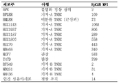
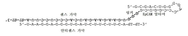
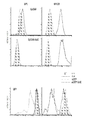


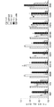


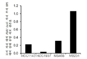


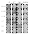
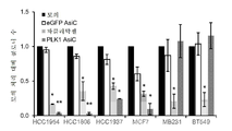


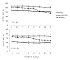

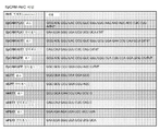
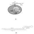

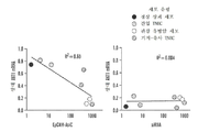


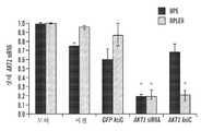


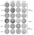


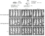


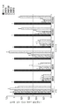
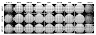
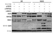

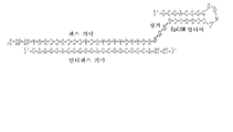

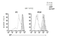
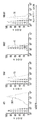
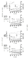


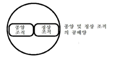


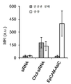

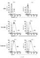

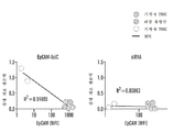


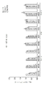


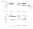
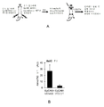


Classifications
-
- C—CHEMISTRY; METALLURGY
- C12—BIOCHEMISTRY; BEER; SPIRITS; WINE; VINEGAR; MICROBIOLOGY; ENZYMOLOGY; MUTATION OR GENETIC ENGINEERING
- C12N—MICROORGANISMS OR ENZYMES; COMPOSITIONS THEREOF; PROPAGATING, PRESERVING, OR MAINTAINING MICROORGANISMS; MUTATION OR GENETIC ENGINEERING; CULTURE MEDIA
- C12N15/00—Mutation or genetic engineering; DNA or RNA concerning genetic engineering, vectors, e.g. plasmids, or their isolation, preparation or purification; Use of hosts therefor
- C12N15/09—Recombinant DNA-technology
- C12N15/11—DNA or RNA fragments; Modified forms thereof; Non-coding nucleic acids having a biological activity
- C12N15/115—Aptamers, i.e. nucleic acids binding a target molecule specifically and with high affinity without hybridising therewith ; Nucleic acids binding to non-nucleic acids, e.g. aptamers
-
- A—HUMAN NECESSITIES
- A61—MEDICAL OR VETERINARY SCIENCE; HYGIENE
- A61K—PREPARATIONS FOR MEDICAL, DENTAL OR TOILETRY PURPOSES
- A61K31/00—Medicinal preparations containing organic active ingredients
- A61K31/33—Heterocyclic compounds
- A61K31/335—Heterocyclic compounds having oxygen as the only ring hetero atom, e.g. fungichromin
- A61K31/337—Heterocyclic compounds having oxygen as the only ring hetero atom, e.g. fungichromin having four-membered rings, e.g. taxol
-
- A—HUMAN NECESSITIES
- A61—MEDICAL OR VETERINARY SCIENCE; HYGIENE
- A61K—PREPARATIONS FOR MEDICAL, DENTAL OR TOILETRY PURPOSES
- A61K48/00—Medicinal preparations containing genetic material which is inserted into cells of the living body to treat genetic diseases; Gene therapy
-
- A—HUMAN NECESSITIES
- A61—MEDICAL OR VETERINARY SCIENCE; HYGIENE
- A61P—SPECIFIC THERAPEUTIC ACTIVITY OF CHEMICAL COMPOUNDS OR MEDICINAL PREPARATIONS
- A61P35/00—Antineoplastic agents
-
- C—CHEMISTRY; METALLURGY
- C07—ORGANIC CHEMISTRY
- C07H—SUGARS; DERIVATIVES THEREOF; NUCLEOSIDES; NUCLEOTIDES; NUCLEIC ACIDS
- C07H21/00—Compounds containing two or more mononucleotide units having separate phosphate or polyphosphate groups linked by saccharide radicals of nucleoside groups, e.g. nucleic acids
-
- C—CHEMISTRY; METALLURGY
- C07—ORGANIC CHEMISTRY
- C07K—PEPTIDES
- C07K14/00—Peptides having more than 20 amino acids; Gastrins; Somatostatins; Melanotropins; Derivatives thereof
- C07K14/435—Peptides having more than 20 amino acids; Gastrins; Somatostatins; Melanotropins; Derivatives thereof from animals; from humans
- C07K14/705—Receptors; Cell surface antigens; Cell surface determinants
-
- C—CHEMISTRY; METALLURGY
- C12—BIOCHEMISTRY; BEER; SPIRITS; WINE; VINEGAR; MICROBIOLOGY; ENZYMOLOGY; MUTATION OR GENETIC ENGINEERING
- C12N—MICROORGANISMS OR ENZYMES; COMPOSITIONS THEREOF; PROPAGATING, PRESERVING, OR MAINTAINING MICROORGANISMS; MUTATION OR GENETIC ENGINEERING; CULTURE MEDIA
- C12N15/00—Mutation or genetic engineering; DNA or RNA concerning genetic engineering, vectors, e.g. plasmids, or their isolation, preparation or purification; Use of hosts therefor
- C12N15/09—Recombinant DNA-technology
- C12N15/11—DNA or RNA fragments; Modified forms thereof; Non-coding nucleic acids having a biological activity
- C12N15/113—Non-coding nucleic acids modulating the expression of genes, e.g. antisense oligonucleotides; Antisense DNA or RNA; Triplex- forming oligonucleotides; Catalytic nucleic acids, e.g. ribozymes; Nucleic acids used in co-suppression or gene silencing
- C12N15/1137—Non-coding nucleic acids modulating the expression of genes, e.g. antisense oligonucleotides; Antisense DNA or RNA; Triplex- forming oligonucleotides; Catalytic nucleic acids, e.g. ribozymes; Nucleic acids used in co-suppression or gene silencing against enzymes
-
- A—HUMAN NECESSITIES
- A61—MEDICAL OR VETERINARY SCIENCE; HYGIENE
- A61K—PREPARATIONS FOR MEDICAL, DENTAL OR TOILETRY PURPOSES
- A61K39/00—Medicinal preparations containing antigens or antibodies
- A61K2039/51—Medicinal preparations containing antigens or antibodies comprising whole cells, viruses or DNA/RNA
- A61K2039/515—Animal cells
- A61K2039/5152—Tumor cells
-
- C—CHEMISTRY; METALLURGY
- C12—BIOCHEMISTRY; BEER; SPIRITS; WINE; VINEGAR; MICROBIOLOGY; ENZYMOLOGY; MUTATION OR GENETIC ENGINEERING
- C12N—MICROORGANISMS OR ENZYMES; COMPOSITIONS THEREOF; PROPAGATING, PRESERVING, OR MAINTAINING MICROORGANISMS; MUTATION OR GENETIC ENGINEERING; CULTURE MEDIA
- C12N2310/00—Structure or type of the nucleic acid
- C12N2310/10—Type of nucleic acid
- C12N2310/14—Type of nucleic acid interfering N.A.
-
- C—CHEMISTRY; METALLURGY
- C12—BIOCHEMISTRY; BEER; SPIRITS; WINE; VINEGAR; MICROBIOLOGY; ENZYMOLOGY; MUTATION OR GENETIC ENGINEERING
- C12N—MICROORGANISMS OR ENZYMES; COMPOSITIONS THEREOF; PROPAGATING, PRESERVING, OR MAINTAINING MICROORGANISMS; MUTATION OR GENETIC ENGINEERING; CULTURE MEDIA
- C12N2310/00—Structure or type of the nucleic acid
- C12N2310/10—Type of nucleic acid
- C12N2310/16—Aptamers
-
- C—CHEMISTRY; METALLURGY
- C12—BIOCHEMISTRY; BEER; SPIRITS; WINE; VINEGAR; MICROBIOLOGY; ENZYMOLOGY; MUTATION OR GENETIC ENGINEERING
- C12N—MICROORGANISMS OR ENZYMES; COMPOSITIONS THEREOF; PROPAGATING, PRESERVING, OR MAINTAINING MICROORGANISMS; MUTATION OR GENETIC ENGINEERING; CULTURE MEDIA
- C12N2310/00—Structure or type of the nucleic acid
- C12N2310/30—Chemical structure
- C12N2310/32—Chemical structure of the sugar
- C12N2310/322—2'-R Modification
-
- C—CHEMISTRY; METALLURGY
- C12—BIOCHEMISTRY; BEER; SPIRITS; WINE; VINEGAR; MICROBIOLOGY; ENZYMOLOGY; MUTATION OR GENETIC ENGINEERING
- C12N—MICROORGANISMS OR ENZYMES; COMPOSITIONS THEREOF; PROPAGATING, PRESERVING, OR MAINTAINING MICROORGANISMS; MUTATION OR GENETIC ENGINEERING; CULTURE MEDIA
- C12N2310/00—Structure or type of the nucleic acid
- C12N2310/30—Chemical structure
- C12N2310/35—Nature of the modification
- C12N2310/351—Conjugate
- C12N2310/3519—Fusion with another nucleic acid
-
- C—CHEMISTRY; METALLURGY
- C12—BIOCHEMISTRY; BEER; SPIRITS; WINE; VINEGAR; MICROBIOLOGY; ENZYMOLOGY; MUTATION OR GENETIC ENGINEERING
- C12N—MICROORGANISMS OR ENZYMES; COMPOSITIONS THEREOF; PROPAGATING, PRESERVING, OR MAINTAINING MICROORGANISMS; MUTATION OR GENETIC ENGINEERING; CULTURE MEDIA
- C12N2310/00—Structure or type of the nucleic acid
- C12N2310/30—Chemical structure
- C12N2310/35—Nature of the modification
- C12N2310/353—Nature of the modification linked to the nucleic acid via an atom other than carbon
- C12N2310/3533—Halogen
Landscapes
- Health & Medical Sciences (AREA)
- Life Sciences & Earth Sciences (AREA)
- Genetics & Genomics (AREA)
- Chemical & Material Sciences (AREA)
- Engineering & Computer Science (AREA)
- Molecular Biology (AREA)
- Organic Chemistry (AREA)
- Biomedical Technology (AREA)
- Biotechnology (AREA)
- General Health & Medical Sciences (AREA)
- Zoology (AREA)
- Biochemistry (AREA)
- Wood Science & Technology (AREA)
- Bioinformatics & Cheminformatics (AREA)
- General Engineering & Computer Science (AREA)
- Biophysics (AREA)
- Physics & Mathematics (AREA)
- Microbiology (AREA)
- Plant Pathology (AREA)
- Medicinal Chemistry (AREA)
- Public Health (AREA)
- Veterinary Medicine (AREA)
- Pharmacology & Pharmacy (AREA)
- Animal Behavior & Ethology (AREA)
- Epidemiology (AREA)
- Virology (AREA)
- Cell Biology (AREA)
- Immunology (AREA)
- Toxicology (AREA)
- Gastroenterology & Hepatology (AREA)
- Proteomics, Peptides & Aminoacids (AREA)
- General Chemical & Material Sciences (AREA)
- Chemical Kinetics & Catalysis (AREA)
- Nuclear Medicine, Radiotherapy & Molecular Imaging (AREA)
- Pharmaceuticals Containing Other Organic And Inorganic Compounds (AREA)
- Medicines That Contain Protein Lipid Enzymes And Other Medicines (AREA)
- Medicinal Preparation (AREA)
Abstract
암, 예를 들어 유방암을, 예를 들어 압타머-siRNA 키메라 분자를 사용하여 치료하는 것에 관한 방법 및 조성물이 본 명세서에 기재된다.Methods and compositions relating to the treatment of cancer, e. G., Breast cancer, using, for example, platamer-siRNA chimeric molecules are described herein.

Description
관련 출원과의 상호 참조Cross reference to related application
본 출원은 2014년 8월 29일자로 출원된 미국 가출원 번호 62/043,803에 대하여 35 U.S.C. § 119(e) 하에서 이득을 주장하며, 이의 내용은 전체적으로 본 명세서에 참고로 포함된다.This application claims the benefit of U.S. Provisional Application No. 62 / 043,803, filed August 29, Benefit under § 119 (e), the content of which is incorporated herein by reference in its entirety.
정부 지원Government support
본 발명은 미국 국방부(Department of Defense)에 의해 허여된 승인 번호 W81 XWH-09-1-0058 하에서 연방정부의 보조금으로 이루어졌다. 미국 정부는 본 발명에 대해 소정의 권리를 갖는다.The invention was made with federal grants under authorization number W81 XWH-09-1-0058, granted by the Department of Defense of the United States. The US Government has certain rights to the invention.
서열 목록Sequence List
본 출원은 ASCII 포맷으로 전자적으로 제출된 서열 목록을 함유하고, 서열 목록은 전체적으로 본 명세서에 참고로 포함된다. 2015년 8월 28일자에 작성된 상기 ASCII 사본은 701039-082401-PCT_SL.txt로 명명되고 크기가 8,984 바이트이다. The present application contains sequence listings submitted electronically in ASCII format, and the sequence listing is incorporated herein by reference in its entirety. The ASCII copy made on August 28, 2015 is named 701039-082401-PCT_SL.txt and has a size of 8,984 bytes.
본 명세서에 기재된 기술은 EpCAM 결합(binding)-분자 및 억제성 핵산을 포함하는 키메라 분자(chimeric molecule), 및 암, 예를 들어 상피암의 치료에 그러한 조성물을 사용하는 방법에 관한 것이다.The techniques described herein relate to EpCAM binding-molecules and chimeric molecules comprising inhibitory nucleic acids, and methods of using such compositions in the treatment of cancer, e. G., Epithelial cancer.
RNA 간섭(RNAi)이 간에서의 유전자 발현을 감소시키는 데 있에서의 치료적(therapeutic) 용도를 위하여 조사되어 왔다. 그러나, 간은 RNAi 분자로 형질감염되기 쉽다는 것이 독특하다. 작은 RNA의 전달 및 그 결과로 인한 다른 조직에서의 유전자 녹다운(knockdown)은 계속 비효율적이고 궁극적으로 효과적이지 않다. 특히, 전달 로드블록(roadblock)은 RNAi를 이용하여 암을 치료하는 데 주요 장애물이다.RNA interference (RNAi) has been investigated for therapeutic uses in reducing gene expression in the liver. However, it is unique that the liver is susceptible to transfection with RNAi molecules. Transmission of small RNAs and consequent gene knockdown in other tissues is still inefficient and ultimately ineffective. In particular, delivery roadblocks are a major obstacle to the treatment of cancer using RNAi.
본 명세서에 기재된 바와 같이, 본 발명자들은 신규한 키메라 압타머-siRNA 분자(AsiC)를 개발하였다. 이들 AsiC는 암 세포 마커를 표적화하여, siRNA를 특이적으로 암 세포로 유도하며, 이는 부작용에 대한 잠재성을 감소시키면서 전달 효능 및 치료적 유효성을 증가시킨다.As described herein, the present inventors have developed a novel chimericapatamer-siRNA molecule (AsiC). These AsiCs target cancer cell markers and specifically induce siRNAs to cancer cells, which increases the delivery efficacy and therapeutic efficacy while reducing the potential for side effects.
일 태양에서, 암 마커-결합 압타머 도메인 및 억제성 핵산 도메인을 포함하는 키메라 분자가 본 명세서에 기재된다. 일부 실시 형태에서, 암 마커는 EpCAM 또는 EphA2이다. 일부 실시 형태에서, 억제성 핵산은 암 세포에서 상향조절되는 유전자 산물에 특이적으로 결합한다. 일부 실시 형태에서, 억제성 핵산은 하기로 이루어진 군으로부터 선택되는 유전자의 발현을 억제한다: Plk1; MCL1; EphA2; PsmA2; MSI1; BMI1; XBP1; PRPF8; PFPF38A; RBM22; USP39; RAN; NUP205; 및 NDC80. 일부 실시 형태에서, 암 마커는 EpCAM이고, 억제성 핵산 도메인은 Plk1의 발현을 억제한다.In one embodiment, chimeric molecules comprising a cancer marker-associated abdominal domain and an inhibitory nucleic acid domain are described herein. In some embodiments, the cancer marker is EpCAM or EphA2. In some embodiments, the inhibitory nucleic acid specifically binds to a gene product that is up-regulated in cancer cells. In some embodiments, the inhibitory nucleic acid inhibits expression of a gene selected from the group consisting of: Plk1; MCL1; EphA2; PsmA2; MSI1; BMI1; XBP1; PRPF8; PFPF38A; RBM22; USP39; RAN; NUP205; And NDC80. In some embodiments, the cancer marker is EpCAM and the inhibitory nucleic acid domain inhibits expression of Plkl.
일부 실시 형태에서, 분자는 압타머-siRNA 키메라(AsiC)이다. 일부 실시 형태에서, 암 마커-결합 압타머 도메인은 서열 번호 33의 서열을 포함한다. 일부 실시 형태에서, 암 마커-결합 압타머 도메인은 서열 번호 33의 서열로 본질적으로 이루어진다. 일부 실시 형태에서, 억제성 핵산 도메인은 서열 번호 2의 서열을 포함한다. 일부 실시 형태에서, 억제성 핵산 도메인은 서열 번호 2의 서열로 본질적으로 이루어진다. 일부 실시 형태에서, 분자는 서열 번호 1 내지 서열 번호 3 중 하나의 서열 번호의 서열을 포함한다. 일부 실시 형태에서, 분자는 서열 번호 1 내지 서열 번호 3 중 하나의 서열 번호의 서열로 본질적으로 이루어진다.In some embodiments, the molecule is platamer-siRNA chimera (AsiC). In some embodiments, the cancer marker-associated abdominal domain comprises the sequence of SEQ ID NO: 33. In some embodiments, the cancer marker-associated abdominal domain consists essentially of the sequence of SEQ ID NO: 33. In some embodiments, the inhibitory nucleic acid domain comprises the sequence of SEQ ID NO: 2. In some embodiments, the inhibitory nucleic acid domain consists essentially of the sequence of SEQ ID NO: 2. In some embodiments, the molecule comprises a sequence of SEQ ID NO: 1 to SEQ ID NO: 3. In some embodiments, the molecule consists essentially of a sequence of SEQ ID NO: 1 to SEQ ID NO: 3.
일부 실시 형태에서, 분자의 3' 말단은 dTdT를 포함한다. 일부 실시 형태에서, 분자는 적어도 하나의 2'-F 피리미딘을 포함한다.In some embodiments, the 3 ' end of the molecule comprises dTdT. In some embodiments, the molecule comprises at least one 2 ' -F pyrimidine.
일 태양에서, 본 명세서에 기재된 바와 같은 키메라 분자 및 약제학적으로 허용되는 담체를 포함하는 약제학적 조성물이 본 명세서에 기재된다. 일부 실시 형태에서, 조성물은 본 명세서에 기재된 바와 같은 키메라 분자를 적어도 2개 포함하며, 키메라 분자는 상이한 압타머 도메인 및/또는 억제성 핵산 도메인을 갖는다. 일부 실시 형태에서, 상이한 압타머 또는 억제성 핵산 도메인은 상이한 표적을 인식한다. 일부 실시 형태에서, 상이한 압타머 또는 억제성 핵산 도메인은 서열을 갖고 동일한 표적을 인식한다.In one aspect, pharmaceutical compositions comprising a chimeric molecule as described herein and a pharmaceutically acceptable carrier are described herein. In some embodiments, the composition comprises at least two chimeric molecules as described herein, and the chimeric molecule has a different abdominal domain and / or an inhibitory nucleic acid domain. In some embodiments, different abomas or inhibitory nucleic acid domains recognize different targets. In some embodiments, different abomasor or inhibitory nucleic acid domains have sequences and recognize the same target.
일 태양에서, 암의 치료 방법이 본 명세서에 기재되며, 본 방법은 본 명세서에 기재된 바와 같은 키메라 분자 및/또는 조성물을 투여하는 것을 포함한다. 일부 실시 형태에서, 암은 상피암 또는 유방암이다. 일부 실시 형태에서, 유방암은 삼중-음성 유방암이다. 일부 실시 형태에서, 투여는 피하 투여이다. 일부 실시 형태에서, 대상체에는 추가의 암 치료가 추가로 실시된다. 일부 실시 형태에서, 암 치료는 파클리탁셀이다.In one aspect, a method of treating cancer is described herein, the method comprising administering a chimeric molecule and / or composition as described herein. In some embodiments, the cancer is epithelial or breast cancer. In some embodiments, the breast cancer is triple-negative breast cancer. In some embodiments, the administration is subcutaneous administration. In some embodiments, additional cancer treatment is additionally administered to the subject. In some embodiments, the cancer treatment is paclitaxel.
도 1a 내지 도 1h는 EpCAM 압타머가 기저 A 유방암 세포를 특이적으로 표적화함을 입증한다. EpCAM 압타머 및 PLK1 siRNA(서열 번호 1로 개시된 센스 가닥 및 서열 번호 2로 개시된 안티센스 가닥)를 함유하는 EpCAM-AsiC의 설계(도 1c). 상피 유방암 세포주(BPLER)가 정상 유방 상피 세포주(BPE)에 비하여 EpCAM 단백질을 과발현한다(도 1a 및 도 1b). GFP를 표적화하는 EpCAM-AsiC를 안티센스 siRNA 가닥의 3' 말단에서 Alexa647 또는 Cy3로 표지화하고, BPLER 및 BPE 세포와 인큐베이션하였다. 24시간 후에 유세포측정에 의해 흡수를 평가하였다(도 1d). 데이터는 3개의 독립적인 실험을 대표한다. Cy3 및 Alexa647-표지화된 EpCAM-AsiC는 MB468 및 BPLER(EpCAM+ 세포) 각각에 의해 흡수되었지만, BPE(EpCAM-)에 의해서는 그렇지 않았다. 각각의 피크의 MFI가 나타나 있다. 유전자 침묵에 대해 시험하기 위하여, BPLER 및 BPE를 GFP를 표적화하는 EpCAM-AsiC(4 μM)로 처리하고, Dharmafect 및 GFP-siRNA(100 nM)를 사용하는 형질감염 대조군과 대비하였다. 인큐베이션 후 72시간에 유세포측정에 의해 녹다운을 평가하였다. 대조군은 모의 처리 및 Dhrmafect 단독 처리(지질)였다. (n = 4)(도 1d). AKT1을 표적화하는 EpCAM-AsiC는 기저 A 및 관강 유방암 세포주에서 AKT1 mRNA(도 1e) 및 단백질(도 1f) 발현을 선택적으로 녹다운하고, 기저 B 또는 인간 섬유아세포(hFb)에서는 그렇지 않다. AKT1을 표적화하는 siRNA에 의한 형질감염은 모든 세포주에서 유전자 녹다운을 유도하며, 한편 GFP를 표적화하는 EpCAM-AsiC에 의한 처리는 AKT1 mRNA 및 단백질 수준에 영향을 주지 않는다(* p<0.05, p<0.01). EpCAM-AsiC의 효과를 siRNA 형질감염의 효과와 대비한 AKT1 단백질 및 유전자 녹다운의 도표. EpCAM-AsiC 유도 녹다운은 EpCAM 발현과 상관된다(도 1e 내지 도 1h). (n = 3; 모의에 대해 정규화된 평균 ± SEM; *P < 0.05, **P < 0.01, 양측(2-tailed) t 검정).
도 2a 내지 도 2e는 PLK1을 표적화하는 EpCAM AsiC가 기저 A 유방암 세포에서 세포 증식을 특이적으로 억제함을 입증한다. 세포 증식에 대한 PLK1을 표적화하는 EpCAM-AsiC의 효과를 Cell-titer-Glo 검정(CTG)을 사용하여 기저 A, B 및 관강 세포주를 대표하는 10개의 유방암 세포주에 대해 시험하였다. PLK1을 표적화하는 EpCAM-AsiC는 기저 A 세포주 및 관강 세포주 둘 모두에서 세포 증식을 감소시켰으며, 한편 기저 B 세포에 대해서는 효과를 갖지 않았다(도 2a, 도 2c). EpCAM 발현 수준과 세포 생존력 사이에 상관관계가 관찰되었다(도 2b). 기저 A(EpCAM+GFP-) 세포를 BPE(EpCAM-GFP+) 세포와 공배양하고, PLK1을 표적화하는 EpCAM-AsiC로 처리하거나 무처리하였다. 무처리된 공배양물은 유사한 비의 세포들을 나타내었으며, PLK1을 표적화하는 EpCAM-AsiC 처리 후에는 EpCAM+ 세포의 비는 감소되었고 EpCAM- 세포는 증가하였다. 대표적인 유세포측정 도표(도 2d), 4개의 상이한 세포주에서 GFP+/GFP- 세포의 비를 분석한 실험의 정량화(도 2e). (n=4, * p<0.05, p<0.01).
도 3a 내지 도 3d는 인간 TNBC 조직이 Cy3-EpCAM 압타머를 특이적으로 흡수함을 입증한다. 실험 설계; GFP를 표적화하는 Cy3-EpCAM-AsiC, Alexa647-siRNA-GFP 또는 Alexa647-chol-siRNA-GFP(각각 2 μM)를 유방암 및 대조군 외식편(explant)에 첨가하고, 24시간 동안 인큐베이션한 후, 조직을 단세포 현탁액으로 콜라게나제로 분해하고 유세포측정에 의해 분석하였다(도 3a). 종양 생검은 EpCAM 및 사이토케라틴 - 상피 세포 마커 - 을 과발현한다(도 3b). 3개의 독립적인 실험 중 하나로부터의 대표적인 히스토그램은, siRNA 및 chol-siRNA가 유사한 효능으로 종양 및 건강한 조직 둘 모두를 침투하였으며, 한편 EpCAM-AsiC는 종양 조직 생검에 의해 선택적으로 흡수되었고, 건강한 대조군 조직 샘플에 의해서는 흡수되지 않았음을 보여준다(도 3c). 이러한 흡수 실험을 3명의 상이한 환자로부터의 종양에서 반복하였으며, 입수된 각각의 생검을 각각의 처리에 대해 3회씩 시험하였다. 모든 3명의 환자의 요약(도 3d). (n=3, 모의, 회색 EpCAM, 적색 *P < 0.05, **P < 0.005, t-검정 CD4-AsiC vs. 모의 처리).
도 4a 내지 도 4c는 PLK1을 표적화하는 EpCAM AsiC가 기저 A 유방암 세포에서 종양 개시를 특이적으로 억제함을 입증한다. 유방암 세포주의 콜로니 검정은 24시간 동안 PLK1 또는 GFP를 표적화하는 EpCAM-AsiC(4 uM) 또는 파클리탁셀(100 nM)을 처리하고, 무약물 배지 중에서 8일 동안 배양하였다. 파클리탁셀에 의한 처리는 모든 세포주에서 콜로니 형성을 감소시켰으며, 한편 PLK1을 표적화하는 EpCAM-AsiC에 의한 처리는 단지 관강(MCF7) 및 기저 A(HCC1954) 세포에서만 콜로니 형성을 제거하였으며, GFP를 표적화하는 EpCAM-AsiC에 의한 처리는 어떠한 효과도 갖지 않았다(도 4a). 검정을 추가 3개의 세포주에서 반복하였으며, 결과는 재현가능하였다(도 4b). 종양괴 형성 검정은 유사한 결과를 나타내었으며, PLK1을 표적화하는 EpCAM-AsiC는 단지 기저 A 및 관강 세포에서만 종양괴의 수를 감소시켰으며, 기저 B 세포에 대해서는 효과를 갖지 않았다(도 4c). MB468-luc 세포를 GFP 또는 PLK1을 표적화하는 EpCAM-AsiC로 24시간 동안 처리하고, 누드 마우스의 옆구리에 s.c. 주사하였다. 마우스를 20일 동안 매 5일마다 이미징하였다. 무처리 마우스 및 GFP를 표적화하는 EpCAM-AsiC로 처리된 마우스는 종양 개시의 증가를 나타내었으며, 한편 PLK1을 표적화하는 EpCAM-AsiC로 전처리된 세포가 주사된 마우스는 종양 개시를 갖지 않는다.
도 5a 내지 도 5c는 EpCAM+ 종양 내로의 Alexa750-EpCAM-AsiC의 선택적 흡수를 입증한다. 도 5a는 실험 셋업을 나타내는데; 누드 마우스에 MB468-luc(좌측 옆구리) 및 MB231-luc-mCherry(우측 옆구리) 세포를 주사하고, 주사 후 5일에, Alexa750 표지화된, GFP를 표적화하는 EpCAM-AsiC(0.5 mg/kg)를 목 부위에 s.c. 주사하였다. 마우스를 주사 직후에 이미징하고, 다시 24, 48시간 및 5일 후에 이미징하였다. Alexa750 표지화된, GFP를 표적화하는 EpCAM-AsiC는 MB468-luc 종양(EpCAM+)에서 루시페라제 종양과 동시-국재화(co-localize)되었고, MB231-luc-mCherry(EpCAM-) 종양에는 그렇지 않았다. 7 마리의 마우스의 분석은 MB468(EpCAM+) 종양에서의 Alexa750의 유의한 증가를 나타낸다(도 5b). 도 5c는 Alexa750 흡수율의 그래프를 나타낸다.
도 6a 및 도 6b는 PLK1을 표적화하는 EpCAM AsiC가 기저 A 유방암 세포에서 종양 성장을 특이적으로 억제함을 입증한다. 도 6a는 실험 설계를 나타낸다. MB231-luc-mCherry 세포(5x10⁵개) 또는 MB468-luc 세포(5x10⁶개)가 주사된 누드 마우스를 72시간마다 PLK1 또는 GFP를 표적화하는 EpCAM AsiC 5 mg/Kg으로 처리하거나 무처리 상태로 두었다. 도 6b: PLK1을 표적화하는 EpCAM-AsiC로 처리된 MB468-luc 종양은 처리 후 빠르게는 6일에 크기가 수축되었으며, 14일 후에는 다수의 마우스에서 완전히 소실되었으며, 무처리 종양은 EpCAM+ 및 EpCAM- 둘 모두 14일에 걸쳐 크기가 증가하였다.
도 7은 EpCAM AsiC가 인간 및 마우스 혈청에서 안정함을 입증한다. 2'-플루오로-피리미딘을 사용하여 합성된 eGFP EpCAM-AsiC, 화학적으로 안정화된 콜레스테롤-접합된 eGFP siRNA(chol-siRNA), 또는 비변형된 eGFP siRNA를 동일 부피의 인간 또는 마우스 혈청과 함께 인큐베이션하였다. 분취물을 규칙적인 간격으로 회수하고, 겔 로딩 완충액 중에 재현탁하고, -80℃에서 저장한 후, 변성(denaturing) PAGE 겔 상에서 전기영동을 행하였다. 염색 후 밀도측정(densitometry)에 의해 정량화된 2개의 독립적인 실험으로부터의 밴드들의 평균 세기(+S.E.M.)가 나타나 있다.
도 8의 A 및 도 8의 B는 EpCAM AsiC의 주사가 마우스에서 선천적 면역을 자극하지 않음을 입증한다. 마우스에 eGFP EpCAM-AsiC(5 mg/kg, n=3)를 피하 주사하거나 폴리(I:C)(5 또는 50 mg/kg)(n=2/용량)를 ip 주사하였다. 도 8의 A: 기준선에서 그리고 처리 후 6 및 16시간에 수집된 혈청 샘플을 멀티플렉스 면역검정에 의해 IFNβ, IL-6 및 IP-10에 대해 평가하였다. * p<0.05, ** p<0.01, *** p<0.001, 기준선 대비. 도 8의 B: gapdh 대비 사이토카인 및 IFN-유도 유전자의 mRNA 발현을, 처리 후 16시간에 수거된 총 비장세포에서 qRT-PCR에 의해 검정하였다. ** p<0.01, 무처리 대비(NT, n=3).
도 9는 서열 표를 나타낸다. (출현 순서대로, 각각 서열 번호 1 및 2, 그리고 23 내지 32).
도 10의 A 및 도 10의 B는 압타머-siRNA 키메라(AsiC)를 나타낸다. 도 10의 A는 AsiC(siRNA의 하나의 가닥에 공유 결합된 압타머)의 다이아그램을 나타내는데, AsiC는 암 세포 표면 수용체를 특이적으로 인식하고, 세포내이입되고, 이어서 세포질로 방출되고, 세포질에서 그것이 내인성 전-miRNA처럼 처리되어 표적 유전자를 녹다운한다. 바(bar)는 하기의 2가지 전달 장애물, 즉 세포 흡수 및 엔도솜으로부터의 세포질로의 방출을 나타내며, 이때 세포질에는 Dicer 및 RNA 유도 침묵 복합체(RNA induced silencing complex, RISC)가 위치되어 있다. 도 10의 B는 PLK1을 표적화하는 EpCAM AsiC의 설계를 나타낸다. (서열 번호 1로 개시된 센스 가닥 및 서열 번호 2로 개시된 안티센스 가닥).
도 11a 및 도 11d는 EpCAM-AsiC 녹다운 및 항종양 효과가 EpCAM 수준과 상관되고 상피 유방 종양 T-IC를 억제함을 입증한다. 도 11a 및 도 11b: 대표적인 실험(도 11a) 및 EpCAM-AsiC와 지질 siRNA 형질감염을 비교하는 AKT1 녹다운(도 11b). 도 11c: 단지 EpCAM+ 세포주에서만 PLK1을 녹다운하는 EpCAM-AsiC의 항증식 효과. 도 11d: PLK1 EpCAM-AsiC는 관강 MCF 및 기저-A TNBC HCC1143에서 콜로니 형성을 억제하지만, 간엽(mesenchymal) 기저-B MB231 세포에서는 그렇지 않다.
도 12의 A 및 도 12의 B는 기능적 EphA2 압타머의 확인을 입증한다. 도 12의 A: EphA2+ 기저-B MB231 세포를 EphA2 압타머(EphA2apt)와 함께 인큐베이션하는 것은 EphA2 분해 및 활성 Akt(pAkt)의 일시적 감소로 이어진다. 도 12의 B: EphA2apt(0 내지 100 nM)와 함께 2시간 동안 인큐베이션된 EphA2+ 유방암 세포는 감소된 EphA2를 보여주지만 대조군 비결합 압타머(ctl)는 그렇지 않다. 에프린(Ephrin) A의 첨가를 EphA2 분해에 대한 양성 대조군으로서 사용하였다.
도 13a 내지 도 13c: EpCAM-AsiC는 단지 EpCAM+ 세포주에서만 GFP 단백질(도 13a) 및 AKT1 mRNA(도 13b 및 도 13c)를 녹다운하며, 불멸화 유방 상피 세포주(BPE) 또는 간엽 기저 B TNBC 또는 인간 섬유아세포에서는 그렇지 않다. 형질감염된 siRNA는 그의 녹다운에 있어서 비특이적이다. *, P<0.05
도 14. 동일한 대상체로부터의 정상 유방 조직 및 기저-A TNBC 종양 생검을 Cy3-표지화된 EpCAM-AsiC와 함께 인큐베이션하고, 3일 후에 유세포측정에 의해 단세포 현탁액을 흡수에 대해 분석하였다. 네이키드 siRNA는 어느 것에 의해서도 흡수되지 않았고, 콜레스테롤-접합된 siRNA는 동일하게 흡수되었지만, EpCAM-AsiC는 종양에 의해 특이적으로 흡수되었다. 대표적인 조직이 좌측에 나타나 있다.
도 15a 내지 도 15c. PLK1 EpCAM-AsiC에 의한 EpCAM+ 유방암 세포주의 처리는 T-IC 기능의 시험관내 검정인, 콜로니(도 15a, 도 15b) 및 유방종양괴(도 15c) 기능을 억제하지만, EpCAM- 유방암 세포주는 그렇지 않다.
도 16은 PLK1 EpCAM-AsiC에 의한 MB468 세포의 생체외 처리가 누드 마우스에서 종양을 형성하는 그의 능력을 제거하였음을 입증한다. 동일한 수의 생존 세포를 처리 후 당일에 삽입하였다.
도 17의 A 및 도 17의 B는 EpCAM-AsiC가 EpCAM+ 내로 선택적으로 흡수되지만, EpCAM-, TNBC 종양은 그렇지 않음을 입증한다. 도 17의 A는 실험 계획을 나타낸다. 도 17의 B는 희생 시에 절제된 종양 내에의 EpCAM-AsiC의 농축을 나타낸다.
도 18의 A 및 도 18의 B는 PLK1 EpCAM-AsiC가 EpCAM+ TNBC 이종이식편의 완전 종양 퇴행을 야기하였지만, EpCAM- 기저-B 이종이식편에 대해서는 효과를 갖지 않았음을 입증한다. 도 18의 A는 실험 설계를 나타낸다. 좌측 및 우측 옆구리 종양의 루시페라제 활성의 이미징을 2주에 걸쳐 순차적으로 수행하였다. 도 18의 B는 루시페라제 활성에 의한 종양 크기의 그래프를 나타낸다. PLK1 AsiC로 처리된 마우스에서의 모든 EpCAM+ 종양은 신속히 퇴행하였지만, 다른 종양들은 계속 성장하였다.
도 19a 내지 도 19c는 기저 의존성 유전자가 4개의 트리-snRNP 스플라이세오좀 복합체 유전자(PFPF8, PRPF38A, RBM22, USP39), 2개의 핵외 수송 유전자(NUP205, RAN), 및 동원체 유전자(NDC80)를 포함함을 입증한다. 도 19a는 대조군 siRNA에 대해 정규화된, 녹다운 후 3일에의 세포 생존력을 나타낸다. 도 19b는 녹다운 후 2일에 생존 세포를 플레이팅함으로써 평가된 콜로니 형성을 나타낸다. 도 19c는 녹다운 후 2일에의 카스파제 활성화가 MB468에 특이적이고 BPE 세포에서는 일어나지 않음을 나타낸다.
도 20은 세포내이입을 개선하기 위해 다량체화된 EpCAM-AsiC에 대한 일부 가능한 설계를 나타낸다. 이들 설계에서, 센스 및 안티센스 가닥은 교환될 수 있었고, 링커는 변동될 수 있었다.
도 21a 내지 도 21d는 EpCAM 압타머가 기저 A 유방암 세포를 특이적으로 표적화함을 입증한다. 도 21a는 EpCAM 압타머 및 PLK1 siRNA(서열 번호 1로 개시된 센스 가닥 및 서열 번호 2로 개시된 안티센스 가닥)를 함유하는 EpCAM-AsiC의 설계를 입증한다. 도 21b는 상피 유방암 세포주(BPLER)가 정상 유방 상피 세포주(BPE)에 비하여 EpCAM 단백질을 과발현함을 입증하는 그래프를 나타낸다. GFP를 표적화하는 EpCAM-AsiC를 안티센스 siRNA 가닥의 3' 말단에서 Alexa647 또는 Cy3로 표지화하고, BPLER 및 BPE 세포와 인큐베이션하였다. 24시간 후에 유세포측정에 의해 흡수를 평가하였다(도 21c). 데이터는 3개의 독립적인 실험을 대표한다. Cy3 및 Alexa647-표지화된 EpCAM-AsiC는 MB468 및 BPLER(EpCAM+ 세포) 각각에 의해 흡수되었지만, BPE(EpCAM-)에 의해서는 그렇지 않았다. 각각의 피크의 MFI가 나타나 있다(모의, 회색). 도 21d는 유전자 침묵에 대해 시험하기 위하여, BPLER 및 BPE를 GFP를 표적화하는 EpCAM-AsiC(4 μM)로 처리하고, Dharmafect 및 GFP-siRNA(100 nM)를 사용하는 형질감염 대조군과 대비한 실험의 그래프를 나타낸다. 인큐베이션 후 72시간에 유세포측정에 의해 녹다운을 평가하였다. 대조군은 모의 처리 및 Dhrmafect 단독 처리(지질)였다. (n = 4).
도 22는 EpCAM 압타머가 마우스 EpCAM에 결합하지 않음을 입증하는 그래프를 나타낸다. mCD326 항체와 함께 유세포측정을 사용하여 마우스 ESA(EpCAM) 수준을 결정하였다. 상피 마우스 유방암 세포주인 4T1 세포는 EpCAM의 고발현 수준을 나타내었다. RAW(마우스 단핵구 세포주) 및 MB468(인간 기저 A 세포주) 둘 모두는 EpCAM 발현의 증가를 나타내었지만, 4T1 세포보다 훨씬 더 작았다. 마우스 간엽 암 세포주(67NR)는 EpCAM 발현의 최소한의 증가를 나타내었다. 흡수 실험은 EpCAM-압타머가 4T1 또는 67NR 세포 어느 것에 의해서도 흡수되지 않았음을 입증하였다.
도 23은 EpCAM이 기저 A 및 관강 유방암 세포주에서 과발현되지만, 기저 B 유방암 세포주에서는 그렇지 않음을 입증하는 그래프를 나타낸다. hEpCAM 항체를 사용하여 유세포측정에 의해 EpCAM 발현 수준을 시험하는, 8개의 상이한 유방암 세포주의 대표적인 FACS 도표. EpCAM은 모든 기저 A 및 관강 세포주에서는 과발현되지만 기저 B에서는 그렇지 않다. (모의, 음영처리된 회색 EpCAM, 흑색)
도 24a 내지 도 24f는 EpCAM AsiC가 기저 A 유방암 세포에서 유전자 발현을 특이적으로 침묵을 입증한다. AKT1을 표적화하는 EpCAM-AsiC는 기저 A 및 관강 유방암 세포주에서 AKT1 mRNA(도 24a) 및 단백질(도 24b, 도 24c) 발현을 선택적으로 녹다운하고, 기저 B 또는 인간 섬유아세포(hFb)에서는 그렇지 않다. AKT1을 표적화하는 siRNA에 의한 형질감염은 모든 세포주에서 유전자 녹다운을 유도하며, 한편 GFP를 표적화하는 EpCAM-AsiC에 의한 처리는 AKT1 mRNA 및 단백질 수준에 영향을 주지 않는다(* p<0.05, p<0.01). EpCAM-AsiC의 효과를 siRNA 형질감염의 효과와 대비한 AKT1 단백질 및 유전자 녹다운의 도표. EpCAM-AsiC 유도 녹다운은 EpCAM 발현과 상관된다(도 24d, 도 24e). (n = 3; 모의에 대해 정규화된 평균 ± SEM; *P < 0.05, **P < 0.01, 양측 t 검정). 도 24f는 유세포측정 분석의 결과를 도시한다.
도 25a 내지 도 25e는 인간 TNBC 조직이 Cy3-EpCAM 압타머를 특이적으로 흡수함을 입증한다. 도 25a는 실험 설계를 나타내는데; GFP를 표적화하는 Cy3-EpCAM-AsiC, Alexa647-siRNA-GFP 또는 Alexa647-chol-siRNA-GFP(각각 2 μM)를 유방암 및 대조군 외식편에 첨가하고, 24시간 동안 인큐베이션한 후, 조직을 단세포 현탁액으로 콜라게나제로 분해하고 유세포측정에 의해 분석하였다. 도 25b는 종양 생검이 EpCAM 및 사이토케라틴 - 상피 세포 마커 - 을 과발현함을 입증하는 그래프를 나타낸다. 도 25c는 3개의 독립적인 실험 중 하나로부터의 대표적인 히스토그램이, siRNA 및 chol-siRNA가 유사한 효능으로 종양 및 건강한 조직 둘 모두를 침투하였으며, 한편 EpCAM-AsiC는 종양 조직 생검에 의해 선택적으로 흡수되었고, 건강한 대조군 조직 샘플에 의해서는 흡수되지 않았음을 보여줌을 나타낸다. 이러한 흡수 실험을 3명의 상이한 환자로부터의 종양에서 반복하였으며, 입수된 각각의 생검을 각각의 처리에 대해 3회씩 시험하였다. 도 25d는 대표적인 종양을 나타낸다. 모든 3명의 환자의 요약이 도 25e에 나타나 있다. (n=3, *P < 0.05, **P < 0.005, t-검정 CD4-AsiC vs. 모의 처리).
도 26은 EpCAM-AsiC가 건강한 생검 및 결장암 생검 둘 모두에 의해 흡수됨을 입증하는 그래프를 나타낸다. GFP를 표적화하는 Cy3-EpCAM-AsiC, Alexa647-siRNA-GFP 또는 Alexa647-chol-siRNA-GFP(각각 2 μM)를 결장암 및 대조군 외식편에 첨가하고, 24시간 동안 인큐베이션한 후, 조직을 단세포 현탁액으로 콜라게나제로 분해하고 유세포측정에 의해 분석하였다. 대표적인 히스토그램은 EpCAM-AsiC, siRNA 및 chol-siRNA는 유사한 효능으로 종양 및 건강한 조직 둘 모두에 침투하였음을 보여준다.
도 27a 내지 도 27d는 PLK1을 표적화하는 EpCAM AsiC가 기저 A 유방암 세포에서 세포 증식을 특이적으로 억제함을 입증한다. 세포 증식에 대한 PLK1을 표적화하는 EpCAM-AsiC의 효과를 Cell-titer-Glo 검정(CTG)을 사용하여 기저 A, B 및 관강 세포주를 대표하는 10개의 유방암 세포주에 대해 시험하였다. PLK1을 표적화하는 EpCAM-AsiC는 기저 A 세포주 및 관강 세포주 둘 모두에서 세포 증식을 감소시켰으며, 한편 기저 B 세포에 대해서는 효과를 갖지 않았다(도 27a). EpCAM 발현 수준과 세포 생존력 사이에 상관관계가 관찰되었다(도 27b). 기저 A(EpCAM+GFP-) 세포를 BPE(EpCAM-GFP+) 세포와 공배양하고, PLK1을 표적화하는 EpCAM-AsiC로 처리하거나 무처리하였다. 무처리된 공배양물은 유사한 비의 세포들을 나타내었으며, PLK1을 표적화하는 EpCAM-AsiC 처리 후에는 EpCAM+ 세포의 비는 감소되었고 EpCAM- 세포는 증가하였다. 도 27c는 대표적인 유세포측정 도표를 나타내고, 도 27d는 4개의 상이한 세포주에서 GFP+/GFP- 세포의 비를 분석한 실험의 정량화의 그래프를 나타낸다. (n=4, * p<0.05, p<0.01).
도 28은 기저 A 유방암 세포주에서의 세포 생존력의 특이적 감소는 PLK1 의존성임을 입증하는 그래프를 나타낸다. 기저 A, B 및 관강 세포를 나타내는 10개의 상이한 유방암 세포주를 PLK1을 표적화하는 EpCAM-AsiC로 또는 단지 EpCAM-압타머만으로 처리하고, 무처리 대조군과 비교하였다. EpCAM-압타머로 처리된 세포주의 어느 것도 세포 생존력의 감소를 나타내지 않았으며, 한편 기저 A 및 관강 세포주는 PLK1을 표적화하는 EpCAM-AsiC에 의한 처리 후에 세포 생존력의 감소를 나타내었다.
도 29의 A 내지 도 29의 C는 PLK1을 표적화하는 EpCAM AsiC가 기저 A 유방암 세포에서 종양 개시를 특이적으로 억제함을 입증한다. 유방암 세포주의 콜로니 검정은 24시간 동안 PLK1 또는 GFP를 표적화하는 EpCAM-AsiC(4 uM) 또는 파클리탁셀(100 nM)을 처리하고, 무약물 배지 중에서 8일 동안 배양하였다. 파클리탁셀에 의한 처리는 모든 세포주에서 콜로니 형성을 감소시켰으며, 한편 PLK1을 표적화하는 EpCAM-AsiC에 의한 처리는 단지 관강(MCF7) 및 기저 A(HCC1954) 세포에서만 콜로니 형성을 제거하였으며, GFP를 표적화하는 EpCAM-AsiC에 의한 처리는 어떠한 효과도 갖지 않았다. 도 29의 A는 검정 결과의 이미지를 나타낸다. 검정을 추가 3개의 세포주에서 반복하였으며, 결과는 도 29의 B에 도시된 그래프에서 입증된 바와 같이 재현가능하였다. 도 29의 C는 종양괴 형성 검정은 유사한 결과를 나타내었으며, PLK1을 표적화하는 EpCAM-AsiC는 단지 기저 A 및 관강 세포에서만 종양괴의 수를 감소시켰으며, 기저 B 세포에 대해서는 효과를 갖지 않았음을 입증하는 그래프를 나타낸다. MB468-luc 세포를 GFP 또는 PLK1을 표적화하는 EpCAM-AsiC로 24시간 동안 처리하고, 누드 마우스의 옆구리에 s.c. 주사하였다. 마우스를 20일 동안 매 5일마다 이미징하였다. 무처리 마우스 및 GFP를 표적화하는 EpCAM-AsiC로 처리된 마우스는 종양 개시의 증가를 나타내었으며, 한편 PLK1을 표적화하는 EpCAM-AsiC로 전처리된 세포가 주사된 마우스는 종양 개시를 갖지 않는다.
도 30a 및 도 30b는 EpCAM AsiC가 36시간 동안 인간 및 마우스 혈청에서 안정함을 입증한다. GFP를 표적화하는 EpCAM-AsiC를, 2'-플루오로-피리미딘, 화학적으로 안정화된 21-mer 콜레스테롤-접합된 GFP-siRNA(chol-siRNA), 및 비변형된 21-mer GFP-siRNA - 각각은 100 ul의 PBS 중에 있음 - 를 사용하여 합성하고, 이들을 100 μl의 인간 또는 마우스 혈청에 첨가하였다. 규칙적인 간격으로, 20 μL를 회수하고, 겔 로딩 완충액 중에 재현탁하고, -80℃에서 동결시킨 후, 변성 PAGE 겔 상에서 전기영동을 수행하였다. 도 30a는 대표적인 PAGE 겔을 나타내고, 도 30b는 밀도측정에 의해 분석된 2개의 독립적인 실험으로부터의 밴드들의 평균 세기(+S.E.M.)의 그래프를 나타낸다. 안정화된 콜레스테롤-접합된 siRNA 및 EpCAM-AsiC 둘 모두는 36시간의 실험에 걸쳐 안정하다.
도 31의 A 및 도 31의 B는 EpCAM+ 종양 내로의 Alexa750-EpCAM-AsiC의 선택적 흡수를 입증한다. 도 31의 A는 실험 셋업을 나타내는데; 누드 마우스에 MB468-luc(좌측 옆구리) 및 MB231-luc-mCherry(우측 옆구리) 세포를 주사하고, 주사 후 5일에, Alexa750 표지화된, GFP를 표적화하는 EpCAM-AsiC(0.5 mg/kg)를 목 부위에 s.c. 주사하였다. 마우스를 주사 직후에 이미징하고, 다시 24, 48시간 및 5일 후에 이미징하였다. Alexa750 표지화된, GFP를 표적화하는 EpCAM-AsiC는 MB468-luc 종양(EpCAM+)에서 루시페라제 종양과 동시-국재화되었고, MB231-luc-mCherry(EpCAM-) 종양에는 그렇지 않았다. 도 31의 B는 MB468(EpCAM+) 종양에서의 Alexa750의 유의한 증가를 나타내는 7 마리의 마우스의 분석 그래프를 나타낸다. 일수 5에, 종양을 적출하고 시각화하여, GFP를 표적화하는 Alexa750 표지화된 EpCAM-AsiC가 실제로 종양에 진입하였음을 검증하였다. Alexa750의 증가된 수준은 mCherry 수준과 음의 상관관계를 갖는다. (n=8, *P < 0.05, t-검정 EpCAM+ 대비 EpCAM- 세포).
도 32의 A 및 도 32의 B는 PLK1을 표적화하는 EpCAM AsiC가 기저 A 유방암 세포에서 종양 성장을 특이적으로 억제함을 입증한다. 도 32의 A는 실험 셋업을 나타내는데; MB231-luc-mCherry 세포(5x10⁵개) 또는 MB468-luc 세포(5x10⁶개)가 주사된 누드 마우스를 72시간마다 PLK1 또는 GFP를 표적화하는 EpCAM AsiC 5 mg/Kg으로 처리하거나 무처리 상태로 두었다. 마우스를 IVIS 스펙트라 이미징 시스템을 사용하여 14일 동안 매 72시간마다 이미징하였다. 도 32의 B는 PLK1을 표적화하는 EpCAM-AsiC로 처리된 MB468-luc 종양은 처리 후 빠르게는 6일에 크기가 수축되었으며, 14일 후에는 다수의 마우스에서 완전히 소실되었으며, 무처리 종양은 EpCAM+ 및 EpCAM- 둘 모두 14일에 걸쳐 크기가 증가하였음을 입증하는 그래프를 나타낸다.
도 33은 MB468 종양이 PLK1 EpCAM-AsiC에 의한 처리 후에만 퇴행함을 입증하는 종양 성장의 그래프를 나타낸다. sc MB468 종양을 갖는 마우스를 종양이 촉진가능하게 되었을 때 시작하여 5 mg/kg RNA 2x/wk로 처리하였다. PLK1 EpCAM-AsiC, GFP SpCAM-AsiC, EpCAM 압타머, PLK1 siRNA, 및 모의 처리된 샘플을 지시된 바와 같이 분석하였다.
도 34는 PLK1 siRNA가 PLK1 EpCAM-AsiC로 처리된 세포에서 Argonaute(AGO)와 관련됨을 입증한다. 2일 동안 PLK1 EPCAM-AsiC 또는 siRNA로 처리된 MB-468 세포를 용해시키고, 세포 용해물을 pan-AGO 항체 또는 IgG 동종형 대조군으로 면역침전시켰다. 면역침전물 중의 PLK1 siRNA의 양을 Taqman qRT-PCR에 의해 정량화하고, miR-16에 대하여, 표준 편차와 함께 log2 평균으로서 제시하였다. **, 스튜던트 t-검정(Student's t-test)에 의해 siRNA-처리된 세포 대비 P < 0.01. ND, 검출 불가능함. PLK1 siRNA는 PLK1 EpCAM-AsiC에 의한 처리 후에 RISC 중에서 발견되지 않았다. 그러나, Ago 면역침전은 상층액으로부터 PLK1 siRNA를 유의하게 고갈하지 않았다. 이것은 세포에 의해 흡수된 대부분의 RNA가 엔도솜으로부터 세포질로 방출되지 않기 때문일 가능성이 높다(문헌[A. Wittrup et al., Visualizing lipid-formulated siRNA release from endosomes and target gene knockdown. Nature Biotechnology 2015, in press]).
도 35는 PLK EpCAM AsiC가 MCF10CA1a(CA1a) 종양 성장을 억제함을 입증한다. 상부 패널은 실험 계획을 나타낸다. 이 실험에서는, AsiC를 종양 근처 옆구리에 피하 주사하였지만, 종양 내로는 주사하지 않았다. 하부 패널은 종양의 Log2 총 발광 광자속(photon flux)의 그래프를 나타낸다(N = 4); *, 스튜던트 t-검정에 의해 P<0.05.Figures 1A-1H demonstrate that EpCAM aptamers specifically target basal A breast cancer cells. Design of EpCAM-AsiC containing EpCAM plumamer and PLK1 siRNA (sense strand as SEQ ID NO: 1 and antisense strand as SEQ ID NO: 2) (FIG. Epidermal breast cancer cell line (BPLER) overexpresses EpCAM protein compared to normal breast epithelial cell line (BPE) (FIGS. 1A and 1B). EpCAM-AsiC targeting GFP was labeled with Alexa647 or Cy3 at the 3 ' end of the antisense siRNA strand and incubated with BPLER and BPE cells. Absorption was assessed by flow cytometry after 24 hours (Fig. 1d). The data represent three independent experiments. Cy3 and Alexa647-labeled EpCAM-AsiC were absorbed by MB468 and BPLER (EpCAM + cells, respectively), but not by BPE (EpCAM-). The MFI of each peak is shown. To test for gene silencing, BPLER and BPE were treated with EpCAM-AsiC (4 [mu] M) targeting GFP and compared with transfection controls using Dharmafect and GFP-siRNA (100 nM). The knockdown was assessed by flow cytometry at 72 hours postincubation. The control group was simulated and Dhrmafect alone treatment (lipid). ( n = 4) (Fig. 1d). EpCAM-AsiC targeting AKT1 selectively knockdown expression of AKT1 mRNA (Fig. 1e) and protein (Fig. 1f) in basal A and tubal breast cancer cell lines, and not in basal B or human fibroblasts (hFb). Transfection with AKT1-targeting siRNA induces gene knockdown in all cell lines, while treatment with GFP-targeted EpCAM-AsiC does not affect AKT1 mRNA and protein levels (* p <0.05, p <0.01 ). Diagram of AKT1 protein and gene knockdown, comparing the effect of EpCAM-AsiC with the effect of siRNA transfection. EpCAM-AsiC induced knockdown correlates with EpCAM expression (Fig. 1e-1h). ( n = 3; mean ± SEM normalized for simulation; * P <0.05, ** P <0.01, two-tailed t test).
Figures 2A-2E demonstrate that EpCAM AsiC targeting PLK1 specifically inhibits cell proliferation in basal A breast cancer cells. The effect of EpCAM-AsiC targeting PLK1 on cell proliferation was tested on 10 breast cancer cell lines representing basal A, B and ductal cell lines using the Cell-titer-Glo assay (CTG). EpCAM-AsiC targeting PLK1 reduced cell proliferation in both basal A and ductal cell lines, while it had no effect on basal B cells (Fig. 2a, Fig. 2c). Correlations between EpCAM expression levels and cell viability were observed (Figure 2b). Basal A (EpCAM + GFP-) cells were co-cultured with BPE (EpCAM-GFP +) cells and treated with EpCAM-AsiC for targeting PLK1 or untreated. The untreated coculture showed similar numbers of cells, and after EpCAM-AsiC treatment targeting PLK1, the ratio of EpCAM + cells decreased and that of EpCAM-cells increased. Representative flow cytometry diagrams (Fig. 2d), quantification of experiments analyzing the ratio of GFP + / GFP-cells in four different cell lines (Fig. 2e). (n = 4, * p < 0.05, p < 0.01).
Figures 3a-3d demonstrate that human TNBC tissue specifically absorbs Cy3-EpCAM squamous. Experimental Design; Cy3- EpCAM-AsiC , Alexa647-siRNA-GFP or Alexa647-chol-siRNA-GFP (2 μM each) targeting GFP were added to the breast cancer and control explants and incubated for 24 hours, Collagenase by single cell suspension and analyzed by flow cytometry (Figure 3a). Tumor biopsy overexpresses EpCAM and cytokeratin-epithelial cell markers (Figure 3b). Representative histograms from one of three independent experiments showed that siRNA and chol-siRNA infiltrated both tumor and healthy tissue with similar efficacy while EpCAM-AsiC was selectively absorbed by tumor biopsy, and healthy control tissue But not absorbed by the sample (Figure 3c). This absorption experiment was repeated in tumors from three different patients and each biopsy obtained was tested three times for each treatment. Summary of all three patients (Figure 3d). (n = 3, mock, gray EpCAM, red * P <0.05, ** P <0.005, t - test CD4 - AsiC vs. simulation).
Figures 4A-4C demonstrate that EpCAM AsiC targeting PLK1 specifically inhibits tumor initiation in basal A breast cancer cells. Colonies of breast cancer cell lines were treated with EpCAM-AsiC (4 uM) or paclitaxel (100 nM) targeting PLK1 or GFP for 24 hours and cultured for 8 days in no drug medium. Treatment with paclitaxel reduced colony formation in all cell lines while treatment with EpCAM-AsiC targeting PLK1 only eliminated colony formation in ductal (MCF7) and basal A (HCC1954) cells, and targeted GFP Treatment with EpCAM-AsiC had no effect (Fig. 4A). The assay was repeated in three additional cell lines and the results were reproducible (Figure 4b). Tumorigenesis assays showed similar results, and EpCAM-AsiC targeting PLK1 reduced the number of tumor masses only in basal A and ductal cells, and had no effect on basal B cells (Fig. 4c). MB468-luc cells were treated with EpCAM-AsiC targeting GFP or PLK1 for 24 hours and sc injected into the side of nude mice. Mice were imaged every 5 days for 20 days. Mice treated with EpCAM-AsiC targeting untreated mice and GFP showed an increase in tumor initiation while mice injected with cells pretreated with EpCAM-AsiC targeting PLK1 did not have tumor initiation.
Figures 5A-5C demonstrate the selective uptake of Alexa750-EpCAM-AsiC into EpCAM + tumors. Figure 5A shows an experimental setup; Nude mice were injected with MB468-luc (left flank) and MB231-luc-mCherry (right flank) cells and 5 days after injection, Alexa750 labeled EpCAM-AsiC (0.5 mg / kg) Sc. ≪ / RTI > Mice were imaged immediately after injection and imaged again 24, 48 and 5 days later. Alexa750-labeled, GFP-targeting EpCAM-AsiC co-localized with luciferase tumors in MB468-luc tumor (EpCAM +) and not MB231-luc-mCherry (EpCAM-) tumors. Analysis of 7 mice shows a significant increase in
6A and 6B demonstrate that EpCAM AsiC targeting PLK1 specifically inhibits tumor growth in basal A breast cancer cells. Figure 6A shows an experimental design. MB231-luc-mCherry cells (5x10 5 pieces) or MB468-luc cells (5x10 6 pieces) is a scanning nude mice every 72 hours treatment with
Figure 7 demonstrates that EpCAM AsiC is stable in human and mouse serum. The eGFP-EpCAM AsiC, synthesized by using the pyrimidin-chemically stabilized cholesterol-2'-fluoro-bonded eGFP siRNA (chol-siRNA), or unmodified siRNA with the same volume of eGFP with the human or mouse serum Lt; / RTI > The aliquots were collected at regular intervals, resuspended in gel loading buffer, stored at -80 占 폚, and then subjected to denaturing PAGE gel electrophoresis. The mean intensity (+ SEM) of bands from two independent experiments quantified by densitometry after dyeing is shown.
Figures 8A and 8B demonstrate that injection of EpCAM AsiC does not stimulate innate immunity in mice. Mice were injected subcutaneously with eGFP EpCAM-AsiC (5 mg / kg, n = 3) or ip (I: C) (5 or 50 mg / kg) (n = 2 / dose). Serum samples collected at the A: baseline of FIG. 8 and at 6 and 16 hours after treatment were evaluated for IFN beta, IL-6 and IP-10 by multiplex immunoassay. * p <0.05, ** p <0.01, *** p <0.001, compared to baseline. B of Figure 8: mRNA expression of gapdh versus cytokine and IFN-inducible genes was assayed by qRT-PCR in total spleen cells harvested 16 hours after treatment. ** p <0.01, untreated (NT, n = 3).
Figure 9 shows a sequence listing. (In order of appearance, SEQ ID NOS: 1 and 2, and 23 to 32, respectively).
Figs. 10A and 10B show platamer-siRNA chimera (AsiC). FIG. 10A shows a diagram of AsiC (an elliptical covalently bonded to one strand of siRNA), which specifically recognizes a cancer cell surface receptor, is intracellularly transferred, then released into the cytoplasm, In which it is treated like endogenous pre-miRNA to knock down the target gene. The bar represents the following two transport barriers: cell uptake and release into the cytoplasm from endosomes, where the cytoplasm contains the Dicer and RNA induced silencing complex (RISC). Figure 10B shows the design of EpCAM AsiC targeting PLK1. (The sense strand disclosed in SEQ ID NO: 1 and the antisense strand disclosed in SEQ ID NO: 2).
Figures 11a and 11d demonstrate that EpCAM-AsiC knockdown and antitumor effects correlate with EpCAM levels and inhibit epithelial breast tumor T-IC. 11A and 11B: Representative experiments (FIG. 11A) and AKT1 knockdown (FIG. 11B) comparing lipid siRNA transfection with EpCAM-AsiC. Figure 11c: Antiproliferative effect of EpCAM-AsiC knocking down PLK1 only in EpCAM + cell line only. Figure 11d: PLK1 EpCAM-AsiC inhibits colony formation in tubular MCF and basal-A TNBC HCC1143, but not in mesenchymal baseline-BB231 cells.
Figures 12A and 12B demonstrate confirmation of a functional EphA2 platemaker. Incubation of A: EphA2 + basal-B MB231 cells with EphA2 platamer (EphA2apt) in Figure 12 leads to transient reduction of EphA2 degradation and active Akt (pAkt). B of Figure 12: EphA2 + breast cancer cells incubated with EphA2apt (0-100 nM) for 2 hours show reduced EphA2 but not control non-binding capacitors (ctl). The addition of Ephrin A was used as a positive control for EphA2 degradation.
13C: EpCAM-AsiC knockdown GFP protein (FIG. 13A) and AKT1 mRNA (FIG. 13B and FIG. 13C) only in EpCAM + cell line, and immortalized breast epithelial cell line (BPE) or hepatic basal B TNBC or human fibroblast Not so. The transfected siRNA is nonspecific in its knockdown. *, P < 0.05
Figure 14. Normal breast tissue and basal-A TNBC tumor biopsies from the same subject were incubated with Cy3-labeled EpCAM-AsiC and analyzed for absorption by single cell suspension by flow cytometry after 3 days. Naked siRNAs were not absorbed by either and cholesterol-conjugated siRNAs were equally absorbed, but EpCAM-AsiC was specifically absorbed by the tumor. A representative organization is shown on the left.
15A to 15C. Treatment of EpCAM + breast cancer cell line with PLK1 EpCAM-AsiC inhibited the in vitro test of T-IC function, colony (Fig. 15a, b) and breast tumor mass (Fig. 15c), whereas EpCAM-breast cancer cell line did not .
Figure 16 demonstrates that the in vitro treatment of MB468 cells with PLK1 EpCAM-AsiC removed its ability to form tumors in nude mice. The same number of viable cells were inserted on the day after treatment.
Figures 17A and 17B demonstrate that although EpCAM-AsiC is selectively absorbed into EpCAM +, EpCAM- and TNBC tumors are not. Fig. 17A shows an experimental plan. Figure 17B shows the concentration of EpCAM-AsiC in the resected tumor at sacrifice.
FIGS. 18A and 18B demonstrate that PLK1 EpCAM-AsiC caused complete tumor regression of EpCAM + TNBC xenografts, but had no effect on EpCAM-basal-B xenografts. 18A shows an experimental design. Imaging of the luciferase activity of the left and right flank tumors was performed sequentially over two weeks. Figure 18B shows a graph of tumor size due to luciferase activity. All EpCAM + tumors in mice treated with PLK1 AsiC rapidly regressed, but other tumors continued to grow.
19A to 19C show that the base-dependent gene includes four tri-snRNP spliceosomal complex genes (PFPF8, PRPF38A, RBM22, USP39), two extra-nuclear transfer genes (NUP205, RAN) . Figure 19a shows cell viability at
Figure 20 shows some possible designs for < RTI ID = 0.0 > EpCAM-AsiC < / RTI > In these designs, the sense and antisense strands could be exchanged, and the linkers could be varied.
Figures 21a-21d demonstrate that EpCAM aptamers specifically target basal A breast cancer cells. Figure 21a demonstrates the design of EpCAM-AsiC containing EpCAM plumbers and PLK1 siRNA (sense strand as SEQ ID NO: 1 and antisense strand as SEQ ID NO: 2). FIG. 21B shows a graph demonstrating that epithelial breast cancer cell line (BPLER) overexpresses EpCAM protein relative to normal breast epithelial cell line (BPE). EpCAM-AsiC targeting GFP was labeled with Alexa647 or Cy3 at the 3 ' end of the antisense siRNA strand and incubated with BPLER and BPE cells. After 24 hours, absorption was evaluated by flow cytometry (FIG. 21C). The data represent three independent experiments. Cy3 and Alexa647-labeled EpCAM-AsiC were absorbed by MB468 and BPLER (EpCAM + cells, respectively), but not by BPE (EpCAM-). The MFI of each peak is shown (simulated, gray). Figure 21d shows that BPLER and BPE were treated with EpCAM-AsiC (4 [mu] M) targeting GFP and compared with transfection controls using Dharmafect and GFP-siRNA (100 nM) Graph. The knockdown was assessed by flow cytometry at 72 hours postincubation. The control group was simulated and Dhrmafect alone treatment (lipid). ( n = 4).
Figure 22 shows a graph demonstrating that the EpCAM aptamer does not bind to mouse EpCAM. Flow cytometry with mCD326 antibody was used to determine mouse ESA (EpCAM) levels. The epithelial mouse breast cancer cell line 4T1 cells showed high expression level of EpCAM. Both RAW (mouse monocytic cell line) and MB468 (human basal A cell line) both showed an increase in EpCAM expression, but were much smaller than 4T1 cells. Mouse mesenchymal cancer cell line (67NR) showed minimal increase in EpCAM expression. Absorption experiments have demonstrated that EpCAM-aptamer was not absorbed by either 4T1 or 67NR cells.
23 shows a graph demonstrating that EpCAM is overexpressed in basal A and ductal breast cancer cell lines but not in basal B breast cancer cell lines. Representative FACS charts of eight different breast cancer cell lines, testing EpCAM expression levels by flow cytometry using hEpCAM antibody. EpCAM is overexpressed in all basal A and ductal cell lines, but not in basal B. (Simulated, shaded gray EpCAM, black)
24A-24F demonstrate that EpCAM AsiC specifically silences gene expression in basal A breast cancer cells. EpCAM-AsiC targeting AKT1 selectively knockdown expression of AKT1 mRNA (Fig. 24A) and protein (Fig. 24B, Fig. 24C) in basal A and tubal breast cancer cell lines, and not in basal B or human fibroblast (hFb). Transfection with AKT1-targeting siRNA induces gene knockdown in all cell lines, while treatment with GFP-targeted EpCAM-AsiC does not affect AKT1 mRNA and protein levels (* p <0.05, p <0.01 ). Diagram of AKT1 protein and gene knockdown, comparing the effect of EpCAM-AsiC with the effect of siRNA transfection. EpCAM-AsiC induced knockdown correlates with EpCAM expression (Figure 24d, Figure 24e). ( n = 3; mean ± SEM normalized for simulation; * P <0.05, ** P <0.01, bilateral t test). Figure 24f shows the results of flow cytometry analysis.
Figures 25A-25E demonstrate that human TNBC tissue specifically absorbs Cy3-EpCAM squamous. 25A shows an experimental design; Cy3- EpCAM-AsiC , Alexa647-siRNA-GFP or Alexa647-chol-siRNA-GFP (each 2 [mu] M) targeting GFP were added to the breast cancer and control diet for 24 h and then the tissue was suspended in a single cell suspension Collagenase and analyzed by flow cytometry. Figure 25B shows a graph demonstrating that tumor biopsies overexpress EpCAM and cytokeratin-epithelial cell markers. Figure 25c shows a representative histogram from one of three independent experiments showing that siRNA and chol-siRNA penetrated both tumor and healthy tissue with similar efficacy while EpCAM-AsiC was selectively absorbed by tumor biopsy, Indicating that they were not absorbed by healthy control tissue samples. This absorption experiment was repeated in tumors from three different patients and each biopsy obtained was tested three times for each treatment. Figure 25d shows a representative tumor. A summary of all three patients is shown in Figure 25e. (n = 3, * P <0.05, ** P <0.005, t -test CD4-AsiC vs. simulated treatment).
Figure 26 shows a graph demonstrating that EpCAM-AsiC is absorbed by both healthy biopsies and colon cancer biopsies. Cy3- EpCAM-AsiC , Alexa647-siRNA-GFP or Alexa647-chol-siRNA-GFP (each 2 [mu] M) targeting GFP were added to colon cancer and control meal sections and incubated for 24 hours before tissue was suspended in a single cell suspension Collagenase and analyzed by flow cytometry. Representative histograms show that EpCAM-AsiC, siRNA and chol-siRNA penetrate both tumors and healthy tissues with similar efficacy.
27A to 27D demonstrate that EpCAM AsiC targeting PLK1 specifically inhibits cell proliferation in basal A breast cancer cells. The effect of EpCAM-AsiC targeting PLK1 on cell proliferation was tested on 10 breast cancer cell lines representing basal A, B and ductal cell lines using the Cell-titer-Glo assay (CTG). EpCAM-AsiC targeting PLK1 reduced cell proliferation in both basal A and ductal cell lines, while it had no effect on basal B cells (Fig. 27A). Correlations between EpCAM expression levels and cell viability were observed (Figure 27b). Basal A (EpCAM + GFP-) cells were co-cultured with BPE (EpCAM-GFP +) cells and treated with EpCAM-AsiC for targeting PLK1 or untreated. The untreated coculture showed similar numbers of cells, and after EpCAM-AsiC treatment targeting PLK1, the ratio of EpCAM + cells decreased and that of EpCAM-cells increased. Figure 27c shows a representative flow cytometry plot, and Figure 27d shows a graph of quantitation of experiments analyzing the ratio of GFP + / GFP-cells in four different cell lines. (n = 4, * p < 0.05, p < 0.01).
Figure 28 shows a graph demonstrating that the specific decrease in cell viability in basal A breast cancer cell lines is PLK1 dependent. Ten different breast cancer cell lines representing basal A, B and ductal cells were treated with EpCAM-AsiC targeting PLK1 or only EpCAM-Aptamer alone and compared to untreated controls. Neither of the EpCAM-aptamer-treated cell lines showed a decrease in cell viability, while basal A and ductal cell lines showed a decrease in cell viability after treatment with EpCAM-AsiC targeting PLK1.
FIGS. 29A-29C demonstrate that EpCAM AsiC targeting PLK1 specifically inhibits tumor initiation in basal A breast cancer cells. Colonies of breast cancer cell lines were treated with EpCAM-AsiC (4 uM) or paclitaxel (100 nM) targeting PLK1 or GFP for 24 hours and cultured for 8 days in no drug medium. Treatment with paclitaxel reduced colony formation in all cell lines while treatment with EpCAM-AsiC targeting PLK1 only eliminated colony formation in ductal (MCF7) and basal A (HCC1954) cells, and targeted GFP Treatment with EpCAM-AsiC had no effect. Figure 29A shows an image of the test result. The assay was repeated in three additional cell lines and the results were reproducible as evidenced in the graph shown in Figure 29B. 29C showed similar results, and EpCAM-AsiC targeting PLK1 reduced the number of tumor masses only in basal A and ductal cells and had no effect on basal B cells Lt; / RTI > MB468-luc cells were treated with EpCAM-AsiC targeting GFP or PLK1 for 24 hours and sc injected into the side of nude mice. Mice were imaged every 5 days for 20 days. Mice treated with EpCAM-AsiC targeting untreated mice and GFP showed an increase in tumor initiation while mice injected with cells pretreated with EpCAM-AsiC targeting PLK1 did not have tumor initiation.
Figures 30a and 30b demonstrate that EpCAM AsiC is stable in human and mouse serum for 36 hours. The GFP-targeted EpCAM-AsiC was transfected with 2'-fluoro-pyrimidine, chemically stabilized 21-mer cholesterol-conjugated GFP-siRNA (chol-siRNA), and unmodified 21-mer GFP-siRNA Were in 100 μl of PBS - and added to 100 μl of human or mouse serum. At regular intervals, 20 μL was recovered, resuspended in gel loading buffer, frozen at -80 ° C., and electrophoresed on denaturing PAGE gels. Figure 30A shows a representative PAGE gel and Figure 30B shows a graph of the mean intensity (+ SEM) of bands from two independent experiments analyzed by density measurement. Both stabilized cholesterol-conjugated siRNA and EpCAM-AsiC are stable over 36 hours of experiment.
Figures 31A and 31B demonstrate the selective uptake of Alexa750-EpCAM-AsiC into EpCAM + tumors. 31A shows an experimental set-up; Nude mice were injected with MB468-luc (left flank) and MB231-luc-mCherry (right flank) cells and 5 days after injection, Alexa750 labeled EpCAM-AsiC (0.5 mg / kg) Sc. ≪ / RTI > Mice were imaged immediately after injection and imaged again 24, 48 and 5 days later. The Alexa750-labeled, GFP-targeting EpCAM-AsiC was co-localized with luciferase tumors in MB468-luc tumor (EpCAM +) and not MB231-luc-mCherry (EpCAM-) tumors. Figure 31B shows an analysis graph of 7 mice showing a significant increase in
Figures 32A and 32B demonstrate that EpCAM AsiC targeting PLK1 specifically inhibits tumor growth in basal A breast cancer cells. Figure 32A shows an experimental setup; MB231-luc-mCherry cells (5x10 5 pieces) or MB468-luc cells (5x10 6 pieces) is a scanning nude mice every 72 hours treatment with
Figure 33 shows a graph of tumor growth demonstrating that MB468 tumors only degenerate after treatment with PLK1 EpCAM-AsiC. sc MB468 tumors were treated with 5 mg / kg RNA 2x / wk starting when the tumor became capable of promoting. PLK1 EpCAM-AsiC, GFP SpCAM-AsiC, EpCAM squamometer, PLK1 siRNA, and simulated samples were analyzed as indicated.
Figure 34 demonstrates that PLK1 siRNA is associated with Argonaute (AGO) in cells treated with PLK1 EpCAM-AsiC. MB-468 cells treated with PLK1 EPCAM-AsiC or siRNA for 2 days were lysed and cell lysates were immunoprecipitated with pan-AGO antibody or IgG homologous control. The amount of PLK1 siRNA in the immunoprecipitates was quantified by Taqman qRT-PCR and presented as log 2 means with standard deviation for miR-16. **, P <0.01 compared to siRNA-treated cells by Student's t-test. ND, undetectable. PLK1 siRNA was not found in RISC after treatment with PLK1 EpCAM-AsiC. However, Ago immunoprecipitation did not significantly deplete PLK1 siRNA from the supernatant. This is likely because most of the RNA absorbed by the cells is not released into the cytoplasm from endosomes (A. Wittrup et al., Visualizing lipid-formulated siRNA release from endosomes and target gene knockdown. Nature Biotechnology 2015, in press]).
Figure 35 demonstrates that PLK EpCAM AsiC inhibits MCF10CA1a (CA1a) tumor growth. The upper panel shows the experimental plan. In this experiment, AsiC was subcutaneously injected into the flank of the tumor but not into the tumor. The lower panel shows a graph of the log 2 total photon flux of the tumor (N = 4); *, P <0.05 by Student's t-test.
본 발명자들은 암을 치료하는 데 있어서 AsiC(압타머-siRNA 키메라 분자)의 놀라운 효능을 입증하였다. 본 명세서에 기재된 AsiC는, 키메라 분자가 특이적으로 암 세포를 표적화하도록 하는 압타머를 이용하여, siRNA에 의해 표적화된 유전자의 효과적이고 표적 적중(on-target)인 억제를 제공한다.The inventors have demonstrated the surprising efficacy of AsiC (aptamer-siRNA chimeric molecule) in treating cancer. The AsiC described herein provides effective and on-target inhibition of the gene targeted by the siRNA using an umbilical chimera molecule to specifically target cancer cells.
특히, 본 명세서에 기재된 압타머, 예를 들어 EpCAM 및 EphA2를 표적화하는 것들은 본 요법이 종양-개시 세포(암 줄기 세포로도 지칭됨)를 표적화할 수 있게 한다. 이들 세포는 종양 개시, 재발, 및 전이를 담당할 뿐만 아니라, 종래의 세포독성 요법에 대해서도 비교적 저항성을 나타낸다. 따라서, 본 명세서에 기재된 조성물 및 방법은 기존의 요법들이 수행하지 못한 방식으로 기초 병리의 효과적인 치료를 가능하게 한다. 본 명세서에 기재된 AsiC의 성공은, 항체에 의한 EpCAM의 직접 표적화(direct targeting)가 이전에 연구되어 왔고 유효성이 결여된 것으로 확인되어 왔다는 점에서 특히 놀라운 일이다.In particular, those targeted therapies described herein, such as EpCAM and EphA2, allow the therapy to target tumor-initiating cells (also referred to as cancer stem cells). These cells are not only responsible for tumor initiation, recurrence, and metastasis, but are also relatively resistant to conventional cytotoxic therapies. Thus, the compositions and methods described herein enable effective treatment of basal pathologies in ways that conventional therapies do not. The success of AsiC described herein is particularly surprising in that direct targeting of EpCAM by antibodies has been previously studied and found to lack efficacy.
더욱이, 본 명세서에 기재된 AsiC는 상피암, 예를 들어 유방암(예를 들어, 삼중 음성 유방암(TNBC)의 치료에서 놀라울 정도로 효능이 있는 것으로 입증되어 있다. 현재, TNBC에 대한 표적화된 요법은 없고, 이용가능한 모든 치료는 전형적으로 3년 이내에 전이를 초래하여 사망으로 이어진다. 본 명세서에 기재된 AsiC는, 건강한 세포에 비하여 특이적으로 관강(luminal) TNBC 세포 및 기저-A TNBC 세포에서 효과적인 유전자 녹다운을 입증하였으며, 시험관내(in vitro)에서 콜로니 및 유방종양괴 형성을 억제하였으며, 생체외(ex vivo)에서 종양 개시를 방지하였다. AsiC에 의한 시험관내 치료는 치료제의 표적화된 전달 및 신속한 종양 퇴행을 가져왔다.Moreover, AsiC described herein has been shown to be surprisingly effective in the treatment of epithelial cancers, such as breast cancer (e.g., triple negative breast cancer (TNBC).) Currently, there is no targeted therapy for TNBC, All available therapies typically result in metastasis within three years leading to death. The AsiC described herein demonstrates an effective gene knockdown in luminal TNBC and basal-TNBC cells specifically compared to healthy cells was, in vitro inhibited the colony and breast tumor masses formed in the (in vitro), was prevented tumor initiation in vitro (ex vivo). in vitro treatment with AsiC bring the targeted delivery of a therapeutic agent, and rapid tumor regression come.
일 태양에서, 암 마커-결합 도메인 및 억제성 핵산 도메인을 포함하는 키메라 분자가 본 명세서에 기재된다. 본 명세서에 사용되는 바와 같이, "암 마커-결합 도메인"은 동일한 유형의 건강한 세포에 비하여, 암 세포의 표면 상에 더 고도로 발현되는 분자(암 마커)에 특이적으로 결합할 수 있는 도메인 및/또는 분자를 지칭한다. 일부 실시 형태에서, 암 마커는 단백질 및/또는 폴리펩티드일 수 있다. 일부 실시 형태에서, 암 마커는 EpCAM 또는 EphA2로부터 선택될 수 있다. 일부 실시 형태에서, 암 마커-결합 도메인은 압타머일 수 있다.In one aspect, chimeric molecules comprising a cancer marker-binding domain and an inhibitory nucleic acid domain are described herein. As used herein, a "cancer marker-binding domain" refers to a domain that is capable of specifically binding to a molecule (cancer marker) that is more highly expressed on the surface of cancer cells and / Or < / RTI > In some embodiments, the cancer marker can be a protein and / or a polypeptide. In some embodiments, the cancer marker can be selected from EpCAM or EphA2. In some embodiments, the cancer marker-binding domain may be an aptamer.
본 명세서에 사용되는 바와 같이, "EpCAM" 또는 "상피 세포 부착 분자"는 상피 세포에서의 막관통 당단백질 매개 Ca2+-비의존성 동형(homotypic) 세포-세포 부착을 지칭한다. EpCAM에 대한 서열은 다양한 종, 예를 들어 인간 EpCAM에 대해 알려져 있다(예를 들어, NCBI 유전자 ID: 4072; 단백질 서열: NCBI 참조 서열: NP_002345.2 참조).As used herein, "EpCAM" or "epithelial cell adhesion molecule" refers to membrane-bound glycoprotein mediated Ca 2+ -independent homotypic cell-cell attachment in epithelial cells. The sequence for EpCAM is known for a variety of species, e. G. Human EpCAM (e.g. NCBI gene ID: 4072; protein sequence: NCBI reference sequence: NP_002345.2).
본 명세서에 사용되는 바와 같이, "EphA2" 또는 "EPH 수용체 A2"는 에피린 유형 단백질-티로신 키나제 수용체를 지칭한다. EphA2 결합 에피린-A 리간드는 카포시 육종-관련 헤르페스바이러스의 숙주 세포 내로의 진입을 가능하게 한다. EphA2에 대한 서열은 다양한 종, 예를 들어 인간 EphA2에 대해 알려져 있다(예를 들어, NCBI 유전자 ID: 1969; 단백질 서열: NCBI 참조 서열: NP_004422.2 참조).As used herein, "EphA2" or "EPH receptor A2" refers to an eprinine-type protein-tyrosine kinase receptor. The EphA2 binding eprinine-A ligand allows the entry of Kaposi's sarcoma-associated herpesvirus into the host cell. The sequence for EphA2 is known for a variety of species, e. G. Human EphA2 (for example NCBI gene ID: 1969; protein sequence: NCBI reference sequence: NP_004422.2).
본 명세서에 사용되는 바와 같이, "억제성 핵산 도메인"은 억제성 핵산을 포함하는 도메인을 지칭한다. 일부 실시 형태에서, 억제성 핵산은 siRNA일 수 있다.As used herein, an "inhibitory nucleic acid domain" refers to a domain comprising an inhibitory nucleic acid. In some embodiments, the inhibitory nucleic acid can be siRNA.
억제성 핵산 도메인은 암 세포에서 상향조절되는 유전자 산물의 발현 및/또는 세포 성장 및/또는 생존에 필요한 유전자의 발현을 억제할 수 있으며, 예를 들어 표적화할 수 있다. 일부 실시 형태에서, 억제성 핵산 도메인은 하기로부터 선택되는 유전자의 발현을 억제할 수 있다: Plk1(예를 들어, "폴로-유사(polo-like) 키나제 1"; NCBI 유전자 ID: 5347); MCL1(예를 들어, 골수성 세포 백혈병 1; NCBI 유전자 ID: 4170); EphA2(NCBI 유전자 ID: 1969); PsmA2(예를 들어, 프로테아좀 하위단위 알파 2; NCBI 유전자 ID: 5683); MSI1(예를 들어, 무사시(musashi) RNA-결합 단백질 1; NCBI 유전자 ID: 4440); BMI1(예를 들어, B 림프종 Mo-MLV 삽입 1, NCBI 유전자 ID: 648); XBP1(X-박스 결합 단백질 1; NCBI 유전자 ID: 7494); PRPF8(예를 들어, 전-mRNA 처리 인자 8; NCBI 유전자 ID:10594), PFPF38A(예를 들어, 전-mRNA 처리 인자 38A; NCBI 유전자 ID: 84950), RBM22(예를 들어, RNA 결합 모티프 단백질 22; NCBI 유전자 ID: 55696), USP39(예를 들어, 유비퀴틴 특이적 펩티다제 39; NCBI 유전자 ID: 10713); RAN(예를 들어, ras-관련 핵 단백질; NCBI 유전자 ID: 5901); NUP205(예를 들어, 뉴클레오포린 205 kDa; NCBI 유전자 ID: 23165), 및 NDC80(예를 들어, NDC80 동원체 복합 성분; NCBI 유전자 ID: 10403). 이들 유전자, 예를 들어 인간 mRNA의 서열은 NCBI 데이터베이스로부터 용이하게 입수되고, 억제성 핵산을 설계하기 위해 당업자에 의해 사용될 수 있다. 더욱이, 예시적인 억제성 핵산 도메인, 예를 들어 서열 번호 2의 서열을 갖는 핵산이 본 명세서에 제공된다.The inhibitory nucleic acid domain may inhibit expression and / or target expression of a gene product that is up-regulated in cancer cells and / or the expression of a gene necessary for cell growth and / or survival. In some embodiments, the inhibitory nucleic acid domain can inhibit the expression of a gene selected from: Plk1 (e.g., "polo-
일부 실시 형태에서, 본 명세서에 기재된 바와 같은 조성물은 압타머를 포함하는 암 마커-결합 도메인 및 siRNA를 포함하는 억제성 핵산 도메인을 포함할 수 있으며, 예를 들어 본 조성물은 압타머-siRNA 키메라(AsiC)를 포함할 수 있다.In some embodiments, a composition as described herein may comprise an inhibitory nucleic acid domain comprising a cancer marker-binding domain comprising an abtamer and an siRNA, for example, the composition may comprise an Abtamer-siRNA chimera ( AsiC).
일부 실시 형태에서, 본 명세서에 기재된 방법은 본 명세서에 기재된 바와 같은 조성물에 의해 암을 갖거나 암을 갖는 것으로 진단된 대상체를 치료하는 것에 관한 것이다. 암을 갖는 대상체는 현재의 암 진단 방법을 사용하여 의사에 의해 확인될 수 있다. 이러한 상태를 특징으로 하고 진단에 도움이 되는 암의 증상 및/또는 합병증은 당업계에 잘 알려져 있고, 예를 들어, 유방암의 경우에는 유방 조직 내의 소괴(lump) 또는 종괴(mass), 유방 전체 또는 일부의 종창, 피부 자극, 유방의 함몰(dimpling), 유방 또는 유두에서의 통증, 유두 뒤당김(retraction), 발적, 낙설, 또는 유방 또는 유두의 자극, 및 유두 분비물을 포함하지만 이로 한정되지 않는다. 예를 들어 유방암의 진단에 도움이 될 수 있는 테스트는 유방촬영술(mammogram), X-선, MRI, 초음파, 유관조영술(ductogram), 생검, 및 유관 세척액을 포함하지만 이로 한정되지 않는다. 암의 가족 병력 또는 암에 대한 위험 인자(예를 들어, 흡연, 방사선, 오염물, BRCA1 돌연변이 등)에 대한 노출이 또한, 대상체가 암을 가질 가능성이 높은지를 결정하는 데 또는 암의 진단을 실시하는 데 도움이 될 수 있다.In some embodiments, the methods described herein relate to treating a subject having a cancer or being diagnosed with a cancer by a composition as described herein. A subject with cancer can be identified by a physician using current cancer diagnostic methods. Symptoms and / or complications of cancer that characterize this condition and are helpful for diagnosis are well known in the art and include, for example, lumps or masses in breast tissue, But are not limited to, swelling of the skin, skin irritation, dimpling of the breast, pain in the breast or nipple, retraction of the papilla, redness, closure or irritation of the breast or nipple, and nipple discharge. For example, tests that may be helpful in the diagnosis of breast cancer include, but are not limited to, mammograms, X-rays, MRI, ultrasound, ductograms, biopsies, and tube washing fluids. Exposure to a family history of cancer or risk factors for cancer (e.g., smoking, radiation, contaminants, BRCA1 mutations, etc.) may also be used to determine whether the subject is more likely to have cancer, This can help.
본 명세서에 사용되는 바와 같이, 용어 "악성", "악성 질환", "암", 또는 "종양"은 세포의 제어되지 않은 성장을 지칭하는데, 이러한 성장은 신체 기관 및 시스템의 정상 기능을 방해한다.As used herein, the terms "malignant", "malignant disease", "cancer", or "tumor" refer to the uncontrolled growth of a cell, .
본 명세서에 사용되는 바와 같이, 일반적으로 용어 "암"은 비정상 세포가 제어 없이 분열되고 근처 조직을 침습할 수 있는 질병 또는 질환 부류에 관한 것이다. 암 세포는 또한 혈액 및 림프 시스템을 통해 신체의 다른 부분으로 퍼질 수 있다.As used herein, the term "cancer" generally refers to a class of diseases or disorders in which abnormal cells can divide without control and invade nearby tissues. Cancer cells can also spread to other parts of the body through the blood and lymph system.
"암 세포" 또는 "종양 세포"는 암성(cancerous) 성장 또는 조직의 개별 세포를 지칭한다. 일반적으로, 종양은 세포의 비정상 성장에 의해 형성된 종창 또는 병변을 지칭하는데, 이는 양성, 전악성(pre-malignant), 또는 악성일 수 있다. 대부분의 암 세포는 종양을 형성하지만, 일부는, 예를 들어 백혈병은 반드시 종양을 형성하는 것은 아니다. 종양을 형성하는 암 세포의 경우, 용어 암(세포) 및 종양(세포)은 상호교환 가능하게 사용된다."Cancer cell" or "tumor cell" refers to individual cells of cancerous growth or tissue. In general, a tumor refers to a swell or lesion formed by abnormal growth of a cell, which may be benign, pre-malignant, or malignant. Most cancer cells form tumors, but some, such as leukemia, do not necessarily form tumors. In the case of cancer cells that form tumors, the terms cancer (cells) and tumors (cells) are used interchangeably.
암 또는 종양을 갖는 대상체는 대상체의 몸에 존재하는 객관적으로 측정가능한 암 세포를 갖는 대상체이다. 이 정의에는 활발히 증식성인 악성 암뿐만 아니라, 잠재적 휴면 종양 또는 미소전이도 포함된다. 원래의 위치로부터 이동하여 다른 생명유지 기관을 시딩(seeding)하는 암은 궁극적으로, 영향을 받은 기관의 기능상의 저하를 통해 대상체의 사망으로 이어질 수 있다. 조혈암, 예컨대 백혈병은 대상체에서 정상 조혈 구획을 파괴하고, 그럼으로써 (빈혈, 혈소판감소증 및 호중구감소증의 형태로의) 조혈 실패로 이어져서, 궁극적으로 사망을 야기한다.A subject having cancer or a tumor is a subject having an objectively measurable cancer cell present in the subject's body. This definition includes not only proliferating malignant cancers actively, but also potential dormant tumors or micro metastasis. Cancer that moves from its original location and seeds other life support organ can ultimately lead to death of the subject through functional degradation of the affected organ. Hematopoietic cancers, such as leukemia, cause destruction of the normal hematopoietic compartment in the subject, leading to hematopoietic failure (in the form of anemia, thrombocytopenia and neutropenia) and ultimately death.
암의 예에는 암종, 림프종, 아세포종, 육종, 백혈병, 기저 세포 암종, 담도암; 방광암; 골암; 뇌 및 CNS 암; 유방암; 복막암; 자궁경부암; 융모막암종; 결장 및 직장 암; 결합 조직암; 소화계 암; 자궁내막암; 식도암; 안암(eye cancer); 두경부암; 위암(gastric cancer)(위장암을 포함함); 교아세포종(GBM); 간 암종; 간세포암; 상피내 신생물; 신장 또는 콩팥 암; 후두암; 백혈병; 간암; 폐암(예를 들어, 소세포 폐암, 비소세포 폐암, 폐의 선암종, 및 폐의 편평 세포 암종); 호지킨 림프종 및 비호지킨 림프종을 포함한 림프종; 흑색종; 골수종; 신경아세포종; 구강암(예를 들어, 입술, 혀, 입, 및 인두); 난소암; 췌장암; 전립선암; 망막아세포종; 횡문근육종; 직장암; 호흡계 암; 타액선 암종; 육종; 피부암; 편평 세포암; 위암(stomach cancer); 고환암; 갑상선암; 자궁 또는 자궁내막 암; 비뇨기계 암; 외음부암; 이뿐만 아니라 기타 암종 및 육종; 이뿐만 아니라 B-세포 림프종(저악성도/여포성 비호지킨 림프종(NHL); 소림프구성(SL) NHL; 중간 악성도/여포성 NHL; 중간 악성도 미만성 NHL; 고악성도 면역아구성 NHL; 고악성도 림프아구성 NHL; 고악성도 소비분할 세포 NHL; 거대 질병(bulky disease) NHL; 외투 세포 림프종; AIDS-관련 림프종; 및 발덴스트룀 거대글로불린혈증을 포함함); 만성 림프구성 백혈병(CLL); 급성 림프아구성 백혈병(ALL); 모발상 세포 백혈병; 만성 골수아구성 백혈병; 및 이식후 림프증식성 장애(PTLD), 이뿐만 아니라 모반증과 관련된 비정상 혈관 증식, 부종(예컨대, 뇌종양과 관련된 것), 및 메이그스 증후군(Meigs' syndrome)도 포함되지만 이로 한정되지 않는다. 일부 실시 형태에서, 암은 상피암일 수 있다. 일부 실시 형태에서, 암은 유방암일 수 있다. 일부 실시 형태에서, 암은 삼중 음성 유방암일 수 있다.Examples of cancer include carcinoma, lymphoma, bloom, sarcoma, leukemia, basal cell carcinoma, biliary cancer; Bladder cancer; Bone cancer; Brain and CNS cancer; Breast cancer; Peritoneal cancer; Cervical cancer; Chorionic villus carcinoma; Colon and rectal cancer; Connective tissue cancer; Digestive system cancer; Endometrial cancer; Esophagus cancer; Eye cancer; Head and neck cancer; Gastric cancer (including gastrointestinal cancer); Glioblastoma (GBM); Liver carcinoma; Hepatocellular carcinoma; Epithelial neoplasm; Kidney or kidney cancer; Laryngeal cancer; leukemia; Liver cancer; Lung cancer (e.g., small cell lung cancer, non-small cell lung cancer, adenocarcinoma of the lung, and squamous cell carcinoma of the lung); Lymphoma including Hodgkin's lymphoma and non-Hodgkin's lymphoma; Melanoma; Myeloma; Neuroblastoma; Oral cancer (e.g., lips, tongue, mouth, and pharynx); Ovarian cancer; Pancreatic cancer; Prostate cancer; Retinoblastoma; Rhabdomyosarcoma; Rectal cancer; Respiratory system cancer; Salivary gland carcinoma; sarcoma; cutaneous cancer; Squamous cell cancer; Stomach cancer; Testicular cancer; Thyroid cancer; Uterine or endometrial cancer; Urinary tract cancer; Vulvar cancer; As well as other carcinomas and breeds; In addition to these, B-cell lymphoma (low-malignant / nonfollicular non-Hodgkin's lymphoma (NHL), small lymphatic component (SL) NHL, intermediate malignant / follicular NHL, intermediate malignant diffuse NHL, High malignant lymphocytic NHL, high malignant dissociated dividing cell NHL, bulky disease NHL, mantle cell lymphoma, AIDS-related lymphoma, and valgent stigmata macroglobulinemia); Chronic lymphocytic leukemia (CLL); Acute lymphocytic leukemia (ALL); Hair cell leukemia; Chronic myeloid leukemia; And post-transplant lymphoproliferative disorders (PTLD), as well as abnormal angiopoiesis, edema (e.g., associated with brain tumors), and Meigs' syndrome associated with paresis. In some embodiments, the cancer may be a cancer of the epithelium. In some embodiments, the cancer may be breast cancer. In some embodiments, the cancer may be a triple negative breast cancer.
"암 세포"는 생체내(in vivo)에서, 생체외에서, 또는 조직 배양에서 자발적 또는 유도된 표현형 변화 - 이는 반드시 새로운 유전자 물질의 흡수를 수반하는 것은 아님 - 를 갖는 암성, 전암성, 또는 형질전환된 세포이다. 형질전환 바이러스에 의한 감염 및 새로운 게놈 핵산의 도입, 또는 외인성 핵산의 흡수로부터 형질전환이 일어날 수 있기는 하지만, 이는 또한 자발적으로 또는 발암물질에 대한 노출 후에도 일어날 수 있으며, 이로써 내인성 유전자를 돌연변이화할 수 있다. 형질전환/암은, 예를 들어 형태학적 변화, 세포의 불멸화, 비정상 성장 제어, 병소(foci) 형성, 부착 비의존성(anchorage independence), 악성, 접촉 억제 및 성장의 밀도 한계의 상실, 성장 인자 또는 혈청 비의존성, 종양 특이적 마커, 침습성 또는 전이, 및 적합한 동물 숙주, 예컨대 누드 마우스에서의 종양 성장과 관련된다. 예를 들어, 문헌[Freshney, Culture Animal Cells: Manual Basic Tech. (3rd ed., 1994)]을 참조한다."Cancer cell" refers to a cancerous, pre-cancerous, or transgenic cell having a spontaneous or induced phenotypic change in vivo , ex vivo , or in tissue culture, Lt; / RTI > Although transformation can occur from infection by a transgenic virus and introduction of a new genomic nucleic acid, or absorption of an exogenous nucleic acid, it can also occur spontaneously or even after exposure to a carcinogen, thereby allowing the endogenous gene to be mutagenized have. Transformation / cancer may be caused, for example, by morphological changes, cell immortalization, abnormal growth control, foci formation, anchorage independence, malignancy, contact inhibition and loss of density margins of growth, Serum-independent, tumor-specific markers, invasive or metastatic, and tumor growth in suitable animal hosts such as nude mice. See, e.g., Freshney, Culture Animal Cells: Manual Basic Tech. (3rd ed., 1994).
본 명세서에 기재된 조성물 및 방법은 암을 갖거나 암을 갖는 것으로 진단된 대상체에게 실시될 수 있다. 일부 실시 형태에서, 본 명세서에 기재된 방법은 암의 증상을 경감시키기 위하여 본 명세서에 기재된 조성물의 유효량을 대상체에게 투여하는 것을 포함한다. 본 명세서에 사용되는 바와 같이, "암의 증상을 경감시키는 것"은 암과 관련된 임의의 질환 또는 증상을 개선하는 것이다. 등가의 비치료 대조군과 비교하여, 그러한 감소는 임의의 표준 기법에 의해 측정될 때 적어도 5%, 10%, 20%, 40%, 50%, 60%, 80%, 90%, 95%, 99% 또는 그 이상이다. 본 명세서에 기재된 조성물을 대상체에게 투여하기 위한 다양한 수단은 당업자에게 알려져 있다. 그러한 방법은 경구, 비경구, 정맥내, 근육내, 피하, 경진피, 기도(에어로졸), 폐, 피부, 국소, 주사, 또는 종양내 투여를 포함할 수 있지만 이로 한정되지 않는다. 투여는 국부 또는 전신 투여일 수 있다. 일부 실시 형태에서, 투여는 피하 투여이다. 일부 실시 형태에서, 본 명세서에 기재된 바와 같은 AsiC의 투여는 피하 투여이다.The compositions and methods described herein may be administered to a subject having or having been diagnosed as having cancer. In some embodiments, the methods described herein comprise administering to a subject an effective amount of a composition described herein to alleviate symptoms of cancer. As used herein, "alleviating the symptoms of cancer" is an amelioration of any disease or condition associated with cancer. Such reduction may be at least 5%, 10%, 20%, 40%, 50%, 60%, 80%, 90%, 95%, 99% % Or more. Various means for administering the compositions described herein to a subject are known to those skilled in the art. Such methods may include, but are not limited to, oral, parenteral, intravenous, intramuscular, subcutaneous, intravascular, airway (aerosol), pulmonary, dermal, topical, injection, or intratumoral administration. Administration may be local or systemic administration. In some embodiments, the administration is subcutaneous administration. In some embodiments, the administration of AsiC as described herein is subcutaneous.
본 명세서에 사용되는 바와 같이, 용어 "유효량"은 질병 또는 장애의 적어도 하나 이상의 증상을 경감시키는 데 필요한 조성물의 양을 지칭하고, 원하는 효과를 제공하기에 충분한 약리학적 조성물의 양과 관련된다. 따라서, 용어 "치료적 유효량"은 전형적인 대상체에게 투여될 때 특정 항암 효과를 제공하기에 충분한 양을 지칭한다. 다양한 문맥에서, 본 명세서에 사용되는 바와 같이, 유효량은 또한 질병의 증상의 발생을 지연시키거나, 질병의 증상의 과정을 변경시키거나(예를 들어, 제한 없이, 질병의 증상의 진행을 감속시키거나), 또는 질병의 증상을 역전시키기에 충분한 양을 포함할 것이다. 따라서, 정확한 "유효량"을 지정하는 것은 일반적으로 예측가능하지 않다. 그러나, 임의의 주어진 경우에, 적절한 "유효량"은 단지 일상적 실험을 사용하여 당업자에 의해 결정될 수 있다.As used herein, the term "effective amount " refers to an amount of a composition necessary to alleviate at least one symptom of a disease or disorder and is related to the amount of the pharmacological composition sufficient to provide the desired effect. Thus, the term "therapeutically effective amount" refers to an amount sufficient to provide a particular anti-cancer effect when administered to a typical subject. In various contexts, as used herein, an effective amount may also be sufficient to delay the onset of symptoms of a disease, to alter the course of a symptom of the disease (for example, without limitation, slowing the progression of a symptom of the disease Or an amount sufficient to reverse the symptoms of the disease. Thus, it is generally not predictable to specify the correct "effective amount ". However, in any given case, the appropriate "effective amount" can only be determined by one of ordinary skill in the art using routine experimentation.
유효량, 독성, 및 치료적 효능은 세포 배양물 또는 실험 동물에서, 예를 들어 LD50(집단의 50%에 대한 치사 용량) 및 ED50(집단의 50%에서 치료적으로 유효한 용량)을 결정하기 위한 표준 약제학적 절차에 의해 결정될 수 있다. 투여량은 사용된 투여형 및 이용된 투여 경로에 따라 변동될 수 있다. 독성 효과와 치료적 효과 사이의 용량비는 치료적 지수(therapeutic index)이고, 비 LD50/ED50으로서 표현될 수 있다. 큰 치료적 지수를 나타내는 조성물 및 방법이 바람직하다. 치료적 유효 용량은 초기에 세포 배양물 검정으로부터 추산될 수 있다. 또한, 세포 배양물에서, 또는 적절한 동물 모델에서 결정될 때 증상의 반수 최대 억제(half-maximal inhibition)를 달성하는 IC50(즉, 조성물의 농도)을 포함하는 순환 혈장 농도 범위를 달성하도록 동물 모델에서 용량이 제형화될 수 있다. 혈장 중 수준은, 예를 들어 고성능 액체 크로마토그래피에 의해 측정될 수 있다. 임의의 특정 투여량의 효과는 적합한 생물학적 검정, 예를 들어, 특히 종양 크기에 대한 검정에 의해 모니터링될 수 있다. 투여량은 의사에 의해 결정될 수 있고, 필요에 따라, 관찰된 치료 효과에 맞도록 조정될 수 있다.Effective amounts, toxicity, and therapeutic efficacy may be determined in cell cultures or experimental animals, for example, as a standard for determining LD50 (lethal dose for 50% of the population) and ED50 (therapeutically effective dose in 50% of the population) May be determined by pharmaceutical procedures. The dosage may vary depending on the dosage form employed and the route of administration utilized. The dose ratio between toxic and therapeutic effects is a therapeutic index and can be expressed as a non-LD50 / ED50. Compositions and methods that exhibit large therapeutic indices are preferred. Therapeutically effective doses can be estimated initially from cell culture assays. In addition, in a cell culture, or in an animal model to achieve a circulating plasma concentration range that includes an IC50 (i. E. Concentration of the composition) that achieves half-maximal inhibition of symptoms when determined in a suitable animal model Can be formulated. Plasma levels can be measured, for example, by high performance liquid chromatography. The effect of any particular dose can be monitored by testing for suitable biological assays, such as, in particular, tumor size. The dosage may be determined by a physician and, if necessary, adjusted to suit the observed therapeutic effect.
일부 실시 형태에서, 본 명세서에 기재된 기술은 본 명세서에 기재된 바와 같은 약제학적 조성물, 및 선택적으로 약제학적으로 허용되는 담체와 관련된다. 약제학적으로 허용되는 담체 및 희석제는 식염수, 완충 수용액, 용매 및/또는 분산 매체를 포함한다. 그러한 담체 및 희석제의 사용은 당업계에 잘 알려져 있다. 약제학적으로 허용되는 담체로서의 역할을 할 수 있는 물질의 일부 비제한적인 예에는 하기가 포함된다: (1) 당, 예컨대 락토스, 글루코스 및 수크로스; (2) 전분, 예컨대 옥수수 전분 및 감자 전분; (3) 셀룰로스, 및 그의 유도체, 예컨대 소듐 카르복시메틸 셀룰로스, 메틸셀룰로스, 에틸 셀룰로스, 미세결정질 셀룰로스 및 셀룰로스 아세테이트; (4) 분말형 트래거캔스; (5) 맥아; (6) 젤라틴; (7) 윤활제, 예컨대 마그네슘 스테아레이트, 소듐 라우릴 설페이트 및 활석; (8) 부형제, 예컨대 코코아 버터 및 좌제 왁스; (9) 오일, 예컨대 땅콩유, 면실유, 홍화유, 참깨유, 올리브유, 옥수수유 및 대두유; (10) 글리콜, 예컨대 프로필렌 글리콜; (11) 폴리올, 예컨대 글리세린, 소르비톨, 만니톨 및 폴리에틸렌 글리콜(PEG); (12) 에스테르, 예컨대 에틸 올레에이트 및 에틸 라우레이트; (13) 한천; (14) 완충제, 예컨대 수산화마그네슘 및 수산화알루미늄; (15) 알긴산; (16) 무발열성 물(pyrogen-free water); (17) 등장 식염수; (18) 링거 용액; (19) 에틸 알코올; (20) pH 완충 용액; (21) 폴리에스테르, 폴리카르보네이트 및/또는 폴리안하이드라이드; (22) 벌킹제(bulking agent), 예컨대 폴리펩티드 및 아미노산; (23) 혈청 성분, 예컨대 혈청 알부민, HDL 및 LDL; (22) C2-C12 알코올, 예컨대 에탄올; 및 (23) 약제학적 제형에서 사용되는 기타 비독성 상용성 물질. 습윤제, 착색제, 이형제, 코팅제, 감미제, 향미제, 방향제, 보존제 및 산화방지제가 또한 제형에 존재할 수 있다. "부형제", "담체", "약제학적으로 허용되는 담체" 등과 같은 용어는 본 명세서에서 상호교환 가능하게 사용된다. 일부 실시 형태에서, 담체는, 예를 들어 본 명세서에 기재된 바와 같은 활성제의 분해를 억제한다.In some embodiments, the techniques described herein relate to a pharmaceutical composition as described herein, and optionally a pharmaceutically acceptable carrier. Pharmaceutically acceptable carriers and diluents include saline, buffered aqueous solutions, solvents and / or dispersing media. The use of such carriers and diluents is well known in the art. Some non-limiting examples of materials that can serve as pharmaceutically acceptable carriers include: (1) sugars, such as lactose, glucose and sucrose; (2) starches, such as corn starch and potato starch; (3) Cellulose, and derivatives thereof such as sodium carboxymethylcellulose, methylcellulose, ethylcellulose, microcrystalline cellulose and cellulose acetate; (4) powdered tragacanth; (5) malt; (6) gelatin; (7) lubricants such as magnesium stearate, sodium lauryl sulfate and talc; (8) excipients such as cocoa butter and suppository wax; (9) oils such as peanut oil, cottonseed oil, safflower oil, sesame oil, olive oil, corn oil and soybean oil; (10) glycols such as propylene glycol; (11) polyols such as glycerin, sorbitol, mannitol and polyethylene glycol (PEG); (12) esters such as ethyl oleate and ethyl laurate; (13) agar; (14) Buffering agents such as magnesium hydroxide and aluminum hydroxide; (15) alginic acid; (16) pyrogen-free water; (17) isotonic saline; (18) Ringer's solution; (19) Ethyl alcohol; (20) pH buffer solution; (21) polyesters, polycarbonates and / or polyanhydrides; (22) bulking agents such as polypeptides and amino acids; (23) serum components such as serum albumin, HDL and LDL; (22) C 2 -C 12 alcohols such as ethanol; And (23) other non-toxic compatible materials used in pharmaceutical formulations. Wetting agents, coloring agents, release agents, coatings, sweeteners, flavoring agents, fragrances, preservatives and antioxidants may also be present in the formulations. The terms such as "excipient "," carrier ","pharmaceutically acceptable carrier ", and the like are used interchangeably herein. In some embodiments, the carrier inhibits degradation of the active agent, for example, as described herein.
일부 실시 형태에서, 본 명세서에 기재된 바와 같은 약제학적 조성물은 비경구 투여형일 수 있다. 비경구 투여형의 투여는 전형적으로 오염물에 대한 환자의 자연 방어(natural defense)를 우회하기 때문에, 비경구 투여형은 바람직하게는 멸균성이거나 환자에 대한 투여 전에 멸균될 수 있다. 비경구 투여형의 예에는 즉시 주사가능한 용액, 주사를 위한 약제학적으로 허용되는 비히클 중에 즉시 용해 또는 현탁가능한 건조 생성물, 즉시 주사가능한 현탁액, 및 에멀젼이 포함되지만 이로 한정되지 않는다. 게다가, 환자의 투여를 위하여 제어-방출 비경구 투여형이 제조될 수 있는데, 이에는 DUROS®-유형 투여형 및 용량-덤핑(dose-dumping)이 포함되지만 이로 한정되지 않는다.In some embodiments, the pharmaceutical composition as described herein may be of a parenteral dosage form. Because administration of parenteral dosage forms typically bypasses the patient ' s natural defense of contaminants, parenteral dosage forms are preferably sterile or may be sterilized prior to administration to the patient. Examples of parenteral dosage forms include, but are not limited to, an immediately injectable solution, a dry product that is readily soluble or suspendable in a pharmaceutically acceptable vehicle for injection, an immediately injectable suspension, and an emulsion. In addition, controlled-release parenteral dosage forms may be prepared for administration to a patient, including, but not limited to, DUROS-type dosage forms and dose-dumping.
본 명세서에 개시된 바와 같은 비경구 투여형을 제공하기 위해 사용될 수 있는 적합한 비히클은 당업자에게 잘 알려져 있다. 예에는, 제한 없이, 멸균수; 주사용수 USP; 식염수 용액; 글루코스 용액; 수성 비히클, 예컨대, 제한 없이, 염화나트륨 주사액, 링거 주사액, 덱스트로스 주사액, 덱스트로스 및 염화나트륨 주사액, 및 락트산 첨가 링거 주사액; 수혼화성 비히클, 예컨대, 제한 없이, 에틸 알코올, 폴리에틸렌 글리콜, 및 프로필렌 글리콜; 및 비수성 비히클, 예컨대, 제한 없이, 옥수수유, 면실유, 땅콩유, 참깨유, 에틸 올레에이트, 이소프로필 미리스테이트, 및 벤질 벤조에이트가 포함된다. 약제학적으로 허용되는 염의 용해성을 변경 또는 변형시키는 화합물이 또한, 종래의 제어-방출 비경구 투여형을 포함한 본 발명의 비경구 투여형 내로 혼입될 수 있다.Suitable vehicles which can be used to provide parenteral dosage forms as disclosed herein are well known to those skilled in the art. Examples include, without limitation, sterile water; Injection water USP; Saline solution; Glucose solution; Aqueous vehicles such as, without limitation, sodium chloride injection, Ringer's injection, dextrose injection, dextrose and sodium chloride injection, and lactic acid-added Ringer injection; Water-miscible vehicles, such as, without limitation, ethyl alcohol, polyethylene glycol, and propylene glycol; And non-aqueous vehicles such as, without limitation, corn oil, cottonseed oil, peanut oil, sesame oil, ethyl oleate, isopropyl myristate, and benzyl benzoate. Compounds that alter or modify the solubility of the pharmaceutically acceptable salts may also be incorporated into the parenteral dosage forms of the invention, including conventional control-release parenteral dosage forms.
약제학적 조성물은 또한 경구 투여에 적합하도록, 예를 들어 개별 투여형으로서, 예컨대, 제한 없이, 정제(제한 없이, 스코어링(scored) 또는 코팅(coated) 정제를 포함함), 환제, 당의정, 캡슐, 저작성 정제, 분말 패킷, 카세제(cachet), 트로키(troche), 웨이퍼, 에어로졸 스프레이, 또는 액체, 예컨대, 제한 없이, 시럽, 엘릭서, 수성 액체 중의 용액 또는 현탁액, 비수성 액체, 수중유 에멀젼, 또는 유중수 에멀젼으로서 제형화될 수 있다. 그러한 조성물은 개시된 화합물의 약제학적으로 허용되는 염의 미리 정해진 양을 함유하고, 당업자에게 잘 알려진 약제학의 방법에 의해 제조될 수 있다. 전반적으로, 문헌[Remington: The Science and Practice of Pharmacy, 21st Ed., Lippincott, Williams, and Wilkins, Philadelphia PA. (2005)]을 참조한다.The pharmaceutical compositions may also be formulated for oral administration, for example, as individual dosage forms, including, without limitation, tablets (including, without limitation, scored or coated tablets), pills, dragees, capsules, Such as, for example, tablets, powders, cachets, troche, wafers, aerosol sprays, or liquids such as, without limitation, syrups, elixirs, solutions or suspensions in aqueous liquids, non-aqueous liquids, , Or a water-in-oil emulsion. Such compositions contain a predetermined amount of the pharmaceutically acceptable salts of the disclosed compounds and may be prepared by methods of pharmacy well known to those skilled in the art. Overall, Remington: The Science and Practice of Pharmacy, 21st Ed., Lippincott, Williams, and Wilkins, Philadelphia PA. (2005).
종래의 투여형은 일반적으로 제형으로부터의 신속 또는 즉시 약물 방출을 제공한다. 약물의 약리(pharmacology) 및 약물동태(pharmacokinetics)에 따라, 종래의 투여형의 사용은 환자의 혈액 및 다른 조직 중에서의 약물 농도의 광범위한 변동으로 이어질 수 있다. 이러한 변동은 다수의 파라미터, 예컨대 투여 빈도, 작용 개시, 효능 지속시간, 치료적 혈액 수준의 유지, 독성, 부작용 등에 영향을 줄 수 있다. 유리하게는, 약물의 작용 개시, 작용 지속시간, 치료적 창(therapeutic window) 내의 혈장 수준, 및 피크 혈액 수준을 제어하기 위해 제어-방출 제형이 사용될 수 있다. 특히, 잠재적 유해 효과 및 안전성 문제 - 이는 약물에 대한 독성 수준을 초과하는 것으로부터뿐만 아니라 약물을 용량 미만으로 투여하는 것으로부터도 일어날 수 있음 - 를 최소화하면서 약물의 최대 유효성이 달성됨을 보장하기 위하여, 제어-방출 또는 연장-방출 투여형 또는 제형이 사용될 수 있다. 일부 실시 형태에서, 본 조성물은 지속 방출 제형으로 투여될 수 있다.Conventional dosage forms generally provide for rapid or immediate drug release from the formulation. Depending on the pharmacology and pharmacokinetics of the drug, the use of conventional dosage forms may lead to a wide variation in drug concentration in the patient's blood and other tissues. Such fluctuations can affect a number of parameters such as frequency of administration, onset of action, duration of efficacy, maintenance of therapeutic blood levels, toxicity, side effects, and the like. Advantageously, a controlled-release formulation may be used to control the onset of action of the drug, the duration of action, the plasma level within the therapeutic window, and the peak blood level. In particular, in order to ensure that the maximum efficacy of the drug is achieved while minimizing the potential hazard and safety issues - not only from exceeding the toxic level for the drug but also from administering the drug below the dose, Controlled-release or extended-release dosage forms or formulations may be used. In some embodiments, the compositions can be administered in sustained release formulations.
제어-방출 약제학적 제품들의 일반적인 목적은 그들의 비-제어 방출 대응물(non-controlled release counterpart)에 의해 달성되는 것에 비하여 약물 요법을 개선하는 것이다. 이상적으로는, 의학적 치료에 있어서의 최적으로 설계된 제어-방출 제제의 사용은 최소한의 시간량에서 질환을 치유 또는 제어하는 데 최소한의 약물 물질이 사용된다는 것에 의해 특징지어진다. 제어-방출 제형의 이점은 하기를 포함한다: 1) 약물의 연장된 활성; 2) 감소된 투여 빈도; 3) 증가된 환자 순응성; 4) 더 적은 총 약물의 사용량; 5) 국부 또는 전신 부작용의 감소; 6) 약물 축적의 최소화; 7) 혈액 수준 변동의 감소; 8) 치료 효능의 개선; 9) 약물 활성의 강화(potentiation) 또는 손실의 감소; 및 10) 질병 또는 질환의 제어 속도의 개선. 문헌[Kim, Cherng-ju, Controlled Release Dosage Form Design, 2 (Technomic Publishing, Lancaster, Pa.: 2000)].The general purpose of controlled-release pharmaceutical products is to improve drug therapy over that achieved by their non-controlled release counterparts. Ideally, the use of an optimally designed control-release formulation in medical therapy is characterized by the minimum amount of drug substance being used to heal or control the disease in a minimum amount of time. Advantages of controlled-release formulations include: 1) prolonged activity of the drug; 2) reduced dosing frequency; 3) increased patient compliance; 4) less total drug use; 5) reduction of local or systemic side effects; 6) minimization of drug accumulation; 7) reduction of blood level fluctuation; 8) improvement of therapeutic efficacy; 9) potentiation of drug activity or reduction of loss; And 10) improved control of the disease or disorder. Kim, Cherng-ju, Controlled Release Dosage Form Design, 2 (Technomic Publishing, Lancaster, Pa. 2000).
대부분의 제어-방출 제형은, 원하는 치료적 효과를 즉각적으로 생성하는 약물(활성 성분)의 양을 초기에 방출하고, 연장된 기간에 걸쳐 이 수준의 치료적 또는 예방적 효과를 유지하기 위해 약물의 다른 양을 점진적으로 그리고 연속적으로 방출하도록 설계된다. 체내에서 이러한 일정 약물 수준을 유지하기 위하여, 약물은, 약물이 체내에서 대사되고 그로부터 배설되는 양을 대체할 속도로 투여형으로부터 방출되어야 한다. 활성 성분의 제어-방출은, pH, 이온 강도, 삼투압, 온도, 효소, 물, 및 다른 생리학적 조건 또는 화합물을 포함하지만 이로 한정되지 않는 다양한 조건에 의해 자극될 수 있다.Most controlled-release formulations are intended to initially release the amount of drug (active ingredient) that immediately produces the desired therapeutic effect and to release the amount of the drug (active ingredient) to maintain the therapeutic or prophylactic effect at this level over an extended period of time. And is designed to release gradual and continuous amounts of other quantities. To maintain this constant drug level in the body, the drug must be released from the dosage form at a rate that will replace the amount of drug metabolized and excreted in the body. Controlled release of the active ingredient can be stimulated by a variety of conditions including, but not limited to, pH, ionic strength, osmotic pressure, temperature, enzymes, water, and other physiological conditions or compounds.
다양한 알려진 제어-방출 또는 연장-방출 투여형, 제형, 및 장치는 본 발명의 염 및 조성물과의 사용에 맞게 구성될 수 있다. 예에는 미국 특허 번호 3,845,770; 3,916,899; 3,536,809; 3,598,123; 4,008,719; 5674,533; 5,059,595; 5,591,767; 5,120,548; 5,073,543; 5,639,476; 5,354,556; 5,733,566; 및 6,365,185 B1에 기재된 것들이 포함되지만 이로 한정되지 않으며; 이들 각각은 본 명세서에 참고로 포함된다. 이들 투여형은 사용하는 하나 이상의 활성 성분의 서방 또는 제어-방출을 제공하기 위해 사용될 수 있는데, 이러한 활성 성분은, 예를 들어 하이드록시프로필메틸 셀룰로스, 다른 중합체 매트릭스, 겔, 투과성 막, 삼투 시스템(예컨대, OROS®(Alza Corporation사, 미국 캘리포니아주 마운틴 뷰 소재)), 또는 다양한 비율의 원하는 방출 프로파일을 제공하기 위한 이들의 조합이다.Various known control-release or extended-release dosage forms, formulations, and devices may be configured for use with the salts and compositions of the present invention. Examples include U.S. Patent Nos. 3,845,770; 3,916,899; 3,536,809; 3,598,123; 4,008,719; 5674, 533; 5,059,595; 5,591,767; 5,120,548; 5,073,543; 5,639,476; 5,354,556; 5,733,566; And 6,365,185 B1, all of which are incorporated herein by reference; Each of which is incorporated herein by reference. These dosage forms can be used to provide controlled or controlled release of one or more active ingredients to be used, such as, for example, hydroxypropyl methylcellulose, other polymer matrices, gels, permeable membranes, osmotic systems Such as, for example, OROS (Alza Corporation, Mountain View, CA), or combinations thereof to provide varying proportions of the desired release profile.
본 명세서에 기재된 방법은, 예를 들어 병용 요법의 일부로서, 대상체에게 제2 작용제 및/또는 치료를 실시하는 것을 추가로 포함할 수 있다. 제2 작용제 및/또는 치료의 비제한적인 예에는 하기가 포함될 수 있다: 방사선 요법, 수술, 겜시타빈, 시스플라스틴, 파클리탁셀, 카르보플라틴, 보르테조밉, AMG479, 보리노스타트, 리툭시맙, 테모졸로마이드, 라파마이신, ABT-737, PI-103; 알킬화제, 예컨대 티오테파 및 CYTOXAN® 사이클로스포스파미드; 알킬 설포네이트, 예컨대 부설판, 임프로설판 및 피포설판; 아지리딘, 예컨대 벤조도파, 카르보퀀, 메투레도파, 및 우레도파; 에틸렌이민 및 메틸아멜라민(아트레타민, 트리에틸렌멜라민, 트리에틸렌포스포라미드, 트리에틸렌티오포스포라미드 및 트리메틸올로멜라민을 포함함); 아세토게닌(특히, 불라타신 및 불라타시논); 캄토테신(합성 유사체 토포테칸을 포함함)); 브리오스타틴; 칼리스타틴; CC-1065(그의 아도젤레신, 카르젤레신 및 비젤레신 합성 유사체를 포함함); 크립토피신(특히, 크립토피신 1 및 크립토피신 8); 돌라스타틴; 듀오카르마이신(합성 유사체, KW-2189 및 CB1-TM1을 포함함); 엘류테로빈; 판크라티스타틴; 사르코딕틴; 스폰지스타틴; 질소 머스타드, 예컨대 클로람부실, 클로르나파진, 콜로포스파미드, 에스트라무스틴, 이포스파미드, 메클로레타민, 메클로레타민 옥사이드 하이드로클로라이드, 멜팔란, 노벰비킨, 페네스테린, 프레드니무스틴, 트로포스파미드, 우라실 머스타드; 니트로소우레아, 예컨대 카르무스틴, 클로로조토신, 포테무스틴, 로무스틴, 니무스틴, 및 라님누스틴; 항생제, 예컨대 엔디인(enediyne) 항생제(예를 들어,칼리케아미신, 특히 칼리케아미신 감마1I 및 칼리케아미신 오메가I1(예를 들어, 문헌[Agnew, Chem. Intl. Ed. Engl., 33: 183-186 (1994)] 참조); 다이네미신(다이네미신 A를 포함함); 비스포스포네이트, 예컨대 클로드로네이트; 에스페라미신; 이뿐만 아니라 네오카르지노스타틴 크로모포어 및 관련 크로모 엔디인 항생제 크로모포어), 아클라시노마이신, 악티노마이신, 오트라마이신, 아자세린, 블레오마이신, 칵티노마이신, 카라비신, 카미노마이신, 카르지노필린, 크로모마이신, 닥티노마이신, 다우노루비신, 데토루비신, 6-디아조-5-옥소-L-노르류신, ADRIAMYCIN® 독소루비신(모르폴리노-독소루비신, 시아노모르폴리노-독소루비신, 2-피롤리노-독소루비신 및 데옥시독소루비신을 포함함), 에피루비신, 에소루비신, 이다루비신, 마르셀로마이신, 미토마이신, 예컨대 미토마이신 C, 마이코페놀산, 노갈라마이신, 올리보마이신, 페플로마이신, 포트피로마이신, 퓨로마이케, 켈라마이신, 로도루비신, 스트렙토니그린, 스트렙토조신, 투베르시딘, 우베니멕스, 지노스타틴, 조루비신; 항대사물, 예컨대 메토트렉세이트 및 5-플루오로우라실(5-FU); 엽산 유사체, 예컨대 데노프테린, 메토트렉세이트, 프테로프테린, 트리메트렉세이트; 퓨린 유사체, 예컨대 플루다라빈, 6-메르캅토퓨린, 티아미프린, 티오구아닌; 피리미딘 유사체, 예컨대 안시타빈, 아자시티딘, 6-아자우리딘, 카르모푸르, 시타라빈, 디데옥시우리딘, 독시플루리딘, 에노시타빈, 플록수리딘; 안드로겐, 예컨대 칼루스테론, 드로모스타놀론 프로피오네이트, 에피티오스타놀, 메피티오스탄, 테스토락톤; 항-아드레날제(anti-adrenal), 예컨대 아미노글루테티미드, 미토탄, 트리로스탄; 엽산 보충제, 예컨대 프롤린산; 아세글라톤; 알도포스파미드 글리코사이드; 아미노레불린산; 에닐우라실; 암사크린; 베스트라부실; 비산트렌; 에다트락세이트; 데포파민; 데메콜신; 디아지퀀; 엘포르미틴; 엘리프티늄 아세테이트; 에포틸론; 에토글루시드; 질산갈륨; 하이드록시우레아; 렌티난; 로니다이닌; 메이탄시노이드, 예컨대 메이탄신 및 안사미토신; 미토구아존; 미톡산트론; 모피단몰; 니트라에린; 펜토스타틴; 페나메트; 피라루비신; 로속산트론; 포도필린산; 2-에틸하이드라지드; 프로카르바진; PSK® 다당류 복합체(JHS Natural Products사, 미국 오레곤주 유진 소재); 라족산; 리족신; 시조푸란; 스피로게르마늄; 테누아존산; 트리아지퀀; 2,2',2''-트리클로로트리에틸아민; 트리코테센(특히, T-2 독소, 베라쿠린 A, 로리딘 A 및 안구이딘); 우레탄; 빈데신; 다카르바진; 만노무스틴; 미토브로니톨; 미토락톨; 피포브로만; 가시토신; 아라비노사이드("Ara-C"); 사이클로포스파미드; 티오테파; 탁소이드, 예를 들어, TAXOL® 파클리탁셀(Bristol-Myers Squibb Oncology사, 미국 뉴저지주 프린세톤 소재), 파클리탁셀의 ABRAXANE® 크레모포어-무함유(Cremophor-free), 알부민-조작된 나노입자 제형(American Pharmaceutical Partners사, 미국 일리노이주 샤움베르그 소재), 및 TAXOTERE® 독세탁셀(Rhone-Poulenc Rorer사, 프랑스 안토니 소재); 클로란부실; GEMZAR® 겜시타빈; 6-티오구아닌; 메르캅토푸린; 메토트렉세이트; 백금 유사체, 예컨대 시스플라틴, 옥살리플라틴 및 카르보플라틴; 빈블라스틴; 백금; 에토포사이드(VP-16); 이포스파미드; 미톡산트론; 빈크리스틴; NAVELBINE™; 비노렐빈; 노반트론; 테니포사이드; 에다트렉세이트; 다우노마이신; 아미노프테린; 젤로다; 이반드로네이트; 이리노테칸(Camptosar, CPT-11)(이리노테칸을 5-FU 및 류코보린과 병용한 치료 계획(treatment regimen)을 포함함); 토포이성화 효소(topoisomerase) 억제제 RFS 2000; 디플루오로메틸오르니틴(DMFO); 레티노이드, 예컨대 레틴산; 카페시타빈; 콤브레타스타틴; 류코보린(LV); 옥살리플라틴(옥살리플라틴 치료 계획(FOLFOX)을 포함함); 라파티닙(Tykerb™); 세포 증식을 감소시키는 하기의 억제제: PKC-알파, Raf, H-Ras, EGFR(예를 들어, 에를로티닙(Tarceva®)) 및 VEGF-A, 및 상기 중 임의의 것의 약제학적으로 허용되는 염, 산 또는 유도체.The methods described herein may further comprise administering a second agent and / or treatment to the subject, for example, as part of a combination therapy. Non-limiting examples of secondary agents and / or therapies may include: radiotherapy, surgery, gemcitabine, cisplastin, paclitaxel, carboplatin, bortezomib, AMG479, borinostat, rituximab, Temozolomide, rapamycin, ABT-737, PI-103; Alkylating agents such as thiotepa and CYTOXAN® cyclosporamide; Alkyl sulphonates such as the sulphate, impor sulphate and povosulphane; Aziridine, such as benzodopa, carbolic, metouredopa, and uredopa; Ethyleneimine and methylamelamine (including atretamine, triethylene melamine, triethylenephosphoramide, triethylenethiophosphoramide and trimethylolromelamine); Acetogenin (especially, bulatacin and bulatacinone); Camptothecin (including synthetic analogue topotecan)); Bryostatin; Calistatin; CC-1065 (including its adogelesin, carzelesin and non-gellesin synthetic analogs); Cryptophycin (especially cryptophycin 1 and cryptophycin 8); Dolastatin; Duo < / RTI > carmycine (including synthetic analogs, KW-2189 and CB1-TM1); Elluteobin; Pancreatistin; Sarcoctidin; Sponge statin; Nitrogen mustards such as chlorambucil, chlorpavine, colophosphamide, estramustine, ipospamide, mechlorethamine, mechlorethamine oxide hydrochloride, melphalan, novivicin, penestherin, Stin, troposphamide, uracil mustard; Nitrosourea, such as carmustine, chlorozotocin, potemustine, lomustine, nimustine, and lemnystine; Antibiotics such as enediyne antibiotics (e.g., calicheamicin, in particular calicheamicin gamma l and calicheamicin omega ll (see, for example, Agnew, Chem. Intl. Ed. Engl. 183-186 (1994)), dyneemicin (including dyneemicin A), bisphosphonates such as clodronate, esperamicin, as well as neocarzinostatin chromophor and related chromoenidin Antibiotic chromophor), acclinomycin, actinomycin, otramycin, azaserine, bleomycin, cactinomycin, carabicin, caminomycin, carzinophilin, chromomycin, dactinomycin, daunorubicin , Decruvicin, 6-diazo-5-oxo-L-norleucine, ADRIAMYCIN® doxorubicin (including morpholino-doxorubicin, cyanomorpholino-doxorubicin, 2-pyrrolino-doxorubicin and deoxy doxorubicin ), Epirubicin, Esorubishin, isarubi Neuroleptic, neuroleptic, neuroleptic, neuroleptic, neuroleptic, neuroleptic, neuroleptic, neuroleptic, neuroleptic, neuroleptic, neuroleptic, neuroleptic, neuroleptic, neuroleptic, , Tubercidin, ubenimex, zinostatin, and zorubicin; Antimetabolites such as methotrexate and 5-fluorouracil (5-FU); Folic acid analogues such as denonfterin, methotrexate, proteopterin, trimethrexate; Purine analogs such as fludarabine, 6-mercaptopurine, thiamiprine, thioguanine; Pyrimidine analogs such as ancitabine, azacytidine, 6-azauridine, carmopur, cytarabine, dideoxyuridine, doxifluridine, enocitabine, fluoxuridine; Androgens such as carrousosterone, dromoglomerolone propionate, epithiostanol, meptiostane, testolactone; Anti-adrenals such as aminoglutethimide, mitotan, triostane; Folic acid supplements, such as proline acid; Acetic acid; Aldopo-spasmolglycoside; Aminolevulinic acid; Enyluracil; Amsacrine; Best La Vucil; Arsenate; Edatroxate; Depopamin; Demechecine; Diazin; El formitin; Elifthinium acetate; Epothilone; Etoglucide; Gallium nitrate; Hydroxyurea; Lentinan; Ronidainine; Maytansinoids such as maytansine and ansamitocin; Mitoguazone; Mitoxantrone; Fur monotherapy; Nitraerine; Pentostatin; Phenamate; Pyra rubicin; Rosantanone; Grapefinal acid; 2-ethylhydrazide; Procarbazine; PSK® polysaccharide complex (JHS Natural Products, Eugene, Oregon, USA); Lauric acid; Liqin; Sijo furan; Spirogermanium; Tenuazonic acid; Triaziquan; 2,2 ', 2 "-trichlorotriethylamine; Tricothexene (particularly T-2 toxin, veraclin A, loridine A and angiidine); urethane; Bindeseo; Dakar Basin; Mannostin; Mitobronitol; Mitolactol; Pipobroman; Astaxanthin; Arabinoside ("Ara-C"); Cyclophosphamide; Thiotepa; Taxoids, such as TAXOL® paclitaxel (Bristol-Myers Squibb Oncology, Princeton, NJ), ABRAXANE® cremophor-free, albumin-engineered nanoparticle formulations of paclitaxel American Pharmaceutical Partners, Schaumburg, Ill.), And TAXOTERE® dock laundry cell (Rhone-Poulenc Rorer, Anthony, France); Clorane boiling; GEMZAR® gemcitabine; 6-thioguanine; Mercaptopurine; Methotrexate; Platinum analogs such as cisplatin, oxaliplatin and carboplatin; Bin blastin; platinum; Ethofoside (VP-16); Iospasmide; Mitoxantrone; Vincristine; NAVELBINE ™; Vinorelbine; Nobanthrone; Tennyfoside; Etrexate; Daunomaisin; Aminopterin; Geloda; Ibandronate; Camptosar, CPT-11 (including treatment regimen in which irinotecan is combined with 5-FU and leucovorin); Topoisomerase inhibitors RFS 2000; Difluoromethylornithine (DMFO); Retinoids such as retinoic acid; Capecitabine; Combretastatin; Leucovorin (LV); Oxaliplatin (including the oxaliplatin treatment plan (FOLFOX)); Tykerb (TM); The following inhibitors that reduce cell proliferation: PKC-alpha, Raf, H-Ras, EGFR (e.g., erlotinib (Tarceva®) and VEGF-A, and the pharmaceutically acceptable salts of any of the above , Acids or derivatives thereof.
게다가, 본 치료 방법은 방사선 또는 방사선 요법의 사용을 추가로 포함할 수 있다. 또한, 본 치료 방법은 외과적 치료의 사용을 추가로 포함할 수 있다.In addition, the present method of treatment may further comprise the use of radiation or radiotherapy. In addition, the method of treatment may further comprise the use of surgical treatment.
본 명세서에 기재된 태양들 중 임의의 것의 일부 실시 형태에서, 본 명세서에 기재된 바와 같은 키메라 분자는 탁산(예를 들어, 도세탁셀 또는 파클리탁셀)과 병용하여 투여될 수 있다. 본 명세서에 기재된 태양들 중 임의의 것의 일부 실시 형태에서, 본 명세서에 기재된 바와 같은 키메라 분자는 파클리탁셀과 병용하여 투여될 수 있다. 본 명세서에 기재된 태양들 중 임의의 것의 일부 실시 형태에서, 본 명세서에 기재된 바와 같은 AsiC는 탁산과 병용하여 투여될 수 있다. 본 명세서에 기재된 태양들 중 임의의 것의 일부 실시 형태에서, 본 명세서에 기재된 바와 같은 AsiC는 파클리탁셀과 병용하여 투여될 수 있다.In some embodiments of any of the aspects described herein, a chimeric molecule as described herein may be administered in combination with a taxane (e. G., Docetaxel or paclitaxel). In some embodiments of any of the aspects described herein, a chimeric molecule as described herein may be administered in combination with paclitaxel. In some embodiments of any of the aspects described herein, AsiC as described herein can be administered in combination with a taxane. In some embodiments of any of the aspects described herein, AsiC as described herein may be administered in combination with paclitaxel.
소정 실시 형태에서, 본 명세서에 기재된 바와 같은 조성물의 유효 용량이 환자에게 1회 투여될 수 있다. 소정 실시 형태에서, 조성물의 유효 용량이 환자에게 반복적으로 투여될 수 있다. 전신 투여의 경우, 대상체에게는, 예를 들어 0.1 mg/kg, 0.5 mg/kg, 1.0 mg/kg, 2.0 mg/kg, 2.5 mg/kg, 5 mg/kg, 10 mg/kg, 15 mg/kg, 20 mg/kg, 25 mg/kg, 30 mg/kg, 40 mg/kg, 50 mg/kg, 또는 그 이상을 포함하는 조성물의 치료적 양(therapeutic amount)이 투여될 수 있다.In certain embodiments, an effective dose of a composition as described herein may be administered to a patient once. In certain embodiments, an effective dose of the composition can be administered to the patient repeatedly. In the case of systemic administration, for example, 0.1 mg / kg, 0.5 mg / kg, 1.0 mg / kg, 2.0 mg / kg, 2.5 mg / kg, 5 mg / kg, 10 mg / kg, 15 mg / kg A therapeutic amount of a composition comprising 20 mg / kg, 25 mg / kg, 30 mg / kg, 40 mg / kg, 50 mg / kg or more can be administered.
일부 실시 형태에서, 초기 치료 계획 후에, 치료는 더 적은 빈도에 기초하여 실시될 수 있다. 예를 들어, 3개월 동안 격주로 치료 후에, 치료는 6개월 동안 또는 1년 또는 그 이상 동안 1개월당 1회 반복될 수 있다. 본 명세서에 기재된 방법에 따른 치료는 질환의 마커 또는 증상의 수준을, 예를 들어 적어도 10%, 적어도 15%, 적어도 20%, 적어도 25%, 적어도 30%, 적어도 40%, 적어도 50%, 적어도 60%, 적어도 70%, 적어도 80 % 또는 적어도 90% 또는 그 이상 감소시킬 수 있다.In some embodiments, after the initial treatment regimen, treatment may be performed based on less frequent. For example, after biweekly treatment for three months, treatment may be repeated once per month for six months or for one or more years. Treatment according to the methods described herein may be carried out at a concentration of at least 10%, at least 15%, at least 20%, at least 25%, at least 30%, at least 40%, at least 50% , 60%, at least 70%, at least 80% or at least 90% or more.
본 명세서에 기재된 바와 같은 조성물의 투여량은 의사에 의해 결정될 수 있고, 필요에 따라, 관찰된 치료 효과에 맞도록 조정될 수 있다. 치료의 지속시간 및 빈도에 관하여, 숙련된 임상의는, 치료가 치료적 이득을 제공하고 있을 때를 결정하기 위하여, 그리고 투여량의 증가 또는 감소의 여부, 투여 빈도의 증가 또는 감소의 여부, 치료의 중단 여부, 치료의 재개 여부, 또는 치료 계획에 대해 다른 변경을 행할지의 여부를 결정하기 위하여 대상체를 모니터링하는 것이 전형적이다. 투여 일정은 다수의 임상 인자, 예컨대 조성물에 대한 대상체의 감수성에 따라 주 1회부터 매일까지 다양할 수 있다. 활성화의 원하는 용량 또는 양은 한 번에 투여될 수 있거나 또는 하위용량, 예를 들어 2 내지 4회 하위용량으로 분할되고 일정 기간에 걸쳐, 예를 들어 하루를 통해 적절한 간격으로 또는 다른 적절한 일정으로 투여될 수 있다. 일부 실시 형태에서, 투여는 만성일 수 있으며, 예를 들어 1회 이상의 용량 및/또는 치료가 수주 또는 수개월 기간에 걸쳐 매일 실시될 수 있다. 투여 및/또는 치료 일정의 예는 1주, 2주, 3주, 4주, 1개월, 2개월, 3개월, 4개월, 5개월, 또는 6개월, 또는 그 이상의 일정 기간에 걸쳐 매일, 매일 2회, 매일 3회 또는 매일 4회 이상이다. 조성물은 일정 기간에 걸쳐, 예를 들어 5분, 10분, 15분, 20분, 또는 25분 기간에 걸쳐 투여될 수 있다.Dosages of the compositions as described herein may be determined by the physician and, if desired, adjusted to suit the observed therapeutic effect. With regard to the duration and frequency of treatment, the skilled clinician will be able to determine whether the treatment is providing a therapeutic benefit, and to determine when the dose is increased or decreased, whether the dose is increased or decreased, The subject is typically monitored to determine whether to stop treatment, whether to resume treatment, or whether to make other changes to the treatment plan. The dosage schedule may vary from one week to several days, depending on a number of clinical factors, such as the susceptibility of the subject to the composition. The desired dose or amount of activation may be administered at one time or may be divided into sub-doses, such as two to four sub-doses, and administered over a period of time, e. G., At appropriate intervals throughout the day, . In some embodiments, the administration can be chronic, for example one or more doses and / or treatments can be daily administered over a period of weeks or months. An example of a schedule of administration and / or treatment is daily, daily, or daily administration over a period of one week, two weeks, three weeks, four weeks, one month, two months, three months, four months, five months, 2 times, 3 times a day or 4 times a day. The composition may be administered over a period of time, for example over a period of 5 minutes, 10 minutes, 15 minutes, 20 minutes, or 25 minutes.
편의성을 위하여, 본 명세서, 실시예, 및 첨부된 청구범위에 사용되는 일부 용어 및 어구의 의미가 하기에 제공된다. 달리 기재되지 않는 한, 또는 문맥으로부터 암시되지 않는 한, 하기의 용어 및 어구는 하기에 제공되는 의미를 포함한다. 이들 정의는 특정 실시 형태를 설명하는 데 도움이 되도록 제공되고, 청구된 발명을 제한하고자 하지 않는데, 본 발명의 범주는 단지 청구항에 의해서만 제한되기 때문이다. 달리 정의되지 않는 한, 본 명세서에서 사용되는 모든 기술 및 과학 용어는 본 발명이 속한 기술분야에서 통상의 기술을 가진 자에 의해 일반적으로 이해되는 것과 동일한 의미를 갖는다. 당업계에서의 용어의 용법과 본 명세서에 제공되는 그의 정의 사이에 명백한 불일치가 있다면, 본 명세서 내에서 제공되는 정의가 우선할 것이다.For convenience, the meanings of some terms and phrases used in the specification, the examples, and the appended claims are provided below. Unless otherwise indicated, or to the contrary, the following terms and phrases include the meanings provided hereinafter. These definitions are provided to aid in describing certain embodiments and are not intended to limit the claimed invention, since the scope of the invention is limited only by the claims. Unless otherwise defined, all technical and scientific terms used herein have the same meaning as commonly understood by one of ordinary skill in the art to which this invention belongs. If there is a clear discrepancy between the usage of the term in the art and its definition provided herein, the definitions provided herein will prevail.
편의성을 위하여, 본 명세서, 실시예 및 첨부된 청구항에서, 여기서 사용되는 소정의 용어를 여기에 모아둔다.For convenience, in this specification, the Examples, and the appended claims, certain terms used herein are recited herein.
용어 "저하시키다", "감소된", "감소", 또는 "억제하다"는 모두 본 명세서에서 통계학적으로 유의한 양으로의 감소를 의미하는 데 사용된다. 일부 실시 형태에서, "감소시키다", "감소" 또는 "저하시키다" 또는 "억제하다"는 전형적으로 참조 수준(예를 들어, 주어진 치료의 부재)과 대비하여 적어도 10%의 감소를 의미하고, 예를 들어 적어도 약 10%, 적어도 약 20%, 적어도 약 25%, 적어도 약 30%, 적어도 약 35%, 적어도 약 40%, 적어도 약 45%, 적어도 약 50%, 적어도 약 55%, 적어도 약 60%, 적어도 약 65%, 적어도 약 70%, 적어도 약 75%, 적어도 약 80%, 적어도 약 85%, 적어도 약 90%, 적어도 약 95%, 적어도 약 98%, 적어도 약 99%, 또는 그 이상의 감소를 포함할 수 있다. 본 명세서에 사용되는 바와 같이, "감소" 또는 "억제"는 참조 수준과 대비하여 완전한 억제 또는 감소를 포함하는 것은 아니다. "완전한 억제"는 참조 수준과 대비하여 100% 억제이다. 감소는 바람직하게는, 주어진 장애를 갖지 않는 개체에 대해 정상 범위 내에서 허용되는 수준으로 내려갈 수 있다.The terms "lowered "," decreased ", "decreased ", or" suppressed "are all used herein to mean a decrease in a statistically significant amount. In some embodiments, "reducing", "decreasing" or "decreasing" or "suppressing" typically means a reduction of at least 10% compared to a reference level (eg, At least about 10%, at least about 20%, at least about 25%, at least about 30%, at least about 35%, at least about 40%, at least about 45% At least about 90%, at least about 95%, at least about 98%, at least about 99%, or at least about 70%, at least about 70%, at least about 70% Or more. As used herein, "decrease" or "inhibition" does not include complete inhibition or reduction relative to reference levels. "Complete inhibition" is 100% inhibition compared to the reference level. The reduction may preferably be down to a level acceptable within the normal range for an individual not having a given disorder.
용어 "증가된", "증가시키다", "향상시키다", 또는 "활성화시키다"는 모두 본 명세서에서 통계학적으로 유의한 양으로의 증가를 의미하는 데 사용된다. 일부 실시 형태에서, 용어 "증가된", "증가시키다", "향상시키다", 또는 "활성화시키다"는 참조 수준과 대비하여 적어도 10%의 증가, 예를 들어 적어도 약 20%, 또는 적어도 약 30%, 또는 적어도 약 40%, 또는 적어도 약 50%, 또는 적어도 약 60%, 또는 적어도 약 70%, 또는 적어도 약 80%, 또는 적어도 약 90% 또는 최대 100% 증가(종점 포함) 또는 참조 수준과 대비하여 10 내지 100%의 임의의 증가, 또는 참조 수준과 대비하여 적어도 약 2배, 또는 적어도 약 3배, 또는 적어도 약 4배, 또는 적어도 약 5배 또는 적어도 약 10배 증가, 또는 2배 내지 10배 또는 그 이상의 임의의 증가의 증가를 의미할 수 있다. 마커 또는 증상과 관련하여, "증가"는 그러한 수준으로의 통계학적으로 유의한 증가이다.The terms "increased "," increased ", "enhancing ", or" activating "are all used herein to mean an increase in a statistically significant amount. In some embodiments, the terms "increased," " increased, "" enhanced," or & (Including endpoints) or reference levels of at least about 50%, or at least about 40%, or at least about 50%, or at least about 60%, or at least about 70%, or at least about 80%, or at least about 90% At least about 2-fold, or at least about 3-fold, or at least about 4-fold, or at least about 5-fold, or at least about 10-fold increase, or 2-fold, May mean an increase of any increase of 10 times or more. With respect to a marker or symptom, "increase" is a statistically significant increase to such a level.
본 명세서에 사용되는 바와 같이, "대상체"는 인간 또는 동물을 의미한다. 통상적으로, 동물은 척추동물, 예컨대 영장류, 설치류, 가축 또는 사냥용 동물이다. 영장류는 침팬지, 사이노몰거스 원숭이, 거미 원숭이, 및 마카크, 예를 들어 벵골 원숭이(Rhesus)를 포함한다. 설치류는 마우스, 래트, 마멋(woodchuck), 페럿, 토끼 및 햄스터를 포함한다. 가축 및 사냥용 동물은 소, 말, 돼지, 사슴, 들소, 버팔로, 고양잇과 종, 예를 들어 집고양이, 갯과 종, 예를 들어 개, 여우, 늑대, 조류 종, 예를 들어 닭, 에뮤, 타조, 및 어류, 예를 들어 송어, 메기 및 연어를 포함한다. 일부 실시 형태에서, 대상체는 포유동물, 예를 들어 영장류, 예를 들어 인간이다. 용어 "개체", "환자" 및 "대상체"는 본 명세서에서 상호교환 가능하게 사용된다.As used herein, "subject" means human or animal. Typically, the animal is a vertebrate animal such as a primate, rodent, livestock, or hunting animal. Primates include chimpanzees, cynomolgus monkeys, spider monkeys, and macaques such as Rhesus. Rodents include mice, rats, woodchucks, pelots, rabbits and hamsters. Livestock and hunting animals include cattle, horses, pigs, deer, bison, buffalo, cats and dogs, eg domestic cats, nests and species such as dogs, foxes, wolves, Emu, ostrich, and fish, such as trout, catfish, and salmon. In some embodiments, the subject is a mammal, such as a primate, such as a human. The terms "subject," " subject, " and "subject" are used interchangeably herein.
바람직하게는, 대상체는 포유동물이다. 포유동물은 인간, 인간 이외의 영장류, 마우스, 래트, 개, 고양이, 말, 또는 소일 수 있지만, 이들 예로 한정되지 않는다. 인간 이외의 포유동물은 암의 동물 모델을 나타내는 대상체로서 유리하게 사용될 수 있다. 대상체는 수컷 또는 암컷일 수 있다.Preferably, the subject is a mammal. Mammals may be humans, non-human primates, mice, rats, dogs, cats, horses, or bees, but are not limited to these examples. Non-human mammals can be advantageously used as subjects to represent animal models of cancer. The subject may be male or female.
대상체는 치료를 필요로 하는 질환(예를 들어, 암) 또는 그러한 질환과 관련된 하나 이상의 합병증을 갖는 것으로 이전에 진단받았거나 그것을 앓고 있거나 갖는 것으로 확인되었던 자일 수 있으며, 선택적으로, 암 또는 암과 관련된 하나 이상의 합병증에 대한 치료를 이미 받았을 수 있다. 대안적으로, 대상체는 또한 암 또는 암과 관련된 하나 이상의 합병증을 갖는 것으로 이전에 진단받은 적이 없는 자일 수 있다. 예를 들어, 대상체는 암 또는 암과 관련된 하나 이상의 합병증에 대한 하나 이상의 위험 인자를 나타내는 자 또는 위험 인자를 나타내지 않는 대상체일 수 있다.The subject may be a person previously diagnosed with, or who has been found to have, or has been found to have a disease (e. G., Cancer) or one or more complications associated with the disease, You may have already received treatment for one or more complications. Alternatively, the subject may also be one who has not previously been diagnosed as having at least one complication associated with cancer or cancer. For example, the subject may be a subject that exhibits one or more risk factors for one or more complications associated with cancer or cancer, or a subject that does not exhibit a risk factor.
특정 질환에 대한 치료를 "필요로 하는 대상체"는 그러한 질환을 갖거나, 그러한 질환을 갖는 것으로 진단받거나, 또는 그러한 질환이 발생될 위험이 있는 대상체일 수 있다. A subject "requiring treatment" for a particular disease may be a subject that has, is diagnosed as having, or is at risk of developing such a disease.
본 명세서에 사용되는 바와 같이, 용어 "단백질" 및 "폴리펩티드"는 본 명세서에서 인접한 잔기들의 알파-아미노 기와 알파-카르복시 기 사이의 펩티드 결합에 의해 서로 연결된 일련의 아미노산 잔기들을 지정하기 위해 상호교환 가능하게 사용된다. 용어 "단백질", 및 "폴리펩티드"는 크기 또는 기능에 관계 없이, 변형된 아미노산(예를 들어, 포스포릴화, 당화(glycated), 글리코실화 등) 및 아미노산 유사체를 포함한 아미노산의 중합체를 지칭한다. "단백질" 및 "폴리펩티드"는 종종 상대적으로 큰 폴리펩티드에 관하여 사용되며, 한편 용어 "펩티드"는 종종 작은 폴리펩티드에 관하여 사용되지만, 당업계에서의 이들 용어의 용법은 중첩된다. 용어 "단백질" 및 "폴리펩티드"는 본 명세서에서 유전자 산물 및 그의 단편을 지칭할 때 상호교환 가능하게 사용된다. 따라서, 예시적인 폴리펩티드 또는 단백질은 유전자 산물, 천연 발생 단백질, 호모로그, 오르토로그, 파라로그, 단편 및 이들의 다른 등가물, 변이체, 단편, 및 유사체를 포함한다.As used herein, the terms "protein " and" polypeptide "are used interchangeably herein to designate a series of amino acid residues linked together by a peptide bond between the alpha-amino group and the alpha-carboxy group of adjacent residues Lt; / RTI > The terms "protein" and "polypeptide" refer to polymers of amino acids, including modified amino acids (eg, phosphorylated, glycated, glycosylated, etc.) and amino acid analogs, regardless of size or function. The terms " protein "and" polypeptide "are often used with respect to relatively large polypeptides, while the term" peptide "is often used with respect to small polypeptides, The terms "protein" and "polypeptide" are used interchangeably herein to refer to the gene product and its fragments. Thus, exemplary polypeptides or proteins include gene products, naturally occurring proteins, homologs, orthologs, paralogs, fragments, and other equivalents, variants, fragments, and analogs thereof.
본 명세서에 사용되는 바와 같이, 용어 "핵산" 또는 "핵산 서열"은 리보핵산, 데옥시리보핵산 또는 이들의 유사체의 단위를 포함하는 임의의 분자, 바람직하게는 중합체 분자를 지칭한다. 핵산은 단일-가닥 또는 이중-가닥일 수 있다. 단일-가닥 핵산은 변성 이중-가닥 DNA의 하나의 핵산 가닥일 수 있다. 대안적으로, 그것은 어떠한 이중-가닥 DNA로부터도 유래되지 않은 단일-가닥 핵산일 수 있다. 일 태양에서, 핵산은 DNA일 수 있다. 또 다른 태양에서, 핵산은 RNA일 수 있다. 적합한 핵산 분자는 DNA이며, 이에는 게놈 DNA 또는 cDNA가 포함된다. 다른 적합한 핵산 분자는 RNA이며, 이에는 mRNA가 포함된다.As used herein, the term " nucleic acid "or" nucleic acid sequence "refers to any molecule, preferably a polymer molecule, comprising units of ribonucleic acid, deoxyribonucleic acid or analogs thereof. The nucleic acid may be single-stranded or double-stranded. The single-stranded nucleic acid may be one nucleic acid strand of the modified double-stranded DNA. Alternatively, it may be a single-stranded nucleic acid not derived from any double-stranded DNA. In one embodiment, the nucleic acid can be DNA. In another embodiment, the nucleic acid may be RNA. Suitable nucleic acid molecules are DNA, including genomic DNA or cDNA. Another suitable nucleic acid molecule is RNA, which includes mRNA.
주어진 유전자의 발현의 억제제는 억제성 핵산 또는 억제성 올리고뉴클레오티드일 수 있다. 일부 실시 형태에서, 억제성 핵산은 억제성 RNA(iRNA)이다. 일부 실시 형태에서, 억제성 핵산은 억제성 DNA(iDNA)이다. 이중-가닥 RNA 분자(dsRNA)는 RNA 간섭(RNAi)으로서 알려진 고도로 보존된 조절 기전에서 유전자 발현을 차단하는 것으로 밝혀져 있다. 본 명세서에 기재된 억제성 핵산은 길이가 30개 뉴클레오티드 이하, 즉 길이가 8 내지 30개 뉴클레오티드, 일반적으로 길이가 19 내지 24개 뉴클레오티드인 영역을 갖는 RNA 또는 DNA 가닥(안티센스 가닥)을 포함할 수 있으며, 이러한 영역은 표적 유전자의 전사체의 전구체 또는 성숙 형태의 적어도 일부와 실질적으로 상보적이다. 이들 억제성 올리고뉴클레오티드의 사용은 표적 유전자의 표적화된 분해를 가능하게 하며, 그 결과 표적 유전자의 감소된 발현 및/또는 활성을 가져온다. The inhibitor of expression of a given gene may be an inhibitory nucleic acid or an inhibitory oligonucleotide. In some embodiments, the inhibitory nucleic acid is an inhibitory RNA (iRNA). In some embodiments, the inhibitory nucleic acid is inhibitory DNA (iDNA). Double-stranded RNA molecules (dsRNA) have been shown to block gene expression in a highly conserved regulatory mechanism known as RNA interference (RNAi). The inhibitory nucleic acids described herein may comprise RNA or DNA strands (antisense strands) having a length of no more than 30 nucleotides, i.e., a region of 8 to 30 nucleotides in length, typically 19 to 24 nucleotides in length, , This region is substantially complementary to at least a portion of the precursor or mature form of the transcript of the target gene. The use of these inhibitory oligonucleotides enables targeted degradation of the target gene, resulting in reduced expression and / or activity of the target gene.
본 명세서에 사용되는 바와 같이, 용어 "억제성 올리고뉴클레오티드", "억제성 핵산", 또는 "안티센스 올리고뉴클레오티드"(ASO)는 RNA 전사체의 표적화된 절단을 매개하는 올리고뉴클레오티드, 예를 들어 DNA 또는 RNA 분자를 함유하는 작용제를 지칭한다. 일 실시 형태에서, 본 명세서에 기재된 바와 같은 억제성 올리고뉴클레오티드는 표적 유전자의 발현 및/또는 활성의 억제를 달성한다. 본 방법 및 조성물에 유용한 억제성 핵산은 안티센스 올리고뉴클레오티드, 리보자임, 외부 가이드 서열(EGS) 올리고뉴클레오티드, siRNA 화합물, 단일-가닥 또는 이중-가닥 RNA 간섭(RNAi) 화합물, 예컨대 siRNA 화합물, 변형된 염기/고정된 핵산(LNA), 안타고미르(antagomir), 펩티드 핵산(PNA), 및 표적 핵산의 적어도 일부분에 혼성화되어 그의 기능을 조절하는 다른 올리고머 화합물 또는 올리고뉴클레오티드 모방체를 포함한다. 일부 실시 형태에서, 억제성 핵산은 안티센스 RNA, 안티센스 DNA, 키메라 안티센스 올리고뉴클레오티드, 변형된 결합(linkage)을 포함하는 안티센스 올리고뉴클레오티드, 간섭 RNA(RNAi), 짧은 간섭 RNA(siRNA); 마이크로 간섭 RNA(miRNA); 작은 일시적 RNA(stRNA); 또는 짧은 헤어핀 RNA(shRNA); 작은 RNA-유도 유전자 활성화(RNAa); 작은 활성화 RNA(saRNA), 또는 이들의 조합을 포함한다. 억제성 핵산에 관한 추가의 개시를 위하여, US2010/0317718(안티센스 올리고); US2010/0249052(이중-가닥 리보핵산(dsRNA)); US2009/0181914 및 US2010/0234451(LNA); US2007/0191294(siRNA 유사체); US2008/0249039(변형된 siRNA); 및 WO2010/129746 및 WO2010/040112(억제성 핵산)를 참조한다.The term "inhibitory oligonucleotide "," inhibitory nucleic acid ", or "antisense oligonucleotide" (ASO) as used herein refers to an oligonucleotide that mediates targeted cleavage of an RNA transcript, Refers to an agent that contains an RNA molecule. In one embodiment, the inhibitory oligonucleotides as described herein achieve expression and / or inhibition of the activity of the target gene. Inhibitory nucleic acids useful in the present methods and compositions include but are not limited to antisense oligonucleotides, ribozymes, external guide sequence (EGS) oligonucleotides, siRNA compounds, single-stranded or double-stranded RNA interference (siRNA) compounds such as siRNA compounds, / Immobilized nucleic acid (LNA), antagomir, peptide nucleic acid (PNA), and other oligomeric compounds or oligonucleotide mimetics that hybridize to at least a portion of the target nucleic acid to modulate its function. In some embodiments, the inhibitory nucleic acid comprises an antisense RNA, an antisense DNA, a chimeric antisense oligonucleotide, an antisense oligonucleotide comprising a modified linkage, an interfering RNA (RNAi), a short interfering RNA (siRNA); Micro-interfering RNA (miRNA); Small transient RNA (stRNA); Or short hairpin RNA (shRNA); Small RNA-induced gene activation (RNAa); Small activated RNA (saRNA), or a combination thereof. For further disclosure of inhibitory nucleic acids, see US2010 / 0317718 (antisense oligo); US2010 / 0249052 (double-stranded ribonucleic acid (dsRNA)); US2009 / 0181914 and US2010 / 0234451 (LNA); US 2007/0191294 (siRNA analog); US2008 / 0249039 (modified siRNA); And WO2010 / 129746 and WO2010 / 040112 (inhibitory nucleic acid).
소정 실시 형태에서, 세포를 억제제(예를 들어, 억제성 올리고뉴클레오티드)와 접촉함으로써, 그 결과 세포에서의 표적 RNA 수준이, 억제성 올리고뉴클레오티드의 부재 하의 세포에서 발견되는 표적 mRNA 수준의 적어도 약 5%, 약 10%, 약 20%, 약 30%, 약 40%, 약 50%, 약 60%, 약 70%, 약 80%, 약 90%, 약 95%, 약 99%, 최대 100%(종점 포함) 감소된다.In certain embodiments, the cell is contacted with an inhibitor (e. G., An inhibitory oligonucleotide) such that the target RNA level in the cell is at least about 5 < RTI ID = %, About 10%, about 20%, about 30%, about 40%, about 50%, about 60%, about 70%, about 80%, about 90% End point).
본 명세서에 사용되는 바와 같이, 용어 "iRNA"는, 용어가 본 명세서에 정의된 바와 같은 RNA를 함유하고, RNA-유도 침묵 복합체(RISC) 경로를 통해 RNA 전사체의 표적화된 절단을 매개하는 작용제를 지칭한다. 일 실시 형태에서, 본 명세서에 기재된 바와 같은 iRNA는 표적 유전자의 발현 및/또는 활성의 억제를 달성한다. 일 태양에서, RNA 간섭제(interference agent)는 표적 RNA의 절단을 유도하도록 표적 RNA 서열과 상호작용하는 단일-가닥 RNA를 포함한다. 이론에 의해 구애되고자 함이 없이, 식물 및 무척추동물 세포 내로 도입된 긴 이중-가닥 RNA는 Dicer로 알려진 유형 III 엔도뉴클레아제에 의해 siRNA로 분해된다(문헌[Sharp et al., Genes Dev. 2001, 15:485]). 리보뉴클레아제-III-유사 효소인 Dicer는 dsRNA를 처리하여, 특징적인 2 염기 3' 오버행(overhang)을 갖는 19 내지 23개의 염기쌍의 짧은 간섭 RNA가 되게 한다(문헌[Bernstein, et al., (2001) Nature 409:363]). 이어서, siRNA를 RNA-유도 침묵 복합체(RISC) 내로 혼입시키고, 여기서 하나 이상의 헬리카제가 siRNA 이중체(duplex)를 풀게 되는데, 이는 상보적 안티센스 가닥이 표적 인식을 안내할 수 있게 한다(문헌[Nykanen, et al., (2001) Cell 107:309]). 적절한 표적 mRNA에 대한 결합 시에, RISC 내의 하나 이상의 엔도뉴클레아제가 표적을 절단하여 침묵을 유도한다(문헌[Elbashir, et al., (2001) Genes Dev. 15:188]). 따라서, 일 태양에서, RNA 간섭제는 RNA의 단일 가닥을 포함하는 RISC 복합체의 형성을 촉진시키는 이중-가닥 RNA와 관련되며, 이때 RNA의 단일 가닥은 표적 전사체의 표적 영역에서의 절단을 위하여 복합체를 안내하여 표적 유전자의 침묵을 달성한다.As used herein, the term "iRNA" refers to an agent that contains an RNA as defined herein and mediates targeted cleavage of an RNA transcript through an RNA-induced silencing complex (RISC) pathway Quot; In one embodiment, the iRNA as described herein achieves expression and / or inhibition of the activity of the target gene. In one embodiment, an RNA interference agent comprises single-stranded RNA that interacts with a target RNA sequence to induce cleavage of the target RNA. Without wishing to be bound by theory, the long double-stranded RNA introduced into plant and invertebrate cells is degraded into siRNA by Type III endonuclease, known as Dicer (Sharp et al., Genes Dev. 2001 , 15: 485). Dicer, a ribonuclease-III-like enzyme, treats dsRNA to result in 19-23 base pairs of short interfering RNA with characteristic dibasic 3 'overhangs (Bernstein, et al. (2001) Nature 409: 363). The siRNA is then incorporated into an RNA-induced silencing complex (RISC), where one or more helicases are allowed to unwind the siRNA duplex, which allows the complementary antisense strand to direct target recognition (Nykanen , et al., (2001) Cell 107: 309). Upon binding to an appropriate target mRNA, one or more endonucleases in the RISC cleave the target to induce silence (Elbashir, et al., (2001) Genes Dev. 15: 188). Thus, in one aspect, an RNA interference agent is associated with a double-stranded RNA that promotes the formation of a RISC complex comprising a single strand of RNA, wherein a single strand of RNA is conjugated to a complex To achieve silencing of the target gene.
일부 실시 형태에서, 억제성 올리고뉴클레오티드는 이중-가닥 핵산(예를 들어, dsRNA)일 수 있다. 이중-가닥 핵산은, 이중-가닥 핵산이 사용될 조건 하에서 이중체 구조를 형성하도록 혼성화되기에 충분히 상보적인 2개의 핵산 가닥을 포함한다. 이중-가닥 핵산의 하나의 가닥(안티센스 가닥)은 표적 서열에 대한, 실질적으로 상보적인, 그리고 대체로 완전히 상보적인, 상보성 영역을 포함한다. 표적 서열은 mRNA의 서열 및/또는 표적 유전자의 발현 동안 형성된 성숙 miRNA의 서열로부터 유래될 수 있다. 다른 하나의 가닥(센스 가닥)은 안티센스 가닥과 상보적인 영역을 포함하는데, 이때 이들 2개의 가닥은 적합한 조건 하에서 조합될 때 혼성화되어 이중체 구조를 형성하도록 상보적이다. 일반적으로, 이중체 구조는 8 내지 30개(종점 포함), 더 일반적으로는 18 내지 25개(종점 포함), 더욱 더 일반적으로는 19 내지 24개(종점 포함), 그리고 가장 일반적으로는 19 내지 21개 염기쌍 길이(종점 포함)이다. 유사하게, 표적 서열에 대한 상보성 영역은 8 내지 30개(종점 포함), 더 일반적으로는 18 내지 25개(종점 포함), 더욱 더 일반적으로는 19 내지 24개(종점 포함), 그리고 가장 일반적으로는 19 내지 21개 뉴클레오티드 길이(종점 포함)이다. 일부 실시 형태에서, dsRNA는 15 내지 20개 뉴클레오티드 길이(종점 포함)이고, 다른 실시 형태에서 dsRNA는 25 내지 30개 뉴클레오티드 길이(종점 포함)이다. 당업자가 인식하는 바와 같이, 절단을 위해 표적화되는 RNA의 표적화된 영역은 대부분의 경우에 더 큰 RNA 분자, 종종 mRNA 분자의 일부일 것이다. 관련있는 경우, mRNA 및/또는 miRNA 표적의 "일부"는 안티센스-유도 절단(예를 들어, RISC 경로를 통한 절단)에 대한 기질이 되기에 충분한 길이의 mRNA 표적의 연속 서열이다. 8개 염기쌍만큼이나 짧은 이중체를 갖는 이중-가닥 핵산이 일부 상황 하에서 안티센스-유도 RNA 절단을 매개할 수 있다. 대부분의 경우에, 표적은 적어도 15개 뉴클레오티드 길이, 바람직하게는 15 내지 30개 뉴클레오티드 길이일 것이다.In some embodiments, the inhibitory oligonucleotide may be a double-stranded nucleic acid (e. G., A dsRNA). Double-stranded nucleic acids include two nucleic acid strands sufficiently complementary to hybridize to form a duplex structure under the conditions in which the double-stranded nucleic acid is to be used. One strand (antisense strand) of the double-stranded nucleic acid comprises a complementary region, substantially complementary, and generally completely complementary, to the target sequence. The target sequence may be derived from the sequence of the mRNA and / or the sequence of the mature miRNA formed during expression of the target gene. The other strand (sense strand) comprises a complementary region with the antisense strand, wherein the two strands are complementary to hybridize to form a duplex structure when combined under suitable conditions. Typically, the dendritic structure will comprise from 8 to 30 (including endpoints), more usually from 18 to 25 (including endpoints), even more typically from 19 to 24 (including endpoints) 21 base pair lengths (including endpoints). Similarly, the complementarity region for the target sequence can range from 8 to 30 (including endpoints), more usually from 18 to 25 (including endpoints), more usually from 19 to 24 (including endpoints) Is 19 to 21 nucleotides in length (including endpoints). In some embodiments, the dsRNA is between 15 and 20 nucleotides in length (including the endpoint) and in other embodiments the dsRNA is between 25 and 30 nucleotides in length (including the endpoint). As will be appreciated by those skilled in the art, the targeted region of RNA targeted for cleavage will in most cases be a larger RNA molecule, often part of an mRNA molecule. Where relevant, a "part" of an mRNA and / or miRNA target is a contiguous sequence of an mRNA target of sufficient length to become a substrate for antisense-induced cleavage (eg, cleavage through the RISC pathway). A double-stranded nucleic acid having a duplex as short as 8 base pairs can mediate antisense-induced RNA cleavage under some circumstances. In most cases, the target will be at least 15 nucleotides in length, preferably 15-30 nucleotides in length.
당업자는 또한 이중체 영역이 이중-가닥 억제성 핵산의 주요 기능 부분임을 인식할 것이며, 예를 들어 8 내지 36개, 예를 들어 15 내지 30개 염기쌍의 이중체 영역이다. 따라서, 일 실시 형태에서, 예를 들어 절단을 위하여 원하는 RNA를 표적화하는 15 내지 30개 염기쌍의 기능 이중체로 처리되게 되는 한에 있어서, 30개 염기쌍 초과의 이중체 영역을 갖는 억제성 핵산 분자 또는 억제성 핵산 분자들의 복합체는 이중-가닥 핵산이다. 따라서, 당업자는, 일 실시 형태에서, 또, miRNA는 dsRNA임을 인식할 것이다. 또 다른 실시 형태에서, dsRNA는 천연 발생 miRNA가 아니다. 또 다른 실시 형태에서, 표적 유전자 발현을 표적화하는 데 유용한 억제성 핵산 작용제는 표적 세포에서 더 큰 이중-가닥 핵산 분자의 절단에 의해 생성되지 않는다.Those of ordinary skill in the art will also recognize that the duplex region is a major functional part of the double-stranded inhibitory nucleic acid and is, for example, a duplex region of 8 to 36, for example 15 to 30 base pairs. Thus, in one embodiment, for example, as long as it is to be treated with 15 to 30 base pairs of functional complexes that target the desired RNA for cleavage, an inhibitory nucleic acid molecule having a duplex region of greater than 30 base pairs, The complex of sex nucleic acid molecules is a double-stranded nucleic acid. Thus, one of skill in the art will recognize that in one embodiment, the miRNA is also a dsRNA. In yet another embodiment, the dsRNA is not a naturally occurring miRNA. In another embodiment, an inhibitory nucleic acid agent useful for targeting a target gene expression is not produced by cleavage of a larger double-stranded nucleic acid molecule in the target cell.
표적 서열이 일반적으로 15 내지 30개 뉴클레오티드 길이인 경우, 임의의 주어진 표적 RNA의 절단을 유도하기 위한 이 범위 내의 특정 서열의 적합성에 있어서 폭넓은 변동이 있다. miRNA가 표적화되는 경우, 표적 서열은, "시드(seed)" 영역(예를 들어, 뉴클레오티드 수 2 내지 8)을 포함하여, 8개의 뉴클레오티드만큼이나 짧을 수 있다. 다양한 소프트웨어 패키지 및 본 명세서에 기재된 가이드라인이 임의의 주어진 유전자 표적에 대한 최적의 표적 서열의 확인에 대한 지침을 제공하지만, 경험적 접근이 또한 취해질 수 있는데, 여기서는 주어진 크기(비제한적 예로서, 21개 뉴클레오티드)의 "창" 또는 "마스크"가 표적 RNA 서열 상에 문자 그대로 또는 비유적으로(예를 들어, 컴퓨터 모의실험(in silico)을 포함함) 배치되어, 표적 서열로서의 역할을 할 수 있는 크기 범위의 서열을 확인한다. 서열 "창"을 초기 표적 서열 위치에서 하나의 뉴클레오티드만큼 상류 또는 하류로 점진적으로 이동시킴으로써, 그 다음의 잠재적인 표적 서열이 확인될 수 있으며, 이는 선택된 임의의 주어진 표적 크기에 대해, 가능한 서열들의 완성된 세트가 확인될 때까지 행해진다. 이러한 과정은, 최적으로 수행하는 서열들을 확인하기 위한, 계통적 합성 및 (본 명세서에 기재된 바와 같은 또는 당업계에 알려진 바와 같은 검정을 사용하여) 확인된 서열들의 테스트와 결합되어, 억제성 핵산 작용제를 사용하여 표적화될 때, 표적 유전자 발현의 최상의 억제를 매개하는 RNA 서열들을 확인할 수 있다.When the target sequence is generally 15 to 30 nucleotides in length, there is a wide variation in the suitability of a particular sequence within this range to induce cleavage of any given target RNA. When miRNAs are targeted, the target sequence may be as short as 8 nucleotides, including a "seed" region (e.g., 2 to 8 nucleotides). While various software packages and the guidelines set forth herein provide guidance for identification of optimal target sequences for any given gene target, an empirical approach may also be taken, wherein a given size (including, but not limited to, 21 Quot; nucleotide ") is placed literally or figuratively (including, for example, in silico) on the target RNA sequence to form a size capable of serving as the target sequence Identify the sequence of the range. By moving the sequence "window " gradually upstream or downstream by one nucleotide at the initial target sequence position, the next potential target sequence can be identified, which, for any given target size selected, Until the set is confirmed. This process, combined with systematic synthesis and testing of the identified sequences (as described herein or using assays such as are known in the art) to identify sequences to perform optimally, When used as targets, RNA sequences mediating the best inhibition of target gene expression can be identified.
본 명세서에 기재된 바와 같은 이중-가닥 억제성 핵산은 하나 이상의 단일-가닥 뉴클레오티드 오버행을 추가로 포함할 수 있다. 이중-가닥 억제성 핵산은 하기에 추가로 논의된 바와 같은 당업계에 알려진 표준 방법에 의해, 예를 들어 자동화 DNA 합성기, 예컨대, 예를 들어 Biosearch, Applied Biosystems, Inc.사로부터 구매가능한 것의 사용에 의해 합성될 수 있다. 일 실시 형태에서, 이중-가닥 억제성 핵산의 안티센스 가닥은 3' 말단 및/또는 5' 말단에서 1 내지 10개의 뉴클레오티드 오버행을 갖는다. 일 실시 형태에서, 이중-가닥 억제성 핵산의 센스 가닥은 3' 말단 및/또는 5' 말단에서 1 내지 10개의 뉴클레오티드 오버행을 갖는다. 일 실시 형태에서, 이중-가닥 억제성 핵산의 적어도 하나의 말단은 1 내지 4개, 일반적으로 1개 또는 2개의 뉴클레오티드의 단일-가닥 뉴클레오티드 오버행을 갖는다. 적어도 하나의 뉴클레오티드 오버행을 갖는 이중-가닥 억제성 핵산은 그의 블런트-말단(blunt-ended) 대응물에 비하여 예상 밖으로 월등한 억제 특성을 갖는다.Double-stranded inhibitory nucleic acids as described herein may additionally comprise one or more single-stranded nucleotide overhangs. The double-stranded inhibitory nucleic acid may be prepared by standard methods known in the art, as discussed further below, for example, using automated DNA synthesizers such as those available from Biosearch, Applied Biosystems, Inc. ≪ / RTI > In one embodiment, the antisense strand of the double-stranded inhibitory nucleic acid has 1 to 10 nucleotide overhangs at the 3 ' and / or 5 ' ends. In one embodiment, the sense strand of the double-stranded inhibitory nucleic acid has 1 to 10 nucleotide overhangs at the 3 ' and / or 5 ' ends. In one embodiment, at least one terminus of the double-strand inhibitory nucleic acid has a single-stranded nucleotide overhang of 1 to 4, typically 1 or 2 nucleotides. A double-stranded inhibitory nucleic acid with at least one nucleotide overhang has an unexpectedly superior inhibitory property compared to its blunt-ended counterpart.
또 다른 실시 형태에서, 오버행 내의 뉴클레오티드들 중 하나 이상이 뉴클레오시드 티오포스페이트로 대체된다.In another embodiment, at least one of the nucleotides in the overhang is replaced with a nucleoside thiophosphate.
본 명세서에 사용되는 바와 같이, 용어 "뉴클레오티드 오버행"은 억제성 핵산, 예를 들어 dsRNA의 이중체 구조로부터 돌출된 적어도 하나의 쌍을 이루지 않은 뉴클레오티드를 지칭한다. 예를 들어, 이중-가닥 억제성 핵산의 하나의 가닥의 3'-말단이 다른 하나의 가닥의 5'-말단을 넘어서 연장되거나, 또는 그 반대인 경우, 뉴클레오티드 오버행이 존재한다. 이중-가닥 억제성 핵산은 적어도 하나의 뉴클레오티드의 오버행을 포함할 수 있으며; 대안적으로, 오버행은 적어도 2개의 뉴클레오티드, 적어도 3개의 뉴클레오티드, 적어도 4개의 뉴클레오티드, 적어도 5개의 뉴클레오티드 또는 그 이상을 포함할 수 있다. 뉴클레오티드 오버행은 데옥시뉴클레오티드/뉴클레오시드를 포함한 뉴클레오티드/뉴클레오시드 유사체를 포함하거나 그로 이루어질 수 있다. 오버행(들)은 센스 가닥, 안티센스 가닥 또는 이들의 임의의 조합 상에 있을 수 있다. 더욱이, 오버행의 뉴클레오티드(들)는 이중-가닥 억제성 핵산의 안티센스 또는 센스 가닥의 5' 말단, 3' 말단 또는 양쪽 말단 상에 존재할 수 있다.As used herein, the term "nucleotide overhang" refers to at least one unpaired nucleotide protruding from a repressible nucleic acid, e. G., A duplex structure of a dsRNA. For example, if the 3'-end of one strand of the double-strand inhibiting nucleic acid extends beyond the 5'-end of the other strand, or vice versa, there is a nucleotide overhang. The double-stranded inhibitory nucleic acid may comprise an overhang of at least one nucleotide; Alternatively, the overhang may comprise at least two nucleotides, at least three nucleotides, at least four nucleotides, at least five nucleotides, or more. The nucleotide overhang may comprise or consist of a nucleotide / nucleoside analog including a deoxynucleotide / nucleoside. The overhang (s) can be on the sense strand, the antisense strand, or any combination thereof. Moreover, the nucleotide (s) of the overhang can be on the 5 ', 3', or both ends of the antisense or sense strand of the double-stranded inhibitory nucleic acid.
이중-가닥 억제성 핵산과 관련하여 본 명세서에 사용되는 바와 같이, 용어 "블런트" 또는 "블런트 말단"은 dsRNA의 주어진 종결 말단에 쌍을 이루지 않은 뉴클레오티드 또는 뉴클레오티드 유사체가 없음을, 즉 뉴클레오티드 오버행이 없음을 의미한다. 이중-가닥 억제성 핵산의 한쪽 또는 양쪽 말단은 블런트일 수 있다. 이중-가닥 억제성 핵산의 양쪽 말단이 블런트인 경우에, 이중-가닥 억제성 핵산은 블런트 말단이라고 한다. 명확하게 하기 위하여, "블런트 말단" 이중-가닥 억제성 핵산은 양쪽 말단에서 블런트 상태인, 즉 분자의 어느 말단에도 뉴클레오티드 오버행이 존재하지 않는 이중-가닥 억제성 핵산이다. 대부분의 경우에, 그러한 분자는 그의 전체 길이에 걸쳐 이중-가닥일 것이다.The term "blunt" or "blunt end, " as used herein with respect to a double-stranded nucleic acid, means that no nucleotide or nucleotide analogue is paired at a given termination end of the dsRNA, . One or both ends of the double-strand inhibiting nucleic acid may be a blunt. When both ends of the double-strand inhibitory nucleic acid are blunt, the double-strand inhibitory nucleic acid is referred to as the blunt end. For clarity, the "blunt-ended" double-stranded inhibitory nucleic acid is a double-stranded inhibitory nucleic acid that is blunt at both ends, i.e. no nucleotide overhang at either end of the molecule. In most cases, such molecules will be double-stranded throughout their entire length.
이러한 태양에서, 2개의 가닥 중 하나는 2개의 가닥 중 다른 하나와 상보적이며, 이때 이들 가닥 중 하나는 표적 유전자 전구체 또는 성숙 miRNA의 서열과 실질적으로 상보적이다. 그렇기 때문에, 이러한 태양에서, 이중-가닥 억제성 핵산은 2개의 올리고뉴클레오티드를 포함할 것이며, 여기서 하나의 올리고뉴클레오티드는 센스 가닥으로서 기재되고, 제2 올리고뉴클레오티드는 센스 가닥의 상응하는 안티센스 가닥으로서 기재된다. 본 명세서의 어딘가 다른 곳에 기재된 바와 같이 그리고 당업계에 알려진 바와 같이, 이중-가닥 억제성 핵산의 상보적 서열들은 또한 단일 핵산 분자의 자기-상보적 영역들로서 함유될 수 있으며, 이는 별개의 올리고뉴클레오티드들 상에 있는 것과는 대조적이다.In this embodiment, one of the two strands is complementary to the other of the two strands, wherein one of the strands is substantially complementary to the sequence of the target gene precursor or mature miRNA. Thus, in this embodiment, the double-stranded inhibitory nucleic acid will comprise two oligonucleotides, wherein one oligonucleotide is described as a sense strand and the second oligonucleotide is described as the corresponding antisense strand of the sense strand . As described elsewhere herein and as is known in the art, complementary sequences of a double-stranded inhibitory nucleic acid can also be contained as self-complementary regions of a single nucleic acid molecule, which can contain separate oligonucleotides In contrast to what is on.
당업자는 20 내지 23개, 그러나 구체적으로는 21개의 염기쌍의 이중체 구조를 갖는 억제성 핵산이 안티센스-매개 억제를 유도하는 데 있어서 특히 효과적인 것으로 묘사되어 왔다는 것을 충분히 인식하고 있다(문헌[Elbashir et al., EMBO 2001, 20:6877-6888]). 그러나, 다른 사람들은 더 짧거나 더 긴 억제성 핵산이 마찬가지로 효과적일 수 있음을 알아내었다.Those skilled in the art are fully aware that inhibitory nucleic acids having 20 to 23, but particularly 21, base pairs of dendrimer structures have been described as being particularly effective in inducing antisense-mediated inhibition (Elbashir et al ., EMBO 2001, 20: 6877-6888). However, others have found that shorter or longer inhibitory nucleic acids can be equally effective.
또한, 확인된 임의의 서열에 대하여, 뉴클레오티드를 계통적으로 부가 또는 제거하여 더 길거나 더 짧은 서열들을 생성하고, 생성된 서열들을, 더 길거나 더 짧은 크기의 창을 그 지점으로부터 표적 RNA의 위 또는 아래로 이동시켜 테스트함으로써 추가의 최적화가 달성될 수 있는 것으로 고려된다. 다시, 이러한 접근을, 당업계에 알려진 바와 같은 또는 본 명세서에 기재된 바와 같은 억제 검정에서 새로운 후보 표적 서열에 기초한 억제성 핵산의 유효성에 대한 테스트를 사용하여 그러한 후보 표적을 생성하는 데 결합시킴으로써, 억제 효율의 추가의 개선으로 이어질 수 있다. 또한, 그러한 최적화된 서열은, 예를 들어 본 명세서에 기재된 바와 같은 또는 당업계에 알려진 바와 같은 변형된 뉴클레오티드의 도입, 오버행에서의 부가 또는 변화, 또는 발현 억제제로서 분자를 추가로 최적화하기 위한, 당업계에 알려진 바와 같은 그리고/또는 본 명세서에 논의된 바와 같은 다른 변형(예를 들어, 혈청 안정성 또는 순환 반감기의 증가, 열 안정성의 증가, 막관통 전달의 향상, 특정 위치 또는 세포 유형에 대한 표적화, 침묵 경로 효소와의 상호작용의 증가, 엔도솜으로부터의 방출의 증가 등)에 의해 조정될 수 있다.In addition, for any sequence identified, nucleotides may be added or removed systematically to generate longer or shorter sequences, and the resulting sequences may be amplified by introducing a longer or shorter size window from that point onto or below the target RNA It is considered that further optimization can be achieved by moving and testing. Again, this approach can be used to inhibit the expression of an inhibitory nucleic acid by binding to produce such a candidate target using a test for the effectiveness of an inhibitory nucleic acid based on a novel candidate target sequence, such as that known in the art or as described herein, Leading to further improvement of efficiency. In addition, such optimized sequences can be used in conjunction with, for example, the introduction of modified nucleotides as described herein or as known in the art, additions or changes in overhangs, Other modifications such as those known in the art and / or discussed herein (e.g., increased serum stability or circulating half-life, increased thermal stability, improved transmembrane delivery, targeting to a particular site or cell type, An increase in interaction with silent pathway enzymes, an increase in release from endosomes, etc.).
본 명세서에 기재된 바와 같은 억제성 핵산은 표적 서열과의 하나 이상의 불일치(mismatch)를 함유할 수 있다. 일 실시 형태에서, 본 명세서에 기재된 바와 같은 억제성 핵산은 3개 이하의 불일치를 함유한다. 억제성 핵산의 안티센스 가닥이 표적 서열과의 불일치를 함유하는 경우, 불일치의 영역은 상보성 영역의 중심에 위치되지 않는 것이 바람직하다. 억제성 핵산의 안티센스 가닥이 표적 서열과의 불일치를 함유하는 경우, 이러한 불일치는 상보성 영역의 5' 또는 3' 말단으로부터 마지막 5개의 뉴클레오티드 내에 있도록 제한되는 것이 바람직하다. 예를 들어, 표적 유전자 또는 그의 전구체의 영역과 상보적인 23-뉴클레오티드 억제성 핵산 작용제 가닥의 경우, 그러한 가닥은 일반적으로 중앙의 13개의 뉴클레오티드 내에서는 어떠한 불일치도 함유하지 않는다. 본 명세서에 기재된 방법 또는 당업계에 알려진 방법이 표적 서열과의 불일치를 함유하는 억제성 핵산이 표적 유전자의 발현을 억제하는 데 효과적인지의 여부를 결정하기 위해 사용될 수 있다. 표적 유전자의 발현을 억제하는 데 있어서의 불일치를 갖는 억제성 핵산의 효능에 대한 고려는 중요한데, 이는 표적 유전자에서의 특정 상보성 영역이 집단 내에서 다형 서열 변이(polymorphic sequence variation)를 갖는 것으로 알려져 있는 경우에 특히 그러하다.Inhibitory nucleic acids as described herein may contain one or more mismatches with the target sequence. In one embodiment, the inhibitory nucleic acid as described herein contains no more than 3 mismatches. If the antisense strand of the inhibitory nucleic acid contains an inconsistency with the target sequence, it is preferred that the region of inconsistency is not located in the center of the complementarity region. If the antisense strand of the inhibitory nucleic acid contains an inconsistency with the target sequence, it is preferred that such inconsistency is limited to be within the last 5 nucleotides from the 5 ' or 3 ' end of the complementarity region. For example, in the case of 23-nucleotide-inhibiting nucleic acid agonist strands complementary to the region of the target gene or its precursor, such strands generally do not contain any discrepancy within the central 13 nucleotides. Methods described herein or methods known in the art can be used to determine whether an inhibitory nucleic acid containing an inconsistency with a target sequence is effective in inhibiting the expression of a target gene. Consideration of the efficacy of the inhibitory nucleic acid with inconsistencies in inhibiting the expression of the target gene is important because it is known that certain complementarity regions in the target gene have polymorphic sequence variation within the population Especially on.
또 다른 실시 형태에서, 억제성 핵산, 예를 들어 dsRNA의 핵산은 안정성 또는 다른 유익한 특성을 향상시키도록 화학적으로 변형된다. 본 발명에서 제공되는 핵산은 당업계에 잘 확립된 방법에 의해, 예컨대 문헌["Current protocols in nucleic acid chemistry," Beaucage, S.L. et al. (Edrs.), John Wiley & Sons, Inc., New York, NY, USA]에 기재된 것들에 의해 합성 및/또는 변형될 수 있으며, 이는 이로써 본 명세서에 참고로 포함된다. 변형은, 예를 들어 (a) 말단 변형, 예를 들어 5' 말단 변형(포스포릴화, 접합, 역전된 결합 등), 3' 말단 변형(접합, DNA 뉴클레오티드, 역전된 결합 등), (b) 염기 변형, 예를 들어 안정화 염기, 불안정화 염기, 또는 파트너의 확장된 레퍼토리와 염기쌍을 형성하는 염기로의 대체, 염기의 제거(탈염기 뉴클레오티드), 또는 접합된 염기, (c) 당 변형(예를 들어, 2' 위치 또는 4' 위치에서) 또는 당의 대체, 이뿐만 아니라 (d) 포스포디에스테르 결합의 변형 또는 대체를 포함한 골격 변형을 포함한다. 본 명세서에 기재된 실시 형태에서 유용한 핵산 화합물의 구체적인 예에는 변형된 골격을 함유하거나 천연 뉴클레오시드간 결합(internucleoside linkage)을 함유하지 않는 핵산이 포함되지만 이로 한정되지 않는다. 변형된 골격을 갖는 핵산은, 특히, 골격 내에 인 원자를 갖지 않는 것들을 포함한다. 본 명세서의 목적을 위하여, 그리고 때때로 당업계에 언급된 바와 같이, 뉴클레오시드간 골격 내에 인 원자를 갖지 않는 변형된 핵산이 또한 올리고뉴클레오시드인 것으로 고려될 수 있다. 특정 실시 형태에서, 변형된 핵산은 그의 뉴클레오시드간 골격 내에 인 원자를 가질 것이다.In another embodiment, the nucleic acid of the inhibitory nucleic acid, e. G., DsRNA, is chemically modified to enhance stability or other beneficial properties. The nucleic acids provided in the present invention can be prepared by methods well known in the art, for example as described in "Current protocols in nucleic acid chemistry ", Beaucage, et al. (Eds.), John Wiley & Sons, Inc., New York, NY, USA, which is hereby incorporated by reference herein. The modifications include, for example, (a) terminal modifications such as 5 'terminal modifications (phosphorylation, conjugation, reversed binding, etc.), 3' terminal modifications (conjugation, DNA nucleotides, (De-base nucleotides), or conjugated bases, (c) sugar modifications (e. G., Nucleotide substitutions) For example at the 2 'position or at the 4' position) or substitution of sugars, as well as (d) skeletal modifications including modification or substitution of phosphodiester linkages. Specific examples of nucleic acid compounds useful in the embodiments described herein include, but are not limited to, nucleic acids that contain a modified backbone or that do not contain a natural internucleoside linkage. Nucleic acids with modified backbones, in particular, do not have phosphorus atoms in the backbone. For purposes of this specification, and as sometimes referred to in the art, modified nucleic acids that do not have phosphorus atoms within the internucleoside backbone can also be considered oligonucleosides. In certain embodiments, the modified nucleic acid will have phosphorus atoms in its internucleoside backbone.
변형된 골격은, 예를 들어 포스포로티오에이트, 키랄 포스포로티오에이트, 포스포로디티오에이트, 포스포트리에스테르, 아미노알킬포스포트리에스테르, 메틸 및 다른 알킬 포스포네이트(3'-알킬렌 포스포네이트 및 키랄 포스포네이트를 포함함), 포스피네이트, 포스포르아미데이트(3'-아미노 포스포르아미데이트 및 아미노알킬포스포르아미데이트를 포함함), 티오노포스포르아미데이트, 티오노알킬포스포네이트, 티오노알킬포스포트리에스테르, 및 정상 3'-5' 결합을 갖는 보라노포스페이트, 이의 2'-5' 연결 유사체, 및 뉴클레오시드 단위들의 인접한 쌍들이 3'-5'에서 5'-3'으로 또는 2'-5'에서 5'-2'으로 연결된 역전된 극성을 갖는 것들을 포함할 수 있다. 다양한 염, 혼합 염 및 유리 산 형태가 또한 포함된다.The modified skeleton can be, for example, phosphorothioate, chiral phosphorothioate, phosphorodithioate, phosphotriester, aminoalkyl phosphotriester, methyl and other alkyl phosphonates (3'-alkylene phospho Phosphates, phosphates, phosphates, phosphates (including phosphates, phosphates, phosphates, phosphates, phosphates, phosphates, phosphates, phosphates and phosphates), phosphates Phosphonate, thionoalkylphosphotriester, and vicanophosphate having a normal 3'-5 'linkage, its 2'-5' linkage analogue, and adjacent pairs of nucleoside units at 3'-5 ' May have those with inverted polarity connected to '-3' or from 2'-5 'to 5'-2'. Various salts, mixed salts and free acid forms are also included.
상기 인-함유 결합의 제조를 교시하는 대표적인 미국 특허는 미국 특허 번호 3,687,808; 4,469,863; 4,476,301; 5,023,243; 5,177,195; 5,188,897; 5,264,423; 5,276,019; 5,278,302; 5,286,717; 5,321,131; 5,399,676; 5,405,939; 5,453,496; 5,455,233; 5,466,677; 5,476,925; 5,519,126; 5,536,821; 5,541,316; 5,550,111; 5,563,253; 5,571,799; 5,587,361; 5,625,050; 6,028,188; 6,124,445; 6,160,109; 6,169,170; 6,172,209; 6, 239,265; 6,277,603; 6,326,199; 6,346,614; 6,444,423; 6,531,590; 6,534,639; 6,608,035; 6,683,167; 6,858,715; 6,867,294; 6,878,805; 7,015,315; 7,041,816; 7,273,933; 7,321,029; 및 미국 특허 RE39464를 포함하지만 이로 한정되지 않으며, 이들 각각은 본 명세서에 참고로 포함된다.Representative US patents teaching the preparation of such phosphorus-containing bonds are disclosed in U.S. Patent Nos. 3,687,808; 4,469,863; 4,476,301; 5,023,243; 5,177,195; 5,188,897; 5,264,423; 5,276,019; 5,278,302; 5,286,717; 5,321,131; 5,399,676; 5,405,939; 5,453,496; 5,455,233; 5,466,677; 5,476,925; 5,519,126; 5,536,821; 5,541,316; 5,550,111; 5,563,253; 5,571,799; 5,587,361; 5,625,050; 6,028,188; 6,124,445; 6,160,109; 6,169,170; 6,172,209; 6, 239, 265; 6,277,603; 6,326,199; 6,346,614; 6,444, 423; 6,531,590; 6,534,639; 6,608,035; 6,683,167; 6,858,715; 6,867,294; 6,878,805; 7,015,315; 7,041,816; 7,273,933; 7,321,029; And US Patent RE39464, each of which is incorporated herein by reference.
내부에 인 원자를 포함하지 않는 변형된 골격은 단쇄 알킬 또는 사이클로알킬 뉴클레오시드간 결합, 혼합된 헤테로원자 및 알킬 또는 사이클로알킬 뉴클레오시드간 결합, 또는 하나 이상의 단쇄 헤테로원자 또는 헤테로사이클릭 뉴클레오시드간 결합에 의해 형성된 골격을 갖는다. 이들은 모르폴리노 결합(뉴클레오시드의 당 부분으로부터 부분적으로 형성됨); 실록산 골격; 설파이드, 설폭사이드 및 설폰 골격; 포름아세틸 및 티오포름아세틸 골격; 메틸렌 포름아세틸 및 티오포름아세틸 골격; 알켄 함유 골격; 설파메이트 골격; 메틸렌이미노 및 메틸렌히드라지노 골격; 설포네이트 및 설폰아미드 골격; 아미드 골격; 및 혼합된 N, O, S 및 CH2 성분 부분을 갖는 기타 골격을 갖는 것들을 포함한다.Modified skeletons that do not contain phosphorus atoms therein may include short chain alkyl or cycloalkyl nucleoside linkages, mixed heteroatoms and alkyl or cycloalkyl nucleoside linkages, or one or more short chain heteroatoms or heterocyclic nucleosides And a skeleton formed by the bond between the seeds. These include morpholino bonds (formed partly from the sugar moiety of the nucleoside); Siloxane skeleton; Sulfide, sulfoxide, and sulfone skeleton; Formacetyl and thioformacetyl skeleton; Methylene formacetyl and thioformacetyl backbones; Alkene containing skeleton; Sulfamate skeleton; Methyleneimino and methylenehydrazino backbones; Sulfonates and sulfonamide skeletons; Amide skeleton; And other frameworks having mixed N, O, S and CH2 component moieties.
상기 올리고뉴클레오시드의 제조를 교시하는 대표적인 미국 특허는 미국 특허 번호 5,034,506; 5,166,315; 5,185,444; 5,214,134; 5,216,141; 5,235,033; 5,64,562; 5,264,564; 5,405,938; 5,434,257; 5,466,677; 5,470,967; 5,489,677; 5,541,307; 5,561,225; 5,596,086; 5,602,240; 5,608,046; 5,610,289; 5,618,704; 5,623,070; 5,663,312; 5,633,360; 5,677,437; 및, 5,677,439를 포함하지만 이로 한정되지 않으며, 이들 각각은 본 명세서에 참고로 포함된다.Representative US patents teaching the preparation of the oligonucleosides are described in U.S. Patent Nos. 5,034,506; 5,166,315; 5,185,444; 5,214,134; 5,216,141; 5,235,033; 5,64, 562; 5,264,564; 5,405,938; 5,434,257; 5,466,677; 5,470,967; 5,489,677; 5,541,307; 5,561,225; 5,596,086; 5,602,240; 5,608,046; 5,610,289; 5,618,704; 5,623,070; 5,663,312; 5,633,360; 5,677,437; And 5,677, 439, each of which is incorporated herein by reference.
억제성 핵산에서의 사용에 적합하거나 고려되는 다른 핵산 모방체에서는, 뉴클레오티드 단위의 당 및 뉴클레오시드간 결합, 즉 골격이 신규한 기로 대체된다. 염기 단위는 적절한 핵산 표적 화합물과의 혼성화를 위해 유지된다. 탁월한 혼성화 특성을 갖는 것으로 밝혀진 핵산 모방체인 하나의 그러한 올리고머 화합물은 펩티드 핵산(PNA)으로 지칭된다. PNA 화합물에서, 핵산의 당 골격은 아미드 함유 골격, 특히 아미노에틸글리신 골격으로 대체된다. 핵염기는 유지되어, 골격의 아미드 부분의 아자 질소 원소에 직접적으로 또는 간접적으로 결합된다. PNA 화합물의 제조를 교시하는 대표적인 미국 특허는 미국 특허 5,539,082; 5,714,331; 및 5,719,262를 포함하지만 이로 한정되지 않으며, 이들 각각은 본 명세서에 참고로 포함된다. PNA 화합물의 추가의 교시는, 예를 들어 문헌[Nielsen et al., Science, 1991, 254, 1497-1500]에서 확인될 수 있다.In other nucleic acid mimics suitable or considered for use in inhibitory nucleic acids, the sugar and nucleoside linkages in the nucleotide unit, i.e., the backbone, are replaced by novel groups. The base unit is maintained for hybridization with an appropriate nucleic acid target compound. One such oligomeric compound that is a nucleic acid mimetic that has been found to have excellent hybridization properties is referred to as a peptide nucleic acid (PNA). In PNA compounds, the sugar backbone of the nucleic acid is replaced by an amide containing backbone, particularly an aminoethylglycine backbone. The nucleobase is retained and bound directly or indirectly to the azeotrope element of the amide portion of the backbone. Representative US patents teaching the preparation of PNA compounds are described in U. S. Patent Nos. 5,539, 082; 5,714,331; And 5,719, 262, each of which is incorporated herein by reference. Additional teachings of PNA compounds can be found, for example, in Nielsen et al., Science, 1991, 254, 1497-1500.
본 발명에서 제공되는 일부 실시 형태는 포스포로티오에이트 골격을 갖는 핵산 및 헤테로원자 골격을 갖는 올리고뉴클레오시드, 그리고 특히 상기 언급된 미국 특허 번호 5,489,677의 --CH2--NH--CH2--, --CH2--N(CH3)--O--CH2--[메틸렌 (메틸이미노) 또는 MMI 골격으로 알려짐], --CH2--O--N(CH3)--CH2--, --CH2--N(CH3)--N(CH3)--CH2-- 및 --N(CH3)--CH2--CH2--[여기서, 천연 포스포디에스테르 골격은 --O--P--O--CH2--로 나타냄], 및 상기 언급된 미국 특허 번호 5,602,240의 아미드 골격을 포함한다. 일부 실시 형태에서, 본 명세서에서 제공되는 억제성 핵산은 상기 언급된 미국 특허 번호 5,034,506의 모르폴리노 골격 구조를 갖는다.Some embodiments provided herein include nucleic acids having a phosphorothioate backbone and oligonucleosides having a heteroatom skeleton, and in particular -CH2-NH-CH2-, as described in U. S. Patent No. 5,489, 677, - CH 2 - N (CH 3) - O - CH 2 - [methylene (methylimino) or MMI backbone), - -CH2-N (CH3) -N (CH3) -CH2- and -N (CH3) -CH2-CH2- wherein the natural phosphodiester backbone is -O- -O-CH2-, and the amide backbone of the above-mentioned U.S. Patent No. 5,602,240. In some embodiments, the inhibitory nucleic acid provided herein has the morpholino backbone structure of the aforementioned U.S. Patent No. 5,034,506.
변형된 핵산은 또한 하나 이상의 치환된 당 모이어티(moiety)를 함유할 수 있다. 본 명세서에서 제공되는 억제성 핵산은 2' 위치에 하기 중 하나를 포함할 수 있으며: OH; F; O-, S-, 또는 N-알킬; O-, S-, 또는 N-알케닐; O-, S- 또는 N-알키닐; 또는 O-알킬-O-알킬, 여기서 알킬, 알케닐 및 알키닐은 치환 또는 비치환된 C1 내지 C10 알킬 또는 C2 내지 C10 알케닐 및 알키닐일 수 있다. 예시적인 적합한 변형은 O[(CH2)nO] mCH3, O(CH2).nOCH3, O(CH2)nNH2, O(CH2) nCH3, O(CH2)nONH2, 및 O(CH2)nON[(CH2)nCH3)]2를 포함하며, 여기서 n 및 m은 1 내지 약 10이다. 다른 실시 형태에서, dsRNA는 2' 위치에 하기 중 하나를 포함한다: C1 내지 C10 저급 알킬, 치환된 저급 알킬, 알크아릴, 아르알킬, O-알크아릴 또는 O-아르알킬, SH, SCH3, OCN, Cl, Br, CN, CF3, OCF3, SOCH3, SO2CH3, ONO2, NO2, N3, NH2, 헤테로사이클로알킬, 헤테로사이클로알크아릴, 아미노알킬아미노, 폴리알킬아미노, 치환된 실릴, RNA 절단 기, 리포터 기, 삽입제(intercalator), 억제성 핵산의 약동학적 특성을 개선하기 위한 기, 또는 억제성 핵산의 약력학적 특성을 개선하기 위한 기, 및 유사한 특성을 갖는 다른 치환체. 일부 실시 형태에서, 변형은 2' 메톡시에톡시(2'-O--CH2CH2OCH3, 이는 2'-O-(2-메톡시에틸) 또는 2'-MOE로도 알려짐)(문헌[Martin et al., Helv. Chim. Acta, 1995, 78:486-504]), 즉 알콕시-알콕시 기를 포함한다. 또 다른 예시적인 변형은, 하기에서 본 명세서의 실시예에 기재된 바와 같은 2'-디메틸아미노옥시에톡시, 즉 O(CH2)2ON(CH3)2 기(2'-DMAOE로도 알려짐), 및 하기에서 본 명세서의 실시예에 또한 기재된 바와 같은 2'-디메틸아미노에톡시에톡시(당업계에서 2'-O-디메틸아미노에톡시에틸 또는 2'-DMAEOE로도 알려짐), 즉 2'-O--CH2--O--CH2--N(CH2)2이다.The modified nucleic acid may also contain one or more substituted sugar moieties. The inhibitory nucleic acid provided herein may comprise one of the following at the 2 ' position: OH; F; O-, S-, or N-alkyl; O-, S-, or N-alkenyl; O-, S- or N-alkynyl; Or O-alkyl-O-alkyl, wherein alkyl, alkenyl and alkynyl may be substituted or unsubstituted C1 to C10 alkyl or C2 to C10 alkenyl and alkynyl. Exemplary suitable modifications include O [(CH2) nO] mCH3, O (CH2) nOCH3, O (CH2) nNH2, O (CH2) nCH3, O )] 2, wherein n and m are from 1 to about 10. In another embodiment, the dsRNA comprises one of the following at the 2 ' position: Cl to C10 lower alkyl, substituted lower alkyl, alkaryl, aralkyl, O-alkaryl or O-aralkyl, SH, SCH3 , OCN, Cl, Br, CN, CF3, OCF3, SOCH3, SO2CH3, ONO2, NO2, N3, NH2, heterocycloalkyl, heterocycloalkaryl, aminoalkylamino, polyalkylamino, , A reporter group, an intercalator, a group for improving the pharmacokinetic properties of the inhibitory nucleic acid, or a group for improving the pharmacological properties of the inhibitory nucleic acid, and other substituents having similar properties. In some embodiments, the modification is 2'-methoxyethoxy (2'-O-CH2CH2OCH3, also known as 2'-O- (2-methoxyethyl) or 2'-MOE) (Martin et al. , Helv. Chim. Acta, 1995, 78: 486-504), i.e., alkoxy-alkoxy groups. Another exemplary variation is the 2'-dimethylaminooxyethoxy, i.e., O (CH2) 2ON (CH3) 2 group (also known as 2'-DMAOE) as described in the examples herein below, 2'-dimethylaminoethoxyethoxy (also known in the art as 2'-O-dimethylaminoethoxyethyl or 2'-DMAEOE) as also described in the examples herein, 2'-O-CH2 - O - CH2 - N (CH2) 2.
다른 변형은 2'-메톡시(2'-OCH3), 2'-아미노프로폭시(2'-OCH2CH2CH2NH2) 및 2'-플루오로(2'-F)를 포함한다. 유사한 변형이 또한 억제성 핵산의 핵산 상의 다른 위치, 특히 3' 말단 뉴클레오티드 상의 또는 2'-5' 연결된 dsRNA 내의 당의 3' 위치, 및 5' 말단 뉴클레오티드의 5' 위치에서 이루어질 수 있다. 억제성 핵산은 또한 펜토푸라노실 당 대신에 당 모방체, 예컨대 사이클로부틸 모이어티를 가질 수 있다. 그러한 변형된 당 구조의 제조를 교시하는 대표적인 미국 특허는 미국 특허 번호 4,981,957; 5,118,800; 5,319,080; 5,359,044; 5,393,878; 5,446,137; 5,466,786,5,514,785; 5,519,134; 5,567,811; 5,576,427; 5,591,722; 5,597,909; 5,610,300; 5,627,053; 5,639,873; 5,646,265; 5,658,873; 5,670,633; 5,610,289; 5,700,920; 5,623,070; 5,663,312; 5,633,360; 5,677,437; 및 5,677,439를 포함하지만 이로 한정되지 않으며, 이들 중 일부는 본 출원과 공동 소유되고, 이들 각각은 본 명세서에 참고로 포함된다. Other variations include 2'-methoxy (2'-OCH3), 2'-aminopropoxy (2'-OCH2CH2CH2NH2) and 2'-fluoro (2'-F). Similar modifications can also be made at other positions on the nucleic acid of the inhibitory nucleic acid, particularly at the 3 'position of the sugar in the 3' terminal nucleotide or in the 2'-5 'linked dsRNA, and at the 5' position of the 5 'terminal nucleotide. The inhibitory nucleic acid may also have a sugar moiety, such as a cyclobutyl moiety, instead of a pentofuranosyl sugar. Representative US patents teaching the manufacture of such modified sugar structures are described in U. S. Patent Nos. 4,981, 957; 5,118,800; 5,319,080; 5,359,044; 5,393,878; 5,446,137; 5,466,786, 5,514,785; 5,519,134; 5,567,811; 5,576,427; 5,591,722; 5,597,909; 5,610,300; 5,627,053; 5,639,873; 5,646,265; 5,658,873; 5,670,633; 5,610,289; 5,700,920; 5,623,070; 5,663,312; 5,633,360; 5,677,437; And 5,677, 439, some of which are jointly owned by the present application, each of which is incorporated herein by reference.
억제성 핵산은 또한 핵염기(종종 당업계에서 간단히 "염기"로 지칭됨) 변형 또는 치환을 포함할 수 있다. 본 명세서에 사용되는 바와 같이, "비변형" 또는 "천연" 핵염기는 푸린 염기 아데닌(A) 및 구아닌(G), 및 피리미딘 염기 티민(T), 시토신(C) 및 우라실(U)을 포함한다. 변형된 핵염기는 다른 합성 및 천연 핵염기, 예컨대 5-메틸시토신(5-me-C), 5-하이드록시메틸 시토신, 잔틴, 하이포잔틴, 2-아미노아데닌, 아데닌 및 구아닌의 6-메틸 및 다른 알킬 유도체, 아데닌 및 구아닌의 2-프로필 및 다른 알킬 유도체, 2-티오우라실, 2-티오티민 및 2-티오시토신, 5-할로우라실 및 시토신, 5-프로피닐 우라실 및 시토신, 6-아조 우라실, 시토신 및 티민, 5-우라실(슈도우라실), 4-티오우라실, 8-할로, 8-아미노, 8-티올, 8-티오알킬, 8-하이드록실 및 다른 8-치환된 아데닌 및 구아닌, 5-할로, 특히 5-브로모, 5-트리플루오로메틸 및 다른 5-치환된 우라실 및 시토신, 7-메틸구아닌 및 7-메틸아데닌, 8-아자구아닌 및 8-아자아데닌, 7-데아자구아닌 및 7-데아자아데닌 및 3-데아자구아닌 및 3-데아자아데닌을 포함한다. 추가의 핵염기는 미국 특허 번호 3,687,808에 개시된 것들; 문헌[Modified Nucleosides in Biochemistry, Biotechnology and Medicine, Herdewijn, P. ed. Wiley-VCH, 2008]에 개시된 것들; 문헌[The Concise Encyclopedia Of Polymer Science And Engineering, pages 858-859, Kroschwitz, J. L, ed. John Wiley & Sons, 1990]에 개시된 것들; 문헌[Englisch et al., Angewandte Chemie, International Edition, 1991, 30, 613]에 개시된 것들; 및 문헌[Sanghvi, Y S., Chapter 15, dsRNA Research and Applications, pages 289-302, Crooke, S. T. and Lebleu, B., Ed., CRC Press, 1993]에 개시된 것들을 포함한다. 이들 핵염기 중 소정의 것은 본 발명에 제공되는 올리고머 화합물의 결합 친화도를 증가시키는 데 특히 유용하다. 이들은 5-치환된 피리미딘, 6-아자피리미딘 및 N-2, N-6 및 O-6 치환된 푸린을 포함하며, 이에는 2-아미노프로필아데닌, 5-프로피닐우라실 및 5-프로피닐시토신이 포함된다. 5-메틸시토신 치환은 핵산 이중체 안정성을 0.6 내지 1.2℃ 증가시키는 것으로 밝혀져 있고(문헌[Sanghvi, Y. S., Crooke, S. T. and Lebleu, B., Eds., dsRNA Research and Applications, CRC Press, Boca Raton, 1993, pp. 276-278]), 예시적인 염기 치환으로, 이는 더욱 더 특히 2'-O-메톡시에틸 당 변형과 조합될 때 그러하다.Inhibitory nucleic acids may also include nucleobases (often referred to in the art as "bases ") modifications or substitutions. As used herein, a "unmodified" or "natural" nucleobase is a nucleophilic group that includes purine base adenine (A) and guanine (G), and pyrimidine base thymine (T), cytosine (C) . The modified nucleobases can be synthesized with other synthetic and natural nucleobases such as 5-methylcytosine (5-me-C), 5-hydroxymethylcytosine, xanthine, hypoxanthine, 2-aminoadenine, 6-methyl of adenine and guanine, 2-propyl and other alkyl derivatives of other alkyl derivatives, adenine and guanine, 2-thiouracil, 2-thiothymine and 2-thiocytosine, 5-haloracil and cytosine, 5-propynyl uracil and cytosine, , Cytosine and thymine, 5-uracil (pseudouracil), 4-thiouracil, 8-halo, 8-amino, 8-thiol, 8- thioalkyl, 8- -Halo, especially 5-bromo, 5-trifluoromethyl and other 5-substituted uracils and cytosines, 7-methylguanine and 7-methyladenine, 8-azaguanine and 8-azaadenine, 7-deazaguanine And 7-deazaadenine and 3-deazaguanine and 3-deazaadenine. Additional nucleobases are those disclosed in U.S. Patent No. 3,687,808; Modified Nucleosides in Biochemistry, Biotechnology and Medicine, Herdewijn, P. ed. Wiley-VCH, 2008; The Concise Encyclopedia Of Polymer Science And Engineering, pages 858-859, Kroschwitz, J. L., ed. John Wiley & Sons, 1990; Those disclosed in Englisch et al., Angewandte Chemie, International Edition, 1991, 30, 613; And Sanghvi, Y. S.,
상기 언급된 변형된 핵염기들 중 소정의 것뿐만 아니라 다른 변형된 핵염기의 제조를 교시하는 대표적인 미국 특허는 상기 언급된 미국 특허 번호 3,687,808뿐만 아니라, 미국 특허 번호 4,845,205; 5,130,30; 5,134,066; 5,175,273; 5,367,066; 5,432,272; 5,457,187; 5,459,255; 5,484,908; 5,502,177; 5,525,711; 5,552,540; 5,587,469; 5,594,121, 5,596,091; 5,614,617; 5,681,941; 6,015,886; 6,147,200; 6,166,197; 6,222,025; 6,235,887; 6,380,368; 6,528,640; 6,639,062; 6,617,438; 7,045,610; 7,427,672; 및 7,495,088도 포함하지만 이로 한정되지 않으며, 이들 각각은 본 명세서에 참고로 포함되고, 미국 특허 번호 5,750,692 또한 본 명세서에 참고로 포함된다.Representative US patents teaching the preparation of any of the above-mentioned modified nucleobases as well as other modified nucleobases are disclosed in U. S. Patent No. 3,687, 808, as well as U.S. Patent Nos. 4,845,205; 5,130,30; 5,134,066; 5,175,273; 5,367,066; 5,432,272; 5,457,187; 5,459,255; 5,484,908; 5,502,177; 5,525,711; 5,552,540; 5,587,469; 5,594,121, 5,596,091; 5,614,617; 5,681,941; 6,015,886; 6,147,200; 6,166,197; 6,222,025; 6,235,887; 6,380,368; 6,528,640; 6,639,062; 6,617,438; 7,045,610; 7,427,672; And 7,495,088, each of which is incorporated herein by reference and U.S. Patent No. 5,750,692 is also incorporated herein by reference.
억제성 핵산의 핵산은 또한 하나 이상의 고정된 핵산(LNA)을 포함하도록 변형될 수 있다. 고정된 핵산은 변형된 리보스 모이어티를 갖는 뉴클레오티드이며, 여기서는 리보스 모이어티가 2' 탄소와 4' 탄소를 연결시키는 추가 가교(bridge)를 포함한다. 이러한 구조는 3'-엔도 구조 형태로 리보스를 효과적으로 "고정시킨다(lock)". 고정된 핵산의 siRNA에 대한 부가는 혈청에서의 siRNA 안정성을 증가시키는 것으로, 그리고 탈표적(off-target) 효과를 감소시키는 것으로 밝혀져 있다(문헌[Elmen, J. et al., (2005) Nucleic Acids Research 33(1):439-447]; 문헌[Mook, OR. et al., (2007) Mol Canc Ther 6(3):833-843]; 문헌[Grunweller, A. et al., (2003) Nucleic Acids Research 31(12):3185-3193]).The nucleic acid of the inhibitory nucleic acid may also be modified to include one or more immobilized nucleic acids (LNA). The immobilized nucleic acid is a nucleotide with a modified ribosome moiety, wherein the ribosome includes additional bridges linking the 2 ' carbon and the 4 ' carbon. This structure effectively "locks" the ribose in the form of a 3'-endo structure. The addition of immobilized nucleic acids to siRNA has been shown to increase siRNA stability in serum and to reduce off-target effects (Elmen, J. et al., (2005) Nucleic Acids Research 33 (1): 439-447]; Grunweller, A. et al., (2003) Mol Canc Ther 6 (3): 833-843; Nucleic Acids Research 31 (12): 3185-3193).
고정된 핵산 뉴클레오티드의 제조를 교시하는 대표적인 미국 특허는 하기를 포함하지만 이로 한정되지 않으며: 미국 특허 번호 6,268,490; 6,670,461; 6,794,499; 6,998,484; 7,053,207; 7,084,125; 및 7,399,845, 이들 각각은 전체적으로 본 명세서에 참고로 포함된다.Representative US patents teaching the preparation of immobilized nucleic acid nucleotides include, but are not limited to: U. S. Patent Nos. 6,268, 490; 6,670,461; 6,794,499; 6,998,484; 7,053,207; 7,084,125; And 7,399,845, each of which is incorporated herein by reference in its entirety.
본 발명에서 제공되는 억제성 핵산의 핵산의 또 다른 변형은 억제성 핵산의 활성, 세포 분포, 약동학적 특성, 또는 세포 흡수를 향상시키는 하나 이상의 리간드, 모이어티 또는 접합체를 핵산에 화학적으로 연결시키는 것을 수반한다. 그러한 모이어티는 하기를 포함하지만 이로 한정되지 않는다: 지질 모이어티, 예컨대 콜레스테롤 모이어티(문헌[Letsinger et al., Proc. Natl. Acid. Sci. USA, 1989, 86: 6553-6556]), 콜산(문헌[Manoharan et al., Biorg. Med. Chem. Let., 1994, 4:1053-1060]), 티오에테르, 예를 들어 베릴-S-트리틸티올(문헌[Manoharan et al., Ann. N.Y. Acad. Sci., 1992, 660:306-309]; 문헌[Manoharan et al., Biorg. Med. Chem. Let., 1993, 3:2765-2770]), 티오콜레스테롤(문헌[Oberhauser et al., Nucl. Acids Res., 1992, 20:533-538]), 지방족 쇄, 예를 들어 도데칸디올 또는 운데실 잔기(문헌[Saison-Behmoaras et al., EMBO J, 1991, 10:1111-1118]; 문헌[Kabanov et al., FEBS Lett., 1990, 259:327-330]; 문헌[Svinarchuk et al., Biochimie, 1993, 75:49-54]), 인지질, 예를 들어 디-헥사데실-rac-글리세롤 또는 트리에틸-암모늄 1,2-디-O-헥사데실-rac-글리세로-3-포스포네이트(문헌[Manoharan et al., Tetrahedron Lett., 1995, 36:3651-3654]; 문헌[Shea et al., Nucl. Acids Res., 1990, 18:3777-3783]), 폴리아민 또는 폴리에틸렌 글리콜 쇄(문헌[Manoharan et al., Nucleosides & Nucleotides, 1995, 14:969-973]), 또는 아다만탄 아세트산(문헌[Manoharan et al., Tetrahedron Lett., 1995, 36:3651-3654]), 팔미틸 모이어티(문헌[Mishra et al., Biochim. Biophys. Acta, 1995, 1264:229-237]), 또는 옥타데실아민 또는 헥실아미노-카르보닐옥시콜레스테롤 모이어티(문헌[Crooke et al., J. Pharmacol. Exp. Ther., 1996, 277:923-937]).Another variation of the nucleic acids of the inhibitory nucleic acids provided in the present invention is to chemically link one or more ligands, moieties or conjugates to the nucleic acid that enhance the activity, cell distribution, pharmacokinetic properties, or cellular uptake of the inhibitory nucleic acid It is accompanied. Such moieties include, but are not limited to, lipid moieties such as the cholesterol moiety (Letsinger et al., Proc. Natl. Acid. Sci. USA, 1989, 86: 6553-6556) (Manoharan et al., Biorg. Med. Chem. Let., 1994, 4: 1053-1060), thioethers such as beryl-S-tritythiol (Manoharan et al., Ann. NY Acad. Sci., 1992, 660: 306-309; Manoharan et al., Biorg. Med. Chem. Let., 1993, 3: 2765-2770), thiocholesterol (Oberhauser et al. , Nucl. Acids Res., 1992, 20: 533-538), aliphatic chains such as dodecanediol or undecyl residues (Saison-Behmoaras et al., EMBO J, 1991, 10: 1111-1118 (Svinarchuk et al., Biochimie, 1993, 75: 49-54), phospholipids such as di-hexadecyl glycerol or
일 실시 형태에서, 리간드는 그것이 혼입되는 억제성 핵산 작용제의 분포, 표적화 또는 수명을 변경시킨다. 바람직한 실시 형태에서, 리간드는, 예를 들어 그러한 리간드가 부재하는 종에 비하여, 선택된 표적, 예를 들어 분자, 세포 또는 세포 유형, 구획, 예를 들어 세포 또는 기관 구획, 조직, 기관 또는 신체의 영역에 대한 향상된 친화도을 제공한다. 바람직한 리간드는 이중체화된(duplexed) 핵산에서 이중체 쌍을 형성하는 데 참여하지 않을 것이다.In one embodiment, the ligand alters the distribution, targeting, or lifetime of the inhibitory nucleic acid agent into which it is incorporated. In a preferred embodiment, the ligand is selected from the group consisting of, for example, a selected target, such as a molecule, a cell or cell type, a compartment, such as a cell or organ compartment, a tissue, Lt; RTI ID = 0.0 > affinity < / RTI > The preferred ligand will not participate in forming a duplex pair in a duplexed nucleic acid.
리간드는 천연 발생 물질, 예컨대 단백질(예를 들어, 인간 혈청 알부민(HSA), 저밀도 지질단백질(LDL), 또는 글로불린); 탄수화물(예를 들어, 덱스트란, 풀루란, 키틴, 키토산, 이눌린, 사이클로덱스트린 또는 히알루론산); 또는 지질을 포함할 수 있다. 리간드는 또한 재조합 또는 합성 분자, 예컨대 합성 중합체, 예를 들어 합성 폴리아미노산일 수 있다. 폴리아미노산의 예에는 폴리라이신(PLL), 폴리 L 아스파르트산, 폴리 L-글루탐산, 스티렌-말레산 무수물 공중합체, 폴리(L-락타이드-co-글리콜라이드) 공중합체, 디비닐 에테르-말레산 무수물 공중합체, N-(2-하이드록시프로필)메타크릴아미드 공중합체(HMPA), 폴리에틸렌 글리콜(PEG), 폴리비닐 알코올(PVA), 폴리우레탄, 폴리(2-에틸아크릴산), N-이소프로필아크릴아미드 중합체, 또는 폴리포스파진이 포함된다. 폴리아민의 예에는 폴리에틸렌이민, 폴리라이신(PLL), 스페르민, 스페르미딘, 폴리아민, 슈도펩티드-폴리아민, 펩티드 모방체(peptidomimetic) 폴리아민, 덴드리머 폴리아민, 아르기닌, 아미딘, 프로타민, 양이온성 지질, 양이온성 포르피린, 폴리아민의 4차 염, 또는 알파 나선 펩티드가 포함된다.The ligand can be a naturally occurring substance, such as a protein (e. G., Human serum albumin (HSA), low density lipoprotein (LDL), or globulin); Carbohydrates (e. G., Dextran, pullulan, chitin, chitosan, inulin, cyclodextrin or hyaluronic acid); Or lipids. The ligand may also be a recombinant or synthetic molecule, such as a synthetic polymer, such as a synthetic polyamino acid. Examples of polyamino acids include polylactic acid (PLL), poly L aspartic acid, poly L-glutamic acid, styrene-maleic anhydride copolymer, poly (L-lactide-co-glycolide) copolymer, (2-hydroxypropyl) methacrylamide copolymer (HMPA), polyethylene glycol (PEG), polyvinyl alcohol (PVA), polyurethane, poly Acrylamide polymers, or polyphosphazines. Examples of polyamines include polyethylenimine, polylysine (PLL), spermine, spermidine, polyamine, pseudopeptide-polyamine, peptidomimetic polyamine, dendrimer polyamine, arginine, amidine, protamine, cationic lipid, Cationic porphyrins, quaternary salts of polyamines, or alpha helical peptides.
리간드는 또한, 지정된 세포 유형, 예컨대, 특히, 간세포 또는 대식세포에 결합되는 표적화 기(targeting group), 예를 들어 세포 또는 조직 표적화제, 예를 들어 렉틴, 당단백질, 지질 또는 단백질, 예를 들어 항체를 포함할 수 있다. 표적화 기는 티로트로핀, 멜라노트로핀, 렉틴, 당단백질, 표면활성 단백질 A, 뮤신 탄수화물, 다가 락토스, 다가 갈락토스, N-아세틸-갈락토사민, N-아세틸-글루코사민, 다가 만노스, 다가 푸코스, 글리코실화 폴리아미노산, 다가 갈락토스, 트랜스페린, 비스포스포네이트, 폴리글루타메이트, 폴리아스파르테이트, 지질, 콜레스테롤, 스테로이드, 답즙산, 엽산염(folate), 비타민 B12, 비타민 A, 비오틴, 또는 RGD 펩티드 또는 RGD 펩티드 모방체일 수 있다.The ligand may also be a targeting group, for example a cell or tissue targeting agent, such as a lectin, a glycoprotein, a lipid or a protein, for example, a targeting group that binds to a designated cell type, such as hepatocytes or macrophages, Antibodies. The targeting agent may be selected from the group consisting of thyrotropin, melanotropin, lectin, glycoprotein, surface active protein A, mucin carbohydrate, polyvalent lactose, polygalactose, N-acetyl-galactosamine, N-acetyl-glucosamine, , Glycosylated polyamino acid, polygalactose, transferrin, bisphosphonate, polyglutamate, polyaspartate, lipid, cholesterol, steroids, succulent acid, folate, vitamin B12, vitamin A, biotin, or RGD peptide or RGD peptide mimic Can be.
리간드의 다른 예에는 염료, 삽입제(예를 들어, 아크리딘), 가교결합제(예를 들어, 소랄렌(psoralene), 미토마이신 C), 포르피린(TPPC4, 텍사피린, 사피린), 폴리사이클릭 방향족 탄화수소(예를 들어, 페나진, 디하이드로페나진), 인공 엔도뉴클레아제(예를 들어, EDTA), 친유성 분자, 예를 들어 콜레스테롤, 콜산, 아다만탄 아세트산, 1-피렌 부티르산, 디하이드로테스토스테론, 1,3-비스-O(헥사데실)글리세롤, 제라닐옥시헥실 기, 헥사데실글리세롤, 보르네올, 멘톨, 1,3-프로판디올, 헵타데실 기, 팔미트산, 미리스트산, O3-(올레오일)리토콜산, O3-(올레오일)콜렌산, 디메톡시트리틸, 또는 페녹사진) 및 펩티드 접합체(예를 들어, 안테나페디아(antennapedia) 펩티드, Tat 펩티드), 알킬화제, 포스페이트, 아미노, 메르캅토, PEG(예를 들어, PEG-40K), MPEG, [MPEG]2, 폴리아미노, 알킬, 치환된 알킬, 방사선 표지화된 마커, 효소, 합텐(예를 들어, 비오틴), 수송/흡수 촉진제(facilitator)(예를 들어, 아스피린, 비타민 E, 엽산), 합성 리보뉴클레아제(예를 들어, 이미다졸, 비스이미다졸, 히스타민, 이미다졸 클러스터, 아크리딘-이미다졸 접합체, 테트라아자거대고리의 Eu3+ 복합체), 디니트로페닐, HRP, 또는 AP가 포함된다.Other examples of ligands include dyes, intercalating agents (e.g., acridine), crosslinking agents (e.g., psoralene, mitomycin C), porphyrin (TPPC4, (E. G., EDTA), lipophilic molecules such as cholesterol, cholic acid, adamantane acetic acid, 1-pyrene butyrate , Dihydrotestosterone, 1,3-bis-O (hexadecyl) glycerol, geranyloxyhexyl group, hexadecyl glycerol, borneol, menthol, 1,3- propanediol, heptadecyl group, palmitic acid, (Such as O3- (oleoyl) lithocholic acid, O3- (oleoyl) cholenic acid, dimethoxytrityl, or phenoxazone) and peptide conjugates (e.g., antennapedia peptides, Tat peptides) , Phosphate, amino, mercapto, PEG (e.g. PEG-40K), MPEG, [MPEG] 2, polyamino, (E. G., Aspirin, vitamin E, folic acid), synthetic ribonuclease (e. G., ≪ RTI ID = 0.0 & (Europium complexes of aziridine, aziridine, aziridine, aziridine, bisimidazole, histamine, imidazole cluster, acridine-imidazole conjugate, tetraaza macrocycle), dinitrophenyl, HRP or AP.
리간드는 단백질, 예를 들어 당단백질, 또는 펩티드, 예를 들어 공-리간드(co-ligand)에 대해 특이적 친화도를 갖는 분자, 또는 항체, 예를 들어 지정된 세포 유형, 예컨대 간세포 또는 대식세포에 결합하는 항체일 수 있다. 리간드는 또한 호르몬 및 호르몬 수용체를 포함할 수 있다. 이들은 또한 비-펩티드 종, 예컨대 지질, 렉틴, 탄수화물, 비타민, 보인자(cofactor), 다가 락토스, 다가 갈락토스, N-아세틸-갈락토사민, N-아세틸-글루코사민 다가 만노스, 또는 다가 푸코스를 포함한다.The ligand may be a protein, e. G., A glycoprotein, or a peptide, e. G., A molecule having a specific affinity for a co-ligand, or an antibody, e. G., In a specified cell type such as hepatocytes or macrophages. Binding < / RTI > The ligand can also include hormones and hormone receptors. They also include non-peptide species such as lipids, lectins, carbohydrates, vitamins, cofactors, polyvalent lactose, polygalactose, N-acetyl-galactosamine, N-acetyl-glucosamine, do.
리간드는, 예를 들어, 세포의 세포골격을 파괴함으로써, 예를 들어 세포의 미세소관, 미세필라멘트, 및/또는 중간 필라멘트를 파괴함으로써, 억제성 핵산 작용제의 세로 내로의 흡수를 증가시킬 수 있는 물질, 예를 들어 약물일 수 있다. 약물은, 예를 들어 탁손, 빈크리스틴, 빈블라스틴, 사이토칼라신, 노코다졸, 자플라키놀라이드, 라트룬쿨린 A, 팔로이딘, 스윈홀라이드 A, 인다노신, 또는 미오세르빈일 수 있다.The ligand may be a substance capable of increasing the uptake of the inhibitory nucleic acid agonist into the longitudinal, for example, by destroying the cellular skeleton of the cell, for example, by destroying the microtubules, microfilaments, and / For example, a drug. The drug may be selected from the group consisting of, for example, Taxon, vincristine, vinblastine, cytochalasin, nocodazole, zaplacinolide, ratrunculin A, paloidine, swinholide A, indanosine, have.
일부 실시 형태에서, 본 명세서에 기재된 바와 같은 억제성 핵산에 부착된 리간드는 약동학적(PK) 조절제로서 작용한다. 본 명세서에 사용되는 바와 같이, "PK 조절제"는 약동학적 조절제를 지칭한다. PK 조절제는 친유성 기(lipophile), 담즙산, 스테로이드, 인지질 유사체, 펩티드, 단백질 결합제, PEG, 비타민 등을 포함한다. 예시적인 PK 조절제는 콜레스테롤, 지방산, 콜산, 리토콜산, 디알킬글리세라이드, 다아실글리세라이드, 인지질, 스핑고지질, 나프록센, 이부프로펜, 비타민 E, 비오틴 등을 포함하지만 이로 한정되지 않는다. 다수의 포스포로티오에이트 결합을 포함하는 올리고뉴클레오티드가 또한 혈청 단백질에 결합하는 것으로 알려져 있으며, 이에 따라 짧은 올리고뉴클레오티드, 예를 들어, 골격 내에 다수의 포스포로티오에이트 결합을 포함하는, 약 5개 염기, 10개 염기, 15개 염기 또는 20개 염기의 올리고뉴클레오티드가 또한 리간드로서(예를 들어, PK 조절 리간드로서) 본 발명에 적합하다. 게다가, 혈청 성분(예를 들어, 혈청 단백질)과 결합하는 압타머가 또한 본 명세서에 기재된 실시 형태에서 PK 조절 리간드로서 사용하기에 적합하다.In some embodiments, the ligand attached to the inhibitory nucleic acid as described herein acts as a pharmacokinetic (PK) modulator. As used herein, "PK modulator" refers to a pharmacokinetics modulator. PK modulators include lipophiles, bile acids, steroids, phospholipid analogs, peptides, protein binders, PEGs, vitamins and the like. Exemplary PK modulators include, but are not limited to, cholesterol, fatty acids, cholic acid, litholic acid, dialkyl glycerides, polyacyl glycerides, phospholipids, sphingolipids, naproxen, ibuprofen, vitamin E, biotin and the like. Oligonucleotides comprising a plurality of phosphorothioate linkages are also known to bind to serum proteins, and thus have short oligonucleotides, for example, about 5 bases, including many phosphorothioate linkages in the backbone , 10 bases, 15 bases or 20 bases of oligonucleotides are also suitable as ligands (e. G., As PK modulating ligands). In addition, an aptamer that binds to a serum component (e. G., A serum protein) is also suitable for use as a PK modulating ligand in the embodiments described herein.
이중층 막을 용이하게 횡단할 수 없는 거대분자 약물 및 친수성 약물 분자의 경우, 세포의 엔도솜/리소좀 구획 내로의 포획은 그들의 작용 부위로의 효과적인 전달에 가장 큰 장애가 될 것으로 여겨진다. 이 문제에 대처하기 위해 다수의 접근 및 전략이 안출되어 왔다. 리포솜 제형의 경우, 제형에서의 융합유도성(fusogenic) 지질의 사용이 가장 일반적인 접근이 되어 왔다(문헌[(Singh, R. S., Goncalves, C. et al. (2004). On the Gene Delivery Efficacies of pH-Sensitive Cationic Lipids via Endosomal Protonation. A Chemical Biology Investigation. Chem. Biol. 11, 713-723]). 다른 성분들 - 이들은 양성자화 및/또는 pH-유도 형태 변화를 통한 pH-민감성 엔도솜 분해성(endosomolytic) 활성을 나타냄 - 은 하전된 중합체 및 펩티드를 포함한다. 예는 문헌[Hoffman, A. S., Stayton, P. S. et al. (2002). Design of "smart" polymers that can direct intracellular drug delivery. Polymers Adv. Technol. 13, 992-999]; 문헌[Kakudo, Chaki, T., S. et al. (2004). Transferrin-Modified Liposomes Equipped with a pH-Sensitive Fusogenic Peptide: An Artificial Viral-like Delivery System. Biochemistry 436, 5618-5628]; 문헌[Yessine, M. A. and Leroux, J. C. (2004). Membrane-destabilizing polyanions: interaction with lipid bilayers and endosomal escape of biomacromolecules. Adv. Drug Deliv. Rev. 56, 999-1021]; 문헌[Oliveira, S., van Rooy, I. et al. (2007). Fusogenic peptides enhance endosomal escape improving inhibitory nucleic acid-induced silencing of oncogenes. Int. J. Pharm. 331, 211-4]에서 찾아볼 수 있다. . 이들은 일반적으로 약물 전달 시스템, 예컨대 리포솜 또는 리포플렉스(lipoplex)와 관련하여 사용되어 왔다. 예를 들어, 리포솜 제형을 사용하는 엽산염 수용체-매개 전달의 경우, pH-민감성 융합유도성 펩티드가 리포솜 내로 혼입되어 왔고 흡수 과정 동안의 약물 언로딩(unloading)의 개선을 통해 활성을 향상시키는 것으로 밝혀져 있다(문헌[Turk, M. J., Reddy, J. A. et al. (2002)]. 엔도솜 pH에서 엽산염-표적화된 리포솜으로부터의 약물 방출을 향상시키는 신규한 pH-민감성 펩티드의 특성화가 문헌[Biochim. Biophys. Acta 1559, 56-68]에 기재되어 있다).In the case of macromolecular and hydrophilic drug molecules that can not easily traverse bilayer membranes, capture of the cells into the endosomes / lysosomal compartments is believed to be the greatest impediment to effective delivery to their site of action. Numerous approaches and strategies have been put in place to address this problem. For liposomal formulations, the use of fusogenic lipids in formulations has been the most common approach (Singh, RS, Goncalves, C. et al. (2004). On the Gene Delivery Efficacies of pH Other components - these are pH-sensitive endosomolytic (eg, endosomolytic) enzymes through protonation and / or pH-induced morphological changes (eg, endosomolytic (2002), Design of "smart" polymers that can direct intracellular drug delivery. Polymers < RTI ID = 0.0 > Adv. Technol Modified Liposomes Equilibrated with a pH-Sensitive Fusogenic Peptide: An Artificial Viral-like Delivery System.
소정 실시 형태에서, 엔도솜 분해성 성분은 pH-의존성 막 활성 및/또는 융합유도성을 나타내는 다가음이온성 펩티드 또는 펩티드 모방체일 수 있다. 펩티드 모방체는 펩티드를 모방하도록 설계된 작은 단백질-유사 쇄일 수 있다. 펩티드 유사체는 분자의 특성을 변경시키기 위하여 기존 펩티드의 변형을 일으키거나, 또는 비천연 아미노산 또는 그의 유사체를 사용하여 펩티드-유사 분자의 합성을 일으킬 수 있다. 소정 실시 형태에서, 이것은 펩티드에 비하여 개선된 안정성 및/또는 생물학적 활성을 갖는다. 소정 실시 형태에서, 엔소솜 분해성 성분은 엔도솜 pH(예를 들어, pH 5 내지 6)에서 그의 활성 형태를 나타낸다. "활성" 형태는, 엔도솜 분해성 성분이 엔도솜의 용해 및/또는 본 발명의 모듈 조성물(modular composition) 또는 그의 성분들 중 임의의 것(예를 들어, 핵산)의 엔도솜으로부터 세포의 세포질로의 수송을 촉진시키는 형태이다.In certain embodiments, the endosomal degradable component may be a polyvalent anionic peptide or peptide mimetic that exhibits pH-dependent membrane activity and / or fusion inducibility. Peptide mimetics can be small protein-like chains designed to mimic peptides. Peptide analogs can cause modification of existing peptides to alter the properties of the molecule, or can result in the synthesis of peptide-like molecules using unnatural amino acids or their analogs. In certain embodiments, it has improved stability and / or biological activity relative to the peptide. In certain embodiments, the entomolytic component exhibits its active form at an endosomal pH (e. G.,
예시적인 엔도솜 분해성 성분은 GALA 펩티드(문헌[Subbarao et al., Biochemistry, 1987, 26: 2964-2972]), EALA 펩티드(문헌[Vogel et al., J. Am. Chem. Soc., 1996, 118: 1581-1586]), 및 이들의 유도체(문헌[Turk et al., Biochem. Biophys. Acta, 2002, 1559: 56-68])를 포함한다. 소정 실시 형태에서, 엔도솜 분해성 성분은 pH의 변화에 반응하여 전하의 변화 또는 양성자화를 거치게 될 화학적 기(예를 들어, 아미노산)를 함유할 수 있다. 엔도솜 분해성 성분은 선형 또는 분지형일 수 있다. 엔도솜 분해성 성분의 예시적인 1차 서열은 H2N-(AALEALAEALEALAEALEALAEAAAAGGC)-CO2H(서열 번호 16); H2N-(AALAEALAEALAEALAEALAEALAAAAGGC)-CO2H(서열 번호 17); 및 H2N-(ALEALAEALEALAEA)-CONH2(서열 번호 18)를 포함한다. Exemplary endosomal degradable components include GALA peptides (Subbarao et al., Biochemistry, 1987, 26: 2964-2972), EALA peptides (Vogel et al., J. Am. Chem. 118: 1581-1586), and derivatives thereof (Turk et al., Biochem. Biophys. Acta, 2002, 1559: 56-68). In certain embodiments, the endosomal degradable component may contain a chemical group (e. G., An amino acid) that will undergo a charge change or protonation in response to a change in pH. The endosomal degradable component may be linear or branched. An exemplary primary sequence of the endosomal degradable component is H2N- (AALEALAEALEALAEALEALAEAAAAGGC) -CO2H (SEQ ID NO: 16); H2N- (AALAEALAEALAEALAEALAEALAAAAGGC) -CO2H (SEQ ID NO: 17); And H2N- (ALEALAEALEALAEA) -CONH2 (SEQ ID NO: 18).
소정 실시 형태에서, 하나 초과의 엔도솜 분해성 성분이 본 발명의 억제성 핵산 작용제 내로 혼입될 수 있다. 일부 실시 형태에서, 이는 억제성 핵산 작용제 내로의 하나 초과의 동일한 엔도솜 분해성 성분을 혼입하는 것을 수반할 것이다. 다른 실시 형태에서, 이는 억제성 핵산 작용제 내로 2개 이상의 상이한 엔도솜 분해성 성분을 혼입하는 것을 수반할 것이다.In certain embodiments, more than one endosomal degradable component may be incorporated into the inhibitory nucleic acid agent of the present invention. In some embodiments, this will entail incorporating more than one of the same endosomal degradable components into the inhibitory nucleic acid agonist. In another embodiment, this will entail incorporating two or more different endosomal degradable components into the inhibitory nucleic acid agonist.
이들 엔도솜 분해성 성분은, 예를 들어 엔도솜 pH에서 형태를 변화시킴으로써 엔도솜 이탈(endosomal escape)을 매개할 수 있다. 소정 실시 형태에서, 엔도솜 분해성 성분은, 중성 pH에서는 랜덤 코일 형태로 존재할 수 있고, 엔도솜 pH에서는 양친매성 나선으로 재배열될 수 있다. 이러한 형태 전이의 결과로서, 이들 펩티드는 엔도솜의 지질막 내로 삽입되어, 세포질 내로의 엔도솜 내용물의 누출을 야기할 수 있다. 형태 전이는 pH-의존성이기 때문에, 엔도솜 분해성 성분은 혈액(약 pH 7.4) 중에서 순환하면서 융합유도 활성을 거의 또는 전혀 나타내지 않을 수 있다. 본 명세서에 사용되는 바와 같이, "융합유도 활성"은 엔도솜 분해성 성분에 의해 지질막의 파괴를 가져오는 활성으로서 정의된다. 융합유도 활성의 한 예는 엔도솜 분해성 성분에 의한 엔도솜 막의 파괴이며, 이는 엔도솜 용해 또는 누출 및 본 발명의 모듈 조성물의 하나 이상의 성분(예를 들어, 핵산)의 엔도솜으로부터 세포질로의 수송을 야기한다.These endosomal degradable components can mediate endosomal escape by, for example, changing the morphology at the endosomal pH. In certain embodiments, the endosomal degradable component can be in the form of a random coil at neutral pH and rearranged to an amphipathic helix at endosomal pH. As a result of these morphological transitions, these peptides can be inserted into the lipid membrane of the endosome and cause leakage of the endosomal contents into the cytoplasm. Because the morphological transition is pH-dependent, the endosomal degradable component may circulate in the blood (approximately pH 7.4) and exhibit little or no fusion inducing activity. As used herein, "fusion inducing activity" is defined as activity that results in the breakdown of a lipid membrane by an endosomal degradable component. One example of a fusion inducing activity is the destruction of the endosomal membrane by an endosomal degradable component which is responsible for endosomal dissolution or leaching and the transport of endosomes to the cytoplasm of one or more components (e. G., Nucleic acid) .
적합한 엔도솜 분해성 성분이 당업자에 의해 시험되고 확인될 수 있다. 예를 들어, pH 환경에 따른 전하 변화에 반응하는 화합물의 능력은 일상적인 방법에 의해, 예를 들어 세포 검정으로 시험될 수 있다. 소정 실시 형태에서, 시험 화합물은 세포와 조합되거나 그와 접촉되고, 세포는, 예를 들어 세포내이입(endocytosis)에 의해 시험 화합물을 내재화되게 한다. 이어서, 엔도솜 조제물이 접촉된 세포로부터 제조될 수 있고, 이러한 엔도솜 조제물은 대조군 세포로부터의 엔도솜 조제물과 대비될 수 있다. 대조군 세포 대비 접촉된 세포로부터의 엔도솜 분획의 변화, 예를 들어 감소는 시험 화합물이 융합유도제로서 기능할 수 있음을 나타낸다. 대안적으로, 접촉된 세포 및 대조군 세포는 세포 내의 엔도솜 집단의 차이를 결정하기 위해, 예를 들어 현미경법에 의해, 예를 들어 광학 또는 전자 현미경법에 의해 평가될 수 있다. 시험 화합물 및/또는 엔도솜은, 예를 들어 엔도솜 누출을 정량화하기 위해 표지화될 수 있다.Suitable endosomal degradable components can be tested and verified by those skilled in the art. For example, the ability of a compound to respond to changes in charge according to the pH environment can be tested by routine methods, e. G., Cell assays. In certain embodiments, the test compound is combined with or contacted with a cell, and the cell allows the test compound to be internalized, for example by endocytosis. The endosomal preparations can then be prepared from the contacted cells, and such endosomal preparations can be contrasted with the endosomal preparations from the control cells. A change in the endosomal fraction, e.g., a decrease, from the contacted cells relative to the control cells indicates that the test compound can function as a fusion inducer. Alternatively, the contacted cells and the control cells can be evaluated, for example, by microscopy, for example, by optical or electron microscopy, to determine the difference in the endosomal population within the cells. Test compounds and / or endosomes may be labeled, for example, to quantitate endosomal leaks.
또 다른 유형의 검정에서, 본 명세서에 기재된 억제성 핵산 작용제는 하나 이상의 시험 또는 추정 융합유도제를 사용하여 작제된다. 억제성 핵산 작용제는 용이한 시각화를 위해 표지화될 수 있다. 일단 억제성 핵산 작용제가 세포에 의해 흡수되면, 엔도솜 이탈을 촉진시키는 엔도솜 분해성 성분의 능력은, 예를 들어 엔도솜 조제물의 제조에 의해, 또는 세포의 세포질 내의 표지화된 억제성 핵산 작용제의 시각화를 가능하게 하는 현미경법 기법에 의해 평가될 수 있다. 소정의 다른 실시 형태에서, 유전자 발현의 억제, 또는 임의의 다른 생리학적 파라미터가 엔도솜 이탈에 대한 대리 마커로 사용될 수 있다.In yet another type of assay, the inhibitory nucleic acid agents described herein are constructed using one or more test or putative fusion inducing agents. Inhibitory nucleic acid agonists can be labeled for easy visualization. Once the inhibitory nucleic acid agonist is absorbed by the cells, the ability of the endosomal degradable component to facilitate endosomal release can be determined, for example, by the preparation of endosomal preparations, or by visualization of the labeled inhibitory nucleic acid agonist Which can be assessed by microscopic technique techniques that allow for In certain other embodiments, inhibition of gene expression, or any other physiological parameter, may be used as a surrogate marker for endosomal elimination.
다른 실시 형태에서, pH-의존성 구조 전이를 나타내는 화합물을 확인하기 위해 원형 이색성 분광법(circular dichroism spectroscopy)이 사용될 수 있다. 2-단계 검정이 또한 수행될 수 있는데, 여기서 첫 번째 검정은 pH의 변화에 반응하는 시험 화합물 단독의 능력을 평가하고, 두 번째 검정은 pH의 변화에 반응하는 시험 화합물을 포함하는 모듈 조성물의 능력을 평가한다.In another embodiment, circular dichroism spectroscopy can be used to identify compounds that exhibit pH-dependent structural transitions. A two-step assay can also be performed, wherein the first assay assesses the ability of the test compound alone to respond to changes in pH, and the second assay measures the ability of the module composition to include the test compound in response to a change in pH .
본 명세서에 기재된 태양의 일 실시 형태에서, 리간드 또는 접합체는 지질 또는 지질-기반 분자이다. 그러한 지질 또는 지질-기반 분자는 바람직하게는 혈청 단백질, 예를 들어 인간 혈청 알부민(HSA)에 결합한다. HSA 결합 리간드는 표적 조직, 예를 들어 신체의 비-신장 표적 조직으로의 접합체의 분포를 가능하게 한다. HSA에 결합할 수 있는 다른 분자가 또한 리간드로서 사용될 수 있다. 예를 들어, 네프록신 또는 아스피린이 사용될 수 있다. 지질 또는 지질-기반 리간드는 (a) 접합체의 분해에 대한 저항성을 증가시킬 수 있고/있거나, (b) 표적 세포 또는 세포막 내로의 표적화 또는 수송을 증가시킬 수 있고/있거나, (c) 혈청 단백질, 예를 들어 HSA에 대한 결합을 조정하기 위해 사용될 수 있다.In one embodiment of the aspects described herein, the ligand or conjugate is a lipid or lipid-based molecule. Such lipid or lipid-based molecules preferably bind to serum proteins, such as human serum albumin (HSA). HSA binding ligands enable the distribution of conjugates to target tissues, e. G., To non-renal target tissues of the body. Other molecules capable of binding to HSA can also be used as ligands. For example, four progesin or aspirin may be used. Lipid or lipid-based ligands can (a) increase resistance to degradation of the conjugate and / or (b) increase targeting or transport into the target cell or cell membrane, and / or (c) For example, to modulate binding to HSA.
또 다른 태양에서, 리간드는 세포-투과제, 바람직하게는 나선형 세포-투과제이다. 바람직하게는, 그러한 작용제는 양친매성이다. 예시적인 작용제는 펩티드, 예컨대 tat 또는 안테나페디아(antennapedia)이다. 작용제가 펩티드인 경우, 이는 변형될 수 있으며, 이에는 펩티딜 모방체(peptidylmimetic), 인버토머(invertomer), 비-펩티드 또는 슈도-펩티드 결합, 및 D-아미노산의 사용이 포함된다. 나선 작용제는 바람직하게는, α-나선 작용제이며, 이는 바람직하게는 친유성 및 소유성(lipophobic) 상(phase)을 갖는다. In another embodiment, the ligand is a cell-to-task, preferably a helical cell-to-task. Preferably, such an agent is amphipathic. Exemplary agents are peptides, such as tat or antennapedia. When the agonist is a peptide, it can be modified, including the use of peptidylmimetic, invertomer, non-peptide or pseudo-peptide bonds, and D-amino acids. The helical agonist is preferably an? -Helical agonist, which preferably has a lipophobic phase and a lipophobic phase.
본 발명과 함께 사용하기에 적합한 펩티드는 천연 펩티드, 예를 들어 tat 또는 안테나페디아 펩티드, 합성 펩티드, 또는 펩티드 모방체일 수 있다. 또한, 펩티드는 변형된 펩티드일 수 있으며, 예를 들어 펩티드는 비-펩티드 또는 슈도-펩티드 결합, 및 D-아미노산을 포함할 수 있다. 펩티드 모방체(본 명세서에서 올리고펩티드 모방체(oligopeptidomimetic)로도 지칭됨)는 천연 펩티드와 유사한 규정된 3차원 구조로 폴딩될 수 있는 분자이다. 억제성 핵산 작용제에 대한 펩티드 및 펩티드 모방체의 부착은, 예를 들어 세포 인지 및 흡수를 향상시킴으로써 억제성 핵산의 약동학적 분포에 영향을 미칠 수 있다. 펩티드 또는 펩티드 모방체 모이어티는 약 5 내지 50개의 아미노산 길이, 예를 들어 약 5, 10, 15, 20, 25, 30, 35, 40, 45, 또는 50개의 아미노산 길이일 수 있다.Peptides suitable for use with the present invention may be natural peptides, such as tat or anantipedia peptides, synthetic peptides, or peptide mimetics. In addition, the peptide may be a modified peptide, for example the peptide may comprise a non-peptide or pseudo-peptide bond, and a D-amino acid. A peptide mimetic (also referred to herein as an oligopeptidomimetic) is a molecule that can be folded into a defined three dimensional structure similar to a native peptide. Attachment of peptides and peptide mimetics to an inhibitory nucleic acid agonist can affect the pharmacokinetic distribution of the inhibitory nucleic acid by, for example, improving cell recognition and absorption. The peptide or peptide mimetic moiety may be about 5 to 50 amino acids in length, for example about 5, 10, 15, 20, 25, 30, 35, 40, 45, or 50 amino acids in length.
펩티드 또는 펩티드 모방체는, 예를 들어 세포 투과 펩티드, 양이온성 펩티드, 양친매성 펩티드, 또는 소수성 펩티드(예를 들어, Tyr, Trp 또는 Phe로 주로 이루어짐)일 수 있다. 펩티드 모이어티는 덴드리머 펩티드, 구속성 펩티드(constrained peptide) 또는 가교결합된 펩티드일 수 있다. 또 다른 대안에서, 펩티드 모이어티는 소수성 막 전위 서열(MTS)을 포함할 수 있다. 예시적인 소수성 MTS-함유 펩티드는 아미노산 서열 AAVALLPAVLLALLAP(서열 번호 19)를 갖는 RFGF이다. 소수성 MTS를 함유하는 RFGF 유사체(예를 들어, 아미노산 서열 AALLPVLLAAP(서열 번호 20))가 또한 표적화 모이어티일 수 있다. 펩티드 모이어티는 세포막을 가로질러 펩티드, 올리고뉴클레오티드, 및 단백질을 포함한 큰 극성 분자를 운반할 수 있는 "전달" 펩티드일 수 있다. 예를 들어, HIV Tat 단백질(GRKKRRQRRRPPQ(서열 번호 21)) 및 드로소필라 안테나페디아(Drosophila Antennapedia) 단백질(RQIKIWFQNRRMKWKK(서열 번호 22))로부터의 서열이 전달 펩티드로서 기능할 수 있는 것으로 확인되었다. 펩티드 또는 펩티드 모방체가 DNA의 무작위 서열에 의해 인코딩될 수 있으며, 예컨대 파지-디스플레이 라이브러리, 또는 원-비드-원-화합물(one-bead-one-compound, OBOC) 조합 라이브러리로부터 확인된 펩티드이다(문헌[Lam et al., Nature, 354:82-84, 1991]). 바람직하게는, 혼입된 단량체 단위를 통해 dsRNA 작용제에 테더링된(tethered) 펩티드 또는 펩티드 모방체는 세포 표적화 펩티드, 예컨대 아르기닌-글리신-아스파르트산(RGD)-펩티드, 또는 RGD 모방체이다. 펩티드 모이어티는 약 5개의 아미노산 내지 약 40개의 아미노산 길이의 범위일 수 있다. 펩티드 모이어티는, 예를 들어 안정성을 증가시키거나 형태적 특성을 유도하기 위해 구조적 변형을 가질 수 있다. 하기에 기재되는 구조적 변형들 중 임의의 것이 이용될 수 있다.The peptide or peptide mimetics may be, for example, a cell permeable peptide, a cationic peptide, an amphipathic peptide, or a hydrophobic peptide (e.g. consisting predominantly of Tyr, Trp or Phe). The peptide moiety may be a dendrimer peptide, a constrained peptide, or a cross-linked peptide. In yet another alternative, the peptide moiety may comprise a hydrophobic membrane potential sequence (MTS). An exemplary hydrophobic MTS-containing peptide is RFGF having the amino acid sequence AAVALLPAVLLALLAP (SEQ ID NO: 19). RFGF analogs containing hydrophobic MTS (e. G., Amino acid sequence AALLPVLLAAP (SEQ ID NO: 20)) may also be targeting moieties. The peptide moiety can be a "delivery" peptide that can carry large polar molecules including peptides, oligonucleotides, and proteins across the cell membrane. For example, it has been found that sequences from HIV Tat protein (GRKKRRQRRRPPQ (SEQ ID NO: 21)) and Drosophila Antennapedia protein (RQIKIWFQNRRMKWKK (SEQ ID NO: 22)) can function as delivery peptides. Peptides or peptide mimetics can be encoded by random sequences of DNA and are peptides identified, for example, from a phage-display library, or one-bead-one-compound (OBOC) Lam et al., Nature, 354: 82-84, 1991). Preferably, the peptide or peptide mimetics tethered to the dsRNA agonist through the incorporated monomer units are cell targeting peptides such as arginine-glycine-aspartic acid (RGD) -peptide, or RGD mimetics. The peptide moiety may range from about 5 amino acids to about 40 amino acids in length. Peptide moieties may have structural modifications, for example to increase stability or to induce morphological characteristics. Any of the structural variations described below can be used.
"세포 투과 펩티드"는 세포, 예를 들어 미생물 세포, 예컨대 세균 또는 진균 세포, 또는 포유류 세포, 예컨대 인간 세포를 투과할 수 있다. 미생물 세포-투과 펩티드는, 예를 들어 α-나선 선형 펩티드(예를 들어, LL-37 또는 Ceropin P1), 이황화물 결합-함유 펩티드(예를 들어, α-디펜신, β-디펜신 또는 박테네신), 또는 단지 1개 또는 2개의 우세 아미노산을 함유하는 펩티드(예를 들어, PR-39 또는 인돌리시딘)일 수 있다. 세포 투과 펩티드는 또한 핵 위치 신호(nuclear localization signal, NLS)를 포함할 수 있다. 예를 들어, 세포 투과 펩티드는, HIV-1 gp41 및 SV40 큰 T 항원의 NLS의 융합 펩티드 도메인으로부터 유래되는, MPG와 같은 양분된 양친매성 펩티드일 수 있다(문헌[Simeoni et al., Nucl. Acids Res. 31:2717-2724, 2003]).A "cell permeable peptide" is capable of permeating a cell, such as a microbial cell, such as a bacterial or fungal cell, or a mammalian cell, such as a human cell. Microbial cell-permeable peptides can be, for example, alpha-helical peptides (e.g., LL-37 or Ceropin P1), disulfide bond-containing peptides (e. G., Alpha -dipensin, Nessin), or a peptide containing only one or two dominant amino acids (e.g., PR-39 or indolizidine). The cell permeable peptides may also include a nuclear localization signal (NLS). For example, a cell permeable peptide can be a bisected amphipathic peptide such as MPG, derived from the fusion peptide domain of NLS of HIV-1 gp41 and SV40 large T antigen (Simeoni et al., Nucl. Acids Res. 31: 2717-2724, 2003).
일부 실시 형태에서, 본 명세서에 기재된 억제성 핵산 올리고뉴클레오티드는 탄수화물 접합체를 추가로 포함한다. 탄수화물 복합체는 본 명세서에 기재된 바와 같은 생체내 치료적 용도에 적합한 조성물 및 핵산의 생체내 전달에도 유리하다. 본 명세서에 사용되는 바와 같이, "탄수화물"은 그 자체가 각각의 탄소 원자에 결합된 산소, 질소 또는 황 원자와 함께 (선형, 분지형 또는 환형일 수 있는) 적어도 6개의 탄소 원자를 갖는 하나 이상의 단당류 단위로 구성되는 탄수화물인 화합물; 또는 각각의 탄소 원자에 결합된 산소, 질소 또는 황 원자와 함께 (선형, 분지형 또는 환형일 수 있는) 적어도 6개의 탄소 원자를 각각 갖는 하나 이상의 단당류 단위로 구성되는 화합물을 지칭한다. 대표적인 탄수화물은 당류(단당류, 이당류, 삼당류 및 약 4 내지 9개의 단당류 단위를 함유하는 올리고당류), 및 다당류, 예컨대 전분, 글리코겐, 셀룰로스 및 다당류 검을 포함한다. 구체적인 단당류는 C5 이상(바람직하게는, C5-C8)의 당류를 포함하고, 이당류 및 삼당류는 2개 또는 3개의 단당류 단위(바람직하게는, C5-C8)를 갖는 당류를 포함한다. 일부 실시 형태에서, 탄수화물 접합체는 PK 조절제, 엔도솜 분해성 리간드, 및 세포 투과 펩티드와 같은, 그러나 이로 한정되지 않는 다른 리간드를 추가로 포함한다.In some embodiments, the inhibitory nucleic acid oligonucleotides described herein further comprise a carbohydrate conjugate. Carbohydrate complexes are also advantageous for in vivo delivery of compositions and nucleic acids suitable for in vivo therapeutic use as described herein. As used herein, "carbohydrate" refers to a carbohydrate that itself has at least 6 carbon atoms (which may be linear, branched or cyclic) with an oxygen, nitrogen or sulfur atom attached to each carbon atom A compound that is a carbohydrate composed of monosaccharide units; Or one or more monosaccharide units each having at least six carbon atoms (which may be linear, branched or cyclic) with oxygen, nitrogen or sulfur atoms bonded to each carbon atom. Representative carbohydrates include sugars (including monosaccharides, disaccharides, trisaccharides and oligosaccharides containing about 4-9 monosaccharide units), and polysaccharides such as starch, glycogen, cellulose and polysaccharides. Specific monosaccharides include saccharides of C5 or higher (preferably, C5-C8), and disaccharides and trisaccharides include saccharides having two or three monosaccharide units (preferably, C5-C8). In some embodiments, the carbohydrate conjugate further comprises other ligands such as, but not limited to, PK modulators, endosome-degrading ligands, and cell penetrating peptides.
일부 실시 형태에서, 본 명세서에 기재된 접합체는 절단가능 또는 절단 불가능할 수 있는 다양한 링커를 이용하여 억제성 핵산 올리고뉴클레오티드에 부착될 수 있다. 용어 "링커" 또는 "연결기"는 화합물의 2개의 부분을 연결시키는 유기 모이어티를 의미한다. 링커는 전형적으로 직접 결합 또는 원자, 예컨대 산소 또는 황, 단위, 예컨대 NR8, C(O), C(O)NH, SO, SO2, SO2NH 또는 원자의 쇄, 예컨대, 제한 없이, 치환 또는 비치환된 알킬, 치환 또는 비치환된 알케닐, 치환 또는 비치환된 알키닐, 아릴알킬, 아릴알케닐, 아릴알키닐, 헤테로아릴알킬, 헤테로아릴알케닐, 헤테로아릴알키닐, 헤테로사이클릴알킬, 헤테로사이클릴알케닐, 헤테로사이클릴알키닐, 아릴, 헤테로아릴, 헤테로사이클릴, 사이클로알킬, 사이클로알케닐, 알킬아릴알킬, 알킬아릴알케닐, 알킬아릴알키닐, 알케닐아릴알킬, 알케닐아릴알케닐, 알케닐아릴알키닐, 알키닐아릴알킬, 알키닐아릴알케닐, 알키닐아릴알키닐, 알킬헤테로아릴알킬, 알킬헤테로아릴알케닐, 알킬헤테로아릴알키닐, 알케닐헤테로아릴알킬, 알케닐헤테로아릴알케닐, 알케닐헤테로아릴알키닐, 알키닐헤테로아릴알킬, 알키닐헤테로아릴알케닐, 알키닐헤테로아릴알키닐, 알킬헤테로사이클릴알킬, 알킬헤테로사이클릴알케닐, 알킬헤테로사이클릴알키닐, 알케닐헤테로사이클릴알킬, 알케닐헤테로사이클릴알케닐, 알케닐헤테로사이클릴알키닐, 알키닐헤테로사이클릴알킬, 알키닐헤테로사이클릴알케닐, 알키닐헤테로사이클릴알키닐, 알킬아릴, 알케닐아릴, 알키닐아릴, 알킬헤테로아릴, 알케닐헤테로아릴, 알키닐헤테로아릴을 포함하며, 하나 이상의 메틸렌은 O, S, S(O), SO2, N(R8), C(O), 치환 또는 비치환된 아릴, 치환 또는 비치환된 헤테로아릴, 치환 또는 비치환된 헤테로사이클릭이 개재되거나 그에 의해 말단화될 수 있고, R8은 수소, 아실, 지방족 또는 치환된 지방족이다. 일 실시 형태에서, 링커는 1 내지 24개의 원자, 바람직하게는 4 내지 24개의 원자, 바람직하게는 6 내지 18개의 원자, 더 바람직하게는 8 내지 18개의 원자, 그리고 가장 바람직하게는 8 내지 16개의 원자이다. In some embodiments, the conjugates described herein can be attached to inhibitory nucleic acid oligonucleotides using a variety of linkers that may or may not be cleavable. The term "linker" or "linking group" means an organic moiety linking two parts of a compound. The linker typically has a direct bond or an atom such as oxygen or sulfur, a unit such as NR8, C (O), C (O) NH, SO, SO2, SO2NH or a chain of atoms such as, Alkyl, substituted or unsubstituted alkenyl, substituted or unsubstituted alkynyl, arylalkyl, arylalkenyl, arylalkynyl, heteroarylalkyl, heteroarylalkenyl, heteroarylalkynyl, heterocyclylalkyl, heterocycle Alkylarylalkyl, alkylarylalkenyl, alkylarylalkynyl, alkenylarylalkyl, alkenylarylalkenyl, alkenylarylalkenyl, arylalkenyl, arylalkenyl, Alkynyl arylalkenyl, alkynylarylalkynyl, alkylheteroarylalkyl, alkylheteroarylalkenyl, alkylheteroarylalkynyl, alkenylheteroarylalkyl, alkenylheteroaryl, alkylheteroarylalkynyl, Alkenyl, alkenylheteroaryl Alkynyl heteroarylalkyl, alkynylheteroarylalkenyl, alkynylheteroarylalkynyl, alkylheterocyclylalkyl, alkylheterocyclylalkenyl, alkylheterocyclylalkynyl, alkenylheterocyclylalkyl, alkinylheteroarylalkenyl, Alkenylheterocyclylalkenyl, alkenylheterocyclylalkynyl, alkynylheterocyclylalkyl, alkynylheterocyclylalkenyl, alkynylheterocyclylalkynyl, alkylaryl, alkenylaryl, alkynylaryl, alkylheteroaryl (O), SO2, N (R8), C (O), substituted or unsubstituted aryl, substituted or unsubstituted aryl, substituted or unsubstituted heteroaryl, Substituted heteroaryl, substituted or unsubstituted heterocyclic may be interrupted or terminated by R8, and R8 is hydrogen, acyl, aliphatic or substituted aliphatic. In one embodiment, the linker has 1 to 24 atoms, preferably 4 to 24 atoms, preferably 6 to 18 atoms, more preferably 8 to 18 atoms, and most preferably 8 to 16 It is an atom.
절단가능한 연결기는, 세포 외부에서는 충분히 안정하지만, 표적 세포 내로의 진입 시 절단되어 링커가 함께 보유하고 있는 2개의 부분을 방출하는 것이다. 일 바람직한 실시 형태에서, 절단가능한 연결기는 표적 세포 내에서 또는 제1 참조 조건(이는, 예를 들어 세포내 조건을 모방하거나 나타내도록 선택될 수 있음) 하에서, 대상체의 혈액 중에서 또는 제2 참조 조건(이는, 예를 들어 혈액 또는 혈청에서 발견되는 조건을 모방하거나 나타내도록 선택될 수 있음) 하에서보다 적어도 10배 또는 그 이상, 바람직하게는 적어도 100배 더 신속히 절단된다.A cleavable linker is sufficiently stable outside the cell, but is cleaved upon entry into the target cell, releasing the two portions that the linker has together. In one preferred embodiment, the cleavable linker is attached to the target cell in the target cell or under the first reference condition (which may be selected, for example, to mimic or express the intracellular condition) Which may be selected to, for example, mimic or exhibit conditions found in blood or serum), at least 10 times or more, preferably at least 100 times faster.
절단가능한 연결기는 절단 인자(cleavage agent), 예를 들어 pH, 산화환원 전위 또는 분해성 분자의 존재에 취약하다. 일반적으로, 절단 인자는 혈청 또는 혈액 중에서보다 세포 내부에서 더 우세하거나, 더 높은 수준 또는 활성으로 발견된다. 그러한 분해제의 예에는, 환원에 의해 산화환원 절단가능한 연결기를 분해할 수 있는, 세포 내에 존재하는, 예를 들어, 메르캅탄과 같은 산화 또는 환원 효소 또는 환원제를 포함하는, 특정 기질에 대해 선택되거나 기질 특이성을 갖지 않는 산화환원제; 에스테라제; 산성 환경을 생성할 수 있는 엔도솜 또는 작용제, 예를 들어 5 이하의 pH를 가져오는 엔도솜 또는 작용제; 일반적인 산으로서 작용함으로써 산 절단가능한 연결기를 가수분해 또는 분해할 수 있는 효소, 펩티다제(이는 기질 특이적일 수 있음), 및 포스파타제가 포함된다.Cleavable linkages are vulnerable to the presence of cleavage agents, such as pH, redox potentials or degradable molecules. Generally, the cleavage factor is found to be more dominant, higher level or active within the cell than in serum or blood. Examples of such disruption are selected for a particular substrate, including oxidizing or reducing enzymes or reducing agents, such as, for example, mercaptans, present in the cell that are capable of degrading the redox cleavable linkage by reduction An oxidation-reduction agent having no substrate specificity; Esterase; Endosomes or agents capable of producing an acidic environment, such as endosomes or agents that bring about a pH of 5 or less; An enzyme capable of hydrolyzing or decomposing an acid cleavable linkage by acting as a common acid, a peptidase (which may be substrate specific), and a phosphatase.
절단가능한 연결기, 예컨대 이황화물 결합은 pH에 취약할 수 있다. 인간 혈청의 pH는 7.4이며, 한편 평균 세포내 pH는 약 7.1 내지 7.3의 범위로 약간 낮다. 엔도솜은 5.5 내지 6.0 범위의 더 산성인 pH를 갖고, 리소좀은 약 5.0으로 더욱 더 산성인 pH를 갖는다. 일부 링커는, 바람직한 pH에서 절단됨으로써 세포 내부의 리간드로부터, 또는 세포의 원하는 구획 내로 양이온성 지질을 방출하는 절단가능한 연결기를 가질 것이다.Cleavable linkages, such as disulfide bonds, may be vulnerable to pH. The pH of human serum is 7.4, while the average intracellular pH is somewhat low, ranging from about 7.1 to 7.3. The endosomes have a pH that is less acidic in the range of 5.5 to 6.0 and the lysosomes have a pH that is even more acidic to about 5.0. Some linkers will have cleavable linkages that release cationic lipids from the ligand within the cell or into the desired compartment of the cell by cleavage at the desired pH.
링커는 특정 효소에 의해 절단될 수 있는 절단가능한 연결기를 포함할 수 있다. 링커 내로 혼입된 절단가능한 연결기의 유형은 표적화되는 세포에 좌우될 수 있다. 절단가능한 연결기의 추가의 예에는 산화환원-절단가능한 연결기(예를 들어, 이황화물 연결기(-S-S-)), 포스페이트-기반 절단가능한 연결기, 에스테르-기반 절단가능한 연결기, 및 펩티드-기반 절단가능한 연결기가 포함되지만 이로 한정되지 않는다. RNA 접합체의 제조를 교시하는 대표적인 미국 특허는 미국 특허 번호 4,828,979; 4,948,882; 5,218,105; 5,525,465; 5,541,313; 5,545,730; 5,552,538; 5,578,717, 5,580,731; 5,591,584; 5,109,124; 5,118,802; 5,138,045; 5,414,077; 5,486,603; 5,512,439; 5,578,718; 5,608,046; 4,587,044; 4,605,735; 4,667,025; 4,762,779; 4,789,737; 4,824,941; 4,835,263; 4,876,335; 4,904,582; 4,958,013; 5,082,830; 5,112,963; 5,214,136; 5,082,830; 5,112,963; 5,214,136; 5,245,022; 5,254,469; 5,258,506; 5,262,536; 5,272,250; 5,292,873; 5,317,098; 5,371,241, 5,391,723; 5,416,203, 5,451,463; 5,510,475; 5,512,667; 5,514,785; 5,565,552; 5,567,810; 5,574,142; 5,585,481; 5,587,371; 5,595,726; 5,597,696; 5,599,923; 5,599,928 및 5,688,941; 6,294,664; 6,320,017; 6,576,752; 6,783,931; 6,900,297; 7,037,646을 포함하지만 이로 한정되지 않으며; 이들 각각은 본 명세서에 참고로 포함된다.The linker may comprise a cleavable linkage group that can be cleaved by a particular enzyme. The type of cleavable linkage incorporated into the linker can depend on the cell being targeted. Further examples of cleavable linking groups include, but are not limited to, a redox-cleavable linkage (e.g., a disulfide linkage (-SS-)), a phosphate-based cleavable linkage, an ester- But is not limited to. A representative US patent that teaches the preparation of RNA conjugates is disclosed in U.S. Patent Nos. 4,828,979; 4,948,882; 5,218,105; 5,525,465; 5,541,313; 5,545,730; 5,552,538; 5,578,717, 5,580,731; 5,591,584; 5,109,124; 5,118,802; 5,138,045; 5,414,077; 5,486,603; 5,512,439; 5,578,718; 5,608,046; 4,587,044; 4,605,735; 4,667,025; 4,762,779; 4,789,737; 4,824,941; 4,835,263; 4,876,335; 4,904,582; 4,958,013; 5,082,830; 5,112,963; 5,214,136; 5,082,830; 5,112,963; 5,214,136; 5,245,022; 5,254,469; 5,258,506; 5,262,536; 5,272,250; 5,292,873; 5,317,098; 5,371,241, 5,391,723; 5,416,203, 5,451,463; 5,510,475; 5,512,667; 5,514,785; 5,565,552; 5,567,810; 5,574,142; 5,585,481; 5,587,371; 5,595,726; 5,597,696; 5,599,923; 5,599,928 and 5,688,941; 6,294,664; 6,320,017; 6,576,752; 6,783,931; 6,900,297; 7,037,646; Each of which is incorporated herein by reference.
일반적으로, 후보의 절단가능한 연결기의 적합도는 후보 연결기를 절단하는 분해제(또는 조건)의 능력을 시험함으로써 평가될 수 있다. 또한, 혈액 중에서 또는 다른 비표적 조직과 접촉 시 절단에 저항하는 능력에 대해 후보의 절단가능한 연결기를 또한 시험하는 것이 바람직할 것이다. 따라서, 제1 조건과 제2 조건 사이의 절단에 대한 상대 감수성을 결정할 수 있으며, 제1 조건은 표적 세포 내에서의 절단을 나타내도록 선택되고, 제2 조건은 다른 조직 또는 생물학적 유체, 예를 들어 혈액 또는 혈청 중에서의 절단을 나타내도록 선택된다. 평가는 무세포 시스템에서, 세포에서, 세포 배양물에서, 기관 또는 조직 배양물에서, 또는 전체 동물에서 수행될 수 있다. 무세포 또는 배양물 조건에서 초기 평가를 수행하고, 전체 동물에서 추가 평가에 의해 확인하는 것이 유용할 수 있다. 바람직한 실시 형태에서, 유용한 후보 화합물은 세포(또는 세포내 조건을 모방하도록 선택된 시험관내 조건 하)에서, 혈액 또는 혈청(또는 세포외 조건을 모방하도록 선택된 시험관내 조건 하)에 비하여 적어도 2, 4, 10 또는 100배 더 신속히 절단된다.In general, the fitness of a candidate severable connector can be assessed by testing the ability of the power off (or condition) to disconnect the candidate connector. It would also be desirable to also test candidate cuttable connectors for their ability to resist cutting in contact with blood or other non-target tissue. Thus, the relative susceptibility to cleavage between the first condition and the second condition can be determined, wherein the first condition is selected to exhibit cleavage within the target cell, and the second condition is selected from other tissue or biological fluids, Is selected to indicate cleavage in blood or serum. The evaluation can be performed in a cell-free system, in a cell, in a cell culture, in an organ or tissue culture, or in an entire animal. It may be useful to perform an initial assessment in cell-free or culture conditions and to confirm by further evaluation in whole animals. In a preferred embodiment, a useful candidate compound is at least 2, 4, or 5 times more potent than the cell (or in vitro conditions selected to mimic intracellular conditions), compared to blood or serum (or in vitro conditions selected to mimic extracellular conditions) 10 or 100 times faster.
주어진 화합물 내의 모든 위치가 균일하게 변형될 필요는 없고, 실제로는, 상기 언급된 변형들 중 하나 초과의 변형이 단일 화합물 내에 또는 심지어 억제성 핵산 내의 단일 뉴클레오시드에 혼입될 수 있다. 본 발명은 또한 키메라 화합물인 억제성 핵산 화합물을 포함한다. 본 발명과 관련하여 "키메라" 억제성 핵산 화합물 또는 "키메라"는 dsRNA 화합물의 경우에 적어도 하나의 단량체 단위, 즉 뉴클레오티드로 각각 구성된 2개 이상의 화학적으로 별개인 영역을 함유하는 억제성 핵산 화합물, 예를 들어 dsRNA이다. 이들 억제성 핵산은 전형적으로, 뉴클레아제 분해에 대한 증가된 저항성, 증가된 세포 흡수, 및/또는 표적 핵산에 대한 증가된 결합 친화도를 억제성 핵산에 부여하도록 핵산이 변형된 적어도 하나의 영역을 함유한다. 억제성 핵산의 추가 영역은 RNA:DNA 또는 RNA:RNA 혼성체를 절단할 수 있는 효소에 대한 기질로서의 역할을 할 수 있다. 예로서, RNase H는 RNA:DNA 이중체의 RNA 가닥을 절단하는 세포 엔도뉴클레아제이다. RNase H의 활성화는 RNA 표적의 절단을 가져오며, 그럼으로써 유전자 발현의 억제성 핵산 억제의 효능을 크게 향상시킨다. 결과로서, 키메라 억제성 핵산이 사용되는 경우, 예를 들어 동일 표적 영역에 혼성화되는 포스포로티오에이트 데옥시 dsRNA에 비하여 더 짧은 억제성 핵산으로 비견되는 결과가 종종 획득될 수 있다. RNA 표적의 절단은 일상적으로 겔 전기영동, 및 필요하다면, 당업계에 알려진 관련 핵산 혼성화 기법에 의해 검출될 수 있다.Not all positions in a given compound need be uniformly deformed, and in fact, more than one of the above-mentioned variations may be incorporated into a single nucleoside or even within a single nucleoside within the inhibitory nucleic acid. The present invention also includes inhibitory nucleic acid compounds which are chimeric compounds. In connection with the present invention, "chimeric" inhibitory nucleic acid compounds or "chimeras" refer to inhibitory nucleic acid compounds containing two or more chemically distinct regions, each consisting of at least one monomer unit, i.e. a nucleotide, in the case of a dsRNA compound, For example, dsRNA. These inhibitory nucleic acids typically comprise at least one region of nucleic acid modified to impart increased resistance to nuclease degradation, increased cellular uptake, and / or increased binding affinity for the target nucleic acid to the inhibitory nucleic acid Lt; / RTI > Additional regions of the inhibitory nucleic acid can serve as substrates for enzymes capable of cleaving RNA: DNA or RNA: RNA hybrids. As an example, RNase H is a cell endonuclease that cleaves RNA strands of RNA: DNA duplexes. Activation of RNase H results in cleavage of the RNA target, thereby greatly enhancing the inhibitory effect of inhibiting nucleic acid in gene expression. As a consequence, when chimeric inhibitory nucleic acids are used, results that are often compared to shorter inhibitory nucleic acids compared to, for example, phosphorothioate deoxy dsRNAs that hybridize to the same target region can often be obtained. Cleavage of the RNA target can be routinely detected by gel electrophoresis and, if necessary, related nucleic acid hybridization techniques known in the art.
소정의 경우에, 억제성 핵산의 핵산은 비-리간드 기에 의해 변형될 수 있다. 억제성 핵산의 활성, 세포 분포 또는 세포 흡수를 향상시키기 위하여 다수의 비-리간드 분자가 억제성 핵산에 접합되어 왔으며, 그러한 접합을 수행하기 위한 절차는 과학 문헌에서 이용가능하다. 그러한 비-리간드 모이어티는 하기를 포함해 왔다: 지질 모이어티, 예컨대 콜레스테롤(문헌[Kubo, T. et al., Biochem. Biophys. Res. Comm., 2007, 365(1):54-61]; 문헌[Letsinger et al., Proc. Natl. Acad. Sci. USA, 1989, 86:6553]), 콜산(문헌[Manoharan et al., Bioorg. Med. Chem. Lett., 1994, 4:1053]), 티오에테르, 예를 들어 헥실-S-트리틸티올(문헌[Manoharan et al., Ann. N.Y. Acad. Sci., 1992, 660:306]; 문헌[Manoharan et al., Bioorg. Med. Chem. Let., 1993, 3:2765]), 티오콜레스테롤(문헌[Oberhauser et al., Nucl. Acids Res., 1992, 20:533]), 지방족 쇄, 예를 들어 도데칸디올 또는 운데실 잔기(문헌[Saison-Behmoaras et al., EMBO J., 1991, 10:111]; 문헌[Kabanov et al., FEBS Lett., 1990, 259:327]; 문헌[Svinarchuk et al., Biochimie, 1993, 75:49]), 인지질, 예를 들어 디-헥사데실-rac-글리세롤 또는 트리에틸암모늄 1,2-디-O-헥사데실-rac-글리세로-3-H-포스포네이트(문헌[Manoharan et al., Tetrahedron Lett., 1995, 36:3651]; 문헌[Shea et al., Nucl. Acids Res., 1990, 18:3777]), 폴리아민 또는 폴리에틸렌 글리콜 쇄(문헌[Manoharan et al., Nucleosides & Nucleotides, 1995, 14:969]), 또는 아다만탄 아세트산(문헌[Manoharan et al., Tetrahedron Lett., 1995, 36:3651]), 팔미틸 모이어티(문헌[Mishra et al., Biochim. Biophys. Acta, 1995, 1264:229]), 또는 옥타데실아민 또는 헥실아미노-카르보닐-옥시콜레스테롤 모이어티(문헌[Crooke et al., J. Pharmacol. Exp. Ther., 1996, 277:923]). 그러한 핵산 접합체의 제조를 교시하는 대표적인 미국 특허가 상기에 열거되어 있다. 전형적인 접합 프로토콜은 서열의 하나 이상의 위치에 아미노 링커를 갖는 핵산의 합성을 수반한다. 이어서, 적절한 커플링 또는 활성화 시약을 사용하여, 접합되는 분자와 아미노 기가 반응된다. 접합 반응은 고체 지지체에 여전히 결합되어 있는 핵산에 대해 수행될 수 있거나, 핵산의 절단 후에 용액 상에서 수행될 수 있다. HPLC에 의한 핵산 접합체의 정제는 전형적으로 순수한 접합체를 제공한다.In certain cases, the nucleic acid of the inhibitory nucleic acid can be modified by a non-ligand group. Many non-ligand molecules have been conjugated to inhibitory nucleic acids to enhance the activity of inhibitory nucleic acids, cell distribution or cellular uptake, and procedures for carrying out such conjugations are available in the scientific literature. Such non-ligand moieties have included: lipid moieties such as cholesterol (Kubo, T. et al., Biochem. Biophys. Res. Comm., 2007, 365 (1): 54-61) (Manoharan et al., Bioorg. Med. Chem. Lett., 1994, 4: 1053) ), Thioethers such as hexyl-S-tritythiol (Manoharan et al., Ann. NY Acad. Sci., 1992, 660: 306); Manoharan et al., Bioorg. (Oberhauser et al., Nucl. Acids Res., 1992, 20: 533), aliphatic chains such as dodecanediol or undecyl residues Svinarchuk et al., Biochimie, 1993, 75 (1991), pp. 75-75), as described in the literature [Saison-Behmoaras et al., EMBO J., : 49]), phospholipids such as di-hexadecyl-rac-glycerol or
용어 "압타머"는 표적 분자, 예컨대 폴리펩티드에 결합할 수 있는 핵산 분자를 지칭한다. 예를 들어, 본 발명의 압타머는 표적 분자, 또는 표적 분자의 발현 및/또는 활성을 조절하는 신호전달 경로에 있는 분자에 특이적으로 결합할 수 있다. 압타머의 생성 및 치료적 용도는 당업계에 잘 확립되어 있다. 예를 들어, 미국 특허 번호 5,475,096을 참조한다.The term " abdominal "refers to a nucleic acid molecule capable of binding to a target molecule, such as a polypeptide. For example, the aptamers of the present invention can specifically bind to a target molecule, or to a molecule in the signal transduction pathway that regulates the expression and / or activity of the target molecule. The production and therapeutic uses of platamers are well established in the art. See, for example, U.S. Pat. No. 5,475,096.
본 명세서에 사용되는 바와 같이, 용어 "특이적 결합"은, 2개의 분자, 화합물, 세포 및/또는 입자 사이의 화학적 상호작용에 있어서, 제1 실체가 비표적인 제3 실체에 결합하는 것보다 더 큰 특이성 및 친화도로 제2 표적 실체에 결합하는 상기 화학적 상호작용을 지칭한다. 일부 실시 형태에서, 특이적 결합은 제1 실체의 제2 표적 실체에 대한 친화도가 제3 비표적 실체에 대한 친화도의 적어도 10배, 적어도 50배, 적어도 100배, 적어도 500배, 적어도 1000배 또는 그 이상인 것을 지칭할 수 있다. 주어진 표적에 대해 특이적인 시약은 이용되는 검정의 조건 하에서 그러한 표적에 대해 특이적 결합을 나타내는 것이다.As used herein, the term "specific binding" refers to a chemical interaction between two molecules, a compound, a cell, and / or a particle, in which a first entity binds to a third non- Quot; refers to the chemical interaction that binds to a second target entity with greater specificity and affinity. In some embodiments, the specific binding is such that the affinity of the first entity for the second target entity is at least 10-fold, at least 50-fold, at least 100-fold, at least 500-fold, at least 1000 Times or more. A reagent specific for a given target is indicative of a specific binding to such a target under the conditions of the assay used.
본 명세서에 사용되는 바와 같이, 용어 "치료하다", "치료", "치료하는", 또는 "개선"은 치료적 치료(therapeutic treatment)를 지칭하며, 여기서의 목적은 질병 또는 장애, 예를 들어 암과 관련된 상태의 진행 또는 중증도를 역전, 경감, 개선, 억제, 감속 또는 정지시키는 것이다. 용어 "치료하는"은 암과 관련된 질환, 질병 또는 장애의 적어도 하나의 유해 효과 또는 증상을 감소 또는 경감시키는 것을 포함한다. 치료는 일반적으로 하나 이상의 증상 또는 임상적 마커가 감소된다면 "효과적"이다. 대안적으로, 치료는 질병의 진행이 감소 또는 정지된다면 "효과적"이다. 즉, "치료"는 치료의 부재 하에서 예상되는 것에 비하여, 증상 또는 마커의 개선뿐만 아니라, 증상의 진행 또는 악화의 정지 또는 적어도 감속도 포함한다. 유익하거나 원하는 임상 결과는 하나 이상의 증상(들)의 경감, 질병의 정도의 저하, 질병의 안정화된(즉, 악화되지 않은) 상태, 질병 진행의 지연 또는 감속, 질병 상태의 개선 또는 고식, 관해(부분 또는 전체 어느 것이든), 및/또는 검출가능하든 검출 불가능하든 어느 것이든 간에 감소된 사망률을 포함하지만 이로 한정되지 않는다. 질병의 "치료"라는 용어는 또한 (고식적 치료를 포함한) 질병의 증상 또는 부작용으로부터의 완화의 제공을 포함한다.As used herein, the terms "treat," "treat," "treat," or "improve" refer to therapeutic treatment, Relieve, ameliorate, inhibit, slow down or stop the progression or severity of a condition associated with cancer. The term "treating" includes reducing or alleviating at least one adverse effect or symptom of a disease, disorder or disorder associated with cancer. Treatment is generally "effective" if at least one symptom or clinical marker is reduced. Alternatively, the treatment is "effective" if the progression of the disease is reduced or stopped. That is, "treatment" includes not only the improvement of symptoms or markers, but also the halting or at least deceleration of the progression or deterioration of symptoms, as compared with what is expected in the absence of treatment. A beneficial or desired clinical result may include a reduction in one or more symptoms (s), a reduction in the severity of the disease, a stabilized (i.e., non-aggravated) condition of the disease, a delay or slowing of disease progression, Either partially or wholly), and / or reduced mortality, whether detectable or undetectable. The term "treatment" of disease also includes the provision of relief from the symptoms or side effects of the disease (including conventional treatment).
본 명세서에 사용되는 바와 같이, 용어 "약제학적 조성물"은 약제학적으로 허용되는 담체, 예를 들어 제약 산업에서 일반적으로 사용되는 담체와 배합된 활성제를 지칭한다. 본 명세서에서, 어구 "약제학적으로 허용되는"은 합리적인 효과/위험 비에 상응하여, 과도한 독성, 자극, 알러지성 반응, 또는 다른 문제 또는 합병증 없이, 올바른 의학적 판단의 범주 내에서 인간 및 동물의 조직과 접촉하여 사용하기에 적합한 화합물, 재료, 조성물, 및/또는 투여형을 지칭하는 데 사용된다.As used herein, the term "pharmaceutical composition" refers to a pharmaceutically acceptable carrier, for example, an active agent combined with a carrier commonly used in the pharmaceutical industry. As used herein, the phrase "pharmacologically acceptable" refers to a combination of human and animal tissues within the scope of sound medical judgment, without excessive toxicity, irritation, allergic response, or other problem or complication, corresponding to a reasonable effect / Is used to refer to a compound, material, composition, and / or dosage form suitable for use in contact with the composition.
본 명세서에 사용되는 바와 같이, 용어 "투여하는"은 본 명세서에 개시된 바와 같은 화합물을 원하는 부위에서 작용제의 적어도 부분적인 전달을 가져오는 방법 또는 경로에 의해 대상체 내로 배치하는 것을 지칭한다. 본 명세서에 개시된 화합물을 포함하는 약제학적 조성물은 대상체에서 효과적인 치료를 가져오는 임의의 적절한 경로에 의해 투여될 수 있다.As used herein, the term "administering " refers to the placement of a compound as disclosed herein into a subject by a method or route that results in at least partial delivery of the agent at the desired site. The pharmaceutical compositions comprising the compounds disclosed herein may be administered by any suitable route that leads to effective treatment in a subject.
용어 "통계학적으로 유의한" 또는 "유의하게"는 통계학적 유의성을 지칭하고, 일반적으로 2 표준 편차(2SD) 이상의 차이를 의미한다.The term "statistically significant" or "significantly" refers to statistical significance and generally refers to a difference of at least 2 standard deviations (2SD).
실시예 이외에서, 또는 달리 지시된 경우에, 본 명세서에 사용되는 성분 또는 반응 조건의 양을 표현하는 모든 수는 모든 경우에 용어 "약"에 의해 수식되는 것으로 이해되어야 한다. 용어 "약"은 백분율과 함께 사용될 때 ±1%를 의미할 수 있다.It is to be understood that all numbers expressing quantities of ingredients or reaction conditions used herein, other than in the examples, or otherwise indicated, are to be modified in all instances by the term "about ". The term "about" when used with a percentage may mean +/- 1%.
본 명세서에 사용되는 바와 같이, 용어 "포함하는" 또는 "포함하다"는 조성물, 방법, 및 이들의 각각의 구성요소(들)와 관련하여, 그러한 방법 또는 조성물에 본질적이지만 여전히 지정되지 않은 요소 - 본질적이든 그렇지 않든 간에 - 의 포함에 대해 개방된 경우에 사용된다.As used herein, the term " comprising " or "comprising" refers to a composition, method, and element (s) Whether it is intrinsic or not - is used when it is open to inclusion.
용어 "로 이루어진"은 본 명세서에 기재된 바와 같은 조성물, 방법, 및 이들의 각각의 구성요소가 실시 형태의 그 설명에서 언급되지 않은 어떠한 요소도 제외시키는 경우를 지칭한다.The word "comprising " refers to a composition, method, and each element of the invention as described herein excludes any element not mentioned in the description of the embodiment.
본 명세서에 사용되는 바와 같이, 용어 "로 본질적으로 이루어진"은 주어진 실시 형태에 요구되는 요소들을 지칭한다. 이 용어는 그 실시 형태의 기본적이고 신규하거나 기능적인 특성(들)에 실질적으로 영향을 미치지 않는 요소들의 존재를 허용한다.As used herein, the term "consisting essentially of" refers to elements required for a given embodiment. This term allows the presence of elements that do not materially affect the basic, novel or functional characteristic (s) of the embodiment.
단수형 용어("a", "an", 및 "the")는 문맥에 명백히 달리 나타나 있지 않는 한 복수의 지시대항을 포함한다. 유사하게, 단어 "또는"은 문맥에 명백히 달리 나타나 있지 않는 한 "및"을 포함하고자 한다. 본 명세서에 기재된 것들과 유사하거나 동등한 방법 및 재료가 본 발명의 실시 또는 시험에서 사용될 수 있기는 하지만, 적합한 방법 및 재료가 하기에 기재되어 있다. 약어 "예를 들어(e.g.)"는 라틴어(exempli gratia)로부터 유래되고, 본 명세서에서 비제한적인 예를 나타내기 위해 사용된다. 따라서, 약어 "예를 들어"는 용어 "예컨대"와 동의어이다.Singular terms ("a", "an", and "the") include plural referential oppositions unless the context clearly indicates otherwise. Similarly, the word "or" includes "and" unless the context clearly indicates otherwise. Although methods and materials similar or equivalent to those described herein can be used in the practice or testing of the present invention, suitable methods and materials are described below. The abbreviation "eg" is derived from Latin (exempli gratia) and is used herein to illustrate non-limiting examples. Thus, the abbreviation "for example" is synonymous with the term "for example ".
세포 생물학 및 분자 생물학에서의 일반 용어의 정의는 문헌["The Merck Manual of Diagnosis and Therapy", 19th Edition, published by Merck Research Laboratories, 2006 (ISBN 0-911910-19-0)]; 문헌[Robert S. Porter et al. (eds.), The Encyclopedia of Molecular Biology, published by Blackwell Science Ltd., 1994 (ISBN 0-632-02182-9)]; 문헌[Benjamin Lewin, Genes X, published by Jones & Bartlett Publishing, 2009 (ISBN-10: 0763766321)]; 문헌[Kendrew et al. (eds.), Molecular Biology and Biotechnology: a Comprehensive Desk Reference, published by VCH Publishers, Inc., 1995 (ISBN 1-56081-569-8)] 및 문헌[Current Protocols in Protein Sciences 2009, Wiley Intersciences, Coligan et al., eds]에서 찾아볼 수 있다.The definitions of general terms in cellular biology and molecular biology are described in Merck Manual of Diagnosis and Therapy, 19th Edition, Merck Research Laboratories, 2006 (ISBN 0-911910-19-0); Robert S. Porter et al. (eds.), The Encyclopedia of Molecular Biology, published by Blackwell Science Ltd., 1994 (ISBN 0-632-02182-9); Benjamin Lewin, Genes X, published by Jones & Bartlett Publishing, 2009 (ISBN-10: 0763766321); See Kendrew et al. (Eds.), Molecular Biology and Biotechnology: a Comprehensive Desk Reference, published by VCH Publishers, Inc., 1995 (ISBN 1-56081-569-8) and Current Protocols in Protein Sciences 2009, Wiley Intersciences, Coligan et al., eds.
달리 언급되지 않는 한, 본 발명은, 예를 들어 문헌[Sambrook et al., Molecular Cloning: A Laboratory Manual (4 ed.), Cold Spring Harbor Laboratory Press, Cold Spring Harbor, N.Y., USA (2012)]; 문헌[Davis et al., Basic Methods in Molecular Biology, Elsevier Science Publishing, Inc., New York, USA (1995)]; 또는 문헌[Methods in Enzymology: Guide to Molecular Cloning Techniques Vol.152, S. L. Berger and A. R. Kimmel Eds., Academic Press Inc., San Diego, USA (1987)]; 문헌[Current Protocols in Protein Science (CPPS) (John E. Coligan, et. al., ed., John Wiley and Sons, Inc.)], 문헌[Current Protocols in Cell Biology (CPCB) (Juan S. Bonifacino et. al. ed., John Wiley and Sons, Inc.)], 및 문헌[Culture of Animal Cells: A Manual of Basic Technique by R. Ian Freshney, Publisher: Wiley-Liss]; 문헌[5th edition (2005), Animal Cell Culture Methods (Methods in Cell Biology, Vol. 57, Jennie P. Mather and David Barnes editors, Academic Press, 1st edition, 1998)]에 기재된 바와 같은 표준 절차를 사용하여 수행하였으며, 이들은 모두 전체적으로 본 명세서에 참고로 포함된다.Unless otherwise stated, the present invention is described, for example, in Sambrook et al., Molecular Cloning: A Laboratory Manual (4 ed.), Cold Spring Harbor Laboratory Press, Cold Spring Harbor, N. Y., USA (2012); Davis et al., Basic Methods in Molecular Biology, Elsevier Science Publishing, Inc., New York, USA (1995); Or Methods in Enzymology: Guide to Molecular Cloning Techniques Vol. 152, S. L. Berger and A. R. Kimmel Eds., Academic Press Inc., San Diego, USA (1987); Current Protocols in Cell Biology (CPCB) (Juan S. Bonifacino et al., Ed., John Wiley and Sons, Inc.) ed., John Wiley and Sons, Inc.), and Culture of Animal Cells: A Manual of Basic Techniques by R. Ian Freshney, Publisher: Wiley-Liss; Using standard procedures as described in 5th edition (2005), Animal Cell Culture Methods (Methods in Cell Biology, Vol. 57, Jennie P. Mather and David Barnes editors, Academic Press, 1st edition, 1998) All of which are incorporated herein by reference in their entirety.
다른 용어가 본 명세서에 본 발명의 다양한 태양의 설명 안에 정의되어 있다.Other terms are defined herein in the description of the various aspects of the invention.
본 출원에 걸쳐 인용된 서적 참고문헌, 허여된 특허, 공개된 특허 출원, 및 공계류 중인 특허 출원을 포함한 모든 특허 및 다른 간행물은, 예를 들어 본 명세서에 기재된 기술과 함께 사용될 수 있는 그러한 간행물에 기재된 방법을 설명 및 개시할 목적으로 본 명세서에 명시적으로 참고로 포함된다. 이들 간행물은 본 출원의 출원일에 앞서 그들의 개시 목적으로만 제공된다. 이 점과 관련하여 어느 것도, 본 발명자들이 선행 발명에 의해 또는 어떠한 다른 이유로 그러한 개시보다 선행되지 않음을 인정하는 것으로 해석되어서는 안 된다. 이들 문헌의 내용에 관한 표현 또는 날짜에 관한 모든 진술은 출원인이 이용가능한 정보에 기초하며, 이들 문헌의 내용 또는 날짜의 정확함에 대해 어떠한 것도 인정하는 것으로 여겨지지 않는다.All patents and other publications, including book references, granted patents, published patent applications, and copending patent applications, cited throughout this application are incorporated herein by reference, for example, in such publications that may be used in conjunction with the techniques described herein Are hereby expressly incorporated by reference for the purpose of describing and disclosing the described method. These publications are provided for their disclosure purposes only prior to the filing date of the present application. Nothing in this regard should be construed as an admission that the inventors are prior to such disclosure by a prior invention or for any other reason. All statements regarding the contents of these documents or dates are based on information available to the applicant and are not considered to be admissible in any way as to the accuracy of the contents or dates of these documents.
본 발명의 실시 형태의 설명은 본 발명을 총망라하거나 개시된 정확한 형태로 제한하고자 하지 않는다. 본 발명의 구체적인 실시 형태 및 그에 대한 실시예가 예시적인 목적으로 본 명세서에 기재되어 있지만, 당업자가 인식하는 바와 같이 다양한 등가 변형이 본 발명의 범주 내에서 가능하다. 예를 들어, 방법 단계들 또는 기능들이 주어진 순서대로 제시되어 있지만, 대안적인 실시 형태는 상이한 순서로 기능들을 수행할 수 있거나, 또는 기능들이 실질적으로 동시에 수행될 수 있다. 본 명세서에 제공된 본 발명의 교시내용은 필요에 따라 다른 절차 또는 방법에 적용될 수 있다. 본 명세서에 기재된 다양한 실시 형태는 추가의 실시 형태를 제공하도록 조합될 수 있다. 본 발명의 태양은 필요하다면 상기 참고문헌 및 출원의 조성물, 기능 및 개념을 사용하여 본 발명의 또 다른 실시 형태를 제공하도록 변형될 수 있다. 이들 및 다른 변화는 상세한 설명에 비추어 본 발명에 대해 이루어질 수 있다. 모든 그러한 변형은 첨부된 청구범위의 범주 내에 포함되고자 한다.The description of the embodiments of the present invention is not intended to be exhaustive or to limit the invention to the precise form disclosed. While specific embodiments of the invention and its embodiments have been described herein for illustrative purposes, various equivalent modifications are possible within the scope of the invention, as those skilled in the art will recognize. For example, although method steps or functions are presented in a given order, alternative embodiments may perform functions in a different order, or functions may be performed substantially concurrently. The teachings of the present invention provided herein may be applied to other procedures or methods as needed. The various embodiments described herein may be combined to provide further embodiments. Aspects of the present invention can be modified to provide yet another embodiment of the present invention using the compositions, functions, and concepts of the above references and applications as needed. These and other changes may be made to the invention in light of the detailed description. All such modifications are intended to be included within the scope of the appended claims.
상기 실시 형태들 중 임의의 것의 구체적인 요소들이 조합되거나 또는 다른 실시 형태에서의 요소들 대신 사용될 수 있다. 더욱이, 본 발명의 소정의 실시 형태와 관련된 이점이 이들 실시 형태의 상황에서 기재되어 있지만, 다른 실시 형태가 또한 그러한 이점을 나타낼 수 있고, 본 발명의 범주 내에 있기 위하여 모든 실시 형태가 반드시 그러한 이점을 나타낼 필요는 없다.The specific elements of any of the above embodiments may be combined or used in place of the elements in other embodiments. Moreover, while advantages associated with certain embodiments of the invention are described in the context of these embodiments, it is to be understood that other embodiments may also exhibit such advantages, and that all embodiments necessarily have such advantages There is no need to indicate.
본 명세서에 기재된 기술은 하기 실시예에 의해 추가로 예시되는데, 이러한 실시예는 어떠한 방식으로든 추가로 제한하는 것으로서 해석되어서는 안된다.The techniques described herein are further illustrated by the following examples which should not be construed as further limiting in any way.
본 명세서에 기재된 기술의 일부 실시 형태는 하기의 번호를 매긴 단락들 중 어느 하나의 단락에 따라 규정될 수 있다:Some embodiments of the techniques described herein may be defined according to any one of the following numbered paragraphs:
1. 암 마커-결합 압타머 도메인 및 억제성 핵산 도메인을 포함하는 키메라 분자.One. A chimeric molecule comprising a cancer marker-binding enhancer domain and an inhibitory nucleic acid domain.
2.
단락 1에 있어서, 암 마커는 EpCAM 또는 EphA2인, 분자.2.
In
3.
단락 1 또는 단락 2에 있어서, 상기 분자는 압타머-siRNA 키메라(AsiC)인, 분자.3.
In
4.
단락 1 내지 단락 3 중 어느 하나의 단락에 있어서, 억제성 핵산은 암 세포에서 상향조절되는 유전자 산물에 특이적으로 결합하는, 분자.4.
In any one of
5.
단락 1 내지 단락 4 중 어느 하나의 단락에 있어서, 억제성 핵산은 하기로 이루어진 군으로부터 선택되는 유전자의 발현을 억제하는, 분자:5.
In any one of
Plk1; MCL1; EphA2; PsmA2; MSI1; BMI1; XBP1; PRPF8; PFPF38A; RBM22; USP39; RAN; NUP205; 및 NDC80.Plk1; MCL1; EphA2; PsmA2; MSI1; BMI1; XBP1; PRPF8; PFPF38A; RBM22; USP39; RAN; NUP205; And NDC80.
6.
단락 1 내지 단락 5 중 어느 하나의 단락에 있어서, 암 마커는 EpCAM이고, 억제성 핵산 도메인은 Plk1의 발현을 억제하는, 분자.6.
In any one of
7.
단락 1 내지 단락 6 중 어느 하나의 단락에 있어서, 암 마커-결합 압타머 도메인은 서열 번호 33의 서열을 포함하는, 분자.7.
33. The molecule of any one of
8.
단락 1 내지 단락 6 중 어느 하나의 단락에 있어서, 암 마커-결합 압타머 도메인은 서열 번호 33의 서열로 본질적으로 이루어지는, 분자.8.
36. The molecule of any one of
9.
단락 1 내지 단락 8 중 어느 하나의 단락에 있어서, 억제성 핵산 도메인은 서열 번호 2의 서열을 포함하는, 분자.9.
The method of any one of
10.
단락 1 내지 단락 8 중 어느 하나의 단락에 있어서, 억제성 핵산 도메인은 서열 번호 2의 서열로 본질적으로 이루어지는, 분자.10.
In any one of
11.
단락 1 내지 단락 10 중 어느 하나의 단락에 있어서, 서열 번호 1 내지 서열 번호 3 중 하나의 서열 번호의 서열을 포함하는, 분자.11.
A molecule according to any one of
12.
단락 1 내지 단락 11 중 어느 하나의 단락에 있어서, 서열 번호 1 내지 서열 번호 3 중 하나의 서열 번호의 서열로 본질적으로 이루어지는, 분자.12.
A molecule according to any one of
13.
단락 1 내지 단락 12 중 어느 하나의 단락에 있어서, 상기 분자의 3' 말단은 dTdT를 포함하는, 분자.13.
The molecule of any one of
14.
단락 1 내지 단락 13 중 어느 하나의 단락에 있어서, 상기 분자는 적어도 하나의 2'-F 피리미딘을 포함하는, 분자.14.
The molecule of any one of
15.
단락 1 내지 단락 14 중 어느 하나의 단락의 분자 및 약제학적으로 허용되는 담체를 포함하는 약제학적 조성물.15.
A pharmaceutical composition comprising a molecule of any one of
16.
단락 15에 있어서, 단락 1 내지 단락 14 중 어느 하나의 단락의 키메라 분자를 적어도 2개 포함하며, 키메라 분자는 상이한 압타머 도메인 또는 억제성 핵산 도메인을 갖는, 조성물.16.
15. A composition according to
17.
단락 16에 있어서, 상이한 압타머 또는 억제성 핵산 도메인은 상이한 표적을 인식하는, 조성물.17.
In
18.
단락 16에 있어서, 상이한 압타머 또는 억제성 핵산 도메인은 서열을 갖고 동일한 표적을 인식하는, 조성물.18.
In
19.
단락 1 내지 단락 18 중 어느 하나의 단락의 분자 또는 조성물을 투여하는 것을 포함하는 암의 치료 방법.19.
A method of treating cancer comprising administering a molecule or composition of any one of
20. 단락 19에 있어서, 암은 상피암 또는 유방암인, 방법.20. 19. The method of paragraph 19, wherein the cancer is epithelial or breast cancer.
21.
단락 20에 있어서, 유방암은 삼중-음성 유방암인, 방법.21.
The method of
22. 단락 19 내지 단락 21 중 어느 하나의 단락에 있어서, 투여는 피하 투여인, 방법.22. Wherein, in any one of paragraphs 19 to 21, the administration is subcutaneous administration.
23. 단락 19 내지 단락 22 중 어느 하나의 단락에 있어서, 대상체에는 추가의 암 치료가 추가로 실시되는, 방법.23. Wherein in any one of paragraphs 19 to 22, a further cancer treatment is administered to the subject.
24.
단락 23에 있어서, 암 치료는 파클리탁셀인, 방법.24.
23. The method of
실시예Example
실시예 1: EpCAM 압타머-siRNA 키메라에 의한 유전자 녹다운은 기저-유사 삼중 음성 유방암 및 그의 종양-개시 세포를 억제한다. Example 1: EpCAM plummeter-siRNA Gene knockdown by chimera inhibits basal-like triple negative breast cancer and its tumor-starving cells.
암을 치료하기 위한 RNA 간섭을 이용하기 위해, 간 외부의 세포에서 유전자를 녹다운하기 위한 생체내 siRNA 전달에 대한 효과적인 치료적 전략이 필요하다. EpCAM은 일반 상피암 및 그의 종양-개시 세포(T-IC, 암 줄기 세포로도 알려짐) 상에서 고도로 발현되는 종양-관련 항원이다. 압타머-siRNA 키메라(AsiC, siRNA 센스 가닥에 연결되고 siRNA 안티센스 가닥에 어닐링된 EpCAM 압타머)는 인간 암 생검 조직에서 그리고 시험관내에서 EpCAM+ 암 세포에 선택적으로 흡수되고 거기에서의 유전자 발현을 녹다운하는 것으로 본 명세서에 입증되어 있다. PLK1 EpCAM-AsiC는 콜로니 및 유방종양괴 형성을 억제하고(시험관내 T-IC 검정), 누드 마우스에서 EpCAM+ 관강 및 기저-A 삼중 음성 유방암(TNBC) 세포주에 의한 종양 개시를 억제하지만, pCAM- 간엽 기저-B TNBC는 그렇지 않다. 피하 투여된 EpCAM-AsiC는 EpCAM+ Her2+ 및 TNBC 종양에 농축되고 그의 성장을 억제한다. 따라서, EpCAM-AsiC는 상피암의 치료에 대한 매력적인 접근을 제공한다.In order to exploit RNA interference to treat cancer, an effective therapeutic strategy for in vivo siRNA delivery to knock down genes in cells outside the liver is needed. EpCAM is a tumor-associated antigen that is highly expressed on normal epithelium and its tumor-initiated cells (T-IC, also known as cancer stem cells). The platamer-siRNA chimeras (AsiC, EpCAM platamer, linked to the siRNA sense strand and annealed to the siRNA antisense strand) are selectively absorbed in human cancer biopsy tissue and in vitro to EpCAM + cancer cells and knock down the gene expression therein ≪ / RTI > PLK1 EpCAM-AsiC inhibits colony and breast tumorigenesis (in vitro T-IC assays), inhibits tumor initiation by EpCAM + tubular and basal-A triple negative breast cancer (TNBC) cell lines in nude mice, Base-B TNBC is not. Subcutaneously administered EpCAM-AsiC is enriched in EpCAM + Her2 + and TNBC tumors and inhibits its growth. Thus, EpCAM-AsiC provides an attractive approach to the treatment of epithelial cancer.
개요summary
RNA 간섭(RNAi)은 질병-유발 유전자를 녹다운함으로써 질병을 치료할 기회를 제공한다.1 최근의 초기 단계 임상 시험은 지질 나노입자-캡슐화된 또는 GalNAc-접합된 siRNA를 사용하여 간에서 소수의 유전자 표적들의 격렬한(75 내지 95%), 지속된(수주 또는 최대 수개월 동안 지속됨) 및 안전한 녹다운을 보여주었다.2-5 신체의 주요 여과 기관(filtering organ)인 간은 입자들을 포획하고, 이에 따라 형질감염되기가 비교적 쉽다. 그러나, 대부분의 질병을 치료하기 위해 RNAi를 이용하는 데 있어서의 주요 장애물이 아직까지 해결되지 않았다 - 즉, 작은 RNA의 효율적인 전달 및 간 이외의 세포에서의 유전자 녹다운. 특히, 전달 로드블록은 RNAi를 이용하여 암을 치료하는 데 주요 장애물이다.6 RNA interference (RNAi) provides an opportunity to cure diseases by knocking down disease-causing genes. 1 Recent early-stage clinical trials have shown that the use of lipid nanoparticle-encapsulated or GalNAc-conjugated siRNAs can result in intense (75-95%), persistent (lasting for weeks or months) and few I showed the knockdown. 2-5 The liver, the main filtering organ of the body, captures particles and is therefore relatively easy to be transfected. However, major obstacles to the use of RNAi to treat most diseases have yet to be solved - efficient delivery of small RNAs and gene knockdown in cells other than the liver. In particular, delivery rod blocks are a major obstacle to the treatment of cancer using RNAi. 6
에스트로겐, 프로게스테론 및 Her2 수용체 발현의 결여에 의해 규정된 불량하게 분화된 암의 이질군(heterogeneous group)인 삼중 음성 유방암(TNBC)은 임의의 유방암 아형의 가장 나쁜 예후를 갖는다.7-9 대부분의 TNBC는 상피 특성을 갖고, 기저-유사로서 분류되거나 기저-A 아형에 속하지만, 상당한 소수는 간엽(기저-B 아형)이다. TNBC는 젊은 여성들이 앓고 있고, BRCA1 유전자 돌연변이와 관련된 아형이다. 표적화된 요법이 이용가능하지 않다. 대부분의 TNBC 환자가 화학요법에 반응하기는 하지만, 3년 이내에 약 3분의 1에서 전이가 발생되고 궁극적으로 사망에 이른다. 따라서, 새로운 접근이 필요하다.Triple negative breast cancer (TNBC), a heterogeneous group of poorly differentiated cancers defined by a lack of estrogen, progesterone and Her2 receptor expression, has the worst prognosis of any breast cancer subtype. 7-9 Most TNBCs have epithelial properties and are classified as basal-like or belong to the basal-A subtype, but a significant minority is the mesenchyme (basal-B subtype). TNBC is a subtype associated with BRCA1 mutations in young women. Targeted therapy is not available. Most TNBC patients respond to chemotherapy, but within three years, about a third of them develop metastases and ultimately death. Therefore, a new approach is needed.
기저-유사 TNBC의 유전자 녹다운 및 치료에 대한 융통성 있는 표적화된 플랫폼이 본 명세서에 기재되어 있으며, 이러한 플랫폼은 대부분의 일반(상피) 암에 대한 요법에도 적합할 수 있다. 작은 간섭 RNA(siRNA)를 EpCAM에 결합하는 RNA 압타머에 연결함으로써 그것을 상피암 세포 내로 전달하는데, 이때 EpCAM은 기저-유사 TNBC를 포함한 상피암 상에 과발현되는 세포 표면 수용체인 최초로 기재된 종양 항원이다. 압타머-siRNA 키메라(AsiC)로 알려진 압타머-연결된 siRNA는 소동물 모델에서 전립선암을 치료하고 HIV 감염을 예방하기 위해 사용되어 왔다.10-18 기저-유사 TNBC를 표적화하기 위해 EpCAM을 선택하는데, EpCAM은 상피암 상에서 고도로 발현되기 때문이다. 고친화성 EpCAM 압타머가 이전에 확인되었다{문헌[Shigdar, 2011 #17903]}. EpCAM은 또한 종양-개시 세포(T-IC, 암 줄기 세포로도 알려짐)를 마킹한다.20-27 이들 세포는 종양 개시를 담당하는 것으로 여겨질 뿐만 아니라, 또한 종래의 세포독성 요법에 대해 비교적 저항성을 나타내고 종양 재발 및 전이를 담당하는 것으로 여겨진다. T-IC를 제거하기 위한 요법의 고안은 암 연구의 충족되지 않은 중요한 목적이다.28 A flexible targeted platform for gene knockdown and treatment of basal-like TNBC is described herein, and such a platform may also be suitable for therapy for most general (epithelium) cancers. By introducing small interfering RNAs (siRNAs) into RNA tamamers that bind EpCAMs, they are delivered into epithelial cancer cells, where EpCAM is the first described tumor antigen that is a cell surface receptor over-expressed in epithelial cancer, including basal-like TNBC. Aptamer-linked siRNA, known as platamer-siRNA chimera (AsiC), has been used in small animal models to treat prostate cancer and prevent HIV infection. EpCAM is chosen to target 10-18 base-like TNBC because EpCAM is highly expressed on epithelial cancer. A high affinity EpCAM aptamer has been previously identified {Shigdar, 2011 # 17903}. EpCAM also marks tumor-initiating cells (T-IC, also known as cancer stem cells). 20-27 These cells are not only considered to be responsible for tumor initiation, but also are relatively resistant to conventional cytotoxic therapies and are considered responsible for tumor recurrence and metastasis. The design of therapies for the removal of T-ICs is an important unmet need for cancer research. 28
정상 상피에서, EpCAM은 단지 기저외측 갭 접합부(basolateral gap junction) 상에서 약하게 발현되며, 여기서는 이것이 약물에 접근 불가능할 수 있다.29 상피암에서, 이것은 (수 자리만큼) 더 풍부할 뿐만 아니라, 또한 세포막을 따라 분포된다. EpCAM의 연결은 부착을 촉진시키고, 세포 증식 및 침윤성을 향상시킨다. EpCAM의 단백질 절단은 줄기 세포 인자 전사를 증가시키는 세포내 단편을 방출한다.30,31 EpCAM의 발암성 특성은 EpCAM의 하향조절에 의해 종양 세포가 저항성을 발현하는 것을 어렵게 할 수 있다. 한 연구에서, TNBC의 약 2/3 - 추측컨대, 기저-A 아형 - 가 EpCAM에 대해 강하게 염색되었다.25 EpCAM+ 순환 세포의 수는 유방암에서의 불량한 예후와 연관된다.32-36 EpCAM 항체가 상피암에 대해 임상적으로 평가되어 왔지만, 그 자체만으로는 제한된 유효성을 가졌다.37-39 EpCAM 발현은 전이성 유방암, 결장암 및 전립선암 치료를 모니터링하기 위한 FDA-승인된 방법에서 순환 종양 세포를 확인시켜 준다.32-36 더욱이, 인간 유방암의 약 97% 및 다른 일반 상피암 - 폐, 결장, 췌장 및 전립선을 포함함 - 의 사실상 100%가 EpCAM에 대해 선명하게 염색되는데, 이는 본 명세서에서 개발된 플랫폼이 일반 암의 RNAi-기반 요법에 적합하게 될 수 있음을 시사한다.In the normal epithelium, EpCAM is only weakly expressed on the basolateral gap junction, where it may be inaccessible to the drug. 29 In epithelial cancer, it is not only richer (by a few places), but also distributed along the cell membrane. EpCAM linkage promotes adhesion and improves cell proliferation and invasiveness. Protein cleavage of EpCAM releases intracellular fragments that increase stem cell factor transcription. 30,31 The carcinogenic properties of EpCAM can make it difficult for tumor cells to develop resistance by downregulating EpCAM. In one study, about two-thirds of TNBC, presumably, the basal-A subtype, was strongly stained for EpCAM. The number of 25 EpCAM + circulating cells is associated with poor prognosis in breast cancer. 32-36 EpCAM antibodies have been clinically evaluated for epithelial cancer, but by themselves have limited efficacy. 37-39 EpCAM expression identifies circulating tumor cells in an FDA-approved method for monitoring metastatic breast, colon, and prostate cancer treatments. 32-36 Moreover, about 97%, and other general cell carcinoma of the human breast cancer-lung, including the colon, pancreas and prostate - there is virtually 100% of the sharp stained for EpCAM, which is the normal arm platform herein Lt; / RTI > RNAi-based therapies.
시험된 모든 상피 유방암 세포주는 EpCAM에 대해 선명하게 염색된 반면, 불멸화 정상 유방 상피 세포, 섬유아세포 및 간엽 종양 세포주는 그렇지 않았음이 본 명세서에 입증되어 있다. EpCAM-AsiC는 시험관내에서 관강 및 기저-A TNBC 암 세포 및 인간 유방암 조직에서 표적화된 유전자 녹다운을 야기하였지만, 정상 상피 세포, 간엽 종양 세포 또는 정상 인간 유방 조직에서는 그렇지 않았다. 녹다운은 EpCAM 발현에 비례하였다. 더욱이, 유사분열에 필요한 유전자인 PLK1의 EpCAM-AsiC-매개 녹다운은 상피 유방암 세포주의 시험관내 T-IC 기능 검정(콜로니 및 유방종양괴 형성)을 억제하였다. 생체외 처리는 종양 개시를 특이적으로 소실시켰다. 피하 주사된 PLK1 EpCAM-AsiC는 불량한 예후의 기저-A 및 Her2 유방암의 EpCAM+ 기저-A 삼중 음성 유방암(TNBC) 동소성 이종이식편에 의해 특이적으로 흡수되었으며 신속한 종양 퇴행을 야기하였다.All epithelial breast cancer cell lines tested were clearly stained for EpCAM, whereas immortalized normal breast epithelial cells, fibroblasts and mesenchymal tumor cells were not. EpCAM-AsiC caused gene knockdown targeted in tubal and basal-A TNBC cancer cells and human breast cancer tissue in vitro, but not in normal epithelial cells, mesenchymal tumor cells or normal human breast tissue. The knockdown was proportional to EpCAM expression. Furthermore, the EpCAM-AsiC-mediated knockdown of the gene, PLK1 , required for mitosis inhibited the in vitro T-IC function assay (colony and breast tumorigenesis) of epithelial breast cancer cell lines. The in vitro treatment specifically lost tumor initiation. Subcutaneously injected PLK1 EpCAM-AsiC was specifically uptake by EpCAM + basal-A triple negative breast cancer (TNBC) homotopic xenograft of poor prognosis baseline-A and Her2 breast cancer and caused rapid tumor regression.
EpCAM은 상피 유방암 세포주 상에서 고도로 발현된다EpCAM is highly expressed on epithelial breast cancer cell lines
먼저, EpCAM 발현을 유방암 세포주에서 검사하였다. 암 세포주 Encyclopedia40에서의 유전자 발현 데이터에 기초하면, EpCAM mRNA는 기저-A TNBC 및 관강 유방암 세포주에서는 고도로 발현되지만, 기저-B(간엽) TNBC에서는 불량하게 발현된다(도 1a). 유세포측정에 의해 평가되는 표면 EpCAM 염색은 시험된 모든 관강 및 기저-유사 세포주에서, hTERT로 불멸화된 정상 상피(BPE)41, 섬유아세포 또는 간엽 TNBC보다 2 내지 3 log 더 선명하였다(도 1b). 따라서, EpCAM은 정상 세포 또는 간엽 종양에 비하여 상피 유방암 세포주에서 고도도 발현된다.First, EpCAM expression was examined in breast cancer cell lines. Based on gene expression data in the cancer
EpCAM-AsiC는 EpCAM+ 유방암 세포에서의 유전자 발현을 선택적으로 녹다운한다EpCAM-AsiC selectively degrades gene expression in EpCAM + breast cancer cells
12 nM의 친화도로 인간 EpCAM에 결합하는 19 뉴클레오티드(nt) 압타머19가 SELEX에 의해 확인되었다.42,43 이것은 마우스 EpCAM에는 결합하지 않는다(데이터는 나타내지 않음). siRNA의 센스 또는 안티센스 가닥을 몇몇 링커에 의해 압타머의 3'-말단에 연결시킨 소수의 EpCAM-AsiC를 설계하고, 2'-플루오로피리미딘 치환 및 3'-dTdT 오버행을 갖도록 합성하여 생체내 안정성을 향상시키고, 유사한 서열을 보유하는 부분 상보적 유전자의 탈표적 녹다운을 피하고, 선천적 면역 수용체 자극을 제한하였다. RNA 전달, 유전자 녹다운 및 항종양 효과를 시험하기 위하여, siRNA를 혼입하여 하기를 녹다운시켰다: (유용한 마커 유전자로서의) eGFP; 연구된 모든 세포주에서 발현된 내인성 유전자인 AKT1 - 그의 녹다운은 치사적이 아님 -; 및 유사분열에 필요한 키나제인 PLK1 - 그의 녹다운은 치사적임(도 9). 유전자 녹다운의 용량 반응 연구에서 최상으로 수행한 AsiC는 19 nt EpCAM 압타머를 U-U-U 링커를 통해 siRNA의 센스(불활성) 가닥에 결합시킨 것이었다(도 1c). 이러한 EpCAM-AsiC는 화학적으로 합성된 약 42 내지 44 nt 길이 가닥(19 nt 압타머 + 링커 + 20 내지 22 nt siRNA 센스 가닥)을 20 내지 22 nt 안티센스 siRNA 가닥에 어닐링함으로써 생성하였다. 2'-플루오로피리미딘을 갖도록 상업적으로 합성되어{문헌[Jackson, 2003 #11353]; 문헌[Scacheri, 2004 #11912]; 문헌[Jackson, 2006 #13758]; 문헌[Wheeler, 2011 #17906]}, 이들은 RNase 저항성이고 인간 혈청에서 매우 안정하고(T1/2 >>36시간, 도 7), 종양-보유 마우스 내로 생체내 주사될 때 선천적 면역을 촉발하지 않는다(도 7).A 19 nucleotide (nt) abdomen 19 binding to human EpCAM with an affinity of 12 nM was identified by SELEX. 42,43 This does not bind to mouse EpCAM (data not shown). A small number of EpCAM-AsiC in which the sense or antisense strand of the siRNA was linked to the 3'-end of the squamous cell by several linkers was designed and synthesized to have 2'-fluoropyrimidine substitution and 3'-dTdT overhangs, Enhances stability, avoids the devolatilization of partial complementarity genes with similar sequences, and limits innate immune receptor stimulation. To test RNA delivery, gene knockdown and antitumor effects, siRNA was incorporated to knock down: eGFP (as a useful marker gene); The endogenous gene AKT1 expressed in all the cell lines studied - its knockdown is not lethal; And knockdown of the kinase PLK1 - required for mitosis is lethal (Figure 9). AsiC performed best in dose-response studies of gene knockdown was a 19 nt EpCAM calibrator coupled to a sense (inactive) strand of siRNA via a UUU linker (Fig. 1c). This EpCAM-AsiC was generated by annealing approximately 42 to 44 nt long chemically synthesized strands (19 nt elastomer + linker + 20 to 22 nt siRNA sense strand) to 20 to 22 nt antisense siRNA strands. 2'-fluoropyrimidine {Jackson, 2003 # 11353; [Scacheri, 2004 # 11912]; Jackson, 2006 # 13758; (Wheeler, 2011 # 17906)}, they are RNase resistant and very stable in human serum (T 1/2 >> 36 hours, Figure 7) and do not trigger innate immunity when injected in vivo into tumor-bearing mice (Fig. 7).
EpCAM+ 종양 세포에 의한 선택적 흡수를 검증하기 위하여, 공초점 형광 현미경법을 사용하여, EpCAM+ MDA-MB-468 TNBC 세포 및 BPE, EpCAMdim 불멸화 유방 상피 세포에서, Cy3로 5'-말단에서 형광 표지화된 EpCAM 압타머의 내재화를 비교하였다(데이터는 나타내지 않음). 이론에 의해 구애되고자 함이 없이, AsiC는 단지 하나의 압타머만을 함유하기 때문에, 이는 그것이 인식하는 수용체를 가교결합시키지 않는다. 결과적으로, 세포 내재화가 느려지는데, 그것이 활성화-유도 세포내이입의 더 신속한 과정을 통해서라기보다는 오히려 수용체 재순환을 통해 일어날 가능성이 높기 때문이다.To verify the selective absorption by the EpCAM + tumor cells, confocal the using fluorescence microscopy, EpCAM + MDA-MB-468 TNBC cells and BPE, EpCAM dim immortalized breast epithelial cells, fluorescence labeled at the 5'-terminus with Cy3 The internalization of the EpCAM platemer was compared (data not shown). Without wishing to be bound by theory, since AsiC contains only one indomethacin, it does not cross-recognize the receptor it recognizes. As a result, cell internalization is slowed because it is likely to occur through receptor recycling rather than through a faster process of activation-inducing intracellular entry.
단지 MDA-MB-468 세포만이 압타머를 흡수하였다. 흡수는 22시간에서 명백히 검출되었지만, 43시간 후에 크게 증가하였다. EpCAM-AsiC가 EpCAM 브라이트(bright) 세포주에 의해 특이적으로 흡수되는지의 여부를 시험하기 위하여, AsiC의 안티센스 가닥의 3' 말단을 형광 표지화하였다. 인간 TERT, SV40 초기 영역 및 H-RASV12에 의한 형질감염에 의해 BPE로부터 형질전환된 기저-A TNBC 세포주인 EpCAM+ BPLER은 24시간 인큐베이션 후에 분석하였을 때 Alexa-647 EpCAM-AsiC를 흡수하였지만, BPE 세포는 그렇지 않았다(도 1d). 이전의 연구들은 AsiC가 Dicer에 의해 세포 내에서 처리되어 siRNA를 압타머로부터 방출함을 보여주었다.10,12,15 방출된 siRNA가 RNA 유도 침묵 복합체(RISC)에 의해 흡수되었음을 검증하기 위하여, qRT-PCR을 이용하여, MDA-MB-468 세포가 PLK1 EpCAM-AsiC와 함께 인큐베이션될 때 Ago로 면역침전된 그러한 PLK1 siRNA를 증폭시켰다(도 33). 동일한 세포를 PLK1 siRNA와 함께 인큐베이션하였을 때 PLK1 siRNA는 Ago에 결합되지 않았다.Only MDA-MB-468 cells absorbed the platamer. Absorption was clearly detected at 22 hours, but increased significantly after 43 hours. To test whether EpCAM-AsiC was specifically uptake by EpCAM bright cell lines, the 3 ' end of the antisense strand of AsiC was fluorescently labeled. EpCAM + BPLER, the basal-A TNBC cell line transformed from BPE by transfection with human TERT, SV40 early region and H-RASV12, absorbed Alexa-647 EpCAM-AsiC when assayed after 24 h incubation, (Fig. 1d). Previous studies have shown that AsiC is treated intracellularly by Dicer to release siRNA from the platemaker. When MDA-MB-468 cells were incubated with PLK1 EpCAM-AsiC using qRT-PCR to verify that the released siRNA was absorbed by the RNA induced silencing complex (RISC), immunization with Ago Such precipitated PLK1 siRNA was amplified (Figure 33). When the same cells were incubated with PLK1 siRNA, PLK1 siRNA was not bound to Ago.
TNBC 세포는 Alexa-467 EpCAM-AsiC를 흡수하였지만, BPE 세포에서는 흡수가 검출되지 않았다(도 1e). 다음으로, 유전자 녹다운이 EpCAM+ 종양에 특이적인지의 여부를 평가하기 위하여, eGFP 녹다운을 이들 동일한 세포주 - 이는 eGFP를 안정하게 발현함 - 에서, eGFP EpCAM-AsiC 및 eGFP siRNA의 지질 형질감염에 의해 비교하였다(도 1d). eGFP siRNA의 형질감염은 BPE 및 BPLER에서 동등하게 유전자 발현을 녹다운하였지만, 어떠한 형질감염 지질의 부재 하에서 EpCAM-AsiC와 함께 인큐베이션함으로써 단지 BPLER에서만 발현을 선택적으로 녹다운하였다. AsiC 녹다운은 균일하였으며, 지질 형질감염에 의해 달성된 것과 비견되었다. 다음으로, AKT1 AsiC에 의한 내인성 AKT1 유전자의 특이적 녹다운을 비교하였는데, 정상 인간 섬유아세포에 비하여 6개의 유방암 세포주에서 AKT1 siRNA를 형질감염시켰다(도 1e). AKT1은 단지 EpCAMbright 관강 및 기저-A TNBC에서만 AKT1을 표적화하는 EpCAM-AsiC에 의해 선택적으로 녹다운되었으며, 간엽 기저-B TNBC, 섬유아세포 또는 BPE 세포에서는 그렇지 않았다(데이터는 나타내지 않음). 예상된 바와 같이, eGFP를 표적화하는 AsiC는 AKT1 수준에 대해 어떠한 효과도 갖지 않았으며, AKT1 siRNA의 형질감염은 연구된 모든 세포주에서 비견될 정도로 발현을 녹다운시켰다. 더욱이, AKT1의 EpCAM-AsiC 녹다운은 EpCAM 발현과 강하게 상관되었다(도 1g). AKT1 단백질을 염색된 형질감염된 세포에서 유세포측정에 의해 분석하였을 때에도 유사한 결과가 얻어졌다(도 1g, 도 1h). 따라서, EpCAM-AsiC에 의한 시험관내 녹다운은 EpCAMbright 종양 세포에 대해 효과적이고 특이적이다.TNBC cells absorbed Alexa-467 EpCAM-AsiC, but no absorption was detected in BPE cells (Fig. 1e). Next, eGFP knockdown was compared by lipid transfection of eGFP EpCAM-AsiC and eGFP siRNAs in these same cell lines, which stably expresses eGFP , to assess whether gene knockdown is specific for EpCAM + tumors (Fig. 1d). Transfection of eGFP siRNA knocked out equally gene expression in BPE and BPLER, but selectively knocked down expression only in BPLER by incubation with EpCAM-AsiC in the absence of any transfected lipid. AsiC knockdown was homogeneous and comparable to that achieved by lipid transfection. Next, the specific knockdown of the endogenous AKT1 gene by AKT1 AsiC was compared. AKT1 siRNA was transfected in six breast cancer cell lines as compared to normal human fibroblasts (Fig. 1E). AKT1 was selectively knocked down by EpCAM-AsiC targeting AKT1 only in EpCAM bright tubule and basal-A TNBC, and not in basal-B TNBC, fibroblast or BPE cells (data not shown). As expected, AsiC targeting eGFP had no effect on AKT1 levels, and transfection of AKT1 siRNA knocked down expression to a degree comparable to all cell lines studied. Moreover, EpCAM-AsiC knockdown of AKT1 was strongly correlated with EpCAM expression (Fig. 1g). Similar results were obtained when AKT1 protein was analyzed by flow cytometry in stained transfected cells (Fig. Ig, Fig. 1h). Thus, EpCAM-AsiC in vitro knockdown is effective and specific for EpCAM bright tumor cells.
PLK1PLK1 EpCAM-AsiC는 시험관내에서 EpCAM ≪ RTI ID = 0.0 > EpCAM-AsiC < brightbright 종양 세포를 선택적으로 사멸한다 Selectively kills tumor cells
EpCAM-AsiC가 유방암에서 항종양제로서 사용될 수 있는지를 조사하기 위하여, CellTiterGlo 검정에 의해, 5개의 기저-A TNBC, 2개의 관강 세포주, 및 3개의 기저-B TNBC를 포함한 10개의 유방암 세포주의 생존에 대한, 유사분열에 필요한 키나제인 PLK1에 대해 유도된 AsiC의 효과를 검사하였다. PLK1을 표적화하는 EpCAM-AsiC는 기저-A 및 관강 세포주에서는 세포 증식을 감소시켰지만, 기저-B 세포는 억제하지 않았으며, 그러나 eGFP에 대해 유도된 대조군 AsiC는 그렇지 않았다(도 2a). PLK1 siRNA의 지질 형질감염은 모든 세포주의 성장을 억제하였다. 항증식 효과는 EpCAM 발현과 강하게 상관되었다(도 2b). 녹다운 후의 생존 EpCAM+ 세포의 감소는 세포자멸의 유도에 기인하였는데, 이때 세포자멸의 유도는 아넥신 V-프로피듐 요오다이드 염색 및 카스파제 활성화에 의해 평가되었다(데이터는 나타내지 않음). EpCAM 압타머의 연결이 EpCAM-AsiC의 항증식 효과에 기여하였는지의 여부를 결정하기 위해, PLK1 EpCAM-AsiC로 처리된 세포와 압타머 그 자체만으로 처리된 세포의 생존율을 비교하였다(도 2c). 압타머 그 자체만으로는 어떠한 유방암 세포주의 생존에 대해서도 재현가능한 효과를 갖지 않았는데, 이는 아마도 단량체성 작용제로서, 그것은 EpCAM 수용체를 가교결합시키지 않아서 EpCAM 신호전달을 변경시키지 않기 때문일 것이다. 따라서, PLK1 EpCAM-AsiC는 유전자 녹다운에 의한 EpCAM+ 유방암 세포에 대한 그의 특이적 항종양 효과를 확실히 보여준다.To investigate whether EpCAM-AsiC could be used as an antitumor agent in breast cancer, the survival of 10 breast cancer cell lines, including 5 basal-A TNBCs, 2 ductal cell lines, and 3 basal-B TNBCs by the CellTiterGlo assay , The effect of AsiC induced on PLK1 , the kinase required for mitosis, was examined. EpCAM-AsiC targeting PLK1 reduced cell proliferation in basal-A and ductal cell lines, but not basal-B cells, but control AsiC directed against eGFP was not (Fig. 2a). Lipid transfection of PLK1 siRNA inhibited the growth of all cell lines. The antiproliferative effect was strongly correlated with EpCAM expression (Figure 2b). Reduction of surviving EpCAM + cells after knockdown was due to the induction of apoptosis, where induction of apoptosis was assessed by annexin V-propidium iodide staining and caspase activation (data not shown). To determine whether the EpCAM platemater linkage contributed to the antiproliferative effect of EpCAM-AsiC, the survival rates of cells treated with PLK1 EpCAM-AsiC alone and with platum alone were compared (Fig. 2C). Uttaram alone did not have a reproducible effect on the survival of any breast cancer cell line, presumably as a monomeric agent, since it does not crosslink EpCAM receptors and does not alter EpCAM signaling. Thus, PLK1 EpCAM-AsiC clearly demonstrates its specific antitumor effect on EpCAM + breast cancer cells by gene knockdown.
EpCAM-AsiC가 EpCAMdim 비형질전환된 상피 세포와 혼합될 때 EpCAM+ 세포를 특이적으로 표적화하는지의 여부를 결정하기 위하여, GFP- TNBC 세포 및 GFP+ BPE 세포의 공배양물을 PLK1 EpCAM-AsiC 또는 배지와 인큐베이션하고, 3일 후에 GFP 형광을 사용하여 유세포측정에 의해 그의 상대 생존율을 측정하였다(도 2d, 도 2e). PLK1을 표적화하는 EpCAM-AsiC는 생존 EpCAM+ 기저-A 종양 세포의 비율을 크게 감소시켰지만, EpCAM- 기저-B 세포주의 생존에 대해서는 어떠한 효과도 없었다. 따라서, PLK1 EpCAM-AsiC는 정상 세포와 혼합될 때 EpCAM+ 종양 세포에 대해 선택적으로 세포독성이다.In order to determine whether EpCAM-AsiC specifically targets EpCAM + cells when mixed with EpCAM dim -untransformed epithelial cells, co-cultures of GFP-TNBC cells and GFP + BPE cells were incubated with PLK1 EpCAM-AsiC or media And 3 days later, its relative survival rate was measured by flow cytometry using GFP fluorescence (Fig. 2d, Fig. 2e). EpCAM-AsiC targeting PLK1 greatly reduced the proportion of viable EpCAM + basal-A tumor cells, but had no effect on the survival of EpCAM-basal-B cell lines. Thus, PLK1 EpCAM-AsiC is selectively cytotoxic to EpCAM + tumor cells when mixed with normal cells.
EpCAM-AsiC는 EpCAM+ 유방 종양 생검 검체에 농축된다.EpCAM-AsiC is concentrated on EpCAM + breast tumor biopsy specimens.
다음으로, EpCAM-AsiC가 온전한 조직 내의 정상 유방 샘플에 비하여 인간 유방 종양에 농축되는지의 여부를 검사하였다. 3명의 유방암 환자로부터의 쌍을 이룬 정상 조직 및 유방 종양 생검을 약 3 mm 변을 갖는 정육면체로 절단하고 페트리(Petri) 디시에 놓았다. 종양 샘플 세포는 모두 EpCAMbright이고, 정상 조직 세포는 EpCAMdim이었다(도 3a). 형광 표지화된 Alexa647-siRNA(정상 조직 또는 종양 어느 것에 의해서도 흡수될 것으로 예상되지 않음), Alexa647-콜레스테롤-접합된 siRNA(chol-siRNA, 둘 모두에 의해 흡수될 것으로 예상됨), 또는 Cy3-EpCAM-AsiC를 배양 배지에 첨가하고, 조직을 24시간 동안 인큐베이션한 후 수거하였다. 육안으로 볼 수 있는 AsiC의 Cy3 신호는 단지 종양 검체에만 농축되었고 정상 조직에서는 검출되지 않았다(도 3b). RNA 흡수를 정량화하기 위해, 조직 검체의 세척된 단세포 현탁액에 대해 유세포측정 분석을 수행하였다(대표적인 종양-정상 조직 쌍(도 3c), 3개의 EpCAMbright 쌍을 이룬 유방 종양-정상 조직 샘플로부터의 3개의 반복된 생검의 평균 ± SD(도 3d)). EpCAM-AsiC는 종양에 의해서는 유의하게 흡수되었지만 정상 조직에 의해서는 그렇지 않았으며, 한편 둘 중 어느 것도 비접합된 siRNA를 흡수하지 않았고 둘 모두 chol-siRNA를 어느 정도 흡수하였다. 따라서, 온전한 조직 내에서, EpCAM-AsiC는 정상 조직에 비하여 EpCAMbright 종양에 선택적으로 전달된다.Next, it was examined whether EpCAM-AsiC was concentrated in human breast tumors relative to normal breast samples in intact tissue. Paired normal tissue and breast tumor biopsies from 3 breast cancer patients were cut into cubes with approximately 3 mm sides and placed in a Petri dish. Tumor sample cells were all EpCAM bright and normal tissue cells were EpCAM dim (Figure 3a). Alexa647-cholesterol-conjugated siRNA (expected to be absorbed by both), or Cy3-EpCAM-siRNA (not expected to be absorbed by normal tissue or tumor), Alexa647-cholesterol-conjugated siRNA AsiC was added to the culture medium and the tissue was incubated for 24 hours after incubation. The visible Cy3 signals of AsiC were concentrated only in tumor specimens and were not detected in normal tissues (FIG. 3b). To quantify RNA uptake, a flow cytometric analysis was performed on a washed single cell suspension of tissue samples (representative tumor-normal tissue pairs (Figure 3c), 3 EpCAM bright paired breast tumors - 3 Mean SD of repeated biopsies (Figure 3D)). EpCAM-AsiC was significantly absorbed by the tumor but not by normal tissue, neither of which absorbed unconjugated siRNA and both absorbed some of the chol-siRNA. Thus, in intact tissue, EpCAM-AsiC selectively transfers to EpCAM bright tumor relative to normal tissue.
PLK1PLK1 EpCAM-AsiC는 EpCAM+ 종양의 T-IC를 억제한다 EpCAM-AsiC inhibits T-IC of EpCAM + tumors
부분 표적화를 위하여 EpCAM을 선택하였는데, EpCAM은 T-IC 및 전이-개시 세포(M-IC)를 마킹하기 때문이다.20,22,26,27,31 EpCAM-AsiC가 T-IC를 억제하는지의 여부를 조사하기 위하여, eGFP 또는 PLK1에 대한 모의 처리, 파클리탁셀에 의한 또는 EpCAM-AsiC에 의한 처리 후에 콜로니 및 유방종양괴 형성(T-IC 기능 대리 검정)을 비교하였다. PLK1 EpCAM-AsiC는 EpCAM+ 기저-A TNBC 및 관강 세포주의 콜로니 및 유방종양괴 형성을 파클리탁셀보다 더 강하게 억제하였지만, EpCAM- 기저-B TNBC에 대해서는 불활성이었다(도 4a 내지 도 4c). eGFP AsiC가 효과를 갖지 않았기 때문에, T-IC 억제는 특이적이었다. PLK1 EpCAM-AsiC와 함께 인큐베이션함으로써 또한, T-IC의 표현형을 갖는 세포의 비율, 즉 기저-A 및 관강 유방암 세포주에 특이적으로 존재하는 CD44+CD24low/- 및 ALDH+ 세포의 수를 감소시켰지만, eGFP AsiC는 그렇지 않았다(데이터는 나타내지 않음). 종양 개시에 대한 EpCAM-AsiC의 효과를 평가하기 위하여, 루시페라제를 안정하게 발현하는 EpCAM+ MB468 세포를 배지 또는 PLK1 또는 eGFP EpCAM-AsiC로 하룻밤 처리하고, 이어서 동일한 수의 생존 세포를 누드 마우스에 피하 삽입하였다. PLK1 EpCAM-AsiC는 종양 형성을 완전히 차단하였는데, 이는 생체내 종양 세포 발광에 의해 평가된다(데이터는 나타내지 않음). 대조적으로, 기저-B MB436 세포의 유사한 처리는 종양 개시에 대한 효과를 갖지 않았다(데이터는 나타내지 않음). 따라서, PLK1 EpCAM-AsiC는 시험관내 T-IC 검정에서 선택적으로 EpCAM+ 유방암에 대한 종양 개시를 억제한다.EpCAM was chosen for partial targeting because EpCAM marks T-IC and metastasis-initiating cells (M-IC). 20,22,26,27,31 In order to investigate whether EpCAM-AsiC inhibits T-IC, it was investigated whether or not it is possible to perform a simulation treatment for eGFP or PLK1 , a colony and a breast tumor mass after treatment with paclitaxel or EpCAM-AsiC (T-IC function surrogate test) were compared. PLK1 EpCAM-AsiC inhibited EpCAM + basal-A TNBC and ductal cell colony and breast tumor mass formation more strongly than paclitaxel, but was inactive for EpCAM-basal-B TNBC (Figs. 4A-4C). Because eGFP AsiC was not effective, T-IC inhibition was specific. Incubation with PLK1 EpCAM-AsiC also reduced the proportion of cells with a phenotype of T-IC, i.e., the number of CD44 + CD24 low / - and ALDH + cells specifically present in basal-A and ductal breast cancer cell lines, eGFP AsiC did not (data not shown). Overnight process in order to assess the efficacy of EpCAM-AsiC for tumor initiation, EpCAM + MB468 cells stably expressing luciferase into the medium or PLK1 or e GFP EpCAM-AsiC, followed nude mice a living cell of the same number of Lt; / RTI > PLK1 EpCAM-AsiC completely blocked tumor formation, which is assessed by in vivo tumor cell luminescence (data not shown). In contrast, a similar treatment of basal-B MB436 cells had no effect on tumor initiation (data not shown). Thus, PLK1 EpCAM-AsiC selectively inhibits tumor initiation against EpCAM + breast cancer in an in vitro T-IC assay.
피하 투여된 EpCAM-AsiC는 원격 EpCAM+ TNBC에 의해 선택적으로 흡수된다 Subcutaneously administered EpCAM-AsiC is selectively absorbed by remote EpCAM + TNBC
임상적으로 유용하기 위하여, EpCAM-AsiC는 산재된 종양 세포에 의해 흡수될 필요가 있다. 마우스의 꼬리 정맥에서의 형광 EpCAM-AsiC의 정맥내 주사는 누드 마우스의 옆구리에 삽입된 피하 종양 내에의 유의한 AsiC 축적으로 이어지지 않았는데(데이터는 나타내지 않음), 이는 아마도 그의 크기(약 25 kDa)가 신장 여과에 대한 역치 미만이고 이는 신속히 배설되기 때문일 것이다. 폴리에틸렌 글리콜에 대한 연결은 전립선암의 마우스 이종이식편 모델에서 PSMA-AsiC의 순환 반감기, 종양 축적 및 항종양 치료적 효과를 크게 향상시켰다.11 그러나, 이러한 변형이 우회될 수 있었는지를 알아보기 위하여, 살아있는 동물의 반사형광 이미징(epifluorescence imaging)에 의해, 7 마리의 마우스의 목덜미 내에의 Alexa750-표지화된 eGFP EpCAM-AsiC의 피하 주사가 각각의 플랭크 내에 피하 삽입된 원격 EpCAM+ MB468 및 EpCAM- MB231 TNBC 내에의 축적으로 이어졌는지의 여부를 검사하였다(도 5a, 도 5b). 주사 1일 이내에, EpCAM-AsiC는 단지 EpCAM+ 종양에만 농축되었고, 거기서 적어도 4일 동안 지속되었다. EpCAM-AsiC는 일수 2에 주사 부위 주위에서 검출되었지만, 일수 4에서는 EpCAM+ 종양 내에서만 단지 발견되었다.To be clinically useful, EpCAM-AsiC needs to be absorbed by sporadic tumor cells. Intravenous injection of fluorescent EpCAM-AsiC in the tail vein of mice did not lead to significant AsiC accumulation in subcutaneous tumors inserted into the flank of nude mice (data not shown), presumably because of its size (about 25 kDa) This is because it is below the threshold for renal filtration and is rapidly excreted. The linkage to polyethylene glycol significantly improved the cyclic half-life, tumor accumulation and antitumor therapeutic effects of PSMA-AsiC in the mouse xenograft model of prostate cancer. 11 However, to see if this deformation could be bypassed, subcutaneous injections of Alexa750-labeled eGFP EpCAM-AsiC in the neck of 7 mice were performed by epifluorescence imaging of live animals, respectively Gt; EpCAM-MB231 < / RTI > TNBC subcutaneously inserted into the flank of the EpCAM-MB248 and EpCAM-MB231 TNBCs (Figs. 5A and 5B). Within one day of injection, EpCAM-AsiC was enriched only in EpCAM + tumors and lasted there for at least 4 days. EpCAM-AsiC was detected around the injection site at 2 days, but only in EpCAM + tumors at 4 days.
PLK1 PLK1 EpCAM AsiC는 기저-A TNBC 및 Her2 유방암 이종이식편의 퇴행을 야기한다EpCAM AsiC causes regression of basal-A TNBC and Her2 breast carcinoma xenografts
피하 주사된 EpCAM-AsiC가 원격 EpCAM+ 종양에 농축되었기 때문에, 다음으로 PLK1 EpCAM-AsiC의 피하 주사가 EpCAM+ TNBC 이종이식된 종양의 성장을 선택적으로 억제할 수 있는지를 검토하였다. EpCAM+ MB468-luc 세포를 Matrigel 중에 넣어서 누드 마우스의 한쪽 옆구리에 삽입하고, EpCAM- MB231-luc-mCherry 세포를 반대쪽 옆구리에 이식하였다. 일단 두 종양 모두의 루시페라제 신호가 백그라운드 위에서 명백히 검출되었다면, 5 마리 또는 6 마리의 마우스의 군을 2주 동안 매 3일마다 PLK1 또는 eGFP를 표적화하는 EpCAM-AsiC 5 mg/kg으로 피하 주사하거나 모의 처리하였다. 종양 성장에 따라 발광이 이어졌다. 모든 EpCAM+ 종양은 PLK1-표적화 AsiC를 투여받은 마우스에서만 신속히 완전 퇴행하였다(도 6a, 도 6b). eGFP-표적화 AsiC로 처리된 마우스에서의 EpCAM+ 종양 및 모든 EpCAM- 종양은 계속 성장하였다. 이 실험을 반복하였으며, PLK1 AsiC의 주사 후에 유사한 결과가 얻어졌다. Her2+ MCF10A-CA1a를 보유하는 마우스 내로 단지 EpCAM 압타머 또는 PLK1 siRNA(데이터는 나타내지 않음)로만 처리된 추가의 대조군 마우스 군에서도 또한 유의한 변화 없이 종양은 계속 성장하였다(도 34). 따라서, 피하 주사된 PLK1 EpCAM-AsiC는 기저-A TNBC 및 EpCAM+ 인간 이종이식편에 대한 특이적 항종양 활성을 보여준다.Since subcutaneously injected EpCAM-AsiC was concentrated in remote EpCAM + tumors, we next examined whether subcutaneous injection of PLK1 EpCAM-AsiC could selectively inhibit the growth of EpCAM + TNBC xenografted tumors. EpCAM + MB468-luc cells were inserted into Matrigel and inserted into one side of nude mice, and EpCAM-MB231-luc-mCherry cells were transplanted into the contralateral side. Once luciferase signals in both tumors were clearly detected in the background, groups of 5 or 6 mice were subcutaneously injected with 5 mg / kg of EpCAM-AsiC targeting PLK1 or eGFP every 3 days for 2 weeks Respectively. Luminescence was followed by tumor growth. All EpCAM + tumors were completely regressed rapidly only in mice that received PLK1 -targeted AsiC (Fig. 6a, Fig. 6b). EpCAM + tumors and all EpCAM-tumors in mice treated with eGFP-targeted AsiC continued to grow. This experiment was repeated and similar results were obtained after injection of PLK1 AsiC. Tumors continued to grow in the mice bearing Her2 + MCF10A-CA1a without significant changes also in the additional control group of mice treated only with EpCAM platamater or PLK1 siRNA (data not shown) (FIG. 34). Thus, subcutaneously injected PLK1 EpCAM-AsiC shows specific antitumor activity against basal-A TNBC and EpCAM + human xenografts.
논의Argument
지금까지의 표적화된 요법은 발암성 키나제에 대한 종양-특이적 항체 또는 억제제의 사용에 의지해 왔다. 비접합된 AsiC가 강력한 항종양 효과를 가질 수 있다는 것 또는 AsiC가 피하 투여될 수 있다는 것을 이전에는 어느 누구도 보여주지 않았다. 현재 TNBC 또는 T-IC에 대한 표적화된 요법은 없다. TNBC에 대한 표적화된 요법의 개발 및 T-IC를 제거하는 방법의 개발이 암 연구의 충족되지 않은 중요한 목적이다.Up to now, targeted therapies have relied on the use of tumor-specific antibodies or inhibitors of carcinogenic kinases. No one previously showed that unconjugated AsiC can have potent anti-tumor effects or that AsiC can be administered subcutaneously. There is currently no targeted therapy for TNBC or T-IC. The development of targeted therapies for TNBC and the development of methods to eliminate T-ICs is an important and unmet need for cancer research.
EpCAM-AsiC가 정상 상피 세포 및 간질(stroma)은 그대로 두고 상피 유방암 세포 및 그의 줄기 세포에서 유전자를 선택적으로 녹다운하여 종양 퇴행을 야기하고 종양 개시를 억제하는 데 사용될 수 있음이 본 명세서에 입증되어 있다. 하나의 매우 공격적인 TNBC 이종이식편 모델에서, EpCAM-AsiC는 단지 3회의 주사 후에 완전 종양 퇴행을 야기하였다. 이는 표적화된 요법에 대한 융통성 있는 플랫폼인데, 잠재적으로, 고수준의 EpCAM을 균일하게 발현하는 모든 일반 상피암에 대해 그러하다.It has been demonstrated herein that EpCAM-AsiC can be used to selectively degrade epithelial breast cancer cells and their stem cells leaving normal epithelial cells and stroma intact, causing tumor regression and inhibiting tumor initiation . In one very aggressive TNBC xenograft model, EpCAM-AsiC caused complete tumor regression after only three injections. This is a flexible platform for targeted therapies, potentially for all normal epithelial cancers that uniformly express high levels of EpCAM.
PLK1을 표적화하는 EpCAM-AsiC를 본 발명에서 사용하였지만, siRNA는 종양 아형 또는 개별 환자의 종양의 분자 특성에 맞게 설계될 임의의 종양 의존성 유전자를 녹다운하도록 변동될 수 있다. 약물 저항성을 발생시킬 가능성을 줄이기 위하여, 하나 초과의 유전자를 표적화하는 AsiC 칵테일이 암 치료제에 있어서 이상적일 것이다. 지금까지의 표적화된 암 요법은 발암성 키나제에 대한 종양-특이적 항체 또는 소분자 억제제의 사용에 의지해 왔다. AsiC 리간드로서의 EpCAM의 사용 및 표적 암 줄기 세포에 대한 RNAi 요법의 개발은 신규하다. 비접합된 AsiC가 강력한 항종양 효과를 가질 수 있다는 것 또는 AsiC가 피하 투여될 수 있다는 것을 이전에는 어느 누구도 보여주지 않았다. 더욱이, 인간화 마우스에서의 피하 투여된 CD4-AsiC의 예비 연구는 비장 및 원격 림프절에서 CD4 세포의 강한 녹다운을 보여주었는데, 이는 체내의 어딘가 다른 곳에 위치된 세포들 상의 수용체를 표적화하는 AsiC가 또한 피하 투여될 수 있음을 시사한다. 현재 TNBC 또는 T-IC에 대한 표적화된 요법은 없다. 표적화된 전달은 방관자 세포에 대한 감소된 투여 및 감소된 독성의 이점을 갖는다.Although EpCAM-AsiC targeting PLK1 is used in the present invention, the siRNA can be varied to knock down any tumor-dependent gene that will be designed to the tumor subtype or the molecular characteristics of the tumor in an individual patient. To reduce the likelihood of developing drug resistance, AsiC cocktails targeting more than one gene would be ideal for cancer therapies. Previously targeted cancer therapies have relied on the use of tumor-specific antibodies or small molecule inhibitors of carcinogenic kinases. The use of EpCAM as an AsiC ligand and the development of RNAi therapy for target cancer stem cells are novel. No one previously showed that unconjugated AsiC can have potent anti-tumor effects or that AsiC can be administered subcutaneously. Moreover, preliminary studies of subcutaneously administered CD4-AsiC in humanized mice showed a strong knockdown of CD4 cells in the spleen and remote lymph nodes, suggesting that AsiC targeting receptors on cells located elsewhere in the body may also be administered subcutaneously . There is currently no targeted therapy for TNBC or T-IC. Targeted delivery has the advantage of reduced administration and reduced toxicity to bystander cells.
암에 대해 RNAi를 이용하는 데 있어서의 주요 장애물은 작은 RNA를 산재된 세포 내로 전달하는 것이다. 이러한 장애물을 극복하기 위한 AsiC의 용도가 본 명세서에 기재된다. 새로운 부류의 강력한 항암 약물이 본 명세서에 기재된다. AsiC는 상이한 세포 표면 수용체들을 표적화하고 임의의 유전자 또는 유전자들의 조합을 녹다운할 수 있는 융통성 있는 플랫폼이다.{문헌[Burnett, 2012 #18447]; 문헌[Zhou, 2011 #18448]; 문헌[Thiel, 2010 #18445]} 압타머를 변화시킴으로써, AsiC 플랫폼은 대부분의 질병에 대한 RNAi-기반 요법의 응용을 좌절시켜 온 전달 로드블록에 대적할 수 있다. 이러한 접근은 개인맞춤형 암 요법에 이상적인데, 표적화하고자 하는 유전자의 선택이 종양의 분자 특성에 따라 조정될 수 있기 때문이다. 더욱이, RNA 칵테일은 한번에 다수의 유전자를 녹다운하여 약물 저항성을 예상하고 극복할 수 있다. AsiC는 간 외부에서의 유전자 녹다운을 위한 가장 매력적인 방법이다. 이것은 RNA를 전달하는 복잡한 리포솜, 나노입자 또는 접합 방법보다 더 우수한데, 그 이유는 이것이 혈액 중에서 안정하고, 제조가 용이하고, 비면역원성이고, 조직을 용이하게 침투할 수 있고, 여과 기관에 포획되지 않는 단일 화학적 실체이기 때문이다.A major hurdle in using RNAi for cancer is the transfer of small RNAs into sporadic cells. The use of AsiC to overcome these obstacles is described herein. A new class of potent anti-cancer drugs is described herein. AsiC is a flexible platform for targeting different cell surface receptors and knocking down any gene or combination of genes {Burnett, 2012 # 18447; [Zhou, 2011 # 18448]; [Thiel, 2010 # 18445]} By varying the pressure ramp, the AsiC platform can counteract the delivery rodblock that has frustrated the application of RNAi-based therapies for most diseases. This approach is ideal for personalized cancer therapy because the choice of gene to be targeted can be tailored to the molecular characteristics of the tumor. Moreover, RNA cocktails can anticipate and overcome drug resistance by knocking down multiple genes at once. AsiC is the most attractive method for gene knockdown outside the liver. This is superior to complex liposomes, nanoparticles or conjugation methods that deliver RNA, because it is stable in blood, easy to manufacture, non-immunogenic, can easily penetrate tissue, This is because it is a single chemical entity.
암 연구의 중요한 목적은 T-IC(암 줄기 세포)를 제거하는 것이다. T-IC는 화학요법에 대해 비교적 저항성이고, 종양 재발 및 전이를 담당하는 것으로 여겨진다.{문헌[Federici, 2011 #19371]} 본 명세서에 기재된 AsiC는 고효율로 (상피) T-IC를 표적화한다. 그렇기 때문에, 이것은 진행성 질병에 대한 위험이 있는 종양 내의 이러한 공격적인 하위집단을 제거할 수 있다(도 6a, 도 6b 참조).An important goal of cancer research is to eliminate T-IC (cancer stem cells). T-IC is relatively resistant to chemotherapy and is believed to be responsible for tumor recurrence and metastasis (Federici, 2011 # 19371). AsiC described herein targets high (epithelial) T-ICs. As such, this can eliminate these aggressive subpopulations in tumors at risk for progressive disease (see Figures 6a and 6b).
여기에 사용된 EpCAM 압타머의 작은 크기는 AsiC 약물에 이상적인데, 60 nt 미만의 RNA가 효율적으로 합성될 수 있기 때문이다.The small size of the EpCAM abatumer used here is ideal for AsiC drugs because less than 60 nt of RNA can be efficiently synthesized.
잠재적인 치료적 용도에 더하여, EpCAM-AsiC는 또한 종양 및 T-IC의 의존성 유전자를 확인하여 신규한 약물 표적을 규정하기 위한 강력한 생체내 연구 수단이다. 원칙적으로, 압타머 키메라는 siRNA뿐만 아니라 miRNA 모방체 또는 안타고미르, RNAi 이외의 다른 기전에 의해 기능하는 안티센스 올리고뉴클레오티드, 또는 심지어 더 긴 mRNA 또는 비코딩 RNA도 전달하도록 설계될 수 있다.(50, 51) 이것은 또한 하나 초과의 압타머, 다수의 siRNA, 또는 심지어 독소 또는 소분자 항암 약물을 혼입시키도록 설계될 수 있다.In addition to potential therapeutic uses, EpCAM-AsiC is also a powerful in vivo research tool to identify novel drug targets by identifying tumor and T-IC dependent genes. In principle, platemeric chimeras can be designed to deliver not only siRNA but also miRNA mimetics or antagomir, antisense oligonucleotides that function by mechanisms other than RNAi, or even longer mRNA or noncoding RNA. (50, 51) It can also be designed to incorporate more than one compressor, multiple siRNAs, or even toxins or small molecule anti-cancer drugs.
그의 작은 크기는 AsiC 약물에 이상적인데, 60 nt 이하의 RNA가 효율적으로 합성될 수 있기 때문이다. siRNA는 종양에 대해 표적화될 뿐만 아니라, 약물 표적이 또한 종양 의존성 유전자를 녹다운함으로써 종양의 아킬레스건을 공격하도록 선택될 수 있다. 이러한 융통성은 개별 환자의 암의 분자 취약성을 표적으로 한 개인맞춤형 암 요법에 사용될 수 있다.Its small size is ideal for AsiC drugs because less than 60 nt of RNA can be efficiently synthesized. Not only are siRNAs targeted against the tumor, drug targets can also be selected to attack the Achilles tendon of the tumor by knocking down the tumor-dependent gene. This flexibility can be used in individualized cancer therapies that target the molecular susceptibility of cancer to individual patients.
재료 및 방법Materials and methods
세포 배양. 인간 BPE 및 BPLER 세포를 WIT 배지(Stemgent사) 중에서 성장시켰다. MB468을 루시페라제 리포터로 형질도입시켰다. 모든 다른 인간 세포주는 ATCC로부터 입수하였으며, MEM(MCF7, BT474), McCoy's 5A(SKBR3), RPMI1640(HCC1806, HCC1143, HCC1937, HCC1954, HCC1187, MB468, T47D) 또는 DMEM(MB231, BT549, MB436) 중에서 성장시켰는데, 이때 이들 모두는 달리 지시되지 않는 한 10% FBS, 1 mM L-글루타민 및 페니실린/스트렙토마이신(Gibco사)으로 보충되었다. 4T1 마우스 유방암 세포를 10% FBS DMEM 중에서 성장시켰다. 생체내 이미징을 위하여, Firefly 루시페라제를 안정하게 발현하는 MB468 세포(MB468-luc)를 사용하고, Firefly 루시페라제 및 mCherry를 안정하게 발현하는 MB231 세포(MB231-luc-mCherry)를 (콜럼비아 대학의 Andrew Kung에 의해 제공된) pLV-Fluc-mCherry-Puro 렌티바이러스로 감염시킨 후에 선택하였다. MB231 세포를 퓨로마이신을 사용하여 선택하였다. Cell culture. Human BPE and BPLER cells were grown in WIT medium (Stemgent). MB468 was transfected with a luciferase reporter. All other human cell lines were obtained from ATCC and grown in MEM (MCF7, BT474), McCoy's 5A (SKBR3), RPMI1640 (HCC1806, HCC1143, HCC1937, HCC1954, HCC1187, MB468, T47D) or DMEM (MB231, BT549, MB436) , All of which were supplemented with 10% FBS, 1 mM L-glutamine and penicillin / streptomycin (Gibco) unless otherwise indicated. 4T1 mouse breast cancer cells were grown in 10% FBS DMEM. MB231-luc-mCherry (MB231-luc-mCherry), which stably expresses Firefly luciferase and mCherry using MB468 cells (MB468-luc) stably expressing Firefly luciferase for in vivo imaging Flush-mCherry-Puro lentivirus (provided by Andrew Kung of the USA). MB231 cells were selected using puromycin.
흡수 및 침묵 처리를 위하여, 세포를 저밀도로 플레이팅하고(96웰 플레이트에서 10,000개 세포/웰), 즉시 처리하였다. 모든 AsiC 및 siRNA 처리를 OptiMEM 또는 WIT 배지 중에서 수행하였다. 세포 생존력을 96웰 플레이트에서 트립판-블루(Trypan-Blue) 염색에 의해 또는 CellTiter-Glo(Promega사)에 의해 평가하였다.For absorption and silent treatment, cells were plated at low density (10,000 cells / well in 96 well plate) and immediately treated. All AsiC and siRNA treatments were performed in OptiMEM or WIT medium. Cell viability was assessed by Trypan-Blue staining in 96-well plates or by CellTiter-Glo (Promega).
콜로니 형성 검정을 위하여, 1,000개의 생존 세포를 둥근바닥 96웰 플레이트에서 6시간 동안 처리하고, 이어서 10 cm 플레이트에 혈청-함유 배지 중에 전달하였다. 배지를 매 3일마다 교체하였다. 8 내지 14일 후, 세포를 메탄올(-20C) 중에 고정시키고 결정 바이올렛으로 염색하였다. 종양괴(sphere) 형성 검정을 위하여, 1,000/ml의 생존 세포를 둥근바닥 96웰 플레이트에서 6시간 동안 처리하고, 이어서 EGF(20 ng/ml, BD Biosciences사), B27(1:50, Invitrogen사), 0.4% 소혈청 알부민(Sigma사) 및 4 μg/ml의 인슐린(Sigma사)으로 보충된, 무혈청 DMEM/F12 1:1의 현탁액(Invitrogen사) 중에서 배양하였다. 종양괴를 1주 또는 2주 후에 카운팅하였다.For colony formation assays, 1,000 viable cells were treated for 6 hours in a round bottom 96 well plate and then in a serum-containing medium in a 10 cm plate. The medium was changed every 3 days. After 8-14 days, the cells were fixed in methanol (-20C) and stained with crystal violet. For the formation of tumor sphere, 1,000 / ml of viable cells were treated with 96-well round-bottomed plates for 6 hours, followed by EGF (20 ng / ml, BD Biosciences), B27 (1:50, Invitrogen ), Serum free DMEM / F12 1: 1 (Invitrogen) supplemented with 0.4% bovine serum albumin (Sigma) and 4 μg / ml insulin (Sigma). Tumor masses were counted after 1 or 2 weeks.
siRNA 형질감염. 세포를 제조자의 프로토콜에 따라 Dharmafect I로 형질감염시켰다. 모든 siRNA 서열에 대해서는 도 9를 참조한다. siRNA transfection. The cells were transfected with Dharmafect I according to the manufacturer's protocol. See Figure 9 for all siRNA sequences.
유세포측정. 유세포측정을 위하여, 이전에 기재된 바와 같이 세포를 염색하였는데(문헌[Yu, F. et al (2007). let-7 Regulates Self Renewal and Tumorigenicity of Breast Cancer Cells. Cell 131, 1109-1123]), 간략하게 말하면, EpCAM 및 AKT1의 직접 면역염색을 4℃에서 30 내지 60분 동안 hAb의 1:50 희석물을 사용하여 수행하였다(BioLegend/BD사). 세포를 0.5% FCS, 1 mM EDTA, 및 25 mM HEPES를 함유하는 PBS 중에서 염색하였다. 샘플을 동일한 완충액 중에서 2회 세척하였다. ACS-Canto II(BD Biosciences사)를 사용하여 데이터를 획득하였다. 분석을 3회 반복하여 수행하고, 10,000 게이트 이벤트(gated event)/샘플을 카운팅하였다. 모든 데이터 분석은 FlowJo(Treestar Inc.사)를 사용하여 수행하였다. Flow cytometry. For flow cytometry, cells were stained as previously described (Yu, F. et al (2007) Let-7 Regulates Self Renewal and Tumorigenicity of Breast Cancer Cells. Cell 131 , 1109-1123) , Direct immunostaining of EpCAM and AKTl was performed using a 1:50 dilution of hAb (BioLegend / BD) at 30 ° C for 30 to 60 minutes. Cells were stained in PBS containing 0.5% FCS, 1 mM EDTA, and 25 mM HEPES. The sample was washed twice in the same buffer. Data were obtained using ACS-Canto II (BD Biosciences). Analysis was repeated three times and 10,000 gated events / samples were counted. All data analysis was performed using FlowJo (Treestar Inc.).
RNA 분석. qRT-PCR 분석을 문헌[Petrocca, F., et al. (2008). E2F1-regulated microRNAs impair TGFbeta-dependent cell- cycle arrest and apoptosis in gastric cancer. Cancer Cell 13, 272-286]에 기재된 바와 같이 수행하였다. 간략하게 말하면, 총 RNA를 Trizol(Invitrogen사)로 추출하고, 제조사의 SYBR 그린 마스터 믹스(Green Master Mix)(Applied Biosystems사) 및 BioRad C1000 서멀 사이클러(Thermal Cycler)(Biorad사)에 따라 Thermoscript RT 키트(Invitrogen사)를 사용하여 1000 ng의 총 RNA로부터 cDNA를 제조하였다. 상대 CT 값을 GAPDH에 대해 정규화하고 선형 스케일로 변환하였다. RNA analysis. qRT-PCR analysis was performed as described in Petrocca, F. , et al. (2008). E2F1-regulated microRNAs impair TGFbeta-dependent cell-cycle arrest and apoptosis in gastric cancer.
인간 유방 조직의 콜라게나제 분해. 새로운 유방암 또는 결장암 및 대조군 생검을 UMASS 조직 뱅크(Tissue Bank)로부터 받아서, 샘플을 3x3x3 mm 샘플들로 절단하고 100 ul의 RPMI와 함께 96웰 플레이트에 넣었다. 샘플들을 24시간 동안 Alexa647-siRNA-GFP, Alexa647-chol-siRNA-GFP 또는 Cy3-AsiC-GFP로 처리하였다. 샘플들을 사진촬영하고 분해하였다. 각각의 처리로부터의 3개의 샘플을 풀링(pooling)하고, 진탕하면서 37℃에서 30분 동안 1 mg/ml의 콜라게나제 II(Sigma-Aldrich사)를 함유하는 10 ml의 RPMI 중에 넣어두었다. 콜라게나제 분해 전과 후 둘 모두에서 37℃에서 30분 동안 비장 프로그램을 사용하여 gentleMACS 분리기(dissociator)(Miltenyi사)에서 샘플들을 파괴하였다. 세포 현탁액을 70 μm 세포 스트레이너(cell strainer)(BD Falcon사)에 통과시키고, 30 ml의 RPMI로 세척하고, 유세포측정을 위해 염색하였다. Collagenase degradation of human breast tissue. New breast or colon cancer and control biopsies were obtained from the UMASS Tissue Bank and the samples were cut into 3 x 3 x 3 mm samples and placed in 96 well plates with 100 ul of RPMI. Samples were treated with Alexa647-siRNA-GFP, Alexa647-chol-siRNA-GFP or Cy3-AsiC-GFP for 24 hours. The samples were photographed and disassembled. Three samples from each treatment were pooled and placed in 10 ml RPMI containing collagenase II (Sigma-Aldrich) at 1 mg / ml for 30 minutes at 37 ° C with shaking. Samples were destroyed in a gentleMACS dissociator (Miltenyi) using a spleen program at 37 ° C for 30 minutes both before and after collagenase degradation. The cell suspension was passed through a 70 μm cell strainer (BD Falcon), washed with 30 ml of RPMI and stained for flow cytometry.
동물 실험. 모든 동물 절차는 하버드 의학대학원(Harvard Medical School) 및 보스턴 아동병원 동물실험윤리위원회(Boston Children's Hospital Animal Care and Use Committee) 승인 하에 수행하였다. 누드 마우스를 Jackson Laboratory사로부터 구매하였다. Animal experiments. All animal procedures were conducted under the approval of the Harvard Medical School and the Boston Children's Hospital Animal Care and Use Committee. Nude mice were purchased from Jackson Laboratory.
생체내 실험. 종양 개시 연구를 위하여, 8 주령된 암컷 Nu/J 마우스(스톡(Stock) # 002019, Jackson Laboratories사)에 EpCAM-AsiC-GFP 또는 EpCAM-AsiC-PLK1로 24시간 동안 전처리되거나 또는 무처리된 MB468-luc (5x10⁶개) 세포를 피하 주사하였다. 세포를 Tryple Express(Invitrogen사)로 트립신 처리(trypsinize)하고, WIT 배지 중에 재현탁하고, 옆구리에 피하 주사하였다. 150 mg/kg의 D-루시페린(Caliper Life Sciences사)의 복막내 주사 후에, 전신의 발광 이미지를 IVIS 스펙트라(Spectra) 시스템(Caliper Life Sciences사)을 사용하여 총 20일 동안 매 5일마다 촬영하였다. In vivo experiments. For tumor initiation studies, 8-week-old female Nu / J mice (Stock # 002019, Jackson Laboratories) were pretreated with EpCAM-AsiC-GFP or EpCAM-AsiC- luc (5x10 6 ) cells were subcutaneously injected. Cells were trypsinized with Tryple Express (Invitrogen), resuspended in WIT medium, and injected subcutaneously into the flank. After intraperitoneal injection of 150 mg / kg of D-luciferin (Caliper Life Sciences), whole body luminescent images were taken every 5 days for a total of 20 days using the IVIS Spectra system (Caliper Life Sciences) .
AsiC 흡수 실험을 위하여, MB468-luc (5x10⁶개) 및 MB231-luc-mCherry (5x10⁵개) 세포를 Tryple Express(Invitrogen사)로 트립신 처리하고, 1:1 WIT-Matrigel 용액 중에 재현탁하고, 8 주령된 암컷 Nu/J 마우스(스톡 # 002019, Jackson Laboratories사)의 옆구리에 피하 주사하였다. IVIS 스펙트라 시스템(Caliper Life Sciences사)을 사용하여 매일 종양 크기를 분석하였다. 5일 후에, 종양은 명백히 가시적이었으며, 마우스의 목 부위에 Alexa750-EpCAM-AsiC-GFP(0.5 mg/kg)를 피하 주사하였다. 종양 대비 AsiC의 국재화를 7일 동안 매 48시간마다 시험하였다.For AsiC uptake experiments, MB468-luc (5x10 6 ) and MB231-luc-mCherry (5x10 5 ) cells were trypsinized with Tryple Express (Invitrogen), resuspended in 1: 1 WIT- Were subcutaneously injected into the side of an 8-week-old female Nu / J mouse (Stock # 002019, Jackson Laboratories). The tumor size was analyzed daily using an IVIS Spectra System (Caliper Life Sciences). After 5 days, the tumor was clearly visible, and Alexa750-EpCAM-AsiC-GFP (0.5 mg / kg) was subcutaneously injected into the neck of the mouse. The localization of AsiC versus tumor was tested every 48 hours for 7 days.
종양 억제 연구를 위하여, MB468-luc (5x10⁶개) 및 MB231-luc-mCherry (5x10⁵개) 세포를 Tryple Express(Invitrogen사)로 트립신 처리하고, 1:1 WIT-Matrigel 용액 중에 재현탁하고, 8 주령된 암컷 Nu/J 마우스(스톡 # 002019, Jackson Laboratories사)의 옆구리에 피하 주사하였다. 종양이 명백히 가시적이고 나서 5일 후에, IVIS 스펙트라를 사용하여 매일 종양 크기를 분석하였다. 비견되는 크기의 종양을 보유하는 마우스를 5개의 군에 무작위 배정하고, 5 mg/kg의 EpCAM-AsiC-PLK1, EpCAM-AsiC-GFP, EpCAM-압타머 또는 siRNA-PLK1로 처리하거나 또는 무처리하였다. 마우스를 14일 동안 매 72시간마다 처리하였다.MB468-luc (5x10 6 ) and MB231-luc-mCherry (5x10 5 ) cells were trypsinized with Tryple Express (Invitrogen), resuspended in 1: 1 WIT-Matrigel solution, Were subcutaneously injected into the side of an 8-week-old female Nu / J mouse (Stock # 002019, Jackson Laboratories). Five days after the tumor was clearly visible, tumor size was analyzed daily using IVIS spectra. Mice bearing comparable sized tumors were randomly assigned to five groups and treated with 5 mg / kg of EpCAM-AsiC-PLK1, EpCAM-AsiC-GFP, EpCAM-Aptamer or siRNA-PLK1 or untreated . Mice were treated every 72 hours for 14 days.
모든 이미지를 Living Image® 소프트웨어(Caliper Life Sciences사)를 사용하여 분석하였다.All images were analyzed using Living Image® software (Caliper Life Sciences).
통계학적 분석. Microsoft Excel을 사용하여 계산되는 스튜던트 t-검정을 사용하여, 처리된 샘플과 대조군 사이의 유의성을 분석하였으며, 여기서 검정 유형은 편측 분포(one-tail distribution) 및 2-샘플 등분산성으로 설정하였다. 선천적 면역 자극을 평가하기 위하여, GraphPad Prizm 4 소프트웨어(미국 캘리포니아주 샌 디에고 소재의 GraphPad Software사)를 사용하여 본페로니 다중 비교 검정(Bonferroni's Multiple comparison test)에 의한 일원분산분석(ANOVA)을 수행하였다. P<0.05를 유의한 것으로 간주하였다. Statistical analysis. Using Student's t-test calculated using Microsoft Excel, the significance between the treated sample and the control was analyzed, where the test type was set to one-tail distribution and two-sample equi-dispersion. To assess the innate immune stimuli, ANOVA was performed using the Bonferroni's Multiple comparison test using
선천적 면역 자극의 측정. 마우스에 eGFP EpCAM-AsiC(5 mg/kg)를 피하 주사하거나 폴리(I:C)(5 또는 50 mg/kg)를 ip 주사하였다. 기준선 및 처리 후 6 및 16시간에서 수집된 혈청 샘플을 -80℃에서 저장한 후, ProcartaPlex 다중 면역검정(Multiplex Immunoassay)(미국 캘리포니아주 샌 디에고 소재의 Affymetrix/eBioscience)을 사용하여 IFNβ, IL-6 및 IP-10을 측정하였다. 처리 후 16시간에 희생시켜 수거한 비장을 RNAlater(Qiagen사) 중에 저장한 후, TRIZOL(Invitrogen사)을 사용하여 gentleMACS 분리기(미국 캘리포니아주 샌 디에고 소재의 MACS Miltenyi Biotec사)로 RNA를 추출하였다. Superscript III 및 랜덤 육량체(Invitrogen사)를 사용하여 cDNA를 합성하고, SsoFast EvaGreen Supermix 및 Bio-Rad CFX96 실시간 PCR 시스템(Real-Time PCR System)(미국 캘리포니아주 허큘레스 소재의 Bio-Rad Laboratories사)을 사용하여 PCR을 수행하였는데, 이때 하기의 프라이머를 사용하였다: Measurement of innate immune stimulation. Mice were injected subcutaneously with eGFP EpCAM-AsiC (5 mg / kg) or ip (I: C) (5 or 50 mg / kg) ip. Serum samples collected at baseline and at 6 and 16 hours after treatment were stored at -80 ° C and then analyzed using the ProcartaPlex Multiplex Immunoassay (Affymetrix / eBioscience, San Diego, CA) And IP-10 were measured. The spleen was sacrificed at 16 hours after the treatment and the collected spleen was stored in RNAlater (Qiagen), and RNA was extracted with a gentleMACS separator (MACS Miltenyi Biotec, San Diego, CA, USA) using TRIZOL (Invitrogen). CDNA was synthesized using Superscript III and random volume (Invitrogen), and SsoFast EvaGreen Supermix and Bio-Rad CFX96 Real-Time PCR System (Bio-Rad Laboratories, Hercules, CA) PCR was carried out using the following primers:
Gapdh 순방향: 5'- TTCACCACCATGGAGAAGGC-3'(서열 번호 4), Gapdh forward: 5'- TTCACCACCATGGAGAAGGC-3 '(SEQ ID NO: 4),
Gapdh 역방향: 5'- GGCATGGACTGTGGTCATGA-3'(서열 번호 5), Gapdh reverse direction: 5'-GGCATGGACTGTGGTCATGA-3 '(SEQ ID NO: 5),
ifnb 순방향: 5'-CTGGAGCAGCTGAATGGAAAG-3'(서열 번호 6), ifnb forward direction: 5'-CTGGAGCAGCTGAATGGAAAG-3 '(SEQ ID NO: 6),
ifnb 역방향: 5'- CTTGAAGTCCGCCCTGTAGGT-3'(서열 번호 7), ifnb reverse direction: 5'-CTTGAAGTCCGCCCTGTAGGT-3 '(SEQ ID NO: 7),
il-6 순방향: 5'-TGCCTTCATTTATCCCTTGAA-3'(서열 번호 8), il-6 forward direction: 5'-TGCCTTCATTTATCCCTTGAA-3 '(SEQ ID NO: 8),
il-6 역방향: 5'-TTACTACATTCAGCCAAAAAGCAC-3'(서열 번호 9), il-6 reverse direction: 5'-TTACTACATTCAGCCAAAAAGCAC-3 '(SEQ ID NO: 9),
ip-10 순방향: 5'-GCTGCCGTCATTTTCTGC-3'(서열 번호 10), ip-10 forward: 5'-GCTGCCGTCATTTTCTGC-3 '(SEQ ID NO: 10),
ip-10 역방향: 5'-TCTCACTGGCCCGTCATC-3'(서열 번호 11), ip-10 reverse direction: 5'-TCTCACTGGCCCGTCATC-3 '(SEQ ID NO: 11),
oas-1 순방향: 5'-GGAGGTTGCAGTGCCAACGAAG-3'(서열 번호 12), oas-1 forward: 5'-GGAGGTTGCAGTGCCAACGAAG-3 '(SEQ ID NO: 12),
oas-1 역방향: 5'-TGGAAGGGAGGCAGGGCATAAC-3'(서열 번호 13), oas-1 reverse direction: 5'-TGGAAGGGAGGCAGGGCATAAC-3 '(SEQ ID NO: 13),
stat1 순방향: 5'-TTTGCCCAGACTCGAGCTCCTG-3'(서열 번호 14), stat1 forward direction: 5'-TTTGCCCAGACTCGAGCTCCTG-3 '(SEQ ID NO: 14),
stat1 역방향: 5'-GGGTGCAGGTTCGGGATTCAAC-3'(서열 번호 15). stat1 reverse direction: 5'-GGGTGCAGGTTCGGGATTCAAC-3 '(SEQ ID NO: 15).
EpCAM PLK1 센스EpCAM PLK1 sense GCG ACU GGU UAC CCG GUC GUU UUG AAG AAG AUC ACC CUC CUU AdTdTGCG ACU GGU UAC CCG GUC GUU UUG AAG AAG AUC ACC CUC CUU AdTdT 서열 번호 1SEQ ID NO: 1
EpCAM PLK1 안티센스 UAA GGA GGG UGA UCU UCU UCA dTdT 서열 번호 2EpCAM PLK1 antisense UAA GGA GGG UGA UCU UCU UCA dTdT SEQ ID NO: 2
EpCAM PLK1 안티센스 GCG ACU GGU UAC CCG GUC GUU UUAA GGA GGG UGA UCU UCU UCA dTdT 서열 번호 3EpCAM PLK1 Antisense GCG ACU GGU UAC CCG GUC GUU UAA GGA GGG UGA UCU UCU UCA dTdT SEQ ID NO: 3
EpCAM 압타머 GCG ACU GGU UAC CCG GUC GUU U 서열 번호 33EpCAM Tablet GCG ACU GGU UAC CCG GUC GUU U SEQ ID NO: 33
EpCAM은 기저 A 및 관강에서는 과발현되지만 기저 B 유방암 세포주에서는 그렇지 않다(데이터는 나타내지 않음). 8개의 상이한 유방암 세포주에 대해 FACS를 수행하여, hEpCAM 항체를 사용하여 유세포측정에 의해 EpCAM 발현 수준을 시험하였다. EpCAM은 모든 기저 A 및 관강 세포주에서는 과발현되지만 기저 B에서는 그렇지 않다.EpCAM is overexpressed in basal A and ductal but not in basal B breast cancer cell lines (data not shown). FACS was performed on 8 different breast cancer cell lines and EpCAM expression levels were tested by flow cytometry using hEpCAM antibody. EpCAM is overexpressed in all basal A and ductal cell lines, but not in basal B.
기저 A 유방암 세포주에서의 세포 생존력의 특이적 감소는 PLK1 의존성이다. 기저 A, B 및 관강 세포를 나타내는 10개의 상이한 유방암 세포주를 PLK1을 표적화하는 EpCAM-AsiC로 또는 단지 EpCAM-압타머만으로 처리하고, 무처리 대조군과 비교하였다. EpCAM-압타머로 처리된 세포주의 어느 것도 세포 생존력의 감소를 나타내지 않았으며, 한편 기저 A 및 관강 세포주는 PLK1을 표적화하는 EpCAM-AsiC에 의한 처리 후에 세포 생존력의 감소를 나타내었다(데이터는 나타내지 않음).The specific decrease in cell viability in basal A breast cancer cell lines is PLK1 dependent. Ten different breast cancer cell lines representing basal A, B and ductal cells were treated with EpCAM-AsiC targeting PLK1 or only EpCAM-Aptamer alone and compared to untreated controls. Neither of the EpCAM-aptamer-treated cell lines showed a decrease in cell viability, while basal A and ductal cell lines showed a decrease in cell viability after treatment with EpCAM-AsiC targeting PLK1 (data not shown) .
EpCAM-AsiC는 건강한 생검 및 결장암 생검 둘 모두에 의해 흡수된다. GFP를 표적화하는 Cy3-EpCAM-AsiC, Alexa647-siRNA-GFP 또는 Alexa647-chol-siRNA-GFP(각각 2 μM)를 결장암 및 대조군 외식편에 첨가하고, 24시간 동안 인큐베이션한 후, 조직을 단세포 현탁액으로 콜라게나제로 분해하고 유세포측정에 의해 분석하였다. EpCAM-AsiC, siRNA 및 chol-siRNA는 유사한 효능으로 종양 및 건강한 조직 둘 모두에 침투하였다. 일수 5에, 종양을 적출하고 시각화하여, GFP를 표적화하는 Alexa750 표지화된 EpCAM-AsiC가 실제로 종양에 진입하였음을 검증하였다. Alexa750의 증가된 수준은 mCherry 수준과 음의 상관관계를 갖는다(n=8, *P < 0.05, t-검정 EpCAM+ 대 EpCAM- 세포)(데이터는 나타내지 않음).EpCAM-AsiC is absorbed by both healthy biopsies and colon cancer biopsies. Cy3- EpCAM-AsiC , Alexa647-siRNA-GFP or Alexa647-chol-siRNA-GFP (each 2 [mu] M) targeting GFP were added to colon cancer and control meal sections and incubated for 24 hours before tissue was suspended in a single cell suspension Collagenase and analyzed by flow cytometry. EpCAM-AsiC, siRNA and chol-siRNA infiltrated both tumors and healthy tissues with similar efficacy. At
참고문헌references
1. de Fougerolles A, Vornlocher HP, Maraganore J, Lieberman J. Interfering with disease: a progress report on siRNA-based therapeutics. Nat Rev Drug Discov 2007;6:443-453.1. de Fougerolles A, Vornlocher HP, Maraganore J, Lieberman J. Interfering with disease: a progress report on siRNA-based therapeutics. Nat Rev Drug Discov 2007; 6: 443-453.
2. Tabernero J, Shapiro GI, LoRusso PM, Cervantes A, Schwartz GK, Weiss GJ et al. First-in-humans trial of an RNA interference therapeutic targeting VEGF and KSP in cancer patients with liver involvement. Cancer Discov 2013;3:406-417.2. Tabernero J, Shapiro GI, LoRusso PM, Cervantes A, Schwartz GK, Weiss GJ et al. First-in-humans trial of an RNA interference therapeutic targeting VEGF and KSP in cancer patients with liver involvement. Cancer Discov 2013; 3: 406-417.
3. Adams D, Coehlo T, Suhr O, Conceicao I, Waddington-Cruz M, Schmidt H et al. Interim Results for Phase II Trial of ALN-TTR02, a Novel RNAi Therapeutic for the Treatment of Familial Amyloidotic Polyneuropathy. Biennial Meeting of the Peripheral Nerve Society, St Malo, France 2013. Avilable from:3. Adams D, Coehlo T, Suhr O, Conceicao I, Waddington-Cruz M, Schmidt H et al. Interim Results for Phase II Trial of ALN-TTR02, a Novel RNAi Therapeutic for the Treatment of Familial Amyloidotic Polyneuropathy. Biennial Meeting of the Peripheral Nerve Society, St Malo, France 2013. Avilable from:
alnylam.com/capella/presentations/aln-ttr02phiidata/.alnylam.com/capella/presentations/aln-ttr02phiidata/.
4. Fitzgerald K, Frank-Kamenetsky M, Shulga-Morskaya S, Liebow A, Bettencourt BR, Sutherland JE et al. Effect of an RNA interference drug on the synthesis of proprotein convertase subtilisin/kexin type 9 (PCSK9) and the concentration of serum LDL cholesterol in healthy volunteers: a randomised, single-blind, placebo-controlled, phase 1 trial. Lancet 2014;383:60-68.4. Fitzgerald K, Frank-Kamenetsky M, Shulga-Morskaya S, Liebow A, Bettencourt BR, Sutherland JE et al. A randomized, single-blind, placebo-controlled,
5. Sorensen B, Mant T, Georgiev P, Rangarajan S, Pasi KJ, Creagh D et al. A Subcutaneously Administered Investigational RNAi Therapeutic (ALN-AT3) Targeting Antithrombin for Treatment of Hemophilia: Phase 1 Study Results in Subjects with Hemophilia A or B. International Society of Thrombosis and Hemostasis, Toronto, Canada. 2015. Available from: alnylam.com/capella/presentations/aln-at3-isth-june2015/.5. Sorensen B, Mant T, Georgiev P, Rangarajan S, Pasi KJ, Creagh D et al. A Subcutaneously Administered Investigational RNAi Therapeutic (ALN-AT3) Targeting Antithrombin for Hemophilia:
6. Petrocca F, Lieberman J. Promise and challenge of RNA interference-based therapy for cancer. J Clin Oncol 2011;29:747-754.6. Petrocca F, Lieberman J. Promise and challenge of RNA interference-based therapy for cancer. J Clin Oncol 2011; 29: 747-754.
7. Foulkes WD, Smith IE, Reis-Filho JS. Triple-negative breast cancer. N Engl J Med 2010;363:1938-1948.7. Foulkes WD, Smith IE, Reis-Filho JS. Triple-negative breast cancer. N Engl J Med 2010; 363: 1938-1948.
8. Lehmann BD, Bauer JA, Chen X, Sanders ME, Chakravarthy AB, Shyr Y et al. Identification of human triple-negative breast cancer subtypes and preclinical models for selection of targeted therapies. J Clin Invest 2011;121:2750-2767.8. Lehmann BD, Bauer JA, Chen X, Sanders ME, Chakravarthy AB, Shyr Y et al. Identification of human triple-negative breast cancer subtypes and preclinical models for targeted therapies. J Clin Invest 2011; 121: 2750-2767.
9. Metzger-Filho O, Tutt A, de Azambuja E, Saini KS, Viale G, Loi S et al. Dissecting the heterogeneity of triple-negative breast cancer. J Clin Oncol 2012;30:1879-1887.9. Metzger-Filho O, Tutte, de Azambuja E, Saini KS, Viale G, Loi S et al. Dissecting the heterogeneity of triple-negative breast cancer. J Clin Oncol 2012; 30: 1879-1887.
10. McNamara JO, 2nd, Andrechek ER, Wang Y, Viles KD, Rempel RE, Gilboa E et al. Cell type-specific delivery of siRNAs with aptamer-siRNA chimeras. Nat Biotechnol 2006;24:1005-1015.10. McNamara JO, 2nd, Andrechek ER, Wang Y, Viles KD, Rempel RE, Gilboa E et al. Cell type-specific delivery of siRNAs with aptamer-siRNA chimeras. Nat Biotechnol 2006; 24: 1005-1015.
11. Dassie JP, Liu XY, Thomas GS, Whitaker RM, Thiel KW, Stockdale KR et al. Systemic administration of optimized aptamer-siRNA chimeras promotes regression of PSMA-expressing tumors. Nat Biotechnol 2009;27:839-849.11. Dassie JP, Liu XY, Thomas GS, Whitaker RM, Thiel KW, Stockdale KR et al. Systemic administration of optimized aptamer-siRNA chimeras promotes regression of PSMA-expressing tumors. Nat Biotechnol 2009; 27: 839-849.
12. Zhou J, Li H, Li S, Zaia J, Rossi JJ. Novel dual inhibitory function aptamer-siRNA delivery system for HIV-1 therapy. Mol Ther 2008;16:1481-1489.12. Zhou J, Li H, Li S, Zaia J, Rossi JJ. Novel dual inhibitory function aptamer-siRNA delivery system for HIV-1 therapy. Mol Ther 2008; 16: 1481-1489.
13. Neff CP, Zhou J, Remling L, Kuruvilla J, Zhang J, Li H et al. An aptamer-siRNA chimera suppresses HIV-1 viral loads and protects from helper CD4(+) T cell decline in humanized mice. Sci Transl Med 2011;3:66ra66.13. Neff CP, Zhou J, Remling L, Kuruvilla J, Zhang J, Li H et al. An aptamer-siRNA chimera suppresses HIV-1 viral loads and protects from helper CD4 (+) T cell decline in humanized mice. Sci Transl Med 2011; 3: 66ra66.
14. Kim MY, Jeong S. In vitro selection of RNA aptamer and specific targeting of ErbB2 in breast cancer cells. Nucleic Acid Ther 2011;21:173-178.14. Kim MY, Jeong S. In vitro selection of RNA aptamers and specific targeting of ErbB2 in breast cancer cells. Nucleic Acid Ther 2011; 21: 173-178.
15. Wheeler LA, Trifonova R, Vrbanac V, Basar E, McKernan S, Xu Z et al. Inhibition of HIV transmission in human cervicovaginal explants and humanized mice using CD4 aptamer-siRNA chimeras. J Clin Invest 2011;121:2401-2412.15. Wheeler LA, Trifonova R, Vrbanac V, Basar E, McKernan S, Xu Z et al. Inhibition of HIV transmission in human cervicovaginal explants and humanized mice using CD4 aptamer-siRNA chimeras. J Clin Invest 2011; 121: 2401-2412.
16. Thiel KW, Hernandez LI, Dassie JP, Thiel WH, Liu X, Stockdale KR et al. Delivery of chemo-sensitizing siRNAs to HER2+-breast cancer cells using RNA aptamers. Nucleic Acids Res 2012;40:6319-6337.16. Thiel KW, Hernandez LI, Dassie JP, Thiel WH, Liu X, Stockdale KR et al. Delivery of chemo-sensitizing siRNAs to HER2 + -Breast cancer cells using RNA aptamers. Nucleic Acids Res 2012; 40: 6319-6337.
17. Pastor F, Kolonias D, Giangrande PH, Gilboa E. Induction of tumour immunity by targeted inhibition of nonsense-mediated mRNA decay. Nature 2010;465:227-230.17. Pastor F, Kolonias D, Giangrande PH, Gilboa E. Induction of tumor immunity by targeted inhibition of nonsense-mediated mRNA decay. Nature 2010; 465: 227-230.
18. Wheeler LA, Vrbanac V, Trifonova R, Brehm MA, Gilboa-Geffen A, Tanno S et al. Durable knockdown and protection from HIV transmission in humanized mice treated with gel-formulated CD4 aptamer-siRNA chimeras. Mol Ther 2013;21:1378-1389.18. Wheeler LA, Vrbanac V, Trifonova R, Brehm MA, Gilboa-Geffene, Tanno S et al. Durable knockdown and protection from HIV transmission in humanized mice treated with gel-formulated CD4 aptamer-siRNA chimeras. Mol Ther 2013; 21: 1378-1389.
19. Shigdar S, Lin J, Yu Y, Pastuovic M, Wei M, Duan W. RNA aptamer against a cancer stem cell marker epithelial cell adhesion molecule. Cancer Sci 2011;102:991-998.19. Shigdar S, Lin J, Yu Y, Pastuovic M, Wei M, Duan W. RNA aptamer against a cancer stem cell marker epithelial cell adhesion molecule. Cancer Sci 2011; 102: 991-998.
20. Stingl J, Eaves CJ, Zandieh I, Emerman JT. Characterization of bipotent mammary epithelial progenitor cells in normal adult human breast tissue. Breast Cancer Res Treat 2001;67:93-109.20. Stingl J, Eaves CJ, Zandieh I, Emerman JT. Characterization of bipotent mammary epithelial progenitor cells in normal adult human breast tissue. Breast Cancer Res Treat 2001; 67: 93-109.
21. Osta WA, Chen Y, Mikhitarian K, Mitas M, Salem M, Hannun YA et al. EpCAM is overexpressed in breast cancer and is a potential target for breast cancer gene therapy. Cancer Res 2004;64:5818-5824.21. Osta WA, Chen Y, Mikhitarian K, Mitas M, Salem M, Hannun YA et al. EpCAM is overexpressed in breast cancer and is a potential target for breast cancer gene therapy. Cancer Res 2004; 64: 5818-5824.
22. Marhaba R, Klingbeil P, Nuebel T, Nazarenko I, Buechler MW, Zoeller M. CD44 and EpCAM: cancer-initiating cell markers. Curr Mol Med 2008;8:784-804.22. Marhaba R, Klingbeil P, Nuebel T, Nazarenko I, Buechler MW, Zoeller M. CD44 and EpCAM: cancer-initiating cell markers. Curr Mol Med 2008; 8: 784-804.
23. Spizzo G, Fong D, Wurm M, Ensinger C, Obrist P, Hofer C et al. EpCAM expression in primary tumour tissues and metastases: an immunohistochemical analysis. J Clin Pathol 2011;64:415-420.23. Spizzo G, Fong D, Wurm M, Ensinger C, Obrist P, Hofer C et al. EpCAM expression in primary tumor tissues and metastases: an immunohistochemical analysis. J Clin Pathol 2011; 64: 415-420.
24. Sarrio D, Franklin CK, Mackay A, Reis-Filho JS, Isacke CM. Epithelial and mesenchymal subpopulations within normal basal breast cell lines exhibit distinct stem cell/progenitor properties. Stem Cells 2012;30:292-303.24. Sarrio D, Franklin CK, Mackay A, Reis-Filho JS, Isacke CM. Epithelial and mesenchymal subpopulations within normal basal breast cell lines exhibit distinct stem cell / progenitor properties. Stem Cells 2012; 30: 292-303.
25. Soysal SD, Muenst S, Barbie T, Fleming T, Gao F, Spizzo G et al. EpCAM expression varies significantly and is differentially associated with prognosis in the luminal B HER2(+), basal-like, and HER2 intrinsic subtypes of breast cancer. Br J Cancer 2013;108:1480-1487.25. Soysal SD, Muenst S, Barbie T, Fleming T, Gao F, Spizzo G et al. EpCAM expression varies significantly and is associated with prognosis in the luminal B HER2 (+), basal-like, and HER2 intrinsic subtypes of breast cancer. Br J Cancer 2013; 108: 1480-1487.
26. Imrich S, Hachmeister M, Gires O. EpCAM and its potential role in tumor-initiating cells. Cell Adh Migr 2012;6:30-38.26. Imrich S, Hachmeister M, Gires O. EpCAM and its potential role in tumor-initiating cells. Cell Adh Migr 2012; 6: 30-38.
27. Munz M, Baeuerle PA, Gires O. The emerging role of EpCAM in cancer and stem cell signaling. Cancer Res 2009;69:5627-5629.27. Munz M, Baeuerle PA, Gires O. The emerging role of EpCAM in cancer and stem cell signaling. Cancer Res 2009; 69: 5627-5629.
28. Federici G, Espina V, Liotta L, Edmiston KH. Breast cancer stem cells: a new target for therapy. Oncology 2011;25:25-28, 30.28. Federici G, Espina V, Liotta L, Edmiston KH. Breast cancer stem cells: a new target for therapy. Oncology 2011; 25: 25-28, 30.
29. Ladwein M, Pape UF, Schmidt DS, Schnolzer M, Fiedler S, Langbein L et al. The cell-cell adhesion molecule EpCAM interacts directly with the tight junction protein claudin-7. Exp Cell Res 2005;309:345-357.29. Ladwein M, Pape UF, Schmidt DS, Schnolzer M, Fiedler S, Langbein L et al. The cell-cell adhesion molecule EpCAM interacts directly with the tight junction protein claudin-7. Exp Cell Res 2005; 309: 345-357.
30. Gonzalez B, Denzel S, Mack B, Conrad M, Gires O. EpCAM is involved in maintenance of the murine embryonic stem cell phenotype. Stem Cells 2009;27:1782-1791.30. Gonzalez B, Denzel S, Mack B, Conrad M, Gires O. EpCAM is involved in maintenance of the murine embryonic stem cell phenotype. Stem Cells 2009; 27: 1782-1791.
31. Lu TY, Lu RM, Liao MY, Yu J, Chung CH, Kao CF et al. Epithelial cell adhesion molecule regulation is associated with the maintenance of the undifferentiated phenotype of human embryonic stem cells. J Biol Chem 2010;285:8719-8732.31. Lu TY, Lu RM, Liao MY, Yu J, Chung CH, Kao CF et al. Epithelial cell adhesion molecule regulation is associated with the maintenance of the undifferentiated phenotype of human embryonic stem cells. J Biol Chem 2010; 285: 8719-8732.
32. Schulze K, Gasch C, Staufer K, Nashan B, Lohse AW, Pantel K et al. Presence of EpCAM-positive circulating tumor cells as biomarker for systemic disease strongly correlates to survival in patients with hepatocellular carcinoma. Int J Cancer 2013.32. Schulze K, Gasch C, Staufer K, Nashan B, Lohse AW, Pantel K et al. Presence of EpCAM-positive circulating tumor cells as a biomarker for systemic disease is correlated with survival in patients with hepatocellular carcinoma. Int J Cancer 2013.
33. Konigsberg R, Obermayr E, Bises G, Pfeiler G, Gneist M, Wrba F et al. Detection of EpCAM positive and negative circulating tumor cells in metastatic breast cancer patients. Acta Oncol 2011;50:700-710.33. Konigsberg R, Obermayr E, Bises G, Pfeiler G, Gneist M, Wrba F et al. Detection of EpCAM positive and negative circulating tumor cells in metastatic breast cancer patients. Acta Oncol 2011; 50: 700-710.
34. Weissenstein U, Schumann A, Reif M, Link S, Toffol-Schmidt UD, Heusser P. Detection of circulating tumor cells in blood of metastatic breast cancer patients using a combination of cytokeratin and EpCAM antibodies. BMC Cancer 2012;12:206.34. Weissenstein U, Schumann A, Reif M, Link S, Toffol-Schmidt UD, Heusser P. Detection of circulating tumor cells in a metastatic breast cancer patient using a combination of cytokeratin and EpCAM antibodies. BMC Cancer 2012; 12: 206.
35. Zhao S, Yang H, Zhang M, Zhang D, Liu Y, Song Y et al. Circulating tumor cells (CTCs) detected by triple-marker EpCAM, CK19, and hMAM RT-PCR and their relation to clinical outcome in metastatic breast cancer patients. Cell Biochem Biophys 2013;65:263-273.35. Zhao S, Yang H, Zhang M, Zhang D, Liu Y, Song Y et al. (CTCs) detected by triple-marker EpCAM, CK19, and hMAM RT-PCR and their relation to clinical outcome in metastatic breast cancer patients. Cell Biochem Biophys 2013; 65: 263-273.
36. Chen Q, Ge F, Cui W, Wang F, Yang Z, Guo Y et al. Lung cancer circulating tumor cells isolated by the EpCAM-independent enrichment strategy correlate with Cytokeratin 19-derived CYFRA21-1 and pathological staging. Clin Chim Acta 2013;419:57-61.36. Chen Q, Ge F, Cui W, Wang F, Yang Z, Guo Y et al. Lung cancer circulating tumor cells isolated by the EpCAM-independent enrichment strategy correlate with cytokeratin 19-derived CYFRA21-1 and pathological staging. Clin Chim Acta 2013; 419: 57-61.
37. Schmidt M, Scheulen ME, Dittrich C, Obrist P, Marschner N, Dirix L et al. An openlabel, randomized phase II study of adecatumumab, a fully human anti-EpCAM antibody, as monotherapy in patients with metastatic breast cancer. Ann Oncol 2010;21:275-282.37. Schmidt M, Scheulen ME, Dittrich C, Obrist P, Marschner N, Dirix L et al. An open-label, randomized phase II trial of adecatumumab, a fully human anti-EpCAM antibody, and monotherapy in patients with metastatic breast cancer. Ann Oncol 2010; 21: 275-282.
38. Marschner N, Ruttinger D, Zugmaier G, Nemere G, Lehmann J, Obrist P et al. Phase II study of the human anti-epithelial cell adhesion molecule antibody adecatumumab in prostate cancer patients with increasing serum levels of prostatespecific antigen after radical prostatectomy. Urol Int 2010;85:386-395.38. Marschner N, Ruttinger D, Zugmaier G, Nemere G, Lehmann J, Obrist P et al. The aim of this study was to evaluate the efficacy of anti-epithelial cell adhesion molecule antibody (ADAC) for the treatment of prostate cancer. Urol Int 2010; 85: 386-395.
39. Schmidt M, Ruttinger D, Sebastian M, Hanusch CA, Marschner N, Baeuerle PA et al. Phase IB study of the EpCAM antibody adecatumumab combined with docetaxel in patients with EpCAM-positive relapsed or refractory advanced-stage breast cancer. Ann Oncol 2012;23:2306-2313.39. Schmidt M, Ruttinger D, Sebastian M, Hanuschka, Marschner N. Baeuerle PA et al. Phase II study of the EpCAM antibody adecatumumab combined with docetaxel in patients with EpCAM-positive relapsed or refractory advanced stage breast cancer. Ann Oncol 2012; 23: 2306-2313.
40. Barretina J, Caponigro G, Stransky N, Venkatesan K, Margolin AA, Kim S, et al. The Cancer Cell Line Encyclopedia enables predictive modelling of anticancer drug sensitivity. Nature 2012;483:603-607.40. Barretina J, Caponigro G, Stransky N, Venkatesan K, Margolin AA, Kim S, et al. The Cancer Cell Line Encyclopedia enables predictive modeling of anticancer drug sensitivity. Nature 2012; 483: 603-607.
41. Ince TA, Richardson AL, Bell GW, Saitoh M, Godar S, Karnoub AE et al. Transformation of different human breast epithelial cell types leads to distinct tumor phenotypes. Cancer Cell 2007;12:160-170.41. Ince TA, Richardson AL, Bell GW, Saitoh M, Godar S, Karnoub AE et al. Transformation of different human breast epithelial cell types leads to distinct tumor phenotypes. Cancer Cell 2007; 12: 160-170.
42. Daniels DA, Chen H, Hicke BJ, Swiderek KM, Gold L. A tenascin-C aptamer identified by tumor cell SELEX: systematic evolution of ligands by exponential enrichment. Proc Natl Acad Sci U S A 2003;100:15416-15421.42. Daniels, Chen H, Hicke BJ, Swiderek KM, Gold L. A tenascin-C aptamer identified by tumor cell SELEX: systematic evolution of ligands by exponential enrichment. Proc Natl Acad Sci U SA 2003; 100: 15416-15421.
43. Huang Z, Szostak JW. Evolution of aptamers with a new specificity and new secondary structures from an ATP aptamer. RNA 2003;9:1456-1463.43. Huang Z, Szostak JW. Evolution of aptamers with a new specificity and new secondary structures from an ATP aptamer. RNA 2003; 9: 1456-1463.
44. Gold L, Janjic N, Jarvis T, Schneider D, Walker JJ, Wilcox SK et al. Aptamers and the RNA world, past and present. Cold Spring Harbor perspectives in biology 2012;4.44. Gold L, Janjic N, Jarvis T, Schneider D, Walker JJ, Wilcox SK et al. Aptamers and the RNA world, past and present. Cold Spring Harbor perspectives in biology 2012; 4.
45. Zimmermann B, Gesell T, Chen D, Lorenz C, Schroeder R. Monitoring genomic sequences during SELEX using high-throughput sequencing: neutral SELEX. PLoS One 2010;5:e9169.45. Zimmermann B, Gesell T, Chen D, Lorenz C, Schroeder R. Monitoring genomic sequences during SELEX using high-throughput sequencing: neutral SELEX. PLoS One 2010; 5: e9169.
46. Burnett JC, Rossi JJ. RNA-based therapeutics: current progress and future prospects. Chem Biol 2012;19:60-71.46. Burnett JC, Rossi JJ. RNA-based therapeutics: current progress and future prospects. Chem Biol 2012; 19: 60-71.
47. Dassie JP, Giangrande PH. Current progress on aptamer-targeted oligonucleotide therapeutics. Ther Deliv 2013;4:1527-1546.47. Dassie JP, Giangrande PH. Current progress on aptamer-targeted oligonucleotide therapeutics. Ther Deliv 2013; 4: 1527-1546.
48. Keefe AD, Pai S, Ellington A. Aptamers as therapeutics. Nat Rev Drug Discov 2010;9:537-550.48. Keefe AD, Pai S, Ellington A. Aptamers as therapeutics. Nat Rev Drug Discov 2010; 9: 537-550.
49. Sundaram P, Kurniawan H, Byrne ME, Wower J. Therapeutic RNA aptamers in clinical trials. Eur J Pharm Sci 2013;48:259-271.49. Sundaram P, Kurniawan H, Byrne ME, Wower J. Therapeutic RNA aptamers in clinical trials. Eur J Pharm Sci 2013; 48: 259-271.
50. Kotula JW, Pratico ED, Ming X, Nakagawa O, Juliano RL, Sullenger BA. Aptamermediated delivery of splice-switching oligonucleotides to the nuclei of cancer cells. Nucleic Acid Ther 2012;22:187-195.50. Kotula JW, Pratico ED, Ming X, Nakagawa O, Juliano RL, Sullenger BA. Aptamermediated delivery of splice-switching oligonucleotides to the nuclei of cancer cells. Nucleic Acid Ther 2012; 22: 187-195.
51. Esposito CL, Cerchia L, Catuogno S, De Vita G, Dassie JP, Santamaria G et al. Multifunctional aptamer-miRNA conjugates for targeted cancer therapy. Mol Ther 2014;22:1151-1163.51. Esposito CL, Cerchia L, Catuogno S, De Vita G, Dassie JP, Santamaria G et al. Multifunctional aptamer-miRNA conjugates for targeted cancer therapy. Mol Ther 2014; 22: 1151-1163.
실시예 2Example 2
siRNA에 공유 결합되는, 세포 표면 단백질에 대한 고친화성 결합을 위해 선택된, 압타머로 불리는 구조화된 RNA로 구성된 키메라 RNA를 사용하는 표적화된 siRNA 전달체(압타머-siRNA 키메라(AsiC))의 개발이 본 명세서에 기재된다. 이들 AsiC는 압타머가 인식하는 수용체를 발현하는 세포에 의해 흡수되고, 세포 내에서 처리되어 활성 siRNA를 방출한다. 이는 특정 세포 표면 수용체를 표적화함으로써 상이한 세포들을 표적화하도록 변형될 수 있고 임의의 유전자 또는 유전자들의 조합을 녹다운하도록 설계될 수 있는 융통성 있는 플랫폼이다.The development of a targeted siRNA delivery vehicle (aptamer-siRNA chimera (AsiC)) using chimeric RNA consisting of structured RNAs called asptamers, selected for high affinity binding to cell surface proteins, Lt; / RTI > These AsiCs are absorbed by the cells expressing the receptor that the aptamer recognizes and are processed in the cell to release the active siRNA. It is a flexible platform that can be modified to target different cells by targeting specific cell surface receptors and can be designed to knock down any gene or combination of genes.
압타머는 인간 EpCAM(CD326 또는 ESA)에 대한 고친화성 결합을 위해 선택하였는데, 이때 인간 EpCAM은 모든 상피 세포 상에서 발현되지만, 불량하게 분화된 유방암, 예컨대 기저-유사 TNBC를 포함한 상피암 상에서 훨씬 더 고도로 발현된다. 모든 일반 암(폐, 췌장, 전립선, 유방 및 결장)은 고도의 EpCAM 발현을 갖고, 잠재적으로 표적화될 수 있다.Abtamer has been selected for high affinity binding to human EpCAM (CD326 or ESA), where human EpCAM is expressed on all epithelial cells but is much more highly expressed on poorly differentiated breast cancers, such as epithelial cancers including basal-like TNBC . All common cancers (lung, pancreas, prostate, breast and colon) have high EpCAM expression and can be potentially targeted.
상피 유방암 세포는 시험관내에서 EpCAM-AsiC를 선택적으로 흡수하고 유전자 녹다운을 겪지만, 간엽 또는 정상 상피 세포는 그렇지 않다는 것이 본 명세서에 입증되어 있다. 더욱이, 녹다운의 정도는 EpCAM 수준과 강하게 상관된다. EpCAM-AsiC를 사용하는, 유사분열에 필요한 유전자인 PLK1의 녹다운은 이종이식편에서의 콜로니 및 유방종양괴 형성 및 종양 개시를 포함한 암 세포주 성장 및 줄기 세포 특성을 제거한다. 이러한 플랫폼은 상피 종양 내의 암 세포 및 악성 암 줄기 세포를 제거하는 데 사용될 수 있다.It has been demonstrated herein that epithelial breast cancer cells selectively absorb EpCAM-AsiC in vitro and undergo gene knockdown, whereas mesenchymal or normal epithelial cells are not. Moreover, the degree of knockdown is strongly correlated with the EpCAM level. The knockdown of PLK1, a gene required for mitosis using EpCAM-AsiC, eliminates cancer cell growth and stem cell characteristics, including colonization and breast tumorigenesis and tumor initiation in xenografts. Such a platform can be used to remove cancer cells and malignant cancer stem cells in epithelial tumors.
EpCAM AsiC는 기저-유사 종양에 특이적으로 전달되고 종양 성장을 억제할 수 있다. 이들 AsiC는 또한, 종래의 약물 또는 RNAi-기반 약물 어느 것에 대해서도 우수한 표적이 될 수 있는 T-IC 세포가 의존하는 유전자를 확인하기 위한 강력한 연구 수단일 수 있다. EpCAM AsiC is specifically delivered to basal-like tumors and can inhibit tumor growth. These AsiC can also be a powerful research tool for identifying genes that T-IC cells depend on, which can be good targets for either conventional drugs or RNA-based drugs.
실시예 3Example 3
유전자 발현을 조절하기 위한 보편적 기전은 RNA 간섭이라 부른다. 이것은 짧은 상보적 서열을 보유하는 작은 RNA를 사용하여 유전자 정보의 단백질로의 번역을 차단한다. 이러한 내인성 과정을 이용함으로써, 질병-유발 유전자의 발현을 녹다운하여 질병을 치료할 흥미진진한 가능성을 제공한다. 주요 장애물은 작은 RNA를 세포 내로 전달하는 것인데, 여기에 RNA 간섭 기구(machinery)가 놓이게 된다. 지난 수년, 예비 임상 연구는 간에서의 비정상적 유전자 발현에 의해 야기된 몇몇 질병에서 유의한 독성 없이 매우 유망한 결과를 보여주어 왔다. 그러나, 혈액 중의 입자를 포획하는 기관인 간에의 전달은 전이성 종양 세포에 약물을 전달하는 것보다 달성하기가 더 쉽다. RNA를 상피암 세포 내로 표적화하기 위한 전략이 본 명세서에 기재되어 있으며, 이러한 전략은 가장 공격적인 유형의 유방암인 삼중 음성 유방암(TNBC)을 표적화하는 데 특히 우수하다. 더욱이, 이는 또한 대부분의 유방암에서 가장 악성인 하위집단을 표적화하는데, 이는 암 줄기 세포로 불린다. 이들 세포는 화학요법 약물에 저항성을 나타내고, 종양 재발 및 전이를 담당하는 것으로 여겨진다. 현재 암 연구의 중요한 목적은 암 세포 및 정상적으로 분열하는 세포(예컨대, 혈액 형성 세포 및 장의 내막을 형성하는 세포) 둘 모두에 대해 독성인 세포독성 화학요법 약물을 종양에 대해, 특히 종양 내의 암 줄기 세포에 대해 선택적인 활성을 갖는 작용제로 대체하는 것이다.The universal mechanism for regulating gene expression is called RNA interference. It blocks the translation of gene information into proteins using small RNAs with short complementary sequences. By utilizing this endogenous process, it provides an exciting possibility to cure disease by knocking down the expression of disease-causing genes. The main hurdle is the transfer of small RNAs into cells, where RNA interference machinery is placed. Over the past several years, preliminary clinical studies have shown promising results without significant toxicity in some diseases caused by abnormal gene expression in the liver. However, transduction in the liver, which is an organ that captures particles in the blood, is easier to achieve than delivering the drug to metastatic tumor cells. Strategies for targeting RNA into epithelial cancer cells are described herein, and this strategy is particularly good at targeting triple negative breast cancer (TNBC), the most aggressive type of breast cancer. Moreover, it also targets the most malignant subpopulation in most breast cancers, which is called cancer stem cells. These cells are resistant to chemotherapy drugs and are considered responsible for tumor recurrence and metastasis. An important objective of current cancer research is to provide a cytotoxic chemotherapeutic drug that is toxic to both cancer cells and normally dividing cells (e.g., blood forming cells and cells that form intestinal lining of the intestine) against tumors, particularly cancer stem cells Lt; RTI ID = 0.0 > of < / RTI >
하나의 유형의 유방암(Her2+)에 대한 표적화된 요법은 혁신적인 치료 및 유의하게 개선된 생존을 갖는다. 현재 TNBC 또는 유방암 줄기 세포에 대한 표적화된 요법은 없다.Targeted therapy for one type of breast cancer (Her2 +) has innovative treatment and significantly improved survival. There is currently no targeted therapy for TNBC or breast cancer stem cells.
간섭 RNA를 세포 표면 단백질을 인식하는 구조화된 RNA(압타머)에 연결시키는 RNA가 공격적인 유방암 세포에서의 유전자 발현을 녹다운할 수 있음을 입증하는 데이터가 본 명세서에 기재된다. 유방암 줄기 세포 및 대부분의 TNBC 세포 상에서 고도로 발현되는 단백질에 결합하는 압타머는 특이적으로 TNBC의 가장 일반적인 아형에서 암 세포 분열 또는 생존에 필요한 단백질을 녹다운할 수 있다. 이들 RNA는, 예를 들어 TNBC의 조직 배양물 및 마우스 모델 둘 모두에서 시험될 수 있다. RNA-기반 약물을 이용하여 불량한 예후의 유방암을 치료하기 위한 플랫폼 및 마우스 모델에서의 그의 효능에 대한 입증이 본 명세서에 기재된다.Data is provided herein that demonstrate that RNA linking interfering RNA to structurally-encoded RNA (aptamer) that recognize cell surface proteins can knock down gene expression in aggressive breast cancer cells. The aptamers that bind to highly expressed proteins on breast cancer stem cells and most TNBC cells can specifically knock down the proteins required for cancer cell division or survival in the most common subtypes of TNBC. These RNAs can be tested, for example, in both tissue culture and mouse models of TNBC. A platform for treating poor prognosis breast cancer using RNA-based drugs and demonstration of its efficacy in a mouse model is described herein.
유방암 치료를 위한 궁극적인 응용가능성(어떤 환자, 그것이 그들을 어떻게 도울지, 임상 응용/효과/위험, 환자-관련 결과가 나오기까지의 예상 시간). 이러한 요법이 표적화할 수 있는 단백질은 모든 상피암 세포 상에서 발현되지만, 가장 적게 분화된, 그리고 이에 따라 가장 악성인 암 세포 상에서 더 강하게 발현된다. 이러한 접근은 대부분의 상피 유방암(그리고 대부분의 유방암 세포는 상피임)을 치료하는 데 사용될 수 있을 뿐만 아니라, 또한 결장, 폐, 췌장, 및 전립선을 포함한 일반 암을 치료하는 데 잠재성을 갖는다. 가장 공격적이고 가장 불량한 예후의 유방암인 TNBC에 초점을 두는데, 이는 젊은 여성 및 소수 집단으로부터의 여성을 우선적으로 무너뜨린다. 이러한 접근은 유방암 요법에 대한 새로운 플랫폼을 가능하게 한다. 임의의 암-유발 또는 촉진 유전자, 또는 유전자들의 조합이 녹다운될 수 있으며, 이는 이러한 전략이 개인맞춤형 암 요법의 도래에 이상적이게 하는데, 이러한 개인맞춤형 암 요법에서는 각각의 환자의 요법이 그녀의 개별 종양의 분자 특성에 따라 설계될 것이다.The ultimate applicability for breast cancer treatment (which patient, how it will help them, the clinical application / efficacy / risk, and the expected time to patient-related outcome). Proteins that can be targeted by these therapies are expressed on all epithelial cancer cells, but are more strongly expressed on the least differentiated, and thus the most malignant, cancer cells. This approach not only can be used to treat most epithelial breast cancer (and most breast cancer cells are contraceptive), but also has the potential to treat common cancers, including colon, lung, pancreas, and prostate. It focuses on TNBC, the most aggressive and poorest prognosis breast cancer, which primarily destroys women from young women and minority populations. This approach enables a new platform for breast cancer therapies. Any combination of cancer-inducing or promoting genes, or genes, can be knocked down, which makes this strategy ideal for the advent of personalized cancer therapies. In this personalized cancer therapy, And will be designed according to the molecular characteristics of the.
더욱이, 종양이 무반응성이거나 저항성이게 된다면, 표적 유전자들의 칵테일은 민첩하게 조정될 수 있다. 정상 상피 세포는 표적화에 사용된 단백질을 저수준으로 발현하기 때문에, 정상 상피 세포에 대해 약간의 흡수 및 독성이 있을 수 있으며, 이는 본 명세서에서 평가된다. 그러나, 플랫폼은 융통성이 있어서, 치료적 siRNA 카고(cargo)가 정상 세포에 대한 최소한의 독성을 가지면서 종양 세포를 사멸하도록 선택되게 할 수 있다.Moreover, if the tumor becomes non-responsive or resistant, the cocktail of target genes can be agonized. Because normal epithelial cells express low levels of the protein used for targeting, there may be some absorption and toxicity to normal epithelial cells, which is evaluated herein. However, the platform is flexible, allowing therapeutic siRNA cargo to be selected to kill tumor cells with minimal toxicity to normal cells.
종양-특이적 유전자 녹다운 및 종양 억제를 야기할 수 있는 몇몇 분자의 마우스 TNBC 모델에서의 설계 및 시험이 본 명세서에 기재된다.Design and testing in mouse TNBC models of several molecules capable of causing tumor-specific gene knockdown and tumor suppression are described herein.
TNBC 또는 유방암 내의 고도로 악성인 종양-개시 세포 하위집단에 대한 표적화된 요법은 없다.There is no targeted therapy for TNBC or a highly malignant tumor-initiating cell subpopulation within breast cancer.
삼중 음성 유방암(TNBC)은 가장 나쁜 유방암 예후를 갖는다.1-4 표적화된 요법은 없고, TNBC는 종종 재발된다. 기저-유사(또는 기저-A) TNBC에서 종양 의존성 유전자를 녹다운하는 작은 RNA-기반 약물의 개발이 본 명세서에 기재된다. 원칙적으로, RNA 간섭(RNAi)은 질병-유발 유전자를 녹다운하여 임의의 질병을 치료하는 데 이용될 수 있다.5-9 그러나, 작은 RNA를 약물로 변환시키는 것은 어려운 일이다. 최근의 I 상 및 II 상 임상 시험은 유의한 독성 없이 간에서의 대폭적이고 지속성 있는 유전자 녹다운을 보여주어 왔다(약 80 내지 95%, 단회 주사 후에 거의 1개월 동안 지속됨).10-16 그러나, 암을 치료하기 위한 유전자 녹다운의 잠재성의 실현은, 간-표적화 RNA에 의해서는 불가능한 방법인, RNA를 산재된 암 세포 내로 전달할 견고한 방법을 필요로 한다.7 이상적인 요법은, 정상 세포는 그대로 두고 암 세포 내의 유전자를 선택적으로 녹다운하여 독성을 최소화할 것이다.17 Triple negative breast cancer (TNBC) has the worst breast cancer prognosis. There is no targeted therapy, and TNBC often recurs. The development of small RNA-based drugs that knock down tumor-dependent genes in basal-like (or basal-A) TNBC is described herein. In principle, RNA interference (RNAi) can be used to knock down disease-causing genes and treat any disease. 5-9 However, it is difficult to convert small RNAs into drugs. Recent Phase I and II trials have shown widespread and persistent gene knockdown in the liver (~ 80-95%, lasting nearly one month after single injection) without significant toxicity. 10-16, however, the realization of the potential, gene knockdown for treating cancer, liver-and to have a method of non-rugged way delivered into the RNA scattered cancer cells by targeting RNA. 7 An ideal therapy would be to minimize the toxicity by selectively knocking down the genes in cancer cells while leaving the normal cells intact. 17
AsiC는 siRNA에 공유 결합된 RNA 압타머(수용체에 대해 고친화성을 갖는 구조화된 RNA)18,19로 구성된다(도 10의 A 및 도 10의 B).AsiC consists of siRNA-covalently bound RNA strand (structured RNA with high affinity for the receptor) 18,19 (Fig. 10A and Fig. 10B).
EpCAM 압타머를 사용하여 상피암에서 유전자를 녹다운하기 위한 AsiC의 용도가 본 명세서에 기재된다.37 최초로 기재된 종양 항원인 EpCAM은 모든 일반 상피암 상에서 고도로 발현된다.38-45 상피 유방암 상에서, EpCAM은 정상 유방 조직 상에서보다 약 400배 더 풍부하다.46 EpCAM39,45,47-53은 또한 대부분의 상피암 종양-개시 세포(T-IC, 암 줄기 세포로도 알려짐) 상에서 고도로 발현된다.39,45,47-53 The use of AsiC to knock down genes in epithelial cancers using EpCAM plumbers is described herein. 37 EpCAM, the first described tumor antigen, is highly expressed on all normal epithelial cancers. On the 38-45 epithelial breast cancer, EpCAM is approximately 400 times more abundant than in normal breast tissue. 46 EpCAM 39,45,47-53 is also highly expressed on most epithelial cancer tumor-initiating cells (T-IC, also known as cancer stem cells). 39, 45, 47-53
EpCAM 압타머는 고친화도(12 nM)를 갖고 짧으며(19 nt), 이는 60 nt 미만인 RNA가 저렴하고 효율적으로 합성될 수 있기 때문에 AsiC 약물에 이상적이다. 이러한 EpCAM-AsiC는 20 내지 22 nt 안티센스(활성) siRNA 가닥에 어닐링된 42 내지 44 nt 길이 가닥(19 nt 압타머 + 3 nt 링커 + 20 내지 22 nt siRNA 센스 가닥)으로 이루어진다(도 10의 B). 이들은 2'-플루오로피리미딘을 갖도록 상업적으로 합성되는데, 이는 혈청 안정성을 향상시키고(T1/2 >3d), 선천적 면역 인식을 차단한다.28,54-56 EpCAM aptamers are short (19 nt) with high affinity (12 nM), which is ideal for AsiC drugs because RNAs below 60 nt can be synthesized inexpensively and efficiently. This EpCAM-AsiC consists of a 42 to 44 nt long strand (19 nt elastomer + 3 nt linker + 20 to 22 nt siRNA sense strand) annealed to 20 to 22 nt antisense (active) siRNA strands (Figure 10 B) . They are commercially synthesized to have 2'-fluoropyrimidines, which improve serum stability (T1 / 2> 3d) and block innate immune recognition. 28, 54-56
EpCAM 표적화는 정상 상피에 비하여, 기저-유사 TNBC에서 선택적 유전자 녹다운을 야기할 수 있다. 선택적 녹다운은 약물 용량 및 정상 조직 독성 둘 모두를 감소시킬 것이다. 정상 상피에서, EpCAM은 단지 기저외측 갭 접합부 상에서만 발현되며, 여기서는 이것이 접근 불가능할 수 있다. 상피암에서, 그것은 더 풍부하고 또한 전체 세포막을 따라 분포되어 있다. EpCAM은 부착을 촉진시키고, 또한 증식 및 침윤성을 향상시킨다. EpCAM의 단백질 분해적 절단은 세포내 단편을 방출하고, 이것은 줄기 세포 인자의 전사를 증가시킨다. EpCAM의 발암성 특성은 EpCAM의 하향조절에 의해 종양 세포가 저항성을 발현하는 것을 어렵게 할 수 있다. EpCAM+ 순환 세포의 수는 유방암에서의 불량한 예후와 연관된다. 실제로, 순환 EpCAM+ 세포의 계산은 전이성 유방암, 결장암 및 전립선암 치료를 모니터링하기 위한 FDA-승인된 방법의 기초가 된다. 본 발명자들의 연구에서, 9개의 기저-A TNBC 및 관강 유방암 세포주 중 9개는 강한 EpCAM+이며, 한편 정상 유방암 상피 세포주 및 간엽 TNBC는 백그라운드 수준에 가까웠다(도 1b). 따라서, 대부분의 기저-유사 TNBC 및 관강 유방암은 EpCAM-AsiC에 의해 표적화될 가능성이 높을 것이다. 예비 데이터에서, EpCAM-AsiC는 EpCAM+ 유방암 및 결장암 세포주에서 발현을 선택적으로 녹다운하였지만, 정상 상피 세포 또는 간엽 종양 세포에서는 그렇지 않고; 녹다운은 균일하고 지질 형질감염과 비견되었지만, 지질 형질감염은 모든 세포주에서 유전자 발현을 균일하게 녹다운하였다(도 3a 내지 도 3c).EpCAM targeting may cause selective gene knockdown in basal-like TNBC as compared to normal epithelium. Selective knockdown will reduce both drug dose and normal tissue toxicity. In the normal epithelium, EpCAM is expressed only on the basolateral gap junction, which may be inaccessible here. In epithelial cancers, it is more abundant and distributed along the entire cell membrane. EpCAM promotes adhesion and also enhances proliferation and invasiveness. Proteolytic cleavage of EpCAM releases intracellular fragments, which increase transcription of stem cell factor. The carcinogenic properties of EpCAM can make it difficult for tumor cells to develop resistance by downregulating EpCAM. The number of EpCAM + circulating cells is associated with poor prognosis in breast cancer. Indeed, the calculation of circulating EpCAM + cells is the basis of an FDA-approved method for monitoring metastatic breast, colon and prostate cancer treatments. In our study, nine out of nine basal-A TNBC and tubal breast cancer cell lines were strong EpCAM + while normal breast cancer epithelial cell line and mesenchymal TNBC were close to background level (Fig. 1b). Thus, most of the basal-like TNBC and ductal breast cancer will likely be targeted by EpCAM-AsiC. In the preliminary data, EpCAM-AsiC selectively knocked down expression in EpCAM + breast cancer and colon cancer cell lines, but not in normal epithelial or mesenchymal tumor cells; Although knockdown was homogeneous and comparable to lipid transfection, lipid transfection uniformly knocked out gene expression in all cell lines (Figures 3a-3c).
유사분열에 필요한 키나제인 PLK1에 대한 EpCAM-AsiC에 의한 AKT1 녹다운 및 세포 증식의 억제는 EpCAM 수준과 상관되었다. 정상 형질전환된 상피 세포(BPE)57을 상피 TNBC 세포주와 혼합하였을 때, EpCAM- AsiC는 BPE 세포는 그대로 두고 단지 종양 세포에서만 PLK1-감수성 세포사를 야기하였다(나타내지 않음). 더욱이, 종양 생검 및 정상 조직 생검을 형광 AsiC와 동시-인큐베이션하였을 때, 단지 종양만이 AsiC를 흡수하고 형광을 발하였다(나타내지 않음). 이들 결과는 EpCAM-AsiC가 정상 상피에 비하여 상피 종양 세포에 특이적임을 시사한다.Inhibition of AKT1 knockdown and cell proliferation by EpCAM-AsiC on the kinase PLK1 required for mitosis was correlated with EpCAM levels. When normal transformed epithelial cells (BPE) 57 were mixed with epithelial TNBC cell line, EpCAM-AsiC caused PLK1-sensitive cell death only in tumor cells with BPE cells intact (not shown). Furthermore, when tumor biopsy and normal tissue biopsy were co-incubated with fluorescent AsiC, only tumors absorbed AsiC and fluoresced (not shown). These results suggest that EpCAM-AsiC is specific for epithelial tumor cells compared to normal epithelium.
EpCAM은 또한 T-IC를 마킹한다.40,45,58 암 연구의 중요한 목적은 T-IC를 표적화하는 방법을 개발하는 것이다. 줄기 세포 가설이 논란이 많고 모든 암에 적용될 수 있는 것은 아니지만, 유방암이 T-IC 하위집단을 포함한다는 우수한 증거가 있다.59-82 T-IC는 화학요법에 대해 비교적 저항성이고, 종양 재발 및 전이를 담당하는 것으로 또한 여겨진다. 본 명세서에 기재된 AsiC는 고효율로 (상피) T-IC를 표적화하도록 설계된다. 그렇기 때문에, 이들은 재발 위험이 있는 환자 내의 이러한 공격적인 하위집단을 제거하기에 적합할 수 있다. EpCAM-AsiC가 TNBC T-IC를 억제하는지의 여부를 조사하기 위하여, eGFP 또는 PLK1에 대해 모의처리되거나 EpCAM-AsiC로 처리된 유방 종양 세포들의 유방종양괴 및 콜로니 형성(T-IC 기능의 시험관내 대리 지표)을 비교하였다. PLK1 EpCAM-AsiC는 유방 관강 및 기저-유사 TNBC 세포주의 유방종양괴 및 콜로니 형성을 제거하였지만, 대조군 GFP AsiC는 그렇지 않았다(도 11d). PLK1 EpCAM-AsiC는 또한 CD44+ CD24low 및 Aldefluor+ 세포를 감소시켰다(나타내지 않음). 중요한 점은, PLK1 EpCAM-AsiC에 의한 처리는 기저-유사 TNBC에 의한 종양 개시를 제거하였지만, 예상된 바와 같이, 기저-B TNBC 종양 개시에는 효과를 갖지 않았다는 것이다(데이터는 나타내지 않음). 루시페라제-발현 세포주를 모의처리하거나 AsiC로 하룻밤 처리한 후, 유방 지방체(mammary fatpad) 내에 동소 삽입을 행하였다.EpCAM also marks the T-IC. 40,45,58 An important goal of cancer research is to develop a method for targeting T-ICs. Although the stem cell hypothesis is controversial and not applicable to all cancers, there is good evidence that breast cancer involves the T-IC subpopulation. 59-82 T-ICs are relatively resistant to chemotherapy and are also considered responsible for tumor recurrence and metastasis. The AsiC described herein is designed to target high-efficiency (epithelial) T-ICs. As such, they may be suitable for removing these aggressive subgroups in patients at risk of recurrence. In order to examine whether EpCAM-AsiC inhibits TNBC T-IC, mammary tumor masses and colony formation of mammary tumor cells simulated or treated with EpCAM-AsiC for eGFP or PLK1 (in vitro of T-IC function Proxy indices). PLK1 EpCAM-AsiC abolished mammary tumor mass and colony formation in breast ductal and basal-like TNBC cell lines, whereas control GFP AsiC did not (Fig. 11d). PLK1 EpCAM-AsiC also reduced CD44 + CD24low and Aldefluor + cells (not shown). Importantly, treatment with PLK1 EpCAM-AsiC removed tumor initiation by basal-like TNBC, but, as expected, had no effect on baseline-B TNBC tumor initiation (data not shown). After luciferase-expressing cell lines were simulated or treated with AsiC overnight, they were homogenized in a mammary fat pad.
EGF 수용체 신호전달에서 중요한 EphA2를 표적화하는 AsiC83-92가 또한 본 발명에서 고려된다. EphA2는 상피 및 간엽(각각 기저-A 및 기저-B) TNBC 세포주 - 그들의 T-IC를 포함함 - 상에서 발현되지만, EpCAM보다 더 적게 그리고 다른 유방암 상에서는 단지 약하게 발현된다. EphA2의 억제는 다수의 암 모델에서 종양 성장 및 혈관신생을 감소시킨다. 더욱이, EphA2는 암 세포에 선택적으로 접근가능하지만, 정상 세포에는 그렇지 않다.The AsiC 83-92 to target relevant EphA2 in EGF receptor signaling are also contemplated by the present invention. EphA2 is expressed on the epithelium and mesenchyme (including basal-A and basal-B) TNBC cell lines-including their T-ICs, but less than EpCAM and only weakly expressed on other breast cancers. Inhibition of EphA2 reduces tumor growth and angiogenesis in a number of cancer models. Moreover, EphA2 selectively accesses cancer cells, but not normal cells.
마우스-인간 교차-반응성 AsiC가 또한 본 발명에서 고려되는데, 이들로 인해 자발적 마우스 종양 모델에서 독성 및 유효성을 평가할 수 있게 될 것이기 때문에, 이들은 향후의 약물 개발에 가치가 있을 것이다.As mouse-human cross-reactive AsiC is also contemplated in the present invention, they will be valuable for future drug development, as they will be able to assess toxicity and efficacy in spontaneous mouse tumor models.
EphA2를 표적화하는 AsiC는 EphA2 신호전달 및 세포 증식을 억제하고 또한 유전자를 녹다운하는 이중 기능 RNA를 생성할 수 있다.AsiC targeting EphA2 can produce dual function RNAs that inhibit EphA2 signaling and cell proliferation and also knock down genes.
AsiC는 개인맞춤형 암 요법에 이상적인데, 녹다운을 위해 표적화된 유전자가 종양의 분자 특성에 맞게 조정될 수 있기 때문이다. 더욱이, RNA 칵테일이 병용 요법을 위하여 한번에 다수의 유전자를 녹다운하도록 어셈블링되어 약물 저항성을 예상하고 극복할 수 있다. AsiC는 약물이 종양을 표적화할 뿐만 아니라, siRNA가 또한 종양의 특정 아킬레스건을 공격하도록 선택될 수 있다. siRNA는 또한 "약물로 표적화가 불가능한(undruggable)" 유전자의 표적화에 대한 유일한 기회를 제공한다. 종양 생존에는 필요하지만 정상 세포 생존에는 필요하지 않은, 종양 의존성 유전자를 녹다운하는 AsiC는 감소된 독성을 가져야 한다. 녹다운할 수 있었던 기저-유사 TNBC의 유전자 의존성을 확인하기 위하여, 게놈 규모 siRNA 치사성 스크린을 수행하여, 2개의 TNBC 세포주, 즉 기저-유사 BPLER 및 근상피 HMLER 세포를 비교하였는데, 이때 근상피 HMLER 세포는 인간의 10개의 유방 상피 세포를 상이한 배지 중에서 동일한 발암유전자로 형질전환시킨 것이다.57,93 AsiC is ideal for personalized cancer therapy because the gene targeted for knockdown can be tailored to the molecular characteristics of the tumor. Moreover, RNA cocktails can be assembled to knock down multiple genes at once for concomitant therapy, anticipating and overcoming drug resistance. AsiC can be chosen not only to target the tumor to the drug, but also to attack the specific Achilles tendon of the tumor. siRNAs also provide a unique opportunity for the targeting of "undruggable" genes. AsiC, which is necessary for tumor survival but is not necessary for normal cell survival, will knock down tumor-dependent genes should have reduced toxicity. Genomic-scale siRNA lethal screens were performed to compare the gene expression profiles of two TNBC cell lines, basal-like BPLER and myoepithelial HMLER cells, in order to confirm the gene-dependent nature of the knockdown base-like TNBC, Are human 10 breast epithelial cells transformed with the same carcinogens in different media. 57,93
본질적으로 동계(isogenic)이지만, BPLER은 고도로 악성이고 T-IC가 부화되어, 누드 마우스에서 단지 50개의 세포만으로 종양을 형성하는 한편, HMLER은 종양을 개시하는 데 105개 초과의 세포를 필요로 한다. 스크린은, BPLER은 의존하지만 HMLER은 의존하지 않는 154개의 유전자를 확인시켜 주었다. 프로테아좀 유전자가 고도로 부화되었다(P<10-14). BPLER 의존성 유전자 발현은 유방암에서의 불량한 예후와 상관되었지만, 폐암 또는 결장암에서는 그렇지 않았다. TNBC들은 이질적이기 때문에1,3,4,94, 기저-유사 TNBC들에서의 공유된 의존성을 확인하기 위하여, 17개의 유방암 세포주를 154개의 BPLER 의존성 유전자에 대한 그들의 의존성에 대해 시험하기 위해 또 다른 스크린을 행하였다(비공개). BPLER 의존성 중 다수가 단지 기저-유사 TNBC 세포주의 하위세트와만 공유되었지만, 프로테아좀, MCL1, 일부 스플라이싱 유전자, 및 수개의 다른 신규한 유전자가 두드러졌는데, 이는 사실상 모든(9개 중 적어도 8개의) 기저-유사 TNBC 세포주가 이들 유전자에 의존하였지만, 정상 세포는 그렇지 않았기 때문이다. 스크린에 의해 예측된 바와 같이, 프로테아좀 억제제 보르테조밉은 기저-A TNBC를 사멸시키고, 또한 T-IC 기능을 차단하였는데, 이는 콜로니 및 유방종양괴 형성에 의해 평가되었으며, 역시 대부분 선택적으로 기저-유사 TNBC에서 일어났다. 보르테조밉에 대한 짧은 노출은 또한 마우스 상피 TNBC 세포주의 콜로니 형성 및 종양 억제를 억제하였다. 보르테조밉은 다수의 인간 기저-A 세포주 및 Tp53+/- 마우스에서 자발적으로 발생된 원발성 TNBC의 종양 성장을 강하게 억제하였지만, 기저-B 또는 관강 세포주에 대해서는 그렇지 않았다. 보르테조밉은 또한 IV-주사된 TNBC 세포의 전이성 폐 콜로니화를 차단하였다. 그러나, 보르테조밉은 고형 종양 내로는 잘 침투하지 못한다. 최대 내성 용량이 프로테아좀 활성을 저해하고 종양을 억제하는 데 필요하였다. 개발 중인 프로테아좀 억제제에 의해 종양 침투가 개선될 수 있기는 하지만, 프로테아좀 유전자 녹다운은 더 지속적이고 효율적인 프로테아좀 억제를 제공할 수 있다.While inherently isogenic, BPLER is highly malignant and T-IC hatches to form tumors in only 50 cells in nude mice, while HMLER requires more than 105 cells to initiate tumors . The screen identified 154 genes that are dependent on BPLER but not on HMLER. Proteasome genes were highly hatched (P <10-14). BPLER-dependent gene expression was correlated with poor prognosis in breast cancer, but not in lung or colon cancer. Since TNBCs are heterogeneous, to identify shared dependencies in 1,3,4,94 , basal-like TNBCs , 17 breast cancer cell lines were screened for their dependence on 154 BPLER dependent genes on another screen (Non-disclosure). Although many of the BPLER dependencies were shared only with a subset of basal-like TNBC cell lines, proteasomes, MCL1, some splicing genes, and several other novel genes were noted, which virtually all 8) basal-like TNBC cell lines depended on these genes, but normal cells were not. As predicted by the screen, the proteasome inhibitor bortezomib kills basal-A TNBC and also blocked T-IC function, which was assessed by colony and breast tumor formation, Similar occurred in TNBC. Short exposure to bortezomib also inhibited colony formation and tumor suppression of mouse epithelial TNBC cell lines. Bortezomib strongly inhibited tumor growth of spontaneously generated primary TNBC in a number of human basal-A cell lines and Tp53 +/- mice, but not in basal-B or ductal cell lines. Bortezomib also blocked metastatic lung colonization of IV-injected TNBC cells. However, bortezomib does not penetrate well into solid tumors. The maximum tolerated dose was required to inhibit proteasome activity and inhibit tumors. Although tumor invasion may be improved by the proteasome inhibitor under development, proteasome gene knockdown can provide more continuous and efficient proteasome suppression.
정상 세포는 그대로 두고 기저-유사 TNBC 암을 치료하고, 그 안의 T-IC를 제거하기 위해, 표적화된 유전자 녹다운에 EpCAM- 및 EphA2-AsiC가 사용될 수 있다. EpCAM 또는 EphA2를 약하게 발현하는 정상 상피 세포에서 약간의 흡수가 있을 수 있지만, 유전자 녹다운은 압타머 리간드bright 종양 세포에 농축될 것이다.EpCAM- and EphA2-AsiC can be used in targeted gene knockdown to treat basal-like TNBC cancer and to remove T-IC therein, while leaving normal cells intact. Gene knockdown will be concentrated in the squamous-ladder bright tumor cells, although there may be some absorption in normal epithelial cells that weakly express EpCAM or EphA2.
EpCAM- 및 EphA2-AsiC가 어느 유방암 아형을 표적화하고 어떻게 압타머 리간드 수준이 유전자 침묵에 영향을 주는지를 결정하는지가 결정될 수 있다. 정상 상피 대비 암 조직에서의 흡수/녹다운이 또한 평가될 수 있다. EpCAM-AsiC는 기저-유사 TNBC에서의 녹다운을 야기하는 데 있어서의 유효성에 대해 EphA2-AsiC와 비교될 수 있다. EpCAM-AsiC 및 EphA2-AsiC가 T-IC를 표적화하여 종양 개시를 억제할 수 있는지의 여부가 결정될 수 있다.It can be determined whether EpCAM- and EphA2-AsiC target which breast cancer subtype and how the level of the amphetamer ligand affects gene silencing. Absorption / knockdown in cancer tissue versus normal epithelium can also be assessed. EpCAM-AsiC can be compared to EphA2-AsiC for its efficacy in causing knockdown in basal-like TNBC. It can be determined whether EpCAM-AsiC and EphA2-AsiC can target T-IC to inhibit tumor initiation.
동소성 TNBC 이종이식된 마우스의 살아있는 동물 이미징을 사용하여, EpCAM- 및 EphA2-AsiC의 약동학(PK)/약력학(PD) 연구가 수행될 수 있다. 처리된 조직 샘플 및 동물이 독성 및 선천적 면역 활성화에 대해 검사될 수 있고, AsiC는 PK/PD를 개선하거나 독성을 감소시키도록 필요하다면 화학적으로 변형될 것이다. 원리의 증명으로서, PLK1의 녹다운의 항종양 효과가 평가될 것이다. 최근 확인된 기저-A TNBC 의존성 유전자, 예컨대 MCL1 및 프로테아좀 유전자의 억제는 본 명세서에 기재된 방법에 따라 달성될 수 있다.Using live animal imaging of syngeneic TNBC xenografted mice, pharmacokinetic (PK) / pharmacodynamic (PD) studies of EpCAM- and EphA2-AsiC can be performed. Treated tissue samples and animals can be tested for toxic and innate immune activation, and AsiC will be chemically modified if necessary to improve PK / PD or reduce toxicity. As a proof of principle, the antitumor effect of knockdown of PLK1 will be evaluated. Inhibition of recently identified basal-A TNBC-dependent genes such as MCL1 and proteasome genes can be achieved according to the methods described herein.
본 명세서에서는 하기가 고려된다:The following are considered in the present specification:
EpCAM 및 EphA2를 인식하고, 정상 상피 세포에 비하여 기저-A TNBC 내로 선택적으로 내재화되는 교차-종 반응성 압타머EpCAM < / RTI > and EphA2, which are selectively internalized into basal-A TNBC as compared to normal epithelial cells,
정상 상피 세포에 비하여 유방암 세포주에서의 TNBC-표적화 AsiC의 시험관내에서의 선택적 흡수, 유전자 침묵 및 세포독성 효과의 검증, 그것이 형질감염시키는 유방암 세포주의 아형의 결정, 및 유방 T-IC를 형질감염시키고 제거하는 그의 잠재성의 평가In vitro assays of selective absorption, gene silencing and cytotoxic effects of TNBC-targeted AsiC in breast cancer cell lines compared to normal epithelial cells, determination of the subtypes of transfected breast cancer cell lines, and transfection of breast T-IC Evaluating his potential to remove
생체내에서의 전신 전달 및 종양 농축의 평가, TNBC-표적화 AsiC의 PK 및 PD 및 최대 내성 용량의 규정, 및 마우스에서의 원발성 및 전이성 암의 인간 TNBC 세포주 모델에서 기저-유사 TNBC의 PLK1 및 의존성 유전자를 녹다운하는 최적화된 TNBC-표적화 AsiC의 항종양 효과의 평가Evaluation of systemic delivery and tumor enrichment in vivo, definition of PK and PD and maximal tolerance dose of TNBC-targeted AsiC, and expression of PLK1 and dependent genes of basal-like TNBC in human TNBC cell line models of primary and metastatic cancers in mice Evaluation of the antitumor effect of optimized TNBC-targeted AsiC knocking down
TNBC-표적화 압타머의 선택. 선택된 표적에 결합하는 압타머는 SELEX(Systematic Evolution of Ligands by Exponential enrichment, 지수 부화에 의한 리간드의 계통적 진화)로 불리는 프로세스에 의해 거대 복잡성(전형적으로, 1012 내지 1014의 별개의 서열)의 조합 핵산 서열 라이브러리의 반복 스크리닝에 의해 확인된다.95,96 고전적 방법에서, RNA 라이브러리는 단백질 표적과 함께 인큐베이션되고, 결합하는 RNA가 분리되고, 결합 RNA의 풀을 생성하도록 증폭된다. 이들을 다시 다회 사이클로 적용하여, 더욱 더 부화된 고친화성 RNA 풀을 생성한다. SELEX의 다회의 라운드 후에 드러나는 서열의 확인은 100개 미만의 개별 서열을 클로닝 및 서열분석함으로써 이전에 달성하였다.Selection of TNBC-targeted pressure tampers. An aptamer that binds to a selected target can be identified by a combination of large complexity (typically 1012 to 1014 distinct sequences) by a process called SELEX (Systematic Evolution of Ligands by Exponential Enrichment, exponential evolution of ligand by exponential hatching) Lt; / RTI > 95,96 In a classical method, an RNA library is incubated with a protein target, the binding RNA is separated and amplified to produce a pool of binding RNA. They are again applied in a multi-cycle cycle to produce a more enriched, highly affinity RNA pool. Identification of sequences revealed after multiple rounds of SELEX was previously accomplished by cloning and sequencing less than 100 individual sequences.
이것은 종종 압타머를 확인하기에 충분한 수의 위닝(winning) 서열을 제공하였지만, 분석한 서열들의 수는 진화된 올리고뉴클레오티드 풀의 서열 복잡성과 비교하여 매우 적었다. 다회의 선택 사이클에 의해, 효율적으로 증폭되지 않은 일부 효과적인 압타머 서열이 고갈 및 손실될 수 있다. 차세대 심층 서열분석 방법(next generation deep sequencing method) 및 생물정보학은 초기 사이클 SELEX 서열 풀 내에서 더 많은 서열의 평가를 가능하게 하여 더 초기의 선택 라운드에서 위닝 압타머 서열을 확인할 수 있고, 이로써 고친화성 압타머의 완전 확인에 필요한 시간 및 자원을 감소시킬 수 있게 한다.30,97-104 This often provided a sufficient number of winning sequences to identify the platamer, but the number of sequences analyzed was very small compared to the sequence complexity of the evolved oligonucleotide pools. By multiple selection cycles, some effective abdominal sequences that are not amplified efficiently can be exhausted and lost. The next generation deep sequencing method and bioinformatics enable the evaluation of more sequences within the initial cycle SELEX sequence pool to identify the sequence of winning plumbers in the earlier round of selection, Thereby reducing the time and resources required for complete verification of the plummeter. 30,97-104
AsiC 내로의 혼입에 유용한 압타머의 중요한 특성은 세포 내로의 효율적인 내재화이다. 세포-표면 단백질의 일부 리간드는 그들의 세포 표면 단백질 표적에 결합한 후에 효율적으로 내재화되는데, 이 동안에 다른 것들은 그렇지 않다. 전임상 개발에 있어서 유용한 속성인, 상이한 종들로부터의 동일한 리간드를 인식하는 교차-반응성 압타머를 위해 또 다른 전략("토글(toggle) SELEX")이 선택한다. 오르토로그성(orthologous) 단백질 리간드들(예를 들어, 마우스 및 인간 형태)에 의한 선택 사이에서 사이클들을 토클링함으로써, 교차-종 반응성 압타머를 부화하는 것이 가능하다.105 An important characteristic of platamers useful for incorporation into AsiC is efficient internalization into cells. Some ligands of cell-surface proteins are efficiently internalized after binding to their cell surface protein targets, while others are not. Another strategy ("toggle SELEX") is chosen for cross-reactive platamers that recognize the same ligand from different species, a useful attribute in preclinical development. It is possible to hatch cross-species reactive platamas by togling the cycles between selections by orthologous protein ligands (e. G., Mouse and human form). 105
이러한 SELEX 기법은 인간(및 마우스) 기저-유사 TNBC 내로는 내재화되지만 정상 불멸화 상피 세포주 내로는 내재화되지 않는, EpCAM 및 EphA2에 대한 고친화성 교차-종 반응성 압타머의 확인을 가능하게 할 수 있다. 길항적 활성을 갖고/갖거나 상응하는 마우스 항원을 교차-인식하는 추가의 EpCAM 및 EphA2 압타머를 선택하기 위하여(공개된 EpCAM 압타머는 마우스 EpCAM을 인식하지 않음(데이터는 나타내지 않음)), 구매가능한 마우스 및 인간의 정제된 재조합 표적 단백질 사이에서 토글링할 수 있으며, 이는 2'-플루오로피리미딘을 함유하는 1012개 RNA 서열의 라이브러리로 시작한다. 51 nt 길이 올리고뉴클레오티드의 이 라이브러리는 각각의 선택 라운드에서의 PCR 증폭을 위한 기지 서열의 불변 영역에 의해 플랭킹된 20개의 뉴클레오티드의 랜덤 영역을 갖도록 설계된다. 이전에 기재된 방법들은 고정된 C-말단 태깅된 단백질에 결합하는 고친화성 RNA를 선택하는 데 사용될 것이다.37 (이는 N-말단 영역이 노출된 상태로 있게 하여 세포외 도메인을 인식하는 압타머의 선택을 용이하게 한다.) 태깅된 제어 단백질이, RNA 압타머 라이브러리를 사전정화(pre-clear)하여 비특이적 결합제를 제거하는 데 사용될 수 있다.7-10 특이적 압타머를 부화하기 위하여, SELEX의 반복 라운드가 수행될 수 있다. 각각의 라운드 후의 부화는 표면 플라즈몬 공명에 의해 모니터링될 수 있다. 특이적 결합을 보여주는 부화된 풀은 고처리량 서열분석을 사용하여 서열분석될 수 있다. 기재된 바와 같이 부화된 라이브러리 서열의 생물정보학 분석을 사용한 실험 검증을 위해 서열이 선택될 수 있다.97,98,106 This SELEX technique may enable the identification of high-affinity cross-species responsive tympanomas for EpCAM and EphA2 that are internalized into human (and mouse) basal-like TNBC, but are not internalized into normal immortalized epithelial cell lines. To select additional EpCAM and EphA2 tumamers that have antagonistic activity and / or cross-recognize the corresponding mouse antigens (published EpCAM aptamers do not recognize mouse EpCAM (data not shown)), Can be toggled between mouse and human purified recombinant target proteins, starting with a library of 1012 RNA sequences containing 2'-fluoropyrimidines. This library of 51 nt long oligonucleotides is designed to have a random region of 20 nucleotides flanked by constant regions of the nucleotide sequence for PCR amplification in each selection round. The previously described methods will be used to select high affinity RNAs that bind to a fixed C-terminal tagged protein. 37 , which allows the N-terminal region to remain exposed, thereby facilitating selection of an extramellar domain-recognizing extramammary. The tagged control protein can be pre-cleared by non-specific Can be used to remove the binder. 7-10 Repeat rounds of SELEX can be performed to hatch the specific platemaster. The incubation after each round can be monitored by surface plasmon resonance. The hatched pools showing specific binding can be sequenced using high throughput sequencing. Sequences can be selected for experimental verification using bioinformatic analysis of the hatched library sequences as described. 97,98,106
고처리량 서열분석 및 생물정보학 분석으로부터 상위 10 내지 15개의 서열이, 기재된 바와 같이 상대 결합 친화도를 평가하기 위해 표면 플라즈몬 공명에 의해 평가될 수 있는데,99,106 이때 비교를 위해 미리 특성화된 인간 압타머를 사용한다.
고친화성 교차-반응성 압타머를 확인하는 대안적인 접근은 세포 내재화 SELEX인데, 이는 인간 또는 마우스 EpCAM을 발현하도록 형질감염된 293T 세포 상에서 양성 선택하고, 제어 단백질을 발현하는 세포 상에서 사전정화한다. EpCAM/EphA2+ 세포 내로 내재화되는 5개의 최고 친화성 압타머의 능력은 이전에 기재된 바와 같이 qRT-PCR 및 유세포측정(형광 태깅된 압타머를 사용함)에 의해 미리 선택된 압타머와 대비될 것이다.37 An alternative approach to identifying high affinity cross-reactive tympanomas is the cell-mediated SELEX, which is positive selection on 293T cells transfected to express human or mouse EpCAM and pre-purified on cells expressing the control protein. The ability of the five highest affinity extramammers to be internalized into EpCAM / EphA2 + cells will be contrasted with preselected presamptons by qRT-PCR and flow cytometry (using fluorescent tagged platamers) as previously described. 37
이들 압타머는 또한 종양 세포주 증식을 특이적으로 억제하는 그들의 능력에 대해 평가될 수 있다. 이러한 특성을 갖는 압타머는 수용체 길항제일 수 있는데, 이는 세포 신호전달에 대한 그들의 효과를 조사함으로써 검증될 것이다. 인간 EphA2 세포외 도메인과 마우스 EphA2 세포외 도메인 사이의 높은 상동성을 고려한다면(90% 초과의 동일성; 90% 초과의 구조 상동성), 인간 및 마우스 EphA2와 교차-반응하는 압타머를 확인하는 것은 마우스에 대한 교차-반응성에 대해 이미 선택된 압타머를 시험하는 것만큼 단순할 수 있다. 따라서, 먼저, 20개 인간 EphA2 압타머의 기존 세트가 마우스 EphA2에 결합하는 능력에 대해 평가될 수 있다. 대안적으로, 전술된 접근은 후속될 수 있다. 수개의 상위 압타머의 경우에, 절두된 서열들(라이브러리 어댑터 서열들 중 어느 하나 또는 둘 모두가 결여됨)이 결합에 필요한 최소 서열을 규정하도록 합성될 수 있다.These aptamers can also be evaluated for their ability to specifically inhibit tumor cell line proliferation. Aptamers with these properties can be receptor antagonists, which will be verified by examining their effect on cell signaling. Identifying an abdominal cross-reacting with human and mouse EphA2, considering the high homology between the human EphA2 extracellular domain and the mouse EphA2 extracellular domain (greater than 90% identity, greater than 90% structural homology) It can be as simple as testing an already selected platamer for cross-reactivity to the mouse. Thus, first, an existing set of 20 human EphA2 tumors can be evaluated for their ability to bind to mouse EphA2. Alternatively, the above-described approach can be followed. In the case of several higher plumbers, the truncated sequences (lacking either or both of the library adapter sequences) can be synthesized to define the minimal sequence required for binding.
약 20 내지 35 nt 길이의 압타머가 각각의 리간드에 대해 확인될 수 있는데, 이러한 리간드는 화학적 분석에 적합한 AsiC 내로 설계될 수 있다.An aptamer of about 20 to 35 nt in length can be identified for each ligand, which ligand can be designed into AsiC suitable for chemical analysis.
TNBC-표적화 AsiC 및 T-IC에 대한 그의 활성의 시험관내 평가. 어느 유방암 아형이 TNBC-표적화 AsiC로 효율적으로 형질감염되는지가 규정될 수 있고; 종양 녹다운이 정상 조직 세포에 비하여 특이적인지의 여부가 평가될 수 있는데, 이는 먼저 세포주에서 그리고 이어서 10개의 종양 검체에서 행하여, 세포주에 대한 결과가 10개의 조직으로 변환되는지를 검증한다. 또한, 유방 T-IC를 형질감염 및 표적화하는 TNBC-표적화 AsiC의 잠재성을 평가할 수 있다.In vitro evaluation of its activity against TNBC-targeted AsiC and T-IC. It can be defined which breast cancer subtype is efficiently transfected with TNBC-targeted AsiC; It can be assessed whether tumor knockdown is specific compared to normal tissue cells, which is first performed in the cell line and then in 10 tumor samples to verify that the results for the cell line are converted to 10 tissues. In addition, the potential of TNBC-targeted AsiC to transfect and target breast T-ICs can be assessed.
AsiC 설계 및 초기 시험. (정상 세포 대비 불량한 예후의 암에서의 친화도, 결합의 선택성 및 발현, 더 짤은 길이로의 절두, 발암(oncogenesis) 및 줄기 세포 거동에서의 리간드의 중요성, 수용체 길항작용 및 교차-종 반응성의 고려사항에 기초하여 우선순위가 매겨진) 상기에서 확인된 가장 매력적인 압타머는 본 명세서에 전술된 바와 같은 초기 EpCAM-AsiC에 사용되어 온 (대조군 스크램블(scrambled) siRNA 대비) eGFP, AKT1 및 PLK1을 표적화하는 siRNA에 대한 결합에 의해 AsiC 내로 설계될 수 있다.AsiC design and initial testing. (Affinity of cancer for poor prognosis relative to normal cells, selectivity and expression of binding, truncation to longer length, importance of ligand in oncogenesis and stem cell behavior, receptor antagonism and cross-species responsiveness The most attractive aptamers identified above, which have been prioritized on the basis of considerations, are eGFPs that have been used in early EpCAM-AsiC as described herein (versus scrambled siRNAs), AKT1 and PLK1 targeting can be engineered into AsiC by binding to siRNA.
불안정화된(destabilized) (d1)EGFP(단백질 T1/2 약 1시간)를 안정하게 발현하는 기저-유사 NBC 세포주를 렌티바이러스를 사용하여 미리 생성하였다. GFP 발현은 유동 및 이미징에 의해 용이하게 정량화될 수 있고, 그의 녹다운은 생물학적 결과를 갖지 않는다. 짧은 T1/2는 녹다운의 신속하고 민감한 검출을 가능하게 한다. 모든 세포에서 발현되는 AKT1은 연구에 우수한 내인성 유전자인데, 그의 녹다운이 세포 생존력에 많은 영향을 주지 않기 때문이다.A basal-like NBC cell line stably expressing destabilized (d1) EGFP (protein T1 / 2 approximately 1 hour) was pre-generated using lentivirus. GFP expression can be readily quantified by flow and imaging, and its knockdown has no biological consequences. The short T1 / 2 enables fast and sensitive detection of the knockdown. AKT1, expressed in all cells, is an excellent endogenous gene in the study because its knockdown does not have much effect on cell viability.
PLK1은 그의 항종양 효과에 관하여 사용되는데, 그의 녹다운이 세포 분열에 세포독성을 나타내기 때문이다. 이들 유전자 각각을 표적화하는 EpCAM-AsiC에 의한 견고하고 재현가능한 유전자 녹다운이 본 명세서에 기재된다. AsiC는, 안정성 및 선천적 면역 인식의 억제를 위한 2'-플루오로피리미딘을 갖도록 그리고 엑소뉴클레아제 분해에 대해 보호하기 위해 그의 3'-말단에 dT 잔기를 갖도록 화학적으로 합성될 것이다. 2개의 가닥은 어닐링되어 최종 RNA를 생성할 것이다(도 10의 A 및 도 10의 B). 이들 AsiC는 평가되고, AsiC 흡수(짧은 가닥의 3' 말단에 접합된 형광단, 예컨대 AF-647(이는 AsiC 활성에 영향을 주지 않음)을 사용함), 유전자 녹다운 및 감소된 종양 세포주 성장 및 생존율에 대한 시험관내 용량 반응 실험에서 원래의 EpCAM-AsiC(양성 대조군으로서) 및 CD4- 또는 PSMA- AsiC(음성 대조군으로서)와 대비될 수 있다. 수개의 인간 기저-A TNBC 세포주(불멸화 상피 세포 대비 MB468, HCC1937, BPLER 세포)의 선택적 흡수, 유전자 녹다운 및 항종양 효과는 각각 유세포측정; 유세포측정 및 qRT-PCR; 및 Cell-TiterGlo 및 아넥신-PI 염색에 의해 정량화될 수 있다. 이들 실험은 EpCAM 및 EphA2를 인식하는 소수의 최상으로 수행하는 AsiC의 선택을 가능하게 할 수 있다.PLK1 is used for its antitumor effect because its knockdown is cytotoxic to cell division. A robust and reproducible gene knockdown by EpCAM-AsiC targeting each of these genes is described herein. AsiC will be chemically synthesized to have 2'-fluoropyrimidines for stability and inhibition of innate immune recognition and to have dT residues at its 3'-end to protect against exonuclease degradation. The two strands will be annealed to produce the final RNA (Figure 10 A and Figure 10 B). These AsiC were evaluated and assessed for AsiC uptake (using fluorescent moieties conjugated to the 3'end of short strands, such as AF-647 (which does not affect AsiC activity)), gene knockdown and reduced tumor cell growth and survival (As a positive control) and CD4- or PSMA-AsiC (as a negative control) in the in vitro dose-response experiments for mice. Selective absorption, gene knockdown and antitumor effects of several human basal-TNBC cell lines (MB468, HCC1937, BPLER cells versus immortalized epithelial cells) were measured by flow cytometry; Flow cytometry and qRT-PCR; And Cell-TiterGlo and annexin-PI staining. These experiments can enable the selection of a small number of best performing AsiCs that recognize EpCAM and EphA2.
TNBC-표적화 AsiC에 반응하는 유방암의 유형. 어느 유형의 유방암이 선택된 AsiC로 형질감염될 수 있는지 그리고 특이적 유전자 녹다운이 정상 상피 세포에 비하여 종양에서 어떠한지가 결정될 수 있다. 일반 유방암 아형을 나타내지만 TNBC에 대해 가중된 20개의 인간 유방암 세포주(14개의 TNBC 세포주에 더하여, 관강 및 Her2+ 세포주의 샘플링)에서의 선택된 AsiC에 의한 시험관내 녹다운이 평가될 수 있다.93 압타머 리간드 발현, 형광-표지화된 AsiC의 흡수 및 유전자 침묵이 음성 대조군으로서의 BPE57 및 섬유아세포 세포주와 대비될 수 있다. 이러한 큰 세포주 패널은 세포 표면 EpCAM 및 EphA2 수준이 RNA 흡수 및 유전자 침묵에 어떻게 영향을 미치는지 그리고 효율적인 녹다운에 필요한 발현 역치가 있는지의 여부의 평가를 가능하게 할 수 있다. 용량 반응 실험은 압타머의 고친화성이 AsiC 내에서 보존되는지의 검증을 가능하게 할 수 있다. (비특이적 "점착(sticking)" 대비) 흡수의 특이성은 산 세척을 사용하여 느슨하게 부착된 압타머를 제거하고 결합이 비표지화된 압타머에 의해 경쟁되고 세포가 처리 전에 트립신 처리될 때 제거되는 것을 보여줌으로써 검증될 것이다. AsiC-매개 형질감염은 양성 대조군으로서의 지질 형질감염과 그리고 음성 대조군으로서의 네이키드(naked) siRNA와 대비될 것이다. 녹다운은 AsiC-매개 녹다운에 대해 최적의 시간인 5일 후에 유세포측정 및 qRT-PCR에 의해 평가될 것이다. 흡수 및 유전자 침묵이 압타머 리간드 수준과 상관될 것으로 예상된다. 종양 세포에 대한 특이성이 리간드+ 및 리간드dim/- 비형질전환된 유방 상피 세포의 혼합물에서 유지되는지를 검증하기 위하여, PLK1이 상이한 압타머 리간드 수준을 발현하는 종양 세포 및 상이한 수의 GFP+ BPE 세포의 혼합물에서의 유전자 표적일 때 형광 AsiC 흡수, 유전자 녹다운 및 생존율을 비교할 수 있다.Types of Breast Cancer Responding to TNBC-Targeting AsiC. It is possible to determine what type of breast cancer can be transfected with the selected AsiC and whether the specific gene knockdown is in the tumor relative to normal epithelial cells. In-vitro knockdown by selected AsiC in 20 human breast cancer cell lines (14 TNBC cell lines plus sampling of tubal and Her2 + cell lines) representing normal breast cancer subtypes but weighed against TNBC can be evaluated. 93 platelet ligand expression, fluorescence-labeled AsiC uptake and gene silencing can be contrasted with BPE57 and fibroblast cell lines as negative controls. This large cell line panel can enable evaluation of whether cell surface EpCAM and EphA2 levels affect RNA uptake and gene silencing and whether there is an expression threshold required for efficient knockdown. Capacitive response experiments may allow verification of whether the high affinity of the platamer is conserved in AsiC. The specificity of the absorbance (vs. nonspecific "sticking") absorption is shown using pickling to remove loosely adherent platamerms and demonstrate that the binding is competing by the unlabeled pletumer and removed when the cells are trypsinized prior to treatment . AsiC-mediated transfection will be compared to lipid transfection as a positive control and naked siRNA as a negative control. The knockdown will be assessed by flow cytometry and qRT-
상피 원발성 유방암 세포는 조직 외식편에서 TNBC-표적화 AsiC를 우선적으로 흡수하고 정상 상피 세포에 비하여 녹다운을 보여주는가? 다음으로, 원발성 종양 흡수 및 녹다운을 평가하고 정상 조직 세포에 대한 잠재적 독성을 예상하기 위하여, 10개의 관강, Her2+ 및 TNBC 유방암 및 주위 정상 조직의 외식편에서의 계내(in situ) 형질감염 및 유전자 녹다운을 평가할 수 있다. 약 25개의 종양으로부터의 샘플을 분석하여 일반 종양 아형에 대한 종합적인 검토를 제공할 수 있다. 종양 유형분석은 ER, PR, Her2 및 E-카드헤린에 대한 조직학 및 면역조직화학(IHC) 염색에 의해 학인될 수 있다. 압타머가 마우스 리간드를 인식하는 경우, 또한 마우스 종양/정상 조직을 사용하여 정상 상피에 대한 잠재적 독성을 평가할 수 있다. 종양 세포의 큰 경쟁 소스를 갖지 않는 정상 조직을 종양 세포를 함유하는 조직과 대비할 수 있다. 이것은 요법 또는 수술 후 낮은/검출 불가능한 종양 부하를 가진 환자에게 AsiC가 제공된 상황에서 독성을 예상하는 데 중요할 수 있다. 이들 실험은 또한 10개의 종양에 의한 녹다운이 세포주에서의 것과 비견되는지의 여부, 조직 구조가 종양 세포에서의 흡수/녹다운에 영향을 주는지의 여부, 및 상이한 종양 아형들이 얼마나 잘 형질감염되는지의 평가를 가능하게 할 수 있다. 상피 유방암은 효율적인 유전자 녹다운을 겪겠지만, 정상 상피 세포는 그렇지 않을 것으로 본 발명에서 고려된다.Does epithelial primary breast cancer cells preferentially absorb TNBC-targeted AsiC in tissue ectopic epithelial cells and show a knockdown compared to normal epithelial cells? Next, in situ transfection and gene knockdown in 10 ducts, Her2 + and TNBC breast carcinoma and gastrointestinal segments of surrounding normal tissue to assess primary tumor uptake and knockdown and predict potential toxicity to normal tissue cells Can be evaluated. Samples from about 25 tumors can be analyzed to provide a comprehensive review of common tumor subtypes. Tumor type analysis can be studied by histology and immunohistochemistry (IHC) staining for ER, PR, Her2 and E-carderine. When aptamers recognize mouse ligands, they can also assess the potential toxicity to normal epithelium using mouse tumors / normal tissues. Normal tissues that do not have a large competing source of tumor cells can be compared to tissues containing tumor cells. This may be important in predicting toxicity in situations where AsiC is given to patients with low / undetectable tumor burden after therapy or surgery. These experiments also assessed whether the knockdown by 10 tumors is comparable to that in the cell line, whether the tissue structure affects the absorption / knockdown in tumor cells, and how well different tumor subtypes are transfected . Epithelial breast cancer will undergo efficient gene knockdown, but normal epithelial cells will not be considered in the present invention.
약 3x3x3 mm3 조각으로 절단된 생검이 미세적정 웰에서 형질감염될 수 있는데, 이는 SQ 또는 IV 주입 후의 생체내 흡수를 모방할 것이다. 리포펙타민 캡슐화된 siRNA 및 콜레스테롤-접합된 siRNA는 둘 모두 극성 원주 및 편평 생식관에서 정상 상피 세포의 유전자 녹다운에 효과적이며108,109, 한편 네이키드 siRNA는 흡수되지 않는다. 유사한 결과가 정상 유방 상피 조직에서 이들 대조군에 대해 예상된다. 동시에, 콜라게나제-분해된 10개의 세포의 녹다운을 분석하여, 조직에서 달성된 것과 암 세포주에 의해 달성된 것의 녹다운을 비교할 수 있다. 먼저, 미리 녹다운시킨 상피 유전자(예컨대, E-카드헤린, 사이토케라틴(CK)-5(기저 세포의 우수 마커) 및 14, 및 넥틴-1)를 표적화하기 위해 siRNA를 사용하여 이들 대조군을 검증할 수 있으며93,108,109, 이때 이들 상피 유전자의 발현은 단리된 세포의 IHC, 형광 현미경법(FM) 또는 유세포측정에 의해 용이하게 추적될 수 있다. 표적 유전자의 염색은 표현형 마커 및 형광 표지화된 siRNA에 대한 염색과 상관되어 어느 세포 유형이 표적화되는지를 결정할 수 있다. pan-CK 항체는 상피 세포(정상 및 종양)와 간질을 구별할 수 있다. 특히 관심있는 것은 희귀 기저 조직 줄기 세포에의 전달 및 CK5 녹다운인데, EpCAM-AsiC는 이들 세포를 표적화할 수 있고 잠재적으로 정상 조직 줄기 세포의 고갈로 이어질 수 있기 때문이다. 조직 독성 및 염증은 조직 절편의 H&E 염색 및 I형 인터페론 및 염증성 사이토카인(IL-1, IL-6, TNF-!)에 대한 qRT-PCR 검정에 의해 평가될 것이다. 검출된다면 잠재적으로 유해한 염증을 없애기 위해, (2'-플루오로피리미딘 이외에도) RNA 서열의 추가의 화학적 변형이 도입될 것이다.A biopsy cut into approximately 3x3x3 mm 3 pieces can be transfected in microtiter wells, which will mimic in vivo absorption after SQ or IV infusion. Both lipofectamine encapsulated siRNA and cholesterol-conjugated siRNAs are effective in gene knockdown of normal epithelial cells in polar circumscribed and flat gonadotropins , 108,109 while naked siRNAs are not absorbed. Similar results are expected for these controls in normal breast epithelium. At the same time, the knockdown of 10 collagenase-degraded cells can be analyzed to compare the knockdown achieved with the cancer cell line to that achieved with the tissue. First, these controls are verified using siRNA to target pre-knockdown epithelial genes (e. G., E-carderin, cytokaratin (CK) -5 (superior marker of basal cell) and 14, and nectin-1) 93,108,109 , where expression of these epithelial genes can be easily traced by IHC, fluorescence microscopy (FM) or flow cytometry of isolated cells. Staining of the target gene can be correlated with staining for phenotypic markers and fluorescently labeled siRNAs to determine which cell types are targeted. Pan-CK antibodies can distinguish epithelial cells (normal and tumors) from epilepsy. Of particular interest are delivery to rare basal tissue stem cells and CK5 knockdown since EpCAM-AsiC can target these cells and potentially lead to depletion of normal tissue stem cells. Tissue toxicity and inflammation will be assessed by H & E staining of tissue sections and qRT-PCR assays for type I interferon and inflammatory cytokines (IL-1, IL-6, TNF-!). If detected, additional chemical modifications of the RNA sequence (in addition to 2'-fluoropyrimidine) will be introduced to eliminate potentially harmful inflammation.
TNBC-표적화 AsiC는 유방 종양-개시 세포를 표적화할 수 있는가? 압타머 표적으로서 EpCAM 및 EphA2를 선택하는데, 부분적으로 이는 T-IC를 형질감염시키는 그들의 잠재성 때문이다. 유방 T-IC들은 표현형 마커에 의해 고유하게 규정되지 않아서(그리고 이들은 실제로 이질적일 수 있어서)93,110-113), 실험을 어렵게 만드는데, T- IC들은 연속적으로 이식될 수 있는 작은 수로 종양을 개시하는 그들의 능력에 의해 기능적으로 규정되기 때문이다. 상이한 조합의 CD44, CD24, EpCAM, CD133, CD49f 또는 ALDH1에 대한 염색은 T-IC에 대해 부화된다.59,72,78,114-121 상이한 프로토콜은 잠재적 T-IC의 중첩되지만 동일하지 않은 하위세트들을 규정한다. 이론에 의해 구애되고자 함이 없이, EpCAM- 및 EphA2-AsiC는 T-IC에 의해 흡수되어 T-IC에서 유전자 침묵을 야기할 것이고, 종양 내의 T-IC 능력을 없애거나 무능하게 하기 위한 표적화된 요법에 사용될 수 있는 것으로 본 발명에서 고려된다.Can TNBC-targeted AsiC target mammary tumor-starving cells? EpCAM and EphA2 are selected as platelet targets, partly because of their potential to transfect T-IC. Since breast T-ICs are not uniquely defined by phenotypic markers (and they can indeed be heterogeneous) 93,110-113 ), making the experiments difficult, the T-ICs are a relatively small Because it is functionally defined by the ability. The staining for the different combinations of CD44, CD24, EpCAM, CD133, CD49f or ALDH1 is hatched for T-IC. 59,72,78, 114-121 Different protocols define overlapping but non-identical subsets of potential T-ICs. Without wishing to be bound by theory, it is believed that EpCAM- and EphA2-AsiC will be absorbed by the T-IC to cause gene silencing in the T-IC, and targeted therapies to eliminate or disable T- Which is contemplated by the present invention.
T-IC 하위집단에서의 AsiC 흡수 및 유전자 침묵을 분석하기 위하여, 유방암 세포주(관강, Her2+, 기저-A 및 B TNBC)의 패널에서의 EpCAM, EphA2, CD44 및 CD24의 다색 유세포측정을 사용하여, 어느 유방 세포주가 EpCAM 및/또는 EphA2에 대해 선명하게 염색되는 세포를 함유하는 추정 T-IC 집단을 갖는지를 확인할 수 있다. 또한, 이들 세포주로부터 생성된 유방종양괴 및 Aldefluor+ 세포121,123-125의 EpCAM/EphA2 염색을 검사할 수 있다. 이러한 하위목적에서 연구하기에 가장 매력적인 세포주로서 T-IC 내에서 가장 선명한/가장 균일한 EpCAM/EphA2 발현을 갖는 약 4개 또는 5개의 세포주를 선택할 수 있고, 안정한 (d1)GFP-발현 변이체를 생성할 수 있다. 이들 세포주뿐만 아니라, 그들의 유방종양괴 및 Aldefluor+ 하위집단도, GFP siRNA를 보유하는 AF647-표지화된 AsiC(및 음성 대조군으로서, 비표적화 PSMA-AsiC)와 함께 인큐베이션될 수 있다. AsiC 흡수는 EpCAM 또는 EphA2, CD44 및 CD24 및 Aldefluor 염색과 함께 AF-647 형광에 의해 평가될 것이다. AsiC는 EpCAM+ 또는 EphA2+ CD44+ CD24-/dim Aldefluor+ 세포에 의해 흡수될 수 있다. T-IC 표현형 세포에서의 유전자 녹다운을 평가하기 위하여, eGFP 또는 대조군 siRNA-보유 AsiC에 의한 처리 후에 유세포측정 및 qRT-PCR에 의해 T-IC 집단 및 나머지 세포에서의 GFP 발현을 모니터링할 수 있다. 또한, 내인성 PLK1 및 AKT1의 녹다운을 평가할 수 있다.Using multicolor flow cytometry of EpCAM, EphA2, CD44 and CD24 in a panel of breast cancer cell lines (Lumen, Her2 +, Basal-A and B TNBC) to analyze AsiC uptake and gene silencing in the T-IC subpopulation, It can be confirmed that a breast cell line has an estimated T-IC population containing cells that are stained clearly for EpCAM and / or EphA2. In addition, the mammary tumor masses and Aldefluor + cells 121 , 123-125 produced from these cell lines can be examined for EpCAM / EphA2 staining. The most attractive cell line to study in this sub-objective is the selection of approximately 4 or 5 cell lines with the clearest / most uniform EpCAM / EphA2 expression in the T-IC and produces stable (d1) GFP-expressing variants can do. In addition to these cell lines, their breast tumor masses and Aldefluor + subpopulations can also be incubated with AF647-labeled AsiC (and negative control PSMA-AsiC) carrying GFP siRNA. AsiC absorption will be assessed by AF-647 fluorescence with EpCAM or EphA2, CD44 and CD24 and Aldefluor staining. AsiC can be absorbed by EpCAM + or EphA2 + CD44 + CD24- / dim Aldefluor + cells. To assess gene knockdown in T-IC phenotypic cells, GFP expression in the T-IC population and the rest of the cells can be monitored by flow cytometry and qRT-PCR after treatment with eGFP or control siRNA-bearing AsiC. In addition, the knockdown of endogenous PLK1 and AKT1 can be assessed.
이들 실험은 상이한 아형들의 유방암에서의 T-IC들이 EpCAM/EphA2-AsiC에 의해 표적화되는지의 여부를 나타낼 수 있다. 후속 실험에서, T-IC-부화된 집단에서 80% 초과의 녹다운을 갖는 세포주에 초점을 둘 것이다. 녹다운이 비효율적이라면, 형질감염 조건(AsiC의 양, 세포의 수, 부피 등)을 변형시킬 수 있다. 다음으로, AsiC가 유방종양괴 및 콜로니 형성을 억제하는지, CD44 및 ALDH1-발현 하위집단을 감소시키는지, 그리고 사이드 집단의 크기를 감소시키는지의 여부를 평가할 수 있다. PLK1을 녹다운하는 것에 더하여, 자기-재생 또는 다분화능(multipotency) 유지를 위하여 유방 T-IC가 의존하는 수개의 추가 유전자에 대해 AsiC를 설계 및 평가할 수 있다. 기저-유사 TNBC T-IC는 프로테아좀 억제에 선택적으로 감수성이다.93 These experiments may indicate whether T-ICs in breast cancer of different subtypes are targeted by EpCAM / EphA2-AsiC. In subsequent experiments, the focus will be on cell lines with a knockdown of greater than 80% in the T-IC-hatched population. If knockdown is inefficient, transfection conditions (amount of AsiC, number of cells, volume, etc.) can be modified. Next, we can assess whether AsiC inhibits breast tumor mass and colony formation, decreases CD44 and ALDH1-expression subpopulations, and decreases the size of the side population. In addition to knocking down PLK1, AsiC can be designed and evaluated for several additional genes on which breast T-IC depends for self-renewal or multipotency maintenance. The basal-like TNBC T-IC is selectively susceptible to proteasome inhibition. 93
따라서, 프로테아좀 성분(PSMA2) 및 잠재적으로 다른 선택적 T-IC 의존성 유전자(예컨대, Wnt 및 Notch 신호전달을 조절하는 유방 T-IC에서의 RNA 결합 단백질인 MSI1(무사시)126-130 또는 줄기 세포 자기-재생에 필요한 폴리콤(polycomb) 성분인 BMI1131-134)의 녹다운을 평가할 수 있다. 이들 유전자가 유방종양괴 세포에서 발현되고 녹다운된다는 것을 검증한 후에, PLK1, MSI1, BMI1 또는 PSMA2를 표적화하는 AsiC에 의해 또는 음성 대조군으로서 eGFP를 표적화하는 AsiC에 의해 부착성 세포 및 유방종양괴 둘 모두를 처리하고, CD44, CD24, EpCAM, CD133, CD49f 및 ALDH1에 대해 염색함으로써 5 내지 7일 후에 T-IC 하위집단의 크기를 측정할 수 있다. 또한, 소분자 염료를 유출하는 세포의 집단("사이드 집단")을 측정할 수 있다. 이들 실험은 콜로니 형성 세포 및 유방종양괴의 빈도를 정량화하는 기능 검정에 의해 보완될 수 있다. 연속 리플레이팅(serial replating)은 종양괴가 억제됨에 따라 T-IC를 연속적으로 증식시키는 능력의 여부를 평가할 수 있다. PLK1, MSI1, BMI1 또는 PSMA2의 녹다운이 일부 세포주로부터 T-IC에서의 T-IC 수, 증식 및 기능을 감소시킬 수 있지만, 상이한 유전자가 상이한 유방 세포주에 대해 더 활성일 수 있는 것으로 본 발명에서 고려된다. 예를 들어, 프로테아좀 억제는 기저-유사 TNBC에서 T-IC를 제거하였지만, 3개의 간엽 TNBC 세포주 중 1개에서만 그러하였고, 더 분화된 비-TNBC 종양에서는 그렇지 않았다.93 Thus, the proteasome component (PSMA2) and potentially other selective T-IC dependent genes (e.g., MSI1 (Musashi) 126-130 in breast T-ICs that regulate Wnt and Notch signaling or stem cells The knockdown of the polycomb component BMI1 131-134 required for self-regeneration can be evaluated. After verifying that these genes are expressed and knocked down in breast tumor masses, both AsiC targeting MSM1, BMIl, or PSMA2, or AsiC targeting eGFP as negative control, both adherent cells and breast tumor masses And measure the size of the T-IC subpopulation after 5-7 days by staining for CD44, CD24, EpCAM, CD133, CD49f and ALDH1. In addition, a population of cells ("side populations") that leak small molecule dyes can be measured. These experiments can be supplemented by functional assays that quantify the frequency of colony forming cells and breast tumor masses. Serial replating can assess the ability of the T-IC to proliferate continuously as the tumor mass is inhibited. It is contemplated in the present invention that knockdown of PLK1, MSI1, BMI1 or PSMA2 may reduce T-IC number, proliferation and function in T-IC from some cell lines, but that different genes may be more active for different breast cell lines do. For example, proteasome inhibition eliminated T-IC in basal-like TNBC, but only in one of the three mesenchymal TNBC cell lines, and not in more differentiated non-TNBC tumors. 93
T-IC를 억제하는 녹다운 접근이, 이용가능한 경우 화학 억제제(예컨대, 보르테조밉)를 사용하여 실험에 의해, 또는 동일한 경로로 다른 유전자(예컨대, MSI1에 대한 NOTCH1, β-카테닌 또는 WNT1)를 녹다운하는 것이 또한 항-T-IC 활성을 갖는지의 여부를 검사함으로써 추가로 조사될 수 있다. 다음으로, AsiC에 대한 기저-유사 TNBC 세포주의 단기 생체외 노출이, 시험관내에서 유망한 것으로 보이는 AsiC를 사용하여, T-IC 능력의 억제의 궁극적 조치로서 TNBC 종양 개시를 억제하는지의 여부를 결정할 수 있다. 선택된 AsiC(및 음성 대조군으로서, PSMA 압타머를 사용하거나 eGFP siRNA를 함유하는 AsiC)로 하룻밤 처리된 세포주가 생존력에 대해 평가될 것이다. 단기 siRNA 노출이 생존력에 영향을 주지 않음을 검증한 후에, 생체외 처리된 세포가 NOD/scid/"c-/- (NSG) 마우스(이들 마우스는 종양 삽입에 대해 최고의 흡수를 가짐) 내로 동소적으로 광범위한 세포수로 주사될 것이다. 24시간 동안의 보르테조밉 처리(이 시점에서 세포의 약 40%가 여전히 생존함)는 양성 대조군으로서의 역할을 할 수 있다.A knockdown approach that inhibits T-IC is used to control knock-down (or knock down) of other genes (e.g., NOTCH1, beta -catenin or WNT1 for MSI1) by experimentation using a chemical inhibitor (e.g., bortezomib) Lt; RTI ID = 0.0 > anti-T-IC < / RTI > activity. Next, short-term in vitro exposure of the basal-like TNBC cell line to AsiC can determine whether it inhibits TNBC tumor initiation as the ultimate measure of inhibition of T-IC ability, using AsiC, which appears to be promising in vitro have. Cell lines treated with selected AsiC (and AsiC using PSMA platemor as a negative control or containing eGFP siRNA) will be evaluated for viability. After verifying that short-term siRNA exposure does not affect viability, the ex vivo treated cells were injected into NOD / scid / c - / - (NSG) mice (these mice had the best absorption for tumor insertion) As a positive control, and bortezomib treatment for 24 hours (at this point, approximately 40% of the cells are still alive) can serve as a positive control.
TNBC-표적화 AsiC의 생체내 평가 In vivo evaluation of TNBC-targeted AsiC
다음으로, 최상으로 수행하는 수개의 AsiC가, 한쪽에는 압타머 리간드+기저-A TNBC 세포주, 예컨대 MB468 또는 HCC1187의 유방 지방체 이종이식편을 보유하고, 다른 한쪽에는 대비가 되는 리간드- 유방암 세포주, 예컨대 기저-B MB231을 보유하는 누드 마우스(이들 모델을 사용한 본 발명자들의 경험에 기초하여 재현가능한 통계를 얻기 위해 약 5 내지 8 마리의 마우스/군)를 사용하여 생체내에서 평가될 수 있다. 생체내 이미징을 위하여, 루시페라제- 및 mCherry-발현 렌티바이러스에 의한 감염에 의해 안정한 발광/형광 세포주를 이미 제조하였다.Next, several AsiC that perform best include the ligand-breast cancer cell line having a mammary gland ligand + basal-A TNBC cell line on one side, such as MB468 or HCC1187, and contrasting on the other side Can be assessed in vivo using nude mice bearing baseline-B MB231 (approximately 5-8 mice / group to obtain reproducible statistics based on our experience using these models). For in vivo imaging, stable luminescent / fluorescent cell lines were already prepared by infection with luciferase- and mCherry-expressing lentiviruses.
전신 전달 및 종양 세포에서의 녹다운. 비변형된 AsiC는 작기 때문에(약 30 kDa), IV 또는 IP 주사될 때, 이것은 신장 여과에 의해 신속히 제거된다. 20 kDa 폴리에틸렌 글리콜(PEG)이 siRNA의 불활성(패신저) 가닥의 5'-말단에 부착될 수 있다. 21개의 IV 주사된 페길화(PEGylated) PSMA-AsiC는 피하 종양에 농축되었고; 페길화는 IP 주사된 AsiC의 순환 T1/2을 < 35분으로부터 >> 30시간까지 연장시켰고, 유전자 침묵의 지속성을 약 5일까지 증가시켰고, 유효 종양-억제 용량을 250 pmol x 5회 주사로 8배 감소시켰다. 또한, 5 mg/kg의 비변형된 CD4-AsiC의 SQ 주사가 인간화 마우스의 비장 및 근위 및 원위 림프절에서 CD4+ 세포에서 전신 특이적 녹다운을 야기한다는 것을 확인하였다(나타내지 않음). 따라서, AF-790-커플링된 AsiC 및 IVIS 스펙트럼을 사용하여 생체내 이미징에 의해 그리고 혈액, 소변, 간 및 종양 샘플에서의 활성 가닥의 Taqman 검정에 의해 원래의 AsiC 작제물과 PEG-AsiC의 IV 및 SQ 투여 후의 AsiC 수준을 비교할 수 있다. 샘플은 5일에 걸쳐 분석될 수 있으며, 이때 첫째날에는 빈번한 샘플 수집이 이루어진다. 조직 손상에 대해서는 조직 절편이 평가될 수 있고, 혈액학적 간 및 신장 독성에 대해서는 혈액 카운트 및 혈청 화학에 의해 혈액이 분석될 수 있다. 선천적 면역 또는 염증의 유도와 관련된 독성이 혈청 인터페론 및 염증성 사이토카인의 ELISA 검정에 의해 평가될 수 있다. EpCAM+ 종양에 국재화되는 주사된 약물의 순환 T1/2 및 비율이 계산될 수 있다. CD4-AsiC의 SQ 및 IV 투여에 의한 본 발명자들의 예비 실험 및 PSMA-AsiC에 대한 생체내 경험에 기초하여9,21,25, 이론에 의해 구애되고자 함이 없이, 비페길화(unPEGylated) AsiC는 IV 투여 후에 신속히 배설되겠지만, SQ EpCAM-AsiC 및 IV PEG-AsiC는 종양 이종이식편에 대한 더 유리한 국재화를 가질 것으로 본 발명에서 고려된다.Whole body transmission and knockdown in tumor cells. Because unmodified AsiC is small (about 30 kDa), when injected IV or IP, it is quickly removed by kidney filtration. 20 kDa polyethylene glycol (PEG) can be attached to the 5'-end of the inactive (passive) strand of the siRNA. Twenty-one IV injected PEGylated PSMA-AsiC was enriched in subcutaneous tumors; Pegylation extended the cycle T1 / 2 of IP-injected AsiC from <35 min to> 30 hrs, increased persistence of gene silencing by about 5 days, and effective tumor-suppressing capacity of 250 pmol x 5
광범위한 농도로의 단회 AsiC 주사 후의 mCherry 및 PLK1의 녹다운이 생체내 이미징에 의해 그리고 처리 후 4, 7 및 12일에 수거된 종양 검체의 유세포측정, FM, 및 qRT-PCR에 의해 평가될 수 있다. 이들 실험은 50, 75 및 90%(ED50, ED75, ED90)의 피크 종양 유전자 녹다운에 그리고 종양에서의 녹다운의 지속성에 필요한 유효 용량의 추산치를 제공할 수 있다(T-KD50으로서 정량화되며, T-KD50 = 종양 발현이 피크 녹다운으로부터 대조군에의 절반으로 되돌아오기까지의 시간). 이들 파라미터는 각각의 작제물에 대해 결정될 수 있다. 또한, PLK1 작제물에 대한 최대 내성 용량(MTD)을 결정할 수 있다. 부적절한 PK/PD 또는 선천적 면역 자극의 징후가 있는 경우, 화학적 변형을 조정하거나(2'-OMe 리보스를 일부 잔기에 첨가함) 또는 간단히 더 긴 PEG 중합체를 첨가하여 이들 파라미터를 개선해야 할 것이다.The mCherry and knockdown of PLK1 after single AsiC injection at a wide range of concentrations can be assessed by in vivo imaging and by flow cytometry, FM, and qRT-PCR of tumor samples collected at 4, 7 and 12 days after treatment. These experiments can provide an estimate of the effective dose required for the peak tumor gene knockdown of 50, 75 and 90% (ED50, ED75, ED90) and for the persistence of knockdown in tumors (quantified as T-KD50, KD50 = time from onset of tumor return from peak knockdown to half of control). These parameters can be determined for each construct. In addition, the maximum tolerable dose (MTD) for the PLK1 construct can be determined. If there are signs of inadequate PK / PD or innate immune stimulation, then these parameters will need to be improved either by modifying chemical modifications (adding 2'-OMe ribose to some residues) or simply adding longer PEG polymers.
항종양 효과. 최상의 TNBC-표적화 AsiC가 누드 마우스에서 (전이 모델로서) IV 주사되거나 유방 지방체 내에 삽입된 기저-A 종양에 대해 얼마나 효과적인지가 생체내 이미징에 의해 시험될 수 있다. 원리의 증명으로서 PLK1을 표적화함으로써 시작할 수 있다.21,107 PK/PD 결과에 기초하여 선택된 투여 일정을 사용하여 기저-A TNBC 지방체 종양을 보유하는 8 마리의 마우스의 군(군 크기는 이전의 실험에 기초하여 통계학적 유의성에 대해 선택됨)에서 PLK1-AsiC가 SQ 및/또는 IV 주사될 수 있다. 마우스는 종양이 촉진가능(palpable)하게 되자 마자 곧바로 처리될 수 있다. 대표적인 리간드+ 및 리간드- 종양에 대한 효과가 비교될 것이다. 대조군 마우스는 PBS 또는 네이키드 siRNA, 스크램블 siRNA를 보유하는 AsiC 및 PLK1 PSMA-AsiC로 처리될 수 있다. 종양 크기는 이미징 및 캘리퍼스에 의해 정량화될 수 있다. 항종양 효과가 최적 미만인 경우, 최대 내성 계획으로 투여 계획이 조정될 수 있다.Antitumor effect. It can be tested by in vivo imaging how best the best TNBC-targeted AsiC is for IV-injected (as a metastatic model) or for a basal-A tumor implanted in a breast fat body. It can be started by targeting PLK1 as a proof of principle. A group of 8 mice (group size selected for statistical significance based on previous experiments) of group of mice bearing basal-A TNBC adiposarcoma using a selected dosing schedule based on 21,107 PK / AsiC can be SQ and / or IV scanned. Mice can be treated as soon as the tumor is palpable. The effects on representative ligand + and ligand-tumors will be compared. Control mice can be treated with PBS or naked siRNA, AsiC carrying scrambled siRNA and PLK1 PSMA-AsiC. Tumor size can be quantified by imaging and calipers. If the antitumor effect is less than optimal, the dosage regimen may be adjusted to the maximal tolerability schedule.
또한, 잠재적인 임상 연구를 예상하기 위하여 PLK1 녹다운 및 표준 치료 화학요법의 효과를, 이들 자체만으로 그리고 병용하여 투여하여, 비교할 수 있다. 완전 종양 퇴행이 있다면, 감소된 용량을 평가할 수 있다. 효과적인 계획이 또한 수개의 다른 기저-A TNBC 세포주가 이식된 마우스에서 평가되어 항종양 반응의 일반성을 검증할 수 있다. 또한, 원위 전이에 대한 유효성을 결정하기 위하여 종양 세포에 IV 주사한 후의 AsiC 처리를 평가할 수 있다. 희생 시점에서, 마우스가 희생될 수 있고, 유방 지방체가 잔존하는 미시적 또는 거시적 종양에 대해 FM, H&E 및 IHC에 의해 검사될 수 있다. 잔존하는 종양 세포가 또한 EpCAM/EphA2 발현에 대해 평가되어, 압타머 리간드의 하향조절의 결과로서 종양 저항성이 발생되었을 수 있는지의 여부를 결정할 수 있다. 처리된 마우스는 또한 독성의 임상 징후에 대해 관찰될 수 있고, 희생 시점에서 장 및 골수 독성에 대해 장, 골수 및 비장의 혈액 카운트 및 병리학적 검사에 의해 신중하게 검사될 수 있다. 교차-반응성 압타머를 갖도록 설계된 AsiC가 정상 상피 독성을 평가하기 위해 사용될 수 있다. 다음으로, 본 발명자들의 최상의 AsiC 설계를 사용하여, PLK1 녹다운과 TNBC 의존성 유전자(예컨대, PSMA2 또는 MCL1)의 녹다운의 비교를 시작할 수 있는데, 이때 TNBC 의존성 유전자는 단독으로 또는 PLK1과 조합하여 시험된 본 발명자들의 siRNA 스크린93에서 확인된 것이다.In addition, the effects of PLK1 knockdown and standard therapy chemotherapy can be compared by administering them alone and in combination to anticipate potential clinical studies. If there is complete tumor regression, a reduced dose can be assessed. An effective scheme can also be evaluated in mice transplanted with several different basal-TNBC cell lines to verify the generality of the antitumor response. In addition, the AsiC treatment after IV injection into tumor cells can be evaluated to determine efficacy against a distal metastasis. At the time of sacrifice, the mice can be sacrificed and examined by FM, H & E and IHC for microscopic or macroscopic tumors in which the breast fat remains. The remaining tumor cells may also be evaluated for EpCAM / EphA2 expression to determine whether tumor resistance may have occurred as a result of downregulation of the platameric ligand. Treated mice can also be observed for clinical signs of toxicity and can be carefully examined by blood count and pathological examination of bowel, bone marrow and spleen for intestinal and bone marrow toxicity at the time of sacrifice. AsiC designed to have a cross-reactive tympanic membrane can be used to assess normal epithelial toxicity. Next, using our best AsiC design, we can begin to compare the knockdown of the PLK1 knockdown and the TNBC dependent gene (eg, PSMA2 or MCL1), wherein the TNBC dependent gene, either alone or in combination with PLK1, SiRNA screen 93 of the inventors.
인용된 참고문헌References cited
1. Foulkes WD, Smith IE, Reis-Filho JS. Triple-negative breast cancer. N Engl J Med. 2010. 363: 1938-19481. Foulkes WD, Smith IE, Reis-Filho JS. Triple-negative breast cancer. N Engl J Med. 2010. 363: 1938-1948
2. Gusterson B. Do 'basal-like' breast cancers really exist? Nat Rev Cancer. 2009. 9: 128-134.2. Gusterson B. Do 'basal-like' breast cancers really exist? Nat Rev Cancer. 2009. 9: 128-134.
3. Metzger-Filho O, Tutt A, de Azambuja E, Saini KS, Viale G, Loi S, Bradbury I, Bliss JM, Azim HA, Jr., EllisP, Di Leo A, Baselga J, Sotiriou C, Piccart-Gebhart M. Dissecting the heterogeneity of triple-negative breast cancer. J Clin Oncol. 2012. 30: 1879-1887.3. Metzger-Filho O, Tutte, de Azambuja E, Saini KS, Viale G, Loi S, Bradbury I, Bliss JM, Azim HA, Jr., Ellis P, Di Leoya, Baselga J, Sotiriou C, Piccart-Gebhart M. Dissecting the heterogeneity of triple-negative breast cancer. J Clin Oncol. May 30: 1879-1887.
4. Shah SP, Roth A, Goya R, Oloumi A, Ha G, Zhao Y, Turashvili G, Ding J, Tse K, Haffari G, Bashashati A, Prentice LM, Khattra J, Burleigh A, Yap D, Bernard V, McPherson A, Shumansky K, Crisan A, Giuliany R, Heravi-Moussavi A, Rosner J, Lai D, Birol I, Varhol R, Tam A, Dhalla N, Zeng T, Ma K, Chan SK, Griffith M, Moradian A, Cheng SW, Morin GB, Watson P, Gelmon K, Chia S, Chin SF, Curtis C, Rueda OM, Pharoah PD, Damaraju S, Mackey J, Hoon K, Harkins T, Tadigotla V, Sigaroudinia M, Gascard P, Tlsty T, Costello JF, Meyer IM, Eaves CJ, Wasserman WW, Jones S, Huntsman D, Hirst M, Caldas C, Marra MA, Aparicio S. The clonal and mutational evolution spectrum of primary triple-negative breast cancers. Nature. 2012. 486: 395-399.4. Shah SP, Rothe, Goya R, Oloumie, Ha G, Zhao Y, Turashvili G, Ding J, Tse K, Haffari G, Bashashatie, Prentice LM, Khattra J, Burleigh A, Griffith M, Moradiane, Dhulla N, Zeng T, Ma K, Chan SK, Griffith M, Cag SW, Morin GB, Watson P, Gelmon K, Chia S, Chin SF, Curtis C, Rueda OM, Pharoah PD, Damaraju S, Mackey J, Hoon K, Harkins T, Tadigotla V, Sigaroudinia M, Gascard P, Tlsty T , Costello JF, Meyer IM, Eaves CJ, Wasserman WW, Jones S, Huntsman D, Hirst M, Caldas C, Marra MA, Aparicio S. The clonal and mutational evolutionary spectrum of primary triple-negative breast cancers. Nature. 2012. 486: 395-399.
5. Dykxhoorn DM, Lieberman J. Knocking down disease with siRNAs. Cell. 2006. 126: 231-235.5. Dykxhoorn DM, Lieberman J. Knocking down disease with siRNAs. Cell. 2006. 126: 231-235.
6. de Fougerolles A, Vornlocher HP, Maraganore J, Lieberman J. Interfering with disease: a progress report on siRNA-based therapeutics. Nat Rev Drug Discov. 2007. 6: 443-453.6. de Fougerolles A, Vornlocher HP, Maraganore J, Lieberman J. Interfering with disease: a progress report on siRNA-based therapeutics. Nat Rev Drug Discov. 2007. 6: 443-453.
7. Petrocca F, Lieberman J. Promise and challenge of RNA interference-based therapy for cancer. J Clin Oncol. 2011. 29: 747-754.7. Petrocca F, Lieberman J. Promise and challenge of RNA interference-based therapy for cancer. J Clin Oncol. 2011. 29: 747-754.
8. Watts JK, Corey DR. Silencing disease genes in the laboratory and the clinic. J Pathol. 2012. 226: 365-379.8. Watts JK, Corey DR. Silencing disease genes in the laboratory and the clinic. J Pathol. 2012. 226: 365-379.
9. Burnett JC, Rossi JJ. RNA-based therapeutics: current progress and future prospects. Chem Biol. 2012. 19:60-71.9. Burnett JC, Rossi JJ. RNA-based therapeutics: current progress and future prospects. Chem Biol. 2012. 19: 60-71.
10. Tabernero J, Shapiro GI, LoRusso PM, Cervantes A, Schwartz GK, Weiss GJ, Paz-Ares L, Cho DC, Infante JR, Alsina M, Gounder MM, Falzone R, Harrop J, White AC, Toudjarska I, Bumcrot D, Meyers RE, Hinkle G, Svrzikapa N, Hutabarat RM, Clausen VA, Cehelsky J, Nochur SV, Gamba-Vitalo C, Vaishnaw AK, Sah DW, Gollob JA, Burris HA, 3rd. First-in-humans trial of an RNA interference therapeutic targeting VEGF and KSP in cancer patients with liver involvement. Cancer Discov. 2013. 3: 406-417.10. Tabernero J, Shapiro GI, LoRusso PM, Cervantes A, Schwartz GK, Weiss GJ, Paz-Ares L, Cho DC, Infante JR, Alsina M, Gounder MM, Falzone R, Harrop J, White AC, Toudjarska I, Bumcrot D, Meyers RE, Hinkle G, Svrzikapa N, Hutabarat RM, Clausen VA, Cehelsky J, Nochur SV, Gamba-Vitalo C, Vaishnaw AK, Sah DW, Gollob JA, Burris HA, 3rd. First-in-humans trial of an RNA interference therapeutic targeting VEGF and KSP in cancer patients with liver involvement. Cancer Discov. 2013. 3: 406-417.
11. Janssen HL, Reesink HW, Lawitz EJ, Zeuzem S, Rodriguez-Torres M, Patel K, van der Meer AJ, Patick AK, Chen A, Zhou Y, Persson R, King BD, Kauppinen S, Levin AA, Hodges MR. Treatment of HCV infection by targeting microRNA. N Engl J Med. 2013. 368: 1685-1694.11. Janssen HL, Reesink HW, Lawitz EJ, Zeuzem S, Rodriguez-Torres M, Patel K, van der Meer AJ, Patrick AK, Chen A, Zhou Y, Persson R, King BD, Kauppinen S, Levin AA, Hodges MR . Treatment of HCV infection by targeting microRNA. N Engl J Med. 2013. 368: 1685-1694.
12. Sehgal A, Vaishnaw A, Fitzgerald K. Liver as a target for oligonucleotide therapeutics. J Hepatol. 2013.12. Sehgala, Vaishnawa, Fitzgerald K. Liver as a target for oligonucleotide therapeutics. J Hepatol. 2013.
13. Coelho T, Adams D, Silva A, Lozeron P, Hawkins PN, Mant T, Perez J, Chiesa J, Warrington S, Tranter E, Munisamy M, Falzone R, Harrop J, Cehelsky J, Bettencourt BR, Geissler M, Butler JS, Sehgal A, Meyers RE, Chen Q, Borland T, Hutabarat RM, Clausen VA, Alvarez R, Fitzgerald K, Gamba-Vitalo C, Nochur SV, Vaishnaw AK, Sah DW, Gollob JA, Suhr OB. Safety and efficacy of RNAi therapy for transthyretin amyloidosis. N Engl J Med. 2013. 369: 819-829.13. Coelho T, Adams D, Silva A, Lozeron P, Hawkins PN, Mant T, Perez J, Chiesa J, Warrington S, Tranter E, Munisamy M, Falzone R, Harrop J, Cehelsky J, Bettencourt BR, Geissler M, Butler JS, Sehgala, Meyers RE, Chen Q, Borland T, Hutabarat RM, Clausen VA, Alvarez R, Fitzgerald K, Gamba-Vitalo C, Nochur SV, Vaishnaw AK, Sah DW, Gollob JA, Suhr OB. Safety and efficacy of RNAi therapy for transthyretin amyloidosis. N Engl J Med. 2013. 369: 819-829.
14. Alnylam. Interim Results for Phase II Trial of ALN-TTR02: A Novel RNAi Therapeutic for the Treatment of Familial Amyloidotic Polyneuropathy. Biennial Meeting of the Peripheral Nerve Society, St Malo, France. 2013.14. Alnylam. Interim Results for Phase II Trial of ALN-TTR02: A Novel RNAi Therapeutic for the Treatment of Familial Amyloidotic Polyneuropathy. Biennial Meeting of the Peripheral Nerve Society, St Malo, France. 2013.
15. Lieberman J, Sarnow P. Micromanaging hepatitis C virus. N Engl J Med. 2013. 368: 1741-1743.15. Lieberman J, Sarnow P. Micromanaging hepatitis C virus. N Engl J Med. 2013. 368: 1741-1743.
16. Fitzgerald K, Frank-Kamenetsky M, Shulga-Morskaya S, Liebow A, Bettencourt BR, Sutherland JE, Hutabarat RM, Clausen VA, Karsten V, Cehelsky J, Nochur SV, Kotelianski V, Horton J, Mant T, Chiesa J, Ritter J, Munisamy M, Vaishnaw AK, Gollob JA, Simon A. Effect of an RNA interference drug on the synthesis of proprotein convertase subtilisin/kexin type 9 (PCSK9) and the concentration of serum LDL cholesterol in healthy volunteers: a randomised, single-blind, placebo-controlled, phase 1 trial. Lancet. 2013.16. Fitzgerald K, Frank-Kamenetsky M, Shulga-Morskaya S, Liebowe, Bettencourt BR, Sutherland JE, Hutabarat RM, Clausen VA, Karsten V, Cehelsky J, Nochur SV, Kotelianski V, Horton J, Mant T, Chiesa J , Ritter J, Munisamy M, Vaishnaw AK, Gollob JA, Simon A. Effect of an RNA interference drug on the synthesis of proprotein convertase subtilisin / kexin type 9 (PCSK9) and the concentration of serum LDL cholesterol in healthy volunteers: a randomized, single-blind, placebo-controlled,
17. Peer D, Lieberman J. Special delivery: targeted therapy with small RNAs. Gene Ther. 2011. 18: 1127-1133.17. Peer D, Lieberman J. Special delivery: targeted therapy with small RNAs. Gene Ther. 2011. 18: 1127-1133.
18. Daniels DA, Chen H, Hicke BJ, Swiderek KM, Gold L. A tenascin-C aptamer identified by tumor cell SELEX: systematic evolution of ligands by exponential enrichment. Proc Natl Acad Sci U S A. 2003. 100: 15416-15421.18. Daniels, Chen H, Hicke BJ, Swiderek KM, Gold L. A tenascin-C aptamer identified by tumor cell SELEX: systematic evolution of ligands by exponential enrichment. Proc Natl Acad Sci U S. 2003. 100: 15416-15421.
19. Huang Z, Szostak JW. Evolution of aptamers with a new specificity and new secondary structures from an ATP aptamer. RNA. 2003. 9: 1456-1463.19. Huang Z, Szostak JW. Evolution of aptamers with a new specificity and new secondary structures from an ATP aptamer. RNA. 2003. 9: 1456-1463.
20. McNamara JO, 2nd, Andrechek ER, Wang Y, Viles KD, Rempel RE, Gilboa E, Sullenger BA, Giangrande PH. Cell type-specific delivery of siRNAs with aptamer-siRNA chimeras. Nat Biotechnol. 2006. 24: 1005-1015.20. McNamara JO, 2nd, Andrechek ER, Wang Y, Viles KD, Rempel RE, Gilboa E, Sullenger BA, Giangrande PH. Cell type-specific delivery of siRNAs with aptamer-siRNA chimeras. Nat Biotechnol. 2006. 24: 1005-1015.
21. Dassie JP, Liu XY, Thomas GS, Whitaker RM, Thiel KW, Stockdale KR, Meyerholz DK, McCaffrey AP, McNamara JO, 2nd, Giangrande PH. Systemic administration of optimized aptamer-siRNA chimeras promotes regression of PSMA-expressing tumors. Nat Biotechnol. 2009. 27: 839-849.21. Dassie JP, Liu XY, Thomas GS, Whitaker RM, Thiel KW, Stockdale KR, Meyerholz DK, McCaffrey AP, McNamara JO, 2nd, Giangrande PH. Systemic administration of optimized aptamer-siRNA chimeras promotes regression of PSMA-expressing tumors. Nat Biotechnol. 2009. 27: 839-849.
22. Zhou J, Li H, Li S, Zaia J, Rossi JJ. Novel dual inhibitory function aptamer-siRNA delivery system for HIV1 therapy. Mol Ther. 2008. 16: 1481-1489.22. Zhou J, Li H, Li S, Zaia J, Rossi JJ. Novel dual inhibitory function aptamer-siRNA delivery system for HIV1 therapy. Mol Ther. 2008. 16: 1481-1489.
23. Zhou J, Rossi JJ. Aptamer-targeted cell-specific RNA interference. Silence. 2010. 1: 4.23. Zhou J, Rossi JJ. Aptamer-targeted cell-specific RNA interference. Silence. 2010. 1: 4.
24. Zhou J, Swiderski P, Li H, Zhang J, Neff CP, Akkina R, Rossi JJ. Selection, characterization and application of new RNA HIV gp 120 aptamers for facile delivery of Dicer substrate siRNAs into HIV infected cells. Nucleic Acids Res. 2009. 37: 3094-3109.24. Zhou J, Swiderski P, Li H, Zhang J, Neff CP, Akkina R, Rossi JJ. Selection, characterization and application of new
25. Neff CP, Zhou J, Remling L, Kuruvilla J, Zhang J, Li H, Smith DD, Swiderski P, Rossi JJ, Akkina R. An aptamer-siRNA chimera suppresses HIV-1 viral loads and protects from helper CD4(+) T cell decline in humanized mice. Sci Transl Med. 2011. 3: 66ra66.25. aptamer-siRNA chimera suppresses HIV-1 viral loads and protects from helper CD4 (+) viral load, ) T cell decline in humanized mice. Sci Transl Med. 2011. 3: 66ra66.
26. McNamara JO, Kolonias D, Pastor F, Mittler RS, Chen L, Giangrande PH, Sullenger B, Gilboa E. Multivalent 4-1BB binding aptamers costimulate CD8+ T cells and inhibit tumor growth in mice. J Clin Invest. 2008. 118: 376-386.26. McNamara JO, Kolonias D, Pastor F, Mittler RS, Chen L, Giangrande PH, Sullinger B, Gilboa E. Multivalent 4-1BB binding aptamers costimulate CD8 + T cells and inhibit tumor growth in mice. J Clin Invest. 2008. 118: 376-386.
27. Kim MY, Jeong S. In vitro selection of RNA aptamer and specific targeting of ErbB2 in breast cancer cells. Nucleic Acid Ther. 2011. 21: 173-178.27. Kim MY, Jeong S. In vitro selection of RNA aptamer and specific targeting of ErbB2 in breast cancer cells. Nucleic Acid Ther. 2011. 21: 173-178.
28. Wheeler LA, Trifonova R, Vrbanac V, Basar E, McKernan S, Xu Z, Seung E, Deruaz M, Dudek T, Einarsson JI, Yang L, Allen TM, Luster AD, Tager AM, Dykxhoorn DM, Lieberman J. Inhibition of HIV transmission in human cervicovaginal explants and humanized mice using CD4 aptamer-siRNA chimeras. J Clin Invest. 2011. 121: 2401-2412.28. Wheeler LA, Trifonova R, Vrbanac V, Basar E, McKernan S, Xu Z, Seung E, Deruaz M, Dudek T, Einarsson JI, Yang L, Allen TM, Luster AD, Tager AM, Dykxhoorn DM, Lieberman J. Inhibition of HIV transmission in human cervicovaginal explants and humanized mice using CD4 aptamer-siRNA chimeras. J Clin Invest. 2011. 121: 2401-2412.
29. Rockey WM, Hernandez FJ, Huang SY, Cao S, Howell CA, Thomas GS, Liu XY, Lapteva N, Spencer DM, McNamara JO, Zou X, Chen SJ, Giangrande PH. Rational truncation of an RNA aptamer to prostate-specific membrane antigen using computational structural modeling. Nucleic Acid Ther. 2011. 21: 299-314.29. Rockey WM, Hernandez FJ, Huang SY, Cao S, Howell CA, Thomas GS, Liu XY, Lapteva N, Spencer DM, McNamara JO, Zou X, Chen SJ, Giangrande PH. Rational truncation of an RNA aptamer to prostate-specific membrane antigen using computational structural modeling. Nucleic Acid Ther. 2011. 21: 299-314.
30. Thiel KW, Hernandez LI, Dassie JP, Thiel WH, Liu X, Stockdale KR, Rothman AM, Hernandez FJ, McNamara JO, 2nd, Giangrande PH. Delivery of chemo-sensitizing siRNAs to HER2+-breast cancer cells using RNA aptamers. Nucleic Acids Res. 2012. 40: 6319-6337.30. Thiel KW, Hernandez LI, Dassie JP, Thiel WH, Liu X, Stockdale KR, Rothman AM, Hernandez FJ, McNamara JO, 2nd, Giangrande PH. Delivery of chemo-sensitizing siRNAs to HER2 + -Breast cancer cells using RNA aptamers. Nucleic Acids Res. 2012. 40: 6319-6337.
31. Pastor F, Kolonias D, Giangrande PH, Gilboa E. Induction of tumour immunity by targeted inhibition of nonsense-mediated mRNA decay. Nature. 2010. 465: 227-230.31. Pastor F, Kolonias D, Giangrande PH, Gilboa E. Induction of tumor immunity by targeted inhibition of nonsense-mediated mRNA decay. Nature. 2010. 465: 227-230.
32. Zhou J, Rossi JJ. Cell-specific aptamer-mediated targeted drug delivery. Oligonucleotides. 2011. 21: 1-10.32. Zhou J, Rossi JJ. Cell-specific aptamer-mediated targeted drug delivery. Oligonucleotides. 21st: 1-10.
33. Pastor F, Kolonias D, McNamara JO, 2nd, Gilboa E. Targeting 4-1BB costimulation to disseminated tumor lesions with bi-specific oligonucleotide aptamers. Mol Ther. 2011. 19: 1878-1886.33. Pastor F, Kolonias D, McNamara JO, 2nd, Gilboa E. Targeting 4-1BB costimulation to disseminated tumor lesions with bi-specific oligonucleotide aptamers. Mol Ther. 2011. 19: 1878-1886.
34. Wheeler LA, Vrbanac V, Trifonova R, Brehm MA, Gilboa-Geffen A, Tanno S, Greiner DL, Luster AD, Tager AM, Lieberman J. Durable knockdown and protection from HIV transmission in humanized mice treated with gel-formulated CD4 aptamer-siRNA chimeras. Mol Ther. 2013. 21: 1378-1389.34. Wheeler LA, Vrbanac V, Trifonova R, Brehm MA, Gilboa-Geffen A, Tanno S, Greiner DL, Luster AD, Tager AM, Lieberman J. Durable knockdown and protection from HIV transmission in humanized mice treated with gel-formulated CD4 aptamer-siRNA chimeras. Mol Ther. 2013. 21: 1378-1389.
35. Keefe AD, Pai S, Ellington A. Aptamers as therapeutics. Nat Rev Drug Discov. 2010. 9: 537-550.35. Keefe AD, Pai S, Ellington A. Aptamers as therapeutics. Nat Rev Drug Discov. 2010. 9: 537-550.
36. Sundaram P, Kurniawan H, Byrne ME, Wower J. Therapeutic RNA aptamers in clinical trials. Eur J Pharm Sci. 2013. 48: 259-271.36. Sundaram P, Kurniawan H, Byrne ME, Wower J. Therapeutic RNA aptamers in clinical trials. Eur J Pharm Sci. 2013. 48: 259-271.
37. Shigdar S, Lin J, Yu Y, Pastuovic M, Wei M, Duan W. RNA aptamer against a cancer stem cell marker epithelial cell adhesion molecule. Cancer Sci. 2011. 102: 991-998.37. Shigdar S, Lin J, Yu Y, Pastuovic M, Wei M, Duan W. RNA aptamer against a cancer stem cell marker epithelial cell adhesion molecule. Cancer Sci. 2011. 102: 991-998.
38. Stingl J, Eaves CJ, Zandieh I, Emerman JT. Characterization of bipotent mammary epithelial progenitor cells in normal adult human breast tissue. Breast Cancer Res Treat. 2001. 67: 93-109.38. Stingl J, Eaves CJ, Zandieh I, Emerman JT. Characterization of bipotent mammary epithelial progenitor cells in normal adult human breast tissue. Breast Cancer Res Treat. 2001. 67: 93-109.
39. Trzpis M, Popa ER, McLaughlin PM, van Goor H, Timmer A, Bosman GW, de Leij LM, Harmsen MC. Spatial and temporal expression patterns of the epithelial cell adhesion molecule (EpCAM/EGP-2) in developing and adult kidneys. Nephron Exp Nephrol. 2007. 107: e119-131.39. Trzpis M, Popa ER, McLaughlin PM, van Goor H, Timmere, Bosman GW, de Leij LM, Harmsen MC. Spatial and temporal expression patterns of the epithelial cell adhesion molecule (EpCAM / EGP-2) in developing and adult kidneys. Nephron Exp Nephrol. 2007. 107: e119-131.
40. Marhaba R, Klingbeil P, Nuebel T, Nazarenko I, Buechler MW, Zoeller M. CD44 and EpCAM: cancerinitiating cell markers. Curr Mol Med. 2008. 8: 784-804.40. Marhaba R, Klingbeil P, Nuebel T, Nazarenko I, Buechler MW, Zoeller M. CD44 and EpCAM: cancerinitiating cell markers. Curr Mol Med. 2008. 8: 784-804.
41. Gonzalez B, Denzel S, Mack B, Conrad M, Gires O. EpCAM is involved in maintenance of the murine embryonic stem cell phenotype. Stem Cells. 2009. 27: 1782-1791.41. Gonzalez B, Denzel S, Mack B, Conrad M, Gires O. EpCAM is involved in maintenance of the murine embryonic stem cell phenotype. Stem Cells. 2009. 27: 1782-1791.
42. Lu TY, Lu RM, Liao MY, Yu J, Chung CH, Kao CF, Wu HC. Epithelial cell adhesion molecule regulation is associated with the maintenance of the undifferentiated phenotype of human embryonic stem cells. J Biol Chem. 2010. 285: 8719-8732.42. Lu TY, Lu RM, Liao MY, Yu J, Chung CH, Kao CF, Wu HC. Epithelial cell adhesion molecule regulation is associated with the maintenance of the undifferentiated phenotype of human embryonic stem cells. J Biol Chem. 2010. 285: 8719-8732.
43. Martin-Killias P, Stefan N, Rothschild S, Pluckthun A, Zangemeister-Wittke U. A novel fusion toxin derived from an EpCAM-specific designed ankyrin repeat protein has potent antitumor activity. Clin Cancer Res. 2011. 17: 100-110.43. Martin-Killias P, Stefan N, Rothschild S, Pluckthun A, Zangemeister-Wittke U. A novel fusion toxin derived from an epimerase-specific designed ankyrin repeat protein has potent antitumor activity. Clin Cancer Res. 2011. 17: 100-110.
44. Spizzo G, Fong D, Wurm M, Ensinger C, Obrist P, Hofer C, Mazzoleni G, Gastl G, Went P. EpCAM expression in primary tumour tissues and metastases: an immunohistochemical analysis. J Clin Pathol. 2011. 64: 415-420.44. Spizzo G, Fong D, Wurm M, Ensinger C, Obrist P, Hofer C, Mazzoleni G, Gastl G, Went P. EpCAM expression in primary tumor tissues and metastases: an immunohistochemical analysis. J Clin Pathol. 2011. 64: 415-420.
45. Keller PJ, Arendt LM, Skibinski A, Logvinenko T, Klebba I, Dong S, Smith AE, Prat A, Perou CM, Gilmore H, Schnitt S, Naber SP, Garlick JA, Kuperwasser C. Defining the cellular precursors to human breast cancer. Proc Natl Acad Sci U S A. 2012. 109: 2772-2777.45. Keller PJ, Arendt LM, Skibinskie, Logvinenko T, Klebba I, Dong S, Smith AE, Prate, Perou CM, Gilmore H, Schnitt S, Naber SP, Garlick JA, Kuperwasser C. Defining the cellular precursors to human breast cancer. Proc Natl Acad Sci U S. A. 2012. 109: 2772-2777.
46. Osta WA, Chen Y, Mikhitarian K, Mitas M, Salem M, Hannun YA, Cole DJ, Gillanders WE. EpCAM is overexpressed in breast cancer and is a potential target for breast cancer gene therapy. Cancer Res. 2004. 64:5818-5824.46. Osta WA, Chen Y, Mikhitarian K, Mitas M, Salem M, Hannun YA, Cole DJ, Gillanders WE. EpCAM is overexpressed in breast cancer and is a potential target for breast cancer gene therapy. Cancer Res. 2004. 64: 5818-5824.
47. Baeuerle PA, Gires O. EpCAM (CD326) finding its role in cancer. Br J Cancer. 2007. 96: 417-423.47. Baeuerle PA, Gires O. EpCAM (CD326) finding its role in cancer. Br J Cancer. 2007. 96: 417-423.
48. Carpenter G, Red Brewer M. EpCAM: another surface-to-nucleus missile. Cancer Cell. 2009. 15: 165-166.48. Carpenter G, Red Brewer M. EpCAM: another surface-to-nucleus missile. Cancer Cell. 2009. 15: 165-166.
49. Maetzel D, Denzel S, Mack B, Canis M, Went P, Benk M, Kieu C, Papior P, Baeuerle PA, Munz M, Gires O. Nuclear signalling by tumour-associated antigen EpCAM. Nat Cell Biol. 2009. 11: 162-171.49. Maetzel D, Denzel S, Mack B, Canis M, Went P, Benk M, Kieu C, Papil P, Baeuerle PA, Munz M, Gires O. Nuclear signaling by tumor-associated antigen EpCAM. Nat Cell Biol. 2009. 11: 162-171.
50. Schulze K, Gasch C, Staufer K, Nashan B, Lohse AW, Pantel K, Riethdorf S, Wege H. Presence of EpCAMpositive circulating tumor cells as biomarker for systemic disease strongly correlates to survival in patients with hepatocellular carcinoma. Int J Cancer. 2013.50. Schulze K, Gasch C, Staufer K, Nashan B, Lohse AW, Pantel K, Riethdorf S, Wege H. Presence of Epocamposite Circulating Tumor Cells as Biomarker for Systemic Disease Associated with Survival in Patients with Hepatocellular Carcinoma. Int J Cancer. 2013.
51. Konigsberg R, Obermayr E, Bises G, Pfeiler G, Gneist M, Wrba F, de Santis M, Zeillinger R, Hudec M, Dittrich C. Detection of EpCAM positive and negative circulating tumor cells in metastatic breast cancer patients. Acta Oncol. 2011. 50: 700-710.51. Konigsberg R, Obermayr E, Bises G, Pfeiler G, Gneis M, Wrba F, de Santis M, Zeillinger R, Hudec M, Dittrich C. Detection of EpCAM positive and negative circulating tumor cells in metastatic breast cancer patients. Acta Oncol. 2011. 50: 700-710.
52. Weissenstein U, Schumann A, Reif M, Link S, Toffol-Schmidt UD, Heusser P. Detection of circulating tumor cells in blood of metastatic breast cancer patients using a combination of cytokeratin and EpCAM antibodies. BMC Cancer. 2012. 12: 206.52. Weissenstein U, Schumann A, Reif M, Link S, Toffol-Schmidt UD, Heusser P. Detection of circulating tumor cells in a metastatic breast cancer patient using a combination of cytokeratin and EpCAM antibodies. BMC Cancer. 12: 206.
53. Zhao S, Yang H, Zhang M, Zhang D, Liu Y, Song Y, Zhang X, Li H, Ma W, Zhang Q. Circulating tumor cells (CTCs) detected by triple-marker EpCAM, CK19, and hMAM RT-PCR and their relation to clinical outcome in metastatic breast cancer patients. Cell Biochem Biophys. 2013. 65: 263-273.53. Zhao S, Yang H, Zhang M, Zhang D, Liu Y, Song Y, Zhang X, Li H, Ma W W Zhang Q. Circulating tumor cells (CTCs) detected by triple-marker EpCAM, CK19, and hMAM RT -PCR and their relation to clinical outcome in metastatic breast cancer patients. Cell Biochem Biophys. 2013. 65: 263-273.
54. Jackson AL, Bartz SR, Schelter J, Kobayashi SV, Burchard J, Mao M, Li B, Cavet G, Linsley PS. Expression profiling reveals off-target gene regulation by RNAi. Nat Biotechnol. 2003. 21: 635-637.54. Jackson AL, Bartz SR, Schelter J, Kobayashi SV, Burchard J, Mao M, Li B, Cavet G, Linsley PS. Expression profiling reveals off-target gene regulation by RNAi. Nat Biotechnol. 2003. 21: 635-637.
55. Scacheri PC, Rozenblatt-Rosen O, Caplen NJ, Wolfsberg TG, Umayam L, Lee JC, Hughes CM, Shanmugam KS, Bhattacharjee A, Meyerson M, Collins FS. Short interfering RNAs can induce unexpected and divergent changes in the levels of untargeted proteins in mammalian cells. Proc Natl Acad Sci U S A. 2004. 101: 1892- 1897.55. Scacheri PC, Rozenblatt-Rosen O, Caplen NJ, Wolfsberg TG, Umayam L, Lee JC, Hughes CM, Shanmugam KS, Bhattacharjeee, Meyerson M, Collins FS. Short interfering RNAs can induce unexpected and divergent changes in the levels of untargeted proteins in mammalian cells. Proc Natl Acad Sci U S. 2004. 101: 1892-1897.
56. Jackson AL, Burchard J, Leake D, Reynolds A, Schelter J, Guo J, Johnson JM, Lim L, Karpilow J, Nichols K, Marshall W, Khvorova A, Linsley PS. Position-specific chemical modification of siRNAs reduces "offtarget" transcript silencing. RNA. 2006. 12: 1197-1205.56. Jackson AL, Burchard J, Leake D, Reynolds A, Schelter J, Guo J, Johnson JM, Lim L, Karpilow J, Nichols K, Marshall W, Khvorovaya, Linsley PS. Position-specific chemical modification of siRNAs reduces "offtarget" transcript silencing. RNA. 2006. 12: 1197-1205.
57. Ince TA, Richardson AL, Bell GW, Saitoh M, Godar S, Karnoub AE, Iglehart JD, Weinberg RA. Transformation of different human breast epithelial cell types leads to distinct tumor phenotypes. Cancer Cell. 2007. 12: 160-170.57. Ince TA, Richardson AL, Bell GW, Saitoh M, Godar S, Karnoub AE, Iglehart JD, Weinberg RA. Transformation of different human breast epithelial cell types leads to distinct tumor phenotypes. Cancer Cell. 2007. 12: 160-170.
58. Imrich S, Hachmeister M, Gires O. EpCAM and its potential role in tumor-initiating cells. Cell Adh Migr. 2012. 6: 30-38.58. Imrich S, Hachmeister M, Gires O. EpCAM and its potential role in tumor-initiating cells. Cell Adh Migr. 6: 30-38.
59. Al-Hajj M, Wicha MS, Benito-Hernandez A, Morrison SJ, Clarke MF. Prospective identification of tumorigenic breast cancer cells. Proc Natl Acad Sci U S A. 2003. 100: 3983-3988.59. Al-Hajj M, Wicha MS, Benito-Hernandeze, Morrison SJ, Clarke MF. Prospective identification of tumorigenic breast cancer cells. Proc Natl Acad Sci U S. 2003. 100: 3983-3988.
60. Dick JE. Breast cancer stem cells revealed. Proc Natl Acad Sci U S A. 2003. 100: 3547-3549.60. Dick JE. Breast cancer stem cells revealed. Proc Natl Acad Sci U S. 2003. 100: 3547-3549.
61. Hirschmann-Jax C, Foster AE, Wulf GG, Nuchtern JG, Jax TW, Gobel U, Goodell MA, Brenner MK. A distinct "side population" of cells with high drug efflux capacity in human tumor cells. Proc Natl Acad Sci U S A. 2004. 101: 14228-14233.61. Hirschmann-Jax C, Foster AE, Wulf GG, Nuchtern JG, Jax TW, Gobeli, Goodell MA, Brenner MK. A distinct "side population" of cells with high drug efflux capacity in human tumor cells. Proc Natl Acad Sci U S A. 2004. 101: 14228-14233.
62. Dean M, Fojo T, Bates S. Tumour stem cells and drug resistance. Nat Rev Cancer. 2005. 5: 275-284.62. Dean M, Fojo T, Bates S. Tumor stem cells and drug resistance. Nat Rev Cancer. 2005. 5: 275-284.
63. Bao S, Wu Q, McLendon RE, Hao Y, Shi Q, Hjelmeland AB, Dewhirst MW, Bigner DD, Rich JN. Glioma stem cells promote radioresistance by preferential activation of the DNA damage response. Nature. 2006. 444:756-760.63. Bao S, Wu Q, McLendon RE, Hao Y, Shi Q, Hjelmeland AB, Dewhirst MW, Bigner DD, Rich JN. Glioma stem cells promote radioresistance by preferential activation of the DNA damage response. Nature. 2006. 444: 756-760.
64. Clarke MF, Dick JE, Dirks PB, Eaves CJ, Jamieson CHM, Jones DL, Visvader J, Weissman IL, Wahl GM (2006). Cancer stem cells--perspectives on current status and future directions: AACR Workshop on cancer stem cells. In Cancer Res, pp. 9339-9344.64. Clarke MF, Dick JE, Dirks PB, Eaves CJ, Jamieson CHM, Jones DL, Visvader J, Weissman IL, Wahl GM (2006). Cancer stem cells - perspectives on current status and future directions: AACR Workshop on cancer stem cells. In Cancer Res, pp. 9339-9344.
65. Jordan CT, Guzman ML, Noble M. Cancer stem cells. N Engl J Med. 2006. 355: 1253-1261.65. Jordan CT, Guzman ML, Noble M. Cancer stem cells. N Engl J Med. 2006. 355: 1253-1261.
66. Phillips TM, McBride WH, Pajonk F. The response of CD24(-/low)/CD44+ breast cancer-initiating cells to radiation. J Natl Cancer Inst. 2006. 98: 1777-1785.66. Phillips TM, McBride WH, Pajonk F. The response of CD24 (- / low) / CD44 + breast cancer-initiating cells to radiation. J Natl Cancer Inst. 2006. 98: 1777-1785.
67. Polyak K, Hahn WC. Roots and stems: stem cells in cancer. Nat Med. 2006. 12: 296-300.67. Polyak K, Hahn WC. Roots and stems: stem cells in cancer. Nat Med. 2006. 12: 296-300.
68. Sheridan C, Kishimoto H, Fuchs RK, Mehrotra S, Bhat-Nakshatri P, Turner CH, Goulet R, Jr., Badve S, Nakshatri H. CD44+/CD24- breast cancer cells exhibit enhanced invasive properties: an early step necessary for metastasis. Breast Cancer Res. 2006. 8: R59.68. Sheridan C, Kishimoto H, Fuchs RK, Mehrotra S, Bhat-Nakshatri P, Turner CH, Goulet R, Jr., Badve S, Nakshatri H. CD44 + / CD24- breast cancer cells exhibit enhanced invasive properties: an early step necessary for metastasis. Breast Cancer Res. 2006. 8: R59.
69. Wicha MS. Cancer stem cells and metastasis: lethal seeds. Clin Cancer Res. 2006. 12: 5606-5607.69. Wicha MS. Cancer stem cells and metastasis: lethal seeds. Clin Cancer Res. 2006. 12: 5606-5607.
70. Cho RW, Wang X, Diehn M, Shedden K, Chen GY, Sherlock G, Gurney A, Lewicki J, Clarke MF. Isolation and Molecular Characterization of Cancer Stem Cells in MMTV-Wnt-1Murine Breast Tumors. Stem Cells (Dayton, Ohio). 2008. 26: 364-371.70. Cho RW, Wang X, Diehn M, Shedden K, Chen GY, Sherlock G, Gurneye, Lewicki J, Clarke MF. Isolation and Molecular Characterization of Cancer Stem Cells in MMTV-Wnt-1Murine Breast Tumors. Stem Cells (Dayton, Ohio). 2008. 26: 364-371.
71. Dalerba P, Clarke M. Cancer Stem Cells and Tumor Metastasis: First Steps into Uncharted Territory. Cell Stem Cell. 2007. 1: 241-242.71. Dalerba P, Clarke M. Cancer Stem Cells and Tumor Metastasis: First Steps into Uncharted Territory. Cell Stem Cell. 2007. 1: 241-242.
72. Fillmore C, Kuperwasser C. Human breast cancer stem cell markers CD44 and CD24: enriching for cells with functional properties in mice or in man? Breast Cancer Res. 2007. 9: 303.72. Fillmore C, Kuperwasser C. Human breast cancer stem cell markers CD44 and CD24: enriching for cells with functional properties in mice or in man? Breast Cancer Res. 2007. 9: 303.
73. Kelly PN, Dakic A, Adams JM, Nutt SL, Strasser A. Tumor growth need not be driven by rare cancer stem cells. Science. 2007. 317: 337.73. Kelly PN, Dakic A, Adams JM, Nutt SL, Strasser A. Tumor growth need not be driven by rare cancer stem cells. Science. 2007. 317: 337.
74. O'Brien CA, Pollett A, Gallinger S, Dick JE. A human colon cancer cell capable of initiating tumour growth in immunodeficient mice. Nature. 2007. 445: 106-110.74. O'Brience, Pollette, Gallinger S, Dick JE. A human colon cancer cell capable of initiating tumor growth in immunodeficient mice. Nature. 2007. 445: 106-110.
75. Shafee N, Smith CR, Wei S, Kim Y, Mills GB, Hortobagyi GN, Stanbridge EJ, Lee EY-HP. Cancer stem cells contribute to cisplatin resistance in Brca1/p53-mediated mouse mammary tumors. Cancer Res. 2008. 68: 3243-3250.75. Shafee N, Smith CR, Wei S, Kim Y, Mills GB, Hortobagyi GN, Stanbridge EJ, Lee EY-HP. Cancer stem cells contribute to cisplatin resistance in Brca1 / p53-mediated mouse mammary tumors. Cancer Res. 2008. 68: 3243-3250.
76. Charafe-Jauffret E, Ginestier C, Iovino F, Wicinski J, Cervera N, Finetti P, Hur M-H, Diebel ME, Monville F, Dutcher J, Brown M, Viens P, Xerri L, Bertucci F, Stassi G, Dontu G, Birnbaum D, Wicha MS. Breast Cancer Cell Lines Contain Functional Cancer Stem Cells with Metastatic Capacity and a Distinct Molecular Signature. Cancer Res. 2009. 69: 1302-1313.76. Charafe-Jauffret E, Ginestier C, Iovino F, Wicinski J, Cervera N, Finetti P, Hur MH, Diebel ME, Monville F, Dutcher J, Brown M, Viens P, Xerri L, Bertucci F, Stassi G, Dontu G, Birnbaum D, Wicha MS. Breast Cancer Cell Lines Contain Functional Cancer Stem Cells with Metastatic Capacity and a Distinct Molecular Signature. Cancer Res. 2009. 69: 1302-1313.
77. Rosen JM, Jordan CT. The Increasing Complexity of the Cancer Stem Cell Paradigm. Science (New York,NY). 2009. 324: 1670-1673.77. Rosen JM, Jordan CT. The Increasing Complexity of the Cancer Stem Cell Paradigm. Science (New York, NY). 2009. 324: 1670-1673.
78. Liu H, Patel MR, Prescher JA, Patsialou A, Qian D, Lin J, Wen S, Chang YF, Bachmann MH, Shimono Y, Dalerba P, Adorno M, Lobo N, Bueno J, Dirbas FM, Goswami S, Somlo G, Condeelis J, Contag CH, Gambhir SS, Clarke MF. Cancer stem cells from human breast tumors are involved in spontaneous metastases in orthotopic mouse models. Proc Natl Acad Sci U S A. 2010. 107: 18115-18120.78. Liu H, Patel MR, Prescher JA, Patsialou A, Qian D, Lin J, Wen S, Chang YF, Bachmann MH, Shimono Y, Dalerba P. Adorno M, Lobo N. Bueno J. Dirbas FM, Goswami S, Somlo G, Condeelis J, Contag CH, Gambhir SS, Clarke MF. Cancer stem cells from human breast tumors are involved in spontaneous metastases in orthotopic mouse models. Proc Natl Acad Sci U S. 2010. 107: 18115-18120.
79. McDermott SP, Wicha MS. Targeting breast cancer stem cells. Mol Oncol. 2010. 4: 404-419.79. McDermott SP, Wicha MS. Targeting breast cancer stem cells. Mol Oncol. 2010. 4: 404-419.
80. Federici G, Espina V, Liotta L, Edmiston KH. Breast cancer stem cells: a new target for therapy. Oncology.2011. 25: 25-28, 30.80. Federici G, Espina V, Liotta L, Edmiston KH. Breast cancer stem cells: a new target for therapy. Oncology 2011. 25: 25-28, 30.
81. Castano Z, Fillmore CM, Kim CF, McAllister SS. The bed and the bugs: interactions between the tumor microenvironment and cancer stem cells. Semin Cancer Biol. 2012. 22: 462-470.81. Castano Z, Fillmore CM, Kim CF, McAllister SS. The bed and the bugs: interactions between the tumor microenvironment and cancer stem cells. Semin Cancer Biol. 2012. 22: 462-470.
82. Valent P, Bonnet D, De Maria R, Lapidot T, Copland M, Melo JV, Chomienne C, Ishikawa F, Schuringa JJ, Stassi G, Huntly B, Herrmann H, Soulier J, Roesch A, Schuurhuis GJ, Wohrer S, Arock M, Zuber J, Cerny-Reiterer S, Johnsen HE, Andreeff M, Eaves C. Cancer stem cell definitions and terminology: the devil is in the details. Nat Rev Cancer. 2012. 12: 767-775.82. Valent P, Bonnet D, De Maria R, Lapidot T, Copland M, Melo JV, Chomienne C, Ishikawa F, Schuringa JJ, Stassi G, Huntly B, Herrmann H, Soulier J, Roesche, Schuurhuis GJ, Wohrer S , Arock M, Zuber J, Cerny-Reiter S, Johnsen HE, Andreeff M, Eaves C. Cancer stem cell definitions and terminology: the devil is in the details. Nat Rev Cancer. 12: 767-775.
83. Hiramoto-Yamaki N, Takeuchi S, Ueda S, Harada K, Fujimoto S, Negishi M, Katoh H. Ephexin4 and EphA2 mediate cell migration through a RhoG-dependent mechanism. J Cell Biol. 2010. 190: 461-477.83. Hiramoto-Yamaki N, Takeuchi S, Ueda S, Harada K, Fujimoto S, Negishi M,
84. Wykosky J, Palma E, Gibo DM, Ringler S, Turner CP, Debinski W. Soluble monomeric EphrinA1 is released from tumor cells and is a functional ligand for the EphA2 receptor. Oncogene. 2008. 27: 7260-7273.84. Wykosky J, Palma E, Gibo DM, Ringler S, Turner CP, Debinski W. Soluble monomeric EphrinA1 is released from tumor cells and is a functional ligand for the EphA2 receptor. Oncogene. 2008. 27: 7260-7273.
85. Brantley-Sieders DM, Zhuang G, Hicks D, Fang WB, Hwang Y, Cates JM, Coffman K, Jackson D, Bruckheimer E, Muraoka-Cook RS, Chen J. The receptor tyrosine kinase EphA2 promotes mammary adenocarcinoma tumorigenesis and metastatic progression in mice by amplifying ErbB2 signaling. J Clin Invest. 2008. 118: 64-78.85. Brantley-Sieders DM, Zhuang G, Hicks D, Fang WB, Hwang Y, Cates JM, Coffman K, Jackson D, Bruckheimer E, Muraoka-Cook RS, Chen J. The receptor tyrosine kinase EphA2 promotes mammary adenocarcinoma tumorigenesis and metastatic progression in mice by amplifying ErbB2 signaling. J Clin Invest. 2008. 118: 64-78.
86. Fang WB, Brantley-Sieders DM, Parker MA, Reith AD, Chen J. A kinase-dependent role for EphA2 receptor in promoting tumor growth and metastasis. Oncogene. 2005. 24: 7859-7868.86. Fang WB, Brantley-Sieders DM, Parker MA, Reith AD, Chen J. A kinase-dependent role for EphA2 receptor promoting tumor growth and metastasis. Oncogene. 2005. 24: 7859-7868.
87. Macrae M, Neve RM, Rodriguez-Viciana P, Haqq C, Yeh J, Chen C, Gray JW, McCormick F. A conditional feedback loop regulates Ras activity through EphA2. Cancer Cell. 2005. 8: 111-118.87. Macrae M, Neve RM, Rodriguez-Viciana P, Haqq C, Yeh J, Chen C, Gray JW, McCormick F. A conditional feedback loop regulates Ras activity through EphA2. Cancer Cell. 2005. 8: 111-118.
88. Ireton RC, Chen J. EphA2 receptor tyrosine kinase as a promising target for cancer therapeutics. Curr Cancer Drug Targets. 2005. 5: 149-157.88. Ireton RC, Chen J. EphA2 receptor tyrosine kinase as a promising target for cancer therapeutics. Curr Cancer Drug Targets. 2005. 5: 149-157.
89. Coffman KT, Hu M, Carles-Kinch K, Tice D, Donacki N, Munyon K, Kifle G, Woods R, Langermann S, Kiener PA, Kinch MS. Differential EphA2 epitope display on normal versus malignant cells. Cancer Res. 2003. 63: 7907-7912.89. Coffman KT, Hu M, Carles-Kinch K, Tice D, Donacki N, Munyon K, Kifle G, Woods R, Langerman S, Kiener PA, Kinch MS. Differential EphA2 epitope display on normal versus malignant cells. Cancer Res. 2003. 63: 7907-7912.
90. Carles-Kinch K, Kilpatrick KE, Stewart JC, Kinch MS. Antibody targeting of the EphA2 tyrosine kinase inhibits malignant cell behavior. Cancer Res. 2002. 62: 2840-2847.90. Carles-Kinch K, Kilpatrick KE, Stewart JC, Kinch MS. Antibody targeting of the EphA2 tyrosine kinase inhibits malignant cell behavior. Cancer Res. 2002. 62: 2840-2847.
91. Zelinski DP, Zantek ND, Stewart JC, Irizarry AR, Kinch MS. EphA2 overexpression causes tumorigenesis of mammary epithelial cells. Cancer Res. 2001. 61: 2301-2306.91. Zelinski DP, Zantek ND, Stewart JC, Irizarry AR, Kinch MS. EphA2 overexpression causes tumorigenesis of mammary epithelial cells. Cancer Res. 2001. 61: 2301-2306.
92. Zhuang G, Brantley-Sieders DM, Vaught D, Yu J, Xie L, Wells S, Jackson D, Muraoka-Cook R, Arteaga C, Chen J. Elevation of receptor tyrosine kinase EphA2 mediates resistance to trastuzumab therapy. Cancer Res. 2010. 70: 299-308.92. Zhuang G, Brantley-Sieders DM, Vaught D, Yu J, Xie L, Wells S, Jackson D, Muraoka-Cook R, Arteaga C, Chen J. Elevation of receptor tyrosine kinase EphA2 mediates resistance to trastuzumab therapy. Cancer Res. 2010. 70: 299-308.
93. Petrocca F, Altschuler G, Tan SM, Mendillo ML, Yan H, Jerry DJ, Kung AL, Hide W, Ince TA, Lieberman J. A Genome-wide siRNA Screen Identifies Proteasome Addiction as a Vulnerability of Basal-like Triple- Negative Breast Cancer Cells. Cancer Cell. 2013. 24: 182-196.Genome-wide siRNA Screen Identifies Proteasome Addiction as a Vulnerability of Basal-like Triple-Stranded siRNAs in Rats, Negative Breast Cancer Cells. Cancer Cell. 2013. 24: 182-196.
94. Lehmann BD, Bauer JA, Chen X, Sanders ME, Chakravarthy AB, Shyr Y, Pietenpol JA. Identification of human triple-negative breast cancer subtypes and preclinical models for selection of targeted therapies. J Clin Invest. 2011. 121: 2750-2767.94. Lehmann BD, Bauer JA, Chen X, Sanders ME, Chakravarthy AB, Shyr Y, Pietenpol JA. Identification of human triple-negative breast cancer subtypes and preclinical models for targeted therapies. J Clin Invest. 2011. 121: 2750-2767.
95. Ellington AD, Szostak JW. In vitro selection of RNA molecules that bind specific ligands. Nature. 1990. 346: 818-822.95. Ellington AD, Szostak JW. In vitro selection of RNA molecules that bind specific ligands. Nature. 1990. 346: 818-822.
96. Tuerk C, Gold L. Systematic evolution of ligands by exponential enrichment: RNA ligands to bacteriophage T4 DNA polymerase. Science. 1990. 249: 505-510.96. Tuerk C, Gold L. Systematic evolution of ligands by exponential enrichment: RNA ligands to bacteriophage T4 DNA polymerase. Science. 1990. 249: 505-510.
97. Thiel WH, Bair T, Peek AS, Liu X, Dassie J, Stockdale KR, Behlke MA, Miller FJ, Jr., Giangrande PH. Rapid identification of cell-specific, internalizing RNA aptamers with bioinformatics analyses of a cell-based aptamer selection. PLoS One. 2012. 7: e43836.97. Thiel WH, Bair T, Peek AS, Liu X, Dassie J, Stockdale KR, Behlke MA, Miller FJ, Jr., Giangrande PH. Rapid identification of cell-specific, internalizing RNA aptamers with bioinformatics analyzes of a cell-based aptamer selection. PLoS One. July 7: e43836.
98. Thiel WH, Bair T, Wyatt Thiel K, Dassie JP, Rockey WM, Howell CA, Liu XY, Dupuy AJ, Huang L, Owczarzy R, Behlke MA, McNamara JO, Giangrande PH. Nucleotide bias observed with a short SELEX RNA aptamer library. Nucleic Acid Ther. 2011. 21: 253-263.98. Thiel WH, Bair T, Wyatt T. Thiel K, Dassie JP, Rockey WM, Howell CA, Liu XY, Dupuy AJ, Huang L, Owczarzy R, Behlke MA, McNamara JO, Giangrande PH. Nucleotide bias observed with a short SELEX RNA aptamer library. Nucleic Acid Ther. 2011. 21: 253-263.
99. Huang YZ, Hernandez FJ, Gu B, Stockdale KR, Nanapaneni K, Scheetz TE, Behlke MA, Peek AS, Bair T, Giangrande PH, McNamara JO. RNA Aptamer-based Functional Ligands of the Neurotrophin Receptor, TrkB.Mol Pharmacol. 2012.99. Huang YZ, Hernandez FJ, Gu B, Stockdale KR, Nanapaneni K, Scheetz TE, Behlke MA, Peek AS, Bair T, Giangrande PH, McNamara JO. RNA Aptamer-based Functional Ligands of the Neurotrophin Receptor, TrkB. Mol Pharmacol. 2012.
100. Meyer S, Maufort JP, Nie J, Stewart R, McIntosh BE, Conti LR, Ahmad KM, Soh HT, Thomson JA.Development of an efficient targeted cell-SELEX procedure for DNA aptamer reagents. PLoS One. 2013. 8:e71798.100. Meyer S, Maufort JP, Nie J, Stewart R, McIntosh BE, Conti LR, Ahmad KM, Soh HT, Thomson J. Development of an efficient targeted cell-SELEX procedure for DNA aptamer reagents. PLoS One. 2013. 8: e71798.
101. Reiss DJ, Howard FM, Mobley HL. A novel approach for transcription factor analysis using SELEX with high-throughput sequencing (TFAST). PLoS One. 2012. 7: e42761.101. Reiss DJ, Howard FM, Mobley HL. A novel approach for transcription factor analysis using SELEX with high-throughput sequencing (TFAST). PLoS One. July 7: e42761.
102. Zimmermann B, Gesell T, Chen D, Lorenz C, Schroeder R. Monitoring genomic sequences during SELEX using high-throughput sequencing: neutral SELEX. PLoS One. 2010. 5: e9169.102. Zimmermann B, Gesell T, Chen D, Lorenz C, Schroeder R. Monitoring genomic sequences during SELEX using high-throughput sequencing: neutral SELEX. PLoS One. 2010. 5: e9169.
103. Jagannathan V, Roulet E, Delorenzi M, Bucher P. HTPSELEX--a database of high-throughput SELEX libraries for transcription factor binding sites. Nucleic Acids Res. 2006. 34: D90-94.103. Jagannathan V, Roulet E, Delorenzi M, Bucher P. HTPSELEX - a database of high-throughput SELEX libraries for transcription factor binding sites. Nucleic Acids Res. 2006. 34: D90-94.
104. Roulet E, Busso S, Camargo AA, Simpson AJ, Mermod N, Bucher P. High-throughput SELEX SAGE method for quantitative modeling of transcription-factor binding sites. Nat Biotechnol. 2002. 20: 831-835.104. Roulet E, Busso S, Camargo AA, Simpson AJ, Mermod N, Bucher P. High-throughput SELEX SAGE method for quantitative modeling of transcription-factor binding sites. Nat Biotechnol. 2002. 20: 831-835.
105. White R, Rusconi C, Scardino E, Wolberg A, Lawson J, Hoffman M, Sullenger B. Generation of speciescross-reactive aptamers using "toggle" SELEX. Mol Ther. 2001. 4: 567-573.105. White R, Rusconi C, Scardino E, Wolberg A, Lawson J, Hoffman M, Sullenger B. Generation of speciescross-reactive aptamers using "toggle" SELEX. Mol Ther. 2001 4: 567-573.
106. Berezhnoy A, Stewart CA, McNamara Ii JO, Thiel W, Giangrande P, Trinchieri G, Gilboa E. Isolation and Optimization of Murine IL-10 Receptor Blocking Oligonucleotide Aptamers Using High-throughput Sequencing. Mol Ther. 2012. 20: 1242-1250.106. Berezhnoy A, Stewart CA, McNamara Ii JO, Thiel W, Giangrande P, Trinchieri G, Gilboa E. Isolation and Optimization of Murine IL-10 Receptor Blocking Oligonucleotide Aptamers Using High-throughput Sequencing. Mol Ther. 2012. 20: 1242-1250.
107. Yao YD, Sun TM, Huang SY, Dou S, Lin L, Chen JN, Ruan JB, Mao CQ, Yu FY, Zeng MS, Zang JY, Liu Q, Su FX, Zhang P, Lieberman J, Wang J, Song E. Targeted delivery of PLK1-siRNA by ScFv suppresses Her2+ breast cancer growth and metastasis. Sci Transl Med. 2012. 4: 130ra148.Liu Q, Su FX, Zhang P, Lieberman J, Wang J, Mao CQ, Yu FY, Zeng MS, Zhang JY, Liu Q, Song E. Targeted delivery of PLK1-siRNA by ScFv suppresses Her2 + breast cancer growth and metastasis. Sci Transl Med. 4: 130ra148.
108. Palliser D, Chowdhury D, Wang QY, Lee SJ, Bronson RT, Knipe DM, Lieberman J. An siRNA-based microbicide protects mice from lethal herpes simplex virus 2 infection. Nature. 2006. 439: 89-94.108. Palliser D, Chowdhury D, Wang QY, Lee SJ, Bronson RT, Knipe DM, Lieberman J. An siRNA-based microbicide protects mice from lethal herpes
109. Wu Y, Navarro F, Lal A, Basar E, Pandey RK, Manoharan M, Feng Y, Lee SJ, Lieberman J, Palliser D. Durable protection from Herpes Simplex Virus-2 transmission following intravaginal application of siRNAs targeting both a viral and host gene. Cell Host Microbe. 2009. 5: 84-94.109. Wu Y, Navarro F, Lala A, Basar E, Pandey RK, Manoharan M, Feng Y, Lee SJ, Lieberman J, Palliser D. Durable protection from Herpes simplex virus-2 transmission following intravaginal application of siRNAs targeting both a viral and host gene. Cell Host Microbe. 2009. 5: 84-94.
110. Sarrio D, Franklin CK, Mackay A, Reis-Filho JS, Isacke CM. Epithelial and mesenchymal subpopulations within normal basal breast cell lines exhibit distinct stem cell/progenitor properties. Stem Cells. 2012. 30: 292-303.110. Sarrio D, Franklin CK, Mackay A, Reis-Filho JS, Isacke CM. Epithelial and mesenchymal subpopulations within normal basal breast cell lines exhibit distinct stem cell / progenitor properties. Stem Cells. 2012. 30: 292-303.
111. Polyak K, Weinberg RA. Transitions between epithelial and mesenchymal states: acquisition of malignant and stem cell traits. Nat Rev Cancer. 2009. 9: 265-273.111. Polyak K, Weinberg RA. Transitions between epithelial and mesenchymal states: acquisition of malignant and stem cell traits. Nat Rev Cancer. 2009. 9: 265-273.
112. Biddle A, Liang X, Gammon L, Fazil B, Harper LJ, Emich H, Costea DE, Mackenzie IC. Cancer Stem Cells in Squamous Cell Carcinoma Switch between Two Distinct Phenotypes That Are Preferentially Migratory or Proliferative. Cancer Res. 2011. 71: 5317-5326.112. Biddle A, Liang X, Gammon L, Fazil B, Harper LJ, Emich H, Costea DE, Mackenzie IC. Cancer Stem Cells in Squamous Cell Carcinoma Switch between Two Distinct Phenotypes That Are Preferentially Migratory or Proliferative. Cancer Res. 2011. 71: 5317-5326.
113. Scheel C, Eaton EN, Li SH-J, Chaffer CL, Reinhardt F, Kah K-J, Bell G, Guo W, Rubin J, Richardson AL, Weinberg RA. Paracrine and Autocrine Signals Induce and Maintain Mesenchymal and Stem Cell States in the Breast. Cell. 2011. 145: 926-940.113. Scheel C, Eaton EN, Li SH-J, Chaffer CL, Reinhardt F, Kah KJ, Bell G, Guo W, Rubin J, Richardson AL, Weinberg RA. Paracrine and Autocrine Signals Induce and Maintain Mesenchymal and Stem Cell States in the Breast. Cell. 2011. 145: 926-940.
114. Dontu G, Abdallah WM, Foley JM, Jackson KW, Clarke MF, Kawamura MJ, Wicha MS. In vitro propagation and transcriptional profiling of human mammary stem/progenitor cells. Genes Dev. 2003. 17:1253-1270.114. Dontu G, Abdallah WM, Foley JM, Jackson KW, Clarke MF, Kawamura MJ, Wicha MS. In vitro propagation and transcriptional profiling of human mammary stem / progenitor cells. Genes Dev. 2003. 17: 1253-1270.
115. Ponti D, Costa A, Zaffaroni N, Pratesi G, Petrangolini G, Coradini D, Pilotti S, Pierotti MA, Daidone MG. Isolation and in vitro propagation of tumorigenic breast cancer cells with stem/progenitor cell properties. Cancer Res. 2005. 65: 5506-5511.115. Ponti D, Costa A, Zaffaroni N, Pratesi G, Petrangolini G, Coradini D, Pilotti S, Pierotti MA, Daidone MG. Isolation and in vitro propagation of tumorigenic breast cancer cells with stem / progenitor cell properties. Cancer Res. 2005. 65: 5506-5511.
116. Honeth G, Bendahl PO, Ringner M, Saal LH, Gruvberger-Saal SK, Lovgren K, Grabau D, Ferno M, Borg A, Hegardt C. The CD44+/CD24- phenotype is enriched in basal-like breast tumors. Breast Cancer Res. 2008. 10: R53.116. Honey G, Bendahl PO, Ringner M, Saal LH, Gruvberger-Saal SK, Lovgren K, Grabau D, Ferno M, Borge, Hegardt C. The CD44 + / CD24- phenotype is enriched in basal-like breast tumors. Breast Cancer Res. 2008. 10: R53.
117. Giatromanolaki A, Sivridis E, Fiska A, Koukourakis MI. The CD44+/CD24- phenotype relates to 'triplenegative' state and unfavorable prognosis in breast cancer patients. Med Oncol. 2010.117. Giatromanolakia, Sivridis E, Fiskaya, Koukourakis MI. The CD44 + / CD24-phenotype relates to 'triplenegative' state and unfavorable prognosis in breast cancer patients. Med Oncol. 2010.
118. Park SY, Lee HE, Li H, Shipitsin M, Gelman R, Polyak K. Heterogeneity for stem cell-related markers according to tumor subtype and histologic stage in breast cancer. Clin Cancer Res. 2010. 16: 876-887.118. Park SY, Lee HE, Li H, Shipitsin M, Gelman R, Polyak K. Heterogeneity for stem cell-related markers according to tumor subtype and histologic stage in breast cancer. Clin Cancer Res. 2010. 16: 876-887.
119. Leth-Larsen R, Terp MG, Christensen AG, Elias D, Kuhlwein T, Jensen ON, Petersen OW, Ditzel HJ. Functional heterogeneity within the CD44 high human breast cancer stem cell-like compartment reveals a gene signature predictive of distant metastasis. Mol Med. 2012. 18: 1109-1121.119. Leth-Larsen R, Terp MG, Christensen AG, Elias D, Kuhlwein T, Jensen ON, Petersen OW, Ditzel HJ. Functional heterogeneity within the CD44 high human breast cancer stem cell-like compartment reveals a gene signature predictive of distant metastasis. Mol Med. 2012. 18: 1109-1121.
120. Ginestier C, Wicha MS. Mammary stem cell number as a determinate of breast cancer risk. Breast Cancer Res. 2007. 9: 109.120. Ginestier C, Wicha MS. Mammary stem cell number as a determinate of breast cancer risk. Breast Cancer Res. 2007. 9: 109.
121. Ricardo S, Vieira AF, Gerhard R, Leitao D, Pinto R, Cameselle-Teijeiro JF, Milanezi F, Schmitt F, Paredes J. Breast cancer stem cell markers CD44, CD24 and ALDH1: expression distribution within intrinsic molecular subtype. J Clin Pathol. 2011. 64: 937-946.121. Ricardo S, Vieira AF, Gerhard R, Leitao D, Pinto R, Cameselle-Teijeiro JF, Milanezi F, Schmitt F, Paredes J. Breast cancer stem cell markers CD44, CD24 and ALDH1: expression distribution within intrinsic molecular subtype. J Clin Pathol. 2011. 64: 937-946.
122. Yu F, Yao H, Zhu P, Zhang X, Pan Q, Gong C, Huang Y, Hu X, Su F, Lieberman J, Song E. let-7 Regulates Self Renewal and Tumorigenicity of Breast Cancer Cells. Cell. 2007. 131: 1109-1123.122. Yu F, Yao H, Zhu P, Zhang X, Pan Q, Gong C, Huang Y, Hu X, Su F, Lieberman J. J Song E. Let-7 Regulates Self Renewal and Tumorigenicity of Breast Cancer Cells. Cell. 2007. 131: 1109-1123.
123. Douville J, Beaulieu R, Balicki D. ALDH1 as a functional marker of cancer stem and progenitor cells. Stem cells and development. 2009. 18: 17-25.123. Douville J, Beaulieu R, Balicki D. ALDH1 as a functional marker of cancer stem and progenitor cells. Stem cells and development. 2009. 18: 17-25.
124. Wright MH, Calcagno AM, Salcido CD, Carlson MD, Ambudkar SV, Varticovski L. Brca1 breast tumors contain distinct CD44+/CD24- and CD133+ cells with cancer stem cell characteristics. Breast Cancer Res. 2008. 10: R10.124. Wright MH, Calcagno AM, Salcido CD, Carlson MD, Ambudkar SV, Varticovski L. Brca1 breast tumors contain distinct CD44 + / CD24- and CD133 + cells with cancer stem cell characteristics. Breast Cancer Res. 2008. 10: R10.
125. Seigel GM, Campbell LM, Narayan M, Gonzalez-Fernandez F. Cancer stem cell characteristics in retinoblastoma. Mol Vis. 2005. 11: 729-737.125. Seigel GM, Campbell LM, Narayan M, Gonzalez-Fernandez F. Cancer stem cell characteristics in retinoblastoma. Mol Vis. 2005. 11: 729-737.
126. Clarke RB. Isolation and characterization of human mammary stem cells. Cell Prolif. 2005. 38: 375-386.126. Clarke RB. Isolation and characterization of human mammary stem cells. Cell Prolif. 2005. 38: 375-386.
127. Clarke RB, Anderson E, Howell A, Potten CS. Regulation of human breast epithelial stem cells. Cell Prolif. 2003. 36 Suppl 1: 45-58.127. Clarke RB, Anderson E, Howelle, Potten CS. Regulation of human breast epithelial stem cells. Cell Prolif. 2003. 36 Suppl 1: 45-58.
128. Clarke RB, Spence K, Anderson E, Howell A, Okano H, Potten CS. A putative human breast stem cell population is enriched for steroid receptor-positive cells. Dev Biol. 2005. 277: 443-456.128. Clarke RB, Spence K, Anderson E, Howella, Okano H, Potten CS. A putative human breast stem cell population is enriched for steroid receptor-positive cells. Dev Biol. 2005. 277: 443-456.
129. Kagara N, Huynh KT, Kuo C, Okano H, Sim MS, Elashoff D, Chong K, Giuliano AE, Hoon DS. Epigenetic regulation of cancer stem cell genes in triple-negative breast cancer. Am J Pathol. 2012. 181: 257-267.129. Kagara N, Huynh KT, Kuo C, Okano H, Sim MS, Elashoff D, Chong K, Giuliano AE, Hoon DS. Epigenetic regulation of cancer stem cell genes in triple-negative breast cancer. Am J Pathol. 2012. 181: 257-267.
130. Wang XY, Penalva LO, Yuan H, Linnoila RI, Lu J, Okano H, Glazer RI. Musashi1 regulates breast tumor cell proliferation and is a prognostic indicator of poor survival. Mol Cancer. 2010. 9: 221.130. Wang XY, Penalva LO, Yuan H, Linnoila RI, Lu J, Okano H, Glazer RI. Musashi1 regulates breast tumor cell proliferation and is a prognostic indicator of poor survival. Mol Cancer. 2010. 9: 221.
131. Liu S. Hedgehog Signaling and Bmi-1 Regulate Self-renewal of Normal and Malignant Human Mammary Stem Cells. Cancer Res. 2006. 66: 6063-6071.131. Liu S. Hedgehog Signaling and Bmi-1 Regulate Self-renewal of Normal and Malignant Human Mammary Stem Cells. Cancer Res. 2006. 66: 6063-6071.
132. Polytarchou C, Iliopoulos D, Struhl K. An integrated transcriptional regulatory circuit that reinforces the breast cancer stem cell state. Proc Natl Acad Sci U S A. 2012. 109: 14470-14475.132. Polytarchou C, Iliopoulos D, Struhl K. An integrated transcriptional regulatory circuit that enhances the breast cancer stem cell state. Proc Natl Acad Sci U S. A. 2012. 109: 14470-14475.
133. Pietersen A, Evers B, Prasad A, Tanger E, Cornelissensteijger P, Jonkers J, Vanlohuizen M. Bmi1 Regulates Stem Cells and Proliferation and Differentiation of Committed Cells in Mammary Epithelium. Curr Biol. 2008. 18: 1094-1099.133. Pietersen, Evers B, Prasade, Tanger E, Cornelissensteijger P, Jonkers J, Vanlohuizen M. Bmi1 Regulates Stem Cells and Proliferation and Differentiation of Committed Cells in Mammary Epithelium. Curr Biol. 2008. 18: 1094-1099.
134. Hoenerhoff MJ, Chu I, Barkan D, Liu Z-y, Datta S, Dimri GP, Green JE. BMI1 cooperates with H-RAS to induce an aggressive breast cancer phenotype with brain metastases. Oncogene. 2009. 28: 3022-3032.134. Hoenerhoff MJ, Chu I, Barkan D, Liu Z-y, Datta S, Dimri GP, Green JE. BMI1 cooperates with H-RAS to induce an aggressive breast cancer phenotype with brain metastases. Oncogene. 2009. 28: 3022-3032.
실시예 4Example 4
RNA 간섭(RNAi)은 질병-유발 유전자를 녹다운함으로써 질병을 치료할 흥미진진한 기회를 제공한다. 최근의 초기 단계 임상 시험은 간에서의 비정상적 유전자 발현에 의해 야기된 소수의 질병에서 유망하고 지속적인 유전자 녹다운 및/또는 임상 효과를 보여주었다. 암 치료에 대해 RNAi를 이용하는 데 있어서의 주요 장애물은 작은 RNA를 산재된 암 세포에 전달하는 것이다. 대부분의 상피암 세포 및 이들 내의 종양-개시 세포(T-IC)는 최초로 기재된 종양 항원인 EpCAM을 고도로 발현한다. 시험된 모든 상피 유방암 세포주는 EpCAM에 대해 선명하게 염색되었으며, 한편 불멸화 정상 유방 상피 세포 및 섬유아세포는 그렇지 않다. 시험관내에서의 상피암 세포에서의 표적화된 유전자 녹다운이, siRNA에 공유 결합되는, EpCAM에 대한 고친화성 결합을 위해 선택된, 압타머로 불리는 구조화된 RNA로 구성된 키메라 RNA를 사용하여 달성될 수 있다. 이들 EpCAM 압타머-siRNA 키메라(AsiC)는 EpCAM+ 세포에 의해 흡수되고, 상피 유방암 세포에서 선택적으로 유전자 녹다운을 야기하지만, 정상 상피 세포에서는 그렇지 않다. 더욱이, EpCAM-AsiC에 의한 PLK1의 녹다운은 종양-개시능(tumor-initiating potential), 및 종양 개시의 시험관내 검정에서 상피 유방암 세포주의 콜로니 및 유방종양괴 형성을 억제한다.RNA interference (RNAi) provides an exciting opportunity to cure diseases by knocking down disease-causing genes. Recent early-stage clinical trials have shown promising and sustained gene knockdown and / or clinical effects in a small number of diseases caused by abnormal gene expression in the liver. A major hurdle in using RNAi for cancer treatment is the delivery of small RNAs to sporadic cancer cells. Most of the epithelial cancer cells and tumor-initiating cells (T-IC) within them highly express EpCAM, the first described tumor antigen. All epithelial breast cancer cell lines tested were clearly stained for EpCAM, whereas immortalized normal mammary epithelial cells and fibroblasts were not. Targeted gene knockdown in epithelial cancer cells in vitro can be achieved using chimeric RNA composed of structured RNAs called asptamers, selected for high affinity binding to EpCAM, covalently linked to siRNA. These EpCAM abstamer-siRNA chimeras (AsiC) are absorbed by EpCAM + cells and cause gene knockdown selectively in epithelial breast cancer cells, but not in normal epithelial cells. Moreover, knockdown of PLK1 by EpCAM-AsiC inhibits tumor-initiating potential and colony and breast tumorigenesis in epithelial breast cancer cell lines in vitro in vitro assay.
피하 주사된 PLK1 EpCAM-AsiC는 EpCAM+ 기저-A 삼중 음성 유방암(TNBC) 동소성 이종이식편에 의해 특이적으로 흡수되고 신속한 종양 퇴행을 야기한다. TNBC는 임의의 유방암의 가장 나쁜 예후를 갖고 그에 대한 표적화된 요법이 없다. 정상 세포는 그대로 두고 상피(기저-유사) TNBC 암을 치료하고, 그 안의 T-IC를 제거하기 위해, 표적화된 유전자 녹다운에 EpCAM-AsiC가 사용될 수 있는 것으로 본 발명에서 구체적으로 고려된다. 어느 유방암 아형이 EpCAM-AsiC에 의해 표적화되고 EpCAM 수준이 어떻게 흡수 및 유전자 침묵에 영향을 주는지를 결정할 수 있는지가 규정될 수 있다. EpCAM을 발현하는 암 세포 및 정상 상피에서의 상대 흡수/녹다운이 인간 유방암 조직 외식편에서 평가될 수 있다. 또한, EpCAM-AsiC가 유방 T-IC를 표적화하여 종양 개시를 방해할 수 있는지의 여부가 결정될 수 있다.Subcutaneously injected PLK1 EpCAM-AsiC is specifically uptake by EpCAM + baseline-A triple negative breast cancer (TNBC) homotypic xenografts and causes rapid tumor regression. TNBC has the worst prognosis of any breast cancer and there is no targeted therapies for it. It is specifically contemplated in the present invention that EpCAM-AsiC can be used in targeted gene knockdown to treat epithelial (basal-like) TNBC cancer while leaving normal cells intact and to remove T-IC therein. Whether a breast cancer subtype can be targeted by EpCAM-AsiC and how EpCAM levels affect absorption and gene silencing can be defined. Relative absorption / knockdown in cancer cells expressing EpCAM and normal epithelium can be evaluated in the human breast cancer tissue ectoderm. In addition, it can be determined whether EpCAM-AsiC can target breast T-IC to interfere with tumor initiation.
EpCAM-AsiC의 약물-유사 특징은 최적화될 수 있다. EpCAM-AsiC는 세포 흡수, 엔도솜 방출, 전신 전달 및 생체내 유전자 녹다운에 대해 최적화될 수 있다. EpCAM-AsiC 흡수 및 유전자 침묵 및 종양 억제의 약동학(PK) 및 약력학(PD)은 TNBC 세포주 이종이식편 모델에서 살아있는 동물 이미징을 사용하여 평가될 수 있다. 원리의 증명으로서, 세포 증식에 필요한 PLK1의 녹다운의 항종양 효과가 평가될 수 있다. 게다가, TNBC 유전자 의존성에 대한 게놈 규모 siRNA 스크린에서 확인된 신규한 유전자 표적의 녹다운이 마우스 이종이식편 모델에서 평가될 것이다. 최적화된 EpCAM-AsiC 및 그의 PK, PD 및 가능한 독성의 지식이 추가의 독성 및 다른 전임상 연구에 대한 실험에서 사용될 수 있다.The drug-like characteristics of EpCAM-AsiC can be optimized. EpCAM-AsiC can be optimized for cell uptake, endosomal release, systemic delivery and in vivo gene knockdown. The pharmacokinetics (PK) and pharmacodynamics (PD) of EpCAM-AsiC uptake and gene silencing and tumor suppression can be assessed using live animal imaging in a TNBC cell line xenograft model. As a proof of principle, the antitumor effect of knockdown of PLK1 required for cell proliferation can be evaluated. In addition, the knockdown of novel gene targets identified on genomic-scale siRNA screens for TNBC gene dependency will be evaluated in a mouse xenograft model. Knowledge of optimized EpCAM-AsiC and its PK, PD, and possible toxicity can be used in further toxicity and other preclinical studies.
기저-유사 삼중 음성 유방암 및 다른 상피암 및 이들 안의 종양-개시 세포에서의 표적화된 유전자 녹다운에 대한 방법으로서 EpCAM 압타머-siRNA 키메라의 개발이 본 명세서에 기재된다. 현재, 삼중 음성 유방암 - 이는 빈번하게 재발함 - 에 대한, 또는 유방암 내의 고도로 악성인 종양-개시 세포 하위집단 - 이는 약물 저항성 및 재발의 일부 경우를 담당할 수 있음 - 에 대한 표적화된 요법이 없다. 이들 RNA는 RNA-기반 약물이 불량한 예후의 유방암을 처리하기 위한 다용도성이고 융통성 있는 플랫폼을 제공한다.The development of EpCAM platamer-siRNA chimeras as a method for targeted gene knockdown in basal-like triple negative breast cancers and other epithelial cancers and their oncogen-initiating cells is described herein. Currently, there is no targeted therapy for triple negative breast cancer - which frequently recurs - or a highly malignant tumor-initiating cell subpopulation in breast cancer - which may account for some resistance to drug resistance and recurrence. These RNAs provide a versatile and flexible platform for the treatment of breast cancer of poor prognosis by RNA-based drugs.
실시예 5Example 5
하기가 본 명세서에 입증되어 있다: (1) EpCAM 압타머는 그 자체만으로는 EpCAM+ 유방 종양 세포주의 세포 성장 또는 생존력에 영향을 주지 않으며(나타내지 않음); (2) 정상 유방 생검이 시험관내에서 EpCAM+ TNBC 인간 유방 종양 조직과 혼합되는 경우, 형광 EpCAM-AsiC만이 종양에 농축되며(도 14); (3) PLK1 EpCAM-AsiC에 의한 EpCAM+ 관강 및 기저-A TNBC 세포의 처리는 종양-개시 세포의 시험관내 검정(T-IC, 콜로니 및 유방종양괴 형성) 및 생체내 종양 개시를 차단하지만, 간엽 TNBC는 그렇지 않다(도 15a 내지 도 15c 및 도 16); (4) 피하(sc) 주사된 EpCAM- AsiC는 주사 부위로부터 원격에 위치된, 양쪽 옆구리에 EpCAM+ 및 EpCAM- TNBC를 보유하는 마우스에서 EpCAM+ 종양에 농축되며(도 17의 A 내지 도 18의 B); (5) 가장 중요한 것은, PLK1 EpCAM-AsiC의 피하 주사는 촉진가능한 기저-A TNBC 이종이식편의 완전 퇴행으로 이어진다는 것이다(도 18의 A 및 도 18의 B). 게다가, (6) 새로운 siRNA 스크린은 EpCAM-AsiC 녹다운에 대한 기저-A TNBC의 신규한 공유된 유전자 의존성을 확인하였다(도 19). The following are demonstrated herein: (1) EpCAM aptamers alone do not (and do not) affect cell growth or viability of EpCAM + breast tumor cell lines; (2) When normal breast biopsy is mixed with EpCAM + TNBC human breast tumor tissue in vitro, only fluorescent EpCAM-AsiC is enriched in the tumor (Figure 14); (3) Treatment of EpCAM + ducted and basal-A TNBC cells with PLK1 EpCAM-AsiC blocked in vitro assay of tumor-initiating cells (T-IC, colony and breast tumorigenesis) and in vivo tumor initiation, TNBC is not (Figs. 15A to 15C and Fig. 16); (4) Subcutaneously (sc) injected EpCAM-AsiC is concentrated to EpCAM + tumors in mice having EpCAM + and EpCAM-TNBC on both sides, remotely located from the injection site (Figures 17A-B) ; (5) Most importantly, subcutaneous injection of PLK1 EpCAM-AsiC leads to complete regression of the promotable basal-A TNBC xenograft (Fig. 18A and Fig. 18B). In addition, (6) the new siRNA screen identified a novel shared gene dependency of basal-A TNBC on EpCAM-AsiC knockdown (Figure 19).
이론에 의해 구애되고자 함이 없이, T-IC들은 이질적이고, 상피/간엽 유전자 발현에서 유연(plastic)하다. 간엽 형질이 초기 조직 침윤을 촉진시킬 수 있기는 하지만, 임상적으로 유의한 전이(콜로니화)의 형성은 상피 특성을 필요로 할 수 있다. siRNA의 EpCAM-매개 전달은 종양 개시를 효과적으로 차단하지만, 단지 상피(기저-A TNBC, 관강) 유방암에 대해서만 그러하다. Without wishing to be bound by theory, T-ICs are heterogeneous and plastic in epithelial / mesenchymal gene expression. Although mesenchymal traits can promote early tissue invasion, the formation of clinically significant metastases (colonization) may require epithelial characterization. EpCAM-mediated delivery of siRNA effectively blocks tumor initiation, but only for epithelial (basal-A TNBC, ductal) breast cancer.
EpCAM 압타머의 고친화성 및 균일하고 혼합된 세포 집단에서의 본 발명자들의 흡수, 유전자 녹다운, 및 증식 실험은 EpCAM+ 세포에 대한 특이적 표적화를 보여준다. 정상 상피 세포s 및 섬유아세포는 표적화되지 않는다. EpCAM-AsiC가 정상 인간 유방 생검에 의해 흡수되지 않는다는 것을 보여주는 새로운 데이터가 설득력을 얻고 있다.The high affinity of EpCAM plumbers and our assimilation, gene knockdown, and proliferation experiments in a uniform and mixed cell population show specific targeting to EpCAM + cells. Normal epithelial cells s and fibroblasts are not targeted. New data showing that EpCAM-AsiC is not absorbed by normal human breast biopsy is gaining persuasiveness.
에스트로겐, 프로게스테론 및 Her2 수용체를 발현하지 않는 고도로 악성인 암들의 다양한 군인 삼중 음성 유방암(TNBC)은 가장 나쁜 유방암 예후를 갖는다. TNBC에 대한 표적화된 요법이 없으며, 이는 종종 세포독성 요법 후에 재발한다. 특이적으로 표적화된 RNA 간섭(RNAi)을 사용하는, 기저-유사 TNBC에 대한 유전자 녹다운 치료법에 대한 플랫폼이 본 명세서에 기재된다. RNAi는 질병-유발 유전자를 선택적으로 녹다운한다. 그러나, 암을 치료하기 위한 유전자 녹다운의 치료적 잠재성의 실현은 RNA를 산재된 암 세포 내로 전달할 견고한 방법을 필요로 한다. 2가지 장애물이 있다 - RNA가 세포막을 가로질러 그리고 엔도솜으로부터 표적 세포 세포질 - 여기에 RNAi 기구가 놓이게 됨 - 까지 이르게 하는 것. 이상적인 요법은, 대부분의 정상 세포는 그대로 두면서 암 세포 내의 유전자를 선택적으로 녹다운하여 독성을 최소화할 것이다.Triple negative breast cancer (TNBC), a diverse group of highly malignant cancers that do not express estrogen, progesterone and Her2 receptors, has the worst breast cancer prognosis. There is no targeted therapy for TNBC, which often recurs after cytotoxic therapy. A platform for gene knockdown therapy for basal-like TNBC using specifically targeted RNA interference (RNAi) is described herein. RNAi selectively degrades the disease-causing gene. However, the realization of the therapeutic potential of gene knockdown to treat cancer requires a robust method of delivering RNA into sporadic cancer cells. There are two obstacles - from RNA across the cell membrane and from endosomes to the target cell cytoplasm - where the RNAi machinery is placed. An ideal therapy would be to minimize the toxicity by selectively knocking down the genes in cancer cells, keeping most normal cells intact.
압타머(최초로 기재된 종양 항원인 EpCAM(CD326 또는 ESA로도 알려짐)"+에 대한 표적 분자에 대한 고친화성 결합을 위해 선택된 구조화된 핵산)를 사용하는 키메라 RNA에 의한 기저-유사 TNBC(TNBC의 대다수)에서의 유전자의 녹다운이 본 명세서에 기재된다. EpCAM은 상피 유방암(기저-유사 TNBC을 포함함) 상에서 고도로 발현된다 - 정상 유방 조직 상에서보다 평균 400배 더 많음. 이것은 또한 다른 상피암 상에서 고도로 발현되고, "암 줄기 세포"(종양-개시 세포(T-IC)로도 불림)의 마커이다. 압타머-siRNA 키메라(AsiC)는 표적화 압타머를 siRNA에 공유 결합시킨다(도 10의 B). Dicer가 세포 내부에서 압타머로부터 siRNA를 절단한다.-Like TNBC (the majority of TNBCs) by chimeric RNA using platamer (a structured nucleic acid selected for high affinity binding to the target molecule for EpCAM (also known as CD326 or ESA) " EpCAM is highly expressed on epithelial breast cancer (including basal-like TNBC) - an average of 400-fold higher than on normal breast tissue It is also highly expressed on other epithelial cancers, SiRNA Chimera (AsiC) covalently binds the targeted tympanic membrane to the siRNA (Figure 10B). Dicer is a marker of cell < RTI ID = 0.0 > The siRNA is cleaved from the squamous cell inside.
상피 유방암 세포는 시험관내에서 EpCAM-AsiC를 선택적으로 흡수하고 유전자 녹다운을 겪지만, 간엽 또는 정상 상피 세포는 그렇지 않다. 더욱이, 녹다운은 EpCAM 수준과 강하게 상관된다. EpCAM-AsiC를 사용하는, 유사분열에 필요한 유전자인 PLK1의 녹다운은 콜로니 및 유방종양괴 형성(자기 재생 및 종양 개시와 상관되는 시험관내 검정) 및 생체내 종양 개시를 제거하는데, 이는 EpCAM-AsiC가 T-IC를 표적화하는 데 사용될 수 있음을 시사한다. PLK1 EpCAM-AsiC의 피하 주사는 EpCAM+ TNBC 이종이식편의 완전 퇴행을 야기하였지만, EpCAM- 간엽 TNBC에 대해서는 효과를 갖지 않았다.Epithelial breast cancer cells selectively absorb EpCAM-AsiC in vitro and undergo gene knockdown, whereas mesenchymal or normal epithelium cells do not. Moreover, the knockdown is strongly correlated with the EpCAM level. The knockdown of PLK1, a gene that is required for mitosis using EpCAM-AsiC, eliminates colony and breast tumor mass formation (in vitro assays correlated with self-renewal and tumor initiation) and in vivo tumor initiation, since EpCAM-AsiC Lt; RTI ID = 0.0 > T-IC. ≪ / RTI > Subcutaneous injection of PLK1 EpCAM-AsiC resulted in complete regression of EpCAM + TNBC xenografts, but not EpCAM-mesenchymal TNBC.
정상 세포는 그대로 두고 기저-유사 TNBC 암을 치료하고, 그 안의 T-IC를 제거하기 위해, 표적화된 유전자 녹다운에 EpCAM-AsiC가 사용될 수 있는 것으로 본 명세서에 기재된다. 표적 세포에 대한 선택적 전달 이외에도, AsiC는 나노입자, 리포솜 또는 RNA-결합 단백질에 의한 RNA 전달에 비하여 암 치료에 대한 중요한 이점을 갖는다 - (1) 이것은 간 및 폐 포획을 우회하고 종양에 농축되며; (2) 단일 RNA 분자로서, 이것은 다성분 약물보다 제조사에게 더 간단하고 더 저렴하며; (3) 이것은 사실상 독성을 갖지 않고 선천적 면역 또는 염증을 자극하지 않거나 유의한 탈표적 효과를 야기하지 않으며; (4) 이것은 항체를 이끌어내지 않기 때문에, 이것은 반복적으로 사용될 수 있으며; (5) 이것은 혈청 및 다른 체액 중에서 안정하다.It is described herein that EpCAM-AsiC can be used in targeted gene knockdown to treat basal-like TNBC cancer and to remove T-IC therein, while leaving normal cells intact. In addition to selective delivery to target cells, AsiC has significant advantages for cancer treatment compared to RNA delivery by nanoparticles, liposomes or RNA-binding proteins - (1) it bypasses liver and lungs and is concentrated in tumors; (2) As a single RNA molecule, it is simpler and cheaper to the manufacturer than a multicomponent drug; (3) it is virtually non-toxic and does not stimulate innate immunity or inflammation or cause significant de-target effects; (4) since this does not lead to an antibody, it can be used repeatedly; (5) It is stable in serum and other body fluids.
어느 유방암 아형이 EpCAM-AsiC에 의해 표적화되고 EpCAM 수준이 어떻게 흡수 및 유전자 침묵에 영향을 주는지를 결정할 수 있는지가 규정될 수 있다. 정상 상피 대비 암 조직에서의 상대 흡수/녹다운이 평가될 수 있다. 또한, EpCAM-AsiC가 유방 T-IC를 표적화하여 종양 개시를 억제할 수 있는지의 여부가 결정될 수 있다. 중요한 목적은 EpCAM-AsiC를 흡수, 엔도솜 방출, 전신 전달 및 생체내 녹다운에 대해 최적화하는 것이다. EpCAM-AsiC 흡수, 유전자 침묵 및 종양 억제의 약동학(PK) 및 약력학(PD)은 TNBC 동소성 이종이식편에서 살아있는 동물 이미징에 의해 평가될 것이다. 원리의 증명으로서, 세포 증식에 필요한 PLK1의 녹다운의 항종양 효과가 평가될 수 있다. 기저-유사 TNBC의 유전자 의존성으로서 게놈 규모 RNAi 스크린에서 확인된 다른 유전자의 녹다운이 평가될 수 있다. 최적화된 EpCAM-AsiC의 개발 및 그의 PK, PD 및 가능한 독성의 지식 및 표적화하고자 하는 신규한 기저-유사 TNBC 의존성 유전자의 확인이 본 명세서에 기재된다. Whether a breast cancer subtype can be targeted by EpCAM-AsiC and how EpCAM levels affect absorption and gene silencing can be defined. Relative absorption / knockdown in cancer tissue versus normal epithelium can be assessed. In addition, it can be determined whether EpCAM-AsiC can target breast T-IC to inhibit tumor initiation. An important goal is to optimize EpCAM-AsiC for absorption, endosomal release, systemic delivery and in vivo knockdown. The pharmacokinetics (PK) and pharmacodynamics (PD) of EpCAM-AsiC uptake, gene silencing and tumor suppression will be assessed by living animal imaging in TNBC-homozygous xenografts. As a proof of principle, the antitumor effect of knockdown of PLK1 required for cell proliferation can be evaluated. The knockdown of other genes identified in the genomic RNAi screen as the gene-dependent nature of basal-like TNBC can be assessed. The development of optimized EpCAM-AsiC and its PK, PD and possible toxicity knowledge and identification of novel basal-like TNBC dependent genes to be targeted are described herein.
하기가 본 명세서에 기재된다: 정상 상피와 비교하여 상피 유방암에서의 선택적 EpCAM-AsiC 활성의 검증 및 유방 T-IC(즉, 암 줄기 세포)를 형질감염시키고 제거하는 EpCAM-AsiC의 잠재성의 평가; 시험관내에서 상피 TNBC 세포에서 유전자를 형질감염 및 녹다운시키기 위한, 그리고 생체내에서 전신 전달 및 종양 농축을 위한 EpCAM-AsiC의 최적화, 및 PK 및 PD 및 최대 내성 용량의 규정; 마우스에서의 원발성 및 전이성 암의 인간 상피 TNBC 모델에서 기저-유사 TNBC의 PLK1 및 신규한 의존성 유전자를 표적화하는 최적화된 EpCAM-AsiC의 항종양 효과의 평가.The following is described herein: verification of selective EpCAM-AsiC activity in epithelial breast cancer as compared to normal epithelium and evaluation of the potential of EpCAM-AsiC to transfect and remove breast T-IC (i. E., Cancer stem cells); Optimizing EpCAM-AsiC for transfection and knockdown of genes in epithelial TNBC cells in vitro and for systemic delivery and tumor enrichment in vivo, and defining PK and PD and maximal tolerated doses; Assessment of the antitumor effect of optimized EpCAM-AsiC to target PLK1 and novel dependent genes of basal-like TNBC in the human epithelial TNBC model of primary and metastatic cancers in mice.
대부분의 TNBC 환자가 화학요법에 반응하기는 하지만, 3년 이내에 약 3분의 1에서 전이가 발생되고 궁극적으로 사망에 이른다. 따라서, 새로운 접근이 필요하다. TNBC들은 아형에 의해 또는 개인맞춤형 요법으로 치료될 필요가 있을 수 있는 이질적인, 불량하게 분화된 종양이다.1,3,4,72 대부분의 TNBC는 기저-유사이거나, 기저-A 아형에 속한다. 개인맞춤형 요법에 적합한 기저-유사 TNBC를 치료하기 위한 융통성 있는 표적화된 플랫폼이 본 명세서에 기재된다. 약물은 종양에 대해 표적화될 뿐만 아니라, 약물 표적이 또한 종양 의존성 유전자를 녹다운함으로써 종양의 아킬레스건을 공격하도록 선택될 수 있다. 본 발명의 접근은 작은 간섭 RNA(siRNA)를 EpCAM에 결합하는 RNA 압타머에 연결함으로써 그것을 상피암 세포 내로 전달하는데(도 10의 B), 이때 EpCAM은 기저-유사 TNBC를 포함한 상피암 상에 과발현되는 세포 표면 수용체이다. EpCAM은 상피암 및 그의 T-IC 상에서 고도로 발현된다Most TNBC patients respond to chemotherapy, but within three years, about a third of them develop metastases and ultimately death. Therefore, a new approach is needed. TNBCs are heterogeneous, poorly differentiated tumors that may need to be treated by subtyping or by personalized therapy. 1,3,4,72 Most TNBCs are basal-like or belong to the basal-A subtype. A flexible targeted platform for treating basal-like TNBC suitable for personalized therapies is described herein. In addition to targeting the drug to the tumor, the drug target can also be selected to attack the Achilles tendon of the tumor by knocking down the tumor-dependent gene. The approach of the present invention involves introducing small interfering RNA (siRNA) into the epidermal growth factor-beta (Figure 10B) by ligating it into an RNA tamtamer that binds EpCAM (Figure 10B), where EpCAM is a cell overexpressed in epithelial cancer, including basal- Surface receptor. EpCAM is highly expressed on epithelial cancer and its T-IC
EpCAM 표적화는 기저-유사 TNBC에서 선택적 유전자 녹다운을 야기할 수 있지만, 정상 상피에서는 그렇지 않다. 선택적 녹다운은 약물 용량을 감소시키고 또한 조직 독성을 감소시킬 것이다.EpCAM targeting can cause selective gene knockdown in basal-like TNBC, but not in normal epithelium. Selective knockdown will reduce drug dosage and also reduce tissue toxicity.
본 명세서에 기재된 바와 같이, 9개의 기저-A TNBC 및 관강 유방암 세포주 중 9개는 강한 EpCAM+이며, 한편 정상 유방 상피 세포주, 섬유아세포 및 간엽 TNBC는 백그라운드 EpCAM에 가까웠다(도 1b). 따라서, 사실상 모든 기저-유사 TNBC(및 아마도 관강 유방암)은 EpCAM-AsiC에 의해 표적화될 것이다. 더욱이, 폐, 결장, 췌장 및 전립선을 포함한 상피암의 약 100%가 EpCAM에 대해 선명하게 염색되기 때문에, 이 플랫폼은 또한 일반 암의 RNAi-기반 요법에 사용될 수 있다.As described herein, nine of the nine basal-A TNBC and tubal breast cancer cell lines were strong EpCAM + while normal breast epithelial cell line, fibroblast and mesenchymal TNBC were close to background EpCAM (Fig. 1b). Thus, virtually all basal-like TNBC (and possibly ductal breast cancer) will be targeted by EpCAM-AsiC. Moreover, because about 100% of epithelial cancers, including lung, colon, pancreas and prostate, are stained clearly for EpCAM, the platform can also be used for RNA-based therapy of common cancers.
RNAi가 포유류에서 발견되었을 때, 작은 RNA는 그 다음의 신규 약물 부류인 것으로 묘사되었다. 곧바로 연구자들은 RNAi가 약물로서 작용되게 하는 것이 간단하지 않다는 것을 알아차렸다. 그러나, RNA 요법에 대한 주요 장애물(세포 흡수)을 해결한 후, 이제 RNAi-기반 약물에 대해서는 낙관적이다. 최근의 I/II 상 연구는 비정상적 간 유전자 발현에 의해 야기되는 고콜레스테롤혈증, 트랜스티레틴-관련 아밀로이드증, C형 간염, 혈우병 및 간 전이에서 80 내지 95%의 유전자 녹다운을 보여주었다. 그러나, RNAi를 암 요법에 적용하는 것은 여전히 꿈이다. 암에 대해 RNAi를 이용하는 데 있어서의 주요 장애물은 작은 RNA를 산재된 세포 내로 전달하는 것이다. 이 문제를, 예를 들어 AsiC의 사용에 의해 극복하는 방법 및 조성물이 본 명세서에 기재된다.When RNAi was found in mammals, small RNAs were described as the next new class of drugs. Immediately, researchers realized that it was not straightforward to make RNAi act as a drug. However, after resolving major obstacles to RNA therapy (cell uptake), it is now optimistic about RNAi-based drugs. Recent I / II studies have shown a gene knockdown of 80 to 95% in hypercholesterolemia, trastiretin-related amyloidosis, hepatitis C, hemophilia, and liver metastasis caused by abnormal liver gene expression. However, applying RNAi to cancer therapy is still a dream. A major hurdle in using RNAi for cancer is the transfer of small RNAs into sporadic cells. Methods and compositions for overcoming this problem, for example, by the use of AsiC, are described herein.
AsiC는 상이한 세포 표면 수용체들을 표적화하고 임의의 유전자 또는 유전자들의 조합을 녹다운할 수 있는 융통성 있는 플랫폼이다. 압타머를 변화시킴으로써, AsiC 플랫폼은 대부분의 질병에 대한 RNAi-기반 요법의 응용을 좌절시켜 온 전달 로드블록에 대적할 수 있다. 이러한 접근은 개인맞춤형 암 요법에 이상적인데, 표적화하고자 하는 유전자의 선택이 종양의 분자 특성에 따라 조정될 수 있기 때문이다. 더욱이, RNA 칵테일은 한번에 다수의 유전자를 녹다운하여 약물 저항성을 예상하고 극복할 수 있다.AsiC is a flexible platform that can target different cell surface receptors and knock down any gene or combination of genes. By changing the pressure thermometer, the AsiC platform can counteract the delivery rodblock that has frustrated the application of RNAi-based therapies for most diseases. This approach is ideal for personalized cancer therapy because the choice of gene to be targeted can be tailored to the molecular characteristics of the tumor. Moreover, RNA cocktails can anticipate and overcome drug resistance by knocking down multiple genes at once.
명확한 PK/PD를 갖는 최적화된 EpCAM-AsiC의 개발이 본 명세서에 기재된다.The development of optimized EpCAM-AsiC with definite PK / PD is described herein.
암 연구의 중요한 목적은 T-IC(암 줄기 세포)를 제거하는 것이다. T-IC는 화학요법에 대해 비교적 저항성이고, 종양 재발 및 전이를 담당하는 것으로 여겨진다. 본 명세서에 기재된 AsiC는 고효율로 (상피) T-IC를 표적화하도록 설계된다. 그렇기 때문에, 이것은 진행성 질병에 대한 위험이 있는 종양 내의 이러한 공격적인 하위집단을 제거할 수 있다(도 16 참조).An important goal of cancer research is to eliminate T-IC (cancer stem cells). T-IC is relatively resistant to chemotherapy and is believed to be responsible for tumor recurrence and metastasis. The AsiC described herein is designed to target high-efficiency (epithelial) T-ICs. As such, it can eliminate these aggressive subpopulations in tumors at risk for progressive disease (see FIG. 16).
잠재적인 치료적 용도에 더하여, EpCAM-AsiC는 또한 종양 및 T-IC의 의존성 유전자를 확인하여 신규한 약물 표적을 규정하기 위한 강력한 생체내 연구 수단일 수 있다.In addition to potential therapeutic uses, EpCAM-AsiC can also be a powerful in vivo research tool for identifying novel drug targets by identifying genes that are dependent on the tumor and T-IC.
상피암 및 그의 T-IC에서 광범위하게 과발현되는 종양 항원인 EpCAM을 표적화함에 의한, 상피암 및 그 안의 T-IC에 대한 신규한 표적화된 요법이 본 명세서에 기재된다. 지금까지의 표적화된 요법은 발암성 키나제에 대한 종양-특이적 항체 또는 억제제의 사용에 의지해 왔다. 비접합된 AsiC가 강력한 항종양 효과를 가질 수 있다는 것 또는 AsiC가 피하 투여될 수 있다는 것을 이전에는 어느 누구도 보여주지 않았다. 현재 TNBC 또는 T-IC에 대한 표적화된 요법은 없다. TNBC에 대한 표적화된 요법의 개발 및 T-IC를 제거하는 방법의 개발이 암 연구의 충족되지 않은 중요한 목적이다.Novel targeted therapies for epithelial cancer and T-IC therein are described herein by targeting EpCAM, a tumor antigen that is widely overexpressed in epithelial cancer and its T-IC. Up to now, targeted therapies have relied on the use of tumor-specific antibodies or inhibitors of carcinogenic kinases. No one previously showed that unconjugated AsiC can have potent anti-tumor effects or that AsiC can be administered subcutaneously. There is currently no targeted therapy for TNBC or T-IC. The development of targeted therapies for TNBC and the development of methods to eliminate T-ICs is an important and unmet need for cancer research.
본 명세서에 기재된 방법은 2가지 방법으로 표적화된다 - 압타머는 치료적 RNA를 종양 세포에 특이적으로 전달하며, 한편 녹다운을 위해 선택된 유전자는 표적화된 종양의 특이적 분자 의존성에 기초하여 선택될 수 있다. 생체내 녹다운을 시험함으로써, 기저-유사 TNBC 및 그의 T-IC가 프로테아좀, MCL1 및 U4/U6-U5 트리-snRNP 스플라이싱 복합체에 선택적으로 의존한다는 것이 입증될 수 있다. 이 연구는 종래 약물 및 RNAi-기반 약물 둘 모두에 적합한 새로운 세트의 약물 표적을 확인할 수 있다.The methods described herein are targeted in two ways - aptamers specifically deliver therapeutic RNA to tumor cells, while genes selected for knockdown can be selected based on the specific molecular dependence of the targeted tumor . By testing knockdown in vivo, it can be demonstrated that the basal-like TNBC and its T-IC are selectively dependent on the proteasome, MCL1 and U4 / U6-U5 tri-snRNP splicing complexes. This study confirms a new set of drug targets suitable for both conventional and RNA-based drugs.
형질감염된 세포에서의 siRNA의 수송이 검사될 수 있고, 세포에서의 RNA 처리의 각각의 단계는 siRNA의 약물 특징을 개선하도록 체계적으로 최적화될 수 있다.The transport of the siRNA in the transfected cells can be examined and each step of the RNA treatment in the cell can be systematically optimized to improve the drug characteristics of the siRNA.
CD4-AsiC는 CD4+ T 림프구 및 대식세포에서의 유전자 발현을 지속적으로 녹다운하고, 인간화 마우스에 대한 HIV 감염을 억제한다. CD4-AsiC는 극성 인간 경질(cervicovaginal) 조직 외식편에서 그리고 인간화 마우스의 암컷 생식관에서 CD4+ T 세포 및 대식세포에서의 유전자 발현을 특이적으로 억제하였다. 단량체이고 수용체를 가교결합시키지 않기 때문에, CD4-AsiC는 표적화된 세포를 활성화하지 않았다. 이것은 또한 선천적 면역을 자극하지 않았다. 인간화 마우스에 대한, HIV 유전자 및/또는 CCR5에 대해 유도된 단지 80 pmol의 CD4-AsiC의 질내 적용은 HIV 성감염을 완전히 차단하였다. 생체내에서의 RNAi-매개 유전자 녹다운은 수주 지속되었다. 시험투여(challenge) 전 2일에 적용된 CCR5 CD4-AsiC에 의해 감염이 차단되었다. 노출이 4일 또는 6일 동안 지연되었을 때, 유의하지만 불완전한 보호가 또한 일어났다. 노출 후에, gag/vif를 표적화하는 CD4-AsiC가 투여되었을 때 보호를 제공하였다. 따라서, CD4-AsiC는 HIV 살미생물제에서의 사용에 있어서 유망하다.CD4-AsiC continues to knock down gene expression in CD4 + T lymphocytes and macrophages and inhibits HIV infection in humanized mice. CD4-AsiC specifically suppressed gene expression in CD4 + T cells and macrophages in the polar human cervicovaginal tissue ectoderm and in the female reproductive tract of humanized mice. Because the monomers are not cross-linking the receptor, CD4-AsiC did not activate the targeted cells. It also did not stimulate innate immunity. In vivo application of only 80 pmol of CD4-AsiC directed against the HIV gene and / or CCR5 to humanized mice completely blocked HIV-infected infection. RNA-mediated gene knockdown in vivo continued for weeks. Infection was blocked by CCR5 CD4-AsiC applied 2 days before challenge. When exposure was delayed for 4 or 6 days, significant but incomplete protection also occurred. After exposure, protection was provided when CD4-AsiC targeted gag / vif was administered. Thus, CD4-AsiC is promising for use in HIV antimicrobials.
HIV 감염에 대한 보호는 생식관에서의 국소 녹다운을 필요로 한다. 그러나, 전신 전달이 더 큰 과제이고 이는 암에 대해 필요하다. AsiC는 충분히 작아서 신장에 의해 여과되기 때문에, 이것은 신속히 제거되고 유전자 침묵을 효율적으로 야기하지 않는다. 일부 실시 형태에서, 폴리에틸렌 글리콜(PEG)이 siRNA의 불활성(패신저) 가닥의 5'-말단에 부착될 수 있다. iv주사된 PEG-AsiC가 sc 종양에 농축된다. 페길화는 ip 주사된 AsiC의 순환 T1/2을 < 35분으로부터 >> 30시간까지 연장시켰고, 유전자 침묵의 지속성을 약 5일까지 증가시켰고, 필요 용량을 8배 감소시켰다. 비변형된 CD4-AsiC의 피하 주사는 인간화 마우스의 비장, 근위 및 원위 림프절에서 특이적으로 CD4+ 세포에서 약 80% 유전자 녹다운을 야기하였다(나타내지 않음). EpCAM-AsiC의 피하 주사는 마찬가지로 EpCAM+ 종양에서 특이적 농축/녹다운으로 이어졌다(하기 참조). Protection against HIV infection requires localized knockdown in the reproductive tract. However, systemic delivery is a larger task and is needed for cancer. Since AsiC is sufficiently small to be filtered by the kidneys, it is rapidly removed and does not cause gene silencing to occur efficiently. In some embodiments, polyethylene glycol (PEG) can be attached to the 5'-end of the inactive (passive) strand of the siRNA. iv Scanned PEG-AsiC is enriched in sc tumors. Pegylation extended the cycling T1 / 2 of the ip injected AsiC from <35 min to> 30 hrs, increasing the persistence of gene silencing by approximately 5 days and reducing the required dose by 8-fold. Subcutaneous injection of unmodified CD4-AsiC resulted in about 80% gene knockdown in CD4 + cells (not shown) specifically in spleen, proximal and distal lymph nodes of humanized mice. Subcutaneous injection of EpCAM-AsiC similarly led to specific concentration / knockdown in EpCAM + tumors (see below).
EpCAM-AsiC는 EpCAM+ 암 세포에서의 유전자 발현을 선택적으로 녹다운한다. 이러한 EpCAM-AsiC는 20 내지 22 nt 상보적 siRNA 가닥에 어닐링된 약 42 내지 44 nt 길이 가닥(19 nt 압타머 + 링커 + 20 내지 22 nt siRNA 가닥)을 갖는다(도 10의 B). 2'-플루오로피리미딘을 갖도록 상업적으로 합성되어, 이것은 RNase에 저항성을 나타내고(혈청 중에서의 T1/2 >3일, 데이터는 나타내지 않음) 선천적 면역을 촉발하지 않는다.37,91-93 EpCAM-AsiC selectively down-regulates gene expression in EpCAM + cancer cells. This EpCAM-AsiC has about 42 to 44 nt long strands (19 nt elastomer + linker + 20 to 22 nt siRNA strands) annealed to 20 to 22 nt complementary siRNA strands (Fig. 10B). 2'-fluoropyrimidine, which is resistant to RNase (T1 / 2> 3 days in serum, data not shown) and does not trigger congenital immunity. 37,91-93
표면 EpCAM은 시험된 모든 관강 및 기저-유사 세포주에서 높았지만, hTERT(BPE)로 불멸화된 정상 상피94, 섬유아세포 및 간엽 TNBC에서는 백그라운드에 가까웠다(도 1b). (센스 및 안티센스 가닥을 교환하고 몇몇 링커를 사용하여) 시험된 소수의 설계들 중 몇몇이 특이적으로 EpCAM+ 세포주에서 유전자 발현을 녹다운하였지만, 가장 효과적인 설계는 도 10의 B에 나타나 있다. EpCAM-AsiC에 의한 eGFP 및 AKT1의 유전자 녹다운은 균일하고 EpCAM+ 세포에 대해 선택적이었고, siRNA 지질 형질감염만큼 효과적이었는데, siRNA 지질 형질감염은 선택적이지 않았다(도 13a 내지 도 13c). 8개의 유방암 세포주에서, PLK1 EpCAM-AsiC에 의한 AKT1 녹다운 및 세포 증식의 억제는 EpCAM 수준과 강하게 상관되었다(도 11b 및 도 11c). EpCAM 압타머는 그 자체만으로는 세포 증식에 대해 효과를 갖지 않았다(나타내지 않음). EpCAM- BPE 세포를 상피 TNBC 세포주와 혼합하였을 때, EpCAM- AsiC는 정상 상피 세포는 그대로 두고 단지 종양 세포에서만 AKT1을 녹다운하고 PLK1-감수성 세포사를 야기하였다(나타내지 않음). 생존 종양 세포의 비율은 3일 후에 7배 감소하였다. 형광 AsiC, 콜레스테롤-접합된 siRNA(chol-siRNA, 정상 상피에 의해 흡수됨) 또는 네이키드 siRNA를 정상 유방 및 종양 생검 샘플에 첨가하였을 때, EpCAM-AsiC가 단지 종양에만 농축되었다(도 14). 따라서, EpCAM-AsiC는 상피 종양 세포에 특이적이다.Surface EpCAM was high in all the ductal and basal-like cell lines tested, but close to background in normal epithelium 94 , fibroblasts and mesenchymal TNBC immobilized with hTERT (BPE) (Fig. 1b). Although some of the few designs tested (using exchange of sense and antisense strands and using some linkers) specifically knocked out gene expression in EpCAM + cell lines, the most effective design is shown in Figure 10B. The gene knockdown of eGFP and AKT1 by EpCAM-AsiC was homogeneous and selective for EpCAM + cells and as effective as siRNA lipid transfection, siRNA lipid transfection was not selective (Figures 13a-13c). In 8 breast cancer cell lines, inhibition of AKT1 knockdown and cell proliferation by PLK1 EpCAM-AsiC was strongly correlated with EpCAM levels (Fig. 11b and Fig. 11c). The EpCAM aptamer alone had no effect on cell proliferation (not shown). When EpCAM-BPE cells were mixed with epithelial TNBC cell line, EpCAM-AsiC knocked down AKT1 only in tumor cells with normal epithelial cells intact and caused PLK1-sensitive cell death (not shown). The proportion of surviving tumor cells decreased seven-fold after three days. When fluorescent AsiC, cholesterol-conjugated siRNA (chol-siRNA, absorbed by normal epithelium) or naked siRNA were added to normal breast and tumor biopsy samples, EpCAM-AsiC was concentrated only in the tumor (FIG. 14). Thus, EpCAM-AsiC is specific for epithelial tumor cells.
EpCAM-AsiC는 EpCAM+ 종양의 T-IC를 억제한다. 부분 표적화를 위하여 EpCAM을 선택하였는데, EpCAM은 T-IC 및 전이-개시 세포(M-IC)를 마킹하기 때문이다. EpCAM-AsiC가 T-IC를 억제하는지의 여부를 조사하기 위하여, eGFP 또는 PLK1에 대한 모의 처리, 파클리탁셀에 의한 또는 EpCAM-AsiC에 의한 처리 후에 콜로니 및 유방종양괴 형성(T-IC 기능 대리 지표)을 비교하였다. PLK1 EpCAM-AsiC는 다수의 EpCAM+ 기저-유사 TNBC 및 관강 세포주의 콜로니 및 유방종양괴 형성을 파클리탁셀보다 더 강하게 억제하였지만, EpCAM- 기저-B TNBC에 대해서는 불활성이었다(도 15a 내지 도 15c). 종양 개시에 대한 EpCAM-AsiC의 효과를 평가하기 위하여, 배지 또는 PLK1 또는 GFP EpCAM-AsiC로 하룻밤 처리된 생존 luc+ EpCAM+ MB468 및 EpCAM- MB231 세포를 누드 마우스에 피하 삽입하였다. PLK1 EpCAM-AsiC는 종양 형성을 차단하였지만, 단지 EpCAM+ 종양에서만 그러하였다(도 16 및 데이터는 나타내지 않음). 따라서, EpCAM-AsiC는 EpCAM+ 유방암에서의 종양 개시를 억제한다.EpCAM-AsiC inhibits T-IC of EpCAM + tumors. EpCAM was chosen for partial targeting because EpCAM marks T-IC and metastasis-initiating cells (M-IC). In order to investigate whether EpCAM-AsiC inhibits T-IC, it is necessary to perform simulated treatment for eGFP or PLK1, colony and breast tumorigenesis (T-IC function surrogate indicator) after treatment with paclitaxel or with EpCAM-AsiC, Were compared. PLK1 EpCAM-AsiC inhibited more colonies and breast tumor mass formation in a number of EpCAM + basal-like TNBC and ductal cell lines than paclitaxel, but was inactive for EpCAM-basal-B TNBC (FIGS. 15A-C). To assess the effect of EpCAM-AsiC on tumor initiation, survival luc + EpCAM + MB468 and EpCAM-MB231 cells treated overnight with PLK1 or GFP EpCAM-AsiC were subcutaneously injected into nude mice. PLK1 EpCAM-AsiC blocked tumor formation, but only in EpCAM + tumors (Figure 16 and data not shown). Thus, EpCAM-AsiC inhibits tumor initiation in EpCAM + breast cancer.
EpCAM-AsiC는 EpCAM+ TNBC에 의해 선택적으로 흡수되고 종양 퇴행을 야기한다. EpCAM-AsiC의 잠재적인 임상 유용성을 조사하기 위하여, 먼저 각각의 옆구리에 EpCAM+ 및 EpCAM- TNBC를 보유하는 마우스의 목덜미에 피하 주사된 Alexa750-표지화된 EpCAM-AsiC의 전달을 검사하였다(도 17의 A 및 도 17의 B). EpCAM-AsiC는 단지 EpCAM+ 종양에만 농축되었다. 양측성 종양을 보유하는 마우스를 모의 처리하거나 PLK1 또는 GFP EpCAM-AsiC를 격주로 주사하였으며, 종양 성장에 따라 발광이 이어졌다. EpCAM+ 종양은 PLK1-표적화 AsiC를 투여받은 마우스에서만 신속히 완전 퇴행하였다(도 18의 A 및 도 18의 B). 이 실험을 추가 대조군, EpCAM 압타머 단독 또는 PLK1 siRNA에 대해 반복하였으며, 이들 중 어느 것도 어떠한 항종양 활성도 갖지 않았다(데이터는 나타내지 않음). 따라서, 피하 주사된 PLK1 EpCAM-AsiC는 기저-A TNBC에 대한 특이적 항종양 활성을 보여준다.EpCAM-AsiC is selectively absorbed by EpCAM + TNBC and causes tumor regression. To investigate the potential clinical utility of EpCAM-AsiC, the delivery of subcutaneously injected Alexa750-labeled EpCAM-AsiC into the neck of mice bearing EpCAM + and EpCAM-TNBC in each side was first examined (Figure 17 A And Fig. 17B). EpCAM-AsiC was concentrated only in EpCAM + tumors. Mice harboring bilateral tumors were simulated or injected with PLK1 or GFP EpCAM-AsiC every other week, followed by luminescence according to tumor growth. EpCAM + tumors were completely regressed only rapidly in mice that received PLK1-targeted AsiC (Fig. 18A and Fig. 18B). This experiment was repeated for an additional control, EpCAM platamater alone or PLK1 siRNA, none of which had any anti-tumor activity (data not shown). Thus, subcutaneously injected PLK1 EpCAM-AsiC shows a specific antitumor activity against basal-A TNBC.
siRNA 흡수, 엔도솜 방출 및 유전자 침묵의 생세포 이미징. 단일 분자 검출이 가능한 최적화된 회전 디스크 공초점 현미경을 사용하여, 방출된 형광 RNA의 약한 세포질 신호를 검출하였는데, 이는 이전에는 불가능한 것이었다. Alexa647-siRNA 리포플렉스와 함께 인큐베이션된 HeLa 세포를 매 3초마다 이미징하였다. RNA-함유 후기 엔도솜은 극히 일부의 그의 카고 RNA를 방출하였으며, 이는 신속히 확산되어 세포질을 채웠다(데이터는 나타내지 않음). 방출은 세포내이입 후 약 15 내지 20분에, 좁은 시간 프레임 동안 일어났다. 전형적인 경우에 약 104개의 siRNA가 방출되었다. eGFP-d1을 안정하게 발현하는 HeLa 세포에서, GFP siRNA는 GFP 발현이 엔도솜 방출 후 T1/2 약 2.5시간으로 신속히 감소되게 하였다. 단지 약 1000개의 세포질 siRNA만이 효율적인 유전자 침묵에 필요하였다. 방출은 오토파지(autophagy)를 촉발하였는데, 이는 RNA-함유 엔도솜을 이중 오토파지 막 내에 봉쇄하였다. 그 후에는 방출이 일어나지 않았다.vivo imaging of siRNA uptake, endosomal release and gene silencing. Using an optimized rotating disk confocal microscope capable of single molecule detection, we detected weak cytoplasmic signals of released fluorescent RNA, which previously was impossible. HeLa cells incubated with Alexa 647-siRNA lipoflex were imaged every 3 seconds. The RNA-containing late endosomes released a small fraction of their cargo RNA, which rapidly spread and filled the cytoplasm (data not shown). The release occurred during the narrow time frame, about 15 to 20 minutes after intracellular entry. Approximately 104 siRNAs were released in a typical case. In HeLa cells stably expressing eGFP-d1, GFP siRNA rapidly reduced GFP expression to approximately 2.5 hours T1 / 2 after endosomal release. Only about 1000 cytoplasmic siRNAs were required for efficient gene silencing. The release triggered autophagy, which blocked the RNA-containing endosomes in dual autophagy membranes. Thereafter no release occurred.
이 방법을 적용하여, EpCAM+ MB468 TNBC를 EpCAM- BPE 세포와 비교하면서, Cy3-표지화된 EpCAM-AsiC의 흡수/방출을 연구하였다. 흡수 및 방출은 BPE에서는 무시할 만하였지만, MB468에서는 명확하였다. 이러한 이미징 방법 및 siRNA 수송에 대한 본 발명자들의 이해는 엔도솜 방출 및 녹다운을 개선하도록 EpCAM-AsiC 설계를 최적화하는 데 사용될 수 있다.This method was applied to study the absorption / release of Cy3-labeled EpCAM-AsiC, comparing EpCAM + MB468 TNBC to EpCAM-BPE cells. Absorption and release were negligible in BPE, but clear in MB468. This imaging method and our understanding of siRNA transport can be used to optimize the EpCAM-AsiC design to improve endosomal release and knockdown.
기저-유사 TNBC 의존성 유전자(BDG)의 확인. EpCAM-AsiC가 표적화할 수 있었던 기저-유사 TNBC의 유전자 의존성을 확인하기 위하여, 게놈 규모 siRNA 치사성 스크린을 수행하여, 기저-유사 BPLER 및 근상피 HMLER 세포를 비교하였는데, 이때 근상피 HMLER 세포는 인간 1차 유방 상피 세포를 상이한 배지 중에서 동일한 발암유전자로 형질전환시킨 것이다. 본질적으로 동계이지만, BPLER은 고도로 악성이고 T-IC가 부화되어, 누드 마우스에서 단지 50개의 세포만으로 종양을 형성하는 한편, HMLER은 종양을 개시하는 데 105개 초과의 세포를 필요로 한다. 스크린은, BPLER은 의존하지만 HMLER은 의존하지 않는 154개의 유전자를 확인시켜 주었다. 프로테아좀 유전자가 고도로 부화되었다(P<10-14). BPLER 의존성 유전자의 발현은 유방암에서의 불량한 예후와 상관되었지만, 폐암 또는 결장암에서는 그렇지 않았다. 프로테아좀 억제제 감수성은 기저-A TNBC들의 공유된 특징이고 MCL1 의존성과 상관되었다. 정상 유방 상피 세포, 관강 유방암 세포주 및 간엽 TNBC 세포주는 프로테아좀 또는 MCL1에 의존하지 않았다. 프로테아좀 억제는 기저-A TNBC를 사멸시킬 뿐만 아니라, 그것은 또한 T-IC 기능을 차단하였는데, 이는 콜로니 및 유방종양괴 검정에 의해 행해졌으며, 역시 대부분 선택적으로 기저-유사 TNBC에서 일어났다. 보르테조밉에 대한 짧은 노출은 또한 마우스 기저-유사 TNBC 세포주의 종양 개시를 억제하였다.Identification of the basal-like TNBC-dependent gene (BDG). Genomic-scale siRNA lethal screens were performed to confirm the gene-dependent nature of base-like TNBCs that could be targeted by EpCAM-AsiC, comparing basal-like BPLER and myoepithelial HMLER cells, Primary breast epithelial cells were transformed with the same carcinogens in different media. Although essentially in the winter, BPLER is highly malignant and T-IC hatches to form tumors in only 50 cells in nude mice, while HMLER requires more than 105 cells to initiate tumors. The screen identified 154 genes that are dependent on BPLER but not on HMLER. Proteasome genes were highly hatched (P <10-14). Expression of BPLER dependent genes was correlated with poor prognosis in breast cancer, but not in lung or colon cancer. Proteasome inhibitor susceptibility was a shared feature of basal-A TNBCs and correlated with MCL1 dependence. Normal breast epithelial cells, ductal breast cancer cell lines and mesenchymal TNBC cell lines did not depend on proteasome or MCL1. Not only did proteasome inhibition kill basal-A TNBC, it also blocked T-IC function, which was done by colony and breast tumor assay, and also mostly selectively in basal-like TNBC. Short exposure to bortezomib also inhibited tumor initiation of mouse basal-like TNBC cell lines.
다음으로, 프로테아좀 억제가 마우스에서 기저-유사 TNBC 종양의 성장을 억제하였는지의 여부를 시험하였다. 보르테조밉은 고형 종양 내로 잘 침투되지 않으며, 이는 그의 임상적 사용을 제한해 왔다. 최대 내성 iv 용량(MTD)이 sc 종양에서 프로테아좀 활성을 억제하는 데 필요하였다. MTD에 의한 처리는 3개의 인간 및 1개의 마우스 기저-A TNBC 세포주 및 Tp53+/- 마우스에서 자발적으로 발생된 10개의 TNBC의 종양 성장을 강하게 억제하였지만, 기저-B 또는 관강 세포주에 대해서는 불활성이었다. 유사한 결과가 카르필조밉에 대해 획득되었다. 보르테조밉은 또한 iv(정맥내)-주사된 마우스 TNBC 세포의 폐 콜로니화를 차단하였다. 따라서, 프로테아좀은 상피 TNBC 성장, 종양 개시 및 전이에 선택적으로 필요하다. 종양 침투 및 PD가 더 새로운 프로테아좀 억제제로 개선될 수 있기는 하지만, 프로테아좀 유전자 녹다운은 더 효과적인 프로테아좀 억제를 제공할 수 있다.Next, it was tested whether proteasome inhibition inhibited the growth of basal-like TNBC tumors in mice. Bortezomib is not well penetrated into solid tumors, which has limited its clinical use. Maximum tolerated iv dose (MTD) was required to inhibit proteasome activity in sc tumors. Treatment with MTD strongly inhibited tumor growth of 10 spontaneously generated TNBC in three human and one mouse basal-A TNBC cell lines and Tp53 +/- mice, but was inactive for basal-B or ductal cell lines. Similar results were obtained for the car- phyzo mip. Bortezomib also blocked pulmonary colonization of iv (intravenous) - injected mouse TNBC cells. Thus, proteasomes are optionally required for epithelial TNBC growth, tumor initiation and metastasis. Although tumor invasion and PD can be improved with newer proteasome inhibitors, proteasome gene knockdown can provide more effective proteasome suppression.
TNBC들은 이질적이기 때문에1,3,4,72, 4개의 기저-A TNBC 및 3개의 관강 인간 암 세포주에서 154개의 BPLER 의존성 유전자를 재스크리닝하였다. 본 발명자들의 목적은 잠재적인 EpCAM-AsiC 표적으로서의 기저-유사 TNBC 세포주의 추가의 공유된 의존성을 확인하는 것이었다. 154개의 BPLER 의존성 유전자 중 단지 21개만이, 시험된 4개의 기저-A 세포주 중 3개에서 생존력을 적어도 2배 감소시켰다. 이들 추정 BDG는 4개의 기능군으로 분류된다 - 4개의 프로테아좀 유전자 및 MCL1(이전에 검증됨), RNA 스플라이싱에 관여하는 10개의 유전자, 유사분열에 관여하는 2개의 유전자 및 핵외 수송에 필요한 2개의 유전자. 21개의 BDG 유전자 중 20개를 새로운 세트의 siRNA를 사용하여 재시험하였으며, 14개의 유전자가 재확인되었다(나머지 다른 "히트(hit)"들은 탈표적 효과에 부수하였을 수 있거나 그들의 녹다운이 치사성을 야기하기에 불충분하였을 수 있다). 주목할 만한 것은, 10개의 스플라이싱 유전자 중 9개가 재확인되었다는 것이다. 이들은 U4/U6-U5 트리-snRNP 복합체의 4개의 구성원, PRPF8, PFPF38A, RBM22, USP39를 포함하였다. 다른 관심있는 공유된 히트들은 RAN 핵외 수송 G 단백질 및 뉴클레오포린 NUP205, 및 동원체를 유사분열 방추체에 고착시키는 동원체 성분인 NDC80이었다. (USP39가 또한 유사분열 방추체 체크포인트에 필요하다).Because TNBCs are heterogeneous, 154 BPLER-dependent genes have been re-screened in 1,3,4,72 , 4 basal-A TNBC and 3 tubal cancer cell lines. Our aim was to identify additional shared dependencies of basal-like TNBC cell lines as potential EpCAM-AsiC targets. Only 21 of 154 BPLER-dependent genes reduced viability by at least 2-fold in three of the four baseline-A cell lines tested. These putative BDGs are categorized into four functional groups - four proteasome genes and MCL1 (previously verified), ten genes involved in RNA splicing, two genes involved in mitosis, and extranuclear transport Two genes required. Twenty of the twenty-one BDG genes were retested using a new set of siRNAs and 14 genes were reaffirmed (the other "hits" could be attributed to degenerative effects or their knockdown causing lethality Lt; / RTI > Notably, nine of the 10 splicing genes were reaffirmed. These included the four members of the U4 / U6-U5 tri-snRNP complex, PRPF8, PFPF38A, RBM22, and USP39. Other interesting shared hits were RAN extracellular transport G protein and nucleoporin NUP205 and NDC80, an immobilization component that fixes the kinase to the mitotic spindle. (USP39 is also required for mitotic checkpoints).
TNBC는 항유사분열제에 특히 취약한 것으로 알려져 있다. USP39는 정상 유방 조직에 비하여 유방암 세포에서 과발현되고, USP39 녹다운은 관강 MCF7 세포의 증식 및 콜로니 형성을 억제하였다. 더욱이, 제브라피시에서, USP39 돌연변이는 rb1 및 p21과 같은 종양 억제 유전자의 스플라이싱 결함으로 이어진다. 기저-유사 TNBC에서의 스플라이싱 억제의 치료적 효과를 조사하기 위하여, 6개의 기저-유사 세포주에서 그리고 관강 MCF7 세포에서 4개의 스플라이세오좀(spliceosome) 트리-snRNP 복합체 BDG(PRPF8, PRPF38A, RBM22, USP39)를 침묵시켰다(도 19). PRPF8, PRPF38A 또는 RBM22의 녹다운은 카스파제-3을 활성화시키고, 6개의 기저-유사 세포주 중 6개에 대해 치사적이었지만, MCF7에 대해서는 그렇지 않았으며; USP39 녹다운은 6개의 기저-유사 세포주 중 3개를 사멸시켰다. 스플라이세오좀 단백질은 모든 아형의 유방암 세포주에서 빈번하게 상향조절되었다. 모든 6개의 기저-유사 세포주의 생존력은 유사분열 동원체 유전자 NDC80 또는 핵외 수송 유전자 RAN 또는 NUP205의 녹다운에 의해 적어도 2배 감소되었지만, MCF7 세포는 그렇지 않았다. 더욱이, 트리-snRNP 복합체 유전자, 즉 RAN, NUP205 또는 NDC80 각각의 녹다운은 3개의 기저-유사 TNBC 세포주 중 3개에서 콜로니 형성(T-IC 잠재성의 대리 지표)을 차단하였다.TNBC is known to be particularly vulnerable to anti-mitotic agents. USP39 was overexpressed in breast cancer cells compared to normal breast tissue, and USP39 knockdown inhibited proliferation and colony formation of tubular MCF7 cells. Moreover, in zebrafish, the USP39 mutation leads to splice defects in tumor suppressor genes such as rb1 and p21. In order to investigate the therapeutic effect of splicing inhibition in basal-like TNBC, four spliceosome tri-sNRNP complexes BDG (PRPF8, PRPF38A, RBM22, USP39) were silenced (Fig. 19). The knockdown of PRPF8, PRPF38A or RBM22 activated caspase-3 and was lethal for six of the six basal-like cell lines, but not for MCF7; USP39 knockdown killed three of six basal-like cell lines. Spleechema protein was frequently upregulated in all subtype breast cancer cell lines. The viability of all six basal-like cell lines was at least 2-fold reduced by the knockdown of the mitotic motor gene NDC80 or extracellular transport gene RAN or NUP205, whereas MCF7 cells did not. Furthermore, the knockdown of each of the tri-snRNP complex genes, RAN, NUP205 or NDC80, blocked colony formation (a surrogate marker of T-IC potential) in three of the three base-like TNBC cell lines.
EpCAM-AsiC는 EpCAM+ 종양 및 그 안의 T-IC에서 표적화된 유전자 녹다운을 야기할 수 있다. EpCAM을 약하게 발현하는 정상 상피 세포에서 약간의 흡수가 있을 수 있기는 하지만, 유전자 녹다운은 EpCAMbright 종양 세포에, 특히 T-IC에 집중될 것이다. EpCAM-AsiC는 본 명세서에 기재된 바와 같이, 마우스 모델에서 허용가능한 독성으로 기저-유사 TNBC의 종양 성장 및 전이를 억제하기에 유리한 PK/PD를 위해 최적화될 수 있다.EpCAM-AsiC can cause gene knockdown targeted in EpCAM + tumors and T-IC therein. Gene knockdown will be concentrated on EpCAM bright tumor cells, particularly T-IC, although there may be some absorption in normal epithelial cells that weakly express EpCAM. EpCAM-AsiC can be optimized for PK / PD, which is advantageous to inhibit tumor growth and metastasis of basal-like TNBC with acceptable toxicity in mouse models, as described herein.
eGFP, AKT1 및 PLK1을 표적화하는 EpCAM-AsiC가, 유전자 녹다운을 평가하고 AsiC 설계를 최적화하기 위한 모델로서 본 발명에 사용된다. (단백질 T1/2이 약 1시간인 불안정화된 (d1)EGFP를 안정하게 발현하는 세포주가 렌티바이러스를 사용하여 생성될 수 있다. GFP 발현은 유동 및 이미징에 의해 용이하게 정량화될 수 있고, 그의 녹다운은 생물학적 결과를 갖지 않는다. 짧은 T1/2는 녹다운의 신속하고 민감한 검출을 가능하게 한다. 시험된 모든 세포에서 발현되는 AKT1은 연구에 우수한 내인성 유전자인데, TNBC에서의 그의 녹다운이 세포 생존력에 많은 영향을 주지 않기 때문이다. PLK1은 그의 항종양 효과에 대한 개념의 증명으로서 사용되는데, 그의 녹다운이 모든 세포 분열에 세포독성을 나타내기 때문이다. 상이한 전달 전략을 사용하는 PLK1 녹다운이 마우스에서 Her2+ 유방암을 대폭 억제한다는 것을 이전에 보여주었다. 최근 스크린에서, PLK1은 키나제 유전자들 중에서 독특하였는데, 그의 녹다운은 유방 T-IC를 제거하였기 때문이다. 본 발명자들은 이들 유전자 각각을 표적화하는 EpCAM-AsiC에 의한 견고하고 재현가능한 유전자 녹다운을 달성하였다.EpCAM-AsiC targeting eGFP, AKT1 and PLK1 is used in the present invention as a model for evaluating gene knockdown and optimizing AsiC design. (A cell line stably expressing a destabilized (d1) EGFP with a protein T1 / 2 of about 1 hour can be generated using lentivirus. GFP expression can be readily quantified by flow and imaging, AKT1, which is expressed in all cells tested, is an excellent endogenous gene in the study, and its knockdown in TNBC has a great effect on cell viability PLK1 is used as a proof of concept for its antitumor effect because its knockdown exhibits cytotoxicity to all cell divisions. PLK1 knockdown using different delivery strategies can inhibit the expression of Her2 + breast cancer In a recent screen, PLK1 was unique among the kinase genes, The present inventors achieved a robust and reproducible gene knockdown by EpCAM-AsiC targeting each of these genes.
EpCAM-AsiC는, 예를 들어 TriLink사 또는 NITTO Avecia사로부터 비-GMP RNA로서 구매될 수 있다. EpCAM-AsiC의 각각의 가닥을, 2'-플루오로피리미딘을 갖도록 그리고 엑소뉴클레아제 분해에 대해 보호하기 위해 그의 3'-말단에 dT 잔기를 갖도록 합성하고, 이어서 어닐링하여 최종 RNA를 생성하였다(도 10의 B). AsiC를 최적화하기 때문에, 다른 화학적 변형이 치환 및 시험되어 그것이 개선된 활성을 부여하는지를 결정할 수 있다. 압타머 단독 및 비표적화 siRNA를 보유하는 AsiC가 대조군으로서의 역할을 할 수 있다. eGFP EpCAM-AsiC들 중 일부는 또한 3'-말단에서 형광단(이는 AsiC 활성에 영향을 주지 않음(나타내지 않음))으로 변형된 안티센스 가닥에 어닐링되어 AsiC 흡수 및 세포 내 및 생체내에서의 수송을 정량화할 수 있다.EpCAM-AsiC can be purchased as non-GMP RNA from, for example, TriLink or NITTO Avecia. Each strand of EpCAM-AsiC was synthesized to have a dT residue at its 3'-end to protect it against 2'-fluoropyrimidine and against exonuclease degradation, followed by annealing to generate final RNA (Fig. 10B). As the AsiC is optimized, other chemical modifications can be substituted and tested to determine if it gives improved activity. AsiC carrying platamer alone and non-targeting siRNA can serve as a control. Some of the eGFP EpCAM-AsiCs were also annealed to the modified antisense strand at the 3'-end with a fluorophore (which did not affect AsiC activity (not shown)), resulting in AsiC uptake and intracellular and in vivo transport Can be quantified.
정상 상피 세포에 비하여 상피 유방암 및 유방암 T-IC에서의 특이적 EpCAM-AsiC 녹다운. 어느 유방암 아형이 EpCAM-AsiC로 형질감염되는지가 결정될 수 있고; 종양 녹다운이 암 세포에 특이적인지의 여부가 평가될 수 있는데, 이는 먼저 세포주에서 그리고 이어서 10개의 종양 조직에서 행하여, 세포주에 대한 결과가 계내에서 조직으로 변환되는지를 검증한다. EpCAM-AsiC는 또한 정상 조직 줄기 세포를 형질감염시킬 수 있기 때문에, 이들 희귀 기저 세포에 대한 녹다운 및 독성이 조직 실험에서 평가될 것이다. 또한, 유방 T-IC를 형질감염 및 표적화하는 EpCAM-AsiC의 잠재성을 평가할 수 있다.Specific EpCAM-AsiC knockdown in epithelial breast and breast cancer T-IC compared to normal epithelium. It can be determined which breast cancer subtype is transfected with EpCAM-AsiC; Whether tumor knockdown is specific to cancer cells can be assessed, first in cell lines and then in 10 tumor tissues to verify that the results for cell lines are transformed from system to tissue. Since EpCAM-AsiC can also transfect normal tissue stem cells, knockdown and toxicity to these rare basal cells will be evaluated in tissue experiments. In addition, the potential of EpCAM-AsiC to transfect and target breast T-ICs can be assessed.
EpCAM-AsiC에 반응하는 유방암의 유형. 어느 유형의 유방암이 EpCAM-AsiC로 형질감염될 수 있는지 그리고 특이적 유전자 녹다운이 정상 상피 세포에 비하여 종양에서 어떠한지를 먼저 알아야 할 필요가 있다. 일반 유방암 아형을 나타내지만 TNBC에 대해 가중된 20개의 인간 유방암 세포주(14개의 TNBC 세포주에 더하여, 관강 및 Her2+ 세포주의 샘플링)의 패널에서의 시험관내 녹다운을 평가함으로써 본 발명자들의 예비 연구를 확대한다(도 13a 내지 도 13c 및 도 11b 및 도 11c).95 EpCAM 발현, Cy3-표지화된 AsiC의 흡수 및 종양 세포주에서의 유전자 침묵이 BPE94 및 섬유아세포에서의 것과 대비될 수 있다. 이러한 큰 종양 패널은 어떻게 세포 표면 EpCAM 수준이 유전자 침묵에 영향을 주는지 그리고 효율적인 녹다운에 대한 EpCAM 발현 역치가 존재하는지의 여부를 평가할 수 있게 할 것이다. 또한, EpCAM 압타머의 보고된 고 결합 친화도가 AsiC에서 보존된다는 것을 수개의 EpCAM+ 세포주를 사용하여 용량 반응 실험에서 검증할 수 있다. (비특이적 "점착" 대비) 흡수의 특이성은 산 세척을 사용하여 느슨하게 부착된 압타머를 제거하고 결합이 비표지화된 압타머에 의해 경쟁되고 세포가 처리 전에 트립신 처리될 때 제거되는 것을 보여줌으로써 검증될 수 있다. EpCAM-AsiC-매개 형질감염은 대조군으로서의 지질 형질감염 및 네이키드 siRNA와 대비될 수 있다. 녹다운은 AsiC-매개 녹다운에 대해 최적의 시간인 5일 후에 유세포측정 및 qRT-PCR에 의해 평가될 것이다. 흡수 및 유전자 침묵이 EpCAM 수준과 상관될 것으로 예상된다. EpCAM+ 세포에 대한 특이성이 EpCAM+ 및 EpCAMdim 비형질전환된 유방 상피 세포의 혼합물에서 유지되는지를 검증하기 위하여, PLK1이 상이한 EpCAM 수준(MFI가 100 내지 1000 범위임)을 발현하는 종양 세포 및 상이한 수의 GFP+ BPE 세포의 혼합물에서의 유전자 표적일 때 형광 EpCAM-AsiC 흡수, 유전자 녹다운 및 생존율을 비교할 수 있다.Types of Breast Cancer Responding to EpCAM-AsiC. It is necessary to know firstly which type of breast cancer can be transfected with EpCAM-AsiC and what kind of gene knockdown is in tumor compared to normal epithelial cells. We extend our preliminary studies by evaluating in vitro knockdown in a panel of 20 human breast cancer cell lines that represent normal breast cancer subtypes but are weighted for TNBC (in addition to 14 TNBC cell lines plus tubular and Her2 + cell line sampling) Figures 13a-13c and 11b and 11c). 95 EpCAM expression, uptake of Cy3-labeled AsiC, and gene silencing in tumor cell lines can be contrasted with that of BPE94 and fibroblasts. These large tumor panels will enable us to assess how cell surface EpCAM levels affect gene silencing and whether there is an EpCAM expression threshold for efficient knockdown. In addition, the reported high binding affinity of the EpCAM plumbers is conserved in AsiC can be verified in dose response experiments using several EpCAM + cell lines. The specificity of the absorbance (versus non-specific "sticky") absorption is verified by using pickling to remove the loosely adherent plumbers and showing that the binding is competing by the unlabeled pletumer and removed when the cells are trypsinized prior to treatment . EpCAM-AsiC-mediated transfection can be contrasted with lipid transfection as a control and naked siRNA. The knockdown will be assessed by flow cytometry and qRT-
상피 유방암 세포는 조직 외식편에서 EpCAM-AsiC를 우선적으로 흡수하고 정상 상피 세포에 비하여 녹다운을 보여주는가? 원발성 종양 녹다운을 평가하고 정상 조직 세포에 대한 잠재적 독성을 예상하기 위하여, 유방절제술 검체로부터의 10개의 관강, Her2+ 및 TNBC 유방암 및 주위 정상 조직의 외식편에서의 계내 형질감염 및 유전자 녹다운을 평가할 수 있다. 종양 아형에 대한 종합적인 검토를 제공하기 위해 약 25개의 종양으로부터의 샘플이 분석될 수 있다. 종양 유형분석은 ER, PR, Her2, E-카드헤린에 대한 조직학 및 IHC 염색에 의해 학인될 수 있다. EpCAM+ 세포의 큰 경쟁 소스를 갖지 않는 정상 조직을 종양 세포를 함유하는 조직과 대비할 수 있다. 이것은 요법 또는 수술 후 낮은/검출 불가능한 종양 부하를 가진 환자에게 AsiC가 제공된 상황에서 독성을 예상하는 데 중요할 수 있다. 이들 실험은 10개의 종양에 의한 녹다운이 세포주에서의 것과 비견되는지의 여부, 조직 구조가 종양 세포에서의 흡수/녹다운에 영향을 주는지의 여부, 및 상이한 종양 아형들이 얼마나 잘 형질감염되는지의 평가를 가능하게 할 수 있다.Is epithelial breast cancer cells preferentially absorbing EpCAM-AsiC from tissue ectopic epithelial cells and showing a knockdown compared to normal epithelial cells? In order to assess primary tumor knockdown and predict potential toxicity to normal tissue cells, we can assess in situ transfection and gene knockdown in the ten ducts from the mastectomy specimens, the Her2 + and TNBC breast carcinoma and the ectoderm of the surrounding normal tissue . Samples from about 25 tumors can be analyzed to provide a comprehensive review of tumor subtypes. Tumor type analysis can be studied by histology and IHC staining for ER, PR, Her2, E-carderine. Normal tissues that do not have a large competitive source of EpCAM + cells can be compared to tissues containing tumor cells. This may be important in predicting toxicity in situations where AsiC is given to patients with low / undetectable tumor burden after therapy or surgery. These experiments can be used to assess whether knockdown by 10 tumors is comparable to that in cell lines, whether the tissue structure affects tumor cell uptake / knockdown, and how well different tumor subtypes are transfected .
본 명세서에 제시된 데이터, 예를 들어 도 14에 기초하여, 상피 유방암은 효율적인 유전자 녹다운을 겪을 수 있지만, 정상 상피 세포는 그렇지 않은 것으로 본 발명에서 고려된다. 약 3x3x3 mm3 샘플로 절단된 조직이 미세적정 웰 내의 Optimem 용액 중에서 형질감염될 수 있다. 리포플렉스화(lipoplexed) siRNA 및 chol-siRNA 둘 모두는 정상 원주 및 편평 생식관에서 유전자를 녹다운시키며, 한편 네이키드 siRNA는 흡수되지 않는다. 먼저, 미리 녹다운시킨 상피 유전자(예컨대, E-카드헤린, claudin3, 사이토케라틴(CK)-5(기저 세포의 우수 마커), 및 넥틴-1)를 표적화하기 위해 siRNA를 사용하여 이들 대조군을 검증할 수 있으며, 이때 이들 상피 유전자의 발현은 분리된 세포의 IHC, 형광 현미경법(FM) 또는 유세포측정에 의해 용이하게 추적될 수 있다. 표적 유전자 산물의 염색은 표현형 마커 및 형광 siRNA에 대한 염색과 상관되어 조직 내의 어느 세포 유형이 표적화되는지를 결정할 수 있다. pan-CK 항체는 상피 세포(정상 및 종양)와 간질을 구별하는 데 사용될 수 있다. 또한, 콜라게나제-분해된 10개의 세포의 녹다운을 조직 녹다운과 대비할 수 있다. 이론에 의해 구애되고자 함이 없이, 희귀한 기저 조직 줄기 세포 내의 전달 및 CK5 녹다운이 평가될 수 있는데, EpCAM-AsiC가 이들 세포를 표적화하고 잠재적으로 독성으로 이어질 수 있기 때문이다. GI 관에 대한 독성은 종종 암 약물에 대한 용량 제한 독성이기 때문에, 결장 종양 검체를 사용하여 이들 연구를 반복하여, 결장암 세포, 정상 관 상피 및 선와(crypt) 줄기 세포가 형질감염되는지의 여부를 결정할 수 있다. 이들 실험은 임상 독성 및 녹다운시킬 유전자의 선택에 관한 유용한 데이터를 제공할 수 있으며, 즉 조직 줄기 세포가 효율적으로 형질감염된다면, 정상 줄기 세포에 본질적이지 않은 암 의존성 유전자를 녹다운할 수 있다. (조혈 세포는 EpCAM을 발현하지 않으며, 이에 따라 조혈 독성은 예상되지 않는다.)Based on the data presented herein, for example based on Figure 14, epithelial breast cancer may undergo an efficient gene knockdown, but normal epithelial cells are contemplated in the present invention. The tissue cut into approximately 3x3x3 mm 3 samples from the transfected Optimem solution in the microtiter well can be infected. Both lipoplexed siRNA and chol-siRNA knock down genes in normal circumference and flat gonadal tubes, while naked siRNAs are not absorbed. First, these controls are verified using siRNA to target pre-knockdown epithelial genes (eg, E-carderine, claudin3, cytokeratin (CK) -5 (superior marker of basal cell), and nectin-1) , Where expression of these epithelial genes can be easily traced by IHC, fluorescence microscopy (FM) or flow cytometry of isolated cells. Staining of the target gene product can be correlated with staining for phenotypic markers and fluorescent siRNAs to determine which cell types within the tissue are targeted. Pan-CK antibodies can be used to differentiate epithelial cells (normal and tumors) from epilepsy. In addition, knockdown of ten collagenase-degraded cells can be compared to tissue knockdown. Without wishing to be bound by theory, it is possible to evaluate the transmission and CK5 knockdown in rare basal tissue stem cells because EpCAM-AsiC can target these cells and potentially lead to toxicity. Because toxicity to the GI tract is often dose limiting toxicity to cancer drugs, these studies are repeated using colon tumor specimens to determine whether colon cancer cells, normal ductal epithelium and crypt stem cells are transfected . These experiments can provide useful data on clinical toxicity and selection of genes to be knocked down, that is, if the tissue stem cells are efficiently transfected, they can knock down cancer-dependent genes that are not essential to normal stem cells. (Hematopoietic cells do not express EpCAM, so hematopoietic toxicity is not expected.)
EpCAM-AsiC는 유방 종양-개시 세포를 표적화하는 데 사용될 수 있는가? 압타머 표적으로서 EpCAM을 선택하는 한 가지 이유는 T-IC("암 줄기 세포")를 형질감염시킬 그의 잠재성이다. T-IC는 약물 저항성이고, 종양 개시, 재발 및 전이를 담당하는 것으로 여겨진다. 유방 T-IC들은 표현형에 의해 고유하게 규정되지 않아서, 실험을 어렵게 만드는데, T- IC들은 연속적으로 이식될 수 있는 종양을 개시하는 그들의 능력에 의해 기능적으로 규정되기 때문이다. 상이한 조합의 CD44, CD24, EpCAM, CD133, CD49f 또는 ALDH1에 대한 염색은 T-IC에 대해 부화된다.49,61,67,107-111 Can EpCAM-AsiC be used to target breast tumor-initiating cells? One reason for choosing EpCAM as an abdominal target is its potential to transfect the T-IC ("cancer stem cells"). T-IC is drug resistant and is believed to be responsible for tumor initiation, recurrence, and metastasis. Breast T-ICs are not uniquely defined by phenotypes, making the experiment difficult because T-ICs are functionally defined by their ability to initiate tumors that can be subsequently implanted. The staining for the different combinations of CD44, CD24, EpCAM, CD133, CD49f or ALDH1 is hatched for T-IC. 49,61,67,107-111
상이한 프로토콜은 잠재적 T-IC의 중첩되지만 동일하지 않은 하위세트들을 규정한다. T-IC들은 이질적이고 그들의 상피 대 간엽 특징에서 유연성을 나타낸다(그리고 실제로 두 상태 모두의 일부 특징을 가질 수 있다).28,95,112-118 일부 유방 T-IC는 간엽이고, EpCAM을 발현하지 않는다. 그러나, 원격 조직을 콜로니화하여 거시적 전이를 형성하는 기저-유사 TNBC의 능력 - 거의 틀림없이, T-IC의 가장 임상적으로 중요한 기능 - 이 상피 특성에 의존한다는 증거가 증가하고 있다. 더욱이, T-IC 기능 및 종양 개시에 대한 EpCAM-AsiC의 효과에 대한 본 발명자들의 새로운 데이터(도 15a 내지 도 15c 및 도 16)는 EpCAM-AsiC가 기저-A TNBC에 대해 항-T-IC 활성을 가짐을 나타낸다. 본 발명자들은 EpCAM-AsiC가 기저-유사 TNBC T-IC에 의해 흡수되고 그 안의 T-IC 능력을 무능하게 하기 위한 표적화된 요법에 사용될 수 있다는 가설을 세운다.Different protocols define overlapping but non-identical subsets of potential T-ICs. T-ICs are heterogeneous and exhibit flexibility in their epithelial versus mesenchymal features (and indeed they can have some characteristics of both states). 28,95,112-118 Some breast T-ICs are mesenchymal and do not express EpCAM. However, there is growing evidence that the ability of basal-like TNBC to colonize remote tissues to form macroscopic metastasis - almost by no means the most clinically important function of T-IC - depends on epithelial properties. Moreover, our new data on the effect of EpCAM-AsiC on T-IC function and tumor initiation (Figures 15A-15C and 16) show that EpCAM-AsiC inhibits anti-T-IC activity against basal- . The present inventors hypothesize that EpCAM-AsiC may be absorbed by the basal-like TNBC T-IC and used in targeted therapies to disable T-IC capabilities therein.
T-IC에서의 EpCAM-AsiC 흡수 및 유전자 침묵을 분석하기 위하여, 먼저 EpCAM, CD44 및 CD24에 대해 유방암 세포주들의 패널을 염색하여, 추정 T-IC 집단이 EpCAM에 대해 선명하게 염색되는 세포를 함유하는 유방 세포주를 확인할 수 있다. 또한, 이들 세포주로부터 생성된 유방종양괴 및 Aldefluor+ 세포111,123,124의 EpCAM 염색을 검사할 수 있다. 이러한 하위목적에서 연구하기에 가장 매력적인 세포주로서 T-IC 내에서 가장 균일한 EpCAM 발현을 갖는 약 4개 또는 5개의 세포주(및 대조군으로서, T-IC가 EpCAM 염색이 결여될 수 있는 1개 또는 2개의 기저-B 세포주)를 선택할 수 있고, 안정한 eGFP-발현 변이체를 생성할 수 있다. 이들 세포주, 및 그들의 유방종양괴 및 Aldefluor+ 하위집단은 형광 eGFP EpCAM-AsiC(및 대조군으로서, 비표적화 PSMA-AsiC)와 함께 인큐베이션될 수 있다. AsiC 흡수는 EpCAM, CD44 및 CD24 및 Aldefluor 염색과 함께 평가될 것이다. AsiC는 EpCAM+ CD44+ CD24+ CD24-/dim Aldefluor+ 세포에 의해 흡수될 것이다. T-IC 표현형 세포에서 유전자 녹다운을 평가하기 위하여, (Aldefluor+ 또는 유방종양괴 집단의) 유세포측정 및 qRT-PCR에 의해 eGFP 또는 대조군 siRNA-보유 AsiC에 의한 처리 후에 T-IC 집단 및 나머지 세포에서 GFP를 모니터링할 수 있다. 또한, 내인성 PLK1 및 AKT1의 녹다운을 평가할 수 있다. 이들 실험은 상이한 아형들의 유방암에서의 T-IC들이 EpCAM-AsiC에 의해 표적화되는지의 여부를 알려줄 수 있다. 다음으로, AsiC가 유방종양괴 및 콜로니 형성을 억제하는지, 표현형 T-IC 하위집단을 감소시키는지, 또는 사이드 집단을 감소시키는지의 여부를 평가한다.To analyze EpCAM-AsiC uptake and gene silencing in T-IC, a panel of breast cancer cell lines was first stained for EpCAM, CD44 and CD24 to determine whether the putative T-IC population contained cells stained for EpCAM Breast cell lines can be identified. In addition, EpCAM staining of breast tumor masses and Aldefluor + cells 111 , 123, and 124 produced from these cell lines can be examined. The most attractive cell lines to study in this sub-objective are approximately 4 or 5 cell lines with the most uniform EpCAM expression within the T-IC (and one or two cells in which the T-IC may lack EpCAM staining, Lt; / RTI > basal-B cell line) can be selected and stable eGFP-expressing variants can be generated. These cell lines, and their breast tumor masses and Aldefluor + subpopulations can be incubated with fluorescent eGFP EpCAM-AsiC (and as a control, untargeted PSMA-AsiC). AsiC absorption will be assessed with EpCAM, CD44 and CD24 and Aldefluor staining. AsiC will be absorbed by EpCAM + CD44 + CD24 + CD24- / dim Aldefluor + cells. To evaluate gene knockdown in T-IC-expressing cells, flow cytometry (of Aldefluor + or breast tumor mass) and T-IC group after treatment with eGFP or control siRNA-containing AsiC by qRT-PCR and GFP Can be monitored. In addition, the knockdown of endogenous PLK1 and AKT1 can be assessed. These experiments can indicate whether T-ICs in breast cancer of different subtypes are targeted by EpCAM-AsiC. Next, we assess whether AsiC inhibits breast tumor mass and colony formation, decreases phenotypic T-IC subpopulations, or decreases side populations.
또한, 자기-재생 또는 다분화능에 필요한 추가 유전자에 대해 AsiC를 설계 및 평가할 수 있다. 기저-유사 TNBC T-IC는 프로테아좀 억제에 감수성이기 때문에, 프로테아좀 성분(PSMA2)의 녹다운을 평가할 수 있다. 평가할 다른 잠재적인 T-IC 의존성 유전자는 Wnt 및 Notch 신호전달을 조절하는 유방 T-IC에서 고도로 발현되는 유전자인 MSI1125-129, 자기-재생에 필요한 폴리콤 성분인 BMI1130-133, 및 가능하게는 본 발명자들의 최근 siRNA 스크린에서 확인된 수개의 신규 BDG(도 19)이다. MSI1 녹다운은 MCF7 및 T47D 세포에서 줄기 세포 마커 및 유방종양괴 형성을 감소시킨다.129 In addition, AsiC can be designed and evaluated for additional genes required for self-renewal or multipotentialization. Since the basal-like TNBC T-IC is susceptible to proteasome inhibition, the knockdown of the proteasome component (PSMA2) can be assessed. Other potential T-IC dependent genes to be evaluated include MSI1 125-129 , a highly expressed gene in breast T-IC that regulates Wnt and Notch signaling, BMI1 130-133 , a polycomponent required for self- Several new BDGs (Fig. 19) identified on our recent siRNA screens. MSI1 knockdown reduces stem cell markers and breast tumor mass formation in MCF7 and T47D cells. 129
이들 유전자가 유방종양괴 세포에서 발현되고 녹다운된다는 것을 검증한 후에, 이들 유전자를 표적화하는 AsiC에 의해 또는 음성 대조군으로서 eGFP를 표적화하는 AsiC에 의해 부착성 세포 및 유방종양괴 둘 모두를 처리하고, CD44, CD24, EpCAM, CD133, CD49f 및 ALDH1에 대해 염색함으로써 5 내지 7일 후에 T-IC 하위집단의 크기를 측정할 수 있다. 또한, 소분자 염료를 유출하는 세포의 집단("사이드 집단")을 측정할 수 있다. 이들 실험은 콜로니 형성 세포 및 유방종양괴를 정량화하는 기능 검정에 의해 보완될 수 있다. 연속 리플레이팅은 종양괴가 억제됨에 따른 T-IC의 증식 여부를 조사할 수 있다.After verifying that these genes are expressed and knocked down in breast tumor cells, both adherent cells and breast tumor masses are treated by AsiC targeting these genes or by AsiC targeting eGFP as negative control, and CD44 , CD24, EpCAM, CD133, CD49f, and ALDH1 by measuring the size of the T-IC subpopulation after 5-7 days. In addition, a population of cells ("side populations") that leak small molecule dyes can be measured. These experiments can be supplemented by functional assays that quantify colony forming cells and breast tumor masses. Continuous reflec- tion can be used to investigate the proliferation of T-ICs as the tumor mass is inhibited.
PLK1, MSI1, BMI1 또는 PSMA2의 녹다운이 일부 유방암 아형에서 T-IC 수, 증식 및 기능을 감소시킬 수 있지만, 상이한 유전자가 상이한 유방 세포주에 대해 더 활성일 수 있다(즉, 프로테아좀 억제는 기저-유사 TNBC에서 T-IC를 제거하였지만, 비-TNBC 종양에서는 그렇지 않았고, 3개의 기저-B TNBC 중 1개에서만 그러하였다95). T-IC를 억제하는 녹다운 접근이, 이용가능한 화학 억제제를 사용하여 실험에 의해, 그리고/또는 동일한 경로로 다른 유전자(예컨대, MSI1에 대한 NOTCH1, β-카테닌 또는 WNT1)를 녹다운함으로써 추가로 조사될 수 있다. EpCAM-AsiC의 T-IC에 대한 효과가 EpCAM 압타머 단독 및 EpCAM 항체, 아데카투무맙(Amgen사)과 비교될 수 있다. Although the knockdown of PLK1, MSI1, BMI1 or PSMA2 may reduce T-IC count, proliferation and function in some breast cancer subtypes, different genes may be more active for different breast cell lines (i.e., - While removing the T-IC in a similar TNBC, did not in non-tumor -TNBC was however only one of the three basal -B TNBC dog 95). A knockdown approach to inhibiting T-IC is further investigated by experimentation using available chemical inhibitors and / or by knocking down other genes (such as NOTCH1, beta -catenin or WNT1 for MSI1) in the same pathway . The effect of EpCAM-AsiC on T-IC can be compared with EpCAM platemaker alone and EpCAM antibody, adecatuomat (Amgen).
다음으로, EpCAM-AsiC에 대한 기저-유사 TNBC 세포주의 단기 생체외 노출이, T-IC 억제의 궁극적 조치로서 종양 개시를 억제하는지의 여부를 결정한다. 가장 유망한 AsiC가 생체내에서 시험될 수 있다. AsiC(및 음성 대조군으로서, PSMA 압타머를 사용하거나 eGFP siRNA를 함유하는 AsiC)로 하룻밤 처리된 세포주가 생존력에 대해 평가될 수 있다. 단기 siRNA 노출이 생존력에 영향을 주지 않음을 검증한 후에, 생체외 처리된 세포가 NOD/scid/!c-/- (NSG) 마우스(이들 마우스는 종양 삽입에 대해 최고의 흡수를 가짐) 내로 동소적으로 광범위한 세포수로 주사될 것이다. 보르테조밉 - 이는 기저-유사 TNBC에서 종양 개시를 감소시켰음 -, 또는 아데카투무맙에 의한 전처리가 대조군일 것이다.Next, the short-term in vitro exposure of the basal-like TNBC cell line to EpCAM-AsiC determines whether it inhibits tumor initiation as the ultimate measure of T-IC inhibition. The most promising AsiC can be tested in vivo. Cell lines treated with AsiC (and AsiC using PSMA platemor as a negative control, or containing eGFP siRNA) overnight can be evaluated for viability. After verifying that short-term siRNA exposure does not affect viability, the ex vivo treated cells were injected into NOD / scid / c - / - (NSG) mice (these mice had the best absorption for tumor insertion) With a wide range of cell numbers. Bortezomib-which reduced tumor initiation in basal-like TNBC-, or pretreatment by adecatuomat, would be the control.
EpCAM-AsiC의 최적화. EpCAM-AsiC 약물 특징을 개선하기 위하여, 시험관내 유전자 녹다운 및 생체내 전달의 각각의 단계를 최적화할 수 있다. 또한, 탈표적 효과를 최소화하기 위해 (필요하다면) EpCAM-AsiC의 화학을 변형시킬 수 있다.Optimization of EpCAM-AsiC. To improve the EpCAM-AsiC drug characteristics, each step of in vitro gene knockdown and in vivo delivery can be optimized. It is also possible to modify the chemistry of EpCAM-AsiC (if necessary) to minimize de-target effects.
예비 연구 및 공개된 연구에서, 시험관내에서의 최적의 녹다운에 필요한 AsiC 농도는 약 1 내지 4 μM로, 이는 지질 형질감염에 사용되는 약 100 nM(또는 미만) 농도보다 다수배 더 높다. 녹다운을 위하여, EpCAM-AsiC는 하기의 단계를 따른다: (1) 세포 수용체 결합, (2) 세포내이입, (3) 엔도솜 방출, (4) Dicer 처리, (5) RNA-유도 침묵 복합체(RISC) 내로의 혼입, 및 (6) 표적 mRNA 절단. 단계 (2) 및 단계 (3)에 초점을 두면서, 각각의 단계를 체계적으로 최적화할 수 있는데, 이들 단계에서 효능에 있어서 최대의 이득을 얻을 수 있을 것으로 예상된다. AsiC 설계 변수는 EpCAM 압타머 - 이의 친화도가 단계 1 및 단계 2에 영향을 줌 -; 압타머와 siRNA 사이의 링커 서열 - 이는 단계 4를 제어함 -; siRNA 서열 - 이는 단계 6을 제어함 - 이다. 게다가, 포스포라미다이트 빌딩 블록(building block)으로부터 화학적 합성에 사용되는 각각의 잔기가 화학적으로 변형되어, 뉴클레아제 분해, 부분 상보적 서열의 탈표적 억제, 선천적 면역 RNA 센서의 결합 및 자극을 감소시키고 세포 흡수 및 생체내 PK를 개선할 수 있다. 가장 일반적인 화학적 변형은 포스페이트 골격 내의 O를 대신하여 S로 치환하는 것(RNase-저항성 포스포로티오에이트(PS) 결합을 생성하기 위함) 및 리보스 내의 2'-OH를 대신하여 2'-F, 2'-O-메틸(2'OMe), 또는 2'-O-메틸옥시에틸(2'MOE)로 치환하는 것이다. PS, 2'-F 및 2'-OMe 변형은 임상 시험에서 우수한 내성을 나타내며, 이에 따라 본 발명자들은 이들에 집중한다. 2'-OMe는 rRNA 및 tRNA에서 천연적으로 발생되며, 이에 따라 안전하고, 2'-F 또한 우수한 내성을 나타내고; 심하게 PS 변형된 뉴클레오티드는 점착성이고(그리고 혈청 단백질에 대한 결합을 야기하며, 이는 순환 T1/2을 개선할 수 있음), 원치 않는 부작용을 야기할 수 있고; 가볍게 변형된 PS-RNA는 비독성이다. 화학적 변형은 단계 5 및 단계 6에서 유전자 침묵의 억제 및 향상 둘 모두를 행할 수 있다. 이것은 반복적 과정일 수 있으며; 변형이 한 단계로 이루어지기 때문에, 가장 매력적인 변형된 후보가 다른 단계들에 대해 최적화될 수 있어서, 이전의 후보로부터 교훈을 얻을 수 있다. 추가의 발전을 위해 선택된 변형된 AsiC들은 선천적 면역을 자극하지 않거나 세포 독성을 가져오지 않는다는 것을 검증할 수 있다. 그들이 그러하다면, 본 발명자들의 설계를 추가로 변형시켜 이들 문제를 피할 수 있다.In preliminary studies and published studies, the AsiC concentration required for optimal knockdown in vitro is about 1 to 4 [mu] M, which is many times higher than about 100 nM (or less) used for lipid transfection. For knockdown, EpCAM-AsiC follows the following steps: (1) cell receptor binding, (2) intracellular entry, (3) endosomal release, (4) Dicer treatment, and (5) RNA-induced silencing complex RISC), and (6) targeted mRNA cleavage. With each step focusing on step (2) and step (3), each step can be systematically optimized, and it is expected that the greatest gain in efficacy will be obtained at these steps. The AsiC design parameters affect the affinity of the EpCAM plummeter -
시험관내 녹다운을 최적화한다Optimize in vitro knockdown
(1) EpCAM 결합. EpCAM 압타머는 12 nM 친화도를 가지며, 이러한 친화도는 EpCAM-AsiC에서 보존된다는 것이 검증될 수 있다. AsiC가 압타머보다 더 낮은 친화도를 갖는다면, 재조합 EpCAM과 함께 바이오층 간섭법(bio-layer interferometry)(OctetRED System, ICCB-Longwood Core)을 사용하여 압타머와 AsiC의 친화도를 비교할 수 있다. AsiC가 더 낮은 결합 친화도를 갖는다면, 그것은 적절하게 폴딩(folding)될 수 없다. 원하는 형태로의 폴딩을 향상시키기 위하여, 압타머와 AsiC 센스 가닥 사이의 링커의 유형 및 길이의 변화를 시도할 수 있다(즉, 더 많은 3C 링커 또는 트리에틸렌 또는 헥사에틸렌 글리콜 스페이서를 혼입시킬 수 있다).(1) EpCAM binding. The EpCAM aptamer has an affinity of 12 nM, and this affinity can be verified to be conserved in EpCAM-AsiC. If AsiC has a lower affinity than platamer, the affinity of platamer and AsiC can be compared using bio-layer interferometry (OctetRED System, ICCB-Longwood Core) with recombinant EpCAM . If AsiC has a lower binding affinity, it can not fold properly. In order to improve the folding to the desired shape, one can attempt to change the type and length of the linker between the plastomer and the AsiC sense strand (i. E., Incorporate more 3C linker or triethylene or hexaethylene glycol spacer ).
(2) 세포내이입. 단량체성 AsiC는 구성적 수용체 재순환에 의해 서서히 흡수된다. 이 단계는 능동적 세포내이입을 촉발하도록 수용체 가교결합에 의해 최적화될 수 있는데, 이는 압타머 다량체화를 필요로 한다. (연결된 siRNA가 있거나 없는 상태의) 압타머의 다량체화는 (결합가를 증가시킴으로써) 결합력(binding avidity)을 증가시킬 수 있거나, 또는 신호전달을 야기하지 않는 압타머를 효능적 시약(agonistic reagent)으로 전환시킬 수 있다. 압타머는 스트렙타비딘(SA)을 사용하여 비오티닐화(Bi) 압타머 및 siRNA를 결합함으로써; 가요성 링커에 연결된 다수의 상보적 서열들을 함유하는 조직화(organizing) 올리고뉴클레오티드에 결합하는 어댑터를 사용하여 압타머를 연장함으로써; 또는 상보적 어댑터 서열을 사용하여 압타머를 연장하여 이량체를 생성함으로써 다량체화될 수 있다. 올(all)-RNA 설계에 초점을 두는데, 이는 항체를 포함하지 않는다. 시험할 수 있는 설계들 중 일부가 도 20에 나타나 있다(또한, 센스 및 안티센스 가닥이 교환된 작제물을 시험할 수 있다).(2) Intracellular transfer. Monomeric AsiC is slowly absorbed by constitutive receptor recycling. This step can be optimized by receptor cross-linking to trigger active intracellular entry, which requires platamater multimerization. Quantification of squid tumors (with or without siRNA attached) can increase binding avidity (by increasing binding), or can be converted to agonistic reagent without causing signal transduction . Aptamers by binding biotinylated (Bi) constipation and siRNA using streptavidin (SA); Extending the squamometer using an adapter that binds to an organizing oligonucleotide containing a plurality of complementary sequences linked to a flexible linker; Or can be mass-produced by extending the extramammary using a complementary adapter sequence to generate a dimer. It focuses on all-RNA design, which does not include antibodies. Some of the designs that can be tested are shown in Figure 20 (also, the sense and antisense strands can test exchanged constructs).
시간 경과 및 용량 반응 실험은 형광 태깅된 다량체성 작제물을 단량체성 AsiC와 비교하여 흡수 및 GFP 녹다운의 정도 및 속도를 평가할 것이며, 이때 이는 유세포측정 및 생세포 이미징에 의해 행해진다(데이터는 나타내지 않음). 세포내이입이 다량체화에 의해 향상되지만, 녹다운이 개선되지 않는다면, 노턴 블롯팅(Northern blotting)을 사용하여 Dicer 절단을 추적하고, 예상된 안티센스 가닥이 생성되는지의 여부를 결정할 수 있다(하기 참조). 그렇지 않다면, 예를 들어 이중체 영역을 21 nt부터 27 nt로 길게 함으로써 링커의 설계를 변경시킬 수 있으며, 이에 따라 다량체화된 AsiC가 우수한 Dicer 기질(&)이 되고, Dicer 산물의 5' 말단이 의도된 염기에서 기원된다는 것을 검증한다. 다량체화는 녹다운에 필요한 AsiC 농도를 다수배 감소시킬 것이다. 그러나, 다량체화는 원치 않는 EpCAM 신호전달을 야기하고 종양 세포 증식을 촉진시킬 수 있다. 이것은 eGFP를 표적화하는 다량체화된 작제물을 사용하는 경우는 아니라는 것을 검증할 수 있다. 다량체화의 매력적인 특징은 그것이 다수의 상이한 siRNA들을, 암 "칵테일"을 생성하기 위한 조합 유전자 녹다운을 위한 단일 RNA 분자 내로 연결시킬 수 있다는 것이다.Time course and dose response experiments will evaluate the extent and rate of absorption and GFP knock down by comparing fluorescence tagged multispecific constructs to monomeric AsiC, which is done by flow cytometry and vivo imaging (data not shown) . Intracellular transfection is enhanced by massifacting, but if the knockdown is not improved, Northern blotting can be used to track the Dicer cleavage and determine whether anticipated antisense strands are generated (see below) . Otherwise, for example, the design of the linker can be altered by lengthening the duplex region from 21 nt to 27 nt, so that the asymmetric AsiC becomes a superior Dicer substrate (&) and the 5 ' It is derived from the intended base. Multimerization will reduce the AsiC concentration required for knockdown by a multiple of. However, multimerization can lead to unwanted EpCAM signaling and promote tumor cell proliferation. It can be verified that this is not the case with the use of mass-engineered constructs targeting eGFP. An attractive feature of multimerization is that it can link a large number of different siRNAs into a single RNA molecule for combinatorial gene knockdown to generate a cancer "cocktail ".
이들 다량체 중 어느 것도 작용하지 않는다면, RNA가 자기집합(selfassemble)되어 작은 나노입자로 될 수 있게 하는 상보적 서열들을 함유하는 단량체성 AsiC를 시험하거나, 또는 덜 면역자극성인 SA 돌연변이체를 사용하여, SA-Bi 전략을 시험할 수 있다.If none of these multimers are functional, try monomeric AsiC containing complementary sequences that allow the RNA to selfassemble into small nanoparticles, or use a less immunostimulatory SA mutant , The SA-Bi strategy can be tested.
(3) 엔도솜 방출. 1000개 미만의 세포질 siRNA 분자가 녹다운에 필요한 것으로 추산되기는 하지만(나타내지 않음), 세포내이입된 리포솜에서 단지 수 %의 siRNA만이 세포질 내로 방출된다. EpCAM-AsiC 엔도솜 방출은 생세포 이미징에 의해 평가되어, 세포내이입된 AsiC의 세포질 방출의 효율을 측정할 수 있다. 이것이 원하는 엔도솜 방출보다 더 적은 값을 나타낸다면, 방출을 개선함으로써 약물 용량을 실질적으로 감소시켜야 한다. 가역적으로 마스킹되는 양친매성 양이온성 펩티드(멜리틴) 또는 중합체(부틸 비닐 에테르)의 사전인큐베이션 및 세포내이입이 세포질로의 siRNA 이탈을 향상시킬 수 있다. 마스킹은, 천연 pH에서는 펩티드 또는 중합체가 하전되지 않아서 혈장 막과 상호작용하지 않고 그것을 손상시키지 않지만, 음의 엔도솜 pH에서는, 양이온성 분자가 생성되어, 그것이 엔도솜 막을 손상시키고 동시세포내이입된(coendocytosed) 올리고뉴클레오티드를 방출함을 의미한다. siRNA 전달 2시간 이내에 이들 마스킹되는 중합체를 iv 주사함으로써 마우스 및 비인간 영장류에서 chol-siRNA에 의한 간세포 녹다운을 500배만큼 많이 증강시켰다.(3) Endosomal release. Although less than 1000 cytoplasmic siRNA molecules are estimated to be required for knockdown (not shown), only a few percent siRNA in the intracellular liposomes are released into the cytoplasm. EpCAM-AsiC endosomal release can be assessed by viable cell imaging to determine the efficiency of cytoplasmic release of AsiC intracellularly transferred. If this represents less than the desired endosomal release, the drug dose should be substantially reduced by improving the release. Preincubation and intracellular entry of reversibly masked amphipathic cationic peptides (melittin) or polymers (butyl vinyl ether) can improve siRNA release into the cytoplasm. Masking does not interact with the plasma membrane and does not damage it because the peptide or polymer is not charged at the native pH, but at negative endosomal pH, cationic molecules are produced, which damage the endosomal membrane and cause simultaneous intracellular entry quot; means to release coendocytosed oligonucleotides. siRNA-induced hepatocyte knockdown in mouse and non-human primates by a factor of 500 by iv injection of these masked polymers within 2 hours of siRNA delivery.
먼저, 생세포 비디오현미경법(videomicroscopy)에 의해, 마스킹되는 양이온성 중합체의 사전 형질감염이 시험관내에서 EpCAM-AsiC(및 리포플렉스화 siRNA) 세포질 전달 및 eGFP 녹다운을 촉진시키는지의 여부를 결정할 수 있다. 또한, EpCAM-AsiC를 염기성 펩티드/중합체와 인큐베이션함으로써, 바필로마이신 A 또는 콘카나마이신을 사용한 엔도솜 산성화의 억제가, 양성자 스폰지 이론에 의해 예측되는 바와 같이, EpCAM-AsiC 세포질 방출 및 녹다운을 변경시키는지의 여부를 또한 결정할 수 있는지를 조사할 수 있다. 이들 실험이 양성자 스폰지 이론을 확인시켜 준다면, EpCAM-AsiC의 변경에 대한 전략을 조사할 수 있다. 이들은, 상이한 크기의 폴리아르기닌, 프로타민152, 멜리틴, 트랜스포르탄 또는 페네트라틴을 포함한, 세포 침투 펩티드에 대한 센스 또는 안티센스 가닥의 (세포질의 환원성 환경에서 자발적으로 역전되는 이황화물 결합을 통한) 공유 접합, 및 부틸 및 아미노 비닐 에스테르에 대한 AsiC 센스 가닥의 접합 또는 상이한 길이의 포스포스페르민에 대한 센스 가닥의 결합을 포함한다. 이들 변형이 용해도를 변경시키지 않으며, 그 결과 세포독성 또는 선천적 면역 자극을 발생시키거나 특이적 EpCAM 표적화를 방해하지 않는다는 것을 검증할 수 있다.First, by videomicroscopy, it can be determined whether pre-transfection of the masked cationic polymer facilitates EpCAM-AsiC (and lipoflexed siRNA) cytoplasmic delivery and eGFP knockdown in vitro. Also, by incubating EpCAM-AsiC with a basic peptide / polymer, inhibition of endosomal acidification using bar philomin A or con-kanamycin can be achieved by altering EpCAM-AsiC cytosolic release and knockdown, as predicted by proton sponge theory And whether or not it can also be determined whether or not it can be determined. If these experiments confirm the theory of proton sponge, then strategies for altering EpCAM-AsiC can be investigated. These include the use of sense or antisense strands (through disulfide bonds spontaneously reversed in a cellular cytoplasmic reducing environment) for cell penetrating peptides, including polyarginine of different sizes, protamine 152, melittin, transporter, Covalent attachment of AsiC sense strand to the butyl and amino vinyl esters, or binding of the sense strand to phosphospermin of different lengths. It can be verified that these modifications do not alter the solubility and, as a result, cause cytotoxicity or innate immune stimulation or do not interfere with specific EpCAM targeting.
Dicer 처리, RISC 혼입, 표적 mRNA 절단. 다음으로, 초기 설계를 대조군으로 하여, 상위 2개 또는 3개의 EpCAM-AsiC를 취하고, siRNA 기능이 최적화될 수 있는지의 여부를 검사한다. EpCAM-AsiC의 센스, 안티센스 및 압타머 부분에 대해 프로빙되는 노턴 블롯은 세포 내의 EpCAM-AsiC 산물을 분석할 수 있다. 이들의 이동이 합성된 센스 및 안티센스 가닥, 압타머 및 전장(full length) EpCAM-AsiC의 것과 대비될 수 있다. 예상된 바와 같이, Dicer가 AsiC를 절단한다면, 센스 및 안티센스 가닥처럼 이동하는 RNA(뿐만 아니라 엔도솜으로부터의 무처리된 EpCAM-AsiC 및 링커에 결합되는 압타머의 크기의 밴드)를 회수할 수 있다. (Dicer 의존성은 기능상실(hypomorphic) Dicer를 발현하는 HCT116 세포를 사용하여 검증될 수 있다). 세포내 RNA가 예상된 크기가 아니라면, 이들을 클로닝하여 어디서 Dicer가 절단하는지를 결정할 수 있다. 밴드가 절단되지 ?方킬? 원하는 곳이 아니라면, 링커 및 이중 가닥 영역을 재설계하여 원하는 절단을 생성할 수 있다. 또한, siRNA에 대한 세포내 처리를 향상시키기 위해, 하나 이상의 3C 링커 또는 트리에틸렌 또는 헥사에틸렌 글리콜 스페이스를 치환 또는 부가함으로써, UUU 링커를 대안 링커 또는 링커들의 조합으로 대체하는 것을 조사할 수 있다. 또한, 세포질 내에서 자발적으로 환원되는 이황화물 결합에 의해 siRNA의 센스 또는 안티센스 가닥에 압타머가 공유 결합되는 Dicer-비의존성 설계가 더 효율적인 녹다운으로 이어지는지의 여부를 조사할 수 있다.Dicer treatment, RISC incorporation, target mRNA cleavage. Next, using the initial design as a control, take the upper two or three EpCAM-AsiC and examine whether the siRNA function can be optimized. The Norton blot probed for the sense, antisense, and umbilical parts of EpCAM-AsiC can analyze EpCAM-AsiC products in the cells. Their migration can be contrasted with that of synthesized sense and antisense strand, platamer and full length EpCAM-AsiC. As expected, if Dicer cleaves AsiC, it can recover RNA (as well as untreated EpCAM-AsiC from the endosomes and a band of size of the platamer bound to the linker), such as sense and antisense strands . (Dicer dependence can be verified using HCT116 cells expressing hypomorphic Dicer). If the intracellular RNA is not of the expected size, you can clone them to determine where Dicer cleaves. Is the band not cut? If not desired, the linker and double-stranded regions can be redesigned to produce the desired cut. It may also be investigated to replace the UUU linker with a combination of alternative linkers or linkers by replacing or adding one or more 3C linkers or triethylene or hexaethylene glycol spaces to enhance intracellular processing of the siRNA. In addition, it is possible to investigate whether Dicer-independent design, in which an abrader is covalently bound to a sense or antisense strand of siRNA by spontaneous reduction of disulfide bonds in the cytoplasm leads to a more efficient knockdown.
일단 적절한 안티센스 가닥이 생성된다는 것이 밝혀졌다면, 다음으로 RISC 내로의 안티센스 가닥 혼입을 비교할 수 있다. 노턴 블로팅 및 Taqman PCR은 전세포 용해물 중의 얼마나 많은 투입 활성 가닥이 pan-Ago 항체(2A8)에 의해 끌어내려지는지를 정량화할 것이다. Ago 결합, RISC에서의 siRNA의 T1/2, 및 표적 유전자 녹다운은 센스 및 안티센스 가닥의 화학적 변형에 의해 영향을 받는다. 독점적 위치 및 서열에 배열된 두 가닥 모두에서의 특정 2'-F 및 2'-OMe 화학적 변형은 녹다운을 50배 증가시킬 수 있고, 말단에서의 PS 결합은 유전자 녹다운 지속시간을 크게 증가시킬 수 있다. AsiC의 siRNA 부분들의 상이한 공유 변형을 보유하는 AsiC들의 작은 세트를 설계하고, eGFP, AKT1 및 PLK1의 녹다운에 대한 이들의 효과를 분석할 수 있다. Once it has been found that appropriate antisense strands are produced, then antisense strand incorporation into the RISC can be compared. Norton blotting and Taqman PCR will quantify how much input active strand in the whole cell lysate is taken up by the pan-Ago antibody (2A8). Ago binding, T1 / 2 of siRNA in RISC, and target gene knockdown are affected by chemical modification of sense and antisense strands. Specific 2 ' -F and 2 ' -OMe chemical modifications in both strands arranged in proprietary positions and sequences can increase the knockdown by 50-fold and PS binding at the ends can greatly increase the gene knockdown duration . We can design a small set of AsiCs that have different shared variants of the siRNA moieties of AsiC and analyze their effect on the knockdown of eGFP, AKTl and PLKl.
생체내에서 평가되는 추가 유전자를 표적화하는 EpCAM-AsiC가 가장 활성인 siRNA 서열 및 최상의 화학적 변형을 갖도록 설계될 수 있다. (압타머 없이, 형질감염에 의한) 녹다운에 대해 시험하기 위한 siRNA 서열들의 소그룹이 웹 알고리즘에 의해 확인될 수 있다. 가장 효율적인 siRNA(pM 활성) - 이는 또한 낮은 예측된 융융 온도(Tm)를 가짐 - 가 사용될 수 있는데, 이들은 더 우수하게 처리되기 때문이다. 더 높은 Tm을 갖는 서열을 사용할 필요가 있다면, 센스 가닥의 3'-말단에 불일치를 부가하여 siRNA 풀림 및 RISC 내에의 활성 가닥의 혼입을 촉진시킬 수 있다.EpCAM-AsiC targeting the additional gene to be evaluated in vivo can be designed to have the most active siRNA sequence and the best chemical modification. A small group of siRNA sequences for testing against knockdown (due to transfection, by transfection) can be confirmed by the web algorithm. The most efficient siRNA (pM activity) - which also has a low predicted melting temperature (Tm) - can be used because they are better treated. If it is necessary to use a sequence with a higher Tm, inconsistencies may be added to the 3'-end of the sense strand to promote siRNA annealing and incorporation of active strands into the RISC.
탈표적 효과 및 독성을 제거한다. 이들 실험은 원래의 AsiC 및 최상으로 최적화된 AsiC를 사용하여 수행될 수 있다. eGFP siRNA(그의 녹다운은 생존력에 영향을 주지 않아야 함)를 인코딩하는 다양한 AsiC의 독성의 결여는 AsiC-인큐베이션된 TNBC 세포주의 Cell Titer-Glo 검정에 의해 공식적으로 평가될 수 있다. 이전 연구에 기초하여, 유의하게 감소된 생존력이 예상되지 않는다. 지질 형질감염이 세포독성 RNA 전달에 대한 대조군으로서 사용될 것이다. 마지막으로, AsiC 각각은 qRT-PCR에 의해 면역자극성이 아님을 검증할 수 있는데, 이때 qRT-PCR은 AsiC 인큐베이션 후 6 및 24시간에 수행되어, 염증성 및 선천적 면역 반응 유전자들(IFNB, IFNG, IL1, IL8, IL10, OAS1, STAT1, IP10)의 패널을 증폭시킨다. qRT-PCR은 면역자극에 대한 가장 민감한 검정이고, 피크 반응을 포획할 시간을 선택한다. 폴리(I:C)로 처리된 세포는 양성 대조군으로서의 역할을 할 수 있고, 모의-처리된 세포는 음성 대조군일 것이다. 임의의 AsiC가 면역자극성(서열 및 농도 의존적 특성)인 경우, 선천적 면역 센서 결합을 감소시키는 추가의 화학적 변형이 유전자 녹다운을 손상시키지 않고서 선천적 자극을 제거하는지의 여부를 평가할 수 있다. 전 AsiC 또는 Dicer 절단 산물의 제2 잔기의 2'-F 또는 2'-OMe 변형이 이 목적을 달성할 수 있다.Eliminating de-target effects and toxicity. These experiments can be performed using the original AsiC and the best optimized AsiC. The lack of toxicity of various AsiCs encoding eGFP siRNAs (whose knockdown should not affect survival) can be formally evaluated by the Cell Titer-Glo assay of AsiC -incubated TNBC cell lines. Based on previous studies, significantly reduced viability is not expected. Lipid transfection will be used as a control for cytotoxic RNA delivery. Finally, each AsiC can be verified to be non-immunostimulatory by qRT-PCR, where qRT-PCR is performed at 6 and 24 hours after AsiC incubation to detect inflammatory and innate immune response genes (IFNB, IFNG, IL1 , IL8, IL10, OAS1, STAT1, IP10). qRT-PCR is the most sensitive assay for immunostimulation and selects the time to capture the peak response. Cells treated with poly (I: C) could serve as positive controls and mock-treated cells would be negative controls. If any AsiC is immunostimulatory (sequence and concentration dependent), it can be assessed whether additional chemical modifications that reduce innate immune sensor binding can eliminate congenital irritation without compromising gene knockdown. The 2'-F or 2'-OMe modification of the second residue of the entire AsiC or Dicer cleavage product can accomplish this purpose.
CD4-AsiC는 본 발명자들의 예비 연구에서 면역자극성이 아니고, 최적화된 AsiC는 크게 감소된 농도에서 활성이기(그리고 탈표적 효과는 농도 의존성이기) 때문에, 선천적 면역 자극은 일어날 것 같지 않지만, 만약 검출된다면, 화학적 변형에 의해 용이하게 억제될 수 있다. 또한, 조직 외식편 연구와 함께, 상피 조직 구조의 파괴 및 세포 괴사에 대해 신중하게 조직 구조(tissue histology)를 검사할 수 있다.Because CD4-AsiC is not immunostimulatory in our preliminary studies and the optimized AsiC is active at a greatly reduced concentration (and the de-target effect is concentration dependent), innate immune stimulation is unlikely to occur, , And can be easily inhibited by chemical modification. Tissue histology can also be carefully examined for destruction of the epithelial tissue structure and cell necrosis, as well as for studying the tissue ectoderm.
종양 농축을 최적화하고 PK/PD를 규정한다. 다음으로, 종양-보유 마우스에서 전신 T1/2 및 종양 표적화를 평가하고 개선한다. 원래의 AsiC 설계 및 (확인된 바와 같은) 수개의 시험관내 최적화된 작제물에 초점을 둘 수 있다. qRT-PCR을 사용하여 순환 T1/2 및 조직 분포를 측정할 수 있고, 형광 AsiC의 생체내 이미징을 사용하여 종양 국재화 및 유전자 침묵의 판독으로서의 종양 세포 mCherry(GFP는 백그라운드 자가형광으로 인해 사용되지 않음)의 침묵을 검토할 수 있다. EpCAM-AsiC PK/PD의 연구가 생체내 이미징에 의한 본 발명자들의 최근 경험에 의해 촉진될 수 있다(도 16, 도 17의 A 및 도 17의 B, 및 도 18의 A 및 도 18의 B, 데이터는 나타내지 않음). 이들 실험은, EpCAM- 간엽 기저-B TNBC 세포주, 예컨대 MB231 대비 EpCAM- 간엽 기저-B TNBC 세포주, 예컨대 MB231의, 생성된 루시페라제-mCherry 안정 형질감염체의 유방 지방체 이종이식편을 보유하는 누드 마우스를 사용할 수 있다. 이들 태그에 대한 발현 플라스미드를 갖고 렌티바이러스 감염을 사용하여 안정한 형질감염체를 생성한다. 이들 모델에서의 본 발명자들의 예비 데이터에 기초하여 통계학적 유의성을 얻기 위해 약 5 내지 8 마리의 마우스/군이 사용될 것이다. 먼저, 원래의 AsiC 작제물 및 시험관내 녹다운에 대해 최적화된 작제물의 iv 및 피하 투여 후의 혈액 및 종양 농축을 비교할 수 있다. 마우스는 독성의 임상 징후에 대해 빈번하게 검사될 수 있다. 샘플은 5일에 걸쳐 분석될 수 있으며, 이때 첫째날에는 빈번한 샘플 수집이 이루어진다. 매 시점마다, 혈액 및 소변이 수거되고, Taqman 검정에 의해 안티센스 가닥에 대해 분석될 수 있다. 종양 및 샘플 기관은 안락사된 동물로부터 더 적은 시점에서 수거될 수 있다. 혈액 카운트 및 혈청 화학에 의해 혈액학적 간 및 신장 독성에 대해 혈액이 분석될 수 있다. EpCAM+ 종양에 국재화되는 주사된 약물의 순환 T1/2 및 비율이 계산될 수 있다. 이론에 의해 구애되고자 함이 없이, CD4-AsiC의 피하 및 정맥 투여에 의한 본 발명자들의 예비 실험 및 PSMA-AsiC에 의한 생체내 경험에 기초하여, 이들 EpCAM-AsiC 중 대부분은 iv 투여 후에 신속히 배설될 것으로 예상하지만, 피하 주사된 EpCAM-AsiC는 종양 이종이식편에 농축될 것으로 예상한다. 더 큰 다량체화된 작제물(Fig. 20)은 신장 여과에 저항할 수 있고 iv로 투여될 때 더 우수한 종양 농축을 가질 수 있다. sc(피하) 및 iv(정맥내) PK 결과는 광범위한 농도로의 단회 EpCAM-AsiC 주사 후의 mCherry 녹다운과 비교될 것인데, 이는 생체내 이미징(IVIS 스펙트럼을 사용함)에 의해 그리고 처리 후 4, 7 및 12일에 수거된 종양 검체의 유세포측정, FM, 및 qRT-PCR에 의해 평가된다. 이들 실험은 50, 75 및 90%(ED50, ED75, ED90)의 피크 종양 유전자 녹다운에 그리고 종양에서의 녹다운의 지속성에 필요한 유효 용량의 추정치를 제공할 수 있다(T-KD50으로서 정량화되며, T-KD50 = 종양 발현이 피크 녹다운으로부터 대조군에의 절반으로 되돌아오기까지의 시간). 이들 파라미터는 각각의 선택된 작제물에 대해 결정될 수 있다.Optimize tumor concentration and define PK / PD. Next, systemic T1 / 2 and tumor targeting in tumor-bearing mice are evaluated and improved. You can focus on the original AsiC design and several in vitro optimized constructs (as verified). Circular T1 / 2 and tissue distribution can be measured using qRT-PCR, and tumor cell mCherry (GFP as a reading of tumor localization and gene silencing using in vivo imaging of fluorescent AsiC is not used due to background autofluorescence ) Can be considered. Studies of EpCAM-AsiC PK / PD can be facilitated by our recent experience with in vivo imaging (Figures 16, 17A and 17B, and 18A and 18B, Data not shown). These experiments were performed on nude mice bearing the mammary gland xenograft of the resulting luciferase-mCherry stable transfectant of EpCAM-basal basal-B TNBC cell line, such as EpCAM-basal basal-B TNBC cell line against MB231, You can use a mouse. The expression plasmids for these tags are used to generate stable transfectants using lentiviral infections. About 5-8 mice / group will be used to obtain statistical significance based on our preliminary data in these models. First, iv and subcutaneous administration of blood and tumor enrichment of optimized constructs for the original AsiC construct and in vitro knockdown can be compared. Mice can be frequently tested for clinical signs of toxicity. Samples can be analyzed over 5 days, where frequent sample collection occurs on the first day. At each time point, blood and urine are collected and analyzed for antisense strand by Taqman assay. Tumors and sample organs can be collected at less time from euthanized animals. Blood counts and blood chemistry can be analyzed for hematological liver and renal toxicity by serum chemistry. The circulating T1 / 2 and ratio of injected drug localized to EpCAM + tumors can be calculated. Without wishing to be bound by theory, based on our in vivo experience with PSMA-AsiC and our preliminary experiments by subcutaneous and intravenous administration of CD4-AsiC, most of these EpCAM-AsiC are rapidly excreted after iv administration As expected, subcutaneously injected EpCAM-AsiC is expected to be enriched in tumor xenografts. Larger mass-structured constructs (Fig. 20) can resist renal filtration and have better tumor concentration when administered as iv. The sc (subcutaneous) and iv (intravenous) PK results will be compared to the mCherry knockdown after a single dose of EpCAM-AsiC injection at a wide range of concentrations, by in vivo imaging (using IVIS spectra) and at 4, 7 and 12 FM, and qRT-PCR of tumor specimens collected in the day. These experiments can provide estimates of the effective dose required for the peak tumor gene knockdown of 50, 75 and 90% (ED50, ED75, ED90) and for the persistence of knockdown in tumors (quantified as T-KD50, KD50 = time from onset of tumor return from peak knockdown to half of control). These parameters may be determined for each selected construct.
다음으로, 순환 T1/2을 개선하는 방법을 평가한다. 이들은 AsiC의 크기를 증가시키는 것(즉, 독성인 것으로 알려진 중합체, 예컨대 PEI는 피하면서, 수개의 크기, 예컨대 10, 20 및 30 kD를 비교하는 PEG 접합에 의함) 및 혈청 단백질에 대한 결합을 증가시켜 신장 여과를 감소시키는 것(즉, 혈청 LDL158,159에 결합하는 콜레스테롤과의 접합에 의하거나 디아실 꼬리를 부가하여 혈청 알부민에 대한 결합을 촉진시킴에 의함)을 포함한다. 입자 또는 응집체를 생성하는 전략은 피하는데, 이들은 더 불량한 종양 침투를 가질 것이고 간에서 포획될 수 있기 때문이다. 압타머의 5'-말단, 불활성 siRNA의 3'-말단 또는 활성 가닥의 3'-말단에 대한 PEG의 연결은 RNAi를 방해하지 않을 것이다. 생체내 PK/PD/독성 평가는 양성 대조군(및 벤치마크)으로서 비접합된 AsiC를 그리고 음성 대조군으로서 (압타머 없는) 접합된 siRNA를 사용하여 상기에서와 같이 수행될 수 있다. GFP에 대해 최저 ED75 또는 ED90 및 최장 T- KD50을 갖는 작제물들 중 2개 또는 3개가 PLK1 EpCAM-AsiC를 사용하여 재시험되어, 상응하는 PK/PD 파라미터를 결정하여, 항종양 효능 실험에 대한 투여 계획을 설계하는 데 도움을 줄 것이다. 또한, 이들 PLK1 작제물에 대한 최대 내성 용량(MTD)을 결정할 수 있다.Next, we evaluate how to improve the cyclic T1 / 2. These include increasing the size of AsiC (i.e., by PEG conjugation comparing several sizes, e.g., 10, 20 and 30 kD, while avoiding polymers known to be toxic, such as PEI) (I. E., By conjugation with cholesterol binding to serum LDL 158,159 or by adding diacyl tail to promote binding to serum albumin). Strategies to create particles or aggregates are avoided because they will have poorer tumor penetration and can be trapped in the liver. The linkage of the PEG to the 5'-end of the squamous, the 3'-end of the inactive siRNA or the 3'-end of the active strand will not interfere with RNAi. In vivo PK / PD / toxicity assays can be performed as above using non-conjugated AsiC as a positive control (and benchmark) and conjugated siRNA (negative) as a negative control. Two or three of the constructs with the lowest ED75 or ED90 and the longest T-KD50 for GFP were retested using PLK1 EpCAM-AsiC to determine the corresponding PK / PD parameters to determine the dose It will help you plan your plan. In addition, the maximum tolerable dose (MTD) for these PLK1 constructs can be determined.
기저-유사 TNBC에 대한 EpCAM AsiC의 항종양 효과. 본 발명자들의 최종 목적은 동소성 유방 지방체 종양 및 전이에 대해 EpCAM-AsiC를 시험하는 것이다. 종양이 성장하지 않거나 서서히 성장하거나 하지 않는 한 누드 마우스를 사용할 수 있으며, 이러한 경우에는 NSG 마우스로 교체할 것이다. 다색 형광 및 바이오발광에 민감한 IVIS 스펙트럼을 사용하여 살아있는 동물 이미징이 수행될 수 있다. 이들 실험은 확인된 최상의 EpCAM-AsiC들 중 2개 또는 3개를 평가할 수 있다.Antitumor effect of EpCAM AsiC on basal-like TNBC. The ultimate goal of the present inventors is to test EpCAM-AsiC against isogenic breast adipocytes and metastasis. As long as the tumor does not grow or grows slowly, nude mice can be used, in which case they will be replaced with NSG mice. Live animal imaging can be performed using an IVIS spectrum that is sensitive to multicolor fluorescence and bioluminescence. These experiments can evaluate two or three of the best identified EpCAM-AsiCs.
동소성 이종이식편에 대한 PLK1 EpCAM-AsiC의 활성. PLK1을 표적화함으로써 시작할 수 있다. 전술된 바와 같이 최적화된 수개의 PLK1 EpCAM-AsiC 설계물이, 상기의 PK/PD 결과에 기초하여 선택된 용량 및 투여 일정/주사 경로를 사용하여 5 내지 8 마리의 마우스의 군(크기는 이러한 군 크기가 통계학적으로 유의한 결과를 제공한 이전 실험에 기초하여 검정력(power) 계산으로부터 선택됨)에 피하 및/또는 정맥내 주사될 수 있다. 예를 들어, ED90이 충분히 MTD 미만이라면, 초기 실험은 매 T-KD50/2 d마다 2ED90의 투여를 조사할 것이다. 마우스는 그들의 종양이 촉진가능하게 되자마자 곧바로 초기에 처리될 수 있지만, 나중의 실험에서는, 고정 직경의 더 큰 종양이 다회 투여 후에 퇴행되는지의 여부를 조사할 수 있다. 대표적인 EpCAM+ 기저-A(MB468, HC1187, BPLER) 및 EpCAM- 기저-B(MB231) 종양을 보유하는 마우스가 비교될 것이다. 일부 실험의 경우, 각각의 옆구리에 이들 종양을 보유하는 마우스를 처리할 수 있지만, 이들은 종양 크기의 동물간 변동 때문에 더 많은 마우스를 필요로 할 수 있다. 대조군 마우스는 PBS 또는 네이키드 siRNA, EpCAM 압타머 단독, 스크램블 siRNA 서열을 보유하는 EpCAM-AsiC 및 PLK1 PSMA-AsiC로 처리될 수 있다. 일부 실험에서는, EpCAM-AsiC 처리를 아데카투무맙 또는 파클리탁셀과 비교할 수 있다. 종양 크기는 q3d로의 발광 및 두께(caliper) 측정에 의해 정량화될 것이다. 처리된 마우스는 또한 체중이 측정되고 임상 징후에 대해 관찰될 수 있고, 희생 시점에서 장 및 골수 독성에 대해 장, 골수 및 비장의 혈액 카운트 및 병리학적 검사에 의해 신중하게 검사될 수 있다. 군들 사이의 차이는 일원분산분석에 의해 평가될 수 있으며, 이때 필요에 따라 다수의 비교에 대해 보정을 실시한다. 효과적인 AsiC에 대하여, 처리의 중간 효과를 또한 검사하여 항종양 활성의 기전을 평가하고 AsiC가 선천적 면역 반응을 활성화시키고 있지 않음을 검증할 수 있다. 종양-보유 마우스가 단회 치료적 또는 대조군 주사 후 1 내지 3일에 희생되고, 종양이 활성화된 카스파제에 대해 H&E에 의해 염색되어 사멸이 세포자멸에 의한 것인지를 결정하는데, 이때 H&E는 유사분열 방추체에 대해 검토하여 PLK1 녹다운의 예측된 효과를 추적하기 위한 것이다. 멀티플렉스(multiplexed) ELISA에 의해 혈청 인터페론 및 전염증성 사이토카인이 평가되고, 비장 및 종양 세포가 상응하는 mRNA에 대해 qRT-PCR에 의해 분석될 수 있다. 항종양 효과가 없거나 항종양 효과가 최적 미만이라면, 투여 계획은 MTD로 조정될 수 있다. 항종양 효과가 완전하다면(완전 종양 퇴행), 요법의 시작 시에 감소된 용량 및/또는 더 큰 종양을 평가할 수 있다. 무처리된 종양이 허용되는 크기에 도달하였기 때문에 대조군 마우스가 희생될 때, 처리된 마우스가 희생되고, 유방 지방체가 FM, H&E 및 IHC에 의해 잔존하는 미시적 또는 거시적 종양에 대해 검사될 수 있다. 잔존하는 종양 세포가 또한 EpCAM 발현에 대해 평가되어, EpCAM의 하향조절의 결과로서 종양 저항성 - 만약 그것이 발생된다면 - 이 발생되었을 수 있는지의 여부를 결정할 수 있다. 잔존하는 종양 세포가 기재되지 않는다면, 추가의 실험을 수행하여, 종양이 근절된 상태인지의 여부를 결정할 수 있다 - 마우스는 루시페라제 측정치가 백그라운드 수준으로 되돌아온 후 1 내지 2주 동안 처리될 것이고, 이어서 마우스는 처리 중단(off treatment)으로부터 1 내지 2개월 동안 관찰되어 종양이 재성장하거나 전이가 일어나는지를 확인알 수 있다. 기저-A TNBC에 대한 가장 효과적인 계획(들)은 또한, EpCAM이 표적화할 것으로 예상하는 다른 유방암 아형(관강, Her2+)에 대해 평가될 수 있다.Activity of PLK1 EpCAM - AsiC on Isotopic xenografts. Lt; RTI ID = 0.0 > PLK1. ≪ / RTI > Several PLK1 EpCAM-AsiC designs optimized as described above were used to design a group of 5-8 mice using a selected dose and schedule / route based on the above PK / PD results May be subcutaneously and / or intravenously injected into a subject (selected from power calculations based on previous experiments providing statistically significant results). For example, if the ED90 is below the MTD sufficiently, the initial experiment will examine administration of 2ED90 per T-KD50 / 2d. Mice can be treated immediately as soon as their tumors are able to be promoted, but in later experiments, it can be investigated whether larger tumors of fixed diameter are regressed after multiple administration. A comparison will be made with mice bearing representative EpCAM + basal-A (MB468, HC1187, BPLER) and EpCAM-basal-B (MB231) tumors. In some experiments, mice with these tumors on each side can be treated, but these may require more mice due to inter-animal variation in tumor size. Control mice can be treated with PBS or naked siRNA, EpCAM platemaker alone, EpCAM-AsiC carrying a scrambled siRNA sequence and PLKl PSMA-AsiC. In some experiments, EpCAM-AsiC treatment can be compared to adecatuomat or paclitaxel. Tumor size will be quantified by luminescence to q3d and caliper measurements. Treated mice can also be weighed and observed for clinical signs and can be carefully examined by blood count and pathological examination of bowel, bone marrow and spleen for intestinal and bone marrow toxicity at the time of sacrifice. Differences between groups can be assessed by one-way ANOVA, where corrections are made for multiple comparisons as needed. For effective AsiC, the intermediate effects of the treatment can also be examined to assess the mechanism of antitumor activity and to verify that AsiC does not activate the innate immune response. Tumor-bearing mice are sacrificed 1 to 3 days after single treatment or control injection and stained with H & E for tumor-activated caspases to determine whether killing is due to apoptosis, And to track the predicted effect of PLK1 knockdown. Serum interferon and proinflammatory cytokines are evaluated by multiplexed ELISA and spleen and tumor cells can be analyzed by qRT-PCR for the corresponding mRNA. If there is no antitumor effect or the antitumor effect is less than optimal, the dosage regimen may be adjusted to MTD. If the antitumor effect is complete (complete tumor regression), a reduced dose and / or a larger tumor can be assessed at the start of therapy. When the control mice are sacrificed, the treated mice can be sacrificed and the mammary fat can be examined for microscopic or macroscopic tumors remaining by FM, H & E, and IHC, since the untreated tumors have reached an acceptable size. The remaining tumor cells may also be evaluated for EpCAM expression to determine whether the tumor resistance - if it occurs - may have occurred as a result of downregulation of EpCAM. If the remaining tumor cells are not described, further experiments can be performed to determine whether the tumor is in an exfoliated state - the mice will be treated for 1-2 weeks after the luciferase assay returns to the background level, Mice are then observed for one to two months after off treatment to confirm whether the tumors regrow or metastasize. The most effective plan (s) for basal-A TNBC may also be evaluated for other breast cancer subtypes (ductal, Her2 +) that EpCAM expects to target.
전이성 종양에 대한 PLK1 EpCAM-AsiC 활성. 전이성 암 세포에 대한 EpCAM-AsiC의 유효성을 평가하기 위하여, 최상의 종양 포획을 가진 NSG 마우스에 정맥내 주사된 기저-A TNBC 세포주에 대한 PLK1 EpCAM-AsiC를 평가할 수 있다. 기저-A(또는 대조군으로서의 기저-B) TNBC의 꼬리 정맥 주사 후에 폐가 루시페라제+ 상태로 되자마자 곧마로 마우스를 처리하기 시작할 수 있다. 처리 투여는 원발성 종양에 대해 상기에서 결정된 효과적인 투여 일정 및 투여 방식을 사용할 수 있다. 마우스는 q3d로 이미징될 수 있다. 대조군은 모의-처리된 군 및 파클리탁셀 또는 비표적화 siRNA를 함유하는 EpCAM-AsiC로 처리된 군으로 축소될 수 있다. 대조군 마우스가 희생될 필요가 있을 때, 모든 군은 이미징될 수 있다. 폐, 간 및 뇌를 절개하고, 측량하고, 이미징하여 종양 부하를 정량화할 수 있고, 절편을 H&E 염색에 의해 EpCAM에 대해 분석할 수 있고, 각각의 동물로부터의 한쪽 폐를 qRT-PCR에 의해 인간/마우스 Gapdh의 상대 발현에 대해 분석하여 독립적으로 종양 부하를 정량화할 것이다. PLK1 EpCAM-AsiC로 처리된 마우스가 전이로부터 완전히 보호되거나 대조군에 비하여 유의한 이점을 보여준다면, 더 큰 전이 부하를 갖는 마우스가 종양 부하가 더 클 때까지 처리의 시작을 지연시킴으로써 또한 보호되는지를 결정할 수 있다.PLK1 EpCAM-AsiC activity on metastatic tumors. To assess the efficacy of EpCAM-AsiC on metastatic cancer cells, PLK1 EpCAM-AsiC for intravenously injected basal-TNCBC cell lines can be evaluated in NSG mice with the best tumor capture. After a tail vein injection of basal-A (or basal-B as control) TNBC, the malarose mice may soon begin processing as soon as the lungs becomes luciferase +. Treatment administration can employ the effective dosing schedule and mode of administration as determined above for primary tumors. The mouse can be imaged with q3d. The control group can be reduced to a group treated with EpCAM-AsiC containing a mock-treated group and paclitaxel or non-targeted siRNA. When a control mouse needs to be sacrificed, all groups can be imaged. Lungs, liver and brain can be dissected, quantified, and imaged to quantify tumor burden, and sections can be analyzed for EpCAM by H & E staining, and one lung from each animal can be analyzed by qRT-PCR for human / Mouse Gapdh, to quantify tumor load independently. If the mice treated with PLK1 EpCAM-AsiC are fully protected from the metastasis or show a significant advantage over the control, then it is determined whether mice with larger metastatic load are also protected by delaying the onset of treatment until the tumor load is greater .
또한, 가장 효과적인 iv 계획을 동소성 종양의 처리에 있어서 상기에서 확인된 가장 효과적인 sc 계획과 비교할 수 있는데, 전이성 부위에서의 RNA 전달/녹다운은 원발성 종양 부위와는 상이할 수 있기 때문이다. 또한, 이러한 전이 모델을 사용하여, 생체외에서의 종양 개시를 위하여 필요에 따라 본 명세서에서 상기에서 확인된 본 발명자들의 스크린의 BDG 유전자 및 유전자의 생체내 녹다운을 평가할 수 있는데, 이는 M-IC 능력이 T-IC 기능과 상관되는 것으로 여겨지기 때문이다.In addition, the most effective iv scheme can be compared to the most effective sc scheme identified above in the treatment of autosomal tumors, since RNA transfer / knockdown at the metastatic site may be different from the primary tumor site. In addition, using this transition model, we can assess the in vivo knockdown of the BDG gene and gene of our screen as identified herein above as necessary for in vivo tumor initiation, T-IC < / RTI > function.
BDF 유전자를 표적화하는 EpCAM-AsiC의 활성. 다음으로, PLK1 녹다운을 본 발명자들의 siRNA 스크린에서 또는 문헌에서 확인된 TNBC 의존성 유전자(예컨대, XBP1)의 녹다운과 비교할 수 있다. 선택된 각각의 유전자 표적에 대한 이들 생체내 실험은 (1) 각각의 유전자에 대해 활성 siRNA를 확인하고, 시험관내에서 세포 증식 및 T-IC 기능에 대한 녹다운의 효과를 평가하는 단계; (2) 특이적 유전자를 녹다운하도록 AsiC를 설계하고 시험관내에서 시험하는 단계; (3) 다양한 유방암 세포주에서 시험관내 증식 및 T-IC 기능에 대한 유전자 녹다운의 효과를 평가하는 단계; 및 (4) 개별 AsiC의 탈표적 면역 자극의 결여를 검증하는 단계를 수반할 수 있다. 시험관내에서 최상으로 거동하는 유전자는 PLK1에 대해 전술된 바와 같은 동소성 및 전이성 모델에서 생체내 시험으로 진행될 수 있다. 이들 실험에서는, 무처리된 마우스를 특이적 유전자 또는 PLK1을 표적화하는 EpCAM-AsiC로 처리된 마우스와 비교할 수 있다. 특정 유전자 표적에 대한 특이적 억제제 약물이 존재한다면(즉, 프로테아좀의 경우 보르테조밉/카르필조밉), 대조군 마우스 군이 또한 비교용 약물로 처리될 수 있다. 그러한 실험을 위한 예시적인 유전자는 프로테아좀 유전자 및 MCL1, U4/U6-U5 트리-snRNP 복합체 유전자96,97, XBP1 및 동원체 유전자 NDC80이다. 자체만으로도 최상의 생체내 활성을 갖는 AsiC가 또한 PLK1 AsiC와 병용하여 그리고 서로 평가될 것이다. 프로테아좀 억제제 감수성은 시험관내에서 MCL1 의존성과 강하게 상관되기 때문에(나타내지 않음), 프로테아좀 유전자 및 MCL1 녹다운은 상승적일 것이라고 가설을 세운다. 상이한 AsiC 및 AsiC/약물 병용물의 상승작용은 상이한 RNA 용량 병용물 또는 관련 억제제 약물과의 병용물을 사용하여 아이소볼로그램(isobologram) 방법에 의해 공식적으로 시험될 수 있다. 특히, EpCAM-AsiC를 표준 치료 약물, 예컨대 파클리탁셀과 병용하는 것이 원래의 작제물과 함께 작용해서 상승적인지의 여부를 결정할 것이다.Activity of EpCAM-AsiC targeting BDF gene. Next, PLK1 knockdown can be compared to the knockdown of the TNBC dependent gene (e.g., XBP1) identified on our siRNA screen or in the literature. These in vivo experiments on each selected gene target include (1) identifying the active siRNA for each gene and assessing the effect of knockdown on cell proliferation and T-IC function in vitro; (2) designing AsiC to knock down a specific gene and testing it in vitro; (3) assessing the effect of gene knockdown on in vitro proliferation and T-IC function in various breast cancer cell lines; And (4) verifying lack of exogenous immunostimulation of individual AsiC. The best-behaving genes in vitro can be subjected to in vivo testing in a kinetic and metastatic model as described above for PLK1. In these experiments, untreated mice can be compared to mice treated with EpCAM-AsiC targeting specific genes or PLK1. If there is a specific inhibitor drug for a particular gene target (ie, bortezomib / carmyzomipine in the case of proteasome), a control group of mice can also be treated with the comparative drug. Exemplary genes for such experiments are the proteasome gene and the MCL1, U4 / U6-U5 tri-snRNP complex genes 96 , 97, XBP1 and the homolog gene NDC80. AsiC alone, which has the best in vivo activity on its own, will also be evaluated in combination with PLK1 AsiC. Since proteasome inhibitor sensitivity is strongly correlated with MCL1 dependence in vitro (not shown), proteasome genes and MCL1 knockdown are hypothesized to be synergistic. The synergistic action of different AsiC and AsiC / drug combinations can be formally tested by the isobologram method using a combination with different RNA dosage co-drugs or related inhibitor drugs. In particular, the combination of EpCAM-AsiC with a standard therapeutic drug, such as paclitaxel, will work with the original construct to determine whether it is synergistic.
인용된 문헌Quoted literature
1. Foulkes WD, Smith IE and Reis-Filho JS. 2010. Triple-negative breast cancer. N Engl J Med 363: 1938-1948.1. Foulkes WD, Smith, IE and Reis-Filho JS. 2010. Triple-negative breast cancer. N Engl J Med 363: 1938-1948.
2. Gusterson B. 2009. Do 'basal-like' breast cancers really exist? Nat Rev Cancer 9: 128-134.2. Gusterson B. 2009. Do 'basal-like' breast cancers really exist? Nat Rev Cancer 9: 128-134.
3. Metzger-Filho O, Tutt A, de Azambuja E, Saini KS, Viale G, Loi S, Bradbury I, Bliss JM, Azim HA, Jr., Ellis P, Di Leo A, Baselga J, Sotiriou C and Piccart-Gebhart M. 2012. Dissecting the heterogeneity of triple-negative breast cancer. J Clin Oncol 30: 1879-1887.3. Metzger-Filho O, Tutte, de Azambuja E, Saini KS, Viale G, Loi S, Bradbury I, Bliss JM, Azim HA, Jr., Ellis P, Di Leoya, Baselga J, Sotiriou C and Piccart- Gebhart M. 2012. Dissecting the heterogeneity of triple-negative breast cancer. J Clin Oncol 30: 1879-1887.
4. Shah SP, Roth A, Goya R, Oloumi A, Ha G, Zhao Y, Turashvili G, Ding J, Tse K, Haffari G, Bashashati A, Prentice LM, Khattra J, Burleigh A, Yap D, Bernard V, McPherson A, Shumansky K, Crisan A, Giuliany R, Heravi-Moussavi A, Rosner J, Lai D, Birol I, Varhol R, Tam A, Dhalla N, Zeng T, Ma K, Chan SK, Griffith M, Moradian A, Cheng SW, Morin GB, Watson P, Gelmon K, Chia S, Chin SF, Curtis C, Rueda OM, Pharoah PD, Damaraju S, Mackey J, Hoon K, Harkins T, Tadigotla V, Sigaroudinia M, Gascard P, Tlsty T, Costello JF, Meyer IM, Eaves CJ, Wasserman WW, Jones S, Huntsman D, Hirst M, Caldas C, Marra MA and Aparicio S.4. Shah SP, Rothe, Goya R, Oloumie, Ha G, Zhao Y, Turashvili G, Ding J, Tse K, Haffari G, Bashashatie, Prentice LM, Khattra J, Burleigh A, Griffith M, Moradiane, Dhulla N, Zeng T, Ma K, Chan SK, Griffith M, Cag SW, Morin GB, Watson P, Gelmon K, Chia S, Chin SF, Curtis C, Rueda OM, Pharoah PD, Damaraju S, Mackey J, Hoon K, Harkins T, Tadigotla V, Sigaroudinia M, Gascard P, Tlsty T , Costello JF, Meyer IM, Eaves CJ, Wasserman WW, Jones S, Huntsman D, Hirst M, Caldas C, Marra MA and Aparicio S.
2012. The clonal and mutational evolution spectrum of primary triple-negative breast cancers. Nature 486:395-399.2012. The clonal and mutational evolutionary spectrum of primary triple-negative breast cancers. Nature 486: 395-399.
5. Dykxhoorn DM and Lieberman J. 2006. Knocking down disease with siRNAs. Cell 126: 231-235.5. Dykxhoorn DM and Lieberman J. 2006. Knocking down disease with siRNAs. Cell 126: 231-235.
6. de Fougerolles A, Vornlocher HP, Maraganore J and Lieberman J. 2007. Interfering with disease: a progress report on siRNA-based therapeutics. Nat Rev Drug Discov 6: 443-453.6. de Fougerolles A, Vornlocher HP, Maraganore J and Lieberman J. 2007. Interfering with disease: a progress report on siRNA-based therapeutics. Nat Rev Drug Discov 6: 443-453.
7. Petrocca F and Lieberman J. 2011. Promise and challenge of RNA interference-based therapy for cancer. J Clin Oncol 29: 747-754.7. Petrocca F and Lieberman J. 2011. Promise and challenge of RNA interference-based therapy for cancer. J Clin Oncol 29: 747-754.
8. Watts JK and Corey DR. 2012. Silencing disease genes in the laboratory and the clinic. J Pathol 226: 365-379.8. Watts JK and Corey DR. 2012. Silencing disease genes in the laboratory and the clinic. J Pathol 226: 365-379.
9. Burnett JC and Rossi JJ. 2012. RNA-based therapeutics: current progress and future prospects. Chem Biol 19: 60-71.9. Burnett JC and Rossi JJ. 2012. RNA-based therapeutics: current progress and future prospects. Chem Biol 19: 60-71.
10. Tabernero J, Shapiro GI, LoRusso PM, Cervantes A, Schwartz GK, Weiss GJ, Paz-Ares L, Cho DC, Infante JR, Alsina M, Gounder MM, Falzone R, Harrop J, White AC, Toudjarska I, Bumcrot D, Meyers RE, Hinkle G, Svrzikapa N, Hutabarat RM, Clausen VA, Cehelsky J, Nochur SV, Gamba-Vitalo C, Vaishnaw AK, Sah DW, Gollob JA and Burris HA, 3rd. 2013. First-in-humans trial of an RNA interference therapeutic targeting VEGF and KSP in cancer patients with liver involvement. Cancer Discov 3: 406-417.10. Tabernero J, Shapiro GI, LoRusso PM, Cervantes A, Schwartz GK, Weiss GJ, Paz-Ares L, Cho DC, Infante JR, Alsina M, Gounder MM, Falzone R, Harrop J, White AC, Toudjarska I, Bumcrot D, Meyers RE, Hinkle G, Svrzikapa N, Hutabarat RM, Clausen VA, Cehelsky J, Nochur SV, Gamba-Vitalo C, Vaishnaw AK, Sah DW, Gollob JA and Burris HA, 3rd. 2013. First-in-humans trial of an RNA interference therapeutic targeting VEGF and KSP in cancer patients with liver involvement. Cancer Discov 3: 406-417.
11. Janssen HL, Reesink HW, Lawitz EJ, Zeuzem S, Rodriguez-Torres M, Patel K, van der Meer AJ, Patick AK, Chen A, Zhou Y, Persson R, King BD, Kaupinen S, Levin AA and Hodges MR. 2013. Treatment of HCV infection by targeting microRNA. N Engl J Med 368: 1685-1694.11. Janssen HL, Reesink HW, Lawitz EJ, Zeuzem S, Rodriguez-Torres M, Patel K, van der Meer AJ, Patrick AK, Chen A, Zhou Y, Persson R, King BD, Kaupinen S, Levin AA and Hodges MR . 2013. Treatment of HCV infection by targeting microRNA. N Engl J Med 368: 1685-1694.
12. Alnylam. 2013. Interim Results for Phase II Trial of ALN-TTR02: A Novel RNAi Therapeutic for the Treatment of Familial Amyloidotic Polyneuropathy. Biennial Meeting of the Peripheral Nerve Society, St Malo, France12. Alnylam. 2013. Interim Results for Phase II Trial of ALN-TTR02: A Novel RNAi Therapeutic for the Treatment of Familial Amyloidotic Polyneuropathy. Biennial Meeting of the Peripheral Nerve Society, St Malo, France
13. Fitzgerald K, Frank-Kamenetsky M, Shulga-Morskaya S, Liebow A, Bettencourt BR, Sutherland JE, Hutabarat RM, Clausen VA, Karsten V, Cehelsky J, Nochur SV, Kotelianski V, Horton J, Mant T, Chiesa J, Ritter J, Munisamy M, Vaishnaw AK, Gollob JA and Simon A. 2014. Effect of an RNA interference drug on the synthesis of proprotein convertase subtilisin/kexin type 9 (PCSK9) and the concentration of serum LDL cholesterol in healthy volunteers: a randomised, single-blind, placebo-controlled, phase 1 trial. Lancet 383: 60-13. Fitzgerald K, Frank-Kamenetsky M, Shulga-Morskaya S, Liebowe, Bettencourt BR, Sutherland JE, Hutabarat RM, Clausen VA, Karsten V, Cehelsky J, Nochur SV, Kotelianski V, Horton J, Mant T, Chiesa J , Ritter J, Munisamy M, Vaishnaw AK, Gollob JA and Simon A. 2014. Effect of an RNA interference drug on the synthesis of proprotein convertase subtilisin / kexin type 9 (PCSK9) and the concentration of serum LDL cholesterol in healthy volunteers: a randomized, single-blind, placebo-controlled,
68.68.
14. Akin A. 2014. ALN-AT3: An investigational RNAi Therapeutic targeting antithrombin for the treatment of helophilia. presentation at the World Federation of Hemophilia World Congress, Melbourne Australia14. Akin A. 2014. ALN-AT3: An investigational RNAi Therapeutic targeting antithrombin for the treatment of helophilia. presentation at the World Federation of Hemophilia World Congress, Melbourne Australia
15. Peer D and Lieberman J. 2011. Special delivery: targeted therapy with small RNAs. Gene Ther 18: 1127-1133.15. Peer D and Lieberman J. 2011. Special delivery: targeted therapy with small RNAs. Gene Ther 18: 1127-1133.
16. Daniels DA, Chen H, Hicke BJ, Swiderek KM and Gold L. 2003. A tenascin-C aptamer identified by tumor cell SELEX: systematic evolution of ligands by exponential enrichment. Proc Natl Acad Sci U S A 100: 15416-15421.16. Daniels, Chen H, Hicke BJ, Swiderek KM and Gold L. 2003. A tenascin-C aptamer identified by tumor cell SELEX: systematic evolution of ligands by exponential enrichment. Proc Natl Acad Sci U SA 100: 15416-15421.
17. Huang Z and Szostak JW. 2003. Evolution of aptamers with a new specificity and new secondary structures from an ATP aptamer. RNA 9: 1456-1463.17. Huang Z and Szostak JW. 2003. Evolution of aptamers with a new specificity and new secondary structures from an ATP aptamer. RNA 9: 1456-1463.
18. Shigdar S, Lin J, Yu Y, Pastuovic M, Wei M and Duan W. 2011. RNA aptamer against a cancer stem cell marker epithelial cell adhesion molecule. Cancer Sci 102: 991-998.18. Shigdar S, Lin J, Yu Y, Pastuovic M, Wei M and Duan W. 2011. RNA aptamer against a cancer stem cell marker epithelial cell adhesion molecule. Cancer Sci 102: 991-998.
19. Stingl J, Eaves CJ, Zandieh I and Emerman JT. 2001. Characterization of bipotent mammary epithelial progenitor cells in normal adult human breast tissue. Breast Cancer Res Treat 67: 93-109.19. Stingl J, Eaves CJ, Zandieh I and Emerman JT. 2001. Characterization of bipotent mammary epithelial progenitor cells in normal adult human breast tissue. Breast Cancer Res Treat 67: 93-109.
20. Trzpis M, Popa ER, McLaughlin PM, van Goor H, Timmer A, Bosman GW, de Leij LM and Harmsen MC. 2007. Spatial and temporal expression patterns of the epithelial cell adhesion molecule (EpCAM/EGP-2) in developing and adult kidneys. Nephron Exp Nephrol 107: e119-131.20. Trzpis M, Popa ER, McLaughlin PM, van Goor H, Timmera, Bosman GW, de Leij LM and Harmsen MC. 2007. Spatial and temporal expression patterns of the epithelial cell adhesion molecule (EpCAM / EGP-2) in developing and adult kidneys. Nephron Exp Nephrol 107: e119-131.
21. Marhaba R, Klingbeil P, Nuebel T, Nazarenko I, Buechler MW and Zoeller M. 2008. CD44 and EpCAM: cancer-initiating cell markers. Curr Mol Med 8: 784-804.21. Marhaba R, Klingbeil P, Nuebel T, Nazarenko I, Buechler MW and Zoeller M. 2008. CD44 and EpCAM: cancer-initiating cell markers. Curr Mol Med 8: 784-804.
22. Gonzalez B, Denzel S, Mack B, Conrad M and Gires O. 2009. EpCAM is involved in maintenance of the murine embryonic stem cell phenotype. Stem Cells 27: 1782-1791.22. Gonzalez B, Denzel S, Mack B, Conrad M and Gires O. 2009. EpCAM is involved in maintenance of the murine embryonic stem cell phenotype. Stem Cells 27: 1782-1791.
23. Lu TY, Lu RM, Liao MY, Yu J, Chung CH, Kao CF and Wu HC. 2010. Epithelial cell adhesion molecule regulation is associated with the maintenance of the undifferentiated phenotype of human embryonic stem cells. J Biol Chem 285: 8719-8732.23. Lu TY, Lu RM, Liao MY, Yu J, Chung CH, Kao CF and Wu HC. 2010. Epithelial cell adhesion molecule regulation associated with the maintenance of the undifferentiated phenotype of human embryonic stem cells. J Biol Chem 285: 8719-8732.
24. Martin-Killias P, Stefan N, Rothschild S, Pluckthun A and Zangemeister-Wittke U. 2011. A novel fusion toxin derived from an EpCAM-specific designed ankyrin repeat protein has potent antitumor activity. Clin Cancer Res 17: 100-110.24. Martin-Killias P, Stefan N, Rothschild S, Pluckthun A and Zangemeister-Wittke U. 2011. A novel fusion toxin derived from an epimerase-specific designed ankyrin repeat protein has potent antitumor activity. Clin Cancer Res 17: 100-110.
25. Spizzo G, Fong D, Wurm M, Ensinger C, Obrist P, Hofer C, Mazzoleni G, Gastl G and Went P. 2011. EpCAM expression in primary tumour tissues and metastases: an immunohistochemical analysis. J Clin Pathol 64: 415-420.25. Spizzo G, Fong D, Wurm M, Ensinger C, Obrist P, Hofer C, Mazzoleni G, Gastl G and Went P. 2011. EpCAM expression in primary tumor tissues and metastases: an immunohistochemical analysis. J Clin Pathol 64: 415-420.
26. Keller PJ, Arendt LM, Skibinski A, Logvinenko T, Klebba I, Dong S, Smith AE, Prat A, Perou CM, Gilmore H, Schnitt S, Naber SP, Garlick JA and Kuperwasser C. 2012. Defining the cellular precursors to human breast cancer. Proc Natl Acad Sci U S A 109: 2772-2777.26. Keller PJ, Arendt LM, Skibinskie, Logvinenko T, Klebba I, Dong S, Smith AE, Prate, Perou CM, Gilmore H, Schnitt S, Naber SP, Garlick JA and Kuperwasser C. 2012. Defining the cellular precursors to human breast cancer. Proc Natl Acad Sci U SE 109: 2772-2777.
27. Osta WA, Chen Y, Mikhitarian K, Mitas M, Salem M, Hannun YA, Cole DJ and Gillanders WE. 2004. EpCAM is overexpressed in breast cancer and is a potential target for breast cancer gene therapy. Cancer Res 64: 5818-5824.27. Osta WA, Chen Y, Mikhitarian K, Mitas M, Salem M, Hannun YA, Cole DJ and Gillanders WE. 2004. EpCAM is overexpressed in breast cancer and is a potential target for breast cancer gene therapy. Cancer Res 64: 5818-5824.
28. Sarrio D, Franklin CK, Mackay A, Reis-Filho JS and Isacke CM. 2012. Epithelial and mesenchymal subpopulations within normal basal breast cell lines exhibit distinct stem cell/progenitor properties. Stem Cells 30: 292-303.28. Sarrio D, Franklin CK, Mackay A, Reis-Filho JS and Isacke CM. 2012. Epithelial and mesenchymal subpopulations within normal basal breast cell lines exhibit distinct stem cell / progenitor properties. Stem Cells 30: 292-303.
29. McNamara JO, 2nd, Andrechek ER, Wang Y, Viles KD, Rempel RE, Gilboa E, Sullenger BA and Giangrande PH. 2006. Cell type-specific delivery of siRNAs with aptamer-siRNA chimeras. Nat Biotechnol 24:1005-1015.29. McNamara JO, 2nd, Andrechek ER, Wang Y, Viles KD, Rempel RE, Gilboa E, Sullenger BA and Giangrande PH. 2006. Cell type-specific delivery of siRNAs with aptamer-siRNA chimeras. Nat Biotechnol 24: 1005-1015.
30. Dassie JP, Liu XY, Thomas GS, Whitaker RM, Thiel KW, Stockdale KR, Meyerholz DK, McCaffrey AP, McNamara JO, 2nd and Giangrande PH. 2009. Systemic administration of optimized aptamer-siRNA chimeras promotes regression of PSMA-expressing tumors. Nat Biotechnol 27: 839-849.30. Dassie JP, Liu XY, Thomas GS, Whitaker RM, Thiel KW, Stockdale KR, Meyerholz DK, McCaffrey AP, McNamara JO, 2nd and Giangrande PH. 2009. Systemic administration of optimized aptamer-siRNA chimeras promotes regression of PSMA-expressing tumors. Nat Biotechnol 27: 839-849.
31. Zhou J, Li H, Li S, Zaia J and Rossi JJ. 2008. Novel dual inhibitory function aptamer-siRNA delivery system for HIV-1 therapy. Mol Ther 16: 1481-1489.31. Zhou J, Li H, Li S, Zaia J and Rossi JJ. 2008. Novel dual inhibitory function aptamer-siRNA delivery system for HIV-1 therapy. Mol Ther. 16: 1481-1489.
32. Zhou J and Rossi JJ. 2010. Aptamer-targeted cell-specific RNA interference. Silence 1: 4.32. Zhou J and Rossi JJ. 2010. Aptamer-targeted cell-specific RNA interference. Silence 1: 4.
33. Zhou J, Swiderski P, Li H, Zhang J, Neff CP, Akkina R and Rossi JJ. 2009. Selection, characterization and application of new RNA HIV gp 120 aptamers for facile delivery of Dicer substrate siRNAs into HIV infected cells. Nucleic Acids Res 37: 3094-3109.33. Zhou J, Swiderski P, Li H, Zhang J, Neff CP, Akkina R and Rossi JJ. 2009. Selection, characterization and application of new
34. Neff CP, Zhou J, Remling L, Kuruvilla J, Zhang J, Li H, Smith DD, Swiderski P, Rossi JJ and Akkina R. 2011. An aptamer-siRNA chimera suppresses HIV-1 viral loads and protects from helper CD4(+) T cell decline in humanized mice. Sci Transl Med 3: 66ra66.34. Neff CP, Zhou J, Remling L, Kuruvilla J, Zhang J, Li H, Smith DD, Swiderski P, Rossi JJ and Akkina R. 2011. An aptamer-siRNA chimera suppresses HIV-1 viral loads and protects from helper CD4 (+) T cell decline in humanized mice. Sci Transl Med 3: 66ra66.
35. McNamara JO, Kolonias D, Pastor F, Mittler RS, Chen L, Giangrande PH, Sullenger B and Gilboa E. 2008. Multivalent 4-1BB binding aptamers costimulate CD8+ T cells and inhibit tumor growth in mice. J Clin Invest 118: 376-386.35. McNamara JO, Kolonias D, Pastor F, Mittler RS, Chen L, Giangrande PH, Sullinger B and Gilboa E. 2008. Multivalent 4-1BB binding aptamers costimulate CD8 + T cells and inhibit tumor growth in mice. J Clin Invest 118: 376-386.
36. Kim MY and Jeong S. 2011. In vitro selection of RNA aptamer and specific targeting of ErbB2 in breast cancer cells. Nucleic Acid Ther 21: 173-178.36. Kim MY and Jeong S. 2011. In vitro selection of RNA aptamer and specific targeting of ErbB2 in breast cancer cells. Nucleic Acid Ther 21: 173-178.
37. Wheeler LA, Trifonova R, Vrbanac V, Basar E, McKernan S, Xu Z, Seung E, Deruaz M, Dudek T, Einarsson JI, Yang L, Allen TM, Luster AD, Tager AM, Dykxhoorn DM and Lieberman J. 2011. Inhibition of HIV transmission in human cervicovaginal explants and humanized mice using CD4 aptamer-siRNA chimeras. J Clin Invest 121: 2401-2412.37. Wheeler LA, Trifonova R, Vrbanac V, Basar E, McKernan S, Xu Z, Seung E, Deruaz M, Dudek T, Einarsson JI, Yang L, Allen TM, Luster AD, Tager AM, Dykxhoorn DM and Lieberman J. 2011. Inhibition of HIV transmission in human cervicovaginal explants and humanized mice using CD4 aptamer-siRNA chimeras. J Clin Invest 121: 2401-2412.
38. Rockey WM, Hernandez FJ, Huang SY, Cao S, Howell CA, Thomas GS, Liu XY, Lapteva N, Spencer DM, McNamara JO, Zou X, Chen SJ and Giangrande PH. 2011. Rational truncation of an RNA aptamer to prostatespecific membrane antigen using computational structural modeling. Nucleic Acid Ther 21: 299-314.38. Rockey WM, Hernandez FJ, Huang SY, Cao S, Howellce, Thomas GS, Liu XY, Lapteva N, Spencer DM, McNamara JO, Zou X, Chen SJ and Giangrande PH. 2011. Rational truncation of an RNA aptamer to prostatepecific membrane antigen using computational structural modeling. Nucleic Acid Ther 21: 299-314.
39. Thiel KW, Hernandez LI, Dassie JP, Thiel WH, Liu X, Stockdale KR, Rothman AM, Hernandez FJ, McNamara JO, 2nd and Giangrande PH. 2012. Delivery of chemo-sensitizing siRNAs to HER2+-breast cancer cells using RNA aptamers. Nucleic Acids Res 40: 6319-6337.39. Thiel KW, Hernandez LI, Dassie JP, Thiel WH, Liu X, Stockdale KR, Rothman AM, Hernandez FJ, McNamara JO, 2nd and Giangrande PH. 2012. Delivery of chemo-sensitizing siRNAs to HER2 + -Breast cancer cells using RNA aptamers. Nucleic Acids Res 40: 6319-6337.
40. Pastor F, Kolonias D, Giangrande PH and Gilboa E. 2010. Induction of tumour immunity by targeted inhibition of nonsense-mediated mRNA decay. Nature 465: 227-230.40. Pastor F, Kolonias D, Giangrande PH and Gilboa E. 2010. Induction of tumor immunity by targeted inhibition of nonsense-mediated mRNA decay. Nature 465: 227-230.
41. Zhou J and Rossi JJ. 2011. Cell-specific aptamer-mediated targeted drug delivery. Oligonucleotides 21:1-10.41. Zhou J and Rossi JJ. 2011. Cell-specific aptamer-mediated targeted drug delivery. Oligonucleotides 21: 1-10.
42. Pastor F, Kolonias D, McNamara JO, 2nd and Gilboa E. 2011. Targeting 4-1BB costimulation to disseminated tumor lesions with bi-specific oligonucleotide aptamers. Mol Ther 19: 1878-1886.42. Pastor F, Kolonias D, McNamara JO, 2nd and Gilboa E. 2011. Targeting 4-1BB costimulation to disseminated tumor lesions with bi-specific oligonucleotide aptamers. Mol Ther. 19: 1878-1886.
43. Wheeler LA, Vrbanac V, Trifonova R, Brehm MA, Gilboa-Geffen A, Tanno S, Greiner DL, Luster AD, Tager AM and Lieberman J. 2013. Durable knockdown and protection from HIV transmission in humanized mice treated with gel-formulated CD4 aptamer-siRNA chimeras. Mol Ther 21: 1378-1389.43. Wheeler LA, Vrbanac V, Trifonova R, Brehm MA, Gilboa-Geffen A, Tanno S, Greiner DL, Luster AD, Tager AM and Lieberman J. 2013. Durable knockdown and protection from HIV transmission in humanized mice treated with gel- formulated CD4 aptamer-siRNA chimeras. Mol Ther. 21: 1378-1389.
44. Hussain AF, Tur MK and Barth S. 2013. An aptamer-siRNA chimera silences the eukaryotic elongation factor 2 gene and induces apoptosis in cancers expressing alphavbeta3 integrin. Nucleic Acid Ther 23: 203-212.44. Hussain AF, Tur MK and Barth S. 2013. An aptamer-siRNA chimera silences the
45. Wang CW, Chung WH, Cheng YF, Ying NW, Peck K, Chen YT and Hung SI. 2013. A new nucleic acidbased agent inhibits cytotoxic T lymphocyte-mediated immune disorders. J Allergy Clin Immunol 132: 713-722 e711.45. Wang CW, Chung WH, Cheng YF, Ying NW, Peck K, Chen YT and Hung SI. 2013. A new nucleic acid-based inhibitor of cytotoxic T lymphocyte-mediated immune disorders. J Allergy Clin Immunol 132: 713-722 e711.
46. Dassie JP and Giangrande PH. 2013. Current progress on aptamer-targeted oligonucleotide therapeutics. Ther Deliv 4: 1527-1546.46. Dassie JP and Giangrande PH. 2013. Current progress on aptamer-targeted oligonucleotide therapeutics. Ther Deliv 4: 1527-1546.
47. Lai WY, Wang WY, Chang YC, Chang CJ, Yang PC and Peck K. 2014. Synergistic inhibition of lung cancer cell invasion, tumor growth and angiogenesis using aptamer-siRNA chimeras. Biomaterials 35: 2905-2914.47. Lai WY, Wang WY, Chang YC, Chang CJ, Yang PC and Peck K. 2014. Synergistic inhibition of lung cancer cell invasion, tumor growth and angiogenesis using aptamer-siRNA chimeras. Biomaterials 35: 2905-2914.
48. Yoo H, Jung H, Kim SA and Mok H. 2014. Multivalent comb-type aptamer-siRNA conjugates for efficient and selective intracellular delivery. Chem Commun (Camb) 50: 6765-6767.48. Yoo H, Jung H, Kim SA and Mok H. 2014. Multivalent comb-type aptamer-siRNA conjugates for efficient and selective intracellular delivery. Chem Commun (Camb) 50: 6765-6767.
49. Al-Hajj M, Wicha MS, Benito-Hernandez A, Morrison SJ and Clarke MF. 2003. Prospective identification of tumorigenic breast cancer cells. Proc Natl Acad Sci U S A 100: 3983-3988.49. Al-Hajj M, Wicha MS, Benito-Hernandeze, Morrison SJ and Clarke MF. 2003. Prospective identification of tumorigenic breast cancer cells. Proc Natl Acad Sci U SE 100: 3983-3988.
50. Dick JE. 2003. Breast cancer stem cells revealed. Proc Natl Acad Sci U S A 100: 3547-3549.50. Dick JE. 2003. Breast cancer stem cells revealed. Proc Natl Acad Sci U SA 100: 3547-3549.
51. Hirschmann-Jax C, Foster AE, Wulf GG, Nuchtern JG, Jax TW, Gobel U, Goodell MA and Brenner MK 2004. A distinct "side population" of cells with high drug efflux capacity in human tumor cells. Proc Natl Acad Sci U S A 101: 14228-14233.51. Hirschmann-Jax C, Foster AE, Wulf GG, Nuchtern JG, Jax TW, Gobeli, Goodell MA and Brenner MK 2004. A distinct "side population" of cells with high efflux capacity in human tumor cells. Proc Natl Acad Sci U SE 101: 14228-14233.
52. Dean M, Fojo T and Bates S. 2005. Tumour stem cells and drug resistance. Nat Rev Cancer 5: 275-284.52. Dean M, Fojo T and Bates S. 2005. Tumor stem cells and drug resistance. Nat Rev Cancer 5: 275-284.
53. Bao S, Wu Q, McLendon RE, Hao Y, Shi Q, Hjelmeland AB, Dewhirst MW, Bigner DD and Rich JN. 2006. Glioma stem cells promote radioresistance by preferential activation of the DNA damage response. Nature 444: 756-760.53. Bao S, Wu Q, McLendon RE, Hao Y, Shi Q, Hjelmeland AB, Dewhirst MW, Bigner DD and Rich JN. 2006. Glioma stem cells promote radioresistance by preferential activation of the DNA damage response. Nature 444: 756-760.
54. Jordan CT, Guzman ML and Noble M. 2006. Cancer stem cells. N Engl J Med 355: 1253-1261.54. Jordan CT, Guzman ML and Noble M. 2006. Cancer stem cells. N Engl J Med 355: 1253-1261.
55. Phillips TM, McBride WH and Pajonk F. 2006. The response of CD24(-/low)/CD44+ breast cancer-initiating cells to radiation. J Natl Cancer Inst 98: 1777-1785.55. Phillips TM, McBride WH and Pajonk F. 2006. The response of CD24 (- / low) / CD44 + breast cancer-initiating cells to radiation. J Natl Cancer Inst 98: 1777-1785.
56. Polyak K and Hahn WC. 2006. Roots and stems: stem cells in cancer. Nat Med 12: 296-300.56. Polyak K and Hahn WC. 2006. Roots and stems: stem cells in cancer. Nat Med 12: 296-300.
57. Sheridan C, Kishimoto H, Fuchs RK, Mehrotra S, Bhat-Nakshatri P, Turner CH, Goulet R, Jr., Badve S and Nakshatri H. 2006. CD44+/CD24- breast cancer cells exhibit enhanced invasive properties: an early step necessary for metastasis. Breast Cancer Res 8: R59.57. Sheridan C, Kishimoto H, Fuchs RK, Mehrotra S, Bhat-Nakshatri P, Turner CH, Goulet R, Jr., Badve S and Nakshatri H. 2006. CD44 + / CD24- breast cancer cells exhibit enhanced invasive properties: an early step necessary for metastasis. Breast Cancer Res 8: R59.
58. Wicha MS. 2006. Cancer stem cells and metastasis: lethal seeds. Clin Cancer Res 12: 5606-5607.58. Wicha MS. 2006. Cancer stem cells and metastasis: lethal seeds. Clin Cancer Res 12: 5606-5607.
59. Cho RW, Wang X, Diehn M, Shedden K, Chen GY, Sherlock G, Gurney A, Lewicki J and Clarke MF. 2008. Isolation and Molecular Characterization of Cancer Stem Cells in MMTV-Wnt-1Murine Breast Tumors. Stem cells (Dayton, Ohio) 26: 364-371.59. Cho RW, Wang X, Diehn M, Shedden K, Chen GY, Sherlock G, Gurneye, Lewicki J and Clarke MF. 2008. Isolation and Molecular Characterization of Cancer Stem Cells in MMTV-Wnt-1Murine Breast Tumors. Stem cells (Dayton, Ohio) 26: 364-371.
60. Dalerba P and Clarke M. 2007. Cancer Stem Cells and Tumor Metastasis: First Steps into Uncharted Territory. Cell Stem Cell 1: 241-242.60. Dalerba P and Clarke M. 2007. Cancer Stem Cells and Tumor Metastasis: First Steps into Uncharted Territory. Cell Stem Cell 1: 241-242.
61. Fillmore C and Kuperwasser C. 2007. Human breast cancer stem cell markers CD44 and CD24: enriching for cells with functional properties in mice or in man? Breast Cancer Res 9: 303.61. Fillmore C and Kuperwasser C. 2007. Human breast cancer stem cell markers CD44 and CD24: enriching for cells with functional properties in mice or in man? Breast Cancer Res 9: 303.
62. Kelly PN, Dakic A, Adams JM, Nutt SL and Strasser A. 2007. Tumor growth need not be driven by rare cancer stem cells. Science 317: 337.62. Kelly PN, Dakic A, Adams JM, Nutt SL and Strasser A. 2007. Tumor growth need not be driven by rare cancer stem cells. Science 317: 337.
63. O'Brien CA, Pollett A, Gallinger S and Dick JE. 2007. A human colon cancer cell capable of initiating tumour growth in immunodeficient mice. Nature 445: 106-110.63. O'Brience, Pollette, Gallinger S and Dick JE. 2007. A human colon cancer cell capable of initiating tumor growth in immunodeficient mice. Nature 445: 106-110.
64. Shafee N, Smith CR, Wei S, Kim Y, Mills GB, Hortobagyi GN, Stanbridge EJ and Lee EY-HP. 2008. Cancer stem cells contribute to cisplatin resistance in Brca1/p53-mediated mouse mammary tumors. Cancer Res 68: 3243-3250.64. Shafee N, Smith CR, Wei S, Kim Y, Mills GB, Hortobagyi GN, Stanbridge EJ and Lee EY-HP. 2008. Cancer stem cells contribute to cisplatin resistance in Brca1 / p53-mediated mouse mammary tumors. Cancer Res 68: 3243-3250.
65. Charafe-Jauffret E, Ginestier C, Iovino F, Wicinski J, Cervera N, Finetti P, Hur M-H, Diebel ME, Monville F, Dutcher J, Brown M, Viens P, Xerri L, Bertucci F, Stassi G, Dontu G, Birnbaum D and Wicha MS. 2009. Breast Cancer Cell Lines Contain Functional Cancer Stem Cells with Metastatic Capacity and a Distinct Molecular Signature. Cancer Res 69: 1302-1313.65. Charafe-Jauffret E, Ginestier C, Iovino F, Wicinski J, Cervera N, Finetti P, Hur MH, Diebel ME, Monville F, Dutcher J, Brown M, Viens P, Xerri L, Bertucci F, Stassi G, Dontu G, Birnbaum D and Wicha MS. 2009. Breast Cancer Cell Lines Contain Functional Cancer Stem Cells with Metastatic Capacity and a Distinct Molecular Signature. Cancer Res 69: 1302-1313.
66. Rosen JM and Jordan CT. 2009. The Increasing Complexity of the Cancer Stem Cell Paradigm. Science (New York, NY) 324: 1670-1673.66. Rosen JM and Jordan CT. 2009. The Increasing Complexity of the Cancer Stem Cell Paradigm. Science (New York, NY) 324: 1670-1673.
67. Liu H, Patel MR, Prescher JA, Patsialou A, Qian D, Lin J, Wen S, Chang YF, Bachmann MH, Shimono Y, Dalerba P, Adorno M, Lobo N, Bueno J, Dirbas FM, Goswami S, Somlo G, Condeelis J, Contag CH, Gambhir SS and Clarke MF. 2010. Cancer stem cells from human breast tumors are involved in spontaneous metastases in orthotopic mouse models. Proc Natl Acad Sci U S A 107: 18115-18120.67. Liu H, Patel MR, Prescher JA, Patsialou A, Qian D, Lin J, Wen S, Chang YF, Bachmann MH, Shimono Y, Dalerba P. Adorno M, Lobo N. Bueno J. Dirbas FM, Goswami S, Somlo G, Condeelis J, Contag CH, Gambhir SS and Clarke MF. 2010. Cancer stem cells from human breast tumors are involved in spontaneous metastases in orthotopic mouse models. Proc Natl Acad Sci U SA 107: 18115-18120.
68. McDermott SP and Wicha MS. 2010. Targeting breast cancer stem cells. Mol Oncol 4: 404-419.68. McDermott SP and Wicha MS. 2010. Targeting breast cancer stem cells. Mol Oncol 4: 404-419.
69. Federici G, Espina V, Liotta L and Edmiston KH. 2011. Breast cancer stem cells: a new target for therapy. Oncology 25: 25-28, 30.69. Federici G, Espina V, Liotta L and Edmiston KH. 2011. Breast cancer stem cells: a new target for therapy. Oncology 25: 25-28, 30.
70. Castano Z, Fillmore CM, Kim CF and McAllister SS. 2012. The bed and the bugs: interactions between the tumor microenvironment and cancer stem cells. Semin Cancer Biol 22: 462-470.70. Castano Z, Fillmore CM, Kim CF and McAllister SS. 2012. The bed and the bugs: interactions between the tumor microenvironment and cancer stem cells. Semin Cancer Biol 22: 462-470.
71. Valent P, Bonnet D, De Maria R, Lapidot T, Copland M, Melo JV, Chomienne C, Ishikawa F, Schuringa JJ, Stassi G, Huntly B, Herrmann H, Soulier J, Roesch A, Schuurhuis GJ, Wohrer S, Arock M, Zuber J, Cerny-Reiterer S, Johnsen HE, Andreeff M and Eaves C. 2012. Cancer stem cell definitions and terminology: the devil is in the details. Nat Rev Cancer 12: 767-775.71. Valent P, Bonnet D, De Maria R, Lapidot T, Copland M, Melo JV, Chomienne C, Ishikawa F, Schuringa JJ, Stassi G, Huntly B, Herrmann H, Soulier J, Roesche, Schuurhuis GJ, Wohrer S , Arock M, Zuber J, Cerny-Reiterer S, Johnsen HE, Andreeff M and Eaves C. 2012. Cancer stem cell definitions and terminology: the devil is in the details. Nat Rev Cancer 12: 767-775.
72. Lehmann BD, Bauer JA, Chen X, Sanders ME, Chakravarthy AB, Shyr Y and Pietenpol JA. 2011. Identification of human triple-negative breast cancer subtypes and preclinical models for selection of targeted therapies. J Clin Invest 121: 2750-2767.72. Lehmann BD, Bauer JA, Chen X, Sanders ME, Chakravarthy AB, Shyr Y and Pietenpol JA. 2011. Identification of human triple-negative breast cancer subtypes and preclinical models for targeted therapies. J Clin Invest 121: 2750-2767.
73. Soysal SD, Muenst S, Barbie T, Fleming T, Gao F, Spizzo G, Oertli D, Viehl CT, Obermann EC and Gillanders WE. 2013. EpCAM expression varies significantly and is differentially associated with prognosis in the luminal B HER2(+), basal-like, and HER2 intrinsic subtypes of breast cancer. Br J Cancer 108: 1480-1487.73. Soysal SD, Muenst S, Barbie T, Fleming T, Gao F, Spizzo G, Oertli D, Viehl CT, Obermann EC and Gillanders WE. 2013. EpCAM expression varies significantly and is associated with prognosis in the luminal B HER2 (+), basal-like, and HER2 intrinsic subtypes of breast cancer. Br J Cancer 108: 1480-1487.
74. Imrich S, Hachmeister M and Gires O. 2012. EpCAM and its potential role in tumor-initiating cells. Cell Adh Migr 6: 30-38.74. Imrich S, Hachmeister M and Gires O. 2012. EpCAM and its potential role in tumor-initiating cells. Cell Adh Migr 6: 30-38.
75. Munz M, Baeuerle PA and Gires O. 2009. The emerging role of EpCAM in cancer and stem cell signaling. Cancer Res 69: 5627-5629.75. Munz M, Baeuerle PA and Gires O. 2009. The emerging role of EpCAM in cancer and stem cell signaling. Cancer Res 69: 5627-5629.
76. Ladwein M, Pape UF, Schmidt DS, Schnolzer M, Fiedler S, Langbein L, Franke WW, Moldenhauer G and Zoller M. 2005. The cell-cell adhesion molecule EpCAM interacts directly with the tight junction protein claudin7. Exp Cell Res 309: 345-357.76. Ladwein M, Pape UF, Schmidt DS, Schnolzer M, Fiedler S, Langbein L, Franke WW, Moldenhauer G and Zoller M. 2005. The cell-cell adhesion molecule EpCAM interacts directly with the tight junction protein claudin7. Exp Cell Res 309: 345-357.
77. Schulze K, Gasch C, Staufer K, Nashan B, Lohse AW, Pantel K, Riethdorf S and Wege H. 2013. Presence of EpCAM-positive circulating tumor cells as biomarker for systemic disease strongly correlates to survival in patients with hepatocellular carcinoma. Int J Cancer77. Schulze K, Gasch C, Staufer K, Nashan B, Lohse AW, Pantel K, Riethdorf S and Wege H. 2013. Presence of EpCAM-positive circulating tumor cells as biomarker for systemic disease in patients with hepatocellular carcinoma . Int J Cancer
78. Konigsberg R, Obermayr E, Bises G, Pfeiler G, Gneist M, Wrba F, de Santis M, Zeillinger R, Hudec M and Dittrich C. 2011. Detection of EpCAM positive and negative circulating tumor cells in metastatic breast cancer patients. Acta Oncol 50: 700-710.78. Konigsberg R, Obermayr E, Bises G, Pfeiler G, Gneis M, Wrba F, de Santis M, Zeillinger R, Hudec M and Dittrich C. 2011. Detection of EpCAM positive and negative circulating tumor cells in metastatic breast cancer patients. Acta Oncol 50: 700-710.
79. Weissenstein U, Schumann A, Reif M, Link S, Toffol-Schmidt UD and Heusser P. 2012. Detection of circulating tumor cells in blood of metastatic breast cancer patients using a combination of cytokeratin and EpCAM antibodies. BMC Cancer 12: 206.79. Weissenstein U, Schumann A, Reif M, Link S, Toffol-Schmidt UD and Heusser P. 2012. Detection of circulating tumor cells in a metastatic breast cancer patient using a combination of cytokeratin and EpCAM antibodies. BMC Cancer 12: 206.
80. Zhao S, Yang H, Zhang M, Zhang D, Liu Y, Song Y, Zhang X, Li H, Ma W and Zhang Q. 2013. Circulating tumor cells (CTCs) detected by triple-marker EpCAM, CK19, and hMAM RT-PCR and their relation to clinical outcome in metastatic breast cancer patients. Cell Biochem Biophys 65: 263-273.80. Zhao S, Yang H, Zhang M, Zhang D, Liu Y, Song Y, Zhang X, Li H, Ma W and Zhang Q. 2013. Circulating tumor cells (CTCs) detected by triple-marker EpCAM, CK19, and hMAM RT-PCR and their relation to clinical outcome in metastatic breast cancer patients. Cell Biochem Biophys 65: 263-273.
81. Chen Q, Ge F, Cui W, Wang F, Yang Z, Guo Y, Li L, Bremner RM and Lin PP. 2013. Lung cancer circulating tumor cells isolated by the EpCAM-independent enrichment strategy correlate with Cytokeratin 19-derived CYFRA21-1 and pathological staging. Clin Chim Acta 419: 57-61.81. Chen Q, Ge F, Cui W, Wang F, Yang Z, Guo Y, Li L, Bremner RM and Lin PP. 2013. Lung cancer circulating tumor cells isolated by the EpCAM-independent enrichment strategy correlate with cytokeratin 19-derived CYFRA21-1 and pathological staging. Clin Chim Acta 419: 57-61.
82. Oberneder R, Weckermann D, Ebner B, Quadt C, Kirchinger P, Raum T, Locher M, Prang N, Baeuerle PA and Leo E. 2006. A phase I study with adecatumumab, a human antibody directed against epithelial cell adhesion molecule, in hormone refractory prostate cancer patients. Eur J Cancer 42: 2530-2538.Ernesther R, Weckermann D, Ebner B, Quadt C, Kirchinger P, Raum T, Locher M, Prang N. Baeuerle PA and Leo E. 2006. A phase I study with a human antibody directed against epithelial cell adhesion molecule , in hormone refractory prostate cancer patients. Eur J Cancer 42: 2530-2538.
83. Schmidt M, Scheulen ME, Dittrich C, Obrist P, Marschner N, Dirix L, Ruttinger D, Schuler M, Reinhardt C and Awada A. 2010. An open-label, randomized phase II study of adecatumumab, a fully human anti-EpCAM antibody, as monotherapy in patients with metastatic breast cancer. Annals of oncology : official journal of the European Society for Medical Oncology / ESMO 21: 275-282.83. Schmidt M, Scheulen ME, Dittrich C, Obrist P, Marschner N, Dirix L, Ruttinger D, Schuler M, Reinhardt C and Awada A. 2010. An open-label, randomized phase II study of adecatumumab, a fully human anti -EpCAM antibody, as monotherapy in patients with metastatic breast cancer. Annals of oncology: Official journal of the European Society for Medical Oncology / ESMO 21: 275-282.
84. Kurtz JE and Dufour P. 2010. Adecatumumab: an anti-EpCAM monoclonal antibody, from the bench to the bedside. Expert Opin Biol Ther 10: 951-958.84. Kurtz JE and Dufour P. 2010. Adecatumumab: an anti-EpCAM monoclonal antibody, from the bench to the bedside. Expert Opin Biol Ther 10: 951-958.
85. Marschner N, Ruttinger D, Zugmaier G, Nemere G, Lehmann J, Obrist P, Baeuerle PA, Wolf A, Schmidt M, Abrahamsson PA, Reinhardt C and Heidenreich A. 2010. Phase II study of the human anti-epithelial cell adhesion molecule antibody adecatumumab in prostate cancer patients with increasing serum levels of prostate-specific antigen after radical prostatectomy. Urol Int 85: 386-395.85. Marschner N, Ruttinger D, Zugmaier G, Nemere G, Lehmann J, Obrist P, Baeuerle PA, Wolfe, Schmidt M, Abrahamsson PA, Reinhardt C and Heidenreich A. 2010. Phase II study of the human anti-epithelial cell adhesion molecule antibody adecatumum in prostate cancer patients with increased serum levels of prostate-specific antigen after radical prostatectomy. Urol Int 85: 386-395.
86. Schmidt M, Ruttinger D, Sebastian M, Hanusch CA, Marschner N, Baeuerle PA, Wolf A, Goppel G, Oruzio D, Schlimok G, Steger GG, Wolf C, Eiermann W, Lang A and Schuler M. 2012. Phase IB study of the EpCAM antibody adecatumumab combined with docetaxel in patients with EpCAM-positive relapsed or refractory advanced-stage breast cancer. Annals of oncology : official journal of the European Society for Medical Oncology / ESMO 23: 2306-2313.86. Schmidt M, Ruttinger D, Sebastian M, Hanuschka, Marschner N, Baeuerle PA, Wolfe, Goppel G, Oruzio D, Schlimok G, Steger GG, Wolf C, Eiermann W, Lange and Schuler M. 2012. Phase IB study of the EpCAM antibody adecatumumab combined with docetaxel in patients with EpCAM-positive relapsed or refractory advanced stage breast cancer. Annals of oncology: official journal of the European Society for Medical Oncology / ESMO 23: 2306-2313.
87. Munz M, Murr A, Kvesic M, Rau D, Mangold S, Pflanz S, Lumsden J, Volkland J, Fagerberg J, Riethmuller G, Ruttinger D, Kufer P, Baeuerle PA and Raum T. 2010. Side-by-side analysis of five clinically tested anti-EpCAM monoclonal antibodies. Cancer cell international 10: 44.87. Munz M, Murra, Kvesic M, Rau D, Mangold S, Pflanz S, Lumsden J, Volkland J, Fagerberg J, Riethmuller G, Ruttinger D, Kufer P, Baeuerle PA and Raum T. 2010. Side- side analysis of five clinically tested anti-EpCAM monoclonal antibodies. Cancer cell international 10: 44.
88. Thiel KW and Giangrande PH. 2010. Intracellular delivery of RNA-based therapeutics using aptamers. Ther Deliv 1: 849-861.88. Thiel KW and Giangrande PH. 2010. Intracellular delivery of RNA-based therapeutics using aptamers. Ther Deliv 1: 849-861.
89. Keefe AD, Pai S and Ellington A. 2010. Aptamers as therapeutics. Nat Rev Drug Discov 9: 537-550.89. Keefe AD, Pai S and Ellington A. 2010. Aptamers as therapeutics. Nat Rev Drug Discov 9: 537-550.
90. Sundaram P, Kurniawan H, Byrne ME and Wower J. 2013. Therapeutic RNA aptamers in clinical trials. Eur J Pharm Sci 48: 259-271.90. Sundaram P, Kurniawan H, Byrne ME and Wower J. 2013. Therapeutic RNA aptamers in clinical trials. Eur J Pharm Sci 48: 259-271.
91. Jackson AL, Bartz SR, Schelter J, Kobayashi SV, Burchard J, Mao M, Li B, Cavet G and Linsley PS. 2003. Expression profiling reveals off-target gene regulation by RNAi. Nat Biotechnol 21: 635-637.91. Jackson AL, Bartz SR, Schelter J, Kobayashi SV, Burchard J, Mao M, Li B, Cavet G and Linsley PS. 2003. Expression profiling reveals off-target gene regulation by RNAi. Nat Biotechnol 21: 635-637.
92. Scacheri PC, Rozenblatt-Rosen O, Caplen NJ, Wolfsberg TG, Umayam L, Lee JC, Hughes CM, Shanmugam KS, Bhattacharjee A, Meyerson M and Collins FS. 2004. Short interfering RNAs can induce unexpected and divergent changes in the levels of untargeted proteins in mammalian cells. Proc Natl Acad Sci U S A 101: 1892-1897.92. Scacheri PC, Rozenblatt-Rosen O, Caplen NJ, Wolfsberg TG, Umayam L, Lee JC, Hughes CM, Shanmugam KS, Bhattacharjeee, Meyerson M and Collins FS. 2004. Short interfering RNAs can induce unexpected and divergent changes in the levels of untargeted proteins in mammalian cells. Proc Natl Acad Sci U SE 101: 1892-1897.
93. Jackson AL, Burchard J, Leake D, Reynolds A, Schelter J, Guo J, Johnson JM, Lim L, Karpilow J, Nichols K, Marshall W, Khvorova A and Linsley PS. 2006. Position-specific chemical modification of siRNAs reduces "off-target" transcript silencing. RNA 12: 1197-1205.93. Jackson AL, Burchard J, Leake D, Reynolds A, Schelter J, Guo J, Johnson JM, Lim L, Karpilow J, Nichols K, Marshall W, Khvorovaya and Linsley PS. 2006. Position-specific chemical modification of siRNAs reduces "off-target" transcript silencing. RNA 12: 1197-1205.
94. Ince TA, Richardson AL, Bell GW, Saitoh M, Godar S, Karnoub AE, Iglehart JD and Weinberg RA. 2007. Transformation of different human breast epithelial cell types leads to distinct tumor phenotypes. Cancer Cell 12: 160-170.94. Ince TA, Richardson AL, Bell GW, Saitoh M, Godar S, Karnoub AE, Iglehart JD and Weinberg RA. 2007. Transformation of different human breast epithelial cell types leads to distinct tumor phenotypes. Cancer Cell 12: 160-170.
95. Petrocca F, Altschuler G, Tan SM, Mendillo ML, Yan H, Jerry DJ, Kung AL, Hide W, Ince TA and Lieberman J. 2013. A Genome-wide siRNA Screen Identifies Proteasome Addiction as a Vulnerability of Basallike Triple-Negative Breast Cancer Cells. Cancer Cell 24: 182-196.95. A genome-wide siRNA Screen Identified Proteaseome Addiction as a Vulnerability of Basal Triple-Predictors of Genome-wide siRNAs, Altschuler G, Tan SM, Mendillo ML, Yan H, Jerry DJ, Kung AL, Hide W, Negative Breast Cancer Cells. Cancer Cell 24: 182-196.
96. Behrens SE and Luhrmann R. 1991. Immunoaffinity purification of a [U4/U6.U5] tri-snRNP from human cells. Genes Dev 5: 1439-1452.96. Behrens SE and Luhrmann R. 1991. Immunoaffinity purification of a [U4 / U6.U5] tri-snRNP from human cells. Genes Dev 5: 1439-1452.
97. Huang YH, Chung CS, Kao DI, Kao TC and Cheng SC. 2014. Sad1 counteracts Brr2-mediated dissociation of U4/U6.U5 in tri-snRNP homeostasis. Mol Cell Biol 34: 210-220.97. Huang YH, Chung CS, Kao DI, Kao TC and Cheng SC. 2014. Sad1 counteracts Brr2-mediated dissociation of U4 / U6.U5 in tri-snRNP homeostasis. Mol Cell Biol 34: 210-220.
98. van Leuken RJ, Luna-Vargas MP, Sixma TK, Wolthuis RM and Medema RH. 2008. Usp39 is essential for mitotic spindle checkpoint integrity and controls mRNA-levels of aurora B. Cell Cycle 7: 2710-2719.98. van Leuken RJ, Luna-Vargas MP, Sixma TK, Wolthuis RM and Medema RH. 2008. Usp39 is essential for mitotic spindle checkpoint integrity and controls mRNA-levels of aurora B. Cell Cycle 7: 2710-2719.
99. Cheng Y, Holloway MP, Nguyen K, McCauley D, Landesman Y, Kauffman MG, Shacham S and Altura RA. 2014. XPO1 (CRM1) inhibition represses STAT3 activation to drive a survivin-dependent oncogenic switch in triple-negative breast cancer. Mol Cancer Ther 13: 675-686.99. Cheng Y, Holloway MP, Nguyen K, McCauley D, Landesman Y, Kauffman MG, Shacham S and Altura RA. 2014. XPO1 (CRM1) inhibition represses STAT3 activation to drive a survivin-dependent oncogenic switch in triple-negative breast cancer. Mol Cancer Ther 13: 675-686.
100. Wang H, Ji X, Liu X, Yao R, Chi J, Liu S, Wang Y, Cao W and Zhou Q. 2013. Lentivirus-mediated inhibition of USP39 suppresses the growth of breast cancer cells in vitro. Oncol Rep 30: 2871-2877.100. Wang H, Ji X, Liu X, Yao R, Chi J, Liu S, Wang Y, Cao W and Zhou Q. 2013. Lentivirus-mediated inhibition of USP 39 suppresses the growth of breast cancer cells in vitro. Oncol Rep 30: 2871-2877.
101. Rios Y, Melmed S, Lin S and Liu NA. 2011. Zebrafish usp39 mutation leads to rb1 mRNA splicing defect and pituitary lineage expansion. PLoS Genet 7: e1001271.101. Rios Y, Melmed S, Lin S and Liu NA. 2011. Zebrafish usp39 mutation leads to rb1 mRNA splicing defect and pituitary lineage expansion. PLoS Genet 7: e1001271.
102. Adler AS, McCleland ML, Yee S, Yaylaoglu M, Hussain S, Cosino E, Quinones G, Modrusan Z, Seshagiri S, Torres E, Chopra VS, Haley B, Zhang Z, Blackwood EM, Singh M, Junttila M, Stephan JP, Liu J, Pau G, Fearon ER, Jiang Z and Firestein R. 2014. An integrative analysis of colon cancer identifies an essential function for PRPF6 in tumor growth. Genes Dev 28: 1068-1084.102. Adler AS, McCleland ML, Yee S, Yaylaoglu M, Hussain S, Cosino E, Quinones G, Modrusan Z, Seshagiri S, Torres E, Chopra VS, Haley B, Zhang Z, Blackwood EM, Singh M, Junttila M, Stephan JP, Liu J, Pau G, Fearon ER, Jiang Z and Firestein R. 2014. An integrative analysis of colon cancer identifies an essential function for tumor growth in PRPF6. Genes Dev 28: 1068-1084.
103. Yao YD, Sun TM, Huang SY, Dou S, Lin L, Chen JN, Ruan JB, Mao CQ, Yu FY, Zeng MS, Zang JY, Liu Q, Su FX, Zhang P, Lieberman J, Wang J and Song E. 2012. Targeted delivery of PLK1-siRNA by ScFv suppresses Her2+ breast cancer growth and metastasis. Sci Transl Med 4: 130ra148.103. Yao YD, Sun TM, Huang SY, Dou S, Lin L, Chen JN, Ruan JB, Mao CQ, Yu FY, Zeng MS, Zang JY, Liu Q, Su FX, Zhang P, Lieberman J, Song E. 2012. Targeted delivery of PLK1-siRNA by ScFv suppresses Her2 + breast cancer growth and metastasis. Sci Transl Med 4: 130ra148.
104. Hu K, Law JH, Fotovati A and Dunn SE. 2012. Small interfering RNA library screen identified polo-like kinase-1 (PLK1) as a potential therapeutic target for breast cancer that uniquely eliminates tumor-initiating cells. Breast Cancer Res 14: R22.104. Hu K, Law JH, Fotovatia and Dunn SE. 2012. Small interfering RNA library screen identified polo-like kinase-1 (PLK1) as a potential therapeutic target for breast cancer that uniquely eliminates tumor-initiating cells. Breast Cancer Res 14: R22.
105. Palliser D, Chowdhury D, Wang QY, Lee SJ, Bronson RT, Knipe DM and Lieberman J. 2006. An siRNAbased microbicide protects mice from lethal herpes simplex virus 2 infection. Nature 439: 89-94.105. Palliser D, Chowdhury D, Wang QY, Lee SJ, Bronson RT, Knipe DM and Lieberman J. 2006. An siRNA-based microbicide protects mice from lethal herpes
106. Wu Y, Navarro F, Lal A, Basar E, Pandey RK, Manoharan M, Feng Y, Lee SJ, Lieberman J and Palliser D. 2009. Durable protection from Herpes Simplex Virus-2 transmission following intravaginal application of siRNAs targeting both a viral and host gene. Cell Host Microbe 5: 84-94.106. Wu Y, Navarro F, Lal A, Basar E, Pandey RK, Manoharan M, Feng Y, Lee SJ, Lieberman J and Palliser D. 2009. Durable protection from Herpes simplex virus-2 transmission following intravaginal application of siRNAs targeting both a viral and host gene. Cell Host Microbe 5: 84-94.
107. Dontu G, Abdallah WM, Foley JM, Jackson KW, Clarke MF, Kawamura MJ and Wicha MS. 2003. In vitro propagation and transcriptional profiling of human mammary stem/progenitor cells. Genes Dev 17: 1253-1270.107. Dontu G, Abdallah WM, Foley JM, Jackson KW, Clarke MF, Kawamura MJ and Wicha MS. 2003. In vitro propagation and transcriptional profiling of human mammary stem / progenitor cells. Genes Dev 17: 1253-1270.
108. Ponti D, Costa A, Zaffaroni N, Pratesi G, Petrangolini G, Coradini D, Pilotti S, Pierotti MA and Daidone MG. 2005. Isolation and in vitro propagation of tumorigenic breast cancer cells with stem/progenitor cell properties. Cancer Res 65: 5506-5511.108. Ponti D, Costa A, Zaffaroni N, Pratesi G, Petrangolini G, Coradini D, Pilotti S, Pierotti MA and Daidone MG. 2005. Isolation and in vitro propagation of tumorigenic breast cancer cells with stem / progenitor cell properties. Cancer Res 65: 5506-5511.
109. Park SY, Lee HE, Li H, Shipitsin M, Gelman R and Polyak K. 2010. Heterogeneity for stem cell-related markers according to tumor subtype and histologic stage in breast cancer. Clin Cancer Res 16: 876-887.109. Park SY, Lee HE, Li H, Shipitsin M, Gelman R and Polyak K. 2010. Heterogeneity for stem cell-related markers according to tumor subtype and histologic stage in breast cancer. Clin Cancer Res 16: 876-887.
110. Leth-Larsen R, Terp MG, Christensen AG, Elias D, Kuhlwein T, Jensen ON, Petersen OW and Ditzel HJ. 2012. Functional heterogeneity within the CD44 high human breast cancer stem cell-like compartment reveals a gene signature predictive of distant metastasis. Mol Med 18: 1109-1121.110. Leth-Larsen R, Terp MG, Christensen AG, Elias D, Kuhlwein T, Jensen ON, Petersen OW and Ditzel HJ. 2012. Functional heterogeneity within the CD44 high human breast cancer stem cell-like compartment reveals a gene signature predictive of distant metastasis. Mol Med 18: 1109-1121.
111. Ricardo S, Vieira AF, Gerhard R, Leitao D, Pinto R, Cameselle-Teijeiro JF, Milanezi F, Schmitt F and Paredes J. 2011. Breast cancer stem cell markers CD44, CD24 and ALDH1: expression distribution within intrinsic molecular subtype. J Clin Pathol 64: 937-946.Expression distribution within intrinsic molecular subtype: CD44, CD24 and ALDH1: Expression distribution within intrinsic molecular subtype: CD44, CD24 and ALDH1: expression distribution within intrinsic molecular subtype . J Clin Pathol 64: 937-946.
112. Polyak K and Weinberg RA. 2009. Transitions between epithelial and mesenchymal states: acquisition of malignant and stem cell traits. Nat Rev Cancer 9: 265-273.112. Polyak K and Weinberg RA. 2009. Transitions between epithelial and mesenchymal states: acquisition of malignant and stem cell traits. Nat Rev Cancer 9: 265-273.
113. Biddle A, Liang X, Gammon L, Fazil B, Harper LJ, Emich H, Costea DE and Mackenzie IC. 2011. Cancer Stem Cells in Squamous Cell Carcinoma Switch between Two Distinct Phenotypes That Are Preferentially Migratory or Proliferative. Cancer Res 71: 5317-5326.113. Biddle A, Liang X, Gammon L, Fazil B, Harper LJ, Emich H, Costea DE and Mackenzie IC. 2011. Cancer Stem Cells in Squamous Cell Carcinoma Switch between Two Distinct Phenotypes That Are Preferentially Migratory or Proliferative. Cancer Res. 71: 5317-5326.
114. Scheel C, Eaton EN, Li SH-J, Chaffer CL, Reinhardt F, Kah K-J, Bell G, Guo W, Rubin J, Richardson AL and Weinberg RA. 2011. Paracrine and Autocrine Signals Induce and Maintain Mesenchymal and Stem Cell States in the Breast. Cell 145: 926-940.114. Scheel C, Eaton EN, Li SH-J, Chaffer CL, Reinhardt F, Kah KJ, Bell G, Guo W, Rubin J, Richardson AL and Weinberg RA. 2011. Paracrine and Autocrine Signals Induce and Maintain Mesenchymal and Stem Cell States in the Breast. Cell 145: 926-940.
115. Visvader JE and Stingl J. 2014. Mammary stem cells and the differentiation hierarchy: current status and perspectives. Genes Dev 28: 1143-1158.115. Visvader JE and Stingl J. 2014. Mammary stem cells and the differentiation hierarchy: current status and perspectives. Genes Dev 28: 1143-1158.
116. Smalley M, Piggott L and Clarkson R. 2013. Breast cancer stem cells: Obstacles to therapy. Cancer Lett 338: 57-62.116. Smalley M, Piggott L and Clarkson R. 2013. Breast cancer stem cells: Obstacles to therapy. Cancer Lett 338: 57-62.
117. Chaffer CL, Marjanovic ND, Lee T, Bell G, Kleer CG, Reinhardt F, D'Alessio AC, Young RA and Weinberg RA. 2013. Poised chromatin at the ZEB1 promoter enables breast cancer cell plasticity and enhances tumorigenicity. Cell 154: 61-74.117. Chaffer CL, Marjanovic ND, Lee T, Bell G, Kleer CG, Reinhardt F, D'Alessio AC, Young RA and Weinberg RA. 2013. Poised chromatin at the ZEB1 promoter enables breast cancer cell plasticity and enhances tumorigenicity. Cell 154: 61-74.
118. Liu S, Cong Y, Wang D, Sun Y, Deng L, Liu Y, Martin-Trevino R, Shang L, McDermott SP, Landis MD, Hong S, Adams A, D'Angelo R, Ginestier C, Charafe-Jauffret E, Clouthier SG, Birnbaum D, Wong ST, Zhan M, Chang JC and Wicha MS. 2014. Breast Cancer Stem Cells Transition between Epithelial and Mesenchymal States Reflective of their Normal Counterparts. Stem Cell Reports 2: 78-91.118. Liu S, Cong Y, Wang D, Sun Y, Deng L, Liu Y, Martin-Trevino R, Shang L, McDermott SP, Landis MD, Hong S, Adams A, D'Angelo R, Ginestier C, Charafe- Jauffret E, Clouthier SG, Birnbaum D, Wong ST, Zhan M, Chang JC and Wicha MS. 2014. Breast Cancer Stem Cells Transition between Epithelial and Mesenchymal States Reflective of their Normal Counterparts. Stem Cell Reports 2: 78-91.
119. Dykxhoorn DM, Wu Y, Xie H, Yu F, Lal A, Petrocca F, Martinvalet D, Song E, Lim B and Lieberman J. 2009. miR-200 enhances mouse breast cancer cell colonization to form distant metastases. PLoS One 4:e7181.119. Dykxhoorn DM, Wu Y, Xie H, Yu F, Lala, Petrocca F, Martinvalet D, Song E, Lim B and Lieberman J. 2009. miR-200 enhances mouse breast cancer cell colonization to form distant metastases. PLoS One 4: e7181.
120. Korpal M, Ell BJ, Buffa FM, Ibrahim T, Blanco MA, Celia-Terrassa T, Mercatali L, Khan Z, Goodarzi H, Hua Y, Wei Y, Hu G, Garcia BA, Ragoussis J, Amadori D, Harris AL and Kang Y. 2011. Direct targeting of Sec23a by miR-200s influences cancer cell secretome and promotes metastatic colonization. Nat Med 17: 1101-1108.120. Korpal M, Ell BJ, Buffa FM, Ibrahim T, Blanco MA, Celia-Terrassa T, Mercatali L, Khan Z, Goodarzi H, Hua Y, Wei Y, Hu G, Garcia BA, Ragoussis J, Amadori D, Harris AL and Kang Y. 2011. Direct targeting of Sec23a by miR-200s influenza cancer cell secretome and promotes metastatic colonization. Nat Med 17: 1101-1108.
121. Stankic M, Pavlovic S, Chin Y, Brogi E, Padua D, Norton L, Massague J and Benezra R. 2013. TGF-beta-Id1 signaling opposes Twist1 and promotes metastatic colonization via a mesenchymal-to-epithelial transition. Cell Rep 5: 1228-1242.121. Stankic M, Pavlovic S, Chin Y, Brogi E, Padua D, Norton L, Massage J and Benezra R. 2013. TGF-beta-Id1 signaling opposes Twist1 and promotes metastatic colonization via a mesenchymal-to-epithelial transition. Cell Rep 5: 1228-1242.
122. Yu F, Yao H, Zhu P, Zhang X, Pan Q, Gong C, Huang Y, Hu X, Su F, Lieberman J and Song E. 2007. let7 Regulates Self Renewal and Tumorigenicity of Breast Cancer Cells. Cell 131: 1109-1123.122. Yu F, Yao H, Zhu P, Zhang X, Pan Q, Gong C, Huang Y, Hu X, Su F, Lieberman J and Song E. 2007. Let7 Regulates Self Renewal and Tumorigenicity of Breast Cancer Cells. Cell 131: 1109-1123.
123. Douville J, Beaulieu R and Balicki D. 2009. ALDH1 as a functional marker of cancer stem and progenitor cells. Stem cells and development 18: 17-25.123. Douville J, Beaulieu R and Balicki D. 2009. ALDH1 as a functional marker of cancer stem and progenitor cells. Stem cells and development 18: 17-25.
124. Seigel GM, Campbell LM, Narayan M and Gonzalez-Fernandez F. 2005. Cancer stem cell characteristics in retinoblastoma. Mol Vis 11: 729-737.124. Seigel GM, Campbell LM, Narayan M and Gonzalez-Fernandez F. 2005. Cancer stem cell characteristics in retinoblastoma. Mol Vis 11: 729-737.
125. Clarke RB. 2005. Isolation and characterization of human mammary stem cells. Cell Prolif 38: 375-386.125. Clarke RB. 2005. Isolation and characterization of human mammary stem cells. Cell Prolif 38: 375-386.
126. Clarke RB, Anderson E, Howell A and Potten CS. 2003. Regulation of human breast epithelial stem cells. Cell Prolif 36 Suppl 1: 45-58.126. Clarke RB, Anderson E, Howelle and Potten CS. 2003. Regulation of human breast epithelial stem cells.
127. Clarke RB, Spence K, Anderson E, Howell A, Okano H and Potten CS. 2005. A putative human breast stem cell population is enriched for steroid receptor-positive cells. Dev Biol 277: 443-456.127. Clarke RB, Spence K, Anderson E, Howella, Okano H and Potten CS. 2005. A putative human breast stem cell population is enriched for steroid receptor-positive cells. Dev Biol 277: 443-456.
128. Kagara N, Huynh KT, Kuo C, Okano H, Sim MS, Elashoff D, Chong K, Giuliano AE and Hoon DS. 2012. Epigenetic regulation of cancer stem cell genes in triple-negative breast cancer. Am J Pathol 181: 257-267.128. Kagara N, Huynh KT, Kuo C, Okano H, Sim MS, Elashoff D, Chong K, Giuliano AE and Hoon DS. 2012. Epigenetic regulation of cancer stem cell genes in triple-negative breast cancer. Am J Pathol 181: 257-267.
129. Wang XY, Penalva LO, Yuan H, Linnoila RI, Lu J, Okano H and Glazer RI. 2010. Musashi1 regulates breast tumor cell proliferation and is a prognostic indicator of poor survival. Mol Cancer 9: 221.129. Wang XY, Penalva LO, Yuan H, Linnoila RI, Lu J, Okano H and Glazer RI. 2010. Musashi1 regulates breast tumor cell proliferation and is a prognostic indicator of poor survival. Mol Cancer 9: 221.
130. Liu G, Yuan X, Zeng Z, Tunici P, Ng H, Abdulkadir IR, Lu L, Irvin D, Black KL and Yu JS. 2006. Analysis of gene expression and chemoresistance of CD133+ cancer stem cells in glioblastoma. Mol Cancer 5: 67.130. Liu G, Yuan X, Zeng Z, Tunici P, Ng H, Abdulkadir IR, Lu L, Irvin D, Black KL and Yu JS. 2006. Analysis of gene expression and chemoresistance of CD133 + cancer stem cells in glioblastoma. Mol Cancer 5: 67.
131. Polytarchou C, Iliopoulos D and Struhl K. 2012. An integrated transcriptional regulatory circuit that reinforces the breast cancer stem cell state. Proc Natl Acad Sci U S A 109: 14470-14475.131. Polytarchou C, Iliopoulos D and Struhl K. 2012. An integrated transcriptional regulatory circuit that enhances the breast cancer stem cell state. Proc Natl Acad Sci U SE 109: 14470-14475.
132. Pietersen A, Evers B, Prasad A, Tanger E, Cornelissensteijger P, Jonkers J and Vanlohuizen M. 2008. Bmi1 Regulates Stem Cells and Proliferation and Differentiation of Committed Cells in Mammary Epithelium. Curr Biol 18: 1094-1099.132. Pietersen, Evers B, Prasade, Tanger E, Cornelissensteijger P, Jonkers J and Vanlohuizen M. 2008. Bmi1 Regulates Stem Cells and Proliferation and Differentiation of Committed Cells in Mammary Epithelium. Curr Biol 18: 1094-1099.
133. Hoenerhoff MJ, Chu I, Barkan D, Liu Z-y, Datta S, Dimri GP and Green JE. 2009. BMI1 cooperates with H-RAS to induce an aggressive breast cancer phenotype with brain metastases. Oncogene 28: 3022-3032.133. Hoenerhoff MJ, Chu I, Barkan D, Liu Z-y, Datta S, Dimri GP and Green JE. 2009. BMI1 cooperates with H-RAS to induce an aggressive breast cancer phenotype with brain metastases. Oncogene 28: 3022-3032.
134. Ge Q, Dallas A, Ilves H, Shorenstein J, Behlke MA and Johnston BH. 2010. Effects of chemical modification on the potency, serum stability, and immunostimulatory properties of short shRNAs. RNA 16: 118-130.134. Ge Q, Dallas A, Ilves H, Shorenstein J, Behlke MA and Johnston BH. 2010. Effects of chemical modification on the potency, stability, and immunostimulatory properties of short shRNAs. RNA 16: 118-130.
135. Behlke MA. 2008. Chemical modification of siRNAs for in vivo use. Oligonucleotides 18: 305-319.135. Behlke MA. 2008. Chemical modification of siRNAs for in vivo use. Oligonucleotides 18: 305-319.
136. Collingwood MA, Rose SD, Huang L, Hillier C, Amarzguioui M, Wiiger MT, Soifer HS, Rossi JJ and Behlke MA. 2008. Chemical modification patterns compatible with high potency dicer-substrate small interfering RNAs. Oligonucleotides 18: 187-200.136. Collingwood MA, Rose SD, Huang L, Hillier C, Amarzguioui M, Wiiger MT, Soifer HS, Rossi JJ and Behlke MA. 2008. Chemical modification patterns compatible with high potency dicer-substrate small interfering RNAs. Oligonucleotides 18: 187-200.
137. Chu TC, Twu KY, Ellington AD and Levy M. 2006. Aptamer mediated siRNA delivery. Nucleic Acids Res 34: e73.137. Chu TC, Twu KY, Ellington AD and Levy M. 2006. Aptamer mediated siRNA delivery. Nucleic Acids Res 34: e73.
138. Santulli-Marotto S, Nair SK, Rusconi C, Sullenger B and Gilboa E. 2003. Multivalent RNA aptamers that inhibit CTLA-4 and enhance tumor immunity. Cancer Res 63: 7483-7489.138. Santulli-Marotto S, Nair SK, Rusconi C, Sullinger B and Gilboa E. 2003. Multivalent RNA aptamers that inhibit CTLA-4 and enhance tumor immunity. Cancer Res 63: 7483-7489.
139. Dollins CM, Nair S, Boczkowski D, Lee J, Layzer JM, Gilboa E and Sullenger BA. 2008. Assembling OX40 aptamers on a molecular scaffold to create a receptor-activating aptamer. Chem Biol 15: 675-682.139. Dollins CM, Nair S, Boczkowski D, Lee J, Layzer JM, Gilboa E and Sullenger BA. 2008. Assembling OX40 aptamers on a molecular scaffold to create a receptor-activating aptamer. Chem Biol 15: 675-682.
140. Kim Y, Cao Z and Tan W. 2008. Molecular assembly for high-performance bivalent nucleic acid inhibitor. Proc Natl Acad Sci U S A 105: 5664-5669.140. Kim Y, Cao Z and Tan W. 2008. Molecular assembly for high-performance bivalent nucleic acid inhibitor. Proc Natl Acad Sci U SA 105: 5664-5669.
141. Tian L and Heyduk T. 2009. Bivalent ligands with long nanometer-scale flexible linkers. Biochemistry 48: 264-275.141. Tian L and Heyduk T. 2009. Bivalent ligands with long nanometer-scale flexible linkers. Biochemistry 48: 264-275.
142. Wullner U, Neef I, Eller A, Kleines M, Tur MK and Barth S. 2008. Cell-specific induction of apoptosis by rationally designed bivalent aptamer-siRNA transcripts silencing eukaryotic elongation factor 2. Curr Cancer Drug Targets 8: 554-565.142. Wullner U, Neef I, Eller A, Kleines M, Tur MK and Barth S. 2008. Cell-specific induction of apoptosis by rationally designed bivalent aptamer-
143. Zhou J, Neff CP, Swiderski P, Li H, Smith DD, Aboellail T, Remling-Mulder L, Akkina R and Rossi JJ. 2013. Functional in vivo delivery of multiplexed anti-HIV-1 siRNAs via a chemically synthesized aptamer with a sticky bridge. Mol Ther 21: 192-200.143. Zhou J, Neff CP, Swiderski P, Li H, Smith DD, Aboellail T, Remling-Mulder L, Akkina R and Rossi JJ. 2013. Functional in vivo delivery of multiplexed anti-HIV-1 siRNAs via a chemically synthesized aptamer with a sticky bridge. Mol Ther 21: 192-200.
144. Zhou J, Tiemann K, Chomchan P, Alluin J, Swiderski P, Burnett J, Zhang X, Forman S, Chen R and Rossi J. 2013. Dual functional BAFF receptor aptamers inhibit ligand-induced proliferation and deliver siRNAs to NHL cells. Nucleic Acids Res 41: 4266-4283.144. Zhou J, Tiemann K, Chomchan P, Alluin J, Swiderski P, Burnett J, Zhang X, Forman S, Chen R R Rossi J. 2013. Dual functional BAFF receptor aptamers inhibit ligand-induced proliferation and deliver siRNAs to NHL cells . Nucleic Acids Res 41: 4266-4283.
145. Kim DH, Behlke MA, Rose SD, Chang MS, Choi S and Rossi JJ. 2005. Synthetic dsRNA Dicer substrates enhance RNAi potency and efficacy. Nat Biotechnol 23: 222-226.145. Kim DH, Behlke MA, Rose SD, Chang MS, Choi S and Rossi JJ. 2005. Synthetic dsRNA Dicer substrates enhance RNAi potency and efficacy. Nat Biotechnol 23: 222-226.
146. Shu Y, Haque F, Shu D, Li W, Zhu Z, Kotb M, Lyubchenko Y and Guo P. 2013. Fabrication of 14 different RNA nanoparticles for specific tumor targeting without accumulation in normal organs. RNA 19: 767-777.146. Shu Y, Haque F, Shu D, Li W, Zhu Z, Kotb M, Lyubchenko Y and Guo P. 2013. Fabrication of 14 different RNA nanoparticles for specific tumor targeting without accumulation in normal organs. RNA 19: 767-777.
147. Guo P. 2011. RNA Nanotechnology: methods for synthesis, conjugation, assembly and application of RNA nanoparticles. Methods 54: 201-203.147. Guo P. 2011. RNA Nanotechnology: methods for synthesis, conjugation, assembly and application of RNA nanoparticles. Methods 54: 201-203.
148. Meyer DL, Schultz J, Lin Y, Henry A, Sanderson J, Jackson JM, Goshorn S, Rees AR and Graves SS. 2001. Reduced antibody response to streptavidin through site-directed mutagenesis. Protein Sci 10: 491-503.148. Meyer DL, Schultz J, Lin Y, Henry A, Sanderson J, Jackson JM, Goshorn S, Rees AR and Graves SS. 2001. Reduced antibody response to streptavidin through site-directed mutagenesis. Protein Sci 10: 491-503.
149. Stalder L, Heusermann W, Sokol L, Trojer D, Wirz J, Hean J, Fritzsche A, Aeschimann F, Pfanzagl V, Basselet P, Weiler J, Hintersteiner M, Morrissey DV and Meisner-Kober NC. 2013. The rough endoplasmatic reticulum is a central nucleation site of siRNA-mediated RNA silencing. Embo J 32: 1115-1127.149. Stalder L, Heusermann W, Sokol L, Trojer D, Wirz J, Hean J, Fritzsche A, Aeschimann F, Pfanzagl V, Bassel P, Weiler J, Hintersteiner M, Morrissey DV and Meisner-Kober NC. 2013. The rough endoplasmic reticulum is a central nucleation site of siRNA-mediated RNA silencing. Embo J 32: 1115-1127.
150. Rehman ZU, Hoekstra D and Zuhorn IS. 2013. Mechanism of Polyplex- and Lipoplex-Mediated Delivery of Nucleic Acids: Real-Time Visualization of Transient Membrane Destabilization without Endosomal Lysis. ACS Nano 7: 3767-3777.150. Rehman ZU, Hoekstra D and Zuhorn IS. 2013. Mechanism of Polyplex- and Lipoplex-Mediated Delivery of Nucleic Acids: Real-Time Visualization of Transient Membrane Destabilization without Endosomal Lysis. ACS Nano 7: 3767-3777.
151. Cho YW, Kim JD and Park K. 2003. Polycation gene delivery systems: escape from endosomes to cytosol. J Pharm Pharmacol 55: 721-734.151. Cho YW, Kim JD and Park K. 2003. Polycation gene delivery systems: escape from endosomes to cytosol. J Pharm Pharmacol 55: 721-734.
152. Song E, Zhu P, Lee SK, Chowdhury D, Kussman S, Dykxhoorn DM, Feng Y, Palliser D, Weiner DB, Shankar P, Marasco WA and Lieberman J. 2005. Antibody mediated in vivo delivery of small interfering RNAs via cell-surface receptors. Nat Biotechnol 23: 709-717.152. Song E, Zhu P, Lee SK, Chowdhury D, Kussman S, Dykxhoorn DM, Feng Y, Palliser D, Weiner DB, Shankar P, Marasco WA and Lieberman J. 2005. Antibody mediated in vivo delivery of small interfering RNAs via cell-surface receptors. Nat Biotechnol 23: 709-717.
153. Wooddell CI, Rozema DB, Hossbach M, John M, Hamilton HL, Chu Q, Hegge JO, Klein JJ, Wakefield DH, Oropeza CE, Deckert J, Roehl I, Jahn-Hofmann K, Hadwiger P, Vornlocher HP, McLachlan A and Lewis DL. 2013. Hepatocyte-targeted RNAi Therapeutics for the Treatment of Chronic Hepatitis B Virus Infection. Mol Ther 21: 973-985.153. Wooddell CI, Rozema DB, Hossbach M, John M, Hamilton HL, Chu Q, Hegge JO, Klein JJ, Wakefield DH, Oropeza CE, Deckert J, Roehl I, Jahn-Hofmann K, Hadwiger P, Vornlocher HP, McLachlan A and Lewis DL. 2013. Hepatocyte-targeted RNAi Therapeutics for the Treatment of Chronic Hepatitis B Virus Infection. Mol Ther. 21: 973-985.
154. Wong SC, Klein JJ, Hamilton HL, Chu Q, Frey CL, Trubetskoy VS, Hegge J, Wakefield D, Rozema DB and Lewis DL. 2012. Co-injection of a targeted, reversibly masked endosomolytic polymer dramatically improves the efficacy of cholesterol-conjugated small interfering RNAs in vivo. Nucleic Acid Ther 22: 380-390.154. Wong SC, Klein JJ, Hamilton HL, Chu Q, Frey CL, Trubetsky VS, Hegge J, Wakefield D, Rozema DB and Lewis DL. 2012. Co-injection of a targeted, reversibly masked endosomolytic polymer to dramatically improves the efficacy of cholesterol-conjugated small interfering RNAs in vivo. Nucleic Acid Ther 22: 380-390.
155. Shim MS and Kwon YJ. 2011. Dual mode polyspermine with tunable degradability for plasmid DNA and siRNA delivery. Biomaterials 32: 4009-4020.155. Shim MS and Kwon YJ. 2011. Dual mode polyspermine with tunable degradability for plasmid DNA and siRNA delivery. Biomaterials 32: 4009-4020.
156. Novina CD, Murray MF, Dykxhoorn DM, Beresford PJ, Riess J, Lee SK, Collman RG, Lieberman J, Shankar P and Sharp PA. 2002. siRNA-directed inhibition of HIV-1 infection. Nat Med 8: 681-686.156. Novina CD, Murray MF, Dykxhoorn DM, Beresford PJ, Riess J, Lee SK, Collman RG, Lieberman J, Shankar P and Sharp PA. 2002. siRNA-directed inhibition of HIV-1 infection. Nat Med 8: 681-686.
157. Berezhnoy A, Brenneman R, Bajgelman M, Seales D and Gilboa E. 2012. Thermal Stability of siRNA Modulates Aptamer- conjugated siRNA Inhibition. Mol Ther Nucleic Acids 1: e51.157. Berezhnoy A, Brenneman R, Bajgelman M, Seales D and Gilboa E. 2012. Thermal Stability of siRNA Modulates Aptamer-conjugated siRNA Inhibition. Mol Ther Nucleic Acids 1: e51.
158. Krutzfeldt J, Rajewsky N, Braich R, Rajeev KG, Tuschl T, Manoharan M and Stoffel M. 2005. Silencing of microRNAs in vivo with 'antagomirs'. Nature 438: 685-689.158. Krutzfeldt J, Rajewsky N, Braich R, Rajeev KG, Tuschl T, Manoharan M and Stoffel M. 2005. Silencing of microRNAs in vivo with 'antagomirs'. Nature 438: 685-689.
159. Wolfrum C, Shi S, Jayaprakash KN, Jayaraman M, Wang G, Pandey RK, Rajeev KG, Nakayama T, Charrise K, Ndungo EM, Zimmermann T, Koteliansky V, Manoharan M and Stoffel M. 2007. Mechanisms and optimization of in vivo delivery of lipophilic siRNAs. Nat Biotechnol 25: 1149-1157.159. Mechanisms and optimization of Wolfram C, Shi S, Jayaprakash KN, Jayaraman M, Wang G, Pandey RK, Rajeev KG, Nakayama T, Charrise K, Ndungo EM, Zimmermann T, Koteliansky V, Manoharan M and Stoffel M. 2007 in vivo delivery of lipophilic siRNAs. Nat Biotechnol 25: 1149-1157.
160. Liu H, Moynihan KD, Zheng Y, Szeto GL, Li AV, Huang B, Van Egeren DS, Park C and Irvine DJ. 2014. Structure-based programming of lymph-node targeting in molecular vaccines. Nature 507: 519-522.160. Liu H, Moynihan KD, Zheng Y, Szeto GL, Li AV, Huang B, Van Egeren DS, Park C and Irvine DJ. 2014. Structure-based programming of lymph node targeting in molecular vaccines. Nature 507: 519-522.
161. Chen X, Iliopoulos D, Zhang Q, Tang Q, Greenblatt MB, Hatziapostolou M, Lim E, Tam WL, Ni M, Chen Y, Mai J, Shen H, Hu DZ, Adoro S, Hu B, Song M, Tan C, Landis MD, Ferrari M, Shin SJ, Brown M, Chang JC, Liu XS and Glimcher LH. 2014. XBP1 promotes triple-negative breast cancer by controlling the HIF1alpha pathway. Nature 508: 103-107.161. Chen X, Iliopoulos D, Zhang Q, Tang Q, Greenblatt MB, Hatziapostolou M, Lim E, Full WL, Ni M, Chen Y, Mai J, Shen H, Hu DZ, Tan C, Landis MD, Ferrari M, Shin SJ, Brown M, Chang JC, Liu XS and Glimcher LH. 2014. XBP1 promotes triple-negative breast cancer by controlling the HIF1alpha pathway. Nature 508: 103-107.
162. Tallarida RJ. 2010. Combination analysis. Adv Exp Med Biol 678: 133-137.162. Tallarida RJ. 2010. Combination analysis. Adv Exp Med Biol 678: 133-137.
실시예 6 Example 6
재료 및 방법Materials and methods
세포 배양Cell culture
인간 BPE 및 BPLER 세포를 WIT 배지 중에서 성장시켰다. MB468을 루시페라제 리포터로 형질도입시켰다. 모든 다른 인간 세포주는 ATCC로부터 입수하였으며, MEM(MCF7, BT474), McCoy's 5A(SKBR3), RPMI1640(HCC1806, HCC1143, HCC1937, HCC1954, HCC1187, MB468, T47D) 또는 DMEM(MB231, BT549, MB436) 중에서 성장시켰는데, 이때 이들 모두는 달리 지시되지 않는 한 10% FBS, 1 mM L-글루타민 및 페니실린/스트렙토마이신(Gibco사)으로 보충되었다. 4T1 마우스 유방암 세포를 10% FBS DMEM 중에서 성장시켰다. 생체내 이미징을 위하여, Firefly 루시페라제를 안정하게 발현하는 MB468 세포(MB468-luc)를 사용하고, Firefly 루시페라제 및 mCherry를 안정하게 발현하는 MB231 세포(MB231-luc-mCherry)를 pLV-Fluc-mCherry-Puro 렌티바이러스로 감염시킨 후에 선택하였다. MB231 세포를 퓨로마이신을 사용하여 선택하였다.Human BPE and BPLER cells were grown in WIT medium. MB468 was transfected with a luciferase reporter. All other human cell lines were obtained from ATCC and grown in MEM (MCF7, BT474), McCoy's 5A (SKBR3), RPMI1640 (HCC1806, HCC1143, HCC1937, HCC1954, HCC1187, MB468, T47D) or DMEM (MB231, BT549, MB436) , All of which were supplemented with 10% FBS, 1 mM L-glutamine and penicillin / streptomycin (Gibco) unless otherwise indicated. 4T1 mouse breast cancer cells were grown in 10% FBS DMEM. MB231 cells (MB231-luc-mCherry) expressing Firefly luciferase and mCherry stably using MB468 cells (MB468-luc) stably expressing Firefly luciferase were used for in vivo imaging using pLV-Fluc -mCherry-Puro lentivirus. MB231 cells were selected using puromycin.
흡수 및 침묵 처리를 위하여, 세포를 저밀도로 플레이팅하고(96웰 플레이트에서 10,000개 세포/웰), 즉시 처리하였다. 모든 AsiC 및 siRNA 처리를 OptiMEM 또는 WIT 배지 중에서 수행하였다. 세포 생존력을 96웰 플레이트에서 트립판-블루 염색에 의해 또는 CellTiter-Glo(Promega사)에 의해 평가하였다.For absorption and silent treatment, cells were plated at low density (10,000 cells / well in 96 well plate) and immediately treated. All AsiC and siRNA treatments were performed in OptiMEM or WIT medium. Cell viability was assessed by trypan blue staining in 96-well plates or by CellTiter-Glo (Promega).
콜로니 형성 검정을 위하여, 1,000개의 생존 세포를 둥근바닥 96웰 플레이트에서 6시간 동안 처리하고, 이어서 10 cm 플레이트에 혈청-함유 배지 중에 전달하였다. 배지를 매 3일마다 교체하였다. 8 내지 14일 후, 세포를 메탄올(-20C) 중에 고정시키고 결정 바이올렛으로 염색하였다. 종양괴 형성 검정을 위하여, 1,000/ml의 생존 세포를 둥근바닥 96웰 플레이트에서 6시간 동안 처리하고, 이어서 EGF(20 ng/ml, BD Biosciences사), B27(1:50, Invitrogen사), 0.4% 소혈청 알부민(Sigma사) 및 4 μg/ml의 인슐린(Sigma사)으로 보충된, 무혈청 DMEM/F12 1:1의 현탁액(Invitrogen사) 중에서 배양하였다. 종양괴를 1주 또는 2주 후에 카운팅하였다.For colony formation assays, 1,000 viable cells were treated for 6 hours in a round bottom 96 well plate and then in a serum-containing medium in a 10 cm plate. The medium was changed every 3 days. After 8-14 days, the cells were fixed in methanol (-20C) and stained with crystal violet. For the assay of the tumor mass, 1,000 / ml of viable cells were treated with 96-well round-bottomed plates for 6 hours followed by EGF (20 ng / ml, BD Biosciences), B27 (1:50, Invitrogen) (Invitrogen) supplemented with 1% bovine serum albumin (Sigma) and 4 μg / ml insulin (Sigma) in a serum-free DMEM / F12 1: 1 medium. Tumor masses were counted after 1 or 2 weeks.
siRNA 형질감염. 세포를 제조자의 프로토콜에 따라 Dharmafect I로 형질감염시켰다. 모든 siRNA 서열에 대해서는 본 명세서에서 하기를 참조한다. siRNA transfection. The cells were transfected with Dharmafect I according to the manufacturer's protocol. All siRNA sequences are referred to herein below.
유세포측정. 유세포측정을 위하여, 이전에 기재된 바와 같이 세포를 염색하였는데(문헌[Yu, F. et al (2007). let-7 Regulates Self Renewal and Tumorigenicity of Breast Cancer Cells. Cell 131, 1109-1123]), 간략하게 말하면, EpCAM 및 AKT1의 직접 면역염색을 4℃에서 30 내지 60분 동안 hAb의 1:50 희석물을 사용하여 수행하였다(BioLegend/BD사). 세포를 0.5% FCS, 1 mM EDTA, 및 25 mM HEPES를 함유하는 PBS 중에서 염색하였다. 샘플을 동일한 완충액 중에서 2회 세척하였다. ACS-Canto II(BD Biosciences사)를 사용하여 데이터를 획득하였다. 분석을 3회 반복하여 수행하고, 10,000 게이트 이벤트/샘플을 카운팅하였다. 모든 데이터 분석은 FlowJo(Treestar Inc.사)를 사용하여 수행하였다. Flow cytometry. For flow cytometry, cells were stained as previously described (Yu, F. et al (2007) Let-7 Regulates Self Renewal and Tumorigenicity of Breast Cancer Cells. Cell 131 , 1109-1123) , Direct immunostaining of EpCAM and AKTl was performed using a 1:50 dilution of hAb (BioLegend / BD) at 30 ° C for 30 to 60 minutes. Cells were stained in PBS containing 0.5% FCS, 1 mM EDTA, and 25 mM HEPES. The sample was washed twice in the same buffer. Data were obtained using ACS-Canto II (BD Biosciences). The analysis was performed in triplicate and 10,000 gate events / samples were counted. All data analysis was performed using FlowJo (Treestar Inc.).
RNA 분석. qRT-PCR 분석을 문헌[Petrocca, F., et al. (2008). E2F1-regulated microRNAs impair TGFbeta-dependent cell- cycle arrest and apoptosis in gastric cancer. Cancer Cell 13, 272-286]에 기재된 바와 같이 수행하였다. 간략하게 말하면, 총 RNA를 Trizol(Invitrogen사)로 추출하고, 제조사의 SYBR 그린 마스터 믹스(Applied Biosystems사) 및 BioRad C1000 서멀 사이클러(Biorad사)에 따라 Thermoscript RT 키트(Invitrogen사)를 사용하여 1000 ng의 총 RNA로부터 cDNA를 제조하였다. 상대 CT 값을 GAPDH에 대해 정규화하고 선형 스케일로 변환하였다. RNA analysis. qRT-PCR analysis was performed as described in Petrocca, F. , et al. (2008). E2F1-regulated microRNAs impair TGFbeta-dependent cell-cycle arrest and apoptosis in gastric cancer.
인간 유방 조직의 콜라게나제 분해. 새로운 유방암 또는 결장암 및 대조군 생검을 UMASS 조직 뱅크로부터 받아서, 샘플을 3x3x3 mm 샘플들로 절단하고 100 ul의 RPMI와 함께 96웰 플레이트에 넣었다. 샘플들을 24시간 동안 Alexa647-siRNA-GFP, Alexa647-chol-siRNA-GFP 또는 Cy3-AsiC-GFP로 처리하였다. 샘플들을 사진촬영하고 분해하였다. 각각의 처리로부터의 3개의 샘플을 풀링하고, 진탕하면서 37℃에서 30분 동안 1 mg/ml의 콜라게나제 II(Sigma-Aldrich사)를 함유하는 10 ml의 RPMI 중에 넣어두었다. 콜라게나제 분해 전과 후 둘 모두에서 37℃에서 30분 동안 비장 프로그램을 사용하여 gentleMACS 분리기(Miltenyi사)에서 샘플들을 파괴하였다. 세포 현탁액을 70 μm 세포 스트레이너(BD Falcon사)에 통과시키고, 30 ml의 RPMI로 세척하고, 유세포측정을 위해 염색하였다. Collagenase degradation of human breast tissue. New breast or colon cancer and control biopsies were obtained from the UMASS tissue bank, samples were cut into 3 x 3 x 3 mm samples and placed in 96 well plates with 100 ul of RPMI. Samples were treated with Alexa647-siRNA-GFP, Alexa647-chol-siRNA-GFP or Cy3-AsiC-GFP for 24 hours. The samples were photographed and disassembled. Three samples from each treatment were pooled and placed in 10 ml RPMI containing collagenase II (Sigma-Aldrich) at 1 mg / ml for 30 minutes at 37 占 while shaking. Samples were broken in a gentleMACS separator (Miltenyi) using a spleen program at 37 ° C for 30 minutes both before and after collagenase degradation. The cell suspension was passed through a 70 μm cell strainer (BD Falcon), washed with 30 ml of RPMI and stained for flow cytometry.
동물 실험. 모든 동물 절차는 하버드 의학대학원 및 보스턴 아동병원 동물실험윤리위원회 승인 하에 수행하였다. 누드 마우스를 Jackson Laboratory사로부터 구매하였다. Animal experiments. All animal procedures were conducted under the approval of Harvard Medical School and the Boston Children's Hospital Animal Experimental Ethics Committee. Nude mice were purchased from Jackson Laboratory.
생체내 실험. 종양 개시 연구를 위하여, 8 주령된 암컷 Nu/J 마우스(스톡 # 002019, Jackson Laboratories사)에 EpCAM-AsiC-GFP 또는 EpCAM-AsiC-PLK1로 24시간 동안 전처리되거나 처리되지 않은 MB468-luc (5x10⁶개) 세포를 피하 주사하였다. 세포를 Tryple Express(Invitrogen사)로 트립신 처리하고, WIT 배지 중에 재현탁하고, 옆구리에 피하 주사하였다. 150 mg/kg의 D-루시페린(Caliper Life Sciences사)의 복막내 주사 후에, 전신의 발광 이미지를 IVIS 스펙트라 시스템(Caliper Life Sciences사)을 사용하여 총 20일 동안 매 5일마다 촬영하였다. In vivo experiments. For tumor initiation study, 8 week-old female Nu / J mice (stock # 002019, Jackson Laboratories Inc.) EpCAM-AsiC-GFP or EpCAM-AsiC-PLK1 as a pretreatment or MB468-luc (5x10 6 untreated for 24 hours Cells) were subcutaneously injected. Cells were trypsinized with Tryple Express (Invitrogen), resuspended in WIT medium, and injected subcutaneously into the flank. After intraperitoneal injection of 150 mg / kg of D-luciferin (Caliper Life Sciences), whole body luminescence images were taken every 5 days for 20 days using the IVIS Spectra System (Caliper Life Sciences).
AsiC 흡수 실험을 위하여, MB468-luc (5x10⁶개) 및 MB231-luc-mCherry (5x10⁵개) 세포를 Tryple Express(Invitrogen사)로 트립신 처리하고, 1:1 WIT-Matrigel 용액 중에 재현탁하고, 8 주령된 암컷 Nu/J 마우스(스톡 # 002019, Jackson Laboratories사)의 옆구리에 피하 주사하였다. IVIS 스펙트라 시스템(Caliper Life Sciences사)을 사용하여 매일 종양 크기를 분석하였다. 5일 후에, 종양은 명백히 가시적이었으며, 마우스의 목 부위에 Alexa750-EpCAM-AsiC-GFP(0.5 mg/kg)를 피하 주사하였다. 종양 대비 AsiC의 국재화를 7일 동안 매 48시간마다 시험하였다.For AsiC uptake experiments, MB468-luc (5x10 6 ) and MB231-luc-mCherry (5x10 5 ) cells were trypsinized with Tryple Express (Invitrogen), resuspended in 1: 1 WIT- Were subcutaneously injected into the side of an 8-week-old female Nu / J mouse (Stock # 002019, Jackson Laboratories). The tumor size was analyzed daily using an IVIS Spectra System (Caliper Life Sciences). After 5 days, the tumor was clearly visible, and Alexa750-EpCAM-AsiC-GFP (0.5 mg / kg) was subcutaneously injected into the neck of the mouse. The localization of AsiC versus tumor was tested every 48 hours for 7 days.
종양 억제 연구를 위하여, MB468-luc (5x10⁶개) 및 MB231-luc-mCherry (5x10⁵개) 세포를 Tryple Express(Invitrogen사)로 트립신 처리하고, 1:1 WIT-Matrigel 용액 중에 재현탁하고, 8 주령된 암컷 Nu/J 마우스(스톡 # 002019, Jackson Laboratories사)의 옆구리에 피하 주사하였다. 종양이 명백히 가시적이고 나서 5일 후에, IVIS 스펙트라를 사용하여 매일 종양 크기를 분석하였다. 비견되는 크기의 종양을 보유하는 마우스를 5개의 군에 무작위 배정하고, 5 mg/kg의 EpCAM-AsiC-PLK1, EpCAM-AsiC-GFP, EpCAM-압타머 또는 siRNA-PLK1로 처리하거나 또는 무처리하였다. 마우스를 14일 동안 매 72시간마다 처리하였다.MB468-luc (5x10 6 ) and MB231-luc-mCherry (5x10 5 ) cells were trypsinized with Tryple Express (Invitrogen), resuspended in 1: 1 WIT-Matrigel solution, Were subcutaneously injected into the side of an 8-week-old female Nu / J mouse (Stock # 002019, Jackson Laboratories). Five days after the tumor was clearly visible, tumor size was analyzed daily using IVIS spectra. Mice bearing comparable sized tumors were randomly assigned to five groups and treated with 5 mg / kg of EpCAM-AsiC-PLK1, EpCAM-AsiC-GFP, EpCAM-Aptamer or siRNA-PLK1 or untreated . Mice were treated every 72 hours for 14 days.
모든 이미지를 Living Image® 소프트웨어(Caliper Life Sciences사)를 사용하여 분석하였다.All images were analyzed using Living Image® software (Caliper Life Sciences).
통계학적 분석. Microsoft Excel을 사용하여 계산되는 스튜던트 t-검정을 사용하여, 처리된 샘플과 대조군 사이의 유의성을 분석하였으며, 여기서 검정 유형은 편측 분포 및 2-샘플 등분산성으로 설정하였다. Statistical analysis. Using the Student t-test calculated using Microsoft Excel, the significance between the treated sample and the control was analyzed, where the test type was set to unidirectional distribution and 2-sample equidistant.
결과:result:
EpCAM-AsiC는 기저 A 유방암 세포를 특이적으로 표적화한다EpCAM-AsiC specifically targets basal A breast cancer cells
EpCAM 압타머를 지수 부화에 의한 리간드의 계통적 진화(SELEX)에 의해, 인간 EpCAM에 결합하도록 선택하였다. 최적화된 압타머는 단지 19개의 뉴클레오티드(nt) 길이이고 12 nM 친화도로 인간 EpCAM에 결합한다(문헌[Shigdar S. et. al. RNA aptamer against a cancer stem cell marker epithelial cell adhesion molecule affinity Cancer Sci. 2011 May;102(5):991-8]). 이것은 마우스 EpCAM에는 결합하지 않는다(도 22). 그의 짧은 길이는 AsiC 약물에 이상적인데, 약 60 nt 이하 길이의 RNA는 저렴하게 그리고 효율적으로 화학적으로 합성될 수 있기 때문이다. 설계한 이러한 EpCAM-AsiC는 20 내지 22 nt 안티센스(활성) siRNA 가닥에 어닐링된 42 내지 44 nt의 더 긴 가닥(19 nt 압타머 + 3 nt 링커 + 20 내지 22 nt의 siRNA의 센스(불활성) 가닥)으로 이루어진다(도 21a). 두 가닥 모두 2'-플루오로피리미딘 치환을 갖도록 상업적으로 합성되었으며, 이러한 치환은 혈청 및 다른 체액에서 향상된 안정성(T1/2 >>3 d)을 부여하고, 선천적 면역 RNA 센서의 자극을 방지한다. 먼저, 인간 유방 세포주들의 패널에서 유세포측정에 의해 EpCAM 세포 표면 수준을 평가하였다(표 2, 도 23). EpCAM은 시험된 모든 기저 A 및 관강 암 세포주에 의해 고도로 발현되었지만, 기저 B 암 세포주에 의해서는 그렇지 않았다. 정상 인간 상피 세포(BPE)의 EpCAM 염색은 백그라운드에 가까웠으며, 한편 그의 형질전환된 유도체 BPLER은 선명한 EpCAM 염색을 가졌다(도 21b). (센스 및 안티센스 가닥을 교환하고 몇몇 링커를 사용하여) 시험된 소수의 설계들 중 몇몇이 EpCAM+에서 유전자 발현을 녹다운하였지만 - 그러나, EpCAM-에서는 그렇지 않았음 -, 용량 반응 실험에서 최상으로 작용한 설계는 도 21a에 나타나 있다. EpCAM-AsiC가 EpCAM+ 세포주에 의해 특이적으로 흡수될지의 여부를 시험하기 위하여, Alexa647로 AsiC의 안티센스 가닥의 3' 말단을 표지화하였다. BPLER 기저 A TNBC 세포주는 EpCAM을 과발현하며, 한편 BPE 대조군 상피 유방 세포주는 그렇지 않다(도 21b). BPLER 및 BPE 세포 둘 모두를 GFP를 표적화하는 Alexa647-EpCAM-AsiC로 처리하였으며, 단지 BPLER만이 AsiC의 흡수를 나타내었다(도 21c). EpCAM+ MDA-MB-468 세포 및 BPE 대조군을 Cy3 표지화된 EpCAM-압타머(19 nt 압타머를 5' 말단에서 Cy3로 표지화함)로 처리함으로써 EpCAM-AsiC의 선택적 흡수를 추가로 검증하였다. 22 및 43시간 후에, EpCAM+ 세포에서 선택적 AsiC 흡수를 명백히 보여주었다(데이터는 나타내지 않음). 유전자 녹다운을 선택적으로 촉발하는 EpCAM AsiC의 능력을 이해하기 위하여, GFP를 안정하게 과발현하는 BPLER 및 BPE 세포주를 선택한다. 세포를 GFP를 표적화하는 EpCAM-AsiC로 처리하거나, 양성 대조군으로서 GFP-siRNA로 형질감염시켰다(도 21d). GFP-siRNA에 의한 형질감염은 BPE 및 BPLER에서 동등하게 유전자 발현을 녹다운시켰지만, EpCAM-AsiC는 어떠한 지질 없이 BPLER에서의 발현을 선택적으로 녹다운시켰으며; 녹다운은 균일하였으며 지질 형질감염에 의해 달성된 것과 비견되었다.EpCAM < / RTI > calibrators were selected to bind human EpCAM by systematic evolution of the ligand by exponential hatching (SELEX). The optimized aptamer binds human EpCAM with only 19 nucleotides (nt) in length and 12 nM affinity (Shigdar S. et al. RNA aptamer against a cancer stem cell marker epithelial cell adhesion molecule affinity Cancer Sci. ; 102 (5): 991-8). This does not bind to mouse EpCAM (Figure 22). Its short length is ideal for AsiC drugs because RNAs less than about 60 nt in length can be chemically synthesized inexpensively and efficiently. This designed EpCAM-AsiC is a 42-44 nt longer strand annealed to 20-22 nt antisense (active) siRNA strand (19 nt elastomer + 3 nt linker plus sense strand of 20-22 nt siRNA ) (Fig. 21A). Both strands have been commercially synthesized to have 2'-fluoropyrimidine substitution, which permits enhanced stability (T1 / 2 > > 3 d) in serum and other body fluids and prevents stimulation of the innate immune RNA sensor . First, EpCAM cell surface levels were assessed by flow cytometry in a panel of human breast cell lines (Table 2, Figure 23). EpCAM was highly expressed by all basal A and ductal carcinoma cell lines tested, but not by basal B cancer cell lines. EpCAM staining of normal human epithelial cells (BPE) was near background, while its transformed derivative BPLER had clear EpCAM staining (Figure 21b). Although some of the few designs tested (by exchanging sense and antisense strands and using some linkers) knocked down gene expression in EpCAM + - but not in EpCAM - Is shown in Fig. 21A. To test whether EpCAM-AsiC is specifically uptake by the EpCAM + cell line, the 3 ' end of the antisense strand of AsiC was labeled with Alexa647. The BPLER basal A TNBC cell line overexpresses EpCAM, while the BPE control epithelial breast cell line is not (Fig. 21b). Both BPLER and BPE cells were treated with Alexa647-EpCAM-AsiC targeting GFP, with only BPLER exhibiting AsiC uptake (Fig. 21c). The selective uptake of EpCAM-AsiC was further verified by treating EpCAM + MDA-MB-468 cells and BPE control with Cy3-labeled EpCAM-Abtamer (19 nt squamometer labeled with Cy3 at the 5 'end). After 22 and 43 hours, selective AsiC absorption in EpCAM + cells was evident (data not shown). To understand the ability of EpCAM AsiC to selectively trigger gene knockdown, BPLER and BPE cell lines stably overexpressing GFP are selected. Cells were treated with EpCAM-AsiC targeting GFP or transfected with GFP-siRNA as a positive control (Figure 21d). Transfection with GFP-siRNA knocked out equally gene expression in BPE and BPLER, but EpCAM-AsiC selectively knocked down expression in BPLER without any lipid; The knockdown was homogeneous and comparable to that achieved by lipid transfection.
이들 결과는 EpCAM-AsiC가 EpCAM+ 세포에 의해 선택적으로 흡수되고 이들 EpCAM+ 세포에서 특이적으로 유전자 녹다운을 유도할 수 있음을 명백히 나타낸다. 또한, 상이한 위치(압타머의 5' 또는 안티센스 가닥의 3')에서의 상이한 형광단(Alexa647 또는 Cy3)의 사용이 특이적 흡수에 영향을 주지 않았음을 보여준다.These results clearly show that EpCAM-AsiC is selectively absorbed by EpCAM + cells and that they can specifically induce gene knockdown in these EpCAM + cells. It also shows that the use of different fluorophores (Alexa647 or Cy3) at different positions (5 'of the extramammary or 3' of the antisense strand) did not affect specific uptake.
특이적 mRNA 및 단백질 녹다운을 8개의 상이한 유방암 세포주에 대해 추가로 분석하였다. 여기서는, EpCAM을 과발현하는 기저 A 및 관강 세포주가 AKT1을 표적화하는 EpCAM-AsiC에 의한 처리 후에 감소된 AKT1 mRNA 및 단백질 수준을 나타내었음을 보여준다. AKT1-siRNA에 의한 형질감염은 모든 세포주에 대해 유사한 녹다운 효과를 가졌으며, 한편 대조군으로서 GFP를 표적화하는 EpCAM-AsiC의 사용은 어떠한 세포주에도 영향을 주지 않았다(도 24a, 도 24b). mRNA 및 단백질 수준 둘 모두에서 EpCAM 발현 수준과 녹다운 효과 사이에 명백한 상관관계가 있었다(도 24d, 도 24e).Specific mRNA and protein knockdown were further analyzed for eight different breast cancer cell lines. Here we show that the basal A and ductal cell lines overexpressing EpCAM exhibited reduced AKT1 mRNA and protein levels after treatment with AKT1-targeted EpCAM-AsiC. Transfection with AKTl-siRNA had a similar knockdown effect on all cell lines, whereas the use of EpCAM-AsiC targeting GFP as a control did not affect any cell lines (Fig. 24a, Fig. 24b). There was a clear correlation between EpCAM expression level and knockdown effect at both mRNA and protein levels (Fig. 24d, Fig. 24e).
인간 상피 유방암 조직이 건강한 인간 조직에 비하여 EpCAM-AsiC를 특이적으로 흡수할 수 있는지를 결정하기 위하여, 인간 상피 유방암 생검 및 동일한 환자로부터의 건강한 대조군 조직을 시험하였다. 샘플들을 24시간 동안 Alexa647-siRNA-GFP, Alexa647-chol-siRNA-GFP 또는 Cy3-EpCAM-AsiC-GFP로 처리하였다(도 25a). 인간 종양 샘플은 더 높은 EpCAM 수준뿐만 아니라 더 높은 사이토케라틴 수준 - 상피 세포 마커 - 도 나타낸다(도 25b). 표지화된 siRNA 및 chol-siRNA는 유사한 효능으로 종양 및 건강한 조직 둘 모두를 침투하였지만, EpCAM-AsiC는 종양 조직에 의해서는 선택적으로 흡수되었지만, 건강한 대조군 조직 샘플에 의해서는 그렇지 않았다(도 25c, 도 25d). 이러한 흡수 실험을 3명의 상이한 환자로부터의 종양에서 반복하였으며, 입수된 각각의 생검을 각각의 처리에 대해 3회씩 시험하였다. 모든 3명의 환자의 요약(도 25e). 결장 암 생검들을 시험하고, 매칭된 건강한 샘플들과 비교하였으며, 건강한 샘플 및 종양 결장 샘플 둘 모두는 Cy3-EpCAM-AsiC-GFP를 흡수할 수 있다(도 26).Human epithelial breast cancer biopsies and healthy control tissues from the same patients were tested to determine whether human epithelial breast cancer tissues could specifically absorb EpCAM-AsiC as compared to healthy human tissues. Samples were treated with Alexa647-siRNA-GFP, Alexa647-chol-siRNA-GFP or Cy3-EpCAM-AsiC-GFP for 24 hours (Fig. Human tumor samples also exhibit higher levels of EpCAM as well as higher cytokeratin levels-epithelial cell markers (Figure 25b). Although labeled siRNA and chol-siRNA infiltrated both tumor and healthy tissue with similar efficacy, EpCAM-AsiC was selectively absorbed by tumor tissue, but not by healthy control tissue samples (Figure 25c, Figure 25d ). This absorption experiment was repeated in tumors from three different patients and each biopsy obtained was tested three times for each treatment. Summary of all three patients (Figure 25e). Colon cancer biopsies were tested and compared to matched healthy samples, both healthy and tumor colonic samples capable of absorbing Cy3-EpCAM-AsiC-GFP (Figure 26).
PLK1을 표적화하는 EpCAM AsiC는 기저 A 유방암 세포에서 세포 증식을 특이적으로 억제한다EpCAM AsiC targeting PLK1 specifically inhibits cell proliferation in basal A breast cancer cells
EpCAM-AsiC가 기저 A 및 관강 유방암 세포를 특이적으로 표적화하고 증식을 억제할 수 있는지의 여부를 이해하기 위하여, PLK1을 표적화하는 EpCAM-AsiC를 설계하였다. PLK1은 G2/M 이행을 위한 알려진 촉발인자이다. 세포 증식에 대한 PLK1을 표적화하는 EpCAM-AsiC의 효과를 기저 A, B 및 관강 세포주를 대표하는 10개의 유방암 세포에 대해 시험하였다. PLK1을 표적화하는 EpCAM-AsiC는 기저 A 세포주 및 관강 세포주 둘 모두에서 세포 증식을 감소시켰으며, 한편 기저 B 세포에 대해서는 효과를 갖지 않았다(도 27a). EpCAM 발현 수준과 세포 생존력 사이에 상관관계가 관찰되었다(도 27b). EpCAM-AsiC가 혼합 세포 집단에서 EpCAM+ 세포를 특이적으로 표적화할 것인지를 이해하기 위하여, HCC1937(EpCAM+GFP-) 세포를 BPE(EpCAM-GFP+) 세포와 공배양하고, PLK1을 표적화하는 EpCAM-AsiC로 처리하거나 또는 무처리하였다. 무처리된 공배양물은 유사한 비의 세포들(41% BPE 및 59% HCC1937)을 나타내었다. PLK1을 표적화하는 EpCAM-AsiC 처리 후에, EpCAM+ 세포의 비는 17%로 감소되었고, EpCAM- 세포는 83%로 증가하였는데, 이는 EpCAM-AsiC가 EpCAM+ 세포에서의 증식을 특이적으로 억제함을 나타낸다. 이러한 공배양을 다른 기저 A 세포주들(MB468 및 HCC1143)을 사용하여 반복하였으며, 유사한 결과가 얻어졌다. BPE 세포를 기저 B 세포(MB231)와의 공배양물 중에서 성장시켰을 때, BPE 세포와 MB231 세포 사이의 비는 EpCAM-AsiC 처리와 관계없이 동일하게 유지되었다(무처리된 공배양물 중에서는 66% BPE 및 33% MB231, 그리고 EpCAM-AsiC 처리 후에는 61% BPE 및 38% MB231)(도 27c, 도 27d).In order to understand whether EpCAM-AsiC can specifically target basal A and ductal breast cancer cells and inhibit proliferation, EpCAM-AsiC targeting PLK1 was designed. PLK1 is a known trigger for G2 / M transition. The effect of EpCAM-AsiC targeting PLK1 on cell proliferation was tested on 10 breast cancer cells representing basal A, B and ductal cell lines. EpCAM-AsiC targeting PLK1 reduced cell proliferation in both basal A and ductal cell lines, while it had no effect on basal B cells (Fig. 27A). Correlations between EpCAM expression levels and cell viability were observed (Figure 27b). To understand whether EpCAM-AsiC would specifically target EpCAM + cells in a mixed cell population, HCC1937 (EpCAM + GFP-) cells were co-cultured with BPE (EpCAM-GFP +) cells and EpCAM-AsiC With or without treatment. The untreated cocoon showed similar numbers of cells (41% BPE and 59% HCC1937). After EpCAM-AsiC treatment targeting PLK1, the ratio of EpCAM + cells was reduced to 17% and EpCAM-cells increased to 83%, indicating that EpCAM-AsiC specifically inhibited proliferation in EpCAM + cells. These co-cultures were repeated using other basal A cell lines (MB468 and HCC1143) and similar results were obtained. When BPE cells were grown in co-cultures with basal B cells (MB231), the ratio between BPE and MB231 cells remained the same regardless of EpCAM-AsiC treatment (66% BPE and 33 % MB231 after EpCAM-AsiC treatment, and 61% BPE and 38% MB231 after EpCAM-AsiC treatment) (Figure 27C, Figure 27D).
기저 A 세포에서의 세포 생존력에 대한 PLK1을 표적화하는 EpCAM-AsiC의 억제 효과가 EpCAM 수용체에 결합하는 EpCAM-압타머에 의해 또는 PLK1의 침묵에 의해 촉발되는지를 결정하기 위하여, 세포를 EpCAM-압타머로 처리하고, PLK1을 표적화하는 EpCAM-AsiC와 대비하였다. PLK1을 표적화하는 EpCAM-AsiC는 기저 A 및 관강 세포주에서 세포 생존력을 억제하였으며, 한편 EpCAM-압타머는 어떠한 세포주에서도 세포 생존력에 영향을 주지 않았다(도 28).To determine whether the inhibitory effect of EpCAM-AsiC targeting PLK1 on cell viability in basal A cells was triggered by EpCAM-Aptamer binding to EpCAM receptors or by silencing of PLK1, cells were plated into EpCAM-aspirator And compared with EpCAM-AsiC, which targets PLK1. EpCAM-AsiC targeting PLK1 inhibited cell viability in basal A and ductal cell lines, while EpCAM-aptamer did not affect cell viability in any cell line (Fig. 28).
본 발명자들의 목적 중 하나는 PLK1을 표적화하는 EpCAM-AsiC가 종양 내의 T-IC를 표적화하는 데 이용될 수 있는지를 이해하는 것이었다. 기저-A 및 관강 세포 내의 세포의 벌크에 대해서뿐만 아니라 그 안의 T-IC에 대해서도 활성일 수 있는지의 검사하기 위하여, 기저 A, B 및 관강 세포주를 24시간 동안 PLK1을 표적화하는 EpCAM-AsiC로 처리하고 시험관내 콜로니 및 종양괴 형성에 대한 효과를 시험하였다. 클론 밀도로 플레이팅할 때 콜로니를 형성하는 기저 A 및 관강 세포주(HCC1937, HCC1954, HCC1806 및 MCF7)는 PLK1을 표적화하는 EpCAM-AsiC 처리 후에 콜로니를 형성하는 능력을 손실하였으며, 한편 저항성 클론이 파클리탁셀 처리 후에 나타났다(도 29의 A 및 도 29의 B). 대조적으로, PLK1을 표적화하는 EpCAM-AsiC에 대한 노출은 기저 B(MB231 및 BT549) 세포의 콜로니 형성에는 영향을 주지 않았으며, 한편 파클리탁셀은 기저 A 및 관강 세포에 유사한 효과를 가져서, 콜로니 형성을 감소시켰지만, 여전히 저항성 클론이 변함없이 나타났다. 마찬가지로, 비부착성 조건 하에서 종양괴를 형성하는 유방암 세포주들 중에서, 파클리탁셀은 모두에서 종양괴 형성을 감소시켰으며(도 29의 C), 한편 PLK1을 표적화하는 EpCAM-AsiC는 기저 A 및 관강에서 종양괴 형성을 특이적으로 억제하였다. PLK1을 표적화하는 EpCAM-AsiC에 의한 전처리가 생체내에서의 종양 개시를 억제 또는 지연시킬 것인지의 여부를 검사하기 위하여, 24시간 동안 MB-468-luc 세포를 PLK1 또는 GFP를 표적화하는 EpCAM-AsiC로 처리하거나 또는 무처리하고, 세포를 누드 마우스의 옆구리 내로 주사하였다. IVIS 스펙트라 이미징 시스템을 사용하여, 20일 동안 매 5일마다 종양 성장을 추적하였다. PLK1을 표적화하는 EpCAM-AsiC로 전처리된 세포는 20일 후에 종양의 어떠한 징후도 나타내지 않았으며, 한편 무처리된 세포 또는 GFP를 표적화하는 EpCAM-AsiC로 전처리된 세포는 5일 후에 종양을 나타내었고, 종양 크기가 20일 동안 성장하였다(도 29d).One of the objectives of the present inventors was to understand whether EpCAM-AsiC targeting PLK1 could be used to target T-ICs in tumors. To examine whether it could be active against the bulk of the cells in basal-A and ductal cells as well as in the T-IC therein, basal A, B and ductal cell lines were treated with EpCAM-AsiC targeting PLK1 for 24 hours And the effects on in vitro colony and tumor mass formation were examined. The basal A and ductal cell lines (HCC1937, HCC1954, HCC1806, and MCF7) that formed colonies when plated at the clone density lost the ability to form colonies after EpCAM-AsiC treatment targeting PLK1, while the resistant clones were treated with paclitaxel (Fig. 29A and Fig. 29B). In contrast, exposure to EpCAM-AsiC targeting PLK1 did not affect colony formation in basal B (MB231 and BT549) cells, while paclitaxel had similar effects on basal A and ductal cells, leading to decreased colony formation But the resistance clone still showed up. Similarly, of the breast cancer cell lines that formed tumor mass under nonadherent conditions, paclitaxel decreased tumor mass formation in all (Fig. 29C), while EpCAM-AsiC targeting PLK1 inhibited tumor growth in basal A and tubal And specifically inhibited mass formation. To examine whether pretreatment with EpCAM-AsiC targeting PLK1 inhibited or delayed tumor initiation in vivo, MB-468-luc cells were incubated with EpCAM-AsiC targeting PLK1 or GFP for 24 hours Treated or untreated, and the cells were injected into the side of a nude mouse. Using the IVIS spectra imaging system, tumor growth was monitored every 5 days for 20 days. Cells pretreated with EpCAM-AsiC targeting PLK1 did not show any signs of tumor after 20 days, whereas cells pretreated with EpCAM-AsiC targeting untreated cells or GFP showed tumors after 5 days, Tumor size grew for 20 days (Figure 29d).
PLK1을 표적화하는 EpCAM AsiC는 기저 A 유방암 세포에서의 종양 개시 및 성장을 특이적으로 억제한다EpCAM AsiC targeting PLK1 specifically inhibits tumor initiation and growth in basal A breast cancer cells
EpCAM-AsiC가 시험관내에서 EpCAM+ 세포를 특이적으로 표적화할 수 있음을 보여줄 수 있었으며, 이러한 능력이 생체내에서 유지되는지의 여부를 이해하기 위하여, 먼저 시간 경과에 따라 마우스 및 인간 혈청에서 EpCAM-AsiC의 안정성을 시험하였다. EpCAM-AsiC가 마우스 및 인간 혈청 둘 모두에서 적어도 36시간 동안 안정하다는 것을 알았다(도 30a 및 도 30b). 누드 마우스의 대향하는 옆구리들에 MB468-luc 및 MB231-luc-mCherry 세포 둘 모두를 주사하였다. IVIS 스펙트라 이미징 시스템을 사용하여 종양이 명백히 가시적인 시점으로부터 5일 후에, 마우스에 (종양 세포 주사 시야로부터 가능한 한 멀리 떨어진 목 부위에), Alexa750 표지화된, GFP를 표적화하는 EpCAM-AsiC 0.5 mg/kg을 s.c. 주사하였다. 마우스를 주사 직후에 이미징하고, 다시 24, 48시간 및 5일 후에 이미징하여 AsiC 국재화를 추적하였다. Alexa750 표지화된, GFP를 표적화하는 EpCAM-AsiC는 MB468-luc 종양(EpCAM+)에 명백히 국재화되었고, MB231-luc-mCherry(EpCAM-) 종양에는 그렇지 않았다(도 31의 A). 7 마리의 마우스의 분석은 MB468(EpCAM+) 종양에서의 Alexa750의 유의한 증가를 나타낸다(도 31의 B). 일수 5에, 종양을 적출하고 시각화하여, GFP를 표적화하는 Alexa750 표지화된 EpCAM-AsiC가 실제로 종양에 진입하였음을 검증하였다. Alexa750의 증가된 수준은 mCherry 수준과 음의 상관관계를 가졌다(데이터는 나타내지 않음).It could be shown that EpCAM-AsiC can specifically target EpCAM + cells in vitro. In order to understand whether this ability is maintained in vivo, EpCAM-AsiC Were tested for stability. It was found that EpCAM-AsiC was stable in both mouse and human serum for at least 36 hours (Figs. 30A and 30B). Both MB468-luc and MB231-luc-mCherry cells were injected into the opposite sides of the nude mice. Using an IVIS spectra imaging system, Alexa750-labeled, GFP-targeted EpCAM-AsiC 0.5 mg / kg (as far as possible from the tumor cell injection field) was injected into the
본 발명자들의 세포 생존력 및 종양 개시 데이터는 PLK1을 표적화하는 EpCAM AsiC가 기저 A 유방암 세포에서 종양 성장을 특이적으로 억제함을 나타낸다. 이러한 가설을 시험하기 위하여, 누드 마우스에 EpCAM- 기저 B 세포(MB231-luc-mCherry 세포) 또는 EpCAM+ 기저 A 세포(MB468-luc 세포)를 주사하였다. 일단 종양이 IVIS 이미징 시스템에 의해 명백히 가시적이게 되면, 마우스를 14일 동안 매 72시간마다 PLK1 또는 GFP를 표적화하는 EpCAM AsiC 5 mg/Kg으로 처리하거나 무처리 상태로 두었다. 마우스를 IVIS 스펙트라 이미징 시스템을 사용하여 14일 동안 매 72시간마다 이미징하였다. PLK1을 표적화하는 EpCAM-AsiC로 처리된 MB468-luc 종양은 처리 후 빠르게는 6일에 크기가 수축되었으며, 14일 후에는 다수의 마우스에서 완전히 소실되었으며, 한편 MB231-luc-mCherry 종양은 변화되지 않은 상태로 남아 있었다. EpCAM-AsiC는 그것이 GFP를 표적화하고 있더라도 약간의 효과를 가졌던 것으로 여겨지는데, GFP AsiC로 처리된 기저 A 종양은 대조군 무처리 마우스만큼은 크기가 증가하지 않았기 때문이다. GFP를 표적화하는 EpCAM-AsiC에 의한 처리는 EpCAM+ 및 EpCAM- 종양 둘 모두에서 종양 성장을 억제하지만, 종양을 제거하지는 않았다. 무처리 종양은 EpCAM+ 및 EpCAM- 둘 모두 14일에 걸쳐 크기가 증가되었다(도 32의 A 및 도 32의 B).Our cell viability and tumorigenic data indicate that EpCAM AsiC targeting PLK1 specifically inhibits tumor growth in basal A breast cancer cells. To test this hypothesis, nude mice were injected with EpCAM-basal B cells (MB231-luc-mCherry cells) or EpCAM + basal A cells (MB468-luc cells). Once the tumor became apparently visible by the IVIS imaging system, the mice were treated or untreated with
표 1: EpCAM-AsiC 서열Table 1: EpCAM-AsiC sequence
[표 2] 인간 유방 세포주의 EpCAM 평균 형광 세기(MFI)[Table 2] EpCAM average fluorescence intensity (MFI) of human breast cell lines [
세포주Cell line 아형Subtype EpCAM MFIEpCAM MFI
BPE
불멸화 정상 상피
2BPE
Immortal
BPLER
기저-A TNBC
109BPLER
Base-
HMLER 미분류 TNBC (근상피) 72HMLER Unclassified TNBC (muscle epithelium) 72
HCC1143
기저-A TNBC
1068HCC1143
Base-
HCC1937
기저-A TNBC
806HCC1937
Base-
HCC1187
기저-A TNBC
289HCC1187
Base-
HCC1806
기저-A TNBC
558HCC1806
Base-
HCC70
기저-A TNBC
443HCC70
Base-
MB468
기저-A TNBC
340MB468
Base-
MCF7
관강
583
T47D
관강
799
BT549
기저-B TNBC
2BT549
Base-
MB231
기저-B TNBC
31MB231
Base-
MB436
기저-B TNBC
4MB436
Base-
인간 섬유아세포
정상 조직
14Human fibroblast
실시예 7Example 7
삼중 음성 유방암은 임의의 유방암 아형의 가장 나쁜 예후를 가지며, 표적화된 TNBC 요법은 없다. TNBC는 암 줄기 세포로도 알려진 종양 개시 세포(T-IC)와 관련된 표현형을 갖는다. T-IC는 화학요법에 저항성이고, 종양 재발 및 전이를 담당하는 것으로 여겨진다.Triple negative breast cancer has the worst prognosis of any breast cancer subtype, and there is no targeted TNBC therapy. TNBC has a phenotype associated with tumor-initiated cells (T-IC), also known as cancer stem cells. T-IC is resistant to chemotherapy and is believed to be responsible for tumor recurrence and metastasis.
EpCAM은 정상 상피 세포 상에서는 저수준으로 갭 접합부에서 발현되지만, 사실상 모든 상피암의 막 전체에 걸쳐 (100 내지 1000배 더 크게) 훨씬 더 고도로 발현되고, 알려진 TI-C 마커이다.EpCAM is expressed at a low level gap junction on normal epithelial cells, but is much more highly expressed and known TI-C marker across the membrane of virtually all epithelial cancers (100-1000 times larger).
기저-유사 TNBC에 대한 유전자 녹다운 치료법에 대한 전략이 본 명세서에 기재된다. 본 명세서에 기재된 바와 같이, 압타머-siRNA 키메라(AsiC) 플랫폼은 상피 유방암 세포를 형질감염시키는 데 적합하게 되어 있으면서, 또한 유방 종양-개시 세포(T-IC)를 표적화한다. 압타머는 암 세포 및 암 줄기 세포 상에 고도로 발현되는 EpCAM에 결합한다. 개념의 증명으로서, siRNA는 모든 세포에서 유사분열에 필요한 키나제(PLK1)에 유도된다.Strategies for gene knockdown therapy for basal-like TNBC are described herein. As described herein, the platamer-siRNA chimera (AsiC) platform targets mammary tumor-starving cells (T-ICs) while being adapted to transfect epithelial breast cancer cells. Aptamers bind to highly expressed EpCAM on cancer cells and cancer stem cells. As a proof of concept, siRNA is induced in all cells in the kinase (PLK1) required for mitosis.
본 명세서에 입증된 바와 같이, EpCAM-AsiC는 인간 및 마우스에서 안정하다. EpCAM AsiC는 2'-F 피리미딘을 갖도록 그리고 3'-말단에 dTdT를 갖도록 화학적으로 합성될 수 있는데, 이는 그것을 RNase에 저항성이 되게 하고 선천적 면역을 자극하지 않을 것 같게 한다.As evidenced herein, EpCAM-AsiC is stable in humans and mice. EpCAM AsiC can be chemically synthesized to have 2'-F pyrimidines and have dTdT at the 3'-end, which makes it resistant to RNase and not to stimulate innate immunity.
세포를 5일 동안 4 mM EpCAM-AsiC로 처리하고, AKT1-AsiC에 의한 특이적 AKT1 단백질 침묵을 유세포측정에 의해 검출하였다(도 24f). Cells were treated with 4 mM EpCAM-AsiC for 5 days and specific AKT1 protein silencing by AKT1-AsiC was detected by flow cytometry (Figure 24f).
MB468 종양은 PLK1 EpCAM-AsiC에 의한 처리 후에만 퇴행한다. sc MB468 종양을 갖는 마우스를 종양이 촉진가능하게 되었을 때 시작하여 5 mg/kg RNA 2x/wk로 처리하였다. PLK1 EpCAM-AsiC, GFP SpCAM-AsiC, EpCAM 압타머, PLK1 siRNA, 및 모의 처리된 샘플을 분석하였다(도 33).MB468 tumors regress only after treatment with PLK1 EpCAM-AsiC. sc MB468 tumors were treated with 5 mg / kg RNA 2x / wk starting when the tumor became capable of promoting. PLK1 EpCAM-AsiC, GFP SpCAM-AsiC, EpCAM squamometer, PLK1 siRNA, and simulated samples were analyzed (Figure 33).
SEQUENCE LISTING <110> CHILDREN'S MEDICAL CENTER CORPORATION <120> METHODS AND COMPOSITIONS FOR THE TREATMENT OF CANCER <130> 701039-082401-PCT <140> <141> <150> 62/043,803 <151> 2014-08-29 <160> 33 <170> PatentIn version 3.5 <210> 1 <211> 45 <212> DNA <213> Artificial Sequence <220> <223> Description of Artificial Sequence: Synthetic oligonucleotide <220> <223> Description of Combined DNA/RNA Molecule: Synthetic oligonucleotide <400> 1 gcgacugguu acccggucgu uuugaagaag aucacccucc uuatt 45 <210> 2 <211> 23 <212> DNA <213> Artificial Sequence <220> <223> Description of Artificial Sequence: Synthetic oligonucleotide <220> <223> Description of Combined DNA/RNA Molecule: Synthetic oligonucleotide <400> 2 uaaggagggu gaucuucuuc att 23 <210> 3 <211> 45 <212> DNA <213> Artificial Sequence <220> <223> Description of Artificial Sequence: Synthetic oligonucleotide <220> <223> Description of Combined DNA/RNA Molecule: Synthetic oligonucleotide <400> 3 gcgacugguu acccggucgu uuuaaggagg gugaucuucu ucatt 45 <210> 4 <211> 20 <212> DNA <213> Artificial Sequence <220> <223> Description of Artificial Sequence: Synthetic primer <400> 4 ttcaccacca tggagaaggc 20 <210> 5 <211> 20 <212> DNA <213> Artificial Sequence <220> <223> Description of Artificial Sequence: Synthetic primer <400> 5 ggcatggact gtggtcatga 20 <210> 6 <211> 21 <212> DNA <213> Artificial Sequence <220> <223> Description of Artificial Sequence: Synthetic primer <400> 6 ctggagcagc tgaatggaaa g 21 <210> 7 <211> 21 <212> DNA <213> Artificial Sequence <220> <223> Description of Artificial Sequence: Synthetic primer <400> 7 cttgaagtcc gccctgtagg t 21 <210> 8 <211> 21 <212> DNA <213> Artificial Sequence <220> <223> Description of Artificial Sequence: Synthetic primer <400> 8 tgccttcatt tatcccttga a 21 <210> 9 <211> 24 <212> DNA <213> Artificial Sequence <220> <223> Description of Artificial Sequence: Synthetic primer <400> 9 ttactacatt cagccaaaaa gcac 24 <210> 10 <211> 18 <212> DNA <213> Artificial Sequence <220> <223> Description of Artificial Sequence: Synthetic primer <400> 10 gctgccgtca ttttctgc 18 <210> 11 <211> 18 <212> DNA <213> Artificial Sequence <220> <223> Description of Artificial Sequence: Synthetic primer <400> 11 tctcactggc ccgtcatc 18 <210> 12 <211> 22 <212> DNA <213> Artificial Sequence <220> <223> Description of Artificial Sequence: Synthetic primer <400> 12 ggaggttgca gtgccaacga ag 22 <210> 13 <211> 22 <212> DNA <213> Artificial Sequence <220> <223> Description of Artificial Sequence: Synthetic primer <400> 13 tggaagggag gcagggcata ac 22 <210> 14 <211> 22 <212> DNA <213> Artificial Sequence <220> <223> Description of Artificial Sequence: Synthetic primer <400> 14 tttgcccaga ctcgagctcc tg 22 <210> 15 <211> 22 <212> DNA <213> Artificial Sequence <220> <223> Description of Artificial Sequence: Synthetic primer <400> 15 gggtgcaggt tcgggattca ac 22 <210> 16 <211> 29 <212> PRT <213> Artificial Sequence <220> <223> Description of Artificial Sequence: Synthetic peptide <400> 16 Ala Ala Leu Glu Ala Leu Ala Glu Ala Leu Glu Ala Leu Ala Glu Ala 1 5 10 15 Leu Glu Ala Leu Ala Glu Ala Ala Ala Ala Gly Gly Cys 20 25 <210> 17 <211> 30 <212> PRT <213> Artificial Sequence <220> <223> Description of Artificial Sequence: Synthetic polypeptide <400> 17 Ala Ala Leu Ala Glu Ala Leu Ala Glu Ala Leu Ala Glu Ala Leu Ala 1 5 10 15 Glu Ala Leu Ala Glu Ala Leu Ala Ala Ala Ala Gly Gly Cys 20 25 30 <210> 18 <211> 15 <212> PRT <213> Artificial Sequence <220> <223> Description of Artificial Sequence: Synthetic peptide <400> 18 Ala Leu Glu Ala Leu Ala Glu Ala Leu Glu Ala Leu Ala Glu Ala 1 5 10 15 <210> 19 <211> 16 <212> PRT <213> Artificial Sequence <220> <223> Description of Artificial Sequence: Synthetic peptide <400> 19 Ala Ala Val Ala Leu Leu Pro Ala Val Leu Leu Ala Leu Leu Ala Pro 1 5 10 15 <210> 20 <211> 11 <212> PRT <213> Artificial Sequence <220> <223> Description of Artificial Sequence: Synthetic peptide <400> 20 Ala Ala Leu Leu Pro Val Leu Leu Ala Ala Pro 1 5 10 <210> 21 <211> 13 <212> PRT <213> Human immunodeficiency virus <400> 21 Gly Arg Lys Lys Arg Arg Gln Arg Arg Arg Pro Pro Gln 1 5 10 <210> 22 <211> 16 <212> PRT <213> Artificial Sequence <220> <223> Description of Artificial Sequence: Synthetic peptide <400> 22 Arg Gln Ile Lys Ile Trp Phe Gln Asn Arg Arg Met Lys Trp Lys Lys 1 5 10 15 <210> 23 <211> 42 <212> DNA <213> Artificial Sequence <220> <223> Description of Artificial Sequence: Synthetic oligonucleotide <220> <223> Description of Combined DNA/RNA Molecule: Synthetic oligonucleotide <400> 23 gcgacugguu acccggucgu ugcuggagaa ccucaugcug tt 42 <210> 24 <211> 21 <212> DNA <213> Artificial Sequence <220> <223> Description of Artificial Sequence: Synthetic oligonucleotide <220> <223> Description of Combined DNA/RNA Molecule: Synthetic oligonucleotide <400> 24 cagcaugagg uucuccagct t 21 <210> 25 <211> 43 <212> DNA <213> Artificial Sequence <220> <223> Description of Artificial Sequence: Synthetic oligonucleotide <220> <223> Description of Combined DNA/RNA Molecule: Synthetic oligonucleotide <400> 25 gcgacugguu acccggucgu uuggcuacgu ccaggagcgc att 43 <210> 26 <211> 21 <212> DNA <213> Artificial Sequence <220> <223> Description of Artificial Sequence: Synthetic oligonucleotide <220> <223> Description of Combined DNA/RNA Molecule: Synthetic oligonucleotide <400> 26 ugcgcuccug gacguagcct t 21 <210> 27 <211> 18 <212> RNA <213> Artificial Sequence <220> <223> Description of Artificial Sequence: Synthetic oligonucleotide <400> 27 uggcuacguc caggagcg 18 <210> 28 <211> 18 <212> RNA <213> Artificial Sequence <220> <223> Description of Artificial Sequence: Synthetic oligonucleotide <400> 28 ugcgcuccug gacguagc 18 <210> 29 <211> 19 <212> RNA <213> Artificial Sequence <220> <223> Description of Artificial Sequence: Synthetic oligonucleotide <400> 29 gcuggagaac cucaugcug 19 <210> 30 <211> 19 <212> RNA <213> Artificial Sequence <220> <223> Description of Artificial Sequence: Synthetic oligonucleotide <400> 30 cagcaugagg uucuccagc 19 <210> 31 <211> 21 <212> RNA <213> Artificial Sequence <220> <223> Description of Artificial Sequence: Synthetic oligonucleotide <400> 31 ugaagaagau cacccuccuu a 21 <210> 32 <211> 21 <212> RNA <213> Artificial Sequence <220> <223> Description of Artificial Sequence: Synthetic oligonucleotide <400> 32 uaaggagggu gaucuucuuc a 21 <210> 33 <211> 22 <212> RNA <213> Artificial Sequence <220> <223> Description of Artificial Sequence: Synthetic oligonucleotide <400> 33 gcgacugguu acccggucgu uu 22 SEQUENCE LISTING <110> CHILDREN'S MEDICAL CENTER CORPORATION <120> METHODS AND COMPOSITIONS FOR THE TREATMENT OF CANCER <130> 701039-082401-PCT <140> <141> <150> 62 / 043,803 <151> 2014-08-29 <160> 33 <170> PatentIn version 3.5 <210> 1 <211> 45 <212> DNA <213> Artificial Sequence <220> <223> Description of Artificial Sequence: Synthetic 자이클립 <220> <223> Description of Combined DNA / RNA Molecule: Synthetic 자이클립 <400> 1 gcgacugguu acccggucgu uuugaagaag aucacccucc uuatt 45 <210> 2 <211> 23 <212> DNA <213> Artificial Sequence <220> <223> Description of Artificial Sequence: Synthetic 자이클립 <220> <223> Description of Combined DNA / RNA Molecule: Synthetic 자이클립 <400> 2 uaaggagggu gaucuucuuc att 23 <210> 3 <211> 45 <212> DNA <213> Artificial Sequence <220> <223> Description of Artificial Sequence: Synthetic 자이클립 <220> <223> Description of Combined DNA / RNA Molecule: Synthetic 자이클립 <400> 3 gcgacugguu acccggucgu uuuaaggagg guguucu ucatt 45 <210> 4 <211> 20 <212> DNA <213> Artificial Sequence <220> <223> Description of Artificial Sequence: Synthetic primer <400> 4 ttcaccacca tggagaaggc 20 <210> 5 <211> 20 <212> DNA <213> Artificial Sequence <220> <223> Description of Artificial Sequence: Synthetic primer <400> 5 ggcatggact gtggtcatga 20 <210> 6 <211> 21 <212> DNA <213> Artificial Sequence <220> <223> Description of Artificial Sequence: Synthetic primer <400> 6 ctggagcagc tgaatggaaa g 21 <210> 7 <211> 21 <212> DNA <213> Artificial Sequence <220> <223> Description of Artificial Sequence: Synthetic primer <400> 7 cttgaagtcc gccctgtagg t 21 <210> 8 <211> 21 <212> DNA <213> Artificial Sequence <220> <223> Description of Artificial Sequence: Synthetic primer <400> 8 tgccttcatt tatcccttga a 21 <210> 9 <211> 24 <212> DNA <213> Artificial Sequence <220> <223> Description of Artificial Sequence: Synthetic primer <400> 9 ttactacatt cagccaaaaa gcac 24 <210> 10 <211> 18 <212> DNA <213> Artificial Sequence <220> <223> Description of Artificial Sequence: Synthetic primer <400> 10 gctgccgtca ttttctgc 18 <210> 11 <211> 18 <212> DNA <213> Artificial Sequence <220> <223> Description of Artificial Sequence: Synthetic primer <400> 11 tctcactggc ccgtcatc 18 <210> 12 <211> 22 <212> DNA <213> Artificial Sequence <220> <223> Description of Artificial Sequence: Synthetic primer <400> 12 ggaggttgca gtgccaacga ag 22 <210> 13 <211> 22 <212> DNA <213> Artificial Sequence <220> <223> Description of Artificial Sequence: Synthetic primer <400> 13 tggaagggag gcagggcata ac 22 <210> 14 <211> 22 <212> DNA <213> Artificial Sequence <220> <223> Description of Artificial Sequence: Synthetic primer <400> 14 tttgcccaga ctcgagctcc tg 22 <210> 15 <211> 22 <212> DNA <213> Artificial Sequence <220> <223> Description of Artificial Sequence: Synthetic primer <400> 15 gggtgcaggt tcgggattca ac 22 <210> 16 <211> 29 <212> PRT <213> Artificial Sequence <220> <223> Description of Artificial Sequence: Synthetic peptide <400> 16 Ala Ala Leu Glu Ala Leu Ala Glu Ala Leu 1 5 10 15 Leu Glu Ala Leu Ala Glu Ala Ala Ala Ala Gly Gly Cys 20 25 <210> 17 <211> 30 <212> PRT <213> Artificial Sequence <220> <223> Description of Artificial Sequence: Synthetic polypeptide <400> 17 Ala Ala Leu Ala Glu Ala Leu Ala Glu Ala Leu Ala Glu Ala Leu Ala 1 5 10 15 Glu Ala Leu Ala Glu Ala Leu Ala Ala Ala Ala Gly Gly Cys 20 25 30 <210> 18 <211> 15 <212> PRT <213> Artificial Sequence <220> <223> Description of Artificial Sequence: Synthetic peptide <400> 18 Ala Leu Glu Ala Leu Ala Glu Ala Leu Glu Ala Leu Ala Glu Ala 1 5 10 15 <210> 19 <211> 16 <212> PRT <213> Artificial Sequence <220> <223> Description of Artificial Sequence: Synthetic peptide <400> 19 Ala Ala Val Ala Leu Ala Leu Pro Ala Val Leu Leu Ala Leu 1 5 10 15 <210> 20 <211> 11 <212> PRT <213> Artificial Sequence <220> <223> Description of Artificial Sequence: Synthetic peptide <400> 20 Ala Ala Leu Ala Ala Pro 1 5 10 <210> 21 <211> 13 <212> PRT <213> Human immunodeficiency virus <400> 21 Gly Arg Lys Lys Arg Arg Gln Arg Arg Pro Pro Gln 1 5 10 <210> 22 <211> 16 <212> PRT <213> Artificial Sequence <220> <223> Description of Artificial Sequence: Synthetic peptide <400> 22 Arg Gln Ile Lys Ile Trp Phe Gln Asn Arg Arg Met Lys Trp Lys Lys 1 5 10 15 <210> 23 <211> 42 <212> DNA <213> Artificial Sequence <220> <223> Description of Artificial Sequence: Synthetic 자이클립 <220> <223> Description of Combined DNA / RNA Molecule: Synthetic 자이클립 <400> 23 gcgacugguu acccggucgu ugcuggagaa ccucaugcug tt 42 <210> 24 <211> 21 <212> DNA <213> Artificial Sequence <220> <223> Description of Artificial Sequence: Synthetic 자이클립 <220> <223> Description of Combined DNA / RNA Molecule: Synthetic 자이클립 <400> 24 cagcaugagg uucuccagct t 21 <210> 25 <211> 43 <212> DNA <213> Artificial Sequence <220> <223> Description of Artificial Sequence: Synthetic 자이클립 <220> <223> Description of Combined DNA / RNA Molecule: Synthetic 자이클립 <400> 25 gcgacugguu acccggucgu uuggcuacgu ccaggagcgc att 43 <210> 26 <211> 21 <212> DNA <213> Artificial Sequence <220> <223> Description of Artificial Sequence: Synthetic 자이클립 <220> <223> Description of Combined DNA / RNA Molecule: Synthetic 자이클립 <400> 26 ugcgcuccug gacguagcct t 21 <210> 27 <211> 18 <212> RNA <213> Artificial Sequence <220> <223> Description of Artificial Sequence: Synthetic 자이클립 <400> 27 uggcuacguc caggagcg 18 <210> 28 <211> 18 <212> RNA <213> Artificial Sequence <220> <223> Description of Artificial Sequence: Synthetic 자이클립 <400> 28 ugcgcuccug gacguagc 18 <210> 29 <211> 19 <212> RNA <213> Artificial Sequence <220> <223> Description of Artificial Sequence: Synthetic 자이클립 <400> 29 gcuggagaac cucaugcug 19 <210> 30 <211> 19 <212> RNA <213> Artificial Sequence <220> <223> Description of Artificial Sequence: Synthetic 자이클립 <400> 30 cagcaugagg uucuccagc 19 <210> 31 <211> 21 <212> RNA <213> Artificial Sequence <220> <223> Description of Artificial Sequence: Synthetic 자이클립 <400> 31 ugaagaagau cacccuccuu a 21 <210> 32 <211> 21 <212> RNA <213> Artificial Sequence <220> <223> Description of Artificial Sequence: Synthetic 자이클립 <400> 32 uaaaaaaaaaaaaaaaaaaaaaa <210> 33 <211> 22 <212> RNA <213> Artificial Sequence <220> <223> Description of Artificial Sequence: Synthetic 자이클립 <400> 33 gcgacugguu acccggucgu uu 22
Claims (24)
암 마커는 EpCAM 또는 EphA2인, 분자.The method according to claim 1,
The cancer marker is EpCAM or EphA2, a molecule.
상기 분자는 압타머-siRNA 키메라(AsiC)인, 분자.3. The method according to claim 1 or 2,
Wherein said molecule is an extramer-siRNA chimera (AsiC).
억제성 핵산은 암 세포에서 상향조절되는 유전자 산물에 특이적으로 결합하는, 분자.4. The method according to any one of claims 1 to 3,
The inhibitory nucleic acid specifically binds to a gene product that is up-regulated in cancer cells.
억제성 핵산은 하기로 이루어진 군으로부터 선택되는 유전자의 발현을 억제하는, 분자:
Plk1; MCL1; EphA2; PsmA2; MSI1; BMI1; XBP1; PRPF8; PFPF38A; RBM22; USP39; RAN; NUP205; 및 NDC80.5. The method according to any one of claims 1 to 4,
The inhibitory nucleic acid is a molecule which inhibits the expression of a gene selected from the group consisting of:
Plk1; MCL1; EphA2; PsmA2; MSI1; BMI1; XBP1; PRPF8; PFPF38A; RBM22; USP39; RAN; NUP205; And NDC80.
암 마커는 EpCAM이고, 억제성 핵산 도메인은 Plk1의 발현을 억제하는, 분자.6. The method according to any one of claims 1 to 5,
The cancer marker is EpCAM, and the inhibitory nucleic acid domain inhibits the expression of Plk1.
암 마커-결합 압타머 도메인은 서열 번호 33의 서열을 포함하는, 분자.7. The method according to any one of claims 1 to 6,
Wherein the cancer marker-associated abdominal domain comprises the sequence of SEQ ID NO: 33.
암 마커-결합 압타머 도메인은 서열 번호 33의 서열로 본질적으로 이루어지는, 분자.7. The method according to any one of claims 1 to 6,
Wherein the cancer marker-binding platemer domain consists essentially of the sequence of SEQ ID NO: 33.
억제성 핵산 도메인은 서열 번호 2의 서열을 포함하는, 분자.9. The method according to any one of claims 1 to 8,
Wherein the inhibitory nucleic acid domain comprises the sequence of SEQ ID NO: 2.
억제성 핵산 도메인은 서열 번호 2의 서열로 본질적으로 이루어지는, 분자.9. The method according to any one of claims 1 to 8,
Wherein the inhibitory nucleic acid domain consists essentially of the sequence of SEQ ID NO: 2.
서열 번호 1 내지 서열 번호 3 중 하나의 서열 번호의 서열을 포함하는, 분자.11. The method according to any one of claims 1 to 10,
A molecule comprising the sequence of SEQ ID NO: 1 to SEQ ID NO: 3;
서열 번호 1 내지 서열 번호 3 중 하나의 서열 번호의 서열로 본질적으로 이루어지는, 분자.12. The method according to any one of claims 1 to 11,
A molecule consisting essentially of a sequence of SEQ ID NO: 1 to SEQ ID NO: 3.
상기 분자의 3' 말단은 dTdT를 포함하는, 분자.13. The method according to any one of claims 1 to 12,
Wherein the 3 ' end of the molecule comprises dTdT.
상기 분자는 적어도 하나의 2'-F 피리미딘을 포함하는, 분자.14. The method according to any one of claims 1 to 13,
Wherein the molecule comprises at least one 2 ' -F pyrimidine.
제1항 내지 제14항 중 어느 한 항의 키메라 분자를 적어도 2개 포함하며, 키메라 분자는 상이한 압타머 도메인 또는 억제성 핵산 도메인을 갖는, 조성물.16. The method of claim 15,
15. A composition comprising at least two chimeric molecules of any one of claims 1 to 14, wherein the chimeric molecule has a different abdominal domain or inhibitory nucleic acid domain.
상이한 압타머 또는 억제성 핵산 도메인은 상이한 표적을 인식하는, 조성물.17. The method of claim 16,
Wherein the different abstamers or inhibitory nucleic acid domains recognize different targets.
상이한 압타머 또는 억제성 핵산 도메인은 서열을 갖고 동일한 표적을 인식하는, 조성물.17. The method of claim 16,
Wherein different abstamers or inhibitory nucleic acid domains have sequences and recognize the same target.
암은 상피암 또는 유방암인, 방법.20. The method of claim 19,
Cancer is epithelial or breast cancer.
유방암은 삼중-음성 유방암인, 방법.21. The method of claim 20,
Breast cancer is triple-negative breast cancer.
투여는 피하 투여인, 방법.22. The method according to any one of claims 19 to 21,
Wherein the administration is subcutaneous administration.
대상체에는 추가의 암 치료가 추가로 실시되는, 방법.23. The method according to any one of claims 19 to 22,
Wherein the subject is further administered with an additional cancer treatment.
암 치료는 파클리탁셀인, 방법.24. The method of claim 23,
The method of treating cancer is paclitaxel.
Applications Claiming Priority (3)
| Application Number | Priority Date | Filing Date | Title |
|---|---|---|---|
| US201462043803P | 2014-08-29 | 2014-08-29 | |
| US62/043,803 | 2014-08-29 | ||
| PCT/US2015/047449 WO2016033472A1 (en) | 2014-08-29 | 2015-08-28 | Methods and compositions for the treatment of cancer |
Publications (1)
| Publication Number | Publication Date |
|---|---|
| KR20170070022A true KR20170070022A (en) | 2017-06-21 |
Family
ID=55400670
Family Applications (1)
| Application Number | Title | Priority Date | Filing Date |
|---|---|---|---|
| KR1020177008493A KR20170070022A (en) | 2014-08-29 | 2015-08-28 | Methods and compositions for the treatment of cancer |
Country Status (8)
| Country | Link |
|---|---|
| US (2) | US10385343B2 (en) |
| EP (2) | EP3185910A4 (en) |
| JP (1) | JP2017526367A (en) |
| KR (1) | KR20170070022A (en) |
| CN (1) | CN107106704A (en) |
| AU (1) | AU2015308721B2 (en) |
| CA (1) | CA2959386A1 (en) |
| WO (1) | WO2016033472A1 (en) |
Cited By (1)
| Publication number | Priority date | Publication date | Assignee | Title |
|---|---|---|---|---|
| WO2023014192A1 (en) * | 2021-08-06 | 2023-02-09 | 인터올리고 주식회사 | Non-natural nucleic acid ligand, uses thereof, and pharmaceutical composition for preventing or treating cancer comprising same as active ingredient |
Families Citing this family (4)
| Publication number | Priority date | Publication date | Assignee | Title |
|---|---|---|---|---|
| EP3625359A4 (en) * | 2017-05-18 | 2021-03-03 | Children's National Medical Center | Compositions comprising aptamers and nucleic acid payloads and methods of using the same |
| CN108559748B (en) * | 2018-04-24 | 2021-12-28 | 中山大学孙逸仙纪念医院 | DNA aptamer specific to CD4 positive cell and chimera thereof |
| EP3976792A1 (en) * | 2019-06-03 | 2022-04-06 | Fundació Institut d'Investigació Biomèdica de Bellvitge (IDIBELL) | Eph2a aptamer and uses thereof |
| US20220340906A1 (en) * | 2019-06-21 | 2022-10-27 | The Children's Medical Center Corporation | Methods and compositions for the treatment of cancer |
Family Cites Families (205)
| Publication number | Priority date | Publication date | Assignee | Title |
|---|---|---|---|---|
| US3536809A (en) | 1969-02-17 | 1970-10-27 | Alza Corp | Medication method |
| US3598123A (en) | 1969-04-01 | 1971-08-10 | Alza Corp | Bandage for administering drugs |
| US3687808A (en) | 1969-08-14 | 1972-08-29 | Univ Leland Stanford Junior | Synthetic polynucleotides |
| US3845770A (en) | 1972-06-05 | 1974-11-05 | Alza Corp | Osmatic dispensing device for releasing beneficial agent |
| US3916899A (en) | 1973-04-25 | 1975-11-04 | Alza Corp | Osmotic dispensing device with maximum and minimum sizes for the passageway |
| US4008719A (en) | 1976-02-02 | 1977-02-22 | Alza Corporation | Osmotic system having laminar arrangement for programming delivery of active agent |
| US4469863A (en) | 1980-11-12 | 1984-09-04 | Ts O Paul O P | Nonionic nucleic acid alkyl and aryl phosphonates and processes for manufacture and use thereof |
| US5023243A (en) | 1981-10-23 | 1991-06-11 | Molecular Biosystems, Inc. | Oligonucleotide therapeutic agent and method of making same |
| US4476301A (en) | 1982-04-29 | 1984-10-09 | Centre National De La Recherche Scientifique | Oligonucleotides, a process for preparing the same and their application as mediators of the action of interferon |
| JPS5927900A (en) | 1982-08-09 | 1984-02-14 | Wakunaga Seiyaku Kk | Oligonucleotide derivative and its preparation |
| FR2540122B1 (en) | 1983-01-27 | 1985-11-29 | Centre Nat Rech Scient | NOVEL COMPOUNDS COMPRISING A SEQUENCE OF OLIGONUCLEOTIDE LINKED TO AN INTERCALATION AGENT, THEIR SYNTHESIS PROCESS AND THEIR APPLICATION |
| US4605735A (en) | 1983-02-14 | 1986-08-12 | Wakunaga Seiyaku Kabushiki Kaisha | Oligonucleotide derivatives |
| US4948882A (en) | 1983-02-22 | 1990-08-14 | Syngene, Inc. | Single-stranded labelled oligonucleotides, reactive monomers and methods of synthesis |
| US4824941A (en) | 1983-03-10 | 1989-04-25 | Julian Gordon | Specific antibody to the native form of 2'5'-oligonucleotides, the method of preparation and the use as reagents in immunoassays or for binding 2'5'-oligonucleotides in biological systems |
| US4587044A (en) | 1983-09-01 | 1986-05-06 | The Johns Hopkins University | Linkage of proteins to nucleic acids |
| US5118802A (en) | 1983-12-20 | 1992-06-02 | California Institute Of Technology | DNA-reporter conjugates linked via the 2' or 5'-primary amino group of the 5'-terminal nucleoside |
| US5118800A (en) | 1983-12-20 | 1992-06-02 | California Institute Of Technology | Oligonucleotides possessing a primary amino group in the terminal nucleotide |
| US5550111A (en) | 1984-07-11 | 1996-08-27 | Temple University-Of The Commonwealth System Of Higher Education | Dual action 2',5'-oligoadenylate antiviral derivatives and uses thereof |
| FR2567892B1 (en) | 1984-07-19 | 1989-02-17 | Centre Nat Rech Scient | NOVEL OLIGONUCLEOTIDES, THEIR PREPARATION PROCESS AND THEIR APPLICATIONS AS MEDIATORS IN DEVELOPING THE EFFECTS OF INTERFERONS |
| US5258506A (en) | 1984-10-16 | 1993-11-02 | Chiron Corporation | Photolabile reagents for incorporation into oligonucleotide chains |
| US5430136A (en) | 1984-10-16 | 1995-07-04 | Chiron Corporation | Oligonucleotides having selectably cleavable and/or abasic sites |
| US5367066A (en) | 1984-10-16 | 1994-11-22 | Chiron Corporation | Oligonucleotides with selectably cleavable and/or abasic sites |
| IE58110B1 (en) | 1984-10-30 | 1993-07-14 | Elan Corp Plc | Controlled release powder and process for its preparation |
| US4828979A (en) | 1984-11-08 | 1989-05-09 | Life Technologies, Inc. | Nucleotide analogs for nucleic acid labeling and detection |
| FR2575751B1 (en) | 1985-01-08 | 1987-04-03 | Pasteur Institut | NOVEL ADENOSINE DERIVATIVE NUCLEOSIDES, THEIR PREPARATION AND THEIR BIOLOGICAL APPLICATIONS |
| US5235033A (en) | 1985-03-15 | 1993-08-10 | Anti-Gene Development Group | Alpha-morpholino ribonucleoside derivatives and polymers thereof |
| US5185444A (en) | 1985-03-15 | 1993-02-09 | Anti-Gene Deveopment Group | Uncharged morpolino-based polymers having phosphorous containing chiral intersubunit linkages |
| US5166315A (en) | 1989-12-20 | 1992-11-24 | Anti-Gene Development Group | Sequence-specific binding polymers for duplex nucleic acids |
| US5405938A (en) | 1989-12-20 | 1995-04-11 | Anti-Gene Development Group | Sequence-specific binding polymers for duplex nucleic acids |
| US5034506A (en) | 1985-03-15 | 1991-07-23 | Anti-Gene Development Group | Uncharged morpholino-based polymers having achiral intersubunit linkages |
| US4762779A (en) | 1985-06-13 | 1988-08-09 | Amgen Inc. | Compositions and methods for functionalizing nucleic acids |
| US5317098A (en) | 1986-03-17 | 1994-05-31 | Hiroaki Shizuya | Non-radioisotope tagging of fragments |
| JPS638396A (en) | 1986-06-30 | 1988-01-14 | Wakunaga Pharmaceut Co Ltd | Poly-labeled oligonucleotide derivative |
| US5276019A (en) | 1987-03-25 | 1994-01-04 | The United States Of America As Represented By The Department Of Health And Human Services | Inhibitors for replication of retroviruses and for the expression of oncogene products |
| US5264423A (en) | 1987-03-25 | 1993-11-23 | The United States Of America As Represented By The Department Of Health And Human Services | Inhibitors for replication of retroviruses and for the expression of oncogene products |
| US4904582A (en) | 1987-06-11 | 1990-02-27 | Synthetic Genetics | Novel amphiphilic nucleic acid conjugates |
| ATE113059T1 (en) | 1987-06-24 | 1994-11-15 | Florey Howard Inst | NUCLEOSIDE DERIVATIVES. |
| US5585481A (en) | 1987-09-21 | 1996-12-17 | Gen-Probe Incorporated | Linking reagents for nucleotide probes |
| US5188897A (en) | 1987-10-22 | 1993-02-23 | Temple University Of The Commonwealth System Of Higher Education | Encapsulated 2',5'-phosphorothioate oligoadenylates |
| US4924624A (en) | 1987-10-22 | 1990-05-15 | Temple University-Of The Commonwealth System Of Higher Education | 2,',5'-phosphorothioate oligoadenylates and plant antiviral uses thereof |
| US5525465A (en) | 1987-10-28 | 1996-06-11 | Howard Florey Institute Of Experimental Physiology And Medicine | Oligonucleotide-polyamide conjugates and methods of production and applications of the same |
| DE3738460A1 (en) | 1987-11-12 | 1989-05-24 | Max Planck Gesellschaft | MODIFIED OLIGONUCLEOTIDS |
| US5082830A (en) | 1988-02-26 | 1992-01-21 | Enzo Biochem, Inc. | End labeled nucleotide probe |
| EP0406309A4 (en) | 1988-03-25 | 1992-08-19 | The University Of Virginia Alumni Patents Foundation | Oligonucleotide n-alkylphosphoramidates |
| US5278302A (en) | 1988-05-26 | 1994-01-11 | University Patents, Inc. | Polynucleotide phosphorodithioates |
| US5109124A (en) | 1988-06-01 | 1992-04-28 | Biogen, Inc. | Nucleic acid probe linked to a label having a terminal cysteine |
| US5216141A (en) | 1988-06-06 | 1993-06-01 | Benner Steven A | Oligonucleotide analogs containing sulfur linkages |
| US5175273A (en) | 1988-07-01 | 1992-12-29 | Genentech, Inc. | Nucleic acid intercalating agents |
| US5073543A (en) | 1988-07-21 | 1991-12-17 | G. D. Searle & Co. | Controlled release formulations of trophic factors in ganglioside-lipsome vehicle |
| US5262536A (en) | 1988-09-15 | 1993-11-16 | E. I. Du Pont De Nemours And Company | Reagents for the preparation of 5'-tagged oligonucleotides |
| US5512439A (en) | 1988-11-21 | 1996-04-30 | Dynal As | Oligonucleotide-linked magnetic particles and uses thereof |
| US5599923A (en) | 1989-03-06 | 1997-02-04 | Board Of Regents, University Of Tx | Texaphyrin metal complexes having improved functionalization |
| US5457183A (en) | 1989-03-06 | 1995-10-10 | Board Of Regents, The University Of Texas System | Hydroxylated texaphyrins |
| IT1229203B (en) | 1989-03-22 | 1991-07-25 | Bioresearch Spa | USE OF 5 METHYLTHETRAHYDROPHOLIC ACID, 5 FORMYLTHETRAHYDROPHOLIC ACID AND THEIR PHARMACEUTICALLY ACCEPTABLE SALTS FOR THE PREPARATION OF PHARMACEUTICAL COMPOSITIONS IN THE FORM OF CONTROLLED RELEASE ACTIVE IN THE THERAPY OF MENTAL AND ORGANIC DISORDERS. |
| US5391723A (en) | 1989-05-31 | 1995-02-21 | Neorx Corporation | Oligonucleotide conjugates |
| US4958013A (en) | 1989-06-06 | 1990-09-18 | Northwestern University | Cholesteryl modified oligonucleotides |
| US5451463A (en) | 1989-08-28 | 1995-09-19 | Clontech Laboratories, Inc. | Non-nucleoside 1,3-diol reagents for labeling synthetic oligonucleotides |
| US5134066A (en) | 1989-08-29 | 1992-07-28 | Monsanto Company | Improved probes using nucleosides containing 3-dezauracil analogs |
| US5254469A (en) | 1989-09-12 | 1993-10-19 | Eastman Kodak Company | Oligonucleotide-enzyme conjugate that can be used as a probe in hybridization assays and polymerase chain reaction procedures |
| US5591722A (en) | 1989-09-15 | 1997-01-07 | Southern Research Institute | 2'-deoxy-4'-thioribonucleosides and their antiviral activity |
| US5399676A (en) | 1989-10-23 | 1995-03-21 | Gilead Sciences | Oligonucleotides with inverted polarity |
| AU658562B2 (en) | 1989-10-24 | 1995-04-27 | Isis Pharmaceuticals, Inc. | 2' modified oligonucleotides |
| US5264564A (en) | 1989-10-24 | 1993-11-23 | Gilead Sciences | Oligonucleotide analogs with novel linkages |
| US5120548A (en) | 1989-11-07 | 1992-06-09 | Merck & Co., Inc. | Swelling modulated polymeric drug delivery device |
| US5292873A (en) | 1989-11-29 | 1994-03-08 | The Research Foundation Of State University Of New York | Nucleic acids labeled with naphthoquinone probe |
| US5177198A (en) | 1989-11-30 | 1993-01-05 | University Of N.C. At Chapel Hill | Process for preparing oligoribonucleoside and oligodeoxyribonucleoside boranophosphates |
| CA2029273A1 (en) | 1989-12-04 | 1991-06-05 | Christine L. Brakel | Modified nucleotide compounds |
| US5486603A (en) | 1990-01-08 | 1996-01-23 | Gilead Sciences, Inc. | Oligonucleotide having enhanced binding affinity |
| US5852188A (en) | 1990-01-11 | 1998-12-22 | Isis Pharmaceuticals, Inc. | Oligonucleotides having chiral phosphorus linkages |
| US7037646B1 (en) | 1990-01-11 | 2006-05-02 | Isis Pharmaceuticals, Inc. | Amine-derivatized nucleosides and oligonucleosides |
| US5459255A (en) | 1990-01-11 | 1995-10-17 | Isis Pharmaceuticals, Inc. | N-2 substituted purines |
| US5587361A (en) | 1991-10-15 | 1996-12-24 | Isis Pharmaceuticals, Inc. | Oligonucleotides having phosphorothioate linkages of high chiral purity |
| US5670633A (en) | 1990-01-11 | 1997-09-23 | Isis Pharmaceuticals, Inc. | Sugar modified oligonucleotides that detect and modulate gene expression |
| US5587470A (en) | 1990-01-11 | 1996-12-24 | Isis Pharmaceuticals, Inc. | 3-deazapurines |
| US5681941A (en) | 1990-01-11 | 1997-10-28 | Isis Pharmaceuticals, Inc. | Substituted purines and oligonucleotide cross-linking |
| US5646265A (en) | 1990-01-11 | 1997-07-08 | Isis Pharmceuticals, Inc. | Process for the preparation of 2'-O-alkyl purine phosphoramidites |
| US6783931B1 (en) | 1990-01-11 | 2004-08-31 | Isis Pharmaceuticals, Inc. | Amine-derivatized nucleosides and oligonucleosides |
| US5578718A (en) | 1990-01-11 | 1996-11-26 | Isis Pharmaceuticals, Inc. | Thiol-derivatized nucleosides |
| AU7579991A (en) | 1990-02-20 | 1991-09-18 | Gilead Sciences, Inc. | Pseudonucleosides and pseudonucleotides and their polymers |
| US5214136A (en) | 1990-02-20 | 1993-05-25 | Gilead Sciences, Inc. | Anthraquinone-derivatives oligonucleotides |
| US5321131A (en) | 1990-03-08 | 1994-06-14 | Hybridon, Inc. | Site-specific functionalization of oligodeoxynucleotides for non-radioactive labelling |
| US5470967A (en) | 1990-04-10 | 1995-11-28 | The Dupont Merck Pharmaceutical Company | Oligonucleotide analogs with sulfamate linkages |
| GB9009980D0 (en) | 1990-05-03 | 1990-06-27 | Amersham Int Plc | Phosphoramidite derivatives,their preparation and the use thereof in the incorporation of reporter groups on synthetic oligonucleotides |
| ATE167523T1 (en) | 1990-05-11 | 1998-07-15 | Microprobe Corp | IMMERSIBLE TEST STRIPS FOR NUCLEIC ACID HYBRIDIZATION ASSAY AND METHOD FOR COVALENT IMMOBILIZATION OF OLIGONUCLEOTIDES |
| US5733566A (en) | 1990-05-15 | 1998-03-31 | Alkermes Controlled Therapeutics Inc. Ii | Controlled release of antiparasitic agents in animals |
| WO1991019813A1 (en) | 1990-06-11 | 1991-12-26 | The University Of Colorado Foundation, Inc. | Nucleic acid ligands |
| US5610289A (en) | 1990-07-27 | 1997-03-11 | Isis Pharmaceuticals, Inc. | Backbone modified oligonucleotide analogues |
| US5138045A (en) | 1990-07-27 | 1992-08-11 | Isis Pharmaceuticals | Polyamine conjugated oligonucleotides |
| US5618704A (en) | 1990-07-27 | 1997-04-08 | Isis Pharmacueticals, Inc. | Backbone-modified oligonucleotide analogs and preparation thereof through radical coupling |
| US5541307A (en) | 1990-07-27 | 1996-07-30 | Isis Pharmaceuticals, Inc. | Backbone modified oligonucleotide analogs and solid phase synthesis thereof |
| US5688941A (en) | 1990-07-27 | 1997-11-18 | Isis Pharmaceuticals, Inc. | Methods of making conjugated 4' desmethyl nucleoside analog compounds |
| US5608046A (en) | 1990-07-27 | 1997-03-04 | Isis Pharmaceuticals, Inc. | Conjugated 4'-desmethyl nucleoside analog compounds |
| US5677437A (en) | 1990-07-27 | 1997-10-14 | Isis Pharmaceuticals, Inc. | Heteroatomic oligonucleoside linkages |
| US5602240A (en) | 1990-07-27 | 1997-02-11 | Ciba Geigy Ag. | Backbone modified oligonucleotide analogs |
| US5623070A (en) | 1990-07-27 | 1997-04-22 | Isis Pharmaceuticals, Inc. | Heteroatomic oligonucleoside linkages |
| US5218105A (en) | 1990-07-27 | 1993-06-08 | Isis Pharmaceuticals | Polyamine conjugated oligonucleotides |
| US5614617A (en) | 1990-07-27 | 1997-03-25 | Isis Pharmaceuticals, Inc. | Nuclease resistant, pyrimidine modified oligonucleotides that detect and modulate gene expression |
| US5489677A (en) | 1990-07-27 | 1996-02-06 | Isis Pharmaceuticals, Inc. | Oligonucleoside linkages containing adjacent oxygen and nitrogen atoms |
| HU217036B (en) | 1990-08-03 | 1999-11-29 | Sanofi | Methods for making of compounds which can inhibit the gene expression |
| US5245022A (en) | 1990-08-03 | 1993-09-14 | Sterling Drug, Inc. | Exonuclease resistant terminally substituted oligonucleotides |
| US5512667A (en) | 1990-08-28 | 1996-04-30 | Reed; Michael W. | Trifunctional intermediates for preparing 3'-tailed oligonucleotides |
| US5214134A (en) | 1990-09-12 | 1993-05-25 | Sterling Winthrop Inc. | Process of linking nucleosides with a siloxane bridge |
| US5561225A (en) | 1990-09-19 | 1996-10-01 | Southern Research Institute | Polynucleotide analogs containing sulfonate and sulfonamide internucleoside linkages |
| EP0549686A4 (en) | 1990-09-20 | 1995-01-18 | Gilead Sciences Inc | Modified internucleoside linkages |
| US5432272A (en) | 1990-10-09 | 1995-07-11 | Benner; Steven A. | Method for incorporating into a DNA or RNA oligonucleotide using nucleotides bearing heterocyclic bases |
| EP0556301B1 (en) | 1990-11-08 | 2001-01-10 | Hybridon, Inc. | Incorporation of multiple reporter groups on synthetic oligonucleotides |
| GB9100304D0 (en) | 1991-01-08 | 1991-02-20 | Ici Plc | Compound |
| US7015315B1 (en) | 1991-12-24 | 2006-03-21 | Isis Pharmaceuticals, Inc. | Gapped oligonucleotides |
| US5714331A (en) | 1991-05-24 | 1998-02-03 | Buchardt, Deceased; Ole | Peptide nucleic acids having enhanced binding affinity, sequence specificity and solubility |
| US5719262A (en) | 1993-11-22 | 1998-02-17 | Buchardt, Deceased; Ole | Peptide nucleic acids having amino acid side chains |
| US5539082A (en) | 1993-04-26 | 1996-07-23 | Nielsen; Peter E. | Peptide nucleic acids |
| US5371241A (en) | 1991-07-19 | 1994-12-06 | Pharmacia P-L Biochemicals Inc. | Fluorescein labelled phosphoramidites |
| US5571799A (en) | 1991-08-12 | 1996-11-05 | Basco, Ltd. | (2'-5') oligoadenylate analogues useful as inhibitors of host-v5.-graft response |
| DE59208572D1 (en) | 1991-10-17 | 1997-07-10 | Ciba Geigy Ag | Bicyclic nucleosides, oligonucleotides, processes for their preparation and intermediates |
| US5594121A (en) | 1991-11-07 | 1997-01-14 | Gilead Sciences, Inc. | Enhanced triple-helix and double-helix formation with oligomers containing modified purines |
| US6235887B1 (en) | 1991-11-26 | 2001-05-22 | Isis Pharmaceuticals, Inc. | Enhanced triple-helix and double-helix formation directed by oligonucleotides containing modified pyrimidines |
| US5484908A (en) | 1991-11-26 | 1996-01-16 | Gilead Sciences, Inc. | Oligonucleotides containing 5-propynyl pyrimidines |
| US5359044A (en) | 1991-12-13 | 1994-10-25 | Isis Pharmaceuticals | Cyclobutyl oligonucleotide surrogates |
| EP1695979B1 (en) | 1991-12-24 | 2011-07-06 | Isis Pharmaceuticals, Inc. | Gapped modified oligonucleotides |
| US6277603B1 (en) | 1991-12-24 | 2001-08-21 | Isis Pharmaceuticals, Inc. | PNA-DNA-PNA chimeric macromolecules |
| US5580578A (en) | 1992-01-27 | 1996-12-03 | Euro-Celtique, S.A. | Controlled release formulations coated with aqueous dispersions of acrylic polymers |
| US5595726A (en) | 1992-01-21 | 1997-01-21 | Pharmacyclics, Inc. | Chromophore probe for detection of nucleic acid |
| US5565552A (en) | 1992-01-21 | 1996-10-15 | Pharmacyclics, Inc. | Method of expanded porphyrin-oligonucleotide conjugate synthesis |
| FR2687679B1 (en) | 1992-02-05 | 1994-10-28 | Centre Nat Rech Scient | OLIGOTHIONUCLEOTIDES. |
| DE4203923A1 (en) | 1992-02-11 | 1993-08-12 | Henkel Kgaa | METHOD FOR PRODUCING POLYCARBOXYLATES ON A POLYSACCHARIDE BASE |
| US5633360A (en) | 1992-04-14 | 1997-05-27 | Gilead Sciences, Inc. | Oligonucleotide analogs capable of passive cell membrane permeation |
| US5434257A (en) | 1992-06-01 | 1995-07-18 | Gilead Sciences, Inc. | Binding compentent oligomers containing unsaturated 3',5' and 2',5' linkages |
| EP0577558A2 (en) | 1992-07-01 | 1994-01-05 | Ciba-Geigy Ag | Carbocyclic nucleosides having bicyclic rings, oligonucleotides therefrom, process for their preparation, their use and intermediates |
| US5272250A (en) | 1992-07-10 | 1993-12-21 | Spielvogel Bernard F | Boronated phosphoramidate compounds |
| US6346614B1 (en) | 1992-07-23 | 2002-02-12 | Hybridon, Inc. | Hybrid oligonucleotide phosphorothioates |
| US5574142A (en) | 1992-12-15 | 1996-11-12 | Microprobe Corporation | Peptide linkers for improved oligonucleotide delivery |
| US5591767A (en) | 1993-01-25 | 1997-01-07 | Pharmetrix Corporation | Liquid reservoir transdermal patch for the administration of ketorolac |
| US5476925A (en) | 1993-02-01 | 1995-12-19 | Northwestern University | Oligodeoxyribonucleotides including 3'-aminonucleoside-phosphoramidate linkages and terminal 3'-amino groups |
| GB9304618D0 (en) | 1993-03-06 | 1993-04-21 | Ciba Geigy Ag | Chemical compounds |
| EP0691968B1 (en) | 1993-03-30 | 1997-07-16 | Sanofi | Acyclic nucleoside analogs and oligonucleotide sequences containing them |
| CA2159629A1 (en) | 1993-03-31 | 1994-10-13 | Sanofi | Oligonucleotides with amide linkages replacing phosphodiester linkages |
| DE4311944A1 (en) | 1993-04-10 | 1994-10-13 | Degussa | Coated sodium percarbonate particles, process for their preparation and detergent, cleaning and bleaching compositions containing them |
| US5955591A (en) | 1993-05-12 | 1999-09-21 | Imbach; Jean-Louis | Phosphotriester oligonucleotides, amidites and method of preparation |
| US6015886A (en) | 1993-05-24 | 2000-01-18 | Chemgenes Corporation | Oligonucleotide phosphate esters |
| US6294664B1 (en) | 1993-07-29 | 2001-09-25 | Isis Pharmaceuticals, Inc. | Synthesis of oligonucleotides |
| US5502177A (en) | 1993-09-17 | 1996-03-26 | Gilead Sciences, Inc. | Pyrimidine derivatives for labeled binding partners |
| AU678085B2 (en) | 1993-11-16 | 1997-05-15 | Genta Incorporated | Synthetic oligomers having chirally pure phosphonate internucleosidyl linkages mixed with non-phosphonate internucleosidyl linkages |
| US5457187A (en) | 1993-12-08 | 1995-10-10 | Board Of Regents University Of Nebraska | Oligonucleotides containing 5-fluorouracil |
| US5446137B1 (en) | 1993-12-09 | 1998-10-06 | Behringwerke Ag | Oligonucleotides containing 4'-substituted nucleotides |
| US5519134A (en) | 1994-01-11 | 1996-05-21 | Isis Pharmaceuticals, Inc. | Pyrrolidine-containing monomers and oligomers |
| US5599922A (en) | 1994-03-18 | 1997-02-04 | Lynx Therapeutics, Inc. | Oligonucleotide N3'-P5' phosphoramidates: hybridization and nuclease resistance properties |
| US5596091A (en) | 1994-03-18 | 1997-01-21 | The Regents Of The University Of California | Antisense oligonucleotides comprising 5-aminoalkyl pyrimidine nucleotides |
| US5627053A (en) | 1994-03-29 | 1997-05-06 | Ribozyme Pharmaceuticals, Inc. | 2'deoxy-2'-alkylnucleotide containing nucleic acid |
| US5625050A (en) | 1994-03-31 | 1997-04-29 | Amgen Inc. | Modified oligonucleotides and intermediates useful in nucleic acid therapeutics |
| US5525711A (en) | 1994-05-18 | 1996-06-11 | The United States Of America As Represented By The Secretary Of The Department Of Health And Human Services | Pteridine nucleotide analogs as fluorescent DNA probes |
| IT1270594B (en) | 1994-07-07 | 1997-05-07 | Recordati Chem Pharm | CONTROLLED RELEASE PHARMACEUTICAL COMPOSITION OF LIQUID SUSPENSION MOGUISTEIN |
| US5597696A (en) | 1994-07-18 | 1997-01-28 | Becton Dickinson And Company | Covalent cyanine dye oligonucleotide conjugates |
| US5580731A (en) | 1994-08-25 | 1996-12-03 | Chiron Corporation | N-4 modified pyrimidine deoxynucleotides and oligonucleotide probes synthesized therewith |
| US5597909A (en) | 1994-08-25 | 1997-01-28 | Chiron Corporation | Polynucleotide reagents containing modified deoxyribose moieties, and associated methods of synthesis and use |
| US6608035B1 (en) | 1994-10-25 | 2003-08-19 | Hybridon, Inc. | Method of down-regulating gene expression |
| WO1996027606A1 (en) | 1995-03-06 | 1996-09-12 | Isis Pharmaceuticals, Inc. | Improved process for the synthesis of 2'-o-substituted pyrimidines and oligomeric compounds therefrom |
| US6166197A (en) | 1995-03-06 | 2000-12-26 | Isis Pharmaceuticals, Inc. | Oligomeric compounds having pyrimidine nucleotide (S) with 2'and 5 substitutions |
| US6160109A (en) | 1995-10-20 | 2000-12-12 | Isis Pharmaceuticals, Inc. | Preparation of phosphorothioate and boranophosphate oligomers |
| US6444423B1 (en) | 1996-06-07 | 2002-09-03 | Molecular Dynamics, Inc. | Nucleosides comprising polydentate ligands |
| US6639062B2 (en) | 1997-02-14 | 2003-10-28 | Isis Pharmaceuticals, Inc. | Aminooxy-modified nucleosidic compounds and oligomeric compounds prepared therefrom |
| US6172209B1 (en) | 1997-02-14 | 2001-01-09 | Isis Pharmaceuticals Inc. | Aminooxy-modified oligonucleotides and methods for making same |
| US6576752B1 (en) | 1997-02-14 | 2003-06-10 | Isis Pharmaceuticals, Inc. | Aminooxy functionalized oligomers |
| JP3756313B2 (en) | 1997-03-07 | 2006-03-15 | 武 今西 | Novel bicyclonucleosides and oligonucleotide analogues |
| US6794499B2 (en) | 1997-09-12 | 2004-09-21 | Exiqon A/S | Oligonucleotide analogues |
| US6617438B1 (en) | 1997-11-05 | 2003-09-09 | Sirna Therapeutics, Inc. | Oligoribonucleotides with enzymatic activity |
| US6528640B1 (en) | 1997-11-05 | 2003-03-04 | Ribozyme Pharmaceuticals, Incorporated | Synthetic ribonucleic acids with RNAse activity |
| US6320017B1 (en) | 1997-12-23 | 2001-11-20 | Inex Pharmaceuticals Corp. | Polyamide oligomers |
| US7273933B1 (en) | 1998-02-26 | 2007-09-25 | Isis Pharmaceuticals, Inc. | Methods for synthesis of oligonucleotides |
| US6365185B1 (en) | 1998-03-26 | 2002-04-02 | University Of Cincinnati | Self-destructing, controlled release peroral drug delivery system |
| US7045610B2 (en) | 1998-04-03 | 2006-05-16 | Epoch Biosciences, Inc. | Modified oligonucleotides for mismatch discrimination |
| US6531590B1 (en) | 1998-04-24 | 2003-03-11 | Isis Pharmaceuticals, Inc. | Processes for the synthesis of oligonucleotide compounds |
| US6867294B1 (en) | 1998-07-14 | 2005-03-15 | Isis Pharmaceuticals, Inc. | Gapped oligomers having site specific chiral phosphorothioate internucleoside linkages |
| US6465628B1 (en) | 1999-02-04 | 2002-10-15 | Isis Pharmaceuticals, Inc. | Process for the synthesis of oligomeric compounds |
| US7084125B2 (en) | 1999-03-18 | 2006-08-01 | Exiqon A/S | Xylo-LNA analogues |
| NZ514348A (en) | 1999-05-04 | 2004-05-28 | Exiqon As | L-ribo-LNA analogues |
| US6593466B1 (en) | 1999-07-07 | 2003-07-15 | Isis Pharmaceuticals, Inc. | Guanidinium functionalized nucleotides and precursors thereof |
| US6147200A (en) | 1999-08-19 | 2000-11-14 | Isis Pharmaceuticals, Inc. | 2'-O-acetamido modified monomers and oligomers |
| WO2001053307A1 (en) | 2000-01-21 | 2001-07-26 | Geron Corporation | 2'-arabino-fluorooligonucleotide n3'→p5'phosphoramidates: their synthesis and use |
| ATE325806T1 (en) | 2000-10-04 | 2006-06-15 | Santaris Pharma As | IMPROVED SYNTHESIS OF PURINE-BLOCKED NUCLEIC ACID ANALOGS |
| US6878805B2 (en) | 2002-08-16 | 2005-04-12 | Isis Pharmaceuticals, Inc. | Peptide-conjugated oligomeric compounds |
| EP1560931B1 (en) * | 2002-11-14 | 2011-07-27 | Dharmacon, Inc. | Functional and hyperfunctional sirna |
| AU2004221760B2 (en) | 2003-03-21 | 2010-03-18 | Roche Innovation Center Copenhagen A/S | Short interfering RNA (siRNA) analogues |
| US20050147593A1 (en) * | 2003-05-22 | 2005-07-07 | Medimmune, Inc. | EphA2, EphA4 and LMW-PTP and methods of treatment of hyperproliferative cell disorders |
| EP1661905B9 (en) | 2003-08-28 | 2012-12-19 | IMANISHI, Takeshi | Novel artificial nucleic acids of n-o bond crosslinkage type |
| EP1713912B1 (en) | 2004-01-30 | 2013-09-18 | Santaris Pharma A/S | Modified short interfering rna (modified sirna) |
| WO2006026485A2 (en) | 2004-08-25 | 2006-03-09 | Isis Pharmaceuticals, Inc. | Modulation of hif1-beta expression |
| US20060154888A1 (en) | 2004-11-09 | 2006-07-13 | Santaris Pharma A/S | LNA oligonucleotides and the treatment of cancer |
| WO2007090071A2 (en) | 2006-01-27 | 2007-08-09 | Isis Pharmaceuticals, Inc. | 6-modified bicyclic nucleic acid analogs |
| PE20090064A1 (en) | 2007-03-26 | 2009-03-02 | Novartis Ag | DOUBLE-CHAIN RIBONUCLEIC ACID TO INHIBIT THE EXPRESSION OF THE HUMAN E6AP GENE AND THE PHARMACEUTICAL COMPOSITION THAT INCLUDES IT |
| MX2010005703A (en) | 2007-11-26 | 2010-06-11 | Enzon Pharmaceuticals Inc | Lna antagonists targeting the androgen receptor. |
| US20110136233A1 (en) | 2008-08-05 | 2011-06-09 | Marina Biotech, Inc. | Nucleic acid compounds for inhibiting plk1 gene expression and uses thereof |
| WO2010019446A1 (en) | 2008-08-09 | 2010-02-18 | University Of Iowa Research Foundation | Nucleic acid aptamers |
| CN102239260B (en) | 2008-10-03 | 2017-04-12 | 库尔纳公司 | Treatment of apolipoprotein-a1 related diseases by inhibition of natural antisense transcript to apolipoprotein-a1 |
| CN101766817A (en) * | 2008-12-26 | 2010-07-07 | 中国科学技术大学 | Medicine carrier and application thereof |
| WO2010129746A2 (en) | 2009-05-06 | 2010-11-11 | Curna, Inc. | Treatment of tristetraproline (ttp) related diseases by inhibition of natural antisense transcript to ttp |
| WO2011130458A2 (en) | 2010-04-13 | 2011-10-20 | John Rossi | Rna aptamers against baff-r as cell-type specific delivery agents and methods for their use |
| US9163242B2 (en) * | 2010-05-14 | 2015-10-20 | University Of Iowa Research Foundation | HER2 nucleic acid aptamers |
| US10378016B2 (en) * | 2010-12-06 | 2019-08-13 | Children's Medical Center Corporation | Composition and method for oligonucleotide delivery |
| WO2013025930A1 (en) * | 2011-08-16 | 2013-02-21 | Research Foundation of State University of New York at Albany | Aptamer modulators of estrogen receptors |
| CA2880590C (en) | 2012-08-02 | 2020-10-06 | Deakin University | Epcam aptamer for detection of cancer stem cells |
| ES2912033T3 (en) | 2012-10-23 | 2022-05-24 | Caris Science Inc | Aptamers and uses thereof |
| WO2014093698A1 (en) * | 2012-12-12 | 2014-06-19 | The Methodist Hospital Research Institute | Multi-aptamer-based, cell-specific, one-step tumor cell detection assays |
| JP5875054B2 (en) | 2013-02-13 | 2016-03-02 | 国立大学法人 東京大学 | Cancer testing method and testing kit |
| CN103977433B (en) | 2014-05-14 | 2017-01-11 | 上海中医药大学附属岳阳中西医结合医院 | An ultrasonic prostate cancer diagnosis targeting reagent and a preparing method thereof |
| WO2016127216A1 (en) | 2015-02-11 | 2016-08-18 | Deakin University | Epcam aptamers and conjugates thereof |
-
2015
- 2015-08-28 AU AU2015308721A patent/AU2015308721B2/en active Active
- 2015-08-28 EP EP15835677.4A patent/EP3185910A4/en not_active Withdrawn
- 2015-08-28 WO PCT/US2015/047449 patent/WO2016033472A1/en active Application Filing
- 2015-08-28 US US15/506,010 patent/US10385343B2/en active Active
- 2015-08-28 KR KR1020177008493A patent/KR20170070022A/en not_active Application Discontinuation
- 2015-08-28 CA CA2959386A patent/CA2959386A1/en active Pending
- 2015-08-28 JP JP2017511674A patent/JP2017526367A/en active Pending
- 2015-08-28 EP EP21214663.3A patent/EP4043567B1/en active Active
- 2015-08-28 CN CN201580059104.2A patent/CN107106704A/en active Pending
-
2019
- 2019-05-10 US US16/409,081 patent/US11180762B2/en active Active
Cited By (2)
| Publication number | Priority date | Publication date | Assignee | Title |
|---|---|---|---|---|
| WO2023014192A1 (en) * | 2021-08-06 | 2023-02-09 | 인터올리고 주식회사 | Non-natural nucleic acid ligand, uses thereof, and pharmaceutical composition for preventing or treating cancer comprising same as active ingredient |
| WO2023014193A1 (en) * | 2021-08-06 | 2023-02-09 | 인터올리고 주식회사 | Pharmaceutical composition for treating triple negative breast cancer, containing non-natural nucleic acid ligand as active ingredient |
Also Published As
| Publication number | Publication date |
|---|---|
| WO2016033472A1 (en) | 2016-03-03 |
| JP2017526367A (en) | 2017-09-14 |
| CN107106704A (en) | 2017-08-29 |
| US20170275629A1 (en) | 2017-09-28 |
| EP4043567A1 (en) | 2022-08-17 |
| AU2015308721A1 (en) | 2017-03-16 |
| AU2015308721B2 (en) | 2021-04-01 |
| US20190309297A1 (en) | 2019-10-10 |
| EP3185910A4 (en) | 2018-01-10 |
| CA2959386A1 (en) | 2016-03-03 |
| US10385343B2 (en) | 2019-08-20 |
| EP4043567B1 (en) | 2024-05-08 |
| US11180762B2 (en) | 2021-11-23 |
| EP3185910A1 (en) | 2017-07-05 |
Similar Documents
| Publication | Publication Date | Title |
|---|---|---|
| US11180762B2 (en) | Methods and compositions for the treatment of cancer | |
| Zhou et al. | Dual functional BAFF receptor aptamers inhibit ligand-induced proliferation and deliver siRNAs to NHL cells | |
| KR102599712B1 (en) | C/EBP alpha saRNA compositions and methods of use | |
| EP3277811A1 (en) | Fully stabilized asymmetric sirna | |
| TW201932597A (en) | Complement component C5 iRNA compositions and methods of use thereof | |
| US20180282728A1 (en) | Organic compositions to treat beta-catenin-related diseases | |
| CN103328633A (en) | Nucleic acid molecules inducing RNA interference, and uses thereof | |
| KR20160033251A (en) | Organic compositions to treat hsf1-related diseases | |
| CA3075205A1 (en) | Stabilized hnf4a sarna compositions and methods of use | |
| Quirico et al. | Axl-148b chimeric aptamers inhibit breast cancer and melanoma progression | |
| US20170333565A1 (en) | Tlr9 targeted cytotoxic agents | |
| KR20210144754A (en) | Methods and compositions for treating cancer | |
| US20220340906A1 (en) | Methods and compositions for the treatment of cancer | |
| US20200115712A1 (en) | Compositions and methods for cancer immunotherapy | |
| US20210155928A1 (en) | Rna-aided immunotherapeutics | |
| EP4124656A1 (en) | Pd-l1 micrornas | |
| Akhtar | Pharmacological reprogramming of macrophages through aptamiRs | |
| Lee | Lipid nanoparticles encapsulating siRNAs against the androgen receptor to treat advanced prostate cancer | |
| Xiong | Development of aptamer conjugates for targeted cancer therapy |
Legal Events
| Date | Code | Title | Description |
|---|---|---|---|
| E902 | Notification of reason for refusal | ||
| E601 | Decision to refuse application |