KR20140091741A - Method and system for multi-scale anatomical and functional modeling of coronary circulation - Google Patents
Method and system for multi-scale anatomical and functional modeling of coronary circulation Download PDFInfo
- Publication number
- KR20140091741A KR20140091741A KR1020147015738A KR20147015738A KR20140091741A KR 20140091741 A KR20140091741 A KR 20140091741A KR 1020147015738 A KR1020147015738 A KR 1020147015738A KR 20147015738 A KR20147015738 A KR 20147015738A KR 20140091741 A KR20140091741 A KR 20140091741A
- Authority
- KR
- South Korea
- Prior art keywords
- model
- stenotic
- coronary
- multiscale
- functional
- Prior art date
Links
Images


Classifications
-
- G—PHYSICS
- G16—INFORMATION AND COMMUNICATION TECHNOLOGY [ICT] SPECIALLY ADAPTED FOR SPECIFIC APPLICATION FIELDS
- G16H—HEALTHCARE INFORMATICS, i.e. INFORMATION AND COMMUNICATION TECHNOLOGY [ICT] SPECIALLY ADAPTED FOR THE HANDLING OR PROCESSING OF MEDICAL OR HEALTHCARE DATA
- G16H50/00—ICT specially adapted for medical diagnosis, medical simulation or medical data mining; ICT specially adapted for detecting, monitoring or modelling epidemics or pandemics
- G16H50/50—ICT specially adapted for medical diagnosis, medical simulation or medical data mining; ICT specially adapted for detecting, monitoring or modelling epidemics or pandemics for simulation or modelling of medical disorders
-
- G—PHYSICS
- G16—INFORMATION AND COMMUNICATION TECHNOLOGY [ICT] SPECIALLY ADAPTED FOR SPECIFIC APPLICATION FIELDS
- G16B—BIOINFORMATICS, i.e. INFORMATION AND COMMUNICATION TECHNOLOGY [ICT] SPECIALLY ADAPTED FOR GENETIC OR PROTEIN-RELATED DATA PROCESSING IN COMPUTATIONAL MOLECULAR BIOLOGY
- G16B15/00—ICT specially adapted for analysing two-dimensional or three-dimensional molecular structures, e.g. structural or functional relations or structure alignment
-
- G—PHYSICS
- G16—INFORMATION AND COMMUNICATION TECHNOLOGY [ICT] SPECIALLY ADAPTED FOR SPECIFIC APPLICATION FIELDS
- G16B—BIOINFORMATICS, i.e. INFORMATION AND COMMUNICATION TECHNOLOGY [ICT] SPECIALLY ADAPTED FOR GENETIC OR PROTEIN-RELATED DATA PROCESSING IN COMPUTATIONAL MOLECULAR BIOLOGY
- G16B25/00—ICT specially adapted for hybridisation; ICT specially adapted for gene or protein expression
-
- G—PHYSICS
- G16—INFORMATION AND COMMUNICATION TECHNOLOGY [ICT] SPECIALLY ADAPTED FOR SPECIFIC APPLICATION FIELDS
- G16B—BIOINFORMATICS, i.e. INFORMATION AND COMMUNICATION TECHNOLOGY [ICT] SPECIALLY ADAPTED FOR GENETIC OR PROTEIN-RELATED DATA PROCESSING IN COMPUTATIONAL MOLECULAR BIOLOGY
- G16B5/00—ICT specially adapted for modelling or simulations in systems biology, e.g. gene-regulatory networks, protein interaction networks or metabolic networks
-
- G—PHYSICS
- G16—INFORMATION AND COMMUNICATION TECHNOLOGY [ICT] SPECIALLY ADAPTED FOR SPECIFIC APPLICATION FIELDS
- G16C—COMPUTATIONAL CHEMISTRY; CHEMOINFORMATICS; COMPUTATIONAL MATERIALS SCIENCE
- G16C10/00—Computational theoretical chemistry, i.e. ICT specially adapted for theoretical aspects of quantum chemistry, molecular mechanics, molecular dynamics or the like
-
- G—PHYSICS
- G16—INFORMATION AND COMMUNICATION TECHNOLOGY [ICT] SPECIALLY ADAPTED FOR SPECIFIC APPLICATION FIELDS
- G16C—COMPUTATIONAL CHEMISTRY; CHEMOINFORMATICS; COMPUTATIONAL MATERIALS SCIENCE
- G16C20/00—Chemoinformatics, i.e. ICT specially adapted for the handling of physicochemical or structural data of chemical particles, elements, compounds or mixtures
- G16C20/30—Prediction of properties of chemical compounds, compositions or mixtures
-
- G—PHYSICS
- G16—INFORMATION AND COMMUNICATION TECHNOLOGY [ICT] SPECIALLY ADAPTED FOR SPECIFIC APPLICATION FIELDS
- G16C—COMPUTATIONAL CHEMISTRY; CHEMOINFORMATICS; COMPUTATIONAL MATERIALS SCIENCE
- G16C20/00—Chemoinformatics, i.e. ICT specially adapted for the handling of physicochemical or structural data of chemical particles, elements, compounds or mixtures
- G16C20/40—Searching chemical structures or physicochemical data
-
- G—PHYSICS
- G16—INFORMATION AND COMMUNICATION TECHNOLOGY [ICT] SPECIALLY ADAPTED FOR SPECIFIC APPLICATION FIELDS
- G16C—COMPUTATIONAL CHEMISTRY; CHEMOINFORMATICS; COMPUTATIONAL MATERIALS SCIENCE
- G16C99/00—Subject matter not provided for in other groups of this subclass
-
- G—PHYSICS
- G16—INFORMATION AND COMMUNICATION TECHNOLOGY [ICT] SPECIALLY ADAPTED FOR SPECIFIC APPLICATION FIELDS
- G16H—HEALTHCARE INFORMATICS, i.e. INFORMATION AND COMMUNICATION TECHNOLOGY [ICT] SPECIALLY ADAPTED FOR THE HANDLING OR PROCESSING OF MEDICAL OR HEALTHCARE DATA
- G16H30/00—ICT specially adapted for the handling or processing of medical images
- G16H30/40—ICT specially adapted for the handling or processing of medical images for processing medical images, e.g. editing
-
- G—PHYSICS
- G16—INFORMATION AND COMMUNICATION TECHNOLOGY [ICT] SPECIALLY ADAPTED FOR SPECIFIC APPLICATION FIELDS
- G16H—HEALTHCARE INFORMATICS, i.e. INFORMATION AND COMMUNICATION TECHNOLOGY [ICT] SPECIALLY ADAPTED FOR THE HANDLING OR PROCESSING OF MEDICAL OR HEALTHCARE DATA
- G16H50/00—ICT specially adapted for medical diagnosis, medical simulation or medical data mining; ICT specially adapted for detecting, monitoring or modelling epidemics or pandemics
- G16H50/20—ICT specially adapted for medical diagnosis, medical simulation or medical data mining; ICT specially adapted for detecting, monitoring or modelling epidemics or pandemics for computer-aided diagnosis, e.g. based on medical expert systems
-
- G—PHYSICS
- G16—INFORMATION AND COMMUNICATION TECHNOLOGY [ICT] SPECIALLY ADAPTED FOR SPECIFIC APPLICATION FIELDS
- G16Z—INFORMATION AND COMMUNICATION TECHNOLOGY [ICT] SPECIALLY ADAPTED FOR SPECIFIC APPLICATION FIELDS, NOT OTHERWISE PROVIDED FOR
- G16Z99/00—Subject matter not provided for in other main groups of this subclass
-
- G—PHYSICS
- G16—INFORMATION AND COMMUNICATION TECHNOLOGY [ICT] SPECIALLY ADAPTED FOR SPECIFIC APPLICATION FIELDS
- G16H—HEALTHCARE INFORMATICS, i.e. INFORMATION AND COMMUNICATION TECHNOLOGY [ICT] SPECIALLY ADAPTED FOR THE HANDLING OR PROCESSING OF MEDICAL OR HEALTHCARE DATA
- G16H30/00—ICT specially adapted for the handling or processing of medical images
- G16H30/20—ICT specially adapted for the handling or processing of medical images for handling medical images, e.g. DICOM, HL7 or PACS
-
- G—PHYSICS
- G16—INFORMATION AND COMMUNICATION TECHNOLOGY [ICT] SPECIALLY ADAPTED FOR SPECIFIC APPLICATION FIELDS
- G16H—HEALTHCARE INFORMATICS, i.e. INFORMATION AND COMMUNICATION TECHNOLOGY [ICT] SPECIALLY ADAPTED FOR THE HANDLING OR PROCESSING OF MEDICAL OR HEALTHCARE DATA
- G16H50/00—ICT specially adapted for medical diagnosis, medical simulation or medical data mining; ICT specially adapted for detecting, monitoring or modelling epidemics or pandemics
- G16H50/30—ICT specially adapted for medical diagnosis, medical simulation or medical data mining; ICT specially adapted for detecting, monitoring or modelling epidemics or pandemics for calculating health indices; for individual health risk assessment
Landscapes
- Health & Medical Sciences (AREA)
- Engineering & Computer Science (AREA)
- Medical Informatics (AREA)
- General Health & Medical Sciences (AREA)
- Life Sciences & Earth Sciences (AREA)
- Public Health (AREA)
- Bioinformatics & Cheminformatics (AREA)
- Physics & Mathematics (AREA)
- Theoretical Computer Science (AREA)
- Bioinformatics & Computational Biology (AREA)
- Primary Health Care (AREA)
- Epidemiology (AREA)
- Spectroscopy & Molecular Physics (AREA)
- Biomedical Technology (AREA)
- Data Mining & Analysis (AREA)
- Databases & Information Systems (AREA)
- Pathology (AREA)
- Evolutionary Biology (AREA)
- Computing Systems (AREA)
- Biotechnology (AREA)
- Biophysics (AREA)
- Chemical & Material Sciences (AREA)
- Crystallography & Structural Chemistry (AREA)
- Molecular Biology (AREA)
- Nuclear Medicine, Radiotherapy & Molecular Imaging (AREA)
- Radiology & Medical Imaging (AREA)
- Physiology (AREA)
- Genetics & Genomics (AREA)
- Apparatus For Radiation Diagnosis (AREA)
- Magnetic Resonance Imaging Apparatus (AREA)
- Ultra Sonic Daignosis Equipment (AREA)
Abstract
관상순환의 멀티스케일 해부학적 및 기능적 모델링에 대한 방법 및 시스템이 개시된다. 관상동맥 및 심장의 환자 특이적 해부학적 모델이 환자의 의료 영상 데이타로부터 생성된다. 관상순환의 멀티스케일 기능적 모델은 환자 특이적 해부학적 모델을 기반으로 하여 생성된다. 상기 관상순환의 멀티스케일 기능적 모델을 이용하여 하나 이상의 관상동맥의 하나 이상의 협착 부위에서 혈류가 시뮬레이션된다. 분획 혈류 예비력 (FFR)과 같은 혈류역학적 양을 계산하여 협착의 기능적 평가를 결정하고, 가상적 중재술 시뮬레이션은 결정 지지 및 중재술 방안을 위해 관상순환의 멀티스케일 기능적 모델을 이용하여 수행된다.Methods and systems for multiscale anatomical and functional modeling of coronary circulation are disclosed. A patient-specific anatomical model of the coronary artery and heart is generated from the patient's medical imaging data. Multiscale functional models of coronary circulation are generated based on patient-specific anatomical models. A multiscale functional model of the coronary circulation is used to simulate blood flow at one or more stenotic sites of one or more coronary arteries. Functional evaluation of stenosis is determined by calculating hemodynamic quantities such as fractional blood flow reserve (FFR), and simulated interventional simulations are performed using multiscale functional models of coronary circulation for decision support and intervention strategies.
Description
본 출원은 그 개시가 본 명세서에 참조로서 통합되는 2012년 11월 10일자로 출원된 미국 가출원 제61/557,935호의 우선권을 주장한다.This application claims the benefit of U.S. Provisional Application No. 61 / 557,935, filed November 10, 2012, the disclosure of which is incorporated herein by reference.
본 발명은 관상순환 (coronary circulation)의 해부학적 및 기능적 모델링에 관한 것이고, 보다 구체적으로는 관상동맥 (coronary artery) 질환 진단 및 중재술 방안 (intervention planning)을 위한 관상순환의 멀티스케일 해부학적 및 기능적 모델링 (multi-scale anatomical and functional modeling)에 관한 것이다.The present invention relates to anatomical and functional modeling of coronary circulation and more particularly to multi-scale anatomical and functional modeling of coronary circulation for coronary artery disease diagnosis and intervention planning (multi-scale anatomical and functional modeling).
심혈관계 질환 (cardiovascular disease, CVD)은 전세계 사망의 주요 원인이다. 다양한 CVD 중, 관상동맥 질환 (CAD)은 그들 죽음의 거의 50%를 차지한다. 의료 영상 및 기타 진단 양식의 상당한 개선에도 불구하고, CAD 환자에 대한 조기 병적 상태와 사망률의 증가는 여전히 매우 높다. 이것에 대한 하나의 이유는 질병의 진단 및 진행에 대한 정확한 생체 내 및 생체 외의 환자 특이적 추정의 부족이다. 예를 들면, 관상동맥 협착 (coronary stenosis)의 경우에 있어서, 해부학적 구조 (즉, 관상 동맥이 좁아지는 양)의 정확한 추정은 진단 영상에서 보이는 바와 같이 폐색 (blockage)의 심각성을 크게 과소 추정하거나 과대 추정할 수 있다. 이러한 폐색의 기능적 평가를 위해, 다수의 스케일로 혈류역학 (hemodynamic) 및 세포 메커니즘 (cellular mechanism)에서 다각적인 정보를 통합하는 것이 중요하다. 복잡한 모델에서의 이러한 멀티스케일 정보의 통합은 고속 계산적 필요성으로 인해 과거에는 어려웠다.Cardiovascular disease (CVD) is a leading cause of death worldwide. Of the various CVDs, coronary artery disease (CAD) accounts for almost 50% of their deaths. Despite significant improvements in medical imaging and other diagnostic modalities, the increase in early morbidity and mortality rates for CAD patients is still very high. One reason for this is the lack of accurate in vivo and in vitro patient specific estimates of disease diagnosis and progression. For example, in the case of coronary stenosis, an accurate estimation of anatomical structures (i.e., the amount by which the coronary arteries become narrower) can greatly underestimate the severity of blockage as shown in diagnostic images Can be overestimated. For the functional assessment of these occlusions, it is important to integrate multifaceted information in the hemodynamic and cellular mechanisms at multiple scales. Integration of this multiscale information in complex models has been difficult in the past due to the need for high-speed computation.
본 발명은 관상순환의 멀티스케일 해부학적 및 기능적 모델링을 위한 방법 및 시스템 (system)을 제공한다. 본 발명의 실시형태는 관상순환의 해부학적 구조의 전차수 (full-order) 및 감소 차수 (reduced order)의 하위 모델 (sub-model)을 효율적으로 통합한다. 본 발명의 실시형태는 건강한 그리고 질병에 걸린 혈관에서의 관상순환에 대해 높은 예측력으로 환자 특이적 멀티스케일 계산 모델 (patient-specific multi-scale computational model)을 제공한다. 본 발명의 실시형태는 추가로 특정 치료의 중재술을 만들기 위해 계산 모델을 레버리징 (leveraging) 함으로써 관상동맥 질환의 임상 관리를 개선시키기 위한 가상적 중재술 (virtual intervention) 기반의 방안을 제공한다.The present invention provides a method and system for multiscale anatomical and functional modeling of coronary circulation. Embodiments of the present invention efficiently integrate sub-models of the full-order and the reduced order of the anatomical structure of the coronary circulation. Embodiments of the present invention provide a patient-specific multi-scale computational model with high predictive power for coronary circulation in healthy and diseased vessels. Embodiments of the present invention further provide a virtual intervention-based approach to improving the clinical management of coronary artery disease by leveraging computational models to create interventions for specific therapies.
본 발명의 한 실시형태에서, 환자의 의료 영상 데이타(medical image data)로부터 관상동맥 및 심장의 환자 특이적 해부학적 모델이 생성된다. 상기 환자 특이적 해부학적 모델을 기반으로 하는 관상순환의 멀티스케일 기능적 모델이 생성된다. 상기 관상순환의 멀티스케일 기능적 모델을 이용하여 적어도 하나의 관상동맥의 적어도 하나의 협착 부위 (stenosis region)에서의 혈류가 시뮬레이션 (simulation) 된다.In one embodiment of the present invention, a patient-specific anatomical model of the coronary artery and heart is created from the patient's medical image data. A multiscale functional model of coronary circulation based on the patient-specific anatomical model is generated. A multiscale functional model of the coronary circulation is used to simulate blood flow in at least one stenosis region of at least one coronary artery.
본 발명의 이들 및 다른 이점은 다음의 상세한 설명 및 첨부 도면의 참조에 의해 당업자에게 명백할 것이다.
도 1은 관상동맥 질환을 그래픽으로 나타낸 것이고;
도 2는 본 발명의 실시형태에 따른 관상순환의 모델링 방법, 관상동맥 질환의 평가 및 중재술 방안을 나타낸 것이며;
도 3은 본 발명의 실시형태에 따른 멀티스케일 모델링 접근법의 개요를 나타낸 것이고;
도 4는 관상동맥의 자동 조절을 그래프로 나타낸 것이며;
도 5는 고프레임률 체적 초음파 영상 (high frame rate volumetric ultrasound image)으로부터 심근 역학 (myocardial mechanics)의 추정을 나타낸 것이고;
도 6은 좌심실 (left ventricle) 심근 역학의 추정을 나타낸 것이며;
도 7은 체적 혈류 (volumetric flow)의 자동 정량을 나타낸 것이고;
도 8은 관상동맥 협착의 해부학적 평가 및 관상동맥 혈관 추출을 위한 예시적인 의료 영상 처리 소프트웨어를 나타낸 것이며;
도 9는 가상적 중재술 방안의 예를 나타낸 것이고;
도 10은 본 발명을 구현할 수 있는 컴퓨터의 고레벨 블록도 (high-level block diagram)이다.These and other advantages of the present invention will become apparent to those skilled in the art from the following detailed description and by reference to the accompanying drawings.
1 is a graphical representation of coronary artery disease;
2 illustrates a method of modeling coronary circulation, evaluation and intervention of coronary artery disease according to an embodiment of the present invention;
3 shows an overview of a multiscale modeling approach in accordance with an embodiment of the present invention;
Figure 4 is a graphical representation of automatic regulation of coronary arteries;
Figure 5 shows the estimation of myocardial mechanics from a high frame rate volumetric ultrasound image;
Figure 6 shows the estimation of left ventricle myocardial dynamics;
Figure 7 shows automatic quantification of volumetric flow;
Figure 8 illustrates an exemplary medical imaging software for anatomic evaluation of coronary artery stenosis and coronary artery vascular retrieval;
Figure 9 illustrates an example of a virtual intervention scheme;
Figure 10 is a high-level block diagram of a computer in which the present invention may be implemented.
본 발명은 의료 영상 데이타를 이용하는 관상순환의 멀티스케일 해부학적 및 기능적 모델링에 관한 것이다. 본 발명의 실시형태가 본 명세서에 설명됨으로써 관상순환 모델링에 대한 방법의 시각적 이해를 제공한다. 디지털 영상은 종종 하나 이상의 대상 (또는 모양)의 디지털 표현으로 구성된다. 대상의 디지털 표현은 종종 본 명세서에서 대상의 식별 및 조작의 측면에서 설명된다. 이러한 조작은 컴퓨터 시스템의 메모리 또는 다른 회로/하드웨어에서 달성되는 가상적인 조작이다. 따라서, 본 발명의 실시형태는 컴퓨터 시스템 내에 저장된 데이터를 이용하여 컴퓨터 시스템 내에서 수행될 수 있다는 것이 이해된다.The present invention relates to multiscale anatomical and functional modeling of coronary circulation using medical imaging data. Embodiments of the present invention are described herein to provide a visual understanding of the methodology for coronary circulation modeling. Digital images often consist of digital representations of one or more objects (or shapes). The digital representation of an object is often described herein in terms of identification and manipulation of objects. Such manipulation is a virtual manipulation achieved in memory or other circuitry / hardware of the computer system. Thus, it is understood that embodiments of the invention may be practiced within a computer system using data stored within the computer system.
본 발명의 실시형태는 건강한 그리고 질병에 걸린 혈관에서의 관상순환에 대해 높은 예측력으로 환자 특이적 멀티스케일 계산 모델을 제공한다. 이러한 계산 모델은 다음과 같은 구성 요소로 구현된다: 관상순환에서의 해부학적, 혈류역학적 및 세포 현상의 포괄적인 모델링; 고급의 환자 특이적 계산을 위한 최첨단 심장 모델과의 효율적인 멀티스케일의 커플링; 및 관상동맥 협착의 심각성 평가 및 진단을 위한 분획 혈류 예비력 (FFR, fractional flow reserve), 관상동맥 혈류 예비력 (CFR), 병변 심각성 지수 (lesion severity index) 등과 같은 기능적 파라미터 및 혈류역학적 양의 결정. 본 발명의 실시형태는 추가로 특정 치료의 중재술을 만들기 위해 계산 모델을 레버리징함으로써 관상동맥 질환의 임상 관리를 개선시키기 위한 가상적 중재술 기반 방안을 제공한다. 이러한 가상적 중재술 방안은 계산 모델을 이용하는 중재술 방안 (가상적 스텐팅 (virtual stenting), 혈관성형술 (angioplasty) 및 관상동맥 우회술 (CABG, coronary artery bypass graft))에 대한 시뮬레이션 기반의 방법을 이용하여 구현된다. 이러한 실시형태는 예측 가능한 포괄적인 멀티스케일 모델의 결과를 야기하며, 이는 관상동맥 질환의 해부학적 및 기능적 측면을 분석하기 위해서 뿐만 아니라, 진단 및 중재술 방안 모두에 대한 개선된 임상 관리를 위해 이용될 수 있다.Embodiments of the present invention provide a patient-specific multiscale computational model with high predictability for healthy and coronary circulation in diseased vessels. These computational models are implemented with the following components: comprehensive modeling of anatomical, hemodynamic and cellular phenomena in the coronary circulation; Efficient multiscale coupling with state-of-the-art cardiac models for advanced patient-specific computation; And determination of functional parameters and hemodynamic quantities such as fractional flow reserve (FFR), coronary flow reserve (CFR), lesion severity index, and the like for assessing and diagnosing the severity of coronary artery stenosis. Embodiments of the present invention further provide a virtual intervention based approach to improve the clinical management of coronary artery disease by leveraging computational models to create interventions for specific therapies. These virtual intervention strategies are implemented using simulation-based methods for intervention strategies using computational models (virtual stenting, angioplasty, and coronary artery bypass grafting (CABG)). This embodiment results in a predictable comprehensive multiscale model that can be used for improved clinical management of both diagnostic and interventional methods as well as for analyzing the anatomical and functional aspects of coronary artery disease have.
도 1은 관상동맥 질환을 그래픽으로 나타낸 것이다. 도 1에서 보이는 바와 같이, 관상동맥 질환은 관상동맥 내부의 플라크 (plaque)의 증강으로 인해 혈관이 좁아지는 것을 특징으로 하며, 심근 (myocardium)에 감소된 산소가 공급되는 결과를 야기한다. 시간이 지남에 따라 결과는 협심증 (angina), 심근경색 (myocardial infarction), 뇌졸중 (stroke) 또는 사망의 결과를 야기하며 심각할 수 있다.Figure 1 graphically depicts coronary artery disease. As shown in FIG. 1, coronary artery disease is characterized by narrowing of blood vessels due to the enhancement of the plaque inside the coronary artery, resulting in reduced oxygen supply to the myocardium. Over time, the outcome can be severe, resulting in angina, myocardial infarction, stroke or death.
관상동맥 질환의 기능적 중요성은 혈관이 좁아지는 것을 관찰하는 것만으로 결정될 수 없으며, 유량 및 압력과 같은 다양한 혈류 특성과 관련있다. 현재 임상 실습은 이들 양의 적절한 평가를 위한 피괴적인 측정이 수반된다. 본 발명의 유리한 실시형태에서, 이들 중재술과 관련된 위험은 첫째 다양한 영상 기술 (imaging technique)을 통해 관상동맥 트리 (coronary arterial tree)의 기하학적 구조에 대한 상세한 정보를 습득한 다음, 환자 특이적 기하학적 구조를 나타내는 모델 상에서 혈류 계산을 수행함으로써 피할 수 있다. 또한 이들 모델은 외과적 (invasive) 측정을 방지하도록 할 뿐만 아니라, 다양한 시나리오 (scenario) (혈관성형술, 스텐팅, 우회 절차) 시뮬레이션에 의해 치료 방안을 개선시키고, 그에 따라 환자의 결과를 개선시킨다.The functional significance of coronary artery disease can not be determined simply by observing the narrowing of blood vessels and is associated with various blood flow characteristics such as flow rate and pressure. Current clinical practice is accompanied by catastrophic measurements for the proper evaluation of these quantities. In an advantageous embodiment of the present invention, the risks associated with these interventions include: first obtaining detailed information on the geometry of the coronary arterial tree through a variety of imaging techniques, and then determining the patient-specific geometry Can be avoided by performing blood flow calculations on the model. In addition, these models not only prevent invasive measurements, but also improve treatment options by simulating various scenarios (angioplasty, stenting, bypass procedures) and thereby improving patient outcomes.
최근, 계산 유체 역학 (computational fluid dynamic, CFD) 기반의 혈류 시뮬레이션이 보고되었으며, 환자 특이적 데이타 (초음파, MRI, CT 등을 통해 얻은 것)에 대해 입증되었다. 이러한 모델은 기관 수준의 분석에 매우 적합하지만, 중재술 방안에 대한 (모든 규모에서) 포괄적인 예측 모델을 얻기 위해 매우 중요한 복잡한 멀티스케일 현상을 설명하지 못한다. 임상 환경 (clinical setting)에 대한 애플리케이션 (application)을 개발할 때의 또 다른 중요한 도전은 계산의 복잡도를 감소시킴으로써, 그 결과가 적절한 양의 시간으로 얻어질 수 있고, 임상 실습에서 효율적으로 적용될 수 있는 것이다.Recently, computational fluid dynamics (CFD) based blood flow simulation has been reported and proven for patient specific data (obtained via ultrasound, MRI, CT, etc.). These models are well suited for institutional level analysis, but they do not account for complex multiscale phenomena that are critical to achieving a comprehensive forecasting model (at all scales) for intervention strategies. Another important challenge when developing an application for clinical settings is to reduce computational complexity so that the results can be obtained in an appropriate amount of time and can be applied efficiently in clinical practice .
관상동맥 혈관은 심근을 공급하기 때문에, 그것들은 강력하게 심장에 결합되고, 그것의 기계적 동작은 특히 미세 혈관의 혈관 상에서 수행된다. 따라서, 생리학적으로 정확한 계산을 수행하기 위해서는, 관상동맥 혈관 상에서 심장에 의해 운동되는 효과를 정확히 넣는 것이 중요하다. 또한, 관상동맥 질환의 발달은 (혈관 내피 (endothelial) 층에서) 세포 수준에서 일어나는 현상과 관련 있다. 세포 수준의 모델의 통합은 플라크 침착물 (plaque deposit)의 발달 및 상응하는 심외막 동맥 (epicardial artery)에 의해 공급되는 미세혈관 베드 (microvascular bed)에 대한 그것들의 점진적 증가 효과를 추적할 수 있게 한다. 이는 질병에 걸린 혈관의 기능적 중요성의 신뢰성 있는 평가가 복잡한 셋업 (setup)을 필요로 하고, 혈류 계산을 통해서만 얻어질 수 없지만, 본 명세서에 설명된 모델이 모든 측면을 효율적으로 고려하고 있다는 것을 보여준다.Because the coronary arteries feed myocardium, they are strongly bound to the heart, and its mechanical action is particularly performed on the blood vessels of microvascular vessels. Thus, in order to perform physiologically accurate calculations, it is important to accurately incorporate the effects of cardiac motion on the coronary arteries. In addition, the development of coronary artery disease (in the endothelial layer) is associated with events that occur at the cellular level. Integration of cell-level models allows the development of plaque deposits and their gradual increasing effects on the microvascular bed supplied by the corresponding epicardial artery . This shows that the reliable evaluation of the functional significance of diseased vessels requires complex setups and can not be obtained only through blood flow calculations, but the model described herein efficiently considers all aspects.
또 다른 측면은 이들 모델의 생산성을 증가시키고 증가된 수의 환자를 진단할 수 있도록, 멀티스케일 모델의 실행 시간이 최적화되어야 하는 것이다. 이를 해결하기 위해, 모델 물리학에 대해 저해하지 않으면서 실행 시간을 대폭으로 감소시키는 효율적인 커플링 전략이 요구된다. 이러한 문제는 모델의 복잡성 대 (vs) 실행 시간의 균형을 맞춰 전차수 및 감소 차수의 모델을 효율적으로 이용하고, 커플링시킴으로써, 본 발명의 실시형태에서 해결된다.Another aspect is that the execution time of the multiscale model should be optimized to increase the productivity of these models and to diagnose an increased number of patients. To solve this problem, an efficient coupling strategy is required which drastically reduces execution time without hindering model physics. This problem is solved in the embodiment of the present invention by efficiently using and coupling the model of the number of trams and the reduction order in order to balance the complexity of the model (vs) and the execution time.
심외막 협착의 기능적 의의를 결정한 후, 해부학적 및 기능적 모델은 다양한 중재술의 영향을 시뮬레이션하는데 사용될 것이며, 이는 환자의 건강 상태를 개선시킬 수 있다. 풍선 팽창 (balloon inflation)은 협착으로부터의 방해를 사실상 감소시킴으로써 모델링될 수 있다. (다른 제조업체로부터의 스텐트로의) 가상적 스텐트 삽입 (virtual stent implantation) 후, 커플링된 혈류 분석을 수행함으로써, 동맥 트리 내부의 파동 전파 (wave propagation)에 대한 효과 및 그것의 혈류역학적 변수에 대한 영향의 분석을 가능하게 할 것이다. 일부 경우에 있어서, 관상동맥 우회술 (CABG) 절차가 전형적으로 수행된다. 다양한 출발 및 끝점을 이용하여 이러한 근접한 혈관을 도입함으로써 이들 모델 내부에서 시뮬레이션될 수도 있으며; 그에 따라 중재술 전에 가장 적합한 옵션이 결정될 수 있다.After determining the functional significance of epicardial stenosis, anatomical and functional models will be used to simulate the effects of various interventions, which can improve the patient's health status. Balloon inflation can be modeled by virtually reducing disturbance from stenosis. By performing coupled blood flow analysis after virtual stent implantation (from another manufacturer to the stent), the effect on wave propagation within the arterial tree and its effect on hemodynamic variables Will be possible. In some cases, a coronary artery bypass procedure (CABG) procedure is typically performed. May be simulated within these models by introducing these adjacent vessels using various start and end points; Thus, the most appropriate option can be determined before intervention.
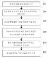
도 2는 본 발명의 실시형태에 따른 관상순환의 모델링 방법, 관상동맥 질환의 평가 및 중재술 방안을 나타낸 것이다.2 shows a modeling method of coronary circulation according to an embodiment of the present invention, a method of evaluation and intervention of coronary artery disease.
단계 202에서, 환자의 의료 영상 데이타가 수신된다. 하나 또는 다수의 영상 기기로부터의 의료 영상 데이타가 수신될 수 있다. 예를 들면, 상기 의료 영상 데이타는 컴퓨터 단층 촬영 (computed tomography, CT), Dyna CT, 자기 공명 (magnetic resonance, MR), 혈관 조영 검사 (Angiography), 초음파, 단일 광자 단층 촬영 (Single Photon Emission computed Tomography, SPECT) 및 임의의 다른 유형의 의료 영상 기기를 포함할 수 있다. 상기 의료 영상 데이타는 2D, 3D 또는 4D (3D + 시간) 의료 영상 데이타일 수 있다. 상기 의료 영상 데이타는 CT 스캐너, MR 스캐너, 혈관 조영 검사 스캐너, 초음파 장치 등과 같은 하나 이상의 영상 획득 장치로부터 직접 수신될 수 있고, 또는 의료 영상 데이타는 환자에 대한 이전에 저장된 의료 영상 데이타를 로딩함으로써 수신될 수 있다.In
단계 204에서, 관상동맥 및 심장의 환자 특이적 해부학적 모델이 상기 의료 영상 데이타로부터 생성된다. 유리한 구현에 있어서, 상기 환자 특이적 해부학적 모델은 4D 의료 영상 데이타를 이용하여 생성된 관상동맥의 4D (3D + 시간) 기하학적 모델을 포함한다. 상기 관상동맥의 환자 특이적 해부학적 모델을 생성시키기 위하여, 관상동맥은 4D 영상 데이타의 각각의 프레임 (frame)에서 분할된다. 상기 관상동맥은 임의의 관상동맥 분할법을 이용하여 의료 영상 데이타의 각각의 프레임에서 분할될 수 있다. 예를 들면, 관상동맥은 본 명세서에 참조로서 통합되는 공개된 미국 특허출원 제2010/0067760호에 설명된 방법을 이용하여 CT 볼륨 (volume)에서 분할될 수 있다. 다음, 기하학적 표면 모델이 각각의 프레임의 관심있는 분할된 관상동맥에 대하여 생성된다. 예를 들면, 둘다 본 명세서에 참조로서 통합되는 미국 특허 제7,860,290호 및 미국 특허 제7,953,266호에는 관상동맥의 해부학적 모델링을 위한 방법이 설명되어 있다. 이것은 시간이 지남에 따라 변화하는 관상동맥의 해부학적 구조를 보여주는 관상동맥의 해부학적 모델이 되게 한다.In
유리한 실시형태에서, 상기 환자 특이적 해부학적 모델은 또한 4D 영상 데이타로부터 생성된 심장의 환자 특이적 4D 해부학적 모델을 포함한다. 상기 4D 해부학적 심장 모델은 챔버 (좌심실 (left ventricle), 좌심방 (left atrium), 우심실 (right ventricle) 및 우심방 (right atrium)), 심장판 (대동맥판 (aortic valve), 좌방실판막 (mitral valve), 우방실판막 (tricuspid valve) 및 폐동맥판 (pulmonary valve)), 및 대동맥을 포함하는 것과 같이 다수의 심장 구성요소를 가지는 다-구성요소 모델이다. 이러한 심장의 포괄적인 모델은 매우 다양한 형태학적, 기능적, 그리고 병적인 변화를 캡쳐하는데 이용된다. 모듈식 (modular)의 그리고 계층적 (hierarchical) 접근법은 해부학적 복잡성을 줄이고, 개별적 해부의 효율적이고 유연한 추정을 용이하게 하기 위해 이용될 수 있다. 4D 해부학적 심장 모델은, 예를 들어 여백 학습 (marginal space learning, MSL)을 이용하여 각각의 심장 구성요소의 개별적 모델을 생성시키고, 다음 메쉬 포인트 (mesh point) 대응점을 설정함으로써 심장 구성요소 모델을 통합함으로써, 생성될 수 있다. 이러한 4D 환자 특이적 심장 모델의 생성에 관한 추가의 상세한 사항은 본 명세서에 참조로서 통합되는 공개된 미국 특허출원 제2012/0022843호에 설명되어 있다.In an advantageous embodiment, the patient-specific anatomical model also includes a patient-specific 4D anatomical model of the heart generated from 4D image data. The 4D anatomic cardiac model includes chambers (left ventricle, left atrium, right ventricle and right atrium), cardiac valves (aortic valve, mitral valve, A tricuspid valve, and a pulmonary valve), and aortic aneurysm, which is a multi-component model with multiple cardiac components. This comprehensive model of the heart is used to capture a wide variety of morphological, functional, and pathological changes. A modular and hierarchical approach can be used to reduce anatomical complexity and facilitate efficient and flexible estimation of individual anatomy. The 4D anatomic cardiac model can be modeled by creating individual models of each cardiac component using, for example, marginal space learning (MSL), and setting the following mesh point correspondence points, By integrating them. Further details regarding the generation of such a 4D patient-specific cardiac model are described in published US Patent Application No. 2012/0022843, which is incorporated herein by reference.
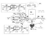
단계 206에서, 관상순환의 멀티스케일 기능적 모델이 생성된다. 인간의 심혈관계의 자세한 모델링에 대한 중요한 문제점 중 하나는 개별적인 구성요소간의 상호 의존도가 높은 폐회로를 나타낸다는 사실이다. 계 (국부적 혈류역학)의 특정 세그먼트의 혈류 특성은 계의 전체적인 역학과 밀접하게 관련이 있다. 국부적 혈류를 연구하는 것은 중요한데, 이는 혈관이 국부적으로 두껍게 되는 것 또는 협착의 형성과 같은 특정 병리학 (pathology)이 국부적 혈류역학에 의해 강하게 영향을 받기 때문이다. 한편, 혈관 내강 (vascular lumen)의 변형과 같은 특정한 국부적 변화는 혈류의 전체적인 재분배를 유도하여, 원위 부분 (distal part)의 영향을 받는 혈관에서 충분이 높은 유량을 보장하는 일부 보장성 메커니즘 (compensatory mechanism)을 촉발시킬 수 있다. 3D 또는 실물 크기 (full-scale)의 혈류 계산은 계산적으로 고가이며, 단지 감소된 수의 혈관에 대해 수행될 수 있다. 전신성 (systemic) 즉, 전체적 혈류역학 및 국부적 혈류역학 간의 상호 영향 및 3D 계산의 높은 계산적 요구 사항 모두는 혈류의 기하학적 멀티스케일 모델링의 개념을 유도하며, 관상순환을 분석하기 위해 여기서 사용된다.In
따라서, 본 발명의 유리한 실시형태에서, 관상동맥 트리 내의 관심있는 국부적 영역만이, 예를 들어 좁아진 플라크 침착물을 함유하는 세그먼트는 전방향 3D 모델 (full 3D model)을 이용하여 분할되는 반면, 나머지 순환은 감소 차수 모델 (대동맥의 경우 1D 모델이고, 소동맥 및 미세혈관의 경우 집중 모델 (lumped model))을 통해 나타낸다. 감소 차수 모델은 압력 및 유량 파형의 면에서 신뢰성 있는 결과를 산출하고 (1D 모델), 원위 혈관 및 미세혈관의 효과를 바르게 고려하며 (집중 모델), 상응하는 3D 계산보다 작은 크기의 2차수보다 큰 실행 시간을 유도한다. 한편, 전체 계산은 3D 모델 상에 구현될 수 있다.Thus, in an advantageous embodiment of the invention, only the localized regions of interest in the coronary artery tree, for example segments containing narrowed plaque deposits, are segmented using a full 3D model, while the rest The circulation is represented by a reduced order model (1D model for the aorta and lumped model for the small arteries and microvessels). The reduced order model produces reliable results in terms of pressure and flow waveforms (1D model), considers the effects of the distal vessels and microvessels properly (focused model), and is larger than the second order of smaller dimensions than the corresponding 3D calculations Leading to execution time. On the other hand, the entire calculation can be implemented on the 3D model.
도 3은 본 발명의 실시형태에 따른 멀티스케일 모델링 접근법의 개요를 나타낸 것이다. 도 3에 도시된 바와 같이, 심장 모델(302)은 대동맥의 뿌리에서 커플링된다. 심장 모델(302)은 전방향 3D 심장 모델로서 구현될 수 있거나, 또는 환자 특이적 데이타를 통해 파라미터화되는 집중 모델로서 구현될 수 있다. 대동맥 및 커다란 동맥 (예를 들어, 좌관상동맥 (LCA), 우관상동맥 (RCA) 등)은 1D 혈류 모델(304, 306, 308, 310, 312, 314, 316, 318, 320)로서 나타낼 수 있는데, 이는 이들 1D 혈류 모델(304-318)이 압력 및 혈류 속도값의 관점에서 신뢰성 있는 결과를 산출하고, 파동 전파 현상을 고려하기 때문이다. 모든 미세혈관 베드는 혈류에 적용된 저항 및 원위 혈관의 유연성 (compliance)을 설명하는 집중 파라미터 모델(322, 324, 326, 328, 330)를 통해 시뮬레이션될 것이다. 관상동맥 트리의 경우, 커다란 (심외막) 혈관의 혈류는 전신성 트리 모델(321)에서 1D 모델을 통해 계산된다. 협착 세그먼트(332, 334) (즉, 혈관의 영역이 협착이거나 또는 좁아지는 것이 검출됨)는 1D 혈류 모델을 이용하여 시뮬레이션 될 수 없는데, 이는 단면적의 변화가 크고, 협착 모양이 혈류 거동 및 특히 이러한 협착의 기능적 중요성을 평가함에 있어서 중요한 역할을 하는 트랜스-협착성 압력 강하 (trans-stenotic pressure drop)에 영향을 주기 때문이다. 관상동맥 혈관 베드는 집중 파라미터 모델(324, 326, 328, 330)을 통해 모델링되며, 이는 그것들이 심장 수축기 (systole) 동안에 심근 수축의 영향을 고려한다는 의미에서 관상순환에 적당하다.3 shows an overview of a multiscale modeling approach in accordance with an embodiment of the present invention. As shown in FIG. 3, the cardiac model 302 is coupled at the root of the aorta. The cardiac model 302 may be implemented as an omni-directional 3D cardiac model, or may be implemented as a centralized model parameterized via patient specific data. The aorta and large arteries (e.g., left coronary artery (LCA), right coronary artery (RCA), etc.) can be represented as 1D blood flow models (304, 306, 308, 310, 312, 314, 316, 318, This is because these 1D blood flow models (304-318) yield reliable results in terms of pressure and blood flow velocity values and consider wave propagation phenomena. All microvessel beds will be simulated through the
실행 시간이 중요하기 때문에, 유리한 구현에 따르면, 딱딱한 벽면 3D 모델(340, 350)은 그 실행 시간이 2배 더 긴 3D 유체-구조 상호작용 (fluid-structure interaction, FSI) 대신에 협착 부위(332, 334)를 나타내는데 사용될 수 있다. 협착 부위(332, 334)의 탄성은 중요하지 않기 때문에 이러한 측면이 전체적인 결과에 영향을 주지 않지만, 관상동맥 트리 내부에서 파동 전파 현상을 바르게 나타내기 위하여 0D 인터페이스 모델 (OD interface model)(342, 344, 352, 354)은 유연한 1D 모델과 딱딱한 3D 모델 사이의 계면에 포함된다. 이들 인터페이스 모델은 그것의 계면에서 3D 세그먼트의 유연성을 집중시킨다. 관상순환에서 매우 중요하고, 협착의 형태학적 및 기능적 중요성 간의 커다란 불일치에 기여하는 하나의 추가적 측면은 형태학적으로 중요한 협착이 기능적으로 중요하지 않은 것이 되도록 할 수 있는 측부 혈류 (collateral flow)의 존재이다. 환자 특이적 혈관 형태학에 의존하여, 상기 측부 혈류(336)는 문합 대혈관 (anastomotic large vessel)을 통하거나 (1D 모델로), 또는 혈액으로 영향을 받는 영역에 공급하는 미세혈관을 통하여 (도 3에서와 같이 집중 요소를 통하여 모델링) 모두 모델링될 수 있다.Because the execution time is important, according to an advantageous implementation, the rigid wall 3D models 340,350 can be used in place of the 3D-fluid-structure interaction (FSI) , 334). Although this aspect does not affect the overall result because the elasticity of the
소동맥 및 미세혈관에 대한 집중 모델Intensive model for small arteries and microvessels
미세혈관의 집중 또는 0D 모델(322-330)은 전기와 유압 (hydraulics) 사이의 유사성을 기반으로 하며, 집중 요소: 저항, 유연성 및 이너턴스 (inertance)에 작은 혈관의 생리학적 특성을 집중시킴으로써 독립 변수의 공간 종속성을 제거한다. 관상동맥 베드는 미세혈관이 심근 수축에 의해 가장 많은 영향을 받고, 그 효과가 심외막과 심내막 사이에서 변화하기 때문에 특별한 치료를 필요로 한다. 관상동맥 트리 내부에서 관찰된 유량 파형을 설명하기 위하여 3가지 상이한 메커니즘이 이용될 수 있다: 변화하는 탄성, 단축 유도되는 세포내 압력 및 공동 (cavity) 유도되는 세포외 압력. 유리한 구현에 있어서, 관상동맥 혈류의 심근 수축의 효과를 모델링하기 위하여 2번째와 3번째 메커니즘의 조합이 이용되는 반면, 변화하는 탄성은 다른 2가지 메커니즘의 작용에 대해 오히려 혈관을 보호한다. 따라서, 심장 수축기 동안에 외부 압력은 심내막하 (subendocardium)의 더 높은 그리고 심외막하 (subepicardium)의 더 낮은 관상동맥 혈관 상에서의 작용하고, 좌심실 및 우심실 압력으로부터 결정될 수 있다. 심근의 표면에서 기능하는 심외막 혈관은 이들 수축에 의해 영향을 받지 않는다. 멀티스케일의 관점에서, 규칙적인 3원계 윈드케셀 모델 (3-element Windkessel model)이 관상동맥 트리에 속하지 않는 혈관의 말단 위치에서 이용될 수 있다. 저항 및 유연성의 값은 비생리학적 반영을 회피하기 위하여 평균 압력 및 유량값을 취하고, 상기 저항값을 채택함으로써 결정된다.The microvessel concentration or the 0D model (322-330) is based on the similarity between electricity and hydraulics and focuses on the physiological characteristics of small blood vessels in concentration elements: resistance, flexibility and inertance, Remove the spatial dependency of the variable. Coronary artery beds require special treatment because microvessels are most affected by myocardial contraction and their effects vary between the epicardium and the endocardium. Three different mechanisms can be used to describe the observed flow waveforms inside the coronary artery tree: varying elasticity, shortening induced intracellular pressure, and cavity induced extracellular pressure. In an advantageous implementation, the combination of the second and third mechanisms is used to model the effect of myocardial contraction on coronary blood flow, while the changing elasticity protects the blood vessels rather than the action of the other two mechanisms. Thus, during cardiac systole, the external pressure acts on the lower and higher coronary arteries of the subendocardium and subepicardium, and can be determined from the left ventricular and right ventricular pressures. The endocardial vessels functioning on the surface of the myocardium are not affected by these contractions. From a multiscale point of view, a regular 3-element Windkessel model can be used at the distal end of the vessel that does not belong to the coronary artery tree. The values of resistance and flexibility are determined by taking the mean pressure and flow values to avoid non-physiological reflections and adopting the resistance values.
심외막 협착 세그먼트에 대한 상세 3D 해부학적 모델Detailed 3D anatomical model of epicardial stenosis segment
각각의 협착 부위(332, 334)에서의 상세한 3D 혈류 계산(340, 350)은 딱딱한 도메인에서 비압축성의 나비에-스토크스 방정식 (Navier-Stokes equation)의 수적인 해결책을 기반으로 한다. 노-슬립 (no-slip)의 경계 조건 (boundary condition)은 혈관 벽에 적용되고, 유입 및 유출 경계 조건은 근위 (proximal) 및 원위 (distal)의 1D 세그먼트로 명시적/암시적 커플링에 의해 결정된다. 유리한 구현에 있어서, 3D 모델(340, 350)은 벽면 전단 응력 (wall shear stress)과 같은 관심의 대상이 되는 국부적 파라미터의 값을 결정할 뿐만 아니라, 협착 부위에 대하여는 협착의 상세한 모양에 대한 압력 강하를 계산한다.The detailed 3D
대동맥의 1D 모델 및 협착 부위에 대한 1D model of the aorta and for stenosis 근위Proximal 및 And 원위Distal 세그먼트Segment ; 및 혈관 트리의 구조적 트리 모델 (; And the structural tree model of the vascular tree ( StructuredStructured TreeTree ModelModel ))
1D 모델(304-320)은 장축의 길이를 따라 3D 도메인을 1D 도메인으로 변환하기 위해 일련의 간략화된 가정을 이용한다. 이러한 1D 모델은 준선형 (quasilinear) 일차 편미분 방정식의 시스템에 의해 설명될 수 있다. 이들 모델의 유리한 측면은 이들이 혈관의 유연성을 고려하고 있다는 사실이며, 이는 심혈관계에서 나타나는 파동 현상의 설명을 가능하게 한다. 이들 모델의 종속 변수는 내강 면적, 유량 및 장축의 길이에 따른 평균 압력이다 (전형적으로 유연성 있는 혈관의 네트워크가 고려됨). 1D 모델은 예를 들어 유량의 전파 및 압력 파동에 대한 관상동맥의 기하학적 테이퍼링 또는 국부적 보강의 효과를 연구하는데 이용될 수 있다. 1D 모델은 심혈관계 내부의 유량 및 압력 파동의 특성을 결정할 때에 매우 유용하다. 이들 파동은 혈류와 혈관 벽 사이의 상호작용에 의해 생성되며, 특정한 유연성을 가지고 있으며, 혈관의 탄성 특성에 의존한다. 이들 모델에 대한 다양한 접근 가능성이 있다. 가장 간단한 경우에 있어서, 혈관의 기계적 특성은 평균 압력과 혈관의 반경 사이의 대수적 관계에 의해 설명된다. 보다 복잡한 상황에 있어서, 점탄성이나 벽의 관성과 같은 다른 특성이 고려될 수 있다. 본 발명의 유리한 실시형태에서, 압력과 혈관 반경의 관계는 미분 방정식에 의해 주어진다. 1D 모델을 이용한 혈류 계산은 압력 및 유량 (속도) 값의 관점에서 우수한 결과를 산출하고, 계산적 수고가 줄어드는 이점을 갖는 것으로 보여진다. 따라서, 혈관 해부학적 구조가 규칙적이고 (예를 들어, 원통형), 벽면 전단 응력 또는 진동 전단 지수와 같은 국부적인 값을 계산할 필요가 없을 경우, 이러한 1D 모델은 계산 시간을 줄일 뿐만 아니라 파동 전파 현상을 연구하기 위해 이용될 수 있다. 이들 1D 모델은 협착성 세그먼트의 전 또는 후 모두에서 사용되고, 따라서 전체 관상동맥 트리에서의 상세한 공간 압력과 유량 분포가 결정되도록 한다. 아울러, 1D 모델 (비선형 조건 함유)은 선형화될 수 있으며, 정확한 분석 해결책은 주파수 영역에서 얻어질 수 있다. 이러한 방법인 구조적 트리 모델 (321)은 동맥 트리의 원위 부위에 대해 얻어질 수 있으며, 다음에 임피던스로 집중되고, 비선형 1D 모델의 출구에서 경계 조건으로서 적용될 수 있다.The 1D model 304-320 uses a series of simplified assumptions to transform the 3D domain into the 1D domain along the length of the major axis. This 1D model can be explained by a system of quasilinear first order partial differential equations. The advantageous aspect of these models is that they consider the flexibility of the blood vessels, which allows explanations of the wave phenomena that occur in cardiovascular. Dependent variables in these models are average pressure along the lumen area, flow rate and length of the major axis (typically a flexible network of vessels is considered). 1D models can be used, for example, to study the effects of geometric tapering or local reinforcement of coronary arteries on flow and pressure waves of flow. The 1D model is very useful in determining the characteristics of flow and pressure waves inside the cardiovascular system. These waves are generated by the interaction between blood flow and blood vessel walls, have certain flexibility, and depend on the elastic properties of the blood vessels. There are various approaches to these models. In the simplest case, the mechanical properties of the vessel are explained by the algebraic relationship between the mean pressure and the radius of the vessel. In more complex situations, other properties such as viscoelasticity and wall inertia can be considered. In an advantageous embodiment of the present invention, the relationship between pressure and vessel radius is given by the differential equation. The 1D model of blood flow calculation shows good results in terms of pressure and flow (velocity) values, and has the advantage of reducing computational labor. Thus, if the vascular anatomy is regular (eg, cylindrical) and there is no need to calculate local values such as wall shear stress or vibration shear index, this 1D model not only reduces computation time, Can be used to study. These 1D models are used both before and after the stenotic segment, thus allowing detailed spatial pressure and flow distribution in the entire coronary artery tree to be determined. In addition, 1D models (containing nonlinear conditions) can be linearized and accurate analytical solutions can be obtained in the frequency domain. This method, the
1D-3D 커플링 계면에서의 집중 모델Concentration model at 1D-3D coupling interface
3D 혈류 계산은 딱딱하고 유연한 벽면 (유체 구조 상호작용) 모두로 수행될 수 있다. 딱딱한 벽면 계산은 상당히 빠르며, 협착성 부위에서의 탄성은 감소된다 (플라크 침착물 뿐만 아니라, 관상동맥 혈관이 증가된 강성을 특징으로 하는 비교적 작은 직경을 갖기 때문임). 따라서, 협착성 부위(332, 334)는 딱딱한 벽면으로 시뮬레이션 되며, 혈관의 유연성은 종종 그것들 자신에 의해 3D 결과의 의미성과 관련이 없을지라도 멀티스케일 모델에서 중요한데, 이는 혈관의 유연성이 전체 관상동맥 트리에서 크게 관심있는 압력-파동 전파의 구동 메커니즘이기 때문이다. 따라서, 협착성 세그먼트의 유연성은 고려되어야 하며, 이것은 1D 및 3D 세그먼트 사이의 계면에 3D 세그먼트의 유연성을 집중시키는 0D 모델(342, 344, 352, 354)을 도입함으로써 가장 잘 수행될 있다. 관상동맥 트리에서의 압력 및 유량 파동 전파는 심근 수축의 영향 때문에 복잡하고, 현재까지 광범위하게 연구되지 않았다.3D blood flow calculations can be performed with both rigid and flexible wall surfaces (fluid structure interaction). Rigid wall calculations are fairly rapid and the elasticity at the stenotic site is reduced (because not only plaque deposits, but also coronary artery blood vessels have a relatively small diameter characterized by increased stiffness). Thus, the
0D 모델(342, 344, 352, 354)은 1D 및 3D 모델 사이의 계면에 도입된다 하더라도 일부 어려움은 여전히 멀티스케일 모델의 커플링 지점에서 발생할 수있다. 1D 및 3D 모델 사이의 적절한 커플링은 모델의 상이한 수학적 성질에 의해 강조되는 측면을 달성하기 어렵다. 나비에-스토크스 방정식은 속도의 관점에서 포물선인 편미분 방정식의 시스템을 나타내는 반면, 1D 모델은 쌍곡선의 편미분 방정식에 의존한다. 이들 조건 하에서, 수학적 문제가 잘 제기되고, 수치 결과가 원하는 정확도인 것을 보장해야 한다. 모델의 적절한 커플링을 보장하는데 이용될 수 있는 다양한 가능성이 있다: 소위 "아무것도 하지 않는 (do-nothing)" 접근 (전형적으로 압력 경계 조건에 대해 이용됨) 및 "라그랑제 승수 (Lagrange multiplier)" 접근 (전형적으로 속도-유량 경계 조건에 대해 이용됨). 모델들을 커플링함으로써, 혈액의 전반적 및 국부적 거동 뿐만 아니라 국부적 모델과 전반적 모델 사이의 상호 영향이 결정될 수 있다.Although the
관상동맥의 자동 조절 (coronary autoregulation) 및 충혈 모델링 (hyperemia modeling)Coronary autoregulation and hyperemia modeling of coronary arteries
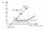
관상동맥의 자동 조절은 심외막 혈관에서의 동맥경화 세그먼트에 대한 관상동맥 트리의 적용에서 중요한 역할을 한다. 자동 조절은 관류 (perfusion) 압력의 변화에 대한 반응으로서 미세혈관 저항의 변화를 의미하고, 그것의 역할은 모세관을 통해 일정한 유량을 유지시키는 것이다. 이러한 측면은 신체의 정상 또는 안정된 상태를 의미한다. 또 다른 유형의 조절은 미세혈관 저항이 유량의 3배 내지 5배 증가를 허용하기 위해 최소로 감소할 때, 운동 또는 약물로 인한 충혈시 발생한다. 이들 측면 모두 관상동맥 트리의 멀티스케일 모델 내부에서 고려되어야 한다.Automatic regulation of coronary arteries plays an important role in the application of the coronary artery tree to atherosclerotic segments in the epicardial vasculature. Automatic regulation refers to a change in microvascular resistance as a response to changes in perfusion pressure, and its role is to maintain a constant flow rate through the capillary. This aspect refers to the normal or stable state of the body. Another type of modulation occurs when the microvessel resistance declines to a minimum to allow a three to five-fold increase in flow rate, congestion due to exercise or drug. All of these aspects should be considered within the multiscale model of the coronary artery tree.
도 4는 관상동맥의 자동 조절을 그래프로 나타낸 것이다. 자동 조절은 안정된 상태, 즉 감소된 심박수 및 혈압에서 일어나며, 협착된 혈관에 대하여 혈류 의존 협착 저항의 도입을 보상하는 감소된 미세혈관 저항을 유도한다. 일반적으로 관상동맥의 혈류, 특히 자동 조절 현상에 영향을 미치는 중요한 측면은 측부 혈류의 존재이다. 문합 채널로도 불리는 이들 혈관은 허혈 (ischemia)에 대한 적응으로서 심장에서 발달한다. 그것들은 중증의 협착을 브릿지하거나, 또는 하나의 심외막 관상동맥과 또 다른 하나의 심외막 관상동맥에 의해 제공되는 영역을 연결하는 도관 (conduit) 세그먼트의 역할을 한다. 따라서, 측부 혈관은 관상동맥 질환에 의해 영향을 받는 심근의 특정 부분에 대한 혈액 공급의 대안적인 공급원을 나타내고, 그것들은 심근 기능을 유지하는데 도움을 줄 수 있다. 두가지 상이한 유형의 측부 혈관이 있다: 평활근 세포 (smooth muscle cell)가 없는 모세관 크기의 측부 혈관 (보통 심내막하에서 나타남) 및 이미 존재하는 소동맥으로부터 발달한 보다 큰 근육질의 측부 혈관 (보통 심외막에서 나타남). 두번째 유형의 조절은 협착의 기능적 유의성을 결정하기 위해 임상 실습에서 적용되는 충혈을 유도하는 약물의 효과를 시뮬레이션하기 위해 중요하다. 협착의 존재 하에서, 협착 세그먼트의 충혈 혈류는 더 이상 최대값에 도달할 수 없으며, 정상적인 건강한 혈관인 경우에 얻어질 것이다. 이것은 협착에 의해 도입되는 저항에 의해 발생하고, 최대 혈류를 제한하는데, 그 이유는 미세혈관 저항이 그것의 정상 하한치 미만으로 감소할 수 없기 때문이다.Figure 4 is a graphical representation of automatic adjustment of coronary arteries. Automatic regulation occurs at steady state, i.e., at reduced heart rate and blood pressure, leading to reduced microvascular resistance that compensates for the introduction of blood flow dependent stenotic resistance to the stenosed blood vessels. In general, the presence of lateral blood flow is an important aspect that affects the blood flow of coronary arteries, especially the autoregulation phenomenon. These blood vessels, also called anastomotic channels, develop in the heart as adaptations to ischemia. They serve as a conduit segment that bridges severe stenosis or connects the area provided by one epicardial coronary artery and another epicardial coronary artery. Thus, the collateral vessels represent an alternative source of blood supply to a particular portion of the myocardium affected by coronary artery disease, and they can help maintain myocardial function. There are two different types of collateral vessels: capillary sized collateral vessels without smooth muscle cells (usually seen under endocardium) and larger muscular collateral vessels developed from pre-existing small arteries (usually seen in the epicardium ). The second type of modulation is important to simulate the effects of drugs that induce congestion applied in clinical practice to determine the functional significance of stenosis. In the presence of stenosis, the reticulated blood flow of the stenotic segment can no longer reach its maximum value and will be obtained in the case of normal healthy blood vessels. This is caused by the resistance introduced by the stenosis and limits the maximum blood flow because the microvascular resistance can not be reduced below its normal lower limit.
기계 생물학 (Mechanobiology) 및 세포 역학 신호 변환 (Mechanotransduction)을 위한 세포 모델Mechanobiology and Cytomics Cell models for signal transduction
관상순환의 멀티스케일 커플링된 모델의 예측 성질은 본질적으로 세포 모델들과의 커플링과 연관된다. 이것은 개시, 및 관상동맥 플라크의 후속 성장 및 전체적인 순환에 대한 이들의 효과를 추적하는 것을 가능하게 한다. 관상동맥에서 혈류에 의해 유도된 벽면 전단 응력은 플라크 발달에 영향을 줄 뿐만 아니라, 플라크 파열과 연관된다는 것을 보여준다. 이러한 측면 배후의 메커니즘은 일반적으로 내피 세포의 역할에 의해 설명되며, 이는 혈류역학적 힘에 응답하는 것으로 알려져 있다. 혈관 벽면에 가해진 전단 응력은 세포 역학 신호 변환의 과정을 통해 생화학적 신호로 변환된다. 이것은 궁극적으로 혈관 벽면에 특정한 변화를 초래한다. 전단력은 내피 세포에 가해지고, 국부적 세포 역학 신호 변환의 메커니즘을 통해 내피 세포의 구조와 기능을 조절한다. 유리한 구현에 있어서, 세포 역학 신호 변환의 다양한 분석적 및 수학적 모델이 사용되어 기관 수준의 순환 모델과 커플링될 수 있다. 이러한 커플링은 혈류역학적 계산에서 얻은 다음 세포 역학 신호 변환 모델에 입력으로서 전달되는 벽면-전단 응력값을 통해 수행된다. 내피 세포는 혈관 톤을 조절뿐만 아니라, 세포 역학 신호 변환 모델들에 의해 고려될 협착으로 인한 혈류역학적 변화에 대응하여 혈관 벽면을 리모델링 (remodeling)한다. 이러한 리모델링은 기관 수준의 순환 모델에 영향을 미칠 것이다.The predictive properties of a multiscale coupled model of coronary circulation are inherently associated with coupling with cellular models. This makes it possible to track the onset and subsequent effects of coronary plaques and their effects on the overall circulation. Blood flow shear stress induced by coronary arteries in blood flow not only affects plaque development but also correlates with plaque rupture. This side-back mechanism is generally explained by the role of endothelial cells, which are known to respond to hemodynamic forces. The shear stress applied to the vessel wall is converted into biochemical signals through the process of cellular signal transduction. This ultimately leads to a specific change in the wall of the blood vessel. Shear forces are applied to endothelial cells and regulate the structure and function of endothelial cells through mechanisms of local cellular signal transduction. In an advantageous implementation, various analytical and mathematical models of cytodynamic signal transduction can be used to couple with an organ-level circulation model. This coupling is accomplished through wall-shear stress values obtained from hemodynamic calculations and then transmitted as input to the cytodynamic signal transformation model. Endothelial cells not only modulate vascular tone, but also remodel the vessel wall in response to hemodynamic changes due to stenosis considered by cytodynamic signal transduction models. This remodeling will affect the institutional level circulation model.
심장의 감소 차수 모델Reduced-order model of the heart
몇가지 파라미터의 심장 모델이 있으며, 이는 신체의 다양한 상태를 시뮬레이션하는데 매우 유용한다. 가장 많이 이용되는 것 중 일부는 다양한 엘라스턴스 (elastance) 모델 및 단일 섬유 모델이다. 이들 심장 모델은 심장의 공간 모델을 고려하지 않고 상이한 심장 챔버에서의 압력 및 유량을 결정할 수 있다. 신체의 다양한 상태를 고려하여, 모델을 개인의 필요에 맞추기 위하여 수축성 (contractility), 박출량 (stroke volume), 타임-투-맥시멈 (time-to-maximum), 불용 체적 (dead volume), 심박수와 같은 몇가지 파라미터가 채택될 수 있다.There are several parameter heart models, which are very useful for simulating various states of the body. Some of the most commonly used are the various elastance and single fiber models. These cardiac models can determine the pressure and flow rate in different heart chambers without considering the spatial model of the heart. Considering the various states of the body, the model can be used to meet the individual's needs, such as contractility, stroke volume, time-to-maximum, dead volume, Several parameters may be employed.
본 발명의 유리한 실시형태에서, 감소 차수 심장 모델은 심장의 전차수 해부학적 및 혈류역학적 모델로부터 추출되고, 관상순환 모델과 함께 이것들과 효율적으로 커플링된다. 특히, 수신된 의료 영상 데이타로부터 생성된 심장의 형태학, 역학 및 혈류역학의 환자 특이적 진전된 모델이 통합될 수 있다. 생리학적인 랜드마크는 추출된 해부학적 심장 모델에 명시적으로 나타내고, 근본적인 해부학적 구조에 의미론적인 연관성을 제공한다. 상기 모델은 매우 모듈식이며, 적용 분야에 의존하여 원하는 대로 바꿀 수 있고, 좌심실과 우심실, 좌심방과 우심방, 대동맥, 대동맥판, 좌방실판막, 우방실판막, 폐동맥판과 폐동맥 (pulmonary trunk), 폐정맥 (pulmonary vein) 및 상대정맥 (superior vena cava)/하대정맥 (inferior vena cava)의 해부학적 구조 및 역학을 포함할 수 있다. 진전된 형태학적 및 역학적 파라미터가 쉽게 이용 가능하고, 심장의 커플링 기능을 연구하는데 사용될 수 있다. 상기 모델은 다양한 유형의 영상 기기 (예를 들어, CT, MRI, 초음파 등)을 사용하여 개인의 필요에 맞출 수 있다. In an advantageous embodiment of the present invention, the reduced order cardiac model is extracted from the cardiac catheter anatomy and hemodynamic model and is efficiently coupled with the coronary circulation model. In particular, patient specific progressive models of cardiac morphology, epidemiology and hemodynamics generated from received medical imaging data may be incorporated. Physiological landmarks are explicitly represented in the extracted anatomic cardiac model and provide a semantic link to the underlying anatomical structure. The model is highly modular and can be changed as desired depending on the field of application and can be changed as desired. The left ventricle and right ventricle, left atrium and right atrium, aorta, aortic valve, left atrioventricular valve, right ventricular valve, pulmonary trunk, pulmonary vein ) And the superior vena cava / inferior vena cava. Evolved morphological and mechanical parameters are readily available and can be used to study the cardiac coupling function. The model can be tailored to the needs of the individual using various types of imaging devices (e.g., CT, MRI, ultrasound, etc.).
실시간 전체 체적 심장 초음파 (real-time full volume echocardiography)의 최근 발전은 전방향 3D 심근 운동 뿐만 아니라 체적 색 도플러 (volumetric color Doppler) 정보를 되찾기 위하여 기회를 만든다. 조밀한 심근 운동은 심장 모델의 개인 맞춤화를 위해서 뿐만 아니라, 움직이는 관상동맥 혈관의 연구에 있어서 중요한 정보를 제공한다. 빠르고 강력한 탐지 및 3D + 시간 초음파 데이타 상의 심장 해부학적 구조의 조밀한 추적을 달성하기 위해 스페클 패턴, 영상 그라디언트 (image gradient), 경계 감지 및 움직임 예측과 같은 다수의 정보원이 결합될 수 있다. 이러한 심근의 에코 기반의 추정된 움직임 및 기계적 파라미터는 기본 진리값 (ground truth value)에 충분히 가깝다. 유리한 구현에 있어서, 체적 색 도플러 속도의 이용 가능성은 환자 특이적 혈류 정보의 빠르고 비외과적 (non-invasive)인 회복을 가능하게 하고, 이어지는 CFD 계산을 위한 경계 조건들로서 사용될 수 있다.Recent developments in real-time full volume echocardiography have created opportunities to regain volumetric color Doppler information as well as omni-directional 3D myocardial motion. Dense myocardial motion provides important information not only for personalization of the heart model, but also for the study of moving coronary vessels. A number of sources such as speckle patterns, image gradients, boundary detection, and motion estimation can be combined to achieve fast and robust detection and dense tracking of cardiac anatomic structures on 3D + time ultrasound data. These echo-based estimated motion and mechanical parameters of the myocardium are close enough to the ground truth value. In an advantageous implementation, the availability of volume color Doppler velocities enables fast, non-invasive recovery of patient-specific blood flow information and can be used as boundary conditions for subsequent CFD calculations.
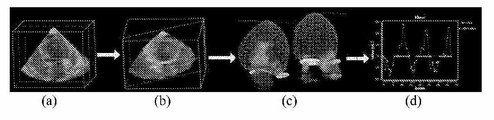
도 5는 고프레임률 체적 초음파 영상으로부터 심근 역학의 추정을 나타낸 것이다. 도 5에 도시된 바와 같이, 영상 (a)는 심장 수축기에서의 심근을 보여주고, 영상 (b)는 이완기에서의 심근을 보여주며, 영상 (c) 및 (d)는 심근의 나선 운동을 보여주는 3D 조밀한 움직임 벡터를 보여주고, 영상 (e)는 심내막과 심외막 등고선의 투영 및 4개의 챔버 평면상으로의 움직임 벡터를 보여준다.5 shows the estimation of myocardial dynamics from a high-frame-rate volume ultrasound image. As shown in Fig. 5, the image a shows the myocardium at the systole, the image b shows the myocardium at the diastole, the images c and d show the spiral movement of the myocardium 3D dense motion vectors are shown, and image (e) shows the projection of the endocardial and epicardial contour lines and the motion vectors onto the four chamber planes.
도 6은 좌심실 심근 역학의 추정을 나타낸 것이다. 도 6의 영상 (a)에 도시된 바와 같이, 602는 심내막 경계에 맵핑된 길이 방향 좌상의 추정값을 보여주고, 604는 방향 및 조밀한 속도장의 크기를 보여주며, 606은 추정된 길이 방향 변형 대 (vs) 정점의 중간 및 기초 영역에 대한 시간의 플롯을 보여준다. 도 6의 영상 (b)에 도시된 바와 같이, 612는 심내막 경계에 맵핑된 시선 속도 (radial velocity)의 추정값을 보여주고, 614는 조밀한 속도장의 방향 및 크기를 보여주며, 616은 추정된 시선 속도 대 정점의 중간 및 기초 영역에 대한 시간의 플롯을 보여준다. 도 6의 영상 (c)에 도시된 바와 같이, 622는 심내막 경계로 맵핑된 원주 변위의 추정값을 보여주고, 624는 조밀한 속도장의 방향 및 크기를 보여주며, 626은 추정된 원주 변위 대 정점의 중간 및 기초 영역에 대한 시간의 플롯을 보여준다.Figure 6 shows the estimation of left ventricular myocardial dynamics. As shown in the image (a) of FIG. 6,
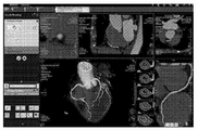
도 7은 체적 혈류의 자동 정량을 나타낸 것이다. 영상 (a)는 체적 b-모드 및 환자로부터 획득한 색깔 초음파를 보여준다. 영상 (b)는 초음파에서 좌심실, 좌방실판막 (MV) 및 좌심실 유출관 (LVOT)의 자동 검출 및 추적을 보여준다. 영상 (c)는 MV 및 LVOT 추적된 위치에서의 혈류 샘플링을 보여준다. 영상 (d)는 유입/유출 정량 및 디-앨리어싱 (de-aliasing)을 보여준다.Figure 7 shows automatic quantification of volumetric blood flow. Image (a) shows the volume b-mode and color ultrasound acquired from the patient. Image (b) shows the automatic detection and tracking of left ventricle, left atrioventricular valve (MV) and left ventricular outflow tract (LVOT) on ultrasound. Image (c) shows blood sampling at MV and LVOT tracked positions. Image (d) shows inflow / outflow metering and de-aliasing.
의료 영상으로부터 관상동맥의 해부학적 구조를 추출하기 위해, 본 발명의 실시형태는 기존의 관상동맥 분할 및 중심선 추출 알고리즘을 이용할 수 있다. 이러한 알고리즘은 협착이 좁아지는 것을 포함하여 표면 표현과 함께 관상동맥 혈관 중심선 트리를 쉽게 제공하며, 이는 다음에 3D 및 1D 계산에 대하여 요구되는 해부학적 데이터의 추출에 이용된다. 예를 들어, 협착의 폐색 비율은 검출되어, 협착 부위의 3D 모델을 구성하는데 이용될 수 있다. 도 8은 관상동맥 협착의 해부학적 평가 및 관상동맥 혈관 추출을 위한 예시적인 의료 영상 처리 소프트웨어를 나타낸 것이다.To extract the anatomical structure of the coronary artery from a medical image, embodiments of the present invention may utilize existing coronary artery segmentation and centerline extraction algorithms. These algorithms easily provide a coronary vascular centreline tree with surface representation, including narrowing of stenosis, which is then used to extract the anatomical data required for 3D and 1D calculations. For example, the occlusion rate of stenosis may be detected and used to construct a 3D model of the stenotic site. 8 shows an exemplary medical image processing software for anatomical evaluation of coronary artery stenosis and coronary artery blood vessel extraction.
환자 특이적 4-D 해부학적 모델링, 고성능 컴퓨팅 (High Performance Computing, HPC) 및 영상을 통한 3-D 혈류 측정 기술의 최근의 진전으로, 심장 혈관 적용 분야에서 혈류역학적 평가 및 차후의 확인을 위한 계산 유체 역학 (CFD)을 이용하는 것이 가능하다. 대부분의 이전 접근법은 유체 구조 상호작용 (fluid structure interaction, FSI) 방법을 이용하거나 또는 강하게 혈액과 상호작용하는 단일 심장 구성요소에 초점을 둔 반면, 본 발명의 실시형태는 4D CT 데이타로부터 유도된 고품질의 환자 특이적 심장 모델을 이용하여 전체 심장 혈류의 CFD 계산을 이용하였다. 상기 4D CT로부터 캡쳐된 4D 생리학적 모델은 심장 내부의 혈류 계산을 위한 적절한 제한을 제공하는데 사용되었다. 4D 심장 모델을 이용하는 환자 특이적 CFD 계산에 관한 추가의 상세한 사항은 본 명세서에 참조로서 통합되는 "심장의 포괄적인 환자 특이적 모델링을 위한 방법 및 시스템(Method and System for Comprehensive Patient-Specific Modeling of the Heart)"이라는 제목의 미국의 공개된 특허출원 제2012/0022843호에 설명되어 있다. Recent advances in patient-specific 4-D anatomical modeling, High Performance Computing (HPC), and imaging 3-D blood flow measurement technology have enabled the hemodynamic evaluation in cardiovascular applications and calculation It is possible to use fluid dynamics (CFD). While most prior approaches focused on single cardiac components using fluid structure interaction (FSI) methods or strongly interacting with blood, embodiments of the present invention are based on high quality derived from 4D CT data The CFD calculation of total cardiac blood flow was performed using the patient-specific cardiac model of FIG. The 4D physiological model captured from the 4D CT was used to provide appropriate limits for the calculation of blood flow in the heart. Further details regarding patient-specific CFD calculations using the 4D cardiac model can be found in "Method and System for Comprehensive Patient-Specific Modeling of the Heart ", which is incorporated herein by reference. Heart "in U. S. Patent Application Serial No. < RTI ID = 0.0 > 2012/0022843. ≪ / RTI >
도 2로 돌아가서, 단계 208에서, 혈류 계산은 관상순환의 멀티스케일 기능적 모델을 이용하여 수행된다. 혈류는 환자 특이적 경계 조건을 갖는 CFD를 사용하여 관상동맥의 협착 부위의 3D 모델에서 시뮬레이션된다. 특히, 혈액은 뉴턴 유체 (Newtonian fluid)로서 모델링되며, 속도장은 딱딱한 벽면 가정하에 나비에-스토크스 방정식 (연속 및 운동량 방정식(continuity and momentum equation))을 해결함으로써 수치적으로 얻어진다. 별개의 것으로 구분된 (discretized) 나비에-스토크스 방정식은 점진적으로 시간이 지남에 따라 관상동맥 내의 혈류의 속도와 압력을 시뮬레이션 하는데 사용된다. 이것은 시간의 함수로서 3D 모델에서 모든 방향에서의 흐름을 계산한다. 별개의 것으로 구분된 나비에-스토크스 방정식을 사용하는 CFD 계산에 관한 추가적인 상세한 사항은 본 명세서에 참조로서 통합되는 "심장의 포괄적인 환자 특이적 모델링을 위한 방법 및 시스템(Method and System for Comprehensive Patient-Specific Modeling of the Heart)"이란 제목의 공개된 미국 특허출원 제2012/0022843호에 설명되어 있다. 협착 부위의 환자 특이적 해부학적 구조는 환자 특이적 해부학적 구조를 기반으로 하는 혈류 계산을 제한하기 위해 CFD 모델링에 또한 입력된다. 아울러, 협착 부위의 3D 계산은 관상동맥의 주변 부분의 1D 계산과 커플링된다. 1D 모델 (예를 들어, 관상동맥 및 대동맥)에 대한 계산은 특히 관상동맥 가지 (또는 대동맥)를 통과하는 벌크 유량을 계산한다. 상기 1D 계산은 나비에-스토크스 방정식을 사용하여 수행될 수도 있다. 0D 모델 (집중 모델)은 해부학적 모델의 수학적인 추상적 개념이며, 입력 조건을 기반으로 하여 출력값을 준다.Returning to Fig. 2, in
환자 특이적 멀티스케일 모델을 이용하여 혈류를 시뮬레이션 하기 위해, 멀티스케일 심장, 관상순환 및 세포 모델은 효율적으로 커플링되어야 한다. 암시적 결합은 좌심실과 대동맥간에 구현될 수 있다. 전신성 트리 (도 3의 321)에 대한 유입 경계 조건은 심장의 좌심실에 대동맥을 커플링함으로써 유도된다. 따라서, 대동맥의 주입구에서 결정된 심박출량 (cardiac output)은 심장 모델에 의해서 뿐만 아니라, 그것이 커플링된 전신성 트리의 특성에 의해서도 결정된다.To simulate blood flow using a patient-specific multiscale model, the multiscale heart, coronary circulation and cell model must be efficiently coupled. Implicit coupling can be realized between the left ventricle and the aorta. The inflow boundary condition for the systemic tree (321 in Figure 3) is induced by coupling the aorta to the left ventricle of the heart. Thus, the cardiac output determined at the aortic opening is determined not only by the cardiac model, but also by the nature of the systematic tree to which it is coupled.
관상순환에 관한 심근 수축의 영향은 심장의 실물 스케일 모델을 이용함으로써 추정된다. 특히, 유리한 구현에 있어서, 심장의 실물 스케일 모델은 각각의 심외막 관상동맥 혈관에 관한 심장 수축의 영향을 결정하기 위하여 사용된다. 심장의 오른쪽과 왼쪽간의 중요한 차이점이 있으며, 또한 국부적으로 보다 상세한 변형이 고려될 수 있다. 영상 데이타로부터 추출된 3D 좌상 맵은 이들 경계 조건을 도입하기 위하여 사용될 수 있다. The effect of myocardial contraction on coronary circulation is estimated by using the physical scale model of the heart. In particular, in an advantageous implementation, a physical scale model of the heart is used to determine the effect of cardiac contraction on each of the epicardial coronary arteries. There are important differences between the right and left sides of the heart, and local more detailed deformations can be considered. The 3D upper left map extracted from the image data can be used to introduce these boundary conditions.
좌심실 및 우심실 내부의 압력은 혈관 상에 세포외 압력을 가한다. 압력의 양은 혈관의 위치, 즉 심외막 (낮은 압력) 또는 심내막 (높은 압력)인지에 따라 달라진다. 이러한 정보는 관상순환에 대한 세포외 압력을 설명하기 위하여 관상동맥 베드의 집중 파라미터 모델에 직접 사용될 수 있다. Pressure inside the left ventricle and right ventricle exerts extracellular pressure on the blood vessels. The amount of pressure depends on the location of the blood vessel, ie, whether it is an epicardium (low pressure) or an endocardium (high pressure). This information can be used directly in the intensive parameter model of the coronary artery bed to account for the extracellular pressure for the coronary circulation.
관상동맥 혈류 모델은 벽면 전단 응력 조건을 통해 세포 모델에 커플링되며, 내피 세포 기능을 조절하여 변경된 혈류역학 (플라크 성장으로 인함) 및 벽면 리모델링 (즉, 벽면의 탄성 변화)을 야기시킨다.Coronary artery blood flow models are coupled to cell models through wall shear stress conditions and regulate endothelial function, resulting in altered hemodynamics (due to plaque growth) and wall remodeling (ie, wall elasticity changes).
도 2로 돌아가서, 단계 210에서 혈류역학적 양은 혈류 계산을 기반으로 결정된다. 관상동맥 질환의 몇 가지 지표가 제안되었다. 동맥 경화증에 대해, 협착의 형태학적 측면보다 오히려 기능적 측면이 환자의 결과를 예측할 수 있는 것으로 보여진다. 상기 기능적인 측면은 휴식과 충혈 상태 동안에 협착을 통한 혈류 속도와 관련이 있다. 상기 형태학적 측면은 협착의 기하학적 형상과 관련이 있으며, 정량적 관상동맥 조영술 (quantitative coronary angiography, QCA)을 통해 결정될 수 있다. 협착 단독의 기하학적 모양이 왜 환자의 결과를 예측할 수 없는지 여러 가지 이유가 있다. 예를 들면, QCA는 2D 표현이며, 따라서 직경의 감소는 정확히 결정될 수 없으며, 확산성 질환 (diffuse disease)의 경우에는 기준 직경을 결정하는 것이 어렵다. 또한, 이전의 심근 경색은 공급되는 영역이 경색에 의해 영향을 받는다면 협착의 유의성을 줄일 수 있다. 또한, 측부 혈류는 혈류를 증가시킬 수 있으며, 협착의 효과를 감쇄시킬 수 있다. 기능적 유의성은 다양한 지표를 통해, 예컨대 트랜스-협착성 압력 강하, 관상동맥 혈류 예비력 (CFR), 상대적 혈류 예비력 (RFR) 또는 분획 혈류 예비력 (FFR)을 통해 결정된다. FFR은 다른 지표를 통해 일련의 이점을 제공한다. 예를 들면, FFR은 나머지 상태는 포함하지 않으며 (혈류역학적 파라미터에 매우 의존적임), 인접하는 건강한 혈관의 존재에 의존하지 않고 (RFR과는 대조적으로, 따라서 다혈관 질환의 경우에 적용할 수 있음), 측부 혈류 및 이전의 심근 경색의 영향을 포함한다. 또한, FFR은 환자의 혈류역학적 상태 (혈압, 심박수, 수축성)에 상당히 독립적이라고 보여진다. FFR은 협착에 대한 원위부 압력을 협착에 대한 근위부 압력에 대해 나누어 계산된다. 정맥의 압력은 일반적으로 상당한 에러를 도입하지 않고 0과 동일하게 간주될 수 있고, 미세혈관 저항이 정상 및 협착된 혈관 모두에 대해 충혈 상태 동안에 최소 및 일정하기 때문에, 압력의 분율은 협착의 존재 하에 여전히 공급될 수 있는 보통의 최대 충혈 혈류의 분율을 또한 나타낸다. 몇 가지 연구는 약 0.75의 절단값이 협착된 혈관에 의해 공급되는 심근 영역에서 가역적 허혈이 유도 가능한지 예측하는 것을 보여준다.Returning to FIG. 2, in
본 발명의 실시형태에서, 각각의 협착에 대한 FFR과 같은 기능적 파라미터를 결정하고, 따라서 각각의 협착에 대한 기능적 유의성을 결정하기 위하여 관상순환의 환자 특이적 멀티스케일 모델이 사용된다.In an embodiment of the present invention, a patient specific multiscale model of coronary circulation is used to determine functional parameters such as FFR for each stenosis, and thus to determine functional significance for each stenosis.
도 2로 돌아가서, 단계 212에서 가상적 중재술 방안 및 결정 지지를 위해 시뮬레이션이 수행된다. 관상동맥 질환의 진단 및 관리를 위해, 현재의 임상 사례는 정량적 관상동맥 조영술 (Quantitative Coronary Angiography, QCA)에 의해 질병에 걸린 혈관의 평가를 수반한다. 이러한 평가는 임상의에게 면적 감소를 포함하여 협착 세그먼트의 풍부한 해부학적 개요를 제공하지만, 기능적 평가를 제공하지는 않는다. 협착된 혈관에 전압선을 침습적으로 도입함으로써 측정되는 FFR 값은 협착의 기능적 평가를 위한 종래의 기술이다. QCA만이 협착의 형태학적 유의성을 평가하고, 일련의 다른 제한점을 가지며, 전압선 기반의 FFR 측정은 중재술과 관련된 위험을 수반하고, 매우 좁은 협착에 대하여는 전압선이 추가적인 압력 강하를 유도할 수 있다.Returning to Fig. 2, a simulation is performed at
이들 단점 모두는 본 명세서에 설명된 멀티스케일 관상순환 모델링 접근법에 의해 제거된다. 관상동맥 트리의 이전 CFD-기반의 계산은 오로지 3D 모델링을 사용하였으며, 따라서 간단한 심장 모델과 커플링된 높은 계산 복잡성을 유도하였고, 심장의 국부적 운동과 같은 모든 환자 특이적 측면을 포함할 수 없다. 다른 접근법은 단지 1D 모델링 (또한 간단한 심장 모델과 커플링됨)을 포함하였고, 협착의 정확한 모양이 고려되지 않기 때문에, 협착을 따라 압력 강하를 평가함에 있어 어려움이 생기게 했다.All of these disadvantages are eliminated by the multiscale coronary circulation modeling approach described herein. Previous CFD-based computations of the coronary artery tree used only 3D modeling, thus leading to a high computational complexity coupled with a simple cardiac model and can not include all patient-specific aspects such as local motion of the heart. Another approach involved only 1D modeling (coupled with a simple cardiac model) and caused difficulties in evaluating the pressure drop along the stenosis, since the exact shape of the stenosis was not taken into account.
심외막 협착의 기능적 유의성을 결정한 후에 (단계 210에서), 제안된 모델은 다음에 다양한 중재술의 영향 (도 13)을 시뮬레이션하는데 사용될 수 있으며, 환자의 건강 상태를 개선시키는데 사용될 수 있다. 풍선 팽창은 협착으로부터 방해를 사실상 감소시키고, 협착에서의 혈류를 재시뮬레이션 함으로써 모델링될 수 있다. 실제로, 풍선 팽창이 만족스러운 결과 (즉, 트랜스 협착성 압력 강하가 높게 유지됨)로 이어지지 않을 경우, 스텐트가 삽입된다. 협착 모델에 특정 스텐트의 가상적 모델을 첨가한 다음, 커플링된 혈류 분석을 수행함으로써 구현될 수 있는, 다른 제조업체로부터의 스텐트에 의한 가상적 스텐트는 관상동맥 트리 내부의 파동 전파에 대한 스텐트의 효과 및 분석되는 혈류역학적 변수에 대한 스텐트의 영향이 분석되도록 할 것이다.After determining the functional significance of the episcleral stenosis (at 210), the proposed model can then be used to simulate the effects of various interventions (FIG. 13) and can be used to improve the patient's health condition. Balloon dilatation can be modeled by virtually reducing disturbance from stenosis and re-simulating blood flow in stenosis. Indeed, if the balloon inflation does not lead to a satisfactory outcome (i. E., The trans-stenotic pressure drop remains high), the stent is inserted. A stent-induced stent from another manufacturer, which can be implemented by adding a hypothetical model of a specific stent to the stenosis model and then performing a coupled blood flow analysis, analyzes the effect and analysis of the stent for wave propagation within the coronary artery tree So that the effect of the stent on hemodynamic variables will be analyzed.
확산성 동맥경화 질환 (diffuse artherosclerotic disease)의 경우, 혈관성형술 또는 스텐트 삽입 중 어느 것도 환자의 상태를 개선시킬 수 없으며, 관상동맥 우회술 (CABG)이 전형적으로 수행된다. CABG를 시뮬레이션 하기 위해, 다양한 시작점과 끝점을 사용하여 이러한 인접하는 혈관을 도입하는 것이 멀티스케일 관상순환 모델 내에서 시뮬레이션될 수 있다. 따라서, 가장 적합한 중재술 또는 치료 옵션이 결정될 수 있으며, 상기 중재술 또는 치료는 중재술을 수행하기 전에 계획 (예를 들어, CABG를 위한 스텐트의 유형 또는 CABG의 시작점 및 끝점 선택)될 수 있다.In the case of diffuse artherosclerotic disease, neither angioplasty or stenting can improve the condition of the patient, and coronary artery bypass grafting (CABG) is typically performed. To simulate the CABG, introducing these adjacent vessels using various starting and ending points can be simulated in a multiscale coronary circulation model. Thus, the most appropriate intervention or treatment option may be determined, and the intervention or treatment may be a plan (e.g., a type of stent for CABG or a starting and ending point of CABG) prior to performing the intervention.
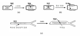
도 9는 가상적 중재술 방안의 예를 나타낸 것이다. 도 9에 도시된 바와 같이, 영상 (a)는 가상적 스텐트 모델 (virtual stent model)(904)을 사용한 협착 부위(902)에서의 스텐트 시술의 시뮬레이션을 보여준다. 영상 (b)는 감소된 폐색(914)을 시뮬레이션 함으로써 협착 부위(912) 내부의 풍선 팽창의 시뮬레이션을 보여준다. 영상 (c)는 가상적 우회술 혈관(924)을 첨가함으로써 확산성 관상동맥 질환을 가진 관상동맥의 영역(922)에서의 CABG의 시뮬레이션을 보여준다.Fig. 9 shows an example of a virtual intervention method. As shown in FIG. 9, the image (a) shows a simulation of stenting at the
의료 영상 데이타를 사용한 관상순환의 멀티스케일 해부학적 및 기능적 모델링을 위한 상기에 설명된 방법은 잘 알려진 컴퓨터 프로세서, 메모리 유닛, 저장 장치, 컴퓨터 소프트웨어 및 기타 구성요소를 사용하여 컴퓨터 상에서 구현될 수 있다. 이러한 컴퓨터의 고레벨 블록도는 도 10에 도시된다. 컴퓨터(1002)는 프로세서(1004)를 함유하며, 프로세서(1004)는 컴퓨터의 작동을 정의하는 컴퓨터 프로그램 명령을 실행함으로써 컴퓨터(1002)의 전체적인 작동을 조절한다. 컴퓨터 프로그램 명령의 실행이 요구되면, 컴퓨터 프로그램 명령은 저장 장치(1012) (예를 들어, 마그네틱 디스크(magnetic disk))에 저장되고, 메모리(1010)로 로딩 (loading)될 수 있다. 따라서, 도 2의 방법의 단계는 메모리(1010) 및/또는 기억 장치(1012)에서 저장된 컴퓨터 프로그램 명령에 의해 정의되고, 상기 컴퓨터 프로그램 명령을 실행하는 프로세서(1004)에 의해 조절될 수 있다. CT 스캔 장치, MR 스캔 장치, 초음파 장치 등의 영상 획득 장치(1020)는 컴퓨터(1002)에 연결되어 컴퓨터(1002)에 영상 데이타를 입력할 수 있다. 영상 획득 장치(1020) 및 컴퓨터(1002)를 하나의 장치로 구현하는 것이 가능하다. 영상 획득 장치(1020) 및 컴퓨터(1002)를 네트워크를 통해 무선으로 통신하는 것 또한 가능하다. 컴퓨터(1002)는 또한 네트워크를 통한 다른 장치와의 통신을 위해 하나 이상의 네트워크 인터페이스(1006)를 포함한다. 컴퓨터(1002)는 또한 컴퓨터(1002)와 사용자의 상호작용을 가능하게 다른 입력/출력 장치(1008) (예를 들어, 디스플레이, 키보드, 마우스, 스피커, 버튼 등)를 포함한다. 이러한 입력/출력 장치(1008)는 영상 획득 장치(1020)로부터 수신된 볼륨에 주석을 달기 위해 주석 도구로서 컴퓨터 프로그램의 세트와 함께 사용될 수 있다. 당업자는 실제 컴퓨터의 구현 뿐만 아니라 다른 구성요소 또한 함유할 수 있다는 것을 인식할 것이고, 도 10은 설명을 목적으로 이러한 컴퓨터의 구성요소의 일부를 고레벨로 표현한 것이다.The above-described methods for multiscale anatomical and functional modeling of coronary circulation using medical imaging data may be implemented on a computer using well known computer processors, memory units, storage devices, computer software, and other components. A high-level block diagram of such a computer is shown in FIG. The
전술한 상세한 설명은 모든 점에서 한정되는 것이 아니라 예시적이고 대표적인 것으로 이해되어야 하고, 본 명세서에 개시된 발명의 범위는 상세한 설명으로부터 결정된다기 보다는 특허법에 의해 허용되는 전체 폭에 따라 해석되는 청구범위로부터 결정되어야 한다. 본 명세서에서 보여지고 설명된 실시형태는 단지 본 발명의 원리의 예시이며, 여러 가지 변형이 본 발명의 범위 및 사상에서 벗어나지 않으면서 당업자에 의해 구현될 수 있다는 것을 이해해야 한다. 당업자는 본 발명의 사상 및 범위를 벗어나지 않고 다양한 다른 조합으로 기능을 구현할 수 있다.It is to be understood that the foregoing detailed description is not to be limited in all respects, but should be understood to be exemplary and exemplary, and the scope of the invention disclosed herein should be determined from the claims that are interpreted according to the entire breadth permitted by the patent law, do. It is to be understood that the embodiments shown and described herein are merely illustrative of the principles of the invention and that various modifications may be practiced by those skilled in the art without departing from the scope and spirit of the invention. Skilled artisans may implement the functionality in various other combinations without departing from the spirit and scope of the invention.
Claims (40)
상기 환자 특이적 해부학적 모델을 기반으로 하는 관상순환 (coronary circulation)의 멀티스케일 기능적 모델 (multi-scale functional model)을 생성시키는 단계; 및
상기 관상순환의 멀티스케일 기능적 모델을 이용하여 하나 이상의 관상동맥의 하나 이상의 협착 부위 (stenosis region)에서의 혈류를 시뮬레이션 (simulation)하는 단계
를 포함하는 방법.Generating a patient-specific anatomical model of coronary artery and heart from the patient's medical image data;
Generating a multi-scale functional model of coronary circulation based on the patient-specific anatomical model; And
Simulating blood flow in one or more stenosis regions of one or more coronary arteries using a multiscale functional model of the coronary circulation;
≪ / RTI >
상기 환자의 의료 영상 데이타로부터 관상동맥 및 심장의 환자 특이적 해부학적 모델을 생성시키는 단계는,
4D 의료 영상 데이타로부터 관상동맥의 4D 기하학적 모델을 생성시키는 단계; 및
상기 4D 의료 영상 데이타로부터 심장의 4D 해부학적 모델을 생성시키는 단계
를 포함하는 방법.The method according to claim 1,
Generating a patient-specific anatomical model of the coronary artery and heart from the patient ' s medical imaging data comprises:
Generating a 4D geometric model of the coronary artery from the 4D medical image data; And
Generating a 4D anatomical model of the heart from the 4D medical image data;
≪ / RTI >
상기 4D 의료 영상 데이타로부터 관상동맥의 4D 기하학적 모델을 생성시키는 단계는,
상기 관상동맥을 상기 4D 의료 영상 데이타의 복수의 프레임 (frame)각각에 분할하는 단계; 및
상기 4D 의료 영상 데이타의 복수의 프레임 각각에 분할된 관상동맥에 대한 기하학적 표면 모델을 생성시키는 단계
를 포함하는 방법.3. The method of claim 2,
The step of generating a 4D geometric model of a coronary artery from the 4D medical image data comprises:
Dividing the coronary artery into a plurality of frames of the 4D medical image data; And
Generating a geometric surface model for a coronary artery segmented into each of a plurality of frames of the 4D medical image data
≪ / RTI >
상기 4D 의료 영상 데이타로부터 심장의 4D 해부학적 모델을 생성시키는 단계는,
상기 4D 의료 영상 데이타의 복수의 프레임 각각에서의 복수의 심장 구성요소 각각의 개별적 모델을 추출하는 단계; 및
상기 개별적 모델 사이의 메쉬 포인트 (mesh point) 대응점을 설정함으로써, 상기 4D 의료 영상 데이타의 복수의 프레임 각각에서의 복수의 심장 구성요소에 대한 개별적 모델을 통합하는 단계
를 포함하는 방법.3. The method of claim 2,
The step of generating a 4D anatomical model of the heart from the 4D medical image data comprises:
Extracting an individual model of each of a plurality of cardiac components in each of a plurality of frames of the 4D medical image data; And
Integrating an individual model for a plurality of cardiac components in each of a plurality of frames of the 4D medical image data by setting a mesh point correspondence point between the individual models;
≪ / RTI >
상기 환자 특이적 해부학적 모델을 기반으로 하는 관상순환의 멀티스케일 기능적 모델을 생성시키는 단계는,
상기 관상동맥의 하나 이상의 협착 부위 각각에 대한 3D 계산 모델 (computation model)을 생성시키는 단계;
상기 관상동맥 및 대동맥 (aorta)의 비협착 부위 (non-stenosis region)에 대한 1D 계산 모델을 생성시키는 단계; 및
OD 집중 모델 (lumped model)을 이용하여 미세 혈관구조 혈관을 나타내는 단계
를 포함하는 방법.The method according to claim 1,
Generating a multiscale functional model of coronary circulation based on the patient-specific anatomical model comprises:
Generating a 3D computation model for each of the at least one stenotic region of the coronary artery;
Generating a 1D calculation model for the non-stenosis region of the coronary artery and the aorta; And
Representation of microvascular structures using OD-lumped model
≪ / RTI >
상기 각각의 협착 부위에 대한 3D 계산 모델은 딱딱한 벽면 3D 모델이고, 상기 각각의 협착 부위에 대한 3D 계산 모델과 상기 각각의 협착 부위에 인접한 관상동맥의 비협착 부위에 대한 1D 계산 모델 사이의 0D 인터페이스 모델 (OD interface model)은 상기 협착 부위의 유연성 (compliance)을 집중시키는 것인 방법.6. The method of claim 5,
Wherein the 3D computational model for each stenotic site is a rigid 3D wall model and a 3D computational model for each stenotic site and an 0D interface between the 1D computational model for a non-stenotic site of the coronary artery adjacent to the respective stenotic site Wherein the OD interface model concentrates the compliance of the stenotic zone.
상기 환자 특이적 해부학적 모델을 기반으로 하는 관상순환의 멀티스케일 기능적 모델을 생성시키는 단계는,
환자의 혈관 트리 (vascular tree)에 대한 구조적 트리 모델 (structured tree model)을 생성시키는 단계를 추가로 포함하는 방법.6. The method of claim 5,
Generating a multiscale functional model of coronary circulation based on the patient-specific anatomical model comprises:
Further comprising creating a structured tree model for the vascular tree of the patient.
상기 환자 특이적 해부학적 모델을 기반으로 하는 관상순환의 멀티스케일 기능적 모델을 생성시키는 단계는,
심장의 전차수 (full-order) 해부학적 및 혈류역학적 (hemodynamic) 모델로부터 심장의 감소 차수 (reduced order) 모델을 생성시키는 단계를 추가로 포함하는 방법.6. The method of claim 5,
Generating a multiscale functional model of coronary circulation based on the patient-specific anatomical model comprises:
Further comprising generating a reduced order model of the heart from a full-order anatomical and hemodynamic model of the heart.
상기 심장의 전차수 해부학적 및 혈류역학적 모델로부터 심장의 감소 차수 모델을 생성시키는 단계는,
상기 심장의 해부학적 및 혈류역학적 모델을 기반으로 하는 하나 이상의 심장 구성요소의 움직임 및 기계적 파라미터를 추정하는 단계; 및
상기 하나 이상의 심장 구성요소의 움직임 및 기계적 파라미터를 기반으로 하는 계산 유체 역학 (computational fluid dynamic) 시뮬레이션에 대한 경계 조건 (boundary condition)을 결정하는 단계
를 포함하는 방법.9. The method of claim 8,
The step of generating a reduced-order model of the heart from the tachyarrhythmia and hemodynamic model of the heart comprises:
Estimating motion and mechanical parameters of one or more heart components based on the anatomical and hemodynamic model of the heart; And
Determining a boundary condition for a computational fluid dynamic simulation based on motion and mechanical parameters of the one or more heart components
≪ / RTI >
상기 관상순환의 멀티스케일 기능적 모델을 이용하여 하나 이상의 관상동맥의 하나 이상의 협착 부위에서의 혈류를 시뮬레이션하는 단계는,
상기 관상동맥 및 심장의 해부학적 모델로부터 결정된 경계 조건을 기반으로 하는 관상순환의 멀티스케일 기능적 모델을 이용하여 하나 이상의 협착 부위에서의 혈류를 시뮬레이션하는 단계를 포함하는 방법.The method according to claim 1,
Wherein simulating blood flow at one or more stenotic sites of one or more coronary arteries using a multiscale functional model of coronary circulation comprises:
Simulating blood flow at one or more stenotic sites using a multiscale functional model of coronary circulation based on boundary conditions determined from anatomical models of the coronary arteries and heart.
상기 관상순환의 멀티스케일 기능적 모델을 이용하여 하나 이상의 관상동맥의 하나 이상의 협착 부위에서의 혈류를 시뮬레이션하는 단계는,
상기 각각의 협착 부위에 대한 3D 계산 모델 및 1D 계산 모델에서 계산 유체 역학 (CFD) 시뮬레이션을 수행하는 단계; 및
상기 각각의 협착 부위에 대한 3D 계산 모델, 1D 계산 모델 및 0D 집중 모델을 커플링하는 단계
를 포함하는 방법.6. The method of claim 5,
Wherein simulating blood flow at one or more stenotic sites of one or more coronary arteries using a multiscale functional model of coronary circulation comprises:
Performing computational fluid dynamics (CFD) simulations in a 3D computational model and a 1D computational model for each of the stenotic sites; And
Coupling the 3D computational model, 1D computational model, and 0D convergent model for each of the stenotic sites;
≪ / RTI >
상기 각각의 협착 부위에 대한 3D 계산 모델, 1D 계산 모델 및 0D 집중 모델을 커플링하는 단계는,
심장 모델의 좌심실 (left ventricle)에 대한 대동맥을 나타내는 1D 계산 모델을 커플링함으로써, 시스템 트리 모델의 유입 경계 조건을 유도하는 단계를 포함하는 방법.12. The method of claim 11,
Coupling the 3D computational model, the 1D computational model, and the 0D convergent model for each of the stenotic sites,
And coupling the 1D computational model representing the aorta to the left ventricle of the cardiac model, thereby deriving an inlet boundary condition of the system tree model.
상기 각각의 협착 부위에 대한 3D 계산 모델, 1D 계산 모델 및 0D 집중 모델을 커플링하는 단계는,
상기 의료 영상 데이타로부터 추출된 3D 좌상 맵 (strain map)을 이용하여 심외막 관상동맥 혈관 (epicardial coronary vessel)의 1D 계산 모델 상에서의 심장 수축의 영향을 나타내는 경계 조건을 도입하는 단계를 포함하는 방법.12. The method of claim 11,
Coupling the 3D computational model, the 1D computational model, and the 0D convergent model for each of the stenotic sites,
Introducing a boundary condition indicative of the effect of cardiac contraction on a 1D computational model of an epicardial coronary vessel using a 3D left top map extracted from the medical image data.
상기 각각의 협착 부위에 대한 3D 계산 모델, 1D 계산 모델 및 0D 집중 모델을 커플링하는 단계는,
상기 0D 집중 모델을 이용하여 상기 관상동맥 혈관의 위치를 기반으로 하는 관상동맥 혈관의 1D 계산 모델에 적용되는 세포외 압력을 결정하는 단계를 포함하는 방법.12. The method of claim 11,
Coupling the 3D computational model, the 1D computational model, and the 0D convergent model for each of the stenotic sites,
Determining the extracellular pressure applied to the 1D calculation model of coronary artery blood vessels based on the location of the coronary artery using the 0D concentration model.
상기 각각의 협착 부위에 대한 3D 계산 모델, 1D 계산 모델 및 0D 집중 모델을 커플링하는 단계는,
벽면 전단 응력 조건 (wall shear stress term)을 통해 상기 1D 계산 모델을 상기 0D 집중 모델에 커플링하는 단계를 포함하는 방법.12. The method of claim 11,
Coupling the 3D computational model, the 1D computational model, and the 0D convergent model for each of the stenotic sites,
And coupling the 1D calculation model to the 0D concentration model through a wall shear stress term.
상기 각각의 협착 부위에 대한 3D 계산 모델, 1D 계산 모델 및 0D 집중 모델을 커플링하는 단계는,
0D 인터페이스 모델을 이용하여 상기 3D 계산 모델을 인접하는 1D 계산 모델에 커플링하는 단계를 포함하는 방법.12. The method of claim 11,
Coupling the 3D computational model, the 1D computational model, and the 0D convergent model for each of the stenotic sites,
0.0 > 0D < / RTI > interface model to an adjacent 1D calculation model.
상기 하나 이상의 협착 부위를 통과하는 시뮬레이션된 혈류를 기반으로 하는 하나 이상의 협착 부위의 기능적 유의성을 결정하기 위해 혈류역학적 양을 산출하는 단계를 추가로 포함하는 방법.The method according to claim 1,
Further comprising calculating hemodynamic quantities to determine functional significance of one or more stenotic sites based on simulated blood flow through the one or more stenotic sites.
상기 하나 이상의 협착 부위를 통과하는 시뮬레이션된 혈류를 기반으로 하는 하나 이상의 협착 부위의 기능적 유의성을 결정하기 위해 혈류역학적 양을 산출하는 단계는,
상기 하나 이상의 협착 부위를 통과하는 계산 혈류 (computation blood flow)를 기반으로 하는 하나 이상의 협착 부위의 분획 혈류 예비력 (FFR, fractional flow reserve)을 산출하는 단계를 포함하는 방법.18. The method of claim 17,
Wherein calculating hemodynamic quantities to determine functional significance of one or more stenotic sites based on simulated blood flow through the one or more stenotic sites comprises:
Calculating a fractional flow reserve (FFR) of one or more stenotic sites based on computation blood flow through the at least one stenotic site.
상기 관상순환의 멀티스케일 기능적 모델을 이용하여 하나 이상의 협착 부위에서의 가상적 중재술 (virtual intervention)을 시뮬레이션하는 단계를 추가로 포함하는 방법.The method according to claim 1,
Further comprising simulating virtual intervention at one or more stenotic sites using a multiscale functional model of the coronary circulation.
상기 관상순환의 멀티스케일 기능적 모델을 이용하여 하나 이상의 협착 부위에서의 가상적 중재술을 시뮬레이션하는 단계는,
상기 관상순환의 멀티스케일 기능적 모델에서의 하나 이상의 협착 부위로부터의 방해를 사실상 감소시킴으로써 풍선 팽창 (balloon inflation)을 시뮬레이션하는 단계 및 상기 하나 이상의 협착 부위를 통과하는 혈류를 재시뮬레이션 (re-simulation)하는 단계를 포함하는 방법.20. The method of claim 19,
Wherein simulating virtual intervention at one or more stenotic sites using a multiscale functional model of the coronary circulation comprises:
Simulating balloon inflation by substantially reducing disturbances from one or more stenotic sites in a multiscale functional model of the coronary circulation, and re-simulating blood flow through the one or more stenotic sites Lt; / RTI >
상기 관상순환의 멀티스케일 기능적 모델을 이용하여 하나 이상의 협착 부위에서의 가상적 중재술을 시뮬레이션하는 단계는,
상기 관상순환의 멀티스케일 기능적 모델에서의 하나 이상의 협착 부위에 가상적 스텐트 모델 (virtual stent model)을 도입함으로써 스텐트 삽입 (stent implantation)을 시뮬레이션하는 단계 및 상기 하나 이상의 협착 부위를 통과하는 혈류를 재시뮬레이션하는 단계를 포함하는 방법.20. The method of claim 19,
Wherein simulating virtual intervention at one or more stenotic sites using a multiscale functional model of the coronary circulation comprises:
Simulating a stent implantation by introducing a virtual stent model at one or more stenotic sites in a multiscale functional model of the coronary circulation, and simulating stent implantation by re-simulating blood flow through the at least one stenotic site Lt; / RTI >
상기 관상순환의 멀티스케일 기능적 모델을 이용하여 하나 이상의 협착 부위에서의 가상적 중재술을 시뮬레이션하는 단계는,
상기 관상순환의 멀티스케일 기능적 모델에서의 하나 이상의 협착 부위에 인접한 우회 혈관 (bypass vessel)을 첨가함으로써 관상동맥 우회술 (CABG, coronary artery bypass graft)을 시뮬레이션하는 단계 및 상기 하나 이상의 협착 부위를 통과하는 혈류를 재시뮬레이션하는 단계를 포함하는 방법.20. The method of claim 19,
Wherein simulating virtual intervention at one or more stenotic sites using a multiscale functional model of the coronary circulation comprises:
Simulating a coronary artery bypass graft (CABG) by adding a bypass vessel adjacent one or more stenotic sites in a multiscale functional model of the coronary circulation; and simulating blood flow through the at least one stenotic site Lt; RTI ID = 0.0 > re-simulating < / RTI >
상기 환자 특이적 해부학적 모델을 기반으로 하는 관상순환의 멀티스케일 기능적 모델을 생성시키기 위한 수단; 및
상기 관상순환의 멀티스케일 기능적 모델을 이용하여 하나 이상의 관상동맥의 하나 이상의 협착 부위에서의 혈류를 시뮬레이션하기 위한 수단
을 포함하는 장치.Means for generating a patient-specific anatomical model of the coronary artery and heart from the medical imaging data of the patient;
Means for generating a multiscale functional model of coronary circulation based on the patient-specific anatomical model; And
Means for simulating blood flow at one or more stenotic sites of one or more coronary arteries using a multiscale functional model of the coronary circulation;
/ RTI >
상기 환자의 의료 영상 데이타로부터 관상동맥 및 심장의 환자 특이적 해부학적 모델을 생성시키기 위한 수단은,
4D 의료 영상 데이타로부터 관상동맥의 4D 기하학적 모델을 생성시키기 위한 수단; 및
상기 4D 의료 영상 데이타로부터 심장의 4D 해부학적 모델을 생성시키기 위한 수단
을 포함하는 장치.24. The method of claim 23,
Means for generating a patient-specific anatomical model of the coronary artery and heart from the medical imaging data of the patient comprises:
Means for generating a 4D geometric model of a coronary artery from 4D medical imaging data; And
Means for generating a 4D anatomical model of the heart from the 4D medical imaging data
/ RTI >
상기 환자 특이적 해부학적 모델을 기반으로 하는 관상순환의 멀티스케일 기능적 모델을 생성시키기 위한 수단은,
상기 관상동맥의 하나 이상의 협착 부위 각각에 대한 3D 계산 모델을 생성시키기 위한 수단;
상기 관상동맥 및 대동맥의 비협착 부위에 대한 1D 계산 모델을 생성시키기 위한 수단; 및
OD 집중 모델을 이용하여 미세 혈관구조 혈관을 나타내기 위한 수단
을 포함하는 장치.24. The method of claim 23,
Means for creating a multiscale functional model of coronary circulation based on the patient-specific anatomical model,
Means for generating a 3D computational model for each of the at least one stenotic region of the coronary artery;
Means for generating a 1D calculation model for non-stenotic regions of the coronary arteries and the aorta; And
Means for representing microvascular blood vessels using an OD concentration model
/ RTI >
상기 각각의 협착 부위에 대한 3D 계산 모델은 딱딱한 벽면 3D 모델이고, 상기 각각의 협착 부위에 대한 3D 계산 모델과 상기 각각의 협착 부위에 인접한 관상동맥의 비협착 부위에 대한 1D 계산 모델 사이의 0D 인터페이스 모델은 상기 협착 부위의 유연성을 집중시키는 것인 장치.26. The method of claim 25,
Wherein the 3D computational model for each stenotic site is a rigid 3D wall model and a 3D computational model for each stenotic site and an 0D interface between the 1D computational model for a non-stenotic site of the coronary artery adjacent to the respective stenotic site Wherein the model concentrates the flexibility of the stenotic region.
상기 환자 특이적 해부학적 모델을 기반으로 하는 관상순환의 멀티스케일 기능적 모델을 생성시키기 위한 수단은,
심장의 전차수 해부학적 및 혈류역학적 모델로부터 심장의 감소 차수 모델을 생성시키기 위한 수단을 추가로 포함하는 장치.26. The method of claim 25,
Means for creating a multiscale functional model of coronary circulation based on the patient-specific anatomical model,
Further comprising means for generating a reduced-order model of the heart from anatomical and hemodynamic models of the cardiac tachycardia.
상기 관상순환의 멀티스케일 기능적 모델을 이용하여 하나 이상의 관상동맥의 하나 이상의 협착 부위에서의 혈류를 시뮬레이션하기 위한 수단은,
상기 관상동맥 및 심장의 해부학적 모델로부터 결정된 경계 조건을 기반으로 하는 관상순환의 멀티스케일 기능적 모델을 이용하여 하나 이상의 협착 부위에서의 혈류를 시뮬레이션하기 위한 수단을 포함하는 장치.24. The method of claim 23,
The means for simulating blood flow at one or more stenotic sites of one or more coronary arteries using a multiscale functional model of the coronary circulation,
And means for simulating blood flow at one or more stenotic sites using a multiscale functional model of coronary circulation based on boundary conditions determined from anatomical models of the coronary arteries and heart.
상기 관상순환의 멀티스케일 기능적 모델을 이용하여 하나 이상의 관상동맥의 하나 이상의 협착 부위에서의 혈류를 시뮬레이션하기 위한 수단은,
상기 각각의 협착 부위에 대한 3D 계산 모델 및 1D 계산 모델에서 계산 유체 역학 (CFD) 시뮬레이션을 수행하기 위한 수단; 및
상기 각각의 협착 부위에 대한 3D 계산 모델, 1D 계산 모델 및 0D 집중 모델을 커플링하기 위한 수단
을 포함하는 장치.26. The method of claim 25,
The means for simulating blood flow at one or more stenotic sites of one or more coronary arteries using a multiscale functional model of the coronary circulation,
Means for performing computational fluid dynamics (CFD) simulations in a 3D computational model and a 1D computational model for each of the stenotic regions; And
Means for coupling a 3D computational model, a 1D computational model, and an 0D convergent model for each of the stenotic sites;
/ RTI >
상기 하나 이상의 협착 부위를 통과하는 시뮬레이션된 혈류를 기반으로 하는 하나 이상의 협착 부위의 기능적 유의성을 결정하는 혈류역학적 양을 산출하기 위한 수단을 추가로 포함하는 장치.24. The method of claim 23,
Further comprising means for calculating a hemodynamic quantity to determine functional significance of one or more stenosis sites based on simulated blood flow through the one or more stenosis sites.
상기 관상순환의 멀티스케일 기능적 모델을 이용하여 하나 이상의 협착 부위에서의 가상적 중재술을 시뮬레이션하는 것을 추가로 포함하는 장치.24. The method of claim 23,
And simulating hypothetical intervention at one or more stenosis sites using a multiscale functional model of the tubular circulation.
환자의 의료 영상 데이타로부터 관상동맥 및 심장의 환자 특이적 해부학적 모델을 생성시키는 것;
상기 환자 특이적 해부학적 모델을 기반으로 하는 관상순환의 멀티스케일 기능적 모델을 생성시키는 것; 및
상기 관상순환의 멀티스케일 기능적 모델을 이용하여 하나 이상의 관상동맥의 하나 이상의 협착 부위에서의 혈류를 시뮬레이션하는 것
을 포함하는 비일시적인 컴퓨터 판독가능 매체.1. A non-transitory computer readable medium storing computer program instructions for causing a processor to perform operations upon operation on a processor,
Generating a patient-specific anatomical model of the coronary artery and heart from the patient ' s medical imaging data;
Generating a multiscale functional model of coronary circulation based on the patient-specific anatomical model; And
Simulating blood flow at one or more stenotic sites of one or more coronary arteries using a multiscale functional model of the coronary circulation
≪ / RTI >
상기 환자의 의료 영상 데이타로부터 관상동맥 및 심장의 환자 특이적 해부학적 모델을 생성시키는 것은,
4D 의료 영상 데이타로부터 관상동맥의 4D 기하학적 모델을 생성시키는 것; 및
상기 4D 의료 영상 데이타로부터 심장의 4D 해부학적 모델을 생성시키는 것
을 포함하는 비일시적인 컴퓨터 판독가능 매체.33. The method of claim 32,
Generating a patient-specific anatomical model of the coronary artery and heart from the medical imaging data of the patient,
Generating 4D geometric models of coronary arteries from 4D medical imaging data; And
Generating a 4D anatomical model of the heart from the 4D medical imaging data
≪ / RTI >
상기 환자 특이적 해부학적 모델을 기반으로 하는 관상순환의 멀티스케일 기능적 모델을 생성시키는 것은,
상기 관상동맥의 하나 이상의 협착 부위 각각에 대한 3D 계산 모델을 생성시키는 것;
상기 관상동맥 및 대동맥의 비협착 부위에 대한 1D 계산 모델을 생성시키는 것; 및
OD 집중 모델을 이용하여 미세 혈관구조 혈관을 나타내는 것
을 포함하는 비일시적인 컴퓨터 판독가능 매체.33. The method of claim 32,
Generating a multiscale functional model of coronary circulation based on the patient-specific anatomical model,
Creating a 3D computational model for each of the at least one stenotic region of the coronary artery;
Creating a 1D computation model for the non-stenotic regions of the coronary arteries and the aorta; And
Representing microvascular structure vessels using OD concentration model
≪ / RTI >
상기 각각의 협착 부위에 대한 3D 계산 모델은 딱딱한 벽면 3D 모델이고, 상기 각각의 협착 부위에 대한 3D 계산 모델과 상기 각각의 협착 부위에 인접한 관상동맥의 비협착 부위에 대한 1D 계산 모델 사이의 0D 인터페이스 모델은 상기 협착 부위의 유연성을 집중시키는 것인 비일시적인 컴퓨터 판독가능 매체.35. The method of claim 34,
Wherein the 3D computational model for each stenotic site is a rigid 3D wall model and a 3D computational model for each stenotic site and an 0D interface between the 1D computational model for a non-stenotic site of the coronary artery adjacent to the respective stenotic site Wherein the model concentrates the flexibility of the stenotic region.
상기 환자 특이적 해부학적 모델을 기반으로 하는 관상순환의 멀티스케일 기능적 모델을 생성시키는 것은,
심장의 전차수 해부학적 및 혈류역학적 모델로부터 심장의 감소 차수 모델을 생성시키는 것을 추가로 포함하는 비일시적인 컴퓨터 판독가능 매체.35. The method of claim 34,
Generating a multiscale functional model of coronary circulation based on the patient-specific anatomical model,
A non-transitory computer readable medium further comprising generating a reduced-order model of the heart from anatomical and hemodynamic models of the cardiac tachycardia.
상기 관상순환의 멀티스케일 기능적 모델을 이용하여 하나 이상의 관상동맥의 하나 이상의 협착 부위에서의 혈류를 시뮬레이션하는 것은,
상기 관상동맥 및 심장의 해부학적 모델로부터 결정된 경계 조건을 기반으로 하는 관상순환의 멀티스케일 기능적 모델을 이용하여 하나 이상의 협착 부위에서의 혈류를 시뮬레이션하는 것을 포함하는 비일시적인 컴퓨터 판독가능 매체.33. The method of claim 32,
Simulating blood flow at one or more stenotic sites of one or more coronary arteries using a multiscale functional model of the coronary circulation,
And simulating blood flow at one or more stenotic sites using a multiscale functional model of coronary circulation based on boundary conditions determined from anatomical models of the coronary arteries and heart.
상기 관상순환의 멀티스케일 기능적 모델을 이용하여 하나 이상의 관상동맥의 하나 이상의 협착 부위에서의 혈류를 시뮬레이션하는 것은,
상기 각각의 협착 부위에 대한 3D 계산 모델 및 1D 계산 모델에서 계산 유체 역학 (CFD) 시뮬레이션을 수행하는 것; 및
상기 각각의 협착 부위에 대한 3D 계산 모델, 1D 계산 모델 및 0D 집중 모델을 커플링하는 것
을 포함하는 비일시적인 컴퓨터 판독가능 매체.35. The method of claim 34,
Simulating blood flow at one or more stenotic sites of one or more coronary arteries using a multiscale functional model of the coronary circulation,
Performing computational fluid dynamics (CFD) simulations in a 3D computational model and a 1D computational model for each of the stenotic sites; And
Coupling the 3D calculation model, the 1D calculation model, and the 0D concentration model for each of the stenotic sites
≪ / RTI >
상기 작동은 상기 하나 이상의 협착 부위를 통과하는 시뮬레이션된 혈류를 기반으로 하는 하나 이상의 협착 부위의 기능적 유의성을 결정하기 위해 혈류역학적 양을 산출하는 것을 추가로 포함하는 비일시적인 컴퓨터 판독가능 매체.33. The method of claim 32,
Wherein the operation further comprises calculating a hemodynamic amount to determine functional significance of one or more stenosis sites based on simulated blood flow through the one or more stenosis sites.
상기 작동은 상기 관상순환의 멀티스케일 기능적 모델을 이용하여 하나 이상의 협착 부위에서의 가상적 중재술을 시뮬레이션하는 것을 추가로 포함하는 비일시적인 컴퓨터 판독가능 매체.33. The method of claim 32,
Wherein the operation further comprises simulating hypothetical intervention at one or more stenosis sites using a multiscale functional model of the tubular circulation.
Applications Claiming Priority (5)
| Application Number | Priority Date | Filing Date | Title |
|---|---|---|---|
| US201161557935P | 2011-11-10 | 2011-11-10 | |
| US61/557,935 | 2011-11-10 | ||
| US13/672,781 | 2012-11-09 | ||
| US13/672,781 US10162932B2 (en) | 2011-11-10 | 2012-11-09 | Method and system for multi-scale anatomical and functional modeling of coronary circulation |
| PCT/US2012/064604 WO2013071219A1 (en) | 2011-11-10 | 2012-11-12 | Method and system for multi-scale anatomical and functional modeling of coronary circulation |
Publications (1)
| Publication Number | Publication Date |
|---|---|
| KR20140091741A true KR20140091741A (en) | 2014-07-22 |
Family
ID=47279046
Family Applications (1)
| Application Number | Title | Priority Date | Filing Date |
|---|---|---|---|
| KR1020147015738A KR20140091741A (en) | 2011-11-10 | 2012-11-12 | Method and system for multi-scale anatomical and functional modeling of coronary circulation |
Country Status (8)
| Country | Link |
|---|---|
| US (1) | US10162932B2 (en) |
| EP (1) | EP2776960B1 (en) |
| JP (1) | JP5980942B2 (en) |
| KR (1) | KR20140091741A (en) |
| CN (2) | CN104081401A (en) |
| BR (1) | BR112014011342A2 (en) |
| IN (1) | IN2014CN03423A (en) |
| WO (1) | WO2013071219A1 (en) |
Cited By (3)
| Publication number | Priority date | Publication date | Assignee | Title |
|---|---|---|---|---|
| KR20160143560A (en) * | 2015-06-05 | 2016-12-14 | 서울대학교산학협력단 | Method and apparatus for analyzing bronchus using computational fluid dynamics |
| KR20170090286A (en) * | 2016-01-28 | 2017-08-07 | 연세대학교 산학협력단 | Hemodynamics simulation method by considering fluid-structure interaction in occlusive lesion |
| KR20180077140A (en) * | 2018-06-28 | 2018-07-06 | 연세대학교 산학협력단 | Method for determining cardiovascular information using mathematical modeling of stenosed lesion |
Families Citing this family (117)
| Publication number | Priority date | Publication date | Assignee | Title |
|---|---|---|---|---|
| US8200466B2 (en) | 2008-07-21 | 2012-06-12 | The Board Of Trustees Of The Leland Stanford Junior University | Method for tuning patient-specific cardiovascular simulations |
| US9405886B2 (en) | 2009-03-17 | 2016-08-02 | The Board Of Trustees Of The Leland Stanford Junior University | Method for determining cardiovascular information |
| US8315812B2 (en) * | 2010-08-12 | 2012-11-20 | Heartflow, Inc. | Method and system for patient-specific modeling of blood flow |
| US9119540B2 (en) | 2010-09-16 | 2015-09-01 | Siemens Aktiengesellschaft | Method and system for non-invasive assessment of coronary artery disease |
| US8965084B2 (en) * | 2012-01-19 | 2015-02-24 | Siemens Aktiengesellschaft | Blood flow computation in vessels with implanted devices |
| US10229615B2 (en) | 2012-01-31 | 2019-03-12 | Vascular Simulations Inc. | Cardiac simulation device |
| US9183763B2 (en) * | 2012-01-31 | 2015-11-10 | Vascular Simulations, Llc | Cardiac simulation device |
| US10034614B2 (en) * | 2012-02-29 | 2018-07-31 | General Electric Company | Fractional flow reserve estimation |
| JP5946127B2 (en) | 2012-05-11 | 2016-07-05 | 富士通株式会社 | Simulation method, simulation apparatus, and simulation program |
| US8548778B1 (en) | 2012-05-14 | 2013-10-01 | Heartflow, Inc. | Method and system for providing information from a patient-specific model of blood flow |
| US9247918B2 (en) | 2012-07-09 | 2016-02-02 | Siemens Aktiengesellschaft | Computation of hemodynamic quantities from angiographic data |
| GB2519909A (en) * | 2012-08-10 | 2015-05-06 | Vita Sentry Ltd | Estimations of equivalent inner diameter of arterioles |
| US10433740B2 (en) | 2012-09-12 | 2019-10-08 | Heartflow, Inc. | Systems and methods for estimating ischemia and blood flow characteristics from vessel geometry and physiology |
| US10398386B2 (en) | 2012-09-12 | 2019-09-03 | Heartflow, Inc. | Systems and methods for estimating blood flow characteristics from vessel geometry and physiology |
| US9943233B2 (en) | 2012-10-24 | 2018-04-17 | Cathworks Ltd. | Automated measurement system and method for coronary artery disease scoring |
| US10595807B2 (en) | 2012-10-24 | 2020-03-24 | Cathworks Ltd | Calculating a fractional flow reserve |
| US9858387B2 (en) * | 2013-01-15 | 2018-01-02 | CathWorks, LTD. | Vascular flow assessment |
| US10210956B2 (en) | 2012-10-24 | 2019-02-19 | Cathworks Ltd. | Diagnostically useful results in real time |
| JP6358590B2 (en) * | 2013-08-09 | 2018-07-18 | 富士通株式会社 | Blood vessel data generation device, blood vessel data generation method, and blood vessel data generation program |
| US9805463B2 (en) | 2013-08-27 | 2017-10-31 | Heartflow, Inc. | Systems and methods for predicting location, onset, and/or change of coronary lesions |
| US9462954B2 (en) * | 2013-09-04 | 2016-10-11 | Siemens Aktiengesellschaft | Method and system for blood flow velocity reconstruction from medical images |
| US9788807B2 (en) | 2013-09-06 | 2017-10-17 | Koninklijke Philips N.V. | Processing apparatus for processing cardiac data |
| US10595806B2 (en) | 2013-10-22 | 2020-03-24 | Koninklijke Philips N.V. | Fractional flow reserve (FFR) index with adaptive boundary condition parameters |
| US9092743B2 (en) | 2013-10-23 | 2015-07-28 | Stenomics, Inc. | Machine learning system for assessing heart valves and surrounding cardiovascular tracts |
| EP3061015A2 (en) | 2013-10-24 | 2016-08-31 | Cathworks Ltd. | Vascular characteristic determination with correspondence modeling of a vascular tree |
| US10639103B2 (en) | 2013-11-08 | 2020-05-05 | Senol PISKIN | Operation scenario flow and mechanical modeling and analysis system of cardiovascular repair operations for newborn and foetus |
| EP3076854B1 (en) * | 2013-12-04 | 2022-04-20 | Koninklijke Philips N.V. | Local ffr estimation and visualisation for improved functional stenosis analysis |
| US9220418B2 (en) * | 2013-12-18 | 2015-12-29 | Heartflow, Inc. | Systems and methods for predicting coronary plaque vulnerability from patient-specific anatomic image data |
| WO2015124388A1 (en) * | 2014-02-19 | 2015-08-27 | Koninklijke Philips N.V. | Motion adaptive visualization in medical 4d imaging |
| JP6262027B2 (en) | 2014-03-10 | 2018-01-17 | 東芝メディカルシステムズ株式会社 | Medical image processing device |
| JP2015171486A (en) * | 2014-03-12 | 2015-10-01 | 国立大学法人大阪大学 | blood flow analysis system and blood flow analysis program |
| US9390232B2 (en) | 2014-03-24 | 2016-07-12 | Heartflow, Inc. | Systems and methods for modeling changes in patient-specific blood vessel geometry and boundary conditions |
| US9087147B1 (en) * | 2014-03-31 | 2015-07-21 | Heartflow, Inc. | Systems and methods for determining blood flow characteristics using flow ratio |
| US9773219B2 (en) * | 2014-04-01 | 2017-09-26 | Heartflow, Inc. | Systems and methods for using geometry sensitivity information for guiding workflow |
| US10354349B2 (en) | 2014-04-01 | 2019-07-16 | Heartflow, Inc. | Systems and methods for using geometry sensitivity information for guiding workflow |
| US9595089B2 (en) * | 2014-05-09 | 2017-03-14 | Siemens Healthcare Gmbh | Method and system for non-invasive computation of hemodynamic indices for coronary artery stenosis |
| JP2015217113A (en) * | 2014-05-16 | 2015-12-07 | 株式会社東芝 | Blood vessel analysis device, medical image diagnostic device, blood vessel analysis method, and blood vessel analysis program |
| EP3160335B1 (en) * | 2014-06-30 | 2023-09-20 | Koninklijke Philips N.V. | Apparatus for determining a fractional flow reserve value |
| US10130266B2 (en) | 2014-06-30 | 2018-11-20 | Siemens Healthcare Gmbh | Method and system for prediction of post-stenting hemodynamic metrics for treatment planning of arterial stenosis |
| US9888968B2 (en) * | 2014-07-22 | 2018-02-13 | Siemens Healthcare Gmbh | Method and system for automated therapy planning for arterial stenosis |
| US9195801B1 (en) | 2014-08-05 | 2015-11-24 | Heartflow, Inc. | Systems and methods for treatment planning based on plaque progression and regression curves |
| US9594876B2 (en) | 2014-11-04 | 2017-03-14 | Heartflow, Inc. | Systems and methods for simulation of occluded arteries and optimization of occlusion-based treatments |
| US9349178B1 (en) | 2014-11-24 | 2016-05-24 | Siemens Aktiengesellschaft | Synthetic data-driven hemodynamic determination in medical imaging |
| US20160203289A1 (en) | 2015-01-14 | 2016-07-14 | Heartflow, Inc. | Systems and methods for embolism prediction using embolus source and destination probabilities |
| DE102015202286A1 (en) * | 2015-02-10 | 2016-08-11 | Siemens Healthcare Gmbh | Method for producing a production model for a medical implant |
| US10478130B2 (en) | 2015-02-13 | 2019-11-19 | Siemens Healthcare Gmbh | Plaque vulnerability assessment in medical imaging |
| EP3062248A1 (en) * | 2015-02-27 | 2016-08-31 | Pie Medical Imaging BV | Method and apparatus for quantitative flow analysis |
| EP3937186A1 (en) * | 2015-04-10 | 2022-01-12 | HeartFlow, Inc. | System and method for vascular tree generation using joint prior information |
| WO2016182508A1 (en) * | 2015-05-12 | 2016-11-17 | Singapore Health Services Pte Ltd | Medical image processing methods and systems |
| US10872698B2 (en) * | 2015-07-27 | 2020-12-22 | Siemens Healthcare Gmbh | Method and system for enhancing medical image-based blood flow computations using physiological measurements |
| US11031136B2 (en) | 2015-08-05 | 2021-06-08 | Koninklijke Philips N.V. | Assistance device and method for an interventional hemodynamic measurement |
| JP6818492B2 (en) | 2015-10-05 | 2021-01-20 | キヤノンメディカルシステムズ株式会社 | Image processing equipment, image processing methods, and programs |
| JP6873981B2 (en) * | 2015-10-07 | 2021-05-19 | コーニンクレッカ フィリップス エヌ ヴェKoninklijke Philips N.V. | Mobile FFR simulation |
| WO2017076620A1 (en) | 2015-11-05 | 2017-05-11 | Koninklijke Philips N.V. | Collateral flow modelling for non-invasive fractional flow reserve (ffr) |
| EP3374895A1 (en) | 2015-11-10 | 2018-09-19 | HeartFlow, Inc. | Systems and methods for anatomical modeling using information from a procedure |
| US10499990B2 (en) | 2015-11-23 | 2019-12-10 | Heartflow, Inc. | Systems and methods for assessing organ and/or tissue transplantation by simulating one or more transplant characteristics |
| US11191490B2 (en) | 2015-12-02 | 2021-12-07 | Siemens Healthcare Gmbh | Personalized assessment of patients with acute coronary syndrome |
| JP6829262B2 (en) | 2016-02-26 | 2021-02-10 | ハートフロー, インコーポレイテッド | Systems and methods for identifying and modeling unresolved vessels in an image-based patient-specific hemodynamic model |
| DE102016203860A1 (en) * | 2016-03-09 | 2017-09-14 | Siemens Healthcare Gmbh | Apparatus and method for determining at least one individual fluid dynamic characteristic of a stenosis in a multi-serial stenosis vessel segment |
| US10971271B2 (en) * | 2016-04-12 | 2021-04-06 | Siemens Healthcare Gmbh | Method and system for personalized blood flow modeling based on wearable sensor networks |
| EP3457930B1 (en) | 2016-05-16 | 2023-11-15 | Cathworks Ltd. | System for vascular assessment |
| WO2017199246A1 (en) | 2016-05-16 | 2017-11-23 | Cathworks Ltd. | Vascular selection from images |
| CN106073894B (en) * | 2016-05-31 | 2017-08-08 | 博动医学影像科技(上海)有限公司 | Vascular pressure drop numerical value and the appraisal procedure and system of blood flow reserve fraction based on implantation virtual bracket |
| CN109863501B (en) * | 2016-07-22 | 2022-12-27 | 康奈尔大学 | Rapid prototyping and in vitro modeling of patient-tailored coronary artery bypass grafts |
| CN106308838B (en) * | 2016-08-29 | 2019-06-07 | 于洋 | A kind of CT image processing apparatus |
| US11039804B2 (en) * | 2016-09-16 | 2021-06-22 | Koninklijke Philips N.V. | Apparatus and method for determining a fractional flow reserve |
| KR102477057B1 (en) | 2016-09-20 | 2022-12-14 | 하트플로우, 인크. | Systems and methods for estimation of blood flow characteristics using reduced order models and machine learning |
| WO2018089413A1 (en) * | 2016-11-08 | 2018-05-17 | President And Fellows Of Harvard College | Methodology and apparatus for simulation of medical devices |
| CN106361429B (en) * | 2016-11-10 | 2018-09-11 | 西安电子科技大学 | Congenital heart disease operation plan preoperative evaluation device and method are realized by 3D printing |
| JP7021224B2 (en) * | 2016-12-15 | 2022-02-16 | シンテフ テーテーオー アクスイェ セルスカプ | Methods and Processes to Provide Object-Specific Computational Models Used to Assist and Diagnose Cardiovascular Disease Determination |
| CN108122616B (en) * | 2016-12-28 | 2020-12-25 | 北京昆仑医云科技有限公司 | Method for generating individual-specific cardiovascular models and use thereof |
| EP3375364A4 (en) | 2017-01-23 | 2019-01-23 | Shanghai United Imaging Healthcare Co., Ltd. | System and method for analyzing blood flow state |
| CN110494923B (en) * | 2017-03-31 | 2023-12-08 | 皇家飞利浦有限公司 | Simulation of the effects on coronary flow and pressure caused by Transcatheter Aortic Valve Implantation (TAVI) |
| CN110494081A (en) * | 2017-04-06 | 2019-11-22 | 皇家飞利浦有限公司 | Based on the coronary artery disease measurement according to ECG signal to the estimation of myocardial microvascular resistance |
| CN107411819B (en) * | 2017-04-12 | 2021-08-10 | 上海大学 | Real-time simulation method for balloon angioplasty procedure |
| WO2018202899A1 (en) * | 2017-05-04 | 2018-11-08 | Koninklijke Philips N.V. | System and method for concurrent visualization and quantification of wall shear stress in blood vessels |
| US10909676B2 (en) * | 2017-07-12 | 2021-02-02 | Siemens Healthcare Gmbh | Method and system for clinical decision support with local and remote analytics |
| US11589924B2 (en) | 2017-08-01 | 2023-02-28 | Siemens Healthcare Gmbh | Non-invasive assessment and therapy guidance for coronary artery disease in diffuse and tandem lesions |
| WO2019071249A1 (en) | 2017-10-06 | 2019-04-11 | Emory University | Methods and systems for determining hemodynamic information for one or more arterial segments |
| EP3494893A1 (en) | 2017-12-05 | 2019-06-12 | Koninklijke Philips N.V. | Ultrasound imaging system and method |
| US11605447B2 (en) | 2017-10-27 | 2023-03-14 | Siemens Healthcare Gmbh | Intelligent agents for patient management |
| EP3489893B1 (en) * | 2017-11-22 | 2020-06-24 | Siemens Healthcare GmbH | Method and system for assessing a haemodynamic parameter |
| JP6530043B2 (en) * | 2017-12-12 | 2019-06-12 | キヤノンメディカルシステムズ株式会社 | Medical image processing apparatus, medical image processing method and recording medium |
| WO2019173830A1 (en) * | 2018-03-09 | 2019-09-12 | Emory University | Methods and systems for determining coronary hemodynamic characteristic(s) that is predictive of myocardial infarction |
| US11389130B2 (en) | 2018-05-02 | 2022-07-19 | Siemens Healthcare Gmbh | System and methods for fast computation of computed tomography based fractional flow reserve |
| US20210366620A1 (en) * | 2018-05-31 | 2021-11-25 | Corflow Therapeutics Ag | Microfluidic coronary circulatory model |
| US11083377B2 (en) * | 2018-06-15 | 2021-08-10 | Pie Medical Imaging B.V. | Method and apparatus for quantitative hemodynamic flow analysis |
| KR102189311B1 (en) * | 2018-08-21 | 2020-12-09 | 두산중공업 주식회사 | An apparatus of analysis and a method therefor |
| KR102138227B1 (en) * | 2018-08-21 | 2020-07-27 | 두산중공업 주식회사 | An apparatus for optimizing fluid dynamics analysis and a method therefor |
| EP3613351A1 (en) * | 2018-08-22 | 2020-02-26 | Koninklijke Philips N.V. | Coronary circulation using intra-cardiac echo |
| GB201820044D0 (en) * | 2018-10-29 | 2019-01-23 | Univ Oxford Innovation Ltd | Radiomic signature of an epicardial region |
| EP3853838A1 (en) * | 2018-09-21 | 2021-07-28 | Mentice AB | Cardiac simulation device |
| WO2020098704A1 (en) * | 2018-11-13 | 2020-05-22 | 苏州润迈德医疗科技有限公司 | Method, apparatus and system for acquiring vascular assessment parameter on basis of angiographic image |
| US10964017B2 (en) * | 2018-11-15 | 2021-03-30 | General Electric Company | Deep learning for arterial analysis and assessment |
| JP7233209B2 (en) * | 2018-12-03 | 2023-03-06 | 朝日インテック株式会社 | heart simulator |
| CN109700574B (en) * | 2018-12-21 | 2021-01-05 | 北京工业大学 | Method for preparing calcified aortic valve based on CT image data |
| CN109700475B (en) * | 2018-12-27 | 2021-01-08 | 浙江大学 | Method, device, equipment and storage medium for determining coronary bypass parameters |
| LT3820357T (en) * | 2019-01-11 | 2022-10-25 | Hemolens Diagnostics Spólka Z Ograniczona Odpowiedzialnoscia | Patient-specific modeling of hemodynamic parameters in coronary arteries |
| CN109872336B (en) * | 2019-03-13 | 2021-07-09 | 数坤(北京)网络科技股份有限公司 | Blood vessel segmentation method, device and computer storage medium |
| US10861157B2 (en) | 2019-04-04 | 2020-12-08 | Medtronic Vascular, Inc. | System and methods for determining modified fractional flow reserve values |
| US11357573B2 (en) | 2019-04-25 | 2022-06-14 | International Business Machines Corporation | Optimum treatment planning during coronary intervention by simultaneous simulation of a continuum of outcomes |
| JP6751178B2 (en) * | 2019-05-14 | 2020-09-02 | キヤノンメディカルシステムズ株式会社 | Medical image processing apparatus, medical image processing method and recording medium |
| CN110368087B (en) * | 2019-06-06 | 2021-01-08 | 中国人民解放军北部战区总医院 | Information processing method and device and readable storage medium |
| CN110310744B (en) * | 2019-06-10 | 2021-12-24 | 暨南大学 | Construction method of virtual physiological tissue of sinus node, storage medium and computing device |
| KR102130254B1 (en) * | 2019-10-15 | 2020-07-03 | 주식회사 실리콘사피엔스 | Method and apparatus for simulating blood flow of a subject-specific blood vessel |
| US11026749B1 (en) | 2019-12-05 | 2021-06-08 | Board Of Regents Of The University Of Nebraska | Computational simulation platform for planning of interventional procedures |
| CN111241759B (en) * | 2020-01-13 | 2024-03-26 | 北京工业大学 | FFR (Fabry-Perot) rapid calculation method based on zero-dimensional hemodynamic model |
| DE102020200750A1 (en) * | 2020-01-22 | 2021-07-22 | Siemens Healthcare Gmbh | Providing a blood flow parameter set of a vascular malformation |
| CN111317455B (en) * | 2020-03-03 | 2022-02-22 | 上海联影医疗科技股份有限公司 | Method, device and equipment for determining hemodynamic parameters and storage medium |
| CN111616678B (en) * | 2020-04-17 | 2023-04-14 | 清华大学 | Restenosis monitoring device and method based on intelligent coronary stent |
| US11538153B2 (en) * | 2020-08-31 | 2022-12-27 | Huidan Yu | Non-invasive functional assessment technique for determining hemodynamic severity of an arterial stenosis |
| CN112617791B (en) * | 2020-12-22 | 2022-09-20 | 杭州晟视科技有限公司 | Method, device and storage medium for simulating blood vessel pulsation |
| WO2022136688A1 (en) * | 2020-12-23 | 2022-06-30 | Flowreserve Labs S.L. | Non-invasive method for determining a vessel damage indicator |
| CN112950537A (en) * | 2021-01-26 | 2021-06-11 | 上海友脉科技有限责任公司 | Coronary blood flow reserve fraction acquisition system, method and medium |
| US20220378506A1 (en) * | 2021-05-27 | 2022-12-01 | Board Of Regents Of The University Of Nebraska | Patient-specific computational simulation of coronary artery bypass grafting |
| CN113648059B (en) * | 2021-08-26 | 2023-09-29 | 上海联影医疗科技股份有限公司 | Surgical plan evaluation method, computer device, and storage medium |
| CN113658701A (en) * | 2021-08-26 | 2021-11-16 | 上海联影医疗科技股份有限公司 | Post-operative evaluation method, computer device and storage medium |
Family Cites Families (26)
| Publication number | Priority date | Publication date | Assignee | Title |
|---|---|---|---|---|
| DE10026666A1 (en) | 2000-05-29 | 2001-12-20 | Gunther Burgard | Use of hyaluronidase for the prophylaxis and treatment of cardiovascular diseases |
| US7912528B2 (en) * | 2003-06-25 | 2011-03-22 | Siemens Medical Solutions Usa, Inc. | Systems and methods for automated diagnosis and decision support for heart related diseases and conditions |
| WO2007072281A2 (en) | 2005-12-20 | 2007-06-28 | Philips Intellectual Property & Standards Gmbh | Reconstruction unit for reconstructing a fine reproduction of at least a part of an object |
| US7860290B2 (en) | 2006-04-21 | 2010-12-28 | Siemens Medical Solutions Usa, Inc. | Three-dimensional (3D) modeling of coronary arteries |
| US7953266B2 (en) | 2007-02-06 | 2011-05-31 | Siemens Medical Solutions Usa, Inc. | Robust vessel tree modeling |
| US8098918B2 (en) | 2007-09-21 | 2012-01-17 | Siemens Corporation | Method and system for measuring left ventricle volume |
| WO2009064715A1 (en) | 2007-11-14 | 2009-05-22 | Auckland Uniservices Limited | Method for multi-scale meshing of branching biological structures |
| CA2713861A1 (en) * | 2008-02-04 | 2009-08-13 | Iain Alexander Anderson | Integrated-model musculoskeletal therapies |
| US9427173B2 (en) * | 2008-05-09 | 2016-08-30 | General Electric Company | Determining mechanical force on aneurysms from a fluid dynamic model driven by vessel blood flow information |
| JP5089514B2 (en) | 2008-07-11 | 2012-12-05 | キヤノン株式会社 | Imaging apparatus and imaging system |
| US8200466B2 (en) | 2008-07-21 | 2012-06-12 | The Board Of Trustees Of The Leland Stanford Junior University | Method for tuning patient-specific cardiovascular simulations |
| US8582854B2 (en) * | 2008-09-15 | 2013-11-12 | Siemens Aktiengesellschaft | Method and system for automatic coronary artery detection |
| US9405996B2 (en) * | 2008-09-18 | 2016-08-02 | Siemens Aktiengesellschaft | Method and system for generating a personalized anatomical heart model |
| US9405886B2 (en) | 2009-03-17 | 2016-08-02 | The Board Of Trustees Of The Leland Stanford Junior University | Method for determining cardiovascular information |
| US9715637B2 (en) | 2009-03-18 | 2017-07-25 | Siemens Healthcare Gmbh | Method and system for automatic aorta segmentation |
| US8224640B2 (en) | 2009-09-08 | 2012-07-17 | Siemens Aktiengesellschaft | Method and system for computational modeling of the aorta and heart |
| EP2742858B1 (en) | 2009-09-23 | 2024-06-05 | Light-Lab Imaging Inc. | Lumen morphology and vascular resistance measurements data collection systems, apparatus and methods |
| US8526699B2 (en) | 2010-03-12 | 2013-09-03 | Siemens Aktiengesellschaft | Method and system for automatic detection and classification of coronary stenoses in cardiac CT volumes |
| US20110270051A1 (en) * | 2010-05-03 | 2011-11-03 | Morteza Naghavi | Cardiovascular Disease Screening Method and Apparatus |
| US8682626B2 (en) | 2010-07-21 | 2014-03-25 | Siemens Aktiengesellschaft | Method and system for comprehensive patient-specific modeling of the heart |
| US8157742B2 (en) | 2010-08-12 | 2012-04-17 | Heartflow, Inc. | Method and system for patient-specific modeling of blood flow |
| US8315812B2 (en) | 2010-08-12 | 2012-11-20 | Heartflow, Inc. | Method and system for patient-specific modeling of blood flow |
| DE102010039312B4 (en) | 2010-08-13 | 2020-02-13 | Siemens Healthcare Gmbh | Process for simulating blood flow |
| US9119540B2 (en) | 2010-09-16 | 2015-09-01 | Siemens Aktiengesellschaft | Method and system for non-invasive assessment of coronary artery disease |
| DE102010043849B3 (en) | 2010-11-12 | 2012-02-16 | Siemens Aktiengesellschaft | Device for determining and representing blood circulation of heart muscle, of computer tomography system, has stimulation unit that stimulates blood flow in different regions of heart muscle and determines blood circulation of heart muscle |
| US9141763B2 (en) | 2011-02-07 | 2015-09-22 | Siemens Aktiengesellschaft | Method and system for patient-specific computational modeling and simulation for coupled hemodynamic analysis of cerebral vessels |
-
2012
- 2012-11-09 US US13/672,781 patent/US10162932B2/en active Active
- 2012-11-12 JP JP2014541362A patent/JP5980942B2/en active Active
- 2012-11-12 KR KR1020147015738A patent/KR20140091741A/en active Search and Examination
- 2012-11-12 BR BR112014011342A patent/BR112014011342A2/en not_active Application Discontinuation
- 2012-11-12 IN IN3423CHN2014 patent/IN2014CN03423A/en unknown
- 2012-11-12 WO PCT/US2012/064604 patent/WO2013071219A1/en active Application Filing
- 2012-11-12 CN CN201280066658.1A patent/CN104081401A/en active Pending
- 2012-11-12 CN CN201910202025.2A patent/CN110085321B/en active Active
- 2012-11-12 EP EP12795185.3A patent/EP2776960B1/en active Active
Cited By (3)
| Publication number | Priority date | Publication date | Assignee | Title |
|---|---|---|---|---|
| KR20160143560A (en) * | 2015-06-05 | 2016-12-14 | 서울대학교산학협력단 | Method and apparatus for analyzing bronchus using computational fluid dynamics |
| KR20170090286A (en) * | 2016-01-28 | 2017-08-07 | 연세대학교 산학협력단 | Hemodynamics simulation method by considering fluid-structure interaction in occlusive lesion |
| KR20180077140A (en) * | 2018-06-28 | 2018-07-06 | 연세대학교 산학협력단 | Method for determining cardiovascular information using mathematical modeling of stenosed lesion |
Also Published As
| Publication number | Publication date |
|---|---|
| EP2776960B1 (en) | 2021-09-01 |
| US20130132054A1 (en) | 2013-05-23 |
| BR112014011342A2 (en) | 2017-05-02 |
| IN2014CN03423A (en) | 2015-07-03 |
| JP5980942B2 (en) | 2016-08-31 |
| WO2013071219A1 (en) | 2013-05-16 |
| CN104081401A (en) | 2014-10-01 |
| JP2014534889A (en) | 2014-12-25 |
| CN110085321B (en) | 2023-10-27 |
| US10162932B2 (en) | 2018-12-25 |
| EP2776960A1 (en) | 2014-09-17 |
| CN110085321A (en) | 2019-08-02 |
Similar Documents
| Publication | Publication Date | Title |
|---|---|---|
| JP5980942B2 (en) | Method and system for multiscale anatomical and functional modeling of coronary circulation | |
| EP3043276B1 (en) | Personalized whole-body circulation in medical imaging | |
| CN105078440B (en) | Method and system for non-invasively calculating hemodynamic index of coronary artery stenosis | |
| US10354744B2 (en) | Non-invasive functional assessment of coronary artery stenosis including simulation of hyperemia by changing resting microvascular resistance | |
| US11393593B2 (en) | Biological simulation apparatus and biological simulation apparatus control method | |
| US10299862B2 (en) | Three-dimensional quantitative heart hemodynamics in medical imaging | |
| CN108294735B (en) | Method and system for non-invasive functional assessment of coronary artery stenosis | |
| US10872698B2 (en) | Method and system for enhancing medical image-based blood flow computations using physiological measurements | |
| US9629563B2 (en) | Method and system for functional assessment of renal artery stenosis from medical images | |
| US10130266B2 (en) | Method and system for prediction of post-stenting hemodynamic metrics for treatment planning of arterial stenosis | |
| JP2023112190A (en) | Method and system for modeling patient-specific blood flow | |
| US20170068797A1 (en) | Method and System for Non-Invasive Functional Assessment of Coronary Artery Stenosis Using Flow Computations in Diseased and Hypothetical Normal Anatomical Models | |
| KR20140138961A (en) | A framework for personalization of coronary flow computations during rest and hyperemia | |
| KR102620470B1 (en) | Patient-specific modeling of coronary hemodynamic parameters | |
| Khodaei et al. | Long-term prognostic impact of paravalvular leakage on coronary artery disease requires patient-specific quantification of hemodynamics | |
| WO2022064445A1 (en) | Automated method for identifying and indicating to an operator pathological risk regions in at least one part of a patient's cardiovascular system by means of the reconstruction of an augmented reality of morphology and hemodynamics | |
| Sadeghi | Impacts of Coarctation Coexisting with Other Cardiovascular Diseases on Hemodynamics Using Patient Specific Lumped Parameter and Lattice Boltzmann Modeling | |
| Lo | Integration of patient-specific myocardial perfusion in CT-based FFR computations |
Legal Events
| Date | Code | Title | Description |
|---|---|---|---|
| A201 | Request for examination | ||
| AMND | Amendment | ||
| AMND | Amendment | ||
| E902 | Notification of reason for refusal | ||
| AMND | Amendment | ||
| E601 | Decision to refuse application | ||
| AMND | Amendment |