KR20100052545A - Compositions that bind multiple epitopes of igf-1r - Google Patents
Compositions that bind multiple epitopes of igf-1r Download PDFInfo
- Publication number
- KR20100052545A KR20100052545A KR1020107006427A KR20107006427A KR20100052545A KR 20100052545 A KR20100052545 A KR 20100052545A KR 1020107006427 A KR1020107006427 A KR 1020107006427A KR 20107006427 A KR20107006427 A KR 20107006427A KR 20100052545 A KR20100052545 A KR 20100052545A
- Authority
- KR
- South Korea
- Prior art keywords
- binding
- molecule
- igf
- antibody
- scfv
- Prior art date
Links
Images


Classifications
-
- C—CHEMISTRY; METALLURGY
- C07—ORGANIC CHEMISTRY
- C07K—PEPTIDES
- C07K16/00—Immunoglobulins [IGs], e.g. monoclonal or polyclonal antibodies
- C07K16/18—Immunoglobulins [IGs], e.g. monoclonal or polyclonal antibodies against material from animals or humans
- C07K16/28—Immunoglobulins [IGs], e.g. monoclonal or polyclonal antibodies against material from animals or humans against receptors, cell surface antigens or cell surface determinants
- C07K16/2863—Immunoglobulins [IGs], e.g. monoclonal or polyclonal antibodies against material from animals or humans against receptors, cell surface antigens or cell surface determinants against receptors for growth factors, growth regulators
-
- A—HUMAN NECESSITIES
- A61—MEDICAL OR VETERINARY SCIENCE; HYGIENE
- A61P—SPECIFIC THERAPEUTIC ACTIVITY OF CHEMICAL COMPOUNDS OR MEDICINAL PREPARATIONS
- A61P35/00—Antineoplastic agents
-
- C—CHEMISTRY; METALLURGY
- C07—ORGANIC CHEMISTRY
- C07K—PEPTIDES
- C07K2317/00—Immunoglobulins specific features
- C07K2317/30—Immunoglobulins specific features characterized by aspects of specificity or valency
- C07K2317/34—Identification of a linear epitope shorter than 20 amino acid residues or of a conformational epitope defined by amino acid residues
-
- C—CHEMISTRY; METALLURGY
- C07—ORGANIC CHEMISTRY
- C07K—PEPTIDES
- C07K2317/00—Immunoglobulins specific features
- C07K2317/60—Immunoglobulins specific features characterized by non-natural combinations of immunoglobulin fragments
- C07K2317/62—Immunoglobulins specific features characterized by non-natural combinations of immunoglobulin fragments comprising only variable region components
- C07K2317/626—Diabody or triabody
-
- C—CHEMISTRY; METALLURGY
- C07—ORGANIC CHEMISTRY
- C07K—PEPTIDES
- C07K2317/00—Immunoglobulins specific features
- C07K2317/60—Immunoglobulins specific features characterized by non-natural combinations of immunoglobulin fragments
- C07K2317/64—Immunoglobulins specific features characterized by non-natural combinations of immunoglobulin fragments comprising a combination of variable region and constant region components
-
- C—CHEMISTRY; METALLURGY
- C07—ORGANIC CHEMISTRY
- C07K—PEPTIDES
- C07K2317/00—Immunoglobulins specific features
- C07K2317/70—Immunoglobulins specific features characterized by effect upon binding to a cell or to an antigen
- C07K2317/73—Inducing cell death, e.g. apoptosis, necrosis or inhibition of cell proliferation
- C07K2317/732—Antibody-dependent cellular cytotoxicity [ADCC]
-
- C—CHEMISTRY; METALLURGY
- C07—ORGANIC CHEMISTRY
- C07K—PEPTIDES
- C07K2317/00—Immunoglobulins specific features
- C07K2317/70—Immunoglobulins specific features characterized by effect upon binding to a cell or to an antigen
- C07K2317/77—Internalization into the cell
-
- C—CHEMISTRY; METALLURGY
- C07—ORGANIC CHEMISTRY
- C07K—PEPTIDES
- C07K2317/00—Immunoglobulins specific features
- C07K2317/90—Immunoglobulins specific features characterized by (pharmaco)kinetic aspects or by stability of the immunoglobulin
- C07K2317/92—Affinity (KD), association rate (Ka), dissociation rate (Kd) or EC50 value
-
- C—CHEMISTRY; METALLURGY
- C07—ORGANIC CHEMISTRY
- C07K—PEPTIDES
- C07K2319/00—Fusion polypeptide
-
- C—CHEMISTRY; METALLURGY
- C07—ORGANIC CHEMISTRY
- C07K—PEPTIDES
- C07K2319/00—Fusion polypeptide
- C07K2319/30—Non-immunoglobulin-derived peptide or protein having an immunoglobulin constant or Fc region, or a fragment thereof, attached thereto
Landscapes
- Health & Medical Sciences (AREA)
- Chemical & Material Sciences (AREA)
- Organic Chemistry (AREA)
- Immunology (AREA)
- Life Sciences & Earth Sciences (AREA)
- Medicinal Chemistry (AREA)
- General Health & Medical Sciences (AREA)
- Proteomics, Peptides & Aminoacids (AREA)
- Biochemistry (AREA)
- Molecular Biology (AREA)
- Genetics & Genomics (AREA)
- Biophysics (AREA)
- Veterinary Medicine (AREA)
- General Chemical & Material Sciences (AREA)
- Pharmacology & Pharmacy (AREA)
- Public Health (AREA)
- Nuclear Medicine, Radiotherapy & Molecular Imaging (AREA)
- Animal Behavior & Ethology (AREA)
- Chemical Kinetics & Catalysis (AREA)
- Peptides Or Proteins (AREA)
- Preparation Of Compounds By Using Micro-Organisms (AREA)
- Medicines Containing Antibodies Or Antigens For Use As Internal Diagnostic Agents (AREA)
- Micro-Organisms Or Cultivation Processes Thereof (AREA)
- Medicines That Contain Protein Lipid Enzymes And Other Medicines (AREA)
Abstract
Description
관련 출원의 교차 참조Cross reference of related application
본 출원은 2007년 8월 28일 출원된 미국 가특허 출원 제60/966,475호(발명의 명칭: IGF-1R의 다중 에피토프에 결합하는 조성물)에 대해 §119(e)의 규정에 근거하여 우선권을 주장한다. 본 출원은 2008년 8월 28일 출원된 USSN XX/XXX,XXX(이 출원은 2007년 8월 28일에 출원된 발명의 명칭이 "항-IGF-1R 항체 및 이의 용도"인 미국 가특허 출원 XX/XXX,XXX에 §119(e)의 규정에 근거하여 우선권을 주장한다)와 관련이 있다. 본 출원은 또한 2007년 3월 28일 출원된 미국 특허 출원 제11/727,887호(이 출원은 2006년 3월 28일에 출원된 미국 가특허 출원 제60/786,347호 및 2006년 12월 22일에 출원된 미국 가특허 출원 제60/876,554호에 35 U.S.C. §119(e)의 규정에 근거하여 우선권을 주장한다)와 관련이 있다. 상기 언급된 특허 출원 각각은 참조에 의해 이의 전체가 본원에 통합된다.This application claims priority based on the provisions of §119 (e) for US Provisional Patent Application No. 60 / 966,475, filed on August 28, 2007, titled Composition: Binds to Multiple Epitopes of IGF-1R. Insist. This application is directed to USSN XX / XXX, XXX, filed Aug. 28, 2008 (this application filed on Aug. 28, 2007, US Provisional Patent Application entitled “Anti-IGF-1R Antibody and Use thereof”). XX / XXX, XXX claim priority under § 119 (e)). This application also discloses US patent application Ser. No. 11 / 727,887, filed Mar. 28, 2007 (this application is filed on March 28, 2006, and US Provisional Patent Application No. 60 / 786,347, filed Dec. 22, 2006). U.S.
암 세포에서, 수용체 티로신 키나제(TK)는 세포 외 종양 미세환경을 세포 내 신호전달 경로와 연결하는 데 중요한 역할을 하며, 상기 세포 내 신호전달 경로는 세포 분할 주기, 생존, 아폽토시스, 유전자 발현, 세포골격 구조, 세포 부착 및 세포 이동과 같은 다양한 세포 기능을 조절한다. 세포 신호전달을 조절하는 메카니즘이 더욱 밝혀짐에 따라, 하나 이상의 이러한 세포 기능을 붕괴시키는 치료 전략은 리간드 결합 수준, 수용체 발현/리사이클 수준, 수용체 활성화 및 신호전달 이벤트에 관여된 단백질을 표적화함으로써 발전될 수 있었다(Hanahan and Weinberg, 세포 2000. 100:57-70).In cancer cells, receptor tyrosine kinases (TKs) play an important role in linking the extracellular tumor microenvironment with intracellular signaling pathways, which mediate cell division cycles, survival, apoptosis, gene expression, cells It modulates various cellular functions such as skeletal structure, cell adhesion and cell migration. As mechanisms regulating cell signaling become more and more, therapeutic strategies that disrupt one or more of these cellular functions can be developed by targeting proteins involved in ligand binding levels, receptor expression / recycle levels, receptor activation and signaling events. (Hanahan and Weinberg, Cell 2000. 100: 57-70).
제1형 인슐린 유사 성장인자 수용체(IGF-1R, CD221)는 수용체 티로신 키나제(RTK)족에 속한다(Ullrich et al., Cell. 1990., 61:203-12). IGF-1 및 IGF-2는 IGF-1R의 2개의 활성화 리간드이다. 인슐린 유사 성장인자 수용체 2(IGF-2R; CD222) 및 관련 IGF 결합 단백질(IGFBP-1 내지 IGFBP-6)과 함께, 이들 단백질은 집합적으로 IGF 시스템을 형성하며, 이 시스템은 출생 전후의 발달, 성장 호르몬 반응성, 세포 형질전환, 생존, 및 침습성 및 전이성 종양 표현형의 획득에 중요한 역할을 하는 것으로 보인다(Baserga, Cell. 1994. 79:927-30; Baserga et al., Exp. Cell Res. 1999. 253:1-6, Baserga et al., Int J. Cancer. 2003. 107:873-77).
여러 연구에서 다수의 인간 종양이 IGF-1R을 높은 수준으로 발현시킨다는 사실이 보고되었다. IGF-1R 발현 종양은 순환중에 IGF-1R로부터 파라크린(paracrine) 수용체 활성 신호(간에서 생성됨) 및 종양 자체에 의해 생성된 IGF-2로부터 오토크린(autocrine) 수용체 활성 신호 모두를 수용한다. 초기 임상실험들로부터의 최근 데이터는 IGF-1R 경로의 억제가 민감성 종양들에서 임상적인 반응을 초래할 수 있다는 것을 암시한다. 그러나, IGF-1R 발현에 대한 항체 유도된 하향조절이 환자에서 IGF-1의 수준을 빈번히 전신적으로 증가시킨다는 것이 주목되어 왔다. 그 결과, IGF-1R 경로의 완전한 억제가 빈번하게 실현 가능하지 않다. 따라서, 해당 기술에서, 암 및 이의 전이를 포함한 종양 질환에서 세포 생존 및 성장의 IGF-1R 매개된 경로를 보다 더 효과적으로 차단할 수 있는 치료방법 및 조성물의 개발이 요구되고 있다.Several studies have reported that many human tumors express high levels of IGF-1R. IGF-1R expressing tumors receive both paracrine receptor activity signals (generated in the liver) from IGF-1R and autocrine receptor activity signals from IGF-2 produced by the tumor itself in circulation. Recent data from early clinical trials suggest that inhibition of the IGF-1R pathway can result in clinical response in sensitive tumors. However, it has been noted that antibody induced downregulation of IGF-1R expression frequently increases the level of IGF-1 systemically in patients. As a result, full inhibition of the IGF-1R pathway is frequently not feasible. Accordingly, there is a need in the art to develop therapeutic methods and compositions that can more effectively block the IGF-1R mediated pathway of cell survival and growth in tumor diseases including cancer and metastases thereof.
발명의 요약Summary of the Invention
본 발명은 IGF-1R 상의 상이한 에피토프를 결합하는 결합 분자가, 단일 IGF-1R 에피토프에 결합하는 결합 분자와 비교할 때, 개선된 IGF-1 및/또는 IGF-2 차단 능력을 야기한다는 발견에 적어도 부분적으로 기초한다. 본 발명은 IGF-1R의 다중 에피토프(예를 들어, 단일특이적 결합 분자 또는 다중특이적 결합 분자(예: 이중특이적 분자)의 배합들)에 결합하는 조성물들을 제공한다. 대상체에 결합하는 분자의 제조 방법 및 IGF-1R 신호전달을 길항하는 데 있어서 본 발명의 결합 분자를 사용하는 방법이 또한 제공된다.The invention is at least partly in the discovery that binding molecules that bind different epitopes on IGF-1R result in improved IGF-1 and / or IGF-2 blocking ability when compared to binding molecules that bind to a single IGF-1R epitope. Based on. The present invention provides compositions that bind to multiple epitopes of IGF-1R (eg, combinations of monospecific binding molecules or multispecific binding molecules (eg bispecific molecules)). Also provided are methods of making a molecule that binds to a subject and using the binding molecule of the invention in antagonizing IGF-1R signaling.
일 측면에서, 본 발명은 IGF-1R 를 발현시키는 종양 세포의 증식을 억제하는 방법에 관한 것으로서, 상기 방법은 종양 세포를, IGF-1R의 제1 에피토프에 결합하고 IGF-1 및 IGF-2 중 적어도 하나가 IGF-1R에 결합하는 것을 차단하는 제1 결합 잔기(moiety), 및 IGF-1R의 상이한 에피토프인 제2 에피토프에 결합하고 IGF-1 및 IGF-2 중 적어도 하나가 IGF-1R에 결합하는 것을 차단하는 제2 결합 잔기에 접촉시키는 것을 포함하되, 제1 및 제2 결합 잔기를 IGF-1R에 결합시키는 것이 제1 또는 제2 결합 잔기를 단독으로 결합시키는 것보다 더 큰 정도로 IGF-1R 매개된 신호전달을 차단함으로써 IGF-1R을 발현시키는 종양 세포의 생존 또는 성장을 억제한다.In one aspect, the present invention relates to a method for inhibiting proliferation of tumor cells expressing IGF-1R, the method binds tumor cells to a first epitope of IGF-1R and binds to IGF-1 and IGF-2. Binds to a first binding moiety that blocks at least one from binding to IGF-1R, and a second epitope that is a different epitope of IGF-1R and at least one of IGF-1 and IGF-2 binds to IGF-1R Contacting a second binding moiety that prevents the protein from binding, wherein binding the first and second binding moieties to IGF-1R is to a greater extent than binding the first or second binding moieties alone. Blocking mediated signaling inhibits the survival or growth of tumor cells expressing IGF-1R.
일 구현예에서, 제1 및 제2 결합 잔기는 IGF-1 및 IGF-2 중 적어도 어느 하나의 IGF-1R와의 결합을 상이한 메커니즘에 의해 차단한다.In one embodiment, the first and second binding moieties block binding of at least one of IGF-1 and IGF-2 to IGF-1R by different mechanisms.
일 구현예에서, 제1 및 제2 결합 잔기는 동일한 결합 분자에 존재한다. 다른 구현예에서, 제1 및 제2 결합 잔기는 별개의 결합 분자에 존재한다.In one embodiment, the first and second binding moieties are on the same binding molecule. In other embodiments, the first and second binding moieties are in separate binding molecules.
일 구현예에서, 제1 및 제2 결합 잔기는 IGF-1R에의 결합에 대해 서로 경쟁적이지 않다.In one embodiment, the first and second binding moieties are not competitive with each other for binding to IGF-1R.
일 측면에서, 본 발명은 IGF-1R의 제1 에피토프에 결합하고 IGF-1 및 IGF-2 중 적어도 어느 하나가 IGF-1R에 결합하는 것을 차단하는 제1 IGF-1R 결합 잔기; 및 IGF-1R의 상이한 에피토프인 제2 에피토프에 결합하고 IGF-1 및 IGF-2 중 적어도 어느 하나가 IGF-1R에 결합하는 것을 차단하는 제2 결합 잔기를 포함하는 다중특이적 IGF-1R 결합 분자에 관한 것이다.In one aspect, the present invention provides a kit comprising: a first IGF-1R binding moiety that binds to a first epitope of IGF-1R and blocks at least one of IGF-1 and IGF-2 from binding to IGF-1R; And a second binding moiety that binds to a second epitope that is a different epitope of IGF-1R and blocks at least one of IGF-1 and IGF-2 from binding to IGF-1R. It is about.
일 측면에서, 본 발명은 a) 적어도 제1 알로스테릭 IGF-1R 결합 잔기; 및 b) 적어도 제2 IGF-1R 결합 잔기를 포함하는 다중특이적 IGF-1R 결합 분자에 관한 것으로, 상기 제1 알로스테릭 IGF-1R 결합 잔기는 제1 알로스테릭 IGF-1R 에피토프에 특이적으로 결합함으로써 IGF-1 및 IGF-2의 IGF-1R에의 결합을 알로스테릭하게 차단하고, 상기 제2 IGF-1R 결합 잔기는 (i) 경쟁적 IGF-1R 에피토프에 특이적으로 결합함으로써 IGF-1 및 IGF-2의 IGF-1R에의 결합을 경쟁적으로 차단하거나; (ⅱ) 제2 알로스테릭 IGF-1R 에피토프에 특이적으로 결합함으로써 IGF-2는 아닌 IGF-1의 IGF-1R에의 결합을 알로스테릭하게 차단한다.In one aspect, the invention provides a composition comprising a) at least a first allosteric IGF-1R binding moiety; And b) at least a second IGF-1R binding moiety, wherein the first allosteric IGF-1R binding moiety is specific for a first allosteric IGF-1R epitope. Allosterically block binding of IGF-1 and IGF-2 to IGF-1R by binding to and wherein the second IGF-1R binding moiety is (i) specifically binding to a competitive IGF-1R epitope And competitively block binding of IGF-2 to IGF-1R; (Ii) specifically binding the second allosteric IGF-1R epitope to allosterically block binding of IGF-1 to IGF-1R but not IGF-2.
일 구현예에서, 제1 알로스테릭 에피토프는 IGF-1R의 FnIII-I 도메인을 포괄하며 IGF-1R의 437번 내지 586번 아미노산을 포함하는 영역 내에 위치한다. 다른 구현예에서, 제1 알로스테릭 에피토프는 적어도 3개의 인접하거나 인접하지 않은 아미노산을 포함하며, 여기서 상기 에피토프의 아미노산 중 적어도 하나는 IGF-1R의 아미노산 위치 437, 438, 459, 460, 461, 462, 464, 466, 467, 469, 470, 471, 472, 474, 476, 477, 478, 479, 480, 482, 483, 488, 490, 492, 493, 495, 496, 509, 513, 514, 515, 53, 544, 545, 546, 547, 548, 551, 564, 565, 568, 570, 571, 572, 573, 577, 578, 579, 582, 584, 585, 586 및 587로 이루어진 군으로부터 선택된다. 다른 구현예에서, 제1 알로스테릭 에피토프는 IGF-1R의 461, 462 및 464번 아미노산 중 적어도 어느 하나를 포함한다.In one embodiment, the first allosteric epitope encompasses the FnIII-I domain of IGF-1R and is located within a region comprising amino acids 437 to 586 of IGF-1R. In other embodiments, the first allosteric epitope comprises at least three contiguous or noncontiguous amino acids, wherein at least one of the amino acids of the epitope is at amino acid positions 437, 438, 459, 460, 461, of IGF-1R, 462, 464, 466, 467, 469, 470, 471, 472, 474, 476, 477, 478, 479, 480, 482, 483, 488, 490, 492, 493, 495, 496, 509, 513, 514, Select from the group consisting of 515, 53, 544, 545, 546, 547, 548, 551, 564, 565, 568, 570, 571, 572, 573, 577, 578, 579, 582, 584, 585, 586 and 587 do. In other embodiments, the first allosteric epitope comprises at least one of
일 구현예에서, 경쟁적 에피토프는 CRR 도메인의 일부를 포함하는 영역 내에 위치하되, 상기 영역은 IGF-1R의 248번 내지 303번 아미노산 잔기를 포함한다. 다른 구현예에서, 경쟁적 에피토프는 적어도 3개의 인접하거나 인접하지 않는 아미노산을 포함하며, 여기서 상기 에피토프의 아미노산 중 적어도 하나는 IGF-1R의 248, 250, 254, 257, 259, 260, 263, 265, 301 및 303번 아미노산으로 이루어진 군으로부터 선택된다. 다른 구현예에서, 경쟁적 에피토프는 IGF-1R의 248, 250 및 254번 아미노산을 포함한다.In one embodiment, the competitive epitope is located within a region comprising a portion of the CRR domain, wherein the region comprises amino acid residues 248-303 of IGF-1R. In other embodiments, the competitive epitope comprises at least three contiguous or noncontiguous amino acids, wherein at least one of the amino acids of the epitope is 248, 250, 254, 257, 259, 260, 263, 265, of IGF-1R. It is selected from the group consisting of
일 구현예에서, 제2 알로스테릭 에피토프는 IGF-1R의 CRR 및 L2 도메인 모두를 포함하는 영역 내에 위치하며, 상기 영역은 IGF-1R 의 241 내지 379번 잔기를 포함한다. 다른 구현예에서, 제2 알로스테릭 에피토프는 적어도 3개의 인접하거나 인접하지 않은 아미노산을 포함하며, 상기 아미노산의 적어도 하나는 IGF-1R의 241, 248, 250, 251, 254, 257, 263, 265, 266, 301, 303, 308, 327 및 379번 아미노산으로 이루어진 군으로부터 선택된다. 다른 구현예에서, 제2 알로스테릭 에피토프는 IGF-1R의 241, 242, 251, 257, 265 및 266번 아미노산 중 적어도 하나를 포함한다.In one embodiment, the second allosteric epitope is located within a region comprising both the CRR and L2 domains of IGF-1R, wherein the region comprises residues 241 to 379 of IGF-1R. In another embodiment, the second allosteric epitope comprises at least three contiguous or noncontiguous amino acids, wherein at least one of the amino acids is 241, 248, 250, 251, 254, 257, 263, 265 of IGF-1R. , 266, 301, 303, 308, 327 and 379 amino acids. In other embodiments, the second allosteric epitope comprises at least one of
일 구현예에서, 상기 제1 알로스테릭 결합 잔기는 M13-C06 항체(ATCC 등록 PTA-7444) 또는 M14-C03 항체(ATCC 등록 PTA-7445)로부터 유도된다. 다른 구현예에서, 제1 알로스테릭 결합 잔기는 M13-C06 항체(ATCC 등록 PTA-7444) 또는 M14-C03 항체(ATCC 등록 PTA-7445)의 CDR 1 내지 6을 포함하는 항원 결합 부위이다. 다른 구현예에서, 상기 제1 알로스테릭 결합 잔기는 IGF-1R에의 결합에 대해 M13-C06 항체(ATCC 등록 PTA-7444) 또는 M14-C03 항체(ATCC 등록 PTA-7445)와 경쟁한다.In one embodiment, the first allosteric binding moiety is derived from an M13-C06 antibody (ATCC registered PTA-7444) or an M14-C03 antibody (ATCC registered PTA-7445). In other embodiments, the first allosteric binding moiety is an antigen binding site comprising CDRs 1 to 6 of an M13-C06 antibody (ATCC registered PTA-7444) or M14-C03 antibody (ATCC registered PTA-7445). In other embodiments, the first allosteric binding moiety competes with an M13-C06 antibody (ATCC registered PTA-7444) or M14-C03 antibody (ATCC registered PTA-7445) for binding to IGF-1R.
일 구현예에서, 상기 경쟁적 결합 잔기는 M14-G11 항체(ATCC 등록 PTA-7855)로부터 유도된다. 다른 구현예에서, 경쟁적 결합 잔기는 M14-G11 항체(ATCC 등록 PTA-7855)의 CDR 1 내지 6을 포함하는 항원 결합 부위이다. 다른 구현예에서, 상기 경쟁적 결합 잔기는 IGF-1R에의 결합에 대해 M14-G11 항체(ATCC 등록 PTA-7855)와 경쟁한다.In one embodiment, the competitive binding moiety is derived from an M14-G11 antibody (ATCC registered PTA-7855). In other embodiments, the competitive binding moiety is an antigen binding site comprising CDRs 1 to 6 of the M14-G11 antibody (ATCC registered PTA-7855). In another embodiment, the competitive binding moiety competes with M14-G11 antibody (ATCC registered PTA-7855) for binding to IGF-1R.
일 구현예에서, 상기 제2 알로스테릭 결합 잔기는 P1E2 항체(ATCC 등록 PTA-7730) 또는 αIR3 항체로부터 유도된다. 다른 구현예에서, 제2 알로스테릭 결합 잔기는 P1E2 항체(ATCC 등록 PTA-7730) 또는 αIR3 항체의 CDR 1 내지 6을 포함하는 항원 결합 부위이다. 다른 구현예에서, 상기 제2 알로스테릭 결합 잔기는 IGF-1R에의 결합에 대해 P1E2 항체(ATCC 등록 PTA-7730) 또는 αIR3 항체와 경쟁하는 항체로부터 유도된다.In one embodiment, the second allosteric binding moiety is derived from a P1E2 antibody (ATCC registered PTA-7730) or an αIR3 antibody. In another embodiment, the second allosteric binding moiety is an antigen binding site comprising CDRs 1 to 6 of a P1E2 antibody (ATCC registered PTA-7730) or an αIR3 antibody. In another embodiment, the second allosteric binding moiety is derived from an antibody that competes with a P1E2 antibody (ATCC registered PTA-7730) or an αIR3 antibody for binding to IGF-1R.
일 측면에서, 본 발명은 이중특이적인 본 발명의 결합 분자에 관한 것이다. 일 구현예에서, 결합 분자는 제1 결합 특이성에 대해 다가(multivalent)이다. 다른 구현예에서, 결합 분자는 제2 결합 특이성에 대해 다가이다.In one aspect, the invention relates to a bispecific binding molecule of the invention. In one embodiment, the binding molecule is multivalent with respect to the first binding specificity. In other embodiments, the binding molecule is multivalent with respect to the second binding specificity.
일 구현예에서, 결합 분자는 4개의 결합 잔기를 포함한다.In one embodiment, the binding molecule comprises four binding moieties.
일 구현예에서, 결합 잔기의 적어도 하나는 scFv 분자이다. In one embodiment, at least one of the binding moieties is an scFv molecule.
일 구현예에서, 결합 분자는 2개 이상의 scFv를 포함하는 4가(tetravalent) 항체 분자이다. 상기 scFv 분자는 본원에 개시된 scFv 분자 중 어느 하나로부터 독립적으로 선택될 수 있다. In one embodiment, the binding molecule is a tetravalent antibody molecule comprising two or more scFvs. The scFv molecule can be independently selected from any of the scFv molecules disclosed herein.
일 구현예에서, 상기 scFv 분자는 4가 항체 분자의 중쇄(heavy chain) C-말단에 융합된다. 다른 구현예에서, 상기 scFv 분자는 4가 항체 분자의 중쇄의 N-말단에 융합된다. 다른 구현예에서, 상기 scFv 분자는 4가 항체 분자의 경쇄(light chain)의 N-말단에 융합된다.In one embodiment, the scFv molecule is fused to the heavy chain C-terminus of the tetravalent antibody molecule. In other embodiments, the scFv molecule is fused to the N-terminus of the heavy chain of the tetravalent antibody molecule. In other embodiments, the scFv molecule is fused to the N-terminus of the light chain of the tetravalent antibody molecule.
일 구현예에서, 결합 분자는 안정화된 scFv 분자이다.In one embodiment, the binding molecule is a stabilized scFv molecule.
일 구현예에서, 결합 분자는 완전히 인간화된다. 다른 구현예에서, 결합 분자는 인간화된 가변 영역을 포함한다. 다른 구현예에서. 결합 분자는 키메라 가변 영역을 포함한다.In one embodiment, the binding molecule is fully humanized. In other embodiments, the binding molecule comprises a humanized variable region. In another embodiment. Binding molecules comprise chimeric variable regions.
일 구현예에서, 결합 분자는 중쇄 불변 영역(constant region) 또는 이의 절편 을 포함한다. 다른 구현예에서, 상기 중쇄 불변 영역 또는 이의 절편은 인간 IgG4이다. 다른 구현예에서, 상기 IgG4 불변 영역은 글리코실화가 불충분하다. 다른 구현예에서, 상기 IgG4 불변 영역은 야생형 IgG4 불변 영역과 비교하여 S228P 및 T299A 돌연변이를 포함한다(EU 넘버링법에 따라 넘버링함).In one embodiment, the binding molecule comprises a heavy chain constant region or fragment thereof. In other embodiments, the heavy chain constant region or fragment thereof is human IgG4. In other embodiments, the IgG4 constant region lacks glycosylation. In another embodiment, the IgG4 constant region comprises S228P and T299A mutations as compared to wild type IgG4 constant region (numbered according to EU numbering method).
또 다른 측면에서, 본 발명은 2개의 알로스테릭 결합 잔기(예: 본원에 개시된 임의의 2개의 알로스테릭 결합 잔기(예: M13-C06 항체(ATCC 등록 PTA-7444)로부터 유도된 알로스테릭 결합 잔기)) 및 2개의 경쟁적 결합 잔기(예: 본원에 개시된 임의의 2개의 경쟁적 결합 잔기(예: M14-G11 항체(ATCC 등록 PTA-7855)로부터 유도된 경쟁적 결합 잔기))를 포함하는 이중특이적 IGF-1R 항체 분자에 관한 것이다.In another aspect, the invention provides an allosteric derived from two allosteric binding residues (eg, any two allosteric binding residues disclosed herein, such as an M13-C06 antibody (ATCC registered PTA-7444). Binding residues)) and two competitive binding residues (e.g., any two competitive binding residues disclosed herein (e.g., competitive binding residues derived from an M14-G11 antibody (ATCC registered PTA-7855)) It relates to an enemy IGF-1R antibody molecule.
일 구현예에서, 상기 경쟁적 결합 잔기는 IgG 항체에 의해 제공되고 상기 알로스테릭 결합 잔기는 상기 IgG 항체에 연결되거나 융합된 2개의 scFv 분자에 의해 제공된다. 특정 구현예에서, 상기 scFv 분자는 본원에 개시된 CO6 scFv 분자 중 어느 하나로부터 독립적으로 선택된다.In one embodiment, the competitive binding moiety is provided by an IgG antibody and the allosteric binding moiety is provided by two scFv molecules linked or fused to the IgG antibody. In certain embodiments, the scFv molecules are independently selected from any one of the CO6 scFv molecules disclosed herein.
일 구현예에서, 상기 IgG 항체는 M14-G11 항체로부터의 경쇄(VL) 및 중쇄(VH) 가변 도메인을 포함한다. 일 구현예에서, 상기 IgG 항체의 상기 VL 도메인은 SEQ ID NO: 93의 아미노산 서열을 포함하고 상기 IgG 항체의 상기 VH 도메인은 SEQ ID NO: 32의 아미노산 서열을 포함한다.In one embodiment, the IgG antibody comprises light chain (VL) and heavy chain (VH) variable domains from an M14-G11 antibody. In one embodiment, said VL domain of said IgG antibody comprises the amino acid sequence of SEQ ID NO: 93 and said VH domain of said IgG antibody comprises the amino acid sequence of SEQ ID NO: 32.
일 구현예에서, 상기 알로스테릭 결합 잔기의 상기 scFv 분자 중 하나 또는 둘 모두는 M13-C06 항체로부터 유도된 경쇄(VL) 및 중쇄(VH) 가변 도메인을 포함한다.In one embodiment, one or both of the scFv molecules of the allosteric binding moiety comprise a light chain (VL) and heavy chain (VH) variable domain derived from an M13-C06 antibody.
일 구현예에서, 상기 scFv 분자 중 하나 또는 둘 모두는 60 내지 61℃ 보다 큰 T50을 갖는 안정화된 C06 scFv 분자이다. 일 구현예에서, 상기 scFv 분자 중 하나 또는 둘 모두는 종래 C06 scFv 분자(pWXU092 또는 pWXU090) 보다 적어도 2℃ 내지 10℃ 높은 T50을 갖는 안정화된 scFv 분자이다. In one embodiment, one or both of the scFv molecules is a stabilized C06 scFv molecule having a T50 greater than 60 to 61 ° C. In one embodiment, one or both of the scFv molecules are stabilized scFv molecules having a T50 of at least 2 ° C. to 10 ° C. higher than conventional C06 scFv molecules (pWXU092 or pWXU090).
일 구현예에서, 상기 안정화된 scFv의 가변 경쇄 도메인(VL)은 (i) M4, (ⅱ) L11, (ⅲ) V15, (ⅳ) T20, (v) Q24, (ⅵ) R30, (ⅶ) T47, (ⅷ) A51, (ⅸ) G63, (x) D70, (xi) S72, (xii) T74, (xⅲ) S77 및 (xiv) 183(카바트 넘버링법)으로 이루어진 군으로부터 선택된 VL 도메인 내의 아미노산 위치에서 하나 이상의 안정화 돌연변이의 존재를 제외하고는 M13-CO6 항체(SEQ ID NO:78)의 VL 도메인과 동일하다. In one embodiment, the variable light domain (VL) of the stabilized scFv is (i) M4, (ii) L11, (VII) V15, (VII) T20, (v) Q24, (VII) R30, (VII) T47, (iii) A51, (iii) G63, (x) D70, (xi) S72, (xii) T74, (xVII) S77 and (xiv) 183 (Kabat numbering method). Same as the VL domain of the M13-CO6 antibody (SEQ ID NO: 78) except for the presence of one or more stabilizing mutations at the amino acid position.
일 구현예에서, 상기 안정화 돌연변이는 M4L, L11G, V15A, V15D, V15E, V15G, V15I, V15N, V15P, V15R, V15S, T20R, Q24K, R30N, R30T, R30Y, A51G, G63S, D70E, S72N, S72Y, T74S, S77G, I83D, I83E, I83G, I83M, I83R, I83S 및 I83V로 이루어진 군으로부터 선택된다.In one embodiment, the stabilizing mutations are M4L, L11G, V15A, V15D, V15E, V15G, V15I, V15N, V15P, V15R, V15S, T20R, Q24K, R30N, R30T, R30Y, A51G, G63S, D70E, S72N, S72Y , T74S, S77G, I83D, I83E, I83G, I83M, I83R, I83S and I83V.
일 구현예에서, 상기 안정화된 scFv의 가변 중쇄 도메인(VH)은, (i) S21, (ⅱ) W47, (ⅲ) R83 및(ⅳ) T11O(카바트 넘버링법)로 이루어진 군으로부터 선택된 아미노산 위치에서 하나 이상의 안정화 돌연변이의 존재를 제외하고는 M13-CO6 항체(SEQ ID NO: 14)의 VH 도메인과 동일하다.In one embodiment, the variable heavy chain domain (VH) of the stabilized scFv is an amino acid position selected from the group consisting of (i) S21, (ii) W47, (iii) R83 and (iii) T11O (Kabat numbering method) Is identical to the VH domain of the M13-CO6 antibody (SEQ ID NO: 14) except for the presence of one or more stabilizing mutations in.
일 구현예에서, 상기 안정화 돌연변이는 S21E, W47F, R83K, R83T 및 T110V로 이루어진 군으로부터 선택된다. 다른 구현예에서, 상기 안정화된 scFv 분자는 돌연변이 VL L15S: VH T11OV의 배합을 포함한다. 다른 구현예에서, 상기 안정화된 scFv 분자는 돌연변이 VL S77G: VL I83Q의 배합을 포함한다.In one embodiment, the stabilizing mutation is selected from the group consisting of S21E, W47F, R83K, R83T and T110V. In another embodiment, the stabilized scFv molecule comprises a combination of mutant VL L15S: VH T11OV. In another embodiment, the stabilized scFv molecule comprises a combination of mutant VL S77G: VL I83Q.
일 구현예에서, 상기 안정화된 scFv 분자(들) 중 하나 또는 둘 모두는 MJF-014, MJF-015, MJF-016, MJF-017, MJF-018, MJF-019, MJF-020, MJF-021, MJF-022, MJF-023, MJF-024, MJF-025, MJF-026, MJF-027, MJF-028, MJF-029, MJF-030, MJF-031, MJF-032, MJF-033, MJF-034, MJF-035, MJF-036, MJF-037, MJF-038, MJF-039, MJF-040, MJF-041, MJF-042, MJF-043, MJF-044, MJF-045, MJF-046, MJF-047, MJF-048, MJF-049, MJF-050 및 MJF-051로 이루어진 군으로부터 독립적으로 선택된 안정화된 CO6 scFv 분자이다.In one embodiment, one or both of the stabilized scFv molecule (s) is MJF-014, MJF-015, MJF-016, MJF-017, MJF-018, MJF-019, MJF-020, MJF-021 , MJF-022, MJF-023, MJF-024, MJF-025, MJF-026, MJF-027, MJF-028, MJF-029, MJF-030, MJF-031, MJF-032, MJF-033, MJF -034, MJF-035, MJF-036, MJF-037, MJF-038, MJF-039, MJF-040, MJF-041, MJF-042, MJF-043, MJF-044, MJF-045, MJF-046 , MJF-047, MJF-048, MJF-049, MJF-050 and MJF-051, stabilized CO6 scFv molecules independently selected from the group consisting of.
일 구현예에서, 상기 안정화된 scFv 분자는 MJF-045의 아미노산 서열(SEQ ID NO:128)을 포함하는 안정화된 CO6 VH/VL(I83E) scFv 분자이다.In one embodiment, the stabilized scFv molecule is a stabilized CO6 VH / VL (I83E) scFv molecule comprising the amino acid sequence of MJF-045 (SEQ ID NO: 128).
일 구현예에서, 상기 scFv 분자 중 하나 또는 둘 모두는 Gly/Ser 링커에 의해 상기 IgG 항체에 연결된다. 다른 구현예에서, 상기 Gly/Ser 링커는(Gly4Ser)5 또는 Ser(Gly4Ser)3 링커이다.In one embodiment, one or both of the scFv molecules are linked to the IgG antibody by a Gly / Ser linker. In another embodiment, the Gly / Ser linker is (Gly 4 Ser) 5 or Ser (Gly 4 Ser) 3 linker.
일 구현예에서, 상기 scFv 분자는 상기 scFv 분자의 VL 도메인을 통하여 상기 IgG 항체에 연결 또는 융합된다. 다른 구현예에서, scFv 분자는 VH →(Gly4Ser)n 링커 → VL의 배향이다(여기서, n은 3, 4, 5 또는 6이다). 다른 구현예에서, 상기 scFv 분자는 상기 scFv 분자의 VH 도메인을 통하여 상기 IgG 항체에 연결 또는 융합된다. 일 구현예에서, scFv 분자는 VL →(Gly4Ser)n 링커 → VH의 배향이다(여기서, n은 3, 4, 5 또는 6이다).In one embodiment, the scFv molecule is linked or fused to the IgG antibody via the VL domain of the scFv molecule. In other embodiments, the scFv molecule is in the orientation of VH → (Gly 4 Ser) n linker → VL, where n is 3, 4, 5 or 6. In other embodiments, the scFv molecule is linked or fused to the IgG antibody via the VH domain of the scFv molecule. In one embodiment, the scFv molecule is in the orientation of VL → (Gly 4 Ser) n linker → VH, where n is 3, 4, 5 or 6.
일 구현예에서, 상기 scFv 분자 중 하나 또는 둘 모두는 상기 IgG 항체의 중쇄에 연결 또는 융합되어 상기 이중특이적 항체의 중쇄를 형성한다. 일 구현예에서, 상기 scFv 분자 중 하나는 상기 IgG 항체의 제1 중쇄에 연결 또는 융합되고 상기 scFv 분자 중 하나는 상기 IgG 항체의 제2 중쇄에 연결 또는 융합된다. 다른 구현예에서, 상기 scFv 분자는 상기 IgG 항체의 상기 제1 및 제2 중쇄의 N-말단에 연결 또는 융합된다.In one embodiment, one or both of the scFv molecules are linked or fused to the heavy chain of the IgG antibody to form a heavy chain of the bispecific antibody. In one embodiment, one of said scFv molecules is linked or fused to a first heavy chain of said IgG antibody and one of said scFv molecules is linked or fused to a second heavy chain of said IgG antibody. In other embodiments, the scFv molecule is linked or fused to the N-terminus of the first and second heavy chains of the IgG antibody.
일 구현예에서, 상기 IgG 항체의 경쇄는 SEQ ID NO: 130의 G11 경쇄 서열(pXWU118)을 포함하고; 상기 이중특이적 항체의 중쇄는 SEQ ID NO: 133의 아미노산 서열(pXWU136)을 포함한다. In one embodiment, the light chain of the IgG antibody comprises the G11 light chain sequence (pXWU118) of SEQ ID NO: 130; The heavy chain of said bispecific antibody comprises the amino acid sequence of SEQ ID NO: 133 (pXWU136).
일 구현예에서, 상기 결합 분자는 ATCC 기탁번호 XXX로 기탁된 세포주에 의해 생성된다.In one embodiment, the binding molecule is produced by a cell line deposited with ATCC accession number XXX.
일 구현예에서, 상기 scFv 분자는 상기 IgG 항체의 상기 제1 및 제2 중쇄의 C-말단에 연결 또는 융합되어 상기 이중특이적 항체 분자의 중쇄를 형성한다.In one embodiment, the scFv molecule is linked or fused to the C-terminus of the first and second heavy chains of the IgG antibody to form a heavy chain of the bispecific antibody molecule.
일 구현예에서, 상기 IgG 항체의 경쇄는 SEQ ID NO: 130의 G11 경쇄 서열(pXWU118)을 포함하고 상기 scFv 분자는 중쇄의 N-말단에 연결될 때 SEQ ID NO: 137의 서열(pXWU135)을 포함한다. In one embodiment, the light chain of the IgG antibody comprises the G11 light chain sequence (pXWU118) of SEQ ID NO: 130 and the scFv molecule comprises the sequence of SEQ ID NO: 137 (pXWU135) when linked to the N-terminus of the heavy chain do.
일 구현예에서, 상기 결합 분자는 ATCC 기탁번호 XXX로 기탁된 세포주에 의해 생성된다.In one embodiment, the binding molecule is produced by a cell line deposited with ATCC accession number XXX.
일 구현예에서, 상기 scFv 분자 중 하나 또는 둘 모두는 상기 IgG 항체의 경쇄에 연결 또는 융합된다.In one embodiment, one or both of the scFv molecules are linked or fused to the light chain of the IgG antibody.
일 구현예에서, 상기 scFv 분자 중 하나는 상기 IgG 항체의 제1 경쇄에 연결 또는 융합되고 상기 scFv 분자 중 하나는 상기 IgG 항체의 제2 경쇄에 연결 또는 융합된다. 일 구현예에서, 상기 scFv 분자는 상기 IgG 항체의 상기 제1 및 제2 경쇄의 N-말단에 연결 또는 융합된다.In one embodiment, one of said scFv molecules is linked or fused to a first light chain of said IgG antibody and one of said scFv molecules is linked or fused to a second light chain of said IgG antibody. In one embodiment, the scFv molecule is linked or fused to the N-terminus of the first and second light chains of the IgG antibody.
일 구현예에서, 상기 알로스테릭 결합 잔기는 상기 IgG 항체에 의해 제공되고 상기 경쟁적 결합 잔기는 상기 IgG 항체에 연결 또는 융합되는 두개의 scFv 분자에 의해 제공된다. In one embodiment, the allosteric binding moiety is provided by the IgG antibody and the competitive binding moiety is provided by two scFv molecules linked or fused to the IgG antibody.
일 구현예에서, 상기 IgG 항체는 M13-C06 항체로부터의 경쇄(VL) 및 중쇄(VH) 가변 도메인을 포함한다.In one embodiment, the IgG antibody comprises light chain (VL) and heavy chain (VH) variable domains from an M13-C06 antibody.
일 구현예에서, 상기 IgG 항체의 상기 VL 도메인은 SEQ ID NO: 78의 아미노산 서열을 포함하고 상기 IgG 항체의 VH 도메인은 SEQ ID NO: 14의 아미노산 서열을 포함한다.In one embodiment, the VL domain of the IgG antibody comprises the amino acid sequence of SEQ ID NO: 78 and the VH domain of the IgG antibody comprises the amino acid sequence of SEQ ID NO: 14.
일 구현예에서, 상기 scFv 분자 중 하나 또는 둘 모두는 M14-G11로부터 유도된 경쇄(VL) 및 중쇄(VH) 가변 도메인을 포함한다.In one embodiment, one or both of the scFv molecules comprise light chain (VL) and heavy (VH) variable domains derived from M14-G11.
일 구현예에서, 상기 scFv 분자 중 하나 또는 둘 모두는 50 내지 51℃ 보다 큰 T50을 갖는 안정화된 G11 scFv 분자이다. 상기 scFv 분자 중 하나 또는 둘 모두는 종래 G11(VL/GS4/VH) scFv 분자(pMJF060) 보다 적어도 2℃ 내지 10℃ 높은 T50을 갖는 안정화된 scFv 분자이다. In one embodiment, one or both of the scFv molecules is a stabilized G11 scFv molecule having a T50 greater than 50 to 51 ° C. One or both of these scFv molecules are stabilized scFv molecules having a T50 of at least 2 ° C. to 10 ° C. higher than the conventional G11 (VL / GS4 / VH) scFv molecule (pMJF060).
일 구현예에서, 상기 안정화된 scFv의 가변 경쇄 도메인(VL)은, 아미노산 위치 L50 및/또는 V83(카바트 넘버링법) 에서 하나 이상의 안정화 돌연변이의 존재를 제외하고는 M14-G11 항체(SEQ ID NO: 93)의 VL 도메인과 동일하다.In one embodiment, the variable light domain (VL) of the stabilized scFv is a M14-G11 antibody (SEQ ID NO except for the presence of one or more stabilizing mutations at amino acid positions L50 and / or V83 (Kabat numbering method). : VL domain of 93).
일 구현예에서, 상기 안정화 돌연변이는 L50G, L50M, L50N 및 V83E로 이루어진 군으로부터 선택된다.In one embodiment, said stabilizing mutation is selected from the group consisting of L50G, L50M, L50N and V83E.
일 구현예에서, 상기 안정화된 scFv의 가변 중쇄 도메인(VH)는 아미노산 위치 E6 및/또는 S49(카바트 넘버링법) 에서 하나 이상의 안정화 돌연변이의 존재를 제외하고는 M14-G11 항체(SEQ ID NO: 32)의 VH 도메인과 동일하다.In one embodiment, the variable heavy chain domain (VH) of the stabilized scFv is a M14-G11 antibody (SEQ ID NO: except for the presence of one or more stabilizing mutations at amino acid positions E6 and / or S49 (Kabat numbering method). The VH domain of 32).
일 구현예에서, 상기 안정화 돌연변이는 E6Q, S49A 및 S49G로 이루어진 군으로부터 선택된다.In one embodiment, the stabilizing mutation is selected from the group consisting of E6Q, S49A and S49G.
일 구현예에서, 상기 안정화된 scFv 분자는 돌연변이 VL L50N: VH E6Q의 배합을 포함한다.In one embodiment, the stabilized scFv molecule comprises a combination of mutant VL L50N: VH E6Q.
일 구현예에서, 상기 안정화된 scFv 분자는 돌연변이 VL V83E: VH E6Q의 배합을 포함한다.In one embodiment, the stabilized scFv molecule comprises a combination of mutant VL V83E: VH E6Q.
일 구현예에서, 상기 안정화된 scFv 분자는 MJF-060, MJF-084, MJF-085, MJF-086, MJF-087, MJF-091, MJF-092 및 MJF-097로 이루어진 군으로부터 선택된 안정화된 G11 scFv 분자이다.In one embodiment, the stabilized scFv molecule is stabilized G11 selected from the group consisting of MJF-060, MJF-084, MJF-085, MJF-086, MJF-087, MJF-091, MJF-092 and MJF-097 scFv molecule.
일 구현예에서, 상기 scFv 분자 중 하나 또는 둘 모두는 Gly/Ser 링커에 의해 상기 IgG 항체에 연결된다.In one embodiment, one or both of the scFv molecules are linked to the IgG antibody by a Gly / Ser linker.
일 구현예에서, 상기 Gly/Ser 링커는(Gly4Ser)5 또는 Ser(Gly4Ser)3 링커이다.In one embodiment, the Gly / Ser linker is (Gly 4 Ser) 5 or Ser (Gly 4 Ser) 3 linker.
일 구현예에서, 상기 scFv 분자는 상기 scFv 분자의 VL 도메인을 통하여 상기 IgG 항체에 연결 또는 융합된다.In one embodiment, the scFv molecule is linked or fused to the IgG antibody via the VL domain of the scFv molecule.
일 구현예에서, 상기 scFv 분자는 VH →(Gly4Ser)n 링커 → VL의 배향이다(여기서, n은 3, 4, 5 또는 6이다).In one embodiment, the scFv molecule is in the orientation of VH → (Gly 4 Ser) n linker → VL, where n is 3, 4, 5 or 6.
일 구현예에서, 상기 scFv 분자는 상기 scFv 분자의 VH 도메인을 통하여 상기 IgG 항체에 연결 또는 융합된다.In one embodiment, the scFv molecule is linked or fused to the IgG antibody via the VH domain of the scFv molecule.
일 구현예에서, scFv 분자는 VL →(Gly4Ser)n 링커 → VH의 배향이다(여기서, n은 3, 4, 5 또는 6이다).In one embodiment, the scFv molecule is in the orientation of VL → (Gly 4 Ser) n linker → VH, where n is 3, 4, 5 or 6.
일 구현예에서, 상기 scFv 분자 중 하나 또는 둘 모두는 상기 IgG 항체의 중쇄에 연결 또는 융합된다.In one embodiment, one or both of the scFv molecules are linked or fused to the heavy chain of the IgG antibody.
일 구현예에서, 상기 scFv 분자 중 하나는 상기 IgG 항체의 제1 중쇄에 연결 또는 융합되고 상기 scFv 분자 중 하나는 상기 IgG 항체의 제2 중쇄에 연결 또는 융합된다.In one embodiment, one of said scFv molecules is linked or fused to a first heavy chain of said IgG antibody and one of said scFv molecules is linked or fused to a second heavy chain of said IgG antibody.
일 구현예에서, 상기 scFv 분자는 상기 IgG 항체의 상기 제1 및 제2 중쇄의 N-말단에 연결 또는 융합된다.In one embodiment, the scFv molecule is linked or fused to the N-terminus of the first and second heavy chains of the IgG antibody.
일 구현예에서, 상기 IgG 항체의 경쇄는 SEQ ID NO: 140의 CO6 경쇄 서열을 포함하고 상기 scFv 분자는 중쇄의 N-말단에 연결될 때 SEQ ID NO: 144의 서열을 포함한다. In one embodiment, the light chain of the IgG antibody comprises the CO6 light chain sequence of SEQ ID NO: 140 and the scFv molecule comprises the sequence of SEQ ID NO: 144 when linked to the N-terminus of the heavy chain.
일 구현예에서, 상기 결합 분자는 ATCC 기탁번호 XXX로 기탁된 세포주에 의해 생성된다.In one embodiment, the binding molecule is produced by a cell line deposited with ATCC accession number XXX.
일 구현예에서, 상기 scFv 분자는 상기 IgG 항체의 상기 제1 및 제2 중쇄의 C-말단에 연결 또는 융합된다.In one embodiment, the scFv molecule is linked or fused to the C-terminus of the first and second heavy chains of the IgG antibody.
일 구현예에서, 상기 IgG 항체의 경쇄는 SEQ ID NO: 140의 CO6 경쇄 서열을 포함하고 상기 scFv 분자는 상기 중쇄의 N-말단에 연결될 때 SEQ ID NO: 144의 서열을 포함한다. 상기 결합 분자는 ATCC 기탁번호 XXX로 기탁된 세포주에 의해 생성된다.In one embodiment, the light chain of the IgG antibody comprises the CO6 light chain sequence of SEQ ID NO: 140 and the scFv molecule comprises the sequence of SEQ ID NO: 144 when linked to the N-terminus of the heavy chain. The binding molecule is produced by a cell line deposited with ATCC accession number XXX.
일 구현예에서, 상기 scFv 분자 중 하나 또는 둘 모두는 상기 IgG 항체의 경쇄에 연결 또는 융합된다. 일 구현예에서, 상기 scFv 분자 중 하나는 상기 IgG 항체의 제1 경쇄에 연결 또는 융합되고 상기 scFv 분자 중 하나는 상기 IgG 항체의 제2 경쇄에 연결 또는 융합된다. 일 구현예에서, 상기 scFv 분자는 상기 IgG 항체의 상기 제1 및 제2 경쇄의 N-말단에 연결 또는 융합된다.In one embodiment, one or both of the scFv molecules are linked or fused to the light chain of the IgG antibody. In one embodiment, one of said scFv molecules is linked or fused to a first light chain of said IgG antibody and one of said scFv molecules is linked or fused to a second light chain of said IgG antibody. In one embodiment, the scFv molecule is linked or fused to the N-terminus of the first and second light chains of the IgG antibody.
일 구현예에서, 상기 IgG 항체는 인간 IgG4 아이소타입(isotype)의 중쇄 불변 도메인을 포함한다. 다른 구현예에서, 상기 IgG 항체는 인간 IgG1 아이소타입의 중쇄 불변 도메인을 포함한다.In one embodiment, the IgG antibody comprises a heavy chain constant domain of a human IgG4 isotype. In other embodiments, the IgG antibody comprises a heavy chain constant domain of a human IgG1 isotype.
일 구현예에서, 상기 IgG 항체는 둘 이상의 인간 항체 아이소타입로부터의 중쇄 불면 도메인 부분의 키메라이다.In one embodiment, the IgG antibody is a chimeric of the heavy chain sleep domain portion from two or more human antibody isotypes.
일 구현예에서, IgG 항체는 Fc 영역을 포함하며, 여기서 Fc 영역의 잔분(residue) 233-236 및 327-331(EU 넘버링법)은 인간 IgG2 항체 유래이고 Fc 영역의 다른 잔분은 인간 IgG4 항체 유래이다.In one embodiment, the IgG antibody comprises an Fc region, wherein residues 233-236 and 327-331 (EU numbering method) of the Fc region are derived from human IgG2 antibodies and the other residues of the Fc region are derived from human IgG4 antibodies. to be.
일 구현예에서, 상기 IgG 항체의 중쇄 불변 영역들은 글리코실화가 불충분하다.In one embodiment, the heavy chain constant regions of the IgG antibody lack glycosylation.
일 구현예에서, 상기 IgG 항체는 상기 전체 항체의 경첩(hinge) 도메인에 S228P 및/또는 상기 전체 항체의 CH2 도메인에 T299A 돌연변이를 포함하며, 상기 돌연변이는 야생형 인간 IgG 항체(EU 넘버링법)와 대조적이다.In one embodiment, the IgG antibody comprises a S228P in the hinge domain of the whole antibody and / or a T299A mutation in the CH2 domain of the whole antibody, wherein the mutation is in contrast to wild type human IgG antibody (EU numbering method). to be.
일 구현예에서, 결합 분자는 상업적 규모로 생산될 때 응집(aggregation)에 대한 본질적으로 저항성을 갖는다.In one embodiment, the binding molecule is inherently resistant to aggregation when produced on a commercial scale.
일 구현예에서, 본 발명의 결합 분자는 IGF-1R 매개된 세포증식을 억제한다. 일 구현예에서, 본 발명의 결합 분자는 IGF-1 또는 IGF-2 매개된 IGF-1R 인산화를 억제한다. 일 구현예에서, 본 발명의 결합 분자는 IGF-1 또는 IGF-2 매개된 AKT 인산화를 억제한다. 일 구현예에서, 본 발명의 결합 분자는 AKT 매개된 생존 신호전달을 억제한다. 일 구현예에서, 본 발명의 결합 분자는 생체내(in ⅵvo)에서 종양 성장을 억제한다. 일 구현예에서, 본 발명의 결합 분자는 IGF-1R 내재화를 억제한다.In one embodiment, the binding molecules of the invention inhibit IGF-1R mediated cell proliferation. In one embodiment, the binding molecules of the invention inhibit IGF-1 or IGF-2 mediated IGF-1R phosphorylation. In one embodiment, the binding molecules of the invention inhibit IGF-1 or IGF-2 mediated AKT phosphorylation. In one embodiment, the binding molecules of the invention inhibit AKT mediated survival signaling. In one embodiment, the binding molecules of the invention inhibit tumor growth in vivo. In one embodiment, the binding molecule of the invention inhibits IGF-1R internalization.
일 구현예에서, 본 발명의 결합 분자의 세포독성제는, 치료제, 세포증식억제제, 생물학적 독소, 프로드럭, 펩티드, 단백질, 효소, 바이러스, 지질, 생물학적 반응 개질제, 약제학적 제제, 림포카인, 이종성 항체 또는 이의 절편, 검출가능한 라벨, 폴리에틸렌 글리콜(PEG), 및 상기 제제의 임의의 2이상의 배합으로 이루어진 군으로부터 선택된 제제에 접합된다.In one embodiment, the cytotoxic agent of the binding molecule of the present invention is a therapeutic agent, cytostatic agent, biological toxin, prodrug, peptide, protein, enzyme, virus, lipid, biological response modifier, pharmaceutical agent, lymphokine, A heterologous antibody or fragment thereof, detectable label, polyethylene glycol (PEG), and an agent selected from the group consisting of any two or more combinations of the above agents.
일 구현예에서, 본 발명의 결합 분자의 세포독성제는 방사성핵종(radionuclide), 생물독소, 효소적 활성 독소, 세포증식 억제제 또는 세포독성 치료제, 프로드럭, 면역학적 활성 리간드, 생물학적 반응 개질제, 또는 상기 세포독성제의 임의의 2이상의 배합으로 이루어진 군으로부터 선택된다.In one embodiment, the cytotoxic agent of the binding molecule of the invention is a radionuclide, biotoxin, enzymatically active toxin, cytostatic inhibitor or cytotoxic therapeutic agent, prodrug, immunologically active ligand, biological response modifier, or Any two or more combinations of the above cytotoxic agents.
일 구현예에서, 본 발명은 본 발명의 결합 분자 및 담체에 관한 것이다.In one embodiment, the present invention relates to a binding molecule and a carrier of the present invention.
일 측면에서, 본 발명은 대상체에 본 발명의 결합 분자를 투여하여 치료하는 것을 포함하는 과증식성 장애를 가진 대상체를 치료하는 방법에 관한 것이다.In one aspect, the invention relates to a method of treating a subject with a hyperproliferative disorder comprising administering to and treating the binding molecule of the invention.
일 구현예에서, 상기 과증식성 장애는 암, 신생물, 종양, 악성종양 및 이의 전이로 이루어진 군으로부터 선택된다. 일 구현예에서, 과증식성 장애는 암이고, 상기 암은, 육종, 폐암, 유방암, 결장암, 흑색종, 백혈병, 위암, 뇌암, 췌장암, 자궁경부암, 난소암, 자궁암, 간암, 방광암, 신장암, 전립선암, 고환암, 갑상선암, 두경부암, 편평세포암, 다발성 골수증 및 림프종으로 이루어진 군으로부터 선택된다.In one embodiment, the hyperproliferative disorder is selected from the group consisting of cancer, neoplasms, tumors, malignancies and metastases thereof. In one embodiment, the hyperproliferative disorder is cancer, the cancer being sarcoma, lung cancer, breast cancer, colon cancer, melanoma, leukemia, gastric cancer, brain cancer, pancreatic cancer, cervical cancer, ovarian cancer, uterine cancer, liver cancer, bladder cancer, kidney cancer, Prostate cancer, testicular cancer, thyroid cancer, head and neck cancer, squamous cell cancer, multiple myelosis and lymphoma.
일 구현예에서, 본 발명은 본 발명의 결합 분자 또는 이의 중쇄 또는 경쇄를 인코딩(encoding)하는 핵산 분자에 관한 것이다. 일 구현예에서, 핵산 분자는 벡터 내에 존재한다. 일 구현예에서, 본 발명은 본 발명의 벡터를 포함하는 숙주세포에 관한 것이다.In one embodiment, the present invention relates to a nucleic acid molecule encoding a binding molecule of the present invention or a heavy or light chain thereof. In one embodiment, the nucleic acid molecule is in a vector. In one embodiment, the invention relates to a host cell comprising the vector of the invention.
일 구현예에서, 본 발명은 (i) 숙주세포 배양 배지 내에서 결합 분자가 분비되도록 본 발명의 숙주세포를 배양하는 단계; 및 (ⅱ) 결합 분자를 상기 배지로부터 분리하는 단계를 포함하는, 본 발명의 결합 분자를 생산하는 방법에 관한 것이다.In one embodiment, the invention comprises the steps of (i) culturing the host cell of the invention to secrete the binding molecule in the host cell culture medium; And (ii) separating the binding molecule from the medium.
또 다른 측면에서, 본 발명은 안정화된 scFv 분자에 관한 것으로, 상기 안정화된 scFv 분자는 종래 scFv 분자에 비해 최소한 2℃ 내지 10℃ 더 높은 T50을 갖는다. 특정 구현예에서, 본 발명의 안정화된 scFv 분자는 IGF-1R에 대해 결합 특이성을 갖는다. In another aspect, the invention relates to a stabilized scFv molecule, wherein the stabilized scFv molecule has a T50 of at least 2 ° C. to 10 ° C. higher than the conventional scFv molecule. In certain embodiments, stabilized scFv molecules of the invention have binding specificity for IGF-1R.
일 구현예에서, 상기 scFv 분자는 50℃보다 큰 T50을 갖는다. 다른 구현예에서, 본 발명의 scFv 분자는 60℃보다 큰 T50을 갖는다.In one embodiment, the scFv molecule has a T50 greater than 50 ° C. In another embodiment, scFv molecules of the invention have a T50 greater than 60 ° C.
또 다른 측면에서, 본 발명의 결합 분자는 종래 scFv 분자에 비해 하나 이상의 안정화 돌연변이를 포함하며, 상기 돌연변이는 (i) 4, (ⅱ) 11, (ⅲ) 15, (ⅳ) 20, (v) 24, (ⅵ) 30, (ⅶ) 47, (ⅷ) 50, (ⅸ) 51, (x) 63, (xi) 70, (xii) 72, (xⅲ) 74, (xiv) 77 및 (xv) 83(카바트 넘버링법)으로 이루어진 VL 아미노산 위치들의 군으로부터 선택된 VL 아미노산 위치에 존재한다.In another aspect, the binding molecules of the invention comprise one or more stabilizing mutations compared to conventional scFv molecules, wherein said mutations comprise (i) 4, (ii) 11, (iii) 15, (iii) 20, (v) 24, (v) 30, (v) 47, (v) 50, (v) 51, (x) 63, (xi) 70, (xii) 72, (xv) 74, (xiv) 77 and (xv) And VL amino acid position selected from the group of VL amino acid positions consisting of 83 (Kabat numbering method).
일 구현예에서, 상기 안정화 돌연변이는 4L, 11G, 15A, 15D, 15E, 15G, 15I, 15N, 15P, 15R, 15S, 2OR, 24K, 30N, 30T, 30Y, 50G, 50M, 50N, 51G, 63S, 7OE, 72N, 72Y, 74S, 77G, 83D, 83E, 83G, 83M, 83R, 83S 및 83V로 이루어진 군으로부터 선택된다.In one embodiment, the stabilizing mutations are 4L, 11G, 15A, 15D, 15E, 15G, 15I, 15N, 15P, 15R, 15S, 2OR, 24K, 30N, 30T, 30Y, 50G, 50M, 50N, 51G, 63S , 7OE, 72N, 72Y, 74S, 77G, 83D, 83E, 83G, 83M, 83R, 83S and 83V.
일 구현예에서, 본 발명의 결합 분자는 종래 scFv 분자에 비해 하나 이상의 안정화 돌연변이를 포함하며, 상기 돌연변이는 (i) 6, (ⅱ) 21, (ⅲ) 47, (ⅳ) 49 및 (v) 110(카바트 넘버링법)으로 이루어진 VH 아미노산 위치들의 군으로부터 선택된 VH 아미노산 위치에 존재한다.In one embodiment, the binding molecules of the invention comprise one or more stabilizing mutations compared to conventional scFv molecules, said mutations comprising (i) 6, (ii) 21, (iii) 47, (iii) 49 and (v) And VH amino acid position selected from the group of VH amino acid positions consisting of 110 (Kabat numbering method).
일 구현예에서, 상기 안정화 돌연변이는 6Q, 21E, 47F, 49A, 49G, 83K, 83T 및 110V로 이루어진 군으로부터 선택된다.In one embodiment, the stabilizing mutation is selected from the group consisting of 6Q, 21E, 47F, 49A, 49G, 83K, 83T and 110V.
일 구현예에서, 본 발명의 결합 분자는 종래 scFv 분자에 비해 하나 이상의 안정화 돌연변이를 포함하며, 상기 돌연변이는 (i) VL 아미노산 위치 50, (ⅱ) VL 아미노산 위치 83, (ⅲ) VH 아미노산 위치 6 및 (ⅳ) VH 아미노산 위치 49(카바트 넘버링법)으로 이루어진 군으로부터 선택되는 아미노산 위치에 존재한다.In one embodiment, the binding molecule of the invention comprises one or more stabilizing mutations compared to a conventional scFv molecule, wherein said mutations comprise (i) VL
일 구현예에서, 본 발명의 결합 분자는 종래 scFv 분자에 비해 안정화 돌연변이를 포함하며, 상기 돌연변이는 (i) VL 아미노산 위치 50, (ⅱ) VL 아미노산 위치 83, (ⅲ) VH 아미노산 위치 6 및 (ⅳ) VH 아미노산 위치 49(카바트 넘버링법)에 존재한다.In one embodiment, the binding molecules of the invention comprise stabilizing mutations compared to conventional scFv molecules, wherein said mutations comprise (i) VL
일 구현예에서, 상기 안정화 돌연변이는 VL 50G, VL 50M, VL 50N, VL 83D, VL 83E, VL 83G, VL 83M, VL 83R, VL 83S, VL 83V, VH 6Q, VH 49A 및 VH 49G로 이루어진 군으로부터 선택된다.In one embodiment, the stabilizing mutation is a group consisting of VL 50G, VL 50M, VL 50N, VL 83D, VL 83E, VL 83G, VL 83M, VL 83R, VL 83S, VL 83V, VH 6Q, VH 49A and VH 49G. Is selected from.
일 구현예에서, 본 발명의 결합 분자의 상기 안정화된 scFv 분자는 종래 C06 scFv 분자(pWXU092 또는 pWXU090) 보다 적어도 2℃ 내지 10℃ 높은 T50을 갖는다.In one embodiment, the stabilized scFv molecules of the binding molecules of the invention have a T50 at least 2 ° C. to 10 ° C. higher than conventional C06 scFv molecules (pWXU092 or pWXU090).
일 구현예에서, 상기 안정화된 scFv의 가변 경쇄 도메인(VL)은 (i) M4, (ⅱ) L11, (ⅲ) V15, (ⅳ) T20, (v) Q24, (ⅵ) R30, (ⅶ) T47, (ⅷ) A51, (ⅸ) G63, (x) D70, (xi) S72, (xii) T74, (xⅲ) S77 및 (xiv) 183(카바트 넘버링법)으로 이루어진 군으로부터 선택된 VL 도메인 내의 아미노산 위치들에서 하나 이상 안정화 돌연변이가 존재하는 것을 제외하고는 M13-CO6 항체(SEQ ID NO:78)의 VL 도메인과 동일하다. In one embodiment, the variable light domain (VL) of the stabilized scFv is (i) M4, (ii) L11, (VII) V15, (VII) T20, (v) Q24, (VII) R30, (VII) T47, (iii) A51, (iii) G63, (x) D70, (xi) S72, (xii) T74, (xVII) S77 and (xiv) 183 (Kabat numbering method). The VL domain of the M13-CO6 antibody (SEQ ID NO: 78) is identical except that at least one stabilizing mutation is present at the amino acid positions.
일 구현예에서, 상기 안정화 돌연변이는 M4L, L11G, V15A, V15D, V15E, V15G, V15I, V15N, V15P, V15R, V15S, T20R, Q24K, R30N, R30T, R30Y, A51G, G63S, D70E, S72N, S72Y, T74S, S77G, I83D, I83E, I83G, I83M, I83R, I83S 및 I83V로 이루어진 군으로부터 선택된다.In one embodiment, the stabilizing mutations are M4L, L11G, V15A, V15D, V15E, V15G, V15I, V15N, V15P, V15R, V15S, T20R, Q24K, R30N, R30T, R30Y, A51G, G63S, D70E, S72N, S72Y , T74S, S77G, I83D, I83E, I83G, I83M, I83R, I83S and I83V.
일 구현예에서, 상기 안정화된 scFv의 가변 중쇄 도메인(VH)은 (i) S21, (ⅱ) W47, (ⅲ) R83 및 (ⅳ) T11O(카바트 넘버링법)로 이루어진 군으로부터 선택되는 아미노산 위치들에서 하나 이상의 안정화 돌연변이가 존재하는 것을 제외하고는 M13-CO6 항체(SEQ ID NO: 14)의 VH 도메인과 동일하다.In one embodiment, the variable heavy chain domain (VH) of the stabilized scFv is an amino acid position selected from the group consisting of (i) S21, (ii) W47, (iii) R83 and (iii) T11O (Kabat numbering method) Are identical to the VH domain of the M13-CO6 antibody (SEQ ID NO: 14), except that one or more stabilizing mutations are present.
일 구현예에서, 상기 안정화 돌연변이는 S21E, W47F, R83K, R83T 및 T110V로 이루어진 군으로부터 선택된다. 일 구현예에서, 상기 안정화된 scFv 분자는 돌연변이 VL L15S: VH T11OV의 배합을 포함한다. 일 구현예에서, 상기 안정화된 scFv 분자는 돌연변이 VL S77G: VL I83Q의 배합을 포함한다.In one embodiment, the stabilizing mutation is selected from the group consisting of S21E, W47F, R83K, R83T and T110V. In one embodiment, the stabilized scFv molecule comprises a combination of mutant VL L15S: VH T11OV. In one embodiment, the stabilized scFv molecule comprises a combination of mutant VL S77G: VL I83Q.
일 구현예에서, 상기 안정화된 scFv 분자는 MJF-014, MJF-015, MJF-016, MJF-017, MJF-018, MJF-019, MJF-020, MJF-021, MJF-022, MJF-023, MJF-024, MJF-025, MJF-026, MJF-027, MJF-028, MJF-029, MJF-030, MJF-031, MJF-032, MJF-033, MJF-034, MJF-035, MJF-036, MJF-037, MJF-038, MJF-039, MJF-040, MJF-041, MJF-042, MJF-043, MJF-044, MJF-045, MJF-046, MJF-047, MJF-048, MJF-049, MJF-050 및 MJF-051로 이루어진 군으로부터 선택되는 안정화된 CO6 scFv 분자이다.In one embodiment, the stabilized scFv molecules are MJF-014, MJF-015, MJF-016, MJF-017, MJF-018, MJF-019, MJF-020, MJF-021, MJF-022, MJF-023 , MJF-024, MJF-025, MJF-026, MJF-027, MJF-028, MJF-029, MJF-030, MJF-031, MJF-032, MJF-033, MJF-034, MJF-035, MJF -036, MJF-037, MJF-038, MJF-039, MJF-040, MJF-041, MJF-042, MJF-043, MJF-044, MJF-045, MJF-046, MJF-047, MJF-048 , MJF-049, MJF-050, and MJF-051, stabilized CO6 scFv molecules selected from the group consisting of.
일 구현예에서, 본 발명의 결합 분자는 종래 G11(VL/GS4/VH) scFv 분자(pMJF060) 보다 적어도 2℃ 내지 10℃ 높은 T50을 갖는 안정화된 scFv 분자이다.In one embodiment, the binding molecule of the invention is a stabilized scFv molecule having a T50 of at least 2 ° C. to 10 ° C. higher than the conventional G11 (VL / GS4 / VH) scFv molecule (pMJF060).
일 구현예에서, 상기 안정화된 scFv의 가변 경쇄 도메인(VL)은, 아미노산 위치 L50 및/또는 V83(카바트 넘버링법)에서 하나 이상의 안정화 돌연변이의 존재를 제외하고는 M14-G11 항체(SEQ ID NO: 93)의 VL 도메인과 동일하다. In one embodiment, the variable light domain (VL) of the stabilized scFv is a M14-G11 antibody (SEQ ID NO except for the presence of one or more stabilizing mutations at amino acid positions L50 and / or V83 (Kabat numbering method). : VL domain of 93).
일 구현예에서, 상기 안정화 돌연변이는 L50G, L50M, L50N 및 V83E로 이루어진 군으로부터 선택된다.In one embodiment, said stabilizing mutation is selected from the group consisting of L50G, L50M, L50N and V83E.
일 구현예에서, 상기 안정화된 scFv의 가변 중 도메인(VH)은 아미노산 위치 E6 및/또는 S49(카바트 넘버링법)에서 하나 이상의 안정화 돌연변이가 존재하는 것을 제외하고는 M14-G11 항체(SEQ ID NO: 32)의 VH 도메인과 동일하다.In one embodiment, the variable heavy domain (VH) of the stabilized scFv is a M14-G11 antibody (SEQ ID NO, except that at least one stabilizing mutation is present at amino acid positions E6 and / or S49 (Kabat numbering method). : VH domain of 32).
일 구현예에서, 상기 안정화 돌연변이는 E6Q, S49A 및 S49G로 이루어진 군으로부터 선택된다.In one embodiment, the stabilizing mutation is selected from the group consisting of E6Q, S49A and S49G.
일 구현예에서, 상기 안정화된 scFv 분자는 돌연변이 VL L50N: VH E6Q의 배합을 포함한다. 일 구현예에서, 상기 안정화된 scFv 분자는 돌연변이 VL V83E: VH E6Q의 배합을 포함한다. In one embodiment, the stabilized scFv molecule comprises a combination of mutant VL L50N: VH E6Q. In one embodiment, the stabilized scFv molecule comprises a combination of mutant VL V83E: VH E6Q.
일 구현예에서, 상기 안정화된 scFv 분자는 MJF-060, MJF-084, MJF-085, MJF-086, MJF-087, MJF-091, MJF-092 및 MJF-097로 이루어진 군으로부터 선택된 안정화된 G11 scFv 분자이다.In one embodiment, the stabilized scFv molecule is stabilized G11 selected from the group consisting of MJF-060, MJF-084, MJF-085, MJF-086, MJF-087, MJF-091, MJF-092 and MJF-097 scFv molecule.
일 구현예에서, 본 발명은 본 발명의 안정화된 scFv 분자를 포함하는 다가 결합 분자에 관한 것이다. In one embodiment, the invention relates to a multivalent binding molecule comprising a stabilized scFv molecule of the invention.
일 구현예에서, 본 발명의 결합 분자는 상업적 규모로 생산될 때 응집물이 반드시 존재하지 않는다.In one embodiment, the binding molecules of the present invention do not necessarily have aggregates when produced on a commercial scale.
일 구현예에서, 본 발명의 결합 분자는 완충계(예: PBS)에서 적어도 3개월 동안 배양 후에 응집물이 반드시 존재하지 않는다.In one embodiment, the binding molecules of the present invention do not necessarily have aggregates after incubation for at least 3 months in a buffer system (eg PBS).
일 구현예에서, 본 발명의 결합 분자는 적어도 60℃의 용융온도(Tm)를 갖는다.In one embodiment, the binding molecules of the present invention have a melting temperature (Tm) of at least 60 ° C.
다른 측면에서, 본 발명은 안정화된 다가 결합 분자를 제조하는 방법에 관한 것으로, 상기 방법은 본 발명의 안정화된 scFv 분자를 항체 분자의 경쇄 또는 중쇄의 아미노 말단 또는 카르복시 말단에 유전적으로 융합하는 것을 포함한다.In another aspect, the invention relates to a method of preparing a stabilized multivalent binding molecule, the method comprising genetically fusion of the stabilized scFv molecule of the invention to the amino terminus or carboxy terminus of the light or heavy chain of the antibody molecule. do.
일 측면에서, 본 발명은 본 발명의 안정화된 scFv 분자 또는 본 발명의 다가 결합 분자를 인코딩하는 뉴클레오티드 서열을 포함하는 핵산 분자에 관한 것이다.In one aspect, the invention relates to a nucleic acid molecule comprising a nucleotide sequence encoding a stabilized scFv molecule of the invention or a multivalent binding molecule of the invention.
일 구현예에서, 본 발명은 안정화된 결합 분자가 생산되도록 하는 조건들하에서 본 발명의 숙주세포를 배양하는 것을 포함하는 안정화된 결합 분자의 제조방법에 관한 것이다. In one embodiment, the present invention relates to a method for preparing a stabilized binding molecule comprising culturing the host cell of the present invention under conditions such that the stabilized binding molecule is produced.
일 구현예에서, 숙주세포는 상업적 규모(예: 50L)로 배양되는데, 적어도 5 mg의 안정화된 결합 분자가 숙주세포 배지의 매 리터당 생산된다.In one embodiment, the host cell is incubated on a commercial scale (eg 50 L) with at least 5 mg of stabilized binding molecule produced per liter of host cell medium.
일 구현예에서, 숙주세포는 상업적 규모로 배양되며, 결합 분자의 10% 이하가 응집물 형태로 존재한다.In one embodiment, the host cell is cultured on a commercial scale and up to 10% of the binding molecules are in aggregate form.
또 다른 측면에서, 본 발명은 다중특이적 IGF-1R 결합 분자에 관한 것으로, 상기 분자는 a) 제1 IGF-1R 에피토프에 특이적으로 결합하는 적어도 제1 IGF-1R 결합 잔기; 및 b) 상기 제1 에피토프와 중복되지 않는 제2 IGF-1R 에피토프에 특이적으로 결합하는 적어도 제2 IGF-1R 결합 잔기를 포함하며, 여기서 다중특이적 IGF-1R 결합 분자의 IGF-1R에의 결합이, 생체 외에서(in ⅵtro) IGF-1R 매개된 종양세포 성장을 (i) 상기 제1 결합 잔기를 포함하는 제1 단일특이적 결합 분자, (ⅱ) 상기 제2 잔기를 포함하는 제2 단일특이적 결합 분자, 또는 (ⅲ) 상기 제1 및 제2 단일특이적 결합 분자의 배합보다 더 큰 정도로 억제한다. In another aspect, the invention relates to a multispecific IGF-1R binding molecule, the molecule comprising: a) at least a first IGF-1R binding moiety that specifically binds to a first IGF-1R epitope; And b) at least a second IGF-1R binding moiety that specifically binds a second IGF-1R epitope that does not overlap with the first epitope, wherein the binding of the multispecific IGF-1R binding molecule to IGF-1R. This in vitro IGF-1R mediated tumor cell growth may include (i) a first monospecific binding molecule comprising the first binding moiety, and (ii) a second monospecific comprising the second moiety. Inhibitory molecules, or (iii) to a greater extent than the combination of said first and second monospecific binding molecules.
또 다른 측면에서, 본 발명은 다중특이적 IGF-1R 결합 분자에 관한 것으로, 상기 분자는 제1 IGF-1R 에피토프에 특이적으로 결합하는 적어도 제1 IGF-1R 결합 잔기; 및 상기 제1 에피토프와 중복되지 않는 제2 IGF-1R 에피토프에 특이적으로 결합하는 적어도 제2 IGF-1R 결합 잔기를 포함하며, 여기서 다중특이적 IGF-1R 결합 분자의 IGF-1R에의 결합이, 생체 내에서(in ⅵvo) IGF-1R 매개된 종양세포 성장을 (i) 상기 제1 결합 잔기를 포함하는 제1 단일특이적 결합 분자, (ⅱ) 상기 제2 잔기를 포함하는 제2 단일특이적 결합 분자, 또는 (ⅲ) 상기 제1 및 제2 단일특이적 결합 분자의 배합보다 더 큰 정도로 억제한다.In another aspect, the invention relates to a multispecific IGF-1R binding molecule, the molecule comprising at least a first IGF-1R binding moiety that specifically binds to a first IGF-1R epitope; And at least a second IGF-1R binding moiety that specifically binds a second IGF-1R epitope that does not overlap with the first epitope, wherein the binding of the multispecific IGF-1R binding molecule to IGF-1R, IGF-1R mediated tumor cell growth in vivo is characterized by (i) a first monospecific binding molecule comprising said first binding moiety, and (ii) a second monospecific comprising said second moiety. Inhibitory to a greater extent than the binding molecule, or (iii) a combination of the first and second monospecific binding molecules.
또 다른 측면에서, 본 발명은 다중특이적 IGF-1R 결합 분자에 관한 것으로, 상기 분자는 제1 IGF-1R 에피토프에 특이적으로 결합하는 적어도 제1 IGF-1R 결합 잔기; 및 상기 제1 에피토프와 중복되지 않는 제2 IGF-1R 에피토프에 특이적으로 결합하는 적어도 제2 IGF-1R 결합 잔기를 포함하며, 여기서 다중특이적 IGF-1R 결합 분자의 IGF-1R에의 결합이 IGF-1R 매개된 신호전달을 (i) 상기 제1 결합 잔기를 포함하는 제1 단일특이적 결합 분자, (ⅱ) 상기 제2 잔기를 포함하는 제2 단일특이적 결합 분자, 또는 (ⅲ) 상기 제1 및 제2 단일특이적 결합 분자의 배합보다 더 큰 정도로 차단한다.In another aspect, the invention relates to a multispecific IGF-1R binding molecule, the molecule comprising at least a first IGF-1R binding moiety that specifically binds to a first IGF-1R epitope; And at least a second IGF-1R binding moiety that specifically binds a second IGF-1R epitope that does not overlap with the first epitope, wherein the binding of the multispecific IGF-1R binding molecule to IGF-1R is IGF-1R. -1R mediated signaling comprises (i) a first monospecific binding molecule comprising said first binding moiety, (ii) a second monospecific binding molecule comprising said second moiety, or (iii) said agent Blocking to a greater extent than the combination of the first and second monospecific binding molecules.
또 다른 측면에서, 본 발명은 다중특이적 IGF-1R 결합 분자에 관한 것으로, 상기 분자는 제1 IGF-1R 에피토프에 특이적으로 결합하는 적어도 제1 IGF-1R 결합 잔기; 및 상기 제1 에피토프와 중복되지 않는 제2 IGF-1R 에피토프에 특이적으로 결합하는 적어도 제2 IGF-1R 결합 잔기를 포함하며, 여기서 다중특이적 IGF-1R 결합 분자는 (i) 상기 제1 결합 잔기를 포함하는 제1 단일특이적 결합 분자, (ⅱ) 상기 제2 잔기를 포함하는 제2 단일특이적 결합 분자, 또는 (ⅲ) 상기 제1 및 제2 단일특이적 결합 분자의 배합보다 더 높은 결합 친화성(affinity)으로 IGF-1R에 결합한다.In another aspect, the invention relates to a multispecific IGF-1R binding molecule, the molecule comprising at least a first IGF-1R binding moiety that specifically binds to a first IGF-1R epitope; And at least a second IGF-1R binding moiety that specifically binds a second IGF-1R epitope that does not overlap with the first epitope, wherein the multispecific IGF-1R binding molecule comprises (i) the first binding A first monospecific binding molecule comprising a moiety, (ii) a second monospecific binding molecule comprising said second moiety, or (iii) a combination of said first and second monospecific binding molecules Binds to IGF-1R with binding affinity.
또 다른 측면에서, 본 발명은 다중특이적 IGF-1R 결합 분자에 관한 것으로, 상기 분자는 제1 IGF-1R 에피토프에 특이적으로 결합하는 적어도 제1 IGF-1R 결합 잔기; 및 상기 제1 에피토프와 중복되지 않는 제2 IGF-1R 에피토프에 특이적으로 결합하는 적어도 제2 IGF-1R 결합 잔기를 포함하며, 여기서 다중특이적 IGF-1R 결합 분자의 IGF-1R에의 결합이 IGF-1 및/또는 IGF-2가 IGF-1R에 결합하는 것을 (i) 상기 제1 결합 잔기를 포함하는 제1 단일특이적 결합 분자, (ⅱ) 상기 제2 잔기를 포함하는 제2 단일특이적 결합 분자, 또는 (ⅲ) 상기 제1 및 제2 단일특이적 결합 분자의 배합보다 더 큰 정도로 차단한다.In another aspect, the invention relates to a multispecific IGF-1R binding molecule, the molecule comprising at least a first IGF-1R binding moiety that specifically binds to a first IGF-1R epitope; And at least a second IGF-1R binding moiety that specifically binds a second IGF-1R epitope that does not overlap with the first epitope, wherein the binding of the multispecific IGF-1R binding molecule to IGF-1R is IGF-1R. Binding of -1 and / or IGF-2 to IGF-1R comprises: (i) a first monospecific binding molecule comprising the first binding moiety, (ii) a second monospecific comprising the second moiety Blocking molecules, or (iii) to a greater extent than the combination of the first and second monospecific binding molecules.
또 다른 측면에서, 본 발명은 다중특이적 IGF-1R 결합 분자에 관한 것으로, 상기 분자는 제1 IGF-1R 에피토프에 특이적으로 결합하는 적어도 제1 IGF-1R 결합 잔기; 및 상기 제1 에피토프와 중복되지 않는 제2 IGF-1R 에피토프에 특이적으로 결합하는 적어도 제2 IGF-1R 결합 잔기를 포함하며, 여기서 다중특이적 IGF-1R 결합 분자는 (i) 상기 제1 결합 잔기를 포함하는 제1 단일특이적 결합 분자, (ⅱ) 상기 제2 잔기를 포함하는 제2 단일특이적 결합 분자, 또는 (ⅲ) 상기 제1 및 제2 단일특이적 결합 분자의 배합보다 더 긴 혈청 반감기를 갖는다.In another aspect, the invention relates to a multispecific IGF-1R binding molecule, the molecule comprising at least a first IGF-1R binding moiety that specifically binds to a first IGF-1R epitope; And at least a second IGF-1R binding moiety that specifically binds a second IGF-1R epitope that does not overlap with the first epitope, wherein the multispecific IGF-1R binding molecule comprises (i) the first binding A first monospecific binding molecule comprising a moiety, (ii) a second monospecific binding molecule comprising said second moiety, or (iii) a combination of said first and second monospecific binding molecules Have a serum half-life.
또 다른 측면에서, 본 발명은 다중특이적 IGF-1R 결합 분자에 관한 것으로, 상기 분자는 제1 IGF-1R 에피토프에 특이적으로 결합하는 적어도 제1 IGF-1R 결합 잔기; 및 상기 제1 에피토프와 중복되지 않는 제2 IGF-1R 에피토프에 특이적으로 결합하는 적어도 제2 IGF-1R 결합 잔기를 포함하며, 여기서 다중특이적 IGF-1R 결합 분자의 IGF-1R에의 결합이 IGF-1 또는 IGF-2 매개된 IGF-1R 인산화를 (i) 상기 제1 결합 잔기를 포함하는 제1 단일특이적 결합 분자, (ⅱ) 상기 제2 잔기를 포함하는 제2 단일특이적 결합 분자, 또는 (ⅲ) 상기 제1 및 제2 단일특이적 결합 분자의 배합보다 더 큰 정도로 억제한다.In another aspect, the invention relates to a multispecific IGF-1R binding molecule, the molecule comprising at least a first IGF-1R binding moiety that specifically binds to a first IGF-1R epitope; And at least a second IGF-1R binding moiety that specifically binds a second IGF-1R epitope that does not overlap with the first epitope, wherein the binding of the multispecific IGF-1R binding molecule to IGF-1R is IGF-1R. -1 or IGF-2 mediated IGF-1R phosphorylation comprising (i) a first monospecific binding molecule comprising said first binding moiety, (ii) a second monospecific binding molecule comprising said second moiety, Or (iii) to a greater extent than the combination of the first and second monospecific binding molecules.
또 다른 측면에서, 본 발명은 다중특이적 IGF-1R 결합 분자에 관한 것으로, 상기 분자는 제1 IGF-1R 에피토프에 특이적으로 결합하는 적어도 제1 IGF-1R 결합 잔기; 및 상기 제1 에피토프와 중복되지 않는 제2 IGF-1R 에피토프에 특이적으로 결합하는 적어도 제2 IGF-1R 결합 잔기를 포함하며, 여기서 다중특이적 IGF-1R 결합 분자의 IGF-1R에의 결합이 IGF-1 또는 IGF-2 매개된 AKT 및/또는 MAPK 인산화를 (i) 상기 제1 결합 잔기를 포함하는 제1 단일특이적 결합 분자, (ⅱ) 상기 제2 잔기를 포함하는 제2 단일특이적 결합 분자, 또는 (ⅲ) 상기 제1 및 제2 단일특이적 결합 분자의 배합보다 더 큰 정도로 억제한다.In another aspect, the invention relates to a multispecific IGF-1R binding molecule, the molecule comprising at least a first IGF-1R binding moiety that specifically binds to a first IGF-1R epitope; And at least a second IGF-1R binding moiety that specifically binds a second IGF-1R epitope that does not overlap with the first epitope, wherein the binding of the multispecific IGF-1R binding molecule to IGF-1R is IGF-1R. -1 or IGF-2 mediated AKT and / or MAPK phosphorylation by (i) a first monospecific binding molecule comprising said first binding moiety, (ii) a second monospecific binding comprising said second moiety Molecules, or (iii) to a greater extent than the combination of the first and second monospecific binding molecules.
또 다른 측면에서, 본 발명은 다중특이적 IGF-1R 결합 분자에 관한 것으로, 상기 분자는 제1 IGF-1R 에피토프에 특이적으로 결합하는 적어도 제1 IGF-1R 결합 잔기; 및 상기 제1 에피토프와 중복되지 않는 제2 IGF-1R 에피토프에 특이적으로 결합하는 적어도 제2 IGF-1R 결합 잔기를 포함하며, 여기서 다중특이적 IGF-1R 결합 분자는 IGF-1R 수용체를 (i) 상기 제1 결합 잔기를 포함하는 제1 단일특이적 결합 분자, (ⅱ) 상기 제2 잔기를 포함하는 제2 단일특이적 결합 분자, 또는 (ⅲ) 상기 제1 및 제2 단일특이적 결합 분자의 배합보다 더 큰 정도로 교차결합한다.In another aspect, the invention relates to a multispecific IGF-1R binding molecule, the molecule comprising at least a first IGF-1R binding moiety that specifically binds to a first IGF-1R epitope; And at least a second IGF-1R binding moiety that specifically binds to a second IGF-1R epitope that does not overlap with the first epitope, wherein the multispecific IGF-1R binding molecule binds to the IGF-1R receptor (i A first monospecific binding molecule comprising said first binding moiety, (ii) a second monospecific binding molecule comprising said second moiety, or (iii) said first and second monospecific binding molecule Crosslink to a greater extent than the formulation of.
또 다른 측면에서, 본 발명은 다중특이적 IGF-1R 결합 분자에 관한 것으로, 상기 분자는 제1 IGF-1R 에피토프에 특이적으로 결합하는 적어도 제1 IGF-1R 결합 잔기; 및 상기 제1 에피토프와 중복되지 않는 제2 IGF-1R 에피토프에 특이적으로 결합하는 적어도 제2 IGF-1R 결합 잔기를 포함하며, 여기서 다중특이적 IGF-1R 결합 분자의 IGF-1R에의 결합이 IGF-1R 수용체 내재화를 (i) 상기 제1 결합 잔기를 포함하는 제1 단일특이적 결합 분자, (ⅱ) 상기 제2 잔기를 포함하는 제2 단일특이적 결합 분자, 또는 (ⅲ) 상기 제1 및 제2 단일특이적 결합 분자의 배합보다 더 큰 정도로 유도한다.In another aspect, the invention relates to a multispecific IGF-1R binding molecule, the molecule comprising at least a first IGF-1R binding moiety that specifically binds to a first IGF-1R epitope; And at least a second IGF-1R binding moiety that specifically binds a second IGF-1R epitope that does not overlap with the first epitope, wherein the binding of the multispecific IGF-1R binding molecule to IGF-1R is IGF-1R. -1R receptor internalization comprises (i) a first monospecific binding molecule comprising the first binding moiety, (ii) a second monospecific binding molecule comprising the second moiety, or (iii) the first and To a greater extent than the combination of the second monospecific binding molecule.
또 다른 측면에서, 본 발명은 다중특이적 IGF-1R 결합 분자에 관한 것으로, 상기 분자는 제1 IGF-1R 에피토프에 특이적으로 결합하는 적어도 제1 IGF-1R 결합 잔기; 및 상기 제1 에피토프와 중복되지 않는 제2 IGF-1R 에피토프에 특이적으로 결합하는 적어도 제2 IGF-1R 결합 잔기를 포함하며, 여기서 다중특이적 IGF-1R 결합 분자의 IGF-1R에의 결합이 종양세포 주기 정지를 (i) 상기 제1 결합 잔기를 포함하는 제1 단일특이적 결합 분자, (ⅱ) 상기 제2 잔기를 포함하는 제2 단일특이적 결합 분자, 또는 (ⅲ) 상기 제1 및 제2 단일특이적 결합 분자의 배합보다 더 큰 정도로 유도한다.In another aspect, the invention relates to a multispecific IGF-1R binding molecule, the molecule comprising at least a first IGF-1R binding moiety that specifically binds to a first IGF-1R epitope; And at least a second IGF-1R binding moiety that specifically binds a second IGF-1R epitope that does not overlap with the first epitope, wherein binding of the multispecific IGF-1R binding molecule to IGF-1R is a tumor. Cell cycle arrest may be achieved by (i) a first monospecific binding molecule comprising the first binding moiety, (ii) a second monospecific binding molecule comprising the second moiety, or (iii) the first and second agents. 2 to a greater extent than the combination of monospecific binding molecules.
또 다른 측면에서, 본 발명은 다중특이적 IGF-1R 결합 분자에 관한 것으로, 상기 분자는 제1 IGF-1R 에피토프에 특이적으로 결합하는 적어도 제1 IGF-1R 결합 잔기; 및 상기 제1 에피토프와 중복되지 않는 제2 IGF-1R 을 특이적으로 결합하는 적어도 제2 IGF-1R 결합 잔기를 포함하며, 여기서 다중특이적 IGF-1R 결합 분자의 IGF-1R에의 결합이 IGF-1R 매개된 종양세포 성장을 (i) 상기 제1 결합 잔기를 포함하는 제1 단일특이적 결합 분자, (ⅱ) 상기 제2 잔기를 포함하는 제2 단일특이적 결합 분자, 또는 (ⅲ) 상기 제1 및 제2 단일특이적 결합 분자의 배합보다 더 큰 정도로 억제한다. In another aspect, the invention relates to a multispecific IGF-1R binding molecule, the molecule comprising at least a first IGF-1R binding moiety that specifically binds to a first IGF-1R epitope; And at least a second IGF-1R binding moiety that specifically binds a second IGF-1R that does not overlap with the first epitope, wherein the binding of the multispecific IGF-1R binding molecule to IGF-1R is IGF-R. 1R mediated growth of tumor cells comprises (i) a first monospecific binding molecule comprising said first binding moiety, (ii) a second monospecific binding molecule comprising said second moiety, or (iii) said agent Inhibition to a greater extent than the combination of the first and second monospecific binding molecules.
도 1은 IGF-1R 구조의 개략적 다이어그램이다. FnIII-2 도메인은 지그재그선으로 도시된 바와 같은 생체내 단백질 가수분해 처리되는 루프 구조를 포함한다. 막전위 영역은 인지질 이중층의 개략도를 횡단하는 나선형 루프로 도시된다. IGF-1R 내의 IGF-1/IGF-2 결합 부위의 위치는 별 모양으로 도시된다. 오직 하나의 IGF-1/IGF-2 분자만이 각 IGF-1R 이형이합체(heterodimeric) 분자에 결합하는 것으로 입증되었다.
도 2는 IGF-1R(SEQ ID NO: 2)의 성숙 폴리펩티드 서열이다.
도 3은 M13-C06의 본래의 개질되지 않은 VH 및 VL 도메인의 뉴클레오티드 및 아미노산 서열을 도시한다. (a) (SEQ ID NO: 13)은 M13-C06의 VH 도메인의 단일가닥 DNA 서열을 도시한다. (b) (SEQ ID NO: 77)는 M13-C06의 VL 도메인의 단일가닥 DNA 서열을 도시한다. (c) (SEQ ID NO: 14)는 M13-C06의 VH 도메인의 아미노산 서열을 도시한다. (d) (SEQ ID NO: 78)는 M13-C06의 VL 도메인의 아미노산 서열을 도시한다.
도 4는 M13-C06의 최적화된 VH 도메인의 뉴클레오티드 및 아미노산 서열을 도시한다. (a) (SEQ ID NO: 18)는 M13-C06의 최적화된 VH 도메인의 단일가닥 DNA 서열을 도시한다. (b) (SEQ ID NO: 14)는 최적화된 VH 도메인 M13-C06의 아미노산 서열을 도시한다.
도 5는 M14-C03의 VH 및 VL 도메인의 본래의 개질되지 않은 버전들의 뉴클레오티드 및 아미노산 서열이다. (a) (SEQ ID NO: 25)는 M14-C03의 중쇄 가변 영역(VH)의 단일가닥 DNA 서열을 도시한다. (b) (SEQ ID NO: 87)는 M14-C03의 경쇄 가변 영역들(VL)의 단일가닥 DNA 서열을 도시한다. (c) (SEQ ID NO: 26)는 M14-C03의 중쇄 가변 영역(VH)의 아미노산 서열을 도시한다. (d) (SEQ ID NO: 88)는 M14-C03의 경쇄 가변 영역(VL)의 아미노산 서열을 도시한다.
도 6은 M14-C03의 최적화된 VH 도메인의 뉴클레오티드 및 아미노산 서열이다. (a) (SEQ ID NO: 30)는 M14-C03의 최적화된 VH 도메인의 단일가닥 DNA 서열을 도시한다. (b) (SEQ ID NO: 26)는 M14-C03의 최적화된 VH 도메인의 아미노산 서열을 도시한다.
도 7은 M14-G11의 VH 및 VL 도메인의 본래의 개질되지 않은 버전의 뉴클레오티드 및 아미노산 서열이다. (a) (SEQ ID NO: 31)는 M14-G11의 중쇄 가변 영역(VH)의 단일가닥 DNA 서열을 도시한다. (b) (SEQ ID NO: 92)는 M14-G11의 경쇄 가변 영역(VL)의 단일가닥 DNA 서열을 도시한다. (c) (SEQ ID NO: 32)는 M14-G11의 중쇄 가변 영역(VH)의 아미노산 서열을 도시한다. (d) (SEQ ID NO: 93)는 M14-G11의 경쇄 가변 영역(VL)의 아미노산 서열을 도시한다.
도 8은 M14-G11의 최적화된 중쇄 가변 영역(VH)의 뉴클레오티드 및 아미노산 서열이다. (a) (SEQ ID NO: 36)는 M14-G11의 최적화된 VH 도메인의 단일가닥 DNA 서열을 도시한다. (b) (SEQ ID NO: 32)는 M14-G11의 서열 최적화된 VH 도메인의 아미노산 서열을 도시한다.
도 9는 P1E2.3B12의 VH 및 VL 도메인의 개질되지 않은 버전의 뉴클레오티드 및 아미노산이다. (a) (SEQ ID NO: 62)는 P1E2.3B12의 VH 도메인의 단일가닥 DNA 서열을 도시한다. (b) (SEQ ID NO: 117)는 P1E2.3B12의 VL 도메인의 단일가닥 DNA 서열을 도시한다. (c) (SEQ ID NO: 63)는 P1E2.3B12의 VH 도메인의 아미노산 서열을 도시한다. (d) (SEQ ID NO: 118)는 P1E2.3B12의 VL 도메인의 아미노산 서열을 도시한다.
도 10은 본 발명의 결합 분자에 사용되는 불변 도메인의 아미노산 서열이다. (a) (SEQ ID NO: 1)는 경쇄 불변 도메인의 아미노산 서열을 도시한다. (b) (SEQ ID NO: 122)는 중쇄 aglyIgG4.P 불변 도메인의 아미노산 서열을 도시한다.
도 11은 IGF-1R 항체 결합 에피토프의 교차경쟁 결합 분석이다. +++++ = 자체에 대한 항체 결합 경쟁(90-100%). ++++ = 70-90% 경쟁. +++ = 50-70% 경쟁. ++ = 30-50% 경쟁. + = 10-30% 경쟁. +/- = 0-10% 경쟁. N/A = 결과 입수 불가.
도 12는 hIGF-1R-Fc(도 12a) 및 mIGF-1R-Fc(도 12b)에 결합하는 M13.C06 항체의 구현예로서 IGF-1R 변종 단백질 SD006(도 12c; 결합 포지티브) 및 SD015(도 12d; 결합 네거티브)에 결합하는 항체에 비하여 SPR 검사에서 조절한다.
도 13은 SPR계 경쟁 검사에서 M13-C06 및 M14-G11가 서로 교차차단할 수 없음을 나타낸다. 가용성 M14-G11 및 M13-C06은 고정된 M13-C06(도 13a) 또는 M14-G11(도 13b)를 함유하는 센서칩 표면들에 주입되기 전에 hIGF-1R-His 용액에 적정된다. (a) IGF-1 및 (b) IGF-2의 존재에서 M13-C06 및 M14-G11 센서칩 표면들에 결합하는 IGF-1R의 SPR 신호 감소가 도 13c 및 13d에 각각 도시된다.
도 14는 항체 M13-C06, M14-C03, M14-G11, P1E2, 및/또는 αIR3에 의한, 바이오틴화된 hIGF-1R-Fc에 결합하는 인간 IGF-1 His(도 14a) 또는 인간 IGF-2 His(도 14b)의 억제를 도시한다.
도 15는 바이오틴화된 hIGF-1R에 결합하는 인간 IGF-1 His를 검출하기 위한 ELISA 검사를 도시한다(도 15a; 인간 IGF-1 His는 PBST(원 모양)로 순차적으로 희석되고, 2μM M13-C06(정사각형 모양)뿐 아니라 단일의 단일클론항체들에 비해 항체 배합들의 특성들을 차단하는, IGF-1을 함유하는 PBST(도 15b) 또는 IGF-2를 함유하는 PBST(도 15c)로 순차적으로 희석된다).
도 16은 이의 돌연변이가 M13-C06의 hIGF-1R-Fc에의 결합에 영향을 미치는 잔분(residues)을 상동 IR 에코도메인의 구조에 멥핑(mapping)하였다. IGF-1R 아미노산 잔분(415, 427, 468, 478, 532)의 돌연변이는 M13-C06 항체 결합에 감지할 수 있는 정도의 영향을 미치지는 않는다. IGF-1R 아미노산 잔분(466, 467, 533, 564, 565)의 돌연변이는 M13-C06 항체 결합에 약한 부정적인 영향을 미친다. IGF-1R 아미노산 잔분(459, 460, 461, 462, 464, 480, 482, 483, 490, 570, 571)의 변이는 M13-C06 항체 결합에 강한 부정적인 영향을 미친다(돌연변이 분석 결과들의 편집물에 대한 표 7 참조).
도 17은 이의 돌연변이가 M14-G11의 hIGF-1R-Fc에의 결합에 영향을 미치는 잔분을 인간 IGF-1R의 최초 3개 엑토도메인의 구조에 멥핑하였다. IGF-1R 아미노산 잔분(28, 227, 237, 285, 286, 301, 327, 412)의 돌연변이는 M14-G11 항체 결합에 어떠한 감지가능한 영향도 미치지 않았다. IGF-1R 아미노산 잔분(257, 259, 260, 263, 265)의 돌연변이는 M14-G11 항체 결합에 미약한 부정적 영향을 미친다. IGF-1R 아미노산 잔분(254)의 돌연변이는 M14-G11 항체 결합에 중간 정도의 부정적 영향을 미친다. IGF-1R 아미노산 잔분(248, 250)의 돌연변이는 M14-G11 항체 결합에 강한 부정적 영향을 미친다(돌연변이 분석 결과들의 편집물에 대한 표 7 참조).
도 18은 이의 돌연변이가 αIR3 및 P1E2의 hIGF-1R-Fc에의 결합에 영향을 미치는 잔분을 인간 IGF-1R의 최초 3개 엑토도메인의 구조에 멥핑하였다. IGF-1R 아미노산 잔분(28, 227, 237, 250, 259, 260, 264, 285, 286, 306, 412)의 돌연변이는 항체 결합에 어떠한 검출가능한 영향도 미치지 않았다. IGF-1R 아미노산 잔분(257, 263, 301, 303, 308, 327, 389)의 돌연변이는 항체 결합에 미약한 부정적 영향을 미친다. IGF-1R 아미노산 잔분(248, 254)의 돌연변이는 M14-G11 항체 결합에 중간 정도의 부정적 영향을 미친다. IGF-1R 아미노산 잔분(265)의 돌연변이는 항체 결합에 강한 부정적 영향을 미친다(돌연변이 분석 결과들의 편집물에 대한 표 7 참조).
도 19는 상승적 항-IGF-1R 억제의 모델을 도시한다. 개별 항체들을 다중 에피토프(D)에 결합시키는 것은 단일 에피토프(B 및 C)에 결합하는 것에 비해, IGF-1 및 IGF-2 매개된 신호전달을 상승적으로 억제시킨다.
도 20은 뚜렷한 IGF-1R 에피토프의 조합된 표적화를 통하여 IGF-1/IGF-2 에 의해 자극되는 종양 세포 성장의 억제가 개선됨을 보여준다. BXPC3 세포 성장의 억제 개선이 등몰 용량의 C06 및 G11 항체(100, 10 및 1 nM(도 20a) 및 1 uM 내지 0.15 nM(도 20b))를 함유한 혈청 비함유 조건에서 관찰되었다. H322M 세포 성장의 억제 개선이 IGF-1/IGF-2(도 20c)가 증가된 10% 혈청에서 또한 관찰되었다.
도 21은 상이한 결합 특이성을 가진 2가 IgG 항체에 융합되는 제1 결합 특이성을 갖는 scFv 분자를 포함하는, 본 발명의 예시적 4가 이중특이적 결합 분자를 도시한다. scFv 분자는 2가 항체의 중쇄의 C-말단, 또는 경쇄 또는 중쇄의 N-말단에 결합되거나 융합되어 이중특이적 결합 분자를 생성할 수 있다. 바람직한 구현예들에서, scFv 분자는 안정화된 분자이다.
도 22는 본 발명의 예시적 이중특이적 결합 분자의 결합에 따른 상승적 항-IGF-1R 억제의 모델을 도시한다. 이중특이적 항체를 다중 에피토프(B)에 결합시키는 것은 단일 에피토프(A)에의 결합에 비해, IGF-1 및 IGF-2 매개된 신호전달을 상승적으로 억제시킨다.
도 23은 IgG 유사 N- 및 C-이중특이적 항체들의 개략적 다이어그램이다. 안정성-조작된 항-Ep-1 scFv는, 각각 유연한 Gly/Ser 링커인 25- 또는 16-아미노산 중 어느 하나를 사용하여 총길이 중쇄의 아미노- 또는 카르복실-단말에 유전적으로 매여진다. 총길이 항체는 Ep-2에 대해 특이성을 나타낸다. 바람직한 구현예들에서, scFv 분자의 적어도 하나는 안정화된 scFv 분자이다. 특정 구현예들에서, scFv 분자는 중쇄의 C-말단 또는 N-말단, 또는 항체 경쇄의 N-말단 중 어느 하나에 융합 또는 연결될 수 있다. Ep= 에피토프.
도 24는 실시예 1에 기재된 바와 같이 C06 scFv의 어셈블리에 사용된 단계 및 PCR 생성물들의 개략도를 도시한다.
도 25는 종래 C06(VL/GS3VH) scFv(pXWU092)의 단일가닥 DNA 서열(SEQ ID NO: 123, 도 25a) 및 아미노산 서열(SEQ ID NO: 124; 도 25b)을 도시한다. Myc 및 His tag 서열 DDDKSFLEQKLISEEDLNSAVDHHHHHH은 scFv의 C-말단에 첨부되어 정제를 촉진한다.
도 26은 종래 C06(VH/GS3/VL) scFv(pXWU090)의 단일가닥 DNA 서열(SEQ ID NO: 125, 도 26a) 및 아미노산 서열(SEQ ID NO: 126; 도 26b)을 도시한다. Myc 및 His tag 서열 DDDKSFLEQKLISEEDLNSAVDHHHHHH은 scFv의 C-말단에 첨부되어 정제를 촉진한다.
도 27은 열도전검사(thermal challenge assay)의 결과를 도시하며, 여기서 (Gly4Ser)3 링커를 함유하는 종래 C06(VH/GS3/VL) scFv(●), (Gly4Ser)4 링커를 함유하는 종래 C06(VH/GS4/VL) scFv(○), (Gly4Ser)3 링커를 함유하는 종래 C06(VL/ GS3/VH) scFv(■) 및 (Gly4Ser)4 링커를 함유하는 종래 C06(VL/GS4/VH) scFv(□)의 열안정성이 비교된다. scFv 분자의 50%가 IGF-1R에 대한 결합 활성을 보유하는 온도(T50)가 상기 도면에 표시되어 있다.
도 28은 안정화된 항-IGF-1R C06(I83E) scFv의 단일가닥 DNA 서열(SEQ ID NO: 127, 도 28a) 및 아미노산 서열(SEQ ID NO: 128; 도 28b)을 도시한다. Myc 및 His tag 서열 DDDKSFLEQKLISEEDLNSAVDHHHHHH가 scFv의 C-말단에 부착되어 정제를 촉진했다.
도 29는 항-IGF-1R G11 경쇄의 단일가닥 DNA 서열(SEQ ID NO: 129, 도 29a) 및 아미노산 서열(SEQ ID NO: 130; 도 29b)를 도시한다. 도 29a 내의 이탤릭체 서열은 신호 펩티드 MDMRVPAQLLGLLLLWLPGARC(SEQ ID NO: 131)를 인코딩하는 DNA 서열을 나타낸다.
도 30은 N-항-IGF-1R 이중특이적 항체(pXWU136)의 중쇄의 단일가닥 DNA 서열(SEQ ID NO: 132, 도 30a) 및 아미노산 서열(SEQ ID NO: 133; 도 30b)을 도시한다. 도 30a 내의 이탤릭체 서열은 신호 펩티드 MGWSLILLFLVAVATRVLS(SEQ ID NO: 134)을 인코딩하는 DNA 서열을 나타낸다. 안정성 조작된 항-IGF-1R scFv(MJF-045)가 VL → VH 배향으로 도시되고, (GlyGlyGlyGlySer)4(SEQ ID NO: 135) 링커를 통하여 항-IGF-1R G11 중쇄의 N-말단에 부착된다.
도 31은 C-항-IGF-1R 이중특이적 항체(pXWU135)의 중쇄의 단일가닥 DNA 서열(SEQ ID NO: 136, 도 31a) 및 아미노산 서열(SEQ ID NO: 137; 도 31b)을 도시한다. 도 31a 내의 이탤릭체 서열은 신호 펩티드: MGWSLILLFLVAVATRVLS(SEQ ID NO: 134)를 인코딩하는 DNA 서열을 나타낸다. 안정성 조작된 항-IGF-1R scFv(MJF-045)가 VL → VH 배향으로 도시되고, Ser(GlyGlyGlyGlySer)3(SEQ ID NO: 138) 링커를 통하여 항-IGF-1R G11 중쇄의 C-말단에 부착된다.
도 32는 항-IGF-1R C06 경쇄의 단일가닥 DNA 서열(SEQ ID NO: 139, 도 32a) 및 아미노산 서열(SEQ ID NO: 140; 도 32b)을 도시한다. 도 32a 내의 이탤릭체 서열은 신호 펩티드: MDMRVPAQLLGLLLLWLPGARC(SEQ ID NO: 131)를 인코딩하는 DNA 서열을 나타낸다.
도 33은 N-항-IGF-1R 이중특이적 항체의 중쇄의 단일가닥 DNA 서열(SEQ ID NO: 141, 도 33a) 및 아미노산 서열(SEQ ID NO: 142; 도 33b)을 도시한다. 도 33a 내의 이탤릭체 서열은 신호 펩티드: MGWSLILLFLVAVATRVLS(SEQ ID NO: 134)를 인코딩하는 DNA 서열을 나타낸다. 항-IGF-1R G11 scFv가 VL → VH 배향으로 도시되고, (GlyGlyGlyGlySer)4(SEQ ID NO: 135) 링커를 통하여 항-IGF-1R C06 중쇄의 N-말단에 부착된다.
도 34는 C-항-IGF-1R 이중특이적 항체의 중쇄의 단일가닥 DNA 서열(SEQ ID NO: 143, 도 34a) 및 아미노산 서열(SEQ ID NO: 144; 도 34b)를 도시한다. 도 34a 내의 이탤릭체 서열은 신호 펩티드: MGWSLILLFLVAVATRVLS(SEQ ID NO: 134)를 인코딩하는 DNA 서열을 나타낸다. 항-IGF-1R G11 scFv가 VL → VH 배향으로 도시되고 Ser(GlyGlyGlyGlySer)3(SEQ ID NO: 138) 링커를 통하여 항-IGF-1R C06 중쇄의 C-말단에 부착된다.
도 35는 SDS-PAGE 겔(도 35a), 및 정제된 안정성 조작된 C-항-IGF-1R 이중특이적 항체(pXWU135/pXWU118)의 분석 SEC 용출 프로파일(도 35b)을 도시한다.
도 36은 SDS-PAGE 겔(도 36a), 및 정제된 안정성 조작된 N-항-IGF-1R 이중특이적 항체(pXWU136/pXWU118)의 분석 SEC 용출 프로파일(도 36b)을 도시한다.
도 37은 N- 및 C-말단 항-IGF-1R 이중특이적 항체(또한 N- 및 C-말단 IGF-1R 이중특이적 항체들을 도시함)의 개략적 다이어그램이다. scFv은 C06 MAb로부터 유도되었고 IgG1 항체는 G11 항체로부터 유도되었다.
도 38은 N- 및 C-말단 IGF-1R 이중특이적 항체들의 SDS PAGE 및 분석 크기 배제 크로마토그래피(SEC)를 도시한다. 정제된 N-말단(도 38a) 및 C-말단(도 38b) IGF-1R 이중특이적 항체 단백질은 비-환원 및 환원 조건 모두에서 4-20% Tris-Glycine Novex® 겔에 전개된다. N- 및 C-말단 IGF-1R (각 30g)이 분석 SEC 칼럼을 또한 통과하였다(도 38c). BsAbs가 단백질 분자량 표준을 기준으로 -200 kDa의 예상되는 분자량에서 용출되었다.
도 39는 10℃에서 근자외선(도 39a) 및 원자외선(도 39b) CD 스텍트라, 및 N- 및 C-말단 이중특이적 항체 및 G11 IgG1 조절 항체의 DSC 스캔(도 39c)을 도시한다. 근자외선 CD 영역의 매우 낮은 감수성으로 인한 신호 대 잡음 및 전위 이동(potential drift)을 이해하기 위하여, 제2 비-단백질 PBS 기준선이 운행되었고, 이 기준선은 제1 PBS 기준선을 사용하여 교정하였고, 플롯 상에 도시된다(도 39a 및 도 39b). G11 IgG1은 인간 IgG1에게 흔한 고전적 3개의 전이를 보인다. N- 및 C-말단 BsAb 모두는 CH2, CH3에 대한 3개 전이 및 Fab 도메인에 안정화된 C06 scFv 도메인의 전개로 발생하는 하나의 추가 전이를 또한 보인다.
도 40에서 ITC는 C06 및 G11 항체들뿐만 아니라 N- 및 C-말단 IGF-1R 이중특이적 항체들의 IGF-1R에의 공동결합 능력을 입증한다(도 4Oa). 도 4Oa: ITC 세포에서 C06 MAb의 제1 주입으로서 열용량의 조플롯(raw plot)이 작성된 후 G11 MAb가 주입된다. 도 4Ob는 도 40a로부터의 조(粗) 데이터의 MAb 적정에 대한 결합의 엔탈피로의 변환을 도시한다. 도 4Oc는 ITC 세포에서 N-말단의 주입으로서 열용량의 조플롯이다. IGF-1R 이중특이적 항체(위) 및 C-말단 IGF-1R 이중특이적 항체가 sIGF-1R(1-903)를 함유하는 용액에 제작된다. 도 4Od는 도 40c로부터의 조 데이터의 BsAb 적정에 대한 결합의 엔탈피로의 변환이다.
도 41은 sIGF-1R(1-903) 및 N- 및 C-말단 IGF-1R 이중특이적 항체 사이의 평형 용액 결합 실험을 도시한다. C06 및 G11 MAbs 및 Fabs는 당해 실험에서 대조군으로서 사용되었다. 도 41a는 Biacore 3000에서 포획제로서 C06을 사용하는 용액 결합 실험을 도시한다. 도 41b는 Biacore 3000에서 포획제로서 G11을 사용하는 용액 결합 실험을 도시한다.
도 42는 항체 C06 및 G11 및 N- 및 C-말단 IGF-1R 이중특이적 항체를 사용하여 IGF-1R 리간드를 차단하는 ELISA를 도시한다. 도 42a는 IGF-1를 차단하는 ELISA를 도시한다. 도 42b는 IGF-2를 차단하는 ELISA를 도시한다.
도 43은 억제성 항-IGF-1R 항체 C06 및 G11의, 알로스테릭 대 경쟁적 IGF-1 및 IGF-2의 차단 특성 차이를 도시한다. 도 43a는 다양한 IGF-1 농도에서 수행된 IGF-1 차단 검사에 경쟁적 억제제 G11을 첨가한 결과들이다. 도 43b은 다양한 IGF-1 농도에서 수행된 IGF-1 차단 검사에 알로스테릭 억제제 C06을 첨가한 결과들이다.
도 44는 억제성 ELISA 검사들을 사용한 다중 IGF-1 및 IGF-2 농도에서 N- 및 C-말단 IGF-1R 이중특이적 단백질의 리간드 차단 특성을 도시한다. 도 44a는 C-말단 IGF-1R 이중특이적 항체로 IGF-1 차단을 도시한다. 도 44b는 C-말단 IGF-1R 이중특이적 항체로 IGF-2 차단을 도시한다. 도 44c는 N-말단 IGF-1R 이중특이적 항체로 IGF-1의 차단을 도시한다. 도 44d는 N-말단 IGF-1R 이중특이적 항체로 IGF-2 차단을 도시한다.
도 45는 sIGF-1R(1-903) 및 C06 및 G11 MAbs 사이에 형성된 복합체(도 45a) 또는 N- 및 C-말단 IGF-1R 이중특이적 항체(도 45b)의 크기 배제 크로마토그라피(SEC) 및 정적 광 산란에 의한 분자량 측정을 도시한다.
도 46은 IGF-1R 이중특이적 항체가 IGF-1R 인산화를 억제하고(도 46a), H322M NSCLC 세포에서 24시간에 걸쳐 IGF-1R 분해를 유도함(도 46B)을 도시한다.
도 47은 IGF-1R 이중특이적 항체가 24시간 동안 IGF-1R 내재화를 유도하고(도 47a), p-ERK를 억제함을(도 47b) 도시한다.
도 48은 IGF-1R 이중특이적 항체가 H322M NSCLC 세포에서(도 48a); A549 NSCLC 세포에서(도 48b); 및 BxPC3 세포에서(도 48c) p-AKT를 억제함을 도시한다.
도 49은 IGF-1R 이중특이적 항체가 혈청 비함유 배지(SFM) 중에서, BxPC3 췌장암 세포(도 49a); H322M NSCLC 세포(도 49b); A431 암세포(도 49c); 및 A549 NSCLC 세포(도 49d)의 IGF 유도된 세포 성장을 억제함을 도시한다.
도 50은 IGF-1R 이중특이적 항체가 10% FBS에서 BxPC3 췌장암 세포(도 50a); A549 NSCLC 세포(도 50b); SJSA-I 골육종 세포(도 50c); 및 HT-29 결장암 세포(도 50d)의 IGF 유도된 세포 성장을 억제함을 도시한다.
도 51은 IGF-1R 이중특이적 항체가 10% FBS에서 A549 NSCLC 세포(도 51a) 및 H322M NSCLC 세포(도 51b)의 혈청 유도된 세포 성장을 억제함을 도시한다.
도 52는 IGF-1R 이중특이적 항체가, IGF 무자극(도 52a) 또는 100ng/ml의 IGF-1 및 IGF-2 함유(도 52b)의, BxPC3 췌장암 세포의 IGF 유도된 세포 주기를 억제함을 도시한다.
도 53은 IGF-1R 이중특이적 항체들이 ADCC 활성(도 53a)은 유도하지 않지만, A549 NSCLC 세포의 콜로니 형성은 억제함을 도시한다.
도 54은 C06 및 G11의 배합으로 골육종 SJSA-1 모델에서 종양 성장 억제가 개선됨을 도시한다.
도 55는 H322M 비소세포 폐암 세포주에 결합하는 항체의 세포계 유세포분석을 도시한다. 유세포분석은 항체 결합을 검출하기 위한 PE-컨쥬게이트된 2차 시약으로서 항-인간 Fab(도 55a) 또는 항-인간 Fcgamma(도 55b) 항체 중 어느 하나를 사용하여 실시하였다.
도 56은 복강 내 단일 투여 후 무종양 암컷 CB 17 SCID 마우스에서 C06, G11, C-IGF-1R 이중특이적 항체들(도 56a) 및 N-IGF-1R 이중특이적 항체들(도 56b)의 혈청 농도-시간 프로파일을 도시한다.
도 57은 혈청으로부터 희석된 sIGF-1R(1-903) 및 N- 및 C-말단 IGF-1R 이중특이적 항체 사이의 평형 용액 결합 실험이다. 도 57a 및 도 57b는 포획제로서 C06 MAb 및 혈청으로 희석된 C-말단 IGF-1R 이중특이적 항체(도 57a) 또는 N-말단 IGF-1R 이중특이적 항체(도 57b)를 사용한 용액 결합 실험을 도시한다. 도 57c 및 도 57d는 포획제로서 G11 MAb 및 C-말단 IGF-1R 이중특이적 항체(도 57c) 또는 N-말단 IGF-1R 이중특이적 항체(도 57d)를 사용한 용액 결합 실험을 도시한다.1 is a schematic diagram of an IGF-1R structure. The FnIII-2 domain comprises a loop structure that is subjected to proteolytic hydrolysis in vivo as shown by the zigzag lines. The membrane potential region is shown as a helical loop that traverses the schematic of the phospholipid bilayer. The location of the IGF-1 / IGF-2 binding site in IGF-1R is shown in a star. Only one IGF-1 / IGF-2 molecule was demonstrated to bind to each IGF-1R heterodimeric molecule.
2 is a mature polypeptide sequence of IGF-1R (SEQ ID NO: 2).
3 shows the nucleotide and amino acid sequences of the original unmodified VH and VL domains of M13-C06. (a) (SEQ ID NO: 13) shows the single stranded DNA sequence of the VH domain of M13-C06. (b) (SEQ ID NO: 77) shows the single stranded DNA sequence of the VL domain of M13-C06. (c) (SEQ ID NO: 14) shows the amino acid sequence of the VH domain of M13-C06. (d) (SEQ ID NO: 78) shows the amino acid sequence of the VL domain of M13-C06.
4 shows the nucleotide and amino acid sequences of the optimized VH domain of M13-C06. (a) (SEQ ID NO: 18) shows the single stranded DNA sequence of the optimized VH domain of M13-C06. (b) (SEQ ID NO: 14) shows amino acid sequence of optimized VH domain M13-C06.
5 is the nucleotide and amino acid sequence of the original unmodified versions of the VH and VL domains of M14-C03. (a) (SEQ ID NO: 25) shows the single stranded DNA sequence of the heavy chain variable region (VH) of M14-C03. (b) (SEQ ID NO: 87) shows the single stranded DNA sequence of the light chain variable regions (VL) of M14-C03. (c) (SEQ ID NO: 26) shows the amino acid sequence of the heavy chain variable region (VH) of M14-C03. (d) (SEQ ID NO: 88) shows the amino acid sequence of the light chain variable region (VL) of M14-C03.
6 is the nucleotide and amino acid sequence of the optimized VH domain of M14-C03. (a) (SEQ ID NO: 30) shows the single stranded DNA sequence of the optimized VH domain of M14-C03. (b) (SEQ ID NO: 26) shows the amino acid sequence of the optimized VH domain of M14-C03.
7 is the nucleotide and amino acid sequence of the original unmodified version of the VH and VL domains of M14-G11. (a) (SEQ ID NO: 31) shows the single stranded DNA sequence of the heavy chain variable region (VH) of M14-G11. (b) (SEQ ID NO: 92) shows the single stranded DNA sequence of the light chain variable region (VL) of M14-G11. (c) (SEQ ID NO: 32) shows the amino acid sequence of the heavy chain variable region (VH) of M14-G11. (d) (SEQ ID NO: 93) shows the amino acid sequence of the light chain variable region (VL) of M14-G11.
8 is the nucleotide and amino acid sequence of the optimized heavy chain variable region (VH) of M14-G11. (a) (SEQ ID NO: 36) shows the single stranded DNA sequence of the optimized VH domain of M14-G11. (b) (SEQ ID NO: 32) shows the amino acid sequence of the sequence optimized VH domain of M14-G11.
9 is an unmodified version of the nucleotides and amino acids of the VH and VL domains of P1E2.3B12. (a) (SEQ ID NO: 62) shows the single stranded DNA sequence of the VH domain of P1E2.3B12. (b) (SEQ ID NO: 117) shows the single stranded DNA sequence of the VL domain of P1E2.3B12. (c) (SEQ ID NO: 63) shows the amino acid sequence of the VH domain of P1E2.3B12. (d) (SEQ ID NO: 118) shows the amino acid sequence of the VL domain of P1E2.3B12.
10 is an amino acid sequence of the constant domains used in the binding molecules of the invention. (a) (SEQ ID NO: 1) shows the amino acid sequence of the light chain constant domains. (b) (SEQ ID NO: 122) shows the amino acid sequence of the heavy chain aglyIgG4.P constant domain.
11 is a cross competitive binding assay of IGF-1R antibody binding epitopes. +++++ = antibody binding competition to itself (90-100%). ++++ = 70-90% competition. +++ = 50-70% competition. ++ = 30-50% competition. + = 10-30% competition. +/- = 0-10% competition. N / A = no results available.
FIG. 12 shows IGF-1R variant protein SD006 (FIG. 12C; binding positive) and SD015 (FIG. 12A) as an embodiment of M13.C06 antibody that binds hIGF-1R-Fc (FIG. 12A) and mIGF-1R-Fc (FIG. 12B). 12d; binding negative) in the SPR assay compared to antibodies that bind.
Figure 13 shows that M13-C06 and M14-G11 can not cross each other in the SPR system competition test. Soluble M14-G11 and M13-C06 are titrated with hIGF-1R-His solution before injection into the sensor chip surfaces containing fixed M13-C06 (FIG. 13A) or M14-G11 (FIG. 13B). SPR signal reduction of IGF-1R binding to the M13-C06 and M14-G11 sensor chip surfaces in the presence of (a) IGF-1 and (b) IGF-2 is shown in FIGS. 13C and 13D, respectively.
FIG. 14 shows human IGF-1 His (FIG. 14A) or human IGF-2 that binds to biotinylated hIGF-1R-Fc by antibodies M13-C06, M14-C03, M14-G11, P1E2, and / or αIR3. Inhibition of His (FIG. 14B) is shown.
FIG. 15 shows an ELISA assay for detecting human IGF-1 His binding to biotinylated hIGF-1R (FIG. 15A; human IGF-1 His is serially diluted in PBST (circular)) and 2 μM M13 − Dilution sequentially with PBST containing IGF-1 (FIG. 15B) or PBST containing IGF-2 (FIG. 15C), which blocks the properties of antibody combinations as compared to C06 (square shape) as well as single monoclonal antibodies do).
Figure 16 maps the residues whose mutations affect the binding of M13-C06 to hIGF-1R-Fc in the structure of homologous IR ecodomains. Mutations in the IGF-1R amino acid residues (415, 427, 468, 478, 532) have no appreciable effect on M13-C06 antibody binding. Mutations in the IGF-1R amino acid residues (466, 467, 533, 564, 565) have a weak negative effect on M13-C06 antibody binding. Mutations in the IGF-1R amino acid residues (459, 460, 461, 462, 464, 480, 482, 483, 490, 570, 571) have a strong negative effect on M13-C06 antibody binding (for the compilation of mutant assays). See Table 7).
17 maps the residues whose mutations affect binding of M14-G11 to hIGF-1R-Fc in the structure of the first three ectodomains of human IGF-1R. Mutations in the IGF-1R amino acid residues (28, 227, 237, 285, 286, 301, 327, 412) had no detectable effect on M14-G11 antibody binding. Mutations in the IGF-1R amino acid residues (257, 259, 260, 263, 265) have a minor negative impact on M14-G11 antibody binding. Mutations in the IGF-1R
Figure 18 maps the residues whose mutations affect the binding of αIR3 and P1E2 to hIGF-1R-Fc in the structure of the first three ectodomains of human IGF-1R. Mutations in the IGF-1R amino acid residues (28, 227, 237, 250, 259, 260, 264, 285, 286, 306, 412) had no detectable effect on antibody binding. Mutations in the IGF-1R amino acid residues (257, 263, 301, 303, 308, 327, 389) have a minor negative impact on antibody binding. Mutations in the IGF-1R amino acid residues (248, 254) have a moderate negative impact on M14-G11 antibody binding. Mutations in the IGF-1R
19 shows a model of synergistic anti-IGF-1R inhibition. Binding of individual antibodies to multiple epitopes (D) synergistically inhibits IGF-1 and IGF-2 mediated signaling compared to binding to single epitopes (B and C).
20 shows that the combined targeting of distinct IGF-1R epitopes improves the inhibition of tumor cell growth stimulated by IGF-1 / IGF-2. Improved inhibition of BXPC3 cell growth was observed in serum-free conditions containing equimolar doses of C06 and G11 antibodies (100, 10 and 1 nM (FIG. 20A) and 1 uM to 0.15 nM (FIG. 20B)). Improved inhibition of H322M cell growth was also observed in 10% serum with increased IGF-1 / IGF-2 (FIG. 20C).
FIG. 21 depicts an exemplary tetravalent bispecific binding molecule of the invention comprising an scFv molecule with a first binding specificity fused to a bivalent IgG antibody with different binding specificities. The scFv molecule can be bound or fused to the C-terminus of the heavy chain of a bivalent antibody, or the N-terminus of the light or heavy chain, to produce a bispecific binding molecule. In preferred embodiments, the scFv molecule is a stabilized molecule.
22 depicts a model of synergistic anti-IGF-1R inhibition following binding of exemplary bispecific binding molecules of the invention. Binding of bispecific antibodies to multiple epitopes (B) synergistically inhibits IGF-1 and IGF-2 mediated signaling compared to binding to a single epitope (A).
Figure 23 is a schematic diagram of IgG like N- and C-bispecific antibodies. Stability-engineered anti-Ep-1 scFv is genetically tied to the amino- or carboxyl-terminal of the full length heavy chain using either 25- or 16-amino acids, each being a flexible Gly / Ser linker. Length antibody shows specificity for Ep-2. In preferred embodiments, at least one of the scFv molecules is a stabilized scFv molecule. In certain embodiments, the scFv molecule can be fused or linked to either the C-terminus or N-terminus of the heavy chain, or the N-terminus of the antibody light chain. Ep = epitope.
24 shows a schematic of the steps and PCR products used in the assembly of the C06 scFv as described in Example 1. FIG.
FIG. 25 depicts a single stranded DNA sequence (SEQ ID NO: 123, FIG. 25A) and amino acid sequence (SEQ ID NO: 124; FIG. 25B) of a conventional C06 (VL / GS3VH) scFv (pXWU092). Myc and His tag sequences DDDKSFLEQKLISEEDLNSAVDHHHHHH are attached to the C-terminus of the scFv to facilitate purification.
FIG. 26 shows a single stranded DNA sequence (SEQ ID NO: 125, FIG. 26A) and amino acid sequence (SEQ ID NO: 126; FIG. 26B) of a conventional C06 (VH / GS3 / VL) scFv (pXWU090). Myc and His tag sequences DDDKSFLEQKLISEEDLNSAVDHHHHHH are attached to the C-terminus of the scFv to facilitate purification.
FIG. 27 shows the results of a thermal challenge assay, where (Gly4Ser)3 Conventional C06 (VH / GS3 / VL) scFv (●) containing linker, (Gly4Ser)4 Conventional C06 (VH / GS4 / VL) scFv (○), containing linker (Gly4Ser)3 Conventional C06 (VL / GS3 / VH) scFv (■) and (Gly) containing linker4Ser)4The thermal stability of the conventional C06 (VL / GS4 / VH) scFv (□) containing a linker is compared. The temperature at which 50% of scFv molecules retain binding activity for IGF-1R (T50Is shown in the figure.
FIG. 28 depicts a single stranded DNA sequence (SEQ ID NO: 127, FIG. 28A) and amino acid sequence (SEQ ID NO: 128; FIG. 28B) of a stabilized anti-IGF-1R C06 (I83E) scFv. Myc and His tag sequences DDDKSFLEQKLISEEDLNSAVDHHHHHH were attached to the C-terminus of the scFv to facilitate purification.
FIG. 29 depicts a single stranded DNA sequence (SEQ ID NO: 129, FIG. 29A) and amino acid sequence (SEQ ID NO: 130; FIG. 29B) of an anti-IGF-1R G11 light chain. The italic sequence in FIG. 29A shows the DNA sequence encoding the signal peptide MDMRVPAQLLGLLLLWLPGARC (SEQ ID NO: 131).
FIG. 30 depicts a single stranded DNA sequence (SEQ ID NO: 132, FIG. 30A) and amino acid sequence (SEQ ID NO: 133; FIG. 30B) of the heavy chain of the N-anti-IGF-1R bispecific antibody (pXWU136). . The italic sequence in FIG. 30A shows the DNA sequence encoding the signal peptide MGWSLILLFLVAVATRVLS (SEQ ID NO: 134). Stability engineered anti-IGF-1R scFv (MJF-045) is VL → VH Shown in orientation, (GlyGlyGlyGlySer)4(SEQ ID NO: 135) is attached to the N-terminus of the anti-IGF-1R G11 heavy chain via a linker.
FIG. 31 depicts a single stranded DNA sequence (SEQ ID NO: 136, FIG. 31A) and amino acid sequence (SEQ ID NO: 137; FIG. 31B) of the heavy chain of the C-anti-IGF-1R bispecific antibody (pXWU135). . The italic sequence in FIG. 31A shows the DNA sequence encoding the signal peptide: MGWSLILLFLVAVATRVLS (SEQ ID NO: 134). Stability engineered anti-IGF-1R scFv (MJF-045) is VL → VH Shown in orientation, Ser (GlyGlyGlyGlySer)3(SEQ ID NO: 138) is attached to the C-terminus of the anti-IGF-1R G11 heavy chain via a linker.
FIG. 32 depicts a single stranded DNA sequence (SEQ ID NO: 139, FIG. 32A) and amino acid sequence (SEQ ID NO: 140; FIG. 32B) of the anti-IGF-1R C06 light chain. The italic sequence in FIG. 32A shows the DNA sequence encoding the signal peptide: MDMRVPAQLLGLLLLWLPGARC (SEQ ID NO: 131).
FIG. 33 depicts a single stranded DNA sequence (SEQ ID NO: 141, FIG. 33A) and amino acid sequence (SEQ ID NO: 142; FIG. 33B) of the heavy chain of the N-anti-IGF-1R bispecific antibody. The italic sequence in FIG. 33A shows the DNA sequence encoding the signal peptide: MGWSLILLFLVAVATRVLS (SEQ ID NO: 134). Anti-IGF-1R G11 scFv VL → VH Shown in orientation, (GlyGlyGlyGlySer)4(SEQ ID NO: 135) is attached to the N-terminus of the anti-IGF-1R C06 heavy chain via a linker.
FIG. 34 depicts a single stranded DNA sequence (SEQ ID NO: 143, FIG. 34A) and amino acid sequence (SEQ ID NO: 144; FIG. 34B) of the heavy chain of the C-anti-IGF-1R bispecific antibody. The italic sequence in FIG. 34A shows the DNA sequence encoding the signal peptide: MGWSLILLFLVAVATRVLS (SEQ ID NO: 134). Anti-IGF-1R G11 scFv VL → VH Orientation and Ser (GlyGlyGlyGlySer)3(SEQ ID NO: 138) is attached to the C-terminus of the anti-IGF-1R C06 heavy chain via a linker.
FIG. 35 shows analytical SEC elution profile (FIG. 35B) of SDS-PAGE gel (FIG. 35A), and purified stability engineered C-anti-IGF-1R bispecific antibody (pXWU135 / pXWU118).
FIG. 36 shows analytical SEC elution profile (FIG. 36B) of SDS-PAGE gel (FIG. 36A), and purified stability engineered N-anti-IGF-1R bispecific antibody (pXWU136 / pXWU118).
37 is a schematic diagram of N- and C-terminal anti-IGF-1R bispecific antibodies (also showing N- and C-terminal IGF-1R bispecific antibodies). scFv was derived from C06 MAb and IgGl antibody was derived from G11 antibody.
38 shows SDS PAGE and analytical size exclusion chromatography (SEC) of N- and C-terminal IGF-1R bispecific antibodies. Purified N-terminal (FIG. 38A) and C-terminal (FIG. 38B) IGF-1R bispecific antibody proteins are run on 4-20% Tris-Glycine Novex® gel in both non-reducing and reducing conditions. N- and C-terminal IGF-1Rs (30 g each) also passed through the analytical SEC column (FIG. 38C). BsAbs eluted at the expected molecular weight of -200 kDa based on protein molecular weight standards.
FIG. 39 shows DSC scans of near-ultraviolet (FIG. 39A) and far-ultraviolet (FIG. 39B) CD stacks, and N- and C-terminal bispecific antibodies and G11 IgG1 regulatory antibodies at 10 ° C. (FIG. 39C). To understand the signal-to-noise and potential drift due to the very low sensitivity of the near-ultraviolet CD region, a second non-protein PBS baseline was run, which was calibrated using the first PBS baseline, and plotted Is shown (FIGS. 39A and 39B). G11 IgG1 shows three classical metastases common to human IgG1. N- and C-terminal BsAb are both
In FIG. 40 ITC demonstrates the ability of co-binding of C06 and G11 antibodies as well as N- and C-terminal IGF-1R bispecific antibodies to IGF-1R (FIG. 40A). FIG. 4Oa: G11 MAb is injected after a raw plot of heat dose is prepared as the first injection of C06 MAb in ITC cells. FIG. 4Ob shows the transformation of the bond data from FIG. 40A into enthalpy of binding to the MAb titration. 4Oc is a crude plot of heat dose as N-terminal infusion in ITC cells. IGF-1R bispecific antibodies (above) and C-terminal IGF-1R bispecific antibodies are constructed in a solution containing sIGF-1R (1-903). 4Od is the transformation of enthalpy of binding to BsAb titration of the crude data from FIG. 40C.
Figure 41 shows equilibrium solution binding experiments between sIGF-1R (1-903) and N- and C-terminal IGF-1R bispecific antibodies. C06 and G11 MAbs and Fabs were used as controls in this experiment. 41A depicts solution binding experiments using C06 as a capture agent in
42 depicts ELISAs that block IGF-1R ligands using antibodies C06 and G11 and N- and C-terminal IGF-1R bispecific antibodies. 42A shows ELISA blocking IGF-1. 42B shows ELISA blocking IGF-2.
FIG. 43 shows the difference in blocking properties of allosteric versus competitive IGF-1 and IGF-2, of inhibitory anti-IGF-1R antibodies C06 and G11. 43A shows the results of the addition of a competitive inhibitor G11 to the IGF-1 blockade test performed at various IGF-1 concentrations. 43B shows the results of the addition of the allosteric inhibitor C06 to the IGF-1 blockade test performed at various IGF-1 concentrations.
44 shows ligand blocking properties of N- and C-terminal IGF-1R bispecific proteins at multiple IGF-1 and IGF-2 concentrations using inhibitory ELISA tests. 44A shows IGF-1 blockade with C-terminal IGF-1R bispecific antibodies. 44B depicts IGF-2 blockade with C-terminal IGF-1R bispecific antibodies. 44C depicts blocking of IGF-1 with an N-terminal IGF-1R bispecific antibody. 44D depicts IGF-2 blockade with the N-terminal IGF-1R bispecific antibody.
45 shows size exclusion chromatography (SEC) of complex formed between sIGF-1R (1-903) and C06 and G11 MAbs (FIG. 45A) or N- and C-terminal IGF-1R bispecific antibodies (FIG. 45B). And molecular weight measurement by static light scattering.
FIG. 46 depicts IGF-1R bispecific antibodies inhibit IGF-1R phosphorylation (FIG. 46A) and induce IGF-1R degradation over 24 hours in H322M NSCLC cells (FIG. 46B).
FIG. 47 shows that IGF-1R bispecific antibodies induce IGF-1R internalization for 24 hours (FIG. 47A) and inhibit p-ERK (FIG. 47B).
48 shows IGF-1R bispecific antibodies in H322M NSCLC cells (FIG. 48A); In A549 NSCLC cells (FIG. 48B); And inhibition of p-AKT in BxPC3 cells (FIG. 48C).
FIG. 49 shows BxPC3 pancreatic cancer cells (FIG. 49A) in IGF-1R bispecific antibody serum-free medium (SFM); H322M NSCLC cells (FIG. 49B); A431 cancer cells (FIG. 49C); And inhibit IGF induced cell growth of A549 NSCLC cells (FIG. 49D).
50 shows BxPC3 pancreatic cancer cells in 10% FBS with IGF-1R bispecific antibody (FIG. 50A); A549 NSCLC cells (FIG. 50B); SJSA-I osteosarcoma cells (FIG. 50C); And inhibits IGF induced cell growth of HT-29 colon cancer cells (FIG. 50D).
FIG. 51 shows that IGF-1R bispecific antibodies inhibit serum induced cell growth of A549 NSCLC cells (FIG. 51A) and H322M NSCLC cells (FIG. 51B) in 10% FBS.
FIG. 52 shows that IGF-1R bispecific antibodies inhibit IGF induced cell cycle of BxPC3 pancreatic cancer cells, either IGF free (FIG. 52A) or containing 100 ng / ml of IGF-1 and IGF-2 (FIG. 52B) To show.
FIG. 53 shows that IGF-1R bispecific antibodies do not induce ADCC activity (FIG. 53A) but inhibit colony formation in A549 NSCLC cells.
54 shows that the combination of C06 and G11 improves tumor growth inhibition in the osteosarcoma SJSA-1 model.
FIG. 55 depicts cell line flow cytometry of antibodies that bind to H322M non-small cell lung cancer cell line. Flow cytometry was performed using either an anti-human Fab (FIG. 55A) or an anti-human Fcgamma (FIG. 55B) antibody as a PE-conjugated secondary reagent for detecting antibody binding.
FIG. 56 shows C06, G11, C-IGF-1R bispecific antibodies (FIG. 56A) and N-IGF-1R bispecific antibodies (FIG. 56B) in tumor-free female CB 17 SCID mice after single intraperitoneal administration. Serum concentration-time profile is shown.
FIG. 57 is an equilibrium solution binding experiment between sIGF-1R (1-903) and N- and C-terminal IGF-1R bispecific antibodies diluted from serum. 57A and 57B show solution binding experiments using C-terminal IGF-1R bispecific antibodies (FIG. 57A) or N-terminal IGF-1R bispecific antibodies (FIG. 57B) diluted with C06 MAb and serum as capture agents. To show. 57C and 57D show solution binding experiments using G11 MAb and C-terminal IGF-1R bispecific antibodies (FIG. 57C) or N-terminal IGF-1R bispecific antibodies (FIG. 57D) as capture agents.
I. 정의들I. Definitions
용어 "하나"의 엔티티(entity)는 하나 이상의 해당 엔티티를 뜻하는 것으로 이해되며, 예를 들어, "하나의 IGF-1R 항체" 는 하나 이상의 IGF-1R 항체들을 나타내는 것으로 이해된다. 이와 같이, 용어 "하나", "하나 이상" 및 "적어도 하나"는 본원에서 호환적으로 사용될 수 있다. The term “one” is understood to mean one or more such entities, for example “one IGF-1R antibody” is understood to represent one or more IGF-1R antibodies. As such, the terms “one”, “one or more” and “at least one” may be used interchangeably herein.
본원에 사용된 바와 같이, 용어 "결합 분자"는 관심 표적 분자(예: 항원)에 결합하는(예: 특이적으로 결합하거나 우선적으로 결합하는) 분자를 뜻한다. 특정 구현예에서, 본 발명의 결합 분자는 IGF-1R의 적어도 하나의 에피토프에 특이적으로 또는 우선적으로 결합하는 결합 부위를 포함하는 폴리펩티드이다. 본 발명의 범위 내의 결합 분자는 소분자, 핵산, 펩티드들, 펩티드모방체(peptidomimetics), 덴드리머 및 본원에 기재된 IGF-1R 에피토프에 대하여 결합 특이성을 갖는 기타 분자를 또한 포함한다. 다른 구현예들에서, 본 발명의 결합 분자는 폴리펩티드, 소분자, 핵산, 펩티드, 펩티드모방체, 덴드리머, 또는 IGF-1R 에피토프에 대하여 결합 특이성을 갖는 기타 분자인 결합 잔기를 포함한다.As used herein, the term “binding molecule” refers to a molecule that binds (eg, specifically binds or preferentially binds) a target molecule of interest (eg, an antigen). In certain embodiments, a binding molecule of the invention is a polypeptide comprising a binding site that specifically or preferentially binds at least one epitope of IGF-1R. Binding molecules within the scope of the present invention also include small molecules, nucleic acids, peptides, peptidomimetics, dendrimers, and other molecules having binding specificities for the IGF-1R epitopes described herein. In other embodiments, binding molecules of the invention comprise binding moieties that are polypeptides, small molecules, nucleic acids, peptides, peptidomimetic, dendrimers, or other molecules having binding specificity for IGF-1R epitopes.
본원에 사용된 바와 같이, 용어 "폴리펩티드"는 단일 "폴리펩티드"뿐만 아니라 복수의 "폴리펩티드들"(예: 이합체(dimeric) 또는 다합체(multimeric) 폴리펩티드들)을 포함하는 의미로, 아미드 결합(또한 펩티드 결합이라고도 함)에 의해 선형으로 연결된 단량체들(아미노산)로 이루어진 분자를 뜻한다. 용어 "폴리펩티드"는 2 이상의 아미노산의 임의의 쇄(chain) 또는 쇄들을 뜻하며, 특정 길이의 생성물을 나타내는 것은 아니다. 따라서, 펩티드들, 디펩티드들, 트리펩티드들, 올리고펩티드들, "단백질," "아미노산 쇄" 또는 2이상의 아미노산의 사슬 또는 쇄들을 나타내기 위해 사용되는 임의의 기타 용어는 "폴리펩티드" 정의 내에 포함되고, 용어 "폴리펩티드"는 이러한 용어들 대신 사용되거나 이들 용어와 호환적으로 사용될 수 있다. 용어 "폴리펩티드"는 또한 글리코실화, 아세틸화, 인산화, 아미드화, 공지의 보호기/차단기에 의한 유도체화, 단백질 가수분해 절단 또는 부자연적으로 발생한 아미노산에 의한 변형을 포함하되 이에 한정되지 않는 폴리펩티드의 발현 후 변형 생성물을 나타내고자 한다. 폴리펩티드는 천연의 생물학적 원료로부터 유도되거나 재조합 기술에 의해 합성될 수 있으나, 지정된 핵산 서열로부터 반드시 전사되어야 하는 것은 아니다. 폴리펩티드는 화학적 합성을 포함한 임의의 방식에 의해 생성될 수 있다.As used herein, the term "polypeptide" is meant to include not only a single "polypeptide" but also a plurality of "polypeptides" (eg, dimeric or multimeric polypeptides), including amide bonds (also Refers to a molecule composed of monomers (amino acids) linearly linked by a peptide bond). The term "polypeptide" refers to any chain or chains of two or more amino acids and does not refer to a product of a particular length. Thus, peptides, dipeptides, tripeptides, oligopeptides, “proteins,” “amino acid chains” or any other term used to refer to a chain or chains of two or more amino acids are included within the “polypeptide” definition. The term "polypeptide" may be used instead of these terms or used interchangeably with these terms. The term "polypeptide" also expresses polypeptides, including but not limited to glycosylation, acetylation, phosphorylation, amidation, derivatization by known protecting / blocking groups, proteolytic cleavage or modification by unnaturally occurring amino acids. Post-modification products are intended. Polypeptides may be derived from natural biological sources or synthesized by recombinant technology, but are not necessarily transcribed from the designated nucleic acid sequences. Polypeptides can be produced by any method, including chemical synthesis.
본 발명의 폴리펩티드는 약 3개 이상, 5개 이상, 10개 이상, 20개 이상, 25개 이상, 50개 이상, 75개 이상, 100개 이상, 200개 이상, 500개 이상, 1,000개 이상, 또는 2,000개 이상의 아미노산의 크기일 수 있다. 폴리펩티드는 반드시 3차원 구조를 가져야 하는 것은 아니지만 지정된 3차원 구조를 가질 수 있다. 지정된 3차원 구조를 갖는 폴리펩티드를 접힌(folded) 것으로 나타내고, 지정된 3차원 구조를 갖지 않지만 다수의 다양한 형태로 채택될 수 있는 폴리펩티드를 펼쳐진(unfold) 것으로 나타낸다. 본원에 사용된 바와 같이, 용어 글리코프로테인은 하나 이상의 탄화수소 잔기에 결합한 단백질을 나타내며, 상기 탄화수소 잔기는 아미노산 잔분의 산소 함유 또는 질소 함유 측쇄(예: 세린 잔분 또는 아스파라긴 잔분)를 통해 단백질에 부착된다. At least about 3, at least 5, at least 10, at least 20, at least 25, at least 50, at least 75, at least 100, at least 200, at least 500, at least 1,000, Or 2,000 or more amino acids in size. Polypeptides do not necessarily have a three dimensional structure, but may have a designated three dimensional structure. Polypeptides having a designated three-dimensional structure are shown folded and unfolded polypeptides that do not have a designated three-dimensional structure but can be adopted in a number of different forms. As used herein, the term glycoprotein refers to a protein bound to one or more hydrocarbon residues, which hydrocarbon residues are attached to the protein via an oxygen-containing or nitrogen-containing side chain such as a serine residue or an asparagine residue.
"분리된" 폴리펩티드, 이의 절편, 변이체 또는 이의 유도체는 이의 천연 환경에서의 상태가 아닌 폴리펩티드를 나타낸다. 특정 수준의 정제가 필요한 것은 아니다. 예를 들어, 분리된 폴리펩티드는 이의 천연 또는 자연 환경에서 제거될 수 있다. 숙주세포에서 발현된 재조합으로 제조된 폴리펩티드 및 단백질은, 천연 또는 재조합 폴리펩티드가 임의의 적합한 기술에 의해 분리, 분획화, 또는 부분적으로 또는 실질적으로 정제되는 것과 같이, 본 발명의 목적을 위해 분리된 것이 고려된다. 바람직하게는, 본 발명의 폴리펩티드는 분리된다.A "isolated" polypeptide, fragment, variant or derivative thereof, refers to a polypeptide that is not in its natural environment. No specific level of purification is necessary. For example, an isolated polypeptide can be removed from its natural or natural environment. Recombinantly produced polypeptides and proteins expressed in host cells are isolated for the purposes of the present invention, such that the natural or recombinant polypeptides are isolated, fractionated, or partially or substantially purified by any suitable technique. Is considered. Preferably, the polypeptides of the invention are isolated.
본원에 사용된 용어 지정된 단백질로부터 "유래된"은 폴리펩티드의 기원임을 나타낸다. 일 구현예에서, 특정 출발 폴리펩티드에서 유래된 폴리펩티드 또는 아미노산 서열은 가변 영역 서열(예: VH 또는 VL) 또는 이와 관련된 서열(예: CDR 또는 골격 영역)이다. 일 구현예에서, 특정 출발 폴리펩티드에서 유래된 아미노산 서열은 인접해 있지 않다. 예를 들어, 일 구현예에서, 1개, 2개, 3개, 4개, 5개 또는 6개 CDR이 출발 항체에서 유래된다. 일 구현예에서, 특정 출발 폴리펩티드 또는 아미노산 서열에서 유래된 폴리펩티드 또는 아미노산 서열은, 위 출발 서열 또는 이의 일부의 서열과 반드시 동일하거나 당업자가 위 출발 서열이 기원이 됨을 인식할 수 있는 아미노산 서열을 가지고, 여기서 상기 일부의 서열은 적어도 3 내지 5개 아미노산, 적어도 5 내지 10개 아미노산, 적어도 10 내지 20개 아미노산, 적어도 20 내지 30개 아미노산 또는 적어도 30 내지 50개 아미노산으로 이루어진다.As used herein, the term “derived from” a designated protein indicates the origin of the polypeptide. In one embodiment, the polypeptide or amino acid sequence derived from a particular starting polypeptide is a variable region sequence (eg VH or VL) or a sequence related thereto (eg a CDR or framework region). In one embodiment, amino acid sequences derived from a particular starting polypeptide are not contiguous. For example, in one embodiment, one, two, three, four, five or six CDRs are derived from the starting antibody. In one embodiment, a polypeptide or amino acid sequence derived from a particular starting polypeptide or amino acid sequence has an amino acid sequence that is necessarily identical to the sequence of the starting sequence or portion thereof or that one of ordinary skill in the art will recognize that the starting sequence is of origin, Wherein said partial sequence consists of at least 3 to 5 amino acids, at least 5 to 10 amino acids, at least 10 to 20 amino acids, at least 20 to 30 amino acids or at least 30 to 50 amino acids.
또한, 본 발명의 폴리펩티드는 전술한 폴리펩티드의 절편(fragment), 유도체, 유사체 또는 변이체 및 이의 임의의 조합을 포함한다. 본 발명의 결합 분자를 나타낼 때 용어 "절편", "변이체", "유도체" 및 "유사체"는 상응하는 분자의 결합 특성 중 적어도 일부를 보유하는 임의의 폴리펩티드를 포함한다. 본 발명의 폴리펩티드의 절편은 본원에서 언급된 특정 항체 절편 외에 단백질 분해성 절편, 결실 절편을 포함한다. 본 발명의 결합 분자의 변이체는 전술한 바와 같은 절편과, 아미노산 치환, 결실 또는 삽입으로 인해 변경된 아미노산 서열을 갖는 폴리펩티드를 또한 포함한다. 변이체는 자연스럽게 또는 부자연스럽게 발생할 수 있다. 부자연스럽게 발생하는 변이체는 당업계에 공지된 변이생성 기술을 사용하여 생성될 수 있다. 변이체 폴리펩티드는 보존적 또는 비보존적 아미노산 치환, 결실 또는 추가를 포함할 수 있다.In addition, the polypeptides of the present invention include fragments, derivatives, analogs or variants of any of the foregoing polypeptides and any combination thereof. The terms “fragment”, “variant”, “derivative” and “analogue” when referring to a binding molecule of the invention include any polypeptide having at least some of the binding properties of the corresponding molecule. Fragments of polypeptides of the invention include proteolytic fragments, deletion fragments in addition to the specific antibody fragments mentioned herein. Variants of the binding molecules of the invention also include fragments as described above, and polypeptides having amino acid sequences altered due to amino acid substitutions, deletions or insertions. Variants may occur naturally or unnaturally. Unnaturally occurring variants may be produced using mutagenesis techniques known in the art. Variant polypeptides may include conservative or non-conservative amino acid substitutions, deletions or additions.
"보존적 아미노산 치환"은 아미노산 잔분이 유사한 측쇄를 갖는 아미노산 잔분으로 대체되는 것이다. 유사한 측쇄를 갖는 아미노산 잔분류는 당업계에 정립되어 있으며, 예를 들어, 염기성 측쇄(예: 리신, 아르기닌, 히스티딘), 산성 측쇄(예: 아스파르트산, 글루탐산), 비전하성 극성 측쇄(예: 글리신, 아스파라긴, 글루타민, 세린, 트레오닌, 티로신, 시스테인), 비극성 측쇄(예: 알라닌, 발린, 류신, 이소류신, 프롤린, 페닐알라닌, 메티오닌, 트립토판), 베타-분지된 측쇄(예: 트레오닌, 발린, 이소류신) 및 방향족 측쇄(예: 티로신, 페닐알라닌, 트립토판, 히스티딘)을 포함한다. 따라서, 폴리펩티드 내 아미노산 잔분은 동일한 측쇄류로부터 다른 아미노산 잔분으로 대체될 수 있다. 다른 구현예에서, 한 스트링(string)의 아미노산은, 측쇄류 구성 맴버의 순서 및/또는 조성이 다른 구조적으로 유사한 스트링으로 대체될 수 있다. 또는, 다른 구현예에서, 변이체가 폴리펩티드 모두 또는 일부에서 무작위적으로 도입될 수 있다. "Conservative amino acid substitutions" are those in which amino acid residues are replaced with amino acid residues having similar side chains. Amino acid residues with similar side chains are established in the art and include, for example, basic side chains (eg lysine, arginine, histidine), acidic side chains (eg aspartic acid, glutamic acid), non-charged polar side chains (eg glycine). , Asparagine, glutamine, serine, threonine, tyrosine, cysteine), nonpolar side chains (e.g. alanine, valine, leucine, isoleucine, proline, phenylalanine, methionine, tryptophan), beta-branched side chains (e.g. threonine, valine, isoleucine) And aromatic side chains such as tyrosine, phenylalanine, tryptophan, histidine. Thus, amino acid residues in a polypeptide can be replaced with other amino acid residues from the same side chain. In other embodiments, amino acids of one string may be replaced with structurally similar strings that differ in the order and / or composition of the sidechain constituent members. Alternatively, in other embodiments, variants may be introduced randomly in all or part of a polypeptide.
본 발명의 폴리펩티드는 표적 분자(예: IGF-1R)에 특이적으로 결합하는 적어도 하나의 결합 부위 또는 잔기를 포함하는 결합 분자이다. 예를 들어, 일 구현예에서, 본 발명의 결합 분자는 면역글로불린 항원 결합 부위 또는 리간드 결합을 책임지는 수용체 분자의 일부를 포함한다. 본 발명은 이러한 결합 분자 또는 이들을 인코딩하는 핵산 분자에 관한 것이다. 일 구현예에서, 결합 분자는 적어도 2개의 결합 부위를 포함한다. 일 구현예에서, 결합 분자는 2개의 결합 부위를 포함한다. 일 구현예에서, 결합 분자는 3개의 결합 부위를 포함한다. 다른 구현예에서, 결합 분자는 4개의 결합 부위를 포함한다. 다른 구현예에서, 결합 분자는 5개의 결합 부위를 포함한다. 다른 구현예에서, 결합 분자는 6개의 결합 부위를 포함한다.Polypeptides of the invention are binding molecules comprising at least one binding site or moiety that specifically binds to a target molecule (eg IGF-1R). For example, in one embodiment, the binding molecule of the present invention comprises an immunoglobulin antigen binding site or a portion of a receptor molecule responsible for ligand binding. The present invention relates to such binding molecules or nucleic acid molecules encoding them. In one embodiment, the binding molecule comprises at least two binding sites. In one embodiment, the binding molecule comprises two binding sites. In one embodiment, the binding molecule comprises three binding sites. In other embodiments, the binding molecule comprises four binding sites. In other embodiments, the binding molecule comprises five binding sites. In other embodiments, the binding molecule comprises six binding sites.
일 구현예에서, 본 발명의 결합 분자는 단량체이다. 다른 구현예에서, 본 발명의 결합 분자는 다합체이다. 예를 들어 일 구현예에서, 본 발명의 결합 분자는 이합체이다. 일 구현예에서, 본 발명의 이합체는 2개의 동일한 단량체 서브유닛을 포함하는 동종이합체(homodimer)이다. 다른 구현예에서, 본 발명의 이합체는 적어도 2개의 동일하지 않은 단량체 서브유닛을 포함하는 이종이합체이다. 이합체의 서브유닛은 하나 이상의 폴리펩티드 쇄를 포함할 수 있다. 예를 들어, 일 구현예에서, 이합체는 적어도 2개의 폴리펩티드 쇄를 포함한다. 일 구현예에서, 이합체는 2개의 폴리펩티드 쇄를 포함한다. 다른 구현예에서, 이합체는 3개의 폴리펩티드 쇄를 포함한다. 다른 구현예에서, 이합체는 4개의 폴리펩티드 쇄(예: 항체 분자의 경우에서와 같이)를 포함한다. 다른 구현예에서, 이합체는 5개의 폴리펩티드 쇄를 포함한다. 다른 구현예에서, 이합체는 6개의 폴리펩티드 쇄를 포함한다.In one embodiment, the binding molecule of the invention is a monomer. In other embodiments, the binding molecules of the invention are multimers. For example, in one embodiment, the binding molecule of the invention is a dimer. In one embodiment, the dimer of the invention is a homodimer comprising two identical monomer subunits. In another embodiment, the dimers of the invention are heterodimers comprising at least two unequal monomer subunits. The subunit of the dimer may comprise one or more polypeptide chains. For example, in one embodiment, the dimers comprise at least two polypeptide chains. In one embodiment, the dimers comprise two polypeptide chains. In other embodiments, the dimers comprise three polypeptide chains. In other embodiments, the dimers comprise four polypeptide chains (such as in the case of antibody molecules). In other embodiments, the dimers comprise five polypeptide chains. In other embodiments, the dimers comprise six polypeptide chains.
일 구현예에서, 본 발명의 결합 분자는 1가, 즉, 하나의 표적 결합 부위(예: scFv 분자의 경우에서와 같이)를 포함한다. 1가 결합 분자의 경우, IGF-1R의 적어도 2개의 상이한 에피토프에 결합하는 본 발명의 조성물은 적어도 2개의 그러한 결합 분자를 포함하며, 각 분자는 상이한 IGF-1R의 에피토프에 대해 특이성을 나타낸다. 일 구현예에서, 본 발명의 결합 분자는 다가, 즉, 하나 초과의 표적 결합 부위를 포함한다. 다른 구현예에서, 결합 분자는 적어도 2개의 결합 부위를 포함한다. 일 구현예에서, 결합 분자는 2개의 결합 부위를 포함한다. 일 구현예에서, 결합 분자는 3개의 결합 부위를 포함한다. 다른 구현예에서, 결합 분자는 4개의 결합 부위를 포함한다. 다른 구현예에서, 결합 분자는 4개보다 많은 결합 부위를 포함한다. In one embodiment, the binding molecule of the invention comprises a monovalent, ie one target binding site (such as in the case of an scFv molecule). In the case of monovalent binding molecules, a composition of the present invention that binds to at least two different epitopes of IGF-1R comprises at least two such binding molecules, each molecule exhibiting specificity for epitopes of different IGF-1R. In one embodiment, the binding molecules of the invention are multivalent, ie comprise more than one target binding site. In other embodiments, the binding molecule comprises at least two binding sites. In one embodiment, the binding molecule comprises two binding sites. In one embodiment, the binding molecule comprises three binding sites. In other embodiments, the binding molecule comprises four binding sites. In other embodiments, the binding molecule comprises more than four binding sites.
본원에서 사용된 용어 "-가(valency)"는 결합 분자에서 잠재적 결합 부위의 개수를 나타낸다. 결합 분자는 "1가"로 단일 결합 부위를 가질 수 있거나, 결합 분자는 "다가"(예: 2가, 3가, 4가 또는 이의 초과)일 수 있다. 각 결합 부위는 하나의 표적 분자에 특이적으로 결합하거나 표적 분자 상 특정 부위(예: 에피토프)에 특이적으로 결합한다. 결합 분자가 하나 초과의 표적 결합 부위를 포함하는 경우(즉, 다가 결합 분자), 각 표적 결합 부위는 동일하거나 상이한 분자에 특이적으로 결합할 수 있다(예를 들어 상이한 IGF-1R 분자 또는 동일한 IGF-1R 분자 상의 상이한 에피토프에 결합할 수 있다).As used herein, the term "valency" refers to the number of potential binding sites in a binding molecule. The binding molecule may have a single binding site "monovalent" or the binding molecule may be "multivalent" (eg, divalent, trivalent, tetravalent or more). Each binding site specifically binds to one target molecule or specifically to a specific site (eg epitope) on the target molecule. If the binding molecule contains more than one target binding site (ie, multivalent binding molecule), each target binding site may specifically bind to the same or different molecules (eg different IGF-1R molecules or the same IGF). May bind to different epitopes on the -1R molecule).
본원에서 사용된 용어 "결합 잔기(moiety)", "결합 부위" 또는 "결합 도메인"은 관심 표적 분자(예: IGF-1R)에 특이적으로 결합하는 결합 분자의 부분을 나타낸다. 예시적 결합 도메인은 항체의 항원 결합 부위, 항체 가변 도메인(예: VL 또는 VH 도메인), 리간드의 수용체 결합 도메인, 수용체의 리간드 결합 도메인 또는 효소적 도메인을 포함한다. 일 구현예에서, 결합 분자는 IGF-1R에 특이적인 적어도 하나의 결합 부위를 가진다. 특정 구현예에서, 결합 부위는 하나의 IGF-1R 결합 특이성을 가진다. 특정 구현예에서, 결합 부위는 2개 이상의 결합 특이성을 가질 수 있다(예를 들어 여기서 적어도 하나의 결합 특이성은 IGF-1R 결합 특이성이다). 예를 들어, 결합 분자는 2중 특이성을 가진 단일 결합 부위를 가질 수 있다. As used herein, the term “binding moiety”, “binding site” or “binding domain” refers to the portion of a binding molecule that specifically binds to a target molecule of interest (eg, IGF-1R). Exemplary binding domains include antigen binding sites, antibody variable domains (eg, VL or VH domains), receptor binding domains of ligands, ligand binding domains or enzymatic domains of receptors. In one embodiment, the binding molecule has at least one binding site specific for IGF-1R. In certain embodiments, the binding site has one IGF-1R binding specificity. In certain embodiments, the binding site can have two or more binding specificities (eg, at least one binding specificity is IGF-1R binding specificity). For example, the binding molecule can have a single binding site with double specificity.
용어 "결합 특이성" 또는 "특이성"은 지정된 표적 분자 또는 에피토프에 특이적으로 결합(예: 이와의 면역반응)하는 결합 분자의 능력을 나타낸다. 특정 구현예에서, 본 발명의 결합 분자는 2개 이상의 결합 특이성을 포함한다(즉, 상기 분자는 하나 이상의 상이한 항원에 존재하는 2개 이상의 상이한 에피토프에 동시에 결합한다). 결합 분자는 "단일특이적"이어서 하나의 결합 특이성을 가질 수 있거나, 결합 분자는 "다중특이적"(예: 이중특이적, 삼중특이적 또는 사중이상특이적)으로 2개 이상의 결합 특이성을 가질 수 있다. 예시적 구현예에서, 본 발명의 결합 분자는 "이중특이적"이며 2개의 결합 특이성을 포함한다. 따라서, IGF-1R 결합 분자가 "단일특이적" 또는 "다중특이적"(예: "이중특이적")이라는 것은 결합 분자가 반응하는 상이한 에피토프의 개수를 뜻한다. 예시적 구현예에서, 본 발명의 다중특이적 결합 분자는 하나 이상의 IGF-1R 분자 상의 상이한 에피토프에 대해 특이적일 수 있다.The term “binding specificity” or “specificity” refers to the ability of a binding molecule to specifically bind (eg, immunoreact with it) to a designated target molecule or epitope. In certain embodiments, a binding molecule of the invention comprises two or more binding specificities (ie, the molecule simultaneously binds to two or more different epitopes present in one or more different antigens). A binding molecule may be "monospecific" to have one binding specificity, or the binding molecule may be "multispecific" (e.g. bispecific, trispecific or tetrabispecific) to have two or more binding specificities. Can be. In an exemplary embodiment, the binding molecule of the invention is "bispecific" and comprises two binding specificities. Thus, that an IGF-IR binding molecule is "monospecific" or "multispecific" (eg "bispecific") refers to the number of different epitopes to which the binding molecule reacts. In an exemplary embodiment, multispecific binding molecules of the invention can be specific for different epitopes on one or more IGF-1R molecules.
일 구현예에서, 결합 분자는 이중 결합 특이성을 포함할 수 있다. 본원에서 사용된 용어 "이중 결합 특이성" 또는 "이중 특이성"은 결합 분자가 하나 이상의 상이한 에피토프에 특이적으로 결합하는 능력을 나타낸다. 예를 들어, 결합 분자는 표적 분자 상에 2개 이상의 상이한 에피토프(예: 2개 이상의 중복되지 않거나 비연속적인 에피토프)에 특이적으로 결합하는 적어도 하나의 결합 부위를 갖는 결합 특이성을 포함할 수 있다. 따라서, 이중 결합 특이성을 갖는 결합 분자는 2개 이상의 에피토프와 교차 반응한다고 한다.In one embodiment, the binding molecule may comprise double bond specificity. As used herein, the term “double binding specificity” or “bispecificity” refers to the ability of a binding molecule to specifically bind to one or more different epitopes. For example, the binding molecule may comprise binding specificity with at least one binding site that specifically binds two or more different epitopes (eg, two or more non-overlapping or discontinuous epitopes) on the target molecule. . Thus, binding molecules with double bond specificities are said to cross react with two or more epitopes.
본 발명의 제시된 결합 분자는 특정 결합 특이성에 대해 1가 또는 다가일 수 있다. 예를 들어, IGF-1R 결합 분자가 단일특이적일 때, 결합 특이성은 한 에피토프에 특이적으로 결합하는 단일 결합 부위를 포함할 수 있으며(즉, "1가 단일특이적" 결합 분자), 이러한 결합 분자는 IGF-1R의 상이한 에피토프에 대해 적어도 하나의 결합 특이성을 갖는 제2 결합 분자와 조합하여 사용될 수 있다. 일 구현예에서, 단일특이적 IGF-1R 결합 분자는 동일한 에피토프에 특이적으로 결합하는 2개의 결합 도메인을 포함할 수 있다. 이러한 결합 분자는 2가이며 단일특이적이다. 기타 구현예에서, 결합 분자가 다중특이적인 경우, 하나 이상의 이의 결합 특이성은 동일한 에피토프에 특이적으로 결합하는 2개 이상의 결합 도메인을 포함할 수 있다(즉, "다가 결합 특이성"). 예를 들어, 이중특이적 분자는 2가인 제1 결합 특이성(즉, 제1 에피토프에 결합하는 2개의 결합 부위) 및 2가인 제2 결합 특이성(즉, 제2의 상이한 에피토프에 결합하는 2개의 결합 부위)를 포함할 수 있다. 다른 구현예에서, 이중특이적 분자는 1가인 제1 결합 특이성(즉, 제1 에피토프에 결합하는 하나의 결합 부위) 및 2가 또는 1가인 제2 결합 특이성을 포함할 수 있다.Binding molecules of the invention may be monovalent or polyvalent for certain binding specificities. For example, when an IGF-1R binding molecule is monospecific, the binding specificity may comprise a single binding site that specifically binds to one epitope (ie, “monovalent monospecific” binding molecule) and such binding The molecule can be used in combination with a second binding molecule having at least one binding specificity for different epitopes of IGF-1R. In one embodiment, the monospecific IGF-1R binding molecule may comprise two binding domains that specifically bind to the same epitope. Such binding molecules are bivalent and monospecific. In other embodiments, where the binding molecule is multispecific, one or more of its binding specificities may comprise two or more binding domains that specifically bind to the same epitope (ie, “multivalent binding specificity”). For example, a bispecific molecule may have a bivalent first binding specificity (ie, two binding sites that bind to a first epitope) and a bivalent second binding specificity (ie, two bindings that bind to a second different epitope). Site). In other embodiments, the bispecific molecule may comprise a monovalent first binding specificity (ie, one binding site that binds to the first epitope) and a bivalent or monovalent second binding specificity.
본원에 기술된 결합 분자는 항원의 에피토프(들) 또는 부분(들), 예를 들어 이들이 인식하거나 특이적으로 결합하는 표적 폴리펩티드(예: IGF-1R)에 관하여 기술되거나 특정될 수 있다. 결합 분자의 결합 부위 또는 잔기와 특이적으로 상호 작용하는 표적 폴리펩티드의 부분이 "에피토프" 또는 "항원 결정부"이다. 표적 폴리펩티드는 단일 에피토프를 포함하거나, 일반적으로는 적어도 2개의 에피토프를 포함할 수 있으며, 항원의 크기, 형태 및 종류에 따라 임의 개수의 에피토프를 포함할 수 있다. 더욱이 표적 폴리펩티드 상의 "에피토프"는 비-폴리펩티드 요소이거나 이를 포함할 수 있으며, 예를 들어 "에피토프"는 탄화수소 측쇄를 포함할 수 있다. 항체에 대한 펩티드 또는 폴리펩티드 에피토프의 최소 크기는 약 4 내지 5개 아미노산인 것으로 생각된다. 펩티드 또는 폴리펩티드 에피토프는 바람직하게 적어도 7개, 더욱 바람직하게 적어도 9개 및 가장 바람직하게는 적어도 약 15개 내지 약 30개의 아미노산을 함유한다. CDR이 이의 3급 형태에서 항원성 펩티드 또는 폴리펩티드를 인식할 수 있기 때문에, 에피토프를 포함하는 아미노산은 인접할 필요는 없으며 특정 경우에는 동일한 펩티드 사슬 상에 존재하지 않을 수 있다. 본 발명에서, 본 발명의 IGF-1R 항체에 의해 인식된 펩티드 또는 폴리펩티드 에피토프는 적어도 4개, 적어도 5개, 적어도 6개, 적어도 7개의 인접하거나 인접하지 않는 IGF-1R 아미노산 서열, 더욱 바람직하게는 적어도 8개, 적어도 9개, 적어도 10개, 적어도 15개, 적어도 20개, 적어도 25개, 또는 약 15개 내지 약 30개의 인접하거나 인접하지 않는 IGF-1R 아미노산 서열을 함유한다. Binding molecules described herein may be described or specified in terms of epitope (s) or portion (s) of an antigen, eg, a target polypeptide to which they recognize or specifically bind. The portion of the target polypeptide that specifically interacts with the binding site or residue of the binding molecule is an "epitope" or "antigen determinant". The target polypeptide may comprise a single epitope, or generally at least two epitopes, and may include any number of epitopes depending on the size, shape and type of antigen. Moreover, an "epitope" on a target polypeptide may be or include a non-polypeptide element, for example an "epitope" may comprise a hydrocarbon side chain. It is believed that the minimum size of the peptide or polypeptide epitope for the antibody is about 4 to 5 amino acids. The peptide or polypeptide epitope preferably contains at least 7, more preferably at least 9 and most preferably at least about 15 to about 30 amino acids. Because the CDRs can recognize antigenic peptides or polypeptides in their tertiary form, amino acids comprising epitopes need not be contiguous and in certain cases may not be present on the same peptide chain. In the present invention, the peptide or polypeptide epitope recognized by the IGF-1R antibody of the present invention is at least four, at least five, at least six, at least seven contiguous or non contiguous IGF-1R amino acid sequences, more preferably At least 8, at least 9, at least 10, at least 15, at least 20, at least 25, or about 15 to about 30 contiguous or noncontiguous IGF-IR amino acid sequences.
"특이적으로 결합하는"은 결합 분자가 당해 결합 분자의 결합 부위(예: 항원 결합 도메인)를 통해 에피토프에 결합하고, 이러한 결합은 결합 부위와 에피토프 사이에 특정의 상보성을 수반함을 일반적으로 의미한다. 본 정의에 따라, 결합 분자가 결합 부위를 통해 관련없는 에피토프에 결합하는 것보다 더 용이하게 해당 에피토프에 결합할 때 해당 에피토프에 "특이적으로 결합한다"고 한다. 결합 분자가 다중특이적인 경우, 결합 분자는 당해 결합 분자의 다른 결합 부위를 통해(예: 항원 결합 도메인) 제2 에피토프(즉, 제1 에피토프와는 관련없는)에 특이적으로 결합할 수 있다.“Specifically binds” generally means that a binding molecule binds to an epitope through a binding site (eg, an antigen binding domain) of the binding molecule, and such binding generally involves certain complementarity between the binding site and the epitope. do. According to this definition, a binding molecule is said to "specifically bind" to an epitope when it binds to that epitope more easily than to bind to an unrelated epitope via a binding site. If the binding molecule is multispecific, the binding molecule can specifically bind to a second epitope (ie, not related to the first epitope) via another binding site of the binding molecule (eg, an antigen binding domain).
"우선적으로 결합하는"은 결합 분자가 관련된, 닮은, 동종의 또는 유사한 에피토프에 결합하는 것보다 용이하게 결합 부위를 통해 에피토프에 특이적으로 결합하는 것을 의미한다. 따라서, 제시된 에피토프에 "우선적으로 결합하는" 항체는 결합 분자가 관련 에피토프와 교차-반응할지라도 관련 에피토프보다는 해당 에피토프에 더 잘 결합할 것이다. By "binding preferentially" is meant that the binding molecule specifically binds to the epitope via a binding site more easily than binding to an associated, similar, homologous or similar epitope. Thus, antibodies that "bind preferentially" to a given epitope will bind to that epitope better than the related epitope even if the binding molecule cross-reacts with the related epitope.
본원에서 사용된 용어 "교차-반응성"은 한 항원 또는 항체에 특이적인 결합 분자가 제2 항원과 반응하는 능력, 즉, 2개의 상이한 항원 성분 사이에 관계의 척도를 나타낸다. 따라서, 항체가 이의 형성을 유도하는 에피토프 외에 다른 에피토프에 결합하는 경우 교차 반응성이다. 교차 반응성 에피토프는 일반적으로 유도 에피토프로서 다수의 동일한 상보적인 구조적 특징을 함유하며, 특정 경우에서는 본래의 것보다 실제로 더 잘 맞을 수 있다.As used herein, the term “cross-reactive” refers to the ability of a binding molecule specific for one antigen or antibody to react with a second antigen, ie a measure of the relationship between two different antigenic components. Thus, when an antibody binds to an epitope other than the epitope that induces its formation, it is cross reactive. Cross reactive epitopes generally contain many of the same complementary structural features as induced epitopes, and in certain cases may actually fit better than the original.
예를 들어, 특정 결합 분자는 당해 분자가 관련되어 있지만 동일하지 않은 에피토프, 예를 들어 참조 에피토프와 적어도 95%, 적어도 90%, 적어도 85%, 적어도 80%, 적어도 75%, 적어도 70%, 적어도 65%, 적어도 60%, 적어도 55% 및 적어도 50%의 상동성(당업자에게 공지된 방법 및 본원에 기술된 방법을 사용하여 계산됨) 을 가진 에피토프에 결합한다는 점에서 어느 정도의 교차 반응성을 가진다. 항체가, 참조 에피토프와 95% 미만, 90% 미만, 85% 미만, 80% 미만, 75% 미만, 70% 미만, 65% 미만, 60% 미만, 55% 미만 및 50% 미만의 상동성(당업자에게 공지된 방법 및 본원에 기술된 방법을 사용하여 계산됨)을 가진 에피토프에 결합하지 않는다면, 교차-반응성이 거의 없거나 전혀 없다고 할 수 있다. 항체가 항원 또는 에피토프의 임의의 기타 유사체, 오쏘로그(ortholog) 또는 호모로그(homolog)에 결합하지 않는다면 특정 항원 또는 에피토프에 대해 "매우 특이적"이라고 할 수 있다.For example, a particular binding molecule may have at least 95%, at least 90%, at least 85%, at least 80%, at least 75%, at least 70%, at least an epitope with which the molecule is related but not identical, for example, a reference epitope. It has some degree of cross reactivity in that it binds to an epitope having a homology of 65%, at least 60%, at least 55% and at least 50% (calculated using methods known to those skilled in the art and methods described herein). . The antibody has a homology of less than 95%, less than 90%, less than 85%, less than 80%, less than 75%, less than 70%, less than 65%, less than 60%, less than 55%, and less than 50% with the reference epitope. Without binding to an epitope having a method known in the art and calculated using the methods described herein, it can be said that there is little or no cross-reactivity. An antibody may be referred to as "very specific" for a particular antigen or epitope if it does not bind to any other analog, ortholog or homolog of the antigen or epitope.
본원에서 사용된 용어 "친화력"은 결합 분자의 결합 부위와 개별 에피토프의 결합 강도의 척도를 나타낸다[참조예: Harlow et al, Antibodies: A Laboratory Manual, (Cold Spring Harbor Laboratory Press, 2nd ed. 1988) 페이지 27-28]. 바람직한 결합 친화력은 5×10⁻² M, 10-2 M, 5×10⁻³ M, 10-3 M, 5×10⁻⁴ M, 10-4 M, 5×10⁻⁵ M, 10-5 M, 5×10⁻⁶ M, 10-6 M, 5×10⁻⁷ M, 10-7 M, 5×10⁻⁸ M, 10-8 M, 5×10⁻⁹ M, 10-9 M, 5×10⁻¹⁰ M, 10-10 M, 5× 10-11 M, 10-11 M, 5×10-12 M, 10-12 M, 5×lO-13 M, 10-13 M, 5×10-14 M, 10-14 M, 5×10-15 M, 또는 10-15 M 미만의 해리 상수 또는 Kd를 가진 것을 포함한다. As used herein, the term “affinity” refers to a measure of the binding strength of an individual epitope and the binding site of a binding molecule. See, eg, Harlow et al, Antibodies: A Laboratory Manual, (Cold Spring Harbor Laboratory Press, 2nd ed. 1988). Pages 27-28]. Preferred binding affinity is 5 × 10 -2 M, 10 -2 M, 5 × 10 -3 M, 10 -3 M, 5 × 10 -4 M, 10 -4 M, 5 × 10 -5 M, 10 -5 M, 5 × 10 -6 M, 10 -6 M, 5 × 10 -7 M, 10 -7 M, 5 × 10 -8 M, 10 -8 M, 5 × 10 -9 M, 10 -9 M, 5 × 10 -10 M, 10 -10 M, 5 × 10 -11 M, 10 -11 M, 5 × 10 -12 M, 10 -12 M, 5 × 10 -13 M, 10 -13 M, 5 × And those having a dissociation constant or Kd of less than 10 −14 M, 10 −14 M, 5 × 10 −15 M, or 10 −15 M.
본원에서 사용된 용어 "결합력(aⅵdity)"는 결합 분자 군집(예: 항체)과 항원 사이의 복합체의 전체적 안정성, 즉, 항원과의 결합 분자 혼합물의 기능적 결합 강도를 나타낸다(참조예: Harlow, 페이지 29-34). 결합력은 군집 중 개별 결합 분자와 특정 에피토프와의 친화력과, 결합 분자와 항원의 결합가(valency) 모두와 관련되어 있다. 예를 들어, 2가 단일클론 항체와 중합체와 같이 고도로 반복적인 에피토프 구조를 가진 항원 사이의 상호 작용은 고결합력의 하나일 것이다. As used herein, the term “aⅵdity” refers to the overall stability of the complex between a binding molecule population (eg, an antibody) and an antigen, ie the functional binding strength of a binding molecule mixture with an antigen (see, eg, Harlow, Page) 29-34). Avidity is related to both the affinity of an individual binding molecule and a specific epitope in the cluster and the valency of the binding molecule and antigen. For example, the interaction between bivalent monoclonal antibodies and antigens with highly repetitive epitope structures such as polymers would be one of high binding capacity.
특정 구현예에서, 본 발명의 결합 분자의 결합 부위는 항원 결합 부위이다. 항원 결합 부위는 폴리펩티드마다 다른 가변 영역에 의해 형성된다. 일 구현예에서, 본 발명의 폴리펩티드는 적어도 2개의 항원 결합 부위를 포함한다. 본원에 사용된 용어 "항원 결합 부위"는 항원(예: 세포 표면 또는 항원의 가용성 형태)에 특이적으로 결합하는(면역반응하는) 부위를 포함한다. 항원 결합 부위는 면역글로불린 중쇄 및 경쇄 가변 영역을 포함하며, 이 가변 영역에 의해 형성된 결합 부위가 항체의 특이성을 결정한다. 일 구현예에서, 본 발명의 항원 결합 부위는 적어도 1개의 항체 분자의 중쇄 또는 경쇄 CDR(예: 당업자에게 공지되어나 본원에 기술된 서열)을 포함한다. 다른 구현예에서, 본 발명의 항원 결합 부위는 하나 이상의 항체 분자로부터의 적어도 2개의 CDR을 포함한다. 다른 구현예에서, 본 발명의 항원 결합 부위는 하나 이상의 항체 분자로부터의 적어도 3개의 CDR을 포함한다. 다른 구현예에서, 본 발명의 항원 결합 부위는 하나 이상의 항체 분자로부터의 적어도 4개의 CDR을 포함한다. 다른 구현예에서, 본 발명의 항원 결합 부위는 하나 이상의 항체 분자로부터의 적어도 5개의 CDR을 포함한다. 다른 구현예에서, 본 발명의 항원 결합 부위는 하나 이상의 항체 분자로부터의 적어도 6개의 CDR을 포함한다. 대상체 항원 결합 분자에 포함될 수 있는 적어도 하나의 CDR(예: CDR 1-6)을 포함하는 예시적 결합 부위가 당업자에게 공지되어 있으며 예시적 분자가 본원에 기술되어 있다.In certain embodiments, the binding site of the binding molecule of the invention is an antigen binding site. Antigen binding sites are formed by variable regions that vary from polypeptide to polypeptide. In one embodiment, a polypeptide of the invention comprises at least two antigen binding sites. As used herein, the term “antigen binding site” includes a site that specifically binds (immunoreacts) an antigen (eg, a cell surface or a soluble form of the antigen). Antigen binding sites include immunoglobulin heavy and light chain variable regions, wherein the binding sites formed by these variable regions determine the specificity of the antibody. In one embodiment, the antigen binding site of the invention comprises a heavy or light chain CDR of at least one antibody molecule (eg, a sequence known to those skilled in the art or described herein). In another embodiment, the antigen binding site of the invention comprises at least two CDRs from one or more antibody molecules. In another embodiment, the antigen binding site of the invention comprises at least three CDRs from one or more antibody molecules. In another embodiment, the antigen binding site of the invention comprises at least four CDRs from one or more antibody molecules. In another embodiment, the antigen binding site of the invention comprises at least five CDRs from one or more antibody molecules. In other embodiments, the antigen binding site of the invention comprises at least six CDRs from one or more antibody molecules. Exemplary binding sites comprising at least one CDR (eg, CDRs 1-6) that can be included in a subject antigen binding molecule are known to those skilled in the art and exemplary molecules are described herein.
본 발명의 바람직한 결합 분자는 인간 아미노산 서열로부터 유래된 골격(framework) 및 불변 영역 아미노산 서열을 포함한다. 그러나, 결합 폴리펩티드는 다른 포유류 종으로부터 유래된 골격 및/또는 불변 영역 서열을 포함할 수 있다. 예를 들어, 뮤린(murine) 서열을 포함하는 결합 분자는 특정한 적용에 적합할 수 있다. 일 구현예에서, 대상 결합 분자는 영장류 골격 영역(예: 인간을 제외한 영장류), 중쇄 부분 및/또는 경첩 부분을 포함할 수 있다. 일 구현예에서, 하나 이상의 뮤린 아미노산은 결합 폴리펩티드의 골격 영역 내 존재할 수 있으며, 예를 들어 인간 또는 인간 제외 영장류 골격 아미노산 서열은 하나 이상의 아미노산 복귀 돌연변이(back mutation)를 포함할 수 있으며, 여기서 상응하는 뮤린 아미노산 잔분은 출발 뮤린 항체에서 발견되지 않는 상이한 아미노산 잔분에의 하나 이상의 돌연변이가 존재하고/존재하거나 이를 포함할 수 있다. 바람직한 본 발명의 결합 분자는 뮤린 항체에 비해 면역원성이 적다.Preferred binding molecules of the invention include framework and constant region amino acid sequences derived from human amino acid sequences. However, binding polypeptides may comprise framework and / or constant region sequences derived from other mammalian species. For example, binding molecules comprising murine sequences may be suitable for certain applications. In one embodiment, the binding molecule of interest may comprise a primate backbone region (eg, a primate except human), a heavy chain portion and / or a hinge portion. In one embodiment, one or more murine amino acids can be present in the backbone region of a binding polypeptide, eg, a human or non-human primate backbone amino acid sequence can comprise one or more amino acid back mutations, wherein the corresponding The murine amino acid residue may be present and / or include one or more mutations in different amino acid residues not found in the starting murine antibody. Preferred binding molecules of the invention are less immunogenic than murine antibodies.
"융합" 또는 키메라 단백질은 사실상 자연스럽게 연결되지 않는 제2 아미노산 서열에 연결된 제1 아미노산 서열을 포함한다. 상기 아미노산 서열은 보통 융합 폴리펩티드 내에서 함께 수집되는 별개의 단백질 내 존재할 수 있거나, 보통 동일한 단백질 내 존재하지만 융합 폴리펩티드 내에서 새로운 배열로 위치할 수 있다. 융합 단백질은 예를 들어 화학 합성 또는 폴리뉴클레오티드의 생성 및 전사(여기서, 펩티드 영역은 목적하는 관계로 인코딩된다)에 의해 생성될 수 있다. A “fusion” or chimeric protein comprises a first amino acid sequence linked to a second amino acid sequence that is not naturally linked in nature. The amino acid sequence may be present in separate proteins that are usually collected together in the fusion polypeptide or may be located in a new arrangement, usually in the same protein but in the fusion polypeptide. Fusion proteins can be produced, for example, by chemical synthesis or by the production and transcription of polynucleotides, where the peptide regions are encoded in the desired relationship.
폴리뉴클레오티드 또는 폴리펩티드에 적용된 바와 같은 용어 "이종성"은 폴리뉴클레오티드 또는 폴리펩티드가 비교 대상이 되는 엔티티와 유전자형이 상이한 엔티티에서 유래됨을 의미한다. 예를 들어, 이종성 폴리뉴클레오티드 또는 항원은 상이한 종이나 한 개체의 상이한 세포 유형에서 유래되거나, 구별된 개체의 동일하거나 상이한 세포 유형에서 유래될 수 있다. The term “heterologous” as applied to a polynucleotide or polypeptide means that the polynucleotide or polypeptide is derived from an entity whose genotype is different from the entity to which it is being compared. For example, heterologous polynucleotides or antigens may be derived from different species or from different cell types of an individual, or from the same or different cell types of distinct individuals.
본원에서 사용된 용어 "수용체 결합 도메인" 또는 "수용체 결합 부분"은 적어도 정성적 수용체 결합 능력, 바람직하게는 상응하는 천연 리간드의 생물학적 활성을 보유하고 있는 임의의 천연 리간드 또는 임의의 영역 또는 이의 유도체를 나타낸다.The term "receptor binding domain" or "receptor binding moiety" as used herein refers to any natural ligand or any region or derivative thereof that retains at least qualitative receptor binding capacity, preferably the biological activity of the corresponding natural ligand. Indicates.
용어 "항체" 및 "면역글로불린"은 본원에서 호환되게 사용된다. 항체 또는 면역글로불린은 적어도 중쇄의 가변 도메인을 포함하며, 통상은 적어도 중쇄 및 경쇄의 가변 도메인을 포함한다. 척추동물계에서 기본적인 면역글로불린 구조는 익히 잘 이해된다[참조예: Harlow et al., Antibodies: A Laboratory Manual, (Cold Spring Harbor Laboratory Press, 2nd ed. 1988].The terms "antibody" and "immunoglobulin" are used interchangeably herein. The antibody or immunoglobulin comprises at least the variable domain of the heavy chain and usually comprises at least the variable domain of the heavy and light chains. The basic immunoglobulin structure in the vertebrate system is well understood (see, eg, Harlow et al., Antibodies: A Laboratory Manual, (Cold Spring Harbor Laboratory Press, 2nd ed. 1988).
이하 보다 상세히 기술되는 바와 같이 용어 "면역글로불린"은 생화학적으로 구별될 수 있는 다양한 넓은 종류의 폴리펩티드들을 포함한다. 당업자는 중쇄가 감마, 뮤, 알파, 델타 또는 엡실론(![]()
![]()
![]()
![]()
![]()
![]()
![]()
![]()
![]()
![]()
![]()
![]()
경쇄는 카파 또는 람다(κ,λ) 중 어느 하나로 분류된다. 각 중쇄 종류는 카파 또는 람다 경쇄 중 어느 하나에 결합될 수 있다. 일반적으로 경쇄 및 중쇄는 서로 공유 결합하고 있으며, 2개의 중쇄의 "꼬리" 부분은, 면역글로불린이 하이브리도마, B-세포 또는 유전자 조작된 숙주세포에 의해 생성될 때, 공유적 디설피드 연결에 의해 서로 결합되거나 비-공유적 연결에 의해 서로 결합된다. 중쇄에서, 아미노산 서열은 Y 배열의 분기된 말단의 N-말단에서 각 사슬 바닥의 C-말단으로 진행한다.Light chains are classified as either kappa or lambda (κ, λ). Each heavy chain type may be bound to either a kappa or lambda light chain. In general, the light and heavy chains are covalently bound to each other, and the “tail” portion of the two heavy chains is responsible for covalent disulfide linkage when immunoglobulins are produced by hybridomas, B-cells or genetically engineered host cells. By one another or by non-covalent connections. In the heavy chain, the amino acid sequence runs from the N-terminus of the branched end of the Y configuration to the C-terminus of each chain bottom.
경쇄 및 중쇄 모두 구조적 및 기능적 상동성의 영역으로 나눠진다. 용어 "불변" 및 "가변"은 기능적으로 사용된다. 이에 대해, 경쇄(VL) 및 중쇄(VH) 부분 모두의 가변 도메인이 항원 인식 및 특이성을 결정한다는 것이 인정될 것이다. 반대로, 경쇄(CL) 및 중쇄(CH1, CH2 또는 CH3)의 불변 영역은 분비, 태반경유 이동성, Fc 수용체 결합, 보체 결합 등과 같은 중요한 생물학적 특성을 부여한다. 관례적으로 불변 영역 도메인의 번호는 당해 도메인이 항원 결합 부위 또는 항체의 아미노-말단으로부터 멀어질수록 커진다. N-말단 부분은 가변 영역이며 C-말단 부분은 불변 영역이고, CH3 및 CL 도메인은 중쇄 및 경쇄의 카복시-말단을 각각 실제로 포함한다.Both light and heavy chains are divided into regions of structural and functional homology. The terms "constant" and "variable" are used functionally. In this regard, it will be appreciated that the variable domains of both the light (VL) and heavy (VH) moieties determine antigen recognition and specificity. In contrast, the constant regions of the light chain (CL) and heavy chain (CH1, CH2 or CH3) confer important biological properties such as secretion, transplacental mobility, Fc receptor binding, complement binding and the like. By convention, the number of constant region domains increases as the domains move away from the antigen binding site or amino-terminus of the antibody. The N-terminal portion is the variable region and the C-terminal portion is the constant region, and the CH3 and CL domains actually comprise the carboxy-terminus of the heavy and light chains, respectively.
상기한 바와 같이, 가변 영역은 항체가 항원 상의 에피토프를 선택적으로 인식하고 특이적으로 결합할 수 있게 한다. 즉, 항체의 VL 도메인 및 VH 도메인, 또는 상보성 결정 영역(CDR)의 서브셋 (예: 특정 경우 CH3 도메인)이 조합되어 3차원적인 항원 결합 부위를 정하는 가변 영역을 형성한다. 이러한 네 요소로 된 항체 구조는 Y의 각 팔의 말단에 존재하는 항원 결합 부위를 형성한다. 일 구현예에서, 항원 결합 부위는 각 VH 및 VL 사슬 상의 3개의 CDR에 의해 정해진다. 특정 예에서, 예를 들어 카멜리드(camelid) 종에서 유래되거나 카멜리드 면역글로불린에 기초하여 조작된 특정 면역글로불린 분자 또는 완전한 면역글로불린 분자는 경쇄없이 중쇄만으로 이루어질 수 있다[참조예: Hamers-Casterman et al., Nature 363:446-448(1993)].As noted above, the variable region allows the antibody to selectively recognize and specifically bind epitopes on the antigen. That is, the VL and VH domains of an antibody, or a subset of complementarity determining regions (CDRs) (eg, CH3 domains in certain cases) are combined to form a variable region that defines a three-dimensional antigen binding site. These four element antibody structures form the antigen binding site present at the ends of each arm of Y. In one embodiment, the antigen binding site is defined by three CDRs on each VH and VL chain. In certain instances, a specific immunoglobulin molecule or a complete immunoglobulin molecule, eg, derived from a camelid species or engineered based on a camelid immunoglobulin, may consist of only a heavy chain without a light chain (see, for example, Hamers-Casterman et. al., Nature 363: 446-448 (1993).
본원에서 사용된 용어 "가변 영역 CDR 아미노산 잔분"은 서열 또는 구조에 기초한 방법을 사용하여 확인된 바와 같이, CDR 또는 상보성 결정 영역 내 아미노산을 포함한다. 본원에 사용된 용어 "CDR" 또는 "상보성 결정 영역"은 중쇄 및 경쇄 폴리펩티드 모두의 가변 영역 내에서 발견되는 인접하지 않은 항원 결합 부위를 나타낸다. 이러한 특정 영역은 문헌[Kabat et al., J. Biol. Chem. 252, 6609-6616(1977) 및 Kabat et al., Sequences of protein of immunological interest.(1991), 및 Chothia et al., J. MoI. Biol. 196:901-917(1987) 및 MacCallum et al., J. MoI. Biol. 262:732- 745(1996)]에 기술되어 있으며, 여기서, 정의들은 서로 비교할 때 중복되거나 서브셋의 아미노산 잔분을 포함한다. 상기 언급된 문헌 각각에서 정의된 바와 같은 CDR을 포함하는 아미노산 잔분이 비교를 위해 표 1에 기재되어 있다. 바람직하게 용어 "CDR"은 서열 비교에 기초하여 카바트(Kabat)에 의해 정의되는 바와 같은 CDR을 나타낸다.As used herein, the term "variable region CDR amino acid residues" includes amino acids in a CDR or complementarity determining region, as identified using methods based on sequence or structure. As used herein, the term "CDR" or "complementarity determining region" refers to a noncontiguous antigen binding site found within the variable regions of both heavy and light chain polypeptides. This particular region is described by Kabat et al., J. Biol. Chem. 252, 6609-6616 (1977) and Kabat et al., Sequences of protein of immunological interest. (1991), and Chothia et al., J. MoI. Biol. 196: 901-917 (1987) and Mac Callum et al., J. MoI. Biol. 262: 732-745 (1996), wherein the definitions include overlapping or subsets of amino acid residues when compared to each other. Amino acid residues comprising CDRs as defined in each of the aforementioned documents are listed in Table 1 for comparison. Preferably the term “CDR” refers to a CDR as defined by Kabat based on sequence comparison.
[표 1]TABLE 1

1잔분 넘버링은 카바트 등의 명명법에 따른다. The remaining numbering is based on the nomenclature of Kabat.
2잔분 넘버링은 Chothia 등의 명명법에 따른다. 2 Remnant numbering follows the nomenclature of Chothia et al.
3잔분 넘버링은 MacCallum 등의 명명법에 따른다.
Three balance numbering follows the nomenclature of MacCallum et al.
본원에 사용된 용어 "가변 영역 골격 (FR) 아미노산 잔분"은 Ig 쇄의 골격 영역에서 아미노산 잔분을 뜻한다. 본원에서 사용된 용어 "골격 영역" 또는 "FR 영역"은 가변 영역의 일부이나 CDR(예: CDR의 카바트 정의를 사용함)의 일부는 아닌 아미노산 잔분을 포함한다. 따라서, 가변 영역 골격은 약 100 내지 120개의 아미노산 길이를 가지지만, CDR 외측의 아미노산만을 포함한다. 중쇄 가변 영역의 특정 예 및 카바트 등에 의해 정의된 바와 같은 CDR에 대해, 골격 영역 1은 아미노산 1-30을 포함하는 가변 영역의 도메인에 상응하고; 골격 영역 2는 아미노산 36-49를 포함하는 가변 영역의 도메인에 상응하며; 골격 영역 3은 아미노산 66-94를 포함하는 가변 영역의 도메인에 상응하고; 골격 영역 4는 아미노산 103으로부터 가변 영역 말단까지의 가변 영역 도메인에 상응한다. 경쇄의 골격 영역은 경쇄 가변 영역 CDR 각각에 의해 유사하게 분리된다. 유사하게, Chothia 등 또는 McCallum 등에 의한 CDR의 정의를 사용하여 골격 영역 경계는 전술한 바와 같이 각각의 CDR 말단에 의해 분리된다. 바람직한 구현예에서, CDR은 카바트에 의해 정의된 바와 같다.As used herein, the term "variable region backbone (FR) amino acid residue" refers to an amino acid residue in the backbone region of the Ig chain. The term "skeletal region" or "FR region" as used herein includes amino acid residues that are not part of a variable region or part of a CDR (eg, using the Kabat definition of a CDR). Thus, the variable region backbone has a length of about 100 to 120 amino acids, but includes only amino acids outside the CDRs. For CDRs as defined by specific examples of heavy chain variable regions and Kabat et al.,
자연적으로 발생하는 항체에서, 각각의 단량체 항체 상에 존재하는 6개의 CDR은 짧은 인접하지 않은 아미노산 서열이며, 이 서열은 항체가 수성 환경에서 3차원적인 배열을 취함에 따라 특이적으로 위치하여 항원 결합 부위를 형성한다. 중(heavy) 및 경(light)의 가변 도메인의 나머지는 아미노산 서열에서 적은 분자 내 변동성을 보이며 골격 영역이라 칭한다. 골격 영역은 주로 β-쉬트(sheet) 배열을 취하며 CDR은 상기 β-쉬트 구조를 연결하는, 특정 경우엔 이의 일부를 형성하는 루프를 형성한다. 따라서, 이들 골격 영역이 작용하여 사슬 내, 비공유적 상호 작용에 의해 정확한 방향으로 6개의 CDR이 위치할 수 있도록 하는 스캐폴드(scaffold)를 형성한다. 위치된 CDR에 의해 형성된 항원 결합 부위는 면역반응성 항원 상의 에피토프에 표면 상보성을 정한다. 이러한 상보성 표면은 항체의 면역반응성 항원 에피토프에 대한 비공유적 결합을 촉진한다. CDR의 위치는 당업자에 의해 용이하게 확인될 수 있다.In naturally occurring antibodies, the six CDRs present on each monomer antibody are short noncontiguous amino acid sequences, which are specifically positioned to bind antigen as the antibody takes a three-dimensional arrangement in an aqueous environment. Form a site. The rest of the heavy and light variable domains show less intramolecular variability in the amino acid sequence and are called framework regions. The backbone region takes predominantly β-sheet arrangement and the CDRs form a loop that connects the β-sheet structure, in some cases forming part thereof. Thus, these framework regions act to form a scaffold that allows for the placement of six CDRs in the correct direction by non-covalent interactions in the chain. The antigen binding site formed by the positioned CDRs defines surface complementarity to the epitope on the immunoreactive antigen. This complementary surface promotes non-covalent binding of the antibody to the immunoreactive antigen epitope. The location of the CDRs can be readily identified by one skilled in the art.
카바트 등은 또한 어떤 항체에도 적용할 수 있는 가변 영역 서열에 대한 넘버링 체계를 정하였다. 당업자는 명확하게 이 "카바트 넘버링" 체계를 서열 자체를 벗어난 임의의 실험적 데이타에 의존하지 않고도 임의의 가변 영역 서열에 부여할 수 있다. 본원에서 사용된 "카바트 넘버링"은 문헌[Kabat et al., U.S. Dept. of Health and Human Serⅵces, "Sequence of Proteins of Immunological Interest"(1983)]에 기재된 넘버링 체계를 나타낸다. 다른 특별한 언급이 없다면, 본 발명의 IGF-1R 항체의 가변 영역 또는 항원 결합 절편, 이의 변이체 또는 이의 유도체의 넘버링에 대한 언급은 카바트 넘버링 체계를 따른다.Kabat et al. Also established a numbering scheme for variable region sequences that can be applied to any antibody. Those skilled in the art can clearly assign this "Kabat numbering" scheme to any variable region sequence without resorting to any experimental data outside the sequence itself. As used herein, “Kabat numbering” is described in Kabat et al., U.S. Dept. numbering system described in of Health and Human Services, "Sequence of Proteins of Immunological Interest" (1983). Unless otherwise noted, references to the numbering of variable regions or antigen binding fragments, variants or derivatives thereof of the IGF-1R antibodies of the present invention follow the Kabat numbering scheme.
본원에서 사용된 용어 "Fc 도메인" 또는 "Fc 영역"은 파파인(papain) 절단 부위의 바로 업스트림의 경첩 영역(즉, 중쇄 불변 영역의 제1 잔분을 취하여 114가 되는, IgG에서 잔분 216)에서 시작하여 항체의 C-말단에서 끝나는 면역글로불린 중쇄 부분을 나타낸다. 따라서, 완전한 Fc 영역은 적어도 경첩 도메인, CH2 도메인 및 CH3 도메인을 포함한다. 특정 구현예에서, Fc 영역은 적어도 2개의 분리된 중쇄 부분을 포함하는 이합체이다. 다른 구현예에서, Fc 영역은 융합되거나 연결된(예를 들어 Gly/Ser 펩티드 또는 기타 펩티드 링커를 통해) 적어도 2개의 중쇄 부분을 포함하는 단일 사슬 Fc 영역("scFc")이다. ScFc 영역은 2008년 5월 14일에 출원된 국제 PCT 출원 제PCT/US2008/006260호에 상세히 기술되어 있으며, 당해 문헌은 참조에 의해 이의 전체가 본원에 통합된다.As used herein, the term “Fc domain” or “Fc region” starts at the hinge region immediately upstream of the papain cleavage site (ie, residue 216 in IgG, taking the first residue of the heavy chain constant region to be 114). To an immunoglobulin heavy chain portion ending at the C-terminus of the antibody. Thus, a complete Fc region comprises at least a hinge domain, a CH2 domain and a CH3 domain. In certain embodiments, the Fc region is a dimer comprising at least two separate heavy chain moieties. In other embodiments, the Fc region is a single chain Fc region (“scFc”) comprising at least two heavy chain moieties fused or linked (eg, via a Gly / Ser peptide or other peptide linker). The ScFc region is described in detail in International PCT Application No. PCT / US2008 / 006260, filed May 14, 2008, which is incorporated herein by reference in its entirety.
본원에 사용된 용어 "Fc 도메인 부분" 또는 "Fc 부분"은 Fc 도메인 또는 Fc 영역으로부터 유래된 아미노산 서열을 포함한다. Fc 부분을 포함하는 폴리펩티드는 경첩(예: 상부, 중간 및/또는 저부 경첩 영역) 도메인, CH2 도메인, CH3 도메인, CH4 도메인 또는 이의 변이체 또는 절편 중 하나 이상을 포함한다. 일 구현예에서, 본 발명의 폴리펩티드는 경첩 도메인의 적어도 일부와 CH2 도메인을 포함한다. 다른 구현예에서, 본 발명의 폴리펩티드는 CH1 도메인과 CH3 도메인을 포함하는 적어도 하나의 Fc 영역을 포함한다. 다른 구현예에서, 본 발명의 폴리펩티드는 CH1 도메인, 경첩 도메인의 적어도 일부 및 CH3 도메인을 포함하는 적어도 하나의 Fc 영역을 포함한다. 다른 구현예에서, 본 발명의 폴리펩티드는 CH3 도메인을 포함하는 적어도 하나의 Fc 영역을 포함한다. 일 구현예에서, 본 발명의 폴리펩티드는 CH2 도메인의 적어도 일부(예: CH2 도메인 전부 또는 일부)가 결여된 적어도 하나의 Fc 영역을 포함한다. 본원에서 기술된 바와 같이, 당업자는 자연적으로 발생하는 면역글로불린 분자의 천연 Fc 영역으로부터 아미노산 서열을 변화시키도록 임의의 Fc 영역에 변형이 가해질 수 있음을 이해할 것이다.As used herein, the term “Fc domain portion” or “Fc portion” includes amino acid sequences derived from an Fc domain or an Fc region. Polypeptides comprising an Fc moiety comprise one or more of a hinge (eg, top, middle and / or bottom hinge region) domain, a CH2 domain, a CH3 domain, a CH4 domain, or a variant or fragment thereof. In one embodiment, the polypeptides of the invention comprise at least a portion of the hinge domain and a CH2 domain. In another embodiment, a polypeptide of the invention comprises at least one Fc region comprising a CH1 domain and a CH3 domain. In another embodiment, a polypeptide of the invention comprises at least one Fc region comprising a CH1 domain, at least a portion of a hinge domain and a CH3 domain. In another embodiment, a polypeptide of the invention comprises at least one Fc region comprising a CH3 domain. In one embodiment, a polypeptide of the invention comprises at least one Fc region lacking at least a portion of a CH2 domain (eg all or part of a CH2 domain). As described herein, those skilled in the art will appreciate that modifications can be made to any Fc region to change the amino acid sequence from the native Fc region of a naturally occurring immunoglobulin molecule.
본 발명의 폴리펩티드의 Fc 영역은 상이한 면역글로불린 분자(예: 2개 이상의 상이한 인간 항체 아이소타입)로부터 유래될 수 있다. 예를 들어, 폴리펩티드의 Fc 영역은 IgG1 분자로부터 유래된 CH1 도메인, IgG3 분자로부터 유래된 키메라 경첩 영역을 포함할 수 있다. 다른 예에서, Fc 영역은 IgG1 분자로부터 부분적으로 및 IgG3 분자로부터 부분적으로 유래된 경첩 영역을 포함할 수 있다. 다른 예에서, Fc 영역은 IgG1 분자로부터 부분적으로 및 IgG4 분자로부터 부분적으로 유래된 키메라 경첩 영역을 포함할 수 있다. 다른 구현예에서, Fc 영역은 제1 항체 아이소타입(예: IgG1 또는 IgG2)으로부터의 경첩 도메인 및 상이한 인간 항체 아이소타입(예: IgG4)으로부터의 CH2 도메인을 포함할 수 있다. 다른 구현예에서, Fc 영역은 제1 항체 아이소타입(예: IgG1 또는 IgG2)으로부터의 CH2 도메인 및 상이한 인간 항체 아이소타입(예: IgG4)으로부터의 CH3 도메인을 포함할 수 있다. 다른 구현예에서, Fc 영역의 잔분 233-236 및 327-331은 인간 IgG2 항체에서 유래되고 Fc 영역의 나머지 잔분은 인간 IgG4 항체에서 유래된다. 예시적 키메라 Fc 영역은 예를 들어 PCT 공보 제WO/1999/58572호에 기술되어 있으며, 이 문헌은 참조에 의해 이의 전체가 본원에 통합된다.The Fc region of a polypeptide of the invention may be derived from different immunoglobulin molecules (eg, two or more different human antibody isotypes). For example, the Fc region of a polypeptide may comprise a CH1 domain derived from an IgG1 molecule, a chimeric hinge region derived from an IgG3 molecule. In another example, the Fc region may comprise a hinge region partially derived from an IgG1 molecule and partially derived from an IgG3 molecule. In another example, the Fc region may comprise a chimeric hinge region partially derived from an IgG1 molecule and partially derived from an IgG4 molecule. In other embodiments, the Fc region may comprise a hinge domain from a first antibody isotype (eg, IgG1 or IgG2) and a CH2 domain from different human antibody isotypes (eg, IgG4). In other embodiments, the Fc region may comprise a CH2 domain from a first antibody isotype (eg IgG1 or IgG2) and a CH3 domain from a different human antibody isotype (eg IgG4). In other embodiments, residues 233-236 and 327-331 of the Fc region are derived from human IgG2 antibodies and the remainder of the Fc region is derived from human IgG4 antibodies. Exemplary chimeric Fc regions are described, for example, in PCT Publication WO / 1999/58572, which is incorporated herein by reference in its entirety.
CH1, 경첩, CH2 및 CH3 도메인에서 아미노산 부분을 포함한 중쇄 불변 영역 내 아미노산 위치는 본원에서 EU 인덱스 넘버링 체계에 따라 넘버링된다[참조: Kabat et al., in "Sequence of Proteins of Immunological Interest", U.S.Dept. Health and Human Serⅵces, 5th edition, 1991]. 이와 반대로, 경쇄 불변 영역(예: CL 도메인)에서 아미노산 위치는 본원에서 카바트 인덱스 넘버링 체계(참조: Kabat et al., ibid)에 따라 넘버링된다.Amino acid positions in the heavy chain constant region, including amino acid moieties in the CH1, hinge, CH2 and CH3 domains are numbered herein according to the EU index numbering scheme. Kabat et al., In "Sequence of Proteins of Immunological Interest", USDept. Health and Human Serces, 5 th edition, 1991]. In contrast, amino acid positions in light chain constant regions (eg CL domains) are numbered herein according to the Kabat index numbering scheme (Kabat et al., Ibid).
예시적 결합 분자는 예를 들어 폴리클로날, 모노클로날, 다중특이적, 인간, 인간화된, 영장류화된, 또는 키메릭 항체, 단일 사슬 항체, 에피토프 결합 절편(예: Fab, Fab' 및 F(ab')2), Fd, Fvs, 단일 사슬 Fvs(scFv), 단일 사슬 항체, 디설피드-연결된 Fvs(sdFv), VL 또는 VH 도메인 포함 절편, Fab 발현 라이브러리에 의해 생산된 절편, 및 항-유전자형(항-Id) 항체(예를 들어 본원에 기술된 IGF-1R 항체에 대한 항-Id 항체를 포함)를 포함하거나 포함할 수 있다. ScFv 분자는 당업계에 공지되어 있으며, 예를 들어 미국 특허 제5,892,019호에 기술되어 있다. Ig 중쇄를 포함하는 본 발명의 결합 분자는 임의의 타입의 면역글로불린 분자(예: IgG, IgE, IgM, IgD, IgA 및 IgY), 클래스(예: IgG1, IgG2, IgG3, IgG4, IgA1 및 IgA2), 또는 이의 하위클래스일 수 있다.Exemplary binding molecules include, for example, polyclonal, monoclonal, multispecific, human, humanized, primateized, or chimeric antibodies, single chain antibodies, epitope binding fragments (eg, Fab, Fab 'and F). (ab ') 2 ), Fd, Fvs, single chain Fvs (scFv), single chain antibodies, disulfide-linked Fvs (sdFv), fragments containing VL or VH domains, fragments produced by Fab expression libraries, and anti- May comprise or include genotype (anti-Id) antibodies (eg, including anti-Id antibodies to IGF-1R antibodies described herein). ScFv molecules are known in the art and are described, for example, in US Pat. No. 5,892,019. Binding molecules of the invention comprising an Ig heavy chain can be any type of immunoglobulin molecule (e.g., IgG, IgE, IgM, IgD, IgA and IgY), class (e.g., IgGl, IgG2, IgG3, IgG4, IgA1 and IgA2) , Or a subclass thereof.
결합 분자는 경첩 영역, CH1, CH2 및 CH3 도메인의 전체 또는 일부와 조합되거나 단독인, 가변 영역(들)을 포함할 수 있다. 본 발명은 또한 가변 영역(들)과 경첩 영역, CH1, CH2 및 CH3 도메인의 임의의 배합을 포함하는 항원 결합 절편을 포함한다. 본 발명의 결합 분자는 조류 및 포유류를 포함한 임의의 동물 기원의 항체이거나 이 항체에서 유래될 수 있다. 바람직하게, 항체는 인간, 쥐류, 당나귀, 토끼, 염소, 기니아 피그, 낙타, 라마, 말 또는 닭 항체이다. 다른 구현예에서, 가변 영역은 기원(예: 상어 유래)의 콘드릭토이드(chondricthoid)일 수 있다. 본원에서 사용된 "인간" 항체는 인간 면역글로불린의 아미노산 서열을 갖는 항체를 포함하며, 인간 면역글로불린 라이브러리로부터 분리된 항체 또는 하나 이상의 인간 면역글로불린으로 유전자 이식된 동물로부터 분리된 항체로 내인성 면역글로불린을 발현하지 않는 항체를 포함한다[참조예: 아래 기술된 바와 같은 미국 특허 제5,939,598호, Kucherlapati 등].The binding molecule may comprise variable region (s), either alone or in combination with all or a portion of the hinge region, CH1, CH2 and CH3 domains. The invention also includes antigen binding fragments comprising any combination of variable region (s) and hinge regions, CH1, CH2 and CH3 domains. The binding molecule of the invention may be or be derived from an antibody of any animal origin, including birds and mammals. Preferably, the antibody is a human, murine, donkey, rabbit, goat, guinea pig, camel, llama, horse or chicken antibody. In other embodiments, the variable region may be a chondricthoid of origin (eg, from sharks). As used herein, “human” antibody includes an antibody having an amino acid sequence of human immunoglobulin, and comprises an endogenous immunoglobulin with an antibody isolated from a human immunoglobulin library or an antibody isolated from an animal transfected with one or more human immunoglobulins. Antibodies that do not express (see, eg, US Pat. No. 5,939,598, Kucherlapati et al., As described below).
용어 "절편"은 고유 또는 완전한 폴리펩티드보다 더 적은 개수의 아미노산 잔분을 포함하는 폴리펩티드(예: 항체 또는 항체 쇄)의 일부 또는 부분을 뜻한다. 용어 "항원 결합 절편"은 항원과 결합하거나 항원 결합(즉, 특이적 결합)에 대한 고유의 항체(즉, 이로부터 유래되는 고유의 항체)와 경쟁하는 면역글로불린 또는 항체의 폴리펩티드 절편을 뜻한다. 본원에 사용된 용어 항체 분자의 "절편"은 항체의 항원 결합 절편, 예를 들어 항체 경쇄(VL), 항체 중쇄(VH), 단일 사슬 항체(scFv), F(ab')2 절편, Fab 절편, Fd 절편, Fv 절편, 및 단일 도메인 항체 절편(DAb)을 포함한다. 절편은 예를 들어 고유의 또는 완전한 항체 또는 항체 쇄를 화학적 또는 효소적으로 처리하여 수득되거나 재조합 수단을 통해 수득될 수 있다.The term “fragment” refers to a portion or portion of a polypeptide (eg, an antibody or antibody chain) comprising fewer amino acid residues than a native or complete polypeptide. The term “antigen binding fragment” refers to an immunoglobulin or polypeptide fragment of an antibody that binds to or competes with a native antibody (ie, a native antibody derived therefrom) for antigen binding (ie, specific binding). As used herein, the term “fragment” of an antibody molecule refers to an antigen binding fragment of an antibody, eg, antibody light chain (VL), antibody heavy chain (VH), single chain antibody (scFv), F (ab ') 2 fragment, Fab fragment. , Fd fragments, Fv fragments, and single domain antibody fragments (DAb). Fragments can be obtained, for example, by chemical or enzymatic treatment of native or complete antibodies or antibody chains, or by recombinant means.
일 구현예에서, 본 발명의 결합 분자는 불변 영역, 예를 들어 중쇄 불변 영역을 포함한다. 일 구현예에서, 이러한 불변 영역은 야생형 불변 영역에 비해 변형될 수 있다. 즉, 본원에 기술된 본 발명의 폴리펩티드는 하나 이상의 3개의 중쇄 불변 도메인(CH1, CH2 또는 CH3) 및/또는 경쇄 불변 영역 도메인(CL)의 변경 또는 변형을 포함할 수 있다. 예시적 변형은 하나 이상의 도메인 내 하나 이상의 아미노산의 첨가, 결실 또는 치환을 포함한다. 다른 변형된 불변 영역은 글리코실화가 결여되거나 변경된 글리칸 구조(예: 아푸코실화된(afucosylated) 글리칸)를 갖는다. 이러한 변화는 효과기 기능을 최적화, 감소 또는 제거하거나, 반감기를 개선하는 등을 위해 포함될 수 있다.In one embodiment, the binding molecule of the invention comprises a constant region, eg a heavy chain constant region. In one embodiment, such constant regions may be modified compared to wild type constant regions. That is, the polypeptides of the invention described herein may comprise alterations or modifications of one or more three heavy chain constant domains (CH1, CH2 or CH3) and / or light chain constant region domains (CL). Exemplary modifications include the addition, deletion or substitution of one or more amino acids in one or more domains. Other modified constant regions have glycan structures that lack or alter glycosylation (eg, afucosylated glycans). Such changes can be included to optimize, reduce or eliminate effector function, improve half-life, and the like.
특정 구현예에서, 본 발명의 결합 분자는 결합 분자의 하나 이상의 결합 부위에 연결된 중쇄 부분을 포함한다. 본원에서 사용된 용어 "중쇄 부분"은 면역글로불린 중쇄의 불변 영역에서 유래된 아미노산 서열을 포함한다. 중쇄 부분을 포함하는 폴리펩티드는 CH1 도메인, 경첩(예: 상부, 중간 및/또는 저부 경첩 영역) 도메인, CH2 도메인, CH3 도메인 또는 이의 변이체 또는 절편 중 하나 이상을 포함한다. 예를 들어, 본 발명에서 사용되는 결합 폴리펩티드는 CH1 도메인을 포함하는 폴리펩티드 쇄; CH1 도메인, 경첩 도메인의 적어도 일부 및 CH2 도메인을 포함하는 폴리펩티드 쇄; CH1 도메인 및 CH3 도메인을 포함하는 폴리펩티드 쇄; CH1 도메인, 경첩 도메인의 적어도 일부, 및 CH3 도메인을 포함하는 폴리펩티드 사슬 또는 CH1 도메인, 경첩 도메인의 적어도 일부, CH2 도메인 및 CH3 도메인을 포함하는 폴리펩티드 쇄를 포함할 수 있다. 다른 구현예에서, 본 발명의 폴리펩티드는 CH3 도메인을 포함하는 폴리펩티드 쇄를 포함한다. 추가로, 본 발명에서 사용되는 결합 폴리펩티드는 CH2 도메인의 적어도 일부(예: CH2 도메인의 전부 또는 일부)가 결여될 수 있다. 전술한 바와 같이, 당업자는 이들 도메인(예: 중쇄 부분)이 자연적으로 발생하는 면역글로불린 분자로부터의 아미노산 서열이 달라지도록 변형될 수 있음을 이해할 것이다. 결합 분자가 다합체(multimer)인 특정 구현예에서, 다합체의 하나의 폴리펩티드 쇄의 중쇄 부분은 다합체의 제2 폴리펩티드 사슬 상의 것과 동일하다. 또는, 일 구현예에서, 본 발명의 중쇄 부분 함유 단량체는 동일하지 않다. 예를 들어 각 단량체는 예를 들어 이중특이적 항체를 형성하는 상이한 표적 결합 부위를 포함할 수 있다.In certain embodiments, binding molecules of the invention comprise heavy chain moieties linked to one or more binding sites of the binding molecule. As used herein, the term "heavy chain portion" includes amino acid sequences derived from the constant region of an immunoglobulin heavy chain. A polypeptide comprising a heavy chain portion comprises one or more of a CH1 domain, a hinge (eg, top, middle and / or bottom hinge region) domain, a CH2 domain, a CH3 domain or a variant or fragment thereof. For example, a binding polypeptide used in the present invention may be a polypeptide chain comprising a CH1 domain; A polypeptide chain comprising a CH1 domain, at least a portion of a hinge domain and a CH2 domain; Polypeptide chains comprising a CH1 domain and a CH3 domain; A polypeptide chain comprising a CH1 domain, at least a portion of a hinge domain, and a CH3 domain or a polypeptide chain comprising a CH1 domain, at least a portion of a hinge domain, a CH2 domain and a CH3 domain. In another embodiment, a polypeptide of the invention comprises a polypeptide chain comprising a CH3 domain. In addition, the binding polypeptides used in the present invention may lack at least part of the CH2 domain (eg, all or part of the CH2 domain). As noted above, those skilled in the art will appreciate that these domains (eg, heavy chain portions) may be modified to vary the amino acid sequence from naturally occurring immunoglobulin molecules. In certain embodiments where the binding molecule is multimer, the heavy chain portion of one polypeptide chain of the multimer is the same as on the second polypeptide chain of the multimer. Or, in one embodiment, the heavy chain portion containing monomers of the invention are not identical. For example, each monomer may comprise different target binding sites that form, for example, bispecific antibodies.
본원에 기술된 방법에 사용되는 결합 폴리펩티드의 중쇄 부분은 상이한 면역글로불린 분자에서 유래될 수 있다. 예를 들어, 폴리펩티드의 중쇄 부분은 IgG1 분자에서 유래된 CH1 도메인 및 IgG3 분자에서 유래된 경첩 영역을 포함할 수 있다. 다른 예에서, 중쇄 부분은 IgG1 분자에서 부분적으로 및 IgG3 분자에서 부분적으로 유래된 경첩 영역을 포함할 수 있다. 다른 예에서 중쇄 부분은 IgG1 분자에서 부분적으로 및 IgG4 분자에서 부분적으로 유래된 키메라 경첩을 포함할 수 있다. The heavy chain portions of the binding polypeptides used in the methods described herein may be derived from different immunoglobulin molecules. For example, the heavy chain portion of the polypeptide may comprise a CH1 domain derived from an IgG1 molecule and a hinge region derived from an IgG3 molecule. In another example, the heavy chain portion may comprise a hinge region partially derived from an IgG1 molecule and partially derived from an IgG3 molecule. In other instances the heavy chain portion may comprise a chimeric hinge partially derived from the IgG1 molecule and partially derived from the IgG4 molecule.
본원에서 사용된 용어 "경쇄 부분"은 면역글로불린 경쇄에서 유래된 아미노산 서열을 포함한다. 바람직하게, 경쇄 부분은 VL 또는 CL 도메인 중 적어도 하나를 포함한다.As used herein, the term “light chain portion” includes amino acid sequences derived from an immunoglobulin light chain. Preferably, the light chain portion comprises at least one of a VL or CL domain.
전술한 바와 같이, 다양한 면역글로불린 클래스의 불변 영역의 서브유닛 구조 및 3차원 배열은 익히 공지되어 있다. 본원에서 사용된 용어 "VH 도메인"은 면역글로불린 중쇄의 아미노 말단의 가변 도메인을 포함하며, 용어 "CH1 도메인"은 면역글로불린 중쇄의 제1(아미노의 최고 말단의) 불변 영역 도메인을 포함한다. CH1 도메인은 VH 도메인에 인접하며 면역글로불린 중쇄 분자의 경첩 영역에 대해 아미노 말단이다.As mentioned above, the subunit structure and three-dimensional arrangement of the constant regions of various immunoglobulin classes are well known. The term "VH domain" as used herein includes the variable domain of the amino terminus of an immunoglobulin heavy chain, and the term "CH1 domain" includes the first (most terminal of amino) constant region domain of an immunoglobulin heavy chain. The CH1 domain is adjacent to the VH domain and is amino terminal to the hinge region of the immunoglobulin heavy chain molecule.
본원에 사용된 용어 "CH1 도메인"은, 예를 들어 약 EU의 118-215 위치로부터 신장하는 면역글로불린 중쇄의 제1(아미노의 최고 말단의) 불변 영역 도메인을 포함한다. CH1 도메인은 VH 도메인에 인접하며 면역글로불린 중쇄 분자의 경첩 영역에 대해 아미노 말단이며, 면역글로불린 중쇄의 Fc 영역의 일부를 형성하지 않는다. 일 구현예에서, 본 발명의 결합 분자는 면역글로불린 중쇄 분자(예: 인간 IgG1 또는 IgG4 분자)로부터 유래된 CH1 도메인을 포함한다.As used herein, the term “CH1 domain” includes the first (most terminal of amino) constant region domain of an immunoglobulin heavy chain extending, for example, from the 118-215 position of about EU. The CH1 domain is adjacent to the VH domain and is amino terminal to the hinge region of the immunoglobulin heavy chain molecule and does not form part of the Fc region of the immunoglobulin heavy chain. In one embodiment, the binding molecule of the invention comprises a CH1 domain derived from an immunoglobulin heavy chain molecule (eg, a human IgG1 or IgG4 molecule).
본원에 사용된 용어 "CH2 도메인"은, 예를 들어 약 EU의 231-340 위치로부터 신장하는 면역글로불린 분자의 중쇄 부분을 포함한다. CH2 도메인은 다른 도메인과 긴밀하게 쌍을 이루지 않는다는 점에서 독특하다. 또한, 2개의 N-연결된 분지된 탄화수소 쇄가 고유의 천연 IgG 분자의 2개의 CH2 도메인 사이에 위치한다. 일 구현예에서, 본 발명의 결합 분자는 IgG1 분자(예: 인간 IgG1 분자)로부터 유래된 CH2 도메인을 포함한다. 다른 구현예에서, 본 발명의 변경된 폴리펩티드는 IgG4 분자(예: 인간 IgG4 분자)에서 유래된 CH2 도메인을 포함한다. 한 예시적 구현예에서, 본 발명의 폴리펩티드는 CH2 도메인(EU 231-340 위치) 또는 이의 일부를 포함한다.As used herein, the term "CH2 domain" includes the heavy chain portion of an immunoglobulin molecule, e.g., extending from the 231-340 position of about EU. The CH2 domain is unique in that it is not closely paired with other domains. In addition, two N-linked branched hydrocarbon chains are located between the two CH2 domains of the native native IgG molecule. In one embodiment, the binding molecule of the invention comprises a CH2 domain derived from an IgG1 molecule (eg, a human IgG1 molecule). In other embodiments, the modified polypeptides of the invention comprise a CH2 domain derived from an IgG4 molecule (eg, a human IgG4 molecule). In one exemplary embodiment, a polypeptide of the invention comprises a CH2 domain (positioning EU 231-340) or part thereof.
본원에 사용된 용어 "CH3 도메인"은 CH2 도메인의 N-말단, 예를 들어 약 341-446b 위치(EU 넘버링 체계)로부터 대략 110개 잔분이 신장하는 중쇄 면역글로불린 분자의 일부를 포함한다. CH3 도메인은 일반적으로 항체의 C-말단 부분을 형성한다. 그러나, 특정 면역글로불린에서, 추가적 도메인이 CH3 도메인으로부터 신장하여 분자의 C-말단 부분(예: IgM의 μ 사슬 및 IgE의 ![]()
![]()
본원에 사용된 바와 같이, 용어 "경첩 영역(hinge region)"은 CH1 도메인을 CH2 도메인에 결합시키는 중쇄 분자의 부분을 포함한다. 이 경첩 영역은 약 25개의 잔분들을 포함하고 신축적이며, 따라서 두개의 N-말단 항원 결합 영역들이 독립적으로 이동하도록 한다. 경첩 영역들은 3개의 별개의 도메인 (상부, 중간 및 저부 경첩 도메인)으로 세분될 수 있다(Roux et αl., J. Immunol. 161:4083(1998)).As used herein, the term “hinge region” includes a portion of a heavy chain molecule that binds a CH1 domain to a CH2 domain. This hinge region contains about 25 residues and is flexible, thus allowing the two N-terminal antigen binding regions to move independently. The hinge regions can be subdivided into three distinct domains (top, middle and bottom hinge domains) (Roux et al., J. Immunol. 161: 4083 (1998)).
본원에 사용된 바와 같이, 용어 "효과기 기능(effector function)"은 단백질 및/또는 면역체계의 세포에 결합하여 다양한 생물학적 효과를 매개하는 Fc 영역 또는 이의 부분의 기능적 능력을 뜻한다. 효과기 기능들은 항원 의존적 또는 항원-독립적일 수 있다. 효과기 기능의 감소는, 항체(또는 이의 절편)의 가변 영역의 항원 결합 활성은 유지하면서, 하나 이상의 효과기 기능이 감소되는 것을 뜻한다. 효과기 기능에서 증가 또는 감소(예: Fc 수용체 또는 보체 단백에의 Fc 결합)는 배수 변화(예: 1배, 2배 등으로 변화됨)의 관점에서 표현될 수 있고 예를 들어, 당업계에 공지된 검사들을 사용하여 측정된 결합 활성의 변화율(%)에 기초하여 계산될 수 있다.As used herein, the term “effector function” refers to the functional ability of an Fc region or portion thereof to bind proteins and / or cells of the immune system to mediate various biological effects. Effector functions may be antigen dependent or antigen-independent. Reduction of effector function means that one or more effector function is reduced while maintaining the antigen binding activity of the variable region of the antibody (or fragment thereof). Increases or decreases in effector function (e.g., Fc binding to Fc receptors or complement proteins) can be expressed in terms of fold changes (e.g. changed by 1-fold, 2-fold, etc.) and are known, for example, in the art It can be calculated based on the percentage change in binding activity measured using the tests.
본원에 사용된 바와 같이, 용어 "항원 의존적 효과기 기능"은 대체로 항체가 이에 상응하는 항원에 결합한 후 유도되는 효과기 기능을 뜻한다. 전형적인 항원 의존적 효과기 기능들은 보체 단백질 (예: C1q)에 결합하는 능력을 포함한다. 예를 들어, 보체의 C1 성분이 Fc 영역에 결합함으로써 고전적인 보체 시스템을 활성화시켜, 보체 의존적 세포독성(CDCC, complement-dependent cytotoxicity)이라고 불리우는 과정인 세포 병원균의 옵소닌화 및 용해에 이른다. 보체의 활성화는 또한 염증성 반응을 자극하고 또한 자가면역 과민성과 관여될 수 있다. As used herein, the term “antigen dependent effector function” generally refers to an effector function that is induced after the antibody binds to the corresponding antigen. Typical antigen dependent effector functions include the ability to bind complement proteins (eg C1q). For example, the C1 component of complement binds to the Fc region to activate the classical complement system, leading to opsonization and lysis of cell pathogens, a process called complement-dependent cytotoxicity (CDCC). Activation of complement also stimulates the inflammatory response and can also be involved with autoimmune hypersensitivity.
기타 항원 의존적 효과기 기능들은 항체들이 이의 Fc 영역을 통하여 세포상의 특정 Fc 수용체들("FcRs")에 결합함으로써 매개된다. IgG(감마 수용체 또는 IgγRs), IgE(엡실론 수용체 또는 IgεRs), IgA(알파 수용체 또는 IgαRs) 및 IgM(뮤 수용체 또는 IgμRs)를 포함한, 상이한 종류의 항체에 대해 특이적인 많은 Fc 수용체들이 있다. 항체를 세포 표면 상의 Fc 수용체에 결합시키는 것은 면역복합체의 엔도시토시스, 항체 코팅된 입자 또는 미생물의 포획(engulfment) 또는 파괴(항체 의존적 식세포작용(phagocytosis), 즉 ADCP로도 불림), 면역 복합체들의 청소, 살해 세포(killer cells)에 의해 항체 코팅된 표적 세포의 용해(항체 의존적 세포 매개된 새포독성, 즉 ADCC로 불림), 염증성 매개체들의 방출, 면역계 세포 활성화 조절, 태반 통과 (placental transfer) 및 면역글로불린 생성의 조절을 포함하는 많은 중요하고 다양한 생물학적 반응들을 촉발시킨다. Other antigen dependent effector functions are mediated by antibodies binding to specific Fc receptors (“FcRs”) on the cell through their Fc region. There are many Fc receptors specific for different kinds of antibodies, including IgG (gamma receptors or IgγRs), IgE (epsilon receptors or IgεRs), IgA (alpha receptors or IgαRs) and IgM (mu receptors or IgμRs). Binding of antibodies to Fc receptors on the cell surface can lead to endocytosis of immunocomplexes, capture or destruction of antibody coated particles or microorganisms (also called antibody dependent phagocytosis, ie ADCP), clearance of immune complexes , Lysis of antibody-coated target cells by killer cells (called antibody-dependent cell mediated cell toxicity, ie ADCC), release of inflammatory mediators, regulation of immune system cell activation, placental transfer and immunoglobulins It triggers many important and diverse biological reactions, including the regulation of production.
본원에 사용된 바와 같이, 용어 "키메릭 항체"는, 제1 종으로부터 결합 부위 또는 잔기(예: 가변 영역)가 수득되거나 유도되고, 제2 종으로부터는 불변 영역 (본 발명에 따라 고유의, 부분적, 또는 변형될 수 있음)이 수득되는 임의의 항체를 의미하는 것으로 여겨진다. 바람직한 구현예들에서, 표적 결합 영역 또는 부위는 비인간 소스(non-human source) (예: 마우스 또는 영장류) 유래이고, 불변 영역은 인간 유래이다.As used herein, the term “chimeric antibody” refers to a binding site or residue (such as a variable region) obtained or derived from a first species, and from a second species a constant region (specific according to the invention, Partial or modified) is understood to mean any antibody obtained. In preferred embodiments, the target binding region or site is from a non-human source (eg mouse or primate) and the constant region is from human.
본원에 사용된 바와 같이, 용어 "scFv 분자"는 하나의 경쇄 가변 도메인 (VL) 또는 이의 부분, 및 하나의 중쇄 가변 도메인(VH) 또는 이의 부분으로 이루어진 결합 분자를 포함하며, 여기서, 각 가변 도메인(또는 이들의 부분)은 동일 또는 상이한 항체들로부터 유도된다. scFv 분자는 바람직하게 VH 도메인 및 VL 도메인 사이에 위치된 scFv 링커를 포함한다. scFv 분자는 당업계에 공지되어 있고 예를 들어, 미국 특허번호 5,892,019, Ho et al. 1989. Gene 77:51; Bird et al. 1988 Science 242:423; Pantoliano et al. 1991. Biochemistry 30:10117; Milenic et al. 1991. Cancer Research 51:6363; Takkinen et al. 1991. Protein Engineering 4:837에 개시된다. scFv 분자의 VL 및 VH 도메인은 하나 이상의 항체분자로부터 유도된다. 본 발명의 scFv 분자의 가변 영역들은, 이 가변 영역들이 유래된 항체 분자로부터의 아미노산 서열이 변하도록 변경될 수 있음이 당업자에 의해 또한 이해될 것이다. 예를 들어, 일 구현예에서, 보존적 치환 또는 아미노산 잔분의 변화를 일으키는 뉴클레오티드 또는 아미노산 치환이 일어날 수 있다(예: CDR 및/또는 골격 잔분에서). 선택적으로 또는 추가하여, 공지된 기술들을 사용하여 항원 결합을 최적화하기 위하여 돌연변이가 CDR 아미노산 잔분을 돌연변이시킬 수 있다. 본 발명의 결합 분자는 항원에 결합하는 능력을 유지한다.As used herein, the term “scFv molecule” includes a binding molecule consisting of one light chain variable domain (VL) or part thereof, and one heavy chain variable domain (VH) or part thereof, wherein each variable domain (Or portions thereof) are derived from the same or different antibodies. The scFv molecule preferably comprises an scFv linker located between the VH domain and the VL domain. scFv molecules are known in the art and are described, for example, in US Pat. No. 5,892,019, Ho et al. 1989. Gene 77:51; Bird et al. 1988 Science 242: 423; Pantoliano et al. 1991. Biochemistry 30: 10117; Milenic et al. 1991. Cancer Research 51: 6363; Takkinen et al. 1991. Protein Engineering 4: 837. The VL and VH domains of the scFv molecule are derived from one or more antibody molecules. It will also be understood by those skilled in the art that the variable regions of the scFv molecules of the invention may be altered such that the amino acid sequence from the antibody molecule from which these variable regions are derived is changed. For example, in one embodiment, nucleotide or amino acid substitutions may occur that result in conservative substitutions or changes in amino acid residues (eg, in CDRs and / or framework residues). Alternatively or in addition, mutations may mutate CDR amino acid residues to optimize antigen binding using known techniques. Binding molecules of the invention maintain the ability to bind antigen.
본원에 사용된 바와 같이, "scFv 링커"는 scFv의 VL 및 VH 도메인 사이에 배치된 잔기를 뜻한다. ScFv 링커는 바람직하게 항원 결합 배열로 scFv 분자를 유지시킨다. 일 구현예에서, scFv 링커는 scFv 링커 펩티드를 포함하거나 scFv 링커 펩티드로 이루어진다. 특정 구현예들에서, scFv 링커 펩티드는 gly-ser 연결 펩티드를 포함하거나 gly-ser 연결 펩티드로 이루어진다. 다른 구현예에서, scFv 링커는 디설피드 결합(disulfide bond)을 포함한다.As used herein, “scFv linker” refers to a residue disposed between the VL and VH domains of scFv. The ScFv linker preferably maintains the scFv molecule in an antigen binding arrangement. In one embodiment, the scFv linker comprises or consists of a scFv linker peptide. In certain embodiments, the scFv linker peptide comprises or consists of a gly-ser linked peptide. In another embodiment, the scFv linker comprises a disulfide bond.
본원에 사용된 바와 같이, 용어 "gly-ser 연결 펩티드"는 글리신 및 세린 잔분들로 이루어진 펩티드를 뜻한다. 예시적 gly/ser 연결 펩티드는 아미노산 서열(Gly4 Ser)n 을 포함한다. 일 구현예에서, n=l 이다. 일 구현예에서, n=2 이다. 다른 구현예에서, n=3 이다. 바람직한 구현예에서, n=4 즉, (Gly4 Ser)4 이다. 다른 구현예에서, n=5 이다. 또 다른 구현예에서, n=6 이다. 다른 예시적 gly/ser 연결 펩티드는 아미노산 서열 Ser(Gly4Ser)n을 포함한다. 일 구현예에서, n=l 이다. 일 구현예에서, n=2 이다. 바람직한 구현예에서, n=3 이다. 다른 구현예에서, n=4 이다. 다른 구현예에서, n=5 이다. 또 다른 구현예에서, n=6 이다.As used herein, the term “gly-ser linking peptide” refers to a peptide consisting of glycine and serine residues. Exemplary gly / ser linking peptides comprise the amino acid sequence (Gly 4 Ser) n . In one embodiment, n = l. In one embodiment, n = 2. In other embodiments, n = 3. In a preferred embodiment, n = 4 ie (Gly 4 Ser) 4 . In other embodiments, n = 5. In another embodiment, n = 6. Other exemplary gly / ser linking peptides comprise the amino acid sequence Ser (Gly 4 Ser) n . In one embodiment, n = l. In one embodiment, n = 2. In a preferred embodiment, n = 3. In other embodiments, n = 4. In other embodiments, n = 5. In another embodiment, n = 6.
본원에 사용된 바와 같이, 용어 "디설피드 결합"은 두개의 유황 원자 사이에 형성된 공유 결합을 포함한다. 아미노산 시스테인은 제2 티올 그룹과 디설피드 결합 또는 브리지를 형성할 수 있는 티올 그룹을 포함한다. 가장 자연적으로 발생하는 IgG 분자에서, CH1 및 CL 영역들은 디설피드 결합에 의해 연결되고 두개의 중쇄들은 카바트 넘버링 체계를 사용하여 239 및 242 (EU 넘버링법의 위치 226 또는 229)에 상응하는 위치들에서 2개의 디설피드 결합들에 의해 연결된다.As used herein, the term "disulfide bond" includes covalent bonds formed between two sulfur atoms. The amino acid cysteine includes a thiol group capable of forming a disulfide bond or bridge with the second thiol group. In the most naturally occurring IgG molecule, the CH1 and CL regions are linked by disulfide bonds and the two heavy chains correspond to positions 239 and 242 (positions 226 or 229 of the EU numbering method) using the Kabat numbering scheme. In which are connected by two disulfide bonds.
본원에 사용된 바와 같이, 용어 "종래 scFv 분자"는 안정화된 scFv 분자가 아닌 scFv 분자를 뜻한다. 예를 들어, 전형적인 종래 scFv 분자는 안정화 돌연변이가 결여되고 (G4S)3 링커에 의해 연결된 VH 및 VL 도메인을 포함한다.As used herein, the term "conventional scFv molecule" refers to a scFv molecule that is not a stabilized scFv molecule. For example, a typical conventional scFv molecule lacks stabilizing mutations and comprises the VH and VL domains linked by (G 4 S) 3 linkers.
본 발명의 "안정화된 scFv 분자"는 종래 scFv 분자에 비해 (즉, 종래 scFv 분자에 비해) scFv 분자의 안정화를 초래하는 적어도 하나의 변화 또는 변경을 포함하는 scFv 분자이다. 본원에 사용된 바와 같이, 용어 "안정화 돌연변이"는 scFv 분자 및/또는 상기 scFv 분자를 포함하는 더 큰 단백질에 강화된 단백질 안정성 (예: 열적 안정성)를 부여하는 돌연변이를 포함한다. 일 구현예에서, 안정화 돌연변이는 불안정화 아미노산을 강화된 단백질 안정성 (본원에서, "안정화 아미노산")을 부여하는 대체 아미노산으로 치환하는 것을 포함한다. 일 구현예에서, 안정화 돌연변이는 scFv 링커의 길이가 최적화되는 돌연변이이다. 일 구현예에서, 본 발명의 안정화된 scFv 분자는 하나 이상의 아미노산 치환을 포함한다. 예를 들어, 일 구현예에서, 안정화 돌연변이는 적어도 하나의 아미노산 잔분의 치환을 포함하며, 여기서 이 치환은 scFv 분자의 VH 및 VL 인터페이스의 안정성 증가를 초래한다. 일 구현예에서, 아미노산은 인터페이스 내에 있다. 다른 구현예에서, 이 아미노산은 VH 및 VL 사이의 상호작용의 발판(scaffold)이 되는 아미노산이다. 다른 구현예에서, 안정화 돌연변이는 VH 및 VL 도메인 사이의 인터페이스에서 둘 이상의 아미노산으로 공변 (covaries)하는 VH 도메인 또는 VL 도메인에서 적어도 하나의 아미노산을 치환하는 것을 포함한다. 다른 구현예에서, 안정화 돌연변이는, VH 및 VL 도메인이 VH 의 아미노산 및 VL 도메인의 아미노산 사이의 적어도 하나의 디설피드 결합에 의해 연결되도록, 적어도 하나의 시스테인 잔분이 도입되는(즉, VH 또는 VL 도메인 중 하나 이상에로 조작됨) 돌연변이이다. 특정 바람직한 구현예들에서, 본 발명의 안정화된 scFv 분자는 scFv 링커의 둘 모두의 길이가 최적화되고 적어도 하나의 아미노산 잔분이 치환되고/치환되거나 VH 및 VL 도메인이 VH의 아미노산 및 VL 도메인의 아미노산 사이의 디설피드 결합에 의해 연결되는 분자이다. 일 구현예에서, scFv 분자에 생성된 하나 이상의 안정화 돌연변이는 종래 scFv 분자에 비하여 scFv 분자의 VH 및 VL 도메인 둘 모두의 열적 안정성을 동시에 개선한다. 일 구현예에서, 군집(population)의 안정화된 scFv 분자가 동일한 안정화 돌연변이 또는 안정화 돌연변이의 배합을 포함할 수 있다. 다른 구현예들에서, 군집의 개별적 안정화된 scFv 분자는 상이한 안정화 돌연변이를 포함할 수 있다. 예시적 안정화 돌연변이가 참조에 의해 이의 전체가 본원에 통합된 미국 특허 출원번호 11/725,970에서 상세히 개시된다.A “stabilized scFv molecule” of the present invention is an scFv molecule comprising at least one change or alteration that results in stabilization of the scFv molecule relative to the conventional scFv molecule (ie, as compared to the conventional scFv molecule). As used herein, the term “stabilizing mutation” includes mutations that confer enhanced protein stability (eg, thermal stability) to scFv molecules and / or larger proteins comprising such scFv molecules. In one embodiment, the stabilizing mutation comprises replacing the destabilizing amino acid with a replacement amino acid that confers enhanced protein stability (herein referred to as "stabilizing amino acid"). In one embodiment, the stabilizing mutation is a mutation in which the length of the scFv linker is optimized. In one embodiment, the stabilized scFv molecules of the invention comprise one or more amino acid substitutions. For example, in one embodiment, the stabilizing mutation comprises a substitution of at least one amino acid residue, where the substitution results in increased stability of the VH and VL interfaces of the scFv molecule. In one embodiment, the amino acids are in the interface. In another embodiment, this amino acid is an amino acid that scaffolds the interaction between VH and VL. In other embodiments, the stabilizing mutation comprises replacing at least one amino acid in a VH domain or VL domain that covariates with two or more amino acids at the interface between the VH and VL domains. In other embodiments, the stabilizing mutations include introducing at least one cysteine residue such that the VH and VL domains are linked by at least one disulfide bond between an amino acid of VH and an amino acid of the VL domain (ie, a VH or VL domain). Engineered to one or more of the) mutations. In certain preferred embodiments, the stabilized scFv molecules of the present invention are optimized in length of both scFv linkers and at least one amino acid residue is substituted and / or the VH and VL domains are between amino acids of the VH and amino acids of the VL domain. Are molecules linked by disulfide bonds. In one embodiment, one or more stabilization mutations generated in the scFv molecule simultaneously improve the thermal stability of both the VH and VL domains of the scFv molecule as compared to the conventional scFv molecule. In one embodiment, the population of stabilized scFv molecules may comprise the same stabilizing mutation or combination of stabilizing mutations. In other embodiments, the individual stabilized scFv molecules of the population may comprise different stabilizing mutations. Exemplary stabilizing mutations are disclosed in detail in US Patent Application No. 11 / 725,970, which is hereby incorporated by reference in its entirety.
본원에 사용된 바와 같이, 용어 "단백질 안정성"은 환경적 조건(예: 상승 또는 하강한 온도)에 대한 반응에서 단백질의 하나 이상의 물리적 특성이 유지되는 것이 기술 인정된(art-recognized) 측정을 뜻한다. 일 구현예에서, 물리적 특성은 단백질의 공유 구조의 유지(예: 단백질 가수분해 난할, 원치않는 산화 또는 탈아미노의 부재)이다. 다른 구현예에서, 물리적 특성은 적절하게 접힌 상태 (예: 가용성 또는 불용성 응집체 또는 침전물의 부재)에서 단백질의 존재이다. 일 구현예에서, 단백질의 안정성은 예를 들어, 열적 안정성, pH 전개 프로파일, 글리코실화의 안정적 제거, 용해도, 생물학적 기능 (예: 단백질(예: 리간드, 수용체, 항원 등) 또는 화학적 잔기 등에 결합하는 능력) 및/또는 이들의 배합과 같은 단백질의 생물리학적 특성을 검사함으로써 측정된다. 다른 구현예에서, 생물리학적 기능은 상호작용의 결합친화성에 의해 입증된다. 일 구현예에서, 단백질 안정성의 측정은 열적 안정성, 즉, 열도전에 대한 저항성이다. 안정성은 당업계에 공지되고/되거나 본원에 개시된 방법을 사용하여 측정될 수 있다.As used herein, the term “protein stability” refers to an art-recognized measure that maintains one or more physical properties of a protein in response to environmental conditions (eg, rising or falling temperatures). do. In one embodiment, the physical property is the maintenance of the covalent structure of the protein (eg, lack of proteolytic degradation, unwanted oxidation or deaminoation). In other embodiments, the physical property is the presence of the protein in a properly folded state (eg, the absence of soluble or insoluble aggregates or precipitates). In one embodiment, the stability of the protein binds to, for example, thermal stability, pH development profile, stable elimination of glycosylation, solubility, biological function (eg, proteins (eg ligands, receptors, antigens, etc.) or chemical residues, etc. Capacity) and / or biophysical properties of proteins such as combinations thereof. In other embodiments, the biophysical function is demonstrated by the binding affinity of the interaction. In one embodiment, the measure of protein stability is thermal stability, ie resistance to thermal conduction. Stability can be measured using methods known in the art and / or disclosed herein.
본원에 사용된 바와 같이, 용어 "조작된 항체"는 중쇄 및 경쇄의 어느 하나 또는 모두에서 가변 도메인이 공지의 특이성의 항체로부터 하나 이상의 CDR의 최소한 부분적인 대체 및 필요한 경우 부분적 골격 영역 대체 및 서열 변화에 의해 변경된 항체를 뜻한다. 골격 영역이 항체로부터 유래됨에 따라 CDR들이 동일한 클래스 또는 심지어 서브클래스의 항체로부터 유도될 수 있지만, CDR들은 상이한 종류의 항체 및 바람직하게는 상이한 종의 항체로부터 유도될 것이 예견된다. 공지된 특이성을 갖는 비-인간 항체로부터 하나 이상의 '공여체 (donor)' CDR들이 인간의 중쇄 똔느 경쇄 골격 영역 내로 이식된 조작된 항체를 본원에서 '인간화된 항체(humanized antibody)'라고 칭한다. 하나의 가변 도메인의 항원 결합 용량을 다른 도메인으로 전이하기 위하여 모든 CDR들을 공여체 가변 영역으로부터의 완전한 CDR들로 대체할 필요는 없을 수 있다. 오히려, 표적 결합 부위의 활성을 유지하는데 필요한 이 잔분들을 이송할 필요만이 있을 수 있다. 예를 들어, 미국 특허번호 5,585,089, 5,693,761, 5,693,762, 및 6,180,370에 개시된 설명을 고려하면, 기능적으로 조작되거나 인간화된 항체를 수득하기 위하여 통상적 실험을 수행하거나 시행착오의 시험을 거치는 것은 당업자의 능력 내에 속할 것이다. As used herein, the term “engineered antibody” refers to at least partial replacement of one or more CDRs and, if necessary, partial skeletal region replacement and sequence change from one or both of the heavy and light chain variable domains to antibodies of known specificity. It means the antibody changed by. While the CDRs can be derived from antibodies of the same class or even subclass as the backbone region is derived from an antibody, it is envisaged that the CDRs will be derived from different kinds of antibodies and preferably from different species of antibody. Engineered antibodies in which one or more 'donor' CDRs from a non-human antibody with known specificity have been grafted into a human heavy or light chain framework region are referred to herein as a 'humanized antibody'. It may not be necessary to replace all CDRs with complete CDRs from the donor variable region in order to transfer the antigen binding capacity of one variable domain to another domain. Rather, it may only be necessary to transfer these residues necessary to maintain the activity of the target binding site. For example, given the descriptions disclosed in US Pat. Nos. 5,585,089, 5,693,761, 5,693,762, and 6,180,370, it would be within the ability of one of ordinary skill in the art to perform routine experiments or undergo trial and error to obtain functionally engineered or humanized antibodies. will be.
본원에 사용된 바와 같이, 용어 "적절하게 접힌 폴리펩티드"는 폴리펩티드를 포함하는 모든 기능적 도메인이 현저하게 활성인 폴리펩티드 (예: IGF-1R scFv 분자)을 포함한다. 본원에 사용된 바와 같이, 용어 "부적절하게 접힌 폴리펩티드"는 폴리펩티드의 적어도 하나의 기능적 도메인이 활성이 아닌 폴리펩티드들을 포함한다. 일 구현예에서, 적절하게 접힌 폴리펩티드는 적어도 하나의 디설피드 결합에 의해 연결된 폴리펩티드를 포함하고, 역으로, 부적절하게 접힌 폴리펩티드는 적어도 하나의 디설피드 결합에 의해 연결되지 않은 폴리펩티드 쇄들(chains)을 포함한다. As used herein, the term “suitably folded polypeptide” includes polypeptides (eg, IGF-1R scFv molecules) in which all functional domains comprising the polypeptide are markedly active. As used herein, the term “improperly folded polypeptide” includes polypeptides in which at least one functional domain of the polypeptide is not active. In one embodiment, a properly folded polypeptide comprises a polypeptide linked by at least one disulfide bond, and conversely, an improperly folded polypeptide comprises polypeptide chains not linked by at least one disulfide bond. do.
용어 "폴리뉴클레오티드"는 단일 핵산은 물론 복수의 핵산을 포함하도록 의도되고, 분리된 핵산분자 또는 구조 (예: 메신저 RNA (mRNA) 또는 프라스미드 DNA (plasmid DNA) (pDNA))를 뜻한다. 폴리뉴클레오티드는 통상의 인산디에스테르 결합 또는 통상적이지 않은 결합(예: 펩티드 핵산(PNA)에서 볼 수 있는 것과 같은 아미드 결합)을 포함할 수 있다. 용어 "핵산"은 폴리뉴클레오티드에 존재하는 하나 이상의 임의의 핵산 분절(segments)(예: DNA 또는 RNA 절편들)을 뜻한다. "분리된" 핵산 또는 폴리뉴클레오티드는 이의 천연 환경으로부터 제거된 핵산 분자, DNA 또는 RNA를 나타내고자 한다. 예를 들어, 벡터에 함유된 IGF-1R 결합 분자를 인코딩하는 재조합 폴리뉴클레오티드는 본 발명의 목적을 위해 분리되는 것이 고려된다. 분리된 폴리뉴클레오티드의 추가적 예들은 이종 숙주세포 내 보유된 재조합 폴리뉴클레오티드 또는 용액에서 (부분적 또는 실제적으로) 정제된 폴리뉴클레오티드들을 포함한다. 분리된 RNA 분자는 본 발명의 폴리뉴클레오티드들의 생체내 또는 시험관 내 RNA 전사를 포함한다. 본 발명에 따른 분리된 폴리뉴클레오티드 또는 핵산은 합성적으로 제조된 이러한 분자를 추가로 포함한다. 또한, 폴리뉴클레오티드 또는 핵산은 프로모터(promoter), 리보솜 결합 부위(ribosome binding site) 또는 전사 종결자 (transcription terminator)와 같은 조절인자일 수 있거나 이를 포함할 수 있다.The term “polynucleotide” is intended to include a single nucleic acid as well as a plurality of nucleic acids, and refers to an isolated nucleic acid molecule or structure (eg messenger RNA (mRNA) or plasmid DNA (pDNA)). The polynucleotides may comprise conventional phosphodiester bonds or unusual bonds such as amide bonds as seen in peptide nucleic acids (PNAs). The term “nucleic acid” refers to one or more arbitrary nucleic acid segments (eg, DNA or RNA fragments) present in a polynucleotide. A “isolated” nucleic acid or polynucleotide is intended to represent a nucleic acid molecule, DNA or RNA that has been removed from its natural environment. For example, it is contemplated that recombinant polynucleotides encoding IGF-1R binding molecules contained in a vector will be isolated for the purposes of the present invention. Additional examples of isolated polynucleotides include recombinant polynucleotides retained in heterologous host cells or polynucleotides (partially or substantially) purified in solution. Isolated RNA molecules include in vivo or in vitro RNA transcription of the polynucleotides of the invention. Isolated polynucleotides or nucleic acids according to the present invention further include such molecules prepared synthetically. In addition, the polynucleotide or nucleic acid may be or include a regulator such as a promoter, a ribosomal binding site, or a transcription terminator.
본원에 사용된 바와 같이, "코딩영역 (coding region)"은 아미노산으로 해독된 코돈으로 구성된 핵산 분자의 부분이다. 비록 "종결 코돈 (stop codon)" (TAG, TGA 또는 TAA)는 아미노산으로 해독되지 않지만, 이는 코딩영역의 일부로 간주될 수 있다. 그러나, 예를 들어 프로모터들, 리보솜 결합 부위, 전사 종결자들, 인트론 등의 임의의 측면 서열(flanking sequences)은 코딩영역의 일부가 아니다. 본 발명의 둘 이상의 코딩영역은 단일 폴리뉴클레오티드 구조 (예: 단일 벡터) 또는 별개의 폴리뉴클레오티드 구조 (예: 별개의 (상이한) 벡터들)상에 존재할 수 있다. 또한, 임의의 벡터는 단일 코딩영역을 포함하거나 둘 이상의 코딩영역들을 포함할 수 있으며, 예를 들어 단일 벡터는 면역글로불린 중쇄 가변 영역 및 면역글로불린 경쇄 가변 영역을 별도로 인코딩할 수 있다. 또한, 본 발명의 벡터, 폴리뉴클레오티드 또는 핵산은, 핵산에 융합 또는 불융합된 이종 코딩 영역들을 IGF-1R 결합 분자 또는 절편, 변이체 또는 이의 유도체로 인코딩할 수 있다. 이종 코딩 영역들은 분비성 신호 펩티드, 또는 동정 또는 정제를 촉진시킬 수 있는 이종 기능성 도메인 또는 태그들과 같은 특별한 인자 또는 모티프 (motifs)을 포함하나 이에 한정되지 않는다.As used herein, a "coding region" is the portion of a nucleic acid molecule consisting of codons translated into amino acids. Although the "stop codon" (TAG, TGA or TAA) is not translated into amino acids, it can be considered part of the coding region. However, any flanking sequences, eg, promoters, ribosomal binding sites, transcription terminators, introns, etc., are not part of the coding region. Two or more coding regions of the present invention may exist on a single polynucleotide structure (eg a single vector) or on a separate polynucleotide structure (eg separate (different) vectors). In addition, any vector may comprise a single coding region or may comprise two or more coding regions, for example a single vector may separately encode an immunoglobulin heavy chain variable region and an immunoglobulin light chain variable region. In addition, the vectors, polynucleotides or nucleic acids of the invention can encode heterologous coding regions fused or fused to nucleic acids into IGF-IR binding molecules or fragments, variants or derivatives thereof. Heterologous coding regions include, but are not limited to, secretory signal peptides or special factors or motifs such as heterologous functional domains or tags that can facilitate identification or purification.
특정 구현예들에서, 폴리뉴클레오티드 또는 핵산분자는 DNA 분자이다. DNA의 경우, 폴리펩티드를 인코딩하는 핵산을 포함하는 폴리뉴클레오티드는 대체로 프로모터, 및/또는 하나 이상의 코딩 영역에 작동가능하게 연합되는 기타 전사 또는 해독 조절 인자들을 포함할 수 있다. 작동가능한 연합에서, 유전자 산물 (gene product)에 대한 코딩 영역(예: 폴리펩티드)은 조절 서열(들)의 영향 또는 조절하에서 유전자 산물의 발현을 배치하는 방법으로 하나 이상의 조절 서열과 연합된다. 두개의 DNA 절편 (폴리펩티드 코딩 영역 및 이와 연합된 프로모터와 같은)은 프로모터 기능의 유도가 원하는 유전자 산물을 인코딩하는 mRNA 의 전사를 야기하고, 두개의 DNA 절편 사이의 결합의 성격이 유전자 산물의 발현을 지시하도록 하기 위해 발현 조절 서열의 능력을 간섭하지 않거나 전사되어야 할 주형의 능력을 간섭하지 않는 경우 "작동가능하게 연합"된다. 따라서, 프로모터 영역은 프로모터가 핵산의 전사에 영향을 미칠 수 있는 경우 폴리펩티드를 인코딩하는 핵산과 작동가능하게 연합될 것이다. 프로모터는 미리 설정된 세포에서만 DNA의 실질적 전사를 지시하는 세포 특이적 프로모터일 수 있다. 프로모터 이외의 예를 들어, 인헨서 (enhancer), 작동인자 (operator), 억제인자 (repressor) 및 전사 말단 신호의 기타 전사 조절 인자들이 세포 특이적 전사를 지시하기 위하여 폴리뉴클레오티드와 작동가능하게 연합될 수 있다. 적절한 프로모터 및 기타 전사 조절 영역들이 본원에 개시된다.In certain embodiments, the polynucleotide or nucleic acid molecule is a DNA molecule. In the case of DNA, a polynucleotide comprising a nucleic acid encoding a polypeptide can generally include a promoter and / or other transcriptional or translational control factors operably associated with one or more coding regions. In an operable association, a coding region (eg, a polypeptide) for a gene product is associated with one or more regulatory sequences by placing expression of the gene product under the influence or control of the regulatory sequence (s). Two DNA fragments (such as polypeptide coding regions and associated promoters) cause the induction of promoter function to cause transcription of the mRNA encoding the desired gene product, and the nature of the binding between the two DNA fragments is responsible for expression of the gene product. “Operably associated” if it does not interfere with the ability of the expression control sequences to direct or interfere with the ability of the template to be transcribed. Thus, the promoter region will be operably associated with the nucleic acid encoding the polypeptide if the promoter can affect the transcription of the nucleic acid. The promoter may be a cell specific promoter that directs substantial transcription of the DNA only in predetermined cells. For example, enhancers, operators, suppressors, and other transcriptional regulators of transcriptional end signals other than promoters may be operably associated with polynucleotides to direct cell specific transcription. Can be. Suitable promoters and other transcriptional regulatory regions are disclosed herein.
다양한 전사 조절 영역들이 당업계에 공지되어 있다. 이들은 이에 제한됨 없이, 거대세포바이러스 (cytomegaloⅵruses) (인트론-A와 함께 극초기발현 프로모터), 유인원 (simian) 바이러스 40 (조기 프로모터) 및 (로우스 육종 바이러스 와 같은) 레트로바이러스들로부터의 프로모터 및 인헨서 분절과 같은 척추동물 세포에서 기능하는 전사 조절 영역들을 포함한다. 기타 전사 조절 영역들은 액틴, 열 쇼크 단백질, 소 성장 호르몬 및 토끼 β-글로빈 뿐 아니라, 진핵 세포에서 유전자 발현을 조절할 수 있는 기타 서열과 같은 척추동물 유전자들로부터 유도된 영역들을 포함한다. 추가적 적절한 전사 조절 영역들은 조직 특이적 프로모터 및 인헨서들은 물론 림포카인 유도가능한 프로모터 (예: 인터페론 또는 인터루킨들에 의해 유도가능한 프로모터들을 포함한다.Various transcriptional regulatory regions are known in the art. These include, but are not limited to, promoters and phosphorus from cytomegaloviruses (extreme promoter with intron-A), simian virus 40 (early promoter) and retroviruses (such as rouse sarcoma virus). And transcriptional regulatory regions that function in vertebrate cells, such as the Hencer segment. Other transcriptional regulatory regions include regions derived from vertebrate genes such as actin, heat shock protein, bovine growth hormone and rabbit β-globin, as well as other sequences capable of regulating gene expression in eukaryotic cells. Further suitable transcriptional regulatory regions include tissue specific promoters and enhancers as well as lymphokine inducible promoters (eg, promoters inducible by interferon or interleukins).
유사하게, 다양한 해독 조절 인자들이 당업자에게 공지되어 있다. 이는 리보솜 결합 부위, 해독 개시 및 말단 코돈, 및 피코르나바이러스 (특히 CITE 서열이라고도 불리우는 내부 리보솜 도입 부위)로부터 유도된 인자들을 포함하나 이에 한정되지 않는다.Similarly, various translational regulators are known to those of skill in the art. This includes, but is not limited to, factors derived from ribosomal binding sites, translational initiation and terminal codons, and picornaviruses (especially internal ribosomal transduction sites, also called CITE sequences).
다른 구현예들에서, 본 발명의 폴리뉴클레오티드는 예를 들어 메신저 RNA (mRNA) 형태의 RNA 분자이다. In other embodiments, the polynucleotides of the invention are RNA molecules, for example in the form of messenger RNA (mRNA).
본 발명의 영역들을 코딩하는 폴리뉴클레오티드 및 핵산은 본 발명의 폴리뉴클레오티드에 의해 인코딩되는 폴리펩티드의 분비를 지시하는 분비 또는 신호 펩티드들을 인코딩하는 추가적 코드화 영역들에 연합될 수 있다. 신호 가설에 따라, 포유류 세포에 의해 분비된 단백질은 일단 조면소포체를 가로지르는 성장 단백질 쇄의 수송이 개시되면 성장 단백질로부터 쪼개진 신호 펩티드 또는 분비 리더 서열을 갖는다. 당업자는 척추동물 세포에 의해 분비된 폴리펩티드들이 일반적으로 폴리펩티드의 분비된 또는 '성숙한' 형태를 생성하기 위하여 완전하거나 '전체 길이 (full length)' 폴리펩티드로부터 쪼개진 폴리펩티드의 N-말단에 융합되는 신호 펩티드를 갖는다는 것을 이해한다. 특정 구현예들에서, 천연 신호 펩티드, 예를 들어, 면역글로불린 중쇄 또는 경쇄 신호 펩티드가 사용되거나 또는 기능적 유도체와 작동가능하게 연합된 폴리펩티드의 분비를 지시하는 능력을 보유한 서열의 기능적 유도체가 사용된다. 또는, 이종 포유류 신호 펩티드 또는 이의 기능적 유도체가 사용될 수 있다. 예를 들어, 야생형 리더 서열이 인간 조직 플라스미노겐 활성화제 (human tissue plasminogen activator (TPA)) 또는 쥐 β-글로쿠로니다제 (glucuronidase)의 리더 서열로 치환될 수 있다.Polynucleotides and nucleic acids encoding regions of the invention may be associated with additional coding regions encoding secretion or signal peptides that direct the secretion of a polypeptide encoded by the polynucleotide of the invention. According to the signal hypothesis, a protein secreted by a mammalian cell has a signal peptide or secretory leader sequence cleaved from the growth protein once the transport of the growth protein chain across the rough endoplasmic reticulum is initiated. Those skilled in the art will appreciate that signal peptides that are secreted by vertebrate cells are generally fused to the N-terminus of the polypeptide that is cleaved from a full or 'full length' polypeptide to produce a secreted or 'mature' form of the polypeptide. Understand In certain embodiments, natural signal peptides, eg, immunoglobulin heavy chain or light chain signal peptides, are used or functional derivatives of sequences that possess the ability to direct the secretion of a polypeptide operably associated with a functional derivative are used. Alternatively, heterologous mammalian signal peptides or functional derivatives thereof can be used. For example, the wild type leader sequence can be substituted with a leader sequence of human tissue plasminogen activator (TPA) or murine β-glucuronidase.
본원에 사용된 바와 같이, 핵산 또는 폴리펩티드 분자에 관련하여 용어 "조작된"은 (예: 재조합 기술에 의한, 시험관내 펩티드 합성, 펩티드들의 효소적 또는 화학적 결합 또는 이들 기술의 몇몇 배합에 의한) 합성 수단에 의해 조작되는 이러한 분자를 뜻한다.As used herein, the term “engineered” in reference to a nucleic acid or polypeptide molecule refers to the synthesis (eg, by recombinant techniques, in vitro peptide synthesis, enzymatic or chemical binding of peptides, or some combination of these techniques). It means such a molecule that is manipulated by means.
본원에 사용된 바와 같이, 용어 "연결된 (linked)", "융합된 (fused)" 또는 "융합 (fusion)"은 상호교환적으로 사용된다. 이 용어들은 2개 이상의 요소 또는 성분들이 화학적 콘쥬게이션 (conjugation) 또는 재조합 수단을 포함하는 어떤 수단에 의해 함께 결합하는 것을 뜻한다. "프레임 내 융합 (in-frame fusion)"은 원래 열린 해독틀(original open reading frames(ORFs))의 바른 해독 판독틀을 유지하는 방식으로 더 긴 연속적 ORF를 형성하기 위해 둘 이상의 폴리뉴클레오티드 열린 해독적 해독틀(ORFs)이 결합하는 것을 뜻한다. 따라서, 재조합 융합 단백질은 원래 ORF (분절이 대체로 자연적으로 그렇게 결합되지 않음)에 의해 인코딩되는 폴리펩티드들에 상응하는 둘 이상의 분절을 포함하는 단일 단백질이다. 비록 따라서 해독틀이 융합된 분절 전반에 계속하여 만들어지지만, 분절은 예를 들어 프레임내 링커 서열에 의해 물리적 또는 공간적으로 분리될 수 있다. 예를 들어, 면역글로불린 가변 영역의 CDR들을 인코딩하는 폴리뉴클레오티드들은 프레임내에서 융합될 수 있으나, "융합된" CDR들이 연속적 폴리펩티드의 일부로서 공동해독 (co-translated)되는 한, 적어도 하나의 면역글로불린 골격 영역 또는 추가적 CDR 영역 중 적어도 하나를 인코딩하는 폴리뉴클레오티드에 의해 분리된다.As used herein, the terms "linked", "fused" or "fusion" are used interchangeably. These terms mean that two or more elements or components are joined together by some means, including chemical conjugation or recombinant means. “In-frame fusion” refers to two or more polynucleotide open deciphers to form a longer continuous ORF in a manner that maintains the correct reading frame of the original open reading frames (ORFs). ORFs are meant to combine. Thus, a recombinant fusion protein is a single protein that originally comprises two or more segments corresponding to polypeptides encoded by ORF (the segments are generally not so naturally bound). Although the reading frame is thus made throughout the fused segment, the segment can be separated physically or spatially, for example by an in-frame linker sequence. For example, polynucleotides encoding CDRs of an immunoglobulin variable region can be fused in frame, but at least one immunoglobulin so long as the "fused" CDRs are co-translated as part of a continuous polypeptide. Separated by a polynucleotide encoding at least one of a framework region or an additional CDR region.
폴리펩티드와 관련해서, "선형 서열" 또는 "서열"은 폴리펩티드내의 아미노산의 아미노에서 카르복실 말단 방향으로의 순서이며, 여기서 서열 내 서로 이웃하는 잔분들은 폴리펩티드의 기초 구조에서 인접하여 있다.In the context of a polypeptide, a “linear sequence” or “sequence” is the order from amino to carboxyl terminus of the amino acids in the polypeptide, wherein residues that are next to each other in the sequence are contiguous in the base structure of the polypeptide.
본원에 사용된 바와 같이, 용어 "발현"은 유전자가 생화학적, 예를 들어, RNA 또는 폴리펩티드를 생성하는 과정을 뜻한다. 상기 과정은 유전자 넉다운 (knockdown)은 물론 일시적 발현 및 안정적 발현 모두를 포함하나 이에 한정되지 않는 세포내의 유전자의 기능적 존재의 임의 징후를 포함한다. 이는 유전자가 메신저 RNA(mRNA), 운반 RNA(tRNA), small hairpin RNA(shRNA), 소간섭 RNA (siRNA) 또는 임의의 기타 RNA 산물로 해독되는 것 및 이러한 mRNA가 폴리펩티드(들)로 해독되는 것을 포함하나 이에 한정되지 않는다. 최종 원하는 산물이 생화학물일 경우, 발현은 그 생화학물 및 임의의 전구체들의 생성을 포함한다. 유전자의 발현은 '유전자 산물'을 생성한다. 본원에 사용된 바와 같이, 유전자 산물은 핵산(예: 유전자의 전사에 의해 생성되는 메신저 RNA) 또는 전사로부터 해독되는 폴리펩티드 중 어느 하나일 수 있다. 본원에 개시된 유전자 산물들은 전사후 변형된 핵산(예: 폴리아데닐레이션) 또는 전사후 변형된 폴리펩티드(예: 메틸화, 글리코실화, 지질 추가, 기타 단백질 서브유닛과 연합, 단백질 가수분해성 절단 등)를 추가로 포함한다.As used herein, the term “expression” refers to the process by which a gene produces biochemical, eg, RNA or polypeptide. The process includes gene knockdown as well as any indication of the functional presence of the gene in the cell, including but not limited to both transient and stable expression. This means that the gene is translated into messenger RNA (mRNA), carrier RNA (tRNA), small hairpin RNA (shRNA), small interfering RNA (siRNA) or any other RNA product and that such mRNA is translated into polypeptide (s). Including but not limited to. If the final desired product is a biochemical, expression includes the production of that biochemical and any precursors. Expression of the gene produces a 'gene product'. As used herein, a gene product may be either a nucleic acid (eg, messenger RNA produced by transcription of a gene) or a polypeptide translated from transcription. The gene products disclosed herein add post-transcriptionally modified nucleic acids (eg polyadenylation) or post-transcriptionally modified polypeptides (eg methylation, glycosylation, lipid addition, association with other protein subunits, proteolytic cleavage, etc.) It includes.
본원에 사용된 바와 같이, 용어들 "치료" 또는 "치료하는"은 치료적 처치 및예방적 또는 방지적 수단 모두를 뜻하는데, 이의 목적은 암의 발전 및 확산과 같은 원하지 않는 생리적 변화 또는 장애를 방지하거나 늦추는 (완화하는) 것이다. 유리하거나 목적하는 임상 결과들은, 감지될 수 있거나 감지될 수 없는, 증상들의 경감, 질병의 정도의 축소, 질병의 안정화된(즉, 악화되지 않음) 상태, 질병 진행의 지연 또는 늦춤, 질병 상태의 향상 또는 완화, 또는 (부분적이거나 전부) 회복을 포함하나, 이에 한정되지 않는다. "치료"는 치료를 받지 않을 경우 예상되는 것에 비해 생존율이 연장되는 것을 의미한다. 치료가 필요한 사람들은 이미 질병 (condition) 또는 장애를 갖고 있는 사람들 뿐 아니라 질병 또는 장애를 갖을 경향이 있는 사람들 또는 질병 또는 장애 발생이 방지되어야 하는 사람들을 포함한다.As used herein, the terms “treatment” or “treating” refer to both therapeutic treatment and prophylactic or prophylactic means, the purpose of which is to avoid unwanted physiological changes or disorders such as the development and spread of cancer. It is to prevent or slow down. Beneficial or desired clinical outcomes may include: amelioration of symptoms, diminishment of the extent of the disease, a stabilized (ie, not worsen) condition of the disease, delay or slowing of disease progression, the detection of the disease state, which may or may not be detected. Enhancement, mitigation, or (partial or full) recovery. "Treatment" means longer survival than expected without treatment. Those in need of treatment include those who already have a condition or disorder as well as those who tend to have the disease or disorder or those in whom the occurrence of the disease or disorder should be prevented.
"대상체(subject)" 또는 "개인(indiⅵdual)" 또는 "동물" 또는 "환자" 또는 "포유류"는 진단, 예후 또는 치료가 필요한 임의의 대상체, 특히 포유류 대상체를 의미한다. 포유류 대상체는 인간들, 가축들, 농장 동물 및 개, 고양이, 기니아 피그, 토끼, 랫트, 마우스, 말, 소, 젖소 등과 같은 동물원, 스포츠 또는 애완동물을 포함한다."Subject" or "individual" or "animal" or "patient" or "mammal" means any subject in need of diagnosis, prognosis or treatment, in particular a mammalian subject. Mammalian subjects include zoos, sports or pets such as humans, livestock, farm animals and dogs, cats, guinea pigs, rabbits, rats, mice, horses, cattle, cows and the like.
본원에 사용된 바와 같이, "결합 분자의 투여가 이로울 수 있는 대상체" 및 "치료가 필요한 동물"과 같은 문구는 예를 들어, 결합 분자에 의해 인식되는 항원의 탐지(예: 진단 절차를 위한)를 위한 용도로 결합 분자의 투여 및/또는 제시된 표적 단백질에 특이적으로 결합하는 결합 분자의 투여로 치료(예: 암과 같은 질환의 완화 또는 방지)에 이득을 볼 수 있는 포유류 대상체들과 같은 대상체들을 포함한다. 본원에서 더 상세히 설명되는 바와 같이, 결합 분자는 콘쥬게이트되지 않은 형태로 사용될 수 있거나 예를 들어, 약물, 프로드럭, 또는 이소토프에 콘쥬게이트될 수 있다.As used herein, phrases such as "subjects that may benefit from administration of a binding molecule" and "animals in need of treatment" refer to, for example, the detection of antigens recognized by the binding molecule (eg, for diagnostic procedures). Administration of the binding molecule and / or administration of the binding molecule that specifically binds to a given target protein, such as mammalian subjects, which may benefit from treatment (e.g., alleviation or prevention of diseases such as cancer). Including the subjects. As described in more detail herein, the binding molecule may be used in unconjugated form or may be conjugated to a drug, prodrug, or isotope, for example.
"과증식성 질환 또는 장애"에 의해 변형된 세포 및 조직 및 모든 암의 세포 및 조직들을 포함하는 악성이든 양성이든 신생 세포 성장 또는 증식을 의미한다. 과증식성 질환 또는 장애는 전암변병(precancerous lesions), 이상 세포 성장들, 양성 종양들, 악성 종양 및 "암"을 포함하되 이에 한정되지 않는다. 본 발명의 특정 구현예들에서, 과증식성 질환 또는 장애 (예: 전암변병, 이상세포성장, 양성 종양, 악성 종양 또는 '암')은 IGF-1R를 발현, 과발현 또는 이상발현하는 세포를 포함한다.By malignant or benign, cells or tissues modified by "hyperproliferative diseases or disorders" and all cancerous cells and tissues are meant new cell growth or proliferation. Hyperproliferative diseases or disorders include, but are not limited to, precancerous lesions, abnormal cell growths, benign tumors, malignant tumors, and "cancer". In certain embodiments of the invention, a hyperproliferative disease or disorder (eg, precancerous disease, dysplastic growth, benign tumor, malignant tumor or 'cancer') includes cells that express, overexpress or overexpress IGF-1R. .
과증식성 질환, 장애 및/또는 질병의 추가 예들은, 전립선, 결장, 복부, 뼈, 유방, 소화기계, 간, 췌장, 복막, 내분비선(부신, 부갑상선, 뇌하수체, 고환들, 난소, 흉선, 갑상선), 눈, 두경부(head and neck), 신경 (중앙 및 말초), 림프계, 골반, 피부, 연조직, 비장, 흉곽 (thoracic) 및 비뇨생식기 (urogenital tract)에 위치된 양성 또는 악성의 이상증식 (neoplasias)을 포함하나 이에 한정되지 않는다. 특정 구현예들에서, 이러한 이상증식들은 IGF-1R를 발현, 과발현 또는 이상발현시킨다.Additional examples of hyperproliferative diseases, disorders and / or diseases include prostate, colon, abdomen, bone, breast, digestive system, liver, pancreas, peritoneum, endocrine gland (adrenal gland, parathyroid gland, testicles, ovary, thymus, thyroid gland) Benign or malignant neoplasia located in the eyes, head and neck, nerves (central and peripheral), lymphatic system, pelvis, skin, soft tissue, spleen, thoracic and urogenital tract Including but not limited to. In certain embodiments, such aberrations express, overexpress or overexpress IGF-1R.
기타 과증식성 장애는 과감마글로불린혈증 (hypergammaglobulinemia), 림프증식성 장애 (lymphoproliferative disorders), 파라단백혈증 (paraproteinemias), 자반병 (purpura), 유육종증 (sarcoidosis), 세자리병 (Sezary Syndrome), 왈덴스트롬 거대 글로브린혈증 (Waldenstron's macroglobulinemia), 고셰병 (Gaucher's Disease), 조직구증식증 (histiocytosis) 및 상기 기관계 (organ system)에 위치되는 이상증식 이외의 임의의 기타 과증식성 질환을 포함하되, 이에 한정되지 않는다. 본 발명의 특정 구현예들에서, 상기 질병은 IGF-1R를 발현, 과발현 또는 이상발현하는 세포를 포함한다.Other hyperproliferative disorders include hypergammaglobulinemia, lymphoproliferative disorders, paraproteinemias, purpura, sarcoidosis, Sezary Syndrome, and Waldenstrom giant globe. Including but not limited to Waldenstron's macroglobulinemia, Gaucher's Disease, histiocytosis and any other hyperproliferative disorder other than aberrant proliferation located in the organ system. In certain embodiments of the invention, the disease comprises a cell expressing, overexpressing or overexpressing IGF-1R.
본원에 사용된 바와 같이, 용어들 "종양" 또는 "종양 조직"는 특정 경우들에서는 IGF-1R를 발현, 과발현 또는 이상발현하는 세포를 포함하는, 과세포분열로부터 야기되는 조직의 덩어리 이상을 뜻한다. 종양 또는 종양 조직은 이상 성장 특성들을 갖고 유용한 신체적 기능이 없는 신생 세포인 "종양 세포"을 포함한다. 종양들, 종양 조직 및 종양 세포는 양성 또는 악성일 수 있다. 종양 또는 종양 조직은 "종양-연합된 비종양 세포" (예: 종양 또는 종양 조직을 공급하기 위하여 혈관 (blood vessels)을 형성하는 혈관세포)을 또한 포함할 수 있다. 비종양 세포는 종양 세포에 의해 복제 및 발전하도록 유도될 수 있다 (예: 종양 또는 종양 조직에서 혈관신생의 유도).As used herein, the terms “tumor” or “tumor tissue” refer to agglomerative abnormalities of tissue resulting from hypercellular division, including, in certain cases, cells expressing, overexpressing or overexpressing IGF-1R. do. Tumors or tumor tissues include "tumor cells" which are neoplastic cells with abnormal growth properties and lack of useful physical function. Tumors, tumor tissue and tumor cells can be benign or malignant. Tumors or tumor tissues may also include “tumor-associated non-tumor cells” (eg, vascular cells forming blood vessels to feed tumors or tumor tissues). Non-tumor cells can be induced to replicate and develop by tumor cells (eg, induction of angiogenesis in tumors or tumor tissues).
본원에 사용된 바와 같이, 용어 "악성"은 비-양성 종양 또는 암을 뜻한다. 본원에 사용된 바와 같이, 용어 "암"은 규제되지 않거나, 조절되지 않는 세포성장에 의해 특징되는 악성을 포함하는 과증식성 질환 유형을 의미한다. 암의 예들은 암종 (carcinoma), 림프종 (lymphoma), 모세포종 (blastoma), 육종, 백혈병 또는 림프종양 (lymphoid malignancies)을 포함하되, 이에 한정되지 않는다. 이러한 암의 더욱 특별한 예들은 아래 기재되고 포함한다: 편평 (squamous) 세포암 (예: 상피 (epithelial) 편평 세포암), 소세포 폐암을 포함하는 폐암, 비소세포 폐암, 폐의 선암 (adenocarcinoma) 및 폐의 편평암종, 복막의 암, 간세포암, 위장간암을 포함하는 위암, 췌장암, 신경교아 세포종 (glioblastoma), 자궁경부암 (cerⅵcal cancer), 난소암, 간암, 방광암, 간암(hepatoma), 유방암, 결장암, 직장암, 대장직장암, 자궁내막암 또는 자궁암, 침샘암 (salivary gland carcinoma), 신장암 (kidney or renal cancer), 전립선암, 외음암 (vulval cancer), 갑상선암, 간암 (hepatic carcinoma), 항문암 (anal carcinoma), 음경암 (penile carcinoma)뿐 아니라 두경부암. 용어 "암"은 원발성 악성 세포 또는 종양 (예: 이들의 세포가 대상체 신체에서 원래 악성 또는 종양의 부위 이외의 부위로 이동하지 않음) 및 2차 악성 세포 또는 종양 (예: 전이로부터 발생. 악성 세포 또는 종양 세포의 원래 악성 또는 종양의 부위와 상이한 2차 부위로의 이동)을 포함한다. 본 발명의 치료 방법의 대상이 되는 암은 IGF-1R을 발현, 과발현 또는 이상발현하는 세포를 포함한다.As used herein, the term "malignant" refers to a non-benign tumor or cancer. As used herein, the term "cancer" refers to a type of hyperproliferative disease, including malignancy, characterized by unregulated or unregulated cell growth. Examples of cancer include, but are not limited to, carcinoma, lymphoma, blastoma, sarcoma, leukemia, or lymphoid malignancies. More specific examples of such cancers are described below and include: squamous cell carcinoma (e.g. epithelial squamous cell carcinoma), lung cancer including small cell lung cancer, non-small cell lung cancer, lung adenocarcinoma and lung Squamous carcinoma, peritoneal cancer, hepatocellular carcinoma, gastric cancer including gastric liver cancer, pancreatic cancer, glioblastoma, cervical cancer, ovarian cancer, liver cancer, bladder cancer, hepatoma, breast cancer, colon cancer, Rectal cancer, colorectal cancer, endometrial or uterine cancer, salivary gland carcinoma, kidney or renal cancer, prostate cancer, vulval cancer, thyroid cancer, hepatic carcinoma, anal cancer carcinoma), penile carcinoma, as well as head and neck cancer. The term “cancer” refers to primary malignant cells or tumors (eg, their cells do not migrate to areas other than the site of the original malignant or tumors in the subject's body) and secondary malignant cells or tumors (eg, resulting from metastasis. Or migration of tumor cells to a secondary site that is different from the site of the original malignancy or tumor). Cancers that are the subject of the therapeutic methods of the invention include cells that express, overexpress or overexpress IGF-1R.
암 또는 악성 종양들의 기타 예들은: 급성 소아 림프구성 백혈병, 급성 림프구성 백혈병, 급성 림프성 백혈병, 급성 골수 (Myeloid) 백혈병, 부신피질 암종 (Adrenocortical Carcinoma), 성인 원발성 (primary) 간세포암, 성인 (원발성) 간암, 성인 급성 림프성 백혈병, 성인 급성 골수성 백혈병, 성인 호지킨병 (Hodgkin's Disease), 성인 호지킨 림프종, 성인 림프성 백혈병, 성인 비호지킨 (Non-Hodgkin's) 림프종, 성인 원발성 간암, 성인 연조직 육종, AIDS-관련 림프종, AIDS-관련 악성종양들, 항문암, 성상세포종 (Astrocytoma), 담관암 (Bile Duct Cancer), 방광암, 골암 (Bone Cancer), 뇌간교세포종 (Brain Stem Glioma), 뇌종양 (Brain Tumors), 유방암, 신우 및 요도 (Renal Pelⅵs and Ureter)의 암, 중추신경계 (원발성) 림프종, 중추신경계 림프종, 소뇌 성상세포종 (Cerebellar Astrocytoma), 대뇌 성상세포증 (Cerebral Astrocytoma), 자궁경부암, 소아 (원발성) 간세포암, 소아 (원발성) 간암, 소아 급성 림프구성 백혈병, 소아 급성 골수성 백혈병, 소아 뇌줄기신경아교종 (Brain Stem Glioma), 소아 소뇌 성상세포종, 소아 대뇌 성상세포종, 소아 두개외 생식세포 종양 (Childhood Extracranial Germ Cell Tumors), 소아 호지킨병, 소아 호지킨 림프종, 소아 시상하부 및 시로신경교종 (Hypothalamic and Visual Pathway Glioma), 소아 림프구성 백혈병, 소아 수아세포종 (Medulloblastoma), 소아 비호지킨 림프종, 소아 송과 및 천막상 원발성 신경외배엽성 종양 (Pineal and Supratentorial Primitive Neuroectodermal Tumors), 소아 원발성 간암, 소아 횡문근육종 (Rhabdomyosarcoma), 소아 연조직 육종, 소아 시로 및 시상하부 신경교종 (Visual Pathway and Hypothalamic Glioma), 만성 림프성 백혈병, 만성 골수성 백혈병, 결장암, 피부 T-세포 림프종 (Cutaneous T-CeIl Lymphoma), 내분비 췌장 섬세포 암종 (Endocrine Pancreas Islet Cell Carcinoma), 자궁체부암, 상의세포종 (Ependymoma), 상피세포암 (Epithelial Cancer), 식도암, 유잉육종 (Ewing's Sarcoma) 및 관련 종양들, 외분비 (Exocrine) 췌장암, 두개외 생식세포 종양, 고환외 생식세포종양 (Extragonadal Germ Cell Tumor), 간외담관암 (Extrahepatic Bile Duct Cancer), 눈암, 여성유방암, 고셰병, 담낭암 (Gallbladder Cancer), 위암, 위장관 유암종 (Gastrointestinal Carcinoid Tumor), 위장관 종양들, 생식세포 종양들, 임신성 융모성 종양 (Gestational Trophoblastic Tumor), 모발세포 백혈병, 두경부암, 간세포암, 호지킨병, 호지킨 림프종, 과감마글로불린혈증, 하인두암 (Hypopharyngeal Cancer), 장암 (Intestinal Cancers), 안내흑색종 (Intraocular Melanoma), 섬세포 암종, 섬세포 췌장암, 카포시 육종 (Kaposi's Sarcoma), 신장암, 후두암 (Laryngeal Cancer), 입술 및 구강암 (Oral Caⅵty Cancer), 간암, 폐암, 림프증식성 장애, 거대 글로브린혈증, 남성 유방암, 악성 중피종 (Mesothelioma), 악성 흉선종 (Thymoma), 속질모세포종(Medulloblastoma), 흑색종, 중피종, 전이성 잠혈 원발 편평상피 목암 (Metastatic Occult Primary Squamous Neck Cancer), 전이성 원발 편평상피 목암, 다발성 골수염 (Multiple Myeloma), 다발성 골수염/형질세포 신생물 (Plasma Cell Neoplasm), 골수이형성 증후군 (Myelodysplastic Syndrome), 골수성 (Myelogenous) 백혈병, 골수 백혈병, 골수증식성 장애, 비강 및 부비강암 (Nasal Caⅵty and Paranasal Sinus Cancer), 비인두암 (Nasopharyngeal Cancer), 신경모세포종 (Neuroblastoma), 임신중 비호지킨 림프종, 비흑색종 (Nonmelanoma) 피부암, 비소세포 폐암, 잠혈 원발성 전이성 편평 편평상피 목암, 구인두강암 (Oropharyngeal Cancer), 관절(Osteo-)/악성 섬유 육종 (Fibrous Sarcoma), 골육종/악성 섬유성 조직구종 (Histiocytoma), 뼈의 골육종/악성 섬유성 조직구종, 난소상피세포암, 난소 생식세포 종양, 난소 저악성 잠재성 종양 (Low Malignant Potential Tumor), 췌장암, 파라단백질혈증, 자반변, 부갑상선암, 음경암, 갈색세포증 (Pheochromocytoma), 뇌하수체 종양 (Pituitary Tumor), 형질세포 신생물/다발형 골수염, 원발성 중추신경계 림프종, 원발성 간암, 전립선암, 직장암, 신장세포암, 신우 및 요도암, 망막 모세포종 (Retinoblastoma), 횡문근육종, 타액선암 (Salivary Gland Cancer), 유육종증, 세자리병, 피부암, 소세포 폐암, 소장암 (Small Intestine Cancer), 연조직 육종, 편평세포 목 암 (Squamous Neck Cancer), 위암 (Stomach Cancer), 천막상 원발성 신경외배엽성 및 송과 종양들, T-세포 림프종, 고환암, 흉선종 (Thymoma), 갑상선암, 신우 및 요도의 이행 세포암 (Transitional Cell Cancer) 이행 신우 및 요도암, 영양막 종양 (Trophoblastic Tumors), 요도 및 신우 세포암, 요도암, 자궁암 (Uterine Cancer), 자궁육종, 질암 (Vaginal Cancer), 시로 및 시상하부 신경교종, 외음암, 왈덴스트롬 거대 글로브린혈증, 빌름스 종양 (Wilms' Tumor), 및 상기 기관계에 위치된 이상증식을 제외한 임의의 기타 과증식성 질환을 포함하나, 이에 제한되는 것은 아니다.Other examples of cancer or malignant tumors include: acute pediatric lymphocytic leukemia, acute lymphocytic leukemia, acute lymphocytic leukemia, acute myeloid leukemia, adrenocortical carcinoma, primary primary hepatocellular carcinoma, adult Primary) liver cancer, adult acute lymphoid leukemia, adult acute myeloid leukemia, adult Hodgkin's Disease, adult Hodgkin's lymphoma, adult lymphocytic leukemia, adult Non-Hodgkin's lymphoma, adult primary liver cancer, adult soft tissue Sarcoma, AIDS-Related Lymphoma, AIDS-Related Malignancies, Anal Cancer, Astrocytoma, Bile Duct Cancer, Bladder Cancer, Bone Cancer, Brain Stem Glioma, Brain Tumor Tumors), breast cancer, renal pelvis and ureter cancer, central nervous system (primary) lymphoma, central nervous system lymphoma, cerebellar astrocytoma, cerebral astrocytosis (Cerebral Astr) ocytoma), cervical cancer, childhood (primary) hepatocellular carcinoma, childhood (primary) liver cancer, childhood acute lymphoblastic leukemia, childhood acute myeloid leukemia, childhood brain stem glioma, childhood cerebellar astrocytoma, childhood cerebral astrocytoma, Childhood Extracranial Germ Cell Tumors, Pediatric Hodgkin's Disease, Pediatric Hodgkin's Lymphoma, Pediatric Hypothalamic and Visual Pathway Glioma, Pediatric Lymphocytic Leukemia, Medulloblastoma , Pediatric Non-Hodgkin's Lymphoma, Pediatric and Supratentorial Primitive Neuroectodermal Tumors, Pediatric Primary Liver Cancer, Pediatric Rhabdomyosarcoma, Pediatric Soft Tissue Sarcoma, Pediatric Silo and Hypothalamic Glioma (Visual Pathway and Hypothalamic Glioma), chronic lymphocytic leukemia, chronic myeloid leukemia, colon cancer, cutaneous T-cell lymphoma (Cutaneou s T-CeIl Lymphoma, Endocrine Pancreas Islet Cell Carcinoma, Cervical Carcinoma, Ependymoma, Epithelial Cancer, Esophageal Cancer, Ewing's Sarcoma and Related Tumors, Exocrine Pancreatic Cancer, Extracranial Germ Cell Tumor, Extragonadal Germ Cell Tumor, Extrahepatic Bile Duct Cancer, Eye Cancer, Female Breast Cancer, Goche Disease, Gallbladder Cancer, Gastric Cancer, Gastrointestinal Tract Gastrointestinal Carcinoid Tumor, gastrointestinal tumors, germ cell tumors, gestational trophoblastic tumor, hair cell leukemia, head and neck cancer, hepatocellular carcinoma, Hodgkin's disease, Hodgkin's lymphoma, hypergammaglobulinemia, hypopharyngeal cancer (Hypopharyngeal Cancer), Intestinal Cancers, Intraocular Melanoma, Islet Cell Carcinoma, Islet Pancreatic Cancer, Kaposi's Sarcoma, Kidney Cancer, Laryngeal Cancer, Mouth Alcohol and Oral Caⅵty Cancer, Liver Cancer, Lung Cancer, Lymphoproliferative Disorders, Giant Globerinemia, Male Breast Cancer, Mesothelioma, Thymoma, Medulloblastoma, Melanoma, Mesothelioma, Metastatic Metastatic Occult Primary Squamous Neck Cancer, Metastatic Primary Squamous Neck Cancer, Multiple Myeloma, Multiple Myelitis / Plasma Cell Neoplasm, Myelodysplastic Syndrome, Myeloid ( Myelogenous leukemia, myeloid leukemia, myeloproliferative disorders, Nasal Caⅵty and Paranasal Sinus Cancer, Nasopharyngeal Cancer, Neuroblastoma, Non-Hodgkin's lymphoma during pregnancy, Nonmelanoma Skin cancer, non-small cell lung cancer, occult blood primary metastatic squamous squamous neck cancer, oropharyngeal cancer, osteo- / malignant fibrosarcoma, Osteosarcoma / Mist Fibrous Histiocytoma (Histiocytoma), Bone Osteosarcoma / Malignant Fibrous Histiocytoma, Ovarian Epithelial Cell Carcinoma, Ovarian Germ Cell Tumor, Ovarian Low Malignant Potential Tumor, Pancreatic Cancer, Paraproteinemia, Purpura, parathyroid cancer, penile cancer, pheochromocytoma, pituitary tumor, pituitary tumor, plasma cell neoplasia / multiple osteomyelitis, primary central nervous system lymphoma, primary liver cancer, prostate cancer, rectal cancer, kidney cell cancer, renal pelvis and Urethral Cancer, Retinoblastoma, Rhabdomyosarcoma, Salivary Gland Cancer, Sarcoidosis, Trigeminal Disease, Skin Cancer, Small Cell Lung Cancer, Small Intestine Cancer, Soft Tissue Sarcoma, Squamous Neck Cancer , Stomach Cancer, Tentative Primary Neuroectodermal and Pineal Tumors, T-Cell Lymphoma, Testicular Cancer, Thymoma, Thyroid Cancer, Renal Pelvis and Urethral Transitional Cell Cancer Transitional Pelvic and Urethral Cancer, Trophoblastic Tumors, Urethral and Pulmonary Cell Cancer, Urethral Cancer, Uterine Cancer, Uterine Sarcoma, Vaginal Cancer, Shiro and Hypothalamic Glioma, Vulvar Cancer, Waldenstrom Giant Globulinemia, Wilms' Tumor, and any other hyperproliferative disease except aberrant proliferation located in the organ system.
본 발명의 방법은 상술한 장애를 포함하나 이에 한정되지 않는, 전암 질병을 치료하고 신생 또는 악성 상태로의 진행을 방지하는 데 사용될 수 있다. 이러한 사용들은 이상증식 또는 암의 전조징후 (preceding progression)라고 알려지거나 의심되는 질병에서 나타나는데, 특히, 이상증식 (hyperplasia), 화생 (metaplasia), 무엇보다도, 이형성증 (dysplasia)으로 구성된 신생물이 아닌 세포성장이 발생할 때이다 (이러한 이상 성장 질병에 대한 검토를 위하여, Robbins and Angell, Basic Pathology, 2d Ed., W. B. Saunders Co., Philadelphia, pp. 68-79(1976) 참조). 세포가 IGF-1R를 발현, 과발현 또는 이상발현하는 질병은 특히 본 발명의 방법에 의해 치료가능하다. The methods of the present invention can be used to treat precancerous diseases and prevent progression to neoplastic or malignant conditions, including but not limited to the disorders described above. These uses are found in diseases known or suspected to be aberrant proliferation or preceding progression of cancer, in particular, non-neoplastic cells consisting of hyperplasia, metaplasia, and most of all, dysplasia This is when growth occurs (see Robbins and Angell, Basic Pathology, 2d Ed., WB Saunders Co., Philadelphia, pp. 68-79 (1976) for a review of these abnormal growth diseases). Diseases in which cells express, overexpress or overexpress IGF-1R are particularly treatable by the methods of the invention.
이상증식은 구조 또는 기능에서 상당한 변화 없이 조직 또는 기관의 세포수의 증가에 관여하는 조절된 세포증식 의 형태이다. 본 발명의 방법에 의해 치료될 수 있는 이상증식 질병은 혈관여포양 종격동 임파선 증식증 (angiofollicular mediastinal lymph node hyperplasia), 호산구 증가증 (eosinophilia)를 갖는 혈관림프구 증식증 (angiolymphoid hyperplasia), 비정형 멜라닌세포증식증 (atypical melanocytic hyperplasia), 기저세포증식증 (basal cell hyperplasia), 양성 거대임파절 증식증 (giant lymph node hyperplasia), 백악질 증식증 (cementum hyperplasia), 선천성 부신과형성증 (congenital adrenal hyperplasia), 선천성 피지선 과형성 (sebaceous hyperplasia), 낭포성 증식증 (cystic hyperplasia), 유방의 낭포성 증식증, 의치성증식증 (denture hyperplasia), 유관증식증 (ductal hyperplasia), 자궁내막증식증 (endometrial hyperplasia), 섬유근성 증식증 (fibromuscular hyperplasia), 초점성 상피증식증 (focal epithelial hyperplasia), 치은 증식증 (gingival hyperplasia), 염증성 섬유 증식증 (inflammatory fibrous hyperplasia), 염증성 유두형 이상증식 (papillary hyperplasia), 혈과내 유두형 내피세포증식 증 (intravascular papillary endothelial hyperplasia), 전립성의 결절과형성 (nodular hyperplasia), 결절성 재생증식증 (nodular regenerative hyperplasia), 위상피종 증식 (pseudoepitheliomatous hyperplasia), 노인성 (senile) 피지선 과형성 및 우췌상 과증식증 (verrucous hyperplasia)을 포함하되, 이에 한정되지 않는다.Aberrant proliferation is a form of controlled cell proliferation that is involved in increasing the number of cells in a tissue or organ without significant change in structure or function. Aberrant proliferative diseases that can be treated by the methods of the invention include angiofollicular mediastinal lymph node hyperplasia, angiolymphoid hyperplasia with eosinophilia, and atypical melanocytic hyperplasia. hyperplasia, basal cell hyperplasia, benign lymph node hyperplasia, cementum hyperplasia, congenital adrenal hyperplasia, congenital sebaceous hyperplasia, cystic cyst Cystic hyperplasia, cystic hyperplasia of the breast, denture hyperplasia, ductal hyperplasia, endometrial hyperplasia, fibromuscular hyperplasia, focal epithelial hyperplasia epithelial hyperplasia), gingival hyperplasia, inflammatory fibrosis (inflammatory fibrous hyperplasia), papillary hyperplasia, intravascular papillary endothelial hyperplasia, nodular hyperplasia, nodular regenerative hyperplasia Pseudoepitheliomatous hyperplasia, senile sebaceous gland hyperplasia and verrucous hyperplasia.
화생 (Metaplasia)은 성체 (adult) 또는 완전히 분화된 세포의 한 유형이 성체세포의 다른 유형으로 치환되는 조절된 세포 성장의 형태이다. 본 발명의 방법에 의해 치료될 수 있는 화생 장애는 특발성 골수화생증 (agnogenic myeloid metaplasia), 아포크린 화생 (apocrine metaplasia), 비정형 화생 (atypical metaplasia), 자동실질성 화생(autoparenchymatous metaplasia), 결합조직 (connective tissue) 화생, 상피화생 (epithelial metaplasia), 장 화생 (intestinal metaplasia), 화생성 빈혈 (metaplastic anemia), 화생성 골화 (ossification), 화생성 용종 (polyps), 골수화생 (myeloid metaplasia), 원발성 골수성 화생, 2차 골수성 화생, 편평세포 화생, 양막의 편평세포 화생 및 증상적 골수성 화생을 포함하나, 이에 한정되지 않는다.Metaplasia is a form of controlled cell growth in which one type of adult or fully differentiated cell is replaced with another type of adult cell. Metabolic disorders that can be treated by the methods of the present invention include agogenic myeloid metaplasia, apocrine metaplasia, atypical metaplasia, autoparenchymatous metaplasia, connective tissue tissue, epithelial metaplasia, intestinal metaplasia, metaplastic anemia, metabolic ossification, metabolic polyps, myeloid metaplasia, primary myeloid metaplasia , Secondary myeloid metaplasia, squamous cell metastasis, squamous cell metastasis of amniotic membrane and symptomatic myeloid metaplasia.
이형성증 (dysplasia)은 빈번하게 암의 전조이고 주로 상피에서 발견된다; 이형성증은 개별 세포 균일성 및 세포의 구조적 배향에 있어서의 손실을 포함하는 신생물이 아닌 세포 성장의 가장 무질서한 형태이다. 이형성 세포는 빈번히 이상하게 크고 깊게 염색된 핵들을 갖고 다형태성을 보인다. 이형성증은 특징적으로 만성 자극 또는 염증이 있는 경우에 특징적으로 발생한다. 본 발명의 방법에 의해 치료될 수 있는 이형성증 질병은 무한성 외배엽 이형성증 (anhidrotic ectodermal dysplasia), 반대면 이형성증, 질식성 흉곽 형성장애 (asphyxiating thoracic dysplasia), 안트리오디지털(atriodigital) 이형성증, 기관지폐 이성형증 (bronchopulmonary dysplasia), 대뇌 이형성증, 자궁경부 이형성증, 연골외배엽 형성부진 (chondroectodermal dysplasia), 쇄골두개 이형성증 (cleidocranial dysplasia), 선천성 외배엽 이형성증, 크라니오디아피지얼(craniodiaphysial) 이형성증, 머리손목발목 이형성증 (craniocarpotarsal dysplasia), 두개골간단 이형성증 (craniometaphysial dysplasia), 상아질 이형성증 (dentin dysplasia), 뼈몸통 이형성증 (diaphysial dysplasia), 외배엽 형성부진, 에나멀 (enamel) 이형성증, 뇌-눈 (encephalo-ophthalmic) 이형성증, 이형성증 골단 반지증 (epiphysialis hemimelia), 이형성증 골단 다중 (dysplasia epiphysialis multiplex), 이형성증 골단 점상 (dysplasia epiphysialis punctata), 상피 이형성증, 얼굴손가락생식기관 이형성증 (faciodigitogenital dysplasia), 턱의 가족성 섬유성 이형성증 (familial fibrous dysplasia of jaws), 가족성 백색 주름 이형성증, 섬유근성 이형성증 (fibromuscular dysplasia), 뼈의 섬유성 이형성증 (fibrous dysplasia), 개화성 골이형성증 (florid osseous dysplasia), 유전적 신장-망막 이형성증 (hereditary renal-retinal dysplasia), 발한성 외배엽 이형성증 (hidrotic ectodermal dysplasia), 저한성 (hypohidrotic) 외배엽 이형성증, 림포구 감소성 흉선 이형성증 (lymphopenic thymic dysplasia), 유방 이형성증 (mammary dysplasia), 하악안면 이형성증 (mandibulofacial dysplasia), 뼈몸통끝 이형성증 (metaphysial dysplasia), 몬디니 기형 (Mondini dysplasia), 단골성 섬유성 이형성증 (monostotic fibrous dysplasia), 점막상피성 이형성증 (mucoepithelial dysplasia), 다발성 골단 이형성증 (multiple epiphysial dysplasia), 눈귀척추 이형성증, 눈코치아골력 이형성증 (oculodentodigital dysplasia), 눈척추 이형성증, 치원성 이형성증 (odontogenic dysplasia), 눈턱 이형성증, 치근단 백악질 이형성증 (periapical cemental dysplasia), 다발성 섬유성 이형성증 (polyostotic fibrous dysplasia), 가성연골발율부진 척추팔다리뼈끝 이형성증 (pseudoachondroplastic spondyloepiphysial dysplasia), 망막형성부전증 (retinal dysplasia), 중격-시신경 형성장애 (septo-optic dysplasia), 척추팔다리뼈끝 이형성증 및 뇌실방사형 이형성증을 포함하되, 이에 한정되지 않는다.Dysplasia is frequently a precursor to cancer and is mainly found in the epithelium; Dysplasia is the most disordered form of non-neoplastic cell growth, including loss of individual cell uniformity and structural orientation of the cell. Dysplastic cells frequently have polymorphisms with strangely large and deeply stained nuclei. Dysplasia characteristically occurs when there is chronic irritation or inflammation. Dysplasia diseases that can be treated by the methods of the invention include: anhidrotic ectodermal dysplasia, contralateral dysplasia, asphyxiating thoracic dysplasia, atriodigital dysplasia, bronchiopulmonary dysplasia ( bronchopulmonary dysplasia, cerebral dysplasia, cervical dysplasia, chondroectodermal dysplasia, clavicocranial dysplasia, congenital ectoderm dysplasia, craniodiaphysial dysplasia, carotid dystrophy , Craniometaphysial dysplasia, dentin dysplasia, diaphysial dysplasia, ectoderm, enamel dysplasia, encephalo-ophthalmic dysplasia, dysplasia epiphysialis hemimelia), dysplasia epiphysis multiple (dysplasi a epiphysialis multiplex, dysplasia epiphysialis punctata, epithelial dysplasia, faciodigitogenital dysplasia, familial fibrous dysplasia of jaws, familial white wrinkle dysplasia, fibromuscular Fibromuscular dysplasia, fibrous dysplasia of bone, florid osseous dysplasia, hereditary renal-retinal dysplasia, sweating ectoderm dysplasia, hidrotic ectodermal dysplasia Hypohidrotic ectoderm dysplasia, lymphocytic thymic dysplasia, mammary dysplasia, mandibulofacial dysplasia, bony tonic dysplasia, mondinis ), Monostotic fibrous dysplasia, mucosal epithelial dysplasia (m ucoepithelial dysplasia, multiple epiphysial dysplasia, ocular vertebral dysplasia, oculodentodigital dysplasia, ocular vertebral dysplasia, odontogenic dysplasia, dysplasia, dental dysplasia Polyostotic fibrous dysplasia, pseudochondrial spondylopiphysial dysplasia, retinal dysplasia, septal-optic dysplasia and cerebral bone dysplasia Including but not limited to radial dysplasia.
본 발명의 방법에 의해 치료될 수 있는 추가적 전-암 (pre-neoplastic) 질병은 양성 이증식(dysproliferative) 장애 (예: 양성 종양들, 섬유낭종성 질병 (fibrocystic conditions), 조직 비대증 (tissue hypertrophy), 장 폴립 (intestinal polyps), 대장 용종 (colon polyps), 식도 이형성증 (esophageal dysplasia)), 백반증 (leukoplakia), 각화증 (keratoses), 보웬병 (Bowen's disease), 농부피부 (Farmer's Skin), 일광 구순염 (solar cheilitis) 및 일광 각화증을 포함하되, 이에 한정되지 않는다.Additional pre-neoplastic diseases that can be treated by the methods of the invention include benign dysproliferative disorders (eg benign tumors, fibrocystic conditions, tissue hypertrophy). , Intestinal polyps, colon polyps, esophageal dysplasia, leukoplakia, keratosis, Bowen's disease, Farmer's Skin, sun protection solar cheilitis) and sun keratosis.
바람직한 구현예들에서, 본 발명의 방법은 과증식성 세포 (예: 체외 및 체내의 IGF-1R 발현 종양 세포의 증식)의 성장, 암, 특히 상술한 암의 발전 및/또는 전이를 억제하는데 사용된다.In preferred embodiments, the methods of the present invention are used to inhibit the growth of cancer cells, especially the development and / or metastasis of cancers, in particular the cancers described above, in hyperproliferative cells (eg, proliferation of IGF-1R expressing tumor cells in vitro and in vivo). .
추가적 과증식성 질환, 장애 및/또는 질병은 백혈병 (급성 백혈병 (예: 급성 림프성 백혈병, 골수아세포성 (myeloblastic), 전골수성 (promyelocytic), 골수단핵구성 (myelomonocytic), 단핵구성 (monocytic) 및 적백혈병 (erythroleukemia)를 포함하는) 급성 골수성 백혈병 및 만성 백혈병 (예: 만성 골수성 (과립형) 백혈병 및 만성 림프성 백혈병), 적혈구 증가증 (polycythemia vera), 림프종 (예: 호지킨병 및 비-호지킨병), 다발성 골수증, 왈덴스트롬 거대 글로브린혈증, 중쇄 질환 및 고형 종양, 예를 들어 육종 및 암종, 예컨대 섬유육종, 점액육종 (myxosarcoma), 지방육종 (liposarcoma), 연골육종 (chondrosarcoma), 골형성 육종 (osteogenic sarcoma), 척색종 (chordoma), 혈관육종 (angiosarcoma), 혈관배피육종, 림프관육종, 림프관혈관내피육종, 활막종 (synoⅵoma), 중피종 (mesothelioma), 유잉종양 (Ewing's tumor), 평활근육종 (leiomyosarcoma), 횡문근육종 (rhabdomyosarcoma), 결장암, 췌장암, 유방암, 난소암, 전립선암, 편평세포암, 기저세포암, 선암 (adenocarcinoma), 땀샘암종 (sweat gland carcinoma), 피지선종 (sebaceous gland carcinoma), 유두형 암종, 유두형 선암, 낭선암 (cystadenocarcinoma), 수질암 (medullary carcinoma), 기관지암 (bronchogenic carcinoma), 신장세포암, 간세포암, 담관암종, 융모막암종 (choriocarcinoma), 정상피종 (seminoma), 태생암종 (embryonal carcinoma), 빌림스 종양 (Wilm's tumor), 자궁경부암, 고환암, 폐암, 소세포 폐암, 방광암, 상피암종, 신경교종 (glioma), 성상세포종 (astrocytoma), 수모세포종 (medulloblastoma), 두개인두종 (craniopharyngioma), 상의세포종 (ependymoma), 송과체종 (pinealoma), 과혈관성 뇌종양, 청각신경종양(acoustic neuroma), 회소돌기아교세포종 (oligodendroglioma), 흑색종, 신경모세포종 및 망막모세포종과 같은 육종 및 암종과 같은 악성 및 관련 질병의 진행 및/또는 전이를 포함하되, 이에 한정되지 않는다.
Additional hyperproliferative diseases, disorders and / or diseases include leukemia (acute leukemia (e.g., acute lymphocytic leukemia, myeloblastic), promyelocytic, myelomonocytic, monocytic and enemy Acute myeloid leukemia (including leukemia) and chronic leukemia (eg chronic myeloid (granular) leukemia and chronic lymphocytic leukemia), erythrocytosis (polycythemia vera), lymphomas (eg Hodgkin's disease and non-Hodgkin's) Disease), multiple myelosis, Waldenstrom megaglobinemia, heavy chain disease and solid tumors such as sarcomas and carcinomas such as fibrosarcoma, myxosarcoma, liposarcoma, chondrosarcoma, bone Osteogenic sarcoma, chordoma, angiosarcoma, angiodermal sarcoma, lymphangiosarcoma, lymphangiovascular endothelial sarcoma, synoⅵoma, mesothelioma, Ewing's tumor Myeloma (leiomyosarcoma), rhabdomyosarcoma, colon cancer, pancreatic cancer, breast cancer, ovarian cancer, prostate cancer, squamous cell cancer, basal cell cancer, adenocarcinoma, sweat gland carcinoma, sebaceous gland carcinoma ), Papilloma carcinoma, papilloma type adenocarcinoma, cystadenocarcinoma, medullary carcinoma, bronchogenic carcinoma, renal cell carcinoma, hepatocellular carcinoma, cholangiocarcinoma, choriocarcinoma, normal carcinoma (seminoma), Embryonal carcinoma, Wilm's tumor, cervical cancer, testicular cancer, lung cancer, small cell lung cancer, bladder cancer, epithelial carcinoma, glioma, astrocytoma, medulloblastoma, cranioblastoma (craniopharyngioma), ependymoma, pinealoma, hypervascular brain tumor, auditory neuroma, oligodendroglioma, melanoma, neuroblastoma and retinoblastoma And comprising the progress and / or metastasis of malignant and associated diseases such as such breeding and carcinoma, and the like.
IIII . . IGFIGF 시스템 system
IGF 시스템은 세포증식, 분화, 어팝토시스 및 형질전환을 조절하는데 중요한 역할을 하고 (Jones et al, Endocrinology Rev. 1995. 16:3-34) 종양 세포는 IGF 시스템의 하나 이상의 성분을 생성하는 것이 보여져왔다. IGF 시스템은 2가지 타입의 관련되지 않은 수용체들, 즉, 인슐린 유사성장 인자 수용체 1 (IGF-1R; CD221) 및 인슐린 유사 성장 인자 수용체 2 (IGF-2R; CD222); 두개의 리간드들, 인슐린 유사 성장 인자 1 (IGF-1 및 IGF-2); 여러 개의 IGF 결합 단백질 (IGFBP-1 내지 IGFBP-6); 및 인슐린-수용체 기질 (IRS) 족, AKT, 라파마이신의 표적, 및 S6 키나제의 구성원을 포함하는 IGF1R 에 대한 세포내의 신호화 말단에 관여되는 단백질로 구성된다. 또한, IGFBP 프로테이나제들의 대그룹 (예: caspases, metalloproteinases, 전립선 특이적 항원)이 IGF에 결합된 IGFBP를 가수분해하여 유리 IGF 를 방출하고, 이는 그 후에 IGF-1R 및 IGF-2R과 상호작용한다. IGF 시스템은 또한 인슐린 및 인슐린 수용체 (InsR)에 친밀하게 연결된다 (Moschos et al. Oncology 2002. 63:317-32; Baserga et al., Int J. Cancer. 2003. 107:873-77; Pollak et al., Nature Reⅵews Cancer. 2004. 4:505-516).
IGF systems play an important role in regulating cell proliferation, differentiation, apoptosis and transformation (Jones et al, Endocrinology Rev. 1995. 16: 3-34). Has been shown. The IGF system includes two types of unrelated receptors: insulin like growth factor receptor 1 (IGF-1R; CD221) and insulin like growth factor receptor 2 (IGF-2R; CD222); Two ligands, insulin-like growth factor 1 (IGF-1 and IGF-2); Several IGF binding proteins (IGFBP-1 to IGFBP-6); And a protein involved in the intracellular signaling terminus for IGF1R, including members of the insulin-receptor substrate (IRS) family, AKT, rapamycin, and members of S6 kinase. In addition, a large group of IGFBP proteinases (eg caspases, metalloproteinases, prostate specific antigens) hydrolyze IGFBP bound to IGF to release free IGF, which then interacts with IGF-1R and IGF-2R. do. The IGF system is also intimately linked to insulin and insulin receptors (InsR) (Moschos et al. Oncology 2002. 63: 317-32; Baserga et al., Int J. Cancer. 2003. 107: 873-77; Pollak et al., Nature Reewews Cancer. 2004. 4: 505-516).
(a) IGF-1(a) IGF-1
IGF-1 는 순환 호르몬 및 조직 성장 인자 모두의 특징을 갖는다. 순환에서 발견되는 대부분의 IGF-1 는 간에서 생성되지만, 현재 IGF-1 가 행동 (action)의 오토크린 및 파라크린 메커니즘이 또한 중요한 기타 기관들에서 또한 합성된다는 것이 인정된다. IGF-1 신호전달은 증식을 자극하고 세포의 생존을 연장시킨다. 전염병 연구들의 다수는 IGF-1의 통상 순환 수준들보다 높은 수준이 유방암 (Hankinson et al, Lancet 1998.351:1393-6), 전립선암 (Chan et al, Science. 1998. 279:563-6), 폐암 (Yu et al, J. Natl. Cancer Inst.1999. 91:151-6) 및 결장암 (Ma et al, J. Natl. Cancer Inst.1999. 91:620-5) 을 포함하는 여러 일반 암의 증가된 위험과 연합된다는 것을 보여준다.
IGF-1 is characterized by both circulating hormones and tissue growth factors. Most of the IGF-1 found in the circulation is produced in the liver, but it is now recognized that IGF-1 is also synthesized in other organs where the autoclean and paracrine mechanisms of action are also important. IGF-1 signaling stimulates proliferation and prolongs cell survival. Many infectious disease studies have shown that levels of IGF-1 are higher than normal circulating levels of breast cancer (Hankinson et al, Lancet 1998.351: 1393-6), prostate cancer (Chan et al, Science. 1998. 279: 563-6), lung cancer. (Yu et al, J. Natl. Cancer Inst. 1999. 91: 151-6) and colon cancer (Ma et al, J. Natl. Cancer Inst. 1999. 91: 620-5) Shows that it is associated with risks.
(b) IGF-2(b) IGF-2
IGF-2 의 상승한 순환 수준들은 자궁내막암의 증가된 위험과 또한 관련이 있음을 보여준다 (Jonathan et al, Cancer Biomarker & Prevention. 2004. 13:748-52). IGF2 는 또한 간 및 간장밖 부위에서 발현된다. 비록 시험관 내 연구들은 종양들이 IGF-1 또는 IGF-2를 생성할 수 있음을 개시하고 있지만, 해독에 대한 연구들은 IGF-2가 종양들에서 더 적절하고 일반적으로 발현된다는 것을 보여준다. 이는 IGF-2 유전자의 이대립인자 발현을 야기하는 후성유전적 변이들(epigenetic alterations)에 의한 종양에서 침묵된 IGF-2 대립인자(allele)의 임프린팅 손실 때문이다 (Fienberg et al., Nat. Rev. Cancer 2004. 4:143-53; Giovannucci et al, Horm. Metab. Res. 2003. 35:694-04; De Souza et al, FASEB J. et al, 1997. 11:60-7). 이는 다시 암세포 및 종양 성장을 지지하는 미세환경에 대한 증가된 IGF-2 공급을 초래한다.
Elevated circulation levels of IGF-2 have also been shown to be associated with increased risk of endometrial cancer (Jonathan et al, Cancer Biomarker & Prevention. 2004. 13: 748-52). IGF2 is also expressed in the liver and extrahepatic sites. Although in vitro studies have shown that tumors can produce IGF-1 or IGF-2, studies on detoxification show that IGF-2 is more appropriate and commonly expressed in tumors. This is due to the imprinting loss of silent IGF-2 alleles in tumors by epigenetic alterations leading to the expression of the allele of the IGF-2 gene (Fienberg et al., Nat. Rev. Cancer 2004. 4: 143-53; Giovannucci et al, Horm. Metab. Res. 2003. 35: 694-04; De Souza et al, FASEB J. et al, 1997. 11: 60-7). This in turn results in increased IGF-2 supply to the microenvironment supporting cancer cells and tumor growth.
(c) IGF-1R(c) IGF-1R
IGFl 및 IGF2 모두는 세포-표면 타이로신 키나제 신호전달 분자인 IGF-1R에 대한 리간드들이다. IGF-1R은 당업계에서 CD221 및 JTK13이라는 명칭으로도 공지되어 있다. 리간드가 IGF-1R에 결합한 후에, 증식뿐 아니라 세포 생존에 유리한 세포 내 신호전달 경로들이 활성화된다. IGF-1R에 대한 초기 인산화 표적들은 IRS 단백질을 포함하고, 하류(downstream) 신호전달 분자는 포스파티딜이노시톨 (phosphatidylinositol) 3-키나제, AKT, TOR, S6 키나제 및 미토겐 (mitogen) 활성화된 단백질 키나제 (MAPK)를 포함한다.Both IGFl and IGF2 are ligands for IGF-1R, a cell-surface tyrosine kinase signaling molecule. IGF-1R is also known in the art under the names CD221 and JTK13. After the ligand binds to IGF-1R, intracellular signaling pathways are activated that are beneficial for cell survival as well as proliferation. Early phosphorylation targets for IGF-1R include IRS proteins, and downstream signaling molecules include phosphatidylinositol 3-kinase, AKT, TOR, S6 kinase and mitogen activated protein kinases (MAPK). ).
구조적으로, IGF-1R는 InsR과 매우 관련성이 높다(Pierre De Meyts and Whittaker, Nature Reⅵews Drug Discovery. 2002, 1: 769-83). IGF-1R 는 키나제 도메인에서 InsR과 84% 서열 동일성을 포함하는 반면, 막옆 (juxta-membrane) 및 C-말단 영역들은 각각 61% 및 44% 서열 동일성을 공유한다(Ulrich et al., EMBO J., 1986, 5:2503-12; Blakesley et al., Cytokine Growth Factor Rev., 1996. 7:153-56). IGF-1R 및 InsR 사이의 높은 상동관계에도 불구하고, 증거들은 두개의 수용체들이 뚜렷한 생물학적 역할들을 갖는다는 것을 시사하며; InsR는 글루코스 운반, 및 글리코겐 및 지방의 생합성과 같은 생리학적 기능들의 주요 조절제인 반면, IGF-1R는 세포 성장 및 분화의 강한 조절제이다. InsR과는 대조적으로, IGF-1R 는 IGF-1을 조정하는 성장 호르몬 (GH)의 조절하에, IGF-1R이 조직 성장에 중요한 역할을 하는 조직들에서 어디서나 발현된다. 비록 IGF-1R 활성화가 보통 세포 성장을 촉진하는 것으로 나타났지만, 실험적 증거는 IGF-1R가 절대적 요건은 아님을 시사한다 (Baserga et al, Exp Cell Res. 1999. 253:1-6; Baserga et al, Int. J. Cancer. 2003. 107:873-77). 암세포에서, 프로-서바이벌 (pro-surⅵval) 및 증식성 신호전달에 더하여, IGF-1R의 활성화는 또한 운동성 및 침습과 관련이 있는 것으로 나타났다 (Ress et al., Oncogene 2001. 20:490-00, Nolan et al, Int. J. Cancer.l997.72:828-34, Stracke et al, J. Biol. Chem. 1989. 264:21544-49; Jackson et al, Oncogene, 2001. 20:7318-25).Structurally, IGF-1R is highly related to InsR (Pierre De Meyts and Whittaker, Nature Reewews Drug Discovery. 2002, 1: 769-83). IGF-1R contains 84% sequence identity with InsR in the kinase domain, whereas the juxta-membrane and C-terminal regions share 61% and 44% sequence identity, respectively (Ulrich et al., EMBO J. , 1986, 5: 2503-12; Blakesley et al., Cytokine Growth Factor Rev., 1996. 7: 153-56). Despite the high homology between IGF-1R and InsR, the evidence suggests that the two receptors have distinct biological roles; InsR is a major regulator of physiological functions such as glucose transport and glycogen and fat biosynthesis, while IGF-1R is a strong regulator of cell growth and differentiation. In contrast to InsR, IGF-1R is expressed everywhere in tissues where IGF-1R plays an important role in tissue growth, under the control of growth hormone (GH) that regulates IGF-1. Although IGF-1R activation has usually been shown to promote cell growth, experimental evidence suggests that IGF-1R is not an absolute requirement (Baserga et al, Exp Cell Res. 1999. 253: 1-6; Baserga et al , Int. J. Cancer. 2003. 107: 873-77). In cancer cells, in addition to pro-survival and proliferative signaling, activation of IGF-1R has also been shown to be associated with motility and invasion (Ress et al.,
IGF-1R는 다음의 특정한 세포를 포함하되 이에 한정되지 않는 많은 종양 세포에서 발현된다: 방광종양 (Hum. Pathol. 34:803(2003)); 뇌종양 (Clinical Cancer Res. 8:1822(2002)); 유방 종양 (Eur. J. Cancer 30:307(1994) and Hum Pathol. 36:448-449(2005)); 결장 종양 (예: adenocarcinomas, metastases, 및 adenomas(Human Pathol. 30:1128(1999), Virchows. Arc. 443:139(2003), and Clinical Cancer Res. 10:843(2004)); 위 종양 (Clin. Exp. Metastasis 21:755(2004)); 신장 종양 (예: 투명 세포, chromophobe and papillary RCC(Am. J. Clin. Pathol. 122:931-937(2004)); 폐 종양 (Hum. Pathol. 34:803-808(2003) and J. Cancer Res. Clinical Oncol. 119:665-668(1993)); 난소 종양 (Hum. Pathol. 34:803-808(2003)); 췌장 종양 (예: ductal adenocarcinoma (Digestive Diseases. Sci. 48:1972-1978(2003) and Clinical Cancer Res. 11:3233-3242(2005)); 및 전립선 종양 (Cancer Res. 62:2942-2950(2002)).IGF-1R is expressed in many tumor cells, including but not limited to the following specific cells: bladder tumors (Hum. Pathol. 34: 803 (2003)); Brain cancer (Clinical Cancer Res. 8: 1822 (2002)); Breast tumors (Eur. J. Cancer 30: 307 (1994) and Hum Pathol. 36: 448-449 (2005)); Colon tumors (e.g. adenocarcinomas, metastases, and adenomas (Human Pathol. 30: 1128 (1999), Virchows. Arc. 443: 139 (2003), and Clinical Cancer Res. 10: 843 (2004)); gastric tumors (Clin Exp. Metastasis 21: 755 (2004)); kidney tumors (e.g. clear cells, chromophobe and papillary RCC (Am. J. Clin. Pathol. 122: 931-937 (2004)); lung tumors (Hum. Pathol. 34: 803-808 (2003) and J. Cancer Res. Clinical Oncol. 119: 665-668 (1993); ovarian tumors (Hum. Pathol. 34: 803-808 (2003)); pancreatic tumors (e.g. ductal adenocarcinoma (Digestive Diseases. Sci. 48: 1972-1978 (2003) and Clinical Cancer Res. 11: 3233-3242 (2005)); and prostate tumors (Cancer Res. 62: 2942-2950 (2002)).
IGF-1R의 분자 구조는 두개의 세포외 α 서브유닛 (각 130-135 kD) 및 세포질 촉매 키나제 도메인를 함유하는 두개의 막 확장(spanning) β 서브유닛 (각 95 kD)을 포함한다. 인슐린 수용체(InsR)와 같이 IGF-1R은 디설피드 결합에 의해 연결된 공유 이합체(α2β2) 구조들을 갖음으로써 기타 RTK 족 구성과 상이하다 (Massague,J. and Czech,M.P. J. Biol. Chem. 257:5038-5045(1992)). IGF-1R 세포밖 영역은 다음과 같이 연속으로 연결되는 6개의 단백질 도메인로 이루어진다: N-말단 류신 강화된(rich) 반복 도메인(L1); 시스테인 강화된 반복 (CRR); 제2 류신 강화된 반복 도메인(L2); 및 FnIII-I, FnIII-2, 및 FnIII-3으로 표현된 3개의 피브로넥틴 타입 형 도메인 (도 1 참조).The molecular structure of IGF-1R comprises two extracellular α subunits (130-135 kD each) and two membrane spanning β subunits (95 kD each) containing cytoplasmic catalytic kinase domains. Like the insulin receptor (InsR), IGF-1R differs from other RTK family configurations by having covalent dimer (α2β2) structures linked by disulfide bonds (Massague, J. and Czech, MPJ Biol. Chem. 257: 5038- 5045 (1992). The IGF-1R extracellular region consists of six protein domains linked in series: N-terminal leucine enriched repeat domain (L1); Cysteine enhanced repeat (CRR); Second leucine enhanced repeat domain (L2); And three fibronectin type type domains represented by FnIII-I, FnIII-2, and FnIII-3 (see FIG. 1).
인간 IGF-1R mRNA의 핵산 서열은 GenBank Accession Number NM_000875(gi 1119220593)로 구할 수 있다. 전구체 폴리펩티드 서열은 GenBank Accession Number NP_000866(gi 4557665)로 구할 수 있다. 아미노산 1 내지 30 은 IGF-1R 신호 펩티드를 인코딩한다고 보고되고, 아미노산 31 내지 740 는 IGF-1R α-서브유닛을 인코딩한다고 보고되고 아미노산 741 내지 1367 는 IGF-1R β-서브유닛을 인코딩한다고 보고된다. 성숙 IGF-1R 폴리펩티드는 IGF1-R 신호 펩티드가 결여되어 있다. 따라서, 본원에서 IGF-1R 아미노산의 넘버링은 도 2(SEQ ID NO:2)에 도시된 바와 같이 인간 IGF-1R의 성숙 형태의 아미노산 서열을 뜻한다. 이 서열의 구조적 도메인은 표 2에 나타난다.The nucleic acid sequence of human IGF-1R mRNA can be obtained by GenBank Accession Number NM_000875 (gi 1119220593). Precursor polypeptide sequences can be obtained as GenBank Accession Number NP — 000866 (gi 4557665).
[표 2]TABLE 2

III. IGF-1R 결합 잔기III. IGF-1R binding residues
본 발명의 결합 분자의 IGF-1R 결합 잔기는 항원 인지 부위, 전체 가변 영역 또는 하나 이상의 출발 또는 모 (parental) 항-IGF-1R 항체들로부터 유도된 하나 이상의 CDR (예: 6개 CDR들)를 포함할 수 있다. 모 항체들은 자연스럽게 발생한 항체 또는 항체 절편들은 물론 자연스럽게 발생한 IGF-1R 항체들로부터 적응된 항체 또는 항체 절편들을 포함할 수 있다. 결합 잔기는 또한 IGF-1R 표적 분자에 대해 특이적이라고 알려진 IGF-1R 항체 또는 항체 절편들의 서열을 사용하여 새로 (de novo) 구축된 항-IGF-1R 항체들로부터 유도될 수 있다. 모 항체들로부터 유도될 수 있는 서열은 중쇄 및/또는 경쇄 가변 영역 및/또는 CDR들, 골격 영역 또는 이들의 기타 부분들을 포함한다.The IGF-1R binding moiety of a binding molecule of the invention may comprise one or more CDRs (eg, six CDRs) derived from an antigen recognition site, the entire variable region or one or more starting or parental anti-IGF-1R antibodies. It may include. Parent antibodies may include antibodies or antibody fragments adapted from naturally occurring antibodies or antibody fragments as well as naturally occurring IGF-1R antibodies. Binding residues may also be derived from anti-IGF-1R antibodies constructed de novo using sequences of IGF-1R antibodies or antibody fragments known to be specific for the IGF-1R target molecule. Sequences that can be derived from parent antibodies include heavy and / or light chain variable regions and / or CDRs, framework regions or other portions thereof.
특정 구현예들에서, IGF-1R 결합 잔기는 IGF-1R 또는 절편 또는 변형체 중 적어도 하나에 특이적으로 결합하는 바, 즉 결합 잔기가 관련없거나 무작위 에피토프에 결합할 수 있는 것보다 이런 에피토프에 보다 손쉽게 결합하고; 상술한 IGF-1R 또는 절편 또는 변형체 중 적어도 하나에 우선적으로 결합하는 바, 즉, 결합 잔기가 관련되고 비슷하고 상응하거나 유사한 에피토프에 결합할 수 있는 것보다 보다 손쉽게 결합하고; 상술한 IGF-1R 또는 절편 또는 변형체의 특정 에피토프에 그 자체가 특이적이거나 우선적으로 결합하는 기준 항체의 결합을 경쟁적으로 방지하거나; 약 5×10⁻² M, 약 10-2 M, 약 5×10⁻³ M, 약 10-3 M, 약 5×10⁻⁴ M, 약 10-4 M, 약 5×10⁻⁵ M, 약 10-5 M, 약 5×10⁻⁶ M, 약 10-6 M, 약 5×10⁻⁷ M, 약 10-7 M, 약 5×10⁻⁸ M, 약 10-8 M, 약 5×10⁻⁹ M, 약 10-9 M, 약 5×10⁻¹⁰ M, 약 10-10 M, 약 5×10-11 M, 약 10-11 M, 약 5×10-12 M, 약 10-12 M, 약 5×10-13 M, 약 10-13 M, 약 5×10-14 M, 약 10-14 M, 약 5×10-15 M, 또는 약 10-15 M 보다 적은 해리상수 (dissociation constant, KD)에 의해 특징되는 친화성으로 상술한 IGF-1R 또는 절편 또는 변형체 중 적어도 하나의 에피토프에 결합한다.In certain embodiments, an IGF-1R binding moiety specifically binds to at least one of IGF-1R or a fragment or variant, that is, it is easier to bind to such epitope than the binding moiety can bind to unrelated or random epitopes. To combine; Preferentially binds to at least one of the foregoing IGF-IRs or fragments or variants, ie, the binding moiety binds more easily than it is able to bind to related, similar and corresponding or similar epitopes; Competitively prevent binding of a reference antibody that is specific or preferentially binds itself to a specific epitope of IGF-1R or a fragment or variant as described above; About 5 × 10 -2 M, about 10 -2 M, about 5 × 10 -3 M, about 10 -3 M, about 5 × 10 -4 M, about 10 -4 M, about 5 × 10 -5 M, About 10 -5 M, about 5 × 10 -6 M, about 10 -6 M, about 5 × 10 -7 M, about 10 -7 M, about 5 × 10 -8 M, about 10 -8 M, about 5 × 10 -9 M, about 10 -9 M, about 5 × 10 -10 M, about 10 -10 M, about 5 × 10 -11 M, about 10 -11 M, about 5 × 10 -12 M, about 10 Dissociation constants less than -12 M, about 5 × 10 -13 M, about 10 -13 M, about 5 × 10 -14 M, about 10 -14 M, about 5 × 10 -15 M, or about 10 -15 M binding to an epitope of at least one of the aforementioned IGF-1R or fragments or variants with an affinity characterized by a dissociation constant (K D ).
특별한 측면에서, IGF-1R 결합 잔기는 쥐 IGF-1R 폴리펩티드 또는 이의 절편에 비교하여, 인간 IGF-1R 폴리펩티드 또는 이의 절편에 우선적으로 결합한다. 다른 특별한 측면에서, IGF-1R 결합 잔기는 하나 이상의 IGF-1R 폴리펩티드 또는 이의 절편 (예: 하나 이상의 포유류 IGF-1R 폴리펩티드)에 우선적으로 결합하나 인슐린 수용체(InsR) 폴리펩티드들에 결합하지 않는다. 일 구현예에서, 본 발명의 결합 분자의 결합 잔기는 InsR에 교차반응하지 않는다.In a particular aspect, the IGF-1R binding moiety preferentially binds to a human IGF-1R polypeptide or fragment thereof as compared to a murine IGF-1R polypeptide or fragment thereof. In another particular aspect, the IGF-1R binding moiety preferentially binds one or more IGF-1R polypeptides or fragments thereof (eg, one or more mammalian IGF-1R polypeptides) but does not bind insulin receptor (InsR) polypeptides. In one embodiment, the binding moiety of the binding molecule of the invention does not cross react with InsR.
특정 구현예들에서, 결합 잔기는 5×10⁻² sec-1, 10-2 sec-1, 5×l0-3 sec-1 또는 l0-3 sec-1 이하 또는 동일의 오프 레이트 (off rate)로 IGF-1R 폴리펩티드 또는 이의 절편 또는 변형체들에 결합한다. 또한, IGF-1R 결합 잔기는 5×10⁻⁴ sec-1, 10-4 sec-1, 5×10⁻⁵ sec-1, or 10-5 sec-1 5×10⁻⁶ sec-1, 10-6 sec-1, 5×10⁻⁷ sec-1 또는 10-7 sec-1 이하 또는 동일의 오프레이트 (k(off))로 IGF-1R 폴리펩티드 또는 이의 절편 또는 변형체들을 결합한다. 다른 구현예들에서, IGF-1R 결합 잔기는 103 M-1 sec-1, 5×10³ M-1 sec-1, 104 M-1 sec-1 또는 5×10⁴ M-1 sec-1 이상 또는 동일의 온 레이트(on rate)로 IGF-1R 폴리펩티드 또는 이의 절편 또는 변형체들에 결합한다. 또는, IGF-1R 결합 잔기는 105 M-1 sec-1, 5×10⁵ M-1 sec-1, 106 M-1 sec-1, or 5×10⁶ M-1 sec-1 또는 107 M-1 sec-1 이상 또는 동일의 온 레이트로 IGF-1R 폴리펩티드 또는 이의 절편 또는 변형체들에 결합한다.In certain embodiments, the binding moiety is less than or equal to 5 × 10 −2 sec −1 , 10 −2 sec −1 , 5 × 10 −3 sec −1 or l0 −3 sec −1 or the same. To the IGF-1R polypeptide or fragments or variants thereof. In addition, the IGF-1R binding moiety is 5 × 10 -4 sec -1 , 10 -4 sec -1 , 5 × 10 -5 sec -1 , or 10 -5
다양한 구현예들에서, IGF-1R 결합 잔기는 IGF-1R 활성을 길항하는 작용을 한다. 특정 구현예에서, 예를 들어, 종양 세포에 발현된 바와 같이 IGF-1R 결합 잔기를 IGF-1R에 결합하는 것은 다음 활성 중 적어도 하나를 나타낸다: 인슐린 성장 인자 (예: IGF-1, IGF-2, 또는 IGF-1 및 IGF-2 둘 모두)의 IGF-1R에의 결합을 억제하거나; IGF-1R의 내재화를 촉진하고 이에 따라 이의 신호 형질도입 능력을 억제하거나; IGF-1R의 인산화를 억제하거나; IGF-1R 신호 형질도입 경로에서 분자 하류의 인산화를 억제하거나; 예, Akt 또는 p42/44 MAPK; 종양 세포증식 을 억제하거나; 종양 세포 운동성을 억제하고/억제하거나 종양 세포 전이를 억제한다.In various embodiments, the IGF-1R binding moiety acts to antagonize IGF-1R activity. In certain embodiments, binding of an IGF-1R binding moiety to IGF-1R, eg, as expressed in tumor cells, indicates at least one of the following activities: insulin growth factors (eg, IGF-1, IGF-2) Or inhibits binding of IGF-1 and IGF-2) to IGF-1R; Promotes internalization of IGF-1R and thus inhibits its signal transduction ability; Inhibits phosphorylation of IGF-1R; Inhibit phosphorylation downstream of the molecule in the IGF-1R signal transduction pathway; Eg Akt or p42 / 44 MAPK; Inhibit tumor cell proliferation; Inhibit tumor cell motility and / or inhibit tumor cell metastasis.
일 구현예에서, 결합 잔기는 IGF-1R 항체 분자의 적어도 하나의 중쇄 또는 경쇄 CDR를 포함한다. 다른 구현예에서, 결합 잔기는 하나 이상의 항체분자로부터의 적어도 두개의 CDR를 포함한다. 다른 구현예에서, 결합 잔기는 하나 이상의 항체분자로부터의 적어도 3개의 CDR를 포함한다. 다른 구현예에서, 결합 잔기는 하나 이상의 항체분자로부터의 적어도 4개의 CDR를 포함한다. 다른 구현예에서, 결합 잔기는 하나 이상의 항체분자로부터 적어도 5개의 CDR를 포함한다. 다른 구현예에서, 결합 잔기는 하나 이상의 항체분자로부터 적어도 6개의 CDR를 포함한다. 본 발명의 주제 IGF-1R 결합 잔기 (또는 결합 분자)에 포함될 수 있는 예시적 CDR들이 본원에 개시된다 (예: 표 3 및 표 4 참조). 주제 IGF-1R 결합 분자 (또는 결합 잔기)에 포함될 수 있는 적어도 하나의 CDR이 또한 본원에 개시된다. 특정 구현예들에서, 중쇄 및/또는 경쇄 가변 도메인의 아미노산 서열이 당업계에 공지된 방법 (예: 서열 과변이 (hypervariability)의 영역들을 결정하기 위하여 다른 중쇄 및 경쇄 가변 영역들의 공지 아미노산 서열을 비교함으로써)에 의하여 영역 (CDRs)를 결정하는 상보성의 서열을 확인하기 위하여 검사될 수 있다. 통상적 재조합 DNA 기술을 사용하여, 하나 이상의 CDR가 골격영역 (예: 인간 골격 영역들) 내에 삽입되어 인간화된 결합 특이성을 형성할 수 있다. 골격 영역들은 자연적으로 발생하거나 컨센서스(consensus) 골격영역들일 수 있고, 바람직하게는 인간 골격 영역들이다 (인간 골격 영역들의 리스트에 대하여 예를 들어, Chothia et al., J. MoI. Biol. 278:451 '-479(1998) 참조). 바람직하게, 골격영역 및 CDR들의 배합에 의해 발생되는 폴리뉴클레오티드는 원하는 폴리펩티드 (예: IGF-1R)의 적어도 하나의 에피토프에 특이적으로 결합하는 항체를 인코딩한다. 바람직하게, 하나 이상의 아미노산 치환은 골격영역 내에서 될 수 있고, 바람직하게, 아미노산 치환들은 항원에 대한 항체의 결합을 향상시킨다. 또한, 이러한 방법은 아미노산 치환 또는 하나 이상의 쇄내 (intrachain) 디설피드 결합에 참여하는 하나 이상의 가변 영역 시스테인 잔류물을 제거하는데 사용될 수 있다. 폴리뉴클레오티드에 대한 기타 변형들은 본 발명 및 해당 기술 내에 포함된다.In one embodiment, the binding moiety comprises at least one heavy or light chain CDR of the IGF-1R antibody molecule. In other embodiments, the binding moiety comprises at least two CDRs from one or more antibody molecules. In other embodiments, the binding moiety comprises at least three CDRs from one or more antibody molecules. In other embodiments, the binding moiety comprises at least four CDRs from one or more antibody molecules. In other embodiments, the binding moiety comprises at least five CDRs from one or more antibody molecules. In other embodiments, the binding moiety comprises at least six CDRs from one or more antibody molecules. Exemplary CDRs that may be included in the subject IGF-1R binding residues (or binding molecules) of the present invention are disclosed herein (see, eg, Tables 3 and 4). Also disclosed herein are at least one CDR that may be included in a subject IGF-1R binding molecule (or binding moiety). In certain embodiments, amino acid sequences of heavy and / or light chain variable domains are compared to known amino acid sequences of other heavy and light chain variable regions to determine regions of sequence hypervariability (e.g., to determine regions of sequence hypervariability). By determining the sequence of complementarity that determines the regions (CDRs). Using conventional recombinant DNA techniques, one or more CDRs can be inserted into framework regions (eg, human framework regions) to form humanized binding specificities. The skeletal regions may be naturally occurring or consensus skeletal regions and are preferably human skeletal regions (for a list of human skeletal regions, see, for example, Chothia et al., J. MoI. Biol. 278: 451). '-479 (1998). Preferably, the polynucleotides generated by the combination of framework regions and CDRs encode antibodies that specifically bind to at least one epitope of the desired polypeptide (eg, IGF-1R). Preferably, one or more amino acid substitutions can be in the framework region, and preferably, amino acid substitutions enhance binding of the antibody to the antigen. This method can also be used to remove one or more variable region cysteine residues that participate in amino acid substitutions or one or more intrachain disulfide bonds. Other modifications to the polynucleotides are included in the present invention and the art.
일 구현예에서, 본 발명은 폴리뉴클레오티드는, 중쇄 가변 영역의 적어도 하나의 CDR 또는 중쇄 가변 영역의 적어도 2개의 VH-CDR가 본원에 개시된 단일클론 IGF-1R 항체들로부터의 기준 중쇄 VH-CDR1, VH-CDR2, 또는 VH-CDR3 아미노산 서열과 적어도 80%, 85%, 90%, 95%, 또는 100% 동일한 면역글로불린 중쇄 가변 영역(VH)을 인코딩하는 핵산을 포함하거나, 핵산으로 필수적으로 이루어지거나, 핵산으로 이루어지는 결합 분자 또는 결합 잔기를 인코딩하는 분리된 폴리뉴클레오티드를 제공한다. 따라서, 본 발명의 결합 잔기 (또는 결합 분자)은 상기 폴리뉴클레오티드에 의해 인코딩되는 VH를 포함할 수 있다. 또한, VH의 VH-CDR1, VH-CDR2, 및 VH-CDR3 영역들은 본원에 개시된 단일클론 IGF-1R 항체들로부터의 기준 중쇄 VH-CDR1, VH-CDR2, 및 VH-CDR3 아미노산 서열과 적어도 80%, 85%, 90%, 95%, 또는 100% 동일하다. 따라서, 이 구현예에 따라, 중쇄 가변 영역(예: 본 발명의 결합 분자 또는 결합 잔기)는 표 3에 도시된 폴리펩티드 서열과 관련된 VH-CDR1, VH-CDR2, 또는 VH-CDR3 폴리펩티드 서열을 갖는다:In one embodiment, the present invention provides a polynucleotide comprising at least one CDR of the heavy chain variable region or at least two VH-CDRs of the heavy chain variable region from a reference heavy chain VH-CDR1 from the monoclonal IGF-1R antibodies disclosed herein, Contain, consist essentially of, or consist of a nucleic acid encoding an immunoglobulin heavy chain variable region (VH) that is at least 80%, 85%, 90%, 95%, or 100% identical to the VH-CDR2, or VH-CDR3 amino acid sequence , An isolated polynucleotide encoding a binding molecule or binding moiety consisting of a nucleic acid. Thus, the binding moiety (or binding molecule) of the present invention may comprise a VH encoded by said polynucleotide. In addition, the VH-CDR1, VH-CDR2, and VH-CDR3 regions of VH are at least 80% with the reference heavy chain VH-CDR1, VH-CDR2, and VH-CDR3 amino acid sequences from the monoclonal IGF-1R antibodies disclosed herein. , 85%, 90%, 95%, or 100% equal. Thus, according to this embodiment, the heavy chain variable region (eg, binding molecule or binding moiety of the invention) has a VH-CDR1, VH-CDR2, or VH-CDR3 polypeptide sequence associated with the polypeptide sequence shown in Table 3:
[표 3a][Table 3a]
참조 Reference VHVH -- CDR1CDR1 , , VHVH -- CDR2CDR2 , 및 , And VHVH -- CDR3CDR3 아미노산 서열* Amino acid sequence *

[표 3b]TABLE 3b

[표 3c]TABLE 3c

[표 3d]Table 3d

[표 3e]Table 3e

[표 3f]TABLE 3f

[표 3g]Table 3g

[표 3h]Table 3h

[표 3i]Table 3i

* 카바트 넘버링법에 의해 결정됨 (위 참조).* Determined by Kabat numbering method (see above).
N=뉴클레오티드 서열, P=폴리펩티드 서열.
N = nucleotide sequence, P = polypeptide sequence.
당업계에 공지된 바와 같이, 두개의 폴리펩티드 또는 두개의 폴리뉴클레오티드 사이의 "서열 확인"은 하나의 폴리펩티드 또는 폴리뉴클레오티드의 아미노산 또는 핵산서열을 제2 폴리펩티드 또는 폴리뉴클레오티드의 서열에 비교함으로써 결정된다. 본원에서 논의된 때, 임의의 특별한 폴리펩티드가 다른 폴리펩티드에 적어도 약 40%, 45%, 50%, 55%, 60%, 65%, 70%, 75%, 80%, 85%, 90%, 95%, 또는 100% 일치하는 지의 여부는 당업계에 공지된 방법 및 BESTFIT 프로그램 (Wisconsin Sequence Analysis Package, Version 8 for Unix, Genetics Computer Group, University Research Park, 575 Science Drive, Madison, WI 53711)와 같은 (이에 한정되지 않는) 컴퓨터 프로그램들/소프트웨어을 사용하여 결정될 수 있다. BESTFIT 은 두개의 서열 사이의 상동성의 최상의 절편을 찾기 위하여 Smith and Waterman, Advances in Applied Mathematics 2:482-489(1981) 의 국소 상동성 알고리즘 (local homology algorithm)을 사용한다. 특별한 서열이 예를 들어 본 발명에 따른 기준 서열의 95% 동일한지 여부를 결정하기 위하여 BESTFIT 또는 임의의 기타 서열 프로그램을 사용할 때, 파라미터들은 물론 동일성의 백분율이 기준 폴리펩티드 서열의 전체 길이에서 계산되고 기준 서열에서 아미노산 전체 숫자의 5% 까지의 상동성에서의 간극들(gaps)이 허락된다.As is known in the art, “sequencing” between two polypeptides or two polynucleotides is determined by comparing the amino acid or nucleic acid sequence of one polypeptide or polynucleotide to the sequence of the second polypeptide or polynucleotide. As discussed herein, any particular polypeptide is at least about 40%, 45%, 50%, 55%, 60%, 65%, 70%, 75%, 80%, 85%, 90%, 95 %, Or 100% matches, such as methods known in the art and BESTFIT programs (Wisconsin Sequence Analysis Package,
특정 구현예들에서, 폴리뉴클레오티드에 의해 인코딩되는 VH를 포함하는 결합 분자 또는 결합 잔기는 IGF-1R 에 특정적 또는 우선권으로 결합한다. 특정 구현예들에서, VH 폴리펩티드를 인코딩하는 뉴클레오티드 서열은 그에 의해 인코딩되는 아미노산 서열을 변경하지 않고, 변경된다. 예를 들어, 서열은 스플라이스 부위 (splice sites)을 제거하기 위하여 또는 제한 효소 부위를 제거하기 위하여 주어진 종에서 개선된 코돈 사용을 위하여 변경될 수 있다. 이러한 것과 같은 서열 최적화들은 구현예들에 기재되고 공지되었으며 당업자에 의해 일상적으로 수행된다. In certain embodiments, a binding molecule or binding moiety comprising a VH encoded by a polynucleotide binds specifically or priority to IGF-1R. In certain embodiments, the nucleotide sequence encoding the VH polypeptide is altered without altering the amino acid sequence encoded thereby. For example, the sequence can be altered for improved codon use in a given species to remove splice sites or to remove restriction enzyme sites. Sequence optimizations such as these are described and known in the embodiments and routinely performed by those skilled in the art.
다른 구현예에서, 본 발명은 VH-CDR1, VH-CDR2, 및 VH-CDR3 영역들이 표 3에 기재된 VH-CDR1, VH-CDR2, 및 VH-CDR3 군에 일치하는 폴리펩티드 서열을 갖는 면역글로불린 중쇄 가변 영역(VH) 을 인코딩하는 핵산을 포함하거나, 핵산으로 필수적으로 이루어지거나 또는 핵산으로 이루어지는 분리된 폴리뉴클레오티드를 제공한다. 따라서, 본 발명의 결합 잔기 (또는 결합 분자)는 상기 폴리뉴클레오티드에 의해 인코딩되는 VH를 포함할 수 있다. 특정 구현예들에서, 폴리뉴클레오티드에 의해 인코딩되는 VH를 포함하는 결합 분자 또는 결합 잔기는 특이적 또는 우선적으로 IGF-1R에 결합한다.In another embodiment, the present invention provides an immunoglobulin heavy chain variable having a polypeptide sequence in which the VH-CDR1, VH-CDR2, and VH-CDR3 regions correspond to the VH-CDR1, VH-CDR2, and VH-CDR3 groups described in Table 3. An isolated polynucleotide is provided that comprises, consists essentially of, or consists of a nucleic acid encoding a region (VH). Thus, the binding moiety (or binding molecule) of the present invention may comprise a VH encoded by said polynucleotide. In certain embodiments, the binding molecule or binding moiety comprising a VH encoded by a polynucleotide binds specifically or preferentially to IGF-1R.
특정 구현예들에서, 본 발명은 M13-C06, M14-G11, M14-C03, M14-B01, M12-E01, 및 M12-G04 로 이루어진 군으로부터 선택되는 기준 모노클론 Fab 항체로서, 또는 P2A7.3E11, 20C8.3B8, P1A2.2B11, 20D8.24B11, P1E2.3B12, 및 P1G10.2B8 로 이루어진 군으로부터 선택되는 하이브리도마에 의해 생성되는 기준 모노클론 항체로서 동일 IGF-1R 에피토프에 특징적 또는 우선적으로 결합하거나, 또는 이러한 모노클론 항체 또는 절편이 IGF-1R에 결합하는 것을 경쟁적으로 억제할 상술한 하나 이상의 폴리뉴클레오티드에 의해 인코딩되는 VH를 포함하거나, VH로 필수적으로 이루어지거나, 또는 VH로 이루어지는 결합 잔기 또는 결합 분자에 속한다. In certain embodiments, the invention provides a reference monoclonal Fab antibody selected from the group consisting of M13-C06, M14-G11, M14-C03, M14-B01, M12-E01, and M12-G04, or P2A7.3E11 Characterized by or preferentially binding to the same IGF-1R epitope as a reference monoclonal antibody produced by a hybridoma selected from the group consisting of 20C8.3B8, P1A2.2B11, 20D8.24B11, P1E2.3B12, and P1G10.2B8 Or a binding moiety comprising, consisting essentially of, or consisting of VH encoded by one or more of the polynucleotides described above that would competitively inhibit binding of such monoclonal antibodies or fragments to IGF-1R, or It belongs to the binding molecule.
특정 구현예들에서, 본 발명은 IGF-1R 폴리펩티드 또는 이의 절편, 또는 5×10⁻² M, 10-2 M, 5×10⁻³ M, 10-3 M, 5×10⁻⁴ M, 10-4 M, 5×10⁻⁵ M, 10-5 M, 5×10⁻⁶ M, 10-6 M, 5×10⁻⁷ M, 10-7 M, 5×10⁻⁸ M, 10-8 M, 5×10⁻⁹ M, 10-9 M, 5×10⁻¹⁰ M, 10-10 M, 5×10-11 M, 10-11 M, 5×10-12 M, 10-12 M, 5×10-13 M, 10-13 M, 5×10-14 M, 10-14 M, 5×10-15 M, 또는 10-15 M 이상의 해리상수 (KD) 에 의해 특징되는 친화성을 갖는 IGF-1R 변형체 폴리펩티드에 특이적 또는 우선적으로 결합하는 상술한 하나 이상의 폴리뉴클레오티드에 의해 인코딩되는 VH를 포함하거나, VH로 필수적으로 이루어지거나, 또는 VH로 이루어지는 결합 잔기 또는 결합 분자에 속한다. In certain embodiments, the present invention provides an IGF-1R polypeptide or fragment thereof, or 5 × 10 −2 M, 10 −2 M, 5 × 10 −3 M, 10 −3 M, 5 × 10 −4 M, 10 -4 M, 5 × 10 -5 M, 10 -5 M, 5 × 10 -6 M, 10 -6 M, 5 × 10 -7 M, 10 -7 M, 5 × 10 -8 M, 10 -8 M, 5 × 10 -9 M, 10 -9 M, 5 × 10 -10 M, 10 -10 M, 5 × 10 -11 M, 10 -11 M, 5 × 10 -12 M, 10 -12 M, Affinity characterized by dissociation constants (K D ) of at least 5 × 10 -13 M, 10 -13 M, 5 × 10 -14 M, 10 -14 M, 5 × 10 -15 M, or 10 -15 M Belonging to a binding moiety or binding molecule comprising, consisting essentially of, or consisting of, VH encoded by one or more of the polynucleotides described above that specifically or preferentially binds to an IGF-IR variant polypeptide having.
다른 구현예에서, 본 발명은 경쇄 가변 영역의 적어도 하나의 VL-CDR 또는 경쇄 가변 영역의 적어도 2개의 VL-CDR들이 본원에 개시된 모노클론 IGF-1R 항체들로부터의 기준 경쇄 VL-CDR1, VL-CDR2, 또는 VL-CDR3 아미노산 서열에 적어도 80%, 85%, 90%, 95%, 또는 100% 일치하는 면역글로불린 경쇄 가변 영역(VL)를 인코딩하는 핵산을 포함하거나, 핵산으로 필수적으로 이루어지거나, 핵산으로 이루어지는 분리된 폴리뉴클레오티드를 제공한다. 따라서, 이 구현예에 따라, 경쇄 가변 영역 (예: 본 발명의 결합 잔기 또는 결합 분자) 는 표 4에 도시되는 폴리펩티드 서열에 관련된 VL-CDR1, VL-CDR2, 또는 VL-CDR3 폴리펩티드 서열을 갖는다. In another embodiment, the invention relates to a reference light chain VL-CDR1, VL- wherein at least one VL-CDR of the light chain variable region or at least two VL-CDRs of the light chain variable region is from the monoclonal IGF-1R antibodies disclosed herein. Comprises, consists essentially of nucleic acid encoding an immunoglobulin light chain variable region (VL) that is at least 80%, 85%, 90%, 95%, or 100% identical to a CDR2, or VL-CDR3 amino acid sequence, or An isolated polynucleotide consisting of a nucleic acid is provided. Thus, according to this embodiment, the light chain variable region (eg, binding moiety or binding molecule of the invention) has a VL-CDR1, VL-CDR2, or VL-CDR3 polypeptide sequence related to the polypeptide sequence shown in Table 4.
[표 4a]TABLE 4a
참조 Reference
VLVL
--
CDR1CDR1
, ,
VLVL
--
CDR2CDR2
, 및 , And
VLVL
--
CDR3CDR3
아미노산 서열* Amino acid sequence *

[표 4b]TABLE 4b

[표 4c]TABLE 4c

[표 4d]Table 4d

[표 4e]Table 4e

[표 4f]TABLE 4f

* 카바트 넘버링법에 의해 결정됨 (위 참조).* Determined by Kabat numbering method (see above).
PN=뉴클레오티드 서열, PP=폴리펩티드 서열.
PN = nucleotide sequence, PP = polypeptide sequence.
다른 구현예에서, 본 발명은 VL-CDR1, VL-CDR2, 및 VL-CDR3 영역들이 표 4에 기재된 VL-CDR1, VL-CDR2, 및 VL-CDR3 군들과 일치하는 폴리펩티드 서열을 갖는 면역글로불린 경쇄 가변 영역(VL)을 인코딩하는 핵산을 포함하거나, 핵산으로 필수적으로 이루어지거나, 또는 핵산으로 구성되는 분리된 폴리뉴클레오티드을 제공한다. 따라서, 본 발명의 결합 잔기 (또는 결합 분자)는 폴리뉴클레오티드에 의해 인코딩되는 VL을 포함할 수 있다. 특정 구현예들에서, 폴리뉴클레오티드에 의해 인코딩되는 VL을 포함하는 결합 잔기 (또는 결합 분자)는 특이적 또는 우선적으로 IGF-1R에 결합한다.In another embodiment, the invention provides an immunoglobulin light chain variable having a polypeptide sequence in which the VL-CDR1, VL-CDR2, and VL-CDR3 regions are consistent with the VL-CDR1, VL-CDR2, and VL-CDR3 groups described in Table 4. An isolated polynucleotide is provided that comprises, consists essentially of, or consists of a nucleic acid encoding a region (VL). Thus, the binding moiety (or binding molecule) of the present invention may comprise a VL encoded by a polynucleotide. In certain embodiments, the binding moiety (or binding molecule) comprising the VL encoded by the polynucleotide binds specifically or preferentially to IGF-1R.
추가적 측면에서, 본 발명은 VL-CDR1, VL-CDR2, 및 VL-CDR3 영역들이 표 4에 기재된 VL-CDR1, VL-CDR2, 및 VL-CDR3 군들을 인코딩하는 뉴클레오티드 서열과 동일한 뉴클레오티드 서열에 의해 인코딩되는 면역글로불린 경쇄 가변 영역(VL)을 인코딩하는 핵산을 포함하거나, 핵산으로 필수적으로 이루어지거나 또는 핵산으로 이루어지는 분리된 폴리뉴클레오티드을 제공한다. 따라서, 본 발명의 결합 잔기 (또는 결합 분자)는 상기 폴리뉴클레오티드에 의해 인코딩되는 VL를 포함할 수 있다. 특정 구현예들에서, 폴리뉴클레오티드에 의해 인코딩되는 VL 을 포함하는 결합 잔기 또는 결합 분자는 특이적 또는 우선적으로 IGF-1R 에 결합한다.In a further aspect, the invention encodes the VL-CDR1, VL-CDR2, and VL-CDR3 regions by the same nucleotide sequence as the nucleotide sequence encoding the VL-CDR1, VL-CDR2, and VL-CDR3 groups described in Table 4. Provided are isolated polynucleotides comprising, consisting essentially of, or consisting of nucleic acids encoding an immunoglobulin light chain variable region (VL). Thus, the binding moiety (or binding molecule) of the present invention may comprise a VL encoded by said polynucleotide. In certain embodiments, the binding moiety or binding molecule comprising a VL encoded by a polynucleotide binds specifically or preferentially to IGF-1R.
특정 구현예들에서, 본 발명은 M13-C06, M14-G11, M14-C03, M14-B01, M12-E01, 및 M12-G04로 이루어진 군으로부터 선택되는 기준 모노클론 Fab 항체 절편 또는 P2A7.3E11, 20C8.3B8, P1A2.2B11, 20D8.24B11, P1E2.3B12, 및 PlGlO.2B 8 로 이루어진 군으로부터 선택되는 하이브리도마에 의해 생성되는 기준 모노클론 항체로서 동일 IGF-1R 에피토프에 특징적 또는 우선적으로 결합하거나, 또는 이러한 모노클론 항체 또는 절편이 IGF-1R에 결합하는 것을 경쟁적으로 억제할 상술한 하나 이상의 폴리뉴클레오티드에 의해 인코딩되는 VL을 포함하거나, VL로 필수적으로 이루어지거나, 또는 VL로 이루어지는 결합 잔기 또는 결합 분자에 속한다.In certain embodiments, the invention provides a reference monoclonal Fab antibody fragment or P2A7.3E11, selected from the group consisting of M13-C06, M14-G11, M14-C03, M14-B01, M12-E01, and M12-G04, Binding characteristicly or preferentially to the same IGF-1R epitope as a reference monoclonal antibody produced by a hybridoma selected from the group consisting of 20C8.3B8, P1A2.2B11, 20D8.24B11, P1E2.3B12, and
어떤 구체예에서, 상기에 기재된 하나 이상의 폴리뉴클레오티드에 의해 인코딩된 VL 을 포함하는, 본질적으로 이루어진, 이루어진 본 발명의 결합 부분 (또는 결합 분자)은 하기 이하의 해리상수 (KD)를 특징으로 하는 친화성을 가진 IGF-1R 폴리펩티드 또는 그의 절편, 또는 IGF-1R 변이체 폴리펩티드에 명확하게 또는 우선적으로 결합한다: 5×10⁻² M, 10-2 M, 5×10⁻³ M, 10-3 M, 5×10⁻⁴ M, 10-4 M, 5×10⁻⁵ M, 10-5 M, 5×10⁻⁶ M, 10-6 M, 5×10⁻⁷ M, 10-7 M, 5×10⁻⁸ M, 10-8 M, 5×10⁻⁹ M, 10-9 M, 5×10⁻¹⁰ M, 10-10 M, 5×10-11 M, 10-11 M, 5×10-12 M, 10-12 M, 5×10-13 M, 10-13 M, 5×10-14 M, 10-14 M, 5×10-15 M, 또는 10-15 M.In some embodiments, the binding moiety (or binding molecule) of the present invention, consisting essentially of a VL encoded by one or more polynucleotides described above, is characterized by the following dissociation constant (K D ) Binds specifically or preferentially to an IGF-1R polypeptide or fragment thereof with an affinity, or an IGF-1R variant polypeptide: 5 × 10 −2 M, 10 −2 M, 5 × 10 −3 M, 10 −3 M , 5 × 10 -4 M, 10 -4 M, 5 × 10 -5 M, 10 -5 M, 5 × 10 -6 M, 10 -6 M, 5 × 10 -7 M, 10 -7 M, 5 × 10 -8 M, 10 -8 M, 5 × 10 -9 M, 10 -9 M, 5 × 10 -10 M, 10 -10 M, 5 × 10 -11 M, 10 -11 M, 5 × 10 -12 M, 10 -12 M, 5 x 10 -13 M, 10 -13 M, 5 x 10 -14 M, 10 -14 M, 5 x 10 -15 M, or 10 -15 M.
추가 구체예에서, 본 발명은 SEQ ID NOs: 4, 9, 14, 20, 26, 32, 38, 43, 48, 53, 58, 및 63 로 이루어진 군으로부터 선택된 참조 VH 폴리펩티드 서열과 적어도 80%, 85%, 90% 95% 또는 100% 동일한 VH 를 인코팅하는 핵산을 포함하는, 본질적으로 그것으로 이루어진 또는 이루어진 분리된 폴리뉴클레오티드를 포함한다. 따라서, 본 발명의 결합 부분 (또는 결합 분자)는 상기 폴리뉴클레오티드에 의해 인코딩된 VH 를 포함할 수 있다. 어떤 구체예에서, 폴리뉴클레오티드에 의해 인코딩된 VH 를 포함하는 결합 부분 또는 결합 분자는 명확하게 또는 우선적으로 IGF-1R 에 결합한다.In a further embodiment, the present invention provides at least 80% of a reference VH polypeptide sequence selected from the group consisting of SEQ ID NOs: 4, 9, 14, 20, 26, 32, 38, 43, 48, 53, 58, and 63; Isolated polynucleotides consisting essentially of or consisting of nucleic acids encoding 85%, 90% 95% or 100% identical VH. Thus, the binding moiety (or binding molecule) of the invention may comprise a VH encoded by said polynucleotide. In some embodiments, the binding moiety or binding molecule comprising the VH encoded by the polynucleotide, specifically or preferentially binds to IGF-1R.
다른 측면에서, 본 발명은 SEQ ID NOs: 4, 9, 14, 20, 26, 32, 38, 43, 48, 53, 58, 및 63 로 이루어진 군으로부터 선택된 폴리펩티드 서열을 가진 VH 를 인코딩하는 핵산 서열을 포함하는, 본질적으로 그것으로 이루어진 또는 이루어진 분리된 폴리뉴클레오티드를 포함한다. 따라서, 본 발명의 결합 부분 (또는 결합 분자)는 상기 폴리뉴클레오티드에 의해 인코딩된 VH를 포함할 수 있다. 어떤 구체예에서, 폴리뉴클레오티드에 의해 인코딩된 VH 를 포함하는 결합 부분 또는 결합 분자는 명확하게 또는 우선적으로 IGF-1R 에 결합한다.In another aspect, the invention provides a nucleic acid sequence encoding a VH having a polypeptide sequence selected from the group consisting of SEQ ID NOs: 4, 9, 14, 20, 26, 32, 38, 43, 48, 53, 58, and 63. And isolated polynucleotides consisting essentially of or consisting of. Thus, the binding moiety (or binding molecule) of the invention may comprise a VH encoded by the polynucleotide. In some embodiments, the binding moiety or binding molecule comprising the VH encoded by the polynucleotide, specifically or preferentially binds to IGF-1R.
추가 구체예에서, 본 발명은 SEQ ID NOs: 3, 8, 13, 18, 19, 24, 25, 30, 31, 36, 37, 42, 47, 52, 57, 및 62 로 이루어진 군으로부터 선택된 참조 핵산 서열과 적어도 80%, 85%, 90% 95% 또는 100% 동일한 VH 를 인코팅하는 핵산을 포함하는, 본질적으로 그것으로 이루어진 또는 이루어진 분리된 폴리뉴클레오티드를 포함한다. 따라서, 본 발명의 결합 부분 (또는 결합 분자)는 상기 폴리뉴클레오티드에 의해 인코딩된 VH 를 포함할 수 있다. 어떤 구체예에서, 그와 같은 폴리뉴클레오티드에 의해 인코딩된 VH 를 포함하는 결합 부분 또는 결합 분자는 명확하게 또는 우선적으로 IGF-1R 에 결합한다.In further embodiments, the present invention provides a reference selected from the group consisting of SEQ ID NOs: 3, 8, 13, 18, 19, 24, 25, 30, 31, 36, 37, 42, 47, 52, 57, and 62 Isolated polynucleotides consisting essentially of or consisting of a nucleic acid encoding a VH that is at least 80%, 85%, 90% 95% or 100% identical to the nucleic acid sequence. Thus, the binding moiety (or binding molecule) of the invention may comprise a VH encoded by said polynucleotide. In some embodiments, the binding moiety or binding molecule comprising the VH encoded by such polynucleotides specifically or preferentially binds to IGF-1R.
다른 측면에서, 본 발명은 본 발명의 VH 를 인코딩하는 핵산 서열을 포함하는, 본질적으로 그것으로 이루어진 또는 이루어진 분리된 폴리뉴클레오티드를 포함하는데, 여기서, VH 의 아미노산 서열은 SEQ ID NOs: 4, 9, 14, 20, 26, 32, 38, 43, 48, 53, 58, 및 63 로 이루어진 군으로부터 선택된다. 본 발명은 추가로 본 발명의 VH 를 인코딩하는 핵산 서열을 포함하는, 본질적으로 그것으로 이루어진 또는 이루어진 분리된 폴리뉴클레오티드를 포함하는데, 핵산의 서열은 SEQ ID NOs: 3, 8, 13, 18, 19, 24, 25, 30, 31, 36, 37, 42, 47, 52, 57, 및 62 로 이루어진 군으로부터 선택된다. 따라서, 본 발명의 결합 부분 (또는 결합 분자)는 상기 폴리뉴클레오티드에 의해 인코딩된 VH 를 포함할 수 있다. 어떤 구체예에서, 그와 같은 폴리뉴클레오티드에 의해 인코딩된 VH 를 포함하는 결합 부분 또는 결합 분자는 명확하게 또는 우선적으로 IGF-1R 에 결합한다.In another aspect, the invention encompasses isolated polynucleotides consisting essentially of or consisting of a nucleic acid sequence encoding a VH of the invention, wherein the amino acid sequence of the VH is SEQ ID NOs: 4, 9, 14, 20, 26, 32, 38, 43, 48, 53, 58, and 63. The invention further comprises an isolated polynucleotide consisting essentially of or consisting of a nucleic acid sequence encoding a VH of the invention, wherein the sequence of nucleic acid is SEQ ID NOs: 3, 8, 13, 18, 19 , 24, 25, 30, 31, 36, 37, 42, 47, 52, 57, and 62. Thus, the binding moiety (or binding molecule) of the invention may comprise a VH encoded by said polynucleotide. In some embodiments, the binding moiety or binding molecule comprising the VH encoded by such polynucleotides specifically or preferentially binds to IGF-1R.
어떤 구체예에서, 상기에 기재된 하나 이상의 폴리뉴클레오티드에 의해 인코딩된 VH 를 포함하는, 본질적으로 그것으로 이루어진 또는 이루어진 본 발명의 결합 부분 또는 결합 분자는 M13-C06, M14-G11, M14-C03, M14-B01, M12-E01, 및 M12-G04 로 이루어진 군으로부터 선택된 참조 모노클로날 Fab 항체 절편, 또는 P2A7.3E11, 20C8.3B8, P1A2.2B11, 20D8.24B11, P1E2.3B12, 및 P1G10.2B8 로 이루어진 군으로부터 선택된 하이브리도마에 의해 산출된 참조 모노클로날 항체와 동일한 IGF-1R 에피토프에 명확하게 또는 우선적으로 결합하고, 또는 그와 같은 모노클로날 항체 또는 절편의 IGF-1R 에의 결합을 경쟁적으로 억제하거나, 그와 같은 모노클로날 항체의 IGF-1R 에의 결합을 경쟁적으로 억제할 것이다. In some embodiments, a binding moiety or binding molecule of the invention, consisting essentially of or consisting of VH encoded by one or more polynucleotides described above is M13-C06, M14-G11, M14-C03, M14 -With reference monoclonal Fab antibody fragments selected from the group consisting of: B01, M12-E01, and M12-G04, or P2A7.3E11, 20C8.3B8, P1A2.2B11, 20D8.24B11, P1E2.3B12, and P1G10.2B8 Bind specifically or preferentially to the same IGF-1R epitope as the reference monoclonal antibody produced by a hybridoma selected from the group consisting of, or competitively bind the binding of such monoclonal antibody or fragment to IGF-1R Or competitively inhibit the binding of such monoclonal antibodies to IGF-1R.
어떤 구체예에서, 상기에 기재된 하나 이상의 폴리뉴클레오티드에 의해 인코딩된 VH 를 포함하는, 본질적으로 이루어진, 또는 이루어진 본 발명의 결합 부분 결합 분자는 하기 이하의 해리상수 (KD) 를 특징으로 하는 친화성을 가진 IGF-1R 폴리펩티드 또는 그의 절편, 또는 IGF-1R 변이체 폴리펩티드에 명확하게 또는 우선적으로 결합한다: 5×10⁻² M, 10-2 M, 5×10⁻³ M, 10-3 M, 5×10⁻⁴ M, 10-4 M, 5×10⁻⁵ M, 10-5 M, 5×10⁻⁶ M, 10-6 M, 5×10⁻⁷ M, 10-7 M, 5×10⁻⁸ M, 10-8 M, 5×10⁻⁹ M, 10-9 M, 5×10⁻¹⁰ M, 10-10 M, 5×10-11 M, 10-11 M, 5×10-12 M, 10-12 M, 5×10-13 M, 10-13 M, 5×10-14 M, 10-14 M, 5×10-15 M, 또는 10-15 M.In some embodiments, the binding moiety binding molecules of the invention, consisting essentially of, or consisting of, a VH encoded by one or more polynucleotides described above, are characterized by the following dissociation constants (K D ): Binds specifically or preferentially to IGF-1R polypeptides or fragments thereof, or IGF-1R variant polypeptides: 5 × 10 −2 M, 10 −2 M, 5 × 10 −3 M, 10 −3 M, 5 × 10 -4 M, 10 -4 M, 5 × 10 -5 M, 10 -5 M, 5 × 10 -6 M, 10 -6 M, 5 × 10 -7 M, 10 -7 M, 5 × 10 -8 M, 10 -8 M, 5 × 10 -9 M, 10 -9 M, 5 × 10 -10 M, 10 -10 M, 5 × 10 -11 M, 10 -11 M, 5 × 10 -12 M, 10 -12 M, 5 × 10 -13 M, 10 -13 M, 5 × 10 -14 M, 10 -14 M, 5 × 10 -15 M, or 10 -15 M.
추가 구체예에서, 본 발명은 SEQ ID NOs: 68, 73, 78, 83, 88, 93, 98, 103, 108, 113, 및 118 로 이루어진 군으로부터 선택된 아미노산 서열을 가진 참조 VL 폴리펩티드 서열과 적어도 80%, 85%, 90% 95% 또는 100% 동일한 VL 을 인코팅하는 핵산을 포함하는, 본질적으로 그것으로 이루어진 또는 이루어진 분리된 폴리뉴클레오티드를 포함한다. 추가 구체예에서, 본 발명은 SEQ ID NOs: 67, 72, 77, 82, 87, 92, 97, 102, 107, 112, 및 117 로 이루어진 군으로부터 선택된 참조 핵산 서열과 적어도 80%, 85%, 90% 95% 또는 100% 동일한 VL 을 인코팅하는 핵산을 포함하는, 본질적으로 그것으로 이루어진 또는 이루어진 분리된 폴리뉴클레오티드를 포함한다. 따라서, 본 발명의 결합 부분 (또는 결합 분자)는 상기 폴리뉴클레오티드에 의해 인코딩된 VL 을 포함할 수 있다. 어떤 구체예에서, 그와 같은 폴리뉴클레오티드에 의해 인코딩된 VL 을 포함하는 결합 부분 또는 결합 분자는 명확하게 또는 우선적으로 IGF-1R 에 결합한다.In a further embodiment, the present invention relates to a reference VL polypeptide sequence having an amino acid sequence selected from the group consisting of SEQ ID NOs: 68, 73, 78, 83, 88, 93, 98, 103, 108, 113, and 118 and at least 80 Isolated polynucleotides consisting essentially of or consisting of nucleic acids encoding VL%, 85%, 90% 95% or 100% identical. In a further embodiment, the present invention provides a nucleic acid comprising at least 80%, 85%, and a reference nucleic acid sequence selected from the group consisting of SEQ ID NOs: 67, 72, 77, 82, 87, 92, 97, 102, 107, 112, and 117; Isolated polynucleotides consisting essentially of or consisting of nucleic acids encoding 90% 95% or 100% identical VL. Thus, the binding portion (or binding molecule) of the present invention may comprise a VL encoded by the polynucleotide. In some embodiments, the binding moiety or binding molecule comprising the VL encoded by such polynucleotides specifically or preferentially binds to IGF-1R.
다른 측면에서, 본 발명은 SEQ ID NOs: 68, 73, 78, 83, 88, 93, 98, 103, 108, 113, 및 118 로 이루어진 군으로부터 선택된 폴리펩티드 서열을 가진 VL 을 인코딩하는 핵산 서열을 포함하는, 본질적으로 그것으로 이루어진 또는 이루어진 분리된 폴리뉴클레오티드를 포함한다. 본 발명은 추가로 본 발명의 VL 을 인코딩하는 핵산 서열을 포함하는, 본질적으로 그것으로 이루어진 또는 이루어진 분리된 폴리뉴클레오티드를 포함하는데, 핵산의 서열은 SEQ ID NOs: 67, 72, 77, 82, 87, 92, 97, 102, 107, 112, 및 117 로 이루어진 군으로부터 선택된다. 따라서, 본 발명의 결합 부분 (또는 결합 분자)는 상기 폴리뉴클레오티드에 의해 인코딩된 VL 을 포함할 수 있다. 어떤 구체예에서, 그와 같은 폴리뉴클레오티드에 의해 인코딩된 VL 을 포함하는 결합 부분 또는 결합 분자는 명확하게 또는 우선적으로 IGF-1R 에 결합한다.In another aspect, the invention comprises a nucleic acid sequence encoding a VL having a polypeptide sequence selected from the group consisting of SEQ ID NOs: 68, 73, 78, 83, 88, 93, 98, 103, 108, 113, and 118. In isolation, consisting of or consisting of isolated polynucleotides. The invention further comprises an isolated polynucleotide consisting essentially of or consisting of a nucleic acid sequence encoding a VL of the invention, wherein the sequence of nucleic acid is SEQ ID NOs: 67, 72, 77, 82, 87 , 92, 97, 102, 107, 112, and 117. Thus, the binding portion (or binding molecule) of the present invention may comprise a VL encoded by the polynucleotide. In some embodiments, the binding moiety or binding molecule comprising the VL encoded by such polynucleotides specifically or preferentially binds to IGF-1R.
어떤 구체예에서, 상기에 기재된 하나 이상의 폴리뉴클레오티드에 의해 인코딩된 VH 를 포함하는, 본질적으로 그것으로 이루어진 또는 이루어진 본 발명의 결합 부분 또는 결합 분자는 M13-C06, M14-G11, M14-C03, M14-B01, M12-E01, 및 M12-G04 로 이루어진 군으로부터 선택된 참조 모노클로날 Fab 항체 절편, 또는 P2A7.3E11, 20C8.3B8, P1A2.2B11, 20D8.24B11, P1E2.3B12, 및 P1G10.2B8 로 이루어진 군으로부터 선택된 하이브리도마에 의해 산출된 참조 모노클로날 항체와 동일한 IGF-1R 에피토프에 명확하게 또는 우선적으로 결합하고, 또는 그와 같은 모노클로날 항체 또는 절편의 IGF-1R 에의 결합을 경쟁적으로 억제할 것이다.In some embodiments, a binding moiety or binding molecule of the invention, consisting essentially of or consisting of VH encoded by one or more polynucleotides described above is M13-C06, M14-G11, M14-C03, M14 -With reference monoclonal Fab antibody fragments selected from the group consisting of: B01, M12-E01, and M12-G04, or P2A7.3E11, 20C8.3B8, P1A2.2B11, 20D8.24B11, P1E2.3B12, and P1G10.2B8 Bind specifically or preferentially to the same IGF-1R epitope as the reference monoclonal antibody produced by a hybridoma selected from the group consisting of, or competitively bind the binding of such monoclonal antibody or fragment to IGF-1R Will be suppressed.
어떤 구체예에서, 상기에 기재된 하나 이상의 폴리뉴클레오티드에 의해 인코딩된 VH 를 포함하는, 본질적으로 이루어진, 또는 이루어진 본 발명의 결합 부분 결합 분자는 하기 이하의 해리상수 (KD) 를 특징으로 하는 친화성을 가진 IGF-1R 폴리펩티드 또는 그의 절편, 또는 IGF-1R 변이체 폴리펩티드에 명확하게 또는 우선적으로 결합한다: 5×10⁻² M, 10-2 M, 5×10⁻³ M, 10-3 M, 5×10⁻⁴ M, 10-4 M, 5×10⁻⁵ M, 10-5 M, 5×10⁻⁶ M, 10-6 M, 5×10⁻⁷ M, 10-7 M, 5×10⁻⁸ M, 10-8 M, 5×10⁻⁹ M, 10-9 M, 5×10⁻¹⁰ M, 10-10 M, 5×10-11 M, 10-11 M, 5×10-12 M, 10-12 M, 5×10-13 M, 10-13 M, 5×10-14 M, 10-14 M, 5×10-15 M, 또는 10-15 M.In some embodiments, the binding moiety binding molecules of the invention, consisting essentially of, or consisting of, a VH encoded by one or more polynucleotides described above, are characterized by the following dissociation constants (K D ): Binds specifically or preferentially to IGF-1R polypeptides or fragments thereof, or IGF-1R variant polypeptides: 5 × 10 −2 M, 10 −2 M, 5 × 10 −3 M, 10 −3 M, 5 × 10 -4 M, 10 -4 M, 5 × 10 -5 M, 10 -5 M, 5 × 10 -6 M, 10 -6 M, 5 × 10 -7 M, 10 -7 M, 5 × 10 -8 M, 10 -8 M, 5 × 10 -9 M, 10 -9 M, 5 × 10 -10 M, 10 -10 M, 5 × 10 -11 M, 10 -11 M, 5 × 10 -12 M, 10 -12 M, 5 × 10 -13 M, 10 -13 M, 5 × 10 -14 M, 10 -14 M, 5 × 10 -15 M, or 10 -15 M.
상기에 기재된 폴리뉴클레오티드 중 어떤 것은 예를 들어 인코딩된 폴리펩티드의 분비를 명령하기 위한 신호 펩티드, 본 명세서에 기재된 항체 일정 부위, 또는 본 명세서에 기재된 다른 이종 폴리펩티드를 인코딩하는 추가적인 핵산을 추가로 포함할 수 있다.Any of the polynucleotides described above may further comprise additional nucleic acids encoding, for example, a signal peptide for directing the secretion of the encoded polypeptide, an antibody site described herein, or other heterologous polypeptide described herein. have.
또한, 본 명세서의 다른 곳에서 상세히 기재된 바와 같이, 본 발명은 상기에 기재된 하나 이상의 폴리뉴클레오티드를 포함하는 조성물을 포함한다. 하나의 구체예에서, 본 발명은 제1 폴리뉴클레오티드 및 제2 폴리뉴클레오티드를 포함하는 조성물을 포함하고, 여기서, 상기 제1 폴리뉴클레오티드는 본 명세서에 기재된 VH 폴리펩티드를 인코딩하고, 상기 제2 폴리뉴클레오티드는 본 명세서에 기재된 VL 폴리펩티드를 인코딩한다. 명확하게, 조성물은 VH 폴리뉴클레오티드, 및 VL 폴리뉴클레오티드를 포함하고, 본질적으로 이루어지고 또는 이루어지고, 여기서 VH 폴리뉴클레오티드 및 VL 폴리뉴클레오티드는 SEQ ID NOs: 4 및 68, 8 및 73, 14 및 78, 20 및 83, 26 및 88, 32 및 93, 38 및 98, 43 및 103, 48 및 108, 53 및 103, 58 및 113, 및 63 및 118 로 이루어진 군으로부터 선택된 참조 VL 및 VL 폴리펩티드 아미노산 서열과 각각 적어도 80%, 85%, 90% 95% 또는 100% 동일한 폴리펩티드를 인코딩한다. 대안적으로, 조성물은 SEQ ID NOs: 3 및 67, 8 및 72, 13 및 77, 18 및 77, 19 및 82, 24 및 82, 25 및 87, 30 및 87, 31 및 92, 36 및 92, 37 및 97, 42 및 102, 47 및 107, 58 및 102, 57 및 112, 및 62 및 117 로 이루어진 군으로부터 선택된 참조 VL 및 VL 핵산 서열과 각각 적어도 80%, 85%, 90% 95% 또는 100% 동일한 VH 폴리뉴클레오티드, 및 VL 폴리뉴클레오티드를 포함하는, 본질적으로 그것으로 이루어진 또는 이루어진다. 어떤 구체예에서, 그와 같은 조성물 중 폴리뉴클레오티드에 의해 인코딩된 VH 및 VL 를 포함하는 항체 또는 항원 결합 절편은 명확하게 또는 우선적으로 IGF-1R 에 결합한다.In addition, as described in detail elsewhere herein, the invention includes compositions comprising one or more polynucleotides described above. In one embodiment, the invention comprises a composition comprising a first polynucleotide and a second polynucleotide, wherein the first polynucleotide encodes a VH polypeptide described herein, wherein the second polynucleotide is Encode the VL polypeptides described herein. Clearly, the composition comprises, consists essentially of or consists of a VH polynucleotide, and a VL polynucleotide, wherein the VH polynucleotide and the VL polynucleotide are SEQ ID NOs: 4 and 68, 8 and 73, 14 and 78, Reference VL and VL polypeptide amino acid sequences selected from the group consisting of 20 and 83, 26 and 88, 32 and 93, 38 and 98, 43 and 103, 48 and 108, 53 and 103, 58 and 113, and 63 and 118, respectively Encode at least 80%, 85%, 90% 95% or 100% identical polypeptide. Alternatively, the composition may comprise SEQ ID NOs: 3 and 67, 8 and 72, 13 and 77, 18 and 77, 19 and 82, 24 and 82, 25 and 87, 30 and 87, 31 and 92, 36 and 92, Reference VL and VL nucleic acid sequences selected from the group consisting of 37 and 97, 42 and 102, 47 and 107, 58 and 102, 57 and 112, and 62 and 117 and at least 80%, 85%, 90% 95% or 100, respectively Consisting essentially of or consisting of% identical VH polynucleotides, and VL polynucleotides. In some embodiments, antibodies or antigen binding fragments comprising VH and VL encoded by polynucleotides in such compositions specifically or preferentially bind IGF-1R.
폴리뉴클레오티드는 선행기술에 공지된 어떤 방법에 의해 산출되거나 제조될 수 있다. 예를 들어, 항체의 뉴클레오티드 서열이 공지되면, 항체를 인코딩하는 폴리뉴클레오티드는 화학적으로 합성된 올리고뉴클레오티드로부터 모을 수 있고 (예를 들어, Kutmeier 등, BioTechniques 17:242(1994)에 기재), 이는 항체를 인코딩하는 서열의 부분을 함유하는 겹친 올리고뉴클레오티드의 합성, 그 올리고뉴클레오티드의 어널링(annealing) 및 결찰, 및 그 다음으로 PCR 에 의한 결찰 올리고뉴클레오티드의 증폭을 필요로 한다. Polynucleotides can be produced or prepared by any method known in the art. For example, if the nucleotide sequence of an antibody is known, the polynucleotide encoding the antibody can be collected from chemically synthesized oligonucleotides (eg, described in Kutmeier et al., BioTechniques 17 : 242 (1994)), which is an antibody. Synthesis of overlapping oligonucleotides containing portions of the sequence encoding A, annealing and ligation of the oligonucleotides, and then amplification of the ligation oligonucleotides by PCR is required.
대안적으로, IGF-1R 항체, 또는 항원 결합 절편, 변이체, 또는 그의 유도체를 인코딩하는 폴리뉴클레오티드는 적절한 공급원으로부터의 핵산으로부터 산출될 수 있다. 특정 항체를 인코딩하는 핵산을 함유하는 클론이 이용가능하지 않지만, 항체 분자의 서열이 공지되어 있으면, 항체를 인코딩하는 핵산은 화학적으로 합성되거나 서열의 3' 및 5' 말단으로 혼성화될 수 있는 합성 프라이머를 사용하는 PCR 증폭에 의해, 또는 항체 또는 다른 IGF-1R 항체를 인코딩하는 cDNA 라이브러리로부터 예를 들어 cDNA 클론을 확인하기 위해 특정 유전자 서열 에 대해 특이적인 올리고뉴클레오티드 프로브를 사용하는 클로닝에 의해적절한 공급원(예를 들어, 항체를 발현시키기 위해 선택된 하이브리도마와 같은 항체 또는 다른 IGF-1R 항체를 어떤 조직 또는 세포로부터 산출된 항체 cDNA 라이브러리, 또는 cDNA 라이브러리, 또는 그 조직 또는 세포로부터 분리된 핵산, 바람직하게는 폴리 A+RNA)으로부터 얻을 수 있다. 그 다음, PCT 에 의해 산출된 증폭 핵산은 선행기술에 공지된 어떤 방법을 사용하여 복제가능한 클로닝 벡터로 클로닝될 수 있다.Alternatively, polynucleotides encoding an IGF-1R antibody, or antigen binding fragment, variant, or derivative thereof can be generated from nucleic acid from an appropriate source. Clones containing nucleic acids encoding specific antibodies are not available, but if the sequence of the antibody molecule is known, the synthetic primers can be chemically synthesized or hybridized to the 3 'and 5' ends of the sequence. Appropriate source by PCR amplification using or by cloning using oligonucleotide probes specific for a particular gene sequence, for example to identify cDNA clones from cDNA libraries encoding antibodies or other IGF-1R antibodies. For example, an antibody, such as a hybridoma selected for expressing an antibody, or another IGF-1R antibody may be obtained from an antibody cDNA library, or cDNA library, or nucleic acid isolated from that tissue or cell, preferably Can be obtained from poly A + RNA). The amplified nucleic acid produced by the PCT can then be cloned into a replicable cloning vector using any method known in the art.
일단 IGF-1R 항체, 또는 항원 결합 절편, 변이체, 또는 그의 유도체의 뉴클레오티드 서열 및 상응하는 아미노산 서열이 결정되면, 그의 뉴클레오티드 서열은 뉴클레오티드 서열의 처리를 위한 선행기술에 공지된 방법, 예를 들어, 재조합 DNA 기술, 위치 특히 돌연변이유발, PCR 등 (참조, 예를 들어, Sambrook 등, Molecular Cloning, ALaboratory Manual, 2dEd., Cold Spring Harbor Laboratory, Cold Spring Harbor, N.Y.(1990) 및 Ausubel 등, eds., Current Protocols in Molecular Biology, John Wiley & Sons, NY(1998); 이들 모두는 그의 전체가 본 명세서에 포함되어 있음)을 사용하여 처리되어 예를 들어 아미노산 치환, 결실, 및/또는 삽입을 만들기 위해 상이한 아미노산 서열을 가진 항체를 산출할 수 있다. Once the nucleotide sequence and corresponding amino acid sequence of an IGF-1R antibody, or antigen binding fragment, variant, or derivative thereof is determined, the nucleotide sequence thereof is determined by methods known in the art for the treatment of nucleotide sequences, eg, recombinant. DNA techniques, particularly mutagenesis, PCR, etc. (see, eg, Sambrook et al., Molecular Cloning, ALaboratory Manual , 2dEd., Cold Spring Harbor Laboratory, Cold Spring Harbor, NY (1990) and Ausubel et al., Eds., Current Protocols in Molecular Biology , John Wiley & Sons, NY (1998); All of which may be processed to yield antibodies with different amino acid sequences, eg, to make amino acid substitutions, deletions, and / or insertions.
IGF 결합 분자를 인코딩하는 폴리뉴클레오티드는 폴리리보뉴클레오티드 또는 폴리데옥시리보뉴클레오티드로 구성될 수 있고, 이는 비변형 RNA 또는 DNA 또는 변형 RNA 또는 DNA 일 수 있다. 예를 들어, IGF-1R 결합 분자를 인코딩하는 폴리뉴클레오티드는 단일가닥 및 이중가닥 DNA, 단일가닥 및 이중가닥 부위의 혼합물인 DNA, 단일가닥 및 이중가닥 RNA, 및 단일가닥 및 이중가닥 부위의 혼합물인 RNA, 단일가닥일 수 있는 DNA 및 RNA 를 포함하는 하이브리드 분자, 또는 전형적으로 단일가닥 및 이중가닥 부위의 혼합물로 구성될 수 있다. 또한, IGF-1R 결합 분자를 인코딩하는 폴리뉴클레오티드는 RNA 또는 DNA 또는 이들 모두를 포함하는 삼중가닥 부위로 이루어질 수 있다. IGF-1R 결합 분자를 인코딩하는 폴리뉴클레오티드는 또한 안정성 또는 다른 이유로 하나 이상의 변형 염기 또는 DNA 또는 RNA 백본 변형을 함유할 수 있다. "변형" 염기는 예를 들어, 트리틸레이트화 염기 및 특이한 염기, 예컨대 이노신(inosine)을 포함한다. 다양한 변형이 DNA 및 RNA 에 대해 행해질 수 있고; 따라서, "폴리뉴클레오티드"는 화학적으로, 효소적으로 또는 물질대사적으로 변형된 형태를 받아들인다.The polynucleotide encoding the IGF binding molecule may consist of polyribonucleotides or polydeoxyribonucleotides, which may be unmodified RNA or DNA or modified RNA or DNA. For example, a polynucleotide encoding an IGF-1R binding molecule may be a mixture of single- and double-stranded DNA, single- and double-stranded DNA, single- and double-stranded RNA, and a mixture of single- and double-stranded sites. RNA, DNA that may be single stranded and hybrid molecules comprising RNA, or typically a mixture of single stranded and double stranded sites. In addition, the polynucleotide encoding the IGF-1R binding molecule may consist of triple stranded sites comprising RNA or DNA or both. Polynucleotides encoding IGF-IR binding molecules may also contain one or more modified bases or DNA or RNA backbone modifications for stability or other reasons. "Modified" bases include, for example, tritylated bases and specific bases such as inosine. Various modifications can be made to DNA and RNA; Thus, a "polynucleotide" accepts chemically, enzymatically or metabolically modified forms.
면역글로불린 (예를 들어, 면역글로불린 중쇄부 또는 경쇄부)로부터 유도된 폴리펩티드의 비천연 변이체를 인코딩하는 분리된 폴리뉴클레오티드는 면역글로불린의 뉴클레오티드 서열에 하나 이상의 뉴클레오티드 치환, 부가 또는 결실을 도입함으로써 만들어질 수 있고, 이에 따라, 하나 이상의 아미노산 치환, 부가 또는 결실은 인코딩된 단백질로 도입된다. 돌연변이는 표준 기술, 예컨대 자리 지정 돌연변이유발 및 PCR 매개 돌연변이유발에 의해 도입될 수 있다. 바람직하게는, 보존적 아미노산 치환은 하나 이상의 비본질적 아미노산 잔기에서 행해진다.An isolated polynucleotide encoding a non-natural variant of a polypeptide derived from an immunoglobulin (eg, an immunoglobulin heavy or light chain) may be made by introducing one or more nucleotide substitutions, additions or deletions into the nucleotide sequence of the immunoglobulin. Whereby one or more amino acid substitutions, additions or deletions are introduced into the encoded protein. Mutations can be introduced by standard techniques such as site directed mutagenesis and PCR mediated mutagenesis. Preferably, conservative amino acid substitutions are made at one or more non-essential amino acid residues.
본 발명은 IGF-1R 항체를 만드는 분리된 폴리펩티드, 및 그와 같은 폴리펩티드를 인코딩하는 폴리뉴클레오티드에 관한 것이다. 본 발명의 IGF-1R 결합 분자는 폴리펩티드, 예를 들어, 면역글로불린 분자로부터 유도된 IGF-1R 특정 한원 결합 부위를 인코딩하는 아미노산 서열을 포함한다. 지정 단백질"로부터 유도된" 폴리펩티드 또는 아미노산 서열은 어떤 아미노산 서열을 가진 폴리펩티드의 기원을 의미한다. 어떤 경우에, 특정 개시 폴리펩티드 또는 아미노산 서열로부터 유도된 폴리펩티드 또는 아미노산 서열은 개시 서열 또는 그의 부분과 본질적으로 동일하거나 또는 개시 서열에서 기원을 가진 것으로 당업자가 확인할 수 있는 아미노산 서열을 가지며, 여기서 그 부분은 적어도 10 내지 20개의 아미노산, 적어도 20 내지 30개의 아미노산, 적어도 30 내지 50개의 아미노산으로 이루어진다.The present invention is directed to isolated polypeptides that produce IGF-IR antibodies, and polynucleotides encoding such polypeptides. IGF-1R binding molecules of the invention comprise an amino acid sequence encoding an IGF-1R specific Korean binding site derived from a polypeptide, eg, an immunoglobulin molecule. A "polypeptide or amino acid sequence" derived from a designated protein "refers to the origin of a polypeptide having a certain amino acid sequence. In some cases, a polypeptide or amino acid sequence derived from a specific starting polypeptide or amino acid sequence has an amino acid sequence that can be identified by one of ordinary skill in the art as being essentially identical to, or originating in, the starting sequence or portion thereof, wherein the portion is At least 10-20 amino acids, at least 20-30 amino acids, at least 30-50 amino acids.
하나의 구체예에서, 본 발명은 면역글로불린 중쇄 가변 영역 (VH)를 포함하는, 본질적으로 이루어진 도는 이루어진 분리된 폴리펩티드를 제공하고, 여기서, 중쇄 가변 영역의 적어도 하나의 VH-CDR 또는 중쇄 가변 영역의 적어도 2개의 VH-CDR 는 본 명세서에 기재된 모노클로날 IGF-1R 항체로부터의 참조 중쇄 VH-CDR1, VH-CDR2 또는 VH-CDR3 아미노산 서열과 적어도 80%, 85%, 90%, 95%, 또는 100% 동일하다. 대안적으로, VH 의 VH-CDR1, VH-CDR2 및 VH-CDR3 부위는 모노클로날 IGF-1R 항체로부터의 참조 중쇄 VH-CDR1, VH-CDR2 또는 VH-CDR3 아미노산 서열과 적어도 80%, 85%, 90%, 95%, 또는 100% 동일하다. 따라서, 이러한 구체예에 따라, 본 발명의 중쇄 가변 영역은 상기의 표3에 보여진 그룹과 관련된 VH-CDR1, VH-CDR2 및 VH-CDR3 폴리펩티드 서열을 가진다. 표3은 카바트(카바트) 체계에 의해 정의된 VH-CDR 을 보여주지만, 다른 CDR 정의, 예를 들어, 코티아(Chothia) 체계에 의해 정의된 VH-CDR 도 본 발명에 포함된다. 어떤 구체예에서, VH 를 포함하는 항체 또는 항원 결합 절편은 명확하게 또는 우선적으로 IGF-1R 에 결합한다.In one embodiment, the present invention provides an isolated polypeptide consisting essentially of or consisting of an immunoglobulin heavy chain variable region (VH), wherein at least one VH-CDR or heavy chain variable region of the heavy chain variable region is At least two VH-CDRs are at least 80%, 85%, 90%, 95%, or with a reference heavy chain VH-CDR1, VH-CDR2 or VH-CDR3 amino acid sequence from a monoclonal IGF-1R antibody described herein 100% identical. Alternatively, the VH-CDR1, VH-CDR2 and VH-CDR3 regions of VH are at least 80%, 85% with the reference heavy chain VH-CDR1, VH-CDR2 or VH-CDR3 amino acid sequence from the monoclonal IGF-1R antibody. , 90%, 95%, or 100% equal. Thus, according to this embodiment, the heavy chain variable region of the invention has the VH-CDR1, VH-CDR2 and VH-CDR3 polypeptide sequences associated with the groups shown in Table 3 above. Table 3 shows the VH-CDR defined by the Kabat (Kabat) system, but other CDR definitions, such as the VH-CDR defined by the Chothia system, are also included in the present invention. In some embodiments, the antibody or antigen binding fragment comprising the VH specifically or preferentially binds to IGF-1R.
다른 구체예에서, 본 발명은 면역글로불린 중쇄 가변 영역 (VH)를 포함하는, 본질적으로 그것으로 이루어진 또는 이루어진 분리된 폴리펩티드를 제공하고, 여기서, VH-CDR1, VH-CDR2 및 VH-CDR3 부위는 표3에 보여진 VH-CDR1, VH-CDR2 및 VH-CDR3 그룹과 동일한 폴리펩티드 서열을 가진다. 어떤 구체예에서, VH 를 포함하는 항체 또는 항원 결합 절편은 명확하게 또는 우선적으로 IGF-1R 에 결합한다.In another embodiment, the invention comprises an immunoglobulin heavy chain variable region (VH), and essentially provide a separate polypeptide comprising or consisting of it, where, VH-CDR1, VH-CDR2 and VH-CDR3 regions are provided in Table It has the same polypeptide sequence as the VH-CDR1, VH-CDR2 and VH-CDR3 groups shown in 3 . In some embodiments, the antibody or antigen binding fragment comprising the VH specifically or preferentially binds to IGF-1R.
다른 구체예에서, 본 발명은 본 발명은 면역글로불린 중쇄 가변 영역 (VH)를 포함하는, 본질적으로 그것으로 이루어진 또는 이루어진 분리된 폴리펩티드를 제공하고, 여기서, VH-CDR1, VH-CDR2 및 VH-CDR3 부위는 표3에 보여진 VH-CDR1, VH-CDR2 및 VH-CDR3 그룹과 동일한 폴리펩티드 서열을 갖지만, 어떤 하나의 VH-CDR 에서 1개, 2개, 3개, 4개, 5개 또는 6개의 아미노산 치환은 제외된다. 더 큰 CDR, 예를 들어, VH-CDR-3 에서, 추가적인 치환은 CDR 에서 행해질 수 있고, 단, VH-CDR 를 포함하는 VH 는 명확하게 또는 우선적으로 IGF-1R 에 결합한다. 어떤 구체예에서, 아미노산 치환은 보존적이다. 어떤 구체예에서, VH 를 포함하는 항체 또는 항원 결합 절편은 명확하게 또는 우선적으로 IGF-1R 에 결합한다.In another embodiment, the invention provides an isolated polypeptide consisting essentially of or consisting of an immunoglobulin heavy chain variable region (VH), wherein VH-CDR1, VH-CDR2 and VH-CDR3 The site has the same polypeptide sequence as the VH-CDR1, VH-CDR2 and VH-CDR3 groups shown in Table 3 , but with 1, 2, 3, 4, 5 or 6 amino acids in any one VH-CDR Substitution is excluded. In larger CDRs, eg, VH-CDR-3, further substitutions can be made in the CDRs, provided that the VH comprising the VH-CDR binds to IGF-1R explicitly or preferentially. In some embodiments, amino acid substitutions are conservative. In some embodiments, the antibody or antigen binding fragment comprising the VH specifically or preferentially binds to IGF-1R.
추가 구체예에서, 본 발명은 SEQ ID NOs: SEQ ID NOs: 4, 9, 14, 20, 26, 32, 38, 43, 48, 53, 58, 및 63 로 이루어진 군으로부터 선택된 참조 VH 폴리펩티드 아미노산 서열과 적어도 80%, 85%, 90% 95% 또는 100% 동일한 VH 폴리펩티드를 포함하는, 본질적으로 이루어진, 또는 이루어진 분리된 폴리펩티드를 포함한다. 어떤 구체예에서, VH 폴리펩티드를 포함하는 항체 또는 항원 결합 절편은 명확하게 또는 우선적으로 IGF-1R 에 결합한다.In further embodiments, the present invention provides a reference VH polypeptide amino acid sequence selected from the group consisting of SEQ ID NOs: SEQ ID NOs: 4, 9, 14, 20, 26, 32, 38, 43, 48, 53, 58, and 63 And consisting polypeptides consisting essentially of, or consisting of, VH polypeptides that are at least 80%, 85%, 90% 95%, or 100% identical. In some embodiments, the antibody or antigen binding fragment comprising the VH polypeptide specifically or preferentially binds to IGF-1R.
다른 측면에서, 본 발명은 SEQ ID NOs: 4, 9, 14, 20, 26, 32, 38, 43, 48, 53, 58, 및 63 으로 이루어진 군으로부터 선택된 VH 폴리펩티드를 포함하는, 본질적으로 이루어진, 또는 이루어진 분리된 폴리펩티드를 포함한다. 어떤 구체예에서, VH 폴리펩티드를 포함하는 항체 또는 항원 결합 절편은 명확하게 또는 우선적으로 IGF-1R 에 결합한다.In another aspect, the invention consists essentially of a VH polypeptide selected from the group consisting of SEQ ID NOs: 4, 9, 14, 20, 26, 32, 38, 43, 48, 53, 58, and 63, Or an isolated polypeptide. In some embodiments, the antibody or antigen binding fragment comprising the VH polypeptide specifically or preferentially binds to IGF-1R.
어떤 구체예에서, 상기에 기재된 하나 이상의 VH 폴리펩티드를 포함하는, 본질적으로 그것으로 이루어진 또는 이루어진 본 발명의 결합 부분 또는 결합 분자는 M13-C06, M14-G11, M14-C03, M14-B01, M12-E01, 및 M12-G04 로 이루어진 군으로부터 선택된 참조 모노클로날 Fab 항체 절편, 또는 P2A7.3E11, 20C8.3B8, P1A2.2B11, 20D8.24B11, P1E2.3B12, 및 P1G10.2B8 로 이루어진 군으로부터 선택된 하이브리도마에 의해 산출된 참조 모노클로날 항체와 동일한 IGF-1R 에피토프에 명확하게 또는 우선적으로 결합하고, 또는 그와 같은 모노클로날 항체 또는 절편의 IGF-1R 에의 결합을 경쟁적으로 억제할 것이다.In some embodiments, a binding moiety or binding molecule of the invention comprising or consisting essentially of one or more of the VH polypeptides described above is M13-C06, M14-G11, M14-C03, M14-B01, M12- E01, and a reference monoclonal Fab antibody fragment selected from the group consisting of M12-G04, or high selected from the group consisting of P2A7.3E11, 20C8.3B8, P1A2.2B11, 20D8.24B11, P1E2.3B12, and P1G10.2B8 Will bind specifically or preferentially to the same IGF-1R epitope as the reference monoclonal antibody produced by bridoma, or competitively inhibit binding of such monoclonal antibodies or fragments to IGF-1R.
어떤 구체예에서, 상기에 기재된 하나 이상의 VH 폴리펩티드를 포함하는, 본질적으로 이루어진, 또는 이루어진 본 발명의 결합 부분 결합 분자는 하기 이하의 해리상수 (KD) 를 특징으로 하는 친화성을 가진 IGF-1R 폴리펩티드 또는 그의 절편, 또는 IGF-1R 변이체 폴리펩티드에 명확하게 또는 우선적으로 결합한다: 5×10⁻² M, 10-2 M, 5×10⁻³ M, 10-3 M, 5×10⁻⁴ M, 10-4 M, 5×10⁻⁵ M, 10-5 M, 5×10⁻⁶ M, 10-6 M, 5×10⁻⁷ M, 10-7 M, 5×10⁻⁸ M, 10-8 M, 5×10⁻⁹ M, 10-9 M, 5×10⁻¹⁰ M, 10-10 M, 5×10-11 M, 10-11 M, 5×10-12 M, 10-12 M, 5×10-13 M, 10-13 M, 5×10-14 M, 10-14 M, 5×10-15 M, 또는 10-15 M.In some embodiments, the binding moiety binding molecules of the invention, consisting essentially of or consisting of one or more of the VH polypeptides described above, have affinity IGF-1R characterized by the following dissociation constant (K D ) Binds specifically or preferentially to the polypeptide or fragment thereof, or IGF-1R variant polypeptide: 5 × 10 −2 M, 10 −2 M, 5 × 10 −3 M, 10 −3 M, 5 × 10 −4 M , 10 -4 M, 5 × 10 -5 M, 10 -5 M, 5 × 10 -6 M, 10 -6 M, 5 × 10 -7 M, 10 -7 M, 5 × 10 -8 M, 10 -8 M, 5 × 10 -9 M, 10 -9 M, 5 × 10 -10 M, 10 -10 M, 5 × 10 -11 M, 10 -11 M, 5 × 10 -12 M, 10 -12 M, 5 × 10 -13 M, 10 -13 M, 5 × 10 -14 M, 10 -14 M, 5 × 10 -15 M, or 10 -15 M.
다른 구체예에서, 본 발명은 면역글로불린 경쇄 가변 영역 (VL)를 포함하는, 본질적으로 이루어진, 또는 이루어진 분리된 폴리펩티드를 제공하고, 여기서, 경쇄 가변 영역의 적어도 하나의 VL-CDR 또는 경쇄 가변 영역의 적어도 2개의 VL-CDR 은 본 명세서에 기재된 모노클로날 IGF-1R 항체로부터 참조 경쇄 VL-CDR1, VL-CDR2 또는 VL-CDR3 아미노산 서열과 적어도 80%, 85%, 90%, 95%, 또는 100% 동일하다. 대안적으로, VL 의 VL-CDR1, VL-CDR2 및 VL-CDR3 부위는 모노클로날 IGF-1R 항체로부터의 참조 경쇄 VL-CDR1, VL-CDR2 또는 VL-CDR3 아미노산 서열과 적어도 80%, 85%, 90%, 95%, 또는 100% 동일하다. 따라서, 이러한 구체예에 따라, 본 발명의 경쇄 가변 영역은 상기의 표4에 보여진 폴리펩티드와 관련된 VL-CDR1, VL-CDR2 및 VL-CDR3 폴리펩티드 서열을 가진다. 표4은 카바트(카바트) 체계에 의해 정의된 VL-CDR 을 보여주지만, 다른 CDR 정의, 예를 들어, 코티아(Chothia) 체계에 의해 정의된 VL-CDR 도 본 발명에 포함된다. 어떤 구체예에서, VL 폴리펩티드를 포함하는 항체 또는 항원 결합 절편은 명확하게 또는 우선적으로 IGF-1R 에 결합한다.In another embodiment, the invention provides an isolated polypeptide consisting essentially of, or consisting of, an immunoglobulin light chain variable region (VL), wherein at least one VL-CDR or light chain variable region of the light chain variable region is At least two VL-CDRs are at least 80%, 85%, 90%, 95%, or 100 with the reference light chain VL-CDR1, VL-CDR2 or VL-CDR3 amino acid sequence from the monoclonal IGF-1R antibodies described herein % same. Alternatively, the VL-CDR1, VL-CDR2 and VL-CDR3 sites of the VL are at least 80%, 85% with the reference light chain VL-CDR1, VL-CDR2 or VL-CDR3 amino acid sequence from the monoclonal IGF-1R antibody. , 90%, 95%, or 100% equal. Thus, according to this embodiment, the light chain variable region of the invention has the VL-CDR1, VL-CDR2 and VL-CDR3 polypeptide sequences associated with the polypeptides shown in Table 4 above. Table 4 shows the VL-CDR defined by the Kabat (Kabat) system, but other CDR definitions, such as the VL-CDR defined by the Chothia system, are also included in the present invention. In some embodiments, the antibody or antigen binding fragment comprising the VL polypeptide specifically or preferentially binds to IGF-1R.
다른 구체예에서, 본 발명은 면역글로불린 경쇄 가변 영역 (VL)를 포함하는, 본질적으로 그것으로 이루어진 또는 이루어진 분리된 폴리펩티드를 제공하고, 여기서, VL-CDR1, VL-CDR2 및 VL-CDR3 부위는 표4에 보여진 VL-CDR1, VL-CDR2 및 VL-CDR3 그룹과 동일한 폴리펩티드 서열을 가진다. 어떤 구체예에서, VL 폴리펩티드를 포함하는 항체 또는 항원 결합 절편은 명확하게 또는 우선적으로 IGF-1R 에 결합한다.In another embodiment, the invention comprises an immunoglobulin light chain variable region (VL), and essentially provide a separate polypeptide comprising or consisting of it, where, VL-CDR1, VL-CDR2 and VL-CDR3 regions are provided in Table 4 VL-CDR1, has the same polypeptide sequence and the VL-CDR2 and VL-CDR3 groups shown in. In some embodiments, the antibody or antigen binding fragment comprising the VL polypeptide specifically or preferentially binds to IGF-1R.
다른 구체예에서, 본 발명은 본 발명은 면역글로불린 경쇄 가변 영역 (VL)를 포함하는, 본질적으로 그것으로 이루어진 또는 이루어진 분리된 폴리펩티드를 제공하고, 여기서, VL-CDR1, VL-CDR2 및 VL-CDR3 부위는 표4에 보여진 VL-CDR1, VL-CDR2 및 VL-CDR3 그룹과 동일한 폴리펩티드 서열을 갖지만, 어떤 하나의 VL-CDR 에서 1개, 2개, 3개, 4개, 5개 또는 6개의 아미노산 치환은 제외된다. 더 큰 CDR 에서, 추가적인 치환은 CDR 에서 행해질 수 있고, 단, VL-CDR 을 포함하는 VL 은 명확하게 또는 우선적으로 IGF-1R 에 결합한다. 어떤 구체예에서, 아미노산 치환은 보존적이다. 어떤 구체예에서, VL 을 포함하는 항체 또는 항원 결합 절편은 명확하게 또는 우선적으로 IGF-1R 에 결합한다.In another embodiment, the invention provides an isolated polypeptide consisting essentially of or consisting of an immunoglobulin light chain variable region (VL), wherein VL-CDR1, VL-CDR2 and VL-CDR3 The site has the same polypeptide sequence as the VL-CDR1, VL-CDR2 and VL-CDR3 groups shown in Table 4 , but with 1, 2, 3, 4, 5 or 6 amino acids in any one VL-CDR Substitution is excluded. In larger CDRs, further substitutions can be made in the CDRs, provided that the VL comprising the VL-CDR binds to IGF-1R explicitly or preferentially. In some embodiments, amino acid substitutions are conservative. In some embodiments, the antibody or antigen binding fragment comprising the VL specifically or preferentially binds to IGF-1R.
추가 구체예에서, 본 발명은 SEQ ID NOs: 68, 73, 78, 83, 88, 93, 98, 103, 108, 113, 및 118 로 이루어진 군으로부터 선택된 참조 VL 폴리펩티드 아미노산 서열과 적어도 80%, 85%, 90% 95% 또는 100% 동일한 VL 폴리펩티드를 포함하는, 본질적으로 이루어진, 또는 이루어진 분리된 폴리펩티드를 포함한다. 어떤 구체예에서, VH 폴리펩티드를 포함하는 항체 또는 항원 결합 절편은 명확하게 또는 우선적으로 IGF-1R 에 결합한다.In a further embodiment, the present invention relates to a reference VL polypeptide amino acid sequence selected from the group consisting of SEQ ID NOs: 68, 73, 78, 83, 88, 93, 98, 103, 108, 113, and 118 and at least 80%, 85 Isolated polypeptides consisting essentially of, or consisting of, V%, 90%, 95% or 100% identical VL polypeptides. In some embodiments, the antibody or antigen binding fragment comprising the VH polypeptide specifically or preferentially binds to IGF-1R.
다른 측면에서, 본 발명은 SEQ ID NOs: 68, 73, 78, 83, 88, 93, 98, 103, 108, 113, 및 118 로 이루어진 군으로부터 선택된 VL 폴리펩티드를 포함하는, 본질적으로 이루어진, 또는 이루어진 분리된 폴리펩티드를 포함한다. 어떤 구체예에서, VL 폴리펩티드를 포함하는 항체 또는 항원 결합 절편은 명확하게 또는 우선적으로 IGF-1R 에 결합한다.In another aspect, the invention comprises, consists essentially of, or consists of a VL polypeptide selected from the group consisting of SEQ ID NOs: 68, 73, 78, 83, 88, 93, 98, 103, 108, 113, and 118 Isolated polypeptides. In some embodiments, the antibody or antigen binding fragment comprising the VL polypeptide specifically or preferentially binds to IGF-1R.
어떤 구체예에서, 상기에 기재된 하나 이상의 VL 폴리펩티드를 포함하는, 본질적으로 그것으로 이루어진 또는 이루어진 본 발명의 결합 부분 또는 결합 분자는 M13-C06, M14-G11, M14-C03, M14-B01, M12-E01, 및 M12-G04 로 이루어진 군으로부터 선택된 참조 모노클로날 Fab 항체 절편, 또는 P2A7.3E11, 20C8.3B8, P1A2.2B11, 20D8.24B11, P1E2.3B12, 및 P1G10.2B8 로 이루어진 군으로부터 선택된 하이브리도마에 의해 산출된 참조 모노클로날 항체와 동일한 IGF-1R 에피토프에 명확하게 또는 우선적으로 결합하고, 또는 그와 같은 모노클로날 항체 또는 절편의 IGF-1R 에의 결합을 경쟁적으로 억제할 것이다.In some embodiments, a binding moiety or binding molecule of the invention, consisting essentially of or consisting of one or more VL polypeptides described above is M13-C06, M14-G11, M14-C03, M14-B01, M12- E01, and a reference monoclonal Fab antibody fragment selected from the group consisting of M12-G04, or high selected from the group consisting of P2A7.3E11, 20C8.3B8, P1A2.2B11, 20D8.24B11, P1E2.3B12, and P1G10.2B8 Will bind specifically or preferentially to the same IGF-1R epitope as the reference monoclonal antibody produced by bridoma, or competitively inhibit binding of such monoclonal antibodies or fragments to IGF-1R.
어떤 구체예에서, 상기에 기재된 하나 이상의 VL 폴리펩티드를 포함하는, 본질적으로 이루어진, 또는 이루어진 본 발명의 결합 부분 결합 분자는 하기 이하의 해리상수 (KD) 를 특징으로 하는 친화성을 가진 IGF-1R 폴리펩티드 또는 그의 절편, 또는 IGF-1R 변이체 폴리펩티드에 명확하게 또는 우선적으로 결합한다: 5×10⁻² M, 10-2 M, 5×10⁻³ M, 10-3 M, 5×10⁻⁴ M, 10-4 M, 5×10⁻⁵ M, 10-5 M, 5×10⁻⁶ M, 10-6 M, 5×10⁻⁷ M, 10-7 M, 5×10⁻⁸ M, 10-8 M, 5×10⁻⁹ M, 10-9 M, 5×10⁻¹⁰ M, 10-10 M, 5×10-11 M, 10-11 M, 5×10-12 M, 10-12 M, 5×10-13 M, 10-13 M, 5×10-14 M, 10-14 M, 5×10-15 M, 또는 10-15 M.In some embodiments, the binding partial binding molecules of the invention, consisting essentially of, or consisting of, one or more of the VL polypeptides described above, have affinity IGF-1R characterized by the following dissociation constant (K D ) Binds specifically or preferentially to the polypeptide or fragment thereof, or IGF-1R variant polypeptide: 5 × 10 −2 M, 10 −2 M, 5 × 10 −3 M, 10 −3 M, 5 × 10 −4 M , 10 -4 M, 5 × 10 -5 M, 10 -5 M, 5 × 10 -6 M, 10 -6 M, 5 × 10 -7 M, 10 -7 M, 5 × 10 -8 M, 10 -8 M, 5 × 10 -9 M, 10 -9 M, 5 × 10 -10 M, 10 -10 M, 5 × 10 -11 M, 10 -11 M, 5 × 10 -12 M, 10 -12 M, 5 × 10 -13 M, 10 -13 M, 5 × 10 -14 M, 10 -14 M, 5 × 10 -15 M, or 10 -15 M.
다른 구체예에서, 본 발명은 VH 폴리펩티드, 및 VL 폴리펩티드를 포함하는, 본질적으로 이루어진, 도는 이루어진 결합 부분 또는 결합 분자에 관한 것이고, 여기서 VH 폴리펩티드 및 VL 폴리펩티드는 각각 하기로 이루어진 군으로부터 선택되는 참조 VL 및 VL 폴리펩티드 아미노산 서열과 적어도 80%, 85%, 90% 95% 또는 100% 동일하다: SEQ ID NOs: 4 및 68, 8 및 73, 14 및 78, 20 및 83, 26 및 88, 32 및 93, 38 및 98, 43 및 103, 48 및 108, 53 및 103, 58 및 113, 및 63 및 118. 어떤 구체예에서, 이들 VH 및 VL 폴리펩티드를 포함하는 항체 또는 항원 결합 절편은 명확하게 또는 우선적으로 IGF-1R 에 결합한다.In another embodiment, the invention relates to a binding moiety or binding molecule consisting essentially of or comprising a VH polypeptide and a VL polypeptide, wherein the VH polypeptide and the VL polypeptide are each reference VL selected from the group consisting of And at least 80%, 85%, 90% 95% or 100% identical to the VL polypeptide amino acid sequence: SEQ ID NOs: 4 and 68, 8 and 73, 14 and 78, 20 and 83, 26 and 88, 32 and 93 , 38 and 98, 43 and 103, 48 and 108, 53 and 103, 58 and 113, and 63 and 118. In some embodiments, an antibody or antigen binding fragment comprising these VH and VL polypeptides is expressly or preferentially Binds to IGF-1R.
상기에 기재된 폴리펩티드는 추가적인 폴리펩티드, 예를 들어, 인코딩된 폴리펩티드의 분비를 명령하는 신호 펩티드, 본 명세서에 기재된 항체 일정 부위, 또는 다른 이종 폴리펩티드를 추가로 포함할 수 있다.The polypeptides described above may further comprise additional polypeptides, eg, signal peptides that direct the secretion of the encoded polypeptide, antibody specific regions described herein, or other heterologous polypeptides.
또한, 본 명세서의 다른 곳에서 상세히 기재된 바와 같이, 본 발명은 상기에 기재된 폴리펩티드를 포함하는 결합 부분 또는 결합 분자를 포함한다.In addition, as described in detail elsewhere herein, the invention includes binding moieties or binding molecules comprising the polypeptides described above.
본 명세서에 개시된 IGF-1R 항체 폴리펩티드는 변형될 수 있고, 이에 따라 유도된 자연 발생 결합 폴리펩티드로부터 아미노산 서열에서 다르다는 것을 당업자는 또한 이해할 것이다. 예를 들어, 지정 단백질로부터 유도된 폴리펩티드 또는 아미노산 서열은 유사할 수 있고, 개시 서열과 어떤 % 의 동일성을 가지며, 개시 서열과 60%, 70%, 75%, 80%, 85%, 90%, 95%, 또는 100% 동일할 수 있다.Those skilled in the art will also appreciate that the IGF-IR antibody polypeptides disclosed herein can be modified and thus differ in amino acid sequence from naturally occurring binding polypeptides derived. For example, a polypeptide or amino acid sequence derived from a designated protein may be similar and have some% identity to the starting sequence, 60%, 70%, 75%, 80%, 85%, 90%, 95%, or 100% may be the same.
더욱이, "비본질적" 아미노산 부위에서 보존적 치환 또는 변화에 이르는 뉴클레오티드 또는 아미노산 치환, 결실, 또는 삽입이 행해질 수 있다. 예를 들어, 지정 단백질로부터 유도된 폴리펩티드 또는 아미노산 서열은 하나 이상의 개별 아미노산 치환, 삽입, 또는 결실, 예를 들어, 1개, 2개, 3개, 4개, 5개, 6개, 7개, 8개, 9개, 10개, 15개, 20개 또는 그 이상의 개별 아미노산 치환, 삽입 또는 결실을 제외하고는 개시 서열과 동일할 수 있다. 다른 구체예에서, 지정 단백질로부터 유도된 폴리펩티드 또는 아미노산 서열은 2개 이하, 3개 이하, 4개 이하, 5개 이하, 6개 이하, 7개 이하, 8개 이하, 9개 이하, 10개 이하, 15개 이하, 또는 20개 이하의 개별 아미노산 치환, 삽입, 또는 결실을 제외하고는 개시 서열과 동일할 수 있다. 어떤 구체예에서, 지정 단백질로부터 유도된 폴리펩티드 또는 아미노산 서열은 개시 서열에 대해 1개 내지 5개, 1개 내지 10개, 1개 내지 15개, 또는 1개 내지 20개의 개별 아미노산 치환, 삽입, 또는 결실을 가진다.Moreover, nucleotide or amino acid substitutions, deletions, or insertions can be made that result in conservative substitutions or changes at “non-essential” amino acid sites. For example, a polypeptide or amino acid sequence derived from a designated protein may comprise one or more individual amino acid substitutions, insertions, or deletions, eg, one, two, three, four, five, six, seven, It may be identical to the starting sequence except for 8, 9, 10, 15, 20 or more individual amino acid substitutions, insertions or deletions. In other embodiments, the polypeptide or amino acid sequence derived from the designated protein is no more than 2, no more than 3, no more than 4, no more than 5, no more than 6, no more than 7, no more than 8, no more than 9, no more than 10 Or up to 15, or up to 20 individual amino acid substitutions, insertions, or deletions. In some embodiments, a polypeptide or amino acid sequence derived from a designated protein has one to five, one to ten, one to fifteen, or one to twenty individual amino acid substitutions, insertions, or Have a fruit.
본 발명의 어떤 IGF-1R 결합 잔기 또는 결합 분자는 인간 아미노산 서열을 포함하는 인간 폴리펩티드로부터 유도된 아미노산 서열을 포함하고, 본질적으로 그것으로 이루어진 또는 이루어진다. 그러나, 어떤 IGF-1R 항체 폴리펩티드는 다른 포유동물종으로부터 유도된 하나 이상의 접촉성 아미노산을 포함한다. 예를 들어, 본 발명의 IGF-1R 항체는 영장류 중쇄부, 경첩(hinge)부, 또는 항원 결합부를 포함할 수 있다. 다른 예에서, 하나 이상의 쥐과동물 유도 아미노산은 비(非)-쥐과동물 항체 폴리펩티드, 예를 들어, IGF-1R 항체의 항원 결합 부위에 존재할 수 있다. 다른 예에서,IGF-1R 항체의 항원 결합 부위는 완전한 쥐과동물이다. 어떤 치료적 적용에서, IGF-1R 특이적 항체, 또는 항원 결합 절편, 변이체, 또는 그의 유사물은 항체가 투여되는 동물에서 면역성이 되지 않도록 고안된다.Any IGF-IR binding moiety or binding molecule of the invention comprises, consists essentially of or consists of an amino acid sequence derived from a human polypeptide comprising a human amino acid sequence. However, certain IGF-1R antibody polypeptides comprise one or more contacting amino acids derived from other mammalian species. For example, the IGF-1R antibody of the present invention may comprise a primate heavy chain portion, a hinge portion, or an antigen binding portion. In another example, one or more murine derived amino acids may be present at the antigen binding site of a non-murine antibody polypeptide, eg, an IGF-1R antibody. In another example, the antigen binding site of the IGF-1R antibody is a complete murine animal. In certain therapeutic applications, the IGF-1R specific antibodies, or antigen binding fragments, variants, or the like thereof are designed to be not immune in the animal to which the antibody is administered.
어떤 구체예에서, IGF-1R 결합 잔기 또는 결합 분자는 보통 항체와 연관되지 않은 아미노산 서열 또는 하나 이상의 부분을 포함한다. 예시적인 변형은 하기에 더욱 상세히 기재되어 있다. 예를 들어, 본 발명의 단일 사슬 Fv 항체 절편은 유동 링커 서열을 포함할 수 있고/있거나, 기능성 부분 (예를 들어, PEG, 의약, 독소, 또는 라벨(label))을 포함하도록 변형될 수 있다.In some embodiments, an IGF-1R binding moiety or binding molecule usually comprises an amino acid sequence or one or more moieties not associated with an antibody. Exemplary variations are described in more detail below. For example, single chain Fv antibody fragments of the invention may comprise a flow linker sequence and / or may be modified to include a functional moiety (eg, PEG, medicament, toxin, or label). .
본 발명의 IGF-1R 결합 잔기 또는 결합 분자는 융합 단백질을 포함하는, 본질적으로 이루어지고 또는 이루어질 수 있다. 융합 단백질은 예를 들어, 적어도 하나의 표적 결합 부위, 및 적어도 하나의 이종 부분, 즉, 사실상 자연적으로 결합되지 않는 부분을 가진 면역글로불린 항원 결합 영역을 포함하는 키메라(chimeric) 분자이다. 아미노산 서열은 융합 폴리펩티드에 함께 가져오는 개별 단백질에 보통 존재하거나 동일한 단백질에 존재할 수 있지만, 융합 폴리펩티드에서의 신규 배열로 위치된다. 융합 단백질은 예를 들어 화학 합성에 의해, 또는 펩티드 부위가 목적 관계에서 인코딩된 폴리뉴클레오티드를 만들거나 번역함으로써 만들어질 수 있다.The IGF-1R binding moiety or binding molecule of the invention may consist essentially of or consist of a fusion protein. Fusion proteins are, for example, chimeric molecules comprising an immunoglobulin antigen binding region having at least one target binding site and at least one heterologous moiety, ie , a part that is virtually non-naturally bound. The amino acid sequences may be present in individual proteins that are brought together in the fusion polypeptide or in the same protein, but are located in a novel arrangement in the fusion polypeptide. Fusion proteins can be made, for example, by chemical synthesis, or by making or translating polynucleotides in which peptide sites are encoded in a desired relationship.
폴리뉴클레오티드 또는 폴리펩티드에 적용된 용어 "이종"이란, 폴리뉴클레오티드 또는 폴리펩티드가 비교될 본체의 나머지로부터의 뚜렷한 본체로부터 유도된다. 예를 들어, 본 명세서에 사용된 바와 같이, IGF-1R 결합 잔기에 융합되는 "이종 폴리펩티드"는 동일한 종의 비-면역글로불린 폴리펩티드, 또는 상이한 종의 면역글로불린 또는 비-면역글로불린 폴리펩티드로부터 유도될 수 있다.The term “heterologous” as applied to a polynucleotide or polypeptide is derived from a distinct body from the rest of the body to which the polynucleotide or polypeptide is to be compared. For example, as used herein, a “heterologous polypeptide” fused to an IGF-1R binding moiety can be derived from a non-immunoglobulin polypeptide of the same species, or an immunoglobulin or non-immunoglobulin polypeptide of the same species. have.
"보존적 아미노산 치환"이란, 아미노산 잔기가 유사한 측쇄를 가진 아미노산 잔기로 대체된 것을 의미한다. 유사한 측쇄를 가진 아미노산 잔기의 부류는 하기를 포함하며, 선행기술에 정의되었다: 염기성 측쇄 (예를 들어, 리신, 아르기닌, 히스티딘), 산성 측쇄 (예를 들어, 아스파르트산, 글루탐산), 언차지(uncharged) 극성 측쇄 (예를 들어, 글리신, 아스파라긴, 글루타민, 세린, 트레오닌, 티로신, 시스테인), 무극성 측쇄 (예를 들어, 알라닌, 발린, 류신, 이소류신, 프롤린, 페닐알라닌, 메티오닌, 트립토판), 베타-분지 측쇄 (예를 들어, 트레오닌, 발린, 이소류신) 및 방향족 측쇄 (예를 들어, 티로신, 페닐알라닌, 트립토판, 히스티딘). 따라서, 면역글로불린 폴리펩티드 중 비본질적 아미노산 잔기 바람직하게는 동일한 측쇄류로부터의 다른 아미노산 잔기로 대체된다. 다른 구체예에서, 아미노산의 스트링(string)은 측쇄류 수의 순서 및/또는 조성이 상이한 구조적으로 유사한 스트링으로 대체될 수 있다.By "conservative amino acid substitutions" is meant that amino acid residues have been replaced with amino acid residues having similar side chains. A class of amino acid residues with similar side chains is defined in the prior art, including: basic side chains (eg lysine, arginine, histidine), acidic side chains (eg aspartic acid, glutamic acid), uncharged ( uncharged) polar side chains (e.g. glycine, asparagine, glutamine, serine, threonine, tyrosine, cysteine), nonpolar side chains (e.g. alanine, valine, leucine, isoleucine, proline, phenylalanine, methionine, tryptophan), beta- Branched side chains (eg threonine, valine, isoleucine) and aromatic side chains (eg tyrosine, phenylalanine, tryptophan, histidine). Thus, non-essential amino acid residues in an immunoglobulin polypeptide are preferably replaced with other amino acid residues from the same side chain. In other embodiments, strings of amino acids may be replaced with structurally similar strings that differ in the order and / or composition of the side chain numbers.
대안적으로, 다른 구체예에서, 돌연변이는 포화 돌연변이유발에 의한 것과 같은 면역글로불린 코딩 서열의 모두 또는 일부에 따라 무작위로 도입될 수 있고, 수득한 돌연변이체는 본 명세서에 개시된 진단 및 치료 방법에 사용하기 위한 IGF-1R 항체에 혼입될 수 있고, 목적 항원, 예를 들어, IGF-1R에 결합하는 능력에 대해 스크린(screen)될 수 있다.Alternatively, in other embodiments, the mutations can be introduced randomly according to all or a portion of an immunoglobulin coding sequence, such as by saturation mutagenesis, and the resulting mutants are used in the diagnostic and therapeutic methods disclosed herein. Can be incorporated into the IGF-1R antibody for screening and screened for the ability to bind the desired antigen, eg, IGF-1R.
또 다른 구체예에서, 본 발명의 범위 내의 결합 부분 또는 결합 분자는 핵산, 펩티드, 펩티도미메틱(peptidomimetic), 덴드리머(dendrimer), 및 본 명세서에 기재된 IGF-1R 에피토프에 대한 결합 특이성을 가진 다른 분자를 포함한다. 하나의 구체예에서, 결합 분자는 핵산, 펩티드, 펩티도미메틱, 덴드리머, 또는 본 명세서에 기재된 IGF-1R 에피토프에 대한 결합 특이성인 결합 부위를 포함한다. 예를 들어, 본 발명의 결합 분자 또는 결합 부분은 본 명세서에 기재된 IGF-1R 에피토프에 대한 높은 친화성으로 결합할 수 있는 핵산 분자 (예를 들어, 작은 RNA 또는 앱타머(aptamer))를 포함한다. 목적 특이성의 핵산 분자를 선택 또는 스크린하는 방법은 선행기술에 공지되어 있다 (참조, 예를 들어, Ellington 및 Szostak, Nature 346:818(1990), Tuerk 및 Gold, Science 249:505(1990), 미국 특허 No. 5,582,981, PCT 공보 No. WO00/20040, 미국 특허 No. 5,270,163, Lorsch and Szostak, Biochemistry, 33:973(1994), Mannironi 등, Biochemistry 36:9726 (1997), Blind, Proc. Nat'l . Acad . Sci . USA 96:3606-3610(1999), Huizenga and Szostak, Biochemistry, 34:656-665(1995), PCT 공보 Nos. WO 99/54506, WO 99/27133, WO 97/42317 및 미국 특허 No. 5,756,291). 선행기술에 공지된 예시적인 스크리닝 방법은 SELEX 방법이다 (Systematic Evolution of Rigands by Exponential Enrichment, 참조, 예를 들어, 미국 특허 번호 5,270,163 및 5,567,588; 이는 본 명세서에 참고로 통합되어 있음). In another embodiment, the binding moiety or binding molecule within the scope of the present invention is a nucleic acid, peptide, peptidomimetic, dendrimer, and other molecules with binding specificities for the IGF-1R epitopes described herein. It includes. In one embodiment, the binding molecule comprises a binding site that is binding specificity for a nucleic acid, peptide, peptidomimetic, dendrimer, or IGF-1R epitope described herein. For example, binding molecules or binding moieties of the invention include nucleic acid molecules (eg, small RNAs or aptamers) capable of binding with high affinity for the IGF-1R epitopes described herein. . Methods of selecting or screening nucleic acid molecules of desired specificity are known in the art (see, eg, Ellington and Szostak, Nature). 346: 818 (1990), Tuerk and Gold, Science 249: 505 (1990), US Pat. 5,582,981, PCT Publication No. WO00 / 20040, US Patent No. 5,270,163, Lorsch and Szostak, Biochemistry, 33: 973 (1994), Mannironi et al., Biochemistry 36: 9726 (1997), Blind, Proc. Nat'l . Acad . Sci . USA 96: 3606-3610 (1999), Huizenga and Szostak, Biochemistry, 34: 656-665 (1995), PCT Publication Nos. WO 99/54506, WO 99/27133, WO 97/42317 and US Pat. 5,756,291). Exemplary screening methods known in the prior art are SELEX methods (Systematic Evolution of Rigands by Exponential Enrichment, see, eg, US Pat. Nos. 5,270,163 and 5,567,588; which is incorporated herein by reference).
다른 구체예에서, 본 발명의 결합 분자 또는 결합 부분은 모방체(mimetic), 예를 들어 펩티도미메틱이다. 다양한 구조의 수많은 펩티도미메틱(peptidomimetic)은 선행기술에 공지되어 있다. 예를 들어, WO 00/68185는 어떤 단백질의 나선형 부분을 모방하는 펩티도미메틱(peptidomimetic)을 개시하고 있다. 다른 구체예에서, 본 발명은 본 명세서에 기재된 결합 폴리펩티드(예를 들어 항체)의 결합 부위(예를 들어 CDR, 항원 결합 부위, 또는 파라토프(paratope))의 3차원 구조를 모방하는 화합물 또는 분자에 관한 것이다. 본 명세서에 사용된 바와 같이, 용어 "모방하는(mimic)"이란 모방체(mimetic)의 원자의 3차원 배치를 의미하고, 이에 따라 유사한 이온결합력, 공유결합력, 반데르발스력 또는 다른 힘, 및 유사한 전하 상보성, 또는 정전기 상보성은 메메틱의 원자와 항원 결합 부위 또는 에피토프의 원자 사이에 존재하고/하거나, 모방체는 본 명세서에 기재된 결합 폴리펩티드로서 항원성 에피토프(예를 들어 IGF-1R 에피토프)에 대한 유사한 결합 친화성을 가지고/가지거나 모방체는 인비트로(in vitro) 또는 인비보(in vivo) 항원의 기능에 대한 유사한 효과를 가진다. 결합 부위를 모방하는 화합물 또는 모방체에 대한 분리 또는 스크리닝 방법은 선행기술에 공지되어 있다. 예를 들어, 본 명세서에 기재된 IGF-1R 결합 폴리펩티드 상에 위치한 독특한 유전형 결정소를 인식하는 항유전형 항체를 사용할 수 있다. 이들 결정소는 특정 IGF-1R 에피토프를 결합하는 결합 폴리펩티드의 결합 부위 (예를 들어, 항체의 극가변 영역) 에 위치한다. 항유전형 항체는 흥미있는 결합 폴리펩티드로 동물을 면역시킴으로써 제조될 수 있고, 이에 따라 결합 부위의 유전형 결정소를 인식하는 항체가 산출된다. 제1 결합 잔기에 대해 만들어진 항유전형 모노클로날 항체는 제1 결합 잔기에 의해 결합된 에피토프의 이미지인 결합 부위를 가질 것이다. 면역된 동물의 항유전형 항체를 사용하여, 면역을 위해 사용된 항체로서 동일한 유전자형으로 다른 항체를 확인할 수 있다. 2개의 항체 사이의 유전형 동일성은, 2개의 항체가 동일한 에피토프의 인식에 대해 동일하다는 것을 실증한다. 따라서, 항유전형 항체를 사용하여, 동일한 에피토프 결합 특이성을 가진 다른 항체를 확인할 수 있다. 항유전형 항체는 제1 결합 폴리펩티드에 의해 결합된 에피토프의 이미지이기 때문에, 그리고 항유전형 항체는 항원으로서 효과적으로 작용하기 때문에, 작은 화학 분자, 펩티드, 또는 다른 분자의 조합 라이브러리, 예컨대 펩티드 파지 디스플레이(phage display) 라이브러리로부터 모방체를 분리하기 위해 사용될 수 있다 (참조, 예를 들어, Scott 등, Science, 249: 386-390 (1990); Scott 등, Curr. Opin. Biotechnol., 5: 40 내지 48 (1992); Bonnycastle 등, J. Mol. Biol., 258: 747-762 (1996), 이는 참고로 본 명세서에 포함되어 있다). 예를 들어, 지질, 탄수화물, 또는 다른 부분과 함께 포함하는 펩티드 또는 구속된 펩티드 모방체는 클로닝될 수 있다 (참조, Harris 등, PNAS, 94: 2454-2459 (1997)).In another embodiment, the binding molecule or binding moiety of the invention is mimetic, eg peptidomimetic. Numerous peptidomimetic of various structures are known in the prior art. For example, WO 00/68185 discloses peptidomimetic that mimics the helical portion of a protein. In another embodiment, the invention is a compound or molecule that mimics the three-dimensional structure of a binding site (eg CDR, antigen binding site, or paratope) of a binding polypeptide (eg, an antibody) described herein. It is about. As used herein, the term “mimic” refers to the three-dimensional arrangement of atoms of mimetic, and thus similar ionic, covalent, van der Waals or other forces, and Similar charge complementarity, or electrostatic complementarity, exists between the atoms of the mematic and the atoms of the antigen binding site or epitope, and / or the mimetics are directed to antigenic epitopes (eg, IGF-1R epitopes) as the binding polypeptides described herein. to have a similar binding affinity / or kind of mimic the in vitro (in in vitro or in vivo ( in vivo ) has a similar effect on the function of the antigen. Methods of separation or screening for compounds or mimetics that mimic the binding site are known in the prior art. For example, antigenotypic antibodies that recognize unique genotype determinants located on the IGF-1R binding polypeptides described herein can be used. These determinants are located at the binding site (eg, the hypervariable region of the antibody) of the binding polypeptide that binds to a particular IGF-1R epitope. Antigenic antibodies can be prepared by immunizing an animal with a binding polypeptide of interest, resulting in an antibody that recognizes genotypic determinants of the binding site. An antigenic monoclonal antibody made against a first binding moiety will have a binding site that is an image of the epitope bound by the first binding moiety. Antigenotype antibodies of immunized animals can be used to identify other antibodies of the same genotype as the antibody used for immunization. Genotyping identity between two antibodies demonstrates that the two antibodies are identical for recognition of the same epitope. Thus, anti-genetic antibodies can be used to identify other antibodies with the same epitope binding specificity. Because anti-genotype antibodies are images of epitopes bound by a first binding polypeptide, and because anti-genotype antibodies effectively act as antigens, combinatorial libraries of small chemical molecules, peptides, or other molecules such as peptide phage display Can be used to isolate mimetics from libraries (see, eg, Scott et al., Science, 249: 386-390 (1990); Scott et al., Curr. Opin. Biotechnol., 5: 40-48 (1992). Bonnycastle et al., J. Mol. Biol., 258: 747-762 (1996), which is incorporated herein by reference). For example, peptides or constrained peptide mimetics that comprise with lipids, carbohydrates, or other moieties can be cloned (see Harris et al., PNAS, 94: 2454-2459 (1997)).
선행기술에 공지된 기술에 의해, 화합물 또는 모방체는, 또한 결합 분자의 X-선 결정학 또는 NMR 측정시 본 명세서에 기재된 결합 분자의 핵산 및 아미노산 서열 및 결합 분자의 아미노산의 3차원 어레이 또는 형상의 관점에서 고안될 수 있다 (참조, 예를 들어, US 특허 5,648,379; Colman 등, Protein Science, 3: 1687-1696 (1994); Malby 등, Structure 등, 2: 733-746 (1994); McCoy 등, J. Mol. Biol., 268: 570 내지 584 (1997); Pallaghy 등, Biochemistry, 34: 3782-3794 (1995), 각각은 참고로 본 명세서에 포함되어 있음). 따라서, 본 발명에 기재된 결합 부위 또는 부분의 3차원 구조를 모방하는 모방체 또는 분자(예를 들어, 파라토프(paratope))는 2개의 분자의 결정에서, 또는 2개의 분자를 함유하는 용액에서 결합 부위 및 IGF-1R 에피토프의 상호작용의 분석으로부터 고안될 수 있다. 순수하게 합성된 결합 분자는 원자의 3차원 배치에 의해 고안될 수 있고, 유사한 이온결합력, 공유결합력, 반데르발스력 또는 다른 힘, 및 유사한 전하 상보성은 모방체의 원자와 결합 또는 부분의 원자 사이에 존재한다. 그 다음, 이들 모방체는 항원 에피토프에 결합하는 고친화성 및 인비트로(in vitro ) 및 인비도(in vivo ) B-세포 기증의 억제에 대해 스크리닝될 수 있다.By techniques known in the art, compounds or mimetics can also be used to determine the three-dimensional array or shape of the nucleic acid and amino acid sequences of the binding molecules described herein and the amino acids of the binding molecules upon X-ray crystallography or NMR measurements of the binding molecules. (See, eg, US Pat. No. 5,648,379; Colman et al., Protein Science, 3: 1687-1696 (1994); Malby et al., Structure et al., 2: 733-746 (1994); McCoy et al., J. Mol. Biol., 268: 570-584 (1997); Pallaghy et al., Biochemistry, 34: 3782-3794 (1995), each of which is incorporated herein by reference). Thus, mimetics or molecules (eg, paratopes) that mimic the three-dimensional structure of a binding site or moiety described herein bind in crystals of two molecules or in a solution containing two molecules. It can be designed from the analysis of the interaction of the site and IGF-1R epitope. Purely synthesized binding molecules can be designed by the three-dimensional arrangement of atoms, with similar ionic, covalent, van der Waals or other forces, and similar charge complementarities between the atoms of the mimetics and the atoms of the bond or part Exists in. Then, these mimetics are high affinity and in vitro binding to the antigen epitopes (in vitro) and in-Fig. (in in vivo ) can be screened for inhibition of B-cell donation.
IVIV . . IGFIGF -1R -1R 에피토프Epitope
A. 결합의 경쟁적 억제로 귀결되는 A. which results in competitive inhibition of bonding 에피토프Epitope
어떤 구체예에서, IGF-1R 결합 잔기는 IGF-1R 의 경쟁적 에피토프에 결합할 수 있고, 이에 따라 경쟁적으로 리간드 (예를 들어 IGF1 및/또는 IGF2)의 IGF-1R 로의 결합을 차단한다. 그와 같은 결합 특이성은 본 명세서에서는 "경쟁적 결합 잔기"이라 칭한다. 하나의 구체예에서, 경쟁적 결합 잔기는 경쟁적으로 IGF-1 (IGF-2 는 아님)의 IGF-1R 로의 결합을 차단한다. 다른 구체예에서, 경쟁적 결합 잔기는 경쟁적으로 IGF-2 (IGF-1 아님)의 IGF-1R 로의 결합을 차단한다. 또 다른 구체예에서, 경쟁적 결합 잔기는 경쟁적으로 IGF-1 및 IGF-2 모두의 IGF-1R 로의 결합을 차단한다. In some embodiments, the IGF-1R binding moiety may bind to a competitive epitope of IGF-1R, thus competitively blocking binding of ligands (eg, IGF1 and / or IGF2) to IGF-1R. Such binding specificity is referred to herein as a "competitive binding moiety." In one embodiment, the competitive binding moiety competitively blocks binding of IGF-1 (but not IGF-2) to IGF-1R. In another embodiment, the competitive binding moiety competitively blocks binding of IGF-2 (not IGF-1) to IGF-1R. In another embodiment, the competitive binding moiety competitively blocks binding of both IGF-1 and IGF-2 to IGF-1R.
결합 분자는 리간드의 결합을 "경쟁적으로 억제한다" 또는 "경쟁적으로 차단한다"라고 하는데, 단 리간드 (예를 들어 IGF)의 IGF-1R 로의 결합이 리간드의 농도에 의존하는 방식으로 억제되거나 차단(예를 들어 입체적으로 차단)되는 정도로 에피토프에 명확하게 또는 우선적으로 결합한다. 예를 들어, 생화학적으로 측정될 때, 결합 분자의 소정의 농도에서의 경쟁적 억제는 리간드의 농도를 증가시킴으로써 극복될 수 있고, 이 경우에, 리간드는 표적 분자 (예를 들어, IGF-1R)에의 결합에 대한 결합 분자와 경쟁할 것이다. 어떤 특정 이론에 구속되지 않고, 경쟁은, 결합 분자가 결합하는 에피토프가 리간드의 결합 부위 또는 그 근처에 위치할 때 생길 것이라고 생각되고, 이에 따라 리간드의 결합을 방지한다. 경쟁적 억제는 선행기술에 공지되고/되거나 실시예에 기재된 방법(예를 들어, 경쟁 ELISA 분석법 포함)으로 측정될 수 있다. 하나의 구체예에서, 본 발명의 결합 분자는 경쟁적으로 리간드의 소정의 에피토프에의 결합을 적어도 90%, 적어도 80%, 적어도 70%, 적어도 60%, 또는 적어도 50% 까지 억제한다.A binding molecule is said to "competitively inhibit" or "competitively block" the binding of a ligand, provided that the binding of the ligand (eg, IGF) to IGF-1R is inhibited or blocked in a manner dependent on the concentration of the ligand ( Binding to the epitope explicitly or preferentially to a degree, for example, sterically blocked). For example, as measured biochemically, competitive inhibition at certain concentrations of binding molecules can be overcome by increasing the concentration of the ligand, in which case the ligand is a target molecule (eg, IGF-1R) Will compete with the binding molecule for binding to. Without being bound by any particular theory, it is believed that competition will occur when the epitope to which the binding molecule binds is located at or near the binding site of the ligand, thus preventing binding of the ligand. Competitive inhibition can be measured by methods known in the art and / or described in the Examples, including, for example, competitive ELISA assays. In one embodiment, the binding molecules of the present invention competitively inhibit binding of a ligand to a given epitope by at least 90%, at least 80%, at least 70%, at least 60%, or at least 50%.
예시적인 경쟁적 에피토프는 IGF-1R 의 잔기 248 내지 303번 에서 CRR 도메인의 중간 및 C-말단 부위를 포함하는 영역 내에 위치한다. 이러한 IGF-1R 의 에피토프는 L1 영역의 IGF-1/IGF-2 리간드 결합 부위에 (3차원 공간 내에) 인접한다. 이 에피토프에 경쟁적으로 결합하는 예시적인 항체는 인간 항체 지정 M14-G11 이다. M14-G11 항체는 IGF-1 및 IGF-2 모두의 IGF-1R 에의 결합을 경쟁적으로 차단한다는 것을 보여주었다. M14-G11 의 Fab 항체 절편을 발현시키는 중국 햄스터 난소 세포주는 2006년 8월 26일에 American Type Culture Collection ("ATCC") 에 기탁되었고, ATCC 기탁번호는 PTA-7855 였다. Exemplary competitive epitopes are located within the region comprising the middle and C-terminal portions of the CRR domain at residues 248-303 of IGF-1R. This epitope of IGF-1R is adjacent to (in three-dimensional space) the IGF-1 / IGF-2 ligand binding site of the L1 region. An exemplary antibody that competitively binds this epitope is human antibody directed M14-G11. M14-G11 antibodies have been shown to competitively block binding of both IGF-1 and IGF-2 to IGF-1R. Chinese hamster ovary cell lines expressing Fab antibody fragments of M14-G11 were deposited in the American Type Culture Collection (“ATCC”) on August 26, 2006, and the ATCC accession number was PTA-7855.
따라서, 어떤 구체예에서, 본 발명의 조성물에 이용된 결합 부분은 M14-G11 항체로서 동일한 경쟁적 에피토프에 결합할 수 있다. 예를 들어, 결합 부분은 M14-G11 항체를 교차차단하거나(즉, 그 항체와의 결합에 맞서거나), 또는 M14-G11 항체의 결합을 차단하는 항체로부터 유도될 수 있다. 다른 구체예에서, 결합 부분은 M14-G11 항체 자체, 또는 절편, 변이체, 또는 그의 유도체를 포함할 수 있다. 다른 구체예에서, 결합 부분은 항원 결합 영역, 가변 영역 (VL 또는 VH), 또는 그것으로부터의 CDR 를 포함할 수 있다. 예를 들어, 경쟁적 결합 잔기는 M14-G11 항체의 6개의 CDR (즉, CDR 1-6) 모두를 포함할 수 있거나, M14-G11 항체로부터의 6개의 CDR 보다 더 적은 (예를 들어, 1개, 2개, 3개, 4개, 또는 5개의 CDR)를 포함할 수 있다. 하나의 예시적인 구체예에서, 경쟁적 결합 특이성은 M14-G11 항체로부터의 CDR-H3 을 포함한다. Thus, in some embodiments, the binding moiety used in the compositions of the present invention may bind to the same competitive epitope as the M14-G11 antibody. For example, the binding moiety can be derived from an antibody that cross-blocks M14-G11 antibody (ie, counteracts binding to that antibody), or blocks binding of M14-G11 antibody. In other embodiments, the binding moiety may comprise the M14-G11 antibody itself, or a fragment, variant, or derivative thereof. In other embodiments, the binding moiety may comprise an antigen binding region, variable region (VL or VH), or a CDR therefrom. For example, the competitive binding moiety may comprise all six CDRs (ie CDR 1-6) of the M14-G11 antibody, or less than (eg, one CDR) from six CDRs from the M14-G11 antibody , 2, 3, 4, or 5 CDRs). In one exemplary embodiment, the competitive binding specificity comprises CDR-H3 from M14-G11 antibody.
IGF-1R 의 경쟁적 에피토프에 결합하는 다른 항체는 선행기술에 인식된 방법으로 확인될 수 있다. 예를 들어, 신호 서열의 다양한 절편, 또는 신호 서열없는 전체 길이 IGF-1R에 대한 항체가 생성되었다면, 항체 또는 항원 결합 절편이 결합하는 IGF-1R 의 아미노산 또는 에피토프는 선행기술(예를 들어, 하기에 기재된 이중 항체-샌드위치 ELISA: "Chapter 11 - Immunology," Current Protocols in Molecular Biology, Ed. Ausubel 등, v.2, John Wiley & Sons, Inc. (1996))에 공지된 방법뿐만 아니라 본 명세서의 에피토프 맵핑(mapping) 프로토콜에 의해 측정될 수 있다. 추가적인 맵핑 프로토콜은 Morris, G. Mapping Protocols , New Jersey: Humana Press (1996). 에피토프 맵핑은 상업적으로 이용가능한 수단 (즉, ProtoPROBE, Inc. (Milwaukee, Wisconsin))에 의해 수행될 수 있다. 추가로, IGF-1R 의 경쟁적 에피토프에 결합하는 생성된 항체는 인슐인 성장 인자, 예를 들어 IGF-1, IGF-2, 또는 IGF-1 와 IGF-2 내지 IGF-1R 모두의 결합을 경쟁적으로 억제하는 능력에 대해 스크리닝될 수 있다. 항체는 실시예에 상세히 기재된 방법에 따라 이들 및 다른 특성에 대해 스크리닝될 수 있다.Other antibodies that bind to competitive epitopes of IGF-1R can be identified by methods recognized in the prior art. For example, if antibodies to various segments of the signal sequence, or full length IGF-1R without signal sequence, were generated, the amino acids or epitopes of IGF-1R to which the antibody or antigen binding fragment binds are known from the prior art (e.g., Dual antibody-sandwich ELISAs described in: "Chapter 11-Immunology," Current Protocols in Molecular Biology , Ed. Ausubel et al., V.2, John Wiley & Sons, Inc. (1996)) as well as the epitope mapping protocol herein. Additional mapping protocols are described in Morris, G. Mapping. Protocols , New Jersey: Humana Press (1996). Epitope mapping can be performed by commercially available means (ie, ProtoPROBE, Inc. (Milwaukee, Wisconsin)). In addition, the resulting antibodies that bind to the competitive epitopes of IGF-1R competitively bind the insulin growth factors such as IGF-1, IGF-2, or the binding of both IGF-1 and IGF-2 to IGF-1R. Can be screened for the ability to inhibit. Antibodies can be screened for these and other properties according to the methods described in detail in the Examples.
다른 구체예에서, 경쟁적 IGF-1R 결합 잔기는 IGF-1R 의 서열 신장(spanning) 잔기 248 내지 303번 의 적어도 4개 내지 5개의 아미노산을 포함하는, 본질적으로 그것으로 이루어진 또는 이루어진 경쟁적 에피토프에 명확하게 또는 우선적으로 결합한다. 예를 들어, 하나의 구체예에서, 경쟁적 IGF-1R 결합 잔기는 IGF-1R 의 서열 신장(spanning) 잔기 248 내지 303번 의 적어도 7개, 적어도 9개, 또는 적어도 약 15개 내지 약 30개의 아미노산을 포함한다. 주어진 에피토프의 아미노산은 접촉성 또는 선형일 수 있지만, 그럴 필요는 없다. 어떤 구체예에서, 경쟁적 에피토프는 세포의 표면 상에 발현된 것으로서 또는 IgG Fc 영역에 융합된 가용성 절편으로서 IGF-1R 의 CRR 및 L2 도메인 인터페이스(interface)에 의해 형성된 비(非)-선형 에피토프를 포함하는, 본질적으로 그것으로 이루어진 또는 이루어진다. 따라서, 어떤 구체예에서, IGF-1R 의 경쟁적 에피토프는 하기를 포함하는, 본질적으로 그것으로 이루어진 또는 이루어진다: IGF-1R 의 서열 포괄잔기 248 내지 303번 의 적어도 3, 적어도 4, 적어도 5, 적어도 6, 적어도 7, 적어도 8, 적어도 9, 적어도 10, 적어도 15, 적어도 20, 적어도 25, 약 15 내지 약 30, 또는 적어도 10, 15, 20, 25, 30, 35, 40, 또는 45 인접 또는 비인접 아미노산. 비-접촉성 아미노산의 경우에, 아미노산은 단백질 접힘(folding)을 통해 에피토프를 형성한다. In another embodiment, the competitive IGF-1R binding moiety is expressly in a competitive epitope consisting essentially of or consisting of at least 4 to 5 amino acids of sequence spanning residues 248-303 of IGF-1R. Or preferentially combine. For example, in one embodiment, the competitive IGF-1R binding moiety is at least 7, at least 9, or at least about 15 to about 30 amino acids of sequence spanning residues 248-303 of IGF-1R. It includes. The amino acids of a given epitope may be contact or linear, but need not be. In some embodiments, the competitive epitope comprises a non-linear epitope formed by the CRR and L2 domain interfaces of IGF-1R as expressed on the surface of the cell or as a soluble fragment fused to an IgG Fc region. Which consists essentially of or consists of it. Thus, in some embodiments, a competitive epitope of IGF-1R consists essentially of or consists of, including: at least 3, at least 4, at least 5, at least 6 of sequence encompassing residues 248-303 of IGF-1R. , At least 7, at least 8, at least 9, at least 10, at least 15, at least 20, at least 25, about 15 to about 30, or at least 10, 15, 20, 25, 30, 35, 40, or 45 adjacent or non-adjacent amino acid. In the case of non-contact amino acids, the amino acids form epitopes through protein folding.
다른 구체예에서, 결합 부분이 결합하는 경쟁적 에피토프는 IGF-1R 의 적어도 3, 적어도 4, 적어도 5, 적어도 6, 적어도 7, 적어도 8, 적어도 9, 적어도 10, 적어도 15, 적어도 20, 적어도 25, 약 15 내지 약 30개의 인접 또는 비인접 아미노산을 포함하고, 본질적으로 이루어진 또는 이루어지고, 에피토프의 아미노산 중 적어도 하나는 IGF-1R 의 아미노산 수 248, 250, 254, 257, 259, 260, 263, 265, 301, 및 303 으로 이루어진 군으로부터 선택된다. In other embodiments, the competitive epitope to which the binding moiety binds is at least 3, at least 4, at least 5, at least 6, at least 7, at least 8, at least 9, at least 10, at least 15, at least 20, at least 25, Comprising, consisting essentially of or consisting of about 15 to about 30 contiguous or non-contiguous amino acids, wherein at least one of the amino acids of the epitope is
다른 구체예에서, 본 발명의 결합 부분에 의해 결합된 아미노산은 IGF-1R 의 에피토프 포괄아미노산 248 내지 303번 에 존재한다. 하나의 구체예에서, 본 발명의 결합 부분에 의해 결합된 에피토프는, 돌연변이될 때 항체 친화성에서의 제거 또는 큰 감소 (예를 들어, 친화성에서 >100 배의 감소), 예를 들어 IGF-1R 잔기 248 및/또는 250로 되는 적어도 하나의 아미노산을 포함한다. 다른 구체예에서, 에피토프는, 돌연변이될 때 수용체 (야생형(wild type) IGF-1R 의 것보다 10≥KD≥100 배)를 향하는 항체 친화성에서의 알맞은 감소로 되는 IGF-1R 의 하나 이상의 아미노산을 포함할 수 있다. 또 다른 구체예에서, 에피토프는, 돌연변이될 때, 야생형 인간 IGF-1R, 예를 들어 IGF-1R 의 하나 이상의 잔기 254, 257, 259, 260, 263, 265, 301, 또는 303 와 비교하여 항체 친화성에서의 작은 감소(예를 들어, 2.5≥KD≥10nM)로 되는 IGF-1R 의 아미노산을 포함할 수 있다. 바람직한 구체예에서, 본 발명의 결합 부분에 의해 결합된 에피토프는 IGF-1R 잔기 248, 250, 및/또는 254 중 어떤 하나, 두개 또는 3개 모두를 포함한다. 특히 바람직한 구체예에서, 경쟁적 결합 잔기는 아미노산 248, 250, 및 254 3개 모두를 포함하는 에피토프에 결합하고, 동시에 이들 아미노산 잔기를 인식한다.In another embodiment, the amino acid bound by the binding moiety of the invention is present at epitope comprehensive amino acids 248-303 of IGF-1R. In one embodiment, the epitope bound by the binding moiety of the invention, when mutated, eliminates or significantly decreases in antibody affinity (eg, a> 100-fold decrease in affinity), eg, IGF- At least one amino acid of
B. 결합의 B. Combined 알로스테릭Allosteric (( allostericallosteric ) 억제로 귀결되는 ) Resulting in suppression 에피토프Epitope
어떤 구체예에서, 결합 부분은 알로스테릭 에피토프에 결합할 수 있고, 이에 따라 IGF 리간드의 IGF-1R 에의 결합을 알로스테릭으로 차단한다. 본 명세서에서, 그와 같은 결합 특이성을 "알로스테릭 결합 잔기"이라 한다. 하나의 구체예에서, 알로스테릭 결합 잔기는 IGF-1 (IGF-2 제외) 의 IGF-1R 에의 결합을 알로스테릭으로 차단한다. 다른 구체예에서, 알로스테릭 결합 잔기는 IGF-2 (IGF-1 제외) 의 IGF-1R 에의 결합을 알로스테릭으로 차단한다. 또 다른 구체예에서, 알로스테릭 결합 잔기는 IGF-1 및 IGF-2 모두의 IGF-1R 에의 결합을 알로스테릭으로 차단한다. In some embodiments, the binding moiety can bind to an allosteric epitope, thereby allosterically blocking the binding of the IGF ligand to IGF-1R. In the present specification, such binding specificity is referred to as "allosteric binding moiety". In one embodiment, the allosteric binding residues block allosteric binding of IGF-1 (except IGF-2) to IGF-1R. In another embodiment, the allosteric binding moiety blocks allosteric binding of IGF-2 (except IGF-1) to IGF-1R. In another embodiment, the allosteric binding moiety is allosterically blocking binding of both IGF-1 and IGF-2 to IGF-1R.
결합 분자는 리간의 결합을 "알로스테릭하게 억제하고" 또는 "알로스테릭하게 차단하고", 단, 리간드 (예를 들어 IGF1 및/또는 IGF2)의 IGF-1R 에의 결합이 결합 분자의 농도에 의존하는 방식으로 억제 또는 차단되는 정도로 명확하게 또는 우선적으로 에피토프에 결합한다. 예를 들어, 리간드 농도의 증가는 억제의 효능에 영항을 주기 않을 것이다(예를 들어, IC50, 즉 결합 분자가 최대 리간드 억제에서 50% 감소로 되는 농도). 어떤 특별한 이론에 제한되지 않으면서, 알로스테릭 억제는, 결합 분자의 알로스테릭 에피토프로의 결합에 의해 야기된 표적 분자 (예를 들어 IGF-1R)에서의 구조적 또는 동적 변화가 있을 때, 일어나는 것으로 생각되고, 이에 따라 표적에 대한 리간드의 친화성은 감소된다. 알로스테릭 억제는 선행기술에 공지되어 있거나 실시예에 기재된 방법(예를 들어, 경쟁 ELISA 분석법 포함)에 의해 결정될 수 있다. 하나의 구체예에서, 결합 분자는 리간드의 주어진 에피토프에의 결합을 적어도 90%, 적어도 80%, 적어도 70%, 적어도 60%, 또는 적어도 50% 까지 알로스테릭하게 억제할 수 있다.
The binding molecule “allosterically inhibits” or “allosterically blocks” the binding of the ligand, provided that binding of the ligand (eg, IGF1 and / or IGF2) to IGF-1R depends on the concentration of the binding molecule. Bind to the epitope explicitly or preferentially to the extent that it is inhibited or blocked in a dependent manner. For example, an increase in ligand concentration will not affect the efficacy of inhibition (eg, IC 50 , ie the concentration at which the binding molecule results in a 50% decrease in maximum ligand inhibition). Without being limited to any particular theory, allosteric inhibition occurs when there is a structural or dynamic change in the target molecule (eg, IGF-1R) caused by the binding of the binding molecule to the allosteric epitope. It is believed that the affinity of the ligand for the target is thus reduced. Allosteric inhibition can be determined by methods known in the art or described in the Examples, including, for example, competitive ELISA assays. In one embodiment, the binding molecule can allosterically inhibit binding of a ligand to a given epitope by at least 90%, at least 80%, at least 70%, at least 60%, or at least 50%.
(i) (i) IGFIGF -1 및 -1 and IGFIGF -- 2 의2 of 알로스테릭Allosteric 차단로By blocking 귀결되는 Consequent 에피토프Epitope
어떤 예시적인 구체예에서, 본 발명의 결합 분자는 IGF-1R 의 전체 FnIII-1 도메인을 신장하고 IGF-1R 의 잔기 440 내지 586 을 포함하는 영역 내에 위치한 알로스테릭 에피토프을 결합하는 결합 부분을 포함한다. 상기 부위 내에 에피토프를 알로스테릭하게 결합하는 예시적인 항체는 인간 항체 지정 M13-C06 및 M14-C03 이다. M13-C06 항체 및 M14-C03 항체 모두는 IGF-1 및 IGF-2 모두의 IGF-1R 에의 결합을 알로스테릭하게 차단하기 위한 실시예에 보여주었다. M13-C06 및 M14-C03 의 전체 길이 항체를 발현시키는 중국 햄스터 난소 세포는 2006년 3월 28일에 American Type Culture Collection ("ATCC")에 기탁되었고, 그의 기탁 번호는 각각 PTA-7444 및 PTA-7445 였다. 따라서, 어떤 구체예에서, 본 발명의 조성물에 사용된 결합 부분은 M13-C06 항체 또는 M14-C03 항체로서 동일한 알로스테릭 에피토프에 결합될 수 있다. 예를 들어, 결합 특이성은 M13-C06 항체 또는 M14-C03 항체를 교차차단하거나(그 항체와 경쟁하거나) M13-C06 항체 또는 M14-C03 항체의 결합을 차단하는 항체로부터 유도될 수 있다. 다른 구체예에서, 결합 부분은 M13-C06 또는 M14-C03 항체 자체, 또는 절편, 변이체, 또는 그의 유도체를 포함할 수 있다. 다른 구체예에서, 결합 부분은 항원 결합 영역, 가변 영역 (VL 및/또는 VH), 또는 그로부터의 CDR 을 포함할 수 있다. 예를 들어, 알로스테릭 결합 잔기는 M13-C06 항체 또는 M14-C03 항체의 모두 6개의 CDR 을 포함할 수 있고, M13-C06 항체 또는 M14-C03 항체로부터의 모두 6개 미만(예를 들어, 1개, 2개, 3개, 4개, 또는 5개의 CDR)의 CDR 을 포함할 수 있다. 하나의 예시적인 구체예에서, 알로스테릭 결합 특이성은 M13-C06 항체 또는 M14-C03 항체로부터 CDR-H3 를 포함한다. In certain exemplary embodiments, binding molecules of the invention comprise a binding moiety that extends the entire FnIII-1 domain of IGF-1R and binds an allosteric epitope located within a region comprising residues 440 to 586 of IGF-1R. . Exemplary antibodies that allosterically bind epitopes within this site are human antibody designations M13-C06 and M14-C03. Both M13-C06 antibodies and M14-C03 antibodies are shown in the examples for allosterically blocking binding of both IGF-1 and IGF-2 to IGF-1R. Chinese hamster ovary cells expressing full-length antibodies of M13-C06 and M14-C03 were deposited in the American Type Culture Collection (“ATCC”) on March 28, 2006, and their accession numbers were PTA-7444 and PTA-, respectively. 7445. Thus, in some embodiments, the binding moiety used in the compositions of the present invention may be bound to the same allosteric epitope as M13-C06 antibody or M14-C03 antibody. For example, binding specificity can be derived from an antibody that crosses (competites with, or competes with) an M13-C06 antibody or M14-C03 antibody or blocks binding of an M13-C06 antibody or M14-C03 antibody. In other embodiments, the binding moiety may comprise the M13-C06 or M14-C03 antibody itself, or a fragment, variant, or derivative thereof. In other embodiments, the binding moiety may comprise an antigen binding region, variable region (VL and / or VH), or CDRs therefrom. For example, the allosteric binding moiety can comprise all six CDRs of an M13-C06 antibody or an M14-C03 antibody, and less than six (eg, all from an M13-C06 antibody or an M14-C03 antibody). One, two, three, four, or five CDRs). In one exemplary embodiment, the allosteric binding specificity comprises CDR-H3 from an M13-C06 antibody or M14-C03 antibody.
어떤 구체예에서, 알로스테릭 IGF-1R 결합 잔기는 IGF-1R 의 서열 포괄잔기 440 내지 586 의 적어도 약 4개 내지 5개, 적어도 7개, 적어도 9개, 또는 적어도 약 15 내지 약 30개의 아미노산을 포함하는, 본질적으로 이루어진, 또는 이루어진 알로스테릭 에피토프에 명확하게 또는 우선적으로 결합한다. In some embodiments, the allosteric IGF-1R binding moiety is at least about 4 to 5, at least 7, at least 9, or at least about 15 to about 30 amino acids of sequence encompassing residues 440 to 586 of IGF-1R. Specifically or preferentially binds to an allosteric epitope consisting essentially of, or consisting of;
어떤 구체예에서, 알로스테릭 에피토프는 세포의 표면 상에 발현된 것으로서 또는 IgG Fc 영역에 융합된 가용성 절편으로서 IGF-1R 의 CRR 및 L2 도메인 인터페이스(interface)에 의해 형성된 비(非)-선형 에피토프를 포함하는, 본질적으로 그것으로 이루어진 또는 이루어진다. 따라서, 어떤 구체예에서, 알로스테릭 에피토프는 하기를 포함하는, 본질적으로 그것으로 이루어진 또는 이루어진다: IGF-1R 의 서열 포괄아미노산 위치 440-586 의 적어도 3, 적어도 4, 적어도 5, 적어도 6, 적어도 7, 적어도 8, 적어도 9, 적어도 10, 적어도 15, 적어도 20, 적어도 25, 약 15 내지 약 30, 또는 적어도 10, 15, 20, 25, 30, 또는 그 이상의 인접 또는 비인접 아미노산. 여기서 비-접촉성 아미노산은 단백질 접힘을 통해 에피토프를 형성한다. In some embodiments, the allosteric epitope is a non-linear epitope formed by the CRR and L2 domain interfaces of IGF-1R as expressed on the surface of the cell or as a soluble fragment fused to an IgG Fc region. Including, consisting essentially of or made of it. Thus, in some embodiments, the allosteric epitope consists essentially of or consists of: at least 3, at least 4, at least 5, at least 6, at least of the sequence encompassing amino acid positions 440-586 of IGF-1R; 7, at least 8, at least 9, at least 10, at least 15, at least 20, at least 25, about 15 to about 30, or at least 10, 15, 20, 25, 30, or more contiguous or non-contiguous amino acids. Wherein non-contacting amino acids form epitopes through protein folding.
다른 구체예에서, 결합 부분이 결합하는 알로스테릭 에피토프는 IGF-1R 의 적어도 3, 적어도 4, 적어도 5, 적어도 6, 적어도 7, 적어도 8, 적어도 9, 적어도 10, 적어도 15, 적어도 20, 적어도 25, 약 15 내지 약 30개의 인접 또는 비인접 아미노산을 포함하고, 본질적으로 이루어진 또는 이루어지고, 에피토프의 아미노산 중 적어도 하나는 IGF-1R 의 아미노산 수 437, 438, 459, 460, 461, 462, 464, 466, 467, 469, 470, 471,472, 474,476, 477, 478, 479, 480, 482, 483, 488, 490, 492, 493, 495, 496, 509, 513, 514, 515, 533, 544, 545, 546, 547, 548, 551, 564, 565, 568, 570, 571, 572, 573, 577, 578, 579, 582, 584, 585, 586, 및 587 로 이루어진 군으로부터 선택된다. In other embodiments, the allosteric epitope to which the binding moiety binds is at least 3, at least 4, at least 5, at least 6, at least 7, at least 8, at least 9, at least 10, at least 15, at least 20, at least of IGF-1R. Comprising, consisting essentially of or consisting of 25, about 15 to about 30 contiguous or non-contiguous amino acids, wherein at least one of the amino acids of the epitope is
다른 구체예에서, 본 발명의 결합 부분에 의해 결합된 에피토프는 잔기 462-464, 예를 들어, 잔기 S437, E438, E469, N470, E471, L472, K474, S476, Y477, I478, R479, R488, E490, Y492, W493, P495, D496, E509, Q513, N514, V515, K544, S545, Q546, N547, H548, W551, R577, T578, Y579, K582, D584, I585, I586, 및 Y587 의 반경 14 Å 이내의 IGF-1R 의 FnIII-1 도메인의 표면 상의 잔기로부터 선택된 IGF-1R 의 적어도 하나의 아미노산을 포함한다. 다른 구체예에서, 본 발명의 결합 부분은, 돌연변이될 때 항체 친화성에서의 제거 또는 큰 감소 (예를 들어, 친화성에서 >100 배의 감소), 예를 들어 IGF-1R 잔기 459, 460, 461, 462, 464, 480, 482, 483, 490, 533, 570, 또는 571 로 되는 IGF-1R 의 위치 440-586 내의 잔기로부터 선택된 적어도 하나의 아미노산에 결합한다. 또 다른 구체예에서, 에피토프는, 돌연변이될 때, 야생형 인간 IGF-1R, 예를 들어 IGF-1R 의 하나 이상의 잔기 466, 467, 478, 533, 564, 565, 또는 568 와 비교하여 항체 친화성에서의 작은 감소(예를 들어, 2.5≥KD≥10nM)로 되는 IGF-1R 의 아미노산을 포함할 수 있다. 특히 바람직한 구체예에서, 본 발명의 결합 부분에 의해 결합된 에피토프는 IGF-1R 잔기 461, 462, 및 464 중 어떤 하나, 두개 또는 3개 모두를 포함한다.In another embodiment, the epitopes bound by the binding moiety of the invention are residues 462-464, eg, residues S437, E438, E469, N470, E471, L472, K474, S476, Y477, I478, R479, R488,
(ⅱ) (Ii) IGFIGF -2 가 아닌 Not -2 IGFIGF -1의 -1's 알로스테릭Allosteric 차단로By blocking 귀결되는 Consequent 에피토프Epitope
다른 예시적인 알로스테릭 에피토프는 IGF-1/IGF-2 결합 포켓으로부터 약간 떨어져서 회전된 수용체의 면 상의 IGF-1R 의 CRR 도메인의 표면 상에 위치한다. 에피토프는 CRR 및 L2 도메인 무두의 큰 부위를 신장시킬 수 있다. 하나의 구체예에서, 알로스테릭 에피토프는 IGF-1R 의 241 내지 379번 잔기를 포함하는 영역 내에 위치한다. 어떤 구체예에서, 알로스테릭 에피토프는 IGF-1R 의 CRR 도메인의 잔기 241-266 또는 IGF-1R 의 L2 도메인의 잔기 301-308 및 327-379를 포함하는 영역 내에 위치한다. 이러한 에피토프에 알로스테릭하게 결합한 예시적인 항체는 항체 지정 P1E2 및 αIR3 를 포함한다. P1E2 항체 및 αIR3 항체 모두는 IGF-1 (IGF-2 아님)의 IGF-1R 로의 결합을 알레스테릭하게 차단하기 위한 실시예에 보여주었다. 하나의 구체예에서, P1E2 항체는 P1E2.3B12 마우스 하이브리도마에 의해 발현되고 인간 IgG4Palgy/kappa 일정 영역 (예를 들어, 치환 S228P 및 T299A (EU 넘버링법)를 포함하는 IgG4 일정 영역)에 융합된 마우스 항체로부터 유도된 마우스 VH 및 VL를 함유하는 키메라(chimeric) 항체이다. 전체 길이 마우스 항체 P1E2.3B12 를 발현시키는 하이브리도마 세포주는 2006년 7월 11일에 ATCC 에 기탁되었고, ATCC 기탁 번호는 PTA-7730 였다.Another exemplary allosteric epitope is located on the surface of the CRR domain of IGF-1R on the face of the rotated receptor slightly away from the IGF-1 / IGF-2 binding pocket. Epitopes can extend large regions of the CRR and L2 domain tannery. In one embodiment, the allosteric epitope is located within a region comprising residues 241 to 379 of IGF-1R. In some embodiments, the allosteric epitope is located in a region comprising residues 241-266 of the CRR domain of IGF-1R or residues 301-308 and 327-379 of the L2 domain of IGF-1R. Exemplary antibodies that allosterically bind such epitopes include antibody designations P1E2 and αIR3. Both P1E2 antibodies and αIR3 antibodies were shown in the examples to allosterically block binding of IGF-1 (not IGF-2) to IGF-1R. In one embodiment, the P1E2 antibody is expressed by P1E2.3B12 mouse hybridoma and fused to a region of human IgG4Palgy / kappa (eg, IgG4 constant region comprising substitutions S228P and T299A (EU numbering method)) Chimeric antibodies containing mouse VH and VL derived from mouse antibodies. Hybridoma cell lines expressing the full-length mouse antibody P1E2.3B12 were deposited with the ATCC on July 11, 2006, and the ATCC accession number was PTA-7730.
따라서, 어떤 구체예에서, 본 발명의 조성물에 사용된 결합 부분은 P1E2 항체 또는 αIR3 항체로서 동일한 알로스테릭 에피토프에 결합할 수 있다. 예를 들어, 결합 특이성은 P1E2 항체 또는 αIR3 항체를 교차차단하거나(그 항체와 경쟁하거나) P1E2 항체 또는 αIR3 항체의 결합을 차단하는 항체로부터 유도될 수 있다. 다른 구체예에서, 결합 부분은 P1E2 항체 또는 αIR3 항체 자체, 또는 절편, 변이체, 또는 그의 유도체를 포함할 수 있다. 다른 구체예에서, 결합 부분은 항원 결합 영역, 가변 영역 (VL 및/또는 VH), 또는 그로부터의 CDR 을 포함할 수 있다. 예를 들어, 알로스테릭 결합 잔기는 P1E2 항체 또는 αIR3 항체의 모두 6개의 CDR 을 포함할 수 있고, P1E2 항체 또는 αIR3 항체로부터의 모두 6개 미만(예를 들어, 1개, 2개, 3개, 4개, 또는 5개의 CDR)의 CDR 을 포함할 수 있다. 하나의 예시적인 구체예에서, 알로스테릭 결합 특이성은 P1E2 항체 또는 αIR3 항체로부터 CDR-H3 를 포함한다. Thus, in some embodiments, the binding moiety used in the compositions of the present invention may bind to the same allosteric epitope as a P1E2 antibody or an αIR3 antibody. For example, binding specificity can be derived from an antibody that cross-blocks (competites with, or competes with) the P1E2 antibody or αIR3 antibody or blocks binding of the P1E2 antibody or αIR3 antibody. In other embodiments, the binding moiety may comprise a P1E2 antibody or an αIR3 antibody itself, or a fragment, variant, or derivative thereof. In other embodiments, the binding moiety may comprise an antigen binding region, variable region (VL and / or VH), or CDRs therefrom. For example, the allosteric binding moiety may comprise all six CDRs of a P1E2 antibody or αIR3 antibody, and less than six (eg, one, two, three, all from a P1E2 antibody or αIR3 antibody). , Four, or five CDRs). In one exemplary embodiment, the allosteric binding specificity comprises CDR-H3 from a P1E2 antibody or an αIR3 antibody.
IGF-1R 의 알로스테릭 에피토프에 결합하는 다른 항체는 상기에 기재된 것과 같은 선행기술의 인식 방법을 사용하여 확인될 수 있다. 또한, 그 다음, IGF-1R 의 알로스테릭 에피토프에 결합하는 산출된 항체는 인슐린 성장 인자, 예를 들어, IGF-1, IGF-2, 또는 IGF-1 및 IGF-2 모두의 IGF-1R 에의 결합을 알로스테릭하게 차단하기 위한 능력에 대해 스크린될 수 있다. 항체는 실시예에서 상세히 기재된 방법에 따라 이들 및 다른 특성에 대해 스크린될 수 있다.Other antibodies that bind to allosteric epitopes of IGF-1R can be identified using prior art recognition methods such as those described above. In addition, the resulting antibodies that bind to the allosteric epitope of IGF-1R may be directed to insulin growth factors such as IGF-1, IGF-2, or IGF-1R of both IGF-1 and IGF-2. Screening for the ability to allosterically block binding Antibodies can be screened for these and other properties according to the methods described in detail in the Examples.
다른 구체예에서, 알로스테릭 IGF-1R 결합 잔기는 IGF-1R 의 서열 신장(spanning) 잔기 241-266 의 적어도 4개 내지 5개, 7개, 적어도 9개, 또는 적어도 약 15개 내지 약 25개의 아미노산을 포함하는, 본질적으로 그것으로 이루어진 또는 이루어진 알로스테릭 에피토프에 명확하게 또는 우선적으로 결합한다. 에피토프의 아미노산은 접촉성 또는 선형일 수 있지만, 그럴 필요는 없다. 어떤 구체예에서, 알로스테릭 에피토프는 세포의 표면 상에 발현된 것으로서 또는 IgG Fc 영역에 융합된 가용성 절편으로서 IGF-1R 의 CRR 도메인의 외부세포 표면 상에 존재하는 비(非)-선형 에피토프를 포함하는, 본질적으로 그것으로 이루어진 또는 이루어진다. 따라서, 어떤 구체예에서, 알로스테릭 에피토프는 하기를 포함하는, 본질적으로 그것으로 이루어진 또는 이루어진다: IGF-1R 의 서열 포괄잔기 241-379 (예를 들어 잔기 241-266 또는 301-308 또는 327-379) 의 적어도 3, 적어도 4, 적어도 5, 적어도 6, 적어도 7, 적어도 8, 적어도 9, 적어도 10, 적어도 15, 적어도 20, 적어도 25, 약 15 내지 약 25, 또는 적어도 10, 11, 12, 13, 14, 15, 16, 17,18, 19, 20, 21, 22, 23, 24, 또는 25 인접 또는 비인접 아미노산. 비-접촉성 아미노산의 경우에, 아미노산은 단백질 접힘(folding)을 통해 에피토프를 형성한다. In other embodiments, the allosteric IGF-1R binding moiety is at least 4-5, 7, at least 9, or at least about 15 to about 25 of sequence spanning residues 241-266 of IGF-1R. Specifically or preferentially binds to an allosteric epitope consisting essentially of or consisting of two amino acids. The amino acids of the epitopes may be contact or linear, but need not be. In some embodiments, the allosteric epitope is a non-linear epitope that is present on the outer cell surface of the CRR domain of IGF-1R as expressed on the surface of the cell or as a soluble fragment fused to an IgG Fc region. Including, consisting essentially of or consisting of. Thus, in some embodiments, the allosteric epitope consists essentially of or consists of, including: SEQ ID NO: 241-379 (eg, residues 241-266 or 301-308 or 327-) of IGF-1R. 379) at least 3, at least 4, at least 5, at least 6, at least 7, at least 8, at least 9, at least 10, at least 15, at least 20, at least 25, about 15 to about 25, or at least 10, 11, 12, 13, 14, 15, 16, 17, 18, 19, 20, 21, 22, 23, 24, or 25 contiguous or noncontiguous amino acids. In the case of non-contact amino acids, the amino acids form epitopes through protein folding.
다른 구체예에서, 결합 부분이 결합하는 알로스테릭 에피토프는 적어도 3, 적어도 4, 적어도 5, 적어도 6, 적어도 7, 적어도 8, 적어도 9, 적어도 10, 적어도 15, 적어도 20, 적어도 25, 약 15 내지 약 30개의 인접 또는 비인접 아미노산을 포함하고, 본질적으로 이루어진 또는 이루어지고, 여기서 상기 에피토프의 아미노산 중 적어도 하나(바람직하게는 에피토프의 모든 아미노산)은 IGF-1R 의 아미노산 수 41, 248, 250, 251, 254, 257, 263, 265, 266, 301, 303, 308, 327, 및 379 로 이루어진 군으로부터 선택된다. In other embodiments, the allosteric epitope to which the binding moiety binds is at least 3, at least 4, at least 5, at least 6, at least 7, at least 8, at least 9, at least 10, at least 15, at least 20, at least 25, about 15 To about 30 contiguous or non-contiguous amino acids, consisting essentially of or consisting of: wherein at least one of the amino acids of the epitope (preferably all amino acids of the epitope) is
다른 구체예에서, 본 발명의 결합 부분에 의해 인식된 에피토프는 돌연변이될 때 항체 친화성에서의 제거 또는 큰 감소 (예를 들어, 친화성에서 >100 배의 감소), 예를 들어 IGF-1R 잔기 248, 254 또는 265 중 적어도 하나 또는 모두로 되는 IGF-1R 의 하나 이상의 아미노산 241-266 을 포함한다. 다른 구체예에서, 에피토프는, 돌연변이될 때 결합 친화성(예를 들어, 야생형 (wild type) IGF-1R 의 것보다 10≥KD≥100 배)의 완만한 감소를 야기하는 적어도 하나의 아미노산, 예를 들어 IGF-1R 잔기 245 및/또는 257 을 포함할 수 있다. 또 다른 구체예에서, 에피토프는, 돌연변이될 때, 야생형 인간 IGF-1R, 예를 들어 IGF-1R 의 하나 이상의 잔기 248, 263, 301, 303, 308, 327, 또는 379 와 비교하여 항체 친화성에서의 작은 감소(예를 들어, 2.5≥KD≥10nM)로 되는 IGF-1R 의 아미노산을 포함할 수 있다. 특히 바람직한 구체예에서, 에피토프는 IGF-1R 잔기 241, 242, 251, 257, 265, 및 266 중 어떤 하나, 2개, 3개, 5개, 5개 또는 6개 모두를 포함한다.
In another embodiment, the epitope recognized by the binding moiety of the present invention is eliminated or greatly reduced in antibody affinity (eg, a> 100-fold decrease in affinity) when mutated, eg, an IGF-1R residue One or more amino acids 241-266 of IGF-1R consisting of at least one or all of 248, 254 or 265. In another embodiment, the epitope is at least one amino acid which, when mutated, causes a gentle decrease in binding affinity (eg, 10 ≧ K D ≧ 100 fold than that of wild type IGF-1R), For example IGF-IR residues 245 and / or 257. In another embodiment, the epitope, when mutated, in antibody affinity compared to one or
C. 다른 C. Other IGFIGF -1R -1R 에피토프Epitope
어떤 구체예에서, IGF-1R 결합 잔기는 P2A7.3E11, 20C8.3B8, P1A2.2B11, 20D8.24B11, 및 P1G10.2B8 로 이루어진 군으로부터 선택된 항체와 동일한 에피토프에 결합할 수 있다. 본 발명의 하나의 예시적인 구체예에서, 본 발명의 결합 분자의 IGF-1R 부분은 P2A7.3E11, 20C8.3B8, P1A2.2B11, 20D8.24B11, 및 P1G10.2B8 로 이루어진 군으로부터 선택된 모(parent) 쥐과동물 항체로부터 유도된다. 항체 P2A7.3E11, 20C8.3B8, 및 P1A2.2B11 를 발현시키는 하이브리도마 세포주는 2006년 3월 28일에 ATCC 에 기탁되었고, ATCC 기탁 번호는 각각 PTA-7458, PTA-7732, 및, PTA-7457 이다. 전체 길이 항체 20D8.24B11 및 P1G10.2B8 를 발현시키는 하이브리도마 세포주는 2006년 3월 28 일에 ATCC 에 기탁되었고, ATCC 기탁 번호 는 각각 PTA-7456 및 PTA-7731 였다.In some embodiments, the IGF-1R binding moiety can bind to the same epitope as an antibody selected from the group consisting of P2A7.3E11, 20C8.3B8, P1A2.2B11, 20D8.24B11, and P1G10.2B8. In one exemplary embodiment of the invention, the IGF-1R portion of the binding molecule of the invention is a parent selected from the group consisting of P2A7.3E11, 20C8.3B8, P1A2.2B11, 20D8.24B11, and P1G10.2B8 ) Derived from murine antibodies. Hybridoma cell lines expressing antibodies P2A7.3E11, 20C8.3B8, and P1A2.2B11 were deposited with ATCC on March 28, 2006, and ATCC accession numbers are PTA-7458, PTA-7732, and PTA-, respectively. 7457. Hybridoma cell lines expressing full length antibodies 20D8.24B11 and P1G10.2B8 were deposited with ATCC on March 28, 2006, and ATCC accession numbers were PTA-7456 and PTA-7731, respectively.
또 다른 구체예에서, 본 발명의 조성물에 사용된 결합 부분은 P2A7.3E11, 20C8.3B8, P1A2.2B11, 20D8.24B11, 및 P1G10.2B8 로 이루어진 군으로부터 선택된 어떤 항체로 이루어진 군으로부터 선택된 항체를 교차차단하거나(그 항체와 경쟁하거나), P2A7.3E11, 20C8.3B8, P1A2.2B11, 20D8.24B11, 및 P1G10.2B8 로 이루어진 군으로부터 선택된 결합을 차단하는 항체로부터 유도될 수 있다. 다른 구체예에서, 결합 부분은 P2A7.3E11, 20C8.3B8, P1A2.2B11, 20D8.24B11, 및 P1G10.2B8 로 이루어진 군으로부터 선택된 항체, 또는 절편, 변이체, 또는 그의 유도체를 포함할 수 있다. 다른 구체예에서, 결합 부분은 항원 결합 영역, 가변 영역 (VL 및/또는 VH), 또는 그로부터의 CDR 을 포함할 수 있다. 예를 들어, 결합 부분은 P2A7.3E11, 20C8.3B8, P1A2.2B11, 20D8.24B11, 및 P1G10.2B8 로 이루어진 군으로부터 선택된 항체의 모두 6개의 CDR 을 포함할 수 있고, P2A7.3E11, 20C8.3B8, P1A2.2B11, 20D8.24B11, 및 P1G10.2B8 로 이루어진 군으로부터 선택된 항체로부터의 모두 6개 미만의 CDR (예를 들어, 1개, 2개, 3개, 4개, 또는 5개의 CDR) 을 포함할 수 있다. 하나의 예시적인 구체예에서, 결합 특이성은 P2A7.3E11, 20C8.3B8, P1A2.2B11, 20D8.24B11, 및 P1G10.2B8 로 이루어진 군으로부터 선택된 항체로부터 CDR-H3 를 포함한다. In another embodiment, the binding moiety used in the composition of the invention comprises an antibody selected from the group consisting of any antibody selected from the group consisting of P2A7.3E11, 20C8.3B8, P1A2.2B11, 20D8.24B11, and P1G10.2B8. Or may be derived from an antibody that blocks (or competes with) the antibody or blocks binding selected from the group consisting of P2A7.3E11, 20C8.3B8, P1A2.2B11, 20D8.24B11, and P1G10.2B8. In other embodiments, the binding moiety can comprise an antibody, or fragment, variant, or derivative thereof selected from the group consisting of P2A7.3E11, 20C8.3B8, P1A2.2B11, 20D8.24B11, and P1G10.2B8. In other embodiments, the binding moiety may comprise an antigen binding region, variable region (VL and / or VH), or CDRs therefrom. For example, the binding moiety may comprise all six CDRs of an antibody selected from the group consisting of P2A7.3E11, 20C8.3B8, P1A2.2B11, 20D8.24B11, and P1G10.2B8, and P2A7.3E11, 20C8. Less than six CDRs (eg, one, two, three, four, or five CDRs) from all antibodies selected from the group consisting of 3B8, P1A2.2B11, 20D8.24B11, and P1G10.2B8 It may include. In one exemplary embodiment, the binding specificity comprises CDR-H3 from an antibody selected from the group consisting of P2A7.3E11, 20C8.3B8, P1A2.2B11, 20D8.24B11, and P1G10.2B8.
V. V. IGFIGF -1R 의 상이한 -1R different 에피토프에On epitopes 결합하는 결합 분자를 포함하는 조성물 A composition comprising a binding molecule to bind
본 발명은 상이한 IGF-1R 의 상이한 에피토프에 결합하는 결합 분자를 포함하는 조성물을 제공한다. 어떤 구체예에서, 본 발명의 조성물은 상이한 IGF-1R 결합 특이성을 가진 2개의 IGF-1R 결합 잔기 또는 결합 분자를 포함한다. 다른 구체예에서, 본 발명의 결합 조성물은 다중 IGF-1R 결합 특이성을 가진 IGF-1R 결합 분자(즉, 다중특이적 IGF-1R 결합 분자)를 포함한다. 바람직한 구체예에서, 본 발명의 결합 조성물의 IGF-1R 에의 결합은 IGF-1R 에 대한 단일 특이성을 가진 하나의 결합 분자의 사용과 비교하여 감소된 IGF-1R 신호 신호전달(signaling)으로 귀결된다. 예를 들어, 어떤 구체예에서, 본 발명의 조성물은 IGF-1/IGF-2 매개 신호전달에서의 상승적인 감소 및/또는 종양 세포증식 에서의 상승적인 감소로 이어진다. 그와 같은 조성물은 더 큰 효능과 함께 완전한 IGF 리간드 차단로 이어질 수 있고, 또한 IGF-1R 신호전달의 차단에 의해 효과적으로 억제될 수 있는 표적 세포군을 확장할 수 있다.The present invention provides compositions comprising binding molecules that bind to different epitopes of different IGF-1R. In some embodiments, the compositions of the present invention comprise two IGF-1R binding residues or binding molecules with different IGF-1R binding specificities. In another embodiment, the binding composition of the present invention comprises an IGF-1R binding molecule ( ie , a multispecific IGF-1R binding molecule) with multiple IGF-1R binding specificities. In a preferred embodiment, the binding of the binding composition of the invention to IGF-1R results in reduced IGF-1R signal signaling compared to the use of one binding molecule with single specificity for IGF-1R. For example, in some embodiments, the compositions of the present invention lead to a synergistic decrease in IGF-1 / IGF-2 mediated signaling and / or a synergistic decrease in tumor cell proliferation. Such compositions can lead to complete IGF ligand blockade with greater efficacy and can also expand target cell populations that can be effectively inhibited by blocking IGF-1R signaling.
어떤 구체예에서, 본 발명의 결합 조성물 또는 결합 분자는 상기에 개시된 결합 분자 또는 결합 부분 중의 어떤 하나로부터 독립적으로 선택된 제1 및 제2 결합 분자 또는 결합 부분을 포함한다. 어떤 구체예에서, 제1 및 제2 결합 잔기의 IGF-1R 에의 결합은 제1 또는 제2 결합 잔기만의 결합보다 더 큰 정도로 IGF-1R 매개된 신호전달을 차단한다. 본 명세서에 사용된 바와 같이, 결합 분자의 IGF-1R 에의 결합에 대한 용어 "더 큰 정도로 IGF-1R 매개된 신호전달을 차단한다"란, (IGF-1 및 IGF-2 중 적어로 하나의 IGF-1R 에의 결합을 차단하는) IGF-1R 의 제1 에피토프에 결합하는 제1 결합 잔기 및 IGF-1R 의 상이한 제2 에피토프에 결합하는 제2 결합 잔기의 결합이 제1 또는 제2 잔기 만의 결합보다 IGF-1R 매개된 신호전달을 차단하는 상황을 의미한다. IGF-1R 매개된 신호전달의 억제는 하기의 예와 같은 수많은 상이한 방식으로 측정될 수 있다: 종양 성장의 하향조절 (예를 들어 종양 성장 지연), 종양 크기 또는 전이의 감소, 암의 임상적 손상 또는 징후의 개선 또는 최소화, 그와 같은 치료의 부재에서 기대되는 대상체의 생존의 연장, 및 투여, 즉 예방적 투여 전에 어떤 종양 형성이 부족한 동물에서의 종양 성장의 예방. 본 명세서에 사용된 바와 같이, 용어 "하향조절(downmodulate)"이란, 특정 과정이 일어나는 속도를 감소시키고, 특정 과정을 억제하고, 특정 과정을 거꾸로 하고, 및/또는 특정 과정의 개시를 예방하는 것을 의미한다. 따라서, 특정 과정이 종양 성장 또는 전이이면, 용어 "하향조절"은 비제한적으로 종양 성장 및/또는 전이이 일어나는 속도를 감소시키고; 종양 성장 및/또는 전이를 억제하고; 종양 성장 및/또는 전이 (종양 축소 및/또는 박멸 포함)를 거꾸로하고; 및/또는 종양 성장 및/또는 전이를 예방하는 것을 포함한다. In some embodiments, a binding composition or binding molecule of the invention comprises first and second binding molecules or binding moieties independently selected from any one of the binding molecules or binding moieties disclosed above. In some embodiments, the binding of the first and second binding residues to IGF-1R blocks IGF-1R mediated signaling to a greater extent than the binding of only the first or second binding residues. As used herein, the term “blocking IGF-1R mediated signaling to a greater extent” for the binding of a binding molecule to IGF-1R means (at least one IGF, at least one of IGF-1 and IGF-2). Binding of the first binding moiety that binds to the first epitope of IGF-1R and the second binding moiety that binds to a different second epitope of IGF-1R (which blocks binding to -1R) is greater than the binding of only the first or second moiety. It refers to the situation of blocking IGF-1R mediated signaling. Inhibition of IGF-1R mediated signaling can be measured in a number of different ways, such as the following examples: downregulation of tumor growth (eg, delaying tumor growth), reduction of tumor size or metastasis, clinical impairment of cancer Or improving or minimizing the signs, prolonging the survival of the subject expected in the absence of such treatment, and administering, i.e. preventing tumor growth in animals lacking any tumor formation prior to prophylactic administration. As used herein, the term "downmodulate" refers to reducing the rate at which a particular process occurs, inhibiting certain processes, inverting certain processes, and / or preventing the onset of certain processes. it means. Thus, if a particular process is tumor growth or metastasis, the term "downregulation" decreases the rate at which tumor growth and / or metastasis occurs; Inhibit tumor growth and / or metastasis; Reverse tumor growth and / or metastasis (including tumor reduction and / or eradication); And / or preventing tumor growth and / or metastasis.
하나의 구체예에서, IGF-1R 매개된 신호전달이 큰 정도로 차단될 때, 추가적인 효과가 관찰된다. 상기 용어 "추가적인 효과"는 제1 및 제2 결합 잔기를 조합한 결합의 총 효과는 제1 또는 제2 결합 잔기가 단독으로 결합할 때에 관찰된 효과와 거의 같다는 시나리오를 의미한다. 추가적인 효과는 제1 또는 제2 결합 잔기 (단독) 대 IGF-1R 의 몰비는 제1 및 제2 결합 잔기(함께) 대 IGF-1R 의 몰비와 거의 동일하다는 조건 하에서 전형적으로 측정된다.In one embodiment, additional effects are observed when IGF-1R mediated signaling is blocked to a large extent. The term "additional effect" refers to a scenario in which the total effect of the combination of the first and second binding moieties is about the same as the effect observed when the first or second binding moieties bind alone. Additional effects are typically measured under the condition that the molar ratio of the first or second binding moiety (alone) to IGF-1R is approximately equal to the molar ratio of the first and second binding moieties (together) to IGF-1R.
하나의 구체예에서, IGF-1R 매개된 신호전달이 큰 정도로 차단될 때, 상승 효과가 관찰된다. 상기의 용어 "상승적 효과"란 제1 및 제2 결합 잔기의 결합 시 생성되고 제1 또는 제2 결합 잔기 단독의 개별적인 투여로부터 얻은 것을 초과하는 추가적인 효과보다 더 크다는 것을 의미한다. 상승 효과는 제1 또는 제2 결합 잔기 (단독) 대 IGF-1R 의 몰비는 제1 및 제2 결합 잔기(함께) 대 IGF-1R 의 몰비와 거의 동일하다는 조건 하에서 전형적으로 측정된다. 본 발명의 구체예는 상기 제1 및 제2 IGF-1R 결합 잔기의 사용을 통해 IGF-1R 매개된 신호전달을 하향조절시 상승 효과를 생기게하는 방법은 포함하는데, 여기서, 상기 효과는 상응하는 추가적인 효과보다 적어도 5%, 10%, 15%, 20%, 25%, 30%, 40%, 50%, 60%, 70%, 80%, 90% 또는 100% 더 크다. In one embodiment, synergistic effects are observed when IGF-1R mediated signaling is blocked to a large extent. The term "synergistic effect" means greater than the additional effects produced upon binding of the first and second binding moieties and exceeding those obtained from separate administration of the first or second binding moieties alone. Synergistic effects are typically measured under the condition that the molar ratio of the first or second binding moiety (alone) to IGF-1R is approximately equal to the molar ratio of the first and second binding moieties (together) to IGF-1R. Embodiments of the invention include methods of producing synergistic effects upon downregulation of IGF-1R mediated signaling through the use of the first and second IGF-1R binding moieties, wherein the effect is correspondingly additional. At least 5%, 10%, 15%, 20%, 25%, 30%, 40%, 50%, 60%, 70%, 80%, 90% or 100% greater than the effect.
하나의 구체예에서, 상승 효과는 중앙값-효과 원리를 근거로 한 Chou 및 Talalay (참조, Chang 등, Cancer Res. 45:2434-2439,(1985))의 조합지수 (CI)법을 사용하여 측정된다. 이 방법은 다양한 레벨의 세포독성에서 2개의 의약 사이의 상승작용, 가산성, 또는 길항작용의 정도를 계산한다. CI 값이 1 미만인 경우, 2개의 의약 사이에 상승작용이 있다. CI 값이 1 인 경우, 추가적인 효과가 있지만 상승 효과는 없다. CI 값이 1초과이면, 길항작용을 나타낸다. CI 값이 작을수록, 상승효과는 더 크다. 다른 구체예에서, 상승 효과는 분할 억제 농도 (FIC)를 사용하여 측정된다. 이 분할 값은 단독으로 작용하는 의약의 IC50 의 함수로서 조합하여 작용하는 의약의 IC50 을 표현하여 측정된다. 2개의 상호작용 의약에 대해서, 각 의약에 대한 FIC 값의 합은 상승 상호작용의 척도를 나타낸다. FIC 가 1 미만인 경우, 2개의 의약 사이에 상승작용이 있다. FIC 값이 1 인 경우는, 추가적은 효과를 나타낸다. FIC 값이 작을수록, 상승 상호작용은 더 크다.In one embodiment, the synergistic effect is based on the median-effect principle, Chou and Talalay (cf. Chang et al., Cancer Res . 45: 2434-2439, (1985)), using the Combination Index (CI) method. This method calculates the degree of synergy, addiction, or antagonism between two medications at various levels of cytotoxicity. If the CI value is less than 1, there is a synergy between the two medications. If the CI value is 1, there is an additional effect but no synergy. If the CI value is greater than 1, it indicates antagonism. The smaller the CI value, the greater the synergy. In another embodiment, the synergistic effect is measured using split inhibitory concentration (FIC). This split value is measured by expressing the IC 50 of the drugs acting in combination as a function of the IC 50 of the drug acting alone. For two interacting medications, the sum of the FIC values for each medication represents a measure of synergistic interactions. If the FIC is less than 1, there is a synergy between the two medications. If the FIC value is 1, additional effects are exhibited. The smaller the FIC value, the greater the synergistic interaction.
어떤 대안적인 구체예에서, 상승 효과는 각 화합물의 포화 농도 또는 복용량을 사용할 때 가능한 것보다 2개의 개별 화합물(예를 들어 별개의 결합 부분)의 조합시 더 큰 조절이 일어날 때 관찰된다. 이러한 형태의 상승작용은, 단일 결합 부분 자체가 완전한 효과(예를 들어, 100% 하향조절은, 의약의 농도가 어떻게 고농도로 사용되는 지와 무관하게 도달되지 않는다)로 이어질 수 없는 경우에 일어날 수 있다. 이러한 상황에서, 상승효과는 EC50 또는 IC50 값의 분석에 의해 충분히 포착되지 않는다. 2개의 화합물 (예를 들어 결합 부분) 의 조합이 단일 화합물에 대해 가능한 것보다 더 큰 하향조절로 이어진다면, 이는 강력한 상승 효과로서 인정된다.In some alternative embodiments, a synergistic effect is observed when greater control occurs in the combination of two separate compounds (eg, separate binding moieties) than is possible when using a saturated concentration or dose of each compound. This form of synergy can occur when a single binding moiety itself cannot lead to a full effect (eg 100% downregulation is not reached regardless of how the concentration of the drug is used in high concentrations). have. In this situation, synergy is not sufficiently captured by analysis of EC 50 or IC 50 values. If a combination of two compounds (eg binding moieties) leads to greater downregulation than is possible for a single compound, this is recognized as a potent synergistic effect.
어떤 구체예에서, 본 발명의 결합 조성물은 IGF-1R 의 외부세포 부위 상의 2이상의 상이한 에피토프 (예를 들어, 2이상의 비(非)-중복 에피토프)을 표적으로 삼을 수 있다. 하나의 구체예에서, 본 발명의 결합 조성물은 제1 IGF-1R 에피토프를 결합하는 제1 결합 분자 및 제2 IGF-1R 에피토프를 결합하는 제2 결합 분자를 포함할 수 있다. 당업자는, 제1 및 제2 IGF-1R 에피토프가 동일한 IGF-1R 분자내에 또는 상이한 IGF-1R 분자 상에 위치할 수 있다는 것을 인식할 것이다.In some embodiments, the binding compositions of the present invention may target two or more different epitopes (eg, two or more non-overlapping epitopes) on the extracellular site of IGF-1R. In one embodiment, the binding composition of the present invention may comprise a first binding molecule that binds a first IGF-1R epitope and a second binding molecule that binds a second IGF-1R epitope. Those skilled in the art will appreciate that the first and second IGF-1R epitopes may be located within the same IGF-1R molecule or on different IGF-1R molecules.
하나의 구체예에서, 본 발명의 결합 조성물은 2이상의 상이한 에피토프를 결합하는데, 여기서 상기 에피토프는 하기로 이루어진 군으로부터 독립적으로 선택된다: L1 영역에 위치한 에피토프, CRR 도메인에 위치한 에피토프, L2 도메인에 위치한 에피토프, Fn-1 영역에 위치한 에피토프, Fn-2 영역에 위치한 에피토프, Fn-3 영역에 영역에 위치한 에피토프. 예를 들어, 본 발명의 결합 조성물은 L2 도메인에 위치한 제1 에피토프 및 CRR 도메인에 위치한 제2 에피토프를 결합할 수 있다. 다른 구체예에서, 본 발명의 결합 조성물은 동일한 영역에 위치한 2이상의 에피토프를 결합할 수 있다. 또 다른 구체예에서, 본 발명의 결합 조성물은 2이상의 에피토프를 결합할 수 있는데, 여기서, 적어도 하나의 에피토프는 2이상의 영역 (예를 들어, L2 도메인 및 CRR 도메인의 결합 인터페이스 내의 에피토프)에 의해 형성된다. In one embodiment, the binding composition of the invention binds two or more different epitopes, wherein the epitopes are independently selected from the group consisting of: epitopes located in the L1 region, epitopes located in the CRR domain, located in the L2 domain Epitopes, epitopes located in the Fn-1 region, epitopes located in the Fn-2 region, epitopes located in the region in the Fn-3 region. For example, the binding composition of the present invention may bind a first epitope located in the L2 domain and a second epitope located in the CRR domain. In other embodiments, the binding compositions of the present invention can bind two or more epitopes located in the same region. In another embodiment, a binding composition of the present invention may bind two or more epitopes, wherein at least one epitope is formed by two or more regions (eg, epitopes within the binding interface of the L2 domain and the CRR domain). do.
하나의 구체예에서, 본 발명의 조성물은 IGF-1R 의 2개의 상이한 에피토프를 표적으로 하는 하나 이상의 결합 분자를 포함하는데, 여기서, 각 에피토프는, 결합 부분에 의해 결합될 때, 상이한 메카니즘을 통해 IGF-1R 신호전달을 억제한다. 하나의 구체예에서, 본 발명의 결합 조성물은 알로스테릭 에피토프 및 경쟁적 에피토프를 표적으로 할 수 있다.In one embodiment, the compositions of the present invention comprise one or more binding molecules that target two different epitopes of IGF-1R, wherein each epitope, when bound by a binding moiety, passes the IGF through a different mechanism. Suppresses -1R signaling. In one embodiment, the binding compositions of the present invention can target allosteric epitopes and competitive epitopes.
본 발명의 결합 조성물은 IGF-1R 내의 경쟁적 또는 알로스테릭 에피토프에 결합할 수 있다. 본 명세서에서 사용된 바와 같이, 용어 "경쟁적 에피토프"란, 결합 분자에 의해 결합될 때, 수용체에 결합하는(예를 들어, IGF-1 및/또는 IGF-2 의 IGF-1R 에의 결합) 리간드의 경쟁적 억제로 이어지는 에피토프를 의미한다. 경쟁적 에피토프는 수용체의 리간드 결합 부위에 통상 위치한다. 예시적인 IGF-1R 의 경쟁적 에피토프는 IGF-1 및 IGF-2 결합 부위의 근처에 있는 CRR 도메인의 내면 상에 위치한다(참조, 도 1). 이러한 에피토프에의 결합은 IGF-1 및 IGF-2 결합의 경쟁적 억제로 이어진다. 본 명세서에서 사용된 바와 같이, 용어 "알로스테릭 에피토프"란, 결합 분자에 의해 결합될 때, 수용체에 결합하는 리간드의 알로스테릭 억제로 이어지는 에피토프를 의미한다. 알로스테릭 에피토프는 리간드 결합 부위에 대해 먼 수용체 내의 부위에 통상 위치한다. 예시적인 IGF-1R 의 알로스테릭 에피토프는 CRR/L2 부위의 노출면 상에 위치한다(참조, 도 1). 이러한 에피토프에의 결합은 알로스테릭 IGF-1 차단에 이르지만, IGF-2 결합에 대한 효과를 거의 갖지 않는다. 다른 예시적인 알로스테릭 에피토프는 FnIII-1 도메인의 외부 표면 상에 위치한다 (참조, 도 1). 이러한 에피토프에의 결합은 IGF-1 및 IGF-2 모두의 알로스테릭 차단에 이른다. The binding compositions of the invention can bind to competitive or allosteric epitopes in IGF-1R. As used herein, the term "competitive epitope" means that, when combined by a binding molecule, binding to the receptor (e. G., Binding to IGF-1R of IGF-1 and / or IGF-2) of the ligand Epitopes leading to competitive inhibition. Competitive epitopes are usually located at the ligand binding site of the receptor. Competitive epitopes of exemplary IGF-1R are located on the inner surface of the CRR domain in the vicinity of the IGF-1 and IGF-2 binding sites (see FIG. 1 ). Binding to these epitopes leads to competitive inhibition of IGF-1 and IGF-2 binding. As used herein, the term "allosteric epitope" means an epitope that, when bound by a binding molecule, leads to allosteric inhibition of a ligand that binds to a receptor. Allosteric epitopes are usually located at sites in the receptor that are remote to the ligand binding site. An allosteric epitope of exemplary IGF-1R is located on the exposed surface of the CRR / L2 site (see FIG. 1 ). Binding to these epitopes leads to allosteric IGF-1 blockade but has little effect on IGF-2 binding. Another exemplary allosteric epitope is located on the outer surface of the FnIII-1 domain (see FIG. 1 ). Binding to these epitopes leads to allosteric blocking of both IGF-1 and IGF-2.
하나의 구체예에서, 본 발명의 조성물은 표적 IGF-1R 의 2개의 상이한 에피토프를 표적으로 하는 하나 이상의 결합 분자를 포함는데, 여기서 제1 에피토프에 결합하는 결합 부분은 제2 에피토프에 결합하는 제2 결합 잔기를 교차차단(그 구분과 경쟁)하지 않는다.In one embodiment, the compositions of the present invention comprise one or more binding molecules that target two different epitopes of target IGF-1R, wherein the binding moiety that binds to the first epitope is a second that binds to the second epitope. It does not cross-block (compete with that distinction) the binding moiety.
A. 다중결합 분자의 조합A. Combination of Multiple Binding Molecules
본 발명의 어떤 측면에서, 상이한 IGF-1R 결합 특이성을 가진 억제 항-IGF-1R 결합 분자 (예를 들어, 2이상의 항-IGF-1R 항체, 항체 절편, 항체 변이체, 압타머(aptamer), 또는 그의 유도체)의 배합을 포함하는 조성물을 제공한다. 예를 들어, 본 발명의 조성물은 제1 IGF-1R 결합 특이성을 가진 제1 항-IGF-1R 결합 분자 및 제2 IGF-1R 결합 특이성을 가진 제2 항-IGF-1R 결합 분자를 포함할 수 있다. 바람직한 구체예에서, 제1 및 제2 결합 특이성은 IGF-1R 의 외부세포 영역 내에 비(非)-중복 에피토프를 결합한다. 조성물의 결합 분자는 대상체에 따로따로 또는 조합하여 투여될 수 있다. 상기 조성물은 종래 조성물보다 더 큰 효능(예를 들어 저농도)과 함께 완전한 리간드 차단에 이를 수 있다. 다른 구체예에서, 조성물은 종양 세포증식 에서 상승적인 감소에 이를 수 있다. In some aspects of the invention, an inhibitory anti-IGF-1R binding molecule having different IGF-1R binding specificities (eg, two or more anti-IGF-1R antibodies, antibody fragments, antibody variants, aptamers, or Composition thereof) is provided. For example, a composition of the present invention may comprise a first anti-IGF-1R binding molecule having a first IGF-1R binding specificity and a second anti-IGF-1R binding molecule having a second IGF-1R binding specificity. have. In a preferred embodiment, the first and second binding specificities bind non-redundant epitopes within the extracellular region of IGF-1R. The binding molecules of the composition can be administered separately or in combination to the subject. Such compositions have greater efficacy ( eg, Low concentrations), leading to complete ligand blocking. In other embodiments, the composition can lead to a synergistic decrease in tumor cell proliferation.
당업자는, 본 발명의 조성물 중 결합 분자의 조합은 본 명세서에 개시된 결합 분자의 어떤 조합을 포함할 수 있다는 것을 인식할 것이다. 예를 들어, 본 발명의 조성물은 적어도 하나의 제1 및 제2 결합 분자의 조합을 포함할 수 있는데, 여기서, 상기 결합 분자는 본 명세서에 개시된 결합 분자 중 어떤 것으로터 독립적으로 선택된다. 바람직하게는, 결합 분자의 조합은 알로스테릭 결합 잔기를 포함하는 제1 결합 분자 및 경쟁적 결합 잔기를 포함하는 제2 결합 분자를 포함한다. 하나의 구체예에서, 조합은 알로스테릭 결합 잔기를 포함하는 제1 항체 또는 scFv 분자 (예를 들어, 본 명세서에 개시된 안정화된 scFv 분자 중 어느 하나) 및 경쟁적 결합 잔기를 포함하는 제2 항체 또는 scFv 분자를 포함한다.Those skilled in the art will recognize that the combination of binding molecules in the compositions of the present invention may include any combination of binding molecules disclosed herein. For example, a composition of the present invention may comprise a combination of at least one first and second binding molecule, wherein said binding molecule is independently selected from any of the binding molecules disclosed herein. Preferably, the combination of binding molecules comprises a first binding molecule comprising an allosteric binding moiety and a second binding molecule comprising a competitive binding moiety. In one embodiment, the combination is a first antibody or scFv molecule comprising an allosteric binding moiety (eg, any of the stabilized scFv molecules disclosed herein) and a second antibody or comprising a competitive binding moiety. scFv molecules.
본 발명의 결합 분자는 1가일 수 있고, 즉 하나의 표적 결합 부위 (예를 들어, scFv 분자의 경우와 같이) 또는 그 이상의 표적 결합 부위를 포함할 수 있다. 하나의 구체예에서, 결합 분자는 적어도 2개의 결합 부위를 포함한다. 하나의 구체예에서, 결합 분자는 3개의 결합 부위를 포함한다. 다른 구체예에서, 결합 분자는 4개의 결합 부위를 포함한다. 다른 구체예에서, 결합 분자는 4개 초과의 결합 부위를 포함한다. The binding molecule of the invention may be monovalent, ie it may comprise one target binding site (eg, as in the case of an scFv molecule) or more target binding sites. In one embodiment, the binding molecule comprises at least two binding sites. In one embodiment, the binding molecule comprises three binding sites. In another embodiment, the binding molecule comprises four binding sites. In other embodiments, the binding molecule comprises more than four binding sites.
하나의 구체예에서, 본 발명의 결합 분자는 모노머이다. 다른 구체예에서, 본 발명의 결합 분자는 중합체이다. 예를 들어, 하나의 구체예에서, 본 발명의 결합 분자는 이량체이다. 하나의 구체예에서, 본 발명의 이량체는 2개의 동일한 단량체성 소단위를 포함하는 호모이량체이다. 다른 구체예에서, 본 발명의 이량체는 2개의 비(非)-동일한 단량체성 소단위를 포함하는 헤테로이량체이다. 이량체의 소단위는 하나 이상의 폴리펩티드 사슬을 포함할 수 있다. 예를 들어, 하나의 구체예에서, 이량체는 적어도 2개의 폴리펩티드 사슬을 포함한다. 하나의 구체예에서, 이량체는 2개의 폴리펩티드 사슬을 포함한다. 다른 구체예에서, 이량체는 4개의 폴리펩티드 사슬 (예를 들어, 항체 분자의 경우와 같이)을 포함한다.In one embodiment, the binding molecule of the invention is a monomer. In another embodiment, the binding molecule of the invention is a polymer. For example, in one embodiment, the binding molecule of the invention is a dimer. In one embodiment, the dimers of the invention are homodimers comprising two identical monomeric subunits. In another embodiment, the dimers of the present invention are heterodimers comprising two non-identical monomeric subunits. Subunits of dimers may comprise one or more polypeptide chains. For example, in one embodiment, the dimers comprise at least two polypeptide chains. In one embodiment, the dimers comprise two polypeptide chains. In other embodiments, the dimers comprise four polypeptide chains (eg, as in the case of antibody molecules).
B. 다중특이적 결합 분자B. Multispecific Binding Molecules
다른 측면에서, 본 발명은 2이상의 IGF-1R 결합 특이성을 가진 다중특이적 IGF-1R 결합 분자 (예를 들어, 다중특이적 항-IGF-1R 항체, 항체 변이체, 항체 절편, 또는 압타머)를 포함하는 조성물을 제공한다. 본 발명의 다중특이적 IGF-1R 결합 분자는 2이상의 상이한 IGF-1R 결합 특이성을 가진다. 예를 들어, 다중특이적 IGF-1R 결합 분자는 제1 IGF-1R 결합 특이성 및 제2 IGF-1R 결합 특이성을 포함할 수 있다. 바람직한 구체예에서, 결합 특이성은 IGF-1R 의 외부세포 영역 내에 비(非)-중복 에피토프를 인정한다. 하나의 구체예에서, 본 발명의 다중특이적 IGF-1R 결합 분자는 동일한 IGF-1R 분자 내에 비(非)-중복 에피토프를 결합할 수 있다. 다른 구체예에서, 다중특이적 IGF-1R 는 별개의 IGF-1R 분자에서 비(非)-중복 에피토프를 결합할 수 있다. 어떤 구체예에서, 본 발명의 다중특이적 결합 분자는 상기에 논의된 결합들 중의 하나에 사용된 적어도 하나의 항체 (바람직하게는 2개)로부터의 결합 특이성을 포함한다. 다른 구체예에서, 본 발명의 다중특이적 결합 분자는 상이한 특이성을 가진 제2 결합 잔기에 결합 또는 융합된 상기의 결합 분자 중의 어떤 것을 포함한다.In another aspect, the invention provides a multispecific IGF-1R binding molecule (eg, a multispecific anti-IGF-1R antibody, antibody variant, antibody fragment, or aptamer) having two or more IGF-1R binding specificities. It provides a composition comprising. Multispecific IGF-1R binding molecules of the invention have two or more different IGF-1R binding specificities. For example, the multispecific IGF-1R binding molecule may comprise a first IGF-1R binding specificity and a second IGF-1R binding specificity. In a preferred embodiment, the binding specificity recognizes non-redundant epitopes in the extracellular region of IGF-1R. In one embodiment, multispecific IGF-1R binding molecules of the invention can bind non-redundant epitopes within the same IGF-1R molecule. In other embodiments, multispecific IGF-IR can bind non-redundant epitopes in separate IGF-IR molecules. In some embodiments, multispecific binding molecules of the invention comprise binding specificity from at least one antibody (preferably two) used for one of the bindings discussed above. In other embodiments, multispecific binding molecules of the invention include any of the above binding molecules bound or fused to a second binding moiety with different specificities.
어떤 측면에서, 본 발명의 다중특이적 결합 분자는 주어진 참조 단일특이성 항체보다 더 큰 결합력와 함께 IGF-1R 폴리펩티드 또는 그의 절편, 또는 IGF-1R 변이체 폴리펩티드에 특이적으로 결합한다. 결합 분자 및 참조 항체의 명확한 결합력은 선행기술에 공지된 또는 실시예에 기재된 어떤 방법(예를 들어, BIAcore 분석)을 사용하여 측정될 수 있다. 어떤 구체예에서, 본 발명의 다중특이적 결합 분자는 참조 항체(예를 들어, 2배, 5배, 10 내지 배, 50배 또는 100배 미만)의 k (오프)율보다 더 낮은 k (오프)율을 가진 IGF-1R 폴리펩티드 또는 그의 절편 또는 변이체를 결합한다. 다른 구체예에서, 본 발명의 다중특이적 결합 분자는 참조 항체(예를 들어, 2배, 5배, 10 내지 배, 50배 또는 100배 미만)보다 더 큰 율(k(온))을 가진 IGF-1R 폴리펩티드 또는 그의 절편 또는 변이체를 결합한다.In some aspects, multispecific binding molecules of the invention specifically bind to an IGF-1R polypeptide or fragment thereof, or an IGF-1R variant polypeptide, with greater binding capacity than a given reference monospecific antibody. The specific binding capacity of the binding molecule and the reference antibody can be measured using any method known in the art or described in the examples (eg, BIAcore assay). In some embodiments, multispecific binding molecules of the invention have a lower k (off) rate than the k (off) rate of the reference antibody (eg, 2, 5, 10 to 50, or 100 times less). Binds an IGF-1R polypeptide or fragment or variant thereof. In another embodiment, multispecific binding molecules of the invention have a higher rate (k (on)) than a reference antibody (eg, 2, 5, 10 to 50, or 100 times less). Binds an IGF-1R polypeptide or fragment or variant thereof.
하나의 구체예에서, 본 발명의 결합 분자는 다중특이적이고, 즉, 표적 분자의 에피토프 또는 제1 표적 IGF-1R 분자에 결합하는 적어도 하나의 결합 특이성 및 제1 표적 IGF-1R 분자의 상이한 제2 에피토프 또는 제2 상이한 표적 IGF-1R 분자에 결합하는 적어도 하나의 제2 결합 특이성을 가진다. 어떤 구체예에서, 본 발명의 다중특이적 결합 분자 (예를 들어 이중특이적 결합 분자)는 상기에 기재된 결합 특이성 중으로부터 독립적으로 선택된 적어도 2개의 결합 특이성을 포함한다. 본 발명의 결합 분자는 세포의 표면에 존재하거나 가용성인 IGF-1R 에 결합할 수 있다.In one embodiment, the binding molecule of the invention is multispecific, ie at least one binding specificity that binds to the epitope of the target molecule or the first target IGF-1R molecule and the second different second of the first target IGF-1R molecule. Have at least one second binding specificity that binds to an epitope or a second different target IGF-IR molecule. In some embodiments, multispecific binding molecules of the invention (eg Bispecific binding molecules) comprise at least two binding specificities independently selected from among the binding specificities described above. The binding molecule of the invention can bind to IGF-1R which is present or soluble on the surface of the cell.
하나의 구체예에서, 본 발명의 다중특이적 결합 분자는 세포 표면 IGF-1R 에게로 향한 적어도 하나의 결합 부분 및 가용성 IGF-1R 분자에게로 향한 적어도 하나의 결합 부분을 가진 것을 포함한다.In one embodiment, multispecific binding molecules of the invention include those having at least one binding portion directed to cell surface IGF-IR and at least one binding portion directed to soluble IGF-IR molecule.
당업자는, 본 발명의 다중특이적 결합 분자는 본 명세서에 기재된 결합 부분의 어떤 조합을 포함할 수 있다는 것을 인식할 것이다. 예를 들어, 어떤 구체예에서, 다중특이적 본 발명의 결합 분자는 적어도 제1 및 제2 결합 잔기를 포함할 수 있고, 여기서 상기 제1 및 제2 결합 잔기는 본 명세서에 개시된 기탁된 항체로부터 유도된 결합 부분으로부터 독립적으로 선택된다. 다른 구체예에서, 하나 이상의 상기 결합 부분은 본 명세서에 개시된 scFv 분자의 어떤 것(예를 들어, 안정화된 scFv 분자의 어떤 것)으로부터 독립적으로 선택된 scFv 분자이다. 다른 구체예에서, 하나 이상의 상기 결합 부분은 본 명세서에 개시된 항체중으로부터 독립적으로 선택되는 항체이다. 상기 항체는 어떤 IgG 아이소타입 (예를 들어, IgG1 또는 IgG4) 또는 글리코실화 상태 (예를 들어, 글리코실화된 또는 아글리코실화된)일 수 있다. Those skilled in the art will appreciate that multispecific binding molecules of the invention may comprise any combination of binding moieties described herein. For example, in certain embodiments, a multispecific binding molecule of the invention may comprise at least first and second binding moieties, wherein the first and second binding moieties are from deposited antibodies disclosed herein. Independently from the derived binding moiety. In other embodiments, one or more of the binding moieties is an scFv molecule independently selected from any of the scFv molecules disclosed herein (eg, any of the stabilized scFv molecules). In other embodiments, at least one of said binding moieties is an antibody independently selected from among the antibodies disclosed herein. The antibody may be of any IgG isotype (eg, IgG1 or IgG4) or glycosylated state (eg, glycosylated or aglycosylated).
하나의 구체예에서, 본 발명의 결합 분자는 적어도 하나의 억제 IGF-1R 결합 특이성 또는 결합 부분 및 적어도 하나의 알로스테릭 IGF-1R 결합 특이성 또는 결합 부분을 포함한다. 예를 들어, 바람직한 구체예에서, 본 발명의 결합 분자는 IGF-1 및/또는 IGF-2 의 IGF-1R 에의 결합을 경쟁적으로 차단하는 적어도 하나의 경쟁적 결합 특이성 또는 결합 부분, 및 IGF-1 및/또는 IGF-2 의 IGF-1R 에의 결합을 알로스테릭하게 차단하는 적어도 하나의 알로스테릭 결합 특이성 또는 결합 부분을 포함한다.In one embodiment, the binding molecule of the invention comprises at least one inhibitory IGF-1R binding specificity or binding moiety and at least one allosteric IGF-1R binding specificity or binding moiety. For example, in a preferred embodiment, the binding molecule of the invention comprises at least one competitive binding specificity or binding moiety that competitively blocks binding of IGF-1 and / or IGF-2 to IGF-1R, and IGF-1 and And / or at least one allosteric binding specificity or binding moiety that allosterically blocks binding of IGF-2 to IGF-1R.
다른 구체예에서, 본 발명의 결합 분자는 IGF-1 및 IGF-2 의 IGF-1R 에의 결합을 알로스테릭하게 차단하는 적어도 하나의 알로스테릭 결합 특이성 또는 결합 부분, 및 IGF-1 (IGF-2 아임)의 IGF-1R 에의 결합을 알로스테릭하게 차단하는 적어도 하나의 알로스테릭 결합 특이성 또는 결합 부분을 포함한다.In another embodiment, the binding molecules of the invention comprise at least one allosteric binding specificity or binding portion that allosterically blocks binding of IGF-1 and IGF-2 to IGF-1R, and IGF-1 (IGF- 2 sub) at least one allosteric binding specificity or binding portion that allosterically blocks binding of IGF-1R to IGF-1R.
하나의 구체예에서, 본 발명의 IGF-1R 결합 분자는 이중특이적 IGF-1R 결합 분자, 예를 들어, 이중특이적 항체, 미니바디, 영역 결실 항체, 또는 융합 단백질 (이는 1개 초과의 에피토프, 예를 들어, 1개 초과의 항원 또는 동일한 항원 상의 1개 초과의 에피토프에 대한 결합 특이성을 가진다)이다. 이중특이적 IGF-1R 결합 분자는 예를 들어 동일한 또는 상이한 IGF-1R 분자 상의 2개의 상이한 표적 부위에 결합할 수 있다. 예를 들어, 본 발명의 이중특이적 분자는 예를 들어 동일한 또는 2개의 상이한 IGF-1R 항원 상의 2개의 상이한 에피토프에 결합할 수 있다.In one embodiment, the IGF-1R binding molecule of the invention is a bispecific IGF-1R binding molecule, such as a bispecific antibody, minibody, region deletion antibody, or fusion protein (which has more than one epitope) For example, more than one antigen or binding specificity for more than one epitope on the same antigen). Bispecific IGF-IR binding molecules can bind, for example, two different target sites on the same or different IGF-IR molecules. For example, bispecific molecules of the invention may bind to two different epitopes on the same or two different IGF-1R antigens, for example.
하나의 구체예에서, 이중특이적 IGF-1R 항체는 본 명세서에 개시된 표적 폴리펩티드, 즉, IGF-1R 상의 적어도 하나의 에피토프에 대해 특이적인 적어도 하나의 결합 영역을 가진다. 하나의 구체예에서, 이중특이적 IGF-1R 항체는 IGF-1R 상의 경쟁적 에피토프에 대한 적어도 하나의 결합 특이성 또는 결합 부분, 및 IGF-1R 상의 알로스테릭 에피토프에 대한 적어도 하나의 결합 특이성을 가진다. 이중특이적 IGF-1R 항체는 본 명세서에 개시된 표적 IGF-1R 폴리펩티드의 제1 에피토프에 대해 특이적인 2개의 결합 특이성 또는 결합 부분 및 표적 IGF-1R 표적 폴리펩티드의 제2 에피토프에 대해 특이적인 2개의 표적 결합 영역을 가진 4가 항체일 수 있다. 따라서, 4가 이중특이적 IGF-1R 항체는 각 특이성에 대해 2가일 수 있다.In one embodiment, the bispecific IGF-1R antibody has at least one binding region specific for at least one epitope on a target polypeptide disclosed herein, ie, IGF-1R. In one embodiment, the bispecific IGF-1R antibody has at least one binding specificity or binding moiety for a competitive epitope on IGF-1R, and at least one binding specificity for an allosteric epitope on IGF-1R. Bispecific IGF-1R antibodies have two binding specificities or binding moieties specific for the first epitope of the target IGF-1R polypeptide disclosed herein and two targets specific for the second epitope of the target IGF-1R target polypeptide. It may be a tetravalent antibody with a binding region. Thus, tetravalent bispecific IGF-IR antibodies may be bivalent for each specificity.
본 발명의 다중특이적 결합 분자는 각 특이성에 대해 1가 또는 다가일 수 있다. 하나의 구체예에서, 이중특이적 본 발명의 결합 분자는 제1 IGF-1R 분자와 반응하는 하나의 결합 부위 및 제2 표적 IGF-1R 분자 (예를 들어 이중특이적 항체 분자, 융합 단백질, 또는 미니바디)와 반응하는 하나의 결합 부위를 포함할 수 있다. 다른 구체예에서, 이중특이적 본 발명의 결합 분자는 제1 IGF-1R 표적 분자와 반응하는 2개의 결합 부위 및 제2 IGF-1R 표적 분자 (예를 들어 이중특이적 scFv2 4가 항체, 4가 미니바디, 또는 디아바디(diabody))와 반응하는 2개의 결합 부위를 포함할 수 있다.Multispecific binding molecules of the invention can be monovalent or multivalent for each specificity. In one embodiment, a bispecific binding molecule of the invention comprises one binding site and a second target IGF-1R molecule (eg, reacting with a first IGF-1R molecule). Bispecific antibody molecules, fusion proteins, or minibodies). In another embodiment, a bispecific binding molecule of the invention comprises two binding sites that react with a first IGF-1R target molecule and a second IGF-1R target molecule (eg Two binding sites that react with a bispecific scFv2 tetravalent antibody, tetravalent minibody, or diabody).
다른 구체예에서, 이중특이적 결합 분자가 결합할 수 있는 제1 및 제2 IGF-1R 분자는 동일한 세포 또는 세포형 상에 위치할 수 있다. 동일한 세포 상에 제1 및 제2 수용체를 가교결합시킴으로써, 이중특이적 본 발명의 결합 분자는 제1 및 제2 수용체의 하나 또는 모두와 관련된 활성(예를 들어 신호전달활성)을 억제할 수 있거나 향상된 수용체 하향조절 또는 내재화에 이를 수 있다.In other embodiments, the first and second IGF-1R molecules to which the bispecific binding molecule can bind may be located on the same cell or cell type. By crosslinking the first and second receptors on the same cell, the bispecific binding molecule of the present invention is characterized by activity associated with one or both of the first and second receptors (eg, Signaling activity) or can lead to enhanced receptor downregulation or internalization.
하나의 구체예에서, 본 발명의 다중특이적 IGF-1R 결합 분자는 IGF-1R 이외의 항원에 대한 결합 부분을 포함할 수 있다. 예를 들어, 본 발명의 다중특이적 결합 분자는 약물 또는 독소에 대해 특이적인 결합 부분을 가질 수 있다. 다른 예시적인 구체예에서, 본 발명의 다중특이적 결합 분자는 IGF-2R 또는 인슐린 수용체에 대한 결합 부분을 포함할 수 있다.In one embodiment, multispecific IGF-1R binding molecules of the invention may comprise binding portions for antigens other than IGF-1R. For example, multispecific binding molecules of the invention may have binding moieties specific for a drug or toxin. In other exemplary embodiments, multispecific binding molecules of the invention may comprise binding moieties to IGF-2R or insulin receptors.
다중특이적 분자를 생산하는 방법은 선행기술에 공지되어 있다. 예를 들어, 재조합 기술은 다중특이적 분자, 예를 들어, 디아바디, 단일 사슬 디아바디, 탠덤(tandem) scFvs 등을 생산하기 위해 사용될 수 있다. 다중특이적 분자를 생산하기 위한 예시적인 기술은 선행기술에 공지되어 있다 (예를 들어, Kontermann 등. Methods in Molecular Biology Vol. 248: Antibody Engineering: Methods and Protocols. Pp 227-242 US 2003/0207346 A1 및 본 명세서에 인용됨 참조문헌들). 하나의 구체예에서, 다중결합 다중특이적 분자는 예를 들어, US 2003/0207346 A1 또는 US 특허 5,821,333, 또는 US2004/0058400 에 기재된 것과 같은 방법을 사용하여 제조된다.Methods of producing multispecific molecules are known in the prior art. For example, recombinant techniques can be used to produce multispecific molecules such as diabodies, single chain diabodies, tandem scFvs, and the like. Exemplary techniques for producing multispecific molecules are known in the art (eg, Kontermann et al. Methods in Molecular Biology Vol. 248: Antibody Engineering: Methods and Protocols.Pp 227-242 US 2003/0207346 A1 And references cited herein). In one embodiment, multibinding multispecific molecules are prepared using methods such as described, for example, in US 2003/0207346 A1 or US Pat. No. 5,821,333, or US2004 / 0058400.
VIVI . 결합 분자의 예시적인 형태. Exemplary Forms of Binding Molecules
A 단일특이적 결합 분자A monospecific binding molecule
i) i) IGFIGF -1R 항체-1R antibody
어떤 구체예에서, 본 발명의 IGF-1R 결합 분자는 항체이고, 또는 결합 분자 내의 하나 이상의 결합 부분으로서 항체를 포함한다. 본 발명의 항체는 항체의 합성을 위한 선행기술에 공지된 어떤 방법, 특히 화학 합성, 바람직하게는 본 명세서에 기재된 재조합 발현 기술로 제조될 수 있다. 예를 들어, 항체 생산 세포주는 당업자에게 공지된 기술을 사용하여 선택 및 배양될 수 있다. 그와 같은 기술은 다양한 실험실 매뉴얼 및 주된 공보에 기재되어 있다. 이 점에서는, 하기에 기재된 바와 같은 본 발명에 사용하기에 적절한 기술은 Current Protocols in Immunology, Coligan 등, Eds., Green Publishing Associates and Wiley-Interscience, John Wiley 및 Sons, New York (1991) 에 기재되어 있고, 이는 보충물을 포함하는 그의 전체가 참고로 본 명세서에 포함되어 있다.In some embodiments, an IGF-1R binding molecule of the invention is an antibody or comprises an antibody as one or more binding moieties within the binding molecule. Antibodies of the invention may be prepared by any method known in the art for the synthesis of antibodies, in particular by chemical synthesis, preferably by the recombinant expression techniques described herein. For example, antibody producing cell lines can be selected and cultured using techniques known to those skilled in the art. Such techniques are described in various laboratory manuals and main publications. In this respect, a suitable technique for use in the present invention as described below is Current Protocols in Immunology , Coligan et al., Eds., Green Publishing Associates and Wiley-Interscience, John Wiley and Sons, New York (1991), which are hereby incorporated by reference in their entirety, including supplements.
본 발명의 또 다른 구체예는 내인성 면역글로불린을 생산할 수 없는 형질전환 동물 (예를 들어, 마우스) 중 인간 또는 실질적 인간 항체의 산출을 포함한다(참조, 예를 들어, 미국 특허 번호 6,075,181, 5,939,598, 5,591,669 및 5,589,369, 각각은 참고로 본 명세서에 포함된다). 예를 들어, 키메라(chimeric) 및 생식선 돌연변이체 마우스에서 부위를 결합하는 합체 중쇄의 동종접합 결실이 내인성 항체 생산의 완전로 억제로 귀결된다는 것이 기재되었다. 인간 면역글로불린 유전자 어레이의 그와 같은 생식선 돌연변이체 마우스로의 이동은 항원 도전시 인간 항체의 생산으로 귀결될 것이다. SCID 마우스를 사용하여 인간 항체를 산출하는 다른 바람직한 수단은 참고로 본 명세서에 포함되어 있는 미국 특허 No. 5,811,524 에 개시되어 있다. 이들 인간 항체와 관련된 유전물질이 또한 본 명세서에 기재된 바와 같이 분리 및 처리될 수 있다는 것이 인정될 것이다.Another embodiment of the invention includes the generation of human or substantial human antibodies in a transgenic animal (eg, a mouse) that is unable to produce endogenous immunoglobulins (see, eg, US Pat. No. 6,075,181, 5,939,598, 5,591,669 and 5,589,369, each of which is incorporated herein by reference). For example, it has been described that homozygous deletion of the conjugated heavy chain binding site in chimeric and germline mutant mice results in complete inhibition of endogenous antibody production. Migration of human immunoglobulin gene arrays into such germline mutant mice will result in the production of human antibodies upon antigen challenge. Other preferred means for calculating human antibodies using SCID mice are described in US Pat. 5,811,524. It will be appreciated that genetic material associated with these human antibodies can also be isolated and processed as described herein.
다른 구체예에서, 임파구는 현미조작에 의해 선택될 수 있고 다양한 유전자가 분리될 수 있다. 예를 들어, 말초혈액 단핵세포는 면역 동물로부터 분리될 수 있고, 인비트로(in vitro) 약 7일 동안 배양될 수 있다. 배양조직은 스크리닝 기준에 부합하는 특이적 IgG 에 대해 스크린될 수 있다. 양성 웰(well)로부터의 세포는 분리될 수 있다. 개별 Ig 생산 B 세포는 FACS 에 의해 또는 보충물 매개 용혈 플라크 측정법으로 확인함으로써 분리될 수 있다. Ig 생산 B 세포는 튜브에 현미경적 조작될 수 있고, VH 및 VL 유전자는 예를 들어, RT-PCR 을 사용하여 증폭될 수 있다. VH 및 VL 유전자는 항체 발현 벡터로 클로닝될 수 있고, 발현을 위해 세포(예를 들어, 진핵 또는원핵 세포)로 감염시킬 수 있다.In other embodiments, lymphocytes may be selected by micromanipulation and various genes may be isolated. For example, peripheral blood mononuclear cells can be isolated from immune animals and cultured for about 7 days in vitro. Cultures can be screened for specific IgG that meets screening criteria. Cells from positive wells can be isolated. Individual Ig producing B cells can be isolated by FACS or by confirmation with supplement mediated hemolytic plaque assay. Ig producing B cells can be microscopically engineered into tubes and the VH and VL genes can be amplified using, for example, RT-PCR. VH and VL genes can be cloned into antibody expression vectors and infected with cells (eg, eukaryotic or prokaryotic cells) for expression.
어떤 구체예에서, IGF-1R 항체, 또는 항원 결합 절편, 변이체, 또는 그의 유도체의 가변 및 인정 부위 모두는 완전히 인간이다. 완전한 인간 항체는 선행기술에 공지되어 있고 본 명세서에 기재된 기술을 사용하여 만들어질 수 있다. 예를 들어, 특정 항원에 대한 완전한 인간 항체는 항원 도전에 대하여 그와 같은 항체를 생산하기 위해 변형되었지만 내인성 장소가 망가진 형질전환 동물에 항원을 투여함으로써 제조될 수 있다. 그와 같은 항체를 만들기 위해 사용될 수 있는 예시적인 기술은 US 특허s: 6,150,584; 6,458,592; 6,420,140 에 기재되어 있다. 다른 기술은 선행기술에 공지되어 있다. 완전 인간 항체는 마찬가지로 본 명세서의 다른 곳에서 상세히 기재된 바와 같이. 다양한 디스플레이 기술, 예를 들어, 파지 디스플레이(phage display) 또는 다른 바이러스 디스플레이 시스템에 의해 생산될 수 있다.In some embodiments, both the variable and recognition sites of the IGF-1R antibody, or antigen binding fragment, variant, or derivative thereof are fully human. Fully human antibodies are known in the art and can be made using the techniques described herein. For example, fully human antibodies against a particular antigen can be prepared by administering the antigen to a transgenic animal that has been modified to produce such an antibody against antigen challenge but whose endogenous site is broken. Exemplary techniques that can be used to make such antibodies are described in US patents: 6,150,584; 6,458,592; 6,420,140. Other techniques are known in the art. Fully human antibodies are likewise described in detail elsewhere herein. It can be produced by various display technologies, such as phage displays or other viral display systems.
관심을 끄는 에피토프에 대한 다클론성 항체는 선행기술에 공지된 다양한 절차에 의하여 생산될 수 있다. 예를 들어, 관심을 끄는 에피토프를 포함하는 항원은 항원에 특이적인 다클론성 항체를 함유하는 혈청의 생산을 유도하기 위해 토끼, 마우스, 래트, 닭, 햄스터, 염소, 원숭이 등을 비제한적으로 포함하는 다양한 숙주 동물에 투여될 수 있다. 다양한 보조제는 숙주의 종에 의존하여 면역학적 반응을 증가시키기 위해 사용될 수 있고, 비제한적으로 하기를 포함한다: 프룬드(Freund) 보제제 (완전 및 불완전), 미네랄겔(mineral gel), 예컨대 알루미늄 하이드록시드, 표면활성물질, 예컨대 리조레시틴, 플루로닉 폴리올(pluronic polyol), 폴리아니온(polyanion), 펩티드, 오일 에멀젼, 키홀 림펫 헤모시아닌(keyhole limpet hemocyanin), 디니트로페놀, 및 잠재적으로 유용한 인간 보조제, 예컨대 BCG (bacille Calmette-Guerin) 및 작은와포자충(Corynebacterium parvum ). 그와 같은 보조제는 선행기술에 공지되어 있다.Polyclonal antibodies against epitopes of interest can be produced by a variety of procedures known in the art. For example, antigens containing epitopes of interest include, but are not limited to, rabbits, mice, rats, chickens, hamsters, goats, monkeys, and the like, to induce the production of serum containing polyclonal antibodies specific for the antigen. Can be administered to a variety of host animals. Various adjuvants may be used to increase the immunological response depending on the species of the host, including but not limited to: Freund's supplements (complete and incomplete), mineral gels such as aluminum Hydroxides, surface-active substances such as riserecithin, pluronic polyols, polyanions, peptides, oil emulsions, keyhole limpet hemocyanin, dinitrophenols, and potentially Useful human adjuvants such as BCG (bacille Calmette-Guerin) and Corynebacterium parvum ). Such adjuvants are known in the prior art.
모노클로날 IGF-1R 항체는 하이브리도마, 재조합, 및 파지 디스플레이 기술, 또는 이의 조합의 사용을 포함하는 선행기술에 공지된 다양한 기술을 사용하여 제조될 수 있다. 예를 들어, 모노클로날 항체는 선행기술에 공지된 것을 포함하는 하이브리도마 기술을 사용하여 생산될 수 있고 하기에서 가르치고 있다: Harlow 등, Antibody: A Laboratory Manual, Cold Spring Harbor Laboratory Press, 2nd ed.(1988); Hammerling 등, in: Monoclonal Antibodies and T- Cell Hybridomas Elsevier, N.Y., 563-681 (1981) (상기 참조문헌은 그의 전체가 참고로 본 명세서에 포함되어 있다). 본 명세서에 사용된 용어 "모노클로날 항체"는 하이브리도마 기술을 통해 생산된 항체로 한정되지 않는다. 용어 "모노클로날 항체"란, 진핵, 원핵, 또는 파지 클론(phage clone)을 포함하는 단일 클론으로부터 유도된 항체를 의미하고, 생산되는 방법에 의한 것은 아니다. 따라서, 용어 "모노클로날 항체"란 하이브리도마 기술을 통해 생산된 항체로 한정되지 않는다. 모노클로날 항체는 에피토프 인식의 부위를 증가시키기 위해 IGF-1R 녹아웃(knockout) 마우스를 사용하여 준비될 수 있다. 모노클로날 항체는 본 명세서의 어딘가에 기재된 하이브리도마 및 재조합 및 파지 디스플레이 기술의 사용을 포함하는 선행기술에 공지된 다양한 기술을 사용하여 준비될 수 있다.Monoclonal IGF-IR antibodies can be prepared using a variety of techniques known in the art, including the use of hybridoma, recombinant, and phage display techniques, or a combination thereof. For example, monoclonal antibodies can be produced using hybridoma techniques including those known in the prior art and taught below: Harlow et al., Antib ody : A Laboratory Manual , Cold Spring Harbor Laboratory Press, 2nd ed. (1988); Hammerling et al., In: Monoclonal Antibodies and T- Cell Hybridomas Elsevier, NY, 563-681 (1981), the entirety of which is incorporated herein by reference in its entirety. The term "monoclonal antibody" as used herein is not limited to antibodies produced through hybridoma technology. The term "monoclonal antibody" means an antibody derived from a single clone, including eukaryotic, prokaryotic, or phage clones, and not by the method of production. Thus, the term "monoclonal antibody" is not limited to antibodies produced through hybridoma technology. Monoclonal antibodies can be prepared using IGF-1R knockout mice to increase the site of epitope recognition. Monoclonal antibodies can be prepared using a variety of techniques known in the art, including the use of hybridomas and recombinant and phage display techniques described elsewhere herein.
선행기술 인식 프로토콜을 사용하여, 하나의 예로, 항체는 관련 항원(예를 들어, IGF-1R 을 포함하는 세포 추출물 또는 세포 또는 정제된 IGF-1R) 및 보조제의 다중 피하 또는 복강내 주사에 의해 포유동물에서 증가된다. 이러한 면역은 활성 비장세포 또는 임파구로부터 항원 반응성 항체의 생산을 포함하는 면역 반응을 전형적으로 이끌어 낸다. 수득한 항체는 다클론성 제제를 제공하기 위해 동물의 혈청으로부터 채취될 수 있지만, 때때로 모노클로날 항체 (MAb)의 균질한 제제를 제공하기 위해 비장, 림프절 또는 말초 혈액으로부터 개별 임파구를 분리하는 것이 바람직하다. 바람직하게는, 임파구는 비장으로부터 얻는다. 이러한 공지된 공정 (Kohler 등, Nature 256:495 (1975))에서, 항원이 주사된 비교적 단명인 또는 치명적인 임파구는 불멸의 종양 세포주 (예를 들어 골수종 세포주)와 융합되고, 따라서, 모두 불멸이고 B 세포의 유전적 코딩(coding) 항체를 생산할 수 있는 하이브리드 세포 또는 "하이브리도마"를 생산한다. 수득한 하이브리드는 선택, 희석 및 재성잘에 의해 단일 유전자 스트레인(strain)으로 분리되고, 각 개별 스트레인은 단일 항체의 형성에 대하여 특정 유전자를 포함한다. 목적 항원에 대하여 균질한 항체를 생산하고, 순수한 유전 혈통과 관련하여 "모노클로날"이라 한다.Using prior art recognition protocols, in one example, an antibody is administered by multiple subcutaneous or intraperitoneal injections of the relevant antigen (eg, a cell extract or cell or purified IGF-1R comprising IGF-1R) and an adjuvant. Increased in animals. Such immunity typically elicits an immune response that involves the production of antigen reactive antibodies from active splenocytes or lymphocytes. The antibodies obtained can be taken from the serum of the animal to provide polyclonal preparations, but sometimes it is necessary to isolate individual lymphocytes from the spleen, lymph nodes or peripheral blood to provide a homogeneous preparation of monoclonal antibodies (MAb). desirable. Preferably, lymphocytes are obtained from the spleen. In this known process (Kohler et al ., Nature 256 : 495 (1975)), relatively short-lived or lethal lymphocytes injected with antigen are fused with an immortal tumor cell line (eg myeloma cell line), thus all are immortal and B Produce hybrid cells or “hybridomas” capable of producing genetically encoding antibodies for the cells. The resulting hybrids are separated into single gene strains by selection, dilution and regeneration, and each individual strain contains a specific gene for the formation of a single antibody. It produces a homogeneous antibody against the antigen of interest and is referred to as "monoclonal" in connection with a pure genetic lineage.
이렇게 준비된 하이브리도마 세포는 비융합 골수종 모세포의 성장 또는 생존을 억제하는 하나 이상의 물질을 바람직하게 함유하는 적절한 배양 배지에서 파종되고 성장된다. 당업자는, 하이브리도마의 형성, 선택 및 성장에 대한 시약, 세포주 및 배지는 수많은 공급원으로부터 상업적으로 이용할 수 있고 표준화 프로토콜은 잘 확립된다는 것을 인식할 것이다. 일반적으로, 하이브리도마 세포가 성장하는 배양 배지는 목적 항원에 대한 모노클로날 항체의 생산을 위해 분석된다. 바람직하게는, 하이브리도마 세포에 의해 생산된 모노클로날 항체의 결합 특이성은 인비트로(in vitro ) 분석법, 예컨대 면역침강, 방사면역측정법(RIA) 또는 효소결합면역측정법 (ELISA)에 의해 측정된다. 목적 특이성, 친화성 및/또는 활성의 항체를 생산하는 하이브리도마 세포가 확인된 후, 클론은 희석 절차를 한정하여 서브클론(subclone)될 수 있고, 표준법에 의해 성장될 수 있다(Goding, Monoclonal Antibodies: Principles and Practice, Academic Press, pp 59-103 (1986)). 서브클론에 의해 분비된 모노클로날 항체는 종래 정제 절차, 예컨대 단백질-A, 히드록실아파타이드 크로마토그래피, 겔 전기이동, 투석 또는 친화성 크로마토그래피에 의해 배양 배지, 복수(腹水) 또는 혈청으로부터 분리될 수 있다는 것을 인식할 것이다.The hybridoma cells thus prepared are sown and grown in a suitable culture medium preferably containing one or more substances that inhibit the growth or survival of the non-fused myeloma blasts. Those skilled in the art will appreciate that reagents, cell lines and media for the formation, selection and growth of hybridomas are commercially available from a number of sources and standardization protocols are well established. Generally, the culture medium in which hybridoma cells are grown is assayed for production of monoclonal antibodies against the desired antigen. Preferably, the binding specificity of monoclonal antibody to the monoclonal antibody produced by the hybridoma cells in vitro (in in vitro ) assays such as immunoprecipitation, radioimmunoassay (RIA) or enzyme-linked immunoassay (ELISA). After hybridoma cells producing antibodies of the desired specificity, affinity and / or activity have been identified, the clones can be subcloned to limit dilution procedures and grown by standard methods (Goding, Monoclonal). Antibodies: Principles and Practice , Academic Press, pp 59-103 (1986)). Monoclonal antibodies secreted by subclones are isolated from the culture medium, ascites or serum by conventional purification procedures such as protein-A, hydroxylapatite chromatography, gel electrophoresis, dialysis or affinity chromatography. It will be appreciated.
당업자는, 또한 항체 또는 항체 절편 (예를 들어, 항원 결합 부위)을 인코딩하는 DNA 는 또한 항체 라이브러리, 예컨대 파지 디스클레이 라이브러리로부터 유도될 수 있다. 특히, 그와 같은 파지는 레퍼토리 또는 조합 항체 라이브러리 (예를 들어, 인간 또는 쥐과동물)로부터 발현된 항원 결합 영역을 디스플레이하기 위해 이용될 수 있다. 관심을 끄는 항원을 결합하는 항원 결합 영역을 발현시키는 파지는 선택될 수 있거나, 예를 들어 고형 표면 또는 비드에 결합 또는 포획되는 항원 또는 표지된 항원을 사용하여 항원으로 확인될 수 있다. 이들 방법에 사용된 파지는 전형적으로 Fab, Fv OE DAB (경쇄 또는 중쇄로부터의 개별 Fv 부위) 또는 파지 유전자 Ⅲ 또는 유전자 VⅢ 단백질에 재조합으로 융합된 디술파이드 안정화 Fv 항체 영역을 가진 파지로부터 발현된 fd 및 M13 결합 영역을 포함하는 실모양의 파지이다. 예시적인 방법은 예를 들어 하기에 나타나 있다: EP 368 684 B1; 미국 특허. 5,969, 108, Hoogenboom, H.R. and Chames, Immunol . Today 21:371 (2000); Nagy 등 Nat . Med . 8:801 (2002); Huie 등, Proc . Natl . Acad . Sci . USA 98:2682 (2001); Lui 등, J. Mol . Biol . 315:1063 (2002), 각각은 참고로 본 명세서에 포함되어 있다. 몇개의 공보 (예를 들어, Marks 등, Bio / Technology 10:779-783(1992))는 큰 파지 라이브러리를 작도하기 위한 전략으로서 조합 감염 및 인비보(in vivo) 재조합뿐만 아니라 사슬 재편성(shuffling)에 의해 고친화성 인간 항체의 생산을 기재하였다. 다른 구체예에서, 리보좀 디스플레이는 디스플레이 플랫폼으로서 박테리오파지를 대체하기 위해 사용될 수 있다 (참조, 예를 들어, Hanes 등, Nat . Biotechnol. 18:1287 (2000); Wilson 등, Proc . Natl . Acad . Sci . USA 98:3750 (2001); 또는 Irving 등, J. Immunol . Methods 248:31(2001)). 또 다른 구체예에서, 세포 표면 라이브러리는 항체에 대해 스크린될 수 있다(Boder 등, Proc . Natl. Acad . Sci . USA 97:10701 (2000); Daugherty 등, J. Immunol . Methods 243:211 (2000)). 다른 예시적인 구체예에서, 고친화성 인간 Fab 라이브러리는 선택된 상보성 측정 부위, 예컨대 CDR H1 및 CDR H2 에서 인간 기증자로부터 유도된 면역글로불린 서열을 합성 다양성과 조합함으로써 고안된다 (참조, 예를 들어, Hoet 등, NatureBiotechnol ., 23:344-348 (2005), 이는 참고로 본 명세서에 포함되어 있다). 그와 같은 잘차는 모노클로날 항체의 분리 및 다음의 클로닝에 대한 종래 하이브리도마 기술에 대한 대안을 제공한다.One skilled in the art can also derive DNA from an antibody or antibody fragment (eg, antigen binding site) also from an antibody library, such as a phage discley library. In particular, such phage can be used to display antigen binding regions expressed from repertoires or combinatorial antibody libraries (eg, human or murine). Phage expressing an antigen binding region that binds the antigen of interest can be selected or identified as an antigen, for example using an antigen or labeled antigen that binds or is captured to a solid surface or bead. Phage used in these methods are typically fd expressed from ph, Fv OE DAB (individual Fv sites from light or heavy chain) or phage with disulfide stabilized Fv antibody regions recombinantly fused to phage gene III or gene VIII protein. And M13 binding regions. Exemplary methods are shown for example in the following: EP 368 684 B1; United States patent. 5,969, 108, Hoogenboom, HR and Chames, Immunol . Today 21 : 371 (2000); Nagy et al. Nat . Med . 8 : 801 (2002); Huie et al . , Proc . Natl . Acad . Sci . USA 98 : 2682 (2001); Lui et al . , J. Mol . Biol . 315 : 1063 (2002), each of which is incorporated herein by reference. Several publications (eg Marks et al., Bio / Technology 10 : 779-783 (1992)) are strategies for constructing large phage libraries, as well as combination infection and in vivo recombination, as well as chain shuffling. The production of high affinity human antibodies has been described. In other embodiments, ribosomal displays can be used to replace bacteriophages as display platforms (see, eg, Hanes et al . , Nat . Biotechnol . 18 : 1287 (2000); Wilson et al . , Proc . Natl . Acad . Sci) . . USA 98:. 3750 (2001 ); or Irving, etc., J. Immunol Methods 248: 31 ( 2001)). In another embodiment, cell surface libraries can be screened for antibodies (Boder et al . , Proc . Natl. Acad . Sci . USA 97 : 10701 (2000); Daugherty et al . , J. Immunol . Methods 243 : 211 (2000). )). In other exemplary embodiments, high affinity human Fab libraries are designed by combining immunoglobulin sequences derived from human donors at selected complementarity measurement sites such as CDR H1 and CDR H2 with synthetic diversity (see, eg, Hoet et al. , Nature Biotechnol ., 23: 344-348 (2005), which is incorporated herein by reference). Such zalchas provide an alternative to conventional hybridoma techniques for isolation and subsequent cloning of monoclonal antibodies.
파지 디스클레이법에서, 기능적 항체 영역은 파지 입자를 인코딩하는 폴리뉴클레오티드를 가지고 있는 파지 입자의 표면 상에 디스플레이된다. 예를 들어, VH 및 VL 부위를 인코딩하는 DNA 서열은 증폭되거나 동물 cDNA 라이브러리 (예를 들어, 림프 조직의 인간 또는 쥐과동물 cDNA 라이브러리) 또는 합성 cDNA 라이브러리로부터 분리된다. 어떤 구체예에서, VH 및 VL 부위를 인코딩하는 DNA 서열은 PCR 에 의한 scFv 링커에 의해 함께 접합되고 파지미드(phagemid) 벡터 (예를 들어, p CANTAB 6 또는 pComb 3 HSS)로 클론된다. 벡터는 대장균에서 전기도금되고, 대장균은 헬퍼 파지로 감염된다. 이들 방법에 사용된 파지는 전형적으로 fd 및 M13 을 포함하는 실모양의 파지이고, VH 또는 VL 부위는 통상 파지 유전자 Ⅲ 또는 유전자 VⅢ에 재조합으로 융합된다. 관심을 끄는 항원(즉, IGF-1R 폴리펩티드 또는 그의 절편)에 결합한 항원 결합 영역을 발현시키는 파지는 선택되거나 예를 들어 고형 표면 또는 비드에 결합 또는 포획되는 항원 또는 표지된 항원을 사용하여 항원으로 확인될 수 있다.In phage declaying, functional antibody regions are displayed on the surface of phage particles with polynucleotides encoding the phage particles. For example, DNA sequences encoding VH and VL sites are amplified or isolated from animal cDNA libraries (eg, human or murine cDNA libraries of lymphoid tissue) or synthetic cDNA libraries. In some embodiments, the DNA sequences encoding VH and VL sites are conjugated together by scFv linkers by PCR and cloned into phagemid vectors (eg,
항체를 만들기 위해 사용될 수 있는 파지 디스클레이법의 추가적인 예는 하기에 기재된 것을 포함한다: Brinkman 등, J. Immunol . Methods 182:41-50 (1995); Ames 등, J. Immunol . Methods 184:177-186 (1995); Kettleborough 등, Eur . J. Immunol. 24:952-958 (1994); Persic 등, Gene 187:9-18 (1997); Burton 등, Advances in Immunology 57:191-280 (1994); PCT 출원 No. PCT/GB91/01134; PCT 공개 WO90/02809; WO91/10737; WO92/01047; WO92/18619; WO93/11236; WO95/15982; WO95/20401; 및 미국 특허 번호 5,698,426; 5,223,409; 5,403,484; 5,580,717; 5,427,908; 5,750,753; 5,821,047; 5,571,698; 5,427,908; 5,516,637; 5,780,225; 5,658,727; 5,733,743 및 5,969, 108; 각각은 참고로 그 전체가 본 명세서에 포함된다. Additional examples of phage declaying methods that can be used to make antibodies include those described below: Brinkman et al . , J. Immunol . Methods 182 : 41-50 (1995); Ames et al . , J. Immunol . Methods 184 : 177-186 (1995); Kettleborough et al., Eur . J. Immunol. 24 : 952-958 (1994); Persic et al., Gene 187 : 9-18 (1997); Burton et al., Advances in Immunology 57 : 191-280 (1994); PCT Application No. PCT / GB91 / 01134; PCT Publication WO90 / 02809; WO91 / 10737; WO92 / 01047; WO92 / 18619; WO 93/11236; WO95 / 15982; WO95 / 20401; And US Patent No. 5,698,426; 5,223,409; 5,403,484; 5,580,717; 5,427,908; 5,750,753; 5,821,047; 5,571,698; 5,427,908; 5,516,637; 5,780,225; 5,658,727; 5,733,743 and 5,969, 108; Each is hereby incorporated by reference in its entirety.
상기 참고문헌에 기재된 바와 같이, 파지 선택 후, 파지로부터 부위를 코딩하는 항체는 분리되고 사용되어 전체 항체(인간 항체 포함), 또는 어떤 다른 목적 항원 결합 절편을 산출하고, 포유동물세포, 곤충 세포, 식물 세포, 효소 및 박테리아를 포함하는 어떤 목적 숙주에서 발현될 수 있다. 예를 들어, Fab, Fab' 및 F(ab')2 절편을 재조합으로 생산하기 위한 기술은 하기에 기재된 것과 같은 선행기술에 공지된 방법을 사용하여 적용될 수 있다: PCT 공개공보 WO 92/22324; Mullinax 등, BioTechniques 12(6):864-869 (1992); 및 Sawai 등, AJRI 34:26-34 (1995); 및 Better 등, Science 240:1041-1043 (1988) (상기 참조문헌은 그 전체가 참고로 본 명세서에 포함된다).As described in the above references, after phage selection, antibodies encoding sites from phage are isolated and used to yield whole antibodies (including human antibodies), or any other desired antigen binding fragment, mammalian cells, insect cells, It can be expressed in any desired host, including plant cells, enzymes and bacteria. For example, techniques for recombinantly producing Fab, Fab 'and F (ab') 2 fragments can be applied using methods known in the prior art such as those described below: PCT Publication WO 92/22324; Mullinax et al., BioTechniques 12 (6) : 864-869 (1992); And Sawai et al., AJRI 34 : 26-34 (1995); And Better et al., Science 240 : 1041-1043 (1988), which is incorporated herein by reference in its entirety.
단일 사슬 Fvs 및 항체를 생산하기 위해 사용될 수 있는 기술의 예는 하기에 기재된 것을 포함한다: 미국 특허 번호 4,946,778 및 5,258,498; Huston 등, Methods in Enzymology 203:46-88 (1991); Shu 등, PNAS 90:7995-7999 (1993); 및 Skerra 등, Science 240:1038-1040 (1988). Examples of techniques that can be used to produce single chain Fvs and antibodies include those described below: US Pat. Nos. 4,946,778 and 5,258,498; Huston et al., Methods in Enzymology 203 : 46-88 (1991); Shu et al., PNAS 90 : 7995-7999 (1993); And Skerra et al., Science 240 : 1038-1040 (1988).
인간의 항체의 인비보(in vivo) 사용 및 인비트로(in vitro) 검출 분석을 포함하는 어떤 사용에 대해, 키메라(chimeric), 인간화, 또는 인간 항체를 사용하는 것이 바람직할 수 있다. For some uses, including in vivo use of human antibodies and in vitro detection assays, it may be desirable to use chimeric, humanized, or human antibodies.
완전 인간 항체는 인간 환자의 치료에 대해 특히 바람직하다. 인간 항체는 인간 면역글로불린 서열로부터 유도된 항체 라이브러리를 사용하는 상기에 기재된 파지 디스플레이법을 포함하는 선행기술에 공지된 다양한 방법에 의해 만들어질 수 있다. 참조, 미국 특허 번호 4,444,887 및 4,716,111; 및 PCT 공개공보 WO 98/46645, WO 98/50433, WO 98/24893, WO 98/16654, WO 96/34096, WO 96/33735, 및 WO 91/10741; 각각은 참고로 그 전체가 본 명세서에 포함된다. Fully human antibodies are particularly preferred for the treatment of human patients. Human antibodies can be made by a variety of methods known in the art, including the phage display method described above using antibody libraries derived from human immunoglobulin sequences. See, US Pat. Nos. 4,444,887 and 4,716,111; And PCT publications WO 98/46645, WO 98/50433, WO 98/24893, WO 98/16654, WO 96/34096, WO 96/33735, and WO 91/10741; Each is hereby incorporated by reference in its entirety.
인간 항체는 기능적 내인성 면역글로불린을 발현할 수 없지만 인간 면역 글로불린 유전자를 발현시킬 수 있는 형질전환 마우스를 사용하여 생산될 수 있다. 예를 들어, 인간 중쇄 및 경쇄 면역글로불린 유전자 복합체는 무작위로 또는 상동 재조합에 의해 마우스 배아 줄기 세포에 도입될 수 있다. 대안적으로, 인간 가변 영역, 일정 부위, 및 다양성 부위는 인간 중쇄 및 경쇄 유전자에 추가하여 마우스 배아 줄기 세포에 도입될 수 있다. 마우스 중쇄 및 경쇄 면역글로불린 유전자는 상동 재조합에 의해 인간 면역글로불린 위치의 도입과 함께 따로따로 또는 동시에 비(非)-기능적으로 될 수 있다. 특히, JH 부위의 동종접합 결실은 내인성 항체 생산을 방지한다. 변형 배아 줄기 세포는 팽창되고 배반포에 미량주사되어 키메라(chimeric) 마우스를 생산한다. 그 다음, 키메라(chimeric) 마우스는 번식되어 인간 항체를 발현시키는 동종접합 자손을 생산한다. 형질전환 마우스는 보통 방식으로 선택된 항원, 예를 들어, 원하는 표적 폴리펩티드의 모두 또는 일부로 면역된다. 항원에 대해 지시된 모노클로날 항체는 종래 하이브리도마 기술을 사용하여 면역된 형질전환 마우스로부터 얻을 수 있다. 형질전환 마우스에 의해 품여진 인간 면역글로불린 형질전환유전자는 B-세포 분화 동안에 재배열하고, 그 다음에, 클래스 전환(class switching) 및 체세포 돌연변이를 경험한다. 따라서, 그와 같은 기술을 사용하여, 치료적으로 유용한 IgG, IgA, IgM 및 IgE 항체를 생산할 수 있다. 인간 항체를 생산하기 위한 이러한 기술의 개요를 위해, 하기를 참조한다: Lonberg and Huszar Int . Rev . Immunol .13:65-93(1995). 인간 항체 및 인간 모노클로날 항체를 생산하기 위한 기술 및 그와 같은 항체를 생산하기 위한 프로토콜의 상세한 논의를 위해, 예를 들어 하기를 참조한다: PCT 공개공보 WO 98/24893; WO 96/34096; WO 96/33735; 미국 특허 번호 5,413,923; 5,625,126; 5,633,425; 5,569,825; 5,661,016; 5,545,806; 5,814,318; 및 5,939,598, 이는 그의 전체가 참고로 본 명세서에 포함되어 있다. 또한, Abgenix, Inc. (Freemont, Calif.) 및 GenPharm (San Jose, Calif.)와 같은 회사는 상기에 기재된 것과 유사한 기술을 사용하여 선택된 항원에 대하여 지시된 인간 항체를 제공하기 위해 관여할 수 있다.Human antibodies can be produced using transgenic mice that are unable to express functional endogenous immunoglobulins but can express human immunoglobulin genes. For example, human heavy and light chain immunoglobulin gene complexes can be introduced into mouse embryonic stem cells randomly or by homologous recombination. Alternatively, human variable regions, constant regions, and diversity regions can be introduced into mouse embryonic stem cells in addition to human heavy and light chain genes. Mouse heavy and light chain immunoglobulin genes can be made non-functional separately or simultaneously with the introduction of human immunoglobulin sites by homologous recombination. In particular, the homozygous deletion of the JH site prevents endogenous antibody production. Modified embryonic stem cells are expanded and microinjected into blastocysts to produce chimeric mice. Chimeric mice then breed to produce homozygous progeny that express human antibodies. Transgenic mice are usually immunized with all or a portion of the antigen of interest, eg, the desired target polypeptide. Monoclonal antibodies directed against the antigen can be obtained from transgenic mice immunized using conventional hybridoma technology. Human immunoglobulin transgenes harbored by transgenic mice are rearranged during B-cell differentiation, and then undergo class switching and somatic mutations. Thus, such techniques can be used to produce therapeutically useful IgG, IgA, IgM and IgE antibodies. For an overview of this technique for producing human antibodies, see Lonberg and Huszar Int . Rev. Immunol. 13 : 65-93 (1995). For a detailed discussion of techniques for producing human antibodies and human monoclonal antibodies and protocols for producing such antibodies, see, eg, PCT Publication WO 98/24893; WO 96/34096; WO 96/33735; US Patent No. 5,413,923; 5,625,126; 5,633,425; 5,569,825; 5,661,016; 5,545,806; 5,814,318; And 5,939,598, which are hereby incorporated by reference in their entirety. In addition, Abgenix, Inc. Companies such as Freemont, Calif. And GenPharm (San Jose, Calif.) May be involved to provide human antibodies directed against selected antigens using techniques similar to those described above.
선택된 에피토프를 인식하는 완전한 인간 항체는 "안내받은 선택(guided selection)"으로 불리는 기술을 사용하여 산출될 수 있다. 이러한 접근에서, 선택된 비(非)-인간 모노클로날 항체, 예를 들어, 마우스 항체는 동일한 에피토프를 인식하는 완전한 인간 항체의 선택을 안내하기 위해 사용된다 (Jespers 등, Bio/Technology 12:899-903 (1988). 또한, 미국 특허 No. 5,565,332 참조).Complete human antibodies that recognize the selected epitope can be calculated using a technique called "guided selection." In this approach, selected non-human monoclonal antibodies, eg, mouse antibodies, are used to guide the selection of fully human antibodies that recognize the same epitope (Jespers et al., Bio / Technology 12 : 899-). 903 (1988), see also US Pat. No. 5,565,332).
또한, 본 발명의 표적 폴리펩티드에 대한 항체는 당업자에게 공지된 기술을 사용하여 표적 폴리펩티드를 "모방하는" 항-유전자형 항체를 산출하기 위해 차례로 이용될 수 있다 (참조, 예를 들어, Greenspan & Bona, FASEB J.7(5):437-444 (1989) 및 Nissinoff, J. Immunol . 147(8): 2429-2438 (1991)).예를 들어, 폴리펩티드 다중결합 및/또는 본 발명의 폴리펩티드의 리간드에의 결합을 경쟁적으로 억제하는 항체는 폴리펩티드 다중결합화 및/또는 결합 영역을 모방하는 항-유전자형을 산출하기 위해 사용될 수 있고, 그 결과, 폴리펩티드 및/또는 이의 리간드에 결합하여 중화한다. 그와 같은 중화 항-유전자형 또는 이의 Fab 절편은 폴리펩티드 리간드를 중화하기 위해 치료 계획에 사용될 수 있다. 예를 들어, 그와 같은 항-유전형 항체는 원하는 표적 폴리펩티드를 결합하고/하거나 그의 리간드/수용체를 결합하기 위해 사용될 수 있고, 이에 따라 생물학적 활성을 차단한다.In addition, antibodies to the target polypeptides of the invention can be used in turn to yield anti-genic antibodies that "imitate" the target polypeptide using techniques known to those of skill in the art (see, eg, Greenspan & Bona, FASEB J.7 (5) : 437-444 ( 1989) and Nissinoff, J. Immunol . 147 (8) : 2429-2438 (1991)). For example, polypeptide multiple bonds and / or ligands of the polypeptides of the invention. Antibodies that competitively inhibit binding to can be used to yield polypeptide polybindings and / or anti-genotypes that mimic binding regions, and as a result, bind and neutralize polypeptides and / or their ligands. Such neutralizing anti-genotypes or Fab fragments thereof can be used in the treatment regimen to neutralize polypeptide ligands. For example, such anti-genetic antibodies can be used to bind the desired target polypeptide and / or bind its ligand / receptor, thus blocking biological activity.
ⅱ. 단일 사슬 결합 분자Ii. Single chain binding molecule
다른 구체예에서, 본 발명의 결합 분자는 단일 사슬 결합 분자 (예를 들어, 단일 사슬 가변 영역 또는 scFv)일 수 있고, 또는 결합 부분으로서 상기 단일 사슬 결합 분자를 포함할 수 있다. 바람직하게는, 단일 사슬 결합 분자는 명확하게 또는 우선적으로 IGF-1R 를 결합한다. 단일 사슬 항체의 생산을 위해 기재된 기술(U.S. 특허 No. 4,694,778; Bird, Science 242:423-442 (1988); Huston 등, Proc. Natl . Acad . Sci . USA 85:5879-5883 (1988); 및 Ward 등, Nature 334:544-554 (1989))은 단을 사슬 결합 분자를 생산하기 위해 개조될 수 있다. 단일 사슬 항체는 아미노산 브릿지(bridge)를 통해 Fv 부위의 중쇄 및 경쇄 절편을 결합하여 형성되고, 이에 따라 단일 사슬 항체가 형성된다. 대장균 중 기능적 Fv 절편의 소집 기술이 또한 사용될 수 있다 (Skerra 등, Science 242:1038-1041(1988)). In another embodiment, the binding molecule of the invention may be a single chain binding molecule (eg, a single chain variable region or scFv), or may comprise the single chain binding molecule as a binding moiety. Preferably, the single chain binding molecule specifically or preferentially binds IGF-1R. Techniques described for the production of single chain antibodies (US Pat. No. 4,694,778; Bird, Science 242 : 423-442 (1988); Huston et al . , Proc. Natl . Acad . Sci . USA 85 : 5879-5883 (1988); and Ward et al ., Nature 334 : 544-554 (1989)) can be adapted to produce a chain binding molecule. Single chain antibodies are formed by binding heavy and light chain fragments of the Fv region via an amino acid bridge, thereby forming a single chain antibody. Techniques for recruiting functional Fv fragments in E. coli can also be used (Skerra et al., Science 242 : 1038-1041 (1988)).
어떤 구체예에서, 본 발명의 결합 분자는 scFv 분자 (예를 들어, scFv 링커에 의해 결합된 VH 및 VL 도메인) 이고, 또는 그와 같은 분자를 포함한다. scFv 분자는 종래 scFv 분자 또는 안정화된 scFv 분자일 수 있다. scFv 또는 이를 포함하는 결합 분자에 대한 개선된 안정성(예를 들어, 개선된 열적 안정성)을 부여하는 안정화 돌연변이, 디술파이드 결합 도는 최적화 링커를 포함하는 안정화된 scFv 는 US 특허 출원 No. 11/725,970 에 상세히 기재되어 있고, 이는 그 전체가 참고로 본 명세서에 포함되어 있다. 다른 구체예에서, 본 발명의 결합 분자는 scFv 분자를 포함하는 폴리펩티드이다. 본 발명의 안정화된 scFv 분자가 제2 분자에 융합될 때, 제2 분자는 또한 결합 특이성을 융합 단백질에 부여할 수 있다. 안정화된 scFv 분자는 개선된 열적 안정성, 예를 들어, 54℃ 초과(예를 들어 55, 56, 57, 58, 59, 60℃ 또는 그 이상) 또는 T50 39℃ 초과(예를 들어 40, 41, 42, 43, 44, 45, 46, 47, 48, 50, 51, 52, 53, 54, 55, 56, 57, 58, 59, 60, 61, 62, 63, 64, 65, 66, 67, 68, 69, 70, 71, 72, 73, 74, 75, 76, 77, 78, 79, 또는 80℃ 초과)의 용융 온도 (Tm) 값을 가진다. 하나의 바람직한 구체예에서, 안정화된 scFv 분자는 50℃ 보다 큰 T50 을 가진다. 다른 바람직한 구체예에서, 안정화된 scFv 분자는 60℃ 보다 큰 T50 을 가진다.In some embodiments, the binding molecules of the invention are scFv molecules (eg, VH and VL domains bound by scFv linkers), or include such molecules. The scFv molecule may be a conventional scFv molecule or a stabilized scFv molecule. Stabilized scFvs comprising stabilizing mutations, disulfide bond or optimization linkers that confer improved stability (eg, improved thermal stability) to an scFv or a binding molecule comprising the same, are described in US Pat. 11 / 725,970, which is incorporated herein by reference in its entirety. In another embodiment, the binding molecule of the invention is a polypeptide comprising a scFv molecule. When the stabilized scFv molecules of the invention are fused to a second molecule, the second molecule can also impart binding specificity to the fusion protein. Stabilized scFv molecules may have improved thermal stability, eg, above 54 ° C. (
본 발명의 scFv 분자의 안정성은 선행기술에 공지된 방법, 예컨대 US 특허 출원 No. 11/725,970 (US 공보 No. 2008/0050370) 에 기재된 것을 사용하여 측정될 수 있고, 그 내용은 참고로 본 명세서에 포함되어 있다.The stability of the scFv molecules of the present invention can be determined by methods known in the art, such as US Patent Application No. 11 / 725,970 (US Publication No. 2008/0050370), which can be measured using the contents of which are incorporated herein by reference.
본 발명의 scFv 분자 또는 이를 포함하는 융합 단백질의 안정성은 종래 (비(非)-안정화) scFv 분자 또는 종래 scFv 분자 포함 결합 분자의 생물물리 특성(예를 들어, 열적 안정성)을 참조로 평가될 수 있다. 하나의 구체예에서, 본 발명의 결합 분자는 대조군 결합 분자 (예를 들어, 종래 scFv 분자)보다 약 0.1, 약 0.25, 약 0.5, 약 0.75, 약 1, 약 1.25, 약 1.5, 약 1.75, 약 2, 약 3, 약 4, 약 5, 약 6, 약 7, 약 8, 약 9, 또는 약 10℃ 더 큰 열적 안정성을 가진다. The stability of the scFv molecules or fusion proteins comprising them can be assessed with reference to the biophysical properties (eg, thermal stability) of conventional (non-stabilized) scFv molecules or binding molecules comprising conventional scFv molecules. have. In one embodiment, a binding molecule of the invention is about 0.1, about 0.25, about 0.5, about 0.75, about 1, about 1.25, about 1.5, about 1.75, about a control binding molecule (eg, a conventional scFv molecule) 2, about 3, about 4, about 5, about 6, about 7, about 8, about 9, or about 10 ° C. has a greater thermal stability.
하나의 구체예에서, scFv 링커는 아미노산 서열 (Gly4Ser)4(SEQ ID NO:135)로 이루어지거나 (Gly4Ser)4(SEQ ID NO:135) 서열을 포함한다. 다른 예시적인 링커는 (Gly4Ser)3(SEQ ID NO:185) 및 (Gly4Ser)5(SEQ ID NO:184) 서열을 포함하거나 이루어진다. 본 발명의 scFv 링커는 길이가 변할 수 있다. 하나의 구체예에서, 본 발명의 scFv 링커는, 그 길이가 약 5 내지 약 50개의 아미노산이다. 다른 구체예에서, 본 발명의 scFv 링커는, 그 길이가 약 10 내지 약 40개의 아미노산이다. 다른 구체예에서, 본 발명의 scFv 링커는, 그 길이가 약 15 내지 약 30개의 아미노산이디. 다른 구체예에서, 본 발명의 scFv 링커는, 그 길이가 약 17 내지 약 28개의 아미노산이다. 다른 구체예에서, 본 발명의 scFv 링커는, 그 길이가 약 19 내지 약 26개의 아미노산이다. 다른 구체예에서, 본 발명의 scFv 링커는, 그 길이가 약 21 내지 약 24개의 아미노산이다. In one embodiment, the scFv linker consists of the amino acid sequence (Gly 4 Ser) 4 (SEQ ID NO: 135) or comprises the (Gly 4 Ser) 4 (SEQ ID NO: 135) sequence. Another exemplary linker comprises or consists of (Gly 4 Ser) 3 (SEQ ID NO: 185) and (Gly 4 Ser) 5 (SEQ ID NO: 184) sequences. The scFv linkers of the present invention can vary in length. In one embodiment, the scFv linker of the invention is about 5 to about 50 amino acids in length. In another embodiment, the scFv linker of the present invention is about 10 to about 40 amino acids in length. In another embodiment, the scFv linker of the invention is about 15 to about 30 amino acids in length. In another embodiment, the scFv linker of the invention is about 17 to about 28 amino acids in length. In another embodiment, the scFv linker of the invention is about 19 to about 26 amino acids in length. In another embodiment, the scFv linker of the invention is about 21 to about 24 amino acids in length.
어떤 구체예에서, 본 발명의 안정화된 scFv 분자는 VL 도메인에서의 아미노산을 VH 도메인에서의 아미노산과 결합한 적어도 하나의 디술파이드 결합을 포함한다. 시스테인 잔기는 디술파이드 결합을 제공할 필요가 있다. 디술파이드 결합은 예를 들어, VL 의 FR4 및 VH 의 FR2 을 결합하거나 VL 의 FR2 및 VH 의 FR4 을 결합하기 위해 본 발명의 scFv 분자에 포함될 수 있다. 디술파이드 결합을 위한 예시적인 위치를 하기를 포함한다: 카바트 넘버링(카바트 numbering)인 VH 의 43, 44, 45, 46, 47, 103, 104, 105, 및 106 및 VL 의 42, 43, 44, 45, 46, 98, 99, 100, 및 101. 시스테인 잔기로 돌연변이된 아미노산 위치의 예시적인 조합은 하기를 포함한다: VH44-VL100, VH105-VL43, VH105-VL42, VH44-VL101, VH106-VL43, VH104-VL43, VH44-VL99, VH45-VL98, VH46-VL98, VH103-VL43, VH103-VL44, 및 VH103-VL45. 하나의 구체예에서, 디술파이드 결합은 VH 아미노산 44 및 VL 아미노산 100 을 결합한다.In some embodiments, the stabilized scFv molecules of the invention comprise at least one disulfide bond that combines an amino acid in the VL domain with an amino acid in the VH domain. Cysteine residues need to provide disulfide bonds. Disulfide bonds can be included in the scFv molecules of the present invention, for example, to bind FR4 of VL and FR2 of VH or to FR2 of VL and FR4 of VH. Exemplary locations for disulfide bonds include: 43, 44, 45, 46, 47, 103, 104, 105, and 106 of VH, which is Kabat numbering, and 42, 43, of VL, 44, 45, 46, 98, 99, 100, and 101. Exemplary combinations of amino acid positions mutated with cysteine residues include: VH44-VL100, VH105-VL43, VH105-VL42, VH44-VL101, VH106- VL43, VH104-VL43, VH44-VL99, VH45-VL98, VH46-VL98, VH103-VL43, VH103-VL44, and VH103-VL45. In one embodiment, the disulfide bond binds V H amino acid 44 and V L amino acid 100.
하나의 구체예에서, 본 발명의 안정화된 scFv 분자는 VH 도메인 및 VL 도메인 사이에 위치한 아미노산 서열 (Gly4Ser)4(SEQ ID NO:135)을 가진 scFv 링커를 포함하고, 여기서 VH 및 VL 도메인은 아미노산 위치 44 에서의 VH 중 아미노산과 아미노산 위치 100 에서의 VL 중 아미노산 사이의 디술파이드 결합에 의해 결합된다.In one embodiment, the stabilized scFv molecules of the invention comprise an scFv linker having an amino acid sequence (Gly 4 Ser) 4 (SEQ ID NO: 135) located between a VH domain and a VL domain, wherein V H and VL The domain is joined by a disulfide bond between an amino acid in V H at amino acid position 44 and an amino acid in V L at
다른 구체예에서, 본 발명의 안정화된 scFv 분자는 scFv의 가변 영역 (VH 또는 VL) 내의 하나 이상의 (예를 들어 2, 3, 4, 5, 또는 그 이상) 안정화 돌연변이를 포함한다. 어떤 구체예에서, 안정화 돌연변이는 본 명세서에 개시된 VH 또는 VL 가변 영역(예를 들어, M13-CO6 항체 (SEQ ID NO:78) 또는 M14-G11 항체 (SEQ ID NO:93)로부터의 VL 도메인 또는 M13-CO6 항체 (SEQ ID NO:14) 또는 M14-G11 항체 (SEQ ID NO:32)로부터의 VH 도메인) 중 어떤 것에 도입된다. In other embodiments, the stabilized scFv molecules of the present invention comprise one or more (
하나의 구체예에서, 안정화 돌연변이는 하기로 이루어진 군으로부터 선택된다: a) VL 의 카바트 위치 3 에서의 아미노산 (예를 들어, 글루타민)의, 예를 들어, 알라닌, 세린, 발린, 아스파르트산, 또는 글리신에 의한 치환; (b) VL 의 카바트 위치 46 에서의 아미노산 (예를 들어, 세린)의, 예를 들어, 류신에 의한 치환; (c) VL 의 카바트 위치 49 에서의 아미노산 (예를 들어, 세린)의, 예를 들어, 티로신 또는 세린에 의한 치환; (d) VL 의 카바트 위치 50 에서의 아미노산 (예를 들어, 세린 또는 발린)의, 예를 들어, 세린, 트레오닌, 및 아르기닌, 아스파르트산, 글리신, 또는 리신에 의한 치환; (e) VL 의 카바트 위치 49 에서의 아미노산 (예를 들어, 세린) 및 VL 의 카바트 위치 50 에서의 아미노산 (예를 들어, 세린) 각각의, 티로신 및 세린; 티로신 및 트레오닌; 티로신 및 아르기닌; 티로신 및 글리신; 세린 및 아르기닌; 또는 세린 및 리신에 의한 치환; (f) VL 의 카바트 위치 75 에서의 아미노산 (예를 들어, 발린)의, 예를 들어, 이소류신에 의한 치환; (g) VL 의 카바트 위치 80 에서의 아미노산 (예를 들어, 프롤린)의, 예를 들어, 세린 또는 글리신에 의한 치환; (h) VL 의 카바트 위치 83 에서의 아미노산 (예를 들어, 페닐알라닌)의, 예를 들어, 세린, 알라닌, 글리신, 또는 트레오닌에 의한 치환; (i) VH 의 카바트 위치 6 에서의 아미노산 (예를 들어, 글루탐산)의, 예를 들어, 글루타민에 의한 치환; (j) VH 의 카바트 위치 13 에서의 아미노산 (예를 들어, 리신)의, 예를 들어, 글루타메이트의 치환; (k) VH 의 카바트 위치 16 에서의 아미노산 (예를 들어, 세린)의, 예를 들어, 글루타메이트 또는 글루타민에 의한 치환; (l) VH 의 카바트 위치 20 에서의 아미노산 (예를 들어, 발린)의, 예를 들어, 이소류신에 의한 치환; (m) VH 의 카바트 위치 32 에서의 아미노산 (예를 들어, 아스파라긴)의, 예를 들어, 세린에 의한 치환; (n) VH 의 카바트 위치 43 에서의 아미노산 (예를 들어, 글루타민)의, 예를 들어, 리신 또는 아르기닌에 의한 치환; (o) VH 의 카바트 위치 48 에서의 아미노산 (예를 들어, 메티오닌)의, 예를 들어, 이소류신 또는 글리신에 의한 치환; (p) VH 의 카바트 위치 49 에서의 아미노산 (예를 들어, 세린)의, 예를 들어, 글리신 또는 알라닌에 의한 치환; (q) VH 의 카바트 위치 55 에서의 아미노산 (예를 들어, 발린)의, 예를 들어, 글리신에 의한 치환; (r) VH 의 카바트 위치 67 에서의 아미노산 (예를 들어, 발린)의, 예를 들어, 이소류신 또는 류신에 의한 치환; (s) VH 의 카바트 위치 72 에서의 아미노산 (예를 들어, 글루탐산)의, 예를 들어, 아스파르테이트 또는 아스파라긴에 의한 치환; (t) VH 의 카바트 위치 79 에서의 아미노산 (예를 들어, 페닐알라닌)의, 예를 들어, 세린, 발린, 또는 티로신에 의한 치환; 및 (u) VH 의 카바트 위치 101 에서의 아미노산 (예를 들어, 프롤린)의, 예를 들어, 아스파르트산에 의한 치환.In one embodiment, the stabilizing mutations are selected from the group consisting of: a) of amino acids (eg glutamine) at Kabat position 3 of the VL, eg, alanine, serine, valine, aspartic acid, Or substitution by glycine; (b) substitution of an amino acid (eg serine) at Kabat position 46 of the VL with, for example, leucine; (c) substitution of an amino acid (eg serine) at Kabat position 49 of the VL with, eg, tyrosine or serine; (d) substitution of an amino acid (eg, serine or valine) at Kabat position 50 of the VL with, eg, serine, threonine, and arginine, aspartic acid, glycine, or lysine; (e) tyrosine and serine, respectively, of amino acids (eg serine) at Kabat position 49 of VL and amino acids (eg serine) at Kabat position 50 of VL; Tyrosine and threonine; Tyrosine and arginine; Tyrosine and glycine; Serine and arginine; Or substitution by serine and lysine; (f) substitution of an amino acid (eg valine) at Kabat position 75 of the VL with, for example, isoleucine; (g) substitution of an amino acid (eg proline) at Kabat position 80 of the VL with, for example, serine or glycine; (h) substitution of an amino acid (eg phenylalanine) at Kabat position 83 of the VL with, for example, serine, alanine, glycine, or threonine; (i) substitution of an amino acid (eg glutamic acid) at Kabat position 6 of VH with, for example, glutamine; (j) substitution of an amino acid (eg lysine) at Kabat position 13 of the VH, eg, glutamate; (k) substitution of an amino acid (eg serine) at Kabat position 16 of VH with, for example, glutamate or glutamine; (l) substitution of an amino acid (eg valine) at Kabat position 20 of the VH with, for example, isoleucine; (m) substitution of an amino acid (eg asparagine) at Kabat position 32 of VH with, for example, serine; (n) substitution of an amino acid (eg glutamine) at Kabat position 43 of VH with, for example, lysine or arginine; (o) substitution of an amino acid (eg methionine) at Kabat position 48 of VH with, for example, isoleucine or glycine; (p) substitution of an amino acid (eg serine) at Kabat position 49 of VH with, for example, glycine or alanine; (q) substitution of an amino acid (eg valine) at Kabat position 55 of VH with, for example, glycine; (r) substitution of an amino acid (eg valine) at Kabat position 67 of VH with, for example, isoleucine or leucine; (s) substitution of an amino acid (eg glutamic acid) at Kabat position 72 of VH with, for example, aspartate or asparagine; (t) substitution of an amino acid (eg phenylalanine) at Kabat position 79 of VH with, for example, serine, valine, or tyrosine; And (u) substitution of an amino acid (eg proline) at Kabat position 101 of VH with, for example, aspartic acid.
다른 구체예에서, 안정화 돌연변이는 하기로 이루어진 군으로부터 선택된다: a) VL 의 카바트 위치 4 에서의 아미노산 (예를 들어, 메티오닌)의, 예를 들어, 류신에 의한 치환; (b) VL 의 카바트 위치 11 에서의 아미노산, 예를 들어, 글리신에 의한 치환; (c) VL 의 카바트 위치 15 에서의 아미노산 (예를 들어, 발린)의, 예를 들어, 알라닌, 아스파르트산, 글루탐산, 글리신, 이소류신, 아스파라긴, 프롤린, 아르기닌, 또는 세린에 의한 치환; (d) VL 의 카바트 위치 20 에서의 아미노산의, 예를 들어, 아르기닌에 의한 치환; (e) VL 의 카바트 위치 24 에서의 아미노산의, 예를 들어, 리신에 의한 치환; (f) VL 의 카바트 위치 30 에서의 아미노산 (예를 들어, 아르기닌)의, 예를 들어, 아스파라긴, 트레오닌, 또는 티로신에 의한 치환; (g) VL 의 카바트 위치 47 에서의 아미노산 (예를 들어, 트레노닌)의, 예를 들어, 세린에 의한 치환; (h) VL 의 카바트 위치 50 에서의 아미노산의, 예를 들어, 글리신, 메티오닌, 또는 아스파라긴에 의한 치환; (i) VL 의 카바트 위치 51 에서의 아미노산 (예를 들어, 알라닌)의, 예를 들어, 글리신에 의한 치환; (j) VL 의 카바트 위치 63 에서의 아미노산의, 예를 들어, 세린에 의한 치환; (k) VL 의 카바트 위치 70 에서의 아미노산의, 예를 들어, 글루탐산에 의한 치환; (l) VL 의 카바트 위치 72 에서의 아미노산 (예를 들어, 세린)의, 예를 들어, 아스파라긴 또는 티로신에 의한 치환; (m) VL 의 카바트 위치 74 에서의 아미노산 (예를 들어, 아스파르트산)의, 예를 들어, 세린에 의한 치환; (n) VL 의 카바트 위치 77 에서의 아미노산의, 예를 들어, 글리신에 의한 치환; (o) VL 의 카바트 위치 83 에서의 아미노산 (예를 들어, 이소류신)의, 예를 들어, with 아스파르트산, 글루탐산, 글리신, 메티오닌, 아르기닌, 세린, 또는 발린에 의한 치환; (p) VH 의 카바트 위치 6 에서의 아미노산의, 예를 들어, 글루타민에 의한 치환; (q) VH 의 카바트 위치 21 에서의 아미노산의, 예를 들어, 글루탐산에 의한 치환; (r) VH 의 카바트 위치 47 에서의 아미노산 (예를 들어, 트립토판)의, 예를 들어, 페닐알라닌에 의한 치환; (s) VH 의 카바트 위치 49 에서의 아미노산의, 예를 들어, 알라닌에 의한 치환; (t) VH 의 카바트 위치 83 에서의 아미노산 (예를 들어, 아르기닌)의, 예를 들어, 리신 또는 트레오닌에 의한 치환; 및 (u) VH 의 카바트 위치 110 에서의 아미노산 (예를 들어, 트레오닌)의, 예를 들어, 발린에 의한 치환.In other embodiments, the stabilizing mutations are selected from the group consisting of: a) substitution of an amino acid (eg methionine) at Kabat position 4 of the VL, for example by leucine; (b) substitution with an amino acid at Kabat position 11 of the VL, eg, glycine; (c) substitution of an amino acid (eg valine) at Kabat position 15 of the VL with, for example, alanine, aspartic acid, glutamic acid, glycine, isoleucine, asparagine, proline, arginine, or serine; (d) substitution of the amino acid at Kabat position 20 of the VL with, for example, arginine; (e) substitution of the amino acid at Kabat position 24 of the VL with, for example, lysine; (f) substitution of an amino acid (eg, arginine) at Kabat position 30 of the VL with, for example, asparagine, threonine, or tyrosine; (g) substitution of an amino acid (eg, trenonin) at Kabat position 47 of the VL with, for example, serine; (h) substitution of the amino acid at Kabat position 50 of the VL with, for example, glycine, methionine, or asparagine; (i) substitution of an amino acid (eg alanine) at Kabat position 51 of the VL with, for example, glycine; (j) substitution of the amino acid at Kabat position 63 of the VL with, for example, serine; (k) substitution of the amino acid at Kabat position 70 of the VL with, for example, glutamic acid; (l) substitution of an amino acid (eg serine) at Kabat position 72 of the VL with, for example, asparagine or tyrosine; (m) substitution of an amino acid (eg aspartic acid) at Kabat position 74 of the VL with, for example, serine; (n) substitution of the amino acid at Kabat position 77 of the VL with, for example, glycine; (o) substitution of an amino acid (eg, isoleucine) at Kabat position 83 of the VL with, for example, with aspartic acid, glutamic acid, glycine, methionine, arginine, serine, or valine; (p) substitution of the amino acid at Kabat position 6 of VH with, for example, glutamine; (q) substitution of the amino acid at Kabat position 21 of VH with, for example, glutamic acid; (r) substitution of an amino acid (eg, tryptophan) at Kabat position 47 of VH with, for example, phenylalanine; (s) substitution of the amino acid at Kabat position 49 of VH with, for example, alanine; (t) substitution of an amino acid (eg, arginine) at Kabat position 83 of VH with, for example, lysine or threonine; And (u) substitution of an amino acid (eg threonine) at Kabat position 110 of VH, for example by valine.
또 다른 구체예에서, scFv 분자는 종래 scFv 분자와 비교하여 안정화 돌연변이를 포함하고, 여기서, 상기 돌연변이는 하기에서 존재한다: (i) VL 아미노산 위치 50, (ⅱ) VL 아미노산 위치 83; (ⅲ) VH 아미노산 위치 6 및 (ⅳ) VH 아미노산 위치 49 (카바트 넘버링법). 다른 구체예에서, 상기 안정화 돌연변이는 하기로 이루어진 군으로부터 선택된다: 6Q, 21E, 47F, 49A, 49G, 83K, 83T 및 110V. In another embodiment, the scFv molecule comprises a stabilizing mutation compared to a conventional scFv molecule, wherein said mutation is present in: (i) VL
하나의 구체예에서, 본 발명은 SEQ ID NOs: 123, 125 또는 127 로 이루어진 군으로부터 선택된 참조 폴리뉴클레오티드 서열과 적어도 80%, 85%, 90% 95% 또는 100% 동일한 폴리뉴클레오티드에 의해 인코딩된 서열을 포함하는 안정화된 scFv 분자를 제공한다. 다른 예시적인 구체예에서, 안정화된 scFv 분자는 SEQ ID NOs: 124, 126 또는 128 로 이루어진 군으로부터 선택된 아미노산 서열과 적어도 80%, 85%, 90% 95% 또는 100% 동일한 아미노산 서열을 포함한다. 어떤 구체예에서, 안정화된 scFv 는 명확하게 또는 우선적으로 IGF-1R 에 결합한다.In one embodiment, the invention is a sequence encoded by a polynucleotide that is at least 80%, 85%, 90% 95% or 100% identical to a reference polynucleotide sequence selected from the group consisting of SEQ ID NOs: 123, 125 or 127 It provides a stabilized scFv molecule comprising a. In another exemplary embodiment, the stabilized scFv molecule comprises an amino acid sequence that is at least 80%, 85%, 90% 95%, or 100% identical to an amino acid sequence selected from the group consisting of SEQ ID NOs: 124, 126, or 128. In some embodiments, the stabilized scFv binds to IGF-1R explicitly or preferentially.
상기에서 논의된 어떤 안정화 돌연변이는 단백질 안정성에서 유사한 증가를 달성하기 위해 다른 비(非)-scFv 결합 분자 (예를 들어, 본 명세서에 개시된 어떤 IGF-1R 결합 분자)의 적합한 가변 영역 (VL 또는 VH)에 도입될 수 있다는 것을 당업자가 인식할 것이다. 예를 들어, 상기에 개시된 하나 이상의 안정화 돌연변이는 분자의 안정성을 증가시키기 위해 Fab 분자 또는 전체 길이 IgG 항체 분자의 VL 또는 VH 도메인의 동등한 아미노산 위치 (카바트 넘버링에 따름)에 도입될 수 있다. Certain stabilizing mutations discussed above may be suitable variable regions (VL or VH) of other non-scFv binding molecules (eg, any of the IGF-1R binding molecules disclosed herein) to achieve a similar increase in protein stability. Will be appreciated by those skilled in the art. For example, one or more stabilization mutations disclosed above can be introduced at equivalent amino acid positions (according to Kabat numbering) of the VL or VH domain of a Fab molecule or full length IgG antibody molecule to increase the stability of the molecule.
ⅲ. 단일 영역 결합 분자Iii. Single domain binding molecule
어떤 구체예에서, 결합 분자는 나노바디(nanobody)로 공지된 단일 영역 결합 분자 (예를 들어 단일 영역 항체)이거나 포함한다. 예시적인 단일 영역 분자는 경쇄 가변 영역 없는 항체의 분리된 중쇄 가변 영역 (VH), 즉, 중쇄 가변 영역, 및 중쇄 가변 영역 없는 항체의 분리된 경쇄 가변 영역 (VL), 즉, 경쇄 가변 영역을 포함한다. 본 발명의 결합 분자에 이용된 예시적인 단일 영역 항체는 예를 들어, 하기에 기재된 카멜리드(Camelid) 중쇄 가변 영역 (약 118 내지 136개의 아미노산 잔기): Hamers-Casterman, 등, Nature 363:446-448 (1993), 및 Dumoulin, 등, Protein Science 11:500 내지 515 (2002). 단일 영역 항체의 다량체는 또한 본 발명의 범위 내에 있다. 다른 단일 영역 항체는 샤크(shark) 항체 (예를 들어, 샤크 Ig-NAR)를 포함한다. 샤크 Ig-NAR 는 하나의 가변 영역 (V-NAR) 및 5개의 C와 같은 일정 영역 (C-NAR)의 호모다이머를 포함하고, 여기서 다양성은, 길이 3 내지 23개의 잔기로 변하는 신장된 CDR3 부위에 집중된다. 카멜리드(camelid) 종 (예를 들어, 야마스(llamas))에서, VHH 로 불리는 중쇄 가변 영역은 전체 항원 결합 영역을 형성한다. 카멜리드 VHH 가변 영역 및 종래 항체 (VH)로부터 유도된 것 사이의 주된 차이는 하기를 포함한다: (a) VHH 중 상응하는 부위와 비교하여 VH 의 경쇄 접촉면에서 더 많은 소수성 아미노산, (b) VHH 에서 더 긴 CDR3, 및 (c) VHH 에서 CDR1 와 CDR3 사이의 디술파디 결합의 빈번한 발생. 단일 영역 결합 분자를 만드는 방법은 US 특허 Nos 6.005,079 및 6,765,087 에 기재되어 있고, 이들 모두는 참고로 본 명세서에 포함되어 있다.In some embodiments, the binding molecule is or comprises a single domain binding molecule (eg, a single domain antibody) known as a nanobody. Exemplary single region molecules include an isolated heavy chain variable region (V H ) of an antibody without a light chain variable region, ie a heavy chain variable region, and an isolated light chain variable region (V L ), ie a light chain variable region, of an antibody without a heavy chain variable region. It includes. Exemplary single region antibodies used in the binding molecules of the invention include, for example, the Camellid heavy chain variable region (about 118-136 amino acid residues) described below: Hamers-Casterman, et al., Nature 363: 446- 448 (1993), and Dumoulin, et al., Protein Science 11: 500-515 (2002). Multimers of single region antibodies are also within the scope of the present invention. Other single region antibodies include shark antibodies (eg, shark Ig-NAR). Shark Ig-NAR comprises one variable region (V-NAR) and a constant region (C-NAR) such as five C's, wherein the diversity is elongated CDR3 sites that vary from 3 to 23 residues in length Are focused on. In camelid species (eg, llamas), the heavy chain variable region called VHH forms the entire antigen binding region. The main differences between the camelid VHH variable region and those derived from conventional antibodies (VH) include: (a) more hydrophobic amino acids at the light chain contact side of the VH compared to the corresponding site in the VHH, (b) VHH Longer CDR3, and (c) frequent occurrence of disulfadi bonds between CDR1 and CDR3 in VHH. Methods of making single domain binding molecules are described in US Pat. Nos 6.005,079 and 6,765,087, all of which are incorporated herein by reference.
iviv . 미니항체. Mini Antibody
어떤 구체예에서, 본 발명의 결합 분자는 미니항체이거나 미니항체를 포함한다. 미니항체는 선행기술에 기재된 방법을 사용하여 만들어질 수 있다(참조, 예를 들어, US 특허 5,837,821 또는 WO 94/09817A1). 어떤 구체예에서, 미니바디는 자연적 또는 비자연적(예를 들어, 돌연변이)으로 발생하는 중쇄 가변 영역 또는 경쇄 가변 영역, 또는 이의 조합의 단지 2개의 상보성 결정 부위 (CDR)를 포함하는 결합 분자이다. 그와 같은 미니바디의 예는 하기에 기재되어 있다: Pessi 등, Nature 362:367-369 (1993). 다른 예시적인 미니바디는 CH3 영역 또는 완전한 Fc 영역에 결합 또는 융합된 scFv 분자를 포함한다. 미니항체의 다량체는 또한 본 발명의 범위 내에 있다.In some embodiments, the binding molecules of the invention are miniantibodies or include miniantibodies. Miniantibodies can be made using the methods described in the prior art (see, eg, US Pat. No. 5,837,821 or WO 94 / 09817A1). In some embodiments, a minibody is a binding molecule comprising only two complementarity determining regions (CDRs) of a heavy chain variable region or a light chain variable region, or a combination thereof, that occurs naturally or unnaturally (eg, a mutation). Examples of such minibodies are described below: Pessi et al., Nature 362: 367-369 (1993). Other exemplary minibodies include scFv molecules bound or fused to a CH3 region or a complete Fc region. Multimers of miniantibodies are also within the scope of the present invention.
v. 비-면역글로불린 결합 분자v. Non-immunoglobulin binding molecule
어떤 구체예에서, 본 발명의 결합 분자는 비-면역글로불린 결합 분자 또는 이로부터 유도된 하나 이상의 결합 부분을 포함한다. 본 명세서에서 사용된 바와 같이, 용어 "비-면역글로불린 결합 분자"란, 결합 부위가 면역글로불린 이외에 폴리펩티드로부터 유도되지만 원하는 결합 특이성을 부여하기 위해 처리될 수 있는 (예를 들어, 돌연변이를 일으킬 수 있는) 부분(예를 들어, 골격(scaffold 또는 framework))을 포함하는 결합 분자이다.In some embodiments, a binding molecule of the invention comprises a non-immunoglobulin binding molecule or one or more binding moieties derived therefrom. As used herein, the term “non-immunoglobulin binding molecule” means that the binding site is derived from a polypeptide other than an immunoglobulin but can be treated (eg, can cause mutations) to impart the desired binding specificity. ) Moiety (eg, scaffold or framework).
비-면역글로불린 결합 분자는 면역글로불린 (예를 들어 T-세포 수용체 또는 세포 접착 단백질 (예를 들어, CTLA-4, N-CAM, 텔로킨))이 아닌 수많은 면역글로불린 수퍼패밀리(superfamily)로부터 유도된 결합 부위 부분을 포함할 수 있다. 그와 같은 결합 분자는 면역글로불린 접힘의 구조를 유지하고 IGF1-R 에피토프에 특이적으로 결합할 수 있는 결합 부위 부분을 포함한다. 다른 구체예에서, 비-면역글로불린 본 발명의 결합 분자는 면역글로불린 접힘(예를 들어, 안키린 반복 단백질 또는 피브로넥틴)에 기반을 두지않지만 표적(예를 들어 IGF-1R 에피토프)에 특이적으로 결합할 수 있는 단백질 형상을 가진 결합 부위를 또한 포함한다.Non-immunoglobulin binding molecules are derived from numerous immunoglobulin superfamily other than immunoglobulins (eg T-cell receptors or cell adhesion proteins (eg CTLA-4, N-CAM, telokin)) May comprise a binding site moiety. Such binding molecules include binding site moieties that maintain the structure of the immunoglobulin fold and are capable of specifically binding to IGF1-R epitopes. In other embodiments, non-immunoglobulin binding molecules of the invention are not based on immunoglobulin folding (eg, ankyrin repeat protein or fibronectin) but specifically bind to a target (eg, IGF-1R epitope) It also includes a binding site having a protein shape capable of doing so.
비-면역글로불린 결합 분자는 인위적으로 다각적인 결합 부위를 가진 결합 분자의 라이브러리로부터 표적 결합 변이체의 선택 또는 분리에 의해 확인될 수 있다. 다각적인 라이브러리는 완전 무작위 접근 (예를 들어, 실수유발 PCR, 엑손(exon) 재편성, 또는 방향적 진화)을 사용하여 산출될 수 있거나 선행기술 인정 디자인 전력에 의해 도움을 받을 수 있다. 예를 들어, 결합 부위가 같은 종류의 표적 분자와 상호작용할 때 통상 연관되는 아미노산 위치는 결합 부위를 인코딩하는 핵산 내의 상응하는 위치에서 퇴화성 코돈, 트리뉴클레오티드, 랜덤 펩티드, 또는 전체 루프의 삽입에 의해 무작위로 추출될 수 있다 (참조, 예를 들어, 미국 공보 No. 20040132028). 아미노산 위치의 소재는 표적 분자와의 복합체에서 결합 부위의 결정 구조의 조사에 의해 확인될 수 있다. 무작위추출의 후보 위치는 루프, 평평한 표면, 나선형의 것, 및 결합 부위의 결합 공동을 포함한다. 어떤 구체예에서, 다각화에 대해 후보일 것 같은 결합 부위 내의 아미노산은 면역글로불린 접힘과의 상동관계에 의해 확인될 수 있다. 예를 들어, 피브로넥틴의 CDR과 같은 루프 내의 잔기는 피브로넥틴 결합 분자의 라이브러리를 산출하기 위해 무작위추출될 수 있다(참조, 예를 들어, Koide 등, J. Mol. Biol., 284: 1141-1151 (1998)). 무작위추출될 수 있는 결합 부위의 다른 부분은 평평한 표면을 포함한다. 무작위추출 후에, 다각적인 라이브러리에 대해 목적 결합 특성, 예를 들어, 상기에 기재된 IGF-1R 에피토프에 대한 특이적 결합을 가진 결합 분자를 얻기 위해 선택 또는 스크리닝 절차가 수행될 수 있다. 예를 들어, 선택은 선행기술 인식 방법, 예컨대 파지 디스플레이, 효모 디스클레이, 또는 리보좀 디스플레이에 의해 달성될 수 있다.Non-immunoglobulin binding molecules can be identified by selection or separation of target binding variants from a library of binding molecules with artificially multiple binding sites. Multifaceted libraries can be calculated using fully random approaches (eg, real-time PCR, exon reassortment, or directional evolution) or can be assisted by prior art recognized design efforts. For example, when the binding site interacts with a target molecule of the same kind, the amino acid position typically associated is by insertion of a degenerate codon, trinucleotide, random peptide, or entire loop at the corresponding position in the nucleic acid encoding the binding site. Randomly extracted (see, eg, US Publication No. 20040132028). The location of amino acid positions can be identified by examining the crystal structure of the binding site in the complex with the target molecule. Candidate positions for randomization include loops, flat surfaces, helical ones, and binding cavities of binding sites. In some embodiments, amino acids in the binding site that are likely candidates for diversification can be identified by homology with immunoglobulin folding. For example, residues in a loop, such as the CDRs of fibronectin, can be randomized to yield a library of fibronectin binding molecules (see, e.g., Koide et al., J. Mol. Biol., 284: 1141-1151 ( 1998)). The other part of the binding site that can be randomized comprises a flat surface. After randomization, a selection or screening procedure can be performed to obtain binding molecules with the desired binding properties, e.g., specific binding to the IGF-1R epitopes described above, for the multiple libraries. For example, selection may be accomplished by prior art recognition methods such as phage display, yeast display, or ribosomal display.
하나의 구체예에서, 본 발명의 결합 분자는는 피브로넥틴 결합 분자로부터 결합 부위를 포함한다. 피브로넥틴 결합 분자 (예를 들어, 피브로넥틴 타입 I, II, 또는 Ⅲ 영역을 포함하는 분자)는 면역글로불린과는 반대로사슬내 디술파이드 결합에 의존하지 않는 CDR 과 같은 루프를 나타낸다. 피브로넥틴 결합 폴리펩티드를 만드는 방법은 예를 들어 하기에 기재되어 있다: WO 01/64942 및 US 특허 Nos. 6,673,901, 6,703,199, 7,078,490, 및 7,119,171, 이는 참고로 본 명세서에 포함되어 있다. In one embodiment, the binding molecule of the invention comprises a binding site from a fibronectin binding molecule. Fibronectin binding molecules (eg, molecules comprising fibronectin type I, II, or III regions) exhibit loops such as CDRs that do not rely on intra-chain disulfide bonds as opposed to immunoglobulins. Methods of making fibronectin binding polypeptides are described, for example, in WO 01/64942 and US Patent Nos. 6,673,901, 6,703,199, 7,078,490, and 7,119,171, which are incorporated herein by reference.
다른 구체예에서, 본 발명의 결합 분자는 애피바디(affibody)로부터 결합 부위를 포함한다. 애피바디(affibody)는 포도상구균성 단백질 A (SPA) 의 면역글로불린 결합 영역으로부터 유도된다(참조, 예를 들어, Nord 등, Nat. Biotechnol., 15: 772-777 (1997)). 본 발명에 이용된 애피바디 결합 부위는 In another embodiment, the binding molecule of the invention comprises a binding site from an affibody. Affibody is derived from the immunoglobulin binding region of Staphylococcal protein A (SPA) (see, eg, Nord et al., Nat. Biotechnol., 15: 772-777 (1997)). Epibody binding site used in the present invention is
SPA 의 영역(예를 들어, 영역 B)으로부터 유도된 SPA 관련 단백질 (예를 들어, 단백질 Z)에 돌연변이를 일으키고 IGF-1R 에피토프에 대해 결합 친화성을 가진 돌연변이체 SPA 관련 폴리펩티드를 선택함으로써 합성될 수 있다. 애피바디 결합 부위를 만드는 다른 방법은 US 특허 6,740,734 및 6,602,977 및 WO 00/63243 에 기재되어 있고 각각은 참고로 본 명세서에 포함된다. To be synthesized by mutating the SPA related protein (eg, protein Z) derived from the region of SPA (eg, region B) and selecting a mutant SPA related polypeptide having binding affinity for the IGF-1R epitope. Can be. Other methods of making an epibody binding site are described in US Pat. Nos. 6,740,734 and 6,602,977 and WO 00/63243, each of which is incorporated herein by reference.
다른 구체예에서, 본 발명의 결합 분자는 안티칼린(anticalin)으로부터 결합 부위를 포함한다. 안티칼린 (리포칼린으로 공지됨)은, 기능이 배럴(barrel)/루프 부위에서 표적 분자를 결합하기 위한 것인 다양한 β배럴 단백질의 멤버이다. 리포칼린 결합 부위는 배럴의 스트랜드(strand)를 결합하는 루프 서울을 무작위추출하여 IGF-1R 에피토프를 결합하기 위해 처리될 수 있다(참조, 예를 들어, Schlehuber 등, Drug Discov. Today, 10: 23-33 (2005); Beste 등, PNAS, 96: 1898-1903 (1999). 본 발명의 결합 분자에 이용된 안티칼린 결합 부위는 큰흰나비의 빌린(Bilin) 결합 단백질(BBP)의 아미노산 위치 28 내지 45, 58 내지 69, 86 내지 99 및 114 내지 129 에 대응하는 4개의 분절에서 돌연변이된 리포칼린 패밀리의 폴리펩티드로부터 개시하여 얻을 수 있다. 안티칼린 결합 부위를 만드는 다른 방법은 WO99/16873 및 WO 05/019254 에 기재되어 있고, 각각은 참고로 본 명세서에 포함된다. In another embodiment, the binding molecule of the invention comprises a binding site from anticalin. Anticalin (also known as lipocalin) is a member of various β-barrel proteins whose function is to bind target molecules at the barrel / loop site. The lipocalin binding site can be processed to bind IGF-1R epitopes by randomizing loop Seoul that binds the strands of the barrel (see, eg, Schlehuber et al., Drug Discov. Today, 10: 23). -33 (2005); Beste et al., PNAS, 96: 1898-1903 (1999) The anticalin binding sites used in the binding molecules of the present invention are amino acid positions 28 to 28 of the Bilin binding protein (BBP) of the Great White Butterfly Starting from polypeptides of the lipocalin family mutated in four segments corresponding to 45, 58 to 69, 86 to 99 and 114 to 129. Other methods of making anticalin binding sites are described in WO99 / 16873 and WO 05 /. 019254, each of which is incorporated herein by reference.
다른 구체예에서, 본 발명의 결합 분자는 시스테인 풍부 폴리펩티드로부터 결합 부위를 포함한다. 본 발명의 실시예 이용되는 시스테인 풍부 영역은 전형적으로 α-나선, β 시트, 또는 β배럴 구조를 형성하지 않는다. 전형적으로, 디술파이드 결합은 영역의 3차원 구조로의 접힘을 촉진한다. 통상, 시스테인 풍부 영역은 적어도 2개의 디술파이드 결합, 더욱 전형적으로 적어도 3개의 디술파이드 결합을 가진다. 예시적인 시스테인 풍부 폴리펩티드는 A 영역 단백질이다. A-영역 (때때로 "보충물 타입 반복"로 불림)은 약 30 내지 50 또는 30 내지 65개의 아미노산을 함유한다. 일부 구체예에서, 영역은 약 35 내지 45개의 아미노산을 포함하고, 어떤 경우에는, 약 40개의 아미노산을 포함한다. 30 내지 50개의 아미노산 내에, 약 6개의 시스테인 잔기가 있다. 6개의 시스테인 중에서, 디술파이드 결합은 전형적으로 하기 시스테인: C1 및 C3, C2 및 C5, C4 및 C6 사이에 발견된다. A 영역은 리간드 결합 부분을 구성한다. 영역의 시스테인 잔기는 조밀하고, 안정하며 기능적으로 독립적인 부분에 디술파이드이다. 이들 반복의 클러스터는 리간드 결합 영역을 만들 수 있고, 차별적 클러스터링은 리간드 결합에 대하여 특이성을 부여할 수 있다. A-영역을 함유하는 예시적인 단백질은 예를 들어 하기를 포함한다: 보충물 성분 (예를 들어, C6, C7, C8, C9, 및 Factor I), 세린 프로테아제 (예를 들어, 엔테로펩티다제, 매트립타제, 및 코린), 막관통 단백질 (예를 들어, ST7, LRP3, LRP5 및 LRP6) 및 세포내 수용체 (예를 들어, 소르틸린 관련 수용체, LDL-수용체, VLDLR, LRP1, LRP2, 및 ApoER2). 원하는 결합 특이성의 A 영역 단백질을 만드는 방법은 예를 들어 WO 02/088171 및 WO 04/044011 에 기재되어 있고, 각각은 참고로 본 명세서에 포함된다. In another embodiment, the binding molecules of the invention comprise binding sites from cysteine rich polypeptides. Embodiments of the invention The cysteine rich regions used typically do not form α-helices, β sheets, or β barrel structures. Typically, disulfide bonds facilitate folding of the region into a three-dimensional structure. Typically, the cysteine rich region has at least two disulfide bonds, more typically at least three disulfide bonds. Exemplary cysteine rich polypeptides are A region proteins. The A-region (sometimes referred to as "supplement type repeat") contains about 30-50 or 30-65 amino acids. In some embodiments, the region comprises about 35 to 45 amino acids, and in some cases, about 40 amino acids. Within 30 to 50 amino acids there are about 6 cysteine residues. Among the six cysteines, disulfide bonds are typically found between the following cysteines: C1 and C3, C2 and C5, C4 and C6. The A region constitutes a ligand binding moiety. Cysteine residues in the region are disulfides in the dense, stable and functionally independent moiety. Clusters of these repeats can create ligand binding regions, and differential clustering can confer specificity for ligand binding. Exemplary proteins containing an A-region include, for example: supplement components (eg, C6, C7, C8, C9, and Factor I), serine proteases (eg, enteropeptidase) , Matriptase, and corin), transmembrane proteins (eg, ST7, LRP3, LRP5, and LRP6) and intracellular receptors (eg, sorbitol related receptors, LDL-receptors, VLDLR, LRP1, LRP2, and ApoER2). Methods of making A region proteins of desired binding specificity are described, for example, in WO 02/088171 and WO 04/044011, each of which is incorporated herein by reference.
다른 구체예에서, 본 발명의 결합 분자는 반복 단백질로부터 결합 부위를 포함한다. 반복 단백질은 접촉성 영역을 형성하기 위해 함께 쌓인 작은 (예를 들어, 약 20 내지 약 40개의 아미노산 잔기) 구조 단위 또는 반복의 연속적 본사를 함유하는 단백질이다. 반복 단백질은 단백질 중 반복의 수를 조절하여 특정 표적 결합 부위에 어울리게 하기 위해 변형될 수 있다. 예시적인 반복 단백질은 지정된 안키린 반복 단백질 (즉, DARPins) (참조, 예를 들어, Binz 등, Nat. Biotechnol., 22: 575-582 (2004)) 또는 류신 풍부 반복 단백질 (즉, LRRPs) (참조, 예를 들어, Pancer 등, Nature, 430: 174-180 (2004))을 포함한다. 안키린 반복 단위의 지금까지 결정된 모든 3차 구조는 β-헤어핀(hairpin), 그 다음 2개의 반평행 α-나선으로 구성되고 반복 반위를 다른 것과 결합하는 루프로 끝나는 특성을 공유한다. 안키린 반복 단위로 만들어진 영역은 반복 단위를 확장된 곡선 구조에 쌓아서 형성된다. 바다 칠성장어 및 다른 원구류의 적응성 면역계의 일부로부터의 LRRP 결합 부위는, 임파구 성숙 동안에 한 조의 류신 풍부 반복 유전자의 재조합에 의해 형성된다는 점에서 항체와 공통점이 있다. DARpin 또는 LRRP 결합 부위을 만드는 방법은 WO 02/20565 및 WO 06/083275 에 기재되어 있고, 각각은 참고로 본 명세서에 포함된다. In another embodiment, the binding molecule of the invention comprises a binding site from a repeat protein. A repeat protein is a protein containing small (eg, about 20 to about 40 amino acid residues) structural units or contiguous headquarters of repeats stacked together to form a contact region. Repeat proteins can be modified to adapt to the specific target binding site by controlling the number of repeats in the protein. Exemplary repeat proteins include designated ankyrin repeat proteins (ie, DARPins) (see, eg, Binz et al., Nat. Biotechnol., 22: 575-582 (2004)) or leucine rich repeat proteins (ie, LRRPs) ( See, eg, Pancer et al., Nature, 430: 174-180 (2004). All tertiary structures determined so far of the ankyrin repeat unit consist of a β-hairpin, then two antiparallel α-helices, and share a property that ends in a loop that combines the repetitive face with another. Areas made of ankyrin repeat units are formed by stacking repeat units in an extended curved structure. LRRP binding sites from parts of the adaptive immune system of sea horses and other species of fish have something in common with antibodies in that they are formed by recombination of a set of leucine rich repeat genes during lymphocyte maturation. Methods of making DARpin or LRRP binding sites are described in WO 02/20565 and WO 06/083275, each of which is incorporated herein by reference.
본 발명의 결합 분자에 이용될 수 있는 다른 비-면역글로불린 결합 부위는 Src 상동관계 영역 (예를 들어 SH2 또는 SH3 영역), PDZ 영역, 베타-락탐분해효소, 고친화성 프로테아제 억제제, 또는 작은 디술파이드 결합 단백질 골격, 예컨대 전갈 독소로부터 유도된 결합 부위를 포함한다. 이들 분자로부터 유도된 결합 부위를 만드는 방법은 예를 들어 하기의 선행기술에 개시되었다: 참고, Panni 등, J. Biol. Chem., 277: 21666-21674 (2002), Schneider 등, Nat. Biotechnol., 17: 170 내지 175 (1999); Legendre 등, Protein Sci., 11:1506-1518 (2002); Stoop 등, Nat. Biotechnol., 21: 1063-1068 (2003); 및 Vita 등, PNAS, 92: 6404-6408 (1995). 게다가, 다른 결합 부위는 하기로 이루어진 군으로부터 선택된 결합 영역으로부터 유도될 수 있다: EGF 같은 영역, 크링글(Kringle) 영역, PAN 영역, Gla 영역, SRCR 영역, 쿠니쯔(Kunitz)/소과동물 췌장 트립신 억제제 영역, 카잘(Kazal) 타입 세린 프로테아제 억제제 영역, 트레포일(Trefoil) (P 타입) 영역, 빌레브란드 인자(von Willebrand factor) 타입 C 영역, 아나필라톡신 같은 영역, CUB 영역, 티로글로불린 타입 I 반복, LDL-수용체 클래스 A 영역, 스시(Sushi) 영역, 연결(Link) 영역, 트롬보스폰딘 타입 I 영역, 면역글로불린 같은 영역, C 타입 렉틴 영역, MAM 영역, 폰 빌레브란드 인자(von Willebrand factor) 타입 A 영역, 간장호르몬 B 영역, WAP 타입 4개의 디술파이드 코어(core) 영역, F5/8 타입 C 영역, 헤모펙신(Hemopexin) 영역, 라미닌(Laminin) 타입 EGF 같은 영역, C2 영역, 및 당업자에게 공지된 다른 그와 같은 영역, 및 그의 유도체 및/또는 변이체. Other non-immunoglobulin binding sites that can be used in the binding molecules of the invention include Src homology regions (eg, SH2 or SH3 regions), PDZ regions, beta-lactamases, high affinity protease inhibitors, or small disulfides. Binding protein backbones, such as binding sites derived from scorpion toxins. Methods of making binding sites derived from these molecules have been disclosed, for example, in the following prior art: Panni et al., J. Biol. Chem., 277: 21666-21674 (2002), Schneider et al., Nat. Biotechnol., 17: 170-175 (1999); Legendre et al., Protein Sci., 11: 1506-1518 (2002); Stoop et al., Nat. Biotechnol., 21: 1063-1068 (2003); And Vita et al., PNAS, 92: 6404-6408 (1995). In addition, other binding sites can be derived from binding regions selected from the group consisting of: EGF-like regions, Kringle regions, PAN regions, Gla regions, SRCR regions, Kunitz / Bovine pancreatic trypsin Inhibitor region, Kazal type serine protease inhibitor region, Trefoil (P type) region, von Willebrand factor type C region, anafilatoxin-like region, CUB region, tyroglobulin type I repeat , LDL-receptor class A region, Sushi region, Link region, thrombospondin type I region, immunoglobulin-like region, C type lectin region, MAM region, von Willebrand factor type A region, hepatic hormone B region, WAP type four disulfide core region, F5 / 8 type C region, hemopexin region, region like Laminin type EGF, C2 region, and known to those skilled in the art Done Other such regions, and derivatives and / or variants thereof.
vivi . 결합 분자 절편. Binding molecule fragments
명확하게 지적하지 않으면, 결합 분자를 참고로 본 명세서의 "절편"은 항원 결합 절편, 즉, 항원에 특이적으로 결합하는 결합의 부분을 의미한다. 하나의 구체예에서, 본 발명의 결합 분자는 항체 절편이거나 그와 같은 절편을 포함한다. 특이적 에피토프를 인식하는 항체 절편은 공지 기술에 의해 산출될 수 있다. 예를 들어, Fab 및 F(ab')2 절편은 효소, 예컨대 파파인(papain) (이는 Fab 절편을 생산함) 또는 펩신 (이는 F(ab')2절편을 생산함)를 사용하여 단백질 절단 of 면역글로불린 분자의 단백질 절단에 의해 또는 재조합으로 생산될 수 있다. F(ab')2 절편은 가변 영역, 경쇄 일정 부위 및 중쇄의 CH1 영역을 함유한다.Unless specifically indicated, “fragment” herein with reference to a binding molecule refers to an antigen binding fragment, ie, a portion of a binding that specifically binds to an antigen. In one embodiment, the binding molecule of the invention is or comprises an antibody fragment. Antibody fragments that recognize specific epitopes can be produced by known techniques. For example, Fab and F (ab ') 2 fragments can be digested using enzymes such as papain (which produces Fab fragments) or pepsin (which produces F (ab') 2 fragments). It can be produced recombinantly or by protein cleavage of an immunoglobulin molecule. The F (ab ') 2 segment contains the variable region, the light chain constant region and the CH1 region of the heavy chain.
B. 다중특이적 결합 분자B. Multispecific Binding Molecules
본 발명의 다중특이적 결합 분자는 적어도 2개의 결합 부위 또는 결합 부분을 포함할 수 있고, 여기서 적어도 하나의 결합 부위 또는 결합 부분은 상기에 기재된 단일특이적 결합 분자 중 하나 또는 그의 결합 부분으로부터 유도되거나 포함한다. 어떤 구체예에서, 본 발명의 다중특이적 결합 분자의 적어도 하나의 결합 부위는 항체의 항원 결합부 또는 그의 항원 결합 절편 (예를 들어 항체 또는 상기에 기재된 항원 결합 절편) 이다. Multispecific binding molecules of the invention may comprise at least two binding sites or binding moieties, wherein at least one binding site or binding moiety is derived from one or a binding moiety of the monospecific binding molecules described above Include. In some embodiments, at least one binding site of a multispecific binding molecule of the invention is an antigen binding portion of an antibody or antigen binding fragment thereof (eg, an antibody or antigen binding fragment described above).
(i) (i) 이중특이적Bispecific 항체 Antibodies
어떤 구체예에서, 본 발명의 다중특이적 결합 분자는 이중특이적이다. 이중특이적 결합 분자는 2가 또는 그 이상 (예를 들어, 3가, 4가, 5가 등)일 수 있다. 이중특이적 2가 항체, 및 이의 제조 방법은 예를 들어 미국 특허 Nos. 5,731,168; 5,807,706; 5,821,333; 및 미국 Appl. Publ. Nos. 2003/020734 및 2002/0155537 에 기재되어 있고, 그의 내용 모두는 참고로 본 명세서에 포함되어 있다. 이중특이적 4가 항체 및 그의 제조 방법은 예를 들어 WO 02/096948 및 WO 00/44788 에 기재되어 있고, 그의 내용 모두는 참고로 본 명세서에 포함되어 있다. 참고, PCT 공보 WO93/17715; WO92/08802; WO91/00360; WO92/05793; Tutt 등, J. Immunol. 147:60-69(1991); 미국 특허 번호 4,474,893; 4,714,681; 4,925,648; 5,573,920; 5,601,819; Kostelny 등, J. Immunol .148: 1547-1553(1992).In some embodiments, multispecific binding molecules of the invention are bispecific. Bispecific binding molecules can be bivalent or higher (eg, trivalent, tetravalent, pentavalent, etc.). Bispecific bivalent antibodies, and methods for their preparation, are described, for example, in US Pat. 5,731,168; 5,807,706; 5,821,333; And US Appl. Publ. Nos. 2003/020734 and 2002/0155537, all of which is incorporated herein by reference. Bispecific tetravalent antibodies and methods for their preparation are described, for example, in WO 02/096948 and WO 00/44788, all of which are incorporated herein by reference. See, PCT Publication WO 93/17715; WO92 / 08802; WO91 / 00360; WO92 / 05793; Tutt et al., J. Immunol. 147: 60-69 (1991); US Patent No. 4,474,893; 4,714,681; 4,925,648; 5,573,920; 5,601,819; Kostelny, etc., J. Immunol .148: 1547-1553 (1992 ).
이중특이적 본 발명의 항체는 어떤 단일특이적 결합 분자 (예를 들어, 상기에 기재된 어떤 항체) 또는 상기에 개시된 어떤 결합 부분을 포함할 수 있다. 예를 들어, 어떤 구체예에서, 이중특이적 항체는 제1 결합 잔기으로서 본 명세서에 개시된 어떤 기탁된 항체및 제2 결합 잔기으로서 본 명세서에 개시된 어떤 scFv 분자를 포함할 수 있고, 단, 상기 제1 및 제2 결합 잔기는 상이한 결합 특이성을 가진다. 하나의 예시적인 구체예에서, 이중특이적 본 발명의 항체는 M13.C06 IgG 항체의 가변 영역로부터 유도된 하나 이상의 scFv 분자 (예를 들어, 하나 이상의 안정화된 scFv 분자)에 결합 또는 융합된 M14.G11 IgG 항체를 포함할 수 있다. 다른 예시적인 구체예에서, 이중특이적 본 발명의 항체는 M14.G11 항체의 가변 영역로부터 유도된 하나 이상의 scFv 분자 (예를 들어, 정화 scFv 분자)에 결합 또는 융합된 M13.C06 IgG 항체를 포함할 수 있다. 상기 이중특이적 항체의 M14.G11 IgG 항체 또는 M13.CO6 IgG 항체는 어떤 아이소타입 (예를 들어, IgG1, IgG2, IgG3 또는 IgG4 아이소타입)의 중쇄 불변 영역을 포함할 수 있다. 어떤 구체예에서, 중쇄 불변 영역은 완전히 글리코실화된다. 다른 구체예에서, 중쇄 불변 영역은 글리코실화 (예를 들어, IgG 항체는 "어글리(agly)" 항체, 예를 들어, 어글리 IgG1 또는 어글리 IgG4 항체이다)가 부족하다. 하나의 구체예에서, scFvs 는 M14.G11 또는 M13.C06 IgG 항체의 중쇄의 성숙 N-말단에 결합 또는 융합된다. 다른 구체예에서, scFvs 는 IgG 항체 중쇄의 성숙 C 말단에 결합 또는 융합된다. 또 다른 구체예에서, scFvs 는 M14.G11 또는 M13.C06 IgG 항체의 경쇄의 성숙 N 말단에 결합 또는 융합된다. 어떤 구체예에서, gly/ser 결합 펩티드 (예를 들어, (Gly4Ser)5(SEQ ID NO:184) 링커)는 상기 이중특이적 항체의 IgG 항체에 scFvs 를 결합하기 위해 사용될 수 있다.Bispecific antibodies of the invention may comprise any monospecific binding molecule (eg, any of the antibodies described above) or any binding portion disclosed above. For example, in some embodiments, the bispecific antibody may comprise any deposited antibody disclosed herein as the first binding moiety and any scFv molecule disclosed herein as the second binding moiety, provided that the agent The first and second binding moieties have different binding specificities. In one exemplary embodiment, the bispecific antibody of the invention is a M14. Conjugated or fused to one or more scFv molecules (eg, one or more stabilized scFv molecules) derived from the variable region of the M13.C06 IgG antibody. G11 IgG antibodies. In another exemplary embodiment, the bispecific antibody of the invention comprises an M13.C06 IgG antibody bound or fused to one or more scFv molecules (eg, a purified scFv molecule) derived from the variable region of the M14.G11 antibody. can do. The M14.G11 IgG antibody or M13.CO6 IgG antibody of the bispecific antibody may comprise a heavy chain constant region of any isotype (eg, IgG1, IgG2, IgG3 or IgG4 isotype). In some embodiments, the heavy chain constant region is fully glycosylated. In other embodiments, the heavy chain constant region lacks glycosylation (eg, an IgG antibody is an “agly” antibody, eg, an ugly IgG1 or ugly IgG4 antibody). In one embodiment, scFvs binds or fuses to the mature N-terminus of the heavy chain of the M14.G11 or M13.C06 IgG antibody. In other embodiments, the scFvs is bound or fused to the mature C terminus of the IgG antibody heavy chain. In another embodiment, scFvs is bound or fused to the mature N terminus of the light chain of the M14.G11 or M13.C06 IgG antibody. In some embodiments, a gly / ser binding peptide (eg, (Gly 4 Ser) 5 (SEQ ID NO: 184) linker) can be used to bind scFvs to the IgG antibody of the bispecific antibody.
추가 예시적인 구체예에서, 본 발명은 SEQ ID NOs: 132, 136, 141 또는 143 로 이루어진 군으로부터 선택된 참조 폴리뉴클레오티드 서열과 적어도 80%, 85%, 90% 95% 또는 100% 동일한 폴리뉴클레오티드에 의해 인코딩된 중쇄를 포함하는 이중특이적 결합 분자를 제공한다. 다른 예시적인 구체예에서, 이중특이적 분자는 SEQ ID NOs: 133, 137, 142, 또는 144 로 이루어진 군으로부터 선택된 아미노산 서열과 적어도 80%, 85%, 90% 95% 또는 100% 동일한 중쇄를 포함한다. 어떤 구체예에서, 이중특이적 아미노산은 SEQ ID NO: 129 또는 SEQ ID NO: 139 의 참조 폴리뉴클레오티드 서열과 적어도 80%, 85%, 90% 95% 또는 100% 동일한 폴리뉴클레오티드에 의해 인코딩된 경쇄를 추가로 포함한다. 다른 예시적인 구체예에서, 이중특이적 분자는 SEQ ID NO:130 또는 SEQ ID NO:140 와 적어도 80%, 85%, 90% 95% 또는 100% 동일한 경쇄를 추가로 포함한다. 어떤 구체예에서, 이중특이적 항체 명확하게 또는 우선적으로 IGF-1R 에 결합한다.In a further exemplary embodiment, the invention is directed to a polynucleotide having at least 80%, 85%, 90% 95% or 100% identical to a reference polynucleotide sequence selected from the group consisting of SEQ ID NOs: 132, 136, 141 or 143. Provided are bispecific binding molecules comprising an encoded heavy chain. In another exemplary embodiment, the bispecific molecule comprises a heavy chain that is at least 80%, 85%, 90% 95% or 100% identical to an amino acid sequence selected from the group consisting of SEQ ID NOs: 133, 137, 142, or 144 do. In some embodiments, the bispecific amino acid comprises a light chain encoded by a polynucleotide that is at least 80%, 85%, 90% 95%, or 100% identical to the reference polynucleotide sequence of SEQ ID NO: 129 or SEQ ID NO: 139 Additionally included. In other exemplary embodiments, the bispecific molecule further comprises a light chain at least 80%, 85%, 90% 95% or 100% identical to SEQ ID NO: 130 or SEQ ID NO: 140. In some embodiments, bispecific antibodies specifically or preferentially bind to IGF-1R.
(ⅱ) (Ii) scFvscFv 함유 다중특이적 결합 분자 Containing multispecific binding molecules
하나의 구체예에서, 본 발명의 다중특이적 결합 분자는 적어도 하나의 scFv 분자, 예를 들어 본 명세서에 기재된 어떤 scFv 분자를 포함하는 다중특이적 결합 분자이다. 다른 구체예에서, 본 발명의 다중특이적 결합 분자는 2개의 scFv 분자, 예를 들어 이중특이적 scFv (Bis-scFv)를 포함한다. 상기 scFv 분자는 동일 또는 상이할 수 있다. 어떤 구체예에서, scFv 분자는 종래 scFv 분자이다. 다른 구체예에서, scFv 분자는 상기에 기재된 안정화된 scFv 분자이다. 어떤 구체예에서, 다중특이적 결합 분자는 scFv 분자 (예를 들어, 안정화된 scFv 분자)를 상기에 기재된 어떤 결합 분자로부터 선택된 어떤 모 결합 분자와 결합함으로써 만들어질 수 있고, 여기서 scFv 분자 및 모 결합 분자는 상이한 IGF-1R 결합 잔기 (예를 들어, 경쟁적 결합 잔기 및 알로스테릭 결합 잔기)을 가진다. 예를 들어, 본 발명의 결합 분자는 제2 IGF-1R 결합 특이성을 부여하는 비(非)-scFv 결합 분자 (예를 들어, IGF-1R 항체) 또는 제2 scFv 분자에 결합된 제1 결합 특이성을 가진 scFv 분자를 포함할 수 있다. 하나의 구체예에서, 본 발명의 결합 분자는, 안정화된 scFv 분자가 융합된 자연 발생 항체이다.In one embodiment, multispecific binding molecules of the invention comprise at least one scFv molecule, eg Multispecific binding molecules comprising any scFv molecule described herein. In another embodiment, multispecific binding molecules of the invention comprise two scFv molecules, for example bispecific scFv (Bis-scFv). The scFv molecules may be the same or different. In some embodiments, the scFv molecule is a conventional scFv molecule. In other embodiments, the scFv molecule is a stabilized scFv molecule described above. In some embodiments, the multispecific binding molecule can be made by combining an scFv molecule (eg, a stabilized scFv molecule) with any parent binding molecule selected from any of the binding molecules described above, wherein the scFv molecule and the parent binding The molecule has different IGF-IR binding moieties (eg, competitive binding moieties and allosteric binding moieties). For example, a binding molecule of the invention may be a non-scFv binding molecule (eg, an IGF-1R antibody) or a first binding specificity bound to a second scFv molecule that confers a second IGF-1R binding specificity. It may include a scFv molecule having a. In one embodiment, the binding molecule of the invention is a naturally occurring antibody to which a stabilized scFv molecule is fused.
안정화된 scFv 가 모 결합 분자에 결합할 때, 안정화된 scFv 분자의 결합은 바람직하게는 결합 분자의 열적 안정성을 적어도 약 2℃ 또는 3℃ 까지 개선한다. 하나의 구체예에서, 본 발명의 scFv 함유 결합 분자는 종래 결합 분자에 비해서 1℃ 개선된 열정 안정성을 가진다. 다른 구체예에서, 본 발명의 결합 분자는 종래 결합 분자에 비해서 2℃ 개선된 열정 안정성을 가진다. 다른 구체예에서, 본 발명의 결합 분자는 종래 결합 분자에 비해서 4, 5, 또는 6℃ 개선된 열정 안정성을 가진다.When the stabilized scFv binds to the parent binding molecule, the binding of the stabilized scFv molecule preferably improves the thermal stability of the binding molecule to at least about 2 ° C. or 3 ° C. In one embodiment, the scFv-containing binding molecules of the present invention have 1 ° C. improved passion stability compared to conventional binding molecules. In another embodiment, the binding molecules of the present invention have a 2 ° C. improved passion stability over conventional binding molecules. In another embodiment, the binding molecules of the present invention have 4, 5, or 6 ° C. improved passion stability over conventional binding molecules.
하나의 구체예에서, 본 발명의 결합 분자는 안정화 "항체" 또는 "면역글로불린" 분자, 예를 들어, 자연 발생 항체 또는 면역글로불린 분자 (또는 그의 항원 결합 절편) 또는 유전자로 처리된 항체 분자인데, 이는 항체 분자와 유사한 방식으로 항원을 결합하고 본 발명의 scFv 분자를 포함한다. 본 명세서에서 사용된 바와 같이, 용어 "면역글로불린"은, 어떤 관련 특이적 면역활성을 가진지에 관계없이 2개의 중쇄 및 2개의 경쇄의 조합을 가진 폴리펩티드를 포함한다.In one embodiment, the binding molecule of the invention is a stabilizing "antibody" or "immunoglobulin" molecule, eg, a naturally occurring antibody or immunoglobulin molecule (or antigen binding fragment thereof) or an antibody molecule treated with a gene, It binds antigens in a similar manner to antibody molecules and includes the scFv molecules of the invention. As used herein, the term “immunoglobulin” includes polypeptides having a combination of two heavy chains and two light chains regardless of which related specific immune activity.
하나의 구체예에서, 본 발명의 다중특이적 결합 분자는 항체 중쇄의 C 말단에 결합된 적어도 하나의 scFv (예를 들어 2, 3, 또는 4 scFvs, 예를 들어, 안정화된 scFvs)을 포함하고, 여기서 scFv 및 항체는 상이한 결합 특이성을 가진다. 다른 구체예에서, 본 발명의 다중특이적 결합 분자는 항체 중쇄의 N 말단에 결합된 적어도 하나의 scFv (예를 들어 2, 3, 또는 4 scFvs, 예를 들어, 안정화된 scFvs)을 포함하고, 여기서 scFv 및 항체는 상이한 결합 특이성을 가진다. 다른 구체예에서, 본 발명의 다중특이적 결합 분자는 항체 경쇄의 N 말단에 결합된 적어도 하나의 scFv (예를 들어 2, 3, 또는 4 scFvs 또는 안정화된 scFvs)을 포함하고, 여기서 scFv 및 항체는 상이한 결합 특이성을 가진다. 다른 구체예에서, 본 발명의 다중특이적 결합 분자는 항체 중쇄 또는 경쇄의 N 말단에 결합된 적어도 하나의 scFv (예를 들어, 2, 3, 또는 4 scFvs 또는 안정화된 scFvs) 및 중쇄의 C 말단에 결합된 적어도 하나의 scFv (예를 들어, 2, 3, 또는 4 scFvs 또는 안정화된 scFvs)을 포함하고, 여기서 scFvs 는 상이한 결합 특이성을 가진다. In one embodiment, the multispecific binding molecule of the invention comprises at least one scFv (eg, linked to the C terminus of the antibody heavy chain) 2, 3, or 4 scFvs, eg, stabilized scFvs), wherein the scFv and the antibody have different binding specificities. In another embodiment, multispecific binding molecules of the invention comprise at least one scFv (eg, linked to the N terminus of the antibody heavy chain) 2, 3, or 4 scFvs, eg, stabilized scFvs), wherein the scFv and the antibody have different binding specificities. In another embodiment, multispecific binding molecules of the invention comprise at least one scFv (eg, linked to the N terminus of the antibody light chain) 2, 3, or 4 scFvs or stabilized scFvs), wherein the scFv and the antibody have different binding specificities. In other embodiments, multispecific binding molecules of the invention comprise at least one scFv (eg , 2, 3, or 4 scFvs or stabilized scFvs) bound to the N terminus of the antibody heavy or light chain and the C terminus of the heavy chain At least one scFv (eg, 2, 3, or 4 scFvs or stabilized scFvs) bound to wherein scFvs has different binding specificities.
(ⅲ) 다가 미니항체((V) polyvalent mini-antibody ( MinibodyMinibody ))
하나의 구체예에서, 본 발명의 다중특이적 결합 분자는 제1 결합 특이성을 가진 적어도 하나의 scFv 절편 및 제2 결합 특이성을 가진 적어도 하나의 scFv 을 가진 다가 미니항체이다. 바람직한 구체예에서, 적어도 하나의 scFv 분자는 안정되어 있다. 예시적인 이중특이적 2가 미니바디 구성은 N 말단에서 VL 도메인에 융합된 (Gly4Ser)n (SEQ ID NO: 182) 유동 링커에 N 말단을 통해 융합된 VH 도메인에 N 말단에서 융합된 연결 펩티드에 N 말단에서 융합된 CH3 영역을 포함한다. 어떤 구체예에서, 다가 미니항체는 2가, 3가 (예를 들어, 트리바디), 이중특이적 (예를 들어, 디아바디), 또는 4가 (예를 들어, 테트라바디)일 수 있다. In one embodiment, the multispecific binding molecule of the invention is a multivalent miniantibody having at least one scFv segment with a first binding specificity and at least one scFv with a second binding specificity. In a preferred embodiment, at least one scFv molecule is stable. Exemplary bispecific bivalent minibody constructs are linked to a ligated peptide fused at the N terminus to a VH domain fused through the N terminus to a (Gly4Ser) n (SEQ ID NO: 182) flow linker fused to the VL domain at the N terminus. And a CH3 region fused at the N terminus. In some embodiments, the multivalent miniantibody can be divalent, trivalent (eg, tribody), bispecific (eg, diabody), or tetravalent (eg, tetrabody).
다른 구체예에서, 본 발명의 결합 분자는 scFv 4가 미니항체인데, scFv 4가 미니바디의 각 중쇄부는 상이한 결합 특이성을 가진 제1 및 제2 scFv 절편을 함유한다. 바람직한 구체예에서, 적어도 하나의 scFv 분자는 안정하다. 상기 제2 scFv 절편은 제1 scFv 절편 (예를 들어 이중특이적 NHscFv 4가 미니항체 또는 이중특이적 NLscFv 4가 미니항체)의 N 말단에 결합될 수 있다. 대안적으로, 제2 scFv 절편은 상기 제1 scFv 절편 (예를 들어 이중특이적 C-scFv 4가 미니항체)를 함유하는 상기 중쇄부의 C 말단에 결합될 수 있다. 이중특이적 4가 미니바디의 제1 중쇄부의 제1 및 제2 scFv 절편이 동일한 표적 IGF-1R 분자를 결합하는 경우, 이중특이적 4가 미니바디의 제2 중쇄부의 적어도 하나의 제1 및 제2 scFv 절편은 동일한 또는 상이한 IGF-1R 표적 분자를 결합할 수 있다. In another embodiment, the binding molecules of the invention are scFv tetravalent miniantibodies, wherein each heavy chain portion of the scFv tetravalent minibody contains first and second scFv segments with different binding specificities. In a preferred embodiment, at least one scFv molecule is stable. The second scFv fragment may be linked to the N terminus of the first scFv fragment (eg a bispecific N H scFv tetravalent miniantibody or a bispecific N L scFv tetravalent miniantibody). Alternatively, a second scFv fragment may be linked to the C terminus of the heavy chain portion containing the first scFv fragment (eg a bispecific C-scFv tetravalent miniantibody). When the first and second scFv segments of the first heavy chain portion of the bispecific tetravalent minibody bind the same target IGF-1R molecule, at least one first and the second of the second heavy chain portion of the bispecific tetravalent minibody Two scFv fragments may bind the same or different IGF-1R target molecules.
(ⅳ) 다중특이적 디아바디((Iv) multispecific diabodies ( DiabodyDiabody ))
다른 구체예에서, 본 발명의 결합 분자는 다중특이적 디아바디이다. 하나의 구체예에서, 본 발명의 다중특이적 결합 분자는 이중특이적 디아바디이고, 이 디아바디의 각 암(arm)은 탠덤(tandem) scFv 절편을 포함한다. 바람직한 구체예에서, 적어도 하나의 scFv 절편은 안정하다. 하나의 구체예에서, 이중특이적 디아바디는 제1 결합 특이성을 가진 제1 암 및 제2 결합 특이성을 가진 제2 암을 포함할 수 있다. 다른 구체예에서, 디아바디의 각 암은 제1 결합 특이성을 가진 제1 scFv 및 제2 결합 특이성을 가진 제2 scFv 를 포함할 수 있다. 어떤 구체예에서, 다중특이적 디아바디는 완전한 Fc 영역 또는 Fc 부분 (예를 들어 CH3 영역)에 직접 융합될 수 있다. In another embodiment, the binding molecules of the invention are multispecific diabodies. In one embodiment, the multispecific binding molecule of the invention is a bispecific diabody, wherein each arm of the diabody comprises a tandem scFv fragment. In a preferred embodiment, at least one scFv segment is stable. In one embodiment, the bispecific diabody may comprise a first cancer with a first binding specificity and a second cancer with a second binding specificity. In other embodiments, each arm of the diabody may comprise a first scFv having a first binding specificity and a second scFv having a second binding specificity. In some embodiments, multispecific diabodies may be directly fused to a complete Fc region or Fc moiety (eg, a CH3 region).
(v) (v) scFv2scFv2 4가 항체 Tetravalent antibodies
다른 구체예에서, 본 발명의 다중특이적 결합 분자는 scFv 분자를 함유하는 scFv2 4가 항체의 각 중쇄부를 가진 scFv2 4가 항체이다. 상기 scFv 분자는 본 명세서에 개시된 어떤 scFv 분자로부터 독립적으로 선택될 수 있다. 바람직한 구체예에서, 적어도 하나의 scFv 분자는 안정하다. scFv 절편은 중쇄부(예를 들어 이중특이적 NHscFv24가 항체 또는 이중특이적 NLscFv24가 항체)의 가변 영역의 N 말단에 결합할 수 있다. 대안적으로, scFv 절편은 scFv2 4가 항체의 중쇄부의 C 말단에 결합할 수 있다. scFv2 4가 항체의 각 중쇄부는 동일한 또는 상이한 표적 IGF-1R 분자 또는 에피토프를 결합하는 scFv 절편 및 가변 영역을 가질 수 있다. 이중특이적 scFc2 4가 항체의 제1 중쇄부의 scFv 절편 및 가변 영역이 동일한 표적 분자 또는 에피토프를 결합하는 경우에, 이중특이적 4가 미니바디의 제2 중쇄부의 적어도 하나의 제1 및 제2 scFv 절편은 상이한 표적 분자 또는 에피토프를 결합한다.In another embodiment, the multispecific binding molecule of the invention is an scFv2 tetravalent antibody with each heavy chain portion of an scFv2 tetravalent antibody containing a scFv molecule. The scFv molecule can be independently selected from any scFv molecule disclosed herein. In a preferred embodiment, at least one scFv molecule is stable. The scFv fragment may bind to the N terminus of the variable region of the heavy chain portion (eg, bispecific N H scFv24 antibody or bispecific N L scFv24 antibody). Alternatively, the scFv fragment may bind to the C terminus of the heavy chain portion of the scFv2 tetravalent antibody. Each heavy chain portion of the scFv2 tetravalent antibody may have scFv fragments and variable regions that bind the same or different target IGF-1R molecules or epitopes. At least one first and second scFv of the second heavy chain portion of the bispecific tetravalent minibody when the scFv fragment and the variable region of the first heavy chain portion of the bispecific scFc2 tetravalent antibody bind the same target molecule or epitope Fragments bind different target molecules or epitopes.
(ⅵ) 다중특이적 결합 분자 절편(Iii) Multispecific binding molecule fragments
어떤 구체예에서, 본 발명의 결합 분자 절편은 다중특이적이 되도록 만들어질 수 있다. 본 발명의 다중특이적 결합 분자는 이중특이적 Fab2 또는 다중특이적 (예를 들어 3중특이적) Fab3 분자를 포함한다. 예를 들어, 다중특이적 결합 분자 절편은 상이한 특이성을 가진 Fab 또는 scFv 분자의 화학적으로 콘쥬게이트된 다량체 (예를 들어 다이머, 트라이머, 또는 테트라머)를 포함할 수 있다.
In some embodiments, binding molecule segments of the invention can be made to be multispecific. Multispecific binding molecules of the invention include bispecific Fab2 or multispecific (eg trispecific) Fab3 molecules. For example, multispecific binding molecule segments may comprise chemically conjugated multimers (eg dimers, trimers, or tetramers) of Fab or scFv molecules with different specificities.
(ⅶ) 탠덤((Ⅶ) tandem tandemtandem ) 가변 영역 결합 분자A) variable region binding molecule
다른 구체예에서, 본 발명의 다중특이적(multispecific) 결합 분자는 탠덤 항원 결합 부위를 포함하는 결합 분자를 포함할 수 있다. 예를 들면, 가변 영역은 직접적으로 직렬 융합되거나 연결된 두개 이상(예컨대, 둘, 셋, 넷 또는 그 이상)의 가변 중쇄 영역(VH 도메인)을 포함하도록 고안된 항체 중쇄 및 직접적으로 직렬 융합되거나 연결된 두개 이상(예컨대, 둘, 셋, 넷 또는 그 이상)의 가변 경쇄 영역(VL 도메인)을 포함하도록 고안된 항체 경쇄를 포함할 수 있다. VH 도메인은 상응하는 VL 도메인과 상호작용하여 일련의 항원 결합 부위를 형성하는데, 여기서 이들 결합 부위중 적어도 두 부위가 IGF-1R의 서로 다른 에피토프(epitope)를 결합시킨다. 예를 들면, 이들 결합 부위 중 하나는 상기 기재된 경쟁적 에피토프와 교차-반응하지만, 또 다른 항원 결합 부위는 상기 기재된 알로스테릭 에피토프와 교차-반응한다. 탠덤 가변 영역 결합 분자는 두개 이상의 중쇄 또는 경쇄를 포함할 수 있고 고차 원자가(예컨대, 2가 또는 4가)를 가진다. 탠덤 가변 영역 결합 분자의 제조 방법은 당해 분야에 알려져 있다(예컨대, WO 2007/024715 참조).
In other embodiments, multispecific binding molecules of the invention may comprise binding molecules comprising tandem antigen binding sites. For example, a variable region may comprise two or more (eg, two, three, four, or more) variable heavy chain regions (VH domains) that are directly serial fused or linked and two or more directly fused or linked in series Antibody light chains designed to include (eg, two, three, four, or more) variable light chain regions (VL domains). The VH domains interact with the corresponding VL domains to form a series of antigen binding sites, where at least two of these binding sites bind different epitopes of IGF-1R. For example, one of these binding sites cross-reacts with the competitive epitope described above, while another antigen binding site cross-reacts with the allosteric epitope described above. Tandem variable region binding molecules may comprise two or more heavy or light chains and have a higher order valency (eg, divalent or tetravalent). Methods of making tandem variable region binding molecules are known in the art (see eg WO 2007/024715).
(ⅷ) (Ⅷ) 이중특이적Bispecific 결합 분자 Binding molecule
다른 구체예에서, 본 발명의 다중특이적 결합 분자는 이중 결합 특이성을 가진 단일 결합 부위를 포함할 수 있다. 예를 들면, 본 발명의 이중특이적 결합 분자는 상기 기재된 경쟁적 에피토프 및 상기 기재된 알로스테릭 에피토프와 교차-반응하는 결합 부위를 포함할 수 있다. 또 다른 구체예에서, 본 발명의 이중특이적 결합 분자는 상기 기재된 알로스테릭 에피토프 중 임의의 두 개(예컨대, IGF-1 및 IGF-2를 알로스테릭 방식으로 차단하는 알로스테릭 에피토프, 및 IGF-2가 아닌 IGF-1를 알로스테릭 방식으로 차단하는 알로스테릭 에피토프)와 교차-반응하는 결합 부위를 포함할 수 있다. 이중특이적 결합 분자를 제조하는 당해 분야에 인지된 방법은 당해 분야에 알려져 있다. 예를 들면, 이중특이적 결합 분자를, 제1 에피토프를 결합시키는 결합 분자에 대해 스크리닝하여 단리하고, 그 단리된 결합 분자를 역-스크리닝하여 제2 에피토프에 결합할 수 있도록 한다.
In other embodiments, multispecific binding molecules of the invention may comprise a single binding site with double binding specificities. For example, a bispecific binding molecule of the invention may comprise a binding site that cross-reacts with the competitive epitope described above and the allosteric epitope described above. In another embodiment, a bispecific binding molecule of the invention comprises an allosteric epitope that blocks any two of the allosteric epitopes described above (eg, an allosteric way of blocking IGF-1 and IGF-2, and Binding sites that cross-react with allosteric epitopes that block IGF-1, but not IGF-2, in an allosteric manner. Methods recognized in the art for preparing bispecific binding molecules are known in the art. For example, bispecific binding molecules can be screened and isolated for binding molecules that bind the first epitope, and the isolated binding molecules can be back-screened to bind to the second epitope.
(ⅸ) 다중특이적 융합 단백질(Iv) multispecific fusion proteins
또 다른 구체예에서, 본 발명의 다중특이적 결합 분자는 다중특이적 융합 단백질이다. 본원에 사용된 "다중특이적 융합 단백질"이란 용어는 상기 기재된 두개 이상의 결합 특이성을 가진 (상기 정의된 바와 같은) 융합 단백질을 가리킨다. 다중특이적 융합 단백질은, WO 89/02922(1989년 4월 6일자로 공개됨), EP 314,317(1989년 5월 3일자로 공개됨) 및 US 5,116,964(1992년 5월 2일자로 공개됨)에 본질적으로 개시된 바와 같이, 예컨대 헤테로이합체, 헤테로삼합체 또는 헤테로사합체로 어셈블리될 수 있다. 어떤 구체예에서, 다중특이적 융합 단백질의 결합 특이성은 적어도 scFv, 예컨대 안정된 scFv를 포함한다.In another embodiment, the multispecific binding molecule of the invention is a multispecific fusion protein. The term "multispecific fusion protein" as used herein refers to a fusion protein (as defined above) having two or more binding specificities described above. Multispecific fusion proteins are essentially described in WO 89/02922 (published April 6, 1989), EP 314,317 (published May 3, 1989) and US 5,116,964 (published May 2, 1992). As disclosed, for example, it may be assembled into a heterodimer, heterotrimer or heterotetramer. In some embodiments, the binding specificity of the multispecific fusion protein comprises at least scFv, such as a stable scFv.
그 밖의 다양한 다가 항체 구조는, 예를 들면 하기에 개시된 바와 같은 통상의 재조합 DNA 기법을 사용하여 당해 분야 숙련자에 의해 개발될 수 있다: PCT 국제 출원 제PCT/US86/02269호; 유럽 특허 출원 제184,187호; 유럽 특허 출원 제171,496호; 유럽 특허 출원 제173,494호; PCT 국제 공개 제WO 86/01533호; 미국 특허 제4,816,567호; 유럽 특허 출원 제125,023호; 문헌[Better et al. (1988) Science 240:1041-1043]; 문헌[Liu et al. (1987) Proc . Natl . Acad . Sci . USA 84:3439-3443]; 문헌[Liu et al. (1987) J. Immunol. 139:3521-3526]; 문헌[Sun et al. (1987) Proc . Natl . Acad . Sci . USA 84:214-218]; 문헌[Nishimura et al. (1987) Cancer Res . 47:999-1005]; 문헌[Wood et al. (1985) Nature 314:446-449]; 문헌[Shaw et al. (1988) J. Natl . Cancer Inst . 80:1553-1559]; 문헌[Morrison (1985) Science 229:1202-1207]; 문헌[Oi et al. (1986) BioTechniques 4:214]; 미국 특허 제5,225,539호; 문헌[Jones et al. (1986) Nature 321:552-525]; 문헌[Verhoeyan et al. (1988) Science 239:1534]; 문헌[Beidler et al. (1988) J. Immunol. 141:4053-4060]; 및 문헌[Winter and Milstein, Nature, 349, pp. 293-99 (1991)]. 바람직하게는, 비-인간 항체는 상기 비-인간 항원 결합 영역을 인간 불변 영역과 결합시킴으로써 "인간화"된다(예컨대, 카빌리(Cabilly) 등의 미국 특허 제4,816,567호; 문헌[Morrison et al., Proc. Natl. Acad. Sci. U.S.A., 81, pp. 6851-55 (1984)]. Various other multivalent antibody structures can be developed by those skilled in the art, for example, using conventional recombinant DNA techniques as disclosed below: PCT International Application No. PCT / US86 / 02269; European Patent Application No. 184,187; European Patent Application No. 171,496; European Patent Application No. 173,494; PCT International Publication No. WO 86/01533; US Patent No. 4,816,567; European Patent Application No. 125,023; Better et al . (1988) Science 240: 1041-1043; Liu et al . (1987) Proc . Natl . Acad . Sci . USA 84: 3439-3443; Liu et al . (1987) J. Immunol . 139: 3521-3526; Sun et al . (1987) Proc . Natl . Acad . Sci . USA 84: 214-218; Nishimura et al . (1987) Cancer Res . 47: 999-1005; Wood et al . (1985) Nature 314: 446-449; Shaw et al . (1988) J. Natl . Cancer Inst . 80: 1553-1559; Morrison (1985) Science 229: 1202-1207; Oi et al . (1986) BioTechniques 4: 214; US Patent No. 5,225,539; Jones et al . (1986) Nature 321: 552-525; Verhoeyan et al . (1988) Science 239: 1534; Beidler et al . (1988) J. Immunol. 141: 4053-4060; And Winter and Milstein, Nature, 349, pp. 293-99 (1991). Preferably, the non-human antibody is “humanized” by binding the non-human antigen binding region with a human constant region (eg, US Pat. No. 4,816,567 to Cabilly et al .; Morrison et al., Proc. Natl. Acad. Sci. USA, 81, pp. 6851-55 (1984).
다가 항체 구조를 제조하는 데 사용될 수 있는 그 밖의 방법은 하기 문헌에 개시되어 있다: 문헌[Ghetie, Maria-Ana et al. (2001) Blood 97:1392-1398]; 문헌[Wolff, Edith A. et al. (1993) Cancer Research 53:2560-2565]; 문헌[Ghetie, Maria-Ana et al. (1997) Proc . Natl . Acad . Sci . 94:7509-7514]; 문헌[Kim , J.C. et al . (2002) Int . J. Cancer 97(4):542-547]; 문헌[Todorovska, Aneta et al. (2001) Journal of Immunological Methods 248:47-66]; 문헌[Coloma M.J. et al. (1997) Nature Biotechnology 15:159-163]; 문헌[Zuo, Zhuang et al. (2000) Protein Engineering ( Suppl .) 13(5):361-367]; 문헌[Santos A.D., et al. (1999) Clinical Cancer Research 5:3118s-3123s]; 문헌[Presta , Leonard G. (2002) Current Pharmaceutical Biotechnology 3:237-256]; 문헌[van Spriel, Annemiek et al., (2000) Review Immunology Today 21(8) 391-397].
Other methods that can be used to prepare multivalent antibody structures are disclosed in Ghetie, Maria-Ana et al. (2001) Blood 97: 1392-1398; Wolff, Edith A. et al. (1993) Cancer Research 53: 2560-2565; Ghetie, Maria-Ana et al. (1997) Proc . Natl . Acad . Sci . 94: 7509-7514 ; See Kim , JC et al . (2002) Int . J. Cancer 97 (4): 542-547; Todorovska, Aneta et al. (2001) Journal of Immunological Methods 248: 47-66; Coloma MJ et al. (1997) Nature Biotechnology 15: 159-163; Zuo, Zhuang et al. (2000) Protein Engineering ( Suppl .) 13 (5): 361-367; Santos AD, et al. (1999) Clinical Cancer Research 5: 3118s-3123s ]; Presta , Leonard G. (2002) Current Pharmaceutical Biotechnology 3: 237-256; Van Spriel, Annemiek et al., (2000) Review Immunology Today 21 (8) 391-397].
C. C. 개질된Modified 결합 분자 Binding molecule
어떤 구체예에서, 본 발명의 결합 분자 중 적어도 하나(예컨대, 본 발명의 다중특이적 결합 분자 또는 본 발명의 조합에 사용된 단일 특이적 결합 분자)는 하나 이상의 개질을 포함할 수 있다. 본 발명의 IGF-1R 결합 분자의 개질된 형태는 당해 분야에 알려진 기법을 사용하여 전체 전구체 또는 모(parent) 항체로부터 제조될 수 있다.In some embodiments, at least one of the binding molecules of the invention (eg, a multispecific binding molecule of the invention or a single specific binding molecule used in a combination of the invention) may comprise one or more modifications. Modified forms of IGF-IR binding molecules of the invention can be prepared from whole precursors or parent antibodies using techniques known in the art.
어떤 구체예에서, 본 발명의 개질된 IGF-1R 결합 분자는 순수 폴리펩티드에서 발견되지 않은 추가의 특징들을 나타내도록 변화된 폴리펩티드이다. 하나의 구체예에서, 상기 결합 분자의 하나 이상의 잔기는 작용성 측기의 반응에 의해 화학적으로 유도체화될 수 있다. 하나의 구체예에서, 결합 분자는 상기 20개 표준 아미노산 중 하나 이상의 천연 아미노산 유도체를 포함하도록 개질될 수 있다. 예를 들면, 4-하이드록시프롤린은 프롤린을 치환시킬 수 있고; 5-하이드록시라이신은 라이신을 치환시킬 수 있고; 3-메틸히스티딘은 히스티딘을 치환시킬 수 있고; 호모세린은 세린을 치환시킬 수 있고; 오르니틴은 라이신을 치환시킬 수 있다.In some embodiments, the modified IGF-1R binding molecules of the invention are polypeptides that have been modified to exhibit additional features not found in pure polypeptides. In one embodiment, one or more residues of the binding molecule can be chemically derivatized by reaction of functional side groups. In one embodiment, the binding molecule can be modified to include one or more natural amino acid derivatives of the 20 standard amino acids. For example, 4-hydroxyproline can substitute proline; 5-hydroxylysine may substitute lysine; 3-methylhistidine can substitute histidine; Homoserine can substitute serine; Ornithine may substitute for lysine.
하나의 구체예에서, 본 발명의 IGF-1R 결합 분자는 합성 불변 영역을 포함하며, 여기서 하나 이상의 영역은 부분적으로 또는 전체적으로 결손된다("영역-결손 결합 분자"). 어떤 구체예에서, 상용성 있고 개질된 결합 분자는 영역 결손 구조 또는 변형을 포함할 것이며, 여기서 전체 CH2 도메인이 결손된다(ΔCH2 구조). 다른 구체예의 경우, 짧은 연결 펩티드가 상기 결손된 영역에 대해 치환되어 상기 가변 영역에 유연성 및 이동의 자유를 제공할 수 있다. 당해 분야 숙련자들은 항체 분해 속도에 대한 CH2 도메인의 조절 특성으로 인해 상기 구조가 특히 바람직하다는 것을 이해할 수 있을 것이다. 영역 결손 구조는 IgG1 인간 불변 영역을 인코딩하는 벡터를 사용하여 유도될 수 있다(예컨대, WO 02/060955 A1 및 WO 02/096948 A2 참조). 이러한 벡터는 상기 CH2 도메인을 결손시키고 영역 결손된 IgG1 불변 영역을 발현시키는 합성 벡터를 제공하도록 고안된다.In one embodiment, the IGF-1R binding molecule of the invention comprises a synthetic constant region, wherein one or more regions are partially or wholly deleted ("region-deleted binding molecule"). In some embodiments, compatible and modified binding molecules will include region deletion structures or modifications in which the entire CH2 domain is deleted (ΔCH2 structure). In other embodiments, short linking peptides can be substituted for the deleted region to provide flexibility and freedom of movement for the variable region. Those skilled in the art will appreciate that this structure is particularly desirable due to the regulatory properties of the CH2 domain on antibody degradation rates. Region deletion structures can be derived using vectors encoding IgG 1 human constant regions (see, eg, WO 02/060955 A1 and WO 02/096948 A2). Such vectors are designed to provide a synthetic vector that lacks the CH2 domain and expresses a region deleted IgG 1 constant region.
하나의 구체예에서, 본 발명의 IGF-1R 결합 분자는, 단량체성 서브유닛 간의 회합을 허용하는 한, 몇몇 또는 심지어 단일 아미노산의 결손 또는 치환을 가진 면역글로불린 중쇄를 포함한다. 예를 들면, 상기 CH2 도메인의 선택 영역에서의 단일 아미노산의 돌연변이는 Fc 결합을 실질적으로 감소시켜 종양 국소화를 증가시키기에 충분할 수 있다. 유사하게, 조절되는 효과기 기능(예컨대, 보체 결합)을 제어하는 하나 이상의 불변 영역 영역의 일부를 단순히 결손시키는 것이 바람직할 수 있다. 불변 영역의 이러한 부분 결손은 항체의 선택된 특성(혈청 반감기)을 개선하면서도 개체 불변 영역 영역과 관계된 다른 바람직한 기능을 본래대로 유지할 수 있다. 더욱이, 상기 언급된 바와 같이, 상기 결합 분자의 불변 영역은, 생성 구조의 프로파일을 개선시키는 하나 이상의 아미노산의 돌연변이 또는 치환을 통해 합성될 수 있다. 이와 관련하여, 보존된 결합 부위(예컨대, Fc 결합)에 의해 제공된 활성을 파괴하면서도 상기 개질된 결합 분자의 구조 및 면역원성 프로파일을 실질적으로 유지하는 것이 가능할 수 있다. 또 다른 구체예는 하나 이상의 아미노산을 상기 불변 영역에 첨가하여 효과기 기능과 같은 바람직한 특성을 향상시키거나 또는 더 많은 세포독소(cytotoxin) 또는 탄수화물 결합을 제공하는 것을 포함한다. 이러한 구체예에서는, 선택된 불변 영역 영역으로부터 유도된 특이적 서열을 삽입하거나 복제하는 것이 바람직할 수 있다.In one embodiment, the IGF-1R binding molecule of the invention comprises an immunoglobulin heavy chain with deletions or substitutions of several or even single amino acids, so long as they allow association between monomeric subunits. For example, mutation of a single amino acid in the selected region of the CH2 domain may be sufficient to substantially reduce Fc binding and increase tumor localization. Similarly, it may be desirable to simply lack a portion of one or more constant region regions that control the effector function to be controlled (eg, complement binding). This partial deletion of the constant region can improve the selected properties of the antibody (serum half-life) while still maintaining other desirable functions associated with the individual constant region region. Moreover, as mentioned above, the constant region of the binding molecule can be synthesized through mutation or substitution of one or more amino acids which improves the profile of the resulting structure. In this regard, it may be possible to substantially maintain the structure and immunogenicity profile of the modified binding molecule while disrupting the activity provided by conserved binding sites (eg, Fc binding). Still other embodiments include adding one or more amino acids to the constant region to enhance desirable properties such as effector function or to provide more cytotoxin or carbohydrate bonds. In such embodiments, it may be desirable to insert or replicate specific sequences derived from selected constant region regions.
또한, 본 발명은 본원에 기재된 결합 부위(예컨대, 항체 분자의 VH 도메인 및/또는 VL 도메인)를 포함하거나, 이로 본질적으로 이루어지거나 또는 이루어진 결합 분자를 제공하며, 이의 결합 부위 또는 절편은 IGF-1R 폴리펩티드 또는 절편 또는 이의 변형에 면역특이적으로 결합한다. 당해 분야 숙련자들에게 공지된 표준 기법을 사용하여, 예컨대 아미노산 치환을 초래하는 특이-부위 돌연변이유발 및 PCR 매개된 돌연변이유발을 포함하나, 이들에 국한되지 않는, IGF-1R 결합 분자를 인코딩하는 뉴클레오티드 서열에 돌연변이를 도입할 수 있다. 바람직하게는, 상기 변형(유도체를 포함함)은 기준 VH 도메인 VH-CDR1, VH-CDR2, VH-CDR3, VL 도메인, VL-CDR1, VL-CDR2 또는 VL-CDR3에 대해 50 미만의 아미노산 치환, 40 미만의 아미노산 치환, 30 미만의 아미노산 치환, 25 미만의 아미노산 치환, 20 미만의 아미노산 치환, 15 미만의 아미노산 치환, 10 미만의 아미노산 치환, 5 미만의 아미노산 치환, 4 미만의 아미노산 치환, 3 미만의 아미노산 치환 또는 2 미만의 아미노산 치환을 인코딩한다. "보전적 아미노산 치환"이란 아미노산 잔기가 유사한 전하의 측쇄를 가진 아미노산 잔기에 의한 치환을 지칭한다. 유사한 전하의 측쇄를 가진 아미노산 잔기의 부류는 당해 분야에 정의되어 있다. 이들 부류로는 염기성 측쇄(예컨대, 라이신, 아르기닌, 히스티딘), 산성 측쇄(예컨대, 아스파르트산, 글루탐산), 비전하 극성 측쇄(예컨대, 글라이신, 아스파라긴, 글루타민, 세린, 트레오닌, 타이로신, 시스테인), 비극성 측쇄(예컨대, 알라닌, 발린, 류신, 아이소류신, 프롤린, 페닐알라닌, 메티오닌, 트립토판), 베타-분지형 측쇄(예컨대, 트레오닌, 발린, 아이소류신) 및 방향족 측쇄(예컨대, 타이로신, 페닐알라닌, 트립토판, 히스티딘)를 가진 아미노산을 들 수 있다. 다르게는, 돌연변이는, 예컨대 포화 돌연변이유발에 의해 코딩 서열의 전부 또는 일부를 따라 불규칙하게 도입될 수 있고, 생성 돌연변이체는 활성(예컨대, IGF-1R 폴리펩티드를 결합시키는 능력)을 유지하는 돌연변이체를 동정하는 생물학적 활성에 대해 스크리닝될 수 있다.The present invention also provides a binding molecule comprising, consisting essentially of or consisting of the binding sites (eg, the VH domain and / or VL domain of an antibody molecule) described herein, wherein the binding site or fragment thereof is an IGF-1R. Immunospecifically binds to the polypeptide or fragment or modification thereof. Nucleotide sequences encoding IGF-IR binding molecules, including, but not limited to, specific-site mutagenesis and PCR mediated mutagenesis, such as resulting in amino acid substitutions, using standard techniques known to those skilled in the art. Mutations can be introduced. Preferably, said modification (including derivative) comprises an amino acid substitution of less than 50 for the reference VH domains VH-CDR1, VH-CDR2, VH-CDR3, VL domain, VL-CDR1, VL-CDR2 or VL-CDR3, Less than 40 amino acid substitutions, less than 30 amino acid substitutions, less than 25 amino acid substitutions, less than 20 amino acid substitutions, less than 15 amino acid substitutions, less than 10 amino acid substitutions, less than 5 amino acid substitutions, less than 4 amino acid substitutions, less than 3 Encode an amino acid substitution of less than 2 amino acid substitutions. "Conservative amino acid substitutions" refer to substitutions by amino acid residues in which the amino acid residues have side chains of similar charge. Classes of amino acid residues with side chains of similar charge are defined in the art. These classes include basic side chains (e.g. lysine, arginine, histidine), acidic side chains (e.g. aspartic acid, glutamic acid), non-charged polar side chains (e.g. glycine, asparagine, glutamine, serine, threonine, tyrosine, cysteine), nonpolar Side chains (eg alanine, valine, leucine, isoleucine, proline, phenylalanine, methionine, tryptophan), beta-branched side chains (eg threonine, valine, isoleucine) and aromatic side chains (eg tyrosine, phenylalanine, tryptophan, histidine) And amino acids having Alternatively, the mutations can be introduced irregularly along all or part of the coding sequence, such as by saturation mutagenesis, and the resulting mutants may identify mutants that retain activity (eg, the ability to bind IGF-1R polypeptides). Can be screened for identifying biological activity.
예를 들면, 본 발명의 결합 분자(예컨대, 항체 분자)의 골격 영역 또는 CDR 영역에만 돌연변이를 도입시킬 수 있다. 도입된 돌연변이는 침묵 또는 중성 오인 돌연변이, 즉 항원을 결합시키는 능력에 전혀 또는 거의 영향을 미치지 않을 수 있으며, 실제로 일부 돌연변이는 아미노산 서열을 조금도 변경시키지 못한다. 이러한 유형의 돌연변이는 코돈 사용을 최적화하거나 또는 하이브리도마 항체 생산을 개선하는 데 유용할 수 있다. 본 발명의 IGF-1R 결합 분자를 인코딩하는 코돈-최적화된 코딩 영역이 본원에 개시되어 있다. 다르게는, 비-중성 오인 돌연변이는 항원을 결합시키는 결합 분자의 능력을 변경시킬 수 있다. 예를 들면, 항체에서, 대부분의 침묵 및 중성 오인 돌연변이의 위치는 골격 영역 내일 수 있는 반면, 대부분의 비-중성 오인 돌연변이의 위치는 CDR 내일 수 있지만, 이것이 절대적인 요건은 아니다. 당해 분야 숙련자는 항원 결합 활성의 비-변경 또는 결합 활성의 변경과 같은 바람직한 특성(예컨대, 항원 결합 활성 개선 또는 항체 특이성 변화)을 가진 돌연변이 분자를 고안하고 시험할 수 있을 것이다. 돌연변이유발에 따라, 상기 인코딩된 단백질은 통상적으로 발현될 수 있고, 상기 인코딩된 단백질의 기능적 및/또는 생물학적 활성(예컨대, IGF-1R 폴리펩티드의 에피토프 하나 이상을 면역 특이적으로 결합시키는 능력)은 본원에 기재된 기법을 사용하거나 또는 당해 분야의 통상적인 개질 기법에 의해 결정될 수 있다.
For example, mutations can be introduced only into the framework regions or CDR regions of the binding molecules (eg, antibody molecules) of the invention. The introduced mutations may have no or little effect on silent or neutral misleading mutations, ie the ability to bind antigens, and in fact some mutations do not alter the amino acid sequence at all. Mutations of this type may be useful for optimizing codon usage or for improving hybridoma antibody production. Disclosed herein are codon-optimized coding regions encoding the IGF-1R binding molecules of the invention. Alternatively, non-neutral misleading mutations can alter the ability of the binding molecule to bind antigen. For example, in antibodies, the location of most silent and neutral misinfection mutations can be in the framework region, while the location of most non-neutral misinfection mutations can be in the CDRs, but this is not an absolute requirement. Those skilled in the art will be able to design and test mutant molecules with desirable properties such as non-modification of antigen binding activity or alteration of binding activity (eg, improving antigen binding activity or changing antibody specificity). According to mutagenesis, the encoded protein can be expressed conventionally, and the functional and / or biological activity of the encoded protein (eg, the ability to immunospecifically bind one or more epitopes of an IGF-1R polypeptide) is described herein. It can be determined using the techniques described in or by conventional modification techniques in the art.
(i) 공유결합(i) covalent bonds
본 발명의 IGF-1R 결합 분자는, 예를 들면 상기 결합 분자에 대한 분자의 공유결합에 의해 개질됨으로써 공유결합이 상기 결합 분자가 그의 인지체 에피토프에 특이적으로 결합하지 못하도록 한다. 예를 들면, 비-제한적으로, 본 발명의 결합 분자는 글리코실화, 아세틸화, 페길화, 인산화, 아마이드화, 공지의 보호/차단 기에 의한 유도체화, 단백질 분해성 절단, 세포성 리간드 또는 다른 단백질과의 연결 등에 의해 개질될 수 있다. 임의의 다수의 화학적 변형은 특이적 화학적 절단, 아세틸화, 포밀화, 투니카마이신의 대사 합성 등을 포함하나, 이들에 국한되지 않는 공지의 기법에 의해 수행될 수 있다. 또한, 상기 유도체는 하나 이상의 비-통상적인 아미노산을 함유할 수 있다.The IGF-1R binding molecule of the present invention is modified by, for example, a covalent bond of a molecule to the binding molecule such that a covalent bond prevents the binding molecule from specifically binding to its cognitive epitope. For example, but not limited to, the binding molecules of the present invention may be glycosylated, acetylated, PEGylated, phosphorylated, amidated, derivatized by known protecting / blocking groups, proteolytic cleavage, cellular ligands or other proteins. It can be modified by the connection of. Any of a number of chemical modifications can be carried out by known techniques, including but not limited to specific chemical cleavage, acetylation, formylation, metabolic synthesis of tunicamycin, and the like. In addition, the derivative may contain one or more non-conventional amino acids.
본원에 더욱 상세히 논의되는 바와 같이, 본 발명의 결합 분자는 또한 N- 또는 C-말단에서 비상동 폴리펩티드와 재조합 방식으로 융합되거나 또는 폴리펩티드 또는 다른 조성과 화학적으로 접합(conjugation)(공유결합 및 비-공유결합 접합을 포함함)될 수 있다. 예를 들면, IGF-1R 특이적 IGF-1R 결합 분자는 검출 분석 및 효과기 분자, 예컨대 비상동 폴리펩티드, 약제, 방사선핵종 또는 독소에서 표지로서 유용한 분자와 재조합 방식으로 융합되거나 접합될 수 있다. 예를 들면, PCT 국제 공개 WO 92/08495; WO 91/14438; WO 89/12624; US 5,314,995; 및 EP 396,387 참조.As discussed in more detail herein, a binding molecule of the present invention may also be recombinantly fused to a non-homologous polypeptide at the N- or C-terminus or chemically conjugated (covalently and non-) with a polypeptide or other composition. Including covalent bonds). For example, IGF-1R specific IGF-1R binding molecules may be recombinantly fused or conjugated with molecules useful as labels in detection assays and effector molecules such as nonhomologous polypeptides, drugs, radionuclides or toxins. See, eg, PCT International Publication WO 92/08495; WO 91/14438; WO 89/12624; US 5,314,995; And EP 396,387.
본 발명의 IGF-1R 결합 분자는 펩티드 결합 또는 변형된 펩티드 결합에 의해 서로 연결된 아미노산, 즉 펩티드 동배체(isostere)로 구성될 수 있으며, 20개 유전자-인코딩된 아미노산 외의 아미노산을 함유할 수 있다. IGF-1R 특이적 결합 분자는 번역후 과정과 같은 자연 과정에 의하거나 또는 당해 분야에 잘 알려져 있는 화학적 개질 기법에 의해 개질될 수 있다. 이러한 개질은 기본 교과서, 상세하게는 전공 논문뿐만 아니라 방대한 연구 문헌에 잘 기술되어 있다. 개질은 펩티드 골격을 비롯한 IGF-1R 특이적 결합 분자, 아미노산 측쇄 및 아미노 또는 카르복실 말단, 또는 탄수화물과 같은 부분 어느 곳에서나 일어날 수 있다. 동일한 유형의 개질이, 제공된 IGF-1R 특이적 결합 분자의 여러 부위에서 동일하거나 다양한 정도로 존재할 수 있음을 이해할 수 있을 것이다. 또한, 제공된 IGF-1R 특이적 결합 분자는 많은 유형의 개질을 함유할 수 있다. IGF-1R 특이적 결합 분자는, 예를 들면 유비퀴틴화(ubiquitination)의 결과로서 분지화될 수 있고, 분지 또는 비분지의 환형일 수 있다. 환형, 분지형 및 분지된 환형 IGF-1R 특이적 결합 분자는 변역후 자연 과정으로부터 형성될 수 있거나 또는 합성 방법에 의해 제조될 수 있다. 개질은 아세틸화, 아실화, ADP-리보실화, 아마이드화, 플라빈의 공유결합, 헴 부분의 공유결합, 뉴클레오티드 또는 뉴클레오티드 유도체의 공유결합, 지질 또는 지질 유도체의 공유결합, 포스포티딜리노시톨의 공유결합, 가교-결합, 고리화, 이황화 결합 형성, 탈메틸화, 공유결합성 가교 결합 형성, 시스테인 형성, 파이로글루타메이트 형성, 포밀화, 감마-카르복실화, 글리코실화, GPI 고정물 형성, 하이드록실화, 요오드화, 메틸화, 미리스토일화, 산화, 페길화, 단백질 분해 과정, 인산화, 프레닐화, 라세미화, 셀레노일화, 설페이트화, 단백질에 아미노산의 운반-RNA 매개 첨가, 예컨대 아르기닐화, 및 유비퀴틴화를 포함한다. (예컨대, 문헌[Proteins - Structure And Molecular Properties, T. E. Creighton, W. H. Freeman and Company, New York 2nd Ed., (1993)]; 문헌[Posttranslational Covalent Modification Of Proteins, B. C. Johnson, Ed., Academic Press, New York, pgs. 1-12 (1983)]; 문헌[Seifter et al., Meth Enzymol 182:626-646 (1990)]; 문헌[Rattan et al., Ann NY Acad Sci 663:48-62 (1992)] 참조.)The IGF-1R binding molecule of the present invention may consist of amino acids, ie peptide isosteres, linked to each other by peptide bonds or modified peptide bonds, and may contain amino acids other than twenty gene-encoded amino acids. IGF-1R specific binding molecules can be modified by natural processes such as post-translational processes or by chemical modification techniques well known in the art. Such modifications are well documented in basic textbooks, in particular in major papers, as well as in extensive research literature. The modification can occur anywhere in the moiety, such as IGF-1R specific binding molecules, including amino acid peptide backbones, amino acid side chains and amino or carboxyl ends, or carbohydrates. It will be appreciated that the same type of modification may be present at the same or varying degrees at different sites of a given IGF-1R specific binding molecule. In addition, provided IGF-1R specific binding molecules may contain many types of modifications. IGF-1R specific binding molecules may be branched, for example, as a result of ubiquitination, and may be branched or unbranched. Cyclic, branched, and branched cyclic IGF-IR specific binding molecules can be formed from post-translational natural processes or can be prepared by synthetic methods. Modifications include acetylation, acylation, ADP-ribosylation, amidation, covalent bonds of flavins, covalent bonds of heme moieties, covalent bonds of nucleotides or nucleotide derivatives, covalent bonds of lipids or lipid derivatives, phosphatidylinositol of Covalent bonds, cross-links, cyclization, disulfide bond formation, demethylation, covalent crosslink formation, cysteine formation, pyroglutamate formation, formylation, gamma-carboxylation, glycosylation, GPI fixation formation, hydroxide Silylation, iodide, methylation, myristoylation, oxidation, PEGylation, proteolysis, phosphorylation, prenylation, racemization, selenoylation, sulphation, carrier-RNA mediated addition of amino acids to proteins such as arginylation, and Ubiquitination. (See, eg, Proteins -Structure And Molecular Properties , TE Creighton, WH Freeman and Company, New York 2nd Ed., (1993); Posttranslational Covalent Modification Of Proteins , BC Johnson, Ed., Academic Press, New York, pgs. 1-12 (1983); Seifter et al ., Meth Enzymol 182 : 626-646 (1990); Rattan et al ., Ann NY Acad Sci 663 : 48-62 (1992).)
또한, 본 발명은 IGF-1R 결합 분자 및 비상동 폴리펩티드를 포함하는 융합 단백질을 제공한다. 항체가 융합된 상기 비상동 폴리펩티드는 기능상 유용할 수 있거나 또는 상기 IGF-1R 폴리펩티드 발현 세포를 표적하는 데 유용하다. 하나의 구체예에서, 본 발명의 융합 단백질은 본 발명의 결합 분자의 임의의 하나 이상의 결합 부위의 아미노산 서열을 가진 폴리펩티드 및 비상동 폴리펩티드 서열을 포함하거나, 이로 본질적으로 이루어지거나 또는 이루어진다. 또 다른 구체예에서, 본원에 개시된 진단 및 치료 방법에 사용하기 위한 융합 단백질은 IGF-1R 특이적 결합 분자의 임의의 하나, 둘 또는 세개의 VH-CDR의 아미노산 서열 또는 IGF-1R 특이적 결합 분자의 임의의 하나, 둘 또는 세개의 VL-CDR의 아미노산 서열을 가진 폴리펩티드, 및 비상동 폴리펩티드 서열을 포함하거나, 이로 본질적으로 이루어지거나 또는 이루어진다. 하나의 구체예에서, 상기 융합 단백질은 본 발명의 IGF-1R 특이적 결합 분자의 VH-CDR3의 아미노산 서열을 가진 폴리펩티드 및 비상동 폴리펩티드 서열을 포함하며, 이 융합 단백질은 IGF-1R의 하나 이상의 에피토프에 특이적으로 결합한다. 또 다른 구체예에서, 융합 단백질은 본 발명의 IGF-1R 특이적 결합 분자의 하나 이상의 VH 도메인의 아미노산 서열 및 본 발명의 IGF-1R 특이적 결합 분자의 하나 이상의 VL 도메인의 아미노산 서열 또는 절편, 이의 유도체 또는 변형체, 및 비상동 폴리펩티드 서열을 포함한다. 바람직하게는, 상기 융합 단백질의 VH 및 VL 도메인은, IGF-1R의 하나 이상의 에피토프에서 특이적으로 결합하는 단일 공급원 결합 분자를 나타낸다. 또 다른 구체예에서, 본원에 개시된 진단 및 치료 방법에 사용하기 위한 융합 단백질은 IGF-1R 특이적 결합 분자의 임의의 하나, 둘, 셋 또는 그 이상의 VH CDR의 아미노산 서열 및 IGF-1R 특이적 결합 분자의 임의의 하나, 둘, 셋 또는 그 이상의 VL CDR의 아미노산 서열을 가진 폴리펩티드, 및 비상동 폴리펩티드 서열을 포함한다. 바람직하게는, 둘, 셋, 넷, 다섯, 여섯 또는 그 이상의 VH-CDR 또는 VL-CDR은 본 발명의 단일 공급원 결합 분자를 나타낸다. 이들 융합 단백질을 인코딩하는 핵산 분자 또한 본 발명에 포함된다.The present invention also provides a fusion protein comprising an IGF-1R binding molecule and a nonhomologous polypeptide. The non-homologous polypeptide fused to an antibody may be functionally useful or useful for targeting the IGF-1R polypeptide expressing cells. In one embodiment, a fusion protein of the invention comprises, consists essentially of, or consists of a polypeptide having an amino acid sequence of any one or more binding sites of a binding molecule of the invention and a nonhomologous polypeptide sequence. In another embodiment, the fusion protein for use in the diagnostic and therapeutic methods disclosed herein is an amino acid sequence of any one, two or three VH-CDRs or an IGF-1R specific binding molecule of the IGF-1R specific binding molecule. A polypeptide having an amino acid sequence of any one, two or three VL-CDRs of, and a non-homologous polypeptide sequence. In one embodiment, the fusion protein comprises a polypeptide having a amino acid sequence of VH-CDR3 of the IGF-1R specific binding molecule of the invention and a non-homologous polypeptide sequence, wherein the fusion protein comprises one or more epitopes of IGF-1R. Specifically binds to In another embodiment, the fusion protein comprises an amino acid sequence of one or more VH domains of an IGF-1R specific binding molecule of the invention and an amino acid sequence or fragment of one or more VL domains of an IGF-1R specific binding molecule of the invention, Derivatives or variants, and heterologous polypeptide sequences. Preferably, the VH and VL domains of the fusion protein refer to a single source binding molecule that specifically binds to one or more epitopes of IGF-1R. In another embodiment, the fusion protein for use in the diagnostic and therapeutic methods disclosed herein comprises an amino acid sequence and any IGF-1R specific binding of any one, two, three or more VH CDRs of an IGF-1R specific binding molecule. Polypeptides having amino acid sequences of any one, two, three or more VL CDRs of a molecule, and nonhomologous polypeptide sequences. Preferably, two, three, four, five, six or more VH-CDRs or VL-CDRs represent a single source binding molecule of the invention. Nucleic acid molecules encoding these fusion proteins are also included in the present invention.
문헌에 보고된 예시적인 융합 단백질은 T 세포 수용체(문헌[Gascoigne et al., Proc . Natl . Acad . Sci . USA 84:2936-2940 (1987)]); CD4(문헌[Capon et al., Nature 337:525-531 (1989)]; 문헌[Traunecker et al., Nature 339:68-70 (1989)]; 문헌[Zettmeissl et al., DNA Cell Biol. USA 9:347-353 (1990)]; 및 문헌[Byrn et al., Nature 344:667-670 (1990)]); L-선택(귀소(homing) 수용체)(문헌[Watson et al., J. Cell . Biol . 110:2221-2229 (1990)]; 및 문헌[Watson et al., Nature 349:164-167 (1991)]); CD44(문헌[Aruffo et al., Cell 61:1303-1313 (1990)]); CD28 및 B7(문헌[Linsley et al., J. Exp . Med . 173:721-730 (1991)]); CTLA-4(문헌[Lisley et al., J. Exp . Med . 174:561-569 (1991)]); CD22(문헌[Stamenkovic et al., Cell 66:1133-1144 (1991)]); TNF 수용체(문헌[Ashkenazi et al., Proc . Natl. Acad . Sci . USA 88:10535-10539 (1991)]; 문헌[Lesslauer et al., Eur . J. Immunol. 27:2883-2886 (1991)]; 및 문헌[Peppel et al., J. Exp . Med . 174:1483-1489 (1991)]); 및 IgE 수용체(문헌[Ridgway and Gorman, J. Cell . Biol . Vol. 115, Abstract No. 1448 (1991)])의 융합을 포함한다.Exemplary fusion proteins reported in the literature are T cell receptors (Gascoigne et. al., Proc . Natl . Acad . Sci . USA 84 : 2936-2940 (1987)]; CD4 (Capon et al ., Nature 337 : 525-531 (1989); Traunecker et al ., Nature 33 9: 68-70 (1989); Zettmeissl et al ., DNA Cell Biol . USA 9 : 347-353 (1990); And Byrn et al ., Nature 344 : 667-670 (1990)); L- selected (homing (homing) receptors) (as described [Watson et al, J. Cell Biol 110:... 2221-2229 (1990)]; and the literature [Watson et al ., Nature 349 : 164-167 (1991)]); CD44 (Aruffo et al ., Cell 61 : 1303-1313 (1990)); CD28 and B7 (Linsley et al ., J. Exp . Med . 173 : 721-730 (1991)]); CTLA-4 (Lisley et al ., J. Exp . Med . 174 : 561-569 (1991)]); CD22 (Stamenkovic et al ., Cell 66 : 1133-1144 (1991)); TNF receptor (Ashkenazi et al ., Proc . Natl. Acad . Sci . USA 88 : 10535-10539 (1991); Lesserer et al ., Eur . J. Immunol. 27 : 2883-2886 (1991); And Peppel et al ., J. Exp . Med . 174 : 1483-1489 (1991)]); And fusion of IgE receptors (Ridgway and Gorman, J. Cell . Biol . Vol . 115 , Abstract No. 1448 (1991)).
본원에 논의되는 바와 같이, 본 발명의 IGF-1R 항체 또는 항원 결합 절편, 변형체, 또는 이들의 유도체를 비상동 폴리펩티드와 융합시켜 폴리펩티드의 생체내 반감기를 증가시키거나 또는 당해 분야에 공지된 방법을 사용하여 면역분석에 사용할 수 있다. 예를 들면, 하나의 구체예에서, PEG를 본 발명의 IGF-1R 결합 분자에 접합시켜 이들의 생체내 반감기를 증가시킬 수 있다. 문헌[Leong, S.R., et al., Cytokine 16:106 (2001)]; 문헌[Adv . in Drug Deliv . Rev. 54:531 (2002)]; 또는 문헌[Weir et al., Biochem . Soc . Transactions 30:512 (2002)] 참조.As discussed herein, an IGF-1R antibody or antigen binding fragment, variant, or derivative thereof of the present invention is fused with a nonhomologous polypeptide to increase the in vivo half-life of the polypeptide or using methods known in the art. Can be used for immunoassay. For example, in one embodiment, PEG can be conjugated to the IGF-1R binding molecules of the invention to increase their in vivo half-life. Leong, SR, et al ., Cytokine 16 : 106 (2001); Adv . in Drug Deliv . Rev. 54 : 531 (2002); Or Weir et al ., Biochem . Soc . Transactions 30 : 512 (2002)].
또한, 본 발명의 IGF-1R 결합 분자를 마커 서열, 예컨대 펩티드와 융합시켜 이들의 정제 또는 검출을 용이하게 할 수 있다. 바람직한 구체예에서, 상기 마커 아미노산 서열은 헥사-히스티딘 펩티드, 예컨대 특히 pQE 벡터(문헌[QIAGEN, Inc., 9259 Eton Avenue, Chatsworth, Calif., 91311])에서 제공된 태그(tag)이며, 이들 대부분은 상업적으로 이용가능하다. 문헌[Gentz et al., Proc . Natl . Acad . Sci . USA 86:821-824 (1989)]에 기재된 바와 같이, 예를 들면, 헥사-히스티딘은 상기 융합 단백질의 간편한 정제를 제공한다. 정제용으로 유용한 그 밖의 펩티드 태그로는 "HA" 태그(이는 인플루엔자 헤마글루타닌 단백질(문헌[Wilson et al., Cell 37:767 (1984)])에 해당함), "플래그(flag)" 태그 및 "myc" 태그가 포함되나, 이들에 국한되지 않는다.In addition, the IGF-1R binding molecules of the invention can be fused with marker sequences, such as peptides, to facilitate their purification or detection. In a preferred embodiment, the marker amino acid sequence is a hexa-histidine peptide, such as a tag provided in particular a pQE vector (QIAGEN, Inc., 9259 Eton Avenue, Chatsworth, Calif., 91311), most of which are Commercially available. Gentz et al ., Proc . Natl . Acad . Sci . USA 86 : 821-824 (1989), for example, hexa-histidine provides convenient purification of the fusion protein. Other peptide tags useful for purification include the "HA" tag (which is an influenza hemagglutanin protein (Wilson et. al ., Cell 37 : 767 (1984)]), “flag” tags and “myc” tags, but are not limited to these.
융합 단백질은 당해 분야에 잘 알려진 방법을 사용하여 제조될 수 있다(예컨대, 미국 특허 제5,116,964호 및 제5,225,538호 참조). 상기 융합이 만들어지는 정확한 부위를 실험적으로 선택하여 상기 융합 단백질의 분비 또는 결합 특성을 최적화할 수 있다. 이어서, 상기 융합 단백질을 인코딩하는 DNA를 숙주세포로 주입시켜 발현시킨다.Fusion proteins can be prepared using methods well known in the art (see, eg, US Pat. Nos. 5,116,964 and 5,225,538). The exact site at which the fusion is made can be experimentally selected to optimize the secretion or binding properties of the fusion protein. Subsequently, the DNA encoding the fusion protein is injected into a host cell and expressed.
본 발명의 결합 분자를 단독으로 또는 추가적인 치료제(예컨대, 생물제 또는 화학치료제)와 함께 주입하여 세포에 미치는 IGF-1R-매개 영향을 감소시키거나 억제할 수 있다(예컨대, 종양 세포증식 을 억제하거나 과증식 장애의 진행을 치료하거나 늦출 수 있다).The binding molecules of the invention, either alone or in combination with additional therapeutic agents (eg, biologics or chemotherapeutic agents), can reduce or inhibit IGF-1R-mediated effects on cells (eg, inhibit tumor cell proliferation or Treat or slow the progression of a hyperproliferative disorder).
하나의 구체예에서, 본 발명의 IGF-1R 결합 분자는 비-접합된 형태로 사용되거나 또는 다양한 분자 중 하나 이상과 접합되어, 예컨대 상기 분자의 치료적 특성을 개선함으로써, 표적 검출을 촉진하거나 환자의 조영 또는 치료를 촉진할 수 있다. 본 발명의 IGF-1R 결합 분자는, 정제가 수행되는 경우, 그 정제 전 또는 후에 표지되거나 접합될 수 있다.In one embodiment, the IGF-1R binding molecule of the invention is used in non-conjugated form or conjugated with one or more of a variety of molecules, such as by improving the therapeutic properties of the molecule, thereby promoting target detection or patients May promote contrast or treatment. The IGF-1R binding molecule of the present invention may be labeled or conjugated before or after the purification, if purification is performed.
특히, 본 발명의 IGF-1R 결합 분자는 치료제, 프로드러그, 펩티드, 단백질, 효소, 바이러스, 지질, 생물학적 반응 조절제, 약제 또는 PEG와 접합될 수 있다.In particular, IGF-1R binding molecules of the invention may be conjugated with a therapeutic agent, prodrug, peptide, protein, enzyme, virus, lipid, biological response modifier, medicament or PEG.
또한, 당해 분야 숙련자들은 접합에 선택된 제제에 따라 다양한 기법을 사용하여 접합체들을 어셈블리할 수 있음을 이해할 것이다. 예를 들면, 바이오틴(biotin)을 가진 접합체는, 예를 들면 결합 폴리펩티드를 바이오틴의 활성 에스터, 예컨대 바이오틴 N-하이드록시석신이미드 에스터와 반응시킴으로써 제조된다. 유사하게, 형광성 마커를 가진 접합체는 본원에 개시된 것들과 같은 커플링제의 존재하에 제조되거나 또는 아이소티오시아네이트, 바람직하게는 플루오레세인-아이소티오시아네이트와의 반응에 의해 제조될 수 있다. 본 발명의 IGF-1R 결합 분자의 접합체도 유사한 방식으로 제조된다.In addition, those skilled in the art will understand that various techniques may be used to assemble the conjugates depending on the agent selected for conjugation. For example, conjugates with biotin are prepared, for example, by reacting a binding polypeptide with an active ester of biotin, such as biotin N-hydroxysuccinimide ester. Similarly, conjugates with fluorescent markers can be prepared in the presence of a coupling agent such as those disclosed herein or by reaction with an isothiocyanate, preferably fluorescein-isothiocyanate. Conjugates of the IGF-1R binding molecules of the invention are also prepared in a similar manner.
또한, 본 발명은 진단 또는 치료제에 접합된 본 발명의 IGF-1R 결합 분자를 포함한다. 상기 IGF-1R 결합 분자를 사용하여, 예를 들어 임상 시험 과정의 일부로서 신경 질환의 발달 또는 진행을 모니터함으로써, 예컨대 제공된 치료 및/또는 예방 요법의 효과를 결정할 수 있다. 상기 IGF-1R 결합 분자를 검출가능한 물질과 커플링시킴으로써 검출을 용이하게 할 수 있다. 검출가능한 물질의 예로는 다양한 효소, 보결 기, 형광성 물질, 발광성 물질, 생체발광성 물질, 방사능 물질, 다양한 양전자 방출 토모그래피를 이용한 양전자 방출 금속, 및 비-방사능 상자성 금속 이온이 포함된다. 예컨대, 본 발명에 따른 진단제로 사용하기 위한 항체에 접합될 수 있는 금속 이온에 대해 미국 특허 제4,741,900호 참조. 적합한 효소의 예로는 호스래디쉬 퍼옥시다제(HRP), 알칼리성 포스파타제소, β-갈락토시다제 또는 아세틸콜린에스터라제가 포함되고; 적합한 보결 기 복합체의 예로는 스트렙타비딘/바이오틴 및 아비딘/바이오틴이 포함되고; 적합한 형광성 물질의 예로는 움벨리페론, 플루오레세인, 플루오레세인 아이소티오시아네이트, 로다민, 다이클로로트라이아지닐아민 플루오레세인, 단실 클로라이드 또는 피코에리트린이 포함되고; 발광성 물질의 예로는 루미놀이 포함되고, 생체발광성 물질의 예로는 루시퍼라제, 루시페린 및 에쿼린이 포함되고; 적합한 방사능 물질의 예로는 125I, 131I, 111In 또는 99Tc가 포함된다.The present invention also encompasses the IGF-1R binding molecules of the invention conjugated to a diagnostic or therapeutic agent. The IGF-1R binding molecule can be used, for example, to monitor the development or progression of neurological disease as part of a clinical trial process, such as to determine the effectiveness of a provided treatment and / or prophylaxis. Detection can be facilitated by coupling the IGF-1R binding molecule with a detectable substance. Examples of detectable materials include various enzymes, prosthetic groups, fluorescent materials, luminescent materials, bioluminescent materials, radioactive materials, positron emitting metals using various positron emission tomography, and non-radioactive paramagnetic metal ions. See, for example, US Pat. No. 4,741,900 for metal ions that can be conjugated to an antibody for use as a diagnostic agent according to the present invention. Examples of suitable enzymes include horseradish peroxidase (HRP), alkaline phosphatase, β-galactosidase or acetylcholinesterase; Examples of suitable prosthetic group complexes include streptavidin / biotin and avidin / biotin; Examples of suitable fluorescent materials include umbelliferone, fluorescein, fluorescein isothiocyanate, rhodamine, dichlorotriazinylamine fluorescein, monosil chloride or phycoerythrin; Examples of luminescent materials include luminol, and examples of bioluminescent materials include luciferase, luciferin and equarin; Examples of suitable radioactive materials include 125 I, 131 I, 111 In or 99 Tc.
IGF-1R 결합 분자는 또한 화학발광성 화합물과 커플링시킴으로써 검출가능하게 표지될 수 있다. 이어서, 상기 화학발광-표지된 IGF-1R 결합 분자의 존재가 화학 반응 중에 일어나는 발광의 존재를 검출함으로써 결정된다. 특히 유용한 화학발광성 표지 화합물의 예로는 루미놀, 아이소루미놀, 써로마틱(theromatic) 아크리디늄 에스터, 이미다졸, 아크리디늄 염 및 옥살레이트 에스터가 있다.IGF-1R binding molecules can also be detectably labeled by coupling with chemiluminescent compounds. The presence of the chemiluminescent-labeled IGF-IR binding molecule is then determined by detecting the presence of luminescence that occurs during the chemical reaction. Examples of particularly useful chemiluminescent labeling compounds are luminol, isoluminol, theromatic acridinium esters, imidazoles, acridinium salts and oxalate esters.
IGF-1R 결합 분자를 검출가능하게 표지할 수 있는 방법 중 하나는 상기 분자를 효소와 결합시키고, 결합된 생성물을 효소 면역분석(EIA)에 사용하는 것이다(문헌[Voller, A., "The Enzyme Linked Immunosorbent Assay (ELISA)" Microbiological Associates Quarterly Publication, Walkersville, Md., Diagnostic Horizons 2:1-7 (1978)]; 문헌[Voller et al., J. Clin . Pathol . 31:507-520 (1978)]; 문헌[Butler, J. E., Meth . Enzymol. 73:482-523 (1981)]; 문헌[Maggio, E. (ed.), Enzyme Immunoassay, CRC Press, Boca Raton, Fla., (1980)]; 문헌[Ishikawa, E. et al., (eds.), Enzyme Immunoassay, Kgaku Shoin, Tokyo (1981)]). 상기 IGF-1R 결합 분자에 결합된 효소는, 예를 들면 분광광도법, 형광측정법 또는 시각적 수단에 의해 검출될 수 있는 화학적 부분을 생성하도록 하는 방식으로 적절한 기질, 바람직하게는 발색성 기질과 반응할 것이다. 상기 항체를 검출가능하게 표지하는 데 사용될 수 있는 효소로는 말레이트 디하이드로제나제, 포도상구균성 뉴클레아제, 델타-5-스테로이드 이성화효소, 효모 알코올 디하이드로제나제, 알파-글리세로포스페이트, 디하이드로제나제, 3탄당 포스페이트 이성화효소, 호스래디쉬 퍼옥시다제, 알칼리성 프스파타제, 아스파라기나제, 글루코스 옥시다제, 베타-갈락토시다제, 리보뉴클레아제, 우레아제, 카탈라제, 글루코스-6-포스페이트 디하이드로제나제, 글루코아밀라제 및 아세틸콜린에스터라제가 포함되나, 이들에 국한되지 않는다. 또한, 상기 검출은 상기 효소에 대한 발색성 기질을 사용하는 비색정량법에 의해 달성될 수 있다. 검출은 또한 유사하게 제조된 표준시료에 대한 기질의 효소 반응 정도의 시각적 비교에 의해 달성될 수 있다.One way to detectably label an IGF-1R binding molecule is to bind the molecule with an enzyme and use the bound product for enzyme immunoassay (EIA) (Voller, A., "The Enzyme"). Linked Immunosorbent Assay (ELISA) "Microbiological Associates Quarterly Publication, Walkersville, Md., Diagnostic Horizons 2 : 1-7 (1978); Voller et. al ., J. Clin . Pathol . 31 : 507-520 (1978); Butler, JE, Meth . Enzymol . 73 : 482-523 (1981); Maggio, E. (ed.), Enzyme Immunassay , CRC Press, Boca Raton, Fla., (1980)]; Ishikawa, E. et al ., (eds.), Enzyme Immunoassay , Kgaku Shoin, Tokyo (1981)]. The enzyme bound to the IGF-IR binding molecule will react with a suitable substrate, preferably a chromogenic substrate, in such a way as to produce a chemical moiety that can be detected by, for example, spectrophotometry, fluorometry or visual means. Enzymes that can be used to detectably detect the antibody include maleate dehydrogenase, staphylococcal nuclease, delta-5-steroid isomerase, yeast alcohol dehydrogenase, alpha-glycerophosphate, Dehydrogenase, trisaccharide phosphate isomerase, horseradish peroxidase, alkaline pspfatase, asparaginase, glucose oxidase, beta-galactosidase, ribonucleases, urease, catalase, glucose-6 Phosphate dehydrogenases, glucoamylases and acetylcholinesterases, but are not limited to these. The detection can also be accomplished by colorimetric methods using a chromogenic substrate for the enzyme. Detection can also be accomplished by visual comparison of the degree of enzymatic reaction of a substrate to a similarly prepared standard.
검출은 또한 여러 가지 다른 임의의 면역분석법을 사용하여 달성될 수 있다. 예를 들면, 상기 IGF-1R 결합 분자를 방사능 표지함으로써, 방사능면역분석법(RIA)을 사용하여 상기 결합 분자를 검출할 수 있다(예컨대, 문헌[Weintraub, B., Principles of Radioimmunoassays , Seventh Training Course on Radioligand Assay Techniques, The Endocrine Society, (March, 1986)] 참조, 이를 본원에 참고로 인용한다). 방사성 동위원소는 감마 계수기, 조영 계수기 또는 자가방사능을 포함하나, 이에 국한되지 않는 수단에 의해 검출될 수 있다.Detection can also be accomplished using a variety of other immunoassays. For example, by radiolabeling the IGF-1R binding molecule, the binding molecule can be detected using radioimmunoassay (RIA) (see, eg, Weintraub, B., Principles of Radioimmunoassays , Seventh Training Course on Radioligand Assay Techniques , The Endocrine Society, (March, 1986), incorporated herein by reference). Radioactive isotopes can be detected by means including, but not limited to, gamma counters, contrast counters, or self-radioactivity.
IGF-1R 결합 분자는 또한 152Eu와 같은 형광 방출 금속, 또는 란탄 계열의 다른 금속을 사용하여 방사성 표지될 수 있다. 이들 금속은 금속 킬레이트화 기, 예컨대 다이에틸렌트라이아민펜타아세트산(DTPA) 또는 에틸렌다이아민테트라아세트산(EDTA)을 사용하여 상기 결합 분자에 연결될 수 있다.IGF-1R binding molecules can also be radiolabeled using fluorescent emitting metals such as 152Eu, or other metals of the lanthanide series. These metals can be linked to the binding molecule using metal chelating groups such as diethylenetriaminepentaacetic acid (DTPA) or ethylenediaminetetraacetic acid (EDTA).
결합 분자에 다양한 부분을 접합시키는 기법은 잘 알려져 있으며, 예컨대 문헌[Arnon et al., "Monoclonal Antibodies For Immunotargeting Of Drugs In Cancer Therapy", in Monoclonal Antibodies And Cancer Therapy, Reisfeld et al. (eds.), pp. 243-56]; 문헌[Alan R. Liss, Inc. (1985)]; 문헌[Hellstrom et al., "Antibodies For Drug Delivery", in Controlled Drug Delivery (2nd Ed.), Robinson et al. (eds.), Marcel Dekker, Inc., pp. 623-53 (1987)]; 문헌[Thorpe, "Antibody Carriers Of Cytotoxic Agents In Cancer Therapy: A Review", in Monoclonal Antibodies '84: Biological And Clinical Applications, Pinchera et al. (eds.), pp. 475-506 (1985)]; 문헌["Analysis, Results, And Future Prospective Of The Therapeutic Use Of Radiolabeled Antibody In Cancer Therapy", in Monoclonal Antibodies For Cancer Detection And Therapy, Baldwin et al. (eds.), Academic Press pp. 303-16 (1985)], 및 문헌[Thorpe et al., "The Preparation And Cytotoxic Properties Of Antibody-Toxin Conjugates", Immunol . Rev. 62:119-58 (1982)] 참조.Techniques for conjugating various moieties to binding molecules are well known and described, for example, in Arnon et. al ., "Monoclonal Antibodies For Immunotargeting Of Drugs In Cancer Therapy", in Monoclonal Antibodies And Cancer Therapy, Reisfeld et al . (eds.), pp. 243-56; Alan R. Liss, Inc. (1985); Hellstrom et al ., "Antibodies For Drug Delivery", in Controlled Drug Delivery (2nd Ed.), Robinson et al . (eds.), Marcel Dekker, Inc., pp. 623-53 (1987); Thorpe, "Antibody Carriers Of Cytotoxic Agents In Cancer Therapy: A Review", in Monoclonal Antibodies '84: Biological And Clinical Applications , Pinchera et al . (eds.), pp. 475-506 (1985); See, "Analysis, Results, And Future Prospective Of The Therapeutic Use Of Radiolabeled Antibody In Cancer Therapy", in Monoclonal Antibodies For Cancer Detection And Therapy , Baldwin et al . (eds.), Academic Press pp. 303-16 (1985), and Thorpe et al ., "The Preparation And Cytotoxic Properties Of Antibody-Toxin Conjugates", Immunol . Rev. 62 : 119-58 (1982).
특히, 본원에 개시된 진단 및 치료 방법에 사용하기 위한 결합 분자는 세포독성(예컨대, 방사성 동위원소, 세포독성 약물, 또는 독성물질) 치료제, 세포증식 억제제, 생물학적 독성물질, 프로드러그, 펩티드, 단백질, 효소, 바이러스, 지질, 생물학적 반응 조절제, 약제, 면역학적 활성 리간드(예컨대, 림포카인 또는 다른 항체, 여기서 생성 분자가 신생 세포 및 효과기 세포(예컨대, T 세포) 둘 모두와 결합함), 또는 PEG와 접합될 수 있다. 또 다른 구체예에서, 본원에 개시된 진단 및 치료 방법에 사용하기 위한 결합 분자는, 종양의 혈관화를 감소시키는 분자와 접합될 수 있다. 다른 구체예에서, 상기 개시된 조성물은 약물 또는 프로드러그과 커플링된 결합 분자를 포함할 수 있다. 본 발명의 또 다른 구체예는 특정 생체독소 또는 이의 세포독성 절편, 예컨대 라이신, 겔로닌, 슈도모나스(Pseudomonas) 외독소 또는 디프테리아 독소와 접합된 결합 분자의 용도를 포함한다. 상기 접합 또는 비-접합된 결합 분자의 선택적 사용은 암의 유형 및 단계, 보조 치료(예컨대, 화학요법 또는 외부 방사선 조사)의 사용, 및 환자 증상에 의존할 것이다. 당해 분야 숙련자는 본원의 교시내용을 참고로 이러한 선택을 용이하게 할 수 있을 것으로 이해된다.In particular, binding molecules for use in the diagnostic and therapeutic methods disclosed herein include therapeutic agents for cytotoxicity (eg, radioisotopes, cytotoxic drugs, or toxins), cytostatic inhibitors, biological toxins, prodrugs, peptides, proteins, Enzymes, viruses, lipids, biological response modifiers, medicaments, immunologically active ligands (eg, lymphokines or other antibodies, wherein the production molecule binds both neoplastic and effector cells (eg, T cells)), or PEG Can be conjugated with. In another embodiment, the binding molecule for use in the diagnostic and therapeutic methods disclosed herein can be conjugated with a molecule that reduces the vascularization of the tumor. In other embodiments, the disclosed compositions can include binding molecules coupled with drugs or prodrugs. Another embodiment of the invention comprises a specific biological toxin or a cytotoxic fragment, for example lysine, gel Ronin, Pseudomonas (Pseudomonas) exotoxin, or diphtheria toxin and use of conjugated binding molecules. The selective use of such conjugated or non-conjugated binding molecules will depend on the type and stage of the cancer, the use of adjuvant therapy (eg chemotherapy or external irradiation), and patient symptoms. It is understood that those skilled in the art may facilitate this selection with reference to the teachings herein.
이전의 연구에서는 동위원소에 의해 표지된 항-종양 항체가 고형 종양뿐만 아니라 동물 모델, 몇몇 경우에는 인간의 림프종/백혈병 세포를 파괴하는 데 성공적으로 사용되어왔음을 이해할 것이다. 예시적인 방사성 동위원소로는 90Y, 125I, 131I, 123I, 111In, 105Rh, 153Sm, 67Cu, 67Ga, 166Ho, 177Lu, 186Re 및 188Re이 포함된다. 방사성 핵종은, 핵 DNA의 다중 가닥을 파괴시켜 세포 죽음을 유발하는 이온화 방사선을 발생시키는 작용을 한다. 치료적 접합 물질을 생성하는 데 사용되는 동위원소는 전형적으로, 짧은 경로 길이를 가진 고 에너지 α- 또는 β-입자를 생성한다. 이러한 방사성 핵종은 그에 인접해 있는 세포, 예컨대 상기 접합 물질이 부착되거나 유입된 신생 세포를 사멸시킨다. 이들은 비-편재화된 세포에는 거의 또는 전혀 영향이 없다. 방사성 핵종은 본질적으로 비-면역성이다.In previous studies it will be understood that isotopically labeled anti-tumor antibodies have been used successfully to destroy not only solid tumors but also animal models, in some cases human lymphoma / leukemia cells. Exemplary radioisotopes include 90 Y, 125 I, 131 I, 123 I, 111 In, 105 Rh, 153 Sm, 67 Cu, 67 Ga, 166 Ho, 177 Lu, 186 Re and 188 Re. Radionuclides function to generate ionizing radiation that destroys multiple strands of nuclear DNA, causing cell death. Isotopes used to produce therapeutic conjugate materials typically produce high energy α- or β-particles with short path lengths. Such radionuclides kill cells adjacent to them, such as neoplastic cells to which the conjugate material is attached or introduced. They have little or no effect on non-localized cells. Radionuclides are inherently non-immune.
본 발명과 함께 방사성 표지된 접합 물질의 사용에 의해, 결합 분자가 (예컨대, 요오드화를 통해) 직접적으로 표지되거나, 또는 킬레이트화제의 사용을 통해 간접적으로 표지될 수 있다. 본원에 사용된 "간접적 표지" 및 "간접적 표지 접근"이란 용어는 둘 모두 킬레이트화제가 결합 분자에 공유 결합되고, 하나 이상의 방사성 핵종이 상기 킬레이트화제와 연결되어 있음을 의미한다. 이러한 킬레이트화제는 전형적으로, 이것이 상기 폴리펩티드 및 상기 방사성 동위원소 둘 모두를 결합시키기 때문에, 2성분성 킬레이트화제로서 지칭된다. 특히 바람직한 킬레이트화제로는 1-아이소티오사이크마토벤질-3-메틸다이오틸렌 트라이아민펜타아세트산("MX-DTPA") 및 사이클로헥실 다이에틸렌트라이아민 펜타아세트산("CHX-DTPA") 유도체가 포함된다. 그 밖의 킬레이트화제로는 P-DOTA 및 EDTA 유도체가 포함된다. 간접적 표지에 특히 바람직한 방사성 핵종으로는 111In 및 90Y가 포함된다. By the use of radiolabeled conjugation materials with the present invention, binding molecules can be labeled directly (eg, via iodide) or indirectly through the use of chelating agents. As used herein, the terms "indirect label" and "indirect label approach" mean that a chelating agent is covalently bound to a binding molecule, and one or more radionuclides are linked to the chelating agent. Such chelating agents are typically referred to as bicomponent chelating agents because they bind both the polypeptide and the radioisotope. Particularly preferred chelating agents include 1-isothiocyclomatozyl-3-methyldiotylene triaminepentaacetic acid ("MX-DTPA") and cyclohexyl diethylenetriamine pentaacetic acid ("CHX-DTPA") derivatives. . Other chelating agents include P-DOTA and EDTA derivatives. Particularly preferred radionuclides for indirect labeling include 111 In and 90 Y.
본원에 사용된 "직접적 표지" 및 "직접적 표지 접근"이란 용어는 둘 모두 방사성 핵종이 (전형적으로 아미노산 잔기에 의해) 폴리펩티드에 직접적으로 공유 결합되는 것을 의미한다. 더욱 구체적으로, 이러한 연결 기술은 랜덤 표지 및 특이-부위 표지를 포함한다. 후자의 경우, 상기 표지는 폴리펩티드 상의 특정 부위, 예컨대 상기 접합 물질의 Fc 부분에만 존재하는 N-연결된 잔기로 지향된다. 또한, 다양한 직접적 표지 기법 및 프로토콜이 본 발명과 호환가능하다. 예컨대, 테크네튬-99 표지된 폴리펩티드는 리간드 교환 과정에 의해; 주석 함유 이온 용액에 의해 퍼테크네이트(TcO4 -)를 환원시키고, 상기 환원된 테크네튬을 세파덱스(Sephadex) 컬럼 상으로 킬레이트화하고, 상기 결합 폴리펩티드를 상기 컬럼에 적용함에 의해; 또는, 예컨대 퍼테크네이트, SnCl2와 같은 환원제, 나트륨-칼륨 프탈레이트-용액과 같은 완충액 및 상기 결합 분자의 배양에 의한 일괄 표지 기법에 의해 제조될 수 있다. 어느 경우든, 폴리펩티드를 직접적으로 표지하기에 바람직한 방사성 핵종은 당해 분야에 잘 알려져 있으며, 직접적 표지에 특히 바람직한 방사성 핵종은 타이로신 잔기에 의해 공유 결합된 131I이다. 본원에 개시된 방법에 사용하기 위한 결합 분자는, 예컨대 방사능 요오드화 나트륨 또는 칼륨 및 화학적 산화제, 예컨대 나트륨 하이포클로라이트, 클로라민 T 등, 또는 효소 산화제, 예컨대 락토퍼옥사이드, 글루코스 옥시다제 및 글루코스에 의해 유도될 수 있다.As used herein, the terms "direct labeling" and "direct labeling access" mean that the radionuclide is covalently bonded directly to the polypeptide (typically by amino acid residues). More specifically, such linking techniques include random labels and specific-site labels. In the latter case, the label is directed to an N-linked moiety present only at a specific site on the polypeptide, such as the Fc portion of the conjugate material. In addition, various direct labeling techniques and protocols are compatible with the present invention. For example, technetium-99 labeled polypeptides may be prepared by ligand exchange processes; By reducing pertechate (TcO 4 − ) with a tin containing ionic solution, chelating the reduced technetium onto a Sephadex column and applying the binding polypeptide to the column; Or, for example, by a batch labeling technique by culturing the binding molecule and buffer such as pertechate, reducing agent such as SnCl 2 , sodium-potassium phthalate-solution. In either case, preferred radionuclides for direct labeling of polypeptides are well known in the art, and particularly preferred radionuclides for direct labeling are 131 I covalently bound by tyrosine residues. Binding molecules for use in the methods disclosed herein can be derived, for example, by radioactive sodium iodide or potassium and chemical oxidizing agents such as sodium hypochlorite, chloramine T and the like, or enzymatic oxidizing agents such as lactoperoxide, glucose oxidase and glucose. Can be.
킬레이트제 및 킬레이트제 접합체와 관련된 특허는 당해 분야에 알려져 있다. 예컨대, 간소우의 미국 특허 제4,831,175호는 다치환된 다이에틸렌트라이아민펜타아세트산 킬레이트 및 이를 함유하는 단백질 접합체, 및 이들의 제조 방법에 관한 것이다. 간소우의 미국 특허 제5,099,069호, 제5,246,692호, 제5,286,850호, 제5,434,287호 및 제5,124,471호는 또한 다치환된 DTPA 킬레이트에 관한 것이다. 이들 특허를 전체로서 본원에 참고로 인용한다. 상용성 금속 킬레이트제의 다른 예로는 에틸렌다이아민테트라아세트산(EDTA), 다이에틸렌트라이아민펜타아세트산(DPTA), 1,4,8,11-테트라아자테트라데칸, 1,4,8,11-테트라아자테트라데칸-1,4,8,11-테트라아세트산, 1-옥사-4,7,12,15-테트라아자헵타데칸-4,7,12,15-테트라아세트산 등이 있다. 사이클로헥실-DTPA 또는 CHX-DTPA가 특히 바람직하고 하기에 폭넓게 예시된다. 아직 발견되지 않은 것들을 비롯한 또 다른 상용성 킬레이트제도 당해 분야 숙련자가 쉽게 식별할 수 있고 명백히 본 발명의 범위 내에 있다.Patents relating to chelating agents and chelating agent conjugates are known in the art. For example, U.S. Patent No. 4,831,175 to Gansou relates to polysubstituted diethylenetriaminepentaacetic chelates and protein conjugates containing the same, and methods for their preparation. Gansou US Pat. Nos. 5,099,069, 5,246,692, 5,286,850, 5,434,287 and 5,124,471 also relate to polysubstituted DTPA chelates. These patents are incorporated herein by reference in their entirety. Other examples of compatible metal chelating agents include ethylenediaminetetraacetic acid (EDTA), diethylenetriaminepentaacetic acid (DPTA), 1,4,8,11-tetraazatetradecane, 1,4,8,11-tetra Azatetradecane-1,4,8,11-tetraacetic acid, 1-oxa-4,7,12,15-tetraazaheptadecane-4,7,12,15-tetraacetic acid, and the like. Cyclohexyl-DTPA or CHX-DTPA is particularly preferred and is broadly exemplified below. Other compatible chelating agents, including those not yet found, are readily identifiable to those skilled in the art and are clearly within the scope of the present invention.
바람직하게는, 킬레이트화를 촉진하는 데 사용되는 특정 2작용성 킬레이트제를 비롯한 상용성 킬레이트제(예컨대, 미국 특허 제6,682,134호, 제6,399,061호 및 제5,843,439호, 이들을 전체로서 본원에 참고로 통합함)를 선택함으로써 3가 금속과의 높은 친화도를 제공하고, 표적 부위, 즉 B-세포 림프종 종양 부위에서 방사성 핵종의 더 큰 생체내 보유율뿐만 아니라 증가된 종양-대-비종양 비율 및 감소된 골 흡수를 나타내도록 한다. 그러나, 이러한 모든 특징을 갖거나 가질 수 없는 그 밖의 2작용성 킬레이트제가 당해 분야에 알려져 있으며, 이 또한 종양 치료에 유익할 수 있다.Preferably, compatible chelating agents (eg, US Pat. Nos. 6,682,134, 6,399,061 and 5,843,439, all of which are incorporated herein by reference in their entirety, including certain bifunctional chelating agents used to promote chelation) ) Provides high affinity with the trivalent metal and increases the tumor-to-non-tumor ratio and decreased bone as well as a greater in vivo retention rate of the radionuclide at the target site, ie, the B-cell lymphoma tumor site. Indicate absorption. However, other bifunctional chelating agents with or without all these features are known in the art and may also be beneficial for tumor treatment.
또한, 본원에 교시된 내용에 따라, 결합 분자는 상이한 방사성 표지물질과 진단 및 치료 목적으로 접합될 수 있음을 이해할 것이다. 이를 위해, 전술된 미국 특허 제6,682,134호, 제6,399,061호 및 제5,843,439호는 치료용 항체의 주입 전에 종양의 진단 "조영"을 위한 방사성 표지된 치료용 접합체를 개시하고 있다. "In2B8" 접합체는, 2작용성 킬레이트제, 즉 1-아이소티오시아나토벤질-3-메틸-DTPA와 1-메틸-3-아이소티오시아나토벤질-DTPA의 1:1 혼합물을 포함하는 MX-DTPA(다이에틸렌트라이아민펜타아세트산)에 의해 111In과 결합된, 인간 CD20 항원에 특이적인 수상 단일클론 항체 2B8을 포함한다. 111In은 진단용 방사성 핵종으로 특히 바람직한데, 왜냐하면 약 1 내지 약 10 mCi를 독성 검출 없이 안전하게 투입할 수 있고, 그 조영 데이타는 일반적으로 후속적인 90Y-표지된 항체 분포를 알려주기 때문이다. 대부분의 조영 연구는 5 mCi 111In-표지된 항체를 이용하는데, 왜냐하면 상기 투여량이 안전하고, 더 낮은 투여량에 비해 증가된 조영 효과를 제공하기 때문이며, 여기서 최적의 조영은 항체 투여 후 3 내지 6일에 나타난다. 예컨대, 문헌[Murray, J. Nuc . Med . 26: 3328 (1985)] 및 문헌[Carraguillo et al., J. Nuc . Med . 26:67 (1985)] 참조.In addition, it will be appreciated that in accordance with the teachings herein, binding molecules may be conjugated with different radiolabels for diagnostic and therapeutic purposes. To this end, the aforementioned US Pat. Nos. 6,682,134, 6,399,061 and 5,843,439 disclose radiolabeled therapeutic conjugates for diagnostic "contrast" of tumors prior to injection of therapeutic antibodies. An "In2B8" conjugate is an MX- that comprises a bifunctional chelating agent, i.e. a 1: 1 mixture of 1-isothiocyanatobenzyl-3-methyl-DTPA and 1-methyl-3-isothiocyanatobenzyl-DTPA. Dendritic monoclonal antibody 2B8 specific for human CD20 antigen, bound to 111 In by DTPA (diethylenetriaminepentaacetic acid). 111 In is particularly preferred as a diagnostic radionuclide because about 1 to about 10 mCi can be safely injected without detection of toxicity, and its imaging data generally indicates subsequent 90 Y-labeled antibody distribution. Most contrast studies use a 5 mCi 111 In- labeled antibody because the dose is safe and provides an increased contrast effect compared to lower doses, where the optimal contrast is 3 to 6 days after antibody administration. Appears on See, eg, Murray, J. Nuc . Med . 26: 3328 (1985) and Carraguillo et al., J. Nuc . Med . 26:67 (1985).
상기 기재된 바와 같이, 다양한 방사성 핵종이 본 발명에 적용될 수 있고, 당해 분야 숙련자들은 다양한 환경 하에서 가장 적절한 방사선 핵종을 용이하게 결정할 수 있다. 예컨대, 131I는 표적 면역요법에 사용되는 잘 알려진 방사선 핵종이다. 그러나, 131I의 임상적 유용성은, 8일 물리적 반감기; 혈액 및 종양 부위 둘 모두에서의 요오드화된 항체의 탈할로겐화; 및 종양 내 국부 투여량 침착에 차선책일 수 있는 방출 특성(예컨대, 거대 감마 성분)을 비롯한 수개 요소에 의해 제한될 수 있다. 뛰어난 킬레이트제의 등장으로, 금속 킬레이트화 기를 단백질에 결합시키는 기회가 111In 및 90Y와 같은 다른 방사선 핵종을 이용할 기회를 증가시켰다. 90Y는 방사성면역치료 용도에 사용하기에 유익한 수개의 이점을 제공한다. 즉, 90Y의 64시간 반감기는, 예컨대 131I와 달리, 종양에 의한 항체 축적을 허용할 만큼 충분히 길고, 90Y는 100 내지 1,000 세포 직경의 조직 범위를 가진, 감마 방사선 붕괴를 수반하지 않는 높은 에너지의 순수 베타 방출기이다. 또한, 최소량의 투과 방사선은 90Y-표지된 항체의 외래환자 투여를 허용한다. 더욱이, 표지된 항체의 내재화는 세포 사멸에 요구되지 않으며, 이온화 방사선의 국부 방출은 인접 종양 세포에 치명적이어서, 표적 분자의 결핍을 가져온다. As described above, various radionuclides can be applied to the present invention and those skilled in the art can readily determine the most appropriate radionuclide under various circumstances. For example, 131 I is a well known radionuclide used for target immunotherapy. However, the clinical usefulness of 131 I is 8 days physical half-life; Dehalogenation of iodinated antibodies in both blood and tumor sites; And release properties (eg, large gamma components) that can be a suboptimal for local dose deposition in tumors. With the emergence of superior chelating agents, the opportunity to bind metal chelating groups to proteins has increased the chance of using other radionuclides such as 111 In and 90 Y. 90 Y offers several benefits that are beneficial for use in radioimmunotherapy applications. That is, 64-hour half-life of 90 Y is, for example, unlike the 131 I, long enough to allow antibody accumulation by the tumor, 90 Y is not accompanied by the gamma radiation decay with tissue the range of 100 to 1000 cell diameters high It is a pure beta emitter of energy. In addition, minimal amounts of transmitted radiation allow outpatient administration of 90 Y-labeled antibodies. Moreover, internalization of the labeled antibody is not required for cell death, and local release of ionizing radiation is fatal to adjacent tumor cells, resulting in a deficiency of the target molecule.
결합 분자, 예컨대 결합 폴리펩티디에 대한 접합에 추가로 바람직한 제제는 세포독성제, 특히 암치료에 사용되는 것들이다. 본원에 사용된 "세포독소 또는 세포독성제"란 세포의 성장 및 증식에 해로우며 세포 또는 악성을 감소, 억제 또는 파괴할 수 있는 임의의 제제를 의미한다. 예시적인 세포독소로는 방사성 핵종, 생체독소, 효소적 활성 독소, 세포증식억제제 또는 세포독성 치료제, 프로드러그, 면역학적 활성 리간드 및 생물학적 반응 조절제, 예컨대 시토카인이 포함되며, 이들에 국한되지 않는다. 면역반응성 세포 또는 악성 세포의 성장을 지연시키거나 늦추는 작용을 하는 임의의 세포독소는 본 발명의 범위 내에 있다.Further preferred agents for conjugation to binding molecules, such as binding polypeptides, are cytotoxic agents, especially those used in the treatment of cancer. As used herein, "cytotoxin or cytotoxic agent" means any agent that is detrimental to the growth and proliferation of cells and capable of reducing, inhibiting or destroying cells or malignancies. Exemplary cytotoxins include, but are not limited to, radionuclides, biotoxins, enzymatically active toxins, cytostatic or cytotoxic agents, prodrugs, immunologically active ligands and biological response modifiers such as cytokines. Any cytotoxin that acts to slow or slow the growth of immunoreactive or malignant cells is within the scope of the present invention.
예시적인 세포독소로는 일반적으로 세포증식 억제제, 알킬화제, 대사길항물질, 항증식제, 튜불린 결합제, 호르몬 및 호르몬 길항제 등이 포함된다. 예시적인 본 발명과 상용성 있는 세포증식 억제제로는 알킬화 물질, 예컨대 메클로르에타민, 트라이에틸렌포스포르아마이드, 사이클로포스프아마이드, 이포스프아마이드, 클로르암부실, 뷰설판, 멜팔란 또는 투라이아지쿠온, 나이트로소우레아 화합물, 예컨대 카르무스틴, 로무스틴 또는 세무스틴이 포함된다. 세포독성제의 다른 바람직한 부류로는, 예컨대 약제의 메이탄시노이드 족이 포함된다. 세포독성제의 다른 바람직한 부류로는, 예컨대 약제의 안트라사이클린 족, 빈카 약제, 미토마이신, 블레오마이신, 세포독성 뉴클레오시드, 약제의 프터리딘 족, 다이엔 및 포도파일로톡신이 포함된다. 상기 부류 중 특히 유용한 일원으로는, 예컨대 아드리아마이신, 카르미노마이신, 다우노루비신(다우노마이신), 독소루비신, 아미노프터린, 메토트렉세이트, 메토프터린, 미트라마이신, 스트렙토니그린, 다이클로로메토트렉세이트, 미토마이신 C, 악티노마이신-D, 포르피로마이신, 5-플루오로우라실, 플록스우리딘, 프토라퍼, 6-머캅토퓨린, 사이타라빈, 사이토신 아라비노사이드, 포도파일로톡신, 또는 포도파일로톡신 유도체, 예컨대 에토포사이드 또는 에토포사이드 포스페이트, 멜팔란, 빈블라스틴, 빈크리스틴, 루로시딘, 빈데신, 루로신 등이 포함된다. 본원의 교시내용과 상용성 있는 또 다른 세포독소로는 탁솔, 탁산, 사이토칼라신 B, 그라미시딘 D, 에티듐 브로마이드, 에메틴, 테노포사이드, 콜히친, 다이하이드록시 안트라신 다이온, 미토잔트론, 프로카인, 테트라카인, 리도카인, 프로프라놀올, 및 퓨로마이신 및 이들의 유사체 또는 동족체가 포함된다. 코르티코스테로이드와 같은 호르몬 및 호르몬 길항제, 예컨대 프레드니손, 프로게스틴, 예컨대 하이드록시프로게스테론 또는 메드로프로게스테론, 에스트로겐, 예컨대 다이에틸스틸베스트롤, 안티에스트로겐, 예컨대 타목시펜, 안드로겐, 예컨대 테스토스테론, 및 아로마타제 억제제, 예컨대 아미노글루트에티마이드가 또한 본원의 교시내용과 상용성이 있다. 당해 분야 숙련자는 본 발명의 접합체를 제조하기에 더욱 편리한 상기 화합물의 반응을 위해 바람직한 화합물로 화학적 개질시킬 수 있다. 특히 바람직한 세포독소의 예는, 칼리키아마이신, 에스페라마이신 또는 다인에마이신을 비롯한 항-종양 항생물질의 에네딘 족의 일원 또는 유도체를 포함한다. 이들 독소는 매우 강력하고 핵 DNA를 분해시키는 작용을 해서 세포 사멸을 유발한다. 생체내에서 분해되어 불활성이면서도 면역성의 폴리펩티드 절편들을 다수 제공하는 단백질 독소와 달리, 칼리키아마이신, 에스페라마이신 및 다른 에네딘과 같은 독소는 본질적으로 비-면역성인 소분자가다. 이들 비-펩티드 독소는, 단일클론 항체 및 다른 분자를 표지하는 데 이미 사용된 기법에 의해 이합체 또는 사합체로 화학적으로 연결된다. 이러한 연결 기술은 구조체의 Fc 부분에만 존재하는 N-연결된 당 잔기를 통한 특이-부위 연결을 포함한다. 이러한 특이-부위 연결 방법은 그 연결이 구조체의 결합 특성에 미치는 가능한 영향을 감소시키는 이점을 가진다.Exemplary cytotoxins generally include cytostatic inhibitors, alkylating agents, metabolic antagonists, antiproliferative agents, tubulin binding agents, hormones and hormonal antagonists. Exemplary cell proliferation inhibitors compatible with the present invention include alkylating agents such as mechlorethamine, triethylenephosphoramide, cyclophosphamide, ifosfamide, chlorambucil, busulsulfan, melphalan or thuraiaziku On, nitrosourea compounds such as carmustine, lomustine or semustine. Other preferred classes of cytotoxic agents include, for example, the maytansinoid family of drugs. Other preferred classes of cytotoxic agents include, for example, the anthracycline family of drugs, the vinca drug, mitomycin, bleomycin, cytotoxic nucleosides, the phthalinine family of drugs, dienes and grapepyotoxins. Particularly useful members of this class include, for example, adriamycin, carminomycin, daunorubicin (danomycin), doxorubicin, aminoptrin, methotrexate, methotterin, mitramycin, streptonigrin, dichloromethototrexate, mito Mycin C, actinomycin-D, porphyromycin, 5-fluorouracil, phloxuridine, protopers, 6-mercaptopurine, cytarabine, cytosine arabinosides, grapepytotoxin, or grapes Pyrotoxin derivatives such as etoposide or etoposide phosphate, melphalan, vinblastine, vincristine, luroshidin, vindesine, lurosine and the like. Other cytotoxins that are compatible with the teachings herein include Taxol, Taxane, Cytokalacin B, Gramicidine D, Ethidium Bromide, Emethine, Tenofoside, Colchicine, Dihydroxy Anthracin Dione, Mitozan Tron, procaine, tetracaine, lidocaine, propranool, and puromycin and their analogs or analogs. Hormones and hormonal antagonists such as corticosteroids such as prednisone, progestins such as hydroxyprogesterone or medroprogesterone, estrogens such as diethylstilbestrol, antiestrogens such as tamoxifen, androgens such as testosterone, and aromatase inhibitors such as amino Gluethiamide is also compatible with the teachings herein. One skilled in the art can chemically modify the compound with the desired compound for the reaction of the compound which is more convenient for preparing the conjugates of the present invention. Examples of particularly preferred cytotoxins include members or derivatives of the enedine family of anti-tumor antibiotics, including calicheamicin, esperamycin or diynemycin. These toxins are very potent and break down nuclear DNA, causing cell death. Unlike protein toxins, which are degraded in vivo and provide a large number of inactive but immune polypeptide fragments, toxins such as calicheamicin, esperamycin and other enedines are essentially non-immune small molecules. These non-peptide toxins are chemically linked to dimers or tetramers by techniques already used to label monoclonal antibodies and other molecules. Such linking techniques include specific-site linkage through N-linked sugar residues present only in the Fc portion of the construct. This singular-site linking method has the advantage of reducing the possible effect of the linking on the binding properties of the structure.
상기 언급된 바와 같이, 접합체의 제조를 위한 상용성 세포독소는 프로드러그를 포함할 수 있다. 본원에 사용된 "프로드러그"이란 모(parent) 약물에 비해 종양 세포에 덜 세포독성이고, 더욱 활성적인 모(parent) 형태로 효소적으로 활성화되거나 전환되는 약학적 활성 물질의 전구체 또는 유도체 형태를 지칭한다. 본 발명과 상용적인 프로드러그는, 더욱 활성적인 세포독성 유리 약물로 전환될 수 있는, 포스페이트 함유 프로드러그, 티오포스페이트 함유 프로드러그, 설페이트 함유 프로드러그, 펩티드 함유 프로드러그, β-락탐 함유 프로드러그, 임의적으로 치환된 페녹시아세트아마이드 함유 프로드러그 또는 임의적으로 치환된 페닐아세트아마이드 함유 프로드러그, 5-플루오로사이토신 및 다른 5-플루오로우리딘 프로드러그를 포함하며, 이들에 국한되지 않는다. 본 발명에 사용하기 위한 프로드러그 형태로 유도체화될 수 있는 세포독성 약물의 추가의 예로는 상기 기재된 화학치료제가 포함된다. 다른 세포독소 중에, 본원에 기술된 결합 분자는 또한 라이신 서브유닛 A, 아브린, 디프테리아 독소, 보툴리늄, 시안지노신, 색시톡신, 시가톡신, 테나누스, 테트로도톡신, 트라이코테센, 베루콜로겐 또는 독성 효소와 같은 생체독소와 연결되거나 접합될 수 있음을 이해할 것이다. 바람직하게는, 이러한 구조체는 상기 항체-독소 구조체의 직접적 발현을 가능하게 하는 유전 공학 기법을 사용하여 제조될 것이다. 본원에 개시된 결합 분자와 연결될 수 있는 그 밖의 생물학적 반응 개질제는 림포카인 및 인터페론과 같은 사이토카인을 포함한다. 본 발명의 개시내용을 참고로, 당해 분야 숙련자는 통상의 기법을 사용하여 상기 구조체들을 용이하게 형성할 수 있을 것이다.As mentioned above, compatible cytotoxins for the preparation of the conjugates may include prodrugs. As used herein, “prodrug” refers to the form of a precursor or derivative of a pharmaceutically active substance that is less cytotoxic to tumor cells than the parent drug and which is enzymatically activated or converted into a more active parent form. Refer. Prodrugs compatible with the present invention include phosphate containing prodrugs, thiophosphate containing prodrugs, sulfate containing prodrugs, peptide containing prodrugs, β-lactam containing prodrugs, which can be converted to more active cytotoxic free drugs, Optionally substituted phenoxyacetamide-containing prodrugs or optionally substituted phenylacetamide-containing prodrugs, 5-fluorocytosine and other 5-fluorouridine prodrugs. Further examples of cytotoxic drugs that can be derivatized in the form of prodrugs for use in the present invention include the chemotherapeutic agents described above. Among other cytotoxins, the binding molecules described herein can also be lysine subunit A, abrin, diphtheria toxin, botulinum, cyanzinosine, saxitoxin, cigatoxin, tenanus, tetrodotoxin, trichothecene, belucollogen or toxic. It will be appreciated that it may be linked or conjugated with a biotoxin such as an enzyme. Preferably, such constructs will be prepared using genetic engineering techniques that allow for direct expression of the antibody-toxin construct. Other biological response modifiers that may be linked to the binding molecules disclosed herein include cytokines such as lymphokines and interferons. With reference to the disclosure of the present invention, one skilled in the art will be able to readily form the structures using conventional techniques.
상기 개시된 결합 분자와 연결하거나 접합하는 데 사용될 수 있는 상용성 세포독소의 또 다른 부류로는 종양 또는 면역반응성 세포에 효과적으로 지향될 수 있는 방사선 민감성 약물이 있다. 이러한 약물은 이온화 방사선에 대한 민감도를 증대시킴으로써 방사선요법의 효과를 증가시킨다. 종양 세포에 의해 내재화된 결합 분자 접합체는 상기 방사선 민감제를 방사선민감도가 최대인 핵에 더 가까이 전달할 것이다. 상기 비결합된 방사선 민감제 연결된 본 발명의 결합 분자는 혈액으로부터 급속히 제거되어, 잔류 방사선 민감제를 표적 종양에 편재시키고 정상 조직에서 최소 흡수될 것이다. 혈액으로부터 급속히 제거된 후, 보조 방사선치료가 하기 세 가지 방식 중 하나로 시행될 것이다: 1) 특히 종양에 지향된 외부 방사선 치료, 2) 종양에 직접적으로 이식된 방사능 치료, 또는 3) 동일한 표적 항체를 가진 시스템적 방사선 면역치료. 상기 접근법의 잠재적으로 매력적인 변형은 상기 방사선 민감화된 면역접합체에 치료적 방사선 동위원소를 결합시킴으로써, 단일 약물을 환자에게 투여하는 편리함을 제공할 것이다. 어떤 구체예에서, 결합 폴리펩티드와 같은 결합 분자의 안정성 또는 효능을 증대시키는 부분, 예컨대 IGF-1R 특이적 항체 또는 이의 면역특이적 절편이 접합될 수 있다. 예컨대, 하나의 구체예에서는, PEG가 본 발명의 결합 분자에 접합되어 그의 생체내 반감기를 증가시킬 수 있다. 문헌[Leong, S.R., et al ., Cytokine 16:106 (2001)]; [Adv . in Drug Deliv . Rev. 54:531 (2002)] 또는 [Weir et al., Biochem . Soc . Transactions 30:512 (2002)] 참조.Another class of compatible cytotoxins that can be used to connect or conjugate with the binding molecules disclosed above is radiation sensitive drugs that can be effectively directed to tumors or immunoreactive cells. Such drugs increase the effectiveness of radiotherapy by increasing their sensitivity to ionizing radiation. Binding molecule conjugates internalized by tumor cells will deliver the radiation sensitive agent closer to the nucleus with the highest radiosensitivity. The unbound radiation sensitizer linked binding molecules of the invention will be rapidly removed from the blood, localizing the residual radiation sensitizer to the target tumor and minimally absorbed by normal tissue. After rapid removal from the blood, adjuvant radiotherapy may be performed in one of three ways: 1) external radiation therapy, in particular directed to the tumor, 2) radiotherapy directly implanted into the tumor, or 3) the same target antibody. Systemic radiation immunotherapy. A potentially attractive variant of this approach would provide the convenience of administering a single drug to a patient by incorporating a therapeutic radioisotope into the radiation sensitive immunoconjugate. In some embodiments, moieties that enhance the stability or efficacy of a binding molecule, such as a binding polypeptide, such as an IGF-1R specific antibody or immunospecific fragment thereof may be conjugated. For example, in one embodiment, PEG may be conjugated to a binding molecule of the invention to increase its in vivo half life. Leong, SR, et al . , Cytokine 16 : 106 (2001); [ Adv . in Drug Deliv . Rev. 54: 531 (2002) or Weir et al ., Biochem . Soc . Transactions 30: 512 (2002)].
또한, 본 발명은 진단 또는 치료제에 접합된 결합 분자의 용도를 포함한다. 상기 결합 분자를 진단에 사용하여 예를 들어 임상 시험 절차의 일부로서 종양의 발달 또는 진행을 모니터함으로써, 예컨대 제공된 치료 및/또는 예방 요법의 효과를 결정할 수 있다. 상기 결합 분자를 검출가능한 물질과 커플링시킴으로써 검출을 용이하게 할 수 있다. 검출가능한 물질의 예로는 다양한 효소, 보결 기, 형광성 물질, 발광성 물질, 생체발광성 물질, 방사능 물질, 양전자 방출 토모그래피를 이용하는 양전자 방출 금속, 및 비-방사능 상자성 금속 이온이 포함된다. 예컨대, 본 발명에 따른 진단제로 사용하기 위한 항체와 접합될 수 있는 금속 이온에 대해 미국 특허 제4,741,900호 참조. 적합한 효소의 예로는 호스래디쉬 퍼옥시다제, 알칼리성 포스파타제, β-갈락토시다제, 또는 아세틸콜린에스터라제가 포함되고; 적합한 보결 기 복합체의 예로는 스트렙타비딘/바이오틴 및 아비딘/바이오틴이 포함되고; 적합한 형광성 물질의 예로는 움벨리페론, 플루오레세인, 플루오레세인 아이소티오시아네이트, 로다민, 다이클로로트라이아지닐아민 플루오레세인, 단실 클로라이드 또는 피코에리트린이 포함되고; 발광성 물질의 예로는 루미놀이 포함되고, 생체발광성 물질의 예로는 루시퍼라제, 루시페린 및 에쿼린이 포함되고; 적합한 방사능 물질의 예로는 125I, 131I, 111In 또는 99Tc가 포함된다.The invention also encompasses the use of binding molecules conjugated to a diagnostic or therapeutic agent. Such binding molecules can be used in diagnosis to monitor the development or progression of a tumor, for example as part of a clinical trial procedure, such as to determine the effectiveness of a given treatment and / or prophylaxis. Coupling the binding molecule with a detectable substance may facilitate detection. Examples of detectable materials include various enzymes, prosthetic groups, fluorescent materials, luminescent materials, bioluminescent materials, radioactive materials, positron emitting metals using positron emission tomography, and non-radioactive paramagnetic metal ions. See, for example, US Pat. No. 4,741,900 for metal ions that can be conjugated with an antibody for use as a diagnostic agent according to the present invention. Examples of suitable enzymes include horseradish peroxidase, alkaline phosphatase, β-galactosidase, or acetylcholinesterase; Examples of suitable prosthetic group complexes include streptavidin / biotin and avidin / biotin; Examples of suitable fluorescent materials include umbelliferone, fluorescein, fluorescein isothiocyanate, rhodamine, dichlorotriazinylamine fluorescein, monosil chloride or phycoerythrin; Examples of luminescent materials include luminol, and examples of bioluminescent materials include luciferase, luciferin and equarin; Examples of suitable radioactive materials include 125 I, 131 I, 111 In or 99 Tc.
결합 분자는 이를 화학발광성 화합물과 커플링시킴으로써 검출가능하게 표지될 수 있다. 이어서, 상기 화학발광-표지된 결합 분자의 존재가 화학 반응 중에 일어나는 발광의 존재를 검출함으로써 결정된다. 특히 유용한 화학발광성 표지 화합물의 예로는 루미놀, 아이소루미놀, 써로마틱 아크리디늄 에스터, 이미다졸, 아크리디늄 염 및 옥살레이트 에스터가 있다. 결합 분자를 검출가능하게 표지할 수 있는 방법 중 하나는 상기 분자를 효소와 결합시키고, 결합된 생성물을 효소 면역분석(EIA)에 사용하는 것이다(문헌[Voller, A., "The Enzyme Linked Immunosorbent Assay (ELISA)" Microbiological Associates Quarterly Publication, Walkersville, Md., Diagnostic Horizons 2:1-7 (1978)]; 문헌[Voller et al., J. Clin. Pathol . 31:507-520 (1978)]; 문헌[Butler, J. E., Meth . Enzymol. 73:482-523 (1981)]; 문헌[Maggio, E. (ed.), Enzyme Immunoassay, CRC Press, Boca Raton, Fla., (1980)]; 문헌[Ishikawa, E. et al., (eds.), Enzyme Immunoassay, Kgaku Shoin, Tokyo (1981)]). 상기 결합 분자에 결합된 효소는, 예를 들면 분광광도법, 형광측정법 또는 시각적 수단에 의해 검출될 수 있는 화학적 부분을 생성하도록 하는 방식으로 적절한 기질, 바람직하게는 발색성 기질과 반응할 것이다. 상기 항체를 검출가능하게 표지하는 데 사용될 수 있는 효소로는 말레이트 디하이드로제나제, 포도상구균성 뉴클레아제, 델타-5-스테로이드 이성화효소, 효모 알코올 디하이드로제나제, 알파-글리세로포스페이트, 디하이드로제나제, 3탄당 포스페이트 이성화효소, 호스래디쉬 퍼옥시다제, 알칼리성 프스파타제, 아스파라기나제, 글루코스 옥시다제, 베타-갈락토시다제, 리보뉴클레아제, 우레아제, 카탈라제, 글루코스-6-포스페이트 디하이드로제나제, 글루코아밀라제 및 아세틸콜린에스터라제가 포함되나, 이들에 국한되지 않는다. 또한, 상기 검출은 상기 효소에 대한 발색성 기질을 사용하는 비색정량법에 의해 달성될 수 있다. 검출은 또한 유사하게 제조된 표준시료에 대한 기질의 효소 반응 정도의 시각적 비교에 의해 달성될 수 있다.The binding molecule can be detectably labeled by coupling it with a chemiluminescent compound. Subsequently, the presence of the chemiluminescent-labeled binding molecule is determined by detecting the presence of luminescence that occurs during the chemical reaction. Examples of particularly useful chemiluminescent labeling compounds are luminol, isoluminol, thermochromic acridinium esters, imidazoles, acridinium salts and oxalate esters. One way to detectably label a binding molecule is to bind the molecule with an enzyme and use the bound product for enzyme immunoassay (EIA) (Voller, A., "The Enzyme Linked Immunosorbent Assay"). (ELISA) "Microbiological Associates Quarterly Publication, Walkersville, Md., Diagnostic Horizons 2 : 1-7 (1978); Voller et al ., J. Clin. Pathol . 31 : 507-520 (1978); Butler, JE, Meth . Enzymol . 73 : 482-523 (1981); Maggio, E. (ed.), Enzyme Immunassay , CRC Press, Boca Raton, Fla., (1980)]; Ishikawa, E. et al ., (eds.), Enzyme Immunoassay , Kgaku Shoin, Tokyo (1981)]. The enzyme bound to the binding molecule will react with a suitable substrate, preferably a chromogenic substrate, in such a way as to produce a chemical moiety that can be detected by, for example, spectrophotometry, fluorometry or visual means. Enzymes that can be used to detectably detect the antibody include maleate dehydrogenase, staphylococcal nuclease, delta-5-steroid isomerase, yeast alcohol dehydrogenase, alpha-glycerophosphate, Dehydrogenase, trisaccharide phosphate isomerase, horseradish peroxidase, alkaline pspfatase, asparaginase, glucose oxidase, beta-galactosidase, ribonucleases, urease, catalase, glucose-6 Phosphate dehydrogenases, glucoamylases and acetylcholinesterases, but are not limited to these. The detection can also be accomplished by colorimetric methods using a chromogenic substrate for the enzyme. Detection can also be accomplished by visual comparison of the degree of enzymatic reaction of a substrate to a similarly prepared standard.
검출은 또한 여러 가지 다른 임의의 면역분석법을 사용하여 달성될 수 있다. 예를 들면, 상기 결합 분자를 방사능 표지함으로써, 방사능면역분석법(RIA)을 사용하여 상기 결합 분자를 검출할 수 있다(예컨대, 문헌[Weintraub, B., Principles of Radioimmunoassays , Seventh Training Course on Radioligand Assay Techniques, The Endocrine Society, (March, 1986)] 참조, 이를 본원에 참고로 인용한다). 방사성 동위원소는 감마 계수기, 조영 계수기 또는 자가방사능을 포함하나, 이들에 국한되지 않는 수단에 의해 검출될 수 있다.Detection can also be accomplished using a variety of other immunoassays. For example, by radiolabeling the binding molecules, the binding molecules can be detected using radioimmunoassay (RIA) (see, eg, Weintraub, B., Principles of Radioimmunoassays , Seventh). Training Course on Radioligand Assay Techniques , The Endocrine Society, (March, 1986), incorporated herein by reference). Radioactive isotopes can be detected by means including, but not limited to, gamma counters, contrast counters, or self-radioactivity.
결합 분자는 또한 152Eu와 같은 형광 방출 금속, 또는 란탄 계열의 다른 금속을 사용하여 방사성 표지될 수 있다. 이들 금속은 금속 킬레이트화 기, 예컨대 다이에틸렌트라이아민펜타아세트산(DTPA) 또는 에틸렌다이아민테트라아세트산(EDTA)을 사용하여 상기 결합 분자에 연결될 수 있다.The binding molecule can also be radiolabeled using a fluorescence emitting metal such as 152Eu, or other lanthanide series metals. These metals can be linked to the binding molecule using metal chelating groups such as diethylenetriaminepentaacetic acid (DTPA) or ethylenediaminetetraacetic acid (EDTA).
결합 분자에 다양한 부분을 접합시키는 기법은 잘 알려져 있으며, 예컨대 문헌[Arnon et al., "Monoclonal Antibodies For Immunotargeting Of Drugs In Cancer Therapy", in Monoclonal Antibodies And Cancer Therapy, Reisfeld et al. (eds.), pp. 243-56]; 문헌[Alan R. Liss, Inc. (1985)]; 문헌[Hellstrom et al., "Antibodies For Drug Delivery", in Controlled Drug Delivery (2nd Ed.), Robinson et al. (eds.), Marcel Dekker, Inc., pp. 623-53 (1987)]; 문헌[Thorpe, "Antibody Carriers Of Cytotoxic Agents In Cancer Therapy: A Review", in Monoclonal Antibodies '84: Biological And Clinical Applications, Pinchera et al. (eds.), pp. 475-506 (1985)]; 문헌["Analysis, Results, And Future Prospective Of The Therapeutic Use Of Radiolabeled Antibody In Cancer Therapy", in Monoclonal Antibodies For Cancer Detection And Therapy, Baldwin et al. (eds.), Academic Press pp. 303-16 (1985)], 및 문헌 [Thorpe et al., "The Preparation And Cytotoxic Properties Of Antibody-Toxin Conjugates", Immunol. Rev . 62:119-58 (1982)] 참조.
Techniques for conjugating various moieties to binding molecules are well known and described, for example, in Arnon et. al ., "Monoclonal Antibodies For Immunotargeting Of Drugs In Cancer Therapy", in Monoclonal Antibodies And Cancer Therapy, Reisfeld et al . (eds.), pp. 243-56; Alan R. Liss, Inc. (1985); Hellstrom et al ., "Antibodies For Drug Delivery", in Controlled Drug Delivery (2nd Ed.), Robinson et al . (eds.), Marcel Dekker, Inc., pp. 623-53 (1987); Thorpe, "Antibody Carriers Of Cytotoxic Agents In Cancer Therapy: A Review", in Monoclonal Antibodies '84: Biological And Clinical Applications , Pinchera et al . (eds.), pp. 475-506 (1985); See, "Analysis, Results, And Future Prospective Of The Therapeutic Use Of Radiolabeled Antibody In Cancer Therapy", in Monoclonal Antibodies For Cancer Detection And Therapy , Baldwin et al . (eds.), Academic Press pp. 303-16 (1985), and Thorpe et al ., "The Preparation And Cytotoxic Properties Of Antibody-Toxin Conjugates", Immunol. Rev. 62 : 119-58 (1982).
(( iiii ) 면역원성의 감소A decrease in immunogenicity
특정의 구체예에서, 본 발명의 IGF-1R 결합 분자 또는 그의 일부분은 본 기술분야에서 인지된 기술을 사용하여 그들의 면역원성이 감소하도록 변형된다. 예를 들어, 결합 분자 또는 그의 일부분은 인간화되거나, 영장류화되거나, 탈면역화될 수 있다. 일 구체예에서, 키메릭 결합 분자가 만들어질 수 있거나, 결합 분자는 키메릭 결합 분자의 적어도 일부분을 포함할 수 있다. 이러한 경우에, 모 결합 분자의 항원 결합 특성을 유지하거나 실질적으로 유지하지만, 인간에게서 면역원성이 보다 작은 비-인간 IGF-1R 결합 분자, 일반적으로 쥐 또는 영장류 결합 분자가 구성된다. 이것은 (a) 전체 비-인간 가변 영역을 인간 불변 부분 상에 그래프트시켜 키메릭 결합 분자를 생성시키거나; (b) 하나 또는 그 이상의 비-인간 상보성 결정부분 (CDRs)의 적어도 일부분을 인간 골격구조 및 중요한 골격구조 잔기의 보존이 있거나 없는 불변 부분에 그래프트시키거나; (c) 전체 비-인간 가변 영역을 이식하지만, 이들을 표면 잔기를 대체시킴으로써 인간 유사 절편으로 은폐시키는 ("cloaking") 것을 포함하는 다양한 방법에 의해서 달성될 수 있다. 이러한 방법은 모두 이에 의해서 온전히 참고로 포함되는 문헌 [Morrison et al., Proc . Natl. Acad . Sci . 81:6851-6855 (1984); Morrison et al ., Adv . Immunol . 44:65-92 (1988); Verhoeyen et al ., Science 239:1534-1536 (1988); Padlan, Molec . Immun. 28:489-498 (1991); Padlan, Molec . Immun . 31:169-217 (1994), 및 미국 특허 제5,585,089, 5,693,761, 5,693,762, 및 6,190,370호]에 기술되어 있다.In certain embodiments, IGF-1R binding molecules or portions thereof of the present invention are modified to reduce their immunogenicity using techniques known in the art. For example, the binding molecule or portion thereof may be humanized, primateized, or deimmunized. In one embodiment, a chimeric binding molecule can be made, or the binding molecule can comprise at least a portion of a chimeric binding molecule. In such cases, non-human IGF-1R binding molecules, which are retained or substantially retain the antigen binding properties of the parent binding molecule but have less immunogenicity in humans, generally rat or primate binding molecules are constructed. This can be accomplished by (a) grafting the entire non-human variable region onto a human constant moiety to produce a chimeric binding molecule; (b) grafting at least a portion of one or more non-human complementarity determining portions (CDRs) to a constant portion with or without preservation of human skeletal structure and important skeletal residues; (c) transplantation of the entire non-human variable region, but can be accomplished by a variety of methods, including concealing (“cloaking”) them into human-like fragments by replacing surface residues. All of these methods are hereby incorporated by reference in Morrison et al. al ., Proc . Natl. Acad . Sci . 81 : 6851-6855 (1984); Morrison et al . , Adv . Immunol . 44 : 65-92 (1988); Verhoeyen et al . , Science 239 : 1534-1536 (1988); Padlan, Molec . Immun. 28 : 489-498 (1991); Padlan, Molec . Immun . 31 : 169-217 (1994), and US Pat. Nos. 5,585,089, 5,693,761, 5,693,762, and 6,190,370.
일 구체예에서, 본 발명의 결합 분자 또는 그의 일부분은 키메릭성일 수 있다. 키메릭 결합 분자는 쥐 모노클로날 항체로부터 유래하는 가변 부분 및 인간 면역글로불린 불변 부분을 갖는 항체와 같이, 결합 분자의 상이한 부분들이 상이한 동물 종으로부터 유래하는 결합 분자이다. 키메릭 결합 분자를 생산하는 방법은 본 기술분야에서 공지되어 있다 [참조: 예를 들어, Morrison, Science 229:1202 (1985); Oi et al., BioTechniques 4:214 (1986); Gillies et al., J. Immunol . Methods 125:191-202 (1989); 미국 특허 제5,807,715; 4,816,567; 및 4,816397호; 이들은 본 명세서에 온전히 참고로 포함된다]. "키메릭 항체"의 생산을 위해서 개발된 기술 [Morrison et al., Proc . Natl . Acad . Sci . 81:851-855 (1984); Neuberger et al., Nature 312:604-608 (1984); Takeda et al., Nature 314:452-454 (1985)]이 상기 분자의 합성을 위해서 사용될 수 있다. 예를 들어, 마우스 IGF-1R 결합 분자의 결합 특이성을 코드화한 유전자 서열을 적절한 생물학적 활성의 인간 항체 분자로부터의 서열과 함께 융합시킬 수 있다. 본 명세서에서 사용된 것으로서, 키메릭 결합 분자는 쥐 모노클로날 항체로부터 유래하는 가변 부분 및 인간 면역글로불린 불변 부분을 갖는 것, 예를 들어, 인간화된 항체와 같이, 상이한 부분들이 상이한 동물 종으로부터 유래하는 분자이다.In one embodiment, the binding molecule or portion thereof of the present invention may be chimeric. Chimeric binding molecules are binding molecules in which different portions of the binding molecule are from different animal species, such as antibodies with variable portions derived from murine monoclonal antibodies and human immunoglobulin constant portions. Methods for producing chimeric binding molecules are known in the art. See, eg, Morrison, Science 229 : 1202 (1985); Oi et al ., BioTechniques 4 : 214 (1986); Gillies et al ., J. Immunol . Methods 125 : 191-202 (1989); U.S. Patent 5,807,715; 4,816,567; And 4,816397; These are incorporated herein by reference in their entirety. Techniques developed for the production of "chimeric antibodies" [Morrison et al ., Proc . Natl . Acad . Sci . 81 : 851-855 (1984); Neuberger et al ., Nature 312 : 604-608 (1984); Takeda et al ., Nature 314: 452-454 (1985) can be used for the synthesis of these molecules. For example, a gene sequence encoding the binding specificity of a mouse IGF-1R binding molecule can be fused together with a sequence from a human antibody molecule of appropriate biological activity. As used herein, a chimeric binding molecule has a variable moiety derived from a murine monoclonal antibody and a human immunoglobulin constant moiety, such as a humanized antibody, for example, where different parts are derived from different animal species It is a molecule.
또 다른 구체예에서, 본 발명의 결합 분자 또는 그의 일부분은 영장류화된다. 항체를 영장류화하는 방법은 문헌 [Newman, Biotechnology 10: 1455-1460 (1992)]에 기술되어 있다. 특히, 이 기술은 원숭이 가변 영역 및 인간 불변 서열을 함유하는 항체의 생성을 제공한다. 이 문헌은 본 명세서에 온전히 참고로 포함된다. 더구나, 이 기술은 또한, 통상적으로 양도된 미국 특허 제5,658,570, 5,693,780 및 5,756,096호에 기술되어 있으며, 이들은 각각 본 명세서에 참고로 포함된다.In another embodiment, the binding molecules of the invention or portions thereof are primatized. Methods for primatizing antibodies are described in Newman, Biotechnology 10 : 1455-1460 (1992). In particular, this technique provides for the production of antibodies containing monkey variable regions and human constant sequences. This document is incorporated herein by reference in its entirety. Moreover, this technique is also described in commonly assigned US Pat. Nos. 5,658,570, 5,693,780 and 5,756,096, each of which is incorporated herein by reference.
또 다른 구체예에서, 본 발명의 결합 분자 (예를 들어, 항체) 또는 그의 일부분은 인간화된다. 인간화된 결합 분자는 비-인간 종 항체로부터의 하나 또는 그 이상의 상보성 결정 부분 (CDRs) 및 인간 면역글로불린 분자로부터의 골격구조 부분을 갖는 원하는 항원을 결합시키는 비-인간 종 항체로부터의 결합 특이성을 갖는 결합 분자이다. 종종, 인간 골격구조 부분에서의 골격구조 잔기는 CDR 공여체 항체로부터의 상응하는 잔기에 의해서 치환되어 항원 결합을 변화, 바람직하게는 개선시킬 수 있다. 이들 골격구조 치환은 본 기술분야에서 잘 알려진 방법에 의해서, 예를 들어, 항원 결합에 중요한 골격구조 잔기를 확인하기 위해 CDR과 골격구조 잔기의 상호작용을 모델링하고, 특정한 위치에서 이례적인 골격구조 잔기를 확인하기 위해서 서열을 비교함으로써 확인된다 [참조: 예를 들어, Queen et al., 미국 특허 제5,585,089호; Riechmann et al., Nature 332:323 (1988); 이들은 본 명세서에 온전히 참고로 포함된다]. 항체는 예를 들어, CDR-그래프팅 [EP 239,400; PCT 공개 제WO 91/09967호; 미국 특허 제5,225,539; 5,530,101; 및 5,585,089호], 베니어링 (veneering) 또는 재표면화 (resurfacing) [EP 592,106; EP 519,596; Padlan, Molecular Immunology 28(4/5):489-498 (1991); Studnicka et al., Protein Engineering 7(6):805-814 (1994); Roguska. et al., PNAS 91:969-973 (1994)], 및 연쇄 셔플링 (chain shuffling) [미국 특허 제5,565,332호]를 포함한 본 기술분야에서 공지된 다양한 기술을 사용하여 인간화될 수 있다. In another embodiment, the binding molecule (eg, antibody) or portion thereof of the invention is humanized. Humanized binding molecules have binding specificities from non-human species antibodies that bind the desired antigen with one or more complementarity determining portions (CDRs) from the non-human species antibody and a framework portion from the human immunoglobulin molecule. It is a binding molecule. Often, framework residues in the human framework regions may be substituted by corresponding residues from CDR donor antibodies to alter, preferably improve, antigen binding. These framework substitutions are modeled by methods well known in the art, for example, to model the interaction of CDRs with framework residues to identify framework residues important for antigen binding, and to identify unusual framework residues at specific locations. Confirmed by comparing sequences to confirm [see, eg, Queen et al ., US Pat. No. 5,585,089; Riechmann et al ., Nature 332 : 323 (1988); These are incorporated herein by reference in their entirety. Antibodies are described, for example, in CDR-grafting [EP 239,400; PCT Publication WO 91/09967; U.S. Patent 5,225,539; 5,530,101; And 5,585,089], veneering or resurfacing [EP 592,106; EP 519,596; Padlan, Molecular Immunology 28 (4/5) : 489-498 (1991); Studnicka et al ., Protein Engineering 7 (6) : 805-814 (1994); Roguska. et al ., PNAS 91 : 969-973 (1994), and chain shuffling (US Pat. No. 5,565,332), which can be humanized using various techniques known in the art.
면역제거 (de-immunization)가 또한, 결합 분자의 면역원성을 감소시키기 위해서 사용될 수 있다. 본 명세서에서 사용된 것으로서, 용어 "면역제거"는 T 세포 에피토프를 변형시키기 위한 결합 분자의 변화를 포함한다 [참조: 예를 들어, WO9852976A1, WO0034317A2]. 예를 들어, 출발 항체로부터의 VH 및 VL 서열을 분석하고, 각각의 V 부분으로부터의 인간 T 세포 에피토프 "지도 (map)"는 서열 내의 상보성-결정 부분 (CDRs) 및 다른 주요 잔기에 관한 에피토프의 위치를 나타낸다. 최종 항체의 활성을 변화시킬 위험이 낮은 대체 아미노산 치환을 확인하기 위하여 T 세포 에피토프 지도로부터의 개별의 T 세포 에피토프를 분석한다. 아미노산 치환의 배합을 포함하는 광범한 대체 VH 및 VL 서열을 디자인하고, 이어서 이들 서열을 본 발명에 기술된 진단 및 치료 방법에서 사용하기 위한 광범한 결합 폴리펩티드, 예를 들어, IGF-1R 특이적 항체 또는 그의 면역특이적 절편에 혼입시킨 다음에, 기능에 대해서 시험한다. 일반적으로, 12 내지 24개의 변이체 항체를 생성하여 시험한다. 그 후, 변형된 V 및 인간 C 부분을 포함하는 완전한 중쇄 및 경쇄 유전자를 발현 벡터 내에 클로닝하고, 이어서 플라스미드를 전체 항체의 생산을 위해서 세포주 내에 도입시킨다. 그 후, 항체를 적절한 생화학적 및 생물학적 시험에서 비교하고, 최적 변이체를 확인한다.
De-immunization can also be used to reduce the immunogenicity of the binding molecule. As used herein, the term "immunoassay" encompasses changes in binding molecules to modify T cell epitopes (see, eg, WO9852976A1, WO0034317A2). For example, the VH and VL sequences from the starting antibody are analyzed and the human T cell epitope “map” from each V portion is derived from the epitope with respect to complementarity-determining portions (CDRs) and other major residues in the sequence. Indicates a location. Individual T cell epitopes from the T cell epitope map are analyzed to identify replacement amino acid substitutions that are at low risk of altering the activity of the final antibody. Designing a broad range of alternative VH and VL sequences, including combinations of amino acid substitutions, followed by extensive binding polypeptides, eg, IGF-1R specific antibodies, for use in the diagnostic and therapeutic methods described herein Or incorporated into immunospecific fragments thereof, and then tested for function. In general, 12 to 24 variant antibodies are generated and tested. The complete heavy and light chain genes, including the modified V and human C moieties, are then cloned into the expression vector and the plasmid is then introduced into the cell line for the production of whole antibody. The antibodies are then compared in the appropriate biochemical and biological tests and the optimal variants identified.
(ⅲ) 효과기 기능 및 (Iii) effector function and FcFc 변형 transform
본 발명의 IGF-1R 결합 분자는 하나 또는 그 이상의 효과기 기능을 매개하는 불변 부분을 포함할 수 있다. 예를 들어, 항체 불변 부분에 대한 보체의 C1 성분의 결합은 보체 시스템을 활성화시킬 수 있다. 보체의 활성화는 세포 병원체의 옵소닌작용 (opsonisation) 및 용해에 중요하다. 보체의 활성화는 또한 염증성 반응을 자극하며, 또한 자가면역 과민성에 연루될 수도 있다. 또한, 항체는 Fc 부분을 통해서 다양한 세포 상의 수용체에 결합하는데, 항체 Fc 부분 상의 Fc 수용체 결합 부위가 세포 상의 Fc 수용체 (FcR)에 결합한다. 여기에는 IgG (감마 수용체), IgE (입실론 수용체), IgA (알파 수용체) 및 IgM (뮤 (mu) 수용체)을 포함한, 다양한 부류의 항체에 대해서 특이적인 다수의 Fc 수용체가 있다. 세포 표면상의 Fc 수용체에 대한 항체의 결합은 항체 코팅된 입자의 탐식 (engulfment) 및 파괴, 면역 컴플렉스의 청소, 킬러 세포에 의한 항체 코팅된 세포의 용해 (소위 항체 의존적 세포 매개된 세포독성, 또는 ADCC), 면역 매개물질의 방출, 태반 전이 및 면역글로불린 생산의 조절을 포함한 다수의 중요하고 다양한 생물학적 반응을 유발한다.IGF-1R binding molecules of the invention may comprise constant portions that mediate one or more effector functions. For example, binding of the C1 component of the complement to the antibody constant portion can activate the complement system. Activation of complement is important for opsonisation and lysis of cellular pathogens. Activation of complement also stimulates the inflammatory response and may also be involved in autoimmune hypersensitivity. In addition, the antibody binds to receptors on various cells through the Fc moiety, wherein the Fc receptor binding site on the antibody Fc moiety binds to the Fc receptor (FcR) on the cell. There are a number of Fc receptors specific for various classes of antibodies, including IgG (gamma receptor), IgE (epsilon receptor), IgA (alpha receptor), and IgM (mu (mu) receptor). Binding of the antibody to the Fc receptor on the cell surface can lead to the incorporation and destruction of antibody coated particles, clearance of immune complexes, lysis of antibody coated cells by killer cells (so-called antibody dependent cell mediated cytotoxicity, or ADCC). ), It triggers a number of important and diverse biological responses, including the release of immune mediators, placental metastasis and regulation of immunoglobulin production.
본 발명의 특정한 구체예는 하나 또는 그 이상의 불변 부분에서 적어도 하나의 아미노산이 결실되거나, 대략 동일한 면역원성의 변화되지 않는 전체 항체와 비교하는 경우에 감소된 효과기 기능, 비-공유적으로 다이머화하는 능력, 종양의 부위에 국재화하는 증가된 능력, 감소된 혈청 반감기, 또는 증가된 혈청 반감기와 같은 원하는 생화학적 특성을 제공하도록 다른 식으로 변화된 IGF-1R 결합 분자를 포함한다. 예를 들어, 본 발명에 기술된 진단 및 치료 방법에서 사용하기 위한 특정의 결합 분자는 면역글로불린 중쇄와 유사한 폴리펩티드 쇄를 포함하지만, 하나 또는 그 이상의 중쇄 영역의 적어도 일부분이 결여된 영역 결실된 항체이다. 예를 들어, 특정의 구체예에서 변형된 항체의 불변 부분의 하나의 전체 영역이 결실될 수 있는데, 예를 들어, CH2 도메인의 전부 또는 일부가 결실될 수 있다.Certain embodiments of the present invention provide for reduced effector function, non-covalently dimerizing, when compared to whole antibodies that lack at least one amino acid in one or more constant moieties or have approximately the same immunogenic unchanged IGF-1R binding molecules that are otherwise altered to provide the desired biochemical properties such as ability, increased ability to localize at the site of the tumor, reduced serum half-life, or increased serum half-life. For example, certain binding molecules for use in the diagnostic and therapeutic methods described herein are region deleted antibodies that include polypeptide chains similar to immunoglobulin heavy chains but lack at least a portion of one or more heavy chain regions. . For example, in certain embodiments one entire region of the constant portion of the modified antibody may be deleted, eg, all or part of the CH2 domain may be deleted.
특정의 IGF-1R 결합 분자에서, Fc 부분은 본 기술분야에서 공지된 기술을 사용하여 효과기 기능이 감소하도록 돌연변이될 수 있다. 예를 들어, 불변 부분 영역의 결실 또는 불활성화 (점 돌연변이 또는 그 밖의 다른 방법에 의함)는 순환하는 변형된 결합 분자의 Fc 수용체 결합을 감소시킴으로써 종양 국재화를 증가시킬 수 있다. 다른 경우에, 본 발명과 일치하는 불변 부분 변형은 보체 결합을 완화시키고, 따라서 컨쥬게이트된 세포독소의 혈청 반감기 및 비특이적 회합을 감소시킬 수 있다. 불변 부분의 또 다른 변형을 사용하여 증가된 항원 특이성 또는 유연성으로 인하여 증진된 국재화를 허용하는 디설파이드 연결 또는 올리고사카라이드 부위를 변형시킬 수 있다. 종양 국재화, 생체분포 및 혈청 반감기와 같은, 변형으로 인한 생리학적 프로필, 생체이용율 및 다른 생화학적 효과는 과도한 실험이 없이 잘 알려진 면역학적 기술을 사용하여 용이하게 측정 및 정량화될 수 있다.In certain IGF-IR binding molecules, the Fc moiety can be mutated to reduce effector function using techniques known in the art. For example, deletion or inactivation of the constant region (by point mutations or other methods) can increase tumor localization by reducing Fc receptor binding of circulating modified binding molecules. In other cases, constant partial modifications consistent with the present invention can mitigate complement binding and thus reduce serum half-life and nonspecific association of conjugated cytotoxins. Another modification of the constant portion can be used to modify disulfide linkage or oligosaccharide sites that allow for enhanced localization due to increased antigen specificity or flexibility. Physiological profiles, bioavailability and other biochemical effects due to modifications, such as tumor localization, biodistribution and serum half-life, can be readily measured and quantified using well known immunological techniques without undue experimentation.
특정의 구체예에서, 본 발명의 결합 폴리펩티드에서 사용된 Fc 영역은 Fc 변이체이다. 본 명세서에서 사용된 것으로서, 용어 "Fc 변이체"는 상기의 Fc 영역이 유래하는 야생형 Fc 영역에 관하여 적어도 하나의 아미노산 치환을 갖는 Fc 영역을 나타낸다. 예를 들어, Fc 영역이 인간 IgG1 항체로부터 유래하는 경우에, 상기의 인간 IgG1 영역의 Fc 변이체는 상기의 Fc 영역에 관하여 적어도 하나의 아미노산 치환을 포함한다.In certain embodiments, the Fc region used in the binding polypeptides of the invention is an Fc variant. As used herein, the term “Fc variant” refers to an Fc region having at least one amino acid substitution relative to the wild type Fc region from which said Fc region is derived. For example, where the Fc region is derived from a human IgGl antibody, the Fc variant of the human IgGl region comprises at least one amino acid substitution relative to the Fc region.
Fc 변이체의 아미노산 치환(들)은 Fc 영역 내에서 어떤 위치에나 (즉, 어떤 EU 협약 아미노산 위치에나) 위치할 수 있다. 일 구체예에서, Fc 변이체는 경첩(hinge) 도메인 또는 그의 일부분에 위치하는 아미노산 위치에서 치환을 포함한다. 또 다른 구체예에서, Fc 변이체는 CH2 도메인 또는 그의 일부분에 위치하는 아미노산 위치에서 치환을 포함한다. 또 다른 구체예에서, Fc 변이체는 CH3 영역 또는 그의 일부분에 위치하는 아미노산 위치에서 치환을 포함한다. 또 다른 구체예에서, Fc 변이체는 CH4 영역 또는 그의 일부분에 위치하는 아미노산 위치에서 치환을 포함한다.The amino acid substitution (s) of the Fc variant may be located at any position within the Fc region (ie at any EU convention amino acid position). In one embodiment, the Fc variant comprises a substitution at an amino acid position located in a hinge domain or portion thereof. In another embodiment, the Fc variant comprises a substitution at an amino acid position located in the CH2 domain or portion thereof. In another embodiment, the Fc variant comprises a substitution at an amino acid position located in the CH3 region or portion thereof. In another embodiment, the Fc variant comprises a substitution at an amino acid position located in the CH4 region or portion thereof.
본 발명의 결합 폴리펩티드는 효과기 기능 및/또는 FcR 결합에 있어서의 개선 (예를 들어, 감소 또는 증진)을 제공하는 것으로 알려진 본 기술분야에서 인지된 어떤 Fc 변이체라도 사용할 수 있다. 상기의 Fc 변이체에는 예를 들어, 각각 본 명세서에 참고로 포함된 국제 PCT 공개 제WO88/07089A1, WO96/14339A1, WO98/05787A1, WO98/23289A1, WO99/51642A1, WO99/58572A1, WO00/09560A2, WO00/32767A1, WO00/42072A2, WO02/44215A2, WO02/060919A2, WO03/074569A2, WO04/016750A2, WO04/029207A2, WO04/035752A2, WO04/063351A2, WO04/074455A2, WO04/099249A2, WO05/040217A2, WO05/070963A1, WO05/077981A2, WO05/092925A2, WO05/123780A2, WO06/019447A1, WO06/047350A2, 및 WO06/085967A2호 또는 미국 특허 제5,648,260; 5,739,277; 5,834,250; 5,869,046; 6,096,871; 6,121,022; 6,194,551; 6,242,195; 6,277,375; 6,528,624; 6,538,124; 6,737,056; 6,821,505; 6,998,253; 및 7,083,784호에 기술된 아미노산 치환 중의 어느 하나를 포함할 수 있다.The binding polypeptides of the present invention can use any Fc variant known in the art known to provide improvements (eg, reduction or enhancement) in effector function and / or FcR binding. Such Fc variants include, for example, International PCT Publication Nos. WO88 / 07089A1, WO96 / 14339A1, WO98 / 05787A1, WO98 / 23289A1, WO99 / 51642A1, WO99 / 58572A1, WO00 / 09560A2, WO00, each of which is incorporated herein by reference. / 32767A1, WO00 / 42072A2, WO02 / 44215A2, WO02 / 060919A2, WO03 / 074569A2, WO04 / 016750A2, WO04 / 029207A2, WO04 / 035752A2, WO04 / 063351A2, WO04 / 074455A2, WO04 / 099249A2, WO05 / 040217A2 , WO05 / 077981A2, WO05 / 092925A2, WO05 / 123780A2, WO06 / 019447A1, WO06 / 047350A2, and WO06 / 085967A2 or US Pat. No. 5,648,260; 5,739,277; 5,834,250; 5,869,046; 6,096,871; 6,121,022; 6,194,551; 6,242,195; 6,277,375; 6,528,624; 6,538,124; 6,737,056; 6,821,505; 6,998,253; And amino acid substitutions described in 7,083,784.
바람직한 구체예에서, 결합 폴리펩티드는 Fc 영역의 "15 옹스트롬 접촉구역 (Angstrom Contact Zone)" 내에 있는 EU 아미노산 위치에서 아미노산 치환을 포함하는 Fc 변이체를 포함할 수 있다. 15 옹스트롬 구역은 Fc 부분의 EU 위치 243 내지 261, 275 내지 280, 282-293, 302 내지 319, 336 내지 348, 367, 369, 372 내지 389, 391, 393, 408, 및 424-440에 위치하는 잔기를 포함한다.In a preferred embodiment, the binding polypeptide may comprise an Fc variant comprising an amino acid substitution at an EU amino acid position within the "15 Angstrom Contact Zone" of the Fc region. The 15 angstrom region is located at EU positions 243-261, 275-280, 282-293, 302-319, 336-348, 367, 369, 372-389, 391, 393, 408, and 424-440 of the Fc portion. It includes residues.
특정의 구체예에서, 본 발명의 결합 폴리펩티드는 항체의 항원-독립적 효과기 기능, 특히 항체의 순환 반감기를 변화시키는 아미노산 치환을 포함하는 Fc 변이체를 포함한다. 이러한 결합 폴리펩티드는 이들 치환이 없는 결합 폴리펩티드와 비교하는 경우에 FcRn에 대한 증가되거나 감소된 결합을 나타내며, 따라서 각각 증가되거나 감소된 혈청내 반감기를 갖는다. FcRn에 대한 개선된 친화성을 갖는 Fc 변이체는 더 긴 혈청 반감기를 갖는 것으로 예상되며, 이러한 분자는 투여된 폴리펩티드의 긴 반감기가 예를 들어, 만성적 질병 또는 장애를 치료하는데 바람직한 경우에 포유동물을 치료하는 방법에서 유용한 용도를 갖는다. 그에 반해서, 감소된 FcRn 결합 친화성을 갖는 Fc 변이체는 더 짧은 반감기를 갖는 것으로 예상되며, 이러한 분자는 또한, 예를 들어, 단축된 순환시간이 유리할 수 있는 경우에, 예를 들어, 생체내 진단적 영상화를 위해서, 또는 출발 폴리펩티드가 장기간 동안 순환 중에 존재하는 경우에 독성 부작용을 갖는 상황에서 포유동물에게 투여하는데 유용하다. 감소된 FcRn 결합 친화성을 갖는 Fc 변이체는 또한, 태반을 덜 가로지를 것 같으며, 따라서 임신한 여성에게서 질병 또는 장애를 치료하는 데에 또한 유용하다. 또한, 감소된 FcRn 결합 친화성이 바람직할 수 있는 다른 용도에는 뇌, 신장 및/또는 간에서의 국재화가 바람직한 용도가 포함된다. 한가지 예시적 구체예에서, 본 발명의 변화된 폴리펩티드는 맥관계로부터 신장 사구체의 상피를 가로질러서 감소된 수송을 나타낸다. 또 다른 구체예에서, 본 발명의 변화된 폴리펩티드는 뇌로부터 혈관뇌장벽 (BBB)을 가로질러서 맥관 공간 내로의 감소된 수송을 나타낸다. 일 구체예에서, 변화된 FcRn 결합을 갖는 결합 폴리펩티드는 Fc 영역의 "FcRn 결합 루프" 내에 하나 또는 그 이상의 아미노산 치환을 갖는 Fc 영역을 포함한다. FcRn 결합 루프는 아미노산 잔기 280-299 (EU 넘버링에 따름)로 이루어진다. 또 다른 구체예에서, 변화된 FcRn 결합 친화성을 갖는 본 발명의 결합 폴리펩티드는 15 Å FcRn "접촉 구역" 내에 하나 또는 그 이상의 아미노산 치환을 갖는 Fc 영역을 포함한다. 본 명세서에서 사용된 것으로서, 용어 15 Å FcRn "접촉 구역"은 위치 243-261, 275-280, 282-293, 302-319, 336-348, 367, 369, 372-389, 391, 393, 408, 424, 425-440 (EU 넘버링)에서의 잔기를 포함한다. 바람직한 구체예에서, 변화된 FcRn 결합 친화성을 갖는 본 발명의 결합 폴리펩티드는 위치 256, 277-281, 283-288, 303-309, 313, 338, 342, 376, 381, 384, 385, 387, 434, 및 438 중의 어느 하나에서 하나 또는 그 이상의 아미노산 치환을 갖는 Fc 영역을 포함한다. FcRn 결합 활성을 변화시킨 아미노산 치환의 예는 본 명세서에 참고로 포함된 국제 PCT 공개 제WO05/047327호에 기술되어 있다.In certain embodiments, binding polypeptides of the invention comprise Fc variants comprising amino acid substitutions that alter the antigen-independent effector function of the antibody, in particular the circulating half-life of the antibody. Such binding polypeptides exhibit increased or decreased binding to FcRn when compared to binding polypeptides without these substitutions, and thus have increased or decreased serum half-life, respectively. Fc variants with improved affinity for FcRn are expected to have longer serum half-lives and such molecules treat mammals when the long half-life of the administered polypeptide is desired to treat, for example, a chronic disease or disorder. It has a useful use in the method. In contrast, Fc variants with reduced FcRn binding affinity are expected to have shorter half-lives and such molecules may also be advantageous, for example, in vivo diagnostics, for example, where a shortened circulation time may be advantageous. It is useful for routine imaging or for administration to a mammal in situations with toxic side effects when the starting polypeptide is present in the circulation for long periods of time. Fc variants with reduced FcRn binding affinity are also more likely to cross the placenta and are therefore also useful for treating diseases or disorders in pregnant women. In addition, other uses where reduced FcRn binding affinity may be desirable include those where localization in the brain, kidney and / or liver is desired. In one exemplary embodiment, the altered polypeptide of the present invention exhibits reduced transport across the epithelium of the renal glomeruli from the pulse line. In another embodiment, the altered polypeptide of the invention exhibits reduced transport from the brain across the vascular brain barrier (BBB) into the vasculature space. In one embodiment, the binding polypeptide having altered FcRn binding comprises an Fc region having one or more amino acid substitutions within the “FcRn binding loop” of the Fc region. The FcRn binding loop consists of amino acid residues 280-299 (according to EU numbering). In another embodiment, a binding polypeptide of the invention having altered FcRn binding affinity comprises an Fc region having one or more amino acid substitutions within a 15 ′ FcRn “contacting region”. As used herein, the
그 밖의 다른 구체예에서, 본 발명에 기술된 진단 및 치료 방법에서 사용하기 위한 특정의 결합 분자는 글리코실화가 감소하거나 제거되도록 변화된 불변 부분, 예를 들어, IgG4 중쇄 불변 부분을 갖는다. 예를 들어, 본 발명의 결합 폴리펩티드는 또한, 결합 폴리펩티드의 글리코실화를 변화시키는 아미노산 치환을 포함하는 Fc 변이체를 포함할 수 있다. 예를 들어, 상기의 Fc 변이체는 감소된 글리코실화 (예를 들어, N- 또는 O-연결된 글리코실화)를 가질 수 있거나, 야생형 Fc 영역의 변화된 단백당형 (glycoform) (예를 들어, 저 푸코즈 또는 푸코즈 부재 글리칸)을 포함할 수 있다. 예시적 구체예에서, Fc 변이체는 통상적으로 아미노산 위치 297 (EU 넘버링)에 존재하는 N-연결된 글리칸의 감소된 글리코실화를 포함한다. 또 다른 예시적 구체예에서, Fc 변이체는 아미노산 위치 297 (EU 넘버링)에서 저 푸코즈 또는 푸코즈 부재 글리칸을 포함한다. 또 다른 구체예에서, 결합 폴리펩티드는 글리코실화 모티프, 예를 들어, 아미노산 서열 NXT 또는 NXS를 함유하는 N-연결된 글리코실화 모티프에 근접하여, 또는 그 모티프 내에 아미노산 치환을 갖는다. 특정한 구체예에서, 결합 폴리펩티드는 아미노산 위치 228 또는 299 (EU 넘버링)에 아미노산 치환을 갖는 Fc 변이체를 포함한다. 더욱 특정한 구체예에서, 결합 분자는 S228P 및 T299A 돌연변이 (EU 넘버링)를 포함하는 IgG4 불변 부분을 포함한다.In other embodiments, certain binding molecules for use in the diagnostic and therapeutic methods described herein have a constant portion, eg, an IgG4 heavy chain constant portion, that has been altered to reduce or eliminate glycosylation. For example, the binding polypeptides of the present invention may also include Fc variants comprising amino acid substitutions that alter the glycosylation of the binding polypeptide. For example, such Fc variants may have reduced glycosylation (eg, N- or O-linked glycosylation), or altered glycoforms (eg, low fucose) of wild-type Fc regions. Or fucose-free glycans). In an exemplary embodiment, the Fc variant comprises reduced glycosylation of the N-linked glycan which is typically present at amino acid position 297 (EU numbering). In another exemplary embodiment, the Fc variant comprises a low fucose or fucose-free glycan at amino acid position 297 (EU numbering). In another embodiment, the binding polypeptide has an amino acid substitution in proximity to or within the glycosylation motif, eg, an N-linked glycosylation motif containing amino acid sequence NXT or NXS. In certain embodiments, the binding polypeptides comprise an Fc variant having an amino acid substitution at amino acid position 228 or 299 (EU numbering). In a more particular embodiment, the binding molecule comprises an IgG4 constant portion comprising S228P and T299A mutations (EU numbering).
감소되거나 변화된 글리코실화를 부여하는 예시적인 아미노산 치환은 본 명세서에 참고로 포함된 국제 PCT 공개 제WO05/018572호에 기술된다. 바람직한 구체예에서, 본 발명의 결합 분자는 글리코실화가 제거되도록 변형된다. 이러한 결합 분자는 "agly" 결합 분자 (예를 들어, "aly" 항체)로 불릴 수 있다. 이론에 얽매이지는 않지만, "agly" 결합 분자는 생체내에서 개선된 안전성 및 안정성 프로필을 가질 수 있는 것으로 믿어진다. 예시적인 agly 결합 분자는 Fc-효과기 기능이 없어서 IGF-1R을 발현시키는 정상적인 생명기관에 대한 Fc 매개된 독성의 가능성이 제거된 IgG4 항체의 아글리코실화된 Fc 부분 ("IgG4.P")을 포함한다. 특정한 구체예에서, 본 발명의 agly 결합 분자는 SEQ ID NO:132로 기술된 IgG4.P 불변 부분을 포함할 수 있다 (참조: 도 10(b)).
Exemplary amino acid substitutions that confer reduced or altered glycosylation are described in International PCT Publication No. WO05 / 018572, incorporated herein by reference. In a preferred embodiment, the binding molecules of the invention are modified to remove glycosylation. Such binding molecules may be referred to as "agly" binding molecules (eg, "aly" antibodies). While not being bound by theory, it is believed that "agly" binding molecules can have improved safety and stability profiles in vivo. Exemplary agly binding molecules include an aglycosylated Fc portion of an IgG4 antibody (“IgG4.P”) that lacks Fc-effector function, thereby eliminating the possibility of Fc mediated toxicity against normal life organs expressing IGF-1R. do. In certain embodiments, agly binding molecules of the invention may comprise an IgG4.P constant moiety set forth in SEQ ID NO: 132 (see FIG. 10 (b) ).
VIIVII . 결합 분자를 제조하는 방법. How to prepare binding molecule
잘 알려진 바와 같이, RNA는 구아니디늄 이소티오시아네이트 추출 및 침전에 이어서 원심분리 또는 크로마토그래피와 같은 표준 기술에 의해서 원래의 하이브리도마 세포로부터 또는 다른 형질전환된 세포로부터 분리될 수 있다. 바람직한 경우에, mRNA는 올리고 dT 셀룰로즈 상에서의 크로마토그래피와 같은 표준 기술에 의해서 전체 RNA로부터 분리될 수 있다. 적합한 기술은 본 기술분야에서 잘 알려져 있다.As is well known, RNA can be isolated from original hybridoma cells or from other transformed cells by standard techniques such as guanidinium isothiocyanate extraction and precipitation followed by centrifugation or chromatography. If desired, mRNA can be isolated from total RNA by standard techniques such as chromatography on oligo dT cellulose. Suitable techniques are well known in the art.
일 구체예에서, 본 발명의 결합 분자의 별도의 쇄, 예를 들어, 항체의 경쇄 및 중쇄를 코드화한 cDNAs는 잘 알려진 방법에 따라서 역전사효소 및 DNA 폴리머라제를 사용하여 동시에 또는 별도로 만들어질 수 있다. 예를 들어, PCR은 공통 (consensus) 불변 부분 프라이머에 의해서, 또는 공개된 DNA 및 아미노산 서열을 기초로 하여 더욱 특정한 프라이머에 의해서 개시될 수 있다. 상기에서 검토된 바와 같이, PCR은 또한, 별도의 결합 분자 쇄를 코드화한 DNA 클론을 분리시키기 위해서 사용될 수 있다. 이 경우에, 라이브러리는 공통 프라이머, 또는 마우스 불변 부분 프로브와 같은 더 큰 동종성 프로브에 의해서 스크리닝될 수 있다. DNA, 일반적으로 플라스미드 DNA는 본 기술분야에서 공지된 기술을 사용하여 세포로부터 분리되고, 예를 들어, 재조합 DNA 기술에 관한 전술한 문헌에 상세히 기술된 잘 알려진 표준 기술에 따라서 지도를 작성하고 서열을 결정할 수 있다. 물론, DNA는 분리공정 또는 후속 분석 중의 어떤 시점에서라도 본 발명에 따라 합성될 수 있다. 본 발명의 결합 분자를 제공하기 위한 분리된 유전자 물질의 조작 후에, IGF-1R 결합 분자를 코드화한 폴리뉴클레오타이드는 일반적으로, 바람직한 양의 IGF-1R 결합 분자를 생산하기 위해서 사용될 수 있는 숙주세포 내로 도입시키기 위한 발현 벡터 내에 삽입된다.In one embodiment, cDNAs encoding separate chains of the binding molecules of the invention, eg, the light and heavy chains of an antibody, can be made simultaneously or separately using reverse transcriptase and DNA polymerase according to well known methods. . For example, PCR can be initiated by consensus constant partial primers, or by more specific primers based on published DNA and amino acid sequences. As discussed above, PCR can also be used to isolate DNA clones encoding separate binding molecular chains. In this case, the library can be screened by consensus primers, or larger homologous probes such as mouse constant region probes. DNA, generally plasmid DNA, is isolated from cells using techniques known in the art and can be mapped and sequenced according to well known standard techniques, e.g., described in detail in the foregoing literature on recombinant DNA techniques. You can decide. Of course, DNA can be synthesized according to the invention at any point in the separation process or subsequent analysis. After manipulation of the isolated genetic material to provide a binding molecule of the invention, polynucleotides encoding the IGF-1R binding molecule are generally introduced into a host cell that can be used to produce the desired amount of IGF-1R binding molecule. Is inserted into an expression vector for expression.
결합 분자, 예를 들어, 본 발명에 기술된 표적 분자에 결합하는 항체의 중쇄 또는 경쇄, 예를 들어, IGF-1R 결합 분자의 재조합체 발현은 결합 분자를 코드화한 폴리뉴클레오타이드를 함유하는 발현 벡터의 구성을 필요로 한다. 일단 본 발명의 결합 분자 (또는 그의 사슬 또는 일부분)를 코드화한 폴리뉴클레오타이드가 수득되면, 결합 분자의 제조를 위한 벡터는 본 기술분야에서 잘 알려진 기술을 사용한 재조합 DNA 기술에 의해서 생산될 수 있다. 결합 분자 코드화 뉴클레오타이드 서열을 함유하는 폴리뉴클레오타이드를 발현시킴으로써 단백질을 제조하는 방법이 본 발명에 기술된다. 본 기술분야에서 숙련된 전문가에게 잘 알려진 방법을 사용하여 결합 분자 코드화 서열 및 적절한 전사 및 해독 조절 신호를 함유하는 발현 벡터를 구성할 수 있다. 이들 방법에는 예를 들어, 시험관내 재조합 DNA 기술, 합성 기술 및 생체내 유전자 재조합이 포함된다. 따라서, 본 발명은 프로모터에 작동적으로 연결된 본 발명의 결합 분자, 또는 그의 사슬 또는 영역을 코드화한 뉴클레오타이드 서열을 포함하는 복제가능한 벡터를 제공한다. 이러한 벡터는 항체 분자의 불변 부분을 코드화한 뉴클레오타이드 서열을 포함할 수 있고 [참조: 예를 들어, PCT 공개 제WO 86/05807호; PCT 공개 제WO 89/01036호; 및 미국 특허 제5,122,464호], 결합 분자 (또는 그의 사슬 또는 영역)를 코드화한 뉴클레오타이드는 전체 결합 분자의 발현을 위해 이러한 벡터 내에 클로닝할 수 있다.Recombinant expression of a heavy molecule or light chain, eg, an IGF-IR binding molecule, of an antibody that binds to a binding molecule, eg, a target molecule described herein, is performed by an expression vector containing a polynucleotide encoding the binding molecule. Requires configuration Once the polynucleotides encoding the binding molecules (or chains or portions thereof) of the present invention are obtained, vectors for the production of the binding molecules can be produced by recombinant DNA techniques using techniques well known in the art. Described herein are methods for making proteins by expressing polynucleotides containing binding molecule encoding nucleotide sequences. Methods well known to those skilled in the art can be used to construct expression vectors containing binding molecule encoding sequences and appropriate transcriptional and translational control signals. These methods include, for example, in vitro recombinant DNA techniques, synthetic techniques, and in vivo genetic recombination. Accordingly, the present invention provides a replicable vector comprising a nucleotide sequence encoding a binding molecule of the invention, or a chain or region thereof, operably linked to a promoter. Such vectors may comprise nucleotide sequences encoding constant portions of antibody molecules, see, eg, PCT Publication No. WO 86/05807; PCT Publication WO 89/01036; And US Pat. No. 5,122,464], nucleotides encoding binding molecules (or chains or regions thereof) can be cloned into such vectors for expression of the entire binding molecule.
본 발명의 결합 분자가 다이머인 경우에, 숙주세포는 본 발명의 두개의 발현 벡터에 의해서 공동-형질감염될 수 있으며, 여기에서 제1 벡터는 제1 폴리펩티드 모노머를 코드화하고, 제2 벡터는 제2 폴리펩티드 모노머를 코드화한다. 두개의 벡터는 모노머의 동등한 발현을 가능하게 하는 동일한 선택적 마커를 함유할 수 있다. 대신으로, 두개의 모노머 모두를 코드화한 단일 벡터가 사용될 수도 있다. 구체예에서, 모노머는 항체 경쇄 및 중쇄이고, 경쇄는 유리하게는 과잉의 독성 유리 중쇄를 피하기 위해서 중쇄 앞에 배치된다 [Proudfoot, Nature 322:52 (1986); Kohler, Proc . Natl . Acad . Sci . USA 77:2197 (1980)]. 결합 분자의 모노머에 대한 코드화 서열은 cDNA 또는 게놈성 DNA를 포함할 수 있다. 용어 "벡터" 또는 "발현 벡터"는 본 발명에서 숙주세포 내로 도입시켜, 숙주세포 내에서 원하는 유전자를 발현시키기 위한 비히클로서 본 발명에 따라 사용되는 벡터를 의미하기 위해서 사용된다. 본 기술분야에서 숙련된 전문가에게 공지된 바와 같이, 이러한 벡터는 플라스미드, 파아지, 바이러스 및 레트로바이러스로 구성된 그룹으로부터 용이하게 선택될 수 있다. 일반적으로, 본 발명에 적합한 벡터는 선택 마커, 원하는 유전자의 클로닝을 용이하게 하기 위한 적절한 제한 부위, 및 진핵 또는 원핵 세포에 들어가고/가거나 그 안에서 복제하는 능력을 포함할 수 있다.If the binding molecule of the invention is a dimer, the host cell can be co-transfected by two expression vectors of the invention, wherein the first vector encodes a first polypeptide monomer and the second vector Encode the two polypeptide monomers. The two vectors may contain the same selective markers that allow for equal expression of the monomers. Alternatively, a single vector encoding both monomers may be used. In an embodiment, the monomers are antibody light chains and heavy chains, and the light chains are advantageously placed before the heavy chain to avoid excess toxic free heavy chains [Proudfoot, Nature 322 : 52 (1986); Kohler, Proc . Natl . Acad . Sci . USA 77 : 2197 (1980). The coding sequence for the monomer of the binding molecule may comprise cDNA or genomic DNA. The term "vector" or "expression vector" is used in the present invention to refer to a vector used according to the present invention as a vehicle for introducing into a host cell and expressing a desired gene in the host cell. As known to those skilled in the art, such vectors can be readily selected from the group consisting of plasmids, phages, viruses and retroviruses. In general, vectors suitable for the present invention may include selection markers, appropriate restriction sites to facilitate cloning of the desired gene, and the ability to enter and / or replicate in eukaryotic or prokaryotic cells.
본 발명의 목적에 따라, 다수의 발현 벡터 시스템이 사용될 수 있다. 예를 들어, 벡터의 한가지 부류는 소 파필로마 바이러스, 폴리오마 바이러스, 아데노바이러스, 백시니아 바이러스, 바큘로바이러스, 레트로바이러스 (RSV, MMTV 또는 MOMLV) 또는 SV40 바이러스와 같은 동물 바이러스로부터 유래하는 DNA 요소를 이용한다. 다른 것은 내부 리보좀 결합 부위를 갖는 폴리시스트론성 시스템의 사용을 포함한다. 추가로, DNA를 그들의 염색체 내에 통합시킨 세포는 형질감염된 숙주세포의 선택을 허용하는 하나 또는 그 이상의 마커를 도입시킴으로써 선택될 수 있다. 마커는 영양요구 숙주에 대한 프로토트로피 (prototrophy), 살생물제 저항성 (예를 들어, 항생제) 또는 구리와 같은 중금속에 대한 저항성을 제공할 수 있다. 선택적 마커는 발현될 DNA 서열에 직접 연결될 수 있거나, 공동-형질전환에 의해서 동일한 세포 내로 도입될 수 있다. mRNA의 최적 합성을 위해서 추가의 요소가 또한 필요할 수도 있다. 이들 요소에는 신호 서열, 스플리스 신호뿐만 아니라 전사 프로모터, 인핸서 및 종결 신호가 포함될 수 있다. 특히 바람직한 구체예에서, 클로닝된 가변 부분 유전자는 상기 검토한 바와 같이 합성적인 중쇄 및 경쇄 불변 부분 유전자 (바람직하게는, 인간)와 함께 발현 벡터 내에 삽입된다. 일 구체예에서, 이것은 NEOSPLA (미국 특허 제6,159,730호에 기술됨)로 불리는 바이오젠사 (Biogen IDEC, Inc.)의 독점적 발현 벡터를 사용하여 수행된다. 이 벡터는 사이토메갈로바이러스 프로모터/인핸서, 마우스 베타 글로빈 주요 프로모터, 복제의 SV40 기점, 소 성장 호르몬 폴리아데닐화 서열, 네오마이신 포스포트랜스퍼라제 엑손 1 및 엑손 2, 디하이드로폴레이트 환원효소 유전자 및 리더 서열을 함유한다. 이 벡터는 가변 및 불변 부분 유전자의 혼입, CHO 세포 내로 형질감염, 이어서 G418 함유 배지 내에서의 선택 및 메토트렉세이트 증폭에 의해서 항생제의 매우 높은 레벨의 항체 발현을 제공하는 것으로 밝혀졌다. 물론, 진핵 세포에서 발현을 야기할 수 있는 어떤 발현 벡터라도 본 발명에서 사용될 수 있다. 적합한 벡터의 예로는 플라스미드 pcDNA3, pHCMV/Zeo, pCR3.1, pEF1/His, pIND/GS, pRc/HCMV2, pSV40/Zeo2, pTRACER-HCMV, pUB6/V5-His, pVAX1, 및 pZeoSV2 (인비트로겐 (Invitrogen, San Diego, CA)으로부터 입수할 수 있음), 및 플라스미드 pCI (프로메가 (Promega, Madison, WI)로부터 입수할 수 있음)가 포함되나, 이들로 제한되지는 않는다. 일반적으로, 적합하게 높은 레벨의 면역글로불린 중쇄 및 경쇄를 발현시키는 것에 대해서 다수의 형질전환된 세포를 스크리닝하는 것은 예를 들어, 로보트 시스템에 의해서 수행될 수 있는 일상적인 실험이다. 벡터 시스템은 또한, 각각 본 명세서에 온전히 참고로 포함된 미국 특허 제5,736,137 및 5,658,570호에 교시되어 있다. 이 시스템은 높은 발현 레벨, 예를 들어, > 30 pg/세포/일을 제공한다. 그 밖의 다른 예시적인 벡터 시스템은 예를 들어, 미국 특허 제6,413,777호에 기술되어 있다.For the purposes of the present invention, multiple expression vector systems can be used. For example, one class of vectors is DNA elements derived from animal viruses such as bovine papilloma virus, polyoma virus, adenovirus, vaccinia virus, baculovirus, retrovirus (RSV, MMTV or MOMLV) or SV40 virus. Use Others include the use of polycistronic systems with internal ribosomal binding sites. In addition, cells incorporating DNA into their chromosomes may be selected by introducing one or more markers to allow selection of transfected host cells. Markers can provide prototrophy, biocide resistance (eg antibiotics) or heavy metals such as copper to nutritional host. Selective markers may be directly linked to the DNA sequence to be expressed or may be introduced into the same cell by co-transformation. Additional elements may also be needed for optimal synthesis of mRNA. These elements can include signal sequences, split signals, as well as transcriptional promoters, enhancers, and termination signals. In a particularly preferred embodiment, the cloned variable region genes are inserted into the expression vector together with the synthetic heavy and light chain constant region genes (preferably human) as discussed above. In one embodiment, this is done using a proprietary expression vector of Biogen IDEC, Inc. called NEOSPLA (described in US Pat. No. 6,159,730). This vector is a cytomegalovirus promoter / enhancer, mouse beta globin major promoter, SV40 origin of replication, bovine growth hormone polyadenylation sequence,
다른 바람직한 구체예에서, 본 발명의 결합 분자는 2002년 11월 18일에 출원되고, 본 명세서에 온전히 포함된 미국 특허출원 공개 제2003-0157641 A1호에 기술된 것과 같은 폴리시스트론성 구조물을 사용하여 발현될 수 있다. 이들 신규의 발현 시스템에서, 항체의 중쇄 및 경쇄와 같은 관심이 있는 다수의 유전자 생성물은 단일 폴리시스트론성 구조물로부터 생산될 수 있다. 이들 시스템은 유리하게는, 내부 리보좀 진입 부위 (IRES)를 사용하여 진핵성 숙주세포 내에서 그의 비교적 높은 레벨의 IGF-1R 결합 분자를 제공한다. 적합한 IRES 서열은 역시 본 명세서에 포함된 미국 특허 제6,193,980호에 기술되어 있다. 본 기술분야에서 숙련된 전문가는 이러한 발현 시스템을 사용하여 본 출원에 기술된 IGF-1R 결합 분자의 전체 범위를 효과적으로 생산할 수 있다.In another preferred embodiment, the binding molecule of the invention uses a polycistronic structure, such as described in US Patent Application Publication No. 2003-0157641 A1, filed Nov. 18, 2002, which is incorporated herein in its entirety. Can be expressed. In these novel expression systems, many gene products of interest, such as the heavy and light chains of an antibody, can be produced from a single polycistronic construct. These systems advantageously use internal ribosomal entry sites (IRES) to provide their relatively high levels of IGF-1R binding molecules in eukaryotic host cells. Suitable IRES sequences are described in US Pat. No. 6,193,980, which is also incorporated herein. Those skilled in the art can use this expression system to effectively produce the full range of IGF-1R binding molecules described herein.
더욱 일반적으로, 일단 IGF-1R 결합 분자의 모노머성 서브유니트를 코드화한 벡터 또는 DNA 서열이 제조되면, 발현 벡터를 적절한 숙주세포 내에 도입시킬 수 있다. 숙주세포 내로의 플라스미드의 도입은 본 기술분야에서 숙련된 전문가에게 잘 알려진 다양한 기술에 의해서 성취될 수 있다. 이들에는 형질감염 (전기영동 및 전기천공을 포함), 원형질체 융합, 인산칼슘 침전, 외피 (enveloped) DNA와의 세포 융합, 현미주사, 및 온전한 바이러스에 의한 감염이 포함되나, 이들로 제한되지는 않는다 [참조: Ridgway, A. A. G. "Mammalian Expression Vectors" Vectors, Rodriguez and Denhardt, Eds., Butterworths, Boston, Mass., Chapter 24.2, pp. 470-472 (1988)]. 일반적으로, 숙주세포 내로의 플라스미드 도입은 전기천공에 의해서 이루어진다. 발현 구조물이 정착된 숙주세포를 결합 분자의 제조에 적절한 조건 하에서 성장시키고, 결합 분자 합성에 대해서 시험한다. 예시적인 시험기술에는 효소-연결된 면역흡착시험 (ELISA), 방사선면역측정법 (RIA), 또는 형광-활성화된 세포 분류 분석 (FACS), 면역조직화학 등이 포함된다.More generally, once a vector or DNA sequence encoding the monomeric subunit of the IGF-1R binding molecule is prepared, the expression vector can be introduced into an appropriate host cell. Introduction of plasmids into host cells can be accomplished by various techniques well known to those skilled in the art. These include, but are not limited to, transfection (including electrophoresis and electroporation), protoplast fusion, calcium phosphate precipitation, cell fusion with enveloped DNA, microinjection, and infection by intact virus [ See: Ridgway, AAG " Mammalian Expression Vectors "Vectors, Rodriguez and Denhardt, Eds., Butterworths, Boston, Mass., Chapter 24.2, pp. 470-472 (1988)]. In general, the introduction of plasmids into host cells is by electroporation. Settled host cells are grown under conditions suitable for the preparation of binding molecules and tested for binding molecule synthesis Exemplary test techniques include enzyme-linked immunosorbent assay (ELISA), radioimmunoassay (RIA), or fluorescence-activation. Cell sorting analysis (FACS), immunohistochemistry, and the like.
발현 벡터를 통상적인 기술에 의해서 숙주세포에 전이시킨 다음에, 형질감염된 세포를 통상적인 기술에 의해서 배양하여 본 발명에 기술된 방법에서 사용하기 위한 결합분자를 생산한다. 따라서, 본 발명은 이종 프로모터에 작동적으로 연결된, 본 발명의 결합 분자를 코드화한 폴리뉴클레오타이드, 또는 그의 모노머 또는 쇄를 함유하는 숙주세포를 포함한다. 이중사슬 또는 다이머성 결합 분자의 발현에 관한 바람직한 구체예에서는, 별도로 결합 분자 쇄를 코드화한 벡터를 이하에 상세히 기술한 바와 같이 전체 결합 분자의 발현을 위해 숙주세포 내에서 공동-발현시킬 수 있다.The expression vector is transferred to host cells by conventional techniques, and then the transfected cells are cultured by conventional techniques to produce binding molecules for use in the methods described herein. Accordingly, the present invention includes host cells containing polynucleotides encoding the binding molecules of the present invention, or monomers or chains thereof, operably linked to heterologous promoters. In a preferred embodiment of the expression of double-chain or dimeric binding molecules, vectors encoding the binding molecule chains separately can be co-expressed in the host cell for expression of the entire binding molecule as described in detail below.
본 명세서에서 사용된 것으로서, "숙주세포"는 재조합 DNA 기술을 사용하여 구성되고, 적어도 하나의 이종 유전자를 코드화한 벡터를 수용한 세포를 나타낸다. 재조합 숙주로부터 결합 분자를 분리시키는 방법의 설명에서, 용어 "세포" 및 "세포 배양물"는 이것이 다른 식으로 분명하게 명시되지 않는 한, 결합 분자의 공급원을 나타내기 위해서 상호교환하여 사용된다. 즉, "세포"로부터 폴리펩티드의 회수는 스핀다운된 전체 세포로부터, 또는 배지 및 현탁된 세포 둘 모두를 함유하는 세포 배양물로부터 이루어지는 것을 의미할 수 있다.As used herein, "host cell" refers to a cell constructed using recombinant DNA technology and containing a vector encoding at least one heterologous gene. In the description of a method for separating binding molecules from a recombinant host, the terms "cell" and "cell culture" are used interchangeably to refer to a source of binding molecule, unless this is clearly stated otherwise. That is, recovery of a polypeptide from a "cell" may mean that the recovery is from whole cells spun down, or from cell cultures containing both medium and suspended cells.
다양한 숙주-발현 벡터 시스템이 본 발명에 기술된 방법에서 사용하기 위한 결합 분자를 발현시키기 위해서 이용될 수 있다. 이러한 숙주-발현 시스템은 이에 의해서 관심이 있는 코드화 서열이 생산될 수 있고, 이어서 정제될 수 있는 비히클을 나타낼 뿐만 아니라, 적절한 뉴클레오타이드 코드화 서열에 의해서 형질전환 또는 형질감염되는 경우에 동소에서 본 발명의 항체 분자를 발현할 수 있는 세포를 나타낸다. 이들에는 미생물, 예를 들어, 결합 분자 코드화 서열을 함유하는 박테리오파아지 DNA, 플라스미드 DNA 또는 코스미드 DNA 발현 벡터에 의해서 형질전환된 박테리아 (예를 들어, 대장균 (E. coli), 비. 서브틸리스 (B. subtilis)); 결합 분자 코드화 서열을 함유하는 재조합 효모 발현 벡터에 의해서 형질전환된 효모 (예를 들어, 사카로마이세스 (Saccharomyces), 피치아 (Pichia)); 결합 분자 코드화 서열을 함유하는 재조합 바이러스 발현 벡터 (예를 들어, 바큘로바이러스)에 의해서 감염된 곤충 세포 시스템; 결합 분자 코드화 서열을 함유하는 재조합 바이러스 발현 벡터 (예를 들어, 컬리플라워 모자이크 바이러스 (cauliflower mosaic virus), CaMV; 담배 모자이크 바이러스 (tobacco mosaic virus), TMV)에 의해서 감염되거나, 재조합 플라스미드 발현 벡터 (예를 들어, Ti 플라스미드)에 의해서 형질전환된 식물 세포; 또는 포유동물 세포의 게놈으로 유래하거나 (예를 들어, 메탈로티오네인 프로모터) 포유동물 바이러스로부터 유래하는 (예를 들어, 아데노바이러스 후기 프로모터; 백시니아 바이러스 7.5K 프로모터) 프로모터를 함유하는 재조합 발현 구조물을 수용하는 포유동물 세포 시스템 (예를 들어, COS, CHO, BLK, 293, 3T3 세포)이 포함되나, 이들로 제한되지는 않는다. 바람직하게는, 에쉐리키아 콜라이 (Escherichia coli)와 같은 박테리아 세포, 및 더욱 바람직하게는, 특히 전체 재조합 결합 분자의 발현을 위해서는, 진핵 세포가 재조합 결합 분자의 발현을 위해서 사용된다. 예를 들어, 인간 사이토메갈로바이러스로부터의 주요 중간 조기 유전자 프로모터 요소와 같은 벡터와 함께 챠이니즈 햄스터 난소 세포 (CHO)와 같은 포유동물 세포가 항체 및 다른 결합 분자를 위한 효과적인 발현 시스템이다 [Foecking et al., Gene 45:101 (1986); Cockett et al., Bio / Technology 8:2 (1990)].Various host-expression vector systems can be used to express binding molecules for use in the methods described herein. Such host-expression systems thereby exhibit a vehicle in which the coding sequence of interest can be produced, which can then be purified, as well as the antibody of the invention in situ when transformed or transfected with the appropriate nucleotide coding sequence. A cell capable of expressing a molecule is shown. These include bacteria transformed by microorganisms such as bacteriophage DNA, plasmid DNA or cosmid DNA expression vectors containing binding molecule coding sequences (eg, E. coli , B. subtilis). ( B. subtilis )); Yeast transformed with recombinant yeast expression vectors containing binding molecule coding sequences (eg, Saccharomyces , Pichia ); Insect cell systems infected with recombinant virus expression vectors (eg, baculovirus) containing binding molecule coding sequences; Infected with a recombinant viral expression vector (eg, cauliflower mosaic virus, CaMV; tobacco mosaic virus, TMV) containing a binding molecule coding sequence, or a recombinant plasmid expression vector (eg Plant cells transformed with, for example, Ti plasmid; Or a recombinant expression construct containing a promoter (eg, adenovirus late promoter; vaccinia virus 7.5K promoter) derived from the genome of a mammalian cell (eg, metallothionein promoter) or from a mammalian virus (eg, adenovirus late promoter; vaccinia virus 7.5K promoter). Mammalian cell systems (eg, COS, CHO, BLK, 293, 3T3 cells) that contain but are not limited to these. Preferably, the Sherry Escherichia coli (Escherichia eukaryotic cells are used for the expression of recombinant binding molecules, and more preferably for the expression of bacterial cells such as coli ), and more preferably for whole recombinant binding molecules. For example, mammalian cells such as Chinese hamster ovary cells (CHO) together with vectors such as major intermediate early gene promoter elements from human cytomegalovirus are effective expression systems for antibodies and other binding molecules [Foecking et. al ., Gene 45 : 101 (1986); Cockett et al ., Bio / Technology 8 : 2 (1990).
단백질 발현을 위해서 사용된 숙주세포주는 종종 포유동물 기원의 것이며; 본 기술분야에서 숙련된 전문가는 그 안에서 발현될 바람직한 유전자 생성물에 가장 적합한 특정의 숙주세포주를 선택적으로 결정하는 능력이 있는 것으로 생각된다. 예시적인 숙주세포주에는 CHO (챠이니즈 햄스터 난소), DG44 및 DUXB11 (챠이니즈 햄스터 난소 세포주, DHFR 결손), HELA (인간 자궁경부암), CVI (원숭이 신장 세포주), COS (SV40 T 항원을 갖는 CVI의 유도체), VERY, BHK (베이비 햄스터 신장), MDCK, 293, WI38, R1610 (챠이니즈 햄스터 섬유아세포), BALBC/3T3 (마우스 섬유아세포), HAK (햄스터 신장 세포주), SP2/O (마우스 골수종), P3x63-Ag3.653 (마우스 골수종), BFA-1c1BPT (소 내피세포), RAJI (인간 림프구) 및 293 (인간 신장)이 포함되나, 이들로 제한되지는 않는다. CHO 세포가 특히 바람직하다. 숙주세포주는 일반적으로 상업적 서비스로부터, 아메리칸 티슈 컬쳐 콜렉숀 (American Tissue Culture Collection)으로부터, 또는 공개된 문헌으로부터 이용할 수 있다.Host cell lines used for protein expression are often of mammalian origin; Those skilled in the art are believed to have the ability to selectively determine the particular host cell line that is most suitable for the desired gene product to be expressed therein. Exemplary host cell lines include CHO (Chinese hamster ovary), DG44 and DUXB11 (Chinese hamster ovary cell line, DHFR deficiency), HELA (human cervical cancer), CVI (monkey kidney cell line), COS (CVI with SV40 T antigen) Derivatives), VERY, BHK (baby hamster kidney), MDCK, 293, WI38, R1610 (Chinese hamster fibroblasts), BALBC / 3T3 (mouse fibroblasts), HAK (hamster kidney cell lines), SP2 / O (mouse myeloma) , P3x63-Ag3.653 (mouse myeloma), BFA-1c1BPT (bovine endothelial cells), RAJI (human lymphocytes), and 293 (human kidneys). CHO cells are particularly preferred. Host cell lines are generally available from commercial services, from the American Tissue Culture Collection, or from published literature.
또한, 삽입된 서열의 발현을 변조시키거나, 원하는 특정의 방식으로 유전자 생성물을 변형 및 처리하는 숙주세포 스트레인이 선택될 수 있다. 단백질 생성물의 이러한 변형 (예를 들어, 글리코실화) 및 처리 (예를 들어, 분열)는 단백질의 기능에 중요할 수 있다. 상이한 숙주세포는 단백질 및 유전자 생성물의 해독-후 처리 및 변형에 대해서 특징적이고 특이적인 기전을 갖는다. 적절한 세포주 또는 숙주 시스템은 발현된 외래 단백질의 정확한 변형 및 처리를 보장하도록 선택될 수 있다. 이러한 목적으로, 유전자 생성물의 일차 전사, 글리코실화 및 포스포릴화의 적절한 처리를 위한 세포성 기구를 갖는 진핵성 숙주세포가 사용될 수 있다.In addition, host cell strains can be selected that modulate the expression of the inserted sequences or modify and process the gene product in a particular manner desired. Such modifications (eg glycosylation) and treatment (eg cleavage) of the protein product may be important for the function of the protein. Different host cells have characteristic and specific mechanisms for post-translational processing and modification of proteins and gene products. Appropriate cell lines or host systems can be selected to ensure the correct modification and processing of the foreign protein expressed. For this purpose, eukaryotic host cells with cellular machinery for the proper processing of primary transcription, glycosylation and phosphorylation of gene products can be used.
재조합 단백질의 장기간, 고수율 생산을 위해서는 안정한 발현이 바람직하다. 예를 들어, 결합 분자를 안정적으로 발현시키는 세포주가 엔지니어링될 수 있다. 복제의 바이러스 기점을 함유하는 발현 벡터를 사용하는 것보다, 숙주세포는 적절한 발현 조절요소 (예를 들어, 프로모터, 인핸서, 서열, 전사 종결인자, 폴리아데닐화 부위 등) 및 선택적 마커에 의해서 조절된 DNA에 의해서 형질전환될 수 있다. 외래 DNA를 도입시킨 후에, 엔지니어링된 세포는 강화 배지 내에서 1-2일 동안 성장하도록 한 다음에, 선택 배지로 교환한다. 재조합 플라스미드 내의 선택적 마커는 선택에 대한 저항성을 부여하고, 세포가 그들의 염색체내에 플라스미드를 안정적으로 통합시키고, 성장하여 다음에는 세포주 내에서 클로닝되고 팽창될 수 있는 병소를 형성하도록 한다. 이 방법은 유리하게는, 결합 분자를 안정적으로 발현시키는 세포를 엔지니어링하기 위해서 사용될 수 있다.Stable expression is desirable for long term, high yield production of recombinant proteins. For example, cell lines that stably express binding molecules can be engineered. Rather than using expression vectors containing viral origins of replication, host cells are regulated by appropriate expression regulators (eg, promoters, enhancers, sequences, transcription terminators, polyadenylation sites, etc.) and selective markers. Can be transformed by DNA. After introducing foreign DNA, the engineered cells are allowed to grow for 1-2 days in enriched medium and then exchanged with the selection medium. Selective markers in recombinant plasmids confer resistance to selection and allow cells to stably integrate the plasmid into their chromosomes and grow to form lesions that can then be cloned and expanded in cell lines. This method can advantageously be used to engineer cells which stably express binding molecules.
각각 tk-, hgprt- 또는 aprt-세포에서 이용될 수 있는 단순 포진 바이러스 티미딘 키나제 [Wigler et al., Cell 11:223 (1977)], 하이포크산틴-구아닌 포스포리보실트랜스퍼라제 [Szybalska & Szybalski, Proc . Natl . Acad . Sci . USA 48:202 (1992)], 및 아데닌 포스포리보실트랜스퍼라제 [Lowy et al., Cell 22:817 1980] 유전자를 포함하는 (단, 이들로 제한되지는 않는다) 다수의 선택 시스템이 사용될 수 있다. 또한, 항-대사산물 저항성은 다음의 유전자에 대한 선택의 기초로 사용될 수 있다: 메토트렉세이트에 대한 저항성을 부여하는 dhfr [Wigler et al., Natl. Acad . Sci . USA 77:357 (1980); O'Hare et al., Proc . Natl . Acad . Sci . USA 78:1527 (1981)]; 마이코페놀산에 대한 저항성을 부여하는 gpt [Mulligan & Berg, Proc. Natl . Acad . Sci . USA 78:2072 (1981)]; 아미노글리코사이드 G-418에 대한 저항성을 부여하는 neo [Clinical Pharmacy 12:488-505; Wu and Wu, Biotherapy 3:87-95 (1991); Tolstoshev, Ann . Rev . Pharmacol . Toxicol . 32:573-596 (1993); Mulligan, Science 260:926-932 (1993); 및 Morgan and Anderson, Ann . Rev . Biochem. 62:191-217 (1993); TIB TECH 11(5):155-215 (May, 1993)]; 및 하이그로마이신에 대한 저항성을 부여하는 hygro [Santerre et al., Gene 30:147 (1984)]. 본 기술분야에서 통상적으로 알려진, 사용될 수 있는 재조합 DNA 기술의 방법은 본 명세서에 온전히 참고로 포함된 문헌 [Ausubel et al. (eds.), Current Protocols in Molecular Biology, John Wiley & Sons, NY (1993); Kriegler, Gene Transfer and Expression, A Laboratory Manual, Stockton Press, NY (1990); and in Chapters 12 and 13, Dracopoli et al. (eds), Current Protocols in Human Genetics, John Wiley & Sons, NY (1994); Colberre-Garapin et al., J. Mol . Biol. 150:1 (1981)]에 기술되어 있다.Herpes simplex virus thymidine kinase that can be used in tk-, hgprt- or aprt-cells, respectively [Wigler et al ., Cell 11 : 223 (1977)], hypoxanthine-guanine phosphoribosyltransferase [Szybalska & Szybalski, Proc . Natl . Acad . Sci . USA 48 : 202 (1992), and adenine phosphoribosyltransferase [Lowy et al ., Cell 22 : 817 1980] A number of selection systems can be used including, but not limited to, genes. Also, anti-metabolite resistance can be used as the basis of selection for the following genes: not give resistance to Methotrexate dhfr [Wigler et al ., Natl. Acad . Sci . USA 77 : 357 (1980); O'Hare et al ., Proc . Natl . Acad . Sci . USA 78 : 1527 (1981); Gpt confers resistance to mycophenolic acid [Mulligan & Berg, Proc. Natl . Acad . Sci . USA 78 : 2072 (1981); Neo [ Clinical to confer resistance to aminoglycoside G-418 Pharmacy 12 : 488-505; Wu and Wu, Biotherapy 3 : 87-95 (1991); Tolstoshev, Ann . Rev. Pharmacol . Toxicol . 32 : 573-596 (1993); Mulligan, Science 260 : 926-932 (1993); And Morgan and Anderson, Ann . Rev. Biochem. 62 : 191-217 (1993); TIB TECH 11 (5) : 155-215 (May, 1993); And hygro conferring resistance to hygromycin [Santerre et] al ., Gene 30 : 147 (1984). Methods of recombinant DNA technology that can be used, commonly known in the art, are described in Ausubel et al. al . (eds.), Current Protocols in Molecular Biology , John Wiley & Sons, NY (1993); Kriegler, Gene Transfer and Expression , A Laboratory Manual, Stockton Press, NY (1990); and in
결합 분자의 발현 레벨은 벡터 증폭에 의해서 증가될 수 있다 [검토를 위해서, 참조: Bebbington and Hentschel, The use of vectors based on gene amplification for the expression of cloned genes in mammalian cells in DNA cloning, Academic Press, New York, Vol. 3. (1987)]. 결합 분자를 발현시키는 벡터 시스템 내의 마커가 증폭가능한 경우에, 숙주세포의 배양 시에 존재하는 억제제의 레벨의 증가는 마커 유전자의 카피의 수를 증가시킬 수 있다. 증폭된 부분은 결합 분자와 회합되기 때문에, 결합 분자의 제조도 또한 증가할 수 있다 [Crouse et al., Mol . Cell . Biol . 3:257 (1983)].Expression levels of binding molecules can be increased by vector amplification [for review, see Bebbington and Hentschel, The use of vectors based on gene amplification for the expression of cloned genes in mammalian cells in DNA cloning , Academic Press, New York, Vol. 3. (1987)]. If the markers in the vector system expressing the binding molecule are amplifiable, increasing the level of inhibitor present in the culture of the host cell may increase the number of copies of the marker gene. Since the amplified moiety is associated with the binding molecule, the production of the binding molecule can also be increased [Crouse et al ., Mol . Cell . Biol . 3 : 257 (1983).
시험관내 생산은 원하는 대량의 폴리펩티드를 제공하도록 스케일-증대를 허용한다. 조직 배양 조건 하에서의 포유동물 세포 배양을 위한 기술은 본 기술분야에서 공지되어 있으며, 예를 들어, 에어리프트 (airlift) 반응기 또는 연속 교반 반응기 내에서의 균질한 현탁 배양, 또는 예를 들어, 중공 섬유, 마이크로캅셀 내에서, 아가로즈 마이크로비드 또는 세라믹 카트리지 상에서의 고정화되거나 포착된 세포 배양이 포함된다. 필요하고/하거나 바람직한 경우에, 폴리펩티드의 용액은 통상적인 크로마토그래피 방법, 예를 들어, 겔 여과, 이온-교환 크로마토그래피, DEAE-셀룰로즈 상에서의 크로마토그래피 또는 (면역-)친화성 크로마토그래피에 의해서 정제될 수 있다.In vitro production allows for scale-up to provide the desired large amount of polypeptide. Techniques for culturing mammalian cells under tissue culture conditions are known in the art and include, for example, homogeneous suspension cultures in airlift reactors or continuous stirred reactors, or for example hollow fibers, In microcapsules, immobilized or entrapped cell cultures on agarose microbeads or ceramic cartridges are included. If necessary and / or preferred, the solution of the polypeptide is purified by conventional chromatography methods such as gel filtration, ion-exchange chromatography, chromatography on DEAE-cellulose or (immune-) affinity chromatography. Can be.
특정의 구체예에서, 본 발명의 결합 분자는 상업적 스케일로 (예를 들어, 25 ℓ, 50 ℓ, 100 ℓ, 1000 ℓ 크기 또는 그 이상의 크기의 세포 배양물 또는 반응기 내에서) 생산되는 경우에 고수율로 생산된다. 예를 들어, 본 발명의 결합 분자는 숙주세포 배양 배지의 매 1 리터당 적어도 5 ㎎ (예를 들어, 적어도 10 ㎎, 20 ㎎, 30 ㎎, 40 ㎎, 50 ㎎, 75 ㎎, 100 ㎎, 200 ㎎, 500 ㎎, 750 ㎎, 1 g, 1.5 g, 2 g, 2.5 g, 또는 5 g)의 결합 분자가 생산되도록 숙주세포에 의해서 생산될 수 있다.In certain embodiments, the binding molecules of the invention are high when produced on a commercial scale (eg, in cell culture or reactors of size 25L, 50L, 100L, 1000L or larger) Produced in yield. For example, the binding molecule of the present invention may contain at least 5 mg (eg, at least 10 mg, 20 mg, 30 mg, 40 mg, 50 mg, 75 mg, 100 mg, 200 mg per liter of host cell culture medium). , 500 mg, 750 mg, 1 g, 1.5 g, 2 g, 2.5 g, or 5 g) may be produced by the host cell to produce a binding molecule.
바람직하게는, 본 발명의 결합 분자는 응집체를 형성하는 경향을 갖지 않는다. 응집은 다수의 비-제한적인 생화학적 또는 생물물리학적 기술에 의해서 측정될 수 있다. 예를 들어, 본 발명의 조성물의 응집은 크로마토그래피, 예를 들어, 크기배제 크로마토그래피 (SEC)를 사용하여 평가될 수 있다. SEC는 크기를 기준으로 분자를 분리한다. 칼럼은 그들의 내부에 이온 및 소분자를 수용하지만, 큰 것은 수용하지 않을 수 있는 폴리머성 겔의 반-고체 비드로 충진된다. 단백질 조성물이 칼럼의 상부에 적용되면, 치밀하게 접힌 단백질 (즉, 비-응집 단백질)은 큰 단백질 응집체에 대해서 이용할 수 있는 것보다 더 대량의 용매를 통해서 분포된다. 따라서, 큰 응집체는 칼럼을 통해서 더 빠르게 이동하며, 이 방법으로 혼합물을 그의 성분으로 분리시키거나 분획화할 수 있다. 각각의 분획은 이것이 겔로부터 용출함에 따라서 별도로 정량화할 수 있다 (예를 들어, 광산란에 의해서). 따라서, 본 발명의 조성물의 응집율 %은 분획의 농도를 겔에 적용된 단백질의 총 농도와 비교함으로써 결정될 수 있다. 안정한 조성물은 칼럼으로부터 본질적으로 단일 분획으로서 용출하며, 용출 프로필 또는 크로마토그램에서 본질적으로 단일 피크로 나타난다.Preferably, the binding molecules of the invention do not have a tendency to form aggregates. Aggregation can be measured by a number of non-limiting biochemical or biophysical techniques. For example, aggregation of the composition of the present invention can be assessed using chromatography, eg size exclusion chromatography (SEC). SEC separates molecules based on size. The columns are filled with semi-solid beads of polymeric gel that may contain ions and small molecules inside them but not large ones. When the protein composition is applied to the top of the column, tightly folded proteins (ie, non-aggregated proteins) are distributed through a larger amount of solvent than is available for large protein aggregates. Thus, large aggregates move faster through the column and in this way the mixture can be separated or fractionated into its components. Each fraction can be quantified separately as it elutes from the gel (eg by light scattering). Thus, the percent aggregation rate of the compositions of the present invention can be determined by comparing the concentration of the fractions with the total concentration of protein applied to the gel. The stable composition elutes essentially from the column as a single fraction and appears as essentially a single peak in the elution profile or chromatogram.
바람직한 구체예에서, SEC는 인-라인 광산란 (예를 들어, 전통적 또는 동적 광산란)과 함께 사용하여 조성물의 응집율 %을 결정한다. 특정의 바람직한 구체예에서는, 정적 광산란을 사용하여 분자 형상 또는 용출 위치와는 무관하게 각각의 분획 또는 피크의 질량을 측정한다. 다른 바람직한 구체예에서는, 동적 광산란을 사용하여 조성물의 유체역학적 크기를 측정한다. 단백질 안정성을 평가하기 위한 그 밖의 다른 예시적인 방법에는 고속 (High-Speed) SEC [참조: 예를 들어, Corbett et al ., Biochemistry . 23(8):1888-94, 1984]가 포함된다.In a preferred embodiment, the SEC is used in combination with in-line light scattering (eg, traditional or dynamic light scattering) to determine the percent aggregation of the composition. In certain preferred embodiments, static light scattering is used to determine the mass of each fraction or peak regardless of molecular shape or elution location. In another preferred embodiment, dynamic light scattering is used to determine the hydrodynamic size of the composition. Other exemplary methods for assessing protein stability include High-Speed SEC [see, for example, Corbett et al . , Biochemistry . 23 (8): 1888-94, 1984].
본 발명의 IGF-1R 결합 분자를 코드화한 유전자는 또한, 박테리아 또는 곤충 또는 효모 또는 식물 세포와 같은 비-포유동물 세포에서 발현될 수도 있다. 쉽게 핵산을 포획하는 박테리아에는 에쉐리키아 콜라이 (Escherichia coli) 또는 살모넬라 (Salmonella)의 스트레인과 같은 엔테로박테리아세 (enterobacteriaceae)의 구성원; 바실루스 서브틸리스 (Bacillus subtilis)와 같은 바실라세 (Bacillaceae); 뉴모코커스 (Pneumococcus); 스트렙토코커스 (Streptococcus) 및 헤모필루스 인플루엔자 (Haemophilus influenzae)가 포함된다. 또한, 박테리아에서 발현되는 경우에, 이종 폴리펩티드는 일반적으로 봉입체 (inclusion bodies)의 일부분이 된다. 이종 폴리펩티드는 분리하고, 정제한 다음에, 기능적 분자 내로 조립되어야 한다. 결합 분자의 4가 형태가 바람직한 경우에, 서브유니트는 4가 결합 분자 (예를 들어, 4가 항체 (WO02/096948A2))로 자체-조립할 수 있다.Genes encoding the IGF-IR binding molecules of the invention may also be expressed in non-mammalian cells such as bacteria or insects or yeast or plant cells. There are easily trapped in a nucleic acid bacterium Escherichia coli Cherie (Escherichia coli ) or members of enterobacteriaceae such as strains of Salmonella ; Bacillus subtilis Bacillaceae, such as subtilis ); Pneumococcus ; Streptococcus (Streptococcus) and Haemophilus influenzae (Haemophilus influenzae ) is included. In addition, when expressed in bacteria, the heterologous polypeptide generally becomes part of inclusion bodies. Heterologous polypeptides must be isolated, purified and then assembled into functional molecules. If a tetravalent form of the binding molecule is desired, the subunit may self-assemble into a tetravalent binding molecule (eg, tetravalent antibody (WO02 / 096948A2)).
박테리아 시스템에서는, 다수의 발현 벡터가 유리하게는, 발현될 결합 분자에 대해 의도된 용도에 따라서 선택될 수 있다. 예를 들어, 이러한 단백질을 대량으로 생산하고자 하는 경우에, 결합 분자의 약제학적 조성물의 생성을 위해서는 쉽게 정제되는 융합 단백질 생성물의 높은 레벨의 발현을 지시하는 벡터가 바람직할 수 있다. 이러한 벡터에는 융합 단백질이 생산되도록 결합 분자 코드화 서열이 lacZ 코드화 부분을 갖는 프레임에서 벡터 내에 개별적으로 접합된 대장균 (E. coli) 발현 벡터 pUR278 [Ruther et al., EMBO J. 2:1791 (1983)]; pIN 벡터 [Inouye & Inouye, Nucleic Acids Res . 13:3101-3109 (1985); Van Heeke & Schuster, J. Biol . Chem . 24:5503-5509 (1989)] 등이 포함되나, 이들로 제한되지는 않는다. 또한, pGEX 벡터를 사용하여 외래 폴리펩티드를 글루타치온 S-트랜스퍼라제 (GST)와의 융합 단백질로서 발현시킬 수 있다. 일반적으로, 이러한 융합 단백질은 가용성이며, 매트릭스 글루타치온-아가로즈 비드에 흡착 및 결합시키고 이어서 유리 글루타치온의 존재 하에서 용출시킴으로써 용해된 세포로부터 용이하게 정제될 수 있다. pGEX 벡터는 클로닝된 표적 유전자 생성물이 GST 부위로부터 방출될 수 있도록 트롬빈 또는 인자 Xa 프로테아제 분열 부위를 포함하게 디자인된다.In bacterial systems, multiple expression vectors can be advantageously selected according to the intended use for the binding molecule to be expressed. For example, where it is desired to produce such proteins in large quantities, vectors that direct the expression of high levels of easily purified fusion protein products may be desirable for the production of pharmaceutical compositions of binding molecules. Such vectors include a binding molecule coding sequence within the vector in a frame having a lacZ encoding portion so that a fusion protein is produced. Individually conjugated E. coli expression vector pUR278 [Ruther et al ., EMBO J. 2 : 1791 (1983); pIN vector [Inouye & Inouye, Nucleic Acids Res . 13 : 3101-3109 (1985); Van Heeke & Schuster, J. Biol . Chem . 24 : 5503-5509 (1989), and the like. The pGEX vector can also be used to express foreign polypeptide as a fusion protein with glutathione S-transferase (GST). In general, these fusion proteins are soluble and can be readily purified from lysed cells by adsorption and binding to matrix glutathione-agarose beads and then eluting in the presence of free glutathione. The pGEX vector is designed to include a thrombin or factor Xa protease cleavage site so that the cloned target gene product can be released from the GST site.
원핵생물 이외에도, 진핵성 미생물이 또한 사용될 수도 있다. 다수의 다른 스트레인, 예를 들어, 피치아 패스토리스 (Pichia pastoris)가 통상적으로 이용될 수 있지만, 사카로마이세스 세레비지에 (Saccharomyces cerevisiae) 또는 통상적인 빵효모가 진핵성 미생물 중에서 가장 통상적으로 사용되는 것이다.In addition to prokaryotes, eukaryotic microbes may also be used. Many other strains, for example Pichia pastoris ) may be commonly used, but Saccharomyces cerevisiae ) or conventional baker's yeast is the most commonly used among eukaryotic microorganisms.
사카로마이세스 내에서의 발현을 위해서는, 예를 들어, 플라스미드 YRp7 [Stinchcomb et al., Nature 282:39 (1979); Kingsman et al., Gene 7:141 (1979); Tschemper et al., Gene 10:157 (1980)]이 통상적으로 사용된다. 이 플라스미드는 이미, 트립토판 내에서 성장하는 능력이 결여된 효모의 다수의 스트레인, 예를 들어, ATCC No. 44076 또는 PEP4-1에 대한 선택 마커를 제공하는 TRP1 유전자를 함유한다 [Jones, Genetics 85:12 (1977)]. 그래서, 효모 숙주세포 게놈의 특징으로서 trpl 병소의 존재는 트립토판의 부재 하에서의 성장에 의해 형질전환을 검출하기 위한 효과적인 환경을 제공한다.For expression in Saccharomyces, for example, plasmid YRp7 [Stinchcomb et] al ., Nature 282 : 39 (1979); Kingsman et al ., Gene 7 : 141 (1979); Tschemper et al ., Gene 10 : 157 (1980). This plasmid already contains a number of strains of yeast that lack the ability to grow in tryptophan, such as ATCC No. It contains the TRP1 gene which provides a selection marker for a 44 076 or PEP4-1 [Jones, Genetics 85: 12 (1977)]. Thus, the presence of trpl foci as a feature of the yeast host cell genome provides an effective environment for detecting transformation by growth in the absence of tryptophan.
곤충 시스템에서는, 오토그라파 캘리포니카 핵다각체병 바이러스 (Autographa californica nuclear polyhedrosis virus; AcNPV)가 외래 유전자를 발현시키는 벡터로서 일반적으로 사용된다. 바이러스는 스포도프테라 프루지페르다 (Spodoptera frugiperda) 세포에서 성장한다. 항체 코드화 서열은 바이러스의 비-필수적 부분 (예를 들어, 폴리헤드린 유전자) 내에 개별적으로 클로닝될 수 있고, AcNPV 프로모터 (예를 들어, 폴리헤드린 프로모터)의 조절 하에 배치될 수 있다.In insect systems, Autographa californica nuclear polyhedrosis virus (AcNPV) is commonly used as a vector to express foreign genes. The virus grows in Spodoptera frugiperda cells. Antibody coding sequences can be cloned individually into non-essential portions of the virus (eg, polyhedrin genes) and placed under the control of an AcNPV promoter (eg, polyhedrin promoter).
일단 본 발명의 결합 분자가 재조합적으로 발현되면, 이것을 결합 분자의 정제를 위해서 본 기술분야에서 공지된 어떤 방법에 의해서라도, 예를 들어, 크로마토그래피 (예를 들어, 이온 교환, 친화성, 특히 단백질 A에 관한 특이적 항원에 대한 친화성에 의함, 및 사이징 (sizing) 칼럼 크로마토그래피), 원심분리, 차등 용해도, 또는 단백질의 정제를 위한 그 밖의 다른 모든 표준 기술에 의해서 정제될 수 있다. 대신으로, 본 발명의 결합 분자 (예를 들어, 항체)의 친화성을 증가시키는 바람직한 방법은 US 2002 0123057 A1에 기술되어 있다.
Once the binding molecule of the invention is recombinantly expressed, it can be prepared by any method known in the art for purification of the binding molecule, for example, by chromatography (eg, ion exchange, affinity, in particular By affinity to specific antigens for Protein A, and by sizing column chromatography), centrifugation, differential solubility, or any other standard technique for purification of proteins. Instead, preferred methods of increasing the affinity of the binding molecules (eg antibodies) of the invention are described in US 2002 0123057 A1.
Ⅷ. Iii. IGFIGF -1R의 상이한 -1R different 에피토프에On epitopes 결합하는 결합 분자를 포함하는 조성물을 사용하는 방법 Method of using a composition comprising a binding molecule to bind
본 발명의 결합 분자는 세포, 예를 들어, IGF-1R을 발현시키는 종양 세포에서의 증식과 같은 세포에 대한 IGF-1R 매개된 효과를 감소시키거나 억제한다. 또 다른 구체예에서, 본 발명의 결합 분자는 세포, 예를 들어, IGF-1R을 발현시키는 종양 세포에서 IGF-1R 매개된 신호전달을 억제한다. IGF-1R 매개된 신호전달의 억제는 하나 또는 그 이상의 신호전달 경로의 활성화에서 측정함으로써, 또는 세포증식 과 같은 활성화의 더욱 하류의 척도를 측정함으로써 측정될 수 있다. 이러한 측정은 본 기술분야에서 공지되거나 본 명세서에, 예를 들어, 실시예에 기술된 표준 방법을 사용하여 이루어질 수 있다.Binding molecules of the invention reduce or inhibit IGF-1R mediated effects on cells such as proliferation in cells, eg, tumor cells expressing IGF-1R. In another embodiment, the binding molecules of the invention inhibit IGF-1R mediated signaling in cells, eg, tumor cells expressing IGF-1R. Inhibition of IGF-1R mediated signaling can be measured by measuring the activation of one or more signaling pathways or by measuring a further downstream measure of activation such as cell proliferation. Such measurements can be made using standard methods known in the art or described herein, eg, in the Examples.
예를 들어, 일 구체예에서 본 발명의 결합 분자는 IGF-1 또는 IGF-2 매개된 IGF-1R 포스포릴화, AKT 또는 MAPK 포스포릴화, AKT 매개된 생존 신호전달을 감소하거나 억제한다. 또 다른 구체예에서, 본 발명의 결합 분자는 예를 들어, 시험관내 또는 생체내에서 종양 세포 성장을 억제한다. 또 다른 구체예에서, 본 발명의 결합 분자는 IGF-1R 내재화를 유도한다.For example, in one embodiment the binding molecule of the invention reduces or inhibits IGF-1 or IGF-2 mediated IGF-1R phosphorylation, AKT or MAPK phosphorylation, AKT mediated survival signaling. In another embodiment, the binding molecules of the invention inhibit tumor cell growth, for example in vitro or in vivo. In another embodiment, the binding molecules of the invention induce IGF-1R internalization.
일 구체예에서, 본 발명의 결합 분자는 분자 내에 존재하는 개별 결합 부위 중의 하나보다 (예를 들어, 그 결합 특이성을 포함하는 모노클로날 항체보다), 또는 (i) 상기 제1 결합 잔기를 포함하는 제1 단일특이적 결합 분자 및 (ii) 상기 제2 부위를 포함하는 제2 단일특이적 결합 분자를 포함하는 조합보다 더 큰 정도로 IGF-1R 매개된 세포성 활성화의 파라메터를 억제한다.In one embodiment, a binding molecule of the invention comprises more than one of the individual binding sites present in the molecule (eg, than a monoclonal antibody comprising its binding specificities), or (i) said first binding moiety. Inhibits parameters of IGF-1R mediated cellular activation to a greater extent than a combination comprising a first monospecific binding molecule and (ii) a second monospecific binding molecule comprising the second site.
본 발명의 일 구체예는 동물에게 본 명세서에 기술된 본 발명의 결합 분자 또는 조성물의 유효량을 투여하는 것을 포함하거나, 본질적으로 투여하는 것으로 구성되거나, 투여하는 것으로 구성되는, 과증식성 질병 또는 장애, 예를 들어, 악성종양, 종양 또는 그의 전이상태를 앓고 있거나, 이러한 질병에 걸리기 쉬운 동물에서 이러한 질병을 치료하는 (예를 들어, 진행을 느리게 하거나, 적어도 하나의 증상을 개선시키거나, 확산을 감소시키는) 방법을 제공한다.One embodiment of the present invention comprises hyperproliferative diseases or disorders comprising, consisting essentially of, or administering to an animal an effective amount of a binding molecule or composition of the invention described herein, For example, treating (eg, slowing progression, improving at least one symptom, or reducing proliferation) of such a disease in an animal suffering from, or susceptible to a malignant tumor, tumor or metastatic state thereof. It provides a method).
본 명세서에 기술된 치료방법에서 사용되는 것으로, IGF-1R 또는 그의 변이체에 특이적으로 결합하는 본 발명의 결합 분자는 세포 과증식에 수반된 세포 활성, 예를 들어, 종종 과증식성 질병 또는 장애와 연관된 혈관형성의 변화되거나 비정상적인 패턴을 유도하는 세포 활성을 중지시키거나, 감소시키거나, 예방하거나 억제하는 치료제로서 제조 및 사용될 수 있다.As used herein in the therapeutic methods described herein, the binding molecules of the invention that specifically bind to IGF-1R or variants thereof are associated with cellular activity associated with cell hyperproliferation, eg, often associated with hyperproliferative diseases or disorders. It can be prepared and used as a therapeutic agent to stop, reduce, prevent or inhibit cellular activity leading to altered or abnormal patterns of angiogenesis.
본 발명에 따르는 결합 분자는 비표지되거나 비컨쥬게이트된 형태로 사용될 수 있거나, 방사성 표지물 및 생화학적 세포독소와 같은 세포독성 부위에 커플링되거나 연결되어 치료학적 효과를 발휘하는 약제를 생산할 수 있다.The binding molecules according to the invention can be used in unlabeled or unconjugated form, or they can produce a medicament that is coupled or linked to cytotoxic sites such as radiolabels and biochemical cytotoxins to exert a therapeutic effect.
본 발명은 포유동물에게 IGF-1R, 예를 들어, 인간 IGF-1R에 특이적으로 또는 선택적으로 결합하는 결합 분자의 유효량을 투여하는 것을 포함하거나, 본질적으로 투여하는 것으로 구성되거나, 투여하는 것으로 구성되어 포유동물에서, 예를 들어, 종양 성장을 억제함으로써, 다양한 과증식성 장애를 치료하는 방법을 제공한다.The present invention comprises administering to, consisting essentially of, or administering to a mammal an effective amount of a binding molecule that specifically or selectively binds to IGF-1R, eg, human IGF-1R. In mammals there is provided a method for treating various hyperproliferative disorders, for example by inhibiting tumor growth.
본 발명은 더욱 특히, 필요한 동물에게 본 발명의 결합 분자의 유효량을 투여하는 것을 포함하거나, 본질적으로 투여하는 것으로 구성되거나, 투여하는 것으로 구성되어 동물, 예를 들어, 포유동물, 예를 들어, 인간에게서 과증식성 질병을 치료하는, 예를 들어, 종양 형성, 종양 성장, 종양 침습성 및/또는 전이 형성을 억제 또는 예방하는 방법에 관한 것이다.The present invention more particularly includes, consists essentially of, or consists of administering to an animal in need of an effective amount of the binding molecule of the invention an animal, eg, a mammal, eg, a human. A method of treating hyperproliferative diseases in, for example, inhibiting or preventing tumor formation, tumor growth, tumor invasiveness and / or metastasis formation.
다른 구체예에서, 본 발명은 약제학적으로 허용되는 담체에 더하여 본 발명의 결합 분자 (예를 들어, 본 발명의 다중특이적 결합 분자) 또는 결합 분자의 조합 (예를 들어, 상이한 IGF-1R 에피토프에 결합하는 두개 또는 그 이상의 단일특이적 결합 분자)을 포함하거나, 본질적으로 이들로 구성되거나, 이들로 구성되는 조성물의 유효량을 치료가 필요한 동물에게 투여하는 것을 포함하여, 동물, 예를 들어, 인간 환자에게서 과증식성 질병을 치료하는, 예를 들어, 종양 형성, 종양 성장 (예를 들어, 세포증식 ), 종양 침습성 및/또는 전이 형성을 억제 또는 감소시키는 방법을 포함한다.In another embodiment, the invention provides a binding molecule of the invention (e.g., a multispecific binding molecule of the invention) or a combination of binding molecules (e.g., different IGF-1R epitopes in addition to a pharmaceutically acceptable carrier). Animals, eg, humans, comprising administering to an animal in need thereof an effective amount of a composition comprising, consisting essentially of, or consisting of two or more monospecific binding molecules that bind to Treating hyperproliferative diseases in a patient, eg, inhibiting or reducing tumor formation, tumor growth (eg, cell proliferation), tumor invasiveness and / or metastasis formation.
다른 구체예에서, 본 발명은 약제학적으로 허용되는 담체에 더하여 본 발명의 결합 분자 (예를 들어, 본 발명의 다중특이적 결합 분자) 또는 결합 분자의 조합 (예를 들어, 상이한 IGF-1R 에피토프에 결합하는 두개 또는 그 이상의 단일특이적 결합 분자)을 포함하거나, 본질적으로 이들로 구성되거나, 이들로 구성되는 조성물의 유효량을 치료가 필요한 동물에게 투여하는 것을 포함하며, 결합 분자는 단백질의 비변형된 버전에 대한 것보다 변형된 표적 단백질에 대해서 더 큰 친화성으로 결합하도록 결합 분자를 변형시키는 추가의 부위, 예를 들어, 카보하이드레이트 부위를 포함할 수 있는, 동물, 예를 들어, 인간 환자에게서 과증식성 질병을 치료하는, 예를 들어, 종양 형성, 종양 성장 (예를 들어, 세포증식 ), 종양 침습성 및/또는 전이 형성을 억제하는 방법을 포함한다.In another embodiment, the invention provides a binding molecule of the invention (e.g., a multispecific binding molecule of the invention) or a combination of binding molecules (e.g., different IGF-1R epitopes in addition to a pharmaceutically acceptable carrier). Two or more monospecific binding molecules) that bind to, or consist essentially of, or comprise an effective amount of a composition comprising them to an animal in need thereof, wherein the binding molecule is an unmodified protein In an animal, eg, human patient, which may include additional sites, eg, carbohydrate sites, that modify the binding molecule to bind with greater affinity for the modified target protein than for the modified version. Treating hyperproliferative diseases, eg, inhibiting tumor formation, tumor growth (eg, cell proliferation), tumor invasiveness and / or metastasis formation It includes a method.
더욱 특히, 본 발명은 치료가 필요한 인간에게 본 발명의 IGF-1R 특이적 결합 분자 (예를 들어, 본 발명의 다중특이적 결합 분자) 또는 결합 분자의 조합 (예를 들어, 상이한 IGF-1R 에피토프에 결합하는 두개 또는 그 이상의 단일특이적 결합 분자) 및 약제학적으로 허용되는 담체를 포함하는 조성물을 투여하는 것을 포함하여, 인간에게서 암을 치료하는 방법을 제공한다. 치료되는 암의 유형에는 위암, 신장암, 뇌암, 방광암, 결장암, 폐암, 유방암, 췌장암, 난소암 및 전립선암이 포함되나, 이들로 제한되지는 않는다.
More particularly, the present invention relates to IGF-1R specific binding molecules of the invention (eg, multispecific binding molecules of the invention) or combinations of binding molecules (eg, different IGF-1R epitopes to humans in need of treatment). Provided are methods for treating cancer in humans, comprising administering a composition comprising two or more monospecific binding molecules that bind to) and a pharmaceutically acceptable carrier. Types of cancer to be treated include, but are not limited to, gastric cancer, kidney cancer, brain cancer, bladder cancer, colon cancer, lung cancer, breast cancer, pancreatic cancer, ovarian cancer and prostate cancer.
IXIX . . IGFIGF -1R 특이적 결합 분자를 사용하는 진단적 또는 Diagnostic or using a -1R specific binding molecule 예후적Prognosis 방법 및 핵산 증폭시험 Method and Nucleic Acid Amplification Test
본 발명의 IGF-1R 특이적 결합 분자 또는 조성물은 IGF-1R의 이상 발현 및/또는 활성과 연관된 질병, 장애 및/또는 상태를 검출, 진단, 또는 모니터하기 위한 진단적 목적으로 사용될 수 있다. IGF-1R 발현은 종양 조직 및 다른 신생물 상태에서 증가된다.The IGF-1R specific binding molecules or compositions of the present invention can be used for diagnostic purposes to detect, diagnose, or monitor diseases, disorders and / or conditions associated with aberrant expression and / or activity of IGF-1R. IGF-1R expression is increased in tumor tissues and other neoplastic states.
IGF-1R 특이적 결합 분자는 포유동물, 바람직하게는 인간에게서 과증식성 장애의 진단, 치료, 예방 및/또는 예후에 유용하다. 이러한 장애에는 암, 신생물, 종양 및/또는 본 명세서의 다른 곳에 기술된 것으로서, 특히 위암, 신장암, 뇌암, 방광암, 결장암, 폐암, 유방암, 췌장암, 난소암 및 전립선암과 같은 IGF-1R-연관된 암이 포함되나, 이들로 제한되지는 않는다.IGF-1R specific binding molecules are useful in the diagnosis, treatment, prevention and / or prognosis of a hyperproliferative disorder in mammals, preferably humans. Such disorders include cancer, neoplasms, tumors and / or elsewhere herein, in particular IGF-1R- such as gastric cancer, kidney cancer, brain cancer, bladder cancer, colon cancer, lung cancer, breast cancer, pancreatic cancer, ovarian cancer and prostate cancer. Associated cancers are included, but are not limited to these.
예를 들어, 본 명세서에 기술된 바와 같이, IGF-1R 발현은 적어도 위, 신장, 뇌, 방광, 결장, 폐, 유방, 췌장, 난소 및 전립선 종양 조직과 연관된다. 따라서, 본 발명의 결합 분자를 사용하여 증가된 레벨의 IGF-1R을 발현시키는 특정한 조직을 검출할 수 있다. 이들 진단시험은 예를 들어, 혈액 샘플, 생검 조직 또는 부검 조직에 대한 것과 같이 생체내 또는 시험관내에서 수행될 수 있다.For example, as described herein, IGF-1R expression is associated with at least stomach, kidney, brain, bladder, colon, lung, breast, pancreas, ovary and prostate tumor tissues. Thus, the binding molecules of the invention can be used to detect specific tissues that express increased levels of IGF-1R. These diagnostic tests can be performed in vivo or in vitro, for example for blood samples, biopsy tissue or autopsy tissue.
따라서, 본 발명은 개체로부터의 조직 또는 다른 세포 또는 체액에서의 IGF-1R 단백질 또는 전사물의 발현 레벨을 측정하고, 측정된 발현 레벨을 정상 조직 또는 체액 내에서의 표준 IGF-1R 발현 레벨과 비교 (여기에서, 표준과 비교한 발현의 증가는 장애를 시사한다)하는 것을 포함하여, 암 및 그 밖의 다른 과증식성 장애의 진단 중에 유용한 진단방법을 제공한다.Accordingly, the present invention measures the expression level of IGF-1R protein or transcript in tissues or other cells or body fluids from an individual, and compares the measured expression levels with standard IGF-1R expression levels in normal tissues or body fluids ( Herein, an increase in expression compared to a standard suggests a disorder), which provides a useful diagnostic method during the diagnosis of cancer and other hyperproliferative disorders.
일 구체예는 개체의 조직 또는 체액 샘플에서 IGF-1R의 발현에 대해서 시험하고, 샘플 내의 IGF-1R 발현의 존재 또는 레벨을 표준 조직 또는 체액 샘플의 패널에서 IGF-1R 발현의 존재 또는 레벨과 비교 (여기에서, IGF-1R 발현의 검출 또는 표준물에 비한 IGF-1R 발현의 증가는 이상 과증식성 세포 성장을 시사한다)하는 것을 포함하여 체액 또는 조직 샘플 내에서 비정상적인 과증식성 세포, 예를 들어, 전암성 또는 암성 세포의 존재를 검출하는 방법을 제공한다.One embodiment tests for the expression of IGF-1R in a tissue or bodily fluid sample of an individual and compares the presence or level of IGF-1R expression in the sample with the presence or level of IGF-1R expression in a panel of standard tissue or bodily fluid samples. Abnormal hyperproliferative cells, e.g., in a body fluid or tissue sample, including detection of IGF-1R expression or an increase in IGF-1R expression relative to a standard, suggesting aberrant hyperproliferative cell growth. Provided are methods for detecting the presence of precancerous or cancerous cells.
더욱 특히, 본 발명은 (a) 본 발명의 IGF-1R 특이적 결합 분자를 사용하여 개체의 조직 또는 체액 샘플에서 IGF-1R의 발현에 대해서 시험하고, (b) 샘플 내의 IGF-1R 발현의 존재 또는 레벨을 표준 조직 또는 체액 샘플의 패널에서 IGF-1R 발현의 존재 또는 레벨을 비교 (여기에서, IGF-1R 발현의 검출 또는 표준물에 비한 IGF-1R 발현의 증가는 이상 과증식성 세포 성장을 시사한다)하는 것을 포함하여, 체액 또는 조직 샘플에서 비정상적인 과증식성 세포의 존재를 검출하는 방법을 제공한다.More particularly, the invention tests for (a) expression of IGF-1R in a tissue or bodily fluid sample of an individual using the IGF-1R specific binding molecule of the invention, and (b) the presence of IGF-1R expression in the sample. Or compare levels or the presence or level of IGF-1R expression in a panel of standard tissue or bodily fluid samples (here, the detection of IGF-1R expression or an increase in IGF-1R expression relative to a standard suggests aberrant hyperproliferative cell growth. It provides a method for detecting the presence of abnormal hyperproliferative cells in a body fluid or tissue sample.
암에 관해서, 개체로부터의 생검 조직 내에서 비교적 대량의 IGF-1R 단백질의 존재는 암 또는 다른 악성 성장의 존재를 나타낼 수 있거나, 이러한 악성종양 또는 종양의 발생에 대한 예후를 나타낼 수 있거나, 실제의 임상적 증상의 출현 전에 질병을 검출하는 수단을 제공할 수 있다. 이러한 유형의 더욱 결정적인 진단은 의료 종사자가 더 조기에 예방적 수단 또는 공격적 치료를 이용하도록 함으로써 암의 발생 또는 추가의 진행을 예방할 수 있다.With respect to cancer, the presence of a relatively large amount of IGF-1R protein in biopsy tissue from an individual may indicate the presence of cancer or other malignant growth, or may indicate a prognosis for the occurrence of such malignancy or tumor, Means may be provided for detecting disease prior to the appearance of clinical symptoms. A more definitive diagnosis of this type can prevent the development or further progression of cancer by having medical practitioners use prophylactic or aggressive treatment earlier.
본 발명의 IGF-1R 특이적 결합 분자는 본 기술분야에서 숙련된 전문가에게 공지된 전통적인 면역조직학적 방법을 사용하여 생물학적 샘플 내에서의 단백질 레벨을 시험하기 위해서 사용될 수 있다 [참조: 예를 들어, Jalkanen, et al., J. Cell. Biol . 101:976-985 (1985); Jalkanen, et al., J. Cell Biol . 105:3087-3096 (1987)]. 단백질 발현을 검출하는데 유용한 그 밖의 다른 항체-기본 방법에는 효소-연결된 면역흡착시험 (ELISA) 및 방사선면역측정법 (RIA)과 같은 면역측정법이 포함된다. 적합한 항체 시험 표지물은 본 기술분야에서 공지되어 있으며, 글루코즈 퍼옥시다제와 같은 효소 표지물; 요오드 (125I, 121I), 탄소 (14C), 황 (35S), 삼중 수소 (3H), 인듐 (112In), 및 테크네튬 (99Tc)과 같은 방사성 동위원소; 루미놀과 같은 발광성 표지물; 및 풀루오레신 및 로다민과 같은 형광성 표지물, 및 바이오틴을 포함한다. 적합한 시험방법은 본 명세서의 다른 곳에 더 상세하게 기술되어 있다.IGF-1R specific binding molecules of the invention can be used to test protein levels in biological samples using conventional immunohistochemical methods known to those skilled in the art. Jalkanen, et al ., J. Cell. Biol . 101 : 976-985 (1985); Jalkanen, et al ., J. Cell Biol . 105 : 3087-3096 (1987). Other antibody-based methods useful for detecting protein expression include immunoassays such as enzyme-linked immunosorbent assays (ELISA) and radioimmunoassay (RIA). Suitable antibody test labels are known in the art and include enzyme labels such as glucose peroxidase; Radioactive isotopes such as iodine ( 125 I, 121 I), carbon ( 14 C), sulfur ( 35 S), tritium ( 3 H), indium ( 112 In), and technetium ( 99 Tc); Luminescent labels such as luminol; And fluorescent labels such as pulloresin and rhodamine, and biotin. Suitable test methods are described in more detail elsewhere herein.
본 발명의 한가지 관점은 동물, 바람직하게는 포유동물, 가장 바람직하게는 인간에게서 IGF-1R의 이상 발현과 연관된 과증식성 질병 또는 장애를 생체내 검출 또는 진단하는 방법이다. 일 구체예에서, 진단은 a) 대상체에게 IGF-1R에 특이적으로 결합하는 본 발명의 표지된 결합 분자의 유효량을 투여하고 (예를 들어, 비경구적, 피하 또는 복강내로); b) 표지된 결합 분자가 IGF-1R이 발현되는 대상체 내의 부위에 선택적으로 농축하도록 허용하기 위해서 (및 배경 레벨로 제거될 비결합 표지된 분자에 대해서) 투여 후에 일정한시간 간격 동안 기다리고; c) 배경 레벨을 측정하고; d) 대상체에서 표지된 분자를 검출함으로써 배경 레벨 이상의 표지된 분자의 검출이 대상체가 IGF-1R의 이상 발현과 연관된 특정의 질병 또는 장애를 갖는 것을 시사하는 것을 포함한다. 배경 레벨은 검출된 표지된 분자의 양을 특정 시스템에 대해서 미리 결정된 표준값과 비교하는 것을 포함하는 다양한 방법에 의해서 결정될 수 있다.One aspect of the invention is a method for detecting or diagnosing in vivo a hyperproliferative disease or disorder associated with aberrant expression of IGF-1R in an animal, preferably a mammal, most preferably a human. In one embodiment, the diagnosis comprises a) administering to a subject an effective amount of a labeled binding molecule of the invention that specifically binds IGF-IR (eg, parenterally, subcutaneously or intraperitoneally); b) waiting for a period of time after administration to allow the labeled binding molecule to selectively concentrate on the site within the subject in which IGF-1R is expressed (and for unbound labeled molecules to be removed to the background level); c) measuring background levels; d) detection of labeled molecules above a background level by detecting labeled molecules in the subject suggests that the subject has a particular disease or disorder associated with aberrant expression of IGF-1R. The background level can be determined by a variety of methods, including comparing the amount of labeled molecule detected with a predetermined standard value for a particular system.
대상체의 크기 및 사용되는 영상화 시스템이 진단적 영상을 생성하는데 필요한 영상화 부위의 양을 결정할 수 있음을 본 기술분야에서 이해될 것이다. 방사성 동위원소 부위의 경우에, 인간 대상체에 대해서 주사하는 방사능의 양은 통상적으로, 예를 들어, 99Tc의 약 5 내지 20 밀리큐리 범위일 수 있다. 다음에, 표지된 결합 분자, 예를 들어, 항체 또는 항체 절편은 특정 단백질을 함유하는 세포의 위치에서 선택적으로 축적할 것이다. 생체내 종양 영상화는 문헌 [S.W. Burchiel et al., "Immunopharmacokinetics of Radiolabeled Antibodies and Their Fragments." (Chapter 13 in Tumor Imaging: The Radiochemical Detection of Cancer, S. W. Burchiel and B. A. Rhodes, eds., Masson Publishing Inc. (1982)]에 기술되어 있다.It will be understood in the art that the size of the subject and the imaging system used may determine the amount of imaging site needed to produce the diagnostic image. In the case of radioisotope sites, the amount of radioactivity injected for a human subject will typically be in the range of about 5 to 20 millicuries, for example of 99 Tc. The labeled binding molecule, eg, an antibody or antibody fragment, will then selectively accumulate at the location of the cell containing the particular protein. In vivo tumor imaging is described in SW Burchiel et al ., "Immunopharmacokinetics of Radiolabeled Antibodies and Their Fragments."
사용되는 표지물의 유형 및 투여의 모드를 포함한 몇 가지 변수에 따라서, 표지된 결합 분자가 IGF-1R이 발현되는 대상체 내의 부위에 선택적으로 농축하도록 허용하기 위한, 및 배경 레벨로 제거될 비결합 표지된 분자에 대한 투여 후의시간 간격은 6 내지 48시간 또는 6 내지 24시간 또는 6 내지 12 시간이다. 또 다른 구체예에서, 투여 후의시간 간격은 5 내지 20일 또는 7 내지 10일이다.Depending on several variables, including the type of label used and the mode of administration, the non-binding labeled to be removed at the background level and to allow the labeled binding molecule to selectively concentrate at the site within the subject in which IGF-1R is expressed. The time interval after administration to the molecule is 6 to 48 hours or 6 to 24 hours or 6 to 12 hours. In another embodiment, the time interval after administration is 5-20 days or 7-10 days.
표지된 결합 분자의 존재는 환자에게서, 생체내 스캐닝을 위하여 본 기술분야에서 공지된 방법을 사용하여 검출될 수 있다. 이들 방법은 사용된 표지물의 유형에 따라 좌우된다. 숙련된 전문가는 특정 표지물을 검출하기 위한 적절한 방법을 결정할 수 있을 것이다. 본 발명의 진단방법에서 사용될 수 있는 방법 및 장치에는 컴퓨터 단층촬영술 (CT), 양전자 방출 단층촬영술 (PET)과 같은 전신 스캔, 자기 공명 영상화 (MRI) 및 초음파검사법이 포함되나, 이들로 제한되지는 않는다.The presence of labeled binding molecules can be detected in a patient using methods known in the art for in vivo scanning. These methods depend on the type of label used. The skilled practitioner will be able to determine the appropriate method for detecting a particular label. Methods and devices that can be used in the diagnostic methods of the present invention include, but are not limited to, whole-body scans such as computed tomography (CT), positron emission tomography (PET), magnetic resonance imaging (MRI), and ultrasonography. Do not.
특정의 구체예에서, 결합 분자는 방사성 동위원소로 표지되며, 환자에게서 방사선 반응성 수술기구를 사용하여 검출된다 [Thurston et al., 미국 특허 제5,441,050호]. 또 다른 구체예에서, 결합 분자는 형광성 화합물에 의해서 표지되며, 환자에게서 형광 반응성 스캐닝 기구를 사용하여 검출된다. 또 다른 구체예에서, 결합 분자는 양전자 방출 금속에 의해서 표지되며, 환자에게서 양전자 방출 단층촬영술을 사용하여 검출된다. 또 다른 구체예에서, 결합 분자는 상자성 표지물에 의해서 표지되며, 환자에게서 자기 공명 영상화 (MRI)를 사용하여 검출된다.In certain embodiments, the binding molecule is labeled with a radioisotope and detected in a patient using a radioactive surgical instrument [Thurston et. al ., US Pat. No. 5,441,050]. In another embodiment, the binding molecule is labeled by a fluorescent compound and detected in the patient using a fluorescent reactive scanning instrument. In another embodiment, the binding molecule is labeled by a positron emitting metal and detected in the patient using positron emission tomography. In another embodiment, the binding molecule is labeled by a paramagnetic label and detected in the patient using magnetic resonance imaging (MRI).
IGF-1R 발현의 생체내 영상화를 위한 항체 표지물 또는 마커에는 X-방사선 사진술, 핵 자기 공명 영상화 (NMR), MRI, CAT-스캔 또는 전자 스핀 공명 영상화 (ESR)에 의해서 검출할 수 있는 것이 포함된다. X-방사선 사진술을 경우에, 적합한 표지물에는 검출할 수 있는 방사선을 방출하지만 대상체에게 명백하게 유해하지는 않은 바륨 또는 세슘과 같은 방사성 동위원소가 포함된다. NMR 및 ESR에 적합한 마커에는 적절한 하이브리도마를 위한 영양소의 표지에 의해서 항체에 혼입될 수 있는 중수소와 같은 검출가능한 특징적 스핀을 갖는 것이 포함된다. 생체내 영상화를 사용하여 인간에게서의 진단을 위해 IGF-1R 발현의 증진된 레벨을 검출하는 경우에는, 본 명세서의 다른 곳에 기술된 바와 같은 인간 항체 또는 "인간화된" 키메릭 모노클로날 항체를 사용하는 것이 바람직할 수 있다.Antibody labels or markers for in vivo imaging of IGF-1R expression include those that can be detected by X-radiography, nuclear magnetic resonance imaging (NMR), MRI, CAT-scan, or electron spin resonance imaging (ESR). . In the case of X-radiography, suitable labels include radioisotopes such as barium or cesium that emit detectable radiation but are not obviously harmful to the subject. Markers suitable for NMR and ESR include those having a detectable characteristic spin such as deuterium that can be incorporated into the antibody by labeling of nutrients for the appropriate hybridomas. When in vivo imaging is used to detect enhanced levels of IGF-1R expression for diagnosis in humans, human antibodies or “humanized” chimeric monoclonal antibodies as described elsewhere herein are used. It may be desirable to.
상술한 경우에 대한 관련된 구체예에서, 이미 진단된 질병 또는 장애의 모니터링은 예를 들어, 초기 진단한 지 1 개월 후, 초기 진단한 지 6 개월 후, 초기 진단한 지 1 년 후 등에 질병 또는 장애를 진단하는 방법 중의 어느 하나를 반복함으로써 수행된다.In a related embodiment for the above cases, the monitoring of a disease or disorder already diagnosed can occur, for example, one month after the initial diagnosis, six months after the initial diagnosis, one year after the initial diagnosis, or the like. It is carried out by repeating any one of the methods of diagnosing.
종양의 진단을 포함한 장애의 진단이 통상적인 방법에 따라 이미 이루어진 경우에, 본 명세서에 기술된 바와 같은 검출방법은 예후 지표로서 유용하며, 이에 의해서 증진된 IGF-1R 발현을 계속 나타내는 환자는 그의 발현 레벨이 표준 레벨에 더 근접하게 감소한 환자에 비해서 더 나쁜 임상적 결과를 경험할 것이다.In cases where the diagnosis of a disorder, including the diagnosis of a tumor, has already been made in accordance with conventional methods, the detection method as described herein is useful as a prognostic indicator, whereby patients who continue to exhibit enhanced IGF-1R expression are identified as You will experience worse clinical results compared to patients whose levels have decreased closer to the standard level.
"종양 연관된 IGF-1R 폴리펩티드의 발현 레벨을 시험하는"이란 제1 생물학적 샘플 내에서 직접적으로 (예를 들어, 절대 단백질 레벨을 결정하거나 평가함으로써) 또는 상대적으로 (예를 들어, 제2 생물학적 샘플 내의 암 연관된 폴리펩티드 레벨과 비교함으로써) IGF-1R 폴리펩티드의 레벨을 정성적 또는 정량적으로 측정하거나 평가하는 것을 나타낸다. 바람직하게는, 제1 생물학적 샘플 내의 IGF-1R 폴리펩티드 발현 레벨을 측정하거나 평가하고, 장애를 갖지 않는 개체로부터 수득된 제2 생물학적 샘플로부터 채택되거나 질병을 갖지 않는 개체의 집단으로부터의 레벨의 평균을 구함으로써 결정된 표준 IGF-1R 폴리펩티드 레벨과 비교한다. 본 기술분야에서 이해될 수 있는 바와 같이, 일단 "표준" IGF-1R 폴리펩티드 레벨이 알려지면, 이것을 비교를 위한 표준으로 반복적으로 사용할 수 있다."Testing the expression level of a tumor associated IGF-1R polypeptide" means directly in a first biological sample (eg, by determining or evaluating absolute protein levels) or relatively (eg, in a second biological sample). By measuring or evaluating the level of the IGF-1R polypeptide qualitatively or quantitatively, as compared to cancer associated polypeptide levels. Preferably, the level of IGF-1R polypeptide expression in the first biological sample is measured or evaluated, and the mean of the levels from a population of individuals without disease or adopted from a second biological sample obtained from an individual with no disorder is obtained. It is compared with standard IGF-1R polypeptide levels determined by. As can be appreciated in the art, once a "standard" IGF-1R polypeptide level is known, it can be used repeatedly as a standard for comparison.
"생물학적 샘플"이란 잠재적으로 IGF-1R을 발현시키는 개체, 세포주, 조직 배양물 또는 그 밖의 다른 세포의 공급원으로부터 수득된 모든 생물학적 샘플을 나타낸다. 표시된 바와 같이, 생물학적 샘플에는 잠재적으로 IGF-1R을 발현시키는 세포를 함유하는 체액 (예를 들어, 혈청, 혈장, 소변, 활액 및 뇌척수액) 및 그 밖의 다른 조직 공급원이 포함된다. 포유동물로부터 조직 생검 및 체액을 수득하는 방법은 본 기술분야에서 잘 알려져 있다.A "biological sample" refers to any biological sample obtained from a source of an individual, cell line, tissue culture or other cell that potentially expresses IGF-1R. As indicated, biological samples include body fluids (eg, serum, plasma, urine, synovial fluid, and cerebrospinal fluid) and other tissue sources that potentially contain cells expressing IGF-1R. Methods of obtaining tissue biopsies and body fluids from mammals are well known in the art.
추가의 구체예에서, 본 발명의 결합 분자를 사용하여 IGF-1R 유전자 생성물 또는 그의 보존된 변이체 또는 펩티드 절편의 존재를 정량적 또는 정성적으로 검출할 수 있다. 이것은 예를 들어, 광현미경, 유동 세포분석 또는 형광 검출과 커플링된 형광 표지된 결합 분자를 이용하는 면역형광 기술에 의해서 성취될 수 있다.In further embodiments, the binding molecules of the invention can be used to quantitatively or qualitatively detect the presence of an IGF-1R gene product or a conserved variant or peptide fragment thereof. This can be accomplished, for example, by immunofluorescence techniques using fluorescently labeled binding molecules coupled with photomicroscopy, flow cytometry or fluorescence detection.
상술한 방법을 사용하여 진단, 및/또는 예측될 수 있는 암에는 위암, 신장암, 뇌암, 방광암, 결장암, 폐암, 유방암, 췌장암, 난소암 및 전립선암이 포함되나, 이들로 제한되지는 않는다.
Cancers that can be diagnosed and / or predicted using the methods described above include, but are not limited to, gastric cancer, kidney cancer, brain cancer, bladder cancer, colon cancer, lung cancer, breast cancer, pancreatic cancer, ovarian cancer and prostate cancer.
X. 면역측정법X. Immunoassay
본 명세서에 기술된 IGF-1R 특이적 결합 분자는 본 기술분야에서 공지된 어떤 방법에 의해서나 면역특이적 결합에 대해 시험할 수 있다. 사용될 수 있는 면역측정법에는 몇 가지 예를 들어 웨스턴 블롯, 방사선면역측정법, ELISA (효소 연결된 면역흡착시험), "샌드위치" 면역측정법, 면역침강시험, 침강소 반응, 겔 확산 침강소 반응, 면역확산 시험, 응집시험, 보체-고정 시험, 방사선면역측정 시험, 형광 면역측정법, 단백질 A 면역측정법과 같은 기술을 사용한 경쟁적 및 비-경쟁적 시험 시스템이 포함되나, 이들로 제한되지는 않는다. 이러한 시험방법은 본 기술분야에서 일상적이며, 잘 알려져 있다 [참조: 예를 들어, 본 명세서에 온전히 참고로 포함된 Ausubel et al., eds, Current Protocols in Molecular Biology, John Wiley & Sons, Inc., New York, Vol. 1 (1994)]. 예시적인 면역측정법은 이하에 간단하게 기술된다 (그러나, 제한하고자 하는 것은 아니다).The IGF-1R specific binding molecules described herein can be tested for immunospecific binding by any method known in the art. Some immunoassays that can be used include, for example, Western blot, radioimmunoassay, ELISA (enzyme linked immunosorbent assay), "sandwich" immunoassay, immunoprecipitation test, sedimentation reaction, gel diffusion sedimentation reaction, immunodiffusion test. Competitive and non-competitive test systems using techniques such as coagulation tests, complement-fixed tests, radioimmunoassays, fluorescence immunoassays, and Protein A immunoassays. Such test methods are routine and well known in the art. See, for example, Ausubel et al., Which is incorporated herein by reference in its entirety. al ., eds, Current Protocols in Molecular Biology , John Wiley & Sons, Inc., New York, Vol. 1 (1994)]. Exemplary immunoassays are briefly described below (but not intended to be limiting).
면역침강 프로토콜은 일반적으로, 세포의 집단을 단백질 포스파타제 및/또는 프로테아제 억제제 (예를 들어, EDTA, PMSF, 아프로티닌, 나트륨 바나데이트)가 보충된 RIPA 완충액 (1% NP-40 또는 Triton X-100, 1% 나트륨 데옥시콜레이트, 0.1% SDS, 0.15 M NaCl, pH 7.2의 0.01 M 인산나트륨, 1% 트라시롤)과 같은 용해 완충액 중에서 용해시키고, 관심이 있는 결합 분자를 세포 용해물에 첨가하고, 4℃에서 일정한시간 (예를 들어, 1 내지 4시간) 동안 배양하고, 단백질 A 및/또는 단백질 G 세파로즈 비드를 세포 용해물에 첨가하고, 4℃에서 약 1시간 또는 그 이상 동안 배양하고, 비드를 용해 완충액 중에서 세척하고, 비드를 SDS/샘플 완충액에 재현탁시키는 단계를 포함한다. 특정 항원을 면역침강시키는 관심이 있는 결합 분자의 능력은 예를 들어, 웨스턴 블롯 분석에 의해서 평가될 수 있다. 본 기술분야에서 숙련된 전문가는 항원에 대한 결합 분자의 결합을 증가시키고, 배경 (예를 들어, 세파로즈 비드에 의한 세포 용해물의 전-제거)이 감소하도록 변형될 수 있는 파라메터에 대해서 정통할 것이다. 면역침강 프로토콜에 관한 추가의 검토해 대해서는 예를 들어, 문헌 [Ausubel et al., eds, Current Protocols in Molecular Biology, John Wiley & Sons, Inc., New York, Vol. 1 (1994) at 10.16.1]을 참고로 한다.Immunoprecipitation protocols generally involve a population of cells in RIPA buffer (1% NP-40 or Triton X-100) supplemented with protein phosphatase and / or protease inhibitors (e.g., EDTA, PMSF, Aprotinin, Sodium Vanadate). , 1% sodium deoxycholate, 0.1% SDS, 0.15 M NaCl, 0.01 M sodium phosphate at pH 7.2, 1% trasirol), and the binding molecule of interest is added to the cell lysate and Incubate for a period of time (eg, 1-4 hours) at 4 ° C, add Protein A and / or Protein G Sepharose beads to the cell lysate, incubate at 4 ° C for about 1 hour or more Washing the beads in lysis buffer and resuspending the beads in SDS / sample buffer. The ability of the binding molecule of interest to immunoprecipitate a particular antigen can be assessed, for example, by western blot analysis. Those skilled in the art will be familiar with the parameters that can be modified to increase binding of binding molecules to antigens and reduce background (eg, pre-removal of cell lysates by Sepharose beads). will be. For further review regarding immunoprecipitation protocols see, for example, Ausubel et. al ., eds, Current Protocols in Molecular Biology , John Wiley & Sons, Inc., New York, Vol. 1 (1994) at 10.16.1.
웨스턴 블롯 분석은 일반적으로, 단백질 샘플을 준비하고, 단백질 샘플을 폴리아크릴아미드 겔 (예를 들어, 항원의 분자량에 따라서 8%-20% SDS-PAGE) 중에서 전기영동하고, 단백질 샘플을 폴리아크릴아미드 겔로부터 니트로셀룰로즈, PVDF 또는 나일론과 같은 막에 전이시키고, 막을 차단용액 (예를 들어, 탈지 우유 또는 3% BSA를 갖는 PBS) 중에서 차단하고, 막을 세척 완충액 (예를 들어, PBS-Tween-20) 중에서 세척하고, 막을 차단 완충액에 희석된 일차 결합 분자 (관심이 있는 결합 분자)로 차단하고, 막을 세척 완충액 중에서 세척하고, 막을 차단 완충액에 희석된 효소적 기질 (예를 들어, 호스래디쉬 퍼옥시다제 또는 알칼리성 포스파타제) 또는 방사성 분자 (예를 들어, 32p 또는 1251)에 컨쥬게이트된 이차 결합 분자 (일차 항체를 인식하는 것, 예를 들어, 항-인간 항체)로 차단하고, 막을 세척 완충액 중에서 세척하고, 항원의 존재를 검출하는 단계를 포함한다. 본 기술분야에서 숙련된 전문가는 검출된 신호를 증가시키고, 배경 잡음이 감소하도록 변형될 수 있는 파라메터에 대해서 정통할 것이다. 웨스턴 블롯 프로토콜에 관한 추가의 검토는 예를 들어, 문헌 [Ausubel et al., eds, Current Protocols in Molecular Biology, John Wiley & Sons, Inc., New York Vol. 1 (1994) at 10.8.1]을 참고로 한다.Western blot analysis generally involves preparing a protein sample, electrophoresis of the protein sample in a polyacrylamide gel (eg, 8% -20% SDS-PAGE depending on the molecular weight of the antigen), and protein acrylamide Transfer from the gel to a membrane such as nitrocellulose, PVDF or nylon, block the membrane in blocking solution (e.g. skim milk or PBS with 3% BSA), and wash the membrane (e.g. PBS-Tween-20) ), The membrane is blocked with primary binding molecules (binding molecules of interest) diluted in blocking buffer, the membrane is washed in washing buffer, and the membrane is enzymatic substrate (e.g., horseradish fur diluted in blocking buffer). Oxidase or alkaline phosphatase) or secondary binding molecules conjugated to radioactive molecules (eg, 32p or 1251) (recognizing primary antibodies, eg anti-human Block to form), washed in washing buffer film, and a step of detecting the presence of antigen. Those skilled in the art will be familiar with the parameters that can be modified to increase the detected signal and reduce background noise. Further review of the western blot protocol can be found in, for example, Ausubel et. al ., eds, Current Protocols in Molecular Biology , John Wiley & Sons, Inc., New York Vol. 1 (1994) at 10.8.1.
ELISAs는 항원을 준비하고, 96 웰 미량역가 플레이트의 웰을 항원으로 코팅하고, 효소적 기질 (예를 들어, 호스래디쉬 퍼옥시다제 또는 알칼리성 포스파타제)과 같은 검출가능한 기질에 컨쥬게이트된 관심이 있는 결합 분자를 웰에 첨가하고, 일정시간 동안 배양하고, 항원의 존재를 검출하는 단계를 포함한다. ELISAs에서, 관심이 있는 결합 분자는 검출가능한 화합물에 컨쥬게이트되지 않아야 하며; 대신에, 검출가능한 화합물에 컨쥬게이트된 제2 결합 분자 (관심이 있는 결합 분자를 인식함)가 웰에 첨가될 수 있다. 또한, 웰을 항원으로 코팅하는 대신에, 결합 분자가 웰에 코팅될 수도 있다. 이 경우에, 검출가능한 화합물에 컨쥬게이트된 제2 결합 분자는 코팅된 웰에 관심이 있는 항원을 첨가한 후에 첨가될 수 있다. 본 기술분야에서 숙련된 전문가는 검출된 신호를 증가시키도록 변형될 수 있는 파라메터뿐만 아니라 본 기술분야에서 공지된 ELISAs의 다른 변형에 대해서 정통할 것이다 [참조: 예를 들어, Ausubel et al., eds, Current Protocols in Molecular Biology, John Wiley & Sons, Inc., New York, Vol. 1 (1994) at 11.2.1].ELISAs are of interest in preparing antigens, coating wells of 96 well microtiter plates with antigens, and conjugated to detectable substrates such as enzymatic substrates (eg, horseradish peroxidase or alkaline phosphatase). Adding the binding molecule to the wells, incubating for a period of time, and detecting the presence of the antigen. In ELISAs, the binding molecule of interest should not be conjugated to a detectable compound; Instead, a second binding molecule conjugated to the detectable compound (which recognizes the binding molecule of interest) can be added to the well. In addition, instead of coating the wells with antigens, binding molecules may be coated on the wells. In this case, the second binding molecule conjugated to the detectable compound may be added after adding the antigen of interest to the coated well. Those skilled in the art will be familiar with other modifications of ELISAs known in the art, as well as parameters that can be modified to increase the detected signal. See, for example, Ausubel et. al ., eds, Current Protocols in Molecular Biology , John Wiley & Sons, Inc., New York, Vol. 1 (1994) at 11.2.1].
항원에 대한 결합 분자의 결합 친화성 및 결합 분자-항원 상호작용의 오프율 (off-rate)은 경쟁적 결합시험에 의해서 결정될 수 있다. 경쟁적 결합시험의 한가지 예는 표지된 항원 (예를 들어, 3H 또는 125I)을 증가량의 비표지된 항원의 존재 하에서 관심이 있는 결합 분자와 함께 배양하고, 표지된 항원에 결합된 결합 분자를 검출하는 것을 포함하는 방사선면역측정법이다. 관심이 있는 결합 분자의 특정한 항원에 대한 친화성 및 결합 오프율은 스캐챠드 플롯 분석 (Scatchard plot analysis)에 의한 데이터로부터 결정될 수 있다. 제2 결합 분자와의 경쟁도 또한, 방사선면역측정법을 사용하여 결정될 수 있다. 이 경우에, 항원은 증가량의 비표지된 제2 결합 분자의 존재 하에서 표지된 화합물 (예를 들어, 3H 또는 125I)에 컨쥬게이트된 관심이 있는 항체와 함께 배양한다.The binding affinity of the binding molecule to the antigen and the off-rate of the binding molecule-antigen interaction can be determined by competitive binding tests. One example of a competitive binding test involves incubating a labeled antigen (eg, 3 H or 125 I) with a binding molecule of interest in the presence of increasing amounts of unlabeled antigen, and binding the binding molecule bound to the labeled antigen. Radioimmunoassay comprising detection. Affinity and binding off rate for a particular antigen of the binding molecule of interest can be determined from data by Scatchard plot analysis. Competition with the second binding molecule can also be determined using radioimmunoassay. In this case, the antigen is incubated with the antibody of interest conjugated to the labeled compound (eg, 3 H or 125 I) in the presence of an increasing amount of unlabeled second binding molecule.
IGF-1R 특이적 결합 분자는 추가로, 암 항원 유전자 생성물 또는 그의 보존된 변이체 또는 펩티드 절편의 동소 검출을 위해서 면역형광, 면역전자 현미경법 또는 비-면역학적 시험방법에서와 같이 조직학적으로 사용될 수 있다. 동소 검출은 환자로부터의 조직학적 검체를 분리시키고, 여기에 표지된 IGF-1R 특이적 결합 분자를 적용함으로써, 바람직하게는 생물학적 샘플 상에 표지된 항체 (또는 절편)을 중첩시켜 적용함으로써 성취될 수 있다. 이러한 절차의 사용에 의해서, IGF-1R 단백질, 또는 보존된 변이체 또는 펩티드 절편의 존재뿐만 아니라 검사한 조직 내에서의 그의 분포도 또한 결정할 수 있다. 본 발명을 사용하여, 통상적으로 숙련된 전문가는 광범한 종류의 조직학적 방법 중의 어느 것 (예를 들어, 염색 절차)이라도 이러한 동소 검출을 달성하기 위하여 변형될 수 있다는 것을 쉽게 인지할 것이다.IGF-1R specific binding molecules may further be used histologically as in immunofluorescence, immunoelectron microscopy or non-immunological assays for in situ detection of cancer antigen gene products or conserved variants or peptide fragments thereof. have. In situ detection can be achieved by isolating histological specimens from a patient and applying labeled IGF-1R specific binding molecules thereto, preferably by superimposing the labeled antibody (or fragment) on the biological sample. have. By using this procedure, the presence of IGF-1R protein, or conserved variants or peptide fragments, as well as their distribution within the tissue examined can also be determined. Using the present invention, one of ordinary skill in the art will readily recognize that any of a wide variety of histological methods (eg, staining procedures) may be modified to achieve such in situ detection.
IGF-1R 유전자 생성물 또는 그의 보존된 변이체 또는 펩티드 절편에 대한 면역측정법 및 비-면역측정법은 일반적으로, 생물학적 유체, 조직 추출물, 새롭게 수확된 세포, 또는 세포 배양물 내에서 배양된 세포의 용해물과 같은 샘플을 IGF-1R 또는 그의 보존된 변이체 또는 펩티드 절편에 결합할 수 있는 검출가능하게 표지된 결합 분자의 존재 하에서 배양하고, 결합된 결합 분자를 본 기술분야에서 잘 알려진 다수의 기술 중의 어떤 것에 의해서나 검출하는 것을 포함한다.Immunoassay and non-immunoassays on the IGF-1R gene product or conserved variants or peptide fragments thereof generally comprise lysates of biological fluids, tissue extracts, freshly harvested cells, or cells cultured in cell culture. The same sample is cultured in the presence of a detectably labeled binding molecule capable of binding to IGF-1R or a conserved variant or peptide fragment thereof, and the bound binding molecule is prepared by any of a number of techniques well known in the art. I include detecting.
생물학적 샘플을 세포, 세포 입자 또는 가용성 단백질을 고정화시킬 수 있는 니트로셀룰로즈 또는 다른 고체 지지체와 같은 고체상 지지체 또는 담체와 접촉하도록 유도하고, 그 위에 고정화시킬 수 있다. 그 후, 지지체를 적합한 완충액으로 세척하고, 이어서 검출가능하게 표지된 IGF-1R 특이적 결합 분자로 처리할 수 있다. 그 후, 고체상 지지체를 완충액으로 이차로 세척하여 비결합된 항체를 제거할 수 있다. 임의로, 결합 분자는 이어서 표지한다. 그 후, 고체 지지체 상의 결합된 표지물의 양을 통상적인 방법에 의해서 검출할 수 있다.The biological sample can be induced to contact and immobilized on a solid phase support or carrier such as nitrocellulose or other solid support capable of immobilizing cells, cell particles or soluble proteins. The support can then be washed with a suitable buffer and then treated with a detectably labeled IGF-1R specific binding molecule. Thereafter, the solid phase support may be secondarily washed with buffer to remove unbound antibody. Optionally, the binding molecule is subsequently labeled. The amount of bound label on the solid support can then be detected by conventional methods.
"고체상 지지체 또는 담체"는 항원 또는 항체를 결합시킬 수 있는 모든 지지체를 나타낸다. 잘 알려진 지지체 또는 담체에는 유리, 폴리스티렌, 폴리프로필렌, 폴리에틸렌, 덱스트란, 나일론, 아밀라제, 천연 및 변형된 셀룰로즈, 폴리아크릴아미드, 반려암, 및 자철광이 포함된다. 담체의 성질은 본 발명의 목적에 따라 어느 정도까지 가용성이거나, 불용성일 수 있다. 지지체 물질은 커플링된 분자가 항원 또는 항체에 결합할 수 있는 한은, 실질적으로 어떤 가능한 구조적 배열이라도 가질 수 있다. 따라서, 지지체 배열은 비드에서와 같이 구형이거나, 시험관의 내부 표면 또는 막대의 외부 표면에서와 같이 원통형일 수 있다. 대신으로, 표면은 시트, 시험 스트립 등과 같은 평평할 수 있다. 바람직한 지지체는 폴리스티렌 비드를 포함한다. 본 기술분야에서 숙련된 전문가는 결합 분자 또는 항원을 결합시키기 위한 그 밖의 다른 다수의 적합한 담체를 알 수 있거나, 일상적인 실험에 의해서 이들을 확인할 수 있을 것이다."Solid support or carrier" refers to any support capable of binding an antigen or antibody. Well known supports or carriers include glass, polystyrene, polypropylene, polyethylene, dextran, nylon, amylase, natural and modified celluloses, polyacrylamides, gabbros, and magnetite. The nature of the carrier may be soluble or insoluble to some extent, for the purposes of the present invention. The support material can have virtually any possible structural arrangement so long as the coupled molecule can bind to the antigen or antibody. Thus, the support arrangement may be spherical as in beads or cylindrical as in the inner surface of a test tube or the outer surface of a rod. Instead, the surface may be flat, such as a sheet, test strips, or the like. Preferred supports include polystyrene beads. A person skilled in the art will know many other suitable carriers for binding the binding molecule or antigen, or will be able to identify them by routine experimentation.
IGF-1R 특이적 결합 분자의 소정의 로트의 결합 활성은 잘 알려진 방법에 따라서 결정될 수 있다. 본 기술분야에서 숙련된 전문가는 일상적인 실험을 사용함으로써 각각의 결정을 위한 작동적 및 최적의 시험조건을 결정할 수 있을 것이다.The binding activity of a given lot of IGF-1R specific binding molecules can be determined according to well known methods. Those skilled in the art will be able to determine the operational and optimal test conditions for each determination by using routine experimentation.
결합 분자-항원 상호작용의 친화성을 측정하기 위해서 이용할 수 있는 다수의 방법이 있지만, 속도상수를 결정하는 방법은 비교적 적다. 대부분의 방법은 필연적으로 일상적인 측정을 복잡하게 하고, 측정된 양의 불확실성을 도입시키는 결합 분자 또는 약제를 표지하는 것에 의지한다.There are a number of methods that can be used to measure the affinity of binding molecule-antigen interactions, but the method of determining rate constants is relatively small. Most methods inevitably rely on labeling a binding molecule or agent that complicates routine measurements and introduces a measured amount of uncertainty.
비아코어 (BIAcore) 상에서 수행된 것으로서 표면 플라즈몬 공명 (SPR)은 항체-항원 상호작용의 친화성을 측정하는 통상적인 방법에 비해서 다수의 이점을 제공한다: (i) 항체 또는 항원을 표지할 필요성이 없음; (ii) 항체를 미리 정제할 필요가 없고, 세포 배양 상등액을 직접 사용할 수 있다; (ⅲ) 상이한 모노클로날 항체 상호작용의 빠른 반-정량적 비교를 허용하는 실시간 측정을 이용할 수 있고, 다수의 평가 목적에 충분하다; (iv) 일련의 상이한 모노클로날 항체가 동일한 조건 하에서 용이하게 비교될 수 있도록 이중특이적 표면이 재생될 수 있다; (v) 분석 절차가 충분히 자동화되며, 광범한 일련의 측정이 사용자 개입이 없이 수행될 수 있다 [BIAapplications Handbook, version AB (reprinted 1998), BIACORE code No. BR-1001-86; BIAtechnology Handbook, version AB (reprinted 1998), BIACORE code No. BR-1001-84].Surface plasmon resonance (SPR), as performed on BIAcore, provides a number of advantages over conventional methods of measuring the affinity of antibody-antigen interactions: (i) the need to label antibodies or antigens. none; (ii) there is no need to pre-purify the antibody, and cell culture supernatants can be used directly; (Iii) real-time measurements are available that allow fast semi-quantitative comparison of different monoclonal antibody interactions, which is sufficient for a number of evaluation purposes; (iv) bispecific surfaces can be regenerated so that a series of different monoclonal antibodies can be easily compared under the same conditions; (v) The analytical procedure is fully automated and a wide range of measurements can be performed without user intervention [BIAapplications Handbook, version AB (reprinted 1998), BIACORE code No. BR-1001-86; BIAtechnology Handbook, version AB (reprinted 1998), BIACORE code No. BR-1001-84].
SPR 기본 결합 연구는 결합쌍의 하나의 구성원을 센서 표면상에 고정화시키는 것을 필요로 한다. 고정화된 결합 파트너는 리간드로 불린다. 용액 중의 결합 파트너는 분석물 (analyte)로 불린다. 일부의 경우에, 리간드는 포착제로 불리는 또 다른 고정화된 분자에 대한 결합을 통해서 표면에 간접적으로 부착된다. SPR 반응은 분석물이 결합하거나 해리함에 따라서 검출기 표면에서의 질량 농도의 변화를 반영한다.SPR basic binding studies require immobilizing one member of the binding pair on the sensor surface. Immobilized binding partner is called ligand. The binding partner in solution is called the analyte. In some cases, ligands are indirectly attached to the surface through binding to another immobilized molecule called a trapping agent. The SPR response reflects the change in mass concentration at the detector surface as the analyte binds or dissociates.
SPR을 기초로 하여, 실시간 비아코어 측정은 상호작용이 일어남에 따라서 즉시 상호작용을 모니터한다. 이 기술은 동적 파라메터의 결정에 매우 적합하다. 비교에 의한 친화성 등급을 매기는 것은 수행하기에 매우 단순하며, 동적 및 친화성 상수는 둘 모두 센소그램 (sensorgram) 데이터로부터 유도될 수 있다.Based on SPR, real-time Biacore measurements monitor interactions as they occur. This technique is well suited for the determination of dynamic parameters. Rating affinity by comparison is very simple to perform, and both dynamic and affinity constants can be derived from sensorgram data.
분석물이 리간드 표면을 가로질러서 이산적 펄스로 주입되는 경우에, 생성된 센소그램은 다음의 3개의 필수적인 상으로 구분될 수 있다: (i) 샘플 주입 중에 리간드와 분석물의 회합; (ii) 분석물 결합의 속도가 컴플렉스로부터의 해리와 균형을 이루는 샘플 주입 중의 평형 또는 정상 상태; (ⅲ) 완충액 유동 중에 표면으로부터의 분석물의 해리.When the analyte is injected in discrete pulses across the ligand surface, the resulting sensogram can be divided into three essential phases: (i) association of ligand and analyte during sample injection; (ii) equilibrium or steady state during sample injection, where the rate of analyte binding is balanced with dissociation from the complex; (Iii) dissociation of the analyte from the surface during buffer flow.
회합 및 해리상은 분석물-리간드 상호작용의 동력학에 대한 정보를 제공한다 (ka 및 kd, 컴플렉스 형성 및 해리의 속도, kd/ka = KD). 평형상은 분석물-리간드 상호작용의 친화성에 대한 정보를 제공한다 (KD).Association and dissociation phases provide information on the kinetics of analyte-ligand interactions (k a and k d , rates of complex formation and dissociation, k d / k a = K D ). The equilibrium provides information on the affinity of the analyte-ligand interaction (K D ).
비아평가 (BIAevaluation) 소프트웨어는 수치 적분 및 글로발 피팅 알고리즘 (global fitting algorithms) 둘 모두를 사용한 곡선 피팅을 위한 포괄적 수단을 제공한다. 데이터의 적합한 분석에 의해서 상호작용에 대한 분리속도 및 친화성 상수가 단순한 비아코어 조사로부터 수득될 수 있다. 이 기술에 의해서 측정할 수 있는 친화성의 범위는 mM에서 pM까지의 범위에 걸쳐 매우 광범하다.BIAevaluation software provides a comprehensive means for curve fitting using both numerical integration and global fitting algorithms. By appropriate analysis of the data, the separation rate and affinity constant for the interaction can be obtained from a simple Biacore investigation. The range of affinity that can be measured by this technique is very broad over the range from mM to pM.
에피토프 특이성은 결합 분자의 중요한 특징이다. 방사선면역측정법, ELISA 또는 그 밖의 다른 표면흡착방법과는 대조적으로 비아코어에 의한 에피토프 지도작성은 표지 또는 정제된 결합 분자를 필요로 하지 않으며, 몇개의 결합 분자의 서열을 사용한 다중-부위 특이성 시험을 허용한다. 추가로, 다수의 분석을 자동적으로 처리할 수 있다.Epitope specificity is an important feature of binding molecules. In contrast to radioimmunoassay, ELISA or other surface adsorption methods, epitope mapping by Biacore does not require labeled or purified binding molecules, and allows for multi-site specificity tests using sequences of several binding molecules. Allow. In addition, multiple analyzes can be processed automatically.
쌍체식 (pair-wise) 결합 실험은 동일한 항원에 동시에 결합하는 두개의 결합 분자의 능력을 시험한다. 별개의 에피토프에 대해서 지향된 결합 분자는 독립적으로 결합할 수 있는 반면에, 동일하거나 밀접하게 관련된 에피토프에 대해서 지향된 MAbs는 서로의 결합을 저해할 수 있다. 비아코어를 사용한 이들 결합 실험은 수행하기가 간단하다.Pair-wise binding experiments test the ability of two binding molecules to simultaneously bind to the same antigen. Binding molecules directed against separate epitopes can bind independently, while MAbs directed against identical or closely related epitopes can inhibit binding to each other. These binding experiments using Biacore are simple to perform.
예를 들어, 전문가는 포착 분자를 사용하여 제1 결합 분자를 결합시키고, 이어서 항원 및 제2 결합 분자를 순차적으로 첨가할 수 있다. 센소그램은 다음을 밝혀낼 수 있다: 1. 얼마나 많은 항원이 제1 결합 분자에 결합하는지; 2. 제2 결합 분자가 어느 정도까지 표면-부착된 항원에 결합하는지; 3. 제2 결합 분자가 결합하지 않는다면, 쌍체식 시험의 순서를 반전시키는 것이 결과를 변화시키는지 여부.For example, an expert can use a capture molecule to bind a first binding molecule, and then add the antigen and the second binding molecule sequentially. The sensogram can reveal: 1. how many antigens bind to the first binding molecule; 2. to what extent the second binding molecule binds to the surface-attached antigen; 3. If the second binding molecule does not bind, reversing the sequence of the paired test changes the result.
펩티드 억제는 에피토프 지도작성을 위해서 사용된 또 다른 기술이다. 이 방법은 쌍체식 항체 결합 연구를 보완할 수 있으며, 항원의 일차 서열이 공지된 경우에 기능적 에피토프를 구조적 특징과 관련지을 수 있다. 펩티드 또는 항원 절편은 고정화된 항원에 대한 상이한 결합 분자의 결합의 억제에 대해서 시험한다. 소정의 결합 분자의 결합을 저해하는 펩티드는 그 결합 분자에 의해서 한정된 에피토프와 구조적으로 관련이 있는 것으로 간주된다.
Peptide inhibition is another technique used for epitope mapping. This method can complement paired antibody binding studies and correlate functional epitopes with structural features when the primary sequence of an antigen is known. Peptides or antigen fragments are tested for inhibition of binding of different binding molecules to immobilized antigens. Peptides that inhibit the binding of a given binding molecule are considered to be structurally related to the epitope defined by that binding molecule.
XIXI . 약제학적 조성물 및 투여 방법. Pharmaceutical Compositions and Methods of Administration
IGF-1R 특이적 결합 분자를 제조하고, 필요한 대상체에게 투여하는 방법은 잘 알려져 있으며, 본 기술분야에서 숙련된 전문가에 의해서 쉽게 결정된다. 결합 분자의 투여 경로는 예를 들어, 경구, 비경구, 흡입 또는 국소일 수 있다. 본 명세서에서 사용된 것으로서 용어 비경구는 예를 들어, 정맥내, 동맥내, 복강내, 근육내, 피하, 직장 또는 질내 투여를 포함한다. 투여의 이들 모든 형태가 명백히 본 발명의 범위 내에 포함되는 것으로 간주되지만, 투여를 위한 형태는 주사용, 특히 정맥내 또는 동맥내 주사용 용액 또는 점적제일 수 있다. 통상적으로, 주사에 적합한 약제학적 조성물은 완충제 (예를 들어, 아세테이트, 포스페이트 또는 시트레이트 완충제), 계면활성제 (예를 들어, 폴리소르베이트), 임의로 안정화제 (예를 들어, 인간 알부민) 등을 포함할 수 있다. 그러나, 본 발명의 교시내용과 양립가능 다른 방법에서는 결합 분자를 유해한 세포 집단의 부위에 직접 송달함으로써 치료제에 대한 질병에 걸린 조직의 노출을 증가시킬 수 있다.Methods of preparing an IGF-1R specific binding molecule and administering it to a subject in need thereof are well known and are readily determined by those skilled in the art. The route of administration of the binding molecule can be, for example, oral, parenteral, inhalation or topical. The term parenteral as used herein includes, for example, intravenous, intraarterial, intraperitoneal, intramuscular, subcutaneous, rectal or vaginal administration. While all these forms of administration are clearly considered to be within the scope of the present invention, the forms for administration may be solutions or drops for injection, in particular intravenous or intraarterial injection. Typically, pharmaceutical compositions suitable for injection include buffers (eg, acetate, phosphate or citrate buffers), surfactants (eg, polysorbates), optionally stabilizers (eg, human albumin), and the like. It may include. However, other methods compatible with the teachings of the present invention can increase the exposure of diseased tissue to a therapeutic agent by delivering the binding molecule directly to the site of the harmful cell population.
비경구 투여를 위한 제제는 멸균 수성 또는 비-수성 용액, 현탁액 및 에멀젼을 포함한다. 비-수성 용매의 예는 프로필렌 글리콜, 폴리에틸렌 글리콜, 올리브유와 같은 식물유, 및 에틸 올리에이트와 같은 주사용 유기 에스테르이다. 수성 담체에는 식염수 및 완충된 매질을 포함한 물, 알콜성/수성 용액, 에멀젼 또는 현탁액이 포함된다. 본 발명에서, 약제학적으로 허용되는 담체에는 0.01 내지 0.1 M, 바람직하게는 0.05 M 포스페이트 완충액 또는 0.8% 식염수가 포함되나, 이들로 제한되지는 않는다. 다른 통상적인 비경구용 비히클은 인산나트륨 용액, 링거 덱스트로즈, 덱스트로즈 및 염화나트륨, 락테이트화 링거 용액, 또는 고정화 오일을 포함한다. 정맥내 비히클은 체액 및 영양소 보충물, 링거 덱스트로즈를 기본으로 하는 것과 같은 전해질 보충물 등을 포함한다. 예를 들어, 항미생물제, 항산화제, 킬레이트화제 및 불활성 가스 등과 같은 보존제 및 그 밖의 다른 첨가제가 또한 존재할 수도 있다.Formulations for parenteral administration include sterile aqueous or non-aqueous solutions, suspensions and emulsions. Examples of non-aqueous solvents are propylene glycol, polyethylene glycol, vegetable oils such as olive oil, and injectable organic esters such as ethyl oleate. Aqueous carriers include water, alcoholic / aqueous solutions, emulsions or suspensions, including saline and buffered media. In the present invention, pharmaceutically acceptable carriers include, but are not limited to, 0.01 to 0.1 M, preferably 0.05 M phosphate buffer or 0.8% saline. Other conventional parenteral vehicles include sodium phosphate solution, Ringer's dextrose, dextrose and sodium chloride, lactated Ringer's solution, or immobilized oil. Intravenous vehicles include fluid and nutrient supplements, electrolyte supplements such as those based on Ringer's dextrose, and the like. For example, preservatives and other additives such as antimicrobial agents, antioxidants, chelating agents and inert gases and the like may also be present.
더욱 특히, 주사용 용도에 적합한 약제학적 조성물은 멸균 수성 용액 (수용성인 경우) 또는 분산액, 및 멸균 주사용 용액 또는 분산액의 즉석 제조를 위한 멸균 분말을 포함한다. 이러한 경우에, 조성물은 반드시 멸균되어야 하며, 용이한 시린지 주입성이 존재할 정도로 유체 상태이어야 한다. 이것은 제조 및 저장의 조건 하에서 안정하여야 하며, 바람직하게는 박테리아 및 진균과 같은 미생물의 오염작용으로부터 보존될 수 있어야 한다. 담체는 예를 들어, 물, 에탄올, 폴리올 (예를 들어, 글리세롤, 프로필렌 글리콜, 및 액체 폴리에틸렌 글리콜 등) 및 이들의 적합한 혼합물을 함유하는 용매 또는 분산 매질일 수 있다. 적절한 유동성은 예를 들어, 레시틴과 같은 코팅의 사용에 의해서, 분산액의 경우에는 필요한 입자 크기의 유지에 의해서, 및 계면활성제의 사용에 의해서 유지될 수 있다. 본 명세서에 기술된 치료학적 방법에서 사용하기 위한 적합한 제제는 문헌 [Remington's Pharmaceutical Sciences, Mack Publishing Co., 16th ed. (1980)]에 기술되어 있다.More particularly, pharmaceutical compositions suitable for injectable use include sterile aqueous solutions (if water soluble) or dispersions, and sterile powders for the instant preparation of sterile injectable solutions or dispersions. In such cases, the composition must be sterile and must be fluid to the extent that easy syringe injectability is present. It should be stable under the conditions of manufacture and storage, and preferably should be able to be preserved from the contaminating action of microorganisms such as bacteria and fungi. The carrier can be, for example, a solvent or dispersion medium containing water, ethanol, polyols (eg, glycerol, propylene glycol, liquid polyethylene glycols, and the like) and suitable mixtures thereof. Proper fluidity can be maintained, for example, by the use of a coating such as lecithin, by the maintenance of the required particle size in the case of dispersions, and by the use of surfactants. Suitable formulations for use in the therapeutic methods described herein are described in Remington's Pharmaceutical Sciences, Mack Publishing Co., 16th ed. (1980).
미생물의 작용의 방지는 다양한 항균 및 항진균제, 예를 들어, 파라벤, 클로로부탄올, 페놀, 아스코르브산, 티메로살 등에 의해서 달성될 수 있다. 대부분의 경우에, 조성물 내에 등장화제, 예를 들어, 당류, 만니톨, 소르비톨과 같은 폴리알콜, 또는 염화나트륨을 포함시키는 것이 바람직할 수 있다. 주사용 조성물의 장기간 흡수는 조성물 내에 흡수를 지연시키는 작용제, 예를 들어, 알루미늄 모노스테아레이트 및 젤라틴을 포함시킴으로써 유도될 수 있다.Prevention of the action of microorganisms can be achieved by various antibacterial and antifungal agents, for example, parabens, chlorobutanol, phenol, ascorbic acid, thimerosal and the like. In most cases, it may be desirable to include isotonic agents, for example, sugars, polyalcohols such as mannitol, sorbitol, or sodium chloride in the composition. Prolonged absorption of the injectable composition can be induced by including agents which delay absorption in the composition, such as aluminum monostearate and gelatin.
어떤 경우든지, 멸균 주사용 용액은 활성 화합물 (예를 들어, 본 발명의 결합 분자)을 필요한 양으로 적절한 용매 내에, 필요에 따라 본 명세서에 열거된 성분들 중의 하나 또는 조합물과 함께 혼입시키고, 이어서 여과 멸균함으로써 제조될 수 있다. 일반적으로, 분산액은 활성 화합물을 기본적 분산 매질 및 상기 열거한 성분들로부터 선택된 필요한 다른 성분들을 함유하는 멸균 비히클 내에 혼입시킴으로써 제조된다. 멸균 주사용 용액의 제조를 위한 멸균 분말의 경우에, 바람직한 제조방법은 미리 멸균-여과된 그의 용액으로부터 활성 성분과 함께 추가의 바람직한 성분의 분말을 수득하는 진공 건조 및 동결-건조이다. 주사를 위한 제제는 가공하고, 앰플, 백, 병, 시린지 또는 바이알과 같은 용기 내에 충진시키고, 본 기술분야에서 공지된 방법에 따라 무균 조건 하에서 밀봉시킨다. 또한, 제제는 본 명세서에 온전히 참고로 포함된, 공동-계류 중인 U.S.S.N. 09/259,337 (US-2002-0102208 A1)에 기술된 것과 같은 키트의 형태로 포장 및 판매될 수 있다. 이러한 제조품은 바람직하게는, 조합된 조성물이 자가면역 또는 신생물 장애를 앓고 있거나, 이에 걸리기 쉬운 대상체를 치료하는데 유용한 것을 나타내는 라벨 또는 포장 설명서를 가질 수 있다.In any case, the sterile injectable solution may be incorporated into the active compound (eg, the binding molecule of the invention) in the required amount in an appropriate solvent with one or a combination of the components listed herein as needed, It can then be prepared by filtration sterilization. Generally, dispersions are prepared by incorporating the active compound into a sterile vehicle which contains a basic dispersion medium and the required other ingredients from those enumerated above. In the case of sterile powders for the preparation of sterile injectable solutions, preferred methods of preparation are vacuum drying and freeze-drying to obtain a powder of the further desired ingredient with the active ingredient from its presterile-filtered solution. Formulations for injection are processed, filled into containers such as ampoules, bags, bottles, syringes or vials and sealed under sterile conditions according to methods known in the art. In addition, the formulations are co-pending U.S.S.N., incorporated herein by reference in their entirety. It may be packaged and sold in the form of a kit as described in 09 / 259,337 (US-2002-0102208 A1). Such articles of manufacture may preferably have labels or packaging instructions indicating that the combined composition is useful for treating a subject suffering from or susceptible to autoimmune or neoplastic disorders.
본 명세서에 기술된 바와 같은 과증식성 장애의 치료를 위한 본 발명의 조성물의 유효 용량은 투여의 방법, 표적 부위, 환자의 생리학적 상태, 환자가 인간인지 동물인지 여부, 투여되는 다른 약제, 및 치료가 예방적인지 치료학적인지 여부를 포함한 다수의 상이한 인자들에 따라서 달라진다. 통상적으로, 환자는 인간이지만, 유전자이식 포유동물을 포함한 비-인간 포유동물도 치료될 수 있다. 치료 투약량은 안전성 및 효능을 최적화하기 위해서 본 기술분야에서 숙련된 전문가에게 공지된 일상적인 방법을 사용하여 결정될 수 있다.Effective doses of the compositions of the present invention for the treatment of hyperproliferative disorders as described herein include the method of administration, the target site, the physiological condition of the patient, whether the patient is a human or animal, other agents administered, and treatment Depends on a number of different factors, including whether or not is prophylactic or therapeutic. Typically, the patient is a human, but non-human mammals, including transgenic mammals, can also be treated. Therapeutic dosages can be determined using routine methods known to those skilled in the art to optimize safety and efficacy.
항체 또는 그의 절편에 의한 과증식성 장애의 치료를 위해서, 투약량은 예를 들어, 숙주 체중에 대해서 약 0.0001 내지 100 ㎎/㎏, 더욱 통상적으로는 0.01 내지 5 ㎎/㎏ (예를 들어, 0.02 ㎎/㎏, 0.25 ㎎/㎏, 0.5 ㎎/㎏, 0.75 ㎎/㎏, 1 ㎎/㎏, 2 ㎎/㎏ 등)의 범위일 수 있다. 예를 들어, 투약량은 1 ㎎/㎏ (체중) 또는 10 ㎎/㎏ (체중) 또는 1-10 ㎎/㎏의 범위 이내, 바람직하게는 적어도 1 ㎎/㎏일 수 있다. 상기 범위 내의 중간 용량도 또한 본 발명의 범주 내에 포함시키고자 한다. 대상체는 이러한 용량을 매일, 격일로, 주 1 회, 또는 실험적 분석에 의해서 결정된 어떤 다른 스케줄에 따라서 투여할 수 있다. 예시적인 치료는 예를 들어, 적어도 6 개월의 장기간에 걸쳐서 수회 투약량을 투여하는 것을 수반한다. 추가의 예시적인 치료 요법은 매 2 주일 마다 한번 또는 1 개월에 한번, 또는 매 3 내지 6 개월마다 한번 투여하는 것을 수반한다. 예시적인 투약 스케줄은 매일 1-10 ㎎/㎏ 또는 15 ㎎/㎏, 격일로 30 ㎎/㎏ 또는 주 1 회씩 60 ㎎/㎏을 포함한다. 일부의 방법에서는, 상이한 결합 특이성을 갖는 2개 또는 그 이상의 모노클로날 항체를 동시에 투여하는데, 이러한 경우에 각각의 투여된 항체의 투약량은 언급된 범위에 들어간다.For the treatment of hyperproliferative disorders by antibodies or fragments thereof, the dosage may be, for example, about 0.0001 to 100 mg / kg, more typically 0.01 to 5 mg / kg (eg 0.02 mg / kg) relative to the host body weight. Kg, 0.25 mg / kg, 0.5 mg / kg, 0.75 mg / kg, 1 mg / kg, 2 mg / kg, etc.). For example, the dosage may be within the range of 1 mg / kg (body weight) or 10 mg / kg (body weight) or 1-10 mg / kg, preferably at least 1 mg / kg. Intermediate doses within this range are also intended to be within the scope of the present invention. Subjects can administer these doses daily, every other day, once a week, or on any other schedule determined by experimental analysis. Exemplary treatments include, for example, administering several dosages over a long period of at least 6 months. Additional exemplary treatment regimens involve administering once every two weeks or once a month, or once every three to six months. Exemplary dosage schedules include 1-10 mg / kg or 15 mg / kg daily, 30 mg / kg every other day or 60 mg / kg once a week. In some methods, two or more monoclonal antibodies with different binding specificities are administered simultaneously, in which case the dosage of each administered antibody falls within the stated range.
본 발명에 기술된 IGF-1R 특이적 결합 분자는 수회 필요할 때에 투여될 수 있다. 단일 투약량 사이의 간격은 주 1 회, 월 1 회 또는 연 1 회일 수 있다. 간격은 또한 환자에게서 표적 폴리펩티드 또는 표적 분자의 혈중 레벨을 측정함으로써 지시되는 바와 같이 불규칙적일 수 있다. 일부의 방법에서, 투약량은 1-1000 ㎍/㎖, 일부의 방법에서는 25-300 ㎍/㎖의 혈장 폴리펩티드 농도가 달성되도록 조정된다. 대신으로, 결합 분자는 서방출형 제제로 투여될 수 있는데, 이 경우에는 덜 빈번한 투여가 필요하다. 투약량 및 빈도는 환자에게서 항체의 반감기에 따라서 달라진다. 결합 분자의 반감기는 또한, 안정한 폴리펩티드 또는 부위, 예를 들어, 알부민 또는 PEG에 대한 융합을 통해서 연장될 수도 있다. 일반적으로, 인간화된 항체가 가장 긴 반감기를 나타내고, 다음이 키메릭 항체 및 비인간 항체이다. 일 구체예에서, 본 발명의 결합 분자는 비컨쥬게이트된 형태로 투여될 수 있다. 또 다른 구체예에서, 본 발명에 기술된 방법에서 사용하기 위한 결합 분자는 컨쥬게이트된 형태로 수회 투여될 수 있다. 또 다른 구체예에서, 본 발명의 결합 분자는 비컨쥬게이트된 형태로 투여한 다음에, 컨쥬게이트된 형태로 투여되거나, 그 반대로 될 수 있다.The IGF-1R specific binding molecules described herein can be administered several times as needed. The interval between single doses can be once a week, once a month or once a year. Intervals can also be irregular as indicated by measuring blood levels of the target polypeptide or target molecule in the patient. In some methods, the dosage is adjusted to achieve a plasma polypeptide concentration of 1-1000 μg / ml and in some methods 25-300 μg / ml. Alternatively, the binding molecule may be administered in a sustained release formulation, in which case less frequent administration is required. Dosage and frequency vary depending on the half-life of the antibody in the patient. The half-life of the binding molecule may also be extended through fusion to stable polypeptides or sites, eg albumin or PEG. In general, humanized antibodies have the longest half-life, followed by chimeric and non-human antibodies. In one embodiment, the binding molecules of the invention can be administered in unconjugated form. In another embodiment, the binding molecule for use in the methods described herein can be administered several times in conjugated form. In another embodiment, the binding molecules of the present invention may be administered in unconjugated form and then administered in conjugated form or vice versa.
투약량 및 투여의 빈도는 치료가 예방적인지, 치료학적인지 여부에 따라서 달라질 수 있다. 예방적 용도에서는, 항체 또는 그의 칵테일을 포함하는 조성물을 아직 질병 상태에 있지 않거나 전-질병 상태에 있는 환자에게서 환자의 저항성을 증진시키기 위해서 투여한다. 이러한 양은 "예방적 유효량"으로 정의된다. 이 용도에서, 정확한 양은 또한, 환자의 건강 상태 및 일반적인 면역성에 따라 좌우되지만, 일반적으로는 용량당 0.1 내지 25 ㎎, 특히 용량당 0.5 내지 2.5 ㎎의 범위이다. 비교적 작은 투약량을 장기간에 걸쳐서 비교적 드문 간격으로 투여한다. 일부의 환자는 그들의 생애의 남은 기간 동안 치료를 계속 받는다.Dosage and frequency of administration may vary depending on whether the treatment is prophylactic or therapeutic. In prophylactic use, a composition comprising an antibody or a cocktail thereof is administered to enhance patient resistance in a patient who is not yet in a disease state or is in a pre-disease state. This amount is defined as "prophylactically effective amount". In this use, the exact amount also depends on the patient's state of health and general immunity, but generally ranges from 0.1 to 25 mg per dose, in particular 0.5 to 2.5 mg per dose. Relatively small doses are administered at relatively rare intervals over a long period of time. Some patients continue to receive treatment for the rest of their lives.
치료학적 용도에서는, 질병의 진행이 감소 또는 종결될 때까지, 바람직하게는 환자가 질병의 증상의 부분적이거나 완전한 개선을 나타낼 때까지 비교적 짧은 간격으로 비교적 높은 투약량 (예를 들어, 용량당 약 1 내지 400 ㎎/㎏의 결합 분자, 예를 들어, 항체, 방사성 면역컨쥬게이트에 대해서는 5 내지 25 ㎎의 투약량, 세포독소-약물 컨쥬게이트된 분자에 대해서는 더 고용량이 통상적으로 사용된다)이 때때로 필요하다. 그 후에, 환자에게 예방적 요법을 투여할 수 있다.In therapeutic applications, relatively high dosages (eg, from about 1 per dose), at relatively short intervals, until the progression of the disease is reduced or terminated, preferably until the patient exhibits partial or complete improvement of the symptoms of the disease. A binding molecule of 400 mg / kg, e.g., an antibody, a dosage of 5-25 mg for radioimmunoconjugates, more commonly used for cytotoxin-drug conjugated molecules, is often used). Thereafter, the patient may be administered prophylactic therapy.
일 구체예에서, 대상체는 (예를 들어, 벡터 내의) IGF-1R 특이적 항체 또는 그의 면역특이성 절편을 코드화한 핵산 분자로 치료할 수 있다. 폴리펩티드를 코드화한 핵산에 대한 용량은 환자당 약 10 ng 내지 1 g, 100 ng 내지 100 mg, 1 ㎎ 내지 10 ㎎, 또는 30-300 ㎎ DNA의 범위이다. 감염성 바이러스성 벡터에 대한 용량은 용량당 10-100, 또는 그 이상의 비리온 (virion)으로 다양하다.In one embodiment, the subject can be treated with a nucleic acid molecule that encodes an IGF-1R specific antibody (eg, in a vector) or an immunospecific fragment thereof. Doses for nucleic acids encoding polypeptides range from about 10 ng to 1 g, 100 ng to 100 mg, 1 mg to 10 mg, or 30-300 mg DNA per patient. Doses for infectious viral vectors vary from 10-100, or more, virions per dose.
치료제는 예방적 및/또는 치료학적 치료를 위해서 비경구, 국소, 정맥내, 경구, 피하, 동맥내, 두개내, 복강내, 비내 또는 근육내 수단에 의해서 투여될 수 있다. 일부의 방법에서, IGF-1R-발현성 세포가 축적하는 특정의 조직 내에 직접적으로 주사, 예를 들어, 두개내 주사한다. 근육내 주사 또는 정맥내 주입이 항체의 투여에 바람직하다. 일부의 방법에서, 특정의 치료학적 항체는 두개내에 직접 주사한다. 일부의 방법에서, 항체는 메디패드 (Medipad™) 장치와 같은 서방출 조성물 또는 장치로 투여된다.The therapeutic agent can be administered by parenteral, topical, intravenous, oral, subcutaneous, intraarterial, intracranial, intraperitoneal, intranasal or intramuscular means for prophylactic and / or therapeutic treatment. In some methods, injection, eg intracranial injection, directly into certain tissues where IGF-1R-expressing cells accumulate. Intramuscular injection or intravenous infusion is preferred for administration of the antibody. In some methods, certain therapeutic antibodies are injected directly into the skull. In some methods, the antibody is administered in a sustained release composition or device, such as a Medipad ™ device.
IGF-1R 결합 분자는 임의로, 치료 (예를 들어, 예방적 또는 치료학적)가 필요한 장애 또는 상태를 치료하는데 효과적인 다른 약제와 함께 투여될 수 있다.The IGF-1R binding molecule may optionally be administered in conjunction with other agents effective for treating a disorder or condition in need of treatment (eg, prophylactic or therapeutic).
90Y-표지된 결합 폴리펩티드의 효과적인 단일 치료 투약량 (즉, 치료학적 유효량)은 약 5 내지 약 75 mCi, 더욱 바람직하게는 약 10 내지 약 40 mCi의 범위이다. 131I-표지된 항체의 효과적인 단일 치료 비-골수 침윤성 투약량은 약 5 내지 약 70 mCi, 더욱 바람직하게는 약 5 내지 약 40 mCi 범위이다. 131I-표지된 항체의 효과적인 단일 치료 침윤성 투약량 (즉, 자가유래 골수 이식을 필요로 할 수 있다)은 약 30 내지 약 600 mCi, 더욱 바람직하게는 약 50 내지 약 500 mCi 미만의 범위이다. 키메릭 항체와 관해서는 쥐 항체에 비해서 더 긴 순환 반감기로 인하여, 요오드-131 표지된 키메릭 항체의 효과적인 단일 치료 비-골수 침윤성 투약량은 약 5 내지 약 40 mCi, 더욱 바람직하게는 약 30 mCi 미만의 범위이다. 예를 들어, 111In 표지물에 대한 영상화 기준은 일반적으로 약 5 mCi 미만이다.Effective single therapeutic dosages (ie, therapeutically effective amounts) of 90 Y-labeled binding polypeptides range from about 5 to about 75 mCi, more preferably from about 10 to about 40 mCi. Effective single therapeutic non-myeloid invasive dosages of 131 I-labeled antibodies range from about 5 to about 70 mCi, more preferably from about 5 to about 40 mCi. Effective single therapeutic invasive dosages of 131 I-labeled antibodies (ie, may require autologous bone marrow transplantation) range from about 30 to about 600 mCi, more preferably from about 50 to less than about 500 mCi. Due to the longer circulatory half-life with respect to chimeric antibodies compared to murine antibodies, the effective monotherapy non-myeloid invasive dosage of iodine-131 labeled chimeric antibodies is from about 5 to about 40 mCi, more preferably less than about 30 mCi. Range. For example, the imaging criteria for 111 In labels is generally less than about 5 mCi.
다수의 임상적 경험이 131I 및 90Y에 의해서 수득되었지만, 다른 방사성 표지물은 본 기술분야에서 공지되어 있으며, 유사한 목적으로 사용된다. 다른 방사성 동위원소도 영상화를 위해서 사용된다. 예를 들어, 본 발명의 범위와 양립가능 추가의 방사성 동위원소에는 123I, 125I, 32P, 57Co, 64Cu, 67Cu, 77Br, 81Rb, 81Kr, 87Sr, 113In, 127Cs, 129Cs, 132I, 197Hg, 203Pb, 206Bi, 177Lu, 186Re, 212Pb, 212Bi, 47Sc, 105Rh, 109Pd, 153Sm, 188Re, 199Au, 225Ac, 211At, 및 213Bi가 포함되나, 이들로 제한되지는 않는다. 이 점에 있어서, 알파, 감마 및 베타 방출제는 모두 본 발명에서 적합하다. 추가로, 본 기술내용에 비추어, 본 기술분야에서 숙련된 전문가는 과도한 실험이 없이 어떤 방사성 핵종이 선택된 치료의 과정과 양립가능지를 쉽게 결정할 수 있는 것으로 언급된다. 이를 위해서, 임상적 진단에서 이미 사용된 추가의 방사성 핵종에는 125I, 123I, 99Tc, 43K, 52Fe, 67Ga, 68Ga뿐만 아니라 111In이 포함된다. 항체는 또한, 표적화된 면역요법에서의 잠재적 사용을 위한 다양한 방사성 핵종에 의해서 표지된다 [Peirersz et al. Immunol . Cell Biol . 65: 111-125 (1987)]. 이들 방사성 핵종에는 188Re 및 186Re뿐만 아니라 더 덜한 정도로 199Au 및 67Cu가 포함된다. 미국 특허 제5,460,785호는 이러한 방사성 동위원소에 관한 추가의 데이터를 제공하며, 본 명세서에 참고로 포함된다.Although a number of clinical experiences have been obtained by 131 I and 90 Y, other radiolabels are known in the art and are used for similar purposes. Other radioisotopes are also used for imaging. For example, additional radioisotopes compatible with the scope of the present invention include 123 I, 125 I, 32 P, 57 Co, 64 Cu, 67 Cu, 77 Br, 81 Rb, 81 Kr, 87 Sr, 113 In, 127 Cs, 129 Cs, 132 I, 197 Hg, 203 Pb, 206 Bi, 177 Lu, 186 Re, 212 Pb, 212 Bi, 47 Sc, 105 Rh, 109 Pd, 153 Sm, 188 Re, 199 Au, 225 Ac , 211 At, and 213 Bi, but are not limited to these. In this respect, alpha, gamma and beta emitters are all suitable in the present invention. In addition, in view of the present disclosure, it is noted that one skilled in the art can readily determine which radionuclides are compatible with the course of treatment chosen without undue experimentation. To this end, additional radionuclides already used in clinical diagnosis include 111 In as well as 125 I, 123 I, 99 Tc, 43 K, 52 Fe, 67 Ga, 68 Ga. Antibodies are also labeled by various radionuclides for potential use in targeted immunotherapy [Peirersz et] al . Immunol . Cell Biol . 65 : 111-125 (1987). These radionuclides include not only 188 Re and 186 Re, but to a lesser extent 199 Au and 67 Cu. U. S. Patent 5,460, 785 provides additional data regarding such radioisotopes and is incorporated herein by reference.
본 발명에 기술된 IGF-1R 특이적 결합 분자가 컨쥬게이트 또는 비컨쥬게이트된 형태로 사용되든지 아니든지, 본 발명의 주된 이점은 골수억제된 환자, 특히 방사선요법 또는 화학요법과 같은 보조 요법을 받고 있거나, 받은 환자에게서 이들 분자를 사용하는 능력인 것으로 이해될 수 있다. 즉, 분자의 유익한 송달 프로필 (즉, 비교적 짧은 혈청 체류시간, 높은 결합 친화성 및 증진된 국재화)은 이들이 감소된 적색 골수 보존을 가지며, 골수독성에 대해서 민감한 환자를 치료하는데 특히 유용하도록 한다. 이에 관해서, 분자의 독특한 송달 프로필은 이들을 골수억제된 암환자에게 방사성 표지된 컨쥬게이트를 투여하는데 매우 효과적이도록 한다. 그 자체로서, 본 발명에 기술된 IGF-1R 특이적 결합 분자는 외부 빔 조사 또는 화학요법과 같은 보조요법을 이미 수행한 환자에게서 컨쥬게이트되거나 비컨쥬게이트된 형태로 유용하다. 다른 바람직한 구체예에서, 본 발명의 결합 분자 (역시 컨쥬게이트되거나 비컨쥬게이트된 형태로)는 화학요법제와 함께 조합된 치료학적 요법으로 사용될 수 있다. 본 기술분야에서 숙련된 전문가는 이러한 치료학적 요법이 기술된 항체 또는 다른 결합 분자 및 하나 또는 그 이상의 화학요법제를 순차적으로, 동시에, 동반하여, 또는 동일한 시간에 걸쳐 투여하는 것을 포함할 수 있음을 이해할 것이다. 본 발명의 이 관점의 특히 바람직한 구체예는 방사성 표지된 결합 폴리펩티드의 투여를 포함할 수 있다.Whether the IGF-1R specific binding molecules described herein are used in conjugated or unconjugated form, the main advantages of the present invention are the treatment of myelosuppressed patients, in particular adjuvant therapy such as radiotherapy or chemotherapy. Or the ability to use these molecules in a patient who has received them. In other words, the beneficial delivery profiles of the molecules (ie, relatively short serum retention times, high binding affinity and enhanced localization) make them particularly useful for treating patients with reduced red bone marrow preservation and sensitive to myelotoxicity. In this regard, the unique delivery profiles of the molecules make them very effective in administering radiolabeled conjugates to myelosuppressed cancer patients. As such, the IGF-1R specific binding molecules described herein are useful in conjugated or unconjugated form in patients who have already undergone adjuvant therapy such as external beam irradiation or chemotherapy. In another preferred embodiment, the binding molecules of the invention (also in conjugated or unconjugated form) can be used in therapeutic therapy in combination with chemotherapeutic agents. Those skilled in the art will appreciate that such therapeutic regimens may include administering the described antibodies or other binding molecules and one or more chemotherapeutic agents sequentially, simultaneously, in combination, or over the same time period. Will understand. Particularly preferred embodiments of this aspect of the invention may include the administration of radiolabeled binding polypeptides.
IGF-1R 특이적 결합 분자는 바로 위에 기술한 바와 같이 투여될 수 있지만, 다른 구체예에서 컨쥬게이트 및 비컨쥬게이트된 결합 분자는 다른 점에서는 건강한 환자에게 제1선 치료제로서 투여될 수 있다는 것이 역설되어야 한다. 이러한 구체예에서, 결합 분자는 정상적이거나 평균적인 적색 골수 보존을 갖는 환자 및/또는 외부 빔 조사 또는 화학요법과 같은 보조 요법을 경험하지 않았거나, 경험하고 있지 않은 환자에게 투여될 수 있다. 그러나, 상기 거론한 바와 같이, 본 발명의 선택된 구체예는 골수억제된 환자에게, 또는 방사선요법 또는 화학요법과 같은 하나 또는 그 이상의 보조요법과 함께 (즉, 조합된 치료학적 요법으로) IGF-1R 특이적 결합 분자를 투여하는 것을 포함한다. 본 명세서에서 사용된 것으로서, 보조 치료법과 함께 또는 그와 조합한 IGF-1R 특이적 결합 분자의 투여는 그 치료법과 기술된 결합 분자를 순차적으로, 동시에, 동일한 시간에 걸쳐, 동반하여, 부수적으로, 또는 동시기에 투여 또는 적용하는 것을 의미한다. 본 기술분야에서 숙련된 전문가는, 조합된 치료학적 요법의 다양한 성분의 투여 또는 적용이 치료의 전반적인 유효성을 증진시키도록 시간을 조절할 수 있다는 것을 이해할 것이다. 예를 들어, 화학요법제를 잘 알려진 표준 치료과정으로 투여하고, 이어서 수주일 이내에 본 명세서에 기술된 방사성 면역컨쥬게이트를 투여할 수 있다. 반대로, 세포독소-컨쥬게이트된 결합 분자를 정맥내로 투여하고, 이어서 종양 국재화된 외부 빔 조사를 수행할 수 있다. 또 다른 구체예에서, 결합 분자는 단일 진료소 방문으로 하나 또는 그 이상의 선택된 화학요법제와 동반하여 투여할 수 있다. 숙련된 전문가 (예를 들어, 경험이 있는 종양학자)는 선택된 보조 치료법 및 본 명세서의 교시 내용에 비추어 과도한 실험이 없이도 효과적인 조합된 치료학적 요법을 쉽게 식별할 수 있을 것이다.The IGF-1R specific binding molecule may be administered as described directly above, but in other embodiments the conjugated and unconjugated binding molecule may otherwise be administered as a first-line therapeutic agent to a healthy patient. Should be. In such embodiments, the binding molecule may be administered to patients with normal or average red bone marrow preservation and / or to patients who have or have not experienced adjuvant therapy such as external beam irradiation or chemotherapy. As discussed above, however, selected embodiments of the invention may be administered to myelosuppressed patients, or in combination with one or more adjuvant therapies such as radiotherapy or chemotherapy (ie, in a combined therapeutic regimen). Administering specific binding molecules. As used herein, administration of an IGF-1R specific binding molecule in combination with or in combination with adjuvant therapy results in the sequential, simultaneous, over the same time accompanying, and concomitantly, Or simultaneous administration or application. Those skilled in the art will appreciate that the administration or application of the various components of the combined therapeutic regimen may be time adjusted to enhance the overall effectiveness of the treatment. For example, the chemotherapeutic agent may be administered in a well known standard of care, followed by the radioimmunoconjugate described herein within a few weeks. Conversely, cytotoxin-conjugated binding molecules can be administered intravenously followed by tumor localized external beam irradiation. In another embodiment, the binding molecule can be administered in conjunction with one or more selected chemotherapeutic agents in a single clinic visit. A skilled practitioner (eg, an experienced oncologist) will readily be able to identify effective combined therapeutic therapies without undue experimentation in light of the selected adjuvant therapy and the teachings herein.
이에 관해서, 결합 분자 (세포독소가 있거나 없음) 및 화학요법제의 조합은 환자에게 치료학적 효과를 제공하는 어떤시간 프레임 내에서라도 어떤 순서로든 투여될 수 있다는 것은 이해될 수 있을 것이다. 즉, 화학요법제와 IGF-1R 특이적 결합 분자는 어떤 순서로든, 또는 동반하여 투여될 수 있다. 선택된 구체예에서, 본 발명의 IGF-1R 특이적 결합 분자는 미리 화학요법을 받은 환자에게 투여될 수 있다. 또 다른 구체예에서, 본 발명의 IGF-1R 특이적 결합 분자는 화학요법제 치료와 실질적으로 동시에, 또는 동반하여 투여될 수 있다. 예를 들어, 환자가 화학요법의 과정을 받는 중에 결합 분자를 제공할 수 있다. 바람직한 구체예에서, 결합 분자는 어떤 화학요법제 또는 치료의 1 년 이내에 투여될 수 있다. 다른 바람직한 구체예에서, 폴리펩티드는 어떤 화학요법제 또는 치료의 10, 8, 6, 4 또는 2 개월 이내에 투여될 수 있다. 또 다른 바람직한 구체예에서, 결합 분자는 어떤 화학요법제 또는 치료의 4, 3, 2 또는 1 주일 이내에 투여될 수 있다. 또 다른 구체예에서, 결합 분자는 어떤 화학요법제 또는 치료의 5, 4, 3, 2 또는 1일 이내에 투여될 수 있다. 추가로, 두개의 작용제 또는 치료는 약 몇시간 또는 분 이내에 (즉, 실질적으로 동시에) 환자에게 투여될 수 있는 것으로 이해될 수 있다.In this regard, it will be appreciated that the combination of binding molecule (with or without cytotoxin) and chemotherapeutic agent may be administered in any order within any time frame that provides a therapeutic effect to the patient. That is, the chemotherapeutic agent and the IGF-1R specific binding molecule may be administered in any order or in conjunction. In selected embodiments, the IGF-1R specific binding molecules of the invention can be administered to a patient who has previously undergone chemotherapy. In another embodiment, the IGF-1R specific binding molecules of the invention may be administered substantially simultaneously with or in combination with chemotherapeutic treatment. For example, the patient may provide a binding molecule during the course of chemotherapy. In a preferred embodiment, the binding molecule can be administered within one year of any chemotherapeutic agent or treatment. In another preferred embodiment, the polypeptide can be administered within 10, 8, 6, 4 or 2 months of any chemotherapeutic agent or treatment. In another preferred embodiment, the binding molecule can be administered within 4, 3, 2 or 1 week of any chemotherapeutic agent or treatment. In another embodiment, the binding molecule can be administered within 5, 4, 3, 2 or 1 day of any chemotherapeutic agent or treatment. In addition, it may be understood that two agents or treatments may be administered to a patient within about several hours or minutes (ie, substantially simultaneously).
또한, 본 발명에 따르면, 골수억제된 환자는 저하된 혈구수를 나타내는 모든 환자를 의미하는 것으로 취급되어야 한다. 본 기술분야에서 숙련된 전문가는 골수억제의 임상적 지표로 통상적으로 사용되는 몇 가지의 혈구수 파라메터가 있고, 전문가는 골수억제가 환자에게서 나타나는 정도를 용이하게 측정할 수 있음을 이해할 수 있을 것이다. 본 기술분야에서 수용되는 골수억제 척도의 예는 절대 호중구수 (Absoute Neutrophil Count; ANC) 또는 혈소판수이다. 이러한 골수억제 또는 부분적 골수제거는 다양한 생화학적 장애 또는 질병의 결과일 수 있거나, 더욱 가능하게는, 선행하는 화학요법 또는 방사선요법의 결과일 수 있다. 이점에 있어서, 본 기술분야에서 숙련된 전문가는 전통적인 화학요법을 받은 환자가 일반적으로 감소된 적색 골수 보존을 나타낸다는 것을 이해할 것이다. 상기 거론한 바와 같이, 이러한 대상체는 종종 증가된 사망률 또는 이환율을 야기하는 빈혈 또는 면역억제와 같은 허용할 수 없는 부작용으로 인하여 최적 레벨의 세포독소 (즉, 방사성 핵종)를 사용하여 치료될 수 없다.In addition, according to the present invention, myelosuppressed patients should be treated as meaning all patients exhibiting decreased blood counts. The skilled artisan will understand that there are several blood cell parameters commonly used as clinical indicators of myelosuppression, and the practitioner will readily understand the extent to which myelosuppression occurs in patients. Examples of myelosuppression measures accepted in the art are absolute neutrophil counts (ANCs) or platelet counts. Such myelosuppression or partial demyelination may be the result of various biochemical disorders or diseases, or more possibly, the result of preceding chemotherapy or radiotherapy. In this regard, those skilled in the art will understand that patients who have undergone traditional chemotherapy generally exhibit reduced red bone marrow preservation. As discussed above, such subjects are often unable to be treated with optimal levels of cytotoxins (ie radionuclides) due to unacceptable side effects such as anemia or immunosuppression resulting in increased mortality or morbidity.
더욱 특히, 본 발명의 컨쥬게이트되거나 비컨쥬게이트된 IGF-1R 특이적 결합 분자는 약 2000/㎣ 미만의 ANCs 또는 약 150,000/㎣ 미만의 혈소판수를 갖는 환자를 효과적으로 치료하기 위해서 사용될 수 있다. 더욱 바람직하게는, 본 발명의 IGF-1R 특이적 결합 분자는 약 1500/㎣ 미만, 약 1000/㎣ 또는 더욱 더 바람직하게는 약 500/㎣ 미만의 ANCs를 갖는 환자를 치료하기 위해서 사용될 수 있다. 유사하게, 본 발명의 IGF-1R 특이적 결합 분자는 약 75,000/㎣ 미만, 약 50,000/㎣ 미만, 또는 더욱이 약 10,000/㎣ 미만의 혈소판수를 갖는 환자를 치료하기 위해서 사용될 수 있다. 더욱 일반적인 의미로, 본 기술분야에서 숙련된 전문가는 정부 이행 지침 및 조치를 사용하여 환자가 골수억제된 경우를 용이하게 결정할 수 있을 것이다.More particularly, conjugated or unconjugated IGF-1R specific binding molecules of the invention can be used to effectively treat patients with ANCs less than about 2000 /
상기 나타낸 바와 같이, 다수의 골수억제된 환자는 화학요법, 이식 방사선요법 또는 외부 빔 방사선요법을 포함한 치료의 과정을 겪는다. 후자의 경우에, 외부 방사선 공급원은 악성종양의 국소적 조사를 위한 것이다. 방사선요법 이식방법의 경우에는, 방사성 시약을 악성종양 내에 수술에 의해 배치함으로써 질병의 부위를 선택적으로 조사한다. 어떤 경우든, 본 발명의 IGF-1R 특이적 결합 분자는 원인과 관계없이 골수억제를 나타내는 환자에게서 장애를 치료하기 위해서 사용될 수 있다.As indicated above, many myelosuppressed patients undergo a course of treatment including chemotherapy, implantation radiotherapy or external beam radiotherapy. In the latter case, the external radiation source is for local irradiation of malignancies. In the case of radiotherapy implantation, the site of disease is selectively irradiated by surgically placing radioactive reagents within the malignancy. In any case, the IGF-1R specific binding molecules of the invention can be used to treat disorders in patients who exhibit myelosuppression regardless of the cause.
이에 관해서, 또한 본 발명의 IGF-1R 특이적 결합 분자는 생체내에서 신생물 세포의 성장을 제거, 감소, 억제 또는 제어하는 어떤 화학요법제(들) (예를 들어, 조합된 치료학적 요법을 제공함)과 함께 또는 그와 조합하여 사용될 수 있다는 것을 이해할 것이다. 거론된 바와 같이, 이러한 약제는 종종 적색 골수 보존의 감소를 야기한다. 이러한 감소는 이러한 환자에게서 유리하게는 신생물 형성의 공격적 치료를 허용하는 본 발명의 화합물의 감소된 골수독성에 의해서 전체적으로 또는 부분적으로 상쇄될 수 있다. 다른 구체예에서, 본 명세서에 기술된 방사성 표지된 면역컨쥬게이트는 방사성 핵종에 대한 신생물 세포의 감수성을 증가시키는 방사선감작제와 함께 효과적으로 사용될 수 있다. 예를 들어, 방사선감작성 화합물은 방사성 표지된 결합 분자가 대부분 혈류로부터 청소되지만, 종양 또는 종양들의 부위에는 여전히 치료학적으로 효과적인 레벨로 남아 있은 후에 투여될 수 있다.In this regard, the IGF-1R specific binding molecules of the present invention may also contain any chemotherapeutic agent (s) (e.g., combined therapeutic regimens that eliminate, reduce, inhibit or control the growth of neoplastic cells in vivo. Will be used in conjunction with, or in combination with. As discussed, such agents often cause a decrease in red bone marrow preservation. Such a reduction may be offset in whole or in part by the reduced myelotoxicity of the compounds of the invention, which advantageously in these patients allow for the aggressive treatment of neoplasia. In another embodiment, the radiolabeled immunoconjugates described herein can be used effectively with radiosensitizers that increase the sensitivity of neoplastic cells to radionuclides. For example, a radiosensitizing compound can be administered after the radiolabeled binding molecule is mostly cleared from the bloodstream but still remains at a therapeutically effective level at the tumor or site of tumors.
본 발명의 이들 관점에 관해서, 본 발명과 양립가능 화학요법제의 예로는 알킬화제, 빈카 알칼로이드 (예를 들어, 빈크리스틴 및 빈블라스틴), 프로카르바진, 메토트렉세이트 및 프레드니손이 포함된다. 4-약물 조합물인 MOPP (메클레타민 (질소 머스타드), 빈크리스틴 (온코빈), 프로카르바진 및 프레드니손)는 다양한 유형의 림프종을 치료하는데 매우 효과적이며, 본 발명의 바람직한 구체예를 구성한다. MOPP-저항성 환자에게서는, ABVD (예를 들어, 아드리아마이신, 블레오마이신, 빈블라스틴 및 다카르바진), ChlVPP (클로람부실, 빈블라스틴, 프로카르바진 및 프레드니손), CABS (로무스틴, 독소루비신, 블레오마이신 및 스트렙토조토신), MOPP + ABVD, MOPP + ABV (독소루비신, 블레오마이신 및 빈블라스틴) 또는 BCVPP (카르무스틴, 사이클로포스파마이드, 빈블라스틴, 프로카르바진 및 프레드니손) 조합물이 사용될 수 있다. 표준 투약 및 스케줄에 대해서는 문헌 [Arnold S. Freedman and Lee M. Nadler, Malignant Lymphomas, in Harrison's Principles of Internal Medicine 1774-1788 (Kurt J. Isselbacher et al ., eds., 13th ed. 1994) and V. T. DeVita et al ., (1997)] 및 이들에서 인용된 문헌을 참고로 한다. 이들 치료법은 본 발명의 하나 또는 그 이상의 IGF-1R 특이적 결합 분자 또는 그의 면역특이적 절편과 조합하여 변화없이, 또는 특정한 환자에 대해서 필요에 따라 변화시켜 사용될 수 있다.With respect to these aspects of the present invention, examples of chemotherapeutic agents compatible with the present invention include alkylating agents, vinca alkaloids (eg vincristine and vinblastine), procarbazine, methotrexate and prednisone. The four-drug combination MOPP (meclectamine (nitrogen mustard), vincristine (oncovin), procarbazine and prednisone) is very effective in treating various types of lymphomas and constitutes a preferred embodiment of the present invention. In MOPP-resistant patients, ABVD (eg, adriamycin, bleomycin, vinblastine and dacarbazine), ChlVPP (chlorambucil, vinblastine, procarbazine and prednisone), CABS (romustine, doxorubicin, Bleomycin and streptozotocin), MOPP + ABVD, MOPP + ABV (doxorubicin, bleomycin and vinblastine) or BCVPP (carmustine, cyclophosphamide, vinblastine, procarbazine and prednisone) combinations Can be used. For standard dosing and schedules see Arnold S. Freedman and Lee M. Nadler, Malignant Lymphomas , in Harrison's Principles of Internal Medicine 1774-1788 (Kurt J. Isselbacher et al . , eds., 13 th ed. 1994) and VT DeVita et al ., (1997) and the literature cited therein. These therapies can be used in combination with one or more IGF-IR specific binding molecules of the present invention or immunospecific fragments thereof, or without changes, as required for a particular patient.
본 발명과 관련하여 유용한 추가의 요법은 사이클로포스파마이드 또는 클로람부실과 같은 단일 알킬화제, 또는 CVP (사이클로포스파마이드, 빈크리스틴 및 프레드니손), CHOP (CVP 및 독소루비신), C-MOPP (사이클로포스파마이드, 빈크리스틴, 프레드니손 및 프로카르바진), CAP-BOP (CHOP + 프로카르바진 및 블레오마이신), m-BACOD (CHOP + 메토트렉세이트, 블레오마이신 및 로이코보린), ProMACE-MOPP (프레드니손, 메토트렉세이트, 독소루비신, 사이클로포스파마이드, 에토포사이드 및 로이코보린 + 표준 MOPP), ProMACE-CytaBOM (프레드니손, 독소루비신, 사이클로포스파마이드, 에토포사이드, 사이타라빈, 블레오마이신, 빈크리스틴, 메토트렉세이트 및 로이코보린) 및 MACOP-B (메토트렉세이트, 독소루비신, 사이클로포스파마이드, 빈크리스틴, 고정화 용량 프레드니손, 블레오마이신 및 로이코보린)와 같은 조합물의 사용을 포함한다. 본 기술분야에서 숙련된 전문가는 이들 요법 각각에 대한 표준 투약량 및 스케줄을 쉽게 결정할 수 있을 것이다. CHOP은 또한, 블레오마이신, 메토트렉세이트, 프로카르바진, 질소 머스타드, 시토신 아라비노사이드 및 에토포사이드와 조합된다. 그 밖의 다른 상화성 화학요법제에는 2-클로로데옥시아데노신 (2-CDA), 2'-데옥시코포르마이신 및 플루다라빈이 포함되나, 이들로 제한되지는 않는다.Further therapies useful in connection with the present invention are single alkylating agents such as cyclophosphamide or chlorambucil, or CVP (cyclophosphamide, vincristine and prednisone), CHOP (CVP and doxorubicin), C-MOPP (cyclophosph) Famid, vincristine, prednisone and procarbazine), CAP-BOP (CHOP + procarbazine and bleomycin), m-BACOD (CHOP + methotrexate, bleomycin and leucoborin), ProMACE-MOPP (prednisone, methotrexate, Doxorubicin, cyclophosphamide, etoposide and leucoboline + standard MOPP), ProMACE-CytaBOM (prednisone, doxorubicin, cyclophosphamide, etoposide, cytarabine, bleomycin, vincristine, methotrexate, and leucoborin) and MACOP -B (methotrexate, doxorubicin, cyclophosphamide, vincristine, immobilized dose prednisone, bleoma And combinations such as lysine and leucoborin). Those skilled in the art will readily be able to determine standard dosages and schedules for each of these therapies. CHOP is also combined with bleomycin, methotrexate, procarbazine, nitrogen mustard, cytosine arabinoside and etoposide. Other compatible chemotherapeutic agents include, but are not limited to, 2-chlorodeoxyadenosine (2-CDA), 2'-deoxycoformycin, and fludarabine.
완화에 도달하는데 실패하였거나 재발한 중간- 및 고-등급 악성종양을 갖는 환자에 대해서는, 구제 치료법 (salvage therapy)이 사용된다. 구제 치료법은 단독으로, 또는 조합물로 제공되는 시토신 아라비노사이드, 시스플라틴, 카보플라틴, 에토포사이드 및 이포스파마이드와 같은 약물을 사용한다. 특정의 신생물성 장애의 재발되거나, 공격적인 형태의 경우에는, 종종 다음의 프로토콜이 사용된다: 각각 잘 알려진 투약율 및 스케줄로 IMVP-16 (이포스파마이드, 메토트렉세이트 및 에토포사이드), MIME (메틸-gag, 이포스파마이드, 메토트렉세이트 및 에토포사이드), DHAP (덱사메타존, 고용량 사이타라빈 및 시스플라틴), ESHAP (에토포사이드, 메틸프레드니손, HD 사이타라빈, 시스플라틴), CEPP(B) (사이클로포스파마이드, 에토포사이드, 프로카르바진, 프레드니손 및 블레오마이신) 및 CAMP (로무스틴, 미토크산트론, 사이타라빈 및 프레드니손).For patients with mid- and high-grade malignancies that have failed to reach remission or relapse, salvage therapy is used. Remedies use drugs such as cytosine arabinoside, cisplatin, carboplatin, etoposide and ifosfamide, which are provided alone or in combination. In the case of recurrent or aggressive forms of certain neoplastic disorders, the following protocols are often used: IMVP-16 (ifosfamide, methotrexate and etoposide), MIME (methyl-gag), with well-known dosage rates and schedules, respectively. , Ifosfamide, methotrexate and etoposide), DHAP (dexamethazone, high dose cytarabine and cisplatin), ESHAP (etoposide, methylprednisone, HD cytarabine, cisplatin), CEPP (B) (cyclophosphamide , Etoposide, procarbazine, prednisone and bleomycin) and CAMP (Romustine, Mitoxantrone, Cytarabine and Prednisone).
본 발명의 IGF-1R 특이적 결합 분자와 조합하여 사용되는 화학요법제의 양은 대상체에 따라서 달라질 수 있거나, 본 기술분야에서 공지된 것에 따라 투여될 수 있다 [침조: 예를 들어, Bruce A Chabner et al ., Antineoplastic Agents , in Goodman & Gilman's The Pharmacological Basis of Therapeutics 1233-1287 (Joel G. Hardman et al ., eds., 9th ed. (1996)].Or can vary the amount of chemotherapeutic agent used in combination with the IGF-1R-specific binding molecules of the invention according to the subject, may be administered in accordance with well-known that in the art [chimjo: for example, Bruce A Chabner et al . , Antineoplastic Agents , in Goodman &Gilman's The Pharmacological Basis of Therapeutics 1233-1287 (Joel G. Hardman et al . , eds., 9 th ed. (1996)].
또 다른 구체예에서, 본 발명의 IGF-1R 특이적 결합 분자는 생물학적 제제와 함께 투여된다. 암의 치료에 유용한 생물학적 제제는 본 기술분야에서 공지되어 있으며, 본 발명의 결합 분자는 예를 들어, 이러한 공지된 생물학적 제제와 함께 투여될 수 있다.In another embodiment, the IGF-1R specific binding molecules of the invention are administered with a biological agent. Biological agents useful for the treatment of cancer are known in the art, and the binding molecules of the present invention may be administered with such known biological agents, for example.
예를 들어, FDA는 유방암의 치료를 위해서 다음의 생물학적 제제를 승인하였다: 헤르셉틴 (Herceptin®) (트라스투주마브 (trastuzumab), Genentech Inc., South San Francisco, CA; HER2-양성 유방암에서 항종양 활성을 갖는 인간화된 모노클로날 항체); 파슬로덱스 (Faslodex®) (풀베스트란트 (fulvestrant), AstraZeneca Pharmaceuticals, LP, Wilmington, DE; 유방암을 치료하기 위해서 사용된 에스트로겐-수용체 길항제); 아리미덱스 (Arimidex®) (아나스트로졸 (anastrozole), AstraZeneca Pharmaceuticals, LP; 에스트로겐을 만드는데 필요한 효소인 아로마타제를 차단하는 비스테로이드성 아로마타제 억제제); 아로마신 (Aromasin®) (엑세메스탄 (exemestane), Pfizer Inc., New York, NY; 유방암의 치료에 사용된 비가역적인 스테로이드성 아로마타제 불활성화제); 페마라 (Femara®) (레트로졸 (letrozole), Novartis Pharmaceuticals, East Hanover, NJ; 유방암을 치료하는 것으로 FDA에 의해서 승인된 비스테로이드성 아로마타제 억제제); 및 놀바덱스 (Nolvadex®) (타목시펜 (tamoxifen), AstraZeneca Pharmaceuticals, LP; 유방암을 치료하는 것으로 FDA에 의해서 승인된 비스테로이드성 항에스트로겐). 본 발명의 결합 분자와 조합할 수 있는 그 밖의 다른 생물학적 제제에는 다음이 포함된다: 아바스틴 (Avastin™) (베바시주마브 (bevacizumab), Genentech Inc.; 혈관형성을 억제하도록 디자인된 최초의 FDA-승인된 치료법); 및 제발린 (Zevalin®) (이브리투모마브 티욱세탄 (ibritumomab tiuxetan), Biogen Idec, Cambridge, MA; B-세포 림프종의 치료를 위해서 현재 승인된 방사성 표지된 모노클로날 항체).For example, the FDA has approved the following biologics for the treatment of breast cancer: Herceptin® (trastuzumab, Genentech Inc., South San Francisco, CA; anti- HER2-positive breast cancer Humanized monoclonal antibodies with tumor activity); Faslodex® (fulvestrant, AstraZeneca Pharmaceuticals, LP, Wilmington, DE; estrogen-receptor antagonists used to treat breast cancer); Arimiddex® (anastrozole, AstraZeneca Pharmaceuticals, LP; nonsteroidal aromatase inhibitors that block aromatase, the enzyme needed to make estrogens); Aromasin® (exemestane, Pfizer Inc., New York, NY; irreversible steroidal aromatase inactivators used to treat breast cancer); Femara® (letrozole, Novartis Pharmaceuticals, East Hanover, NJ; nonsteroidal aromatase inhibitor approved by the FDA for treating breast cancer); And Nolvadex® (tamoxifen, AstraZeneca Pharmaceuticals, LP; nonsteroidal antiestrogens approved by the FDA for treating breast cancer). Other biological agents that can be combined with the binding molecules of the present invention include: Avastin ™ (bevacizumab, Genentech Inc .; the first FDA-approved designed to inhibit angiogenesis Treatment); And Zevalin® (ibritumomab tiuxetan, Biogen Idec, Cambridge, MA; radiolabeled monoclonal antibodies currently approved for the treatment of B-cell lymphoma).
또한, FDA는 결장직장암의 치료를 위해 다음의 생물학적 제제를 승인하였다: 아바스틴 (Avastin™); 에르비툭스 (Erbitux™) (세툭시마브 (cetuximab), ImClone Systems Inc., New York, NY, 및 Bristol-Myers Squibb, New York, NY; 표피성장이나 수용체 (EGFR)에 대해서 지향된 모노클로날 항체); 글리벡 (Gleevec®) (이마티니브 (imatinib) 메실레이트; 단백질 키나제 억제제); 및 에르가미솔 (Ergamisol®) (레바미졸 하이드로클로라이드, Janssen Pharmaceutica Products, LP, Titusville, NJ; 듀크 단계 C 결장암 (Dukes's Stage C colon cancer)이 있는 환자에게서 외과적 절제 후에 5-플루오로우라실과 조합한 보조적 치료제로서 1990년에 FDA에 의해서 승인된 면역조절제).In addition, the FDA has approved the following biological agents for the treatment of colorectal cancer: Avastin ™; Erbitux ™ (cetuximab, ImClone Systems Inc., New York, NY, and Bristol-Myers Squibb, New York, NY; monoclonal directed against epidermal growth or receptor (EGFR) Antibodies); Gleevec® (imatinib mesylate; protein kinase inhibitor); And Ergamisol® (levamizol hydrochloride, Janssen Pharmaceutica Products, LP, Titusville, NJ; combined with 5-fluorouracil after surgical resection in patients with Dukes Stage C colon cancer) Immunomodulator approved by the FDA in 1990 as an adjuvant therapy).
비-호지킨 림프종의 치료에 사용하기 위하여 현재 승인된 치료법에는 다음이 포함된다: 벡사르 (Bexxar®) (토시투모마브 (tositumomab) 및 요오드 I-131 토시투모마브, GlaxoSmithKline, Research Triangle Park, NC; 방사성 분자 (요오드 I-131)에 연결된 마우스 모노클로날 항체 (토시투모마브)를 수반하는 다단계 치료방법); 인트론 (Intron®) A (인터페론 알파-2b, Schering Corporation, Kenilworth, NJ; 안트라사이클린 함유 조합 화학요법 (예를 들어, 사이클로포스파마이드, 독소루비신, 빈크리스틴 및 프레드니손 [CHOP])과 함께 여포성 비-호지킨 림프종의 치료를 위해서 승인된 인터페론의 유형); 리툭산 (Rituxan®) (리툭시마브 (rituximab), Genentech Inc., South San Francisco, CA, and Biogen Idec, Cambridge, MA; 비-호지킨 림프종의 치료를 위해서 승인된 모노클로날 항체); 온탁 (Ontak®) (데닐로이킨 디프티톡스 (denileukin diftitox), Ligand Pharmaceuticals Inc., San Diego, CA; 인터로이킨-2에 유전적으로 융합된 디프테리아 독소의 절편으로 구성된 융합 단백질); 및 제발린 (Zevalin®) (이브리투모마브 티욱세탄 (ibritumomab tiuxetan), Biogen Idec; B-세포 비-호지킨 림프종의 치료를 위해 FDA에 의해서 승인된 방사성 표지된 모노클로날 항체).Therapies currently approved for use in the treatment of non-Hodgkin's lymphomas include: Bexxar® (tositumomab and iodine I-131 tositumomab, GlaxoSmithKline, Research Triangle Park, NC Multi-step therapeutics involving mouse monoclonal antibodies (tositumomab) linked to radioactive molecules (iodine I-131); Follicular ratio with Intron® A (Interferon alfa-2b, Schering Corporation, Kenilworth, NJ; anthracycline containing combination chemotherapy (eg cyclophosphamide, doxorubicin, vincristine and prednisone [CHOP]) The type of interferon approved for the treatment of Hodgkin's lymphoma); Rituxan® (rituximab, Genentech Inc., South San Francisco, CA, and Biogen Idec, Cambridge, MA; monoclonal antibodies approved for the treatment of non-Hodgkin's lymphomas); Ontak® (denileukin diftitox, Ligand Pharmaceuticals Inc., San Diego, CA; fusion protein consisting of segments of diphtheria toxin genetically fused to interleukin-2); And Zevalin® (ibritumomab tiuxetan, Biogen Idec; radiolabeled monoclonal antibody approved by the FDA for the treatment of B-cell non-Hodgkin's lymphoma).
백혈병의 치료를 위해서, 본 발명의 결합 분자와 조합하여 사용될 수 있는 생물학적 제제의 예로는 글리벡 (Gleevec®); 캄패스 (Campath®)-1H (알렘투주마브 (alemtuzumab), Berlex Laboratories, Richmond, CA; 만성 림프구성 백혈병의 치료에 사용된 모노클로날 항체의 유형)가 포함된다. 또한, 제나센스 (Genasense) (오블리메르센 (oblimersen), Genta Corporation, Berkley Heights, NJ; 백혈병을 치료하기 위해서 개발 중에 있는 BCL-2 안티센스 치료법이 사용될 수 있다 (예를 들어, 단독으로 또는 플루다라빈 및 사이클로포스파마이드와 같은 하나 또는 그 이상의 화학요법제 약물과 조합하여))가 특허청구된 결합 분자와 함께 투여될 수도 있다.For the treatment of leukemia, examples of biological agents that can be used in combination with the binding molecules of the invention include Gleevec®; Camppath®-1H (alemtuzumab, Berlex Laboratories, Richmond, CA; type of monoclonal antibody used to treat chronic lymphocytic leukemia). In addition, Genasense (oblimersen, Genta Corporation, Berkley Heights, NJ; BCL-2 antisense therapies in development may be used to treat leukemia (eg, alone or influenza). In combination with one or more chemotherapeutic drugs such as darabine and cyclophosphamide) may be administered with the claimed binding molecule.
폐암의 치료를 위한 예시적인 생물학적 제제에는 타르세바 (Tarceva™) (에를로티니브 HCL, OSI Pharmaceuticals Inc., Melville, NY; 인간 표피성장인자 수용체 1 (HER1) 경로를 표적화하도록 디자인된 소분자)가 포함된다.Exemplary biological agents for the treatment of lung cancer include Tarceva ™ (erlotinib HCL, OSI Pharmaceuticals Inc., Melville, NY; small molecules designed to target the human epidermal growth factor receptor 1 (HER1) pathway) do.
다발성 골수종의 치료를 위한 예시적인 생물학적 제제에는 벨케이드 (Velcade®) 벨케이드 (보르테조미브 (bortezomib), Millennium Pharmaceuticals, Cambridge MA; 프로테아좀 억제제)가 포함된다. 추가의 생물학적 제제에는 탈리도미드 (Thalidomid®) (탈리도마이드 (thalidomide), Clegene Corporation, Warren, NJ; 면역조절제이며, 골수종 세포의 성장 및 생존을 억제하는 능력 및 항-혈관형성을 포함하는 다수의 작용을 갖는 것으로 보임)가 포함된다.Exemplary biological agents for the treatment of multiple myeloma include Velcade® velcades (bortezomib, Millennium Pharmaceuticals, Cambridge MA; proteasome inhibitors). Additional biological agents include Thalidomid® (thalidomide, Clegene Corporation, Warren, NJ; immunomodulators and a number of actions including anti-angiogenesis and the ability to inhibit the growth and survival of myeloma cells. Appears to have).
그 밖의 다른 예시적인 생물학적 제제에는 임클론 시스템스, 인코포레이티드 (ImClone Systems, Inc., New York, NY)에 의해서 개발된 MOAB IMC-C225가 포함된다.Other exemplary biologicals include MOAB IMC-C225 developed by ImClone Systems, Inc., New York, NY.
이전에 검토된 바와 같이, 본 발명의 IGF-1R 특이적 결합 분자 또는 그의 재조합체는 포유동물 과증식성 장애의 생체내 치료를 위한 약제학적 유효량으로 투여될 수 있다. 이에 관해서, 기술된 결합 분자를 활성 약제의 투여를 용이하게 하고, 안정성을 촉진하기 위해서 제제화할 수 있다는 것은 이해될 수 있을 것이다. 바람직하게는, 본 발명에 따르는 약제학적 조성물은 생리학적 식염수, 비독성 완충제, 보존제 등과 같은 약제학적으로 허용되는, 비독성 멸균 담체를 포함한다. 본 출원의 목적에 따라, 치료제와 컨쥬게이트되거나 비컨쥬게이트된 본 발명의 IGF-1R 특이적 결합 분자 또는 그의 재조합체의 약제학적 유효량은 표적에 대한 유효 결합을 달성하고, 효과를 달성하는, 예를 들어, 질병 또는 장애의 증상을 개선하거나, 물질 또는 세포를 검출하기에 충분한 양을 의미하는 것으로 간주되어야 한다. 종양 세포의 경우에, 결합 분자는 바람직하게는 신생물성 또는 면역반응성 세포, 또는 신생물성 세포와 연관된 비-신생물성 세포, 예를 들어, 혈관 세포 상의 선택된 면역반응성 항원과 상호작용할 수 있으며, 이들 세포의 사멸의 증가를 제공할 수 있다. 물론, 본 발명의 약제학적 조성물은 결합 분자의 약제학적 유효량을 제공하는 단일 또는 수회 용량으로 투여될 수 있다.As previously reviewed, the IGF-1R specific binding molecules or recombinants thereof of the present invention may be administered in a pharmaceutically effective amount for the in vivo treatment of mammalian hyperproliferative disorders. In this regard, it will be appreciated that the binding molecules described may be formulated to facilitate administration of the active agent and to promote stability. Preferably, the pharmaceutical composition according to the present invention comprises a pharmaceutically acceptable, non-toxic sterile carrier such as physiological saline, non-toxic buffers, preservatives and the like. For the purposes of the present application, a pharmaceutically effective amount of an IGF-1R specific binding molecule of the invention, or a conjugate thereof conjugated or unconjugated with a therapeutic agent, achieves effective binding to a target and achieves an effect, eg For example, it should be considered to mean an amount sufficient to ameliorate the symptoms of a disease or disorder or to detect a substance or cell. In the case of tumor cells, the binding molecule is preferably capable of interacting with selected immunoreactive antigens on neoplastic or immunoreactive cells, or non-neoplastic cells associated with neoplastic cells, eg vascular cells, these cells May provide an increase in killing. Of course, the pharmaceutical compositions of the present invention may be administered in single or multiple doses that provide a pharmaceutically effective amount of the binding molecule.
본 발명의 범주에 따라서, 본 발명의 IGF-1R 특이적 결합 분자는 치료학적 또는 예방적 효과를 생산하기에 충분한 양으로 전술한 치료방법에 따라 인간 또는 다른 동물에게 투여될 수 있다. 본 발명의 IGF-1R 특이적 항체 결합 분자는 본 발명의 항체를 공지된 기술에 따라 통상적인 약제학적으로 허용되는 담체 또는 희석제와 조합함으로써 제조된 통상적인 투약형으로 이러한 인간 또는 다른 동물에게 투여될 수 있다. 약제학적으로 허용되는 담체 또는 희석제의 형태 및 특성은 조합되는 활성 성분의 양, 투여의 경로, 및 그 밖의 다른 잘 알려진 변수에 의해서 결정된다. 본 기술분야에서 숙련된 전문가는 또한, 본 발명에 따르는 결합 분자의 하나 또는 그 이상의 종을 포함하는 칵테일이 특히 효과적인 것으로 입증될 수 있다는 것을 이해할 것이다.In accordance with the scope of the present invention, the IGF-1R specific binding molecules of the present invention may be administered to humans or other animals according to the methods of treatment described above in an amount sufficient to produce a therapeutic or prophylactic effect. The IGF-1R specific antibody binding molecules of the invention can be administered to such humans or other animals in conventional dosage forms prepared by combining the antibodies of the invention with conventional pharmaceutically acceptable carriers or diluents according to known techniques. Can be. The form and nature of the pharmaceutically acceptable carrier or diluent is determined by the amount of active ingredient combined, the route of administration, and other well known parameters. Those skilled in the art will also understand that cocktails comprising one or more species of binding molecules according to the invention may prove particularly effective.
본 발명의 실시에는 다른 식으로 나타내지 않는 한, 본 기술분야의 기술의 범위 내에 있는 세포 생물학, 세포 배양, 분자 생물학, 유전자이식 생물학, 미생물학, 재조합 DNA, 및 면역학의 통상적인 기술이 사용될 수 있다 [참조: 예를 들어, Molecular Cloning A Laboratory Manual, 2nd Ed., Sambrook et al ., ed., Cold Spring Harbor Laboratory Press: (1989); Molecular Cloning : A Laboratory Manual, Sambrook et al ., ed., Cold Springs Harbor Laboratory, New York (1992), DNA Cloning, D. N. Glover ed., Volumes I and II (1985); Oligonucleotide Synthesis, M. J. Gait ed., (1984); Mullis et al . U.S. Pat. No: 4,683,195; Nucleic Acid Hybridization, B. D. Hames & S. J. Higgins eds. (1984); Transcription And Translation, B. D. Hames & S. J. Higgins eds. (1984); Culture Of Animal Cells, R. I. Freshney, Alan R. Liss, Inc., (1987); Immobilized Cells And Enzymes, IRL Press, (1986); B. Perbal, A Practical Guide To Molecular Cloning (1984); the treatise, Methods In Enzymology, Academic Press, Inc., N.Y.; Gene Transfer Vectors For Mammalian Cells, J. H. Miller and M. P. Calos eds., Cold Spring Harbor Laboratory (1987); Methods In Enzymology, Vols. 154 and 155 (Wu et al . eds.); Immunochemical Methods In Cell And Molecular Biology, Mayer and Walker, eds., Academic Press, London (1987); Handbook Of Experimental Immunology, Volumes I-IV, D. M. Weir and C. C. Blackwell, eds., (1986); Manipulating the Mouse Embryo, Cold Spring Harbor Laboratory Press, Cold Spring Harbor, N.Y., (1986); and in Ausubel et al ., Current Protocols in Molecular Biology , John Wiley and Sons, Baltimore, Maryland (1989)]. In the practice of the present invention, unless otherwise indicated, conventional techniques of cell biology, cell culture, molecular biology, transgenic biology, microbiology, recombinant DNA, and immunology, which are within the skill of the art, may be used [ See, for example, Molecular Cloning A Laboratory Manual, 2nd Ed., Sambrook et. al . , ed., Cold Spring Harbor Laboratory Press: (1989); Molecular Cloning : A Laboratory Manual, Sambrook et al . , ed., Cold Springs Harbor Laboratory, New York (1992), DNA Cloning , DN Glover ed., Volumes I and II (1985); Oligonucleotide Synthesis , MJ Gait ed., (1984); Mullis et al . US Pat. No: 4,683,195; Nucleic Acid Hybridization , BD Hames & SJ Higgins eds. (1984); Transcription And Translation , BD Hames & SJ Higgins eds. (1984); Culture Of Animal Cells , RI Freshney, Alan R. Liss, Inc., (1987); Immobilized Cells And Enzymes , IRL Press, (1986); B. Perbal, A Practical Guide To Molecular Cloning (1984); the treatise, Methods In Enzymology , Academic Press, Inc., NY; Gene Transfer Vectors For Mammalian Cells , JH Miller and MP Calos eds., Cold Spring Harbor Laboratory (1987); Methods In Enzymology , Vols. 154 and 155 (Wu et al . eds.); Immunochemical Methods In cell and Molecular Biology , Mayer and Walker, eds., Academic Press, London (1987); Handbook Of Experimentalal Immunology , Volumes I-IV, DM Weir and CC Blackwell, eds., (1986); Manipulating the Mouse Embryo , Cold Spring Harbor Laboratory Press, Cold Spring Harbor, NY, (1986); and in Ausubel et al . , Current Protocols in Molecular Biology , John Wiley and Sons, Baltimore, Maryland (1989).
항체 엔지니어링의 일반적인 원리는 문헌 [Antibody Engineering , 2nd edition, C.A.K. Borrebaeck, Ed., Oxford Univ. Press (1995)]에 기술되어 있다. 단백질 엔지니어링의 일반적인 원리는 문헌 [Protein Engineering , A Practical Approach, Rickwood, D., et al ., Eds., IRL Press at Oxford Univ. Press, Oxford, Eng. (1995)]에 기술되어 있다. 항체 및 항체-합텐 결합의 일반적인 원리는 문헌 [Nisonoff, A., Molecular Immunology , 2nd ed., Sinauer Associates, Sunderland, MA (1984); 및 Steward, M.W., Antibodies , Their Structure and Function, Chapman and Hall, New York, NY (1984)]에 기술되어 있다. 추가로, 본 기술분야에서 공지되고, 구체적으로 기술되지 않은 면역학의 표준 방법은 일반적으로 문헌 [Current Protocols in Immunology, John Wiley & Sons, New York; Stites et al . (eds), Basic and Clinical - Immunology (8th ed.), Appleton & Lange, Norwalk, CT (1994), 및 Mishell and Shiigi (eds), Selected Methods in Cellular Immunology, W.H. Freeman and Co., New York (1980)]에서와 같이 수행된다.General principles of antibody engineering can be found in Antibody Engineering , 2nd edition, CAK Borrebaeck, Ed., Oxford Univ. Press (1995). General principles of protein engineering can be found in Protein Engineering , A Practical Approach, Rickwood, D., et. al . , Eds., IRL Press at Oxford Univ. Press, Oxford, Eng. (1995). General principles of antibodies and antibody-hapten binding are described in Nisonoff, A., Molecular Immunology , 2nd ed., Sinauer Associates, Sunderland, MA (1984); And Steward, MW, Antibodies , Their Structure and Function, Chapman and Hall, New York, NY (1984). In addition, standard methods of immunology that are known in the art and not specifically described are generally described in Current. Protocols in Immunology , John Wiley & Sons, New York; Stites et al . (eds) , Basic and Clinical - Immunology (8th ed.), Appleton & Lange, Norwalk, CT (1994), and Mishell and Shiigi (eds), Selected Methods in Cellular Immunology, WH Freeman and Co., New York (1980).
면역학의 일반적 원리를 기술한 표준 문헌작업은 문헌 [Current Protocols in Immunology, John Wiley & Sons, New York; Klein, J., Immunology: The Science of Self - Nonself Discrimination, John Wiley & Sons, New York (1982); Kennett, R., et al ., eds., Monoclonal Antibodies , Hybridoma : A New Dimension in Biological Analyses, Plenum Press, New York (1980); Campbell, A., "Monoclonal Antibody Technology" in Burden, R., et al ., eds., Laboratory Techniques in Biochemistry and Molecular Biology, Vol. 13, Elsevere, Amsterdam (1984), Kuby Immunology 4th ed. Ed. Richard A. Goldsby, Thomas J. Kindt and Barbara A. Osborne, H. Freemand & Co. (2000); Roitt, I., Brostoff, J. and Male D., Immunology 6th ed. London: Mosby (2001); Abbas A., Abul, A. and Lichtman, A., Cellular and Molecular Immunology Ed. 5, Elsevier Health Sciences Division (2005); Kontermann and Dubel, Antibody Engineering, Springer Verlan (2001); Sambrook and Russell, Molecular Cloning : A Laboratory Manual. Cold Spring Harbor Press (2001); Lewin, Genes Ⅷ, Prentice Hall (2003); Harlow and Lane, Antibodies : A Laboratory Manual, Cold Spring Harbor Press (1988); Dieffenbach and Dveksler, PCR Primer Cold Spring Harbor Press (2003)]이 포함된다.Standard literature describing the general principles of immunology is described in Current. Protocols in Immunology, John Wiley & Sons, New York; Klein, J., Immunology : The Science of Self - Nonself Discrimination , John Wiley & Sons, New York (1982); Kennett, R., et al . , eds., Monoclonal Antibodies , Hybridoma : A New Dimension in Biological Analyses , Plenum Press, New York (1980); Campbell, A., "Monoclonal Antibody Technology" in Burden, R., et al . , eds., Laboratory Techniques in Biochemistry and Molecular Biology , Vol. 13, Elsevere, Amsterdam (1984),
상기 인용된 모든 문헌뿐만 아니라 본 출원에 인용된 문헌은 본 명세서에 온전히 참고로 포함된다. 또한, 본 발명의 추가의 관점은 설명의 서열 목록 부분 및 도면에 기술된다.
All documents cited above as well as the documents cited in this application are hereby incorporated by reference in their entirety. Further aspects of the present invention are also described in the sequence listing portion of the description and the figures.
실시예Example
실시예Example 1. One. M13M13 .. C06C06 항체는 다른 Antibodies are different 억제성Inhibitory 항- term- IGFIGF -1R 항체와는 다른 Different from -1R antibodies 에피토프를Epitope 인식한다. Recognize.
교차-경쟁 항체 결합시험을 수행하여 M13.C06.G4.P.agly 및 다른 IGF-1R 항체의 IGF-1R 항체 결합 에피토프를 비교하였다. 비표지된 경쟁자 항체를 가용성 IGF-1R에 대한 결합에 관해서 5개의 상이한 표지된 항체와 교차-경쟁하는 이들의 능력에 대해서 분석하였다. 사용된 5개의 표지된 항체는 바이오틴 표지된 M13.C06.G4.P.agly ("Biotin-C06"), 바이오틴 표지된 M14-G11 ("Biotin-G11"), 제논-표지된 P1B10-1A10 ("Zenon-O"), 제논-표지된 20C8-3B4 ("Zenon-M"), 또는 제논-표지된 IR3 항체 ("Zenon-IR3")이었다 [참조: 도 11]. 항체를 피어스 케미칼 (Pierce Chemical)로부터의 비오티닐화 키트 (#21335)를 사용하여 바이오틴으로 표지하였다. 제논 표지는 분자 프로브 (Molecular Probes)로부터의 제논 마우스 IgG 표지 키트 (Z25000)를 사용하여 수행되었다.Cross-competitive antibody binding tests were performed to compare the IGF-1R antibody binding epitopes of M13.C06.G4.P.agly and other IGF-1R antibodies. Unlabeled competitor antibodies were analyzed for their ability to cross-compete with five different labeled antibodies with respect to binding to soluble IGF-1R. The five labeled antibodies used were biotin labeled M13.C06.G4.P.agly ("Biotin-C06"), biotin labeled M14-G11 ("Biotin-G11"), xenon-labeled P1B10-1A10 ( "Zenon-O"), xenon-labeled 20C8-3B4 ("Zenon-M"), or xenon-labeled IR3 antibody ("Zenon-IR3") (see FIG. 11 ). Antibodies were labeled with biotin using the Biotinylation Kit (# 21335) from Pierce Chemical. Xenon labeling was performed using the Xenon Mouse IgG Label Kit (Z25000) from Molecular Probes.
이 분석의 결과는 M13.C06.G4.P.agly 및 M14.C03.G4.P.agly 항체가 시험한 모든 다른 항체와는 다른 IGF-1R의 동일하거나 유사한 부분에 결합하는 것을 나타낸다. 특히, 단지 바이오틴 표지된 M13.C06.G4.P.agly 항체만이 비표지된 M13.C06.G4.P.agly에 의해서, 또는 비표지된 M14.C03.G4.P.agly에 의한 IGF-1R 결합으로부터 효과적으로 경쟁하였다. 또한, M13.C06.G4.P.agly가 잘 연구된 IR3 항체와 교차-경쟁적이지 않는다는 것은 주목할 만하다. 따라서, 이들 두 가지 항체는 특히, 상이한 IGF-1R 에피토프에 결합한다.
The results of this analysis indicate that the M13.C06.G4.P.agly and M14.C03.G4.P.agly antibodies bind to the same or similar portions of IGF-1R as all other antibodies tested. In particular, only the biotin-labeled M13.C06.G4.P.agly antibody was unlabeled M13.C06.G4.P.agly, or by unlabeled M14.C03.G4.P.agly It effectively competed from 1R binding. It is also noteworthy that M13.C06.G4.P.agly is not cross-competitive with the well studied IR3 antibodies. Thus, these two antibodies specifically bind to different IGF-1R epitopes.
실시예Example 2. 2. M13M13 .. C06C06 항체는 Antibodies FnIIIFnIII -1 영역의 N-말단 부분을 결합시키며, Joins the N-terminal part of the -1 region, IGFIGF -1R에 대한 For -1R IGFIGF -1 및 -1 and IGFIGF -2의 결합 친화성을 Binding affinity of -2 알로스테릭적으로Allosterically 감소시킨다. Decrease.
a. 방법:a. Way:
i. i. M13M13 -- C06C06 항체의 존재 및 부재 하에서의 In the presence and absence of antibodies IGFIGF -1/-One/ IGFIGF -1R 결합 실험-1R binding experiment
몇 가지의 구조물을 사용하여 IGF-1R 수용체 또는 인슐린 수용체에 대한 항체/IGF-1 결합을 조사하였다: 인간 IGF-1R(1-902)-His10 (hIGF-1R-His10으로 나타냄, R&D systems로부터), 인간 INSR(28-956)-His10 (INSR로 나타냄, R&D systems로부터), 인간 IGF-1R(1-903)-Fc (hIGF-1R-Fc로 나타냄, Biogen Idec에 의해서 생성됨), 인간 IGF-1R(1-462)-Fc (hIGF-1R(1-462)-Fc로 나타냄, Biogen Idec에 의해서 생성됨), 및 쥐 IGF-1R(1-903)-Fc (mIGF-1R-Fc로 나타냄, Biogen Idec에 의해서 생성됨). "His10"은 구조물의 C-말단 상의 10-잔기 히스티딘 태그를 나타낸다. "Fc"는 C-말단 인간 IgG1-Fc 태그를 나타낸다.Several constructs were used to examine antibody / IGF-1 binding to the IGF-1R receptor or insulin receptor: human IGF-1R (1-902) -His 10 (denoted as hIGF-1R-His 10 , R & D systems ), Human INSR (28-956) -His 10 (denoted by INSR, from R & D systems), human IGF-1R (1-903) -Fc (denoted by hIGF-1R-Fc, produced by Biogen Idec), Human IGF-1R (1-462) -Fc (denoted hIGF-1R (1-462) -Fc, produced by Biogen Idec), and murine IGF-1R (1-903) -Fc (mIGF-1R-Fc Represented by Biogen Idec). “His 10 ” refers to the 10-residue histidine tag on the C-terminus of the construct. "Fc" refers to a C-terminal human IgG1-Fc tag.
인간 IGF-1은 밀리포어 (Millipore)로부터 구입하였다. hIGF-1R-His10에 대한 IGF-1의 친화성은 표면 플라즈몬 공명 (SPR)을 사용하여 결정되었다. 바이오틴 표지된 항-HisTag 항체 (바이오틴-PENTA-His, Qiagen Cat. No. 34440)를 HBS-EP 완충액 중의 500 nM로 주입함으로써 비아코어 SA 칩 (Cat. No. BR-1000-32) 표면상에 포화하도록 고정화시켰다. 각각의 센소그램을 위해서, hIGF-1R-His10을 20 ℓ의 40 nM 단백질을 2 ㎕/분으로 주입함으로써 바이오틴-PENTA-His 표면상에 포착시켰다. hIGF-1R-His10 주입 후에 유속을 20 ㎕/분으로 상승시켰다. 이차로, IGF-1을 함유하는 130 ㎕ 주입을 수행하여 성장 호르몬과 그의 수용체의 상호작용을 조사하였다. IGF-1을 64 nM로부터 0.125 nM까지 계열 희석하여 농도 의존적 동적 결합 곡선을 수득하였다. 각각의 주입 시리즈는 10 mM 아세테이트, pH 4.0의 3×10 ℓ 주입을 20 ㎕/분으로 사용하여 재생시켰다. 각각의 곡선은 (1) PENTA-His 항체가 없는 스트렙타비딘 표면으로부터 수득된 데이터 및 (2) hIGF-1R-His10의 일차 주입 후에 HBS-EP 완충액의 이차 주입으로부터 수득한 데이터를 사용하여 이중 참조되었다. IGF-1에 대한 농도 시리즈는 제조자의 비아평가 소프트웨어 내에서 제공된 1:1 결합 모델에 부합하였다. 데이터의 두개의 세트는 하나는 운전 (running) 완충액, hIGF-1R-His10 주입 완충액, 및 IGF-1 주입 완충액 중의 400 nM 의 M13-C06의 부재 하에서, 및 또 다른 것은 이들의 존재 하에서 수득되었다.
Human IGF-1 was purchased from Millipore. The affinity of IGF-1 for hIGF-1R-His 10 was determined using surface plasmon resonance (SPR). Biotin labeled anti-HisTag antibody (Biotin-PENTA-His, Qiagen Cat. No. 34440) was injected onto the surface of the Biacore SA chip (Cat. No. BR-1000-32) by injecting 500 nM in HBS-EP buffer. Immobilized to saturation. For each sensogram, hIGF-1R-His 10 was captured on the biotin-PENTA-His surface by injecting 20 L of 40 nM protein at 2 μl / min. The flow rate was raised to 20 μl / min after injection of hIGF-1R-His 10 . Secondly, a 130 μl injection containing IGF-1 was performed to investigate the interaction of growth hormone with its receptor. Serial dilution of IGF-1 from 64 nM to 0.125 nM yielded a concentration dependent dynamic binding curve. Each injection series was regenerated using a 3 × 10 L injection of 10 mM acetate, pH 4.0 at 20 μl / min. Each curve was duplicated using (1) data obtained from the streptavidin surface without PENTA-His antibody and (2) data obtained from secondary injection of HBS-EP buffer after primary injection of hIGF-1R-His 10 . Reference was made. The concentration series for IGF-1 conformed to the 1: 1 binding model provided within the manufacturer's via evaluation software. Two sets of data were obtained, one in the absence of 400 nM of M13-C06 in running buffer, hIGF-1R-His 10 injection buffer, and IGF-1 injection buffer, and another in the presence of them. .
iiii . . IGFIGF -1/-One/ IGFIGF -1R/-1R / M13M13 -- C06C06 항체 Antibodies 3성분3-component 컴플렉스의Complex 풀다운 ( Pull down pullpull -- downdown ) 및 ) And 웨스턴Weston 블롯Blot 분석 analysis
재현탁된 단백질 A/G 비드 (300 ㎕, Pierce Cat. No. 20422)를 1XPBS로 세척하고, 실온에서 2시간 동안 회전 진탕기 상의 1.5 ㎖ 에펜도르프 튜브 내에서 1.0 ㎎ M13-C06과 혼합하였다. 별도의 튜브 내에서, 12 ㎍ hIGF-1R-His10 (R&D systems) 및 460 ng 인간 IGF-1 (Chemicon International Cat. No. GF006)를 실온에서 1시간 동안 혼합하였다 (1:1 단백질:단백질 비). 결합된 M13-C06를 갖는 단백질 A/G를 PBS로 세척하고, 실온에서 30 분 동안 hIGF-1R-His10/IGF-1 혼합물과 함께 배양하였다. 결합된 단백질을 갖는 단백질 A/G를 PBS로 세척하고, 이어서 300 ㎕ 100 nM 글리신, pH 3.0으로 결합된 단백질을 용출시켰다. 음성 대조군을 위해서, 12 ㎍ 인간 IGF-1R(1-902)-His10의 첨가를 생략하였다. 용출된 단백질을 일차 항체로서 항-인간 IGF-1 항체 (토끼 항-인간 IGF-1 바이오틴, USBiological Cat. No. I7661-01B) 및 항-인간 IGF-1R 항체 (IGF-1Rα 1H7, Santa Cruz Biotechnology Cat. No. sc-461)에 이어서 이차 항체로서 HRP-표지된 스트렙타비딘 (Southern Biotech Cat. No. 7100-05) 및 HRP-표지된 염소 항-마우스 IgG (USBiological Cat. No. I1904-40J)를 사용한 웨스턴 블롯에 의해서 검출하였다. 3성분 컴플렉스를 형성하는 IGF-1/IGF-1R/M13-C06의 능력을 입증하기 위해서, 본 실험에서 사용된 IGF-1 및 IGF-1R의 농도는 이들 단백질 (특히, 혈청 중의 IGF-1)의 정상적인 생리학적 레벨뿐만 아니라 IGF-1R/IGF-1에 대해 측정된 평형 해리상수를 꽤 초과하였다 (>15배 이상) [참조: 예를 들어, Hankinson et al., 1997].
Resuspended protein A / G beads (300 μl, Pierce Cat. No. 20422) were washed with 1XPBS and mixed with 1.0 mg M13-C06 in a 1.5 ml Eppendorf tube on a rotary shaker for 2 hours at room temperature. In a separate tube, 12 μg hIGF-1R-His 10 (R & D systems) and 460 ng human IGF-1 (Chemicon International Cat. No. GF006) were mixed for 1 hour at room temperature (1: 1 protein: protein ratio). ). Protein A / G with bound M13-C06 was washed with PBS and incubated with hIGF-1R-His 10 / IGF-1 mixture for 30 minutes at room temperature. Protein A / G with bound protein was washed with PBS followed by eluting bound protein with 300
ⅲ. Iii. IGFIGF -1R(1-462)--1R (1-462)- FcFc 의 구성 및 전체 길이 수용체 Composition and full length receptors 엑토영역Ecto Area ( ( ectodomainectodomain )에 대비한 비교 항체 결합 시험Comparative antibody binding test
인간 IGF-1R의 IGF-1/IGF-2 결합 영역인 L1-CR-L2 (잔기 1-462)의 구성은 이전에 공개되었다 [McKern 1997]. 이러한 정보를 이용하여, 본 발명자들은 인간 IGF-1R 잔기 1-462 (N-말단 신호 서열과 함께)를 동일한 사내 (in-house) PV90 벡터 내에 서브클로닝하고, 이것을 사용하여 전체 길이 인간 엑토영역 (잔기 1-903, hIGF-1R-Fc)을 생성시켰다. CHO 내에서의 발현은 이전에 기술된 방법 [Brezinsky 2003]을 사용하여 촉진시켰다. 단백질은 다른 Fc-융합 단백질에 대해서 이전에 기술된 바와 같이 단백질 A 친화성 칼럼 상에 통과시킴으로써 CHO 상등액으로부터 정제하였다 [Demarest 2006]. 이 단백질 구조물은 hIGF-1R(1-462)-Fc로 표시한다.The construction of L1-CR-L2 (residues 1-462), the IGF-1 / IGF-2 binding region of human IGF-1R, was previously published [McKern 1997]. Using this information, we subcloned human IGF-1R residues 1-462 (along with the N-terminal signal sequence) into the same in-house PV90 vector and use it to construct a full length human ectoregion ( Residues 1-903, hIGF-1R-Fc). Expression in CHO was facilitated using the previously described method [Brezinsky 2003]. Proteins were purified from CHO supernatants by passing over a Protein A affinity column as previously described for other Fc-fusion proteins [Demarest 2006]. This protein construct is designated hIGF-1R (1-462) -Fc.
hIGF-1R(1-462)-Fc 및 전체 길이 엑토영역 구조물 hIGF-1R-Fc를 결합시키는 M13-C06, M14-C03, 및 M14-G11 항체의 능력은 Biacore 3000을 사용한 SPR에 의해서 결정하였다. 기구는 25℃로 설정하였으며, 운전 완충액은 HBS-EP, pH 7.2 (Biacore, Cat. No. BR-1001-88)였다. 완전한 인간 항체인 M13-C06, M14-C03, 및 M14-G11을 비아코어에 의해서 공급된 프로토콜에 따라 표준 NHS/EDC-아민 반응성 화학을 사용하여 Biacore CM5 리서치 등급 센서칩 (SensorChip) (Cat. No. BR-1000-14) 표면상에 ~10,000 RU로 고정시켰다. 고정화를 위해서, 항체를 10 mM 아세테이트 pH 4.0 완충액 중에서 40 ㎍/㎖로 희석하였다. 각각의 인간 항체에 대한 hIGF-1R-Fc 및 hIGF-1R(1-462)-Fc의 회합 및 해리의 상대 속도를 조사하기 위해서, 증가 농도의 각각의 수용체 구조물을 센서칩 표면상에 주입하였다. hIGF-1R-Fc 농도 시리즈는 1.0 nM 내지 100 nM의 범위인 반면에, hIGF-1R(1-462)-Fc 농도 시리즈는 1.0 nM 내지 2 μM의 범위였다. 모든 항체 표면은 100 mM 글리신, pH 2.0으로 확실하게 재생시켰다. 반복된 재생은 어떤 항체의 표면에 대해서도 활성 상실을 유도하지 않았다. 유속은 20 ㎕/분이었다.
The ability of M13-C06, M14-C03, and M14-G11 antibodies to bind hIGF-1R (1-462) -Fc and full-length ectoregion construct hIGF-1R-Fc was determined by
b. 결과b. result
hIGF-1R-Fc에 대한 IGF-1 및/또는 IGF-2 결합의 M13-C06에 의한 억제는 이전에 기술된 바와 같이 입증되었다. 포화조건에서라도 대부분의 항체는 hIGF-1R-Fc에 대한 IGF-1 또는 IGF-2 결합을 완전히 억제하지 않는다. 특히 M13-C06의 경우에, 리간드 결합의 억제는 비-경쟁적이거나 알로스테릭일 수 있는 것으로 가정되었다. 이러한 가설을 시험하기 위해서, hIGF-1R-His10에 대한 IGF-1의 친화성은 400 nM 의 M13-C06 항체 (hIGF-1R-His10에 대한 항체의 친화성의 ~4000배 이상)의 존재 및 부재 하에서 결정되었다. SPR을 사용하여, hIGF-1R-His10을 항-His-태그 항체를 사용하여 칩 표면에 고정시키고, 이어서 증가 농도의 IGF-1 (64 nM까지)을 주입하였다. hIGF-1R-His10에 대한 IGF-1 결합은 400 nM 의 M13-C06의 존재 및 부재 하에서 명백하였다. (데이터는 제시되지 않음: 400 nM 의 M13-C06의 부재 및 존재 하에서의 hIGF-1R-His10에 대한 IGF-1의 결합을 입증하는 표면 플라즈몬 공명. SPR 회합상은 1400-1800초 사이였으며, 반면에 해리상은 1800-3000초 사이였다. M13-C06의 부재 하에서, IGF-1은 KD = 17 nM (ka = 2.4×10⁻⁵/M*s)로 hIGF-1R-His10에 결합하였다. 400 nM 의 M13-C06의 존재 하에서, IGF-1은 KD = 59 nM (ka = 7.1×10⁻⁴/M*s)로 hIGF-1R-His10에 결합하였다). hIGF-1R-His10에 대한 IGF-1 결합의 동적 회합 속도상수는 M13-C06의 존재 하에서 약 3배 감소하였으며, 이것은 M13-C06이 수용체에 대한 리간드의 친화성을 알로스테릭적으로 감소시키는 것을 시사한다.Inhibition by M13-C06 of IGF-1 and / or IGF-2 binding to hIGF-1R-Fc was demonstrated as previously described. Even under saturation conditions, most antibodies do not completely inhibit IGF-1 or IGF-2 binding to hIGF-1R-Fc. In particular for M13-C06, it was assumed that inhibition of ligand binding can be non-competitive or allosteric. In order to test this hypothesis, the presence and absence of hIGF-1R-His 10 The affinity of IGF-1 for the castle (affinity to 4000 or more times the antibody to hIGF-1R-His 10) M13 -C06 antibody of 400 nM Determined under Using SPR, hIGF-1R-His 10 was immobilized on the chip surface using an anti-His-tag antibody followed by injection of increasing concentrations of IGF-1 (up to 64 nM). IGF-1 binding to hIGF-1R-His 10 was evident in the presence and absence of 400 nM of M13-C06. ( Data not shown : surface plasmon resonance demonstrating binding of IGF-1 to hIGF-1R-His 10 in the absence and presence of M13-C06 at 400 nM. SPR association phase was between 1400-1800 seconds, whereas The dissociation phase was between 1800-3000 seconds In the absence of M13-C06, IGF-1 bound hIGF-1R-His 10 with K D = 17 nM (k a = 2.4 × 10 −5 / M * s). In the presence of 400 nM of M13-C06, IGF-1 bound hIGF-1R-His 10 with K D = 59 nM (k a = 7.1 × 10 −4 / M * s). The dynamic association rate constant of IGF-1 binding to hIGF-1R-His 10 decreased about three-fold in the presence of M13-C06, indicating that M13-C06 allosterically reduces ligand affinity for the receptor. Suggest.
M13-C06이 hIGF-1R-His10에 대한 결합에 관하여 IGF-1과 직접적으로 경쟁적이지 않는다는 증거를 지지하는 것은 hIGF-1R-His10에 대한 IGF-1 및 M13-C06 둘 모두의 겉보기 친화성보다 훨씬 높은 농도로 M13-C06을 사용하여 hIGF-1R-His10 및 IGF-1의 공동-면역침강을 수행함으로써 생성되었다. 웨스턴 블롯 분석은 hIGF-1R-His10과 혼합된 IGF-1 물질의 ~70-100%가 M13-C06에 의해서 풀다운되었음을 나타내었으며, 이에 의해서 3성분 컴플렉스를 형성하는 hIGF-1R-His10에 의한 M13-C06 및 IGF-1의 공동-점유가 가능함이 입증된다 (데이터는 제시되지 않음). 이들 결과는 정상적인 IGF-1 혈청 농도에서의 IGF-1 결합의 M13-C06 억제의 알로스테릭 성질을 입증하며, M13-C06의 결합 부위가 IGF-1R 리간드 결합 포켓과 중첩되지 않음을 시사한다.Supporting evidence that M13-C06 is not directly competitive with IGF-1 with respect to binding to hIGF-1R-His 10 suggests that the apparent affinity of both IGF-1 and M13-C06 for hIGF-1R-His 10 It was produced by performing co-immunoprecipitation of hIGF-1R-His 10 and IGF-1 using M13-C06 at much higher concentrations. Western blot analysis showed that ˜70-100% of the IGF-1 material mixed with hIGF-1R-His 10 was pulled down by M13-C06, whereby hIGF-1R-His 10 formed a three-component complex. It is demonstrated that co-occupation of M13-C06 and IGF-1 is possible (data not shown). These results demonstrate the allosteric nature of M13-C06 inhibition of IGF-1 binding at normal IGF-1 serum concentrations, suggesting that the binding sites of M13-C06 do not overlap with the IGF-1R ligand binding pocket.
다음으로, M13-C06이 IGF-1R의 추정 리간드 결합 영역 (L1-CR-L2)에 결합하는지 여부를 결정하였다. IgG1-Fc에 융합된 N-말단 3 영역 (잔기 1-462)을 함유하는 수용체의 절단된 변형을 생성시켰으며, M13-C06, M14-C03, 및 M14-G11을 결합시키는 그의 능력을 표면 플라즈몬 공명 (SPR)을 사용하여 전체 길이 수용체 엑토영역 구조물 hIGF-1R-Fc의 경우와 비교하였다. M14-G11은 수용체의 절단된 변형에 대하여 동등한 결합을 나타내었지만, M13-C06 및 M14-C03의 결합은 극적으로 감소하였다. (데이터는 제시되지 않음: 표면 고정화된 M13-C06, M14-C03, 및 M14-G11 항체를 2 M, 100 nM, 30 nM, 10 nM, 5 nM 및 1 nM 범위의 농도에서 hIGF-1R(1-903)Fc 및 절단된 hIGF-1R(1-462)-Fc에 대한 결합에 관하여 시험하였다. SPR 회합상은 480-960초 사이였으며, 반면에 해리상은 960-1170초 사이였다.) 잔류하는 결합은 M13-C06 및 M14-C03 둘 모두에 대해서 명백하였다; 그러나, 데이터는 이들 항체의 에피토프의 적어도 충분한 부분이 리간드 결합 영역 외부의 IGF-1R 부분에 존재함을 시사한다.Next, it was determined whether M13-C06 binds to the putative ligand binding region (L1-CR-L2) of IGF-1R. A cleaved modification of the receptor containing the N-
결론적으로, M13-C06 항체는 성장인자 결합 부위와 경쟁적으로 상호작용함으로써 IGF-1R에 대한 IGF-1 및 IGF-2 결합을 차단하지 않지만, FnIII-1에 결합하고, IGF-1/IGF-2 결합 및 신호전달을 알로스테릭적으로 억제하는 것으로 입증되었다. FnIII-1은 IGF-1R 및 INSR (McKern 2006) 둘 모두의 수용체 호모다이머화를 촉진시키며, 리간드 결합하면 4차 구조 변화에 의해서 활성화 신호를 경막 부분을 통해서 C-말단 티로신 키나제 영역으로 전달하는 것으로 믿어진다. 이 실시예로부터의 데이터는 M13-C06 항체가 하류 수용체 신호전달으로 이어지는 IGF-1/IGF-2에 의해서 유도된 입체배좌 변화를 억제함을 시사한다.
In conclusion, the M13-C06 antibody does not block IGF-1 and IGF-2 binding to IGF-1R by competitively interacting with growth factor binding sites, but binds to FnIII-1 and binds IGF-1 / IGF-2 It has been demonstrated to allosterically inhibit binding and signaling. FnIII-1 promotes receptor homodimerization of both IGF-1R and INSR (McKern 2006) and, upon ligand binding, transfers activation signals through the transmembrane region to the C-terminal tyrosine kinase region by quaternary structural changes. It is believed. The data from this example suggests that the M13-C06 antibody inhibits steric changes induced by IGF-1 / IGF-2 leading to downstream receptor signaling.
실시예Example 3. 3. M13M13 .. C06C06 항체의 예비 Reserve of Antibodies 에피토프Epitope 지도작성 Mapping
a. 방법a. Way
i. i. 에피토프Epitope 지도작성 돌연변이 Mapping mutation
IGF-1R 상의 M13-C06 항체의 에피토프에 관한 프로브에 대한 돌연변이체의 선택은 마우스 IGF-1R에 대한 M13-C06의 결합 친화성이 비아코어 및 FRET 결합 실험에서 현저하게 감소되었거나 검출될 수 없었다는 관찰결과를 기초로 하였다. 마우스 및 인간 IGF-1R은 95% 일차 아미노산 서열 동일성을 공유한다. 인간 IGF-1R 및 인간 INSR은 57% 동일성 (73% 유사성)을 공유한다. 엑토영역에서는 마우스 및 인간 IGF-1R 사이에서 33개의 잔기가 상이하다 (표 5). 이들 잔기 중의 20 개가 돌연변이에 대해서 표적화되었는데, 이는 INSR 엑토영역 내의 상동성 위치가 최근의 INSR 결정 구조 (pdb code 2DTG, McKern 2006)를 기초로 하여 용매에 노출되었기 때문이다. 접근할 수 있는 표면적은 스트럭툴 (StrucTools) (http://molbio.info.nih.gov/structbio/basic.html) 을 사용하여 1.4 Å 프로브 반경으로 계산되었다. INSR의 구조 내에 있지 않은 4개의 추가의 잔기가 또한 돌연변이유발를 위해서 선택되었는데, 이는 이들이 IGF-1/IGF-2 결합에 대해서 중요한 것으로 입증된 [Whittaker 2001; Sorensen 2004] FnIII-2 영역의 조직화되지 않은 루프 부분 내에 존재하였기 때문이다. 에피토프 지도작성 시험을 위해서 선택된 24개의 돌연변이의 리스트는 표 6에 나타내었다.Selection of mutants for probes on epitopes of M13-C06 antibodies on IGF-1R observed that the binding affinity of M13-C06 to mouse IGF-1R was significantly reduced or undetectable in Biacore and FRET binding experiments. Based on the results. Mouse and human IGF-1R share 95% primary amino acid sequence identity. Human IGF-1R and human INSR share 57% identity (73% similarity). In the ectoregion 33 residues differ between mouse and human IGF-1R ( Table 5 ). Twenty of these residues were targeted for mutations because homologous positions in the INSR ectoregion were exposed to the solvent based on the latest INSR crystal structure (pdb code 2DTG, McKern 2006). Accessible surface area was calculated with a 1.4 μm probe radius using StrucTools (http://molbio.info.nih.gov/structbio/basic.html). Four additional residues that were not in the structure of the INSR were also selected for mutagenesis, which proved important for IGF-1 / IGF-2 binding [
[표 5]TABLE 5
인간 및 마우스 IGF -1R 사이의 아미노산 차이. 각각의 잔기 위치의 용매 접근가능성은 상동성 INSR 엑토영역의 공개적으로 이용할 수 있는 구조를 기초로 하여 결정되었다. 볼드체/이탤릭체로 나타낸 잔기는 그들의 표면적의 30% 이상이 용매에 노출되었으며, M13-C06의 IGF-1R 에피토프에 대해 스크리닝하기 위해서 돌연변이유발되었다. Amino Acid Differences Between Human and Mouse IGF- 1R. Solvent accessibility of each residue position was determined based on the publicly available structure of the homologous INSR ectoregion. Residues shown in bold / italic type were exposed to solvents at least 30% of their surface area and were mutagenesis to screen for IGF-1R epitopes of M13-C06.

24개의 돌연변이체 에피토프 지도작성 라이브러리는 제조자의 프로토콜에 따라 스트라타젠 (Stratagene) 부위-지시된 돌연변이유발 키트를 사용하여 야생형 hIGF-1R-Fc PV-90 플라스미드를 돌연변이유발시킴으로써 구성되었다. PV-90 벡터 내로의 각각의 돌연변이체 (또는 SD004, SD011, SD014, SD016, 및 SD019 라이브러리 구성원의 경우에는 이중 돌연변이체)의 혼입은 본 발명자들의 자사 DNA 서열분석 수단에 의해서 확인되었다. 플라스미드를 각각 퀴아겐 미니프레프 키트 (Qiagen Miniprep Kit) 및 퀴아겐 내독소 부재 맥시키트 (Qiagen Endotoxin-Free Maxikits)를 사용하여 미니프레프 (miniprepped) 또는 맥시프레프 (maxiprepped)하였다. 200 g의 각각의 돌연변이체 플라스미드를 배지 내로 가용성 단백질 분비를 위한 폴리펙트 (PolyFect) 형질감염 키트 (Qiagen)를 사용하여 2×10⁶ 세포/㎖로 100 ㎖ HEK293 T 세포에 일시적으로 형질감염시켰다. 세포를 37℃의 CO2 배양기 중에서 DMEM (IvrineScientific), 10% FBS (저 IgG 소혈청, Invitrogen - 20 ㎖ 단백질 A 칼럼 상에 통과시킴으로써 소 IgG를 더 고갈시킴) 중에서 배양하였다. 7일 후에, 각각의 IGF-1R-Fc 돌연변이체를 함유하는 상등액을 1200 rpm으로 원심분리하고, 0.2 ㎛ 필터를 통해서 여과함으로써 수집하였다. 각각의 돌연변이체는 상등액을 1XPBS로 전-평형화시킨 1.2 ㎖ 단백질 A 세파로즈 FF 칼럼 상에 통과시킴으로써 친화성 정제하였다. 돌연변이체를 0.1 M 글리신, pH 3.0을 사용하여 칼럼을 통해서 용출시키고, 1 M 트리스, pH 8.5, 0.1% Tween-80으로 중화시키고, 비바스핀 (VivaSpin) 6 MWCO 30,000 원심분리 농축장치 (Sartorius, Cat. No. VS0621)를 사용하여 ~300 ㎕로 농축시켰다.
Twenty four mutant epitope mapping libraries were constructed by mutagenesis of the wild type hIGF-1R-Fc PV-90 plasmid using the Stratagene site-directed mutagenesis kit according to the manufacturer's protocol. The incorporation of each mutant (or double mutant in the case of SD004, SD011, SD014, SD016, and SD019 library members) into the PV-90 vector was confirmed by our own DNA sequencing means. Plasmids were miniprepped or maxiprepped using Qiagen Miniprep Kit and Qiagen Endotoxin-Free Maxikits, respectively. 200 g of each mutant plasmid was transiently transfected into 100 ml HEK293 T cells at 2 × 10 6 cells / ml using the PolyFect transfection kit (Qiagen) for soluble protein secretion into the medium. Cells were incubated in DMEM (IvrineScientific), 10% FBS (lower IgG bovine serum, deplete bovine IgG by passing over an Invitrogen-20 ml Protein A column) in a 37 ° C. CO 2 incubator. After 7 days, the supernatant containing each IGF-1R-Fc mutant was collected by centrifugation at 1200 rpm and filtering through a 0.2 μm filter. Each mutant was affinity purified by passing the supernatant on a 1.2 ml Protein A Sepharose FF column pre-equilibrated with 1XPBS. The mutant was eluted through the column with 0.1 M glycine, pH 3.0, neutralized with 1 M Tris, pH 8.5, 0.1% Tween-80,
iiii . . IGFIGF -1R 돌연변이체의 Of the -1R mutant 웨스턴Weston 블롯Blot 분석 analysis
hIGF-1R-Fc 돌연변이체 샘플을 표준 제조자 프로토콜에 따라서 엑셀 슈어록 미니 셀 (Xcell SureLock Mini Cell) (Invitrogen Cat. No. EI0001)을 사용하여 4-20% 트리스-글리신 겔 (Invitrogen Cat. No. EC6028) 상에서 운전하였다. 샘플을 표준 제조자 프로토콜에 따라서 아이블롯 드라이 블롯팅 시스템 (iBlot Dry Blotting System; Invitrogen Cat. No. IB1001) 및 트랜스퍼 스택스 (Transfer Stacks; Invitrogen Cat. No. IB3010-01 또는 3010-02)를 사용하여 니트로셀룰로즈에 전이시켰다. 막을 25 ㎖의 PBST; 5 ㎎/㎖ 탈지 분유 중에서 4℃로 밤새 차단시켰다. 차단 후에, 막을 25 ㎖ PBST로 5 분 동안 실온에서 한번 세척하였다. 막을 10 ㎖ PBST 중에서 1:100으로 일차 항-IGF-1Rβ 항체 (Santa Cruz Biotechnology Cat. No. sc-9038)와 함께 실온에서 1시간 동안 배양하였다. 이어서, 막을 5 분 동안 25 ㎖ PBST 중에서 3 회 세척하였다. 검출을 위해서, 막을 10 ㎖ PBST 중에서 1:1000 희석도로 이차 HRP-컨쥬게이트된 염소 항-토끼 IgG-Fc 항체 (US Biological Cat. No. I1904-40J)와 함께 실온에서 1시간 동안 배양하였다. 막을 5 분 동안 25 ㎖ PBST 중에서 3 회 세척한 다음에, 20 분 동안 25 ㎖ PBST로 한번 세척하였다. 단백질 밴드는 표준 제조자 프로토콜에 따라서 아머샴 ECL 웨스턴 블롯팅 분석 시스템 (Amersham ECL Western Blotting Analysis System; GE Healthcare Cat. No. RPN2108)을 사용하여 검출하였다.
hIGF-1R-Fc mutant samples were prepared using a 4-20% tris-glycine gel (Invitrogen Cat.No. 2) using Xcell SureLock Mini Cell (Invitrogen Cat. EC6028). Samples were prepared using iBlot Dry Blotting System (Invitrogen Cat.No. IB1001) and Transfer Stacks; Invitrogen Cat.No. IB3010-01 or 3010-02 according to standard manufacturer protocols. Transfer to nitrocellulose. Membrane to 25 ml PBST; Blocked at 4 ° C. overnight in 5 mg / ml skim milk powder. After blocking, the membrane was washed once with 25 ml PBST for 5 minutes at room temperature. Membranes were incubated with primary anti-IGF-1Rβ antibody (Santa Cruz Biotechnology Cat. No. sc-9038) 1: 100 in 10 ml PBST for 1 hour at room temperature. The membrane was then washed three times in 25 ml PBST for 5 minutes. For detection, membranes were incubated for 1 hour at room temperature with secondary HRP-conjugated goat anti-rabbit IgG-Fc antibody (US Biological Cat. No. I1904-40J) at 1: 1000 dilution in 10 mL PBST. The membranes were washed three times in 25 ml PBST for 5 minutes and then once in 25 ml PBST for 20 minutes. Protein bands were detected using an Amersham ECL Western Blotting Analysis System (GE Healthcare Cat. No. RPN2108) according to standard manufacturer protocols.
ⅲ. Iii. IGFIGF -1R--1R- FcFc 돌연변이체 라이브러리의 Of mutant libraries 비아코어Viacore 분석 analysis
mIGF-1R-Fc 및 hIGF-1R-Fc는 둘 모두 두개의 별개의 호모다이머성 부분 (IGF-1R 및 IgG1-Fc)의 혼입에 의해서 유도된 그들의 매우 다가인 성질로 인하여 상술한 M13-C06, M14-C03, 및 M14-G11 센서칩 표면에 대하여 높은 겉보기 친화성으로 결합한다. M13-C06에 대한 hIGF-1R-Fc의 실제의 높은 친화성 결합과 M13-C06에 대한 mIGF-1R-Fc의 낮은 친화성 결합을 구분하기 위해서, 수용체-Fc 융합물을 M13-C06 센서칩 표면상에 포착시키고, 이어서 추가의 가용성 M13-C06 Fab 결합을 수행한다. 수용체-Fc 구조물을 60 ㎕의 친화성 정제된 농축물질을 1 ㎕/분의 유속으로 주입함으로써 M13-C06 칩 표면 (상술한 바와 같이 제조됨)에 포착시켰다. 수용체-Fc 부하단계를 완료한 후에, 유속을 5 ㎕/분으로 상승시켰다. 각각의 수용체-Fc 구조물의 부하에 이어서 10 nM, 3 nM, 및 1 nM 의 M13-C06 Fab 농도를 주입하였다 (50 ㎕). 각각의 센소그램의 종료 시에, 유속을 30 ㎕/분으로 상승시키고, 칩 표면을 0.1 M 글리신, pH 2의 2×10 ㎕ 주입에 의해서 재생시켰다.
mIGF-1R-Fc and hIGF-1R-Fc are both M13-C06 described above due to their very multivalent properties induced by the incorporation of two separate homodimeric moieties (IGF-1R and IgG1-Fc), Binding with high apparent affinity for M14-C03, and M14-G11 sensor chip surfaces. To distinguish between the actual high affinity bonds of hIGF-1R-Fc to M13-C06 and the low affinity bonds of mIGF-1R-Fc to M13-C06, the receptor-Fc fusion was surfaced on the M13-C06 sensor chip. Phase and then further soluble M13-C06 Fab binding. The receptor-Fc construct was captured on the M13-C06 chip surface (prepared as described above) by injecting 60 μl of affinity purified concentrate at a flow rate of 1 μl / min. After completing the receptor-Fc loading step, the flow rate was raised to 5 μl / min. The load of each receptor-Fc construct was followed by injection of M13-C06 Fab concentrations of 10 nM, 3 nM, and 1 nM (50 μl). At the end of each sensogram, the flow rate was raised to 30 μl / min and the chip surface was regenerated by 2 × 10 μl injection of 0.1 M glycine,
iviv . . IGFIGF -1R--1R- FcFc 돌연변이체 스크리닝을 위한 시간-분해 형광 공명 에너지 전이 ( Time-resolved fluorescence resonance energy transfer for mutant screening trtr -- FRETFRET ) 시험) exam
0.25-0.5 ㎍ (25 ㎕)에서 출발하는 돌연변이체 수용체의 계열 희석액을 384-웰 미량역가 플레이트 (Costar로부터 백색) 내에서 0.05 g IGF1R-His10-Cy5 (12.5 ㎕) 및 0.00375 ㎍ Eu:C06 (12.5 ㎕)과 혼합시켰다. IGF1R-His10-Cy5에 대한 컨쥬게이션 레벨은 6.8:1 Cy5:IGF1R-His10였으며, Eu-C06에 대해서는 10.3:1 Eu:C06이었다. 총 용적은 각각의 샘플에 대해서 50 ㎕였다. 플레이트를 플레이트 교반기 상의 실온에서 1시간 동안 배양하였다. 형광 측정은 340 ㎚의 여기 파장 및 665 ㎚의 방출 파장으로 LANCE 프로토콜을 사용하여 월락 빅터 (Wallac Victor2) 형광 플레이트 판독기 (Perkin Elmer) 상에서 수행하였다. 모든 데이터는 단일-부위 결합 모델에 부합하였으며, 이것으로부터 상응하는 IC50 값을 결정하였다.Serial dilutions of mutant receptors starting at 0.25-0.5 μg (25 μl) were added to 0.05 g IGF1R-His 10- Cy5 (12.5 μl) and 0.00375 μg Eu: C06 (in 384-well microtiter plates (white from Costar)). 12.5 μl). Conjugation level for IGF1R-His 10 -Cy5 was 6.8: 1 Cy5: IGF1R-His 10 and 10.3: 1 Eu: C06 for Eu-C06. Total volume was 50 μl for each sample. Plates were incubated for 1 hour at room temperature on a plate stirrer. Fluorescence measurements were performed on a Wallac Victor 2 fluorescent plate reader (Perkin Elmer) using the LANCE protocol with an excitation wavelength of 340 nm and emission wavelength of 665 nm. All data conformed to the single-site binding model from which the corresponding IC 50 values were determined.
쥐 IGF-1R이 M13-C06 항체에 결합하지 않는다는 사실을 이용하여 IGF-1R 상의 M13-C06 결합 부위를 평가하기 위한 hIGF-1R-Fc 내의 마우스 돌연변이의 라이브러리를 디자인하였다. 시험한 hIGF-1R에서의 다양한 돌연변이를 표 6에 나타내었다. 웨스턴 블롯 분석을 사용하여 각각의 hIGF-1R-Fc 돌연변이체의 발현을 확인하고, 양성 대조군으로서 정제된 hIGF-1R-Fc를 사용한 (데이타는 제시되지 않음) 각각의 돌연변이체 단백질의 상대적 농도에 가까운 표준 곡선을 제공하였다.The fact that the mouse IGF-1R does not bind to the M13-C06 antibody was used to design a library of mouse mutations in hIGF-1R-Fc to assess the M13-C06 binding site on IGF-1R. Various mutations in the hIGF-1R tested are shown in Table 6 . Western blot analysis was used to confirm the expression of each hIGF-1R-Fc mutant and to approach the relative concentration of each mutant protein using purified hIGF-1R-Fc as a positive control (data not shown). Standard curves were provided.
[표 6]TABLE 6
M13 - C06 에 대한 IGF -1R 결합에 대한 돌연변이의 영향. SD015는 두 가지 시험 형식에서 M13-C06에 대해 거의 내지 전혀 결합이 없음을 나타내는 유일한 잔기였기 때문에 볼드체로 나타낸다. ND = 결정되지 않음. The impact of mutations on binding to IGF -1R C06 - M13. SD015 is shown in bold because it was the only residue to show little to no binding to M13-C06 in both test formats. ND = not determined.

SPR 및 tr-FRET를 사용하여 M13-C06에 대한 IGF-1R-Fc의 결합을 억제하는 돌연변이에 대해서 스크리닝하였다. SD015 돌연변이체를 제외하고는, 모든 돌연변이체 IGF-1R 구조물은 SPR 실험에서 M13-C06 결합 활성 또는 M13-C06 Fab 결합 활성을 나타내었다. 참조: 도 12; 표 6; 데이터는 제시되지 않음 (경쟁적 억제 분석을 사용하여 비표지된 hIGF1R-Fc (SDWT), 마우스 IGF1R-Fc (mIGF1R-Fc) 및 돌연변이체 hIGF1R-Fc 구조물의 증가 농도에 의해서 Cy5-표지된 IGF1R에 결합된 Eu-M13-C06의 대체에 대한 결합 곡선을 확립하였다).SPR and tr-FRET were used to screen for mutations that inhibit the binding of IGF-1R-Fc to M13-C06. Except for the SD015 mutant, all mutant IGF-1R constructs showed M13-C06 binding activity or M13-C06 Fab binding activity in SPR experiments. See : Figure 12; Table 6 ; Data not shown (Binding to Cy5-labeled IGF1R by increasing concentrations of unlabeled hIGF1R-Fc (SDWT), mouse IGF1R-Fc (mIGF1R-Fc) and mutant hIGF1R-Fc constructs using competitive inhibition assays Binding curve for the replacement of Eu-M13-C06).
tr-FRET를 사용하여 결정된 IC50 값 및 SPR을 사용하여 결정된 상대적 결합 강도에는 웨스턴 블롯에 의한 발현 및 정량화에 있어서의 자연적 변이로 인하여 약간의 편차가 있었다; 그러나, SD015는 두 가지 시험 모두에서 M13-C06에 대한 결합 활성이 실질적으로 없음을 나타내고, mIGF-1R-Fc 대조군에 대해서 결정된 결과에 필적하는 유일한 돌연변이체였다. His464는 hIGF-1R-Fc 구조물의 절단된 변형 (즉, hIGF-1R(1-462)-Fc)의 C-말단 쪽으로 일차 아미노산 서열 내의 2 아미노산 C-말단에 위치한다. 절단된 hIGF-1R(1-462)에 대한 M13-C06의 잔류하는 결합 활성은 M13-C06 결합 에피토프가 최소로 잔기 Val462-His464를 포함하는 것을 시사한다. 추가의 잔기는 아마도 항체-에피토프 결합 상호작용에 수반되는데, 이는 증거가 M13-C06의 에피토프가 입체배좌적으로 의존적임을 나타내기 때문이다. 그러나, 명백하게 잔기 Val462 및 His464는 FnIII-1 영역의 외부 표면상에 존재하는 것으로 예상된다 (도 1).IC 50 values determined using tr-FRET and relative binding strengths determined using SPR had slight deviations due to natural variations in expression and quantification by Western blot; However, SD015 showed substantially no binding activity for M13-C06 in both tests and was the only mutant comparable to the results determined for the mIGF-1R-Fc control. His464 is located at the 2-amino acid C-terminus in the primary amino acid sequence towards the C-terminus of the truncated modification of the hIGF-1R-Fc construct (ie, hIGF-1R (1-462) -Fc). Residual binding activity of M13-C06 to cleaved hIGF-1R (1-462) suggests that the M13-C06 binding epitope contains minimal residues Val462-His464. Additional residues are probably involved in antibody-epitope binding interactions because the evidence indicates that the epitopes of M13-C06 are stereodependantly dependent. Clearly, however, residues Val462 and His464 are expected to be present on the outer surface of the FnIII-1 region ( FIG. 1 ).
M13-C06 에피토프의 범위 (즉, 462-464 주변의 어떤 잔기가 항체 결합 및 활성에 중요한지)를 특정화하기 위한 시도로, 구조적 모델링 방법이 이용되었다. 인간 IGF-1R 및 인간 INSR은 57% 동일성 (73% 유사성) 및 유사한 삼차 구조를 공유한다. X-선 결정 구조 단백질 항원:항체 결합 표면에 대한 이전의 분석은 결합 에피토프의 중심으로부터 14 Å의 근사 반경을 가지고 700 Å2 의 평균 결합 표면을 시사하였다 (Davies 1996). INSR (pdb code 2DTG, (McKern 2006))의 상동성 엑토영역의 X-선 결정 구조를 사용하여, 잔기 462-464의 14 Å 반경 내의 FnIII-1 영역의 표면상의 잔기는 IGF-1R 잔기 V462에서 H464를 거쳐 INSR 잔기 L472 및 K474까지 지도작성함으로써 계산되었다. 간격 컷-오프 (distances cut-off)가 14 Å 내의 어떤 원자-대-원자 간격에 대해서나 적용되었으며, 반면에 평균 간격은 표면 패치 내의 각각의 잔기에 대해 L472 및 K474의 Cα에서 Cα까지의 간격으로부터 계산하였다. 계산된 평균 간격은 그에 대한 Cα에서 Cα까지의 간격이 14 Å 이상이지만 그의 측쇄가 14 Å 컷-오프 내에 있는 잔기에 대해서 14 Å으로 기입된다. M13-C06 결합에 대해 알맞은 중요성의 잔기 및 활성은 표 7에 기입되었다.In an attempt to characterize the range of M13-C06 epitopes (ie, what residues around 462-464 are important for antibody binding and activity), a structural modeling method was used. Human IGF-1R and human INSR share 57% identity (73% similarity) and similar tertiary structure. Previous analyzes of the X-ray crystal structure protein antigen: antibody binding surface suggested an average binding surface of 700 mm 2 with an approximate radius of 14
[표 7]TABLE 7
M13 - C06 결합 및 활성에 중요한 것으로 예상되는 IGF -1R 내의 잔기 . 잔기 462 및 464는 이들이 IGF-1R 결합 에피토프의 예상되는 중심을 나타내고, 실험 데이터가 M13-C06 결합에서의 이들 잔기의 중요성을 나타내기 때문에 이탤릭체로 나타낸다. Residues in IGF- 1R which are expected to be important for M13 - C06 binding and activity .

공개된 연구는 잔기 440-586을 인식하는 항체가 IGF-1 결합에 대해서 억제성 및 작동성 둘 모두일 수 있다 [Soos 1992; Keyhanfar 2007]. 440-586은 항-IGF-1R 항체에 접근할 수 있는 다수의 잠재적 비-중첩성 표면을 가는 모든 L2 및 FnIII-1을 나타낸다. 본 실시예는 IGF-1R의 억제성 에피토프가 특정한 잔기(들)에 대해서 지도작성된 첫 번째 경우를 제공한다. INSR의 최근의 구조는 그의 수용체에 대한 인슐린 결합을 억제하는 것으로 알려진 항-INSR 항체와 함께 공결정화되었다 [Soos 1986; McKern 2006]. IGF-1R의 His464 (INSR의 K474)에 대한 상동성 잔기는 INSR과 이 항체의 결합 표면의 일부분이다. M13-C06은 IGF-1R에 결합하는 IGF-1을 억제하는데 대해서 억제성 항-INSR 항체와 유사한 억제성 기전을 공유할 수 있다.
Published studies show that antibodies that recognize residues 440-586 can be both inhibitory and operative for IGF-1 binding [Soos 1992; Keyhanfar 2007]. 440-586 shows all L2 and FnIII-1 that goes through a number of potential non-overlapping surfaces that can access anti-IGF-1R antibodies. This example provides the first case where an inhibitory epitope of IGF-1R is mapped for a particular residue (s). Recent structures of INSR have been cocrystallized with anti-INSR antibodies known to inhibit insulin binding to their receptors [Soos 1986; McKern 2006]. Homologous residues for His464 of IGF-1R (K474 of INSR) are part of the binding surface of INSR and this antibody. M13-C06 may share an inhibitory mechanism similar to inhibitory anti-INSR antibodies for inhibiting IGF-1 binding to IGF-1R.
실시예Example 4. 항- 4. Anti- IGFIGF -1R 항체에 의한 교차-차단시험Cross-Block Test with -1R Antibody
a. 재료a. material
항-IGF-1R 항체 M13-C06, M14-G11, M13-C06, M14-C03, 및 P1E2를 전술한 바와 같이 서브클로닝하고, 발현시키고, 정제하였다 [참조: 본 명세서에 참고로 포함된 미국 출원 제11/727,887호]. 상업적으로 이용할 수 있는 억제성 IGF-1R 항체 (αIR3, (Jacobs 1986))는 칼비오켐 (Calbiochem; Cat. No. GR11LSP5)으로부터 구입하였다. N-말단 옥타히스트딘 태그를 갖는 인간 IGF-1 및 IGF-2는 피치아 (Pichia)에서 재조합적으로 생산되고, Ni2 +-NTA 아가로즈를 사용하여 정제하였다. hIGF-1R(1-902)-His10으로 표시되는 것으로서, C-말단 10-히스티딘 태그를 함유하는 재조합체 가용성 인간 IGF-1R 엑토영역은 알앤드디 시스템스 (R&D systems; Cat. No. 305-GR-050)로부터 구입하였다. 인간 및 마우스 IGF-1R(1-903)-IgG1-Fc 융합 단백질은 표준 단백질 A 크로마토그래피 방법을 사용하여 구성하고 정제하였다.
Anti-IGF-1R antibodies M13-C06, M14-G11, M13-C06, M14-C03, and P1E2 were subcloned, expressed, and purified as described above. See US application incorporated herein by reference. 11 / 727,887]. Commercially available inhibitory IGF-1R antibodies (αIR3, (Jacobs 1986)) were purchased from Calbiochem; Cat. No. GR11LSP5. N- terminal octa Hi host Dean human IGF-1 and IGF-2 with a tag being produced recombinantly in a blood teeth (Pichia), was purified using a Ni 2 + -NTA agarose. Recombinant soluble human IGF-1R ectoregions, represented by hIGF-1R (1-902) -His 10 , containing a C-terminal 10-histidine tag, were identified by R & D systems; Cat. GR-050). Human and mouse IGF-1R (1-903) -IgG1-Fc fusion proteins were constructed and purified using standard Protein A chromatography methods.
b. 방법b. Way
(i) 항체:항체 교차-차단시험(i) Antibody: Antibody Cross-Blocking Test
hIGF-1R을 결합시키는 것으로부터 M13-C06 또는 M14-G11을 차단하는 다양한 항체의 능력은 항체 및 hIGF-1R-Fc의 비오티닐화된 변형을 사용하여 결정되었다. 간략하면, 96-웰 투명한 맥시솝 플레이트 (MaxiSorp plate) (Nunc)의 웰당 1XPBS 중의 50 ㎕의 2 ㎍/㎖ hIGF-1R-Fc 를 실온에서 2시간 동안 코팅하였다 (RT, 진탕하지 않음). 플레이트를 1XPBS로 세척하고, PBS/1% BSA 용액을 사용하여 2-8℃에서 밤새 차단하였다. 플레이트를 세척하고, 비오티닐화된 M13-C06 또는 비오티닐화된 M14-G11 (80 ng/㎖)과 억제제 항체의 100 ㎕ 혼합물과 함께 RT에서 1시간 동안 배양하였다. 억제제 항체는 40 ㎍/㎖로부터 3 ng/㎖까지 계열 희석하였다 (5배 희석). M13-C06 및 M14-G11을 제조자에 의해서 제공된 프로토콜 (Pierce Cat. No. 21335)에 따라 EZ-연결 설포-NHS-LC-바이오틴을 사용하여 비오티닐화시켰다. 대조군은 또한, 비-IGF-1R 특이적 IgG4 아이소타입 대조 항체를 비오티닐화된 M13-C06 또는 비오티닐화된 M14-G11에 의해서 계열 희석함으로써 수행되었다. 플레이트를 세척하고, 100 ㎕/웰의 스트렙타비딘-HRP (차단 완충액으로 1:4000 희석, Southern Biotech Cat. No. 7100-05)와 함께 RT에서 1시간 동안 진탕하였다. 플레이트를 세척하고, 100 ㎕/웰의 슈어블루 리저브 (SureBlue Reserve) TMB 마이크로웰 퍼옥시다제 기질 (KPL, Cat. No. 53-00-01)을 웰에 첨가하였다. 비오티닐화된 M13-C06 또는 M14-G11의 존재의 검출은 월락 1420-041 멀티라벨 카운터 (Wallac 1420-041 Multilabel Counter) 플레이트 판독기를 사용하여 매 5 분 마다 650 ㎚에서의 흡광도를 판독함으로써 수행되었다.The ability of various antibodies to block M13-C06 or M14-G11 from binding hIGF-1R was determined using biotinylated modifications of the antibody and hIGF-1R-Fc. Briefly, 50 μl of 2 μg / ml hIGF-1R-Fc in 1 XPBS per well of 96-well clear MaxiSorp plate (Nunc) was coated for 2 hours at room temperature (RT, no shaking). Plates were washed with 1XPBS and blocked overnight at 2-8 ° C. using PBS / 1% BSA solution. Plates were washed and incubated for 1 hour at RT with 100 μl mixture of biotinylated M13-C06 or biotinylated M14-G11 (80 ng / ml) and inhibitor antibody. Inhibitor antibodies were serially diluted (40-fold dilution) from 40 μg / ml to 3 ng / ml. M13-C06 and M14-G11 were biotinylated using EZ-linked sulfo-NHS-LC-biotin according to the protocol provided by the manufacturer (Pierce Cat. No. 21335). Controls were also performed by serial dilution of the non-IGF-1R specific IgG4 isotype control antibody with biotinylated M13-C06 or biotinylated M14-G11. Plates were washed and shaken for 1 h at RT with 100 μl / well of streptavidin-HRP (1: 4000 dilution with blocking buffer, Southern Biotech Cat. No. 7100-05). The plate was washed and 100 μl / well of SureBlue Reserve TMB microwell peroxidase substrate (KPL, Cat. No. 53-00-01) was added to the wells. Detection of the presence of biotinylated M13-C06 or M14-G11 was performed by reading the absorbance at 650 nm every 5 minutes using a Wallac 1420-041 Multilabel Counter plate reader. .
쥐 αIR3을 차단하는 다양한 항체의 능력은 "Zenon-Fab-HRP" 표지된 αIR3 및 hIGF-1R-Fc를 사용하여 결정하였다. αIR (IgG1)는 제조자에 의해서 기술된 바와 같이 표지된 Zenon®-Fab-HRP였다 (Invitrogen Cat. No. Z25054). 간략하면, 96-웰의 투명한 맥시솝 플레이트 (Nunc)의 웰당 1XPBS 중의 50 ㎕ 의 2 ㎍/㎖ hIGF-1R-Fc를 RT에서 2시간 동안 코팅하였다 (진탕하지 않음). 플레이트를 1XPBS로 세척하고, PBS/1% BSA 용액을 사용하여 2-8℃에서 밤새 차단하였다. 플레이트를 세척하고, 제논-표지된 αIR3 (40 ng/㎖) 및 억제제 항체의 100 ㎕ 혼합물과 함께 RT에서 1시간 동안 배양하였다. 억제제 항체를 40 ㎍/㎖부터 3 ng/㎖까지 계열 희석하였다 (5배 희석). 대조군 억제는 비-IGF-1R 특이적 IgG4 아이소타입 대조 항체를 제논-표지된 αIR3으로 계열 희석함으로써 수행되었다. 플레이트를 세척하고, 100 ㎕/웰의 슈어블루 리저브 TMB 마이크로웰 퍼옥시다제 기질 (KPL, Cat. No. 53-00-01)을 웨에 첨가하였다. 제논-표지된 αIR3의 검출은 월락 1420-041 멀티라벨 카운터 플레이트 판독기를 사용하여 매 5 분 마다 650 ㎚에서 흡광도를 판독함으로써 수행되었다.
The ability of various antibodies to block murine αIR3 was determined using “Zenon-Fab-HRP” labeled αIR3 and hIGF-1R-Fc. αIR (IgG1) was labeled Zenon®-Fab-HRP as described by the manufacturer (Invitrogen Cat. No. Z25054). Briefly, 50 μl of 2 μg / ml hIGF-1R-Fc in 1 XPBS per well of a 96-well clear MaxiChen plate (Nunc) was coated (not shaken) at RT for 2 hours. Plates were washed with 1XPBS and blocked overnight at 2-8 ° C. using PBS / 1% BSA solution. The plates were washed and incubated for 1 hour at RT with a 100 μl mixture of xenon-labeled αIR3 (40 ng / ml) and inhibitor antibody. Inhibitor antibodies were serially diluted (40-fold dilution) from 40 μg / ml to 3 ng / ml. Control inhibition was performed by serial dilution of the non-IGF-1R specific IgG4 isotype control antibody with xenon-labeled αIR3. The plate was washed and 100 μl / well of SureBlue Reserve TMB microwell peroxidase substrate (KPL, Cat. No. 53-00-01) was added to the wee. Detection of xenon-labeled αIR3 was performed by reading absorbance at 650 nm every 5 minutes using a Wallac 1420-041 multilabel counter plate reader.
(( iiii ) ) IGFIGF -1/-One/ IGFIGF -2 -2 리간드Ligand :항체 교차-차단시험Antibody Cross-Blocking Test
M13-C06 및 M14-G11을 결합시키는 것으로부터 hIGF-1R-His를 차단하는 IGF-1 및 IGF-2의 능력은 Biacore 3000을 사용하여 SPR에 의해서 결정되었다. 10 mM 아세테이트 pH 4.0 중의 40 ㎍/㎖로 M13-C06 및 M14-G11을 제조자 (Biacore)의 표준 NHS/EDC 화학 프로토콜을 사용하여 Biacore CM5 리서치 등급 센서칩 (Cat. No. BR-1000-14) 표면상에 ~2,000 RU로 고정화시켰다. 고정화된 항체 표면에 대한 h억제하는 IGF-1 또는 IGF-2의 능력을 시험하기 위해서, 500 pM 내지 4 μM 범위의 농도로 IGF-1 또는 IGF-2의 존재 하에서 160 ㎕의 40 nM hIGF-1R-His를 센서칩 표면상에 20 ㎕/분으로 주입하였다. 추가로, 항-IGF-1R 항체, M13-C06 및 M14-G11, 및 이들의 항체 Fabs를 사용하여 동일한 센서칩 표면에 대해 IGF-1R을 차단하는 그들의 능력을 조사하였다. IGF-1 및 IGF-2 차단 실험에 대해 사용된 것과 유사한 항체 계열 희석 (hIGF-1R-His의 존재 하에서)을 수행하였다. 재생은 0.1 M 글리신, pH 2.0을 3 회 10 ㎕ 주입함으로써 달성되었다. 각각의 항체에 대한 100% hIGF-1R-His 결합은 주입까지 질량 수송-제한 조건 60초 하에서 기준선 이상의 신호에 의해서 결정되었다. 용액 중의 IGF-1 또는 IGF-2의 존재를 기초로 한 60 초에서의 신호의 약화는 항체 결합의 리간드 매개된 차단의 척도로서 사용되었다.
The ability of IGF-1 and IGF-2 to block hIGF-1R-His from binding M13-C06 and M14-G11 was determined by
(ⅲ) 단일 항체 또는 항체 (Iii) a single antibody or antibody 조합물에In combination 의한 by IGFIGF -1/-One/ IGFIGF -2 -2 리간드Ligand 차단 block
hIGF-1R-Fc를 제조자에 의해서 제공된 프로토콜 (Pierce Cat. No. 21335)에 따라 EZ-연결 설포-NHS-LC-바이오틴을 사용하여 비오티닐화시켰다. 5 ㎍/㎖의 비오티닐화된 인간 IGF-1R Fc (NB12453-9)를 시그마스크린 (SigmaScreen) 스트렙타비딘 코팅된 96-웰 플레이트 (Sigma, Cat. No. M5432-5EA)의 웰에 100 ㎕/웰로 첨가하고, 2-8℃에서 밤새 배양하였다. 그 후, 플레이트를 200 ㎕/웰의 PBST로 4 회 세척하였다. 인간 IGF-1 His (NB12111-85)를 PBST, 1.0 ㎎/㎖ BSA 중에서 320 nM로 제조하였다. 항-IGF-1R 항체 M13-C06 (NB11054-82), M14-C03 (NB11055-147), M14-G11 (NB11016-120), P1E2 (인간 IgG4agly/카파 불변 영역에 융합된 P1E2 하이브리도마 세포주에 의해서 발현된 항체로부터 유래하는 마우스 VH 및 VL을 포함하는 DE12 키메라), 및 αIR3 (Calbiochem, Cat. No. GR11LSP5)의 계열 희석액을 320 nM 의 IGF-1 His 용액 중에서 제조하였다. 희석액은 M13-C06 및 M14-C03에 대해서는 1.3 μM부터 10 pM까지, M14-G11에 대해서는 5.2 μM부터 10 M까지, 및 P1E2 및 αIR3 둘 모두에 대해서는 2.6 μM부터 10 pM로 제조하였다. 인간 IGF-2 His (NB12110-10)는 PBST, 1.0 ㎎/㎖ BSA 중에서 320 nM로 제조하였다. 항체는 320 nM 의 IGF-2 His의 용액을 사용하여 계열 희석하였다 (M13-C06 및 M14-C03에 대해서는 1.3 μM부터 5 pM까지, M14-G11 및 αIR3에 대해서는 5.2 μM부터 5 pM까지, 및 P1E2에 대해서는 5.2 μM부터 20 pM까지). 희석액을 플레이트에 100 ㎕/웰로 이중으로 첨가하고, 플레이트를 RT에서 1시간 동안 배양하였다. 그 후, 플레이트를 200 ㎕/웰 PBST로 4 회 세척하였다. HRP-컨쥬게이트된 항-His 태그 항체 (펜타-His HRP 컨쥬게이트, QIAGEN, Cat. No. 1014992)를 PBST 중에서 1:1000으로 희석하고, 플레이트에 100 ㎕/웰로 첨가하고, 플레이트를 RT에서 1시간 동안 배양하였다. 그 후, 플레이트를 200 ㎕/웰 PBST로 4 회 세척하였다. 슈어블루 리저브(SureBlue Reserve) TMB 마이크로웰 퍼옥시다제 기질 (KPL, Cat. No. 53-00-01)을 플레이트에 100 ㎕/웰로 첨가하고, 이어서 일단 원하는 반응이 관찰되면 1% 인산을 100 ㎕/웰로 첨가하였다. 각각의 웰의 흡광도를 450 ㎚에서 측정하고, 결과를 표준화하여 항체 농도의 로그에 대해서 도시하였다.
hIGF-1R-Fc was biotinylated using EZ-linked sulfo-NHS-LC-biotin according to the protocol provided by the manufacturer (Pierce Cat. No. 21335). 100 μl of 5 μg / ml biotinylated human IGF-1R Fc (NB12453-9) into the wells of a SigmaScreen streptavidin coated 96-well plate (Sigma, Cat. No. M5432-5EA) / Well was added and incubated overnight at 2-8 ° C. The plates were then washed four times with 200 μl / well of PBST. Human IGF-1 His (NB12111-85) was prepared at 320 nM in PBST, 1.0 mg / ml BSA. Anti-IGF-1R antibody M13-C06 (NB11054-82), M14-C03 (NB11055-147), M14-G11 (NB11016-120), P1E2 (P1E2 hybridoma cell line fused to human IgG4agly / kappa constant region Serial dilutions of DE12 chimera, including mouse VH and VL derived from antibodies expressed by, and αIR3 (Calbiochem, Cat. No. GR11LSP5) were prepared in 320 nM of IGF-1 His solution. Dilutions were prepared from 1.3 μM to 10 pM for M13-C06 and M14-C03, from 5.2 μM to 10 M for M14-G11, and from 2.6 μM to 10 pM for both P1E2 and αIR3. Human IGF-2 His (NB12110-10) was prepared at 320 nM in PBST, 1.0 mg / ml BSA. Antibodies were serially diluted using a solution of 320 nM of IGF-2 His (1.3 μM to 5 pM for M13-C06 and M14-C03, 5.2 μM to 5 pM for M14-G11 and αIR3, and P1E2). For 5.2 μM to 20 pM). Dilutions were added to the plate in duplicate at 100 μl / well and the plate was incubated for 1 hour at RT. The plates were then washed four times with 200 μl / well PBST. HRP-conjugated anti-His tag antibody (Penta-His HRP conjugate, QIAGEN, Cat. No. 1014992) is diluted 1: 1000 in PBST, added to the plate at 100 μl / well, and the plate at 1 RT. Incubated for hours. The plates were then washed four times with 200 μl / well PBST. SureBlue Reserve TMB microwell peroxidase substrate (KPL, Cat. No. 53-00-01) is added to the plate at 100 μl / well, and then 100 μl of 1% phosphoric acid once the desired reaction is observed. / Well was added. The absorbance of each well was measured at 450 nm and the results normalized to the log of antibody concentration.
c. 결과c. result
(i) 항-(i) anti- IGFIGF -1R 항체의 교차-차단 특성Cross-Blocking Properties of -1R Antibodies
항체는 모두 IGF-1R ELISA 결합시험에서 서로를 교차-차단하는 그들의 능력에 대해서 시험하였다 (표 8). M13-C06 및 M14-C03은 시험에서 서로를 교차-차단하였지만, 시험에서 P1E2, αIR3 또는 M14-G11에 대한 교차-차단 활성은 없었다. P1E2 및 αIR3는 둘 모두, 시험에서 표지된 αIR3 및 M14-G11을 완전하게 교차-차단할 수 있었다. M14-G11은 αIR3에 대해서 적당한 교차-차단 활성을 나타내었으며, 이것은 M14-G11의 에피토프가 αIR3 및 P1E2의 에피토프(들)과 중첩할 수는 있지만, 동일하지 않음을 시사한다. Antibodies were all tested for their ability to cross-block each other in IGF-1R ELISA binding assays ( Table 8 ). M13-C06 and M14-C03 cross-blocked each other in the test, but there was no cross-blocking activity against P1E2, αIR3 or M14-G11 in the test. Both P1E2 and αIR3 were able to completely cross-block labeled αIR3 and M14-G11 in the test. M14-G11 showed moderate cross-blocking activity against αIR3, suggesting that the epitopes of M14-G11 may overlap, but are not identical, to the epitope (s) of αIR3 and P1E2.
[표 8][Table 8]
항체 교차-차단 실험의 결과 요약Summary of Results of Antibody Cross-Blocking Experiments

유사한 교차-차단 결과는 SPR-기본 시험을 사용하여 수득되었다 (도 13A,B). 교차 결합 시험은 40 nM 의 IGF-1R 농도 (IGF-1R에 대한 M13-C06 및 M14-G11의 친화성의 ~400배 이상)에서 수행되었다. 따라서, 각각의 항체가 그 자체를 완전히 교차-차단할 수 있는 농도가 항체/IGF-1R 화학량론의 척도가 되어야 한다. M13-C06 및 M14-G11은 둘 모두 각각 40 nM 및 80 nM 항체 및 Fab 농도에서 자체-교차-차단 포화에 도달하였다 (도 13A,B; Fab 데이터는 제시되지 않음). 데이터는 두 가지 항체가 모두 IGF-1R 호모다이머 상의 두개의 부위를 인식함을 시사한다. 여기에는 두개의 부위가 있지만, 각각의 항체에 대한 에피토프는 수용체의 호모다이머성 성질로 인하여 두 번 나타나는 분자 상의 특별한 구조적 부위에 대해서 교유한 것 같다. 분석적 크기 배제/정적 광산란 실험을 수행하여 hIGF-1R-His 엑토영역 구조물이 용액 중에서 호모다이머임을 입증하였다.
Similar cross-blocking results were obtained using the SPR-based test ( FIG. 13A, B ). Crosslinking tests were performed at IGF-1R concentrations of 40 nM (more than ˜400-fold affinity of M13-C06 and M14-G11 for IGF-1R). Therefore, the concentration at which each antibody can completely cross-block itself should be a measure of antibody / IGF-1R stoichiometry. M13-C06 and M14-G11 both reached self-cross-blocking saturation at 40 nM and 80 nM antibody and Fab concentrations respectively ( FIGS. 13A, B ; Fab data not shown). The data suggest that both antibodies recognize two sites on the IGF-1R homodimer. There are two sites, but the epitope for each antibody seems to be associated with a particular structural site on the molecule that appears twice due to the homodimeric nature of the receptor. Analytical size exclusion / static light scattering experiments were performed to demonstrate that the hIGF-1R-His ectodomain structure was a homodimer in solution.
(( iiii ) ) 리간드Ligand : 항체 교차-차단 활성: Antibody cross-blocking activity
IGF-1 및 IGF-2는 둘 모두 SPR 시험에서 M13-C06 및 M14-G11 표면에 결합하는 hIGF-1R-His의 능력을 감소시켰다 (도 13 C 및 D). M13-C06 표면에 대한 IGF-1R 결합의 감소는 IGF-1 및 IGF-2 포화 레벨에서 조차도 단지 ~20-25%였으며, 이것은 리간드가 M13-C06에 대한 IGF-1R의 친화성을 알로스테릭적으로 감소시키는 것을 시사한다. IGF-1 및 IGF-2 억제 곡선은 결코 M14-G11 표면에 대한 IGF-1R 결합을 위한 포화에 도달하지 않았으며, 이는 직접적인 항체/리간드 경쟁의 가능성을 시사한다. 수용체 내에 두개의 가능한 결합 부위가 존재하지만, 수용체에 대한 IGF-1 및 IGF-2 결합의 화학량론은 1:1이다. 이들 부위 중의 하나에 대한 리간드 결합은 수용체의 반대쪽의 제2 부위를 인식하는 능력을 배제하는 것으로 보인다. IGF-1 화학량론 결과는 공개된 연구 [Jannson M. et al ., J. Biol . Chem ., (1997) 8189-8197]와 일치한다.
IGF-1 and IGF-2 both reduced the ability of hIGF-1R-His to bind to M13-C06 and M14-G11 surfaces in the SPR test ( FIGS. 13C and D ). The decrease in IGF-1R binding to the M13-C06 surface was only ˜20-25% even at IGF-1 and IGF-2 saturation levels, indicating that the ligand is allosteric for the affinity of IGF-1R for M13-C06. To decrease. The IGF-1 and IGF-2 inhibition curves never reached saturation for IGF-1R binding to the M14-G11 surface, suggesting the possibility of direct antibody / ligand competition. There are two possible binding sites in the receptor, but the stoichiometry of IGF-1 and IGF-2 binding to the receptor is 1: 1. Ligand binding to one of these sites appears to preclude the ability to recognize the second site opposite of the receptor. IGF-1 stoichiometry results are published in [Jannson M. et. al . , J. Biol . Chem . , (1997) 8189-8197.
(ⅲ) 항-(Iii) IGFIGF -1R 항체의 Of the -1R antibody IGFIGF -1 및 -1 and IGF02IGF02 차단 특성 Blocking properties
5개의 항체 (M13-C06, M14-C03, M14-G11, P1E2, 및 αIR3)를 ELISA-기본 경쟁시험에서 IGF-1R을 결합시키는 것으로부터 IGF-1 및 IGF-2를 차단하는 그들의 능력에 대해서 시험하였다. M13-C06 및 M14-C03은 IGF-1R에 대한 IGF-1 및 IGF-2 결합 둘 모두를 차단한다 (도 13A-D). 부분적 IGF-1 또는 IGF-2 결합은 M13-C06 또는 M14-C03의 포화 레벨의 존재 하에서 조차도 시험에서 리간드의 농도를 증가시킴으로써 회복될 수 있었다. 추가로, M13-C06 및 M14-C03이 억제 곡선의 중간점 (midpoint) (IC50)은 시험에서 IGF-1 또는 IGF-2의 농도와는 무관하였다. 두 가지 결과는 모두 리간드 차단의 알로스테릭 기전을 시사한다. M13-C06의 포화 레벨의 존재 및 부재 하에서 시험 중의 인간 IGF-1 His를 적정하는 것은 hIGF-1R-Fc에 대한 리간드의 겉보기 친화성 상실을 측정하도록 허용하였다. 데이터는 M13-C06 항체의 존재가 hIGF-1R-Fc에 대한 인간 IGF-1 His의 친화성에서 약 50배 상실을 유도함을 시사한다 (도 14A). P1E2 및 αIR3는 또한 IGF-1을 알로스테릭적으로 차단하지만, IGF-1R에 대한 IGF-2 결합에 대해서는 영향이 거의 없다 (도 13A-D). αIR3에 대한 이들 결과는 공개된 결과 [Jacobs 1986]와 일치한다. M14-G11은 경쟁적 방식으로 IGF-1 및 IGF-2 둘 모두를 차단하는 것으로 나타났다 (도 13A-D). M14-G11의 IC50은 시험에서 사용된 IGF-1 농도에 따라 좌우된다. 비록 알로스테릭 차단제의 IC50보다 훨씬 더 큰 M14-G11 농도에서지만 M14-G11의 포화 레벨은 두 가지 리간드 모두의 100%를 차단하도록 하였다.Five antibodies (M13-C06, M14-C03, M14-G11, P1E2, and αIR3) for their ability to block IGF-1 and IGF-2 from binding IGF-1R in ELISA-based competition tests. Tested. M13-C06 and M14-C03 block both IGF-1 and IGF-2 binding to IGF-1R ( FIGS. 13A-D ). Partial IGF-1 or IGF-2 binding could be restored by increasing the concentration of ligand in the test even in the presence of saturation levels of M13-C06 or M14-C03. In addition, the midpoints (IC 50 ) of the M13-C06 and M14-C03 inhibition curves were independent of the concentration of IGF-1 or IGF-2 in the test. Both results suggest an allosteric mechanism of ligand blocking. Titration of human IGF-1 His in the test in the presence and absence of saturation levels of M13-C06 allowed to measure the loss of apparent affinity of the ligand for hIGF-1R-Fc. The data suggest that the presence of the M13-C06 antibody induces a about 50-fold loss in the affinity of human IGF-1 His for hIGF-1R-Fc ( FIG. 14A ). P1E2 and αIR3 also allosterically block IGF-1, but have little effect on IGF-2 binding to IGF-1R ( FIGS. 13A-D ). These results for αIR3 are consistent with published results [Jacobs 1986]. M14-G11 has been shown to block both IGF-1 and IGF-2 in a competitive manner ( FIGS. 13A-D ). IC 50 of M14-G11 depends on the IGF-1 concentration used in the test. Although at an M14-G11 concentration much higher than the IC 50 of the allosteric blocker, the saturation level of M14-G11 blocked 100% of both ligands.
독특하며 비-중첩성인 에피토프를 갖는 항체의 조합물은 항-IGF-1R 항체의 차단 효력 (IC50)의 증가뿐만 아니라 명백한 완전한 리간드 차단 둘 모두를 유도한다 (도 15B 및 C). M13-C06은 리간드 결합 부위에 대한 원위의 반대쪽에서 FnIII-1 영역 상의 더 큰 표면을 결합시키는 것으로 나타났으며, 이는 알로스테릭 차단 기전과 일치한다. M14-G11 및 αIR3는 CRR 및 L2 도메인 상의 중첩성 표면에 결합한다. M14-G11의 에피토프는 리간드 결합 부위에 바로 인접한 CRR 도메인의 표면상에 있으며, 이것은 그의 겉보기 경쟁적 리간드-차단 작용과 일치한다. αIR3의 에피토프는 리간드 결합 부위에 대해 수직인 표면상에 있으며, 이것은 그의 입증된 알로스테릭 차단 작용과 일치한다. 알로스테릭 억제제 M13-C06과 경쟁적 억제제 M14-G11 또는 알로스테릭 억제제 αIR3의 조합물은 분리된 항체의 경우보다 더 적은 IC50 값에서 IGF-1 및 IGF-2의 실질적으로 100% 차단을 유도하였다. 억제성 항-IGF-1R 항체 조합물의 상승적 활성을 나타내는 이 데이터는 표 9 및 10에 제시하였다.Combinations of antibodies with unique and non-overlapping epitopes induce both apparent complete ligand blockage as well as an increase in the blocking potency (IC 50 ) of the anti-IGF-1R antibody ( FIGS. 15B and C ). M13-C06 has been shown to bind a larger surface on the FnIII-1 region on the opposite side of the ligand binding site, consistent with the allosteric blocking mechanism. M14-G11 and αIR3 bind to overlapping surfaces on the CRR and L2 domains. The epitope of M14-G11 is on the surface of the CRR domain immediately adjacent to the ligand binding site, which is consistent with its apparent competitive ligand-blocking action. The epitope of αIR3 is on a surface perpendicular to the ligand binding site, which is consistent with its demonstrated allosteric blocking action. Combination of allosteric inhibitor M13-C06 with competitive inhibitor M14-G11 or allosteric inhibitor αIR3 induces substantially 100% blocking of IGF-1 and IGF-2 at lower IC 50 values than for isolated antibodies It was. This data showing the synergistic activity of the inhibitory anti-IGF-1R antibody combinations is presented in Tables 9 and 10 .
[표 9]TABLE 9
항-IGF-1R 항체 M13-C06, M14-G11, 및 αIR3의 IGF-1 차단 효력 및 IGF-1 억제율 %IGF-1 Blocking Effect and IGF-1 Inhibition Rate of Anti-IGF-1R Antibodies M13-C06, M14-G11, and αIR3

[표 10]TABLE 10
항-IGF-1R 항체 M13-C06, M14-G11, 및 αIR3의 IGF-2 차단 효력 및 IGF-2 억제율 %IGF-2 blocking effect and IGF-2 inhibition rate of anti-IGF-1R antibodies M13-C06, M14-G11, and αIR3

실시예Example 5. 5. IGFIGF -1R의 -1R 알로스테릭Allosteric 및 경쟁적 항체 억제제의 And of competitive antibody inhibitors 잔기Residue 특이적 Specific 에피토프Epitope 지도작성 Mapping
a. 방법a. Way
i. i. 에피토프Epitope 지도작성 돌연변이 Mapping mutation
46개의 돌연변이체 에피토프 지도작성 라이브러리는 야생형 hIGF-1R-Fc PV-90 플라스미드를 제조자의 프로토콜에 따라 스트라타젠 부위-지시된 돌연변이유발 키트를 사용하여 돌연변이유발시킴으로써 구성되었다. PV-90 벡터 내의 각각의 돌연변이체 (또는 이중 돌연변이체)의 혼입은 DNA 서열분석에 의해서 확인되었다. DNA 생산을 위해서, 플라스미드를 DH5α (Invitrogen, Cat. No. 18258-012)에 형질감염시키고, 37℃에서 밤새 배양하고, 각각 퀴아겐 미니프레프 키트 또는 퀴아겐 내독소 부재 맥시키트를 사용하여 미니프레프 또는 맥시프레프하였다. 200 ㎍의 각각의 돌연변이체 플라스미드를 배지 내로 가용성 단백질 분비를 위한 폴리펙트 형질감염 키트 (Qiagen)를 사용하여 2×10⁶ 세포/㎖로 100 ㎖ HEK293 T 세포에 일시적으로 형질감염시켰다. 세포를 37℃의 CO2 배양기 중에서 DMEM (IvrineScientific), 10% FBS (저 IgG 소혈청, Invitrogen - 20 ㎖ 단백질 A 칼럼 상에 통과시킴으로써 소 IgG를 더 고갈시킴) 중에서 배양하였다. 7일 후에, 각각의 IGF-1R-Fc 돌연변이체를 함유하는 상등액을 1200 rpm으로 원심분리하고, 0.2 ㎛ 필터를 통해서 여과함으로써 수집하였다. 각각의 돌연변이체는 상등액을 1XPBS로 전-평형화시킨 1.2 ㎖ 단백질 A 세파로즈 FF 칼럼 상에 통과시킴으로써 친화성 정제하였다. 돌연변이체를 0.1 M 글리신, pH 3.0을 사용하여 칼럼으로부터 용출시키고, 1 M 트리스, pH 8.5, 0.1% Tween-80으로 중화시키고, 비바스핀 6 MWCO 30,000 원심분리 농축장치 (Sartorius, Cat. No. VS0621)를 사용하여 ~300 ㎕로 농축시켰다.
The 46 mutant epitope mapping libraries were constructed by mutagenesis of the wild type hIGF-1R-Fc PV-90 plasmid using the stratagen site-directed mutagenesis kit according to the manufacturer's protocol. The incorporation of each mutant (or double mutant) in the PV-90 vector was confirmed by DNA sequencing. For DNA production, the plasmids were transfected with DH5α (Invitrogen, Cat. No. 18258-012), incubated overnight at 37 ° C., and mini using a Qiagen Miniprep kit or Qiagen Endotoxin-Free Maxitte, respectively. Prep or maxiprep. 200 μg of each mutant plasmid was transiently transfected into 100 ml HEK293 T cells at 2 × 10 6 cells / ml using the Polyfect Transfection Kit (Qiagen) for soluble protein secretion into the medium. Cells were incubated in DMEM (IvrineScientific), 10% FBS (lower IgG bovine serum, deplete bovine IgG by passing over an Invitrogen-20 ml Protein A column) in a 37 ° C. CO 2 incubator. After 7 days, the supernatant containing each IGF-1R-Fc mutant was collected by centrifugation at 1200 rpm and filtering through a 0.2 μm filter. Each mutant was affinity purified by passing the supernatant on a 1.2 ml Protein A Sepharose FF column pre-equilibrated with 1XPBS. The mutants were eluted from the column using 0.1 M glycine, pH 3.0, neutralized with 1 M Tris, pH 8.5, 0.1% Tween-80,
iiii . . IGFIGF -1R 돌연변이체의 Of the -1R mutant 웨스턴Weston 블롯Blot 분석 analysis
hIGF-1R-Fc 돌연변이체 샘플을 표준 제조자 프로토콜에 따라서 엑셀 슈어록 미니 셀 (Invitrogen Cat. No. EI0001)을 사용하여 4-20% 트리스-글리신 겔 (Invitrogen Cat. No. EC6028) 상에서 운전하였다. 샘플을 표준 제조자 프로토콜에 따라서 아이블롯 드라이 블롯팅 시스템 (Invitrogen Cat. No. IB1001) 및 트랜스퍼 스택스 (Invitrogen Cat. No. IB3010-01 또는 3010-02)를 사용하여 니트로셀룰로즈에 전이시켰다. 막을 25 ㎖의 PBST; 5 ㎎/㎖ 탈지 분유 중에서 4℃로 밤새 차단시켰다. 차단 후에, 막을 25 ㎖ PBST로 5 분 동안 실온에서 한번 세척하였다. 막을 10 ㎖ PBST 중에서 1:100으로 일차 항-IGF-1Rβ 항체 (Santa Cruz Biotechnology Cat. No. sc-9038)와 함께 실온에서 1시간 동안 배양하였다. 이어서, 막을 5 분 동안 25 ㎖ PBST 중에서 3 회 세척하였다. 검출을 위해서, 막을 10 ㎖ PBST 중에서 1:1000 희석도로 이차 HRP-컨쥬게이트된 염소 항-토끼 IgG-Fc 항체 (US Biological Cat. No. I1904-40J)와 함께 실온에서 1시간 동안 배양하였다. 막을 5 분 동안 25 ㎖ PBST 중에서 3 회 세척한 다음에, 20 분 동안 25 ㎖ PBST로 한번 세척하였다. 단백질 밴드는 표준 제조자 프로토콜에 따라서 아머샴 ECL 웨스턴 블롯팅 분석 시스템 (GE Healthcare Cat. No. RPN2108)을 사용하여 검출하였다.
hIGF-1R-Fc mutant samples were run on a 4-20% Tris-glycine gel (Invitrogen Cat. No. EC6028) using Excel Surelock mini cells (Invitrogen Cat. No. EI0001) according to standard manufacturer protocols. Samples were transferred to nitrocellulose using Iblot dry blotting system (Invitrogen Cat. No. IB1001) and Transfer Waves (Invitrogen Cat. No. IB3010-01 or 3010-02) according to standard manufacturer protocols. Membrane to 25 ml PBST; Blocked at 4 ° C. overnight in 5 mg / ml skim milk powder. After blocking, the membrane was washed once with 25 ml PBST for 5 minutes at room temperature. Membranes were incubated with primary anti-IGF-1Rβ antibody (Santa Cruz Biotechnology Cat. No. sc-9038) 1: 100 in 10 ml PBST for 1 hour at room temperature. The membrane was then washed three times in 25 ml PBST for 5 minutes. For detection, membranes were incubated for 1 hour at room temperature with secondary HRP-conjugated goat anti-rabbit IgG-Fc antibody (US Biological Cat. No. I1904-40J) at 1: 1000 dilution in 10 mL PBST. The membranes were washed three times in 25 ml PBST for 5 minutes and then once in 25 ml PBST for 20 minutes. Protein bands were detected using the Amersham ECL Western Blotting Analysis System (GE Healthcare Cat. No. RPN2108) according to standard manufacturer protocols.
ⅲ. Iii. IGFIGF -1R--1R- FcFc 돌연변이체 라이브러리의 표면 Surface of Mutant Library 플라즈몬Plasmon 공명 분석 Resonance analysis
표면 플라즈몬 공명 (SPR) 실험은 25℃로 설정된 비아코어 3000 기구 상에서 수행되었다. mIGF-1R-Fc 및 hIGF-1R-Fc 둘 모두를 높은 겉보기 친화성으로, 고정화된 M13-C06, M14-C03, 및 M14-G11을 함유하는 리서치 등급 CM5 센서칩 표면에 결합시킨다. 항체 센서칩 표면은 각각의 항체 (10 mM 아세테이트, pH 4.0 중에서 100 ㎍/㎖로 희석됨)를 제조자의 표준 프로토콜에 따라서 EDC/NHS-활성화된 센서칩 표면상에 주입함으로써 제조되었다. 항체 표면에 결합하는 mIGF-1R-Fc의 능력은 단백질의 높은 겉보기 친화성의 결과였다. hIGF-1R-Fc 및 mIGF-1R-Fc 단백질 둘 모두는 두개의 별개의 호모다이머성 부분 (IGF-1R and IgG1-Fc)으로 인하여 올리고머화한다. hIGF-1R-Fc에 대한 실제의 고친화성 항체 결합과 mIGF-1R-Fc에 대한 저친화성 항체 결합을 구분하기 위해서, 수용체-Fc 융합물을 M13-C06 및 M14-G11 센서칩 표면상에 포착시키고, 이어서 항체 (αIR3 및 P1E2) 또는 항체 Fab (M13-C06, M14-C03, 및 M14-G11)를 추가로 주입하였다. 수용체-Fc 구조물은 60 ㎕의 친화성-정제된 농축 물질을 1 ㎕/분으로 센서칩 표면상에 주입함으로써 항체 표면상에 포착시켰다. 수용체-Fc 부하단계를 완료한 후에, 유속을 5 ㎕/분으로 상승시켰다. 각각의 수용체-Fc 구조물의 부하에 이어서 10 nM, 3 nM, 또는 1 nM로 M13-C06 Fab 또는 αIR3 항체, 또는 30 nM, 10 nM, 또는 3 nM로 M14-C03 Fab, M14-G11 Fab, 또는 P1E2 항체를 함유하는 용액을 주입하였다 (50 ㎕). 해리는 항체 주입이 완료된 후에 7 분 동안 측정하였다. 마지막으로, 유속을 30 ㎕/분으로 상승시키고, 칩 표면을 0.1 M 글리신, pH 2의 2×10 ㎕ 주입에 의해서 재생시켰다.
Surface plasmon resonance (SPR) experiments were performed on a
b. 결과b. result
i. 예비 i. Spare 에피토프Epitope 지도작성 - Mapping- 에피토프Epitope 위치의 결정 Determination of position
19개의 돌연변이의 예비 세트를 구성하여 억제성 항-IGF-1R 항체 에피토프의 위치를 결정하였다. M13-C06, M14-C03, 및 M14-G11이 마우스 IGF-1R에 대해서 거의 활성이 없다는 관찰결과를 기초로 하여, 본 발명자들은 본 발명자들의 능력이 억제성 항-IGF-1R 항체의 에피토프를 찾아낼 수 있어야 하는 인간 IGF-1R 내에서 제한된 세트의 돌연변이를 확인하였다. 마우스 및 인간 IGF-1R는 95% 일차 아미노산 서열 동일성을 공유한다. 엑토영역 내의 마우스 및 인간 IGF-1R 사이에서는 33개의 잔기가 상이하다. 이들 잔기 중의 20 개가 돌연변이를 위해서 표적화되었는데, 이는 상동성 INSR 엑토영역 내에서 이들의 상동성 위치가 용매에 노출되었기 때문이다 (pdb code 2DTG, (McKern 2006)). 접근가능한 표면적은 스트럭툴 (하이퍼텍스트 전이 프로토콜: //molbio.info.nih.gov/structbio/basic.html)을 사용하여 1.4 Å 프로브 반경으로 계산되었다. 제안된 돌연변이가 일차 서열 내에서 서로 인접하는 이들 돌연변이체의 4개의 쌍을 확인하였다. 이들 경우에 각각의 쌍은 단일 구조물 냐에서 이중 돌연변이되었다. 따라서, 20개의 잔기 위치는 16개의 초기 돌연변이체 구조물을 제공하였다. IGF-1/IGF-2 결합에 중요한 것으로 알려진 FnIII-2 영역의 조직화되지 않은 루프 부분 내에서의 마우스/인간 IGF-1R 아미노산 차이에 기인하여 4개의 추가의 돌연변이가 구성되었다 [Whittaker 2001; Sorensen 2004]. 이들 위치 중의 두 개는 일차 서열 내에서 근접하였으며, 단일 돌연변이체 구조물 내에 조합될 수 있었다. 19개의 예비 돌연변이 (SD001-SD019)의 최종적인 리스트는 표 11에 제시되었다. 표 11에 제시된 잔기 넘버링은 30-잔기 IGF-1R 신호 서열이 분열된 것으로 생각된다. 각각의 구조물은 100 ㎖ HEK293 세포 내에 1 주일 동안 일시적 형질감염시킴으로써 발현되고, 단백질 A 크로마토그래피를 사용하여 정제하였다. 정제된 돌연변이체 IGF-1R 구조물을 농축시키고 웨스턴 블롯 분석에 의해서 발현/접힘에 대해서 시험하였다. 발현은 모든 돌연변이체 구조물에 대해서 10-30 ㎍이었다.A preliminary set of 19 mutations was constructed to determine the position of the inhibitory anti-IGF-1R antibody epitope. Based on the observation that M13-C06, M14-C03, and M14-G11 have little activity on mouse IGF-1R, we found that our ability to find epitopes of inhibitory anti-IGF-1R antibodies. A limited set of mutations was identified in human IGF-1R that should be able to produce. Mouse and human IGF-1R share 95% primary amino acid sequence identity. 33 residues are different between mouse and human IGF-1R in the ectoregion. Twenty of these residues were targeted for mutations because their homology positions were exposed to the solvent in the homologous INSR ectoregion (pdb code 2DTG, (McKern 2006)). The accessible surface area was calculated with 1.4 μs probe radius using StructTool (Hypertext Transfer Protocol: //molbio.info.nih.gov/structbio/basic.html). Four pairs of these mutants were identified in which the proposed mutations were adjacent to each other in the primary sequence. In these cases each pair was double mutated in a single construct. Thus, 20 residue positions provided 16 initial mutant constructs. Four additional mutations were constructed due to mouse / human IGF-1R amino acid differences within the unorganized loop portion of the FnIII-2 region known to be important for IGF-1 / IGF-2 binding [
M13-C06, M14-C03, M14-G11, P1E2 및 αIR3를 표면 플라즈몬 공명 (비아코어)을 사용하여 각각의 돌연변이체 IGF-1R-Fc 융합 구조물과 상호작용하는 그들의 능력에 대해서 시험하였다. 시험에서의 변수로서 IGF-1R-Fc 융합 구조물의 불확실한 농도를 제거하기 위해서, 각각의 돌연변이체 구조물을 ~10,000 RU 고정화된 M13-C03, M14-C03, 및 M14-G11 항체를 함유하는 리서치 등급 CM5 칩 상에 포착시켰다. 포착된 돌연변이체 IGF-1R 구조물에 대한 항체 결합에서의 약화를 가시화하는 본 발명자들의 능력을 증진시키기 위해서, 본 발명자들은 효소적으로 유도된 M13-C06, M14-C03, 및 M14-G11 항원 결합 절편 (Fabs)을 이용하였다.M13-C06, M14-C03, M14-G11, P1E2 and αIR3 were tested for their ability to interact with each mutant IGF-1R-Fc fusion construct using surface plasmon resonance (Biacore). In order to eliminate uncertain concentrations of IGF-1R-Fc fusion constructs as variables in the test, each mutant construct was research grade CM5 containing ~ 10,000 RU immobilized M13-C03, M14-C03, and M14-G11 antibodies. Captured on chip. In order to enhance our ability to visualize the weakening in antibody binding to captured mutant IGF-1R constructs, we have enzymatically induced M13-C06, M14-C03, and M14-G11 antigen binding fragments. (Fabs) was used.
이들 19개의 예비 돌연변이체 구조물 중에서, 단지 SD015 (E464H)만이 IGF-1R을 결합시키는 M13-C06 및 M14-C03 Fabs의 능력에 영향을 미쳤다. 잔기 464의 히스티딘으로의 돌연변이는 두 가지 Fabs 모두에 대한 결합반응의 완전한 제거를 유도하였다. 모든 다른 돌연변이체 IGF-1R 구조물은 비교 평형 해리상수 (KD = M13-C06 및 M14-C03 Fabs에 대해서 각각 1 nM 및 5 nM)로 결합하였다. 이들 실험은 M13-C06 및 M14-C03 항체의 에피토프를 FnIII-1 영역의 표면에 국재화시킨다. 두개의 항체의 VH CDR 부분은 매우 유사한 (38개의 잔기 중에서 26 개가 동일하다) 반면에, VL 도메인의 CDR 부분은 관련이 없으며, 이는 항원 인식에 대한 강력한 VH 편향을 시사한다. 당연히, 두개의 항체는 서로 효과적으로 교차-차단한다. 수스 (Soos)와 공동 연구자들은 IR/IGF-1R 키메라를 사용하여 2nd 로이신 풍부 반복 영역 (L2) 및 1st 피브로넥틴 타입 III 영역 (FnIII-1) 내의 하나 또는 그 이상의 에피토프가 수용체 억제를 유도할 수 있음을 보여주었다 [Soos 1992]. 이것은 잔기 333-609에 걸쳐 있으며; 총 276개의 잔기이다. 대조적으로, 본 명세서에 기술된 상세한 에피토프 지도작성 시험은 에피토프가 다수의 비-중첩성 잔기에 걸쳐있고, FnIII-1 영역 내의 단일 잔기 E464가 항체 결합에 특히 중요한 잔기임을 입증한다.Of these 19 preliminary mutant constructs, only SD015 (E464H) affected the ability of the M13-C06 and M14-C03 Fabs to bind IGF-1R. Mutation of
19개의 돌연변이체 중에서, 각각 시스테인 풍부 반복 영역 (CRR) 및 L2 도메인 내의 돌연변이인 SD008 (S257F) 및 SD012 (E303G)만이 인간 IGF-1R을 인식하는 M14-G11 Fab의 능력을 약화시켰다 (표 11). 두 가지 경우 모두에서, 돌연변이는 측정된 KD를 기초로 하여 친화성에서 약 3배 상실을 유도하였다. M13-C06 및 M14-C03에 대한 반응성이 없음을 증명한 SD015를 포함한 모든 다른 돌연변이체 IGF-1R 구조물은 야생형 친화성을 가지고 M14-G11 Fab를 결합시켰다 (KD ~4-6 nM).Of the 19 mutants, only SD008 (S257F) and SD012 (E303G), the mutations in the cysteine rich repeat region (CRR) and the L2 domain, respectively, weakened the ability of the M14-G11 Fab to recognize human IGF-1R ( Table 11 ). . In both cases, the mutation induced about 3-fold loss in affinity based on the measured K D. All other mutant IGF-1R constructs, including SD015, which demonstrated no reactivity to M13-C06 and M14-C03, bound the M14-G11 Fab with wild type affinity (K D ˜4-6 nM).
αIR3 및 P1E2는 또한, 예비 돌연변이체 라이브러리에 대해서 스크리닝하였다. 이들 항체는 둘 모두 야생형 인간 IGF-1R-Fc에 비해 SD012에 대한 그들의 친화성에서 유사한 감소를 나타내었다; 그러나, 단지 P1E2만이 SD008에 대해 감소된 결합을 나타내었다 (표 11).
αIR3 and P1E2 were also screened for a preliminary mutant library. Both of these antibodies showed a similar decrease in their affinity for SD012 compared to wild type human IGF-1R-Fc; However, only P1E2 showed reduced binding to SD008 ( Table 11 ).
iiii . 상세한 . detailed 에피토프Epitope 지도작성 - Mapping- M13M13 -- C06C06 및 And M14M14 -- C03C03 항체 Antibodies 에피토프의Epitope 잔기Residue 특이적 정의 Specific definition
M13-C06 및 M14-C03 에피토프(들)를 IGF-1R의 FnIII-1 영역에 국재화시킨 예비 IGF-1R 돌연변이체 라이브러리의 결과를 기초로 하여, 돌연변이의 두 번째 세트는 항체 결합의 제거를 유도한 원래의 돌연변이 E464H를 둘러싼 IGF-1R의 표면을 프로브화하도록 디자인되었다. 총 21개의 잔기가 E464 (원래의 히스티딘 돌연변이가 아닌 잔기 464에서의 상이한 돌연변이를 포함)에 대한 그들의 3D 근접성을 기초로 한 돌연변이유발을 위해서 선택되었다. 인슐린 수용체의 3D 구조를 사용하여 464를 둘러싼 잔기의 근접성을 평가하였다. 일차 서열에서 인접한 돌연변이에 대해서 7 쌍의 잔기가 확인되었다. 이들 잔기 쌍의 돌연변이는 동시에 이루어져 이중 돌연변이체를 수득하였다. 따라서, 돌연변이의 두 번째 세트는 SD101-SD114로 표 11에 열거된 총 14개의 구조물로 구성되었다.Based on the results of a preliminary IGF-1R mutant library localizing the M13-C06 and M14-C03 epitope (s) to the FnIII-1 region of IGF-1R, a second set of mutations led to the removal of antibody binding. It was designed to probe the surface of IGF-1R surrounding one original mutant E464H. A total of 21 residues were selected for mutagenesis based on their 3D proximity to E464 (including different mutations at
14개 돌연변이체 구조물의 발현, 정제 및 품질 조절은 예비 돌연변이의 첫 번째 세트 (SD001-SD019)에 대해서 기술된 바와 같이 수행되었다. 모두 14개의 구조물이 잘 발현하였으며, SD114를 제외하고는 웨스턴 블롯 분석을 기초로 하여 접힌 것으로 보였다. 이 구조물은 불충분하게 발현하였으며, 본 발명자들은 비아코어 실험에서, 완전히 상이한 에피토프를 인식하는 M13-C06, M14-C03, 또는 M14-G11와 반응하지 않았다. 따라서, 이 돌연변이체 구조물에 대한 데이터는 무시되었다. 다른 13개의 구조물은 M13-C06 및 M14-C03 에피토프의 정확한 잔기 특이적 정의를 허용하였다. 잔기 특이적 결과는 표 11에 열거하였다. 요약하면, M13-C06 및 M14-C03의 에피토프는 거의 동일하였으며, 완전히 FnIII-1 영역 내에 함유되었다. 가장 중요한 (아마도, 중앙의) 잔기는 461 및 462였다. 잔기 461 및 462에서 돌연변이를 함유하는 SD103은 M13-C06 및 M14-C03 Fabs에 대해 반응성이 없고, M13-C06 및 M14-C03 표면에 대해서 반응성이 없음을 입증하였다. M14-G11 표면에 대한 SD103 결합은 이 완전한 제거가 에피토프 특이적이었음을 나타내는 어떤 다른 FnIII-2 돌연변이체 구조물에 대한 것과도 상이하지 않았다. IGF-1R에 대한 항체 친화성의 제거 또는 큰 감소 (친화성에 있어서의 >100배 감소)를 유도하는 다른 돌연변이는 IGF-1R 잔기 459, 460, 464, 480, 482, 483, 533, 570, 및 571에서 발견되었다. 야생형 인간 IGF-1R에 비해 항체 친화성 (2.5=KD=10 nM)에서 작은 감소를 유도하는 돌연변이는 잔기 466, 467, 564, 565, 및 568에서 발견되었다. 이들 잔기의 위치는 상동성 IR 구조의 표면에 대해서 지도작성되었다 (도 16, 참조 McKern).Expression, purification and quality control of the 14 mutant constructs were performed as described for the first set of preliminary mutations (SD001-SD019). All 14 constructs were well expressed and appeared to fold based on Western blot analysis except SD114. This construct was insufficiently expressed and we did not react with M13-C06, M14-C03, or M14-G11 to recognize completely different epitopes in Biacore experiments. Thus, data for this mutant construct was ignored. The other 13 constructs allowed for precise residue specific definition of M13-C06 and M14-C03 epitopes. Residue specific results are listed in Table 11 . In summary, the epitopes of M13-C06 and M14-C03 were nearly identical and completely contained within the FnIII-1 region. The most important (possibly, middle) residues were 461 and 462. SD103 containing mutations at
에피토프의 위치 및 표면적 범위를 기초로 하여, M13-C06 및 M14-C03이 둘 모두 IGF-1 및 IGF-2가 IGF-1R을 결합시키는 것을 알로스테릭적으로 억제하는 것으로 나타났다는 것은 놀라운 것이 아니다. 에피토프는 공지된 리간드 결합 표면과 반대쪽의 수용체 표면상에 존재한다 [Whittaker 2001; Sorensen 2004]. 공개된 연구는 잔기 440-586을 인식하는 항체가 IGF-1 결합에 대해서 억제성일뿐만 아니라 작동성일 수도 있음을 나타내었다 [Soos 1992; Keyhanfar, 2007]. IGF-1R 내에서, 아미노산 잔기 440-586은 항-IGF-1R 항체에 접근할 수 있는 다수의 잠재적인 비-중첩성 표면을 갖는 L2 및 FnIII-1의 모두를 나타낸다. 본 발명자들의 시험은 억제성 에피토프를 잔기 특이적 분해에서 수용체 상의 특정 영역에 국재화하는 것으로 본 발명자들이 알고 있는 일차 시험이다. 인슐린 수용체 (IR)의 최근의 구조는 그의 수용체에 대한 인슐린 결합을 억제하는 것으로 공지된 항-IR 항체와 함께 공동-결정화되었다 [McKern 2006]. IGF-1R의 His464에 대한 상동성 잔기 (IR의 K474)는 IR과 이 항체의 결합 표면의 일부분이다. M13-C06은 길항성 항-IR 항체로서 IGF-1R에 대한 IGF-1 결합을 억제하는 유사한 억제성 기전을 공유할 것이다. 비아코어 결과를 기초로 하여, M13-C06은 동적 회합속도를 감소시킴으로써 IGF-1 (및 어쩌면 IGF-2)을 억제하는 것으로 보인다. 항체는 IGF-1 및 IGF-2가 수용체 결합 부위에 접근하기 어렵게 만드는 입체 배좌에서 수용체 엑토영역을 트래핑하는 것으로 보인다.
Based on the location and surface area range of epitopes, it is not surprising that both M13-C06 and M14-C03 have been shown to allosterically inhibit IGF-1 and IGF-2 binding IGF-1R. Epitopes are present on receptor surfaces opposite to known ligand binding surfaces [
ⅲ. 상세한 Iii. detailed 에피토프Epitope 지도작성 - Mapping- M14M14 -- G11G11 , , P1E2P1E2 및 α And α IR3IR3 항체 Antibodies 에피토프의Epitope 잔기 특이적 정의 Residue Specific Definitions
M14-G11, P1E2, 및 αIR3 에피토프를 IGF-1R의 CRR 및 L2 도메인에 국재화시킨 예비 IGF-1R 돌연변이체 라이브러리의 결과를 기초로 하여, 돌연변이의 세 번째 세트는 수용체에 대한 항체 친화성의 감소를 유도하는 원래의 돌연변이 S257F 및 E303G를 둘러싸는 IGF-1R의 표면을 덮도록 디자인되었다. S257 및 E303 (원래의 페닐알라닌 돌연변이가 아닌 잔기 257에서의 상이한 돌연변이를 포함)에 대한 그들의 3D 근접성을 기초로 하여 총 14개의 잔기가 돌연변이유발을 위해서 선택되었다. 인슐린 수용체의 3D 구조를 사용하여 S257 및 E303을 둘러싸는 잔기의 근접성을 평가하였다. 일차 서열에서 인접한 돌연변이에 대해서 2 쌍의 잔기가 확인되었다. 이들 잔기 쌍의 돌연변이는 동시에 이루어져 이중 돌연변이체를 수득하였다. 따라서, 돌연변이의 두 번째 세트는 SD201-SD213으로 표 11에 열거된 총 13개의 구조물로 구성되었다.Based on the results of a preliminary IGF-1R mutant library localizing the M14-G11, P1E2, and αIR3 epitopes to the CRR and L2 domains of IGF-1R, a third set of mutations resulted in a decrease in antibody affinity for the receptor. It was designed to cover the surface of IGF-1R surrounding the inducing original mutations S257F and E303G. A total of 14 residues were selected for mutagenesis based on their 3D proximity to S257 and E303 (including different mutations at
13개 돌연변이체 구조물의 발현, 정제 및 품질 조절은 예비 돌연변이의 첫 번째 세트 (SD001-SD019)에 대해서 기술된 바와 같이 수행되었다. 이들 구조물은 모두 잘 발현하였으며, SD213을 제외하고는 웨스턴 블롯 분석을 기초로 하여 접힌 것으로 보였다. SD213에 대한 데이터는 수용체의 접힘 상태를 둘러싼 불명료함으로 인하여 무시되었다. 다른 12개의 돌연변이체 구조물은 M14-G11, P1E2, 및 αIR3 에피토프의 정확한 잔기 특이적 정의를 유도하였다. 잔기 특이적 결과는 표 11에 열거하였다. 에피토프는 M14-G11, P1E2 및 αIR3 사이에서 상이하였다. 이것은 M14-G11가 IGF-1 및 IGF-2 둘 모두의 경쟁적 억제제인 것으로 나타난 반면에 P1E2 및 αIR3은 IGF-1의 결합만을 알로스테릭적으로 억제하는 것으로 나타났다는 것을 고려하면, 놀라운 것이 아니었다. M14-G11의 에피토프는 리간드 결합에 대해서 영향을 갖는 것으로 알려진 잔기에 직접 접촉하는 표면상의 CRR 도메인의 중심에 근접한다 [Whittaker 2001; Sorensen 2004]. M14-G11 결합을 제거한 돌연변이는 위치 248 및 250에서 발견되었다. 잔기 254에서의 돌연변이는 수용체에 대한 항체 친화성의 적절한 감소를 유도하였다 (야생형 IGF-1R의 경우보다 10≥KD≥100배 이상). CRR 내에서 두드러지는 다수의 다른 돌연변이는 잔기 257, 259, 260, 263, 265, 및 303을 포함하는 수용체에 대한 M14-G11 친화성을 약간 감소시켰다 (야생형 IGF-1R의 경우보다 2.5≥KD≥10배 이상). 이들 잔기의 위치는 IGF-1R의 처음 3개의 엑토영역의 공개된 구조의 표면에 대해서 지도작성되었다 (도 17, (Garrett 1998)).Expression, purification and quality control of the 13 mutant constructs were performed as described for the first set of preliminary mutations (SD001-SD019). All of these constructs were well expressed and appeared to fold based on Western blot analysis except SD213. Data for SD213 was ignored due to ambiguity surrounding the folded state of the receptor. The other twelve mutant constructs led to the correct residue specific definitions of the M14-G11, P1E2, and αIR3 epitopes. Residue specific results are listed in Table 11 . Epitopes differed between M14-G11, P1E2 and αIR3. This was not surprising considering that M14-G11 was shown to be a competitive inhibitor of both IGF-1 and IGF-2, whereas P1E2 and αIR3 were shown to allosterically inhibit only binding of IGF-1. The epitope of M14-G11 is close to the center of the CRR domain on the surface in direct contact with residues known to have an effect on ligand binding [
P1E2 및 αIR3의 에피토프는 몇개의 사소한 차이를 가지고 서로 유사하였다. 에피토프는 주로, M14-G11의 잔기와 중첩하지만, IGF-1/IGF-2 결합 포켓으로부터 약간 떨어져서 회전된 수용체의 표면상에 존재하는 잔기 상의 CRR 도메인 내에 있다. 추가로, CRR 도메인의 C-말단에서 L2 도메인 내까지 존재하는 (M14-G11에 대한 모든 효과를 갖는 것을 지나서) 잔기는 αIR3 단독의 친화성을 약간 감소시키는 것으로 확인되었다 (표 11). IGF-1R에 대한 P1E2 결합은 잔기 254 및 265에서의 돌연변이에 의해서 제거되었으며; 잔기 257에서의 돌연변이에 의해서 중등도로 감소되었고 (야생형 IGF-1R의 값의 10≥KD≥100배 이상); 잔기 248 및 303에서의 돌연변이에 의해서 약간 감소되었다 (야생형 IGF-1R의 값의 2.5≥KD≥10배 이상). IGF-1R에 대한 αIR3 결합은 잔기 248 및 265에서의 돌연변이에 의해서 제거되었으며; 잔기 254에서의 돌연변이에 의해서 중등도로 감소되었고 (야생형의 값의 10≥KD≥100배 이상); 잔기 263, 301, 303, 308, 327, 및 379에서의 돌연변이에 의해서 약간 감소되었다 (야생형 IGF-1R의 값의 2.5≥KD≥10배 이상). IGF-1R에 대한 P1E2 및 αIR3 결합에 영향 (두 가지 항체에 대한 평균적 영향)을 미치는 잔기의 위치는 IGF-1R의 처음 3개의 엑토영역의 공개된 구조의 표면에 대해서 지도작성되었다 (도 18, (Garrett 1998)). αI3 및 P1E2는 문헌 [Keyhanfar 2007]에 최근에 기술된 두개의 항체의 동일한 알로스테릭/IGF-1 유일 차단 특징을 갖는 것으로 보인다. 잔기 241, 242, 251, 및 266은 수용체에 결합하는 이들 항체의 능력에 영향을 미치는 것으로 나타났다. 본 발명자들의 데이터는 이 보고와 일치하며, 잔기 257 및 265에 대한 추가의 중요성을 시사한다.The epitopes of P1E2 and αIR3 were similar to each other with some minor differences. Epitopes predominantly overlap the residues of M14-G11, but are in the CRR domain on residues that reside on the surface of the rotated receptor slightly away from the IGF-1 / IGF-2 binding pocket. In addition, residues (from all having the effect on M14-G11) from the C-terminus of the CRR domain to the L2 domain were found to slightly reduce the affinity of αIR3 alone ( Table 11 ). P1E2 binding to IGF-1R was removed by mutations at
M14-G11 (경쟁적 IGF-1 및 IGF-2 차단제) 및 P1E2/αIR3 에피토프 사이의 주된 차이점은 IGF-1 결합 부위에 인접한 영역 내에 존재한다. 잔기 248, 250 및 254를 동시에 인식하는 능력은 M14-G11이 IGF-1 및 IGF-2 결합 둘 모두를 경쟁적으로 차단할 수 있도록 하는 규정 인자 (defining factor)일 수 있다. P1E2 및 αIR3는 둘 모두 수용체에 대한 M14-G11 결합을 완전히 제거하는 D250S 돌연변이에 의해서 완전히 영향을 받지 않는다. IGF-1R에 대한 M14-G11의 결합은 또한, IGF-1 결합 부위에 가까운 CRR 도메인의 내부 간극 (cleft) (잔기 259 및 260, 도 17 및 18) 상에서의 돌연변이에 의해서 약화되며, 이것은 다아마도 이 항체가 어떻게 리간드가 수용체를 점유하는 것을 입체적으로 및 경쟁적으로 차단하는지를 설명할 것이다. 이들 위치에서의 돌연변이는 P1E2 또는 αIR3 결합에 대해서 영향을 미치지 않았다. P1E2 및 αIR3 친화성은 IGF-1R 결합 그루브 (groove)의 약간 바깥쪽의 표면상에서의 돌연변이에 의해서 약화된다 (도 17 및 18). 따라서, 경쟁적 리간드 차단을 유도할 수 있는 M14-G11에 의해서 특이적으로 인식되는 것으로 보이는 잔기는 D250, E259, 및 S260이다.The main difference between M14-G11 (competitive IGF-1 and IGF-2 blockers) and P1E2 / αIR3 epitopes is in the region adjacent to the IGF-1 binding site. The ability to recognize
IGF-1R에 대한 αIR3 및 M14-G11 결합을 약화시키는 잔기 돌연변이는 CRR 도메인의 중심으로부터 L2 도메인까지 연장된다. 이들 모든 잔기는 평균 항체 에피토프 영역을 기술한 공개된 결과를 기초로 한 항체와의 동시 직접 상호작용에 참가하는 것 같지는 않다 [Davies 1996]. 최근의 데이터는 반복 단백질의 안정성 및 접힘이 대부분의 구형 영역과는 상이함을 입증하였다 [Kajander 2005]. 반복 영역은 분리된 α-나선의 나선-코일 전이와 유사한 비-협력적 접힘/전개 반응을 겪은 연장된 구조인 경향이 있다. 극단적으로 단순화된 관점에서, 구형 영역은 일반적으로 협력적으로 접혀지며, 단일의 천연적으로 접힌 상태 또는 변성된 상태로 존재할 수 있다. 구형 영역의 구조는 단일 돌연변이에 의해서 부분적으로 붕괴되지 않지만, 단지 돌연변이는 영역의 전반적인 전개를 유도하지 않아야 한다. 이와는 대조적으로, 접힌 반복 영역은 돌연변이에 의해서 점차적으로 전개된 영역으로 복귀할 수 있다. 따라서, 항체 결합에 영향을 미치는 IGF-1R CRR 또는 L2 도메인의 표면에 걸친 돌연변이는 이들 영역의 전반적인 구조 (또는 순서)를 변형시킴으로써 그렇게 할 수 있다. 이 기전은 또한, 특정한 CRR 또는 L2 도메인 입체배좌의 항체 안정화가 어떻게 CRR 도메인과 리간드의 동적 결합반응에 영향을 미칠 수 있는지를 설명한다. 이것은 알로스테릭 방식으로 나타나는 것으로 예상될 수 있지만 (P1E2 및 αIR3에 대해서 관찰된 바와 같이), 단 항체는 또한, 리간드가 결합하는 것을 입체적으로 차단하지 않아야 한다 (M14-G11에 대해서 관찰된 바와 같음).Residue mutations that weaken αIR3 and M14-G11 binding to IGF-1R extend from the center of the CRR domain to the L2 domain. Not all of these residues seem to participate in simultaneous direct interaction with antibodies based on published results describing average antibody epitope regions [Davies 1996]. Recent data have demonstrated that the stability and folding of repeat proteins differ from most spherical regions [Kajander 2005]. The repeat region tends to be an extended structure that has undergone a non-cooperative folding / deployment reaction similar to the helix-coil transition of separated α-helices. In an extremely simplified aspect, the spherical regions are generally cooperatively folded and may exist in a single naturally folded state or in a denatured state. The structure of the spherical region is not partially disrupted by a single mutation, but only the mutation should not lead to the overall development of the region. In contrast, the folded repeat region can return to the progressively developed region by mutation. Thus, mutations across the surface of the IGF-1R CRR or L2 domains that affect antibody binding can be done by modifying the overall structure (or order) of these regions. This mechanism also explains how antibody stabilization of specific CRR or L2 domain conformations can affect the dynamic binding reaction of the CRR domain with the ligand. This can be expected to appear in an allosteric manner (as observed for P1E2 and αIR3), provided that the antibody also does not stericly block ligand binding (as observed for M14-G11). ).
[표 11]TABLE 11
IGF-1R 돌연변이체 및 항체 결합에 대한 이들의 영향에 대한 완전한 리스트Complete list of IGF-1R mutants and their effects on antibody binding

결론적으로, IGF-1R 엑토영역의 표면상의 두개의 별개의 에피토프가 수용체의 억제를 유도할 수 있는 것으로 입증되었다. 이들 두개의 에피토프에 관한 신규한 잔기 특이적 에피토프 지도작성 정보는 46개의 개별 또는 이중 IGF-1R 돌연변이의 데이터 세트를 기초로 하였다. 첫 번째 에피토프는 FnIII-1 내에 존재하며, IGF-1 및 IGF-2 결합 둘 모두의 알로스테릭 차단을 유도한다. 두 번째 에피토프는 CRR 도메인 내에 추정적 IGF-1/IGF-2 결합 부위에 가깝게 있다. 이 부분 내에서 항체 에피토프의 미묘한 차이는 단일 리간드 IGF-1의 결합을 알로스테릭적으로 차단하는 능력을 IGF-1 및 IGF-2 둘 모두를 경쟁적으로 차단하는 능력으로부터 구별한다는 것이 발견되었다. 두 가지 리간드 모두의 경쟁적 차단을 달성하도록 표적화되어야 하는 특정한 잔기는 여기에서 최초로 확인되었다.
In conclusion, it has been demonstrated that two distinct epitopes on the surface of the IGF-1R ectoregion can induce the inhibition of receptors. Novel residue specific epitope mapping information for these two epitopes was based on a data set of 46 individual or double IGF-1R mutations. The first epitope is present in FnIII-1 and induces allosteric blocking of both IGF-1 and IGF-2 binding. The second epitope is close to the putative IGF-1 / IGF-2 binding site in the CRR domain. It was found that subtle differences in antibody epitopes within this section distinguished the ability to allosterically block binding of a single ligand IGF-1 from the ability to competitively block both IGF-1 and IGF-2. The specific residues that have to be targeted to achieve competitive blocking of both ligands were first identified here.
실시예Example 6. 6. 리간드Ligand -차단 항체에 의한 별개의 Separate by blocking antibodies IGFIGF -1R -1R 에피토프의Epitope 조합된 Combined 표적화는Targeting 증진된 종양 세포 성장 억제를 야기한다. Results in enhanced tumor cell growth inhibition.
a. 방법a. Way
상이한 IGF-1R 에피토프를 결합시키는 항체는 개별적인 항체 자체와 비교하여 증진된 종양 세포 성장 억제를 나타낼 수 있는 것으로 추론되었다 (참조: 도 19). 따라서, IGF-1 및 IGF-2 유도된 종양 세포 성장을 차단하는 항체의 능력은 세포 생존도 시험을 사용하여 시험하였다. BxPC3 (인간 췌장 선암) 및 H322M (인간 비-소세포 폐종양) (ATCC) 종양주를 ATCC로부터 구입하였다. 세포주를 RPMI-1640 (ATCC) 및 10% 소태자혈청 (Irvine Scientific Inc.)을 함유하는 완전 성장배지 내에서 성장시켰다. 트립신-EDTA 용액 (Sigma)이 배양 용기로부터 부착된 세포를 제거하기 위해서 사용되었다. 포스페이트 완충된 식염수 (pH 7.2)는 메디아테크 인코포레이티드 (MediaTech Inc.)로부터 입수하였다. 발광시험을 위해서 96웰 투명 바닥 플레이트를 월락 인코포레이티드 (Wallac Inc.)로부터 구입하였다. 80% 단일층으로 성장한 세포를 트립신 처리하고, 세척하고, 재현탁시키고, BxPC3 및 H322M 세포 둘 모두에 대해서 200 ㎕의 0.5% 성장 배지 내의 96-웰 플레이트에 8×10³ 세포/웰로 플레이팅하였다. 24시간 후에, 배양 배지를 50 ㎕ 또는 100 ㎕의 혈청 부재 배지 (SFM)로 대체시키고, 50 ㎕의 계열 희석된 항체를 4× 농도로 첨가하였다. 추가로 30 분 동안 37℃에서 배양한 후에, 50 ㎕의 IGF-1 및 IGF-2를 4× 농도로 첨가하여 최종 농도 1×를 만들었다. 모든 처리는 삼중으로 수행되었다. 세포는 셀 타이터-글로 (CELL TITER-GLO™) 발광 세포 생존도 시험 (Promega Corporation, 2800 Woods Hollow Rd., Madison, WI 53711 USA)을 사용하여 ATP의 양을 측정하기 위해서 용해시킬 때까지 추가로 72시간 동안 배양하였다. 용해 시약과 SFM (발광 기질)의 1:1 혼합물을 200 ㎕/웰로 첨가하였다. 발광은 월락 (Boston, MA) 플레이트 판독기 상에서 검출하였다. 억제는 [1-(Ab-SFM)/(IGF-SFM)]×100%로 계산하였다. 아이소타입 매치된 항체 IDEC-151 (인간 G4)이 음성 대조군 ("ctr" 또는 "ctrl")으로 사용되었다.
It was inferred that antibodies that bind different IGF-1R epitopes may exhibit enhanced tumor cell growth inhibition compared to the individual antibodies themselves ( FIG. 19 ). Thus, the ability of antibodies to block IGF-1 and IGF-2 induced tumor cell growth was tested using the cell viability test. BxPC3 (human pancreatic adenocarcinoma) and H322M (human non-small cell lung tumor) (ATCC) tumor lines were purchased from ATCC. Cell lines were grown in complete growth medium containing RPMI-1640 (ATCC) and 10% fetal bovine serum (Irvine Scientific Inc.). Trypsin-EDTA solution (Sigma) was used to remove adhered cells from the culture vessel. Phosphate buffered saline (pH 7.2) was obtained from MediaTech Inc. 96 well clear bottom plates were purchased from Wallac Inc. for luminescence testing. Cells grown in 80% monolayers were trypsinized, washed, resuspended and plated at 8 × 10 3 cells / well in 96-well plates in 200 μl 0.5% growth medium for both BxPC3 and H322M cells. . After 24 hours, the culture medium was replaced with 50 μl or 100 μl serum free medium (SFM) and 50 μl series of diluted antibody was added at 4 × concentration. After an additional 30 minutes of incubation at 37 ° C., 50 μl of IGF-1 and IGF-2 were added at 4 × concentrations to give a final concentration of 1 ×. All treatments were performed in triplicate. Cells were added until lysed to determine the amount of ATP using the Cell Titer-GLO ™ Luminescent Cell Viability Test (Promega Corporation, 2800 Woods Hollow Rd., Madison, WI 53711 USA). Incubated for 72 hours. A 1: 1 mixture of lysis reagent and SFM (luminescent substrate) was added at 200 μl / well. Luminescence was detected on a Wallac (Boston, MA) plate reader. Inhibition was calculated as [1- (Ab-SFM) / (IGF-SFM)] × 100%. Isotype matched antibody IDEC-151 (human G4) was used as negative control (“ctr” or “ctrl”).
b. 결과b. result
시험관내에서 종양 세포 성장을 억제하는 M13.C06.G4.P.agly (C06) 및 M14.G11.G4.P.agly (G11) 항-IGF1-R 항체의 능력은 대사적 활성의 척도로서 세포성 ATP의 상대적 비교에 의해서 간접적으로 측정되었다. C06 및 G11 억제된 IGF-1 and IGF-2 둘 모두는 혈청 부재 조건 하에서 BxPC3 췌장 종양 세포주를 용량 의존적 방식으로 자극하였다 (도 20A). 중요하게는, 동몰량의 C06 및 G11 항체에 노출된 세포는 항체 단독인 경우에 비해 10 및 1 nM 농도에서 상당히 증진된 성장 억제를 제공하였다 (도 20A). 이들 결과는 C06 및 G11을 광범한 항체 농도 (1 μM 내지 0.15 nM)에서 시험하는 실험에서 더 확인되었다. 도 20B는 C06 및 G11 항체의 동몰량의 조합이 어느 한 항체를 단독으로 상응하는 항체 농도로 사용하여 관찰된 것에 비해 500 nM 내지 5 nM 사이에서 상당히 증진된 세포 성장 억제를 야기하였음을 보여준다.The ability of M13.C06.G4.P.agly (C06) and M14.G11.G4.P.agly (G11) anti-IGF1-R antibodies to inhibit tumor cell growth in vitro is a measure of metabolic activity. Indirectly measured by relative comparison of sex ATP. Both C06 and G11 inhibited IGF-1 and IGF-2 stimulated the BxPC3 pancreatic tumor cell line in a dose dependent manner in the absence of serum ( FIG. 20A ). Importantly, cells exposed to equimolar amounts of C06 and G11 antibodies provided significantly enhanced growth inhibition at 10 and 1 nM concentrations compared to antibody alone (FIG. 20A) . These results were further confirmed in experiments where C06 and G11 were tested at a wide range of antibody concentrations (1 μM to 0.15 nM). 20B shows that a combination of equimolar amounts of C06 and G11 antibodies resulted in significantly enhanced cell growth inhibition between 500 nM and 5 nM compared to that observed using either antibody alone at the corresponding antibody concentration.
췌장암 세포주 (BxPC3)에 의해서 관찰된 억제를 또한, 다른 종양 유형에 적용할 수 있음을 입증하기 위해서, C06 및 G11의 조합을 비-소세포 폐암 기원의 H322M 세포주에서 평가하였다. 도 20C는 10% 소태자혈청의 존재 하에 표준 세포 배양조건 하에서 성장한 H322M에서 관찰된 영향을 예를 나타내며, 여기에서 세포 성장의 상당히 더 큰 억제는 어느 한 항체 단독인 경우에 비해서 C06/G11 항체 조합에 의해서 관찰되었다.
In order to demonstrate that the inhibition observed by pancreatic cancer cell line (BxPC3) can also be applied to other tumor types, the combination of C06 and G11 was evaluated in H322M cell line of non-small cell lung cancer origin. 20C illustrates the effect observed in H322M grown under standard cell culture conditions in the presence of 10% fetal bovine serum, where a significantly greater inhibition of cell growth was a combination of C06 / G11 antibodies compared to either antibody alone. Was observed.
실시예Example 7. 4가 7. 4 이중특이적Bispecific 항체에 의한 별개의 Distinct by antibodies IGFIGF -1R -1R 에피토프의Epitope 조합된 Combined 표적화Targeting
조합하는 경우에 증진되거나 상승적인 항-IGF-1R 특성을 나타내는 상이한 결합 특이성의 두개의 단일특이적 항체의 결합 부위를 조합시킨 4가 이중특이적 항체가 디자인될 수 있다. 본 발명의 예시적인 4가 이중특이적 결합 분자는 제1 결합 특이성을 갖는 scFv 분자 및 제2 결합 특이성을 갖는 2가 항체를 포함한다 (참조: 도 21). scFv 분자는 중쇄의 C-말단 또는 2가 항체의 경쇄 (NL) 또는 중쇄 (NH)의 N-말단에 연결되거나 융합되어 이중특이적 결합 분자를 생성시킬 수 있다. 또한, 본 발명에 기술된 방법을 사용하여 경첩(hinge) 부분에 또는 2가 항체의 CH2 또는 CH3 영역에 직접 융합된 안정화된 scFvs 만으로 구성된 4가 이중특이적 항체를 엔지니어링할 수 있다. 상기의 항체는 전체 길이 Fc 부분 또는 CH2-영역 결실된 Fc 부분을 포함할 수 있다. 다른 예시적 구체예에서는, 두개 또는 그 이상의 scFv 영역이 중쇄 또는 경쇄의 동일한 말단에 융합될 수 있다. 바람직한 구체예에서, scFv 분자 중의 적어도 하나는 안정화된 scFv 분자이다. scFv 분자는 PCR-지시된 돌연변이유발을 통해서 VH 44 및 VL 100 사이에 디설파이드 결함 및/또는 scFv의 VH 및 VL 도메인 사이에 최적화된 길이의 링커 (예를 들어, Gly4Ser4) (SEQ ID NO: 135)를 도입시킴으로써 안정화될 수 있다.In combination, tetravalent bispecific antibodies can be designed that combine the binding sites of two monospecific antibodies of different binding specificities that exhibit enhanced or synergistic anti-IGF-1R properties. Exemplary tetravalent bispecific binding molecules of the invention include scFv molecules with a first binding specificity and bivalent antibodies with a second binding specificity (see FIG. 21 ). The scFv molecule can be linked or fused to the C-terminus of the heavy chain or the N-terminus of the light chain (NL) or heavy chain (NH) of the bivalent antibody to generate a bispecific binding molecule. In addition, the methods described herein can be used to engineer tetravalent bispecific antibodies consisting of only stabilized scFvs fused directly to the hinge portion or directly to the CH2 or CH3 region of a bivalent antibody. The antibody may comprise a full length Fc moiety or a CH2-region deleted Fc moiety. In other exemplary embodiments, two or more scFv regions may be fused to the same terminus of the heavy or light chain. In a preferred embodiment, at least one of the scFv molecules is a stabilized scFv molecule. scFv molecules are linked to disulfide defects between VH 44 and
특정의 예시적 구체예에서, 4가 이중특이적 항체는 M14.G11.G4.P.agly (G11)의 가변 부분으로부터 유도된 안정화된 scFv 분자에 연결되거나 융합된 M13.C06.G4.P.agly (C06) 항체를 포함할 수 있다. 대신으로, 4가 이중특이적 항체는 M13.C06.G4.P.agly (C06)의 가변 부분으로부터 유도된 scFv 분자에 연결되거나 융합된 M14.G11.G4.P.agly (G11) 항체를 포함할 수 있다. 예를 들어, (Gly4Ser)5 (SEQ ID NO: 184) 링커를 사용하여 scFvs를 항체 중쇄의 성숙한 N- 또는 C-말단 또는 항체 경쇄의 N-말단에 연결할 수 있다 (PCR 증폭에 의해서).In certain exemplary embodiments, the tetravalent bispecific antibody is linked to or fused to a stabilized scFv molecule derived from the variable portion of M14.G11.G4.P.agly (G11). agly (C06) antibodies. Instead, tetravalent bispecific antibodies include M14.G11.G4.P.agly (G11) antibodies linked or fused to an scFv molecule derived from the variable portion of M13.C06.G4.P.agly (C06). can do. For example, the (Gly 4 Ser) 5 (SEQ ID NO: 184) linker can be used to link scFvs to the mature N- or C-terminus of the antibody heavy chain or the N-terminus of the antibody light chain (by PCR amplification). .
scFvs를 코드화한 PCR 생성물을 정제하고, 중쇄 또는 경쇄의 전체 길이 전구체 폴리펩티드 서열을 함유하는 포유동물 벡터 (예를 들어, pN5KG1) 내에 접합시킬 수 있다. 포유동물 발현 벡터 pN5KG1은 전사적으로 활성인 통합 현상에 대해서 선택하기 위한 해독-손상되고 변형된 (인트론 함유) 네오마이신 포스포트랜스퍼라제 유전자, 및 메토트렉세이트에 의한 증폭을 허용하기 위한 쥐 디하이드로폴레이트 환원효소 유전자를 함유한다 [Barnett, et al ., Antibody Expression and Engineering. (Imanaka, H. Y. W. a. T., ed), pp. 27-40, Oxford University Press, New York, NY, (1995)]. 접합 (ligation) 혼합물을 사용하여 대장균 (E. coli) 스트레인 TOP 10 적격세포 (Invitrogen Corporation, Carlsbad, CA)를 형질전환시킬 수 있다. 암피실린 약물 저항성으로 형질전환된 대장균 콜로니를 삽입물의 존재에 대해서 스크리닝한다. DNA 서열 분석을 사용하여 최종 구조물의 정확한 서열을 확인할 수 있다.PCR products encoding scFvs can be purified and conjugated into mammalian vectors (eg, pN5KG1) containing the full length precursor polypeptide sequence of the heavy or light chain. Mammalian expression vector pN5KG1 is a detoxified-damaged and modified (intron containing) neomycin phosphotransferase gene for selection for transcriptionally active integration phenomena, and murine dihydrofolate reduction to allow amplification by methotrexate Contains enzyme genes [Barnett, et al ., Antibody Expression and Engineering. (Imanaka, HYW a. T., ed), pp. 27-40, Oxford University Press, New York, NY, (1995). The ligation mixture can be used to transform E.
플라스미드 DNAs를 사용하여 항체 단백질의 일시적 생산을 위해서 CHO DG44 세포를 형질전환시킨다. 각각 20 ㎍의 플라스미드 DNA를 0.4 ㎖ 용적의 1XPBS 중에서 4×10⁶ 세포와 조합한다. 혼합물을 0.4 ㎝ 큐벳 (cuvette) (BioRad)에 첨가하고, 15 분 동안 얼음 상에 배치한다. 세포를 진 펄서 (Gene Pulser) 전기천공기 (BioRad)에 의해 600uF 및 350 볼트에서 전기천공시켰다. 세포를 100 μM 하이포크산틴 및 16 μM 티미딘을 함유하는 CHO-SSFM II 배지 내에서 T-25 플라스크 내에 배치시키고, 37℃에서 4일 동안 배양하였다.Plasmid DNAs are used to transform CHO DG44 cells for transient production of antibody proteins. Each 20 μg of plasmid DNA is combined with 4 × 10 6 cells in 0.4 ml volume of 1XPBS. The mixture is added to 0.4 cm cuvette (BioRad) and placed on ice for 15 minutes. Cells were electroporated at 600 uF and 350 volts by a Gene Pulser electroporator (BioRad). Cells were placed in T-25 flasks in CHO-SSFM II medium containing 100 μM hypoxanthine and 16 μM thymidine and incubated at 37 ° C. for 4 days.
이러한 일시적 CHO 발현 시스템에 의해서 생산된 4가 이중특이적 항체를 함유하는 상등액을 수집하고, 웨스턴 블롯에 의해서 평가하거나 ELISA 시험에서 재조합적으로 생산된 IGF-1R 수용체에 대한 별개의 결합 활성에 대해서 시험한다. 그 후, 항체를 시험관내 세포 생존도 시험 또는 생체내 종양 성장 억제시험을 사용하여 시험할 수 있다. 예를 들어, 종양 세포주 WiDr, (ATCC CCL-218) 인간 결장암 세포주, Me180, (ATCC HTB 33) 인간 경부 상피암 세포주, 및 MDA231, (Dr. Dajun Yang, University of Michigan) 인간 유방암 세포주를 10% FCS, 2 mM L-글루타민, 1X 비필수 아미노산, 0.5 mM 나트륨 피루베이트, 및 페니실린/스트렙토마이신을 함유하는 MRM-얼스 (MEM-Earles) 내에서 배양한다. 종양 세포주를 PBS호 한번 세척하고, 세포를 트립신으로 분해시켜 방출시킨다. 세포를 원심분리에 의해서 수집하고, 완전 배지 내에 재현탁시키고, 계수하고, 96-웰 조직 배양 플레이트를 WiDr 및 Me180에 대해서는 5000 세포/웰 및 MDA231에 대해서는 1500 세포/웰로 접종하였다.Supernatants containing tetravalent bispecific antibodies produced by this transient CHO expression system are collected and tested for distinct binding activity against IGF-1R receptors recombinantly produced by evaluation with Western blot or in an ELISA test. do. The antibody can then be tested using an in vitro cell viability test or an in vivo tumor growth inhibition test. For example, tumor cell lines WiDr, (ATCC CCL-218) human colon cancer cell line, Me180, (ATCC HTB 33) human cervical epithelial cancer cell line, and MDA231, (Dr. Dajun Yang, University of Michigan) human breast
이중특이적 4가 IGF-1R 항체는 단일특이적 IGF-1R 항체에 비해서 증진된 종양 세포 사멸을 제공할 수 있다. 예를 들어, 이중특이적 4가 IGF-1R 항체는 알로스테릭 에피토프 또는 IGF-1R 내의 경쟁적 에피토프에 결합하고, IGF-1R 내재화를 유발할 수 있다 (참조: 결합 현상 #1, 도 22A). 대신으로, 이중특이적 4가 IGF-1R 항체는 두개 또는 그 이상의 에피토프에 결합하고 (참조: 결합 현상 #2, 도 22B), 이에 따라 증진된 IGF-1R 내재화를 유발할 수 있다.
Bispecific tetravalent IGF-1R antibodies can provide enhanced tumor cell death compared to monospecific IGF-1R antibodies. For example, bispecific tetravalent IGF-1R antibodies can bind to allosteric epitopes or competitive epitopes within IGF-1R and can cause IGF-1R internalization (see
실시예Example 8. 항- 8. Anti- IGFIGF -1R -1R 이중특이적Bispecific 항체의 안정성-엔지니어링, 분자 생물학, 및 단백질 발현 Stability-Engineering, Molecular Biology, and Protein Expression of Antibodies
도 23은 본 발명의 항-IGF-1R IgG 유사 이중특이적 항체 ("BsAbs")의 개략도를 나타낸다. 이 디자인은 IGF-1R 상에 존재하는 에피토프 (예를 들어, Ep-1)에 결합하며, 유연성 링커를 통해서 제2의 별개의 에피토프 (예를 들어, Ep-2)에 특이적으로 결합하는 전체 길이 항-IGF-1R 항체의 아미노 (N-BsAb) 또는 카복시 말단 (C-BsAb)에 유전적으로 연결된 안정화된 scFv로 구성된다. 도 23에 나타낸 예에서, scFv는 VL → VH 방향으로 조립되지만, scFv는 또한 VH → VL 형식으로 기능하도록 디자인될 수도 있다.
Figure 23 shows a schematic of the anti-IGF-1R IgG like bispecific antibody ("BsAbs") of the present invention. This design binds to epitopes (eg, Ep-1) present on IGF-1R, and specifically binds to a second, separate epitope (eg, Ep-2) via a flexible linker. It consists of a stabilized scFv genetically linked to the amino (N-BsAb) or carboxy terminus (C-BsAb) of a length anti-IGF-1R antibody. In the example shown in FIG . 23 , the scFv is assembled in the VL → VH direction, but the scFv may also be designed to function in the VH → VL format.
i. 발현 구조물i. Expression constructs
일반적으로, 다른 식으로 나타내지 않는 한, 이하의 실시예에서 scFvs 및 항체 중쇄에 대한 발현 구조물은 아미노산 서열 MGWSLILLFLVAVATRVLS (SEQ ID NO: 134)을 갖는 N-말단 신호 펩티드를 코드화한 뉴클레오타이드 서열을 포함하였다. 항체 경쇄에 대한 발현 구조물은 아미노산 서열 MDMRVPAQLLGLLLLWLPGARC (SEQ ID NO: 131)을 코드화한 뉴클레오타이드 서열을 포함하였다. scFv 분자에 대한 발현 구조물은 정제를 용이하게 하기 위해서 서열 DDDKSFLEQKLISEEDLNSAVDHHHHHH를 포함하는 C-말단 태그를 포함하였다.
In general, unless otherwise indicated, the expression constructs for the scFvs and antibody heavy chains in the following examples included nucleotide sequences encoding N-terminal signal peptides having the amino acid sequence MGWSLILLFLVAVATRVLS (SEQ ID NO: 134). The expression construct for the antibody light chain included a nucleotide sequence encoding the amino acid sequence MDMRVPAQLLGLLLLWLPGARC (SEQ ID NO: 131). Expression constructs for scFv molecules included a C-terminal tag comprising the sequence DDDKSFLEQKLISEEDLNSAVDHHHHHH to facilitate purification.
b. 항-b. term- IGFIGF -1R -1R scFvscFv 및 And FabFab 단백질의 제조 Preparation of Protein
항-IGF-1R C06 scFvs를 미국 특허출원 제20070243194호에 기술된 플라스미드로부터 2-단계 PCR 증폭 프로토콜을 사용하여 서브클로닝하고 조립하였다. 다음의 4 가지C06 scFvs가 지시된 방향으로 지시된 링커를 사용하여 제조되었다: 1) VH → (Gly4Ser)3 링커 (SEQ ID NO: 185) → VL (VH/GS3/VL), 2) VH → (Gly4Ser)4 링커 (SEQ ID NO: 135) → VL (VH/GS4/VL), 3) VL → (Gly4Ser)3 링커 (SEQ ID NO: 185) → VH (VL/GS3/VH), 및 4) VL → (Gly4Ser)4 링커 (SEQ ID NO: 135) → VH (VL/GS4/VH). 도 24는 C06 scFvs의 2-단계 PCR 조립을 위한 프로토콜의 개략도를 나타낸다. 구성에 사용된 올리고뉴클레오타이드는 표 12에 나타내었다. 예로서, C06 (VL/GS3/VH) scFv는 일차로, 두개의 PCR 생성물, 즉 scFv의 5' 절반 및 3' 절반을 생성시킴으로써 구성되었다. 그 후, 두개의 절반을 제2 PCR 반응에서 공통 (Gly4Ser)3 (SEQ ID NO: 185) 중첩성 부분을 통해서 결합시켰다. 간략하면, C06 (VL/GS3/VH) scFv의 5' 절반은 gpIII 리더 서열의 일부분을 코드화한 28개의 염기에 이어서 C06 N-말단 가변 경쇄 영역 유전자에 대해 상보적인 서열의 22개의 염기로 구성된 정방형 5' VL 프라이머 092-F1, 및 C06 C-말단 가변 경쇄 영역 유전자에 대해 상보적인 서열의 22개의 염기 및 (Gly4Ser)3 (SEQ ID NO: 185) 링커 펩티드를 코드화한 뉴클레오타이드 서열로 구성된 역방향 3' VL 프라이머 092- R2를 사용하여 PCR에 의해서 생성되었다. C06 (VL/GS3/VH) scFv의 3' 절반은 (Gly4Ser)3 (SEQ ID NO: 185) 링커 펩티드를 코드화한 뉴클레오타이드 서열 및 C06 N-말단 가변 중쇄 영역 유전자에 대해 상보적인 서열의 22개의 염기로 구성된 정방향 5' VH 프라이머 092-F2, 및 C06 C-말단 가변 중쇄 영역 유전자에 대해 상보적인 서열의 22개의 염기에 이어서 Myc-태그를 코드화한 뉴클레오타이드 서열로 구성된 역방향 3' VH 프라이머 092- R1을 사용하여 PCR에 의해서 생성되었다. 그 후, C06 (VH/GS3/VL) scFv는 최종적으로, 각각 중첩성 (Gly4Ser)3 (SEQ ID NO: 185) 링커 펩티드를 코드화한 서열을 함유하는 미리 합성된 5' 및 3' 절반 유전자 절편, 독특한 Nde I 엔도뉴클레아제 부위에 이어서 gpIII 리더 서열의 N-말단 부분을 코드화한 서열을 함유한 5' 정방향 프라이머 GeneIII -F, 및 Sal I 부위 및 Myc-태그를 코드화한 서열을 함유한 3' 역방향 프라이머 IEH083-R을 사용하여 제2 PCR 반응에서 조립하였다.Anti-IGF-1R C06 scFvs were subcloned and assembled from the plasmid described in US Patent Application No. 20070243194 using a two-step PCR amplification protocol. The following four C06 scFvs were prepared using the indicated linker in the indicated direction: 1) VH → (Gly 4 Ser) 3 linker (SEQ ID NO: 185) → VL (VH / GS3 / VL), 2) VH → (Gly 4 Ser) 4 linker (SEQ ID NO: 135) → VL (VH / GS4 / VL), 3) VL → (Gly 4 Ser) 3 linker (SEQ ID NO: 185) → VH (VL / GS3 / VH), and 4) VL → (Gly 4 Ser) 4 linker (SEQ ID NO: 135) → VH (VL / GS4 / VH). 24 shows a schematic of the protocol for two-step PCR assembly of C06 scFvs. Oligonucleotides used in the construction are shown in Table 12 . As an example, the C06 (VL / GS3 / VH) scFv was constructed primarily by producing two PCR products, 5 'half and 3' half of the scFv. The two halves were then bound through the Gly 4 Ser 3 (SEQ ID NO: 185) overlapping portion in the second PCR reaction. Briefly, the 5 'half of the C06 (VL / GS3 / VH) scFv is a square consisting of 28 bases encoding a portion of the gpIII leader sequence followed by 22 bases of sequence complementary to the C06 N-terminal variable light chain region gene. The reverse sequence consisting of the nucleotide sequence encoding the 5 'VL primer 092-F1 and 22 bases of the sequence complementary to the C06 C-terminal variable light chain region gene and the (Gly 4 Ser) 3 (SEQ ID NO: 185) linker peptide It was generated by PCR using 3 'VL primer 092- R2 . The 3 'half of the C06 (VL / GS3 / VH) scFv is the nucleotide sequence encoding the (Gly 4 Ser) 3 (SEQ ID NO: 185) linker peptide and the sequence complementary to the C06 N-terminal variable heavy chain region gene. Reverse 3 'VH primer 092- consisting of forward 5' VH primer 092-F2 consisting of two bases, and 22 bases in sequence complementary to C06 C-terminal variable heavy chain region gene, followed by a nucleotide sequence encoding Myc-tag It was generated by PCR using R1 . The C06 (VH / GS3 / VL) scFv is then pre-synthesized 5 'and 3' half genes containing sequences encoding the overlapping (Gly 4 Ser) 3 (SEQ ID NO: 185) linker peptides, respectively. Intercept,
예상된 크기에 상응하는 PCR 생성물을 아가로즈 겔 전기영동에 의해서 분해시키고, 절제하고, 제조자 (Millipore; Bedford, MA)의 지침에 따라 밀리포어 울트라프리-DA (Millipore Ultrafree-DA) 추출 키트를 사용하여 정제하였다. 정제된 PCR 생성물을 이어서 Nde I 및 Sal I로 분해시키고, 유도성 ara C 프로모터의 조절 하에서 재조합체 단백질 발현을 유도하도록 디자인된 변형된 대장균 발현 벡터의 Nde I/Sal I 부위에 클로닝하였다. 발현 벡터는 C06 scFv의 출발 코돈과 중첩하는 독특한 Nde I 부위를 코드화한 변형 및 C-말단에서 His-태그에 이어서 정지 코돈을 함유하였다. 겔 정제된 PCR 생성물을 Nde I/Sal I 분해된 발현 플라스미드에 접합시키고, 접합 혼합물의 일부분을 사용하여 대장균 균주 XL1-Blue를 형질전환시켰다. 암피실린 약물 저항성 콜로니를 스크리닝하였으며, DNA 서열 분석은 C06 (VL/GS3/VH) scFv를 코드화한 최종 플라스미드 pXWU092의 정확한 서열을 확인하였다. scFv 구조물은 gIII 신호 펩티드를 함유하였으며, c- myc-His6 검출 태그로 종결되었다.PCR products corresponding to the expected size were digested by agarose gel electrophoresis, excised, and used Millipore Ultrafree-DA extraction kits according to the manufacturer's instructions (Millipore; Bedford, MA). Purification by Purified PCR product was followed by Nde I And It was digested with Sal I and cloned into the Nde I / Sal I site of a modified E. coli expression vector designed to induce recombinant protein expression under the control of an inducible ara C promoter. Expression vector unique Nde overlaps with start codon of C06 scFv Modifications encoding the I site and His-tag at the C-terminus followed by stop codons. Nde gel purified PCR product I / Sal I digested the expressed expression plasmid and a portion of the conjugation mixture was used to transform E. coli strain XL1-Blue. Ampicillin drug resistant colonies were screened and DNA sequence analysis confirmed the correct sequence of the final plasmid pXWU092 encoding the C06 (VL / GS3 / VH) scFv. The scFv construct contained a gIII signal peptide and was terminated with the c- myc -His 6 detection tag.
나머지 3개의 항-IGF-1R C06 scFvs는 표 12에 열거된 올리고뉴클레오타이드를 사용하고, 도 24에 나타낸 도식에 따라 상술한 바와 유사한 방식으로 제조하였다. 플라스미드 pXWU090은 C06 (VH/GS3/VL) scFv를 코드화하였으며, pXWU091은 C06 (VH/GS4/VL) scFv를 코드화하였고, 플라스미드 pXWU093은 C06 (VL/GS4/VH) scFv를 코드화하였다. 예시적인 scFvs C06 (VL/GS3/VH) scFv (pXWU092) 및 C06 (VH/GS3/VL) scFv (pXWU090)의 DNA 및 아미노산 서열은 각각 도 25A 및 25B, 및 도 26A 및 26B에 나타내었다.The remaining three anti-IGF-1R C06 scFvs were prepared in a manner similar to that described above using the oligonucleotides listed in Table 12 and according to the scheme shown in FIG . 24 . Plasmid pXWU090 codes for C06 (VH / GS3 / VL) scFv, pXWU091 codes for C06 (VH / GS4 / VL) scFv, and plasmid pXWU093 codes for C06 (VL / GS4 / VH) scFv. DNA and amino acid sequences of exemplary scFvs C06 (VL / GS3 / VH) scFv (pXWU092) and C06 (VH / GS3 / VL) scFv (pXWU090) are shown in FIGS . 25A and 25B , and FIGS. 26A and 26B , respectively.
[표 12]TABLE 12
통상적인 C06 scFvs의 PCR 증폭을 위한 올리고뉴클레오타이드Oligonucleotides for PCR Amplification of Conventional C06 scFvs

통상적인 C06 scFvs의 발현을 위해서, 플라스미드 pXWU090, pXWU091, pXWU092, 및 pXWU093에 의해서 형질전환된 대장균 균주 W3110 (ATCC, Manassas, Va. Cat. # 27325)의 신선하게 분리된 콜로니를 배양하고, 배양 상등액 또는 주변세포질 추출물을 본 명세서에 온전히 참고로 포함된 미국 특허출원 제11/725,970호에 기술된 바와 같이 제조하였다. C06 FAb를 C06 IgG의 효소적 분해에 의해서 제조하였다. 정제된 FAb를 ~2 ㎎/㎖로 농축시켰다. Fab 농도는 ε280 ㎜ = 1.5 ㎖ ㎎-1 ㎝- 1를 사용하여 결정되었다.
For expression of conventional C06 scFvs, freshly isolated colonies of E. coli strain W3110 (ATCC, Manassas, Va. Cat. # 27325) transformed by plasmids pXWU090, pXWU091, pXWU092, and pXWU093 were cultured and culture supernatant Or periplasmic extract was prepared as described in US patent application Ser. No. 11 / 725,970, which is incorporated herein by reference in its entirety. C06 FAb was prepared by enzymatic digestion of C06 IgG. Purified FAb was concentrated to ˜2 mg / ml. Fab concentrations ε 280 ㎜ = 1.5 ㎖ ㎎ -1 ㎝ - were determined by using 1.
c. 통상적인 c. Common C06C06 scFvscFv 항체의 Antibody 열안정성Thermal stability
미국 특허출원 제11/725,970호에 기술된 열공격 시험이 C06 (VL/GS3/VH) 및 C06 (VH/GS3/VL) scFvs 분자의 50%가 열공격 현상 이후에 그들의 항원 결합 활성을 유지하는 온도를 결정하기 위한 안정성 스크린으로 사용되었다.Heat attack tests described in US patent application Ser. No. 11 / 725,970 show that 50% of the C06 (VL / GS3 / VH) and C06 (VH / GS3 / VL) scFvs molecules retain their antigen binding activity after heat attack. It was used as a stability screen to determine temperature.
대장균 스트레인 W3110 (ATCC, Manassas, Va. Cat. # 27325)을 유도성 ara C 프로모터의 조절 하에서 다양한 C06 scFvs를 코드화한 플라스미드 pXWU090, pXWU091, pXWU092 및 pXWU093로 형질전환시켰다. 형질전환체를 0.6% 글리신, 0.6% Triton X100, 0.02% 아라비노즈, 및 50 ㎎/㎖ 카베니실린이 보충된 SB (Teknova, Half Moon Bay, Ca. Cat. # S0140)로 구성된 발현 배지 내에서 30℃로 밤새 성장시켰다. 박테리아를 원심분리에 의해서 펠릿화하고, 추가의 처리를 위해서 상등액을 수확하였다.Escherichia coli strain W3110 (ATCC, Manassas, Va. Cat. # 27325) was transformed with plasmids pXWU090, pXWU091, pXWU092 and pXWU093 encoding various C06 scFvs under the control of the inducible ara C promoter. Transformants were expressed in SB (Teknova, Half Moon Bay, Ca. Cat. # S0140) supplemented with 0.6% glycine, 0.6% Triton X100, 0.02% arabinose, and 50 mg / ml carbenicillin. Grow overnight at 30 ° C. The bacteria were pelleted by centrifugation and the supernatant harvested for further processing.
열공격 후에, 응집된 물질을 원심분리에 의해서 제거하고, 처리된 투명한 상등액 내에 잔류하는 가용성 scFv 샘플을 DELFIA 시험에 의해서 동족성 가용성 IGF-1R-Fc 항원에 대해 시험하였다. 96-웰 플레이트 (MaxiSorp, Nalge Nunc, Rochester, NY, Cat. # 437111)를 밤새 4℃에서, PBS 중의 1 ㎎/㎖의 가용성 IGF-1R-Fc 항원으로 코팅한 다음에, 실온에서 진탕하면서 1시간 동안 DELFIA 시험 완충액 (DAB, 10 mM 트리스 HCl, 150 mM NaCl, 20 mM EDTA, 0.5% BSA, 0.02% Tween-20, 0.01% NaN3, pH 7.4)으로 차단하였다. 플레이트를 BSA가 없는 DAB (세척 완충액)로 3 회 세척하고, DAB에 희석된 시험 샘플을 100 ℓ의 최종 용적으로 플레이트에 첨가하였다. 플레이트를 진탕하면서 1시간 동안 실온에서 배양한 다음에, 세척 완충액으로 3 회 세척하여 비결합 및 기능적으로 불활성화된 scFv 분자를 제거하였다. 결합된 scFv를, 250 ng/㎖의 Eu-표지된 항-His6 항체 (Perkin Elmer, Boston, MA, Cat. # AD0109)를 함유하는 DAB를 웰당 100 ℓ로 첨가함으로써 검출하고, 실온에서 1시간 동안 진탕하면서 배양하였다. 플레이트를 세척 완충액으로 3 회 세척하고, 웰당 100 ℓ의 DELFIA 강화 용액 (Perkin Elmer, Boston, MA, Cat. # 4001-0010)을 첨가하였다. 15 분 동안 배양한 후에, 플레이트를 빅터 (Victor) 2 (Perkin Elmer, Boston, MA) 상에서 유로피움 (Europium) 방법을 사용하여 판독하였다. 데이터를 모델로서 가변 기울기를 갖는 S자형 용량 반응을 사용하는 프리즘 (Prism) 4 소프트웨어 (GraphPad Software, San Diego, Ca)를 사용하여 분석하였다. 열변성 곡선의 중앙점에 대해 수득된 값을 T50 값이라 칭하며, 생물물리학적으로 유도된 Tm 값과 동등한 것으로 해석되지 않는다.After heat attack, the aggregated material was removed by centrifugation and the soluble scFv sample remaining in the treated clear supernatant was tested for homologous soluble IGF-1R-Fc antigen by DELFIA test. 96-well plates (MaxiSorp, Nalge Nunc, Rochester, NY, Cat. # 437111) were coated overnight at 4 ° C. with 1 mg / ml of soluble IGF-1R-Fc antigen in PBS, then shaken at room temperature. Blocked with DELFIA test buffer (DAB, 10 mM Tris HCl, 150 mM NaCl, 20 mM EDTA, 0.5% BSA, 0.02% Tween-20, 0.01% NaN 3 , pH 7.4) for hours. The plates were washed three times with DAB (wash buffer) without BSA, and test samples diluted in DAB were added to the plates in a final volume of 100 liters. The plates were incubated for 1 hour at room temperature with shaking, followed by three washes with wash buffer to remove unbound and functionally inactivated scFv molecules. Bound scFv was detected by adding 100 L per well of DAB containing 250 ng / mL of Eu-labeled anti-His 6 antibody (Perkin Elmer, Boston, MA, Cat. # AD0109) and 1 hour at room temperature. Incubated while shaking. Plates were washed three times with wash buffer and 100 L of DELFIA enriched solution (Perkin Elmer, Boston, Mass., Cat. # 4001-0010) was added per well. After incubation for 15 minutes, the plates were read using the Europium method on Victor 2 (Perkin Elmer, Boston, Mass.). Data was analyzed using
이 시험으로부터의 결과는 C06 (VL/GS3/VH)의 T50 값이 57.46℃이고, C06 (VH/GS3/VL)은 55.71℃로 약간 더 낮은 것으로 결정되었다 (도 27). 단지 링커 길이에 의한 가변적인 구조물에 대한 T50 값에 있어서의 의미있는 차이의 부재 및 두개의 방향 C06 (VL → (Gly4Ser) n=3 또는 4 (SEQ ID NO: 185 또는 135) → VH) scFvs가 C06 (VH → (Gly4Ser) n=3 또는 4 (SEQ ID NO: 185 또는 135) → VL) scFvs보다 열적으로 약간 더 안정하다는 관찰결과를 고려하여, (Gly4Ser)3 링커 (SEQ ID NO: 185) (플라스미드 pXWU092)를 함유하는 C06 (VL/GS3/VH)이 추가의 안정성 엔지니어링을 위해서 선택되었다.
The results from this test determined that the T 50 value of C06 (VL / GS3 / VH) was 57.46 ° C. and C06 (VH / GS3 / VL) was slightly lower, 55.71 ° C. ( FIG. 27 ). Absence of significant differences in T 50 values for variable structures only by linker length and in two directions C06 (VL → (Gly 4 Ser) n = 3 or 4 (SEQ ID NO: 185 or 135) → VH) scFvs is C06 (VH → (Gly 4 Ser) n = 3 or 4 (SEQ ID NO: 185 or 135) → VL) C06 (VL / GS3) containing (Gly 4 Ser) 3 linker (SEQ ID NO: 185) (plasmid pXWU092), considering the observation that it is slightly more thermally stable than scFvs / VH) was chosen for further stability engineering.
d. 개선된 d. improved 열안정성을Thermal stability 갖는 Have C06C06 scFvscFv 분자의 구성 Molecular Composition
각각의 변이체 및 라이브러리를 표 13에 열거된 올리고뉴클레오타이드를 사용하여 통상적인 C06 (VL/GS3/VH) scFv (pXWU092) 내에 바람직한 아미노산 대체물을 함유하도록 디자인되었다. 표 13에서, 각각의 올리고뉴클레오타이드 명칭은 카바트 넘버링법에 따라 VH 또는 VL 내의 위치(들)에서의 바람직한 아미노산 치환에 대한 기준을 제공한다.Each variant and library was designed to contain the preferred amino acid substitutions in conventional C06 (VL / GS3 / VH) scFv (pXWU092) using the oligonucleotides listed in Table 13 . In Table 13, each oligonucleotide name provides a reference to preferred amino acid substitutions at the position (s) in the VH or VL according to the Kabat numbering method.
[표 13]TABLE 13
변이체 C06 (VL/GS3/VH) scFvs의 구성을 위한 올리고뉴클레오타이드 및 이론적 배경Oligonucleotides and Theoretical Background for the Construction of Variant C06 (VL / GS3 / VH) scFvs

돌연변이유발 반응을 수행하고, 각각의 형질전환된 콜로니를 심부-웰 96 웰 접시 내로 골라내고, 미국 특허출원 제11/725,970호에 상세히 기술된 방법에 따라 처리하고 스크리닝하였다. 형질전환체를 0.6% 글리신, 0.6% Triton X100, 0.02% 아라비노즈 및 50 ㎎/㎖ 카베니실린이 보충된 SB (Teknova, Half Moon Bay, CA Cat. # S0140)로 구성된 발현 배지 내에서 30℃ 또는 32℃로 밤새 성장시켰다. Mutagenesis reactions were performed and each transformed colony was picked into a deep-well 96 well dish, treated and screened according to the method described in detail in US patent application Ser. No. 11 / 725,970. Transformants were 30 ° C. in expression medium consisting of SB (Teknova, Half Moon Bay, CA Cat. # S0140) supplemented with 0.6% glycine, 0.6% Triton X100, 0.02% arabinose and 50 mg / ml carbenicillin Or grown overnight at 32 ° C.
각각의 라이브러리는 열공격 시험을 사용하여, 열공격 처리조건에 적용된 하나의 복제물로부터의 상등액 및 비처리 기준으로 제공되는 제2 상등액에 의해서 이중으로 스크리닝하였다. 열공격 후에, 응집된 물질을 원심분리에 의해서 분리하여 실시예 3에 기술된 가용성 IGF-1R Fc DELFIA로 시험하였다.Each library was screened in duplicate using a superattack test, with a supernatant from one copy subjected to thermal attack treatment conditions and a second supernatant provided on an untreated basis. After the thermal attack, the aggregated material was separated by centrifugation and tested with the soluble IGF-1R Fc DELFIA described in Example 3.
시험 데이터를 스포트파이어 디시존사이트 (Spotfire DecisionSite) 소프트웨어 (Spotfire, Somerville, MA.)를 사용하여 처리하고, 각각의 클론에 대한 기준온도에 대한 공격온도에서 관찰된 DELFIA 수의 비로서 표현하였다. 어버이 플라스미드에 대해서 관찰된 것의 두 배 또는 그 이상의 비를 재현적으로 제공하는 클론을 적중한 것으로 간주하였다. 이들 양성 클론으로부터의 플라스미드 DNAs를 미니-프레프 (Wizard Plus, Promega, Madison, WI)에 의해서 분리시키고, 확정 이차 열공격 시험뿐만 아니라 DNA 서열 결정을 위해서 대장균 W3110 내에 다시 역으로 재형질전환시켰다.Test data was processed using Spotfire DecisionSite software (Spotfire, Somerville, Mass.) And expressed as the ratio of DELFIA numbers observed at attack temperature to baseline for each clone. Clones that reproduced twice or more than what was observed for the parental plasmid were considered hits. Plasmid DNAs from these positive clones were isolated by mini-prep (Wizard Plus, Promega, Madison, Wis.) And retransformed back into E. coli W3110 for DNA sequencing as well as definite secondary heat attack tests.
이들 시험으로부터의 일차 및 확정적 결과는 표 14에 나타내었다. 본 발명의 안정화된 scFv 분자의 대부분은 통상적인 C06 scFv (pXWU092)에 비해 결합 활성의 개선 (T50 >58℃)을 제공하였다. 특히, 라이브러리 위치 VL4 (M4L), 라이브러리 위치 VL15 (V15N, V15S, V15D, V15I, V15R, V15A 및 V15P), 라이브러리 위치 VL24 (Q24K), 라이브러리 위치 VL30 (R30T), 라이브러리 위치 VL51 (A51G), 라이브러리 위치 VL72 (S72N), 및 라이브러리 위치 VL83 (I83M, I83V, I83G, I83D, I83Q, I83E 및 I83S)로부터의 변이체 C06 scFvs의 T50 값은 통상적인 C06 scFv에 비해서 +3℃ 내지 +7℃ 범위의 열안정성 (T50 값) 증가를 나타내었다. 라이브러리 위치 VH47 (W47F), 라이브러리 위치 VH83 (R83K 및 R83T), 및 라이브러리 위치 VH110 (T110V)로부터의 변이체 C06 scFvs의 T50 값은 통상적인 C06 scFv에 비해 +4℃ 내지 +6℃범위의 열안정성 증가를 나타내었다. 라이브러리 위치 VL72 (S72Y 및 S72N)로부터의 변이체 C06 scFvs의 T50 값은 통상적인 C06 scFv에 비해 각각 +2℃ 내지 +5℃의 열안정성 증가를 나타내었다.The primary and definitive results from these tests are shown in Table 14. Most of the stabilized scFv molecules of the present invention have improved binding activity (T 50 compared to conventional C06 scFv (pXWU092)). > 58 ° C.). Specifically, library location V L 4 (M4L), library location V L 15 (V15N, V15S, V15D, V15I, V15R, V15A and V15P), library location V L 24 (Q24K), library location V L 30 (R30T), T 50 values of variant C06 scFvs from library position V L 51 (A51G), library position V L 72 (S72N), and library position V L 83 (I83M, I83V, I83G, I83D, I83Q, I83E and I83S) are typical It showed an increase in thermal stability (T 50 value) in the range of + 3 ° C to + 7 ° C compared to phosphorus C06 scFv. T 50 values of variant C06 scFvs from library position V H 47 (W47F), library position V H 83 (R83K and R83T), and library position V H 110 (T110V) range from + 4 ° C. to ++ compared to conventional C06 scFv. It showed an increase in thermal stability in the range of 6 ° C. T 50 values of variant C06 scFvs from library positions V L 72 (S72Y and S72N) showed an increase in thermal stability of + 2 ° C. to + 5 ° C., respectively, compared to conventional C06 scFvs.
두개의 변이체 C06 scFvs는 PCR 오차 또는 올리고뉴클레오타이드 프라이머에서의 돌연변이로 인하여 우연히 발생한 안정화 돌연변이 VL D70E 및 VL T74S를 함유하였으며, 통상적인 C06 scFv에 비해 각각 +4℃ 및 +5℃의 열안정성 증가를 나타냈고, 추가의 분석에 포함되었다.Two variants C06 scFvs contained stabilizing mutations V L D70E and V L T74S which occurred by chance due to PCR error or mutation in oligonucleotide primers, and increased thermal stability of + 4 ° C. and + 5 ° C., respectively, compared to conventional C06 scFv. And were included in further analysis.
[표 14][Table 14]
C06 VH 및 VL 라이브러리 위치, 라이브러리 조성, 및 스크리닝 결과C06 VH and VL Library Locations, Library Composition, and Screening Results

그 후, 안정화 돌연변이 VL V15N, VL V15S, VL T20R, VL R30N, VL R30Y, VL G63S, VL S72N, VL S72Y, VL S77G, VL I83G, VL I83Q, 및 VH T110V의 다양한 치환체로 구성된 플라스미드를 구성하여 열안정성이 더 증진된 조합을 확인하였다. 각각의 형질전환된 콜로니를 심부-웰 96 웰 접시 내로 골라내고, 상술한 바와 같이 스크리닝을 위해서 처리하였다. 각각 및 조합된 안정화 돌연변이를 함유하는 C06 scFv 단백질은 다음에 대해서 시험하였다: 1) T50 열공격 시험에서의 열안정성, 및 2) 생물학적층 간섭측정 (biolayer interferometry) (Octet QK System, ForteBio, Inc. Menlo Park, CA)에 의한 IGF-1R-Fc에 대한 동적 오프율 (해리상수 또는 kd). 오프율 분석을 위해서, IGF-1R Fc를 단백질 A 바이오센서의 표면상에 고정화시켰다. 30개의 콜로니로부터의 변이체 C06 scFvs를 함유하는 배양 상등액을 결합 및 후속 해리상수 분석을 위한 항원에 대해서 스크리닝하였다. ScFvs를 300-초 동안 고정화된 IGF-1R Fc에 결합하도록 하였다. 이어서, 오프율을 600초 동안 시험하였다. 해리상수 (kd)는 제조자에 의해서 제공된 소프트웨어를 사용하여 계산하였으며, 오프율은 통상적인 C06 scFv의 kd와 비교하였다. 표 15는 T50 열공격 시험 및 오프율 분석의 결과를 요약하였다. 통상적인 C06 scFv에 비해 +8℃ 내지 +10℃ 범위의 열안정성 증가를 나타낸 각각 및 조합된 안정화 돌연변이를 확인하였다. 위치 VL15 및 VH110 (VL L15S: VH T110V) 및 위치 VL77 및 VL83 (S77G:I83Q)에서의 조합된 C06 돌연변이의 T50 값은 통상적인 C06 scFv에 비해 +8-10℃까지 scFv의 열안정성 (T50)을 증가시켰다.Thereafter, a plasmid composed of various substituents of the stabilizing mutations VL V15N, VL V15S, VL T20R, VL R30N, VL R30Y, VL G63S, VL S72N, VL S72Y, VL S77G, VL I83G, VL I83Q, and VH T110V Combinations with further improved thermal stability were identified. Each transformed colony was picked into a deep-well 96 well dish and processed for screening as described above. The C06 scFv proteins, each containing and combined stabilizing mutations, were tested for: 1) thermal stability in the T 50 heat attack test, and 2) biolayer interferometry (Octet QK System, ForteBio, Inc.). Dynamic off rate (dissociation constant or k d ) for IGF-1R-Fc by Menlo Park, CA). For off rate analysis, IGF-1R Fc was immobilized on the surface of the Protein A biosensor. Culture supernatants containing variant C06 scFvs from 30 colonies were screened for antigen for binding and subsequent dissociation constant analysis. ScFvs was allowed to bind to IGF-1R Fc immobilized for 300-seconds. The off rate was then tested for 600 seconds. The dissociation constant (k d ) was calculated using the software provided by the manufacturer, and the off rate was compared with the k d of conventional C06 scFv. Table 15 shows the T 50 The results of the thermal attack test and off rate analysis were summarized. Respective and combined stabilizing mutations were identified which showed an increase in thermostability in the range of + 8 ° C. to + 10 ° C. as compared to conventional C06 scFv. The T 50 values of the combined C06 mutations at
시험한 모든 안정성-엔지니어링된 C06 scFvs는 통상적인 C06 scFv (pXWU092)에 비해 오프율에서 2배 미만의 변화를 가졌다. 안정성 (높은 T50 값) 및 오프율 특성을 기초로 하여, 하나의 안정성-엔지니어링된 C06 scFv인 MJF045가 안정한 항-IGF-1R 이중특이적 항체의 구성 및 생산을 위한 예로 선택되었다.All stability-engineered C06 scFvs tested had less than 2 fold change in off rate compared to conventional C06 scFv (pXWU092). Stability (high T 50 Value) and off rate properties, one stability-engineered C06 scFv, MJF045, was chosen as an example for the construction and production of stable anti-IGF-1R bispecific antibodies.
[표 15]TABLE 15
안정화된 C06 scFv 단백질의 아미노산 치환, 열공격 시험으로부터의 T50 결과, 및 오프율 결정. T50 ≥66℃를 나타내는 단백질은 볼드체로 나타낸다.Amino acid substitutions of stabilized C06 scFv protein, T 50 results from heat attack test, and off rate determination. T 50 Proteins showing ≧ 66 ° C. are shown in bold.

e. 안정화된 항-e. Stabilized anti- IGFIGF -1R -1R 이중특이적Bispecific 항체의 생산 Production of antibodies
본 발명의 안정화된 C06 VL I83E scFv (MJF045)를 도 23에 나타낸 바와 같은 N-말단 및 C-말단 scFv 융합물로서 이중특이적 항체를 구성하기 위해서 사용하였다. C06 MJF045 scFv의 DNA 및 아미노산 서열은 각각 도 28A 및 28B에 나타내었다.The C06 V L I83E scFv (MJF045) stabilized according to the present invention was used to construct the N- terminal and C- terminal scFv bispecific antibodies as fusion, as shown in Fig. The DNA and amino acid sequences of the C06 MJF045 scFv are shown in FIGS. 28A and 28B , respectively.
i. N-말단 항-i. N-terminal Anti- IGFIGF -1R -1R 이중특이적Bispecific 항체의 구성 Composition of Antibodies
미국 특허출원 제11/725,970호에 기술된 것과 유사한 방법을 사용하여 N-말단 항-IGF-1R 이중특이적 항체를 구성하기 위하여 상기 실시예 4에 기술된 MJF045 C06 scFv DNA를 사용하였다. (Gly4Ser)5 (SEQ ID NO: 184) 링커를 사용하여 안정화된 C06 scFv를 G11 IgG lys (-) 중쇄의 성숙한 아미노 말단에 연결시켰다. 우선, 중간체 N-말단 항-IGF-1r BsAb 중쇄 벡터를 미국 특허출원 제20070243194호에 기술된 항-IGF-1R G11 플라스미드로부터의 G11 IgG lys (-) 서열을 코드화한 Mlu I + BamH I DNA 절편을 서브클로닝함으로써 구성하였다. 중간체 벡터인 pXWU117은 합성 중쇄 리더 서열, G11 IgG lys (-) 서열에 이어서 IgG1 CH3 영역의 카복시 말단에서 BamH I 부위를 함유하였다. 다음으로, MJF045 C06 scFv를 서브클로닝하고, 표 16에 기술된 프라이머를 사용한 2-단계 PCR 증폭 프로토콜을 사용하여 변형시켰다. 간략하면, 일차 반응에서는 주형으로서 안정화된 C06 scFv (MJF045), Mlu I 제한 엔도뉴클레아제 부위에 이에서 중쇄 신호 펩티드의 마지막 3개의 아미노산을 코드화한 서열에 이어서 C06 scFv VL의 아미노 말단에 대해 상보적인 서열을 포함하는 정방향 5' C06 scFv VL PCR 프라이머 136-F, 및 역방향 3' C06 scFv VH 프라이머 136- R2를 사용하여 PCR 생성물을 생산하였다. 136- R2는 C06 scFv VH의 카르복실 말단에 대해서 상보적인 서열에 이어서 (Gly4Ser)5 (SEQ ID NO: 184) 링커의 일부분을 코드화한 뉴클레오타이드 서열로 구성되었다. PCR의 몇 번의 사이클 후에, (Gly4Ser)5 (SEQ ID NO: 184) 링커에 대해서 상보적인 서열 및 G11 VH의 N-말단에 이어서 내부 Nhe I 부위로 구성된 제2의 역방향 프라이머 136- R1a를 PCR 혼합물에 첨가하고, 반응을 추가로 20 사이클 동안 다시 계속하였다.The MJF045 C06 scFv DNA described in Example 4 above was used to construct an N-terminal anti-IGF-1R bispecific antibody using a method similar to that described in US patent application Ser. No. 11 / 725,970. The stabilized C06 scFv was linked to the mature amino terminus of the G11 IgG lys (-) heavy chain using a (Gly 4 Ser) 5 (SEQ ID NO: 184) linker. First, a Mlu I + BamH I DNA fragment encoding an intermediate N-terminal anti-IGF-1r BsAb heavy chain vector encoding a G11 IgG lys (−) sequence from an anti-IGF-1R G11 plasmid described in US Patent Application No. 20070243194. It was configured by subcloning. The intermediate vector, pXWU117, contained a BamH I site at the carboxy terminus of the IgG1 CH3 region following the synthetic heavy chain leader sequence, the G11 IgG lys (-) sequence. Next, MJF045 C06 scFv was subcloned and modified using a two-step PCR amplification protocol using the primers described in Table 16 . Briefly, in the first reaction, the sequence encoding the stabilized C06 scFv (MJF045), the Mlu I restriction endonuclease site at which the last three amino acids of the heavy chain signal peptide were followed, followed by the amino termini of C06 scFv VL The PCR product was produced using forward 5 'C06 scFv VL PCR primer 136-F containing a reverse sequence, and reverse 3' C06 scFv VH primer 136- R2 . 136- R2 consisted of a nucleotide sequence encoding a portion of the (Gly4Ser) 5 (SEQ ID NO: 184) linker followed by a sequence complementary to the carboxyl terminus of C06 scFv VH. After several cycles of PCR, a second mixture of reverse primers 136- R1a consisting of the sequence complementary to the (Gly4Ser) 5 (SEQ ID NO: 184) linker and the N-terminus of G11 VH, followed by an internal Nhe I site, was used. Was added and the reaction continued again for an additional 20 cycles.
생성된 PCR 절편을 Mlu I 및 Nhe I 제한 엔도뉴클레아제로 분해하고, Mlu I/Nhe I 분해된 중간체 벡터 pXWU117에 접합시켰다. 이렇게 하여 25 아미노산 (Gly4Ser)5 (SEQ ID NO: 184) 링커를 통한 G11 항체 VH 도메인의 아미노 말단에 대한 안정화된 C06 scFv의 융합 생성물을 제공하였다. 접합 혼합물을 사용하여 대장균 스트레인 TOP 10 적격세포 (Invitrogen Corporation, Carlsbad, CA)를 형질전환시켰다. 암피실린 약물 저항성에 대해서 형질전환된 대장균 콜로니를 삽입물의 존재에 대해서 스크리닝하였다. DNA 서열 분석을 사용하여 안정성-엔지니어링된 C06 scFv 및 the G11 IgG를 포함하는 N-말단 항-IGF-1R 이중특이적 항체를 코드화한 최종 구조물 pXWU136의 정확한 서열을 확인하였다. 사용된 G11 경쇄 (pXWU118)는 항-IGF-1R N- 및 C-이중특이적 항체 중에서 공통적이며, DNA 및 아미노산 서열은 각각 도 29A 및 29B에 나타내었다. 안정화된 MJF045 scFv를 포함한 N-말단 항-IGF-1R 이중특이적 항체에 대한 중쇄 DNA 및 아미노산 서열 (pXWU136)은 각각 도 30A 및 30B에 나타내었다. Generated PCR fragments were labeled with Mlu I and Nhe. I restriction endonuclease and conjugation to Mlu I / Nhe I digested intermediate vector pXWU117. This gave a fusion product of stabilized C06 scFv to the amino terminus of the G11 antibody VH domain via a 25 amino acid (Gly 4 Ser) 5 (SEQ ID NO: 184) linker. The conjugation mixture was used to transform E.
[표 16]TABLE 16
안정화된 C06 scFvs를 갖는 N-말단 항-IGF-1R 이중특이적 항체의 구성을 위한 올리고뉴클레오타이드. 제한 엔도뉴클레아제 부위는 밑줄을 쳐서 나타낸다.Oligonucleotides for the construction of N-terminal anti-IGF-1R bispecific antibodies with stabilized C06 scFvs. Restriction endonuclease sites are underlined.

iiii . C-말단 항-. C-terminal Anti- IGFIGF -1R -1R 이중특이적Bispecific 항체의 구성 Composition of Antibodies
미국 특허출원 제11/725,970호에 기술된 것과 유사한 방법을 사용하여 C-말단 항-IGF-1R 이중특이적 항체를 구성하기 위하여 상기 실시예 4에 기술된 MJF045 C06 scFv DNA를 사용하였다. Ser(Gly4Ser)3 (SEQ ID NO: 138) 링커를 사용하여 안정화된 C06 scFvs를 G11 IgG 중쇄의 카르복실 말단에 연결시켰다. 우선, 중간체 C-말단 항-IGF-1R BsAb 중쇄 벡터를 미국 특허출원 제20070243194호에 기술된 항-IGF-1R G11 플라스미드로부터의 G11 IgG 서열을 코드화한 PCR-생성된 DNA 절편을 서브클로닝함으로써 구성하였다. PCR 반응은 IgG CH1 불변 영역에 대해서 상보적인 서열로 구성되고, 클로닝을 위한 Age I 제한 엔도뉴클레아제 부위를 포함하는 정방향 5'-프라이머 MB -04F (표 17), IgG 중쇄의 카르복실 말단에 대해서 상보적인 서열에 이어서 내부 BamH I 부위를 함유하는 골격내 Ser(Gly4Ser)3 (SEQ ID NO: 138) 링커의 일부분을 코드화한 서열에 이어서 Dra III 부위 및 말단 Bgl II 부위로 구성된 역방향 3'-프라이머 106 mR2 (표 17), 및 주형으로서 G11 중쇄로 구성되었다. 생성된 PCR 생성물은 IgG 중쇄 유전자의 3' 말단에서 기존의 정지 코돈을 제거하였으며, Ser(Gly4Ser)3 (SEQ ID NO: 138) 링커의 일부분을 코드화한 뉴클레오타이드 바로 다음에 BamH I 제한 엔도뉴클레아제 부위, Dra III 부위를 첨가하였고, Bgl II 부위로 종결되었다. PCR 절편은 Age I 및 Bg lI 제한 엔도뉴클레아제에 의해서 분해시키고, Age I/BamH I 분해된 변형된 pV90 벡터 내에 접합시켰다. 이렇게 하여 합성 중쇄 리더 서열, G11 IgG 서열, 및 내부 BamH I 부위에 이어서 Dra III 부위를 함유하는 Ser(Gly4Ser)3 (SEQ ID NO: 138) 링커의 일부분을 코드화한 서열을 함유한 중간체 벡터 pXWU106m2를 제공하였다.The MJF045 C06 scFv DNA described in Example 4 above was used to construct a C-terminal anti-IGF-1R bispecific antibody using a method similar to that described in US patent application Ser. No. 11 / 725,970. The stabilized C06 scFvs were linked to the carboxyl terminus of the G11 IgG heavy chain using a Ser (Gly 4 Ser) 3 (SEQ ID NO: 138) linker. First, an intermediate C-terminal anti-IGF-1R BsAb heavy chain vector was constructed by subcloning a PCR-generated DNA fragment encoding a G11 IgG sequence from an anti-IGF-1R G11 plasmid described in US Patent Application No. 20070243194. It was. The PCR reaction consisted of a sequence complementary to the IgG CH1 constant region and at the forward 5'-primer MB- 04F ( Table 17 ) containing the Age I restriction endonuclease site for cloning, at the carboxyl terminus of the IgG heavy chain. Followed by a sequence encoding a portion of an intraskeletal Ser (Gly 4 Ser) 3 (SEQ ID NO: 138) linker containing a sequence complementary to that followed by the Dra III site and terminal Bgl Reverse 3'-primer 106 mR2 ( Table 17 ) consisting of II sites, and G11 heavy chain as template. The resulting PCR product removed the existing stop codon at the 3 'end of the IgG heavy chain gene and immediately after the nucleotide encoding the portion of the Ser (Gly 4 Ser) 3 (SEQ ID NO: 138) linker, BamH I restriction endonuclease site, Dra The III site was added and terminated with the Bgl II site. PCR fragments Age I and Bg l I restriction endo and degraded by nucleases, Age I / BamH I was conjugated within the digested modified pV90 vector. The intermediate vector thus contains a synthetic heavy chain leader sequence, a G11 IgG sequence, and a sequence encoding a portion of a Ser (Gly 4 Ser) 3 (SEQ ID NO: 138) linker containing an internal BamH I site followed by a Dra III site. pXWU106m2 was provided.
다음에, 안정화된 C06 scFv (MJF045)로부터의 DNA 서열을 정방향 5' VL PCR 프라이머 135-F (BamHI 제한 엔도뉴클레아제 부위에 이어서 Ser(Gly4Ser)3 (SEQ ID NO: 138) 링커 펩티드의 나머지 부분을 코드화한 서열 및 C06 scFv의 아미노 말단을 포함한다) 및 역방향 3' VH PCR 프라이머 135-R (C06 scFv VH의 카르복실 말단 및 정지 코돈에 이어서 DraIII 부위를 포함한다)을 사용한 PCR에 의해서 증폭시켰다. PCR 생성물을 겔 분리시키고, BamHI 및 DraIII 제한 엔도뉴클레아제로 분해시키고, BamHI/DraIII 분해된 중간체 벡터 pXWU106m2에 접합시켰다. 이렇게 하여 16 아미노산 Ser(Gly4Ser)3 (SEQ ID NO: 138) 링커를 통한 G11 항체 CH3 영역의 아미노 말단에 대한 안정화된 C06 scFvs의 융합 생성물을 제공하였다. 접합 혼합물을 사용하여 대장균 스트레인 TOP 10 적격세포 (Invitrogen Corporation, Carlsbad, CA)를 형질전환시켰다. 암피실린 약물 저항성에 대해서 형질전환된 대장균 콜로니를 삽입물의 존재에 대해서 스크리닝하였다. DNA 서열 분석을 사용하여 안정성-엔지니어링된 C06 scFv 및 G11 IgG를 포함하는 C-말단 항-IGF-1R 이중특이적 항체를 코드화한 최종 구조물 pXWU135의 정확한 서열을 확인하였다.Next, the DNA sequence from the stabilized C06 scFv (MJF045) was transferred to the forward 5 'VL PCR primer 135-F ( BamH I restriction endonuclease site followed by Ser (Gly 4 Ser) 3 (SEQ ID NO: 138) linker. The sequence encoding the remainder of the peptide and the amino terminus of the C06 scFv) and the reverse 3 'VH PCR primer 135-R (which includes the carboxyl terminus and stop codon of the C06 scFv VH followed by the Dra III site). Amplification by PCR. PCR products were gel separated, digested with BamH I and Dra III restriction endonucleases and conjugated to BamH I / Dra III digested intermediate vector pXWU106m2. This gave a fusion product of stabilized C06 scFvs to the amino terminus of the G11 antibody CH3 region via a 16 amino acid Ser (Gly 4 Ser) 3 (SEQ ID NO: 138) linker. The conjugation mixture was used to transform E.
[표 17]TABLE 17
안정화된 C06 scFvs를 갖는 C-말단 항-IGF-1R 이중특이적 항체의 구성을 위한 올리고뉴클레오타이드. 제한 엔도뉴클레아제 부위는 밑줄을 쳐서 나타낸다.Oligonucleotides for the construction of C-terminal anti-IGF-lR bispecific antibodies with stabilized C06 scFvs. Restriction endonuclease sites are underlined.

사용된 G11 경쇄 (pXWU118)는 항-IGF-1R N- 및 C-이중특이적 항체 중에서 공통적이며, DNA 및 아미노산 서열은 각각 도 29A 및 29B에 나타내었다. 안정화된 MJF045 scFv를 포함하는 C-말단 항-IGF-1R 이중특이적 항체에 대한 중쇄 DNA 및 아미노산 서열 (pXWU135)은 각각 도 31A 및 31B에 나타내었다.
The G11 light chain used (pXWU118) is common among anti-IGF-1R N- and C-bispecific antibodies, DNA and amino acid sequences are shown in Figures 29A and 29B , respectively. Heavy chain DNA and amino acid sequences (pXWU135) for the C-terminal anti-IGF-1R bispecific antibody comprising stabilized MJF045 scFv are shown in FIGS. 31A and 31B , respectively.
f. 대체 항-f. Alternative term IGFIGF -1R -1R 이중특이적Bispecific 항체의 구성 Composition of Antibodies
표 15에 열거된 안정성-엔지니어링된 C06 scFv 분자를 코드화한 플라스미드가 또한, 상술한 것과 유사한 방법을 사용하여 안정성-엔지니어링된 N- 및 C-말단 항-IGF-1R 이중특이적 항체를 구성하기 위해서 사용될 수 있다. 표 18은 카바트 넘버링법에 따라 C06 scFv에서의 안정화 VH 및 VL 돌연변이의 위치 및 플라스미드 명칭을 열거하고 있다. VH 및 VL 돌연변이의 위치는 기준 서열로서 도 30B (N-항-IGF-1R 이중특이적 항체) 및 31B (C-항-IGF-1R 이중특이적 항체)를 사용하여 순차적 넘버링에 따라 C-항-IGF-1R 이중특이적 항체 및 N-항-IGF-1R 이중특이적 항체 둘 모두에 대한 전체 길이 중쇄 서열 내에 나타내었다.Plasmids encoding the stability-engineered C06 scFv molecules listed in Table 15 may also be used to construct stability-engineered N- and C-terminal anti-IGF-1R bispecific antibodies using methods similar to those described above. Can be used. Table 18 lists the positions and plasmid names of the stabilizing VH and VL mutations in the C06 scFv according to Kabat numbering method. The location of the VH and VL mutations is C-antigen following sequential numbering using Figures 30B (N-anti-IGF-1R bispecific antibody) and 31B (C-anti-IGF-1R bispecific antibody) as reference sequences. Shown in full length heavy chain sequences for both -IGF-1R bispecific antibodies and N-anti-IGF-1R bispecific antibodies.
[표 18]TABLE 18
항-IGF-1R 이중특이적 항체를 구성하기 위해서 사용된 항-IGF-1R C06 scFv 내의 안정화 아미노산 잔기. 안정화 VH 및 VL 돌연변이의 위치는 카바트 넘버링법 (카바트:)에 따라 나타낸다. VH 및 VL 돌연변이의 위치는 각각, 기준 서열로서 도 30B (N-항-IGF-1R 이중특이적 항체) 및 31B (C-항-IGF-1R 이중특이적 항체)를 사용하여 순차적 넘버링에 따라 C-항-IGF-1R 이중특이적 항체 (C:) 및 N-항-IGF-1R 이중특이적 항체 (N:) 둘 모두에 대한 전체 길이 중쇄 서열 내에 나타낸다.Stabilizing amino acid residues in the anti-IGF-1R C06 scFv used to construct an anti-IGF-1R bispecific antibody. The positions of stabilizing VH and VL mutations are shown according to Kabat numbering method (Kabat :). The positions of the VH and VL mutations are respectively shown in FIG. 30B as reference sequence. C-anti-IGF-1R bispecific antibody (C :) following sequential numbering using (N-anti-IGF-1R bispecific antibody) and 31B (C-anti-IGF-1R bispecific antibody) And N-anti-IGF-1R bispecific antibodies (N :) in full length heavy chain sequences.

g. 개선된 g. improved 열안정성을Thermal stability 갖는 Have G11G11 scFvscFv 분자의 구성 Molecular Composition
항-IGF-1R 이중특이적 항체는 상술한 것과 유사한 방법을 사용하여, 미국 특허출원 제11/725,970호에 기술된 바와 같이 G11 IgG로부터 제조된 안정성-엔지니어링된 scFv에 의해서 디자인될 수 있다. 안정성-엔지니어링된 G11 scFv를 사용하여 scFv를 전체 길이 C06 IgG 중쇄의 카르복실 말단의 어느 하나의 아미노에 융합시킴으로써 도 23에 나타낸 바와 같은 N-말단 및 C-말단 scFv 융합물 둘 모두로서 이중특이적 항체를 구성할 수 있다. C06 경쇄는 항-IGF-1R N- 및 C-이중특이적 항체 중에서 공통적일 수 있으며, DNA 및 아미노산 서열은 각각 도 32A 및 32B에 나타낸다. G11 scFv 및 C06 IgG로부터 구성된 항-IGF-1R N-이중특이적 항체에 대해 코드화된 중쇄 DNA 및 아미노산 서열의 예는 도 33A 및 33B에 나타낸다. G11 scFv 및 C06 IgG로부터 구성된 항-IGF-1R C-이중특이적 항체에 대해 코드화된 중쇄 DNA 및 아미노산 서열은 도 34A 및 34B에 나타낸다.Anti-IGF-1R bispecific antibodies can be designed by stability-engineered scFv prepared from G11 IgG, as described in US patent application Ser. No. 11 / 725,970, using methods similar to those described above. Bispecific as both N-terminal and C-terminal scFv fusions as shown in FIG . 23 by fusing the scFv to either amino of the carboxyl terminus of the full length C06 IgG heavy chain using the stability-engineered G11 scFv . Antibodies can be constructed. The C06 light chain can be common among anti-IGF-1R N- and C-bispecific antibodies, DNA and amino acid sequences are shown in Figures 32A and 32B , respectively. Examples of heavy chain DNA and amino acid sequences encoded for anti-IGF-1R N-bispecific antibodies constructed from G11 scFv and C06 IgGs are shown in FIGS. 33A and 33B . The heavy chain DNA and amino acid sequences encoded for anti-IGF-1R C-bispecific antibodies constructed from G11 scFv and C06 IgGs are shown in FIGS. 34A and 34B .
각각의 변이체 및 라이브러리는 표 19에 열거된 올리고뉴클레오타이드를 사용하여 통상적인 G11 (VL/GS4/VH) scFv (pMJF060) 내에 바람직한 아미노산 대체물을 함유하도록 디자인되었다. 표 19에서, 각각의 올리고뉴클레오타이드 명칭은 카바트 넘버링법에 따라 VH 또는 VL 내의 위치(들)에서의 바람직한 아미노산 치환에 대한 기준을 제공한다.Each variant and library was designed using the oligonucleotides listed in Table 19 to contain the preferred amino acid substitutions in conventional G11 (VL / GS4 / VH) scFv (pMJF060). In Table 19 , each oligonucleotide name provides a reference to preferred amino acid substitutions at the position (s) in the VH or VL according to Kabat numbering method.
[표 19]TABLE 19
변이체 G11 (VL/GS4/VH) scFvs의 구성을 위한 올리고뉴클레오타이드 및 이론적 배경Oligonucleotides and Theoretical Background for the Construction of Variant G11 (VL / GS4 / VH) scFvs

돌연변이유발 반응을 수행하고, 각각의 형질전환된 콜로니를 심부-웰 96 웰 접시 내로 골라내고, 미국 특허출원 제11/725,970호에 상세히 기술된 방법에 따라 처리 및 스크리닝하였다. 형질전환체를 0.6% 글리신, 0.6% Triton X100, 0.02% 아라비노즈, 및 50 ㎎/㎖ 카베니실린이 보충된 SB (Teknova, Half Moon Bay, CA Cat. # S0140)로 구성된 발현 배지 내에서 30℃ 또는 32℃로 밤새 성장시켰다.Mutagenesis reactions were performed and each transformed colony was picked into a deep-well 96 well dish and treated and screened according to the method described in detail in US patent application Ser. No. 11 / 725,970. Transformants were 30 in an expression medium consisting of SB (Teknova, Half Moon Bay, CA Cat. # S0140) supplemented with 0.6% glycine, 0.6% Triton X100, 0.02% arabinose, and 50 mg / ml carbenicillin. Grow overnight at or at 32 ° C.
각각의 라이브러리를 열공격 시험을 사용하여, 처리조건에 적용된 하나의 복제물로부터의 상등액 및 비처리 기준으로 제공되는 제2 상등액에 의해서 이중으로 스크리닝하였다. 열공격 후에, 응집된 물질을 원심분리에 의해서 분리하여 실시예 8c에 기술된 바와 같은 가용성 IGF-1R Fc DELFIA로 시험하였다.Each library was screened in duplicate using a superattack test using a supernatant from one copy subjected to treatment conditions and a second supernatant provided on an untreated basis. After thermal attack, the aggregated material was separated by centrifugation and tested with soluble IGF-IR Fc DELFIA as described in Example 8c.
시험 데이터를 스포트파이어 디시존사이트 (Spotfire DecisionSite) 소프트웨어 (Spotfire, Somerville, MA.)를 사용하여 처리하고, 각각의 클론에 대한 기준온도에 대한 공격온도에서 관찰된 DELFIA 수의 비로서 표현하였다. 어버이 플라스미드에 대해서 관찰된 것의 두 배 또는 그 이상의 비를 재현적으로 제공하는 클론을 적중한 것으로 간주하였다. 이들 양성 클론으로부터의 플라스미드 DNAs를 미니-프레프 (Wizard Plus, Promega, Madison, WI)에 의해서 분리시키고, 확정 이차 열공격 시험뿐만 아니라 DNA 서열 결정을 위해서 대장균 W3110 내에 다시 역으로 재형질전환시켰다.Test data was processed using Spotfire DecisionSite software (Spotfire, Somerville, Mass.) And expressed as the ratio of DELFIA numbers observed at attack temperature to baseline for each clone. Clones that reproduced twice or more than what was observed for the parental plasmid were considered hits. Plasmid DNAs from these positive clones were isolated by mini-prep (Wizard Plus, Promega, Madison, Wis.) And retransformed back into E. coli W3110 for DNA sequencing as well as definite secondary heat attack tests.
이들 시험으로부터의 일차 및 확정적 결과는 표 20에 나타내었다. 본 발명의 안정화된 scFv 분자의 대부분은 통상적인 G11 scFv (pMJF060)에 비해 결합활성 (T50 = 50℃)의 개선을 제공하였다. 특히, 라이브러리 위치 VL50 (L50G, L50E, L50M, L50N), 라이브러리 위치 VL83 (V83E), 라이브러리 위치 VH6 (E6Q), 및 라이브러리 위치 VH50 (S49A, S49G)으로부터의 변이체 G11 scFvs의 T50 값은 통상적인 G11 scFv에 비해 +2℃ 내지 +6℃ 범위의 열안정성 (T50 값) 증가를 나타내었다.The primary and definitive results from these tests are shown in Table 20 . Most of the stabilized scFv molecules of the invention provided an improvement in binding activity (T 50 = 50 ° C.) over the conventional G11 scFv (pMJF060). In particular, variant G11 from library positions V L 50 (L50G, L50E, L50M, L50N), library positions V L 83 (V83E), library positions V H 6 (E6Q), and library positions V H 50 (S49A, S49G) The T 50 value of scFvs is determined by thermal stability (T 50) in the range of + 2 ° C. to + 6 ° C. compared to conventional G11 scFv. Value).
[표 20]TABLE 20
G11 VH 및 VL 라이브러리 위치, 라이브러리 조성, 및 스크리닝 결과G11 VH and VL Library Locations, Library Composition, and Screening Results
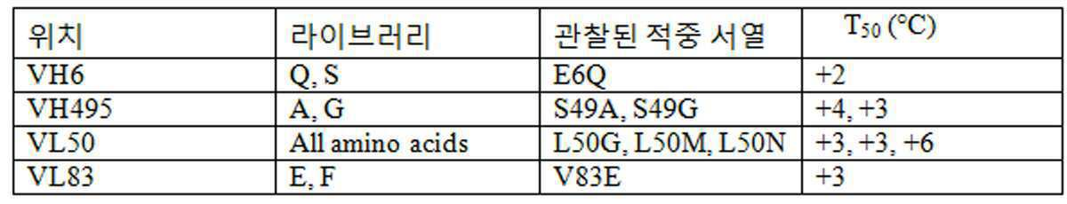
그 후, 안정화 돌연변이의 두개의 치환체로 구성된 플라스미드를 구성하여 열안정성이 더 증진된 조합을 확인하였다. 각각의 형질전환된 콜로니를 심부-웰 96 웰 접시 내로 골라내고, 상술한 바와 같이 스크리닝하기 위해서 처리하였다. 각각 및 조합된 안정화 돌연변이를 함유하는 G11 scFv 단백질을 다음에 대해서 시험하였다: T50 열공격 시험에서의 열안정성. 표 21은 T50 열공격 시험의 결과를 요약한 것이다. 통상적인 G11 scFv에 비해서 +2℃ 내지 +10℃ 범위의 열안정성 증가를 나타내는 각각 및 조합된 안정화 돌연변이를 확인하였다. 위치 VL50 및 VH6 (VL L50N:VH E6Q) 및 positions VL83 및 VH6 (VL V83E:VH E6Q)에서 조합된 G11 돌연변이의 T50 값은 통상적이 G11 scFv에 비해 scFv의 열안정성 (T50)을 +4-10℃까지 증가하였다.Thereafter, a plasmid consisting of two substituents of stabilizing mutations was constructed to identify combinations with further enhanced thermal stability. Each transformed colony was picked into deep-well 96 well dishes and processed for screening as described above. G11 scFv proteins, containing the respective and combined stabilizing mutations, were tested for: T 50 Thermal Stability in Thermal Attack Tests. Table 21 shows the T 50 The results of the thermal attack test are summarized. Respective and combined stabilizing mutations were identified which showed an increase in thermostability in the range of + 2 ° C. to + 10 ° C. as compared to conventional G11 scFv. T 50 values of G11 mutants combined at
[표 21]TABLE 21
안정화된 G11 scFv 단백질의 아미노산 치환, 열공격 시험으로부터의 T50 결과. N/A = 적용할 수 없음.Amino Acid Substitution of Stabilized G11 scFv Protein, T 50 Results from Heat Attack Test. N / A = not applicable.

표 22는 카바트 넘버링법에 따라 G11 scFv 내의 안정화 VH 및 VL 돌연변이의 위치 및 플라스미드 명칭을 열거하고 있다. 위치 VH 및 VL 돌연변이의 위치는 기준 서열로서 각각 도 33B (N-항-IGF-1R 이중특이적 항체) 및 34B (C-항-IGF-1R 이중특이적 항체)를 사용하여 순차적 넘버링에 따라 C-항-IGF-1R 이중특이적 항체 및 N-항-IGF-1R 이중특이적 항체 둘 모두에 대한 전체 길이 중쇄 서열 내에 나타낸다. Table 22 lists the positions and plasmid names of the stabilizing VH and VL mutations in G11 scFv according to Kabat numbering method. Positions of position VH and VL mutations were determined according to sequential numbering using Figure 33B (N-anti-IGF-1R bispecific antibody) and 34B (C-anti-IGF-1R bispecific antibody) as reference sequences, respectively . It is shown in full length heavy chain sequence for both anti-IGF-1R bispecific antibody and N-anti-IGF-1R bispecific antibody.
[표 22]Table 22
항-IGF-1R 이중특이적 항체를 구성하기 위해서 사용된 항-IGF-1R G116 scFv 내의 안정화 아미노산 잔기. 안정화 VH 및 VL 돌연변이의 위치는 카바트 넘버링법 (카바트:)에 따라 나타낸다. VH 및 VL 돌연변이의 위치는 또한, 기준 서열로서 도 33B (N-항-IGF-1R 이중특이적 항체) 및 34B (C-항-IGF-1R 이중특이적 항체)를 사용하여 순차적 넘버링에 따라 C-항-IGF-1R 이중특이적 항체 (C:) 및 N-항-IGF-1R 이중특이적 항체 (N:) 둘 모두에 대한 전체 길이 중쇄 서열 내에 나타낸다.Stabilizing amino acid residues in anti-IGF-1R G116 scFv used to construct an anti-IGF-1R bispecific antibody. The positions of stabilizing VH and VL mutations are shown according to Kabat numbering method (Kabat :). The location of the VH and VL mutations is also shown in Figure 33B as reference sequence. C-anti-IGF-1R bispecific antibody (C :) following sequential numbering using (N-anti-IGF-1R bispecific antibody) and 34B (C-anti-IGF-1R bispecific antibody) And N-anti-IGF-1R bispecific antibodies (N :) in full length heavy chain sequences.

h. h. CHOCHO 세포 내에서 항- Anti- IGFIGF -1R -1R 이중특이적Bispecific 항체의 안정한 발현, 항체 정제 및 특정화 Stable Expression, Antibody Purification and Characterization of Antibodies
플라스미드 DNAs pXWU135, pXWU136 및 pXWU118를 사용하여 항체 단백질의 안정한 생산을 위해서 DHFR-결핍성 CHO DG44 세포를 형질전환시켰다. 형질감염된 세포를 10% 투석된 소태자혈청 (Invitrogen Corporation)이 보충된 2 mM 글루타민을 함유하는 알파 마이너스 (alpha minus) MEM 배지 내에서 성장시키고, 형광적으로 표지된 항체 및 상대 형광-활성화된 세포 분류 (FACS)를 사용하여 안정한 벌크 배양 풀로서 풍부하게 하였다 [Brezinsky, et al. J Immunol Methods. 277(1-2):141-55 (2003)]. FACS를 또한 사용하여 각각의 세포주를 생성시켰다. 세포 풀 또는 세포주를 혈청 부재 조건에 적용시키고, 항체 생산을 위해서 증대시켰다.Plasmid DNAs pXWU135, pXWU136 and pXWU118 were used to transform DHFR-deficient CHO DG44 cells for stable production of antibody proteins. Transfected cells were grown in alpha minus MEM medium containing 2 mM glutamine supplemented with 10% dialysed fetal bovine serum (Invitrogen Corporation), fluorescently labeled antibodies and relative fluorescently-activated cells. Classification (FACS) was used to enrich as a stable bulk culture pool [Brezinsky, et al. J Immunol Methods. 277 (1-2): 141-55 (2003). FACS was also used to generate each cell line. Cell pools or cell lines were subjected to serum-free conditions and expanded for antibody production.
10일 운전된 생물반응기로부터의 50 ℓ의 C-항-IGF-1R 이중특이적 항체 (pXWU135/pXWU118) 상등액을 수확하고, 한외여과에 의해서 전청정화시켰다. 이중특이적 항체를 단백질 A 세파로즈 FF (GE Healthcare)를 사용하여 상등액으로부터 포착시켰다. 이중특이적 항체를 pH 3.0에서 0.1 M 글리신을 사용하여 단백질 A로부터 용출시키고, 트리스 염기로 중화시키고, 더 정제하지 않고 PBS 내로 투석하였다. 내독소 레벨은 엔도세이프 (EndoSafe®) PTC 키트 (Charles River Labs)를 사용하여 동적 정량적 발색성 LAL 분석에 의해서 시험하였다. 모노머 4가 항체 생성물의 순도 및 백분율은 각각 4-20% 트리스-글리신 SDS-PAGE 및 분석적 크기배제 HPLC에 의해서 평가하였다. 이 방법은 0.67 EU/㎎ 단백질의 잔류 내독소 농도를 가지고 5.4 ㎎/㎖의 농도, 96.7% 순도로 289 ㎎의 C-항-IGF-1R 이중특이적 항체를 수득하였다.50 L of C-anti-IGF-1R bispecific antibody (pXWU135 / pXWU118) supernatant from a bioreactor run for 10 days was harvested and precleaned by ultrafiltration. Bispecific antibodies were captured from the supernatant using Protein A Sepharose FF (GE Healthcare). Bispecific antibodies were eluted from Protein A with 0.1 M glycine at pH 3.0, neutralized with Tris base and dialyzed into PBS without further purification. Endotoxin levels were tested by dynamic quantitative chromogenic LAL analysis using the EndoSafe® PTC kit (Charles River Labs). Purity and percentage of monomeric tetravalent antibody product were evaluated by 4-20% tris-glycine SDS-PAGE and analytical size exclusion HPLC, respectively. This method yielded 289 mg of C-anti-IGF-1R bispecific antibody at a concentration of 5.4 mg / ml, 96.7% purity with a residual endotoxin concentration of 0.67 EU / mg protein.
도 35A는 정제된, 안정성-엔지니어링된 C-항-IGF-1R 이중특이적 항체 (pXWU135/pXWU118)의 SDS-PAGE 겔을 나타낸다. 환원된 레인은 중쇄 및 경쇄 단백질의 예상된 크기를 나타낸다. 중요하게도, 여기에서는 분해되거나 원치 않는 저분자량 부산물이 상당한 레벨로 검출되지 않는다. 35A shows an SDS-PAGE gel of purified, stability-engineered C-anti-IGF-1R bispecific antibody (pXWU135 / pXWU118). The reduced lanes represent the expected size of the heavy and light chain proteins. Importantly, no significant or no low molecular weight byproducts are detected at this level.
도 35B는 정제된, 안정성-엔지니어링된 C-항-IGF-1R 이중특이적 항체 (pXWU135/pXWU118)의 분석적 SEC 용출 프로필을 나타낸다. 이 분석은 안정성-엔지니어링된 C-항-IGF-1R 이중특이적 항체가 본질적으로 >96.7% 순수하고, 모노머성이며, 더 큰 등급의 분자량 종을 갖지 않음을 입증하였다. 35B shows the analytical SEC elution profile of the purified, stability-engineered C-anti-IGF-1R bispecific antibody (pXWU135 / pXWU118). This analysis demonstrated that the stability-engineered C-anti-IGF-1R bispecific antibody is essentially> 96.7% pure, monomeric, and has no higher grade molecular weight species.
10일 운전된 생물반응기로부터의 25 ℓ의 N-항-IGF-1R 이중특이적 항체 (pXWU136/pXWU118) 상등액을 수확하고, 한외여과에 의해서 전청정화시켰다. N-항-IGF-1R 이중특이적 항체를 C-항-IGF-1R 이중특이적 항체에 대해서 상술한 바와 같이 정제하였다. 모노머 4가 항체 생성물의 순도 및 백분율은 각각 4-20% 트리스-글리신 SDS-PAGE 및 분석적 크기배제 HPLC에 의해서 평가하였다. 이 방법은 0.09 EU/㎎ 단백질의 잔류 내독소 농도를 가지고 9.2 ㎎/㎖의 농도, 97.3% 순도로 1401 ㎎의 N-항-IGF-1R 이중특이적 항체를 수득하였다.25 L of N-anti-IGF-1R bispecific antibody (pXWU136 / pXWU118) supernatant from a bioreactor run for 10 days were harvested and precleaned by ultrafiltration. N-anti-IGF-1R bispecific antibodies were purified as described above for C-anti-IGF-1R bispecific antibodies. Purity and percentage of monomeric tetravalent antibody product were evaluated by 4-20% tris-glycine SDS-PAGE and analytical size exclusion HPLC, respectively. This method yielded 1401 mg of N-anti-IGF-1R bispecific antibody at a concentration of 9.2 mg / ml, 97.3% purity with a residual endotoxin concentration of 0.09 EU / mg protein.
도 36A는 정제된, 안정성-엔지니어링된 N-항-IGF-1R 이중특이적 항체 (pXWU136/pXWU118)의 SDS-PAGE 겔을 나타낸다. 환원된 레인은 중쇄 및 경쇄 단백질의 예상된 크기를 나타낸다. 역시, 여기에서는 분해되거나 원치 않는 저분자량 부산물이 상당한 레벨로 검출되지 않는다. 36A shows an SDS-PAGE gel of purified, stability-engineered N-anti-IGF-1R bispecific antibody (pXWU136 / pXWU118). The reduced lanes represent the expected size of the heavy and light chain proteins. Again, no significant or undetectable low molecular weight by-products are detected here.
도 36B는 정제된, 안정성-엔지니어링된 N-항-IGF-1R 이중특이적 항체 (pXWU136/pXWU118)의 분석적 SEC 용출 프로필을 나타낸다. 이 분석은 안정성-엔지니어링된 N-항-IGF-1R 이중특이적 항체가 본질적으로 97.3% 순수하고, 모노머성이며, 더 큰 등급의 분자량 종을 갖지 않음을 입증하였다.
36B shows the analytical SEC elution profile of the purified, stability-engineered N-anti-IGF-1R bispecific antibody (pXWU136 / pXWU118). This analysis demonstrated that the stability-engineered N-anti-IGF-1R bispecific antibody is essentially 97.3% pure, monomeric, and has no higher grade molecular weight species.
실시예Example 9. 항- 9. Anti- IGFIGF -1R -1R 이중특이적Bispecific 항체의 생화학적 특징 Biochemical Characteristics of Antibodies
a. 안정화된 a. Stabilized C06C06 scFvscFv 에 N- 또는 C-말단에서 융합된 Fused at the N- or C-terminus to G11G11 카파kappa 경쇄Light chain 및 And G11G11 IgG1 IgG1 중쇄Heavy chain 골격구조를 갖는 N- 및 C-말단 항- N- and C-terminal anti- with skeletal structure IGFIGF -1R -1R BsAbsBsAbs 의 정제 및 생화학적/생물물리학적 특정화.Purification and biochemical / biophysical characterization.
이중특이적 IgG 유사 항체 ("BsAbs")는 항체 엔지니어링의 분야에서 생성물 생산 및 품질 문제와 광범하게 부합하였다 [Demarest, S.J., Glaser, S.M. (2008) Curr. Opin . Drug Discov . Devel . 5, In Press ( September Issue )]. 여기에서, N- 및 C-말단 G11 IgG1/C06 scFv IgG 유사 이중특이적 항체 (N- 및 C-말단 IGF-1R 이중특이적 항체라 칭함, 도 37)의 구성을 위한 안정화된 C06 scFv의 용도는 확고한 취급 특성, 고품질 및 안정성을 갖는 재조합체 단백질을 제공하는 것으로 입증된다.Bispecific IgG-like antibodies (“BsAbs”) have broadly matched product production and quality issues in the field of antibody engineering [Demarest, SJ, Glaser, SM (2008) Curr. Opin . Drug Discov . Devel . 5, In Press ( September Issue ) ]. Use of stabilized C06 scFv herein for the construction of N- and C-terminal G11 IgG1 / C06 scFv IgG-like bispecific antibodies (referred to as N- and C-terminal IGF-1R bispecific antibodies, FIG. 37 ) . Is proven to provide recombinant proteins with firm handling properties, high quality and stability.
i. 방법i. Way
BsAb 발현: 각각 BsAb 중쇄 및 경쇄를 함유하는 자사의 포유동물 발현 벡터를 상기 실시예 8e 및 미국 특허출원 제11/725,970호에 기술된 바와 같이 DHFR-CHO DG44에 형질감염시켰다. N- 및 C-말단 BsAbs를 생산하는 CHO 세포주를 전술한 바와 같이 분리하였다 [Brezinsky, S. C. G., Chiang, G. G., Szilvasi, A., Mohan, S., Shapiro, R. I., MacLean, A., Sisk, W., and Thill, G. (2003) J. Immunol . Methods 277, 141-155]. N- 및 C-말단 IGF-1R 이중특이적 항체 단백질 둘 모두의 발현을 위한 대규모 CHO 세포 배양의 방법은 상기 실시예 8e 및 미국 특허출원 제11/725,970호에 기술되어 있다. N- 및 C-말단 BsAbs는 중성 pH로 미리 조정된 MAb셀렉트 (MAbSelect) 단백질 A 친화성 수지 (GE Healthcare) 상에 포착시킴으로써 CHO 세포 상등액으로부터 분리하였다. 포착된 물질을 5 칼럼 용적의 0.5 M 트리스-HCl, pH 8.5, 0.5 M NaCl로 세척하고, 단백질을 0.1 M 글리신, pH 3.0으로 용출시켰다. 단백질 용출액을 즉시 1 M 트리스-염기를 사용하여 중화시키고, PBS에 대해서 투석하였다. 단백질 농도를 1.6 L ㎝ g-1의 흡광계수를 사용하여 UV-Vis에 의해서 결정하였다. 내독소 레벨은 엔도세이프 (Endosafe®)-PTS LAL 시험 시스템 (Charles River Labs)을 사용하여 결정하였다. G11 IgG1 항체를 발현시키기 위한 플라스미드는 BsAbs의 생산에 대한 전구체로서 생산되었다. G11 IgG1 세포주를 선택하고, BsAbs에 대해서 기술된 바와 같이 생산을 위해서 증대시켰으며, 정제 프로토콜은 동일하였다. BsAb Expression: Our mammalian expression vectors containing BsAb heavy and light chains, respectively, were transfected into DHFR-CHO DG44 as described in Example 8e and US patent application Ser. No. 11 / 725,970, above. CHO cell lines producing N- and C-terminal BsAbs were isolated as described above [Brezinsky, SCG, Chiang, GG, Szilvasi, A., Mohan, S., Shapiro, RI, MacLean, A., Sisk, W , and Thill, G. (2003) J. Immunol . Methods 277 , 141-155. Methods of large-scale CHO cell culture for expression of both N- and C-terminal IGF-1R bispecific antibody proteins are described in Example 8e and US Patent Application No. 11 / 725,970, supra. N- and C-terminal BsAbs were isolated from CHO cell supernatants by entrapment on MAbSelect Protein A affinity resin (GE Healthcare) previously adjusted to neutral pH. The captured material was washed with 5 column volumes of 0.5 M Tris-HCl, pH 8.5, 0.5 M NaCl, and the protein was eluted with 0.1 M glycine, pH 3.0. Protein eluate was immediately neutralized using 1 M Tris-base and dialyzed against PBS. Protein concentration was determined by UV-Vis using an extinction coefficient of 1.6 L cm g -1 . Endotoxin levels were determined using an Endosafe®-PTS LAL test system (Charles River Labs). Plasmids for expressing G11 IgG1 antibodies were produced as precursors for the production of BsAbs. G11 IgG1 cell lines were selected and expanded for production as described for BsAbs, and the purification protocols were the same.
SDS - PAGE 및 분석적 크기 배제 크로마토그래피 ( SEC ): N- 및 C-말단 IGF-1R 이중특이적 항체를 SDS-PAGE 및 분석적 SEC 크로마토그래피를 사용하여 순도에 대해서 시험하였다. 각각의 BsAb (웰당 5 g)를 각각 비-환원 및 환원 조건 하에서 ~0.1 M β-머캅토에탄올을 함유하거나 함유하지 않는 노벡스 (Novex®) SDS 부하 완충액과 혼합시켰다. 샘플을 ~10 분 동안 95℃에서 가열하고, 엑셀 슈어록 미니셀 (XCell SureLock™ Mini Cell) 및 트리스-글리신 운전 완충액을 사용하여 노벡스 (Novex®) 4-20% 트리스 글리신 겔에 적용하고, 제조자의 지시 (Invitrogen)에 따라 운전하였다. SDS - PAGE and Analytical Size Exclusion Chromatography ( SEC ): N- and C-terminal IGF-1R bispecific antibodies were tested for purity using SDS-PAGE and analytical SEC chromatography. Each BsAb (5 g per well) was mixed with Novex® SDS load buffer with or without ˜0.1 M β-mercaptoethanol, respectively, under non-reducing and reducing conditions. Samples are heated at 95 ° C. for ˜ 10 minutes and applied to Novex® 4-20% Tris glycine gel using XCell SureLock ™ Mini Cell and Tris-Glycine Operation Buffer, and Operated according to the instructions of (Invitrogen).
분석적 SEC는 에질런트 (Agilent) 1100 HPLC 시스템을 사용하여 pH 6.8로 10 mM 포스페이트, 150 mM NaCl, 0.02% 나트륨 아지드 중에서 평형화된 바이오셉 (BioSep) 3000 300×7.8 ㎜ (Phenomenex) SEC 칼럼 상에서 수행하였다. 30-100 ㎍의 단백질을 0.5 ㎖/분으로 칼럼에 적용하였다. 용출된 단백질을 280 ㎚에서 UV 흡수에 의해 검출하였다.Analytical SEC was performed on a
원편광 이색성 ( CD ) 분광법: CD 측정은 온도 조절을 위한 열전 펠티어 (thermoelectric peltier) 장치 및 열 싱크 (heat sink)로서 외부 수욕이 장치된 자스코 (Jasco) J-810 분광편광계를 사용하여 수행되었다. 근위 (near) 및 원위 (far) UV 스캔은 각각 1 ㎝ 및 0.1 ㎝ 큐벳을 사용하여 1 M 단백질로 수행하였다. 스펙트럼 범위는 근위 및 원위 UV 측정에 대해서 각각 197-260 ㎚ 및 250-320 ㎚였다. 스캔은 2 초의 반응 시간, 0.1 ㎚의 데이터 피치 (data pitch), 및 근위 및 원위 UV 스캔에 대해서 각각 2 ㎚ 및 1 ㎚의 밴드 폭을 가지고 10℃에서 연속 모드 (100 ㎚/분)로 수행되었다. 모든 측정은 "고감도" 모드로 수행되었다. 모든 단백질 스펙트럼의 기준선은 PBS에 의한 배경 스캔을 사용하여 보정하였다. Circularly polarized light Dichroism ( CD ) spectroscopy: CD measurements were performed using a Jasco J-810 spectrophotometer with a thermoelectric peltier device for temperature control and an external water bath as a heat sink. Near and far UV scans were performed with 1 M protein using 1 cm and 0.1 cm cuvettes, respectively. The spectral range was 197-260 nm and 250-320 nm for proximal and distal UV measurements, respectively. The scan was performed in continuous mode (100 nm / min) at 10 ° C. with a response time of 2 seconds, a data pitch of 0.1 nm, and a band width of 2 nm and 1 nm for proximal and distal UV scans, respectively. . All measurements were performed in "high sensitivity" mode. Baselines of all protein spectra were corrected using a background scan with PBS.
시차 주사 열량법 ( DSC ): DSC 스캔은 자동화 모세관 DSC (capDSC, MicroCal, LLC)를 사용하여 수행되었다. 단백질 및 기준 용액은 로보트 부착장치를 사용하여 96-웰 플레이트로부터 자동적으로 샘플링하였다. 각각의 단백질 스캔을 수행하기 전에, 두개의 완충액 스캔을 수행하여 공제를 위한 기준선을 정의하였다. 단백질을 함유하는 96-웰 플레이트를 모두 6℃의 기구 내에서 저장하였다. 농축된 G11 IgG1 단백질 (3.9 ㎎/㎖) 및 N- 및 C-말단 IGF-1R 이중특이적 항체 (각각 9.2 ㎎/㎖ 및 5.4 ㎎/㎖)를 PBS에 대해서 투석하였다. 이어서, 샘플을 실험하기 전에 PBS에 의해서 1.0 ㎎/㎖ G11 IgG1, 및 1.3 ㎎/㎖의 두 가지 BsAbs로 희석하여 열량계 내에서 단백질의 동몰량이 달성되도록 하였다. 샘플 및 기준 모세관 둘 모두에서 PBS 투석물을 사용한 두개의 배경 스캔은 각각의 단백질 스캔을 수행하기 전에 샘플 모세관 내에서 단백질 및 기준 모세관 내에서 PBS 투석물을 수행하여 수행되었다. 10-100℃에서의 스캔은 낮은 피드백 모드에서 2℃/분으로 수행되었다. 스캔은 제조자에 의해서 제공된 오리진 (Origin) 소프트웨어를 사용하여 분석하였다. 기준물 기준선 스캔의 공제에 이어서, 비-제로 (zero) 단백질 스캔 기준선은 3차 다항식을 사용하여 보정하였다.
Differential Scanning Calorimetry ( DSC ): DSC scans were performed using automated capillary DSC (capDSC, MicroCal, LLC). Protein and reference solutions were automatically sampled from 96-well plates using a robotic attachment. Prior to each protein scan, two buffer scans were performed to define a baseline for subtraction. All 96-well plates containing protein were stored in a 6 ° C. instrument. Concentrated G11 IgG1 protein (3.9 mg / ml) and N- and C-terminal IGF-1R bispecific antibodies (9.2 mg / ml and 5.4 mg / ml, respectively) were dialyzed against PBS. The samples were then diluted with two BsAbs of 1.0 mg / ml G11 IgG1, and 1.3 mg / ml by PBS to achieve equimolar amounts of protein in the calorimeter before experimenting. Two background scans using PBS dialysate in both the sample and reference capillaries were performed by performing PBS dialysate in the protein and reference capillaries in the sample capillary prior to performing each protein scan. Scanning at 10-100 ° C. was performed at 2 ° C./min in low feedback mode. Scans were analyzed using the Origin software provided by the manufacturer. Following the subtraction of the baseline baseline scan, the zero protein scan baseline was corrected using a third order polynomial.
iiii . 결과. result
N- 및 C-말단 IGF-1R 이중특이적 항체를 상기 실시예 8e 및 미국 특허출원 제11/725,970호에 기술된 방법을 사용하여 CHO 세포 내에서 생산하였다. 각각의 BsAb의 두개의 별개의 배취를 첫째는 형질감염된 안정한 벌크 CHO 세포 풀로부터, 둘째는 BsAbs를 발현하도록 선택된 CHO 세포주로부터 생산하였다. 안정한 벌크 풀로부터 정제된 물질은 SDS-PAGE 및 분석적 SEC 크로마토그래피를 기준으로 하여 >98% 모노머 순도를 갖는 고품질의 N-말단 (5 ℓ로부터 20.4 ㎎) 및 C-말단 (4 ℓ로부터 5.6 ㎎) IGF-1R 이중특이적 항체 물질을 제공하였다 (즉, 비-환원된 BsAb에 대해서는 200 kDa 단일 밴드 및 환원 및 분리된 중쇄 및 경쇄에 대해서는 각각 75 kDa 및 25 kDa 밴드, 도 38A,B). 둘 모두 고순도를 갖는 단일 피크로서 용출된 안정한 벌크 배양물로부터의 BsAbs는 단백질 분자량 표준을 기준으로 하여 약 200 kDa 단백질에 대해 예상된 시간에서 칼럼으로부터 분리된다 (도 38C). N- 및 C-말단 IGF-1R 이중특이적 항체를 생산하도록 선택된 CHO 세포주는 각각 25 L 및 50 L로 증대시켰다. 두 가지 생산 운전은 모두 비-환원적 및 환원적 SDS PAGE 분석뿐만 아니라 분석적 SEC를 기초로 하여 정확한 분자량을 ~97% 순도의 물질을 제공하였다. C-말단 BsAb는 50 L 배양물로부터 289 ㎎을 제공하였으며, N-말단 BsAb는 25 L 배양물로부터 1.4 g을 제공하였다.N- and C-terminal IGF-1R bispecific antibodies were produced in CHO cells using the methods described in Example 8e and US patent application Ser. No. 11 / 725,970, above. Two separate batches of each BsAb were produced, first from a pool of stable bulk CHO cells transfected and second from CHO cell lines selected to express BsAbs. Purified material from a stable bulk pool was prepared with high quality N-terminus (20.4 mg from 5 L) and C-terminus (5.6 mg from 4 L) with> 98% monomer purity based on SDS-PAGE and analytical SEC chromatography. IGF-1R bispecific antibody material was provided (ie 200 kDa single band for non-reduced BsAbs and 75 kDa and 25 kDa bands for reduced and isolated heavy and light chains, respectively, FIG. 38A, B ). BsAbs from stable bulk cultures both eluted as single peaks with high purity are separated from the column at the expected time for about 200 kDa protein based on protein molecular weight standards ( FIG. 38C ). CHO cell lines selected to produce N- and C-terminal IGF-1R bispecific antibodies were boosted to 25 L and 50 L, respectively. Both production runs provided materials with an accurate molecular weight of ˜97% purity based on analytical SEC as well as non-reducing and reductive SDS PAGE analysis. C-terminal BsAb provided 289 mg from 50 L culture and N-terminal BsAb provided 1.4 g from 25 L culture.
BsAbs에 의한 원편광 이색성 (CD) 측정은 단백질이 적절하게 접힌 것을 입증한다. N- 및 C-말단 IGF-1R 이중특이적 항체의 원위 UV CD 스펙트럼은 scFv를 결여한 G11 IgG1 단백질에 대해서 관찰된 것과 매우 유사하다 (도 39A). 이것은 추가의 scFv가 단순히 어버이 G11 IgG1 분자의 영역과 필적할 만한 -시트 경향이 있는 추가의 Ig-접힘 단백질 영역을 부가하기 때문인 것으로 예상된다. 217 ㎚에서 N- 및 C-말단 IGF-1R^의 스펙트럼에서의 미묘한 차이는 융합 단백질 내에서의 G11 IgG1의 우세한 C-클래스 Ig-접힘 및 scFvs의 추가의 V-클래스 Ig-접힘의 전반적인 평균 구조 내에서의 작은 차이를 반영하는 것 같다. 근위 UV CD 스펙트럼의 신호-대-잡음은 약하지만, G11 IgG1 및 N- 및 C-말단 IGF-1R BsAbs의 스펙트럼은 모두 매우 유사하다 (도 39B). NUV 신호의 대부분은 V-클래스 및 C-클래스 Ig-접힘 둘 모두 내에서 정준형이며 불변하고, 정준형 Ig-접힘 내부 디설파이드 결합에 인접한 동일한 위치 내에 매립된 내부 Trp 잔기로부터 유래한다. G11 IgG1 및 N- 및 C-말단 IGF-1R 이중특이적 항체의 NUV 스펙트럼은 모두 각각의 분자 내의 단백질 영역의 특성을 기초로 하여 예상될 수 있는 것과 유사하다.Circular polarization dichroism (CD) measurement by BsAbs demonstrates that the protein is properly folded. The distal UV CD spectra of the N- and C-terminal IGF-1R bispecific antibodies are very similar to those observed for the G11 IgG1 protein lacking scFv ( FIG. 39A ). This is expected because the additional scFv simply adds an additional Ig-folding protein region that tends to -sheet tend to be comparable to that of the parental G11 IgG1 molecule. Subtle differences in the spectra of N- and C-terminal IGF-1R ^ at 217 nm show the overall average structure of the predominant C-class Ig-fold of G11 IgG1 and the additional V-class Ig-fold of scFvs in the fusion protein. It seems to reflect a small difference within. Although the signal-to-noise of the proximal UV CD spectrum is weak, the spectra of the G11 IgG1 and N- and C-terminal IGF-1R BsAbs are all very similar ( FIG. 39B ). Most of the NUV signals originate from internal Trp residues that are canonical and unchanged within both the V- and C-class Ig-folds, embedded in the same location adjacent to the canonical Ig-folded internal disulfide bonds. The NUV spectra of the G11 IgG1 and N- and C-terminal IGF-1R bispecific antibodies are all similar to what would be expected based on the nature of the protein regions within each molecule.
시차 주사 열량계 (DSC) 시험은 N- 및 C-말단 IGF-1R 이중특이적 항체 내의 모든 영역이 적절하게 접혀지며, 이상적인 (고온) 열 전개 특성을 가진다는 것을 증명한다. 어버이 G11 IgG1 분자 및 N- 및 C-말단 IGF-1R 이중특이적 항체는 둘 모두 멀티-영역 단백질이다 (도 39C). 연구는 IgG1 분자가 종종 3개의 별개의 전개 전이, 즉 단일 CH2 및 CH3 영역 각각에 대한 하나 및 Fab의 4개의 영역 (VH, VL, CH1, 및 CL, Garber, E., Demarest, S.J. (2007) Biochem . Biophys . Res . Commun . 355, 751-757)에 대한 단일의 크고 명백하게 협력적인 전이를 나타내는 것을 보여주었다. G11 IgG1은 인간 IgG1s에 대한 공통적인 전통적인 3개의 전이를 나타낸다 (도 39C). N- 및 C-말단 BsAbs는 둘 모두 또한, CH2, CH3, 및 Fab 영역에 대한 3개의 전이와 함께 안정화된 C06 scFv 영역의 전개로 인한 하나의 여분의 전이를 나타낸다 (도 39C). G11 IgG1 및 N- 및 C-말단 IGF-1R 이중특이적 항체의 전개 곡선은 각각 3 및 4개의 전이에 부합하였으며, 데이터는 표 23에 제공된다. C-말단 IGF-1R 이중특이적 항체의 IgG 전이는 G11 IgG1에 대해서 관찰된 것과 거의 동일하며, 이것은 안정화된 C06 scFv와 융합 단백질의 IgG1 부분 사이의 매우 적은 상호작용을 시사한다. 추가로, 안정화된 C06 scFv는 66℃의 TM 및 전개의 큰 비교 엔탈피를 갖는 단일 전이를 나타낸다 (표 23). scFv에 대해서 측정된 단일의 전개 전이는 VH 및 VL 도메인이 유사한 온도에서 전재하거나 협력적으로 전개함을 시사한다. scFv에 대해서 측정된 높은 TM 값은 특히 scFv의 열안정성이 생성물 품질 문제를 제기하는 것 같지는 않음을 시사한다. N-말단 IGF-1R 이중특이적 항체에 의한 데이터는 C-말단 BsAb에 대해서 관찰된 것과 유사하다; 그러나, C06 scFv TM은 약간 더 높고, G11 Fab TM은 약간 더 낮다 (표 23). 데이터는 scFv와 융합 단백질의 Fab 부분 사이의 매우 약한 상호작용이 있을 수 있음을 시사하지만, 어떤 것도 생성물 품질 문제를 유도하지는 않을 것이다.Differential scanning calorimetry (DSC) tests demonstrate that all regions within the N- and C-terminal IGF-1R bispecific antibodies are properly folded and have ideal (hot) thermal development properties. Parental G11 IgG1 molecules and N- and C-terminal IGF-1R bispecific antibodies are both multi-region proteins ( FIG. 39C ). Studies have shown that IgG1 molecules often contain three distinct developmental transitions, one for each of the
정제된 N- 및 C-말단 IGF-1R^2 단백질은 2-8℃에서 3 개월 기간에 걸쳐 PBS 중에서의 응집체의 축적에 대해서 모니터하였다. 이들 조건 하에서의 저장은 단백질 시약에 대해서 통상적이다. 대부분의 단백질, 특히 덜 안정하거나 가용성인 것은 어떤 기간 동안 용액 중에서 유지시키는 경우에 가용성 응집체의 축적을 나타낸다. 여기에서 수집된 데이터는 N- 및 C-말단 IGF-1R 이중특이적 항체가 매우 안정하며, 3 개월의 기간에 걸쳐서 실질적으로 응집체의 징후를 나타내지 않았음을 (본 발명자들의 검출의 한계 내에서) 입증한다 (표 24).Purified N- and C-terminal IGF-1R ^ 2 proteins were monitored for accumulation of aggregates in PBS over a 3 month period at 2-8 ° C. Storage under these conditions is common for protein reagents. Most proteins, especially less stable or soluble, indicate the accumulation of soluble aggregates when kept in solution for some period of time. The data collected here indicate that the N- and C-terminal IGF-1R bispecific antibodies are very stable and show substantially no signs of aggregates over a period of three months (within the limits of our detection). To demonstrate ( Table 24 ).
[표 23]TABLE 23
DSC에 의해서 측정된 N- 및 C-말단 IGF-1R 이중특이적 항체 및 G11 IgG1 대조 단백질의 열역학적 전개 파라메터Thermodynamic Development Parameters of N- and C-terminal IGF-1R Bispecific Antibodies and G11 IgG1 Control Proteins Measured by DSC

[표 24]TABLE 24
분석적 SEC에 의해서 측정된 것으로서, 2-8℃에서 3 개월에 걸쳐 PBS 중에서의 IGF-1R 이중특이적 항체의 안정성. N- 및 C-말단 IGF-1R 이중특이적 항체는 각각 4.5 ㎎/㎖ 및 1.7 ㎎/㎖로 유지되었다.Stability of IGF-1R bispecific antibodies in PBS over 3 months at 2-8 ° C. as measured by analytical SEC. N- and C-terminal IGF-1R bispecific antibodies were maintained at 4.5 mg / ml and 1.7 mg / ml, respectively.

b. 등온 적정 b. Isothermal titration 열량법Calorimetry ( ( ITCITC )에 의해서 측정된 (i) (I) measured by C06C06 및 And G11G11 MAbsMAbs 및 ( And ( iiii ) N- 및 C-말단 ) N- and C-terminal IGFIGF -1R -1R 이중특이적Bispecific 항체의 Antibody IGFIGF -1R 결합-1R bond
수용체 상의 다수의 억제성 에피토프를 점유함으로써 증진된 IGF-1R 억제를 달성한다는 가설은 각각의 에피토프가 다른 억제성 에피토프를 차폐시킴이 없이 억제성 항-IGF-1R 항체에 점유될 수 있어야 하는 것을 필요로 한다. 등온 적정 열량법 (ITC)을 사용하여 수용체의 상이한 에피토프를 인식하는 억제성 C06 및 G11 MAbs의 IGF-1R 결합을 측정하였다. 이 실험은 C06 및 G11이 수용체를 공동-점유할 수 있음을 명확하게 입증한다. 또한, ITC 실험을 수행하여 C- 및 N-말단 BsAbs가 IGF-1R에 강력하게 결합하고, 예상된 화학량론으로 수용체를 포화시킨다는 것을 나타내었다.
The hypothesis of achieving enhanced IGF-1R inhibition by occupying multiple inhibitory epitopes on the receptor requires that each epitope must be able to be occupied by an inhibitory anti-IGF-1R antibody without masking other inhibitory epitopes. Shall be. Isothermal titration calorimetry (ITC) was used to measure IGF-1R binding of inhibitory C06 and G11 MAbs that recognize different epitopes of the receptor. This experiment clearly demonstrates that C06 and G11 can co-occupy the receptor. In addition, ITC experiments were performed to show that C- and N-terminal BsAbs bind strongly to IGF-1R and saturate the receptor with expected stoichiometry.
i. 방법i. Way
항체. C06 및 G11 항체 및 sIGF-1R(1-903) 엑토영역 단백질을 미국 특허출원 제2007/0243194호에 기술된 바와 같이 생산 및 정제하였다. Antibodies. C06 and G11 antibodies and sIGF-1R (1-903) ectoregion proteins were produced and purified as described in US Patent Application 2007/0243194.
등온 적정 열량법 ( ITC ). C06 MAb (100 M), G11 MAb (67 M), 및 sIGF-1R(1-903) (5 M)이 항체/수용체 결합 실험을 위해서 사용되었다. 시약들은 PBS, pH 7.2에 대해서 공동-투석하였다. 단백질 용액을 PBS 투석물을 사용한 희석에 의해서 상기 열거된 농도로 조정하였다. BsAbs에 의한 실험은 25 M C-말단 IGF-1R^2, 30 mM N-말단 IGF-1R^2, 및 2.5 M sIGF-1R(1-903)을 사용하였다. 단백질 용액을 MAb 결합 실험을 위해서 상술한 바와 같이 제조하였다. 모든 ITC 실험은 25℃의 ITC200 미세열량계 (MicroCal, LLC) 상에서 수행하였다. 각각의 반응을 위해서, 반응 셀 (cell)을 hIGF-1R(1-903)으로 충진시켰다. sIGF-1R(1-903)에 대한 MAb 또는 BsAb 결합을 조사하기 위해서, 시린지를 MAb 또는 BsAb로 충진시키고, C06 MAb에 대해서는 15×1.5 ㎕ 주입, G11 MAb에 대해서는 18×2.0 ㎕ 주입, 및 N- 및 C-말단 IGF-1R 이중특이적 항체 둘 모두에 대해서는 20×1.8 ㎕ 주입을 사용하여 반응 셀 내로 적정하였다. 4 분의 평형 기간이 60 초의 초기 지연을 가지고 모든 주입들 사이에서 사용되었다. 데이터의 수치 적분은 마이크로칼 (MicroCal; Origin)에 의해서 제공된 ITC 데이터 분석 소프트웨어를 사용하여 수행되었다. ΔHA°(T) 값은 주입액의 결합상 중에 유리/흡수된 평균 열과 일단 수용체 IGF-1R이 MAb 또는 BsAb에 의해서 포화되면 나타나는 희석액의 평균 열 사이의 차이를 기초로 하여 계산되었다. 25℃에서, IGF-1R에 대한 C06 및 G11 MAbs 및 C-말단 IGF-1R 이중특이적 항체 둘 모두의 결합 친화성은 MAbs 및 C-말단 BsAb가 모든 수용체 결합 부위를 포화시킨 경우의 적정의 부분에서 다수의 전이점의 부재에 의해서 나타나는 바와 같이 ITC에 의해서 측정되기에는 너무 높았다.
Isothermal Titration Calorimetry ( ITC ). C06 MAb (100 M), G11 MAb (67 M), and sIGF-1R (1-903) (5 M) were used for antibody / receptor binding experiments. Reagents were co-dialyzed against PBS, pH 7.2. The protein solution was adjusted to the concentrations listed above by dilution with PBS dialysate. Experiments with BsAbs used 25 M C-terminal IGF-1R ^ 2, 30 mM N-terminal IGF-1R ^ 2, and 2.5 M sIGF-1R (1-903). Protein solutions were prepared as described above for MAb binding experiments. All ITC experiments were performed on an ITC 200 microcalorimeter (MicroCal, LLC) at 25 ° C. For each reaction, the reaction cell was filled with hIGF-1R (1-903). To investigate MAb or BsAb binding to sIGF-1R (1-903), syringes were filled with MAb or BsAb, 15 × 1.5 μl injection for C06 MAb, 18 × 2.0 μl injection for G11 MAb, and N For both C-terminal IGF-1R bispecific antibodies, 20 × 1.8 μl injections were used to titrate into the reaction cells. An equilibrium period of 4 minutes was used between all infusions with an initial delay of 60 seconds. Numerical integration of the data was performed using ITC data analysis software provided by MicroCal; Origin. The ΔH A ° (T) value was calculated based on the difference between the mean heat freed / absorbed in the binding phase of the infusion and the mean heat of the diluent once the receptor IGF-1R was saturated by MAb or BsAb. At 25 ° C., the binding affinity of both C06 and G11 MAbs and C-terminal IGF-1R bispecific antibodies for IGF-1R is at a fraction of the titration when MAbs and C-terminal BsAbs saturate all receptor binding sites. Too high to be measured by ITC, as indicated by the absence of multiple transition points.
iiii . 결과. result
등온 적정 열량법을 사용하여 억제성 항-IG-1R MAbs, C06 및 G11이 그들의 비중첩성 에피토프를 통해서 수용체를 공동-점유할 수 있다는 것을 입증하였다. 우선, C06을 재조합체 가용성 IGF-1R 엑토영역을 함유하는 용액 내로 적정하였으며, sIGF-1R(1-903)은 전형적인 항체 결합곡선을 나타낸다 (도 40A, B). sIGF-1R(1-903) 단백질은 SDS-PAGE 및 크기 배제 크로마토그래피/정적 광산란 둘 모두에 의해서 다이머성인 것으로 나타났다 (데이터는 나타내지 않음). 다이머성 수용체 및 2가 MAbs 둘 모두는 두개의 잠재적 결합 부위를 함유한다. 결합의 화학량론은 sIGF-1R(1-903)와 MAbs 사이의 결합 부위의 동몰수를 기초로 하여 예상되는 바와 같이 1:1이었다. 이어서, G11 MAb를 ~2배 과량의 C06 MAb의 존재 하에서 sIGF-1R(1-903)을 함유하는 용액 내로 적정하였다 (도 40A, B). C06 MAb의 존재는 G11 MAb가 수용체를 강력하게 점유하는 것을 차단하지 않았다. sIGF-1R(1-903)에 대한 G11 MAb 결합의 화학량론도 또한 1:1이었다. 이 실험은 IGF-1R을 공동-점유하는 C06 및 G11 MAbs의 능력을 입증한다.Isothermal titration calorimetry was used to demonstrate that inhibitory anti-IG-1R MAbs, C06 and G11 can co-occupy receptors through their non-overlapping epitopes. First, C06 was titrated into a solution containing the recombinant soluble IGF-1R ectoregion, sIGF-1R (1-903) shows a typical antibody binding curve ( FIGS. 40A, B ). The sIGF-1R (1-903) protein was shown to be dimeric by both SDS-PAGE and size exclusion chromatography / static light scattering (data not shown). Both dimeric receptors and divalent MAbs contain two potential binding sites. The stoichiometry of the binding was 1: 1 as expected based on the equimolar number of binding sites between sIGF-1R (1-903) and MAbs. G11 MAb was then titrated into a solution containing sIGF-1R (1-903) in the presence of ˜2 fold excess of C06 MAb ( FIGS. 40A, B ). The presence of C06 MAb did not block G11 MAb from occupying the receptor strongly. The stoichiometry of G11 MAb binding to sIGF-1R (1-903) was also 1: 1. This experiment demonstrates the ability of C06 and G11 MAbs to co-occupy IGF-1R.
다음으로, C- 및 N-말단 IGF-1R 이중특이적 항체를 sIGF-1R(1-903) 내로 적정하였다 (도 40C, D). BsAbs는 둘 모두 수용체를 1:1 몰비로 강력하게 결합시키고 포화시켰다. C-말단 IGF-1R 이중특이적 항체는 C06 및 G11 항체에 대해서 측정된 엔탈피의 조합을 기초로 하여 예상될 수 있는 것보다 훨씬 더 큰 결합의 매우 강력한 엔탈피를 나타내었다 (-54 kcal/mol, 표 25). C06 및 G11 MAbs와 유사하게, 여기에서는 수용체의 포화가 일어나는 전이 부분에서 극히 소수의 적정점이 있었으며, 이것은 매우 단단한 결합을 시사한다 (도 40D). N-말단 IGF-1R 이중특이적 항체는 또한, 미록 C-말단 IGF-1R 이중특이적 항체에 대해서 관찰된 것 미만이지만 강력한 엔탈피를 나타내었다 (-43 kcal/mol, 표 25). 추가로, 여기에서는 sIGF-1R(1-903)에 대한 N-말단 IGF-1R 이중특이적 항체의 친화성을 정의하기 위하여 전이 부분에서 더 만곡상태 (및 추가의 적정점)가 있었다. 이것은 11 nM의 겉보기 평형 해리상수 KD의 측정을 가능하게 하였다 (도 40D, 표 25). 이들 결과는 N-말단 IGF-1R 이중특이적 항체의 결합 친화성이 C-말단 IGF-1R 이중특이적 항체의 경우보다 더 낮을 수 있음을 시사한다.Next, the C- and N-terminal IGF-1R bispecific antibodies were titrated into sIGF-1R (1-903) ( FIG. 40C, D ). Both BsAbs strongly bound and saturated the receptors in a 1: 1 molar ratio. The C-terminal IGF-1R bispecific antibody showed a very strong enthalpy of binding much larger than could be expected based on the combination of enthalpy measured for C06 and G11 antibodies (-54 kcal / mol, Table 25 ). Similar to C06 and G11 MAbs, there were only a few titrations in the transition region where saturation of the receptor occurs, suggesting very tight binding ( FIG. 40D ). N-terminal IGF-1R bispecific antibodies also exhibited strong enthalpy but less than that observed for C-terminal IGF-1R bispecific antibodies (-43 kcal / mol, Table 25 ). In addition, there was a more curvature (and additional titration) at the transition part to define the affinity of the N-terminal IGF-1R bispecific antibody for sIGF-1R (1-903). This enabled the measurement of the apparent equilibrium dissociation constant K D of 11 nM ( FIG. 40D , Table 25 ). These results suggest that the binding affinity of N-terminal IGF-1R bispecific antibodies may be lower than for C-terminal IGF-1R bispecific antibodies.
ITC 실험의 흥미로운 관점은 이들이 BsAbs의 IgG1 부분 내의 C06 scFv 또는 G11 Fv가 수용체에 대한 결합에 관하여 서로를 폐색시킬 수 있는지 여부를 식별할 수 없다는 점이다. 비록 이들이 100% 상호 배타적이었다 할지라도, 분자의 두개의 활성 말단은 여전히 1:1 화학량론이 관찰되도록 할 수 있을 것이다. 그러나, N-말단 IGF-1R 이중특이적 항체의 더 약한 겉보기 친화성과 조합된 두개의 분자의 결합 엔탈피에서의 차이는 N-말단 BsAb가 그의 모든 결합 부위를 사용하여 IGF-1R을 완전히 결합시킬 수 없음을 시사한다. 결합 부위 (즉, C06 scFv 및 G11 항체 Fv)는 두개의 4가 형식 내에서 동일하다; 그러나, 여기에서 두개의 형식에서의 상대적 위치/간격은 상당히 상이하다. scFv와 Fv 사이의 입체 장해는 C- 및 N-말단 IGF-1R 이중특이적 항체 사이에서의 결합 특성의 미묘한 차이를 유도할 수 있는 것이 가능한 것으로 보인다.An interesting aspect of ITC experiments is that they cannot identify whether C06 scFv or G11 Fv in the IgGl portion of BsAbs can occlude each other with respect to binding to the receptor. Although they were 100% mutually exclusive, the two active ends of the molecule could still allow 1: 1 stoichiometry to be observed. However, the difference in binding enthalpy of the two molecules combined with the weaker apparent affinity of the N-terminal IGF-1R bispecific antibody is that the N-terminal BsAb can fully bind IGF-1R using all its binding sites. Implies none. Binding sites (ie, C06 scFv and G11 antibody Fv) are identical within the two tetravalent forms; However, the relative position / spacing in the two forms here is quite different. The steric hindrance between scFv and Fv appears to be able to induce subtle differences in binding properties between C- and N-terminal IGF-1R bispecific antibodies.
[표 25]TABLE 25
각각 및 조합된 C06 및 G11 Mabs에 대한 IGF-1R의 결합에 대한 열역학적 파라메터뿐만 아니라 ITC를 사용하여 측정된 C- 및 N-말단 IGF-1R 이중특이적 항체의 결합에 대한 파라메터.Thermodynamic parameters for the binding of IGF-1R to C06 and G11 Mabs, respectively and in combination, as well as parameters for the binding of C- and N-terminal IGF-1R bispecific antibodies measured using ITC.

c. 용액 기본 표면 c. Solution base surface 플라즈몬Plasmon 공명을 사용한 (i) (I) using resonance C06C06 및 And G11G11 FabsFabs , (, ( iiii ) ) C06C06 및 And G11G11 MAbsMAbs , 및 (ⅲ) N- 및 C-말단 And (iii) the N- and C-terminus IGFIGF -1R -1R 이중특이적Bispecific 항체에 대한 Against antibodies IGFIGF -1R 결합의 화학량론 및 친화성의 결정Determination of Stoichiometry and Affinity of -1R Bonds
C06 및 G11 MAbs는 IGF-1R 상의 상이하며 비-중첩성인 에피토프를 결합시키는데, 즉 C06은 수용체의 제1 타입-III 피브로넥틴 영역 (FnIII-1)의 표면상에, G11은 시스테인 풍부 부분 (CRR)의 표면상에 존재한다. 용액상 결합 실험을 수행하여 모두 4개의 잠재적 결합 부위 (IGF-1R 상의 각각의 에피토프 상에 2개의 부위)를 사용하여 IGF-1R을 결합시키는 N- 및 C-말단 BsAbs의 능력을 조사하였다.
C06 and G11 MAbs bind different and non-overlapping epitopes on IGF-1R, ie C06 is on the surface of the first type-III fibronectin region (FnIII-1) of the receptor, where G11 is a cysteine rich portion (CRR) Present on the surface of Solution phase binding experiments were performed to investigate the ability of the N- and C-terminal BsAbs to bind IGF-1R using all four potential binding sites (two sites on each epitope on IGF-1R).
i. 방법i. Way
표면 플라즈만 공명을 사용한 IGF -1R에 대한 평형 Fab , MAb , 및 BsAb 결합. 모든 실험은 Biacore 3000 기구 (Biacore) 상에서 수행되었다. C06 및 G11 MAbs를 별도로, 제조자에 의해서 제공된 표준 아민 화학 프로토콜을 사용하여 표준 CM5 칩의 두개의 상이한 유동 셀 표면에 고정화시켰다. MAb의 이들 높은 고정화 레벨에서, sIGF-1R(1-903)의 낮은 유동 농도 (<50 nM)는 그의 초기 결합 속도 Vi (RU/s)가 칩 표면에 걸쳐서 유동하는 sIGF-1R(1-903) 용액의 농도에 따라 선형으로 좌우되는 질량-전이 제한된 선형 결합 곡선을 유도하였다. sIGF-1R(1-903) (실시예 9b에 기술됨) 및 Fabs/MAbs/BsAbs 사이의 결합의 결합 상수 및 화학량론은 sIGF-1R(1-903)과 항체의 혼합물을 C06 또는 G11을 함유하는 센서칩 표면상에 유동시킴으로써 결정될 수 있었다. C06 및 G11 센서칩 표면은 sIGF-1R(1-903) 및 항체를 함유하는 용액 중의 비결합된 sIGF-1R(1-903)의 농도를 측정한다. 항체와 sIGF-1R(1-903) 사이의 평형 해리상수 KD, 및 결합 화학량론은 이하의 수학식을 사용하여 비결합된 sIGF-1R(1-903)의 농도에 의해서 결정된다: Only the surface plasma equilibrium Fab, coupled MAb, BsAb and for the IGF -1R with resonance. All experiments were performed on a
![]()
![]()
여기에서 Vi = 결합의 초기 속도, m = sIGF-1R(1-903) 농도 의존적 표준 곡선의 기울기, [IGF-1R]f = 비결합된 IGF-1R 농도, [IGF-1R]t = 총 IGF-1 농도, 및 [Ab]t = 총 Fab/MAb/BsAb 농도 [Day, E.S., Cachero, T.G., Qian, F., Sun, Y., Wen, D., Pelletier, M., Hsu, Y.-M., Whitty, A. (2005). "Selectivity of BAFF/BLyS and APRIL for Binding to the TNF Family Receptors BAFFR/BR3 and BCMA"Biochemistry 44: 1919-1931].
Where V i = initial velocity of binding, m = slope of the sIGF-1R (1-903) concentration-dependent standard curve, [IGF-1R] f = unbound IGF-1R concentration, [IGF-1R] t = total IGF-1 concentration, and [Ab] t = total Fab / MAb / BsAb concentration [Day, ES, Cachero, TG, Qian, F., Sun, Y., Wen, D., Pelletier, M., Hsu, Y -M., Whitty, A. (2005). "Selectivity of BAFF / BLyS and APRIL for Binding to the TNF Family Receptors BAFFR / BR3 and BCMA" Biochemistry 44: 1919-1931.
iiii . 결과. result
평형 용액상 표면 플라즈몬 공명 실험을 수행하여 4가 IGF-1R 이중특이적 항체의 4개 결합 부위 모두가 그들의 적절한 에피토프에 결합하는 능력을 조사하였다. 각각의 에피토프에 대한 결합을 시험하기 위해서, C06 및 G11을 상이한 센서칩 표면에 고정화시키고, sIGF-1R(-1903) 상의 FnIII-1 및 CRR 에피토프가 각각 저해되는지 여부를 프로브화하기 위해서 사용하였다. C06 표면 및 G11 표면에 대한 가용성 IGF-1R 엑토영역의 결합은 다양한 양의 N- 및 C-말단 IGF-1R 이중특이적 항체의 존재 하에서 시험하였다 (도 41A, B). C06 및 G11 MAbs 및d Fabs가 각각 1:1 및 2:1 항체:수용체의 결합의 화학량론을 입증하기 위한 대조군으로 사용되었다. C06 MAb 및 Fab가 sIGF-1R(1-903)에 대한 G11 표면 결합의 억제를 유도하지 않고, G11 MAb 및 Fab가 sIGF-1R(1-903)에 대한 C06 표면 결합의 억제를 유도하지 않는다는 것을 아는 것은 중요한데, 이는 각각의 표면이 이것이 결합하는 sIGF-1R(1-903) 상의 에피토프의 접근가능성의 절대적인 척도를 제공함을 입증한다.Equilibrium solution phase surface plasmon resonance experiments were performed to investigate the ability of all four binding sites of tetravalent IGF-1R bispecific antibodies to bind to their appropriate epitopes. To test binding to each epitope, C06 and G11 were immobilized on different sensor chip surfaces and used to probe whether FnIII-1 and CRR epitopes on sIGF-1R (-1903) were inhibited, respectively. Binding of soluble IGF-1R ectoregions to C06 and G11 surfaces was tested in the presence of varying amounts of N- and C-terminal IGF-1R bispecific antibodies ( FIGS. 41A, B ). C06 and G11 MAbs and d Fabs were used as controls to demonstrate the stoichiometry of the binding of 1: 1 and 2: 1 antibody: receptors, respectively. C06 MAb and Fab do not induce inhibition of G11 surface binding to sIGF-1R (1-903), and G11 MAb and Fab do not induce inhibition of C06 surface binding to sIGF-1R (1-903). It is important to know that each surface provides an absolute measure of the accessibility of the epitope on the sIGF-1R (1-903) to which it binds.
실험의 결과는, G11 Fab 팔 둘 모두 및 C-말단 IGF-1R 이중특이적 항체의 scFv 팔 둘 모두가 G11 및 C06 MAbs에 대해서 관찰된 것과 동일한 친화성을 가지고 그들의 각각의 CRR 및 FnIII-1 에피토프룰 완전히 결합시킬 수 있음을 분명하게 보여준다 (도 41A, B). C-말단 IGF-1R 이중특이적 항체의 억제곡선은 C06 MAb 대비 C06 표면 및 G11 MAb 대비 G11 표면에 대해서 관찰된 억제곡선과 동일하다. sIGF-1R(1-903)에 관한 MAbs 및 C-말단 BsAb의 결합 화학량론은 수용체가 호모다이머라는 사실을 기초로 하여 예상되는 바와 같이 1:1이다 (표 26). C06 및 G11 Fabs는 또한, 예상되는 바와 같이 그들의 각각의 에피토프에 대한 결합에 대해서 2:1 화학량론을 나타낸다 (표 26). Fabs는 또한, 더 낮은 겉보기 친화성을 나타내며, 실질적으로는 동적 비아코어 실험에 의해서 이전에 측정된 친화성과 실질적으로 동일하다 (표 26).The results of the experiment showed that both the G11 Fab arm and the scFv arm of the C-terminal IGF-1R bispecific antibody had their respective CRR and FnIII-1 epitopes with the same affinity observed for G11 and C06 MAbs. It is clearly shown that the rule can be combined completely ( FIGS. 41A, B ). The inhibition curves for the C-terminal IGF-1R bispecific antibody are identical to the inhibition curves observed for the C06 surface versus the C11 MAb and the G11 surface for the G11 MAb. The binding stoichiometry of MAbs and C-terminal BsAb for sIGF-1R (1-903) is 1: 1 as expected based on the fact that the receptor is a homodimer ( Table 26 ). C06 and G11 Fabs also show a 2: 1 stoichiometry for binding to their respective epitopes as expected ( Table 26 ). Fabs also exhibit lower apparent affinity and are substantially the same as those previously measured by dynamic Biacore experiments ( Table 26 ).
C-말단 IGF-1R 이중특이적 항체에 대해서 관찰된 것과는 달리, N-말단 IGF-1R 이중특이적 항체에 의한 결과는 분자의 IgG1 부분의 C06 scFvs 및 G11 Fvs에 의한 결합이 상호 배타적이지 않음을 시사한다. N-말단 BsAb는 MAbs 및 Fabs에 대해서 관찰된 값들 사이의 중간인 1.5:1의 겉보기 화학량론으로 수용체 (에피토프 둘 모두에서)를 포화시킨다 (도 41A, B; 표 26).Contrary to that observed for C-terminal IGF-1R bispecific antibodies, the results with N-terminal IGF-1R bispecific antibodies indicate that the binding by C06 scFvs and G11 Fvs of the IgG1 portion of the molecule is not mutually exclusive. Suggest. N-terminal BsAb saturates receptors (both epitopes) with an apparent stoichiometry of 1.5: 1 that is intermediate between the values observed for MAbs and Fabs ( FIG. 41A, B; Table 26 ).
결론적으로, 평형 용액상 표면 플라즈몬 공명 실험은 N- 및 C-말단 BsAbs 사이의 상이한 결합능을 분명하게 입증한다. 한편, C-말단 IGF-1R 이중특이적 항체는 이상적으로 BsAb의 다른 결합 부위에서의 결합에 의해서 저해됨이 없이 수용체를 독립적으로 점유할 수 있는 4개의 결합 부위 모두에 의해서 행동한다. 대신으로, N-말단 BsAb는 용액 중에서 IGF-1R에 대한 그의 결합 부위의 4개 모두를 동시에 점유할 수 없는 것으로 보인다. 하나의 가능성은 N-말단 형식에서 C06 scFv 및 G11 Fv의 밀접한 근접성은 입체 장해를 유도하는 것이다.In conclusion, equilibrium solution phase surface plasmon resonance experiments clearly demonstrate the different binding capacity between N- and C-terminal BsAbs. C-terminal IGF-IR bispecific antibodies, on the other hand, ideally act by all four binding sites that can occupy the receptor independently without being inhibited by binding at other binding sites of BsAb. Instead, the N-terminal BsAb does not appear to be able to occupy all four of its binding sites for IGF-1R simultaneously in solution. One possibility is that the close proximity of C06 scFv and G11 Fv in the N-terminal format leads to steric hindrance.
[표 26]TABLE 26
대조용 C06 및 G11 MAbs and Fabs와 함께 IGF-1R 이중특이적 항체의 동적 및 평형 (용액상) 친화성 측정Determination of Dynamic and Equilibrium (Solution Phase) Affinity of IGF-1R Bispecific Antibodies with Control C06 and G11 MAbs and Fabs

d. 용액 기본 표면 d. Solution base surface 플라즈몬Plasmon 공명을 사용한 (i) (I) using resonance C06C06 및 And G11G11 FabsFabs , (, ( iiii ) ) C06C06 및 And G11G11 MAbsMAbs , 및 (ⅲ) N- 및 C-말단 And (iii) the N- and C-terminus IGFIGF -1R -1R 이중특이적Bispecific 항체에 대한 Against antibodies IGFIGF -1R 결합의 화학량론 및 친화성의 결정Determination of Stoichiometry and Affinity of -1R Bonds
상기 실시예 4에 기술된 바와 같이, 비-중첩성 에피토프를 인식하는 다수의 억제성 항-IGF-1R 항체를 조합하는 것은 단일 모노클로날 항체를 사용하여 달성되는 것보다 증진된 리간드 차단을 유도할 수 있다. 두 개의 억제성 항-IGF-1R 항체를 단일 단백질 구조물 내에 조합시키기 위해서, 본 발명자들은 C06 항체의 알로스테릭 차단활성과 G11 항체의 경쟁적 리간드 차단 행동을 조합한 4가 이중특이적 항체 (BsAbs)를 생성시켰다. BsAbs는 IgG1 형식으로 G11 항체의 N- 또는 C-말단에 재조합적으로 융합된 C06 항체로부터 유래하는 안정화된 scFv로 구성된다 (도 37). 미국 특허출원 제2008/0050370호 ("Stabilized polypeptide compositions")에 기술된 바와 같이, scFv의 안정화는 고품질 4가 IgG 유사 이중특이적 또는 다가 항체를 생산하기 위한 권능화 단계이다. 본 실시예에서의 데이터는 N- 및 C-말단 IGF-1R 이중특이적 항체가 단일 모노클로날 항체에 비해서 증진된 리간드 차단을 나타내는 것을 보여준다.
As described in Example 4 above, combining multiple inhibitory anti-IGF-1R antibodies recognizing non-overlapping epitopes may lead to enhanced ligand blockage than is achieved using a single monoclonal antibody. Can be. In order to combine two inhibitory anti-IGF-1R antibodies into a single protein construct, we used a tetravalent bispecific antibody (BsAbs) that combines the allosteric blocking activity of the C06 antibody with the competitive ligand blocking behavior of the G11 antibody. Was generated. BsAbs consist of stabilized scFv derived from C06 antibody recombinantly fused to the N- or C-terminus of the G11 antibody in IgG1 format ( FIG. 37 ). As described in US Patent Application 2008/0050370 ("Stabilized polypeptide compositions"), stabilization of scFv is an empowering step to produce high quality tetravalent IgG like bispecific or multivalent antibodies. The data in this example show that N- and C-terminal IGF-1R bispecific antibodies show enhanced ligand block compared to single monoclonal antibodies.
i. 방법i. Way
리간드 차단 특성. IGF-1 및 IGF-2를 차단하는 MAbs 및 BsAbs의 능력은 실시예 4에 기술된 IGF-1 및 IGF-2 차단 ELISA를 사용하여 결정되었다. 시험에서 IGF-1 및 IGF-2 농도는 각각 320 nM 및 640 nM였다. C06 및 G11 MAbs뿐만 아니라 N- 및 C-말단 IGF-1R 이중특이적 항체의 차단능력에 대한 리간드 의존성을 조사하기 위해서, 시험은 다양한 리간드 농도에서 수행하였다. 각각의 차단 ELISAs는 20 nM, 80 nM, 320 nM, 또는 1300 nM 의 IGF-1 또는 40 nM, 160 nM, 640 nM, 또는 2600 nM 의 IGF-2를 사용하여 수행하였다. IGF-2 농도가 더 높았는데, 이는 IGF-2가 수용체에 대해서 약간 더 낮은 친화성을 갖기 때문이다. ELISA 곡선의 선형 범위 내에 유지시키기 위해서, 본 발명자들은 시험을 동등한 결과를 달성하는 IGF-1 차단 ELISA보다 높은 IGF-2 농도에서 수행하는 것이 필요하였다.
Ligand blocking properties. The ability of MAbs and BsAbs to block IGF-1 and IGF-2 was determined using the IGF-1 and IGF-2 blocking ELISA described in Example 4. The IGF-1 and IGF-2 concentrations in the test were 320 nM and 640 nM, respectively. In order to investigate the ligand dependence on the blocking ability of the C06 and G11 MAbs as well as the N- and C-terminal IGF-1R bispecific antibodies, tests were performed at various ligand concentrations. Each blocking ELISAs were performed using 20 nM, 80 nM, 320 nM, or 1300 nM of IGF-1 or 40 nM, 160 nM, 640 nM, or 2600 nM of IGF-2. IGF-2 concentrations were higher because IGF-2 has a slightly lower affinity for the receptor. In order to remain within the linear range of the ELISA curve, we needed to run the test at higher IGF-2 concentrations than the IGF-1 blocked ELISA, which achieved equivalent results.
iiii . 결과. result
C06 및 G11 항체 및 N- 및 C-말단 IGF-1R 이중특이적 항체는 320 nM 의 IGF-1 및 640 nM 의 IGF-2를 사용하여 IGF-1 및 IGF-2 차단 ELISAs (도 42A-B)에서 나란히 시험하였다. N- 및 C-말단 BsAbs는 둘 모두 더 고친화성 항체인 C06에 필적하는 ~1 nM IC50 값을 가졌다. C06 및 G11 항체는 둘 모두 항체 농도 <100 nM에서 IGF-1R에 대한 IGF-1 또는 IGF-2 결합을 완전히 차단할 수 없었다 (도 42A-B). N- 및 C-말단 IGF-1R 이중특이적 항체는 둘 모두 저농도에서 수용체에 대한 IGF-1 및 IGF-2 결합을 완전히 제거할 수 있었다 (<10 nM, 도 42A-B).C06 and G11 antibodies and N- and C-terminal IGF-1R bispecific antibodies were identified using IGF-1 and IGF-2 blocking ELISAs using 320 nM of IGF-1 and 640 nM of IGF-2 ( FIG. 42A-B ). Tested side by side at. N- and C-terminal BsAbs both had ˜1 nM IC 50 values comparable to the higher affinity antibody C06. Both C06 and G11 antibodies could not completely block IGF-1 or IGF-2 binding to IGF-1R at antibody concentration <100 nM ( FIGS. 42A-B ). Both N- and C-terminal IGF-1R bispecific antibodies were able to completely eliminate IGF-1 and IGF-2 binding to receptors at low concentrations (<10 nM, FIGS. 42A-B ).
시험에 사용된 리간드 농도를 증가시키면, 인간 항체 C06은 그의 IGF-1 차단 활성의 일부를 상실한다 (즉, 수용체가 C06으로 포화되는 경우에 IGF-1R을 결합시킬 수 있는 IGF-1의 백분율은 리간드 농도가 상승함에 따라서 상승한다, 도 43A). 이것은 C06이 순수하게 알로스테릭 억제제인 결과이다. G11 항체는 리간드 농도의 증가에 대해 상이하게 반응한다. 경쟁적 억제제로서, 인간 항체 G11은 수용체를 결합시키는데 대해서 리간드와 직접적으로 경쟁하여야 한다. 리간드 농도가 증가함에 따라서, G11 항체의 겉보기 효력은 감소한다 (도 43B). 알로스테릭 부위에서 리간드의 존재 하에 수용체를 결합시키고, 알로스테릭 및 경쟁적 기전 둘 모두를 사용하여 억제하는 능력을 조합함으로써, BsAbs는 리간드 농도와는 무관하게 IGF-1 및 IGF-2 둘 모두가 수용체에 결합하는 것을 완전히 차단할 수 있다 (도 44A-D). 추가로, N- 및 C-말단 IGF-1R^2 항체 둘 모두에 대해서 측정된 IC50 값은 시험에 사용된 리간드 농도와는 무관하며, 시험에서 C06 항체에 대해서 관찰된 IC50 값과 동등하다:Increasing the ligand concentration used in the test, human antibody C06 loses some of its IGF-1 blocking activity (ie, the percentage of IGF-1 that can bind IGF-1R when the receptor is saturated with C06 is It rises as ligand concentration rises, FIG. 43A ). This is the result of C06 being a purely allosteric inhibitor. G11 antibodies respond differently to increasing ligand concentrations. As a competitive inhibitor, human antibody G11 must compete directly with the ligand for binding the receptor. As ligand concentration increases, the apparent potency of the G11 antibody decreases ( FIG. 43B ). By combining the ability to bind a receptor in the presence of a ligand at the allosteric site and to inhibit it using both allosteric and competitive mechanisms, BsAbs resulted in both IGF-1 and IGF-2 independent of ligand concentration. Binding to receptors can be completely blocked ( FIGS. 44A-D ). In addition, the IC 50 values measured for both N- and C-terminal IGF-1R ^ 2 antibodies are independent of the ligand concentration used in the test and are equivalent to the IC 50 values observed for the C06 antibody in the test. :
C-말단 IGF-1R BsAb: IC50 IGF-1 차단 = 2.0±0.3 C-terminal IGF-1R BsAb: IC 50 IGF-1 Blocking = 2.0 ± 0.3
C-말단 IGF-1R BsAb: IC50 IGF-2 차단 = 1.7±0.2C-terminal IGF-1R BsAb: IC 50 IGF-2 Blocking = 1.7 ± 0.2
N-말단 IGF-1R BsAb: IC50 IGF-1 차단 = 1.4±0.2 N-terminal IGF-1R BsAb: IC 50 IGF-1 Blocking = 1.4 ± 0.2
N-말단 IGF-1R BsAb: IC50 IGF-2 차단 = 2.7±0.9.N-terminal IGF-1R BsAb: IC 50 IGF-2 Blocking = 2.7 ± 0.9.
이들 결과는, C06 및 G11 억제성 에피토프 둘 모두를 인식하는 그들의 능력에 의해서 N- 및 C-말단 IGF-1R 이중특이적 항체 둘 모두가 표준 인간 항체 C06 및 G11에 대해 모든 리간드 농도에서 증진된 리간드 차단을 나타냄을 입증한다.
These results indicate that both N- and C-terminal IGF-1R bispecific antibodies are enhanced at all ligand concentrations for standard human antibodies C06 and G11 by their ability to recognize both C06 and G11 inhibitory epitopes. Demonstrate blocking.
e. e. C06C06 및 And G11G11 MAbsMAbs 및 N- 및 C-말단 And N- and C-terminus IGFIGF -1R -1R 이중특이적Bispecific 항체에 의한 By antibody IGFIGF -1R의 교차결합-1R crosslink
IGF-1R에 대한 상이한 억제성 항체의 한가지 잠재적으로 식별성인 특징은 세포 표면상의 수용체에 교차결합하는 그들의 능력이다. 대부분의 수용체 티로신 키나제 집단 구성원은 리간드 매개된 호모- 또는 헤테로다이머화를 통해서 신호를 보낸다. 그러나, IGF-1R (및 인슐린 수용체)은 이 기전을 통해서 신호를 보내지 않는다. IGF-1R은 그의 신호전달이 다이머화에 의해서 회합되지 않은 리간드 결합에 의해서 유도된 입체배좌 변화에 따라 좌우되는 구성적 호모다이머이다.One potentially distinguishing feature of different inhibitory antibodies to IGF-1R is their ability to crosslink receptors on the cell surface. Most receptor tyrosine kinase family members signal through ligand mediated homo- or heterodimerization. However, IGF-1R (and insulin receptors) do not signal through this mechanism. IGF-1R is a constitutive homodimer whose signaling depends on the conformational change induced by ligand binding not associated by dimerization.
IGF-1R에 대한 일부의 항체는 내재화 및 분해를 통해서 수용체를 하향조절하는 능력을 나타내었다. 교차결합은 종종 세포 표면 단백질 내재화의 효율과 연관된다 (예를 들어, FcεRI은 교차결합에 의해서 내재화된다). 이러한 방식으로, 교차결합은 세포 표면 단백질, 및 이 경우에는 IGF-1R에 대한 항체의 활성에 상당한 영향을 미칠 수 있다. 추가로, 세포의 표면상에서 IGF-1R의 항체 매개된 교차결합의 정도는 FcγR 수용체 또는 보체에 대한 항체의 Fc-부분의 결합에 영향을 미칠 수 있으며, 이것은 숙주 면역계에 의한 항체의 활성에 영향을 미친다.Some antibodies against IGF-1R have shown the ability to downregulate receptors through internalization and degradation. Crosslinking is often associated with the efficiency of cell surface protein internalization (eg, FcεRI is internalized by crosslinking). In this way, crosslinking can have a significant impact on the activity of cell surface proteins, and in this case antibodies to IGF-1R. In addition, the extent of antibody mediated crosslinking of IGF-1R on the surface of cells can affect the binding of the Fc-part of the antibody to the FcγR receptor or complement, which affects the activity of the antibody by the host immune system. Crazy
여기에서, 본 발명자들은 C06 및 G11 MAbs가 IGF-1R 엑토영역의 가용성 변형체와 배양하는 경우에 용액 중에서 다양한 크기의 IGF-1R/MAb 면역 컴플렉스를 유도한다는 것을 입증한다. 본 발명자들은 또한, IGF-1R을 갖는 용액에 대해 C06 및 G11 MAbs 둘 모두를 동시에 도입시킴으로써 형성된 생성된 컴플렉스를 조사한다. 마지막으로, 본 발명자들은 N- 및 C-말단 IGF-1R 이중특이적 항체에 의해서 형성된 면역 컴플렉스의 특성을 조사한다.
Here we demonstrate that C06 and G11 MAbs induce IGF-1R / MAb immune complexes of various sizes in solution when incubated with soluble variants of the IGF-1R ectoregion. We also examine the resulting complex formed by introducing both C06 and G11 MAbs simultaneously into a solution with IGF-1R. Finally, we investigate the properties of immune complexes formed by N- and C-terminal IGF-1R bispecific antibodies.
i. 방법i. Way
인-라인 정적 광산란과 함께 분석적 크기 배제 크로마토그래피 ( SEC ). SEC 샘플은 (i) C06 MAb, (ii) G11 MAb, 및 (ⅲ) sIGF-1R(1-903)에 의해서; (iv) C06 MAb 및 sIGF-1R(1-903) 또는 (v) G11 MAb 및 sIGF-1R(1-903)의 이성분 혼합물에 의해서; 또는 (vi) C06 MAb, G11 MAb, 및 sIGF-1R(1-903)의 삼성분 혼합물에 의해서 제조되었다. 모든 샘플은 에질런트 1100 HPLC 시스템을 사용하여, pH 6.8에서 10 mM 포스페이트, 150 mM NaCl, 0.02% 나트륨 아지드 중에서 평형화된 TSK겔 G3000SW XL, 5 ㎜, 250 Å 분석적 SEC 칼럼 (Tosoh Biosciences) 상의 SEC 칼럼 상에 주입하기 전에 PBS, pH 7.2 중에서 50 ㎕의 최종 용적으로 희석된 각각의 단백질 25 ㎍을 함유하였다. SEC 칼럼으로부터 용출하는 물질에 대한 광산란 데이터는 인라인 굴절률 측정기 (Wyatt Techno-logies)에 커플링된 미니다운 (miniDAWN) 정적 광산란 검출기를 사용하여 수집하였다. 광산란 데이터는 제조자에 의해서 제공된 아스트라 (ASTRA) V 소프트웨어를 사용하여 분석하였다. Analytical size exclusion chromatography ( SE ) with in-line static light scattering . SEC samples were prepared by (i) C06 MAb, (ii) G11 MAb, and (iii) sIGF-1R (1-903); (iv) by a binary mixture of C06 MAb and sIGF-1R (1-903) or (v) G11 MAb and sIGF-1R (1-903); Or (vi) a ternary mixture of C06 MAb, G11 MAb, and sIGF-1R (1-903). All samples were TSKgel G3000SW XL, 5 mm, 250 mm analytical SEC column (Tosoh Biosciences) equilibrated in 10 mM phosphate, 150 mM NaCl, 0.02% sodium azide at pH 6.8 using an Agilent 1100 HPLC system. Prior to injection on the column, 25 μg of each protein diluted to 50 μl final volume in PBS, pH 7.2. Light scattering data for the material eluting from the SEC column was collected using a miniDAWN static light scattering detector coupled to an inline refractive index meter (Wyatt Techno-logies). Light scattering data were analyzed using the Astra V software provided by the manufacturer.
추가의 SEC 샘플은 (i) C-말단 IGF-1R 이중특이적 항체 (BsAb), (ii) N-말단 IGF-1R BsAb, 및 (ⅲ) sIGF-1R(1-903); 또는 (iv) C-말단 IGF-1R BsAb 및 sIGF-1R(1-903) 또는 (v) N-말단 IGF-1R BsAb 및 sIGF-1R(1-903)와의 컴플렉스를 사용하여 제조하였다. 실험에서 사용된 sIGF-1R(1-903) 및 BsAb의 양은 각각 30 ㎎ 및 45 ㎎이었다. 모든 샘플은 SEC/정적 광산란 분석 전에 PBS, pH 7.2 중에서 50 ㎕의 최종 용적으로 희석되었다.
Additional SEC samples include (i) C-terminal IGF-1R bispecific antibodies (BsAb), (ii) N-terminal IGF-1R BsAb, and (iii) sIGF-1R (1-903); Or (iv) complexes with C-terminal IGF-1R BsAb and sIGF-1R (1-903) or (v) N-terminal IGF-1R BsAb and sIGF-1R (1-903). The amounts of sIGF-1R (1-903) and BsAb used in the experiment were 30 mg and 45 mg, respectively. All samples were diluted to 50 μl final volume in PBS, pH 7.2 before SEC / static light scattering analysis.
iiii . 결과. result
분리된 C06 및 G11 MAbs뿐만 아니라 분리된 sIGF-1R(1-903)을 사용한 SEC/정적 광산란 결과는 모든 단백질이 그들의 예상된 분자량을 나타냄을 입증하였다 (항체에 대해서는 150 kDa 및 sIGF-1R(1-903)에 대해서는 ~250 kDa, 도 45A). G11 MAb와 sIGF-1R(1-903) 사이에서 형성된 컴플렉스는 관찰된 분자량 840 kDa를 기초로 하여 4 분자 종 (2 분자의 G11 MAb 및 2 분자의 sIGF-1R(1-903))을 선호하는 것으로 보인다 (도 45A). C06 MAb는 가용성 IGF-1R 엑토영역과의 훨씬 더 큰 컴플렉스를 형성한다, 2 MDa 도 45A. C06 및 G11 MAbs 둘 모두가 동시에 가용성 수용체 엑토영역에 도입되어 삼성분 혼합물을 형성하는 경우에, 면역 컴플렉스(들)의 평균 크기는 본 발명자들의 측정가능한 범위를 벗어나 > 10 MDa까지 극적으로 증가한다 (도 45A). 이것은 세포의 표면상의 IGF-1R에 대해 비-중첩성 에피토프를 인식하는 두개의 항체를 첨가하는 것이 신규의 교차결합 및 잠재적인 하향조절 행동을 유도할 수 있음을 시사한다.SEC / static light scattering results using isolated C06 and G11 MAbs as well as isolated sIGF-1R (1-903) demonstrated that all proteins exhibited their expected molecular weight (150 kDa and sIGF-1R (1 for antibodies). -903) -250 kDa, FIG. 45A ). The complex formed between G11 MAb and sIGF-1R (1-903) prefers 4 molecular species (2 molecules of G11 MAb and 2 molecules of sIGF-1R (1-903)) based on the observed molecular weight of 840 kDa. Seems to be ( FIG. 45A ). C06 MAb forms a much larger complex with the soluble IGF-1R ectoregion, 2 MDa FIG. 45A . When both C06 and G11 MAbs are simultaneously introduced into the soluble receptor ectoregion to form a ternary mixture, the average size of the immune complex (s) dramatically increases to> 10 MDa beyond our measurable range ( 45A ). This suggests that the addition of two antibodies that recognize non-overlapping epitopes on IGF-1R on the surface of cells can induce new crosslinking and potential downregulation behavior.
sIGF-1R(1-903)을 갖는 N- 및 C-말단 IGF-1R 이중특이적 항체 혼합물은 또한, SEC/정적 광산란에 의해서 검사하였다 (도 45B). 분리 시에, BsAbs 및 sIGF-1R(1-903)은 모두 그들의 예상된 분자량 (BsAbs에 대해서는 ~210 kDa 및 sIGF-1R(1-903)에 대해서는 ~250 kDa)을 나타낸다. 흥미롭게도, sIGF-1R(1-903)과 컴플렉스화되는 경우에, BsAbs는 G11 MAb에 대해서 측정된 것과 유사한 컴플렉스 크기를 유도한다 (C-말단 IGF-1R BsAb/sIGF-1R(1-903) 컴플렉스에 대해서는 ~1.0 MDa, 및 N-말단 IGF-1R BsAb/sIGF-1R(1-903) 컴플렉스에 대해서는 ~1.3 MDa). BsAbs 및 sIGF-1R(1-903)의 유사한 분자량으로 인하여, 본 발명자들은 이들 시험만을 기초로 하여 면역 컴플렉스의 실제 조성 (즉, BsAb 대 sIGF-1R(1-903) 분자의 수)이 G11 MAb에 대해서 관찰된 것과 유사한지 여부를 결정할 수 없었다.N- and C-terminal IGF-1R bispecific antibody mixtures with sIGF-1R (1-903) were also examined by SEC / static light scattering ( FIG. 45B ). Upon separation, both BsAbs and sIGF-1R (1-903) exhibit their expected molecular weight (˜210 kDa for BsAbs and ˜250 kDa for sIGF-1R (1-903)). Interestingly, when complexed with sIGF-1R (1-903), BsAbs induce a complex size similar to that measured for G11 MAb (C-terminal IGF-1R BsAb / sIGF-1R (1-903) ˜1.0 MDa for the complex and ˜1.3 MDa for the N-terminal IGF-1R BsAb / sIGF-1R (1-903) complex. Due to the similar molecular weights of BsAbs and sIGF-1R (1-903), we found that the actual composition of the immune complex (ie, the number of BsAb vs. sIGF-1R (1-903) molecules) was based on these tests only for G11 MAb. It could not be determined whether it was similar to that observed for.
결론적으로, 본 발명자들은 C06 및 G11 MAbs가 sIGF-1R(1-903) 단백질과의 매우 다양한 면역 컴플렉스의 형성을 유도하며, 삼성분 혼합물을 형성하는 MAbs의 조합은 면역 컴플렉스의 크기에 있어서 매우 큰 증가를 유도한다는 것을 증명한다. C06 및 G11 항체의 이중특이적 형식으로의 전환은 BsAbs에 대한 IGF-1R의 결합시에 면역 컴플렉스의 복잡성 및 크기의 부수적인 증가를 유도하지 않는다. 흥미롭게도, 아마도 입체 장해로 인하여, BsAbs에 의해서 형성된 컴플렉스는 작으며, 이것은 분리 시에 G11 MAb에 대해서 관찰된 것과 유사하다.
In conclusion, we found that C06 and G11 MAbs induce the formation of a wide variety of immune complexes with sIGF-1R (1-903) proteins, and the combination of MAbs forming the ternary mixture is very large in size of the immune complex. Prove that it leads to an increase. Conversion of the C06 and G11 antibodies to the bispecific format does not induce a concomitant increase in the complexity and size of the immune complex upon binding of IGF-1R to BsAbs. Interestingly, perhaps due to steric hindrance, the complex formed by BsAbs is small, similar to that observed for G11 MAb at separation.
실시예Example 10. 10. IGFIGF -1R 상의 -1R Top 두개의Two 별개의 Distinct 에피토프를Epitope 표적화하는Targeted IGFIGF -1R -1R 이중특이적Bispecific 항체의 기능적 활성 Functional activity of antibodies
본 실시예에서 요약된 실험 결과는, IGF-1R 항체의 두개의 상이한 에피토프에 대해서 지시된 이중특이적 항체가 단일 특이성의 항체에 비해서 증진된 생물학적 활성을 나타내며, 생체내에서 더 우수한 항종양 활성을 유도한다는 생물학적인 이론적 근거 및 개념증명을 제공한다.The experimental results summarized in this example show that bispecific antibodies directed against two different epitopes of IGF-1R antibodies show enhanced biological activity compared to monospecific antibodies, and show better antitumor activity in vivo. Provide biological rationale and proof of concept
C06 및 G11은 각각 알로스테릭 및 경쟁적 기전을 통해서 IGF-1 및 IGF-2 결합을 차단하는 IGF-1R 상의 두개의 별개의 에피토프를 표적화하는 두개의 억제성 항-IGF-1R 항체이다. C06 및 G11의 조합은 리간드 차단 및 종양세포 성장의 억제를 증진시킬 수 있다. 따라서, N-IGF-1R 이중특이적 항체 (I83E 돌연변이체와 함께 C06 scFv를 사용한 G11 IgG1 골격구조를 갖는 N-말단 IGF-1R^2) 및 C-IGF-1R 이중특이적 항체 (183E 돌연변이체와 함께 C06 scFv를 사용한 G11 IgG1 골격구조를 갖는 C-말단 IGF-1R 이중특이적 항체)인 두 가지 형식의 이중특이적 항체는 IGF-1R 상의 C06 및 G11 에피토프 둘 모두를 표적화하는 단일 이중특이적 분자로서 구성되었다. C06 및 G11 조합, 또는 단일 이중특이성 약제, N-IGF-1R 이중특이적 항체 및 C-IGF-1R 이중특이적 항체에 의해서 두개의 별개의 억제성 에피토프를 표적화하는 생물학적 효과는 본 명세서에 기술된 실시예에서 요약된 이하의 시험에서 평가되었다: IGF-1R 포스포릴화의 억제; IGF-1R 하향조절의 유도; AKT 및 MAPK 억제의 억제; SFM 및 혈청, -/+ IGF 중에서의 세포 성장의 억제; 세포 사이클 저지; ADCC 활성; 부착 비의존적 성장의 억제; 생체내에서 SJSA-1 종양 성장의 억제; 및 마우스에서 PK 시험.
C06 and G11 are two inhibitory anti-IGF-1R antibodies that target two distinct epitopes on IGF-1R that block IGF-1 and IGF-2 binding, respectively, through allosteric and competitive mechanisms. Combination of C06 and G11 can enhance ligand blocking and inhibition of tumor cell growth. Thus, N-terminal IGF-1R ^ 2 with G11 IgG1 framework using C06 scFv with N-IGF-1R bispecific antibody (I83E mutant) and C-IGF-1R bispecific antibody (183E mutant) Two types of bispecific antibodies, C-terminal IGF-1R bispecific antibodies having a G11 IgG1 framework using C06 scFv), together with a single bispecific targeting both C06 and G11 epitopes on IGF-1R. It was constructed as a molecule. The biological effects of targeting two distinct inhibitory epitopes by C06 and G11 combinations, or by a single bispecific agent, an N-IGF-1R bispecific antibody and a C-IGF-1R bispecific antibody, are described herein. The following tests, summarized in the Examples, were evaluated: inhibition of IGF-1R phosphorylation; Induction of IGF-1R downregulation; Inhibition of AKT and MAPK inhibition; Inhibition of cell growth in SFM and serum,-/ + IGF; Cell cycle arrest; ADCC activity; Inhibition of adhesion independent growth; Inhibition of SJSA-1 tumor growth in vivo; And PK testing in mice.
a. a. C06C06 및 And G11G11 의 조합 또는 단일 약제 Combination or single agent 이중특이적Bispecific 항체에 의한 By antibody IGFIGF -1R의 -1R 두개의Two 별개의 Distinct 억제성Inhibitory 에피토프의Epitope 표적화는Targeting 단일 single 모노클로날Monoclonal 항체보다 더 효과적으로 IGF-1R IGF-1R more effectively than antibodies 포스포릴화를Phosphorylation 억제하였다. Suppressed.
IGF-1R 포스포릴화에 대한 두개의 IGF-1R 에피토프의 이중 표적화의 효과는 C06/G11 조합 또는 C-IGF-1R 이중특이적 항체에 의한 세포의 처리에 따라 평가하였다. 간략하면, 인간 비-소세포 폐암 기원의 H322M 세포를 12-웰 배양 플레이트에 접종하고, 10% 소태자혈청 (FBS, Irvine Scientific, #3000A)을 함유하는 RPMI-1640 배지 내에서 밤새 성장시켰다. 세포를 24시간 동안 혈청 단식시킨 다음에, 0.1 nM, 1 nM, 10 nM, 또는 100 nM의 C2B8 (항-CD20, IgG1 아이소타입 대조 항체, Biogen Idec), C06, G11, C06/G11의 조합, 또는 C-IGF-1R 이중특이적 항체로 37℃에서 1시간 동안 처리하고, 이어서 100 ng/㎖의 IGF-1 및 100 ng/㎖의 IGF-2 (R & D Systems, #291-G1, #292-G2)로 20 분 동안 자극하였다. 세포성 단백질은 세포 용해 완충액 (Meso Scale Discovery, cat# R60TX-3) 중에서 추출하였다. 용해물 중의 단백질 농도는 BCA 단백질 시험 키트 (Pierce, cat# 23227)를 사용하여 측정하였으며, 등량의 단백질을 NUPAGE 4-12% 트리스-비스 겔 상에서 분리시키고, 니트로셀룰로즈 막 (0.45 ㎛ 공극)에 전이시켰다. 블롯을 항-포스포-IGF-1R (Cell Signaling technology, cat# 3021 and 3024), 및 항-총-IGF-1R (Cell Signaling technology, cat# 3027)에 의해서, 및 다음에는 검출 항체 염소 항-토끼-IgG-HRP 컨쥬게이트 (Jackson ImmunoResearch, cat# 111-035-003)로 프로브화하였다. 블롯을 슈퍼신호 웨스턴 (Supersignal Western) 기질 키트 (Pierce, cat# 34095)로 전개시키고, 화학발광 영상을 바이오래드 베르사독 (BioRad's VersaDoc) 5000 영상화 시스템 상에서 포착하였다. 도 46A에 나타낸 바와 같이, C06 및 G11 조합 및 C-IGF-1R 이중특이적 항체는 둘 모두 C06 또는 G11 단독인 경우에 비해 IGF-1R 포스포릴화의 더 강력한 억제를 나타내었다. 흥미롭게도, G11 및 C06의 조합은 단일 항체보다 더 효과적으로 총 IGF-1R 질량을 하향조절하였으며, 반면에 C-IGF-1R 이중특이적 항체는 C06 및 G11의 조합에 비해서 IGF-1R을 하향조절하는데 덜 효과적인 것으로 보였다. C-IGF-1R 이중특이적 항체가 웨스턴 블롯에 의해서 검출되는 바와 동일한 정도의 수용체 하향조절이 없이 조합물과 동등하게 IGF-1R 포스포릴화를 억제할 수 있다는 사실은, IGF-1R 포스포릴화에 대한 C-IGF-1R 이중특이적 항체의 개선된 억제 활성이 주로, 전술한 바와 같은 그의 증가된 IGF1/2 리간드 차단에 기인하였음을 나타낸다. 유사한 결과는 A549 비-소세포 폐암 세포 및 N-IGF-1R 이중특이적 항체에 의해서 나타났으며 (데이타는 나타내지 않음), 이것은 두개의 단일 항체의 조합 또는 단일 이중특이적 항체를 사용한 IGF-1R의 두개의 에피토프의 표적화가 종양세포에서 단일 모노클로날 항-IGF-1R 항체보다 더 효과적으로 IGF-1R 활성화를 억제할 수 있었음을 시사한다.
The effect of dual targeting of two IGF-1R epitopes on IGF-1R phosphorylation was evaluated following treatment of cells with C06 / G11 combinations or C-IGF-1R bispecific antibodies. Briefly, H322M cells of human non-small cell lung cancer origin were seeded in 12-well culture plates and grown overnight in RPMI-1640 medium containing 10% fetal bovine serum (FBS, Irvine Scientific, # 3000A). The cells were serum fasted for 24 hours, then 0.1 nM, 1 nM, 10 nM, or 100 nM of C2B8 (anti-CD20, IgG1 isotype control antibody, Biogen Idec), C06, G11, C06 / G11 combination, Or treated with C-IGF-1R bispecific antibody at 37 ° C. for 1 hour, followed by 100 ng / ml of IGF-1 and 100 ng / ml of IGF-2 (R & D Systems, # 291-G1, # 292-G2) for 20 minutes. Cellular proteins were extracted in cell lysis buffer (Meso Scale Discovery, cat # R60TX-3). Protein concentration in the lysate was measured using a BCA protein test kit (Pierce, cat # 23227), and an equivalent amount of protein was separated on NUPAGE 4-12% Tris-Bis gel and transferred to nitrocellulose membrane (0.45 μm pore) I was. Blots were anti-phospho-IGF-1R (Cell Signaling technology, cat # 3021 and 3024), and anti-total-IGF-1R (Cell Signaling technology, cat # 3027), followed by detection antibody goat anti- Probed with rabbit-IgG-HRP conjugate (Jackson ImmunoResearch, cat # 111-035-003). Blots were developed with Supersignal Western Substrate Kit (Pierce, cat # 34095) and chemiluminescent images were captured on a BioRad's
b. b. C06C06 및 And G11G11 의 조합 또는 단일 약제 Combination or single agent 이중특이적Bispecific 항체에 의한 By antibody IGFIGF -1R의 -1R 두개의Two 별개의 Distinct 억제성Inhibitory 에피토프의Epitope 표적화는Targeting 효과적으로 Effectively IGFIGF -1R 하향조절을 유도하였다.-1R downregulation was induced.
실시예 10a에서 입증된 바와 같이, 세포를 항체로 처리하면 1시간 이내에 G11과 C06의 조합은 단일 항체보다 더 효과적으로 총 IGF-1R 질량을 하향조절하였다. 본 발명자들은 또한,시간 경과 실험에서 IGF-1R 질량을 분해시키는 C06/G11 조합 및 BsAbs의 능력을 조사하였다. H322M 세포를 12-웰 배양 플레이트 내에 플레이팅하고, 15 ㎍/㎖의 C06, G11, C06 및 G11의 조합, C-IGF-1R 이중특이적 항체 또는 N-IGF-1R 이중특이적 항체로 1, 4, 또는 24시간 동안 처리하였다. 세포성 단백질을 상기 실시예 10a에 나타낸 바와 같이 추출하고, NUPAGE 4-12% 트리스-비스 겔 상에서 분리시키고, 니트로셀룰로즈 막에 전이시켰다. 블롯을 항-총-IGF-1R, 및 다음에는 이차 항체 컨쥬게이트 항-토끼-IgG-HRP로 프로브화하고, 이어서 슈퍼신호 웨스턴 기질 키트 (Pierce)에 의해서 전개시켰다. 도 46B는 1-시간 처리 후에, C06 및 G11의 조합뿐만 아니라 N-IGF-1R 이중특이적 항체가 단일 항체보다 더 효과적인 IGF-1R의 분해를 촉진시킨 반면에, C-IGF-1R 이중특이적 항체는 단일 항체 처리와 유사한 정도로 IGF-1R을 분해하였음을 나타낸다. 그러나, 항체의 첨가 후 4시간 및 24 시간까지, 세포 내의 IGF-1R의 대부분은 모든 항체에 의한 처리에 의해서 분해되었으며, 단일 항체 또는 C-BsAb에 의해서 처리된 세포와 C06 및 G11의 조합 또는 N-IGF-1R에 의해 처리된 세포 사이에는 잔류하는 IGF-1R의 양에 약간의 차이가 있었다.As demonstrated in Example 10a, within one hour of treatment with the cells, the combination of G11 and C06 downregulated the total IGF-1R mass more effectively than a single antibody. We also investigated the ability of the C06 / G11 combination and BsAbs to degrade IGF-1R mass in time course experiments. H322M cells were plated in 12-well culture plates and treated with 15 μg / ml of C06, G11, C06 and G11, C-IGF-1R bispecific antibody or N-IGF-1R bispecific antibody. Treatment was 4, or 24 hours. Cellular proteins were extracted as shown in Example 10a above, separated on NUPAGE 4-12% Tris-Bis gel and transferred to nitrocellulose membrane. Blots were probed with anti-total-IGF-1R, followed by secondary antibody conjugated anti-rabbit-IgG-HRP and then developed by Super Signal Western Substrate Kit (Pierce). 46B shows that after 1-hour treatment, the combination of C06 and G11 as well as the N-IGF-1R bispecific antibody promoted the degradation of IGF-1R more effective than the single antibody, while C-IGF-1R bispecific Antibodies degraded IGF-1R to a similar extent as single antibody treatment. However, up to 4 and 24 hours after addition of the antibody, most of the IGF-1R in the cells was degraded by treatment with all antibodies, a combination of C06 and G11 with cells treated with a single antibody or C-BsAb or N There was a slight difference in the amount of IGF-1R remaining between cells treated with -IGF-1R.
더욱이, 본 발명자들은 FACS (형광-활성화된 세포 분류)에 의해서 다양한 항체 처리 조건에 의한 IGF-1R 내재화의 속도를 검사하였다. H322M 세포를 10% FBS 함유 RPMI-1640 배지 내에서 48시간 동안 성장시켰다. 그 후, 세포를 얼음 상에서 100 nM의 C2B8 (아이소타입 음성 대조군), C06, G11, C06 및 G11의 조합, N- 및 C-IGF-1R 이중특이적 항체와 함께 1시간 동안 배양하여 세포 표면 IGF-1R이 항체에 의해서 표지되도록 하였다. 각각의 항체로 염색된 세포의 샘플을 얼음 상에서 유지시켜 내재화를 방지하고,시간 0 (t = 0)으로 칭하였다. 이것을 100% Ab 결합된 대조군으로 사용하였다. 항체-염색된 세포의 나머지를 성장 배지 내에서 1, 4, 또는 24시간 동안 배양하였다. 각각의 배양시간의 종료 시에, 세포를 세포 해리 완충액 (Gibco, catalog #13151-014)에 의해서 플라스크에서 들어올리고, 내재화를 얼음 상에서 PBS 중의 0.1% 나트륨 아지드/1% BSA (FACS 완충액)에 의해 중지시켰다. 세포를 FACS 완충액 중의 PE-표지된 이차 F(ab')2 절편 염소 항-인간 IgG (H+L) (Jackson ImmunoResearch Lab, cat# 109-116-088; 2.5mg/ml)에 의해서 1시간 동안 염색하여 세포 표면상에 잔류하는 항체를 검출하였다. 세포를 2% 파라포름알데히드 (Electron Microscopy Sciences (EMS); cat#15710) 중에 고정시켰다. 그 후, 샘플을 FACS 캘리버 (Calibur) (BD) 상에서 운전하고, 형광 평균 (MFI)을 결정하였다. 처리의 1, 4 또는 24시간 후에 세포 표면상에 잔류하는 각각의 항체에 의해서 표지된 IGF-1R의 분획을시간 0에서의 MFI에 대한 각각의 시점에서의 MFI의 백분율로서 계산하였다. 도 47A에서 나타낸 바와 같이, C06 및 G11의 조합은 수용체 내재화의 약간 더 빠른 속도를 촉진시켰으며, 반면에 전반적인 모든 IGF-1R 항체는 꽤 효율적으로 IGF-1R 내재화를 유도할 수 있으며, 항체 처리의 24시간 후에 표면 IGF-1R을 70% 이상까지 감소시켰다. 종합해 볼 때, 이들 결과는 G06 및 G11의 조합이 모노클로날 항체보다 더 효율적으로 IGF-1R을 하향조절할 수 있었으며, 그러나 모노클로날 항체 및 이중특이적 항체를 포함한 모든 항체는 결국 IGF-1R을 매우 효과적으로 하향조절 (내재화 및 분해)할 수 있음을 나타낸다.
Furthermore, we examined the rate of IGF-1R internalization by various antibody treatment conditions by FACS (fluorescence-activated cell sorting). H322M cells were grown for 48 hours in RPMI-1640 medium containing 10% FBS. The cells were then incubated for 1 hour with 100 nM of C2B8 (isotype negative control), a combination of C06, G11, C06 and G11, N- and C-IGF-1R bispecific antibodies on ice for cell surface IGF -1R was allowed to be labeled by the antibody. Samples of cells stained with each antibody were kept on ice to prevent internalization and called time 0 (t = 0). This was used as a 100% Ab bound control. The rest of the antibody-stained cells were incubated for 1, 4, or 24 hours in growth medium. At the end of each incubation time, cells are lifted out of the flask with cell dissociation buffer (Gibco, catalog # 13151-014) and internalization is carried out in 0.1% sodium azide / 1% BSA (FACS buffer) in PBS on ice. Stopped by. Cells were cultured for 1 hour by PE-labeled secondary F (ab ′) 2 fragment goat anti-human IgG (H + L) (Jackson ImmunoResearch Lab, cat # 109-116-088; 2.5 mg / ml) in FACS buffer. Staining detected the antibody remaining on the cell surface. Cells were fixed in 2% paraformaldehyde (Electron Microscopy Sciences (EMS); cat # 15710). The sample was then run on a FACS Calibur (BD) and the fluorescence mean (MFI) was determined. The fraction of IGF-1R labeled by each antibody remaining on the cell surface after 1, 4 or 24 hours of treatment was calculated as the percentage of MFI at each time point relative to MFI at time zero. As shown in FIG . 47A , the combination of C06 and G11 promoted slightly faster rates of receptor internalization, whereas all overall IGF-1R antibodies can induce IGF-1R internalization quite efficiently, After 24 hours the surface IGF-1R was reduced by at least 70%. Taken together, these results indicate that the combination of G06 and G11 could downregulate IGF-1R more efficiently than monoclonal antibodies, but all antibodies, including monoclonal and bispecific antibodies, eventually resulted in IGF-1R. It can be effectively downregulated (internalized and degraded).
c. c. C06C06 및 And G11G11 의 조합 또는 단일 약제 Combination or single agent 이중특이적Bispecific 항체에 의한 By antibody IGFIGF -1R의 -1R 두개의Two 별개의 Distinct 억제성Inhibitory 에피토프의Epitope 표적화는Targeting 단일 single 모노클로날Monoclonal 항체보다 더 효과적으로 AKT 생존 및 AKT survival and MAPKMAPK 증식 신호전달 경로 둘 모두를 억제하였다. Both proliferative signaling pathways were inhibited.
AKT 및 MAPK 포스포릴화와 같은 하류 신호전달 현상에 대한 IGF-1R의 두개의 에피토프를 표적화하는 C06 및 G11의 조합 또는 이중특이적 항체의 효과를 검사하였다. 세포를 0.1 nM, 1 nM, 10 nM 및 100 nM의 C2B8, C06, G11, C06/G11, N- 및 C-IGF-1R 이중특이적 항체로 1시간 동안 처리한 후에, 실시예 10a에 기술된 바와 같이 100 ng/㎖의 IGF1 및 IGF2로 20 분 동안 자극하였다. AKT 포스포릴화는 제조자의 프로토콜에 따라서 p-AKT (Ser 473) MSD 키트 (Meso Scale Discovery, Cat #K15100D)를 사용하여 정량화하였다. 간략하게는, 세포를 MSD 키트에서 제공된 세포 용해 완충액 중에서 용해시키고, 등량의 단백질을 전극 상에서 항-포스포-AKT 항체로 코팅된 플레이트에 첨가하고, 포스포릴화된 AKT를 MSD 설포-태그 (SULFO-TAG) 시약으로 표지된 항-총 AKT 항체에 의해서 검출하고, 전기화학발광을 섹터 이미저 (SECTOR Imager) 2400 상에서 판독하였다. 억제의 백분율은 수학식 [1-(Ab-SFM)/(IGF-SFM)]×100%에 따라 계산하였다. 도 48에 나타낸 바와 같이, C06 및 G11 조합, C- 및 N-IGF-1R 이중특이적 항체는 모두 H322M (도 48A), A549 (도 48B) 및 BxPC3 (도 48C, 췌장암) 세포 내에서 C06 및 G11 단독에 비해 AKT 포스포릴화의 증진된 억제를 나타내었다. A549 세포 내에서, N-IGF-1R 이중특이적 항체는 C-IGF-1R 이중특이적 항체보다 약간 적은 활성을 나타내었으며, 이것은 상이한 구조를 갖는 두개의 이중특이적 항체가 차별화된 기능을 가질 수 있음을 시사하는 것이다. ERK 포스포릴화 결정을 위해서, 실시예 10A에 기술된 것과 유사한 프로토콜에 따라 항-포스포-ERK (Cell signaling, Cat# 9101) 및 총 ERK (Cell signaling, Cat# 9102)를 사용한 웨스턴 블롯팅이 수행되었다. 도 47B는 ERK 포스포릴화가 C06 또는 G11 단독에 의해서보다 C06 및 G11 조합, 및 C-IGF-1R 이중특이적 항체에 의해서 더욱 효과적으로 억제되었음을 나타낸다. C06/G11 조합 또는 C- 및 N-IGF-1R 이중특이적 항체에 의한 IGF1-R의 두개의 에피토프의 표적화는 ERK 포스포릴화의 증진된 억제를 유도하였다는 유사한 결과는 또한 BxPC3 세포에서 관찰되었다. 이들 결과는, 이중특이적 항체 또는 항체 조합에 의한 IGF-1R 상의 두개의 별개의 에피토프의 표적화가 IGF-1R 경로 민감성 종양에서 AKT 생존 및/또는 ERK 증식 신호전달과 같은 IGF-1R 하류 신호전달의 더 강력한 억제로 증진된 항종양 활성을 유도할 수 있음을 시사한다.
The effect of a combination or bispecific antibody of C06 and G11 targeting two epitopes of IGF-1R on downstream signaling phenomena such as AKT and MAPK phosphorylation was examined. Cells were treated for 1 hour with 0.1 nM, 1 nM, 10 nM and 100 nM of C2B8, C06, G11, C06 / G11, N- and C-IGF-1R bispecific antibodies, described in Example 10a. Stimulation with 100 ng / ml of IGF1 and IGF2 for 20 minutes. AKT phosphorylation was quantified using the p-AKT (Ser 473) MSD kit (Meso Scale Discovery, Cat # K15100D) according to the manufacturer's protocol. Briefly, cells are lysed in cell lysis buffer provided in the MSD kit, an equivalent amount of protein is added to the plate coated with anti-phospho-AKT antibody on the electrode, and phosphorylated AKT is added to the MSD sulfo-tag (SULFO). -TAG) and anti-total AKT antibodies labeled with reagents, and electrochemiluminescence was read on a SECTOR Imager 2400. The percentage of inhibition was calculated according to the formula [1- (Ab-SFM) / (IGF-SFM)] × 100%. As shown in FIG . 48 , the C06 and G11 combinations, C- and N-IGF-1R bispecific antibodies, were both C06 and H32M ( FIG. 48A ), A549 ( FIG. 48B ) and BxPC3 ( FIG. 48C , pancreatic cancer) cells. It showed enhanced inhibition of AKT phosphorylation compared to G11 alone. In A549 cells, the N-IGF-1R bispecific antibody showed slightly less activity than the C-IGF-1R bispecific antibody, which allows two bispecific antibodies with different structures to have differentiated functions. It suggests that there is. For ERK phosphorylation determination, Western blotting using anti-phospho-ERK (Cell signaling, Cat # 9101) and total ERK (Cell signaling, Cat # 9102) was followed according to a protocol similar to that described in Example 10A. Was performed. 47B shows that ERK phosphorylation was more effectively inhibited by C06 and G11 combinations, and C-IGF-1R bispecific antibodies than by C06 or G11 alone. Similar results were also observed in BxPC3 cells that targeting the two epitopes of IGF1-R by C06 / G11 combination or C- and N-IGF-1R bispecific antibodies induced enhanced inhibition of ERK phosphorylation. . These results indicate that targeting of two distinct epitopes on IGF-1R by bispecific antibodies or antibody combinations results in IGF-1R downstream signaling, such as AKT survival and / or ERK proliferation signaling in IGF-1R pathway sensitive tumors. It suggests that stronger inhibition can induce enhanced anti-tumor activity.
d. d. C06C06 및 And G11G11 의 조합 또는 단일 약제 Combination or single agent 이중특이적Bispecific 항체에 의한 By antibody IGFIGF -1R의 -1R 두개의Two 별개의 Distinct 억제성Inhibitory 에피토프의Epitope 표적화는Targeting 단일 single 모노클로날Monoclonal 항체에 비해서 다양한 종양의 세포 성장의 증진된 억제를 제공하였다. Provided enhanced suppression of cell growth of various tumors compared to antibodies.
본 발명자들은 세포 생존도 시험을 사용하여, 다양한 성장 조건 하에서의 모노클로날 항체 C06 및 G11의 효과와 비교하여 다양한 기원의 종양의 세포 성장에 대한 C06/G11 조합, N- 및 C-IGF-1R 이중특이적 항체의 효과를 검사하였다.We used cell viability tests to compare C06 / G11 combinations, N- and C-IGF-1R duplexes on cell growth of tumors of various origins as compared to the effects of monoclonal antibodies C06 and G11 under various growth conditions. The effect of specific antibodies was examined.
우선, 췌장암 BxPC3 세포, 비-소세포 폐암 H322M 및 A549 세포, 및 유표피암 A431 세포를 96-웰 배양 플레이트 내에 웰당 5000-8000 세포로 접종하고, 혈청을 함유하는 일상적인 배양 배지 내에서 밤새 성장시킨 다음에, 세포를 혈청 부재 배지로 교체하고, 0.1 nM, 1 nM, 10 nM 또는 100 nM의 C2B8, C06, G11, C06 및 G11 조합, C- 또는 N-IGF-1R 이중특이적 항체, 및 100 ng/㎖의 IGF-1 및 IGF-2가 보충된 배지 내에서 추가로 3일 동안 배양하였다. 세포 생존도는 세포 역가 글로 (Cell Titer Glo) 시약 (Promega, cat# G7571)을 사용하여 ATP 레벨을 측정함으로써 결정되었다. 도 49는 N-IGF-1R 이중특이적 항체, C-IGF-1R 이중특이적 항체, C06 및 G11의 조합 모두가 BxPC3 (도 49A), H322M (도 49B), A431 (도 49C) 및 A549 (도 49D) 세포 내에서 C06 및 G11 단독에 비해 증진된 항종양 성장 활성을 나타내었음을 보여준다. 흥미롭게도, N-IGF-1R 이중특이적 항체는 시험한 대부분의 세포주에서 C-IGF-1R 이중특이적 항체보다 덜 효과적인 것으로 보였으며, 역시 두개의 이중특이적 분자의 구조-기능 관계에서 잠재적인 차이를 강조하는 것이다.First, pancreatic cancer BxPC3 cells, non-small cell lung cancer H322M and A549 cells, and epidermal cancer A431 cells are seeded at 5000-8000 cells per well in 96-well culture plates, and grown overnight in serum-containing daily culture medium. In the following, cells were replaced with serum-free medium and 0.1 nM, 1 nM, 10 nM or 100 nM of C2B8, C06, G11, C06 and G11 combinations, C- or N-IGF-1R bispecific antibodies, and 100 ng The cells were incubated for an additional 3 days in medium supplemented with / mL of IGF-1 and IGF-2. Cell viability was determined by measuring ATP levels using Cell Titer Glo reagent (Promega, cat # G7571). FIG. 49 shows that the combination of N-IGF-1R bispecific antibody, C-IGF-1R bispecific antibody, C06 and G11 all showed BxPC3 ( FIG. 49A ), H322M ( FIG. 49B ), A431 ( FIG. 49C ) and A549 ( FIG. 49D ) showed enhanced antitumor growth activity in cells compared to C06 and G11 alone. Interestingly, N-IGF-1R bispecific antibodies appeared to be less effective than C-IGF-1R bispecific antibodies in most of the cell lines tested and were also potential in the structure-function relationship of the two bispecific molecules. To emphasize the difference.
생리학적으로 더 적절한 조건 하에서 항체의 항종양 세포 성장 효과를 더 시험하기 위해서, 상술한 바와 같이 세포 생존도 결정을 하기 전에 세포를 0.1 nM, 1 nM, 10 nM 또는 100 nM의 다양한 항체 및 200 ng/㎖의 IGF1 및 IGF2가 보충된 10% 혈청 (FBS, Hyclone cat# SH30071.03) 함유 배지 내에서 3일 동안 성장시켰다. 도 50에 나타낸 바와 같이, IGF-1R^2 및 C06/G11 조합의 유사하게 증진된 활성이 BxPC3 (도 50A), A549 (도 50B), 골육종 SJSA-1 (도 50C) 및 결장암 HT-29 (도 50D) 세포에서 관찰되었다. 추가로, 항체를 세포 배양 배지 내에서 IGF1 또는 IGF2 보충이 없는 상기 혈청 함유 세포 배양조건 하에서 시험하였다. 도 51은 IGF-1R^2 및 C06/G11 조합이 C06 및 G11보다 A549 (도 51A) 및 H322M (도 51B)의 혈청 유도된 세포 성장에서 탁월한 항종양 세포 성장 활성을 나타내었음을 보여준다. 일관되게, C-IGF-1R 이중특이적 항체는 시험한 몇 가지 종양 세포주, 특히 H322M 및 A549 세포에서 N-IGF-1R^2보다 더 큰 항종양 활성을 나타내었다.In order to further test the antitumor cell growth effects of the antibody under physiologically more appropriate conditions, the cells were subjected to 0.1 nM, 1 nM, 10 nM or 100 nM of various antibodies and 200 ng before cell viability determination as described above. Growing for 3 days in medium containing 10% serum (FBS, Hyclone cat # SH30071.03) supplemented with / mL of IGF1 and IGF2. As shown in FIG . 50 , similarly enhanced activity of the combination of IGF-1R ^ 2 and C06 / G11 was observed in BxPC3 ( FIG. 50A ), A549 ( FIG. 50B ), osteosarcoma SJSA-1 ( FIG. 50C ) and colon cancer HT-29 ( 50D ) were observed in the cells. In addition, antibodies were tested under these serum containing cell culture conditions without IGF1 or IGF2 supplementation in cell culture medium. FIG. 51 shows that IGF-1R ^ 2 and C06 / G11 combinations showed superior antitumor cell growth activity in serum induced cell growth of A549 ( FIG. 51A ) and H322M ( FIG. 51B ) over C06 and G11. Consistently, C-IGF-1R bispecific antibodies showed greater antitumor activity than N-IGF-1R ^ 2 in several tumor cell lines tested, especially H322M and A549 cells.
전반적인 이들 결과는 조합물인 C06 및 G11 또는 단일 약제 이중특이적 항체에 의한 IGF-1R의 두개의 별개의 억제성 에피토프의 표적화는 다양한 세포 성장조건 하에서 단일 모노클로날 항체에 비해 증진된 항종양 세포 성장 활성을 유도할 수 있음을 나타낸다.
Overall, these results indicated that targeting two separate inhibitory epitopes of IGF-1R by the combination C06 and G11 or a single drug bispecific antibody resulted in enhanced anti-tumor cell growth compared to a single monoclonal antibody under various cell growth conditions. Indicates that activity can be induced.
e. e. C06C06 및 And G11G11 의 조합 또는 단일 약제 Combination or single agent 이중특이적Bispecific 항체에 의한 By antibody IGFIGF -1R의 -1R 두개의Two 별개의 Distinct 억제성Inhibitory 에피토프의Epitope 표적화는Targeting 종양세포 사이클 정지를 제공하였다. Tumor cell cycle arrest was provided.
IGF-1R 신호전달은 세포 생존 및 세포 사이클 진행에 결정적이다. IGF 유도된 종양세포 주기 진행에 대한 항-IGF-1R 항체의 영향은 FACS-기본 분석을 사용하여 평가되었다. BxPC3 세포를 6-웰 플레이트 내에 웰당 4×10⁵ 세포로 접종하고, 5% FBS를 함유하는 RPMI-1640 배지 내에서 밤새 배양하였다. 그 후, 세포를 24시간 동안 혈청-단식시킨 후에, 15 ㎍/㎖의 C2B8, C06, G11, C06/G11 조합, N- 및 C-IGF-1R 이중특이적 항체의 존재 하에서 100 ng/㎖의 IGF-1 및 IGF-2로 추가로 24시간 동안 처리하였다. 세포를 플레이트로부터 들어올린 다음에, 전-냉각된 70% 에탄올 내에 고정시켰다. 고정된 세포를 프로피듐 요오다이드 (PI, Molecular Probes, Cat# P3566) 염색 용액 (PBS 중의 20 ㎍/㎖ + 0.1% BSA + Triton X-100)으로 실온에서 30 분 동안 염색한 후에 DNA 함량을 FACS 분석하였다. G0/G1, S, 및 G2/M 상에서 세포의 상대적 백분율은 플로우조 (FlowJo) 7.7.2 소프트웨어를 사용하여 도수분포도로부터 계산하였다. 도 52는 혈청 부재 조건 하에서 (도 52A), 세포의 60% 이상이 G0/G1 상에 있었으며, 한편 IGF-1 및 IGF-2 자극 (도 52B)은 G0/G1 상에서 세포의 백분율을 약 40%로 극적으로 감소시켰고, S 상에서의 세포의 백분율을 약 2 배까지 증가시켰음을 보여준다. 모든 항-IGF-1R 항체는 G0/G1 상에서의 세포의 백분율을 증가시키면서 S 상에서의 세포의 백분율을 감소시킴으로써 IGF 자극의 영향을 반전시킬 수 있었다. C06 및 G11의 조합, 및 C-IGF-1R 이중특이적 항체는 종양세포 주기 진행을 S 상으로 차단하고, G0/G1 상에서 세포 주기 정지를 유도하는데 가장 효과적인 것으로 보였다.
IGF-1R signaling is critical for cell survival and cell cycle progression. The effect of anti-IGF-1R antibodies on IGF induced tumor cell cycle progression was assessed using FACS-based assay. BxPC3 cells were seeded at 4 × 10 5 cells per well in 6-well plates and incubated overnight in RPMI-1640 medium containing 5% FBS. The cells were then serum-fasted for 24 hours and then 100 ng / ml in the presence of 15 μg / ml of C2B8, C06, G11, C06 / G11 combination, N- and C-IGF-1R bispecific antibodies. Treatment with IGF-1 and IGF-2 for 24 hours. The cells were lifted from the plates and then fixed in pre-cooled 70% ethanol. The immobilized cells were stained with propidium iodide (PI, Molecular Probes, Cat # P3566) staining solution (20 μg / ml in PBS + 0.1% BSA + Triton X-100) for 30 minutes at room temperature before DNA content was determined. FACS analysis. Relative percentages of cells on G0 / G1, S, and G2 / M were calculated from frequency distributions using FlowJo 7.7.2 software. FIG. 52 shows that under serum-free conditions ( FIG. 52A ), at least 60% of cells were on G0 / G1, while IGF-1 and IGF-2 stimulation ( FIG. 52B ) resulted in about 40% percentage of cells on G0 / G1. And dramatically increased the percentage of cells on S by about two-fold. All anti-IGF-1R antibodies could reverse the effects of IGF stimulation by decreasing the percentage of cells on S while increasing the percentage of cells on G0 / G1. The combination of C06 and G11, and the C-IGF-1R bispecific antibody, appeared to be most effective at blocking tumor cell cycle progression onto S phase and inducing cell cycle arrest on G0 / G1.
f. f. C06C06 및 And G11G11 의 조합 또는 단일 약제 Combination or single agent 이중특이적Bispecific 항체에 의한 By antibody IGFIGF -1R의 -1R 두개의Two 별개의 Distinct 억제성Inhibitory 에피토프의Epitope 표적화는Targeting ADCCADCC 활성을 유도하지 않는다. Does not induce activity.
M13-C06은 항체 의존적 세포 매개된 세포독성 (ADCC)을 갖지 않는 것으로 나타났다. ADCC를 유도하는 C06/G11 조합 및 이중특이성 항-IGF-1R 항체의 능력은 51Cr 방출 ADCC 시험에서 시험하였다. 고 IGF-1R 발현성 H322M 세포를 1시간 동안 51Cr로 표지한 다음에, 세척하여 혼입되지 않은 51Cr을 제거하였다. 표지된 표적 세포를 20 ㎍/㎖의 C06, G11, N- 및 C-IGF-1R 이중특이적 항체를 함유하는 웰에 웰당 1×10⁴ 세포로 첨가하였다. 효과기 세포를 0.1 내지 10의 범위에서 E:T 비로 첨가하였다. 5% CO2 배양기 내의 37℃에서 배양 4시간 후에, 표적 세포에 의해서 방출된 51Cr을 이소데이터 감마 계수기 (Isodata Gamma Counter)를 사용하여 측정하였다. 자발적인 방출은 단지 배지만에 의한 표적 세포로부터 이루어졌으며, 최대 방출은 세제 Triton X-100의 존재 하의 표적 세포로부터 이루어졌다. 세포 용해의 백분율 (%)은 수학식 (cpm샘플 방출-cpm자발적 방출)/(cpm최대 방출-cpm자발적 방출)을 사용하여 계산하였다. 도 53A에 나타낸 바와 같이, 모든 항-IGF-1R 항체, C06, G11, N- 및 C-IGF-1R 이중특이적 항체는 20% 미만의 세포 용해를 갖는 음성 대조 항체와 유사한 활성을 나타내었으며, 반면에 양성 대조군 항-EGFR 항체는 10의 E:T 비로 약 60% 세포 용해를 유도하였다. 이 데이터는 이중특이적 항체 또는 C06/G11 조합을 사용한 IGF-1R의 두개의 에피토프의 표적화가 모노클로날 항체 C06 및 G11의 IgG1 변형체에 의해서 관찰되는 것과 유사하게 ADCC 활성을 촉진하지 않음을 나타낸다. 이 결과는, 이들 항-IGF-1R 항체 모두가 IGF1-R 하향조절을 효율적으로 유도하여 효율적인 항체-NK 세포 점유에 부적절한 세포 수용체를 유도할 수 있다는 사실과 일치하였다. 이들 결과는 분명하게, 이들 항-IGF-1R 항체의 항종양 활성이 ADCC에 의해서가 아니라 수용체 신호전달 및 생물학적 기능을 차단하는 그들의 능력에 따라 좌우된다는 것을 나타낸다.
M13-C06 was shown to have no antibody dependent cell mediated cytotoxicity (ADCC). The ability of the C06 / G11 combination and bispecific anti-IGF-1R antibodies to induce ADCC was tested in the 51 Cr release ADCC test. High IGF-1R expressing H322M cells were labeled with 51 Cr for 1 hour and then washed to remove unincorporated 51 Cr. Labeled target cells were added at 1 × 10 4 cells per well to wells containing 20 μg / ml of C06, G11, N- and C-IGF-1R bispecific antibodies. Effector cells were added at an E: T ratio in the range of 0.1-10. After 4 hours of incubation at 37 ° C. in a 5% CO 2 incubator, 51 Cr released by the target cells was measured using an Isodata Gamma Counter. Spontaneous release was from target cells only by embryos and maximal release was from target cells in the presence of detergent Triton X-100. The percentage of cell lysis was calculated using the formula (cpm sample release -cpm spontaneous release ) / (cpm maximum release -cpm spontaneous release ). As shown in FIG . 53A , all anti-IGF-1R antibodies, C06, G11, N- and C-IGF-1R bispecific antibodies showed similar activity as negative control antibodies with less than 20% cell lysis, In contrast, the positive control anti-EGFR antibody induced about 60% cell lysis at an E: T ratio of 10. This data indicates that targeting of two epitopes of IGF-1R using a bispecific antibody or C06 / G11 combination does not promote ADCC activity similar to that observed by IgGl variants of monoclonal antibodies C06 and G11. This result is consistent with the fact that all of these anti-IGF-1R antibodies can efficiently induce IGF1-R downregulation to induce cellular receptors that are inappropriate for efficient antibody-NK cell occupancy. These results clearly indicate that the antitumor activity of these anti-IGF-1R antibodies depends not on ADCC but on their ability to block receptor signaling and biological function.
g. g. C06C06 및 And G11G11 의 조합 또는 단일 약제 Combination or single agent 이중특이적Bispecific 항체에 의한 By antibody IGFIGF -1R의 -1R 두개의Two 별개의 Distinct 억제성Inhibitory 에피토프의Epitope 표적화는Targeting 부착 Attach 비의존적Independent 종양세포 성장의 증진된 억제를 제공하였다. Provided enhanced inhibition of tumor cell growth.
부착 비의존적 성장은 신생물 형질전환의 홀마크 (hallmark)이다. 부착 비의존적 성장을 억제하는 항-IGF-1R 항체의 능력을 평가하기 위해서, 연질 한천 콜로니 형성 시험 (soft agar colony formation assay)을 24-웰 배양 플레이트 내에서 수행하였다. 우선, RPMI-1640 (Sigma, Cat# R1145) 및 10% 소태자혈청 (Irvine Scientific, Cat# 3000A)으로 제조된 0.6% 한천 (Sigma, cat# A5431-250G)의 바닥층을 붓고, 고화시켰다. 그 후, 10% FBS 및 15 ㎍/㎖의 C06, G11, N-IGF-1R 이중특이적 항체, C06/G11 조합 또는 CE9.1 (항-CD4, IgG1 아이소타입 음성 대조 항체, Biogen Idec)을 함유하는 배지 내에서 웰당 50000 세포로 A549 세포와 혼합된 0.3% 한천의 상부층을 38℃ 내지 42℃의 조절된 온도에서 부었다. 플레이트를 5% CO2 배양기 내에서 37℃로 2-3 주일 동안 배양하고, 30 ㎛ 이상의 콜로니를 자동화 포유동물 세포 콜로니 계수기 (Oxford Optronix GELCOUNT)로 계수하였다. 도 53B는 각각의 항체 처리 조건 하에서 형성된 콜로니의 수를 나타내며, 이것은 모든 항-IGF-1R 항체가 연질 한천 내에서 종양세포 부착 비의존적 성장을 강력하게 억제하였고, 한편 N-IGF-1R 이중특이적 항체 및 C06/G11 조합은 C06 또는 G11 단독에 비해서 약간 증진된 억제를 발휘하였음을 나타내는 것이다. 이 결과는 IGF1-R 상의 두개의 에피토프의 조합된 표적화가 생체내에서 종양 형성 및 성장을 억제하는데 모노클로날 항체보다 더 효과적일 수 있음을 시사한다.
Attachment independent growth is a hallmark of neoplastic transformation. To assess the ability of anti-IGF-1R antibodies to inhibit adhesion independent growth, a soft agar colony formation assay was performed in 24-well culture plates. First, the bottom layer of 0.6% agar (Sigma, cat # A5431-250G) made with RPMI-1640 (Sigma, Cat # R1145) and 10% fetal bovine serum (Irvine Scientific, Cat # 3000A) was poured and solidified. Then 10% FBS and 15 μg / ml of C06, G11, N-IGF-1R bispecific antibody, C06 / G11 combination or CE9.1 (anti-CD4, IgG1 isotype negative control antibody, Biogen Idec) The upper layer of 0.3% agar mixed with A549 cells at 50000 cells per well in the containing medium was poured at a controlled temperature of 38 ° C to 42 ° C. Plates were incubated for 2-3 weeks at 37 ° C. in a 5% CO 2 incubator and at least 30 μm colonies were counted with an automated mammalian cell colony counter (Oxford Optronix GELCOUNT). 53B shows the number of colonies formed under each antibody treatment condition, indicating that all anti-IGF-1R antibodies strongly inhibited tumor cell adhesion independent growth in soft agar, while N-IGF-1R bispecific The combination of antibody and C06 / G11 demonstrates a slightly enhanced inhibition compared to C06 or G11 alone. These results suggest that the combined targeting of two epitopes on IGF1-R may be more effective than monoclonal antibodies in inhibiting tumor formation and growth in vivo.
h. h. C06C06 및 And G11G11 의 조합에 의한 By a combination of IGFIGF -1R의 -1R 두개의Two 별개의 Distinct 억제성Inhibitory 에피토프의Epitope 표적화는Targeting 생체내에서In vivo 종양 성장의 증진된 억제를 유도하였다 Induced enhanced inhibition of tumor growth ..
SJSA-1 골육종 모델을 사용하여 조합물로 또는 단일 약제로서 M13-C06 및 G11의 생체내 항종양 활성을 시험하였다. 누드 마우스 (8-10 주령)를 옆구리 부분에서 20% 마트리겔 내의 5×10⁶ SJSA-1 세포로 피하 접종하였다. 15일에, 종양이 있는 마우스를 4 그룹 (n =8)으로 무작위 분류하였다. 치료를 시작할 때에 그룹의 평균 종양 용적은 약 ~150 ㎣이었다. 마우스에게 M13-C06.G4P.agly (15 ㎎/㎏), G11.G4P.agly (15 ㎎/㎏), C06.G4P.agly + G11.G4P.agly (15 ㎎/㎏ + 15 ㎎/㎏) 또는 대조 항체 IDEC151 (15 ㎎/㎏)을 1 주일에 한번, 총 3 용량으로 복강내 주사하였다. 종양을 1 주일에 2 회 측정하고, 종양 크기를 수학식 V=L*W2/2를 사용하여 계산하였다. 도 54는 다양한 치료 요법에 의한 종양 성장 곡선을 나타낸다. M13-C06 및 G11은 둘 모두 대조군 IDEC151에 비해서 단일 약제로서 종양 성장을 상당히 감소시키는 것으로 나타났으며, 한편 C06 및 G11의 조합은 C06 또는 G11 단독인 경우보다 종양 성장의 상당히 더 강력한 억제를 제공하였다. 이중특이적 항체의 항종양 활성은 SJSA-1 육종 모델 및 그 밖의 다른 모델에서 생체내 시험하며, 이것은 C06 및 G11의 조합에 의해서 관찰된 것과 동등하지만, G11 및 C06 단독인 경우보다는 더 우수한 것으로 예상된다.
The in vivo antitumor activity of M13-C06 and G11 was tested in combination or as a single agent using the SJSA-1 osteosarcoma model. Nude mice (8-10 weeks old) were subcutaneously inoculated with 5 × 10 6 SJSA-1 cells in 20% Matrigel in the flank region. On
i. 유동 세포분석법에 의한 인간 암세포에 대한 i. Human Cancer Cells by Flow Cytometry C06C06 및 And G11G11 모노클로날Monoclonal 항체의 결합과 N- 및 C-말단 Binding of Antibodies and N- and C-terminus IGFIGF -1R -1R 이중특이적Bispecific 항체의 결합의 비교 Comparison of Binding of Antibodies
인간 암세포주에 대한 항체 결합의 세포-기본 유동 세포측정 분석을 수행하여 이중특이적 항체의 생물물리학적 특징의 분석을 잠재적으로 더 큰 생물학적 및 치료학적 적합성을 갖는 시스템까지 확대하였다. 적절한 모노클로날 항체에 대비하여, 인간 암세포주의 복잡한 표면상에 제시된 표적 항원 및 에피토프에 대한 이중특이적 항체의 결합의 특징의 분석은 이중특이적 항체의 독특한 특징을 더 규정할 기회를 제공한다.Cell-based flow cytometry analysis of antibody binding to human cancer cell lines was performed to extend the analysis of the biophysical characteristics of bispecific antibodies to systems with potentially greater biological and therapeutic suitability. In contrast to appropriate monoclonal antibodies, analysis of the characterization of the binding of bispecific antibodies to target antigens and epitopes presented on the complex surface of human cancer cell lines provides an opportunity to further define the unique characteristics of bispecific antibodies.
세포주. 이들 시험에서 분석된 세포주에는 NCI-H322M (NCI로부터의 인간 비-소세포 폐암 세포주), NCI-MCF-7 (NCI로부터의 인간 유방선암 세포주), 및 A431 (ATCC로부터의 인간 상피암종 세포주)이 포함되었다. 세포를 분석하기 전에 24시간 동안 계대함으로써 80% 유합 (confluence)까지 일상적으로 배양하였다. 모든 세포주를 RPMI-1640 (Gibco #11875) 및 10% 소태자혈청 (Irvine Scientific Inc)을 함유하는 완전 성장배지 내에서 성장시켰다. 세포주는 20 계대 이하로 일상적으로 배양하였다. Cell line. Cell lines analyzed in these tests included NCI-H322M (human non-small cell lung cancer cell line from NCI), NCI-MCF-7 (human breast adenocarcinoma cell line from NCI), and A431 (human epithelial carcinoma cell line from ATCC). It became. Cells were routinely cultured up to 80% confluence by passage for 24 hours before analysis. All cell lines were grown in complete growth medium containing RPMI-1640 (Gibco # 11875) and 10% fetal bovine serum (Irvine Scientific Inc). Cell lines were routinely cultured at 20 passages or less.
인간 암세포에 대한 항체 결합의 유동 세포측정 분석. 세포를 세포 해리 완충액 (Gibco catalog #13151-014)에 의해서 들어올리고, 계수하고, 세척하고, 5×10⁶ 세포/㎖로 조정하였다. 세포 (2.5×10⁵; 50 ㎕)를 96-웰 둥근 바닥 플레이트 (Costar #3799)의 각각의 웰에 첨가하였다. 정제된 항체를 300 nM의 출발 농도로 FACS 완충액 중의 계열 절반-로그 (half-log) 희석에 의해서 시험하였다. 시험 전체에 걸쳐서 사용된 FACS 완충액은 5% FBS를 함유하는 PBS (Ca++/Mg++ 없음)였다. 샘플을 얼음 상에서 30 분 동안 배양하고, 200 ㎕ FACS 완충액으로 3× 세척하고, 4℃에서 1200 rpm으로 3 분 동안 원심분리하였다. C2B8 (리툭시마브, IgG1)을 음성 대조군으로 사용하였다. 상등액을 흡인하고, 100 ㎕의 PE-컨쥬게이트되고 친화성-정제된 F(ab')2 절편 특이적 염소 항-인간-IgG 이차 검출 항체 (Jackson ImmunoResearch Lab, catalog #109-116-097; 1:200 희석도로 사용함) 또는 PE-컨쥬게이트되고 친화성-정제된 Fc 특이적 마우스 항-인간-IgG 이차 검출 항체 (Leinco Technologies catalog #I-127; 5 ㎕/1x106 세포 희석도로 사용됨)를 FACS 완충액 중의 상응하는 각각의 웰에 첨가하였다. 샘플을 얼음 상에서 추가로 30 분 동안 배양하고, 광선을 차폐하였다. 세포를 상술한 바와 같이 세척하고, 사망세포 배제를 위한 프로피듐 요오다이드 (PI) (BD Pharmingen, catalog# 51-66211E or 556463; FACS 완충액 중에서 최종 1:500으로 사용됨)를 함유하는 100 ㎕ FACS 완충액에 재현탁시켰다. 샘플을 샘플당 수집된 5000 생명 현상으로 FACS어레이 (FACSArray) 유동 세포분석기 (Becton Dickinson)를 사용하여 삼중으로 운전하였다. 데이터 분석은 그래프패드 프리즘 버전 (GraphPad Prism version) 5.0 소프트웨어 (GraphPad Software Inc., 11452 El Camino Real #215, San Diego, CA 92130)를 사용하여 수행되었다. Flow Cytometry Analysis of Antibody Binding to Human Cancer Cells. Cells were lifted by cell dissociation buffer (Gibco catalog # 13151-014), counted, washed and adjusted to 5 × 10 6 cells / ml. Cells (2.5 × 10 5 ; 50 μl) were added to each well of a 96-well round bottom plate (Costar # 3799). Purified antibodies were tested by serial half-log dilution in FACS buffer at a starting concentration of 300 nM. FACS buffer used throughout the test was PBS (without Ca ++ / Mg ++) containing 5% FBS. Samples were incubated for 30 minutes on ice, washed 3 × with 200 μl FACS buffer and centrifuged at 4 ° C. at 1200 rpm for 3 minutes. C2B8 (rituximab, IgG1) was used as a negative control. Aspirate the supernatant, 100 μl PE-conjugated and affinity-purified F (ab ′) 2 segment specific goat anti-human-IgG secondary detection antibody (Jackson ImmunoResearch Lab, catalog # 109-116-097; 1 FACS using PE-conjugated and affinity-purified Fc specific mouse anti-human-IgG secondary detection antibody (Leinco Technologies catalog # I-127; used at 5 μl / 1 × 10 6 cell dilution) To each corresponding well in buffer. Samples were incubated for an additional 30 minutes on ice and shielded from light. Cells were washed as described above and 100 μl FACS containing propidium iodide (PI) (BD Pharmingen, catalog # 51-66211E or 556463; used as final 1: 500 in FACS buffer) for dead cell exclusion Resuspend in buffer. Samples were run in triplicate using a FACSArray flow cytometer (Becton Dickinson) with 5000 vital phenomena collected per sample. Data analysis was performed using GraphPad Prism version 5.0 software (GraphPad Software Inc., 11452 El Camino Real # 215, San Diego, CA 92130).
결과. 인간 암세포주에 대한 항-IGF-1R 이중특이적 항체 (C-말단 및 N-말단 형식) 및 적절한 모노클로날 항체의 결합의 분석을 유동 세포분석법에 의해서 수행하였다. C06 및 G11 모노클로날 항체는 둘 모두 비-소세포 폐암 세포주 H322M에 대해서 상당한 용량 의존적 결합을 나타내었으며 (도 55), 여기에서 두 가지 항체에 대한 절반-최대 결합은 1 nM 미만에서 관찰되었다 (도 55A: C06 EC50 0.20 nM, R2 0.98; G11 EC50 0.44 nM, R2 0.99; 도 55B: C06 EC50 0.25 nM, R2 0.97; G11 EC50 0.72 nM, R2 0.75). N-말단 이중특이적 항체인 G11-C06scFv (N-말단)는 이들 시험에서 단일 C06 및 G11 모노클로날 항체 둘 모두와 유사한 세포 결합 특징을 나타내었다 (도 55A: G11-C06scFv (N-말단) EC50 0.47 nM, R2 0.97; 도 55B: G11-C06scFv (N-말단) EC50 0.48 nM, R2 0.96). 그러나, C-말단 이중특이적 항체인 G11-C06scFv (C-말단)는 단일 모노클로날 항체 C06 및 G11에 비해서뿐만 아니라, N-말단 이중특이적 항체에 비해서도 상당히 더 큰 전반적 결합을 나타내었다. 유사한 결과는 MCF-7 인간 유방암 세포주뿐만 아니라 A431 인간 비-소세포 폐암 세포주의 분석에서 수득되었다 (데이터는 나타내지 않음). result. Analysis of the binding of anti-IGF-1R bispecific antibodies (C-terminal and N-terminal formats) and appropriate monoclonal antibodies to human cancer cell lines was performed by flow cytometry. Both C06 and G11 monoclonal antibodies showed significant dose dependent binding to non-small cell lung cancer cell line H322M ( FIG. 55 ), where half-maximal binding to the two antibodies was observed at less than 1 nM ( FIG. 55A : C06 EC 50 0.20 nM, R 2 0.98; G11 EC 50 0.44 nM, R 2 0.99; FIG. 55B : C06 EC 50 0.25 nM, R 2 0.97; G11 EC 50 0.72 nM, R 2 0.75). The N-terminal bispecific antibody G11-C06scFv (N-terminus) showed similar cell binding characteristics to both single C06 and G11 monoclonal antibodies in these tests ( FIG. 55A : G11-C06scFv (N-terminus)). EC 50 0.47 nM, R 2 0.97; FIG. 55B: G11-C06scFv (N-terminal) EC 50 0.48 nM, R 2 0.96). However, the C-terminal bispecific antibody G11-C06scFv (C-terminal) showed significantly greater overall binding than N mono-terminal bispecific antibodies as well as single monoclonal antibodies C06 and G11. Similar results were obtained in the analysis of the MCF-7 human breast cancer cell line as well as the A431 human non-small cell lung cancer cell line (data not shown).
모노클로날 및 이중특이적 항체의 결합의 평가는, 관찰된 결과가 이중특이적 항체에 비해서 모노클로날 항체에 결합하는 이차 시약의 능력에 있어서의 차이, 또는 상이한 이중특이적 항체 형식 (예를 들어, N-말단 대 C-말단 융합)에 결합하는 이차 시약의 능력에 있어서의 차이를 반영할 수 있는 가능성을 배제하도록 인간 Fab 또는 인간 Fc 감마에 대해 지시된 독립적 이차 시약을 사용하여 수행되었다 (각각, 도 55A 및 55B). 상응하는 모노클로날 항체에 비해서뿐만 아니라 N-말단 이중특이적 항체에 비해서도 증진된 C-말단 이중특이적 항체에 대해서 관찰된 결합은 이차 시약 둘 모두를 사용하여 관찰되었다. 따라서, 이들 결과는 결합에서 관찰된 차이가 일차 모노클로날 항체 및 이중특이적 항체에 대한 이차 항체 시약의 결합에 있어서의 차이가 아닌 종양세포에 대한 항체의 결합에서의 차이를 반영한다는 결론을 뒷받침한다.Evaluation of the binding of monoclonal and bispecific antibodies may be characterized by differences in the observed results in differences in the ability of secondary reagents to bind monoclonal antibodies as compared to bispecific antibodies, or different bispecific antibody formats (e.g. For example, it was performed using independent secondary reagents directed against human Fab or human Fc gamma to exclude the possibility of reflecting differences in the ability of secondary reagents to bind N-terminal to C-terminal fusion ( 55A and 55B , respectively. Binding observed for enhanced C-terminal bispecific antibodies as well as N-terminal bispecific antibodies as well as corresponding monoclonal antibodies was observed using both secondary reagents. Thus, these results support the conclusion that the difference observed in binding reflects the difference in binding of antibodies to tumor cells rather than the binding of secondary antibody reagents to primary monoclonal and bispecific antibodies. do.
이들 결과는 G11-C06scFv (C-말단) 이중특이적 항체가 이중특이적 항체가 유도되는 모노클로날 항체 중 어느 어느 하나의 결합과는 다른 방식으로 인간 암세포에 결합함을 입증한다. 또한, N-말단 이중특이적 항체에 비해서 C-말단 이중특이적 항체의 결합 특징에서의 차이는 추가로, 이중특이적 항체의 입체배좌가 인간 암세포에 대한 결합에 영향을 미친다는 것을 나타낸다.
These results demonstrate that the G11-C06scFv (C-terminal) bispecific antibody binds to human cancer cells differently from the binding of any of the monoclonal antibodies from which the bispecific antibody is derived. In addition, differences in the binding characteristics of C-terminal bispecific antibodies as compared to N-terminal bispecific antibodies further indicate that stereoconjugation of bispecific antibodies affects binding to human cancer cells.
실시예Example 11. 마우스에서 11. In the mouse IGFIGF -1R -1R 이중특이적Bispecific 항체의 Antibody 생체내In vivo 활성 activation
a. 약물학적 a. Pharmacological 동력학kinetics
단일 용량 약물학적 동력학 (PK) 시험을 수행하여 이종이식 모델에서의 효능시험을 위한 용량 선택을 돕기 위해서 비-종양 보유 마우스에서 이중특이적 Abs의 안정성을 평가하였다. CB17 SCID 암컷 마우스에게 7.4 ㎎/㎏의 G11, 10 ㎎/㎏의 C06, N- 또는 C-IGF-1R 이중특이적 항체를 투여하였다. 투약 후 다양한 시점에, 마우스를 희생시키고, 혈액을 심장 천자에 의해서 수집하고, 혈청 회수를 위해서 분리시켰다. 시험한 시점에는 투약 전 0.25, 0.5, 1, 2, 6 및 24 시간, 및 투약 후 2, 4, 7, 9, 11 및 14일이 포함된다. 혈청 샘플을 수집된 시기에 동결시키고, 나중에 항체의 존재 하에서 ELISA에 의해 시험하였다. 간략하면, ELISA 플레이트 (Terma Electron Corp., cat# 3455)를 4℃에서 밤새 염소 항-인간 IgG (Southern Biotech, Cat# 2040-01)로 코팅한 다음에, PBS 중의 1% 탈지 우유 및 0.05% Tween-20으로 실온에서 1시간 동안 차단하였다. 샘플 혈청의 계열 희석액을 코팅된 플레이트에 첨가하고, 1시간 동안 배양하고, 이어서 검출 항체 염소 항-인간 카파-HRP (Southern Biotech Cat# 2060-05)와 함께 실온에서 1시간 동안 추가로 배양하였다. 1:25 표준 마우스 혈청 (Chemicon, Cat# S-25) 중에서 계열 희석된 공지된 농도의 대조 항체를 포함시켜 표준 곡선을 생성시켰다. 세척은 배양들 사이에 수행하였다. 플레이트를 TMB 기질 (3.3',5.5'-테트라메틸벤지딘, KIRKEGAARD & PERRY LABS, cat# 50-76-00)을 첨가함으로써 전개시키고, 반응을 H2SO4에 의해서 정지시켰다. 450 ㎚에서 각각의 웰의 OD (광학 밀도)를 마이크로플레이트 판독기 (스펙트라맥스 (SpectraMax) M2, Molecular Devices)를 사용하여 측정하였다. 데이터는 소프트맥스 프로 (SoftMax Pro)를 사용하여 분석하였으며, 마우스 혈청 내에서 인간 항체의 농도는 표준 곡선으로부터 결정되었다. 도 56은 단일 투약 후 14-일의 기간에 걸친 마우스 혈청 내에서의 G11, C06, C-IGF-1R 이중특이적 항체 (도 56A) 및 N- IGF-1R 이중특이적 항체 (도 56B)의 농도를 나타낸다. 데이터는 N- 및 C-IGF-1R BsAb 분자 둘 모두가 생체내에서 매우 안정하며, G11 및 C06과 유사한 속도로 제거되는 것을 나타낸다. 윈논린 (WinNonlin) PK 분석을 기초로 하여, C06, G11, C-IGF-1R 이중특이적 항체 및 N-IGF-1R 이중특이적 항체에 대한 반감기 (T1 /2)는 각각 약 18.8, 10.6, 11 및 7.5일이다. 긴 반감기는 IGF-1R 이중특이적 항체가 생체내 효능 시험에 적합한 IgG 유사 약력학적 특성, 및 모노클로날 항체의 경우와 유사한 투약 요법으로 암 치료시의 잠재적인 치료학적 용도를 갖는다는 것을 시사한다.
Single dose pharmacokinetic (PK) tests were performed to assess the stability of bispecific Abs in non-tumor bearing mice to assist in dose selection for efficacy testing in xenograft models. CB17 SCID female mice received 7.4 mg / kg G11, 10 mg / kg C06, N- or C-IGF-1R bispecific antibodies. At various time points after dosing, mice were sacrificed and blood was collected by cardiac puncture and separated for serum recovery. Time points tested included 0.25, 0.5, 1, 2, 6 and 24 hours before dosing, and 2, 4, 7, 9, 11 and 14 days after dosing. Serum samples were frozen at the time of collection and later tested by ELISA in the presence of antibodies. Briefly, ELISA plates (Terma Electron Corp., cat # 3455) were coated with goat anti-human IgG (Southern Biotech, Cat # 2040-01) at 4 ° C. overnight, followed by 1% skim milk and 0.05% in PBS. Blocked with Tween-20 at room temperature for 1 hour. Serial dilutions of sample serum were added to the coated plates and incubated for 1 hour, followed by an additional 1 hour at room temperature with the detection antibody goat anti-human kappa-HRP (Southern Biotech Cat # 2060-05). A standard curve was generated by including a known concentration of control antibody serially diluted in 1:25 standard mouse serum (Chemicon, Cat # S-25). Washing was performed between cultures. The plate was developed by adding TMB substrate (3.3 ′, 5.5′-tetramethylbenzidine, KIRKEGAARD & PERRY LABS, cat # 50-76-00) and the reaction was stopped by H 2 SO 4 . The OD (optical density) of each well at 450 nm was measured using a microplate reader (SpectraMax M2, Molecular Devices). Data was analyzed using SoftMax Pro and the concentration of human antibody in mouse serum was determined from standard curves. FIG. 56 shows G11, C06, C-IGF-1R bispecific antibodies ( FIG. 56A ) and N- IGF-1R bispecific antibodies ( FIG. 56B ) in mouse serum over a 14-day period following a single dose . Indicates concentration. The data show that both N- and C-IGF-1R BsAb molecules are very stable in vivo and are removed at rates similar to G11 and C06. Nn nonrin (WinNonlin) on the basis of the PK analysis, C06, G11, C-IGF-1R bispecific antibodies and N-IGF-1R half-life (T 1/2) for the bispecific antibodies are about 18.8 and 10.6, respectively , 11 and 7.5 days. Long half-life suggests that IGF-1R bispecific antibodies have IgG-like pharmacodynamic properties suitable for in vivo efficacy testing, and potential therapeutic uses in the treatment of cancer with dosage regimens similar to those of monoclonal antibodies. .
b. 결합 활성의 유지b. Maintenance of binding activity
다수의 시험은 항원 인식 영역으로서 하나 또는 그 이상의 scFv 부위를 사용한 IgG 유사 BsAbs의 생산을 보고하였다 [Qu, Blood , 111: 2211-2219 (2008); Lu, J. Biol . Chem . 280: 19665-19672 (2006)]. 이들 시험에서, BsAb 물질은 표적(들)에 대한 활성을 상실하거나, 시험관내 종양세포증식 /활성 시험을 사용하여 발견된 분자의 활성을 생체내에서 반복하는 것으로 보인다. 여기에서, 본 발명자들은 마우스에서 IP 주사 후에 1 주일의 과정에 걸쳐서, 안정화된 C06 scFv에 융합된 G11 IgG1 골격구조를 함유하는 항-IGF-1R BsAbs가 C06 및 G11 에피토프에 결합하는 능력을 시험하였다.
Many tests have reported the production of IgG-like BsAbs using one or more scFv sites as antigen recognition regions [Qu, Blood , 111: 2211-2219 (2008); Lu, J. Biol . Chem . 280: 19665-19672 (2006). In these tests, the BsAb material appears to lose activity on the target (s) or to repeat in vivo the activity of the molecule found using the in vitro tumor cell proliferation / activity test. Here, we tested the ability of anti-IGF-1R BsAbs to bind C06 and G11 epitopes over a week after IP injection in mice, containing a G11 IgG1 framework fused to a stabilized C06 scFv. .
i. 방법i. Way
표면 플라즈몬 공명을 사용한 IGF -1R의 다수의 에피토프에 대한 평형 BsAb (혈청 중) 결합. 모든 실험은 Biacore 3000 기구 (Biacore) 상에서 수행되었다. C06 및 G11 MAbs는 제조자에 의해서 제공된 표준 아민 화학 프로토콜을 사용하여 표준 CM5 칩 표면의 두개의 상이한 유동 세포 표면에 별도로 고정화시켰다. MAb의 이들 높은 고정화 레벨에서, sIGF-1R(1-903)의 낮은 유동 농도 (<50 nM)는 그의 초기 결합 속도 Vi (RU/s)가 칩 표면에 걸쳐서 유동하는 sIGF-1R(1-903) 용액의 농도에 따라 선형으로 좌우되는 질량-전이 제한된 선형 결합 곡선을 유도하였다. sIGF-1R(1-903) 및 N- 및 C-말단 IGF-1R 이중특이적 항체 사이의 결합의 화학량론은 sIGF-1R(1-903)과 BsAb (혈청으로부터 계열 희석됨)의 혼합물을 C06 MAb, G11 MAb를 함유하는 센서칩 표면, 또는 표준 NHC/EDC 고정화 화학을 사용하여 활성화시킨 후에 에탄올아민에 의해서 차단된 공표면상에 유동시킴으로써 결정되었다. C06 및 G11 센서칩 표면은 sIGF-1R(1-903) 및 BsAbs를 함유하는 용액 중의 비결합된 sIGF-1R(1-903)의 농도를 측정한다. BsAbs와 sIGF-1R(1-903) 에피토프 사이의 결합 화학량론 n 은 이하의 수학식을 사용하여 비결합된 sIGF-1R(1-903)의 농도에 의해서 결정된다: Equilibrium BsAb (in serum) binding to multiple epitopes of IGF- 1R using surface plasmon resonance . All experiments were performed on a
![]()
![]()
여기에서 Vi = 결합의 초기 속도, m = sIGF-1R(1-903) 농도 의존적 표준 곡선의 기울기, [IGF-1R]f = 비결합된 IGF-1R 농도 = Vi/m, [IGF-1R]t = 총 IGF-1 농도, 및 [BsAb]t = 총 BsAb 농도 (Day 2005). BsAb의 저장 농도 (혈청 내)는 BsAb PK 실시예에 기술된 바와 같이 ELISA에 의해서 결정되었다.
Where V i = initial velocity of binding, m = slope of the sIGF-1R (1-903) concentration dependent standard curve, [IGF-1R] f = unbound IGF-1R concentration = V i / m, [IGF- 1R] t = total IGF-1 concentration, and [BsAb] t = total BsAb concentration (Day 2005). The storage concentration of BsAb (in serum) was determined by ELISA as described in the BsAb PK Example.
iiii . 결과. result
10 ㎎/㎏의 어느 하나의 BsAb를 주사한 마우스로부터 1, 6, 24 및 168시간 후에 수집한 혈청 내의 온전한/활성 C- 및 N-말단 IGF-1R 이중특이적 항체의 양은 용액 비아코어 측정을 사용하여 평가되었다. BsAb의 활성은 충분한 수용력을 가지고 G11 및 C06 에피토프에 결합하는 그의 능력으로 측정되었다. sIGF-1R(1-903) (30 nM, 사내 엑토영역 시약)으로 도핑된 각각의 혈청 샘플을 30 nM sIGF-1R(1-903)으로 계열 희석하였다. 샘플을 C06 및 G11 고정화된 센서칩 둘 모두에 걸쳐서 운전하여 센서칩 표면에 결합하는 것으로부터 sIGF-1R(1-903)의 능력을 차단하는 BsAbs의 능력을 검출하였다. PK 시험의 2 주일 기간에서의 시점은 활성에 대해서 측정되지 않았는데, 이는 혈청 레벨이 본 발명자들이 본 발명자들의 비아코어 방법을 사용하여 결합 활성/화학량론을 정확하게 결정할 수 있었던 레벨 이하로 저하하는 것으로 예상되었기 때문이다.The amount of intact / active C- and N-terminal IGF-1R bispecific antibodies in serum collected after 1, 6, 24 and 168 hours from mice injected with either BsAb at 10 mg / kg was determined by solution Biacore measurements. Was evaluated using. The activity of BsAb was measured by its ability to bind G11 and C06 epitopes with sufficient capacity. Each serum sample doped with sIGF-1R (1-903) (30 nM, in-house ectodomain reagent) was serially diluted with 30 nM sIGF-1R (1-903). The sample was run across both C06 and G11 immobilized sensor chips to detect BsAbs' ability to block the ability of sIGF-1R (1-903) from binding to the sensor chip surface. The time point in the two week period of the PK test was not measured for activity, which is expected to lower serum levels below the level at which we could accurately determine binding activity / stoichiometry using our Biacore method. Because
C- 및 N-말단 IGF-1R 이중특이적 항체는 1 주일의 과정에 걸쳐서 혈청 내로 순환된 후에 충분한 활성을 유지하는 것으로 보였다. 도 57에 나타낸 바와 같이, C- 및 N-말단 IGF-1R 이중특이적 항체는 둘 모두 모든 시점에서 유사한 억제곡선을 나타내는데, 이것은 IGF-1R에 대한 정제된 이중특이적 항체의 화학량론 및 친화성을 조사하는 이전의 실시예에서 측정된 곡선과 유사하다. 곡선의 기울기는 실험 오차 내에서 모든 유사하였으며, 생성물 분해의 경향을 관찰되지 않았다 (표 26). C-말단 BsAb는 충분히 활성인 것으로 보였다 (즉, C-말단 4가 분자의 4개의 결합 부위 모두가 활성임을 시사하는 농도인 더 낮은 농도에서 sIGF-1R(1-903)을 억제할 수 있었다, 표 26). N-말단 BsAb는 1:1 화학량론 (즉, n = 1.0)을 나타내지 않았지만, 대신에 C06 및 G11 표면에 대해서 각각 n = 1.3 및 1.6을 가졌다 (표 26). 흥미롭게도, 이 결과는 이전의 실시예에서 정제된 단백질에 대해서 관찰된 결과의 반복이었으며, 이는 정제된 N-말단 IGF-1R 이중특이적 항체조차도 그의 결합 부위 모두를 충분히 점유할 수 없음을 시사하는 것이다. 따라서, 정제된 단백질의 활성의 기초로 하여 N-말단 BsAb 분자는 시험의 과정에 걸쳐 생체내에서 검출가능한 활성의 상실을 나타내지 않는다. 결론적으로, 안정화된 C06 scFv를 함유하는 N- 및 C-말단 IGF-1R 이중특이적 항체는 혈청 내에서 1 주일 후에 마우스에서 충분한 활성을 유지하는 것으로 보인다.C- and N-terminal IGF-1R bispecific antibodies appeared to maintain sufficient activity after being circulated into the serum over a course of one week. As shown in FIG . 57 , both C- and N-terminal IGF-1R bispecific antibodies exhibit similar inhibition curves at all time points, which is a stoichiometry and affinity of purified bispecific antibodies for IGF-1R. It is similar to the curve measured in the previous example to investigate. The slope of the curve was all similar within the experimental error and no trend of product degradation was observed ( Table 26 ). C-terminal BsAb appeared to be sufficiently active (ie, it could inhibit sIGF-1R (1-903) at lower concentrations, suggesting that all four binding sites of the C-terminal tetravalent molecule are active). Table 26 ). N-terminal BsAb did not exhibit 1: 1 stoichiometry (ie n = 1.0), but instead had n = 1.3 and 1.6 for C06 and G11 surfaces, respectively ( Table 26 ). Interestingly, this result was a repetition of the results observed for the purified protein in the previous example, suggesting that even purified N-terminal IGF-1R bispecific antibodies cannot fully occupy all of their binding sites. will be. Thus, on the basis of the activity of the purified protein, the N-terminal BsAb molecule shows no loss of detectable activity in vivo over the course of the test. In conclusion, N- and C-terminal IGF-1R bispecific antibodies containing stabilized C06 scFv seem to retain sufficient activity in mice after one week in serum.
[표 26]TABLE 26
C06 또는 G11 에피토프에 대한 IGF-1R 이중특이적 항체 결합의 평형 (용액상) 화학량론 측정.Equilibrium (solution phase) stoichiometry of IGF-1R bispecific antibody binding to C06 or G11 epitopes.

실시예Example 12. 12. 이중특이적인Bispecific IGFIGF -1R -1R 이중특이적Bispecific 항체를 사용한 인간 암의 치료 Treatment of Human Cancer with Antibodies
본 실시예는 상피 및 비-상피 (예를 들어, 육종, 림프종) 악성세포, 예를 들어, IGF-1R 발현을 mRNA 또는 단백질 레벨에서 검출할 수 있는 과증식성 장애를 표적화하는 이중특이적 항-IGF-1R 항체 (또한, IGF-1R^2로 칭함)를 사용하여 암을 치료하는 방법을 설명한다.This example is a bispecific anti-targeting target for hyperproliferative disorders in which epithelial and non-epithelial (eg sarcoma, lymphoma) malignant cells, eg, IGF-1R expression, can be detected at the mRNA or protein level. A method of treating cancer using an IGF-1R antibody (also referred to as IGF-1R ^ 2) is described.
두개의 항-IGF1-1R 항체인 C06 및 G11의 혼합물에 의한 종양 보유 마우스의 치료는 C06 또는 G11 단독인 경우에 비해서 증진된 항종양 활성을 나타낸다. 이러한 관찰 결과 및 시험관내에서 입증된 증진된 생물학적 활성을 기초로 하여, 이 구체예에 기술된 항-IGF-1R 이중특이적 항체 (N- 및 C-변형체)는 C06 및 G11을 포함하는 어떤 단일의 항-IGF-1R 항체에 비해서라도 다수의 상피, 비-상피 (예를 들어, 육종) 및 혈액학적 악성종양 (예를 들어, 림프종, 다발성 골수종)에서 더 큰 항종양 활성을 나타낼 것으로 예상된다.Treatment of tumor bearing mice with a mixture of two anti-IGF1-1R antibodies, C06 and G11, shows enhanced antitumor activity compared to C06 or G11 alone. Based on these observations and the enhanced biological activity demonstrated in vitro, the anti-IGF-1R bispecific antibodies (N- and C-variants) described in this embodiment may be any single including C06 and G11. It is expected to exhibit greater antitumor activity in many epithelial, non-epithelial (eg sarcoma) and hematologic malignancies (eg lymphoma, multiple myeloma), even when compared to the anti-IGF-1R antibody of .
IGF-1R 이중특이적 항체의 항종양 활성의 증진은 종양 성장 및 생존을 위해서 IGF-2/IGF-1R 자가분비 축 (autocrine axis)에 대해 의존적인 것을 포함한 IGF-1R 경로 민감성 종양에서 예상된다. IGF-1R 경로 민감성 종양의 예는 유방암, 비-소세포 폐암 (선암 및 비-선암), 소세포 폐암, 전립선암, 결장암, 두경부암 (편평), 간세포암, 췌장암, 위장암, 신세포암, 윌름 (Wilms) 종양, 방광암, 흑색종, 부신피질암, 다형성 교아세포종, 육종 (유잉 육종 (Ewing's sarcoma), 지방육종, 활액막 육종, 평활근육종, 횡문근육종, 골육종, 섬유육종, 신경아세포종, 두개인두종을 포함한 70 종 이상의 다양한 유형), 위장관 간질종양 (GIST), 다발성 골수종, 비-호지킨 림프종 (B 및 T 세포), 급성 림프구성 백혈병 (ALL) 및 그 밖의 다른 백혈병 (B 및 T 세포) 및 호지킨병이다. 또한, 이중특이적인 IGF-1R 이중특이적 항체는 단일의 항-IGF-1R 항체에 대해서 매우 반응성인 것으로 알려진 종양 서브타입 중에서뿐만 아니라 모노클로날 IGF-1R 항체 치료법에 대해서 약간 또는 덜 반응성인 종양 유형에서도 더 큰 항종양 활성을 나타낼 것으로 예상된다. 다수의 종양에서 Akt 생존 경로를 효율적으로 억제하는 이중특이적인 IGF-1R 이중특이적 항체의 능력은, 본 실시예에 기술된 IGF-1R 이중특이적 항체가 강력한 Akt 억제제이며, 암을 치료하는데 있어서 Akt 길항작용의 다른 모드보다 바람직하게 길항적이 될 수 있음을 나타낸다.Enhancement of antitumor activity of IGF-1R bispecific antibodies is expected in IGF-1R pathway sensitive tumors, including those dependent on the IGF-2 / IGF-1R autocrine axis for tumor growth and survival. Examples of IGF-1R pathway sensitive tumors include breast cancer, non-small cell lung cancer (adenocarcinoma and non-adenocarcinoma), small cell lung cancer, prostate cancer, colon cancer, head and neck cancer (squamous), hepatocellular carcinoma, pancreatic cancer, gastrointestinal cancer, renal cell carcinoma, Wilm (Wilms) Tumors, bladder cancer, melanoma, adrenal cortical cancer, glioblastoma multiforme, sarcoma (Ewing's sarcoma, liposarcoma, synovial sarcoma, leiomyosarcoma, rhabdomyosarcoma, osteosarcoma, fibrosarcoma, neuroblastoma More than 70 different types, including gastrointestinal stromal tumors (GIST), multiple myeloma, non-Hodgkin's lymphoma (B and T cells), acute lymphocytic leukemia (ALL) and other leukemias (B and T cells) and ho It's a disease. In addition, bispecific IGF-1R bispecific antibodies are tumors that are slightly or less responsive to monoclonal IGF-1R antibody therapy, as well as among tumor subtypes known to be highly reactive against a single anti-IGF-1R antibody. Types are also expected to show greater antitumor activity. The ability of bispecific IGF-1R bispecific antibodies to efficiently inhibit Akt survival pathways in a number of tumors is that the IGF-1R bispecific antibodies described in this example are potent Akt inhibitors and are effective in treating cancer. Akt antagonism may be preferred over other modes.
특정의 구체예에서는, 이중특이적인 IGF-1R 이중특이적 항체 (-C 및 -N 말단)를 정제하고, 주사에 적합한 약제학적 비히클과 함께 제제화된다. 과증식성 장애가 있는 인간 환자는 1 ㎎/㎏ (체중) 내지 약 100 ㎏/㎎ (체중)의 범위로 정맥내 주입에 의해서 이중특이적인 IGF-1R 이중특이적 항체를 수회 투약한다. 투약은 질병 진행 또는 사망의 임상적 증거가 관찰될 때까지 1 주일에 1 회, 또는 2, 3 또는 4 주일에 1 회 수행된다. 투약 간격은 치료의 과정 중에 측정된 예후적 지표에 따라 달라질 수 있다. 항체는 각각의 증상에 대한 표준 처리 (화학요법, 방사선요법, 그 밖의 다른 표적화 치료법, 외과적 절제술) 전에, 그와 동시에, 또는 그 후에 투여될 수 있다. 환자는 다음의 파라메터 중의 하나 또는 그 이상을 평가함으로써 치료가 항종양 반응을 제공하는지 여부를 결정하기 위하여 모니터한다: CT 스캔에 의해서 측정되는 것으로서, 종양 퇴화/새로운 병소 (종양)의 발생 감소, FDG-PET 스캔에 의해서 측정되는 것으로서 글루코즈 섭취 (대사적 반응)에 대한 영향, 및 질병 예후를 평가하기 위해서 사용된 예후적 바이오마커 (biomarker)에 대한 변조.
In certain embodiments, bispecific IGF-IR bispecific antibodies (-C and -N termini) are purified and formulated with a pharmaceutical vehicle suitable for injection. Human patients with hyperproliferative disorders are administered multiple bispecific IGF-1R bispecific antibodies by intravenous infusion in the range of 1 mg / kg body weight to about 100 kg / mg body weight. Dosing is once a week, or once every two, three or four weeks until clinical evidence of disease progression or death is observed. Dosing intervals may vary depending on the prognostic indicators measured during the course of treatment. The antibody may be administered before, concurrently with, or after standard treatment (chemotherapy, radiotherapy, other targeted therapy, surgical resection) for each condition. Patients are monitored to determine whether treatment provides an anti-tumor response by evaluating one or more of the following parameters: reduction of tumor degeneration / new lesion (tumor), as measured by CT scan, FDG -Modulation of the prognostic biomarker used to assess the effect of glucose (metabolism) and disease prognosis as measured by PET scans.
동등물Equivalent
본 기술분야에서 숙련된 전문가는 단지 일상적인 실험을 사용하여, 본 명세서에 기술된 본 발명의 특정한 구체예에 대한 다수의 동등물을 인식하거나 확인할 수 있을 것이다. 이러한 동등물도 이하의 특허청구범위에 포함시키고자 한다.Those skilled in the art will recognize, or be able to ascertain, many equivalents to the specific embodiments of the invention described herein using only routine experimentation. Such equivalents are also intended to be included in the following claims.
<110> Biogen Idec MA Inc., et al. <120> COMPOSITIONS THAT BIND MULTIPLE EPITOPES OF IGF-1R <130> BGN-A248PC <150> US 60/966,475 <151> 2007-08-28 <160> 185 <170> PatentIn version 3.5 <210> 1 <211> 107 <212> PRT <213> Homo sapiens <400> 1 Arg Thr Val Ala Ala Pro Ser Val Phe Ile Phe Pro Pro Ser Asp Glu 1 5 10 15 Gln Leu Lys Ser Gly Thr Ala Ser Val Val Cys Leu Leu Asn Asn Phe 20 25 30 Tyr Pro Arg Glu Ala Lys Val Gln Trp Lys Val Asp Asn Ala Leu Gln 35 40 45 Ser Gly Asn Ser Gln Glu Ser Val Thr Glu Gln Asp Ser Lys Asp Ser 50 55 60 Thr Tyr Ser Leu Ser Ser Thr Leu Thr Leu Ser Lys Ala Asp Tyr Glu 65 70 75 80 Lys His Lys Val Tyr Ala Cys Glu Val Thr His Gln Gly Leu Ser Ser 85 90 95 Pro Val Thr Lys Ser Phe Asn Arg Gly Glu Cys 100 105 <210> 2 <211> 1337 <212> PRT <213> Homo sapiens <400> 2 Glu Ile Cys Gly Pro Gly Ile Asp Ile Arg Asn Asp Tyr Gln Gln Leu 1 5 10 15 Lys Arg Leu Glu Asn Cys Thr Val Ile Glu Gly Tyr Leu His Ile Leu 20 25 30 Leu Ile Ser Lys Ala Glu Asp Tyr Arg Ser Tyr Arg Phe Pro Lys Leu 35 40 45 Thr Val Ile Thr Glu Tyr Leu Leu Leu Phe Arg Val Ala Gly Leu Glu 50 55 60 Ser Leu Gly Asp Leu Phe Pro Asn Leu Thr Val Ile Arg Gly Trp Lys 65 70 75 80 Leu Phe Tyr Asn Tyr Ala Leu Val Ile Phe Glu Met Thr Asn Leu Lys 85 90 95 Asp Ile Gly Leu Tyr Asn Leu Arg Asn Ile Thr Arg Gly Ala Ile Arg 100 105 110 Ile Glu Lys Asn Ala Asp Leu Cys Tyr Leu Ser Thr Val Asp Trp Ser 115 120 125 Leu Ile Leu Asp Ala Val Ser Asn Asn Tyr Ile Val Gly Asn Lys Pro 130 135 140 Pro Lys Glu Cys Gly Asp Leu Cys Pro Gly Thr Met Glu Glu Lys Pro 145 150 155 160 Met Cys Glu Lys Thr Thr Ile Asn Asn Glu Tyr Asn Tyr Arg Cys Trp 165 170 175 Thr Thr Asn Arg Cys Gln Lys Met Cys Pro Ser Thr Cys Gly Lys Arg 180 185 190 Ala Cys Thr Glu Asn Asn Glu Cys Cys His Pro Glu Cys Leu Gly Ser 195 200 205 Cys Ser Ala Pro Asp Asn Asp Thr Ala Cys Val Ala Cys Arg His Tyr 210 215 220 Tyr Tyr Ala Gly Val Cys Val Pro Ala Cys Pro Pro Asn Thr Tyr Arg 225 230 235 240 Phe Glu Gly Trp Arg Cys Val Asp Arg Asp Phe Cys Ala Asn Ile Leu 245 250 255 Ser Ala Glu Ser Ser Asp Ser Glu Gly Phe Val Ile His Asp Gly Glu 260 265 270 Cys Met Gln Glu Cys Pro Ser Gly Phe Ile Arg Asn Gly Ser Gln Ser 275 280 285 Met Tyr Cys Ile Pro Cys Glu Gly Pro Cys Pro Lys Val Cys Glu Glu 290 295 300 Glu Lys Lys Thr Lys Thr Ile Asp Ser Val Thr Ser Ala Gln Met Leu 305 310 315 320 Gln Gly Cys Thr Ile Phe Lys Gly Asn Leu Leu Ile Asn Ile Arg Arg 325 330 335 Gly Asn Asn Ile Ala Ser Glu Leu Glu Asn Phe Met Gly Leu Ile Glu 340 345 350 Val Val Thr Gly Tyr Val Lys Ile Arg His Ser His Ala Leu Val Ser 355 360 365 Leu Ser Phe Leu Lys Asn Leu Arg Leu Ile Leu Gly Glu Glu Gln Leu 370 375 380 Glu Gly Asn Tyr Ser Phe Tyr Val Leu Asp Asn Gln Asn Leu Gln Gln 385 390 395 400 Leu Trp Asp Trp Asp His Arg Asn Leu Thr Ile Lys Ala Gly Lys Met 405 410 415 Tyr Phe Ala Phe Asn Pro Lys Leu Cys Val Ser Glu Ile Tyr Arg Met 420 425 430 Glu Glu Val Thr Gly Thr Lys Gly Arg Gln Ser Lys Gly Asp Ile Asn 435 440 445 Thr Arg Asn Asn Gly Glu Arg Ala Ser Cys Glu Ser Asp Val Leu His 450 455 460 Phe Thr Ser Thr Thr Thr Ser Lys Asn Arg Ile Ile Ile Thr Trp His 465 470 475 480 Arg Tyr Arg Pro Pro Asp Tyr Arg Asp Leu Ile Ser Phe Thr Val Tyr 485 490 495 Tyr Lys Glu Ala Pro Phe Lys Asn Val Thr Glu Tyr Asp Gly Gln Asp 500 505 510 Ala Cys Gly Ser Asn Ser Trp Asn Met Val Asp Val Asp Leu Pro Pro 515 520 525 Asn Lys Asp Val Glu Pro Gly Ile Leu Leu His Gly Leu Lys Pro Trp 530 535 540 Thr Gln Tyr Ala Val Tyr Val Lys Ala Val Thr Leu Thr Met Val Glu 545 550 555 560 Asn Asp His Ile Arg Gly Ala Lys Ser Glu Ile Leu Tyr Ile Arg Thr 565 570 575 Asn Ala Ser Val Pro Ser Ile Pro Leu Asp Val Leu Ser Ala Ser Asn 580 585 590 Ser Ser Ser Gln Leu Ile Val Lys Trp Asn Pro Pro Ser Leu Pro Asn 595 600 605 Gly Asn Leu Ser Tyr Tyr Ile Val Arg Trp Gln Arg Gln Pro Gln Asp 610 615 620 Gly Tyr Leu Tyr Arg His Asn Tyr Cys Ser Lys Asp Lys Ile Pro Ile 625 630 635 640 Arg Lys Tyr Ala Asp Gly Thr Ile Asp Ile Glu Glu Val Thr Glu Asn 645 650 655 Pro Lys Thr Glu Val Cys Gly Gly Glu Lys Gly Pro Cys Cys Ala Cys 660 665 670 Pro Lys Thr Glu Ala Glu Lys Gln Ala Glu Lys Glu Glu Ala Glu Tyr 675 680 685 Arg Lys Val Phe Glu Asn Phe Leu His Asn Ser Ile Phe Val Pro Arg 690 695 700 Pro Glu Arg Lys Arg Arg Asp Val Met Gln Val Ala Asn Thr Thr Met 705 710 715 720 Ser Ser Arg Ser Arg Asn Thr Thr Ala Ala Asp Thr Tyr Asn Ile Thr 725 730 735 Asp Pro Glu Glu Leu Glu Thr Glu Tyr Pro Phe Phe Glu Ser Arg Val 740 745 750 Asp Asn Lys Glu Arg Thr Val Ile Ser Asn Leu Arg Pro Phe Thr Leu 755 760 765 Tyr Arg Ile Asp Ile His Ser Cys Asn His Glu Ala Glu Lys Leu Gly 770 775 780 Cys Ser Ala Ser Asn Phe Val Phe Ala Arg Thr Met Pro Ala Glu Gly 785 790 795 800 Ala Asp Asp Ile Pro Gly Pro Val Thr Trp Glu Pro Arg Pro Glu Asn 805 810 815 Ser Ile Phe Leu Lys Trp Pro Glu Pro Glu Asn Pro Asn Gly Leu Ile 820 825 830 Leu Met Tyr Glu Ile Lys Tyr Gly Ser Gln Val Glu Asp Gln Arg Glu 835 840 845 Cys Val Ser Arg Gln Glu Tyr Arg Lys Tyr Gly Gly Ala Lys Leu Asn 850 855 860 Arg Leu Asn Pro Gly Asn Tyr Thr Ala Arg Ile Gln Ala Thr Ser Leu 865 870 875 880 Ser Gly Asn Gly Ser Trp Thr Asp Pro Val Phe Phe Tyr Val Gln Ala 885 890 895 Lys Thr Gly Tyr Glu Asn Phe Ile His Leu Ile Ile Ala Leu Pro Val 900 905 910 Ala Val Leu Leu Ile Val Gly Gly Leu Val Ile Met Leu Tyr Val Phe 915 920 925 His Arg Lys Arg Asn Asn Ser Arg Leu Gly Asn Gly Val Leu Tyr Ala 930 935 940 Ser Val Asn Pro Glu Tyr Phe Ser Ala Ala Asp Val Tyr Val Pro Asp 945 950 955 960 Glu Trp Glu Val Ala Arg Glu Lys Ile Thr Met Ser Arg Glu Leu Gly 965 970 975 Gln Gly Ser Phe Gly Met Val Tyr Glu Gly Val Ala Lys Gly Val Val 980 985 990 Lys Asp Glu Pro Glu Thr Arg Val Ala Ile Lys Thr Val Asn Glu Ala 995 1000 1005 Ala Ser Met Arg Glu Arg Ile Glu Phe Leu Asn Glu Ala Ser Val Met 1010 1015 1020 Lys Glu Phe Asn Cys His His Val Val Arg Leu Leu Gly Val Val Ser 1025 1030 1035 1040 Gln Gly Gln Pro Thr Leu Val Ile Met Glu Leu Met Thr Arg Gly Asp 1045 1050 1055 Leu Lys Ser Tyr Leu Arg Ser Leu Arg Pro Glu Met Glu Asn Asn Pro 1060 1065 1070 Val Leu Ala Pro Pro Ser Leu Ser Lys Met Ile Gln Met Ala Gly Glu 1075 1080 1085 Ile Ala Asp Gly Met Ala Tyr Leu Asn Ala Asn Lys Phe Val His Arg 1090 1095 1100 Asp Leu Ala Ala Arg Asn Cys Met Val Ala Glu Asp Phe Thr Val Lys 1105 1110 1115 1120 Ile Gly Asp Phe Gly Met Thr Arg Asp Ile Tyr Glu Thr Asp Tyr Tyr 1125 1130 1135 Arg Lys Gly Gly Lys Gly Leu Leu Pro Val Arg Trp Met Ser Pro Glu 1140 1145 1150 Ser Leu Lys Asp Gly Val Phe Thr Thr Tyr Ser Asp Val Trp Ser Phe 1155 1160 1165 Gly Val Val Leu Trp Glu Ile Ala Thr Leu Ala Glu Gln Pro Tyr Gln 1170 1175 1180 Gly Leu Ser Asn Glu Gln Val Leu Arg Phe Val Met Glu Gly Gly Leu 1185 1190 1195 1200 Leu Asp Lys Pro Asp Asn Cys Pro Asp Met Leu Phe Glu Leu Met Arg 1205 1210 1215 Met Cys Trp Gln Tyr Asn Pro Lys Met Arg Pro Ser Phe Leu Glu Ile 1220 1225 1230 Ile Ser Ser Ile Lys Glu Glu Met Glu Pro Gly Phe Arg Glu Val Ser 1235 1240 1245 Phe Tyr Tyr Ser Glu Glu Asn Lys Leu Pro Glu Pro Glu Glu Leu Asp 1250 1255 1260 Leu Glu Pro Glu Asn Met Glu Ser Val Pro Leu Asp Pro Ser Ala Ser 1265 1270 1275 1280 Ser Ser Ser Leu Pro Leu Pro Asp Arg His Ser Gly His Lys Ala Glu 1285 1290 1295 Asn Gly Pro Gly Pro Gly Val Leu Val Leu Arg Ala Ser Phe Asp Glu 1300 1305 1310 Arg Gln Pro Tyr Ala His Met Asn Gly Gly Arg Lys Asn Glu Arg Ala 1315 1320 1325 Leu Pro Leu Pro Gln Ser Ser Thr Cys 1330 1335 <210> 3 <211> 393 <212> DNA <213> Homo sapiens <400> 3 gaagttcaat tgttagagtc tggtggcggt cttgttcagc ctggtggttc tttacgtctt 60 tcttgcgctg cttccggatt cactttctct ccttactcta tgctttgggt tcgccaagct 120 cctggtaaag gtttggagtg ggtttcttct atcggttctt ctggtggctc tactcgttat 180 gctgactccg ttaaaggtcg cttcactatc tctagagaca actctaagaa tactctctac 240 ttgcagatga acagcttaag ggctgaggac accgccatgt attactgtgc acgggtacgg 300 gggatccttc attacgatat tttgattggt agaaatctct actactacta catggacgtc 360 tggggcaaag ggaccacggt caccgtctca agc 393 <210> 4 <211> 131 <212> PRT <213> Homo sapiens <400> 4 Glu Val Gln Leu Leu Glu Ser Gly Gly Gly Leu Val Gln Pro Gly Gly 1 5 10 15 Ser Leu Arg Leu Ser Cys Ala Ala Ser Gly Phe Thr Phe Ser Pro Tyr 20 25 30 Ser Met Leu Trp Val Arg Gln Ala Pro Gly Lys Gly Leu Glu Trp Val 35 40 45 Ser Ser Ile Gly Ser Ser Gly Gly Ser Thr Arg Tyr Ala Asp Ser Val 50 55 60 Lys Gly Arg Phe Thr Ile Ser Arg Asp Asn Ser Lys Asn Thr Leu Tyr 65 70 75 80 Leu Gln Met Asn Ser Leu Arg Ala Glu Asp Thr Ala Met Tyr Tyr Cys 85 90 95 Ala Arg Val Arg Gly Ile Leu His Tyr Asp Ile Leu Ile Gly Arg Asn 100 105 110 Leu Tyr Tyr Tyr Tyr Met Asp Val Trp Gly Lys Gly Thr Thr Val Thr 115 120 125 Val Ser Ser 130 <210> 5 <211> 5 <212> PRT <213> Homo sapiens <400> 5 Pro Tyr Ser Met Leu 1 5 <210> 6 <211> 17 <212> PRT <213> Homo sapiens <400> 6 Ser Ile Gly Ser Ser Gly Gly Ser Thr Arg Tyr Ala Asp Ser Val Lys 1 5 10 15 Gly <210> 7 <211> 22 <212> PRT <213> Homo sapiens <400> 7 Val Arg Gly Ile Leu His Tyr Asp Ile Leu Ile Gly Arg Asn Leu Tyr 1 5 10 15 Tyr Tyr Tyr Met Asp Val 20 <210> 8 <211> 390 <212> DNA <213> Homo sapiens <400> 8 gaagttcaat tgttagagtc tggtggcggt cttgttcagc ctggtggttc tttacgtctt 60 tcttgcgctg cttccggatt cactttctct aagtacacta tgcattgggt tcgccaagct 120 cctggtaaag gtttggagtg ggtttcttct atcgtttctt ctggtggctg gactgattat 180 gctgactccg ttaaaggtcg cttcactatc tctagagaca actctaagaa tactctctac 240 ttgcagatga acagcttaag ggctgaggac acggccgtgt attactgtgc gagagatcgg 300 agtatagcag cagctggtac cggttggtct gtgagttttg tggactggtt cgacccctgg 360 ggccagggaa ccctggtcac cgtctcaagc 390 <210> 9 <211> 130 <212> PRT <213> Homo sapiens <400> 9 Glu Val Gln Leu Leu Glu Ser Gly Gly Gly Leu Val Gln Pro Gly Gly 1 5 10 15 Ser Leu Arg Leu Ser Cys Ala Ala Ser Gly Phe Thr Phe Ser Lys Tyr 20 25 30 Thr Met His Trp Val Arg Gln Ala Pro Gly Lys Gly Leu Glu Trp Val 35 40 45 Ser Ser Ile Val Ser Ser Gly Gly Trp Thr Asp Tyr Ala Asp Ser Val 50 55 60 Lys Gly Arg Phe Thr Ile Ser Arg Asp Asn Ser Lys Asn Thr Leu Tyr 65 70 75 80 Leu Gln Met Asn Ser Leu Arg Ala Glu Asp Thr Ala Val Tyr Tyr Cys 85 90 95 Ala Arg Asp Arg Ser Ile Ala Ala Ala Gly Thr Gly Trp Ser Val Ser 100 105 110 Phe Val Asp Trp Phe Asp Pro Trp Gly Gln Gly Thr Leu Val Thr Val 115 120 125 Ser Ser 130 <210> 10 <211> 5 <212> PRT <213> Homo sapiens <400> 10 Lys Tyr Thr Met His 1 5 <210> 11 <211> 17 <212> PRT <213> Homo sapiens <400> 11 Ser Ile Val Ser Ser Gly Gly Trp Thr Asp Tyr Ala Asp Ser Val Lys 1 5 10 15 Gly <210> 12 <211> 21 <212> PRT <213> Homo sapiens <400> 12 Asp Arg Ser Ile Ala Ala Ala Gly Thr Gly Trp Ser Val Ser Phe Val 1 5 10 15 Asp Trp Phe Asp Pro 20 <210> 13 <211> 360 <212> DNA <213> Homo sapiens <400> 13 gaagttcaat tgttagagtc tggtggcggt cttgttcagc ctggtggttc tttacgtctt 60 tcttgcgctg cttccggatt cactttctct atttaccgta tgcagtgggt tcgccaagct 120 cctggtaaag gtttggagtg ggtttctggt atctctcctt ctggtggcac tacttggtat 180 gctgactccg ttaaaggtcg cttcactatc tctagagaca actctaagaa tactctctac 240 ttgcagatga acagcttaag ggctgaggac acggccgtgt attactgtgc gagatggagc 300 gggggttcgg gctatgcttt tgatatctgg ggccaaggga caatggtcac cgtctcaagc 360 360 <210> 14 <211> 120 <212> PRT <213> Homo sapiens <400> 14 Glu Val Gln Leu Leu Glu Ser Gly Gly Gly Leu Val Gln Pro Gly Gly 1 5 10 15 Ser Leu Arg Leu Ser Cys Ala Ala Ser Gly Phe Thr Phe Ser Ile Tyr 20 25 30 Arg Met Gln Trp Val Arg Gln Ala Pro Gly Lys Gly Leu Glu Trp Val 35 40 45 Ser Gly Ile Ser Pro Ser Gly Gly Thr Thr Trp Tyr Ala Asp Ser Val 50 55 60 Lys Gly Arg Phe Thr Ile Ser Arg Asp Asn Ser Lys Asn Thr Leu Tyr 65 70 75 80 Leu Gln Met Asn Ser Leu Arg Ala Glu Asp Thr Ala Val Tyr Tyr Cys 85 90 95 Ala Arg Trp Ser Gly Gly Ser Gly Tyr Ala Phe Asp Ile Trp Gly Gln 100 105 110 Gly Thr Met Val Thr Val Ser Ser 115 120 <210> 15 <211> 5 <212> PRT <213> Homo sapiens <400> 15 Ile Tyr Arg Met Gln 1 5 <210> 16 <211> 17 <212> PRT <213> Homo sapiens <400> 16 Gly Ile Ser Pro Ser Gly Gly Thr Thr Trp Tyr Ala Asp Ser Val Lys 1 5 10 15 Gly <210> 17 <211> 11 <212> PRT <213> Homo sapiens <400> 17 Trp Ser Gly Gly Ser Gly Tyr Ala Phe Asp Ile 1 5 10 <210> 18 <211> 360 <212> DNA <213> Homo sapiens <400> 18 gaggtccagc tgttggagtc cggcggtggc ctggtgcagc ctggggggtc cctgagactc 60 tcctgcgcag ctagcggctt caccttcagc atttaccgta tgcagtgggt gcgccaggct 120 cctggaaagg ggctggagtg ggtttccggt atctctccct ctggtggcac gacgtggtat 180 gctgactccg tgaagggccg gttcacaatc tccagagaca attccaagaa cactctgtac 240 ctgcaaatga acagcctgag agctgaggat actgcagtgt actactgcgc cagatggtcc 300 gggggctccg gatacgcctt cgacatctgg ggacagggaa ccatggtcac cgtctcaagc 360 360 <210> 19 <211> 363 <212> DNA <213> Homo sapiens <400> 19 gaagttcaat tgttagagtc tggtggcggt cttgttcagc ctggtggttc tttacgtctt 60 tcttgcgctg cttccggatt cactttctct aattaccata tggcttgggt tcgccaagct 120 cctggtaaag gtttggagtg ggtttctgtt atctctccta ctggtggccg tactacttat 180 gctgactccg ttaaaggtcg cttcactatc tctagagaca actctaagaa tactctctac 240 ttgcagatga acagcttaag ggctgaggac acagccacat attactgtgc gagagcgggg 300 tacagctatg gttatggcta ctttgactac tggggccagg gaaccctggt caccgtctca 360 agc 363 <210> 20 <211> 121 <212> PRT <213> Homo sapiens <400> 20 Glu Val Gln Leu Leu Glu Ser Gly Gly Gly Leu Val Gln Pro Gly Gly 1 5 10 15 Ser Leu Arg Leu Ser Cys Ala Ala Ser Gly Phe Thr Phe Ser Asn Tyr 20 25 30 His Met Ala Trp Val Arg Gln Ala Pro Gly Lys Gly Leu Glu Trp Val 35 40 45 Ser Val Ile Ser Pro Thr Gly Gly Arg Thr Thr Tyr Ala Asp Ser Val 50 55 60 Lys Gly Arg Phe Thr Ile Ser Arg Asp Asn Ser Lys Asn Thr Leu Tyr 65 70 75 80 Leu Gln Met Asn Ser Leu Arg Ala Glu Asp Thr Ala Thr Tyr Tyr Cys 85 90 95 Ala Arg Ala Gly Tyr Ser Tyr Gly Tyr Gly Tyr Phe Asp Tyr Trp Gly 100 105 110 Gln Gly Thr Leu Val Thr Val Ser Ser 115 120 <210> 21 <211> 5 <212> PRT <213> Homo sapiens <400> 21 Asn Tyr His Met Ala 1 5 <210> 22 <211> 17 <212> PRT <213> Homo sapiens <400> 22 Val Ile Ser Pro Thr Gly Gly Arg Thr Thr Tyr Ala Asp Ser Val Lys 1 5 10 15 Gly <210> 23 <211> 12 <212> PRT <213> Homo sapiens <400> 23 Ala Gly Tyr Ser Tyr Gly Tyr Gly Tyr Phe Asp Tyr 1 5 10 <210> 24 <211> 363 <212> DNA <213> Homo sapiens <400> 24 gaggtccagc tgttggagtc cggcggtggc ctggtgcagc ctggggggtc cctgagactc 60 tcctgcgcag ctagcggctt caccttcagc aattaccaca tggcctgggt gcgccaggct 120 cctggaaagg ggctggagtg ggtttccgtg atctctccta ccggtggcag gaccacttac 180 gctgactccg tgaagggccg gttcacaatc tccagagaca attccaagaa cactctgtac 240 ctgcaaatga acagcctgag agctgaggat actgcaacat actactgcgc cagagccggg 300 tactcctacg gctacggata cttcgactac tggggacagg gaaccctggt caccgtctca 360 agc 363 <210> 25 <211> 357 <212> DNA <213> Homo sapiens <400> 25 gaagttcaat tgttagagtc tggtggcggt cttgttcagc ctggtggttc tttacgtctt 60 tcttgcgctg cttccggatt cactttctct aagtacatga tgtcttgggt tcgccaagct 120 cctggtaaag gtttggagtg ggtttcttat atctctcctt ctggtggcct tacttggtat 180 gctgactccg ttaaaggtcg cttcactatc tctagagaca actctaagaa tactctctac 240 ttgcagatga acagcttaag ggctgaggac acggccgtgt attactgtgc gagagatgga 300 gctagaggct acggtatgga cgtctggggc caagggacca cggtcaccgt ctcaagc 357 <210> 26 <211> 119 <212> PRT <213> Homo sapiens <400> 26 Glu Val Gln Leu Leu Glu Ser Gly Gly Gly Leu Val Gln Pro Gly Gly 1 5 10 15 Ser Leu Arg Leu Ser Cys Ala Ala Ser Gly Phe Thr Phe Ser Lys Tyr 20 25 30 Met Met Ser Trp Val Arg Gln Ala Pro Gly Lys Gly Leu Glu Trp Val 35 40 45 Ser Tyr Ile Ser Pro Ser Gly Gly Leu Thr Trp Tyr Ala Asp Ser Val 50 55 60 Lys Gly Arg Phe Thr Ile Ser Arg Asp Asn Ser Lys Asn Thr Leu Tyr 65 70 75 80 Leu Gln Met Asn Ser Leu Arg Ala Glu Asp Thr Ala Val Tyr Tyr Cys 85 90 95 Ala Arg Asp Gly Ala Arg Gly Tyr Gly Met Asp Val Trp Gly Gln Gly 100 105 110 Thr Thr Val Thr Val Ser Ser 115 <210> 27 <211> 5 <212> PRT <213> Homo sapiens <400> 27 Lys Tyr Met Met Ser 1 5 <210> 28 <211> 17 <212> PRT <213> Homo sapiens <400> 28 Tyr Ile Ser Pro Ser Gly Gly Leu Thr Trp Tyr Ala Asp Ser Val Lys 1 5 10 15 Gly <210> 29 <211> 10 <212> PRT <213> Homo sapiens <400> 29 Asp Gly Ala Arg Gly Tyr Gly Met Asp Val 1 5 10 <210> 30 <211> 357 <212> DNA <213> Homo sapiens <400> 30 gaggtccagc tgttggagtc cggcggtggc ctggtgcagc ctggggggtc cctgagactc 60 tcctgcgcag ctagcggctt caccttcagc aagtacatga tgtcttgggt gcgccaggct 120 cctggaaagg ggctggagtg ggtttcctat atctctccct ctggtggcct gacgtggtat 180 gctgactccg tgaagggccg gttcacaatc tccagagaca attccaagaa cactctgtac 240 ctgcaaatga acagcctgag agctgaggat actgcagtgt actactgcgc cagagatggg 300 gctagaggat acggaatgga cgtctgggga cagggaacca ccgtcaccgt ctcaagc 357 <210> 31 <211> 372 <212> DNA <213> Homo sapiens <400> 31 gaagttcaat tgttagagtc tggtggcggt cttgttcagc ctggtggttc tttacgtctt 60 tcttgcgctg cttccggatt cactttctct aattacccta tgtattgggt tcgccaagct 120 cctggtaaag gtttggagtg ggtttctcgt atctcttctt ctggtggccg tactgtttat 180 gctgactccg ttaaaggtcg cttcactatc tctagagaca actctaagaa tactctctac 240 ttgcagatga acagcttaag ggctgaggac acggccgtgt attactgtgc gagagatcga 300 tggtccagat ctgcagctga atatgggttg ggtggctact ggggccaggg aaccctggtc 360 accgtctcaa gc 372 <210> 32 <211> 124 <212> PRT <213> Homo sapiens <400> 32 Glu Val Gln Leu Leu Glu Ser Gly Gly Gly Leu Val Gln Pro Gly Gly 1 5 10 15 Ser Leu Arg Leu Ser Cys Ala Ala Ser Gly Phe Thr Phe Ser Asn Tyr 20 25 30 Pro Met Tyr Trp Val Arg Gln Ala Pro Gly Lys Gly Leu Glu Trp Val 35 40 45 Ser Arg Ile Ser Ser Ser Gly Gly Arg Thr Val Tyr Ala Asp Ser Val 50 55 60 Lys Gly Arg Phe Thr Ile Ser Arg Asp Asn Ser Lys Asn Thr Leu Tyr 65 70 75 80 Leu Gln Met Asn Ser Leu Arg Ala Glu Asp Thr Ala Val Tyr Tyr Cys 85 90 95 Ala Arg Asp Arg Trp Ser Arg Ser Ala Ala Glu Tyr Gly Leu Gly Gly 100 105 110 Tyr Trp Gly Gln Gly Thr Leu Val Thr Val Ser Ser 115 120 <210> 33 <211> 5 <212> PRT <213> Homo sapiens <400> 33 Asn Tyr Pro Met Tyr 1 5 <210> 34 <211> 17 <212> PRT <213> Homo sapiens <400> 34 Arg Ile Ser Ser Ser Gly Gly Arg Thr Val Tyr Ala Asp Ser Val Lys 1 5 10 15 Gly <210> 35 <211> 15 <212> PRT <213> Homo sapiens <400> 35 Asp Arg Trp Ser Arg Ser Ala Ala Glu Tyr Gly Leu Gly Gly Tyr 1 5 10 15 <210> 36 <211> 372 <212> DNA <213> Homo sapiens <400> 36 gaggtccagc tgttggagtc cggcggtggc ctggtgcagc ctggggggtc cctgagactc 60 tcctgcgcag ctagcggctt caccttcagc aattacccca tgtactgggt gcgccaggct 120 cctggaaagg ggctggagtg ggtttccagg atctctagca gcggtggcag gaccgtgtac 180 gctgactccg tgaagggccg gttcacaatc tccagagaca attccaagaa cactctgtac 240 ctgcaaatga acagcctgag agctgaggat actgcagtgt actactgcgc cagagatagg 300 tggtccagat ctgcagccga gtacggactg gggggctact ggggacaggg aaccctggtc 360 accgtctcaa gc 372 <210> 37 <211> 369 <212> DNA <213> Homo sapiens <400> 37 caggttcagc tgcagcagtc tggacctgag ctagtgaagc ctggggcttc agtgaagatg 60 tcctgcaagg cttctggaaa cacattcact gactatgtta taaactgggt gaagcagaga 120 actggacagg gccttgagtg gattggagag atttatcctg gaaatgaaaa tacttattac 180 aatgagaagt tcaagggcaa ggccacactg actgcagaca aatcctccaa cacagcctac 240 atgcagctca gtagcctgac atctgaggac tctgcggtct atttctgtgc aagagggatt 300 tattactacg gtagtaggac gaggactatg gactactggg gtcaaggaac ctcagtcacc 360 gtctcctca 369 <210> 38 <211> 123 <212> PRT <213> Homo sapiens <400> 38 Gln Val Gln Leu Gln Gln Ser Gly Pro Glu Leu Val Lys Pro Gly Ala 1 5 10 15 Ser Val Lys Met Ser Cys Lys Ala Ser Gly Asn Thr Phe Thr Asp Tyr 20 25 30 Val Ile Asn Trp Val Lys Gln Arg Thr Gly Gln Gly Leu Glu Trp Ile 35 40 45 Gly Glu Ile Tyr Pro Gly Asn Glu Asn Thr Tyr Tyr Asn Glu Lys Phe 50 55 60 Lys Gly Lys Ala Thr Leu Thr Ala Asp Lys Ser Ser Asn Thr Ala Tyr 65 70 75 80 Met Gln Leu Ser Ser Leu Thr Ser Glu Asp Ser Ala Val Tyr Phe Cys 85 90 95 Ala Arg Gly Ile Tyr Tyr Tyr Gly Ser Arg Thr Arg Thr Met Asp Tyr 100 105 110 Trp Gly Gln Gly Thr Ser Val Thr Val Ser Ser 115 120 <210> 39 <211> 5 <212> PRT <213> Homo sapiens <400> 39 Asp Tyr Val Ile Asn 1 5 <210> 40 <211> 16 <212> PRT <213> Homo sapiens <400> 40 Ile Tyr Pro Gly Asn Glu Asn Thr Tyr Tyr Asn Glu Lys Phe Lys Gly 1 5 10 15 <210> 41 <211> 14 <212> PRT <213> Homo sapiens <400> 41 Gly Ile Tyr Tyr Tyr Gly Ser Arg Thr Arg Thr Met Asp Tyr 1 5 10 <210> 42 <211> 366 <212> DNA <213> Homo sapiens <400> 42 gacgtccaac tgcaggagtc tggacctgac ctggtgaaac cttctcagtc actttcactc 60 acctgcactg tcactggcta ctccatcacc agtggttata gctggcactg gatccggcag 120 tttccaggaa acaaactgga atggatgggc tacatacact acagtggtgg cactaactac 180 aacccatctc tcaaaagtcg aatctctatc actcgagaca catccaagaa ccagttcttc 240 ctccagttga attctgtgac tactgaggac acagccacat attactgtgc aagatcgggg 300 tacggctaca ggagtgcgta ctattttgac tactggggcc aagggaccac ggtcaccgtc 360 tcctca 366 <210> 43 <211> 122 <212> PRT <213> Homo sapiens <400> 43 Asp Val Gln Leu Gln Glu Ser Gly Pro Asp Leu Val Lys Pro Ser Gln 1 5 10 15 Ser Leu Ser Leu Thr Cys Thr Val Thr Gly Tyr Ser Ile Thr Ser Gly 20 25 30 Tyr Ser Trp His Trp Ile Arg Gln Phe Pro Gly Asn Lys Leu Glu Trp 35 40 45 Met Gly Tyr Ile His Tyr Ser Gly Gly Thr Asn Tyr Asn Pro Ser Leu 50 55 60 Lys Ser Arg Ile Ser Ile Thr Arg Asp Thr Ser Lys Asn Gln Phe Phe 65 70 75 80 Leu Gln Leu Asn Ser Val Thr Thr Glu Asp Thr Ala Thr Tyr Tyr Cys 85 90 95 Ala Arg Ser Gly Tyr Gly Tyr Arg Ser Ala Tyr Tyr Phe Asp Tyr Trp 100 105 110 Gly Gln Gly Thr Thr Val Thr Val Ser Ser 115 120 <210> 44 <211> 6 <212> PRT <213> Homo sapiens <400> 44 Ser Gly Tyr Ser Trp His 1 5 <210> 45 <211> 16 <212> PRT <213> Homo sapiens <400> 45 Tyr Ile His Tyr Ser Gly Gly Thr Asn Tyr Asn Pro Ser Leu Lys Ser 1 5 10 15 <210> 46 <211> 13 <212> PRT <213> Homo sapiens <400> 46 Ser Gly Tyr Gly Tyr Arg Ser Ala Tyr Tyr Phe Asp Tyr 1 5 10 <210> 47 <211> 363 <212> DNA <213> Homo sapiens <400> 47 caaatacagt tggttcagag cggacctgag ctgaagaagc ctggagagac agtcaagatc 60 tcctgcaagg cttctgggta taccttcaca aaccatggaa tgaactgggt gaagcaggct 120 ccaggaaagg gtttaaagtg gatgggctgg ataaacacct ccactggaga gccaacatat 180 gctgatgact tcaagggacg ttttgccttc tctttggaaa cctctgccag cactgccttt 240 ttgcagatca acaacctcaa aaatgaggac acggcttcat atttctgtgc aagtcccctc 300 tactatatgt acgggcggta tatcgatgtc tggggcgcag ggaccgcggt caccgtctcc 360 tca 363 <210> 48 <211> 120 <212> PRT <213> Homo sapiens <400> 48 Gln Ile Gln Leu Val Gln Ser Gly Pro Glu Leu Lys Lys Pro Gly Glu 1 5 10 15 Thr Val Lys Ile Ser Cys Lys Ala Ser Gly Tyr Thr Phe Thr Asn His 20 25 30 Gly Met Asn Trp Val Lys Gln Ala Pro Gly Lys Gly Leu Lys Trp Met 35 40 45 Gly Trp Asn Thr Ser Thr Gly Glu Pro Thr Tyr Ala Asp Asp Phe Lys 50 55 60 Gly Arg Phe Ala Phe Ser Leu Glu Thr Ser Ala Ser Thr Ala Phe Leu 65 70 75 80 Gln Ile Asn Asn Leu Lys Asn Glu Asp Thr Ala Ser Tyr Phe Cys Ala 85 90 95 Ser Pro Leu Tyr Tyr Met Tyr Gly Arg Tyr Ile Asp Val Trp Gly Ala 100 105 110 Gly Thr Ala Val Thr Val Ser Ser 115 120 <210> 49 <211> 5 <212> PRT <213> Homo sapiens <400> 49 Asn His Gly Met Asn 1 5 <210> 50 <211> 15 <212> PRT <213> Homo sapiens <400> 50 Asn Thr Ser Thr Gly Glu Pro Thr Tyr Ala Asp Asp Phe Lys Gly 1 5 10 15 <210> 51 <211> 12 <212> PRT <213> Homo sapiens <400> 51 Pro Leu Tyr Tyr Met Tyr Gly Arg Tyr Ile Asp Val 1 5 10 <210> 52 <211> 315 <212> DNA <213> Homo sapiens <400> 52 acgtccaact gcaggagtct ggacctgacc tggtgaaacc ttctcagtca ctttcactca 60 cctgcactgt cactggctac tccatcacca gtggttatag ctggcactgg atccggcagt 120 ttccaggaaa caaactggaa tggatgggct acatacacta cagtggtggc actaactaca 180 acccatctct caaaagtcga atctctatca ctcgagacac atccaagaac cagttcttcc 240 tccagttgaa ttctgtgact actgaggaca cagccacata ttactgtgca agatcggggt 300 acggctacag gagtg 315 <210> 53 <211> 122 <212> PRT <213> Homo sapiens <400> 53 Asp Val Gln Leu Gln Glu Ser Gly Pro Asp Leu Val Lys Pro Ser Gln 1 5 10 15 Ser Leu Ser Leu Thr Cys Thr Val Thr Gly Tyr Ser Ile Thr Ser Gly 20 25 30 Tyr Ser Trp His Trp Ile Arg Gln Phe Pro Gly Asn Lys Leu Glu Trp 35 40 45 Met Gly Tyr Ile His Tyr Ser Gly Gly Thr Asn Tyr Asn Pro Ser Leu 50 55 60 Lys Ser Arg Ile Ser Ile Thr Arg Asp Thr Ser Lys Asn Gln Phe Phe 65 70 75 80 Leu Gln Leu Asn Ser Val Thr Thr Glu Asp Thr Ala Thr Tyr Tyr Cys 85 90 95 Ala Arg Ser Gly Tyr Gly Tyr Arg Ser Ala Tyr Tyr Phe Asp Tyr Trp 100 105 110 Gly Gln Gly Thr Thr Leu Thr Val Ser Ser 115 120 <210> 54 <211> 6 <212> PRT <213> Homo sapiens <400> 54 Ser Gly Tyr Ser Trp His 1 5 <210> 55 <211> 16 <212> PRT <213> Homo sapiens <400> 55 Tyr Ile His Tyr Ser Gly Gly Thr Asn Tyr Asn Pro Ser Leu Lys Ser 1 5 10 15 <210> 56 <211> 13 <212> PRT <213> Homo sapiens <400> 56 Ser Gly Tyr Gly Tyr Arg Ser Ala Tyr Tyr Phe Asp Tyr 1 5 10 <210> 57 <211> 360 <212> DNA <213> Homo sapiens <400> 57 cagatccagt tggtgcagtc tggacctgac ctgaagaagc ctggagagac agtcaagatc 60 tcctgcaagg cttctgggta taccttcaca aaccatggaa tgaactgggt gaagcaggct 120 ccaggaaagg atttaaagtg gatgggctgg ataaacacca acactggaga gccaacatat 180 gctgatgact tcaagggacg gtttgccttc tctttggaaa cctctgccag cactgcctat 240 ttgcagatca acaacctcaa aaatgaggac acggctacat atttctgtgc aagtcccctc 300 tactatagga acgggcgata cttcgatgtc tggggcgcag ggaccacggt caccgtctcc 360 360 <210> 58 <211> 121 <212> PRT <213> Homo sapiens <400> 58 Gln Ile Gln Leu Val Gln Ser Gly Pro Asp Leu Lys Lys Pro Gly Glu 1 5 10 15 Thr Val Lys Ile Ser Cys Lys Ala Ser Gly Tyr Thr Phe Thr Asn His 20 25 30 Gly Met Asn Trp Val Lys Gln Ala Pro Gly Lys Asp Leu Lys Trp Met 35 40 45 Gly Trp Ile Asn Thr Asn Thr Gly Glu Pro Thr Tyr Ala Asp Asp Phe 50 55 60 Lys Gly Arg Phe Ala Phe Ser Leu Glu Thr Ser Ala Ser Thr Ala Tyr 65 70 75 80 Leu Gln Ile Asn Asn Leu Lys Asn Glu Asp Thr Ala Thr Tyr Phe Cys 85 90 95 Ala Ser Pro Leu Tyr Tyr Arg Asn Gly Arg Tyr Phe Asp Val Trp Gly 100 105 110 Ala Gly Thr Thr Val Thr Val Ser Ser 115 120 <210> 59 <211> 5 <212> PRT <213> Homo sapiens <400> 59 Asn His Gly Met Asn 1 5 <210> 60 <211> 17 <212> PRT <213> Homo sapiens <400> 60 Trp Ile Asn Thr Asn Thr Gly Glu Pro Thr Tyr Ala Asp Asp Phe Lys 1 5 10 15 Gly <210> 61 <211> 12 <212> PRT <213> Homo sapiens <400> 61 Pro Leu Tyr Tyr Arg Asn Gly Arg Tyr Phe Asp Val 1 5 10 <210> 62 <211> 354 <212> DNA <213> Homo sapiens <400> 62 caggtccaac tgcagcagcc tggggctgaa ctggtgaagc ctggggcttc agtgaagctg 60 tcctgtaagg cttctggcta caccttcacc agctactgga tgcactgggt gaagcagagg 120 cctggacaag gccttgagtg gattggagag attaatccta cctacggtcg tagtaattac 180 aatgagaagt tcaagagtaa ggccacactg actgtagaca aatcctccag cacagcctac 240 atgcaactca gcagcctgac atctgaggac tctgcggtct attactgtgc aagattagta 300 cgcctacggt acttcgatgt ctggggcgca gggaccacgg tcaccgtctc ctca 354 <210> 63 <211> 118 <212> PRT <213> Homo sapiens <400> 63 Gln Val Gln Leu Gln Gln Pro Gly Ala Glu Leu Val Lys Pro Gly Ala 1 5 10 15 Ser Val Lys Leu Ser Cys Lys Ala Ser Gly Tyr Thr Phe Thr Ser Tyr 20 25 30 Trp Met His Trp Val Lys Gln Arg Pro Gly Gln Gly Leu Glu Trp Ile 35 40 45 Gly Glu Ile Asn Pro Thr Tyr Gly Arg Ser Asn Tyr Asn Glu Lys Phe 50 55 60 Lys Ser Lys Ala Thr Leu Thr Val Asp Lys Ser Ser Ser Thr Ala Tyr 65 70 75 80 Met Gln Leu Ser Ser Leu Thr Ser Glu Asp Ser Ala Val Tyr Tyr Cys 85 90 95 Ala Arg Leu Val Arg Leu Arg Tyr Phe Asp Val Trp Gly Ala Gly Thr 100 105 110 Thr Val Thr Val Ser Ser 115 <210> 64 <211> 5 <212> PRT <213> Homo sapiens <400> 64 Ser Tyr Trp Met His 1 5 <210> 65 <211> 17 <212> PRT <213> Homo sapiens <400> 65 Glu Ile Asn Pro Thr Tyr Gly Arg Ser Asn Tyr Asn Glu Lys Phe Lys 1 5 10 15 Ser <210> 66 <211> 9 <212> PRT <213> Homo sapiens <400> 66 Leu Val Arg Leu Arg Tyr Phe Asp Val 1 5 <210> 67 <211> 330 <212> DNA <213> Homo sapiens <400> 67 cagtacgaat tgactcagcc gccctcggtg tctgaggccc cccggcagag ggtcaccatc 60 tcctgttctg gaagcagctc caacatcgga aataatgcta taaactggta ccagcaactc 120 ccaggaaagc ctcccaaact cctcatctat tatgatgatc tgttgccctc aggggtctct 180 gaccgattct ctggctccaa gtctggcacc tcaggctccc tggccatcag tgggctgcag 240 tctgaggatg aggctgatta ttactgtgca gcatgggatg acaacctgaa tggtgtgatt 300 ttcggcggag ggaccaagct gaccgtccta 330 <210> 68 <211> 110 <212> PRT <213> Homo sapiens <400> 68 Gln Tyr Glu Leu Thr Gln Pro Pro Ser Val Ser Glu Ala Pro Arg Gln 1 5 10 15 Arg Val Thr Ile Ser Cys Ser Gly Ser Ser Ser Asn Ile Gly Asn Asn 20 25 30 Ala Ile Asn Trp Tyr Gln Gln Leu Pro Gly Lys Pro Pro Lys Leu Leu 35 40 45 Ile Tyr Tyr Asp Asp Leu Leu Pro Ser Gly Val Ser Asp Arg Phe Ser 50 55 60 Gly Ser Lys Ser Gly Thr Ser Gly Ser Leu Ala Ile Ser Gly Leu Gln 65 70 75 80 Ser Glu Asp Glu Ala Asp Tyr Tyr Cys Ala Ala Trp Asp Asp Asn Leu 85 90 95 Asn Gly Val Ile Phe Gly Gly Gly Thr Lys Leu Thr Val Leu 100 105 110 <210> 69 <211> 13 <212> PRT <213> Homo sapiens <400> 69 Ser Gly Ser Ser Ser Asn Ile Gly Asn Asn Ala Ile Asn 1 5 10 <210> 70 <211> 7 <212> PRT <213> Homo sapiens <400> 70 Tyr Asp Asp Leu Leu Pro Ser 1 5 <210> 71 <211> 11 <212> PRT <213> Homo sapiens <400> 71 Ala Ala Trp Asp Asp Asn Leu Asn Gly Val Ile 1 5 10 <210> 72 <211> 324 <212> DNA <213> Homo sapiens <400> 72 gacatccaga tgacccagtc tccactctcc ctgtctgcat ctgtaggaga cagagtcacc 60 atcacttgcc gggcaagtca gagcattaac ggctacttaa attggtatca gcagaaacca 120 gggaaagccc ctaacctcct gatctacgct acatccagtt tgcaaagtgg ggtcccatca 180 aggttcagtg gcagtggatc tgggacagat ttcactctca ccatcagcag tctgcaacct 240 gaagattttg caacttacta ctgtcaacag agttacagta cccccccgta cacttttggc 300 caggggacca agctggagat caaa 324 <210> 73 <211> 108 <212> PRT <213> Homo sapiens <400> 73 Asp Ile Gln Met Thr Gln Ser Pro Leu Ser Leu Ser Ala Ser Val Gly 1 5 10 15 Asp Arg Val Thr Ile Thr Cys Arg Ala Ser Gln Ser Ile Asn Gly Tyr 20 25 30 Leu Asn Trp Tyr Gln Gln Lys Pro Gly Lys Ala Pro Asn Leu Leu Ile 35 40 45 Tyr Ala Thr Ser Ser Leu Gln Ser Gly Val Pro Ser Arg Phe Ser Gly 50 55 60 Ser Gly Ser Gly Thr Asp Phe Thr Leu Thr Ile Ser Ser Leu Gln Pro 65 70 75 80 Glu Asp Phe Ala Thr Tyr Tyr Cys Gln Gln Ser Tyr Ser Thr Pro Pro 85 90 95 Tyr Thr Phe Gly Gln Gly Thr Lys Leu Glu Ile Lys 100 105 <210> 74 <211> 11 <212> PRT <213> Homo sapiens <400> 74 Arg Ala Ser Gln Ser Ile Asn Gly Tyr Leu Asn 1 5 10 <210> 75 <211> 7 <212> PRT <213> Homo sapiens <400> 75 Ala Thr Ser Ser Leu Gln Ser 1 5 <210> 76 <211> 10 <212> PRT <213> Homo sapiens <400> 76 Gln Gln Ser Tyr Ser Thr Pro Pro Tyr Thr 1 5 10 <210> 77 <211> 321 <212> DNA <213> Homo sapiens <400> 77 gacatccaga tgacccagtc tccactctcc ctgtctgcat ctgtaggaga cagagtcacc 60 atcacttgcc aggcgagtcg ggacattaga aactatttaa attggtatca acaaaaacca 120 gggaaagccc cgaagctcct gatctacgat gcatccagtt tgcaaacagg ggtcccatca 180 aggttcggtg gcagtggatc tgggacagac tttagtttca ccatcggcag cctgcagcct 240 gaagatattg caacatatta ctgtcaacag tttgatagtc tccctcacac ttttggccag 300 gggaccaaac tggagatcaa a 321 <210> 78 <211> 107 <212> PRT <213> Homo sapiens <400> 78 Asp Ile Gln Met Thr Gln Ser Pro Leu Ser Leu Ser Ala Ser Val Gly 1 5 10 15 Asp Arg Val Thr Ile Thr Cys Gln Ala Ser Arg Asp Ile Arg Asn Tyr 20 25 30 Leu Asn Trp Tyr Gln Gln Lys Pro Gly Lys Ala Pro Lys Leu Leu Ile 35 40 45 Tyr Asp Ala Ser Ser Leu Gln Thr Gly Val Pro Ser Arg Phe Gly Gly 50 55 60 Ser Gly Ser Gly Thr Asp Phe Ser Phe Thr Ile Gly Ser Leu Gln Pro 65 70 75 80 Glu Asp Ile Ala Thr Tyr Tyr Cys Gln Gln Phe Asp Ser Leu Pro His 85 90 95 Thr Phe Gly Gln Gly Thr Lys Leu Glu Ile Lys 100 105 <210> 79 <211> 11 <212> PRT <213> Homo sapiens <400> 79 Gln Ala Ser Arg Asp Ile Arg Asn Tyr Leu Asn 1 5 10 <210> 80 <211> 7 <212> PRT <213> Homo sapiens <400> 80 Asp Ala Ser Ser Leu Gln Thr 1 5 <210> 81 <211> 9 <212> PRT <213> Homo sapiens <400> 81 Gln Gln Phe Asp Ser Leu Pro His Thr 1 5 <210> 82 <211> 324 <212> DNA <213> Homo sapiens <400> 82 gacatccaga tgacccagtt tccagccacc ctgtctgtgt ctccagggga aagagccacc 60 ctctcctgca gggccagtca gagtgttatg aggaacttag cctggtacca gcagaaacct 120 ggccagcctc ccaggctcct catctatggt gcatccaaaa gggccactgg catcccagcc 180 aggttcagtg gcagtgggtc tgggacagcc ttcactctca ccatcagcaa cctagagcct 240 gaagattttg cagtttatta ctgtcaccaa cgtagcacct ggcctctggg gactttcggc 300 cctgggacca aactggaggc caaa 324 <210> 83 <211> 108 <212> PRT <213> Homo sapiens <400> 83 Asp Ile Gln Met Thr Gln Phe Pro Ala Thr Leu Ser Val Ser Pro Gly 1 5 10 15 Glu Arg Ala Thr Leu Ser Cys Arg Ala Ser Gln Ser Val Met Arg Asn 20 25 30 Leu Ala Trp Tyr Gln Gln Lys Pro Gly Gln Pro Pro Arg Leu Leu Ile 35 40 45 Tyr Gly Ala Ser Lys Arg Ala Thr Gly Ile Pro Ala Arg Phe Ser Gly 50 55 60 Ser Gly Ser Gly Thr Ala Phe Thr Leu Thr Ile Ser Asn Leu Glu Pro 65 70 75 80 Glu Asp Phe Ala Val Tyr Tyr Cys His Gln Arg Ser Thr Trp Pro Leu 85 90 95 Gly Thr Phe Gly Pro Gly Thr Lys Leu Glu Ala Lys 100 105 <210> 84 <211> 11 <212> PRT <213> Homo sapiens <400> 84 Arg Ala Ser Gln Ser Val Met Arg Asn Leu Ala 1 5 10 <210> 85 <211> 7 <212> PRT <213> Homo sapiens <400> 85 Gly Ala Ser Lys Arg Ala Thr 1 5 <210> 86 <211> 10 <212> PRT <213> Homo sapiens <400> 86 His Gln Arg Ser Thr Trp Pro Leu Gly Thr 1 5 10 <210> 87 <211> 327 <212> DNA <213> Homo sapiens <400> 87 gacatccaga tgacccagtc tccagccacc ctgtctttgt ctccagggga aagagccacc 60 ctctcctgca gggccagtca gagtgttagc agctacttag cctggtacca acagaaacct 120 ggccaggctc ccaggctcct catctatgat gcatccaaca gggccactgg catcccagcc 180 aggttcagtg gcagtgggtc tgggacagac ttcactctca ccatcagcag cctagagcct 240 gaagattttg cagtttatta ctgtcagcag cgtagcaact ggcctccgga ggtcactttc 300 ggccctggga ccaaagtgga tatcaaa 327 <210> 88 <211> 109 <212> PRT <213> Homo sapiens <400> 88 Asp Ile Gln Met Thr Gln Ser Pro Ala Thr Leu Ser Leu Ser Pro Gly 1 5 10 15 Glu Arg Ala Thr Leu Ser Cys Arg Ala Ser Gln Ser Val Ser Ser Tyr 20 25 30 Leu Ala Trp Tyr Gln Gln Lys Pro Gly Gln Ala Pro Arg Leu Leu Ile 35 40 45 Tyr Asp Ala Ser Asn Arg Ala Thr Gly Ile Pro Ala Arg Phe Ser Gly 50 55 60 Ser Gly Ser Gly Thr Asp Phe Thr Leu Thr Ile Ser Ser Leu Glu Pro 65 70 75 80 Glu Asp Phe Ala Val Tyr Tyr Cys Gln Gln Arg Ser Asn Trp Pro Pro 85 90 95 Glu Val Thr Phe Gly Pro Gly Thr Lys Val Asp Ile Lys 100 105 <210> 89 <211> 11 <212> PRT <213> Homo sapiens <400> 89 Arg Ala Ser Gln Ser Val Ser Ser Tyr Leu Ala 1 5 10 <210> 90 <211> 7 <212> PRT <213> Homo sapiens <400> 90 Asp Ala Ser Asn Arg Ala Thr 1 5 <210> 91 <211> 11 <212> PRT <213> Homo sapiens <400> 91 Gln Gln Arg Ser Asn Trp Pro Pro Glu Val Thr 1 5 10 <210> 92 <211> 336 <212> DNA <213> Homo sapiens <400> 92 gacatccaga tgacccagtc tccagactcc ctggctgtgt ctctgggcga gagggccacc 60 atcaactgca agtccagcca gagtgtttta tacagctcca acaataagaa ctacttagct 120 tggtaccagc agaaaccagg acagcctcct aagctgctca tttacttggc atctacccgg 180 gaatccgggg tccctgaccg attcagtggc agcgggtctg ggacagattt cactctcacc 240 atcagcagcc tgcaggctga agatgtggca gtttattact gtcagcaata ttatagtact 300 tggacgttcg gccaagggac caaggtggaa atcaaa 336 <210> 93 <211> 112 <212> PRT <213> Homo sapiens <400> 93 Asp Ile Gln Met Thr Gln Ser Pro Asp Ser Leu Ala Val Ser Leu Gly 1 5 10 15 Glu Arg Ala Thr Ile Asn Cys Lys Ser Ser Gln Ser Val Leu Tyr Ser 20 25 30 Ser Asn Asn Lys Asn Tyr Leu Ala Trp Tyr Gln Gln Lys Pro Gly Gln 35 40 45 Pro Pro Lys Leu Leu Ile Tyr Leu Ala Ser Thr Arg Glu Ser Gly Val 50 55 60 Pro Asp Arg Phe Ser Gly Ser Gly Ser Gly Thr Asp Phe Thr Leu Thr 65 70 75 80 Ile Ser Ser Leu Gln Ala Glu Asp Val Ala Val Tyr Tyr Cys Gln Gln 85 90 95 Tyr Tyr Ser Thr Trp Thr Phe Gly Gln Gly Thr Lys Val Glu Ile Lys 100 105 110 <210> 94 <211> 17 <212> PRT <213> Homo sapiens <400> 94 Lys Ser Ser Gln Ser Val Leu Tyr Ser Ser Asn Asn Lys Asn Tyr Leu 1 5 10 15 Ala <210> 95 <211> 7 <212> PRT <213> Homo sapiens <400> 95 Leu Ala Ser Thr Arg Glu Ser 1 5 <210> 96 <211> 8 <212> PRT <213> Homo sapiens <400> 96 Gln Gln Tyr Tyr Ser Thr Trp Thr 1 5 <210> 97 <211> 324 <212> DNA <213> Homo sapiens <400> 97 gaagttgtgc tcacccagtc tccaaccgcc atggctgcat ctcccgggga gaagatcact 60 atcacctgca gtgccagctc aactttaagt tccaattact tgcattggta tcagcagaag 120 ccaggattct cccctaaact cttgatttat aggacatcca atctggcctc tggagtccca 180 ggtcgcttca gtggcagtgg gtctgggacc tcttactctc tcacaattgg caccatggag 240 gctgaagatg ttgccactta ctactgccag cagggtagta gtataccgct cacgttcggt 300 gctgggacca agctggagct gaag 324 <210> 98 <211> 108 <212> PRT <213> Homo sapiens <400> 98 Glu Val Val Leu Thr Gln Ser Pro Thr Ala Met Ala Ala Ser Pro Gly 1 5 10 15 Glu Lys Ile Thr Ile Thr Cys Ser Ala Ser Ser Thr Leu Ser Ser Asn 20 25 30 Tyr Leu His Trp Tyr Gln Gln Lys Pro Gly Phe Ser Pro Lys Leu Leu 35 40 45 Ile Tyr Arg Thr Ser Asn Leu Ala Ser Gly Val Pro Gly Arg Phe Ser 50 55 60 Gly Ser Gly Ser Gly Thr Ser Tyr Ser Leu Thr Ile Gly Thr Met Glu 65 70 75 80 Ala Glu Asp Val Ala Thr Tyr Tyr Cys Gln Gln Gly Ser Ser Ile Pro 85 90 95 Leu Thr Phe Gly Ala Gly Thr Lys Leu Glu Leu Lys 100 105 <210> 99 <211> 12 <212> PRT <213> Homo sapiens <400> 99 Ser Ala Ser Ser Thr Leu Ser Ser Asn Tyr Leu His 1 5 10 <210> 100 <211> 7 <212> PRT <213> Homo sapiens <400> 100 Arg Thr Ser Asn Leu Ala Ser 1 5 <210> 101 <211> 9 <212> PRT <213> Homo sapiens <400> 101 Gln Gln Gly Ser Ser Ile Pro Leu Thr 1 5 <210> 102 <211> 330 <212> DNA <213> Homo sapiens <400> 102 gacattgtgc tgacacagtc tcctgcttcc ttagctgtat ctctggggca gagggccacc 60 atctcatgca gggccagcaa aagtgtcagt acatctgcct atagttatat gcactggtac 120 caacagaaac caggacagcc acccaaactc ctcatctatc ttgcatccaa cctagaatct 180 ggggtccctg ccaggttcag tggcagtggg tctgggacag acttcaccct caacatccat 240 cctgtggagg aggaggatgc tgcaacctat tactgtcagc acagtaggga gcttccgtat 300 acgttcggag gggggaccaa gctggaaatc 330 <210> 103 <211> 111 <212> PRT <213> Homo sapiens <400> 103 Asp Ile Val Leu Thr Gln Ser Pro Ala Ser Leu Ala Val Ser Leu Gly 1 5 10 15 Gln Arg Ala Thr Ile Ser Cys Arg Ala Ser Lys Ser Val Ser Thr Ser 20 25 30 Ala Tyr Ser Tyr Met His Trp Tyr Gln Gln Lys Pro Gly Gln Pro Pro 35 40 45 Lys Leu Leu Ile Tyr Leu Ala Ser Asn Leu Glu Ser Gly Val Pro Ala 50 55 60 Arg Phe Ser Gly Ser Gly Ser Gly Thr Asp Phe Thr Leu Asn Ile His 65 70 75 80 Pro Val Glu Glu Glu Asp Ala Ala Thr Tyr Tyr Cys Gln His Ser Arg 85 90 95 Glu Leu Pro Tyr Thr Phe Gly Gly Gly Thr Lys Leu Glu Ile Lys 100 105 110 <210> 104 <211> 15 <212> PRT <213> Homo sapiens <400> 104 Arg Ala Ser Lys Ser Val Ser Thr Ser Ala Tyr Ser Tyr Met His 1 5 10 15 <210> 105 <211> 7 <212> PRT <213> Homo sapiens <400> 105 Leu Ala Ser Asn Leu Glu Ser 1 5 <210> 106 <211> 9 <212> PRT <213> Homo sapiens <400> 106 Gln His Ser Arg Glu Leu Pro Tyr Thr 1 5 <210> 107 <211> 321 <212> DNA <213> Homo sapiens <400> 107 gatatccaga tgacacagac tacatcctcc ctatctgcct ctctgggaga cagagtcacc 60 atcagttgca gggcaagtca ggacattagc aattatttaa actggtatca gcagaaacca 120 gatggaacta ttaaactcct gatctactac acatcaagat tacactcagg agtcccatca 180 aggttcagtg gcagtgggtc tggaacagat tattctctca ccattagcaa cctggaacaa 240 gaagattttg ccacttactt ttgccaacag ggtaaaacgc ttccgtggac gttcggtgga 300 ggcaccaagc tggaaatcaa a 321 <210> 108 <211> 107 <212> PRT <213> Homo sapiens <400> 108 Asp Ile Gln Met Thr Gln Thr Thr Ser Ser Leu Ser Ala Ser Leu Gly 1 5 10 15 Asp Arg Val Thr Ile Ser Cys Arg Ala Ser Gln Asp Ile Ser Asn Tyr 20 25 30 Leu Asn Trp Tyr Gln Gln Lys Pro Asp Gly Thr Ile Lys Leu Leu Ile 35 40 45 Tyr Tyr Thr Ser Arg Leu His Ser Gly Val Pro Ser Arg Phe Ser Gly 50 55 60 Ser Gly Ser Gly Thr Asp Tyr Ser Leu Thr Ile Ser Asn Leu Glu Gln 65 70 75 80 Glu Asp Phe Ala Thr Tyr Phe Cys Gln Gln Gly Lys Thr Leu Pro Trp 85 90 95 Thr Phe Gly Gly Gly Thr Lys Leu Glu Ile Lys 100 105 <210> 109 <211> 11 <212> PRT <213> Homo sapiens <400> 109 Arg Ala Ser Gln Asp Ile Ser Asn Tyr Leu Asn 1 5 10 <210> 110 <211> 6 <212> PRT <213> Homo sapiens <400> 110 Thr Ser Arg Leu His Ser 1 5 <210> 111 <211> 9 <212> PRT <213> Homo sapiens <400> 111 Gln Gln Gly Lys Thr Leu Pro Trp Thr 1 5 <210> 112 <211> 321 <212> DNA <213> Homo sapiens <400> 112 gatatccaga tgacacagac tacatcctcc ctgtctgcct ctctgggaga cagagtcacc 60 atcagttgca gggcaagtca ggacattagt aattatttaa attggtatca gcagaaacca 120 gatggatctg ttaaactcct gatctactac acatcaagat tacactcagg agtcccatca 180 aggttcagtg gcagtgggtc tggaacagat tattctctca ccattagcaa cctggaacaa 240 gaagatattg ccacttactt ttgccaacag ggaaagacgc ttccgtggac gttcggtgga 300 ggcaccaagc tggaaatcaa a 321 <210> 113 <211> 107 <212> PRT <213> Homo sapiens <400> 113 Asp Ile Gln Met Thr Gln Thr Thr Ser Ser Leu Ser Ala Ser Leu Gly 1 5 10 15 Asp Arg Val Thr Ile Ser Cys Arg Ala Ser Gln Asp Ile Ser Asn Tyr 20 25 30 Leu Asn Trp Tyr Gln Gln Lys Pro Asp Gly Ser Val Lys Leu Leu Ile 35 40 45 Tyr Tyr Thr Ser Arg Leu His Ser Gly Val Pro Ser Arg Phe Ser Gly 50 55 60 Ser Gly Ser Gly Thr Asp Tyr Ser Leu Thr Ile Ser Asn Leu Glu Gln 65 70 75 80 Glu Asp Ile Ala Thr Tyr Phe Cys Gln Gln Gly Lys Thr Leu Pro Trp 85 90 95 Thr Phe Gly Gly Gly Thr Lys Leu Glu Ile Lys 100 105 <210> 114 <211> 11 <212> PRT <213> Homo sapiens <400> 114 Arg Ala Ser Gln Asp Ile Ser Asn Tyr Leu Asn 1 5 10 <210> 115 <211> 5 <212> PRT <213> Homo sapiens <400> 115 Thr Ser Arg Leu His 1 5 <210> 116 <211> 9 <212> PRT <213> Homo sapiens <400> 116 Gln Gln Gly Lys Thr Leu Pro Trp Thr 1 5 <210> 117 <211> 336 <212> DNA <213> Homo sapiens <400> 117 gatattgtga tgacgcaggc tgcattctcc aatccagtca ctcttggaac atcagcttcc 60 atctcctgca ggtctagtaa gagtctccta catagtaatg gcatcactta tttgtattgg 120 tatctgcaga agccaggcca gtctcctcag ctcctgattt atcagatgtc caaccttgcc 180 tcaggagtcc cagacaggtt cagtagcagt gggtcaggaa ctgatttcac actgagaatc 240 agcagagtgg aggctgagga tgtgggtgtt tattactgtg ctcaaaatct agaacttccg 300 tacacgttcg gaggggggac caagctggaa atcaaa 336 <210> 118 <211> 112 <212> PRT <213> Homo sapiens <400> 118 Asp Ile Val Met Thr Gln Ala Ala Phe Ser Asn Pro Val Thr Leu Gly 1 5 10 15 Thr Ser Ala Ser Ile Ser Cys Arg Ser Ser Lys Ser Leu Leu His Ser 20 25 30 Asn Gly Ile Thr Tyr Leu Tyr Trp Tyr Leu Gln Lys Pro Gly Gln Ser 35 40 45 Pro Gln Leu Leu Ile Tyr Gln Met Ser Asn Leu Ala Ser Gly Val Pro 50 55 60 Asp Arg Phe Ser Ser Ser Gly Ser Gly Thr Asp Phe Thr Leu Arg Ile 65 70 75 80 Ser Arg Val Glu Ala Glu Asp Val Gly Val Tyr Tyr Cys Ala Gln Asn 85 90 95 Leu Glu Leu Pro Tyr Thr Phe Gly Gly Gly Thr Lys Leu Glu Ile Lys 100 105 110 <210> 119 <211> 16 <212> PRT <213> Homo sapiens <400> 119 Arg Ser Ser Lys Ser Leu Leu His Ser Asn Gly Ile Thr Tyr Leu Tyr 1 5 10 15 <210> 120 <211> 7 <212> PRT <213> Homo sapiens <400> 120 Gln Met Ser Asn Leu Ala Ser 1 5 <210> 121 <211> 9 <212> PRT <213> Homo sapiens <400> 121 Ala Gln Asn Leu Glu Leu Pro Tyr Thr 1 5 <210> 122 <211> 326 <212> PRT <213> Homo sapiens <400> 122 Ala Ser Thr Lys Gly Pro Ser Val Phe Pro Leu Ala Pro Cys Ser Arg 1 5 10 15 Ser Thr Ser Glu Ser Thr Ala Ala Leu Gly Cys Leu Val Lys Asp Tyr 20 25 30 Phe Pro Glu Pro Val Thr Val Ser Trp Asn Ser Gly Ala Leu Thr Ser 35 40 45 Gly Val His Thr Phe Pro Ala Val Leu Gln Ser Ser Gly Leu Tyr Ser 50 55 60 Leu Ser Ser Val Val Thr Val Pro Ser Ser Ser Leu Gly Thr Lys Thr 65 70 75 80 Tyr Thr Cys Asn Val Asp His Lys Pro Ser Asn Thr Lys Val Asp Lys 85 90 95 Arg Val Glu Ser Lys Tyr Gly Pro Pro Cys Pro Pro Cys Pro Ala Pro 100 105 110 Glu Phe Leu Gly Gly Pro Ser Val Phe Leu Phe Pro Pro Lys Pro Lys 115 120 125 Asp Thr Leu Met Ile Ser Arg Thr Pro Glu Val Thr Cys Val Val Val 130 135 140 Asp Val Ser Gln Glu Asp Pro Glu Val Gln Phe Asn Trp Tyr Val Asp 145 150 155 160 Gly Val Glu Val His Asn Ala Lys Thr Lys Pro Arg Glu Glu Gln Phe 165 170 175 Asn Ser Ala Tyr Arg Val Val Ser Val Leu Thr Val Leu His Gln Asp 180 185 190 Trp Leu Asn Gly Lys Glu Tyr Lys Cys Lys Val Ser Asn Lys Gly Leu 195 200 205 Pro Ser Ser Ile Glu Lys Thr Ile Ser Lys Ala Lys Gly Gln Pro Arg 210 215 220 Glu Pro Gln Val Tyr Thr Leu Pro Pro Ser Gln Glu Glu Met Thr Lys 225 230 235 240 Asn Gln Val Ser Leu Thr Cys Leu Val Lys Gly Phe Tyr Pro Ser Asp 245 250 255 Ile Ala Val Glu Trp Glu Ser Asn Gly Gln Pro Glu Asn Asn Tyr Lys 260 265 270 Thr Thr Pro Pro Val Leu Asp Ser Asp Gly Ser Phe Phe Leu Tyr Ser 275 280 285 Arg Leu Thr Val Asp Lys Ser Arg Trp Gln Glu Gly Asn Val Phe Ser 290 295 300 Cys Ser Val Met His Glu Ala Leu His Asn His Tyr Thr Gln Lys Ser 305 310 315 320 Leu Ser Leu Ser Leu Gly 325 <210> 123 <211> 816 <212> DNA <213> Artificial Sequence <220> <223> Synthetic oligonucleotide <400> 123 gacatccaga tgacccagtc tccactctcc ctgtctgcat ctgtaggaga cagagtcacc 60 atcacttgcc aggcgagtcg ggacattaga aactatttaa attggtatca acaaaaacca 120 gggaaagccc cgaagctcct gatctacgat gcatccagtt tgcaaacagg ggtcccatca 180 aggttcggtg gcagtggatc tgggacagac tttagtttca ccatcggcag cctgcagcct 240 gaagatattg caacatatta ctgtcaacag tttgatagtc tccctcacac ttttggccag 300 gggaccaaac tggagatcaa aggtgggggc ggatctgggg gcggcgggtc cggtggtggt 360 ggtagtgaag ttcaattgtt agagtctggt ggcggtcttg ttcagcctgg tggttcttta 420 cgtctttctt gcgctgcttc cggattcact ttctctattt accgtatgca gtgggttcgc 480 caagctcctg gtaaaggttt ggagtgggtt tctggtatct ctccttctgg tggcactact 540 tggtatgctg actccgttaa aggtcgcttc actatctcta gagacaactc taagaatact 600 ctctacttgc agatgaacag cttaagggct gaggacacgg ccgtgtatta ctgtgcgaga 660 tggagcgggg gttcgggcta tgcttttgat atctggggcc aagggacaat ggtcaccgtc 720 tcaagcgacg acgacgacaa aagctttcta gaacaaaaac tcatctcaga agaggatctg 780 aatagcgccg tcgaccatca tcatcatcat cattga 816 <210> 124 <211> 242 <212> PRT <213> Artificial Sequence <220> <223> Synthetic peptide <400> 124 Asp Ile Gln Met Thr Gln Ser Pro Leu Ser Leu Ser Ala Ser Val Gly 1 5 10 15 Asp Arg Val Thr Ile Thr Cys Gln Ala Ser Arg Asp Ile Arg Asn Tyr 20 25 30 Leu Asn Trp Tyr Gln Gln Lys Pro Gly Lys Ala Pro Lys Leu Leu Ile 35 40 45 Tyr Asp Ala Ser Ser Leu Gln Thr Gly Val Pro Ser Arg Phe Gly Gly 50 55 60 Ser Gly Ser Gly Thr Asp Phe Ser Phe Thr Ile Gly Ser Leu Gln Pro 65 70 75 80 Glu Asp Ile Ala Thr Tyr Tyr Cys Gln Gln Phe Asp Ser Leu Pro His 85 90 95 Thr Phe Gly Gln Gly Thr Lys Leu Glu Ile Lys Gly Gly Gly Gly Ser 100 105 110 Gly Gly Gly Gly Ser Gly Gly Gly Gly Ser Glu Val Gln Leu Leu Glu 115 120 125 Ser Gly Gly Gly Leu Val Gln Pro Gly Gly Ser Leu Arg Leu Ser Cys 130 135 140 Ala Ala Ser Gly Phe Thr Phe Ser Ile Tyr Arg Met Gln Trp Val Arg 145 150 155 160 Gln Ala Pro Gly Lys Gly Leu Glu Trp Val Ser Gly Ile Ser Pro Ser 165 170 175 Gly Gly Thr Thr Trp Tyr Ala Asp Ser Val Lys Gly Arg Phe Thr Ile 180 185 190 Ser Arg Asp Asn Ser Lys Asn Thr Leu Tyr Leu Gln Met Asn Ser Leu 195 200 205 Arg Ala Glu Asp Thr Ala Val Tyr Tyr Cys Ala Arg Trp Ser Gly Gly 210 215 220 Ser Gly Tyr Ala Phe Asp Ile Trp Gly Gln Gly Thr Met Val Thr Val 225 230 235 240 Ser Ser <210> 125 <211> 816 <212> DNA <213> Artificial Sequence <220> <223> Synthetic oligonucleotide <400> 125 gaagttcaat tgttagagtc tggtggcggt cttgttcagc ctggtggttc tttacgtctt 60 tcttgcgctg cttccggatt cactttctct atttaccgta tgcagtgggt tcgccaagct 120 cctggtaaag gtttggagtg ggtttctggt atctctcctt ctggtggcac tacttggtat 180 gctgactccg ttaaaggtcg cttcactatc tctagagaca actctaagaa tactctctac 240 ttgcagatga acagcttaag ggctgaggac acggccgtgt attactgtgc gagatggagc 300 gggggttcgg gctatgcttt tgatatctgg ggccaaggga caatggtcac cgtctcaagc 360 ggtgggggcg gatctggggg cggcgggtcc ggtggtggtg gtagtgacat ccagatgacc 420 cagtctccac tctccctgtc tgcatctgta ggagacagag tcaccatcac ttgccaggcg 480 agtcgggaca ttagaaacta tttaaattgg tatcaacaaa aaccagggaa agccccgaag 540 ctcctgatct acgatgcatc cagtttgcaa acaggggtcc catcaaggtt cggtggcagt 600 ggatctggga cagactttag tttcaccatc ggcagcctgc agcctgaaga tattgcaaca 660 tattactgtc aacagtttga tagtctccct cacacttttg gccaggggac caaactggag 720 atcaaagacg acgacgacaa aagctttcta gaacaaaaac tcatctcaga agaggatctg 780 aatagcgccg tcgaccatca tcatcatcat cattga 816 <210> 126 <211> 242 <212> PRT <213> Artificial Sequence <220> <223> Synthetic peptide <400> 126 Glu Val Gln Leu Leu Glu Ser Gly Gly Gly Leu Val Gln Pro Gly Gly 1 5 10 15 Ser Leu Arg Leu Ser Cys Ala Ala Ser Gly Phe Thr Phe Ser Ile Tyr 20 25 30 Arg Met Gln Trp Val Arg Gln Ala Pro Gly Lys Gly Leu Glu Trp Val 35 40 45 Ser Gly Ile Ser Pro Ser Gly Gly Thr Thr Trp Tyr Ala Asp Ser Val 50 55 60 Lys Gly Arg Phe Thr Ile Ser Arg Asp Asn Ser Lys Asn Thr Leu Tyr 65 70 75 80 Leu Gln Met Asn Ser Leu Arg Ala Glu Asp Thr Ala Val Tyr Tyr Cys 85 90 95 Ala Arg Trp Ser Gly Gly Ser Gly Tyr Ala Phe Asp Ile Trp Gly Gln 100 105 110 Gly Thr Met Val Thr Val Ser Ser Gly Gly Gly Gly Ser Gly Gly Gly 115 120 125 Gly Ser Gly Gly Gly Gly Ser Asp Ile Gln Met Thr Gln Ser Pro Leu 130 135 140 Ser Leu Ser Ala Ser Val Gly Asp Arg Val Thr Ile Thr Cys Gln Ala 145 150 155 160 Ser Arg Asp Ile Arg Asn Tyr Leu Asn Trp Tyr Gln Gln Lys Pro Gly 165 170 175 Lys Ala Pro Lys Leu Leu Ile Tyr Asp Ala Ser Ser Leu Gln Thr Gly 180 185 190 Val Pro Ser Arg Phe Gly Gly Ser Gly Ser Gly Thr Asp Phe Ser Phe 195 200 205 Thr Ile Gly Ser Leu Gln Pro Glu Asp Ile Ala Thr Tyr Tyr Cys Gln 210 215 220 Gln Phe Asp Ser Leu Pro His Thr Phe Gly Gln Gly Thr Lys Leu Glu 225 230 235 240 Ile Lys <210> 127 <211> 816 <212> DNA <213> Artificial Sequence <220> <223> Synthetic oligonucleotide <400> 127 gacatccaga tgacccagtc tccactctcc ctgtctgcat ctgtaggaga cagagtcacc 60 atcacttgcc aggcgagtcg ggacattaga aactatttaa attggtatca acaaaaacca 120 gggaaagccc cgaagctcct gatctacgat gcatccagtt tgcaaacagg ggtcccatca 180 aggttcggtg gcagtggatc tgggacagac tttagtttca ccatcggcag cctgcagcct 240 gaagatgagg caacatatta ctgtcaacag tttgatagtc tccctcacac ttttggccag 300 gggaccaaac tggagatcaa aggtgggggc ggatctgggg gcggcgggtc cggtggtggt 360 ggtagtgaag ttcaattgtt agagtctggt ggcggtcttg ttcagcctgg tggttcttta 420 cgtctttctt gcgctgcttc cggattcact ttctctattt accgtatgca gtgggttcgc 480 caagctcctg gtaaaggttt ggagtgggtt tctggtatct ctccttctgg tggcactact 540 tggtatgctg actccgttaa aggtcgcttc actatctcta gagacaactc taagaatact 600 ctctacttgc agatgaacag cttaagggct gaggacacgg ccgtgtatta ctgtgcgaga 660 tggagcgggg gttcgggcta tgcttttgat atctggggcc aagggacaat ggtcaccgtc 720 tcaagcgacg acgacgacaa aagctttcta gaacaaaaac tcatctcaga agaggatctg 780 aatagcgccg tcgaccatca tcatcatcat cattga 816 <210> 128 <211> 242 <212> PRT <213> Artificial Sequence <220> <223> Synthetic peptide <400> 128 Asp Ile Gln Met Thr Gln Ser Pro Leu Ser Leu Ser Ala Ser Val Gly 1 5 10 15 Asp Arg Val Thr Ile Thr Cys Gln Ala Ser Arg Asp Ile Arg Asn Tyr 20 25 30 Leu Asn Trp Tyr Gln Gln Lys Pro Gly Lys Ala Pro Lys Leu Leu Ile 35 40 45 Tyr Asp Ala Ser Ser Leu Gln Thr Gly Val Pro Ser Arg Phe Gly Gly 50 55 60 Ser Gly Ser Gly Thr Asp Phe Ser Phe Thr Ile Gly Ser Leu Gln Pro 65 70 75 80 Glu Asp Glu Ala Thr Tyr Tyr Cys Gln Gln Phe Asp Ser Leu Pro His 85 90 95 Thr Phe Gly Gln Gly Thr Lys Leu Glu Ile Lys Gly Gly Gly Gly Ser 100 105 110 Gly Gly Gly Gly Ser Gly Gly Gly Gly Ser Glu Val Gln Leu Leu Glu 115 120 125 Ser Gly Gly Gly Leu Val Gln Pro Gly Gly Ser Leu Arg Leu Ser Cys 130 135 140 Ala Ala Ser Gly Phe Thr Phe Ser Ile Tyr Arg Met Gln Trp Val Arg 145 150 155 160 Gln Ala Pro Gly Lys Gly Leu Glu Trp Val Ser Gly Ile Ser Pro Ser 165 170 175 Gly Gly Thr Thr Trp Tyr Ala Asp Ser Val Lys Gly Arg Phe Thr Ile 180 185 190 Ser Arg Asp Asn Ser Lys Asn Thr Leu Tyr Leu Gln Met Asn Ser Leu 195 200 205 Arg Ala Glu Asp Thr Ala Val Tyr Tyr Cys Ala Arg Trp Ser Gly Gly 210 215 220 Ser Gly Tyr Ala Phe Asp Ile Trp Gly Gln Gly Thr Met Val Thr Val 225 230 235 240 Ser Ser <210> 129 <211> 726 <212> DNA <213> Artificial Sequence <220> <223> Synthetic oligonucleotide <400> 129 atggacatga gggtccccgc tcagctcctg gggctccttc tgctctggct cccaggtgcc 60 agatgtgaca tccagatgac ccagtctcca gactccctgg ctgtgtctct gggcgagagg 120 gccaccatca actgcaagtc cagccagagt gttttataca gctccaacaa taagaactac 180 ttagcttggt accagcagaa accaggacag cctcctaagc tgctcattta cttggcatct 240 acccgggaat ccggggtccc tgaccgattc agtggcagcg ggtctgggac agatttcact 300 ctcaccatca gcagcctgca ggctgaagat gtggcagttt attactgtca gcaatattat 360 agtacttgga cgttcggcca aggtaccaag gtggagatca aacgtacggt ggctgcacca 420 tctgtcttca tcttcccgcc atctgatgag cagttgaaat ctggaactgc ctctgttgtg 480 tgcctgctga ataacttcta tcccagagag gccaaagtac agtggaaggt ggataacgcc 540 ctccaatcgg gtaactccca ggagagtgtc acagagcagg acagcaagga cagcacctac 600 agcctcagca gcaccctgac gctgagcaaa gcagactacg agaaacacaa agtctacgcc 660 tgcgaagtca cccatcaggg cctgagctcg cccgtcacaa agagcttcaa caggggagag 720 tgttga 726 <210> 130 <211> 219 <212> PRT <213> Artificial Sequence <220> <223> Synthetic peptide <400> 130 Asp Ile Gln Met Thr Gln Ser Pro Asp Ser Leu Ala Val Ser Leu Gly 1 5 10 15 Glu Arg Ala Thr Ile Asn Cys Lys Ser Ser Gln Ser Val Leu Tyr Ser 20 25 30 Ser Asn Asn Lys Asn Tyr Leu Ala Trp Tyr Gln Gln Lys Pro Gly Gln 35 40 45 Pro Pro Lys Leu Leu Ile Tyr Leu Ala Ser Thr Arg Glu Ser Gly Val 50 55 60 Pro Asp Arg Phe Ser Gly Ser Gly Ser Gly Thr Asp Phe Thr Leu Thr 65 70 75 80 Ile Ser Ser Leu Gln Ala Glu Asp Val Ala Val Tyr Tyr Cys Gln Gln 85 90 95 Tyr Tyr Ser Thr Trp Thr Phe Gly Gln Gly Thr Lys Val Glu Ile Lys 100 105 110 Arg Thr Val Ala Ala Pro Ser Val Phe Ile Phe Pro Pro Ser Asp Glu 115 120 125 Gln Leu Lys Ser Gly Thr Ala Ser Val Val Cys Leu Leu Asn Asn Phe 130 135 140 Tyr Pro Arg Glu Ala Lys Val Gln Trp Lys Val Asp Asn Ala Leu Gln 145 150 155 160 Ser Gly Asn Ser Gln Glu Ser Val Thr Glu Gln Asp Ser Lys Asp Ser 165 170 175 Thr Tyr Ser Leu Ser Ser Thr Leu Thr Leu Ser Lys Ala Asp Tyr Glu 180 185 190 Lys His Lys Val Tyr Ala Cys Glu Val Thr His Gln Gly Leu Ser Ser 195 200 205 Pro Val Thr Lys Ser Phe Asn Arg Gly Glu Cys 210 215 <210> 131 <211> 22 <212> PRT <213> Artificial Sequence <220> <223> Synthetic peptide <400> 131 Met Asp Met Arg Val Pro Ala Gln Leu Leu Gly Leu Leu Leu Leu Trp 1 5 10 15 Leu Pro Gly Ala Arg Cys 20 <210> 132 <211> 2220 <212> DNA <213> Artificial Sequence <220> <223> Synthetic oligonucleotide <400> 132 atgggttgga gcctcatctt gctcttcctt gtcgctgttg ctacgcgtgt cctgtccgac 60 atccagatga cccagtctcc actctccctg tctgcatctg taggagacag agtcaccatc 120 acttgccagg cgagtcggga cattagaaac tatttaaatt ggtatcaaca aaaaccaggg 180 aaagccccga agctcctgat ctacgatgca tccagtttgc aaacaggggt cccatcaagg 240 ttcggtggca gtggatctgg gacagacttt agtttcacca tcggcagcct gcagcctgaa 300 gatgaggcaa catattactg tcaacagttt gatagtctcc ctcacacttt tggccagggg 360 accaaactgg agatcaaagg tgggggcgga tctgggggcg gcgggtccgg tggtggtggt 420 agtgaagttc aattgttaga gtctggtggc ggtcttgttc agcctggtgg ttctttacgt 480 ctttcttgcg ctgcttccgg attcactttc tctatttacc gtatgcagtg ggttcgccaa 540 gctcctggta aaggtttgga gtgggtttct ggtatctctc cttctggtgg cactacttgg 600 tatgctgact ccgttaaagg tcgcttcact atctctagag acaactctaa gaatactctc 660 tacttgcaga tgaacagctt aagggctgag gacacggccg tgtattactg tgcgagatgg 720 agcgggggtt cgggctatgc ttttgatatc tggggccaag ggacaatggt caccgtctca 780 agcggcggtg gagggtccgg tggagggggc tctggagggg gcggttcagg gggcggtgga 840 tccgggggag gtggctccga ggtccagctg ttggagtccg gcggtggcct ggtgcagcct 900 ggggggtccc tgagactctc ctgcgcagct agcggcttca ccttcagcaa ttaccccatg 960 tactgggtgc gccaggctcc tggaaagggg ctggagtggg tttccaggat ctctagcagc 1020 ggtggcagga ccgtgtacgc tgactccgtg aagggccggt tcacaatctc cagagacaat 1080 tccaagaaca ctctgtacct gcaaatgaac agcctgagag ctgaggatac tgcagtgtac 1140 tactgcgcca gagataggtg gtccagatct gcagccgagt acggactggg gggctactgg 1200 ggacagggaa ccctggtcac cgtctcaagc gcctcaacga aggggcccag cgtgttcccc 1260 ctggccccca gcagcaagag caccagcggc ggcaccgccg ccctgggctg cctggtgaag 1320 gactacttcc ccgaaccggt gacggtgtcg tggaactcag gcgccctgac cagcggcgtg 1380 cacaccttcc cggctgtcct acagtcctca ggactctact ccctcagcag cgtggtgacc 1440 gtgccctcca gcagcttggg cacccagacc tacatctgca acgtgaatca caagcccagc 1500 aacaccaagg tggacaagaa agttgagccc aaatcttgtg acaagactca cacatgccca 1560 ccgtgcccag cacctgaact cctgggggga ccgtcagtct tcctcttccc cccaaaaccc 1620 aaggacaccc tcatgatctc ccggacccct gaggtcacat gcgtggtggt ggacgtgagc 1680 cacgaagacc ctgaggtcaa gttcaactgg tacgtggacg gcgtggaggt gcataatgcc 1740 aagacaaagc cgcgggagga gcagtacaac agcacgtacc gtgtggtcag cgtcctcacc 1800 gtcctgcacc aggactggct gaatggcaag gagtacaagt gcaaggtctc caacaaagcc 1860 ctcccagccc ccatcgagaa aaccatctcc aaagccaaag ggcagccccg agaaccacag 1920 gtgtacaccc tgcccccatc ccgggatgag ctgaccaaga accaggtcag cctgacctgc 1980 ctggtcaaag gcttctatcc cagcgacatc gccgtggagt gggagagcaa tgggcagccg 2040 gagaacaact acaagaccac gcctcccgtg ttggactccg acggctcctt cttcctctac 2100 agcaagctca ccgtggacaa gagcaggtgg cagcagggga acgtcttctc atgctccgtg 2160 atgcatgagg ctctgcacaa ccactacacg cagaagagcc tctccctgtc tcccggttga 2220 2220 <210> 133 <211> 720 <212> PRT <213> Artificial Sequence <220> <223> Synthetic peptide <400> 133 Asp Ile Gln Met Thr Gln Ser Pro Leu Ser Leu Ser Ala Ser Val Gly 1 5 10 15 Asp Arg Val Thr Ile Thr Cys Gln Ala Ser Arg Asp Ile Arg Asn Tyr 20 25 30 Leu Asn Trp Tyr Gln Gln Lys Pro Gly Lys Ala Pro Lys Leu Leu Ile 35 40 45 Tyr Asp Ala Ser Ser Leu Gln Thr Gly Val Pro Ser Arg Phe Gly Gly 50 55 60 Ser Gly Ser Gly Thr Asp Phe Ser Phe Thr Ile Gly Ser Leu Gln Pro 65 70 75 80 Glu Asp Glu Ala Thr Tyr Tyr Cys Gln Gln Phe Asp Ser Leu Pro His 85 90 95 Thr Phe Gly Gln Gly Thr Lys Leu Glu Ile Lys Gly Gly Gly Gly Ser 100 105 110 Gly Gly Gly Gly Ser Gly Gly Gly Gly Ser Glu Val Gln Leu Leu Glu 115 120 125 Ser Gly Gly Gly Leu Val Gln Pro Gly Gly Ser Leu Arg Leu Ser Cys 130 135 140 Ala Ala Ser Gly Phe Thr Phe Ser Ile Tyr Arg Met Gln Trp Val Arg 145 150 155 160 Gln Ala Pro Gly Lys Gly Leu Glu Trp Val Ser Gly Ile Ser Pro Ser 165 170 175 Gly Gly Thr Thr Trp Tyr Ala Asp Ser Val Lys Gly Arg Phe Thr Ile 180 185 190 Ser Arg Asp Asn Ser Lys Asn Thr Leu Tyr Leu Gln Met Asn Ser Leu 195 200 205 Arg Ala Glu Asp Thr Ala Val Tyr Tyr Cys Ala Arg Trp Ser Gly Gly 210 215 220 Ser Gly Tyr Ala Phe Asp Ile Trp Gly Gln Gly Thr Met Val Thr Val 225 230 235 240 Ser Ser Gly Gly Gly Gly Ser Gly Gly Gly Gly Ser Gly Gly Gly Gly 245 250 255 Ser Gly Gly Gly Gly Ser Gly Gly Gly Gly Ser Glu Val Gln Leu Leu 260 265 270 Glu Ser Gly Gly Gly Leu Val Gln Pro Gly Gly Ser Leu Arg Leu Ser 275 280 285 Cys Ala Ala Ser Gly Phe Thr Phe Ser Asn Tyr Pro Met Tyr Trp Val 290 295 300 Arg Gln Ala Pro Gly Lys Gly Leu Glu Trp Val Ser Arg Ile Ser Ser 305 310 315 320 Ser Gly Gly Arg Thr Val Tyr Ala Asp Ser Val Lys Gly Arg Phe Thr 325 330 335 Ile Ser Arg Asp Asn Ser Lys Asn Thr Leu Tyr Leu Gln Met Asn Ser 340 345 350 Leu Arg Ala Glu Asp Thr Ala Val Tyr Tyr Cys Ala Arg Asp Arg Trp 355 360 365 Ser Arg Ser Ala Ala Glu Tyr Gly Leu Gly Gly Tyr Trp Gly Gln Gly 370 375 380 Thr Leu Val Thr Val Ser Ser Ala Ser Thr Lys Gly Pro Ser Val Phe 385 390 395 400 Pro Leu Ala Pro Ser Ser Lys Ser Thr Ser Gly Gly Thr Ala Ala Leu 405 410 415 Gly Cys Leu Val Lys Asp Tyr Phe Pro Glu Pro Val Thr Val Ser Trp 420 425 430 Asn Ser Gly Ala Leu Thr Ser Gly Val His Thr Phe Pro Ala Val Leu 435 440 445 Gln Ser Ser Gly Leu Tyr Ser Leu Ser Ser Val Val Thr Val Pro Ser 450 455 460 Ser Ser Leu Gly Thr Gln Thr Tyr Ile Cys Asn Val Asn His Lys Pro 465 470 475 480 Ser Asn Thr Lys Val Asp Lys Lys Val Glu Pro Lys Ser Cys Asp Lys 485 490 495 Thr His Thr Cys Pro Pro Cys Pro Ala Pro Glu Leu Leu Gly Gly Pro 500 505 510 Ser Val Phe Leu Phe Pro Pro Lys Pro Lys Asp Thr Leu Met Ile Ser 515 520 525 Arg Thr Pro Glu Val Thr Cys Val Val Val Asp Val Ser His Glu Asp 530 535 540 Pro Glu Val Lys Phe Asn Trp Tyr Val Asp Gly Val Glu Val His Asn 545 550 555 560 Ala Lys Thr Lys Pro Arg Glu Glu Gln Tyr Asn Ser Thr Tyr Arg Val 565 570 575 Val Ser Val Leu Thr Val Leu His Gln Asp Trp Leu Asn Gly Lys Glu 580 585 590 Tyr Lys Cys Lys Val Ser Asn Lys Ala Leu Pro Ala Pro Ile Glu Lys 595 600 605 Thr Ile Ser Lys Ala Lys Gly Gln Pro Arg Glu Pro Gln Val Tyr Thr 610 615 620 Leu Pro Pro Ser Arg Asp Glu Leu Thr Lys Asn Gln Val Ser Leu Thr 625 630 635 640 Cys Leu Val Lys Gly Phe Tyr Pro Ser Asp Ile Ala Val Glu Trp Glu 645 650 655 Ser Asn Gly Gln Pro Glu Asn Asn Tyr Lys Thr Thr Pro Pro Val Leu 660 665 670 Asp Ser Asp Gly Ser Phe Phe Leu Tyr Ser Lys Leu Thr Val Asp Lys 675 680 685 Ser Arg Trp Gln Gln Gly Asn Val Phe Ser Cys Ser Val Met His Glu 690 695 700 Ala Leu His Asn His Tyr Thr Gln Lys Ser Leu Ser Leu Ser Pro Gly 705 710 715 720 <210> 134 <211> 19 <212> PRT <213> Artificial Sequence <220> <223> Synthetic peptide <400> 134 Met Gly Trp Ser Leu Ile Leu Leu Phe Leu Val Ala Val Ala Thr Arg 1 5 10 15 Val Leu Ser <210> 135 <211> 20 <212> PRT <213> Artificial Sequence <220> <223> Synthetic peptide <400> 135 Gly Gly Gly Gly Ser Gly Gly Gly Gly Ser Gly Gly Gly Gly Ser Gly 1 5 10 15 Gly Gly Gly Ser 20 <210> 136 <211> 2196 <212> DNA <213> Artificial Sequence <220> <223> Synthetic oligonucleotide <400> 136 atgggttgga gcctcatctt gctcttcctt gtcgctgttg ctacgcgtgt cctgtccgag 60 gtccagctgt tggagtccgg cggtggcctg gtgcagcctg gggggtccct gagactctcc 120 tgcgcagcta gcggcttcac cttcagcaat taccccatgt actgggtgcg ccaggctcct 180 ggaaaggggc tggagtgggt ttccaggatc tctagcagcg gtggcaggac cgtgtacgct 240 gactccgtga agggccggtt cacaatctcc agagacaatt ccaagaacac tctgtacctg 300 caaatgaaca gcctgagagc tgaggatact gcagtgtact actgcgccag agataggtgg 360 tccagatctg cagccgagta cggactgggg ggctactggg gacagggaac cctggtcacc 420 gtctcaagcg cctcaacgaa ggggcccagc gtgttccccc tggcccccag cagcaagagc 480 accagcggcg gcaccgccgc cctgggctgc ctggtgaagg actacttccc cgaaccggtg 540 acggtgtcgt ggaactcagg cgccctgacc agcggcgtgc acaccttccc ggctgtccta 600 cagtcctcag gactctactc cctcagcagc gtggtgaccg tgccctccag cagcttgggc 660 acccagacct acatctgcaa cgtgaatcac aagcccagca acaccaaggt ggacaagaaa 720 gttgagccca aatcttgtga caaaactcac acatgcccac cgtgcccagc acctgaactc 780 ctggggggac cgtcagtctt cctcttcccc ccaaaaccca aggacaccct catgatctcc 840 cggacccctg aggtcacatg cgtggtggtg gacgtgagcc acgaagaccc tgaggtcaag 900 ttcaactggt acgtggacgg cgtggaggtg cataatgcca agacaaagcc gcgggaggag 960 cagtacaaca gcacgtaccg tgtggtcagc gtcctcaccg tcctgcacca ggactggctg 1020 aatggcaagg agtacaagtg caaggtctcc aacaaagccc tcccagcccc catcgagaaa 1080 accatctcca aagccaaagg gcagccccga gaaccacagg tgtacaccct gcccccatcc 1140 cgggatgagc tgaccaagaa ccaggtcagc ctgacctgcc tggtcaaagg cttctatccc 1200 agcgacatcg ccgtggagtg ggagagcaat gggcagccgg agaacaacta caagaccacg 1260 cctcccgtgc tggactccga cggctccttc ttcctctaca gcaagctcac cgtggacaag 1320 agcaggtggc agcaggggaa cgtcttctca tgctccgtga tgcatgaggc tctgcacaac 1380 cactacacgc agaagagcct ctccctgtct ccgggtaaat ccggcggggg tggatccggt 1440 ggagggggct ccggcggtgg cgggtccgac atccagatga cccagtctcc actctccctg 1500 tctgcatctg taggagacag agtcaccatc acttgccagg cgagtcggga cattagaaac 1560 tatttaaatt ggtatcaaca aaaaccaggg aaagccccga agctcctgat ctacgatgca 1620 tccagtttgc aaacaggggt cccatcaagg ttcggtggca gtggatctgg gacagacttt 1680 agtttcacca tcggcagcct gcagcctgaa gatgaggcaa catattactg tcaacagttt 1740 gatagtctcc ctcacacttt tggccagggg accaaactgg agatcaaagg tgggggcgga 1800 tctgggggcg gcgggtccgg tggtggtggt agtgaagttc aattgttaga gtctggtggc 1860 ggtcttgttc agcctggtgg ttctttacgt ctttcttgcg ctgcttccgg attcactttc 1920 tctatttacc gtatgcagtg ggttcgccaa gctcctggta aaggtttgga gtgggtttct 1980 ggtatctctc cttctggtgg cactacttgg tatgctgact ccgttaaagg tcgcttcact 2040 atctctagag acaactctaa gaatactctc tacttgcaga tgaacagctt aagggctgag 2100 gacacggccg tgtattactg tgcgagatgg agcgggggtt cgggctatgc ttttgatatc 2160 tggggccaag ggacaatggt caccgtctca agctga 2196 <210> 137 <211> 712 <212> PRT <213> Artificial Sequence <220> <223> Synthetic peptide <400> 137 Glu Val Gln Leu Leu Glu Ser Gly Gly Gly Leu Val Gln Pro Gly Gly 1 5 10 15 Ser Leu Arg Leu Ser Cys Ala Ala Ser Gly Phe Thr Phe Ser Asn Tyr 20 25 30 Pro Met Tyr Trp Val Arg Gln Ala Pro Gly Lys Gly Leu Glu Trp Val 35 40 45 Ser Arg Ile Ser Ser Ser Gly Gly Arg Thr Val Tyr Ala Asp Ser Val 50 55 60 Lys Gly Arg Phe Thr Ile Ser Arg Asp Asn Ser Lys Asn Thr Leu Tyr 65 70 75 80 Leu Gln Met Asn Ser Leu Arg Ala Glu Asp Thr Ala Val Tyr Tyr Cys 85 90 95 Ala Arg Asp Arg Trp Ser Arg Ser Ala Ala Glu Tyr Gly Leu Gly Gly 100 105 110 Tyr Trp Gly Gln Gly Thr Leu Val Thr Val Ser Ser Ala Ser Thr Lys 115 120 125 Gly Pro Ser Val Phe Pro Leu Ala Pro Ser Ser Lys Ser Thr Ser Gly 130 135 140 Gly Thr Ala Ala Leu Gly Cys Leu Val Lys Asp Tyr Phe Pro Glu Pro 145 150 155 160 Val Thr Val Ser Trp Asn Ser Gly Ala Leu Thr Ser Gly Val His Thr 165 170 175 Phe Pro Ala Val Leu Gln Ser Ser Gly Leu Tyr Ser Leu Ser Ser Val 180 185 190 Val Thr Val Pro Ser Ser Ser Leu Gly Thr Gln Thr Tyr Ile Cys Asn 195 200 205 Val Asn His Lys Pro Ser Asn Thr Lys Val Asp Lys Lys Val Glu Pro 210 215 220 Lys Ser Cys Asp Lys Thr His Thr Cys Pro Pro Cys Pro Ala Pro Glu 225 230 235 240 Leu Leu Gly Gly Pro Ser Val Phe Leu Phe Pro Pro Lys Pro Lys Asp 245 250 255 Thr Leu Met Ile Ser Arg Thr Pro Glu Val Thr Cys Val Val Val Asp 260 265 270 Val Ser His Glu Asp Pro Glu Val Lys Phe Asn Trp Tyr Val Asp Gly 275 280 285 Val Glu Val His Asn Ala Lys Thr Lys Pro Arg Glu Glu Gln Tyr Asn 290 295 300 Ser Thr Tyr Arg Val Val Ser Val Leu Thr Val Leu His Gln Asp Trp 305 310 315 320 Leu Asn Gly Lys Glu Tyr Lys Cys Lys Val Ser Asn Lys Ala Leu Pro 325 330 335 Ala Pro Ile Glu Lys Thr Ile Ser Lys Ala Lys Gly Gln Pro Arg Glu 340 345 350 Pro Gln Val Tyr Thr Leu Pro Pro Ser Arg Asp Glu Leu Thr Lys Asn 355 360 365 Gln Val Ser Leu Thr Cys Leu Val Lys Gly Phe Tyr Pro Ser Asp Ile 370 375 380 Ala Val Glu Trp Glu Ser Asn Gly Gln Pro Glu Asn Asn Tyr Lys Thr 385 390 395 400 Thr Pro Pro Val Leu Asp Ser Asp Gly Ser Phe Phe Leu Tyr Ser Lys 405 410 415 Leu Thr Val Asp Lys Ser Arg Trp Gln Gln Gly Asn Val Phe Ser Cys 420 425 430 Ser Val Met His Glu Ala Leu His Asn His Tyr Thr Gln Lys Ser Leu 435 440 445 Ser Leu Ser Pro Gly Lys Ser Gly Gly Gly Gly Ser Gly Gly Gly Gly 450 455 460 Ser Gly Gly Gly Gly Ser Asp Ile Gln Met Thr Gln Ser Pro Leu Ser 465 470 475 480 Leu Ser Ala Ser Val Gly Asp Arg Val Thr Ile Thr Cys Gln Ala Ser 485 490 495 Arg Asp Ile Arg Asn Tyr Leu Asn Trp Tyr Gln Gln Lys Pro Gly Lys 500 505 510 Ala Pro Lys Leu Leu Ile Tyr Asp Ala Ser Ser Leu Gln Thr Gly Val 515 520 525 Pro Ser Arg Phe Gly Gly Ser Gly Ser Gly Thr Asp Phe Ser Phe Thr 530 535 540 Ile Gly Ser Leu Gln Pro Glu Asp Glu Ala Thr Tyr Tyr Cys Gln Gln 545 550 555 560 Phe Asp Ser Leu Pro His Thr Phe Gly Gln Gly Thr Lys Leu Glu Ile 565 570 575 Lys Gly Gly Gly Gly Ser Gly Gly Gly Gly Ser Gly Gly Gly Gly Ser 580 585 590 Glu Val Gln Leu Leu Glu Ser Gly Gly Gly Leu Val Gln Pro Gly Gly 595 600 605 Ser Leu Arg Leu Ser Cys Ala Ala Ser Gly Phe Thr Phe Ser Ile Tyr 610 615 620 Arg Met Gln Trp Val Arg Gln Ala Pro Gly Lys Gly Leu Glu Trp Val 625 630 635 640 Ser Gly Ile Ser Pro Ser Gly Gly Thr Thr Trp Tyr Ala Asp Ser Val 645 650 655 Lys Gly Arg Phe Thr Ile Ser Arg Asp Asn Ser Lys Asn Thr Leu Tyr 660 665 670 Leu Gln Met Asn Ser Leu Arg Ala Glu Asp Thr Ala Val Tyr Tyr Cys 675 680 685 Ala Arg Trp Ser Gly Gly Ser Gly Tyr Ala Phe Asp Ile Trp Gly Gln 690 695 700 Gly Thr Met Val Thr Val Ser Ser 705 710 <210> 138 <211> 16 <212> PRT <213> Artificial Sequence <220> <223> Synthetic peptide <400> 138 Ser Gly Gly Gly Gly Ser Gly Gly Gly Gly Ser Gly Gly Gly Gly Ser 1 5 10 15 <210> 139 <211> 711 <212> DNA <213> Artificial Sequence <220> <223> Synthetic oligonucleotide <400> 139 atggacatga gggtccccgc tcagctcctg gggctccttc tgctctggct cccaggtgcc 60 agatgtgaca tccagatgac ccagtctcca ctctccctgt ctgcatctgt aggagacaga 120 gtcaccatca cttgccaggc gagtcgggac attagaaact atttaaattg gtatcaacaa 180 aaaccaggga aagccccgaa gctcctgatc tacgatgcat ccagtttgca aacaggggtc 240 ccatcaaggt tcggtggcag tggatctggg acagacttta gtttcaccat cggcagcctg 300 cagcctgaag atattgcaac atattactgt caacagtttg atagtctccc tcacactttt 360 ggccagggga ccaaactgga gatcaaacga actgtggctg caccatctgt cttcatcttc 420 ccgccatctg atgagcagtt gaaatctgga actgcctctg ttgtgtgcct gctgaataac 480 ttctatccca gagaggccaa agtacagtgg aaggtggata acgccctcca atcgggtaac 540 tcccaggaga gtgtcacaga gcaggacagc aaggacagca cctacagcct cagcagcacc 600 ctgacgctga gcaaagcaga ctacgagaaa cacaaagtct acgcctgcga agtcacccat 660 cagggcctga gctcgcccgt cacaaagagc ttcaacaggg gagagtgttg a 711 <210> 140 <211> 214 <212> PRT <213> Artificial Sequence <220> <223> Synthetic peptide <400> 140 Asp Ile Gln Met Thr Gln Ser Pro Leu Ser Leu Ser Ala Ser Val Gly 1 5 10 15 Asp Arg Val Thr Ile Thr Cys Gln Ala Ser Arg Asp Ile Arg Asn Tyr 20 25 30 Leu Asn Trp Tyr Gln Gln Lys Pro Gly Lys Ala Pro Lys Leu Leu Ile 35 40 45 Tyr Asp Ala Ser Ser Leu Gln Thr Gly Val Pro Ser Arg Phe Gly Gly 50 55 60 Ser Gly Ser Gly Thr Asp Phe Ser Phe Thr Ile Gly Ser Leu Gln Pro 65 70 75 80 Glu Asp Ile Ala Thr Tyr Tyr Cys Gln Gln Phe Asp Ser Leu Pro His 85 90 95 Thr Phe Gly Gln Gly Thr Lys Leu Glu Ile Lys Arg Thr Val Ala Ala 100 105 110 Pro Ser Val Phe Ile Phe Pro Pro Ser Asp Glu Gln Leu Lys Ser Gly 115 120 125 Thr Ala Ser Val Val Cys Leu Leu Asn Asn Phe Tyr Pro Arg Glu Ala 130 135 140 Lys Val Gln Trp Lys Val Asp Asn Ala Leu Gln Ser Gly Asn Ser Gln 145 150 155 160 Glu Ser Val Thr Glu Gln Asp Ser Lys Asp Ser Thr Tyr Ser Leu Ser 165 170 175 Ser Thr Leu Thr Leu Ser Lys Ala Asp Tyr Glu Lys His Lys Val Tyr 180 185 190 Ala Cys Glu Val Thr His Gln Gly Leu Ser Ser Pro Val Thr Lys Ser 195 200 205 Phe Asn Arg Gly Glu Cys 210 <210> 141 <211> 2250 <212> DNA <213> Artificial Sequence <220> <223> Synthetic oligonucleotide <400> 141 atgggttgga gcctcatctt gctcttcctt gtcgctgttg ctacgcgtgt cctgtccgac 60 atccagatga cccagtctcc agactccctg gctgtgtctc tgggcgagag ggccaccatc 120 aactgcaagt ccagccagag tgttttatac agctccaaca ataagaacta cttagcttgg 180 taccagcaga aaccaggaca gcctcctaag ctgctcattt acttggcatc tacccgggaa 240 tccggggtcc ctgaccgatt cagtggcagc gggtctggga cagatttcac tctcaccatc 300 agcagcctgc aggctgaaga tgtggcagtt tattactgtc agcaatatta tagtacttgg 360 acgttcggcc aagggaccaa ggtggaaatc aaaggcggtg gagggtccgg tgggggcgga 420 tctgggggcg gcgggtccgg tggtggtggt agtgaggtcc agctgttgga gtccggcggt 480 ggcctggtgc agcctggggg gtccctgaga ctctcctgcg cagctagcgg cttcaccttc 540 agcaattacc ccatgtactg ggtgcgccag gctcctggaa aggggctgga gtgggtttcc 600 aggatctcta gcagcggtgg caggaccgtg tacgctgact ccgtgaaggg ccggttcaca 660 atctccagag acaattccaa gaacactctg tacctgcaaa tgaacagcct gagagctgag 720 gatactgcag tgtactactg cgccagagat aggtggtcca gatctgcagc cgagtacgga 780 ctggggggct actggggaca gggaaccctg gtcaccgtct caagcggcgg tggagggtcc 840 ggtggagggg gctctggagg gggcggttca gggggcggtg gatccggggg aggtggctcc 900 gaggtccagc tgttggagtc cggcggtggc ctggtgcagc ctggggggtc cctgagactc 960 tcctgcgcag ctagcggctt caccttcagc atttaccgta tgcagtgggt gcgccaggct 1020 cctggaaagg ggctggagtg ggtttccggt atctctccct ctggtggcac gacgtggtat 1080 gctgactccg tgaagggccg gttcacaatc tccagagaca attccaagaa cactctgtac 1140 ctgcaaatga acagcctgag agctgaggat actgcagtgt actactgcgc cagatggtcc 1200 gggggctccg gatacgcctt cgacatctgg ggacagggaa ccatggtcac cgtctcaagc 1260 gctagcacca agggcccatc ggtcttcccc ctggcaccct cctccaagag cacctctggg 1320 ggcacagcgg ccctgggctg cctggtcaag gactacttcc ccgaaccggt gacggtgtcg 1380 tggaactcag gcgccctgac cagcggcgtg cacaccttcc cggctgtcct acagtcctca 1440 ggactctact ccctcagcag cgtggtgacc gtgccctcca gcagcttggg cacccagacc 1500 tacatctgca acgtgaatca caagcccagc aacaccaagg tggacaagaa agttgagccc 1560 aaatcttgtg acaaaactca cacatgccca ccgtgcccag cacctgaact cctgggggga 1620 ccgtcagtct tcctcttccc cccaaaaccc aaggacaccc tcatgatctc ccggacccct 1680 gaggtcacat gcgtggtggt ggacgtgagc cacgaagacc ctgaggtcaa gttcaactgg 1740 tacgtggacg gcgtggaggt gcataatgcc aagacaaagc cgcgggagga gcagtacaac 1800 agcacgtacc gtgtggtcag cgtcctcacc gtcctgcacc aggactggct gaatggcaag 1860 gagtacaagt gcaaggtctc caacaaagcc ctcccagccc ccatcgagaa aaccatctcc 1920 aaagccaaag ggcagccccg agaaccacag gtgtacaccc tgcccccatc ccgggatgag 1980 ctgaccaaga accaggtcag cctgacctgc ctggtcaaag gcttctatcc cagcgacatc 2040 gccgtggagt gggagagcaa tgggcagccg gagaacaact acaagaccac gcctcccgtg 2100 ctggactccg acggctcctt cttcctctac agcaagctca ccgtggacaa gagcaggtgg 2160 cagcagggga acgtcttctc atgctccgtg atgcatgagg ctctgcacaa ccactacacg 2220 cagaagagcc tctccctgtc tccgggttga 2250 <210> 142 <211> 730 <212> PRT <213> Artificial Sequence <220> <223> Synthetic peptide <400> 142 Asp Ile Gln Met Thr Gln Ser Pro Asp Ser Leu Ala Val Ser Leu Gly 1 5 10 15 Glu Arg Ala Thr Ile Asn Cys Lys Ser Ser Gln Ser Val Leu Tyr Ser 20 25 30 Ser Asn Asn Lys Asn Tyr Leu Ala Trp Tyr Gln Gln Lys Pro Gly Gln 35 40 45 Pro Pro Lys Leu Leu Ile Tyr Leu Ala Ser Thr Arg Glu Ser Gly Val 50 55 60 Pro Asp Arg Phe Ser Gly Ser Gly Ser Gly Thr Asp Phe Thr Leu Thr 65 70 75 80 Ile Ser Ser Leu Gln Ala Glu Asp Val Ala Val Tyr Tyr Cys Gln Gln 85 90 95 Tyr Tyr Ser Thr Trp Thr Phe Gly Gln Gly Thr Lys Val Glu Ile Lys 100 105 110 Gly Gly Gly Gly Ser Gly Gly Gly Gly Ser Gly Gly Gly Gly Ser Gly 115 120 125 Gly Gly Gly Ser Glu Val Gln Leu Leu Glu Ser Gly Gly Gly Leu Val 130 135 140 Gln Pro Gly Gly Ser Leu Arg Leu Ser Cys Ala Ala Ser Gly Phe Thr 145 150 155 160 Phe Ser Asn Tyr Pro Met Tyr Trp Val Arg Gln Ala Pro Gly Lys Gly 165 170 175 Leu Glu Trp Val Ser Arg Ile Ser Ser Ser Gly Gly Arg Thr Val Tyr 180 185 190 Ala Asp Ser Val Lys Gly Arg Phe Thr Ile Ser Arg Asp Asn Ser Lys 195 200 205 Asn Thr Leu Tyr Leu Gln Met Asn Ser Leu Arg Ala Glu Asp Thr Ala 210 215 220 Val Tyr Tyr Cys Ala Arg Asp Arg Trp Ser Arg Ser Ala Ala Glu Tyr 225 230 235 240 Gly Leu Gly Gly Tyr Trp Gly Gln Gly Thr Leu Val Thr Val Ser Ser 245 250 255 Gly Gly Gly Gly Ser Gly Gly Gly Gly Ser Gly Gly Gly Gly Ser Gly 260 265 270 Gly Gly Gly Ser Gly Gly Gly Gly Ser Glu Val Gln Leu Leu Glu Ser 275 280 285 Gly Gly Gly Leu Val Gln Pro Gly Gly Ser Leu Arg Leu Ser Cys Ala 290 295 300 Ala Ser Gly Phe Thr Phe Ser Ile Tyr Arg Met Gln Trp Val Arg Gln 305 310 315 320 Ala Pro Gly Lys Gly Leu Glu Trp Val Ser Gly Ile Ser Pro Ser Gly 325 330 335 Gly Thr Thr Trp Tyr Ala Asp Ser Val Lys Gly Arg Phe Thr Ile Ser 340 345 350 Arg Asp Asn Ser Lys Asn Thr Leu Tyr Leu Gln Met Asn Ser Leu Arg 355 360 365 Ala Glu Asp Thr Ala Val Tyr Tyr Cys Ala Arg Trp Ser Gly Gly Ser 370 375 380 Gly Tyr Ala Phe Asp Ile Trp Gly Gln Gly Thr Met Val Thr Val Ser 385 390 395 400 Ser Ala Ser Thr Lys Gly Pro Ser Val Phe Pro Leu Ala Pro Ser Ser 405 410 415 Lys Ser Thr Ser Gly Gly Thr Ala Ala Leu Gly Cys Leu Val Lys Asp 420 425 430 Tyr Phe Pro Glu Pro Val Thr Val Ser Trp Asn Ser Gly Ala Leu Thr 435 440 445 Ser Gly Val His Thr Phe Pro Ala Val Leu Gln Ser Ser Gly Leu Tyr 450 455 460 Ser Leu Ser Ser Val Val Thr Val Pro Ser Ser Ser Leu Gly Thr Gln 465 470 475 480 Thr Tyr Ile Cys Asn Val Asn His Lys Pro Ser Asn Thr Lys Val Asp 485 490 495 Lys Lys Val Glu Pro Lys Ser Cys Asp Lys Thr His Thr Cys Pro Pro 500 505 510 Cys Pro Ala Pro Glu Leu Leu Gly Gly Pro Ser Val Phe Leu Phe Pro 515 520 525 Pro Lys Pro Lys Asp Thr Leu Met Ile Ser Arg Thr Pro Glu Val Thr 530 535 540 Cys Val Val Val Asp Val Ser His Glu Asp Pro Glu Val Lys Phe Asn 545 550 555 560 Trp Tyr Val Asp Gly Val Glu Val His Asn Ala Lys Thr Lys Pro Arg 565 570 575 Glu Glu Gln Tyr Asn Ser Thr Tyr Arg Val Val Ser Val Leu Thr Val 580 585 590 Leu His Gln Asp Trp Leu Asn Gly Lys Glu Tyr Lys Cys Lys Val Ser 595 600 605 Asn Lys Ala Leu Pro Ala Pro Ile Glu Lys Thr Ile Ser Lys Ala Lys 610 615 620 Gly Gln Pro Arg Glu Pro Gln Val Tyr Thr Leu Pro Pro Ser Arg Asp 625 630 635 640 Glu Leu Thr Lys Asn Gln Val Ser Leu Thr Cys Leu Val Lys Gly Phe 645 650 655 Tyr Pro Ser Asp Ile Ala Val Glu Trp Glu Ser Asn Gly Gln Pro Glu 660 665 670 Asn Asn Tyr Lys Thr Thr Pro Pro Val Leu Asp Ser Asp Gly Ser Phe 675 680 685 Phe Leu Tyr Ser Lys Leu Thr Val Asp Lys Ser Arg Trp Gln Gln Gly 690 695 700 Asn Val Phe Ser Cys Ser Val Met His Glu Ala Leu His Asn His Tyr 705 710 715 720 Thr Gln Lys Ser Leu Ser Leu Ser Pro Gly 725 730 <210> 143 <211> 2226 <212> DNA <213> Artificial Sequence <220> <223> Synthetic oligonucleotide <400> 143 atgggttgga gcctcatctt gctcttcctt gtcgctgttg ctacgcgtgt cctgtccgag 60 gtccagctgt tggagtccgg cggtggcctg gtgcagcctg gggggtccct gagactctcc 120 tgcgcagcta gcggcttcac cttcagcatt taccgtatgc agtgggtgcg ccaggctcct 180 ggaaaggggc tggagtgggt ttccggtatc tctccctctg gtggcacgac gtggtatgct 240 gactccgtga agggccggtt cacaatctcc agagacaatt ccaagaacac tctgtacctg 300 caaatgaaca gcctgagagc tgaggatact gcagtgtact actgcgccag atggtccggg 360 ggctccggat acgccttcga catctgggga cagggaacca tggtcaccgt ctcaagcgct 420 agcaccaagg gcccatcggt cttccccctg gcaccctcct ccaagagcac ctctgggggc 480 acagcggccc tgggctgcct ggtcaaggac tacttccccg aaccggtgac ggtgtcgtgg 540 aactcaggcg ccctgaccag cggcgtgcac accttcccgg ctgtcctaca gtcctcagga 600 ctctactccc tcagcagcgt ggtgaccgtg ccctccagca gcttgggcac ccagacctac 660 atctgcaacg tgaatcacaa gcccagcaac accaaggtgg acaagaaagt tgagcccaaa 720 tcttgtgaca aaactcacac atgcccaccg tgcccagcac ctgaactcct ggggggaccg 780 tcagtcttcc tcttcccccc aaaacccaag gacaccctca tgatctcccg gacccctgag 840 gtcacatgcg tggtggtgga cgtgagccac gaagaccctg aggtcaagtt caactggtac 900 gtggacggcg tggaggtgca taatgccaag acaaagccgc gggaggagca gtacaacagc 960 acgtaccgtg tggtcagcgt cctcaccgtc ctgcaccagg actggctgaa tggcaaggag 1020 tacaagtgca aggtctccaa caaagccctc ccagccccca tcgagaaaac catctccaaa 1080 gccaaagggc agccccgaga accacaggtg tacaccctgc ccccatcccg ggatgagctg 1140 accaagaacc aggtcagcct gacctgcctg gtcaaaggct tctatcccag cgacatcgcc 1200 gtggagtggg agagcaatgg gcagccggag aacaactaca agaccacgcc tcccgtgctg 1260 gactccgacg gctccttctt cctctacagc aagctcaccg tggacaagag caggtggcag 1320 caggggaacg tcttctcatg ctccgtgatg catgaggctc tgcacaacca ctacacgcag 1380 aagagcctct ccctgtctcc gggtaaatcc ggcgggggtg gatccggtgg agggggctcc 1440 ggcggtggcg ggtccgacat ccagatgacc cagtctccag actccctggc tgtgtctctg 1500 ggcgagaggg ccaccatcaa ctgcaagtcc agccagagtg ttttatacag ctccaacaat 1560 aagaactact tagcttggta ccagcagaaa ccaggacagc ctcctaagct gctcatttac 1620 ttggcatcta cccgggaatc cggggtccct gaccgattca gtggcagcgg gtctgggaca 1680 gatttcactc tcaccatcag cagcctgcag gctgaagatg tggcagttta ttactgtcag 1740 caatattata gtacttggac gttcggccaa gggaccaagg tggaaatcaa aggcggtgga 1800 gggtccggtg ggggcggatc tgggggcggc gggtccggtg gtggtggtag tgaggtccag 1860 ctgttggagt ccggcggtgg cctggtgcag cctggggggt ccctgagact ctcctgcgca 1920 gctagcggct tcaccttcag caattacccc atgtactggg tgcgccaggc tcctggaaag 1980 gggctggagt gggtttccag gatctctagc agcggtggca ggaccgtgta cgctgactcc 2040 gtgaagggcc ggttcacaat ctccagagac aattccaaga acactctgta cctgcaaatg 2100 aacagcctga gagctgagga tactgcagtg tactactgcg ccagagatag gtggtccaga 2160 tctgcagccg agtacggact ggggggctac tggggacagg gaaccctggt caccgtctca 2220 agctga 2226 <210> 144 <211> 722 <212> PRT <213> Artificial Sequence <220> <223> Synthetic peptide <400> 144 Glu Val Gln Leu Leu Glu Ser Gly Gly Gly Leu Val Gln Pro Gly Gly 1 5 10 15 Ser Leu Arg Leu Ser Cys Ala Ala Ser Gly Phe Thr Phe Ser Ile Tyr 20 25 30 Arg Met Gln Trp Val Arg Gln Ala Pro Gly Lys Gly Leu Glu Trp Val 35 40 45 Ser Gly Ile Ser Pro Ser Gly Gly Thr Thr Trp Tyr Ala Asp Ser Val 50 55 60 Lys Gly Arg Phe Thr Ile Ser Arg Asp Asn Ser Lys Asn Thr Leu Tyr 65 70 75 80 Leu Gln Met Asn Ser Leu Arg Ala Glu Asp Thr Ala Val Tyr Tyr Cys 85 90 95 Ala Arg Trp Ser Gly Gly Ser Gly Tyr Ala Phe Asp Ile Trp Gly Gln 100 105 110 Gly Thr Met Val Thr Val Ser Ser Ala Ser Thr Lys Gly Pro Ser Val 115 120 125 Phe Pro Leu Ala Pro Ser Ser Lys Ser Thr Ser Gly Gly Thr Ala Ala 130 135 140 Leu Gly Cys Leu Val Lys Asp Tyr Phe Pro Glu Pro Val Thr Val Ser 145 150 155 160 Trp Asn Ser Gly Ala Leu Thr Ser Gly Val His Thr Phe Pro Ala Val 165 170 175 Leu Gln Ser Ser Gly Leu Tyr Ser Leu Ser Ser Val Val Thr Val Pro 180 185 190 Ser Ser Ser Leu Gly Thr Gln Thr Tyr Ile Cys Asn Val Asn His Lys 195 200 205 Pro Ser Asn Thr Lys Val Asp Lys Lys Val Glu Pro Lys Ser Cys Asp 210 215 220 Lys Thr His Thr Cys Pro Pro Cys Pro Ala Pro Glu Leu Leu Gly Gly 225 230 235 240 Pro Ser Val Phe Leu Phe Pro Pro Lys Pro Lys Asp Thr Leu Met Ile 245 250 255 Ser Arg Thr Pro Glu Val Thr Cys Val Val Val Asp Val Ser His Glu 260 265 270 Asp Pro Glu Val Lys Phe Asn Trp Tyr Val Asp Gly Val Glu Val His 275 280 285 Asn Ala Lys Thr Lys Pro Arg Glu Glu Gln Tyr Asn Ser Thr Tyr Arg 290 295 300 Val Val Ser Val Leu Thr Val Leu His Gln Asp Trp Leu Asn Gly Lys 305 310 315 320 Glu Tyr Lys Cys Lys Val Ser Asn Lys Ala Leu Pro Ala Pro Ile Glu 325 330 335 Lys Thr Ile Ser Lys Ala Lys Gly Gln Pro Arg Glu Pro Gln Val Tyr 340 345 350 Thr Leu Pro Pro Ser Arg Asp Glu Leu Thr Lys Asn Gln Val Ser Leu 355 360 365 Thr Cys Leu Val Lys Gly Phe Tyr Pro Ser Asp Ile Ala Val Glu Trp 370 375 380 Glu Ser Asn Gly Gln Pro Glu Asn Asn Tyr Lys Thr Thr Pro Pro Val 385 390 395 400 Leu Asp Ser Asp Gly Ser Phe Phe Leu Tyr Ser Lys Leu Thr Val Asp 405 410 415 Lys Ser Arg Trp Gln Gln Gly Asn Val Phe Ser Cys Ser Val Met His 420 425 430 Glu Ala Leu His Asn His Tyr Thr Gln Lys Ser Leu Ser Leu Ser Pro 435 440 445 Gly Lys Ser Gly Gly Gly Gly Ser Gly Gly Gly Gly Ser Gly Gly Gly 450 455 460 Gly Ser Asp Ile Gln Met Thr Gln Ser Pro Asp Ser Leu Ala Val Ser 465 470 475 480 Leu Gly Glu Arg Ala Thr Ile Asn Cys Lys Ser Ser Gln Ser Val Leu 485 490 495 Tyr Ser Ser Asn Asn Lys Asn Tyr Leu Ala Trp Tyr Gln Gln Lys Pro 500 505 510 Gly Gln Pro Pro Lys Leu Leu Ile Tyr Leu Ala Ser Thr Arg Glu Ser 515 520 525 Gly Val Pro Asp Arg Phe Ser Gly Ser Gly Ser Gly Thr Asp Phe Thr 530 535 540 Leu Thr Ile Ser Ser Leu Gln Ala Glu Asp Val Ala Val Tyr Tyr Cys 545 550 555 560 Gln Gln Tyr Tyr Ser Thr Trp Thr Phe Gly Gln Gly Thr Lys Val Glu 565 570 575 Ile Lys Gly Gly Gly Gly Ser Gly Gly Gly Gly Ser Gly Gly Gly Gly 580 585 590 Ser Gly Gly Gly Gly Ser Glu Val Gln Leu Leu Glu Ser Gly Gly Gly 595 600 605 Leu Val Gln Pro Gly Gly Ser Leu Arg Leu Ser Cys Ala Ala Ser Gly 610 615 620 Phe Thr Phe Ser Asn Tyr Pro Met Tyr Trp Val Arg Gln Ala Pro Gly 625 630 635 640 Lys Gly Leu Glu Trp Val Ser Arg Ile Ser Ser Ser Gly Gly Arg Thr 645 650 655 Val Tyr Ala Asp Ser Val Lys Gly Arg Phe Thr Ile Ser Arg Asp Asn 660 665 670 Ser Lys Asn Thr Leu Tyr Leu Gln Met Asn Ser Leu Arg Ala Glu Asp 675 680 685 Thr Ala Val Tyr Tyr Cys Ala Arg Asp Arg Trp Ser Arg Ser Ala Ala 690 695 700 Glu Tyr Gly Leu Gly Gly Tyr Trp Gly Gln Gly Thr Leu Val Thr Val 705 710 715 720 Ser Ser <210> 145 <211> 57 <212> DNA <213> Artificial Sequence <220> <223> Synthetic primer <400> 145 gaattacata tgaaaaaact gctgttcgcg attccgctgg tggtgccgtt ctatagc 57 <210> 146 <211> 59 <212> DNA <213> Artificial Sequence <220> <223> Synthetic primer <400> 146 atgatggtcg acggcgctat tcagatcctc ttctgagatg agtttttgtt ctagaaagc 59 <210> 147 <211> 51 <212> DNA <213> Artificial Sequence <220> <223> Synthetic primer <400> 147 gctggtggtg ccgttctata gccatagtga agttcaattg ttagagtctg g 51 <210> 148 <211> 56 <212> DNA <213> Artificial Sequence <220> <223> Synthetic primer <400> 148 tctgggggcg gcgggtccgg tggtggtggt agtgacatcc agatgaccca gtctcc 56 <210> 149 <211> 60 <212> DNA <213> Artificial Sequence <220> <223> Synthetic primer <400> 149 gagtttttgt tctagaaagc ttttgtcgtc gtcgtctttg atctccagtt tggtcccctg 60 60 <210> 150 <211> 54 <212> DNA <213> Artificial Sequence <220> <223> Synthetic primer <400> 150 ccggacccgc cgcccccaga tccgccccca ccgcttgaga cggtgaccat tgtc 54 <210> 151 <211> 62 <212> DNA <213> Artificial Sequence <220> <223> Synthetic primer <400> 151 gggcggatct gggggcggcg ggtccggtgg tggtggtagt gacatccaga tgacccagtc 60 tc 62 <210> 152 <211> 64 <212> DNA <213> Artificial Sequence <220> <223> Synthetic primer <400> 152 cccgccgccc ccagatccgc ccccaccgga ccctccaccg ccgcttgaga cggtgaccat 60 tgtc 64 <210> 153 <211> 50 <212> DNA <213> Artificial Sequence <220> <223> Synthetic primer <400> 153 gctggtggtg ccgttctata gccatagtga catccagatg acccagtctc 50 <210> 154 <211> 60 <212> DNA <213> Artificial Sequence <220> <223> Synthetic primer <400> 154 atctgggggc ggcgggtccg gtggtggtgg tagtgaagtt caattgttag agtctggtgg 60 60 <210> 155 <211> 58 <212> DNA <213> Artificial Sequence <220> <223> Synthetic primer <400> 155 gagtttttgt tctagaaagc ttttgtcgtc gtcgtcgctt gagacggtga ccattgtc 58 <210> 156 <211> 57 <212> DNA <213> Artificial Sequence <220> <223> Synthetic primer <400> 156 accggacccg ccgcccccag atccgccccc acctttgatc tccagtttgg tcccctg 57 <210> 157 <211> 62 <212> DNA <213> Artificial Sequence <220> <223> Synthetic primer <400> 157 gggcggatct gggggcggcg ggtccggtgg tggtggtagt gaagttcaat tgttagagtc 60 tg 62 <210> 158 <211> 65 <212> DNA <213> Artificial Sequence <220> <223> Synthetic primer <400> 158 ccgccgcccc cagatccgcc cccaccggac cctccaccgc ctttgatctc cagtttggtc 60 ccctg 65 <210> 159 <211> 33 <212> DNA <213> Artificial Sequence <220> <223> Synthetic primer <400> 159 catagtgaca tccagctgac ccagtctcca ctc 33 <210> 160 <211> 34 <212> DNA <213> Artificial Sequence <220> <223> Synthetic primer <400> 160 ggtaaaggtt tggagwtcgt ttctggtatc tctc 34 <210> 161 <211> 34 <212> DNA <213> Artificial Sequence <220> <223> Synthetic primer <220> <221> misc_feature <222> (18) <223> n is a, c, g, or t <400> 161 ctccctgtct gcatctvncg gagacagagt cacc 34 <210> 162 <211> 35 <212> DNA <213> Artificial Sequence <220> <223> Synthetic primer <400> 162 gtcaccatca cttgcrvggc gagtcgggac attag 35 <210> 163 <211> 33 <212> DNA <213> Artificial Sequence <220> <223> Synthetic primer <400> 163 gtaggagaca gagtccgcat cacttgccag gcg 33 <210> 164 <211> 33 <212> DNA <213> Artificial Sequence <220> <223> Synthetic primer <400> 164 gcgagtcggg acattamcaa ctatttaaat tgg 33 <210> 165 <211> 32 <212> DNA <213> Artificial Sequence <220> <223> Synthetic primer <400> 165 ggttctttac gtcttgaatg cgctgcttcc gg 32 <210> 166 <211> 34 <212> DNA <213> Artificial Sequence <220> <223> Synthetic primer <400> 166 ctcctgatct acgatggctc cagtttgcaa acag 34 <210> 167 <211> 32 <212> DNA <213> Artificial Sequence <220> <223> Synthetic primer <400> 167 ctgggacaga ctttwmcttc accatcggca gc 32 <210> 168 <211> 34 <212> DNA <213> Artificial Sequence <220> <223> Synthetic primer <220> <221> misc_feature <222> (16)..(17) <223> n is a, c, g, or t <400> 168 ctgcagcctg aagatnnkgc aacatattac tgtc 34 <210> 169 <211> 34 <212> DNA <213> Artificial Sequence <220> <223> Synthetic primer <220> <221> misc_feature <222> (16)..(17) <223> n is a, c, g, or t <400> 169 ctgcagcctg aagatnnkgc aacatattac tgtc 34 <210> 170 <211> 33 <212> DNA <213> Artificial Sequence <220> <223> Synthetic primer <400> 170 caagggacaa tggtcgtggt ctcaagcgac gac 33 <210> 171 <211> 41 <212> DNA <213> Artificial Sequence <220> <223> Synthetic primer <400> 171 gttgctacgc gtgtcctgtc cgacatccag atgacccagt c 41 <210> 172 <211> 105 <212> DNA <213> Artificial Sequence <220> <223> Synthetic primer <400> 172 gaagccgcta gctgcgcagg agagtctcag ggacccccca ggctgcacca ggccaccgcc 60 ggactccaac agctggacct cggagccacc tcccccggat ccacc 105 <210> 173 <211> 89 <212> DNA <213> Artificial Sequence <220> <223> Synthetic primer <400> 173 acctcccccg gatccaccgc cccctgaacc gccccctcca gagccccctc caccggaccc 60 tccaccgccg cttgagacgg tgaccattg 89 <210> 174 <211> 62 <212> DNA <213> Artificial Sequence <220> <223> Synthetic primer <400> 174 gggggtggat ccggtggagg gggctccggc ggtggcgggt ccgacatcca gatgacccag 60 tc 62 <210> 175 <211> 38 <212> DNA <213> Artificial Sequence <220> <223> Synthetic primer <400> 175 ggatctcact gggtgtcagc ttgagacggt gaccattg 38 <210> 176 <211> 22 <212> DNA <213> Artificial Sequence <220> <223> Synthetic primer <400> 176 cttccccgaa ccggtgacgg tg 22 <210> 177 <211> 68 <212> DNA <213> Artificial Sequence <220> <223> Synthetic primer <400> 177 gggcagagat ctcactgggt gtcagccggg atccaccccc gccggattta cccggagaca 60 gggagagg 68 <210> 178 <211> 33 <212> DNA <213> Artificial Sequence <220> <223> Synthetic primer <400> 178 gaggtccagc tgttgcagtc cggcggtggc ctg 33 <210> 179 <211> 33 <212> DNA <213> Artificial Sequence <220> <223> Synthetic primer <400> 179 gggctggagt gggttgscag gatctctagc agc 33 <210> 180 <211> 35 <212> DNA <213> Artificial Sequence <220> <223> Synthetic primer <220> <221> misc_feature <222> (16)..(17) <223> n is a, c, g, or t <400> 180 aagctgctca tttacnnkgc atctacccgg gaatc 35 <210> 181 <211> 34 <212> DNA <213> Artificial Sequence <220> <223> Synthetic primer <400> 181 ctgcaggctg aagatkwkgc agtttattac tgtc 34 <210> 182 <211> 5 <212> PRT <213> Artificial Sequence <220> <223> Synthetic peptide <400> 182 Gly Gly Gly Gly Ser 1 5 <210> 183 <211> 6 <212> PRT <213> Artificial Sequence <220> <223> Synthetic peptide <400> 183 Ser Gly Gly Gly Gly Ser 1 5 <210> 184 <211> 25 <212> PRT <213> Artificial Sequence <220> <223> Synthetic peptide <400> 184 Gly Gly Gly Gly Ser Gly Gly Gly Gly Ser Gly Gly Gly Gly Ser Gly 1 5 10 15 Gly Gly Gly Ser Gly Gly Gly Gly Ser 20 25 <210> 185 <211> 15 <212> PRT <213> Artificial Sequence <220> <223> Synthetic peptide <400> 185 Gly Gly Gly Gly Ser Gly Gly Gly Gly Ser Gly Gly Gly Gly Ser 1 5 10 15 <110> Biogen Idec MA Inc., et al. <120> COMPOSITIONS THAT BIND MULTIPLE EPITOPES OF IGF-1R <130> BGN-A248PC <150> US 60 / 966,475 <151> 2007-08-28 <160> 185 <170> PatentIn version 3.5 <210> 1 <211> 107 <212> PRT <213> Homo sapiens <400> 1 Arg Thr Val Ala Ala Pro Ser Val Phe Ile Phe Pro Pro Ser Asp Glu 1 5 10 15 Gln Leu Lys Ser Gly Thr Ala Ser Val Val Cys Leu Leu Asn Asn Phe 20 25 30 Tyr Pro Arg Glu Ala Lys Val Gln Trp Lys Val Asp Asn Ala Leu Gln 35 40 45 Ser Gly Asn Ser Gln Glu Ser Val Thr Glu Gln Asp Ser Lys Asp Ser 50 55 60 Thr Tyr Ser Leu Ser Ser Thr Leu Thr Leu Ser Lys Ala Asp Tyr Glu 65 70 75 80 Lys His Lys Val Tyr Ala Cys Glu Val Thr His Gln Gly Leu Ser Ser 85 90 95 Pro Val Thr Lys Ser Phe Asn Arg Gly Glu Cys 100 105 <210> 2 <211> 1337 <212> PRT <213> Homo sapiens <400> 2 Glu Ile Cys Gly Pro Gly Ile Asp Ile Arg Asn Asp Tyr Gln Gln Leu 1 5 10 15 Lys Arg Leu Glu Asn Cys Thr Val Ile Glu Gly Tyr Leu His Ile Leu 20 25 30 Leu Ile Ser Lys Ala Glu Asp Tyr Arg Ser Tyr Arg Phe Pro Lys Leu 35 40 45 Thr Val Ile Thr Glu Tyr Leu Leu Leu Phe Arg Val Ala Gly Leu Glu 50 55 60 Ser Leu Gly Asp Leu Phe Pro Asn Leu Thr Val Ile Arg Gly Trp Lys 65 70 75 80 Leu Phe Tyr Asn Tyr Ala Leu Val Ile Phe Glu Met Thr Asn Leu Lys 85 90 95 Asp Ile Gly Leu Tyr Asn Leu Arg Asn Ile Thr Arg Gly Ala Ile Arg 100 105 110 Ile Glu Lys Asn Ala Asp Leu Cys Tyr Leu Ser Thr Val Asp Trp Ser 115 120 125 Leu Ile Leu Asp Ala Val Ser Asn Asn Tyr Ile Val Gly Asn Lys Pro 130 135 140 Pro Lys Glu Cys Gly Asp Leu Cys Pro Gly Thr Met Glu Glu Lys Pro 145 150 155 160 Met Cys Glu Lys Thr Thr Ile Asn Asn Glu Tyr Asn Tyr Arg Cys Trp 165 170 175 Thr Thr Asn Arg Cys Gln Lys Met Cys Pro Ser Thr Cys Gly Lys Arg 180 185 190 Ala Cys Thr Glu Asn Asn Glu Cys Cys His Pro Glu Cys Leu Gly Ser 195 200 205 Cys Ser Ala Pro Asp Asn Asp Thr Ala Cys Val Ala Cys Arg His Tyr 210 215 220 Tyr Tyr Ala Gly Val Cys Val Pro Ala Cys Pro Pro Asn Thr Tyr Arg 225 230 235 240 Phe Glu Gly Trp Arg Cys Val Asp Arg Asp Phe Cys Ala Asn Ile Leu 245 250 255 Ser Ala Glu Ser Ser Asp Ser Glu Gly Phe Val Ile His Asp Gly Glu 260 265 270 Cys Met Gln Glu Cys Pro Ser Gly Phe Ile Arg Asn Gly Ser Gln Ser 275 280 285 Met Tyr Cys Ile Pro Cys Glu Gly Pro Cys Pro Lys Val Cys Glu Glu 290 295 300 Glu Lys Lys Thr Lys Thr Ile Asp Ser Val Thr Ser Ala Gln Met Leu 305 310 315 320 Gln Gly Cys Thr Ile Phe Lys Gly Asn Leu Leu Ile Asn Ile Arg Arg 325 330 335 Gly Asn Asn Ile Ala Ser Glu Leu Glu Asn Phe Met Gly Leu Ile Glu 340 345 350 Val Val Thr Gly Tyr Val Lys Ile Arg His Ser His Ala Leu Val Ser 355 360 365 Leu Ser Phe Leu Lys Asn Leu Arg Leu Ile Leu Gly Glu Glu Gln Leu 370 375 380 Glu Gly Asn Tyr Ser Phe Tyr Val Leu Asp Asn Gln Asn Leu Gln Gln 385 390 395 400 Leu Trp Asp Trp Asp His Arg Asn Leu Thr Ile Lys Ala Gly Lys Met 405 410 415 Tyr Phe Ala Phe Asn Pro Lys Leu Cys Val Ser Glu Ile Tyr Arg Met 420 425 430 Glu Glu Val Thr Gly Thr Lys Gly Arg Gln Ser Lys Gly Asp Ile Asn 435 440 445 Thr Arg Asn Asn Gly Glu Arg Ala Ser Cys Glu Ser Asp Val Leu His 450 455 460 Phe Thr Ser Thr Thr Thr Ser Ser Lys Asn Arg Ile Ile Ile Thr Trp His 465 470 475 480 Arg Tyr Arg Pro Pro Asp Tyr Arg Asp Leu Ile Ser Phe Thr Val Tyr 485 490 495 Tyr Lys Glu Ala Pro Phe Lys Asn Val Thr Glu Tyr Asp Gly Gln Asp 500 505 510 Ala Cys Gly Ser Asn Ser Trp Asn Met Val Asp Val Asp Leu Pro Pro 515 520 525 Asn Lys Asp Val Glu Pro Gly Ile Leu Leu His Gly Leu Lys Pro Trp 530 535 540 Thr Gln Tyr Ala Val Tyr Val Lys Ala Val Thr Leu Thr Met Val Glu 545 550 555 560 Asn Asp His Ile Arg Gly Ala Lys Ser Glu Ile Leu Tyr Ile Arg Thr 565 570 575 Asn Ala Ser Val Pro Ser Ile Pro Leu Asp Val Leu Ser Ala Ser Asn 580 585 590 Ser Ser Ser Gln Leu Ile Val Lys Trp Asn Pro Pro Ser Leu Pro Asn 595 600 605 Gly Asn Leu Ser Tyr Tyr Ile Val Arg Trp Gln Arg Gln Pro Gln Asp 610 615 620 Gly Tyr Leu Tyr Arg His Asn Tyr Cys Ser Lys Asp Lys Ile Pro Ile 625 630 635 640 Arg Lys Tyr Ala Asp Gly Thr Ile Asp Ile Glu Glu Val Thr Glu Asn 645 650 655 Pro Lys Thr Glu Val Cys Gly Gly Glu Lys Gly Pro Cys Cys Ala Cys 660 665 670 Pro Lys Thr Glu Ala Glu Lys Gln Ala Glu Lys Glu Glu Ala Glu Tyr 675 680 685 Arg Lys Val Phe Glu Asn Phe Leu His Asn Ser Ile Phe Val Pro Arg 690 695 700 Pro Glu Arg Lys Arg Arg Asp Val Met Gln Val Ala Asn Thr Thr Met 705 710 715 720 Ser Ser Arg Ser Arg Asn Thr Thr Ala Ala Asp Thr Tyr Asn Ile Thr 725 730 735 Asp Pro Glu Glu Leu Glu Thr Glu Tyr Pro Phe Phe Glu Ser Arg Val 740 745 750 Asp Asn Lys Glu Arg Thr Val Ile Ser Asn Leu Arg Pro Phe Thr Leu 755 760 765 Tyr Arg Ile Asp Ile His Ser Cys Asn His Glu Ala Glu Lys Leu Gly 770 775 780 Cys Ser Ala Ser Asn Phe Val Phe Ala Arg Thr Met Pro Ala Glu Gly 785 790 795 800 Ala Asp Asp Ile Pro Gly Pro Val Thr Trp Glu Pro Arg Pro Glu Asn 805 810 815 Ser Ile Phe Leu Lys Trp Pro Glu Pro Glu Asn Pro Asn Gly Leu Ile 820 825 830 Leu Met Tyr Glu Ile Lys Tyr Gly Ser Gln Val Glu Asp Gln Arg Glu 835 840 845 Cys Val Ser Arg Gln Glu Tyr Arg Lys Tyr Gly Gly Ala Lys Leu Asn 850 855 860 Arg Leu Asn Pro Gly Asn Tyr Thr Ala Arg Ile Gln Ala Thr Ser Leu 865 870 875 880 Ser Gly Asn Gly Ser Trp Thr Asp Pro Val Phe Phe Tyr Val Gln Ala 885 890 895 Lys Thr Gly Tyr Glu Asn Phe Ile His Leu Ile Ile Ala Leu Pro Val 900 905 910 Ala Val Leu Leu Ile Val Gly Gly Leu Val Ile Met Leu Tyr Val Phe 915 920 925 His Arg Lys Arg Asn Asn Ser Arg Leu Gly Asn Gly Val Leu Tyr Ala 930 935 940 Ser Val Asn Pro Glu Tyr Phe Ser Ala Ala Asp Val Tyr Val Pro Asp 945 950 955 960 Glu Trp Glu Val Ala Arg Glu Lys Ile Thr Met Ser Arg Glu Leu Gly 965 970 975 Gln Gly Ser Phe Gly Met Val Tyr Glu Gly Val Ala Lys Gly Val Val 980 985 990 Lys Asp Glu Pro Glu Thr Arg Val Ala Ile Lys Thr Val Asn Glu Ala 995 1000 1005 Ala Ser Met Arg Glu Arg Ile Glu Phe Leu Asn Glu Ala Ser Val Met 1010 1015 1020 Lys Glu Phe Asn Cys His His Val Val Arg Leu Leu Gly Val Val Ser 1025 1030 1035 1040 Gln Gly Gln Pro Thr Leu Val Ile Met Glu Leu Met Thr Arg Gly Asp 1045 1050 1055 Leu Lys Ser Tyr Leu Arg Ser Leu Arg Pro Glu Met Glu Asn Asn Pro 1060 1065 1070 Val Leu Ala Pro Pro Ser Leu Ser Lys Met Ile Gln Met Ala Gly Glu 1075 1080 1085 Ile Ala Asp Gly Met Ala Tyr Leu Asn Ala Asn Lys Phe Val His Arg 1090 1095 1100 Asp Leu Ala Ala Arg Asn Cys Met Val Ala Glu Asp Phe Thr Val Lys 1105 1110 1115 1120 Ile Gly Asp Phe Gly Met Thr Arg Asp Ile Tyr Glu Thr Asp Tyr Tyr 1125 1130 1135 Arg Lys Gly Gly Lys Gly Leu Leu Pro Val Arg Trp Met Ser Pro Glu 1140 1145 1150 Ser Leu Lys Asp Gly Val Phe Thr Thr Tyr Ser Asp Val Trp Ser Phe 1155 1160 1165 Gly Val Val Leu Trp Glu Ile Ala Thr Leu Ala Glu Gln Pro Tyr Gln 1170 1175 1180 Gly Leu Ser Asn Glu Gln Val Leu Arg Phe Val Met Glu Gly Gly Leu 1185 1190 1195 1200 Leu Asp Lys Pro Asp Asn Cys Pro Asp Met Leu Phe Glu Leu Met Arg 1205 1210 1215 Met Cys Trp Gln Tyr Asn Pro Lys Met Arg Pro Ser Phe Leu Glu Ile 1220 1225 1230 Ile Ser Ser Ile Lys Glu Glu Met Glu Pro Gly Phe Arg Glu Val Ser 1235 1240 1245 Phe Tyr Tyr Ser Glu Glu Asn Lys Leu Pro Glu Pro Glu Glu Leu Asp 1250 1255 1260 Leu Glu Pro Glu Asn Met Glu Ser Val Pro Leu Asp Pro Ser Ala Ser 1265 1270 1275 1280 Ser Ser Ser Leu Pro Leu Pro Asp Arg His Ser Gly His Lys Ala Glu 1285 1290 1295 Asn Gly Pro Gly Pro Gly Val Leu Val Leu Arg Ala Ser Phe Asp Glu 1300 1305 1310 Arg Gln Pro Tyr Ala His Met Asn Gly Gly Arg Lys Asn Glu Arg Ala 1315 1320 1325 Leu Pro Leu Pro Gln Ser Ser Thr Cys 1330 1335 <210> 3 <211> 393 <212> DNA <213> Homo sapiens <400> 3 gaagttcaat tgttagagtc tggtggcggt cttgttcagc ctggtggttc tttacgtctt 60 tcttgcgctg cttccggatt cactttctct ccttactcta tgctttgggt tcgccaagct 120 cctggtaaag gtttggagtg ggtttcttct atcggttctt ctggtggctc tactcgttat 180 gctgactccg ttaaaggtcg cttcactatc tctagagaca actctaagaa tactctctac 240 ttgcagatga acagcttaag ggctgaggac accgccatgt attactgtgc acgggtacgg 300 gggatccttc attacgatat tttgattggt agaaatctct actactacta catggacgtc 360 tggggcaaag ggaccacggt caccgtctca agc 393 <210> 4 <211> 131 <212> PRT <213> Homo sapiens <400> 4 Glu Val Gln Leu Leu Glu Ser Gly Gly Gly Leu Val Gln Pro Gly Gly 1 5 10 15 Ser Leu Arg Leu Ser Cys Ala Ala Ser Gly Phe Thr Phe Ser Pro Tyr 20 25 30 Ser Met Leu Trp Val Arg Gln Ala Pro Gly Lys Gly Leu Glu Trp Val 35 40 45 Ser Ser Ile Gly Ser Ser Gly Gly Ser Thr Arg Tyr Ala Asp Ser Val 50 55 60 Lys Gly Arg Phe Thr Ile Ser Arg Asp Asn Ser Lys Asn Thr Leu Tyr 65 70 75 80 Leu Gln Met Asn Ser Leu Arg Ala Glu Asp Thr Ala Met Tyr Tyr Cys 85 90 95 Ala Arg Val Arg Gly Ile Leu His Tyr Asp Ile Leu Ile Gly Arg Asn 100 105 110 Leu Tyr Tyr Tyr Tyr Met Asp Val Trp Gly Lys Gly Thr Thr Val Thr 115 120 125 Val Ser Ser 130 <210> 5 <211> 5 <212> PRT <213> Homo sapiens <400> 5 Pro Tyr Ser Met Leu 1 5 <210> 6 <211> 17 <212> PRT <213> Homo sapiens <400> 6 Ser Ile Gly Ser Ser Gly Gly Ser Thr Arg Tyr Ala Asp Ser Val Lys 1 5 10 15 Gly <210> 7 <211> 22 <212> PRT <213> Homo sapiens <400> 7 Val Arg Gly Ile Leu His Tyr Asp Ile Leu Ile Gly Arg Asn Leu Tyr 1 5 10 15 Tyr Tyr Tyr Met Asp Val 20 <210> 8 <211> 390 <212> DNA <213> Homo sapiens <400> 8 gaagttcaat tgttagagtc tggtggcggt cttgttcagc ctggtggttc tttacgtctt 60 tcttgcgctg cttccggatt cactttctct aagtacacta tgcattgggt tcgccaagct 120 cctggtaaag gtttggagtg ggtttcttct atcgtttctt ctggtggctg gactgattat 180 gctgactccg ttaaaggtcg cttcactatc tctagagaca actctaagaa tactctctac 240 ttgcagatga acagcttaag ggctgaggac acggccgtgt attactgtgc gagagatcgg 300 agtatagcag cagctggtac cggttggtct gtgagttttg tggactggtt cgacccctgg 360 ggccagggaa ccctggtcac cgtctcaagc 390 <210> 9 <211> 130 <212> PRT <213> Homo sapiens <400> 9 Glu Val Gln Leu Leu Glu Ser Gly Gly Gly Leu Val Gln Pro Gly Gly 1 5 10 15 Ser Leu Arg Leu Ser Cys Ala Ala Ser Gly Phe Thr Phe Ser Lys Tyr 20 25 30 Thr Met His Trp Val Arg Gln Ala Pro Gly Lys Gly Leu Glu Trp Val 35 40 45 Ser Ser Ile Val Ser Ser Gly Gly Trp Thr Asp Tyr Ala Asp Ser Val 50 55 60 Lys Gly Arg Phe Thr Ile Ser Arg Asp Asn Ser Lys Asn Thr Leu Tyr 65 70 75 80 Leu Gln Met Asn Ser Leu Arg Ala Glu Asp Thr Ala Val Tyr Tyr Cys 85 90 95 Ala Arg Asp Arg Ser Ile Ala Ala Ala Gly Thr Gly Trp Ser Val Ser 100 105 110 Phe Val Asp Trp Phe Asp Pro Trp Gly Gln Gly Thr Leu Val Thr Val 115 120 125 Ser Ser 130 <210> 10 <211> 5 <212> PRT <213> Homo sapiens <400> 10 Lys Tyr Thr Met His 1 5 <210> 11 <211> 17 <212> PRT <213> Homo sapiens <400> 11 Ser Ile Val Ser Ser Gly Gly Trp Thr Asp Tyr Ala Asp Ser Val Lys 1 5 10 15 Gly <210> 12 <211> 21 <212> PRT <213> Homo sapiens <400> 12 Asp Arg Ser Ile Ala Ala Ala Gly Thr Gly Trp Ser Val Ser Phe Val 1 5 10 15 Asp Trp Phe Asp Pro 20 <210> 13 <211> 360 <212> DNA <213> Homo sapiens <400> 13 gaagttcaat tgttagagtc tggtggcggt cttgttcagc ctggtggttc tttacgtctt 60 tcttgcgctg cttccggatt cactttctct atttaccgta tgcagtgggt tcgccaagct 120 cctggtaaag gtttggagtg ggtttctggt atctctcctt ctggtggcac tacttggtat 180 gctgactccg ttaaaggtcg cttcactatc tctagagaca actctaagaa tactctctac 240 ttgcagatga acagcttaag ggctgaggac acggccgtgt attactgtgc gagatggagc 300 gggggttcgg gctatgcttt tgatatctgg ggccaaggga caatggtcac cgtctcaagc 360 360 <210> 14 <211> 120 <212> PRT <213> Homo sapiens <400> 14 Glu Val Gln Leu Leu Glu Ser Gly Gly Gly Leu Val Gln Pro Gly Gly 1 5 10 15 Ser Leu Arg Leu Ser Cys Ala Ala Ser Gly Phe Thr Phe Ser Ile Tyr 20 25 30 Arg Met Gln Trp Val Arg Gln Ala Pro Gly Lys Gly Leu Glu Trp Val 35 40 45 Ser Gly Ile Ser Pro Ser Gly Gly Thr Thr Trp Tyr Ala Asp Ser Val 50 55 60 Lys Gly Arg Phe Thr Ile Ser Arg Asp Asn Ser Lys Asn Thr Leu Tyr 65 70 75 80 Leu Gln Met Asn Ser Leu Arg Ala Glu Asp Thr Ala Val Tyr Tyr Cys 85 90 95 Ala Arg Trp Ser Gly Gly Ser Gly Tyr Ala Phe Asp Ile Trp Gly Gln 100 105 110 Gly Thr Met Val Thr Val Ser Ser 115 120 <210> 15 <211> 5 <212> PRT <213> Homo sapiens <400> 15 Ile Tyr Arg Met Gln 1 5 <210> 16 <211> 17 <212> PRT <213> Homo sapiens <400> 16 Gly Ile Ser Pro Ser Gly Gly Thr Thr Trp Tyr Ala Asp Ser Val Lys 1 5 10 15 Gly <210> 17 <211> 11 <212> PRT <213> Homo sapiens <400> 17 Trp Ser Gly Gly Ser Gly Tyr Ala Phe Asp Ile 1 5 10 <210> 18 <211> 360 <212> DNA <213> Homo sapiens <400> 18 gaggtccagc tgttggagtc cggcggtggc ctggtgcagc ctggggggtc cctgagactc 60 tcctgcgcag ctagcggctt caccttcagc atttaccgta tgcagtgggt gcgccaggct 120 cctggaaagg ggctggagtg ggtttccggt atctctccct ctggtggcac gacgtggtat 180 gctgactccg tgaagggccg gttcacaatc tccagagaca attccaagaa cactctgtac 240 ctgcaaatga acagcctgag agctgaggat actgcagtgt actactgcgc cagatggtcc 300 gggggctccg gatacgcctt cgacatctgg ggacagggaa ccatggtcac cgtctcaagc 360 360 <210> 19 <211> 363 <212> DNA <213> Homo sapiens <400> 19 gaagttcaat tgttagagtc tggtggcggt cttgttcagc ctggtggttc tttacgtctt 60 tcttgcgctg cttccggatt cactttctct aattaccata tggcttgggt tcgccaagct 120 cctggtaaag gtttggagtg ggtttctgtt atctctccta ctggtggccg tactacttat 180 gctgactccg ttaaaggtcg cttcactatc tctagagaca actctaagaa tactctctac 240 ttgcagatga acagcttaag ggctgaggac acagccacat attactgtgc gagagcgggg 300 tacagctatg gttatggcta ctttgactac tggggccagg gaaccctggt caccgtctca 360 agc 363 <210> 20 <211> 121 <212> PRT <213> Homo sapiens <400> 20 Glu Val Gln Leu Leu Glu Ser Gly Gly Gly Leu Val Gln Pro Gly Gly 1 5 10 15 Ser Leu Arg Leu Ser Cys Ala Ala Ser Gly Phe Thr Phe Ser Asn Tyr 20 25 30 His Met Ala Trp Val Arg Gln Ala Pro Gly Lys Gly Leu Glu Trp Val 35 40 45 Ser Val Ile Ser Pro Thr Gly Gly Arg Thr Thr Tyr Ala Asp Ser Val 50 55 60 Lys Gly Arg Phe Thr Ile Ser Arg Asp Asn Ser Lys Asn Thr Leu Tyr 65 70 75 80 Leu Gln Met Asn Ser Leu Arg Ala Glu Asp Thr Ala Thr Tyr Tyr Cys 85 90 95 Ala Arg Ala Gly Tyr Ser Tyr Gly Tyr Gly Tyr Phe Asp Tyr Trp Gly 100 105 110 Gln Gly Thr Leu Val Thr Val Ser Ser 115 120 <210> 21 <211> 5 <212> PRT <213> Homo sapiens <400> 21 Asn Tyr His Met Ala 1 5 <210> 22 <211> 17 <212> PRT <213> Homo sapiens <400> 22 Val Ile Ser Pro Thr Gly Gly Arg Thr Thr Tyr Ala Asp Ser Val Lys 1 5 10 15 Gly <210> 23 <211> 12 <212> PRT <213> Homo sapiens <400> 23 Ala Gly Tyr Ser Tyr Gly Tyr Gly Tyr Phe Asp Tyr 1 5 10 <210> 24 <211> 363 <212> DNA <213> Homo sapiens <400> 24 gaggtccagc tgttggagtc cggcggtggc ctggtgcagc ctggggggtc cctgagactc 60 tcctgcgcag ctagcggctt caccttcagc aattaccaca tggcctgggt gcgccaggct 120 cctggaaagg ggctggagtg ggtttccgtg atctctccta ccggtggcag gaccacttac 180 gctgactccg tgaagggccg gttcacaatc tccagagaca attccaagaa cactctgtac 240 ctgcaaatga acagcctgag agctgaggat actgcaacat actactgcgc cagagccggg 300 tactcctacg gctacggata cttcgactac tggggacagg gaaccctggt caccgtctca 360 agc 363 <210> 25 <211> 357 <212> DNA <213> Homo sapiens <400> 25 gaagttcaat tgttagagtc tggtggcggt cttgttcagc ctggtggttc tttacgtctt 60 tcttgcgctg cttccggatt cactttctct aagtacatga tgtcttgggt tcgccaagct 120 cctggtaaag gtttggagtg ggtttcttat atctctcctt ctggtggcct tacttggtat 180 gctgactccg ttaaaggtcg cttcactatc tctagagaca actctaagaa tactctctac 240 ttgcagatga acagcttaag ggctgaggac acggccgtgt attactgtgc gagagatgga 300 gctagaggct acggtatgga cgtctggggc caagggacca cggtcaccgt ctcaagc 357 <210> 26 <211> 119 <212> PRT <213> Homo sapiens <400> 26 Glu Val Gln Leu Leu Glu Ser Gly Gly Gly Leu Val Gln Pro Gly Gly 1 5 10 15 Ser Leu Arg Leu Ser Cys Ala Ala Ser Gly Phe Thr Phe Ser Lys Tyr 20 25 30 Met Met Ser Trp Val Arg Gln Ala Pro Gly Lys Gly Leu Glu Trp Val 35 40 45 Ser Tyr Ile Ser Pro Ser Gly Gly Leu Thr Trp Tyr Ala Asp Ser Val 50 55 60 Lys Gly Arg Phe Thr Ile Ser Arg Asp Asn Ser Lys Asn Thr Leu Tyr 65 70 75 80 Leu Gln Met Asn Ser Leu Arg Ala Glu Asp Thr Ala Val Tyr Tyr Cys 85 90 95 Ala Arg Asp Gly Ala Arg Gly Tyr Gly Met Asp Val Trp Gly Gln Gly 100 105 110 Thr Thr Val Thr Val Ser Ser 115 <210> 27 <211> 5 <212> PRT <213> Homo sapiens <400> 27 Lys Tyr Met Met Ser 1 5 <210> 28 <211> 17 <212> PRT <213> Homo sapiens <400> 28 Tyr Ile Ser Pro Ser Gly Gly Leu Thr Trp Tyr Ala Asp Ser Val Lys 1 5 10 15 Gly <210> 29 <211> 10 <212> PRT <213> Homo sapiens <400> 29 Asp Gly Ala Arg Gly Tyr Gly Met Asp Val 1 5 10 <210> 30 <211> 357 <212> DNA <213> Homo sapiens <400> 30 gaggtccagc tgttggagtc cggcggtggc ctggtgcagc ctggggggtc cctgagactc 60 tcctgcgcag ctagcggctt caccttcagc aagtacatga tgtcttgggt gcgccaggct 120 cctggaaagg ggctggagtg ggtttcctat atctctccct ctggtggcct gacgtggtat 180 gctgactccg tgaagggccg gttcacaatc tccagagaca attccaagaa cactctgtac 240 ctgcaaatga acagcctgag agctgaggat actgcagtgt actactgcgc cagagatggg 300 gctagaggat acggaatgga cgtctgggga cagggaacca ccgtcaccgt ctcaagc 357 <210> 31 <211> 372 <212> DNA <213> Homo sapiens <400> 31 gaagttcaat tgttagagtc tggtggcggt cttgttcagc ctggtggttc tttacgtctt 60 tcttgcgctg cttccggatt cactttctct aattacccta tgtattgggt tcgccaagct 120 cctggtaaag gtttggagtg ggtttctcgt atctcttctt ctggtggccg tactgtttat 180 gctgactccg ttaaaggtcg cttcactatc tctagagaca actctaagaa tactctctac 240 ttgcagatga acagcttaag ggctgaggac acggccgtgt attactgtgc gagagatcga 300 tggtccagat ctgcagctga atatgggttg ggtggctact ggggccaggg aaccctggtc 360 accgtctcaa gc 372 <210> 32 <211> 124 <212> PRT <213> Homo sapiens <400> 32 Glu Val Gln Leu Leu Glu Ser Gly Gly Gly Leu Val Gln Pro Gly Gly 1 5 10 15 Ser Leu Arg Leu Ser Cys Ala Ala Ser Gly Phe Thr Phe Ser Asn Tyr 20 25 30 Pro Met Tyr Trp Val Arg Gln Ala Pro Gly Lys Gly Leu Glu Trp Val 35 40 45 Ser Arg Ile Ser Ser Ser Gly Gly Arg Thr Val Tyr Ala Asp Ser Val 50 55 60 Lys Gly Arg Phe Thr Ile Ser Arg Asp Asn Ser Lys Asn Thr Leu Tyr 65 70 75 80 Leu Gln Met Asn Ser Leu Arg Ala Glu Asp Thr Ala Val Tyr Tyr Cys 85 90 95 Ala Arg Asp Arg Trp Ser Arg Ser Ala Ala Glu Tyr Gly Leu Gly Gly 100 105 110 Tyr Trp Gly Gln Gly Thr Leu Val Thr Val Ser Ser 115 120 <210> 33 <211> 5 <212> PRT <213> Homo sapiens <400> 33 Asn Tyr Pro Met Tyr 1 5 <210> 34 <211> 17 <212> PRT <213> Homo sapiens <400> 34 Arg Ile Ser Ser Ser Gly Gly Arg Thr Val Tyr Ala Asp Ser Val Lys 1 5 10 15 Gly <210> 35 <211> 15 <212> PRT <213> Homo sapiens <400> 35 Asp Arg Trp Ser Arg Ser Ala Ala Glu Tyr Gly Leu Gly Gly Tyr 1 5 10 15 <210> 36 <211> 372 <212> DNA <213> Homo sapiens <400> 36 gaggtccagc tgttggagtc cggcggtggc ctggtgcagc ctggggggtc cctgagactc 60 tcctgcgcag ctagcggctt caccttcagc aattacccca tgtactgggt gcgccaggct 120 cctggaaagg ggctggagtg ggtttccagg atctctagca gcggtggcag gaccgtgtac 180 gctgactccg tgaagggccg gttcacaatc tccagagaca attccaagaa cactctgtac 240 ctgcaaatga acagcctgag agctgaggat actgcagtgt actactgcgc cagagatagg 300 tggtccagat ctgcagccga gtacggactg gggggctact ggggacaggg aaccctggtc 360 accgtctcaa gc 372 <210> 37 <211> 369 <212> DNA <213> Homo sapiens <400> 37 caggttcagc tgcagcagtc tggacctgag ctagtgaagc ctggggcttc agtgaagatg 60 tcctgcaagg cttctggaaa cacattcact gactatgtta taaactgggt gaagcagaga 120 actggacagg gccttgagtg gattggagag atttatcctg gaaatgaaaa tacttattac 180 aatgagaagt tcaagggcaa ggccacactg actgcagaca aatcctccaa cacagcctac 240 atgcagctca gtagcctgac atctgaggac tctgcggtct atttctgtgc aagagggatt 300 tattactacg gtagtaggac gaggactatg gactactggg gtcaaggaac ctcagtcacc 360 gtctcctca 369 <210> 38 <211> 123 <212> PRT <213> Homo sapiens <400> 38 Gln Val Gln Leu Gln Gln Ser Gly Pro Glu Leu Val Lys Pro Gly Ala 1 5 10 15 Ser Val Lys Met Ser Cys Lys Ala Ser Gly Asn Thr Phe Thr Asp Tyr 20 25 30 Val Ile Asn Trp Val Lys Gln Arg Thr Gly Gln Gly Leu Glu Trp Ile 35 40 45 Gly Glu Ile Tyr Pro Gly Asn Glu Asn Thr Tyr Tyr Asn Glu Lys Phe 50 55 60 Lys Gly Lys Ala Thr Leu Thr Ala Asp Lys Ser Ser Asn Thr Ala Tyr 65 70 75 80 Met Gln Leu Ser Ser Leu Thr Ser Glu Asp Ser Ala Val Tyr Phe Cys 85 90 95 Ala Arg Gly Ile Tyr Tyr Tyr Gly Ser Arg Thr Arg Thr Met Asp Tyr 100 105 110 Trp Gly Gln Gly Thr Ser Val Thr Val Ser Ser 115 120 <210> 39 <211> 5 <212> PRT <213> Homo sapiens <400> 39 Asp Tyr Val Ile Asn 1 5 <210> 40 <211> 16 <212> PRT <213> Homo sapiens <400> 40 Ile Tyr Pro Gly Asn Glu Asn Thr Tyr Tyr Asn Glu Lys Phe Lys Gly 1 5 10 15 <210> 41 <211> 14 <212> PRT <213> Homo sapiens <400> 41 Gly Ile Tyr Tyr Tyr Gly Ser Arg Thr Arg Thr Met Asp Tyr 1 5 10 <210> 42 <211> 366 <212> DNA <213> Homo sapiens <400> 42 gacgtccaac tgcaggagtc tggacctgac ctggtgaaac cttctcagtc actttcactc 60 acctgcactg tcactggcta ctccatcacc agtggttata gctggcactg gatccggcag 120 tttccaggaa acaaactgga atggatgggc tacatacact acagtggtgg cactaactac 180 aacccatctc tcaaaagtcg aatctctatc actcgagaca catccaagaa ccagttcttc 240 ctccagttga attctgtgac tactgaggac acagccacat attactgtgc aagatcgggg 300 tacggctaca ggagtgcgta ctattttgac tactggggcc aagggaccac ggtcaccgtc 360 tcctca 366 <210> 43 <211> 122 <212> PRT <213> Homo sapiens <400> 43 Asp Val Gln Leu Gln Glu Ser Gly Pro Asp Leu Val Lys Pro Ser Gln 1 5 10 15 Ser Leu Ser Leu Thr Cys Thr Val Thr Gly Tyr Ser Ile Thr Ser Gly 20 25 30 Tyr Ser Trp His Trp Ile Arg Gln Phe Pro Gly Asn Lys Leu Glu Trp 35 40 45 Met Gly Tyr Ile His Tyr Ser Gly Gly Thr Asn Tyr Asn Pro Ser Leu 50 55 60 Lys Ser Arg Ile Ser Ile Thr Arg Asp Thr Ser Lys Asn Gln Phe Phe 65 70 75 80 Leu Gln Leu Asn Ser Val Thr Thr Glu Asp Thr Ala Thr Tyr Tyr Cys 85 90 95 Ala Arg Ser Gly Tyr Gly Tyr Arg Ser Ala Tyr Tyr Phe Asp Tyr Trp 100 105 110 Gly Gln Gly Thr Thr Val Val Val Ser Ser 115 120 <210> 44 <211> 6 <212> PRT <213> Homo sapiens <400> 44 Ser Gly Tyr Ser Trp His 1 5 <210> 45 <211> 16 <212> PRT <213> Homo sapiens <400> 45 Tyr Ile His Tyr Ser Gly Gly Thr Asn Tyr Asn Pro Ser Leu Lys Ser 1 5 10 15 <210> 46 <211> 13 <212> PRT <213> Homo sapiens <400> 46 Ser Gly Tyr Gly Tyr Arg Ser Ala Tyr Tyr Phe Asp Tyr 1 5 10 <210> 47 <211> 363 <212> DNA <213> Homo sapiens <400> 47 caaatacagt tggttcagag cggacctgag ctgaagaagc ctggagagac agtcaagatc 60 tcctgcaagg cttctgggta taccttcaca aaccatggaa tgaactgggt gaagcaggct 120 ccaggaaagg gtttaaagtg gatgggctgg ataaacacct ccactggaga gccaacatat 180 gctgatgact tcaagggacg ttttgccttc tctttggaaa cctctgccag cactgccttt 240 ttgcagatca acaacctcaa aaatgaggac acggcttcat atttctgtgc aagtcccctc 300 tactatatgt acgggcggta tatcgatgtc tggggcgcag ggaccgcggt caccgtctcc 360 tca 363 <210> 48 <211> 120 <212> PRT <213> Homo sapiens <400> 48 Gln Ile Gln Leu Val Gln Ser Gly Pro Glu Leu Lys Lys Pro Gly Glu 1 5 10 15 Thr Val Lys Ile Ser Cys Lys Ala Ser Gly Tyr Thr Phe Thr Asn His 20 25 30 Gly Met Asn Trp Val Lys Gln Ala Pro Gly Lys Gly Leu Lys Trp Met 35 40 45 Gly Trp Asn Thr Ser Thr Gly Glu Pro Thr Tyr Ala Asp Asp Phe Lys 50 55 60 Gly Arg Phe Ala Phe Ser Leu Glu Thr Ser Ala Ser Thr Ala Phe Leu 65 70 75 80 Gln Ile Asn Asn Leu Lys Asn Glu Asp Thr Ala Ser Tyr Phe Cys Ala 85 90 95 Ser Pro Leu Tyr Tyr Met Tyr Gly Arg Tyr Ile Asp Val Trp Gly Ala 100 105 110 Gly Thr Ala Val Thr Val Ser Ser 115 120 <210> 49 <211> 5 <212> PRT <213> Homo sapiens <400> 49 Asn His Gly Met Asn 1 5 <210> 50 <211> 15 <212> PRT <213> Homo sapiens <400> 50 Asn Thr Ser Thr Gly Glu Pro Thr Tyr Ala Asp Asp Phe Lys Gly 1 5 10 15 <210> 51 <211> 12 <212> PRT <213> Homo sapiens <400> 51 Pro Leu Tyr Tyr Met Tyr Gly Arg Tyr Ile Asp Val 1 5 10 <210> 52 <211> 315 <212> DNA <213> Homo sapiens <400> 52 acgtccaact gcaggagtct ggacctgacc tggtgaaacc ttctcagtca ctttcactca 60 cctgcactgt cactggctac tccatcacca gtggttatag ctggcactgg atccggcagt 120 ttccaggaaa caaactggaa tggatgggct acatacacta cagtggtggc actaactaca 180 acccatctct caaaagtcga atctctatca ctcgagacac atccaagaac cagttcttcc 240 tccagttgaa ttctgtgact actgaggaca cagccacata ttactgtgca agatcggggt 300 acggctacag gagtg 315 <210> 53 <211> 122 <212> PRT <213> Homo sapiens <400> 53 Asp Val Gln Leu Gln Glu Ser Gly Pro Asp Leu Val Lys Pro Ser Gln 1 5 10 15 Ser Leu Ser Leu Thr Cys Thr Val Thr Gly Tyr Ser Ile Thr Ser Gly 20 25 30 Tyr Ser Trp His Trp Ile Arg Gln Phe Pro Gly Asn Lys Leu Glu Trp 35 40 45 Met Gly Tyr Ile His Tyr Ser Gly Gly Thr Asn Tyr Asn Pro Ser Leu 50 55 60 Lys Ser Arg Ile Ser Ile Thr Arg Asp Thr Ser Lys Asn Gln Phe Phe 65 70 75 80 Leu Gln Leu Asn Ser Val Thr Thr Glu Asp Thr Ala Thr Tyr Tyr Cys 85 90 95 Ala Arg Ser Gly Tyr Gly Tyr Arg Ser Ala Tyr Tyr Phe Asp Tyr Trp 100 105 110 Gly Gln Gly Thr Thr Leu Thr Val Ser Ser 115 120 <210> 54 <211> 6 <212> PRT <213> Homo sapiens <400> 54 Ser Gly Tyr Ser Trp His 1 5 <210> 55 <211> 16 <212> PRT <213> Homo sapiens <400> 55 Tyr Ile His Tyr Ser Gly Gly Thr Asn Tyr Asn Pro Ser Leu Lys Ser 1 5 10 15 <210> 56 <211> 13 <212> PRT <213> Homo sapiens <400> 56 Ser Gly Tyr Gly Tyr Arg Ser Ala Tyr Tyr Phe Asp Tyr 1 5 10 <210> 57 <211> 360 <212> DNA <213> Homo sapiens <400> 57 cagatccagt tggtgcagtc tggacctgac ctgaagaagc ctggagagac agtcaagatc 60 tcctgcaagg cttctgggta taccttcaca aaccatggaa tgaactgggt gaagcaggct 120 ccaggaaagg atttaaagtg gatgggctgg ataaacacca acactggaga gccaacatat 180 gctgatgact tcaagggacg gtttgccttc tctttggaaa cctctgccag cactgcctat 240 ttgcagatca acaacctcaa aaatgaggac acggctacat atttctgtgc aagtcccctc 300 tactatagga acgggcgata cttcgatgtc tggggcgcag ggaccacggt caccgtctcc 360 360 <210> 58 <211> 121 <212> PRT <213> Homo sapiens <400> 58 Gln Ile Gln Leu Val Gln Ser Gly Pro Asp Leu Lys Lys Pro Gly Glu 1 5 10 15 Thr Val Lys Ile Ser Cys Lys Ala Ser Gly Tyr Thr Phe Thr Asn His 20 25 30 Gly Met Asn Trp Val Lys Gln Ala Pro Gly Lys Asp Leu Lys Trp Met 35 40 45 Gly Trp Ile Asn Thr Asn Thr Gly Glu Pro Thr Tyr Ala Asp Asp Phe 50 55 60 Lys Gly Arg Phe Ala Phe Ser Leu Glu Thr Ser Ala Ser Thr Ala Tyr 65 70 75 80 Leu Gln Ile Asn Asn Leu Lys Asn Glu Asp Thr Ala Thr Tyr Phe Cys 85 90 95 Ala Ser Pro Leu Tyr Tyr Arg Asn Gly Arg Tyr Phe Asp Val Trp Gly 100 105 110 Ala Gly Thr Thr Val Thr Val Ser Ser 115 120 <210> 59 <211> 5 <212> PRT <213> Homo sapiens <400> 59 Asn His Gly Met Asn 1 5 <210> 60 <211> 17 <212> PRT <213> Homo sapiens <400> 60 Trp Ile Asn Thr Asn Thr Gly Glu Pro Thr Tyr Ala Asp Asp Phe Lys 1 5 10 15 Gly <210> 61 <211> 12 <212> PRT <213> Homo sapiens <400> 61 Pro Leu Tyr Tyr Arg Asn Gly Arg Tyr Phe Asp Val 1 5 10 <210> 62 <211> 354 <212> DNA <213> Homo sapiens <400> 62 caggtccaac tgcagcagcc tggggctgaa ctggtgaagc ctggggcttc agtgaagctg 60 tcctgtaagg cttctggcta caccttcacc agctactgga tgcactgggt gaagcagagg 120 cctggacaag gccttgagtg gattggagag attaatccta cctacggtcg tagtaattac 180 aatgagaagt tcaagagtaa ggccacactg actgtagaca aatcctccag cacagcctac 240 atgcaactca gcagcctgac atctgaggac tctgcggtct attactgtgc aagattagta 300 cgcctacggt acttcgatgt ctggggcgca gggaccacgg tcaccgtctc ctca 354 <210> 63 <211> 118 <212> PRT <213> Homo sapiens <400> 63 Gln Val Gln Leu Gln Gln Pro Gly Ala Glu Leu Val Lys Pro Gly Ala 1 5 10 15 Ser Val Lys Leu Ser Cys Lys Ala Ser Gly Tyr Thr Phe Thr Ser Tyr 20 25 30 Trp Met His Trp Val Lys Gln Arg Pro Gly Gln Gly Leu Glu Trp Ile 35 40 45 Gly Glu Ile Asn Pro Thr Tyr Gly Arg Ser Asn Tyr Asn Glu Lys Phe 50 55 60 Lys Ser Lys Ala Thr Leu Thr Val Asp Lys Ser Ser Ser Thr Ala Tyr 65 70 75 80 Met Gln Leu Ser Ser Leu Thr Ser Glu Asp Ser Ala Val Tyr Tyr Cys 85 90 95 Ala Arg Leu Val Arg Leu Arg Tyr Phe Asp Val Trp Gly Ala Gly Thr 100 105 110 Thr Val Thr Val Ser Ser 115 <210> 64 <211> 5 <212> PRT <213> Homo sapiens <400> 64 Ser Tyr Trp Met His 1 5 <210> 65 <211> 17 <212> PRT <213> Homo sapiens <400> 65 Glu Ile Asn Pro Thr Tyr Gly Arg Ser Asn Tyr Asn Glu Lys Phe Lys 1 5 10 15 Ser <210> 66 <211> 9 <212> PRT <213> Homo sapiens <400> 66 Leu Val Arg Leu Arg Tyr Phe Asp Val 1 5 <210> 67 <211> 330 <212> DNA <213> Homo sapiens <400> 67 cagtacgaat tgactcagcc gccctcggtg tctgaggccc cccggcagag ggtcaccatc 60 tcctgttctg gaagcagctc caacatcgga aataatgcta taaactggta ccagcaactc 120 ccaggaaagc ctcccaaact cctcatctat tatgatgatc tgttgccctc aggggtctct 180 gaccgattct ctggctccaa gtctggcacc tcaggctccc tggccatcag tgggctgcag 240 tctgaggatg aggctgatta ttactgtgca gcatgggatg acaacctgaa tggtgtgatt 300 ttcggcggag ggaccaagct gaccgtccta 330 <210> 68 <211> 110 <212> PRT <213> Homo sapiens <400> 68 Gln Tyr Glu Leu Thr Gln Pro Pro Ser Val Ser Glu Ala Pro Arg Gln 1 5 10 15 Arg Val Thr Ile Ser Cys Ser Gly Ser Ser Ser Asn Ile Gly Asn Asn 20 25 30 Ala Ile Asn Trp Tyr Gln Gln Leu Pro Gly Lys Pro Pro Lys Leu Leu 35 40 45 Ile Tyr Tyr Asp Asp Leu Leu Pro Ser Gly Val Ser Asp Arg Phe Ser 50 55 60 Gly Ser Lys Ser Gly Thr Ser Gly Ser Leu Ala Ile Ser Gly Leu Gln 65 70 75 80 Ser Glu Asp Glu Ala Asp Tyr Tyr Cys Ala Ala Trp Asp Asp Asn Leu 85 90 95 Asn Gly Val Ile Phe Gly Gly Thr Lys Leu Thr Val Leu 100 105 110 <210> 69 <211> 13 <212> PRT <213> Homo sapiens <400> 69 Ser Gly Ser Ser Ser Asn Ile Gly Asn Asn Ala Ile Asn 1 5 10 <210> 70 <211> 7 <212> PRT <213> Homo sapiens <400> 70 Tyr Asp Asp Leu Leu Pro Ser 1 5 <210> 71 <211> 11 <212> PRT <213> Homo sapiens <400> 71 Ala Ala Trp Asp Asp Asn Leu Asn Gly Val Ile 1 5 10 <210> 72 <211> 324 <212> DNA <213> Homo sapiens <400> 72 gacatccaga tgacccagtc tccactctcc ctgtctgcat ctgtaggaga cagagtcacc 60 atcacttgcc gggcaagtca gagcattaac ggctacttaa attggtatca gcagaaacca 120 gggaaagccc ctaacctcct gatctacgct acatccagtt tgcaaagtgg ggtcccatca 180 aggttcagtg gcagtggatc tgggacagat ttcactctca ccatcagcag tctgcaacct 240 gaagattttg caacttacta ctgtcaacag agttacagta cccccccgta cacttttggc 300 caggggacca agctggagat caaa 324 <210> 73 <211> 108 <212> PRT <213> Homo sapiens <400> 73 Asp Ile Gln Met Thr Gln Ser Pro Leu Ser Leu Ser Ala Ser Val Gly 1 5 10 15 Asp Arg Val Thr Ile Thr Cys Arg Ala Ser Gln Ser Ile Asn Gly Tyr 20 25 30 Leu Asn Trp Tyr Gln Gln Lys Pro Gly Lys Ala Pro Asn Leu Leu Ile 35 40 45 Tyr Ala Thr Ser Ser Leu Gln Ser Gly Val Pro Ser Arg Phe Ser Gly 50 55 60 Ser Gly Ser Gly Thr Asp Phe Thr Leu Thr Ile Ser Ser Leu Gln Pro 65 70 75 80 Glu Asp Phe Ala Thr Tyr Tyr Cys Gln Gln Ser Tyr Ser Thr Pro Pro 85 90 95 Tyr Thr Phe Gly Gln Gly Thr Lys Leu Glu Ile Lys 100 105 <210> 74 <211> 11 <212> PRT <213> Homo sapiens <400> 74 Arg Ala Ser Gln Ser Ile Asn Gly Tyr Leu Asn 1 5 10 <210> 75 <211> 7 <212> PRT <213> Homo sapiens <400> 75 Ala Thr Ser Ser Leu Gln Ser 1 5 <210> 76 <211> 10 <212> PRT <213> Homo sapiens <400> 76 Gln Gln Ser Tyr Ser Thr Pro Pro Tyr Thr 1 5 10 <210> 77 <211> 321 <212> DNA <213> Homo sapiens <400> 77 gacatccaga tgacccagtc tccactctcc ctgtctgcat ctgtaggaga cagagtcacc 60 atcacttgcc aggcgagtcg ggacattaga aactatttaa attggtatca acaaaaacca 120 gggaaagccc cgaagctcct gatctacgat gcatccagtt tgcaaacagg ggtcccatca 180 aggttcggtg gcagtggatc tgggacagac tttagtttca ccatcggcag cctgcagcct 240 gaagatattg caacatatta ctgtcaacag tttgatagtc tccctcacac ttttggccag 300 gggaccaaac tggagatcaa a 321 <210> 78 <211> 107 <212> PRT <213> Homo sapiens <400> 78 Asp Ile Gln Met Thr Gln Ser Pro Leu Ser Leu Ser Ala Ser Val Gly 1 5 10 15 Asp Arg Val Thr Ile Thr Cys Gln Ala Ser Arg Asp Ile Arg Asn Tyr 20 25 30 Leu Asn Trp Tyr Gln Gln Lys Pro Gly Lys Ala Pro Lys Leu Leu Ile 35 40 45 Tyr Asp Ala Ser Ser Leu Gln Thr Gly Val Pro Ser Arg Phe Gly Gly 50 55 60 Ser Gly Ser Gly Thr Asp Phe Ser Phe Thr Ile Gly Ser Leu Gln Pro 65 70 75 80 Glu Asp Ile Ala Thr Tyr Tyr Cys Gln Gln Phe Asp Ser Leu Pro His 85 90 95 Thr Phe Gly Gln Gly Thr Lys Leu Glu Ile Lys 100 105 <210> 79 <211> 11 <212> PRT <213> Homo sapiens <400> 79 Gln Ala Ser Arg Asp Ile Arg Asn Tyr Leu Asn 1 5 10 <210> 80 <211> 7 <212> PRT <213> Homo sapiens <400> 80 Asp Ala Ser Ser Leu Gln Thr 1 5 <210> 81 <211> 9 <212> PRT <213> Homo sapiens <400> 81 Gln Gln Phe Asp Ser Leu Pro His Thr 1 5 <210> 82 <211> 324 <212> DNA <213> Homo sapiens <400> 82 gacatccaga tgacccagtt tccagccacc ctgtctgtgt ctccagggga aagagccacc 60 ctctcctgca gggccagtca gagtgttatg aggaacttag cctggtacca gcagaaacct 120 ggccagcctc ccaggctcct catctatggt gcatccaaaa gggccactgg catcccagcc 180 aggttcagtg gcagtgggtc tgggacagcc ttcactctca ccatcagcaa cctagagcct 240 gaagattttg cagtttatta ctgtcaccaa cgtagcacct ggcctctggg gactttcggc 300 cctgggacca aactggaggc caaa 324 <210> 83 <211> 108 <212> PRT <213> Homo sapiens <400> 83 Asp Ile Gln Met Thr Gln Phe Pro Ala Thr Leu Ser Val Ser Pro Gly 1 5 10 15 Glu Arg Ala Thr Leu Ser Cys Arg Ala Ser Gln Ser Val Met Arg Asn 20 25 30 Leu Ala Trp Tyr Gln Gln Lys Pro Gly Gln Pro Pro Arg Leu Leu Ile 35 40 45 Tyr Gly Ala Ser Lys Arg Ala Thr Gly Ile Pro Ala Arg Phe Ser Gly 50 55 60 Ser Gly Ser Gly Thr Ala Phe Thr Leu Thr Ile Ser Asn Leu Glu Pro 65 70 75 80 Glu Asp Phe Ala Val Tyr Tyr Cys His Gln Arg Ser Thr Trp Pro Leu 85 90 95 Gly Thr Phe Gly Pro Gly Thr Lys Leu Glu Ala Lys 100 105 <210> 84 <211> 11 <212> PRT <213> Homo sapiens <400> 84 Arg Ala Ser Gln Ser Val Met Arg Asn Leu Ala 1 5 10 <210> 85 <211> 7 <212> PRT <213> Homo sapiens <400> 85 Gly Ala Ser Lys Arg Ala Thr 1 5 <210> 86 <211> 10 <212> PRT <213> Homo sapiens <400> 86 His Gln Arg Ser Thr Trp Pro Leu Gly Thr 1 5 10 <210> 87 <211> 327 <212> DNA <213> Homo sapiens <400> 87 gacatccaga tgacccagtc tccagccacc ctgtctttgt ctccagggga aagagccacc 60 ctctcctgca gggccagtca gagtgttagc agctacttag cctggtacca acagaaacct 120 ggccaggctc ccaggctcct catctatgat gcatccaaca gggccactgg catcccagcc 180 aggttcagtg gcagtgggtc tgggacagac ttcactctca ccatcagcag cctagagcct 240 gaagattttg cagtttatta ctgtcagcag cgtagcaact ggcctccgga ggtcactttc 300 ggccctggga ccaaagtgga tatcaaa 327 <210> 88 <211> 109 <212> PRT <213> Homo sapiens <400> 88 Asp Ile Gln Met Thr Gln Ser Pro Ala Thr Leu Ser Leu Ser Pro Gly 1 5 10 15 Glu Arg Ala Thr Leu Ser Cys Arg Ala Ser Gln Ser Val Ser Ser Tyr 20 25 30 Leu Ala Trp Tyr Gln Gln Lys Pro Gly Gln Ala Pro Arg Leu Leu Ile 35 40 45 Tyr Asp Ala Ser Asn Arg Ala Thr Gly Ile Pro Ala Arg Phe Ser Gly 50 55 60 Ser Gly Ser Gly Thr Asp Phe Thr Leu Thr Ile Ser Ser Leu Glu Pro 65 70 75 80 Glu Asp Phe Ala Val Tyr Tyr Cys Gln Gln Arg Ser Asn Trp Pro Pro 85 90 95 Glu Val Thr Phe Gly Pro Gly Thr Lys Val Asp Ile Lys 100 105 <210> 89 <211> 11 <212> PRT <213> Homo sapiens <400> 89 Arg Ala Ser Gln Ser Val Ser Ser Tyr Leu Ala 1 5 10 <210> 90 <211> 7 <212> PRT <213> Homo sapiens <400> 90 Asp Ala Ser Asn Arg Ala Thr 1 5 <210> 91 <211> 11 <212> PRT <213> Homo sapiens <400> 91 Gln Gln Arg Ser Asn Trp Pro Pro Glu Val Thr 1 5 10 <210> 92 <211> 336 <212> DNA <213> Homo sapiens <400> 92 gacatccaga tgacccagtc tccagactcc ctggctgtgt ctctgggcga gagggccacc 60 atcaactgca agtccagcca gagtgtttta tacagctcca acaataagaa ctacttagct 120 tggtaccagc agaaaccagg acagcctcct aagctgctca tttacttggc atctacccgg 180 gaatccgggg tccctgaccg attcagtggc agcgggtctg ggacagattt cactctcacc 240 atcagcagcc tgcaggctga agatgtggca gtttattact gtcagcaata ttatagtact 300 tggacgttcg gccaagggac caaggtggaa atcaaa 336 <210> 93 <211> 112 <212> PRT <213> Homo sapiens <400> 93 Asp Ile Gln Met Thr Gln Ser Pro Asp Ser Leu Ala Val Ser Leu Gly 1 5 10 15 Glu Arg Ala Thr Ile Asn Cys Lys Ser Ser Gln Ser Val Leu Tyr Ser 20 25 30 Ser Asn Asn Lys Asn Tyr Leu Ala Trp Tyr Gln Gln Lys Pro Gly Gln 35 40 45 Pro Pro Lys Leu Leu Ile Tyr Leu Ala Ser Thr Arg Glu Ser Gly Val 50 55 60 Pro Asp Arg Phe Ser Gly Ser Gly Ser Gly Thr Asp Phe Thr Leu Thr 65 70 75 80 Ile Ser Ser Leu Gln Ala Glu Asp Val Ala Val Tyr Tyr Cys Gln Gln 85 90 95 Tyr Tyr Ser Thr Trp Thr Phe Gly Gln Gly Thr Lys Val Glu Ile Lys 100 105 110 <210> 94 <211> 17 <212> PRT <213> Homo sapiens <400> 94 Lys Ser Ser Gln Ser Val Leu Tyr Ser Ser Asn Asn Lys Asn Tyr Leu 1 5 10 15 Ala <210> 95 <211> 7 <212> PRT <213> Homo sapiens <400> 95 Leu Ala Ser Thr Arg Glu Ser 1 5 <210> 96 <211> 8 <212> PRT <213> Homo sapiens <400> 96 Gln Gln Tyr Tyr Ser Thr Trp Thr 1 5 <210> 97 <211> 324 <212> DNA <213> Homo sapiens <400> 97 gaagttgtgc tcacccagtc tccaaccgcc atggctgcat ctcccgggga gaagatcact 60 atcacctgca gtgccagctc aactttaagt tccaattact tgcattggta tcagcagaag 120 ccaggattct cccctaaact cttgatttat aggacatcca atctggcctc tggagtccca 180 ggtcgcttca gtggcagtgg gtctgggacc tcttactctc tcacaattgg caccatggag 240 gctgaagatg ttgccactta ctactgccag cagggtagta gtataccgct cacgttcggt 300 gctgggacca agctggagct gaag 324 <210> 98 <211> 108 <212> PRT <213> Homo sapiens <400> 98 Glu Val Val Leu Thr Gln Ser Pro Thr Ala Met Ala Ala Ser Pro Gly 1 5 10 15 Glu Lys Ile Thr Ile Thr Cys Ser Ala Ser Ser Thr Leu Ser Ser Asn 20 25 30 Tyr Leu His Trp Tyr Gln Gln Lys Pro Gly Phe Ser Pro Lys Leu Leu 35 40 45 Ile Tyr Arg Thr Ser Asn Leu Ala Ser Gly Val Pro Gly Arg Phe Ser 50 55 60 Gly Ser Gly Ser Gly Thr Ser Tyr Ser Leu Thr Ile Gly Thr Met Glu 65 70 75 80 Ala Glu Asp Val Ala Thr Tyr Tyr Cys Gln Gln Gly Ser Ser Ile Pro 85 90 95 Leu Thr Phe Gly Ala Gly Thr Lys Leu Glu Leu Lys 100 105 <210> 99 <211> 12 <212> PRT <213> Homo sapiens <400> 99 Ser Ala Ser Ser Thr Leu Ser Ser Asn Tyr Leu His 1 5 10 <210> 100 <211> 7 <212> PRT <213> Homo sapiens <400> 100 Arg Thr Ser Asn Leu Ala Ser 1 5 <210> 101 <211> 9 <212> PRT <213> Homo sapiens <400> 101 Gln Gln Gly Ser Ser Ile Pro Leu Thr 1 5 <210> 102 <211> 330 <212> DNA <213> Homo sapiens <400> 102 gacattgtgc tgacacagtc tcctgcttcc ttagctgtat ctctggggca gagggccacc 60 atctcatgca gggccagcaa aagtgtcagt acatctgcct atagttatat gcactggtac 120 caacagaaac caggacagcc acccaaactc ctcatctatc ttgcatccaa cctagaatct 180 ggggtccctg ccaggttcag tggcagtggg tctgggacag acttcaccct caacatccat 240 cctgtggagg aggaggatgc tgcaacctat tactgtcagc acagtaggga gcttccgtat 300 acgttcggag gggggaccaa gctggaaatc 330 <210> 103 <211> 111 <212> PRT <213> Homo sapiens <400> 103 Asp Ile Val Leu Thr Gln Ser Pro Ala Ser Leu Ala Val Ser Leu Gly 1 5 10 15 Gln Arg Ala Thr Ile Ser Cys Arg Ala Ser Lys Ser Val Ser Thr Ser 20 25 30 Ala Tyr Ser Tyr Met His Trp Tyr Gln Gln Lys Pro Gly Gln Pro Pro 35 40 45 Lys Leu Leu Ile Tyr Leu Ala Ser Asn Leu Glu Ser Gly Val Pro Ala 50 55 60 Arg Phe Ser Gly Ser Gly Ser Gly Thr Asp Phe Thr Leu Asn Ile His 65 70 75 80 Pro Val Glu Glu Glu Asp Ala Ala Thr Tyr Tyr Cys Gln His Ser Arg 85 90 95 Glu Leu Pro Tyr Thr Phe Gly Gly Gly Thr Lys Leu Glu Ile Lys 100 105 110 <210> 104 <211> 15 <212> PRT <213> Homo sapiens <400> 104 Arg Ala Ser Lys Ser Val Ser Thr Ser Ala Tyr Ser Tyr Met His 1 5 10 15 <210> 105 <211> 7 <212> PRT <213> Homo sapiens <400> 105 Leu Ala Ser Asn Leu Glu Ser 1 5 <210> 106 <211> 9 <212> PRT <213> Homo sapiens <400> 106 Gln His Ser Arg Glu Leu Pro Tyr Thr 1 5 <210> 107 <211> 321 <212> DNA <213> Homo sapiens <400> 107 gatatccaga tgacacagac tacatcctcc ctatctgcct ctctgggaga cagagtcacc 60 atcagttgca gggcaagtca ggacattagc aattatttaa actggtatca gcagaaacca 120 gatggaacta ttaaactcct gatctactac acatcaagat tacactcagg agtcccatca 180 aggttcagtg gcagtgggtc tggaacagat tattctctca ccattagcaa cctggaacaa 240 gaagattttg ccacttactt ttgccaacag ggtaaaacgc ttccgtggac gttcggtgga 300 ggcaccaagc tggaaatcaa a 321 <210> 108 <211> 107 <212> PRT <213> Homo sapiens <400> 108 Asp Ile Gln Met Thr Gln Thr Thr Ser Ser Leu Ser Ala Ser Leu Gly 1 5 10 15 Asp Arg Val Thr Ile Ser Cys Arg Ala Ser Gln Asp Ile Ser Asn Tyr 20 25 30 Leu Asn Trp Tyr Gln Gln Lys Pro Asp Gly Thr Ile Lys Leu Leu Ile 35 40 45 Tyr Tyr Thr Ser Arg Leu His Ser Gly Val Pro Ser Arg Phe Ser Gly 50 55 60 Ser Gly Ser Gly Thr Asp Tyr Ser Leu Thr Ile Ser Asn Leu Glu Gln 65 70 75 80 Glu Asp Phe Ala Thr Tyr Phe Cys Gln Gln Gly Lys Thr Leu Pro Trp 85 90 95 Thr Phe Gly Gly Gly Thr Lys Leu Glu Ile Lys 100 105 <210> 109 <211> 11 <212> PRT <213> Homo sapiens <400> 109 Arg Ala Ser Gln Asp Ile Ser Asn Tyr Leu Asn 1 5 10 <210> 110 <211> 6 <212> PRT <213> Homo sapiens <400> 110 Thr Ser Arg Leu His Ser 1 5 <210> 111 <211> 9 <212> PRT <213> Homo sapiens <400> 111 Gln Gln Gly Lys Thr Leu Pro Trp Thr 1 5 <210> 112 <211> 321 <212> DNA <213> Homo sapiens <400> 112 gatatccaga tgacacagac tacatcctcc ctgtctgcct ctctgggaga cagagtcacc 60 atcagttgca gggcaagtca ggacattagt aattatttaa attggtatca gcagaaacca 120 gatggatctg ttaaactcct gatctactac acatcaagat tacactcagg agtcccatca 180 aggttcagtg gcagtgggtc tggaacagat tattctctca ccattagcaa cctggaacaa 240 gaagatattg ccacttactt ttgccaacag ggaaagacgc ttccgtggac gttcggtgga 300 ggcaccaagc tggaaatcaa a 321 <210> 113 <211> 107 <212> PRT <213> Homo sapiens <400> 113 Asp Ile Gln Met Thr Gln Thr Thr Ser Ser Leu Ser Ala Ser Leu Gly 1 5 10 15 Asp Arg Val Thr Ile Ser Cys Arg Ala Ser Gln Asp Ile Ser Asn Tyr 20 25 30 Leu Asn Trp Tyr Gln Gln Lys Pro Asp Gly Ser Val Lys Leu Leu Ile 35 40 45 Tyr Tyr Thr Ser Arg Leu His Ser Gly Val Pro Ser Arg Phe Ser Gly 50 55 60 Ser Gly Ser Gly Thr Asp Tyr Ser Leu Thr Ile Ser Asn Leu Glu Gln 65 70 75 80 Glu Asp Ile Ala Thr Tyr Phe Cys Gln Gln Gly Lys Thr Leu Pro Trp 85 90 95 Thr Phe Gly Gly Gly Thr Lys Leu Glu Ile Lys 100 105 <210> 114 <211> 11 <212> PRT <213> Homo sapiens <400> 114 Arg Ala Ser Gln Asp Ile Ser Asn Tyr Leu Asn 1 5 10 <210> 115 <211> 5 <212> PRT <213> Homo sapiens <400> 115 Thr Ser Arg Leu His 1 5 <210> 116 <211> 9 <212> PRT <213> Homo sapiens <400> 116 Gln Gln Gly Lys Thr Leu Pro Trp Thr 1 5 <210> 117 <211> 336 <212> DNA <213> Homo sapiens <400> 117 gatattgtga tgacgcaggc tgcattctcc aatccagtca ctcttggaac atcagcttcc 60 atctcctgca ggtctagtaa gagtctccta catagtaatg gcatcactta tttgtattgg 120 tatctgcaga agccaggcca gtctcctcag ctcctgattt atcagatgtc caaccttgcc 180 tcaggagtcc cagacaggtt cagtagcagt gggtcaggaa ctgatttcac actgagaatc 240 agcagagtgg aggctgagga tgtgggtgtt tattactgtg ctcaaaatct agaacttccg 300 tacacgttcg gaggggggac caagctggaa atcaaa 336 <210> 118 <211> 112 <212> PRT <213> Homo sapiens <400> 118 Asp Ile Val Met Thr Gln Ala Ala Phe Ser Asn Pro Val Thr Leu Gly 1 5 10 15 Thr Ser Ala Ser Ile Ser Cys Arg Ser Ser Lys Ser Leu Leu His Ser 20 25 30 Asn Gly Ile Thr Tyr Leu Tyr Trp Tyr Leu Gln Lys Pro Gly Gln Ser 35 40 45 Pro Gln Leu Leu Ile Tyr Gln Met Ser Asn Leu Ala Ser Gly Val Pro 50 55 60 Asp Arg Phe Ser Ser Ser Gly Ser Gly Thr Asp Phe Thr Leu Arg Ile 65 70 75 80 Ser Arg Val Glu Ala Glu Asp Val Gly Val Tyr Tyr Cys Ala Gln Asn 85 90 95 Leu Glu Leu Pro Tyr Thr Phe Gly Gly Gly Thr Lys Leu Glu Ile Lys 100 105 110 <210> 119 <211> 16 <212> PRT <213> Homo sapiens <400> 119 Arg Ser Ser Lys Ser Leu Leu His Ser Asn Gly Ile Thr Tyr Leu Tyr 1 5 10 15 <210> 120 <211> 7 <212> PRT <213> Homo sapiens <400> 120 Gln Met Ser Asn Leu Ala Ser 1 5 <210> 121 <211> 9 <212> PRT <213> Homo sapiens <400> 121 Ala Gln Asn Leu Glu Leu Pro Tyr Thr 1 5 <210> 122 <211> 326 <212> PRT <213> Homo sapiens <400> 122 Ala Ser Thr Lys Gly Pro Ser Val Phe Pro Leu Ala Pro Cys Ser Arg 1 5 10 15 Ser Thr Ser Glu Ser Thr Ala Ala Leu Gly Cys Leu Val Lys Asp Tyr 20 25 30 Phe Pro Glu Pro Val Thr Val Ser Trp Asn Ser Gly Ala Leu Thr Ser 35 40 45 Gly Val His Thr Phe Pro Ala Val Leu Gln Ser Ser Gly Leu Tyr Ser 50 55 60 Leu Ser Ser Val Val Thr Val Pro Ser Ser Ser Leu Gly Thr Lys Thr 65 70 75 80 Tyr Thr Cys Asn Val Asp His Lys Pro Ser Asn Thr Lys Val Asp Lys 85 90 95 Arg Val Glu Ser Lys Tyr Gly Pro Pro Cys Pro Pro Cys Pro Ala Pro 100 105 110 Glu Phe Leu Gly Gly Pro Ser Val Phe Leu Phe Pro Pro Lys Pro Lys 115 120 125 Asp Thr Leu Met Ile Ser Arg Thr Pro Glu Val Thr Cys Val Val Val 130 135 140 Asp Val Ser Gln Glu Asp Pro Glu Val Gln Phe Asn Trp Tyr Val Asp 145 150 155 160 Gly Val Glu Val His Asn Ala Lys Thr Lys Pro Arg Glu Glu Gln Phe 165 170 175 Asn Ser Ala Tyr Arg Val Val Ser Val Leu Thr Val Leu His Gln Asp 180 185 190 Trp Leu Asn Gly Lys Glu Tyr Lys Cys Lys Val Ser Asn Lys Gly Leu 195 200 205 Pro Ser Ser Ile Glu Lys Thr Ile Ser Lys Ala Lys Gly Gln Pro Arg 210 215 220 Glu Pro Gln Val Tyr Thr Leu Pro Pro Ser Gln Glu Glu Met Thr Lys 225 230 235 240 Asn Gln Val Ser Leu Thr Cys Leu Val Lys Gly Phe Tyr Pro Ser Asp 245 250 255 Ile Ala Val Glu Trp Glu Ser Asn Gly Gln Pro Glu Asn Asn Tyr Lys 260 265 270 Thr Pro Pro Val Leu Asp Ser Asp Gly Ser Phe Phe Leu Tyr Ser 275 280 285 Arg Leu Thr Val Asp Lys Ser Arg Trp Gln Glu Gly Asn Val Phe Ser 290 295 300 Cys Ser Val Met His Glu Ala Leu His Asn His Tyr Thr Gln Lys Ser 305 310 315 320 Leu Ser Leu Ser Leu Gly 325 <210> 123 <211> 816 <212> DNA <213> Artificial Sequence <220> <223> Synthetic oligonucleotide <400> 123 gacatccaga tgacccagtc tccactctcc ctgtctgcat ctgtaggaga cagagtcacc 60 atcacttgcc aggcgagtcg ggacattaga aactatttaa attggtatca acaaaaacca 120 gggaaagccc cgaagctcct gatctacgat gcatccagtt tgcaaacagg ggtcccatca 180 aggttcggtg gcagtggatc tgggacagac tttagtttca ccatcggcag cctgcagcct 240 gaagatattg caacatatta ctgtcaacag tttgatagtc tccctcacac ttttggccag 300 gggaccaaac tggagatcaa aggtgggggc ggatctgggg gcggcgggtc cggtggtggt 360 ggtagtgaag ttcaattgtt agagtctggt ggcggtcttg ttcagcctgg tggttcttta 420 cgtctttctt gcgctgcttc cggattcact ttctctattt accgtatgca gtgggttcgc 480 caagctcctg gtaaaggttt ggagtgggtt tctggtatct ctccttctgg tggcactact 540 tggtatgctg actccgttaa aggtcgcttc actatctcta gagacaactc taagaatact 600 ctctacttgc agatgaacag cttaagggct gaggacacgg ccgtgtatta ctgtgcgaga 660 tggagcgggg gttcgggcta tgcttttgat atctggggcc aagggacaat ggtcaccgtc 720 tcaagcgacg acgacgacaa aagctttcta gaacaaaaac tcatctcaga agaggatctg 780 aatagcgccg tcgaccatca tcatcatcat cattga 816 <210> 124 <211> 242 <212> PRT <213> Artificial Sequence <220> <223> Synthetic peptide <400> 124 Asp Ile Gln Met Thr Gln Ser Pro Leu Ser Leu Ser Ala Ser Val Gly 1 5 10 15 Asp Arg Val Thr Ile Thr Cys Gln Ala Ser Arg Asp Ile Arg Asn Tyr 20 25 30 Leu Asn Trp Tyr Gln Gln Lys Pro Gly Lys Ala Pro Lys Leu Leu Ile 35 40 45 Tyr Asp Ala Ser Ser Leu Gln Thr Gly Val Pro Ser Arg Phe Gly Gly 50 55 60 Ser Gly Ser Gly Thr Asp Phe Ser Phe Thr Ile Gly Ser Leu Gln Pro 65 70 75 80 Glu Asp Ile Ala Thr Tyr Tyr Cys Gln Gln Phe Asp Ser Leu Pro His 85 90 95 Thr Phe Gly Gln Gly Thr Lys Leu Glu Ile Lys Gly Gly Gly Gly Ser 100 105 110 Gly Gly Gly Gly Ser Gly Gly Gly Gly Ser Glu Val Gln Leu Leu Glu 115 120 125 Ser Gly Gly Gly Leu Val Gln Pro Gly Gly Ser Leu Arg Leu Ser Cys 130 135 140 Ala Ala Ser Gly Phe Thr Phe Ser Ile Tyr Arg Met Gln Trp Val Arg 145 150 155 160 Gln Ala Pro Gly Lys Gly Leu Glu Trp Val Ser Gly Ile Ser Pro Ser 165 170 175 Gly Gly Thr Thr Trp Tyr Ala Asp Ser Val Lys Gly Arg Phe Thr Ile 180 185 190 Ser Arg Asp Asn Ser Lys Asn Thr Leu Tyr Leu Gln Met Asn Ser Leu 195 200 205 Arg Ala Glu Asp Thr Ala Val Tyr Tyr Cys Ala Arg Trp Ser Gly Gly 210 215 220 Ser Gly Tyr Ala Phe Asp Ile Trp Gly Gln Gly Thr Met Val Thr Val 225 230 235 240 Ser Ser <210> 125 <211> 816 <212> DNA <213> Artificial Sequence <220> <223> Synthetic oligonucleotide <400> 125 gaagttcaat tgttagagtc tggtggcggt cttgttcagc ctggtggttc tttacgtctt 60 tcttgcgctg cttccggatt cactttctct atttaccgta tgcagtgggt tcgccaagct 120 cctggtaaag gtttggagtg ggtttctggt atctctcctt ctggtggcac tacttggtat 180 gctgactccg ttaaaggtcg cttcactatc tctagagaca actctaagaa tactctctac 240 ttgcagatga acagcttaag ggctgaggac acggccgtgt attactgtgc gagatggagc 300 gggggttcgg gctatgcttt tgatatctgg ggccaaggga caatggtcac cgtctcaagc 360 ggtgggggcg gatctggggg cggcgggtcc ggtggtggtg gtagtgacat ccagatgacc 420 cagtctccac tctccctgtc tgcatctgta ggagacagag tcaccatcac ttgccaggcg 480 agtcgggaca ttagaaacta tttaaattgg tatcaacaaa aaccagggaa agccccgaag 540 ctcctgatct acgatgcatc cagtttgcaa acaggggtcc catcaaggtt cggtggcagt 600 ggatctggga cagactttag tttcaccatc ggcagcctgc agcctgaaga tattgcaaca 660 tattactgtc aacagtttga tagtctccct cacacttttg gccaggggac caaactggag 720 atcaaagacg acgacgacaa aagctttcta gaacaaaaac tcatctcaga agaggatctg 780 aatagcgccg tcgaccatca tcatcatcat cattga 816 <210> 126 <211> 242 <212> PRT <213> Artificial Sequence <220> <223> Synthetic peptide <400> 126 Glu Val Gln Leu Leu Glu Ser Gly Gly Gly Leu Val Gln Pro Gly Gly 1 5 10 15 Ser Leu Arg Leu Ser Cys Ala Ala Ser Gly Phe Thr Phe Ser Ile Tyr 20 25 30 Arg Met Gln Trp Val Arg Gln Ala Pro Gly Lys Gly Leu Glu Trp Val 35 40 45 Ser Gly Ile Ser Pro Ser Gly Gly Thr Thr Trp Tyr Ala Asp Ser Val 50 55 60 Lys Gly Arg Phe Thr Ile Ser Arg Asp Asn Ser Lys Asn Thr Leu Tyr 65 70 75 80 Leu Gln Met Asn Ser Leu Arg Ala Glu Asp Thr Ala Val Tyr Tyr Cys 85 90 95 Ala Arg Trp Ser Gly Gly Ser Gly Tyr Ala Phe Asp Ile Trp Gly Gln 100 105 110 Gly Thr Met Val Thr Val Ser Ser Gly Gly Gly Ser Gly Gly Gly 115 120 125 Gly Ser Gly Gly Gly Gly Ser Asp Ile Gln Met Thr Gln Ser Pro Leu 130 135 140 Ser Leu Ser Ala Ser Val Gly Asp Arg Val Thr Ile Thr Cys Gln Ala 145 150 155 160 Ser Arg Asp Ile Arg Asn Tyr Leu Asn Trp Tyr Gln Gln Lys Pro Gly 165 170 175 Lys Ala Pro Lys Leu Leu Ile Tyr Asp Ala Ser Ser Leu Gln Thr Gly 180 185 190 Val Pro Ser Arg Phe Gly Gly Ser Gly Ser Gly Thr Asp Phe Ser Phe 195 200 205 Thr Ile Gly Ser Leu Gln Pro Glu Asp Ile Ala Thr Tyr Tyr Cys Gln 210 215 220 Gln Phe Asp Ser Leu Pro His Thr Phe Gly Gln Gly Thr Lys Leu Glu 225 230 235 240 Ile Lys <210> 127 <211> 816 <212> DNA <213> Artificial Sequence <220> <223> Synthetic oligonucleotide <400> 127 gacatccaga tgacccagtc tccactctcc ctgtctgcat ctgtaggaga cagagtcacc 60 atcacttgcc aggcgagtcg ggacattaga aactatttaa attggtatca acaaaaacca 120 gggaaagccc cgaagctcct gatctacgat gcatccagtt tgcaaacagg ggtcccatca 180 aggttcggtg gcagtggatc tgggacagac tttagtttca ccatcggcag cctgcagcct 240 gaagatgagg caacatatta ctgtcaacag tttgatagtc tccctcacac ttttggccag 300 gggaccaaac tggagatcaa aggtgggggc ggatctgggg gcggcgggtc cggtggtggt 360 ggtagtgaag ttcaattgtt agagtctggt ggcggtcttg ttcagcctgg tggttcttta 420 cgtctttctt gcgctgcttc cggattcact ttctctattt accgtatgca gtgggttcgc 480 caagctcctg gtaaaggttt ggagtgggtt tctggtatct ctccttctgg tggcactact 540 tggtatgctg actccgttaa aggtcgcttc actatctcta gagacaactc taagaatact 600 ctctacttgc agatgaacag cttaagggct gaggacacgg ccgtgtatta ctgtgcgaga 660 tggagcgggg gttcgggcta tgcttttgat atctggggcc aagggacaat ggtcaccgtc 720 tcaagcgacg acgacgacaa aagctttcta gaacaaaaac tcatctcaga agaggatctg 780 aatagcgccg tcgaccatca tcatcatcat cattga 816 <210> 128 <211> 242 <212> PRT <213> Artificial Sequence <220> <223> Synthetic peptide <400> 128 Asp Ile Gln Met Thr Gln Ser Pro Leu Ser Leu Ser Ala Ser Val Gly 1 5 10 15 Asp Arg Val Thr Ile Thr Cys Gln Ala Ser Arg Asp Ile Arg Asn Tyr 20 25 30 Leu Asn Trp Tyr Gln Gln Lys Pro Gly Lys Ala Pro Lys Leu Leu Ile 35 40 45 Tyr Asp Ala Ser Ser Leu Gln Thr Gly Val Pro Ser Arg Phe Gly Gly 50 55 60 Ser Gly Ser Gly Thr Asp Phe Ser Phe Thr Ile Gly Ser Leu Gln Pro 65 70 75 80 Glu Asp Glu Ala Thr Tyr Tyr Cys Gln Gln Phe Asp Ser Leu Pro His 85 90 95 Thr Phe Gly Gln Gly Thr Lys Leu Glu Ile Lys Gly Gly Gly Gly Ser 100 105 110 Gly Gly Gly Gly Ser Gly Gly Gly Gly Ser Glu Val Gln Leu Leu Glu 115 120 125 Ser Gly Gly Gly Leu Val Gln Pro Gly Gly Ser Leu Arg Leu Ser Cys 130 135 140 Ala Ala Ser Gly Phe Thr Phe Ser Ile Tyr Arg Met Gln Trp Val Arg 145 150 155 160 Gln Ala Pro Gly Lys Gly Leu Glu Trp Val Ser Gly Ile Ser Pro Ser 165 170 175 Gly Gly Thr Thr Trp Tyr Ala Asp Ser Val Lys Gly Arg Phe Thr Ile 180 185 190 Ser Arg Asp Asn Ser Lys Asn Thr Leu Tyr Leu Gln Met Asn Ser Leu 195 200 205 Arg Ala Glu Asp Thr Ala Val Tyr Tyr Cys Ala Arg Trp Ser Gly Gly 210 215 220 Ser Gly Tyr Ala Phe Asp Ile Trp Gly Gln Gly Thr Met Val Thr Val 225 230 235 240 Ser Ser <210> 129 <211> 726 <212> DNA <213> Artificial Sequence <220> <223> Synthetic oligonucleotide <400> 129 atggacatga gggtccccgc tcagctcctg gggctccttc tgctctggct cccaggtgcc 60 agatgtgaca tccagatgac ccagtctcca gactccctgg ctgtgtctct gggcgagagg 120 gccaccatca actgcaagtc cagccagagt gttttataca gctccaacaa taagaactac 180 ttagcttggt accagcagaa accaggacag cctcctaagc tgctcattta cttggcatct 240 acccgggaat ccggggtccc tgaccgattc agtggcagcg ggtctgggac agatttcact 300 ctcaccatca gcagcctgca ggctgaagat gtggcagttt attactgtca gcaatattat 360 agtacttgga cgttcggcca aggtaccaag gtggagatca aacgtacggt ggctgcacca 420 tctgtcttca tcttcccgcc atctgatgag cagttgaaat ctggaactgc ctctgttgtg 480 tgcctgctga ataacttcta tcccagagag gccaaagtac agtggaaggt ggataacgcc 540 ctccaatcgg gtaactccca ggagagtgtc acagagcagg acagcaagga cagcacctac 600 agcctcagca gcaccctgac gctgagcaaa gcagactacg agaaacacaa agtctacgcc 660 tgcgaagtca cccatcaggg cctgagctcg cccgtcacaa agagcttcaa caggggagag 720 tgttga 726 <210> 130 <211> 219 <212> PRT <213> Artificial Sequence <220> <223> Synthetic peptide <400> 130 Asp Ile Gln Met Thr Gln Ser Pro Asp Ser Leu Ala Val Ser Leu Gly 1 5 10 15 Glu Arg Ala Thr Ile Asn Cys Lys Ser Ser Gln Ser Val Leu Tyr Ser 20 25 30 Ser Asn Asn Lys Asn Tyr Leu Ala Trp Tyr Gln Gln Lys Pro Gly Gln 35 40 45 Pro Pro Lys Leu Leu Ile Tyr Leu Ala Ser Thr Arg Glu Ser Gly Val 50 55 60 Pro Asp Arg Phe Ser Gly Ser Gly Ser Gly Thr Asp Phe Thr Leu Thr 65 70 75 80 Ile Ser Ser Leu Gln Ala Glu Asp Val Ala Val Tyr Tyr Cys Gln Gln 85 90 95 Tyr Tyr Ser Thr Trp Thr Phe Gly Gln Gly Thr Lys Val Glu Ile Lys 100 105 110 Arg Thr Val Ala Ala Pro Ser Val Phe Ile Phe Pro Pro Ser Asp Glu 115 120 125 Gln Leu Lys Ser Gly Thr Ala Ser Val Val Cys Leu Leu Asn Asn Phe 130 135 140 Tyr Pro Arg Glu Ala Lys Val Gln Trp Lys Val Asp Asn Ala Leu Gln 145 150 155 160 Ser Gly Asn Ser Gln Glu Ser Val Thr Glu Gln Asp Ser Lys Asp Ser 165 170 175 Thr Tyr Ser Leu Ser Ser Thr Leu Thr Leu Ser Lys Ala Asp Tyr Glu 180 185 190 Lys His Lys Val Tyr Ala Cys Glu Val Thr His Gln Gly Leu Ser Ser 195 200 205 Pro Val Thr Lys Ser Phe Asn Arg Gly Glu Cys 210 215 <210> 131 <211> 22 <212> PRT <213> Artificial Sequence <220> <223> Synthetic peptide <400> 131 Met Asp Met Arg Val Pro Ala Gln Leu Leu Gly Leu Leu Leu Leu Trp 1 5 10 15 Leu Pro Gly Ala Arg Cys 20 <210> 132 <211> 2220 <212> DNA <213> Artificial Sequence <220> <223> Synthetic oligonucleotide <400> 132 atgggttgga gcctcatctt gctcttcctt gtcgctgttg ctacgcgtgt cctgtccgac 60 atccagatga cccagtctcc actctccctg tctgcatctg taggagacag agtcaccatc 120 acttgccagg cgagtcggga cattagaaac tatttaaatt ggtatcaaca aaaaccaggg 180 aaagccccga agctcctgat ctacgatgca tccagtttgc aaacaggggt cccatcaagg 240 ttcggtggca gtggatctgg gacagacttt agtttcacca tcggcagcct gcagcctgaa 300 gatgaggcaa catattactg tcaacagttt gatagtctcc ctcacacttt tggccagggg 360 accaaactgg agatcaaagg tgggggcgga tctgggggcg gcgggtccgg tggtggtggt 420 agtgaagttc aattgttaga gtctggtggc ggtcttgttc agcctggtgg ttctttacgt 480 ctttcttgcg ctgcttccgg attcactttc tctatttacc gtatgcagtg ggttcgccaa 540 gctcctggta aaggtttgga gtgggtttct ggtatctctc cttctggtgg cactacttgg 600 tatgctgact ccgttaaagg tcgcttcact atctctagag acaactctaa gaatactctc 660 tacttgcaga tgaacagctt aagggctgag gacacggccg tgtattactg tgcgagatgg 720 agcgggggtt cgggctatgc ttttgatatc tggggccaag ggacaatggt caccgtctca 780 agcggcggtg gagggtccgg tggagggggc tctggagggg gcggttcagg gggcggtgga 840 tccgggggag gtggctccga ggtccagctg ttggagtccg gcggtggcct ggtgcagcct 900 ggggggtccc tgagactctc ctgcgcagct agcggcttca ccttcagcaa ttaccccatg 960 tactgggtgc gccaggctcc tggaaagggg ctggagtggg tttccaggat ctctagcagc 1020 ggtggcagga ccgtgtacgc tgactccgtg aagggccggt tcacaatctc cagagacaat 1080 tccaagaaca ctctgtacct gcaaatgaac agcctgagag ctgaggatac tgcagtgtac 1140 tactgcgcca gagataggtg gtccagatct gcagccgagt acggactggg gggctactgg 1200 ggacagggaa ccctggtcac cgtctcaagc gcctcaacga aggggcccag cgtgttcccc 1260 ctggccccca gcagcaagag caccagcggc ggcaccgccg ccctgggctg cctggtgaag 1320 gactacttcc ccgaaccggt gacggtgtcg tggaactcag gcgccctgac cagcggcgtg 1380 cacaccttcc cggctgtcct acagtcctca ggactctact ccctcagcag cgtggtgacc 1440 gtgccctcca gcagcttggg cacccagacc tacatctgca acgtgaatca caagcccagc 1500 aacaccaagg tggacaagaa agttgagccc aaatcttgtg acaagactca cacatgccca 1560 ccgtgcccag cacctgaact cctgggggga ccgtcagtct tcctcttccc cccaaaaccc 1620 aaggacaccc tcatgatctc ccggacccct gaggtcacat gcgtggtggt ggacgtgagc 1680 cacgaagacc ctgaggtcaa gttcaactgg tacgtggacg gcgtggaggt gcataatgcc 1740 aagacaaagc cgcgggagga gcagtacaac agcacgtacc gtgtggtcag cgtcctcacc 1800 gtcctgcacc aggactggct gaatggcaag gagtacaagt gcaaggtctc caacaaagcc 1860 ctcccagccc ccatcgagaa aaccatctcc aaagccaaag ggcagccccg agaaccacag 1920 gtgtacaccc tgcccccatc ccgggatgag ctgaccaaga accaggtcag cctgacctgc 1980 ctggtcaaag gcttctatcc cagcgacatc gccgtggagt gggagagcaa tgggcagccg 2040 gagaacaact acaagaccac gcctcccgtg ttggactccg acggctcctt cttcctctac 2100 agcaagctca ccgtggacaa gagcaggtgg cagcagggga acgtcttctc atgctccgtg 2160 atgcatgagg ctctgcacaa ccactacacg cagaagagcc tctccctgtc tcccggttga 2220 2220 <210> 133 <211> 720 <212> PRT <213> Artificial Sequence <220> <223> Synthetic peptide <133> 133 Asp Ile Gln Met Thr Gln Ser Pro Leu Ser Leu Ser Ala Ser Val Gly 1 5 10 15 Asp Arg Val Thr Ile Thr Cys Gln Ala Ser Arg Asp Ile Arg Asn Tyr 20 25 30 Leu Asn Trp Tyr Gln Gln Lys Pro Gly Lys Ala Pro Lys Leu Leu Ile 35 40 45 Tyr Asp Ala Ser Ser Leu Gln Thr Gly Val Pro Ser Arg Phe Gly Gly 50 55 60 Ser Gly Ser Gly Thr Asp Phe Ser Phe Thr Ile Gly Ser Leu Gln Pro 65 70 75 80 Glu Asp Glu Ala Thr Tyr Tyr Cys Gln Gln Phe Asp Ser Leu Pro His 85 90 95 Thr Phe Gly Gln Gly Thr Lys Leu Glu Ile Lys Gly Gly Gly Gly Ser 100 105 110 Gly Gly Gly Gly Ser Gly Gly Gly Gly Ser Glu Val Gln Leu Leu Glu 115 120 125 Ser Gly Gly Gly Leu Val Gln Pro Gly Gly Ser Leu Arg Leu Ser Cys 130 135 140 Ala Ala Ser Gly Phe Thr Phe Ser Ile Tyr Arg Met Gln Trp Val Arg 145 150 155 160 Gln Ala Pro Gly Lys Gly Leu Glu Trp Val Ser Gly Ile Ser Pro Ser 165 170 175 Gly Gly Thr Thr Trp Tyr Ala Asp Ser Val Lys Gly Arg Phe Thr Ile 180 185 190 Ser Arg Asp Asn Ser Lys Asn Thr Leu Tyr Leu Gln Met Asn Ser Leu 195 200 205 Arg Ala Glu Asp Thr Ala Val Tyr Tyr Cys Ala Arg Trp Ser Gly Gly 210 215 220 Ser Gly Tyr Ala Phe Asp Ile Trp Gly Gln Gly Thr Met Val Thr Val 225 230 235 240 Ser Ser Gly Gly Gly Ser Gly Gly Gly Gly Ser Gly Gly Gly Gly 245 250 255 Ser Gly Gly Gly Gly Ser Gly Gly Gly Gly Ser Glu Val Gln Leu Leu 260 265 270 Glu Ser Gly Gly Gly Leu Val Gln Pro Gly Gly Ser Leu Arg Leu Ser 275 280 285 Cys Ala Ala Ser Gly Phe Thr Phe Ser Asn Tyr Pro Met Tyr Trp Val 290 295 300 Arg Gln Ala Pro Gly Lys Gly Leu Glu Trp Val Ser Arg Ile Ser Ser 305 310 315 320 Ser Gly Gly Arg Thr Val Tyr Ala Asp Ser Val Lys Gly Arg Phe Thr 325 330 335 Ile Ser Arg Asp Asn Ser Lys Asn Thr Leu Tyr Leu Gln Met Asn Ser 340 345 350 Leu Arg Ala Glu Asp Thr Ala Val Tyr Tyr Cys Ala Arg Asp Arg Trp 355 360 365 Ser Arg Ser Ala Ala Glu Tyr Gly Leu Gly Gly Tyr Trp Gly Gln Gly 370 375 380 Thr Leu Val Thr Val Ser Ser Ala Ser Thr Lys Gly Pro Ser Val Phe 385 390 395 400 Pro Leu Ala Pro Ser Ser Lys Ser Thr Ser Gly Gly Thr Ala Ala Leu 405 410 415 Gly Cys Leu Val Lys Asp Tyr Phe Pro Glu Pro Val Thr Val Ser Trp 420 425 430 Asn Ser Gly Ala Leu Thr Ser Gly Val His Thr Phe Pro Ala Val Leu 435 440 445 Gln Ser Ser Gly Leu Tyr Ser Leu Ser Ser Val Val Thr Val Pro Ser 450 455 460 Ser Ser Leu Gly Thr Gln Thr Tyr Ile Cys Asn Val Asn His Lys Pro 465 470 475 480 Ser Asn Thr Lys Val Asp Lys Lys Val Glu Pro Lys Ser Cys Asp Lys 485 490 495 Thr His Thr Cys Pro Pro Cys Pro Ala Pro Glu Leu Leu Gly Gly Pro 500 505 510 Ser Val Phe Leu Phe Pro Pro Lys Pro Lys Asp Thr Leu Met Ile Ser 515 520 525 Arg Thr Pro Glu Val Thr Cys Val Val Asp Val Ser His Glu Asp 530 535 540 Pro Glu Val Lys Phe Asn Trp Tyr Val Asp Gly Val Glu Val His Asn 545 550 555 560 Ala Lys Thr Lys Pro Arg Glu Glu Gln Tyr Asn Ser Thr Tyr Arg Val 565 570 575 Val Ser Val Leu Thr Val Leu His Gln Asp Trp Leu Asn Gly Lys Glu 580 585 590 Tyr Lys Cys Lys Val Ser Asn Lys Ala Leu Pro Ala Pro Ile Glu Lys 595 600 605 Thr Ile Ser Lys Ala Lys Gly Gln Pro Arg Glu Pro Gln Val Tyr Thr 610 615 620 Leu Pro Pro Ser Arg Asp Glu Leu Thr Lys Asn Gln Val Ser Leu Thr 625 630 635 640 Cys Leu Val Lys Gly Phe Tyr Pro Ser Asp Ile Ala Val Glu Trp Glu 645 650 655 Ser Asn Gly Gln Pro Glu Asn Asn Tyr Lys Thr Thr Pro Pro Val Leu 660 665 670 Asp Ser Asp Gly Ser Phe Phe Leu Tyr Ser Lys Leu Thr Val Asp Lys 675 680 685 Ser Arg Trp Gln Gln Gly Asn Val Phe Ser Cys Ser Val Met His Glu 690 695 700 Ala Leu His Asn His Tyr Thr Gln Lys Ser Leu Ser Leu Ser Pro Gly 705 710 715 720 <210> 134 <211> 19 <212> PRT <213> Artificial Sequence <220> <223> Synthetic peptide <400> 134 Met Gly Trp Ser Leu Ile Leu Leu Phe Leu Val Ala Val Ala Thr Arg 1 5 10 15 Val Leu Ser <210> 135 <211> 20 <212> PRT <213> Artificial Sequence <220> <223> Synthetic peptide <400> 135 Gly Gly Gly Gly Ser Gly Gly Gly Gly Ser Gly Gly Gly Gly Ser Gly 1 5 10 15 Gly Gly Gly Ser 20 <210> 136 <211> 2196 <212> DNA <213> Artificial Sequence <220> <223> Synthetic oligonucleotide <400> 136 atgggttgga gcctcatctt gctcttcctt gtcgctgttg ctacgcgtgt cctgtccgag 60 gtccagctgt tggagtccgg cggtggcctg gtgcagcctg gggggtccct gagactctcc 120 tgcgcagcta gcggcttcac cttcagcaat taccccatgt actgggtgcg ccaggctcct 180 ggaaaggggc tggagtgggt ttccaggatc tctagcagcg gtggcaggac cgtgtacgct 240 gactccgtga agggccggtt cacaatctcc agagacaatt ccaagaacac tctgtacctg 300 caaatgaaca gcctgagagc tgaggatact gcagtgtact actgcgccag agataggtgg 360 tccagatctg cagccgagta cggactgggg ggctactggg gacagggaac cctggtcacc 420 gtctcaagcg cctcaacgaa ggggcccagc gtgttccccc tggcccccag cagcaagagc 480 accagcggcg gcaccgccgc cctgggctgc ctggtgaagg actacttccc cgaaccggtg 540 acggtgtcgt ggaactcagg cgccctgacc agcggcgtgc acaccttccc ggctgtccta 600 cagtcctcag gactctactc cctcagcagc gtggtgaccg tgccctccag cagcttgggc 660 acccagacct acatctgcaa cgtgaatcac aagcccagca acaccaaggt ggacaagaaa 720 gttgagccca aatcttgtga caaaactcac acatgcccac cgtgcccagc acctgaactc 780 ctggggggac cgtcagtctt cctcttcccc ccaaaaccca aggacaccct catgatctcc 840 cggacccctg aggtcacatg cgtggtggtg gacgtgagcc acgaagaccc tgaggtcaag 900 ttcaactggt acgtggacgg cgtggaggtg cataatgcca agacaaagcc gcgggaggag 960 cagtacaaca gcacgtaccg tgtggtcagc gtcctcaccg tcctgcacca ggactggctg 1020 aatggcaagg agtacaagtg caaggtctcc aacaaagccc tcccagcccc catcgagaaa 1080 accatctcca aagccaaagg gcagccccga gaaccacagg tgtacaccct gcccccatcc 1140 cgggatgagc tgaccaagaa ccaggtcagc ctgacctgcc tggtcaaagg cttctatccc 1200 agcgacatcg ccgtggagtg ggagagcaat gggcagccgg agaacaacta caagaccacg 1260 cctcccgtgc tggactccga cggctccttc ttcctctaca gcaagctcac cgtggacaag 1320 agcaggtggc agcaggggaa cgtcttctca tgctccgtga tgcatgaggc tctgcacaac 1380 cactacacgc agaagagcct ctccctgtct ccgggtaaat ccggcggggg tggatccggt 1440 ggagggggct ccggcggtgg cgggtccgac atccagatga cccagtctcc actctccctg 1500 tctgcatctg taggagacag agtcaccatc acttgccagg cgagtcggga cattagaaac 1560 tatttaaatt ggtatcaaca aaaaccaggg aaagccccga agctcctgat ctacgatgca 1620 tccagtttgc aaacaggggt cccatcaagg ttcggtggca gtggatctgg gacagacttt 1680 agtttcacca tcggcagcct gcagcctgaa gatgaggcaa catattactg tcaacagttt 1740 gatagtctcc ctcacacttt tggccagggg accaaactgg agatcaaagg tgggggcgga 1800 tctgggggcg gcgggtccgg tggtggtggt agtgaagttc aattgttaga gtctggtggc 1860 ggtcttgttc agcctggtgg ttctttacgt ctttcttgcg ctgcttccgg attcactttc 1920 tctatttacc gtatgcagtg ggttcgccaa gctcctggta aaggtttgga gtgggtttct 1980 ggtatctctc cttctggtgg cactacttgg tatgctgact ccgttaaagg tcgcttcact 2040 atctctagag acaactctaa gaatactctc tacttgcaga tgaacagctt aagggctgag 2100 gacacggccg tgtattactg tgcgagatgg agcgggggtt cgggctatgc ttttgatatc 2160 tggggccaag ggacaatggt caccgtctca agctga 2196 <210> 137 <211> 712 <212> PRT <213> Artificial Sequence <220> <223> Synthetic peptide <400> 137 Glu Val Gln Leu Leu Glu Ser Gly Gly Gly Leu Val Gln Pro Gly Gly 1 5 10 15 Ser Leu Arg Leu Ser Cys Ala Ala Ser Gly Phe Thr Phe Ser Asn Tyr 20 25 30 Pro Met Tyr Trp Val Arg Gln Ala Pro Gly Lys Gly Leu Glu Trp Val 35 40 45 Ser Arg Ile Ser Ser Ser Gly Gly Arg Thr Val Tyr Ala Asp Ser Val 50 55 60 Lys Gly Arg Phe Thr Ile Ser Arg Asp Asn Ser Lys Asn Thr Leu Tyr 65 70 75 80 Leu Gln Met Asn Ser Leu Arg Ala Glu Asp Thr Ala Val Tyr Tyr Cys 85 90 95 Ala Arg Asp Arg Trp Ser Arg Ser Ala Ala Glu Tyr Gly Leu Gly Gly 100 105 110 Tyr Trp Gly Gln Gly Thr Leu Val Thr Val Ser Ser Ala Ser Thr Lys 115 120 125 Gly Pro Ser Val Phe Pro Leu Ala Pro Ser Ser Lys Ser Thr Ser Gly 130 135 140 Gly Thr Ala Ala Leu Gly Cys Leu Val Lys Asp Tyr Phe Pro Glu Pro 145 150 155 160 Val Thr Val Ser Trp Asn Ser Gly Ala Leu Thr Ser Gly Val His Thr 165 170 175 Phe Pro Ala Val Leu Gln Ser Ser Gly Leu Tyr Ser Leu Ser Ser Val 180 185 190 Val Thr Val Pro Ser Ser Ser Leu Gly Thr Gln Thr Tyr Ile Cys Asn 195 200 205 Val Asn His Lys Pro Ser Asn Thr Lys Val Asp Lys Lys Val Glu Pro 210 215 220 Lys Ser Cys Asp Lys Thr His Thr Cys Pro Pro Cys Pro Ala Pro Glu 225 230 235 240 Leu Leu Gly Gly Pro Ser Val Phe Leu Phe Pro Pro Lys Pro Lys Asp 245 250 255 Thr Leu Met Ile Ser Arg Thr Pro Glu Val Thr Cys Val Val Val Asp 260 265 270 Val Ser His Glu Asp Pro Glu Val Lys Phe Asn Trp Tyr Val Asp Gly 275 280 285 Val Glu Val His Asn Ala Lys Thr Lys Pro Arg Glu Glu Gln Tyr Asn 290 295 300 Ser Thr Tyr Arg Val Val Ser Val Leu Thr Val Leu His Gln Asp Trp 305 310 315 320 Leu Asn Gly Lys Glu Tyr Lys Cys Lys Val Ser Asn Lys Ala Leu Pro 325 330 335 Ala Pro Ile Glu Lys Thr Ile Ser Lys Ala Lys Gly Gln Pro Arg Glu 340 345 350 Pro Gln Val Tyr Thr Leu Pro Pro Ser Arg Asp Glu Leu Thr Lys Asn 355 360 365 Gln Val Ser Leu Thr Cys Leu Val Lys Gly Phe Tyr Pro Ser Asp Ile 370 375 380 Ala Val Glu Trp Glu Ser Asn Gly Gln Pro Glu Asn Asn Tyr Lys Thr 385 390 395 400 Thr Pro Pro Val Leu Asp Ser Asp Gly Ser Phe Phe Leu Tyr Ser Lys 405 410 415 Leu Thr Val Asp Lys Ser Arg Trp Gln Gln Gly Asn Val Phe Ser Cys 420 425 430 Ser Val Met His Glu Ala Leu His Asn His Tyr Thr Gln Lys Ser Leu 435 440 445 Ser Leu Ser Pro Gly Lys Ser Gly Gly Gly Ser Gly Gly Gly Gly 450 455 460 Ser Gly Gly Gly Gly Ser Asp Ile Gln Met Thr Gln Ser Pro Leu Ser 465 470 475 480 Leu Ser Ala Ser Val Gly Asp Arg Val Thr Ile Thr Cys Gln Ala Ser 485 490 495 Arg Asp Ile Arg Asn Tyr Leu Asn Trp Tyr Gln Gln Lys Pro Gly Lys 500 505 510 Ala Pro Lys Leu Leu Ile Tyr Asp Ala Ser Ser Leu Gln Thr Gly Val 515 520 525 Pro Ser Arg Phe Gly Gly Ser Gly Ser Gly Thr Asp Phe Ser Phe Thr 530 535 540 Ile Gly Ser Leu Gln Pro Glu Asp Glu Ala Thr Tyr Tyr Cys Gln Gln 545 550 555 560 Phe Asp Ser Leu Pro His Thr Phe Gly Gln Gly Thr Lys Leu Glu Ile 565 570 575 Lys Gly Gly Gly Gly Ser Gly Gly Gly Gly Ser Gly Gly Gly Gly Ser 580 585 590 Glu Val Gln Leu Leu Glu Ser Gly Gly Gly Leu Val Gln Pro Gly Gly 595 600 605 Ser Leu Arg Leu Ser Cys Ala Ala Ser Gly Phe Thr Phe Ser Ile Tyr 610 615 620 Arg Met Gln Trp Val Arg Gln Ala Pro Gly Lys Gly Leu Glu Trp Val 625 630 635 640 Ser Gly Ile Ser Pro Ser Gly Gly Thr Thr Trp Tyr Ala Asp Ser Val 645 650 655 Lys Gly Arg Phe Thr Ile Ser Arg Asp Asn Ser Lys Asn Thr Leu Tyr 660 665 670 Leu Gln Met Asn Ser Leu Arg Ala Glu Asp Thr Ala Val Tyr Tyr Cys 675 680 685 Ala Arg Trp Ser Gly Gly Ser Gly Tyr Ala Phe Asp Ile Trp Gly Gln 690 695 700 Gly Thr Met Val Thr Val Ser Ser 705 710 <210> 138 <211> 16 <212> PRT <213> Artificial Sequence <220> <223> Synthetic peptide <400> 138 Ser Gly Gly Gly Gly Ser Gly Gly Gly Gly Ser Gly Gly Gly Gly Ser 1 5 10 15 <139> <211> 711 <212> DNA <213> Artificial Sequence <220> <223> Synthetic oligonucleotide <400> 139 atggacatga gggtccccgc tcagctcctg gggctccttc tgctctggct cccaggtgcc 60 agatgtgaca tccagatgac ccagtctcca ctctccctgt ctgcatctgt aggagacaga 120 gtcaccatca cttgccaggc gagtcgggac attagaaact atttaaattg gtatcaacaa 180 aaaccaggga aagccccgaa gctcctgatc tacgatgcat ccagtttgca aacaggggtc 240 ccatcaaggt tcggtggcag tggatctggg acagacttta gtttcaccat cggcagcctg 300 cagcctgaag atattgcaac atattactgt caacagtttg atagtctccc tcacactttt 360 ggccagggga ccaaactgga gatcaaacga actgtggctg caccatctgt cttcatcttc 420 ccgccatctg atgagcagtt gaaatctgga actgcctctg ttgtgtgcct gctgaataac 480 ttctatccca gagaggccaa agtacagtgg aaggtggata acgccctcca atcgggtaac 540 tcccaggaga gtgtcacaga gcaggacagc aaggacagca cctacagcct cagcagcacc 600 ctgacgctga gcaaagcaga ctacgagaaa cacaaagtct acgcctgcga agtcacccat 660 cagggcctga gctcgcccgt cacaaagagc ttcaacaggg gagagtgttg a 711 <210> 140 <211> 214 <212> PRT <213> Artificial Sequence <220> <223> Synthetic peptide <400> 140 Asp Ile Gln Met Thr Gln Ser Pro Leu Ser Leu Ser Ala Ser Val Gly 1 5 10 15 Asp Arg Val Thr Ile Thr Cys Gln Ala Ser Arg Asp Ile Arg Asn Tyr 20 25 30 Leu Asn Trp Tyr Gln Gln Lys Pro Gly Lys Ala Pro Lys Leu Leu Ile 35 40 45 Tyr Asp Ala Ser Ser Leu Gln Thr Gly Val Pro Ser Arg Phe Gly Gly 50 55 60 Ser Gly Ser Gly Thr Asp Phe Ser Phe Thr Ile Gly Ser Leu Gln Pro 65 70 75 80 Glu Asp Ile Ala Thr Tyr Tyr Cys Gln Gln Phe Asp Ser Leu Pro His 85 90 95 Thr Phe Gly Gln Gly Thr Lys Leu Glu Ile Lys Arg Thr Val Ala Ala 100 105 110 Pro Ser Val Phe Ile Phe Pro Pro Ser Asp Glu Gln Leu Lys Ser Gly 115 120 125 Thr Ala Ser Val Val Cys Leu Leu Asn Asn Phe Tyr Pro Arg Glu Ala 130 135 140 Lys Val Gln Trp Lys Val Asp Asn Ala Leu Gln Ser Gly Asn Ser Gln 145 150 155 160 Glu Ser Val Thr Glu Gln Asp Ser Lys Asp Ser Thr Tyr Ser Leu Ser 165 170 175 Ser Thr Leu Thr Leu Ser Lys Ala Asp Tyr Glu Lys His Lys Val Tyr 180 185 190 Ala Cys Glu Val Thr His Gln Gly Leu Ser Ser Pro Val Thr Lys Ser 195 200 205 Phe Asn Arg Gly Glu Cys 210 <210> 141 <211> 2250 <212> DNA <213> Artificial Sequence <220> <223> Synthetic oligonucleotide <400> 141 atgggttgga gcctcatctt gctcttcctt gtcgctgttg ctacgcgtgt cctgtccgac 60 atccagatga cccagtctcc agactccctg gctgtgtctc tgggcgagag ggccaccatc 120 aactgcaagt ccagccagag tgttttatac agctccaaca ataagaacta cttagcttgg 180 taccagcaga aaccaggaca gcctcctaag ctgctcattt acttggcatc tacccgggaa 240 tccggggtcc ctgaccgatt cagtggcagc gggtctggga cagatttcac tctcaccatc 300 agcagcctgc aggctgaaga tgtggcagtt tattactgtc agcaatatta tagtacttgg 360 acgttcggcc aagggaccaa ggtggaaatc aaaggcggtg gagggtccgg tgggggcgga 420 tctgggggcg gcgggtccgg tggtggtggt agtgaggtcc agctgttgga gtccggcggt 480 ggcctggtgc agcctggggg gtccctgaga ctctcctgcg cagctagcgg cttcaccttc 540 agcaattacc ccatgtactg ggtgcgccag gctcctggaa aggggctgga gtgggtttcc 600 aggatctcta gcagcggtgg caggaccgtg tacgctgact ccgtgaaggg ccggttcaca 660 atctccagag acaattccaa gaacactctg tacctgcaaa tgaacagcct gagagctgag 720 gatactgcag tgtactactg cgccagagat aggtggtcca gatctgcagc cgagtacgga 780 ctggggggct actggggaca gggaaccctg gtcaccgtct caagcggcgg tggagggtcc 840 ggtggagggg gctctggagg gggcggttca gggggcggtg gatccggggg aggtggctcc 900 gaggtccagc tgttggagtc cggcggtggc ctggtgcagc ctggggggtc cctgagactc 960 tcctgcgcag ctagcggctt caccttcagc atttaccgta tgcagtgggt gcgccaggct 1020 cctggaaagg ggctggagtg ggtttccggt atctctccct ctggtggcac gacgtggtat 1080 gctgactccg tgaagggccg gttcacaatc tccagagaca attccaagaa cactctgtac 1140 ctgcaaatga acagcctgag agctgaggat actgcagtgt actactgcgc cagatggtcc 1200 gggggctccg gatacgcctt cgacatctgg ggacagggaa ccatggtcac cgtctcaagc 1260 gctagcacca agggcccatc ggtcttcccc ctggcaccct cctccaagag cacctctggg 1320 ggcacagcgg ccctgggctg cctggtcaag gactacttcc ccgaaccggt gacggtgtcg 1380 tggaactcag gcgccctgac cagcggcgtg cacaccttcc cggctgtcct acagtcctca 1440 ggactctact ccctcagcag cgtggtgacc gtgccctcca gcagcttggg cacccagacc 1500 tacatctgca acgtgaatca caagcccagc aacaccaagg tggacaagaa agttgagccc 1560 aaatcttgtg acaaaactca cacatgccca ccgtgcccag cacctgaact cctgggggga 1620 ccgtcagtct tcctcttccc cccaaaaccc aaggacaccc tcatgatctc ccggacccct 1680 gaggtcacat gcgtggtggt ggacgtgagc cacgaagacc ctgaggtcaa gttcaactgg 1740 tacgtggacg gcgtggaggt gcataatgcc aagacaaagc cgcgggagga gcagtacaac 1800 agcacgtacc gtgtggtcag cgtcctcacc gtcctgcacc aggactggct gaatggcaag 1860 gagtacaagt gcaaggtctc caacaaagcc ctcccagccc ccatcgagaa aaccatctcc 1920 aaagccaaag ggcagccccg agaaccacag gtgtacaccc tgcccccatc ccgggatgag 1980 ctgaccaaga accaggtcag cctgacctgc ctggtcaaag gcttctatcc cagcgacatc 2040 gccgtggagt gggagagcaa tgggcagccg gagaacaact acaagaccac gcctcccgtg 2100 ctggactccg acggctcctt cttcctctac agcaagctca ccgtggacaa gagcaggtgg 2160 cagcagggga acgtcttctc atgctccgtg atgcatgagg ctctgcacaa ccactacacg 2220 cagaagagcc tctccctgtc tccgggttga 2250 <210> 142 <211> 730 <212> PRT <213> Artificial Sequence <220> <223> Synthetic peptide <400> 142 Asp Ile Gln Met Thr Gln Ser Pro Asp Ser Leu Ala Val Ser Leu Gly 1 5 10 15 Glu Arg Ala Thr Ile Asn Cys Lys Ser Ser Gln Ser Val Leu Tyr Ser 20 25 30 Ser Asn Asn Lys Asn Tyr Leu Ala Trp Tyr Gln Gln Lys Pro Gly Gln 35 40 45 Pro Pro Lys Leu Leu Ile Tyr Leu Ala Ser Thr Arg Glu Ser Gly Val 50 55 60 Pro Asp Arg Phe Ser Gly Ser Gly Ser Gly Thr Asp Phe Thr Leu Thr 65 70 75 80 Ile Ser Ser Leu Gln Ala Glu Asp Val Ala Val Tyr Tyr Cys Gln Gln 85 90 95 Tyr Tyr Ser Thr Trp Thr Phe Gly Gln Gly Thr Lys Val Glu Ile Lys 100 105 110 Gly Gly Gly Gly Ser Gly Gly Gly Gly Ser Gly Gly Gly Gly Ser Gly 115 120 125 Gly Gly Gly Ser Glu Val Gln Leu Leu Glu Ser Gly Gly Gly Leu Val 130 135 140 Gln Pro Gly Gly Ser Leu Arg Leu Ser Cys Ala Ala Ser Gly Phe Thr 145 150 155 160 Phe Ser Asn Tyr Pro Met Tyr Trp Val Arg Gln Ala Pro Gly Lys Gly 165 170 175 Leu Glu Trp Val Ser Arg Ile Ser Ser Ser Gly Gly Arg Thr Val Tyr 180 185 190 Ala Asp Ser Val Lys Gly Arg Phe Thr Ile Ser Arg Asp Asn Ser Lys 195 200 205 Asn Thr Leu Tyr Leu Gln Met Asn Ser Leu Arg Ala Glu Asp Thr Ala 210 215 220 Val Tyr Tyr Cys Ala Arg Asp Arg Trp Ser Arg Ser Ala Ala Glu Tyr 225 230 235 240 Gly Leu Gly Gly Tyr Trp Gly Gln Gly Thr Leu Val Thr Val Ser Ser 245 250 255 Gly Gly Gly Gly Ser Gly Gly Gly Gly Ser Gly Gly Gly Gly Ser Gly 260 265 270 Gly Gly Gly Ser Gly Gly Gly Gly Ser Glu Val Gln Leu Leu Glu Ser 275 280 285 Gly Gly Gly Leu Val Gln Pro Gly Gly Ser Leu Arg Leu Ser Cys Ala 290 295 300 Ala Ser Gly Phe Thr Phe Ser Ile Tyr Arg Met Gln Trp Val Arg Gln 305 310 315 320 Ala Pro Gly Lys Gly Leu Glu Trp Val Ser Gly Ile Ser Pro Ser Gly 325 330 335 Gly Thr Thr Trp Tyr Ala Asp Ser Val Lys Gly Arg Phe Thr Ile Ser 340 345 350 Arg Asp Asn Ser Lys Asn Thr Leu Tyr Leu Gln Met Asn Ser Leu Arg 355 360 365 Ala Glu Asp Thr Ala Val Tyr Tyr Cys Ala Arg Trp Ser Gly Gly Ser 370 375 380 Gly Tyr Ala Phe Asp Ile Trp Gly Gln Gly Thr Met Val Thr Val Ser 385 390 395 400 Ser Ala Ser Thr Lys Gly Pro Ser Val Phe Pro Leu Ala Pro Ser Ser 405 410 415 Lys Ser Thr Ser Gly Gly Thr Ala Ala Leu Gly Cys Leu Val Lys Asp 420 425 430 Tyr Phe Pro Glu Pro Val Thr Val Ser Trp Asn Ser Gly Ala Leu Thr 435 440 445 Ser Gly Val His Thr Phe Pro Ala Val Leu Gln Ser Ser Gly Leu Tyr 450 455 460 Ser Leu Ser Ser Val Val Thr Val Pro Ser Ser Ser Leu Gly Thr Gln 465 470 475 480 Thr Tyr Ile Cys Asn Val Asn His Lys Pro Ser Asn Thr Lys Val Asp 485 490 495 Lys Lys Val Glu Pro Lys Ser Cys Asp Lys Thr His Thr Cys Pro Pro 500 505 510 Cys Pro Ala Pro Glu Leu Leu Gly Gly Pro Ser Val Phe Leu Phe Pro 515 520 525 Pro Lys Pro Lys Asp Thr Leu Met Ile Ser Arg Thr Pro Glu Val Thr 530 535 540 Cys Val Val Val Asp Val Ser His Glu Asp Pro Glu Val Lys Phe Asn 545 550 555 560 Trp Tyr Val Asp Gly Val Glu Val His Asn Ala Lys Thr Lys Pro Arg 565 570 575 Glu Glu Gln Tyr Asn Ser Thr Tyr Arg Val Val Ser Val Leu Thr Val 580 585 590 Leu His Gln Asp Trp Leu Asn Gly Lys Glu Tyr Lys Cys Lys Val Ser 595 600 605 Asn Lys Ala Leu Pro Ala Pro Ile Glu Lys Thr Ile Ser Lys Ala Lys 610 615 620 Gly Gln Pro Arg Glu Pro Gln Val Tyr Thr Leu Pro Pro Ser Arg Asp 625 630 635 640 Glu Leu Thr Lys Asn Gln Val Ser Leu Thr Cys Leu Val Lys Gly Phe 645 650 655 Tyr Pro Ser Asp Ile Ala Val Glu Trp Glu Ser Asn Gly Gln Pro Glu 660 665 670 Asn Asn Tyr Lys Thr Thr Pro Pro Val Leu Asp Ser Asp Gly Ser Phe 675 680 685 Phe Leu Tyr Ser Lys Leu Thr Val Asp Lys Ser Arg Trp Gln Gln Gly 690 695 700 Asn Val Phe Ser Cys Ser Val Met His Glu Ala Leu His Asn His Tyr 705 710 715 720 Thr Gln Lys Ser Leu Ser Leu Ser Pro Gly 725 730 <210> 143 <211> 2226 <212> DNA <213> Artificial Sequence <220> <223> Synthetic oligonucleotide <400> 143 atgggttgga gcctcatctt gctcttcctt gtcgctgttg ctacgcgtgt cctgtccgag 60 gtccagctgt tggagtccgg cggtggcctg gtgcagcctg gggggtccct gagactctcc 120 tgcgcagcta gcggcttcac cttcagcatt taccgtatgc agtgggtgcg ccaggctcct 180 ggaaaggggc tggagtgggt ttccggtatc tctccctctg gtggcacgac gtggtatgct 240 gactccgtga agggccggtt cacaatctcc agagacaatt ccaagaacac tctgtacctg 300 caaatgaaca gcctgagagc tgaggatact gcagtgtact actgcgccag atggtccggg 360 ggctccggat acgccttcga catctgggga cagggaacca tggtcaccgt ctcaagcgct 420 agcaccaagg gcccatcggt cttccccctg gcaccctcct ccaagagcac ctctgggggc 480 acagcggccc tgggctgcct ggtcaaggac tacttccccg aaccggtgac ggtgtcgtgg 540 aactcaggcg ccctgaccag cggcgtgcac accttcccgg ctgtcctaca gtcctcagga 600 ctctactccc tcagcagcgt ggtgaccgtg ccctccagca gcttgggcac ccagacctac 660 atctgcaacg tgaatcacaa gcccagcaac accaaggtgg acaagaaagt tgagcccaaa 720 tcttgtgaca aaactcacac atgcccaccg tgcccagcac ctgaactcct ggggggaccg 780 tcagtcttcc tcttcccccc aaaacccaag gacaccctca tgatctcccg gacccctgag 840 gtcacatgcg tggtggtgga cgtgagccac gaagaccctg aggtcaagtt caactggtac 900 gtggacggcg tggaggtgca taatgccaag acaaagccgc gggaggagca gtacaacagc 960 acgtaccgtg tggtcagcgt cctcaccgtc ctgcaccagg actggctgaa tggcaaggag 1020 tacaagtgca aggtctccaa caaagccctc ccagccccca tcgagaaaac catctccaaa 1080 gccaaagggc agccccgaga accacaggtg tacaccctgc ccccatcccg ggatgagctg 1140 accaagaacc aggtcagcct gacctgcctg gtcaaaggct tctatcccag cgacatcgcc 1200 gtggagtggg agagcaatgg gcagccggag aacaactaca agaccacgcc tcccgtgctg 1260 gactccgacg gctccttctt cctctacagc aagctcaccg tggacaagag caggtggcag 1320 caggggaacg tcttctcatg ctccgtgatg catgaggctc tgcacaacca ctacacgcag 1380 aagagcctct ccctgtctcc gggtaaatcc ggcgggggtg gatccggtgg agggggctcc 1440 ggcggtggcg ggtccgacat ccagatgacc cagtctccag actccctggc tgtgtctctg 1500 ggcgagaggg ccaccatcaa ctgcaagtcc agccagagtg ttttatacag ctccaacaat 1560 aagaactact tagcttggta ccagcagaaa ccaggacagc ctcctaagct gctcatttac 1620 ttggcatcta cccgggaatc cggggtccct gaccgattca gtggcagcgg gtctgggaca 1680 gatttcactc tcaccatcag cagcctgcag gctgaagatg tggcagttta ttactgtcag 1740 caatattata gtacttggac gttcggccaa gggaccaagg tggaaatcaa aggcggtgga 1800 gggtccggtg ggggcggatc tgggggcggc gggtccggtg gtggtggtag tgaggtccag 1860 ctgttggagt ccggcggtgg cctggtgcag cctggggggt ccctgagact ctcctgcgca 1920 gctagcggct tcaccttcag caattacccc atgtactggg tgcgccaggc tcctggaaag 1980 gggctggagt gggtttccag gatctctagc agcggtggca ggaccgtgta cgctgactcc 2040 gtgaagggcc ggttcacaat ctccagagac aattccaaga acactctgta cctgcaaatg 2100 aacagcctga gagctgagga tactgcagtg tactactgcg ccagagatag gtggtccaga 2160 tctgcagccg agtacggact ggggggctac tggggacagg gaaccctggt caccgtctca 2220 agctga 2226 <210> 144 <211> 722 <212> PRT <213> Artificial Sequence <220> <223> Synthetic peptide <400> 144 Glu Val Gln Leu Leu Glu Ser Gly Gly Gly Leu Val Gln Pro Gly Gly 1 5 10 15 Ser Leu Arg Leu Ser Cys Ala Ala Ser Gly Phe Thr Phe Ser Ile Tyr 20 25 30 Arg Met Gln Trp Val Arg Gln Ala Pro Gly Lys Gly Leu Glu Trp Val 35 40 45 Ser Gly Ile Ser Pro Ser Gly Gly Thr Thr Trp Tyr Ala Asp Ser Val 50 55 60 Lys Gly Arg Phe Thr Ile Ser Arg Asp Asn Ser Lys Asn Thr Leu Tyr 65 70 75 80 Leu Gln Met Asn Ser Leu Arg Ala Glu Asp Thr Ala Val Tyr Tyr Cys 85 90 95 Ala Arg Trp Ser Gly Gly Ser Gly Tyr Ala Phe Asp Ile Trp Gly Gln 100 105 110 Gly Thr Met Val Thr Val Ser Ser Ala Ser Thr Lys Gly Pro Ser Val 115 120 125 Phe Pro Leu Ala Pro Ser Ser Lys Ser Thr Ser Gly Gly Thr Ala Ala 130 135 140 Leu Gly Cys Leu Val Lys Asp Tyr Phe Pro Glu Pro Val Thr Val Ser 145 150 155 160 Trp Asn Ser Gly Ala Leu Thr Ser Gly Val His Thr Phe Pro Ala Val 165 170 175 Leu Gln Ser Ser Gly Leu Tyr Ser Leu Ser Ser Val Val Thr Val Pro 180 185 190 Ser Ser Ser Leu Gly Thr Gln Thr Tyr Ile Cys Asn Val Asn His Lys 195 200 205 Pro Ser Asn Thr Lys Val Asp Lys Lys Val Glu Pro Lys Ser Cys Asp 210 215 220 Lys Thr His Thr Cys Pro Pro Cys Pro Ala Pro Glu Leu Leu Gly Gly 225 230 235 240 Pro Ser Val Phe Leu Phe Pro Pro Lys Pro Lys Asp Thr Leu Met Ile 245 250 255 Ser Arg Thr Pro Glu Val Thr Cys Val Val Val Asp Val Ser His Glu 260 265 270 Asp Pro Glu Val Lys Phe Asn Trp Tyr Val Asp Gly Val Glu Val His 275 280 285 Asn Ala Lys Thr Lys Pro Arg Glu Glu Gln Tyr Asn Ser Thr Tyr Arg 290 295 300 Val Val Ser Val Leu Thr Val Leu His Gln Asp Trp Leu Asn Gly Lys 305 310 315 320 Glu Tyr Lys Cys Lys Val Ser Asn Lys Ala Leu Pro Ala Pro Ile Glu 325 330 335 Lys Thr Ile Ser Lys Ala Lys Gly Gln Pro Arg Glu Pro Gln Val Tyr 340 345 350 Thr Leu Pro Pro Ser Arg Asp Glu Leu Thr Lys Asn Gln Val Ser Leu 355 360 365 Thr Cys Leu Val Lys Gly Phe Tyr Pro Ser Asp Ile Ala Val Glu Trp 370 375 380 Glu Ser Asn Gly Gln Pro Glu Asn Asn Tyr Lys Thr Thr Pro Pro Val 385 390 395 400 Leu Asp Ser Asp Gly Ser Phe Phe Leu Tyr Ser Lys Leu Thr Val Asp 405 410 415 Lys Ser Arg Trp Gln Gln Gly Asn Val Phe Ser Cys Ser Val Met His 420 425 430 Glu Ala Leu His Asn His Tyr Thr Gln Lys Ser Leu Ser Leu Ser Pro 435 440 445 Gly Lys Ser Gly Gly Gly Gly Ser Gly Gly Gly Gly Ser Gly Gly Gly 450 455 460 Gly Ser Asp Ile Gln Met Thr Gln Ser Pro Asp Ser Leu Ala Val Ser 465 470 475 480 Leu Gly Glu Arg Ala Thr Ile Asn Cys Lys Ser Ser Gln Ser Val Leu 485 490 495 Tyr Ser Ser Asn Asn Lys Asn Tyr Leu Ala Trp Tyr Gln Gln Lys Pro 500 505 510 Gly Gln Pro Pro Lys Leu Leu Ile Tyr Leu Ala Ser Thr Arg Glu Ser 515 520 525 Gly Val Pro Asp Arg Phe Ser Gly Ser Gly Ser Gly Thr Asp Phe Thr 530 535 540 Leu Thr Ile Ser Ser Leu Gln Ala Glu Asp Val Ala Val Tyr Tyr Cys 545 550 555 560 Gln Gln Tyr Tyr Ser Thr Trp Thr Phe Gly Gln Gly Thr Lys Val Glu 565 570 575 Ile Lys Gly Gly Gly Gly Ser Gly Gly Gly Gly Ser Gly Gly Gly Gly 580 585 590 Ser Gly Gly Gly Gly Ser Glu Val Gln Leu Leu Glu Ser Gly Gly Gly 595 600 605 Leu Val Gln Pro Gly Gly Ser Leu Arg Leu Ser Cys Ala Ala Ser Gly 610 615 620 Phe Thr Phe Ser Asn Tyr Pro Met Tyr Trp Val Arg Gln Ala Pro Gly 625 630 635 640 Lys Gly Leu Glu Trp Val Ser Arg Ile Ser Ser Ser Gly Gly Arg Thr 645 650 655 Val Tyr Ala Asp Ser Val Lys Gly Arg Phe Thr Ile Ser Arg Asp Asn 660 665 670 Ser Lys Asn Thr Leu Tyr Leu Gln Met Asn Ser Leu Arg Ala Glu Asp 675 680 685 Thr Ala Val Tyr Tyr Cys Ala Arg Asp Arg Trp Ser Arg Ser Ala Ala 690 695 700 Glu Tyr Gly Leu Gly Gly Tyr Trp Gly Gln Gly Thr Leu Val Thr Val 705 710 715 720 Ser Ser <210> 145 <211> 57 <212> DNA <213> Artificial Sequence <220> <223> Synthetic primer <400> 145 gaattacata tgaaaaaact gctgttcgcg attccgctgg tggtgccgtt ctatagc 57 <210> 146 <211> 59 <212> DNA <213> Artificial Sequence <220> <223> Synthetic primer <400> 146 atgatggtcg acggcgctat tcagatcctc ttctgagatg agtttttgtt ctagaaagc 59 <210> 147 <211> 51 <212> DNA <213> Artificial Sequence <220> <223> Synthetic primer <400> 147 gctggtggtg ccgttctata gccatagtga agttcaattg ttagagtctg g 51 <210> 148 <211> 56 <212> DNA <213> Artificial Sequence <220> <223> Synthetic primer <400> 148 tctgggggcg gcgggtccgg tggtggtggt agtgacatcc agatgaccca gtctcc 56 <210> 149 <211> 60 <212> DNA <213> Artificial Sequence <220> <223> Synthetic primer <400> 149 gagtttttgt tctagaaagc ttttgtcgtc gtcgtctttg atctccagtt tggtcccctg 60 60 <210> 150 <211> 54 <212> DNA <213> Artificial Sequence <220> <223> Synthetic primer <400> 150 ccggacccgc cgcccccaga tccgccccca ccgcttgaga cggtgaccat tgtc 54 <210> 151 <211> 62 <212> DNA <213> Artificial Sequence <220> <223> Synthetic primer <400> 151 gggcggatct gggggcggcg ggtccggtgg tggtggtagt gacatccaga tgacccagtc 60 tc 62 <210> 152 <211> 64 <212> DNA <213> Artificial Sequence <220> <223> Synthetic primer <400> 152 cccgccgccc ccagatccgc ccccaccgga ccctccaccg ccgcttgaga cggtgaccat 60 tgtc 64 <210> 153 <211> 50 <212> DNA <213> Artificial Sequence <220> <223> Synthetic primer <400> 153 gctggtggtg ccgttctata gccatagtga catccagatg acccagtctc 50 <210> 154 <211> 60 <212> DNA <213> Artificial Sequence <220> <223> Synthetic primer <400> 154 atctgggggc ggcgggtccg gtggtggtgg tagtgaagtt caattgttag agtctggtgg 60 60 <210> 155 <211> 58 <212> DNA <213> Artificial Sequence <220> <223> Synthetic primer <400> 155 gagtttttgt tctagaaagc ttttgtcgtc gtcgtcgctt gagacggtga ccattgtc 58 <210> 156 <211> 57 <212> DNA <213> Artificial Sequence <220> <223> Synthetic primer <400> 156 accggacccg ccgcccccag atccgccccc acctttgatc tccagtttgg tcccctg 57 <210> 157 <211> 62 <212> DNA <213> Artificial Sequence <220> <223> Synthetic primer <400> 157 gggcggatct gggggcggcg ggtccggtgg tggtggtagt gaagttcaat tgttagagtc 60 tg 62 <210> 158 <211> 65 <212> DNA <213> Artificial Sequence <220> <223> Synthetic primer <400> 158 ccgccgcccc cagatccgcc cccaccggac cctccaccgc ctttgatctc cagtttggtc 60 ccctg 65 <210> 159 <211> 33 <212> DNA <213> Artificial Sequence <220> <223> Synthetic primer <400> 159 catagtgaca tccagctgac ccagtctcca ctc 33 <210> 160 <211> 34 <212> DNA <213> Artificial Sequence <220> <223> Synthetic primer <400> 160 ggtaaaggtt tggagwtcgt ttctggtatc tctc 34 <210> 161 <211> 34 <212> DNA <213> Artificial Sequence <220> <223> Synthetic primer <220> <221> misc_feature <222> (18) N is a, c, g, or t <400> 161 ctccctgtct gcatctvncg gagacagagt cacc 34 <210> 162 <211> 35 <212> DNA <213> Artificial Sequence <220> <223> Synthetic primer <400> 162 gtcaccatca cttgcrvggc gagtcgggac attag 35 <210> 163 <211> 33 <212> DNA <213> Artificial Sequence <220> <223> Synthetic primer <400> 163 gtaggagaca gagtccgcat cacttgccag gcg 33 <210> 164 <211> 33 <212> DNA <213> Artificial Sequence <220> <223> Synthetic primer <400> 164 gcgagtcggg acattamcaa ctatttaaat tgg 33 <210> 165 <211> 32 <212> DNA <213> Artificial Sequence <220> <223> Synthetic primer <400> 165 ggttctttac gtcttgaatg cgctgcttcc gg 32 <210> 166 <211> 34 <212> DNA <213> Artificial Sequence <220> <223> Synthetic primer <400> 166 ctcctgatct acgatggctc cagtttgcaa acag 34 <210> 167 <211> 32 <212> DNA <213> Artificial Sequence <220> <223> Synthetic primer <400> 167 ctgggacaga ctttwmcttc accatcggca gc 32 <210> 168 <211> 34 <212> DNA <213> Artificial Sequence <220> <223> Synthetic primer <220> <221> misc_feature <222> (16) .. (17) N is a, c, g, or t <400> 168 ctgcagcctg aagatnnkgc aacatattac tgtc 34 <210> 169 <211> 34 <212> DNA <213> Artificial Sequence <220> <223> Synthetic primer <220> <221> misc_feature <222> (16) .. (17) N is a, c, g, or t <400> 169 ctgcagcctg aagatnnkgc aacatattac tgtc 34 <210> 170 <211> 33 <212> DNA <213> Artificial Sequence <220> <223> Synthetic primer <400> 170 caagggacaa tggtcgtggt ctcaagcgac gac 33 <210> 171 <211> 41 <212> DNA <213> Artificial Sequence <220> <223> Synthetic primer <400> 171 gttgctacgc gtgtcctgtc cgacatccag atgacccagt c 41 <210> 172 <211> 105 <212> DNA <213> Artificial Sequence <220> <223> Synthetic primer <400> 172 gaagccgcta gctgcgcagg agagtctcag ggacccccca ggctgcacca ggccaccgcc 60 ggactccaac agctggacct cggagccacc tcccccggat ccacc 105 <210> 173 <211> 89 <212> DNA <213> Artificial Sequence <220> <223> Synthetic primer <400> 173 acctcccccg gatccaccgc cccctgaacc gccccctcca gagccccctc caccggaccc 60 tccaccgccg cttgagacgg tgaccattg 89 <210> 174 <211> 62 <212> DNA <213> Artificial Sequence <220> <223> Synthetic primer <400> 174 gggggtggat ccggtggagg gggctccggc ggtggcgggt ccgacatcca gatgacccag 60 tc 62 <175> 175 <211> 38 <212> DNA <213> Artificial Sequence <220> <223> Synthetic primer <400> 175 ggatctcact gggtgtcagc ttgagacggt gaccattg 38 <210> 176 <211> 22 <212> DNA <213> Artificial Sequence <220> <223> Synthetic primer <400> 176 cttccccgaa ccggtgacgg tg 22 <210> 177 <211> 68 <212> DNA <213> Artificial Sequence <220> <223> Synthetic primer <400> 177 gggcagagat ctcactgggt gtcagccggg atccaccccc gccggattta cccggagaca 60 gggagagg 68 <210> 178 <211> 33 <212> DNA <213> Artificial Sequence <220> <223> Synthetic primer <400> 178 gaggtccagc tgttgcagtc cggcggtggc ctg 33 <210> 179 <211> 33 <212> DNA <213> Artificial Sequence <220> <223> Synthetic primer <400> 179 gggctggagt gggttgscag gatctctagc agc 33 <210> 180 <211> 35 <212> DNA <213> Artificial Sequence <220> <223> Synthetic primer <220> <221> misc_feature <222> (16) .. (17) N is a, c, g, or t <400> 180 aagctgctca tttacnnkgc atctacccgg gaatc 35 <210> 181 <211> 34 <212> DNA <213> Artificial Sequence <220> <223> Synthetic primer <400> 181 ctgcaggctg aagatkwkgc agtttattac tgtc 34 <210> 182 <211> 5 <212> PRT <213> Artificial Sequence <220> <223> Synthetic peptide <400> 182 Gly Gly Gly Gly Ser 1 5 <210> 183 <211> 6 <212> PRT <213> Artificial Sequence <220> <223> Synthetic peptide <400> 183 Ser Gly Gly Gly Gly Ser 1 5 <210> 184 <211> 25 <212> PRT <213> Artificial Sequence <220> <223> Synthetic peptide <400> 184 Gly Gly Gly Gly Ser Gly Gly Gly Gly Ser Gly Gly Gly Gly Ser Gly 1 5 10 15 Gly Gly Gly Ser Gly Gly Gly Gly Ser 20 25 <210> 185 <211> 15 <212> PRT <213> Artificial Sequence <220> <223> Synthetic peptide <400> 185 Gly Gly Gly Gly Ser Gly Gly Gly Gly Ser Gly Gly Gly Gly Ser 1 5 10 15
Claims (178)
A method of inhibiting proliferation of tumor cells expressing IGF-1R, wherein the method binds tumor cells to a first epitope of IGF-1R and at least one of IGF-1 and IGF-2 binds to IGF-1R. To a first binding moiety that blocks it, and a second binding moiety that binds to a second epitope that is a different epitope of IGF-1R and blocks at least one of IGF-1 and IGF-2 from binding to IGF-1R. Including contacting, wherein the binding of the first and second binding moieties to IGF-1R blocks IGF-1R mediated signaling to a greater extent than the binding of the first or second binding moieties alone. A method of inhibiting survival or growth of tumor cells expressing 1R.
The method of claim 1, wherein the first and second binding moieties block binding of at least one of IGF-1 and IGF-2 to IGF-1R by a different mechanism.
The method of claim 1, wherein the first and second binding moieties are in the same binding molecule.
The method of claim 1, wherein the first and second binding moieties are in separate binding molecules.
The method of claim 1, wherein the first and second binding moieties are not competitive for binding to IGF-1R.
A first IGF-1R binding moiety that binds to the first epitope of IGF-1R and blocks binding to at least one of IGF-1 and IGF-2, and a different second epitope of IGF-1R and binds to IGF A multispecific IGF-1R binding molecule comprising a second binding moiety that blocks binding to IGF-1R of at least one of -1 and IGF-2.
a) 제1 알로스테릭 IGF-1R 에피토프에 특이적으로 결합하여 IGF-1 및 IGF-2 의 IGF-1R 에의 결합을 알로스테릭하게 차단하는 적어도 제1 알로스테릭 IGF-1R 결합 잔기; 및
b) 제2 결합 잔기가 (i) 경쟁적 IGF-1R 에피토프에 특이적으로 결합하여 IGF-1 및 IGF-2 의 IGF-1R 에의 결합을 경쟁적으로 차단하거나; 또는 (ⅱ) 제2 알로스테릭 IGF-1R 에피토프에 특이적으로 결합하여 IGF-2 는 아닌 IGF-1 의 IGF-1R 에의 결합을 알로스테릭하게 차단하는 적어도 제2 IGF-1R 결합 잔기.
Multispecific IGF-1R binding molecules comprising:
a) at least a first allosteric IGF-1R binding moiety that specifically binds to a first allosteric IGF-1R epitope and allosterically blocks binding of IGF-1 and IGF-2 to IGF-1R; And
b) the second binding moiety (i) specifically binds to a competitive IGF-1R epitope to competitively block binding of IGF-1 and IGF-2 to IGF-1R; Or (ii) at least a second IGF-1R binding moiety that specifically binds to a second allosteric IGF-1R epitope and allosterically blocks binding of IGF-1 but not IGF-2 to IGF-1R.
The binding molecule of claim 7, wherein the first allosteric epitope encompasses the FnIII-1 domain of IGF-1R and is located within a region comprising amino acids 437 to 586 of IGF-1R.
8. The method of claim 7, wherein the first allosteric epitope comprises at least three contiguous or noncontiguous amino acids, wherein at least one of the amino acids of the epitope is at amino acid positions 437, 438, 459, 460, 461, of IGF-1R. 462, 464, 466, 467, 469, 470, 471, 472, 474, 476, 477, 478, 479, 480, 482, 483, 488, 490, 492, 493, 495, 496, 509, 513, 514, From the group consisting of 515, 53, 544, 545, 546, 547, 548, 551, 564, 565, 568, 570, 571, 572, 573, 577, 578, 579, 582, 584, 585, 586, and 587 Binding molecule selected.
8. The binding molecule of claim 7, wherein the first allosteric epitope comprises at least one of amino acids 461, 462, and 464 of IGF-1R.
The binding molecule of claim 7, wherein the competitive epitope is located within a region comprising a portion of the CRR domain and the region comprises amino acid residues 248-303 of IGF-1R.
The method of claim 7, wherein the competitive epitope comprises at least three contiguous or noncontiguous amino acids, wherein at least one of the amino acids of the epitope is 248, 250, 254, 257, 259, 260, 263, 265, of IGF-1R. A binding molecule selected from the group consisting of amino acids 301, and 303.
The binding molecule of claim 7, wherein the competitive epitope comprises amino acids 248, 250, and 254 of IGF-1R.
The binding molecule of claim 7, wherein the second allosteric epitope is located within a region comprising both the CRR and L2 domains of IGF-1R, wherein the region comprises residues 241 to 379 of IGF-1R.
The method of claim 7, wherein the second allosteric epitope comprises at least three contiguous or noncontiguous amino acids, wherein at least one amino acid is 241, 248, 250, 251, 254, 257, 263, 265 of IGF-1R. , Binding molecules selected from the group consisting of amino acids 266, 301, 303, 308, 327, and 379.
The binding molecule of claim 7, wherein the second allosteric epitope comprises at least one of amino acids 241, 242, 251, 257, 265, and 266 of IGF-1R.
The binding molecule of claim 7, wherein the first allosteric binding moiety is derived from an M13-C06 antibody (ATCC Registered No. PTA -7444 ) or an M14-C03 antibody (ATCC Registered No. PTA -7445 ).
The antigen of claim 17, wherein the first allosteric binding moiety comprises CDRs 1 to 6 of an M13-C06 antibody (ATCC Registration No. PTA-7444 ) or an M14-C03 antibody (ATCC Registration No. PTA -7445 ). Binding molecules that are binding sites.
The method of claim 7, wherein the first allosteric binding moiety is selected from the group consisting of an M13-C06 antibody (ATCC Registration No. PTA -7444 ) or an M14-C03 antibody (ATCC Registration No. PTA-7445 ) for binding to IGF-1R. Competing Binding Molecules.
The binding molecule of claim 7, wherein the competitive binding moiety is derived from an M14-G11 antibody (ATCC Registration No. PTA-7855 ).
The binding molecule of claim 14, wherein the competitive binding moiety is an antigen binding site comprising CDRs 1 to 6 of the M14-G11 antibody (ATCC Registration No. PTA-7855 ).
The binding molecule of claim 7, wherein the competitive binding moiety competes with the M14-G11 antibody (ATCC registration No. PTA- 7855 ) for binding of IGF-1R.
The binding molecule of claim 7, wherein the second allosteric binding moiety is derived from a P1E2 antibody (ATCC Registered No. PTA -7730 ) or an αIR3 antibody.
The binding molecule of claim 23, wherein the second allosteric binding moiety is an antigen binding site comprising CDRs 1 to 6 of a P1E2 antibody (ATCC Registration No. PTA-7730 ) or an αIR3 antibody.
The binding molecule of claim 7, wherein the second allosteric binding moiety is derived from an antibody that competes with a P1E2 antibody (ATCC Registration No. PTA- 7730 ) or an αIR3 antibody for binding to IGF-1R.
The binding molecule of claim 6, wherein the binding molecule is bispecific.
27. The binding molecule of any one of claims 6 to 26, wherein the binding molecule is multivalent with respect to the first binding specificity.
The binding molecule of claim 6, wherein the binding molecule is multivalent with respect to the second binding specificity.
The binding molecule of claim 6, wherein the binding molecule comprises four binding residues.
30. The binding molecule of any one of claims 6-29, wherein at least one of the binding moieties is an scFv molecule.
31. The binding molecule of any one of claims 6 to 30, wherein the binding molecule is a tetravalent antibody molecule comprising two scFv molecules.
The binding molecule of claim 31, wherein the scFv molecule is fused to the C-terminus of the heavy chain of the tetravalent antibody molecule.
The binding molecule of claim 31, wherein the scFv molecule is fused to the N-terminus of the heavy chain of the tetravalent antibody molecule.
The binding molecule of claim 31, wherein the scFv molecule is fused to the N-terminus of the light chain of the tetravalent antibody molecule.
35. The binding molecule of any one of claims 6 to 34, wherein the binding molecule comprises a stabilized scFv molecule.
The binding molecule of claim 6, wherein the binding molecule is fully humanized.
36. The binding molecule of claim 6, wherein the binding molecule comprises a humanized variable region.
36. The binding molecule of claim 6, wherein the binding molecule comprises a chimeric variable region.
The binding molecule of claim 6, wherein the binding molecule comprises a heavy chain constant region or fragment thereof.
The binding molecule of claim 39, wherein the heavy chain constant region or fragment thereof is human IgG4.
The binding molecule of claim 40, wherein said IgG4 constant region is insufficient glycosylation.
42. The binding molecule of claim 41, wherein said IgG4 constant region comprises S228P and T299A mutations numbered according to EU numbering method as compared to wild type IgG4 constant region.
Bispecific comprising two allosteric binding residues derived from M13-C06 antibody (ATCC registration No. PTA -7444 ) and two competitive binding residues derived from M14-G11 antibody (ATCC registration No. PTA -7855 ) IGF-1R antibody molecule.
The antibody molecule of claim 43, wherein the competitive binding moiety is provided by an IgG antibody and the allosteric binding moiety is provided by two scFv molecules linked or fused to the IgG antibody.
The antibody molecule of claim 44, wherein the IgG antibody comprises a light chain (VL) and heavy chain (VH) variable domain from an M14-G11 antibody.
46. The antibody molecule of claim 45, wherein said VL domain of said IgG antibody comprises the amino acid sequence of SEQ ID NO: 93 and said VH domain of said IgG antibody comprises the amino acid sequence of SEQ ID NO: 32.
45. The antibody molecule of claim 44, wherein one or both of the scFv molecules of the allosteric binding moiety comprise light chain (VL) and heavy chain (VH) variable domains derived from M13-C06 antibodies.
The antibody molecule of claim 47, wherein one or both scFv molecules are stabilized C06 scFv molecules having a T50 greater than 60 to 61 ° C. 48.
The antibody molecule of claim 47, wherein one or both scFv molecules are stabilized scFv molecules having a T50 of at least 2 ° C. to 10 ° C. higher than conventional C06 scFv molecules (pWXU092 or pWXU090).
49. The variable light domain (VL) of the stabilized scFv is selected from (i) M4, (ii) L11, (iv) V15, (v) T20, (v) Q24, (v) R30, (v) ) VL domain selected from the group consisting of T47, (iii) A51, (iii) G63, (x) D70, (xi) S72, (xii) T74, (xiii) S77 and (xiv) 183 (Kabat numbering method). An antibody molecule that is identical to the VL domain of a M13-CO6 antibody (SEQ ID NO: 78) except for the presence of one or more stabilizing mutations at amino acid positions within.
The method of claim 50, wherein the stabilizing mutations are M4L, L11G, V15A, V15D, V15E, V15G, V15I, V15N, V15P, V15R, V15S, T20R, Q24K, R30N, R30T, R30Y, A51G, G63S, D70E, S72N, An antibody molecule selected from the group consisting of S72Y, T74S, S77G, I83D, I83E, I83G, I83M, I83R, I83S and I83V.
The variable heavy domain (VH) of claim 48, wherein the variable heavy chain domain (VH) of the stabilized scFv is selected from the group consisting of (i) S21, (ii) W47, (iii) R83, and (iii) T11O (Kabat numbering method) An antibody molecule that is identical to the VH domain of a M13-CO6 antibody (SEQ ID NO: 14) except for the presence of one or more stabilizing mutations in position.
The antibody molecule of claim 52, wherein the stabilizing mutation is selected from the group consisting of S21E, W47F, R83K, R83T and T110V.
The antibody molecule of claim 48, wherein the stabilized scFv molecule comprises a combination of mutant VL L15S: VH T110V.
The antibody molecule of claim 48, wherein the stabilized scFv molecule comprises a combination of mutant VL S77G: VL I83Q.
The method of claim 48, wherein the stabilized scFv molecules are MJF-014, MJF-015, MJF-016, MJF-017, MJF-018, MJF-019, MJF-020, MJF-021, MJF-022, MJF- 023, MJF-024, MJF-025, MJF-026, MJF-027, MJF-028, MJF-029, MJF-030, MJF-031, MJF-032, MJF-033, MJF-034, MJF-035, MJF-036, MJF-037, MJF-038, MJF-039, MJF-040, MJF-041, MJF-042, MJF-043, MJF-044, MJF-045, MJF-046, MJF-047, MJF- An antibody molecule that is a stabilized CO6 scFv molecule selected from the group consisting of 048, MJF-049, MJF-050, and MJF-051.
The antibody molecule of claim 48, wherein the stabilized scFv molecule is a stabilized CO6 VH / VL (I83E) scFv molecule comprising the amino acid sequence of MJF-045 (SEQ ID NO: 128).
The antibody molecule of claim 44, wherein one or both of the scFv molecules are linked to the IgG antibody by a Gly / Ser linker.
The antibody molecule of claim 58, wherein the Gly / Ser linker is a (Gly 4 Ser) 5 or Ser (Gly 4 Ser) 3 linker.
45. The antibody molecule of claim 44 wherein said scFv molecule is linked or fused to said IgG antibody via the VL domain of said scFv molecule.
The antibody molecule of claim 60, wherein the scFv molecule is oriented VH → (Gly4Ser) n linker → VL, wherein n is 3, 4, 5, or 6.
45. The antibody molecule of claim 44 wherein said scFv molecule is linked or fused to said IgG antibody via the VH domain of said scFv molecule.
The antibody molecule of claim 62, wherein the scFv molecule is oriented VL → (Gly4Ser) n linker → VH, wherein n is 3, 4, 5, or 6.
The antibody molecule of claim 44, wherein one or both of the scFv molecules are linked or fused to the heavy chain of the IgG antibody to form a heavy chain of the bispecific antibody.
The antibody molecule of claim 64, wherein one of the scFv molecules is linked or fused to a first heavy chain of the IgG antibody and one of the scFv molecules is linked or fused to a second heavy chain of the IgG antibody.
The antibody molecule of claim 65, wherein said scFv molecule is linked or fused to the N terminus of said first and second heavy chains of said IgG antibody.
The light chain of claim 66, wherein the light chain of the IgG antibody comprises the G11 light chain sequence (pXWU118) of SEQ ID NO: 130, and the heavy chain of the bispecific antibody comprises the amino acid sequence of pCWW136 of SEQ ID NO: 133. Antibody molecule.
67. The antibody molecule of claim 66, wherein said binding molecule is produced by a cell line deposited with ATCC accession number XXX.
The antibody molecule of claim 65, wherein the scFv molecule is linked or fused to the C terminus of the first and second heavy chains of the IgG antibody to form a heavy chain molecule of the bispecific antibody.
70. The antibody of claim 69, wherein the light chain of the IgG antibody comprises a G11 light chain sequence (pXWU118) of SEQ ID NO: 130, and when linked to the N terminus of the heavy chain, the scFv molecule comprises a sequence of SEQ ID NO: 137 (pXWU135). Antibody molecules comprising.
The antibody molecule of claim 69, wherein said binding molecule is produced by a cell line deposited with ATCC accession number XXX.
The antibody molecule of claim 44, wherein one or both of the scFv molecules are linked or fused to the light chain of the IgG antibody.
The antibody molecule of claim 72, wherein one of the scFv molecules is linked or fused to the first light chain of the IgG antibody, and one of the scFv molecules is linked or fused to the second light chain of the IgG antibody.
The antibody molecule of claim 72, wherein said scFv molecule is linked or fused to the N terminus of said first and second light chains of said IgG antibody.
The antibody molecule of claim 43, wherein the allosteric binding moiety is provided by an IgG antibody and the competitive binding moiety is provided by two scFv molecules linked or fused to the IgG antibody.
The antibody molecule of claim 75, wherein the IgG antibody comprises a light chain (VL) and heavy chain (VH) variable domain from an M13-C06 antibody.
The antibody molecule of claim 76, wherein said VL domain of said IgG antibody comprises the amino acid sequence of SEQ ID NO: 78 and said VH domain of said IgG antibody comprises the amino acid sequence of SEQ ID NO: 14.
The antibody molecule of claim 75, wherein one or both of the scFv molecules comprise light chain (VL) and heavy chain (VH) variable domains derived from M14-G11 antibodies.
The antibody molecule of claim 78, wherein one or both scFv molecules are stabilized G11 scFv molecules having a T 50 greater than 50 to 51 ° C. 80.
The antibody molecule of claim 78, wherein one or both scFv molecules are stabilized scFv molecules having a T50 of at least 2 ° C. to 10 ° C. higher than the conventional G11 (VL / GS4 / VH) scFv molecule (pMJF060).
The variable light chain domain (VL) of the stabilized scFv is a M14-G11 antibody (SEQ ID) with the exception of the presence of one or more stabilizing mutations at amino acid positions L50 and / or V83 (Kabat numbering method). NO: antibody molecule identical to VL domain of 93).
The antibody molecule of claim 81 wherein the stabilizing mutation is selected from the group consisting of L50G, L50M, L50N, and V83E.
The variable heavy chain domain (VH) of the stabilized scFv is a M14-G11 antibody (SEQ ID NO) except for the presence of one or more stabilizing mutations at amino acid positions E6 and / or S49 (Kabat numbering method). : Antibody molecule identical to the VH domain of 32).
84. The antibody molecule of claim 83 wherein the stabilizing mutation is selected from the group consisting of E6Q, S49A and S49G.
The antibody molecule of claim 79 wherein the stabilized scFv molecule comprises a combination of mutant VL L50N: VH E6Q.
The antibody molecule of claim 79 wherein the stabilized scFv molecule comprises a combination of mutant VL V83E: VH E6Q.
The stabilized scFv molecule of claim 79, wherein the stabilized scFv molecule is stabilized selected from the group consisting of MJF-060, MJF-084, MJF-085, MJF-086, MJF-087, MJF-091, MJF-092, and MJF-097 An antibody molecule that is a G11 scFv molecule.
The antibody molecule of claim 75, wherein one or both of the scFv molecules are linked to the IgG antibody by a Gly / Ser linker.
89. The antibody molecule of claim 88, wherein said Gly / Ser linker is (Gly 4 Ser) 5 or Ser (Gly 4 Ser) 3 linker.
The antibody molecule of claim 75, wherein the scFv molecule is linked or fused to the IgG antibody via the VL domain of the scFv molecule.
The antibody molecule of claim 90, wherein the scFv molecule is oriented VH → (Gly4Ser) n linker → VL, wherein n is 3, 4, 5, or 6.
The antibody molecule of claim 75, wherein the scFv molecule is linked or fused to the IgG antibody via the VH domain of the scFv molecule.
93. The antibody molecule of claim 92, wherein the scFv molecule is oriented VL → (Gly4Ser) n linker → VH, wherein n is 3, 4, 5 or 6.
The antibody molecule of claim 75, wherein one or both of the scFv molecules are linked or fused to the heavy chain of the IgG antibody.
95. The antibody molecule of claim 94, wherein one of the scFv molecules is linked or fused to a first heavy chain of the IgG antibody and one of the scFv molecules is linked or fused to a second heavy chain of the IgG antibody.
97. The antibody molecule of claim 95, wherein said scFv molecule is linked or fused to the N terminus of said first and second heavy chains of said IgG antibody.
98. The antibody molecule of claim 96, wherein the light chain of the IgG antibody comprises a CO6 light chain sequence of SEQ ID NO: 140 and the scFv molecule comprises a sequence of SEQ ID NO: 144 when linked to the N-terminus of the heavy chain .
97. The antibody molecule of claim 96, wherein said binding molecule is produced by a cell line deposited with ATCC accession number XXX.
97. The antibody molecule of claim 95, wherein said scFv molecule is linked or fused to the C terminus of said first and second heavy chains of said IgG antibody.
The antibody molecule of claim 99, wherein the light chain of the IgG antibody comprises the CO6 light chain sequence of SEQ ID NO: 140 and the scFv molecule comprises the sequence of SEQ ID NO: 144 when linked to the N-terminus of the heavy chain.
The antibody molecule of claim 99, wherein said binding molecule is produced by a cell line deposited with ATCC accession number XXX.
The antibody molecule of claim 95, wherein one or both of the scFv molecules are linked or fused to the light chain of the IgG antibody.
107. The antibody molecule of claim 102, wherein one of the scFv molecules is linked or fused to a first light chain of the IgG antibody and one of the scFv molecules is linked or fused to a second light chain of the IgG antibody.
107. The antibody molecule of claim 103, wherein said scFv molecule is linked or fused to the N terminus of said first and second light chains of said IgG antibody.
105. The antibody molecule of any one of claims 43-104, wherein said IgG antibody comprises a heavy chain constant domain of a human IgG4 isotype.
105. The antibody molecule of any one of claims 43-104, wherein said IgG antibody comprises a heavy chain constant domain of a human IgGl isotype.
107. The antibody molecule of any one of claims 43-104, wherein said IgG antibody is chimeric of the heavy chain constant domain portion from at least two human antibody isotypes.
107. The antibody molecule of claim 107, wherein the IgG antibody comprises an Fc region, wherein residues 233-236 and 327-331 of the Fc region are from human IgG2 antibodies and the other residues of the Fc region are from human IgG4 antibodies.
107. The antibody molecule of claim 105 or 106, wherein the heavy chain constant region of the IgG antibody is insufficient glycosylation.
109. The antibody of claim 109, wherein the IgG antibody comprises S228P in the hinge domain of the whole antibody and / or T299A mutation in the CH2 domain of the whole antibody, wherein the mutation comprises a wild type human IgG antibody (EU numbering method). Antibody molecules in contrast to).
107. The binding molecule of any one of claims 6-104, which is essentially resistant to aggregation when produced on a commercial scale.
105. The binding molecule of any one of claims 6-104, wherein the binding molecule inhibits IGF-1R mediated cell proliferation.
105. The binding molecule of any one of claims 6-104, which inhibits IGF-1 or IGF-2 mediated IGF-1R phosphorylation.
105. The binding molecule of any one of claims 6-104, which inhibits IGF-1 or IGF-2 mediated AKT phosphorylation.
105. The binding molecule of any one of claims 6-104, which inhibits AKT mediated survival signaling.
105. The binding molecule of any one of claims 6-104, which inhibits tumor growth in vivo.
105. The binding molecule of any one of claims 6-104, which induces IGF-1R internalization.
107. The method of any one of claims 6-104, wherein the binding molecule is a cytotoxic agent, therapeutic agent, cytostatic agent, biological toxin, prodrug, peptide, protein, enzyme, virus, lipid, biological response modifier, medicament A binding molecule conjugated to an agent selected from the group consisting of a pharmaceutical agent, a lymphokine, a heterologous antibody or a fragment thereof, a detectable label, polyethylene glycol (PEG), and any two or more combinations of the agent.
119. The cytotoxic agent of claim 118, wherein the cytotoxic agent is a radionuclide, biotoxin, enzymatically active toxin, cytostatic or cytotoxic agent, prodrug, immunologically active ligand, biological response modifier, or a cytotoxic agent. A binding molecule selected from the group consisting of any two or more combinations.
107. A composition comprising the binding molecule of any one of claims 6-104 and a carrier.
105. A method of treating a subject suffering from a hyperproliferative disorder, comprising administering the binding molecule of any one of claims 6 to 104 to a subject in need thereof.
121. The method of claim 121, wherein said hyperproliferative disorder is selected from the group consisting of cancer, neoplasia, tumor, malignancy, or metastases thereof.
124. The method of claim 122, wherein the hyperproliferative disorder is cancer, wherein the cancer is sarcoma, lung cancer, breast cancer, colorectal cancer, melanoma, leukemia, gastric cancer, brain cancer, pancreatic cancer, cervical cancer, ovarian cancer, uterine cancer, liver cancer, bladder cancer, Renal cancer, prostate cancer, testicular cancer, thyroid cancer, head and neck cancer, squamous cell cancer, multiple myeloma and lymphoma.
107. A nucleic acid molecule encoding the binding molecule of any one of claims 6-104 or a heavy or light chain thereof.
126. The nucleic acid molecule of claim 124, wherein the nucleic acid molecule is in a vector.
A host cell comprising the vector of claim 125.
(i) culturing the host cell of claim 126 such that the binding molecule is secreted in the host cell culture medium; 107. A method of producing the binding molecule of any one of claims 6 to 104, comprising separating the binding molecule from the medium.
Stabilized scFv molecules having a T50 of at least 2 ° C to 10 ° C higher than conventional scFv molecules.
131. The scFv molecule of claim 128, wherein the molecule has a T50 greater than 50 ° C.
129. The scFv molecule of claim 128, wherein the molecule has a T50 greater than 60 ° C.
129. The method of claim 128, comprising one or more stabilizing mutations compared to a conventional scFv molecule, wherein said mutations comprise (i) 4, (ii) 11; (V) 15, (v) 20, (v) 24, (v) 30, (v) 47, (v) 50, (v) 51, (x) 63, (xi) 70, (xii) 72, An scFv molecule present at the VL amino acid position selected from the group of VL amino acid positions consisting of (xVII) 74, (xiv) 77 and (xv) 83 (Kabat numbering method).
131. The method of claim 131, wherein the stabilizing mutations are 4L, 11G, 15A, 15D, 15E, 15G, 15I, 15N, 15P, 15R, 15S, 20R, 24K, 30N, 30T, 30Y, 50G, 50M, 50N, 51G, ScFv molecules selected from the group consisting of 63S, 70E, 72N, 72Y, 74S, 77G, 83D, 83E, 83G, 83M, 83R, 83S and 83V.
129. The method of claim 128, comprising one or more stabilizing mutations compared to a conventional scFv molecule, wherein said mutations comprise (i) 6, (ii) 21, (iii) 47, (iii) 49 and (v) 110 (Kabat numbering). ScFv molecule present at the VH amino acid position selected from the group of the VH amino acid positions.
134. The scFv molecule of claim 133, wherein said stabilizing mutation is selected from the group consisting of 6Q, 21E, 47F, 49A, 49G, 83K, 83T, and 110V.
129. The method of claim 128, comprising one or more stabilizing mutations compared to a conventional scFv molecule, wherein said mutations comprise (i) VL amino acid position 50, (ii) VL amino acid position 83; An scFv molecule present at an amino acid position selected from the group consisting of (iii) VH amino acid position 6 and (iii) VH amino acid position 49 (Kabat numbering method).
129. The method of claim 128, comprising a stabilizing mutation compared to a conventional scFv molecule, wherein said mutation comprises (i) VL amino acid position 50, (ii) VL amino acid position 83; The scFv molecule present in (iii) VH amino acid position 6 and (iii) VH amino acid position 49 (Kabat numbering method).
136. The method of claim 135 or 136, wherein said stabilizing mutations are VL 50G, VL 50M, VL 50N, VL 83D, VL 83E, VL 83G, VL 83M, VL 83R, VL 83S, VL 83V, VH 6Q, VH 49A and ScFv molecule selected from the group consisting of VH 49G.
129. The scFv molecule of claim 128, wherein the stabilized scFv molecule has a T50 of at least 2 ° C to 10 ° C higher than conventional C06 scFv molecules (pWXU092 or pWXU090).
138. The variable light domain (VL) of said stabilized scFv is selected from (i) M4, (ii) L11, (iv) V15, (iv) T20, (v) Q24, (v) R30, (v) ) VL domains selected from the group consisting of T47, (iii) A51, (iii) G63, (x) D70, (xi) S72, (xii) T74, (xiii) S77 and (xiv) I83 (Kabat numbering method). An scFv molecule identical to the VL domain of a M13-CO6 antibody (SEQ ID NO: 78) except that one or more stabilizing mutations are present at amino acid positions within.
139. The method of claim 139, wherein the stabilizing mutations are M4L, L11G, V15A, V15D, V15E, V15G, V15I, V15N, V15P, V15R, V15S, T20R, Q24K, R30N, R30T, R30Y, A51G, G63S, D70E, S72N, ScFv molecule selected from the group consisting of S72Y, T74S, S77G, I83D, I83E, I83G, I83M, I83R, I83S and I83V.
138. The variable heavy domain (VH) of the stabilized scFv is amino acid selected from the group consisting of (i) S21, (ii) W47, (iii) R83, and (iii) T11O (Kabat numbering method). The scFv molecule identical to the VH domain of the M13-CO6 antibody (SEQ ID NO: 14) except that at least one stabilizing mutation is present at the positions.
141. The scFv molecule of claim 141, wherein said stabilizing mutation is selected from the group consisting of S21E, W47F, R83K, R83T, and T110V.
138. The scFv molecule of claim 138, wherein the stabilized scFv molecule comprises a combination of mutant VL L15S: VH T110V.
138. The scFv molecule of claim 138, wherein the stabilized scFv molecule comprises a combination of mutant VL S77G: VL I83Q.
138. The method of claim 138, wherein the stabilized scFv molecules are MJF-014, MJF-015, MJF-016, MJF-017, MJF-018, MJF-019, MJF-020, MJF-021, MJF-022, MJF- 023, MJF-024, MJF-025, MJF-026, MJF-027, MJF-028, MJF-029, MJF-030, MJF-031, MJF-032, MJF-033, MJF-034, MJF-035, MJF-036, MJF-037, MJF-038, MJF-039, MJF-040, MJF-041, MJF-042, MJF-043, MJF-044, MJF-045, MJF-046, MJF-047, MJF- ScFv molecule, which is a stabilized CO6 scFv molecule selected from the group consisting of 048, MJF-049, MJF-050, and MJF-051.
129. The scFv molecule of claim 128, which is a stabilized scFv molecule having a T50 of at least 2 ° C to 10 ° C higher than the conventional G11 (VL / GS4 / VH) scFv molecule (pMJF060).
146. The variable light chain domain (VL) of the stabilized scFv is a M14-G11 antibody (SEQ ID) except for the presence of one or more stabilizing mutations at amino acid positions L50 and / or V83 (Kabat numbering method). NO: scFv molecule identical to the VL domain of 93).
147. The scFv molecule of claim 147, wherein said stabilizing mutation is selected from the group consisting of L50G, L50M, L50N, and V83E.
146. The variable heavy chain domain (VH) of the stabilized scFv is a M14-G11 antibody (SEQ ID) except that at least one stabilizing mutation is present at amino acid positions E6 and / or S49 (Kabat numbering). NO: 32 scFv molecule identical to the VH domain.
149. The scFv molecule of claim 149, wherein said stabilizing mutation is selected from the group consisting of E6Q, S49A, and S49G.
146. The scFv molecule of claim 146, wherein the stabilized scFv molecule comprises a combination of mutant VL L50N: VH E6Q.
146. The scFv molecule of claim 146, wherein the stabilized scFv molecule comprises a combination of mutant VL V83E: VH E6Q.
145. The stabilized scFv molecule according to claim 146, wherein said stabilized scFv molecule is stabilized selected from the group consisting of MJF-060, MJF-084, MJF-085, MJF-086, MJF-087, MJF-091, MJF-092 and MJF-097 ScFv molecule, which is a G11 scFv molecule.
153. The scFv molecule of any one of claims 128-153, wherein the scFv molecule has binding specificity for IGF-1R.
153. The multivalent binding molecule comprising the stabilized scFv molecule of any one of claims 128-153.
155. The multivalent binding molecule of claim 155, wherein the multivalent binding molecule is essentially free of aggregates when produced on a commercial scale.
155. The multivalent binding molecule of claim 155, wherein the multivalent binding molecule is essentially free of aggregates after incubation in a buffer system (eg, PBS) for at least 3 months.
155. The multivalent binding molecule of claim 155, having a melting temperature (Tm) of at least 60 ° C.
A method of making a stabilized multivalent binding molecule comprising genetically fusion of the stabilized scFv molecule of any one of claims 128-154 to the amino or carboxy terminus of the light or heavy chain of the antibody molecule.
154. A nucleic acid molecule comprising a nucleotide sequence encoding the stabilized scFv molecule of any one of claims 128-154 or the multivalent binding molecule of 155.
161. The nucleic acid molecule of claim 160, wherein the nucleic acid molecule is in a vector.
A host cell comprising the vector of claim 161.
A method for preparing a stabilized binding molecule comprising culturing the host cell of claim 162 under conditions such that a stabilized binding molecule is produced.
163. The method of claim 163, wherein the host cell is incubated on a commercial scale (e.g., 50L) and at least 5 mg of stabilized binding molecule is produced per liter of host cell culture medium.
163. The method of claim 163, wherein the host cell is cultured on a commercial scale (e.g., 50L) and at least 50 mg of stabilized binding molecule is produced per liter of host cell culture medium.
163. The method of claim 163, wherein the host cell is cultured on a commercial scale and up to 10% of the binding molecules are in aggregate form.
a) at least a first IGF-1R binding residue that specifically binds to a first IGF-1R epitope; And b) at least a second IGF-1R binding moiety that specifically binds a second IGF-1R epitope that does not overlap with the first epitope, wherein the multispecific IGF-1R binding molecule Binding of the binding molecule to IGF-1R results in (i) in vitro IGF-1R mediated tumor cell growth (i) a first monospecific binding molecule comprising the first binding moiety, (ii) the agent A second monospecific binding molecule comprising two residues, or (iii) a multispecific IGF-1R binding molecule that inhibits to a greater extent than the combination of said first and second monospecific binding molecules.
a) at least a first IGF-1R binding residue that specifically binds to a first IGF-1R epitope; And b) at least a second IGF-1R binding moiety that specifically binds a second IGF-1R epitope that does not overlap with the first epitope, wherein the multispecific IGF-1R binding molecule Binding of the binding molecule to IGF-1R results in (i) IGF-1R mediated tumor cell growth in vivo (i) a first monospecific binding molecule comprising the first binding moiety, (ii) the agent A second monospecific binding molecule comprising two residues, or (iii) a multispecific IGF-1R binding molecule that inhibits to a greater extent than the combination of said first and second monospecific binding molecules.
a) at least a first IGF-1R binding residue that specifically binds to a first IGF-1R epitope; And b) at least a second IGF-1R binding moiety that specifically binds a second IGF-1R epitope that does not overlap with the first epitope, wherein the multispecific IGF-1R binding molecule Binding of the binding molecule to IGF-1R results in IGF-1R mediated signaling comprising (i) a first monospecific binding molecule comprising the first binding moiety, and (ii) a second monospecific comprising the second moiety. A multispecific IGF-IR binding molecule that blocks to a greater extent than the binding molecule, or (iii) a combination of said first and second monospecific binding molecules.
a) at least a first IGF-1R binding residue that specifically binds to a first IGF-1R epitope; And b) at least a second IGF-1R binding moiety that specifically binds a second IGF-1R epitope that does not overlap with the first epitope, wherein the multispecific IGF-1R binding molecule The binding molecule may comprise (i) a first monospecific binding molecule comprising the first binding moiety, (ii) a second monospecific binding molecule comprising the second moiety, or (iii) the first and second A multispecific IGF-1R binding molecule that binds to IGF-1R with a higher binding affinity than a combination of monospecific binding molecules.
a) at least a first IGF-1R binding residue that specifically binds to a first IGF-1R epitope; And b) at least a second IGF-1R binding moiety that specifically binds a second IGF-1R epitope that does not overlap with the first epitope, wherein the multispecific IGF-1R binding molecule (I) a first monospecific binding molecule comprising said first binding moiety, wherein (i) said binding of said binding molecule to IGF-1R results in the binding of IGF-1 and / or IGF-2 to IGF-1R; A second monospecific binding molecule comprising two residues, or (iii) a multispecific IGF-IR binding molecule that blocks to a greater extent than the combination of said first and second monospecific binding molecules.
a) at least a first IGF-1R binding residue that specifically binds to a first IGF-1R epitope; And b) at least a second IGF-1R binding moiety that specifically binds a second IGF-1R epitope that does not overlap with the first epitope, wherein the multispecific IGF-1R binding molecule The binding molecule may comprise (i) a first monospecific binding molecule comprising the first binding moiety, (ii) a second monospecific binding molecule comprising the second moiety, or (iii) the first and second Multispecific IGF-IR binding molecules with longer serum half-life than combination of monospecific binding molecules.
a) at least a first IGF-1R binding residue that specifically binds to a first IGF-1R epitope; And b) at least a second IGF-1R binding moiety that specifically binds a second IGF-1R epitope that does not overlap with the first epitope, wherein the multispecific IGF-1R binding molecule Binding of the binding molecule to IGF-1R results in (i) a first monospecific binding molecule comprising the first binding moiety, (ii) the second residue A second monospecific binding molecule comprising: (i) a multispecific IGF-1R binding molecule that inhibits to a greater extent than the combination of said first and second monospecific binding molecules.
a) at least a first IGF-1R binding residue that specifically binds to a first IGF-1R epitope; And b) at least a second IGF-1R binding moiety that specifically binds a second IGF-1R epitope that does not overlap with the first epitope, wherein the multispecific IGF-1R binding molecule Binding of the binding molecule to IGF-1R results in IGF-1 or IGF-2 mediated AKT and / or MAPK phosphorylation (i) a first monospecific binding molecule comprising the first binding moiety, (ii) the second A second monospecific binding molecule comprising a moiety, or (iii) a multispecific IGF-1R binding molecule that inhibits to a greater extent than the combination of said first and second monospecific binding molecules.
a) at least a first IGF-1R binding residue that specifically binds to a first IGF-1R epitope; And b) at least a second IGF-1R binding moiety that specifically binds a second IGF-1R epitope that does not overlap with the first epitope, wherein the multispecific IGF-1R binding molecule The binding molecule may comprise an IGF-1R receptor comprising (i) a first monospecific binding molecule comprising said first binding moiety, (ii) a second monospecific binding molecule comprising said second moiety, or (iii) said A multispecific IGF-1R binding molecule that crosslinks to a greater extent than the combination of the first and second monospecific binding molecules.
a) at least a first IGF-1R binding residue that specifically binds to a first IGF-1R epitope; And b) at least a second IGF-1R binding moiety that specifically binds a second IGF-1R epitope that does not overlap with the first epitope, wherein the multispecific IGF-1R binding molecule Binding of the binding molecule to IGF-1R results in internalization of the IGF-1R receptor by (i) a first monospecific binding molecule comprising the first binding moiety, and (ii) a second monospecific binding comprising the second moiety. A multispecific IGF-1R binding molecule that induces to a greater extent than a molecule, or (iii) a combination of said first and second monospecific binding molecules.
a) at least a first IGF-1R binding residue that specifically binds to a first IGF-1R epitope; And b) at least a second IGF-1R binding moiety that specifically binds a second IGF-1R epitope that does not overlap with the first epitope, wherein the multispecific IGF-1R binding molecule Binding of the binding molecule to IGF-1R results in tumor cell cycle arrest (i) a first monospecific binding molecule comprising said first binding moiety, and (ii) a second monospecific binding molecule comprising said second moiety. Or (iii) a multispecific IGF-1R binding molecule that induces to a greater extent than the combination of said first and second monospecific binding molecules.
Applications Claiming Priority (2)
| Application Number | Priority Date | Filing Date | Title |
|---|---|---|---|
| US96647507P | 2007-08-28 | 2007-08-28 | |
| US60/966,475 | 2007-08-28 |
Publications (1)
| Publication Number | Publication Date |
|---|---|
| KR20100052545A true KR20100052545A (en) | 2010-05-19 |
Family
ID=39967758
Family Applications (1)
| Application Number | Title | Priority Date | Filing Date |
|---|---|---|---|
| KR1020107006427A KR20100052545A (en) | 2007-08-28 | 2008-08-28 | Compositions that bind multiple epitopes of igf-1r |
Country Status (8)
| Country | Link |
|---|---|
| US (1) | US20090130105A1 (en) |
| EP (1) | EP2197490A2 (en) |
| JP (1) | JP2010538012A (en) |
| KR (1) | KR20100052545A (en) |
| CN (1) | CN101842116A (en) |
| AU (1) | AU2008296386A1 (en) |
| CA (1) | CA2697922A1 (en) |
| WO (1) | WO2009032782A2 (en) |
Families Citing this family (134)
| Publication number | Priority date | Publication date | Assignee | Title |
|---|---|---|---|---|
| BRPI0709598A8 (en) * | 2006-03-17 | 2019-01-08 | Biogen Idec Inc | stabilized polypeptide compositions |
| JP2009532027A (en) | 2006-03-28 | 2009-09-10 | バイオジェン・アイデック・エムエイ・インコーポレイテッド | Anti-IGF-1R antibodies and uses thereof |
| US11046784B2 (en) | 2006-03-31 | 2021-06-29 | Chugai Seiyaku Kabushiki Kaisha | Methods for controlling blood pharmacokinetics of antibodies |
| PE20090368A1 (en) | 2007-06-19 | 2009-04-28 | Boehringer Ingelheim Int | ANTI-IGF ANTIBODIES |
| CN101842117A (en) * | 2007-08-28 | 2010-09-22 | 比奥根艾迪克Ma公司 | Anti-IGF-1R antibodies and uses thereof |
| SI2202245T1 (en) | 2007-09-26 | 2016-10-28 | Chugai Seiyaku Kabushiki Kaisha | Method of modifying isoelectric point of antibody via amino acid substitution in cdr |
| US20090162359A1 (en) | 2007-12-21 | 2009-06-25 | Christian Klein | Bivalent, bispecific antibodies |
| US9266967B2 (en) | 2007-12-21 | 2016-02-23 | Hoffmann-La Roche, Inc. | Bivalent, bispecific antibodies |
| CN107488228A (en) | 2008-04-11 | 2017-12-19 | 中外制药株式会社 | The antigen binding molecules combined repeatedly with the antigen of multiple molecules |
| US8268314B2 (en) | 2008-10-08 | 2012-09-18 | Hoffmann-La Roche Inc. | Bispecific anti-VEGF/anti-ANG-2 antibodies |
| CN103396488A (en) | 2008-12-12 | 2013-11-20 | 贝林格尔.英格海姆国际有限公司 | Anti-IGF antibodies |
| CA2756244A1 (en) | 2009-04-02 | 2010-10-07 | Roche Glycart Ag | Multispecific antibodies comprising full length antibodies and single chain fab fragments |
| EP2417156B1 (en) | 2009-04-07 | 2015-02-11 | Roche Glycart AG | Trivalent, bispecific antibodies |
| US9676845B2 (en) * | 2009-06-16 | 2017-06-13 | Hoffmann-La Roche, Inc. | Bispecific antigen binding proteins |
| WO2011028952A1 (en) | 2009-09-02 | 2011-03-10 | Xencor, Inc. | Compositions and methods for simultaneous bivalent and monovalent co-engagement of antigens |
| CN104945509A (en) | 2009-09-16 | 2015-09-30 | 弗·哈夫曼-拉罗切有限公司 | Coiled coil and/or tether containing protein complexes and uses thereof |
| EP3187877A1 (en) * | 2009-09-25 | 2017-07-05 | XOMA Technology Ltd. | Screening methods |
| US9556272B2 (en) | 2009-11-11 | 2017-01-31 | The Trustees Of The University Of Pennsylvania | Anti-TEM1 antibodies and uses thereof |
| EP2529234B1 (en) * | 2010-01-29 | 2015-11-25 | GE Healthcare Bio-Sciences AB | Method for determination of aggregates |
| TW201138821A (en) | 2010-03-26 | 2011-11-16 | Roche Glycart Ag | Bispecific antibodies |
| US20130011406A1 (en) * | 2010-03-26 | 2013-01-10 | Kolltan Pharmaceuticals, Inc. | Anti-kit antibodies and uses thereof |
| TWI426920B (en) | 2010-03-26 | 2014-02-21 | Hoffmann La Roche | Bispecific, bivalent anti-vegf/anti-ang-2 antibodies |
| ES2659406T3 (en) | 2010-05-06 | 2018-03-15 | Novartis Ag | Compositions and methods of use for therapeutic antibodies against protein 6 related to low density lipoproteins (LRP6) |
| SG185415A1 (en) | 2010-05-06 | 2012-12-28 | Novartis Ag | Compositions and methods of use for therapeutic low density lipoprotein - related protein 6 (lrp6) multivalent antibodies |
| BR112012030084B1 (en) * | 2010-05-27 | 2020-04-14 | Janssen Biotech Inc | polypeptides that bind to type 1 insulin-like growth factor receptors, vector, fusion protein and in vitro method for applying a therapeutic agent through endothelial cells |
| EP2402370A1 (en) | 2010-06-29 | 2012-01-04 | Pierre Fabre Médicament | Novel antibody for the diagnosis and/or prognosis of cancer |
| WO2012012571A1 (en) | 2010-07-21 | 2012-01-26 | Hans Zassenhaus | Biolayer interferometry measurement of biological targets |
| US20130287763A1 (en) * | 2010-07-28 | 2013-10-31 | Merck Sharp & Dohme Corp. | Combination therapy for treating cancer comprising an igf-1r inhibitor and an akt inhibitor |
| WO2012016227A2 (en) | 2010-07-29 | 2012-02-02 | Xencor, Inc. | Antibodies with modified isoelectric points |
| WO2012019061A2 (en) | 2010-08-05 | 2012-02-09 | Stem Centrx, Inc. | Novel effectors and methods of use |
| JP5758004B2 (en) | 2010-08-24 | 2015-08-05 | エフ.ホフマン−ラ ロシュ アーゲーF. Hoffmann−La Roche Aktiengesellschaft | Bispecific antibodies comprising Fv fragments stabilized by disulfides |
| MX2013002255A (en) | 2010-08-27 | 2013-07-03 | Stem Centrx Inc | Notum protein modulators and methods of use. |
| WO2012031273A2 (en) | 2010-09-03 | 2012-03-08 | Stem Centrx, Inc. | Novel modulators and methods of use |
| DK2632946T3 (en) * | 2010-10-29 | 2018-03-12 | Ablynx Nv | PROCEDURE FOR MANUFACTURING VARIABLE IMMUNGLOBULIN SINGLE DOMAINS |
| RU2658504C9 (en) | 2010-11-30 | 2018-08-21 | Чугаи Сейяку Кабусики Кайся | Antigen-binding molecule, that is capable of multiple binding with a lot of antigenic molecules |
| RU2016123839A (en) | 2010-12-08 | 2018-11-30 | АббВай Стемсентркс ЭлЭлСи | New modulators and methods of their application |
| EP2655413B1 (en) | 2010-12-23 | 2019-01-16 | F.Hoffmann-La Roche Ag | Polypeptide-polynucleotide-complex and its use in targeted effector moiety delivery |
| SA112330278B1 (en) | 2011-02-18 | 2015-10-09 | ستيم سينتركس، انك. | Novel modulators and methods of use |
| BR112013019975A2 (en) | 2011-02-28 | 2017-08-01 | Hoffmann La Roche | "Antigen binding proteins, pharmaceutical composition, use of an antigen binding protein, method for treating a patient and method for preparing an antigen binding protein, nucleic acid, vector and host cell" |
| MX342034B (en) | 2011-02-28 | 2016-09-12 | Hoffmann La Roche | Monovalent antigen binding proteins. |
| BR112013027021A2 (en) * | 2011-04-19 | 2016-11-29 | Merrimack Pharmaceuticals Inc | monospecific and bispecific anti-igf-1r and anti-erbb3 antibodies |
| EP2726506A1 (en) * | 2011-05-27 | 2014-05-07 | Dutalys | Removal of monomeric targets |
| AU2012272856A1 (en) * | 2011-06-22 | 2013-05-02 | Genomatica, Inc. | Microorganisms for producing 1,4-butanediol and methods related thereto |
| US20130058947A1 (en) | 2011-09-02 | 2013-03-07 | Stem Centrx, Inc | Novel Modulators and Methods of Use |
| US10851178B2 (en) | 2011-10-10 | 2020-12-01 | Xencor, Inc. | Heterodimeric human IgG1 polypeptides with isoelectric point modifications |
| WO2013059206A2 (en) | 2011-10-17 | 2013-04-25 | The United States Of America, As Represented By The Secretary, Department Of Health & Human Services | Monospecific and bispecific human monoclonal antibodies targeting insulin-growth factor ii (igf-ii) |
| ES2666856T3 (en) * | 2011-11-04 | 2018-05-08 | Novartis Ag | Protein 6 related to low density lipoproteins (LRP6) - half-life extender constructs |
| WO2013119966A2 (en) | 2012-02-10 | 2013-08-15 | Genentech, Inc. | Single-chain antibodies and other heteromultimers |
| RU2014138474A (en) | 2012-02-24 | 2016-04-10 | СтемСентРкс, Инк. | NEW MODULATORS AND APPLICATION METHODS |
| US9783610B2 (en) | 2012-04-27 | 2017-10-10 | The Trustees Of The University Of Pennsylvania | Anti-tumor endothelial marker-1 (TEM1) antibody variants and uses thereof |
| MX2019001355A (en) * | 2012-05-10 | 2023-01-17 | Bioatla Llc | Multi-specific monoclonal antibodies. |
| JP6203838B2 (en) | 2012-06-27 | 2017-09-27 | エフ.ホフマン−ラ ロシュ アーゲーF. Hoffmann−La Roche Aktiengesellschaft | Method for selecting and creating tailor-made highly selective and multispecific targeting entities comprising at least two different binding entities and uses thereof |
| WO2014001325A1 (en) | 2012-06-27 | 2014-01-03 | F. Hoffmann-La Roche Ag | Method for making antibody fc-region conjugates comprising at least one binding entity that specifically binds to a target and uses thereof |
| CN104428315B (en) | 2012-07-13 | 2017-09-29 | 罗氏格黎卡特股份公司 | Bispecific anti-vegf/antibody of anti-ANG 2 and its application in treatment Ocular Vessels disease |
| WO2014030750A1 (en) | 2012-08-24 | 2014-02-27 | 中外製薬株式会社 | MOUSE FcγRII-SPECIFIC Fc ANTIBODY |
| KR20210084688A (en) | 2012-08-24 | 2021-07-07 | 추가이 세이야쿠 가부시키가이샤 | FcγRIIb-specific Fc region variant |
| US10590202B2 (en) * | 2012-11-19 | 2020-03-17 | Baliopharm Ag | Recombinant bispecific antibody binding to CD20 and CD95 |
| US11053316B2 (en) | 2013-01-14 | 2021-07-06 | Xencor, Inc. | Optimized antibody variable regions |
| US9701759B2 (en) | 2013-01-14 | 2017-07-11 | Xencor, Inc. | Heterodimeric proteins |
| US10487155B2 (en) | 2013-01-14 | 2019-11-26 | Xencor, Inc. | Heterodimeric proteins |
| US10968276B2 (en) | 2013-03-12 | 2021-04-06 | Xencor, Inc. | Optimized anti-CD3 variable regions |
| EP3620473A1 (en) | 2013-01-14 | 2020-03-11 | Xencor, Inc. | Novel heterodimeric proteins |
| US9605084B2 (en) | 2013-03-15 | 2017-03-28 | Xencor, Inc. | Heterodimeric proteins |
| US10131710B2 (en) | 2013-01-14 | 2018-11-20 | Xencor, Inc. | Optimized antibody variable regions |
| AU2014207549B2 (en) | 2013-01-15 | 2018-12-06 | Xencor, Inc. | Rapid clearance of antigen complexes using novel antibodies |
| EP2954056A4 (en) | 2013-02-08 | 2016-09-21 | Stemcentrx Inc | Novel multispecific constructs |
| SG11201506650PA (en) | 2013-02-22 | 2015-09-29 | Stemcentrx Inc | Novel antibody conjugates and uses thereof |
| EP3444278A1 (en) * | 2013-02-26 | 2019-02-20 | Roche Glycart AG | Bispecific t cell activating antigen binding molecules |
| US20140255413A1 (en) | 2013-03-07 | 2014-09-11 | Boehringer Ingelheim International Gmbh | Combination therapy for neoplasia treatment |
| US10858417B2 (en) | 2013-03-15 | 2020-12-08 | Xencor, Inc. | Heterodimeric proteins |
| CA2906927C (en) | 2013-03-15 | 2021-07-13 | Xencor, Inc. | Modulation of t cells with bispecific antibodies and fc fusions |
| US10519242B2 (en) | 2013-03-15 | 2019-12-31 | Xencor, Inc. | Targeting regulatory T cells with heterodimeric proteins |
| US10106624B2 (en) | 2013-03-15 | 2018-10-23 | Xencor, Inc. | Heterodimeric proteins |
| EP3783017A1 (en) | 2013-04-02 | 2021-02-24 | Chugai Seiyaku Kabushiki Kaisha | Fc region variant |
| JP2016538318A (en) | 2013-08-28 | 2016-12-08 | ステムセントリックス, インコーポレイテッド | New SEZ6 modulator and method of use |
| MX2016002547A (en) | 2013-08-28 | 2016-06-17 | Stemcentrx Inc | Site-specific antibody conjugation methods and compositions. |
| EP3055329B1 (en) | 2013-10-11 | 2018-06-13 | F. Hoffmann-La Roche AG | Multispecific domain exchanged common variable light chain antibodies |
| US10308721B2 (en) | 2014-02-21 | 2019-06-04 | Abbvie Stemcentrx Llc | Anti-DLL3 antibodies and drug conjugates for use in melanoma |
| AU2014385801B2 (en) | 2014-03-06 | 2020-11-12 | National Research Council Of Canada | Insulin-like growth factor 1 receptor -specific antibodies and uses thereof |
| DK3114142T3 (en) | 2014-03-06 | 2019-09-16 | Nat Res Council Canada | INSULIN-LIKE GROWTH FACTOR 1-RECEPTOR-SPECIFIC ANTIBODIES AND APPLICATIONS THEREOF |
| CN106559993B (en) | 2014-03-06 | 2020-12-15 | 加拿大国家研究委员会 | Insulin-like growth factor 1 receptor specific antibodies and uses thereof |
| ME03666B (en) | 2014-03-28 | 2020-10-20 | Xencor Inc | Bispecific antibodies that bind to cd38 and cd3 |
| CN106459191B (en) * | 2014-06-12 | 2021-12-10 | 豪夫迈·罗氏有限公司 | Methods of selecting antibodies with modified FcRn interactions |
| GB201411320D0 (en) | 2014-06-25 | 2014-08-06 | Ucb Biopharma Sprl | Antibody construct |
| TW201617368A (en) | 2014-09-05 | 2016-05-16 | 史坦森特瑞斯公司 | Novel anti-MFI2 antibodies and methods of use |
| MA41044A (en) | 2014-10-08 | 2017-08-15 | Novartis Ag | COMPOSITIONS AND METHODS OF USE FOR INCREASED IMMUNE RESPONSE AND CANCER TREATMENT |
| EP3223845B1 (en) | 2014-11-26 | 2021-05-19 | Xencor, Inc. | Heterodimeric antibodies that bind cd3 and cd20 |
| KR20170084327A (en) | 2014-11-26 | 2017-07-19 | 젠코어 인코포레이티드 | Heterodimeric antibodies that bind cd3 and cd38 |
| US10259887B2 (en) | 2014-11-26 | 2019-04-16 | Xencor, Inc. | Heterodimeric antibodies that bind CD3 and tumor antigens |
| PL3227332T3 (en) | 2014-12-03 | 2020-06-15 | F. Hoffmann-La Roche Ag | Multispecific antibodies |
| EP3240804A4 (en) | 2014-12-19 | 2019-01-09 | Chugai Seiyaku Kabushiki Kaisha | Anti-myostatin antibodies, polypeptides containing variant fc regions, and methods of use |
| WO2016105450A2 (en) | 2014-12-22 | 2016-06-30 | Xencor, Inc. | Trispecific antibodies |
| EP3816179A3 (en) | 2015-02-05 | 2021-08-04 | Chugai Seiyaku Kabushiki Kaisha | Fc region variant comprising a modified fcrn-binding domain |
| AU2016215087A1 (en) * | 2015-02-06 | 2017-08-17 | University Of Maryland, Baltimore | Tetra-specific, octameric binding agents and antibodies against Clostridium difficile toxin A and toxin B for treatment of C. difficile infection |
| WO2016141387A1 (en) | 2015-03-05 | 2016-09-09 | Xencor, Inc. | Modulation of t cells with bispecific antibodies and fc fusions |
| AR104368A1 (en) * | 2015-04-03 | 2017-07-19 | Lilly Co Eli | ANTI-CD20- / ANTI-BAFF BIESPECTIFIC ANTIBODIES |
| CN108472360B (en) | 2015-04-10 | 2023-01-17 | 阿迪马布有限责任公司 | Method for purifying heterodimeric multispecific antibodies from parent homodimeric antibody species |
| BR112018001161A2 (en) * | 2015-07-23 | 2018-09-18 | Inhibrx Lp | multispecific and multivalent gitr-binding fusion proteins |
| JP7058219B2 (en) | 2015-12-07 | 2022-04-21 | ゼンコア インコーポレイテッド | Heterodimer antibody that binds to CD3 and PSMA |
| EP3394098A4 (en) | 2015-12-25 | 2019-11-13 | Chugai Seiyaku Kabushiki Kaisha | Anti-myostatin antibodies and methods of use |
| US10723857B1 (en) * | 2016-04-15 | 2020-07-28 | United States Of America As Represented By The Administrator Of National Aeronautics And Space Administration | Polyimide aerogels with reduced shrinkage from isothermal aging |
| RU2022104399A (en) | 2016-06-14 | 2022-05-05 | Ксенкор, Инк. | BISPECIFIC ANTIBODIES-CHECKPOINT INHIBITORS |
| US10316088B2 (en) | 2016-06-28 | 2019-06-11 | Xencor, Inc. | Heterodimeric antibodies that bind somatostatin receptor 2 |
| KR20230079499A (en) | 2016-08-05 | 2023-06-07 | 추가이 세이야쿠 가부시키가이샤 | Composition for prophylaxis or treatment of il-8 related diseases |
| US10793632B2 (en) | 2016-08-30 | 2020-10-06 | Xencor, Inc. | Bispecific immunomodulatory antibodies that bind costimulatory and checkpoint receptors |
| SG10201607778XA (en) | 2016-09-16 | 2018-04-27 | Chugai Pharmaceutical Co Ltd | Anti-Dengue Virus Antibodies, Polypeptides Containing Variant Fc Regions, And Methods Of Use |
| IL265236B2 (en) | 2016-09-29 | 2023-11-01 | Aebi Ltd | Therapeutic multi-targeting constructs and uses thereof |
| KR102562519B1 (en) | 2016-10-14 | 2023-08-02 | 젠코어 인코포레이티드 | Bispecific Heterodimeric Fusion Proteins Comprising IL-15/IL-15Rα FC-Fusion Proteins and PD-1 Antibody Fragments |
| JOP20190189A1 (en) | 2017-02-02 | 2019-08-01 | Amgen Res Munich Gmbh | Low ph pharmaceutical composition comprising t cell engaging antibody constructs |
| CA3056573A1 (en) | 2017-05-30 | 2018-12-06 | Teijin Pharma Limited | Anti-igf-i receptor antibody |
| MA49517A (en) | 2017-06-30 | 2020-05-06 | Xencor Inc | TARGETED HETERODIMERIC FC FUSION PROTEINS CONTAINING IL-15 / IL-15RA AND AREAS OF ANTIGEN BINDING |
| KR20200085828A (en) | 2017-11-08 | 2020-07-15 | 젠코어 인코포레이티드 | Bispecific and monospecific antibodies using novel anti-PD-1 sequences |
| US10981992B2 (en) | 2017-11-08 | 2021-04-20 | Xencor, Inc. | Bispecific immunomodulatory antibodies that bind costimulatory and checkpoint receptors |
| MX2020006322A (en) | 2017-12-19 | 2020-09-18 | Xencor Inc | Engineered il-2 fc fusion proteins. |
| WO2019177543A1 (en) | 2018-03-15 | 2019-09-19 | Chugai Seiyaku Kabushiki Kaisha | Anti-dengue virus antibodies having cross-reactivity to zika virus and methods of use |
| AU2019247415A1 (en) | 2018-04-04 | 2020-10-22 | Xencor, Inc. | Heterodimeric antibodies that bind fibroblast activation protein |
| CN112437777A (en) | 2018-04-18 | 2021-03-02 | Xencor股份有限公司 | TIM-3 targeting heterodimeric fusion proteins comprising an IL-15/IL-15RA Fc fusion protein and a TIM-3 antigen binding domain |
| WO2019204665A1 (en) | 2018-04-18 | 2019-10-24 | Xencor, Inc. | Pd-1 targeted heterodimeric fusion proteins containing il-15/il-15ra fc-fusion proteins and pd-1 antigen binding domains and uses thereof |
| JOP20200343A1 (en) | 2018-07-02 | 2020-12-31 | Xencor Inc | Anti-steap1 antigen-binding protein |
| CA3115096A1 (en) | 2018-10-03 | 2020-04-09 | Xencor, Inc. | Il-12 heterodimeric fc-fusion proteins |
| US11472890B2 (en) | 2019-03-01 | 2022-10-18 | Xencor, Inc. | Heterodimeric antibodies that bind ENPP3 and CD3 |
| CN115461080A (en) | 2020-04-29 | 2022-12-09 | 安进公司 | Pharmaceutical formulations |
| US20230167175A1 (en) | 2020-04-29 | 2023-06-01 | Amgen Inc. | Pharmaceutical formulation |
| US11919956B2 (en) | 2020-05-14 | 2024-03-05 | Xencor, Inc. | Heterodimeric antibodies that bind prostate specific membrane antigen (PSMA) and CD3 |
| EP4200332A1 (en) | 2020-08-19 | 2023-06-28 | Xencor, Inc. | Anti-cd28 and/or anti-b7h3 compositions |
| AU2021330845A1 (en) | 2020-08-24 | 2023-01-19 | Amgen Inc. | Pharmaceutical formulation comprising a bite, bispecific antibody, and methionine |
| TW202227481A (en) | 2020-11-04 | 2022-07-16 | 美國洛克菲勒大學 | Neutralizing anti-sars-cov-2 antibodies |
| CA3212665A1 (en) | 2021-03-09 | 2022-09-15 | Xencor, Inc. | Heterodimeric antibodies that bind cd3 and cldn6 |
| WO2022192586A1 (en) | 2021-03-10 | 2022-09-15 | Xencor, Inc. | Heterodimeric antibodies that bind cd3 and gpc3 |
| AU2022285665A1 (en) | 2021-06-01 | 2023-10-19 | Amgen Inc. | Accelerated method of making lyophilized protein formualtions |
| WO2023278585A1 (en) | 2021-06-30 | 2023-01-05 | Amgen Inc. | Method of reconstituting lyophilized formulation |
| WO2023147399A1 (en) | 2022-01-27 | 2023-08-03 | The Rockefeller University | Broadly neutralizing anti-sars-cov-2 antibodies targeting the n-terminal domain of the spike protein and methods of use thereof |
| WO2023212559A1 (en) | 2022-04-26 | 2023-11-02 | Amgen Inc. | Lyophilization method |
| WO2024040020A1 (en) | 2022-08-15 | 2024-02-22 | Absci Corporation | Quantitative affinity activity specific cell enrichment |
Family Cites Families (27)
| Publication number | Priority date | Publication date | Assignee | Title |
|---|---|---|---|---|
| US5859205A (en) * | 1989-12-21 | 1999-01-12 | Celltech Limited | Humanised antibodies |
| US5587458A (en) * | 1991-10-07 | 1996-12-24 | Aronex Pharmaceuticals, Inc. | Anti-erbB-2 antibodies, combinations thereof, and therapeutic and diagnostic uses thereof |
| ATE236987T1 (en) * | 1992-11-13 | 2003-04-15 | Idec Pharma Corp | CONSENSUS KOZAK SEQUENCES FOR MAMMAL EXPRESSION |
| AU5670194A (en) * | 1992-11-20 | 1994-06-22 | Enzon, Inc. | Linker for linked fusion polypeptides |
| US6262238B1 (en) * | 1997-01-14 | 2001-07-17 | Roche Diagnostic, Gmbh | Process for modifying the stability of antibodies |
| DE60039354D1 (en) * | 1999-04-09 | 2008-08-14 | Univ Zuerich | Method for stabilizing chimeric immunoglobulins or immunoglobulin fragments and stabilized anti-EGP-2 scFv fragment |
| AU2001264946A1 (en) * | 2000-05-24 | 2001-12-03 | Imclone Systems Incorporated | Bispecific immunoglobulin-like antigen binding proteins and method of production |
| US7229960B2 (en) * | 2000-11-03 | 2007-06-12 | University Of Vermont And State Agricultural College | Methods and compositions for inhibiting GRB7 |
| CN1306272C (en) * | 2000-11-17 | 2007-03-21 | 罗切斯特大学 | In vitro methods of producing and identifying immunoglobulin molecules in eukaryotic cells |
| US7361343B2 (en) * | 2003-01-21 | 2008-04-22 | Arius Research Inc. | Cytotoxicity mediation of cells evidencing surface expression of CD63 |
| EP1399483B1 (en) * | 2001-01-05 | 2010-04-14 | Pfizer Inc. | Antibodies to insulin-like growth factor i receptor |
| US7553485B2 (en) * | 2002-01-18 | 2009-06-30 | Pierre Fabre Medicament | Anti-IGF-IR and/or anti-insulin/IGF-I hybrid receptors antibodies and uses thereof |
| US7217796B2 (en) * | 2002-05-24 | 2007-05-15 | Schering Corporation | Neutralizing human anti-IGFR antibody |
| US8034904B2 (en) * | 2002-06-14 | 2011-10-11 | Immunogen Inc. | Anti-IGF-I receptor antibody |
| US7393531B2 (en) * | 2003-01-21 | 2008-07-01 | Arius Research Inc. | Cytotoxicity mediation of cells evidencing surface expression of MCSP |
| EP1603948A1 (en) * | 2003-03-14 | 2005-12-14 | Pharmacia Corporation | Antibodies to igf-i receptor for the treatment of cancers |
| US7638605B2 (en) * | 2003-05-01 | 2009-12-29 | ImClone, LLC | Fully human antibodies directed against the human insulin-like growth factor-1 receptor |
| AR046071A1 (en) * | 2003-07-10 | 2005-11-23 | Hoffmann La Roche | ANTIBODIES AGAINST RECEIVER I OF THE INSULINAL TYPE GROWTH FACTOR AND THE USES OF THE SAME |
| DK1755660T3 (en) * | 2004-05-07 | 2010-07-19 | Univ North Carolina | Method for enhancing or inhibiting insulin-like growth factor-I |
| MY146381A (en) * | 2004-12-22 | 2012-08-15 | Amgen Inc | Compositions and methods relating relating to anti-igf-1 receptor antibodies |
| WO2006074399A2 (en) * | 2005-01-05 | 2006-07-13 | Biogen Idec Ma Inc. | Multispecific binding molecules comprising connecting peptides |
| AU2006223002A1 (en) * | 2005-03-14 | 2006-09-21 | Medimmune, Llc | Macromolecules comprising a thioether cross-link |
| FR2888850B1 (en) * | 2005-07-22 | 2013-01-11 | Pf Medicament | NOVEL ANTI-IGF-IR ANTIBODIES AND THEIR APPLICATIONS |
| BRPI0709598A8 (en) * | 2006-03-17 | 2019-01-08 | Biogen Idec Inc | stabilized polypeptide compositions |
| JP2009532027A (en) * | 2006-03-28 | 2009-09-10 | バイオジェン・アイデック・エムエイ・インコーポレイテッド | Anti-IGF-1R antibodies and uses thereof |
| CN101842117A (en) * | 2007-08-28 | 2010-09-22 | 比奥根艾迪克Ma公司 | Anti-IGF-1R antibodies and uses thereof |
| WO2009126304A1 (en) * | 2008-04-11 | 2009-10-15 | Biogen Idec Ma Inc. | Therapeutic combinations of anti-igf-1r antibodies and other compounds |
-
2008
- 2008-08-28 JP JP2010523142A patent/JP2010538012A/en active Pending
- 2008-08-28 KR KR1020107006427A patent/KR20100052545A/en not_active Application Discontinuation
- 2008-08-28 CN CN200880114014A patent/CN101842116A/en active Pending
- 2008-08-28 CA CA2697922A patent/CA2697922A1/en not_active Abandoned
- 2008-08-28 AU AU2008296386A patent/AU2008296386A1/en not_active Abandoned
- 2008-08-28 WO PCT/US2008/074693 patent/WO2009032782A2/en active Application Filing
- 2008-08-28 US US12/200,816 patent/US20090130105A1/en not_active Abandoned
- 2008-08-28 EP EP08798916A patent/EP2197490A2/en not_active Withdrawn
Also Published As
| Publication number | Publication date |
|---|---|
| AU2008296386A2 (en) | 2010-06-03 |
| WO2009032782A3 (en) | 2009-05-28 |
| EP2197490A2 (en) | 2010-06-23 |
| US20090130105A1 (en) | 2009-05-21 |
| WO2009032782A2 (en) | 2009-03-12 |
| JP2010538012A (en) | 2010-12-09 |
| CN101842116A (en) | 2010-09-22 |
| AU2008296386A1 (en) | 2009-03-12 |
| CA2697922A1 (en) | 2009-03-12 |
Similar Documents
| Publication | Publication Date | Title |
|---|---|---|
| KR20100052545A (en) | Compositions that bind multiple epitopes of igf-1r | |
| JP6889741B2 (en) | Anti-blood dendritic cell antigen 2 antibody and its use | |
| KR101251157B1 (en) | B-cell reduction using cd37-specific and cd20-specific binding molecules | |
| KR101711380B1 (en) | Compositions comprising anti-IGF-1 receptor antibodies and methods for obtaining said antibodies | |
| KR20190133160A (en) | Molecules Including Anti-GPRC5D Antibody and Anti-GPRC5D Antibody | |
| KR20080113268A (en) | Anti-igf-1r antibodies and uses thereof | |
| US20090092614A1 (en) | Anti-IGF-1R Antibodies and Uses Thereof | |
| CN110831973B (en) | Multispecific antibodies and methods of making and using the same | |
| KR20170036142A (en) | Conditionally active anti-epidermal growth factor receptor antibodies and methods of use thereof | |
| JP2009532027A5 (en) | ||
| RU2730674C2 (en) | Antibodies binding to human cannabinoid receptor 1 (cb1) | |
| TW201010726A (en) | Recombinant anti-epidermal growth factor receptor antibody compositions | |
| KR20160019434A (en) | Anti-tweakr antibodies and uses thereof | |
| CN110831968A (en) | T cell redirecting bispecific antibodies for the treatment of EGFR-positive cancer | |
| CN106795223B (en) | Antibodies to Fc gamma receptors IIB and Fc epsilon receptors | |
| AU2016330471A1 (en) | Antibodies that bind human cannabinoid 1 (CB1) receptor | |
| JP2023542907A (en) | CD47-CD38 bispecific antibody | |
| WO2021013064A1 (en) | Humanized vegfr2 antibody and application thereof | |
| RU2770620C1 (en) | Binding molecules inducing cellular activation | |
| JP7430137B2 (en) | Antibodies and methods of use | |
| KR20230160874A (en) | Trispecific antibody targeting CD79b, CD20 and CD3 | |
| JP2022537703A (en) | Antibodies and directions for use | |
| TW202108174A (en) | Anti-bcma antibody conjugate, compositions comprising the same, and methods of making and using the same | |
| KR20220029563A (en) | ACTRII binding protein and uses thereof | |
| CN107001469A (en) | Antibody of humanized anti-alpha v β 5 and application thereof |
Legal Events
| Date | Code | Title | Description |
|---|---|---|---|
| WITN | Application deemed withdrawn, e.g. because no request for examination was filed or no examination fee was paid |