JP7203426B2 - Antigen binding protein for HER3 - Google Patents
Antigen binding protein for HER3 Download PDFInfo
- Publication number
- JP7203426B2 JP7203426B2 JP2019514232A JP2019514232A JP7203426B2 JP 7203426 B2 JP7203426 B2 JP 7203426B2 JP 2019514232 A JP2019514232 A JP 2019514232A JP 2019514232 A JP2019514232 A JP 2019514232A JP 7203426 B2 JP7203426 B2 JP 7203426B2
- Authority
- JP
- Japan
- Prior art keywords
- her3
- antigen binding
- protein
- cells
- antibody
- Prior art date
- Legal status (The legal status is an assumption and is not a legal conclusion. Google has not performed a legal analysis and makes no representation as to the accuracy of the status listed.)
- Active
Links
Images


Classifications
-
- A—HUMAN NECESSITIES
- A61—MEDICAL OR VETERINARY SCIENCE; HYGIENE
- A61K—PREPARATIONS FOR MEDICAL, DENTAL OR TOILETRY PURPOSES
- A61K39/00—Medicinal preparations containing antigens or antibodies
- A61K39/395—Antibodies; Immunoglobulins; Immune serum, e.g. antilymphocytic serum
- A61K39/39533—Antibodies; Immunoglobulins; Immune serum, e.g. antilymphocytic serum against materials from animals
- A61K39/39558—Antibodies; Immunoglobulins; Immune serum, e.g. antilymphocytic serum against materials from animals against tumor tissues, cells, antigens
-
- A—HUMAN NECESSITIES
- A61—MEDICAL OR VETERINARY SCIENCE; HYGIENE
- A61P—SPECIFIC THERAPEUTIC ACTIVITY OF CHEMICAL COMPOUNDS OR MEDICINAL PREPARATIONS
- A61P35/00—Antineoplastic agents
-
- C—CHEMISTRY; METALLURGY
- C07—ORGANIC CHEMISTRY
- C07K—PEPTIDES
- C07K16/00—Immunoglobulins [IGs], e.g. monoclonal or polyclonal antibodies
- C07K16/18—Immunoglobulins [IGs], e.g. monoclonal or polyclonal antibodies against material from animals or humans
- C07K16/28—Immunoglobulins [IGs], e.g. monoclonal or polyclonal antibodies against material from animals or humans against receptors, cell surface antigens or cell surface determinants
- C07K16/2803—Immunoglobulins [IGs], e.g. monoclonal or polyclonal antibodies against material from animals or humans against receptors, cell surface antigens or cell surface determinants against the immunoglobulin superfamily
- C07K16/2809—Immunoglobulins [IGs], e.g. monoclonal or polyclonal antibodies against material from animals or humans against receptors, cell surface antigens or cell surface determinants against the immunoglobulin superfamily against the T-cell receptor (TcR)-CD3 complex
-
- C—CHEMISTRY; METALLURGY
- C07—ORGANIC CHEMISTRY
- C07K—PEPTIDES
- C07K16/00—Immunoglobulins [IGs], e.g. monoclonal or polyclonal antibodies
- C07K16/18—Immunoglobulins [IGs], e.g. monoclonal or polyclonal antibodies against material from animals or humans
- C07K16/28—Immunoglobulins [IGs], e.g. monoclonal or polyclonal antibodies against material from animals or humans against receptors, cell surface antigens or cell surface determinants
- C07K16/2863—Immunoglobulins [IGs], e.g. monoclonal or polyclonal antibodies against material from animals or humans against receptors, cell surface antigens or cell surface determinants against receptors for growth factors, growth regulators
-
- C—CHEMISTRY; METALLURGY
- C07—ORGANIC CHEMISTRY
- C07K—PEPTIDES
- C07K16/00—Immunoglobulins [IGs], e.g. monoclonal or polyclonal antibodies
- C07K16/18—Immunoglobulins [IGs], e.g. monoclonal or polyclonal antibodies against material from animals or humans
- C07K16/28—Immunoglobulins [IGs], e.g. monoclonal or polyclonal antibodies against material from animals or humans against receptors, cell surface antigens or cell surface determinants
- C07K16/30—Immunoglobulins [IGs], e.g. monoclonal or polyclonal antibodies against material from animals or humans against receptors, cell surface antigens or cell surface determinants from tumour cells
-
- C—CHEMISTRY; METALLURGY
- C07—ORGANIC CHEMISTRY
- C07K—PEPTIDES
- C07K16/00—Immunoglobulins [IGs], e.g. monoclonal or polyclonal antibodies
- C07K16/18—Immunoglobulins [IGs], e.g. monoclonal or polyclonal antibodies against material from animals or humans
- C07K16/32—Immunoglobulins [IGs], e.g. monoclonal or polyclonal antibodies against material from animals or humans against translation products of oncogenes
-
- A—HUMAN NECESSITIES
- A61—MEDICAL OR VETERINARY SCIENCE; HYGIENE
- A61K—PREPARATIONS FOR MEDICAL, DENTAL OR TOILETRY PURPOSES
- A61K39/00—Medicinal preparations containing antigens or antibodies
- A61K2039/505—Medicinal preparations containing antigens or antibodies comprising antibodies
-
- C—CHEMISTRY; METALLURGY
- C07—ORGANIC CHEMISTRY
- C07K—PEPTIDES
- C07K2317/00—Immunoglobulins specific features
- C07K2317/10—Immunoglobulins specific features characterized by their source of isolation or production
- C07K2317/14—Specific host cells or culture conditions, e.g. components, pH or temperature
-
- C—CHEMISTRY; METALLURGY
- C07—ORGANIC CHEMISTRY
- C07K—PEPTIDES
- C07K2317/00—Immunoglobulins specific features
- C07K2317/20—Immunoglobulins specific features characterized by taxonomic origin
- C07K2317/24—Immunoglobulins specific features characterized by taxonomic origin containing regions, domains or residues from different species, e.g. chimeric, humanized or veneered
-
- C—CHEMISTRY; METALLURGY
- C07—ORGANIC CHEMISTRY
- C07K—PEPTIDES
- C07K2317/00—Immunoglobulins specific features
- C07K2317/30—Immunoglobulins specific features characterized by aspects of specificity or valency
- C07K2317/31—Immunoglobulins specific features characterized by aspects of specificity or valency multispecific
-
- C—CHEMISTRY; METALLURGY
- C07—ORGANIC CHEMISTRY
- C07K—PEPTIDES
- C07K2317/00—Immunoglobulins specific features
- C07K2317/30—Immunoglobulins specific features characterized by aspects of specificity or valency
- C07K2317/33—Crossreactivity, e.g. for species or epitope, or lack of said crossreactivity
-
- C—CHEMISTRY; METALLURGY
- C07—ORGANIC CHEMISTRY
- C07K—PEPTIDES
- C07K2317/00—Immunoglobulins specific features
- C07K2317/30—Immunoglobulins specific features characterized by aspects of specificity or valency
- C07K2317/34—Identification of a linear epitope shorter than 20 amino acid residues or of a conformational epitope defined by amino acid residues
-
- C—CHEMISTRY; METALLURGY
- C07—ORGANIC CHEMISTRY
- C07K—PEPTIDES
- C07K2317/00—Immunoglobulins specific features
- C07K2317/30—Immunoglobulins specific features characterized by aspects of specificity or valency
- C07K2317/35—Valency
-
- C—CHEMISTRY; METALLURGY
- C07—ORGANIC CHEMISTRY
- C07K—PEPTIDES
- C07K2317/00—Immunoglobulins specific features
- C07K2317/50—Immunoglobulins specific features characterized by immunoglobulin fragments
- C07K2317/52—Constant or Fc region; Isotype
-
- C—CHEMISTRY; METALLURGY
- C07—ORGANIC CHEMISTRY
- C07K—PEPTIDES
- C07K2317/00—Immunoglobulins specific features
- C07K2317/50—Immunoglobulins specific features characterized by immunoglobulin fragments
- C07K2317/56—Immunoglobulins specific features characterized by immunoglobulin fragments variable (Fv) region, i.e. VH and/or VL
- C07K2317/565—Complementarity determining region [CDR]
-
- C—CHEMISTRY; METALLURGY
- C07—ORGANIC CHEMISTRY
- C07K—PEPTIDES
- C07K2317/00—Immunoglobulins specific features
- C07K2317/60—Immunoglobulins specific features characterized by non-natural combinations of immunoglobulin fragments
- C07K2317/62—Immunoglobulins specific features characterized by non-natural combinations of immunoglobulin fragments comprising only variable region components
- C07K2317/622—Single chain antibody (scFv)
-
- C—CHEMISTRY; METALLURGY
- C07—ORGANIC CHEMISTRY
- C07K—PEPTIDES
- C07K2317/00—Immunoglobulins specific features
- C07K2317/60—Immunoglobulins specific features characterized by non-natural combinations of immunoglobulin fragments
- C07K2317/62—Immunoglobulins specific features characterized by non-natural combinations of immunoglobulin fragments comprising only variable region components
- C07K2317/626—Diabody or triabody
-
- C—CHEMISTRY; METALLURGY
- C07—ORGANIC CHEMISTRY
- C07K—PEPTIDES
- C07K2317/00—Immunoglobulins specific features
- C07K2317/60—Immunoglobulins specific features characterized by non-natural combinations of immunoglobulin fragments
- C07K2317/64—Immunoglobulins specific features characterized by non-natural combinations of immunoglobulin fragments comprising a combination of variable region and constant region components
-
- C—CHEMISTRY; METALLURGY
- C07—ORGANIC CHEMISTRY
- C07K—PEPTIDES
- C07K2317/00—Immunoglobulins specific features
- C07K2317/70—Immunoglobulins specific features characterized by effect upon binding to a cell or to an antigen
- C07K2317/73—Inducing cell death, e.g. apoptosis, necrosis or inhibition of cell proliferation
-
- C—CHEMISTRY; METALLURGY
- C07—ORGANIC CHEMISTRY
- C07K—PEPTIDES
- C07K2317/00—Immunoglobulins specific features
- C07K2317/70—Immunoglobulins specific features characterized by effect upon binding to a cell or to an antigen
- C07K2317/76—Antagonist effect on antigen, e.g. neutralization or inhibition of binding
-
- C—CHEMISTRY; METALLURGY
- C07—ORGANIC CHEMISTRY
- C07K—PEPTIDES
- C07K2317/00—Immunoglobulins specific features
- C07K2317/70—Immunoglobulins specific features characterized by effect upon binding to a cell or to an antigen
- C07K2317/77—Internalization into the cell
-
- C—CHEMISTRY; METALLURGY
- C07—ORGANIC CHEMISTRY
- C07K—PEPTIDES
- C07K2317/00—Immunoglobulins specific features
- C07K2317/90—Immunoglobulins specific features characterized by (pharmaco)kinetic aspects or by stability of the immunoglobulin
- C07K2317/92—Affinity (KD), association rate (Ka), dissociation rate (Kd) or EC50 value
-
- C—CHEMISTRY; METALLURGY
- C07—ORGANIC CHEMISTRY
- C07K—PEPTIDES
- C07K2319/00—Fusion polypeptide
-
- C—CHEMISTRY; METALLURGY
- C07—ORGANIC CHEMISTRY
- C07K—PEPTIDES
- C07K2319/00—Fusion polypeptide
- C07K2319/30—Non-immunoglobulin-derived peptide or protein having an immunoglobulin constant or Fc region, or a fragment thereof, attached thereto
Landscapes
- Health & Medical Sciences (AREA)
- Chemical & Material Sciences (AREA)
- Immunology (AREA)
- Organic Chemistry (AREA)
- Life Sciences & Earth Sciences (AREA)
- Medicinal Chemistry (AREA)
- General Health & Medical Sciences (AREA)
- Genetics & Genomics (AREA)
- Biochemistry (AREA)
- Biophysics (AREA)
- Molecular Biology (AREA)
- Proteomics, Peptides & Aminoacids (AREA)
- Pharmacology & Pharmacy (AREA)
- Animal Behavior & Ethology (AREA)
- Public Health (AREA)
- Veterinary Medicine (AREA)
- Oncology (AREA)
- Nuclear Medicine, Radiotherapy & Molecular Imaging (AREA)
- General Chemical & Material Sciences (AREA)
- Chemical Kinetics & Catalysis (AREA)
- Cell Biology (AREA)
- Engineering & Computer Science (AREA)
- Biomedical Technology (AREA)
- Bioinformatics & Cheminformatics (AREA)
- Microbiology (AREA)
- Mycology (AREA)
- Epidemiology (AREA)
- Peptides Or Proteins (AREA)
- Medicines That Contain Protein Lipid Enzymes And Other Medicines (AREA)
- Medicines Containing Antibodies Or Antigens For Use As Internal Diagnostic Agents (AREA)
- Pharmaceuticals Containing Other Organic And Inorganic Compounds (AREA)
- Medicines Containing Material From Animals Or Micro-Organisms (AREA)
- Micro-Organisms Or Cultivation Processes Thereof (AREA)
- Medicinal Preparation (AREA)
- Preparation Of Compounds By Using Micro-Organisms (AREA)
Description
本発明は、ヒト上皮成長因子受容体3(HER3)のドメインIIIおよびドメインIVによって形成される立体構造エピトープに特異的に結合する抗原結合タンパク質、およびこのタンパク質と結合について競合する抗原結合タンパク質、ならびにこれらを含む融合タンパク質または複合体を提供する。本発明はまた、前記抗原結合タンパク質をコードする配列を含む核酸分子、該核酸分子を含むベクター、ならびに抗原結合タンパク質、融合タンパク質、核酸またはベクターを含む細胞および医薬品を提供する。本発明はまた、薬物としての使用のための抗原結合タンパク質、融合タンパク質もしくは複合体、核酸、ベクター、細胞または医薬品を提供する。本発明はさらに、治療有効量の抗原結合タンパク質、融合タンパク質もしくは複合体、核酸、ベクター、細胞または医薬品を投与することを含む、腫瘍増殖を阻害し、またはがんを処置する方法を提供する。 The present invention provides antigen binding proteins that specifically bind to conformational epitopes formed by domains III and IV of human epidermal growth factor receptor 3 (HER3), and antigen binding proteins that compete for binding with this protein, and Fusion proteins or conjugates containing these are provided. The invention also provides nucleic acid molecules comprising sequences encoding said antigen binding proteins, vectors comprising said nucleic acid molecules, and cells and medicaments comprising antigen binding proteins, fusion proteins, nucleic acids or vectors. The invention also provides antigen binding proteins, fusion proteins or conjugates, nucleic acids, vectors, cells or pharmaceuticals for use as drugs. The invention further provides a method of inhibiting tumor growth or treating cancer comprising administering a therapeutically effective amount of an antigen binding protein, fusion protein or conjugate, nucleic acid, vector, cell or pharmaceutical agent.
ErbBファミリーメンバーの複雑なシグナル伝達ネットワークは、正常なヒト組織において厳密に調節されている。しかしながら、受容体の過剰発現によるErbBファミリーメンバーの調節異常、変異による受容体機能の変化、またはリガンドによる異常な刺激が、がんの発生および進行にしばしば関連している。EGFRは、大腸がん、卵巣がん、頭頸部扁平上皮がんおよび他のがん種においてしばしば過剰発現し、EGFRの過剰発現は予後不良に関連している。HER2はヒト乳がんと特に関連しており、ここでHER2は最大で30%増幅および/または過剰発現されている。HER3もまた、HER2の存在下で発がん性シグナル伝達を促進する結腸がんおよび胃がんのうち約11%において変異していることが以前に示されている(Jaiswal et al., 2013, Oncogenic ErbB3 mutations in human cancers. Cancer Cell 23, 603-617)。さらに、HER3は、ErbB標的治療に対する耐性機構の原因であると報告されているPI3K/Akt経路を強力に活性化するため、特別な関心を集めた(Holbro et al., 2003, The ErbB2/ErbB3 heterodimer functions as an oncogenic unit: ErbB2 requires ErbB3 to drive breast tumor cell proliferation. Proc. Natl. Acad. Sci. USA 100:8933-8938)。がんの発生におけるHER4の役割は議論の的となっているが、ますます多くの研究が、HER4が(特に獲得耐性に関して)腫瘍形成と関連していることを明らかにしている(Canfield et al., 2014, Receptor tyrosine kinase ErbB4 mediates acquired resistance to ErbB2 inhibitors in breast cancer cells. Cell Cycle 14: 648-655)。 A complex signaling network of ErbB family members is tightly regulated in normal human tissues. However, dysregulation of ErbB family members through receptor overexpression, altered receptor function through mutation, or aberrant stimulation by ligands is often associated with cancer development and progression. EGFR is frequently overexpressed in colon cancer, ovarian cancer, head and neck squamous cell carcinoma and other cancer types, and overexpression of EGFR is associated with poor prognosis. HER2 is particularly associated with human breast cancer, where HER2 is amplified and/or overexpressed by up to 30%. HER3 was also previously shown to be mutated in approximately 11% of colon and gastric cancers that promote oncogenic signaling in the presence of HER2 (Jaiswal et al., 2013, Oncogenic ErbB3 mutations in human cancers. Cancer Cell 23, 603-617). Furthermore, HER3 has received special interest because it potently activates the PI3K/Akt pathway, which has been reported to be responsible for resistance mechanisms to ErbB-targeted therapies (Holbro et al., 2003, The ErbB2/ErbB3 heterodimer functions as an oncogenic unit: ErbB2 requires ErbB3 to drive breast tumor cell proliferation. Proc. Natl. Acad. Sci. USA 100:8933-8938). Although the role of HER4 in cancer development is controversial, a growing number of studies reveal that HER4 is associated with tumorigenesis, particularly with regard to acquired resistance (Canfield et al. ., 2014, Receptor tyrosine kinase ErbB4 mediates acquired resistance to ErbB2 inhibitors in breast cancer cells. Cell Cycle 14: 648-655).
発がん性変異が(例えば、結腸がんおよび胃がんの約11%において)HER3に同定されている(Jaiswal et al., 2013)。これらの変異は、リガンド非依存的な様式で結腸上皮細胞および乳房上皮細胞を形質転換することが示された(Jaiswal et al., 2013, Oncogenic ErbB3 mutations in human cancers. Cancer Cell 23, 603-617)。細胞外領域における変異はドメインI、IIおよびIIIに局在化しており、ドメインIIに多くのホットスポット(A232V、P262H/S、G284R、D297Y、G325R)があり、ドメインIに1つ(V104M)あり、ドメインIIIに1つ(T355A/I)ある(Gaborit et al. 2015, Emerging anti-cancer antibodies and combination therapies targeting HER3/ErbB3. Hum. Vaccin. Immunother. 12: 576-592)。 Oncogenic mutations have been identified in HER3 (eg, in approximately 11% of colon and gastric cancers) (Jaiswal et al., 2013). These mutations have been shown to transform colonic and mammary epithelial cells in a ligand-independent manner (Jaiswal et al., 2013, Oncogenic ErbB3 mutations in human cancers. Cancer Cell 23, 603-617 ). Mutations in the extracellular region are localized to domains I, II and III, with many hotspots in domain II (A232V, P262H/S, G284R, D297Y, G325R) and one in domain I (V104M). There is one in domain III (T355A/I) (Gaborit et al. 2015, Emerging anti-cancer antibodies and combination therapies targeting HER3/ErbB3. Hum. Vaccin. Immunother. 12: 576-592).
ErbBファミリーメンバーは抗体で標的化され得る。これらの抗体はリガンド結合および/または受容体二量体化を阻害し得る。さらに、抗体は受容体の架橋によって受容体のインターナリゼーションおよび分解を誘導し得る(Friedman et al., 2005, Synergistic down-regulation of receptor tyrosine kinases by combinations of mAbs: implications for cancer therapy. Proc. Natl. Acad.Sci. USA 102:1915-1920; Roepstorff et al., 2008, Endocytic downregulation of ErbB receptors: mechanims and relevance in cancer. Histochem Cell Biol. 129:563-578; Moody et al., 2015, receptor crosslinking: a general method to trigger internalization and lysosomal targeting of therapeutic receptor:ligand complexes. Mol. Therapy 23:1888-1898)。さらに、Fc部分を含む抗体は、エフェクター機能(抗体依存性細胞傷害(ADCC)および補体依存性細胞傷害(CDC)など)を介してがん細胞の死滅を媒介し得る。抗体はまた、細胞傷害性物質のがん細胞への送達システムとして使用され得る。腫瘍形成の増殖および治療に対する耐性の発生に関与するヘテロ二量体化パートナーとしてのその新たな役割のために、HER3は抗体療法の標的となっている。HER3に対する様々な抗体が開発されており(Gaborit et al. 2015, Emerging anti-cancer antibodies and combination therapies targeting HER3/ErbB3. Hum. Vaccin. Immunother. 12: 576-592; Dey et al. 2015, A critical role of HER3 in HER2-amplified and non-amplified breast cancers: function of a kinase-dead RTK. Am. J. Transl. Res. 7: 733-750; Aurisicchio et al. 2012, The promise of anti-ErbB3 monoclonals as new cancer therapeutics. Oncotarget 3, 744-758; Baselga & Swain 2009, Novel anticancer targets: revisiting ErbB2 and discovering ErbB3. Nat. Rev. Cancer 9: 463-475; Gala & Chandariapaty 2014, Molecular pathways: HER3 targeted therapy. Clin. Cancer Res. 20: 1410-1416; Kol et al. 2014, HER3, serious parnter in crime: therapeutic approaches and potential bomarkers for effect of HER3-targeting. Pharmacol. Ther. 143: 1-11; Zhang et al. 2016, HER3/ErbB3, an emering cancer therapeutic target. Acta Biochim. Biophys. Sin. 48: 39-48)、これらのいくつかはリガンド結合に関与するドメインIまたはドメインIIIのいずれかに対する抗体であり、その他は受容体二量体化に関与するドメインIIおよび/またはドメインIVに対する抗体である。ある抗体KTN3379はドメインIIとドメインIIIとの間に結合し、受容体を不活性な立体構造に固定することが記述された(Lee et al., 2015, Inhibition of ErbB3 by a monoclonal antibody that locks the extracellular domain in an inactive configuration. Proc. Natl. Acad. Sci. USA 112: 13225-13230)。 ErbB family members can be targeted with antibodies. These antibodies can inhibit ligand binding and/or receptor dimerization. Furthermore, antibodies can induce receptor internalization and degradation by cross-linking the receptor (Friedman et al., 2005, Synergistic down-regulation of receptor tyrosine kinases by combinations of mAbs: implications for cancer therapy. Proc. Natl. USA 102:1915-1920; Roepstorff et al., 2008, Endocytic downregulation of ErbB receptors: mechanisms and relevance in cancer. Histochem Cell Biol. 129:563-578; Moody et al., 2015, receptor Mol. Therapy 23:1888-1898). Furthermore, antibodies containing an Fc portion can mediate cancer cell killing through effector functions such as antibody-dependent cellular cytotoxicity (ADCC) and complement-dependent cytotoxicity (CDC). Antibodies can also be used as a delivery system for cytotoxic agents to cancer cells. HER3 has become a target for antibody therapy because of its emerging role as a heterodimerization partner involved in the growth of tumorigenesis and the development of resistance to therapy. Various antibodies against HER3 have been developed (Gaborit et al. 2015, Emerging anti-cancer antibodies and combination therapies targeting HER3/ErbB3. Hum. Vaccin. Immunother. 12: 576-592; Dey et al. 2015, A critical role of HER3 in HER2-amplified and non-amplified breast cancers: function of a kinase-dead RTK. Am. J. Transl. Res. 7: 733-750; Aurisicchio et al. 2012, The promise of anti-ErbB3 monoclonals as Oncotarget 3, 744-758; Baselga & Swain 2009, Novel anticancer targets: revisiting ErbB2 and discovering ErbB3. Nat. Rev. Cancer 9: 463-475; Gala & Chandariapaty 2014, Molecular pathways: HER3 targeted therapy. Cancer Res. 20: 1410-1416; Kol et al. 2014, HER3, serious partner in crime: therapeutic approaches and potential bomarkers for effect of HER3-targeting. Pharmacol. Ther. , HER3/ErbB3, an emerging cancer therapeutic target. Acta Biochim. Biophys. Sin. 48: 39-48), some of which are antibodies to either domain I or domain III involved in ligand binding, others against domain II and/or domain IV involved in receptor dimerization It is an antibody that One antibody, KTN3379, was described to bind between domains II and III, locking the receptor in an inactive conformation (Lee et al., 2015, Inhibition of ErbB3 by a monoclonal antibody that locks the Extracellular domain in an inactive configuration. Proc. Natl. Acad. Sci. USA 112: 13225-13230).
しかしながら、これらの抗体によって標的とされたドメインは1以上の発がん性変異を含み得るため、これらは野生型HER3に対して、またはそれぞれの抗体によって標的とされた位置以外の別の位置が変異している発がん性変異HER3に対して反応性でない場合がある。したがって、野生型HER3および変異HER3の両方と反応する拮抗分子が当分野において必要とされている。さらに、リガンド非依存的およびリガンド依存的なHER3活性化を阻害するために、ヘテロ二量体化およびリガンド結合を阻害するようにHER3と結合する拮抗分子が必要とされている。 However, because the domains targeted by these antibodies may contain one or more oncogenic mutations, they are mutated relative to wild-type HER3 or at other positions than those targeted by the respective antibodies. may not be responsive to oncogenic mutated HER3. Therefore, there is a need in the art for antagonistic molecules that react with both wild-type and mutant HER3. Furthermore, antagonistic molecules that bind HER3 to inhibit heterodimerization and ligand binding are needed to inhibit ligand-independent and ligand-dependent HER3 activation.
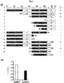
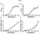
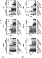
上記の問題を解決するために、本発明者らは、ドメインIIIおよびIVによって形成されるHER3上の特有のエピトープ(これはヒトHER3とマウスHER3との間で保存されている)を認識するヒト抗HER3(ErbB3)抗体3-43を同定した。この抗体は、0.1nM未満のEC50値を有するIgG分子としてHER3発現腫瘍細胞に結合し、リガンド非依存的およびリガンド依存的な受容体活性化ならびに下流のシグナル伝達を効率的に阻害し、迅速で効率的な受容体インターナリゼーションおよび分解を導く。 To solve the above problem, we developed a human epitope that recognizes a unique epitope on HER3 formed by domains III and IV, which is conserved between human and mouse HER3. Anti-HER3 (ErbB3) antibody 3-43 was identified. This antibody binds to HER3-expressing tumor cells as an IgG molecule with an EC50 value of less than 0.1 nM and efficiently inhibits ligand-independent and ligand-dependent receptor activation and downstream signaling, Leads to rapid and efficient receptor internalization and degradation.
第1の態様において、本発明は、ヒト上皮成長因子受容体3(HER3)のドメインIIIおよびドメインIVによって形成される立体構造エピトープに特異的に結合する抗原結合タンパク質を提供する。
第2の態様において、本発明は、第1の態様の抗原結合タンパク質と競合する抗原結合タンパク質を提供する。
第3の態様において、本発明は、第1または第2の態様の抗原結合タンパク質を含む融合タンパク質または複合体を提供する。
第4の態様において、本発明は、第1もしくは第2の態様の抗原結合タンパク質、または第3の態様の融合タンパク質をコードする配列を含む核酸分子を提供する。
第5の態様において、本発明は、第4の態様の核酸を含むベクターを提供する。
In a first aspect, the invention provides an antigen binding protein that specifically binds to a conformational epitope formed by domains III and IV of human epidermal growth factor receptor 3 (HER3).
In a second aspect, the invention provides an antigen binding protein that competes with the antigen binding protein of the first aspect.
In a third aspect, the invention provides fusion proteins or conjugates comprising the antigen binding protein of the first or second aspect.
In a fourth aspect, the invention provides a nucleic acid molecule comprising a sequence encoding the antigen binding protein of the first or second aspect, or the fusion protein of the third aspect.
In a fifth aspect, the invention provides a vector comprising the nucleic acid of the fourth aspect.
第6の態様において、本発明は、第1もしくは第2の態様の抗原結合タンパク質、第3の態様の融合タンパク質、第4の態様の核酸、または第5の態様のベクターを含む細胞を提供する。
第7の態様において、本発明は、第1もしくは第2の態様の抗原結合タンパク質、第3の態様の融合タンパク質、第4の態様の核酸、または第5の態様のベクターを含む医薬組成物を提供する。
第8の態様において、本発明は、薬物としての使用のための、第1もしくは第2の態様の抗原結合タンパク質、第3の態様の融合タンパク質もしくは複合体、第4の態様の核酸、第5の態様のベクター、第6の態様の細胞、または第7の態様の医薬品を提供する。
In a sixth aspect, the invention provides a cell comprising the antigen binding protein of the first or second aspect, the fusion protein of the third aspect, the nucleic acid of the fourth aspect, or the vector of the fifth aspect. .
In a seventh aspect, the invention provides a pharmaceutical composition comprising the antigen binding protein of the first or second aspect, the fusion protein of the third aspect, the nucleic acid of the fourth aspect, or the vector of the fifth aspect. offer.
In an eighth aspect, the invention provides the antigen binding protein of the first or second aspect, the fusion protein or conjugate of the third aspect, the nucleic acid of the fourth aspect, the fifth aspect, for use as a medicament. A vector of
第9の態様において、本発明は、治療有効量の第1もしくは第2の態様の抗原結合タンパク質、第3の態様の融合タンパク質もしくは複合体、第4の態様の核酸、第5の態様のベクター、第6の態様の細胞、または第7の態様の医薬品を投与することを含む、腫瘍増殖を阻害し、またはがんを処置する方法を提供する。 In a ninth aspect, the invention provides a therapeutically effective amount of the antigen binding protein of the first or second aspect, the fusion protein or conjugate of the third aspect, the nucleic acid of the fourth aspect, the vector of the fifth aspect. A method of inhibiting tumor growth or treating cancer comprising administering a cell of the sixth aspect, or a medicament of the seventh aspect.
配列表-フリーテキスト情報
配列番号1:Her3のアミノ酸配列(Expasyエントリー番号:P21860)
配列番号2:IgG3-43の重鎖可変ドメインのアミノ酸配列
配列番号3:IgG3-43の軽鎖可変ドメインのアミノ酸配列
配列番号4:IgG3-43の重鎖のアミノ酸配列
配列番号5:IgG3-43の軽鎖のアミノ酸配列
Sequence Listing-Free Text Information SEQ ID NO: 1: amino acid sequence of Her3 (Expasy entry number: P21860)
SEQ ID NO: 2: amino acid sequence of heavy chain variable domain of IgG3-43 SEQ ID NO: 3: amino acid sequence of light chain variable domain of IgG3-43 SEQ ID NO: 4: amino acid sequence of heavy chain of IgG3-43 SEQ ID NO: 5: IgG3-43 amino acid sequence of the light chain of
配列番号6:scFv3-43のアミノ酸配列
配列番号7:PelBリーダー - scFv 3-43 - c-myc - hisのアミノ酸配列
配列番号8:IgKリーダー - scDb hu225x3-43-Fcのアミノ酸配列
配列番号9:IgKリーダー - 2-35x3-43scDb-Fcのアミノ酸配列
配列番号10:IgKリーダー - scDb 4D5x3-43-LL-Fcのアミノ酸配列
配列番号11:IgKリーダー - FLAG - リンカー - scFv3-43-Fc-scTRAILのアミノ酸配列
配列番号12:IgKリーダー - scDb 3-43xCD3 - Hisのアミノ酸配列
配列番号13:IgKリーダー - scDb 3-43xCD3-scFv 3-43 - Hisのアミノ酸配列
SEQ ID NO: 6: amino acid sequence of scFv3-43 SEQ ID NO: 7: amino acid sequence of PelB leader - scFv 3-43 - c-myc-his SEQ ID NO: 8: amino acid sequence of IgK leader - scDb hu225x3-43-Fc SEQ ID NO: 9: Amino acid sequence of IgK leader - 2-35x3-43scDb-Fc SEQ ID NO: 10: Amino acid sequence of IgK leader - scDb 4D5x3-43-LL-Fc SEQ ID NO: 11: IgK leader - FLAG - Linker - scFv3-43-Fc-scTRAIL Amino acid sequences SEQ ID NO: 12: Amino acid sequence of IgK leader - scDb 3-43xCD3 - His SEQ ID NO: 13: Amino acid sequence of IgK leader - scDb 3-43xCD3-scFv 3-43 - His
配列番号14:ペプチドリンカー1のアミノ酸配列:GGGGS
配列番号15:ペプチドリンカー2のアミノ酸配列:GGGGSGGGGS
配列番号16:ペプチドリンカー3のアミノ酸配列:GGGGSGGGGSGGGGS
配列番号17:ペプチドリンカー4のアミノ酸配列:GSLGGSGG
配列番号18:ペプチドリンカー5のアミノ酸配列:GGGSGGGT
配列番号19:ペプチドリンカー6のアミノ酸配列:GGGSGGGTGS
配列番号20:ペプチドリンカー7のアミノ酸配列:GGGSGGGTGSGG
配列番号21:ペプチドリンカー8のアミノ酸配列:GGGGSGGRASGGGGSGGGGS
配列番号22:ペプチドリンカー9のアミノ酸配列:GGGSGGGS
配列番号23:ペプチドリンカー10のアミノ酸配列:EFTRG
配列番号24:ペプチドリンカー11のアミノ酸配列:AAA
SEQ ID NO: 14: amino acid sequence of peptide linker 1: GGGGS
SEQ ID NO: 15: amino acid sequence of peptide linker 2: GGGGSGGGGS
SEQ ID NO: 16: amino acid sequence of peptide linker 3: GGGGSGGGGSGGGGGS
SEQ ID NO: 17: amino acid sequence of peptide linker 4: GSLGGSGG
SEQ ID NO: 18: amino acid sequence of peptide linker 5: GGGSGGGT
SEQ ID NO: 19: amino acid sequence of peptide linker 6: GGGSGGGTGS
SEQ ID NO: 20: amino acid sequence of peptide linker 7: GGGSGGGTGSGG
SEQ ID NO: 21: amino acid sequence of peptide linker 8: GGGGSGGRASGGGGSGGGGS
SEQ ID NO: 22: amino acid sequence of peptide linker 9: GGGSGGGS
SEQ ID NO: 23: amino acid sequence of peptide linker 10: EFTRG
SEQ ID NO: 24: amino acid sequence of peptide linker 11: AAA
配列番号25:FLAGタグのアミノ酸配列
配列番号26:Hisタグのアミノ酸配列
配列番号27:Mycタグのアミノ酸配列
配列番号28:PelBリーダー配列のアミノ酸配列
配列番号29:IgKリーダー配列のアミノ酸配列
配列番号30:IL-2リーダー配列のアミノ酸配列
SEQ ID NO: 25: FLAG tag amino acid sequence SEQ ID NO: 26: His tag amino acid sequence SEQ ID NO: 27: Myc tag amino acid sequence SEQ ID NO: 28: PelB leader sequence amino acid sequence SEQ ID NO: 29: IgK leader sequence amino acid sequence SEQ ID NO: 30 : amino acid sequence of the IL-2 leader sequence
配列番号31:VH3-43xVLhu225-CLのアミノ酸配列
配列番号32:VHhu225xVLhu3-43-CH1-CH2-CH3のアミノ酸配列
配列番号33:scDbhu225x3-43-Fc(GGGGS)のアミノ酸配列
配列番号34:scDb4D5x3-43-LLのアミノ酸配列
配列番号35:scFv3-43-scDb3-43xhuU3-scFv3-43のアミノ酸配列
SEQ ID NO:31: amino acid sequence of V H 3-43xV L hu225-C L SEQ ID NO:32: amino acid sequence of V H hu225xV L hu3-43-C H 1-C H 2-
本発明を以下で詳細に記述する前に、本明細書に記述される特定の方法論、プロトコルおよび試薬は様々であり得るため、本発明はこれらに限定されないことが理解されるはずである。本明細書で使用される用語は特定の実施態様を記述するためのものにすぎず、本発明の範囲を限定することを意図していないことが理解されるはずであり、本発明の範囲は添付の特許請求の範囲のみによって限定される。他に定義されない限り、本明細書で使用されるあらゆる技術用語および科学用語は、当業者によって一般的に理解されるのと同じ意味を有する。 Before describing the invention in detail below, it is to be understood that the invention is not limited to the particular methodology, protocols and reagents described herein, as they may vary. It is to be understood that the terminology used herein is for the purpose of describing particular embodiments only and is not intended to limit the scope of the invention, which is limited only by the following claims. Unless defined otherwise, all technical and scientific terms used herein have the same meaning as commonly understood by one of ordinary skill in the art.
好ましくは、本明細書で使用される用語は、"A multilingual glossary of biotechnological terms: (IUPAC Recommendations)", Leuenberger, H.G.W, Nagel, B. and Kolbl, H. eds. (1995), Helvetica Chimica Acta, CH-4010 Basel, Switzerlandに記載されているように定義される。 Preferably, the terms used herein refer to "A multilingual glossary of biotechnological terms: (IUPAC Recommendations)", Leuenberger, H.G.W, Nagel, B. and Kolbl, H. eds. (1995), Helvetica Chimica Acta, Defined as set forth in CH-4010 Basel, Switzerland.
複数の文書が本明細書の文章のあらゆる箇所で引用される。本明細書で引用される各文書(あらゆる特許、特許出願、科学的刊行物、製造者の仕様書、説明書、GenBankアクセッション番号配列提出書(submission)などを含む)は上記または下記に関わらず、その全体が参照によって本明細書に組み込まれる。本明細書のいかなる記載も、本発明が先行発明によるこのような開示に先行する権利を有しないことを承認するものと解釈されるべきではない。 Several documents are cited throughout the text of this specification. Each document cited herein (including any patents, patent applications, scientific publications, manufacturer's specifications, instructions, GenBank accession number sequence submissions, etc.) and is incorporated herein by reference in its entirety. Nothing herein is to be construed as an admission that the present invention is not entitled to antedate such disclosure by virtue of prior invention.
定義
単語「含む」およびその変化形(「含んでいる」など)は、記載されている整数もしくは工程または整数もしくは工程の群を包含するが、いかなる他の整数もしくは工程または整数もしくは工程の群も除外しないことを示唆することが理解される。
DEFINITIONS The word "comprising" and variations thereof (such as "comprising") encompasses the recited integer or step or group of integers or steps, but not any other integer or step or group of integers or steps. It is understood that no exclusion is suggested.
本明細書および添付の特許請求の範囲において使用される場合、単数形「a」、「an」および「the」は文脈上明らかな他の指示がない限り、複数の指示対象を含む。 As used in this specification and the appended claims, the singular forms "a," "an," and "the" include plural referents unless the context clearly dictates otherwise.
濃度、量および他の数値データは本明細書において、「範囲」の形式で表現または提示され得る。このような範囲の形式は単に簡便さおよび簡潔さのために使用され、したがって範囲の限界として明確に記載された数値だけでなく、その範囲内に包含されるあらゆる個々の数値または部分範囲もまた、各数値および部分範囲が明確に記載されているように含まれると柔軟に解釈されるべきであることが理解されるはずである。実例として、「150mg~600mg」の数値範囲は、150mg~600mgという明確に記載された値だけでなく、指定された範囲内の個々の値および部分範囲も含むと解釈されるはずである。したがって、150、160、170、180、190、…580、590、600mgなどの個々の値、および150~200、150~250、250~300、350~600などの部分範囲は、この数値範囲に含まれる。これと同一の原理が単一の数値のみを記載している範囲に適用される。さらに、このような解釈は範囲の幅または記載されている特徴にかかわらず適用されるべきである。 Concentrations, amounts, and other numerical data may be expressed or presented herein in a "range" format. These range formats are used merely for convenience and brevity and thus not only state the numerical values clearly stated as the limits of the range, but also any individual numerical value or subrange subsumed within that range. , should be flexibly interpreted to include each numerical value and subrange as explicitly recited. By way of illustration, a numerical range of "150 mg to 600 mg" is to be interpreted to include not only the explicitly recited value of 150 mg to 600 mg, but also individual values and subranges within the stated range. Thus, individual values such as 150, 160, 170, 180, 190, . included. This same principle applies to ranges reciting only single numerical values. Moreover, such an interpretation should apply regardless of the breadth of the range or the characteristics being described.
数値に関連して使用される場合、用語「約」は、示された数値よりも5%小さい下限を有し、示された数値よりも5%大きい上限を有する範囲内の数値を包含することを意味する。 When used in connection with numerical values, the term "about" includes the numerical values in the range having a lower limit of 5% less than the stated numerical value and an upper limit of 5% greater than the stated numerical value. means
用語「核酸」および「核酸分子」は本明細書において同義的に使用され、デオキシリボヌクレオチド塩基もしくはリボヌクレオチド塩基またはその両方の一本鎖または二本鎖のオリゴマーまたはポリマーとして理解される。ヌクレオチドモノマーは、核酸塩基、五炭糖(限定されないが、リボースまたは2’-デオキシリボースなど)および1~3つのリン酸基から構成される。通常、核酸は個々のヌクレオチドモノマー間のホスホジエステル結合を介して形成される。本発明の文脈において、核酸という用語は、限定されないがリボ核酸(RNA)分子およびデオキシリボ核酸(DNA)分子を含み、他の結合を含む核酸の合成型(例えば、Nielsen et al. (Science 254:1497-1500, 1991)に記述されるペプチド核酸)を含む。通常、核酸は一本鎖分子または二本鎖分子であり、天然に存在するヌクレオチドから構成される。核酸の一本鎖の描写は相補鎖の配列を(少なくとも部分的に)規定する。核酸は一本鎖もしくは二本鎖であり得、または二本鎖配列および一本鎖配列の両方の部分を含み得る。例示される二本鎖核酸分子は3’または5’オーバーハングを有し得、そのようなものとして、その全長にわたって完全に二本鎖であることは必要とされず、または想定されない。核酸は、生物学的合成法、生化学的合成法もしくは化学的合成法、または当分野で公知のあらゆる方法(限定されないが、増幅法およびRNAの逆転写法を含む)によって取得され得る。核酸という用語は、染色体もしくは染色体セグメント、ベクター(例えば発現ベクター)、発現カセット、ネイキッドDNAもしくはRNAポリマー、プライマー、プローブ、cDNA、ゲノムDNA、組換えDNA、cRNA、mRNA、tRNA、マイクロRNA(miRNA)または低分子干渉RNA(siRNA)を含む。核酸は、例えば一本鎖、二本鎖または三本鎖であり得、いかなる特定の長さにも限定されない。他の指示がない限り、ある核酸配列は、明確に示されている任意の配列に加えて、相補的配列を含むか、またはコードする。 The terms "nucleic acid" and "nucleic acid molecule" are used interchangeably herein and are understood as a single- or double-stranded oligomer or polymer of deoxyribonucleotide bases or ribonucleotide bases or both. Nucleotide monomers are composed of a nucleobase, a pentose (such as but not limited to ribose or 2'-deoxyribose) and one to three phosphate groups. Nucleic acids are usually formed through phosphodiester bonds between individual nucleotide monomers. In the context of the present invention, the term nucleic acid includes, but is not limited to, ribonucleic acid (RNA) molecules and deoxyribonucleic acid (DNA) molecules, and synthetic forms of nucleic acids containing other linkages (e.g., Nielsen et al. (Science 254: 1497-1500, 1991). Nucleic acids are typically single- or double-stranded molecules and are composed of naturally occurring nucleotides. Describing a single strand of nucleic acid defines (at least partially) the sequence of the complementary strand. Nucleic acids can be single-stranded or double-stranded, or can contain portions of both double- and single-stranded sequence. The exemplified double-stranded nucleic acid molecule may have 3' or 5' overhangs, and as such is not required or assumed to be completely double-stranded along its entire length. Nucleic acids can be obtained by biological, biochemical or chemical synthetic methods, or any method known in the art, including but not limited to amplification methods and reverse transcription of RNA. The term nucleic acid includes a chromosome or chromosomal segment, vector (e.g. expression vector), expression cassette, naked DNA or RNA polymer, primer, probe, cDNA, genomic DNA, recombinant DNA, cRNA, mRNA, tRNA, microRNA (miRNA). or including small interfering RNA (siRNA). Nucleic acids can be, for example, single-stranded, double-stranded or triple-stranded and are not limited to any particular length. Unless otherwise indicated, a given nucleic acid sequence includes or encodes complementary sequences in addition to any sequence specifically indicated.
核酸は、エンドヌクレアーゼまたはエキソヌクレアーゼ(特に、細胞中に見出され得るDNaseおよびRNase)によって分解され得る。したがって、核酸を分解に対して安定化するために修飾することは有利であり得、これにより高濃度の核酸が細胞中に長期間にわたって維持されることが保証され得る。通常、このような安定化は、1以上のヌクレオチド間リン基(internucleotide phosphorus group)を導入すること、または1以上のヌクレオチド間非リンを導入することによって得られ得る。したがって、核酸は、天然に存在しないヌクレオチド、および/または天然に存在するヌクレオチドへの修飾、および/または分子の骨格への変化から構成され得る。核酸における修飾されたヌクレオチド間リン酸ラジカルおよび/または非リン架橋には、限定されないが、メチルホスホン酸、ホスホロチオエート、ホスホルアミダート、ホスホロジチオエートおよび/またはリン酸エステルが含まれ、非リンヌクレオチド間類似体には、限定されないが、シロキサン架橋、カーボネート架橋、カルボキシメチルエステル、アセトアミダート架橋および/またはチオエーテル架橋が含まれる。ヌクレオチド修飾のさらなる例には、限定されないが:連結またはエキソヌクレアーゼ分解/ポリメラーゼ伸長の防止をそれぞれ可能にする5’または3’ヌクレオチドのリン酸化;共有結合および近似的共有結合(near covalent attachment)のためのアミノ、チオール、アルキンまたはビオチニル修飾;フルオロフォアおよび消光剤;ならびに修飾塩基(デオキシイノシン(dI)、5-ブロモ-デオキシウリジン(5-ブロモ-dU)、デオキシウリジン、2-アミノプリン、2,6-ジアミノプリン、逆位dT、逆位ジデオキシT、ジデオキシシチジン(ddC)、5-メチルデオキシシチジン(5-メチルdC)、ロックド核酸(LNA)、5-ニトロインドール、イソdCおよびイソdG塩基、2’-O-メチルRNA塩基、ヒドロキシメチルdC、5-ヒドロキシブチニル-2’-デオキシウリジン(5-hydroxybutynl-2’-deoxyuridine)、8-アザ-7-デアザグアノシンおよびフッ素修飾塩基など)が含まれる。したがって、核酸は人工核酸であり得、これは限定されないが、ポリアミドもしくはペプチド核酸(PNA)、モルフォリノおよびロックド核酸(LNA)、ならびにグリコール核酸(GNA)およびトレオース核酸(TNA)を含む。 Nucleic acids can be degraded by endonucleases or exonucleases, particularly DNases and RNases that can be found in cells. Therefore, it can be advantageous to modify nucleic acids to stabilize them against degradation, which can ensure that high concentrations of nucleic acids are maintained in cells for extended periods of time. Usually such stabilization can be obtained by introducing one or more internucleotide phosphorus groups or by introducing one or more internucleotide non-phosphorus groups. Thus, nucleic acids may be composed of non-naturally occurring nucleotides and/or modifications to naturally occurring nucleotides and/or changes to the backbone of the molecule. Modified internucleotide phosphate radicals and/or non-phosphorus bridges in nucleic acids include, but are not limited to, methylphosphonates, phosphorothioates, phosphoramidates, phosphorodithioates and/or phosphate esters; Inter-analogues include, but are not limited to, siloxane bridges, carbonate bridges, carboxymethyl esters, acetamidate bridges and/or thioether bridges. Further examples of nucleotide modifications include, but are not limited to: phosphorylation of 5' or 3' nucleotides to enable ligation or prevention of exonuclease degradation/polymerase elongation, respectively; fluorophores and quenchers; and modified bases (deoxyinosine (dI), 5-bromo-deoxyuridine (5-bromo-dU), deoxyuridine, 2-aminopurine, 2 , 6-diaminopurine, inverted dT, inverted dideoxy T, dideoxycytidine (ddC), 5-methyldeoxycytidine (5-methyl dC), locked nucleic acid (LNA), 5-nitroindole, isodC and isodG bases. , 2′-O-methyl RNA bases, hydroxymethyl dC, 5-hydroxybutynyl-2′-deoxyuridine, 8-aza-7-deazaguanosine and fluorine-modified bases, etc. ) is included. Thus, nucleic acids can be artificial nucleic acids, including but not limited to polyamide or peptide nucleic acids (PNA), morpholino and locked nucleic acids (LNA), and glycol nucleic acids (GNA) and threose nucleic acids (TNA).
核酸は、別の核酸配列と機能的な関係において配置されている場合、「動作可能に連結」されている。例えば、プロモーターまたはエンハンサーは配列の転写に影響を与える場合、コード配列に動作可能に連結されている;または、リボソーム結合部位は翻訳を促進するように配置されている場合、コード配列に動作可能に連結されている。 Nucleic acid is "operably linked" when it is placed into a functional relationship with another nucleic acid sequence. For example, a promoter or enhancer is operably linked to a coding sequence if it affects transcription of the sequence; or a ribosome binding site is operably linked to a coding sequence if it is positioned to facilitate translation. Concatenated.
本発明の文脈において、用語「オリゴヌクレオチド」は、最大で約50ヌクレオチド(例えば2~約50ヌクレオチド長)の核酸配列を指す。 In the context of the present invention, the term "oligonucleotide" refers to nucleic acid sequences up to about 50 nucleotides (eg, 2 to about 50 nucleotides long).
本発明の文脈において使用される場合、用語「ポリヌクレオチド」は、約50ヌクレオチド長よりも長い核酸(例えば51以上のヌクレオチド長)を指す。 As used in the context of the present invention, the term "polynucleotide" refers to nucleic acids greater than about 50 nucleotides in length (eg, 51 or more nucleotides in length).
オリゴヌクレオチドおよびポリペプチドはあらゆる適切な方法(限定されないが、既存の配列もしくは天然配列の単離、DNA複製もしくは増幅、逆転写、適切な配列のクローニングおよび制限消化、または(Narang et al. (Meth. Enzymol. 68:90-99, 1979)のホスホトリエステル法;Brown et al. (Meth. Enzymol. 68:109-151, 1979)のホスホジエステル法;Beaucage et al. (Tetrahedron Lett. 22:1859-1862, 1981)のジエチルホスホロアミダート法;Matteucci et al. (J. Am. Chem. Soc. 103:3185-3191, 1981)のトリエステル法;自動合成法;もしくは米国特許第4,458,066号の固相担体法、または当業者に公知の他の方法などの方法による)直接的な化学合成を含む)によって調製される。 Oligonucleotides and polypeptides may be prepared by any suitable method, including but not limited to isolation of existing or native sequences, DNA replication or amplification, reverse transcription, cloning and restriction digestion of appropriate sequences, or (Narang et al. (Meth Enzymol. 68:90-99, 1979); the phosphodiester method of Brown et al. (Meth. Enzymol. 68:109-151, 1979); Beaucage et al. (Tetrahedron Lett. 22:1859). -1862, 1981); the triester method of Matteucci et al. (J. Am. Chem. Soc. 103:3185-3191, 1981); the automated synthesis method; by methods such as solid phase support methods, or other methods known to those of skill in the art), including direct chemical synthesis.
本明細書において、用語「ベクター」は、細胞に導入でき、またはその中に含まれるタンパク質および/または核酸を細胞に導入できるタンパク質もしくはポリヌクレオチドまたはそれらの混合物を指す。ベクターの例には、限定されないが、プラスミド、コスミド、ファージ、ウイルスまたは人工染色体が含まれる。特に、ベクターは、対象の遺伝子産物(例えば、外来DNAまたは異種DNAなど)を適切な宿主細胞に輸送するために使用される。ベクターは、宿主細胞におけるベクターの自己複製を促進する「レプリコン」ポリヌクレオチド配列を含み得る。外来DNAは異種DNAとして定義され、これは宿主細胞において天然に見出されないDNAであり、この宿主細胞は例えばベクター分子を複製し、選択可能またはスクリーニング可能なマーカーをコードし、または導入遺伝子をコードしている。宿主細胞に存在する場合、ベクターは宿主染色体DNAとは独立に、または宿主染色体DNAと同時に複製し得、ベクターおよびその挿入されたDNAのいくつかのコピーが作成され得る。さらにベクターは、挿入されたDNAのmRNA分子への転写を可能にし、またはさもなければ挿入されたDNAの複数のコピーのRNAへの複製を生じるのに必要な要素を含み得る。ベクターはさらに、対象の遺伝子の発現を調節する「発現制御配列」を包含し得る。通常、発現制御配列はポリペプチドまたはポリヌクレオチド(限定されないが、プロモーター、エンハンサー、サイレンサー、インスレーターまたはリプレッサーなど)である。1以上の対象の遺伝子産物をコードする2以上のポリヌクレオチドを含むベクターにおいて、発現は1以上の発現制御配列によって共にまたは別々に制御され得る。より具体的には、ベクター上に含まれる各ポリヌクレオチドは別々の発現制御配列によって制御されてもよく、ベクター上に含まれる全てのポリヌクレオチドは単一の発現制御配列によって制御されてもよい。単一の発現制御配列によって制御される単一のベクター上に含まれるポリヌクレオチドは、オープンリーディングフレームを形成し得る。いくつかの発現ベクターは、発現したmRNAの半減期を増加させ、かつ/またはmRNAのタンパク質分子への翻訳を可能にする、挿入されたDNAに隣接する配列要素をさらに含む。したがって、挿入されたDNAによってコードされるmRNAおよびポリペプチドの多数の分子が迅速に合成され得る。 As used herein, the term "vector" refers to a protein or polynucleotide or mixture thereof that can be introduced into a cell, or that can introduce proteins and/or nucleic acids contained therein into a cell. Examples of vectors include, but are not limited to, plasmids, cosmids, phages, viruses or artificial chromosomes. In particular, vectors are used to transport gene products of interest (eg, foreign or heterologous DNA) into suitable host cells. A vector may contain "replicon" polynucleotide sequences that facilitate the autonomous replication of the vector in a host cell. Foreign DNA is defined as heterologous DNA, which is DNA not naturally found in the host cell, which, for example, replicates the vector molecule, encodes a selectable or screenable marker, or encodes a transgene. are doing. When present in a host cell, the vector can replicate independently of or concurrently with the host chromosomal DNA, and several copies of the vector and its inserted DNA can be made. In addition, the vector may contain the elements necessary to permit transcription of the inserted DNA into an mRNA molecule or otherwise effect replication of multiple copies of the inserted DNA into RNA. Vectors may further include "expression control sequences" that regulate the expression of the gene of interest. Typically, expression control sequences are polypeptides or polynucleotides (including but not limited to promoters, enhancers, silencers, insulators or repressors). In vectors containing two or more polynucleotides encoding one or more gene products of interest, expression can be controlled jointly or separately by one or more expression control sequences. More specifically, each polynucleotide contained on the vector may be controlled by a separate expression control sequence, or all polynucleotides contained on the vector may be controlled by a single expression control sequence. Polynucleotides contained on a single vector under the control of a single expression control sequence can form an open reading frame. Some expression vectors further contain sequence elements flanking the inserted DNA that increase the half-life of the expressed mRNA and/or allow translation of the mRNA into a protein molecule. Thus, large numbers of molecules of mRNA and polypeptides encoded by the inserted DNA can be rapidly synthesized.
用語「アミノ酸」は一般に、未置換もしくは置換されたアミノ基、未置換もしくは置換されたカルボキシ基、および1以上の側鎖もしくは基、またはあらゆるこれらの基の類似体を含むあらゆるモノマー単位を指す。例示的な側鎖には、例えば、チオール、セレノ、スルホニル、アルキル、アリール、アシル、ケト、アジド、ヒドロキシル、ヒドラジン、シアノ、ハロ、ヒドラジド、アルケニル、アルキニル、エーテル、ホウ酸塩、ボロン酸塩、ホスホ、ホスホノ、ホスフィン、複素環、エノン、イミン、アルデヒド、エステル、チオ酸、ヒドロキシルアミン、またはこれらの基のあらゆる組合せが含まれる。他の代表的なアミノ酸には、限定されないが、光活性化可能な架橋剤を含むアミノ酸、金属結合アミノ酸、スピン標識アミノ酸、蛍光アミノ酸、金属含有アミノ酸、新規官能基を有するアミノ酸、他の分子と共有結合的または非共有結合的に相互作用しているアミノ酸、光ケージされたアミノ酸および/または光異性化可能なアミノ酸、放射性アミノ酸、ビオチンまたはビオチン類似体を含むアミノ酸、グリコシル化アミノ酸、他の炭水化物で修飾されたアミノ酸、ポリエチレングリコールまたはポリエーテルを含むアミノ酸、重原子置換アミノ酸、化学的に切断可能なアミノ酸および/または光切断可能なアミノ酸、炭素結合糖(carbon-linked sugar)含有アミノ酸、レドックス活性アミノ酸、アミノチオ酸含有アミノ酸、ならびに1以上の毒性成分を含むアミノ酸が含まれる。本明細書において、用語「アミノ酸」は、以下の20種の天然または遺伝的にコードされたアルファアミノ酸を含む:アラニン(AlaまたはA)、アルギニン(ArgまたはR)、アスパラギン(AsnまたはN)、アスパラギン酸(AspまたはD)、システイン(CysまたはC)、グルタミン(GlnまたはQ)、グルタミン酸(GluまたはE)、グリシン(GlyまたはG)、ヒスチジン(HisまたはH)、イソロイシン(IleまたはI)、ロイシン(LeuまたはL)、リジン(LysまたはK)、メチオニン(MetまたはM)、フェニルアラニン(PheまたはF)、プロリン(ProまたはP)、セリン(SerまたはS)、スレオニン(ThrまたはT)、トリプトファン(TrpまたはW)、チロシン(TyrまたはY)、およびバリン(ValまたはV)。「X」残基が定義されていない場合、これらは「任意のアミノ酸」として定義されるべきである。これら20種の天然アミノ酸の構造は、例えばStryer et al., Biochemistry, 5th ed., Freeman and Company (2002)に示されている。さらなるアミノ酸(セレノシステインおよびピロリジンなど)が遺伝的にコードされ得る(Stadtman (1996) "Selenocysteine," Annu Rev Biochem. 65:83-100およびIbba et al. (2002) "Genetic code: introducing pyrrolysine," Curr Biol. 12(13):R464-R466)。用語「アミノ酸」はまた、非天然アミノ酸、(例えば修飾された側鎖および/または骨格を有する)修飾アミノ酸、およびアミノ酸類似体を含む。例えば、Zhang et al. (2004) "Selective incorporation of 5-hydroxytryptophan into proteins in mammalian cells," Proc. Natl. Acad. Sci. U.S.A. 101(24):8882-8887, Anderson et al. (2004) "An expanded genetic code with a functional quadruplet codon" Proc. Natl. Acad. Sci. U.S.A. 101(20):7566-7571, Ikeda et al. (2003) "Synthesis of a novel histidine analogue and its efficient incorporation into a protein in vivo," Protein Eng. Des. Sel. 16(9):699-706, Chin et al. (2003) "An Expanded Eukaryotic Genetic Code," Science 301(5635):964-967, James et al. (2001) "Kinetic characterization of ribonuclease S mutants containing photoisomerizable phenylazophenylalanine residues," Protein Eng. Des. Sel. 14(12):983-991, Kohrer et al. (2001) "Import of amber and ochre suppressor tRNAs into mammalian cells: A general approach to site-specific insertion of amino acid analogues into proteins," Proc. Natl. Acad. Sci. U.S.A. 98(25):14310-14315, Bacher et al. (2001) "Selection and Characterization of Escherichia coli Variants Capable of Growth on an Otherwise Toxic Tryptophan Analogue," J. Bacteriol. 183(18):5414-5425, Hamano-Takaku et al. (2000) "A Mutant Escherichia coli Tyrosyl-tRNA Synthetase Utilizes the Unnatural Amino Acid Azatyrosine More Efficiently than Tyrosine," J. Biol. Chem. 275(51):40324-40328, およびBudisa et al. (2001) "Proteins with {beta}-(thienopyrrolyl) alanines as alternative chromophores and pharmaceutically active amino acids," Protein Sci. 10(7):1281-1292参照。アミノ酸は併合され、ペプチド、ポリペプチドまたはタンパク質になり得る。 The term "amino acid" generally refers to any monomeric unit containing an unsubstituted or substituted amino group, an unsubstituted or substituted carboxy group, and one or more side chains or groups, or any analogs of these groups. Exemplary side chains include, for example, thiol, seleno, sulfonyl, alkyl, aryl, acyl, keto, azide, hydroxyl, hydrazine, cyano, halo, hydrazide, alkenyl, alkynyl, ether, borate, boronate, Included are phospho, phosphono, phosphine, heterocycle, enone, imine, aldehyde, ester, thioacid, hydroxylamine, or any combination of these groups. Other representative amino acids include, but are not limited to, amino acids containing photoactivatable crosslinkers, metal-binding amino acids, spin-labeled amino acids, fluorescent amino acids, metal-containing amino acids, amino acids with novel functional groups, other molecules and Covalently or non-covalently interacting amino acids, photocaged amino acids and/or photoisomerizable amino acids, radioactive amino acids, amino acids containing biotin or biotin analogues, glycosylated amino acids, other carbohydrates amino acids modified with , amino acids containing polyethylene glycol or polyethers, heavy atom substituted amino acids, chemically cleavable amino acids and/or photocleavable amino acids, carbon-linked sugar containing amino acids, redox activity Included are amino acids, aminothioic acid-containing amino acids, as well as amino acids containing one or more toxic moieties. As used herein, the term "amino acid" includes the following twenty naturally occurring or genetically encoded alpha amino acids: alanine (Ala or A), arginine (Arg or R), asparagine (Asn or N), aspartic acid (Asp or D), cysteine (Cys or C), glutamine (Gln or Q), glutamic acid (GIu or E), glycine (Gly or G), histidine (His or H), isoleucine (Ile or I), Leucine (Leu or L), Lysine (Lys or K), Methionine (Met or M), Phenylalanine (Phe or F), Proline (Pro or P), Serine (Ser or S), Threonine (Thr or T), Tryptophan (Trp or W), tyrosine (Tyr or Y), and valine (Val or V). If the "X" residues are not defined, they should be defined as "any amino acid". The structures of these twenty natural amino acids are given, for example, in Stryer et al., Biochemistry, 5th ed., Freeman and Company (2002). Additional amino acids, such as selenocysteine and pyrrolysine, can be genetically encoded (Stadtman (1996) "Selenocysteine," Annu Rev Biochem. 65:83-100 and Ibba et al. (2002) "Genetic code: introducing pyrrolysine," Curr Biol. 12(13):R464-R466). The term "amino acid" also includes non-natural amino acids, modified amino acids (eg, having modified side chains and/or backbones), and amino acid analogs. For example, Zhang et al. (2004) "Selective incorporation of 5-hydroxytryptophan into proteins in mammalian cells," Proc. Natl. Acad. Sci. U.S.A. 101(24):8882-8887, Anderson et al. Natl. Acad. Sci. U.S.A. 101(20):7566-7571, Ikeda et al. (2003) "Synthesis of a novel histidine analogue and its efficient incorporation into a protein in vivo Des. Sel. 16(9):699-706, Chin et al. (2003) "An Expanded Eukaryotic Genetic Code," Science 301(5635):964-967, James et al. (2001) "Kinetic characterization of ribonuclease S mutants containing photoisomerizable phenylazophenylalanine residues," Protein Eng. Des. Sel. 14(12):983-991, Kohrer et al. (2001) "Import of amber and ochre suppressor tRNAs into mammalian cells: A general approach to site-specific insertion of amino acid analogues into proteins," Proc. Natl. Acad. Sci. U.S.A. 98(25):14310-14315, Bacher et al. (2001) "Selection and Characterization of Escherichia coli Variants Capable of Growth on an Otherwise Toxic Tryptophan Analogue," J. Bacteriol. 183(18):5414-5425, Hamano-Takaku et al. (2000) "A Mutant Escherichia coli Tyrosyl-tRNA Synthetase Utilizes the Unnatural Amino Acid Azatyrosine More Efficiently than Tyrosine," J. Biol. Chem. 275(51):40324-40328, and Budisa et al. (2001) "Proteins with {beta}-(thienopyrrolyl) alanines as alternative chromophores and pharmaceutically active amino acids," Protein Sci. 10(7):1281-1292. Amino acids can be combined to form peptides, polypeptides or proteins.
本発明の文脈において、用語「ペプチド」は、ペプチド結合によって連結したアミノ酸の短いポリマーを指す。これはタンパク質と同一の化学結合(ペプチド結合)を有するが、一般に長さがより短い。最も短いペプチドはジペプチドであり、単一のペプチド結合によって結合した2つのアミノ酸からなる。トリペプチド、テトラペプチド、ペンタペプチドなども存在し得る。通常、ペプチドは最大で8、10、12、15、18または20アミノ酸長を有する。ペプチドは環状ペプチドでない限り、アミノ末端およびカルボキシ末端を有する。 In the context of this invention, the term "peptide" refers to a short polymer of amino acids linked by peptide bonds. It has the same chemical bonds (peptide bonds) as proteins, but is generally shorter in length. The shortest peptides are dipeptides, consisting of two amino acids joined by a single peptide bond. Tripeptides, tetrapeptides, pentapeptides, etc. may also be present. Peptides usually have a maximum length of 8, 10, 12, 15, 18 or 20 amino acids. A peptide has an amino terminus and a carboxy terminus, unless it is a cyclic peptide.
本発明の文脈において、用語「ポリペプチド」はペプチド結合によって互いに結合したアミノ酸の単一の直鎖を指し、典型的に少なくとも約21個のアミノ酸を含む。ポリペプチドは2以上の鎖から構成されるタンパク質の1つの鎖であり得、またはタンパク質が1つの鎖から構成される場合、タンパク質自体であり得る。 In the context of this invention, the term "polypeptide" refers to a single linear chain of amino acids linked together by peptide bonds, typically comprising at least about 21 amino acids. A polypeptide can be a single chain of a protein composed of more than one chain, or, if the protein is composed of one chain, the protein itself.
本発明の文脈において、タンパク質またはポリペプチドの「一次構造」は、ポリペプチド鎖のアミノ酸配列である。タンパク質の「二次構造」は、タンパク質の局所セグメントの一般的な3次元形状である。しかしながら、これは、三次構造が考慮される3次元空間における具体的な原子配置を記述しない。タンパク質において、二次構造は、骨格のアミド基とカルボキシ基との間の水素結合のパターンによって規定される。タンパク質の「三次構造」は、原子座標によって決定されるタンパク質の3次元構造である。「四次構造」は、マルチサブユニット複合体における複数のフォールドされた、またはコイル状のタンパク質分子またはポリペプチド分子の配置である。 In the context of this invention, the "primary structure" of a protein or polypeptide is the amino acid sequence of the polypeptide chain. A "secondary structure" of a protein is the general three-dimensional shape of the local segments of the protein. However, it does not describe the specific atomic arrangement in three-dimensional space in which tertiary structure is considered. In proteins, secondary structure is defined by the pattern of hydrogen bonding between backbone amide and carboxy groups. A "tertiary structure" of a protein is the three-dimensional structure of the protein as determined by its atomic coordinates. "Quaternary structure" is the arrangement of multiple folded or coiled protein or polypeptide molecules in a multisubunit complex.
本明細書における用語「フォールディング」または「タンパク質フォールディング」は、タンパク質がその3次元形状または立体構造をとるプロセスを指す(すなわち、これによりタンパク質は非共有結合性相互作用(限定されないが、水素結合、金属配位、疎水性力、ファンデルワールス力、π-π相互作用、および/または静電効果など)を介して特定の3次元形状を形成するように導かれる)。したがって、用語「フォールドされたタンパク質」は、その3次元形状(その二次構造、三次構造または四次構造など)のタンパク質を指す。 As used herein, the term "folding" or "protein folding" refers to the process by which a protein adopts its three-dimensional shape or conformation (i.e., by which the protein is bound by non-covalent interactions, including but not limited to hydrogen bonding, (metal coordination, hydrophobic forces, van der Waals forces, π-π interactions, and/or electrostatic effects, etc.). Accordingly, the term "folded protein" refers to the protein in its three-dimensional shape (such as its secondary, tertiary or quaternary structure).
本明細書における用語「フラグメント」は、天然に存在するフラグメント(例えばスプライスバリアント)および人工的に作成されたフラグメント(特に遺伝子工学の手段によって取得されたフラグメント)を指す。通常、フラグメントは親のポリペプチドと比較して、そのN末端および/またはそのC末端および/または内部(好ましくはそのN末端、そのNおよびC末端、またはそのC末端)において、最大で1、2、3、4、5、6、7、8、9、10、15、20、25、30、35、40、45、50、55、60、65、70、75、80、85、90、95、100、110、120、130、140、150、160、170、180、190、200、210、220、230、240、250、260、270、280、290または300アミノ酸の欠失を有する。 The term "fragment" as used herein refers to naturally occurring fragments (eg splice variants) and artificially created fragments (especially fragments obtained by means of genetic engineering). Fragments will generally have at most 1, 2, 3, 4, 5, 6, 7, 8, 9, 10, 15, 20, 25, 30, 35, 40, 45, 50, 55, 60, 65, 70, 75, 80, 85, 90, Has a deletion of 95, 100, 110, 120, 130, 140, 150, 160, 170, 180, 190, 200, 210, 220, 230, 240, 250, 260, 270, 280, 290 or 300 amino acids.
抗原決定基としても知られる「エピトープ」は、免疫系(具体的には抗体、B細胞またはT細胞)によって認識される高分子のセグメントである。そのようなエピトープは、抗体またはその抗原結合フラグメントに結合できる高分子の一部またはセグメントである。この文脈において、用語「結合」は好ましくは特異的結合に関する。本発明の文脈において、用語「エピトープ」は、免疫系によって認識されるタンパク質またはポリタンパク質のセグメントを指す。エピトープは通常、分子(アミノ酸または糖側鎖など)の化学的に活性な表面の基からなり、通常、特定の三次元構造の特徴および特定の電荷の特徴を有する。立体構造エピトープおよび非立体構造エピトープは、変性溶媒の存在下において立体構造エピトープへの結合は失われるが、非立体構造エピトープへの結合は失われない点で区別される。 An "epitope", also known as an antigenic determinant, is a segment of a macromolecule recognized by the immune system (specifically antibodies, B-cells or T-cells). Such epitopes are parts or segments of macromolecules capable of binding to an antibody or antigen-binding fragment thereof. In this context, the term "binding" preferably relates to specific binding. In the context of the present invention, the term "epitope" refers to a segment of a protein or polyprotein recognized by the immune system. Epitopes usually consist of chemically active surface groupings of molecules such as amino acids or sugar side chains and usually have specific three dimensional structural characteristics, as well as specific charge characteristics. Conformational and non-conformational epitopes are distinguished in that binding to conformational epitopes, but not non-conformational epitopes, is lost in the presence of denaturing solvents.
本明細書において、「立体構造エピトープ」は、直鎖高分子(例えばポリペプチド)の3次元構造によって形成される、直鎖高分子のエピトープを指す。本願の文脈において、「立体構造エピトープ」は、「不連続エピトープ」(すなわち、高分子の一次配列(例えばポリペプチドのアミノ酸配列)における少なくとも2つの別々の領域から形成される高分子(例えばポリペプチド)上の立体構造エピトープ)である。換言すれば、エピトープが、本発明の結合部分(例えば抗体またはその抗原結合フラグメント)が同時に結合する一次配列中の少なくとも2つの別々の領域(これらは、本発明の結合部分が結合しない一次配列中のさらなる1つの領域によって分断されている)からなる場合、エピトープは本発明の文脈において「立体構造エピトープ」とみなされる。特に、このような「立体構造エピトープ」はポリペプチド上に存在し、一次配列中の2つの別々の領域は、本発明の結合部分(例えば抗体またはその抗原結合フラグメント)が結合する2つの別々のアミノ酸配列であり、これらの少なくとも2つの別々のアミノ酸配列は、本発明の結合部分が結合しない一次配列中のさらなる1つのアミノ酸配列によって分断されている。特に、分断しているアミノ酸配列は、結合部分が結合しない2以上のアミノ酸を含む連続したアミノ酸配列である。本発明の結合部分が結合する少なくとも2つの別々のアミノ酸配列は、その長さに関して特に限定されない。前記少なくとも2つの別々のアミノ酸配列内のアミノ酸の総数が、結合部分と立体構造エピトープとの間の特異的結合をもたらすのに十分大きい限り、このような別々のアミノ酸配列は単一のアミノ酸のみからなっていてもよい。 As used herein, a "conformational epitope" refers to a linear macromolecule epitope formed by the three-dimensional structure of a linear macromolecule (eg, polypeptide). In the context of this application, a "conformational epitope" is defined as a "discontinuous epitope" (i.e., a macromolecule (e.g., polypeptide ) is a conformational epitope on ). In other words, the epitope is bound by at least two distinct regions in the primary sequence to which a binding moiety (e.g., an antibody or antigen-binding fragment thereof) of the invention binds simultaneously, which are in the primary sequence to which the binding moiety of the invention does not bind. separated by one additional region of ), the epitope is considered a "conformational epitope" in the context of the present invention. In particular, such "conformational epitopes" are present on a polypeptide wherein two separate regions in the primary sequence are represented by two separate epitopes to which a binding moiety (e.g., an antibody or antigen-binding fragment thereof) of the invention binds. An amino acid sequence, wherein at least two separate amino acid sequences are separated by one additional amino acid sequence in the primary sequence to which the binding moiety of the invention does not bind. In particular, the interrupting amino acid sequences are contiguous amino acid sequences comprising two or more amino acids that are not bound by the binding moieties. The at least two separate amino acid sequences bound by the binding moieties of the invention are not particularly limited as to their length. As long as the total number of amino acids in said at least two separate amino acid sequences is large enough to effect specific binding between the binding moiety and the conformational epitope, such separate amino acid sequences may consist of only single amino acids. It may be.
「パラトープ」は、エピトープを認識する抗体の一部である。本発明の文脈において、「パラトープ」は、エピトープを認識する本明細書に記載される結合部分(例えば抗体またはその抗原結合フラグメント)の一部である。 A "paratope" is the portion of an antibody that recognizes an epitope. In the context of the present invention, a "paratope" is a portion of a binding moiety (eg, an antibody or antigen-binding fragment thereof) described herein that recognizes an epitope.
本発明の文脈において、「ペプチドリンカー」は、複合体の2つの部分または成分(例えば2つのペプチドまたはタンパク質)を立体的に分離するアミノ酸配列を指す。通常、このようなリンカーは1~100アミノ酸からなり、これは最小で少なくとも1、2、3、4、5、6、7、8、9、10、11、12、13、14、15、16、17、18、19、20、21、22、23、24、25、26、27、28、29または30アミノ酸長、および最大で少なくとも100、95、90、85、80、75、70、65、60、55、50、45、40、35、34、33、32、31、30、29、28、27、26、25、24、23、22、21、20、19、18、17、16または15アミノ酸長またはそれ未満を有する。本発明におけるペプチドリンカーの示される好ましい最小長および最大長は(そのような組合せが数学的意味を持つ場合に)組み合わされ得る(例えばこのようなリンカーは1~15または12~40または25~75または1~100アミノ酸からなり得る)。ペプチドリンカーはまた、連結されている2つの部分の間に柔軟性を与え得る。このような柔軟性は一般に、アミノ酸が小さい場合に増加する。したがって、柔軟なペプチドリンカーは、小さなアミノ酸(特にグリシンおよび/またはアラニン、および/または親水性アミノ酸(セリン、スレオニン、アスパラギンおよびグルタミンなど))の含有量の増加を含む。好ましくは、ペプチドリンカーのアミノ酸のうち20%、30%、40%、50%、60%またはそれ以上が小さなアミノ酸である。 In the context of the present invention, a "peptide linker" refers to an amino acid sequence that sterically separates two parts or components (eg, two peptides or proteins) of a complex. Typically, such linkers consist of 1-100 amino acids, which are a minimum of at least 1, 2, 3, 4, 5, 6, 7, 8, 9, 10, 11, 12, 13, 14, 15, 16 , 17, 18, 19, 20, 21, 22, 23, 24, 25, 26, 27, 28, 29 or 30 amino acids long and up to at least 100, 95, 90, 85, 80, 75, 70, 65 , 60, 55, 50, 45, 40, 35, 34, 33, 32, 31, 30, 29, 28, 27, 26, 25, 24, 23, 22, 21, 20, 19, 18, 17, 16 or has a length of 15 amino acids or less. Preferred minimum and maximum lengths indicated for peptide linkers in the present invention can be combined (where such combinations have mathematical meaning) (eg such linkers are or consist of 1-100 amino acids). Peptide linkers can also provide flexibility between the two moieties being linked. Such flexibility generally increases when amino acids are small. Flexible peptide linkers therefore include an increased content of small amino acids, especially glycine and/or alanine, and/or hydrophilic amino acids such as serine, threonine, asparagine and glutamine. Preferably, 20%, 30%, 40%, 50%, 60% or more of the amino acids of the peptide linker are small amino acids.
本明細書における用語「変異体」は、ポリペプチドまたはポリヌクレオチドが由来するポリペプチドまたはポリヌクレオチドと比較して、その長さまたは配列における1以上の変化により異なっているポリペプチドまたはポリヌクレオチドとして理解されるはずである。ポリペプチド変異体またはポリヌクレオチド変異体が由来するポリペプチドまたはポリヌクレオチドは、親のポリペプチドまたはポリヌクレオチドとしても知られる。用語「変異体」は親分子の「フラグメント」または「誘導体」を含む。通常、「フラグメント」は親分子よりも長さまたはサイズが小さく、「誘導体」は親分子と比較してその配列において1以上の差異を示す。限定されないが、翻訳後修飾タンパク質(例えばグリコシル化、ビオチニル化、リン酸化、ユビキチン化、パルミトイル化、またはタンパク分解により切断されたタンパク質)および修飾核酸(メチル化DNAなど)などの修飾分子もまた包含される。種々の分子の混合物(限定されないが、RNA-DNAハイブリッドなど)もまた、用語「変異体」に包含される。通常、変異体は(好ましくは遺伝子工学の手段によって)人工的に作成されるが、親のタンパク質またはポリヌクレオチドは野生型タンパク質もしくはポリヌクレオチドまたはその共通配列である。しかしながら、天然に存在する変異体もまた、本明細書における用語「変異体」に包含されることが理解されるはずである。さらに、本発明に使用できる変異体はまた、親分子の少なくとも1つの生物活性を示す(すなわち機能的に活性である)場合、親分子のホモログ、オルソログもしくはパラログ、または人工的に作成された変異体に由来し得る。 As used herein, the term "variant" is understood as a polypeptide or polynucleotide that differs by one or more alterations in its length or sequence as compared to the polypeptide or polynucleotide from which it is derived. should be done. The polypeptide or polynucleotide from which a polypeptide or polynucleotide variant is derived is also known as the parent polypeptide or polynucleotide. The term "variant" includes "fragments" or "derivatives" of the parent molecule. A "fragment" is generally smaller in length or size than a parent molecule, and a "derivative" exhibits one or more differences in its sequence compared to the parent molecule. Modified molecules such as, but not limited to, post-translationally modified proteins (e.g., glycosylated, biotinylated, phosphorylated, ubiquitinated, palmitoylated, or proteolytically cleaved proteins) and modified nucleic acids (such as methylated DNA) are also included. be done. Mixtures of different molecules (such as but not limited to RNA-DNA hybrids) are also encompassed by the term "mutants". Mutants are usually made artificially (preferably by means of genetic engineering), but the parental protein or polynucleotide is a wild-type protein or polynucleotide or a consensus sequence thereof. However, it should be understood that naturally occurring variants are also encompassed by the term "variant" herein. Furthermore, variants that can be used in the present invention also include homologs, orthologs or paralogs of the parent molecule, or artificially created mutations, if they exhibit at least one biological activity of the parent molecule (i.e. are functionally active). can come from the body.
特に、用語「ペプチド変異体」、「ポリペプチド変異体」、「タンパク質変異体」は、それが由来するペプチド、ポリペプチドまたはタンパク質と比較して、アミノ酸配列における1以上の変化によって異なっているペプチド、ポリペプチドまたはタンパク質として理解されるはずである。ペプチド変異体、ポリペプチド変異体またはタンパク質変異体が由来するペプチド、ポリペプチドまたはタンパク質は、親ペプチド、親ポリペプチドまたは親タンパク質としても知られている。さらに、本発明に使用できる変異体はまた、親ペプチド、親ポリペプチドまたは親タンパク質の少なくとも1つの生物活性を示す場合、親ペプチド、親ポリペプチドまたは親タンパク質のホモログ、オルソログもしくはパラログ、または人工的に作成された変異体に由来し得る。アミノ酸配列の変化は、アミノ酸交換、挿入、欠失、N末端切断もしくはC末端切断、またはこれらの変化のあらゆる組合せであり得、これは1つまたはいくつかの部位において生じ得る。ペプチド変異体、ポリペプチド変異体またはタンパク質変異体は、アミノ酸配列において最大で200個(最大で1、2、3、4、5、6、7、8、9、10、15、20、25、30、35、40、45、50、55、60、65、70、75、80、85、90、95、100、110、120、130、140、150、160、170、180、190、または200個)の変化(すなわち、交換、挿入、欠失、N末端切断および/またはC末端切断)の総数を示し得る。アミノ酸交換は保存的および/または非保存的であり得る。あるいは、またはさらに、本明細書における「変異体」は、由来する親ペプチド、親ポリペプチドまたは親タンパク質とのある程度の配列同一性によって特徴付けられ得る。より正確には、本発明の文脈におけるペプチド変異体、ポリペプチド変異体またはタンパク質変異体は、その親ペプチド、親ポリペプチドまたは親タンパク質と少なくとも80%の配列同一性を示す。ペプチド変異体、ポリペプチド変異体またはタンパク質変異体の配列同一性は、20、30、40、45、50、60、70、80、90、100またはそれ以上のアミノ酸の連続した区間にわたる。 In particular, the terms "peptide variant", "polypeptide variant", "protein variant" refer to peptides that differ by one or more changes in amino acid sequence as compared to the peptide, polypeptide or protein from which they are derived. , polypeptides or proteins. The peptide, polypeptide or protein from which the peptide, polypeptide or protein variant is derived is also known as the parent peptide, parent polypeptide or parent protein. Furthermore, variants that can be used in the present invention also include homologues, orthologs or paralogs of a parent peptide, parent polypeptide or parent protein, or artificial can be derived from mutants made in Amino acid sequence alterations may be amino acid exchanges, insertions, deletions, N- or C-terminal truncations, or any combination of these alterations, which may occur at one or several sites. Peptide, polypeptide or protein variants may have up to 200 amino acid sequences (up to 1, 2, 3, 4, 5, 6, 7, 8, 9, 10, 15, 20, 25, 30, 35, 40, 45, 50, 55, 60, 65, 70, 75, 80, 85, 90, 95, 100, 110, 120, 130, 140, 150, 160, 170, 180, 190, or 200 individual) changes (ie, exchanges, insertions, deletions, N-terminal truncations and/or C-terminal truncations). Amino acid exchanges can be conservative and/or non-conservative. Alternatively, or additionally, a "variant" herein can be characterized by a degree of sequence identity with the parent peptide, polypeptide or protein from which it is derived. More precisely, a peptide, polypeptide or protein variant in the context of the present invention exhibits at least 80% sequence identity with its parent peptide, polypeptide or protein. Sequence identity of a peptide, polypeptide or protein variant may span consecutive stretches of 20, 30, 40, 45, 50, 60, 70, 80, 90, 100 or more amino acids.
「配列同一性の割合」は、比較窓を通して最適にアラインされた2つの配列を比較することによって決定され、ここで比較窓における配列の部分は、2つの配列の最適なアラインメントのために基準配列(これは付加または欠失を含まない)と比較して付加または欠失(すなわちギャップ)を含み得る。割合は、一致した位置の数を得るために両方の配列において同一の核酸塩基またはアミノ酸残基が生じている位置の数を決定し、一致した位置の数を比較の窓における位置の総数で割り、結果に100を掛けて配列同一性の割合を得ることによって算出される。 A "percent sequence identity" is determined by comparing two sequences that are optimally aligned over a window of comparison, wherein the portion of the sequence in the window of comparison is the reference sequence for optimal alignment of the two sequences. may contain additions or deletions (ie, gaps) compared to (which does not contain additions or deletions). Percentage determines the number of positions in which the same nucleobase or amino acid residue occurs in both sequences to obtain the number of matched positions, and divides the number of matched positions by the total number of positions in the window of comparison. , calculated by multiplying the result by 100 to obtain the percent sequence identity.
2以上の核酸配列またはポリペプチド配列の文脈における用語「同一」は、同一である(すなわち、ヌクレオチドまたはアミノ酸の同一の配列を含む)2以上の配列または部分配列を指す。比較窓、または以下の配列比較アルゴリズムのうち1つを用いて、もしくは手動のアラインメントおよび目視検査によって測定される指定された領域にわたって最大の一致のために比較およびアラインされた場合に、配列が特定の割合の同一のヌクレオチドまたはアミノ酸残基(例えば、特定の領域にわたって少なくとも20%、少なくとも25%、少なくとも30%、少なくとも35%、少なくとも40%、少なくとも45%、少なくとも50%、少なくとも55%、少なくとも60%、少なくとも65%、少なくとも70%、少なくとも75%、少なくとも80、少なくとも81%、少なくとも82%、少なくとも83%、少なくとも84%、少なくとも85%、少なくとも86%、少なくとも87%、少なくとも88%、少なくとも89%、少なくとも90%、少なくとも91%、少なくとも92%、少なくとも93%、少なくとも94%、少なくとも95%、少なくとも96%、少なくとも97%、少なくとも98%または少なくとも99%の同一性)を有する場合、互いに「実質的に同一」である。これらの定義はまた、テスト配列の相補体を指す。したがって、用語「少なくとも80%の配列同一性」はポリペプチドおよびポリヌクレオチドの配列比較に関して、本明細書のあらゆる箇所で使用される。この表現は好ましくは、各基準ポリペプチドまたは各基準ポリヌクレオチドとの少なくとも80%、少なくとも81%、少なくとも82%、少なくとも83%、少なくとも84%、少なくとも85%、少なくとも86%、少なくとも87%、少なくとも88%、少なくとも89%、少なくとも90%、少なくとも91%、少なくとも92%、少なくとも93%、少なくとも94%、少なくとも95%、少なくとも96%、少なくとも97%、少なくとも98%、または少なくとも99%の配列同一性を指す。 The term "identical" in the context of two or more nucleic acid or polypeptide sequences refers to two or more sequences or subsequences that are identical (ie, contain identical sequences of nucleotides or amino acids). A sequence is identified when compared and aligned for maximum correspondence over a designated region as determined using a comparison window, or one of the following sequence comparison algorithms, or by manual alignment and visual inspection. percentage of identical nucleotides or amino acid residues (e.g., at least 20%, at least 25%, at least 30%, at least 35%, at least 40%, at least 45%, at least 50%, at least 55%, at least 60%, at least 65%, at least 70%, at least 75%, at least 80, at least 81%, at least 82%, at least 83%, at least 84%, at least 85%, at least 86%, at least 87%, at least 88%, at least 89%, at least 90%, at least 91%, at least 92%, at least 93%, at least 94%, at least 95%, at least 96%, at least 97%, at least 98% or at least 99% identity) , are “substantially identical” to each other. These definitions also refer to the complement of the test sequence. Accordingly, the term "at least 80% sequence identity" is used everywhere herein in reference to polypeptide and polynucleotide sequence comparisons. This expression is preferably at least 80%, at least 81%, at least 82%, at least 83%, at least 84%, at least 85%, at least 86%, at least 87%, at least 88%, at least 89%, at least 90%, at least 91%, at least 92%, at least 93%, at least 94%, at least 95%, at least 96%, at least 97%, at least 98%, or at least 99% sequence identity refers to gender.
用語「配列比較」は、ある配列を基準配列として、テスト配列と比較させるプロセスを指す。配列比較アルゴリズムを用いる場合、テスト配列および基準配列をコンピュータに入力し、必要であれば部分配列座標を指定し、配列アルゴリズムパラメータを指定する。初期設定のプログラムパラメータが一般的に使用され、または代替のパラメータが指定され得る。配列比較アルゴリズムは、プログラムパラメータに基づいて基準配列に対するテスト配列の配列同一性または類似性の割合を計算する。2つの配列を比較し、基準配列が配列同一性の割合を計算すべき配列と比較して特定されていない場合、具体的な他の指示がなければ、配列同一性は比較される2つの配列のうちより長い配列を基準として計算されるべきである。 The term "sequence comparison" refers to the process of comparing a sequence to a reference sequence, to test sequences. When using a sequence comparison algorithm, test and reference sequences are entered into a computer, subsequence coordinates are designated, if necessary, and sequence algorithm parameters are designated. Default program parameters are commonly used, or alternative parameters can be designated. The sequence comparison algorithm calculates the percent sequence identities or similarities for the test sequences relative to the reference sequence, based on the program parameters. When comparing two sequences and a reference sequence is not specified relative to the sequence for which the percent sequence identity is to be calculated, unless specifically indicated otherwise, the sequence identity is determined by comparing the two sequences being compared. should be calculated on the basis of the longer sequence of
配列アラインメントにおいて、用語「比較窓」は、同数の位置を有する配列の連続した位置の基準区間と比較される、配列の連続した位置の区間を指す。選択される連続した位置の数は、10~1000個の範囲であり得る(すなわち、20、30、40、50、60、70、80、90、100、150、200、250、300、350、400、450、500、550、600、650、700、750、800、850、900、950、または1000個の連続した位置を含み得る)。通常、連続した位置の数は、約20~800個の連続した位置、約20~600個の連続した位置、約50~400個の連続した位置、約50~約200個の連続した位置、約100~約150個の連続した位置である。 In sequence alignments, the term "comparison window" refers to a interval of contiguous positions of a sequence that is compared to a reference interval of contiguous positions of a sequence having the same number of positions. The number of consecutive positions selected can range from 10 to 1000 (i.e. 20, 30, 40, 50, 60, 70, 80, 90, 100, 150, 200, 250, 300, 350, 400, 450, 500, 550, 600, 650, 700, 750, 800, 850, 900, 950, or 1000 consecutive positions). Generally, the number of consecutive positions is from about 20 to 800 consecutive positions, from about 20 to 600 consecutive positions, from about 50 to 400 consecutive positions, from about 50 to about 200 consecutive positions, About 100 to about 150 consecutive positions.
比較のために配列をアラインメントする方法は当分野で周知である。例えば、Smith and Waterman (Adv. Appl. Math. 2:482, 1970)の局所相同性アルゴリズム、Needleman and Wunsch (J. Mol. Biol. 48:443, 1970)の相同性アラインメントアルゴリズム、Pearson and Lipman (Proc. Natl. Acad. Sci. USA 85:2444, 1988)の類似性検索の方法、これらのアルゴリズムのコンピュータ化された実施(例えば、the Wisconsin Genetics Software Package, Genetics Computer Group, 575 Science Dr., Madison, Wis.におけるGAP、BESTFIT、FASTAおよびTFASTA)、または手動のアラインメントおよび目視検査(例えばAusubel et al., Current Protocols in Molecular Biology (1995 supplement)参照)によって、比較のための配列の最適なアラインメントが行われ得る。配列同一性の割合および配列類似性の割合を決定するのに適したアルゴリズムはBLASTおよびBLAST2.0アルゴリズムであり、これらはそれぞれAltschul et al. (Nuc. Acids Res. 25:3389-402, 1977)およびAltschul et al. (J. Mol. Biol. 215:403-10, 1990)に記述されている。BLAST解析を実行するためのソフトウェアは、国立生物工学情報センター(http://www.ncbi.nlm.nih.gov/)によって公開されている。このアルゴリズムは、検索配列中の長さWの短い文字列を同定することによって高いスコアの配列対(HSP)を最初に同定することを含み、これはデータベース配列中の同じ長さの文字列とアラインさせた場合に、ある正の閾値スコアTに一致するか、またはこれを満たす。Tは近傍文字列スコア閾値(neighborhood word score threshold)と呼ばれる(Altschul et al.、上記)。これらの最初の近傍文字列ヒットは、これらを含むより長いHSPを見出すための検索を開始するためのシードとして働く。文字列ヒットは、累積アラインメントスコアが増加され得る限り、各配列に沿って両方向に伸長される。累積スコアは、ヌクレオチド配列について、パラメータM(一致する残基の対についての報酬スコア;常に>0)およびN(ミスマッチ残基についてのペナルティスコア;常に<0)を用いて計算される。アミノ酸配列については、スコアマトリックスを用いて累積スコアが計算される。各方向における文字列ヒットの伸長は、以下の場合に停止する:累積アラインメントスコアがその最大達成値から量X低下する場合;1以上の陰性スコア残基アラインメントの蓄積により、累積スコアが0以下になる場合;またはいずれかの配列の末端に達した場合。BLASTアルゴリズムのパラメータW、TおよびXは、アラインメントの感度および速度を決定する。(ヌクレオチド配列用の)BLASTNプログラムは、文字列長(W)11、期待値(E)10、M=5、N=-4および両鎖の比較を初期設定として使用する。アミノ酸配列について、BLASTPプログラムは、文字列長3、期待値(E)10、およびBLOSUM62スコアマトリックス(Henikoff and Henikoff, Proc. Natl. Acad. Sci. USA 89:10915, 1989参照)アラインメント(B)50、期待値(E)10、M=5、N=-4、および両鎖の比較を初期設定として使用する。BLASTアルゴリズムはまた、2つの配列間の類似性の統計解析を実行する(例えば、Karlin and Altschul, Proc. Natl. Acad. Sci. USA 90:5873-87, 1993参照)。BLASTアルゴリズムによって提供される1つの類似性の尺度は最小和確率(smallest sum probability)(P(N))であり、これは、2つのヌクレオチド配列またはアミノ酸配列間の一致が偶然によって起こる確率の指標を与える。例えば、テスト核酸と基準核酸との比較における最小和確率が約0.2未満、典型的には約0.01未満、より典型的には約0.001未満である場合、核酸は基準配列と類似しているとみなされる。 Methods of aligning sequences for comparison are well known in the art. For example, the local homology algorithm of Smith and Waterman (Adv. Appl. Math. 2:482, 1970), the homology alignment algorithm of Needleman and Wunsch (J. Mol. Biol. 48:443, 1970), Pearson and Lipman ( Proc. Natl. Acad. Sci. USA 85:2444, 1988), computerized implementations of these algorithms (see, e.g., the Wisconsin Genetics Software Package, Genetics Computer Group, 575 Science Dr., Madison , Wis., GAP, BESTFIT, FASTA and TFASTA) or by manual alignment and visual inspection (see, e.g., Ausubel et al., Current Protocols in Molecular Biology (1995 supplement)) to determine the optimal alignment of sequences for comparison. can be done. Suitable algorithms for determining percent sequence identity and percent sequence similarity are the BLAST and BLAST 2.0 algorithms, respectively, Altschul et al. (Nuc. Acids Res. 25:3389-402, 1977). and Altschul et al. (J. Mol. Biol. 215:403-10, 1990). Software for performing BLAST analyzes is published by the National Center for Biotechnology Information (http://www.ncbi.nlm.nih.gov/). This algorithm involves first identifying high-scoring sequence pairs (HSPs) by identifying short strings of length W in the search sequence, which are similar to strings of the same length in the database sequences. Matches or satisfies some positive threshold score T when aligned. T is called the neighborhood word score threshold (Altschul et al., supra). These initial neighborhood string hits act as seeds for initiating searches to find longer HSPs containing them. String hits are extended in both directions along each sequence for as long as the cumulative alignment score can be increased. Cumulative scores are calculated using, for nucleotide sequences, the parameters M (reward score for a pair of matching residues; always >0) and N (penalty score for mismatching residues; always <0). For amino acid sequences, a scoring matrix is used to calculate the cumulative score. Extension of a string hit in each direction stops when: the cumulative alignment score drops by an amount X from its maximum achieved value; accumulation of one or more negative-scoring residue alignments brings the cumulative score below 0. or when the end of either sequence is reached. The BLAST algorithm parameters W, T, and X determine the sensitivity and speed of the alignment. The BLASTN program (for nucleotide sequences) uses as defaults a string length (W) of 11, an expectation (E) of 10, M=5, N=−4 and a comparison of both strands. For amino acid sequences, the BLASTP program uses a string length of 3, an expectation (E) of 10, and a BLOSUM62 score matrix (see Henikoff and Henikoff, Proc. Natl. Acad. Sci. USA 89:10915, 1989) alignment (B) of 50. , an expectation (E) of 10, M=5, N=−4, and a comparison of both strands are used as defaults. The BLAST algorithm also performs statistical analysis of the similarity between two sequences (see, eg, Karlin and Altschul, Proc. Natl. Acad. Sci. USA 90:5873-87, 1993). One measure of similarity provided by the BLAST algorithm is the smallest sum probability (P(N)), which is a measure of the probability that a match between two nucleotide or amino acid sequences occurs by chance. give. For example, a nucleic acid is compared to a reference sequence if the minimum sum probability in comparing the test nucleic acid to the reference nucleic acid is less than about 0.2, typically less than about 0.01, more typically less than about 0.001. considered to be similar.
アミノ酸が化学的に関連するアミノ酸に置換される、準保存的アミノ酸置換および特に保存的アミノ酸置換が好ましい。典型的な置換は、脂肪族アミノ酸間、脂肪族ヒドロキシル側鎖を有するアミノ酸間、酸性残基を有するアミノ酸間、アミド誘導体間、塩基性残基を有するアミノ酸間または芳香族残基を有するアミノ酸間の置換である。典型的な準保存的置換および保存的置換は以下である。

A、F、H、I、L、M、P、V、WまたはYからCへの交換は、新たなシステインが遊離チオールとして残る場合、準保存的である。さらに、当業者は、立体的に嵩高い位置におけるグリシンを置換すべきでなく、αヘリックス構造またはβシート構造を有するタンパク質の部分にPを導入すべきでないことを認識している。 A, F, H, I, L, M, P, V, W or Y to C exchanges are semi-conservative if the new cysteine remains as a free thiol. Furthermore, those skilled in the art recognize that glycines in sterically bulky positions should not be substituted and P should not be introduced into portions of the protein that have α-helical or β-sheet structure.
EGF受容体ファミリーは4種のメンバーEGFR(erbB1、HER1)、ErbB2(HER2)、ErbB3(HER3)およびErbB4(HER4)を含む。受容体は、4つのドメイン(I~IV)から構成される細胞外領域、膜貫通領域、ならびにチロシンキナーゼドメインおよびチロシン残基を含むカルボキシ末端鎖から構成される細胞内領域からなる(Baselga & Swain 2009, Novel anticancer targets: revisiting ErbB2 and discovering ErbB3. Nat. Rev. Cancer 9: 463-475)。細胞外ドメインIおよびIIIはリガンド結合に関与し、ドメインIIおよびIVは受容体の二量体化に関与している。ドメインIIは二量体化ループ(いわゆる二量体化アーム)を介して受容体-受容体接触を仲介する(Garrett et al., 2002, Combination of antibody that inhibits ligand-independent HER3 dimerization and a p110 alpha inhibitor potently blocks PI3K signaling and growth of HER2+ breast cancers. Cancer Res. 73: 6013-6023)。EGFリガンドファミリーに属する様々なリガンドが受容体に結合し得る。EGF、形質転換増殖因子-α(TGF-α)およびアンフィレグリンはEGFR/ErbB1に特異的に結合する。ベータセルリン(BTC)、ヘパリン結合EGF(HB-EGF)およびエピレグリン(EPR)は二重特異性を示し、EGFR/ErbB1およびErbB4の両方に結合する。ニューレグリン(NRG)は、ErbB3およびErbB4(NRG-1およびNRG-2)またはErbB4のみ(NRG-3およびNRG-4)に結合するそれらの能力に基づいて2つのサブグループを形成する。いずれのリガンドもErbB2に結合しないが、ErbB2は他のあらゆるErbB受容体の好ましい二量体化パートナーである。ErbB3はキナーゼ活性を損なっており、ErbB受容体ファミリーの別のメンバーと二量体化する場合にのみシグナル伝達能を獲得する。ErbB受容体へのリガンド結合は大きな立体構造変化を誘導し、これは受容体ホモ二量体およびヘテロ二量体の形成ならびに内在性キナーゼドメインの活性化を導き、細胞質側末端における特定のチロシン残基のリン酸化をもたらす。これらのリン酸化された残基は、細胞内シグナル伝達分子のドッキング部位として働く。リガンドはリン酸化されたチロシン残基を確定させ、したがって補充されたシグナル伝達分子を確定させる。ErbBの活性化によって刺激され得る3つの主要な経路:マイトジェン活性化プロテインキナーゼ(MAPK)経路、ホスファチジルイノシトール3-キナーゼ(PI3K)-AKT経路、およびヤヌスキナーゼ(JAK-STAT)経路。これらの経路は全て、細胞代謝、増殖および生存の調節に関与している(Hervent & De Keulenaer, 2012, Molecular mechanisms of cardiotoxicity induced by ErbB receptor inhibitor cancer therapeutics. Int. J. Mol. Sci. 13: 12268-12286)。 The EGF receptor family includes four members EGFR (erbB1, HER1), ErbB2 (HER2), ErbB3 (HER3) and ErbB4 (HER4). Receptors consist of an extracellular region composed of four domains (I-IV), a transmembrane region, and an intracellular region composed of a tyrosine kinase domain and a carboxy-terminal chain containing tyrosine residues (Baselga & Swain 2009, Novel anticancer targets: revisiting ErbB2 and discovering ErbB3. Nat. Rev. Cancer 9: 463-475). Extracellular domains I and III are involved in ligand binding and domains II and IV are involved in receptor dimerization. Domain II mediates receptor-receptor contact through a dimerization loop (the so-called dimerization arm) (Garrett et al., 2002, Combination of antibody that inhibits ligand-independent HER3 dimerization and a p110 alpha inhibitor potently blocks PI3K signaling and growth of HER2+ breast cancers. Cancer Res. 73: 6013-6023). Various ligands belonging to the EGF ligand family can bind to the receptor. EGF, transforming growth factor-α (TGF-α) and amphiregulin bind specifically to EGFR/ErbB1. Betacellulin (BTC), heparin-binding EGF (HB-EGF) and epiregulin (EPR) exhibit bispecificity and bind both EGFR/ErbB1 and ErbB4. Neuregulins (NRGs) form two subgroups based on their ability to bind ErbB3 and ErbB4 (NRG-1 and NRG-2) or ErbB4 alone (NRG-3 and NRG-4). Neither ligand binds to ErbB2, but ErbB2 is the preferred dimerization partner for all other ErbB receptors. ErbB3 has impaired kinase activity and gains signaling ability only when dimerized with another member of the ErbB receptor family. Ligand binding to ErbB receptors induces large conformational changes, which lead to the formation of receptor homo- and heterodimers and activation of the endogenous kinase domain, leading to specific tyrosine residues in the cytoplasmic tail. result in phosphorylation of the group. These phosphorylated residues serve as docking sites for intracellular signaling molecules. The ligand defines a phosphorylated tyrosine residue and thus a recruited signaling molecule. Three major pathways can be stimulated by ErbB activation: the mitogen-activated protein kinase (MAPK) pathway, the phosphatidylinositol 3-kinase (PI3K)-AKT pathway, and the Janus kinase (JAK-STAT) pathway. All these pathways are involved in the regulation of cell metabolism, proliferation and survival (Hervent & De Keulenaer, 2012, Molecular mechanisms of cardiotoxicity induced by ErbB receptor inhibitor cancer therapeutics. Int. J. Mol. Sci. 13: 12268 -12286).
タグ(またはマーカーまたは標識)は、別の物質または物質の複合体の存在を示すことができるあらゆる種類の物質である。マーカーは、検出される物質に結合し、または該物質中に導入される物質であり得る。検出可能なマーカーは、例えば、タンパク質、酵素反応産物、二次メッセンジャー、DNA、分子の相互作用などを検出するために、分子生物学およびバイオテクノロジーにおいて使用される。適切なタグまたは標識の例には、フルオロフォア、発色団、放射性標識、金属コロイド、酵素、または化学発光分子もしくは生物発光分子が含まれる。本発明の文脈において、適切なタグは好ましくはタンパク質タグであり、そのペプチド配列は組換えタンパク質中または上に遺伝的に移植される。タンパク質タグは、例えば、アフィニティータグ、可溶化タグ、クロマトグラフィータグ、エピトープタグまたは蛍光タグを包含し得る。 A tag (or marker or label) is any kind of substance capable of indicating the presence of another substance or a complex of substances. A marker can be a substance that binds to or is incorporated into the substance to be detected. Detectable markers are used in molecular biology and biotechnology, for example, to detect proteins, enzymatic reaction products, second messengers, DNA, molecular interactions, and the like. Examples of suitable tags or labels include fluorophores, chromophores, radiolabels, metal colloids, enzymes, or chemiluminescent or bioluminescent molecules. In the context of the present invention, suitable tags are preferably protein tags, the peptide sequences of which are genetically grafted into or onto recombinant proteins. Protein tags can include, for example, affinity tags, solubilization tags, chromatography tags, epitope tags or fluorescent tags.
「アフィニティータグ」は、タンパク質をその未精製の生物学的供給源からアフィニティー技術を用いて精製できるように、タンパク質に付加される。これらには、キチン結合タンパク質(CBP)、マルトース結合タンパク質(MBP)、およびグルタチオン-S-トランスフェラーゼ(GST)が含まれる。ポリ(His)タグは、金属マトリックスに結合する広く使用されているタンパク質タグである。 An "affinity tag" is added to a protein so that the protein can be purified from its crude biological source using affinity technology. These include chitin-binding protein (CBP), maltose-binding protein (MBP), and glutathione-S-transferase (GST). Poly(His) tags are widely used protein tags that bind to metal matrices.
「可溶化タグ」は、特にシャペロン欠損種において発現される組換えタンパク質のために使用され、タンパク質の適切なフォールディングを補助し、それらを沈殿しないようにする。これらにはチオレドキシン(TRX)およびポリ(NANP)が含まれる。いくつかのアフィニティータグ(MBPおよびGSTなど)は、可溶化剤として二重の役割を有する。 "Solubilization tags" are used specifically for recombinant proteins expressed in chaperone-deficient species to aid in proper folding of proteins and prevent them from precipitating. These include thioredoxin (TRX) and poly(NANP). Some affinity tags (such as MBP and GST) have a dual role as solubilizers.
「クロマトグラフィータグ」はタンパク質のクロマトグラフ特性を変化させ、特定の分離技術にわたって異なる分解能を与えるために使用される。しばしば、これらはポリアニオン性アミノ酸(FLAG-タグなど)からなる。 "Chromatographic tags" are used to alter the chromatographic properties of proteins to provide different resolution across specific separation techniques. Often these consist of polyanionic amino acids (eg FLAG-tag).
「エピトープタグ」は、高親和性抗体が多くの異なる種において確実に産生され得るために選択される短いペプチド配列である。これらは通常ウイルス遺伝子に由来し、これはその高い免疫反応性を説明する。エピトープタグには、V5タグ、MycタグおよびHAタグが含まれる。これらのタグは、ウエスタンブロッティング法、免疫蛍光法および免疫沈降実験に特に有用であるが、抗体精製にも使用される。 An "epitope tag" is a short peptide sequence chosen to ensure that high affinity antibodies can be produced in many different species. They are usually derived from viral genes, which explains their high immunoreactivity. Epitope tags include V5 tags, Myc tags and HA tags. These tags are particularly useful for Western blotting, immunofluorescence and immunoprecipitation experiments, but are also used for antibody purification.
「蛍光タグ」はタンパク質の視覚的読み取りを与えるために使用される。GFPおよびその変異体は最も一般的に使用されている蛍光タグである。GFPのより高度な応用には、(フォールドされている場合は蛍光性であるが、フォールドされていない場合は非蛍光性である)フォールディングレポーターとしての使用が含まれる。フルオロフォアのさらなる例には、フルオレセイン、ローダミンおよびスルホインドシアニン色素Cy5が含まれる。 A "fluorescent tag" is used to give a visual readout of the protein. GFP and its variants are the most commonly used fluorescent tags. More advanced applications of GFP include its use as a folding reporter (fluorescent when folded but non-fluorescent when unfolded). Further examples of fluorophores include fluorescein, rhodamine and the sulfoindocyanine dye Cy5.
本明細書において、用語「抗原結合タンパク質」は、免疫グロブリン分子および免疫グロブリン分子の免疫学的に活性な部分(すなわち、抗原に免疫特異的に結合する抗原結合部位を含む分子)を指す。例えば、標的分子または標的エピトープに特異的に結合するファージディスプレイを含む技術によって選択された免疫グロブリン様タンパク質もまた含まれる。抗原結合タンパク質(例えば、抗体またはその免疫学的に機能的なフラグメント)の結合および/または特異性の評価において、(例えば、インビトロでの競合結合アッセイにおいて測定されるように)過剰量の抗体がリガンドに結合している結合パートナーの量を少なくとも約1~20、20~30%、30~40%、40~50%、50~60%、60~70%、70~80%、80~85%、85~90%、90~95%、95~97%、97~98%、98~99%またはそれ以上減少させる場合、抗体またはフラグメントはリガンドとその結合パートナーとの結合を実質的に阻害し得る。中和能力はIC50値またはEC50値に関して記述され得る。 As used herein, the term "antigen binding protein" refers to immunoglobulin molecules and immunologically active portions of immunoglobulin molecules (ie, molecules that contain an antigen binding site that immunospecifically binds to an antigen). Also included are immunoglobulin-like proteins selected by techniques including phage display, for example, that specifically bind to a target molecule or target epitope. In assessing the binding and/or specificity of an antigen binding protein (e.g., an antibody or immunologically functional fragment thereof), an excess of antibody (e.g., as measured in an in vitro competitive binding assay) the amount of binding partner bound to the ligand is at least about 1-20, 20-30%, 30-40%, 40-50%, 50-60%, 60-70%, 70-80%, 80-85 %, 85-90%, 90-95%, 95-97%, 97-98%, 98-99% or more, the antibody or fragment substantially inhibits binding of the ligand to its binding partner. can. Neutralizing potency can be described in terms of IC50 or EC50 values.
「IC50」値は、物質の50%阻害濃度(half maximal inhibitory concentration)を指し、したがって特定の生物学的機能または生化学的機能の阻害における物質の有効性の尺度である。この値は通常、モル濃度として表される。薬物のIC50は、用量反応曲線を作成し、試験する物質の阻害効果を種々の濃度において調べることによって、機能的拮抗アッセイにおいて決定され得る。あるいは、競合結合アッセイはIC50値を決定するために実行され得る。通常、抑制性抗体は、50nM~1pM(すなわち、50nM、10nM、1nM、900pM、800pM、700pM、600pM、500pM、400pM、300pM、200pM、100pM、50pM、1pM)のIC50値を示す。 An " IC50 " value refers to the half maximal inhibitory concentration of a substance and is thus a measure of a substance's efficacy in inhibiting a particular biological or biochemical function. This value is usually expressed as molarity. The IC50 of a drug can be determined in a functional antagonism assay by constructing a dose-response curve and examining the inhibitory effect of the substance to be tested at various concentrations. Alternatively, competitive binding assays can be performed to determine IC50 values. Inhibitory antibodies typically exhibit IC 50 values between 50 nM and 1 pM (ie, 50 nM, 10 nM, 1 nM, 900 pM, 800 pM, 700 pM, 600 pM, 500 pM, 400 pM, 300 pM, 200 pM, 100 pM, 50 pM, 1 pM).
「EC50」値は、物質の50%効果濃度(half maximal effective concentration)を指し、したがって、ベースラインと特定の曝露時間後の最大値の中間の反応を誘導する前記物質の濃度の尺度である。これは薬物の効力の尺度として一般に使用される。したがって、段階的用量反応曲線のEC50は、最大効果の50%が観察される物質の濃度を表す。計数的(quantal)用量反応曲線のEC50は、特定の曝露期間後に集団の50%が反応を示す化合物の濃度を表す。通常、抑制性抗体は、50nM~1pM(すなわち、50nM、10nM、1nM、900pM、800pM、700pM、600pM、500pM、400pM、300pM、200pM、100pM、50pM、または1pM)のEC50値を示す。 An "EC50" value refers to the half maximal effective concentration of a substance and is thus a measure of the concentration of said substance that induces a response intermediate between baseline and maximum after a specified exposure time. . It is commonly used as a measure of drug efficacy. The EC50 of a stepwise dose-response curve thus represents the concentration of substance at which 50 % of the maximal effect is observed. The EC50 of a quantitative dose-response curve represents the concentration of compound at which 50 % of the population responds after a specified exposure period. Inhibitory antibodies typically exhibit EC50 values between 50 nM and 1 pM (ie, 50 nM, 10 nM, 1 nM, 900 pM, 800 pM, 700 pM, 600 pM, 500 pM, 400 pM, 300 pM, 200 pM, 100 pM, 50 pM, or 1 pM).
本発明における用語「結合」は、好ましくは特異的結合に関する。用語「結合親和性」は一般に、分子(例えば抗体)の単一の結合部位とその結合パートナー(例えば標的または抗原)との間の非共有結合性相互作用の合計の強度を指す。他の指示がない限り、本明細書において「結合親和性」は、結合対(例えば抗体および抗原)のメンバー間の1:1の相互作用を反映する固有の結合親和性を指す。分子XのパートナーYに対する親和性は、一般に解離定数(Kd)によって表され得る。「特異的結合」は、結合部分(例えば抗体)が標的(エピトープなど)により強く結合し、これが別の標的との結合と比較して特異的であることを意味する。結合部分が第2の標的についての解離定数(Kd)よりも低い解離定数で第1の標的と結合する場合、結合部分は第2の標的と比較して、第1の標的により強く結合する。結合部分が特異的に結合する標的についての解離定数(Kd)は、結合部分が特異的に結合しない標的についての解離定数(Kd)よりも、10倍を超えて、好ましくは20倍を超えて、より好ましくは50倍を超えて、さらにより好ましくは100倍、200倍、500倍または1000倍を超えて小さい。 The term "binding" according to the invention preferably relates to specific binding. The term "binding affinity" generally refers to the total strength of non-covalent interactions between a single binding site of a molecule (eg antibody) and its binding partner (eg target or antigen). Unless otherwise indicated, "binding affinity" as used herein refers to the intrinsic binding affinity that reflects a 1:1 interaction between members of a binding pair (eg, antibody and antigen). The affinity of molecule X for partner Y can generally be expressed by a dissociation constant (Kd). "Specific binding" means that a binding moiety (eg, an antibody) binds a target (eg, an epitope) more tightly and is more specific than binding to another target. A binding moiety binds more strongly to a first target relative to a second target if the binding moiety binds to the first target with a dissociation constant (K d ) that is lower than the dissociation constant (K d ) for the second target. . The dissociation constant ( Kd ) for a target to which the binding moiety specifically binds is greater than 10-fold, preferably 20-fold, than the dissociation constant ( Kd ) for a target to which the binding moiety does not specifically bind. more, more preferably more than 50-fold, even more preferably more than 100-fold, 200-fold, 500-fold or 1000-fold smaller.
したがって、(「M」と省略されることがある「mol/L」において測定される)用語「Kd」は、結合部分(例えば抗体またはそのフラグメント)と標的分子(例えば抗原またはそのエピトープ)との間の特定の相互作用の解離平衡定数を指すことが意図される。親和性は当分野で公知の一般的な方法によって測定され得、これは限定されないが、表面プラズモン共鳴に基づくアッセイ(BIAcoreアッセイなど);水晶振動子マイクロバランスアッセイ(例えばAttanaアッセイ);酵素結合免疫吸着測定法(ELISA);および競合アッセイ(例えばRIA)を含む。低親和性抗体は一般に、抗原に遅く結合し、容易に解離する傾向があり、高親和性抗体は抗原により迅速に結合し、より長期間結合し続ける傾向がある。結合親和性を測定する様々な方法が当分野で公知であり、これらのあらゆる方法が本発明の目的のために使用され得る。 Thus, the term “K d ” (measured in “mol/L”, sometimes abbreviated as “M”) is the ratio between a binding moiety (eg, an antibody or fragment thereof) and a target molecule (eg, an antigen or epitope thereof). is intended to refer to the dissociation equilibrium constant for a particular interaction between Affinity may be measured by common methods known in the art, including but not limited to surface plasmon resonance-based assays (such as the BIAcore assay); quartz crystal microbalance assays (such as the Attana assay); adsorption assays (ELISA); and competitive assays (eg RIA). Low affinity antibodies generally bind antigen slowly and tend to dissociate easily, while high affinity antibodies bind antigen more quickly and tend to remain bound for longer periods of time. Various methods of measuring binding affinity are known in the art and any of these methods can be used for the purposes of the present invention.
通常、抗体はその標的に十分な結合親和性(例えば、500nM~1pM(すなわち、500nM、450nM、400nM、350nM、300nM、250nM、200nM、150nM、100nM、50nM、10nM、1nM、900pM、800pM、700pM、600pM、500pM、400pM、300pM、200pM、100pM、50pM、または1pM)のKd値)を伴って結合する。 Antibodies typically have sufficient binding affinity for their target (e.g. , 600 pM, 500 pM, 400 pM, 300 pM, 200 pM, 100 pM, 50 pM, or 1 pM) with a Kd value).
同一のエピトープに対して競合する抗原結合タンパク質(例えば、中和抗原結合タンパク質または中和抗体)の文脈において使用される場合、用語「競合する」は、テストされる抗原結合タンパク質(例えば、抗体またはその免疫学的に機能的なフラグメント)が、基準抗原結合タンパク質(例えば、リガンドまたは基準抗体)と共通の抗原との特異的結合を妨げ、または阻害する(例えば減少させる)アッセイによって決定された、抗原結合タンパク質間の競合を意味する。多数の種類の競合結合アッセイが、ある抗原結合タンパク質が別の抗原結合タンパク質と競合するか否かを決定するのに使用され得る(例えば:固相直接または間接ラジオイムノアッセイ(RIA)、固相直接または間接酵素イムノアッセイ(EIA)、サンドイッチ競合アッセイ(例えば、Stahli et al., 1983, Methods in Enzymology .2:242-253参照);固相直接ビオチン-アビジンEIA(例えば、Kirkland et al., 1986, J. Immunol. 137:3614-3619参照);固相直接標識アッセイ、固相直接標識サンドイッチアッセイ(例えば、Harlow and Lane, 1988, Antibodies, A Laboratory Manual, Cold Spring Harbor Press参照);I-125標識を用いた固相直接標識RIA(例えば、Morel et al., 1988, Molec. Jmmunol. 25:7-15参照);固相直接ビオチンアビジンEIA(例えば、Cheung, et al., 1990, Virology 176:546-552参照);および直接標識RIA(Moldenhauer et al., 1990, Scand. J Immunol. 32:77-82))。通常、このようなアッセイは、これらの非標識テスト抗原結合タンパク質および標識基準抗原結合タンパク質のいずれかを有する固相表面または細胞に結合した精製抗原の使用を含む。競合阻害は、テスト抗原結合タンパク質の存在下において、固相表面または細胞に結合した標識の量を決定することによって測定される。通常、テスト抗原結合タンパク質は過剰に存在する。競合アッセイによって同定された抗原結合タンパク質(競合する抗原結合タンパク質)は、基準抗原結合タンパク質と同一のエピトープに結合する抗原結合タンパク質、および基準抗原結合タンパク質によって結合されるエピトープと立体障害が生じるほど十分近位にある隣接エピトープに結合する抗原結合タンパク質を含む。競合結合を決定する方法に関するさらなる詳細が本明細書の実施例において提供される。通常、競合する抗原結合タンパク質が過剰に存在する場合、これは基準抗原結合タンパク質と共通抗原との特異的結合を少なくとも40~45%、45~50%、50~55%、55~60%、60~65%、65~70%、70~75%、または75%以上阻害する(例えば減少させる)。例えば、結合は、少なくとも80~85%、85~90%、90~95%、95~97%、または97%以上阻害される。 When used in the context of antigen binding proteins (e.g., neutralizing antigen binding proteins or neutralizing antibodies) that compete for the same epitope, the term "competing" refers to the antigen binding protein (e.g., antibody or an immunologically functional fragment thereof) prevents or inhibits (e.g., reduces) specific binding of a reference antigen binding protein (e.g., ligand or reference antibody) to a common antigen, as determined by an assay; Refers to competition between antigen binding proteins. Many types of competitive binding assays can be used to determine whether one antigen binding protein competes with another antigen binding protein (e.g.: solid phase direct or indirect radioimmunoassay (RIA), solid phase direct or indirect enzyme immunoassay (EIA), sandwich competition assay (see, e.g., Stahli et al., 1983, Methods in Enzymology.2:242-253); solid-phase direct biotin-avidin EIA (e.g., Kirkland et al., 1986, J. Immunol. 137:3614-3619); solid phase direct labeling assay, solid phase direct labeling sandwich assay (see, e.g., Harlow and Lane, 1988, Antibodies, A Laboratory Manual, Cold Spring Harbor Press); I-125 labeling (see, e.g., Morel et al., 1988, Molec. Jmmunol. 25:7-15); 546-552); and direct labeling RIAs (Moldenhauer et al., 1990, Scand. J Immunol. 32:77-82)). Generally, such assays involve the use of purified antigen bound to a solid surface or cells with either these unlabeled test antigen binding proteins and labeled reference antigen binding proteins. Competitive inhibition is measured by determining the amount of label bound to the solid surface or cells in the presence of the test antigen binding protein. Usually the test antigen binding protein is present in excess. An antigen binding protein identified by a competition assay (competing antigen binding protein) binds the same epitope as the reference antigen binding protein and is sufficiently sterically hindered with the epitope bound by the reference antigen binding protein. Contains antigen binding proteins that bind to adjacent epitopes in close proximity. Further details regarding how to determine competitive binding are provided in the Examples herein. Usually, when a competing antigen binding protein is present in excess, it reduces the specific binding of the reference antigen binding protein to the common antigen by at least 40-45%, 45-50%, 50-55%, 55-60%, Inhibit (eg, reduce) by 60-65%, 65-70%, 70-75%, or 75% or more. For example, binding is inhibited by at least 80-85%, 85-90%, 90-95%, 95-97%, or 97% or more.
本明細書における用語「免疫グロブリン(Ig)」は、免疫グロブリンスーパーファミリーの免疫を与える糖タンパク質を指す。「表面免疫グロブリン」はその膜貫通領域によってエフェクター細胞の膜に付着し、限定されないが、B細胞受容体、T細胞受容体、クラスIおよびII主要組織適合遺伝子複合体(MHC)タンパク質、β2ミクログロブリン(β2M)、CD3、CD4およびCD8などの分子を包含する。 As used herein, the term "immunoglobulin (Ig)" refers to an immunoconferring glycoprotein of the immunoglobulin superfamily. A "surface immunoglobulin" attaches to the membrane of an effector cell by its transmembrane region and includes, but is not limited to, B-cell receptors, T-cell receptors, class I and II major histocompatibility complex (MHC) proteins, β2 micro It includes molecules such as globulin (β2M), CD3, CD4 and CD8.
通常、本明細書における用語「抗体」は膜貫通領域を持たない分泌型免疫グロブリンを指し、したがって血流および体腔に放出され得る。ヒト抗体はそれらが有する重鎖に基づいて種々のアイソタイプに分類される。ギリシャ文字で示される5種類のヒトIg重鎖が存在する;α、γ、δ、εおよびμ。現在の重鎖の種類は抗体のクラスを規定する(すなわち、これらの鎖はそれぞれIgA、IgD、IgE、IgGおよびIgM抗体において見出され、それぞれが異なる役割を果たし、異なる種類の抗原に対して適切な免疫応答を導く)。別々の重鎖はサイズおよび構成が異なり、約450アミノ酸を含み得る(Janeway et al. (2001) Immunobiology, Garland Science)。IgAは粘膜領域(腸、気道および尿生殖路など)ならびに唾液、涙液および母乳において見出され、病原体の定着を防ぐ(Underdown & Schiff (1986) Annu. Rev. Immunol. 4:389-417)。IgDは、抗原に曝露されたことがないB細胞上の抗原受容体として主に機能し、好塩基球および肥満細胞を活性化して抗菌因子を産生することに関与している(Geisberger et al. (2006) Immunology 118:429-437; Chen et al. (2009) Nat. Immunol. 10:889-898)。IgEは、肥満細胞および好塩基球からのヒスタミンの放出をもたらす、アレルゲンとの結合によるアレルギー反応に関与している。IgEはまた、寄生虫に対する保護にも関与している(Pier et al. (2004) Immunology, Infection, and Immunity, ASM Press)。IgGは侵入病原体に対する抗体に基づく免疫の大部分を提供し、胎盤を通過して胎児に受動免疫を与えることができる唯一の抗体アイソタイプである(Pier et al. (2004) Immunology, Infection, and Immunity, ASM Press)。ヒトには4種のIgGサブクラス(IgG1、2、3および4)が存在し、これらは血清中の存在量の順に命名され、IgG1が最も豊富であり(約66%)、次いでIgG2(約23%)、IgG3(約7%)およびIgG4(約4%)である。種々のIgGクラスの生物学的プロファイルはそれぞれのヒンジ領域の構造によって決定される。IgMは、単量体型および非常に高い結合活性を持つ分泌される五量体型においてB細胞の表面上に発現する。IgMは、十分なIgGが産生される前に、B細胞媒介性(体液性)免疫の初期段階において病原体を排除することに関与している(Geisberger et al. (2006) Immunology 118:429-437)。抗体は単量体として見出されるだけでなく、2つのIg単位の二量体(例えばIgA)、4つのIg単位の四量体(例えば硬骨魚のIgM)、または5つのIg単位の五量体(例えば哺乳類IgM)を形成することも知られている。抗体は通常、ジスルフィド結合によって連結した2つの同一の重鎖および2つの同一の軽鎖を含む4つのポリペプチド鎖から構成され、「Y」字型の高分子のようである。各鎖は多くの免疫グロブリンドメインを含み、そのうちのいくつかは定常ドメインであり、その他は可変ドメインである。免疫グロブリンドメインは、2つのβシートに配置された7~9個の逆平行β鎖間の2層のサンドイッチからなっている。通常、抗体の重鎖は4つのIgドメインを含み、そのうち3つは定常ドメイン(CHドメイン:CH1、CH2、CH3)であり、1つは可変ドメイン(VH)である。軽鎖は通常、1つの定常Igドメイン(CL)および1つの可変Igドメイン(VL)を含む。VH領域およびVL領域は、より保存された領域(フレームワーク領域(FR)と呼ばれる)が散在している、超可変領域(相補性決定領域(CDR)と呼ばれる)にさらに細分され得る。各VHおよびVLは3つのCDRおよび4つのFRで構成され、これらはアミノ末端からカルボキシ末端まで以下の順番で配置される:FR1、CDR1、FR2、CDR2、FR3、CDR3、FR4。重鎖および軽鎖の可変領域は抗原と相互作用する結合ドメインを含む。抗体の定常領域は、免疫グロブリンの宿主組織または因子(免疫系の様々な細胞(例えばエフェクター細胞)および古典的補体系の第1成分(C1q)を含む)との結合を仲介し得る。 Generally, the term "antibody" herein refers to a secretory immunoglobulin that does not have a transmembrane domain and thus can be released into the blood stream and body cavities. Human antibodies are classified into different isotypes based on the heavy chains they possess. There are five types of human Ig heavy chains denoted by Greek letters; α, γ, δ, ε and μ. The current heavy chain types define the class of antibodies (i.e., these chains are found in IgA, IgD, IgE, IgG and IgM antibodies, respectively, and each play a different role and are directed against different types of antigens). elicit an appropriate immune response). Distinct heavy chains vary in size and composition and can contain approximately 450 amino acids (Janeway et al. (2001) Immunobiology, Garland Science). IgA is found in mucosal areas (such as the intestine, respiratory tract and genitourinary tract) as well as saliva, tears and breast milk and prevents colonization by pathogens (Underdown & Schiff (1986) Annu. Rev. Immunol. 4:389-417). . IgD functions primarily as an antigen receptor on antigen-naive B cells and is involved in activating basophils and mast cells to produce antimicrobial factors (Geisberger et al. (2006) Immunology 118:429-437; Chen et al. (2009) Nat. Immunol. 10:889-898). IgE is involved in allergic reactions by binding to allergens, leading to the release of histamine from mast cells and basophils. IgE is also involved in protection against parasites (Pier et al. (2004) Immunology, Infection, and Immunity, ASM Press). IgG provides the majority of antibody-based immunity against invading pathogens and is the only antibody isotype capable of crossing the placenta and imparting passive immunity to the fetus (Pier et al. (2004) Immunology, Infection, and Immunity). , ASM Press). There are four IgG subclasses in humans (IgG1, 2, 3 and 4), named in order of their abundance in serum, with IgG1 being the most abundant (approximately 66%), followed by IgG2 (approximately 23%). %), IgG3 (about 7%) and IgG4 (about 4%). The biological profile of various IgG classes is determined by the structure of their respective hinge regions. IgM is expressed on the surface of B cells in a monomeric form and a secreted pentameric form with very high avidity. IgM is involved in eliminating pathogens in the early stages of B cell-mediated (humoral) immunity before sufficient IgG is produced (Geisberger et al. (2006) Immunology 118:429-437 ). Antibodies are found not only as monomers, but also as dimers of 2 Ig units (e.g. IgA), tetramers of 4 Ig units (e.g. teleost IgM) or pentamers of 5 Ig units (e.g. It is also known to form eg mammalian IgM). Antibodies are usually composed of four polypeptide chains containing two identical heavy chains and two identical light chains linked by disulfide bonds, resembling "Y" shaped macromolecules. Each chain is composed of a number of immunoglobulin domains, some of which are constant domains and others are variable domains. Immunoglobulin domains consist of a two-layer sandwich between 7-9 antiparallel β-strands arranged in two β-sheets. The heavy chains of antibodies normally comprise four Ig domains, three of which are constant domains (CH domains: CH1, CH2, CH3) and one variable domain (VH). A light chain usually comprises one constant Ig domain (CL) and one variable Ig domain (VL). The VH and VL regions can be further subdivided into regions of hypervariability, called complementarity determining regions (CDR), interspersed with regions that are more conserved, called framework regions (FR). Each VH and VL is composed of three CDRs and four FRs, arranged from amino-terminus to carboxy-terminus in the following order: FR1, CDR1, FR2, CDR2, FR3, CDR3, FR4. The variable regions of the heavy and light chains contain the binding domains that interact with antigen. The constant regions of antibodies are capable of mediating the binding of immunoglobulins to host tissues or factors, including various cells of the immune system (eg, effector cells) and the first component (C1q) of the classical complement system.
本明細書において、抗体の用語「抗原結合フラグメント」(または単に「結合部分」)は、抗原に特異的に結合する能力を保持している抗体の1以上のフラグメントを指す。抗体の抗原結合機能は、完全長抗体のフラグメントによって果たされ得ることが示されている。 As used herein, the term "antigen-binding fragment" (or simply "binding portion") of an antibody refers to one or more fragments of an antibody that retain the ability to specifically bind to an antigen. It has been shown that the antigen-binding function of antibodies can be performed by fragments of full-length antibodies.
本明細書において、「ヒト抗体」はヒト生殖系列免疫グロブリン配列由来の可変領域および定常領域を有する抗体を含む。本発明のヒト抗体は、ヒト生殖系列免疫グロブリン配列によってコードされていないアミノ酸残基(例えば、インビトロにおけるランダム変異もしくは部位特異的変異、またはインビボにおける体細胞変異によって導入される変異)を含み得る。本発明のヒト抗体は、ヒト免疫グロブリンライブラリーから、または(例えばKucherlapati & Jakobovitsによって米国特許第5,939,598号に記載される)内在性免疫グロブリンを発現しない、1以上のヒト免疫グロブリンを遺伝子導入した動物から単離される抗体を含む。 As used herein, the term "human antibody" includes antibodies having variable and constant regions derived from human germline immunoglobulin sequences. The human antibodies of the invention may comprise amino acid residues not encoded by human germline immunoglobulin sequences (e.g., mutations introduced by random or site-directed mutation in vitro, or somatic mutation in vivo). The human antibodies of the invention can be derived from one or more human immunoglobulins from a human immunoglobulin library or that do not express endogenous immunoglobulins (eg, as described by Kucherlapati & Jakobovits in U.S. Pat. No. 5,939,598). Includes antibodies isolated from transgenic animals.
本明細書における用語「モノクローナル抗体」は、単一分子組成の抗体分子の調製物を指す。モノクローナル抗体は、特定のエピトープに対する単一の結合特異性および親和性を示す。ある実施態様において、モノクローナル抗体は、不死化細胞と融合した、非ヒト動物(例えばマウス)から得られたB細胞を含むハイブリドーマによって産生される。 The term "monoclonal antibody" as used herein refers to a preparation of antibody molecules of single molecular composition. A monoclonal antibody displays a single binding specificity and affinity for a particular epitope. In certain embodiments, monoclonal antibodies are produced by hybridomas comprising B cells obtained from a non-human animal (eg, mouse) fused with immortalized cells.
本明細書において、用語「組換え抗体」は、組換え手段によって調製、発現、作成または単離されたあらゆる抗体((a)免疫グロブリン遺伝子に関してトランスジェニックもしくはトランスクロモソーマル(transchromosomal)である動物(例えばマウス)またはそれから調製されるハイブリドーマから単離された抗体、(b)抗体を発現するように形質転換された宿主細胞(例えばトランスフェクトーマ(transfectoma))から単離された抗体、(c)組換えコンビナトリアル抗体ライブラリーから単離された抗体、および(d)免疫グロブリン遺伝子配列の他のDNA配列へのスプライシングを含む他のあらゆる手段によって調製、発現、作成または単離された抗体など)を含む。 As used herein, the term "recombinant antibody" refers to any antibody prepared, expressed, produced or isolated by recombinant means (a) an animal that is transgenic or transchromosomal with respect to its immunoglobulin genes (b) an antibody isolated from a host cell (e.g., a transfectoma) that has been transformed to express the antibody, ( c) antibodies isolated from recombinant combinatorial antibody libraries; and (d) antibodies prepared, expressed, engineered or isolated by any other means including splicing immunoglobulin gene sequences into other DNA sequences, and the like. )including.
用語「キメラ抗体」は、重鎖および軽鎖の各アミノ酸配列の一部が、特定の種由来の抗体または特定のクラスに属する抗体の対応する配列に対して相同であり、該鎖の残りのセグメントが別の種またはクラスの対応する配列に対して相同である抗体を指す。通常、軽鎖および重鎖の両方の可変領域は、哺乳類のある種に由来する抗体の可変領域を模倣しており、定常部分は別の種に由来する抗体の配列に対して相同である。このようなキメラ型の1つの明らかな利点は、その可変領域が、非ヒト宿主生物から容易に入手可能なB細胞またはハイブリドーマを用いた現在公知の供給源から、例えばヒト細胞調製物由来の定常領域と組み合わせて都合良く取得できることである。可変領域は調製し易いという利点を有しており、その特異性は供給源によって影響されない。ヒトの定常領域は、非ヒト供給源由来の定常領域よりも、抗体を注入した場合にヒト対象からの免疫応答を誘発する可能性が低い。しかしながら、この定義はこの特定の例に限定されない。 The term "chimeric antibody" means that a portion of each heavy and light chain amino acid sequence is homologous to the corresponding sequence of an antibody from a particular species or belonging to a particular class, and the remainder of the chain is Refers to antibodies whose segments are homologous to the corresponding sequences in another species or class. Generally, the variable regions of both the light and heavy chains mimic those of antibodies from one species of mammal, and the constant portions are homologous to sequences of antibodies from another species. One distinct advantage of such chimeric forms is that the variable regions are derived from known sources, such as human cell preparations, using B cells or hybridomas readily available from non-human host organisms. It can be conveniently acquired in combination with the area. Variable regions have the advantage of being easy to prepare and their specificity is not affected by the source. Human constant regions are less likely to elicit an immune response from a human subject when injected with antibodies than constant regions from non-human sources. However, this definition is not limited to this particular example.
用語「ヒト化抗体」は、非ヒト種由来の免疫グロブリンに実質的に由来する抗原結合部位を有する分子を指し、ここでこの分子の残りの免疫グロブリン構造はヒト免疫グロブリンの構造および/または配列に基づく。抗原結合部位は、定常ドメイン上に融合された完全な可変ドメイン、または可変ドメインにおける適切なフレームワーク領域上に移植された相補性決定領域(CDR)のみのいずれかを含み得る。抗原結合部位は野生型であってもよく、または1以上のアミノ酸置換によって修飾されていてもよい(例えば、ヒト免疫グロブリンにより類似するように修飾されていてもよい)。いくつかの形態のヒト化抗体は全てのCDR配列を保存している(例えば、マウス抗体由来の6つのCDRを全て含むヒト化マウス抗体)。他の形態は、元の抗体に対して変更された1以上のCDRを有する。 The term "humanized antibody" refers to a molecule that has an antigen-binding site substantially derived from an immunoglobulin derived from a non-human species, wherein the remainder of the immunoglobulin structure of the molecule has the structure and/or sequence of a human immunoglobulin. based on. Antigen-binding sites may comprise either complete variable domains fused onto constant domains, or only the complementarity determining regions (CDRs) grafted onto appropriate framework regions in the variable domains. Antigen binding sites may be wild-type or modified by one or more amino acid substitutions (eg, modified to resemble human immunoglobulins more closely). Some forms of humanized antibodies preserve all CDR sequences (eg, a humanized mouse antibody containing all six CDRs from a mouse antibody). Other forms have one or more CDRs altered relative to the original antibody.
Almagro & Fransson, 2008(この内容は参照によってその全体が本明細書に組み込まれる)によって概説されるように、抗体をヒト化する種々の方法が当業者に公知である。Almagro & Franssonによる総説を以下に簡潔に要約する。Almagro & Franssonは、理論的手法と経験的手法とを区別している。理論的手法は、改変した抗体の僅かな変異体を作成し、対象の結合または他のあらゆる性質を評価することを特徴とする。設計された変異体が期待される結果をもたらさない場合、設計および結合評価の新たなサイクルが開始される。理論的手法は、CDRグラフティング、リサーフェシング(Resurfacing)、超ヒト化(Superhumanization)およびヒトストリング含有量最適化(Human String Content Optimization)を含む。対照的に、経験的手法は、ヒト化変異体の大規模ライブラリの作成、および濃縮技術またはハイスループットスクリーニングを用いた最良のクローンの選択に基づく。したがって、経験的手法は、抗体変異体の広大な空間全体にわたって探索できる、信頼性のある選択および/またはスクリーニングシステムに依存している。インビトロでのディスプレイ技術(ファージディスプレイおよびリボソームディスプレイなど)はこれらの要求を満たし、当業者に周知である。経験的手法は、FRライブラリー、誘導選択(Guided selection)、フレームワークシャッフリング(Framework-shuffling)およびヒューマニアリング(Humaneering)を含む。 Various methods of humanizing antibodies are known to those skilled in the art, as reviewed by Almagro & Fransson, 2008, the contents of which are incorporated herein by reference in their entirety. The review by Almagro & Fransson is briefly summarized below. Almagro & Fransson distinguish between theoretical and empirical methods. A rationale approach consists of making minor variants of the engineered antibody and assessing the binding or any other property of interest. If the designed variants do not produce the expected results, a new cycle of design and binding evaluation is initiated. Theoretical approaches include CDR grafting, Resurfacing, Superhumanization and Human String Content Optimization. In contrast, empirical approaches are based on creating large libraries of humanized variants and selecting the best clones using enrichment techniques or high-throughput screening. Empirical approaches therefore rely on reliable selection and/or screening systems that can be searched across a vast space of antibody variants. In vitro display technologies, such as phage display and ribosome display, meet these needs and are well known to those skilled in the art. Empirical techniques include FR libraries, guided selection, framework-shuffling and humaneering.
「二価抗体」は2つの抗原結合部位を含む。二価抗体は単一特異性または二重特異性であり得る。二価抗体が単一特異性である場合、抗体の2つの結合部位は同一の抗原特異性を有する。「二重特異性」または「二機能性」抗原結合タンパク質または抗体は、2つの異なる抗原結合部位を有するハイブリッド抗原結合タンパク質またはハイブリッド抗体である。二重特異性抗原結合タンパク質または二重特異性抗体の2つの結合部位は、同一の抗原上または異なる抗原上のいずれかに存在する2つの異なるエピトープに結合する。二重特異性抗原結合タンパク質または二重特異性抗体は、多特異性抗原結合タンパク質または多特異性抗体の一種であり、様々な方法(限定されないが、ハイブリドーマの融合、IgGもしくはIgGフラグメント(Fab’など)の化学結合、または遺伝的手段による方法を含む)によって作成され得る。例えば、Songsivilai and Lachmann, 1990, Clin. Exp. lmmunol. 79:315-321; Kostelny et al., 1992, J. lmmunol. 148:1547-1553; Kontermann, 2014, MAbs 4:182-197参照。 A "bivalent antibody" contains two antigen-binding sites. Bivalent antibodies can be monospecific or bispecific. When a bivalent antibody is monospecific, the two binding sites of the antibody have the same antigenic specificity. A "bispecific" or "bifunctional" antigen binding protein or antibody is a hybrid antigen binding protein or hybrid antibody that has two different antigen binding sites. The two binding sites of a bispecific antigen binding protein or antibody bind to two different epitopes, either on the same antigen or on different antigens. A bispecific antigen binding protein or bispecific antibody is a type of multispecific antigen binding protein or multispecific antibody that can be synthesized in a variety of ways, including but not limited to fusion of hybridomas, IgG or IgG fragments (Fab' etc.), or by genetic means). See, for example, Songsivilai and Lachmann, 1990, Clin. Exp. lmmunol. 79:315-321; Kostelny et al., 1992, J. lmmunol.
「三機能性抗体」は、異なる抗原を標的とする2つの結合部位、およびアクセサリー細胞(例えば、単球/マクロファージ、ナチュラルキラー細胞、樹状細胞または他の細胞)上のFc受容体に結合できる無傷のFc部分を含む二重特異性抗体の一種である。例えば、三機能性抗体はがん細胞の表面上のエピトープを標的とする結合部位を含み得、第2の結合部位はT細胞の表面(例えばCD3)上のエピトープを標的とし得、Fc部分はマクロファージの表面上のFc受容体に結合し得る。したがって、このような三機能性抗体はT細胞およびマクロファージを腫瘍細胞に連結でき、これは腫瘍細胞の破壊を導く。 A "trifunctional antibody" can bind two binding sites that target different antigens and an Fc receptor on accessory cells (e.g. monocytes/macrophages, natural killer cells, dendritic cells or other cells) It is a type of bispecific antibody that contains an intact Fc portion. For example, a trifunctional antibody may comprise a binding site that targets an epitope on the surface of cancer cells, a second binding site may target an epitope on the surface of T cells (e.g., CD3), and the Fc portion may be It can bind to Fc receptors on the surface of macrophages. Such trifunctional antibodies can therefore link T cells and macrophages to tumor cells, which leads to their destruction.
抗体のパパイン消化は、それぞれが単一の抗原結合部位を有する「Fabフラグメント」(「Fab部分」または「Fab領域」とも呼ばれる)と呼ばれる2つの同一の抗原結合フラグメント、およびその名前が容易に結晶化する能力を反映している残りの「Feフラグメント」(「Fe部分」または「Fe領域」とも呼ばれる)を生成する。ヒトIgG Fe領域の結晶構造が決定されている(Deisenhofer (1981) Biochemistry 20:2361-2370)。IgG、IgAおよびIgDアイソタイプにおいて、Fe領域は2つの同一のタンパク質フラグメントから構成され、これらは抗体の2つの重鎖のCH2およびCH3ドメインに由来する;IgMおよびIgEアイソタイプにおいて、Fe領域は各ポリペプチド鎖における3つの重鎖定常ドメイン(CH2~4)を含む。さらに、より小さな免疫グロブリン分子が天然に存在し、または人工的に作成されている。用語「Fab’フラグメント」はIg分子のヒンジ領域をさらに含むFabフラグメントを指し、「F(ab’)2フラグメント」は、化学的に結合されているか、またはジスルフィド結合によって連結されているかのいずれかである2つのFab’フラグメントを含むことが理解される。「単一ドメイン抗体(sdAb)」(Desmyter et al. (1996) Nat. Structure Biol. 3:803-811)および「ナノボディ」は単一のVHドメインのみを含むが、「単鎖Fv(scFv)」フラグメントは、軽鎖可変ドメインと短いリンカーペプチドを介して連結された重鎖可変ドメインを含む(Huston et al. (1988) Proc. Natl. Acad. Sci. USA 85, 5879-5883)。二価単鎖可変フラグメント(di-scFv)は、2つのscFv(scFvA-scFvB)を連結することによって設計され得る。これは2つのVH領域および2つのVL領域を含む単一のペプチド鎖を作成することによって行われ得、「タンデムscFv」(VHA-VLA-VHB-VLB)を生じる。別の可能性は、2つの可変領域が共にフォールドするには短すぎるリンカーを含むscFvの作成であり、これはscFvが二量体化することを強いる。通常、5残基長のリンカーがこれらの二量体を生成するのに使用される。この種類は「ダイアボディ(diabody)」として知られる。VHドメインとVLドメインとの間のさらに短いリンカー(1または2アミノ酸)は、単一特異性三量体(いわゆる「トリアボディ」または「トリボディ(tribady)」)の形成を導く。二重特異性ダイアボディは、それぞれVHA-VLBおよびVHB-VLAまたはVLA-VHBおよびVLB-VHAの配置を有する鎖を発現することによって形成される。単鎖ダイアボディ(scDb)は、12~20アミノ酸(好ましくは14アミノ酸)のリンカーペプチド(P)によって連結されたVHA-VLBおよびVHB-VLAフラグメント(VHA-VLB-P-VHB-VLA)を含む。「二重特異性T細胞エンゲージャー(BiTE)」は、異なる抗体の2つのScFvからなる融合タンパク質であり、scFvのうち一方がCD3受容体を介してT細胞に結合し、他方は腫瘍特異的分子を介して腫瘍細胞に結合する(Kufer et al. (2004) Trends Biotechnol. 22:238-244)。二重親和性再標的化分子(Dual affinity retargeting molecule)(「DART」分子)は、C末端ジスルフィド架橋によってさらに安定化されたダイアボディである。二価単鎖可変フラグメントは1以上のホモまたはヘテロ二量体化ドメインと連結され得、四価、六価、八価の分子またはさらに高い価数の分子を作成し得る。1以上のホモまたはヘテロ二量体化ドメインを介して連結された単鎖可変フラグメントのそれぞれの特異性に依存して、得られた二量体タンパク質または多量体タンパク質は二重、三重、四重またはそれ以上の特異性を有する。
Papain digestion of an antibody produces two identical antigen-binding fragments, called "Fab fragments" (also called "Fab portions" or "Fab regions"), each with a single antigen-binding site, and the name easily crystallises. It produces residual "Fe fragments" (also called "Fe fractions" or "Fe regions") that reflect its ability to decompose. The crystal structure of the human IgG Fe region has been determined (Deisenhofer (1981) Biochemistry 20:2361-2370). In the IgG, IgA and IgD isotypes, the Fe region is composed of two identical protein fragments, derived from the CH2 and CH3 domains of the two heavy chains of the antibody; It contains three heavy chain constant domains (CH2-4) in the chain. In addition, smaller immunoglobulin molecules either naturally occur or are man-made. The term "Fab'fragment" refers to a Fab fragment that further comprises the hinge region of an Ig molecule, and the "F(ab')2 fragment" are either chemically bound or linked by disulfide bonds. is understood to include two Fab' fragments that are "Single domain antibodies (sdAbs)" (Desmyter et al. (1996) Nat. Structure Biol. 3:803-811) and "nanobodies" contain only a single VH domain, whereas "single-chain Fvs (scFv) The fragment contains the heavy chain variable domain connected via a short linker peptide to the light chain variable domain (Huston et al. (1988) Proc. Natl.
本明細書に記述される抗体は好ましくは単離されている。本明細書において、「単離抗体」は、異なる抗原特異性を有する他の抗体を実質的に含まない抗体を指すことが意図される。さらに、単離抗体は他の細胞性物質および/または化学物質を実質的に含まなくてもよい。本発明のある実施態様において、「単離された」モノクローナル抗体の組合せは、種々の特異性を有し、明確に規定された組成で組み合わされた抗体に関する。 The antibodies described herein are preferably isolated. As used herein, an "isolated antibody" is intended to refer to an antibody that is substantially free of other antibodies with different antigenic specificities. Moreover, an isolated antibody may be substantially free of other cellular material and/or chemicals. In certain embodiments of the invention, a combination of "isolated" monoclonal antibodies relates to antibodies with different specificities combined in a well-defined composition.
本明細書において、用語「抗体様タンパク質」は、標的分子に特異的に結合するように(例えばループの変異によって)設計されているタンパク質を指す。通常、このような抗体様タンパク質は、両端がタンパク質骨格と結合している少なくとも1つの可変ペプチドループを含む。この二重の構造的制約は、抗体様タンパク質の結合親和性を抗体の結合親和性に匹敵するレベルまで大幅に増加させる。可変ペプチドループの長さは通常、10~20アミノ酸からなる。骨格タンパク質は、良好な溶解特性を有するあらゆるタンパク質であり得る。好ましくは、骨格タンパク質は小さい球状タンパク質である。抗体様タンパク質は、アフィボディ(affibody)、アンチカリン(anticalin)、および設計されたアンキリンリピートタンパク質(概説に関してはBinz H.K. et al. (2005) Engineering novel binding proteins from nonimmunoglobulin domains. Nat. Biotechnol. 23(10):1257-1268参照)を含むがこれらに限定されない。抗体様タンパク質は、変異体の大規模ライブラリー由来であってもよく(例えば大規模ファージディスプレイライブラリーからパニング(panned)されてもよい)、通常の抗体と同様に単離されてもよい。また、抗体様結合タンパク質は、球状タンパク質の表面露出残基のコンビナトリアル変異によって取得され得る。抗体様タンパク質は「ペプチドアプタマー」と呼ばれることもある。 As used herein, the term "antibody-like protein" refers to a protein that has been designed (eg, by loop mutations) to specifically bind to a target molecule. Typically, such antibody-like proteins contain at least one variable peptide loop bounded at both ends by a protein backbone. This double structural constraint greatly increases the binding affinity of antibody-like proteins to levels comparable to that of antibodies. The length of the variable peptide loop usually consists of 10-20 amino acids. A scaffold protein can be any protein with good solubility properties. Preferably, the scaffold protein is a small globular protein. Antibody-like proteins include affibodies, anticalins, and engineered ankyrin repeat proteins (for review see Binz H.K. et al. (2005) Engineering novel binding proteins from nonimmunoglobulin domains. Nat. Biotechnol. 23). 10):1257-1268). Antibody-like proteins may be derived from large libraries of variants (eg, panned from large phage display libraries) and isolated in the same manner as conventional antibodies. Antibody-like binding proteins can also be obtained by combinatorial mutagenesis of surface-exposed residues of globular proteins. Antibody-like proteins are sometimes called "peptide aptamers".
本明細書において、「ペプチド模倣物」は、ペプチドを模倣するように設計された小さいタンパク質様の鎖である。ペプチド模倣物は通常、分子の性質を変化させるための既存のペプチドの修飾によって生じる。例えば、ペプチド模倣物は、分子の安定性または生物活性を変化させる修飾によって生じ得る。これは既存のペプチドからの薬物様化合物の開発に関与し得る。これらの修飾は天然に存在しないペプチドへの変化(骨格の変化および非天然アミノ酸の組込みなど)を含む。 As used herein, a "peptidomimetic" is a small protein-like chain designed to mimic a peptide. Peptidomimetics usually result from modification of an existing peptide to alter the properties of the molecule. For example, peptidomimetics can result from modifications that alter the stability or biological activity of the molecule. This could involve the development of drug-like compounds from existing peptides. These modifications include changes to the non-naturally occurring peptide, such as backbone changes and incorporation of unnatural amino acids.
用語「標的」は、抗原結合タンパク質が結合できる分子または分子の一部を指す。ある実施態様において、標的は1以上のエピトープを有し得る。ある実施態様において、標的は抗原である。語句「抗原結合タンパク質」における「抗原」の使用は、抗原を含むタンパク質配列が抗体によって結合され得ることを単に意味する。この文脈において、タンパク質が外来性であること、またはタンパク質が免疫応答を誘導できることは必要とされない。 The term "target" refers to a molecule or part of a molecule that can be bound by an antigen binding protein. In some embodiments, a target can have one or more epitopes. In some embodiments, the target is an antigen. The use of "antigen" in the phrase "antigen binding protein" simply means that a protein sequence comprising the antigen can be bound by an antibody. In this context, it is not required that the protein be foreign or that the protein be capable of inducing an immune response.
用語「組換え体」は、組換え法によって意図的に修飾されているアミノ酸配列またはヌクレオチド配列を指す。本明細書における用語「組換え核酸」は、インビトロで形成される(例えばエンドヌクレアーゼによってさらに操作され、天然には通常見出されない核酸分子を形成する)核酸を指す。例示的な組換え核酸は、直鎖状のcDNA、および通常連結されていないDNA分子を連結することによってインビトロで形成されたベクターを含む。組換え核酸が作製され、宿主細胞中に導入されると、組換え核酸は非組換え的に(すなわち、インビトロでの操作ではなくインビボでの宿主細胞の細胞機構を用いて)複製する。したがって、組換えで作製された核酸は、その後非組み換え的に複製され得る。「組換えタンパク質」は、組み換え技術を用いて(例えば、上記で示された組換え核酸の発現によって)作製されたタンパク質である。本明細書における用語「組換えベクター」は当業者に公知のあらゆるベクターを含み、プラスミドベクター、コスミドベクター、ファージベクター(ラムダファージなど)、ウイルスベクター(アデノウイルスまたはバキュロウイルスベクターなど)、または人工染色体ベクター(細菌人工染色体(BAC)、酵母人工染色体(YAC)、またはP1人工染色体(PAC)など)を含む。前記ベクターは発現ベクターおよびクローニングベクターを含む。発現ベクターはプラスミドベクターおよびウイルスベクターを含み、一般に所望のコード配列、および特定の宿主生物(例えば、細菌、酵母、植物、昆虫または哺乳類)またはインビトロでの発現系において動作可能に結合されたコード配列の発現に必要な適切なDNA配列を含む。クローニングベクターは通常、特定の所望のDNAフラグメントを設計および増幅するのに使用され、所望のDNAフラグメントの発現に必要とされる機能的配列を持たない場合がある。 The term "recombinant" refers to amino acid or nucleotide sequences that have been intentionally modified by recombinant methods. As used herein, the term "recombinant nucleic acid" refers to a nucleic acid formed in vitro (eg, further manipulated by an endonuclease to form a nucleic acid molecule not normally found in nature). Exemplary recombinant nucleic acids include linear cDNAs and vectors formed in vitro by joining DNA molecules that are not normally joined. Once a recombinant nucleic acid is made and introduced into a host cell, it replicates non-recombinantly (ie, using the cellular machinery of the host cell in vivo rather than through manipulation in vitro). Thus, a recombinantly produced nucleic acid can then be replicated non-recombinantly. A "recombinant protein" is a protein produced using recombinant techniques (eg, by expression of a recombinant nucleic acid as described above). As used herein, the term "recombinant vector" includes any vector known to those skilled in the art, including plasmid vectors, cosmid vectors, phage vectors (such as lambda phage), viral vectors (such as adenovirus or baculovirus vectors), or artificial chromosomes. vectors, such as bacterial artificial chromosomes (BAC), yeast artificial chromosomes (YAC), or P1 artificial chromosomes (PAC). Said vectors include expression vectors and cloning vectors. Expression vectors include plasmid vectors and viral vectors, generally the desired coding sequence and the coding sequence operably linked in a particular host organism (e.g., bacterial, yeast, plant, insect or mammalian) or in vitro expression system. contains the appropriate DNA sequences required for the expression of Cloning vectors are commonly used to design and amplify specific desired DNA fragments and may lack functional sequences required for expression of the desired DNA fragment.
用語「宿主細胞」は、ベクター(例えばプラスミドまたはウイルス)を保有する細胞を指す。このような宿主細胞は、原核細胞(例えば細菌細胞)または真核細胞(例えば真菌、植物または動物細胞)のいずれかであり得る。宿主細胞には、単細胞原核生物および単細胞真核生物(例えば、細菌、酵母および放線菌)の両方、ならびに細胞培養で増殖させる場合には高次植物または動物由来の単細胞が含まれる。本明細書において、「組換え宿主細胞」は、対象のポリペプチドフラグメント(すなわち、本発明におけるウイルスPAサブユニットのフラグメントまたはその変異体)をコードするポリヌクレオチドを含む宿主細胞を指す。このポリヌクレオチドは宿主細胞の内部で見出され得、(i)それ自体が自由に分散してい得るか、(ii)組換えベクターに組み込まれてい得るか、または(iii)宿主細胞ゲノムもしくはミトコンドリアDNAに組み込まれてい得る。組換え細胞は、対象のポリヌクレオチドの発現のために、または本発明のポリヌクレオチドもしくは組換えベクターの増幅のために使用され得る。用語「組換え宿主細胞」は、本発明のポリヌクレオチドまたは組換えベクターで形質転換、遺伝子導入、または感染させた元の細胞の子孫を含む。組換え宿主細胞は、細菌細胞(大腸菌細胞など)、酵母細胞(出芽酵母またはピキア・パストリスなど)、植物細胞、昆虫細胞(SF9もしくはHigh Five細胞など)または哺乳動物細胞であり得る。哺乳類細胞の好ましい例は、チャイニーズハムスター卵巣(CHO)細胞、アフリカミドリザル(green African monkey)腎臓(COS)細胞、ヒト胎児腎臓(HEK293)細胞、およびHELA細胞などである。 The term "host cell" refers to a cell that harbors a vector (eg, plasmid or virus). Such host cells can be either prokaryotic cells (eg bacterial cells) or eukaryotic cells (eg fungal, plant or animal cells). Host cells include both unicellular prokaryotes and eukaryotes (eg, bacteria, yeast and actinomycetes), and single cells of higher plant or animal origin when grown in cell culture. As used herein, a "recombinant host cell" refers to a host cell that contains a polynucleotide encoding a polypeptide fragment of interest (ie, a fragment of the viral PA subunit or variant thereof of the present invention). This polynucleotide may be found inside the host cell, (i) be freely distributed by itself, (ii) be incorporated into a recombinant vector, or (iii) be stored in the host cell genome or mitochondria. It may be integrated into DNA. Recombinant cells can be used for expression of polynucleotides of interest or for amplification of polynucleotides or recombinant vectors of the invention. The term "recombinant host cell" includes progeny of the original cell that have been transformed, transduced, or infected with a polynucleotide or recombinant vector of the invention. Recombinant host cells can be bacterial cells (such as E. coli cells), yeast cells (such as Saccharomyces cerevisiae or Pichia pastoris), plant cells, insect cells (such as SF9 or High Five cells) or mammalian cells. Preferred examples of mammalian cells include Chinese hamster ovary (CHO) cells, green African monkey kidney (COS) cells, human embryonic kidney (HEK293) cells, and HELA cells.
用語「個体」、「対象」または「患者」は本明細書において互換的に使用され、本発明から恩恵を受け得るあらゆる哺乳類、爬虫類または鳥類を指す。特に、個体は、実験動物(例えば、マウス、ラットまたはウサギ)、家畜(例えば、モルモット、ウサギ、ウマ、ロバ、ウシ、ヒツジ、ヤギ、ブタ、ニワトリ、アヒル、ラクダ、ネコ、イヌ、カメ、ヘビまたはトカゲ)、または霊長類(チンパンジー、ボノボ、ゴリラおよびヒトを含む)からなる群から選択される。特に「個体」はヒトである。 The terms "individual", "subject" or "patient" are used interchangeably herein and refer to any mammal, reptile or bird that can benefit from the present invention. In particular, the individual may be an experimental animal (e.g. mouse, rat or rabbit), domestic animal (e.g. guinea pig, rabbit, horse, donkey, cow, sheep, goat, pig, chicken, duck, camel, cat, dog, turtle, snake). or lizards), or primates (including chimpanzees, bonobos, gorillas and humans). In particular, an "individual" is a human.
用語「疾患」および「障害」は本明細書において互換的に使用され、異常状態(特に病気または傷害などの異常な病状)を指し、組織、臓器または個体はもはやその機能を効率的に果たすことができない。通常、必ずではないが、疾患は、そのような疾患の存在を示す特定の症状または徴候に関連している。したがって、そのような症状または徴候の存在は、疾患に罹患した組織、臓器または個体を示し得る。これらの症状または徴候の変化は、そのような疾患の進行を示し得る。疾患の進行は通常、疾患の「悪化」または「改善」を示し得るそのような症状または徴候の増加または減少によって特徴付けられる。疾患の「悪化」は、組織、臓器または生物がその機能を効率的に果たす能力の減少によって特徴付けられ、疾患の「改善」は、組織、臓器または個体がその機能を効率的に果たす能力の増加によって通常特徴付けられる。疾患が「発生するリスク」がある組織、臓器または個体は健常状態にあるが、疾患が発生する可能性を示す。通常、疾患が発生するリスクは、そのような疾患の早期または弱い徴候または症状に関連する。そのような場合、疾患の発症は未だ処置によって予防され得る。疾患の例には、限定されないが、感染症、外傷性疾患、炎症性疾患、皮膚疾患、内分泌疾患、腸疾患、神経障害、関節疾患、遺伝性障害、自己免疫疾患、および様々な種類のがんが含まれる。 The terms "disease" and "disorder" are used interchangeably herein and refer to an abnormal condition, particularly an abnormal medical condition such as disease or injury, in which a tissue, organ or individual no longer effectively performs its function. can't Usually, but not always, a disease is associated with certain symptoms or signs that indicate the presence of such disease. Thus, the presence of such symptoms or signs may indicate a diseased tissue, organ or individual. Changes in these symptoms or signs may indicate progression of such diseases. Disease progression is usually characterized by an increase or decrease in such symptoms or signs that may indicate "worsening" or "improvement" of the disease. A "worsening" of a disease is characterized by a reduction in the ability of a tissue, organ or organism to perform its function efficiently, and an "amelioration" of a disease is characterized by a decrease in the ability of a tissue, organ or individual to perform its function efficiently. Usually characterized by an increase. A tissue, organ or individual that is "at risk of developing" a disease is healthy but indicates the likelihood of developing the disease. The risk of developing a disease is usually associated with early or weak signs or symptoms of such disease. In such cases, disease development can still be prevented by treatment. Examples of diseases include, but are not limited to, infectious diseases, traumatic diseases, inflammatory diseases, skin diseases, endocrine diseases, bowel diseases, neurological disorders, joint diseases, genetic disorders, autoimmune diseases, and various types. n is included.
「腫瘍」は、急速で無制限な細胞増殖によって増殖し、新たな増殖を開始させる刺激が止まった後も成長し続ける異常な細胞または組織の群を意味する。腫瘍は、正常組織による構造的機構および機能的協調の部分的または完全な欠如を示し、通常、良性または悪性のいずれかであり得る明確な組織の塊を形成する。 "Tumor" means a group of abnormal cells or tissues that proliferate by rapid and unrestricted cell proliferation and continue to grow after the stimuli that initiate new growth cease. Tumors exhibit partial or complete lack of structural organization and functional coordination with normal tissue and usually form distinct masses of tissue that can be either benign or malignant.
「転移」は、身体の元の部位から別の部分へのがん細胞の拡散を意味する。転移の形成は非常に複雑なプロセスであり、原発腫瘍からの悪性細胞の解離、細胞外マトリクスの浸潤、体腔および血管に進入するための内皮基底膜の浸透、ならびに血液によって輸送された後における標的臓器の浸潤に依存する。最終的に、標的部位における新たな腫瘍の増殖は血管新生に依存する。腫瘍細胞または腫瘍成分は残存して転移能を発達させ得るため、腫瘍の転移は多くの場合、原発腫瘍の除去後にさえ起こる。ある実施態様において、本発明における用語「転移」は、原発腫瘍および局所リンパ節系から離れた転移に関する「遠隔転移」に関する。 "Metastasis" means the spread of cancer cells from its original site to another part of the body. The formation of metastases is a highly complex process involving dissociation of malignant cells from the primary tumor, invasion of the extracellular matrix, penetration of the endothelial basement membrane to enter body cavities and blood vessels, and targeting after being transported by blood. Depends on organ infiltration. Ultimately, new tumor growth at the target site depends on angiogenesis. Tumor metastasis often occurs even after removal of the primary tumor, as tumor cells or tumor components may remain and develop metastatic potential. In one embodiment, the term "metastasis" in the present invention relates to "distant metastases", which relate to metastases distant from the primary tumor and regional lymph node system.
疾患または障害の「症状」は、そのような疾患または障害を有する組織、臓器または生物に顕著な疾患または障害の影響であり、限定されないが、組織、臓器または個体の疼痛、衰弱、圧痛、緊張、硬直およびけいれん、ならびに特定の指標(バイオマーカーまたは分子マーカーなど)の存在、非存在、増加、減少を含む。本明細書における用語「疾患」および「障害」は異常状態(特に病気または傷害などの異常な病状)を指し、組織、臓器または個体はもはやその機能を効率的に果たすことができない。通常、必ずではないが、疾患または障害は、そのような疾患または障害の存在を示す特定の症状または徴候に関連している。疾患または障害は限定されないが、自己免疫疾患、アレルギー性疾患、がんの種類の疾患、皮膚疾患、内分泌疾患、血液の疾患および障害、眼の疾患および障害、遺伝性障害、炎症性疾患、感染症、腸疾患、神経障害および精神疾患を含む。例示的ながんの種類の疾患には、限定されないが、基底細胞がん、膀胱がん、骨がん、脳腫瘍、乳がん、バーキットリンパ腫、子宮頸がん、結腸がん、皮膚T細胞リンパ腫、食道がん、網膜芽細胞腫、胃がん、消化管間質腫瘍、神経膠腫、ホジキンリンパ腫、カポジ肉腫、白血病、リンパ腫、黒色腫、中咽頭がん、卵巣がん、膵がん、胸膜肺芽腫、前立腺がん、咽喉がん、甲状腺がんおよび尿道がんが含まれる。 "Symptoms" of a disease or disorder are the effects of a disease or disorder that are pronounced on a tissue, organ or organism having such disease or disorder, including, but not limited to pain, weakness, tenderness, tension in a tissue, organ or individual. , stiffness and spasms, and the presence, absence, increase, decrease of certain indicators (such as biomarkers or molecular markers). As used herein, the terms "disease" and "disorder" refer to an abnormal condition (especially an abnormal medical condition such as an illness or injury) in which a tissue, organ or individual can no longer perform its function efficiently. Usually, but not always, a disease or disorder is associated with certain symptoms or signs that indicate the presence of such disease or disorder. Diseases or disorders include, but are not limited to, autoimmune diseases, allergic diseases, cancer-type diseases, skin diseases, endocrine diseases, blood diseases and disorders, eye diseases and disorders, genetic disorders, inflammatory diseases, infections including diseases, bowel disorders, neurological disorders and psychiatric disorders. Exemplary cancer type diseases include, but are not limited to, basal cell carcinoma, bladder cancer, bone cancer, brain cancer, breast cancer, Burkitt's lymphoma, cervical cancer, colon cancer, cutaneous T-cell lymphoma. , esophageal cancer, retinoblastoma, gastric cancer, gastrointestinal stromal tumor, glioma, Hodgkin's lymphoma, Kaposi's sarcoma, leukemia, lymphoma, melanoma, oropharyngeal cancer, ovarian cancer, pancreatic cancer, pleural lung Includes blastoma, prostate cancer, throat cancer, thyroid cancer and urethral cancer.
本明細書において、疾患または障害の「処置する」、「処置している」、「処置」または「治療」は、以下のうち1つ以上を達成することを意味する:(a)障害の重症度を減少させること;(b)処置される障害に特徴的な症状の発症を制限または予防すること;(c)処置される障害に特徴的な症状の悪化を抑制すること;(d)以前に障害を有していた個体における障害の再発を制限または予防すること;および(e)以前に障害の症状を示した個体における症状の再発を制限または予防すること。したがって、治療効果を有する成分は上記で指定された効果(a)~(e)のうち1つ以上を達成することによって、疾患または障害の症状を処置する。 As used herein, "treat," "treating," "treatment" or "treatment" of a disease or disorder means to achieve one or more of the following: (a) the severity of the disorder; (b) limiting or preventing the onset of symptoms characteristic of the disorder being treated; (c) inhibiting exacerbation of symptoms characteristic of the disorder being treated; and (e) limiting or preventing the recurrence of symptoms in individuals who have previously exhibited symptoms of the disorder. Thus, a therapeutically active ingredient treats a symptom of a disease or disorder by achieving one or more of the effects (a)-(e) specified above.
本明細書において、疾患または障害の「予防する」、「予防している」、「予防」または「予防法」は、そのような疾患または障害が患者に生じることを予防することを意味する。 As used herein, "prevent," "preventing," "prophylaxis," or "prophylaxis" of a disease or disorder means preventing such disease or disorder from occurring in a patient.
「薬学的に許容され得る」とは、連邦政府もしくは州政府の監督官庁によって承認されていること、または米国薬局方もしくは動物(特にヒト)における使用のための一般的に認められた他の薬局方に記載されていることを意味する。 "Pharmaceutically acceptable" means approved by a federal or state regulatory agency, or by the United States Pharmacopoeia or other generally recognized pharmacy for use in animals (especially humans). It means that the
本明細書における用語「薬学的に活性な部分」は、高分子または複合体(すなわち、ポリペプチド、ポリヌクレオチドまたはその複合体)の一部または部分を指すことが理解され、これは薬理効果(限定されないが、予防効果、治療効果および/または診断効果を含む)を媒介する。薬学的に活性な部分は通常、生物学的医薬品および/または化学的医薬品(例えば、リガンド、エフェクター分子、半減期延長モジュールおよび造影分子)を含む。用語「リガンド」は別の分子と複合体を形成し、特定の生物学的機能を果たす化学物質または生物学的物質を指す。リガンドには、限定されないが、基質、阻害剤および活性化剤(抗原結合分子、足場タンパク質、天然リガンド、リガンド結合受容体フラグメントおよびアプタマーなど)が含まれる。用語「エフェクター分子」は通常、タンパク質に結合し、それによりタンパク質の活性を変化させる小分子、ペプチドまたはポリペプチドを指す。エフェクター分子には、限定されないが、サイトカイン、ケモカイン、免疫(共)刺激分子、免疫抑制分子、デスリガンド、アポトーシス誘導タンパク質、キナーゼ、プロドラッグ変換酵素、RNase、アゴニスト抗体またはアゴニスト抗体フラグメント、アンタゴニスト抗体またはアンタゴニスト抗体フラグメント、毒素、増殖因子、ホルモン、凝固因子、線維素溶解性タンパク質、これらを模倣するペプチド、およびそれらのフラグメント、融合タンパク質または誘導体が含まれる。「半減期延長モジュール」は半減期(例えば、化学物質または生物学的物質の「血漿半減期」または「血清半減期」)を延長する。造影分子は特定の標的分子に結合し、それにより分子の位置の可視化を可能にする分子である。 As used herein, the term "pharmaceutically active moiety" is understood to refer to a part or portion of a macromolecule or complex (i.e., polypeptide, polynucleotide or complex thereof), which has a pharmacological effect ( (including but not limited to prophylactic, therapeutic and/or diagnostic effects). Pharmaceutically active moieties typically include biological and/or chemical agents (eg, ligands, effector molecules, half-life extending modules and imaging molecules). The term "ligand" refers to a chemical or biological entity that forms a complex with another molecule to perform a specific biological function. Ligands include, but are not limited to, substrates, inhibitors and activators such as antigen binding molecules, scaffolding proteins, natural ligands, ligand-binding receptor fragments and aptamers. The term "effector molecule" generally refers to a small molecule, peptide or polypeptide that binds to a protein and thereby alters the protein's activity. Effector molecules include, but are not limited to, cytokines, chemokines, immune (co)stimulatory molecules, immunosuppressive molecules, death ligands, apoptosis-inducing proteins, kinases, prodrug converting enzymes, RNases, agonist antibodies or agonist antibody fragments, antagonist antibodies or Included are antagonist antibody fragments, toxins, growth factors, hormones, clotting factors, fibrinolytic proteins, peptides that mimic these, and fragments, fusion proteins or derivatives thereof. A "half-life extension module" extends the half-life (eg, the "plasma half-life" or "serum half-life" of a chemical or biological agent). Imaging molecules are molecules that bind to specific target molecules, thereby allowing visualization of the location of the molecules.
用語「医薬品」、「薬剤」および「薬物」は本明細書において互換的に使用され、疾患または障害の同定、予防または処置のために使用される物質および/または物質の組合せを指す。 The terms "pharmaceutical," "agent," and "drug" are used interchangeably herein and refer to substances and/or combinations of substances used for the identification, prevention or treatment of a disease or disorder.
用語「調製物」および「組成物」は、担体として封入材料を含む活性化合物の製剤を含むことが意図され、これは他の担体を含むか、または含まない活性成分が担体によって包まれ、したがって活性化合物に関連しているカプセルを提供する。 The terms "preparation" and "composition" are intended to include formulations of the active compound with encapsulating materials as carriers, in which the active ingredient, with or without other carriers, is enveloped by the carrier, thus A capsule associated with the active compound is provided.
「化学的医薬品」は通常、障害または疾患の予防、処置または診断に有効な人工的に合成された化学化合物を指すことが理解される。 A "chemical agent" is generally understood to refer to an artificially synthesized chemical compound effective in the prophylaxis, treatment or diagnosis of a disorder or disease.
「生物製剤」は通常、生物工学的手段を用いて製造され、予防、治療および/またはインビボでの診断の目的のために使用される医薬を指すことが理解される。生物製剤には、限定されないが、ペプチド、ポリペプチド、タンパク質および核酸(例えばDNA、RNAまたはそのハイブリッド)が含まれる。承認されている治療用生物製剤には、限定されないが、ホルモン(例えば、インスリン、hGH、FSH、グルカゴン様ペプチド1、副甲状腺ホルモン、カルシトニン、ルトロピン、グルカゴン)、増殖因子(例えば、エリスロポエチン、G-CSF/GM-CSF、IGF-1)、インターフェロン(例えばIFN-α、IFN-β、IFN-γ)、インターロイキン(例えばIL-2、IL-11、IL-1Ra)、凝固因子(例えば第VIII因子、第IX因子、第VIIa因子、トロンビン)、血栓溶解剤および抗凝固剤(例えばt-PA、ヒルジン、活性化プロテインC)、酵素(例えばα-グルコシダーゼ、グルコセレブロシダーゼ、イズロン酸-2-スルファターゼ、ガラクトシダーゼ、尿酸オキシダーゼ、DNase)、抗体および抗体フラグメントなどの抗原結合分子(例えばIgG、Fab)、ならびにそれらの融合タンパク質(例えばTNFR2-Fc、TMP-Fc、CTLA-4-Fc、IL-1R-Fc、LFA-3-Fc、IL-2-DT)が含まれる。
A "biologic" is generally understood to refer to a medicament manufactured using biotechnological means and used for prophylactic, therapeutic and/or in vivo diagnostic purposes. Biologicals include, but are not limited to, peptides, polypeptides, proteins and nucleic acids (eg, DNA, RNA or hybrids thereof). Approved therapeutic biologics include, but are not limited to, hormones (eg, insulin, hGH, FSH, glucagon-
用語「活性成分」は、生物学的に活性な(すなわち、医薬的価値を与える)医薬品組成物または製剤中の物質を指す。医薬組成物は、互いに組み合わせて、または独立に作用し得る1以上の活性成分を含み得る。活性成分は、中性または塩の形態として製剤化され得る。薬学的に許容され得る塩には、遊離アミノ基で形成される塩(限定されないが、塩酸、リン酸、酢酸、シュウ酸および酒石酸などに由来する塩など)および遊離カルボキシル基で形成される塩(限定されないが、ナトリウム、カリウム、アンモニウム、カルシウム、水酸化第二鉄、イソプロピルアミン、トリエチルアミン、2-エチルアミノエタノール、ヒスチジンおよびプロカインなどに由来する塩など)が含まれる。 The term "active ingredient" refers to a substance in a pharmaceutical composition or formulation that is biologically active (ie, imparts pharmaceutical value). A pharmaceutical composition may contain one or more active ingredients that may act in combination with each other or independently. The active ingredient can be formulated as neutral or salt forms. Pharmaceutically acceptable salts include salts formed with free amino groups (including, but not limited to, salts derived from hydrochloric, phosphoric, acetic, oxalic, and tartaric acids) and salts formed with free carboxyl groups. (such as, but not limited to, salts derived from sodium, potassium, ammonium, calcium, ferric hydroxide, isopropylamine, triethylamine, 2-ethylaminoethanol, histidine, procaine, and the like).
本明細書における用語「担体」は、治療有効成分と共に投与される薬理学的に不活性な物質(限定されないが、希釈剤、賦形剤、界面活性剤、安定剤、生理的緩衝液または溶剤など)を指す。このような医薬担体は液体または固体であり得る。液体担体は限定されないが、水および油(限定されないが、ピーナッツ油、大豆油、鉱物油およびゴマ油などの石油起源、動物起源、植物起源または合成起源の油を含む)中の生理食塩水などの滅菌液を含む。生理食塩水、水性デキストロースおよびグリセロール溶液もまた、(特に注射用溶液のために)液体担体として利用され得る。生理食塩水は、医薬組成物を静脈内投与する場合に好ましい担体である。適切な医薬担体の例は、E. W. Martin による"Remington's Pharmaceutical Sciences"に記載されている。 As used herein, the term "carrier" refers to a pharmacologically inert substance (including, but not limited to, diluents, excipients, surfactants, stabilizers, physiological buffers or solvents) with which the therapeutically active ingredient is administered. etc.). Such pharmaceutical carriers can be liquid or solid. Liquid carriers include, but are not limited to, saline in water and oils (including but not limited to oils of petroleum, animal, vegetable or synthetic origin such as peanut oil, soybean oil, mineral oil and sesame oil). Contains a sterile liquid. Saline solutions, aqueous dextrose and glycerol solutions can also be employed as liquid carriers, particularly for injectable solutions. Physiological saline is a preferred carrier when the pharmaceutical composition is administered intravenously. Examples of suitable pharmaceutical carriers are described in "Remington's Pharmaceutical Sciences" by E. W. Martin.
適切な医薬品「賦形剤」には、デンプン、グルコース、ラクトース、スクロース、ゼラチン、麦芽、米、小麦粉、チョーク、シリカゲル、ステアリン酸ナトリウム、モノステアリン酸グリセロール、タルク、塩化ナトリウム、乾燥脱脂乳、グリセロール、プロピレン、グリコール、水およびエタノールなどが含まれる。 Suitable pharmaceutical "excipients" include starch, glucose, lactose, sucrose, gelatin, malt, rice, flour, chalk, silica gel, sodium stearate, glycerol monostearate, talc, sodium chloride, dried skimmed milk, glycerol. , propylene, glycol, water and ethanol.
「界面活性剤」には、アニオン性、カチオン性、および非イオン性界面活性剤(限定されないが、デオキシコール酸ナトリウム、ドデシル硫酸ナトリウム、Triton X-100およびポリソルベート(ポリソルベート20、ポリソルベート40、ポリソルベート60、ポリソルベート65およびポリソルベート80など)など)が含まれる。
"Surfactants" include anionic, cationic, and nonionic surfactants (including, but not limited to, sodium deoxycholate, sodium dodecyl sulfate, Triton X-100 and polysorbates (
「安定剤」には、限定されないが、マンニトール、スクロース、トレハロース、アルブミンならびにプロテアーゼおよび/またはヌクレアーゼアンタゴニストが含まれる。 "Stabilizers" include, but are not limited to, mannitol, sucrose, trehalose, albumin and protease and/or nuclease antagonists.
「生理的緩衝液」には、限定されないが、塩化ナトリウム溶液、脱塩水、ならびに適切な有機または無機緩衝溶液(限定されないが、リン酸緩衝液、クエン酸緩衝液、tris緩衝液(トリス(ヒドロキシメチル)アミノメタン)、HEPES緩衝液([4 (2 ヒドロキシエチル)ピペラジノ]エタンスルホン酸)またはMOPS緩衝液(3 モルホリノ-1 プロパンスルホン酸)など)が含まれる。一般に、各緩衝液の選択は所望する緩衝液モル濃度に依存する。例えば、リン酸緩衝液は注射液および輸液に適している。 "Physiological buffers" include, but are not limited to, sodium chloride solutions, demineralized water, and suitable organic or inorganic buffer solutions (including, but not limited to, phosphate buffers, citrate buffers, tris buffers (tris(hydroxy methyl)aminomethane), HEPES buffers ([4(2 hydroxyethyl)piperazino]ethanesulfonic acid) or MOPS buffers (3 morpholino-1 propanesulfonic acid), etc.). In general, the selection of each buffer depends on the desired buffer molarity. For example, phosphate buffers are suitable for injections and infusions.
用語「アジュバント」は、組成物の活性成分に対する免疫応答を、細胞レベルまたは液性レベルのいずれかにおいて増大、刺激、活性化、増強または調節する物質を指す(例えば、免疫アジュバントは実際の抗原に対する免疫系の応答を刺激するが、それ自体は免疫学的効果を有さない)。このようなアジュバントの例には、限定されないが、無機アジュバント(例えばリン酸アルミニウムまたは水酸化アルミニウムなどの無機金属塩)、有機アジュバント(例えばサポニンまたはスクアレン)、油性アジュバント(例えばフロイント完全アジュバントおよびフロイント不完全アジュバント)、サイトカイン(例えばIL-1β、IL-2、IL-7、IL-12、IL-18、GM-CFSおよびINF-γ)、粒子アジュバント(例えば、免疫刺激複合体(ISCOM)、リポソームまたは生分解性マイクロスフェア)、ビロソーム、細菌アジュバント(例えば、モノホスホリルリピドAまたはムラミルペプチド)、合成アジュバント(例えば、非イオン性ブロックコポリマー、ムラミルペプチド類似体または合成リピドA)または合成ポリヌクレオチドアジュバント(例えばポリアルギニンまたはポリリジン)が含まれる。 The term "adjuvant" refers to a substance that increases, stimulates, activates, enhances or modulates, at either the cellular or humoral level, the immune response to the active ingredient of the composition (e.g., an immune adjuvant is a stimulates the immune system response but has no immunological effect per se). Examples of such adjuvants include, but are not limited to, inorganic adjuvants (e.g., inorganic metal salts such as aluminum phosphate or aluminum hydroxide), organic adjuvants (e.g., saponin or squalene), oily adjuvants (e.g., Freund's complete adjuvant and Freund's non-adjuvant). complete adjuvants), cytokines (eg IL-1β, IL-2, IL-7, IL-12, IL-18, GM-CFS and INF-γ), particulate adjuvants (eg immunostimulatory complexes (ISCOMs), liposomes or biodegradable microspheres), virosomes, bacterial adjuvants (e.g. monophosphoryl lipid A or muramyl peptides), synthetic adjuvants (e.g. nonionic block copolymers, muramyl peptide analogues or synthetic lipid A) or synthetic polynucleotides Adjuvants such as polyarginine or polylysine are included.
「有効量」または「治療有効量」は、意図される目的を達成するのに十分な治療物質の量である。所定の治療物質の有効量は、治療物質の性質、投与経路、治療物質を投与される動物のサイズおよび種、ならびに投与目的などの要因によって変動する。各場合の個体の有効量は、当分野で確立された方法に従って当業者により経験的に決定され得る。 An "effective amount" or "therapeutically effective amount" is a sufficient amount of a therapeutic agent to achieve its intended purpose. The effective amount of a given therapeutic agent will vary depending on factors such as the nature of the therapeutic agent, the route of administration, the size and species of animal to which the therapeutic agent is administered, and the purpose of administration. The effective amount for an individual in each case can be determined empirically by those skilled in the art according to methods established in the art.
実施態様
第1の態様において、本発明は、ヒト上皮成長因子受容体3(HER3)のドメインIIIおよびドメインIVによって形成される立体構造エピトープに特異的に結合する抗原結合タンパク質を提供する。語句「ドメインIIIおよびドメインIVによって形成される立体構造エピトープ」は、ドメインIIIの少なくとも1つのアミノ酸およびドメインIVの少なくとも1つのアミノ酸が抗原結合タンパク質によって結合されることを意味する。したがって、ドメインIIIおよびIVの全てのアミノ酸が立体構造エピトープの一部であることを示唆するのではなく、両ドメイン内のアミノ酸が結合されることを示唆する。通常、抗体のエピトープは12~20アミノ酸を含み、したがって特定の実施態様において、ドメインIIIの1~19アミノ酸およびドメインIVの1~19アミノ酸が抗原結合タンパク質によって結合され、好ましくはドメインIIIの3~17アミノ酸およびドメインIVの3~17アミノ酸が抗原結合タンパク質によって結合される。それぞれの場合において、結合されるエピトープは12~20アミノ酸を含むことが好ましい。
Embodiments In a first aspect, the present invention provides antigen binding proteins that specifically bind to a conformational epitope formed by domains III and IV of human epidermal growth factor receptor 3 (HER3). The phrase "conformational epitope formed by domain III and domain IV" means that at least one amino acid of domain III and at least one amino acid of domain IV are bound by an antigen binding protein. Thus, rather than suggesting that all amino acids in domains III and IV are part of the conformational epitope, amino acids within both domains are bound. Generally, the epitope of an antibody comprises 12-20 amino acids, thus in certain embodiments 1-19 amino acids of domain III and 1-19 amino acids of domain IV are bound by the antigen binding protein, preferably 3-4 amino acids of domain III. 17 amino acids and 3-17 amino acids of domain IV are bound by antigen binding proteins. In each case, the bound epitope preferably comprises 12-20 amino acids.
ある実施態様において、立体構造エピトープは、HER3の完全なドメインIIIおよび完全なドメインIVによって形成される。代替の実施態様において、立体構造エピトープは、HER3の完全なドメインIIIおよびドメインIVのフラグメントによって形成される。代替の実施態様において、立体構造エピトープは、HER3のドメインIIIのフラグメントおよび完全なドメインIVによって形成される。代替の実施態様において、立体構造エピトープは、HER3のドメインIIIのフラグメントおよびドメインIVのフラグメントによって形成される。 In some embodiments, the conformational epitope is formed by complete domain III and complete domain IV of HER3. In an alternative embodiment, the conformational epitope is formed by fragments of intact domain III and domain IV of HER3. In an alternative embodiment, the conformational epitope is formed by a fragment of domain III of HER3 and complete domain IV. In an alternative embodiment, the conformational epitope is formed by a fragment of domain III and a fragment of domain IV of HER3.
特定の実施態様において、ドメインIIIは、配列番号1記載のHER3の329~531位のアミノ酸からなる。 In a particular embodiment, domain III consists of amino acids 329-531 of HER3 as set forth in SEQ ID NO:1.
特定の実施態様において、ドメインIVのフラグメントは、配列番号1記載のHER3の532~587位のアミノ酸を含むか、またはからなる。 In certain embodiments, the fragment of Domain IV comprises or consists of amino acids 532-587 of HER3 as set forth in SEQ ID NO:1.
特定の実施態様において、ドメインIVは、配列番号1記載のHER3の532~643位のアミノ酸からなる。 In a particular embodiment, domain IV consists of amino acids 532-643 of HER3 as set forth in SEQ ID NO:1.
したがって、特定の実施態様において、本発明は、HER3のドメインIIIおよびドメインIVによって形成される立体構造エピトープに特異的に結合する抗原結合タンパク質を提供し、ここでドメインIIIは配列番号1記載のHER3の329~531位のアミノ酸からなり、ドメインIVのフラグメントは配列番号1記載のHER3の532~587位のアミノ酸を含むか、またはからなる。 Accordingly, in certain embodiments, the invention provides an antigen binding protein that specifically binds to a conformational epitope formed by domain III and domain IV of HER3, wherein domain III is HER3 according to SEQ ID NO: 1 and the domain IV fragment comprises or consists of amino acids 532-587 of HER3 as set forth in SEQ ID NO:1.
特定の実施態様において、本発明は、HER3のドメインIIIおよびドメインIVによって形成される立体構造エピトープに特異的に結合する抗原結合タンパク質を提供し、ここでドメインIIIは配列番号1記載のHER3の329~531位のアミノ酸からなり、ドメインIVは配列番号1記載のHER3の532~643位のアミノ酸からなる。 In certain embodiments, the invention provides an antigen binding protein that specifically binds to a conformational epitope formed by domain III and domain IV of HER3, wherein domain III is 329 of HER3 according to SEQ ID NO:1. Domain IV consists of amino acids 532-643 of HER3 shown in SEQ ID NO:1.
第2の態様において、本発明は、本発明の第1の態様の抗原結合タンパク質と、HER3との結合について競合する抗原結合タンパク質を提供する。 In a second aspect, the invention provides an antigen binding protein that competes for binding HER3 with the antigen binding protein of the first aspect of the invention.
特定の実施態様において、本発明は、ヒト上皮成長因子受容体3(HER3)のドメインIIIおよびドメインIVによって形成される立体構造エピトープに特異的に結合する抗原結合タンパク質と競合する抗原結合タンパク質を提供する。 In certain embodiments, the invention provides antigen binding proteins that compete with antigen binding proteins that specifically bind to conformational epitopes formed by domains III and IV of human epidermal growth factor receptor 3 (HER3). do.
ある実施態様において、立体構造エピトープは、HER3の完全なドメインIIIおよび完全なドメインIVによって形成される。代替の実施態様において、立体構造エピトープは、HER3の完全なドメインIIIおよびドメインIVのフラグメントによって形成される。代替の実施態様において、立体構造エピトープは、HER3のドメインIIIのフラグメントおよび完全なドメインIVによって形成される。代替の実施態様において、立体構造エピトープは、HER3のドメインIIIのフラグメントおよびドメインIVのフラグメントによって形成される。 In some embodiments, the conformational epitope is formed by complete domain III and complete domain IV of HER3. In an alternative embodiment, the conformational epitope is formed by fragments of intact domain III and domain IV of HER3. In an alternative embodiment, the conformational epitope is formed by a fragment of domain III of HER3 and complete domain IV. In an alternative embodiment, the conformational epitope is formed by a fragment of domain III and a fragment of domain IV of HER3.
特定の実施態様において、ドメインIIIは、配列番号1記載のHER3の329~531位のアミノ酸からなる。 In a particular embodiment, domain III consists of amino acids 329-531 of HER3 as set forth in SEQ ID NO:1.
特定の実施態様において、ドメインIVのフラグメントは、配列番号1記載のHER3の532~587位のアミノ酸を含むか、またはからなる。 In certain embodiments, the fragment of Domain IV comprises or consists of amino acids 532-587 of HER3 as set forth in SEQ ID NO:1.
特定の実施態様において、ドメインIVは、配列番号1記載のHER3の532~643位のアミノ酸からなる。 In a particular embodiment, domain IV consists of amino acids 532-643 of HER3 as set forth in SEQ ID NO:1.
したがって、特定の実施態様において、本発明は、HER3のドメインIIIおよびドメインIVによって形成される立体構造エピトープに特異的に結合する抗原結合タンパク質に競合する抗原結合タンパク質を提供し、ここでドメインIIIは配列番号1記載のHER3の329~531位のアミノ酸からなり、ドメインIVのフラグメントは配列番号1記載のHER3の532~587位のアミノ酸を含むか、またはからなる。 Accordingly, in certain embodiments, the invention provides antigen binding proteins that compete with antigen binding proteins that specifically bind to a conformational epitope formed by domains III and IV of HER3, wherein domain III is consisting of amino acids 329-531 of HER3 as set forth in SEQ ID NO:1, and the domain IV fragment comprises or consists of amino acids 532-587 of HER3 as set forth in SEQ ID NO:1.
特定の実施態様において、本発明は、HER3のドメインIIIおよびドメインIVによって形成される立体構造エピトープに特異的に結合する抗原結合タンパク質に競合する抗原結合タンパク質を提供し、ここでドメインIIIは配列番号1記載のHER3の329~531位のアミノ酸からなり、ドメインIVは配列番号1記載のHER3の532~643位のアミノ酸からなる。 In certain embodiments, the invention provides an antigen binding protein that competes with an antigen binding protein that specifically binds to a conformational epitope formed by domains III and IV of HER3, wherein domain III is SEQ ID NO: 1, and domain IV consists of amino acids 532-643 of HER3 described in SEQ ID NO:1.
特定の実施態様において、第2の態様の前記抗原結合タンパク質は、第1の態様の抗原結合タンパク質と、HER3のドメインIIIおよびドメインIVによって形成される立体構造エピトープとの結合について競合する。 In certain embodiments, said antigen binding protein of the second aspect competes with the antigen binding protein of the first aspect for binding to a conformational epitope formed by domains III and IV of HER3.
特定の実施態様において。第2の態様の前記抗原結合タンパク質は、HER3のドメインIIIおよびドメインIVによって形成される立体構造エピトープとの結合について、第1の態様の抗原結合タンパク質よりも高いエピトープとの親和性を示すことによって競合する。 In certain embodiments. The antigen binding protein of the second aspect exhibits a higher affinity for binding to a conformational epitope formed by domain III and domain IV of HER3 than the antigen binding protein of the first aspect. Competing.
さらなる実施態様において、第2の態様の抗原結合タンパク質は、HER3のドメインIIIおよびドメインIVによって形成される立体構造エピトープとの結合について、第1の態様の抗原結合タンパク質の結合を立体的に妨げることによって競合する。ある実施態様において、第2の態様の抗原結合タンパク質は、第1の態様の抗原結合タンパク質がHER3のドメインIIIおよびドメインIVによって形成される立体構造エピトープに結合できないように、同一のエピトープまたは隣接エピトープに結合することによって第1の態様の抗原結合タンパク質との結合を立体的に妨げる。 In a further embodiment, the antigen binding protein of the second aspect sterically hinders binding of the antigen binding protein of the first aspect with respect to binding to a conformational epitope formed by domains III and IV of HER3. compete with. In certain embodiments, the antigen binding protein of the second aspect binds to the same epitope or adjacent epitopes such that the antigen binding protein of the first aspect cannot bind to conformational epitopes formed by domains III and IV of HER3. sterically hinders binding to the antigen binding protein of the first aspect by binding to
本発明の第1または第2の態様の特定の実施態様において、抗原結合タンパク質は以下の特徴のうち1つ以上を示す: In certain embodiments of the first or second aspect of the invention, the antigen binding protein exhibits one or more of the following characteristics:
(a)抗原結合タンパク質は、(特にHER3発現細胞に対するフローサイトメトリーによって分析される)15nM未満のEC50値でHER3に結合する。特に、抗原結合タンパク質は、10nM未満、5nM未満、1nM未満、500pM未満、100pM未満、50pM未満または30pM未満のEC50値でHER3に結合する。 (a) The antigen binding protein binds HER3 with an EC50 value of less than 15 nM (especially analyzed by flow cytometry on HER3 expressing cells). In particular, the antigen binding protein binds HER3 with an EC50 value of less than 10 nM , less than 5 nM, less than 1 nM, less than 500 pM, less than 100 pM, less than 50 pM or less than 30 pM.
(b)抗原結合タンパク質は、(特に、水晶振動子マイクロバランス測定、表面プラズモン共鳴、光干渉法(Octet)または競合ELISAによって分析される)100nM未満のKDで単量体HER3に結合する。特に、抗原結合タンパク質は、50nM未満、30nM未満または20nM未満のKD値で単量体HER3に結合する。 (b) the antigen binding protein binds monomeric HER3 with a K D of less than 100 nM (in particular analyzed by quartz crystal microbalance measurements, surface plasmon resonance, interferometry of light (Octet) or competitive ELISA); In particular, the antigen binding protein binds monomeric HER3 with a K D value of less than 50 nM, less than 30 nM or less than 20 nM.
(c)抗原結合タンパク質は、10nM未満のIC50値でヘレグリン誘導性HER3リン酸化を阻害する。特に、抗原結合タンパク質は、5nM未満、1nM未満、500pM未満、300pM未満、200pM未満または100pM未満のIC50値でヘレグリン誘導性HER3リン酸化を阻害する。特に、抗原結合タンパク質は、80pMのIC50値でヘレグリン誘導性HER3リン酸化を阻害する。 (c) the antigen binding protein inhibits heregulin-induced HER3 phosphorylation with an IC50 value of less than 10 nM; In particular, the antigen binding protein inhibits heregulin-induced HER3 phosphorylation with an IC50 value of less than 5 nM, less than 1 nM, less than 500 pM, less than 300 pM, less than 200 pM or less than 100 pM. In particular, the antigen binding protein inhibits heregulin-induced HER3 phosphorylation with an IC50 value of 80 pM.
したがって、本発明の第1または第2の態様の特定の実施態様において、抗原結合タンパク質は:
(a)(HER3発現細胞に対するフローサイトメトリーによって分析される)15nM未満(特に、10nM未満、5nM未満、1nM未満、500pM未満、100pM未満、50pM未満または30pM未満)のEC50値でHER3に結合する;および/または
(b)(水晶振動子マイクロバランス測定によって分析される)100nM未満(特に、50nM未満、30nM未満または20nM未満)のKD値で単量体HER3に結合する;および/または
(c)10nM未満のIC50値(特に、5nM未満、1nM未満、500pM未満、300pM未満、200pM未満または100pM未満のIC50値、特に80pMのIC50値)でヘレグリン誘導性HER3リン酸化を阻害する。
Accordingly, in certain embodiments of the first or second aspect of the invention, the antigen binding protein is:
(a) binds HER3 with an EC50 value of less than 15 nM (especially less than 10 nM, less than 5 nM, less than 1 nM, less than 500 pM, less than 100 pM, less than 50 pM or less than 30 pM) (as analyzed by flow cytometry on HER3 expressing cells); and/or (b) binds monomeric HER3 with a K D value of less than 100 nM (in particular less than 50 nM, less than 30 nM or less than 20 nM) (as analyzed by quartz crystal microbalance measurements); and/or (c) inhibiting heregulin-induced HER3 phosphorylation with an IC50 value of less than 10 nM (especially an IC50 value of less than 5 nM, less than 1 nM, less than 500 pM, less than 300 pM, less than 200 pM or less than 100 pM, especially an IC50 value of 80 pM); do.
本発明の第1または第2の態様の特定の実施態様において、抗原結合タンパク質は以下のうち1つ以上を阻害する:
(i)HER3のそのリガンドへの結合、
(ii)受容体の活性化および/またはシグナル伝達、
(iii)HER3インターナリゼーションを誘導する、
(iv)細胞増殖を阻害する、および/または
(v)腫瘍増殖を阻害する。
In certain embodiments of the first or second aspect of the invention, the antigen binding protein inhibits one or more of:
(i) binding of HER3 to its ligand;
(ii) receptor activation and/or signaling;
(iii) induce HER3 internalization;
(iv) inhibit cell proliferation and/or (v) inhibit tumor growth.
本発明の第1または第2の態様の特定の実施態様において、抗原結合タンパク質は以下からなる群から選択される:
a)抗体またはその抗原結合フラグメント、
b)抗体様タンパク質、および
c)ペプチド模倣物。
In certain embodiments of the first or second aspect of the invention, the antigen binding protein is selected from the group consisting of:
a) an antibody or antigen-binding fragment thereof,
b) antibody-like proteins, and c) peptidomimetics.
本発明の第1または第2の態様の特定の実施態様において、抗原結合タンパク質は、ポリクローナル抗体、モノクローナル抗体、一価抗体、二重特異性抗体、ヘテロコンジュゲート抗体(heteroconjugate antibody)、多特異性抗体、脱免疫化抗体(deimmunized antibody)、キメラ抗体、ヒト化抗体およびヒト抗体(特にヒトIgG1抗体)からなる群から選択される抗体である。 In certain embodiments of the first or second aspect of the invention, the antigen binding protein is a polyclonal antibody, monoclonal antibody, monovalent antibody, bispecific antibody, heteroconjugate antibody, multispecific Antibodies selected from the group consisting of antibodies, deimmunized antibodies, chimeric antibodies, humanized antibodies and human antibodies (particularly human IgG1 antibodies).
特定の実施態様において、抗体の抗原結合フラグメントは、Fabフラグメント、Fab’フラグメント、F(ab’)2フラグメント、Fdフラグメント、Fvフラグメント、ジスルフィド結合Fv(dsFv)、単一ドメイン抗体、一本鎖Fv(scFv)抗体、および単一ドメイン抗体(VH、VL、VHH、ナノボディ、VNAR)からなる群から選択される。 In certain embodiments, the antigen-binding fragment of an antibody is a Fab fragment, Fab' fragment, F(ab') 2 fragment, Fd fragment, Fv fragment, disulfide-bonded Fv (dsFv), single domain antibody, single chain Fv (scFv) antibodies, and single domain antibodies (VH, VL, VHH, nanobodies, VNAR).
特定の実施態様において、抗体様タンパク質は、リポタンパク質関連凝固阻害因子(LACI-D1);affilin(例えば、ヒトγBクリスタリン(crystalline)またはヒトユビキチン);シスタチン;スルホロブス・アシドカルダリウス由来のSac7D;リポカリンおよびリポカリン由来のアンチカリン;設計されたアンキリンリピートドメイン(DARPin);FynのSH3ドメイン;プロテアーゼ阻害剤のクニッツ(Kunits)ドメイン;モノボディ(monobody)(例えばフィブロネクチンの10番目のIII型ドメイン);アドネクチン(adnectin);システインノットミニタンパク質;アトリマー;エビボディ(evibody)(例えばCTLA4に基づく結合剤);アフィボディ(例えば、黄色ブドウ球菌由来のプロテインAのZドメイン由来の3ヘリックスバンドル;トランスボディ(Trans-body)(例えばヒトトランスフェリン);テトラネクチン(例えば、単量体または三量体ヒトC型レクチンドメイン);ミクロボディ(例えばトリプシンインヒビターII);affilin;アルマジロリピートタンパク質からなる群から選択される。 In certain embodiments, the antibody-like protein is lipoprotein-related coagulation inhibitory factor (LACI-D1); affilin (eg, human γB crystalline or human ubiquitin); cystatin; Sac7D from Sulfolobus acidocaldarius; and lipocalin-derived anticalins; designed ankyrin repeat domains (DARPins); SH3 domains of Fyn; protease inhibitor Kunits domains; monobodies (e.g., the tenth type III domain of fibronectin); cysteine knot miniproteins; atrimers; evibodies (e.g. CTLA4-based binding agents); tetranectins (eg monomeric or trimeric human C-type lectin domains); microbodies (eg trypsin inhibitor II); affilins; armadillo repeat proteins.
本発明の第1または第2の態様の特定の実施態様において、抗原結合タンパク質は、単一特異性、二重特異性または多特異性である。特定の実施態様において、二重特異性または多特異性抗原結合タンパク質は、第2の細胞標的に特異的に結合する。特定の実施態様において、第2の細胞標的は、免疫細胞の表面上に発現されるタンパク質(好ましくはCD3)、腫瘍細胞の表面上に発現されるタンパク質(特に成長受容体(特に上皮成長因子受容体(EGFR)、ヒト上皮成長因子受容体2(HER2)、ヒト上皮成長因子受容体(HER4)、インスリン様成長因子1-受容体(IGF-1R)、肝細胞成長因子受容体(HGFR、c-MET)およびそれらの誘導体、特にEGFRまたはHER2)の細胞外領域)からなる群から選択される。 In certain embodiments of the first or second aspect of the invention, the antigen binding protein is monospecific, bispecific or multispecific. In certain embodiments, the bispecific or multispecific antigen binding protein specifically binds to a second cellular target. In a particular embodiment, the second cellular target is a protein expressed on the surface of immune cells (preferably CD3), a protein expressed on the surface of tumor cells (especially growth receptors (especially epidermal growth factor receptor human epidermal growth factor receptor 2 (HER2), human epidermal growth factor receptor (HER4), insulin-like growth factor 1-receptor (IGF-1R), hepatocyte growth factor receptor (HGFR, c -MET) and derivatives thereof, in particular EGFR or the extracellular domain of HER2)).
本発明の第1または第2の態様の特定の実施態様において、抗原結合タンパク質は三価または四価である。特定の実施態様において、抗原結合タンパク質は、特にFc受容体、胎児性Fc受容体(FcRn)または補体系によって結合されるエフェクタードメインを含む。特定の実施態様において、Fcドメインは、Fcガンマ受容体(特にCD16、CD32および/またはCD64)によって結合されるドメインである。特定の実施態様において、Fcドメインは、補体系を(特に補体系のC1qに結合することによって)活性化するドメインである。 In certain embodiments of the first or second aspect of the invention, the antigen binding protein is trivalent or tetravalent. In certain embodiments, the antigen binding protein comprises an effector domain that is bound by, among others, Fc receptors, fetal Fc receptors (FcRn) or the complement system. In certain embodiments, the Fc domain is the domain bound by the Fc gamma receptor (especially CD16, CD32 and/or CD64). In certain embodiments, the Fc domain is a domain that activates the complement system, particularly by binding C1q of the complement system.
本発明の好ましい実施態様において、抗原結合タンパク質は二価である。他の指示がない限り、以下の抗原結合タンパク質の実施態様における配置は、左側のN末端から右側のC末端に記載される。抗原結合タンパク質は二価かつ二重特異性であることがさらに好ましい。さらに好ましい実施態様において、二価かつ二重特異性の抗原結合タンパク質はダイアボディである。二重特異性ダイアボディは2つの鎖を含み、各鎖は異なる抗体由来のVHおよびVLドメインを含む。2つの可変ドメインVHおよびVLは3~5残基の短いリンカーによって好ましくは連結されている。 In preferred embodiments of the invention, the antigen binding protein is bivalent. Unless otherwise indicated, the orientation in the antigen binding protein embodiments below is written from the N-terminus on the left to the C-terminus on the right. More preferably, the antigen binding protein is bivalent and bispecific. In a further preferred embodiment, the bivalent and bispecific antigen binding protein is a diabody. Bispecific diabodies contain two chains, each chain containing VH and VL domains from a different antibody. The two variable domains VH and VL are preferably linked by a short linker of 3-5 residues.
ダイアボディは2つの鎖のダイアボディ(Db)または単鎖のダイアボディ(scDb)であり得る。2つの鎖のダイアボディのために、2つの鎖はVHA-VLBおよびVHB-VLAまたはVLA-VHBおよびVLB-VHAの配置を有し得、ここでAおよびBは2つの異なる特異性を表す。単鎖ダイアボディのために、第1の鎖VHA-VLBまたはVLA-VHB、および第2の鎖VHB-VLAまたはVLB-VHAが共有結合されている。好ましくは、第1の鎖および第2の鎖は10~15アミノ酸長のペプチドリンカーによって連結されている。好ましくは、二重特異性ダイアボディはscDbである。好ましくは、抗原結合タンパク質は、(VHA-VLB-VHB-VLA)scDbの配置を有する。特に好ましい実施態様において、抗原結合タンパク質は配列番号12または配列番号34のアミノ酸配列を含むか、またはからなる。 The diabody can be a two chain diabody (Db) or a single chain diabody (scDb). For a two chain diabody, the two chains can have the configuration VHA-VLB and VHB-VLA or VLA-VHB and VLB-VHA, where A and B represent two different specificities. For single chain diabodies, the first chain VHA-VLB or VLA-VHB and the second chain VHB-VLA or VLB-VHA are covalently linked. Preferably, the first and second chains are linked by a 10-15 amino acid long peptide linker. Preferably, the bispecific diabodies are scDb. Preferably, the antigen binding protein has the configuration (VHA-VLB-VHB-VLA) 2 scDb . In particularly preferred embodiments, the antigen binding protein comprises or consists of the amino acid sequence of SEQ ID NO:12 or SEQ ID NO:34.
さらに好ましい実施態様において、抗原結合タンパク質は、1以上のscFv(好ましくは1つまたは2つのscFv)と連結した二重特異性Dbまたは二重特異性scDbであり、好ましくは二重特異性scDbである。2以上のscFvが直列に連結されてもよい。scFvは、(好ましくは、約10~25アミノ酸のペプチドリンカーで連結された)同じ抗体のVHおよびVLドメインを含む。scFvはVH-VLまたはVL-VHの配置を有し得る。好ましくは、1以上のscFvは、二重特異性Dbまたは二重特異性scDbの一方または両方の特異性を有する。したがって、scFvは、好ましくはVHA-VLAもしくはVLA-VHAの配置を有し、または好ましくはVHB-VLBもしくはVLB-VHBの配置を有する。さらなる好ましい実施態様において、1以上のscFvは、二重特異性Dbまたは二重特異性scDbの特異性とは異なる特異性を有し得る。したがって、1以上のscFvは、VHC-VLCもしくはVLC-VHC、またはVHD-VLDもしくはVLD-VHDの配置などを有し得る。好ましい実施態様において、抗原結合タンパク質は、二重特異性三価抗原結合タンパク質である。好ましくは、抗原結合タンパク質は、(VHA-VLB-VHB-VLA)scDb-(VHA-VLA)scFvの配置を有する。特に好ましい実施態様において、抗原結合タンパク質は配列番号13のアミノ酸配列を含むか、またはからなる。好ましい実施態様において、抗原結合タンパク質は二重特異性四価抗原結合タンパク質である。好ましい実施態様において、抗原結合タンパク質は、(VHA-VLA)scFv-(VHA-VLB-VHB-VLA)scDb-(VHA-VLA)scFvの配置を有する。特に好ましい実施態様において、抗原結合タンパク質は配列番号35のアミノ酸配列を含むか、またはからなる。 In a further preferred embodiment the antigen binding protein is a bispecific Db or bispecific scDb linked to one or more scFv (preferably one or two scFv), preferably a bispecific scDb be. Two or more scFv may be linked in series. scFvs comprise the VH and VL domains of the same antibody (preferably linked by a peptide linker of about 10-25 amino acids). scFv can have a VH-VL or VL-VH configuration. Preferably, the one or more scFv have the specificity of one or both of a bispecific Db or a bispecific scDb. Thus, the scFv preferably have the VHA-VLA or VLA-VHA configuration, or preferably the VHB-VLB or VLB-VHB configuration. In a further preferred embodiment, one or more scFv may have a specificity different from that of the bispecific Db or bispecific scDb. Thus, one or more scFv may have a configuration such as VHC-VLC or VLC-VHC, or VHD-VLD or VLD-VHD. In preferred embodiments, the antigen binding protein is a bispecific trivalent antigen binding protein. Preferably, the antigen binding protein has the configuration (VHA-VLB-VHB-VLA) scDb -(VHA-VLA) scFv . In a particularly preferred embodiment, the antigen binding protein comprises or consists of the amino acid sequence of SEQ ID NO:13. In preferred embodiments, the antigen binding protein is a bispecific tetravalent antigen binding protein. In a preferred embodiment, the antigen binding protein has the configuration (VHA-VLA) scFv - (VHA-VLB-VHB-VLA) scDb - (VHA-VLA) scFv . In a particularly preferred embodiment, the antigen binding protein comprises or consists of the amino acid sequence of SEQ ID NO:35.
さらに好ましい実施態様において、抗原結合タンパク質は、それぞれがFc領域に連結されている二重特異性Dbまたは二重特異性scDb(好ましくは二重特異性scDb)から構成され、ここでFc領域はホモ二量体化ドメインとして働く。好ましい実施態様において、抗原結合タンパク質は、2つの配置(VHA-VLB-VHB-VLA)scDb-Fcの部分を含む。2つの部分は共有結合的または非共有結合的に結合されてい得る。特に好ましい実施態様において、抗原結合タンパク質は、配列番号8、配列番号9、配列番号10、または配列番号33のアミノ酸配列を含むか、またはからなる2つの部分を含む。 In a further preferred embodiment, the antigen binding protein is composed of a bispecific Db or bispecific scDb (preferably a bispecific scDb) each linked to an Fc region, wherein the Fc region is homozygous Acts as a dimerization domain. In a preferred embodiment, the antigen binding protein comprises portions of two configurations (VHA-VLB-VHB-VLA) scDb- Fc. The two moieties can be covalently or non-covalently attached. In particularly preferred embodiments, the antigen binding protein comprises two moieties comprising or consisting of the amino acid sequence of SEQ ID NO:8, SEQ ID NO:9, SEQ ID NO:10, or SEQ ID NO:33.
二重特異性抗原結合タンパク質のさらに好ましい実施態様において、第2の特異性の追加のVHドメインおよびVLドメインがそれぞれ軽鎖および重鎖に連結され、ここで重鎖のFc領域は二量体化ドメインとして働く。異なる特異性の2つのVHドメインおよび2つのVLドメインがそれぞれ軽鎖および重鎖に様々な組合せで連結され得、種々の配置をもたらし得る。抗原結合タンパク質の好ましい実施態様において、軽鎖はVHA-VHB-CLkの配置を有し、重鎖はVLA-VLB-CH1-CH2-CH3の配置を有する。特定の構成は、VHドメインおよびVLドメインのクロスオーバー対合(crossover pairing)を可能にする。好ましい実施態様において、軽鎖はVHA-VLB-CLkの配置を有し、重鎖はVHB-VLA-CH1-CH2-CH3の配置を有する。好ましい実施態様において、軽鎖はVLA-VLB-CLkの配置を有し、重鎖はVHB-VHA-CH1-CH2-CH3の配置を有する。特に好ましい実施態様において、抗原結合タンパク質は、配列番号31の鎖および配列番号32の鎖を含む。 In a further preferred embodiment of the bispecific antigen binding protein, additional VH and VL domains of the second specificity are linked to the light and heavy chains respectively, wherein the Fc regions of the heavy chains are dimerized act as a domain. Two VH domains and two VL domains of different specificity can be linked in various combinations to the light and heavy chains, respectively, resulting in various configurations. In a preferred embodiment of the antigen binding protein, the light chain has the configuration VHA-VHB-CLk and the heavy chain has the configuration VLA-VLB-CH1-CH2-CH3. Certain configurations allow crossover pairing of VH and VL domains. In a preferred embodiment, the light chain has the configuration VHA-VLB-CLk and the heavy chain has the configuration VHB-VLA-CH1-CH2-CH3. In a preferred embodiment, the light chain has the configuration VLA-VLB-CLk and the heavy chain has the configuration VHB-VHA-CH1-CH2-CH3. In a particularly preferred embodiment, the antigen binding protein comprises the chain of SEQ ID NO:31 and the chain of SEQ ID NO:32.
上記の各例において、文字「A」、「B」、「C」および「D」は、本発明の抗原結合タンパク質の抗原特異性を表す。本発明の各抗原結合タンパク質内の「A」、「B」、「C」および「D」のうち少なくとも1つは、ヒト上皮成長因子受容体3(HER3)のドメインIIIおよびドメインIVによって形成される立体構造エピトープに特異的に結合する。他の特異性は同一であってもよく、または異なっていてもよい。好ましい第2の特異性およびさらなる特異性を以下に概説する。 In each of the above examples, the letters "A", "B", "C" and "D" represent the antigen specificity of the antigen binding proteins of the invention. At least one of "A", "B", "C" and "D" within each antigen binding protein of the invention is formed by domains III and IV of human epidermal growth factor receptor 3 (HER3). specifically binds to conformational epitopes. Other specificities may be the same or different. Preferred second specificities and further specificities are outlined below.
二重特異性抗体のさらなる例は、Brinkmann U & Kontermann RE, MABS, 2017, 9(2), 182-212に記載され、本明細書に具体的に組み込まれる。 Further examples of bispecific antibodies are described in Brinkmann U & Kontermann RE, MABS, 2017, 9(2), 182-212, specifically incorporated herein.
特定の実施態様において、本発明の抗原結合タンパク質は多量体化ドメインを含む。好ましい例は、二量体化ドメイン、三量体化ドメインまたは四量体化ドメインである。2つのタンパク質鎖が互いに結合している場合、それぞれのタンパク質鎖は他のタンパク質内の少なくとも1つの二量体化ドメインに結合できる少なくとも1つの二量体化ドメインを含む。したがって、抗原結合タンパク質が3つのタンパク質鎖を含む場合、それぞれのタンパク質鎖は、それぞれの他の三量体化ドメインに相互作用できる少なくとも1つの三量体化ドメインを含む。特定の実施態様において、二量体化ドメインは、IgM(MHD2)またはIgE(EHD2)の重鎖ドメイン2(CH2)、免疫グロブリンFc領域、IgGまたはIgAの重鎖ドメイン3(CH3)、IgMまたはIgEの重鎖ドメイン4(CH4)、Fab、Fab2、ロイシンジッパーモチーフ、バルナーゼ-バルスター二量体、ミニ抗体、およびZIPミニ抗体からなる群から選択される;三量体化ドメインは、テネイシンC(TNC)、コラーゲンXVIIIのC末端非コラーゲン性ドメイン(NC1)の三量体化領域、Fab3様分子、およびTriBiミニボディからなる群から選択される;または四量体化ドメインは、p53の四量体化ドメイン、統制タンパク質4(general control protein 4)(GCN4)の四量体化ドメイン、VASP(血管拡張因子刺激リン酸化タンパク質)の四量体化ドメイン、タンデムダイアボディ、およびジダイアボディ(di-diabody)からなる群から選択される。いくつかの実施態様において、特に、異なる抗原特異性を持つ2つのタンパク質鎖が使用されるべきである場合、ヘテロ二量体化ドメインの使用が好ましい。 In certain embodiments, the antigen binding proteins of the invention comprise multimerization domains. Preferred examples are dimerization domains, trimerization domains or tetramerization domains. When two protein chains are bound to each other, each protein chain contains at least one dimerization domain that can bind to at least one dimerization domain in the other protein. Thus, when an antigen binding protein comprises three protein chains, each protein chain comprises at least one trimerization domain capable of interacting with each other trimerization domain. In a particular embodiment, the dimerization domain is IgM (MHD2) or IgE (EHD2) heavy chain domain 2 ( CH2 ), immunoglobulin Fc region, IgG or IgA heavy chain domain 3 ( CH3 ), selected from the group consisting of IgM or IgE heavy chain domain 4 (CH 4 ), Fab, Fab2, leucine zipper motifs, barnase-barstar dimers, miniantibodies, and ZIP miniantibodies; selected from the group consisting of tenascin-C (TNC), a trimerization region of the C-terminal noncollagenous domain of collagen XVIII (NC1), a Fab3-like molecule, and a TriBi minibody; or the tetramerization domain is p53 tetramerization domain of general control protein 4 (GCN4), tetramerization domain of VASP (vasodilator-stimulated phosphorylated protein), tandem diabody, and didia is selected from the group consisting of a body (di-diabody); In some embodiments, the use of heterodimerization domains is preferred, especially when two protein chains with different antigenic specificities are to be used.
本発明の第1または第2の態様の特定の実施態様において、抗原結合タンパク質はADCCが改善された重鎖配列を含む。 In certain embodiments of the first or second aspect of the invention, the antigen binding protein comprises a heavy chain sequence with improved ADCC.
本発明の第1または第2の態様の特定の実施態様において、抗原結合タンパク質は以下を含む:
(a)配列番号2記載の32~37位のアミノ酸を含むCDRH1および1つのアミノ酸交換を含むその変異体、配列番号2記載の52~69位のアミノ酸を含むCDRH2および1つのアミノ酸交換を含むその変異体、ならびに配列番号2記載の102~112位のアミノ酸を含むCDRH3および1つのアミノ酸交換を含むその変異体、および/または
(b)配列番号3記載の23~33位のアミノ酸を含むCDRL1および1つのアミノ酸交換を含むその変異体、配列番号3記載の49~55位のアミノ酸を含むCDRL2および1つのアミノ酸交換を含むその変異体、ならびに配列番号3記載の88~98位のアミノ酸を含むCDRL3および1つのアミノ酸交換を含むその変異体。
In certain embodiments of the first or second aspect of the invention, the antigen binding protein comprises:
(a) CDRH1 containing amino acids at positions 32-37 of SEQ ID NO: 2 and its variants containing one amino acid exchange, CDRH2 containing amino acids at positions 52-69 of SEQ ID NO: 2 and its containing one amino acid exchange variants, and CDRH3 comprising amino acids 102-112 of SEQ ID NO:2 and variants thereof comprising one amino acid exchange; and/or (b) CDRL1 comprising amino acids 23-33 of SEQ ID NO:3 and A variant thereof comprising one amino acid exchange, CDRL2 comprising amino acids 49-55 of SEQ ID NO: 3 and a variant thereof comprising one amino acid exchange, and CDRL3 comprising amino acids 88-98 of SEQ ID NO: 3 and variants thereof containing one amino acid exchange.
本発明の第1または第2の態様の特定の実施態様において、抗原結合タンパク質は以下を含む:
(a)配列番号2記載の1~31位のアミノ酸を含むFRH1および少なくとも80%、好ましくは90%、より好ましくは少なくとも95%の配列同一性を含むその変異体、38~51位のアミノ酸を含むFRH2および少なくとも80%、好ましくは90%、より好ましくは少なくとも95%の配列同一性を含むその変異体、70~101位のアミノ酸を含むFRH3および少なくとも80%、好ましくは90%、より好ましくは少なくとも95%の配列同一性を含むその変異体、ならびに113~123位のアミノ酸を含むFRH4および少なくとも80%、好ましくは90%、より好ましくは少なくとも95%の配列同一性を含むその変異体、および/または
(b)配列番号3記載の1~22位のアミノ酸を含むFRL1および少なくとも80%、好ましくは90%、より好ましくは少なくとも95%の配列同一性を含むその変異体、配列番号3記載の34~48位のアミノ酸を含むFRL2および少なくとも80%、好ましくは90%、より好ましくは少なくとも95%の配列同一性を含むその変異体、配列番号3記載の56~87位のアミノ酸を含むFRL3および少なくとも80%、好ましくは90%、より好ましくは少なくとも95%の配列同一性を含むその変異体、ならびに配列番号3記載の99~109位のアミノ酸を含むFRL4および少なくとも80%、好ましくは90%、より好ましくは少なくとも95%の配列同一性を含むその変異体。
In certain embodiments of the first or second aspect of the invention, the antigen binding protein comprises:
(a) FRH1 comprising amino acids 1-31 as set forth in SEQ ID NO: 2 and variants thereof comprising at least 80%, preferably 90%, more preferably at least 95% sequence identity, amino acids 38-51; FRH2 comprising and variants thereof comprising at least 80%, preferably 90%, more preferably at least 95% sequence identity, FRH3 comprising amino acids 70-101 and at least 80%, preferably 90%, more preferably variants thereof comprising at least 95% sequence identity, and FRH4 comprising amino acids 113-123 and variants thereof comprising at least 80%, preferably 90%, more preferably at least 95% sequence identity, and or (b) FRL1 comprising amino acids 1-22 according to SEQ ID NO:3 and variants thereof comprising at least 80%, preferably 90%, more preferably at least 95% sequence identity, according to SEQ ID NO:3 FRL2 comprising amino acids 34-48 and variants thereof comprising at least 80%, preferably 90%, more preferably at least 95% sequence identity, FRL3 comprising amino acids 56-87 as set forth in SEQ ID NO: 3 and variants thereof containing at least 80%, preferably 90%, more preferably at least 95% sequence identity; More preferably variants thereof containing at least 95% sequence identity.
本発明の第1または第2の態様の特定の実施態様において、抗原結合タンパク質は以下を含む:
(a)配列番号2記載の32~37位のアミノ酸からなるCDRH1および1つのアミノ酸交換を含むその変異体、配列番号2記載の52~69位のアミノ酸からなるCDRH2および1つのアミノ酸交換を含むその変異体、配列番号2記載の102~112位のアミノ酸からなるCDRH3および1つのアミノ酸交換を含むその変異体、配列番号2記載の1~31位のアミノ酸からなるFRH1および少なくとも80%、好ましくは90%、より好ましくは少なくとも95%の配列同一性を含むその変異体、配列番号2記載の38~51位のアミノ酸を含むFRH2および少なくとも80%、好ましくは90%、より好ましくは少なくとも95%の配列同一性を含むその変異体、配列番号2記載の70~101位のアミノ酸を含むFRH3および少なくとも80%、好ましくは90%、より好ましくは少なくとも95%の配列同一性を含むその変異体、ならびに配列番号2記載の113~123位のアミノ酸を含むFRH4および少なくとも80%、好ましくは90%、より好ましくは少なくとも95%の配列同一性を含むその変異体を含む重鎖
(b)配列番号3記載の23~33位のアミノ酸からなるCDRL1および1つのアミノ酸交換を含むその変異体、配列番号3記載の49~55位のアミノ酸を含むCDRL2および1つのアミノ酸交換を含むその変異体、配列番号3記載の88~98位のアミノ酸を含むCDRL3および1つのアミノ酸交換を含むその変異体、配列番号3記載の1~22位のアミノ酸を含むFRL1および少なくとも80%、好ましくは90%、より好ましくは少なくとも95%の配列同一性を含むその変異体、配列番号3記載の34~48位のアミノ酸を含むFRL2および少なくとも80%、好ましくは90%、より好ましくは少なくとも95%の配列同一性を含むその変異体、配列番号3記載の56~87位のアミノ酸を含むFRL3および少なくとも80%、好ましくは90%、より好ましくは少なくとも95%の配列同一性を含むその変異体、ならびに配列番号3記載の99~109位のアミノ酸を含むFRL4および少なくとも80%、好ましくは90%、より好ましくは少なくとも95%の配列同一性を含むその変異体を含む軽鎖。
In certain embodiments of the first or second aspect of the invention, the antigen binding protein comprises:
(a) CDRH1 consisting of amino acids at positions 32 to 37 of SEQ ID NO: 2 and its variant containing one amino acid exchange, CDRH2 consisting of amino acids at positions 52 to 69 of SEQ ID NO: 2 and its containing one amino acid exchange A variant, CDRH3 consisting of amino acids 102-112 of SEQ ID NO: 2 and a variant thereof containing one amino acid exchange, FRH1 consisting of amino acids 1-31 of SEQ ID NO: 2 and at least 80%, preferably 90 %, more preferably at least 95% sequence identity, a variant thereof, FRH2 comprising amino acids 38-51 according to SEQ ID NO: 2 and at least 80%, preferably 90%, more preferably at least 95% of the sequence variants thereof comprising identity, FRH3 comprising amino acids 70-101 as set forth in SEQ ID NO: 2 and variants thereof comprising at least 80%, preferably 90%, more preferably at least 95% sequence identity, and sequences (b) a heavy chain comprising FRH4 comprising amino acids 113-123 according to number 2 and variants thereof comprising at least 80%, preferably 90%, more preferably at least 95% sequence identity; CDRL1 consisting of amino acids at positions 23 to 33 and a variant thereof containing one amino acid exchange, CDRL2 containing amino acids at positions 49 to 55 described in SEQ ID NO: 3 and a variant thereof containing one amino acid exchange, described in SEQ ID NO: 3 CDRL3 comprising amino acids 88-98 and variants thereof comprising one amino acid exchange, FRL1 comprising amino acids 1-22 of SEQ ID NO: 3 and at least 80%, preferably 90%, more preferably at least 95% FRL2 comprising amino acids 34-48 according to SEQ ID NO: 3 and variants thereof comprising at least 80%, preferably 90%, more preferably at least 95% sequence identity, FRL3 comprising amino acids 56-87 according to SEQ ID NO:3 and variants thereof comprising at least 80%, preferably 90%, more preferably at least 95% sequence identity and positions 99-109 according to SEQ ID NO:3 and variants thereof comprising at least 80%, preferably 90%, more preferably at least 95% sequence identity.
本発明の第1または第2の態様の特定の実施態様において、抗原結合タンパク質は、配列番号2記載の1~123位のアミノ酸、または配列番号2記載のアミノ酸配列と少なくとも80%、好ましくは90%、より好ましくは少なくとも95%の同一性を有するその変異体における重鎖を含む可変ドメインを含む。 In certain embodiments of the first or second aspect of the invention, the antigen binding protein is at least 80%, preferably 90%, amino acids 1-123 as set forth in SEQ ID NO:2 or the amino acid sequence as set forth in SEQ ID NO:2. %, more preferably at least 95%, of the heavy chain-comprising variable domain in the variant thereof.
本発明の第1または第2の態様の特定の実施態様において、抗原結合タンパク質は、配列番号3記載の1~109位のアミノ酸、または配列番号3記載のアミノ酸配列と少なくとも80%、好ましくは90%、より好ましくは少なくとも95%の同一性を有するその変異体における軽鎖を含む可変ドメインを含む。 In certain embodiments of the first or second aspect of the invention, the antigen binding protein is at least 80%, preferably 90%, amino acids 1-109 as set forth in SEQ ID NO:3 or the amino acid sequence as set forth in SEQ ID NO:3. %, more preferably at least 95%, of the variable domain comprising the light chain in the variant thereof.
本発明の第1または第2の態様の特定の実施態様において、抗原結合タンパク質は、配列番号2記載の1~123位のアミノ酸における重鎖、および配列番号3記載の1~109位のアミノ酸または配列番号3記載のアミノ酸配列と少なくとも80%、好ましくは90%、より好ましくは少なくとも95%の同一性を有するその変異体における軽鎖を含む可変ドメインを含む。 In certain embodiments of the first or second aspect of the invention, the antigen binding protein comprises a heavy chain at amino acids 1-123 as set forth in SEQ ID NO:2 and amino acids 1-109 as set forth in SEQ ID NO:3 or It includes a variable domain comprising a light chain in variants thereof having at least 80%, preferably 90%, more preferably at least 95% identity with the amino acid sequence set forth in SEQ ID NO:3.
本発明の第1または第2の態様の特定の実施態様において、抗原結合タンパク質は、配列番号4記載の1~453位のアミノ酸、または配列番号4記載のアミノ酸配列と少なくとも80%、好ましくは90%、より好ましくは少なくとも95%の同一性を有するその変異体を含むか、またはからなる重鎖を含む。 In certain embodiments of the first or second aspect of the invention, the antigen binding protein is at least 80%, preferably 90%, amino acids 1-453 as set forth in SEQ ID NO:4 or the amino acid sequence as set forth in SEQ ID NO:4. %, more preferably at least 95%, comprising or consisting of a variant thereof having at least 95% identity.
本発明の第1または第2の態様の特定の実施態様において、抗原結合タンパク質は、配列番号5記載の1~215位のアミノ酸、または配列番号5記載のアミノ酸配列と少なくとも80%、好ましくは90%、より好ましくは少なくとも95%の同一性を有するその変異体を含むか、またはからなる軽鎖を含む。 In certain embodiments of the first or second aspect of the invention, the antigen binding protein is at least 80%, preferably 90%, amino acids 1-215 as set forth in SEQ ID NO:5 or the amino acid sequence as set forth in SEQ ID NO:5. %, more preferably at least 95%, comprising or consisting of a variant thereof having at least 95% identity.
本発明の第1または第2の態様の特定の実施態様において、抗原結合タンパク質は、配列番号4記載の1~453位のアミノ酸、または配列番号4記載のアミノ酸配列と少なくとも80%、好ましくは90%、より好ましくは少なくとも95%の同一性を有するその変異体を含むか、またはからなる重鎖、および配列番号5記載の1~215位のアミノ酸、または配列番号5記載のアミノ酸配列と少なくとも80%、好ましくは90%、より好ましくは少なくとも95%の同一性を有するその変異体を含むか、またはからなる軽鎖を含む。 In certain embodiments of the first or second aspect of the invention, the antigen binding protein is at least 80%, preferably 90%, amino acids 1-453 as set forth in SEQ ID NO:4 or the amino acid sequence as set forth in SEQ ID NO:4. %, more preferably at least 95%, and a heavy chain comprising or consisting of a variant thereof having at least 95% identity, and amino acids 1-215 of SEQ ID NO:5, or at least 80 with the amino acid sequence of SEQ ID NO:5. %, preferably 90%, more preferably at least 95%, variants thereof comprising or consisting of a light chain.
特定の実施態様において、抗原結合タンパク質はリンカー(特にペプチドリンカー)をさらに含む。特定の実施態様において、ペプチドリンカーは、5~40アミノ酸長(すなわち、1、2、3、4、5、6、7、8、9、10、11、12、13、14、15、16、17、18、19、20、21、22、23、24、25、26、27、28、29、30、31、32、33、34、35、36、37、38、39、40アミノ酸長)、特に5~20アミノ酸長(すなわち、1、2、3、4、5、6、7、8、9、10、11、12、13、14、15、16、17、18、19、20アミノ酸長)、特に8~15アミノ酸長(すなわち、8、9、10、11、12、13、14、15アミノ酸長)である。
In certain embodiments, the antigen binding protein further comprises a linker (particularly a peptide linker). In certain embodiments, the peptide linker is 5-40 amino acids long (ie, 1, 2, 3, 4, 5, 6, 7, 8, 9, 10, 11, 12, 13, 14, 15, 16, 17, 18, 19, 20, 21, 22, 23, 24, 25, 26, 27, 28, 29, 30, 31, 32, 33, 34, 35, 36, 37, 38, 39, 40 amino acids long) , in particular 5 to 20 amino acids long (
可動性ペプチドリンカーが特に好ましい。可動性リンカーは、アミノ酸鎖の回転または屈曲を妨げる嵩高い側鎖を持たないアミノ酸から構成される。可動性リンカーは、好ましくはG、S、T、およびA残基を含む。特定の実施態様において、可動性リンカーペプチドのアミノ酸の少なくとも50%は、G、S、TおよびAからなる群から選択されるアミノ酸からなる。特定の実施態様において、リンカーのアミノ酸の少なくとも60%、70%、80%、90%、95%または100%は、G、S、TおよびAからなる群から選択されるアミノ酸からなる。多数のペプチドリンカーが当分野において記述されている(Robinson & Sauer, 1998; Volkel et al., 2001; Kavoosi et al., 2007; Watanabe et al., 2011)。特定の実施態様において、ペプチドリンカーは限定されないが、リンカーペプチド1:GGGGS(配列番号14)、リンカーペプチド2:GGGGSGGGGS(配列番号15)、リンカーペプチド3:GGGGSGGGGSGGGGS(配列番号16)、リンカーペプチド4:GSLGGSGG(配列番号17)、リンカーペプチド5:GGGSGGGT(配列番号18)、リンカーペプチド6:GGGSGGGTGS(配列番号19)、リンカーペプチド7:GGGSGGGTGSGG(配列番号20)、リンカーペプチド8:GGGGSGGRASGGGGSGGGGS(配列番号21)、リンカーペプチド9:GGGSGGGS(配列番号22)、リンカーペプチド10:EFTRG(配列番号23)、およびリンカーペプチド11:AAA(配列番号24)またはその多量体、誘導体およびフラグメントを含む。 Flexible peptide linkers are particularly preferred. Flexible linkers are composed of amino acids that do not have bulky side chains that prevent rotation or bending of the amino acid chain. Flexible linkers preferably include G, S, T, and A residues. In certain embodiments, at least 50% of the amino acids of the flexible linker peptide consist of amino acids selected from the group consisting of G, S, T and A. In certain embodiments, at least 60%, 70%, 80%, 90%, 95% or 100% of the amino acids of the linker consist of amino acids selected from the group consisting of G, S, T and A. Numerous peptide linkers have been described in the art (Robinson & Sauer, 1998; Volkel et al., 2001; Kavoosi et al., 2007; Watanabe et al., 2011). In certain embodiments, the peptide linkers include, but are not limited to, linker peptide 1: GGGGS (SEQ ID NO: 14), linker peptide 2: GGGGSGGGGS (SEQ ID NO: 15), linker peptide 3: GGGGSGGGGSGGGGGS (SEQ ID NO: 16), linker peptide 4: GSLGGSGG (SEQ ID NO: 17), linker peptide 5: GGGSGGGT (SEQ ID NO: 18), linker peptide 6: GGGSGGGTGS (SEQ ID NO: 19), linker peptide 7: GGGSGGGTGSGG (SEQ ID NO: 20), linker peptide 8: GGGGSGGRASGGGGSGGGGS (SEQ ID NO: 21) , linker peptide 9: GGGSGGGGS (SEQ ID NO: 22), linker peptide 10: EFTRG (SEQ ID NO: 23), and linker peptide 11: AAA (SEQ ID NO: 24) or multimers, derivatives and fragments thereof.
本発明の第1または第2の態様の特定の実施態様において、抗原結合タンパク質は、抗原結合タンパク質は、配列番号2記載の1~123位のアミノ酸における重鎖、配列番号3記載の1~109位のアミノ酸、またはアミノ酸配列と少なくとも80%の同一性をその変異体における軽鎖、およびペプチドリンカー(特に配列番号16記載のペプチドリンカー)を含む可変ドメインを含む。 In a particular embodiment of the first or second aspect of the invention, the antigen binding protein is a heavy chain at amino acids 1-123 according to SEQ ID NO:2, 1-109 according to SEQ ID NO:3. A variable domain comprising a light chain having at least 80% identity to the amino acid sequence, or variants thereof, and a peptide linker (particularly the peptide linker set forth in SEQ ID NO: 16).
特定の実施態様において、ペプチドリンカーは可変ドメインの重鎖と軽鎖との間に位置している。 In certain embodiments, a peptide linker is located between the heavy and light chains of the variable domain.
本発明の第1または第2の態様の特定の実施態様において、抗原結合タンパク質は配列番号6記載のscFvを含むか、またはからなる。 In a particular embodiment of the first or second aspect of the invention, the antigen binding protein comprises or consists of the scFv set forth in SEQ ID NO:6.
さらに好ましい実施態様において、1以上のリンカーは1以上の切断部位(すなわち、リンカー配列が1以上のペプチド結合の切断によって化学的または酵素的に切断され得る1以上の配列領域)を含む。酵素的切断はタンパク質分解酵素によって達成され得、これは限定されないが、制限エンドヌクレアーゼ(例えば、I型、II型、III型、IV型または人工制限酵素)およびエンドまたはエキソペプチダーゼまたはプロテアーゼ(例えば、セリンプロテアーゼ、システインプロテアーゼ、メタロプロテアーゼ、スレオニンプロテアーゼ、アスパラギン酸プロテアーゼ、グルタミン酸プロテアーゼ)を含む。特に好ましい実施態様において、1以上の切断部位は1以上のエンドペプチダーゼ切断部位(すなわち、配列がエンドペプチダーゼ(限定されないが、トリプシン、ペプシン、エラスターゼ、トロンビン、コラゲナーゼ、フーリン、サーモリシン、エンドペプチダーゼV8、および/またはカテプシンなど)によって切断される、または切断可能である切断部位)を含む。 In further preferred embodiments, the one or more linkers comprise one or more cleavage sites (ie, one or more sequence regions where the linker sequence can be cleaved chemically or enzymatically by cleavage of one or more peptide bonds). Enzymatic cleavage can be accomplished by proteolytic enzymes, including but not limited to restriction endonucleases (e.g. Type I, Type II, Type III, Type IV or artificial restriction enzymes) and endo- or exopeptidases or proteases (e.g. serine proteases, cysteine proteases, metalloproteases, threonine proteases, aspartic proteases, glutamic proteases). In particularly preferred embodiments, the one or more cleavage sites are one or more endopeptidase cleavage sites (i.e., the sequence is an endopeptidase, including but not limited to trypsin, pepsin, elastase, thrombin, collagenase, furin, thermolysin, endopeptidase V8, and / or a cleavage site that is cleaved or cleavable by a cathepsin, etc.).
本発明の第1または第2の態様の特定の実施態様において、抗原結合タンパク質は1以上のタグをさらに含む。特定の実施態様において、1以上のタグは、アフィニティータグ、可溶化タグ、クロマトグラフィータグ、エピトープタグおよび蛍光タグからなる群から選択される。特定の実施態様において、タグは、FLAGタグ(配列番号25)、Hisタグ(配列番号26)およびMycタグ(配列番号27)から選択される。 In certain embodiments of the first or second aspects of the invention, the antigen binding protein further comprises one or more tags. In certain embodiments, the one or more tags are selected from the group consisting of affinity tags, solubilization tags, chromatography tags, epitope tags and fluorescence tags. In certain embodiments, the tag is selected from FLAG tag (SEQ ID NO:25), His tag (SEQ ID NO:26) and Myc tag (SEQ ID NO:27).
本発明の第1または第2の態様の特定の実施態様において、抗原結合タンパク質はリーダー配列をさらに含む。特定の実施態様において、リーダー配列は、細菌発現のための(特に配列番号28記載の)PelBリーダー配列、哺乳類細胞における発現のための(特に配列番号29記載の)IgKリーダー配列またはIL-2リーダー配列(配列番号30)であり得る。 In certain embodiments of the first or second aspect of the invention, the antigen binding protein further comprises a leader sequence. In particular embodiments, the leader sequence is a PelB leader sequence (especially as set forth in SEQ ID NO:28) for bacterial expression, an IgK leader sequence (especially as set forth in SEQ ID NO:29) or an IL-2 leader for expression in mammalian cells. may be the sequence (SEQ ID NO:30).
本発明の第1または第2の態様の特定の実施態様において、抗原結合タンパク質は、配列番号6記載のscFv、MycタグおよびHisタグを含む。本発明の第1または第2の態様の特定の実施態様において、抗原結合タンパク質は、配列番号7記載の23~310位のアミノ酸を含むか、またはからなる。 In certain embodiments of the first or second aspect of the invention, the antigen binding protein comprises the scFv set forth in SEQ ID NO:6, Myc tag and His tag. In a particular embodiment of the first or second aspect of the invention, the antigen binding protein comprises or consists of amino acids 23-310 of SEQ ID NO:7.
本発明の第1または第2の態様の特定の実施態様において、抗原結合タンパク質は、配列番号6記載のscFv、Mycタグ、Hisタグおよびリーダー配列(特に、PelB、IgGKまたはIL-2リーダー配列)を含む。特定の実施態様において、抗原結合タンパク質は、配列番号7記載の1~310位のアミノ酸を含むか、またはからなる。 In particular embodiments of the first or second aspect of the invention, the antigen binding protein comprises a scFv according to SEQ ID NO: 6, a Myc tag, a His tag and a leader sequence (particularly a PelB, IgGK or IL-2 leader sequence) including. In certain embodiments, the antigen binding protein comprises or consists of amino acids 1-310 of SEQ ID NO:7.
本発明の第1または第2の態様のある実施態様において、抗原結合タンパク質は、HER3のドメインIIIおよびドメインIVによって形成される立体構造エピトープおよびEGFRに対する二重特異性抗原結合タンパク質である。特定の実施態様において、抗原結合タンパク質は単鎖ダイアボディであり、ここである抗原結合部位は、上記で詳述されているHER3のドメインIIIおよびドメインIVによって形成される立体構造エピトープに対する部位であり、第2の抗原結合部位はEGFRに対する部位である。特定の実施態様において、EGFRに対する第2の抗原結合部位は、EGFR特異性ヒト化抗体hu225(すなわち、C225(セツキシマブ、アービタックス)のヒト化型)に由来している。さらなる実施態様において、抗原結合タンパク質は三機能性であり、Fcドメイン(特にFcガンマ受容体(特にCD16、CD32および/またはCD64)によって認識されるFcドメイン)をさらに含む。特定の実施態様において、抗原結合タンパク質は配列番号8記載の23~738位のアミノ酸におけるアミノ酸配列を含むか、またはからなる。さらなる実施態様において、抗原結合タンパク質はリーダー配列(特にIgKリーダー配列)をさらに含む。特定の実施態様において、抗原結合タンパク質は配列番号8記載の1~738位のアミノ酸におけるアミノ酸配列を含むか、またはからなる。 In certain embodiments of the first or second aspects of the invention, the antigen binding protein is a bispecific antigen binding protein against the conformational epitope formed by domains III and IV of HER3 and EGFR. In a particular embodiment, the antigen binding protein is a single-chain diabody, wherein the antigen binding site is for the conformational epitope formed by domains III and IV of HER3 as detailed above. , the second antigen-binding site is for EGFR. In certain embodiments, the second antigen-binding site for EGFR is derived from the EGFR-specific humanized antibody hu225 (ie, a humanized version of C225 (cetuximab, Erbitux)). In a further embodiment, the antigen binding protein is trifunctional and further comprises an Fc domain, particularly an Fc domain recognized by Fc gamma receptors (particularly CD16, CD32 and/or CD64). In certain embodiments, the antigen binding protein comprises or consists of the amino acid sequence at amino acids 23-738 of SEQ ID NO:8. In further embodiments, the antigen binding protein further comprises a leader sequence (particularly an IgK leader sequence). In certain embodiments, the antigen binding protein comprises or consists of an amino acid sequence at amino acids 1-738 of SEQ ID NO:8.
本発明の第1または第2の態様のある実施態様において、抗原結合タンパク質は、HER3のドメインIIIおよびドメインIVによって形成される立体構造エピトープおよびHER2に対する二重特異性抗原結合タンパク質である。特定の実施態様において、抗原結合タンパク質は単鎖ダイアボディであり、ここである抗原結合部位は、上記で詳述されているHER3のドメインIIIおよびドメインIVによって形成される立体構造エピトープに対する部位であり、第2の抗原結合部位はHER2に対する部位である。特定の実施態様において、第2の抗原結合部位はHER2特異性ヒト化抗体2-35に由来している。さらなる実施態様において、抗原結合タンパク質は三機能性であり、Fcドメイン(特にFcガンマ受容体(特にCD16、CD32および/またはCD64)によって認識されるFcドメイン)をさらに含む。特定の実施態様において、抗原結合タンパク質は配列番号9記載の23~744位のアミノ酸におけるアミノ酸配列を含むか、またはからなる。さらなる実施態様において、抗原結合タンパク質はリーダー配列(特にIgKリーダー配列)をさらに含む。特定の実施態様において、抗原結合タンパク質は配列番号9記載の1~744位のアミノ酸におけるアミノ酸配列を含むか、またはからなる。 In certain embodiments of the first or second aspect of the invention, the antigen binding protein is a bispecific antigen binding protein against the conformational epitope formed by domains III and IV of HER3 and HER2. In a particular embodiment, the antigen binding protein is a single-chain diabody, wherein the antigen binding site is for the conformational epitope formed by domains III and IV of HER3 as detailed above. , the second antigen-binding site is the site for HER2. In certain embodiments, the second antigen-binding site is derived from the HER2-specific humanized antibody 2-35. In a further embodiment, the antigen binding protein is trifunctional and further comprises an Fc domain, particularly an Fc domain recognized by Fc gamma receptors (particularly CD16, CD32 and/or CD64). In certain embodiments, the antigen binding protein comprises or consists of the amino acid sequence at amino acids 23-744 of SEQ ID NO:9. In further embodiments, the antigen binding protein further comprises a leader sequence (particularly an IgK leader sequence). In certain embodiments, the antigen binding protein comprises or consists of an amino acid sequence at amino acids 1-744 of SEQ ID NO:9.
本発明の第1または第2の態様のある実施態様において、抗原結合タンパク質は、HER3のドメインIIIおよびドメインIVによって形成される立体構造エピトープおよびHER2に対する二重特異性抗原結合タンパク質を含む。特定の実施態様において、抗原結合タンパク質は単鎖ダイアボディであり、ここである抗原結合部位は、上記で詳述されているHER3のドメインIIIおよびドメインIVによって形成される立体構造エピトープに対する部位であり、第2の抗原結合部位はHER2に対する部位である。特定の実施態様において、HER2に対する第2の抗原結合部位はHER2特異性抗体4D5(トラスツズマブ、ハーセプチン)に由来している。さらなる実施態様において、抗原結合タンパク質は三機能性であり、Fcドメイン(特にFcガンマ受容体(特にCD16、CD32および/またはCD64)によって認識されるFcドメイン)をさらに含む。特定の実施態様において、抗原結合タンパク質は配列番号10記載の23~477位のアミノ酸におけるアミノ酸配列を含むか、またはからなる。さらなる実施態様において、抗原結合タンパク質はリーダー配列(特にIgKリーダー配列)をさらに含む。特定の実施態様において、抗原結合タンパク質は配列番号10記載の1~477位のアミノ酸におけるアミノ酸配列を含むか、またはからなる。 In certain embodiments of the first or second aspects of the invention, the antigen binding protein comprises a bispecific antigen binding protein against the conformational epitope formed by domains III and IV of HER3 and HER2. In a particular embodiment, the antigen binding protein is a single-chain diabody, wherein the antigen binding site is for the conformational epitope formed by domains III and IV of HER3 as detailed above. , the second antigen-binding site is the site for HER2. In certain embodiments, the second antigen binding site for HER2 is derived from the HER2-specific antibody 4D5 (trastuzumab, Herceptin). In a further embodiment, the antigen binding protein is trifunctional and further comprises an Fc domain, particularly an Fc domain recognized by Fc gamma receptors (particularly CD16, CD32 and/or CD64). In certain embodiments, the antigen binding protein comprises or consists of the amino acid sequence at amino acids 23-477 of SEQ ID NO:10. In further embodiments, the antigen binding protein further comprises a leader sequence (particularly an IgK leader sequence). In certain embodiments, the antigen binding protein comprises or consists of the amino acid sequence at amino acids 1-477 of SEQ ID NO:10.
本発明の第1または第2の態様のある実施態様において、抗原結合タンパク質は、HER3のドメインIIIおよびドメインIVによって形成される立体構造エピトープに対する抗原結合部位を含み、単鎖TRAIL(scTRAIL)ドメインをさらに含む。特定の実施態様において、抗原結合タンパク質は、上記で詳述されているHER3のドメインIIIおよびドメインIVによって形成される立体構造エピトープに対する抗原結合部位を含む。特定の実施態様において、抗原結合タンパク質は、(特に配列番号6に記載の)scFv3-43をさらに含む。特定の実施態様において、抗原結合タンパク質はFlagタグをさらに含む。特定の実施態様において、抗原結合タンパク質は、配列番号11の23~1020位のアミノ酸におけるアミノ酸配列を含むか、またはからなる。さらなる実施態様において、抗原結合タンパク質はリーダー配列(特にIgKリーダー配列)をさらに含む。特定の実施態様において、抗原結合タンパク質は配列番号11の1~1020位のアミノ酸におけるアミノ酸配列を含むか、またはからなる。 In certain embodiments of the first or second aspect of the invention, the antigen binding protein comprises an antigen binding site for a conformational epitope formed by domains III and IV of HER3 and comprises a single chain TRAIL (scTRAIL) domain. Including further. In certain embodiments, the antigen binding protein comprises an antigen binding site for the conformational epitope formed by domains III and IV of HER3 as detailed above. In certain embodiments, the antigen binding protein further comprises scFv3-43 (particularly set forth in SEQ ID NO:6). In certain embodiments, the antigen binding protein further comprises a Flag tag. In certain embodiments, the antigen binding protein comprises or consists of the amino acid sequence at amino acids 23-1020 of SEQ ID NO:11. In further embodiments, the antigen binding protein further comprises a leader sequence (particularly an IgK leader sequence). In certain embodiments, the antigen binding protein comprises or consists of an amino acid sequence at amino acids 1-1020 of SEQ ID NO:11.
本発明の第1または第2の態様のある実施態様において、抗原結合タンパク質は、HER3のドメインIIIおよびドメインIVによって形成される立体構造エピトープおよびCD3に対する二重特異性抗原結合部位を含む。特定の実施態様において、抗原結合タンパク質は単鎖ダイアボディであり、ここである抗原結合部位は、上記で詳述されているHER3のドメインIIIおよびドメインIVによって形成される立体構造エピトープに対する部位であり、第2の抗原結合部位はCD3に対する部位である。特定の実施態様において、CD3に対する第2の抗原結合部位はCD3特異性ヒト化型のUCHT1に由来している。特定の実施態様において、抗原結合タンパク質はHisタグをさらに含む。特定の実施態様において、抗原結合タンパク質は配列番号12の23~515位のアミノ酸におけるアミノ酸配列を含むか、またはからなる。さらなる実施態様において、抗原結合タンパク質はリーダー配列(特にIgKリーダー配列)をさらに含む。特定の実施態様において、抗原結合タンパク質は配列番号12の1~515位のアミノ酸におけるアミノ酸配列を含むか、またはからなる。 In certain embodiments of the first or second aspects of the invention, the antigen binding protein comprises a conformational epitope formed by domains III and IV of HER3 and a bispecific antigen binding site for CD3. In a particular embodiment, the antigen binding protein is a single-chain diabody, wherein the antigen binding site is for the conformational epitope formed by domains III and IV of HER3 as detailed above. , the second antigen-binding site is the site for CD3. In certain embodiments, the second antigen binding site for CD3 is derived from a CD3-specific humanized form of UCHT1. In certain embodiments, the antigen binding protein further comprises a His-tag. In certain embodiments, the antigen binding protein comprises or consists of the amino acid sequence at amino acids 23-515 of SEQ ID NO:12. In further embodiments, the antigen binding protein further comprises a leader sequence (particularly an IgK leader sequence). In certain embodiments, the antigen binding protein comprises or consists of an amino acid sequence at amino acids 1-515 of SEQ ID NO:12.
特定の実施態様において、抗原結合タンパク質は(特に配列番号6記載の)scFv3-43をさらに含む。特定の実施態様において、抗原結合タンパク質は、配列番号13の23~776位のアミノ酸におけるアミノ酸配列を含むか、またはからなる。さらなる実施態様において、抗原結合タンパク質はリーダー配列(特にIgKリーダー配列)をさらに含む。特定の実施態様において、抗原結合タンパク質は配列番号13の1~776位のアミノ酸におけるアミノ酸配列を含むか、またはからなる。 In certain embodiments, the antigen binding protein further comprises scFv3-43 (especially set forth in SEQ ID NO:6). In certain embodiments, the antigen binding protein comprises or consists of the amino acid sequence at amino acids 23-776 of SEQ ID NO:13. In further embodiments, the antigen binding protein further comprises a leader sequence (particularly an IgK leader sequence). In certain embodiments, the antigen binding protein comprises or consists of an amino acid sequence at amino acids 1-776 of SEQ ID NO:13.
特定の実施態様において、抗原結合タンパク質は、配列番号6、配列番号7の23~310位のアミノ酸、配列番号8の23~738位のアミノ酸、配列番号9の23~724位のアミノ酸、配列番号10の23~744位のアミノ酸、配列番号11の23~1020位のアミノ酸、配列番号12の23~515位のアミノ酸、および配列番号13の23~776位のアミノ酸からなる群から選択されるアミノ酸配列を含むか、またはからなる。 In certain embodiments, the antigen binding protein is SEQ ID NO:6, amino acids 23-310 of SEQ ID NO:7, amino acids 23-738 of SEQ ID NO:8, amino acids 23-724 of SEQ ID NO:9, SEQ ID NO:9 10 amino acids 23-744, SEQ ID NO: 11 amino acids 23-1020, SEQ ID NO: 12 amino acids 23-515, and SEQ ID NO: 13 amino acids 23-776. Contain or consist of an array.
特定の実施態様において、抗原結合タンパク質は、配列番号6、配列番号7、配列番号8、配列番号9、配列番号10、配列番号11、配列番号12および配列番号13からなる群から選択されるアミノ酸配列を含むか、またはからなる。 In certain embodiments, the antigen binding protein has amino acids selected from the group consisting of SEQ ID NO:6, SEQ ID NO:7, SEQ ID NO:8, SEQ ID NO:9, SEQ ID NO:10, SEQ ID NO:11, SEQ ID NO:12 and SEQ ID NO:13 Contain or consist of an array.
特にCDR、超可変領域および可変領域の配列は、HER3に結合する能力を失うことなく修飾され得ることが当業者に理解される。例えば、CDR領域は、本明細書に記載される領域と同一であるか、または高度に相同であるかのいずれかである。「高度に相同である」とは、CDR内に1~5個、好ましくは1~4個(1~3個または1もしくは2個など)の置換、欠失または付加がなされてい得ることが想定される。さらに、超可変領域および可変領域は、本明細書に具体的に開示される領域との実質的な相同性を示すように修飾され得る。 It will be appreciated by those skilled in the art that CDR, hypervariable region and variable region sequences in particular may be modified without losing the ability to bind HER3. For example, CDR regions are either identical or highly homologous to the regions described herein. "Highly homologous" assumes that 1 to 5, preferably 1 to 4 (such as 1 to 3 or 1 or 2) substitutions, deletions or additions may be made within a CDR. be done. Additionally, the hypervariable and variable regions may be modified to exhibit substantial homology with the regions specifically disclosed herein.
さらに、本発明において、本明細書に記載のアミノ酸配列(特にヒト重鎖定常領域のアミノ酸配列)を修飾し、該配列を所望のアロタイプ(例えばコーカサス人集団において見出されるアロタイプ)に適合させることが望まれ得る。 Furthermore, in the present invention, it is possible to modify the amino acid sequences described herein (particularly the amino acid sequences of human heavy chain constant regions) to adapt the sequences to desired allotypes (eg, allotypes found in Caucasian populations). can be desired.
本発明は、抗体の機能的性質または薬物動態の性質を変化させるために、Fc領域に変化がなされた抗体をさらに含む。このような変化は、C1q結合およびCDCまたはFcγR結合およびADCCの減少または増加をもたらし得る。例えば、置換は重鎖定常領域の1以上のアミノ酸残基において行われ得、これにより修飾抗体と比較して抗原に結合する能力を保持しながらエフェクター機能の変化を生じ得る(米国特許第5,624,821号および米国特許第5,648,260号参照)。 The present invention further includes antibodies in which changes have been made in the Fc region to alter the functional or pharmacokinetic properties of the antibody. Such changes may result in decreased or increased C1q binding and CDC or FcγR binding and ADCC. For example, substitutions can be made at one or more amino acid residues in the heavy chain constant region, which can result in altered effector function while retaining the ability to bind antigen relative to the modified antibody (U.S. Pat. 624,821 and U.S. Pat. No. 5,648,260).
抗体のインビボにおける半減期は、該分子が無傷のCH2ドメインまたは無傷のIg Fcドメインを含まないようにIg定常ドメインまたはIg様定常ドメインのサルベージ受容体エピトープを修飾することによって改善され得る(米国特許第6,121,022号および米国特許第6,194,551号参照)。インビボにおける半減期は、Fc領域に変異を起こすことによって(例えば、252位のロイシンをスレオニンに置換することによって、254位のセリンをスレオニンに置換することによって、または256位のフェニルアラニンをスレオニンに置換することによって)さらに増加され得る(米国特許第6,277,375号参照)。 The in vivo half-life of antibodies can be improved by modifying the salvage receptor epitopes of Ig constant domains or Ig-like constant domains so that the molecule does not contain an intact CH2 domain or an intact Ig Fc domain (U.S. Pat. 6,121,022 and U.S. Pat. No. 6,194,551). Half-life in vivo can be adjusted by mutating the Fc region (e.g., by substituting threonine for leucine at position 252, threonine for serine at position 254, or threonine for phenylalanine at position 256). can be further increased (see US Pat. No. 6,277,375).
さらに、抗体の糖鎖付加パターンは、抗体のエフェクター機能を変化させるために修飾され得る。例えば、抗体は、Fc受容体に対するFc領域の親和性を増強させ、次いでNK細胞の存在下において抗体のADCCの増大をもたらすために、Fc領域の297位のAsnに通常結合しているフコース単位を付加しないトランスフェクトーマにおいて発現され得る(Shield et al. (2002) JBC, 277: 26733参照)。さらに、ガラクトシル化の修飾がCDCを修飾するために行われ得る。 Additionally, the glycosylation pattern of the antibody can be modified to alter the effector functions of the antibody. For example, the antibody has a fucose unit normally attached to Asn at position 297 of the Fc region to enhance the affinity of the Fc region for Fc receptors and, in turn, provide increased ADCC of the antibody in the presence of NK cells. (see Shield et al. (2002) JBC, 277: 26733). Additionally, modifications of galactosylation can be made to modify CDC.
したがって、本発明の第1または第2の態様の特定の実施態様において、変異体は所定のアミノ酸配列と少なくとも85%の配列同一性を示す。特定の実施態様において、変異体は所定のアミノ酸配列と少なくとも90%、95%または98%の配列同一性を示す。 Thus, in certain embodiments of the first or second aspect of the invention, the variant exhibits at least 85% sequence identity with the given amino acid sequence. In certain embodiments, variants exhibit at least 90%, 95% or 98% sequence identity with a given amino acid sequence.
第3の態様において、本発明は、上記で詳述された本発明の第1および/または第2の態様における抗原結合タンパク質を含み、少なくとも1つの薬剤活性部分をさらに含む融合タンパク質を提供する。 In a third aspect, the invention provides a fusion protein comprising the antigen binding protein of the first and/or second aspects of the invention detailed above and further comprising at least one pharmaceutically active moiety.
特定の実施態様において、少なくとも1つの薬剤活性部分は化学的医薬品または生物製剤である。少なくとも1つの薬剤活性部分が生物製剤である、ある実施態様において、このような生物製剤はペプチド、ポリペプチド、タンパク質および/または核酸(例えばDNA、RNAまたはそのハイブリッド)であることが好ましい。特定の実施態様において、このような生物製剤は、ホルモン(例えば、インスリン、hGH、FSH、グルカゴン様ペプチド1、副甲状腺ホルモン、カルシトニン、ルトロピン、グルカゴン);増殖因子(例えば、エリスロポエチン、トロンボポエチン、G-CSF/GM-CSF、IGF-1);インターフェロン(例えばIFN-α、IFN-β、IFN-γ)およびインターロイキン(例えばIL-2、IL-11、IL-1Ra)などのサイトカイン(例えば、TNF、TRAIL、FasL、TGF-β);共刺激および免疫刺激リガンド(例えば、4-1BBL、CD40L、CD27L、OX40L、GITRL、LIGHT);凝固因子(例えば第VIII因子、第IX因子、第VIIa因子、トロンビン);血栓溶解剤および抗凝固剤(例えばt-PA、ヒルジン、活性化プロテインC);酵素(例えばα-グルコシダーゼ、グルコセレブロシダーゼ、イズロン酸-2-スルファターゼ、ガラクトシダーゼ、尿酸オキシダーゼ、DNase);抗体および抗体フラグメントなどの抗原結合分子(例えばIgG、Fab、Fc);ならびにそれらの融合タンパク質(例えばTNFR2-Fc、TMP-Fc、CTLA-4-Fc、IL-1R-Fc、LFA-3-Fc、IL-2-DT)からなる群から選択される。
In certain embodiments, at least one pharmaceutically active moiety is a chemical or biological agent. In certain embodiments in which at least one pharmaceutically active moiety is a biologic, such biologics are preferably peptides, polypeptides, proteins and/or nucleic acids (eg, DNA, RNA or hybrids thereof). In certain embodiments, such biologics are hormones (eg, insulin, hGH, FSH, glucagon-
特定の実施態様において、少なくとも1つの薬剤活性部分は、リガンド、エフェクター分子、半減期延長モジュールおよび造影分子からなる群から選択される。 In certain embodiments, at least one pharmaceutically active moiety is selected from the group consisting of ligands, effector molecules, half-life extension modules and imaging molecules.
特定の実施態様において、リガンドは別の分子と複合体を形成し、特定の生物学的機能を果たすあらゆる化学物質または生物学的物質(基質、阻害剤および活性化剤など)である。特に、リガンドには、限定されないが、抗原結合分子、足場タンパク質、天然リガンド(例えば、EGF、VEGF、PDGF、FGF、EPO、TPO、TGF-β、TNF、TRAIL)、リガンド結合受容体フラグメント(例えば、TNFR1、TNFR2、VEGFR、CTLA-4、LFA-3、BR3、CD95R、IL-1R、FGFR1)およびアプタマー(例えば、抗トロンビン、抗FIXa、抗C3b、抗VEGF、抗CD40L)が含まれる。足場タンパク質は重要なシグナル伝達経路の制御因子であり、限定されないが、KSR、MEKK1、BCL-10、MAPK、AHNAK-1、HOMER、Pellino、NLRP、DLG1、スピノフィリン、植物FLU制御タンパク質を含む。 In certain embodiments, a ligand is any chemical or biological entity (such as substrates, inhibitors and activators) that forms a complex with another molecule to perform a specific biological function. In particular, ligands include, but are not limited to, antigen-binding molecules, scaffold proteins, natural ligands (e.g. EGF, VEGF, PDGF, FGF, EPO, TPO, TGF-β, TNF, TRAIL), ligand-binding receptor fragments (e.g. , TNFR1, TNFR2, VEGFR, CTLA-4, LFA-3, BR3, CD95R, IL-1R, FGFR1) and aptamers (eg, anti-thrombin, anti-FIXa, anti-C3b, anti-VEGF, anti-CD40L). Scaffold proteins are regulators of important signaling pathways, including but not limited to KSR, MEKK1, BCL-10, MAPK, AHNAK-1, HOMER, Pellino, NLRP, DLG1, spinophilin, plant FLU regulatory proteins.
特定の実施態様において、抗原結合分子は、抗体フラグメント、Fabフラグメント(IgMまたはIgE由来のFabフラグメントを除く)、Fab’フラグメント(IgMまたはIgE由来のFab’フラグメントを除く)、重鎖抗体、単一ドメイン抗体(sdAb)、重鎖抗体の可変ドメイン、VHH、ナノボディ、単鎖可変フラグメント(scFv)、タンデムscFv、二重特異性T細胞エンゲージャー(BITE)、ダイアボディ、単鎖ダイアボディ、DART分子、トリプルボディ(triple body)、ナノ抗体、代替足場タンパク質(例えばDARPin、アンチカリン、アフィボディ分子、ミクロボディ、モノボディ、フィノマー(Fynomer)、アドネクチン、テトラネクチン、クニッツドメイン、Affilin、アビマー(Avimer))およびそれらの融合タンパク質からなる群から選択される。抗原結合分子は、薬学的に関連する抗原(すなわち、疾患または疾患もしくは障害の症状を予防、診断および/または処置するのに適した抗原)に結合することが好ましい。好ましい実施態様において、疾患はがんの種類の疾患である。好ましくは、抗原結合分子は腫瘍関連抗原(限定されないが、EGFR、HER2、HER4、がん胎児性抗原(CEA)、アルファフェトプロテイン(AFP)、CA-125、上皮性腫瘍抗原(ETA)、チロシナーゼ、黒色腫関連抗原(MAGE)、rasおよびp53の異常産物、エストロゲン受容体、5-アルファ-レダクターゼ、プロスタグランジンエンドペルオキシド合成酵素2、VEGFR、インテグリン受容体ファミリー、線維芽細胞活性化タンパク質、ガレクチン、EpCAM、CEA、CD44、CD44v、CD2、CD5、CD7、CD19、CD20、CD21、CD22、CD24、CD25、CD30、CD33、CD38、CD40、CD52、CD56、CD71、CD72、CD73、CD105、CD117、CD123、クローディン、c-Met、PDGFR、IGF1-R、HMW-MAA、TAG-72、GD2、GD3、GM2、葉酸受容体、Ley、MUC-1、MUC-2、PSMA、PSCAならびにuPARなど)を認識する。好ましい実施態様において、抗原結合分子はIgMまたはIgE由来のFabまたはFcフラグメントではないことが想定される。
In certain embodiments, the antigen-binding molecules are antibody fragments, Fab fragments (excluding Fab fragments derived from IgM or IgE), Fab' fragments (excluding Fab' fragments derived from IgM or IgE), heavy chain antibodies, single domain antibodies (sdAb), variable domains of heavy chain antibodies, VHHs, nanobodies, single chain variable fragments (scFv), tandem scFv, bispecific T cell engagers (BITE), diabodies, single chain diabodies, DART molecules , triple bodies, nanobodies, alternative scaffold proteins (e.g. DARPins, Anticalins, Affibody molecules, Microbodies, Monobodies, Fynomers, Adnectins, Tetranectins, Kunitz domains, Affilins, Avimers ) and fusion proteins thereof. The antigen-binding molecule preferably binds to a pharmaceutically relevant antigen (ie, an antigen suitable for preventing, diagnosing and/or treating a disease or symptom of a disease or disorder). In a preferred embodiment, the disease is a cancer-type disease. Preferably, the antigen-binding molecule is a tumor-associated antigen (including but not limited to EGFR, HER2, HER4, carcinoembryonic antigen (CEA), alphafetoprotein (AFP), CA-125, epithelial tumor antigen (ETA), tyrosinase, melanoma-associated antigen (MAGE), abnormal products of ras and p53, estrogen receptor, 5-alpha-reductase,
特定の実施態様において、抗原結合分子はscFv(好ましくは、抗HER2 scFvまたは抗EGFR scFv)である。 In certain embodiments, the antigen binding molecule is a scFv (preferably anti-HER2 scFv or anti-EGFR scFv).
特定の実施態様において、エフェクター分子(すなわち、タンパク質に結合し、それによりタンパク質の活性を変化させる小分子、ペプチドまたはポリペプチド)には、限定されないが、サイトカイン、ケモカイン、免疫(共)刺激分子、免疫抑制分子、デスリガンド、アポトーシス誘導タンパク質、酵素(例えばキナーゼ)、プロドラッグ変換酵素、RNase、アゴニスト抗体またはアゴニスト抗体フラグメント、アンタゴニスト抗体またはアンタゴニスト抗体フラグメント、毒素、増殖因子、ホルモン、凝固因子、線維素溶解性タンパク質、これらを模倣するペプチド、およびそれらのフラグメント、融合タンパク質または誘導体が含まれる。 In certain embodiments, effector molecules (i.e., small molecules, peptides or polypeptides that bind to a protein and thereby alter the activity of the protein) include, but are not limited to, cytokines, chemokines, immune (co)stimulatory molecules, Immunosuppressive molecules, death ligands, apoptosis-inducing proteins, enzymes (e.g. kinases), prodrug converting enzymes, RNases, agonist antibodies or agonist antibody fragments, antagonist antibodies or antagonist antibody fragments, toxins, growth factors, hormones, clotting factors, fibrin Included are soluble proteins, peptides that mimic these, and fragments, fusion proteins or derivatives thereof.
特定の実施態様において、サイトカインはインターロイキンおよび/またはインターフェロンである。インターロイキン(IL)には、限定されないが、インターロイキン-1、インターロイキン-2、インターロイキン-3、インターロイキン-4、インターロイキン-5、インターロイキン-6、インターロイキン-7、インターロイキン-8、インターロイキン-9、インターロイキン-10、インターロイキン-11、インターロイキン12、インターロイキン-13、インターロイキン-14、インターロイキン-15、インターロイキン-16、インターロイキン-17、インターロイキン-18、インターロイキン-19、インターロイキン-20、インターロイキン-21、インターロイキン-22、インターロイキン-23、インターロイキン-24、インターロイキン-25、インターロイキン-26、インターロイキン-27、インターロイキン-28、インターロイキン-29、インターロイキン-30、インターロイキン-31、インターロイキン-32、インターロイキン-33、インターロイキン-34およびインターロイキン-35が含まれる。インターフェロン(IFN)には、限定されないが、I型インターフェロン(例えばIFN-α、IFN-βおよびIFN-ω)、II型インターフェロン(例えばIFN-γ)およびIII型インターフェロンが含まれる。特に、インターフェロンA1、インターフェロンA2、インターフェロンA4、インターフェロンA5、インターフェロンA6、インターフェロンA7、インターフェロンA8、インターフェロンA10、インターフェロンA13、インターフェロンA14、インターフェロンA16、インターフェロンA17、インターフェロンA21、インターフェロンB1、TNF、TRAILおよびFasLが含まれる。 In certain embodiments, cytokines are interleukins and/or interferons. Interleukins (ILs) include, but are not limited to, interleukin-1, interleukin-2, interleukin-3, interleukin-4, interleukin-5, interleukin-6, interleukin-7, interleukin- 8, interleukin-9, interleukin-10, interleukin-11, interleukin-12, interleukin-13, interleukin-14, interleukin-15, interleukin-16, interleukin-17, interleukin-18 , interleukin-19, interleukin-20, interleukin-21, interleukin-22, interleukin-23, interleukin-24, interleukin-25, interleukin-26, interleukin-27, interleukin-28 , interleukin-29, interleukin-30, interleukin-31, interleukin-32, interleukin-33, interleukin-34 and interleukin-35. Interferons (IFNs) include, but are not limited to, type I interferons (eg IFN-α, IFN-β and IFN-ω), type II interferons (eg IFN-γ) and type III interferons. In particular interferon A1, interferon A2, interferon A4, interferon A5, interferon A6, interferon A7, interferon A8, interferon A10, interferon A13, interferon A14, interferon A16, interferon A17, interferon A21, interferon B1, TNF, TRAIL and FasL. included.
特定の実施態様において、ケモカインには、限定されないが、CCケモカイン、CXCケモカイン、CケモカインおよびCX3Cケモカインが含まれる。特に、ケモカインには、限定されないが、CCL1、CCL2、CCL3、CCL4、CCL5、CCL6、CCL7、CCL8、CCL9/CCL10、CCL11、CCL12、CCL13、CCL14、CCL15、CCL16、CCL17、CCL18、CCL19、CCL20、CCL21、CCL22、CCL23、CCL24、CCL25、CCL26、CCL27、CCL28、CXCL1、CXCL2、CXCL3、CXCL4、CXCL5、CXCL6、CXCL7、CXCL8、CXCL9、CXCL10、CXCL11、CXCL12、CXCL13、CXCL14、CXCL15、CXCL16、CXCL17、XCL1、XCL2およびCX3CL1が含まれる。 In certain embodiments, chemokines include, but are not limited to, CC-chemokines, CXC-chemokines, C-chemokines and CX3C-chemokines. In particular, chemokines include, but are not limited to, CCL21、CCL22、CCL23、CCL24、CCL25、CCL26、CCL27、CCL28、CXCL1、CXCL2、CXCL3、CXCL4、CXCL5、CXCL6、CXCL7、CXCL8、CXCL9、CXCL10、CXCL11、CXCL12、CXCL13、CXCL14、CXCL15、CXCL16、CXCL17、 Included are XCL1, XCL2 and CX3CL1.
特定の実施態様において、免疫(共)刺激タンパク質には、限定されないが、B7.1、B7.2、4-1BBL、LIGHT、ICOSL、GITRL、CD40L、OX40LおよびCD70が含まれる。 In certain embodiments, immune (co)stimulatory proteins include, but are not limited to, B7.1, B7.2, 4-1BBL, LIGHT, ICOSL, GITRL, CD40L, OX40L and CD70.
免疫抑制タンパク質は、IL1-Ra、IL-10、CTLA-4、PD-L1およびPD-L2からなる群から選択され得る。毒素は、シュードモナス外毒素A、ジフテリア毒素およびリシンからなる群から選択され得る。 Immunosuppressive proteins may be selected from the group consisting of IL1-Ra, IL-10, CTLA-4, PD-L1 and PD-L2. The toxin may be selected from the group consisting of Pseudomonas exotoxin A, diphtheria toxin and ricin.
特定の実施態様において、アポトーシス誘導タンパク質は、Bid、Bik、PumaおよびBim、ならびにアポトーシス促進性サイトカイン(デスリガンド)(限定されないが、TNF、scTNF、TRAIL、scTRAILおよびFasLなど)からなる群から選択され得る。特定の実施態様において、サイトカインはTNFである。さらなる実施態様において、サイトカインはTRAILまたはscTRAILである。 In certain embodiments, the apoptosis-inducing protein is selected from the group consisting of Bid, Bik, Puma and Bim, and pro-apoptotic cytokines (death ligands) such as, but not limited to, TNF, scTNF, TRAIL, scTRAIL and FasL. obtain. In certain embodiments, the cytokine is TNF. In further embodiments, the cytokine is TRAIL or scTRAIL.
特定の実施態様において、酵素は、オキシドレダクターゼ、トランスフェラーゼ、ヒドロラーゼ、リアーゼ、イソメラーゼ、リガーゼからなる群から選択され得る。キナーゼには、限定されないが、AGCキナーゼ(PKA、PKCおよびPKGなど)、CaMキナーゼ(カルシウム/カルモジュリン依存性タンパク質キナーゼおよびセリン/スレオニンタンパク質キナーゼ(例えばDAPK2)など)、CK1(カゼインキナーゼ1群など)、CMGC(CDK、MAPK、GSK3およびCLKキナーゼなど)、STE(酵母ステライル7、ステライル11およびステライル20キナーゼのホモログなど)、チロシンキナーゼ(TK)、チロシンキナーゼ様群のキナーゼ(TKL)、受容体関連チロシンキナーゼ、MAPキナーゼおよびヒスチジンキナーゼが含まれる。
In certain embodiments, the enzyme may be selected from the group consisting of oxidoreductases, transferases, hydrolases, lyases, isomerases, ligases. Kinases include, but are not limited to, AGC kinases (such as PKA, PKC and PKG), CaM kinases (such as calcium/calmodulin dependent protein kinases and serine/threonine protein kinases (such as DAPK2)), CK1 (such as casein kinase group 1) , CMGCs (such as CDK, MAPK, GSK3 and CLK kinases), STEs (such as homologues of
プロドラッグ変換酵素は、エステラーゼ(限定されないが、アセチルエステラーゼ、チオールエステルヒドロラーゼ、リン酸モノエステルヒドロラーゼ、リン酸ジエステルヒドロラーゼ、三リン酸モノエステルヒドロラーゼ、硫酸エステルヒドロラーゼ(スルファターゼ)、二リン酸モノエステルヒドロラーゼおよびリン酸三エステルヒドロラーゼなど);ホスファターゼ(限定されないが、チロシン特異的ホスファターゼ、セリン/スレオニン特異的ホスファターゼ、二重特異性ホスファターゼ、ヒスチジンホスファターゼおよび脂質ホスファターゼなど);およびレダクターゼ(限定されないが、5-アルファレダクターゼ、ジヒドロ葉酸レダクターゼ、HMG-CoAレダクターゼ、メトヘモグロビンレダクターゼ、リボヌクレオチドレダクターゼ、チオレドキシンレダクターゼ、大腸菌ニトロレダクターゼ、メチレンテトラヒドロ葉酸レダクターゼおよびカルボキシペプチダーゼG2、シトシンデアミナーゼ、ニトロレダクターゼ、チミジンキナーゼなど)からなる群から選択され得る。 Prodrug converting enzymes include esterases, including but not limited to acetylesterase, thiol ester hydrolase, phosphate monoester hydrolase, phosphate diester hydrolase, triphosphate monoester hydrolase, sulfate ester hydrolase (sulfatase), diphosphate monoester hydrolase. phosphatases (including but not limited to tyrosine-specific phosphatases, serine/threonine-specific phosphatases, bispecific phosphatases, histidine phosphatases and lipid phosphatases); and reductases (including but not limited to 5- alpha reductase, dihydrofolate reductase, HMG-CoA reductase, methemoglobin reductase, ribonucleotide reductase, thioredoxin reductase, E. coli nitroreductase, methylenetetrahydrofolate reductase and carboxypeptidase G2, cytosine deaminase, nitroreductase, thymidine kinase, etc.) can be selected.
RNaseは、(特に、RNase A、RNase H、RNase I、RNase III、RNase L、RNase P、RNase PhyM、RNase T1、RNase T2、RNase U2、RNase V1およびRNase Vからなる群から選択される)エンドリボヌクレアーゼ、およびエキソリボヌクレアーゼ(限定されないが、ポリヌクレオチドホスホリラーゼ(PNPase)、RNase PH、RNase II、RNase R、RNase D、RNase T、オリゴリボヌクレアーゼ、エキソリボヌクレアーゼIおよびエキソリボヌクレアーゼIIなど)を含む。 The RNase is (particularly selected from the group consisting of RNase A, RNase H, RNase I, RNase III, RNase L, RNase P, RNase PhyM, RNase T1, RNase T2, RNase U2, RNase V1 and RNase V) endo Ribonucleases and exoribonucleases, including but not limited to polynucleotide phosphorylase (PNPase), RNase PH, RNase II, RNase R, RNase D, RNase T, oligoribonucleases, exoribonuclease I and exoribonuclease II.
アゴニスト抗体またはアゴニスト抗体フラグメントは、組織、臓器または個体において(例えば、TRAIL受容体、抗グルココルチコイド誘導性腫瘍壊死因子ファミリー受容体(GITR)およびCD40に対して)作用(限定されないが、受容体シグナル伝達、遺伝子発現、タンパク質合成およびタンパク質分解など)を生じるものを含む。アゴニスト抗体またはアゴニスト抗体フラグメントは、受容体分子の活性部位またはアロステリック部位に結合することによって作用し、それにより特異的反応を引き起こす。 Agonist antibodies or agonist antibody fragments act (e.g., against TRAIL receptor, anti-glucocorticoid-inducible tumor necrosis factor family receptor (GITR) and CD40) in a tissue, organ or individual (including, but not limited to, receptor signaling). transmission, gene expression, protein synthesis and protein degradation, etc.). Agonist antibodies or agonist antibody fragments act by binding to the active or allosteric site of the receptor molecule, thereby eliciting a specific response.
アンタゴニスト抗体またはアンタゴニスト抗体フラグメントは、アゴニストの作用を遮断するものを含む。通常、アンタゴニスト抗体またはアンタゴニスト抗体フラグメントは、受容体分子の活性部位またはアロステリック部位に結合することによって作用し、または受容体(例えば抗CTLA-4、抗TNFR1、抗VEGFR、抗PDGFR、抗EGFR、抗Her2)の活性の制御に通常関与しない特有の結合部位と相互作用する。通常、アンタゴニスト抗体またはアンタゴニスト抗体フラグメントは、構造的に規定された結合部位においてアゴニストと競合し、またはアゴニストがその結合によって通常生じる作用を生じることができない様式でアゴニストの結合部位を変化させる。 Antagonist antibodies or antagonist antibody fragments include those that block the action of agonists. Generally, antagonist antibodies or antagonist antibody fragments act by binding to the active site or allosteric site of a receptor molecule or receptor (e.g., anti-CTLA-4, anti-TNFR1, anti-VEGFR, anti-PDGFR, anti-EGFR, anti- It interacts with a unique binding site not normally involved in regulating the activity of Her2). Generally, an antagonist antibody or antagonist antibody fragment competes with an agonist for a structurally defined binding site or alters the agonist's binding site in a manner that prevents the agonist from producing the effects normally induced by its binding.
特定の実施態様において、増殖因子は、アドレノメデュリン(AM)、アンジオポエチン(Ang)、自己分泌型細胞運動刺激因子、骨形成タンパク質(BMP)、脳由来神経栄養因子(BDNF)、上皮細胞増殖因子(EGF)、エリスロポエチン(EPO)、線維芽細胞増殖因子(FGF)、グリア細胞由来神経栄養因子(GDNF)、顆粒球コロニー刺激因子(G-CSF)、顆粒球マクロファージコロニー刺激因子(GM-CSF)、増殖分化因子-9(GDF9)、肝細胞増殖因子(HGF)、肝がん由来増殖因子(HDGF)、インシュリン様増殖因子(IGF)、遊走刺激因子ミオスタチン(GDF-8)、神経成長因子(NGF)および他のニューロトロフィン、血小板由来増殖因子(PDGF)、トロンボポエチン(TPO)、トランスフォーミング増殖因子アルファ(TGF-α)、トランスフォーミング増殖因子ベータ(TGF-β)、血管内皮細胞増殖因子(VEGF)、Wntシグナル伝達経路および胎盤増殖因子(PlGF)からなる群から選択され得る。 In certain embodiments, the growth factor is adrenomedullin (AM), angiopoietin (Ang), autocrine cell motility stimulating factor, bone morphogenetic protein (BMP), brain-derived neurotrophic factor (BDNF), epidermal growth factor (EGF). ), erythropoietin (EPO), fibroblast growth factor (FGF), glial cell-derived neurotrophic factor (GDNF), granulocyte colony-stimulating factor (G-CSF), granulocyte-macrophage colony-stimulating factor (GM-CSF), proliferation Differentiation factor-9 (GDF9), hepatocyte growth factor (HGF), liver cancer-derived growth factor (HDGF), insulin-like growth factor (IGF), migration stimulator myostatin (GDF-8), nerve growth factor (NGF) and other neurotrophins, platelet-derived growth factor (PDGF), thrombopoietin (TPO), transforming growth factor-alpha (TGF-α), transforming growth factor-beta (TGF-β), vascular endothelial growth factor (VEGF) , Wnt signaling pathway and placental growth factor (PlGF).
特定の実施態様において、凝固因子は、トロンビン、第V因子、第VII因子、第VIII因子、第IX因子、第X因子、第XI因子、第XII因子および第XIII因子ならびにそれらの活性フラグメントからなる群から選択され得る。 In certain embodiments, the clotting factors consist of thrombin, Factor V, Factor VII, Factor VIII, Factor IX, Factor X, Factor XI, Factor XII and Factor XIII and active fragments thereof. can be selected from the group.
特定の実施態様において、線維素溶解性タンパク質は、プラスミン、ウロキナーゼ、プラスミノーゲン、α2-抗プラスミン、組織プラスミノーゲンアクチベーター(t-PA)およびプラスミノーゲンアクチベーターインヒビター-1(PAI-1)からなる群から選択され得る。 In certain embodiments, the fibrinolytic protein is plasmin, urokinase, plasminogen, α2-antiplasmin, tissue plasminogen activator (t-PA) and plasminogen activator inhibitor-1 (PAI-1 ).
模倣ペプチドおよびタンパク質は、他のペプチドまたはタンパク質(特に本明細書において上記または下記で指定されたペプチドまたはタンパク質)の活性を模倣するペプチドおよびタンパク質(特に、トロンボポエチン模倣ペプチド、エリスロポエチン模倣ペプチド)を含む。 Peptide mimetics and proteins include peptides and proteins (especially thrombopoietin mimetic peptides, erythropoietin mimetic peptides) that mimic the activity of other peptides or proteins (especially those peptides or proteins specified herein above or below).
さらなる実施態様において、半減期延長モジュールは、本発明のポリペプチドの半減期(例えば「血漿半減期」または「血清半減期」)を変化させる化学物質または生物学的物質である。特に、半減期延長モジュールは、免疫グロブリン結合ドメイン(IgBD)、アルブミン、アルブミン結合ドメイン(ABD)、ペプチド、小分子、脂肪酸、抗体フラグメント、単一ドメイン抗体、VHH、足場タンパク質および長期循環血漿タンパク質に対する親和性を示す天然リガンドからなる群から選択され、これらはいずれも任意にペグ化、HES化(HESylate)、ポリシアル酸化、N-グリコシル化、O-グリコシル化され、またはPEG模倣したポリペプチドである。好ましくは、IgBDはIg分子のあらゆるドメイン(すなわち、Ig分子の可変ドメインVHまたはVLおよび/または定常ドメインCH1、CH2、CH3、CH4および/またはCL)に結合し得る。IGBDは、限定されないが、黄色ブドウ球菌のプロテインA(SpA)、連鎖球菌プロテインG(SpG)、ペプトストレプトコッカス・マグヌス由来のプロテインL(PpL)、大腸菌由来のプロテインEib、ブドウ球菌由来のプロテインSbi、ならびに連鎖球菌プロテインMAG、MIG、H、MおよびZAGに由来するドメインを含む。 In a further embodiment, a half-life extending module is a chemical or biological entity that alters the half-life (eg, "plasma half-life" or "serum half-life") of a polypeptide of the invention. In particular, half-life extending modules are directed against immunoglobulin binding domains (IgBD), albumin, albumin binding domains (ABD), peptides, small molecules, fatty acids, antibody fragments, single domain antibodies, VHHs, scaffold proteins and long-circulating plasma proteins. selected from the group consisting of affinity-exhibiting natural ligands, any of which are optionally pegylated, HESylated, polysialylated, N-glycosylated, O-glycosylated, or PEG-mimicked polypeptides . Preferably, the IgBD can bind to any domain of the Ig molecule (ie variable domain VH or VL and/or constant domains CH1, CH2, CH3, CH4 and/or CL of the Ig molecule). IGBDs include, but are not limited to Staphylococcus aureus protein A (SpA), Streptococcus protein G (SpG), Protein L from Peptostreptococcus magnus (PpL), Protein Eib from E. coli, Protein Sbi from Staphylococcus , and domains from the streptococcal proteins MAG, MIG, H, M and ZAG.
さらなる実施態様において、造影分子は特定の標的分子に結合し、それによりその分子の位置の可視化を可能にする分子である。特に、造影分子は、生物発光試薬、化学発光試薬、蛍光造影剤、光増感剤、キレート試薬および放射性部分からなる群から選択される。 In a further embodiment, the imaging molecule is a molecule that binds to a specific target molecule, thereby allowing visualization of the location of that molecule. In particular, the imaging molecule is selected from the group consisting of bioluminescent agents, chemiluminescent agents, fluorescent imaging agents, photosensitizers, chelating agents and radioactive moieties.
造影分子には、生物発光試薬、化学発光試薬および蛍光造影剤(限定されないが、ウミシイタケおよび/またはメトリディア・ロンガ(Metridia Longa)由来のルシフェラーゼ、過シュウ酸塩(peroxalate)、ポリメチン(例えばCy3、Cy5、Cy5.5、Cy7などのシアニン色素)、スクアライン誘導体、フタロシアニン、ポルフィリン誘導体およびBODIPY類似体(BODIPY FL、BODIPY R6G、BODIPY TR、BODIPY TMR、BODIPY 581/591、BODIPY 630/650、BODIPY 650/665)など)、ならびに蛍光タンパク質(限定されないが、GFP、EGPF、CFP、BFP、YFP、DsREDなど)が含まれる(Chudakov et al. (2010) Physiol. Rev. 90:1103-1163)。
Imaging molecules include bioluminescent, chemiluminescent and fluorescent imaging agents (including but not limited to luciferase from Renilla and/or Metridia Longa, peroxalates, polymethines (e.g. Cy3, Cy5 , Cy5.5, Cy7, etc.), squaraine derivatives, phthalocyanines, porphyrin derivatives and BODIPY analogues (BODIPY FL, BODIPY R6G, BODIPY TR, BODIPY TMR, BODIPY 581/591, BODIPY 630/650,
キレート試薬は、少なくとも1つの金属イオン(限定されないが、カルシウム、マグネシウム、鉄、アルミニウム、亜鉛、銅、ヒ素、鉛、タリウムおよび水銀イオンなど)をキレート化により結合できる。このようなキレート試薬は、エチレンジアミン四酢酸(EDTA)、エチレンジアミン四酢酸(カルシウム二ナトリウムベルサンテ(calcium disodium versante))(CaNa2-EDTA)、ジメルカプロール(BAL)、ジメルカプトコハク酸(DMSA)、ジメルカプトプロパンスルホン酸塩(DMPS)、フェリチン、デフェロキサミンおよびデフェラシロクス、デフェリプロン(1,2-ジメチル-3-ヒドロキシル-4-ピリジノン)、DOTA、DTPA、DADT、DADS、DO3A、N2S2MAMA、トリアミドチオール(Triamidethiol)、ホスホネート、有機ガドリニウム錯体、ペニシラミンおよびテトラサイクリンファミリーの抗生物質を含み得る。 Chelating agents are capable of binding at least one metal ion (including but not limited to calcium, magnesium, iron, aluminum, zinc, copper, arsenic, lead, thallium and mercury ions) by chelation. Such chelating agents include ethylenediaminetetraacetic acid (EDTA), ethylenediaminetetraacetic acid (calcium disodium versante) (CaNa2-EDTA), dimercaprol (BAL), dimercaptosuccinic acid (DMSA), Mercaptopropanesulfonate (DMPS), ferritin, deferoxamine and deferasirox, deferiprone (1,2-dimethyl-3-hydroxyl-4-pyridinone), DOTA, DTPA, DADT, DADS, DO3A, N2S2MAMA, triamide thiol ( Triamidethiol), phosphonates, organic gadolinium complexes, penicillamines and antibiotics of the tetracycline family.
特定の実施態様において、放射性部分は放射性核種を含む。放射性部分は、F、Br、Mn、Co、Ga、As、Zr、P、C、S、H、I、In、Lu、Cu、Rh、Bi、At、Y、Re、Ac、TcまたはHg原子の同位体であり得る。放射性部分は本発明のポリペプチドを放射性標識し、その検出を(例えばヒト体内において)可能にし、それを診断手法(放射免疫検出法:RAID)に有用なものとするだけでなく、治療応用(放射免疫療法:RAIT)にも適したものとする。 In certain embodiments, the radioactive moiety comprises a radionuclide. The radioactive moiety is an F, Br, Mn, Co, Ga, As, Zr, P, C, S, H, I, In, Lu, Cu, Rh, Bi, At, Y, Re, Ac, Tc or Hg atom can be an isotope of The radioactive moiety radiolabels the polypeptide of the invention, allowing its detection (e.g., in the human body), making it useful in diagnostic procedures (radioimmunodetection, RAID), as well as therapeutic applications ( It should also be suitable for radioimmunotherapy: RAIT).
光増感剤は、特定の波長の光で励起された後、発光またはフリーラジカルおよび一重項酸素の形成が可能な化学物質である。光増感剤は、例えば光線力学療法のために使用される。好ましい実施態様において、光増感剤には、限定されないが、ポルフィリンファミリー、テキサフィリンファミリー、クロリンファミリーおよびフタロシアニンファミリーの化合物(特にHpD、ALA、M-ALA、ベルテポルフィン、ルテキサフィリン(Lutexaphyrin)、テモポルフィン、タラポルフィン、HPPH、フタロシアニンおよびナフタロシアニンを含む)が含まれる。 Photosensitizers are chemicals capable of emitting light or forming free radicals and singlet oxygen after being excited by light of a particular wavelength. Photosensitizers are used, for example, for photodynamic therapy. In preferred embodiments, photosensitizers include, but are not limited to, compounds of the porphyrin family, texaphyrin family, chlorin family and phthalocyanine family (especially HpD, ALA, M-ALA, verteporfin, lutexaphyrin, temoporfin, talaporfin, HPPH, phthalocyanines and naphthalocyanines).
第4の態様において、本発明は、本発明の第1または第2の態様の抗原結合タンパク質、および/または本発明の第3の態様の融合タンパク質をコードする配列を含む核酸分子を提供する。特定の実施態様において、このような核酸分子はDNA分子および/またはRNA分子を含む。 In a fourth aspect, the invention provides a nucleic acid molecule comprising a sequence encoding the antigen binding protein of the first or second aspect of the invention and/or the fusion protein of the third aspect of the invention. In certain embodiments, such nucleic acid molecules include DNA molecules and/or RNA molecules.
第5の態様において、本発明は、本発明の第4の態様の核酸分子を含むベクターを提供する。特定の実施態様において、ベクターは、プラスミド、コスミド、ファージ、ウイルスおよび/または人工染色体からなる群から選択される。 In a fifth aspect, the invention provides a vector comprising the nucleic acid molecule of the fourth aspect of the invention. In certain embodiments, the vector is selected from the group consisting of plasmids, cosmids, phages, viruses and/or artificial chromosomes.
第6の態様において、本発明は、本発明の第1または第2の態様の抗原結合タンパク質、本発明の第3の態様の融合タンパク質、本発明の第4の態様の核酸、および/または本発明の第5の態様のベクターを含む組換え宿主細胞を提供する。特定の実施態様において、宿主細胞はHEK293、CHO、BHKまたはPerC6細胞である。 In a sixth aspect, the invention provides the antigen binding protein of the first or second aspect of the invention, the fusion protein of the third aspect of the invention, the nucleic acid of the fourth aspect of the invention, and/or the present A recombinant host cell containing the vector of the fifth aspect of the invention is provided. In certain embodiments, the host cells are HEK293, CHO, BHK or PerC6 cells.
第7の態様において、本発明は、本発明の第1または第2の態様の抗原結合タンパク質、本発明の第3の態様の融合タンパク質、本発明の第4の態様の核酸、および/または本発明の第5の態様のベクターを含み、薬学的に許容され得る担体、希釈剤、賦形剤、充填剤、結合剤、潤滑剤、流動促進剤、崩壊剤、吸着剤および/または保存剤のうち1つ以上をさらに含む医薬組成物を提供する。 In a seventh aspect, the invention provides the antigen binding protein of the first or second aspect of the invention, the fusion protein of the third aspect of the invention, the nucleic acid of the fourth aspect of the invention, and/or the present a pharmaceutically acceptable carrier, diluent, excipient, filler, binder, lubricant, glidant, disintegrant, adsorbent and/or preservative containing the vector of the fifth aspect of the invention; Pharmaceutical compositions further comprising one or more of the following are provided.
特定の実施態様において、第7の態様の組成物は、患者への適切な投与のための形態を提供するために、適切な量の担体および/または賦形剤と共に治療有効量の活性成分(すなわち、好ましくは精製された形態における、本発明の第1または第2の態様の抗原結合タンパク質、本発明の第3の態様の融合タンパク質、本発明の第4の態様の核酸、および/または本発明の第5の態様のベクター)を含む。製剤は投与方法に適しているべきである。 In certain embodiments, the compositions of the seventh aspect comprise a therapeutically effective amount of active ingredient ( That is, the antigen binding protein of the first or second aspect of the invention, the fusion protein of the third aspect of the invention, the nucleic acid of the fourth aspect of the invention, and/or the present invention, preferably in purified form. vector of the fifth aspect of the invention). The formulation should suit the mode of administration.
医薬組成物は、溶液、懸濁液、乳濁液、錠剤、丸剤、カプセル剤、粉末および徐放性製剤などの形態を取り得る。医薬組成物は、トリグリセリドなどの伝統的な結合剤および担体と共に坐薬として処方され得る。 Pharmaceutical compositions can take the form of solutions, suspensions, emulsions, tablets, pills, capsules, powders, sustained release formulations, and the like. Pharmaceutical compositions can be formulated as a suppository, with traditional binders and carriers such as triglycerides.
本発明の医薬組成物を調製するために、薬学的に許容される担体は固体または液体のいずれかであり得る。固形組成物には、粉末、錠剤、丸剤、カプセル剤、ロゼンジ、カシェ剤、坐薬および分散性顆粒剤が含まれる。固形賦形剤は1以上の物質であり得、これはまた希釈剤、香味剤、結合剤、保存剤、錠剤崩壊剤または封入材料としても作用し得る。粉末において、賦形剤は好ましくは微粉固体であり、これは本発明の微粉化された阻害剤と混合される。錠剤において、活性成分は必要とされる結合特性を有する担体と適切な比率で混合され、所望の形状およびサイズに圧縮される。適切な賦形剤は、炭酸マグネシウム、ステアリン酸マグネシウム、タルク、糖、ラクトース、ペクチン、デキストリン、デンプン、ゼラチン、トラガント、メチルセルロース、カルボキシメチルセルロースナトリウム、低融点ワックスおよびカカオバターなどである。坐薬を調製するために、低融点ワックス(脂肪酸グリセリドまたはカカオバターの混合物など)を最初に融解し、活性成分を撹拌などによってその中に均一に分散させる。次に、融解した均一な混合物を都合のよいサイズの型に注ぎ、冷却し、それにより固化させる。錠剤、粉末、カプセル剤、丸剤、カシェ剤およびロゼンジは、経口投与に適した固体投与形態として使用され得る。 For preparing pharmaceutical compositions of the invention, pharmaceutically acceptable carriers can be either solid or liquid. Solid compositions include powders, tablets, pills, capsules, lozenges, cachets, suppositories, and dispersible granules. A solid excipient can be one or more substances, which can also act as diluents, flavoring agents, binders, preservatives, tablet disintegrating agents, or an encapsulating material. In powders, the excipient is preferably a finely divided solid, which is admixed with the finely divided inhibitor of the invention. In tablets, the active ingredient is mixed with carriers having the required binding properties in suitable proportions and compacted in the shape and size desired. Suitable excipients include magnesium carbonate, magnesium stearate, talc, sugar, lactose, pectin, dextrin, starch, gelatin, tragacanth, methylcellulose, sodium carboxymethylcellulose, low melting waxes and cocoa butter. For preparing suppositories, a low-melting wax such as a mixture of fatty acid glycerides or cocoa butter is first melted and the active ingredient is dispersed homogeneously therein, as by stirring. The molten homogeneous mixture is then poured into convenient sized molds, allowed to cool, and thereby to solidify. Tablets, powders, capsules, pills, cachets, and lozenges can be used as solid dosage forms suitable for oral administration.
液状組成物には、溶液、懸濁液および乳濁液(例えば、水、生理食塩水、水性デキストロース、グリセロール溶液または水/プロピレングリコール溶液)が含まれる。非経口注射(例えば静脈内、動脈内、骨内注入、筋肉内、皮下、腹腔内、皮内および髄腔内注射)のために、液体製剤は、溶液(例えば水溶性ポリエチレングリコール溶液)中で製剤化され得る。医薬組成物が静脈内投与される場合、生理食塩水が好ましい担体である。 Liquid compositions include solutions, suspensions and emulsions, such as water, saline, aqueous dextrose, glycerol or water/propylene glycol solutions. For parenteral injection (e.g. intravenous, intraarterial, intraosseous, intramuscular, subcutaneous, intraperitoneal, intradermal and intrathecal injection) liquid formulations are in solution (e.g. aqueous polyethylene glycol solution). can be formulated. Saline is a preferred carrier when the pharmaceutical composition is administered intravenously.
特定の実施態様において、医薬組成物は単位投与形態である。このような形態において、組成物は適切な量の活性成分を含む単位投与量に細分され得る。単位投与量形態はパッケージ化された組成物(パッケージ化された錠剤、カプセル剤およびバイアルまたはアンプル中の粉末など)であり得、パッケージは個別の量の組成物を含む。また、単位投与量形態はカプセル剤、注射用バイアル、錠剤、カシェ剤またはロゼンジ自体であり得、またはパッケージ化された形態におけるこれらのうち適切な数のいずれかであり得る。 In certain embodiments, the pharmaceutical composition is in unit dosage form. In such form, the composition can be subdivided into unit doses containing appropriate quantities of the active component. The unit dosage form can be a packaged composition, such as packaged tablets, capsules, and powders in vials or ampoules, the package containing discrete quantities of the composition. Also, the unit dosage form can be a capsule, injectable vial, tablet, cachet, or lozenge itself, or it can be the appropriate number of any of these in packaged form.
所望の場合、組成物は、少量の湿潤剤もしくは乳化剤またはpH緩衝剤を含み得る。 The composition, if desired, can also contain minor amounts of wetting or emulsifying agents, or pH buffering agents.
さらに、このような医薬組成物はまた、他の薬理活性物質(限定されないが、アジュバントおよび/またはさらなる活性成分など)を含み得る。本発明の文脈におけるアジュバントには、限定されないが、無機アジュバント、有機アジュバント、油性アジュバント、サイトカイン、粒子アジュバント、ビロソーム、細菌アジュバント、合成アジュバントまたは合成ポリヌクレオチドアジュバントが含まれる。 In addition, such pharmaceutical compositions may also contain other pharmacologically active substances such as, but not limited to, adjuvants and/or additional active ingredients. Adjuvants in the context of the present invention include, but are not limited to inorganic adjuvants, organic adjuvants, oily adjuvants, cytokines, particulate adjuvants, virosomes, bacterial adjuvants, synthetic adjuvants or synthetic polynucleotide adjuvants.
第8の態様において、本発明は、医薬における使用のための、本発明の第1または第2の態様の抗原結合タンパク質、本発明の第3の態様の融合タンパク質、本発明の第4の態様の核酸、本発明の第5の態様のベクター、または本発明の第7の態様の医薬組成物を提供する。特定の実施態様において、医薬における使用は、障害または疾患の予防、処置または診断(特に増殖性障害または増殖性疾患の予防、処置または診断)における使用である。 In an eighth aspect, the invention provides an antigen binding protein of the first or second aspect of the invention, the fusion protein of the third aspect of the invention, the fourth aspect of the invention, for use in medicine. , the vector of the fifth aspect of the invention, or the pharmaceutical composition of the seventh aspect of the invention. In a particular embodiment, the use in medicine is in the prophylaxis, treatment or diagnosis of a disorder or disease (especially the prophylaxis, treatment or diagnosis of a proliferative disorder or disease).
特定の実施態様において、本発明の第1または第2の態様の抗原結合タンパク質、本発明の第3の態様の融合タンパク質、本発明の第4の態様の核酸、本発明の第5の態様のベクター、または本発明の第7の態様の医薬組成物は、腫瘍増殖を阻害し、またはがんを処置するのに使用される。 In particular embodiments, the antigen binding protein of the first or second aspect of the invention, the fusion protein of the third aspect of the invention, the nucleic acid of the fourth aspect of the invention, the fifth aspect of the invention. The vector, or pharmaceutical composition of the seventh aspect of the invention, is used to inhibit tumor growth or treat cancer.
増殖性障害または増殖性疾患には、限定されないが、基底細胞がん、膀胱がん、骨がん、脳腫瘍、乳がん、バーキットリンパ腫、子宮頸がん、結腸がん、皮膚T細胞リンパ腫、食道がん、網膜芽細胞腫、胃がん、消化管間質腫瘍、神経膠腫、ホジキンリンパ腫、カポジ肉腫、白血病、リンパ腫、黒色腫、中咽頭がん、卵巣がん、膵がん、胸膜肺芽腫、前立腺がん、咽喉がん、甲状腺がんおよび尿道がんが含まれる。 Proliferative disorders or diseases include, but are not limited to, basal cell carcinoma, bladder cancer, bone cancer, brain cancer, breast cancer, Burkitt's lymphoma, cervical cancer, colon cancer, cutaneous T-cell lymphoma, esophageal Cancer, retinoblastoma, gastric cancer, gastrointestinal stromal tumor, glioma, Hodgkin's lymphoma, Kaposi's sarcoma, leukemia, lymphoma, melanoma, oropharyngeal cancer, ovarian cancer, pancreatic cancer, pleuropulmonary blastoma , prostate cancer, throat cancer, thyroid cancer and urethral cancer.
第9の態様において、本発明は、治療有効量の本発明の第1または第2の態様の抗原結合タンパク質、本発明の第3の態様の融合タンパク質、本発明の第4の態様の核酸、本発明の第5の態様のベクター、または本発明の第7の態様の医薬組成物をそれを必要とする患者に投与することを含む、腫瘍増殖を阻害し、またはがんを処置する方法を提供する。 In a ninth aspect, the invention provides a therapeutically effective amount of an antigen binding protein of the first or second aspect of the invention, a fusion protein of the third aspect of the invention, a nucleic acid of the fourth aspect of the invention, A method of inhibiting tumor growth or treating cancer comprising administering the vector of the fifth aspect of the invention or the pharmaceutical composition of the seventh aspect of the invention to a patient in need thereof. offer.
本発明のあらゆる態様の実施において、上述の医薬組成物または結合部分(例えば抗体またはその抗原結合フラグメント)は、患者に十分なレベルの結合部分を提供する当分野で確立された任意の経路によって患者に投与され得る。これは全身投与または局所投与され得る。このような投与は、非経口的、経粘膜的に(例えば経口的、経鼻的、直腸内、膣内、舌下、粘膜下、経皮的、または吸入によって)なされ得る。好ましくは、投与は(例えば静脈内注射または腹腔内注射による)非経口投与であり、限定されないが、動脈内投与、筋肉内投与、皮内投与および皮下投与を含む。本発明の医薬組成物が局所投与される場合、医薬組成物は処置される臓器または組織(例えば腫瘍に罹患した臓器)に直接注射され得る。 In the practice of any aspect of the invention, the pharmaceutical compositions or binding moieties (eg, antibodies or antigen-binding fragments thereof) described above are administered to patients by any route established in the art that provides sufficient levels of binding moieties to the patient. can be administered to It can be administered systemically or locally. Such administration can be parenteral, transmucosal (eg, orally, nasally, rectally, intravaginally, sublingually, submucosally, transdermally, or by inhalation). Preferably, administration is parenteral (eg, by intravenous or intraperitoneal injection), including, but not limited to, intraarterial, intramuscular, intradermal and subcutaneous administration. When the pharmaceutical composition of the invention is administered locally, the pharmaceutical composition can be injected directly into the organ or tissue to be treated (eg, a tumor-affected organ).
経口投与に適した医薬組成物は、カプセル剤もしくは錠剤として;粉末もしくは顆粒剤として;(水性液体または非水性液体における)溶液、シロップもしくは懸濁液として;食用フォームもしくはホイップとして;または乳濁液として提供され得る。錠剤または硬ゼラチンカプセルは、ラクトース、デンプンまたはその誘導体、ステアリン酸マグネシウム、サッカリンナトリウム、セルロース、炭酸マグネシウム、ステアリン酸またはその塩を含み得る。軟ゼラチンカプセルは、植物油、ワックス、脂肪、半固体または液体ポリオールなどを含み得る。溶液およびシロップは、水、ポリオールおよび糖を含み得る。 Pharmaceutical compositions suitable for oral administration may be as capsules or tablets; as powders or granules; as solutions, syrups or suspensions (in aqueous or non-aqueous liquids); as edible foams or whips; can be provided as Tablets or hard gelatin capsules may contain lactose, starch or derivatives thereof, magnesium stearate, sodium saccharin, cellulose, magnesium carbonate, stearic acid or salts thereof. Soft gelatin capsules may contain vegetable oils, waxes, fats, semisolid or liquid polyols, and the like. Solutions and syrups may contain water, polyols and sugars.
経口投与を意図した活性物質は、消化管における活性物質の崩壊および/または吸収を遅延させる物質でコーティングされ得、または該物質と混合され得る(例えば、モノステアリン酸グリセリンまたはジステアリン酸グリセリンが使用され得る)。したがって、活性物質の徐放が長時間にわたって達成され得、必要に応じて活性物質は胃内における分解から保護され得る。経口投与のための医薬組成物は、特定のpHまたは酵素条件によって特定の胃腸の位置における活性物質の放出を促進するように処方され得る。 Active substances intended for oral administration may be coated with or mixed with a substance that delays the disintegration and/or absorption of the active substance in the gastrointestinal tract (for example glyceryl monostearate or glyceryl distearate is used). obtain). Thus, a sustained release of the active substance can be achieved over an extended period of time and optionally the active substance can be protected from degradation in the stomach. Pharmaceutical compositions for oral administration may be formulated so as to facilitate release of the active substance in specific gastrointestinal locations by specific pH or enzymatic conditions.
経皮投与に適した医薬組成物は、レシピエントの表皮と長期間密接に接触したままにすることが意図される個別のパッチとして提供され得る。局所投与に適した医薬組成物は、軟膏、クリーム、懸濁液、ローション、粉末、溶液、ペースト、ゲル、噴霧剤、エアロゾルまたは油として提供され得る。皮膚、口、眼または他の外部組織への局所投与のために、好ましくは局所用の軟膏またはクリームが使用される。軟膏において処方される場合、活性成分はパラフィン系軟膏基剤または水混和性軟膏基剤のいずれかと共に利用され得る。あるいは、活性成分は、水中油型基剤または油中水型基剤と共にクリームにおいて処方され得る。眼への局所投与に適した医薬組成物は点眼剤を含む。これらの組成物において、活性成分は適切な担体(例えば水性溶媒)に溶解または懸濁され得る。口における局所投与に適した医薬組成物には、ロゼンジ、トローチおよび口腔洗浄剤が含まれる。 Pharmaceutical compositions suitable for transdermal administration may be presented as discrete patches intended to remain in close contact with the epidermis of the recipient for an extended period of time. Pharmaceutical compositions suitable for topical administration may be presented as ointments, creams, suspensions, lotions, powders, solutions, pastes, gels, sprays, aerosols or oils. For topical administration to the skin, mouth, eyes or other external tissue, preferably a topical ointment or cream is used. When formulated in an ointment, the active ingredients may be employed with either a paraffinic or a water-miscible ointment base. Alternatively, the active ingredients may be formulated in a cream with an oil-in-water base or a water-in-oil base. Pharmaceutical compositions suitable for topical administration to the eye include eye drops. In these compositions the active ingredient can be dissolved or suspended in a suitable carrier such as an aqueous solvent. Pharmaceutical compositions suitable for topical administration in the mouth include lozenges, pastilles and mouthwashes.
経鼻投与に適した医薬組成物は、(好ましくは20~500ミクロンの範囲の粒子径を有する)粉末などの固体担体を含み得る。粉末は、嗅ぎタバコを吸う様式で(すなわち、鼻の近くに保持された粉末の容器からの鼻を介した急速な吸入によって)投与され得る。あるいは、経鼻投与に適した組成物は液体担体(例えば鼻腔用噴霧剤または点鼻剤)を含み得る。これらの組成物は活性成分の水溶液または油剤を含み得る。吸入による投与のための組成物は特別に適応された装置(限定されないが、加圧エアロゾル、噴霧器または吸入器を含む)中に供給され得、これは所定の投与量の活性成分を提供するように構築されてい得る。好ましい実施態様において、本発明の医薬組成物は鼻腔を介して肺に投与される。 Pharmaceutical compositions suitable for nasal administration may include solid carriers such as powders (preferably having particle sizes in the range of 20-500 microns). Powders may be administered in a snuff fashion, ie, by rapid inhalation through the nose from a container of powder held near the nose. Alternatively, compositions suitable for nasal administration may include a liquid carrier such as a nasal spray or drops. These compositions may comprise an aqueous solution or oil of the active ingredient. Compositions for administration by inhalation may be supplied in specially adapted devices, including but not limited to pressurized aerosols, nebulizers or inhalers, which provide a predetermined dose of active ingredient. can be built to In a preferred embodiment, the pharmaceutical compositions of the invention are administered to the lungs via the nasal cavity.
直腸内投与に適した医薬組成物は、坐薬または注腸剤として提供され得る。膣内投与に適した医薬組成物は、ペッサリー、タンポン、クリーム、ゲル、ペースト、フォームまたは噴霧剤の製剤として提供され得る。 Pharmaceutical compositions suitable for rectal administration may be presented as suppositories or enemas. Pharmaceutical compositions suitable for vaginal administration may be provided as pessaries, tampons, creams, gels, pastes, foams or spray formulations.
非経口投与に適した医薬組成物は水性および非水性の無菌の注射用溶液または懸濁液を含み、これは抗酸化剤、緩衝剤、静菌剤、および組成物を対象のレシピエントの血液と実質的に等張にする溶質を含み得る。このような組成物中に存在し得る他の成分には、例えば、水、アルコール、ポリオール、グリセリンおよび植物油が含まれる。非経口投与に適した組成物は、単位投与量または複数投与量の容器(例えば密封されたアンプルおよびバイアル)中に示され得、使用直前に無菌液体担体(例えば注射用無菌生理食塩水)の添加のみを必要とするフリーズドライ(凍結乾燥)条件で保存され得る。即時の注射溶液および懸濁液は、無菌の粉末、顆粒および錠剤から調製され得る。 Pharmaceutical compositions suitable for parenteral administration include aqueous and non-aqueous sterile injectable solutions or suspensions, which contain antioxidants, buffers, bacteriostats, and the blood of the intended recipient. may contain solutes that render it substantially isotonic with Other ingredients that may be present in such compositions include, for example, water, alcohols, polyols, glycerin and vegetable oils. Compositions suitable for parenteral administration may be presented in unit-dose or multi-dose containers, such as sealed ampoules and vials, and dissolved in a sterile liquid carrier, such as sterile saline for injection, immediately prior to use. It can be stored in freeze-dried (lyophilized) conditions requiring only additions. Extemporaneous injection solutions and suspensions can be prepared from sterile powders, granules and tablets.
好ましい実施態様において、組成物は、ヒトへの静脈内投与に適した医薬組成物として日常的な手順に従って処方される。通常、静脈内投与用の組成物は、無菌の等張水性緩衝液中の溶液である。必要に応じて、組成物は、可溶化剤、および注射部位における疼痛を緩和するための局所麻酔薬(リドカインなど)を含み得る。一般に、成分は別々に、または単位投与形態中に混合して(例えば、活性物質の量を示す密封容器(アンプルまたは小袋(sachette)など)中における凍結乾燥粉末または水を含まない濃縮物として)供給される。組成物が注入によって投与される場合、組成物は無菌の医薬品グレードの水または生理食塩水を含む注入ボトルを用いて分注され得る。組成物が注射によって投与される場合、成分が投与前に混合され得るように、無菌生理食塩水のアンプルが提供され得る。 In a preferred embodiment, the composition is formulated in accordance with routine procedures as a pharmaceutical composition suitable for intravenous administration to humans. Typically, compositions for intravenous administration are solutions in sterile isotonic aqueous buffer. Optionally, the composition can include a solubilizing agent and a local anesthetic (such as lidocaine) to relieve pain at the injection site. Generally, the ingredients are either separated or mixed in unit dosage form (for example, as a lyophilized powder or water-free concentrate in a hermetically sealed container (such as an ampoule or sachette) indicating the quantity of active substance). supplied. Where the composition is to be administered by infusion, it can be dispensed with an infusion bottle containing sterile pharmaceutical grade water or saline. Where the composition is administered by injection, an ampoule of sterile saline can be provided so that the ingredients may be mixed prior to administration.
別の実施態様において、例えば、化学誘引の阻害剤が制御放出システムにおいて送達され得る。例えば、阻害剤は、静脈内注入、埋込み型浸透圧ポンプ、経皮パッチ、リポソームまたは他の投与方法を用いて投与され得る。ある実施態様において、ポンプが使用され得る(Sefton (1987) CRC Crit. Ref. Biomed. Eng. 14: 201; Buchwald et al. (1980) Surgery 88:507; Saudek et al. (1989) N. Eng. J. Med. 321: 574参照)。別の実施態様において、化合物は、小胞(特にリポソーム)において送達され得る(Langer (1990) Science 249:1527-1533; Treat et al. (1989) in Liposomes in the Therapy of Infectious Disease and Cancer, Lopez-Berestein and Fidler (eds.), Liss, N.Y., 353-365; WO 91/04014; U.S. 4,704,355参照)。別の実施態様において、ポリマー材料が使用され得る(Medical Applications of Controlled Release (1974) Langer and Wise (eds.), CRC Press: Boca Raton, Fla.; Controlled Drug Bioavailability, Drug Product Design and Performance, (1984) Smolen and Ball (eds.), Wiley: N.Y.; Ranger and Peppas (1953) J. Macromol. Sci. Rev. Macromol. Chem. 23: 61参照; Levy et al. (1985) Science 228:190; During et al. (1989) Ann. Neurol. 25: 351; Howard et al. (1989) J. Neurosurg. 71: 105も参照)。 In another embodiment, for example, an inhibitor of chemoattraction can be delivered in a controlled release system. For example, inhibitors may be administered using intravenous infusion, implantable osmotic pumps, transdermal patches, liposomes, or other modes of administration. In some embodiments, a pump may be used (Sefton (1987) CRC Crit. Ref. Biomed. Eng. 14: 201; Buchwald et al. (1980) Surgery 88:507; Saudek et al. (1989) N. Eng. J. Med. 321: 574). In another embodiment, compounds may be delivered in vesicles, particularly liposomes (Langer (1990) Science 249:1527-1533; Treat et al. (1989) in Liposomes in the Therapy of Infectious Disease and Cancer, Lopez -Berestein and Fidler (eds.), Liss, N.Y., 353-365; WO 91/04014; U.S. 4,704,355). In another embodiment, polymeric materials may be used (Medical Applications of Controlled Release (1974) Langer and Wise (eds.), CRC Press: Boca Raton, Fla.; Controlled Drug Bioavailability, Drug Product Design and Performance, (1984 ) Smolen and Ball (eds.), Wiley: N.Y.; Ranger and Peppas (1953) J. Macromol. Sci. Rev. Macromol. (1989) Ann. Neurol. 25: 351; see also Howard et al. (1989) J. Neurosurg. 71: 105).
さらなる別の実施態様において、制御放出システムは治療標的(すなわち標的細胞、標的組織または標的臓器)の近くに配置され得、したがって全身投与の用量のほんの一部を必要とし得る(例えば、Goodson (1984) 115-138 in Medical Applications of Controlled Release, vol. 2参照)。他の制御放出システムが、Langerによる総説(1990, Science 249: 1527-1533)において議論されている。 In yet another embodiment, the controlled-release system may be placed near the therapeutic target (i.e., the target cell, target tissue or organ) and thus require only a fraction of the dose for systemic administration (e.g. Goodson (1984 ) 115-138 in Medical Applications of Controlled Release, vol. 2). Other controlled release systems are discussed in the review by Langer (1990, Science 249: 1527-1533).
具体的な実施態様において、本発明の医薬組成物を、処置を必要とする領域に局所投与することが望まれ得る;これは例えば、限定されないが、注射、カテーテル、坐薬、埋込み剤を用いた手術中の局所注入、(例えば手術後に創傷被覆材と組み合わせた)局所適用によって達成され得、ここで前記埋込み剤は、多孔質物質、非多孔質物質またはゼラチン質物質(シラスティック膜などの膜または繊維を含む)である。 In specific embodiments, it may be desirable to administer the pharmaceutical compositions of the invention locally to the area in need of treatment; this includes, but is not limited to, using injections, catheters, suppositories, implants It can be achieved by local injection during surgery, topical application (e.g. in combination with a wound dressing after surgery), where the implant is a porous, non-porous or gelatinous material (membrane such as a silastic membrane). or fibers).
好ましい有効量の選択は、当業者に公知のいくつかの因子の考慮に基づいて当業者によって決定される。このような因子は医薬組成物の特定の形態(例えばポリペプチドまたはベクター)およびその薬物動態パラメータ(バイオアベイラビリティ、代謝および半減期など)を含み、これらは医薬化合物についての規制認可を取得するのに通常利用される通常の開発手順中に確立される。用量を考慮する際のさらなる因子には、予防および/または処置される病状もしくは疾患、または正常な個体において達成される利益、患者の体重、投与経路、投与が急性であるかまたは慢性であるか、併用薬、および投与される医薬品の有効性に影響を与えることが周知である他の因子が含まれる。したがって、正確な投与量は、施術者の判断および各患者の状況に従って(例えば、標準的な臨床技術に従って、個々の患者の状態および免疫状態に依存して)決定されるべきである。 Selection of a preferred effective amount is determined by one of skill in the art based on consideration of several factors known to those of skill in the art. Such factors include the particular form of the pharmaceutical composition (eg, polypeptide or vector) and its pharmacokinetic parameters (bioavailability, metabolism and half-life, etc.), which are required to obtain regulatory approval for pharmaceutical compounds. Established during normal development procedures that are normally used. Additional factors when considering dosage include the condition or disease to be prevented and/or treated, or benefit achieved in normal individuals, patient weight, route of administration, whether administration is acute or chronic. , concomitant medications, and other factors that are known to affect the efficacy of the administered medication. Accordingly, the precise dosage should be determined according to the judgment of the practitioner and each patient's circumstances (eg, according to standard clinical techniques, depending on the individual patient's condition and immune status).
以下の実施例は本発明の単なる説明であり、いかなる方法においても添付の特許請求の範囲によって示される本発明の範囲を限定するものとして解釈されるべきではない。 The following examples are merely illustrative of the invention and should not be construed as limiting in any way the scope of the invention, which is indicated by the appended claims.
実施例1:抗HER3抗体3-43の結合およびエピトープ特異性
真核生物発現のために最適化された3-43可変ドメイン配列を含む完全ヒトIgG1分子(IgG3-43)をクローン化し、懸濁培養に適応したHEK293-6E細胞中に発現させた。タンパク質を、プロテインAアフィニティークロマトグラフィーによって一過性にトランスフェクトした細胞の上清から精製した。SDS-PAGE分析およびサイズ排除クロマトグラフィーにより、タンパク質の完全性を確認した。精製したIgG3-43のSDS-PAGE分析は、非還元条件下において無傷のIgG(約150kDa)の分子量を伴う単一のバンド、ならびに非還元条件下において重鎖(50kDa)および軽鎖(25kDa)に対応する2つのバンドを示した(図1A)。サイズ排除クロマトグラフィーにより、IgG3-43の純度を確認した(図1B)。IgG3-43の抗原結合を、ヒトHER3の細胞外ドメイン(aa27~599)を含む固定化されたHER3-Fc融合物を用いてELISAにおいて分析した。HER3-Fc融合タンパク質をPBS中、3μg/mlにおいてポリスチレンマイクロタイタープレート上にコーティングした。残存している結合部位をPBS、2%スキムミルク(MPBS)で遮断した。次いでプレートをMPBS中のIgG3-43の段階希釈物と共にインキュベートした。洗浄後、結合した抗体をHRP標識抗ヒトFc抗体および基質としてのTMB、H2O2を用いて検出した。IgG3-43は、サブナノモル濃度範囲のEC50値(0.4±0.2nM)を有するHER3への特異的かつ濃度依存的な結合を示した(図1C)。IgG3-43の単量体受容体HER3に対する親和性を、Attana 200 cell装置を用いて水晶振動子マイクロバランス測定によって決定した。IgG3-43を、約90Hzの振動数変化をもたらす密度において低非特異的結合のカルボキシルチップの表面上にアミン結合によって固定化した。測定を、PBST(0.1%Tween)pH7.4の25μl/分の流速で25℃において行った。結合の再生を、3M MgCl2を用いて15分間、2回行った。2回の測定毎に、緩衝液の注入を行い、ベースラインを決定し、次いでベースラインを隣接する測定値から差し引いた。可溶性Hisタグ付きHER3を2.5~20nMの濃度で、PBSTにおける2倍希釈系列において無作為な順番で注入した(図1D)。11nMのKd値が決定された(表1)。
Example 1: Binding and Epitope Specificity of Anti-HER3 Antibody 3-43 A fully human IgG1 molecule (IgG3-43) containing an optimized 3-43 variable domain sequence for eukaryotic expression was cloned and suspended. It was expressed in culture-adapted HEK293-6E cells. Proteins were purified from supernatants of transiently transfected cells by Protein A affinity chromatography. Protein integrity was confirmed by SDS-PAGE analysis and size exclusion chromatography. SDS-PAGE analysis of purified IgG3-43 revealed a single band with the molecular weight of intact IgG (approximately 150 kDa) under non-reducing conditions, and heavy (50 kDa) and light (25 kDa) chains under non-reducing conditions. showed two bands corresponding to (Fig. 1A). Purity of IgG3-43 was confirmed by size exclusion chromatography (Fig. 1B). Antigen binding of IgG3-43 was analyzed in ELISA using immobilized HER3-Fc fusions containing the extracellular domain of human HER3 (aa 27-599). HER3-Fc fusion proteins were coated onto polystyrene microtiter plates at 3 μg/ml in PBS. Remaining binding sites were blocked with PBS, 2% skimmed milk (MPBS). Plates were then incubated with serial dilutions of IgG3-43 in MPBS. After washing, bound antibody was detected using HRP-labeled anti-human Fc antibody and TMB, H 2 O 2 as substrates. IgG3-43 showed specific and concentration-dependent binding to HER3 with EC 50 values (0.4±0.2 nM) in the sub-nanomolar range (FIG. 1C). The affinity of IgG3-43 for the monomeric receptor HER3 was determined by quartz crystal microbalance measurements using the
実施例2:抗HER3抗体3-43のエピトープマッピングおよび交差反応性
抗体のエピトープを特定するために、ヒトHER3細胞外ドメイン(DII~DIV aa208~643、DIII~DIV aa329~643、DIV aa532~643)の完全長型(aa20~643)および切断型をクローン化し、トランスフェクトしたHEK293細胞においてFc融合タンパク質として産生させた。プロテインAクロマトグラフィーで精製した融合タンパク質の非還元条件および還元条件下でのSDS-PAGEにより、融合タンパク質の正確なサイズおよび二量体会合が確認された。IgG3-43が種々のドメイン欠失HER3融合タンパク質に結合する能力を、ELISAおよび非還元条件下での免疫ブロット実験において評価した(図2Aに要約されている)。ELISAのために、HER3-Fc融合タンパク質をPBS中、10μg/mlにおいてポリスチレンマイクロタイタープレート上にコーティングした。残存している結合部位をPBS、2%スキムミルク(MPBS)で遮断した。次いでプレートをMPBS中のIgG3-43の段階希釈物と共にインキュベートした。洗浄後、結合した抗体をHRP標識抗ヒトFab抗体および基質としてのTMB、H2O2を用いて検出した。結合は、完全長HER3-Fc融合タンパク質(aa20~643)ならびにDII~DIV(aa208~643)FcおよびDIII~DIV(aa329~643)Fc融合タンパク質について検出されたが、DIV-Fc(aa532~643)とは検出されなかった。この知見は、IgG3-43のエピトープがHER3のドメインIIIに存在することを示す。DIIIの一部およびDIV全体を含むフラグメント(aa359~643、395~643、458~643)は結合を示さず、これはDIIIドメイン全体が抗体結合に必要とされることを示す。驚くべきことに、DI~DIII(aa20~531;DIVを持たない)またはDI~DIIおよびDIIIの短い部分(aa20~358)を含むFc融合タンパク質をテストしても結合は観察されなかった。この知見は、DIVもまた抗体結合に必要であることを示す。DI~DIIIプラスDIVの一部(aa20~587、20~550)を含むフラグメントのテストは、3-43とaa20~587との結合を示したが、aa20~550との結合は示さず、これらはエピトープが少なくともaa328~587内に存在し、少なくともaa328~587を必要とすることを示した。これはaa329~587から構成されるフラグメントを用いて確認され、このフラグメントはELISAにおいて結合を示した。対照的に、aa359~587またはaa329~550から構成されるフラグメントは結合を示さず、したがってエピトープがaa329~587内に存在し、aa329~587を必要とすることが確認された。
Example 2: Epitope mapping and cross-reactivity of anti-HER3 antibody 3-43 To identify the epitope of the antibody, human HER3 extracellular domains (DII-DIV aa 208-643, DIII-DIV aa 329-643, DIV aa 532-643) ) were cloned and produced as Fc fusion proteins in transfected HEK293 cells. SDS-PAGE under non-reducing and reducing conditions of protein A chromatography purified fusion proteins confirmed the correct size and dimer association of the fusion proteins. The ability of IgG3-43 to bind various domain-deleted HER3 fusion proteins was assessed in ELISA and immunoblot experiments under non-reducing conditions (summarized in Figure 2A). For ELISA, HER3-Fc fusion proteins were coated onto polystyrene microtiter plates at 10 μg/ml in PBS. Remaining binding sites were blocked with PBS, 2% skimmed milk (MPBS). Plates were then incubated with serial dilutions of IgG3-43 in MPBS. After washing, bound antibody was detected using HRP-labeled anti-human Fab antibody and TMB, H 2 O 2 as substrates. Binding was detected for full-length HER3-Fc fusion protein (aa 20-643) and DII-DIV (aa 208-643) Fc and DIII-DIV (aa 329-643) Fc fusion proteins, whereas DIV-Fc (aa 532-643) ) was not detected. This finding indicates that the IgG3-43 epitope resides in domain III of HER3. Fragments containing part of DIII and all of DIV (aa 359-643, 395-643, 458-643) showed no binding, indicating that the entire DIII domain is required for antibody binding. Surprisingly, no binding was observed when Fc fusion proteins containing DI-DIII (aa 20-531; no DIV) or a short portion of DI-DII and DIII (aa 20-358) were tested. This finding indicates that DIV is also required for antibody binding. Testing of fragments containing DI-DIII plus part of DIV (aa 20-587, 20-550) showed binding of 3-43 to aa 20-587, but not aa 20-550, and these showed that the epitope resides within and requires at least aa 328-587. This was confirmed with a fragment consisting of aa 329-587, which showed binding in ELISA. In contrast, fragments composed of aa359-587 or aa329-550 showed no binding, thus confirming that the epitope resides within and requires aa329-587.
IgG3-43は、変性および還元されたHER3-Fc融合タンパク質を免疫ブロット実験において検出できなかったが、変性しているが還元されていないフラグメントとの結合が見出され、これはIgG3-43のエピトープが還元に敏感である(すなわち、ジスルフィド結合によって安定化されている)ことを示した。さらに、本発明者らは、ヒトおよびマウスHER3-Fc融合タンパク質との結合をELISAにおいて分析した(図2B)。両方のHER3-Fc融合タンパク質との結合が検出され、これはIgG3-43がHER3と交差反応性であり、したがってIgG3-43のエピトープはこれら2つの種において保存されていることを示した。 Although IgG3-43 was unable to detect denatured and reduced HER3-Fc fusion protein in immunoblot experiments, it was found to bind to the denatured but non-reduced fragment, which is indicative of IgG3-43. We have shown that the epitope is sensitive to reduction (ie, stabilized by disulfide bonds). Furthermore, we analyzed binding to human and mouse HER3-Fc fusion proteins in ELISA (Fig. 2B). Binding to both HER3-Fc fusion proteins was detected, indicating that IgG3-43 is cross-reactive with HER3 and thus the epitope of IgG3-43 is conserved in these two species.
実施例3:抗HER3抗体3-43のHER3発現腫瘍細胞株への結合
フローサイトメトリー研究を、HER3発現MCF-7、FaDu、BT474、A431、NCI-N87およびA549細胞を用いて行った(図3)。細胞を37℃で短時間トリプシン処理した。トリプシンをFCS含有培地でクエンチし、遠心分離により除去し、プローブ毎に200,000個の細胞を播種した。次いで、細胞を様々な濃度のIgG3-43と共に少なくとも1時間4℃でインキュベートした。洗浄をPBA(1x PBS中の2%(v/v)FCS、0.02%(w/v)NaN3)を用いて2回行った。マウス由来のPE標識抗ヒトFc抗体をさらに1時間インキュベートし、結合した抗体分子を可視化した。さらなる2回の洗浄工程後、蛍光をMACSQuant(登録商標) Analyzer 10を用いて測定し、無染色細胞に対する蛍光強度の中央値をFlowJoソフトウェアを用いて計算した。これらの実験は、26~74pMの範囲の驚くほど低いEC50値を有するIgG3-43の細胞受容体への結合を示した。
Example 3 Binding of Anti-HER3 Antibody 3-43 to HER3-Expressing Tumor Cell Lines Flow cytometric studies were performed using HER3-expressing MCF-7, FaDu, BT474, A431, NCI-N87 and A549 cells (Fig. 3). Cells were briefly trypsinized at 37°C. Trypsin was quenched with FCS-containing medium, removed by centrifugation, and 200,000 cells seeded per probe. Cells were then incubated with various concentrations of IgG3-43 for at least 1 hour at 4°C. Washing was performed twice with PBA ( 2 % (v/v) FCS, 0.02% (w/v) NaN3 in 1x PBS). Mouse-derived PE-labeled anti-human Fc antibody was incubated for an additional hour to visualize bound antibody molecules. After two additional washing steps, fluorescence was measured using a MACSQuant®
実施例4:IgG3-43はヘレグリンリガンド結合を阻害する
組換えhisタグ付きヒトHRG-β1をMCF-7細胞と共にインキュベートし、結合したタンパク質をPE標識抗his抗体によって検出した。過剰量のIgG3-43(3μM)とのプレインキュベーションは、細胞の蛍光強度を著しく減少させ、これはHRG結合の遮断を示した。一方、陰性対照としてのセツキシマブとのプレインキュベーションは同様の効果をもたらさなかった(図4)
Example 4: IgG3-43 Inhibits Heregulin Ligand Binding Recombinant his-tagged human HRG-β1 was incubated with MCF-7 cells and bound protein was detected by PE-labeled anti-his antibody. Pre-incubation with an excess amount of IgG3-43 (3 μM) significantly decreased the fluorescence intensity of the cells, indicating a block of HRG binding. On the other hand, pre-incubation with cetuximab as a negative control did not produce a similar effect (Figure 4).
実施例5:抗HER3抗体3-43によるヘレグリン誘導性HER3リン酸化およびシグナル伝達の阻害
IgG3-43を、HER3のHRG誘導性リン酸化を妨げる能力についてさらに分析した。セミコンフルエントの細胞をIgG3-43と共に1時間インキュベートし、その後HRG(50ng/ml)で15分間刺激した。細胞溶解物のウェスタンブロット解析は、種々の細胞株(MCF-7、BT-474、NCI-N87、A431、A549、FaDu)におけるHER3リン酸化の効率的な遮断ならびにHRG誘導性ErkおよびAktリン酸化の抑制を明らかにした(図5)。IgG3-43の滴定はさらに、HRG誘導性HER3リン酸化の遮断のための低ピコモル濃度範囲のIC50値を明らかにした。バンドの密度をFusion Solo Sソフトウェア(Vilber)を用いて分析し、チューブリンローディング対照に対する相対値を用いてIC50値を計算した。MCF-7細胞について、80pMのIC50値が決定された。MCF-7細胞上における抗HER3 IgG 3M6(これはセリバンツマブと同一の可変ドメインを含む)との比較は、IgG3-43の優れた活性を示した。ここで、3M6はHER3リン酸化を270pMのIC50値で阻害した。
Example 5 Inhibition of Heregulin-Induced HER3 Phosphorylation and Signaling by Anti-HER3 Antibody 3-43 IgG3-43 was further analyzed for its ability to prevent HRG-induced phosphorylation of HER3. Semi-confluent cells were incubated with IgG3-43 for 1 hour and then stimulated with HRG (50 ng/ml) for 15 minutes. Western blot analysis of cell lysates demonstrated efficient blockade of HER3 phosphorylation and HRG-induced Erk and Akt phosphorylation in various cell lines (MCF-7, BT-474, NCI-N87, A431, A549, FaDu). (Fig. 5). Titration of IgG3-43 further revealed IC 50 values in the low picomolar range for blocking HRG-induced HER3 phosphorylation. Band densities were analyzed using Fusion Solo S software (Vilber) and IC50 values were calculated using relative values to the tubulin loading control. An IC 50 value of 80 pM was determined for MCF-7 cells. A comparison with the anti-HER3 IgG 3M6 (which contains the same variable domain as ceribantumab) on MCF-7 cells showed superior activity of IgG3-43. Here, 3M6 inhibited HER3 phosphorylation with an IC50 value of 270 pM.
実施例6:抗HER3抗体3-43によって誘導されるHER3インターナリゼーション
IgG3-43のインキュベーション後における細胞のHER3発現レベルおよびIgGの局在化をそれぞれウェスタンブロットおよび免疫蛍光顕微鏡によって分析した。ウェスタンブロット解析のために、MCF-7細胞を2日前に6ウェルプレートに播種し、実験当日にセミコンフルエントにした。細胞を一晩血清飢餓させ、100nM IgG3-43と共に図中に示す時間インキュベートした。HER3レベルをウェスタンブロットによって解析した。バンドの密度をFusion Solo Sソフトウェア(Vilber)を用いて分析した。値をチューブリンローディング対照との相対化によってローディング差について補正し、未処理プローブの相対値に対して正規化した。IgG3-43はMCF-7細胞中のHER3レベルの減少を迅速に導く(図6)。さらに、Cy5標識IgG3-43は共焦点顕微鏡によって示されるように、MCF-7細胞中に迅速にインターナライズされた(データ示さず)。1時間後、IgG3-43の強い細胞内蓄積が検出可能であった。
Example 6: HER3 internalization induced by anti-HER3 antibody 3-43 Cellular HER3 expression levels and IgG localization after incubation with IgG3-43 were analyzed by Western blot and immunofluorescence microscopy, respectively. For Western blot analysis, MCF-7 cells were seeded in 6-well plates two days before and made semi-confluent on the day of the experiment. Cells were serum starved overnight and incubated with 100 nM IgG3-43 for the times indicated. HER3 levels were analyzed by Western blot. Band densities were analyzed using Fusion Solo S software (Vilber). Values were corrected for loading differences by relative to tubulin loading controls and normalized to the relative values of untreated probe. IgG3-43 rapidly leads to a decrease in HER3 levels in MCF-7 cells (Fig. 6). Furthermore, Cy5-labeled IgG3-43 was rapidly internalized in MCF-7 cells as shown by confocal microscopy (data not shown). After 1 hour, strong intracellular accumulation of IgG3-43 was detectable.
実施例7:抗HER3 IgG3-43による細胞増殖の阻害
IgG3-43を、インビトロにおいて腫瘍細胞増殖を減少させる能力に関して、さらに評価した。この効果をモニターするために、様々なヒトがん細胞株(MCF-7、BT-474、NCI-N87、FaDu)を96ウェルプレートに低密度で播種し、一晩接着させ、その後低い血清濃度下でIgG3-43または対照としての他の抗体と共にインキュベートした。1週間のインキュベーション後において、増殖を測定した。全4種の細胞株について、対照抗体と比較した増殖の減少が観察された(図7)。FaDuについて、273pMのIC50値がこれらの条件下で決定された。
Example 7 Inhibition of Cell Proliferation by Anti-HER3 IgG3-43 IgG3-43 was further evaluated for its ability to reduce tumor cell proliferation in vitro. To monitor this effect, various human cancer cell lines (MCF-7, BT-474, NCI-N87, FaDu) were seeded at low density in 96-well plates and allowed to adhere overnight followed by low serum concentrations. incubated with IgG3-43 or other antibodies as controls. Proliferation was measured after one week of incubation. A reduction in proliferation compared to the control antibody was observed for all four cell lines (Figure 7). For FaDu, an IC50 value of 273 pM was determined under these conditions.
実施例8:IgG3-43はSCIDマウスにおいて皮下異種移植FaDu腫瘍の増殖を効率的に阻害する
IgG3-43の抗腫瘍活性をSCIDマウスの皮下FaDu異種移植モデルにおいてテストした。5x10⁶個の細胞をマウスの両方の側腹部に注射し、腫瘍が約80mm3の体積に達した際(腫瘍細胞接種の14日後)に処理を開始した。マウスは、静脈内注射を3週間、30、100および300μgの用量(陰性対照としてPBSを含む)で週に2回受けた。抗腫瘍効果が、IgG3-43の全3種の投与量のレジメンについて生存率の増加(より高い2種の濃度についてのみ生存率の中央値が増加した)および有意な腫瘍増殖の阻害を伴って観察された(図8)。
Example 8: IgG3-43 Efficiently Inhibits Subcutaneous Xenograft FaDu Tumor Growth in SCID Mice The anti-tumor activity of IgG3-43 was tested in a subcutaneous FaDu xenograft model in SCID mice. 5×10 6 cells were injected into both flanks of mice and treatment started when tumors reached a volume of approximately 80 mm 3 (14 days after tumor cell inoculation). Mice received intravenous injections twice weekly for 3 weeks at doses of 30, 100 and 300 μg (PBS included as negative control). Anti-tumor efficacy was accompanied by increased survival for all three dose regimens of IgG3-43 (only the two higher concentrations increased median survival) and significant inhibition of tumor growth. observed (Fig. 8).
実施例9:EGFRおよびHER3を標的とする二重特異性単鎖ダイアボディ-Fc融合タンパク質は、EGFRおよびHER3の活性化の強力な阻害を誘導する
本発明者らは、hu225(C225(セツキシマブ、アービタックス)のヒト化型)および3-43の抗体部分を含む、単鎖ダイアボディ-Fcの形式におけるEGFRおよびHER3を標的とする二重特異性抗体を作製した(図9A、C)。scDb-Fc融合タンパク質をHEK293-6E浮遊細胞において産生させ、プロテインAアフィニティー精製によって細胞培養上清から精製した。scDb hu225x3-43-FcのSDS-PAGE分析により、作製物の単量体および二量体会合に対応する、還元条件下における約82kDaの見かけの分子量の単一のバンド、および非還元条件下における約200kDaの見かけの分子量の単一のバンドが明らかとなった(図9B)。対照的に、セツキシマブおよびIgG3-43は、還元条件下において重鎖および軽鎖を示す2つのバンドを示した。純度をサイズ排除クロマトグラフィーによって確認した(図9C)。scDb-Fcのその抗原との結合活性およびErbB受容体を発現する細胞との結合活性をそれぞれELISAおよびフローサイトメトリーによって評価した。ELISA分析は、親抗体とEGFRおよびHER3の細胞外ドメイン(ECD)との結合活性がscDb-Fc形式において保持されていることを明らかにした(図10A参照)。scDb-Fc分子および親抗体はそれらの対応する抗原に、サブモル濃度範囲の類似のEC50値で結合していた(表3)。フローサイトメトリー分析は、セツキシマブおよびscDb hu225x3-43-FcはFaDu細胞に0.2nMのEC50値で結合したが、IgG3-43は0.006nMのEC50値で結合したことを示した(図10B)。
Example 9: Bispecific Single Chain Diabody-Fc Fusion Proteins Targeting EGFR and HER3 Induce Potent Inhibition of EGFR and HER3 Activation A bispecific antibody targeting EGFR and HER3 in the format of a single-chain diabody-Fc containing the antibody portion of Erbitux) and 3-43 was generated (Fig. 9A,C). The scDb-Fc fusion protein was produced in HEK293-6E suspension cells and purified from cell culture supernatants by Protein A affinity purification. SDS-PAGE analysis of scDb hu225x3-43-Fc revealed a single band with an apparent molecular weight of approximately 82 kDa under reducing conditions and a A single band with an apparent molecular weight of approximately 200 kDa was revealed (Fig. 9B). In contrast, cetuximab and IgG3-43 showed two bands representing heavy and light chains under reducing conditions. Purity was confirmed by size exclusion chromatography (Fig. 9C). The binding activity of scDb-Fc to its antigen and to cells expressing ErbB receptors was assessed by ELISA and flow cytometry, respectively. ELISA analysis revealed that the binding activity of the parental antibody to EGFR and the extracellular domain (ECD) of HER3 was retained in the scDb-Fc format (see Figure 10A). The scDb-Fc molecules and the parental antibody bound their corresponding antigens with similar EC50 values in the submolar range (Table 3). Flow cytometry analysis showed that cetuximab and scDb hu225x3-43-Fc bound to FaDu cells with an EC 50 value of 0.2 nM, whereas IgG3-43 bound with an EC 50 value of 0.006 nM (Fig. 10B).
次に、MCF-7細胞においてシグナル伝達阻害アッセイを行い、受容体活性化がscDb hu225x3-43-Fcでの処理によって阻害されるか否かを決定した(図11)。ヘレグリンはHER3リン酸化ならびに下流のエフェクターAktおよびErk1/2の活性化を誘導した。IgG3-43ならびにIgG3-43およびセツキシマブの組合せでの前処理は、HER3リン酸化ならびにAktおよびErk1/2の活性化を効率的に遮断し、HER3を分解した。scDb hu225x3-43-Fcでの処理はまた、HER3シグナル伝達の強力な阻害を示し、HER3分解をもたらした。また、シグナル伝達阻害アッセイを他のErbB過剰発現細胞株(A-431、A549、FaDu、NCI-N87、SK-BR-3)において行い、EGFで刺激したEGFRの阻害をさらに評価した(図12A~F)。二重特異性scDb hu225x3-43-FcならびにセツキシマブおよびIgG3-43の組合せは、全ての細胞株においてEGF存在下におけるEGFRのリン酸化およびヘレグリン存在下におけるHER3のリン酸化の両方を阻害した。scDb-Fcと親抗体との間のHER3シグナル伝達阻害における差異の可能性をさらに調べるため、シグナル伝達阻害アッセイを、ヘレグリン存在下におけるFaDu細胞において抗体の段階希釈物を用いて行った(図13参照)。scDb-FcはHER3リン酸化を0.008nMのIC50値で阻害したが、IgG3-43およびセツキシマブの組合せはHER3リン酸化を0.081nMのIC50値で遮断し、これは、二重特異性抗体が単一特異性親抗体の組合せと比較してHER3リン酸化の優れた阻害活性を有することを示した。 Signal transduction inhibition assays were then performed in MCF-7 cells to determine whether receptor activation was inhibited by treatment with scDb hu225x3-43-Fc (Figure 11). Heregulin induced HER3 phosphorylation and activation of the downstream effectors Akt and Erk1/2. Pretreatment with IgG3-43 and the combination of IgG3-43 and cetuximab effectively blocked HER3 phosphorylation and activation of Akt and Erk1/2 and degraded HER3. Treatment with scDb hu225x3-43-Fc also showed potent inhibition of HER3 signaling, resulting in HER3 degradation. Signaling inhibition assays were also performed in other ErbB-overexpressing cell lines (A-431, A549, FaDu, NCI-N87, SK-BR-3) to further assess inhibition of EGF-stimulated EGFR (Fig. 12A). ~F). The bispecific scDb hu225x3-43-Fc and the combination of cetuximab and IgG3-43 inhibited both EGFR phosphorylation in the presence of EGF and HER3 phosphorylation in the presence of heregulin in all cell lines. To further investigate possible differences in HER3 signaling inhibition between scDb-Fc and the parental antibody, a signaling inhibition assay was performed using serial dilutions of antibody on FaDu cells in the presence of heregulin (Fig. 13). reference). scDb-Fc inhibited HER3 phosphorylation with an IC 50 value of 0.008 nM, whereas the combination of IgG3-43 and cetuximab blocked HER3 phosphorylation with an IC 50 value of 0.081 nM, demonstrating bispecificity. The antibodies were shown to have superior inhibitory activity of HER3 phosphorylation compared to monospecific parental antibody combinations.
実施例10:抗体2-35および3-43に由来するHER2およびHER3を標的とする二重特異性単鎖ダイアボディ-Fc融合タンパク質
本発明者らは、抗体2-35および3-43の抗体部分を含む、単鎖ダイアボディ-Fcの形式におけるHER2およびHER3を標的とする二重特異性抗体を作製した。2-35部分をファージディスプレイによって同定した。2-35部分はHER2の細胞外ドメインに特異的である。scDb-Fc融合タンパク質をHEK293-6E浮遊細胞において産生させ、プロテインAアフィニティー精製によって細胞培養上清から精製した。scDb 2-35x3-43-FcのSDS-PAGE分析により、還元条件下における約82kDaの見かけの分子量の単一のバンド、および非還元条件下における約200kDaの見かけの分子量の単一のバンドが明らかとなった(図14A参照)。純度をサイズ排除クロマトグラフィーによって確認した(図14B参照)。親抗体IgG2-35およびIgG3-43と比較した、scDb-FcのHER2およびHER3の細胞外ドメインとの結合をELISAによって決定した。ELISA分析は、親抗体のHER2およびHER3のECDタンパク質への結合活性がscDb-Fc形式において保持されていることを明らかにした(図15参照)。scDb 2-35x3-43-FcおよびIgG2-35はHER2-ECDに約1.5nMのEC50値で結合し、二重特異性抗体はHER3-ECDと0.24nMのEC50値で結合し、IgG3-43はHER3-ECDと0.33nMのEC50値で結合した。MCF-7細胞におけるシグナル伝達阻害アッセイは、IgG2-35がHER3リン酸化をわずかに減少させたことを示したが、これはHER2とのHER3のヘテロ二量体化の阻害による可能性がある(図11参照)。二重特異性抗体の形式における2-35部分および3-43部分の組合せは、HER3シグナル伝達の強力な阻害を示した。これらの結果は、scDb 2-35x3-43-Fcが代償性シグナル伝達軸(compensatory signaling axis)を遮断し、したがって腫瘍回避を防ぐさらなる候補となり得ることを示す。
Example 10: Bispecific Single Chain Diabody-Fc Fusion Proteins Targeting HER2 and HER3 Derived from Antibodies 2-35 and 3-43 A bispecific antibody targeting HER2 and HER3 in the format of a single chain diabody-Fc containing portion was generated. The 2-35 portion was identified by phage display. The 2-35 portion is specific for the extracellular domain of HER2. The scDb-Fc fusion protein was produced in HEK293-6E suspension cells and purified from cell culture supernatants by Protein A affinity purification. SDS-PAGE analysis of scDb 2-35x3-43-Fc reveals a single band with an apparent molecular weight of approximately 82 kDa under reducing conditions and a single band with an apparent molecular weight of approximately 200 kDa under non-reducing conditions. (see FIG. 14A). Purity was confirmed by size exclusion chromatography (see Figure 14B). Binding of scDb-Fc to the extracellular domains of HER2 and HER3 compared to parental antibodies IgG2-35 and IgG3-43 was determined by ELISA. ELISA analysis revealed that the binding activity of the parental antibodies to HER2 and HER3 ECD proteins was retained in the scDb-Fc format (see Figure 15). scDb 2-35x3-43-Fc and IgG2-35 bind HER2-ECD with an EC 50 value of approximately 1.5 nM, the bispecific antibody binds HER3-ECD with an EC 50 value of 0.24 nM, IgG3-43 bound HER3-ECD with an EC 50 value of 0.33 nM. A signaling inhibition assay in MCF-7 cells showed that IgG2-35 slightly decreased HER3 phosphorylation, which may be due to inhibition of heterodimerization of HER3 with HER2 ( See Figure 11). A combination of the 2-35 and 3-43 moieties in a bispecific antibody format showed potent inhibition of HER3 signaling. These results indicate that scDb 2-35x3-43-Fc may be a further candidate to block the compensatory signaling axis and thus prevent tumor escape.
実施例11:抗HER3 scTRAIL融合タンパク質は、標的依存性細胞毒性を仲介する
TNF関連アポトーシス誘導リガンド(TRAIL)は、がん細胞に対するその選択的毒性のために有望なエフェクター分子であると考えられる。単鎖型のTRAILをヒトIgG1 Fc部分のC末端と融合させて(Fc-scTRAIL)二量体会合を誘導し、これは抗腫瘍効果を大きく増加させた。生物活性をさらに改善するために、scFv3-43のN末端をFc部分と融合させ、scFv3-43-Fc-scTRAILを作製した。融合タンパク質を安定にトランスフェクトしたHEK293細胞において産生させ、抗FLAGアフィニティークロマトグラフィーによって上清から精製した。SDS-PAGE分析およびサイズ排除クロマトグラフィーにより、タンパク質の純度および完全性を確認した(図16参照)。
Example 11: Anti-HER3 scTRAIL fusion protein mediates target-dependent cytotoxicity TNF-related apoptosis-inducing ligand (TRAIL) is considered a promising effector molecule due to its selective toxicity towards cancer cells. A single-chain form of TRAIL was fused to the C-terminus of the human IgG1 Fc portion (Fc-scTRAIL) to induce dimer association, which greatly increased anti-tumor efficacy. To further improve biological activity, the N-terminus of scFv3-43 was fused with the Fc portion to create scFv3-43-Fc-scTRAIL. Fusion proteins were produced in stably transfected HEK293 cells and purified from supernatants by anti-FLAG affinity chromatography. Protein purity and integrity were confirmed by SDS-PAGE analysis and size exclusion chromatography (see Figure 16).
新たなscFv3-43-Fc-scTRAIL融合タンパク質を、対応する標的抗原およびヒトTRAIL-R2に結合する能力に関してELISAおよび無傷のColo205およびHCT-116細胞を用いたフローサイトメトリーによって評価した。抗原結合およびTRAIL受容体結合を、対応する細胞外ドメインのFc融合タンパク質を用いてELISAにおいて分析した。scFv3-43-Fc-scTRAILは、サブナノモル濃度範囲のEC50値を有するHER3への特異的かつ濃度依存的な結合を示した(図17A、表4参照)。さらなるELISA研究は、2.84nMのEC50値を有するヒトTRAIL-R2との強力な結合を明らかにした(図M2B、表M1参照)。したがって、scFv3-43との融合はTRAIL-R2結合を妨げない。ELISAの結果を、抗原およびTRAIL受容体発現Colo205およびHCT-116細胞を用いたフローサイトメトリー研究によって確認した(図17C、D参照)。これらのデータは、scFv3-43-Fc-scTRAILが、精製および細胞表面に発現したHER3およびTRAIL-R2への結合に関して完全な機能性を有することを示す。 The new scFv3-43-Fc-scTRAIL fusion proteins were evaluated for their ability to bind the corresponding target antigen and human TRAIL-R2 by ELISA and flow cytometry using intact Colo205 and HCT-116 cells. Antigen binding and TRAIL receptor binding were analyzed in ELISA using Fc fusion proteins of the corresponding extracellular domains. scFv3-43-Fc-scTRAIL showed specific and concentration-dependent binding to HER3 with EC 50 values in the sub-nanomolar range (see Figure 17A, Table 4). Further ELISA studies revealed strong binding to human TRAIL-R2 with an EC 50 value of 2.84 nM (see Figure M2B, Table M1). Therefore, fusion with scFv3-43 does not interfere with TRAIL-R2 binding. ELISA results were confirmed by flow cytometric studies using antigen and TRAIL receptor-expressing Colo205 and HCT-116 cells (see Figure 17C,D). These data indicate that scFv3-43-Fc-scTRAIL is fully functional with respect to binding to purified and cell surface expressed HER3 and TRAIL-R2.
scFv3-43-Fc-scTRAILの細胞死誘導をColo205細胞を用いて分析し、非標的化Fc-scTRAILと比較した。処理の1日前に、50,000個のColo205細胞/ウェルを96ウェルプレートに播種した。細胞を感作物質ボルテゾミブ(650nM)または培地で30分間前処理した後、細胞を融合タンパク質の段階希釈物で16時間インキュベートした。細胞死をクリスタルバイオレット染色によって分析した。scFv3-43-Fc-scTRAILはColo205に対して細胞死の強力な誘導を示し、これはボルテゾミブ存在下においてさらに増強できた(図18参照)。非標的化Fc-scTRAILとの比較は、ボルテゾミブ非存在下および存在下におけるscFv3-43-Fc-scTRAILのより良い効果を明らかにした(図18、表5参照)。この優位性がscFv3-43標的化部分によって生じることを確認するために、対応する遮断抗体scFv3-43-Fc(200倍モル過剰)を前処理の細胞に添加し、実験を繰り返した。遮断抗体の存在下において、scFv3-43-Fc-scTRAILの効果は、非標的化Fc-scTRAILの効果のレベルまで減少した(図18、表5参照)。これにより、抗腫瘍効果を改善する細胞毒性融合タンパク質の標的化についての3-43抗体部分の適合性が確認された。 Cell death induction of scFv3-43-Fc-scTRAIL was analyzed using Colo205 cells and compared to non-targeted Fc-scTRAIL. One day before treatment, 50,000 Colo205 cells/well were seeded in 96-well plates. After pretreatment of cells with the sensitizer bortezomib (650 nM) or medium for 30 minutes, cells were incubated with serial dilutions of the fusion protein for 16 hours. Cell death was analyzed by crystal violet staining. scFv3-43-Fc-scTRAIL showed potent induction of cell death against Colo205, which could be further enhanced in the presence of bortezomib (see Figure 18). A comparison with non-targeted Fc-scTRAIL revealed a better effect of scFv3-43-Fc-scTRAIL in the absence and presence of bortezomib (see Figure 18, Table 5). To confirm that this advantage was caused by the scFv3-43 targeting moiety, the corresponding blocking antibody scFv3-43-Fc (200-fold molar excess) was added to pretreated cells and the experiment was repeated. In the presence of blocking antibodies, the effect of scFv3-43-Fc-scTRAIL was reduced to the level of that of non-targeting Fc-scTRAIL (see Figure 18, Table 5). This confirmed the suitability of the 3-43 antibody portion for targeting cytotoxic fusion proteins to improve anti-tumor efficacy.
実施例12:T細胞再標的化のための抗HER3x抗CD3二重特異性抗体
二重特異性scDb分子を、抗HER3 3-43の結合部位を抗ヒトCD3抗体UCHT1のヒト化型と組み合わせることによって作製した。したがって、scDb 3-43xCD3は、HER3のための1つの結合部位およびCD3のための1つの結合部位を示す(図19AおよびB)。scDb 3-43xCD3をHEK293細胞において産生させ、IMACで精製した。scDb 3-43xCD3のSDS-PAGE分析により、還元条件下における約55kDaの見かけの分子量の単一のバンド、および非還元条件下における約50kDaの見かけの分子量の単一のバンドが明らかとなった(図19C)。サイズ排除クロマトグラフィーにより、約50kDaの見かけの分子量を有するタンパク質(流体力学半径:2.96nm)の純度および完全性が確認された(図19D)。
Example 12: Anti-HER3x Anti-CD3 Bispecific Antibodies for T Cell Retargeting Combining a bispecific scDb molecule with a binding site of anti-HER3 3-43 with a humanized version of the anti-human CD3 antibody UCHT1 made by Thus, scDb 3-43xCD3 exhibits one binding site for HER3 and one binding site for CD3 (FIGS. 19A and B). scDb 3-43xCD3 was produced in HEK293 cells and purified by IMAC. SDS-PAGE analysis of scDb 3-43xCD3 revealed a single band with an apparent molecular weight of approximately 55 kDa under reducing conditions and a single band with an apparent molecular weight of approximately 50 kDa under non-reducing conditions ( Figure 19C). Size exclusion chromatography confirmed the purity and integrity of the protein with an apparent molecular weight of approximately 50 kDa (hydrodynamic radius: 2.96 nm) (Figure 19D).
新たなscDb作製物の結合を、ELISAおよびフローサイトメトリーによって評価した。scDb 3-43xCD3の抗原結合を、ELISAにおいてヒトHER3の細胞外ドメイン(aa27~599)を含む固定化されたHER3-Fc融合物を用いて分析した。HER3-Fc融合タンパク質をPBS中、2μg/mlにおいてポリスチレンマイクロタイタープレート上にコーティングした。残存している結合部位をPBS、2%スキムミルク(MPBS)で遮断した。次いでプレートをMPBS中のscDb 3-43xCD3の段階希釈物と共にインキュベートした。洗浄後、結合した抗体をHRP標識抗His抗体および基質としてのTMB、H2O2を用いて検出した。scDb 3-43xCD3は、より低いナノモル濃度範囲のEC50値(3.3nM)を有するHER3への特異的かつ濃度依存的な結合を示した(図19E)。 Binding of the new scDb constructs was assessed by ELISA and flow cytometry. Antigen binding of scDb 3-43xCD3 was analyzed in ELISA using an immobilized HER3-Fc fusion containing the extracellular domain of human HER3 (aa 27-599). HER3-Fc fusion proteins were coated onto polystyrene microtiter plates at 2 μg/ml in PBS. Remaining binding sites were blocked with PBS, 2% skimmed milk (MPBS). Plates were then incubated with serial dilutions of scDb 3-43xCD3 in MPBS. After washing, bound antibody was detected with HRP-labeled anti-His antibody and TMB, H 2 O 2 as substrates. scDb 3-43xCD3 showed specific and concentration-dependent binding to HER3 with an EC 50 value (3.3 nM) in the lower nanomolar range (FIG. 19E).
フローサイトメトリー研究を、HER3発現MCF-7(図19F)およびCD3発現Jurkat(図19G)を用いて行った。接着性MCF-7について、細胞を37℃で短時間トリプシン処理し、トリプシンをFCS含有培地でクエンチし、遠心分離により除去した。MCF-7およびJurkatの両方について、1ウェル当たり100,000個の細胞を播種し、PBA(1x PBS中の2%(v/v)FCS、0.02%(w/v)NaN3)中のscDb 3-43xCD3の滴定と共に4℃で1時間インキュベートした。洗浄をPBAで2回行った。結合したタンパク質を、4℃でさらに1時間インキュベートしたPE標識抗His抗体を用いて検出した。洗浄後、蛍光をMACSQuant(登録商標) Analyzer 10を用いて測定した。(無染色細胞に対する)相対的な蛍光強度の中央値をMACSQuant(登録商標)ソフトウェアを用いて計算した。MCF-7について1.1nMのEC50値およびJurkatについて3.1nMのEC50値を有しており、両方の抗原結合部位についてより低いナノモル濃度範囲における類似の結合活性が観察された(図19FおよびG)。
Flow cytometry studies were performed with HER3-expressing MCF-7 (Figure 19F) and CD3-expressing Jurkat (Figure 19G). For adherent MCF-7, cells were trypsinized briefly at 37° C., trypsin was quenched with FCS-containing medium, and removed by centrifugation. For both MCF-7 and Jurkat, 100,000 cells were seeded per well and plated in PBA (2% (v/v) FCS, 0.02% ( w/v) NaN3 in 1x PBS). scDb 3-43xCD3 titration for 1 hour at 4°C. Washing was performed twice with PBA. Bound protein was detected with a PE-labeled anti-His antibody incubated for an additional hour at 4°C. After washing, fluorescence was measured using a MACSQuant®
T細胞の活性化を、IL-2放出アッセイにおいてHER3発現Colo205細胞およびPBMCを用いて分析した。処理の1日前に、1ウェル当たり20,000個のColo205細胞を96ウェルプレートに播種した。培地を除去し、scDb 3-43xCD3の滴定を伴って新鮮な培地に置換した。室温での1時間のインキュベーション後に、1ウェル当たり200,000個のPBMCを添加し、37℃でさらに24時間インキュベートした。上清を回収し、IL-2の濃度を製造者によって供給された説明書に従ってELISA(ヒトIL-2キット、R&D)によって決定した。scDb 3-43xCD3は、0.3nMのEC50値を有するサブナノモル濃度範囲においてIL-2の用量依存的な放出(T細胞の活性化)を示した(図19H)。 T cell activation was analyzed using HER3-expressing Colo205 cells and PBMCs in an IL-2 release assay. One day before treatment, 20,000 Colo205 cells were seeded per well in 96-well plates. Media was removed and replaced with fresh media with a titration of scDb 3-43xCD3. After 1 hour incubation at room temperature, 200,000 PBMCs were added per well and incubated at 37° C. for an additional 24 hours. Supernatants were harvested and IL-2 concentrations were determined by ELISA (human IL-2 kit, R&D) according to the instructions supplied by the manufacturer. scDb 3-43xCD3 showed a dose-dependent release of IL-2 (activation of T cells) in the sub-nanomolar range with an EC 50 value of 0.3 nM (Fig. 19H).
実施例13:T細胞再標的化のための三価の二重特異性抗HER3x抗CD3二重特異性抗体
三価の二重特異性scDb3-43xCD3-scFv343(scDb-scFv)分子を、HER3(3-43)およびCD3(UCHT1のヒト化型)に特異的なscDb分子を抗HER3特異的scFv(3-43)と組み合わせることによって作製した。したがって、scDb-scFvは、HER3に対する2つの結合部位およびCD3に対する1つの結合部位を示す(図20A)。scDb-scFvをHEK293E細胞において産生させ、IMACで精製した。scDb-scFvのSDS-PAGE分析により、還元条件下における約80kDaの見かけの分子量の単一のバンド、および非還元条件下における約75kDaの見かけの分子量の単一のバンドが明らかとなった(図20B)。サイズ排除クロマトグラフィーにより、約62kDaの見かけの分子量を有するタンパク質(流体力学半径:3.83nm)の純度および完全性が確認された(図20C)。
Example 13: Trivalent Bispecific Anti-HER3x Anti-CD3 Bispecific Antibodies for T Cell Retargeting A trivalent bispecific scDb3-43xCD3-scFv343 (scDb-scFv) molecule is 3-43) and CD3 (humanized form of UCHT1) specific scDb molecules were generated by combining with anti-HER3 specific scFv (3-43). Thus, scDb-scFv exhibits two binding sites for HER3 and one binding site for CD3 (Fig. 20A). scDb-scFv were produced in HEK293E cells and purified by IMAC. SDS-PAGE analysis of the scDb-scFv revealed a single band with an apparent molecular weight of approximately 80 kDa under reducing conditions and a single band with an apparent molecular weight of approximately 75 kDa under non-reducing conditions (Fig. 20B). Size exclusion chromatography confirmed the purity and integrity of the protein with an apparent molecular weight of approximately 62 kDa (hydrodynamic radius: 3.83 nm) (Figure 20C).
scDb-scFvの抗原結合を、ELISAにおいてヒトHER3の細胞外ドメイン(aa27~599)を含む固定化されたHER3-Fc融合物を用いて分析した。HER3-Fc融合タンパク質をPBS中、2μg/mlにおいてポリスチレンマイクロタイタープレート上にコーティングした。残存している結合部位をPBS、2%スキムミルク(MPBS)で遮断した。次いでプレートをMPBS中のscDb-scFvおよび対照としての二価および二重特異性scDb3-43xCD3の段階希釈物と共にインキュベートした。洗浄後、結合した抗体をHRP標識抗His抗体および基質としてのTMB、H2O2を用いて検出した。scDb-scFvは、サブナノモル濃度範囲のEC50値(0.81nM)を有するHER3への特異的かつ濃度依存的な結合を示し、scDb3-43xCD3は低ナノモル濃度範囲のEC50値(4.87nM)を示した(図20D)。 Antigen binding of scDb-scFv was analyzed in ELISA using immobilized HER3-Fc fusions containing the extracellular domain of human HER3 (aa 27-599). HER3-Fc fusion proteins were coated onto polystyrene microtiter plates at 2 μg/ml in PBS. Remaining binding sites were blocked with PBS, 2% skimmed milk (MPBS). Plates were then incubated with serial dilutions of scDb-scFv in MPBS and bivalent and bispecific scDb3-43xCD3 as controls. After washing, bound antibody was detected with HRP-labeled anti-His antibody and TMB, H 2 O 2 as substrates. scDb-scFv showed specific and concentration-dependent binding to HER3 with an EC 50 value in the sub-nanomolar range (0.81 nM), while scDb3-43xCD3 exhibited an EC 50 value in the low nanomolar range (4.87 nM). (Fig. 20D).
フローサイトメトリー研究を、HER3発現MCF-7(図20E)およびCD3発現Jurkat(図20F)を用いて行った。接着性MCF-7について、細胞を37℃で短時間トリプシン処理し、トリプシンをFCS含有培地でクエンチし、遠心分離により除去した。MCF-7およびJurkatの両方について、1ウェル当たり100,000個の細胞を播種し、PBA(1x PBS中の2%(v/v)FCS、0.02%(w/v)NaN3)中のscDb-scFvの滴定と共に4℃で1時間インキュベートした。洗浄をPBAで2回行った。結合したタンパク質を、4℃でさらに1時間インキュベートしたPE標識抗His抗体を用いて検出した。洗浄後、蛍光をMACSQuant(登録商標) Analyzer 10を用いて測定した。(無染色細胞に対する)相対的な蛍光強度の中央値をMACSQuant(登録商標)ソフトウェアを用いて計算した。MCF-7細胞との結合は31.6pMのEC50値を有するピコモル濃度範囲において観察され、これはIgG3-43分子全体の結合特性と同等であり、Jurkat細胞との結合は13.2nMのEC50値を有するナノモル濃度範囲において観察された(図20EおよびF)。
Flow cytometry studies were performed with HER3-expressing MCF-7 (Figure 20E) and CD3-expressing Jurkat (Figure 20F). For adherent MCF-7, cells were trypsinized briefly at 37° C., trypsin was quenched with FCS-containing medium, and removed by centrifugation. For both MCF-7 and Jurkat, 100,000 cells were seeded per well and plated in PBA (2% (v/v) FCS, 0.02% ( w/v) NaN3 in 1x PBS). scDb-scFv titration for 1 hour at 4°C. Washing was performed twice with PBA. Bound protein was detected with a PE-labeled anti-His antibody incubated for an additional hour at 4°C. After washing, fluorescence was measured using a MACSQuant®
実施例14:抗体4D5および3-43に由来するHER2およびHER3を標的とする二重特異性単鎖ダイアボディ-Fc融合タンパク質
本発明者らは、4D5(トラスツズマブ、ハーセプチン)および3-43の抗体部分を含む、単鎖ダイアボディ-Fcの形式におけるHER2およびHER3を標的とする二重特異性抗体を作製した(図21)。scDb-Fc融合タンパク質をHEK293-6E浮遊細胞において産生させ、プロテインAアフィニティー精製およびその後のFPLC SECによって細胞培養上清から精製した。scDb 4D5x3-43-FcのSDS-PAGE分析により、作製物の単量体および二量体ポリペプチド鎖に対応する、還元条件下における約85kDaの見かけの分子量の単一のバンド、および非還元条件下における約200kDaの見かけの分子量の単一のバンドが明らかとなった(図21A)。約175kDaの見かけの分子量を用いて、純度および完全性をサイズ排除クロマトグラフィーによって確認した(図21B)。scDb 4D5x3-43-Fcの抗原結合を、ELISAにおいてヒトHER2またはHER3の細胞外ドメインを含む固定化されたHER2-HisまたはHER3-Hisを用いて分析した。抗原をPBS中、2μg/mlにおいてポリスチレンマイクロタイタープレート上にコーティングした。残存している結合部位をPBS、2%スキムミルク(MPBS)で遮断した。次いでプレートをMPBS中のscDb-Fc融合タンパク質の段階希釈物と共にインキュベートした。洗浄後、結合した抗体をHRP標識抗ヒトFc抗体および基質としてのTMB、H2O2を用いて検出した。ELISA分析により、HER2-Hisについて2.5nMのEC50値およびHER3-Hisについて1.9nMのEC50値を有するscDb 4D5x3-43-Fc融合タンパク質のナノモル濃度範囲における結合が明らかとなった(図21C参照)。
Example 14: Bispecific Single Chain Diabody-Fc Fusion Proteins Targeting HER2 and HER3 Derived from Antibodies 4D5 and 3-43 A bispecific antibody targeting HER2 and HER3 in the format of a single chain diabody-Fc containing portions was generated (Figure 21). The scDb-Fc fusion protein was produced in HEK293-6E suspension cells and purified from cell culture supernatants by Protein A affinity purification followed by FPLC SEC. SDS-PAGE analysis of scDb 4D5x3-43-Fc showed a single band with an apparent molecular mass of approximately 85 kDa under reducing conditions and non-reducing conditions corresponding to the monomeric and dimeric polypeptide chains of the product. A single band with an apparent molecular weight of approximately 200 kDa was revealed at the bottom (Fig. 21A). Purity and integrity were confirmed by size exclusion chromatography, with an apparent molecular weight of approximately 175 kDa (Figure 21B). Antigen binding of scDb 4D5x3-43-Fc was analyzed in ELISA using immobilized HER2-His or HER3-His containing the extracellular domain of human HER2 or HER3. Antigen was coated onto polystyrene microtiter plates at 2 μg/ml in PBS. Remaining binding sites were blocked with PBS, 2% skimmed milk (MPBS). Plates were then incubated with serial dilutions of the scDb-Fc fusion protein in MPBS. After washing, bound antibody was detected using HRP-labeled anti-human Fc antibody and TMB, H 2 O 2 as substrates. ELISA analysis revealed binding in the nanomolar range of the scDb 4D5x3-43-Fc fusion protein with an EC 50 value of 2.5 nM for HER2-His and 1.9 nM for HER3-His (Fig. 21C).
フローサイトメトリー研究を、HER2およびHER3発現FaDu細胞を用いて行った(図21D)。FaDu細胞を37℃で短時間トリプシン処理し、トリプシンをFCS含有培地でクエンチし、遠心分離により除去した。1ウェル当たり100,000個の細胞を播種し、PBA(1x PBS中の2%(v/v)FCS、0.02%(w/v)NaN3)中のscDb-Fcの滴定と共に4℃で1時間インキュベートした。洗浄をPBAで2回行った。結合したタンパク質を、4℃でさらに1時間インキュベートしたPE標識抗ヒトFc抗体を用いて検出した。洗浄後、蛍光をMACSQuant(登録商標) Analyzer 10を用いて測定した。(無染色細胞に対する)相対的な蛍光強度の中央値をMACSQuant(登録商標)ソフトウェアを用いて計算した。FaDu細胞との結合は2.9nMのEC50値を伴ってナノモル濃度範囲において観察され、これはELISA分析から得られた結合特性と同等であった。
Flow cytometry studies were performed with HER2 and HER3 expressing FaDu cells (Fig. 21D). FaDu cells were briefly trypsinized at 37° C., trypsin was quenched with FCS-containing medium and removed by centrifugation. 100,000 cells were seeded per well at 4° C. with titration of scDb-Fc in PBA (2% (v/v) FCS, 0.02% (w/v) NaN 3 in 1× PBS). for 1 hour. Washing was performed twice with PBA. Bound protein was detected with a PE-labeled anti-human Fc antibody incubated for an additional hour at 4°C. After washing, fluorescence was measured using a MACSQuant®
実施例15:IgG3-43と共にインキュベートされたSKBR3およびBT474腫瘍細胞のリガンド非依存的なコロニー形成の阻害
SKBR3およびBT474は高レベルのHER2を発現し、したがってリガンド非依存的な様式で増殖し得る。IgG3-43が細胞増殖のマーカーとしてのコロニー形成を阻害する可能性を、これら2種の細胞株に対して分析した(図22)。細胞(1ウェル当たり1,000個の細胞)を12ウェルプレートのRPMI培地中に播種した。翌日、細胞を、2%FCSを含むRPMI培地中において50nMの濃度の抗体(IgG3-43またはトラスツズマブ)と共にインキュベートした。7日後に培地を除去し、同濃度の抗体を含む新鮮な培地を添加した。12日目において、細胞をHistofixを用いて室温で10分間固定し、細胞をクリスタルバイオレットで10分間染色した(図22A)。未処理細胞(con)を陰性対照として含めた。全てのインキュベーションを3通り行った。HER2に対するトラスツズマブを陽性対照として含めた。コロニー形成の強力な阻害が、IgG3-43およびトラスツズマブについて両細胞株に対して観察された(図22B)。これらの知見は、HER3がヘレグリン非存在下においてでさえHER2とシグナル伝達能を有するヘテロ二量体を形成し、これはIgG3-43によって阻害できることを示す。
Example 15 Inhibition of Ligand-Independent Colony Formation of SKBR3 and BT474 Tumor Cells Incubated with IgG3-43 SKBR3 and BT474 express high levels of HER2 and can therefore grow in a ligand-independent manner. The potential for IgG3-43 to inhibit colony formation as a marker of cell proliferation was analyzed against these two cell lines (Figure 22). Cells (1,000 cells per well) were seeded in RPMI media in 12-well plates. The next day, cells were incubated with antibody (IgG3-43 or trastuzumab) at a concentration of 50 nM in RPMI medium containing 2% FCS. After 7 days the medium was removed and fresh medium containing the same concentration of antibody was added. On
実施例16:EGFR(hu225)およびHER3(3-43)を標的とする四価の二重特異性ダイアボディ-Ig融合タンパク質(Db-Ig)
四価の二重特異性Db3-43xhu225-Ig分子を、EGFR(hu225;C225(セツキシマブ、アービタックス)のヒト化型)およびHER3(3-43)に特異的なDb分子をIgG抗体の定常ドメインと組み合わせることによって作製した。したがって、Db3-43xhu225-Ig分子は、2種のポリペプチドVH3-43xVLhu225-CL(軽鎖、配列番号31)およびVHhu225xVLhu3-43-CH1-CH2-CH3(重鎖、配列番号32)からなる(図23A)。二重特異性Db3-43xhu225-Igは、EGFRに対する2つの抗原結合部位およびHER3に対する2つの抗原結合部位を示す(図23B)。遺伝子導入試薬としてポリエチレンイミンを用いて軽鎖または重鎖のいずれかをコードする2つのプラスミドを同時投与した後、一過性にトランスフェクトしたHEK293-6E細胞においてDb3-43xhu225-Igを発現させた。細胞培養上清中に分泌されたタンパク質をCH1-CaptureSelectアフィニティークロマトグラフィーを用いて精製した。SDS-PAGE分析により、還元条件下における重鎖および軽鎖に対応する約65kDaおよび35kDaの2つのバンド、ならびに非還元条件下における二重特異性Db-Ig分子に対応する約220kDaの1つの主要なバンドが明らかとなった(図23C)。サイズ排除クロマトグラフィーにより、Db3-43xhu225-Ig分子の完全性および均一性が確認された(図23D)。Db3-43xhu225-Igおよび単一特異性親抗体(セツキシマブ(抗EGFR)および3-43-IgG(抗HER3))の、EGFR(aa20~643)およびHER3(aa27~599)の細胞外ドメイン(ECD)との結合をELISAによって決定した。Hisタグ付きEGFRまたはHER3融合タンパク質をPBS中に希釈した2μg/mlの濃度においてポリスチレンマイクロタイタープレート上にコーティングした。残存している結合部位をPBS、2%スキムミルク(MPBS)で遮断した。次いでプレートを二重特異性Db3-43xhu225-Igまたは単一特異性親抗体の段階希釈物と共にインキュベートした。洗浄後、結合した抗体をHRP標識抗ヒトFc抗体および基質としてのTMB、H2O2を用いて検出した。ELISA分析は、EGFRおよびHER3の細胞外ドメインに対する親抗体の結合活性がDb-Ig形式において保持されていることを明らかにした。四価の二重特異性Db3-43xhu225-Igは、サブナノモル濃度範囲のEC50値(EGFRについて0.19nM;HER3について0.26nM)を有するEGFRおよびHER3への濃度依存的な結合を示した(図23E)。親抗体は対応する抗原に類似のEC50値で結合した(表6)。両方の抗原(EGFRおよびHER3)への同時結合が第2の結合のELISA分析によって確認された。第1の抗原として、EGFR-Fc融合タンパク質をPBS中、2μg/mlにおいてポリスチレンマイクロタイタープレート上にコーティングした。残存している結合部位をPBS、2%スキムミルク(MPBS)で遮断した。次いでプレートをMPBSで希釈された二重特異性Db3-43xhu225-Igの段階希釈物と共にインキュベートした。洗浄後、第2の抗原HER3-His(ヘキサヒスチジルタグとC末端において融合したHER3の細胞外ドメイン(aa27~599);MPBSで300nMに希釈した)をプレートに添加した。洗浄後、結合したHER3-His(第2の抗原)をHRP標識抗Hisタグ抗体および基質としてのTMB、H2O2を用いて検出した。Db3-43xhu225-IgとコーティングされたHER3-Fcとの結合に類似して、第2の抗原は、サブナノモル濃度範囲のEC50値(0.85nM)で濃度依存的な様式において二重特異性Db3-43xhu225-Igに結合された(図23F)。したがって、この結果は、Db-Ig分子内の両方の抗原結合部位のアクセスしやすさが制限されていないことを示す。
Example 16: Tetravalent Bispecific Diabody-Ig Fusion Protein (Db-Ig) Targeting EGFR (hu225) and HER3 (3-43)
The tetravalent bispecific Db3-43xhu225-Ig molecule was combined with the EGFR (hu225; humanized form of C225 (cetuximab, Erbitux)) and HER3(3-43) specific Db molecules with the constant domains of an IgG antibody. made by combining Thus, the Db3-43xhu225-Ig molecule consists of two polypeptides V H 3-43xV L hu225-C L (light chain, SEQ ID NO: 31) and V H hu225xV L hu3-43-C H 1-C H 2- It consists of C H 3 (heavy chain, SEQ ID NO: 32) (Figure 23A). Bispecific Db3-43xhu225-Ig displays two antigen binding sites for EGFR and two antigen binding sites for HER3 (Fig. 23B). Db3-43xhu225-Ig was expressed in transiently transfected HEK293-6E cells after co-administration of the two plasmids encoding either the light or heavy chain using polyethylenimine as the transfection reagent. . Proteins secreted into cell culture supernatants were purified using CH1-CaptureSelect affinity chromatography. SDS-PAGE analysis revealed two bands of approximately 65 kDa and 35 kDa corresponding to heavy and light chains under reducing conditions and one major band of approximately 220 kDa corresponding to the bispecific Db-Ig molecule under non-reducing conditions. A clear band was revealed (Fig. 23C). Size exclusion chromatography confirmed the integrity and homogeneity of the Db3-43xhu225-Ig molecule (Fig. 23D). EGFR (aa 20-643) and HER3 (aa 27-599) extracellular domains (ECD ) was determined by ELISA. His-tagged EGFR or HER3 fusion proteins were coated onto polystyrene microtiter plates at a concentration of 2 μg/ml diluted in PBS. Remaining binding sites were blocked with PBS, 2% skimmed milk (MPBS). Plates were then incubated with serial dilutions of bispecific Db3-43xhu225-Ig or monospecific parental antibody. After washing, bound antibody was detected using HRP-labeled anti-human Fc antibody and TMB, H 2 O 2 as substrates. ELISA analysis revealed that the parental antibody's binding activity to the extracellular domains of EGFR and HER3 was retained in the Db-Ig format. Tetravalent bispecific Db3-43xhu225-Ig showed concentration-dependent binding to EGFR and HER3 with EC 50 values in the sub-nanomolar range (0.19 nM for EGFR; 0.26 nM for HER3) ( Figure 23E). The parental antibodies bound their corresponding antigens with similar EC50 values (Table 6). Simultaneous binding to both antigens (EGFR and HER3) was confirmed by ELISA analysis of secondary binding. As the first antigen, EGFR-Fc fusion protein was coated onto polystyrene microtiter plates at 2 μg/ml in PBS. Remaining binding sites were blocked with PBS, 2% skimmed milk (MPBS). Plates were then incubated with serial dilutions of bispecific Db3-43xhu225-Ig diluted in MPBS. After washing, a second antigen, HER3-His (the extracellular domain of HER3 (aa 27-599) fused at the C-terminus to a hexahistidyl tag; diluted to 300 nM in MPBS) was added to the plate. After washing, bound HER3-His (second antigen) was detected using HRP-labeled anti-His tag antibody and TMB, H 2 O 2 as substrates. Similar to the binding of Db3-43xhu225-Ig to coated HER3-Fc, the second antigen binds bispecific Db3 in a concentration-dependent manner with an EC 50 value in the sub-nanomolar range (0.85 nM). Bound to -43xhu225-Ig (Fig. 23F). Therefore, this result indicates that the accessibility of both antigen binding sites within the Db-Ig molecule is not restricted.
さらに、Db3-43xhu225-Igおよび親のモノクローナル抗体(セツキシマブおよび3-43-IgG)の、EGFRおよび/またはHER3発現細胞(MCF-7、SKBR-3およびFaDu)への結合研究をフローサイトメトリーによって分析した(図23G)。接着細胞をPBSで洗浄し、37℃で短時間トリプシン処理した。トリプシンをFCS含有培地でクエンチし、遠心分離により除去した(500xg、5分間)。1ウェル当たり100,000個の細胞を播種し、PBA(2%(v/v)FCS、0.02%(w/v)NaN3を含むPBS)で希釈したDb3-43xhu225-Igまたは親のモノクローナル抗体の段階希釈物と共に4℃で1時間インキュベートした。細胞をPBAを用いて2回洗浄した。結合した抗体を、PE標識抗ヒトFc二次抗体(これは4℃でさらに1時間インキュベートされた)を用いて検出した。洗浄後、蛍光強度の中央値(MFI)をMilltenyi MACSQuant(登録商標) Analyzer 10を用いて測定した。(無染色細胞に対する)相対的なMFIをMACSQuant(登録商標)ソフトウェアおよびExcelによって計算した。HER3陽性MCF-7細胞株について、二重特異性Db3-43xhu225-Igは0.054nMのEC50値を伴ってサブナノモル濃度範囲において結合した。親の抗HER3 3-43-IgGは類似のEC50値(0.021nM)で結合したため、親の抗HER3抗体の結合活性はDb-Ig形式において保持されている。MCF-7細胞との結合は、抗EGFR抗体であるセツキシマブについて観察されなかった。類似の範囲においてEGFRおよびHER3を発現する細胞株SKBR-3に関して、二重特異性Db3-43xhu225-Ig分子は0.047nMのEC50値で結合し、これは両方の親抗体セツキシマブ(0.031nM)および3-43-IgG(0.022nM)の結合に類似していた。FaDuとの結合に関して、Db3-43xhu225-Igは0.14nMのEC50値で結合した。親の抗EGFR抗体セツキシマブは類似のEC50値(0.13nM)で細胞に結合したため、親抗体セツキシマブの結合活性もまた、Db-Ig形式において保持されている。FaDu細胞は非常に多量のEGFRおよび比較的少量のHER3を発現するため、Db3-43xhu225-Igは細胞のhu225部分に優先的に結合した可能性が最も高い。しかしながら、親の抗HER3 3-43-IgGはまた、比較的低い蛍光信号を伴って0.003nMのEC50値で細胞に結合した(図23G、表1) In addition, binding studies of Db3-43xhu225-Ig and parental monoclonal antibodies (cetuximab and 3-43-IgG) to EGFR and/or HER3 expressing cells (MCF-7, SKBR-3 and FaDu) were performed by flow cytometry. analyzed (Fig. 23G). Adherent cells were washed with PBS and briefly trypsinized at 37°C. Trypsin was quenched with FCS-containing medium and removed by centrifugation (500×g, 5 min). 100,000 cells were seeded per well and Db3-43xhu225 - Ig or parental Incubated for 1 hour at 4° C. with serial dilutions of monoclonal antibodies. Cells were washed twice with PBA. Bound antibody was detected with a PE-labeled anti-human Fc secondary antibody, which was incubated for an additional hour at 4°C. After washing, the median fluorescence intensity (MFI) was measured using a Milltenyi MACSQuant® Analyzer 10. Relative MFI ( relative to unstained cells) was calculated by MACSQuant® software and Excel. For the HER3-positive MCF-7 cell line, bispecific Db3-43xhu225-Ig bound in the sub-nanomolar range with an EC 50 value of 0.054 nM. Parental anti-HER3 3-43-IgG bound with a similar EC 50 value (0.021 nM), thus retaining the binding activity of the parental anti-HER3 antibody in the Db-Ig format. No binding to MCF-7 cells was observed for the anti-EGFR antibody cetuximab. With respect to the cell line SKBR-3, which expresses EGFR and HER3 in similar ranges, the bispecific Db3-43xhu225-Ig molecule bound with an EC50 value of 0.047 nM , compared to both parental antibody cetuximab (0.031 nM ) and 3-43-IgG (0.022 nM). Regarding binding to FaDu, Db3-43xhu225-Ig bound with an EC 50 value of 0.14 nM. The binding activity of the parental anti-EGFR antibody cetuximab is also retained in the Db-Ig format, as the parental anti-EGFR antibody cetuximab bound to cells with a similar EC50 value (0.13 nM ). Since FaDu cells express very high amounts of EGFR and relatively low amounts of HER3, Db3-43xhu225-Ig most likely bound preferentially to the hu225 portion of the cells. However, parental anti-HER3 3-43-IgG also bound to cells with an EC 50 value of 0.003 nM with a relatively low fluorescence signal (FIG. 23G, Table 1).
四価の二重特異性Db3-43xhu225-Ig分子の薬物動態プロファイルを、SWISSマウスにおいて分析した。25μgのタンパク質を100μlの無菌PBSで希釈し、尾部に静脈内注射した。種々の時点(3分、30分、1時間、2時間、6時間、1日、3日および7日)後において、血液試料を尾部から採取し、氷上で10分間インキュベートした。血餅を遠心(16,000x g、20分間、4℃)し、血清試料を-20℃で保存した。タンパク質血清濃度をELISAによって決定した。EGFR-FcまたはHER3-Fc融合タンパク質をPBSで希釈した2μg/mlの濃度においてポリスチレンマイクロタイタープレート上にコーティングした。残存している結合部位をPBS、2%スキムミルク(MPBS)で遮断した。次いでプレートをMPBSで希釈した血清と共にインキュベートした。洗浄後、結合した抗体をHRP標識抗ヒトFab抗体および基質としてのTMB、H2O2を用いて検出した。Db3-43xhu225-Ig分子の血清濃度を、精製した融合タンパク質の検量線から内挿した(図24)。コーティングされた抗原としてEGFR-FcまたはHER3-Fc融合タンパク質のいずれかを用いたDb3-43xhu225-Igの血清濃度について、差異は観察されなかった。二重特異性Db3-43xhu225-Ig分子は、約2.7時間の初期の半減期および87~92時間の範囲の末期の半減期を有していた。 The pharmacokinetic profile of the tetravalent bispecific Db3-43xhu225-Ig molecule was analyzed in SWISS mice. 25 μg of protein was diluted in 100 μl of sterile PBS and injected intravenously in the tail. After various time points (3 minutes, 30 minutes, 1 hour, 2 hours, 6 hours, 1 day, 3 days and 7 days), blood samples were taken from the tail and incubated on ice for 10 minutes. Clots were centrifuged (16,000 x g, 20 min, 4°C) and serum samples were stored at -20°C. Protein serum concentrations were determined by ELISA. EGFR-Fc or HER3-Fc fusion proteins were coated onto polystyrene microtiter plates at a concentration of 2 μg/ml diluted in PBS. Remaining binding sites were blocked with PBS, 2% skimmed milk (MPBS). Plates were then incubated with sera diluted in MPBS. After washing, bound antibody was detected using HRP-labeled anti-human Fab antibody and TMB, H 2 O 2 as substrates. Serum concentrations of Db3-43xhu225-Ig molecules were interpolated from a standard curve of purified fusion proteins (Figure 24). No differences were observed for serum concentrations of Db3-43xhu225-Ig using either EGFR-Fc or HER3-Fc fusion proteins as coated antigens. The bispecific Db3-43xhu225-Ig molecule had an early half-life of approximately 2.7 hours and a terminal half-life ranging from 87-92 hours.
実施例17:抗HER3 scFv-Fc-scTRAIL融合タンパク質は、様々なメラノーマ細胞株に対してインビトロにおいて標的依存性細胞毒性を仲介する
メラノーマ細胞株のパネルをHER3の発現についてフローサイトメトリーによってスクリーニングし、これはQIFIKIT(Dako)を用いて定量化された(図25A)。分析されたほとんどのメラノーマ細胞株は、様々な程度でHER3発現を示した。中程度のHER3発現を有するA375を、抗HER3 scFv-Fc-scTRAIL融合タンパク質の結合を分析するために選択した(実施例11;図25B、C参照)。scFv3-43-Fc-scTRAILのHER3陽性細胞株A375への結合をフローサイトメトリーによって分析した。1.19±0.31nMのEC50値を有するscFv3-43-Fc-scTRAILの濃度依存的な結合が観察された(図25D)。scFv3-43-Fc-scTRAILのscFv3-43-Fc融合タンパク質との競合阻害を行い、標的化の形式における結合の改善が3-43結合ドメインに起因することを検証した(図25E)。したがって、scFv3-43-Fc-scTRAILの結合ドメインは200倍モル過剰のscFv3-43-Fc(阻害剤)で遮断された。scFv3-43-Fc-scTRAILの結合は阻害剤存在下において非標的化タンパク質のレベルまで減少したが、Fc-scTRAIL自体の結合は影響されなかった。この結果により、TRAIL融合タンパク質の標的化部分(scFv3-43)がHER3陽性細胞への結合を増加させることが確認された。
Example 17: Anti-HER3 scFv-Fc-scTRAIL fusion proteins mediate target-dependent cytotoxicity against various melanoma cell lines in vitro A panel of melanoma cell lines was screened by flow cytometry for the expression of HER3, This was quantified using QIFIKIT (Dako) (Fig. 25A). Most melanoma cell lines analyzed showed HER3 expression to varying degrees. A375, which has moderate HER3 expression, was selected to analyze the binding of anti-HER3 scFv-Fc-scTRAIL fusion protein (Example 11; see Figure 25B,C). Binding of scFv3-43-Fc-scTRAIL to the HER3 positive cell line A375 was analyzed by flow cytometry. A concentration-dependent binding of scFv3-43-Fc-scTRAIL with an EC 50 value of 1.19±0.31 nM was observed (FIG. 25D). Competitive inhibition of scFv3-43-Fc-scTRAIL with the scFv3-43-Fc fusion protein was performed to verify that the improved binding in the targeting format is due to the 3-43 binding domain (Figure 25E). Therefore, the binding domain of scFv3-43-Fc-scTRAIL was blocked with a 200-fold molar excess of scFv3-43-Fc (inhibitor). Binding of scFv3-43-Fc-scTRAIL was reduced to the level of non-targeted proteins in the presence of inhibitor, whereas binding of Fc-scTRAIL itself was not affected. This result confirmed that the targeting portion of the TRAIL fusion protein (scFv3-43) increased binding to HER3 positive cells.
次に、抗HER3 scFv-Fc-scTRAIL融合タンパク質を、ボルテゾミブの存在下または非存在下における種々のメラノーマ細胞株の死滅についてインビトロで分析した(図26)。処理の1日前に、30,000~60,000個の細胞/ウェルを96ウェルプレートに播種した。ボルテゾミブとの併用処理のために、50ng/mlのボルテゾミブで30分間前処理したA375を除いて、細胞を250ng/ml(650nM)のボルテゾミブで前処理した。次いで、細胞を融合タンパク質の段階希釈物と共に16時間インキュベートした。細胞生存率をクリスタルバイオレット染色で分析した。 The anti-HER3 scFv-Fc-scTRAIL fusion protein was then analyzed in vitro for killing various melanoma cell lines in the presence or absence of bortezomib (Figure 26). One day before treatment, 30,000-60,000 cells/well were seeded in 96-well plates. For combination treatment with bortezomib, cells were pretreated with 250 ng/ml (650 nM) bortezomib, except for A375, which was pretreated with 50 ng/ml bortezomib for 30 minutes. Cells were then incubated with serial dilutions of the fusion protein for 16 hours. Cell viability was analyzed by crystal violet staining.
ボルテゾミブ非存在下において、scFv3-43-Fc-scTRAILは、W793(4.25pMのEC50値)、MW1366(48.2pMのEC50値)およびWM35(4.96pMのEC50値)に対する細胞死の強い誘導、ならびにA375、MelJusoおよびMeWoに対する細胞死の(50%死滅に達していない)部分的な誘導を示した(図26参照)。一方、ボルテゾミブの添加はテストした全てのメラノーマ細胞株を感作させ、これらは0.17~4.63pMのEC50値を伴う死滅を示した(図26)。非標的化Fc-scTRAILと比較して、ボルテゾミブ非存在下および存在下の両方においてEC50値は減少した(表7)。さらに、細胞生存率アッセイで用いたメラノーマ細胞のHER3発現をボルテゾミブ非存在下または存在下において、QIFIKIT(Dako)を用いてフローサイトメトリーによって分析した。ボルテゾミブでの処理は、メラノーマ細胞によるHER3の発現に対して全くまたはわずかな効果しか与えなかった(図26)。 In the absence of bortezomib, scFv3-43-Fc-scTRAIL induced cell death against W793 (EC 50 value of 4.25 pM), MW1366 (EC 50 value of 48.2 pM) and WM35 (EC 50 value of 4.96 pM). and partial induction of cell death (not reaching 50% killing) for A375, MelJuso and MeWo (see Figure 26). On the other hand, addition of bortezomib sensitized all melanoma cell lines tested, which showed killing with EC 50 values between 0.17 and 4.63 pM (Figure 26). EC50 values were reduced both in the absence and presence of bortezomib compared to non-targeted Fc-scTRAIL (Table 7). In addition, HER3 expression in melanoma cells used in cell viability assays was analyzed by flow cytometry using QIFIKIT (Dako) in the absence or presence of bortezomib. Treatment with bortezomib had no or only a slight effect on HER3 expression by melanoma cells (Figure 26).
実施例18:抗HER3 scFv-Fc-scTRAIL融合タンパク質は強力な抗腫瘍活性を示し、インビボのColo205異種移植腫瘍モデルにおいて良好な耐容性を示す
scFv3-43-Fc-scTRAIL融合タンパク質(実施例11参照)を、腫瘍を有するマウスにおいて、その抗腫瘍活性、安全性および薬物動態プロファイルについて評価した。(100μl DPBS中の)3x10⁶個のColo205細胞を雌性NMRIヌードマウスの左側腹部および右側腹部に皮下注射した。腫瘍の長さ(a)および幅(b)をノギスで測定し、腫瘍体積(V=a x b2/2)を計算することによって腫瘍増殖をモニターした。腫瘍が約100mm3のサイズに達した際に処理を開始した。(150μl DPBS中の)融合タンパク質を静脈内注射した。対照の動物は各150μl DPBSの注射を受けた。マウス(9または11週齢、各群当たり6匹のマウス)を3週間、0.2nmolタンパク質(0.4nmol scTRAIL単位)で週に2回(14、18、21、25、28、32日目)処理した。血液試料を、最後の処理の4時間および24時間後に採取し、タンパク質濃度およびALTレベルを分析した。
Example 18: Anti-HER3 scFv-Fc-scTRAIL fusion protein exhibits potent anti-tumor activity and is well tolerated in vivo Colo205 xenograft tumor model scFv3-43-Fc-scTRAIL fusion protein (see Example 11 ) was evaluated for its antitumor activity, safety and pharmacokinetic profile in tumor-bearing mice. 3×10 6 Colo205 cells (in 100 μl DPBS) were injected subcutaneously into the left and right flanks of female NMRI nude mice. Tumor growth was monitored by measuring the length (a) and width (b) of the tumor with vernier calipers and calculating the tumor volume (V=a×b 2 /2). Treatment started when tumors reached a size of approximately 100 mm 3 . Fusion proteins (in 150 μl DPBS) were injected intravenously. Control animals received injections of 150 μl DPBS each. Mice (9 or 11 weeks old, 6 mice per group) were treated with 0.2 nmol protein (0.4 nmol scTRAIL units) twice weekly (
scFv3-43-Fc-scTRAILおよび非標的化Fc-scTRAILでの処理について完全な腫瘍寛解が観察された。腫瘍寛解は、scFv3-43-Fc-scTRAILで処理された動物について約100日間のモニタリング期間にわたって安定しており、Fc-scTRAILについて実験の終わりにごくわずかな再増殖が検出された(図27A、B)。最後の処理の4時間および24時間後における血清中のALT活性が未処理群またはPBS処理群と比較して類似していたため、TRAIL融合タンパク質処理群について肝臓の毒性効果は観察されなかった(図27C)。タンパク質の血清濃度を、最後の処理の4時間および24時間後に決定した(図27D)。scFv3-43-Fc-scTRAILとFc-scTRAILとの間で差異は観察されなかった。 Complete tumor remission was observed for treatment with scFv3-43-Fc-scTRAIL and non-targeted Fc-scTRAIL. Tumor remission was stable over a monitoring period of approximately 100 days for animals treated with scFv3-43-Fc-scTRAIL, with negligible regrowth detected at the end of the experiment for Fc-scTRAIL (Fig. 27A, B). No hepatic toxic effects were observed for the TRAIL fusion protein-treated group, as ALT activity in serum at 4 and 24 hours after the last treatment was similar compared to the untreated or PBS-treated groups (Fig. 27C). Serum concentrations of protein were determined 4 and 24 hours after the last treatment (Fig. 27D). No difference was observed between scFv3-43-Fc-scTRAIL and Fc-scTRAIL.
実施例19:EGFRおよびHER3を標的とするG4Sリンカーを含む二重特異性単鎖ダイアボディ-Fc融合タンパク質
EGFRおよびHER3を標的とする二重特異性単鎖ダイアボディ-Fc融合タンパク質のVL3-43とVH3-43を連結するリンカーL2を配列GGGGSGGRASGGGGS(配列番号21)からGGGGSGGGGSGGGGS(配列番号16)に改変した(図28A、B)。改変したscDb-Fc融合タンパク質をHEK293-6E浮遊細胞において産生させ、プロテインAアフィニティークロマトグラフィーおよび高速タンパク質液体クロマトグラフィーによって細胞培養上清から精製した。精製したscDbhu225x3-43-FcのSDS-PAGE分析により、分子の単量体および二量体会合に対応する、還元条件下における約82kDaの見かけの分子量の単一のバンド、および非還元条件下における約200kDaの見かけの分子量の単一のバンドが明らかとなった(図28C)。純度をサイズ排除クロマトグラフィーによって確認した(図28D)。scDb-FcとEGFR(aa20~643)およびHER3(aa27~599)のHisタグ付き細胞外ドメインとの結合を、ELISAによって親抗体(hu225-Igおよび3-43-IgG)と比較して評価した。EGFR-HisまたはHER3-His融合タンパク質を、PBSで希釈した2μg/mlの濃度においてポリスチレンマイクロタイタープレート上にコーティングした。残存している結合部位をPBS、2%スキムミルク(MPBS)で遮断した。次いでプレートを二重特異性scDb-Fcまたは単一特異性親抗体の段階希釈物と共にインキュベートした。洗浄後、結合した抗体をHRP標識抗ヒトFc抗体および基質としてのTMB、H2O2を用いて検出した。ELISA分析は、親抗体とEGFRおよびHER3の細胞外ドメイン(ECD)との結合活性が改変されたscDb-Fc形式において保持されていることを明らかにした(図28E参照)。scDb-Fc分子は、EGFRに0.16nMのEC50値で結合し、HER3に0.20nMのEC50値で結合した。親抗体は、対応する抗原にサブナノモル濃度範囲における類似のEC50値(hu225-IgG:0.20nM;3-43-IgG:0.54nM)で結合した。
Example 19: Bispecific Single Chain Diabody-Fc Fusion Protein with G 4 S Linker Targeting EGFR and HER3 V of Bispecific Single Chain Diabody-Fc Fusion Protein Targeting EGFR and HER3 The linker L2 connecting L3-43 and VH3-43 was modified from the sequence GGGGSGGRASGGGGS (SEQ ID NO:21) to GGGGSGGGGSGGGGGS (SEQ ID NO:16) (FIGS. 28A, B). Modified scDb-Fc fusion proteins were produced in HEK293-6E suspension cells and purified from cell culture supernatants by protein A affinity chromatography and high-performance protein liquid chromatography. SDS-PAGE analysis of purified scDbhu225x3-43-Fc revealed a single band with an apparent molecular mass of approximately 82 kDa under reducing conditions, corresponding to the monomeric and dimeric association of the molecule, and A single band with an apparent molecular weight of approximately 200 kDa was revealed (Fig. 28C). Purity was confirmed by size exclusion chromatography (Figure 28D). Binding of scDb-Fc to the His-tagged extracellular domains of EGFR (aa20-643) and HER3 (aa27-599) was assessed by ELISA compared to the parental antibodies (hu225-Ig and 3-43-IgG). . EGFR-His or HER3-His fusion proteins were coated onto polystyrene microtiter plates at a concentration of 2 μg/ml diluted in PBS. Remaining binding sites were blocked with PBS, 2% skimmed milk (MPBS). Plates were then incubated with serial dilutions of bispecific scDb-Fc or monospecific parental antibodies. After washing, bound antibody was detected using HRP-labeled anti-human Fc antibody and TMB, H 2 O 2 as substrates. ELISA analysis revealed that the binding activity of the parental antibody to EGFR and the extracellular domain (ECD) of HER3 was retained in the modified scDb-Fc format (see Figure 28E). The scDb-Fc molecule bound to EGFR with an EC 50 value of 0.16 nM and to HER3 with an EC 50 value of 0.20 nM. The parental antibodies bound their corresponding antigens with similar EC 50 values in the sub-nanomolar range (hu225-IgG: 0.20 nM; 3-43-IgG: 0.54 nM).
実施例20:EGFRおよびHER3を標的とする二重特異性ダイアボディ-Ig(Db-Ig)と二重特異性単鎖ダイアボディ-Fc融合タンパク質との比較
EGFRおよびHER3を標的とする2種の形式の四価の二重特異性抗体、単鎖ダイアボディ-Fc(scDb-Fc)(実施例19参照)およびダイアボディ-Ig(Db-Ig)(実施例16)を、EGFR、HER2、HER3、AktおよびErkの阻害活性について分析した。FaDu細胞を用いてシグナル阻害アッセイを行った。細胞を、50nMの親抗体(単独または組合せ(50nMの各抗体))、二重特異性抗体(scDbhu225x3-43-Fc(GGGGS)配列番号33、Db3-43xhu225-Ig)で1時間処理した後、ヘレグリン(50ng/ml)で37℃で15分間刺激した。細胞を、プロテアーゼ阻害剤カクテルを含むRIPA緩衝液(50mM Tris pH7.5、150mM NaCl、10mM NaF、20mM βグリセロリン酸、1mM EDTA、1%NP-40、1mM Na3VO4、0.5mM PMSF、0.25% DOC、0.1%SDS)を用いて溶解し、溶解物を免疫ブロット法によって解析した。HER2およびHER3のリン酸化はヘレグリン刺激非存在下において二重特異性抗体および3-43-IgGによって阻害されたが、受容体リン酸化はヘレグリン存在下において、3-43-IgGと比較して両方の二重特異性抗体によってより効率的に阻害された。さらに、二重特異性抗体は、ヘレグリン非存在下および存在下においてAktおよびErkのリン酸化を効率的に阻害した(図29)。
Example 20: Comparison of Bispecific Diabody-Ig (Db-Ig) Targeting EGFR and HER3 Versus Bispecific Single Chain Diabody-Fc Fusion Protein A tetravalent bispecific antibody, single-chain diabody-Fc (scDb-Fc) (see Example 19) and diabody-Ig (Db-Ig) (Example 16), in the form of EGFR, HER2, HER3 , Akt and Erk inhibitory activity. A signal inhibition assay was performed using FaDu cells. Cells were treated with 50 nM parental antibody (alone or in combination (50 nM each antibody)), bispecific antibody (scDbhu225x3-43-Fc (GGGGS) SEQ ID NO: 33, Db3-43xhu225-Ig) for 1 hour, followed by Stimulation was with heregulin (50 ng/ml) for 15 minutes at 37°C. Cells were cultured in RIPA buffer (50 mM Tris pH 7.5, 150 mM NaCl, 10 mM NaF, 20 mM beta-glycerophosphate, 1 mM EDTA, 1% NP- 40 , 1 mM Na3VO4 , 0.5 mM PMSF, RIPA buffer containing protease inhibitor cocktail). 0.25% DOC, 0.1% SDS) and the lysates were analyzed by immunoblotting. Phosphorylation of HER2 and HER3 was inhibited by the bispecific antibody and 3-43-IgG in the absence of heregulin stimulation, whereas receptor phosphorylation was inhibited by both compared to 3-43-IgG in the presence of heregulin. was inhibited more efficiently by the bispecific antibody of Furthermore, the bispecific antibody efficiently inhibited phosphorylation of Akt and Erk in the absence and presence of heregulin (Figure 29).
次に、結腸がん細胞株(SW620、HCT116、およびLoVo)における増殖アッセイを、2つの異なる二重特異性抗体の形式または親抗体(hu225-IgGおよび3-43-IgG)を単独または組合せで用いて行った。細胞を2次元培養または3次元培養のいずれかにおいて増殖させた。2000個の細胞/ウェルを96ウェルプレート(3次元培養用:1:2のマトリゲル:コラーゲン混合物、RPMIまたはDMEM+10%FCS+2%マトリゲル)に播種した。24時間後に培地を捨て、飢餓培地(RPMIまたはDMEM+0.2%FCS+1%P/S)を加えた。さらに24時間のインキュベーション後、細胞を、MEK阻害剤(AZD6244、セルメチニブ)および/または抗体(50nM、組合せ:各50nM)を用いて/用いずに処理した。1時間後、細胞をHRG(6ng/ウェル)で刺激するか、または未刺激のまま保持した。細胞の播種後8日目に、アッセイをCelltiterGlo 3Dキット(ウェル毎に25μlのCelltiterGlo2.0を混合した25μlの飢餓培地)で展開し、発光をプレートリーダー(tecan infinite)で測定した。SW620およびHCT116細胞では、増殖においてわずかな差異のみが全ての抗体で観察された。しかしながら、細胞をヘレグリン存在下でMEK阻害剤と組み合わせて処理した場合、二重特異性抗体は他の抗体と比較して増殖効果の減少を示した。HRGの未刺激LoVo細胞では、MEK阻害剤の存在下または非存在下のいずれにおいても、3次元培養における両方の二重特異性抗体およびhu225-IgGまたは両方の親抗体の組み合わせについて、増殖効果の著しい減少が観察された。HRG刺激後、二重特異性抗体のみがMEK阻害剤存在下または非存在下のいずれにおいても細胞の増殖を効率的に減少させることができた。 Proliferation assays in colon cancer cell lines (SW620, HCT116, and LoVo) were then tested with two different bispecific antibody formats or parental antibodies (hu225-IgG and 3-43-IgG) alone or in combination. I used it. Cells were grown in either 2D or 3D cultures. 2000 cells/well were seeded in 96-well plates (for 3D culture: 1:2 Matrigel:collagen mixture, RPMI or DMEM+10% FCS+2% Matrigel). After 24 hours, medium was discarded and starvation medium (RPMI or DMEM+0.2% FCS+1% P/S) was added. After an additional 24 hours of incubation, cells were treated with/without MEK inhibitors (AZD6244, selumetinib) and/or antibodies (50 nM, combination: 50 nM each). After 1 hour, cells were either stimulated with HRG (6 ng/well) or left unstimulated. Eight days after seeding the cells, the assay was developed with the CelltiterGlo 3D kit (25 μl starvation medium mixed with 25 μl CelltiterGlo 2.0 per well) and luminescence was measured with a plate reader (tecan infinite). Only minor differences in proliferation were observed with all antibodies in SW620 and HCT116 cells. However, when cells were treated in combination with a MEK inhibitor in the presence of heregulin, the bispecific antibody showed a reduced proliferative effect compared to other antibodies. In HRG unstimulated LoVo cells, the proliferative effects of both bispecific antibodies and hu225-IgG or both parental antibody combinations in 3D culture in the presence or absence of MEK inhibitors were significantly reduced. A significant decrease was observed. After HRG stimulation, only bispecific antibodies were able to effectively reduce cell proliferation either in the presence or absence of MEK inhibitors.
実施例21:HER2およびHER3を標的とする二価の二重特異性単鎖ダイアボディ(scDb)
二重特異性scDb分子を、抗HER3 3-43の結合部位をヒト化抗HER2抗体4D5(トラスツズマブ)の結合部位と組み合わせることによって作製した。scDb4D5x3-43-LLは、HER3に対する1つの結合部位およびHER2に対する1つの結合部位を示す(図31AおよびB)。VL3-43をVH3-43と連結するリンカー(L2)は、20アミノ酸(GGGGSGGRASGGGGSGGGGS、配列番号21)からなる。scDb4D5x3-43-LL(配列番号34)をHEK293E浮遊細胞において産生させ、IMACおよび高速タンパク質液体クロマトグラフィー(FPLC)によって精製した。精製したscDb4D5x3-43-LLのSDS-PAGE分析により、還元条件下における約53kDaの見かけの分子量の単一のバンド、および非還元条件下における約50kDaの見かけの分子量の単一のバンドが明らかとなった(図31C)。タンパク質の純度および完全性をサイズ排除クロマトグラフィーによって確認した(図31D)。
Example 21: Bivalent Bispecific Single-Chain Diabodies (scDbs) Targeting HER2 and HER3
A bispecific scDb molecule was generated by combining the binding site of anti-HER3 3-43 with that of the humanized anti-HER2 antibody 4D5 (trastuzumab). scDb4D5x3-43-LL exhibits one binding site for HER3 and one binding site for HER2 (FIGS. 31A and B). The linker (L2) connecting V L 3-43 with V H 3-43 consists of 20 amino acids (GGGGSGGRASGGGGSGGGGS, SEQ ID NO: 21). scDb4D5x3-43-LL (SEQ ID NO:34) was produced in HEK293E suspension cells and purified by IMAC and fast protein liquid chromatography (FPLC). SDS-PAGE analysis of purified scDb4D5x3-43-LL revealed a single band with an apparent molecular weight of approximately 53 kDa under reducing conditions and a single band with an apparent molecular weight of approximately 50 kDa under non-reducing conditions. became (Fig. 31C). Protein purity and integrity were confirmed by size exclusion chromatography (Fig. 31D).
親抗体(トラスツズマブおよび3-43IgG)と比較したscDb4D5x3-43-LLの結合を、ヒトHER2(aa23~652)またはHER3(aa27~599)の細胞外ドメイン(ECD)を含む固定化されたHER2-FcまたはHER3-Fc融合タンパク質を用いてELISAによって評価した。ECD-Fc融合タンパク質をPBS中、2μg/mlにおいてポリスチレンマイクロタイタープレート上にコーティングした。残存している結合部位をPBS、2%スキムミルク(MPBS)で遮断した。次いでプレートをMPBS中のscDb4D5x3-43-LLまたは親抗体の段階希釈物と共にインキュベートした。洗浄後、結合した抗体を、scDb4D5x3-43-LLの場合にHRP標識抗His抗体、またはHRP標識抗ヒトFab抗体のいずれかおよび基質としてのTMB、H2O2を用いて検出した。scDb4D5x3-43-LLは、低いナノモル濃度範囲のEC50値(HER2:1.54nM;HER3:0.93nM)を有するHER2およびHER3への濃度依存的な結合を示した(図31E)。親抗体は各抗原への結合(HER2に対するトラスツズマブ:0.80nM;HER3に対する3-43-IgG:0.27nM)を示し、したがってscDb4D5x3-43-LLタンパク質の結合は保持されている。 The binding of scDb4D5x3-43-LL compared to the parental antibodies (trastuzumab and 3-43 IgG) was evaluated using immobilized HER2- (ECD) containing human HER2 (aa 23-652) or HER3 (aa 27-599) extracellular domains (ECD). Evaluated by ELISA using Fc or HER3-Fc fusion proteins. ECD-Fc fusion proteins were coated onto polystyrene microtiter plates at 2 μg/ml in PBS. Remaining binding sites were blocked with PBS, 2% skimmed milk (MPBS). Plates were then incubated with serial dilutions of scDb4D5x3-43-LL or parental antibody in MPBS. After washing, bound antibody was detected using either HRP-labeled anti-His antibody in the case of scDb4D5x3-43-LL, or HRP-labeled anti-human Fab antibody and TMB, H 2 O 2 as substrates. scDb4D5x3-43-LL showed concentration-dependent binding to HER2 and HER3 with EC 50 values in the low nanomolar range (HER2: 1.54 nM; HER3: 0.93 nM) (Fig. 31E). The parental antibodies showed binding to each antigen (trastuzumab to HER2: 0.80 nM; 3-43-IgG to HER3: 0.27 nM), thus retaining binding of the scDb4D5x3-43-LL protein.
MCF-7細胞においてシグナル阻害アッセイを行い、HER2、HER3、AktおよびErkの受容体活性化が、単独処理または併用処理のいずれかとしての親抗体と比較して、二重特異性scDb4D5x3-43-LL分子での処理によって阻害されるか否かを決定した。さらに、この実験において四価の二重特異性scDb4D5x3-43-Fc融合タンパク質を使用し、受容体のリン酸化を決定した。細胞を、50nMの親抗体(単独または組合せ(50nMの各抗体))または二重特異性scDb4D5x3-43-LLで1時間処理した後、ヘレグリン(50ng/ml)で37℃で15分間刺激した。細胞を、プロテアーゼ阻害剤カクテルを含むRIPA緩衝液(50mM Tris pH7.5、150mM NaCl、10mM NaF、20mM βグリセロリン酸、1mM EDTA、1%NP-40、1mM Na3VO4、0.5mM PMSF、0.25% DOC、0.1%SDS)を用いて溶解し、溶解物を免疫ブロット法によって解析した。HRG未刺激細胞について、二価の二重特異性scDb4D5x3-43-LL分子のみがHER2、AktおよびErkのリン酸化の減少を示し、単独処理または併用処理としての親抗体についてAktの活性化が観察された。さらに、Erkの活性化が、二重特異性であるが四価のscDb4D5x3-43-Fc融合タンパク質について検出された(図32)。類似の結果がHRG刺激細胞において観察された。さらに、二価の二重特異性scDb4D5x3-43分子のみが、HER2、HER3、AktおよびErkのリン酸化の効率的な減少を示した。 Signal inhibition assays were performed in MCF-7 cells showing that receptor activation of HER2, HER3, Akt and Erk compared to the parental antibody as either single or combined treatment, the bispecific scDb4D5x3-43- It was determined whether it was inhibited by treatment with LL molecules. In addition, a tetravalent bispecific scDb4D5x3-43-Fc fusion protein was used in this experiment to determine receptor phosphorylation. Cells were treated with 50 nM parental antibody (alone or in combination (50 nM each antibody)) or bispecific scDb4D5x3-43-LL for 1 hour followed by stimulation with Heregulin (50 ng/ml) for 15 minutes at 37°C. Cells were cultured in RIPA buffer (50 mM Tris pH 7.5, 150 mM NaCl, 10 mM NaF, 20 mM beta-glycerophosphate, 1 mM EDTA, 1% NP- 40 , 1 mM Na3VO4 , 0.5 mM PMSF, RIPA buffer containing protease inhibitor cocktail). 0.25% DOC, 0.1% SDS) and the lysates were analyzed by immunoblotting. For HRG unstimulated cells, only the bivalent bispecific scDb4D5x3-43-LL molecule showed decreased phosphorylation of HER2, Akt and Erk, activation of Akt was observed for the parental antibodies as single or combined treatments. was done. Furthermore, activation of Erk was detected for the bispecific but tetravalent scDb4D5x3-43-Fc fusion protein (Figure 32). Similar results were observed in HRG-stimulated cells. Furthermore, only the bivalent, bispecific scDb4D5x3-43 molecule showed efficient reduction of HER2, HER3, Akt and Erk phosphorylation.
実施例22:T細胞再標的化のための種々の価数を有する抗HER3x抗CD3二重特異性抗体
本発明者らは、一方でヒトCD3(UCHT1のヒト化型)に一価で結合し、他方でHER3に、scDb3-43huU3(実施例12も参照)として一価、scDb3-43xhuU3-scFv3-43(実施例13も参照)として二価、またはscFv3-43-scDb3-43xhuU3-scFv3-43(配列番号35、図33A、B)として三価のいずれかで結合する二重特異性抗体を作製した。二重特異性多価抗体をHEK293細胞において産生させ、IMACで精製した。三価(scDb3-43xhuU3-scFv3-43)および四価(scFv3-43-scDb3-43xhuU3-scFv3-43)の抗体を高速タンパク質液体クロマトグラフィーによってさらに精製し、これは抗体の1つの均一な集団をもたらした。
Example 22: Anti-HER3x anti-CD3 bispecific antibodies with different valencies for T cell retargeting , on the other hand to HER3, monovalent as scDb3-43huU3 (see also Example 12), bivalent as scDb3-43xhuU3-scFv3-43 (see also Example 13), or scFv3-43-scDb3-43xhuU3-scFv3-43 A bispecific antibody was generated that binds either trivalently as (SEQ ID NO:35, FIG. 33A,B). Bispecific multivalent antibodies were produced in HEK293 cells and purified by IMAC. The trivalent (scDb3-43xhuU3-scFv3-43) and tetravalent (scFv3-43-scDb3-43xhuU3-scFv3-43) antibodies were further purified by high performance protein liquid chromatography, which yielded one homogeneous population of antibodies. Brought.
二重特異性抗体の結合を、CD3発現Jurkat細胞(図33C)およびHER3発現MCF-7細胞(図33D)を用いてフローサイトメトリーによって分析した。接着性MCF-7について、細胞を37℃で短時間トリプシン処理し、トリプシンをFCS含有培地でクエンチし、遠心分離により除去した。JurkatおよびMCF-7の両方の細胞株について、1ウェル当たり100,000個の細胞を播種し、PBA(1x PBS中の2%(v/v)FCS、0.02%(w/v)NaN3)中の種々の二重特異性多価抗体の滴定と共に4℃で1時間インキュベートした。洗浄をPBAで2回行った。結合したタンパク質を、4℃でさらに1時間インキュベートしたPE標識抗His抗体を用いて検出した。洗浄後、蛍光をMACSQuant(登録商標) Analyzer 10を用いて測定した。(無染色細胞に対する)相対的な蛍光強度の中央値をMACSQuant(登録商標)ソフトウェアを用いて計算した。全3種の抗体について、CD3陽性Jurkat細胞との結合が濃度依存的な様式で観察され、これはナノモル濃度範囲における類似のEC50値(scDb3-43xhuU3:2.4nM;scDb3-43xhuU3-scFv3-43:4.2nM;scFv3-43-scDb3-43xhuU3-scFv3-43:5.2nM)をもたらした。MCF-7細胞との結合もまた濃度依存的な様式で観察されたが、二重特異性抗体の結合はHER3結合の価数に依存していた。一価のHER3結合剤(scDb3-43xhuU3)のEC50値は1.1nMと決定されたが、二価および三価のHER3結合剤は、scDb3-43xhuU3-scFv3-43について31.4pM、およびscFv3-43-scDb3-43xhuU3-scFv3-43について17.3pMのEC50値を伴うピコモル濃度範囲の結合を示した。
Bispecific antibody binding was analyzed by flow cytometry using CD3-expressing Jurkat cells (Fig. 33C) and HER3-expressing MCF-7 cells (Fig. 33D). For adherent MCF-7, cells were trypsinized briefly at 37° C., trypsin was quenched with FCS-containing medium, and removed by centrifugation. For both Jurkat and MCF-7 cell lines, 100,000 cells were seeded per well and treated with PBA (2% (v/v) FCS, 0.02% (w/v) NaN in 1x PBS). 3 ) were incubated at 4° C. for 1 hour with titrations of different bispecific multivalent antibodies in . Washing was performed twice with PBA. Bound protein was detected with a PE-labeled anti-His antibody incubated for an additional hour at 4°C. After washing, fluorescence was measured using a MACSQuant®
T細胞の活性化を、IL-2放出アッセイにおいてHER3発現MCF-7細胞およびヒトPBMCを用いて分析した。処理の1日前に、1ウェル当たり20,000個のMCF-7細胞を96ウェルプレートに播種した。培地を除去し、種々の二重特異性抗体の滴定を伴って新鮮な培地に置換した。室温での1時間のインキュベーション後に、1ウェル当たり200,000個のPBMCを添加し、37℃でさらに24時間インキュベートした。上清を回収し、IL-2の濃度を製造者によって供給された説明書に従ってELISA(ヒトIL-2キット、R&D)によって決定した。全3種の二重特異性抗体は、0.48nM(scDb3-43xhuU3)、0.29nM(scDb3-43xhuU3-scFv3-43)および0.22nM(scFv3-43-scDb3-43xhuU3-scFv3-43)のEC50値を伴うサブナノモル濃度範囲におけるIL-2の用量依存的な放出(T細胞の活性化)を示した(図33E)。 T cell activation was analyzed using HER3-expressing MCF-7 cells and human PBMCs in an IL-2 release assay. One day before treatment, 20,000 MCF-7 cells were seeded per well in 96-well plates. Media was removed and replaced with fresh media with titrations of various bispecific antibodies. After 1 hour incubation at room temperature, 200,000 PBMCs were added per well and incubated at 37° C. for an additional 24 hours. Supernatants were harvested and IL-2 concentrations were determined by ELISA (human IL-2 kit, R&D) according to the instructions supplied by the manufacturer. All three bispecific antibodies were tested at 0.48 nM (scDb3-43xhuU3), 0.29 nM (scDb3-43xhuU3-scFv3-43) and 0.22 nM (scFv3-43-scDb3-43xhuU3-scFv3-43) A dose-dependent release of IL-2 (activation of T cells) in the sub-nanomolar range with EC 50 values was shown (FIG. 33E).
種々の二重特異性抗体による標的細胞の死滅を、HER3発現MCF-7細胞およびヒトPBMCを用いて分析した。処理の1日前に、1ウェル当たり20,000個のMCF-7細胞を96ウェルプレートに播種した。培地を除去し、種々の二重特異性多価抗体の滴定を伴って新鮮な培地に置換した。室温での1時間のインキュベーション後に、1ウェル当たり200,000個のPBMCを添加し、37℃でさらに48時間インキュベートした。細胞生存率をMTTアッセイにより測定した。全3種の二重特異性抗体は、84pM(scDb3-43xhuU3)、34pM(scDb3-43xhuU3-scFv3-43)および32pM(scFv3-43-scDb3-43xhuU3-scFv3-43)のEC50値を伴うピコモル濃度範囲におけるMCF-7細胞の用量依存的な死滅を示した(図33F;黒色の線)。PBMC非存在下において、MCF-7細胞の細胞生存率の減少は観察されなかった(図33F;灰色の線)。 Target cell killing by various bispecific antibodies was analyzed using HER3-expressing MCF-7 cells and human PBMCs. One day before treatment, 20,000 MCF-7 cells were seeded per well in 96-well plates. Media was removed and replaced with fresh media with titrations of various bispecific multivalent antibodies. After 1 hour incubation at room temperature, 200,000 PBMCs were added per well and incubated at 37° C. for an additional 48 hours. Cell viability was measured by MTT assay. All three bispecific antibodies showed picomolar Dose-dependent killing of MCF-7 cells over a range of concentrations was demonstrated (Fig. 33F; black line). No decrease in cell viability of MCF-7 cells was observed in the absence of PBMCs (Fig. 33F; gray line).
実施例23:IgG3-43はドメインIIIおよびIVが変異したHER3-Fc融合タンパク質に結合する
ドメインIIIおよびIVにおけるHER3体細胞変異体を、Q5(登録商標)部位特異的変異誘発キット(NEB)によってクローン化した。1つのホットスポット変異体(T335A)に加えて、ドメインIIIおよびIVにおける他の6つの変異体(T389I、M406K、R453H、Y464C、D492H、K498I)をHER3-Fc融合タンパク質として発現し、これらを一過性にトランスフェクトしたHEK293-6E細胞において産生させ、プロテインAクロマトグラフィーによって精製した。SDS-PAGE分析により、タンパク質の純度を確認し、還元条件下における約140kDaの単一のバンドが示された(図34A)。3-43-IgGが種々の変異したHER3-Fc融合タンパク質に結合する能力をELISAで分析し、野生型(変異していない)HER3-Fc融合タンパク質との結合と比較した。種々のHER3-Fc融合タンパク質をPBSで希釈した2μg/mlの濃度においてポリスチレンマイクロタイタープレート上にコーティングした。残存している結合部位をPBS、2%スキムミルク(MPBS)で遮断した。次いでプレートを抗HER3抗体3-43-IgGおよび3M6-IgGの段階希釈物と共にインキュベートした。洗浄後、結合した抗体をHRP標識抗ヒトFab抗体および基質としてのTMB、H2O2を用いて検出した。IgG3-43は、ホットスポット変異体T335Aを含む全てのテストされた変異体との結合を示した。HER3のドメインIに結合するIgG 3M6を陽性対照として含め、これは野生型HER3-Fcについて観察された信号と同様に100nMにおいて結合を示した。IgG3-43はHER3の全ての変異型に結合でき、いくつかの変異体では固定化されたHER3-Fc変異体を用いたELISAにおいて飽和結合の減少が観察されたが、EC50値は全てのHER3変異体について類似の範囲内(0.1~0.3nM)であった。
Example 23: IgG3-43 Binds Domain III and IV Mutated HER3-Fc Fusion Proteins HER3 Somatic Mutants in Domains III and IV were Produced by the Q5® Site-Directed Mutagenesis Kit (NEB) cloned. In addition to one hotspot mutant (T335A), six other mutants in domains III and IV (T389I, M406K, R453H, Y464C, D492H, K498I) were expressed as HER3-Fc fusion proteins and combined together. Produced in hypertransfected HEK293-6E cells and purified by Protein A chromatography. SDS-PAGE analysis confirmed the purity of the protein and showed a single band of approximately 140 kDa under reducing conditions (Figure 34A). The ability of 3-43-IgG to bind to various mutated HER3-Fc fusion proteins was analyzed by ELISA and compared to binding to wild type (unmutated) HER3-Fc fusion protein. Various HER3-Fc fusion proteins were coated onto polystyrene microtiter plates at a concentration of 2 μg/ml diluted in PBS. Remaining binding sites were blocked with PBS, 2% skimmed milk (MPBS). Plates were then incubated with serial dilutions of anti-HER3 antibodies 3-43-IgG and 3M6-IgG. After washing, bound antibody was detected using HRP-labeled anti-human Fab antibody and TMB, H 2 O 2 as substrates. IgG3-43 showed binding to all tested variants including the hotspot variant T335A. IgG 3M6, which binds to domain I of HER3, was included as a positive control and showed binding at 100 nM similar to the signal observed for wild-type HER3-Fc. Although IgG3-43 was able to bind all variants of HER3 and reduced saturation binding was observed for some variants in ELISA with immobilized HER3-Fc variants, EC50 values were It was within a similar range (0.1-0.3 nM) for the HER3 mutants.
表:
表1:一価および二価の親和性。KDをAttanaシステムを用いて測定した。

Table 1: Monovalent and bivalent affinities. KD was measured using the Attana system.
表2:IgG3-43の細胞結合特性。表中に示される細胞との結合のEC50値をフローサイトメトリーによって評価した。

表3:scDb hu225x3-43-Fcの結合特性。EGFR-ECDおよびHER3-ECDタンパク質との結合のEC50値[nM二量体]をELISAによって決定した。FaDu細胞との結合のEC50値[nM二量体]をフローサイトメトリーによって評価した。

表4:scFv3-43-Fc-scTRAILの結合特性。HER3およびヒトTRAIL-R2との結合のEC50値[nM単量体]をELISAによって決定した。HER3の細胞外ドメインまたはヒトTRAIL-R2のFc融合タンパク質を抗原として用いた。
![]()
![]()
表5:scFv3-43-Fc-scTRAILによる細胞死誘導。Colo205に対する細胞死誘導のEC50値[nM単量体]を、ボルテゾミブ(650nM)非存在下および存在下、ならびに遮断抗体(200倍モル過剰)の非存在下および存在下において決定した。scFv3-43-Fc-scTRAILの効果を非標的化Fc-scTRAIL融合タンパク質と比較した。

表6:Db3-43xhu225-Igの結合特性。EGFRおよびHER3融合タンパク質の細胞外ドメイン(ECD)との結合のEC50値[nM]をELISAによって決定した。MCF-7、SKBR-3およびFaDu細胞との結合のEC50値[nM]をフローサイトメトリーによって評価した。

表7:ボルテゾミブ(BTB)存在下または非存在下におけるscFv3-43-Fc-scTRAILおよびFc-scTRAILの細胞死誘導アッセイのEC50値。細胞をタンパク質単独またはボルテゾミブとの組合せで16時間処理した後、EC50値[pM]を決定した。-、50%未満の細胞死;平均値±SD。

Claims (15)
(a)配列番号2記載の32~37位のアミノ酸を含むCDRH1、配列番号2記載の52~69位のアミノ酸を含むCDRH2、ならびに配列番号2記載の102~112位のアミノ酸を含むCDRH3、および、
(b)配列番号3記載の23~33位のアミノ酸を含むCDRL1、配列番号3記載の49~55位のアミノ酸を含むCDRL2、ならびに配列番号3記載の88~98位のアミノ酸を含むCDRL3
を含み、そして
抗原結合タンパク質が、HER3発現細胞と100pM未満のEC50値で結合する、
抗原結合タンパク質。 An antigen binding protein that specifically binds to a conformational epitope formed by domains III and IV of human epidermal growth factor receptor 3 (HER3), wherein the antigen binding protein comprises
(a) CDRH 1 comprising amino acids 32-37 of SEQ ID NO:2, CDRH 2 comprising amino acids 52-69 of SEQ ID NO :2, and CDRH containing amino acids 102-112 of SEQ ID NO:2 3, and
(b) CDRL 1 containing amino acids at positions 23-33 of SEQ ID NO:3 , CDRL 2 containing amino acids at positions 49-55 of SEQ ID NO:3, and CDRL containing amino acids at positions 88-98 of SEQ ID NO:3 3
and the antigen binding protein binds to HER3-expressing cells with an EC50 value of less than 100 pM;
antigen binding protein.
(b)ヘレグリン誘導性HER3リン酸化を10nM未満のIC50値で阻害する、請求項1~2のいずれかに記載の抗原結合タンパク質。 (a) binds monomeric HER3 with a K D of less than 100 nM; and/or ( b ) inhibits heregulin-induced HER3 phosphorylation with an IC 50 value of less than 10 nM. antigen binding protein.
a)抗体またはその抗原結合フラグメント、および
b)抗体様タンパク質。 5. The antigen binding protein of any one of claims 1 to 4 , selected from the group consisting of a) an antibody or antigen binding fragment thereof, and b) an antibody-like protein.
Priority Applications (1)
| Application Number | Priority Date | Filing Date | Title |
|---|---|---|---|
| JP2022124782A JP2022169589A (en) | 2016-09-15 | 2022-08-04 | Antigen binding protein against her3 |
Applications Claiming Priority (3)
| Application Number | Priority Date | Filing Date | Title |
|---|---|---|---|
| EP16188871 | 2016-09-15 | ||
| EP16188871.4 | 2016-09-15 | ||
| PCT/EP2017/073328 WO2018050848A1 (en) | 2016-09-15 | 2017-09-15 | Antigen binding protein against her3 |
Related Child Applications (1)
| Application Number | Title | Priority Date | Filing Date |
|---|---|---|---|
| JP2022124782A Division JP2022169589A (en) | 2016-09-15 | 2022-08-04 | Antigen binding protein against her3 |
Publications (2)
| Publication Number | Publication Date |
|---|---|
| JP2020501509A JP2020501509A (en) | 2020-01-23 |
| JP7203426B2 true JP7203426B2 (en) | 2023-01-13 |
Family
ID=57017956
Family Applications (2)
| Application Number | Title | Priority Date | Filing Date |
|---|---|---|---|
| JP2019514232A Active JP7203426B2 (en) | 2016-09-15 | 2017-09-15 | Antigen binding protein for HER3 |
| JP2022124782A Pending JP2022169589A (en) | 2016-09-15 | 2022-08-04 | Antigen binding protein against her3 |
Family Applications After (1)
| Application Number | Title | Priority Date | Filing Date |
|---|---|---|---|
| JP2022124782A Pending JP2022169589A (en) | 2016-09-15 | 2022-08-04 | Antigen binding protein against her3 |
Country Status (10)
| Country | Link |
|---|---|
| US (1) | US11008402B2 (en) |
| EP (1) | EP3512886A1 (en) |
| JP (2) | JP7203426B2 (en) |
| KR (1) | KR102469700B1 (en) |
| CN (1) | CN110167968B (en) |
| AU (1) | AU2017327769B2 (en) |
| BR (1) | BR112019005001A2 (en) |
| CA (1) | CA3033694C (en) |
| MX (1) | MX2019002664A (en) |
| WO (1) | WO2018050848A1 (en) |
Families Citing this family (4)
| Publication number | Priority date | Publication date | Assignee | Title |
|---|---|---|---|---|
| CN113166218A (en) | 2018-04-11 | 2021-07-23 | 信立泰生物医药公司 | Human neuregulin-1 (NRG-1) recombinant fusion protein compositions and methods of use thereof |
| CN115066274A (en) * | 2019-09-25 | 2022-09-16 | 斯图加特大学 | Trivalent binding molecules |
| WO2023165475A1 (en) * | 2022-03-03 | 2023-09-07 | 四川科伦博泰生物医药股份有限公司 | Her3 binding protein and use thereof |
| CN115057946B (en) * | 2022-06-30 | 2023-05-16 | 四川省医学科学院·四川省人民医院 | Application of interferon in preparation of medicines for resisting broad-spectrum influenza virus and coronavirus |
Citations (2)
| Publication number | Priority date | Publication date | Assignee | Title |
|---|---|---|---|---|
| WO2013016714A1 (en) | 2011-07-28 | 2013-01-31 | Sea Lane Biotechnologies | Sur-binding proteins against erbb3 |
| JP2015500830A (en) | 2011-12-05 | 2015-01-08 | ノバルティス アーゲー | HER3 antibody against domain III and domain IV of epidermal growth factor receptor 3 (HER3) |
Family Cites Families (12)
| Publication number | Priority date | Publication date | Assignee | Title |
|---|---|---|---|---|
| US4458066A (en) | 1980-02-29 | 1984-07-03 | University Patents, Inc. | Process for preparing polynucleotides |
| US4704355A (en) | 1985-03-27 | 1987-11-03 | New Horizons Diagnostics Corporation | Assay utilizing ATP encapsulated within liposome particles |
| JP3101690B2 (en) | 1987-03-18 | 2000-10-23 | エス・ビィ・2・インコーポレイテッド | Modifications of or for denatured antibodies |
| WO1991004014A1 (en) | 1989-09-21 | 1991-04-04 | Synergen, Inc. | Method for transporting compositions across the blood brain barrier |
| EP1690934A3 (en) | 1990-01-12 | 2008-07-30 | Abgenix, Inc. | Generation of xenogeneic antibodies |
| US6121022A (en) | 1995-04-14 | 2000-09-19 | Genentech, Inc. | Altered polypeptides with increased half-life |
| US6277375B1 (en) | 1997-03-03 | 2001-08-21 | Board Of Regents, The University Of Texas System | Immunoglobulin-like domains with increased half-lives |
| US6194551B1 (en) | 1998-04-02 | 2001-02-27 | Genentech, Inc. | Polypeptide variants |
| EP1495123B1 (en) * | 2002-03-26 | 2013-10-30 | Zensun (Shanghai) Science and Technology Limited | Erbb3 based methods and compositions for treating neoplasms |
| PT2719708T (en) * | 2009-11-13 | 2018-01-16 | Daiichi Sankyo Europe Gmbh | Material and methods for treating or preventing her-3 associated diseases |
| MX2014006529A (en) * | 2011-11-30 | 2014-11-25 | Genentech Inc | Erbb3 mutations in cancer. |
| CA2928539A1 (en) | 2012-10-25 | 2014-05-01 | Sorrento Therapeutics, Inc. | Antigen binding proteins that bind erbb3 |
-
2017
- 2017-09-15 JP JP2019514232A patent/JP7203426B2/en active Active
- 2017-09-15 CA CA3033694A patent/CA3033694C/en active Active
- 2017-09-15 MX MX2019002664A patent/MX2019002664A/en unknown
- 2017-09-15 CN CN201780057182.8A patent/CN110167968B/en active Active
- 2017-09-15 AU AU2017327769A patent/AU2017327769B2/en active Active
- 2017-09-15 KR KR1020197009096A patent/KR102469700B1/en active IP Right Grant
- 2017-09-15 US US16/326,864 patent/US11008402B2/en active Active
- 2017-09-15 BR BR112019005001A patent/BR112019005001A2/en unknown
- 2017-09-15 WO PCT/EP2017/073328 patent/WO2018050848A1/en unknown
- 2017-09-15 EP EP17768777.9A patent/EP3512886A1/en active Pending
-
2022
- 2022-08-04 JP JP2022124782A patent/JP2022169589A/en active Pending
Patent Citations (2)
| Publication number | Priority date | Publication date | Assignee | Title |
|---|---|---|---|---|
| WO2013016714A1 (en) | 2011-07-28 | 2013-01-31 | Sea Lane Biotechnologies | Sur-binding proteins against erbb3 |
| JP2015500830A (en) | 2011-12-05 | 2015-01-08 | ノバルティス アーゲー | HER3 antibody against domain III and domain IV of epidermal growth factor receptor 3 (HER3) |
Also Published As
| Publication number | Publication date |
|---|---|
| WO2018050848A1 (en) | 2018-03-22 |
| CA3033694C (en) | 2023-06-27 |
| AU2017327769A1 (en) | 2019-03-07 |
| BR112019005001A2 (en) | 2019-06-04 |
| AU2017327769B2 (en) | 2023-12-21 |
| EP3512886A1 (en) | 2019-07-24 |
| MX2019002664A (en) | 2019-09-26 |
| US20190194350A1 (en) | 2019-06-27 |
| KR102469700B1 (en) | 2022-11-23 |
| US11008402B2 (en) | 2021-05-18 |
| JP2022169589A (en) | 2022-11-09 |
| CA3033694A1 (en) | 2018-03-22 |
| CN110167968B (en) | 2023-11-28 |
| KR20190065259A (en) | 2019-06-11 |
| JP2020501509A (en) | 2020-01-23 |
| CN110167968A (en) | 2019-08-23 |
Similar Documents
| Publication | Publication Date | Title |
|---|---|---|
| JP2022169589A (en) | Antigen binding protein against her3 | |
| US20210317204A1 (en) | Bi-specific binding molecules | |
| TWI708788B (en) | Bispecific antibody | |
| Croasdale et al. | Development of tetravalent IgG1 dual targeting IGF-1R–EGFR antibodies with potent tumor inhibition | |
| KR20190140945A (en) | ErbB-2 and ErbB-3 binding bispecific antibodies for treating cells with NRG1 fusion genes | |
| JP2017510559A (en) | Anti-EGFRvIII antibodies and uses thereof | |
| KR20180072797A (en) | A novel polypeptide having affinity for PD-L1 | |
| JP7433236B2 (en) | Anti-folate receptor 1 antibody and its use | |
| EP2603527A1 (en) | Humanized egfr antibodies | |
| TWI822740B (en) | Anti-her2 biparatopic antibody-drug conjugates and methods of use | |
| US20220306751A1 (en) | Antigen-binding protein constructs and uses thereof | |
| US20220298260A1 (en) | Antigen-binding protein constructs and uses thereof | |
| US20220133903A1 (en) | Methods of using a bispecific antigen-binding construct targeting her2 for the treatment of biliary tract cancers | |
| JP2022504472A (en) | HER2-binding tetrameric polypeptide | |
| JP2021508468A (en) | Meditope-compatible T cells | |
| US20210170023A1 (en) | Methods of using a bispecific antigen-binding construct targeting her2 in combination with cdk4/6 inhibitors for the treatment of breast cancer | |
| WO2024036333A2 (en) | Degradation of egfr using a bispecific binding agent | |
| KR20240049339A (en) | Bispecific tetravalent antibody targeting EGFR and HER3 | |
| TW202346354A (en) | Anti-ceacam5 antibodies and conjugates and uses thereof | |
| CA3229327A1 (en) | Antibodies and antigen binding fragments against cd155 methods of use thereof | |
| CN117794953A (en) | Bispecific antibodies and methods of use | |
| CN117999101A (en) | EGFR-targeting Fc antigen binding fragment-drug conjugates | |
| Sachdeva | Design and applications of antibody mimics against epidermal growth factor receptor |
Legal Events
| Date | Code | Title | Description |
|---|---|---|---|
| A621 | Written request for application examination |
Free format text: JAPANESE INTERMEDIATE CODE: A621 Effective date: 20200616 |
|
| A131 | Notification of reasons for refusal |
Free format text: JAPANESE INTERMEDIATE CODE: A131 Effective date: 20210727 |
|
| A601 | Written request for extension of time |
Free format text: JAPANESE INTERMEDIATE CODE: A601 Effective date: 20211026 |
|
| A521 | Request for written amendment filed |
Free format text: JAPANESE INTERMEDIATE CODE: A523 Effective date: 20211210 |
|
| A02 | Decision of refusal |
Free format text: JAPANESE INTERMEDIATE CODE: A02 Effective date: 20220405 |
|
| A521 | Request for written amendment filed |
Free format text: JAPANESE INTERMEDIATE CODE: A523 Effective date: 20220804 |
|
| C60 | Trial request (containing other claim documents, opposition documents) |
Free format text: JAPANESE INTERMEDIATE CODE: C60 Effective date: 20220804 |
|
| A521 | Request for written amendment filed |
Free format text: JAPANESE INTERMEDIATE CODE: A821 Effective date: 20220804 |
|
| C11 | Written invitation by the commissioner to file amendments |
Free format text: JAPANESE INTERMEDIATE CODE: C11 Effective date: 20220830 |
|
| A911 | Transfer to examiner for re-examination before appeal (zenchi) |
Free format text: JAPANESE INTERMEDIATE CODE: A911 Effective date: 20220907 |
|
| C21 | Notice of transfer of a case for reconsideration by examiners before appeal proceedings |
Free format text: JAPANESE INTERMEDIATE CODE: C21 Effective date: 20220913 |
|
| TRDD | Decision of grant or rejection written | ||
| A01 | Written decision to grant a patent or to grant a registration (utility model) |
Free format text: JAPANESE INTERMEDIATE CODE: A01 Effective date: 20221122 |
|
| A61 | First payment of annual fees (during grant procedure) |
Free format text: JAPANESE INTERMEDIATE CODE: A61 Effective date: 20221220 |
|
| R150 | Certificate of patent or registration of utility model |
Ref document number: 7203426 Country of ref document: JP Free format text: JAPANESE INTERMEDIATE CODE: R150 |