JP7190351B2 - Apparatus, system and method for determining respiratory characteristics of a subject based on respiratory gases - Google Patents
Apparatus, system and method for determining respiratory characteristics of a subject based on respiratory gases Download PDFInfo
- Publication number
- JP7190351B2 JP7190351B2 JP2018516147A JP2018516147A JP7190351B2 JP 7190351 B2 JP7190351 B2 JP 7190351B2 JP 2018516147 A JP2018516147 A JP 2018516147A JP 2018516147 A JP2018516147 A JP 2018516147A JP 7190351 B2 JP7190351 B2 JP 7190351B2
- Authority
- JP
- Japan
- Prior art keywords
- respiratory
- aerosol
- deposition rate
- unit
- airway
- Prior art date
- Legal status (The legal status is an assumption and is not a legal conclusion. Google has not performed a legal analysis and makes no representation as to the accuracy of the status listed.)
- Active
Links
- 230000000241 respiratory effect Effects 0.000 title claims description 129
- 239000007789 gas Substances 0.000 title claims description 58
- 238000000034 method Methods 0.000 title description 37
- 239000000443 aerosol Substances 0.000 claims description 141
- 230000008021 deposition Effects 0.000 claims description 121
- 239000002245 particle Substances 0.000 claims description 59
- 238000012544 monitoring process Methods 0.000 claims description 46
- 239000000203 mixture Substances 0.000 claims description 40
- 238000005259 measurement Methods 0.000 claims description 38
- 238000009826 distribution Methods 0.000 claims description 31
- 230000029058 respiratory gaseous exchange Effects 0.000 claims description 22
- 238000001514 detection method Methods 0.000 claims description 19
- 230000003434 inspiratory effect Effects 0.000 claims description 12
- 238000012512 characterization method Methods 0.000 claims description 10
- 230000002685 pulmonary effect Effects 0.000 claims description 8
- 239000003570 air Substances 0.000 description 68
- 210000004072 lung Anatomy 0.000 description 64
- 208000006545 Chronic Obstructive Pulmonary Disease Diseases 0.000 description 35
- 230000000875 corresponding effect Effects 0.000 description 21
- 230000006870 function Effects 0.000 description 19
- 238000012360 testing method Methods 0.000 description 14
- 208000023504 respiratory system disease Diseases 0.000 description 8
- 230000036387 respiratory rate Effects 0.000 description 6
- 238000013459 approach Methods 0.000 description 5
- 208000024891 symptom Diseases 0.000 description 5
- CURLTUGMZLYLDI-UHFFFAOYSA-N Carbon dioxide Chemical compound O=C=O CURLTUGMZLYLDI-UHFFFAOYSA-N 0.000 description 4
- 206010014561 Emphysema Diseases 0.000 description 4
- 208000006673 asthma Diseases 0.000 description 4
- 238000004140 cleaning Methods 0.000 description 4
- 238000004590 computer program Methods 0.000 description 4
- 239000013618 particulate matter Substances 0.000 description 4
- 230000009325 pulmonary function Effects 0.000 description 4
- 239000008280 blood Substances 0.000 description 3
- 210000004369 blood Anatomy 0.000 description 3
- 230000008859 change Effects 0.000 description 3
- 230000001419 dependent effect Effects 0.000 description 3
- 230000000694 effects Effects 0.000 description 3
- 238000011156 evaluation Methods 0.000 description 3
- 230000004199 lung function Effects 0.000 description 3
- 210000000056 organ Anatomy 0.000 description 3
- 210000002345 respiratory system Anatomy 0.000 description 3
- 239000007787 solid Substances 0.000 description 3
- 239000011882 ultra-fine particle Substances 0.000 description 3
- 241000282412 Homo Species 0.000 description 2
- 206010021079 Hypopnoea Diseases 0.000 description 2
- 241001465754 Metazoa Species 0.000 description 2
- 239000012080 ambient air Substances 0.000 description 2
- 238000004458 analytical method Methods 0.000 description 2
- QVGXLLKOCUKJST-UHFFFAOYSA-N atomic oxygen Chemical compound [O] QVGXLLKOCUKJST-UHFFFAOYSA-N 0.000 description 2
- 230000008901 benefit Effects 0.000 description 2
- 229910002092 carbon dioxide Inorganic materials 0.000 description 2
- 239000001569 carbon dioxide Substances 0.000 description 2
- 238000010411 cooking Methods 0.000 description 2
- 238000005314 correlation function Methods 0.000 description 2
- 201000010099 disease Diseases 0.000 description 2
- 208000037265 diseases, disorders, signs and symptoms Diseases 0.000 description 2
- 229940079593 drug Drugs 0.000 description 2
- 239000003814 drug Substances 0.000 description 2
- 238000007429 general method Methods 0.000 description 2
- 239000001307 helium Substances 0.000 description 2
- 229910052734 helium Inorganic materials 0.000 description 2
- SWQJXJOGLNCZEY-UHFFFAOYSA-N helium atom Chemical compound [He] SWQJXJOGLNCZEY-UHFFFAOYSA-N 0.000 description 2
- 208000015181 infectious disease Diseases 0.000 description 2
- 230000000670 limiting effect Effects 0.000 description 2
- 239000000463 material Substances 0.000 description 2
- 210000000214 mouth Anatomy 0.000 description 2
- 210000001331 nose Anatomy 0.000 description 2
- 230000003287 optical effect Effects 0.000 description 2
- 239000001301 oxygen Substances 0.000 description 2
- 229910052760 oxygen Inorganic materials 0.000 description 2
- 230000037081 physical activity Effects 0.000 description 2
- 230000004044 response Effects 0.000 description 2
- 230000000717 retained effect Effects 0.000 description 2
- 230000011664 signaling Effects 0.000 description 2
- 238000013125 spirometry Methods 0.000 description 2
- 210000003437 trachea Anatomy 0.000 description 2
- 238000009423 ventilation Methods 0.000 description 2
- 241000894006 Bacteria Species 0.000 description 1
- 208000014085 Chronic respiratory disease Diseases 0.000 description 1
- MYMOFIZGZYHOMD-UHFFFAOYSA-N Dioxygen Chemical compound O=O MYMOFIZGZYHOMD-UHFFFAOYSA-N 0.000 description 1
- 206010019280 Heart failures Diseases 0.000 description 1
- 241000700605 Viruses Species 0.000 description 1
- 239000013566 allergen Substances 0.000 description 1
- 230000002238 attenuated effect Effects 0.000 description 1
- 210000000621 bronchi Anatomy 0.000 description 1
- 230000000747 cardiac effect Effects 0.000 description 1
- 235000019504 cigarettes Nutrition 0.000 description 1
- 230000007012 clinical effect Effects 0.000 description 1
- 239000000084 colloidal system Substances 0.000 description 1
- 238000004891 communication Methods 0.000 description 1
- 239000000470 constituent Substances 0.000 description 1
- 238000011513 continuous positive airway pressure therapy Methods 0.000 description 1
- 230000001276 controlling effect Effects 0.000 description 1
- 230000002596 correlated effect Effects 0.000 description 1
- 238000011157 data evaluation Methods 0.000 description 1
- 230000007423 decrease Effects 0.000 description 1
- 238000010586 diagram Methods 0.000 description 1
- 238000010790 dilution Methods 0.000 description 1
- 239000012895 dilution Substances 0.000 description 1
- 239000006185 dispersion Substances 0.000 description 1
- 239000000428 dust Substances 0.000 description 1
- 230000007613 environmental effect Effects 0.000 description 1
- 230000005713 exacerbation Effects 0.000 description 1
- 230000002538 fungal effect Effects 0.000 description 1
- 238000004868 gas analysis Methods 0.000 description 1
- 230000003862 health status Effects 0.000 description 1
- 238000009533 lab test Methods 0.000 description 1
- 210000000867 larynx Anatomy 0.000 description 1
- 239000007788 liquid Substances 0.000 description 1
- 230000014759 maintenance of location Effects 0.000 description 1
- 239000003550 marker Substances 0.000 description 1
- 230000002503 metabolic effect Effects 0.000 description 1
- 238000013425 morphometry Methods 0.000 description 1
- 210000003928 nasal cavity Anatomy 0.000 description 1
- 230000008518 non respiratory effect Effects 0.000 description 1
- 230000036961 partial effect Effects 0.000 description 1
- 210000003800 pharynx Anatomy 0.000 description 1
- 230000008569 process Effects 0.000 description 1
- 238000009613 pulmonary function test Methods 0.000 description 1
- 230000002829 reductive effect Effects 0.000 description 1
- 238000005070 sampling Methods 0.000 description 1
- 238000004088 simulation Methods 0.000 description 1
- 239000000779 smoke Substances 0.000 description 1
- 230000005236 sound signal Effects 0.000 description 1
- 239000000126 substance Substances 0.000 description 1
- 238000001356 surgical procedure Methods 0.000 description 1
- 238000012731 temporal analysis Methods 0.000 description 1
- 230000001225 therapeutic effect Effects 0.000 description 1
- 238000002560 therapeutic procedure Methods 0.000 description 1
- 230000001960 triggered effect Effects 0.000 description 1
Images


Classifications
-
- A—HUMAN NECESSITIES
- A61—MEDICAL OR VETERINARY SCIENCE; HYGIENE
- A61B—DIAGNOSIS; SURGERY; IDENTIFICATION
- A61B5/00—Measuring for diagnostic purposes; Identification of persons
- A61B5/08—Detecting, measuring or recording devices for evaluating the respiratory organs
- A61B5/082—Evaluation by breath analysis, e.g. determination of the chemical composition of exhaled breath
-
- A—HUMAN NECESSITIES
- A61—MEDICAL OR VETERINARY SCIENCE; HYGIENE
- A61B—DIAGNOSIS; SURGERY; IDENTIFICATION
- A61B5/00—Measuring for diagnostic purposes; Identification of persons
- A61B5/08—Detecting, measuring or recording devices for evaluating the respiratory organs
- A61B5/0813—Measurement of pulmonary parameters by tracers, e.g. radioactive tracers
-
- A—HUMAN NECESSITIES
- A61—MEDICAL OR VETERINARY SCIENCE; HYGIENE
- A61B—DIAGNOSIS; SURGERY; IDENTIFICATION
- A61B5/00—Measuring for diagnostic purposes; Identification of persons
- A61B5/08—Detecting, measuring or recording devices for evaluating the respiratory organs
- A61B5/0816—Measuring devices for examining respiratory frequency
-
- A—HUMAN NECESSITIES
- A61—MEDICAL OR VETERINARY SCIENCE; HYGIENE
- A61B—DIAGNOSIS; SURGERY; IDENTIFICATION
- A61B5/00—Measuring for diagnostic purposes; Identification of persons
- A61B5/08—Detecting, measuring or recording devices for evaluating the respiratory organs
- A61B5/087—Measuring breath flow
-
- A—HUMAN NECESSITIES
- A61—MEDICAL OR VETERINARY SCIENCE; HYGIENE
- A61B—DIAGNOSIS; SURGERY; IDENTIFICATION
- A61B5/00—Measuring for diagnostic purposes; Identification of persons
- A61B5/08—Detecting, measuring or recording devices for evaluating the respiratory organs
- A61B5/091—Measuring volume of inspired or expired gases, e.g. to determine lung capacity
-
- A—HUMAN NECESSITIES
- A61—MEDICAL OR VETERINARY SCIENCE; HYGIENE
- A61B—DIAGNOSIS; SURGERY; IDENTIFICATION
- A61B5/00—Measuring for diagnostic purposes; Identification of persons
- A61B5/08—Detecting, measuring or recording devices for evaluating the respiratory organs
- A61B5/097—Devices for facilitating collection of breath or for directing breath into or through measuring devices
-
- A—HUMAN NECESSITIES
- A61—MEDICAL OR VETERINARY SCIENCE; HYGIENE
- A61B—DIAGNOSIS; SURGERY; IDENTIFICATION
- A61B5/00—Measuring for diagnostic purposes; Identification of persons
- A61B5/72—Signal processing specially adapted for physiological signals or for diagnostic purposes
- A61B5/7271—Specific aspects of physiological measurement analysis
- A61B5/7278—Artificial waveform generation or derivation, e.g. synthesising signals from measured signals
-
- A—HUMAN NECESSITIES
- A61—MEDICAL OR VETERINARY SCIENCE; HYGIENE
- A61M—DEVICES FOR INTRODUCING MEDIA INTO, OR ONTO, THE BODY; DEVICES FOR TRANSDUCING BODY MEDIA OR FOR TAKING MEDIA FROM THE BODY; DEVICES FOR PRODUCING OR ENDING SLEEP OR STUPOR
- A61M11/00—Sprayers or atomisers specially adapted for therapeutic purposes
- A61M11/001—Particle size control
-
- A—HUMAN NECESSITIES
- A61—MEDICAL OR VETERINARY SCIENCE; HYGIENE
- A61M—DEVICES FOR INTRODUCING MEDIA INTO, OR ONTO, THE BODY; DEVICES FOR TRANSDUCING BODY MEDIA OR FOR TAKING MEDIA FROM THE BODY; DEVICES FOR PRODUCING OR ENDING SLEEP OR STUPOR
- A61M16/00—Devices for influencing the respiratory system of patients by gas treatment, e.g. mouth-to-mouth respiration; Tracheal tubes
- A61M16/10—Preparation of respiratory gases or vapours
- A61M16/1005—Preparation of respiratory gases or vapours with O2 features or with parameter measurement
- A61M2016/102—Measuring a parameter of the content of the delivered gas
-
- A—HUMAN NECESSITIES
- A61—MEDICAL OR VETERINARY SCIENCE; HYGIENE
- A61M—DEVICES FOR INTRODUCING MEDIA INTO, OR ONTO, THE BODY; DEVICES FOR TRANSDUCING BODY MEDIA OR FOR TAKING MEDIA FROM THE BODY; DEVICES FOR PRODUCING OR ENDING SLEEP OR STUPOR
- A61M2205/00—General characteristics of the apparatus
- A61M2205/35—Communication
- A61M2205/3576—Communication with non implanted data transmission devices, e.g. using external transmitter or receiver
- A61M2205/3584—Communication with non implanted data transmission devices, e.g. using external transmitter or receiver using modem, internet or bluetooth
-
- A—HUMAN NECESSITIES
- A61—MEDICAL OR VETERINARY SCIENCE; HYGIENE
- A61M—DEVICES FOR INTRODUCING MEDIA INTO, OR ONTO, THE BODY; DEVICES FOR TRANSDUCING BODY MEDIA OR FOR TAKING MEDIA FROM THE BODY; DEVICES FOR PRODUCING OR ENDING SLEEP OR STUPOR
- A61M2205/00—General characteristics of the apparatus
- A61M2205/35—Communication
- A61M2205/3576—Communication with non implanted data transmission devices, e.g. using external transmitter or receiver
- A61M2205/3592—Communication with non implanted data transmission devices, e.g. using external transmitter or receiver using telemetric means, e.g. radio or optical transmission
-
- A—HUMAN NECESSITIES
- A61—MEDICAL OR VETERINARY SCIENCE; HYGIENE
- A61M—DEVICES FOR INTRODUCING MEDIA INTO, OR ONTO, THE BODY; DEVICES FOR TRANSDUCING BODY MEDIA OR FOR TAKING MEDIA FROM THE BODY; DEVICES FOR PRODUCING OR ENDING SLEEP OR STUPOR
- A61M2230/00—Measuring parameters of the user
- A61M2230/40—Respiratory characteristics
- A61M2230/43—Composition of exhalation
Landscapes
- Health & Medical Sciences (AREA)
- Life Sciences & Earth Sciences (AREA)
- Pulmonology (AREA)
- Engineering & Computer Science (AREA)
- Medical Informatics (AREA)
- Animal Behavior & Ethology (AREA)
- Pathology (AREA)
- Physics & Mathematics (AREA)
- Biomedical Technology (AREA)
- Heart & Thoracic Surgery (AREA)
- Physiology (AREA)
- Molecular Biology (AREA)
- Surgery (AREA)
- Biophysics (AREA)
- General Health & Medical Sciences (AREA)
- Public Health (AREA)
- Veterinary Medicine (AREA)
- Artificial Intelligence (AREA)
- Computer Vision & Pattern Recognition (AREA)
- Psychiatry (AREA)
- Signal Processing (AREA)
- Measurement Of The Respiration, Hearing Ability, Form, And Blood Characteristics Of Living Organisms (AREA)
Description
本発明は、呼吸ガス、特に吸気又は呼気ガスに基づいて対象の呼吸特徴を決定するための装置、システム及び方法に関する。本発明は、肺の状態及び機能の監視、特に、COPD、喘息、肺気腫のような幅広い呼吸疾患、及び心不全のような呼吸症状を呈する非呼吸疾患の、管理又は処置に用途が見出される。更に、本発明は、空気清浄製品のような空気品質に関する製品、及び呼吸補助製品において用途が見出される。 The present invention relates to apparatus, systems and methods for determining respiratory characteristics of a subject based on respiratory gases, particularly inspired or expired gases. The present invention finds application in the monitoring of lung condition and function, particularly in the management or treatment of a wide range of respiratory disorders such as COPD, asthma, emphysema, and non-respiratory disorders that present with respiratory symptoms such as heart failure. Additionally, the present invention finds application in air quality products, such as air freshening products, and respiratory aid products.
呼吸疾患は、近年ますます頻繁になっている。慢性閉塞性肺疾患(COPD)、肺気腫又は喘息のような呼吸疾患の場合、症状の重症度は、環境的な条件及び患者の身体的な条件に強く依存する。これらの要因は、患者の肺活量のような瞬間的な呼吸特徴に影響を与えるため、これら疾患の管理においては呼吸特徴が重要な臨床的なパラメータである。 Respiratory diseases have become more and more frequent in recent years. In the case of respiratory diseases such as chronic obstructive pulmonary disease (COPD), emphysema or asthma, the severity of symptoms strongly depends on the environmental conditions and the physical condition of the patient. These factors influence the instantaneous respiratory characteristics, such as the patient's vital capacity, and thus respiratory characteristics are important clinical parameters in the management of these diseases.
呼吸特徴、特に肺活量とCOPD症状の重症度との関係を考慮すると、COPDの管理を改善しパーソナライズするためには、特に気道の特定の状態に関する、呼吸特徴の瞬間的なレベルを知ることが、非常に望ましい。 Given the relationship between respiratory characteristics, particularly vital capacity, and severity of COPD symptoms, knowing the instantaneous level of respiratory characteristics, particularly with respect to specific conditions of the airway, is essential to improve and personalize management of COPD. highly desirable.
肺機能、及び特に肺活量の評価のための種々の技術が利用可能である。しかしながら、これらの手法は、専用の臨床試験に基づき、大きな欠点を持つ。例えば、既知の手法は、一般には高度に特殊化された医療センターにおいてのみ利用可能な、専用のコストの高い機器を必要とする、非常に高度な臨床試験である。より単純な測定原理は、試験の正しい実施を監視するための、熟練の医師の存在を必要とする。更に、これらの試験は、患者にしばしば痛みを引き起こす、容量/運動試験に基づく。 Various techniques are available for the assessment of lung function, and in particular vital capacity. However, these approaches are based on dedicated clinical trials and have major drawbacks. For example, known approaches are highly sophisticated clinical trials requiring dedicated and costly equipment, generally available only in highly specialized medical centers. Simpler measurement principles require the presence of a trained physician to supervise the correct conduct of the test. Additionally, these tests are based on volume/exercise testing, which often causes pain in the patient.
種々の理由により、肺機能を決定するための手法は、一般に専門家の監督の下での臨床的な設定においてのみ利用可能である。しかしながら、呼吸疾患を持つ患者における症状は、非常に短い時間で大きく変化し得るため、COPD、喘息及び肺気腫のような呼吸疾患の短期的な管理を支援することができる、信頼性高い、外来の肺機能試験に対するニーズが存在する。 For various reasons, techniques for determining pulmonary function are generally only available in clinical settings under expert supervision. However, because symptoms in patients with respiratory disease can vary greatly in a very short time, reliable, outpatient therapy can assist in the short-term management of respiratory diseases such as COPD, asthma and emphysema. A need exists for pulmonary function testing.
米国特許出願公開US2005/0045175A1は、圧力データから変換された吸気の体積に基づいて時定数を算出することにより、肺換気量を測定するための方法を提案している。該提案された方法は、患者の健康状態にかかわらず、慢性の呼吸疾患を監視又は処置することが可能である。しかしながら、該提案された方法は、測定の精度、及び気道を評価する能力が限られ、そのため肺換気量測定の信頼性があまり高くない。 US Patent Application Publication No. US2005/0045175A1 proposes a method for measuring lung ventilation by calculating a time constant based on inspired volume converted from pressure data. The proposed method is capable of monitoring or treating chronic respiratory diseases regardless of the patient's health status. However, the proposed method is limited in the accuracy of the measurements and the ability to assess the airway, making lung ventilation measurements less reliable.
本発明の目的は、測定精度を向上させ、気道を評価する能力を向上させ、更に容易な利用、特に簡略化された自己評価又は家庭での利用を可能とする、対象の呼吸特徴を決定するための装置、システム及び方法を提供することにある。 It is an object of the present invention to determine a subject's respiratory characteristics, which improves the accuracy of the measurement, improves the ability to assess the airway, and allows for easier use, especially simplified self-assessment or home use. It is to provide an apparatus, system and method for
本発明の第1の態様においては、吸気及び/又は吸気のときに対象により生成される呼吸ガスに基づいて、対象、特に生物の呼吸特徴を決定するための装置であって、前記呼吸ガスに含まれるエアロゾルを検出するための、エアロゾル検出ユニットと、前記検出されたエアロゾルについて沈着率を測定するための、沈着率測定ユニットであって、前記沈着率は、吸入されたエアロゾルの総量に対する、前記対象内に沈着したエアロゾルの割合を示す、沈着率測定ユニットと、前記測定された沈着率を、それぞれが気道の種々の気道幾何に対応する複数の所定の沈着率に関連付けることにより、前記呼吸特徴を決定するための、呼吸特徴決定ユニットであって、各気道幾何は、前記気道の異なる部分又は部位に対応する、呼吸特徴決定ユニットと、を有する、装置が提供される。 In a first aspect of the invention, a device for determining respiratory characteristics of a subject, in particular a living organism, on the basis of inspiration and/or respiratory gas produced by the subject during inspiration, wherein said respiratory gas an aerosol detection unit for detecting contained aerosol; and a deposition rate measurement unit for measuring a deposition rate for the detected aerosol, wherein the deposition rate is relative to the total amount of inhaled aerosol. a deposition rate measurement unit indicative of a fraction of aerosol deposited in a subject; wherein each airway geometry corresponds to a different portion or portion of said airway.
本発明の更なる態様においては、吸気及び/又は吸気のときに対象により生成される呼吸ガスに基づいて、前記対象の呼吸特徴を決定するためのシステムであって、前記対象の呼吸を補助するための呼吸補助機器と、前記呼吸補助機器により補助される前記対象の呼吸特徴を決定するための、請求項に記載の装置と、を有するシステムが提供される。 In a further aspect of the invention, a system for determining respiratory characteristics of said subject based on inspiration and/or respiratory gases produced by said subject during inspiration, said system assisting breathing of said subject. and a device according to claim for determining respiratory characteristics of the subject assisted by the respiratory assistance device.
本発明の更なる態様においては、吸気及び/又は吸気のときに対象により生成される呼吸ガスに基づいて、対象、特に生物の呼吸特徴を決定するための方法であって、前記呼吸ガスに含まれるエアロゾルを検出するステップと、前記検出されたエアロゾルについて沈着率を測定するステップであって、前記沈着率は、吸入されたエアロゾルの総量に対する、前記対象内に沈着したエアロゾルの割合を示すステップと、前記測定された沈着率を、それぞれが気道の種々の気道幾何に対応する複数の所定の沈着率に関連付けることにより、前記呼吸特徴を決定するステップであって、各気道幾何は、前記気道の異なる部分又は部位に対応するステップと、を有する方法が提供される。 In a further aspect of the invention, a method for determining respiratory characteristics of a subject, in particular an organism, based on inspiration and/or respiratory gas produced by the subject during inspiration, said respiratory gas comprising: and measuring a deposition rate for the detected aerosol, wherein the deposition rate indicates the percentage of aerosol deposited in the subject relative to the total amount of inhaled aerosol. , determining the respiratory characteristics by associating the measured deposition rate with a plurality of predetermined deposition rates each corresponding to a different airway geometry of the airway, each airway geometry and corresponding to the different parts or sites.
本発明の更なる態様においては、コンピュータ上で実行されたときに、ここで開示された方法をコンピュータに実行させるためのプログラムコード手段を有する、コンピュータプログラム、及び、装置によって実行されたときに、ここで開示された方法が実行されるようにするコンピュータプログラムを保存する、持続性コンピュータ読み取り可能記録媒体が提供される。 In a further aspect of the invention, a computer program and apparatus comprising program code means for, when executed on a computer, causing the computer to perform the methods disclosed herein, and when executed by an apparatus comprising: A non-volatile computer-readable medium is provided storing a computer program that causes the methods disclosed herein to be performed.
本発明の好適な実施例は、従属請求項において定義される。請求されるシステム、方法及びコンピュータプログラムは、請求される装置と同様の及び/又は同一の、及び従属請求項において定義されたものと同様の及び/又は同一の好適な実施例を持つことは、理解されるべきである。 Preferred embodiments of the invention are defined in the dependent claims. The claimed system, method and computer program have the same and/or the same preferred embodiments as the claimed apparatus and as defined in the dependent claims, should be understood.
本発明は、患者のような対象の気道を評価するための簡略化された手法を実現する。「呼吸ガス」なる用語は、一般的に理解されるべきであり、ガスの形態の物質に限定されるものではない。特に、呼吸ガスは、エアロゾルを含み得る。例えばエアロゾルが存在する環境において該装置を通して吸入する及び/又は該装置へと吐き出すことにより患者が呼吸ガスを生成する場合、エアロゾルを含む生成された呼吸ガスは、例えばエアロゾルセンサのような、エアロゾル検出ユニットに到達し、エアロゾルが検出される。吸気の間、環境からのガスは、気道へと吸入され、この時点で、呼吸ガスとなる。呼気の間、気道内の呼吸ガスは、環境へと吐き出される。それ故呼吸ガスは、吸気(吸入)及び呼気(吐き出し)動作の結果であり、その間にエアロゾル/粒子が変化し得る。斯かる変化は、肺状態の示唆を提供し得る。 The present invention provides a simplified technique for assessing the airway of a subject, such as a patient. The term "breathing gas" should be understood generally and is not limited to substances in gas form. In particular, respiratory gases may include aerosols. For example, if a patient generates respiratory gas by inhaling through and/or exhaling into the device in an environment where aerosol is present, the generated respiratory gas containing the aerosol can be detected by an aerosol detection device, such as an aerosol sensor. The unit is reached and an aerosol is detected. During inspiration, gases from the environment are drawn into the respiratory tract, at which point they become respiratory gases. During expiration, respiratory gases in the airways are exhaled to the environment. Respiratory gases are therefore the result of inhalation (inhalation) and exhalation (exhalation) actions, during which aerosols/particles may change. Such changes may provide an indication of lung status.
「エアロゾル」なる用語は、空気及び大気のようなガスにおける、固体粒子又は液体小滴のような、物質の混合物又はコロイドを指す。特に、エアロゾルは、スモッグ、自動車排気粒子、煙草の煙、ウィルス、細菌、霧、花粉、真菌胞子、埃、アレルゲン等のような、種々の異なる粒子を指す。エアロゾルはまた、PM10、PM2.5、PM1及びUFP(超微粒子)のような、種々のサイズ範囲分類の下で示され得る。該装置は、エアロゾル検出ユニットに接続された、例えばマウスピースのような、ガス入口/出口を更に有するシステムに一体化されても良い。代替としては、該装置自体が、ガス入口/出口又はマウスピースを有しても良い。 The term "aerosol" refers to a mixture or colloid of matter, such as solid particles or liquid droplets, in gases such as air and atmosphere. In particular, aerosol refers to a variety of different particles such as smog, automobile exhaust particles, cigarette smoke, viruses, bacteria, fog, pollen, fungal spores, dust, allergens, and the like. Aerosols can also be designated under different size range classifications, such as PM10, PM2.5, PM1 and UFP (ultrafine particles). The device may be integrated into a system further comprising a gas inlet/outlet, eg a mouthpiece, connected to the aerosol detection unit. Alternatively, the device itself may have a gas inlet/outlet or mouthpiece.
エアロゾルの検出の際に、沈着率は、沈着率ユニットにより検出されたエアロゾルについて測定される。沈着率は一般的に、吸入された物質の総量により除算された、対象により吸入され該対象の気道の1つ以上の組織又は部分において沈着した物質の量、として定義される。 Upon detection of the aerosol, the deposition rate is measured for the aerosol detected by the deposition rate unit. Deposition rate is generally defined as the amount of material inhaled by a subject and deposited in one or more tissues or portions of the subject's airways divided by the total amount of material inhaled.
沈着率は、それぞれが呼気フェーズにより後続される吸気フェーズを有する1つ以上の呼吸サイクルの間のような、吸入された及び/又は吐き出されたエアロゾルの体積、質量、圧力及び/又は濃度/密度に基づいて、得られ得る。吸入されたエアロゾルの総量、及び続いて吐き出されたエアロゾルの量を測定することにより、例えば肺のような気道に沈着したエアロゾルの割合が算出されることができる。例えば以前の測定から、これら2つの量のうち一方が既知であれば、検出されたエアロゾルについての沈着率を算出するためには、他方の量が測定される必要があるのみである。 Deposition rate is the volume, mass, pressure and/or concentration/density of an inhaled and/or exhaled aerosol during one or more respiratory cycles each having an inspiratory phase followed by an expiratory phase. can be obtained based on By measuring the total amount of aerosol inhaled and subsequently the amount of aerosol exhaled, the percentage of aerosol deposited in airways such as the lungs can be calculated. If one of these two quantities is known, for example from previous measurements, the other only needs to be measured in order to calculate the deposition rate for the detected aerosol.
対象の呼吸特徴を決定するため、呼吸特徴決定ユニットは、測定された沈着率を、複数の所定の沈着率に関連付けることが可能である。各所定の沈着率は、気道の特定の気道幾何に特徴的なものである。生物、特に人間の気道は、該生物の呼吸器系全体の気道を形成する呼吸組織/器官の集合である。特に、斯かる組織/器官は、鼻、口、咽頭、喉頭、気管、肺、気管支、気管支樹、気管気管支気道、肺気道、上気道、及び下気道を含む。 To determine the respiratory characteristics of the subject, the respiratory characteristics determination unit can associate the measured deposition rate with a plurality of predetermined deposition rates. Each predetermined deposition rate is characteristic of a particular airway geometry of the airway. The respiratory tract of an organism, especially humans, is a collection of respiratory tissues/organs that form the airways of the entire respiratory system of the organism. In particular, such tissues/organs include the nose, mouth, pharynx, larynx, trachea, lungs, bronchi, bronchial tree, tracheobronchial airways, pulmonary airways, upper and lower airways.
異なる気道は、それぞれの異なる幾何により特徴づけられ、各特有の気道幾何について、例えば沈着率測定ユニットによって、又は特に人間若しくは動物についての若しくはシミュレーションによる外部ユニットによって、沈着率が予め決定されることができる。好適には、沈着率は、呼吸特徴が決定されるべき対象について、予め決定されている。このようにして、測定された沈着率の、所定の沈着率への関係付けが容易化され、呼吸特徴の決定がより信頼性の高いものとなる。特に、対象における解剖学的なゆらぎが、弱められ/考慮に入れられても良い。代替として、又はこれに加えて、該所定の沈着率は、例えばサーバ、データ担体/記憶部から又はユーザ入力を介して、呼吸特徴決定ユニットにより取得されても良い。 Different airways are characterized by different geometries, and for each specific airway geometry the deposition rate may be predetermined, for example by a deposition rate measuring unit or by an external unit, especially for humans or animals or by simulation. can. Preferably, the deposition rate is predetermined for the subject whose respiratory characteristics are to be determined. In this way, the correlation of the measured deposition rate to the predetermined deposition rate is facilitated, making the determination of respiratory characteristics more reliable. In particular, anatomical fluctuations in the object may be attenuated/accounted for. Alternatively or additionally, the predetermined deposition rate may be obtained by the respiratory characterization unit, for example from a server, data carrier/storage unit or via user input.
呼吸特徴決定ユニットは好適には、例えば測定された曲線の形状及び/又は値に関して、測定された沈着率を複数の所定の沈着率と比較するステップと、測定された沈着率と複数の所定の沈着率との間の相関関数を計算するステップと、測定された沈着率を複数の所定の沈着率の関数として表現するステップと、のうちの少なくとも1つを実行するように構成される。測定された沈着率を複数の所定の沈着率に関連付ける更に他の方法が用いられても良い。 The respiratory characterization unit preferably compares the measured deposition rate with a plurality of predetermined deposition rates, e.g. in terms of the shape and/or value of the measured curve; and calculating a correlation function between the deposition rates and expressing the measured deposition rate as a function of a plurality of predetermined deposition rates. Still other methods of relating a measured deposition rate to multiple predetermined deposition rates may be used.
呼吸特徴は、限定するものではないが例えば、肺活量(LC)、肺深さ(LD)、総肺活量(TLC)、深吸気量(IC)、一回換気量(VT)、機能的残気量(FRC)、残呼気量(ERV)、肺容量(VC)、残気量(RV)のうちの1つであっても良い。 Respiratory characteristics include, but are not limited to, vital capacity (LC), lung depth (LD), total vital capacity (TLC), deep inspiration (IC), tidal volume ( VT ), functional residual air. It may be one of volume (FRC), residual expiratory volume (ERV), lung volume (VC), residual volume (RV).
有利にも、本発明は、呼吸ガスに基づいて呼吸特徴を決定することを可能とする。特に、呼吸特徴は、例えば種々のサイズの粒子を含むエアロゾルのような、多分散エアロゾルに基づいて決定されても良い。このことは、沈着率は検出されたエアロゾルについて測定され、斯かる測定は特定のエアロゾルの組成、特に特定の粒子サイズに制約されないからである。特に、沈着したエアロゾルのサイズ範囲は、肺活量の示唆として機能し得る。それ故、単分散エアロゾルの代わりに多分散エアロゾルを用いることは有利であり、周囲のエアロゾルによる動作を可能とする。このことは、斯かる決定が、或る特定のエアロゾル組成又はサイズ分布についてのみ可能であるのではなく、いずれの、特に制限されない組成又は周囲のエアロゾルに対しても可能であることを意味する。それ故、呼吸特徴の決定は、エアロゾルの組成の変化によって影響を受けず、そのため気道の連続的な評価が容易化される。 Advantageously, the present invention allows for determining respiratory characteristics based on respiratory gases. In particular, respiratory characteristics may be determined based on polydisperse aerosols, such as aerosols containing particles of different sizes. This is because the deposition rate is measured on the detected aerosol, and such measurement is not constrained to a particular aerosol composition, particularly a particular particle size. In particular, the size range of deposited aerosols can serve as an indication of vital capacity. It is therefore advantageous to use polydisperse aerosols instead of monodisperse aerosols, allowing operation with ambient aerosols. This means that such a determination is not only possible for one particular aerosol composition or size distribution, but for any non-limiting composition or ambient aerosol. Determination of respiratory characteristics is therefore unaffected by changes in aerosol composition, thereby facilitating continuous assessment of the airway.
更に、このことは、例えば良く定義されていない呼吸量又は呼吸ガス速度等のような、患者の制限されない呼吸条件の下でも、呼吸特徴の決定を可能とする。沈着率は、特に肺のような気道のアクセス可能な部位に基づいて検出されたエアロゾルについて測定されることができ、それにより例えば肺活量のような呼吸特徴が信頼性高く決定されることができる。このことは特に、所定の組成を持つ単分散エアロゾルを必要とし、連続的な評価又は制限されない条件の下での評価が不可能であるか又は少なくとも信頼性が高くない、既知の手法と比べて、有利である。 Furthermore, this allows determination of respiratory characteristics even under unrestricted respiratory conditions of the patient, such as, for example, poorly defined respiratory volumes or respiratory gas velocities. Deposition rates can be measured for detected aerosols, particularly based on accessible parts of the airways, such as the lungs, so that respiratory characteristics, such as vital capacity, can be reliably determined. This is especially true in comparison with known techniques, which require monodisperse aerosols with a defined composition and which are not possible or at least unreliable for continuous evaluation or evaluation under unrestricted conditions. , is advantageous.
更に、気道の種々の気道幾何を考慮に入れることにより、本発明はまた、単に呼吸量測定に基づく手法よりも有利である。特に、呼吸量は、肺の実際のサイズ、及び肺活量測定の時間における身体の身体的活動レベルの、両方に依存する。このようにして、特に患者の身体的活動が低いときに、(容易には)アクセス可能ではない気道の特定の部位を考慮に入れなくても良い一般的な評価が提供され得る。 Furthermore, by taking into account the varying airway geometries of the airway, the present invention also has advantages over approaches based solely on respiration measurements. In particular, respiratory volume depends on both the actual size of the lungs and the body's physical activity level at the time of spirometry. In this way, a general assessment can be provided that does not have to take into account specific parts of the airway that are not (easily) accessible, especially when the patient's physical activity is low.
反対に、斯かる「困難な」部位は、例えば該「困難な」部位についての所定の沈着率に、測定された沈着率を関連付けることにより、本発明によって対処されることができる。それ故、このようにして決定された呼吸特徴は、特に肺活量のような気道容量についての改善されたインジケータを担持する。 Conversely, such "difficult" sites can be addressed by the present invention, for example, by relating the measured deposition rate to a predetermined deposition rate for the "difficult" site. Therefore, the respiratory characteristics determined in this way carry an improved indicator for airway capacity, especially vital capacity.
好適な実施例においては、前記沈着率測定ユニットは、前記検出されたエアロゾルの複数の粒子サイズについて粒子濃度を測定するよう構成される。このようにして測定された沈着率は、エアロゾル/粒子のサイズ、特に該検出されたエアロゾルに含まれる粒子の直径の関数である。該検出されたエアロゾルに含まれる種々の粒子サイズのクラスタを監視することは、それぞれが特定の粒子サイズに対応する沈着率の複数の値を導出することができ、それにより、肺活量についてのより詳細なデータが取得可能である。有利にも、測定の精度が増大させられる。 In a preferred embodiment, said deposition rate measuring unit is arranged to measure particle concentration for a plurality of particle sizes of said detected aerosol. The deposition rate thus measured is a function of the aerosol/particle size, in particular the diameter of the particles contained in the detected aerosol. Monitoring clusters of different particle sizes contained in the detected aerosol can derive multiple values of deposition rate, each corresponding to a particular particle size, thereby providing more detail about vital capacity. data can be obtained. Advantageously, the accuracy of the measurements is increased.
好適には、前記沈着率測定ユニットは、前記検出されたエアロゾルの粒子サイズ分布、即ち粒子サイズに依存する前記検出されたエアロゾルの粒子の量、体積又は濃度の分布を、導出するよう構成される。例えば、このことは、1回の又は複数回の呼吸サイクルについて実行されても良い。吸入されたエアロゾルのサイズ分布と、吐き出されたエアロゾルのサイズ分布と、の間の関係又は比から、肺活量に関連する臨床的に重要な特徴が測定されることができる。特に、この手法は、呼吸の深さの測定を可能とする。 Preferably, the deposition rate measuring unit is arranged to derive the particle size distribution of the detected aerosol, i.e. the distribution of the amount, volume or concentration of particles of the detected aerosol depending on the particle size. . For example, this may be done for one or multiple breathing cycles. From the relationship or ratio between the inhaled aerosol size distribution and the exhaled aerosol size distribution, clinically important features related to vital capacity can be measured. In particular, this technique allows measurement of the depth of breathing.
更に、呼吸ガスのいずれの体積もにおけるエアロゾルのサイズ分布を測定する能力は、特定の測定条件における高い柔軟性を本発明に与えることを可能とする。代替として、又はこれに加えて、前記沈着率測定ユニットは、前記検出されたエアロゾルの複数の粒子サイズにおいて測定された沈着率の合計である、総沈着率を導出するよう構成される。 Furthermore, the ability to measure the size distribution of aerosols in any volume of breathing gas allows the present invention to provide great flexibility in specific measurement conditions. Alternatively or additionally, the deposition rate measurement unit is configured to derive a total deposition rate, which is the sum of the measured deposition rates at a plurality of particle sizes of the detected aerosol.
他の好適な実施例においては、前記呼吸特徴決定ユニットは、前記複数の所定の沈着率の関数として、前記測定された沈着率を表現するよう構成される。このようにして、検出されたエアロゾルの沈着率は、対象の気道の種々の部分について前もって決定されている沈着率の関数としてみなされる。有利にも、気道の例えば部分/部位のような種々の気道幾何の、測定された沈着率に対する寄与が考慮に入れられても良く、これによれば、例えば肺活量のような呼吸特徴が、高い精度で決定される。代替として、又はこれに加えて、沈着率は、検出されたエアロゾルの複数の粒子サイズ/直径において測定される。 In another preferred embodiment, said respiratory characterization unit is arranged to express said measured deposition rate as a function of said plurality of predetermined deposition rates. In this way, the detected aerosol deposition rate is taken as a function of previously determined deposition rates for different portions of the subject's airways. Advantageously, the contribution of various airway geometries, e.g. parts/regions of the airway, to the measured deposition rate may be taken into account, according to which respiratory characteristics, e.g. Determined with precision. Alternatively or additionally, the deposition rate is measured at multiple particle sizes/diameters of the detected aerosol.
好適には、前記関数は、前記複数の所定の沈着率又はその副関数の合計を有し、前記複数の所定の沈着率又はその副関数は、それぞれが複数の重み因子のうちの対応する1つにより乗算される。それ故、測定された沈着率は、重み因子によりそれぞれが乗算された所定の沈着率の合計として、又は代替として、所定の沈着率の副関数(例えば多項式、指数関数及び/又は対数関数)の合計として表現され、各副関数は、対応する重み因子により乗算される。有利にも、合計を利用することは、より複雑な関数を用いることに比べて、呼吸特徴の決定を単純化する。代替として、又はこれに加えて、前記合計は、前記複数の所定の沈着率又はその副関数の加重平均である。特に、副関数を用いることは、例えば多症状の影響を含む、より複雑な肺の評価を実行することを可能とする。 Preferably, said function comprises the sum of said plurality of predetermined deposition rates or sub-functions thereof, each of said plurality of predetermined deposition rates or sub-functions thereof comprising a corresponding one of a plurality of weighting factors. multiplied by one. Therefore, the measured deposition rate is the sum of the predetermined deposition rates each multiplied by a weighting factor, or alternatively, a subfunction (e.g., polynomial, exponential and/or logarithmic function) of the predetermined deposition rates. Expressed as a sum, each subfunction is multiplied by the corresponding weight factor. Advantageously, using a sum simplifies the determination of respiratory characteristics compared to using more complex functions. Alternatively or additionally, said sum is a weighted average of said plurality of predetermined deposition rates or subfunctions thereof. In particular, the use of side functions makes it possible to perform more complex pulmonary assessments, including, for example, multisymptom effects.
更に好適には、前記呼吸特徴決定ユニットは、前記複数の重み因子から前記呼吸特徴を導出するよう構成される。気道の特定の部分/部位に対応する所定の沈着率についての重み因子が比較的高い場合、検出されたエアロゾルの測定された沈着率に対する当該部分/部位の寄与は、比較的高い。重み因子を計算することにより、測定された沈着率に対する気道の種々の部分/部位の寄与が、対応する重み因子からそれぞれ直接に導出されることができる。斯かる寄与は、呼吸特徴のレベル/状態の示唆であるため、後者、例えば肺活量又は呼吸の深さが、これら部分/部位の相対的な寄与に基づいて容易に決定されることができる。 More preferably, said respiratory feature determination unit is arranged to derive said respiratory feature from said plurality of weighting factors. If the weighting factor for a given deposition rate corresponding to a particular portion/site of the airway is relatively high, the contribution of that portion/site to the measured deposition rate of the detected aerosol is relatively high. By calculating the weighting factors, the contribution of the various parts/sites of the airway to the measured deposition rate can be directly derived from the corresponding weighting factors respectively. Since such contributions are indicative of the level/state of the respiratory feature, the latter, eg lung capacity or respiratory depth, can be easily determined based on the relative contributions of these parts/sites.
他の好適な実施例においては、1つ以上の特定の気道幾何に対応する前記重み因子は、呼吸特徴を特徴付ける所定の閾値と比較される。代替として、又はこれに加えて、それぞれが特定の気道幾何に対応する少なくとも2つの重み因子の間の比が決定され、好適には呼吸特徴について特徴的な所定の比と比較される。他の実施例においては、以上に説明されたように決定された重み因子は、肺の機能/状態の付加的な特徴のインジケータとして用いられても良い。時間経過に応じた決定された重み因子の変化は、例えば他の臨床的な効果(例えば感染)の発現を決定するために用いられても良い。 In another preferred embodiment, said weighting factors corresponding to one or more specific airway geometries are compared to predetermined thresholds characterizing respiratory characteristics. Alternatively or additionally, a ratio between at least two weighting factors each corresponding to a particular airway geometry is determined and compared to a predetermined ratio, preferably characteristic of respiratory characteristics. In other embodiments, weighting factors determined as described above may be used as indicators of additional characteristics of lung function/status. Changes in the determined weighting factors over time may be used, for example, to determine the onset of other clinical effects (eg infection).
他の好適な実施例においては、前記装置は更に、空気の組成を監視するための監視ユニットを有する。該監視ユニットは、空気品質検出装置、粒子状物質(PM)のサイズ及び/又は濃度を測定するための、及び/又は、例えば直径が2.5マイクロメートル(μm)に及ぶPM2.5、又は直径が10マイクロメートル(μm)に及ぶPM10のような、特定のサイズ範囲についての、検出器を有しても良い。代替として、空気の組成の監視は、前記エアロゾル検出ユニット及び/又は前記沈着率測定ユニットにより実行されても良い。有利にも、監視された空気の組成は、対象が位置する環境についての付加的な情報を与える。 In another preferred embodiment the device further comprises a monitoring unit for monitoring the composition of the air. The monitoring unit may be an air quality detection device, for measuring the size and/or concentration of particulate matter (PM), and/or PM2.5, for example up to 2.5 micrometers (μm) in diameter, or One may have a detector for a particular size range, such as PM10 with diameters up to 10 micrometers (μm). Alternatively, monitoring of air composition may be performed by the aerosol detection unit and/or the deposition rate measurement unit. Advantageously, the monitored air composition provides additional information about the environment in which the subject is located.
他の好適な実施例においては、前記装置は携帯型又は着用可能な装置であり、及び/又は、空気組成を監視するための外部監視ユニットに接続可能である。携帯型又は着用可能な装置は、該装置を利用するときに高い移動性を提供する点において有利である。特に、ユーザは、該ユーザが位置する場所においてはどこでも、いずれの所与の時間においても、肺活量の自己評価を実行することができる。外部の監視ユニットは、(スマートな)空気清浄器、エアコンディショナー、空気清浄化製品等であっても良い。 In other preferred embodiments, the device is a portable or wearable device and/or connectable to an external monitoring unit for monitoring air composition. A portable or wearable device is advantageous in that it provides a high degree of mobility when using the device. In particular, a user can perform a self-assessment of lung capacity at any given time, wherever the user is located. The external monitoring unit may be a (smart) air purifier, air conditioner, air cleaning product, or the like.
好適には、前記装置と前記外部監視ユニットとの間の接続は、例えば赤外線、WiFi(登録商標)及び/又はBluetooth(登録商標)に基づく、無線リンクである。代替として、又はこれに加えて、前記装置が、「スレーブ」として機能する前記外部監視ユニットからデータを受信する及び/又は該外部監視ユニットに命令を送信することが可能な、「マスタ」として機能する。更に、前記外部監視ユニットは、前記装置の近くに配置されても良く、好適には屋内用途については該装置と同じ部屋に配置されても良い。好適には、前記外部監視ユニットは、該装置の場所におけるものと類似する組成を持つ空気を測定するためのものである。前記外部監視ユニット及び前記装置のうちの一方が、エアロゾル源に近いか、又は空気清浄器の近くにある場合、該外部監視ユニット及び該装置について、測定される空気組成及び/又はエアロゾル濃度において、大きな差があり得る。 Preferably, the connection between the device and the external monitoring unit is a wireless link, eg based on infrared, WiFi and/or Bluetooth. Alternatively or additionally, the device acts as a "master", capable of receiving data from and/or sending commands to the external monitoring unit acting as a "slave". do. Furthermore, the external monitoring unit may be located near the device, preferably in the same room as the device for indoor applications. Preferably, said external monitoring unit is for measuring air with a composition similar to that at the location of the device. When one of the external monitoring unit and the device is near an aerosol source or near an air purifier, in the measured air composition and/or aerosol concentration for the external monitoring unit and the device: It can make a big difference.
他の好適な実施例においては、前記装置は更に、前記呼吸特徴を決定するための時点及び/又は位置を定義するためのガイドユニットを有する。前記時点及び/又は位置は、空気の組成及び/又はエアロゾル粒子のサイズ分布の測定の監視に基づいて定義されても良い。例えば、肺の評価を実行するのに適した「好適な」組成(例えば周囲のエアロゾル組成)を持つ家のなかの場所が、例えば空気組成監視により見出されても良い。 In another preferred embodiment, the device further comprises a guide unit for defining points in time and/or positions for determining the respiratory characteristics. Said time points and/or locations may be defined based on monitoring measurements of air composition and/or aerosol particle size distribution. For example, locations in the house with a "suitable" composition (eg, ambient aerosol composition) suitable for performing a pulmonary assessment may be found, eg, by air composition monitoring.
特に、前記監視(例えば内蔵の又は外部の監視ユニットによる瞬間的な又は連続的な監視)は、予め設定された時間間隔で実行されても良いし、又はユーザ入力により起動されても良い。このようにして、例えば肺活量のような、気道の評価のための最適な時点及び/又は位置が定義される。代替として、又はこれに加えて、前記ガイドユニットは、例えばディスプレイ、オーディオ/音声出力、LED、振動を用いるような、ユーザに信号伝達することが可能なユーザインタフェースを有する。 In particular, said monitoring (eg instantaneous or continuous monitoring by an internal or external monitoring unit) may be performed at preset time intervals or may be triggered by user input. In this way, optimal time points and/or locations for airway assessment, such as vital capacity, are defined. Alternatively or additionally, the guide unit has a user interface capable of signaling the user, for example using a display, audio/audio output, LEDs, vibration.
本発明のこれらの及び他の態様は、以下に説明される実施例を参照しながら説明され明らかとなるであろう。 These and other aspects of the invention will be explained and apparent with reference to the examples described below.
沈着率、特に吸入された及び吐き出されたエアロゾルのサイズ分布を測定する一般的な概念に基づいて、肺活量に関連するもののような呼吸特徴の評価のために、種々の手法が利用可能である。斯かる評価は、COPD患者におけるような、呼吸疾患の状態の重要なインジケータを提供する。この点に関し、家庭での利用に適した、及び/又は空気品質の分野における既存の製品及び呼吸補助製品と組み合わせた使用に適した、実施例が提案される。 Various techniques are available for the assessment of respiratory characteristics such as those related to vital capacity, based on the general concept of measuring deposition rates, particularly the size distribution of inhaled and exhaled aerosols. Such assessment provides an important indicator of respiratory disease status, such as in COPD patients. In this regard, embodiments are proposed that are suitable for home use and/or for use in combination with existing products and respiratory aid products in the air quality field.
図1A-Bは、肺活量(LC)、肺深さ(LD)、総肺活量(TLC)、深吸気量(IC)、一回換気量(VT)、機能的残気量(FRC)、残呼気量(ERV)、肺容量(VC)、残気量(RV)、強制呼吸量(FRV)を含む、肺に関連する複数の呼吸特徴の図を模式的に示す。VTは、通常の呼吸の間(例えば安静時)に吸入される及び吐き出される呼吸ガスの体積である。ICは、或る人物が最大限に吸う及び吐くことができる体積である。 Figures 1A-B show vital capacity (LC), lung depth (LD), total vital capacity (TLC), deep inspiration volume (IC), tidal volume (V T ), functional residual capacity (FRC), residual capacity FIG. 2 schematically shows a diagram of several lung-related respiratory characteristics, including expiratory volume (ERV), lung volume (VC), residual volume (RV), and forced respiratory volume (FRV). VT is the volume of respiratory gas inhaled and exhaled during normal breathing (eg, at rest). IC is the maximum volume that a person can inhale and exhale.
図1Aにおいて、通常の/健康な状態における肺体積及び容量が示されている。図1Bにおいて、COPDのような状態における(急激に上昇した)肺体積及び容量が示され、通常の状態に対する変化が可視とされている。とりわけ、COPD患者は典型的に、通常の呼吸(増大したFRVにより特徴付けられる)及び最大呼気(増大したRVにより特徴付けられる)の両方の間に、肺内で増大したERVを持つ。それ結果、COPD患者は、パラメータIC、VC及びVTにおける減少により証拠付けられるように、低い肺活量を持つ。図1Bに示されるように、COPD患者は典型的に、健康な人物よりも、浅い呼吸(即ち小さなVTにより示される)を持つ。斯くして、患者の肺容量についての情報を得るために、患者の呼吸の深さを決定することが望ましい。 In FIG. 1A, lung volume and capacity in normal/healthy conditions are shown. In FIG. 1B, the (rapidly elevated) lung volume and capacity in a COPD-like condition are shown, and the changes to normal conditions are visible. Notably, COPD patients typically have increased ERV in the lungs during both normal breathing (characterized by increased FRV) and maximal expiration (characterized by increased RV). Consequently, COPD patients have low vital capacity as evidenced by decreases in the parameters IC, VC and VT . As shown in FIG. 1B, COPD patients typically have shallower breathing (ie, indicated by a smaller VT ) than healthy individuals. Thus, it is desirable to determine the patient's breathing depth in order to obtain information about the patient's lung capacity.
肺容量とCOPD症状の重症度とは相関するため、COPD管理を改善してパーソナライズするためには、瞬間的な肺容量を知ることが極めて望ましい。しかしながら、肺容量、及びより一般的には肺機能の測定のための現在の手法は、かなりの欠点を持つ専用の臨床検査に基づいている。 Because lung volume and severity of COPD symptoms are correlated, knowing instantaneous lung volume is highly desirable to improve and personalize COPD management. However, current techniques for measuring lung volume, and more generally lung function, are based on proprietary laboratory tests that have significant drawbacks.
患者の生体容量の推定のために用いられているCOPDの管理における既知の検査は、強制呼気量(FEV)検査である。典型的には肺活量測定を用いて実行される該検査においては、患者により吐き出される呼吸ガスの最大体積が、医師により測定される。しかしながら、該検査は、適切に吐き出すことの困難性のため、COPD患者にとっては実行することに痛みを伴うものである。更に、FEVの適用は、実際には臨床環境に限られる。 A known test in the management of COPD that has been used for patient biovolume estimation is the Forced Expiratory Volume (FEV) test. In the test, which is typically performed using spirometry, the maximum volume of respiratory gas exhaled by a patient is measured by a physician. However, the test is painful to perform for COPD patients due to the difficulty of expiring properly. Furthermore, the application of FEV is practically limited to clinical settings.
COPD患者に実施される他の典型的な検査は、「6分間歩行検査」であり、患者の運動の間及び運動後に測定された歩行距離及び酸素飽和度が、肺状態の示唆となる。しかしながら、COPD患者はしばしば、検査の間に強い痛み/不快感を体験するため、FEV検査と同様に、該検査も日常的な監視方法としては適していない。 Another typical test performed on COPD patients is the "6-Minute Walk Test," in which walking distance and oxygen saturation measured during and after a patient's exercise are indicative of lung status. However, like the FEV test, it is not suitable as a routine monitoring method because COPD patients often experience intense pain/discomfort during the test.
肺機能はまた、動脈血における酸素及び二酸化炭素濃度が測定される、動脈血ガス解析により評価され得る。しかしながら、該解析は、動脈血のサンプリングを必要とし、それ故かなり目立ってしまう。 Pulmonary function can also be assessed by arterial blood gas analysis, in which oxygen and carbon dioxide concentrations in arterial blood are measured. However, the analysis requires sampling of arterial blood and is therefore rather obtrusive.
ヘリウム希釈のような手法を用いて、専用の肺体積評価が可能である。これらの手法においては、患者が大気の空気とは異なるガス組成(例えばヘリウムを追加された通常の空気、又は純粋な酸素)を持つ呼吸ガスを吸い込む。吐き出されたガスのガス組成の解析により、機能的なデッドスペース(即ちガス交換に関与しない吸入された空気の体積)を含む、肺体積が推定されることができる。しかしながら、これらの手法は、専用の空気組成の利用可能性を必要とし、それ故臨床環境に限定される。 Dedicated lung volume assessment is possible using techniques such as helium dilution. In these techniques, the patient inhales a breathing gas that has a different gas composition than atmospheric air (eg, normal air supplemented with helium, or pure oxygen). By analysis of the gas composition of exhaled gas, lung volume can be estimated, including functional dead space (ie, the volume of inspired air not participating in gas exchange). However, these approaches require the availability of dedicated air compositions and are therefore limited to clinical settings.
更なる手法は、空気中の二酸化炭素の濃度/分圧を測定するために用いられる、カプノグラフィーを含む。当該手法は一般に、(例えば手術の間に)機械的に換気される患者において用いられる。当該方法は、肺機能を測定するのみならず、心拍出量を測定し、代謝活動についての有用なマーカである。 Additional techniques include capnography, which is used to measure the concentration/partial pressure of carbon dioxide in air. The technique is commonly used in mechanically ventilated patients (eg during surgery). The method not only measures pulmonary function, but also cardiac output, which is a useful marker for metabolic activity.
更に他の手法は、ガスの代わりにエアロゾルに基づくものである。これらの手法は、エアロゾルボーラス分散(ABD)及びエアロゾル導出型気道形態計測(ADAM)である。斯かる方法においては、患者は、特定のサイズ(エアロゾルに含まれる粒子の一般的なサイズの例は1μmである)の粒子を含む単分散エアロゾルの良く定義されたボーラスを吸入する。エアロゾル粒子のサイズは、肺の気道における沈着深さの主な決定要因であるため、吐き出されたエアロゾルにおける粒子濃度が、肺内の空気空間の寸法(又は肺容量)に関するものと解釈されることができる。 Yet another approach is based on aerosols instead of gases. These techniques are aerosol bolus dispersion (ABD) and aerosol-derived airway morphometry (ADAM). In such methods, the patient inhales a well-defined bolus of a monodisperse aerosol containing particles of a particular size (1 μm is an example of a common size for particles contained in an aerosol). Since aerosol particle size is the primary determinant of deposition depth in the airways of the lung, the particle concentration in exhaled aerosol should be interpreted as related to the size of the air space within the lung (or lung volume). can be done.
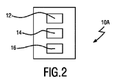
図2は、実施例による呼吸特徴を決定するための装置10Aを模式的に示す。
FIG. 2 schematically shows an
装置10Aは、対象により生成される呼吸ガスにおけるエアロゾル成分を検出するためのエアロゾル検出ユニット12と、該検出されたエアロゾルについて沈着率(DF)を測定するための沈着率測定ユニット14と、それぞれが異なる気道の気道幾何に対応する複数の所定の沈着率に、該測定された沈着率を関連付けることにより、呼吸特徴を決定するための、呼吸特徴決定ユニット16と、を有する。
図3は、他の実施例による呼吸特徴を決定するための装置10Bを模式的に示す。
FIG. 3 schematically illustrates an
装置10Bは、更に空気組成を監視するための監視ユニット20を有する点を除いて、図2に示された装置10Aと同様である。エアロゾル検出ユニット12、沈着率測定ユニット14、及び呼吸特徴決定ユニット16は、あわせて評価部18を形成する。
図4Aは、更なる実施例による装置10Cを模式的に示す。装置10Cは、第1のガス開口22及び第2のガス開口24を更に有する点を除いて、図2に示された装置10Aと同様である。ガス開口22、24はともに、エアロゾル検出ユニット12に結合され、ガスチャネルを形成する。ガス開口22、24は好適には、口及び/又は鼻のような患者の呼吸器官に対する気密な接続を実現する、マウスピース等である。更に、装置10Cは、例えば肺容量の決定のような、気道の評価のための時点及び/又は位置を設定するための、ガイドユニット26を有する。
FIG. 4A schematically shows an
好適には、エアロゾル検出ユニット12及び沈着率測定ユニット14は、環境の空気組成を監視することが可能である。図4Aに示されるように、空気(破線矢印により示される)は、第1のガス開口22を介して装置10Cに入り、エアロゾル検出ユニット12に到達することができる。入ってきた空気の検出に応じて、沈着率測定ユニット14は、例えば異なるサイズ(例えば直径)の粒子の濃度のような、空気の組成を測定することができる。
Preferably, the
斯かる空気組成の監視は、図4A primeに示される外部監視ユニット28により実行されても良く、このとき外部監視ユニット28は、無線リンク30を介して、更なる実施例による装置10C'に接続される。外部監視ユニット28は、空気品質検出装置、特に粒子状物質のサイズ及び/又は濃度を測定するための検出器であっても良い。
Such air composition monitoring may be performed by an
図4A、4A primeに示されたいずれの場合においても、空気組成の監視は、連続的に実行されても良いし、又は、患者により実行されるべき肺データ評価のためのスケジュールと同じであっても良い、所定の時間フレーム/スケジュールに従って起動されても良い。このことは、空気組成の監視が、自律的に、即ち所定の時間間隔のユーザ/患者入力なく、起動されても良いし、又は、例えば患者入力を介して患者により起動されても良いことを意味する。 In either case shown in FIGS. 4A, 4A prime, air composition monitoring may be performed continuously or on the same schedule for pulmonary data evaluation to be performed by the patient. may be activated according to a predetermined timeframe/schedule. This means that air composition monitoring may be initiated autonomously, i.e., without user/patient input for a predetermined time interval, or may be initiated by the patient, e.g., via patient input. means.
空気の組成の監視に基づいて、ガイドユニット26により、呼吸特徴の決定のための時点及び/又は位置が定義されることができる。特に、「ボーラス」の組成、即ち所定のエアロゾル粒子分布及び濃度により特徴付けられるものが、装置10C、10C'又は外部監視ユニット28により監視された場合、ガイドユニット26は、肺状態の評価を実行するようユーザに信号伝達しても良い。ボーラスの組成は好適にも肺状態評価には適しており、例えば家の中において、家の中での及び/又は局所的な(即ち特定の位置及び時間における)活動及び位置に依存して、監視されることができる。好適には、空気の組成が連続的に監視され、瞬間的に測定された粒子/サイズ分布に基づいて、ガイドユニット26は、新たな測定を実行するよう患者に信号伝達するための、最適な時点を決定する。
Based on the monitoring of the air composition, the guiding
好適な実施例においては、外部監視ユニット28は、該装置の近くに配置されても良い。このようにして、外部監視ユニット28の近くにおける装置10C'により、肺の評価が実行/準備/予測されることができる。特に、外部監視ユニット28により監視される空気の組成は、基本的には、装置10C'に存在するものと同じである。斯くして、最適な時点及び/又は位置が、信頼性高く定義されることができる。更に、肺の評価の結果は、監視される空気組成に基づいて、信頼性高く予測されることができる。
In a preferred embodiment, an
代替としては、肺の状態の評価を実行するための最適な時間は、予め設定された時間間隔/スケジュールに基づいて、又は患者の入力に基づいて、定義されても良い。 Alternatively, the optimal time to perform the lung condition assessment may be defined based on preset time intervals/schedules or based on patient input.
図4Bは、対象32の吸気フェーズの間に動作させられる図4A、4A primeに示された装置10C、10C'を模式的に示す。
FIG. 4B schematically shows the
図4Bに示されるように、患者である対象32は、装置10C、10C'を通して吸入することにより呼吸ガスを生成する。このようにして、周囲からの空気は、第2のガス開口24を介して装置10C、10C'に入り、エアロゾル検出ユニット12に到達し、その後に、第1のガス開口22を介して装置10C、10C'から出て、最後に患者32の気道34に入る。図4においては、気道34は模式的にのみ示され、現実の気道の種々の気道の実際の形状又は位置を反映していない。エアロゾル検出ユニット12は、呼吸ガスにおけるエアロゾル成分を検出し、それにより、沈着率測定ユニット14が、該検出されたエアロゾルの粒子サイズ分布及び/又は濃度を測定することができる。このことは、患者32の吸入フェーズ全体の間に実行されても良い。
As shown in FIG. 4B, the patient subject 32 produces respiratory gas by inhaling through the
図4Cは、対象の呼気フェーズの間に動作させられる図4A、4A primeに示された装置10C、10C'を模式的に示す。
Figure 4C schematically shows the
図4Cに示されるように、呼吸ガスは、第1のガス開口22を介して装置10C、10C'に吐き出すことにより、患者32の気道34により生成される。吐き出された呼吸ガスは、エアロゾル検出ユニット12に到達し、ここで該吐き出されたガスからエアロゾルが検出されることができる。エアロゾルの検出の際、沈着率測定ユニット14は、粒子サイズ分布及び/又は濃度を測定することができる。このことは、患者32の呼気フェーズ全体の間に実行されても良い。
Breathing gas is generated by the
対象32における吸入フェーズ及び後続する呼気フェーズの間に測定された粒子サイズ分布に基づいて、呼吸サイクル後に気道34に保持され沈着したエアロゾルの割合が導出され、呼吸サイクルの吸入フェーズ及び後続する呼気フェーズにおいて検出されたエアロゾルについての沈着率を導く。このことは、沈着率測定ユニット14により実行される。
Based on the particle size distribution measured during the inhalation and subsequent exhalation phases in the subject 32, the fraction of aerosol retained and deposited in the
測定された沈着率に基づいて、呼吸特徴決定ユニット16は、それぞれが人間の気道の異なる気道の気道幾何に対応する複数の所定の沈着率に、該測定された沈着率を関連付けることにより、呼吸特徴を決定することができる。
Based on the measured deposition rates,
好適には、該関連付けるステップは、該測定された沈着率を、複数の所定の沈着率の(合計)又は所定の沈着率の副関数として表現することにより実行され、該所定の沈着率又はその副関数はそれぞれ、対応する重み因子(例えば多項式、指数関数及び/又は対数関数)により乗算される。更に好適には、該所定の沈着率に基づいて、該測定された沈着率を表す関数において、複数の重み因子から、呼吸特徴が導出される。例えば、人間の気道の1つ以上の特定の気道幾何に対応する重み因子が、呼吸特徴について特徴的な所定の閾値と比較される。代替として、又はこれに加えて、それぞれが人間の気道の特定の気道幾何に対応する少なくとも2つの重み因子の間の比が決定され、呼吸特徴について特徴的な所定の比と比較される。 Preferably, the correlating step is performed by expressing the measured deposition rate as a (sum) of a plurality of predetermined deposition rates or a subfunction of a predetermined deposition rate, wherein the predetermined deposition rate or its Each subfunction is multiplied by a corresponding weighting factor (eg, polynomial, exponential and/or logarithmic function). More preferably, based on said predetermined deposition rate, respiratory features are derived from a plurality of weighting factors in a function representing said measured deposition rate. For example, weighting factors corresponding to one or more particular airway geometries of the human airway are compared to predetermined thresholds characteristic of respiratory characteristics. Alternatively or additionally, a ratio between at least two weighting factors each corresponding to a particular airway geometry of the human airway is determined and compared to a predetermined ratio characteristic of respiratory characteristics.
測定された沈着率を所定の沈着率の関数として表現することに加えて、呼吸特徴決定ユニットは、測定された沈着率を、測定された曲線の形状及び/又は値に関する1つ以上の所定の沈着率と比較しても良い。代替として、又はこれに加えて、呼吸特徴決定ユニット16は、測定された沈着率と1つ以上の所定の沈着率との間の相関関数を計算しても良い。
In addition to expressing the measured deposition rate as a function of the predetermined deposition rate, the respiratory characterization unit also expresses the measured deposition rate as one or more predetermined values of the shape and/or value of the measured curve. It may be compared with the deposition rate. Alternatively or additionally,
図5Aは、更なる実施例による装置10Dを模式的に示す。
FIG. 5A schematically shows a
装置10Dは、第1のガス開口22及び第2のガス開口24並びにガイドユニット26を更に有する点を除いて、図3に示された装置10Bと同様である。このようにして、図5Aに示された実施例は、図5Aに示された装置10Dの監視ユニット20が、評価部18とともに装置10Dに一体化される一方、図4A primeにおける監視ユニット28は、外部のユニット(例えばスマートな空気清浄器、エアコンディショナー等)である点を除いて、図4A primeに示された装置と同様である。
装置の上述した実施例10A、10B、10C、10C'、10Dは、好適には携帯型/装着可能装置であり、特に装着可能な肺評価装置である。図4A primeに示された装置10C'の場合には、装置10C'は好適には、「スレーブ」として機能する外部監視ユニット28からデータが送信されることができる、「マスタ」として機能する。
The above-described embodiments of the
一体化された監視ユニット20による環境の空気の組成の監視に応じて、装置10Dのガイドユニット26は、以上に説明された装置10C、10C'を用いる場合と同様に、患者32の気道34を評価するための時間及び/又は位置を定義する。図5Bに示されるように、装置10Dは、図4Bとともに説明された場合と同様に、患者32の吸入フェーズの間に動作させられる。
In response to monitoring of the ambient air composition by the integrated
図5Cに示されるように、装置10Dは、図4Cとともに説明された場合と同様に、患者32の呼気フェーズの間に動作させられる。それ故、装置10Dは、患者32の呼吸ガスにおけるエアロゾルの検出に基づいて呼吸特徴を決定し、該検出されたエアロゾルの沈着率を測定することが可能である。呼吸特徴は、それぞれが異なる気道幾何、即ち人間の気道のマークされる部分/部位に対応する、複数の所定の沈着率に、測定された沈着率を関連付けることにより、決定されることができる。
As shown in FIG. 5C,
好適には、上述した実施例のガイドユニット26は、特定の時間、好適には上述したガイドユニット26により定義された最適な時間に、肺の評価を実行するよう、患者に信号伝達するための、ユーザインタフェース(例えばディスプレイ、オーディオ信号要素、振動信号要素、光信号要素、・・・等)を有する。該ユーザインタフェースは、肺評価が実行されるべき特定の場所(例えばリビング、台所、寝室、バルコニー等)に患者32をガイドするよう提供される。更に、ガイドユニット26は最初に、調理が行なわれている台所へと患者32をガイドし、深い吸入を行うよう患者32に信号伝達するよう構成されても良い。続いて、ガイドユニット26は、近くにリビングに入るよう患者32をガイドし、息を吐くよう患者32に信号伝達しても良い。加えて、ガイドユニット26のユーザインタフェースは更に、リビングにおいて1つ以上の呼吸サイクルを実行するよう患者32に信号伝達しても良い。
Preferably, the
図6は、はめ込み図に示された3つの異なる屋内イベントに対応する、エアロゾルのサイズ分布に対してプロットされた、沈着率の例を模式的に示す。 FIG. 6 schematically shows examples of deposition rates plotted against aerosol size distributions corresponding to three different indoor events shown in the inset.
はめ込み図のグラフに示されるように、「PM2.5」粒子状物質の濃度が、3つのイベントE1、E2及びE3について測定されており、イベントE1及びE3は、2つの異なる調理イベントであり、イベントE2は、背景レベルにおける濃度に対応する。図6における主グラフは、それぞれ3つのイベントE1乃至E3について測定されたエアロゾルのサイズ分布M1、M2及びM3を示す。該サイズ分布は、粒子サイズ(ここではμmの単位)に依存する検出されたエアロゾル粒子の濃度として定義され、特定の粒子サイズにおいて測定された濃度値は、全ての検出されたエアロゾル粒子の総濃度に対してスケーリングされている。 As shown in the inset graph, "PM2.5" particulate matter concentration was measured for three events E1, E2 and E3, where events E1 and E3 are two different cooking events; Event E2 corresponds to density at the background level. The main graph in FIG. 6 shows the measured aerosol size distributions M1, M2 and M3 for three events E1 to E3, respectively. The size distribution is defined as the concentration of detected aerosol particles depending on the particle size (here in units of μm), the concentration value measured at a particular particle size being the total concentration of all detected aerosol particles is scaled with respect to
以下、本発明による実施例のいずれによっても用いられ得る、本発明による、エアロゾルのサイズ分布から呼吸パラメータを算出するための一般的な方法が説明される。特に、該一般的な方法は、吸入された及び吐き出されたエアロゾルについて測定されたサイズ分布から、肺の深さを算出するために用いられる。 In the following, a general method for calculating respiratory parameters from an aerosol size distribution according to the invention, which can be used by any of the embodiments according to the invention, is described. In particular, the general method is used to calculate lung depth from measured size distributions for inhaled and exhaled aerosols.
少なくとも2つの特定の粒子直径dについて測定された吸入された及び吐き出されたエアロゾル濃度(Ci、Ce)に基づいて、沈着率(DF)が式(1)に従って定義されることができる。
DF(d)=1-Ce(d)/Ci(d) (1)
Based on measured inhaled and exhaled aerosol concentrations (C i , C e ) for at least two specific particle diameters d, the deposition rate (DF) can be defined according to equation (1).
DF(d)=1−C e (d)/C i (d) (1)
特定の気道幾何(例えば鼻腔、気管、肺胞等)についてのエアロゾル直径の関数とひての沈着率DFが、決定されることができる。実際には、吸入された空気体積は、気道及び肺の多くの異なる部分を通るため、単一の呼吸サイクルから測定される沈着率は、異なる気道幾何/タイプ(即ち気道の特定の気道幾何に対応する重み因子によりそれぞれが乗算された所定の沈着率の合計)に対応する沈着率の加重平均を表す。このことは、総沈着率DFTotalが、例えば気管気管支(上気道)及び肺(より小さな/より低い気道)の沈着率曲線のような、所定の沈着率の加重合計であることを意味する。 A function of aerosol diameter and deposition rate DF for a particular airway geometry (eg, nasal cavity, trachea, alveoli, etc.) can be determined. In practice, the inhaled air volume passes through many different parts of the airways and lungs, so the deposition rate measured from a single respiratory cycle may vary depending on different airway geometries/types (i.e., the specific airway geometry of the airway). represents the weighted average of the deposition rates corresponding to the sum of the predetermined deposition rates each multiplied by the corresponding weighting factor. This means that the total deposition rate DF Total is a weighted sum of predetermined deposition rates, such as the tracheobronchial (upper airways) and lung (smaller/lower airways) deposition rate curves.
重み因子はそれぞれ、対応する気道のタイプ内に含まれる吸入された体積の割合により決定される。例えば、COPD患者は典型的に浅い呼吸を持つため、吸入された体積のうちの小さな割合が、最も小さな気道に到達することとなり、大きな割合は、上気道よりも更には到達しないこととなる。このことは、小さな気道に到達する吸入された体積の割合は、上気道に到達する吸入された体積の割合よりも小さいことを意味する。それ故、健康な人物の総沈着率DFTotal(d)に比べて、COPD患者の総沈着率曲線DFTotal(d)は、気管気管支沈着率曲線がより多くを占めることとなる。沈着率DF(d)は、各気道タイプ/幾何について予め決定されることができるため、DFTotal(d)が例えば直径値のような十分な数のエアロゾルのサイズにおいて測定される場合、特定の気道幾何に対応する重み因子が算出されることができる。呼吸の深さ及び肺容量のような呼吸特徴は、算出された重み因子から推測されることができる。エアロゾルのサイズの数は、超微粒子サイズ範囲(即ち<200nm)から、例えば100μm乃至200μmのような花粉のような大きな生物エアロゾルを表す大きな粒子サイズに及ぶ。少なくとも2つから、100個のサイズクラス(ビン)まで亘り得る。 Each weighting factor is determined by the fraction of the inhaled volume contained within the corresponding airway type. For example, since COPD patients typically have shallow breathing, a small percentage of the inhaled volume will reach the smallest airways and a large percentage will reach no more than the upper airways. This means that the fraction of the inspired volume reaching the small airways is less than the fraction of the inspired volume reaching the upper airways. Therefore, the total deposition rate curve DF Total (d) for COPD patients is dominated by the tracheobronchial deposition rate curve, compared to the total deposition rate DF Total (d) for healthy individuals. Since the deposition rate DF(d) can be predetermined for each airway type/geometry, if DF Total (d) is measured over a sufficient number of aerosol sizes, e.g. A weighting factor corresponding to the airway geometry can be calculated. Respiratory characteristics such as respiration depth and lung volume can be inferred from the calculated weighting factors. The number of aerosol sizes ranges from the ultrafine particle size range (ie <200 nm) to large particle sizes representing large biological aerosols such as pollen, eg, 100 μm to 200 μm. It can range from at least 2 to 100 size classes (bins).
一般性を損なうことなく、以上の手法は、人間又は動物の気道において見られる多くの異なる気道タイプを含むよう、拡張され高度化されても良い。 Without loss of generality, the above techniques may be extended and refined to include many different airway types found in human or animal airways.
図7は、例えば図2乃至5Cに示された、上述の実施例の1つを用いた、肺活量の決定のための手順の流れの方式を模式的に示す。 FIG. 7 schematically illustrates a process flow scheme for determination of vital capacity using one of the above-described embodiments, such as those shown in FIGS. 2-5C.
該手順の流れの方式は、3つのループ、即ち「サンプル入力制御ループ」(ループI)、「測定ループ」(ループII)、及び「患者制御ループ」(ループIII)を含む。ループIの間、患者の環境におけるエアロゾルを検出するため、エアロゾルセンサ36により空気が感知される。ループIIにおいて、患者による空気の吸入の際に、同じ環境の空気が、別のエアロゾルセンサ38により感知されても良い。このことは、患者による吸入が、最初にエアロゾルセンサ38に到達し、その後に患者に到達する、呼吸ガスを生成することを意味する。エアロゾルセンサ38は、該呼吸ガスに含まれるエアロゾルを検出する。続いて、患者が呼吸ガスを吐き出すと、該吐き出されたガスがエアロゾルセンサ38により感知される。
The procedure flow scheme includes three loops: a "sample input control loop" (loop I), a "measurement loop" (loop II), and a "patient control loop" (loop III). During Loop I, air is sensed by the
検出されたエアロゾルの量及び/又はサイズ分布は、それぞれのループにおいてエアロゾルセンサ36、38により測定されることができ、ループIIの場合、患者の吸気及び呼気の間である。好適には、例えばループI、IIの両方の実行の間の時間間隔の間に空気組成の起こり得る変化を監視/検出するため、第1のエアロゾルセンサ36の測定結果は、第2のエアロゾルセンサ38に供給されても良い。代替として、又はこれに加えて、第1のエアロゾルセンサ36の測定結果は、第2のエアロゾルセンサ38を用いた肺容量評価を実行する最適な時点を定義するために用いられても良い。
The amount and/or size distribution of the detected aerosol can be measured by the
ループIIの間、吸入されたエアロゾル粒子及び吐き出されたエアロゾル粒子の量、体積及び/又は濃度に基づいて、検出されたエアロゾルの沈着率を測定するため、エアロゾルセンサ38が用いられる。次いで、人間の気道の異なる気道幾何にそれぞれが対応する複数の所定の沈着率、好適には患者自身により予め決定された沈着率に、測定された沈着率を関連付けることにより、肺容量が決定される。
During Loop II, an
ループIIIの間、例えば患者の呼吸速度を例えば呼吸パターン入力ユニット42に出力する呼吸速度センサ40のような呼吸補助機器により、患者により生成された呼吸ガスが感知される。好適には、呼吸速度センサ40は、患者の呼吸を補助するため、呼吸パターン入力ユニット42から1つ以上の呼吸パターン入力を受けとる。このことは、患者の気道にかけられる気道陽圧、特に持続的気道陽圧を生成する、特に持続的気道陽圧(CPAP)装置のような気道陽圧(PAP)装置を用いて、実行されても良い。
During Loop III, the respiratory gases produced by the patient are sensed, for example by a respiratory assistance device such as a
好適な実施例においては、ループIIにおける肺状態の測定は、望ましい処置効果を達成するために必要な圧力を最小化しつつ、例えばCPAP治療におけるような、CPAP装置の呼吸補助を最適化するために用いられても良い。例えば、例えば肺容量のような呼吸特徴は、装置10A乃至Dのような、上述した実施例のいずれかによる装置によって監視される。決定された肺容量にCPAP圧力をリンクさせる患者固有の関係が確立され得る。特に、望ましい肺容量が予め設定されても良く、この場合、測定された肺容量が、例えば図1Aに示されたパラメータの1つ以上の値のような、該予め設定された肺容量と比較される。制御ループIIにおけるCPAP装置によりかけられるCPAP圧力のレベルは段階的に低下させられても良く、この場合、各圧力低下段階の後に、再び肺容量が測定され、予め設定された肺容量と比較される。この手順は、予め定義された閾値を超える量だけ、測定された肺容量が予め設定された肺容量と異なるまで、実行されても良い。このようにして、例えば肺の処置の間に、望ましい肺容量を依然として達成しつつ、患者の呼吸を補助するために用いられることができる、最小のCPAP圧力レベルを決定することができる。
In a preferred embodiment, the measurement of lung status in Loop II is used to optimize respiratory support of a CPAP machine, such as in CPAP therapy, while minimizing the pressure required to achieve the desired therapeutic effect. May be used. For example, a respiratory characteristic, such as lung volume, may be monitored by a device according to any of the embodiments described above, such as
好適には、例えば患者とエアロゾルセンサ38との間に、及び/又は呼吸速度センサ40に接続された、流量センサ44が利用される。流量センサ44は、流量、特に患者により生成された呼吸ガスの体積及び/又は速度を測定するよう構成される。測定された流量値は、例えば検出されたエアロゾルに基づいて肺容量を決定するときに流量値を考慮に入れるため、エアロゾルセンサ38に供給されても良い。代替として、又はこれに加えて、測定された流量値は、例えば呼吸パターン入力/患者にかけられるCPAP圧力を最適化するため、呼吸速度センサ40に供給されても良い。
Preferably, a
代替として、又はこれに加えて、ループIIのエアロゾルセンサ38は、特に図2乃至5Cに示された上述の実施例10A乃至Dの1つのような、呼吸特徴を決定するための装置に含まれる。このようにして、装置10A乃至D及び呼吸補助機器、例えば呼吸速度センサ40及び好適には呼吸パターン入力ユニット42、更に好適にはCPAP装置は、呼吸ガスに基づいて、特に生物のような対象の呼吸特徴を決定するためのシステムを形成する。
Alternatively, or in addition, a Loop
図8及び9は、肺活量の評価のための他の手順を模式的に示す。 Figures 8 and 9 schematically show another procedure for assessment of vital capacity.
図8Aに示されるように、エアロゾルセンサ、及び好適には流量センサを有するセンサ46は、呼気フェーズ(実線矢印により示される)により後続される吸気フェーズ(破線矢印により示される)の間に動作させられる。吸気フェーズの間、空気は、エアロゾルを検出し該検出された吸入されたエアロゾル(センサ46により検出される同じエアロゾルが後に患者によって吸入されるという意味において)の量、体積、濃度及び/又はサイズ分布を測定するセンサ46を通して、患者によって吸入される。呼気フェーズの間、空気は、ここでもまた該検出された吐き出されたエアロゾルの量、体積、濃度及び/又はサイズ分布を測定するセンサ46へと、患者により吐き出される。それ故、吸気フェーズと呼気フェーズとの間の測定値(例えば量、体積、濃度及び/又はサイズ分布)の比が算出及び/又は監視されることができ、ことによると変化率に帰着する。センサ46は例えば、図1乃至5Cに示された装置10A乃至Dの一部である。
As shown in FIG. 8A, the aerosol sensor, and preferably
吸気及び/又は呼気は、空気組成が以前に関しされている家等における選択された領域において、実行されても良い。特に、呼吸サイクルは、監視された空気が高い濃度のエアロゾル粒子を含む時点において実行されても良い。 Inhalation and/or exhalation may be performed in selected areas, such as a home, where the air composition has been previously determined. In particular, a respiratory cycle may be performed at times when the monitored air contains high concentrations of aerosol particles.
図8は、吸入フェーズ(破線曲線として示されたIP)について、粒子サイズの関数として、粒子の数を示す。更に、粒子サイズに依存する粒子の数が、それぞれ呼気フェーズの間に、COPD不安定患者(Ci)、COPD安定患者(Cs)及びCOPDを持たない人物(N)について、プロットされている。異なるタイプの人物(即ちCOPD不安定、COPD安定及びCOPDなし)の間で、COPD不安定患者は、最も低い肺容量及び最も浅い呼吸を持つことが予測される。COPD安定患者は、より高い肺容量及び浅い呼吸を持つことが予測される。COPDのない患者は、最も高い肺容量及び最も深い呼吸を持つことが予測される。その結果、沈着した呼吸ガスの粒子濃度は、COPD不安定患者について最低となることが予測され、次いでCOPD安定患者、次いでCOPDのない患者と続くことが予測される。従って、吐き出された粒子濃度の粒子濃度は、COPD不安定患者について最高となり、次いでCOPD安定患者、次いでCOPDのない患者と続く。このことは、図8のグラフに示されている。測定される粒子濃度(即ち体積当たりの粒子の数)が高いほど、粒子の沈着率が低く、従って肺容量が低く、その逆も成り立つ。 FIG. 8 shows the number of particles as a function of particle size for the inhalation phase (IP shown as dashed curve). Furthermore, the number of particles as a function of particle size is plotted for unstable COPD patients (C i ), COPD stable patients (C s ) and individuals without COPD (N) during the exhalation phase, respectively. . Among the different types of persons (ie, COPD unstable, COPD stable, and no COPD), COPD unstable patients are expected to have the lowest lung volumes and shallowest breathing. COPD stable patients are expected to have higher lung volumes and shallow breathing. Patients without COPD are expected to have the highest lung volumes and the deepest breaths. As a result, deposited respiratory gas particle concentrations are expected to be lowest for COPD unstable patients, followed by COPD stable patients, and then COPD-free patients. Therefore, the exhaled particle concentration particle concentration is highest for COPD unstable patients, followed by COPD stable patients, and then COPD-free patients. This is illustrated in the graph of FIG. The higher the measured particle concentration (ie number of particles per volume), the lower the rate of particle deposition and hence the lower the lung volume and vice versa.
図8Bにおける曲線IP、Ci、Cs及びNは、現実の測定結果を反映するものとして理解されるべきではなく、技術的及び臨床的な洞察を含む仮説に基づく推定に基づくものである。それ故、図8A乃至8Bは、サイズ分布変化の(仮説に基づく)評価を示している。 Curves IP, C i , C s and N in FIG. 8B should not be understood as reflecting actual measurements, but are based on hypothetical estimates involving technical and clinical insights. Figures 8A-8B therefore show a (hypothetical) assessment of the size distribution changes.
同様の測定が、図9Aに模式的に示されるように実行される。特に、破線の矢印で示されるような、センサ46を通して患者により空気が吸入される第1の吸気フェーズが、患者により実行される。続いて、それぞれが呼気フェーズにより後続される吸気フェーズから成る1つ以上の呼吸サイクルが、患者により実行され、実線の矢印に示されるように、空気が空気清浄器48を通して吸入され、センサ46に吐き出される。センサ46は、エアロゾルを検出し、検出された吸入された及び吐き出されたエアロゾルの量、体積、濃度及び/又はサイズ分布を測定する。図8Aと同様に、吸気フェーズと呼気フェーズとの間の測定値(例えば量、体積、濃度及び/又はサイズ分布)の比が算出及び/又は監視されることができ、ことによると変化率に帰着する。
A similar measurement is performed as shown schematically in FIG. 9A. In particular, a first inspiratory phase is performed by the patient in which air is inhaled by the patient through
空気清浄器48を用いることにより、第1の吸気フェーズの間に吸入された空気よりも低い濃度のエアロゾルを含む、第1の吸気フェーズに続く後続する呼吸サイクルの間に吸入され吐き出される空気が、清浄化される。空気清浄器48は、センサ46に物理的に接続されていても良く、それによれば、これらが同じ空気組成を持つ領域に配置される。代替として、又はこれに加えて、空気清浄化は、例えば空気が既に以前に清浄化されている家の一部において、実行されても良い。
By using the
図9Bは、図8Bに示された場合と同様に、それぞれ呼気フェーズの間の、COPD不安定患者(Ci)、COPD安定患者(Cs)及びCOPDを持たない人物(N)について、粒子サイズの関数として粒子の数を示す。図9Bのサイズ分布の測定は、例えば各清浄化期間の後に1度のように、空気の清浄化の間に実行されても良い。それ故、図9A及び9Bは、汚れた空気(第1の吸気フェーズにおいて吸入された)からの粒子の除去の効果を示す。 FIG. 9B shows, as shown in FIG. 8B , particle Shows the number of particles as a function of size. Measurements of the size distribution of FIG. 9B may be performed during cleaning of the air, eg, once after each cleaning period. Figures 9A and 9B therefore show the effect of removing particles from dirty air (inhaled in the first inspiration phase).
空気を清浄化することにより、肺に保持される空気に存在する粒子の量/濃度は、徐々に減少し、データは、肺状態の種々の評価(例えば肺容量、肺体積、吸気の深さ、患者の呼気容量、等)を実行するために用いられる。特に、以上の手法は、図1に示されるような、COPDのような疾患における呼吸状態の評価において非常に重要なパラメータである、患者の機能的残気量(FRC)を評価するために調整される。患者が正常な空気(即ち粒子を含まないか少量の粒子しか含まない空気)を吸入する場合、吐き出される呼吸ガスにおけるエアロゾルの沈着率は、肺に保持されるエアロゾルのインジケータとして機能し得る。これは肺の残気量に関連する。当該情報は、患者が清浄な空気のみの吸入及び後続するエアロゾルへの呼気を実行するときに、抽出されても良い。測定されたエアロゾル濃度及び好適には更にサイズ範囲が次いで、残気量を推定するために用いられる。好適には、粒子濃度がゼロに近いか又は閾値よりも低い状態である、エアロゾル濃度が「清浄な」状態の値/分布に到達するまで、吐き出されたガスのエアロゾル濃度の時間的な解析が必要とされ得る。 By purifying the air, the amount/concentration of particles present in the air retained in the lungs is gradually reduced, and the data are used in various assessments of lung status (e.g., lung volume, lung volume, depth of inspiration). , patient expiratory volume, etc.). In particular, the above techniques are tailored to assess a patient's functional residual capacity (FRC), a very important parameter in the assessment of respiratory status in diseases such as COPD, as shown in FIG. be done. When a patient inhales normal air (i.e., air with no or few particles), the rate of aerosol deposition in exhaled respiratory gas can serve as an indicator of aerosol retention in the lungs. This is related to lung residual capacity. Such information may be extracted when the patient performs a clean air-only inhalation followed by exhalation into the aerosol. The measured aerosol concentration and preferably also the size range are then used to estimate residual volume. Preferably, the temporal analysis of the aerosol concentration of the exhaled gas is performed until the aerosol concentration reaches a "clean" state value/distribution, in which the particle concentration is close to zero or below a threshold value. can be required.
本発明は、COPD、喘息及び肺気腫のような、呼吸疾患の管理における短期的な評価を支援することが可能な、信頼性の高い、外来の肺機能検査を提供する。斯かる検査は、継続的な監視、処置のパーソナライズ(例えば医薬の時間及び量)、及び増悪の早期の警告を提供するための呼吸状態における感染の早期検出のような、種々の用途のために用いられることができ、患者又は医師が、適時的な態様で治療を調節することを可能とする。更に、該検査は、吸気手法に関するフィードバックを患者に提供することができ、その結果、呼吸薬の吸入を最適化するために用いられ得る。 The present invention provides a reliable, ambulatory pulmonary function test that can support short-term assessment in the management of respiratory diseases such as COPD, asthma and emphysema. Such tests are useful for a variety of applications, such as continuous monitoring, personalization of treatment (e.g., time and amount of medication), and early detection of infection in respiratory conditions to provide early warning of exacerbation. can be used, allowing the patient or physician to adjust treatment in a timely manner. In addition, the test can provide feedback to the patient regarding inhalation technique, and thus can be used to optimize inhalation of respiratory medication.
本発明は図面及び以上の記述において説明され記載されたが、斯かる説明及び記載は説明するもの又は例示的なものであって、限定するものではないとみなされるべきであり、本発明は開示された実施例に限定されるものではない。図面、説明及び添付される請求項を読むことにより、請求される本発明を実施化する当業者によって、開示された実施例に対する他の変形が理解され実行され得る。 While the invention has been illustrated and described in the drawings and foregoing description, such description and description are to be considered illustrative or exemplary and not restrictive, and the invention is disclosed. It is not intended to be limited to the examples given. Other variations to the disclosed embodiments can be understood and effected by those skilled in the art who practice the claimed invention, upon reading the drawings, the description and the appended claims.
請求項において、「有する(comprising)」なる語は他の要素又はステップを除外するものではなく、「1つの(a又はan)」なる不定冠詞は複数を除外するものではない。単一の要素又はその他のユニットが、請求項に列記された幾つかのアイテムの機能を実行しても良い。特定の手段が相互に異なる従属請求項に列挙されているという単なる事実は、これら手段の組み合わせが有利に利用されることができないことを示すものではない。 In the claims, the word "comprising" does not exclude other elements or steps, and the indefinite article "a" or "an" does not exclude a plurality. A single element or other unit may fulfill the functions of several items recited in the claims. The mere fact that certain measures are recited in mutually different dependent claims does not indicate that a combination of these measures cannot be used to advantage.
コンピュータプログラムは、他のハードウェアと共に又は他のハードウェアの一部として供給される光記憶媒体又は固体媒体のような適切な媒体上で保存/配布されても良いが、インターネット又はその他の有線若しくは無線通信システムを介してのような、他の形態で配布されても良い。 The computer program may be stored/distributed on any suitable medium, such as optical storage media or solid-state media supplied with or as part of other hardware, Internet or other wired or It may also be distributed in other forms, such as via a wireless communication system.
請求項におけるいずれの参照記号も、請求の範囲を限定するものとして解釈されるべきではない。 Any reference signs in the claims shall not be construed as limiting the scope of the claims.
Claims (12)
前記呼吸ガスに含まれるエアロゾルを検出するための、エアロゾル検出ユニットと、
前記検出されたエアロゾルについて沈着率を測定するための、沈着率測定ユニットであって、前記沈着率は、吸入されたエアロゾルの総量に対する、前記対象内に沈着したエアロゾルの割合を示す、前記沈着率測定ユニットは、前記検出されたエアロゾルの複数の粒子サイズについて粒子濃度を測定するよう構成される、及び前記沈着率は、各々が前記複数の粒子サイズのうちの1つの粒子サイズに対応している複数の値を有する、沈着率測定ユニットと、
前記測定された沈着率を、それぞれが気道の種々の気道幾何に対応している複数の所定の沈着率と比較することにより、前記呼吸特徴を決定するための、呼吸特徴決定ユニットであり、各気道幾何は、前記気道の異なる部分又は部位に対応している、呼吸特徴決定ユニットと
を有する装置。 1. An apparatus for determining respiratory characteristics of a subject based on respiratory gases produced by the subject during inspiration and expiration , comprising:
an aerosol detection unit for detecting aerosols contained in the respiratory gas;
a deposition rate measurement unit for measuring a deposition rate for the detected aerosol, wherein the deposition rate indicates the proportion of the aerosol deposited in the subject relative to the total amount of inhaled aerosol; The measurement unit is configured to measure particle concentrations for a plurality of particle sizes of the detected aerosol, and the deposition rates each correspond to one particle size of the plurality of particle sizes. a deposition rate measurement unit having a plurality of values;
a respiratory characteristic determination unit for determining the respiratory characteristic by comparing the measured deposition rate with a plurality of predetermined deposition rates, each corresponding to different airway geometries of airways; and a respiratory characterization unit, wherein airway geometry corresponds to different parts or regions of said airway.
前記対象の呼吸を補助するための呼吸補助機器と、
前記呼吸補助機器により補助される前記対象の呼吸特徴を決定するための、請求項1に記載の装置と、
を有するシステム。 1. A system for determining respiratory characteristics of a subject based on respiratory gases produced by the subject during inspiration and expiration , comprising:
a respiratory assistance device for assisting breathing of the subject;
2. An apparatus according to claim 1 for determining respiratory characteristics of said subject assisted by said respiratory assistance device;
A system with
Applications Claiming Priority (3)
| Application Number | Priority Date | Filing Date | Title |
|---|---|---|---|
| EP15188799 | 2015-10-07 | ||
| EP15188799.9 | 2015-10-07 | ||
| PCT/EP2016/072622 WO2017060097A1 (en) | 2015-10-07 | 2016-09-23 | Device, system and method for determining a respiratory feature of a subject based on a breathing gas |
Publications (3)
| Publication Number | Publication Date |
|---|---|
| JP2018535714A JP2018535714A (en) | 2018-12-06 |
| JP2018535714A5 JP2018535714A5 (en) | 2019-08-15 |
| JP7190351B2 true JP7190351B2 (en) | 2022-12-15 |
Family
ID=54292630
Family Applications (1)
| Application Number | Title | Priority Date | Filing Date |
|---|---|---|---|
| JP2018516147A Active JP7190351B2 (en) | 2015-10-07 | 2016-09-23 | Apparatus, system and method for determining respiratory characteristics of a subject based on respiratory gases |
Country Status (5)
| Country | Link |
|---|---|
| US (1) | US20180296124A1 (en) |
| EP (1) | EP3359035B1 (en) |
| JP (1) | JP7190351B2 (en) |
| CN (1) | CN108366756B (en) |
| WO (1) | WO2017060097A1 (en) |
Families Citing this family (5)
| Publication number | Priority date | Publication date | Assignee | Title |
|---|---|---|---|---|
| US10881818B2 (en) | 2016-07-08 | 2021-01-05 | Trudell Medical International | Smart oscillating positive expiratory pressure device |
| CA3036631A1 (en) | 2016-12-09 | 2018-06-14 | Trudell Medical International | Smart nebulizer |
| AU2019205865A1 (en) | 2018-01-04 | 2020-07-16 | Trudell Medical International Inc. | Smart oscillating positive expiratory pressure device |
| JP7020125B2 (en) * | 2018-01-12 | 2022-02-16 | コニカミノルタ株式会社 | Dynamic image analyzer and program |
| CA3152072A1 (en) | 2019-08-27 | 2021-03-04 | Trudell Medical International | Smart oscillating positive expiratory pressure device |
Citations (5)
| Publication number | Priority date | Publication date | Assignee | Title |
|---|---|---|---|---|
| US20050066968A1 (en) | 2000-08-01 | 2005-03-31 | Shofner Frederick M. | Generation, delivery, measurement and control of aerosol boli for diagnostics and treatments of the respiratory/pulmonary tract of a patient |
| JP2007503912A (en) | 2003-09-02 | 2007-03-01 | レスピラトリー マネージメント テクノロジー | Apparatus and method for aerosol transport |
| JP2007533411A (en) | 2004-04-20 | 2007-11-22 | エアロゲン,インコーポレイティド | Aerosol delivery device, method and composition for a pressurized respiratory system |
| JP2011515657A (en) | 2008-02-15 | 2011-05-19 | ジョージア テック リサーチ コーポレイション | System and method for providing environmental monitoring |
| WO2014076250A1 (en) | 2012-11-15 | 2014-05-22 | Löndahl Jakob | Device and method for pulmonary function measurement |
Family Cites Families (17)
| Publication number | Priority date | Publication date | Assignee | Title |
|---|---|---|---|---|
| US4241739A (en) * | 1978-11-13 | 1980-12-30 | C. R. Bard, Inc. | Volume calculator for incentive spirometer |
| DE2938856A1 (en) * | 1979-09-26 | 1981-04-16 | Gesellschaft für Strahlen- und Umweltforschung mbH, 8000 München | METHOD AND DEVICE FOR DETERMINING THE DEPOSITION OF PARTICLES IN THE BREATHING TRACT AND / OR FOR CHECKING THE FUNCTION OF THE BREATHING AREA |
| US4517987A (en) * | 1982-04-02 | 1985-05-21 | Sackner Marvin A | Method for measurement of nonradioisotopic aerosol retained in the lungs with re-breathing |
| JP2823792B2 (en) * | 1993-11-25 | 1998-11-11 | 帝人株式会社 | Inhalation exposed particle measuring device |
| CN100551458C (en) * | 2001-06-05 | 2009-10-21 | 艾利斯达医药品公司 | The aerocolloidal forming device that is used for anapnotherapy |
| AU2003901042A0 (en) * | 2003-03-07 | 2003-03-20 | Resmed Limited | Back-up rate for a ventilator |
| EP1827227A4 (en) * | 2004-12-23 | 2009-04-08 | Resmed Ltd | Method for detecting and disciminatng breathing patterns from respiratory signals |
| WO2008114172A2 (en) * | 2007-03-16 | 2008-09-25 | Philips Intellectual Property & Standards Gmbh | Method and device for evaluation of spirographic and gas exchange data |
| US8555873B2 (en) * | 2007-06-28 | 2013-10-15 | Inhalation Sciences Sweden Ab | Exposure system |
| EP2022525B1 (en) * | 2007-08-02 | 2012-12-26 | Activaero GmbH | Device and system for targeting aerosolized particles to a specific area of the lungs |
| CA2713012A1 (en) * | 2008-01-25 | 2009-07-30 | Salter Labs | Respiratory therapy system including a nasal cannula assembly |
| CN104939831B (en) * | 2010-09-09 | 2018-11-09 | 森撒部伊斯公司 | The system and method and application thereof of sample are collected from the expiratory air of subject |
| WO2014045217A1 (en) * | 2012-09-21 | 2014-03-27 | Koninklijke Philips N.V. | Motion robust vital signal monitoring |
| AU2014217808B2 (en) * | 2013-02-14 | 2018-05-17 | Fluidda Respi | Method for determining a respiratory condition based on functional respiratory imaging |
| CN103202696B (en) * | 2013-03-06 | 2014-12-17 | 王胜昱 | Modeling method for detecting lung diseases through expiratory air on basis of aerosol |
| CN104181084B (en) * | 2014-08-26 | 2017-05-24 | 清华大学 | Aerosol sensor |
| CN204260747U (en) * | 2014-10-24 | 2015-04-15 | 重庆大学 | A kind of monitoring of respiration and breath analysis system |
-
2016
- 2016-09-23 US US15/766,393 patent/US20180296124A1/en active Pending
- 2016-09-23 WO PCT/EP2016/072622 patent/WO2017060097A1/en active Application Filing
- 2016-09-23 CN CN201680071489.9A patent/CN108366756B/en active Active
- 2016-09-23 EP EP16775595.8A patent/EP3359035B1/en active Active
- 2016-09-23 JP JP2018516147A patent/JP7190351B2/en active Active
Patent Citations (5)
| Publication number | Priority date | Publication date | Assignee | Title |
|---|---|---|---|---|
| US20050066968A1 (en) | 2000-08-01 | 2005-03-31 | Shofner Frederick M. | Generation, delivery, measurement and control of aerosol boli for diagnostics and treatments of the respiratory/pulmonary tract of a patient |
| JP2007503912A (en) | 2003-09-02 | 2007-03-01 | レスピラトリー マネージメント テクノロジー | Apparatus and method for aerosol transport |
| JP2007533411A (en) | 2004-04-20 | 2007-11-22 | エアロゲン,インコーポレイティド | Aerosol delivery device, method and composition for a pressurized respiratory system |
| JP2011515657A (en) | 2008-02-15 | 2011-05-19 | ジョージア テック リサーチ コーポレイション | System and method for providing environmental monitoring |
| WO2014076250A1 (en) | 2012-11-15 | 2014-05-22 | Löndahl Jakob | Device and method for pulmonary function measurement |
Also Published As
| Publication number | Publication date |
|---|---|
| JP2018535714A (en) | 2018-12-06 |
| EP3359035B1 (en) | 2021-12-29 |
| EP3359035A1 (en) | 2018-08-15 |
| WO2017060097A1 (en) | 2017-04-13 |
| US20180296124A1 (en) | 2018-10-18 |
| CN108366756A (en) | 2018-08-03 |
| CN108366756B (en) | 2022-04-15 |
Similar Documents
| Publication | Publication Date | Title |
|---|---|---|
| JP6679649B2 (en) | Method and apparatus for detecting and treating respiratory insufficiency | |
| JP7190351B2 (en) | Apparatus, system and method for determining respiratory characteristics of a subject based on respiratory gases | |
| JP6564318B2 (en) | A system for evaluating real-time lung mechanics | |
| US20210402111A1 (en) | Inhalation monitoring system and method | |
| JP5053083B2 (en) | Method and apparatus for non-invasive prediction of positive end expiratory pressure (PEEPi) in a patient receiving ventilator support | |
| JP6404819B2 (en) | System and method for determining sleep stage | |
| US10165966B2 (en) | Methods and systems for monitoring resistance and work of breathing for ventilator-dependent patients | |
| JP5706893B2 (en) | Method and apparatus for determining discharged nitric oxide | |
| JP6200430B2 (en) | Method and apparatus for monitoring and controlling pressure assist devices | |
| CN110448301A (en) | Detect the insufficient equipment of respiratory air flow | |
| CN110049799A (en) | Method and system for driving pressure spontaneous ventilation | |
| CN108135493A (en) | For the abnormality detecting apparatus and method of mechanical parameter Estimation of ventilating | |
| JP7471437B2 (en) | Method and apparatus for determining and/or predicting sleep and respiratory behavior for airway pressure management - Patents.com | |
| JP2005516704A5 (en) | ||
| JP2005516704A (en) | Estimating flow-independent parameters based on expiratory curves of ventilated breathing | |
| WO2023092103A1 (en) | Systems and methods for breath detection | |
| CN108062976A (en) | Breathing equipment and the operation method for carrying the breathing equipment of definite coughing fit | |
| CA3118956C (en) | Method and apparatus for continuous management of airway pressure for detection and/or prediction of respiratory failure | |
| JP2021515614A (en) | Devices and methods for measuring respiratory flow | |
| JP2021508529A (en) | Expiratory flow rate limit detection by adjusting flow rate resistance | |
| EP4079221A1 (en) | System for identifying a condition of a user |
Legal Events
| Date | Code | Title | Description |
|---|---|---|---|
| A521 | Request for written amendment filed |
Free format text: JAPANESE INTERMEDIATE CODE: A523 Effective date: 20190708 |
|
| A621 | Written request for application examination |
Free format text: JAPANESE INTERMEDIATE CODE: A621 Effective date: 20190708 |
|
| A131 | Notification of reasons for refusal |
Free format text: JAPANESE INTERMEDIATE CODE: A131 Effective date: 20200728 |
|
| A601 | Written request for extension of time |
Free format text: JAPANESE INTERMEDIATE CODE: A601 Effective date: 20201023 |
|
| A521 | Request for written amendment filed |
Free format text: JAPANESE INTERMEDIATE CODE: A523 Effective date: 20210126 |
|
| A131 | Notification of reasons for refusal |
Free format text: JAPANESE INTERMEDIATE CODE: A131 Effective date: 20210701 |
|
| A601 | Written request for extension of time |
Free format text: JAPANESE INTERMEDIATE CODE: A601 Effective date: 20210924 |
|
| A521 | Request for written amendment filed |
Free format text: JAPANESE INTERMEDIATE CODE: A523 Effective date: 20211221 |
|
| A02 | Decision of refusal |
Free format text: JAPANESE INTERMEDIATE CODE: A02 Effective date: 20220519 |
|
| A521 | Request for written amendment filed |
Free format text: JAPANESE INTERMEDIATE CODE: A523 Effective date: 20220913 |
|
| C60 | Trial request (containing other claim documents, opposition documents) |
Free format text: JAPANESE INTERMEDIATE CODE: C60 Effective date: 20220913 |
|
| A911 | Transfer to examiner for re-examination before appeal (zenchi) |
Free format text: JAPANESE INTERMEDIATE CODE: A911 Effective date: 20220920 |
|
| C21 | Notice of transfer of a case for reconsideration by examiners before appeal proceedings |
Free format text: JAPANESE INTERMEDIATE CODE: C21 Effective date: 20220922 |
|
| TRDD | Decision of grant or rejection written | ||
| A01 | Written decision to grant a patent or to grant a registration (utility model) |
Free format text: JAPANESE INTERMEDIATE CODE: A01 Effective date: 20221129 |
|
| A61 | First payment of annual fees (during grant procedure) |
Free format text: JAPANESE INTERMEDIATE CODE: A61 Effective date: 20221205 |
|
| R150 | Certificate of patent or registration of utility model |
Ref document number: 7190351 Country of ref document: JP Free format text: JAPANESE INTERMEDIATE CODE: R150 |