JP6884211B2 - X-ray equipment with synthetic field of view - Google Patents
X-ray equipment with synthetic field of view Download PDFInfo
- Publication number
- JP6884211B2 JP6884211B2 JP2019531631A JP2019531631A JP6884211B2 JP 6884211 B2 JP6884211 B2 JP 6884211B2 JP 2019531631 A JP2019531631 A JP 2019531631A JP 2019531631 A JP2019531631 A JP 2019531631A JP 6884211 B2 JP6884211 B2 JP 6884211B2
- Authority
- JP
- Japan
- Prior art keywords
- ray
- ray image
- image data
- region
- processing unit
- Prior art date
- Legal status (The legal status is an assumption and is not a legal conclusion. Google has not performed a legal analysis and makes no representation as to the accuracy of the status listed.)
- Active
Links
- 230000000007 visual effect Effects 0.000 claims description 103
- 238000003384 imaging method Methods 0.000 claims description 75
- 238000012545 processing Methods 0.000 claims description 74
- 238000000034 method Methods 0.000 claims description 52
- 239000002131 composite material Substances 0.000 claims description 24
- 238000004590 computer program Methods 0.000 claims description 22
- 230000008878 coupling Effects 0.000 claims description 13
- 238000010168 coupling process Methods 0.000 claims description 13
- 238000005859 coupling reaction Methods 0.000 claims description 13
- 239000003550 marker Substances 0.000 claims description 9
- 238000004422 calculation algorithm Methods 0.000 claims description 8
- 238000013213 extrapolation Methods 0.000 claims description 4
- 210000004072 lung Anatomy 0.000 description 25
- 210000003484 anatomy Anatomy 0.000 description 19
- 230000003287 optical effect Effects 0.000 description 11
- 230000005855 radiation Effects 0.000 description 7
- 210000000038 chest Anatomy 0.000 description 6
- 238000013519 translation Methods 0.000 description 5
- 238000001514 detection method Methods 0.000 description 4
- 230000008859 change Effects 0.000 description 3
- 238000003745 diagnosis Methods 0.000 description 3
- 238000002601 radiography Methods 0.000 description 3
- FGUUSXIOTUKUDN-IBGZPJMESA-N C1(=CC=CC=C1)N1C2=C(NC([C@H](C1)NC=1OC(=NN=1)C1=CC=CC=C1)=O)C=CC=C2 Chemical compound C1(=CC=CC=C1)N1C2=C(NC([C@H](C1)NC=1OC(=NN=1)C1=CC=CC=C1)=O)C=CC=C2 FGUUSXIOTUKUDN-IBGZPJMESA-N 0.000 description 2
- 230000008901 benefit Effects 0.000 description 2
- 230000007547 defect Effects 0.000 description 2
- 230000001419 dependent effect Effects 0.000 description 2
- 201000010099 disease Diseases 0.000 description 2
- 208000037265 diseases, disorders, signs and symptoms Diseases 0.000 description 2
- 238000006073 displacement reaction Methods 0.000 description 2
- 230000006870 function Effects 0.000 description 2
- 238000004091 panning Methods 0.000 description 2
- 230000037361 pathway Effects 0.000 description 2
- 230000003936 working memory Effects 0.000 description 2
- 238000004846 x-ray emission Methods 0.000 description 2
- GNFTZDOKVXKIBK-UHFFFAOYSA-N 3-(2-methoxyethoxy)benzohydrazide Chemical compound COCCOC1=CC=CC(C(=O)NN)=C1 GNFTZDOKVXKIBK-UHFFFAOYSA-N 0.000 description 1
- 238000010521 absorption reaction Methods 0.000 description 1
- 230000001133 acceleration Effects 0.000 description 1
- 230000003213 activating effect Effects 0.000 description 1
- 238000007792 addition Methods 0.000 description 1
- 238000004458 analytical method Methods 0.000 description 1
- 238000013459 approach Methods 0.000 description 1
- 230000009286 beneficial effect Effects 0.000 description 1
- 238000004364 calculation method Methods 0.000 description 1
- 230000015556 catabolic process Effects 0.000 description 1
- 238000003759 clinical diagnosis Methods 0.000 description 1
- 238000010276 construction Methods 0.000 description 1
- 238000012937 correction Methods 0.000 description 1
- 238000005520 cutting process Methods 0.000 description 1
- 238000006731 degradation reaction Methods 0.000 description 1
- 238000010586 diagram Methods 0.000 description 1
- 238000011156 evaluation Methods 0.000 description 1
- 230000001678 irradiating effect Effects 0.000 description 1
- 239000000203 mixture Substances 0.000 description 1
- 238000012986 modification Methods 0.000 description 1
- 230000004048 modification Effects 0.000 description 1
- 238000005457 optimization Methods 0.000 description 1
- 210000000056 organ Anatomy 0.000 description 1
- 230000007170 pathology Effects 0.000 description 1
- 230000004962 physiological condition Effects 0.000 description 1
- 230000004044 response Effects 0.000 description 1
- 239000007787 solid Substances 0.000 description 1
- 238000010561 standard procedure Methods 0.000 description 1
- 238000003860 storage Methods 0.000 description 1
- 230000002195 synergetic effect Effects 0.000 description 1
- 238000012360 testing method Methods 0.000 description 1
- 239000002699 waste material Substances 0.000 description 1
Images


Classifications
-
- A—HUMAN NECESSITIES
- A61—MEDICAL OR VETERINARY SCIENCE; HYGIENE
- A61B—DIAGNOSIS; SURGERY; IDENTIFICATION
- A61B6/00—Apparatus or devices for radiation diagnosis; Apparatus or devices for radiation diagnosis combined with radiation therapy equipment
- A61B6/52—Devices using data or image processing specially adapted for radiation diagnosis
- A61B6/5211—Devices using data or image processing specially adapted for radiation diagnosis involving processing of medical diagnostic data
- A61B6/5229—Devices using data or image processing specially adapted for radiation diagnosis involving processing of medical diagnostic data combining image data of a patient, e.g. combining a functional image with an anatomical image
- A61B6/5235—Devices using data or image processing specially adapted for radiation diagnosis involving processing of medical diagnostic data combining image data of a patient, e.g. combining a functional image with an anatomical image combining images from the same or different ionising radiation imaging techniques, e.g. PET and CT
- A61B6/5241—Devices using data or image processing specially adapted for radiation diagnosis involving processing of medical diagnostic data combining image data of a patient, e.g. combining a functional image with an anatomical image combining images from the same or different ionising radiation imaging techniques, e.g. PET and CT combining overlapping images of the same imaging modality, e.g. by stitching
-
- A—HUMAN NECESSITIES
- A61—MEDICAL OR VETERINARY SCIENCE; HYGIENE
- A61B—DIAGNOSIS; SURGERY; IDENTIFICATION
- A61B6/00—Apparatus or devices for radiation diagnosis; Apparatus or devices for radiation diagnosis combined with radiation therapy equipment
- A61B6/54—Control of apparatus or devices for radiation diagnosis
- A61B6/545—Control of apparatus or devices for radiation diagnosis involving automatic set-up of acquisition parameters
-
- G—PHYSICS
- G06—COMPUTING; CALCULATING OR COUNTING
- G06T—IMAGE DATA PROCESSING OR GENERATION, IN GENERAL
- G06T7/00—Image analysis
- G06T7/0002—Inspection of images, e.g. flaw detection
- G06T7/0012—Biomedical image inspection
- G06T7/0014—Biomedical image inspection using an image reference approach
-
- A—HUMAN NECESSITIES
- A61—MEDICAL OR VETERINARY SCIENCE; HYGIENE
- A61B—DIAGNOSIS; SURGERY; IDENTIFICATION
- A61B6/00—Apparatus or devices for radiation diagnosis; Apparatus or devices for radiation diagnosis combined with radiation therapy equipment
- A61B6/06—Diaphragms
-
- A—HUMAN NECESSITIES
- A61—MEDICAL OR VETERINARY SCIENCE; HYGIENE
- A61B—DIAGNOSIS; SURGERY; IDENTIFICATION
- A61B6/00—Apparatus or devices for radiation diagnosis; Apparatus or devices for radiation diagnosis combined with radiation therapy equipment
- A61B6/50—Apparatus or devices for radiation diagnosis; Apparatus or devices for radiation diagnosis combined with radiation therapy equipment specially adapted for specific body parts; specially adapted for specific clinical applications
-
- G—PHYSICS
- G06—COMPUTING; CALCULATING OR COUNTING
- G06T—IMAGE DATA PROCESSING OR GENERATION, IN GENERAL
- G06T2207/00—Indexing scheme for image analysis or image enhancement
- G06T2207/10—Image acquisition modality
- G06T2207/10116—X-ray image
-
- G—PHYSICS
- G06—COMPUTING; CALCULATING OR COUNTING
- G06T—IMAGE DATA PROCESSING OR GENERATION, IN GENERAL
- G06T2207/00—Indexing scheme for image analysis or image enhancement
- G06T2207/20—Special algorithmic details
- G06T2207/20212—Image combination
- G06T2207/20221—Image fusion; Image merging
-
- G—PHYSICS
- G06—COMPUTING; CALCULATING OR COUNTING
- G06T—IMAGE DATA PROCESSING OR GENERATION, IN GENERAL
- G06T2207/00—Indexing scheme for image analysis or image enhancement
- G06T2207/30—Subject of image; Context of image processing
- G06T2207/30004—Biomedical image processing
- G06T2207/30061—Lung
-
- G—PHYSICS
- G06—COMPUTING; CALCULATING OR COUNTING
- G06T—IMAGE DATA PROCESSING OR GENERATION, IN GENERAL
- G06T2207/00—Indexing scheme for image analysis or image enhancement
- G06T2207/30—Subject of image; Context of image processing
- G06T2207/30168—Image quality inspection
Landscapes
- Engineering & Computer Science (AREA)
- Health & Medical Sciences (AREA)
- Life Sciences & Earth Sciences (AREA)
- Medical Informatics (AREA)
- General Health & Medical Sciences (AREA)
- Nuclear Medicine, Radiotherapy & Molecular Imaging (AREA)
- Radiology & Medical Imaging (AREA)
- Physics & Mathematics (AREA)
- Computer Vision & Pattern Recognition (AREA)
- Molecular Biology (AREA)
- Biomedical Technology (AREA)
- Veterinary Medicine (AREA)
- Biophysics (AREA)
- High Energy & Nuclear Physics (AREA)
- Optics & Photonics (AREA)
- Pathology (AREA)
- Public Health (AREA)
- Heart & Thoracic Surgery (AREA)
- Animal Behavior & Ethology (AREA)
- Surgery (AREA)
- Quality & Reliability (AREA)
- Theoretical Computer Science (AREA)
- General Physics & Mathematics (AREA)
- Apparatus For Radiation Diagnosis (AREA)
Description
本発明は一般に、合成視野を有するX線画像を取得するための装置に関する。X線撮像システム、合成視野を有するX線画像を取得するための方法、コンピュータプログラム要素、及びコンピュータ可読媒体も説明される。 The present invention generally relates to an apparatus for acquiring an X-ray image having a composite field of view. An X-ray imaging system, a method for acquiring an X-ray image with a composite field of view, computer program elements, and a computer-readable medium are also described.
肺の放射線写真などの放射線照射の視野(FOV:field of view)を構成することは、X線撮像装置を操作するために訓練された放射線技師の注意を必要とする簡単ではない作業である。照射前に誤って設定された視野は、その後に完全なX線画像が撮影されなければならないことを意味し、その理由は、重要な解剖学的細部が最初の画像から欠落している可能性があるためである。患者は呼び戻されて再度訪れる必要さえあり得る。そのような事態は望ましくなく、放射線科における非効率性につながり、患者に吸収されるX線量が増加する。 Constructing a field of view (FOV) of radiation, such as a radiograph of the lungs, is a non-trivial task that requires the attention of a trained radiologist to operate the radiograph. A misplaced field of view before irradiation means that a complete x-ray image must be taken afterwards, because important anatomical details may be missing from the first image. Because there is. The patient may even need to be recalled and revisited. Such a situation is undesirable and leads to inefficiencies in the radiology department, increasing the amount of X-dose absorbed by the patient.
米国特許出願公開第2015/0228071号明細書では、X線撮像装置のコリメータを自動的又は半自動的に制御するための装置及び方法が説明されている。しかしながら、そのような方法はさらに改良することができる。 U.S. Patent Application Publication No. 2015/0228071 describes an apparatus and method for automatically or semi-automatically controlling a collimator of an X-ray imaging apparatus. However, such a method can be further improved.
そのため、第1の態様によれば、合成視野を有するX線画像を取得するための装置が提供される。この装置は、
− 処理ユニット
を備える。
Therefore, according to the first aspect, an apparatus for acquiring an X-ray image having a composite field of view is provided. This device
-Equipped with a processing unit.
処理ユニットは、X線撮像装置を使用して対象者の関心領域の一部の第1のX線画像データを取得するように構成され、第1のX線画像データは、調整可能な視野が初期視野状態に設定されたX線撮像装置から取得され、処理ユニットは、第1のX線画像データを解剖学的モデルと比較し、第1のX線画像データと解剖学的モデルとの比較に基づいて第1のX線画像データに隣接する境界誤り領域を規定し、関心領域における境界誤り領域の位置に基づいて更新視野状態を生成し、更新視野状態をX線撮像装置に送信し、更新視野状態に設定された場合にX線撮像装置を使用して対象者の関心領域の第2のX線画像データを取得し、第1のX線画像データ及び第2のX線画像データを結合して合成視野を有する関心領域の出力画像を取得するように構成される。 The processing unit is configured to use an X-ray imaging apparatus to acquire first X-ray image data of a part of the area of interest of the subject, and the first X-ray image data has an adjustable field of view. Acquired from an X-ray imager set in the initial visual field state, the processing unit compares the first X-ray image data with the anatomical model and compares the first X-ray image data with the anatomical model. A boundary error region adjacent to the first X-ray image data is defined based on the above, an updated visual field state is generated based on the position of the boundary error region in the region of interest, and the updated visual field state is transmitted to the X-ray imaging apparatus. When the update field state is set, the X-ray imaging device is used to acquire the second X-ray image data of the area of interest of the subject, and the first X-ray image data and the second X-ray image data are obtained. It is configured to combine to obtain an output image of the region of interest having a composite field of view.
したがって、第1のX線画像における取得欠損を識別するX線撮像装置を制御することが可能な装置が提供される。X線撮像装置の視野を調整して、関心領域の撮像を完了するさらなるX線画像を提供するために使用可能な設定が生成される。更新視野は、たとえば、初期視野よりも面積が小さい。この場合、患者はX線画像の再撮影時に、第1のX線画像の照射と比較してより少ない線量のX線放射線を受けることになる。そのような装置によって、患者に届く総X線量を減少させることが可能になる。 Therefore, an apparatus capable of controlling an X-ray imaging apparatus that identifies an acquisition defect in the first X-ray image is provided. Settings are generated that can be used to adjust the field of view of the X-ray imager to provide additional X-ray images that complete the imaging of the region of interest. The updated field of view, for example, has a smaller area than the initial field of view. In this case, the patient will receive a lower dose of X-ray radiation when the X-ray image is retaken compared to the irradiation of the first X-ray image. Such a device makes it possible to reduce the total X-dose reaching the patient.
任意選択で、第1の態様による装置では、解剖学的モデルは複数の解剖学的要素を表す確率的解剖学アトラスを含む。処理ユニットは、第1のX線画像の一部を確率的解剖学アトラス内の解剖学的要素と比較するようにさらに構成される。 Optionally, in the device according to the first aspect, the anatomical model comprises a stochastic anatomical atlas representing multiple anatomical elements. The processing unit is further configured to compare a portion of the first x-ray image with the anatomical elements within the stochastic anatomy atlas.
したがって、第1のX線画像における境界誤り領域の正確な印(impression)を導き出して、第2のX線画像を取得することが可能になる。 Therefore, it is possible to derive an accurate impression of the boundary error region in the first X-ray image and acquire the second X-ray image.
任意選択で、第1の態様による装置では、処理ユニットは、解剖学的モデル及び/又は確率的解剖学アトラスにおいて、第1のX線画像データから欠落している想定要素又は想定要素の一部を識別するようにさらに構成される。処理ユニットは、関心領域の一部からの解剖学的モデル及び/又は確率的解剖学アトラスの外挿に基づいて、X線画像データにおける境界誤り領域を規定するようにさらに構成される。 Optionally, in the apparatus according to the first aspect, the processing unit is a hypothetical element or part of the hypothetical element that is missing from the first X-ray image data in the anatomical model and / or the stochastic anatomy atlas. Is further configured to identify. The processing unit is further configured to define the boundary error region in the X-ray image data, based on the extrapolation of the anatomical model and / or the stochastic anatomical atlas from a portion of the region of interest.
したがって、正確な解剖学的データを使用して、第1のX線画像の視野における誤りの範囲が特定される。 Therefore, accurate anatomical data is used to identify the extent of error in the field of view of the first X-ray image.
任意選択で、処理ユニットが、境界誤り領域の特性に基づいて第1のX線画像データの画像完成指標を生成するようにさらに構成される、第1の態様の装置。第2のX線画像データは、画像完成指標が画像完成条件を上回った場合に処理ユニットによって取得される。 The device of the first aspect, optionally, the processing unit is further configured to generate an image completion index of the first X-ray image data based on the characteristics of the boundary error region. The second X-ray image data is acquired by the processing unit when the image completion index exceeds the image completion condition.
したがって、たとえば、解剖学的モデルと比較した後、第1のX線画像データ内の肺要素は95%完全であり、わずかな程度の肺の端部しか第1のX線画像データから欠落していないことが分かる。この場合、第2のX線画像は必要とされないので、患者の不要な余分なX線被曝が省かれる。 Thus, for example, after comparison with an anatomical model, the lung elements in the first X-ray image data are 95% complete and only a small amount of lung ends are missing from the first X-ray image data. You can see that it is not. In this case, the second X-ray image is not needed, thus eliminating unnecessary extra X-ray exposure of the patient.
任意選択で、処理ユニットが、画像縫合アルゴリズムを使用して第1のX線画像データ及び第2のX線画像データを結合するようにさらに構成される、第1の態様による装置が提供される。 Optionally, a device according to a first aspect is provided in which the processing unit is further configured to combine the first X-ray image data and the second X-ray image data using an image stitching algorithm. ..
したがって、画像内の第1のX線画像データ及び第2のX線画像データの間の画像の不連続性は、医療専門家には見えない。 Therefore, the discontinuity of the image between the first X-ray image data and the second X-ray image data in the image is invisible to the medical professional.
任意選択で、処理ユニットが、関心領域において第1のX線画像データに隣接する第2のX線画像データを提供するように更新視野状態を選択するよう構成される、第1の態様による装置が提供される。 A device according to a first aspect, optionally, the processing unit is configured to select an updated visual field state to provide a second X-ray image data adjacent to the first X-ray image data in the region of interest. Is provided.
したがって、更新視野状態は、第1のX線画像データの視野と重ならないので、極力小さい更新視野を提供する。このようにして、患者が不要なX線放射線に被曝することがさらに低減される。 Therefore, since the updated visual field state does not overlap with the visual field of the first X-ray image data, an updated visual field as small as possible is provided. In this way, the patient's exposure to unwanted X-ray radiation is further reduced.
任意選択で、処理ユニットが、関心領域の少なくとも一部にわたって第1のX線画像データと重なる第2のX線画像データを提供するように、更新視野状態を選択するよう構成される、第1の態様による装置が提供される。 A first configuration, optionally, the processing unit is configured to select an updated visual field state such that it provides a second X-ray image data that overlaps the first X-ray image data over at least a portion of the region of interest. The device according to the above embodiment is provided.
したがって、第2のX線画像データは、少なくともある程度、第1のX線画像データに既に存在している画像情報を含む。このようにして、臨床診断を形成するのにあまり関連性がない画像の領域において、第1のX線画像データ及び第2のX線画像データを結合することが可能になる。たとえば、重なる点は、特定の患者を撮像する場合には、主に肺葉の端部にある疾患の診断に関連性がない場合がある、患者の脊椎の下にある視野内のエリアを辿り得る。 Therefore, the second X-ray image data includes image information that already exists in the first X-ray image data, at least to some extent. In this way, it is possible to combine the first X-ray image data and the second X-ray image data in a region of the image that is less relevant to form a clinical diagnosis. For example, overlapping points can trace an area of vision beneath the patient's spine that, when imaging a particular patient, may not be relevant to the diagnosis of the disease, primarily at the edge of the lobe. ..
任意選択で、処理ユニットは、解剖学的モデル及び/又は確率的解剖学アトラスを使用して第1のX線画像データ内の画像結合領域を識別し、画像結合経路にさらに基づいて更新視野状態を生成するようにさらに構成され、第1のX線画像データ及び第2のX線画像データは画像結合領域に沿って結合される、第1の態様による装置が提供される。 Optionally, the processing unit uses an anatomical model and / or a probabilistic anatomical atlas to identify the image binding region in the first X-ray image data and further base the updated visual field state based on the image binding pathway. The device according to the first aspect is provided, which is further configured to generate the first X-ray image data and the second X-ray image data are combined along an image coupling region.
したがって、事前に提供される解剖学的情報を使用して、更新視野状態を規定することによって、より正確な画像結合領域が規定される。 Therefore, by using the anatomical information provided in advance to define the updated visual field state, a more accurate image coupling region is defined.
任意選択で、第1の態様による装置は、初期及び/又は更新視野状態に基づいて人工的な結合領域マーカを出力画像に付加するようにさらに構成される処理ユニットが設けられ、結合領域マーカは、出力画像において歪みの可能性のある領域をユーザに示すものである。 Optionally, the apparatus according to the first aspect is provided with a processing unit further configured to add an artificial coupling region marker to the output image based on the initial and / or updated visual field state, the coupling region marker , Shows the user the area of potential distortion in the output image.
したがって、エンドユーザは、合成出力画像内の領域が医療診断の形成に適していないと警告され得る。 Therefore, the end user may be warned that the area within the composite output image is not suitable for forming a medical diagnosis.
任意選択で、第1の態様の装置によれば、処理ユニットは、3Dカメラから対象者の3D光学画像データを受信するようにさらに構成され、初期及び/又は更新視野状態は、3D光学画像データにおける対象者の位置にさらに基づく。 Optionally, according to the apparatus of the first aspect, the processing unit is further configured to receive the subject's 3D optical image data from the 3D camera, and the initial and / or updated visual field state is the 3D optical image data. Further based on the subject's position in.
したがって、初期又は更新視野は、カメラなどによって捕捉された視野内の患者の位置を使用して規定される。これにより、初期視野並びに更新視野を正確に設定して、患者の不要なX線被曝をさらに低減することが可能になる。 Therefore, the initial or renewal field of view is defined using the patient's position within the field of view captured by a camera or the like. This makes it possible to accurately set the initial visual field and the renewal visual field to further reduce unnecessary X-ray exposure of the patient.
任意選択で、処理ユニットは、入力デバイスから再照射コマンドを受け取るようにさらに構成され、プロセッサは、再照射コマンドが入力デバイスから受け取られるまで第2のX線画像データを取得しないように構成される、第1の態様の装置が提供される。 Optionally, the processing unit is further configured to receive a re-irradiation command from the input device, and the processor is configured not to acquire the second X-ray image data until the re-irradiation command is received from the input device. , The device of the first aspect is provided.
したがって、この装置は、患者への2回目の照射を実行するX線源を作動させる前に、オペレータのコマンドを待機するように構成することができるので、この装置の安全性が高まる。 Therefore, the device can be configured to wait for an operator's command before activating the X-ray source to perform a second irradiation of the patient, thus increasing the safety of the device.
任意選択で、X線撮像装置は調整可能なコリメータを備え、初期視野状態は第1の調整可能なコリメータ設定を含み、処理ユニットは、処理ユニットを使用して第2の調整可能なコリメータ位置設定を生成することによって、更新視野状態を生成するように構成され、処理ユニットは、第2の調整可能なコリメータ位置設定をX線撮像装置の調整可能なコリメータに送信することによって、X線撮像装置の視野を調整するようにさらに構成される、第1の態様による装置が提供される。 Optionally, the X-ray imaging device is equipped with an adjustable collimator, the initial visual field state includes a first adjustable collimator setting, and the processing unit uses the processing unit to set a second adjustable collimator position. The processing unit is configured to generate an updated visual field state by generating an X-ray imaging device by transmitting a second adjustable collimator position setting to the adjustable collimator of the X-ray imaging device. A device according to a first aspect is provided that is further configured to adjust the field of view of the device.
したがって、X線コリメータ設定は、視野状態を変更するためにを使用される。 Therefore, the X-ray collimator setting is used to change the visual field state.
第2の態様によれば、X線撮像システムが提供される。 According to the second aspect, an X-ray imaging system is provided.
X線撮像システムは、
− 目標位置に向けて調整可能な視野を有するX線源と、
− 第1の態様又はその任意選択の実施形態のうちのいずれかに記載した合成視野を有するX線画像を取得するための装置と、
− X線源から放出されたX線を受けるために目標位置の背後に配置されるX線検出器と
を備える。
The X-ray imaging system is
− An X-ray source with an adjustable field of view towards the target position,
− An apparatus for acquiring an X-ray image having a composite field of view according to any one of the first aspect or an optional embodiment thereof, and an apparatus for acquiring an X-ray image.
-Equipped with an X-ray detector located behind the target position to receive the X-rays emitted from the X-ray source.
X線撮像システムは第1のX線画像データを装置に提供するように構成され、装置は目標位置の視野を調整可能にするために更新視野状態をX線源に提供するように構成され、X線撮像システムはX線画像を生成するための装置に第2のX線画像情報を提供するように構成され、装置は合成視野を有する関心領域の出力画像を提供するように構成される。したがって、X線撮像システムの視野は、第1のX線画像から放出された、撮像される患者の関心領域内の要素を撮影するように自動的に調整される。 The X-ray imaging system is configured to provide first X-ray image data to the device, which is configured to provide an updated field state to the X-ray source to allow the field of view at the target position to be adjustable. The X-ray imaging system is configured to provide a second X-ray image information to a device for generating an X-ray image, and the device is configured to provide an output image of a region of interest having a synthetic field of view. Therefore, the field of view of the X-ray imaging system is automatically adjusted to capture the elements in the imaged patient's region of interest emitted from the first X-ray image.
第3の態様によれば、合成視野を有するX線画像を取得するための方法が提供される。この方法は、
a)X線撮像装置を使用して対象者の関心領域の一部の第1のX線画像データを取得するステップであって、X線画像データは、調整可能な視野が初期視野状態に設定されたX線撮像装置を使用して取得される、取得するステップと、
b)X線画像データを解剖学的モデルと比較するステップと、
c)第1のX線画像データと解剖学的モデルとの比較に基づいて第1のX線画像データに隣接する境界誤り領域を規定するステップと、
d)関心領域における境界誤り領域の位置に基づいて更新視野状態を生成するステップと、
e)更新視野状態をX線撮像装置に送信するステップと、
f)送信された視野状態に設定された場合にX線撮像装置を使用して対象者の関心領域の第2のX線画像データを取得するステップと、
g)第1のX線画像データ及び第2のX線画像データを結合して合成視野を有する関心領域の出力画像を取得するステップと
を有する。
According to the third aspect, a method for acquiring an X-ray image having a composite field of view is provided. This method
a) A step of acquiring a first X-ray image data of a part of an area of interest of a subject using an X-ray image pickup apparatus, in which an adjustable field of view is set to an initial field state in the X-ray image data. The steps to be acquired and the steps to be acquired using the X-ray imaging device
b) Steps to compare X-ray image data with anatomical models,
c) A step of defining a boundary error region adjacent to the first X-ray image data based on a comparison between the first X-ray image data and an anatomical model.
d) A step of generating an updated visual field state based on the position of the boundary error region in the region of interest, and
e) The step of transmitting the updated visual field state to the X-ray imaging device, and
f) The step of acquiring the second X-ray image data of the area of interest of the subject using the X-ray imaging device when the transmitted visual field state is set, and
g) It has a step of combining the first X-ray image data and the second X-ray image data to acquire an output image of a region of interest having a composite field of view.
したがって、患者の関心領域内の全ての関連する要素を含むX線画像を生成するための方法であって、第1のX線内に既に包含されていた関連する要素全てを包含するように第2のX線の視野状態が設定された場合よりも少ない量のX線放射線被曝が生成される、方法が提供される。 Therefore, it is a method for generating an X-ray image containing all the related elements in the patient's area of interest, so as to include all the related elements already included in the first X-ray. A method is provided in which a smaller amount of X-ray radiation exposure is generated than if the X-ray visual field state of 2 was set.
第4の態様によれば、第1又は第2の態様に記載した装置及び/又はX線システムを制御するためのコンピュータプログラム要素であって、プロセッサ及び/又はシステムによって実行された場合に、第3の態様の方法を実行するように構成される、コンピュータプログラム要素が提供される。 According to a fourth aspect, a computer program element for controlling the apparatus and / or X-ray system according to the first or second aspect, when executed by a processor and / or system. Computer program elements are provided that are configured to perform the methods of the third aspect.
第5の態様によれば、第4の態様のコンピュータプログラム要素が記憶された、コンピュータ可読媒体が提供される。 According to the fifth aspect, a computer-readable medium in which the computer program elements of the fourth aspect are stored is provided.
以下の明細書において、「X線画像データ」という用語はピクセルアレイを含むデータ構造を指し、各ピクセルは、X線が患者の関心領域を横断した後に、特定のピクセルにおいて受けたX線の強度を表す。強度値は、2次元画像に組み立てられる場合、各ピクセル位置におけるX線吸収量の積分を表す可算性の画像を提供する。 In the following specification, the term "X-ray image data" refers to a data structure that includes a pixel array, where each pixel receives the intensity of the X-rays received at a particular pixel after the X-rays have traversed the patient's area of interest. Represents. The intensity value provides a countable image that represents the integral of the X-ray absorption at each pixel position when assembled into a two-dimensional image.
以下の明細書において、「X線源」という用語は、回転陽極X線管などを含むX線源を指す。これは、撮像される患者の関心領域に向けてX線放射線を放出する。X線放射線は関心領域において患者を横断し、X線検出器によって受け取られ、X線検出器もまたX線撮像システムの一部と見なされ得る。X線撮像装置は自動設定可能な視野パラメータ、たとえば、調節可能なコリメータ配置、調節可能なパン又はチルトサーボモータ、調節可能な高さ又はx−y座標設定などを含む。加えて、X線検出器は垂直又は水平に平行移動される。 In the following specification, the term "X-ray source" refers to an X-ray source including a rotating anode X-ray tube and the like. It emits x-ray radiation towards the area of interest of the patient being imaged. X-ray radiation traverses the patient in the area of interest and is received by the X-ray detector, which can also be considered part of the X-ray imaging system. The X-ray imaging device includes auto-configurable field parameters such as adjustable collimator placement, adjustable pan or tilt servomotors, adjustable height or xy coordinate settings, and the like. In addition, the X-ray detector is translated vertically or horizontally.
以下の明細書において、「視野」という用語は、典型的な照射中にX線撮像装置が撮影する「関心領域」の一部を指す。視野は、一般に、X線検出器及び/又は患者からX線撮像装置までの距離と、X線検出器の開口部の大きさとによって規定される。視野は、X線撮像装置をx−z平面内で移動させることによって、関心領域を横切って平行移動される。視野はX線撮像装置をパン又はチルトすることによっても変更される。視野は、X線源の1つ又は複数のコリメータシャッタを調整することによって、切り落とし又は拡大される。そのため、X線撮像装置の視野を調整するには多くのやり方があることは理解されよう。 In the following specification, the term "field of view" refers to a portion of the "region of interest" imaged by an X-ray imaging device during typical irradiation. The field of view is generally defined by the distance from the X-ray detector and / or the patient to the X-ray imager and the size of the opening in the X-ray detector. The field of view is translated across the region of interest by moving the X-ray imager in the x-z plane. The field of view is also changed by panning or tilting the X-ray imaging device. The field of view is truncated or magnified by adjusting one or more collimator shutters of the X-ray source. Therefore, it will be understood that there are many ways to adjust the field of view of an X-ray imaging device.
以下の明細書において、「解剖学的モデル」という用語は、典型的には記憶されコンピュータなどの処理手段上で実行されるデータ構造を指す。解剖学的モデルは、患者の一般的な解剖学的特徴の位置及び形状を定義する情報を含む。典型的な解剖学的モデルは一般的な患者の体の一部を定義する。解剖学的モデルは、たとえば、肺、肋骨、脊椎などの構造の表現と、特定の解剖学的要素が特定の位置に存在する尤度とを含む。解剖学的モデルは、解剖学的要素が臓器の画像の不完全な部分から識別可能になるように設計される。 In the following specification, the term "anatomical model" typically refers to a data structure that is stored and executed on a processing means such as a computer. The anatomical model contains information that defines the location and shape of the patient's general anatomical features. A typical anatomical model defines a typical patient's body part. Anatomical models include, for example, representations of structures such as lungs, ribs, and spine, and the likelihood that a particular anatomical element is in a particular location. The anatomical model is designed so that the anatomical elements can be identified from the incomplete part of the image of the organ.
「境界誤り領域」という用語は、X線画像データにおける解剖学的でない領域を定義したものである。視野の視準が不正確である場合、境界誤り領域が切り落とされているように見えることは理解されよう。たとえば、肺葉の最も左手側又は最も右手側の部分がX線画像データから欠落している場合がある。しかしながら、パン又はチルト設定が不十分であるために視野が不正確に設定された場合、境界誤り領域は、存在はしているが「台形形状」などに歪んだ解剖学的要素によって規定され得る。換言すれば、X線画像データの境界誤り領域は、患者の解剖学的構造の忠実又は正確な再現ではない画像のエリアを規定する。 The term "boundary error region" defines a non-anatomical region in X-ray image data. It will be understood that if the visual field is inaccurate, the border error area will appear to be truncated. For example, the leftmost or rightmost part of the lung lobe may be missing from the X-ray image data. However, if the field of view is set incorrectly due to inadequate pan or tilt settings, the boundary error region can be defined by anatomical elements that are present but distorted into a "trapezoidal shape" or the like. .. In other words, the border error region of the X-ray image data defines an area of the image that is not a faithful or accurate reproduction of the patient's anatomy.
このように、視野に関する問題について初期X線画像をロバストにチェックし、視野を自動的に再設定して、初期照射内で照射されていない解剖学的領域の2回目の取得を可能にし、それによって画像結合方法を使用して完全な視野を完成させるための自動化された方法を提供することが本明細書の基本概念である。このようにして、誤った初期照射を利用して、より小さい視野を有する2回目の照射を完了することによって、2枚の完全な画像を受諾するために必要なはずの放射線被曝よりも、患者の放射線被曝を低減することが可能になる。 In this way, the initial X-ray image is robustly checked for field of view issues and the field of view is automatically reconfigured to allow a second acquisition of the unirradiated anatomical area within the initial irradiation. It is a basic concept herein to provide an automated method for completing a complete field of view using the image combination method. In this way, by utilizing the false initial irradiation to complete the second irradiation with a smaller field of view, the patient than the radiation exposure that would be required to accept the two complete images. It becomes possible to reduce the radiation exposure of.
本出願では前後方向のビューに基づく肺撮像に関する概念を論じているが、本明細書で論じる技術は、初期X線画像が欠落している境界要素を有するいかなる場合でも、X線撮影において広い適用性を有することは理解されよう。 Although the present application discusses the concept of lung imaging based on anterior-posterior views, the techniques discussed herein have wide application in radiography in any case with border elements where the initial radiograph is missing. It will be understood that it has sex.
以下の図面を参照して例示的な実施形態を説明する。 An exemplary embodiment will be described with reference to the following drawings.
胸部X線撮影は最も一般的に行われている臨床画像検査であり、胸部の解剖学的構造の多くの疾患の検出及び診断に重要な役割を果たしている。画質は広範囲の特定の個々の要因、たとえば、適切な解剖学的構造の視野内への包含、背景信号に対する関心のある構造のコントラスト、並びにX線取得機器に対する患者の胸郭の位置調整のいくつかの態様などに依存する。 Chest radiography is the most commonly performed clinical imaging test and plays an important role in the detection and diagnosis of many diseases of the anatomy of the chest. Image quality is a wide range of specific individual factors, such as inclusion of the appropriate anatomical structure in the field of view, contrast of the structure of interest to the background signal, and some of the patient's thorax alignment with respect to the x-ray acquisition device. Depends on the mode and the like.
照射の視野(FOV)を設定する作業は、通常、放射線技師によって行われる。まず、患者はX線検出器の前の関心領域内に配置される。次いで、X線機器のチューブヘッド内から発せられ、X線放射パターンの範囲と一致する可視光を使用して、患者の体の上に視野を確立する。たとえば、最初にチューブヘッドの高さが変更され、次いで検出器を含む「ブッキー」の高さが変更され、最後にコリメータの開口部が調整される。 The task of setting the field of view (FOV) of irradiation is usually performed by a radiologist. First, the patient is placed within the area of interest in front of the X-ray detector. Visible light emitted from within the tube head of the X-ray machine and consistent with the range of the X-ray emission pattern is then used to establish a field of view over the patient's body. For example, the height of the tube head is changed first, then the height of the "booky" containing the detector is changed, and finally the opening of the collimator is adjusted.
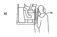
図1に、臨床用X線撮影において最も一般的な投影配置の1つである胸部の前後方向のビューで患者が検査される様子を示す。図1では、X線源は、X線ビームが胸部の後方から入り、胸部の前方を出た後、X線検出器に到達するように配置されている。図1a)に、オペレータがX線源の高さ10を調整する様子を示す。図1b)に、オペレータがX線検出器を含む「ブッキー」の高さ12を調整する様子を示す。図1c)に、特定の視準状態における視野を表す可視光視準パターン14の投影を示す。視準パターンは、X線照射中のX線照射のパターンに対応する。典型的には、視準パターン14は、X線源制御機器18のアイテム上のコントロール16a、16bを使用して精細化される。視準パターンを変化させると、システムの視野が拡大又は縮小される。
FIG. 1 shows a patient being examined in an anterior-posterior view of the chest, which is one of the most common projection arrangements in clinical radiography. In FIG. 1, the X-ray source is arranged so that the X-ray beam enters from the rear of the chest, exits the front of the chest, and then reaches the X-ray detector. FIG. 1a) shows how the operator adjusts the
臨床的な慣例では、画質を決定する側面は、システムオペレータの技術にある程度依存する。事前定義された最低品質基準を保証して、潜在的な誤りの一般的な原因の最小化を可能にすることを目的として標準的な操作手順が医療機関によって確立されているが、視野誤りを引き起こす機会自体は依然として存在する。 In clinical practice, the aspects that determine image quality depend to some extent on the skill of the system operator. Standard operating procedures have been established by medical institutions with the aim of ensuring a predefined minimum quality standard and allowing the minimization of common causes of potential errors, but with visual field errors. The opportunity itself still exists.
X線機器の視野の設定はオペレータの仕事の重要な一部であるが、誤りを起こしやすい仕事でもある。一般的な状況は「切り取り(cut−off)」である。これは、関心のある解剖学的構造の一部が意図せずX線画像内に含まれないという、視野設定における誤りを意味する。切り取りは最も一般的な誤りの1つであり、典型的にはX線画像全体の再撮影が必要になる。 Setting the field of view of an X-ray machine is an important part of the operator's job, but it is also a error-prone task. A common situation is "cut-off". This means an error in the field setting that some of the anatomical structures of interest are unintentionally not included in the X-ray image. Crop is one of the most common mistakes and typically requires recapture of the entire x-ray image.
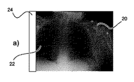
図2に、切り取りの2つの例を示す。 FIG. 2 shows two examples of cutting.
図2a)に、左肺葉22の一部が意図せず切り落とされている前後方向のX線視野20を示す。したがって、境界誤り領域は、一例では、実線の棒状エリア24によって規定されると考えることができる。
FIG. 2a) shows an X-ray
図2b)を参照すると、視野26は他の前後方向のX線画像を示している。この場合、視野誤りは、肺画像の頂部の視準が不十分である結果として生じており、肺葉の頂部を含むはずの境界誤り領域28が見えている。
With reference to FIG. 2b), the field of
通常、完全な前後方向の肺画像を取得するためには、図2a)及び図2b)の照射は破棄される必要がある。いずれの場合も、まったく新しい照射が行われる必要がある。これはX線設備の時間の無駄であり、1回の撮影で画像が正しく撮影された場合と比較して、患者は必要な線量の少なくとも2倍を受けることになる。したがって、視野誤りに応じてそのような余分な線量を低減するための手法が必要になる。 Generally, in order to obtain a complete anterior-posterior lung image, the irradiation in FIGS. 2a) and 2b) needs to be discarded. In either case, a whole new irradiation needs to be done. This is a waste of time in the X-ray equipment, and the patient will receive at least twice the required dose compared to when the image was taken correctly in a single shot. Therefore, there is a need for techniques to reduce such extra doses in response to visual field errors.
図3に、X線撮像システム35を示す。X線撮像システムは、制御装置30、検出アセンブリ31、及びX線撮像源アセンブリ34を備える。患者は典型的には、X線源アセンブリ34と検出アセンブリ31との間の関心領域36内に立つ。
FIG. 3 shows an
図3には、前後の配置の患者を示している。X線撮像源アセンブリ34は、X線撮像スイートの天井レール40から吊り下げられるように構成される天井設置型ドリー38を備える。
FIG. 3 shows the patients in the anterior-posterior arrangement. The X-ray
X線撮像源アセンブリ34は典型的には天井レール40上に支持され、X線源を患者に向かう又は患者から離れるように平行移動させることができる(YS)。X線撮像源は支持部材42によってレールから吊り下げられており、支持部材42は上下方向(床に向かう及び床から離れる方向、ZS軸)に移動可能であって、さらに支持部材の軸の周りに回転可能である(βS)。
The X-ray
X線撮像源44は支持部材42から吊り下げられており、関心領域36に向けてX線放射線を放出するように構成されるX線源46と、関心領域36に向けて可視光を放出するように構成される可視光源48とを含む筐体を備える。
The
X線源46は、たとえば、回転陽極X線管である。可視光源48は典型的には白熱灯又はLEDライトとして設けられる。関心領域36と、X線源46及び可視光源48との間にあるのは視準要素Cである。
The
視準要素Cは、X線ビームの外縁を成形するように構成される。単純なコリメータは、X線撮像装置の開口部を徐々に覆うように構成されるシャッタを備える。より高機能な視準要素は、互いに直交する平面関係に配置された2つのシャッタを備え、視野の大きさを変化させることが可能になる。より複雑な視準構成は、3辺、4辺、又は「虹彩」コリメータシャッタ構成を含む。 The collimation element C is configured to form the outer edge of the X-ray beam. A simple collimator comprises a shutter configured to gradually cover the opening of the X-ray imaging apparatus. The more sophisticated collimation elements include two shutters arranged in a plane relationship orthogonal to each other, which makes it possible to change the size of the field of view. More complex collimator configurations include three-sided, four-sided, or "iris" collimator shutter configurations.
そのため、視準要素Cによって、X線放射パターン及び可視光放射パターンの両方の視野の外側範囲を規定しやすくなる。X線撮像源が角度αSだけチルト可能であることにも留意されたい。X線撮像構成全体も、図面に示すようにXS次元を通る横方向に(図3では紙面に出入りする方向に)平行移動される。 Therefore, the collimation element C makes it easy to define the outer range of the visual field of both the X-ray emission pattern and the visible light emission pattern. It should also be noted that the X-ray imaging source can be tilted by the angle αS. The entire X-ray imaging configuration is also translated laterally through the XS dimension (in FIG. 3, in the direction of entering and exiting the paper) as shown in the drawing.
このように、図3に示すX線撮像システムでは、関心領域の視野は、1つ又は複数のコリメータ要素Cを操作することによって調整される。代替的に又は追加的に、X線撮像ヘッドをYS方向に前後させることによって、視野の大きさを変更することが可能である。視野はZS次元及びXS次元を調整することによって平行移動される。最後に、視野は、X線撮像構成をパン又はチルトすることによって(BS、AS)再形成される。 Thus, in the X-ray imaging system shown in FIG. 3, the field of view of the region of interest is adjusted by manipulating one or more collimator elements C. Alternatively or additionally, the size of the field of view can be changed by moving the X-ray imaging head back and forth in the YS direction. The field of view is translated by adjusting the ZS and XS dimensions. Finally, the field of view is reshaped (BS, AS) by panning or tilting the radiographic configuration.
前述のコリメータ設定及び位置設定を含む視野状態は、可視光源48を使用して患者を照明しながら、オペレータによって選択される。関心領域がうまくカバーされると、X線源が作動され、検出器要素50が関心領域36に関するX線情報を受け取る。これはデータリンク52を介して制御装置30に送信される。オペレータは照射を受けたX線画像をモニタ54などの出力デバイス上で見る。
The visual field conditions, including the collimator and position settings described above, are selected by the operator while illuminating the patient using the visible
以上、従来のX線撮像システムを説明した。視野は、たとえば、自動サーボモータを使用してコリメータ又はX線撮像源の位置を設定することによって制御されてもよいことは理解されよう。 The conventional X-ray imaging system has been described above. It will be appreciated that the field of view may be controlled, for example, by using an automatic servomotor to position the collimator or X-ray source.
図4に、第1の態様による合成視野を有するX線画像を取得するための装置を示す。この装置は、
− 処理ユニット32
を備える。
FIG. 4 shows an apparatus for acquiring an X-ray image having a composite field of view according to the first aspect. This device
−
To be equipped.
処理ユニット32は、X線撮像装置34を使用して対象者の関心領域の一部の第1のX線画像データを取得するように構成され、第1のX線画像データは、調整可能な視野が初期視野状態に設定されたX線撮像装置から取得され、処理ユニット32は、第1のX線画像データを解剖学的モデルと比較し、第1のX線画像データと解剖学的モデルとの比較に基づいて第1のX線画像データに隣接する境界誤り領域を規定し、関心領域における境界誤り領域の位置に基づいて更新視野状態を生成し、更新視野状態をX線撮像装置に送信し、更新視野状態に設定された場合にX線撮像装置を使用して対象者の関心領域の第2のX線画像データを取得し、第1のX線画像データ及び第2のX線画像データを結合して合成視野を有する関心領域の出力画像を取得するように構成される。
The
この装置は、業界標準の遠隔操作プロトコルを使用して既存のX線撮像システムに接続可能な独立型モジュールとして実装されてもよい。換言すれば、この装置は、一実施形態では、既存のシステムに後付け可能であってもよい。代替的には、この装置は、既存のX線システムの制御ソフトウェアを更新することによって提供されてもよい。代替的には、この装置は、新しいX線システム内のモジュールとして提供されてもよい。 The device may be implemented as a stand-alone module that can be connected to existing X-ray imaging systems using industry standard remote control protocols. In other words, the device may, in one embodiment, be retrofitted to an existing system. Alternatively, the device may be provided by updating the control software of an existing X-ray system. Alternatively, the device may be provided as a module within a new X-ray system.
この装置は、X線システムのX線検出器、たとえば、図3のシステムのX線検出器50から入力画像信号を受信するように構成される。
This device is configured to receive an input image signal from an X-ray detector in an X-ray system, such as the
この装置は、処理ユニット32がX線撮像システムの視野を制御又は調整することを可能にするための出力インターフェースをさらに備える。たとえば、装置30は、チルト(αS)、パン(βS)、高さ(ZS)、側方変位(XS)、前後変位(YS)、又はX線検出器の高さ(ZD)を調整するように動作するサーボモータを制御するための出力インターフェースを備える。
The apparatus further comprises an output interface for allowing the
任意選択で、境界誤り領域は、解剖学的モデルの解剖学的要素を第1のX線画像データ内の一致する要素に当てはめて、第1のX線画像データ内の境界を識別することによって生成される。境界誤り領域は、モデルから当てはめられた解剖学的要素が及ぶ、第1のX線画像の視野外の領域(「境界誤りエリア」)から識別可能である。任意選択で、境界誤り領域は、境界誤りエリアの範囲付近に「境界ボックス」をはめ込むことによって生成することができる。当然ながら、前述の議論は「境界誤り領域」を生成するための1つの手法であるが、他の多くの手法を適用することができる。 Optionally, the boundary error region is by fitting the anatomical elements of the anatomical model to the matching elements in the first X-ray image data and identifying the boundaries in the first X-ray image data. Will be generated. The boundary error region can be identified from the out-of-field region (“boundary error area”) of the first X-ray image, which extends from the model-fitted anatomical elements. Optionally, the boundary error area can be generated by fitting a "boundary box" near the range of the boundary error area. Of course, the above discussion is one method for generating "boundary error regions", but many other methods can be applied.
任意選択で、境界ボックスの座標を使用して、調整可能な視野設定、たとえば、コリメータ位置設定を生成する。 Optionally, use the coordinates of the bounding box to generate adjustable field settings, such as collimator position settings.
任意選択で、更新視野状態は、初期視野状態によって生じる、関心領域における境界誤り領域の座標を関心領域における開口部の座標と比較することによって生成される。座標の差が計算される。更新視野設定は、少なくとも境界誤り領域を含むように視野を変更するよう計算される。 Optionally, the updated visual field state is generated by comparing the coordinates of the boundary error region in the region of interest with the coordinates of the opening in the region of interest, which is caused by the initial visual field state. The coordinate difference is calculated. The updated field of view setting is calculated to change the field of view to include at least the boundary error region.
実際には、これを実現する多くのやり方がある。任意選択で、関心領域における境界誤り領域の座標を直接使用して、第2のX線照射用にコリメータを設定する。この手法によって、切り落としの誤りを補正するための余分なX線照射が最小限になる。しかしながら、これには、最終画像において重要な領域に及び得る画像縫合ラインが必要になる。したがって、任意選択で、境界誤り領域の座標は、関心領域全体の幅の5%、10%、15%、20%、25%などの品質マージンだけ、第1のX線画像データの視野内に拡大される。X線源をチルト、パン、又は平行移動させるためのコマンドを生成することによって更新視野をさらに設定するには、たとえば、当業者に知られているように、追加のルックアップテーブル又は最適化手法が必要になる。 In practice, there are many ways to do this. Arbitrarily, the coordinates of the boundary error region in the region of interest are used directly to set the collimator for the second X-ray irradiation. This technique minimizes extra X-ray irradiation to correct for truncation errors. However, this requires an image suture line that can span important areas in the final image. Therefore, at the option, the coordinates of the boundary error region are within the field of view of the first X-ray image data by a quality margin such as 5%, 10%, 15%, 20%, 25% of the width of the entire region of interest. It will be expanded. To further set the update field of view by generating commands to tilt, pan, or translate the X-ray source, for example, additional lookup tables or optimization techniques, as known to those of skill in the art. Is required.
装置30は、任意選択で、X線源内の可視光源48又は線源制御シャッタの制御を可能にするためのインターフェースを備える。装置30は、任意選択で、X線源44上の調整可能なコリメータ要素Cを制御するための制御インターフェースを備える。
The
そのため、装置30は、X線システムの視野を調整することが可能なサーボモータ及び/又はアクチュエータとインターフェースすることができる。
Therefore, the
装置30に備えられる処理ユニット32は、本態様のタスク専用の、又はX線システムを制御するために使用される従来のオペレーティングシステムと共有される、従来のCPUプロセッサでもよい。この装置はパーソナルコンピュータ(PC)でもよい。
The
処理ユニット32は複数のプロセッサを備えてもよく、たとえば、視野制御インターフェースとインターフェースするなどの単純なタスクは汎用プロセッサによって実行され、X線画像データを解剖学的モデルと比較するなどの計算集約的なタスクは、グラフィック処理ユニット、デジタル信号プロセッサ、又は他の形態の加速プロセッサによって実行される。
The
装置30は、解剖学的モデルを処理ユニットに提供することを可能にするためのデータインターフェースをさらに備えてもよい。たとえば、解剖学的モデルは、安全なインターネットリポジトリ、ローカルの病院データリポジトリなどからダウンロードすることができる。解剖学的モデルは、WLAN、LAN又はPACSシステムを介して装置30に通信される。解剖学的モデルは、外部ディスクドライブ、CD−ROM、又はUSBスティックによって装置30に提供されてもよい。このようにして、解剖学的モデルを継続的に更新することが可能である。
The
動作に際して、患者はX線システム35の視野36内に配置される。任意選択でX線源内の光源48を使用して、患者をX線検出器50の前に最初に位置決めした後、X線撮像源44からX線放射線を患者36に照射することによって、第1のX線画像が取得される。X線検出器50は2Dエリア内の受光強度を検出する。X線検出器50は、データリンク52を使用して検出したデータを装置30に送信する。このようにして、第1のX線画像は、X線システムの初期視野状態において取得された第1のX線画像データによって表される。
During operation, the patient is placed within the field of
処理ユニット32は第1のX線画像データを受信し、このX線画像データを解剖学的モデルと比較する。
The
関連する解剖学的構造を検出するための多くの自動化された方法が適用され得ることは理解されよう。適用される典型的な方法は、探索している解剖学的構造のいくつかの要素が第1のX線画像に存在しない場合でも、選択された解剖学的構造、たとえば肺野の境界、又は選択された解剖学的構造の一部を識別することが可能なものである。 It will be appreciated that many automated methods can be applied to detect the relevant anatomy. A typical method applied is that selected anatomical structures, such as lung field boundaries, or, even if some elements of the anatomical structure being explored are not present in the first x-ray image. It is possible to identify a part of the selected anatomical structure.
一例として、国際公開第2014/033614号パンフレットでは、患者の解剖学的構造の確率的アトラスが基準座標系として使用される手法が論じられている。これにより、患者から撮影されたX線視野内の要素を確率的アトラス内の要素と比較することが可能になる。このようにして、第1のX線画像データ内に存在する解剖学的要素は、確率的アトラス内の要素と照合され識別される。 As an example, WO 2014/033614 discusses a method in which the stochastic atlas of a patient's anatomy is used as a frame of reference. This makes it possible to compare the elements in the X-ray field of view taken from the patient with the elements in the stochastic atlas. In this way, the anatomical elements present in the first X-ray image data are collated and identified with the elements in the stochastic atlas.
任意選択で、(たとえば、X線源の視準が不適切なことによる)初期視野状態の誤りのせいで第1のX線画像データ内の特徴が不完全であっても、第1のX線画像の視野内で検出された特徴が照合される。 Arbitrarily, even if the features in the first X-ray image data are incomplete due to an error in the initial visual field state (eg, due to improper collimation of the X-ray source), the first X The features detected in the field of view of the line image are collated.
この手法は、不完全な画像が与えられた場合に、第1のX線画像の視野を信頼性高くロバストに推定するようにする。 This technique ensures that the field of view of the first X-ray image is reliably and robustly estimated given an incomplete image.
第1のX線画像を解剖学的モデル(たとえば、確率的アトラス)と比較することによって、初期視野状態における欠損を識別し、改善された提案視野を規定することが可能になる。そのため、第1のX線画像データと解剖学的モデルとの比較に基づいて、第1のX線画像の端部における境界誤り領域を導き出すことができる。境界誤り領域は、たとえば、有用な情報が内部で又は隣で欠落している画像の一部を表す。たとえば、境界誤り領域は、第1の画像データ内で肺葉が切り取られている位置を規定することができる。 By comparing the first X-ray image with an anatomical model (eg, a stochastic atlas), it is possible to identify defects in the initial visual field state and define an improved proposed visual field. Therefore, the boundary error region at the end of the first X-ray image can be derived based on the comparison between the first X-ray image data and the anatomical model. Boundary error regions represent, for example, a portion of an image in which useful information is missing internally or adjacent to it. For example, the boundary error region can define the position where the lung lobe is cut out in the first image data.
一例では、境界誤り領域は、第1のX線画像データにおいてロバストに一致しない解剖学的モデル又は確率的アトラス内のエリアの周囲に矩形の「境界ボックス」をプロセッサがはめ込むことによって提供される。境界ボックスの座標は、視野の座標に置き換えられる。次いで、更新視野パラメータが生成される。 In one example, the border error region is provided by the processor fitting a rectangular "boundary box" around an area within an anatomical model or stochastic atlas that does not match the robustness in the first x-ray image data. The coordinates of the bounding box are replaced with the coordinates of the field of view. The updated field of view parameters are then generated.
処理ユニット32は、関心領域における境界誤り領域の位置に基づいて、更新視野状態を生成する。更新視野状態は、関心領域内の視野の変更された範囲を反映する。処理ユニット32は更新視野状態を、チルト(αS)、パン(βS)、高さ(ZS)、側方平行移動(XS)、前後平行移動(YS)、検出器高(ZD)及び視準設定(C)を設定するための更新されたパラメータ内に表現する。
The
たとえば、これらの更新されたパラメータは、第2のX線照射でカバーされる必要がある視野内の領域間のルックアップテーブル又は補間関数と、チルト(αS)、パン(BS)、高さ(ZS)、側方平行移動(XS)、前後平行移動(βS)、検出器高(ZD)、及び視準設定(C)についての設定とに基づいて生成される。 For example, these updated parameters include a look-up table or interpolation function between regions in the field of view that need to be covered by a second X-ray irradiation, and tilt (αS), pan (BS), height ( It is generated based on the settings for ZS), lateral translation (XS), anteroposterior translation (βS), detector height (ZD), and collimation setting (C).
任意選択で、個々のパラメータのみが変更される。たとえば、更新視野状態は、視準要素Cの変更のみに基づいて生成することができる。次いで、処理ユニット32は、視準要素Cを変更する必要がある場合、更新視野状態をX線撮像システム、たとえばX線源44に送信する。処理ユニット32は、更新視野状態を設定した場合に、X線撮像装置を使用して対象者の関心領域の第2のX線画像を取得する。
Optional, only individual parameters are changed. For example, the updated visual field state can be generated based solely on changes in collimation element C. The
処理ユニット32は、第1のX線画像データ及び第2のX線画像データを結合して、異なる視野で撮影された2つの別々のX線画像の結合による合成視野を有する画像を生成することによって、画像処理演算を完了する。たとえば、画像縫合アルゴリズムを使用して画像を結合する。
The
任意選択で、更新視野状態の生成は、視準要素Cの位置を更新することを含む。 Arbitrarily, the generation of the updated visual field state involves updating the position of the collimation element C.
任意選択で、更新視野状態の生成は、X線源44のチルト(αS)の調整を実行することを含む。
Arbitrarily, the generation of the updated visual field state involves performing an adjustment of the tilt (αS) of the
任意選択で、更新視野状態の生成は、X線撮像源44のパン(βS)を変更することを含む。
Arbitrarily, the generation of the updated visual field state involves changing the pan (βS) of the
任意選択で、更新視野状態の生成は、X線撮像源44の高さ(ZS)を変更することを含む。
Arbitrarily, the generation of the updated field state includes changing the height (ZS) of the
任意選択で、更新視野状態の生成は、X線撮像源44の水平方向の平行移動(XS)を調整することを含む。
Optionally, the generation of the updated visual field state involves adjusting the horizontal translation (XS) of the
任意選択で、更新視野状態の生成は、X線源44の前後位置(YS)を調整することを含む。
Arbitrarily, the generation of the updated visual field state involves adjusting the anteroposterior position (YS) of the
任意選択で、更新視野状態の生成は、X線検出器の高さ(ZD)を調整することを含む。 Arbitrarily, the generation of updated visual field states involves adjusting the height (ZD) of the X-ray detector.
視野状態の前に挙げたサブコンポーネントの任意の組み合わせを、処理ユニット32によって単独で又は組み合わせて調整することによって、更新視野状態を生成してもよいことは理解されよう。
It will be appreciated that the updated visual field state may be generated by adjusting any combination of the subcomponents listed above, alone or in combination, by the
任意選択で、コリメータは単一の軸を有し、更新視野状態の生成は、単一のシャッタを更新後の位置に移動させることを含む。 Optionally, the collimator has a single axis and the generation of updated visual field states involves moving a single shutter to the updated position.
任意選択で、コリメータ要素Cは2軸コリメータを備える。 Optionally, the collimator element C comprises a biaxial collimator.
任意選択で、更新視野状態の生成は、欠落している解剖学的構造を第2のX線画像の照射で撮影できるように、境界誤り領域を含む更新視野状態を生成することを含む。 Optionally, generating an updated visual field state includes generating an updated visual field state that includes a border error region so that the missing anatomical structure can be captured by irradiation with a second X-ray image.
任意選択で、更新視野状態の生成は、境界誤り領域の位置を含む視野状態であって、第1のX線画像を作成する際にX線撮像装置によって以前に撮像された関心領域の一部又は全部を除外した視野状態を生成することを含む。 Optionally, the generation of the updated visual field state is a visual field state that includes the position of the boundary error region and is part of the region of interest previously imaged by the X-ray imager when creating the first X-ray image. Or it includes generating a visual field state excluding all.
そのため、更新視野状態を提供することによって、関心領域の一部のみを使用して第1のX線画像を拡張することができ、又はX線画像をその全範囲にわたって、欠落しているエリアをさらに含むように再撮影することができる。代替的には、手法を組み合わせることによって、欠落している部分と、最初に撮像されたエリアの一部とを含むX線画像を再撮影することが可能になる。 Therefore, by providing an updated visual field state, the first X-ray image can be extended using only part of the region of interest, or the area where the X-ray image is missing over its entire range. It can be re-photographed to include more. Alternatively, the combination of techniques makes it possible to retake an X-ray image that includes the missing portion and a portion of the initially imaged area.
図5に、X線画像の取得についての説明図を提供する。第1段階60において、前後の配置で患者の背中に初期視準パターンが提供されていることが分かる。1回目の照射62が取得される。1回目の照射は初期視野状態で、たとえば、初期視準設定Cで取得される。画像64に示すように、第1のX線画像62は解剖学的モデルと比較される。画像64は、肺領域66の点線の輪郭が、境界誤り領域を表す切り取り部分68を含むことを示す。肺の欠落している範囲の位置は解剖学的モデルから推測することができるので、境界誤り領域に広がる更新視野状態が生成される。処理ユニット32は、70においてX線システムの視野状態を更新し、第2のX線画像72を取得する。第2のX線画像は肺の解剖学的構造の欠落している要素を含む。72に示す画像は上述のオプションを反映しており、ここで、更新視野は前の(第1の)X線画像から欠落しているデータのみを取り込んでいる。しかしながら、第2のX線画像は、第1のX線画像の視野内に及んでもよい。
FIG. 5 provides an explanatory diagram for acquiring an X-ray image. It can be seen that in the
最後に、画像74は、第1のX線画像及び第2のX線画像を縫合した後に提供される完全な視野の照射を表す。
Finally,
図5に示すように、最初に視野が誤って設定された場合であっても、2つのX線画像の合成として完全な視野が提供される。 As shown in FIG. 5, a complete field of view is provided as a composite of the two X-ray images, even if the field of view is initially set incorrectly.
換言すれば、1回目の照射を取得した後、画像内に存在すると想定される重要な解剖学的特徴を検出するための自動検査が行われる。解剖学的構造の一部が欠落している場合、たとえば、X線源の視準要素などを自動的に再配置することができ、オペレータは、1回目の照射には意図せず含まれなかった視野の一部のみが取得される2回目の照射を行うことができる。最後に、2回の照射からの画像を結合して、視野画像の合成を生成することができる。 In other words, after obtaining the first irradiation, an automated examination is performed to detect important anatomical features that are supposed to be present in the image. If part of the anatomy is missing, for example, the collimation elements of the X-ray source can be automatically rearranged and the operator is unintentionally not included in the first irradiation. A second irradiation can be performed in which only a part of the field of view is acquired. Finally, the images from the two irradiations can be combined to generate a composite of the field image.
任意選択で、解剖学的モデルは複数の解剖学的要素を表す確率的解剖学アトラスを含み、処理ユニット32は、第1のX線画像の一部を確率的解剖学アトラス内の解剖学的要素と比較するようにさらに構成される。
Optionally, the anatomical model contains a stochastic anatomical atlas representing multiple anatomical elements, and the
確率的解剖学アトラスは、解剖学的要素又は欠落している解剖学的要素のロバストな識別を実現する。 The Stochastic Anatomy Atlas provides a robust identification of anatomical elements or missing anatomical elements.
任意選択で、処理ユニット32は、解剖学的モデル及び/又は確率的解剖学アトラスにおいて、第1のX線画像データから欠落している想定要素又は想定要素の一部を識別するようにさらに構成され、処理ユニット32は、関心領域の一部からの解剖学的モデル及び/又は確率的解剖学アトラスの外挿に基づいて、第1のX線画像データにおける境界誤り領域を決定するようにさらに構成される。
Optionally, the
換言すれば、解剖学的モデル及び/又は確率的解剖学アトラスにおいて想定要素又は想定要素の一部が識別されると、不適切な初期視野状態(たとえば、不適切な視準設定)によって意図せず視野から切り落とされた関心領域内のエリアは、解剖学的モデル及び/又は確率的解剖学アトラスを参照して規定することができる。 In other words, when an anatomical model and / or a part of an assumption element is identified in the stochastic anatomical atlas, it is intended by an improper initial visual field condition (eg, improper collimation setting). Areas within the region of interest that are cut off from the field of view can be defined with reference to anatomical models and / or stochastic anatomical atlases.
任意選択で、処理ユニット32は、境界誤り領域の特性に基づいて第1のX線画像データの画像完成指標を生成するようにさらに構成され、第2のX線画像データは、画像完成指標が画像完成条件を上回った場合に処理ユニットによって取得される。
Optionally, the
したがって、境界誤り領域において欠落している画像の量が最低限である場合、第2のX線画像を撮影する必要がない場合がある。 Therefore, if the amount of missing images in the boundary error region is minimal, it may not be necessary to capture a second X-ray image.
任意選択で、画像完成条件は、第1のX線画像データ内に存在する解剖学的要素の99%、95%、90%、85%、80%、75%、70%、65%、60%、55%、50%などとして定義することができる。代替的には、画像完成条件は、解剖学アトラスと組み合わせて患者固有のデータを使用することができる。関心領域の患者固有の部分が第1のX線画像に含まれている場合に、画像完成条件を満足することができる。代替的な規則ベースの構成を可能にするために、ユーザ固有及び/又は機関固有のやり方で規則を定義及び構成することができる。 Optionally, the image completion conditions are 99%, 95%, 90%, 85%, 80%, 75%, 70%, 65%, 60 of the anatomical elements present in the first X-ray image data. It can be defined as%, 55%, 50%, and the like. Alternatively, the image completion condition can use patient-specific data in combination with the anatomical atlas. The image completion condition can be satisfied when the patient-specific portion of the region of interest is included in the first X-ray image. Rules can be defined and configured in user-specific and / or institution-specific ways to allow alternative rule-based configuration.
任意選択で、処理ユニット32は、画像縫合アルゴリズムを使用して第1のX線画像データ及び第2のX線画像データを結合するようにさらに構成される。画像縫合アルゴリズムは、第1のX線画像データを第2のX線画像データと位置合わせし、2つの画像を互いに較正し、次いで2つの画像を混合することを必要とする。
Optionally, the
画像縫合を実行するための適切な画像処理アルゴリズムは当業者に知られている。 Appropriate image processing algorithms for performing image sutures are known to those of skill in the art.
したがって、第1のX線画像データ及び第2のX線画像データから合成視野が生成される。 Therefore, a composite field of view is generated from the first X-ray image data and the second X-ray image data.
任意選択で、処理ユニット32は、関心領域において第1のX線画像データに隣接する第2のX線画像データを提供するように更新視野状態を選択するよう構成される。
Optionally, the
そのため、第1のX線画像データは、関心領域において第2のX線画像データと共通の境界を共有するが、重ならない。そのため、第1のX線画像内に既に存在する不要な情報は第2のX線画像内で重複しないので、このオプションでは患者に加えられる線量が減少する。 Therefore, the first X-ray image data shares a common boundary with the second X-ray image data in the region of interest, but does not overlap. As such, unnecessary information already present in the first X-ray image does not overlap in the second X-ray image, so this option reduces the dose applied to the patient.
図6b)に、先の段落で論じたオプションによる合成視野の組み立てを示す。具体的には、初期視野80によって、左肺部分が撮影されたエリア82と除外されたエリア84とに分割されていることが分かる。そのため、解剖学的モデルと比較した後、初期視野80と隣接した関係にある境界誤り領域86が規定される。これにより、更新視野を計算し、視準設定を調整して、欠落している肺領域90を包含する更新視野88を提供することが可能になる。視野80及び視野88から撮影された画像を縫合した後、合成画像92が提供される。初期視野80と、更新視野88によって覆われるエリアとは、このオプションでは互いに排他的であることが分かる。これには、2回目の照射を行う場合にX線放射線被曝が最小限に抑えられるという利点がある。
Figure 6b) shows the construction of the composite field of view with the options discussed in the previous paragraph. Specifically, it can be seen that the initial
任意選択で、処理ユニット32は、関心領域の少なくとも一部にわたって第1のX線画像データと重なる第2のX線画像データを提供するように、更新視野状態を選択するよう構成される。
Optionally, the
初期視野及び境界誤り領域が臨床的に重要な関心領域の特徴に干渉している場合、その境界において第2の(更新)視野を生成することは有益ではない場合があることは理解されよう。必然的に、画像縫合アルゴリズムの使用時にいくらかの劣化が生じ、これを臨床的に関心のある任意の特徴から離して配置することが好ましい。 It will be appreciated that if the initial visual field and the border error region interfere with the features of the clinically significant region of interest, it may not be beneficial to generate a second (updated) visual field at that boundary. Inevitably, some degradation occurs when using the image stitching algorithm, and it is preferable to place it away from any features of clinical interest.
したがって、図6a)に、肺96の一部が除外された初期視野94を示す。前述のように、解剖学的モデルを使用して、肺96の一部が初期視野から欠落していることが決定され、境界誤り領域は関心領域全体の中の領域98として規定される。
Therefore, FIG. 6a) shows the initial
境界誤り領域における特徴が臨床的に関心があるものであると自動認識することによって、又はプロセッサ32によって取得された患者固有のデータを使用して、第2のX線画像データを取得するための更新視野が、関心領域全体の中の領域100として生成される。領域100は領域94と部分的に重なっていることが分かる。そのため、縫合境界102は、境界誤り領域98内の左肺葉の領域内ではなく、2つの肺の間の中央分割線の下にある。これが有利なのは、たとえば、確実に識別されなければならない臨床病理が境界誤り領域に存在する場合である。この手法では、患者のX線量がわずかに増えるが、より鮮明なX線画像の利益が提供されることは理解されよう。
To obtain a second X-ray image data by automatically recognizing that the feature in the boundary error region is of clinical interest, or by using the patient-specific data obtained by the
更新時に隣接する視野、又は重なる更新視野を提供するか否かに関する判定は、解剖学的モデル又は確率的アトラスに基づいて半自動化又は自動化される。 The determination as to whether to provide adjacent or overlapping renewal fields of view at the time of renewal is semi-automated or automated based on an anatomical model or a stochastic atlas.
図7に、視準誤りを有する1回目の照射104を示す。確率的アトラスモデル106を使用して、1回目の照射104の視野が照合される。確率的アトラスモデルは、後続の照射のために視野をどのように構成するかについて複数のオプションを提供する。第1のオプション108では、隣接画像110が肺葉自体を通過する線112で縫合されるように視野が選択される。代替的には、関心領域全体に重なる画像116をもたらす第2のオプション114が提供され、画像縫合ライン118が実質的に関心領域内の脊椎エリアを走ることになる。
FIG. 7 shows the
当然ながら、視野完成判定エンジンは、3つ以上の視野オプションを提供してもよい。視野完成判定エンジンは、前述の視野の自由度のうちのいずれかを使用してもよい。視野完成判定エンジンは、追加のX線量への被曝と、最適な画像縫合位置とをトレードオフするように構成されてもよい。 Of course, the field of view completion determination engine may provide three or more field of view options. The field of view completion determination engine may use any of the above-mentioned degrees of freedom of the field of view. The field of view completion determination engine may be configured to trade off exposure to additional X doses to optimal image stitching positions.
任意選択で、視野完成判定エンジンは、結合線112又は118を画像の適切な位置に配置可能にするために、患者固有のデータを受信するように構成される。
Optionally, the visual field completion determination engine is configured to receive patient-specific data to allow the
任意選択で、処理ユニット32は、解剖学的モデル及び/又は確率的解剖学アトラスを使用して第1のX線画像データ内の画像結合領域を識別し、画像結合領域にさらに基づいて更新視野状態を生成するようにさらに構成され、第1のX線画像データ及び第2のX線画像データは画像結合領域に沿って結合される。
Optionally, the
任意選択で、処理ユニット32は、初期及び/又は更新視野状態に基づいて人工的な結合領域マーカを出力画像に付加するようにさらに構成され、結合領域マーカは、出力画像において歪みの可能性のある領域をユーザに示すものである。
Optionally, the
したがって、画像の特定の領域が2つの視野の合成から生成されていることをユーザに警告することが可能であり、医療専門家が画像のその部分からの情報をより慎重に扱うことが可能になる。 Therefore, it is possible to warn the user that a particular area of the image is generated from the composition of the two fields of view, allowing healthcare professionals to treat information from that part of the image more carefully. Become.
人工的な結合領域マーカは、たとえば、出力画像において半透明の棒状領域、又は退色した領域として実現されてもよいことは理解されよう。さらに、結合領域マーカは、出力画像において点線又はテキスト注釈が与えられてもよい。 It will be appreciated that the artificial binding region marker may be implemented, for example, as a translucent rod-shaped region or a faded region in the output image. In addition, the combined area marker may be given a dotted line or text annotation in the output image.
任意選択で、処理ユニット32は、3Dカメラから対象者の3D光学画像データを受信するようにさらに構成され、初期及び/又は更新視野状態は、3D光学画像データにおける対象者の位置にさらに基づく。
Optionally, the
提案した手法の拡張は、患者皮膚面の光学検知に基づく、視準設定などの視野設定のための技術を組み合わせたものである。そのような手法は、国際公開第2014/033614号パンフレットで論じられている。光学視野設定を使用するそのような手法によって、患者の光学評価(ビデオ又は写真の自動分析によるもの)に基づいて視野設定を位置決めすることが可能になる。しかしながら、患者の光学評価は患者の外部の皮膚面を画定するのみであり、たとえば、肺の大きさは患者皮膚面の位置では完全に決定できないので、肺の位置を完全に推定することは不可能であることが多い。このように、生理的状態が異なる患者は、体内の肺の範囲及び位置が著しく変動する場合がある。そのような自動化システムが誤った過剰な視野補正(たとえば、過剰な視準)であると決定した場合、本手法によって、患者がX線検出器から離れるよう再配置される前に、2回目の照射を行うことが可能になる。 The extension of the proposed method is a combination of techniques for visual field setting such as collimation setting based on optical detection of the patient's skin surface. Such techniques are discussed in Pamphlet International Publication No. 2014/033614. Such techniques using optical field settings allow the field settings to be positioned based on the patient's optical evaluation (by automatic analysis of video or photographs). However, it is not possible to fully estimate the position of the lungs because the patient's optical assessment only defines the external skin surface of the patient, for example, the size of the lungs cannot be completely determined by the position of the patient's skin surface. Often possible. Thus, patients with different physiological conditions may experience significant changes in the extent and location of the lungs in the body. If such an automated system is determined to be erroneous excessive visual field correction (eg, excessive collimation), a second time before the patient is relocated away from the X-ray detector by this technique. It becomes possible to perform irradiation.
任意選択で、処理ユニット32は、入力デバイスから再照射コマンドを受け取るようにさらに構成され、プロセッサは、再照射コマンドが入力デバイスから受け取られるまで第2のX線画像データを取得しないように構成される。
Optionally, the
オペレータは提案した手法において依然として重要な役割を果たしており、更新視野設定を評価し確認するための法的判断に必要とされることが多い。 Operators still play an important role in the proposed approach and are often required in legal decisions to evaluate and confirm updated field settings.
任意選択で、更新視野設定は、X線撮像システム内の導光体によって提供される視覚的フィードバックによってオペレータに提供することができる。次いで、オペレータは入力デバイスを使用して2回目の照射を許可する。これにより、提案した手法の安全性が高まる。 Optionally, the updated field setting can be provided to the operator by the visual feedback provided by the light guide in the X-ray imaging system. The operator then uses the input device to allow a second irradiation. This enhances the safety of the proposed method.
任意選択で、X線撮像装置44は調整可能なコリメータを備え、初期視野状態は第1の調整可能なコリメータ設定を含み、処理ユニット32は、処理ユニットを使用して第2の調整可能なコリメータ位置設定を生成することによって、更新視野状態を生成するように構成され、処理ユニット32は、第2の調整可能なコリメータ位置設定をX線撮像装置の調整可能なコリメータに送信することによって、X線撮像装置の視野を調整するようにさらに構成される。
Optionally, the
本発明の第2の態様によれば、X線撮像システム35が提供される。X線撮像システム35は、
− 目標位置36に向けて調整可能な視野を有するX線源44と、
− 前述の態様又はオプションのうちの1つに記載した合成視野を有するX線画像を生成するための装置30と、
− X線源から放出されたX線を受けるために目標位置36の背後に配置されるX線検出器50と
を備える。
According to the second aspect of the present invention, the
− An
-A
-Includes an
X線撮像システム35は第1のX線画像データを装置30に提供するように構成され、装置30は目標位置の視野を調整可能にするために更新視野状態をX線源44に提供するように構成され、X線撮像システムはX線画像を生成するための装置30に第2のX線画像情報を提供するように構成され、装置30は合成視野を有する関心領域の出力画像を提供するように構成される。
The
任意選択で、X線システム35は、装置30に動作可能に接続されたコンピュータモニタなどの出力閲覧手段54をさらに備える。これにより、第1のX線データ及び第2のX線データを見ることが可能になる。
Optionally, the
本発明の第3の態様によれば、合成視野を有するX線画像を取得するための方法が提供される。この方法は、
a)X線撮像装置を使用して対象者の関心領域の一部の第1のX線画像データを取得すること(120)であって、第1のX線画像データは、調整可能な視野が初期視野状態に設定されたX線撮像装置を使用して取得される、取得すること(120)と、
b)第1のX線画像データを解剖学的モデルと比較すること(122)と、
c)第1のX線画像データと解剖学的モデルとの比較に基づいて第1のX線画像データに隣接する境界誤り領域を規定すること(124)と、
d)関心領域における境界誤り領域の位置に基づいて更新視野状態を生成すること(126)と、
e)更新視野状態をX線撮像装置に送信すること(128)と、
f)送信された更新視野状態に設定された場合にX線撮像装置を使用して対象者の関心領域の第2のX線画像データを取得すること(130)と、
g)第1のX線画像データ及び第2のX線画像データを結合して合成視野を有する関心領域の出力画像を取得すること(132)と
を含む。
According to a third aspect of the present invention, there is provided a method for acquiring an X-ray image having a synthetic field of view. This method
a) Acquiring the first X-ray image data of a part of the area of interest of the subject using the X-ray image pickup apparatus (120), wherein the first X-ray image data is an adjustable field of view. Is acquired using an X-ray imager set in the initial visual field state (120) and
b) Comparing the first X-ray image data with the anatomical model (122),
c) To define the boundary error region adjacent to the first X-ray image data based on the comparison between the first X-ray image data and the anatomical model (124).
d) Generating an updated visual field state based on the position of the boundary error region in the region of interest (126),
e) Sending the updated visual field state to the X-ray imaging device (128),
f) Acquiring the second X-ray image data of the area of interest of the subject using the X-ray imaging device when the transmitted updated visual field state is set (130), and
g) Combining the first X-ray image data and the second X-ray image data to acquire an output image of a region of interest having a composite field of view (132).
任意選択で、解剖学的モデルは複数の解剖学的要素を表す確率的解剖学アトラスを含み、ステップb)は、
b1)第1のX線画像の一部を確率的解剖学アトラス内の解剖学的要素と比較すること
をさらに含む、第3の態様の方法が提供される。
Optionally, the anatomical model contains a stochastic anatomical atlas representing multiple anatomical elements, step b).
b1) A method of a third aspect is provided that further comprises comparing a portion of the first x-ray image with an anatomical element within the stochastic anatomy atlas.
任意選択で、第3の態様の方法は、
b2)解剖学的モデル及び/又は確率的解剖学アトラスにおいて、第1のX線画像データから欠落している想定要素又は要素の一部を識別すること
をさらに含み、ステップc)において、関心領域の一部からの解剖学的モデル及び/又は確率的解剖学アトラスの外挿に基づいて、第1のX線画像データ内の境界誤り領域が規定される。
Arbitrarily, the method of the third aspect is
b2) In the anatomical model and / or probabilistic anatomy atlas, further including identifying a part of the assumed element or element missing from the first X-ray image data, in step c), the region of interest. Boundary error regions in the first X-ray image data are defined based on the extrapolation of the anatomical model and / or probabilistic anatomical atlas from a portion of.
任意選択で、第3の態様の方法は、
c1)境界誤り領域の特性に基づいて第1のX線画像データの画像完成指標を生成すること
をさらに含み、ステップf)において、第2のX線画像データは、画像完成指標が画像完成条件を上回った場合に取得される。
Arbitrarily, the method of the third aspect is
c1) Further including generating an image completion index of the first X-ray image data based on the characteristics of the boundary error region, in step f), the image completion index of the second X-ray image data is an image completion condition. Is acquired when the number exceeds.
任意選択で、ステップg)において、画像縫合アルゴリズムを使用して第1のX線画像データ及び第2のX線画像データが結合される、第3の態様の方法が提供される。 Optionally, in step g), a method of a third aspect is provided in which the first X-ray image data and the second X-ray image data are combined using an image stitching algorithm.
任意選択で、関心領域において第1のX線画像データに隣接する第2のX線画像データを提供するように、更新された撮像装置コリメータ設定が選択される、第3の態様の方法が提供される。 Provided is a method of a third aspect, in which an updated imager collimator setting is optionally selected to provide a second X-ray image data adjacent to the first X-ray image data in the region of interest. Will be done.
任意選択で、関心領域の少なくとも一部にわたって第1のX線画像データと重なる第2のX線画像データを提供するように、更新された撮像装置コリメータ設定が選択される、第3の態様の方法が提供される。 A third aspect, in which an updated imager collimator setting is optionally selected to provide a second X-ray image data that overlaps the first X-ray image data over at least a portion of the region of interest. A method is provided.
任意選択で、第3の態様の方法は、
c2)解剖学的モデル及び/又は確率的解剖学アトラスを使用して第1のX線画像データ内の画像結合経路を識別することと、
d1)画像結合経路にさらに基づいて、更新された撮像装置コリメータ設定を生成することと
をさらに含み、
ステップG)において、第1のX線画像データ及び第2のX線画像データは画像結合経路に沿って結合される。
Arbitrarily, the method of the third aspect is
c2) Identifying image binding pathways in the first X-ray image data using an anatomical model and / or a stochastic anatomical atlas.
d1) Further including generating updated imager collimator settings based on the image coupling path.
In step G), the first X-ray image data and the second X-ray image data are combined along the image combination path.
任意選択で、第3の態様の方法は、
g1)初期の及び/又は更新されたコリメータ設定に基づいて、人工的な結合領域マーカを出力画像に付加することであって、結合領域マーカは、出力画像において歪みの可能性のある領域をユーザに示すものである、追加すること
をさらに含む。
Arbitrarily, the method of the third aspect is
g1) Adding an artificial coupling region marker to the output image based on the initial and / or updated collimator settings, where the coupling region marker provides the user with potentially distorted regions in the output image. Including additional additions, as shown in.
任意選択で、第3の態様の方法は、
a1)3Dカメラから対象者の3D光学画像データを受信することであって、初期の及び/又は更新されたコリメータ設定は、3D光学画像データにおける対象者の位置にさらに基づく、受信すること
をさらに含む。
Arbitrarily, the method of the third aspect is
a1) Receiving the subject's 3D optical image data from the 3D camera, the initial and / or updated collimator settings are further based on the subject's position in the 3D optical image data. Including.
任意選択で、第3の態様の方法は、
h)出力画像をユーザに表示すること
をさらに含む。
Arbitrarily, the method of the third aspect is
h) Further includes displaying the output image to the user.
任意選択で、第3の態様の方法は、
e1)ユーザから「再照射」コマンドを受信すること
をさらに含み、ステップf)は、「再照射」コマンドがユーザから受け取られるまで実行されない。
Arbitrarily, the method of the third aspect is
e1) Further including receiving a "re-irradiation" command from the user, step f) is not executed until the "re-irradiation" command is received from the user.
第4の態様によれば、第1又は第2の態様で論じた処理ユニット及び/又はX線システムを制御するためのコンピュータプログラム要素であって、この処理ユニット及び/又はシステムによって実行された場合に、第2の態様の方法を実行するように構成される、コンピュータプログラム要素が提供される。 According to the fourth aspect, when it is a computer program element for controlling the processing unit and / or the X-ray system discussed in the first or second aspect and executed by the processing unit and / or the system. Provided is a computer program element configured to perform the method of the second aspect.
第5の態様によれば、第4の態様で論じたコンピュータプログラム要素が記憶された、コンピュータ可読媒体が提供される。他の態様では、前述の実施形態のうちの1つに従って論じた、第2の態様の方法又はその実施形態の方法のステップを適切なシステム上で実行するように構成されることを特徴とする、コンピュータプログラム又はコンピュータプログラム要素が提供される。 According to a fifth aspect, a computer-readable medium is provided in which the computer program elements discussed in the fourth aspect are stored. Another aspect is characterized in that the method of the second embodiment or the steps of the method of the embodiment discussed according to one of the above embodiments are configured to be performed on a suitable system. , Computer programs or computer program elements are provided.
そのため、コンピュータプログラム要素は、コンピュータユニットに記憶されてもよく、これも一実施形態の一部であってもよい。このコンピューティングユニットは、上述のステップを実行する又は実行を誘発するように構成されてもよい。また、上述の装置の構成要素を動作させるように構成されてもよい。コンピューティングユニットは、自動的に動作するように、及び/又はユーザの命令を実行するように構成することができる。計算プログラムは、データプロセッサのワーキングメモリにロードされてもよい。したがって、第2の態様の方法を実行するために、データプロセッサが装備されてもよい。 Therefore, the computer program element may be stored in the computer unit, which may also be part of an embodiment. The computing unit may be configured to perform or induce execution of the steps described above. It may also be configured to operate the components of the device described above. The computing unit can be configured to operate automatically and / or execute user instructions. The calculator may be loaded into the working memory of the data processor. Therefore, a data processor may be equipped to perform the method of the second aspect.
この例示的な実施形態は、たとえば、最初から本発明を使用するように構成されるコンピュータプログラムと、ソフトウェア更新によって既存のプログラムから本発明を使用するプログラムへと構成されるコンピュータプログラムとの両方を含む。 This exemplary embodiment includes, for example, both a computer program configured to use the invention from the beginning and a computer program configured to use the invention from an existing program by software update. Including.
このようにして、コンピュータプログラム要素は、上記で論じた第2の態様に従って必要とされる手順を果たすために必要とされる全ての必要なステップを提供することができる。本発明のさらなる例示的な実施形態によれば、CD−ROMなどのコンピュータ可読媒体が与えられる。コンピュータ可読媒体は、コンピュータプログラム要素が記憶されたコンピュータ可読媒体を有し、このコンピュータプログラム要素は前のセクションで説明している。 In this way, the computer program element can provide all the necessary steps required to perform the required procedure in accordance with the second aspect discussed above. According to a further exemplary embodiment of the present invention, a computer-readable medium such as a CD-ROM is provided. A computer-readable medium has a computer-readable medium in which a computer program element is stored, and this computer program element is described in the previous section.
コンピュータプログラムは、ハードウェアと共に供給されるか又はその一部である光記憶媒体又は固体媒体などの適切な媒体に記憶され、及び/又はそれによって配布されてもよい。また、コンピュータ可読媒体は他の形態で、たとえば、インターネットを介して、又は他の有線若しくは無線の電気通信システムを介して配布されてもよい。 The computer program may be stored and / or distributed by a suitable medium, such as an optical storage medium or a solid-state medium, which is supplied with or is part of the hardware. Computer-readable media may also be distributed in other forms, for example, via the Internet or via other wired or wireless telecommunications systems.
また、コンピュータプログラムは、ワールドワイドウェブなどのネットワークを介して提供することができ、そのようなネットワークからデータプロセッサのワーキングメモリにダウンロードすることができる。本発明の態様のさらなる例示的な実施形態によれば、コンピュータプログラム要素をダウンロード可能にするための媒体が提供され、このコンピュータプログラム要素は、本発明の前述の実施形態のうちの1つによる方法を実行するように構成される。 Computer programs can also be provided via networks such as the World Wide Web and can be downloaded from such networks into the working memory of the data processor. A further exemplary embodiment of an embodiment of the invention provides a medium for making a computer program element downloadable, wherein the computer program element is a method according to one of the aforementioned embodiments of the present invention. Is configured to run.
本発明の実施形態は異なる主題に関して説明していることに留意されたい。具体的には、方法タイプの特徴に関して説明している実施形態もあれば、装置タイプの特徴に関して説明している実施形態もある。当業者であれば、上記及び下記の説明から、特記のない限り、1つのタイプの主題に属する特徴の任意の組み合わせに加えて、1つのタイプの主題に属する特徴の他の任意の組み合わせも、本出願内で開示されているとみなされていると推測するであろう。全ての特徴を組み合わせて、特徴の単純な総和以上の相乗効果を提供することができる。 It should be noted that embodiments of the present invention describe different subjects. Specifically, some embodiments describe the characteristics of the method type, while others describe the characteristics of the device type. Those skilled in the art will appreciate any combination of features belonging to one type of subject, as well as any other combination of features belonging to one type of subject, from the above and below description, unless otherwise noted. It would be presumed that it would be considered disclosed within this application. All features can be combined to provide a synergistic effect that is more than a simple sum of features.
図面及び上記の説明において本発明を詳細に図示及び説明してきたが、そのような図示及び説明は、限定的なものではなく説明的又は例示的なものとみなされるべきである。本発明は開示した実施形態に限定されない。 Although the present invention has been illustrated and described in detail in the drawings and the above description, such illustration and description should be regarded as descriptive or exemplary rather than limiting. The present invention is not limited to the disclosed embodiments.
開示した実施形態に対する他の変形例は、本開示、図面、説明、及び従属請求項を検討することから、特許請求した発明を実践する際に、当業者によって理解され実現することができる。 Other modifications to the disclosed embodiments can be understood and realized by those skilled in the art in practicing the claimed invention by examining the present disclosure, drawings, description, and dependent claims.
特許請求の範囲において、「備える」という語は他の要素又はステップを排除するものではない。不定冠詞「a」又は「an」は複数を除外するものではない。単一のプロセッサ又は他のユニットが、特許請求の範囲に記載のいくつかの項目の機能を果たしてもよい。特定の措置が相異なる従属請求項に記載されているという単なる事実は、これらの措置の組み合わせが有利に使用できないことを示すものではない。特許請求の範囲内のいかなる参照符号も、特許請求の範囲を限定するものと解釈されるべきではない。
In the claims, the word "prepare" does not exclude other elements or steps. The indefinite article "a" or "an" does not exclude more than one. A single processor or other unit may perform some of the functions described in the claims. The mere fact that certain measures are described in different dependent claims does not indicate that the combination of these measures cannot be used in an advantageous manner. Any reference code within the claims should not be construed as limiting the scope of the claims.
Claims (14)
調整可能な視野が初期視野状態に設定されたX線撮像装置を使用して対象者の関心領域の一部の第1のX線画像データを取得し、前記第1のX線画像データは、前記初期視野状態の前記X線撮像装置から取得され、前記第1のX線画像データを解剖学的モデルと比較し、前記第1のX線画像データと前記解剖学的モデルとの比較に基づいて第1のX線画像データに隣接する境界誤り領域を規定し、前記関心領域における前記境界誤り領域の位置に基づいて更新視野状態を生成し、前記更新視野状態を前記X線撮像装置に送信し、前記更新視野状態に設定された場合に前記X線撮像装置を使用して対象者の前記関心領域の第2のX線画像データを取得し、前記第1のX線画像データ及び前記第2のX線画像データを結合して合成視野を有する前記関心領域の出力画像を取得する、装置において、
前記解剖学的モデルは複数の解剖学的要素を表す確率アトラスを含み、前記処理ユニットは、第1のX線画像の一部を前記確率アトラス内の解剖学的要素と比較することを特徴とする、装置。 An apparatus for acquiring an X-ray image having a composite field of view, wherein the apparatus includes a processing unit, and the processing unit includes a processing unit.
The first X-ray image data of a part of the area of interest of the subject is acquired by using the X-ray image pickup apparatus in which the adjustable field of view is set to the initial field state, and the first X-ray image data is obtained. The first X-ray image data acquired from the X-ray imaging apparatus in the initial visual field state is compared with an anatomical model, and based on the comparison between the first X-ray image data and the anatomical model. A boundary error region adjacent to the first X-ray image data is defined, an updated visual field state is generated based on the position of the boundary error region in the region of interest, and the updated visual field state is transmitted to the X-ray imaging apparatus. Then, when the updated visual field state is set, the X-ray imaging apparatus is used to acquire the second X-ray image data of the region of interest of the subject, and the first X-ray image data and the first X-ray image data. In an apparatus that combines two X-ray image data to acquire an output image of the region of interest having a composite field of view.
The anatomical model comprises a probabilistic atlas representing a plurality of anatomical elements, and the processing unit is characterized in that a portion of a first X-ray image is compared with the anatomical elements within the probabilistic atlas. Equipment.
前記処理ユニットは、前記処理ユニットを使用して第2の調整可能なコリメータ位置設定を生成することによって、前記更新視野状態を生成し、前記処理ユニットは、前記第2の調整可能なコリメータ位置設定を前記X線撮像装置の前記調整可能なコリメータに送信することによって、前記X線撮像装置の視野を調整する、請求項1から9の何れか一項に記載の装置。 The X-ray imaging apparatus includes an adjustable collimator, the initial visual field state includes a first adjustable collimator setting.
The processing unit generates the updated visual field state by using the processing unit to generate a second adjustable collimator position setting, and the processing unit generates the second adjustable collimator position setting. The apparatus according to any one of claims 1 to 9, wherein the field of view of the X-ray imaging apparatus is adjusted by transmitting the X-ray imaging apparatus to the adjustable collimator.
合成視野を有するX線画像を取得するための請求項1から10の何れか一項に記載の装置と、
前記X線源から放出されたX線を受けるために前記目標位置の背後に配置されるX線検出器とを備えるX線撮像システムであって、
前記X線撮像システムは、第1のX線画像データを前記装置に提供し、前記装置は前記目標位置の視野を調整可能にするために前記更新視野状態を前記X線源に提供し、前記X線撮像システムは、X線画像を生成するための前記装置に第2のX線画像情報を提供し、前記装置は前記合成視野を有する関心領域の出力画像を提供する、X線撮像システム。 An X-ray source with an adjustable field of view towards the target position,
The apparatus according to any one of claims 1 to 10, for acquiring an X-ray image having a composite field of view.
An X-ray imaging system comprising an X-ray detector located behind the target position to receive X-rays emitted from the X-ray source.
The X-ray imaging system provides the first X-ray image data to the apparatus, and the apparatus provides the updated visual field state to the X-ray source in order to make the visual field of the target position adjustable. The X-ray imaging system is an X-ray imaging system that provides a second X-ray image information to the apparatus for generating an X-ray image, and the apparatus provides an output image of a region of interest having the composite visual field.
a)調整可能な視野が初期視野状態に設定されたX線撮像装置を使用して対象者の関心領域の一部の第1のX線画像データを取得するステップであって、前記第1のX線画像データは、前記初期視野状態に設定された前記X線撮像装置を使用して取得される、取得するステップと、
b)前記X線画像データを解剖学的モデルと比較するステップと、
c)前記第1のX線画像データと前記解剖学的モデルとの比較に基づいて前記第1のX線画像データに隣接する境界誤り領域を規定するステップと、
d)前記関心領域における前記境界誤り領域の位置に基づいて更新視野状態を生成するステップと、
e)前記更新視野状態を前記X線撮像装置に送信するステップと、
f)送信された前記更新視野状態に設定された場合に前記X線撮像装置を使用して対象者の前記関心領域の第2のX線画像データを取得するステップと、
g)前記第1のX線画像データ及び前記第2のX線画像データを結合して合成視野を有する前記関心領域の出力画像を取得するステップと
を有する、方法において、
前記解剖学的モデルは複数の解剖学的要素を表す確率アトラスを含み、処理ユニットが更に、第1のX線画像の一部を前記確率アトラス内の解剖学的要素と比較することを特徴とする、方法。 A method for acquiring an X-ray image having a composite field of view.
a) The first step of acquiring the first X-ray image data of a part of the area of interest of the subject by using the X-ray image pickup device in which the adjustable field of view is set to the initial field of view. The X-ray image data is acquired by using the X-ray image pickup device set in the initial visual field state, and the acquisition step and the acquisition step.
b) A step of comparing the X-ray image data with an anatomical model,
c) A step of defining a boundary error region adjacent to the first X-ray image data based on a comparison between the first X-ray image data and the anatomical model.
d) A step of generating an updated visual field state based on the position of the boundary error region in the region of interest, and
e) A step of transmitting the updated visual field state to the X-ray imaging apparatus, and
f) A step of acquiring a second X-ray image data of the region of interest of the subject using the X-ray imaging device when the transmitted updated visual field state is set.
g) In a method comprising combining the first X-ray image data and the second X-ray image data to obtain an output image of the region of interest having a composite field of view.
The anatomical model comprises a probabilistic atlas representing a plurality of anatomical elements, and the processing unit further features a portion of the first x-ray image to be compared with the anatomical elements within the probabilistic atlas. how to.
A computer-readable medium in which the computer program according to claim 13 is stored.
Applications Claiming Priority (3)
| Application Number | Priority Date | Filing Date | Title |
|---|---|---|---|
| EP16204204.8 | 2016-12-15 | ||
| EP16204204 | 2016-12-15 | ||
| PCT/EP2017/082444 WO2018108923A1 (en) | 2016-12-15 | 2017-12-12 | X-ray apparatus having a composite field of view |
Publications (3)
| Publication Number | Publication Date |
|---|---|
| JP2020501682A JP2020501682A (en) | 2020-01-23 |
| JP2020501682A5 JP2020501682A5 (en) | 2020-09-17 |
| JP6884211B2 true JP6884211B2 (en) | 2021-06-09 |
Family
ID=57754952
Family Applications (1)
| Application Number | Title | Priority Date | Filing Date |
|---|---|---|---|
| JP2019531631A Active JP6884211B2 (en) | 2016-12-15 | 2017-12-12 | X-ray equipment with synthetic field of view |
Country Status (5)
| Country | Link |
|---|---|
| US (1) | US11375969B2 (en) |
| EP (1) | EP3554368B1 (en) |
| JP (1) | JP6884211B2 (en) |
| CN (1) | CN110072456A (en) |
| WO (1) | WO2018108923A1 (en) |
Families Citing this family (5)
| Publication number | Priority date | Publication date | Assignee | Title |
|---|---|---|---|---|
| WO2017148751A1 (en) * | 2016-02-29 | 2017-09-08 | Koninklijke Philips N.V. | Device, system and method for verifying image-related information of a medical image |
| WO2018109127A1 (en) | 2016-12-15 | 2018-06-21 | Koninklijke Philips N.V. | Visualizing collimation errors |
| WO2020232558A1 (en) * | 2019-05-23 | 2020-11-26 | Ka Imaging Inc. | Method and apparatus for flat panel computed tomography |
| CN110766000B (en) * | 2019-09-29 | 2022-03-25 | 深圳市安健科技股份有限公司 | Automatic exposure control method and computer readable storage medium |
| CN116311085B (en) * | 2023-05-19 | 2023-09-01 | 杭州睿影科技有限公司 | Image processing method, system, device and electronic equipment |
Family Cites Families (10)
| Publication number | Priority date | Publication date | Assignee | Title |
|---|---|---|---|---|
| US8045771B2 (en) * | 2006-11-22 | 2011-10-25 | General Electric Company | System and method for automated patient anatomy localization |
| JP5027714B2 (en) | 2008-03-31 | 2012-09-19 | 富士フイルム株式会社 | X-ray imaging apparatus and method |
| RU2571523C2 (en) * | 2010-03-11 | 2015-12-20 | Конинклейке Филипс Электроникс Н.В. | Probabilistic refinement of model-based segmentation |
| WO2012012768A1 (en) * | 2010-07-23 | 2012-01-26 | Tomotherapy Incorporated | System and method for identifying an anatomical organ in a patient |
| CN101916443B (en) * | 2010-08-19 | 2012-10-17 | 中国科学院深圳先进技术研究院 | Processing method and system of CT image |
| CN104602608B (en) * | 2012-08-27 | 2019-01-01 | 皇家飞利浦有限公司 | It is adjusted based on optics 3D scene detection and the patient-specific and automatic x-ray system of explanation |
| EP2767236A1 (en) * | 2013-02-15 | 2014-08-20 | Koninklijke Philips N.V. | X-ray collimator size and position adjustment based on pre-shot |
| KR101666943B1 (en) * | 2013-06-11 | 2016-10-28 | 삼성전자주식회사 | The method and apparatus otaining a image related to region of interest of a target object |
| WO2015139937A1 (en) * | 2014-03-21 | 2015-09-24 | Koninklijke Philips N.V. | Image processing apparatus and method for segmenting a region of interest |
| US9566040B2 (en) | 2014-05-14 | 2017-02-14 | Swissray Asia Healthcare Co., Ltd. | Automatic collimator adjustment device with depth camera and method for medical treatment equipment |
-
2017
- 2017-12-12 WO PCT/EP2017/082444 patent/WO2018108923A1/en unknown
- 2017-12-12 US US16/469,679 patent/US11375969B2/en active Active
- 2017-12-12 EP EP17811329.6A patent/EP3554368B1/en active Active
- 2017-12-12 CN CN201780077224.4A patent/CN110072456A/en active Pending
- 2017-12-12 JP JP2019531631A patent/JP6884211B2/en active Active
Also Published As
| Publication number | Publication date |
|---|---|
| US11375969B2 (en) | 2022-07-05 |
| EP3554368A1 (en) | 2019-10-23 |
| US20200008768A1 (en) | 2020-01-09 |
| CN110072456A (en) | 2019-07-30 |
| WO2018108923A1 (en) | 2018-06-21 |
| EP3554368B1 (en) | 2020-10-14 |
| JP2020501682A (en) | 2020-01-23 |
Similar Documents
| Publication | Publication Date | Title |
|---|---|---|
| JP6884211B2 (en) | X-ray equipment with synthetic field of view | |
| JP6768878B2 (en) | How to generate an image display | |
| KR101695267B1 (en) | Positioning unit for positioning a patient, imaging device and method for the optical generation of a positioning aid | |
| KR101766193B1 (en) | X-ray recording system | |
| US9330481B2 (en) | Medical image processing apparatus and medical image processing method | |
| KR20170015992A (en) | Systems and methods of automated dose control in x-ray imaging | |
| US20130066196A1 (en) | Determining and verifying the coordinate transformation between an x-ray system and a surgery navigation system | |
| CN110461237B (en) | View field adjustment | |
| US20210121138A1 (en) | Computed tomography and positioning of the anatomy desired to be imaged | |
| JP2022518623A (en) | Placement of X-ray imaging system | |
| JP7080237B6 (en) | Visualization of collimation errors | |
| JP2021164641A (en) | Methods and systems for user and/or patient experience improvement in mammography | |
| EP3370616B1 (en) | Device for imaging an object | |
| JP5622898B2 (en) | X-ray diagnostic equipment | |
| WO2023153031A1 (en) | Medical image processing device, treatment system, medical image processing method, program, and recording medium | |
| EP3970619B1 (en) | Method to improve a radiography acquisition workflow | |
| EP4311494A1 (en) | A ct scanner and a scanning method for performing a brain scan | |
| WO2024117129A1 (en) | Medical image processing device, treatment system, medical image processing method, and program | |
| CN118042988A (en) | Prospective quality assessment for acquiring previous imaging exams | |
| EP4370007A1 (en) | Systems and methods for surgical navigation |
Legal Events
| Date | Code | Title | Description |
|---|---|---|---|
| A521 | Request for written amendment filed |
Free format text: JAPANESE INTERMEDIATE CODE: A523 Effective date: 20200804 |
|
| A621 | Written request for application examination |
Free format text: JAPANESE INTERMEDIATE CODE: A621 Effective date: 20200804 |
|
| A871 | Explanation of circumstances concerning accelerated examination |
Free format text: JAPANESE INTERMEDIATE CODE: A871 Effective date: 20200804 |
|
| A975 | Report on accelerated examination |
Free format text: JAPANESE INTERMEDIATE CODE: A971005 Effective date: 20200806 |
|
| A131 | Notification of reasons for refusal |
Free format text: JAPANESE INTERMEDIATE CODE: A131 Effective date: 20200901 |
|
| A601 | Written request for extension of time |
Free format text: JAPANESE INTERMEDIATE CODE: A601 Effective date: 20201130 |
|
| A521 | Request for written amendment filed |
Free format text: JAPANESE INTERMEDIATE CODE: A523 Effective date: 20210209 |
|
| TRDD | Decision of grant or rejection written | ||
| A01 | Written decision to grant a patent or to grant a registration (utility model) |
Free format text: JAPANESE INTERMEDIATE CODE: A01 Effective date: 20210412 |
|
| A61 | First payment of annual fees (during grant procedure) |
Free format text: JAPANESE INTERMEDIATE CODE: A61 Effective date: 20210511 |
|
| R150 | Certificate of patent or registration of utility model |
Ref document number: 6884211 Country of ref document: JP Free format text: JAPANESE INTERMEDIATE CODE: R150 |