JP6730995B2 - Method and system for generating MR image of moving object in environment - Google Patents
Method and system for generating MR image of moving object in environment Download PDFInfo
- Publication number
- JP6730995B2 JP6730995B2 JP2017542280A JP2017542280A JP6730995B2 JP 6730995 B2 JP6730995 B2 JP 6730995B2 JP 2017542280 A JP2017542280 A JP 2017542280A JP 2017542280 A JP2017542280 A JP 2017542280A JP 6730995 B2 JP6730995 B2 JP 6730995B2
- Authority
- JP
- Japan
- Prior art keywords
- image
- data set
- region
- dynamic
- interest
- Prior art date
- Legal status (The legal status is an assumption and is not a legal conclusion. Google has not performed a legal analysis and makes no representation as to the accuracy of the status listed.)
- Active
Links
Images
Classifications
-
- G—PHYSICS
- G01—MEASURING; TESTING
- G01R—MEASURING ELECTRIC VARIABLES; MEASURING MAGNETIC VARIABLES
- G01R33/00—Arrangements or instruments for measuring magnetic variables
- G01R33/20—Arrangements or instruments for measuring magnetic variables involving magnetic resonance
- G01R33/44—Arrangements or instruments for measuring magnetic variables involving magnetic resonance using nuclear magnetic resonance [NMR]
- G01R33/48—NMR imaging systems
- G01R33/54—Signal processing systems, e.g. using pulse sequences ; Generation or control of pulse sequences; Operator console
- G01R33/56—Image enhancement or correction, e.g. subtraction or averaging techniques, e.g. improvement of signal-to-noise ratio and resolution
- G01R33/561—Image enhancement or correction, e.g. subtraction or averaging techniques, e.g. improvement of signal-to-noise ratio and resolution by reduction of the scanning time, i.e. fast acquiring systems, e.g. using echo-planar pulse sequences
- G01R33/5611—Parallel magnetic resonance imaging, e.g. sensitivity encoding [SENSE], simultaneous acquisition of spatial harmonics [SMASH], unaliasing by Fourier encoding of the overlaps using the temporal dimension [UNFOLD], k-t-broad-use linear acquisition speed-up technique [k-t-BLAST], k-t-SENSE
-
- A—HUMAN NECESSITIES
- A61—MEDICAL OR VETERINARY SCIENCE; HYGIENE
- A61B—DIAGNOSIS; SURGERY; IDENTIFICATION
- A61B5/00—Measuring for diagnostic purposes; Identification of persons
- A61B5/05—Detecting, measuring or recording for diagnosis by means of electric currents or magnetic fields; Measuring using microwaves or radio waves
- A61B5/055—Detecting, measuring or recording for diagnosis by means of electric currents or magnetic fields; Measuring using microwaves or radio waves involving electronic [EMR] or nuclear [NMR] magnetic resonance, e.g. magnetic resonance imaging
-
- G—PHYSICS
- G01—MEASURING; TESTING
- G01R—MEASURING ELECTRIC VARIABLES; MEASURING MAGNETIC VARIABLES
- G01R33/00—Arrangements or instruments for measuring magnetic variables
- G01R33/20—Arrangements or instruments for measuring magnetic variables involving magnetic resonance
- G01R33/44—Arrangements or instruments for measuring magnetic variables involving magnetic resonance using nuclear magnetic resonance [NMR]
- G01R33/48—NMR imaging systems
- G01R33/54—Signal processing systems, e.g. using pulse sequences ; Generation or control of pulse sequences; Operator console
- G01R33/56—Image enhancement or correction, e.g. subtraction or averaging techniques, e.g. improvement of signal-to-noise ratio and resolution
- G01R33/563—Image enhancement or correction, e.g. subtraction or averaging techniques, e.g. improvement of signal-to-noise ratio and resolution of moving material, e.g. flow contrast angiography
- G01R33/56308—Characterization of motion or flow; Dynamic imaging
- G01R33/56325—Cine imaging
-
- G—PHYSICS
- G06—COMPUTING; CALCULATING OR COUNTING
- G06F—ELECTRIC DIGITAL DATA PROCESSING
- G06F17/00—Digital computing or data processing equipment or methods, specially adapted for specific functions
- G06F17/10—Complex mathematical operations
- G06F17/14—Fourier, Walsh or analogous domain transformations, e.g. Laplace, Hilbert, Karhunen-Loeve, transforms
-
- A—HUMAN NECESSITIES
- A61—MEDICAL OR VETERINARY SCIENCE; HYGIENE
- A61B—DIAGNOSIS; SURGERY; IDENTIFICATION
- A61B2576/00—Medical imaging apparatus involving image processing or analysis
- A61B2576/02—Medical imaging apparatus involving image processing or analysis specially adapted for a particular organ or body part
- A61B2576/023—Medical imaging apparatus involving image processing or analysis specially adapted for a particular organ or body part for the heart
-
- G—PHYSICS
- G01—MEASURING; TESTING
- G01R—MEASURING ELECTRIC VARIABLES; MEASURING MAGNETIC VARIABLES
- G01R33/00—Arrangements or instruments for measuring magnetic variables
- G01R33/20—Arrangements or instruments for measuring magnetic variables involving magnetic resonance
- G01R33/44—Arrangements or instruments for measuring magnetic variables involving magnetic resonance using nuclear magnetic resonance [NMR]
- G01R33/48—NMR imaging systems
- G01R33/54—Signal processing systems, e.g. using pulse sequences ; Generation or control of pulse sequences; Operator console
- G01R33/56—Image enhancement or correction, e.g. subtraction or averaging techniques, e.g. improvement of signal-to-noise ratio and resolution
- G01R33/561—Image enhancement or correction, e.g. subtraction or averaging techniques, e.g. improvement of signal-to-noise ratio and resolution by reduction of the scanning time, i.e. fast acquiring systems, e.g. using echo-planar pulse sequences
- G01R33/5619—Image enhancement or correction, e.g. subtraction or averaging techniques, e.g. improvement of signal-to-noise ratio and resolution by reduction of the scanning time, i.e. fast acquiring systems, e.g. using echo-planar pulse sequences by temporal sharing of data, e.g. keyhole, block regional interpolation scheme for k-Space [BRISK]
-
- G—PHYSICS
- G16—INFORMATION AND COMMUNICATION TECHNOLOGY [ICT] SPECIALLY ADAPTED FOR SPECIFIC APPLICATION FIELDS
- G16H—HEALTHCARE INFORMATICS, i.e. INFORMATION AND COMMUNICATION TECHNOLOGY [ICT] SPECIALLY ADAPTED FOR THE HANDLING OR PROCESSING OF MEDICAL OR HEALTHCARE DATA
- G16H30/00—ICT specially adapted for the handling or processing of medical images
- G16H30/40—ICT specially adapted for the handling or processing of medical images for processing medical images, e.g. editing
Landscapes
- Physics & Mathematics (AREA)
- Health & Medical Sciences (AREA)
- Nuclear Medicine, Radiotherapy & Molecular Imaging (AREA)
- Engineering & Computer Science (AREA)
- General Physics & Mathematics (AREA)
- High Energy & Nuclear Physics (AREA)
- Radiology & Medical Imaging (AREA)
- General Health & Medical Sciences (AREA)
- Life Sciences & Earth Sciences (AREA)
- Condensed Matter Physics & Semiconductors (AREA)
- Signal Processing (AREA)
- Vascular Medicine (AREA)
- Mathematical Physics (AREA)
- Mathematical Analysis (AREA)
- Mathematical Optimization (AREA)
- Pure & Applied Mathematics (AREA)
- Computational Mathematics (AREA)
- Data Mining & Analysis (AREA)
- Theoretical Computer Science (AREA)
- Pathology (AREA)
- Heart & Thoracic Surgery (AREA)
- Veterinary Medicine (AREA)
- Public Health (AREA)
- Biophysics (AREA)
- Animal Behavior & Ethology (AREA)
- Biomedical Technology (AREA)
- Surgery (AREA)
- Medical Informatics (AREA)
- Molecular Biology (AREA)
- General Engineering & Computer Science (AREA)
- Algebra (AREA)
- Databases & Information Systems (AREA)
- Software Systems (AREA)
- Magnetic Resonance Imaging Apparatus (AREA)
Description
本発明は、一定の期間内に複数の動作フェーズ(運動フェーズ)を含む動きを実行する物体のMR画像(MR:磁気共鳴)を生成する分野に関する。本発明は更に、一定の期間内に複数の動作フェーズからなる動きを実行する物体のMR画像を生成する対応するMRIシステムに関する。 The present invention relates to the field of generating an MR image (MR: Magnetic Resonance) of an object that performs a motion including a plurality of motion phases within a certain period. The invention further relates to a corresponding MRI system for producing an MR image of an object performing a movement consisting of a plurality of movement phases within a certain period of time.
心臓MRI(CMRI)の適用可能性を低下させる2つの制限要因がある。第1の制限要因は、CMRI検査は実行すべきために必要なスキャナ時間が長く、関連情報が取得されるまでに多くのダミーの計画スキャンを必要とすることである。第2の制限要因は、検査助手が正しい心臓の向きで心臓検査を行うことができるためには、周囲に広めるには困難な高度な訓練が必要なことである。胸部全体をカバーする3D等方性ボリューム収集がこれらの2つの制限を回避するのに役立ち、心臓検査のワークフローを改善する。 There are two limiting factors that reduce the applicability of cardiac MRI (CMRI). The first limiting factor is that the CMRI check requires a long scanner time to perform and requires many dummy planned scans before the relevant information is acquired. The second limiting factor is that the laboratory assistant needs a high degree of training that is difficult to spread around in order to be able to perform the heart examination in the correct heart orientation. A 3D isotropic volume acquisition covering the entire chest helps avoid these two limitations and improves the cardiac examination workflow.
L.H.Hamiltonらによる科学論文「Time−Resolved parallel Imaging with reduced Dynamic Field of View」;Magn.Reson.Med.2011;65:1062−74.には、心臓MRI(CMRI)用の加速された動的画像収集のための“Parallel Imaging and Noquist in Tandem”(PINOT)という名前の方法が記載されている。この方法は、パラレルイメージングのSPACE−RIP実装とNoquistの低減された視野(rFOV)によるイメージング方法とを組み合わせたもので、どちらも画像再構成に直接反転モデルを使用する。 L. H. Hamilton et al., Scientific Paper "Time-Resolved parallel Imaging with reduced Dynamic Field of View"; Magn. Reson. Med. 2011;65:1062-74. Describes a method named "Parallel Imaging and Noquist in Tandem" (PINOT) for accelerated dynamic image acquisition for cardiac MRI (CMRI). This method combines a SPACE-RIP implementation of parallel imaging with Noquist's reduced field of view (rFOV) imaging method, both of which use a direct inversion model for image reconstruction.
PINOT再構成では、画像再構成を可能にするために、次のような線形システム方程式を解くことが求められる。

US2014/0303480には、人の患者の体に発生する動的事象の加速されたMRIスキャンからの信号を処理する方法が記載されている。患者は自分の体の関連部分を含むMRI検査を受ける。スキャン時間の経過と共に実質的な変化がないボクセルが特定され、スキャン信号全体から差し引かれる。 US 2014/0303480 describes a method of processing signals from accelerated MRI scans of dynamic events occurring in the body of a human patient. The patient undergoes an MRI examination involving the relevant parts of his body. Voxels that do not change substantially over the scan time are identified and subtracted from the overall scan signal.
本発明の目的は上述の制限を克服する方法及びシステムを提供することである。 It is an object of the present invention to provide a method and system that overcomes the above limitations.
この目的は独立請求項1及び11の特徴により達成される。従属請求項は本発明の詳細な有利な実施形態を述べている。
This object is achieved by the features of
本発明の様々な実施形態によれば、物体のMR画像を生成する方法は、
− 物体の1つの動作フェーズに関する第1のデータセットを提供するステップと、
− 第1のデータセットから関心領域(ROI)の第1の画像を生成するステップと、
− 動的領域及び静的領域を第1の画像内で特定するステップであって、これらの領域は一定の期間内に主に動的又は静的である、ステップと、
− 動的領域をマスクして外すことにより第1の画像を編集するステップと、
− 残りの静的領域を示す編集された第1の画像の逆フーリエ変換を実行するステップと、
− 物体の1つの動作フェーズに関する第2のデータセットを提供するステップと、
− 残りの静的領域の逆フーリエ変換されたものを第2のデータセットから差し引くステップと、
− 差し引かれた第2のデータセットにフーリエ変換を実行するステップと、
− 第1の画像の関心領域について低減された関心領域の第2の画像を生成するステップであって、低減された関心領域は動的領域を含む、ステップと、を含む。
According to various embodiments of the invention, a method of generating an MR image of an object comprises:
Providing a first data set for one motion phase of the object;
Generating a first image of a region of interest (ROI) from a first data set,
Identifying dynamic and static regions in the first image, these regions being predominantly dynamic or static within a certain period of time;
Editing the first image by masking off the dynamic regions,
Performing an inverse Fourier transform of the edited first image showing the remaining static region;
Providing a second data set for one motion phase of the object;
-Subtracting the inverse Fourier transform of the remaining static domain from the second data set,
Performing a Fourier transform on the subtracted second data set,
Generating a second image of the reduced region of interest for the region of interest of the first image, the reduced region of interest comprising a dynamic region.
好ましくは、特定することは第1の画像上で静的及び動的領域を手動で選択することにより実行される。例えば心臓周囲のボックスは動的領域として選択することができ、これによってボックス外側の領域は静的領域として選択される。 Preferably, the identifying is performed by manually selecting static and dynamic regions on the first image. For example, the box around the heart can be selected as the dynamic area, whereby the area outside the box is selected as the static area.
前述のように、動きがある物体は総期間内に複数の動作フェーズからなる動きを実行する。提供された第1及び第2のデータセットのデータは、MRIスキャナを使用したデータ収集により予め収集される。ステップ(g)において、残りの静的領域の逆フーリエ変換を第2のデータセットから差し引くために、(ステップ(e)で生成されると共に第1のデータセットに基づく)このフーリエ変換された画像は、第2のデータセットと同じサンプリング方式に従ってアンダーサンプリングされる。 As mentioned above, a moving object performs a motion consisting of multiple motion phases within a total period. The data of the provided first and second data sets are pre-collected by data collection using an MRI scanner. This Fourier transformed image (based on the first dataset and generated in step (e)) to subtract the inverse Fourier transform of the remaining static region from the second dataset in step (g) Are undersampled according to the same sampling scheme as the second data set.
本発明の思想は、概ね静的な環境にある動きがある物体の(予め収集された)データを提供して、データの冗長性及び高いSENSE加速係数を利用して等方性解像度に到達することに基づく。この方法は、3D収集が上述のPINOT再構成について想定する再構成問題に対処する新しいアプローチを用いる。PINOT収集と同様に、2つの異なるデータセットが区別され得る。第1のデータセットでは、静的及び動的な情報を収集するk空間ラインが収集される。第2のデータセットでは、k空間ラインのサブグループが、一部の情報を最終的な再構成を行うことができるように完全なデータセットから回復することができるという考えに基づいて物体の動作フェーズ(運動フェーズ)ごとに収集される。PINOTとの相違点であるこのアプローチでは、式(1)に記載されたような行列式全体を構築する必要がない。 The idea of the present invention is to provide (pre-collected) data for a moving object in a generally static environment to take advantage of data redundancy and high SENSE acceleration factors to reach isotropic resolution. Based on that. This method uses a new approach that addresses the reconstruction problem that 3D acquisition assumes for the PINOT reconstruction described above. Similar to the PINOT acquisition, two different data sets can be distinguished. In the first dataset, k-space lines that collect static and dynamic information are collected. In the second data set, the behavior of the object is based on the idea that a subgroup of k-space lines can recover some information from the complete data set so that the final reconstruction can be performed. Collected for each phase (exercise phase). This approach, which is different from PINOT, does not require the construction of the entire determinant as described in equation (1).
本発明の好適な実施形態によれば、全ての画像又は少なくとも第2の画像は、SENSE再構成を使用することにより生成される。感度エンコーディング(SENSE)アプローチでは、複数の同時に動作される受信器コイルのアレイが信号収集に使用される。アレイ素子は通常、不均一性の強い互いに異なる空間感度を示す表面コイルである。SENSEアプローチの基本原理は、コイル感度の影響を勾配エンコーディングと類似のエンコーディング効果と見なすことである。実際、感度効果は勾配エンコーディングに数学的に概ね類似している。しかし、勾配概念に優る重要な利点は、感度機構によって、受信器アレイの複数の異なる感度での同時エンコーディングが可能になることである。従って、スキャン時間の相当な節約が、順次的な勾配切り替えを並列感度エンコーディングで部分的に置換することにより達成され得る。 According to a preferred embodiment of the invention, all images or at least a second image are generated by using SENSE reconstruction. In the sensitivity encoding (SENSE) approach, an array of multiple simultaneously operated receiver coils is used for signal acquisition. Array elements are usually surface coils that exhibit different spatial sensitivities with strong non-uniformity. The basic principle of the SENSE approach is to consider the effect of coil sensitivity as an encoding effect similar to gradient encoding. In fact, the sensitivity effect is mathematically much like gradient encoding. However, an important advantage over the gradient concept is that the sensitivity mechanism allows simultaneous encoding of the receiver array at multiple different sensitivities. Therefore, a considerable saving of scan time can be achieved by partially replacing the sequential gradient switching with parallel sensitivity encoding.
感度エンコーディングは、受信器感度が一般に線形場勾配によりフーリエ前処理に対して相補的なエンコーディング効果を有するという事実に基づいている。従って、複数の受信器コイルを並列に使用することにより、フーリエ撮像におけるスキャン時間が相当短縮され得る。感度エンコーディングされたデータからの画像再構成の問題は一般的なやり方で定式化され、任意のコイル設定及びk空間サンプリングパターンについて解決される。 Sensitivity encoding is based on the fact that receiver sensitivity generally has a complementary encoding effect to Fourier preprocessing due to a linear field gradient. Therefore, by using multiple receiver coils in parallel, the scan time in Fourier imaging can be significantly reduced. The problem of image reconstruction from sensitivity-encoded data is formulated in a general way and solved for arbitrary coil settings and k-space sampling patterns.
本発明の別の好適な実施形態によれば、動きを実行する物体は心臓であり、MR画像は心臓MR画像であり、物体の動作フェーズは心臓領域からの心フェーズである。概ね静的な環境における動きがある物体は、データの冗長性及び高いSENSE加速係数を利用して一回の息止めで心臓シネ画像の等方性解像度に到達する角のない環状ボリュームを有する胸部全体である。この収集及び再構成手法によって、息止め期間に数分の再構成時間で3D等方性の心臓画像を取得することができる。 According to another preferred embodiment of the invention, the object performing the movement is the heart, the MR image is the cardiac MR image and the motion phase of the object is the cardiac phase from the cardiac region. A moving object in a generally static environment has a thoracic volume with a non-angular volume that reaches the isotropic resolution of a cardiac cine image with a single breath hold utilizing data redundancy and a high SENSE acceleration factor. It is the whole. With this acquisition and reconstruction technique, a 3D isotropic heart image can be acquired with a reconstruction time of several minutes during the breath-hold period.
本発明の更に別の好適な実施形態によれば、少なくとも1つの別の画像が、9つのステップ(a−i)のうちのステップ(f)〜(i)をそれぞれ頻繁に繰り返すことにより生成される。これらの他の画像は、別の動作フェーズ(運動フェーズ)PH2〜PHNに関連する。 According to yet another preferred embodiment of the present invention, at least one further image is generated by frequently repeating steps (f) to (i) of nine steps (ai) respectively. It These other images are associated with the different motion phases (motion phases) PH2-PHN.
好ましくは、第1の画像はフルサンプル画像を使うことにより生成され、第2の画像(及び次の全ての他の画像)は、好ましくはSENSE再構成を用いることにより生成される。 Preferably, the first image is generated by using a full sample image and the second image (and all other images below) is preferably generated by using SENSE reconstruction.
本発明の別の好適な実施形態によれば、第1のデータセット及び第2のデータセットに提供されたデータは、MRI走査ユニット(MRIスキャナ)を使用したデータ収集により生成される。好ましくは、関心領域(ROI)はMRI走査ユニットの視野(FoV)により決定される。従って、低減された関心領域は低減された視野(rFoV)と呼ばれる。 According to another preferred embodiment of the invention, the data provided in the first data set and the second data set is generated by data acquisition using an MRI scanning unit (MRI scanner). Preferably, the region of interest (ROI) is determined by the field of view (FoV) of the MRI scanning unit. Therefore, the reduced region of interest is called the reduced field of view (rFoV).
好ましくは、複数の受信器コイルがデータ収集に使用される。好ましくは、編集された第1の画像は、複数の画像に分割され、各画像はk空間領域で逆フーリエ変換される。各コイルに対するフーリエ変換された画像は、第2のデータセットのサンプリングと同じサンプリング方式に従ってアンダーサンプリングされる。 Preferably, multiple receiver coils are used for data acquisition. Preferably, the edited first image is divided into a plurality of images, each image being inverse Fourier transformed in the k-space domain. The Fourier transformed image for each coil is undersampled according to the same sampling scheme as the sampling of the second data set.
本発明の別の好適な実施形態によれば、複数の画像の各画像は、逆フーリエ変換を行う前に受信器コイルのうち対応する受信器コイルのコイル感度により重み付けされる。 According to another preferred embodiment of the invention, each image of the plurality of images is weighted by the coil sensitivity of the corresponding receiver coil of the receiver coils before performing the inverse Fourier transform.
本発明の様々な実施形態によれば、以下のステップを実行する、物体のMR画像を生成するMRIシステムが確立される:
− 物体の1つの動作フェーズに関する第1のデータセットを提供するステップ;
− 第1のデータセットから関心領域の第1の画像を生成するステップ;
− 動的領域及び静的領域を第1の画像内で特定し、これらの領域は一定の期間内に主に動的又は静的である、ステップ;
− 動的領域をマスクして外すことにより第1の画像を編集するステップ;
− 残りの静的領域を示す編集された第1の画像の逆フーリエ変換を実行するステップ;
− 物体の1つの動作フェーズに関する第2のデータセットを提供するステップ;
− 残りの静的領域を含む編集された第1の画像の逆フーリエ変換を第2のデータセットから差し引くステップ;
− 差し引かれた第2のデータセットにフーリエ変換を実行するステップ;及び
− 第1の画像のROIについて低減された関心領域の第2の画像を生成し、低減された関心領域は動的領域を含む、ステップ。
According to various embodiments of the present invention, an MRI system for producing MR images of an object is established that performs the following steps:
Providing a first data set for one motion phase of the object;
Generating a first image of the region of interest from the first data set;
-Identifying dynamic and static regions in the first image, these regions being predominantly dynamic or static within a certain period of time;
Editing the first image by masking off the dynamic areas;
Performing an inverse Fourier transform of the edited first image showing the remaining static regions;
-Providing a second data set for one motion phase of the object;
Subtracting the inverse Fourier transform of the edited first image containing the remaining static regions from the second data set;
Performing a Fourier transform on the subtracted second data set; and-producing a second image of the reduced region of interest for the ROI of the first image, the reduced region of interest representing the dynamic region. Including, step.
動きがある物体は、一定の期間内に複数の動作フェーズからなる動きを実行し、この期間は総収集時間である。 A moving object performs a movement consisting of multiple motion phases within a certain period, which is the total acquisition time.
本発明の好適な実施形態によれば、システムは、周囲に動きがある物体、特に胸部の心臓のMR画像を生成する上述の方法を実行するために確立される。 According to a preferred embodiment of the present invention, a system is established for carrying out the above-mentioned method for producing an MR image of a moving object, in particular the chest heart.
本発明の別の好適な実施形態によれば、MRIシステムは、(i)複数の同時に動作される受信器コイルのアレイを備えた、データ記録用のMRI走査ユニットと、(ii)磁気共鳴撮像の画像収集のためのコンピュータによるデータ処理ユニットとを備える。ステップ(a)〜(i)は、MRIシステムのデータ処理ユニットにより実行される。ステップ(a)及び(f)に続くデータ収集ステップは、MRI走査ユニットを使用することにより実行される。 According to another preferred embodiment of the present invention, an MRI system comprises (i) an MRI scanning unit for data recording, comprising an array of a plurality of simultaneously operated receiver coils, and (ii) magnetic resonance imaging. Computer data processing unit for image collection of Steps (a)-(i) are performed by the data processing unit of the MRI system. The data acquisition step following steps (a) and (f) is performed by using an MRI scanning unit.
本発明の様々な他の実施形態は、上述の方法を特に上述のMRIシステムを使用することにより実行するコンピュータプログラム製品に関する。 Various other embodiments of the present invention relate to computer program products for performing the method described above, in particular by using the MRI system described above.
本発明のこれら及び他の態様は、以下に説明される実施形態から明らかとなり、以下に説明される実施形態を参照して理解されるであろう。 These and other aspects of the invention will be apparent from the embodiments described below and will be understood with reference to the embodiments described below.
以下の考察では、撮像される物体として心臓について言及する。しかし本発明は、他の器官等の他の物体にも適用可能である。心臓は単に例として選択されている。 The discussion below refers to the heart as the object to be imaged. However, the present invention is also applicable to other objects such as other organs. The heart is chosen as an example only.
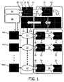
図1は、静的情報及びSENSE収集を同時に使用した全ての再構成ステップの略図を示す。この略図は、ROI内に角のない環状ボリュームを有する胸部の心臓MR(CMR)画像10、20を総期間内に生成する手順を表す。
FIG. 1 shows a schematic of all reconstruction steps using static information and SENSE collection simultaneously. This schematic represents the procedure for generating a cardiac MR (CMR)
手順は次のステップを含む:
ステップ1(S1): 心臓部から(第1の)心フェーズの第1のデータセットを提供すること;
ステップ2(S2): 第1のデータセットからROIの第1の画像10を生成すること;
ステップ3(S3): それぞれ主に総期間内において動的又は静的な動的領域12及び静的領域14を第1の画像10内で特定すること;
ステップ4(S4):動的領域12をマスクすることにより第1の画像10を編集すること;
ステップ5(S5): 残りの静的領域14を示す編集された第1の画像16の逆フーリエ変換(FFT−1)を実行すること;
ステップ6(S6): 心臓部から(第2の)心フェーズに関する第2のデータセットを提供すること;
ステップ7(S7): 残りの静的領域14を含む編集された第1の画像16の逆フーリエ変換(FFT−1)を第2のデータセットから差し引くこと;
ステップ8(S8): 差し引かれた第2のデータセット18にフーリエ変換を実行すること;及び
ステップ9(S9): 少なくとも動的領域12を含む低減された関心領域(rROI)の第2の画像20を生成すること。
The procedure includes the following steps:
Step 1 (S1): Providing a first data set of a (first) cardiac phase from the heart;
Step 2 (S2): Generating the
Step 3 (S3): Identifying in the first image 10 a
Step 4 (S4): Editing the
Step 5 (S5): performing an inverse Fourier transform (FFT-1) of the edited
Step 6 (S6): Providing a second data set for the (second) cardiac phase from the heart;
Step 7 (S7): Subtract the inverse Fourier transform (FFT −1 ) of the edited
Step 8 (S8): Performing a Fourier transform on the subtracted
好ましくは、特定することは第1の画像上で静的及び動的領域を手動で選択することにより実行される。例えば心臓周囲のボックスは動的領域として選択することができ、これによってボックス外側の領域は静的領域として選択される。 Preferably, the identifying is performed by manually selecting static and dynamic regions on the first image. For example, the box around the heart can be selected as the dynamic area, whereby the area outside the box is selected as the static area.
提供された第1及び第2のデータセットのデータはデータ収集により予め収集される。上述の総期間は総収集時間である。ステップ1(S1)に備えて、例えばフルFoVでの一回の収集が折り重なりアーチファクトを除去するために行われる。ステップ6(S6)に備えて、心フェーズごとに低減されたFoV(rFoV)を更新するためにky−kz空間のアンダーサンプリング収集が行われる。フルFoVという用語は(完全な)ROIに対応し、rFoVという用語は低減されたROIに対応する。 The data of the provided first and second data sets is collected in advance by data collection. The total period mentioned above is the total collection time. In preparation for step 1 (S1), a single acquisition, for example at full FoV, is performed to remove the folding artifacts. In preparation for step 6 (S6), an undersampling acquisition of ky-kz space is performed to update the reduced FoV (rFoV) for each cardiac phase. The term full FoV corresponds to the (complete) ROI and the term rFoV corresponds to the reduced ROI.
当業者は、第2のデータセット(rFoV)が第1のデータセットと比較してアンダーサンプリングされる場合は、好ましくはステップ5aがステップ5及びステップ7の間に適用されることを理解するであろう。ステップ5aでは、編集された第1の画像16の逆フーリエ変換(FFT−1)は、第2のデータセットを提供(収集)する際に使用されたアンダーサンプリングと同じやり方でアンダーサンプリングされる。従って手順は次のステップを含む:
ステップ1(S1): 心臓部から(第1の)心フェーズの第1のデータセットを収集すること;
ステップ2(S2): 第1のデータセットからROIの第1の画像10を生成すること;
ステップ3(S3): 動的領域12及び静的領域14を第1の画像10内で特定することであって、これらの領域は総期間内に主に動的又は静的であること;
ステップ4(S4): 動的領域12をマスクして外すことにより第1の画像10を編集すること;
ステップ5(S5): 残りの静的領域14を示す編集された第1の画像16の逆フーリエ変換(FFT−1)を実行すること;
ステップ5a(S5a): 編集された第1の画像16の逆フーリエ変換(FFT−1)をアンダーサンプリング戦略を用いてアンダーサンプリングし、これによってアンダーサンプリングされた第1のデータセット30を提供すること;
ステップ6(S6): 心臓部から(第2の)心フェーズに関する第2のデータセットを収集すること。ここで第2のデータセットは、第2のデータセットの収集中にアンダーサンプリング戦略を用いることにより第1のデータセットと比較してアンダーサンプリングされる;
ステップ7(S7): アンダーサンプリングされた第1のデータセット30を第2のデータセットから差し引くこと;
ステップ8(S8): 差し引かれた第2のデータセット18にフーリエ変換(FFT)を実行すること;及び
ステップ9(S9): 低減された関心領域(rROI)の第2の画像20を生成し、低減された関心領域は少なくとも動的領域12を含むこと。
Those skilled in the art will understand that step 5a is preferably applied between
Step 1 (S1): collecting a first data set of a (first) cardiac phase from the heart;
Step 2 (S2): Generating the
Step 3 (S3): Identifying
Step 4 (S4): Editing the
Step 5 (S5): performing an inverse Fourier transform (FFT-1) of the edited
Step 5a (S5a): Undersampling the inverse Fourier transform (FFT-1) of the edited
Step 6 (S6): Collecting a second data set for the (second) cardiac phase from the heart. Here the second data set is under sampled compared to the first data set by using an undersampling strategy during the collection of the second data set;
Step 7 (S7): Subtract the undersampled
Step 8 (S8): Perform a Fourier Transform (FFT) on the subtracted
PINOT収集と同様に、図示された手順は2つの異なるデータセットを区別することができる。第1のデータセットでは、静的及び動的な情報を収集するk空間ラインが収集される。第2のデータセットでは、k空間ラインのサブグループが、一部の情報を最終的な再構成を行うことができるように完全なデータセットから回復することができるという考えに基づいて心フェーズ(心臓フェーズ)ごとに収集される。PINOTとの相違点であるこのアプローチでは、式(1)に記載されたような行列式全体を構築する必要がない。対照的に、再構成は3つの異なる段階に分かれる。 Similar to the PINOT acquisition, the illustrated procedure can distinguish two different data sets. In the first dataset, k-space lines that collect static and dynamic information are collected. In the second data set, a sub-group of k-space lines can recover some information from the complete data set so that the final reconstruction can be performed, and the cardiac phase ( It is collected every cardiac phase). This approach, which is different from PINOT, does not require the construction of the entire determinant as described in equation (1). In contrast, reconstruction is divided into three different stages.
第1の段階: この再構成段階では、フル画像10が上記第1のデータセットを使用して1つの心フェーズに対して生成される(S2)。この画像10は、従来のSENSE再構成又はフルサンプル画像を使用して生成され、信号精度を改善することができる。
First Stage: In this reconstruction stage, a
第2の段階: この再構成段階では、フル再構成画像内の規定の動的領域は、全ての心フェーズで等しい静的領域14からのみ情報を得るために0に設定される(S4)。この再構成段階では、画像16は、各コイルのコイル感度により重み付けされ、k空間領域で逆フーリエ変換される(S5)。各コイルに対するフーリエ変換された画像は、上記第2のデータセットと同じサンプリング方式に従ってアンダーサンプリングされる(S5及びS7間の未表示の矢印により示される)。最後に、静的領域から生成されたk空間ラインは、心フェーズごとに更新されたk空間ラインから差し引かれる(S7)。
Second stage: In this reconstruction stage, the defined dynamic region in the full reconstructed image is set to 0 to obtain information only from the
第3の段階: この再構成段階では、第2の段階で生成された画像は、画像空間へとフーリエ変換され(S8)、低減されたFoVから情報を取り出すのみで従来のSENSE再構成を使用して再構成される(S9)。 Third stage: In this reconstruction stage, the image produced in the second stage is Fourier transformed into the image space (S8) and the conventional SENSE reconstruction is used only to extract the information from the reduced FoV. And reconfigured (S9).
このアプローチに従えば、SENSE再構成及びNoQUIST流の再構成はかなり分離される。SENSE情報は第3の段階でのみ使用されるのに対し、NoQUIST流の情報は再構成の第2の段階でのみ使用される。更に、再構成の第3の段階では低減された領域のみが再構成され、3Dケースについての従来のSENSE再構成と比較して再構成速度が向上する。 According to this approach, the SENSE reconstruction and the NoQUIST stream reconstruction are largely separated. The SENSE information is used only in the third stage, whereas the NoQUIST style information is used only in the second stage of reconstruction. Furthermore, in the third stage of reconstruction, only the reduced regions are reconstructed, which improves the reconstruction speed compared to the conventional SENSE reconstruction for the 3D case.
本発明は、図面及び前述の記載において詳細に図示及び説明されたが、このような図示及び記載は、説明的又は例示的であって限定するものではないと見なされるべきである。すなわち本発明は、開示された実施形態に限定されるものではない。開示された実施形態のその他の変形が、図面、本開示及び添付の請求項の検討から、請求項に係る発明を実施する当業者によって理解されて実現され得る。請求項において、「comprising(含む、備える)」という単語は、他の要素又はステップを除外するものではなく、不定冠詞「a」又は「an」は、複数を除外するものではない。特定の手段が相互に異なる従属請求項に列挙されるという単なる事実は、これらの手段の組み合わせが有利に用いられないことを示すものではない。請求項における任意の参照符号は、本発明の範囲を限定するものと解釈されるべきではない。 While the invention has been illustrated and described in detail in the drawings and foregoing description, such illustration and description are to be considered illustrative or exemplary and not restrictive. That is, the invention is not limited to the disclosed embodiments. Other variations of the disclosed embodiments can be understood and effected by those skilled in the art in practicing the claimed invention, from a study of the drawings, the disclosure, and the appended claims. In the claims, the word "comprising" does not exclude other elements or steps, and the indefinite article "a" or "an" does not exclude a plurality. The mere fact that certain measures are recited in mutually different dependent claims does not indicate that a combination of these measures cannot be used to advantage. Any reference signs in the claims shall not be construed as limiting the scope of the invention.
本発明に繋がる研究は、助成契約第242038の下で欧州連合第7フレームワークプログラム(FP−7−HEALTH−2009)からの資金提供を受けた。 The work leading to the present invention was funded by the European Union 7th Framework Program (FP-7-HEALTH-2009) under grant agreement number 242038.
Claims (8)
SENSEアンダーサンプリング方式を用いて、複数の受信器コイルを使用して前記物体の1つの前記動作フェーズに関する第1のデータセットを収集するステップS1と、
SENSE再構成によって、前記第1のデータセットから関心領域の第1の画像を再構成するステップS2と、
動的領域及び静的領域を前記第1の画像内で特定するステップS3であって、これらの領域は前記一定の期間内に主に動的又は静的である、ステップS3と、
前記動的領域をマスクして外すことにより前記第1の画像を編集するステップS4と、
編集された前記第1の画像を、前記複数の受信器コイルのコイル感度により重み付けするステップS5と、
残りの前記静的領域を示す編集され重み付けされた前記第1の画像の逆フーリエ変換を実行するステップS6と、
複数の受信器コイルを使用して前記物体の1つの前記動作フェーズに関する第2のデータセットを収集するステップS7であって、収集中にアンダーサンプリングによって前記第2のデータセットが提供され、前記第1のデータセットの視野と比較して低減された視野が生じ、更に、前記第1のデータセットの収集に対するのと同じSENSEアンダーサンプリング方式を用いる、ステップS7と、
前記残りの静的領域を含む前記編集された第1の画像の逆フーリエ変換されたものを前記第2のデータセットから差し引くステップS8と、
差し引かれた前記第2のデータセットにフーリエ変換を実行するステップS9と、
前記第1の画像の前記関心領域について低減された関心領域の第2の画像を生成するステップS10であって、前記低減された関心領域は動的領域を含む、ステップS10と、
を含む、方法。 A method of generating an MR image of an object performing a motion having a plurality of motion phases within a certain period, the method comprising:
Collecting S1SE undersampling scheme to collect a first data set for one of said motion phases of said object using a plurality of receiver coils;
S2 reconstructing a first image of the region of interest from the first dataset by SENSE reconstruction;
Step S3 of identifying dynamic and static regions in the first image, these regions being predominantly dynamic or static within the certain period of time, S3.
Step S4 of editing the first image by masking and removing the dynamic region;
Weighting the edited first image with coil sensitivities of the plurality of receiver coils;
Performing S6 an inverse Fourier transform of the edited and weighted first image showing the remaining static regions;
Collecting S2 a second data set for one of the motion phases of the object using a plurality of receiver coils, wherein the second data set is provided by undersampling during acquisition; A reduced field of view as compared to the field of view of the first data set, and further using the same SENSE undersampling scheme as for the acquisition of the first data set, step S7,
Subtracting an inverse Fourier transformed version of the edited first image containing the remaining static region from the second data set, S8.
Performing a Fourier transform on the subtracted second data set, S9,
Producing a second image of a reduced region of interest for said region of interest of said first image, said reduced region of interest comprising a dynamic region, step S10,
Including the method.
SENSEアンダーサンプリング方式を用いて、複数の受信器コイルを使用して前記物体の1つの前記動作フェーズに関する第1のデータセットを収集することと、
SENSE再構成によって、前記第1のデータセットから関心領域の第1の画像を再構成することと、
動的領域及び静的領域を前記第1の画像内で特定することであって、これらの領域は前記一定の期間内に主に動的又は静的である、特定することと、
前記動的領域をマスクして外すことにより前記第1の画像を編集することと、
編集された前記第1の画像を、前記複数の受信器コイルのコイル感度により重み付けすることと、
残りの前記静的領域を示す編集され重み付けされた前記第1の画像の逆フーリエ変換を実行することと、
複数の受信器コイルを使用して前記物体の1つの前記動作フェーズに関する第2のデータセットを収集することであって、収集中にアンダーサンプリングによって前記第2のデータセットが提供され、前記第1のデータセットの視野と比較して低減された視野が生じ、更に、前記第1のデータセットの収集に対するのと同じSENSEアンダーサンプリング方式を用いる、収集することと、
前記残りの静的領域を含む前記編集された第1の画像の逆フーリエ変換されたものを前記第2のデータセットから差し引くことと、
差し引かれた前記第2のデータセットにフーリエ変換を実行することと、
前記第1の画像の前記関心領域について低減された関心領域の第2の画像を生成することであって、前記低減された関心領域は動的領域を含む、生成することと、
を実行する、MRIシステム。 What is claimed is: 1. An MRI system for generating an MR image of an object performing a motion having a plurality of motion phases within a certain period, the MRI system comprising:
Collecting a first data set for one of said motion phases of said object using a plurality of receiver coils using the SENSE undersampling scheme;
Reconstructing a first image of the region of interest from the first data set by SENSE reconstruction;
Identifying dynamic and static regions in the first image, the regions being predominantly dynamic or static within the period of time;
Editing the first image by masking and removing the dynamic region;
Weighting the edited first image by coil sensitivity of the plurality of receiver coils;
Performing an inverse Fourier transform of the edited and weighted first image showing the remaining static regions;
Collecting a second data set for one of said motion phases of said object using a plurality of receiver coils, wherein undersampling provides said second data set during said acquisition, A reduced field of view as compared to the field of view of the first data set, and further using the same SENSE undersampling scheme as for the acquisition of the first data set;
Subtracting an inverse Fourier transformed version of the edited first image containing the remaining static region from the second data set;
Performing a Fourier transform on the subtracted second data set;
Generating a second image of a reduced region of interest for the region of interest of the first image, the reduced region of interest including a dynamic region;
An MRI system that executes.
Applications Claiming Priority (3)
| Application Number | Priority Date | Filing Date | Title |
|---|---|---|---|
| EP14192290 | 2014-11-07 | ||
| EP14192290.6 | 2014-11-07 | ||
| PCT/EP2015/072756 WO2016071054A1 (en) | 2014-11-07 | 2015-10-01 | Method and system for generating mr images of a moving object in its environment |
Publications (3)
| Publication Number | Publication Date |
|---|---|
| JP2017533064A JP2017533064A (en) | 2017-11-09 |
| JP2017533064A5 JP2017533064A5 (en) | 2018-11-08 |
| JP6730995B2 true JP6730995B2 (en) | 2020-07-29 |
Family
ID=51893873
Family Applications (1)
| Application Number | Title | Priority Date | Filing Date |
|---|---|---|---|
| JP2017542280A Active JP6730995B2 (en) | 2014-11-07 | 2015-10-01 | Method and system for generating MR image of moving object in environment |
Country Status (5)
| Country | Link |
|---|---|
| US (1) | US10416265B2 (en) |
| EP (1) | EP3215863B1 (en) |
| JP (1) | JP6730995B2 (en) |
| CN (1) | CN107209242B (en) |
| WO (1) | WO2016071054A1 (en) |
Families Citing this family (3)
| Publication number | Priority date | Publication date | Assignee | Title |
|---|---|---|---|---|
| DE102016218715A1 (en) * | 2016-09-28 | 2018-03-29 | Siemens Healthcare Gmbh | Improved generation of combined magnetic resonance images |
| DE102018203507B4 (en) * | 2018-03-08 | 2020-10-22 | Siemens Healthcare Gmbh | Method for recording and reconstructing a four-dimensional dynamic image data set with a magnetic resonance device, magnetic resonance device, computer program and electronically readable data carrier |
| US20220187312A1 (en) * | 2018-10-31 | 2022-06-16 | Centro Nacional De Investigaciones Cardiovasculares Carlos Iii (F.S.P.) | Biomarkers of subclinical atherosclerosis |
Family Cites Families (21)
| Publication number | Priority date | Publication date | Assignee | Title |
|---|---|---|---|---|
| JPH10502858A (en) * | 1995-05-02 | 1998-03-17 | フィリップス エレクトロニクス エヌ ベー | Method and apparatus for magnetic resonance imaging of a subject |
| US5653233A (en) | 1995-08-11 | 1997-08-05 | The Board Of Trustees Of The Leland Stanford Junior University | Method and apparatus for improved temporal resolution in dynamic MRI |
| US6353752B1 (en) | 1999-05-14 | 2002-03-05 | Board Of Trustees Of The Leland Standford Junior University | Reduced field-of-view method for cine magnetic resonance imaging |
| US6528995B1 (en) * | 2001-09-10 | 2003-03-04 | Schlumberger Technology Corporation | Methods and apparatus for measuring flow velocity in a wellbore using NMR and applications using same |
| DE60228475D1 (en) * | 2001-11-07 | 2008-10-02 | Koninkl Philips Electronics Nv | MAGNETIC RESONANCE BASED PROCESS FOR CREATING A QUICK DYNAMIC IMAGE |
| CN1653348B (en) * | 2002-05-13 | 2012-08-08 | 皇家飞利浦电子股份有限公司 | Prior-information-enhanced dynamic magnetic resonance imaging method |
| US7405564B2 (en) * | 2003-01-20 | 2008-07-29 | Koninklijke Philips Electronics N.V. | Sub-sampled moving table MRI for at least two adjacent fields of view |
| US20070055134A1 (en) * | 2003-05-06 | 2007-03-08 | Miha Fuderer | Undersampled magnetic resonance imaging |
| WO2005047919A1 (en) * | 2003-11-12 | 2005-05-26 | Invivo Corporation | Method for generating fast magnetic resonance images |
| DE10353342B4 (en) * | 2003-11-14 | 2008-07-17 | Siemens Ag | Improved MRI imaging based on conventional PPA reconstruction techniques |
| JP2007536976A (en) * | 2004-05-14 | 2007-12-20 | コーニンクレッカ フィリップス エレクトロニクス エヌ ヴィ | Moving table MRI |
| CN101351721A (en) * | 2005-04-06 | 2009-01-21 | 皇家飞利浦电子股份有限公司 | Magnetic resonance parallel imaging with continuously moving bed |
| JP2009533128A (en) * | 2006-04-13 | 2009-09-17 | コーニンクレッカ フィリップス エレクトロニクス エヌ ヴィ | MRI of continuously moving objects including motion compensation |
| JP5037866B2 (en) * | 2006-06-28 | 2012-10-03 | 株式会社日立メディコ | Magnetic resonance imaging system |
| US20080292167A1 (en) * | 2007-05-24 | 2008-11-27 | Nick Todd | Method and system for constrained reconstruction applied to magnetic resonance temperature mapping |
| CN103403569B (en) * | 2010-12-22 | 2016-02-03 | 皇家飞利浦有限公司 | Use the parallel MRI method that calibration scan, coil sensitivity map and omniselector compensate for rigid motion |
| WO2013103791A1 (en) * | 2012-01-06 | 2013-07-11 | Cincinnati Children's Hospital Medical Center | Correlation imaging for multi-scan mri with multi-channel data acquisition |
| JP6073627B2 (en) * | 2012-10-01 | 2017-02-01 | 東芝メディカルシステムズ株式会社 | Magnetic resonance imaging apparatus and image processing apparatus |
| US20140303480A1 (en) * | 2013-04-04 | 2014-10-09 | General Electric Company | Mri with static tissue removal |
| US10534060B2 (en) * | 2013-06-10 | 2020-01-14 | Toshiba Medical Systems Corporation | Parallel MRI with spatially misregistered signal |
| US9797970B2 (en) * | 2015-04-14 | 2017-10-24 | The Board Of Trustees Of The University Of Illinois | Image domain segmented echo planar magnetic resonance imaging using a 2D excitation radiofrequency pulse |
-
2015
- 2015-10-01 WO PCT/EP2015/072756 patent/WO2016071054A1/en active Application Filing
- 2015-10-01 EP EP15771635.8A patent/EP3215863B1/en active Active
- 2015-10-01 US US15/523,701 patent/US10416265B2/en active Active
- 2015-10-01 JP JP2017542280A patent/JP6730995B2/en active Active
- 2015-10-01 CN CN201580060220.6A patent/CN107209242B/en active Active
Also Published As
| Publication number | Publication date |
|---|---|
| EP3215863A1 (en) | 2017-09-13 |
| JP2017533064A (en) | 2017-11-09 |
| WO2016071054A1 (en) | 2016-05-12 |
| CN107209242A (en) | 2017-09-26 |
| US20170328974A1 (en) | 2017-11-16 |
| CN107209242B (en) | 2020-07-03 |
| EP3215863B1 (en) | 2020-12-09 |
| US10416265B2 (en) | 2019-09-17 |
Similar Documents
| Publication | Publication Date | Title |
|---|---|---|
| Bustin et al. | Five‐minute whole‐heart coronary MRA with sub‐millimeter isotropic resolution, 100% respiratory scan efficiency, and 3D‐PROST reconstruction | |
| Pang et al. | Whole‐heart coronary MRA with 100% respiratory gating efficiency: self‐navigated three‐dimensional retrospective image‐based motion correction (TRIM) | |
| JP6467344B2 (en) | Propeller with Dixon water fat separation | |
| EP2145200B1 (en) | Diffusion tensor imaging of moving objects | |
| EP3384307B1 (en) | Removal of image artifacts in sense-mri | |
| US10809337B2 (en) | Reconstructing magnetic resonance images with different contrasts | |
| JP6560023B2 (en) | Magnetic resonance imaging system | |
| US11002815B2 (en) | System and method for reducing artifacts in echo planar magnetic resonance imaging | |
| JP6702691B2 (en) | Magnetic resonance imaging and medical image processing apparatus | |
| JP6510273B2 (en) | Magnetic resonance imaging apparatus, magnetic resonance imaging method and magnetic resonance imaging program | |
| CN107209238A (en) | Parallel many section MR imagings with the suppression to sideband artefact | |
| JP2017529960A (en) | Propeller MR imaging with artifact suppression | |
| JP6730995B2 (en) | Method and system for generating MR image of moving object in environment | |
| JP2012505709A (en) | Moving table MRI apparatus and method | |
| CN108780134B (en) | System, method and apparatus for susceptibility mapping of moving objects | |
| Hutter et al. | Dynamic field mapping and motion correction using interleaved double spin-echo diffusion MRI | |
| JP7350568B2 (en) | Magnetic resonance data acquisition method and magnetic resonance imaging apparatus | |
| US9709651B2 (en) | Compensated magnetic resonance imaging system and method for improved magnetic resonance imaging and diffusion imaging | |
| JP2017533064A5 (en) | ||
| JP5156958B2 (en) | Magnetic resonance imaging system | |
| WO2017167937A1 (en) | Dynamic mr imaging with increased temporal and spatial resolution | |
| EP3772660A1 (en) | Magnetic resonance image reconstruction using cluster analysis | |
| JP6873134B2 (en) | Removal of image artifacts in SENSE imaging | |
| JP2019205542A (en) | Magnetic resonance imaging apparatus, processing apparatus, and medical image processing method | |
| Haroon et al. | Probabilistic quantification of regional cortical microstructural complexity |
Legal Events
| Date | Code | Title | Description |
|---|---|---|---|
| A521 | Request for written amendment filed |
Free format text: JAPANESE INTERMEDIATE CODE: A523 Effective date: 20170711 |
|
| A521 | Request for written amendment filed |
Free format text: JAPANESE INTERMEDIATE CODE: A523 Effective date: 20180927 |
|
| A621 | Written request for application examination |
Free format text: JAPANESE INTERMEDIATE CODE: A621 Effective date: 20180927 |
|
| A977 | Report on retrieval |
Free format text: JAPANESE INTERMEDIATE CODE: A971007 Effective date: 20190620 |
|
| A131 | Notification of reasons for refusal |
Free format text: JAPANESE INTERMEDIATE CODE: A131 Effective date: 20190802 |
|
| A601 | Written request for extension of time |
Free format text: JAPANESE INTERMEDIATE CODE: A601 Effective date: 20191025 |
|
| A521 | Request for written amendment filed |
Free format text: JAPANESE INTERMEDIATE CODE: A523 Effective date: 20200122 |
|
| TRDD | Decision of grant or rejection written | ||
| A01 | Written decision to grant a patent or to grant a registration (utility model) |
Free format text: JAPANESE INTERMEDIATE CODE: A01 Effective date: 20200610 |
|
| A61 | First payment of annual fees (during grant procedure) |
Free format text: JAPANESE INTERMEDIATE CODE: A61 Effective date: 20200703 |
|
| R150 | Certificate of patent or registration of utility model |
Ref document number: 6730995 Country of ref document: JP Free format text: JAPANESE INTERMEDIATE CODE: R150 |
|
| R250 | Receipt of annual fees |
Free format text: JAPANESE INTERMEDIATE CODE: R250 |