JP6541237B2 - Insulin-like growth factor 1 receptor specific antibodies and their use - Google Patents
Insulin-like growth factor 1 receptor specific antibodies and their use Download PDFInfo
- Publication number
- JP6541237B2 JP6541237B2 JP2016555727A JP2016555727A JP6541237B2 JP 6541237 B2 JP6541237 B2 JP 6541237B2 JP 2016555727 A JP2016555727 A JP 2016555727A JP 2016555727 A JP2016555727 A JP 2016555727A JP 6541237 B2 JP6541237 B2 JP 6541237B2
- Authority
- JP
- Japan
- Prior art keywords
- igf1r
- antibody
- isolated
- fragment
- seq
- Prior art date
- Legal status (The legal status is an assumption and is not a legal conclusion. Google has not performed a legal analysis and makes no representation as to the accuracy of the status listed.)
- Active
Links
Images


Classifications
-
- C—CHEMISTRY; METALLURGY
- C07—ORGANIC CHEMISTRY
- C07K—PEPTIDES
- C07K16/00—Immunoglobulins [IGs], e.g. monoclonal or polyclonal antibodies
- C07K16/18—Immunoglobulins [IGs], e.g. monoclonal or polyclonal antibodies against material from animals or humans
- C07K16/28—Immunoglobulins [IGs], e.g. monoclonal or polyclonal antibodies against material from animals or humans against receptors, cell surface antigens or cell surface determinants
- C07K16/2863—Immunoglobulins [IGs], e.g. monoclonal or polyclonal antibodies against material from animals or humans against receptors, cell surface antigens or cell surface determinants against receptors for growth factors, growth regulators
-
- A—HUMAN NECESSITIES
- A61—MEDICAL OR VETERINARY SCIENCE; HYGIENE
- A61K—PREPARATIONS FOR MEDICAL, DENTAL OR TOILETRY PURPOSES
- A61K47/00—Medicinal preparations characterised by the non-active ingredients used, e.g. carriers or inert additives; Targeting or modifying agents chemically bound to the active ingredient
- A61K47/50—Medicinal preparations characterised by the non-active ingredients used, e.g. carriers or inert additives; Targeting or modifying agents chemically bound to the active ingredient the non-active ingredient being chemically bound to the active ingredient, e.g. polymer-drug conjugates
- A61K47/51—Medicinal preparations characterised by the non-active ingredients used, e.g. carriers or inert additives; Targeting or modifying agents chemically bound to the active ingredient the non-active ingredient being chemically bound to the active ingredient, e.g. polymer-drug conjugates the non-active ingredient being a modifying agent
- A61K47/68—Medicinal preparations characterised by the non-active ingredients used, e.g. carriers or inert additives; Targeting or modifying agents chemically bound to the active ingredient the non-active ingredient being chemically bound to the active ingredient, e.g. polymer-drug conjugates the non-active ingredient being a modifying agent the modifying agent being an antibody, an immunoglobulin or a fragment thereof, e.g. an Fc-fragment
- A61K47/6835—Medicinal preparations characterised by the non-active ingredients used, e.g. carriers or inert additives; Targeting or modifying agents chemically bound to the active ingredient the non-active ingredient being chemically bound to the active ingredient, e.g. polymer-drug conjugates the non-active ingredient being a modifying agent the modifying agent being an antibody, an immunoglobulin or a fragment thereof, e.g. an Fc-fragment the modifying agent being an antibody or an immunoglobulin bearing at least one antigen-binding site
- A61K47/6849—Medicinal preparations characterised by the non-active ingredients used, e.g. carriers or inert additives; Targeting or modifying agents chemically bound to the active ingredient the non-active ingredient being chemically bound to the active ingredient, e.g. polymer-drug conjugates the non-active ingredient being a modifying agent the modifying agent being an antibody, an immunoglobulin or a fragment thereof, e.g. an Fc-fragment the modifying agent being an antibody or an immunoglobulin bearing at least one antigen-binding site the antibody targeting a receptor, a cell surface antigen or a cell surface determinant
-
- A—HUMAN NECESSITIES
- A61—MEDICAL OR VETERINARY SCIENCE; HYGIENE
- A61K—PREPARATIONS FOR MEDICAL, DENTAL OR TOILETRY PURPOSES
- A61K49/00—Preparations for testing in vivo
- A61K49/0002—General or multifunctional contrast agents, e.g. chelated agents
-
- A—HUMAN NECESSITIES
- A61—MEDICAL OR VETERINARY SCIENCE; HYGIENE
- A61K—PREPARATIONS FOR MEDICAL, DENTAL OR TOILETRY PURPOSES
- A61K49/00—Preparations for testing in vivo
- A61K49/001—Preparation for luminescence or biological staining
- A61K49/0013—Luminescence
- A61K49/0017—Fluorescence in vivo
- A61K49/005—Fluorescence in vivo characterised by the carrier molecule carrying the fluorescent agent
- A61K49/0058—Antibodies
-
- A—HUMAN NECESSITIES
- A61—MEDICAL OR VETERINARY SCIENCE; HYGIENE
- A61P—SPECIFIC THERAPEUTIC ACTIVITY OF CHEMICAL COMPOUNDS OR MEDICINAL PREPARATIONS
- A61P25/00—Drugs for disorders of the nervous system
-
- A—HUMAN NECESSITIES
- A61—MEDICAL OR VETERINARY SCIENCE; HYGIENE
- A61P—SPECIFIC THERAPEUTIC ACTIVITY OF CHEMICAL COMPOUNDS OR MEDICINAL PREPARATIONS
- A61P25/00—Drugs for disorders of the nervous system
- A61P25/14—Drugs for disorders of the nervous system for treating abnormal movements, e.g. chorea, dyskinesia
- A61P25/16—Anti-Parkinson drugs
-
- A—HUMAN NECESSITIES
- A61—MEDICAL OR VETERINARY SCIENCE; HYGIENE
- A61P—SPECIFIC THERAPEUTIC ACTIVITY OF CHEMICAL COMPOUNDS OR MEDICINAL PREPARATIONS
- A61P25/00—Drugs for disorders of the nervous system
- A61P25/28—Drugs for disorders of the nervous system for treating neurodegenerative disorders of the central nervous system, e.g. nootropic agents, cognition enhancers, drugs for treating Alzheimer's disease or other forms of dementia
-
- C—CHEMISTRY; METALLURGY
- C07—ORGANIC CHEMISTRY
- C07K—PEPTIDES
- C07K16/00—Immunoglobulins [IGs], e.g. monoclonal or polyclonal antibodies
- C07K16/46—Hybrid immunoglobulins
- C07K16/461—Igs containing Ig-regions, -domains or -residues form different species
- C07K16/464—Igs containing CDR-residues from one specie grafted between FR-residues from another
- C07K16/465—Igs containing CDR-residues from one specie grafted between FR-residues from another with additional modified FR-residues
-
- G—PHYSICS
- G01—MEASURING; TESTING
- G01N—INVESTIGATING OR ANALYSING MATERIALS BY DETERMINING THEIR CHEMICAL OR PHYSICAL PROPERTIES
- G01N33/00—Investigating or analysing materials by specific methods not covered by groups G01N1/00 - G01N31/00
- G01N33/48—Biological material, e.g. blood, urine; Haemocytometers
- G01N33/50—Chemical analysis of biological material, e.g. blood, urine; Testing involving biospecific ligand binding methods; Immunological testing
- G01N33/53—Immunoassay; Biospecific binding assay; Materials therefor
- G01N33/574—Immunoassay; Biospecific binding assay; Materials therefor for cancer
- G01N33/57484—Immunoassay; Biospecific binding assay; Materials therefor for cancer involving compounds serving as markers for tumor, cancer, neoplasia, e.g. cellular determinants, receptors, heat shock/stress proteins, A-protein, oligosaccharides, metabolites
- G01N33/57492—Immunoassay; Biospecific binding assay; Materials therefor for cancer involving compounds serving as markers for tumor, cancer, neoplasia, e.g. cellular determinants, receptors, heat shock/stress proteins, A-protein, oligosaccharides, metabolites involving compounds localized on the membrane of tumor or cancer cells
-
- G—PHYSICS
- G01—MEASURING; TESTING
- G01N—INVESTIGATING OR ANALYSING MATERIALS BY DETERMINING THEIR CHEMICAL OR PHYSICAL PROPERTIES
- G01N33/00—Investigating or analysing materials by specific methods not covered by groups G01N1/00 - G01N31/00
- G01N33/48—Biological material, e.g. blood, urine; Haemocytometers
- G01N33/50—Chemical analysis of biological material, e.g. blood, urine; Testing involving biospecific ligand binding methods; Immunological testing
- G01N33/68—Chemical analysis of biological material, e.g. blood, urine; Testing involving biospecific ligand binding methods; Immunological testing involving proteins, peptides or amino acids
-
- G—PHYSICS
- G01—MEASURING; TESTING
- G01N—INVESTIGATING OR ANALYSING MATERIALS BY DETERMINING THEIR CHEMICAL OR PHYSICAL PROPERTIES
- G01N33/00—Investigating or analysing materials by specific methods not covered by groups G01N1/00 - G01N31/00
- G01N33/48—Biological material, e.g. blood, urine; Haemocytometers
- G01N33/50—Chemical analysis of biological material, e.g. blood, urine; Testing involving biospecific ligand binding methods; Immunological testing
- G01N33/74—Chemical analysis of biological material, e.g. blood, urine; Testing involving biospecific ligand binding methods; Immunological testing involving hormones or other non-cytokine intercellular protein regulatory factors such as growth factors, including receptors to hormones and growth factors
-
- A—HUMAN NECESSITIES
- A61—MEDICAL OR VETERINARY SCIENCE; HYGIENE
- A61K—PREPARATIONS FOR MEDICAL, DENTAL OR TOILETRY PURPOSES
- A61K39/00—Medicinal preparations containing antigens or antibodies
- A61K2039/505—Medicinal preparations containing antigens or antibodies comprising antibodies
-
- C—CHEMISTRY; METALLURGY
- C07—ORGANIC CHEMISTRY
- C07K—PEPTIDES
- C07K2317/00—Immunoglobulins specific features
- C07K2317/20—Immunoglobulins specific features characterized by taxonomic origin
- C07K2317/22—Immunoglobulins specific features characterized by taxonomic origin from camelids, e.g. camel, llama or dromedary
-
- C—CHEMISTRY; METALLURGY
- C07—ORGANIC CHEMISTRY
- C07K—PEPTIDES
- C07K2317/00—Immunoglobulins specific features
- C07K2317/20—Immunoglobulins specific features characterized by taxonomic origin
- C07K2317/24—Immunoglobulins specific features characterized by taxonomic origin containing regions, domains or residues from different species, e.g. chimeric, humanized or veneered
-
- C—CHEMISTRY; METALLURGY
- C07—ORGANIC CHEMISTRY
- C07K—PEPTIDES
- C07K2317/00—Immunoglobulins specific features
- C07K2317/30—Immunoglobulins specific features characterized by aspects of specificity or valency
- C07K2317/35—Valency
-
- C—CHEMISTRY; METALLURGY
- C07—ORGANIC CHEMISTRY
- C07K—PEPTIDES
- C07K2317/00—Immunoglobulins specific features
- C07K2317/50—Immunoglobulins specific features characterized by immunoglobulin fragments
- C07K2317/51—Complete heavy chain or Fd fragment, i.e. VH + CH1
-
- C—CHEMISTRY; METALLURGY
- C07—ORGANIC CHEMISTRY
- C07K—PEPTIDES
- C07K2317/00—Immunoglobulins specific features
- C07K2317/50—Immunoglobulins specific features characterized by immunoglobulin fragments
- C07K2317/52—Constant or Fc region; Isotype
-
- C—CHEMISTRY; METALLURGY
- C07—ORGANIC CHEMISTRY
- C07K—PEPTIDES
- C07K2317/00—Immunoglobulins specific features
- C07K2317/50—Immunoglobulins specific features characterized by immunoglobulin fragments
- C07K2317/56—Immunoglobulins specific features characterized by immunoglobulin fragments variable (Fv) region, i.e. VH and/or VL
- C07K2317/565—Complementarity determining region [CDR]
-
- C—CHEMISTRY; METALLURGY
- C07—ORGANIC CHEMISTRY
- C07K—PEPTIDES
- C07K2317/00—Immunoglobulins specific features
- C07K2317/50—Immunoglobulins specific features characterized by immunoglobulin fragments
- C07K2317/56—Immunoglobulins specific features characterized by immunoglobulin fragments variable (Fv) region, i.e. VH and/or VL
- C07K2317/569—Single domain, e.g. dAb, sdAb, VHH, VNAR or nanobody®
-
- C—CHEMISTRY; METALLURGY
- C07—ORGANIC CHEMISTRY
- C07K—PEPTIDES
- C07K2317/00—Immunoglobulins specific features
- C07K2317/60—Immunoglobulins specific features characterized by non-natural combinations of immunoglobulin fragments
- C07K2317/64—Immunoglobulins specific features characterized by non-natural combinations of immunoglobulin fragments comprising a combination of variable region and constant region components
-
- C—CHEMISTRY; METALLURGY
- C07—ORGANIC CHEMISTRY
- C07K—PEPTIDES
- C07K2317/00—Immunoglobulins specific features
- C07K2317/70—Immunoglobulins specific features characterized by effect upon binding to a cell or to an antigen
-
- C—CHEMISTRY; METALLURGY
- C07—ORGANIC CHEMISTRY
- C07K—PEPTIDES
- C07K2317/00—Immunoglobulins specific features
- C07K2317/70—Immunoglobulins specific features characterized by effect upon binding to a cell or to an antigen
- C07K2317/77—Internalization into the cell
-
- C—CHEMISTRY; METALLURGY
- C07—ORGANIC CHEMISTRY
- C07K—PEPTIDES
- C07K2317/00—Immunoglobulins specific features
- C07K2317/90—Immunoglobulins specific features characterized by (pharmaco)kinetic aspects or by stability of the immunoglobulin
-
- C—CHEMISTRY; METALLURGY
- C07—ORGANIC CHEMISTRY
- C07K—PEPTIDES
- C07K2317/00—Immunoglobulins specific features
- C07K2317/90—Immunoglobulins specific features characterized by (pharmaco)kinetic aspects or by stability of the immunoglobulin
- C07K2317/92—Affinity (KD), association rate (Ka), dissociation rate (Kd) or EC50 value
-
- C—CHEMISTRY; METALLURGY
- C07—ORGANIC CHEMISTRY
- C07K—PEPTIDES
- C07K2317/00—Immunoglobulins specific features
- C07K2317/90—Immunoglobulins specific features characterized by (pharmaco)kinetic aspects or by stability of the immunoglobulin
- C07K2317/94—Stability, e.g. half-life, pH, temperature or enzyme-resistance
-
- G—PHYSICS
- G01—MEASURING; TESTING
- G01N—INVESTIGATING OR ANALYSING MATERIALS BY DETERMINING THEIR CHEMICAL OR PHYSICAL PROPERTIES
- G01N2333/00—Assays involving biological materials from specific organisms or of a specific nature
- G01N2333/435—Assays involving biological materials from specific organisms or of a specific nature from animals; from humans
- G01N2333/705—Assays involving receptors, cell surface antigens or cell surface determinants
- G01N2333/71—Assays involving receptors, cell surface antigens or cell surface determinants for growth factors; for growth regulators
-
- G—PHYSICS
- G01—MEASURING; TESTING
- G01N—INVESTIGATING OR ANALYSING MATERIALS BY DETERMINING THEIR CHEMICAL OR PHYSICAL PROPERTIES
- G01N2333/00—Assays involving biological materials from specific organisms or of a specific nature
- G01N2333/435—Assays involving biological materials from specific organisms or of a specific nature from animals; from humans
- G01N2333/705—Assays involving receptors, cell surface antigens or cell surface determinants
- G01N2333/72—Assays involving receptors, cell surface antigens or cell surface determinants for hormones
-
- G—PHYSICS
- G01—MEASURING; TESTING
- G01N—INVESTIGATING OR ANALYSING MATERIALS BY DETERMINING THEIR CHEMICAL OR PHYSICAL PROPERTIES
- G01N2570/00—Omics, e.g. proteomics, glycomics or lipidomics; Methods of analysis focusing on the entire complement of classes of biological molecules or subsets thereof, i.e. focusing on proteomes, glycomes or lipidomes
-
- G—PHYSICS
- G01—MEASURING; TESTING
- G01N—INVESTIGATING OR ANALYSING MATERIALS BY DETERMINING THEIR CHEMICAL OR PHYSICAL PROPERTIES
- G01N2800/00—Detection or diagnosis of diseases
- G01N2800/28—Neurological disorders
Landscapes
- Health & Medical Sciences (AREA)
- Life Sciences & Earth Sciences (AREA)
- Chemical & Material Sciences (AREA)
- Immunology (AREA)
- Engineering & Computer Science (AREA)
- General Health & Medical Sciences (AREA)
- Biomedical Technology (AREA)
- Molecular Biology (AREA)
- Medicinal Chemistry (AREA)
- Organic Chemistry (AREA)
- Animal Behavior & Ethology (AREA)
- Public Health (AREA)
- Veterinary Medicine (AREA)
- Biochemistry (AREA)
- Hematology (AREA)
- Urology & Nephrology (AREA)
- Cell Biology (AREA)
- Bioinformatics & Cheminformatics (AREA)
- Epidemiology (AREA)
- Proteomics, Peptides & Aminoacids (AREA)
- Genetics & Genomics (AREA)
- Biophysics (AREA)
- Pharmacology & Pharmacy (AREA)
- Neurology (AREA)
- Neurosurgery (AREA)
- Microbiology (AREA)
- Pathology (AREA)
- Food Science & Technology (AREA)
- Analytical Chemistry (AREA)
- General Physics & Mathematics (AREA)
- Biotechnology (AREA)
- Physics & Mathematics (AREA)
- General Chemical & Material Sciences (AREA)
- Nuclear Medicine, Radiotherapy & Molecular Imaging (AREA)
- Chemical Kinetics & Catalysis (AREA)
- Oncology (AREA)
- Hospice & Palliative Care (AREA)
- Endocrinology (AREA)
- Psychology (AREA)
- Psychiatry (AREA)
Description
本発明は、インスリン様成長因子1受容体特異的抗体、それらの断片、及びそれらの使用に関する。より詳細には、本発明は、血液脳関門を通過するインスリン様成長因子1受容体特異的抗体及びそれらの断片、並びにそれらの使用に関する。
The present invention relates to insulin-
神経変性疾患、たとえばアルツハイマー病及びパーキンソン病は、我々の高齢化社会において大きな負担となってきている。なぜなら、現在のところ、これらの障害に対して有効な治療法がないからである。脳に生じるこれら及び他の疾患の治療は、早期診断とともに、困難なものであり続けている。なぜなら、好適な治療用分子及び診断剤の大半が、密着し高度に制約的な血液脳関門(BBB)を透過できないからである(Abbott、2013)。BBBは、血管を裏打ちし、密着結合を介して互いに結合する脳内皮細胞(BEC)により形成される物理的障壁を構成する(Abbott、2013)。BEC間に形成される密着結合は、BBBの完全性に必須であり、500ドルトン(Da)より大きな分子の傍細胞輸送を妨げる。脳内皮細胞は、非常に低い飲作用速度を示す(Abbott、2013)ので、より大きな分子の経細胞輸送は、高度に特異的な受容体媒介性トランスサイトーシス(RMT)経路、及び受動性の、電荷に基づく吸着媒介性トランスサイトーシスに限定される(Abbott、2013;Pardridge、2002)。加えて、排出ポンプ、たとえばP−糖タンパク質又は多剤耐性タンパク質−1(MDR−1)の高密度に存在することは、脳からの望ましくない物質の除去に貢献する(Abbott、2013)。 Neurodegenerative diseases such as Alzheimer's disease and Parkinson's disease have become a major burden in our aging society. This is because there is currently no effective treatment for these disorders. The treatment of these and other diseases that occur in the brain continues to be difficult, with early diagnosis. This is because the majority of suitable therapeutic molecules and diagnostic agents can not penetrate tight and highly restricted blood-brain barrier (BBB) (Abbott, 2013). The BBB lines the blood vessels and constitutes a physical barrier formed by brain endothelial cells (BECs) that bind to one another via tight junctions (Abbott, 2013). The tight junctions formed between BECs are essential for BBB integrity and prevent paracellular transport of molecules larger than 500 daltons (Da). Because brain endothelial cells exhibit very low rates of pinocytosis (Abbott, 2013), transcellular transport of larger molecules is a highly specific receptor-mediated transcytosis (RMT) pathway, and passive Limited to charge-based, adsorption-mediated transcytosis (Abbott, 2013; Pardridge, 2002). In addition, the presence of a high density of efflux pumps, such as P-glycoprotein or multidrug resistance protein-1 (MDR-1), contributes to the removal of undesired substances from the brain (Abbott, 2013).
すべてのこれらの特性が、病原体及び毒素から脳を保護する一方で、同程度に、ほとんどの治療剤の侵入を妨げる。実際に、薬理学的に適切な(すなわち、中枢神経系(CNS)標的に関与し、薬理学的/治療的反応を誘発するのに十分な)濃度でBBBを通過できるのは、特異的に「フェリー(ferried)」されない限り、すなわち、輸送体分子とカップリングされない限り、小分子治療剤の5%未満であり、より大きな治療剤は事実上一切通過できない。BBBを通過させて分子を輸送する有効な「担体」の欠如により、神経変性疾患に対する数多くの薬物は、さらなる開発が「棚上げ」にされるか、又は中止されてきた。なぜならそれらの薬物は、十分な量で脳に送達されえないからである。 All these properties protect the brain from pathogens and toxins while at the same time preventing the entry of most therapeutic agents. In fact, being able to cross the BBB at concentrations that are pharmacologically relevant (ie involved in central nervous system (CNS) targets and sufficient to elicit a pharmacological / therapeutic response) is specifically Less than 5% of small molecule therapeutics unless "ferried", ie coupled with a transporter molecule, virtually no larger therapeutics can pass through. A number of drugs for neurodegenerative diseases have been "shelf-opened" or halted for further development due to the lack of an effective "carrier" to cross the BBB and transport molecules. Because these drugs can not be delivered to the brain in sufficient quantities.
より大きな分子を脳内に送達する異なるアプローチが探究されてきた。たとえば、BBBの完全性を破壊し、漏出性BBB(leaky BBB)をもたらすことができ、ひいては、脳内へのより大きな分子の非制約的な傍細胞侵入を可能にする。密着結合は、様々なアプローチで、うまくゆるめたり破壊したりできる。たとえば、浸透圧衝撃を誘導する物質(たとえば、マンニトール、高張液)の血流内への注入は、細胞収縮を引き起こし、密着結合の破壊をもたらし、したがってBBBを激しく損なう(Guillaume、2010)。他の密着結合のモジュレーターには、アルキルグリセロール、ブラジキニン及びそのいくつかのアナログが、密着結合の維持に関与するタンパク質の発現を調節するウイルスとともに含まれる(Erdlenbruchら、2003;Prestonら、2008;Ganら、2013)。BBBのより限局的な破壊が、超音波の適用によって可能である(Nhanら、2013)。しかしながら、BBBが破壊されている期間は、脳恒常性を変化させ、有害な化学物質、毒素及び病原体の脳への侵入を可能にするのに十分であり、これは、深刻な副作用、たとえば発作並びに脳腫脹、感染症及び場合によって恒久的な神経病理学的変化をもたらしうる。当業者には明らかであろうとおり、複数の脳領域に発症する慢性及びびまん性脳疾患に対するこれら技術による反復治療は、実際的ではない。これらの治療のほとんどが高コストであり、入院を必要とし、いくつかのアプローチは麻酔を必要とする。 Different approaches to deliver larger molecules into the brain have been explored. For example, the integrity of the BBB can be disrupted, resulting in leaky BBB (leaky BBB), thus allowing unrestricted paracellular entry of larger molecules into the brain. Adhesive bonds can be successfully loosened or broken in a variety of approaches. For example, injection of substances that induce osmotic shock (eg, mannitol, hypertonic solution) into the blood stream causes cell contraction, resulting in the disruption of tight junctions and thus severely impairs the BBB (Guillaume, 2010). Other tight junction modulators include alkylglycerols, bradykinins and some of their analogs, along with viruses that modulate the expression of proteins involved in maintaining tight junctions (Erdlenbruch et al., 2003; Preston et al., 2008; Gan Et al., 2013). A more localized destruction of the BBB is possible by the application of ultrasound (Nhan et al., 2013). However, the period during which the BBB is destroyed is sufficient to alter brain homeostasis and allow the entry of harmful chemicals, toxins and pathogens into the brain, which has serious side effects such as seizures. And can lead to brain swelling, infections and sometimes permanent neuropathological changes. As will be apparent to those skilled in the art, repeated treatment with these techniques for chronic and diffuse brain disease that develops in multiple brain areas is not practical. Most of these treatments are expensive, require hospitalization, and some approaches require anesthesia.
BBBを回避するための別のアプローチは、脳脊髄液(CSF)、実質空間(parenchymal space)、又は脳の他の部分内への治療用分子の直接注射である。注入又は対流増強拡散(CED:convection−enhanced diffusion)ポンプを介した大脳内(実質内)、脳室内、及び髄腔内送達を含む、複数の送達方法が開発されてきた。しかしながら、どのタイプの脳内への直接注射又は大脳内移植片も、侵襲性で高コストの処置である。なぜならそれは、入院、麻酔、及び多くの場合に手術を必要とするからである。さらに、治療剤、特に大きな生物製剤の脳実質内での乏しい拡散率は、治療剤の透過を、注射/移植の部位を囲むきわめて小さな領域に制限する。注射、カテーテル、及び移植片の正確な配置は、困難であるが、脳の標的領域への薬物の拡散を達成するのに決定的に重要である。加えて、カテーテル及び移植片は、部位に感染症及び/又は異物に対する免疫反応をもたらす。 Another approach to avoiding the BBB is the direct injection of therapeutic molecules into the cerebrospinal fluid (CSF), parenchymal space, or other parts of the brain. Several delivery methods have been developed, including intracerebral (intraparenchymal), intracerebroventricular and intrathecal delivery via infusion or convection-enhanced diffusion (CED) pumps. However, any type of direct injection into the brain or intracerebral grafts is an invasive and costly procedure. Because it requires hospitalization, anesthesia, and often surgery. Furthermore, the poor diffusion rate within the brain parenchyma of therapeutic agents, particularly large biologics, limits the penetration of therapeutic agents to the very small area surrounding the site of injection / transplantation. The exact placement of the injections, catheters, and grafts, although difficult, is critical to achieving drug diffusion to the target area of the brain. In addition, the catheter and the graft provide an immune response to the infection and / or foreign matter at the site.
BBBを通過する送達を増加させる別の試みにおいて、CNS薬は、それらの脳への取込みが増加するよう改変されてきた。かかる改変には、それらの表面電荷の変化、分子サイズの低減、及び薬物の親油性(lipohilicity)の変化が含まれうる。しかしながら、脳透過性を増加させるどのような改変もまた、薬物の薬理全体、たとえばその所望の活性及び/又は特異性を変化させる可能性が高い。加えて、親油性分子は、P−糖タンパク質排出ポンプによって脳から排出される傾向にある。 In another attempt to increase delivery across the BBB, CNS drugs have been modified to increase their brain uptake. Such modifications can include changes in their surface charge, reduction in molecular size, and changes in drug lipophilicity. However, any modification that increases brain permeability is also likely to change the overall pharmacology of the drug, such as its desired activity and / or specificity. In addition, lipophilic molecules tend to be excreted from the brain by P-glycoprotein efflux pumps.
最後に、BBBを通過する内因性輸送機序が利用されてきた。BBBを通過するより大きな分子の輸送を可能にする生理機序は、高度に特異的な受容体媒介性トランスサイトーシス(RMT)経路と非特異的な電荷ベースの吸着媒介性エンドサイトーシス経路とに分けられる。エンドサイトーシスはそれぞれ、その受容体に対する特異的リガンドの結合に際して、又はカチオン性リガンド若しくは薬物と脳内皮細胞表面(管腔側)上のアニオン性官能基との間の静電相互作用に際して誘発される。続いて、新しく形成されたエンドソームが細胞を通過して反管腔側へとトランスサイトーシスされ、そのカーゴを放出する。 Finally, endogenous transport mechanisms across the BBB have been used. The physiological mechanisms that allow the transport of larger molecules across the BBB include the highly specific receptor-mediated transcytosis (RMT) pathway and the nonspecific charge-based endocytosis pathway based on charge. Divided into Endocytosis, respectively, is induced upon binding of a specific ligand to its receptor or upon electrostatic interaction between the cationic ligand or drug and an anionic functional group on the surface of the brain endothelial cell (lumen side) Ru. Subsequently, newly formed endosomes are transcytosed through the cells to the abluminal side, releasing their cargo.
吸着媒介性トランスサイトーシスは非特異的な電荷媒介性相互作用であるので、すべての血管床及び器官で生じ、脳送達のための薬物の利用可能性を制限する。したがって、RMT経路を利用することは、唯一の、生理的で非侵襲的だが高度に受容体特異的な脳送達方法である。 As adsorption-mediated transcytosis is a nonspecific charge-mediated interaction, it occurs in all vascular beds and organs, limiting the availability of drugs for brain delivery. Thus, utilizing the RMT pathway is the only physiological, non-invasive but highly receptor specific brain delivery method.
ごく少数の受容体が、BBBでRMTを生じ、それらの天然リガンドを、BBBを通過させて「フェリー」すると、現在のところ知られている。これらは、よく研究されているトランスフェリン受容体(TfR)、インスリン受容体(IR)、低密度リポタンパク質受容体関連タンパク質1及び2(LRP−1及び−2)、ジフテリア毒素受容体、及びTMEM30Aである。これらの受容体と結合し、内因性RMT経路を利用する薬物の脳への輸送体として機能するペプチド、天然リガンド、及び抗体又は抗体断片が開発されてきた(Pardridgeら、1991;Yuら、2011;Muruganandamら、2001;Abulrobら、2005;Demeule、2008;Sumbriaら、2013)。しかしながら、これまで、第I相臨床研究で解析されたのはたった1つのペプチド(Angiopep ANG1005、LRP−1を標的とする)のみで、他の候補は実験室環境で研究されている。RMT経路は、脳への薬物輸送のための最も有望な経路であるようであるが、現在のアプローチには、BBBにおける標的受容体の非選択的発現、受容体に対する担体と天然リガンドとの間の競合、受容体の無効なトランスサイトーシス及びエンドサイトーシスされた担体のリソソーム分解を含む制約がある(Xiao及びGun、2013)。
It is currently known that very few receptors produce RMT in the BBB and their natural ligands "ferry" past the BBB. These include the well-studied transferrin receptor (TfR), insulin receptor (IR), low density lipoprotein receptor
高性能及び高選択性BBB担体の欠如は、脳腫瘍及び神経変性疾患を含む脳に起因する疾患のための新しい治療剤及び診断剤の開発を遅滞させる。BBBの生理及び恒常性を破壊することなしに、小さい及び大きい治療及び診断分子を、薬理学的に有用な用量で脳内に送達する非侵襲的方法が明らかに必要とされている。 The lack of high performance and high selectivity BBB carriers slows the development of new therapeutic and diagnostic agents for diseases caused by the brain, including brain tumors and neurodegenerative diseases. There is a clear need for non-invasive methods of delivering small and large therapeutic and diagnostic molecules into the brain at pharmacologically useful doses without disrupting BBB physiology and homeostasis.
本発明は、インスリン様成長因子1受容体(IGF1R)特異的抗体及びそれらの使用に関する。より詳細には、本発明は、血液脳関門を通過するインスリン様成長因子1受容体特異的抗体及びそれらの断片、並びにそれらの使用に関する。
The present invention relates to insulin-
本発明は、インスリン様成長因子1受容体(IGF1R)エピトープに特異的に結合する単離又は精製抗体又はその断片を提供し、抗体又はその断片は血液脳関門を通過し、エピトープは、配列番号5の抗体により特異的に結合される。IGF1Rエピトープは、IGF1R細胞外ドメイン内にありうる。
The present invention provides an isolated or purified antibody or fragment thereof that specifically binds to an insulin-
本発明は、
GGTVSPTAの相補性決定領域(CDR)1配列(配列番号1)、
ITWSRGTTのCDR2配列(配列番号2)、及び
AASTFLRILPEESAYTYのCDR3配列(配列番号3)
を含み、インスリン様成長因子1受容体(IGF1R)に特異的に結合する単離又は精製抗体又はその断片をさらに提供する。
The present invention
GGTVSPTA complementarity determining region (CDR) 1 sequence (SEQ ID NO: 1),
CDR2 sequence of ITWSRGTT (SEQ ID NO: 2), and CDR3 sequence of AASTFLRILPEESAYTY (SEQ ID NO: 3)
And an isolated or purified antibody or fragment thereof that specifically binds to insulin-
たとえば、いかなる形で限定することも望まないが、IGF1Rに特異的な単離又は精製抗体又はその断片は、
X1VX2LX3ESGGGLVQX4GGSLRLSCX5X6SGGTVSPTAMGWX7RQAPGKX8X9EX10VX11HITWSRGTTRX12ASSVKX13RFTISRDX14X15KNTX16YLQMNSLX17X18EDTAVYYCAASTFLRILPEESAYTYWGQGT X19VTVSS(配列番号4)(式中、X1はE又はQであり、X2はK又はQであり、X3はV又はEであり、X4はA又はPであり、X5はA又はEであり、X6はV又はAであり、X7はV又はFであり、X8はG又はEであり、X9はL又はRであり、X10はF又はWであり、X11はG又はSであり、X12はV又はYであり、X13はD又はGであり、X14はN又はSであり、X15はA又はSであり、X16はL又はVであり、X17はK又はRであり、X18はA又はSであり、X19はL又はQである)、
又はそれと実質的に同一の配列でありうる。より具体的で非限定的な例において、単離又は精製抗体は、
本明細書においてIGF1R−4と呼ばれる、QVKLEESGGGLVQAGGSLRLSCEVSGGTVSPTAMGWFRQAPGKEREFVGHITWSRGTTRVASSVKDRFTISRDSAKNTVYLQMNSLKSEDTAVYYCAASTFLRILPEESAYTYWGQGTQVTVSS(配列番号5)、
本明細書においてIGF1R−4_H2と呼ばれる、QVQLVESGGGLVQPGGSLRLSCAVSGGTVSPTAMGWVRQAPGKGLEWVGHITWSRGTTRYASSVKGRFTISRDNSKNTVYLQMNSLRAEDTAVYYCAASTFLRILPEESAYTYWGQGTLVTVSS(配列番号6)、
本明細書においてIGF1R−4_H3と呼ばれる、QVQLVESGGGLVQPGGSLRLSCAVSGGTVSPTAMGWFRQAPGKGLEFVGHITWSRGTTRYASSVKGRFTISRDNSKNTVYLQMNSLRAEDTAVYYCAASTFLRILPEESAYTYWGQGTLVTVSS(配列番号7)、
本明細書においてIGF1R−4_H4と呼ばれる、QVQLVESGGGLVQPGGSLRLSCAVSGGTVSPTAMGWFRQAPGKGLEFVGHITWSRGTTRYASSVKGRFTISRDSSKNTVYLQMNSLRAEDTAVYYCAASTFLRILPEESAYTYWGQGTLVTVSS(配列番号8)、
本明細書においてIGF1R−4_H5と呼ばれる、QVQLVESGGGLVQPGGSLRLSCAVSGGTVSPTAMGWFRQAPGKEREFVGHITWSRGTTRYASSVKGRFTISRDSSKNTVYLQMNSLRAEDTAVYYCAASTFLRILPEESAYTYWGQGTLVTVSS(配列番号9)、及び
又はそれと実質的に同一の配列からなる群から選択される配列を含みうる。
For example, while not wishing to be limited in any way, an isolated or purified antibody or fragment thereof specific for IGF1R may be
X 1 VX 2 LX 3 ESGGGLVQX 4 GGSLRLSCX 5
Or it may be a sequence substantially identical thereto. In more specific non-limiting examples, the isolated or purified antibody is
QVKLEESGGGLVQAGGGSLRLSCEVGSGGTVSPTAMGGWTAQAPEKE VGFITW SRGTTRVASSV KDRFTISRDSAKNTTVYLQMNSLKSEDTAV YYCAASTFLRILPEESAYTYWGQGTQVTVSS (SEQ ID NO: 5), referred to herein as IGF1 R-4
QVQLVESGGGLVQPGSLRLSCAVSGGTSPTAMGWVRQAPGKGLEW VGHITWSRGTTRYASSVKGRFTISRDNS KNTVYL MMNSLADTAVYYCAASTFLRILPEESAYTYWGQGTLVTVSS (SEQ ID NO: 6), referred to herein as IGF1R-4_H2
QVQLVESGGGLVQPGSLRLSCAVSGGTV, which is referred to herein as IGF1R-4_H3, QVQLVESGGGVGPGGRLLSCAVSGGTV SPTAMGW FRQAPGKGLEFVGHITWSRGTTRYASSVKGRFTISRDNS KNTVYLQMNSLRAEDTAVYYCAASTFLRILPEESAYTYWGQGTLVTVSS (SEQ ID NO: 7),
QVQLVESGGGLVQPGSLRLSCAVSGGTSPAMGWFTQAPGKGLEF VGHITWSRGTTRYASSVKGRFTISRDS SKNTVYLQMNSLRAEDTAVYYCAASTFLRILPEESAYTYWGQGTLVTVSS (SEQ ID NO: 8), referred to herein as IGF1R-4_H4
QVQLVESGGGLVQPGSLRLSCAVSGGTSPAMGWFQAPGKEREFVGHITWSRGTTRYASSV KGRFTISRDS SKNTVYLQMNSLAEDTAVYYCAASTFLRILPEESAYTYWGQ GTLVTVSS (SEQ ID NO: 9), which is referred to herein as a substantially identical sequence of sequences selected from IGF1R-4_H5 herein.
上述の単離又は精製抗体又はその断片は、単一ドメイン抗体(sdAb)でありえ、sdAbは、ラクダ科動物由来でありうる。 The above isolated or purified antibody or fragment thereof may be a single domain antibody (sdAb), and the sdAb may be from a camelid.
本発明の単離又は精製抗体又はその断片は、多価ディスプレイ形式(multivalent display format)で提示されうる。多価ディスプレイ形式において、抗体又はその断片は、Fc断片に連結されることがあり、Fc断片は、マウスFc2b又はヒトFc1である。たとえば、いかなる形で限定することも望まないが、多価ディスプレイにおいて単離又は精製抗体又はその断片は、配列番号10の配列(本明細書において、IGF1R−4コンセンサス−Fc融合物と呼ばれる)、配列番号39(本明細書において、Fc−IGF1R−4コンセンサス融合物と呼ばれる)、又は11(本明細書において、IGF1R−4−Fc融合物と呼ばれる)を含みうる。 The isolated or purified antibodies or fragments thereof of the present invention may be presented in a multivalent display format. In multivalent display formats, the antibody or fragment thereof may be linked to an Fc fragment, wherein the Fc fragment is murine Fc2b or human Fc1. For example, while not wishing to be limited in any way, the isolated or purified antibody or fragment thereof in multivalent display has the sequence SEQ ID NO: 10 (herein referred to as IGF1R-4 consensus-Fc fusion), SEQ ID NO: 39 (herein referred to as Fc-IGF1R-4 consensus fusion) or 11 (herein referred to as IGF1R-4-Fc fusion).
本明細書に記載の単離又は精製抗体又はその断片は、血液脳関門を通過できる。 An isolated or purified antibody or fragment thereof as described herein can cross the blood-brain barrier.
本発明はまた、本明細書に記載の単離又は精製抗体又はその断片をコードする核酸分子を提供する。前述の核酸分子を含むベクターもまた提供される。 The invention also provides nucleic acid molecules encoding the isolated or purified antibodies or fragments thereof as described herein. Also provided are vectors comprising the aforementioned nucleic acid molecules.
本明細書に記載の単離又は精製抗体又はその断片は、表面に固定化できる。 The isolated or purified antibody or fragment thereof described herein can be immobilized on a surface.
本発明は、カーゴ分子に連結された本明細書に記載の単離又は精製抗体又はその断片をさらに提供し、カーゴ分子は、約1kD〜約200kDaの範囲の分子量を有しうる。抗体又はその断片に連結されたカーゴ分子は、検出可能な試剤、治療剤、薬物、ペプチド、成長因子、サイトカイン、受容体トラップ(receptor trap)、化合物、炭水化物部分、酵素、抗体若しくはその断片、DNAベース分子、ウイルスベクター、若しくは細胞毒性剤;検出可能な試剤、治療剤、薬物、ペプチド、酵素、抗体若しくはその断片、DNAベース分子、ウイルスベクター、若しくは細胞毒性剤が充填された1つ若しくは複数のリポソーム若しくはナノ担体;又は1つ若しくは複数のナノ粒子、ナノワイヤ、ナノチューブ、若しくは量子ドットでありうる。 The invention further provides an isolated or purified antibody or fragment thereof as described herein linked to a cargo molecule, wherein the cargo molecule may have a molecular weight in the range of about 1 kD to about 200 kDa. The cargo molecule linked to the antibody or fragment thereof may be a detectable agent, a therapeutic agent, a drug, a peptide, a growth factor, a cytokine, a receptor trap, a compound, a carbohydrate moiety, an enzyme, an antibody or a fragment thereof, DNA Base molecule, viral vector, or cytotoxic agent; one or more loaded with detectable agent, therapeutic agent, drug, peptide, enzyme, antibody or fragment thereof, DNA based molecule, viral vector, or cytotoxic agent It may be a liposome or nanocarrier; or one or more nanoparticles, nanowires, nanotubes or quantum dots.
加えて、本発明は、本明細書に記載の1つ又は複数の単離又は精製抗体又はその断片及び薬学的に許容される担体、希釈剤、又は賦形剤を含む組成物を提供する。 In addition, the invention provides a composition comprising one or more isolated or purified antibodies or fragments thereof as described herein and a pharmaceutically acceptable carrier, diluent or excipient.
IGF1Rを検出するインビトロ方法もまた提供され、この方法は、
a)組織試料を、検出可能な試剤に連結された本明細書に記載の1つ又は複数の単離又は精製抗体又はその断片と接触させるステップ、及び
b)組織試料において、IGF1Rに結合した抗体又はその断片に連結された検出可能な試剤を検出するステップ
を含む。
Also provided is an in vitro method of detecting IGF1R, the method comprising
a) contacting a tissue sample with one or more isolated or purified antibodies or fragments thereof as described herein linked to a detectable agent, and b) an antibody bound to IGF1R in the tissue sample Or detecting the detectable agent linked to the fragment.
上述の方法において、試料は、ヒト又は動物対象からの血清試料、血管組織試料、腫瘍組織試料、又は脳組織試料でありうる。上述の方法において、検出するステップ(ステップb))は、光学イメージング、免疫組織化学、分子画像診断、ELISA、イメージング質量分析、又は他の好適な方法を使用して実施できる。 In the methods described above, the sample may be a serum sample from a human or animal subject, a vascular tissue sample, a tumor tissue sample, or a brain tissue sample. In the above method, the detecting step (step b)) can be performed using optical imaging, immunohistochemistry, molecular imaging, ELISA, imaging mass spectrometry, or any other suitable method.
さらに提供されるのは、対象におけるIGF1R発現を検出するインビボ方法であり、この方法は、
a)検出可能な試剤に連結された本明細書に記載の1つ又は複数の単離又は精製抗体又はその断片を、対象に投与するステップ、及び
b)IGF1Rに結合した抗体又はその断片に連結された検出可能な試剤を検出するステップ
を含む。
Further provided is an in vivo method of detecting IGF1R expression in a subject, the method comprising:
a) administering to the subject one or more isolated or purified antibodies or fragments thereof as described herein linked to a detectable agent, and b) linking to an antibody or fragment thereof bound to IGF1R Detecting the detected detectable agent.
上述の方法において、検出するステップ(ステップb))は、PET(ポジトロン放出断層撮影)、SPECT(単光子放出コンピュータ断層撮影)、蛍光イメージング、又は任意の他の好適な方法を使用して実施できる。 In the method described above, the step of detecting (step b)) may be performed using PET (positron emission tomography), SPECT (single photon emission computed tomography), fluorescence imaging, or any other suitable method .
本発明が提供するのは、血液脳関門(BBB)を通過させて目的の分子を輸送する方法であり、この方法は、
a)血液脳関門を通過する、目的の分子に連結された本明細書に記載の1つ又は複数の単離又は精製抗体又はその断片を、対象に投与するステップ
を含み、
1つ又は複数の抗体又はその断片は、BBBを通過させて目的の分子をフェリーする。前述の方法において、目的の分子は、約1kD〜約200kDaの範囲の分子量を有することがあり、目的の分子は、検出可能な試剤、治療剤、薬物、ペプチド、成長因子、サイトカイン、受容体トラップ、化合物、炭水化物部分、酵素、抗体若しくはその断片、DNAベース分子、ウイルスベクター、若しくは細胞毒性剤;検出可能な試剤、治療剤、薬物、ペプチド、酵素、抗体若しくはその断片、若しくは細胞毒性剤が充填された1つ若しくは複数のリポソーム若しくはナノ担体;又は1つ若しくは複数のナノ粒子、ナノワイヤ、ナノチューブ、若しくは量子ドットでありうる。上述の方法において、投与は、静脈内(iv)、皮下(sc)、又は筋肉内(im)でありうる。
The present invention provides a method of transporting a molecule of interest through the blood-brain barrier (BBB), which comprises:
a) administering to the subject one or more isolated or purified antibodies or fragments thereof as described herein linked to a molecule of interest, which cross the blood-brain barrier,
One or more antibodies or fragments thereof are passed through the BBB to ferry molecules of interest. In the foregoing method, the molecule of interest may have a molecular weight in the range of about 1 kD to about 200 kDa, and the molecule of interest is a detectable agent, a therapeutic agent, a drug, a peptide, a growth factor, a cytokine, a receptor trap , Compound, carbohydrate moiety, enzyme, antibody or fragment thereof, DNA-based molecule, viral vector, or cytotoxic agent; loaded with detectable agent, therapeutic agent, drug, peptide, enzyme, antibody or fragment thereof, or cytotoxic agent It may be one or more liposomes or nanocarriers; or one or more nanoparticles, nanowires, nanotubes or quantum dots. In the methods described above, administration may be intravenous (iv), subcutaneous (sc), or intramuscular (im).
本発明はまた、対象のBBBを通過して送達されたカーゴ分子の量を定量する方法を包含し、カーゴ分子は、本明細書に記載の1つ又は複数の単離又は精製抗体又はその断片に連結され、この方法は、
a)対象から脳脊髄液(CSF)を回収するステップ、及び
b)標的化プロテオミクス方法を使用して、CSFにおける1つ又は複数の単離又は精製抗体又は断片に連結されたカーゴ分子の量を定量するステップ
を含む。
The invention also includes a method of quantifying the amount of cargo molecule delivered across the BBB of interest, wherein the cargo molecule is one or more isolated or purified antibodies or fragments thereof as described herein. This method is linked to
a) recovering cerebrospinal fluid (CSF) from the subject, and b) using a targeted proteomics method, the amount of cargo molecule linked to one or more isolated or purified antibodies or fragments in CSF. It includes the step of quantifying.
カーゴ分子は前述のカーゴ分子を含む任意の所望の分子でありえ、抗体又はその断片はBBBを通過し、前述のとおり、分子は抗体又はその断片に「連結」できる。上の方法において、CSFは、当技術分野において知られている任意の好適な方法を使用して対象から回収される。ステップb)における標的化プロテオミクス方法に必要なCSFの量は、約1〜10μlの間でありうる。カーゴ分子に連結された1つ又は複数の抗体又はその断片の量を定量するのに使用される標的化プロテオミクス方法は、当技術分野において知られている任意の好適な方法でありうる。たとえば、限定することは望まないが、標的化プロテオミクス方法は、質量分析法、たとえば多重反応モニタリング−同位体標識化内部標準(MRM−ILIS:multiple reaction monitoring−isotype labeled internal standards)でありうる。 The cargo molecule can be any desired molecule, including the cargo molecules described above, and the antibody or fragment thereof can pass through the BBB, and as described above, the molecule can be "linked" to the antibody or fragment thereof. In the above method, CSF is recovered from the subject using any suitable method known in the art. The amount of CSF required for the targeted proteomics method in step b) may be between about 1-10 μl. The targeted proteomics method used to quantify the amount of one or more antibodies or fragments thereof linked to a cargo molecule may be any suitable method known in the art. For example, but not wishing to be limiting, the targeted proteomics method may be mass spectrometry, such as multiple reaction monitoring-isotype labeled internal standards (MRM-ILIS).
密着し高度に選択的なBBBを通過する診断剤又は薬物の送達が不十分なことは、脳疾患、たとえば、これに限定されないが、脳腫瘍及び神経変性疾患の治療法の開発の障害となる。BBBを通過する分子を輸送する担体の欠如は、かかる疾患のための新しい治療剤及び診断剤の開発を遅滞させる。本明細書に記載のとおり、BBBを通過した脳内の標的への、抗体にコンジュゲートされた薬物の送達のための、効果的な輸送プラットフォームをもたらす、IGF1R結合性VHHが製造された。本明細書に記載の抗体は、BBB形成脳内皮細胞の管腔側から反管腔側へのIGF1Rの天然RMT経路を利用する。IGF1Rへの抗体の結合後、RMTが開始され、抗体は、コンジュゲートされた分子(カーゴ)とともに、細胞を通過して反管腔側へとトランスサイトーシスされ、そこでそれらは両方とも脳微小環境内へと放出される。抗IGF1R VHHが、IGF1Rに結合し(図3C)、BBB細胞内に内部移行し(図4)、インビトロBBBモデルの反管腔側へと横断する(図6B)ことを確認した。薬物の脳内送達のインビボ研究もまた、IGF1R VHHが、コンジュゲートされたペプチド(ガラニン、約3kDa)を、大きなタンパク質融合物(約80kDa)とともに、BBBを通過させて「運搬」することを示した(図9A及び図9B、図9C)。 Inadequate, highly selective delivery of diagnostic agents or drugs across the BBB is an obstacle to the development of therapeutics for brain diseases such as, but not limited to, brain tumors and neurodegenerative diseases. The lack of a carrier to transport molecules that cross the BBB slows the development of new therapeutic and diagnostic agents for such diseases. As described herein, IGF1 R binding V H H was produced that provides an effective delivery platform for the delivery of drug conjugated to antibodies to targets in the brain across the BBB . The antibodies described herein utilize the natural RMT pathway of IGF1 R from luminal to abluminal of BBB-forming brain endothelial cells. After binding of the antibody to IGF1 R, RMT is initiated and the antibody, along with the conjugated molecule (cargo), is transcytosed across the cell and alumen, where they both are brain microenvironment It is released inside. It was confirmed that anti-IGF1R V H H binds to IGF1R (FIG. 3C), internalizes into BBB cells (FIG. 4) and crosses to the abluminal side of the in vitro BBB model (FIG. 6B). In vivo studies of intracerebral delivery of drugs also allow IGF1 R V H H to “carry” the conjugated peptide (galanin, approximately 3 kDa) through the BBB with the large protein fusion (approximately 80 kDa) (FIGS. 9A and 9B, FIG. 9C).
結果はまた、抗IGF1R VHHが、Fc(結晶性断片)断片と融合して発現し、循環半減期を約75倍(VHH単体の約20分と比較して約25時間)に延長できることを示す。この高分子量融合コンストラクト(約80kDa)もまた、BBBを通過して効率的に輸送される。長い血漿内半減期は、IGF1R VHH−mFc(mFc=マウスFc)コンジュゲートのCSF曝露を、VHH単体と比較して有意に増加させ、CNSに標的を有する慢性疾患の治療のためのBBB送達担体として有用である。コンジュゲートは、免疫蛍光検出を使用して、脳実質において容易に検出される。結果は、IGF1R VHH担体が、大きな分子(サイズにおいて、抗体、酵素、成長因子、ペプチド、サイトカイン、受容体トラップと同等)を、BBBを通過させて「フェリー」できることを示す。 The results also show that anti-IGF1R V H H is expressed fused to an Fc (crystalline fragment) fragment and has a circulating half-life of about 75-fold (about 25 hours compared to about 20 minutes of V H H alone) Indicates that it can be extended. This high molecular weight fusion construct (about 80 kDa) is also efficiently transported across the BBB. Long plasma half-life significantly increases CSF exposure of IGF1 R V H -mFc (mFc = mouse Fc) conjugate compared to V H H alone and for the treatment of chronic diseases with targets in the CNS Are useful as BBB delivery carriers for The conjugate is easily detected in the brain parenchyma using immunofluorescence detection. The results show that the IGF1R V H H carrier can “ferry” large molecules (equivalent in size to antibodies, enzymes, growth factors, peptides, cytokines, receptor traps) through the BBB.
したがって、抗体送達は、短期治療(たとえば、てんかん発作)に有用でありうるだけでなく、中期(たとえば、癌)及び長期(たとえば、アルツハイマー病又はパーキンソン病)治療にも有用でありうる。 Thus, antibody delivery may not only be useful for short-term treatment (eg, seizures), but also for mid-term (eg, cancer) and long-term (eg, Alzheimer's disease or Parkinson's disease) treatment.
本発明の追加の態様及び利点は、以下の説明により明らかになる。詳細な説明及び実施例は、本発明の好ましい実施形態を示す一方で、例示のためにのみ記載される。なぜなら、本発明の範囲内の様々な変更及び修正が、本発明の教示に照らして当業者に明らかになると考えられるからである。 Additional aspects and advantages of the present invention will become apparent as the description proceeds. While the detailed description and examples illustrate preferred embodiments of the present invention, they are described by way of illustration only. This is because various changes and modifications within the scope of the present invention will be apparent to one of ordinary skill in the art in light of the teachings of the present invention.
ここで、本発明のこれら及び他の特徴が、添付の図面を参照しながら例として説明される。 These and other features of the invention will now be described by way of example with reference to the accompanying drawings.
本発明は、インスリン様成長因子1受容体特異的抗体、それらの断片、及びそれらの使用に関する。より詳細には、本発明は、血液脳関門を通過するインスリン様成長因子1受容体特異的抗体又はそれらの断片、及びそれらの使用に関する。
The present invention relates to insulin-
本発明は、インスリン様成長因子1受容体(IGF1R)エピトープに特異的に結合する単離又は精製抗体又はその断片を提供し、抗体又はその断片は血液脳関門を通過し、エピトープは、配列番号5の抗体により特異的に結合される。IGF1Rエピトープは、IGF1R細胞外ドメイン内にありうる。
The present invention provides an isolated or purified antibody or fragment thereof that specifically binds to an insulin-
本発明は、
GGTVSPTAの相補性決定領域(CDR)1配列(配列番号1)、
ITWSRGTTのCDR2配列(配列番号2)、及び
AASTFLRILPEESAYTYのCDR3配列(配列番号3)
を含み、インスリン様成長因子1受容体(IGF1R)に特異的に結合する単離又は精製抗体又はその断片を提供する。
The present invention
GGTVSPTA complementarity determining region (CDR) 1 sequence (SEQ ID NO: 1),
CDR2 sequence of ITWSRGTT (SEQ ID NO: 2), and CDR3 sequence of AASTFLRILPEESAYTY (SEQ ID NO: 3)
And provides an isolated or purified antibody or fragment thereof that specifically binds to insulin-
本明細書において使用される「抗体」という用語は、当技術分野において「免疫グロブリン」(Ig)とも呼ばれ、対となるポリペプチド重鎖及び軽鎖から構築されるタンパク質を指し、IgA、IgD、IgE、IgG、及びIgMを含む様々なIgアイソタイプが存在する。抗体が正確にフォールドされるとき、各鎖は、より直鎖状のポリペプチド配列により連結された数多くの別個の球状ドメインへとフォールドする。たとえば、免疫グロブリン軽鎖は、可変(VL)及び定常(CL)ドメインへとフォールドする一方で、重鎖は、可変(VH)及び3つの定常(CH、CH2、CH3)ドメインへとフォールドする。重鎖及び軽鎖可変ドメイン(VH及びVL)の相互作用は、抗原結合領域(Fv)の形成をもたらす。各ドメインは、当業者が精通するよく確立された構造を有する。 The term "antibody" as used herein, also referred to in the art as "immunoglobulin" (Ig), refers to a protein constructed from paired polypeptide heavy and light chains, such as IgA, IgD. There are various Ig isotypes, including IgE, IgG, and IgM. When the antibody is correctly folded, each chain folds into a number of distinct globular domains linked by more linear polypeptide sequences. For example, immunoglobulin light chains fold into variable (V L ) and constant (C L ) domains, while heavy chains are variable (V H ) and three constant (C H , C H2 , C H3 ) Fold it into a domain. Interaction of the heavy and light chain variable domains (V H and V L ) results in the formation of an antigen binding region (Fv). Each domain has a well-established structure familiar to those skilled in the art.
軽鎖及び重鎖可変領域は、標的抗原との結合を担い、したがって、抗体間での顕著な配列多様性を示しうる。定常領域は、比較的低い配列多様性を示し、重要な生化学的事象を誘発するための数多くの天然タンパク質との結合を担う。抗体の可変領域は、分子の抗原結合決定基を含有し、したがって、抗体のその標的抗原に対する特異性を決定する。配列変異性の大多数は6つの超可変領域において生じ、これらは可変重鎖(VH)及び軽鎖(VL)にそれぞれ3つずつある。超可変領域は組み合わさって抗原結合部位を形成し、抗原決定基との結合及びその認識に寄与する。抗体のその抗原に対する特異性及び親和性は、超可変領域の構造とともに、それらのサイズ、形状、及びそれらが抗原に対して提示する表面の化学的性質により決定される。超可変性の領域の同定のための様々なスキームが存在し、最も一般的な2つは、Kabatのもの並びにChothia及びLeskのものである。Kabatら(1991)は、VH及びVLドメイン抗原結合領域配列変異性に基づいて「相補性決定領域」(CDR)を定義する。Chothia及びLesk(1987)は、VH及びVLドメインにおける構造的ループ領域の位置に基づいて「超可変ループ」(H又はL)を定義する。これらの個別のスキームは、隣接又はオーバーラップするCDR及び超可変ループ領域を定義するので、抗体技術分野の当業者は、「CDR」及び「超可変ループ」という用語を多くの場合互換的に用い、本明細書においてもそのように使用できる。CDR/ループは、可変ドメインの比較を容易にするために開発された、より最近のIMGTナンバリングシステム(Lefranc,M.−P.ら、2003)に従って、本明細書において参照される。このシステムにおいて、保存アミノ酸(たとえばCys23、Trp41、Cys104、Phe/Trp118、及び位置89の疎水性残基)は常に同じ位置を占める。加えて、フレームワーク領域(FR1:位置1〜26;FR2:39〜55;FR3:66〜104;及びFR4:118〜129)及びCDR(CDR1:27〜38、CDR2:56〜65;及びCDR3:105〜117)の標準化された境界設定が提供される。 The light and heavy chain variable regions are responsible for binding to the target antigen and may thus exhibit significant sequence diversity among antibodies. The constant regions exhibit relatively low sequence diversity and are responsible for binding to many natural proteins to trigger important biochemical events. The variable region of the antibody contains the antigen binding determinants of the molecule, thus determining the specificity of the antibody for its target antigen. The majority of sequence variability occurs in the six hypervariable regions, which are three in each of the variable heavy chain (V H ) and light chain (V L ). The hypervariable regions combine to form an antigen binding site and contribute to binding to and recognition of an antigenic determinant. The specificity and affinity of the antibodies for their antigen is determined by the structure of the hypervariable regions, as well as their size, shape, and the chemical nature of the surface that they present to the antigen. Various schemes exist for the identification of regions of hypervariability, the two most common being those of Kabat and those of Chothia and Lesk. Kabat et al. (1991) define "complementarity determining regions" (CDRs) based on V H and V L domain antigen binding region sequence variability. Chothia and Lesk (1987) define "hypervariable loops" (H or L) based on the position of the structural loop region in the V H and V L domains. Because these particular schemes define contiguous or overlapping CDR and hypervariable loop regions, one of ordinary skill in the antibody arts uses the terms "CDR" and "hypervariable loop" often interchangeably Can also be used as such in the present specification. CDRs / loops are referred to herein according to the more recent IMGT numbering system (Lefranc, M.-P. et al., 2003), which was developed to facilitate comparison of variable domains. In this system, conserved amino acids (eg Cys23, Trp41, Cys104, Phe / Trp118 and hydrophobic residues at position 89) always occupy the same position. In addition, the framework regions (FR1: positions 1-26; FR2: 39-55; FR3: 66-104; and FR4: 118-129) and CDRs (CDR 1: 27-38, CDR 2: 56-65; and CDR3 Standardized boundary settings are provided: 105-117).
本明細書において称される「抗体断片」は、当技術分野において知られている任意の好適な抗原結合抗体断片を含みうる。抗体断片は、天然抗体断片でもよく、又は天然抗体の操作若しくは組換え方法の使用により得られてもよい。たとえば、抗体断片には、Fv、単鎖Fv(scFv、ペプチドリンカーで接続されたVL及びVHからなる分子)、Fab、F(ab’)2、単一ドメイン抗体(sdAb、単一VL又はVHからなる断片)、及びこれらのうちの任意のものの多価提示が含まれうるが、これに限定されない。抗体断片、たとえばすぐ上で述べられたものは、断片の異なる部分を連結するためのリンカー配列、ジスルフィド結合、又は他のタイプの共有結合を必要とすることがあり、当業者は、異なるタイプの断片の要件及びそれらの構築のための様々なアプローチに精通していると考えられる。 An "antibody fragment" as referred to herein may comprise any suitable antigen binding antibody fragment known in the art. Antibody fragments may be naturally occurring antibody fragments or may be obtained by manipulation of naturally occurring antibodies or use of recombinant methods. For example, antibody fragments include Fv, single chain Fv (scFv, a molecule consisting of V L and V H connected by a peptide linker), Fab, F (ab ') 2 , single domain antibody (sdAb, single V) A fragment consisting of L or V H ), and multivalent presentation of any of these may be included, but is not limited thereto. Antibody fragments, such as those mentioned immediately above, may require linker sequences to link different parts of the fragments, disulfide bonds, or other types of covalent bonds, and one of ordinary skill in the art will recognize that different types of It is believed to be familiar with fragment requirements and the various approaches to their construction.
非限定的な一例において、抗体断片は、天然ソースに由来するsdAbでありうる。ラクダ科動物由来の重鎖抗体(Hamers−Castermanら、1993)は軽鎖を欠き、したがって、それらの抗原結合部位は、VHHと呼ばれる1つのドメインからなる。sdAbはまた、サメにおいても観察されており、VNARと呼ばれる(Nuttallら、2003)。他のsdAbが、ヒトIg重鎖及び軽鎖配列に基づいて設計されうる(Jespersら、2004;Toら、2005)。本明細書において使用される「sdAb」という用語には、任意の由来のVH、VHH、VL又はVNARリザーバーから、ファージディスプレイ又は他の技術によって直接単離されるsdAb、前述のsdAbに由来するsdAb、組換え産生sdAbが、ヒト化、親和性成熟、安定化、可溶化、ラクダ化、又は抗体工学の他の方法によるかかるsdAbのさらなる改変によって作製されるsdAbとともに含まれる。本発明には、sdAbの抗原結合機能及び特異性を保持するホモログ、誘導体、又は断片も包含される。 In one non-limiting example, the antibody fragment may be a sdAb derived from a natural source. Heavy chain antibodies (Hamers-Casterman et al., 1993) derived from camelid lack light chains, therefore, their antigen-binding site is comprised of one domain called an V H H. The sdAb has also been observed in sharks and is called V NAR (Nuttall et al., 2003). Other sdAbs can be designed based on human Ig heavy and light chain sequences (Jespers et al., 2004; To et al., 2005). The term "sdAb" as used herein includes sdAbs isolated directly by phage display or other techniques from any source of V H , V H H, V L or V NAR reservoirs, sdAbs as described above And recombinantly produced sdAbs are included along with sdAbs produced by further modification of such sdAbs by humanization, affinity maturation, stabilization, solubilization, camelization or other methods of antibody engineering. The present invention also encompasses homologs, derivatives or fragments that retain the antigen binding function and specificity of the sdAb.
sdAbは、抗体分子の所望の特性、たとえば高い熱安定性、高い耐洗剤性、相対的に高いプロテアーゼ耐性(Dumoulinら、2002)及び高い生産収率(Arbabi−Ghahroudiら、1997)を有し、また、免疫ライブラリーからの単離(Liら、2009)、又はインビトロ親和性成熟(Davies&Riechmann、1996)により、非常に高い親和性を有するよう設計できる。安定性を高めるためのさらなる改変、たとえば非カノニカルなジスルフィド結合の導入(Hussackら、2011;Kimら、2012)もまた、sdAbにもたらされうる。 The sdAb has the desired properties of the antibody molecule, such as high thermal stability, high detergent resistance, relatively high protease resistance (Dumoulin et al., 2002) and high production yield (Arbabi-Ghahroudi et al., 1997), It can also be designed to have very high affinity by isolation from immune libraries (Li et al., 2009) or by in vitro affinity maturation (Davies & Riechmann, 1996). Further modifications to enhance stability, such as the introduction of non-canonical disulfide bonds (Hussack et al., 2011; Kim et al., 2012) may also be introduced into sdAbs.
当業者は、単一ドメイン抗体の構造によく精通していると考えられる(たとえば、タンパク質構造データバンクの3DWT、2P42を参照のこと)。sdAbは、免疫グロブリンフォールドを保持する単一の免疫グロブリンドメインを含み、とりわけ、3つのCDR/超可変ループのみが抗原結合部位を形成する。しかしながら、当業者に理解されていると考えられるとおり、すべてのCDRが抗原の結合に必要とされるわけではない。たとえば、限定することは望まないが、1つ、2つ又は3つのCDRが、本発明のsdAbによる抗原との結合及びその認識に寄与しうる。sdAb又は可変ドメインのCDRは、本明細書において、CDR1、CDR2、及びCDR3と呼ばれ、Lefranc,M.−P.ら(2003)による定義どおりナンバリングされる。 Those skilled in the art are considered to be familiar with the structure of single domain antibodies (see, eg, 3DWT, 2P42 of protein structure databanks). The sdAb contains a single immunoglobulin domain that carries an immunoglobulin fold, among which only three CDRs / hypervariable loops form an antigen binding site. However, not all CDRs are required for antigen binding, as would be understood by those skilled in the art. For example, but not by way of limitation, one, two or three CDRs may contribute to antigen binding and recognition by the sdAbs of the invention. The CDRs of the sdAb or variable domain are referred to herein as CDR1, CDR2 and CDR3 and are referred to as Lefranc, M. et al. -P. (2003) as numbered.
本発明の抗体又はその断片は、細胞表面に見出される受容体である、インスリン様成長因子1受容体(IGF1R)に特異的である。IGF1Rは、インスリン様成長因子1結合部位を有する細胞外部分を含むアルファサブユニットを含み、これは、ジスルフィド結合により、小さな細胞外ドメイン、膜貫通領域、及び細胞内部分を含むベータサブユニットと接続される。IGF1受容体は、ホモダイマーへと構築されるか、又はインスリン受容体とのヘテロダイマーを形成しうる。IGF1Rの配列は、図2に示すもの(SwissProt受託番号P08069、配列番号12)、又はそれと実質的に同一の配列でありうるが、これに限定されない。
The antibodies of the invention or fragments thereof are specific for the insulin-
本明細書に記載の抗体又はその断片は、インスリン受容体(IR)又はIGF1Rを介したシグナル伝達を妨げないはずである。具体的には、本明細書に記載の抗体又はそれらの断片は、インスリンにより誘導されるAKTリン酸化を阻害しないはずであり、又は、それら自体でIRのリン酸化を誘導したり、インスリン誘導性シグナル伝達を阻害したりしないはずである。加えて、本明細書に記載の抗体又はそれらの断片は、IGF1RのIGF−1誘導性リン酸化を阻害しないはずである。さらに、それらは、インスリン受容体に結合しないはずである。 The antibodies described herein or fragments thereof should not interfere with insulin receptor (IR) or signaling through IGF1R. Specifically, the antibodies described herein or fragments thereof should not inhibit insulin-induced AKT phosphorylation, or they themselves induce IR phosphorylation or are insulin-inducible It should not inhibit signal transduction. In addition, the antibodies described herein or fragments thereof should not inhibit IGF-1-induced phosphorylation of IGF1R. Furthermore, they should not bind to the insulin receptor.
前述のとおり、抗体又はその断片は、sdAbでありうる。sdAbは、ラクダ科動物由来又はラクダ科動物VHH由来でありえ、したがってラクダ科動物フレームワーク領域に基づきうる。代わりに、上述のCDRは、VNAR、VHH、VH又はVLフレームワーク領域上にグラフトされうる。また別の選択肢において、上述の超可変ループは、任意のソース(たとえば、マウス)の他のタイプの抗体断片(Fv、scFv、Fab)のフレームワーク領域又はCDRをグラフトできる類似のサイズ及び性質のタンパク質上にグラフトされうる(たとえば、Nicaiseら、2004を参照のこと)。 As mentioned above, the antibody or fragment thereof may be a sdAb. The sdAb may be camelid-derived or camelid-derived V H H, and thus may be based on camelid framework regions. Alternatively, the CDRs described above can be grafted onto the V NAR , V H H, V H or V L framework regions. In yet another option, the hypervariable loops described above are of similar size and nature to which can be grafted framework regions or CDRs of any source (eg, mouse) or other type of antibody fragment (Fv, scFv, Fab) It can be grafted onto proteins (see, eg, Nicaise et al., 2004).
本発明は、当技術分野において知られている任意の好適な方法、たとえばこれに限定されないが、CDRグラフティング及びベニアリング(veneering)を使用して「ヒト化」された抗体又は断片をさらに包含する。抗体又は抗体断片のヒト化は、抗原結合能又は特異性を失うことなしに、配列中のアミノ酸を、ヒトコンセンサス配列に見出されるそのヒト対応物と置き換えることを含む。このアプローチは、ヒト対象に導入されたときに、抗体又はその断片の免疫原性を低減する。CDRグラフティングのプロセスにおいて、本明細書において定義される1つ又は複数のCDRが、ヒト可変領域(VH、又はVL)、他のヒト抗体(IgA、IgD、IgE、IgG、及びIgM)、他のヒト抗体断片フレームワーク領域(Fv、scFv、Fab)又はCDRがグラフトされうる類似のサイズ及び性質の他のタンパク質に、融合又はグラフトされうる(Nicaiseら、2004)。かかる場合において、前記1つ又は複数の超可変ループのコンフォメーションは、保存されている可能性が高く、sdAbのその標的(すなわち、IGF1R)に対する親和性及び特異性は、最小限の影響しか受けない可能性が高い。CDRグラフティングは、当技術分野において知られており、少なくとも以下に記載されている。すなわち、米国特許第6180370号、米国特許第5693761号、米国特許第6054297号、米国特許第5859205号、及び欧州特許第626390号。当技術分野において「可変領域リサーフェイシング(variable region resurfacing)」とも呼ばれるベニアリングは、抗体又は断片の溶媒に露出した位置をヒト化することを伴う。したがって、CDRコンフォメーションにとって重要でありうる埋没した非ヒト化残基は保存される一方で、溶媒に露出した領域に対する免疫反応の可能性は最小化される。ベニアリングは、当技術分野において知られており、少なくとも以下に記載されている。すなわち、米国特許第5869619号、米国特許第5766886号、米国特許第5821123号、及び欧州特許第519596号。当業者はまた、かかるヒト化抗体断片を調製し、アミノ酸位置をヒト化する方法にも十分に精通していると考えられる。 The invention further encompasses any suitable method known in the art, such as, but not limited to, antibodies or fragments that are "humanized" using CDR grafting and veneering. Do. Humanization of the antibody or antibody fragment involves replacing an amino acid in the sequence with its human counterpart found in a human consensus sequence without losing antigen binding ability or specificity. This approach reduces the immunogenicity of the antibody or fragment thereof when introduced into a human subject. In the process of CDR grafting, one or more of the CDRs defined herein are human variable regions (V H or V L ), other human antibodies (IgA, IgD, IgE, IgG, and IgM) Other human antibody fragment framework regions (Fv, scFv, Fab) or other proteins of similar size and nature to which CDRs can be grafted can be fused or grafted (Nicaise et al., 2004). In such cases, the conformation of the one or more hypervariable loops is likely to be conserved, and the affinity and specificity of the sdAb for its target (ie, IGF1R) is minimally affected. Not likely. CDR grafting is known in the art and described at least below. U.S. Patent No. 6,180,370; U.S. Patent No. 5,693,761; U.S. Patent No. 6,054,297; U.S. Patent No. 5,859,205; and European Patent No. 626390. Veneering, also referred to in the art as "variable region resurfacing," involves humanizing the solvent-exposed position of the antibody or fragment. Thus, while the buried non-humanized residues that may be important for CDR conformation are conserved, the possibility of an immune response to solvent exposed areas is minimized. Veneering is known in the art and described at least below. U.S. Patent No. 5,869,619, U.S. Patent No. 5,766,886, U.S. Patent No. 5,821,123, and European Patent No. 519596. One skilled in the art will also be familiar with methods of preparing such humanized antibody fragments and humanizing the amino acid positions.
たとえば、いかなる形で限定することも望まないが、IGF1Rに特異的な単離又は精製抗体又はその断片は、
X1VX2LX3ESGGGLVQX4GGSLRLSCX5X6SGGTVSPTAMGWX7RQAPGKX8X9EX10VX11HITWSRGTTRX12ASSVKX13RFTISRDX14X15KNTX16YLQMNSLX17X18EDTAVYYCAASTFLRILPEESAYTYWGQGT X19VTVSS(配列番号4)(式中、X1はE又はQであり、X2はK又はQであり、X3はV又はEであり、X4はA又はPであり、X5はA又はEであり、X6はV又はAであり、X7はV又はFであり、X8はG又はEであり、X9はL又はRであり、X10はF又はWであり、X11はG又はSであり、X12はV又はYであり、X13はD又はGであり、X14はN又はSであり、X15はA又はSであり、X16はL又はVであり、X17はK又はRであり、X18はA又はSであり、X19はL又はQである)、
又はそれと実質的に同一の配列でありうる。代わりに、単離又は精製抗体は、
本明細書においてIGF1R−4と呼ばれる、QVKLEESGGGLVQAGGSLRLSCEVSGGTVSPTAMGWFRQAPGKEREFVGHITWSRGTTRVASSVKDRFTISRDSAKNTVYLQMNSLKSEDTAVYYCAASTFLRILPEESAYTYWGQGTQVTVSS(配列番号5)、
本明細書においてIGF1R−4_H2と呼ばれる、QVQLVESGGGLVQPGGSLRLSCAVSGGTVSPTAMGWVRQAPGKGLEWVGHITWSRGTTRYASSVKGRFTISRDNSKNTVYLQMNSLRAEDTAVYYCAASTFLRILPEESAYTYWGQGTLVTVSS(配列番号6)、
本明細書においてIGF1R−4_H3と呼ばれる、QVQLVESGGGLVQPGGSLRLSCAVSGGTVSPTAMGWFRQAPGKGLEFVGHITWSRGTTRYASSVKGRFTISRDNSKNTVYLQMNSLRAEDTAVYYCAASTFLRILPEESAYTYWGQGTLVTVSS(配列番号7)、
本明細書においてIGF1R−4_H4と呼ばれる、QVQLVESGGGLVQPGGSLRLSCAVSGGTVSPTAMGWFRQAPGKGLEFVGHITWSRGTTRYASSVKGRFTISRDSSKNTVYLQMNSLRAEDTAVYYCAASTFLRILPEESAYTYWGQGTLVTVSS(配列番号8)、
本明細書においてIGF1R−4_H5と呼ばれる、QVQLVESGGGLVQPGGSLRLSCAVSGGTVSPTAMGWFRQAPGKEREFVGHITWSRGTTRYASSVKGRFTISRDSSKNTVYLQMNSLRAEDTAVYYCAASTFLRILPEESAYTYWGQGTLVTVSS(配列番号9)、及び
又はそれと実質的に同一の配列からなる群から選択される配列を含みうる。
For example, while not wishing to be limited in any way, an isolated or purified antibody or fragment thereof specific for IGF1R may be
X 1 VX 2 LX 3 ESGGGLVQX 4 GGSLRLSCX 5
Or it may be a sequence substantially identical thereto. Alternatively, the isolated or purified antibody is
QVKLEESGGGLVQAGGGSLRLSCEVGSGGTVSPTAMGGWTAQAPEKE VGFITW SRGTTRVASSV KDRFTISRDSAKNTTVYLQMNSLKSEDTAV YYCAASTFLRILPEESAYTYWGQGTQVTVSS (SEQ ID NO: 5), referred to herein as IGF1 R-4
QVQLVESGGGLVQPGSLRLSCAVSGGTSPTAMGWVRQAPGKGLEW VGHITWSRGTTRYASSVKGRFTISRDNS KNTVYL MMNSLADTAVYYCAASTFLRILPEESAYTYWGQGTLVTVSS (SEQ ID NO: 6), referred to herein as IGF1R-4_H2
QVQLVESGGGLVQPGSLRLSCAVSGGTV, which is referred to herein as IGF1R-4_H3, QVQLVESGGGVGPGGRLLSCAVSGGTV SPTAMGW FRQAPGKGLEFVGHITWSRGTTRYASSVKGRFTISRDNS KNTVYLQMNSLRAEDTAVYYCAASTFLRILPEESAYTYWGQGTLVTVSS (SEQ ID NO: 7),
QVQLVESGGGLVQPGSLRLSCAVSGGTSPAMGWFTQAPGKGLEF VGHITWSRGTTRYASSVKGRFTISRDS SKNTVYLQMNSLRAEDTAVYYCAASTFLRILPEESAYTYWGQGTLVTVSS (SEQ ID NO: 8), referred to herein as IGF1R-4_H4
QVQLVESGGGLVQPGSLRLSCAVSGGTSPAMGWFQAPGKEREFVGHITWSRGTTRYASSV KGRFTISRDS SKNTVYLQMNSLAEDTAVYYCAASTFLRILPEESAYTYWGQ GTLVTVSS (SEQ ID NO: 9), which is referred to herein as a substantially identical sequence of sequences selected from IGF1R-4_H5 herein.
実質的に同一の配列は、1つ又は複数の保存的アミノ酸変異を含みうる。基準配列に対する1つ又は複数の保存的アミノ酸変異は、基準配列と比較して、生理的、化学的、物理化学的又は機能的特性において実質的変化のない変異体ペプチドをもたらしうることが当技術分野において知られており、かかる場合に、基準配列及び変異体配列は「実質的に同一」のポリペプチドと考えられる。保存アミノ酸置換は、アミノ酸残基の、類似の化学的特性(たとえば、サイズ、電荷、又は極性)を有する別のアミノ酸残基との置換として本明細書に定義される。これらの保存的アミノ酸変異は、sdAbのフレームワーク領域に生じうる一方で、上に挙げたCDR配列及び抗体又は断片のCDRの全体構造を維持する。したがって、抗体の特異性及び結合は維持される。 The substantially identical sequences may comprise one or more conservative amino acid mutations. It is known in the art that one or more conservative amino acid mutations relative to a reference sequence can result in a variant peptide which has substantially no change in physiological, chemical, physicochemical or functional properties as compared to the reference sequence. As is known in the art, in such cases, the reference and variant sequences are considered to be "substantially identical" polypeptides. Conservative amino acid substitutions are defined herein as replacement of an amino acid residue with another amino acid residue having similar chemical properties (eg, size, charge, or polarity). These conservative amino acid mutations can occur in the framework regions of sdAbs while maintaining the overall structure of the CDR sequences listed above and the CDRs of the antibody or fragment. Thus, the specificity and binding of the antibody is maintained.
非限定的な一例において、保存的変異は、アミノ酸置換でありうる。かかる保存アミノ酸置換は、塩基性、中性、疎水性、又は酸性アミノ酸を、同じ群の別のものに置換しうる。「塩基性アミノ酸」という用語により、7を超える側鎖pK値を有する親水性アミノ酸が意図され、これは生理的pHで正の電荷を典型的には有する。塩基性アミノ酸には、ヒスチジン(His又はH)、アルギニン(Arg又はR)、及びリシン(Lys又はK)が含まれる。「中性アミノ酸」(「極性アミノ酸」とも呼ばれる)という用語により、生理的pHで無電荷であるが、2つの原子により共有される電子対がそれら原子の一方によってより近接して保持される少なくとも1つの結合を有する側鎖を有する親水性アミノ酸が意図される。極性アミノ酸には、セリン(Ser又はS)、トレオニン(Thr又はT)、システイン(Cys又はC)、チロシン(Tyr又はY)、アスパラギン(Asn又はN)、及びグルタミン(Gln又はQ)が含まれる。「疎水性アミノ酸」(「非極性アミノ酸」とも呼ばれる)という用語は、Eisenberg(1984)の標準化された合意されている疎水性スケールに従って、ゼロを超える疎水性を示すアミノ酸を含むことが意図される。疎水性アミノ酸には、プロリン(Pro又はP)、イソロイシン(Ile又はI)、フェニルアラニン(Phe又はF)、バリン(Val又はV)、ロイシン(Leu又はL)、トリプトファン(Trp又はW)、メチオニン(Met又はM)、アラニン(Ala又はA)、及びグリシン(Gly又はG)が含まれる。「酸性アミノ酸」は、7未満の側鎖pK値を有する親水性アミノ酸を指し、これは、生理的pHで負の電荷を典型的には有する。酸性アミノ酸には、グルタミン酸(Glu又はE)、及びアスパラギン酸(Asp又はD)が含まれる。 In one non-limiting example, conservative mutations can be amino acid substitutions. Such conservative amino acid substitutions may replace a basic, neutral, hydrophobic or acidic amino acid with another in the same group. By the term "basic amino acid" is intended a hydrophilic amino acid having a side chain pK value of more than 7, which typically has a positive charge at physiological pH. Basic amino acids include histidine (His or H), arginine (Arg or R), and lysine (Lys or K). By the term "neutral amino acid" (also referred to as "polar amino acid") at least at the physiological pH it is uncharged but at least the pair of electrons shared by two atoms is held closer by one of the atoms Hydrophilic amino acids having side chains with one bond are contemplated. Polar amino acids include serine (Ser or S), threonine (Thr or T), cysteine (Cys or C), tyrosine (Tyr or Y), asparagine (Asn or N), and glutamine (Gln or Q) . The term "hydrophobic amino acid" (also referred to as "nonpolar amino acid") is intended to include amino acids exhibiting hydrophobicity greater than zero according to the standardized agreed hydrophobicity scale of Eisenberg (1984) . Hydrophobic amino acids include proline (Pro or P), isoleucine (Ile or I), phenylalanine (Phe or F), valine (Val or V), leucine (Leu or L), tryptophan (Trp or W), methionine (Ph) Included are Met or M), alanine (Ala or A), and glycine (Gly or G). "Acidic amino acid" refers to a hydrophilic amino acid having a side chain pK value of less than 7, which typically has a negative charge at physiological pH. Acidic amino acids include glutamic acid (Glu or E) and aspartic acid (Asp or D).
配列同一性が、2つの配列の類似性を評価するために使用される。それは、残基位置間が最大限対応するよう2つの配列をアラインしたときに同一である残基のパーセントを計算することにより決定される。配列同一性を計算するために、任意の既知の方法を使用でき、たとえば、配列同一性を計算するのにコンピュータソフトウェアを利用可能である。限定することは望まないが、配列同一性は、ソフトウェア、たとえばSwiss Institute of Bioinformaticsにより維持されている(そして、ca.expasy.org/tools/blast/で見られる)NCBI BLAST2サービス、BLAST−P、Blast−N、若しくはFASTA−N、又は当技術分野において知られている任意の他の適切なソフトウェアにより計算できる。 Sequence identity is used to assess the similarity of two sequences. It is determined by calculating the percent of residues that are identical when the two sequences are aligned to maximally correspond between residue positions. Any known method can be used to calculate sequence identity, for example, computer software can be used to calculate sequence identity. While not wishing to be limiting, sequence identity is maintained by software, eg, the Swiss Institute of Bioinformatics (and found at ca.expasy.org/tools/blast/) NCBI BLAST2 service, BLAST-P, It can be calculated by Blast-N, or FASTA-N, or any other suitable software known in the art.
本発明の実質的に同一の配列は、少なくとも90%同一でありうる。別の一例において、実質的に同一の配列は、アミノ酸レベルで本明細書に記載の配列に対し、少なくとも90、91、92、93、94、95、96、97、98、99、又は100%、又はこれらの間の任意のパーセンテージで同一である。重要なことだが、実質的に同一の配列は、基準配列の活性及び特異性を保持する。非限定的一実施形態において、配列同一性の差は、保存的アミノ酸変異(複数可)によるものでありうる。非限定的な一例において、本発明は、本明細書に記載の抗体の配列と少なくとも95%、98%、又は99%同一の配列を含む抗体又はその断片に関するものでありうる。 Substantially identical sequences of the invention may be at least 90% identical. In another example, the substantially identical sequences are at least 90, 91, 92, 93, 94, 95, 96, 97, 98, 99, or 100% of the sequences described herein at the amino acid level. Or any percentage between them is identical. Importantly, substantially identical sequences retain the activity and specificity of the reference sequence. In one non-limiting embodiment, the difference in sequence identity may be due to conservative amino acid mutation (s). In one non-limiting example, the invention may relate to an antibody or fragment thereof comprising a sequence at least 95%, 98% or 99% identical to the sequence of the antibody described herein.
本発明の抗体又はその断片はまた、組換え抗体又はその断片の発現、検出又は精製を補助する追加の配列を含みうる。当業者に知られている任意のかかる配列又はタグを使用できる。たとえば、限定することは望まないが、抗体又はその断片は、標的化又はシグナル配列(たとえば、これに限定されないが、ompA)、検出/精製タグ(たとえば、これに限定されないが、c−Myc、His5、又はHis6)、又はそれらの組合せを含みうる。別の一例において、追加の配列は、ビオチン認識部位、たとえばCronanらにより国際公開第95/04069号に記載されたもの又はVogesらにより国際公開第2004/076670号に記載されたものでありうる。やはり当業者に知られているとおり、リンカー配列を、追加の配列又はタグと併せて使用でき、又は検出/精製タグとして役立ちうる。 The antibodies or fragments thereof of the invention may also comprise additional sequences to aid in expression, detection or purification of the recombinant antibody or fragment thereof. Any such sequence or tag known to the person skilled in the art can be used. For example, but not limiting, the antibody or fragment thereof may be targeted or signal sequence (eg, but not limited to, ompA), detection / purification tag (eg, but not limited to, c-Myc, His 5, or His 6), or may comprise a combination thereof. In another example, the additional sequences may be biotin recognition sites, such as those described by Cronan et al. In WO 95/04069 or those described by Voges et al. In WO 2004/076670. Again, as is known to those skilled in the art, linker sequences can be used in conjunction with additional sequences or tags, or can serve as detection / purification tags.
本発明の抗体又はその断片は、本明細書において多価提示とも呼ばれる、多価ディスプレイ形式でもありうる。多量体化は、当技術分野において知られている任意の好適な方法により達成できる。たとえば、いかなる形で限定することも望まないが、多量体化は、自己組織化分子、たとえばZhangら(2004a、2004b)及び国際公開第2003/046560号に記載のものを使用して達成できる。これらの文献では、本発明の抗体又はその断片及びAB5毒素ファミリーのB−サブユニットの五量体化ドメイン(Merritt&Hol、1995)を含む融合タンパク質を発現させることによりペンタボディ(pentabodies)が製造された。Zhuら(2010)により記述された多量体化ドメインを使用しても、多量体を形成できる。本明細書において「コンボディ(combody)」形態と呼ばれるこの形態は、本発明の抗体又は断片と多量体分子をもたらすコイルドコイルペプチドとの融合物である(Zhuら、2010)。多価ディスプレイの他の形態もまた、本発明に包含される。たとえば、限定することは望まないが、抗体又はその断片が、ダイマー、トリマー、又は任意の他の好適なオリゴマーとして提示されうる。これは、当技術分野において知られている方法、たとえば直接連結接続(Nielsonら、2000)、c−jun/Fos相互作用(de Kruif&Logtenberg、1996)、「ノブ・イントゥー・ホールズ(Knob into holes)」相互作用により達成できる(Ridgwayら、1996)。 The antibodies of the invention or fragments thereof may also be in the form of a multivalent display, also referred to herein as multivalent display. Multimerization can be achieved by any suitable method known in the art. For example, while not wishing to be limited in any way, multimerization can be achieved using self-assembled molecules such as those described in Zhang et al. (2004a, 2004b) and WO 2003/046560. In these documents, pentabodies are produced by expressing a fusion protein containing the antibody of the present invention or a fragment thereof and a pentamerization domain of B-subunit of AB 5 toxin family (Merritt & Holl, 1995). The Multimers can also be formed using multimerization domains as described by Zhu et al. (2010). This form, referred to herein as the "combody" form, is a fusion of an antibody or fragment of the invention with a coiled-coil peptide that results in a multimeric molecule (Zhu et al., 2010). Other forms of multivalent displays are also encompassed by the present invention. For example, but not limiting, the antibody or fragment thereof can be presented as a dimer, trimer, or any other suitable oligomer. This is a method known in the art, such as direct connection (Nielson et al., 2000), c-jun / Fos interaction (de Kruif & Logtenberg, 1996), "Knob into holes". It can be achieved by interaction (Ridgway et al., 1996).
多量体化のための当技術分野において知られている別の方法は、Fcドメイン、たとえば、これに限定されないが、ヒトFcドメインを使用して、抗体又はその断片をダイマー化するものである。Fcドメインは、IgG、IgMを含むが、これに限定されない様々なクラス、又は、IgG1、IgG2等を含むが、これに限定されない様々なサブクラスから選択できる。このアプローチにおいて、Fc遺伝子が、sdAb−Fc融合タンパク質を作製するために、sdAb遺伝子とともにベクター内に挿入される(Bellら、2010;Iqbalら、2010)。融合タンパク質が組換え発現させられ、次に精製される。たとえば、いかなる形で限定することも望まないが、多価ディスプレイ形式は、Fcドメインに連結された抗IGF1R−4 VHHのキメラ形式、又は2つ若しくは3つの抗IGF1R−4 VHH認識特異エピトープとの二重若しくは三重特異性抗体融合物を包含しうる。かかる抗体は、設計及び製造が容易であり、sdAbの血清中半減期を大きく延長させることができ、優れた腫瘍画像化試薬でありうる(Bellら、2010)。 Another method known in the art for multimerization is to dimerize the antibody or fragment thereof using an Fc domain, such as, but not limited to, a human Fc domain. The Fc domain can be selected from various classes including, but not limited to, IgG, IgM, or various subclasses including, but not limited to, IgG1, IgG2, and the like. In this approach, the Fc gene is inserted into a vector along with the sdAb gene to create a sdAb-Fc fusion protein (Bell et al., 2010; Iqbal et al., 2010). The fusion protein is recombinantly expressed and then purified. For example, without wishing to be limiting in any way, multivalent display format, chimeric forms, or two or three anti-IGF1R-4 V H H recognition of anti-IGF1R-4 V H H linked to an Fc domain It may include bispecific or trispecific antibody fusions with specific epitopes. Such antibodies are easy to design and manufacture, can significantly extend the serum half-life of sdAbs, and may be excellent tumor imaging reagents (Bell et al., 2010).
すぐ上で述べた多量体複合体におけるFcドメインは、当技術分野において知られている任意の好適なFc断片でありうる。Fc断片は、任意の好適なソースに由来しうる。たとえば、Fcは、マウス又はヒト由来でありうる。具体的で非限定的な一例において、Fcは、マウスFc2b断片又はヒトFc1断片でありうる(Bellら、2010;Iqbalら、2010)。Fc断片は、本発明の抗IGF1R−3 VHH又はヒト化バージョンのN末端又はC末端と融合できる。具体的で非限定的な一例において、すぐ上で述べた多量体化単離又は精製抗体又は断片は、配列番号10、38、又は11の配列を含みうる。 The Fc domain in the multimeric complex mentioned immediately above may be any suitable Fc fragment known in the art. The Fc fragment may be derived from any suitable source. For example, Fc can be from mouse or human. In a specific non-limiting example, the Fc can be a murine Fc2b fragment or a human Fc1 fragment (Bell et al., 2010; Iqbal et al., 2010). Fc fragments can be fused with the anti-IGF1R-3 V H H or humanized versions of the N-terminus or C-terminus of the present invention. In a specific, non-limiting example, the multimerized isolated or purified antibody or fragment described immediately above may comprise the sequence of SEQ ID NO: 10, 38, or 11.
上述の多量体の各サブユニットは、同じ又は異なる本発明の抗体又はそれらの断片を含むことができ、これは、同じ又は異なる特異性を有しうる。加えて、多量体化ドメインを、必要に応じてリンカーを使用して抗体又は抗体断片に連結できる。かかるリンカーは、2つの分子の柔軟な結合を提供するのに十分な長さ及び適切な組成を有するはずであるが、抗体の抗原結合特性を妨げないはずである。 Each subunit of the above mentioned multimers may comprise the same or different antibodies of the invention or fragments thereof, which may have the same or different specificities. In addition, multimerization domains can be linked to the antibody or antibody fragment, optionally using a linker. Such linkers should have sufficient length and appropriate composition to provide flexible linkage of the two molecules, but should not interfere with the antigen binding properties of the antibody.
本明細書に記載の抗体又はその断片は、血液脳関門を通過できる。脳は、血液脳関門(BBB)として知られている特化した内皮組織により身体の他の部分から分離される。BBBの内皮細胞は、密着結合により接続され、多くの治療化合物が脳に侵入するのを効率的に妨げる。低効率の小胞輸送に加えて、BBBの具体的な特徴の1つは、酵素障壁(複数可)の存在及びBBBの反管腔(脳)側でのATP依存性輸送体、たとえばP−糖タンパク質(Gottesmanら、1993;Watanabe、1995)の高レベル(複数可)の発現であり、これは、様々な分子を脳から血流内へと能動輸送する(Samuels、1993)。小さく(<500ダルトン)疎水性(Pardridge、1995)の分子だけが、より容易にBBBを横断できる。したがって、上述の抗体又はその断片が、表面受容体に特異的に結合し、脳内皮細胞内に内部移行し、リソソーム分解を回避することによりBBBを通過するトランスサイトーシスを受ける能力は、神経学分野において有用である。 The antibodies described herein or fragments thereof can cross the blood-brain barrier. The brain is separated from the rest of the body by specialized endothelial tissue known as the blood-brain barrier (BBB). The endothelial cells of the BBB are connected by tight junctions, effectively preventing many therapeutic compounds from entering the brain. In addition to low efficiency vesicle transport, one of the specific features of the BBB is the presence of enzyme barrier (s) and an ATP-dependent transporter on the abluminal (brain) side of the BBB, such as P- High level (s) of expression of glycoproteins (Gottesman et al., 1993; Watanabe, 1995), which actively transport various molecules from the brain into the bloodstream (Samuels, 1993). Only small (<500 daltons) hydrophobic (Pardridge, 1995) molecules can cross the BBB more easily. Thus, the ability of the above described antibodies or fragments thereof to specifically bind to surface receptors, internalize into brain endothelial cells, and undergo transcytosis across the BBB by avoiding lysosomal degradation is neurologic It is useful in the field.
本発明はまた、本明細書に記載の分子をコードする核酸配列を包含する。遺伝コードの縮重を鑑みると、当業者により容易に理解されると考えられるとおり、数多くの核酸配列が、ポリペプチドをコードする効果を有すると考えられる。核酸配列は、様々な微生物における発現のためにコドン最適化できる。本発明はまた、すぐ上で述べた核酸を含むベクターを包含する。さらに、本発明は、上述の核酸及び/又はベクターを含む細胞を包含する。 The invention also encompasses nucleic acid sequences encoding the molecules described herein. In view of the degeneracy of the genetic code, numerous nucleic acid sequences are believed to have the effect of encoding a polypeptide, as would be readily understood by one of skill in the art. The nucleic acid sequences can be codon optimized for expression in various microorganisms. The invention also encompasses a vector comprising the nucleic acid as described immediately above. Furthermore, the invention encompasses cells comprising the above-described nucleic acids and / or vectors.
本発明は、様々な方法を使用して表面に固定化された単離又は精製抗体又はその断片をさらに包含する。たとえば、限定することは望まないが、抗体又は断片は、His−タグカップリング、ビオチン結合、共有結合、吸着等を介して表面に連結又はカップリングできる。本発明の抗体又はその断片の固定化は、タンパク質を捕捉、精製又は単離するための様々な用途において有用でありうる。固体表面は、任意の好適な表面、たとえば、これに限定されないが、マイクロタイタープレートのウェル表面、表面プラズモン共鳴(SPR)センサーチップのチャンネル、膜、ビーズ(たとえば磁気ベース又はセファロースベースビーズ又は他のクロマトグラフィー樹脂)、ガラス、プラスチック、ステンレス鋼、薄膜、又は任意の他の有用な表面、たとえばナノ粒子、ナノワイヤ及びカンチレバー表面でありうる。 The invention further encompasses isolated or purified antibodies or fragments thereof immobilized on the surface using various methods. For example, without wishing to be limiting, the antibodies or fragments can be linked or coupled to the surface via His-tag coupling, biotin binding, covalent attachment, adsorption and the like. Immobilization of the antibodies of the invention or fragments thereof may be useful in a variety of applications for capturing, purifying or isolating proteins. The solid surface may be any suitable surface, such as, but not limited to, the well surface of a microtiter plate, a channel of a surface plasmon resonance (SPR) sensor chip, a membrane, a bead (eg, a magnetic based or sepharose based bead or other It may be a chromatography resin), glass, plastic, stainless steel, thin film, or any other useful surface such as nanoparticle, nanowire and cantilever surfaces.
本発明はまた、カーゴ分子に連結された上述の抗体又はその断片を包含する。カーゴ分子は、任意の好適な分子でありえ、抗体又はその断片によりBBBを通過して送達される。カーゴ分子は、約1kD〜約200kDaの範囲の分子量を有しうる。たとえば、カーゴ分子は、約1、5、10、15、20、25、30、35、40、45、50、55、60、65、70、75、80、85、90、95、100、105、110、115、120、125、130、135、140、145、150、155、160、165、170、175、180、185、190、195、若しくは200kDaの分子量、又はこれらの間の任意の分子量、又は任意の2つの前述の分子量により定義される任意の範囲の分子量を有しうる。具体的で非限定的な例において、カーゴ分子は、1kDa(たとえば、これに限定されないが、小分子、たとえばCy5.5)、1〜10kDa(たとえば、これに限定されないが、ペプチド、たとえばガラニン、3kDa)、約80kDa(たとえば、これに限定されないが、Fc断片、酵素、タンパク質、抗体等)、又は約200kDa(たとえば、これに限定されないが、モノクローナル抗体)の分子量を有しうる。 The invention also encompasses the above-described antibodies or fragments thereof linked to a cargo molecule. The cargo molecule can be any suitable molecule, delivered across the BBB by an antibody or fragment thereof. The cargo molecule can have a molecular weight in the range of about 1 kD to about 200 kDa. For example, the cargo molecule may be about 1, 5, 10, 15, 20, 25, 30, 35, 40, 45, 50, 55, 60, 65, 70, 75, 80, 85, 90, 95, 100, 105 110, 115, 120, 125, 130, 135, 140, 145, 150, 155, 160, 165, 170, 175, 180, 185, 190, 195, or 200 kDa, or any molecular weight between them Or any of a range of molecular weights defined by any two of the aforementioned molecular weights. In a specific, non-limiting example, the cargo molecule is 1 kDa (eg, but not limited to, small molecules such as Cy 5.5), 1 to 10 kDa (eg, but not limited to, peptides such as galanin, It may have a molecular weight of 3 kDa), about 80 kDa (eg, but not limited to, Fc fragments, enzymes, proteins, antibodies, etc.), or about 200 kDa (eg, but not limited to, monoclonal antibodies).
たとえば、いかなる形で限定することも望まないが、カーゴ分子は、検出可能な試剤、治療剤、薬物、ペプチド、酵素、成長因子、サイトカイン、受容体トラップ、抗体若しくはその断片(たとえば、IgG、scFv、Fab、VHH、VH、VL等)、化合物、炭水化物部分、DNAベース分子(アンチセンスオリゴヌクレオチド、マイクロRNA、siRNA、プラズミド)、細胞毒性剤、ウイルスベクター(アデノ−、レンチ−、レトロ)、任意の前述のタイプのカーゴ分子が充填された1つ若しくは複数のリポソーム若しくはナノ担体、又は1つ若しくは複数のナノ粒子、ナノワイヤ、ナノチューブ、若しくは量子ドットでありうる。上述のカーゴ分子は、検出可能な試剤でありうる。たとえば、IGF1R特異的抗体又はその断片を、放射性同位体、常磁性標識、フルオロフォア、蛍光剤、近赤外(NIR、たとえばCy5.5)蛍光色素若しくは染料、エコー源性マイクロバブル、親和性標識、検出可能なタンパク質ベース分子、ヌクレオチド、量子ドット、ナノ粒子、ナノワイヤ、又はナノチューブ又は画像化方法により検出できる任意の他の好適な試剤に連結できる。抗体又はその断片は、当技術分野において知られている任意の方法(組換え技術、化学的コンジュゲーション等)を使用してカーゴ分子に連結できる。 For example, while not wishing to be limited in any way, the cargo molecule may be a detectable agent, a therapeutic agent, a drug, a peptide, an enzyme, a growth factor, a cytokine, a receptor trap, an antibody or a fragment thereof (eg, IgG, scFv , Fab, V H H, V H , V L etc), compounds, carbohydrate moieties, DNA-based molecules (antisense oligonucleotides, microRNAs, siRNAs, plasmids), cytotoxic agents, viral vectors (adeno-, lenti-, Retro) may be one or more liposomes or nanocarriers loaded with cargo molecules of any of the foregoing types, or one or more nanoparticles, nanowires, nanotubes or quantum dots. The cargo molecules described above may be detectable agents. For example, an IGF1R specific antibody or a fragment thereof, radioisotope, paramagnetic label, fluorophore, fluorescent agent, near infrared (NIR, eg, Cy5.5) fluorescent dye or dye, echogenic microbubble, affinity label It can be linked to a detectable protein based molecule, a nucleotide, a quantum dot, a nanoparticle, a nanowire or a nanotube or any other suitable agent that can be detected by an imaging method. The antibody or fragment thereof can be linked to the cargo molecule using any method known in the art (recombinant techniques, chemical conjugation, etc.).
本明細書に記載のカーゴ分子を、当技術分野において知られている任意の好適な方法により抗体又はその断片に連結でき、本明細書においては「コンジュゲート」できるとも言う。たとえば、限定することは望まないが、カーゴ分子は、共有結合又はイオン性相互作用によりペプチドに連結できる。連結は、化学的架橋反応、又は組換えDNA法を任意のペプチド発現系、たとえば細菌、酵母又は哺乳動物細胞ベースの系と組み合わせて使用した融合によって達成できる。カーゴ分子を抗体又はその断片にコンジュゲートするとき、好適なリンカーを使用できる。抗体又はその断片をカーゴ分子、たとえば治療剤又は検出可能な試剤に連結するための方法は、当業者によく知られていると考えられる。 The cargo molecules described herein can be linked to the antibody or fragment thereof by any suitable method known in the art, also referred to herein as being capable of being "conjugated." For example, but not wishing to be limiting, cargo molecules can be linked to the peptide by covalent bonds or ionic interactions. Linking can be accomplished by chemical crosslinking or by fusion using recombinant DNA technology in combination with any peptide expression system such as bacterial, yeast or mammalian cell based systems. When conjugating a cargo molecule to an antibody or fragment thereof, a suitable linker can be used. Methods for linking an antibody or fragment thereof to a cargo molecule, such as a therapeutic agent or detectable agent, will be well known to those skilled in the art.
非限定的な一例において、カーゴ分子は、検出可能な標識、放射性同位体、常磁性標識、たとえばガドリニウム若しくは酸化鉄、フルオロフォア、近赤外(NIR)蛍光色素若しくは染料、エコー源性マイクロバブル、親和性標識(たとえばビオチン、アビジン等)、酵素、又は画像診断法により検出できる任意の他の好適な試剤でありうる。具体的で非限定的な一例において、抗IGF1R−4又はその断片は、近赤外蛍光(NIRF)画像化染料、たとえば、これに限定することを望まないが、Cy5.5、Alexa680、Dylight680、又はDylight800に連結できる。
In one non-limiting example, the cargo molecule is a detectable label, a radioactive isotope, a paramagnetic label such as gadolinium or iron oxide, a fluorophore, a near infrared (NIR) fluorescent dye or dye, an echogenic microbubble, It may be an affinity label (eg, biotin, avidin, etc.), an enzyme, or any other suitable agent that can be detected by diagnostic imaging. In a specific, non-limiting example, anti-IGF 1 R-4 or a fragment thereof is a near infrared fluorescence (NIRF) imaging dye, such as, but not limited to, Cy5.5, Alexa 680, Dylight 680, Or it can be connected to
したがって、本発明は、組織試料を、検出可能な試剤に連結された本発明の1つ又は複数の単離又は精製抗体又はその断片と接触させるステップを含む、IGF1Rを検出するインビトロ方法をさらに提供する。次に、当技術分野において知られている検出及び/又は画像化技術を使用して、IGF1R抗体複合体を検出できる。すぐ上で述べた方法における組織試料は、任意の好適な組織試料、たとえば、これに限定されないが、血清試料、血管組織試料、腫瘍組織試料、又は脳組織試料でありえ、組織試料は、ヒト又は動物対象からのものでありうる。接触させるステップは、当業者に知られている、抗体又はその断片とIGF1Rとの間の複合体の形成に好適な条件下でなされる。検出するステップは、当技術分野において知られている任意の好適な方法、たとえば、これに限定されないが、光学イメージング、免疫組織化学、分子画像診断、ELISA、イメージング質量分析、又は他の好適な方法により達成できる。たとえば、いかなる形で限定することも望まないが、検出可能な試剤に連結された単離又は精製抗体又はその断片は、イムノアッセイ(IA)、たとえば、これに限定されないが、酵素IA(EIA)、ELISA、「迅速抗原捕捉」、「迅速クロマトグラフィーIA」、及び「迅速EIA」において使用できる。(たとえば、Plancheら、2008;Sloanら、2008;Russmannら、2007;Musherら、2007;Turgeonら、2003;Fennerら、2008を参照のこと)。 Thus, the invention further provides an in vitro method of detecting IGF1R comprising contacting a tissue sample with one or more isolated or purified antibodies or fragments thereof of the invention linked to a detectable agent. Do. The IGF1R antibody complex can then be detected using detection and / or imaging techniques known in the art. The tissue sample in the method immediately above may be any suitable tissue sample, such as, but not limited to, a serum sample, a vascular tissue sample, a tumor tissue sample, or a brain tissue sample, the tissue sample being human or It may be from an animal subject. The contacting step is performed under conditions suitable for the formation of a complex between the antibody or a fragment thereof and IGF1 R known to the person skilled in the art. The step of detecting is any suitable method known in the art, such as, but not limited to, optical imaging, immunohistochemistry, molecular imaging, ELISA, imaging mass spectrometry, or other suitable method. Can be achieved by For example, while not wishing to be limited in any way, an isolated or purified antibody or fragment thereof linked to a detectable agent may be an immunoassay (IA) such as, but not limited to, enzyme IA (EIA), It can be used in ELISA, "rapid antigen capture", "rapid chromatography IA", and "rapid EIA". (See, eg, Planche et al., 2008; Sloan et al., 2008; Russmann et al., 2007; Musher et al., 2007; Turgeon et al., 2003; Fenner et al., 2008).
本発明はまた、対象におけるIGF1R発現を検出するインビボ方法を提供する。この方法は、検出可能な試剤に連結された本明細書に記載の1つ又は複数の単離又は精製抗体又はその断片を、対象に投与するステップ、及びそれに続く、IGF1Rに結合した標識化抗体又はその断片を検出するステップを含む。検出するステップは、当技術分野において知られている任意の好適な方法、たとえば、これに限定されないが、PET、SPECT、又は蛍光イメージング、又は任意の他の好適な方法を含みうる。すぐ上で述べた方法は、血管又は組織、たとえば、これに限定されないが、腫瘍組織におけるIFG1R発現の検出において有用でありうる。 The invention also provides an in vivo method of detecting IGF1R expression in a subject. The method comprises administering to a subject one or more isolated or purified antibodies or fragments thereof as described herein linked to a detectable agent, and subsequently labeling the antibody bound to IGF1R. Or detecting a fragment thereof. The detecting step may comprise any suitable method known in the art, such as, but not limited to, PET, SPECT, or fluorescence imaging, or any other suitable method. The methods mentioned immediately above may be useful in detecting IFG1R expression in blood vessels or tissues, such as, but not limited to, tumor tissue.
上述の方法におけるインビボ検出ステップは、診断目的の全身イメージング又は特定の部位、たとえば、これに限定されないが、脳血管又は脳腫瘍血管の、疾患の進行又は治療計画に対する宿主反応を評価するための定量的方法での局所イメージングでありうる。上述の方法における検出ステップは、免疫組織化学、又は以下を含むが、これに限定されない非侵襲的(分子的)画像診断技術でありうる。すなわち、
光学イメージング;
ポジトロン放出断層撮影(PET)、検出可能な試剤は、同位体、たとえば11C、13N、15O、18F、64Cu、62Cu、124I、76Br、82Rb及び68Gaであり、18Fが最も臨床的に利用されている;
単光子放出コンピュータ断層撮影(SPECT)、検出可能な試剤は、放射性トレーサー、たとえば、具体的な用途に応じて、99mTc、111In、123I、201Tl、133Xeである。
磁気共鳴画像法(MRI)、検出可能な試剤は、たとえば、これに限定されないが、ガドリニウム、酸化鉄ナノ粒子及び炭素被覆鉄コバルトナノ粒子でありえ、そのことにより、プラークの検出についてMRIの感度を高める。
造影超音波検査(CEUS:Contrast−Enhanced Ultrasonography)又は超音波、検出可能な試剤は、少なくとも1つの音響的に活性で気体が充満したマイクロバブルである。超音波は、ヒト疾患のスクリーニング及び早期検出のために広範に用いられている技術である。それは、MRI又はシンチグラフィーよりも安価であり、放射線を伴わないので、分子的画像診断法、たとえば放射性核種イメージングよりも安全である。
The in vivo detection step in the above method may be quantitative for evaluating the host response to disease progression or treatment regimen of whole body imaging or specific sites for diagnostic purposes, such as but not limited to cerebrovascular or brain tumor vessels. It may be local imaging in a method. The detection step in the above method may be immunohistochemistry or non-invasive (molecular) imaging techniques including but not limited to the following. That is,
Optical imaging;
Positron emission tomography (PET), detectable agents include isotopes, for example 11 C, 13 N, 15 O , 18 F, 64 Cu, 62 Cu, 1 24 I, 76 Br, 82 be Rb and 6 8Ga , 18 F is most clinically used;
Single photon emission computed tomography (SPECT), a detectable agent is a radioactive tracer, for example 99m Tc, 111 In, 123 I, 201 Tl, 133 Xe, depending on the specific application.
Magnetic resonance imaging (MRI), agents that can be detected can be, for example, but not limited to, gadolinium, iron oxide nanoparticles and carbon coated iron cobalt nanoparticles, thereby making MRI more sensitive to plaque detection Increase.
Contrast-Enhanced Ultrasonography (CEUS) or ultrasound, a detectable agent is at least one acoustically active, gas-filled microbubble. Ultrasound is a widely used technique for screening and early detection of human disease. It is less expensive than MRI or scintigraphy and is safer than molecular imaging modalities, such as radionuclide imaging, as it is not accompanied by radiation.
本発明は、血液脳関門を通過させて目的の分子を輸送する方法をさらに提供する。この方法は、本明細書に記載の抗体又はその断片と連結された分子を対象に投与するステップを含み、抗体又はその断片は血液脳関門を通過する。分子は、前述のカーゴ分子を含む任意の所望の分子でありえ、分子は、融合タンパク質におけるコンジュゲーション又は発現を含むが、これに限定されない任意の好適な方法を使用して、抗体又はその断片に「連結」できる。投与は、任意の好適な方法、たとえば、静脈内(iv)、皮下(sc)、及び筋肉内(im)投与を含むが、これに限定されない非経口投与によるものでありうる。この方法において、本発明の抗体又はその断片は、BBBを通過させて目的の分子をその脳標的へと「フェリー」する。 The invention further provides a method of passing a molecule of interest across the blood brain barrier. The method comprises administering to the subject a molecule linked to an antibody or fragment thereof as described herein, wherein the antibody or fragment thereof crosses the blood-brain barrier. The molecule can be any desired molecule comprising a cargo molecule as described above, and the molecule can be conjugated to the antibody or fragment thereof using any suitable method including but not limited to conjugation or expression in a fusion protein It can be "connected". Administration may be by any suitable method, for example, parenteral administration including, but not limited to, intravenous (iv), subcutaneous (sc) and intramuscular (im) administration. In this method, an antibody of the invention or fragment thereof is passed through the BBB to "ferry" the molecule of interest to its brain target.
本発明はまた、本明細書に記載の1つ又は複数の単離又は精製抗体又はその断片を含む組成物を包含する。組成物は、上述の単一の抗体若しくは断片を含みうるか、又は抗体若しくは断片の混合物でありうる。さらに、本発明の抗体又は断片の混合物を含む組成物において、抗体は、同じ特異性を有しうるか、又はそれらの特異性において異なりうる。たとえば、いかなる形で限定することも望まないが、組成物は、IGF1R(同じ又は異なるエピトープ)に特異的な抗体又はそれらの断片を含みうる。 The invention also encompasses compositions comprising one or more isolated or purified antibodies or fragments thereof as described herein. The composition may comprise a single antibody or fragment as described above or may be a mixture of antibodies or fragments. Furthermore, in compositions comprising a mixture of antibodies or fragments of the invention, the antibodies may have the same specificity or may differ in their specificity. For example, without wishing to be limited in any way, the composition may comprise an antibody or fragment thereof specific for IGF1R (same or different epitope).
組成物はまた、薬学的に許容される希釈剤、賦形剤、又は担体を含みうる。希釈剤、賦形剤、又は担体は、当技術分野において知られている任意の好適な希釈剤、賦形剤、又は担体でありえ、組成物中の他の成分、組成物の送達方法と適合性でなければならず、組成物のレシピエントにとって有害ではない。組成物は、任意の好適な形態でありうる。たとえば、組成物は、懸濁液形態、粉末形態(たとえば、これに限定されないが、凍結乾燥又はカプセル化されて)、カプセル又は錠剤形態で提供できる。たとえば、限定することは望まないが、組成物が懸濁液形態で提供されるとき、担体は、水、生理食塩水、好適な緩衝液、又は溶解性及び/又は安定性を改善するための添加剤を含みうる。懸濁液をもたらすための再構成は、抗体又はその断片の生存性を保証するのに好適なpHの緩衝液中で実行される。乾燥粉末もまた、安定性を改善する添加剤及び/又はバルク/体積を増加させる担体を含みうる。たとえば、限定することは望まないが、乾燥粉末組成物は、スクロース又はトレハロースを含みうる。具体的で非限定的な一例において、組成物は、抗体又はその断片を対象の胃腸管に送達するように配合されうる。したがって、組成物は、カプセル化、持続放出、又は抗体又はその断片の送達のための他の好適な技術を含みうる。本発明の化合物を含む好適な組成物を調製することは、当業者の能力の範囲内であると考えられる。 The composition may also comprise a pharmaceutically acceptable diluent, excipient or carrier. The diluent, excipient, or carrier can be any suitable diluent, excipient, or carrier known in the art, compatible with the other components of the composition, the method of delivery of the composition It must be sexual and not harmful to the recipient of the composition. The composition may be in any suitable form. For example, the composition can be provided in suspension form, powder form (eg, but not limited to, lyophilized or encapsulated), capsule or tablet form. For example, although not wishing to be limiting, when the composition is provided in the form of a suspension, the carrier may be water, saline, a suitable buffer, or to improve solubility and / or stability. It may contain an additive. Reconstitution to provide a suspension is carried out in a buffer of pH suitable to ensure the viability of the antibody or fragment thereof. The dry powder may also include additives to improve stability and / or carriers to increase bulk / volume. For example, without wishing to be limiting, the dry powder composition may comprise sucrose or trehalose. In a specific, non-limiting example, the composition can be formulated to deliver the antibody or fragment thereof to the gastrointestinal tract of a subject. Thus, the composition may include encapsulation, sustained release, or other suitable techniques for delivery of the antibody or fragment thereof. The preparation of suitable compositions comprising the compounds of the present invention is considered to be within the ability of one of ordinary skill in the art.
本発明はまた、対象のBBBを通過して送達されたカーゴ分子の量を定量する方法を包含し、カーゴ分子は、本明細書に記載の1つ又は複数の単離又は精製抗体又はその断片に連結され、この方法は、
a)対象から脳脊髄液(CSF)を回収するステップ、及び
b)標的化プロテオミクス方法を使用して、CSFにおける1つ又は複数の抗体又はその断片に連結されたカーゴ分子の量を定量するステップ
を含む。
The invention also includes a method of quantifying the amount of cargo molecule delivered across the BBB of interest, wherein the cargo molecule is one or more isolated or purified antibodies or fragments thereof as described herein. This method is linked to
a) recovering cerebrospinal fluid (CSF) from the subject, and b) quantifying the amount of cargo molecule linked to one or more antibodies or fragments thereof in the CSF using a targeted proteomics method including.
カーゴ分子は、前述のカーゴ分子を含む任意の所望の分子でありうる。単離又は精製抗体又はその断片は、血液脳関門を通過する。分子は、前述のとおり、融合タンパク質におけるコンジュゲーション又は発現を含むが、これに限定されない任意の好適な方法を使用して、前述の抗体又はその断片に「連結」できる。上の方法において、CSFは、当技術分野において知られている任意の好適な方法を使用して対象から回収される。ステップb)における標的化プロテオミクス方法に必要とされるCSFの量は、約1〜10μlの間でありうる。たとえば、必要とされるCSFの量は、約1.0、1.5、2.0、2.5、3.0、3.5、4.0、4.5、5.0、5.5、6.0、6.5、7.0、7.5、8.0、8.5、9.0、9.5、又は10μl、又はそれらの間の任意の量、又はすぐ上で述べた量により定義される任意の範囲でありうる。カーゴ分子に連結された抗体又は断片は、CSFの回収前に対象に投与されえただろう。投与から、カーゴ分子に連結された抗体又は断片がBBBを通過して送達されるまでの間の好適な遅延が必要とされうる。遅延は、カーゴ分子に連結された抗体又は断片の投与後少なくとも30分でありうる。たとえば、いかなる形で限定することも望まないが、遅延は、少なくとも30分、1時間、1.5時間、2時間、2.5時間、3時間、3.5時間、4時間、4.5時間、又は5時間でありうる。カーゴ分子に連結された1つ又は複数の抗体又はその断片の量を定量するのに使用される標的化プロテオミクス方法は、当技術分野において知られている任意の好適な方法でありうる。たとえば、限定することは望まないが、標的化プロテオミクス方法は、質量分析法、たとえば、これに限定されないが、同位体標識化内部標準を使用した多重反応モニタリング(MRM−ILIS、たとえばHaqqaniら、2013を参照のこと)でありうる。MRMは、生物学的試料中の未標識標的解析物(たとえば、本明細書に記載の抗体又はその断片)の迅速で、感度が高く、特異的な定量を可能にする点で有利である。アッセイのマルチプレクシング能が、抗体又はその断片とカーゴ分子との両方の定量を可能にしうる。 The cargo molecule can be any desired molecule, including the aforementioned cargo molecules. An isolated or purified antibody or fragment thereof crosses the blood-brain barrier. The molecule can be "linked" to the aforementioned antibody or fragment thereof using any suitable method, including, but not limited to, conjugation or expression in a fusion protein, as described above. In the above method, CSF is recovered from the subject using any suitable method known in the art. The amount of CSF required for the targeted proteomics method in step b) may be between about 1-10 μl. For example, the amount of CSF required may be about 1.0, 1.5, 2.0, 2.5, 3.0, 3.5, 4.0, 4.5, 5.0, 5. 5, 6.0, 6.5, 7.0, 7.5, 8.0, 8.5, 9.0, 9.5, or 10 μl, or any amount therebetween, or immediately above It may be any range defined by the stated amounts. An antibody or fragment linked to a cargo molecule would have been administered to the subject prior to recovery of CSF. A suitable delay between administration and delivery of the antibody or fragment linked to the cargo molecule across the BBB may be required. The delay may be at least 30 minutes after administration of the antibody or fragment linked to the cargo molecule. For example, while not wishing to be limited in any way, the delay may be at least 30 minutes, 1 hour, 1.5 hours, 2 hours, 2.5 hours, 3 hours, 3.5 hours, 4 hours, 4.5 hours. It may be hours or 5 hours. The targeted proteomics method used to quantify the amount of one or more antibodies or fragments thereof linked to a cargo molecule may be any suitable method known in the art. For example, although not wishing to be limiting, the targeted proteomics method may be mass spectrometry, such as, but not limited to, multiple reaction monitoring using isotopically labeled internal standards (MRM-ILIS such as Haqqani et al., 2013) (See). MRM is advantageous in that it enables rapid, sensitive, specific quantification of unlabeled target analytes (eg, antibodies or fragments thereof as described herein) in a biological sample. The multiplexing ability of the assay may allow for quantification of both the antibody or fragment thereof and the cargo molecule.
本発明を、以下の実施例においてさらに例証する。しかしながら、これらの実施例は、例示のみを目的としており、いかなる形でも本発明の範囲を限定するのに使用すべきでないことが理解されるべきである。 The invention is further illustrated in the following examples. However, it should be understood that these examples are for illustrative purposes only and should not be used to limit the scope of the invention in any way.
実施例1:IGF1R組換え断片の精製
IGF1Rの細胞外ドメインの933アミノ酸長の組換え断片(図1に灰色のボックスで示す、配列番号12のアミノ酸1〜933もまた参照のこと)を調製した。断片は、N末端30アミノ酸シグナルペプチド、全長アルファサブユニット、フューリン切断部位(RKRR、配列番号13、アルファ及びベータサブユニットを分離)を、ベータサブユニットの細胞外部分の大部分とともに含んでいた(図1及び図2)。
EXAMPLE 1 Purification of the IGF1 R Recombinant Fragment A recombinant fragment of 933 amino acids in length of the extracellular domain of IGF1 R (shown in gray boxes in FIG. 1, also see
クローニング。目的のIGF1R外部ドメインの配列を、以下のプライマー
5’−CGGGATCCGCCACCATGAAGTCTGGCTCCGGAG−3’(フォワード、配列番号14)
5’−GCTCTAGATCAGAAGTTTTCATATCCTGTTTTGG−3’(リバース、配列番号15)
を使用してPCRにより増幅し、Puc19のSmaI部位内にサブクローニングした。次に、IGF1R933配列を、pCDN4/myc−His(Invitrogen)内にサブクローニングしpIGF1R933−Hisを作製したが、これは、以前に記述されたとおり、His−タグ化外部ドメインの発現を可能にする(Samaniら2004)。
Cloning. The sequence of the IGF1R ectodomain of interest is the following primer: 5'-CGGGATCCCGCACCATGAAGTCTGGCTCCGGAG-3 '(forward, SEQ ID NO: 14)
5'-GCTC TAGATCAGA AGTTTT CATATCC TGTTTTGG-3 '(reverse, SEQ ID NO: 15)
Were amplified using PCR and subcloned into the SmaI site of Puc19. The IGF1R 933 sequence was then subcloned into pCDN4 / myc-His (Invitrogen) to generate pIGF1R 933 -His, which allows expression of the His-tagged ectodomain as previously described (Samani et al. 2004).
一過性トランスフェクション。IGF1R933−Hisを発現するレンチウイルス粒子を、以前に詳述されたとおり、パッケージング細胞株293SF−PacLVにおいて生成した(Broussauら、2008)。簡潔に述べると、細胞に、ポリエチレンイミンを使用してベクターをトランスフェクトした。1μg/mlのドキシサイクリン及び10μg/mlのcumateを含有する新鮮な培地(LC−SFM)を、トランスフェクションの5時間後に細胞に添加し、LV粒子を含有する上清を、48〜72時間後に回収し、100,000×g、2時間、4℃、20%スクロースクッション上での超遠心により濃縮し(Gaillet Bら 2007)、1% FBSを添加したLC−SFM培地中に再懸濁させ、使用まで−70℃で保存した。
Transient transfection. Lentiviral particles expressing IGF1R 933 -His were generated in the packaging cell line 293SF-PacLV as described in detail previously (Broussau et al., 2008). Briefly, cells were transfected with vector using polyethylenimine. Fresh media (LC-SFM) containing 1 μg / ml doxycycline and 10 μg / ml cumate is added to the
安定発現。安定細胞株293SF−cum2−CR5−IGF1R−Hisを、以前に記述されたプロトコルを使用して、293SF−Cum2細胞株にそれぞれのレンチウイルス粒子を形質導入することにより作製した(Gaillet Bら 2010)。簡潔に述べると、0.5〜1.0×10⁵個の293SF−Cum2細胞を、24ウェルプレートにおいて硫酸デキストランなしの200μlのLC−SFM培地中に播種した。200〜500μLのLVを、8μg/mlのポリブレンと混合することにより、LV懸濁液を調製し、それを30分間、37℃でインキュベートした。調製したばかりのLV懸濁液を、播種4時間後の細胞に添加した。24時間後、硫酸デキストランを添加した500μLの培地を細胞に添加した。発現のレベルを高めるため、細胞回復の3〜4日後に、同じプロトコルを使用して6回まで細胞に再形質導入した。最後に、6ウェルプレート及び振盪フラスコにおいて細胞を増殖させた。
Stable expression. The stable cell line 293SF-cum2-CR5-IGF1R-His was generated by transducing the 293SF-Cum2 cell line with the respective lentiviral particles using the previously described protocol (Gaillet B et al 2010) . Briefly, 0.5-1.0 × 10 5 293SF-Cum2 cells were seeded in 200 μl LC-SFM medium without dextran sulfate in 24-well plates. An LV suspension was prepared by mixing 200-500 μL of LV with 8 μg / ml polybrene, which was incubated for 30 minutes at 37 ° C. The freshly prepared LV suspension was added to the
大規模タンパク質製造及び精製。最高産生株として同定されたクローンを、振盪又は撹拌フラスコ内で増殖させた。新鮮な培地に1μg/mlのcumateを添加することにより、タンパク質製造を開始し、その後、37℃で24時間、30℃で4〜8日間インキュベートした。遠心により細胞を除去し、Tangential Flow Filtration Systems(Pellicon限外濾過カセット、EMD Millipore)を使用して、上清を濾過及び濃縮(10×)した。 Large scale protein production and purification. Clones identified as the highest producers were grown in shake or shake flasks. Protein production was initiated by adding 1 μg / ml cumate to fresh media, followed by incubation at 37 ° C. for 24 hours and 30 ° C. for 4-8 days. The cells were removed by centrifugation and the supernatant filtered and concentrated (10 ×) using Tangential Flow Filtration Systems (Pellicon ultrafiltration cassette, EMD Millipore).
HisPrepカラム(GE Healthcare)を製造者の指示に従い使用して、IGF1R933−Hisを精製した。簡潔に述べると、濃縮試料を、His−prep FF(16/10)カラム(GE Healtcare)に適用し、50mMリン酸ナトリウム、300mM NaCl、5mMイミダゾール pH7.5で平衡化及び洗浄し、50mMリン酸ナトリウム、300mM NaCl、500mMイミダゾール pH7.5で溶出した。0.1Mクエン酸ナトリウム pH4.5〜pH2.5での段階溶出を、タンパク質を溶出するために使用し、ピーク画分をプールした。50kDaカットオフ膜又は脱塩カラムを50mMリン酸ナトリウム、150mM NaCl及び0.01% Tween−80、pH7.2を含む緩衝液とともに使用して、限外濾過により緩衝液交換を実施した。SDS−PAGEにより両方のタンパク質の純度を検証し、それらを使用(以下の実施例を参照のこと)まで−80℃で保存した(この後に続く実施例を参照のこと)。 IGF1R 933 -His was purified using HisPrep columns (GE Healthcare) according to the manufacturer's instructions. Briefly, concentrated samples are applied to a His-prep FF (16/10) column (GE Healtcare), equilibrated and washed with 50 mM sodium phosphate, 300 mM NaCl, 5 mM imidazole pH 7.5, 50 mM phosphate Elution was performed with sodium, 300 mM NaCl, 500 mM imidazole pH 7.5. Step elution with 0.1 M sodium citrate pH 4.5 to pH 2.5 was used to elute the protein and the peak fractions were pooled. Buffer exchange was performed by ultrafiltration using a 50 kDa cutoff membrane or desalting column with a buffer containing 50 mM sodium phosphate, 150 mM NaCl and 0.01% Tween-80, pH 7.2. The purity of both proteins was verified by SDS-PAGE and they were stored at -80 <0> C until use (see the following example) (see the following example).
実施例2:ラマ免疫化及び血清反応
IGF1Rの細胞外ドメインを標的とするVHHを単離するため、ラマを、実施例1で得られた組換えIGF1R933−His断片で免疫化した。
EXAMPLE 2 Llama Immunization and Seroreactivity To isolate V H H targeting the extracellular domain of IGF1 R, llama was immunized with the recombinant IGF1 R 933 -His fragment obtained in Example 1.
1頭の雄ラマ(Lama glama)を、IGF1R933−His組換え抗原(実施例1)の皮下腰部注射により免疫化した。第1日に、PBS中1mlまで希釈された200μgの抗原を、1mlのフロイント完全アジュバント(Sigma、St.Louis、MO)とともに注射した。100μgのIGF1R933−His抗原プラスフロイント不完全アジュバント(Sigma)の3回のさらなる注射を第22、36、及び50日に実施した。アジュバントなしの100μgの抗原の最終注射を、第77日に実施した。第1日の最初の注射前に、免疫前の血液を採血し、陰性対照として役立てた。血液(10〜15ml)を、第29、43、57及び84日に回収した。第84日の血液を、末梢血単核細胞(PBMC)を単離するようすぐに処理した。血液を、リン酸緩衝生理食塩水(PBS)で1:1希釈し、Lymphoprep Tube(Axis Shield)を使用して血液からPBMCを精製した。細胞を計数し、約1×10⁷個の細胞のアリコートとして、−80℃で今後の使用のために保存した。
One male llama (Lama glama) was immunized by subcutaneous lumbar injection of IGF1R 933 -His recombinant antigen (Example 1). On day one, 200 μg of antigen diluted to 1 ml in PBS was injected with 1 ml of Freund's complete adjuvant (Sigma, St. Louis, Mo.). Three additional injections of 100 μg of IGF1R 933 -His antigen plus Freund's incomplete adjuvant (Sigma) were performed on
免疫前及び免疫後の全血清を、IGF1R933−His抗原に対する特異的反応について、ELISAにより第57日に解析した。第84日のラマ血清を、以前に記述されたとおり分画した(Doyleら、2008)。得られた画分、A1(HCAb)、A2(HCAb)、G1(HCAb)及びG2(cIgG)を、IGF1R933−His抗原との特異的結合について、ELISAにより解析した。簡潔に述べると、PBS中に希釈された5μgのIGF1R933−His組換え抗原を、96ウェルMaxisorpプレート(Nalgene、Nunc)において一晩インキュベート(100μl/ウェル、18時間、4℃)し、プレートをコーティングした。ウシ血清アルブミン(BSA)でプレートをブロッキングし、PBS−T(PBS+0.05%(v/v)Tween−20)で洗浄し、免疫前全血清、免疫後全血清(第57日)、分画血清(第84日)の連続希釈を適用した。室温での1.5時間のインキュベーション後、PBS−Tでプレートを洗浄し、その後、ヤギ抗ラマIgG(PBS中1:1,000)を添加し、プレートを1時間、37℃でインキュベートした。PBS−Tでの洗浄後、ブタ抗ヤギIgG−HRPコンジュゲート(PBS中1:3,000)を添加し、プレートを1時間、37℃でインキュベートした。最終PBS−T洗浄を実施し、その後、100μ/ウェルTMB基質(KPL、Gaithersburg、MD)を添加し、基質を10分間インキュベートした。100μl/ウェルの1M H3PO4で反応を停止した。吸光度を450nmで読み取った。 All preimmune and postimmune sera were analyzed at day 57 by ELISA for specific response to IGF1R 933 -His antigen. Day 84 llama sera were fractionated as previously described (Doyle et al., 2008). The resulting fractions, A1 (HCAb), A2 (HCAb), G1 (HCAb) and G2 (cIgG), were analyzed by ELISA for specific binding to IGF1R 933 -His antigen. Briefly, 5 μg of IGF1R 933 -His recombinant antigen diluted in PBS is incubated overnight (100 μl / well, 18 hours, 4 ° C.) in 96 well Maxisorp plates (Nalgene, Nunc) and Coated. Block the plate with bovine serum albumin (BSA), wash with PBS-T (PBS + 0.05% (v / v) Tween-20), pre-immune total serum, post-immune total serum (day 57), fractionated Serial dilutions of serum (day 84) were applied. After incubation for 1.5 hours at room temperature, the plates were washed with PBS-T, after which goat anti-llama IgG (1: 1,000 in PBS) was added and the plates were incubated for 1 hour at 37 ° C. After washing with PBS-T, porcine anti-goat IgG-HRP conjugate (1: 3,000 in PBS) was added and the plate was incubated for 1 hour at 37 ° C. A final PBS-T wash was performed after which 100 μl / well TMB substrate (KPL, Gaithersburg, Md.) Was added and the substrate was incubated for 10 minutes. The reaction was stopped with 100 μl / well 1 M H 3 PO 4 . Absorbance was read at 450 nm.
実施例3:ライブラリー構築及びIGF1R結合性V H Hの選択
実施例2においてPBMCから単離されたRNAに基づいて、過免疫化ラマVHHライブラリーを構築した。
Example 3: Library construction and selection of IGF1 R binding V H H Based on the RNA isolated from PBMCs in Example 2, a hyperimmune llama V H H library was constructed.
基本的に以前に記述されたとおり、ライブラリー構築及びパニングを実施した(Arbabi−Ghahroudiら、2009a、2009b;Tanhaら、2003)。全RNAを、QIAamp RNA Blood Mini Kit(Qiagen)を使用して、免疫化後第84日に回収されたおよそ107個のPBMC(実施例2)から単離した。約5μgの全RNAを、First−Strand cDNA Synthesis Kit(GE Healthcare)を使用したオリゴdTプライマーでの第1鎖cDNA合成の鋳型として使用した。以下の3つの可変領域特異的センスプライマー、
MJ1:5’−GCCCAGCCGGCCATGGCCSMKGTGCAGCTGGTGGAKTCTGGGGGA−3’(配列番号16)
MJ2:5’−GCCCAGCCGGCCATGGCCCAGGTAAAGCTGGAGGAGTCTGGGGGA−3’(配列番号17)
MJ3:5’−GCCCAGCCGGCCATGGCCCAGGCTCAGGTACAGCTGGTGGAGTCT−3’(配列番号18)、
及び2つのアンチセンスCH2特異的プライマー、
CH2:5’−CGCCATCAAGGTACCAGTTGA−3’(配列番号19)
CH2b3:5’−GGGGTACCTGTCATCCACGGACCAGCTGA−3’(配列番号20)
の等モルミックスによりcDNAを増幅した。
Library construction and panning was performed essentially as described previously (Arbabi-Ghahroudi et al., 2009a, 2009b; Tanha et al., 2003). Total RNA was isolated from approximately 10 7 PBMCs (Example 2) collected at day 84 post immunization using the QIAamp RNA Blood Mini Kit (Qiagen). Approximately 5 μg of total RNA was used as a template for first strand cDNA synthesis with oligo dT primers using a First-Strand cDNA Synthesis Kit (GE Healthcare). Three variable region specific sense primers:
MJ1: 5'-GCCCAGCCGGCCATGGCCMSMKGTGCAGCTGGTGGAKTCTGGGGGA-3 '(SEQ ID NO: 16)
MJ2: 5'-GCCCAGCCGGCCATGGCCCAGGTAAAGCTGGAGGAGTCTGGGGGA-3 '(SEQ ID NO: 17)
MJ3: 5'-GCCCAGCCGGCCATGGCCCAGGCTCAGGTACAGCTGGTGGAGTCT-3 '(SEQ ID NO: 18),
And two antisense CH 2 specific primers,
CH 2: 5'-CGCCATCAAGGTACCAGTTGA-3 '( SEQ ID NO: 19)
CH 2 b 3: 5'-GGGGTACCTGTCATCCACGGACCAGCTGA -3 '( SEQ ID NO: 20)
The cDNA was amplified by equimolar mix of
簡潔に述べると、PCR反応混合物を、以下の成分で50μlの総量で準備した。すなわち、1〜3μl cDNA、5pmolのMJ1〜3プライマー混合物、5pmolのCH2又はCH2b3プライマー、5μlの10×反応緩衝液、1μlの10mM dNTP、2.5単位のTaq DNAポリメラーゼ(Hoffmann−La Roche)。PCRプロトコルは、(i)3分間94℃での初期ステップ、(ii)続いて1分間94℃、30秒間55℃、30秒間72℃の30サイクル、及び(iii)7分間72℃の最終伸長ステップからなる。増幅PCR産物を、2%アガロースゲルに走らせ、2つの主要なバンドが観察された。すなわち、従来のIgGに対応する約850bpのバンド、及びラクダ科動物重鎖抗体のVHH−CH2領域に対応するおよそ600bpの第2のバンドである。より小さなバンドを切断し、QIAquick Gel Extraction Kit(Qiagen)を使用して精製し、1μl(30ng)のDNA鋳型、5pmolのMJ7プライマー(5’−CATGTGTAGACTCGCG GCCCAGCCGGCCATGGCC−3’ 配列番号21)及びMJ8プライマー(5’−CATGTGTAGATTCCTGGCCGGCCTGGCCTGAGGAGACGGTGACCTGG−3’ 配列番号22)の各プライマー、5μlの10×反応緩衝液、1μlの10mM dNTP、2.5単位のTaq DNAポリメラーゼを使用して、50μlの総量で第2のPCRで再増幅した。PCRプロトコルは、(i)3分間94℃での初期ステップ、(ii)続いて30秒間94℃、30秒間57℃及び1分間72℃の30サイクル、並びに(iii)7分間72℃の最終伸長ステップからなる。340bp〜420bpの間の範囲で重鎖抗体のVHH断片に相当する増幅されたPCR産物を、QIAquick PCR Purification Kit(Qiagen)を使用して精製し、SfiI制限酵素(New England BioLabs)で消化し、同じキットを使用して再精製した。
Briefly, PCR reaction mixtures were prepared in a total volume of 50 μl with the following components. 1 to 3 μl cDNA, 5
80μgのpMED1ファージミドベクター(Arbabi−Ghahroudiら、2009b)を、SfiIで一晩50℃で消化した。自己連結を最小化するため、20単位のXhoI及びPstI制限酵素を添加して切除断片を切断し、消化反応物をさらに2時間、37℃でインキュベートした。60μgの消化ファージミドDNAを、6μgの消化(5時間、50℃でSfiI)VHH断片(モル比1:1)と、3時間室温で、LigaFast Rapid DNA Ligation System(Promega)を製造者の指示に従い使用して連結した。連結されたプラズミドを、QIAquick PCR Purification Kit(Qiagen)を使用して精製し、100μlの最終体積で溶出し、以前記述されたとおり、エレクトロコンピテントTG1 E.coli(Stratagene)内に、1形質転換反応当たり5μlの連結DNAアリコートを使用して形質転換した(Arbabi−Ghahroudiら、2009b)。ライブラリーのサイズを、(Arbabi−Ghahroudiら、2009b)に記載のとおり、5×10⁷個と決定した。20個のクローンをシークエンシングし、すべてのユニークVHH配列を含んでいた。ライブラリーを含有するE.coliを、2〜3時間、37℃、250rpm、2%(w/v)グルコースの存在下で成長させた。次に、細菌を沈殿させ、35%(v/v)グリセロールを有する2×YT/Amp/Glu(100μg/mlアンピシリン及び2%(w/v)グルコースを有する2×YT培地)中に再懸濁させ、−80℃で小さなアリコートで保存した。 80 μg of pMED1 phagemid vector (Arbabi-Ghahroudi et al., 2009b) was digested with SfiI overnight at 50 ° C. To minimize autoligation, 20 units of XhoI and PstI restriction enzymes were added to cleave the excised fragment and the digestion reaction was incubated for an additional 2 hours at 37 ° C. Instructions of the manufacturer of the LigaFast Rapid DNA Ligation System (Promega) with 60 μg of digested phagemid DNA, 6 μg of digested (5 h, SfiI at 50 ° C.) V H H fragment (1: 1 molar ratio) and room temperature for 3 h It connected using according to. The ligated plasmid is purified using QIAquick PCR Purification Kit (Qiagen), eluted in a final volume of 100 μl and electrocompetent TG1 E. coli as previously described. E. coli (Stratagene) was transformed using 5 μl aliquots of ligated DNA per transformation reaction (Arbabi-Ghahroudi et al., 2009b). The size of the library was determined to be 5 × 10 7 as described in (Arbabi-Ghahroudi et al., 2009b). Twenty clones were sequenced and contained all the unique V H H sequences. A library containing E. coli. E. coli was grown for 2-3 hours at 37 ° C., 250 rpm, in the presence of 2% (w / v) glucose. The bacteria are then precipitated and resuspended in 2 × YT / Amp / Glu (2 × YT medium with 100 μg / ml ampicillin and 2% (w / v) glucose) with 35% (v / v) glycerol. It was cloudy and stored in small aliquots at -80 ° C.
パニング実験を、基本的に(Arbabiら、1997)に記載のとおり実施した。2ミリリットルのライブラリー(2.0×10¹⁰個の細菌)を氷上で解凍し、2×YT/Amp/Glu中、約2時間、37℃で成長させた(A600=0.4〜0.5)。続いて、E.coliを、20×過量M13KO7ヘルパーファージ(New England Biolabs)に1時間、37℃で感染させた。次に、培養物を4℃で遠心し、感染細菌沈殿物を、50μg/mlカナマイシンを有する200mlの2×YT/Amp中に再懸濁させ、37℃及び250rpmでインキュベートした。培養上清中のファージ粒子を、1/5体積の20% PEG 8000/2.5M NaClとともに、氷上で1時間インキュベートし、10,000rpmで15分間遠心した。ファージ沈殿物を1.5mlの無菌PBS中に再懸濁させ、滴定し、パニングのためのインプットファージとして使用した。パニングラウンド1のために、96ウェルMaxisorp(商標)プレートを、1ウェル当たり10μgの組換えIGF1R933−Hisで、100μl PBS中一晩4℃でコーティングした。ウェルをPBSですすぎ、PBSプラス1%(w/v)カゼインで2時間、37℃でブロッキングした。およそ1012個のファージを、ブロッキングされたウェルに添加し、2時間、37℃でインキュベートした。PBS/0.1%(v/v)Tween20で10×洗浄後、結合ファージを、0.1Mトリエチルアミンで溶出し、中和(50μlの1M トリス−HCl、pH7.4)し、指数関数的に成長するTG1 E.coliと混合した。溶出ファージの滴定を実施し、感染細菌をM13K07で重感染させ、一晩37℃で成長させた。一晩の培養物からの精製ファージを、パニングの次のラウンドのインプットとして使用した。さらに3ラウンド、パニングを継続した。プレートをコーティングするために使用した組換え抗原の量を、第2、第3及び第4ラウンドのパニングについてそれぞれ7μg、5μg及び5μgに減らしたことを除いて、上述と同じプロトコルを使用した。
Panning experiments were performed essentially as described (Arbabi et al., 1997). Two milliliters of library (2.0 × 10 10 bacteria) were thawed on ice and grown at 37 ° C. in 2 × YT / Amp / Glu for approximately 2 hours (A 600 = 0.4-0 .5). Subsequently, E.I. E. coli were infected at 37 ° C. for 1 hour with 20 × excess M13 KO7 helper phage (New England Biolabs). The cultures were then centrifuged at 4 ° C. and the infected bacterial pellet was resuspended in 200
ラウンド4のパニング後に得られた個々のTG1コロニーは、マイクロタイタープレートをコーティングするのに5μg/mlのIGF1R933−His組換え抗原を使用したことを除いて、基本的に別の文献に記載のとおり(Doyleら、2008)、ファージELISAスクリーニングにかけた。すべての陽性クローンを、DNAシークエンシングに送った。高いファージELISAシグナルを与えたユニーククローンを、既知の方法を使用した大規模発現及び精製のために選択した(実施例4を参照のこと)。IGF1R−4と呼ばれるクローンをさらなる研究のため同定した。その配列を下に示す。
QVKLEESGGGLVQAGGSLRLSCEVSGGTVSPTAMGWFRQAPGKEREFVGHITWSRGTTRVASSVKDRFTISRDSAKNTVYLQMNSLKSEDTAVYYCAASTFLRILPEESAYTYWGQGTQVTVSS(配列番号5)
The individual TG1 colonies obtained after
QVKLEESGGGLVQAGGSLRLSCEVGSGGTVSPTAMGW FRQAPGKEREFVGHITWSRGTTRVASSVKDRFTISRDSAKNTTVYLQMNSLKSEDTAVYYCAASTFLRILPEESAYTYWGQGTQVTVSS (SEQ ID NO: 5)
実施例4:IGF1R−4のヒト化
治療剤のためのBBB担体として適用されたときのラマ由来IGF1R−4の潜在的免疫原性を回避するため、ラクダ科動物由来sdAbを、VHHにおける「ラクダ科動物」残基の変異により「ヒト化」した。ヒト化のため、CDR残基の同定にKabatナンバリング(Kabatら、1991)を使用したことに留意すべきである。
Example 4: In order to avoid the potential immunogenicity of Lama-derived IGF1R-4 when applied as a BBB carrier for humanized therapeutics of IGF1R-4, camelid-derived sdAbs in V H H "Humanized" by mutation of "camelid" residues. It should be noted that Kabat numbering (Kabat et al., 1991) was used to identify CDR residues for humanization.
ラクダ科動物VHHの3D構造モデリング。タンパク質構造データバンク(PDB)に対するBLAST検索を使用して、IGF1R−4 VHHと類似した鋳型構造を同定した。4FHB|Dからの追加情報とともに、主要な鋳型として4KRP|B(PDBコード|Chain ID)に基づき相同性モデリングを使用して、おおよそのIGF1R−4の3D構造を得た。次に、主要な鋳型の構造をIGF1R−4配列に変異させることにより、IGF1R−4 VHH構造を構築した。これは、様々な位置に35の変異を含んでいた。次に、AMBER力場でのエネルギー最小化、及び、最初に緩和されたCDRループから、最終段階になって初めて完全に緩和されたフレームワーク領域の主鎖重原子までの範囲の段階的な拘束の緩和により、IGF1R−4 VHHモデルを洗練させた。次に、VHHモデルのCDR−H3ループを、CDR−H3領域の二面角をサンプリングした後にエネルギー最小化するモンテカルロ最小化(MCM)コンフォメーションサンプリングにより洗練させた。 3D structural modeling of camelid V H H. By using the BLAST search against the protein Data Bank (PDB), was identified similar template structure and IGF1R-4 V H H. Using homology modeling based on 4KRP | B (PDB code | Chain ID) as the primary template, along with additional information from 4FHB | D, we obtained the approximate 3D structure of IGF1R-4. Then, by mutating the structure of the main mold IGF1R-4 sequence was constructed IGF1R-4 V H H structure. It contained 35 mutations at various positions. Second, energy minimization in the AMBER force field and stepwise constraints ranging from the initially relaxed CDR loop to the backbone heavy atoms of the fully relaxed framework region only at the final stage the relaxation was refined IGF1R-4 V H H models. Next, the CDR-H3 loop of the V H H model was refined by Monte Carlo minimization (MCM) conformational sampling, which is energy minimized after sampling the dihedral angle of the CDR-H3 region.
ラクダ科動物CDRのためのヒト重鎖フレームワークの選択。ヒト重鎖フレームワークは、ヒト生殖細胞系列データベース(VBASE)、他の配列データベース(Genbank及びSwissProt)、及びヒトフレームワークコンセンサス配列に対する標準的な配列相同性比較により選択される。フレームワーク領域のみにおいて(すなわち、CDRを除いて)最高の相同性を有する一方で、CDRの長さと一致する配列一致を検索するため、BLAST検索を実行した。IGF1R−4 VHHについて同定された最も近いヒトフレームワークは、ヒトVH−3サブグループに相当した。ヒトVH−3コンセンサス配列に加えて、IGF1R−4 VHHに最も類似する複数のヒト生殖細胞系列VH−3フレームワーク配列もまた保持された。IGF1R−4 VHHフレームワーク配列は、100%フレームワークヒト化のためコンセンサスヒトVH−3配列に到達するために、18の変異を必要とした。 Selection of human heavy chain frameworks for camelid CDRs. Human heavy chain frameworks are selected by standard sequence homology comparisons to human germline databases (VBASE), other sequence databases (Genbank and SwissProt), and human framework consensus sequences. BLAST searches were performed to search for sequence matches that matched the length of the CDRs while having the highest homology only in the framework regions (ie excluding the CDRs). The closest human framework identified for IGF1R-4 V H H corresponded to the human VH-3 subgroup. In addition to the human VH-3 consensus sequences, IGF1R-4 V H H plurality of human germline VH-3 framework sequences that are most similar to also retained. The IGF1R-4 V H H framework sequence required 18 mutations to reach a consensus human VH-3 sequence for 100% framework humanization.
復帰変異のためのフレームワーク残基の同定。IGF1R−4 VHHモデル及びその完全ヒト化対応物を、ヒト度指数(humanness index)、抗原接触傾向指数を推定し、CDR、カノニカルな残基、異常なフレームワーク残基、潜在的グリコシル化部位、埋没残基、Vernierゾーン残基(Vernier zone residues)、及びCDRとの近接性を推定するために特徴づけた。これらのデータの解析は、抗IGF1R VHHの複数のヒト化変異体のデザインを示唆し、各変異体は、様々な位置に親ラクダ科動物残基への様々な数の復帰変異を有する。6つのヒト化変異体を、IGF1R−4 VHH(IGF1R−4_H1からIGF1R−4_H6)についてデザインし、変異体は10までの復帰変異を含有していた。これらのラクダ科動物復帰変異残基のいくつかは、VHHドメインコア内に埋没しており、それゆえ免疫反応を誘導しないと予測される。 Identification of framework residues for back mutation. The IGF1R-4 V H H model and its fully humanized counterparts, human index (humanness index), to estimate the antigen contact tendency index, CDR, a canonical residue, unusual framework residues, potential glycosylation Sites, buried residues, Vernier zone residues, and proximity to CDRs were characterized to estimate. Analysis of these data suggests the design of multiple humanized variants of anti-IGF1R V H H, with each variant having different numbers of back mutations to parent camelid residues at different positions . Six humanized variants, designed for IGF1R-4 V H H (IGF1R -4_H6 from IGF1R-4_H1), mutants contained revertant to 10. Some of these camelid reversion residues are buried within the V H H domain core and are therefore predicted not to induce an immune response.
実施例5:選択されたV H Hコンストラクトの発現及び精製
実施例3において同定されたIGF1R−4及び実施例4において構築されたヒト化バージョン(合わせて本明細書において「VHHコンストラクト」と呼ばれる)を、タンパク質発現及び精製のための発現プラズミド内にサブクローニングした。
Example 5: Expression and Purification of Selected V H H Constructs The IGF1 R-4 Identified in Example 3 and the Humanized Version Constructed in Example 4 (combined with “V H H construct” herein) Was called into the expression plasmid for protein expression and purification.
VHH IGF1R−4クローンのDNAを含有するファージミドベクターを、MiniPrep Kit(Qiagen)を使用して精製した。IGF1R結合性VHH IGF1R−4を、pMED1ファージミドベクターからPCR増幅し、N末端BbsI切断部位及びC末端にBamHI切断部位を、以下のプライマーを使用して付加した。すなわち、
5’−TATGAAGACACCAGGCCCAGGTAAAGCTGGAGGAGTCT−3’(フォワード、配列番号23)
5’−TTGTTCGGATCCTGAGGAGACGGTGACCTG−3’(リバース、配列番号24)
The phagemid vector containing the DNA of the V H H IGF1 R-4 clone was purified using the MiniPrep Kit (Qiagen). IGF1 R binding V H H IGF1 R-4 was PCR amplified from the pMED1 phagemid vector and an N-terminal BbsI cleavage site and a C-terminal BamHI cleavage site were added using the following primers. That is,
5'- TATGAAGACACCAGGCCCAGGTAAAGGTGGAGGTCT-3 '(forward, SEQ ID NO: 23)
5'-TTGTTCGGATCCTGAGGAGACGGTGACCTG-3 '(reverse, SEQ ID NO: 24)
PCR断片及びpSJF2H発現ベクターを、BbsI及びBamHI制限酵素(NEB)で、製造者の指示に従い消化した。消化後、各消化IGF1R−4 VHH断片を、消化pSJF2H発現ベクター内に、Arbabi−Ghahroudiら(2009b)に記載のものと同様の方法を使用して連結した。次に連結産物を、エレクトロコンピテントTG1 E.coli内に形質転換した。LBアガープレート+100μg/mlアンピシリン上でクローンを選択し、DNAシークエンシングにより検証した。 The PCR fragment and the pSJF2H expression vector were digested with BbsI and BamHI restriction enzymes (NEB) according to the manufacturer's instructions. After digestion, each digested IGF1R-4 V H H fragment was ligated into the digested pSJF2H expression vector using methods similar to those described in Arbabi-Ghahroudi et al. (2009b). The ligation product is then subjected to electrocompetent TG1 E. coli. transformed into E. coli. Clones were selected on LB agar plates + 100 μg / ml ampicillin and verified by DNA sequencing.
ヒト化クローンを合成し、上述したのと同様にpSJF2H内に直接クローニングし、続いてE.coli TG1内に形質転換し、上述のとおり選択した。 Humanized clones were synthesized and cloned directly into pSJF2H as described above, followed by E. coli. E. coli TG1 and transformed as described above.
タンパク質発現。すべてのIGF1R−4 VHHコンストラクトを、TG1 E.coliにおいて発現させた。LB/amp/glu培地(100μg/mlアンピシリン及び1%グルコースを添加したLB培地)中の一晩の培養物を、1:100希釈で1LのLB/amp/glu中で継代培養した。0.2mMの最終濃度までのIPTGの添加により、0.8〜0.9のOD600でタンパク質発現を誘導した。220rpmで一晩、37℃で培養物を成長させた。6000rpmでの12分間の遠心により細菌を沈殿させた。沈殿物を35mlの冷たいTES緩衝液(0.2Mトリス−Cl pH8.0、20%スクロース、0.5mM EDTA)中に再懸濁させた懸濁液を、氷上でインキュベートし、10分ごとに1時間、ボルテックスにかけた。次に、45mlの冷たいTES(総体積の1/8体積)を添加し、すぐに1分間及び15秒間、その後10分間ごとに1時間、ボルテックスにかけ、周辺質からタンパク質を抽出した。VHHを含有する得られた上清を、0.22μm膜を通して濾過し、固定化金属アフィニティークロマトグラフィー(IMAC)緩衝液A(10mM HEPES pH7.0、500mM NaCl)中に一晩透析した。HiTrap Chelating HP columns(GE Healthcare)を以前に記述されたとおり使用してタンパク質を精製した(Arbabi−Ghahroudi 2009b)。溶出タンパク質画分を、以前に記述されたとおり、PBSに対して透析する前にSDS−PAGE及びウエスタンブロッティングにより解析した(Arbabi−Ghahroudi 2009b)。精製タンパク質画分をプールし、PBS+3mM EDTAに対して透析し、タンパク質濃度を決定した。 Protein expression. All IGF1R-4 V H H constructs were purified from TG1 E.I. It was expressed in E. coli. An overnight culture in LB / amp / glu medium (LB medium supplemented with 100 μg / ml ampicillin and 1% glucose) was subcultured in 1 L LB / amp / glu at 1: 100 dilution. Protein expression was induced at an OD 600 of 0.8-0.9 by addition of IPTG to a final concentration of 0.2 mM. Cultures were grown at 37 ° C. overnight at 220 rpm. Bacteria were precipitated by centrifugation at 6000 rpm for 12 minutes. A suspension of the precipitate resuspended in 35 ml of cold TES buffer (0.2 M Tris-Cl pH 8.0, 20% sucrose, 0.5 mM EDTA) is incubated on ice, every 10 minutes Vortexed for 1 hour. Next, 45 ml of cold TES (1/8 volume of total volume) was added, vortexed immediately for 1 minute and 15 seconds, then every 10 minutes for 1 hour to extract proteins from periplasm. The resulting supernatant containing V H H was filtered through a 0.22 μm membrane and dialyzed overnight into immobilized metal affinity chromatography (IMAC) buffer A (10 mM HEPES pH 7.0, 500 mM NaCl). Proteins were purified using HiTrap Chelating HP columns (GE Healthcare) as previously described (Arbabi-Ghahroudi 2009b). The eluted protein fractions were analyzed by SDS-PAGE and Western blotting prior to dialysis against PBS as previously described (Arbabi-Ghahroudi 2009b). The purified protein fractions were pooled and dialyzed against PBS + 3 mM EDTA to determine protein concentration.
実施例6:抗IGF1R V H H IGF1R−4の生物物理学的特徴づけ
実施例4において発現させ精製した抗IGF1R VHH IGF1R−4コンストラクトを、サイズ排除クロマトグラフィー、融解温度解析、及び表面プラズモン共鳴を使用して特徴づけた。
Example 6: Anti-IGF1R V H H IGF1R-4 Biophysical Characterization Example Anti IGF1R V H H IGF1R-4 constructs expressed and purified at 4, size exclusion chromatography, melting temperature analysis, and surface plasmon Characterized using resonance.
サイズ排除クロマトグラフィー:Superdex(商標)75(GE Healthcare)を用いたサイズ排除クロマトグラフィーを、表面プラズモン共鳴(SPR)解析前に任意の生じうる凝集物を除去するために使用した。使用したランニング緩衝液は、150mM NaCl、3mM EDTA及び0.005% P20を含有する10mM HEPES、pH7.4だった。SPR解析のために使用した画分の濃度を、280nm波長で吸光度を測定することにより決定した。SEC解析は、標準と比較した溶出体積に基づいて、H1以外のすべてのIGF1R−4 VHHコンストラクトがモノマー性であることを示唆した(図3A)。したがって、IGF1R−4 H1を、さらなる評価から除外した。 Size Exclusion Chromatography: Size exclusion chromatography using SuperdexTM 75 (GE Healthcare) was used to remove any possible aggregates prior to surface plasmon resonance (SPR) analysis. The running buffer used was 10 mM HEPES, pH 7.4, containing 150 mM NaCl, 3 mM EDTA and 0.005% P20. The concentration of fractions used for SPR analysis was determined by measuring absorbance at 280 nm wavelength. SEC analysis is based on the elution volume as compared to standard, suggesting that all of IGF1R-4 V H H constructs other than H1 is a monomeric (Fig. 3A). Therefore, IGF1R-4 H1 was excluded from further evaluation.
融解温度:IGF1R−4 VHH及びヒト化コンストラクトの熱安定性を、CDスペクトル測定による融解温度(Tm)測定を使用して評価した。Peltier熱電性型温度制御システムを備えるJasco J−815分光偏光計(Jasco、Easton、MD、USA)を、実験を実施するために使用した。1mmのパス長を有するCDキュベットを使用した。180〜260nmの波長にわたって、50nm/minの走査速度、4秒のデジタル積分時間(DIT)、1nmのバンド長、1nmのデータピッチ、及び1秒の積分時間でスペクトルを記録した。融解温度又はTmを測定するため(Greenfield、2006a;2006b)、30℃から96℃の温度範囲にわたってCDスペクトルを記録した。すべてのCDスペクトルを、緩衝液スペクトルに相当するブランクから減算した。測定は、100mMリン酸ナトリウム緩衝液、pH7.4中50μg/mL VHHの濃度で実施した。熱誘導タンパク質変性を、210nmですべての変異体についてモニタリングした。フォールド率(ff:fraction folded)は、以前に記述された以下の式により得られた(Greenfield、2006a;2006b)。
ff=([θ]T−[θ]U)/([θ]F−[θ]U)……式I
式中、[θ]Tは、任意の温度のモル楕円率であり、[θ]Fは、30℃で完全にフォールドしたタンパク質のモル楕円率であり、[θ]Uは、90℃での未フォールドタンパク質のモル楕円率である。融解温度(Tm)は、グラフソフトウェアGraphPad Prism(Windows用バージョン4.02)を使用して、非線形回帰曲線フィッティング(ボルツマンシグモイド方程式(Boltzman sigmoidal equation))により、アンフォールディング曲線(フォールド率(ff)対温度)の中点として得られた。VHHの融解温度(Tm)を、変性への急激な遷移に相当する観察された変性曲線と一致する二状態系を仮定して楕円率データに基づいて決定した。Tm値は、フォールド率(ff)対温度のシグモイド変性曲線の中点でとった。
Melting temperature: the thermal stability of IGF1R-4 V H H and humanized constructs were evaluated using the melting temperature (T m) of the measurement by CD spectroscopy. A Jasco J-815 spectropolarimeter (Jasco, Easton, MD, USA) equipped with a Peltier thermoelectric temperature control system was used to carry out the experiments. A CD cuvette with a 1 mm path length was used. Spectra were recorded at a scan rate of 50 nm / min, a digital integration time (DIT) of 4 seconds, a band length of 1 nm, a data pitch of 1 nm, and an integration time of 1 second over the 180-260 nm wavelength. CD spectra were recorded over the temperature range of 30 ° C. to 96 ° C. to determine the melting temperature or T m (Greenfield, 2006a; 2006b). All CD spectra were subtracted from the blanks corresponding to the buffer spectra. The measurements were performed at a concentration of 50 μg / mL V H H in 100 mM sodium phosphate buffer, pH 7.4. Heat-induced protein denaturation was monitored at 210 nm for all mutants. The fold ratio (ff: fraction folded) was obtained by the following equation described previously (Greenfield, 2006a; 2006b).
ff = ([θ] T- [θ] U ) / ([θ] F- [θ] U ) ... Formula I
Where [θ] T is the molar ellipticity of any temperature, [θ] F is the molar ellipticity of the fully folded protein at 30 ° C., and [θ] U is at 90 ° C. It is the molar ellipticity of unfolded protein. The melting temperature (T m ) is determined by nonlinear regression curve fitting (Boltzman sigmoidal equation) using the graph software GraphPad Prism (version 4.02 for Windows), and the unfolding curve (fold rate (ff) Obtained as the midpoint of temperature). The melting temperature (T m ) of V H H was determined based on ellipticity data assuming a two-state system consistent with the observed denaturation curve corresponding to the abrupt transition to denaturation. Tm values were taken at the midpoint of the sigmoid denaturation curve of fold ratio (ff) versus temperature.
結果を図3Bに示す。ヒト化VHH H3、H4及びH5の融解温度が、IGF1R−4 VHHと比較して改善していた(より高かった)ことから、生物物理学的特性が改善されたことが示唆される。ヒト化コンストラクトH2及びH6は、IGF1R−4 VHHと比較して、より貧弱な融解温度を有していた。IGF1R−4 VHHとは対照的に、どのヒト化VHHも熱曝露後にリフォールドしなかった。 The results are shown in FIG. 3B. The melting temperatures of the humanized V H H H3, H4 and H5 were improved (higher) compared to IGF1 R-4 V H H, suggesting that the biophysical properties were improved Ru. Humanized constructs H2 and H6, compared with IGF1R-4 V H H, had poorer melting temperature. In contrast to IGF1R-4 V H H, none of the humanized V H H refolded after heat exposure.
表面プラズモン共鳴(SPR):固定化組換えヒトIGF1R(実施例1)に対するモノマー性IGF1R−4 VHHコンストラクトの結合を、BIACORE 3000(GE Healthcare)を使用してSPRにより決定した。およそ3000共鳴単位(RU)の組換えヒトIGF1Rを、センサーチップCM5上に固定化した。固定化は、製造者により供給されたアミンカップリングキットを使用して、pH4.0の10mM酢酸塩中10μg/mlの濃度で実施した。残りの結合部位を、1MエタノールアミンpH8.5でブロッキングした。エタノールアミンでブロッキングした表面を、基準表面として使用した。結合研究のため、25℃で、150mM NaCl、3mM EDTA及び0.005% 界面活性剤P20(ポリオキシエチレンソルビタン、GE Healthcare)を含有する10mM HEPES、pH7.4中で解析を実施した。様々な濃度のIGF1R−4 VHHを、20μl/minの流量で固定化ヒトIGF1R及び基準表面上に注入した。表面を、10mMグリシンpH2.0で、24秒の接触時間で再生した。BIAevaluation 4.1ソフトウェア(GE Healthcare)でデータを解析した。図3Cのセンサーグラムは、データが1;1モデルによく適合し、表1に示すKD及び「オフレート」を与えることを示す。これは、IGF1R−4及びヒト化変異体が、ヒトIGF1Rの細胞外ドメインに結合する高親和性単一ドメイン抗体であることを示す。
Surface plasmon resonance (SPR): The binding of monomeric IGF1R-4 V H H construct to immobilized recombinant human IGF1R (Example 1) was determined by SPR using BIACORE 3000 a (GE Healthcare). Approximately 3000 resonance units (RU) of recombinant human IGF1R were immobilized on a sensor chip CM5. Immobilization was performed at a concentration of 10 μg / ml in 10 mM acetate pH 4.0 using an amine coupling kit supplied by the manufacturer. The remaining binding sites were blocked with 1 M ethanolamine pH 8.5. The ethanolamine blocked surface was used as a reference surface. For binding studies, analysis was performed at 25 ° C. in 10 mM HEPES, pH 7.4, containing 150 mM NaCl, 3 mM EDTA and 0.005% surfactant P20 (polyoxyethylene sorbitan, GE Healthcare). Various concentrations of IGF1R-4 V H H were injected at a flow rate of 20 μl / min onto immobilized human IGF1 R and a reference surface. The surface was regenerated with 10 mM glycine pH 2.0 for a contact time of 24 seconds. Data were analyzed with BIAevaluation 4.1 software (GE Healthcare). Sensorgrams of Figure 3C,
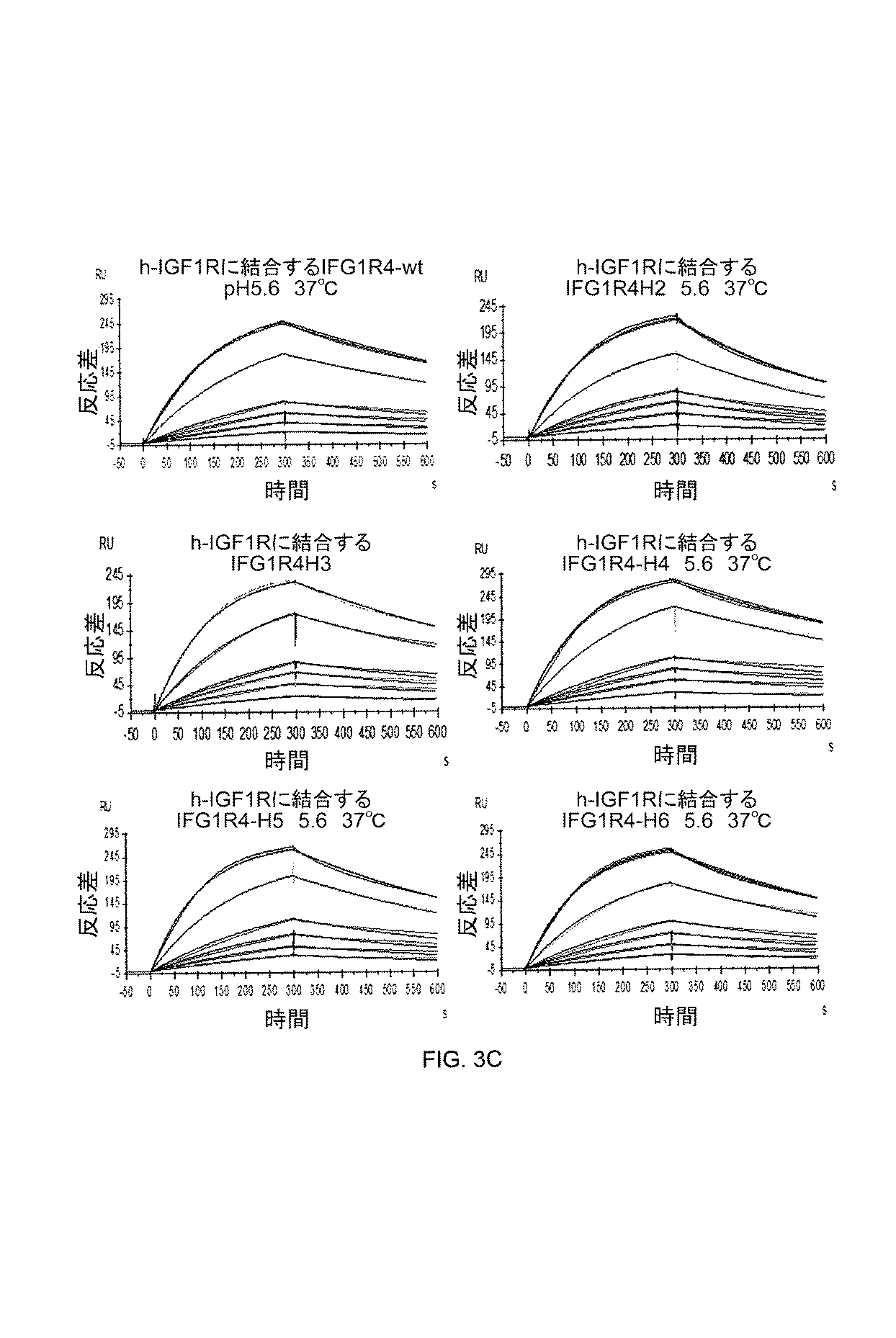
IGF1R−4 VHHが、天然リガンドIGF−1と同じ受容体上のエピトープに結合しないことを実証するために、SPR解析をさらに使用した(図3D)。上述のとおり、実験を準備し、実施し、解析した。新たに固定化されたヒトIGF1R表面に対する結合を、25×KDの濃度のヒトIGF1リガンドの注入と、続く両方が25×KDの濃度でのIGF1R−4の同時注入により、20μl/minの流量、5分の注入時間で研究した。表面を、ランニング緩衝液での洗浄により再生した。天然リガンドIGF−1は、70RUに及ぶ飽和度で受容体に結合した。IGF1R−4 VHHは、IGF1R−IGF−1複合体に、予測された約265RU(相対的単位、結合飽和)で結合した。受容体に対するIGF1R−4 VHH及びIGF−1の両方の同時結合は、両方が異なるエピトープに結合することを実証する。 SPR analysis was further used to demonstrate that IGF1R-4 V H H does not bind to the same epitope on the receptor as the natural ligand IGF-1 (FIG. 3D). The experiments were prepared, conducted and analyzed as described above. Binding to freshly immobilized human IGF1R surface, injection and the concentration of human IGF1 Ligand of 25 × K D, by co-injection of IGF1R-4 at both concentrations followed by 25 × K D, of 20 [mu] l / min The flow was studied at an injection time of 5 minutes. The surface was regenerated by washing with running buffer. The natural ligand IGF-1 bound to the receptor with a degree of saturation of 70 RU. IGF1R-4 V H H bound to the IGF1R-IGF-1 complex with the expected approximately 265 RU (relative unit, binding saturation). Simultaneous binding of both IGF1R-4 V H H, and IGF-1 to the receptor demonstrates that both bind to different epitopes.
ヒトインスリン受容体に結合するIGF1R−4の結合交差反応性もまた、SPRを使用して評価した。上述のとおり、実験を準備し、実施し、解析した。簡潔に述べると、ヒトIGF1Rに加えて、およそ4000共鳴単位(RU)の組換えヒトインスリン受容体(R&D systems)を、センサーチップCM5上の分離細胞上に固定化した。続いて、異なる濃度のIGF1R−4を表面上に流した(0.5〜50nM)。0.5nM IGF1R−4ですでにIGF1Rに結合することが観察できた一方で、50nMでもインスリン受容体表面への結合は観察されず(図3E)、IGF1R−5がインスリン受容体に結合できないことを示唆する。 Binding cross reactivity of IGF1R-4 binding to human insulin receptor was also assessed using SPR. The experiments were prepared, conducted and analyzed as described above. Briefly, in addition to human IGF1 R, approximately 4000 resonance units (RU) of recombinant human insulin receptor (R & D systems) were immobilized on isolated cells on sensor chip CM5. Subsequently, different concentrations of IGF1R-4 were run on the surface (0.5-50 nM). While 0.5 nM IGF1 R-4 could already be observed to bind to IGF1 R, no binding to the insulin receptor surface was observed even at 50 nM (FIG. 3E) and that IGF1 R-5 can not bind to the insulin receptor Suggest.
実施例7:脳内皮細胞によるIGF1R−4の内部移行
IGF1R−4が細胞内に内部移行するかどうかを決定するため、svARBEC細胞をCy5−5標識化IGF1R−4とインキュベートした。
Example 7 Internalization of IGF1R-4 by Brain Endothelial Cells To determine if IGF1R-4 is internalized into cells, svARBEC cells were incubated with Cy5-5 labeled IGF1R-4.
IGF1R−4 VHHをNHS−Cy5.5で標識化した。(タンパク質のN末端及び/又はリジン側鎖の)一級アミンとNHSエステルとの間の安定アミン結合によって標識化を行った。典型的には、10%v/vの1M炭酸塩/重炭酸塩緩衝液(759mM重炭酸塩、258mM炭酸塩)pH9.3を、PBS(1.06mM KH2PO4、154mM NaCl、5.6mM Na2HPO4)pH7.4中に調製された4mgのVHHに添加し、4mg/mLの最終濃度に調整した。10mg/mLでDMSO中に溶解されたNHS−Cy5.5を、染料のタンパク質に対する2×モル比で添加した。混合物を、1.5mL微量遠心管中で何度か反転させながら室温で2時間インキュベートした。インキュベーション後、未結合染料及び反応副産物を、Zeba Spin Desalting Columns、7K MWCO(Pierce)を使用して濾過し、Beckman DU530分光光度計(Beckman Coulter)を使用して測定した。Cy5.5標識化IGF1R−4又は陽性対照としてのFC5(1mg/ml)を、SV40不死化ラット脳内皮細胞(svARBEC)と4℃(図4、上パネル)で、したがって受動的で非特異的な輸送機序のみが生じることを可能にして、又は37℃(図4、下パネル)で、能動的機序、たとえば受容体媒介性エンドサイトーシスが生じることを可能にして、インキュベートした。コムギ胚芽凝集素及びDAPIでの共染色を実施し、細胞表面及び核をそれぞれ視覚化した。蛍光顕微鏡下で細胞を観察し、画像をキャプチャーした。 IGF1R-4 V H H was labeled with NHS-Cy5.5. Labeling was performed by a stable amine bond between the primary amine (at the N-terminus of the protein and / or the lysine side chain) and the NHS ester. Typically, 10% v / v 1 M carbonate / bicarbonate buffer (759 mM bicarbonate, 258 mM carbonate) pH 9.3, PBS (1.06 mM KH 2 PO 4 , 154 mM NaCl, 5. It was added to 4 mg of V H H prepared in 6 mM Na 2 HPO 4 ) pH 7.4 and adjusted to a final concentration of 4 mg / mL. NHS-Cy5.5 dissolved in DMSO at 10 mg / mL was added at a 2 × molar ratio of dye to protein. The mixture was incubated for 2 hours at room temperature with several inversions in a 1.5 mL microfuge. After incubation, unbound dye and reaction byproducts were filtered using Zeba Spin Desalting Columns, 7K MWCO (Pierce) and measured using a Beckman DU530 spectrophotometer (Beckman Coulter). Cy5.5-labeled IGF1R-4 or FC5 (1 mg / ml) as a positive control in SV40 immortalized rat brain endothelial cells (svARBEC) and 4 ° C. (FIG. 4, upper panel), thus passive and nonspecific Incubation allows either only a transport mechanism to occur or at 37.degree. C. (FIG. 4, lower panel) to allow an active mechanism, such as receptor-mediated endocytosis, to occur. Co-staining with wheat germ agglutinin and DAPI was performed to visualize cell surface and nuclei, respectively. The cells were observed under a fluorescence microscope and an image was captured.
4℃でインキュベートされた場合、IGF1R−4は、細胞の外部に、コムギ胚芽凝集素で染色された細胞膜と共局在して見出された。対照的に、37℃でインキュベートされたとき、IGF1R−4は、細胞内部の小胞、おそらくはエンドソームに蓄積し、抗体が能動輸送機序によって細胞内に内部移行したことを示唆する。同様の振舞いが、クラスリン被覆小胞を介したエネルギー依存性エンドサイトーシスにより細胞に侵入することが以前に示されているFC5について観察された(Abulrobら2005)。 When incubated at 4 ° C., IGF1R-4 was found outside the cells co-localized with wheat germ agglutinin-stained cell membranes. In contrast, when incubated at 37 ° C., IGF1R-4 accumulates in vesicles, probably endosomes, inside the cell, suggesting that the antibody has been internalized into the cell by an active transport mechanism. Similar behavior was observed for FC5, which was previously shown to enter cells by energy-dependent endocytosis via clathrin-coated vesicles (Abulrob et al. 2005).
実施例8:IGF1R−4−mFcコンストラクトの製造
マウス抗体結晶性断片(Fc、mFc2b)と融合したIGF1R−4 VHHを含むコンストラクトを調製し、発現させ、単離した。IGF1R−4−mFcコンストラクトの配列を図5Aに示し、分子の概略図を図5Bに示す。融合タンパク質はまた、図5Aの配列に示されていないN末端シグナルペプチド(MEFGLSWVFLVAILKGVQC、配列番号37)を含んでいた。
Example 8 Preparation of IGF1R-4-mFc Constructs Constructs containing IGF1R-4 V HH fused to a murine antibody crystalline fragment (Fc, mFc2b) were prepared, expressed and isolated. The sequence of the IGF1R-4-mFc construct is shown in FIG. 5A and a schematic of the molecule is shown in FIG. 5B. The fusion protein also contained an N-terminal signal peptide (MEFGLSWVFLVAILKGVQC, SEQ ID NO: 37) not shown in the sequence of FIG. 5A.
IGF1R−4 cDNAを、マウスFc2b断片を含有する哺乳動物発現ベクターpTT5(Durocher、2002)内にクローニングした。得られたベクターのポリプレックスを、187.5μgのpTT5−IR5mFc2b、56.25μgのpTT−AKTdd(プロテインキナーゼBの活性化変異体)、18.75μgのpTTo−GFP(トランスフェクション効率をモニタリングするため)、及び112.5μgのサケ精巣DNA(Sigma−Aldrich)を含有する25mlのプラズミドDNA溶液、並びに、1.125mgのPEIpro(商標)(PolyPlus Transfection)を含有する25mlのPEI溶液を混合することにより事前に形成した。両方の溶液が、F17培地中で作られた。混合物を、細胞培地への添加の10分前にインキュベートした。短縮EBNA1タンパク質を安定的に発現するCHO細胞の450ml培養物(CHO−3E7)をF17培地(Invitrogen)中で成長させ、50mlのポリプレックスをトランスフェクトした。トランスフェクション24時間後、培養物に12.5mlの40%(w/v)トリプトンN1(Organotechnie)溶液及び1.25mlの200mMバルプロ酸溶液を加えた。トランスフェクション8日後に培養物を回収し、遠心により清澄化した。清澄化培地を、5mlのプロテインA MabSelect SuRe樹脂(GE Healthcare)を充填したカラムへのその適用前に、0.22μm膜に通して濾過した。ローディング後、5容量のリン酸緩衝生理食塩水(PBS)pH7.1でカラムを洗浄し、100mMクエン酸ナトリウム緩衝液pH3.0で抗体を溶出した。溶出抗体を含有する画分をプールし、PBS中で平衡化した脱塩Econo−Pacカラム(BioRad)上にローディングすることにより緩衝液交換を実施した。次に、Millex GP(Millipore)フィルターユニット(0.22μm)を通過させることにより、脱塩抗体を無菌濾過し、アリコートにした。 The IGF1R-4 cDNA was cloned into a mammalian expression vector pTT5 (Durocher, 2002) containing a murine Fc2b fragment. To monitor the polyplex of the obtained vector, 187.5 μg pTT5-IR5mFc2b, 56.25 μg pTT-AKTdd (activation mutant of protein kinase B), 18.75 μg pTTo-GFP (transfection efficiency) By mixing 25 ml of a plasmid DNA solution containing 112.5 μg of salmon testes DNA (Sigma-Aldrich) and 25 ml of a PEI solution containing 1.125 mg of PEIproTM (PolyPlus Transfection) Formed in advance. Both solutions were made in F17 medium. The mixture was incubated 10 minutes prior to addition to the cell culture medium. A 450 ml culture (CHO-3E7) of CHO cells stably expressing truncated EBNA1 protein (CHO-3E7) was grown in F17 medium (Invitrogen) and transfected with 50 ml of polyplex. Twenty-four hours after transfection, 12.5 ml of 40% (w / v) Tryptone N1 (Organotechnie) solution and 1.25 ml of 200 mM valproic acid solution were added to the culture. Cultures were harvested 8 days after transfection and clarified by centrifugation. The clarified media was filtered through a 0.22 μm membrane prior to its application to a column packed with 5 ml of Protein A MabSelect SuRe resin (GE Healthcare). After loading, the column was washed with 5 volumes of phosphate buffered saline (PBS) pH 7.1 and the antibody was eluted with 100 mM sodium citrate buffer pH 3.0. Fractions containing eluted antibody were pooled and buffer exchange was performed by loading on a desalted Econo-Pac column (BioRad) equilibrated in PBS. The desalted antibody was then sterile filtered and aliquoted by passage through a Millex GP (Millipore) filter unit (0.22 μm).
実施例9:インビトロ血液脳関門モデルを通過するIGF1R−4の輸送
IGF1R−4 VHH、そのヒト化バージョン(H2〜H6)、及びIGF1R−4−mFc(実施例8)が血液脳関門を通過するかどうかを評価するため、下に述べるインビトロアッセイを使用した。実験を要約するフローチャートを図6Aに示す。
SV40不死化成体ラット脳内皮細胞(Sv−ARBEC)を、以前に記述されたとおりインビトロ血液脳関門(BBB)モデルを作製するために使用した(Garbergら、2005;Haqqaniら、2012)。Sv−ARBEC(細胞80,000個/膜)を、1mlの成長培地中の0.1mg/mLラット尾コラーゲンI型被覆組織培養物インサート(細孔サイズ−1μm、表面積0.9cm2、Falcon)上に播種した。インサートアセンブリの下のチャンバーは、不死化新生仔ラット星状細胞馴化培地を1:1(v/v)比で添加した2mlの成長培地を含有していた。等モル量(5.6μM)の正(FC5)又は負(A20.1、クロストリジウム・ディフィシル毒素A結合性VHH、及びEGFR結合性VHHであるEG2)の対照、IGF1R−4 VHH、ヒト化バージョン(H2−H6)又はIGF1R−4mFcを、このラットインビトロBBBモデルを横断するそれらの能力について試験した。BBBの管腔側への等モル量のsdAbの曝露後に、試料を15、30、及び60分後に反管腔側から採取した。次に、各試料のsdAb含有量を、Haqqaniら(2012)により記述されたとおり、質量分析(多重反応モニタリング−同位体標識化内部標準、MRM−ILIS)により定量した(下の方法の説明を参照のこと)。
Example 9 Transport of IGF1 R -4 through In Vitro Blood-Brain Barrier Model IGF1 R-4 V H H, Humanized Versions (H2-H6) and IGF1 R-4-mFc (Example 8) of the Blood-Brain Barrier The in vitro assay described below was used to assess if it passed. A flow chart summarizing the experiment is shown in FIG. 6A.
SV40 immortalized rat brain endothelial cells (Sv-ARBEC) were used to generate an in vitro blood-brain barrier (BBB) model as previously described (Garberg et al., 2005; Haqqani et al., 2012). Sv-ARBEC (80,000 cells / cell membrane), 0.1 mg / mL rat tail collagen type I coated tissue culture insert in 1 ml growth medium (pore size-1 μm, surface area 0.9 cm 2 , Falcon) Sowing on top. The chamber under the insert assembly contained 2 ml of growth medium supplemented with immortalized neonatal rat astrocyte conditioned medium at a 1: 1 (v / v) ratio. Control of equimolar amounts (5.6 μM) of positive (FC5) or negative (A20.1, Clostridium difficile toxin A binding V H H, and EG2 that is EGFR binding V H H), IGF1 R-4 V H H, humanized version (H2-H6) or IGF1R-4mFc were tested for their ability to cross this rat in vitro BBB model. After exposure of equimolar amounts of sdAb to the luminal side of the BBB, samples were taken from the abluminal side after 15, 30, and 60 minutes. Next, the sdAb content of each sample was quantified by mass spectrometry (multiple reaction monitoring-isotope labeled internal standard, MRM-ILIS) as described by Haqqani et al. (2012) (explanation of the method below) See for reference).
見かけ透過係数の決定:定量値を直接プロットでき、又はPapp(見かけ透過係数)値を所定の式で決定し(図6A)、プロットできる。Papp値が、分子についてBBBを横断する能力を決定するために一般に使用される。[Qr/dt=レシーバーコンパートメントにおける累積量対時間、A=細胞単層の面積、C0=投与溶液の初期濃度]。Papp値は、脳内皮単層を通過する化合物の特異的透過性の尺度である。 Determination of Apparent Permeability Coefficient: Quantitative values can be plotted directly or P app (apparent permeability coefficient) values can be determined according to a predetermined equation (FIG. 6A) and plotted. P app values are commonly used to determine the ability of molecules to cross the BBB. [Qr / dt = cumulative volume in receiver compartment versus time, A = area of cell monolayer, C0 = initial concentration of dosing solution]. P app value is a measure of the specific permeability of a compound that passes through the brain endothelial monolayer.
結果を図6B〜図6Dに示す。得られた結果は、複数の独立実験から得られた平均Papp値である。両方の陰性対照が、非常に低いPapp値を有し、BBBモデルを通過するこれらのVHHの非特異的輸送又は傍細胞輸送が最小限であることを示す。IGF1R−4 VHHは、高いPapp値を有し、インビトロBBBモデルを通過する高い輸送率を示す。IGF1R−4 VHHのPappは、陽性対照、すなわちBBB透過性VHH FC5(国際公開第02/057445号)のものよりも4倍高い。結果は、IGF1R−4がインビトロで脳内皮細胞を通過する促進された細胞間輸送を受け、インビボで類似の特性を有しうることの強い示唆を与える。ヒト化IGF1R−4 VHH変異体H3、H4及びH5は、IGF1R−4 VHHと比較して、同等(H3)又はわずかに低いPapp値を示した。H2変異体は、有意に減弱化されたインビトロBBBモデル通過能力を示した一方で、H6変異体はこの能力を完全に失い、内部対照VHH A20.1(図6C)と同じPapp値を示し、さらなる研究から除外した。IGF1R−4mFcのPapp値(図6D)は、IGF1R−4 VHH(図6B)の6倍小さい値と同等であり、IGF1R−4が大きな(抗体サイズの)分子を、BBBを通過させて「運搬」できることを示す。陽性対照であるFC5mFcのPapp値は、IGF1R−4mFcの値よりも3倍低かった。カーゴ分子に連結されたIGF1R−5又はヒト化バージョンを含むコンストラクト(MW 約110kDa又は180kDa)もまた、BBBを通過してフェリーされると示されたことに留意する価値はある(データは示さず)。 The results are shown in FIGS. 6B-6D. The results obtained are the average P app values obtained from several independent experiments. Both negative controls have very low P app values, indicating that non-specific or paracellular transport of these V H H across the BBB model is minimal. IGF1R-4 V H H has high P app values and exhibits high transport rates across in vitro BBB models. The P app of IGF1R-4 V H H is four times higher than that of the positive control, ie BBB permeable V H HFC5 (WO 02/057445). The results provide a strong indication that IGF1R-4 undergoes facilitated intercellular transport across brain endothelial cells in vitro and may have similar properties in vivo. Humanized IGF1R-4 V H H variants H3, H4 and H5 showed comparable (H3) or slightly lower P app values compared to IGF1 R-4 V H H. H2 variant, while showed significantly attenuated in vitro BBB model passing ability, H6 mutant completely lost this ability, internal control V H H A20.1 (Figure 6C) and the same P app values Were excluded from further study. P app values of IGF1R-4mFc (FIG. 6D) is equivalent to 6 times smaller value of IGF1R-4 V H H (FIG. 6B), the IGF1R-4 is a large (antibody size) molecule, to cross the BBB Show that they can be "carried". The P app value of the positive control, FC5mFc, was three times lower than that of IGF1R-4mFc. It is worthwhile to note that a construct (MW ̃110 kDa or 180 kDa) containing IGF1R-5 or a humanized version linked to a cargo molecule was also shown to be ferryed across the BBB (data not shown) ).
別のIGF1R結合性VHHであるIGF1R−1もまた、mFc融合物としてこのアッセイにおいて試験した。IGF1R−1mFcは、対照A20.1 VHH(図6D)よりもわずかに高いPapp値しか有さなかったことから、免疫ラマライブラリーから単離されたIGF1R結合性VHHのサブセットだけが、BBB通過特性を有することが示される。 Another IGF1 R binding V H H, IGF1 R-1, was also tested in this assay as a mFc fusion. Since IGF1 R-1 mFc had only a slightly higher P app value than control A20.1 V H H (FIG. 6D), only a subset of IGF1 R binding V H H isolated from the immune llama library Are shown to have BBB passing characteristics.
MRM−ILIS法を使用したVHHの絶対量測定。本方法は、すべてHaqqaniら(2012)に記載されたとおりである。簡潔に述べると、VHHについてSRM(選択反応モニタリング、多重反応モニタリング(MRM)としても知られている)アッセイを開発するため、各VHHをまず、データ依存性取得を使用してナノLC−MS/MSにより解析し、すべてのイオン性ペプチドを同定した。各ペプチドについて、3〜5個の最も強力な断片イオンを選択した。これらの断片を消化物のアトモル量(約100〜300amol)でモニタリングするための初期SRMアッセイを開発した。低い量での再現性のある強度比を示した(すなわち、より高い量と比較してピアソンr2≧0.95を有していた)断片を、安定的であると考え、最終SRMアッセイのために選択した。アッセイのさらなる最適化のため、近接したm/z(質量対電荷比)及び溶出時間を有するペプチドを選択しないよう注意しながら、各ペプチドの溶出時間もまた含めた。 Measurement of absolute amount of V H H using MRM-ILIS method. The method is all as described in Haqqani et al. (2012). Briefly, V H H for SRM for developing (selected reaction monitoring, also known as multiple reaction monitoring (MRM)) assays, each V H H First, using the data dependency acquisition nano Analysis by LC-MS / MS identified all ionic peptides. For each peptide, 3 to 5 most potent fragment ions were selected. An initial SRM assay was developed to monitor these fragments at a molar amount of digest (about 100-300 amol). Fragments that showed reproducible intensity ratios at lower amounts (ie, had Pearson r2 0.9 0.95 compared to higher amounts) were considered stable and for the final SRM assay Was selected. Elution times for each peptide were also included, taking care not to select peptides with close m / z (mass to charge ratio) and elution times for further optimization of the assay.
細胞培地又は体液(血清又は脳脊髄液(CSF))におけるVHHの典型的なマルチプレックスSRM解析は、既知の量のILIS(0.1〜10nM)を添加し、その後、100〜400ngのCSF又は培養された培地タンパク質(0.3〜1μl)又は約50〜100ngの血清タンパク質(1〜3ナノリットル)をナノLC−MSシステム内に注入することを伴った。各標的ペプチドイオンの前駆m/zが、標的のための特定の溶出時間においてイオントラップ内で選択され(残りの無関係なイオンは破棄された)、衝突誘起解離(CID)フラグメンテーション、及び検出器によるモニタリングのためのイオントラップ内の所望の断片イオンのみの選択が続いた。定量解析のため、LTQ(ThermoFisher)により生成された生ファイルを、標準質量分析データ形式mzXMLに変換し、MatchRxソフトウェアの修正バージョンである、Q−MRM(定量的−MRM、Haqqaniら 2012を参照のこと)と呼ばれるインハウスのソフトウェアを使用して強度を抽出した。各VHHについて、抽出イオンクロマトグラフを、全溶出時間にわたる0.25Da以内の断片m/zの合計強度からなるその断片イオンのそれぞれについて得た。各断片について最終強度値を得るため、0.5分以内の予測保持時間のすべての強度を合計した。VHHを、そのペプチドのうちの少なくとも1つの断片が予測強度比を示す、すなわち、最終強度値がその対応する純VHHの最終強度値と比較して強いピアソン相関r≧0.95及びp<0.05を示す場合に、試料において検出可能なものとして定義した。 Typical multiplex SRM analysis of V H H in cell culture medium or body fluid (serum or cerebrospinal fluid (CSF)) adds a known amount of ILIS (0.1-10 nM) followed by 100-400 ng of Injecting CSF or cultured media protein (0.3-1 μl) or about 50-100 ng serum protein (1-3 nanoliters) into the nano LC-MS system. The precursor m / z of each target peptide ion is selected in the ion trap at a specific elution time for the target (remaining irrelevant ions are discarded), collision induced dissociation (CID) fragmentation, and by detector Selection of only desired fragment ions in the ion trap followed for monitoring. For quantitative analysis, convert the raw file generated by LTQ (ThermoFisher) to the standard mass spectrometry data format mzXML and refer to Q-MRM (Quantitative-MRM, Haqqani et al. 2012), a modified version of MatchRx software The intensity was extracted using in-house software called a). For each V H H, an extraction ion chromatograph was obtained for each of its fragment ions consisting of a total intensity of fragments m / z within 0.25 Da over the entire elution time. All intensities of the predicted retention time within 0.5 minutes were summed to obtain a final intensity value for each fragment. The V H H indicates that at least one fragment of the peptide exhibits a predicted intensity ratio, ie, the final intensity value is strong compared to the final intensity value of its corresponding pure V H H, Pearson correlation r ≧ 0.95 And was defined as detectable in the sample if it showed p <0.05.
VHHの混合物を含有する試料(培地、血清、CSF)を、以前に記述されたとおり、還元し、アルキル化し、トリプシン消化した。(Haqqaniら、2012;Gergovら、2003)消化物(トリプシンペプチド)を酢酸で酸性化させ(5%最終濃度)、LTQ XL ETD又はLTQ Orbitrap ETD質量分析計(ThermoFisher、Waltham、MA)と組み合わせた逆相ナノAcquity UPLC(Waters、Milford、MA)上で解析した。試料の所望のアリコートを注入して300μm I.D.×0.5mm 3μm PepMaps C18トラップ(ThermoFisher)上に充填し、次に100μm I.D.×10cm 1.7μm BEH130C18 ナノLCカラム(Waters)上に、1分間で0%〜20%、16分間で20%〜46%、1分間で46%〜95%アセトニトリル(0.1%ギ酸中)の勾配を使用して、400nL/minの流量で溶出した。溶出ペプチドを、ペプチドイオンのフラグメンテーションのためのCIDを使用したMS/MS及びSRM解析のため、エレクトロスプレーイオン化(ESI)により質量分析計内へとイオン化した。ヘリウムを衝突ガスとして、35%の標準化衝突エネルギー及び30msの活性化時間でCIDを実施した。線形イオントラップ内へのイオン注入時間を、6×103の自動利得制御(AGC)標的値及び200msの最大蓄積時間を使用して機器により調整した。
マルチプレックスアッセイにおいて各VHHの検出及び定量のために使用されたVHH特異的ペプチドを表2に示す。
Samples containing a mixture of V H H (media, serum, CSF) were reduced, alkylated, and trypsin digested as previously described. (Haqqani et al., 2012; Gergov et al., 2003) Acidified digestate (tryptic peptide) with acetic acid (5% final concentration), combined with LTQ XL ETD or LTQ Orbitrap ETD mass spectrometer (ThermoFisher, Waltham, Mass.) Reversed phase nano-Acquity UPLC (Waters, Milford, Mass.) Was analyzed. Inject the desired aliquot of sample to 300 μm I. D. × 0.5
The V H H specific peptides used for detection and quantification of each V H H in multiplex assays are shown in Table 2.
実施例10:インビボ及びエクスビボ光学イメージング
IGF1R−4 VHHが静脈内(iv)注射後に脳内に蓄積するかどうかを決定するため、IGF1R−4 VHH及び陰性対照A20.1 VHHを、近赤外蛍光トレーサーであるCy5.5にコンジュゲートし、実施例7に記載のとおり精製した。2.5mg/kgのIGF1R−Cy5.5又はA20.1 Cy5.のいずれか(150μl容量中)を、尾静脈を介してCD−1ヌードマウスに注射し、それらを腹臥位で、eXplore Optix前臨床画像装置MX2(Advanced Research Technologies、QC)で、注射30分後に生きたまま撮像した。注射の1時間後、2%イソフルラン(Baxter Canada、Mississauga、ON、Canada)で動物を麻酔にかけ、心穿刺を行い、1EU/mLヘパリン(Organon、Toronto、ON、Canada)を添加した10mLの生理食塩水(Baxter Canada、Mississauga、ON、Canada)を、同じニードルから2mL/minで灌流し、血液を除去した(右室切開の部位での血液非含有流体の出現により確認した)。灌流した脳を除去し、eXplore Optixを使用してエクスビボで撮像した。イメージングプロトコルは、Iqbalら、2011に詳細に記載のとおりだった。
Example 10: For in vivo and ex vivo optical imaging IGF1R-4 V H H determines whether to accumulate in the brain following intravenous (iv) injection, IGF1R-4 V H H and negative controls A20.1 V H H Was conjugated to the near infrared fluorescent tracer Cy5.5 and purified as described in Example 7. 2.5 mg / kg IGF1 R-Cy 5.5 or A20.1 Cy5. (In a volume of 150 μl) is injected into CD-1 nude mice via the tail vein and they are placed in a prone position, on the eXplore Optix preclinical imaging device MX2 (Advanced Research Technologies, QC), for 30 minutes injection I photographed it alive later. One hour after injection, animals are anesthetized with 2% isoflurane (Baxter Canada, Mississauga, ON, Canada), cardiac puncture and 10 mL of saline supplemented with 1 EU / mL heparin (Organon, Toronto, ON, Canada) Water (Baxter Canada, Mississauga, ON, Canada) was perfused from the same needle at 2 mL / min to remove blood (confirmed by the appearance of blood free fluid at the site of the right ventricular incision). Perfused brains were removed and imaged ex vivo using eXplore Optix. The imaging protocol was as described in detail in Iqbal et al., 2011.
結果を図7に示す。Cy5.5−IGF1R−4を注射されたマウスは、増強された光学シグナルをインビボの頭部領域(上パネル、矢印)及び灌流後のエクスビボ脳(下パネル、矢印)において示した一方で、Cy5.5−A20.1注射マウスは、低いバックグラウンドシグナルしか示さなかった。データは、IGF1R−4 VHHが、造影剤(Cy5.5、1kD)を、インビボで血液脳関門を通過させて送達できることを示唆する。 The results are shown in FIG. Mice injected with Cy5.5-IGF1R-4 showed enhanced optical signals in the in vivo head area (upper panel, arrow) and ex vivo brain after perfusion (lower panel, arrow) while Cy5 The 5-A20.1 injected mice showed only low background signal. The data suggest that IGF1 R-4 V H H can deliver imaging agents (Cy 5.5, 1 kD) across the blood-brain barrier in vivo.
実施例11:ガラニンへのIGF1R−4のコンジュゲーション
IGF1R−4 VHHがインビボで血液脳関門(BBB)を通過し、それ自体ではBBBを通過できない分子を、BBBを通過させて「フェリー」できるかどうかを決定するため、神経ペプチドガラニンを、IGF1R−4 VHH又はIGF1R4−mFcに化学的にコンジュゲートし、全身投与した。ガラニンは、脳組織において発現するGalR1及びGalR2に結合することにより鎮痛をもたらす向神経活性ペプチドである。末梢的に与えられた場合、ガラニンは鎮痛効果を有さない。なぜなら、それ自体はBBBを通過できないからである(Robertsonら、2011)。
Example 11: Conjugation of IGF1 R-4 to Galanin IGF1 R-4 V H H crosses the blood-brain barrier (BBB) in vivo, and molecules that can not cross the BBB by themselves cross the BBB to “ferry” to determine whether the neuropeptide galanin, chemically conjugated to IGF1R4 V H H or IGF1R4-mFc, administered systemically. Galanin is a neuroactive peptide that provides analgesia by binding to GalR1 and GalR2 expressed in brain tissue. When given peripherally, galanin has no analgesic effect. Because it can not pass the BBB by itself (Robertson et al., 2011).
IGF1R−4 VHHを、システアミド修飾C末端を有するラットガラニン(Gal)断片(Biomatic)(GWTLNSAGYLLGPHAIDNHRSFSDKHGLT−システアミド、配列番号36)にコンジュゲートした。コンジュゲーションのスキームを図8Aに示す。 IGF1R-4 V H H was conjugated to rat galanin (Gal) fragment (Biomatic) with cysteamide modified C-terminus (GWTLNSAGYLLGPHAIDNHRSFSDKHGLT-systemamide, SEQ ID NO: 36). The scheme of conjugation is shown in FIG. 8A.
簡潔に述べると、0.5×PBS、2.5mM EDTA中の[2mg/ml]での5mgのIGF1R−4 VHH(実施例4)を、436.4μlの2.5mg/mlスルホスクシンイミジル−4−(N−マレイミドメチル)シクロヘキサン−1−カルボキシレート(スルホ−SMCC)(7.5×過剰モル比)と混合した。次に、窒素ガスで混合物を洗い流し、30分間室温(RT)でインキュベートし、スルホ−SMCCのNHSエステルアームをVHH上のアミンと反応させた。続いて、10mlの7K Zebaカラム(Pierce)を使用して、未反応スルホ−SMCCをマレイミド−活性化IGF1R−5 VHHから除去した。試料ローディングの前に、カラムを5ml PBSで3回洗浄し、1000×gで2分間回転させた。試料ローディング後に、カラムに200μlのPBSを補給し、2分間、1000×gで回転させた。IGF1R−Fcコンストラクトについて、5mgを約68μlのスルホ−SMCC(6.5×過剰モル比)と、上述と同様に反応させた。
Briefly, 5 mg of IGF1R-4 VH H (Example 4) at [2 mg / ml] in 0.5 × PBS, 2.5 mM EDTA, 436.4 μl of 2.5 mg / ml sulfosk It was mixed with sinimidyl-4- (N-maleimidomethyl) cyclohexane-1-carboxylate (sulfo-SMCC) (7.5 × molar excess ratio). The mixture was then flushed with nitrogen gas and incubated for 30 minutes at room temperature (RT) to react the NHS ester arm of sulfo-SMCC with the amine on V H H. Then, using the
別々に並行して、システアミド修飾C末端ガラニン(Gal−cya)を、10mgの凍結乾燥粉末を10mlのエンドトキシンフリー水中に溶解させることにより調製し、1mg/mlストックを作った(ガラニン−cya粉末は、精製中のジスルフィド架橋形成を防止するため、少量のDTTを有する)。最後に、100μlの0.5M EDTAを添加した(5mM最終濃度)。 Separately in parallel, cysteamide modified C-terminal galanin (Gal-cya) was prepared by dissolving 10 mg of lyophilized powder in 10 ml of endotoxin free water to make a 1 mg / ml stock (Galanin-cya powder is , With a small amount of DTT to prevent disulfide bridge formation during purification). Finally, 100 μl of 0.5 M EDTA was added (5 mM final concentration).
精製マレイミド活性化IGF1R−4 VHH及びIGF1R−Fcコンストラクト(2.6ml)を、0.5×PBS、2.5mM EDTAで5mlまで希釈し、次に、ボルテックスにかけながら5ml又は1mlのGal−cyaをそれぞれ添加した。試料を窒素で洗い流し、密封し、一晩4℃でインキュベートした。翌日、Amicon−15 10K及び30Kカラム(Millipore)をそれぞれ使用して、未反応のGal−cyaを除去した。試料をカラムに添加し、4000×gで7分間、体積が5mlに減少するまで回転させた。5mlの0.5×PBS、2.5mM EDTAを、カラムのインサート中の残りの5ml試料に添加し、試料が4mlに減少するまで再び回転させた。次に、コンジュゲートされた試料を、10ml 7K Zebaカラム(Pierce)に添加し、上述のとおり調製し、次に2分間、1000xgで回転させた。 Purification maleimide activated IGF1R-4 V H H and IGF1R-Fc construct the (2.6ml), 0.5 × PBS, diluted with 2.5 mM EDTA to 5ml, then, of 5ml or 1ml while vortexing Gal- Each cya was added. The samples were flushed with nitrogen, sealed and incubated overnight at 4 ° C. The next day, unreacted Gal-cya was removed using Amicon-15 10K and 30K columns (Millipore) respectively. The sample was applied to the column and spun at 4000 × g for 7 minutes until the volume was reduced to 5 ml. 5 ml of 0.5 × PBS, 2.5 mM EDTA was added to the remaining 5 ml sample in the insert of the column and spun again until the sample was reduced to 4 ml. The conjugated samples were then added to a 10 ml 7K Zeba column (Pierce), prepared as described above, and then spun at 1000 × g for 2 minutes.
次に、コンジュゲートされたIGF1R−4−Gal及びIGF1R−4 Fc−Gal試料を、16%又は10%SDS−PAGE非還元ゲル上に走らせ、銀染色し、コンジュゲーション後の分子量サイズの変化を確認した。1つのVHH又はIGF1R−Fcコントラクト当たり約1〜2個のガラニン分子を達成するため、反応を滴定した。結果は、IGF1R−4 VHH上へのガラニンのロードを確認する(図8Bを参照のこと)。 The conjugated IGF1R-4-Gal and IGF1R-4 Fc-Gal samples are then run on a 16% or 10% SDS-PAGE non-reducing gel, silver stained, and changes in molecular weight size after conjugation confirmed. Reactions were titrated to achieve approximately 1-2 galanin molecules per V H H or IGF1 R-Fc contract. The results confirm the loading of galanin onto IGF1R-4 V H H (see FIG. 8B).
エンドトキシン除去及びエンドトキシンレベルの決定:Amicon Ultra分子量カットオフ(MWCO)セルロース膜スピンカラム(Millipore)を使用して、エンドトキシンを除去した。第1に、15mlのVHH調製物を、4000×gでの10分間の遠心によりAmicon−15〜50K MWCOカラムを通過させた。溶出物を回収した。次に、この溶出物を、Amicon−15〜10K MWCOカラムに添加し、4000×gで7〜10分間回転させ、15mlから7.5mlへの上清体積の低減をもたらした。カラム内の上清体積を、PBSを添加することにより15mlに戻すよう調整した。カラムを上述のとおり再び回転させた。上清を回収し、10〜0.1EU/mlの感度範囲を有するカートリッジを使用したEndoSafe−PTSシステム(Charles River Laboratories International)でエンドトキシンレベルを測定した。25μlの試料を、カートリッジ上の4つのウェルのそれぞれにロードし、必要に応じて希釈した。1mg当たりEU<1を有する試料のみを、動物研究に使用した。 Endotoxin removal and determination of endotoxin levels: Endotoxin was removed using an Amicon Ultra Molecular Weight Cutoff (MWCO) cellulose membrane spin column (Millipore). First, 15 ml of the V H H preparation was passed through an Amicon-15-50 K MWCO column by centrifugation at 4000 xg for 10 minutes. The eluate was collected. This eluate was then applied to an Amicon-15-10 K MWCO column and spun at 4000 xg for 7-10 minutes resulting in a reduction of the supernatant volume from 15 ml to 7.5 ml. The supernatant volume in the column was adjusted back to 15 ml by adding PBS. The column was rotated again as described above. The supernatant was collected and endotoxin levels were measured with the EndoSafe-PTS system (Charles River Laboratories International) using a cartridge with a sensitivity range of 10-0.1 EU / ml. 25 μl of sample was loaded into each of the 4 wells on the cartridge and diluted as needed. Only samples with EU <1 per mg were used for animal studies.
実施例12:ハーグリーヴズモデルを使用したIGF1R−4−Galの輸送
IGF1R−4−Gal又はIGF1R−4−mFc−Gal(実施例11)が血液脳関門を通過するかどうかを評価するため、インビボアッセイを、国際特許公開第2011/127580号に以前に記載のとおり利用した。
Example 12 Transport of IGF1R-4-Gal Using Hargreaves Model In Vivo Assay to Assess Whether IGF1R-4-Gal or IGF1R-4-mFc-Gal (Example 11) Cross the Blood-Brain Barrier Was used as previously described in WO 2011/127580.
Hargreavesら(1988)に記載のものと類似した炎症性痛覚過敏のラットモデルを使用した。動物をポリプロピレンケージ1つ当たり3匹の群で入れ(ハーグリーヴズモデル)、餌及び水を自由に与えた。12時間の明/暗サイクル、24℃の温度及び50±5%の相対湿度で実験を行った。すべての動物処置が、NRC動物ケア委員会により認可され、カナダ動物ケア評議会ガイドラインを順守していた。 A rat model of inflammatory hyperalgesia similar to that described by Hargreaves et al. (1988) was used. Animals were placed in groups of 3 per polypropylene cage (Hagreaves model) and had free access to food and water. The experiment was performed at a light / dark cycle of 12 hours, a temperature of 24 ° C. and a relative humidity of 50 ± 5%. All animal treatments were approved by the NRC Animal Care Committee and were in compliance with the Canadian Animal Care Council guidelines.
このモデルにおいて、6〜8週齢の雄ウィスターラット(体重範囲230〜250g)に、低体積(30ゲージニードルで100μl)の完全フロイントアジュバント(CFA、油:生理食塩水1:1エマルション中に懸濁された熱殺傷M.チュバーキュローシス(M.tuberculosis)(Sigma))を、右後足に短いイソフルラン麻酔(3%)下で注射した。CFAは、侵害受容体を活性化し、慢性的疼痛状態及び痛覚過敏(有害な熱に対する高められた感受性)を創出する炎症促進性物質の放出を誘導する。足退避潜時を、足刺激に対する足底無痛測定器具(IITC Model # 336TG Life Science,Inc.)を使用した、両後足(炎症性及び非炎症性対照足)の足底表面における放射刺激の適用により測定した。動物がその足をなめるか又は動かすことにより反応するのにかかった時間を、陽性反応として解釈した(足退避潜時)。CFA投与前に、光強度灯を、ベースライン足退避潜時を17〜20秒の間で両後足に誘発するよう調整した。20秒以内に退避応答が生じない場合、光線を自動的に消灯して組織損傷を回避し、その足には最大スコアが割り当てられる。 In this model, 6-8 week old male Wistar rats (weight range 230-250 g) are suspended in low volume (100 μl with a 30 gauge needle) in complete Freund's adjuvant (CFA, oil: saline 1: 1 emulsion) Turbid, killed M. tuberculosis (Sigma)) was injected into the right hind paw under short isoflurane anesthesia (3%). CFA activates nociceptors and induces the release of proinflammatory substances that create chronic pain states and hyperalgesia (increased sensitivity to harmful heat). For paw withdrawal latency, radiation stimulation on the sole surface of both hind legs (inflammatory and non-inflammatory control paws) using a plantar painless measuring instrument (IITC Model # 336TG Life Science, Inc.) for foot stimulation Measured by application. The time taken for the animals to respond by licking or moving their paws was interpreted as a positive response (paw withdrawal latency). Prior to CFA administration, light intensity lights were adjusted to elicit a baseline paw withdrawal latency on both hindpaws between 17 and 20 seconds. If no withdrawal response occurs within 20 seconds, the light is automatically turned off to avoid tissue damage and the foot is assigned a maximum score.
CFA注射の2日後、化合物の投与前に、動物を操作し、ストレスを低減し、偽陽性反応を防止する目的で、少なくとも60分間無痛測定器具に順化させた。疼痛の発症(熱痛覚過敏)を検証するため、両足でベースラインを測定した。非炎症性の足を、注射された足に対する対照として使用した。「炎症性の足」について6秒超及び「正常な足」について17秒未満の足退避潜時を有する動物を、実験から除外した。 Two days after CFA injection, animals were manipulated to acclimate for at least 60 minutes painless measurement instruments for the purpose of reducing stress and preventing false positive reactions prior to administration of compounds. Baseline was measured on both feet to verify onset of pain (thermal hyperalgesia). A non-inflammatory foot was used as a control for the injected foot. Animals with paw withdrawal latencies greater than 6 seconds for "inflammatory foot" and less than 17 seconds for "normal foot" were excluded from the experiment.
IFG1R−4−Galが血液脳関門を通過させて送達され、脳実質において標的受容体(GalR1及び2)に結合できるかどうかを決定するため、ラットに、IGF1R−4−ガラニン(3mg/kg)若しくはIGF1R−4mFc−ガラニン(2mg/kg又は5mg/kg)又は対照化合物の1回の尾静脈注射をCFA注射3日後に投与した。各後足(炎症性及び非炎症性)について、15分ごとに3時間、足退避潜時(PWL)を試験した。足退避潜時の増加は、熱痛覚過敏の抑制を示し、これは、IGF1R−4による脳実質内へのガラニンの送達の成功によってしか得られない。ガラニンが鎮痛を誘導できるのは、脳実質内に存在するときだけであり、それ自体では無傷BBBを通過できない(Robertsonら、2011)。 To determine if IFG1R-4-Gal can be delivered across the blood-brain barrier and bind to target receptors (GalR1 and 2) in the brain parenchyma, rats should receive IGF1R-4-galanin (3 mg / kg). Alternatively, a single tail vein injection of IGF1R-4mFc-Galanin (2 mg / kg or 5 mg / kg) or control compound was administered 3 days after CFA injection. Paw withdrawal latencies (PWLs) were examined for each hindpaw (inflammatory and non-inflammatory) every 15 minutes for 3 hours. An increase in paw withdrawal latency is indicative of a suppression of thermal hyperalgesia, which is obtained only by successful delivery of galanin by IGF1 R-4 into the brain parenchyma. Galanin can induce analgesia only when it is present in the brain parenchyma and can not cross intact BBB by itself (Robertson et al., 2011).
足退避潜時(PWL、秒)対時間(分又は時間)の時間経過として結果を解析した(図9A及び9C)。図9Bは、結果を曲線下面積(AUC)として示し、それを最大可能効果の%(%MPE)と比較する。図9Dは、ピーク反応(Emax)での結果を(熱痛覚過敏の)パーセント反転として示す。 Results were analyzed as paw withdrawal latency (PWL, seconds) versus time (minutes or hours) over time (FIGS. 9A and 9C). FIG. 9B shows the results as area under the curve (AUC), which is compared to% of the maximum possible effect (% MPE). FIG. 9D shows the results at peak response (Emax) as percent reversal (of thermal hyperalgesia).
結果は、静脈内投与されたガラニンが、PBSと比較して疼痛を低減しないことを示す。対照的に、FC5−Gal又はIGF1R4−Galの単回注射は、測定可能な鎮痛効果をもたらし、このVHHが、BBBを通過させてガラニンを「フェリー」し、脳実質においてGalR1及び/又は2に結合することで鎮痛効果をもたらすことを示唆する。IGF1R−4−ガラニンの効果はFC5−Galにより誘導されるものよりも有意に顕著であり、IGF1R受容体が、推定されるFC5受容体よりも高いBBB輸送率を有することを示唆する。結果は、IGF1R−4 VHHが、受容体媒介性トランスサイトーシス経路を使用し、少なくとも3000Daの分子を、BBBを通過させて「フェリー」できることを実証する(抗体−ペプチドコンジュゲートの合計MWは、約18kDaである)。能動的な受容体媒介性輸送が必要とされる。なぜなら、BBBは、0.5kDaを超えるすべての親水性分子の通過を妨げると知られているからである。同様に、IGF1R4−mFc−ガラニンは、熱痛覚過敏の用量依存性抑制をもたらす一方で、A201.mFc−ガラニンは無効である。これは、IGF1R−4 VHHが、抗体(約80kD)とサイズが類似した分子を、BBBを通過させてフェリーできることを示唆する。IGF1R−4mFc−ガラニンは、熱痛覚過敏の抑制において、IGF1R−4−ガラニンよりも約5倍強力だったことから、二価ディスプレイ及び延長された血漿半減期が、脳送達の有効性の改善に寄与することが示唆される。 The results show that intravenously administered galanin does not reduce pain compared to PBS. In contrast, a single injection of FC5-Gal or IGF1R4-Gal results in measurable analgesic effect, the V H H is a galanin passed through the BBB to "ferry", GalR1 and / or in the brain parenchyma Suggests that binding to 2 brings about an analgesic effect. The effect of IGF1R-4-galanin is significantly more pronounced than that induced by FC5-Gal, suggesting that the IGF1R receptor has a higher BBB transport rate than the putative FC5 receptor. The results demonstrate that IGF1R-4 V H H can “ferry” molecules of at least 3000 Da through the BBB using a receptor-mediated transcytosis pathway (total MW of antibody-peptide conjugates Is about 18 kDa). Active receptor mediated transport is required. This is because BBB is known to prevent the passage of all hydrophilic molecules above 0.5 kDa. Similarly, while IGF1R4-mFc-Galanin results in dose-dependent suppression of thermal hyperalgesia, A201. mFc-Galanin is ineffective. This suggests that IGF1R-4 V H H can ferry molecules similar in size to the antibody (about 80 kD) past the BBB. IGF1R-4mFc-Galanin was about 5 times more potent than IGF1R-4-Galanin in suppressing thermal hyperalgesia, so bivalent display and extended plasma half-life can improve brain delivery efficacy It is suggested to contribute.
実施例13:CSF及び血漿におけるIGF1R−4レベル
IGF1R−4 VHHが脳内へと、具体的には脳脊髄液(CSF)中へと透過可能かどうかを決定するとともに、CSF及び血漿におけるその存在を定量するために、インビボアッセイを実施した。
Example 13: IGF1 R-4 Levels in CSF and Plasma To determine whether IGF1 R-4 V H H can penetrate into the brain, specifically into cerebrospinal fluid (CSF), as well as in CSF and plasma An in vivo assay was performed to quantify its presence.
動物をポリプロピレンケージに単独で入れ、餌及び水を自由に与えた。12時間の明/暗サイクル、24℃の温度及び50±5%の相対湿度で実験を行った。すべての動物処置が、NRC動物ケア委員会(NRC’s Animal Care Committee)により認可され、カナダ動物ケア評議会(the Canadian Council of Animal Care)ガイドラインを順守していた。 The animals were placed alone in polypropylene cages and given food and water ad libitum. The experiment was performed at a light / dark cycle of 12 hours, a temperature of 24 ° C. and a relative humidity of 50 ± 5%. All animal treatments were approved by the NRC's Animal Care Committee and were in compliance with the Canadian Council of Animal Care guidelines.
8〜10週齢の雄ウィスターラット(体重範囲、230〜250g)を使用した。CSF試料採取のため、動物の頸部及び頭部領域の毛を剃り、次に動物をプレキシグラスチャンバー内に入れ、3%イソフルランで適度に麻酔した。基本的にNirogiら(2009)に記載されたとおり、CSFを回収した。麻酔ラットを、金属枠器具(Vinicio Granados−Soto博士により寛大にも提供された;CINVESTAV、Mexico)に入れ、イヤーバーを使用して固定した。動物の頭部の位置を、およそ45°下向きに維持した。後頭隆起と脊椎の環椎との間に菱脳が現れる押圧可能な表面を、エタノール(75%)を浸した綿でこすることにより視覚可能にした。切開を伴わないCSF回収のため、100ccインスリンシリンジと接続された長さ10cmのPE−10チュービングで覆われた27Gニードル(Becton Dickinson、Mississauga、ON、Canada)を、水平に大槽の中心に挿入した。皮膚の断裂及び環椎後頭膜の断裂ゆえに、ニードルパスに沿った2つの抵抗点(クリック)を容易に感じることができる。ニードルが第2の抵抗点を通過したとき、CSF(40〜100μL)を、インスリンシリンジの穏やかな吸引を適用することによりニードルを通じて回収した。CSF試料採取後に、後開胸の心穿刺により対応する血液を回収し、バキュテナーチューブ(Becton Dickinson、Mississauga、ON、Canada)内に凝固活性化剤及びゲルとともに入れ、次に3,000×gで15分間遠心した。マイクロピペットを使用して血清を除去し、さらなる解析まで−80℃で急速冷凍した。 8-10 week old male Wistar rats (weight range 230-250 g) were used. The animals were shaved in the neck and head area for CSF sampling, then the animals were placed in a Plexiglas chamber and moderately anesthetized with 3% isoflurane. CSF was recovered essentially as described by Nirogi et al. (2009). Anesthetized rats were placed in metal frame devices (provided generously by Dr. Vinicio Granados-Soto; CINVESTAV, Mexico) and fixed using ear bars. The position of the animal's head was maintained approximately 45 ° downward. The depressible surface where the rhomboids appear between the occipital ridge and the atlas of the spine was made visible by rubbing with ethanol (75%). A 27 G needle (Becton Dickinson, Mississauga, ON, Canada) covered with a 10 cm long PE-10 tubing connected with a 100 cc insulin syringe is inserted horizontally into the center of the cistern for CSF recovery without dissection did. Due to skin tears and tears of the atlanto-occipital membrane, two points of resistance (clicks) along the needle path can be easily felt. When the needle passed the second resistance point, CSF (40-100 μL) was collected through the needle by applying a gentle aspiration of the insulin syringe. After CSF sampling, corresponding blood is collected by post thoracotomy cardiac puncture, placed in vacutainer tubes (Becton Dickinson, Mississauga, ON, Canada) with clotting activator and gel, then at 3,000 x g Centrifuge for 15 minutes. Serum was removed using a micropipette and flash frozen at -80 <0> C until further analysis.
血清及びCSF試料を、実施例9に記載のとおり、質量分析及びナノLC−SRMベース定量により解析した。 Serum and CSF samples were analyzed by mass spectrometry and nano LC-SRM based quantitation as described in Example 9.
CSF回収は繊細な処置であり、その間、CSFは容易に血液で汚染されうる。VHHの量は、血中よりもCSFにおいてはるかに小さい(<0.1%)と予測されるので、血液でのわずかな汚染でさえも、個別のCSF試料の値を深刻に損ないうる。したがって、血液汚染CSF試料のためのストリンジェントな除外基準を開発することが必要だった。血液−CSFアルブミン比を評価するため、血漿及びCSF中のアルブミンレベルを定量するためのナノLC−SRM法を開発した。アルブミンペプチドAPQVSTPTLVEAAR(配列番号37)を、マルチプレックスアッセイにおいて他のペプチドピークへの干渉を最小にするため、その固有の保持時間及びm/z値に基づいて選択した(Mol Pharm)。SRMを上述のとおり使用して、CSF及び血漿試料の両方において、ペプチドの強度を定量した。アルブミン比を以下のとおり各ラットについて算出した。
アルブミン比=1nLの解析された血漿当たりの強度/1nLの解析されたCSF当たりの強度
1500以下の比が、血液汚染と考えられた。
CSF recovery is a delicate procedure, during which CSF can be easily contaminated with blood. The amount of V H H is expected to be much smaller (<0.1%) in CSF than in blood, so even slight contamination with blood can seriously compromise the value of individual CSF samples . Therefore, it was necessary to develop stringent exclusion criteria for blood-contaminated CSF samples. To assess the blood-CSF albumin ratio, a nano LC-SRM method was developed to quantify albumin levels in plasma and CSF. The albumin peptide APQVSTPTLVEAAR (SEQ ID NO: 37) was selected based on its unique retention time and m / z value (Mol Pharm) to minimize interference with other peptide peaks in the multiplex assay. SRM was used as described above to quantify peptide strength in both CSF and plasma samples. The albumin ratio was calculated for each rat as follows.
An albumin ratio = 1 nL analyzed plasma intensity / 1 nL analyzed CSF intensity per 1500 or less ratio was considered as blood contamination.
結果を図10に示す。動物に、それぞれ6mg/kgのIGF1R−4、FC5又はA20.1を同時注射し、CSF及び血清を注射30分後に回収した。図は、IGF1R−4のCSF/血清比が、FC5の0.98%及びA20.1の0.017%と比較して、1.2%であることを示し、IGF1R−4及びFC5の両方が、同時注射された対照抗体であるA20.1よりも高いCSF透過性及び脳曝露を有することを示唆する。 The results are shown in FIG. Animals were co-injected with 6 mg / kg each of IGF1R-4, FC5 or A20.1 and CSF and serum were collected 30 minutes after injection. The figure shows that the CSF / serum ratio of IGF1R-4 is 1.2% as compared to 0.98% of FC5 and 0.017% of A20.1, both IGF1R-4 and FC5 Suggest that it has higher CSF permeability and brain exposure than the co-injected control antibody A20.1.
実施例14:IGF1R−4−mFcの免疫検出
末梢投与後にCSFにおいて検出された高レベルのIGF1R−4−mFcが、少なくとも部分的に実質細胞外空間に由来すること、言い換えれば、無傷コンストラクトがBBBを横断したことを確認するため、ラット脳におけるIGF1R−4−mFcの免疫検出を実施した。
Example 14: Immunodetection of IGF1R-4-mFc High levels of IGF1R-4-mFc detected in CSF after peripheral administration, at least partially derived from parenchymal extracellular space, in other words, an intact construct is BBB Immunodetection of IGF1R-4-mFc in rat brain was performed to confirm that it was crossed.
簡潔に述べると、IGF1R−4−mFc又はA20.1mFcのいずれかの6mg/kg尾静脈注射24時間後のPBSでの動物還流直後に、動物の脳を回収した。脳を凍結し、cryotome上で12μm切片に切断した。10分間、RT、100%メタノール中で切片を固定し、PBS中で3×洗浄し、1時間、1×PBS中の0.3% TritonX−100を含有する10%正常ヤギ血清(NGS)中でインキュベートした。1×PBS中の0.3% TritonX−100を含有する5%NGS中のヤギ抗m−IgG Fcy−cy3(Cat#115−165−071、Jackson Immuno Research、ロット#106360)1:200を、一晩4℃で適用した。切片を1×で3回洗浄し、次に、1×PBS中の脈管構造染色レクチンRCAI(Cat# FL−1081、Vector)1:500を10分間添加した。1×PBSで3回洗浄後、切片をDako蛍光封入剤(Cat#S3023、Dako)中においてカバースリップで被覆し、2μg/mlのHoechst(Cat#H3570、Invitrogen)を添加し、核を染色した。10×及び60×対物レンズ及び表3に示すチャンネルを使用して、Olympus 1×81蛍光顕微鏡で画像をキャプチャーした。
Briefly, animal brains were harvested immediately after animal perfusion with PBS 24 h after injection of either 6 mg / kg tail vein of IGF1R-4-mFc or A20.1 mFc. Brains were frozen and cut into 12 μm sections on cryotome. Fix sections in 100% methanol for 10 minutes at RT, wash 3 × in PBS, and in 10% normal goat serum (NGS) containing 0.3% Triton X-100 in 1 × PBS for 1 hour Incubated. Goat anti-m-IgG Fcy-cy3 (Cat # 115-165-071, Jackson Immuno Research, Lot # 106360) 1: 200 in 5% NGS containing 0.3% Triton X-100 in 1 × PBS, Applied at 4 ° C. overnight. Sections were washed 3 × with 1 × and then vasculature staining lectin RCAI (Cat # FL-1081, Vector) 1: 500 in 1 × PBS was added for 10 minutes. After washing three times with 1 × PBS, the sections were coated with coverslips in Dako fluorescent mounting medium (Cat # S3023, Dako) and 2 μg / ml Hoechst (Cat # H3570, Invitrogen) was added to stain nuclei . Images were captured on an
結果を図11に示す。マウスFcの免疫検出は、異なる脳領域に渡る脳血管の強力な染色を、血管周囲の脳実質の染色とともに示したが、これは、IGF1R−4−mFcが脳血管に蓄積し、脳実質の周囲へとBBBを通過していることを示す。対照的に、mFc特異的染色は、A20.1mFc注射動物において検出できなかった。結果は、IGF1R−4−mFcのCSFレベルの増加は、コンストラクトのBBB通過の指標であるという主張を支持する。これは、IGF1R−4に連結されたガラニンが実質GalR1及びGalR2受容体上の薬理学的反応(鎮痛)を誘導したという観察によりさらに強力に支持される。合わせて、インビトロBBB通過結果、インビボ薬物動態学的(血清/CSFレベル)及び薬力学的(ハーグリーヴズモデル)結果は、IGF1R−4 VHHが、そのIGF1Rエピトープへの結合により誘発される能動的受容体媒介性トランスサイトーシスを介して、他のVHHよりもかなり高い割合で無傷BBBを通過すること、及び、それがさもなければ血液脳関門非透過性である一定範囲(1〜80kD)の分子を「フェリー」できることを実証する。 The results are shown in FIG. Immunodetection of mouse Fc showed strong staining of cerebral blood vessels across different brain regions, along with staining of the periplasmic brain parenchyma, which is the accumulation of IGF1R-4-mFc in cerebral blood vessels, It shows that it passes the BBB to the surroundings. In contrast, mFc specific staining was not detectable in A20.1 mFc injected animals. The results support the claim that an increase in CSF levels of IGF1R-4-mFc is an indicator of BBB passage of the construct. This is even more strongly supported by the observation that galanin linked to IGF1R-4 induced pharmacological responses (analgesia) on parenchymal GalR1 and GalR2 receptors. Together, in vitro BBB results, in vivo pharmacokinetics (serum / CSF levels) and pharmacodynamics (Hagreaves model) results show that IGF1R-4 V H H is actively triggered by its binding to its IGF1R epitope. Passing through intact BBB at a much higher rate than other V H H via receptor-mediated transcytosis, and a range (1-80 kD) where it is otherwise blood-brain barrier impermeable Demonstrate that you can "ferry" the molecules of).
実施例15:IGF1Rの「生理」機能に対するIGF1R−4効果
安全性の観点から、本発明の抗体が、受容体媒介性トランスサイトーシスを介した薬物の送達のために受容体に結合するときに、その受容体の生理機能、すなわち、その天然リガンドであるIGF−1により誘導されるシグナル伝達を妨げないと示すことが重要である。この観点から、IGF1R−4 VHH又はIGF1R−4−mFcが、IGF1R又は関連インスリン受容体(IR)を介してそれらの天然リガンドにより誘導される生理的シグナル伝達を妨げないと実証することが重要である。
Example 15: From the viewpoint of the safety of IGF1R-4 effect on the “physiological” function of IGF1R , when the antibody of the present invention binds to the receptor for delivery of drug via receptor-mediated transcytosis It is important to show that it does not interfere with the physiological function of the receptor, ie the signal transduction induced by its natural ligand IGF-1. From this point of view, be IGF1R-4 V H H or IGF1R-4-mFc is demonstrated not interfere with the physiological signal transduction induced by their natural ligands via IGF1R or related insulin receptor (IR) is important.
IGF1R−4が、IGF1R又はIRのみを介してシグナル伝達を誘導するかどうか、又は受容体の天然リガンドであるIGF−1又はインスリンにより刺激されるシグナル伝達を妨げるかどうかを決定するため、それらが受容体自体又は受容体に刺激される下流キナーゼであるAktのリン酸化に与える効果を、SV−ARBEC細胞において決定した。 In order to determine whether IGF1R-4 induces signaling only through IGF1R or IR, or whether it interferes with signaling stimulated by IGF-1 or insulin which is the natural ligand of the receptor The effect on the phosphorylation of Akt, a receptor-stimulated or down-regulated downstream kinase, was determined in SV-ARBEC cells.
当技術分野において知られている方法に従い、SV−ARBECを、ペプトン、D−グルコース、BMEアミノ酸、BMEビタミン、抗生/抗真菌性溶液及び胎仔ウシ血清を添加したM199基本培地中でコンフルエンスまで成長させた。細胞を、処置18時間前に無血清培地へと切り替えた。IGF1R−4 VHH又はIGF1R−4−Fc融合物(100nM又は500nM)を、200ng/mlのIGF−1、10μg/mlのインスリン又はビヒクルのいずれかの添加の1時間前に細胞に添加した。細胞を、リガンド又はビヒクルと20分間インキュベートし、次に、ハンクス平衡塩類溶液中で2回洗浄した。続いて、1% Triton−x 100及びプロテアーゼ阻害剤カクテル(Sigma)を添加した1×RIPA緩衝液(Cell Signaling Technology)を使用して細胞を溶解させた。細胞に、水浴超音波処理器において2×20秒バーストを与え、14,000rpmでの10分間の遠心により溶解物を清澄化した。DCタンパク質アッセイシステム(BIO−RAD laboratories)を使用してタンパク質濃度を決定した。等μgのタンパク質試料を、125Vで4〜20%勾配SDSポリアクリルアミドゲル上に拡散させ、PVDF膜に転写した。ホスホ−Akt(Ser 473)を、この標的に対する一次抗体の1:1000希釈液(Cell Signaling Technology)中での一晩のインキュベーションにより検出し、その後、ヤギ抗ウサギIgG−HRP二次抗体とともに1時間インキュベートし、次に、ECL Plus試薬と反応させ、オートラジオグラフィーフィルム上に視覚化した。Un−Scan−Itソフトウェア(Silk Scientific Inc.)を使用して、デンシトメトリー値を決定した。 SV-ARBEC is grown to confluence in M199 basic medium supplemented with peptone, D-glucose, BME amino acids, BME vitamins, antibiotic / antimycotic solution and fetal calf serum according to methods known in the art The The cells were switched to serum free medium 18 hours prior to treatment. IGF1R-4 V H H or IGF1R-4-Fc fusion (100 nM or 500 nM) was added to the cells one hour prior to addition of either 200 ng / ml IGF-1, 10 μg / ml insulin or vehicle . The cells were incubated with ligand or vehicle for 20 minutes and then washed twice in Hanks Balanced Salt Solution. Subsequently, cells were lysed using 1 × RIPA buffer (Cell Signaling Technology) supplemented with 1% Triton-x 100 and a protease inhibitor cocktail (Sigma). The cells were given a 2 × 20 second burst in a water bath sonicator and the lysate clarified by centrifugation at 14,000 rpm for 10 minutes. Protein concentration was determined using a DC protein assay system (BIO-RAD laboratories). Equal μg of protein sample was spread on a 4-20% gradient SDS polyacrylamide gel at 125 V and transferred to a PVDF membrane. Phospho-Akt (Ser 473) is detected by overnight incubation in a 1: 1000 dilution of primary antibody to this target (Cell Signaling Technology), followed by 1 hour with a goat anti-rabbit IgG-HRP secondary antibody Incubated and then reacted with ECL Plus reagent and visualized on autoradiographic film. Densitometry values were determined using Un-Scan-It software (Silk Scientific Inc.).
結果を図12に示す。Aktリン酸化のウエスタンブロット解析は、IGF1R−4が、10μg/mlのインスリン又は200ng/mlのIGF−1により誘導されるAktリン酸化を、100nMのIGF1R−4若しくはIGF1R−4−mFc又は500nMのIGF1R−4−mFcと同時適用されたときに阻害しなかったことを示した。また、VHH又はFc融合物のいずれも、Aktシグナル伝達をそれ自体では誘導しなかった(図12A、図12B及び図12C、「−4」と表示)。結果は、Fc融合形式での二価ディスプレイにおいてさえも、IGF1R−4が、受容体二量体化及び下流シグナル伝達を誘発せず、したがって、天然リガンドの存在下での受容体機能を妨げないことを実証する。IGF1R−4のこの特徴(「サイレントバインダー」)は、治療剤のBBB担体としてのその適用にとって重要である。なぜなら、それは好ましい安全性プロファイルを付与するからである。 The results are shown in FIG. Western blot analysis of Akt phosphorylation indicates that IGF1R-4 is Akt phosphorylation induced by 10 μg / ml insulin or 200 ng / ml IGF-1, 100 nM IGF1 R-4 or IGF1 R-4-mFc or 500 nM It showed that it did not inhibit when co-applied with IGF1R-4-mFc. Moreover, none of the V H H or Fc fusions did not induce the the Akt signaling itself (Fig. 12A, 12B and 12C, "- 4" and the display). The result is that IGF1R-4 does not induce receptor dimerization and downstream signaling, and thus does not interfere with receptor function in the presence of the natural ligand, even in bivalent display in Fc fusion format Demonstrate that. This feature of IGF1R-4 ("silent binder") is important to its application as a BBB carrier of therapeutic agents. Because it gives a favorable safety profile.
本明細書に記載の実施形態及び実施例は例示的であり、特許請求の範囲に記載の発明の範囲を限定することを意図していない。代替物、改変物及び均等物を含む上述の実施形態の変形形態が、特許請求の範囲により包含されると本発明者らにより意図されている。さらに、論じられた特徴の組合せが、本発明の解決策に必要であるとは限らない。 The embodiments and examples described herein are exemplary and are not intended to limit the scope of the claimed invention. Variations of the above-described embodiments, including alternatives, modifications and equivalents, are intended by the inventors as encompassed by the claims. Furthermore, the combination of features discussed is not necessarily required for the solution of the present invention.
参考文献
本明細書において、また本願全体を通じて参照されたすべての特許、特許出願及び公開が、ここに参照により組み込まれる。
Abbott NJ (2013) Blood-brain barrierstructure and function and the challenges for CNS drug delivery. J InheritMetab Dis. 36(3):437-49.
Abulrob A, Sprong H, Van Bergen enHenegouwen P, Stanimirovic D (2005) The blood-brain barrier transmigratingsingle domain antibody: mechanisms of transport and antigenic epitopes in humanbrain endothelial cells. J Neurochem. 2005 Nov;95(4):1201-14.
Arbabi-Ghahroudi, M. Desmyter A,Wyns L, Hamers R., and Muyldermans S (1997) Selection and identificationof single domain antibody fragments from camel heavy-chain antibodies, FEBSLett 414, 521-526
Arbabi-Ghahroudi, M., To, R.,Gaudette, N., Hirama, T., Ding, W., MacKenzie, R., and Tanha, J. (2009a)Protein Eng. Des. Sel. 22, 59-66.
Arbabi-Ghahroudi, M., MacKenzie, R.,and Tanha, J. (2009b) Methods Mol. Biol. 525, 187-216.
Bell, A., Wang, Z.J.,Arbabi-Ghahroudi, M., Chang, T.A., Durocher, Y., Trojahn, U., Baardsnes, J.,Jaramillo, M.L., Li, S., Baral, T.N., O'Connor-McCourt, M., Mackenzie, R., andZhang, J. (2010) Cancer Lett. 289, 81-90.
Broussau, s., Jabbour, N., Lachapelle,G., Durocher, Y., Tom, R., Transfiguracion, J., Gilbert, R. and Massie, B.(2008) Mol Ther 16, 500-507.
Chothia, C., and Lesk, A.M. (1987) J.Mol. Biol. 196, 901-917.
Davies J., and L. Riechmann, Affinityimprovement of single antibody VH domains: residues in all three hypervariableregions affect antigen binding. Immunotechnology 2 (1996) 169-179
De Kruif, J., and Logtenberg, T.(1996) J. Biol. Chem. 271, 7630-7634.
Demeule, M.; Currie, J. C.; Bertrand,Y.; Che, C.; Nguyen, T.; Regina, A.; Gabathuler, R.; Castaigne, J. P.;Beliveau, R. Involvement of the low-density lipoprotein receptor-relatedprotein in the transcytosis of the brain delivery vector angiopep-2, J.Neurochem. 2008, 106, 1534-1544.
Dumoulin, M., Conrath, K., VanMeirhaighe, A., Meersman, F., Heremans, K., Frenken, L.G., Muyldermans, S.,Wyns, L., and Matagne, A. (2002) Protein Sci 11, 500-515.
Durocher, Y., S. Perret, et al. (2002). "High-leveland high-throughput recombinant protein production by transient transfection ofsuspension-growing human 293-EBNA1 cells." Nucleic Acids Res 30(2): E9.
Doyle, P.J., Arbabi-Ghahroudi, M.,Gaudette, N., Furzer, G., Savard, M.E., Gleddie, S., McLean, M.D., MacKenzie,C.R., and Hall, J.C. (2008) Mol. Immunol. 45, 3703-3713.
Eisenberg, D., Schwarz, E., Komaromy,M., and Wall, R. (1984) J. Mol. Biol. 179, 125-142
Erdlenbruch B, Alipour M, Fricker G,Miller DS, Kugler W, Eibl H, Lakomek M (2003) Alkylglycerol opening of theblood-brain barrier to small and large fluorescence markers in normal and C6 glioma-bearingrats and isolated rat brain capillaries. Br J Pharmacol. 140(7):1201-10.
Fenner, L., Widmer, A.F., Goy, G.,Rudin, S., and Frei, R. (2008) J. Clin. Microbiol. 46, 328-330.
Gaillet, B., Gilbert, R., Broussau,S., Pilotte, A., Malenfant, F., Mullick, A., Garnier, A., and Massie, B. (2010)Biotechnol Bioeng 106, 203-215.
Gan Y, Jing Z, Stetler RA, Cao G(2013) Gene delivery with viral vectors for cerebrovascular diseases. FrontBiosci (Elite Ed). 5:188-203. Review.
Garberg, P.; Ball, M.; Borg, N.;Cecchelli, R.; Fenart, L.; Hurst, R. D.; Lindmark, T.; Mabondzo, A.; Nilsson,J. E.; Raub, T. J.; Stanimirovic, D.; Terasaki, T.; Oberg, J. O.; Osterberg, T.In vitro models for the blood-brain barrier, Toxicol. In Vitro 2005, 19,299-334.
Gergov, M.; Ojanpera, I.; Vuori, E.Simultaneous screening for 238 drugs in blood by liquid chromatography-ionspray tandem mass spectrometry with multiple-reaction monitoring, J.Chromatogr. B Analyt. Technol. Biomed. Life Sci. 2003, 795, 41-53.
GailletB, Gilbert R, Amziani R, Guilbault C, Gadoury C,Caron AW, Mullick A, Garnier A, Massie B (2007) High-level recombinant proteinproduction in CHO cells using an adenoviral vector and the cumate gene-switch.Biotechnol Prog. Jan-23(1):200-9Gottesman et al., Ann. Rev. Biochem.,62, 385-427 (1993)
Hamers-Casterman, C., Atarhouch, T.,Muyldermans, S., Robinson, G., Hamers, C., Songa, E.B., Bendahman, N., andHamers, R. (1993) Nature 363, 446-448.
Haqqani AS, Caram-Salas N, Ding W,Brunette E, Delaney CE, Baumann E, Boileau E, Stanimirovic D (2012) Multiplexedevaluation of serum and CSF pharmacokinetics of brain-targeting single-domainantibodies using a NanoLC-SRM-ILIS method. Mol Pharm. 2013 May 6;10(5):1542-56.
Hargreaves KM, Troullos ES, Dionne RA, Schmidt EA,Schafer SC, Joris JL (1988) Bradykinin is increased during acute and chronicinflammation: therapeutic implications. Clin Pharmacol Ther.44(6):613-21.
Huang YL, Saljo A, Suneson A,Hansson HA (1995) A new approach for multiple sampling of cisternalcerebrospinal fluid in rodents with minimal trauma and inflammation. JNeurosci Methods. 63(1-2):13-22.
Hussack, G., Hirama, T., Ding, W.,MacKenzie, R., and Tanha, J. (2011) PLoS ONE 6, e28218.
Hussack G, Arbabi-Ghahroudi M, vanFaassen H, Songer JG, Ng KK, MacKenzie R, Tanha J (2011b) Neutralization ofClostridium difficile toxin A with single-domain antibodies targeting the cellreceptor binding domain. J Biol Chem. 286(11): 8961-76.
Iqbal U. Abulrob A.Stanimirovic D B (2011) Integrated platform for brain imaging and drug deliveryacross the blood-brain barrier. Methods Mol. Biol. 686, 465-481.
Iqbal, U., Trojahn, U., Albaghdadi,H., Zhang, J., O’Connor, M., Stanimirovic, D., Tomanek, B., Sutherland, G., andAbulrob, A. (2010) Br. J. Pharmacol. 160, 1016-1028.
Jespers, L., Schon, O., Famm, K., andWinter, G. (2004) Nat. Biotechnol. 22, 1161-1165.
Kabat EA, Wu TT. Identical V regionamino acid sequences and segments of sequences in antibodies of differentspecificities. Relative contributions of VH and VL genes, minigenes, andcomplementarity-determining regions to binding of antibody-combining sites. J Immunol. 1991;147:1709-19.
Kim, D.Y., Kandalaft, H., Ding, W.,Ryan, S., van Fassen, H., Hirama, T., Foote, S.J., MacKenzie, R., and Tanha, J. (2012)PEDS advance access August 30, 2012, 1-9.
Kornhuber ME, KornhuberJ, Cimniak U (1986) A method for repeated CSF sampling in the freely movingrat. JNeurosci Methods. 17(1):63-8.
Lefranc, M.-P. et al.,(2003) Dev. Comp. Immunol., 27, 55-77.
Li S, Zheng W, Kuolee R, Hirama T, HenryM, Makvandi-Nejad S, Fjallman T, Chen W, Zhang J. Pentabody-mediated antigendelivery induces antigen-specific mucosal immune response. Mol Immunol2009;46:1718-26.
Merritt, E.A., and Hol, W.G. (1995)Curr. Opin. Struct. Biol. 5, 165-171.
Muruganandam A, Tanha J, Narang S,Stanimirovic D (2001) Selection of phage-displayed llama single-domainantibodies that transmigrate across human blood-brain barrier endothelium.FASEB J. 2002 Feb;16(2):240-2.
Musher, D.M., Manhas, A., Jain, P.,Nuila, F., Waqar, A., Logan, N., Marino, B., Graviss, E.A. (2007) J. Clin.Microbiol. 45, 2737-2739.
Nhan T, Burgess A, Cho EE, StefanovicB, Lilge L, Hynynen K.(2013) Drug delivery to the brain by focused ultrasoundinduced blood-brain barrier disruption: Quantitative evaluation of enhancedpermeability of cerebral vasculature using two-photon microscopy. J ControlRelease.172(1):274-280.
Nicaise M, Valeio-Lepiniec M, MinardP, Desmadril M. (2004) Affinity transfer by CDR grafting on a nonimmunoglobulinscaffold. Protein Sci. 13(7): 1882-1891.
Nielsen, U.B., Adams, G.P., Weiner,L.M., and Marks, J.D. (2000) Cancer Res. 60, 6434-6440.
Nirogi, R.; Kandikere, V.; Mudigonda, K.;Bhyrapuneni, G.; Muddana, N.; Saralaya, R.; Benade, V. (2009) A simple andrapid method to collect the cerebrospinal fluid of rats and its application forthe assessment of drug penetration into the central nervous system, J.Neurosci. Methods, 178, 116-119.
Nuttall, S.D., Krishnan, U.V.,Doughty, L., Pearson, K., Ryan, M.T., Hoogenraad, N.J., Hattarki, M.,Carmichael, J.A., Irving, R.A., and Hudson, P.J. (2003) Eur. J. Biochem. 270,3543-3554.
Pardridge, W. M.; Buciak, J. L.;Friden, P. M. Selective transport of an anti-transferrin receptor antibodythrough the blood-brain barrier in vivo, J. Pharmacol. Exp. Ther. 1991, 259,66-70.
Pardridge, W.M., Adv. Drug DeliveryReviews, 15, 5-36 (1995)
Pardridge, W. M. Drug and genedelivery to the brain: the vascular route, Neuron. 2002, 36, 555-558.
Planche, T., Aghaizu, A., Holliman,R., Riley, P., Poloniecki, J., Breathnach, A., and Krishna, S. (2008) LancetInfect. Dis. 8, 777-784.
Preston E, Slinn J, Vinokourov I,Stanimirovic D. (2008) Graded reversible opening of the rat blood-brain barrierby intracarotid infusion of sodium caprate. J Neurosci Methods. 168(2):443-9.
Ridgway, J.B., Presta, L.G., andCarter, P. (1996) Protein Eng. 9, 617-621.
Robertson CR, Flynn SP, White HS,Bulaj G (2011) Anticonvulsant neuropeptides as drug leads for neurologicaldiseases. Nat Prod Rep. 28(4):741-62.
Russmann, H., Panthel, K., Bader,R.C., Schmitt, C., and Schaumann, R. (2007) Eur. J. Clin. Microbiol. Infect.Dis. 26, 115-119.
Samani, A.A., Chevet, E.,Fallavollita, L., Galipeau, J., and Brodt, P. (2004) Cancer Research 64,3380-3385.
Samuels B.L., J. Clin. Pharmacol. Ther., 54,421-429 (1993)
Sloan, L.M., Duresko, B.J., Gustafson,D.R., and Rosenblatt, J.E. (2008) J. Clin. Microbiol. 46, 1996-2001.
Sumbria RK, Zhou QH, Hui EK, Lu JZ,Boado RJ, Pardridge WM.(2013) Pharmacokinetics and brain uptake of an IgG-TNFdecoy receptor fusion protein following intravenous, intraperitoneal, andsubcutaneous administration in mice. Mol Pharm. 10(4):1425-31.
Tanha, J., Muruganandam, A., andStanimirovic, D. (2003) Methods Mol. Med. 89, 435-449.
To, R., Hirama, T., Arbabi-Ghahroudi,M., MacKenzie, R., Wang, P., Xu, P., Ni, F., and Tanha, J. (2005) J. Biol.Chem. 280, 41395-41403.
Turgeon, D.K., Novicki, T.J., Quick,J., Carlson, L., Miller, P., Ulness, B., Cent, A., Ashley, R., Larson, A.,Coyle, M., Limaye, A.P., Cookson, B.T., and Fritsche, T.R. (2003) J. Clin.Microbiol. 41, 667-670.
Watanabe, T., Acta Oncol., 34, 235-241(1995)
Xiao G, Gan LS.(2013)Receptor-mediated endocytosis and brain delivery of therapeutic biologics. IntJ Cell Biol. doi: 10.1155/2013/703545. Epub 2013 Jun 11.Yaksh TL, Rudy TA(1976) Chronic catheterization of the spinal subarachnoid space. PhysiolBehav.17(6):1031-6.
Yu, Y. J.; Zhang, Y.; Kenrick, M.;Hoyte, K.; Luk, W.; Lu, Y.; Atwal, J.; Elliott, J. M.; Prabhu, S.; Watts, R.J.; Dennis, M. S. Boosting brain uptake of a therapeutic antibody by reducingits affinity for a transcytosis target, Sci. Transl. Med. 2011, 3, 84ra44.
Zhang, J., Li, Q., Nguyen, T.-D.,Tremblay, T.-L., Stone, E., To, R., Kelly, J., and MacKenzie, C.R. (2004a) J.Mol. Biol. 341, 161-169.
Zhang, J., Tanha, J., Hirama, T.,Khiew, N.H., To, R., Tong-Sevinc, H., Stone, E., Brisson, J.R., and MacKenzie,C.R. (2004b) J. Mol. Biol. 335, 49-56.
Zhu et al., Immunology and CellBiology (2010) 88:667-675.
European Patent No. 519596
European Patent No. 626390
US Patent No. 5693761
US Patent No. 5766886
US Patent No. 5821123
US Patent No. 5859205
US Patent No. 5869619
US Patent No. 6054297
US Patent No. 6180370
WO 02/057445
WO 2011/127580
WO 95/04069
WO/2004/076670
WO2003/046560
References All patents, patent applications and publications referred to herein and throughout this application are hereby incorporated by reference.
Abbott NJ (2013) Blood-brain barrier structure and functions and the challenges for CNS drug delivery. J InheritMetab Dis. 36 (3): 437-49.
Abulrob A, Sprong H, Van Bergen en Henegouwen P, Stanimirovic D (2005) The blood-brain barrier transmigrating single domain antibody: mechanisms of transport and antigenic epitopes in human brain endothelial cells. J Neurochem. 2005 Nov; 95 (4): 1201-14: .
Arbabi-Ghahroudi, M. Desmyter A, Wyns L, Hamers R., and Muyldermans S (1997) Selection and identification of single domain antibody fragments from camel heavy-chain antibodies, FEBSLett 414, 521-526
Arbabi-Ghahroudi, M., To, R., Gaudette, N., Hirama, T., Ding, W., MacKenzie, R., and Tanha, J. (2009a) Protein Eng. Des. Sel. 22, 59 -66.
Arbabi-Ghahroudi, M., MacKenzie, R., and Tanha, J. (2009b) Methods Mol. Biol. 525, 187-216.
Bell, A., Wang, ZJ, Arbabi-Ghahroudi, M., Chang, TA, Durocher, Y., Trojahn, U., Baardsnes, J., Jaramillo, ML, Li, S., Baral, TN, O ' Connor-McCourt, M., Mackenzie, R., and Zhang, J. (2010) Cancer Lett. 289, 81-90.
Broussau, s., Jabbour, N., Lachapelle, G., Durocher, Y., Tom, R., Transfiguracion, J., Gilbert, R. and Massie, B. (2008) Mol Ther 16, 500-507.
Chothia, C., and Lesk, AM (1987) J. Mol. Biol. 196, 901-917.
Davies J., and L. Riechmann, Affinity improvement of single antibody VH domains: residues in all three hypervariable regions affect antigen binding. Immunotechnology 2 (1996) 169-179
De Kruif, J., and Logtenberg, T. (1996) J. Biol. Chem. 271, 7630-7634.
Demeule, M .; Currie, JC; Bertrand, Y .; Che, C .; Nguyen, T .; Regina, A .; Gabathuler, R .; Castaigne, JP; Beliveau, R. Involvement of the low-density lipoprotein receptor -related protein in the transcytosis of the brain delivery vector angiopep-2, J. Neurochem. 2008, 106, 1534-1544.
Dumoulin, M., Conrath, K., VanMeirhaighe, A., Meersman, F., Heremans, K., Frenken, LG, Muyldermans, S., Wyns, L., and Matagne, A. (2002) Protein Sci 11 , 500-515.
Durocher, Y., S. Perret, et al. (2002). “High-level and high-throughput recombinant protein production by transient transfection of suspension-growing human 293-EBNA1 cells.” Nucleic Acids Res 30 (2): E9.
Doyle, PJ, Arbabi-Ghahroudi, M., Gaudette, N., Furzer, G., Savard, ME, Gleddie, S., McLean, MD, MacKenzie, CR, and Hall, JC (2008) Mol. Immunol. 45 , 3703-3713.
Eisenberg, D., Schwarz, E., Komaromy, M., and Wall, R. (1984) J. Mol. Biol. 179, 125-142
Erdlenbruch B, Alipour M, Fricker G, Miller DS, Kugler W, Eibl H, Lakomek M (2003) Alkyglycerol opening of the blood-brain barriers to small and large fluorescence markers in normal and C6 glioma-bearingrats and isolated rat brains Capillaries. J Pharmacol. 140 (7): 1201-10.
Fenner, L., Widmer, AF, Goy, G., Rudin, S., and Frei, R. (2008) J. Clin. Microbiol. 46, 328-330.
Gaillet, B., Gilbert, R., Broussau, S., Pilotte, A., Malenfant, F., Mullick, A., Garnier, A., and Massie, B. (2010)
Gan Y, Jing Z, Stetler RA, Cao G (2013) Gene delivery with viral vectors for cerebrovascular diseases. FrontBiosci (Elite Ed). 5: 188-203. Review.
Garberg, P .; Ball, M .; Borg, N .; Cecchelli, R .; Fenart, L .; Hurst, RD; Lindmark, T .; Mabondzo, A .; Nilsson, JE; Raub, TJ; Stanimirovic, D Terasaki, T .; Oberg, JO; Osterberg, T. In vitro models for the blood-brain barrier, Toxicol. In Vitro 2005, 19, 299-334.
Gergov, M .; Ojanpera, I .; Vuori, E. Simultaneous screening for 238 drugs in blood by liquid chromatography-ionspray tandem mass spectrometry with multiple-reaction monitoring, J. Chromatogr. B Analyze. Technol. Biomed. Life Sci. 2003 , 795, 41-53.
Gaillet B, Gilbert R, Amziani R, Guilbault C, Gadoury C, Caron AW, Mullick A, Garnier A, Massie B (2007) High-level recombinant protein production in CHO cells using an adenoviral vector and the cumate gene-switch. Biotechnol Prog. Jan-23 (1): 200-9 Gottesman et al., Ann. Rev. Biochem., 62, 385-427 (1993).
Hamers-Casterman, C., Atarhouch, T., Muyldermans, S., Robinson, G., Hamers, C., Songa, EB, Bendahman, N., and Hamers, R. (1993) Nature 363, 446-448.
Haqqani AS, Caram-Salas N, Ding W, Brunette E, Delaney CE, Baumann E, Boileau E, Stanimirovic D (2012) Multiplexed evaluation of serum and CSF pharmacopekinetics of brain-targeting single-domain antibodies using a NanoLC-SRM-ILIS method. Mol Pharm. 2013 May 6; 10 (5): 1542-56.
Hargreaves KM, Troullos ES, Dionne RA, Schmidt EA, Schafer SC, Joris JL (1988) Bradykinin is inaccurate during acute and chronic inflammation: therapeutic implications. Clin Pharmacol Ther. 44 (6): 613-21.
Huang YL, Saljo A, Suneson A, Hansson HA (1995) A new approach for multiple sampling of cisternal cerebrospinal fluid in rodents with minimal trauma and inflammation. J Neurosci Methods. 63 (1-2): 13-22.
Hussack, G., Hirama, T., Ding, W., MacKenzie, R., and Tanha, J. (2011) PLoS ONE 6, e28218.
Hussack G, Arbabi-Ghahroudi M, van Faassen H, Songer JG, Ng KK, MacKenzie R, Tanha J (2011 b) Neutralization of Clostridium difficile toxin A with single-domain antibodies targeting the cell receptor binding domain. J Biol Chem. 8961-76.
Iqbal U. Abulrob A. Stanimirovic DB (2011) Integrated platform for brain imaging and drug deliveryacross the blood-brain barrier. Methods Mol. Biol. 686, 465-481.
Iqbal, U., Trojahn, U., Albaghdadi, H., Zhang, J., O'Connor, M., Stanimirovic, D., Tomanek, B., Sutherland, G., and Abulrob, A. (2010) Br J. Pharmacol. 160, 1016-1028.
Jespers, L., Schon, O., Famm, K., and Winter, G. (2004) Nat. Biotechnol. 22, 1161-1165.
Relative contributions of VH and VL genes, minigenes, and complementarity-determining regions to binding of antibody-combining sites. Kabat EA, Wu TT. Identical V region amino acid sequences and segments of sequences in anti-bodies of different specifics. -19.
Kim, DY, Kandalaft, H., Ding, W., Ryan, S., van Fassen, H., Hirama, T., Foote, SJ, MacKenzie, R., and Tanha, J. (2012) PEDS advance access August 30, 2012, 1-9.
Kornhuber ME, KornhuberJ, Cimniak U (1986) A method for repeated CSF sampling in the freely moving rat. J Neurosci Methods. 17 (1): 63-8.
Lefranc, M.-P. et al., (2003) Dev. Comp. Immunol., 27, 55-77.
Li S, Zheng W, Kuolee R, Hirama T, Henry M, Makvandi-Nejad S, Fjallman T, Chen W, Zhang J. Pentabody-mediated antigen delivery inductions antigen-specific mucosal immune response. MoI Immunol 2009; 46: 1718-16.
Merritt, EA, and Hol, WG (1995) Curr. Opin. Struct. Biol. 5, 165-171.
Muruganandam A, Tanha J, Narang S, Stanimirovic D (2001) Selection of phage-displayed llama single-domain antibodies that transmigrate across human blood-brain barrier endothelium. FASEB J. 2002 Feb; 16 (2): 240-2.
Musher, DM, Manhas, A., Jain, P., Nuila, F., Waqar, A., Logan, N., Marino, B., Graviss, EA (2007) J. Clin. Microbiol. 45, 2737- 2739.
Nhan T, Burgess A, Cho EE, Stefanovic B, Lilge L, Hynynen K. (2013) Drug delivery to the brain by focused ultrasound-induced blood-brain barrier disruption: quantitative evaluation of enhanced permeability of cerebral vasculature using two-substrates 172 (1): 274-280.
Nicaise M, Valeio-Lepiniec M, Minard P, Desmadril M. (2004) Affinity transfer by CDR grafting on a nonimmunoglobulin scaffold. Protein Sci. 13 (7): 1882-1891.
Nielsen, UB, Adams, GP, Weiner, LM, and Marks, JD (2000) Cancer Res. 60, 6434-6440.
Ndigi, R .; Kandikere, V .; Mudigonda, K .; Bhyrapuneni, G .; Muddana, N .; Saralaya, R .; Benade, V. (2009) A simple andrapid method to collect the cerebrospinal fluid of rats and its application for the assessment of drug penetration into the central nervous system, J. Neurosci. Methods, 178, 116-119.
Nuttall, SD, Krishnan, UV, Doughty, L., Pearson, K., Ryan, MT, Hoogenraad, NJ, Hattarki, M., Carmichael, JA, Irving, RA, and Hudson, PJ (2003) Eur. Biochem. 270, 3543-3554.
Pardridge, WM; Buciak, JL; Friden, PM Selective transport of an anti-transferrin receptor antibody through the blood-brain barrier in vivo, J. Pharmacol. Exp. Ther. 1991, 259, 66-70.
Pardridge, WM, Adv. Drug DeliveryReviews, 15, 5-36 (1995)
Pardridge, WM Drug and gene delivery to the brain: the vascular route, Neuron. 2002, 36, 555-558.
Planche, T., Aghaizu, A., Holliman, R., Riley, P., Poloniecki, J., Breathnach, A., and Krishna, S. (2008) LancetInfect. Dis. 8, 777-784.
Preston E, Slinn J, Vinokourov I, Stanimirovic D. (2008) Graded reversible opening of the rat blood-brain barrier by intracarotid infusion of sodium caprate. J Neurosci Methods. 168 (2): 443-9.
Ridgway, JB, Presta, LG, and Carter, P. (1996) Protein Eng. 9, 617-621.
Robertson CR, Flynn SP, White HS, Bulaj G (2011) Anticonvulsant neuropeptides as drug leads for neurological diseases. Nat Prod Rep. 28 (4): 741-62.
Russmann, H., Panthel, K., Bader, RC, Schmitt, C., and Schaumann, R. (2007) Eur. J. Clin. Microbiol. Infect. Dis. 26, 115-119.
Samani, AA, Chevet, E., Fallavollita, L., Galipeau, J., and Brodt, P. (2004) Cancer Research 64, 3380-3385.
Samuels BL, J. Clin. Pharmacol. Ther., 54, 421-429 (1993)
Sloan, LM, Duresko, BJ, Gustafson, DR, and Rosenblatt, JE (2008) J. Clin. Microbiol. 46, 1996-2001.
Sumbria RK, Zhou QH, Hui EK, Lu JZ, Boado RJ, Pardridge WM. (2013) Pharmacokinetics and brain uptake of an IgG-TNF decoy receptor fusion protein following intrinsic intravenous, intracutaneous administration, and subcutaneous administration in mice. Mol Pharm. 10 (4) : 1425-31.
Tanha, J., Muruganandam, A., and Stanimirovic, D. (2003) Methods Mol. Med. 89, 435-449.
To, R., Hirama, T., Arbabi-Ghahroudi, M., MacKenzie, R., Wang, P., Xu, P., Ni, F., and Tanha, J. (2005) J. Biol. Chem. 280, 41395-41403.
Turgeon, DK, Novicki, TJ, Quick, J., Carlson, L., Miller, P., Ulness, B., Cent, A., Ashley, R., Larson, A., Coyle, M., Limaye, AP, Cookson, BT, and Fritsche, TR (2003) J. Clin. Microbiol. 41, 667-670.
Watanabe, T., Acta Oncol., 34, 235-241 (1995)
IntJ Cell Biol. Doi: 10.1155 / 2013/703545. Epub 2013 Jun 11. Yaksh TL, Rudy TA (1976) Chronic catheterization of the spinel Xiao G, Gan LS. (2013) Receptor-mediated endocytosis and brain delivery of therapeutic biologics. subarachnoid space. Physiol Behav. 17 (6): 1031-6.
Yurick, Y .; Kenrick, M .; Hoyte, K .; Luk, W .; Lu, Y .; Atwal, J .; Elliott, JM; Prabhu, S .; Watts, RJ; Dennis, MS Transl. Med. 2011, 3, 84ra 44. Boosting brain uptake of a therapeutic antibody by reducing its affinity for a transcytosis target, Sci.
Zhang, J., Li, Q., Nguyen, T.-D., Tremblay, T.-L., Stone, E., To, R., Kelly, J., and MacKenzie, CR (2004a) J. Mol. Biol. 341, 161-169.
Zhang, J., Tanha, J., Hirama, T., Khiew, NH, To, R., Tong-Sevinc, H., Stone, E., Brisson, JR, and MacKenzie, CR (2004b) J. Mol Biol. 335, 49-56.
Zhu et al., Immunology and CellBiology (2010) 88: 667-675.
European Patent No. 519596
European Patent No. 626390
US Patent No. 5693761
US Patent No. 5766886
US Patent No. 5821123
US Patent No. 5859205
US Patent No. 5869619
US Patent No. 6054297
US Patent No. 6180370
WO 02/057445
WO 2011/127580
WO 95/04069
WO / 2004/076670
WO2003 / 046560
Claims (16)
ITWSRGTTのCDR2配列(配列番号2)、及び
AASTFLRILPEESAYTYのCDR3配列(配列番号3)
を含み、インスリン様成長因子1受容体(IGF1R)に特異的であり、血液脳関門を通過する、単離又は精製単一ドメイン抗体又はその抗原結合性断片。 GGTVSPTA complementarity determining region (CDR) 1 sequence (SEQ ID NO: 1),
CDR2 sequence of ITWSRGTT (SEQ ID NO: 2), and CDR3 sequence of AASTFLRILPEESAYTY (SEQ ID NO: 3)
Hints, Ri specific der to insulin-like growth factor 1 receptor (IGF1R), crosses the blood-brain barrier, isolated or purified single domain antibody or antigen-binding fragment thereof.
X1VX2LX3ESGGGLVQX4GGSLRLSCX5X6SGGTVSPTAMGWX7RQAPGKX8X9EX10VX11HITWSRGTTRX12ASSVKX13RFTISRDX14X15KNTX16YLQMNSLX17X18EDTAVYYCAASTFLRILPEESAYTYWGQGT X19VTVSS(配列番号4)(式中、X1はE又はQであり、X2はK又はQであり、X3はV又はEであり、X4はA又はPであり、X5はA又はEであり、X6はV又はAであり、X7はV又はFであり、X8はG又はEであり、X9はL又はRであり、X10はF又はWであり、X11はG又はSであり、X12はV又はYであり、X13はD又はGであり、X14はN又はSであり、X15はA又はSであり、X16はL又はVであり、X17はK又はRであり、X18はA又はSであり、X19はL又はQである)
を含む、請求項1に記載の単離又は精製単一ドメイン抗体又はその抗原結合性断片。 Sequence X 1 VX 2 LX 3 ESGGGLVQX 4 GGSLRLSCX 5 X 6 SGGTVSPTAMGWX 7 RQAPGKX 8 X 9 EX 10 VX 11 HITWSRGTTRX 12 ASSVKX 13 RFTISRDX 14 X 15 KNTX 16 YLQMNSLX 17 X 18 EDTAVYYCAASTFLRILPEESAYTYWGQGT X 19 VTVSS ( SEQ ID NO: 4) (in the formula, X 1 is E or Q, X 2 is K or Q, X 3 is V or E, X 4 is A or P, X 5 is A or E, and X 6 is V or A, X 7 is V or F, X 8 is G or E, X 9 is L or R, X 10 is F or W, X 11 is G or S, X 12 is V or Y, X 13 is D or G, X 1 A is N or S, X 15 is A or S, X 16 is L or V, X 17 is K or R, X 18 is A or S, X 19 is L or Q Yes )
Including isolated or purified single domain antibody or antigen-binding fragment thereof according to claim 1.
QVQLVESGGGLVQPGGSLRLSCAVSGGTVSPTAMGWVRQAPGKGLEWVGHITWSRGTTRYASSVKGRFTISRDNSKNTVYLQMNSLRAEDTAVYYCAASTFLRILPEESAYTYWGQGTLVTVSS(配列番号6);
QVQLVESGGGLVQPGGSLRLSCAVSGGTVSPTAMGWFRQAPGKGLEFVGHITWSRGTTRYASSVKGRFTISRDNSKNTVYLQMNSLRAEDTAVYYCAASTFLRILPEESAYTYWGQGTLVTVSS(配列番号7);
QVQLVESGGGLVQPGGSLRLSCAVSGGTVSPTAMGWFRQAPGKGLEFVGHITWSRGTTRYASSVKGRFTISRDSSKNTVYLQMNSLRAEDTAVYYCAASTFLRILPEESAYTYWGQGTLVTVSS(配列番号8);及び
QVQLVESGGGLVQPGGSLRLSCAVSGGTVSPTAMGWFRQAPGKEREFVGHITWSRGTTRYASSVKGRFTISRDSSKNTVYLQMNSLRAEDTAVYYCAASTFLRILPEESAYTYWGQGTLVTVSS(配列番号9)
からなる群から選択される配列を含む、請求項1又は2に記載の単離又は精製単一ドメイン抗体又はその抗原結合性断片。 QV KLEEESGGGLVQAGGSLRLSCEVSGGTVSPTAMGW FRQAPGKEREFVGHITWSRGTTRVASSV KDRFTISRDSAK NTTVYLQMNSLKSEDA V YYCAASTFLRILPEESAYTYWGQ GTQ V T Vss (SEQ ID NO: 5);
QV QLVESGGGLVQPGGSLRLSCAVSGGT SPTAMGWVRQAPGKGLEW VGHITWSRGTTRYASSVKGRFTISRDNS K NTV YL MNMNRAEDA V YYCAASTFLRILPEESAYTYWGQGTLVTVSS (SEQ ID NO: 6);
QV QLVESGGGLVQPGGSLRLSCAVGT GGTSPAGGWFTQAPGKGLEFV GHITWSRGTTRYASSV KGRFTISR DNS K NTV YL Q MNSL RAEDA V YYCAASTFRRILPEESAYTYWGQGTLVTVSS (SEQ ID NO: 7);
KyubuikyuerubuiiesujijijierubuikyuPijijiesueruarueruesushieibuiesujijitibuiesuPitieiemujidaburyuefuarukyueiPijikeijieruiefubuijieichiaitidaburyuesuarujititiaruwaieiesuesubuikeijiaruefutiaiesuarudiesuesukeienutibuiwaierukyuemuenuesueruarueiiditieibuiwaiwaishieieiesuTFLRILPEESAYTYWGQGTLVTVSS (SEQ ID NO: 8); and KyubuikyuerubuiiesujijijierubuikyuPijijiesueruarueruesushieibuiesujijitibuiesuPitieiemujidaburyuefuarukyueiPijikeiiaruiefubuijieichiaitidaburyuesuarujititiaruwaieiesuesubuikeijiaruefutiaiesuarudiesuesukeienutibuiwaierukyuemuenuesueruarueiiditieibuiwaiwaishieieiesuTFLRILPEESAYTYWGQGTLVTVSS (SEQ ID NO: 9)
Or Ranaru comprising a sequence selected from the group isolated or purified single domain antibody or antigen-binding fragment thereof according to claim 1 or 2.
b)前記組織試料において、IGF1Rに結合した前記単一ドメイン抗体又はその抗原結合性断片に連結された前記検出可能な試剤を検出するステップ
を含む、IGF1Rを検出するインビトロ方法。 a) contacting the tissue sample with one or more isolated or purified single domain antibodies or antigen binding fragments thereof according to any one of claims 1 to 6 linked to a detectable agent An in vitro method of detecting IGF1R comprising the steps of: and b) detecting in said tissue sample said detectable agent linked to said single domain antibody or antigen binding fragment thereof bound to IGF1R.
Applications Claiming Priority (3)
| Application Number | Priority Date | Filing Date | Title |
|---|---|---|---|
| US201461948818P | 2014-03-06 | 2014-03-06 | |
| US61/948,818 | 2014-03-06 | ||
| PCT/CA2014/000861 WO2015131257A1 (en) | 2014-03-06 | 2014-12-04 | Insulin-like growth factor 1 receptor -specific antibodies and uses thereof |
Publications (2)
| Publication Number | Publication Date |
|---|---|
| JP2017512464A JP2017512464A (en) | 2017-05-25 |
| JP6541237B2 true JP6541237B2 (en) | 2019-07-10 |
Family
ID=54054287
Family Applications (1)
| Application Number | Title | Priority Date | Filing Date |
|---|---|---|---|
| JP2016555727A Active JP6541237B2 (en) | 2014-03-06 | 2014-12-04 | Insulin-like growth factor 1 receptor specific antibodies and their use |
Country Status (16)
| Country | Link |
|---|---|
| US (1) | US10112998B2 (en) |
| EP (1) | EP3114141B1 (en) |
| JP (1) | JP6541237B2 (en) |
| KR (1) | KR102355310B1 (en) |
| CN (1) | CN106559993B (en) |
| AU (1) | AU2014385800B2 (en) |
| CA (1) | CA2942154C (en) |
| DK (1) | DK3114141T3 (en) |
| EA (1) | EA035480B1 (en) |
| IL (1) | IL247606B (en) |
| MX (1) | MX2016011561A (en) |
| NZ (1) | NZ724868A (en) |
| PE (1) | PE20161311A1 (en) |
| UA (1) | UA121028C2 (en) |
| WO (1) | WO2015131257A1 (en) |
| ZA (1) | ZA201606211B (en) |
Families Citing this family (13)
| Publication number | Priority date | Publication date | Assignee | Title |
|---|---|---|---|---|
| US8164343B2 (en) | 2003-09-05 | 2012-04-24 | Midtronics, Inc. | Method and apparatus for measuring a parameter of a vehicle electrical system |
| GB2443373B (en) | 2005-08-29 | 2009-12-09 | Midtronics Ltd | Automotive vehicle electrical system diagnostic device |
| MX2016011560A (en) | 2014-03-06 | 2017-04-13 | Nat Res Council Canada | Insulin-like growth factor 1 receptor -specific antibodies and uses thereof. |
| JP6541236B2 (en) | 2014-03-06 | 2019-07-10 | ナショナル リサーチ カウンシル オブ カナダ | Insulin-like growth factor 1 receptor specific antibodies and their use |
| CA2942154C (en) | 2014-03-06 | 2023-06-27 | National Research Council Of Canada | Insulin-like growth factor 1 receptor -specific antibodies and uses thereof |
| US11702466B2 (en) * | 2017-01-30 | 2023-07-18 | National Research Council Of Canada | Fusion protein comprising a blood-brain barrier (BBB)-crossing single domain antibody Fc5, an immunoglobulin Fc fragment and a beta-amyloid binding polypeptide (ABP) |
| EP3661961A4 (en) | 2017-08-02 | 2021-04-14 | Stressmarq Biosciences Inc. | Antibody binding active alpha-synuclein |
| KR102263812B1 (en) * | 2017-12-14 | 2021-06-14 | 에이비엘바이오 주식회사 | Bispecific antibodies against a-syn/IGF1R and uses thereof |
| JP7348676B2 (en) * | 2019-06-14 | 2023-09-21 | エービーエル バイオ インコーポレイテッド | Bispecific antibody against α-syn/IGF1R and its uses |
| WO2021136772A1 (en) | 2020-01-02 | 2021-07-08 | F. Hoffmann-La Roche Ag | Method for determining the amount of a therapeutic antibody in the brain |
| KR102302965B1 (en) * | 2020-04-14 | 2021-09-27 | 머티어리얼사이언스 주식회사 | Organic compound and organic electroluminescent device comprising the same |
| WO2022133351A1 (en) * | 2020-12-18 | 2022-06-23 | Usmedigene Inc. | Vectors for delivery of human growth hormone gene and multiple therapeutic genes into central nervous system by crossing blood brain barrier |
| WO2024030341A1 (en) | 2022-07-30 | 2024-02-08 | Pinetree Therapeutics, Inc. | Compositions for targeted lysosomal degradaton and methods of use thereof |
Family Cites Families (34)
| Publication number | Priority date | Publication date | Assignee | Title |
|---|---|---|---|---|
| US5530101A (en) | 1988-12-28 | 1996-06-25 | Protein Design Labs, Inc. | Humanized immunoglobulins |
| US5859205A (en) | 1989-12-21 | 1999-01-12 | Celltech Limited | Humanised antibodies |
| GB8928874D0 (en) | 1989-12-21 | 1990-02-28 | Celltech Ltd | Humanised antibodies |
| DE69233482T2 (en) | 1991-05-17 | 2006-01-12 | Merck & Co., Inc. | Method for reducing the immunogenicity of antibody variable domains |
| WO1994004679A1 (en) | 1991-06-14 | 1994-03-03 | Genentech, Inc. | Method for making humanized antibodies |
| US5869619A (en) | 1991-12-13 | 1999-02-09 | Xoma Corporation | Modified antibody variable domains |
| DE69233204T2 (en) | 1991-12-13 | 2004-07-15 | Xoma Corp., Berkeley | METHOD AND MATERIALS FOR THE PRODUCTION OF MODIFIED VARIABLE ANTIBODY DOMAINS AND THEIR THERAPEUTIC USE |
| US5874239A (en) | 1993-07-30 | 1999-02-23 | Affymax Technologies N.V. | Biotinylation of proteins |
| AU6607100A (en) * | 1999-07-23 | 2001-02-13 | Regents Of The University Of California, The | Anti-growth factor receptor avidin fusion proteins as universal vectors for drugdelivery |
| WO2001090190A2 (en) * | 2000-05-26 | 2001-11-29 | National Research Council Of Canada | Single-domain antigen-binding antibody fragments derived from llama antibodies |
| CA2441903C (en) * | 2000-05-26 | 2012-07-31 | National Research Council Of Canada | Single-domain brain-targeting antibody fragments derived from llama antibodies |
| NZ532838A (en) | 2001-11-30 | 2008-05-30 | Ca Nat Research Council | Multimeric complex preferably pentamer molecules derived from the AB5 toxin family i.e. verotoxin and their use in medical treatment |
| US20050142141A1 (en) * | 2002-11-27 | 2005-06-30 | Pardridge William M. | Delivery of enzymes to the brain |
| US7388079B2 (en) * | 2002-11-27 | 2008-06-17 | The Regents Of The University Of California | Delivery of pharmaceutical agents via the human insulin receptor |
| EP1452601A1 (en) | 2003-02-28 | 2004-09-01 | Roche Diagnostics GmbH | Enhanced expression of fusion polypeptides with a biotinylation tag |
| WO2007000328A1 (en) * | 2005-06-27 | 2007-01-04 | Istituto Di Ricerche Di Biologia Molecolare P Angeletti Spa | Antibodies that bind to an epitope on insulin-like growth factor 1 receptor and uses thereof |
| JP5269597B2 (en) | 2005-09-27 | 2013-08-21 | ナショナル リサーチ カウンシル オブ カナダ | Blood-brain septum epitope and its use |
| US20090252681A1 (en) * | 2005-10-11 | 2009-10-08 | Ablynx N.V. | Nanobodies and Polypeptides Against EGFR and IGF-IR |
| JP2009511032A (en) * | 2005-10-11 | 2009-03-19 | アブリンクス エン.ヴェー. | Nanobodies and polypeptides for EGFR and IGF-IR |
| US7981417B2 (en) | 2006-06-07 | 2011-07-19 | Wisconsin Alumni Research Foundation | Blood-brain barrier targeting anti-bodies |
| WO2007143711A2 (en) | 2006-06-07 | 2007-12-13 | Wisconsin Alumni Research Foundation | Blood-brain barrier targeting antibodies |
| US7741446B2 (en) | 2006-08-18 | 2010-06-22 | Armagen Technologies, Inc. | Fusion antibodies that cross the blood-brain barrier in both directions |
| WO2009032782A2 (en) | 2007-08-28 | 2009-03-12 | Biogen Idec Ma Inc. | Compositions that bind multiple epitopes of igf-1r |
| US8444976B2 (en) * | 2008-07-02 | 2013-05-21 | Argen-X B.V. | Antigen binding polypeptides |
| PT2485761T (en) * | 2009-10-09 | 2019-05-30 | Armagen Inc | Methods and compositions for increasing iduronate 2-sulfatase activity in the cns |
| US8444982B2 (en) | 2009-12-04 | 2013-05-21 | The University Of Hong Kong | Anti-IGF-IR antibodies and uses thereof |
| CA2796215C (en) | 2010-04-14 | 2021-05-18 | National Research Council Of Canada | Compositions and methods for brain delivery of analgesic peptides |
| WO2011150061A1 (en) * | 2010-05-27 | 2011-12-01 | Centocor Ortho Biotech Inc. | Insulin-like growth factor 1 receptor binding peptides |
| KR102099462B1 (en) | 2010-11-30 | 2020-04-10 | 제넨테크, 인크. | Low affinity blood brain barrier receptor antibodies and uses therefor |
| PT2802606T (en) * | 2012-01-10 | 2018-07-13 | Biogen Ma Inc | Enhancement of transport of therapeutic molecules across the blood brain barrier |
| KR102115438B1 (en) * | 2012-05-21 | 2020-05-27 | 제넨테크, 인크. | Methods for improving safety of blood-brain barrier transport |
| JP6541236B2 (en) | 2014-03-06 | 2019-07-10 | ナショナル リサーチ カウンシル オブ カナダ | Insulin-like growth factor 1 receptor specific antibodies and their use |
| CA2942154C (en) | 2014-03-06 | 2023-06-27 | National Research Council Of Canada | Insulin-like growth factor 1 receptor -specific antibodies and uses thereof |
| MX2016011560A (en) | 2014-03-06 | 2017-04-13 | Nat Res Council Canada | Insulin-like growth factor 1 receptor -specific antibodies and uses thereof. |
-
2014
- 2014-12-04 CA CA2942154A patent/CA2942154C/en active Active
- 2014-12-04 AU AU2014385800A patent/AU2014385800B2/en active Active
- 2014-12-04 MX MX2016011561A patent/MX2016011561A/en unknown
- 2014-12-04 WO PCT/CA2014/000861 patent/WO2015131257A1/en active Application Filing
- 2014-12-04 EP EP14884789.0A patent/EP3114141B1/en active Active
- 2014-12-04 CN CN201480078357.XA patent/CN106559993B/en active Active
- 2014-12-04 NZ NZ724868A patent/NZ724868A/en unknown
- 2014-12-04 US US15/123,781 patent/US10112998B2/en active Active
- 2014-12-04 EA EA201691780A patent/EA035480B1/en unknown
- 2014-12-04 DK DK14884789.0T patent/DK3114141T3/en active
- 2014-12-04 KR KR1020167027255A patent/KR102355310B1/en active IP Right Grant
- 2014-12-04 UA UAA201610010A patent/UA121028C2/en unknown
- 2014-12-04 JP JP2016555727A patent/JP6541237B2/en active Active
- 2014-12-04 PE PE2016001571A patent/PE20161311A1/en unknown
-
2016
- 2016-09-04 IL IL247606A patent/IL247606B/en active IP Right Grant
- 2016-09-07 ZA ZA2016/06211A patent/ZA201606211B/en unknown
Also Published As
| Publication number | Publication date |
|---|---|
| IL247606A0 (en) | 2016-11-30 |
| WO2015131257A1 (en) | 2015-09-11 |
| AU2014385800A1 (en) | 2016-10-20 |
| CN106559993A (en) | 2017-04-05 |
| AU2014385800B2 (en) | 2020-10-22 |
| ZA201606211B (en) | 2019-01-30 |
| JP2017512464A (en) | 2017-05-25 |
| US20170022277A1 (en) | 2017-01-26 |
| BR112016020628A2 (en) | 2018-01-23 |
| EP3114141A4 (en) | 2017-08-16 |
| CA2942154A1 (en) | 2015-09-11 |
| PE20161311A1 (en) | 2016-11-25 |
| IL247606B (en) | 2020-04-30 |
| KR102355310B1 (en) | 2022-01-24 |
| US10112998B2 (en) | 2018-10-30 |
| MX2016011561A (en) | 2017-04-13 |
| KR20160130435A (en) | 2016-11-11 |
| NZ724868A (en) | 2022-07-01 |
| EP3114141B1 (en) | 2020-05-06 |
| CN106559993B (en) | 2020-12-15 |
| DK3114141T3 (en) | 2020-08-10 |
| EA201691780A1 (en) | 2017-01-30 |
| EP3114141A1 (en) | 2017-01-11 |
| EA035480B1 (en) | 2020-06-23 |
| UA121028C2 (en) | 2020-03-25 |
| CA2942154C (en) | 2023-06-27 |
Similar Documents
| Publication | Publication Date | Title |
|---|---|---|
| JP6541237B2 (en) | Insulin-like growth factor 1 receptor specific antibodies and their use | |
| JP6541235B2 (en) | Insulin-like growth factor 1 receptor specific antibodies and their use | |
| JP6541236B2 (en) | Insulin-like growth factor 1 receptor specific antibodies and their use | |
| KR102406610B1 (en) | Humanized Antibodies Crossing the Blood-Brain Barrier and Uses Thereof | |
| BR112016020628B1 (en) | SPECIFIC ANTIBODIES TO INSULIN-LIKE GROWTH FACTOR RECEPTOR TYPE 1 AND THEIR USE | |
| BR112016020613B1 (en) | SPECIFIC ANTIBODIES FOR INSULIN-LIKE GROWTH FACTOR RECEPTOR TYPE 1 AND THEIR USE | |
| STANIMIROVIC et al. | Patent 2942152 Summary |
Legal Events
| Date | Code | Title | Description |
|---|---|---|---|
| A621 | Written request for application examination |
Free format text: JAPANESE INTERMEDIATE CODE: A621 Effective date: 20171121 |
|
| A977 | Report on retrieval |
Free format text: JAPANESE INTERMEDIATE CODE: A971007 Effective date: 20180919 |
|
| A131 | Notification of reasons for refusal |
Free format text: JAPANESE INTERMEDIATE CODE: A131 Effective date: 20180925 |
|
| A521 | Request for written amendment filed |
Free format text: JAPANESE INTERMEDIATE CODE: A523 Effective date: 20181225 |
|
| TRDD | Decision of grant or rejection written | ||
| A01 | Written decision to grant a patent or to grant a registration (utility model) |
Free format text: JAPANESE INTERMEDIATE CODE: A01 Effective date: 20190514 |
|
| A61 | First payment of annual fees (during grant procedure) |
Free format text: JAPANESE INTERMEDIATE CODE: A61 Effective date: 20190607 |
|
| R150 | Certificate of patent or registration of utility model |
Ref document number: 6541237 Country of ref document: JP Free format text: JAPANESE INTERMEDIATE CODE: R150 |
|
| R250 | Receipt of annual fees |
Free format text: JAPANESE INTERMEDIATE CODE: R250 |
|
| R250 | Receipt of annual fees |
Free format text: JAPANESE INTERMEDIATE CODE: R250 |