JP6490591B2 - Anti-blood dendritic cell antigen 2 antibody and use thereof - Google Patents
Anti-blood dendritic cell antigen 2 antibody and use thereof Download PDFInfo
- Publication number
- JP6490591B2 JP6490591B2 JP2015545935A JP2015545935A JP6490591B2 JP 6490591 B2 JP6490591 B2 JP 6490591B2 JP 2015545935 A JP2015545935 A JP 2015545935A JP 2015545935 A JP2015545935 A JP 2015545935A JP 6490591 B2 JP6490591 B2 JP 6490591B2
- Authority
- JP
- Japan
- Prior art keywords
- antibody
- seq
- amino acid
- acid sequence
- bdca2
- Prior art date
- Legal status (The legal status is an assumption and is not a legal conclusion. Google has not performed a legal analysis and makes no representation as to the accuracy of the status listed.)
- Active
Links
- 239000000427 antigen Substances 0.000 title claims description 209
- 108091007433 antigens Proteins 0.000 title claims description 207
- 102000036639 antigens Human genes 0.000 title claims description 207
- 239000008280 blood Substances 0.000 title description 92
- 210000004443 dendritic cell Anatomy 0.000 title 1
- 125000003275 alpha amino acid group Chemical group 0.000 claims description 393
- 230000027455 binding Effects 0.000 claims description 334
- 101000766907 Homo sapiens C-type lectin domain family 4 member C Proteins 0.000 claims description 230
- 239000012634 fragment Substances 0.000 claims description 221
- 102100028668 C-type lectin domain family 4 member C Human genes 0.000 claims description 163
- 210000005134 plasmacytoid dendritic cell Anatomy 0.000 claims description 163
- 241000282414 Homo sapiens Species 0.000 claims description 156
- 210000004027 cell Anatomy 0.000 claims description 138
- 238000000034 method Methods 0.000 claims description 112
- 102000057977 human CLEC4C Human genes 0.000 claims description 64
- 239000000203 mixture Substances 0.000 claims description 63
- 201000000596 systemic lupus erythematosus Diseases 0.000 claims description 47
- 230000014509 gene expression Effects 0.000 claims description 40
- 108010047041 Complementarity Determining Regions Proteins 0.000 claims description 39
- 238000011282 treatment Methods 0.000 claims description 37
- 230000000694 effects Effects 0.000 claims description 33
- 108010060818 Toll-Like Receptor 9 Proteins 0.000 claims description 32
- 239000003795 chemical substances by application Substances 0.000 claims description 32
- FAPWRFPIFSIZLT-UHFFFAOYSA-M Sodium chloride Chemical compound [Na+].[Cl-] FAPWRFPIFSIZLT-UHFFFAOYSA-M 0.000 claims description 31
- 239000008194 pharmaceutical composition Substances 0.000 claims description 28
- 238000004519 manufacturing process Methods 0.000 claims description 27
- 206010025135 lupus erythematosus Diseases 0.000 claims description 23
- 108010014726 Interferon Type I Proteins 0.000 claims description 21
- 210000004978 chinese hamster ovary cell Anatomy 0.000 claims description 21
- 208000027866 inflammatory disease Diseases 0.000 claims description 21
- 239000003814 drug Substances 0.000 claims description 20
- XXSMGPRMXLTPCZ-UHFFFAOYSA-N hydroxychloroquine Chemical compound ClC1=CC=C2C(NC(C)CCCN(CCO)CC)=CC=NC2=C1 XXSMGPRMXLTPCZ-UHFFFAOYSA-N 0.000 claims description 19
- 229960004171 hydroxychloroquine Drugs 0.000 claims description 19
- 150000007523 nucleic acids Chemical class 0.000 claims description 19
- 102000002227 Interferon Type I Human genes 0.000 claims description 18
- 201000004681 Psoriasis Diseases 0.000 claims description 18
- 229940079593 drug Drugs 0.000 claims description 18
- -1 statin Substances 0.000 claims description 17
- 239000011780 sodium chloride Substances 0.000 claims description 16
- 108060008682 Tumor Necrosis Factor Proteins 0.000 claims description 14
- 102000000852 Tumor Necrosis Factor-alpha Human genes 0.000 claims description 14
- 108090001005 Interleukin-6 Proteins 0.000 claims description 13
- 102000004889 Interleukin-6 Human genes 0.000 claims description 13
- 239000003430 antimalarial agent Substances 0.000 claims description 13
- 210000003169 central nervous system Anatomy 0.000 claims description 12
- 201000001981 dermatomyositis Diseases 0.000 claims description 12
- 230000011664 signaling Effects 0.000 claims description 12
- MZOFCQQQCNRIBI-VMXHOPILSA-N (3s)-4-[[(2s)-1-[[(2s)-1-[[(1s)-1-carboxy-2-hydroxyethyl]amino]-4-methyl-1-oxopentan-2-yl]amino]-5-(diaminomethylideneamino)-1-oxopentan-2-yl]amino]-3-[[2-[[(2s)-2,6-diaminohexanoyl]amino]acetyl]amino]-4-oxobutanoic acid Chemical compound OC[C@@H](C(O)=O)NC(=O)[C@H](CC(C)C)NC(=O)[C@H](CCCN=C(N)N)NC(=O)[C@H](CC(O)=O)NC(=O)CNC(=O)[C@@H](N)CCCCN MZOFCQQQCNRIBI-VMXHOPILSA-N 0.000 claims description 11
- 208000022559 Inflammatory bowel disease Diseases 0.000 claims description 11
- 239000003112 inhibitor Substances 0.000 claims description 11
- 102000039446 nucleic acids Human genes 0.000 claims description 11
- 108020004707 nucleic acids Proteins 0.000 claims description 11
- 208000023275 Autoimmune disease Diseases 0.000 claims description 10
- 101000669402 Homo sapiens Toll-like receptor 7 Proteins 0.000 claims description 10
- FBOZXECLQNJBKD-ZDUSSCGKSA-N L-methotrexate Chemical compound C=1N=C2N=C(N)N=C(N)C2=NC=1CN(C)C1=CC=C(C(=O)N[C@@H](CCC(O)=O)C(O)=O)C=C1 FBOZXECLQNJBKD-ZDUSSCGKSA-N 0.000 claims description 10
- 201000009594 Systemic Scleroderma Diseases 0.000 claims description 10
- 206010042953 Systemic sclerosis Diseases 0.000 claims description 10
- 102100039390 Toll-like receptor 7 Human genes 0.000 claims description 10
- 229960000485 methotrexate Drugs 0.000 claims description 10
- 206010039073 rheumatoid arthritis Diseases 0.000 claims description 10
- 108010055166 Chemokine CCL5 Proteins 0.000 claims description 9
- ZZUFCTLCJUWOSV-UHFFFAOYSA-N furosemide Chemical compound C1=C(Cl)C(S(=O)(=O)N)=CC(C(O)=O)=C1NCC1=CC=CO1 ZZUFCTLCJUWOSV-UHFFFAOYSA-N 0.000 claims description 9
- 208000005987 polymyositis Diseases 0.000 claims description 9
- 108010054477 Immunoglobulin Fab Fragments Proteins 0.000 claims description 8
- 102000001706 Immunoglobulin Fab Fragments Human genes 0.000 claims description 8
- 208000005777 Lupus Nephritis Diseases 0.000 claims description 8
- 206010039710 Scleroderma Diseases 0.000 claims description 8
- 235000010482 polyoxyethylene sorbitan monooleate Nutrition 0.000 claims description 8
- 229920000053 polysorbate 80 Polymers 0.000 claims description 8
- 230000001939 inductive effect Effects 0.000 claims description 7
- 206010067584 Type 1 diabetes mellitus Diseases 0.000 claims description 6
- LMEKQMALGUDUQG-UHFFFAOYSA-N azathioprine Chemical compound CN1C=NC([N+]([O-])=O)=C1SC1=NC=NC2=C1NC=N2 LMEKQMALGUDUQG-UHFFFAOYSA-N 0.000 claims description 6
- 239000003937 drug carrier Substances 0.000 claims description 6
- KRKNYBCHXYNGOX-UHFFFAOYSA-K Citrate Chemical compound [O-]C(=O)CC(O)(CC([O-])=O)C([O-])=O KRKNYBCHXYNGOX-UHFFFAOYSA-K 0.000 claims description 5
- 108010067060 Immunoglobulin Variable Region Proteins 0.000 claims description 5
- 102000017727 Immunoglobulin Variable Region Human genes 0.000 claims description 5
- 229960002170 azathioprine Drugs 0.000 claims description 5
- 239000002988 disease modifying antirheumatic drug Substances 0.000 claims description 5
- 229940043355 kinase inhibitor Drugs 0.000 claims description 5
- 239000003757 phosphotransferase inhibitor Substances 0.000 claims description 5
- XEEQGYMUWCZPDN-DOMZBBRYSA-N (-)-(11S,2'R)-erythro-mefloquine Chemical compound C([C@@H]1[C@@H](O)C=2C3=CC=CC(=C3N=C(C=2)C(F)(F)F)C(F)(F)F)CCCN1 XEEQGYMUWCZPDN-DOMZBBRYSA-N 0.000 claims description 4
- OVCDSSHSILBFBN-UHFFFAOYSA-N Amodiaquine Chemical compound C1=C(O)C(CN(CC)CC)=CC(NC=2C3=CC=C(Cl)C=C3N=CC=2)=C1 OVCDSSHSILBFBN-UHFFFAOYSA-N 0.000 claims description 4
- 229940124291 BTK inhibitor Drugs 0.000 claims description 4
- CMSMOCZEIVJLDB-UHFFFAOYSA-N Cyclophosphamide Chemical compound ClCCN(CCCl)P1(=O)NCCCO1 CMSMOCZEIVJLDB-UHFFFAOYSA-N 0.000 claims description 4
- UETNIIAIRMUTSM-UHFFFAOYSA-N Jacareubin Natural products CC1(C)OC2=CC3Oc4c(O)c(O)ccc4C(=O)C3C(=C2C=C1)O UETNIIAIRMUTSM-UHFFFAOYSA-N 0.000 claims description 4
- 229960001444 amodiaquine Drugs 0.000 claims description 4
- 229960004397 cyclophosphamide Drugs 0.000 claims description 4
- 229960003722 doxycycline Drugs 0.000 claims description 4
- XQTWDDCIUJNLTR-CVHRZJFOSA-N doxycycline monohydrate Chemical compound O.O=C1C2=C(O)C=CC=C2[C@H](C)[C@@H]2C1=C(O)[C@]1(O)C(=O)C(C(N)=O)=C(O)[C@@H](N(C)C)[C@@H]1[C@H]2O XQTWDDCIUJNLTR-CVHRZJFOSA-N 0.000 claims description 4
- 239000003862 glucocorticoid Substances 0.000 claims description 4
- VHOGYURTWQBHIL-UHFFFAOYSA-N leflunomide Chemical compound O1N=CC(C(=O)NC=2C=CC(=CC=2)C(F)(F)F)=C1C VHOGYURTWQBHIL-UHFFFAOYSA-N 0.000 claims description 4
- 229960000681 leflunomide Drugs 0.000 claims description 4
- 229960001962 mefloquine Drugs 0.000 claims description 4
- RTGDFNSFWBGLEC-SYZQJQIISA-N mycophenolate mofetil Chemical compound COC1=C(C)C=2COC(=O)C=2C(O)=C1C\C=C(/C)CCC(=O)OCCN1CCOCC1 RTGDFNSFWBGLEC-SYZQJQIISA-N 0.000 claims description 4
- 229960004866 mycophenolate mofetil Drugs 0.000 claims description 4
- 229960005179 primaquine Drugs 0.000 claims description 4
- 229960005385 proguanil Drugs 0.000 claims description 4
- SSOLNOMRVKKSON-UHFFFAOYSA-N proguanil Chemical compound CC(C)\N=C(/N)N=C(N)NC1=CC=C(Cl)C=C1 SSOLNOMRVKKSON-UHFFFAOYSA-N 0.000 claims description 4
- WKSAUQYGYAYLPV-UHFFFAOYSA-N pyrimethamine Chemical compound CCC1=NC(N)=NC(N)=C1C1=CC=C(Cl)C=C1 WKSAUQYGYAYLPV-UHFFFAOYSA-N 0.000 claims description 4
- 229960000611 pyrimethamine Drugs 0.000 claims description 4
- NCEXYHBECQHGNR-QZQOTICOSA-N sulfasalazine Chemical compound C1=C(O)C(C(=O)O)=CC(\N=N\C=2C=CC(=CC=2)S(=O)(=O)NC=2N=CC=CC=2)=C1 NCEXYHBECQHGNR-QZQOTICOSA-N 0.000 claims description 4
- 229960001940 sulfasalazine Drugs 0.000 claims description 4
- NCEXYHBECQHGNR-UHFFFAOYSA-N sulfasalazine Natural products C1=C(O)C(C(=O)O)=CC(N=NC=2C=CC(=CC=2)S(=O)(=O)NC=2N=CC=CC=2)=C1 NCEXYHBECQHGNR-UHFFFAOYSA-N 0.000 claims description 4
- 239000005541 ACE inhibitor Substances 0.000 claims description 3
- 229940127291 Calcium channel antagonist Drugs 0.000 claims description 3
- 229940121710 HMGCoA reductase inhibitor Drugs 0.000 claims description 3
- 229940116839 Janus kinase 1 inhibitor Drugs 0.000 claims description 3
- 229940121730 Janus kinase 2 inhibitor Drugs 0.000 claims description 3
- 229940123241 Janus kinase 3 inhibitor Drugs 0.000 claims description 3
- 229940123371 Tyrosine kinase 2 inhibitor Drugs 0.000 claims description 3
- 229940044094 angiotensin-converting-enzyme inhibitor Drugs 0.000 claims description 3
- 229960004191 artemisinin Drugs 0.000 claims description 3
- BLUAFEHZUWYNDE-NNWCWBAJSA-N artemisinin Chemical compound C([C@](OO1)(C)O2)C[C@H]3[C@H](C)CC[C@@H]4[C@@]31[C@@H]2OC(=O)[C@@H]4C BLUAFEHZUWYNDE-NNWCWBAJSA-N 0.000 claims description 3
- 229930101531 artemisinin Natural products 0.000 claims description 3
- 239000000480 calcium channel blocker Substances 0.000 claims description 3
- KDLRVYVGXIQJDK-AWPVFWJPSA-N clindamycin Chemical compound CN1C[C@H](CCC)C[C@H]1C(=O)N[C@H]([C@H](C)Cl)[C@@H]1[C@H](O)[C@H](O)[C@@H](O)[C@@H](SC)O1 KDLRVYVGXIQJDK-AWPVFWJPSA-N 0.000 claims description 3
- 229960002227 clindamycin Drugs 0.000 claims description 3
- 230000034994 death Effects 0.000 claims description 3
- 239000002571 phosphodiesterase inhibitor Substances 0.000 claims description 3
- 229940124530 sulfonamide Drugs 0.000 claims description 3
- 150000003456 sulfonamides Chemical class 0.000 claims description 3
- 229940123907 Disease modifying antirheumatic drug Drugs 0.000 claims 4
- 229940099471 Phosphodiesterase inhibitor Drugs 0.000 claims 2
- 102100033117 Toll-like receptor 9 Human genes 0.000 claims 2
- 238000012258 culturing Methods 0.000 claims 2
- 239000002308 endothelin receptor antagonist Substances 0.000 claims 2
- INDBQLZJXZLFIT-UHFFFAOYSA-N primaquine Chemical compound N1=CC=CC2=CC(OC)=CC(NC(C)CCCN)=C21 INDBQLZJXZLFIT-UHFFFAOYSA-N 0.000 claims 2
- 102000001327 Chemokine CCL5 Human genes 0.000 claims 1
- 102100035360 Cerebellar degeneration-related antigen 1 Human genes 0.000 description 188
- 235000001014 amino acid Nutrition 0.000 description 123
- 238000006467 substitution reaction Methods 0.000 description 118
- 241000282567 Macaca fascicularis Species 0.000 description 105
- 210000004369 blood Anatomy 0.000 description 91
- 230000006870 function Effects 0.000 description 73
- 239000012636 effector Substances 0.000 description 71
- 239000007924 injection Substances 0.000 description 57
- 238000002347 injection Methods 0.000 description 57
- 108090000623 proteins and genes Proteins 0.000 description 54
- 238000010186 staining Methods 0.000 description 43
- 230000001965 increasing effect Effects 0.000 description 42
- 108010021468 Fc gamma receptor IIA Proteins 0.000 description 41
- 102100029204 Low affinity immunoglobulin gamma Fc region receptor II-a Human genes 0.000 description 41
- 102000009109 Fc receptors Human genes 0.000 description 40
- 108010087819 Fc receptors Proteins 0.000 description 40
- 239000013612 plasmid Substances 0.000 description 39
- 239000004698 Polyethylene Substances 0.000 description 38
- 208000037265 diseases, disorders, signs and symptoms Diseases 0.000 description 35
- 229940024606 amino acid Drugs 0.000 description 34
- 108090000765 processed proteins & peptides Proteins 0.000 description 34
- 150000001413 amino acids Chemical class 0.000 description 33
- 239000002299 complementary DNA Substances 0.000 description 33
- 238000001943 fluorescence-activated cell sorting Methods 0.000 description 33
- 102000008235 Toll-Like Receptor 9 Human genes 0.000 description 30
- 239000013604 expression vector Substances 0.000 description 29
- 241000699666 Mus <mouse, genus> Species 0.000 description 28
- 238000001990 intravenous administration Methods 0.000 description 28
- 102000004127 Cytokines Human genes 0.000 description 26
- 108090000695 Cytokines Proteins 0.000 description 26
- 102000004196 processed proteins & peptides Human genes 0.000 description 26
- 230000010056 antibody-dependent cellular cytotoxicity Effects 0.000 description 25
- 230000013595 glycosylation Effects 0.000 description 25
- 238000006206 glycosylation reaction Methods 0.000 description 25
- 102000004169 proteins and genes Human genes 0.000 description 25
- 210000002966 serum Anatomy 0.000 description 25
- 210000001519 tissue Anatomy 0.000 description 25
- 235000018102 proteins Nutrition 0.000 description 24
- 102000019034 Chemokines Human genes 0.000 description 23
- 108010012236 Chemokines Proteins 0.000 description 23
- 229920001184 polypeptide Polymers 0.000 description 23
- 210000004602 germ cell Anatomy 0.000 description 22
- 230000002829 reductive effect Effects 0.000 description 22
- 238000007920 subcutaneous administration Methods 0.000 description 22
- 238000003556 assay Methods 0.000 description 20
- 239000000872 buffer Substances 0.000 description 20
- 230000001404 mediated effect Effects 0.000 description 20
- 239000013598 vector Substances 0.000 description 20
- 230000004540 complement-dependent cytotoxicity Effects 0.000 description 19
- 239000000463 material Substances 0.000 description 19
- 239000003981 vehicle Substances 0.000 description 19
- 208000035475 disorder Diseases 0.000 description 18
- 210000004408 hybridoma Anatomy 0.000 description 18
- 230000005764 inhibitory process Effects 0.000 description 18
- 102100022132 High affinity immunoglobulin epsilon receptor subunit gamma Human genes 0.000 description 17
- 201000010099 disease Diseases 0.000 description 17
- 102000005962 receptors Human genes 0.000 description 17
- 108020003175 receptors Proteins 0.000 description 17
- 239000000523 sample Substances 0.000 description 17
- 102100032367 C-C motif chemokine 5 Human genes 0.000 description 16
- 108091010847 High affinity immunoglobulin epsilon receptor subunit gamma Proteins 0.000 description 16
- 102100025248 C-X-C motif chemokine 10 Human genes 0.000 description 15
- 102000006354 HLA-DR Antigens Human genes 0.000 description 15
- 108010058597 HLA-DR Antigens Proteins 0.000 description 15
- NFGXHKASABOEEW-UHFFFAOYSA-N 1-methylethyl 11-methoxy-3,7,11-trimethyl-2,4-dodecadienoate Chemical compound COC(C)(C)CCCC(C)CC=CC(C)=CC(=O)OC(C)C NFGXHKASABOEEW-UHFFFAOYSA-N 0.000 description 14
- 241000588724 Escherichia coli Species 0.000 description 14
- 101001018097 Homo sapiens L-selectin Proteins 0.000 description 14
- 102100033467 L-selectin Human genes 0.000 description 14
- 230000002776 aggregation Effects 0.000 description 14
- 238000004220 aggregation Methods 0.000 description 14
- 238000004458 analytical method Methods 0.000 description 14
- 210000004544 dc2 Anatomy 0.000 description 14
- 230000007423 decrease Effects 0.000 description 14
- 238000002703 mutagenesis Methods 0.000 description 14
- 231100000350 mutagenesis Toxicity 0.000 description 14
- 230000028327 secretion Effects 0.000 description 14
- 230000001225 therapeutic effect Effects 0.000 description 14
- 102000014150 Interferons Human genes 0.000 description 13
- 108010050904 Interferons Proteins 0.000 description 13
- 241000282560 Macaca mulatta Species 0.000 description 13
- 238000000338 in vitro Methods 0.000 description 13
- 230000002757 inflammatory effect Effects 0.000 description 13
- 229940079322 interferon Drugs 0.000 description 13
- 230000004048 modification Effects 0.000 description 13
- 238000012986 modification Methods 0.000 description 13
- 210000003819 peripheral blood mononuclear cell Anatomy 0.000 description 13
- 102000008394 Immunoglobulin Fragments Human genes 0.000 description 12
- 108010021625 Immunoglobulin Fragments Proteins 0.000 description 12
- 230000008859 change Effects 0.000 description 12
- 150000001875 compounds Chemical class 0.000 description 12
- 238000000684 flow cytometry Methods 0.000 description 12
- 229940100601 interleukin-6 Drugs 0.000 description 12
- 239000003446 ligand Substances 0.000 description 12
- 239000001509 sodium citrate Substances 0.000 description 12
- NLJMYIDDQXHKNR-UHFFFAOYSA-K sodium citrate Chemical compound O.O.[Na+].[Na+].[Na+].[O-]C(=O)CC(O)(CC([O-])=O)C([O-])=O NLJMYIDDQXHKNR-UHFFFAOYSA-K 0.000 description 12
- 108091028043 Nucleic acid sequence Proteins 0.000 description 11
- 210000001744 T-lymphocyte Anatomy 0.000 description 11
- 239000003636 conditioned culture medium Substances 0.000 description 11
- 125000000151 cysteine group Chemical group N[C@@H](CS)C(=O)* 0.000 description 11
- 108010018844 interferon type III Proteins 0.000 description 11
- 230000011488 interferon-alpha production Effects 0.000 description 11
- 230000035772 mutation Effects 0.000 description 11
- 108060003951 Immunoglobulin Proteins 0.000 description 10
- 241001465754 Metazoa Species 0.000 description 10
- 125000000539 amino acid group Chemical group 0.000 description 10
- 230000000295 complement effect Effects 0.000 description 10
- 238000002474 experimental method Methods 0.000 description 10
- 238000009472 formulation Methods 0.000 description 10
- 102000018358 immunoglobulin Human genes 0.000 description 10
- 210000000265 leukocyte Anatomy 0.000 description 10
- 210000003491 skin Anatomy 0.000 description 10
- 239000006228 supernatant Substances 0.000 description 10
- 102000001326 Chemokine CCL4 Human genes 0.000 description 9
- 108010055165 Chemokine CCL4 Proteins 0.000 description 9
- 101150074155 DHFR gene Proteins 0.000 description 9
- 238000002965 ELISA Methods 0.000 description 9
- 102100029185 Low affinity immunoglobulin gamma Fc region receptor III-B Human genes 0.000 description 9
- 108010076504 Protein Sorting Signals Proteins 0.000 description 9
- 238000002648 combination therapy Methods 0.000 description 9
- 230000024203 complement activation Effects 0.000 description 9
- 230000003247 decreasing effect Effects 0.000 description 9
- 231100000673 dose–response relationship Toxicity 0.000 description 9
- 230000009467 reduction Effects 0.000 description 9
- 230000004044 response Effects 0.000 description 9
- 238000007390 skin biopsy Methods 0.000 description 9
- 238000002198 surface plasmon resonance spectroscopy Methods 0.000 description 9
- 208000024891 symptom Diseases 0.000 description 9
- 238000013389 whole blood assay Methods 0.000 description 9
- 101710098275 C-X-C motif chemokine 10 Proteins 0.000 description 8
- 238000001712 DNA sequencing Methods 0.000 description 8
- 101000797762 Homo sapiens C-C motif chemokine 5 Proteins 0.000 description 8
- 108010073807 IgG Receptors Proteins 0.000 description 8
- 238000004364 calculation method Methods 0.000 description 8
- 238000010494 dissociation reaction Methods 0.000 description 8
- 230000005593 dissociations Effects 0.000 description 8
- 239000003550 marker Substances 0.000 description 8
- 230000008488 polyadenylation Effects 0.000 description 8
- 229920000642 polymer Polymers 0.000 description 8
- 102220105350 rs879254456 Human genes 0.000 description 8
- 239000000243 solution Substances 0.000 description 8
- KZNICNPSHKQLFF-UHFFFAOYSA-N succinimide Chemical compound O=C1CCC(=O)N1 KZNICNPSHKQLFF-UHFFFAOYSA-N 0.000 description 8
- 102000004190 Enzymes Human genes 0.000 description 7
- 108090000790 Enzymes Proteins 0.000 description 7
- 101000858088 Homo sapiens C-X-C motif chemokine 10 Proteins 0.000 description 7
- 108010000521 Human Growth Hormone Proteins 0.000 description 7
- 230000004988 N-glycosylation Effects 0.000 description 7
- 230000002378 acidificating effect Effects 0.000 description 7
- 238000007792 addition Methods 0.000 description 7
- 230000003321 amplification Effects 0.000 description 7
- 230000003915 cell function Effects 0.000 description 7
- 238000004587 chromatography analysis Methods 0.000 description 7
- 238000005516 engineering process Methods 0.000 description 7
- 229940088598 enzyme Drugs 0.000 description 7
- 238000001727 in vivo Methods 0.000 description 7
- 238000003199 nucleic acid amplification method Methods 0.000 description 7
- 239000002773 nucleotide Substances 0.000 description 7
- 125000003729 nucleotide group Chemical group 0.000 description 7
- 239000000047 product Substances 0.000 description 7
- 102200004932 rs76764689 Human genes 0.000 description 7
- 101000824104 Homo sapiens High affinity immunoglobulin epsilon receptor subunit gamma Proteins 0.000 description 6
- 102000002265 Human Growth Hormone Human genes 0.000 description 6
- 239000000854 Human Growth Hormone Substances 0.000 description 6
- 108700026244 Open Reading Frames Proteins 0.000 description 6
- 102100021651 SUN domain-containing ossification factor Human genes 0.000 description 6
- YMTLKLXDFCSCNX-BYPYZUCNSA-N Ser-Gly-Gly Chemical compound OC[C@H](N)C(=O)NCC(=O)NCC(O)=O YMTLKLXDFCSCNX-BYPYZUCNSA-N 0.000 description 6
- CZMRCDWAGMRECN-UGDNZRGBSA-N Sucrose Chemical compound O[C@H]1[C@H](O)[C@@H](CO)O[C@@]1(CO)O[C@@H]1[C@H](O)[C@@H](O)[C@H](O)[C@@H](CO)O1 CZMRCDWAGMRECN-UGDNZRGBSA-N 0.000 description 6
- 229930006000 Sucrose Natural products 0.000 description 6
- 230000004913 activation Effects 0.000 description 6
- 206010003246 arthritis Diseases 0.000 description 6
- 230000001580 bacterial effect Effects 0.000 description 6
- 239000011324 bead Substances 0.000 description 6
- 150000001720 carbohydrates Chemical class 0.000 description 6
- 230000006378 damage Effects 0.000 description 6
- 238000011161 development Methods 0.000 description 6
- 230000018109 developmental process Effects 0.000 description 6
- 238000011156 evaluation Methods 0.000 description 6
- 125000003147 glycosyl group Chemical group 0.000 description 6
- 230000001976 improved effect Effects 0.000 description 6
- 238000011534 incubation Methods 0.000 description 6
- 230000002401 inhibitory effect Effects 0.000 description 6
- 230000003993 interaction Effects 0.000 description 6
- 230000005291 magnetic effect Effects 0.000 description 6
- 239000008188 pellet Substances 0.000 description 6
- 108700039921 rat Clec4b2 Proteins 0.000 description 6
- 238000003259 recombinant expression Methods 0.000 description 6
- 208000012263 renal involvement Diseases 0.000 description 6
- 238000002741 site-directed mutagenesis Methods 0.000 description 6
- 239000002904 solvent Substances 0.000 description 6
- 239000005720 sucrose Substances 0.000 description 6
- 238000012360 testing method Methods 0.000 description 6
- 230000009261 transgenic effect Effects 0.000 description 6
- 206010009900 Colitis ulcerative Diseases 0.000 description 5
- 238000005481 NMR spectroscopy Methods 0.000 description 5
- 239000002202 Polyethylene glycol Substances 0.000 description 5
- 239000012980 RPMI-1640 medium Substances 0.000 description 5
- 102000002689 Toll-like receptor Human genes 0.000 description 5
- 108020000411 Toll-like receptor Proteins 0.000 description 5
- 201000006704 Ulcerative Colitis Diseases 0.000 description 5
- 238000000540 analysis of variance Methods 0.000 description 5
- 230000001363 autoimmune Effects 0.000 description 5
- 230000008901 benefit Effects 0.000 description 5
- 102000006635 beta-lactamase Human genes 0.000 description 5
- 238000011088 calibration curve Methods 0.000 description 5
- 238000004113 cell culture Methods 0.000 description 5
- 239000002738 chelating agent Substances 0.000 description 5
- 238000012217 deletion Methods 0.000 description 5
- 230000037430 deletion Effects 0.000 description 5
- 230000000779 depleting effect Effects 0.000 description 5
- 238000010586 diagram Methods 0.000 description 5
- 239000002552 dosage form Substances 0.000 description 5
- 238000012377 drug delivery Methods 0.000 description 5
- 208000026278 immune system disease Diseases 0.000 description 5
- 238000001802 infusion Methods 0.000 description 5
- 238000002595 magnetic resonance imaging Methods 0.000 description 5
- 230000014759 maintenance of location Effects 0.000 description 5
- 230000003647 oxidation Effects 0.000 description 5
- 238000007254 oxidation reaction Methods 0.000 description 5
- 239000002245 particle Substances 0.000 description 5
- 229920001223 polyethylene glycol Polymers 0.000 description 5
- 230000004481 post-translational protein modification Effects 0.000 description 5
- 230000022532 regulation of transcription, DNA-dependent Effects 0.000 description 5
- 230000001105 regulatory effect Effects 0.000 description 5
- 102220214375 rs552361923 Human genes 0.000 description 5
- 238000003118 sandwich ELISA Methods 0.000 description 5
- 241000894007 species Species 0.000 description 5
- 239000000126 substance Substances 0.000 description 5
- 238000001890 transfection Methods 0.000 description 5
- 238000001262 western blot Methods 0.000 description 5
- 101150013553 CD40 gene Proteins 0.000 description 4
- 102000008186 Collagen Human genes 0.000 description 4
- 108010035532 Collagen Proteins 0.000 description 4
- 208000011231 Crohn disease Diseases 0.000 description 4
- 108090000204 Dipeptidase 1 Proteins 0.000 description 4
- 102100026122 High affinity immunoglobulin gamma Fc receptor I Human genes 0.000 description 4
- 241000282412 Homo Species 0.000 description 4
- 101000913074 Homo sapiens High affinity immunoglobulin gamma Fc receptor I Proteins 0.000 description 4
- 101000917858 Homo sapiens Low affinity immunoglobulin gamma Fc region receptor III-A Proteins 0.000 description 4
- 108010047761 Interferon-alpha Proteins 0.000 description 4
- 102000006992 Interferon-alpha Human genes 0.000 description 4
- 108010025815 Kanamycin Kinase Proteins 0.000 description 4
- WHUUTDBJXJRKMK-VKHMYHEASA-N L-glutamic acid Chemical compound OC(=O)[C@@H](N)CCC(O)=O WHUUTDBJXJRKMK-VKHMYHEASA-N 0.000 description 4
- KDXKERNSBIXSRK-UHFFFAOYSA-N Lysine Natural products NCCCCC(N)C(O)=O KDXKERNSBIXSRK-UHFFFAOYSA-N 0.000 description 4
- 241000282553 Macaca Species 0.000 description 4
- 241000829100 Macaca mulatta polyomavirus 1 Species 0.000 description 4
- 102000009571 Macrophage Inflammatory Proteins Human genes 0.000 description 4
- 108010009474 Macrophage Inflammatory Proteins Proteins 0.000 description 4
- 241000699670 Mus sp. Species 0.000 description 4
- 201000002481 Myositis Diseases 0.000 description 4
- 241000288906 Primates Species 0.000 description 4
- 108010029485 Protein Isoforms Proteins 0.000 description 4
- 102000001708 Protein Isoforms Human genes 0.000 description 4
- 208000021386 Sjogren Syndrome Diseases 0.000 description 4
- 102100040245 Tumor necrosis factor receptor superfamily member 5 Human genes 0.000 description 4
- 238000013459 approach Methods 0.000 description 4
- 238000010367 cloning Methods 0.000 description 4
- 229920001436 collagen Polymers 0.000 description 4
- 238000010276 construction Methods 0.000 description 4
- 230000000875 corresponding effect Effects 0.000 description 4
- 235000018417 cysteine Nutrition 0.000 description 4
- 230000006240 deamidation Effects 0.000 description 4
- 239000006185 dispersion Substances 0.000 description 4
- 230000002708 enhancing effect Effects 0.000 description 4
- 230000005714 functional activity Effects 0.000 description 4
- 230000036252 glycation Effects 0.000 description 4
- XKUKSGPZAADMRA-UHFFFAOYSA-N glycyl-glycyl-glycine Chemical compound NCC(=O)NCC(=O)NCC(O)=O XKUKSGPZAADMRA-UHFFFAOYSA-N 0.000 description 4
- 108010001064 glycyl-glycyl-glycyl-glycine Proteins 0.000 description 4
- 230000012010 growth Effects 0.000 description 4
- 230000006872 improvement Effects 0.000 description 4
- 238000003780 insertion Methods 0.000 description 4
- 230000037431 insertion Effects 0.000 description 4
- NOESYZHRGYRDHS-UHFFFAOYSA-N insulin Chemical compound N1C(=O)C(NC(=O)C(CCC(N)=O)NC(=O)C(CCC(O)=O)NC(=O)C(C(C)C)NC(=O)C(NC(=O)CN)C(C)CC)CSSCC(C(NC(CO)C(=O)NC(CC(C)C)C(=O)NC(CC=2C=CC(O)=CC=2)C(=O)NC(CCC(N)=O)C(=O)NC(CC(C)C)C(=O)NC(CCC(O)=O)C(=O)NC(CC(N)=O)C(=O)NC(CC=2C=CC(O)=CC=2)C(=O)NC(CSSCC(NC(=O)C(C(C)C)NC(=O)C(CC(C)C)NC(=O)C(CC=2C=CC(O)=CC=2)NC(=O)C(CC(C)C)NC(=O)C(C)NC(=O)C(CCC(O)=O)NC(=O)C(C(C)C)NC(=O)C(CC(C)C)NC(=O)C(CC=2NC=NC=2)NC(=O)C(CO)NC(=O)CNC2=O)C(=O)NCC(=O)NC(CCC(O)=O)C(=O)NC(CCCNC(N)=N)C(=O)NCC(=O)NC(CC=3C=CC=CC=3)C(=O)NC(CC=3C=CC=CC=3)C(=O)NC(CC=3C=CC(O)=CC=3)C(=O)NC(C(C)O)C(=O)N3C(CCC3)C(=O)NC(CCCCN)C(=O)NC(C)C(O)=O)C(=O)NC(CC(N)=O)C(O)=O)=O)NC(=O)C(C(C)CC)NC(=O)C(CO)NC(=O)C(C(C)O)NC(=O)C1CSSCC2NC(=O)C(CC(C)C)NC(=O)C(NC(=O)C(CCC(N)=O)NC(=O)C(CC(N)=O)NC(=O)C(NC(=O)C(N)CC=1C=CC=CC=1)C(C)C)CC1=CN=CN1 NOESYZHRGYRDHS-UHFFFAOYSA-N 0.000 description 4
- 230000003834 intracellular effect Effects 0.000 description 4
- 238000010255 intramuscular injection Methods 0.000 description 4
- 239000007927 intramuscular injection Substances 0.000 description 4
- 238000007912 intraperitoneal administration Methods 0.000 description 4
- 230000000670 limiting effect Effects 0.000 description 4
- 230000004807 localization Effects 0.000 description 4
- 230000007246 mechanism Effects 0.000 description 4
- OHDXDNUPVVYWOV-UHFFFAOYSA-N n-methyl-1-(2-naphthalen-1-ylsulfanylphenyl)methanamine Chemical compound CNCC1=CC=CC=C1SC1=CC=CC2=CC=CC=C12 OHDXDNUPVVYWOV-UHFFFAOYSA-N 0.000 description 4
- 229920001542 oligosaccharide Polymers 0.000 description 4
- 150000002482 oligosaccharides Chemical class 0.000 description 4
- 230000037361 pathway Effects 0.000 description 4
- 238000004393 prognosis Methods 0.000 description 4
- 230000002441 reversible effect Effects 0.000 description 4
- 150000003839 salts Chemical class 0.000 description 4
- 238000012216 screening Methods 0.000 description 4
- 238000002415 sodium dodecyl sulfate polyacrylamide gel electrophoresis Methods 0.000 description 4
- 238000013518 transcription Methods 0.000 description 4
- 230000035897 transcription Effects 0.000 description 4
- 108091032973 (ribonucleotides)n+m Proteins 0.000 description 3
- 102100022005 B-lymphocyte antigen CD20 Human genes 0.000 description 3
- 102100031092 C-C motif chemokine 3 Human genes 0.000 description 3
- 101710155856 C-C motif chemokine 3 Proteins 0.000 description 3
- 108091035707 Consensus sequence Proteins 0.000 description 3
- 108020004414 DNA Proteins 0.000 description 3
- IAZDPXIOMUYVGZ-UHFFFAOYSA-N Dimethylsulphoxide Chemical compound CS(C)=O IAZDPXIOMUYVGZ-UHFFFAOYSA-N 0.000 description 3
- WHUUTDBJXJRKMK-UHFFFAOYSA-N Glutamic acid Natural products OC(=O)C(N)CCC(O)=O WHUUTDBJXJRKMK-UHFFFAOYSA-N 0.000 description 3
- 101000897405 Homo sapiens B-lymphocyte antigen CD20 Proteins 0.000 description 3
- 101000917839 Homo sapiens Low affinity immunoglobulin gamma Fc region receptor III-B Proteins 0.000 description 3
- 101000946889 Homo sapiens Monocyte differentiation antigen CD14 Proteins 0.000 description 3
- 102000009490 IgG Receptors Human genes 0.000 description 3
- 108700005091 Immunoglobulin Genes Proteins 0.000 description 3
- 108010074328 Interferon-gamma Proteins 0.000 description 3
- CKLJMWTZIZZHCS-REOHCLBHSA-N L-aspartic acid Chemical compound OC(=O)[C@@H](N)CC(O)=O CKLJMWTZIZZHCS-REOHCLBHSA-N 0.000 description 3
- KDXKERNSBIXSRK-YFKPBYRVSA-N L-lysine Chemical compound NCCCC[C@H](N)C(O)=O KDXKERNSBIXSRK-YFKPBYRVSA-N 0.000 description 3
- FFEARJCKVFRZRR-BYPYZUCNSA-N L-methionine Chemical compound CSCC[C@H](N)C(O)=O FFEARJCKVFRZRR-BYPYZUCNSA-N 0.000 description 3
- 102100029193 Low affinity immunoglobulin gamma Fc region receptor III-A Human genes 0.000 description 3
- 101710099301 Low affinity immunoglobulin gamma Fc region receptor III-A Proteins 0.000 description 3
- 102000043131 MHC class II family Human genes 0.000 description 3
- 108091054438 MHC class II family Proteins 0.000 description 3
- 241000124008 Mammalia Species 0.000 description 3
- 102100035877 Monocyte differentiation antigen CD14 Human genes 0.000 description 3
- 241000721454 Pemphigus Species 0.000 description 3
- 102000004160 Phosphoric Monoester Hydrolases Human genes 0.000 description 3
- 108090000608 Phosphoric Monoester Hydrolases Proteins 0.000 description 3
- 229920003171 Poly (ethylene oxide) Polymers 0.000 description 3
- 239000004365 Protease Substances 0.000 description 3
- 108020004511 Recombinant DNA Proteins 0.000 description 3
- 241000219061 Rheum Species 0.000 description 3
- 108010022394 Threonine synthase Proteins 0.000 description 3
- 238000010521 absorption reaction Methods 0.000 description 3
- 239000002253 acid Substances 0.000 description 3
- 230000003213 activating effect Effects 0.000 description 3
- 235000004279 alanine Nutrition 0.000 description 3
- 235000003704 aspartic acid Nutrition 0.000 description 3
- OQFSQFPPLPISGP-UHFFFAOYSA-N beta-carboxyaspartic acid Natural products OC(=O)C(N)C(C(O)=O)C(O)=O OQFSQFPPLPISGP-UHFFFAOYSA-N 0.000 description 3
- 230000015572 biosynthetic process Effects 0.000 description 3
- 210000004899 c-terminal region Anatomy 0.000 description 3
- 239000011575 calcium Substances 0.000 description 3
- 230000030833 cell death Effects 0.000 description 3
- 230000022534 cell killing Effects 0.000 description 3
- 238000012512 characterization method Methods 0.000 description 3
- 238000006243 chemical reaction Methods 0.000 description 3
- 239000013599 cloning vector Substances 0.000 description 3
- 230000008045 co-localization Effects 0.000 description 3
- 238000012761 co-transfection Methods 0.000 description 3
- 239000013068 control sample Substances 0.000 description 3
- 229920001577 copolymer Polymers 0.000 description 3
- 239000003246 corticosteroid Substances 0.000 description 3
- 239000013078 crystal Substances 0.000 description 3
- XUJNEKJLAYXESH-UHFFFAOYSA-N cysteine Natural products SCC(N)C(O)=O XUJNEKJLAYXESH-UHFFFAOYSA-N 0.000 description 3
- 230000002950 deficient Effects 0.000 description 3
- 230000001419 dependent effect Effects 0.000 description 3
- 238000001514 detection method Methods 0.000 description 3
- 230000029087 digestion Effects 0.000 description 3
- 238000009826 distribution Methods 0.000 description 3
- 230000002222 downregulating effect Effects 0.000 description 3
- 239000003623 enhancer Substances 0.000 description 3
- 230000004927 fusion Effects 0.000 description 3
- 108010067216 glycyl-glycyl-glycine Proteins 0.000 description 3
- 239000001963 growth medium Substances 0.000 description 3
- 210000002216 heart Anatomy 0.000 description 3
- 210000000987 immune system Anatomy 0.000 description 3
- 238000003018 immunoassay Methods 0.000 description 3
- 230000005847 immunogenicity Effects 0.000 description 3
- 239000007943 implant Substances 0.000 description 3
- 230000004968 inflammatory condition Effects 0.000 description 3
- 239000004615 ingredient Substances 0.000 description 3
- 238000001361 intraarterial administration Methods 0.000 description 3
- 239000007925 intracardiac injection Substances 0.000 description 3
- 238000007913 intrathecal administration Methods 0.000 description 3
- 230000003902 lesion Effects 0.000 description 3
- 239000006193 liquid solution Substances 0.000 description 3
- 210000004072 lung Anatomy 0.000 description 3
- 125000003588 lysine group Chemical group [H]N([H])C([H])([H])C([H])([H])C([H])([H])C([H])([H])C([H])(N([H])[H])C(*)=O 0.000 description 3
- 239000000696 magnetic material Substances 0.000 description 3
- 238000005259 measurement Methods 0.000 description 3
- 239000002609 medium Substances 0.000 description 3
- 210000004379 membrane Anatomy 0.000 description 3
- 239000012528 membrane Substances 0.000 description 3
- 229930182817 methionine Natural products 0.000 description 3
- 235000013336 milk Nutrition 0.000 description 3
- 239000008267 milk Substances 0.000 description 3
- 210000004080 milk Anatomy 0.000 description 3
- 239000013642 negative control Substances 0.000 description 3
- 210000000056 organ Anatomy 0.000 description 3
- 210000005259 peripheral blood Anatomy 0.000 description 3
- 239000011886 peripheral blood Substances 0.000 description 3
- 239000000546 pharmaceutical excipient Substances 0.000 description 3
- 229920001451 polypropylene glycol Polymers 0.000 description 3
- 239000000843 powder Substances 0.000 description 3
- XOFYZVNMUHMLCC-ZPOLXVRWSA-N prednisone Chemical compound O=C1C=C[C@]2(C)[C@H]3C(=O)C[C@](C)([C@@](CC4)(O)C(=O)CO)[C@@H]4[C@@H]3CCC2=C1 XOFYZVNMUHMLCC-ZPOLXVRWSA-N 0.000 description 3
- 229960004618 prednisone Drugs 0.000 description 3
- 238000002360 preparation method Methods 0.000 description 3
- 230000008569 process Effects 0.000 description 3
- 230000002285 radioactive effect Effects 0.000 description 3
- 230000000638 stimulation Effects 0.000 description 3
- 238000003860 storage Methods 0.000 description 3
- 229960002317 succinimide Drugs 0.000 description 3
- 230000002459 sustained effect Effects 0.000 description 3
- 230000009885 systemic effect Effects 0.000 description 3
- 238000003325 tomography Methods 0.000 description 3
- YBJHBAHKTGYVGT-ZKWXMUAHSA-N (+)-Biotin Chemical compound N1C(=O)N[C@@H]2[C@H](CCCCC(=O)O)SC[C@@H]21 YBJHBAHKTGYVGT-ZKWXMUAHSA-N 0.000 description 2
- XUDGDVPXDYGCTG-UHFFFAOYSA-N (2,5-dioxopyrrolidin-1-yl) 2-[2-(2,5-dioxopyrrolidin-1-yl)oxycarbonyloxyethylsulfonyl]ethyl carbonate Chemical compound O=C1CCC(=O)N1OC(=O)OCCS(=O)(=O)CCOC(=O)ON1C(=O)CCC1=O XUDGDVPXDYGCTG-UHFFFAOYSA-N 0.000 description 2
- HKZAAJSTFUZYTO-LURJTMIESA-N (2s)-2-[[2-[[2-[[2-[(2-aminoacetyl)amino]acetyl]amino]acetyl]amino]acetyl]amino]-3-hydroxypropanoic acid Chemical compound NCC(=O)NCC(=O)NCC(=O)NCC(=O)N[C@@H](CO)C(O)=O HKZAAJSTFUZYTO-LURJTMIESA-N 0.000 description 2
- WQQBUTMELIQJNY-UHFFFAOYSA-N 1-[4-(2,5-dioxo-3-sulfopyrrolidin-1-yl)oxy-2,3-dihydroxy-4-oxobutanoyl]oxy-2,5-dioxopyrrolidine-3-sulfonic acid Chemical compound O=C1CC(S(O)(=O)=O)C(=O)N1OC(=O)C(O)C(O)C(=O)ON1C(=O)CC(S(O)(=O)=O)C1=O WQQBUTMELIQJNY-UHFFFAOYSA-N 0.000 description 2
- QLHLYJHNOCILIT-UHFFFAOYSA-N 4-o-(2,5-dioxopyrrolidin-1-yl) 1-o-[2-[4-(2,5-dioxopyrrolidin-1-yl)oxy-4-oxobutanoyl]oxyethyl] butanedioate Chemical compound O=C1CCC(=O)N1OC(=O)CCC(=O)OCCOC(=O)CCC(=O)ON1C(=O)CCC1=O QLHLYJHNOCILIT-UHFFFAOYSA-N 0.000 description 2
- GJOHLWZHWQUKAU-UHFFFAOYSA-N 5-azaniumylpentan-2-yl-(6-methoxyquinolin-8-yl)azanium;dihydrogen phosphate Chemical compound OP(O)(O)=O.OP(O)(O)=O.N1=CC=CC2=CC(OC)=CC(NC(C)CCCN)=C21 GJOHLWZHWQUKAU-UHFFFAOYSA-N 0.000 description 2
- 108091008875 B cell receptors Proteins 0.000 description 2
- 102000003930 C-Type Lectins Human genes 0.000 description 2
- 108090000342 C-Type Lectins Proteins 0.000 description 2
- 108700012434 CCL3 Proteins 0.000 description 2
- OYPRJOBELJOOCE-UHFFFAOYSA-N Calcium Chemical compound [Ca] OYPRJOBELJOOCE-UHFFFAOYSA-N 0.000 description 2
- 102220587391 Cellular tumor antigen p53_S95T_mutation Human genes 0.000 description 2
- 102000000013 Chemokine CCL3 Human genes 0.000 description 2
- 241000699802 Cricetulus griseus Species 0.000 description 2
- FBPFZTCFMRRESA-FSIIMWSLSA-N D-Glucitol Natural products OC[C@H](O)[C@H](O)[C@@H](O)[C@H](O)CO FBPFZTCFMRRESA-FSIIMWSLSA-N 0.000 description 2
- FBPFZTCFMRRESA-KVTDHHQDSA-N D-Mannitol Chemical compound OC[C@@H](O)[C@@H](O)[C@H](O)[C@H](O)CO FBPFZTCFMRRESA-KVTDHHQDSA-N 0.000 description 2
- FBPFZTCFMRRESA-JGWLITMVSA-N D-glucitol Chemical compound OC[C@H](O)[C@@H](O)[C@H](O)[C@H](O)CO FBPFZTCFMRRESA-JGWLITMVSA-N 0.000 description 2
- KCXVZYZYPLLWCC-UHFFFAOYSA-N EDTA Chemical compound OC(=O)CN(CC(O)=O)CCN(CC(O)=O)CC(O)=O KCXVZYZYPLLWCC-UHFFFAOYSA-N 0.000 description 2
- 238000012286 ELISA Assay Methods 0.000 description 2
- 208000010201 Exanthema Diseases 0.000 description 2
- WQZGKKKJIJFFOK-GASJEMHNSA-N Glucose Natural products OC[C@H]1OC(O)[C@H](O)[C@@H](O)[C@@H]1O WQZGKKKJIJFFOK-GASJEMHNSA-N 0.000 description 2
- DHMQDGOQFOQNFH-UHFFFAOYSA-N Glycine Chemical compound NCC(O)=O DHMQDGOQFOQNFH-UHFFFAOYSA-N 0.000 description 2
- 101001002469 Homo sapiens Interferon lambda-2 Proteins 0.000 description 2
- 101000611183 Homo sapiens Tumor necrosis factor Proteins 0.000 description 2
- 241000701024 Human betaherpesvirus 5 Species 0.000 description 2
- 206010061218 Inflammation Diseases 0.000 description 2
- 102000004877 Insulin Human genes 0.000 description 2
- 108090001061 Insulin Proteins 0.000 description 2
- 102100020989 Interferon lambda-2 Human genes 0.000 description 2
- 102100030131 Interferon regulatory factor 5 Human genes 0.000 description 2
- 101710157897 Interferon regulatory factor 5 Proteins 0.000 description 2
- 208000003456 Juvenile Arthritis Diseases 0.000 description 2
- QNAYBMKLOCPYGJ-REOHCLBHSA-N L-alanine Chemical compound C[C@H](N)C(O)=O QNAYBMKLOCPYGJ-REOHCLBHSA-N 0.000 description 2
- DCXYFEDJOCDNAF-REOHCLBHSA-N L-asparagine Chemical compound OC(=O)[C@@H](N)CC(N)=O DCXYFEDJOCDNAF-REOHCLBHSA-N 0.000 description 2
- OUYCCCASQSFEME-QMMMGPOBSA-N L-tyrosine Chemical compound OC(=O)[C@@H](N)CC1=CC=C(O)C=C1 OUYCCCASQSFEME-QMMMGPOBSA-N 0.000 description 2
- 239000004472 Lysine Substances 0.000 description 2
- 108010009254 Lysosomal-Associated Membrane Protein 1 Proteins 0.000 description 2
- 102100035133 Lysosome-associated membrane glycoprotein 1 Human genes 0.000 description 2
- 208000035719 Maculopathy Diseases 0.000 description 2
- 229930195725 Mannitol Natural products 0.000 description 2
- 102000005741 Metalloproteases Human genes 0.000 description 2
- 108010006035 Metalloproteases Proteins 0.000 description 2
- 241001529936 Murinae Species 0.000 description 2
- NQTADLQHYWFPDB-UHFFFAOYSA-N N-Hydroxysuccinimide Chemical compound ON1C(=O)CCC1=O NQTADLQHYWFPDB-UHFFFAOYSA-N 0.000 description 2
- 125000001429 N-terminal alpha-amino-acid group Chemical group 0.000 description 2
- 229930193140 Neomycin Natural products 0.000 description 2
- 206010028980 Neoplasm Diseases 0.000 description 2
- 230000004989 O-glycosylation Effects 0.000 description 2
- 241000283973 Oryctolagus cuniculus Species 0.000 description 2
- 108090000526 Papain Proteins 0.000 description 2
- 229930040373 Paraformaldehyde Natural products 0.000 description 2
- 102000057297 Pepsin A Human genes 0.000 description 2
- 108090000284 Pepsin A Proteins 0.000 description 2
- 102000011755 Phosphoglycerate Kinase Human genes 0.000 description 2
- 102000004422 Phospholipase C gamma Human genes 0.000 description 2
- 108010056751 Phospholipase C gamma Proteins 0.000 description 2
- 206010035226 Plasma cell myeloma Diseases 0.000 description 2
- 229920002873 Polyethylenimine Polymers 0.000 description 2
- 108010020346 Polyglutamic Acid Proteins 0.000 description 2
- 208000012322 Raynaud phenomenon Diseases 0.000 description 2
- PLXBWHJQWKZRKG-UHFFFAOYSA-N Resazurin Chemical compound C1=CC(=O)C=C2OC3=CC(O)=CC=C3[N+]([O-])=C21 PLXBWHJQWKZRKG-UHFFFAOYSA-N 0.000 description 2
- 108010039491 Ricin Proteins 0.000 description 2
- 229920002684 Sepharose Polymers 0.000 description 2
- 238000012300 Sequence Analysis Methods 0.000 description 2
- 102220510935 Sulfhydryl oxidase 1_S95C_mutation Human genes 0.000 description 2
- 102000000551 Syk Kinase Human genes 0.000 description 2
- 108010016672 Syk Kinase Proteins 0.000 description 2
- 208000018359 Systemic autoimmune disease Diseases 0.000 description 2
- 101001099217 Thermotoga maritima (strain ATCC 43589 / DSM 3109 / JCM 10099 / NBRC 100826 / MSB8) Triosephosphate isomerase Proteins 0.000 description 2
- 230000002411 adverse Effects 0.000 description 2
- 238000001042 affinity chromatography Methods 0.000 description 2
- 238000013019 agitation Methods 0.000 description 2
- 150000001295 alanines Chemical class 0.000 description 2
- 238000003016 alphascreen Methods 0.000 description 2
- 238000010171 animal model Methods 0.000 description 2
- 230000000078 anti-malarial effect Effects 0.000 description 2
- 230000000890 antigenic effect Effects 0.000 description 2
- QVGXLLKOCUKJST-UHFFFAOYSA-N atomic oxygen Chemical compound [O] QVGXLLKOCUKJST-UHFFFAOYSA-N 0.000 description 2
- 239000012472 biological sample Substances 0.000 description 2
- 238000001574 biopsy Methods 0.000 description 2
- NXVYSVARUKNFNF-UHFFFAOYSA-N bis(2,5-dioxopyrrolidin-1-yl) 2,3-dihydroxybutanedioate Chemical compound O=C1CCC(=O)N1OC(=O)C(O)C(O)C(=O)ON1C(=O)CCC1=O NXVYSVARUKNFNF-UHFFFAOYSA-N 0.000 description 2
- VYLDEYYOISNGST-UHFFFAOYSA-N bissulfosuccinimidyl suberate Chemical compound O=C1C(S(=O)(=O)O)CC(=O)N1OC(=O)CCCCCCC(=O)ON1C(=O)C(S(O)(=O)=O)CC1=O VYLDEYYOISNGST-UHFFFAOYSA-N 0.000 description 2
- 229920001400 block copolymer Polymers 0.000 description 2
- 229910052791 calcium Inorganic materials 0.000 description 2
- 235000014633 carbohydrates Nutrition 0.000 description 2
- 238000012219 cassette mutagenesis Methods 0.000 description 2
- 238000005119 centrifugation Methods 0.000 description 2
- 230000001684 chronic effect Effects 0.000 description 2
- 238000000576 coating method Methods 0.000 description 2
- 210000001072 colon Anatomy 0.000 description 2
- 230000009137 competitive binding Effects 0.000 description 2
- 230000004154 complement system Effects 0.000 description 2
- 208000018631 connective tissue disease Diseases 0.000 description 2
- 239000002872 contrast media Substances 0.000 description 2
- 238000013270 controlled release Methods 0.000 description 2
- 230000002596 correlated effect Effects 0.000 description 2
- 230000008878 coupling Effects 0.000 description 2
- 238000010168 coupling process Methods 0.000 description 2
- 238000005859 coupling reaction Methods 0.000 description 2
- 210000005220 cytoplasmic tail Anatomy 0.000 description 2
- 229940127089 cytotoxic agent Drugs 0.000 description 2
- 238000002784 cytotoxicity assay Methods 0.000 description 2
- 231100000263 cytotoxicity test Toxicity 0.000 description 2
- 230000001934 delay Effects 0.000 description 2
- 230000008021 deposition Effects 0.000 description 2
- 206010012601 diabetes mellitus Diseases 0.000 description 2
- 238000002405 diagnostic procedure Methods 0.000 description 2
- 239000010432 diamond Substances 0.000 description 2
- 238000002022 differential scanning fluorescence spectroscopy Methods 0.000 description 2
- 102000004419 dihydrofolate reductase Human genes 0.000 description 2
- 238000010790 dilution Methods 0.000 description 2
- 239000012895 dilution Substances 0.000 description 2
- 239000000539 dimer Substances 0.000 description 2
- 238000006471 dimerization reaction Methods 0.000 description 2
- 230000009266 disease activity Effects 0.000 description 2
- 239000002612 dispersion medium Substances 0.000 description 2
- ZWIBGKZDAWNIFC-UHFFFAOYSA-N disuccinimidyl suberate Chemical compound O=C1CCC(=O)N1OC(=O)CCCCCCC(=O)ON1C(=O)CCC1=O ZWIBGKZDAWNIFC-UHFFFAOYSA-N 0.000 description 2
- 150000002019 disulfides Chemical class 0.000 description 2
- 238000001962 electrophoresis Methods 0.000 description 2
- 238000004520 electroporation Methods 0.000 description 2
- 230000002255 enzymatic effect Effects 0.000 description 2
- 210000003527 eukaryotic cell Anatomy 0.000 description 2
- 201000005884 exanthem Diseases 0.000 description 2
- 230000005284 excitation Effects 0.000 description 2
- 238000000855 fermentation Methods 0.000 description 2
- 230000004151 fermentation Effects 0.000 description 2
- 230000005294 ferromagnetic effect Effects 0.000 description 2
- 238000002866 fluorescence resonance energy transfer Methods 0.000 description 2
- 125000001153 fluoro group Chemical group F* 0.000 description 2
- 102000054766 genetic haplotypes Human genes 0.000 description 2
- 239000008103 glucose Substances 0.000 description 2
- 235000013922 glutamic acid Nutrition 0.000 description 2
- 239000004220 glutamic acid Substances 0.000 description 2
- RWSXRVCMGQZWBV-WDSKDSINSA-N glutathione Chemical compound OC(=O)[C@@H](N)CCC(=O)N[C@@H](CS)C(=O)NCC(O)=O RWSXRVCMGQZWBV-WDSKDSINSA-N 0.000 description 2
- 150000004676 glycans Chemical class 0.000 description 2
- 230000036541 health Effects 0.000 description 2
- 230000002209 hydrophobic effect Effects 0.000 description 2
- 238000003384 imaging method Methods 0.000 description 2
- 230000002163 immunogen Effects 0.000 description 2
- 238000001114 immunoprecipitation Methods 0.000 description 2
- 230000004054 inflammatory process Effects 0.000 description 2
- 239000003978 infusion fluid Substances 0.000 description 2
- 229940125396 insulin Drugs 0.000 description 2
- 239000007928 intraperitoneal injection Substances 0.000 description 2
- 238000002955 isolation Methods 0.000 description 2
- 210000003734 kidney Anatomy 0.000 description 2
- 239000002502 liposome Substances 0.000 description 2
- 208000002780 macular degeneration Diseases 0.000 description 2
- 238000002826 magnetic-activated cell sorting Methods 0.000 description 2
- 238000007726 management method Methods 0.000 description 2
- 239000000594 mannitol Substances 0.000 description 2
- 235000010355 mannitol Nutrition 0.000 description 2
- 238000013507 mapping Methods 0.000 description 2
- 238000013411 master cell bank Methods 0.000 description 2
- GLVAUDGFNGKCSF-UHFFFAOYSA-N mercaptopurine Chemical compound S=C1NC=NC2=C1NC=N2 GLVAUDGFNGKCSF-UHFFFAOYSA-N 0.000 description 2
- MYWUZJCMWCOHBA-VIFPVBQESA-N methamphetamine Chemical compound CN[C@@H](C)CC1=CC=CC=C1 MYWUZJCMWCOHBA-VIFPVBQESA-N 0.000 description 2
- 201000006417 multiple sclerosis Diseases 0.000 description 2
- 210000003205 muscle Anatomy 0.000 description 2
- 231100000219 mutagenic Toxicity 0.000 description 2
- 230000003505 mutagenic effect Effects 0.000 description 2
- 201000000050 myeloid neoplasm Diseases 0.000 description 2
- 210000000822 natural killer cell Anatomy 0.000 description 2
- 229960004927 neomycin Drugs 0.000 description 2
- 230000007935 neutral effect Effects 0.000 description 2
- 239000000041 non-steroidal anti-inflammatory agent Substances 0.000 description 2
- 229940021182 non-steroidal anti-inflammatory drug Drugs 0.000 description 2
- 230000009871 nonspecific binding Effects 0.000 description 2
- 230000003204 osmotic effect Effects 0.000 description 2
- 210000001672 ovary Anatomy 0.000 description 2
- 229910052760 oxygen Inorganic materials 0.000 description 2
- 239000001301 oxygen Substances 0.000 description 2
- 206010033675 panniculitis Diseases 0.000 description 2
- 229940055729 papain Drugs 0.000 description 2
- 235000019834 papain Nutrition 0.000 description 2
- 229920002866 paraformaldehyde Polymers 0.000 description 2
- 230000005298 paramagnetic effect Effects 0.000 description 2
- 238000007911 parenteral administration Methods 0.000 description 2
- 229940111202 pepsin Drugs 0.000 description 2
- 238000012510 peptide mapping method Methods 0.000 description 2
- 238000002823 phage display Methods 0.000 description 2
- 229940012957 plasmin Drugs 0.000 description 2
- 229920000233 poly(alkylene oxides) Polymers 0.000 description 2
- 229920002643 polyglutamic acid Polymers 0.000 description 2
- 239000013641 positive control Substances 0.000 description 2
- 230000003389 potentiating effect Effects 0.000 description 2
- 208000005069 pulmonary fibrosis Diseases 0.000 description 2
- 238000000746 purification Methods 0.000 description 2
- 238000003127 radioimmunoassay Methods 0.000 description 2
- 206010037844 rash Diseases 0.000 description 2
- 230000010076 replication Effects 0.000 description 2
- 238000011160 research Methods 0.000 description 2
- PYWVYCXTNDRMGF-UHFFFAOYSA-N rhodamine B Chemical compound [Cl-].C=12C=CC(=[N+](CC)CC)C=C2OC2=CC(N(CC)CC)=CC=C2C=1C1=CC=CC=C1C(O)=O PYWVYCXTNDRMGF-UHFFFAOYSA-N 0.000 description 2
- 230000003248 secreting effect Effects 0.000 description 2
- 238000000926 separation method Methods 0.000 description 2
- 238000001542 size-exclusion chromatography Methods 0.000 description 2
- 210000004927 skin cell Anatomy 0.000 description 2
- 239000000600 sorbitol Substances 0.000 description 2
- 235000010356 sorbitol Nutrition 0.000 description 2
- 230000006641 stabilisation Effects 0.000 description 2
- 238000011105 stabilization Methods 0.000 description 2
- 239000003381 stabilizer Substances 0.000 description 2
- 238000010561 standard procedure Methods 0.000 description 2
- 239000007929 subcutaneous injection Substances 0.000 description 2
- 235000000346 sugar Nutrition 0.000 description 2
- 239000004094 surface-active agent Substances 0.000 description 2
- 230000008685 targeting Effects 0.000 description 2
- 235000008521 threonine Nutrition 0.000 description 2
- 231100000331 toxic Toxicity 0.000 description 2
- 230000002588 toxic effect Effects 0.000 description 2
- 230000001988 toxicity Effects 0.000 description 2
- 231100000419 toxicity Toxicity 0.000 description 2
- QAEDZJGFFMLHHQ-UHFFFAOYSA-N trifluoroacetic anhydride Chemical compound FC(F)(F)C(=O)OC(=O)C(F)(F)F QAEDZJGFFMLHHQ-UHFFFAOYSA-N 0.000 description 2
- OUYCCCASQSFEME-UHFFFAOYSA-N tyrosine Natural products OC(=O)C(N)CC1=CC=C(O)C=C1 OUYCCCASQSFEME-UHFFFAOYSA-N 0.000 description 2
- XLYOFNOQVPJJNP-UHFFFAOYSA-N water Substances O XLYOFNOQVPJJNP-UHFFFAOYSA-N 0.000 description 2
- HDTRYLNUVZCQOY-UHFFFAOYSA-N α-D-glucopyranosyl-α-D-glucopyranoside Natural products OC1C(O)C(O)C(CO)OC1OC1C(O)C(O)C(O)C(CO)O1 HDTRYLNUVZCQOY-UHFFFAOYSA-N 0.000 description 1
- KIUKXJAPPMFGSW-DNGZLQJQSA-N (2S,3S,4S,5R,6R)-6-[(2S,3R,4R,5S,6R)-3-Acetamido-2-[(2S,3S,4R,5R,6R)-6-[(2R,3R,4R,5S,6R)-3-acetamido-2,5-dihydroxy-6-(hydroxymethyl)oxan-4-yl]oxy-2-carboxy-4,5-dihydroxyoxan-3-yl]oxy-5-hydroxy-6-(hydroxymethyl)oxan-4-yl]oxy-3,4,5-trihydroxyoxane-2-carboxylic acid Chemical compound CC(=O)N[C@H]1[C@H](O)O[C@H](CO)[C@@H](O)[C@@H]1O[C@H]1[C@H](O)[C@@H](O)[C@H](O[C@H]2[C@@H]([C@@H](O[C@H]3[C@@H]([C@@H](O)[C@H](O)[C@H](O3)C(O)=O)O)[C@H](O)[C@@H](CO)O2)NC(C)=O)[C@@H](C(O)=O)O1 KIUKXJAPPMFGSW-DNGZLQJQSA-N 0.000 description 1
- LJRDOKAZOAKLDU-UDXJMMFXSA-N (2s,3s,4r,5r,6r)-5-amino-2-(aminomethyl)-6-[(2r,3s,4r,5s)-5-[(1r,2r,3s,5r,6s)-3,5-diamino-2-[(2s,3r,4r,5s,6r)-3-amino-4,5-dihydroxy-6-(hydroxymethyl)oxan-2-yl]oxy-6-hydroxycyclohexyl]oxy-4-hydroxy-2-(hydroxymethyl)oxolan-3-yl]oxyoxane-3,4-diol;sulfuric ac Chemical compound OS(O)(=O)=O.N[C@@H]1[C@@H](O)[C@H](O)[C@H](CN)O[C@@H]1O[C@H]1[C@@H](O)[C@H](O[C@H]2[C@@H]([C@@H](N)C[C@@H](N)[C@@H]2O)O[C@@H]2[C@@H]([C@@H](O)[C@H](O)[C@@H](CO)O2)N)O[C@@H]1CO LJRDOKAZOAKLDU-UDXJMMFXSA-N 0.000 description 1
- WHTVZRBIWZFKQO-AWEZNQCLSA-N (S)-chloroquine Chemical compound ClC1=CC=C2C(N[C@@H](C)CCCN(CC)CC)=CC=NC2=C1 WHTVZRBIWZFKQO-AWEZNQCLSA-N 0.000 description 1
- IIZPXYDJLKNOIY-JXPKJXOSSA-N 1-palmitoyl-2-arachidonoyl-sn-glycero-3-phosphocholine Chemical compound CCCCCCCCCCCCCCCC(=O)OC[C@H](COP([O-])(=O)OCC[N+](C)(C)C)OC(=O)CCC\C=C/C\C=C/C\C=C/C\C=C/CCCCC IIZPXYDJLKNOIY-JXPKJXOSSA-N 0.000 description 1
- YYDMSFVTLYEPOH-UHFFFAOYSA-N 2,5-dioxo-1-propanoyloxypyrrolidine-3-sulfonic acid Chemical compound CCC(=O)ON1C(=O)CC(S(O)(=O)=O)C1=O YYDMSFVTLYEPOH-UHFFFAOYSA-N 0.000 description 1
- SYEKJCKNTHYWOJ-UHFFFAOYSA-N 2-(2,5-dioxopyrrolidin-1-yl)-2-sulfobutanedioic acid;ethane-1,2-diol Chemical compound OCCO.OC(=O)CC(S(O)(=O)=O)(C(O)=O)N1C(=O)CCC1=O.OC(=O)CC(S(O)(=O)=O)(C(O)=O)N1C(=O)CCC1=O SYEKJCKNTHYWOJ-UHFFFAOYSA-N 0.000 description 1
- UEJJHQNACJXSKW-UHFFFAOYSA-N 2-(2,6-dioxopiperidin-3-yl)-1H-isoindole-1,3(2H)-dione Chemical compound O=C1C2=CC=CC=C2C(=O)N1C1CCC(=O)NC1=O UEJJHQNACJXSKW-UHFFFAOYSA-N 0.000 description 1
- XJFPXLWGZWAWRQ-UHFFFAOYSA-N 2-[[2-[[2-[[2-[[2-[(2-azaniumylacetyl)amino]acetyl]amino]acetyl]amino]acetyl]amino]acetyl]amino]acetate Chemical compound NCC(=O)NCC(=O)NCC(=O)NCC(=O)NCC(=O)NCC(O)=O XJFPXLWGZWAWRQ-UHFFFAOYSA-N 0.000 description 1
- XZKIHKMTEMTJQX-UHFFFAOYSA-N 4-Nitrophenyl Phosphate Chemical compound OP(O)(=O)OC1=CC=C([N+]([O-])=O)C=C1 XZKIHKMTEMTJQX-UHFFFAOYSA-N 0.000 description 1
- KSUDUUBCXJUFRL-SFHVURJKSA-N 6-amino-7-(4-phenoxyphenyl)-9-[(3s)-1-prop-2-enoylpiperidin-3-yl]purin-8-one Chemical compound O=C1N(C=2C=CC(OC=3C=CC=CC=3)=CC=2)C=2C(N)=NC=NC=2N1[C@H]1CCCN(C(=O)C=C)C1 KSUDUUBCXJUFRL-SFHVURJKSA-N 0.000 description 1
- 239000012114 Alexa Fluor 647 Substances 0.000 description 1
- 102000002260 Alkaline Phosphatase Human genes 0.000 description 1
- 108020004774 Alkaline Phosphatase Proteins 0.000 description 1
- 229940086568 Alpha mannosidase I inhibitor Drugs 0.000 description 1
- 239000004475 Arginine Substances 0.000 description 1
- KLKHFFMNGWULBN-VKHMYHEASA-N Asn-Gly Chemical compound NC(=O)C[C@H](N)C(=O)NCC(O)=O KLKHFFMNGWULBN-VKHMYHEASA-N 0.000 description 1
- DCXYFEDJOCDNAF-UHFFFAOYSA-N Asparagine Natural products OC(=O)C(N)CC(N)=O DCXYFEDJOCDNAF-UHFFFAOYSA-N 0.000 description 1
- 101100136076 Aspergillus oryzae (strain ATCC 42149 / RIB 40) pel1 gene Proteins 0.000 description 1
- 238000012935 Averaging Methods 0.000 description 1
- 108090001008 Avidin Proteins 0.000 description 1
- 102100035634 B-cell linker protein Human genes 0.000 description 1
- 210000002237 B-cell of pancreatic islet Anatomy 0.000 description 1
- 101150051290 BLNK gene Proteins 0.000 description 1
- 108010077805 Bacterial Proteins Proteins 0.000 description 1
- 208000009137 Behcet syndrome Diseases 0.000 description 1
- 241000283690 Bos taurus Species 0.000 description 1
- 208000032841 Bulimia Diseases 0.000 description 1
- 206010006550 Bulimia nervosa Diseases 0.000 description 1
- 201000002829 CREST Syndrome Diseases 0.000 description 1
- UXVMQQNJUSDDNG-UHFFFAOYSA-L Calcium chloride Chemical compound [Cl-].[Cl-].[Ca+2] UXVMQQNJUSDDNG-UHFFFAOYSA-L 0.000 description 1
- 101100495352 Candida albicans CDR4 gene Proteins 0.000 description 1
- 102000014914 Carrier Proteins Human genes 0.000 description 1
- 241000282693 Cercopithecidae Species 0.000 description 1
- 108010034753 Complement Membrane Attack Complex Proteins 0.000 description 1
- 108010069112 Complement System Proteins Proteins 0.000 description 1
- 102000000989 Complement System Proteins Human genes 0.000 description 1
- 108020004635 Complementary DNA Proteins 0.000 description 1
- 241000699800 Cricetinae Species 0.000 description 1
- 239000004971 Cross linker Substances 0.000 description 1
- 229930105110 Cyclosporin A Natural products 0.000 description 1
- PMATZTZNYRCHOR-CGLBZJNRSA-N Cyclosporin A Chemical compound CC[C@@H]1NC(=O)[C@H]([C@H](O)[C@H](C)C\C=C\C)N(C)C(=O)[C@H](C(C)C)N(C)C(=O)[C@H](CC(C)C)N(C)C(=O)[C@H](CC(C)C)N(C)C(=O)[C@@H](C)NC(=O)[C@H](C)NC(=O)[C@H](CC(C)C)N(C)C(=O)[C@H](C(C)C)NC(=O)[C@H](CC(C)C)N(C)C(=O)CN(C)C1=O PMATZTZNYRCHOR-CGLBZJNRSA-N 0.000 description 1
- 108010036949 Cyclosporine Proteins 0.000 description 1
- IGXWBGJHJZYPQS-SSDOTTSWSA-N D-Luciferin Chemical compound OC(=O)[C@H]1CSC(C=2SC3=CC=C(O)C=C3N=2)=N1 IGXWBGJHJZYPQS-SSDOTTSWSA-N 0.000 description 1
- SHZGCJCMOBCMKK-UHFFFAOYSA-N D-mannomethylose Natural products CC1OC(O)C(O)C(O)C1O SHZGCJCMOBCMKK-UHFFFAOYSA-N 0.000 description 1
- MQJKPEGWNLWLTK-UHFFFAOYSA-N Dapsone Chemical compound C1=CC(N)=CC=C1S(=O)(=O)C1=CC=C(N)C=C1 MQJKPEGWNLWLTK-UHFFFAOYSA-N 0.000 description 1
- CYCGRDQQIOGCKX-UHFFFAOYSA-N Dehydro-luciferin Natural products OC(=O)C1=CSC(C=2SC3=CC(O)=CC=C3N=2)=N1 CYCGRDQQIOGCKX-UHFFFAOYSA-N 0.000 description 1
- 201000004624 Dermatitis Diseases 0.000 description 1
- 206010012455 Dermatitis exfoliative Diseases 0.000 description 1
- 229920002307 Dextran Polymers 0.000 description 1
- 229920001353 Dextrin Polymers 0.000 description 1
- 239000004375 Dextrin Substances 0.000 description 1
- 201000003066 Diffuse Scleroderma Diseases 0.000 description 1
- 102100024746 Dihydrofolate reductase Human genes 0.000 description 1
- 208000006926 Discoid Lupus Erythematosus Diseases 0.000 description 1
- 206010014080 Ecchymosis Diseases 0.000 description 1
- 208000005189 Embolism Diseases 0.000 description 1
- 102000002045 Endothelin Human genes 0.000 description 1
- 108050009340 Endothelin Proteins 0.000 description 1
- YQYJSBFKSSDGFO-UHFFFAOYSA-N Epihygromycin Natural products OC1C(O)C(C(=O)C)OC1OC(C(=C1)O)=CC=C1C=C(C)C(=O)NC1C(O)C(O)C2OCOC2C1O YQYJSBFKSSDGFO-UHFFFAOYSA-N 0.000 description 1
- 108010021472 Fc gamma receptor IIB Proteins 0.000 description 1
- 108091006020 Fc-tagged proteins Proteins 0.000 description 1
- 108010088842 Fibrinolysin Proteins 0.000 description 1
- 229920001917 Ficoll Polymers 0.000 description 1
- BJGNCJDXODQBOB-UHFFFAOYSA-N Fivefly Luciferin Natural products OC(=O)C1CSC(C=2SC3=CC(O)=CC=C3N=2)=N1 BJGNCJDXODQBOB-UHFFFAOYSA-N 0.000 description 1
- 238000012413 Fluorescence activated cell sorting analysis Methods 0.000 description 1
- YCKRFDGAMUMZLT-UHFFFAOYSA-N Fluorine atom Chemical compound [F] YCKRFDGAMUMZLT-UHFFFAOYSA-N 0.000 description 1
- PNNNRSAQSRJVSB-SLPGGIOYSA-N Fucose Natural products C[C@H](O)[C@@H](O)[C@H](O)[C@H](O)C=O PNNNRSAQSRJVSB-SLPGGIOYSA-N 0.000 description 1
- 108010010803 Gelatin Proteins 0.000 description 1
- 206010071602 Genetic polymorphism Diseases 0.000 description 1
- 108010024636 Glutathione Proteins 0.000 description 1
- YWAQATDNEKZFFK-BYPYZUCNSA-N Gly-Gly-Ser Chemical compound NCC(=O)NCC(=O)N[C@@H](CO)C(O)=O YWAQATDNEKZFFK-BYPYZUCNSA-N 0.000 description 1
- BCCRXDTUTZHDEU-VKHMYHEASA-N Gly-Ser Chemical compound NCC(=O)N[C@@H](CO)C(O)=O BCCRXDTUTZHDEU-VKHMYHEASA-N 0.000 description 1
- 239000004471 Glycine Substances 0.000 description 1
- 108700023372 Glycosyltransferases Proteins 0.000 description 1
- FOHHNHSLJDZUGQ-VWLOTQADSA-N Halofantrine Chemical compound FC(F)(F)C1=CC=C2C([C@@H](O)CCN(CCCC)CCCC)=CC3=C(Cl)C=C(Cl)C=C3C2=C1 FOHHNHSLJDZUGQ-VWLOTQADSA-N 0.000 description 1
- 208000028523 Hereditary Complement Deficiency disease Diseases 0.000 description 1
- 101710182312 High affinity immunoglobulin gamma Fc receptor I Proteins 0.000 description 1
- 101000803266 Homo sapiens B-cell linker protein Proteins 0.000 description 1
- 101000959820 Homo sapiens Interferon alpha-1/13 Proteins 0.000 description 1
- 101001002470 Homo sapiens Interferon lambda-1 Proteins 0.000 description 1
- 101001002466 Homo sapiens Interferon lambda-3 Proteins 0.000 description 1
- 101000926206 Homo sapiens Putative glutathione hydrolase 3 proenzyme Proteins 0.000 description 1
- 101000831496 Homo sapiens Toll-like receptor 3 Proteins 0.000 description 1
- LCWXJXMHJVIJFK-UHFFFAOYSA-N Hydroxylysine Natural products NCC(O)CC(N)CC(O)=O LCWXJXMHJVIJFK-UHFFFAOYSA-N 0.000 description 1
- 102100026120 IgG receptor FcRn large subunit p51 Human genes 0.000 description 1
- 101710177940 IgG receptor FcRn large subunit p51 Proteins 0.000 description 1
- 208000024781 Immune Complex disease Diseases 0.000 description 1
- 102000006496 Immunoglobulin Heavy Chains Human genes 0.000 description 1
- 108010019476 Immunoglobulin Heavy Chains Proteins 0.000 description 1
- 102000013463 Immunoglobulin Light Chains Human genes 0.000 description 1
- 108010065825 Immunoglobulin Light Chains Proteins 0.000 description 1
- 206010062016 Immunosuppression Diseases 0.000 description 1
- 102100040019 Interferon alpha-1/13 Human genes 0.000 description 1
- 102100026720 Interferon beta Human genes 0.000 description 1
- 102100026688 Interferon epsilon Human genes 0.000 description 1
- 101710147309 Interferon epsilon Proteins 0.000 description 1
- 102100022469 Interferon kappa Human genes 0.000 description 1
- 102100020990 Interferon lambda-1 Human genes 0.000 description 1
- 102100020992 Interferon lambda-3 Human genes 0.000 description 1
- 108090000467 Interferon-beta Proteins 0.000 description 1
- 102000008070 Interferon-gamma Human genes 0.000 description 1
- 206010023230 Joint stiffness Diseases 0.000 description 1
- 208000012528 Juvenile dermatomyositis Diseases 0.000 description 1
- 206010059176 Juvenile idiopathic arthritis Diseases 0.000 description 1
- YINZYTTZHLPWBO-UHFFFAOYSA-N Kifunensine Natural products COC1C(O)C(O)C(O)C2NC(=O)C(=O)N12 YINZYTTZHLPWBO-UHFFFAOYSA-N 0.000 description 1
- 108010092694 L-Selectin Proteins 0.000 description 1
- ODKSFYDXXFIFQN-BYPYZUCNSA-P L-argininium(2+) Chemical compound NC(=[NH2+])NCCC[C@H]([NH3+])C(O)=O ODKSFYDXXFIFQN-BYPYZUCNSA-P 0.000 description 1
- SHZGCJCMOBCMKK-DHVFOXMCSA-N L-fucopyranose Chemical compound C[C@@H]1OC(O)[C@@H](O)[C@H](O)[C@@H]1O SHZGCJCMOBCMKK-DHVFOXMCSA-N 0.000 description 1
- 102000016551 L-selectin Human genes 0.000 description 1
- AYFVYJQAPQTCCC-GBXIJSLDSA-N L-threonine Chemical compound C[C@@H](O)[C@H](N)C(O)=O AYFVYJQAPQTCCC-GBXIJSLDSA-N 0.000 description 1
- QIVBCDIJIAJPQS-VIFPVBQESA-N L-tryptophane Chemical compound C1=CC=C2C(C[C@H](N)C(O)=O)=CNC2=C1 QIVBCDIJIAJPQS-VIFPVBQESA-N 0.000 description 1
- 102000004856 Lectins Human genes 0.000 description 1
- 108090001090 Lectins Proteins 0.000 description 1
- 208000000185 Localized scleroderma Diseases 0.000 description 1
- 101710122625 Low affinity immunoglobulin gamma Fc region receptor II Proteins 0.000 description 1
- 102100029205 Low affinity immunoglobulin gamma Fc region receptor II-b Human genes 0.000 description 1
- 101710177649 Low affinity immunoglobulin gamma Fc region receptor III Proteins 0.000 description 1
- DDWFXDSYGUXRAY-UHFFFAOYSA-N Luciferin Natural products CCc1c(C)c(CC2NC(=O)C(=C2C=C)C)[nH]c1Cc3[nH]c4C(=C5/NC(CC(=O)O)C(C)C5CC(=O)O)CC(=O)c4c3C DDWFXDSYGUXRAY-UHFFFAOYSA-N 0.000 description 1
- 101000824137 Macaca fascicularis High affinity immunoglobulin epsilon receptor subunit gamma Proteins 0.000 description 1
- 208000003250 Mixed connective tissue disease Diseases 0.000 description 1
- 101000966481 Mus musculus Dihydrofolate reductase Proteins 0.000 description 1
- 101000932212 Mus musculus Dipeptidase 1 Proteins 0.000 description 1
- QPCDCPDFJACHGM-UHFFFAOYSA-N N,N-bis{2-[bis(carboxymethyl)amino]ethyl}glycine Chemical compound OC(=O)CN(CC(O)=O)CCN(CC(=O)O)CCN(CC(O)=O)CC(O)=O QPCDCPDFJACHGM-UHFFFAOYSA-N 0.000 description 1
- 206010029164 Nephrotic syndrome Diseases 0.000 description 1
- 239000004677 Nylon Substances 0.000 description 1
- 108091034117 Oligonucleotide Proteins 0.000 description 1
- 101100271175 Oryza sativa subsp. japonica AT10 gene Proteins 0.000 description 1
- 241001111421 Pannus Species 0.000 description 1
- 206010034277 Pemphigoid Diseases 0.000 description 1
- 201000011152 Pemphigus Diseases 0.000 description 1
- 102000035195 Peptidases Human genes 0.000 description 1
- 108091005804 Peptidases Proteins 0.000 description 1
- 102000003992 Peroxidases Human genes 0.000 description 1
- 206010057249 Phagocytosis Diseases 0.000 description 1
- 108091000080 Phosphotransferase Proteins 0.000 description 1
- 241000235648 Pichia Species 0.000 description 1
- 229920002732 Polyanhydride Polymers 0.000 description 1
- 206010036030 Polyarthritis Diseases 0.000 description 1
- 229920000954 Polyglycolide Polymers 0.000 description 1
- 108091036414 Polyinosinic:polycytidylic acid Proteins 0.000 description 1
- 208000007048 Polymyalgia Rheumatica Diseases 0.000 description 1
- 229920001710 Polyorthoester Polymers 0.000 description 1
- 208000004880 Polyuria Diseases 0.000 description 1
- 239000004372 Polyvinyl alcohol Substances 0.000 description 1
- ONIBWKKTOPOVIA-UHFFFAOYSA-N Proline Natural products OC(=O)C1CCCN1 ONIBWKKTOPOVIA-UHFFFAOYSA-N 0.000 description 1
- 102000004022 Protein-Tyrosine Kinases Human genes 0.000 description 1
- 108090000412 Protein-Tyrosine Kinases Proteins 0.000 description 1
- 108700033844 Pseudomonas aeruginosa toxA Proteins 0.000 description 1
- 201000001263 Psoriatic Arthritis Diseases 0.000 description 1
- 208000036824 Psoriatic arthropathy Diseases 0.000 description 1
- 206010037575 Pustular psoriasis Diseases 0.000 description 1
- 102100034060 Putative glutathione hydrolase 3 proenzyme Human genes 0.000 description 1
- 238000012181 QIAquick gel extraction kit Methods 0.000 description 1
- 238000011530 RNeasy Mini Kit Methods 0.000 description 1
- 241000700159 Rattus Species 0.000 description 1
- 208000003782 Raynaud disease Diseases 0.000 description 1
- 230000010799 Receptor Interactions Effects 0.000 description 1
- 241000283984 Rodentia Species 0.000 description 1
- 239000006146 Roswell Park Memorial Institute medium Substances 0.000 description 1
- 241000235070 Saccharomyces Species 0.000 description 1
- 208000034189 Sclerosis Diseases 0.000 description 1
- 108010003723 Single-Domain Antibodies Proteins 0.000 description 1
- 229920002125 Sokalan® Polymers 0.000 description 1
- 229920002472 Starch Polymers 0.000 description 1
- 208000006011 Stroke Diseases 0.000 description 1
- 108700005078 Synthetic Genes Proteins 0.000 description 1
- 101710137500 T7 RNA polymerase Proteins 0.000 description 1
- AYFVYJQAPQTCCC-UHFFFAOYSA-N Threonine Natural products CC(O)C(N)C(O)=O AYFVYJQAPQTCCC-UHFFFAOYSA-N 0.000 description 1
- 239000004473 Threonine Substances 0.000 description 1
- 239000004012 Tofacitinib Substances 0.000 description 1
- 102100024324 Toll-like receptor 3 Human genes 0.000 description 1
- 101710120037 Toxin CcdB Proteins 0.000 description 1
- 102000040945 Transcription factor Human genes 0.000 description 1
- 108091023040 Transcription factor Proteins 0.000 description 1
- 108700019146 Transgenes Proteins 0.000 description 1
- HDTRYLNUVZCQOY-WSWWMNSNSA-N Trehalose Natural products O[C@@H]1[C@@H](O)[C@@H](O)[C@@H](CO)O[C@@H]1O[C@@H]1[C@H](O)[C@@H](O)[C@@H](O)[C@@H](CO)O1 HDTRYLNUVZCQOY-WSWWMNSNSA-N 0.000 description 1
- QIVBCDIJIAJPQS-UHFFFAOYSA-N Tryptophan Natural products C1=CC=C2C(CC(N)C(O)=O)=CNC2=C1 QIVBCDIJIAJPQS-UHFFFAOYSA-N 0.000 description 1
- 102100040247 Tumor necrosis factor Human genes 0.000 description 1
- 206010047115 Vasculitis Diseases 0.000 description 1
- 208000036142 Viral infection Diseases 0.000 description 1
- 238000012452 Xenomouse strains Methods 0.000 description 1
- TVXBFESIOXBWNM-UHFFFAOYSA-N Xylitol Natural products OCCC(O)C(O)C(O)CCO TVXBFESIOXBWNM-UHFFFAOYSA-N 0.000 description 1
- 238000002835 absorbance Methods 0.000 description 1
- 150000007513 acids Chemical class 0.000 description 1
- 239000000654 additive Substances 0.000 description 1
- 230000000996 additive effect Effects 0.000 description 1
- 238000005377 adsorption chromatography Methods 0.000 description 1
- 230000009824 affinity maturation Effects 0.000 description 1
- HDTRYLNUVZCQOY-LIZSDCNHSA-N alpha,alpha-trehalose Chemical compound O[C@@H]1[C@@H](O)[C@H](O)[C@@H](CO)O[C@@H]1O[C@@H]1[C@H](O)[C@@H](O)[C@H](O)[C@@H](CO)O1 HDTRYLNUVZCQOY-LIZSDCNHSA-N 0.000 description 1
- 102000019199 alpha-Mannosidase Human genes 0.000 description 1
- 108010012864 alpha-Mannosidase Proteins 0.000 description 1
- 150000001408 amides Chemical group 0.000 description 1
- 125000003277 amino group Chemical group 0.000 description 1
- 102000006646 aminoglycoside phosphotransferase Human genes 0.000 description 1
- 229960000723 ampicillin Drugs 0.000 description 1
- AVKUERGKIZMTKX-NJBDSQKTSA-N ampicillin Chemical compound C1([C@@H](N)C(=O)N[C@H]2[C@H]3SC([C@@H](N3C2=O)C(O)=O)(C)C)=CC=CC=C1 AVKUERGKIZMTKX-NJBDSQKTSA-N 0.000 description 1
- 229940035676 analgesics Drugs 0.000 description 1
- 210000004102 animal cell Anatomy 0.000 description 1
- 239000005557 antagonist Substances 0.000 description 1
- 239000000730 antalgic agent Substances 0.000 description 1
- 239000003242 anti bacterial agent Substances 0.000 description 1
- 230000000844 anti-bacterial effect Effects 0.000 description 1
- 229940124599 anti-inflammatory drug Drugs 0.000 description 1
- 229940124691 antibody therapeutics Drugs 0.000 description 1
- 238000009175 antibody therapy Methods 0.000 description 1
- 210000000628 antibody-producing cell Anatomy 0.000 description 1
- 230000005290 antiferromagnetic effect Effects 0.000 description 1
- 229940121375 antifungal agent Drugs 0.000 description 1
- 239000003429 antifungal agent Substances 0.000 description 1
- 239000002246 antineoplastic agent Substances 0.000 description 1
- 239000003435 antirheumatic agent Substances 0.000 description 1
- 210000000436 anus Anatomy 0.000 description 1
- 230000006907 apoptotic process Effects 0.000 description 1
- 239000007864 aqueous solution Substances 0.000 description 1
- 210000000576 arachnoid Anatomy 0.000 description 1
- ODKSFYDXXFIFQN-UHFFFAOYSA-N arginine Natural products OC(=O)C(N)CCCNC(N)=N ODKSFYDXXFIFQN-UHFFFAOYSA-N 0.000 description 1
- 210000001188 articular cartilage Anatomy 0.000 description 1
- 235000009582 asparagine Nutrition 0.000 description 1
- 229960001230 asparagine Drugs 0.000 description 1
- 125000000613 asparagine group Chemical group N[C@@H](CC(N)=O)C(=O)* 0.000 description 1
- 150000001509 aspartic acid derivatives Chemical class 0.000 description 1
- 208000006673 asthma Diseases 0.000 description 1
- 125000004429 atom Chemical group 0.000 description 1
- 229910001566 austenite Inorganic materials 0.000 description 1
- 201000004995 autoimmune glomerulonephritis Diseases 0.000 description 1
- 230000005784 autoimmunity Effects 0.000 description 1
- 150000001540 azides Chemical class 0.000 description 1
- 230000009286 beneficial effect Effects 0.000 description 1
- 108091008324 binding proteins Proteins 0.000 description 1
- 238000004166 bioassay Methods 0.000 description 1
- 229920000249 biocompatible polymer Polymers 0.000 description 1
- 229920002988 biodegradable polymer Polymers 0.000 description 1
- 230000008827 biological function Effects 0.000 description 1
- 230000033228 biological regulation Effects 0.000 description 1
- 229960002685 biotin Drugs 0.000 description 1
- 235000020958 biotin Nutrition 0.000 description 1
- 239000011616 biotin Substances 0.000 description 1
- 239000003633 blood substitute Substances 0.000 description 1
- 210000001185 bone marrow Anatomy 0.000 description 1
- 239000007853 buffer solution Substances 0.000 description 1
- DQXBYHZEEUGOBF-UHFFFAOYSA-N but-3-enoic acid;ethene Chemical compound C=C.OC(=O)CC=C DQXBYHZEEUGOBF-UHFFFAOYSA-N 0.000 description 1
- 238000010805 cDNA synthesis kit Methods 0.000 description 1
- 239000001110 calcium chloride Substances 0.000 description 1
- 229910001628 calcium chloride Inorganic materials 0.000 description 1
- 239000001506 calcium phosphate Substances 0.000 description 1
- 229910000389 calcium phosphate Inorganic materials 0.000 description 1
- 235000011010 calcium phosphates Nutrition 0.000 description 1
- 238000004422 calculation algorithm Methods 0.000 description 1
- HXCHCVDVKSCDHU-LULTVBGHSA-N calicheamicin Chemical compound C1[C@H](OC)[C@@H](NCC)CO[C@H]1O[C@H]1[C@H](O[C@@H]2C\3=C(NC(=O)OC)C(=O)C[C@](C/3=C/CSSSC)(O)C#C\C=C/C#C2)O[C@H](C)[C@@H](NO[C@@H]2O[C@H](C)[C@@H](SC(=O)C=3C(=C(OC)C(O[C@H]4[C@@H]([C@H](OC)[C@@H](O)[C@H](C)O4)O)=C(I)C=3C)OC)[C@@H](O)C2)[C@@H]1O HXCHCVDVKSCDHU-LULTVBGHSA-N 0.000 description 1
- 229930195731 calicheamicin Natural products 0.000 description 1
- 201000011510 cancer Diseases 0.000 description 1
- 239000002775 capsule Substances 0.000 description 1
- 229940082638 cardiac stimulant phosphodiesterase inhibitors Drugs 0.000 description 1
- 230000015556 catabolic process Effects 0.000 description 1
- 238000005277 cation exchange chromatography Methods 0.000 description 1
- 238000007385 chemical modification Methods 0.000 description 1
- 239000007795 chemical reaction product Substances 0.000 description 1
- 239000003153 chemical reaction reagent Substances 0.000 description 1
- 239000003638 chemical reducing agent Substances 0.000 description 1
- 230000014564 chemokine production Effects 0.000 description 1
- 201000010415 childhood type dermatomyositis Diseases 0.000 description 1
- 229960003677 chloroquine Drugs 0.000 description 1
- WHTVZRBIWZFKQO-UHFFFAOYSA-N chloroquine Natural products ClC1=CC=C2C(NC(C)CCCN(CC)CC)=CC=NC2=C1 WHTVZRBIWZFKQO-UHFFFAOYSA-N 0.000 description 1
- 239000012539 chromatography resin Substances 0.000 description 1
- 208000019069 chronic childhood arthritis Diseases 0.000 description 1
- 208000037976 chronic inflammation Diseases 0.000 description 1
- 208000037893 chronic inflammatory disorder Diseases 0.000 description 1
- 231100000749 chronicity Toxicity 0.000 description 1
- 229960001265 ciclosporin Drugs 0.000 description 1
- 238000003776 cleavage reaction Methods 0.000 description 1
- 230000004186 co-expression Effects 0.000 description 1
- 239000011248 coating agent Substances 0.000 description 1
- 238000004440 column chromatography Methods 0.000 description 1
- 230000002301 combined effect Effects 0.000 description 1
- 230000001447 compensatory effect Effects 0.000 description 1
- 201000002388 complement deficiency Diseases 0.000 description 1
- 230000009918 complex formation Effects 0.000 description 1
- 238000005094 computer simulation Methods 0.000 description 1
- 238000004624 confocal microscopy Methods 0.000 description 1
- 238000012937 correction Methods 0.000 description 1
- 229960001334 corticosteroids Drugs 0.000 description 1
- 238000004132 cross linking Methods 0.000 description 1
- 239000003431 cross linking reagent Substances 0.000 description 1
- 239000012228 culture supernatant Substances 0.000 description 1
- 208000004921 cutaneous lupus erythematosus Diseases 0.000 description 1
- 230000016396 cytokine production Effects 0.000 description 1
- 206010052015 cytokine release syndrome Diseases 0.000 description 1
- 231100000433 cytotoxic Toxicity 0.000 description 1
- 210000001151 cytotoxic T lymphocyte Anatomy 0.000 description 1
- 239000002254 cytotoxic agent Substances 0.000 description 1
- 231100000599 cytotoxic agent Toxicity 0.000 description 1
- 230000001472 cytotoxic effect Effects 0.000 description 1
- 230000003013 cytotoxicity Effects 0.000 description 1
- 231100000135 cytotoxicity Toxicity 0.000 description 1
- 229960000860 dapsone Drugs 0.000 description 1
- 238000006731 degradation reaction Methods 0.000 description 1
- YSMODUONRAFBET-UHFFFAOYSA-N delta-DL-hydroxylysine Natural products NCC(O)CCC(N)C(O)=O YSMODUONRAFBET-UHFFFAOYSA-N 0.000 description 1
- 235000019425 dextrin Nutrition 0.000 description 1
- 238000003745 diagnosis Methods 0.000 description 1
- 238000002059 diagnostic imaging Methods 0.000 description 1
- 238000000502 dialysis Methods 0.000 description 1
- 229910003460 diamond Inorganic materials 0.000 description 1
- 229940052354 dibasic sodium phosphate heptahydrate Drugs 0.000 description 1
- 108020001096 dihydrofolate reductase Proteins 0.000 description 1
- PYLIXCKOHOHGKQ-UHFFFAOYSA-L disodium;hydrogen phosphate;heptahydrate Chemical compound O.O.O.O.O.O.O.[Na+].[Na+].OP([O-])([O-])=O PYLIXCKOHOHGKQ-UHFFFAOYSA-L 0.000 description 1
- 238000004821 distillation Methods 0.000 description 1
- 230000003828 downregulation Effects 0.000 description 1
- 238000001035 drying Methods 0.000 description 1
- 230000012202 endocytosis Effects 0.000 description 1
- ZUBDGKVDJUIMQQ-UBFCDGJISA-N endothelin-1 Chemical compound C([C@@H](C(=O)N[C@@H](CC(C)C)C(=O)N[C@@H](CC(O)=O)C(=O)N[C@@H]([C@@H](C)CC)C(=O)N[C@@H]([C@@H](C)CC)C(=O)N[C@@H](CC=1C2=CC=CC=C2NC=1)C(O)=O)NC(=O)[C@H]1NC(=O)[C@H](CC=2C=CC=CC=2)NC(=O)[C@@H](CC=2C=CC(O)=CC=2)NC(=O)[C@H](C(C)C)NC(=O)[C@H]2CSSC[C@@H](C(N[C@H](CO)C(=O)N[C@@H](CO)C(=O)N[C@H](CC(C)C)C(=O)N[C@@H](CCSC)C(=O)N[C@H](CC(O)=O)C(=O)N[C@@H](CCCCN)C(=O)N[C@@H](CCC(O)=O)C(=O)N2)=O)NC(=O)[C@@H](CO)NC(=O)[C@H](N)CSSC1)C1=CNC=N1 ZUBDGKVDJUIMQQ-UBFCDGJISA-N 0.000 description 1
- 230000009144 enzymatic modification Effects 0.000 description 1
- 210000002615 epidermis Anatomy 0.000 description 1
- 210000002919 epithelial cell Anatomy 0.000 description 1
- 229960001123 epoprostenol Drugs 0.000 description 1
- KAQKFAOMNZTLHT-VVUHWYTRSA-N epoprostenol Chemical compound O1C(=CCCCC(O)=O)C[C@@H]2[C@@H](/C=C/[C@@H](O)CCCCC)[C@H](O)C[C@@H]21 KAQKFAOMNZTLHT-VVUHWYTRSA-N 0.000 description 1
- 230000003628 erosive effect Effects 0.000 description 1
- YSMODUONRAFBET-UHNVWZDZSA-N erythro-5-hydroxy-L-lysine Chemical compound NC[C@H](O)CC[C@H](N)C(O)=O YSMODUONRAFBET-UHNVWZDZSA-N 0.000 description 1
- 210000003238 esophagus Anatomy 0.000 description 1
- 125000001495 ethyl group Chemical group [H]C([H])([H])C([H])([H])* 0.000 description 1
- IYBKWXQWKPSYDT-UHFFFAOYSA-L ethylene glycol disuccinate bis(sulfo-N-succinimidyl) ester sodium salt Chemical compound [Na+].[Na+].O=C1C(S(=O)(=O)[O-])CC(=O)N1OC(=O)CCC(=O)OCCOC(=O)CCC(=O)ON1C(=O)C(S([O-])(=O)=O)CC1=O IYBKWXQWKPSYDT-UHFFFAOYSA-L 0.000 description 1
- 239000005038 ethylene vinyl acetate Substances 0.000 description 1
- 238000001704 evaporation Methods 0.000 description 1
- 230000008020 evaporation Effects 0.000 description 1
- 230000005713 exacerbation Effects 0.000 description 1
- 238000013401 experimental design Methods 0.000 description 1
- 238000011049 filling Methods 0.000 description 1
- 238000001914 filtration Methods 0.000 description 1
- GNBHRKFJIUUOQI-UHFFFAOYSA-N fluorescein Chemical compound O1C(=O)C2=CC=CC=C2C21C1=CC=C(O)C=C1OC1=CC(O)=CC=C21 GNBHRKFJIUUOQI-UHFFFAOYSA-N 0.000 description 1
- 238000002875 fluorescence polarization Methods 0.000 description 1
- 239000011737 fluorine Substances 0.000 description 1
- 229910052731 fluorine Inorganic materials 0.000 description 1
- 239000011888 foil Substances 0.000 description 1
- 230000037433 frameshift Effects 0.000 description 1
- 238000004108 freeze drying Methods 0.000 description 1
- 230000033581 fucosylation Effects 0.000 description 1
- 108020001507 fusion proteins Proteins 0.000 description 1
- 102000037865 fusion proteins Human genes 0.000 description 1
- 210000001035 gastrointestinal tract Anatomy 0.000 description 1
- 239000000499 gel Substances 0.000 description 1
- 229920000159 gelatin Polymers 0.000 description 1
- 239000008273 gelatin Substances 0.000 description 1
- 235000019322 gelatine Nutrition 0.000 description 1
- 235000011852 gelatine desserts Nutrition 0.000 description 1
- 238000002523 gelfiltration Methods 0.000 description 1
- 238000007429 general method Methods 0.000 description 1
- 230000002068 genetic effect Effects 0.000 description 1
- 239000011521 glass Substances 0.000 description 1
- 229930195712 glutamate Natural products 0.000 description 1
- 229960003180 glutathione Drugs 0.000 description 1
- AWUCVROLDVIAJX-UHFFFAOYSA-N glycerol 1-phosphate Chemical compound OCC(O)COP(O)(O)=O AWUCVROLDVIAJX-UHFFFAOYSA-N 0.000 description 1
- 229930182470 glycoside Natural products 0.000 description 1
- 150000002338 glycosides Chemical class 0.000 description 1
- 102000045442 glycosyltransferase activity proteins Human genes 0.000 description 1
- 108700014210 glycosyltransferase activity proteins Proteins 0.000 description 1
- 239000003102 growth factor Substances 0.000 description 1
- 229960003242 halofantrine Drugs 0.000 description 1
- 229920000669 heparin Polymers 0.000 description 1
- ZFGMDIBRIDKWMY-PASTXAENSA-N heparin Chemical compound CC(O)=N[C@@H]1[C@@H](O)[C@H](O)[C@@H](COS(O)(=O)=O)O[C@@H]1O[C@@H]1[C@@H](C(O)=O)O[C@@H](O[C@H]2[C@@H]([C@@H](OS(O)(=O)=O)[C@@H](O[C@@H]3[C@@H](OC(O)[C@H](OS(O)(=O)=O)[C@H]3O)C(O)=O)O[C@@H]2O)CS(O)(=O)=O)[C@H](O)[C@H]1O ZFGMDIBRIDKWMY-PASTXAENSA-N 0.000 description 1
- 238000004128 high performance liquid chromatography Methods 0.000 description 1
- 230000003284 homeostatic effect Effects 0.000 description 1
- 229920001519 homopolymer Polymers 0.000 description 1
- 102000056549 human Fv Human genes 0.000 description 1
- 108700005872 human Fv Proteins 0.000 description 1
- 102000057041 human TNF Human genes 0.000 description 1
- 210000005260 human cell Anatomy 0.000 description 1
- 229920002674 hyaluronan Polymers 0.000 description 1
- 229960003160 hyaluronic acid Drugs 0.000 description 1
- QJHBJHUKURJDLG-UHFFFAOYSA-N hydroxy-L-lysine Natural products NCCCCC(NO)C(O)=O QJHBJHUKURJDLG-UHFFFAOYSA-N 0.000 description 1
- 239000002471 hydroxymethylglutaryl coenzyme A reductase inhibitor Substances 0.000 description 1
- XYFPWWZEPKGCCK-GOSISDBHSA-N ibrutinib Chemical compound C1=2C(N)=NC=NC=2N([C@H]2CN(CCC2)C(=O)C=C)N=C1C(C=C1)=CC=C1OC1=CC=CC=C1 XYFPWWZEPKGCCK-GOSISDBHSA-N 0.000 description 1
- 210000003405 ileum Anatomy 0.000 description 1
- 230000001900 immune effect Effects 0.000 description 1
- 239000012642 immune effector Substances 0.000 description 1
- 108010023260 immunoglobulin Fv Proteins 0.000 description 1
- 229940121354 immunomodulator Drugs 0.000 description 1
- 238000012744 immunostaining Methods 0.000 description 1
- 230000001506 immunosuppresive effect Effects 0.000 description 1
- 229940073062 imuran Drugs 0.000 description 1
- 238000011503 in vivo imaging Methods 0.000 description 1
- 201000008319 inclusion body myositis Diseases 0.000 description 1
- 229910052738 indium Inorganic materials 0.000 description 1
- 230000006698 induction Effects 0.000 description 1
- 230000028709 inflammatory response Effects 0.000 description 1
- 238000011221 initial treatment Methods 0.000 description 1
- 239000007972 injectable composition Substances 0.000 description 1
- 229910052500 inorganic mineral Inorganic materials 0.000 description 1
- 238000012482 interaction analysis Methods 0.000 description 1
- 229960003130 interferon gamma Drugs 0.000 description 1
- 108010080375 interferon kappa Proteins 0.000 description 1
- 230000010468 interferon response Effects 0.000 description 1
- 108700027921 interferon tau Proteins 0.000 description 1
- 238000010253 intravenous injection Methods 0.000 description 1
- 238000004255 ion exchange chromatography Methods 0.000 description 1
- SZVJSHCCFOBDDC-UHFFFAOYSA-N iron(II,III) oxide Inorganic materials O=[Fe]O[Fe]O[Fe]=O SZVJSHCCFOBDDC-UHFFFAOYSA-N 0.000 description 1
- 238000001155 isoelectric focusing Methods 0.000 description 1
- 238000006317 isomerization reaction Methods 0.000 description 1
- 210000001503 joint Anatomy 0.000 description 1
- 201000002215 juvenile rheumatoid arthritis Diseases 0.000 description 1
- OIURYJWYVIAOCW-VFUOTHLCSA-N kifunensine Chemical compound OC[C@@H]1[C@@H](O)[C@H](O)[C@@H](O)[C@@H]2NC(=O)C(=O)N12 OIURYJWYVIAOCW-VFUOTHLCSA-N 0.000 description 1
- 238000002372 labelling Methods 0.000 description 1
- 101150066555 lacZ gene Proteins 0.000 description 1
- 239000000787 lecithin Substances 0.000 description 1
- 235000010445 lecithin Nutrition 0.000 description 1
- 229940067606 lecithin Drugs 0.000 description 1
- 239000002523 lectin Substances 0.000 description 1
- 238000012917 library technology Methods 0.000 description 1
- 150000002632 lipids Chemical class 0.000 description 1
- 239000007788 liquid Substances 0.000 description 1
- 239000008297 liquid dosage form Substances 0.000 description 1
- 239000007791 liquid phase Substances 0.000 description 1
- 230000007774 longterm Effects 0.000 description 1
- 238000004020 luminiscence type Methods 0.000 description 1
- 210000004698 lymphocyte Anatomy 0.000 description 1
- 238000002794 lymphocyte assay Methods 0.000 description 1
- 230000002132 lysosomal effect Effects 0.000 description 1
- 210000002540 macrophage Anatomy 0.000 description 1
- 239000006249 magnetic particle Substances 0.000 description 1
- 210000004962 mammalian cell Anatomy 0.000 description 1
- 210000005075 mammary gland Anatomy 0.000 description 1
- 238000012531 mass spectrometric analysis of intact mass Methods 0.000 description 1
- 238000004949 mass spectrometry Methods 0.000 description 1
- 239000011159 matrix material Substances 0.000 description 1
- 201000008350 membranous glomerulonephritis Diseases 0.000 description 1
- 229960001428 mercaptopurine Drugs 0.000 description 1
- KBOPZPXVLCULAV-UHFFFAOYSA-N mesalamine Chemical compound NC1=CC=C(O)C(C(O)=O)=C1 KBOPZPXVLCULAV-UHFFFAOYSA-N 0.000 description 1
- 229960004963 mesalazine Drugs 0.000 description 1
- HEBKCHPVOIAQTA-UHFFFAOYSA-N meso ribitol Natural products OCC(O)C(O)C(O)CO HEBKCHPVOIAQTA-UHFFFAOYSA-N 0.000 description 1
- 108020004999 messenger RNA Proteins 0.000 description 1
- 229910052751 metal Inorganic materials 0.000 description 1
- 239000002184 metal Substances 0.000 description 1
- 239000004530 micro-emulsion Substances 0.000 description 1
- 244000005700 microbiome Species 0.000 description 1
- 238000000386 microscopy Methods 0.000 description 1
- 230000005012 migration Effects 0.000 description 1
- 238000013508 migration Methods 0.000 description 1
- 239000011707 mineral Substances 0.000 description 1
- 229960004023 minocycline Drugs 0.000 description 1
- DYKFCLLONBREIL-KVUCHLLUSA-N minocycline Chemical compound C([C@H]1C2)C3=C(N(C)C)C=CC(O)=C3C(=O)C1=C(O)[C@@]1(O)[C@@H]2[C@H](N(C)C)C(O)=C(C(N)=O)C1=O DYKFCLLONBREIL-KVUCHLLUSA-N 0.000 description 1
- 238000002156 mixing Methods 0.000 description 1
- 238000012544 monitoring process Methods 0.000 description 1
- 229940045641 monobasic sodium phosphate Drugs 0.000 description 1
- 125000000896 monocarboxylic acid group Chemical group 0.000 description 1
- 229910000403 monosodium phosphate Inorganic materials 0.000 description 1
- 235000019799 monosodium phosphate Nutrition 0.000 description 1
- 210000000214 mouth Anatomy 0.000 description 1
- 238000007837 multiplex assay Methods 0.000 description 1
- OKPYIWASQZGASP-UHFFFAOYSA-N n-(2-hydroxypropyl)-2-methylprop-2-enamide Chemical compound CC(O)CNC(=O)C(C)=C OKPYIWASQZGASP-UHFFFAOYSA-N 0.000 description 1
- 210000004897 n-terminal region Anatomy 0.000 description 1
- 229920005615 natural polymer Polymers 0.000 description 1
- 230000003472 neutralizing effect Effects 0.000 description 1
- 239000002777 nucleoside Substances 0.000 description 1
- 229920001778 nylon Polymers 0.000 description 1
- 238000001543 one-way ANOVA Methods 0.000 description 1
- 230000003287 optical effect Effects 0.000 description 1
- 230000002018 overexpression Effects 0.000 description 1
- 238000005192 partition Methods 0.000 description 1
- 244000052769 pathogen Species 0.000 description 1
- 230000001717 pathogenic effect Effects 0.000 description 1
- 230000007170 pathology Effects 0.000 description 1
- 101150040383 pel2 gene Proteins 0.000 description 1
- 101150050446 pelB gene Proteins 0.000 description 1
- 201000001976 pemphigus vulgaris Diseases 0.000 description 1
- 229960001639 penicillamine Drugs 0.000 description 1
- MXHCPCSDRGLRER-UHFFFAOYSA-N pentaglycine Chemical compound NCC(=O)NCC(=O)NCC(=O)NCC(=O)NCC(O)=O MXHCPCSDRGLRER-UHFFFAOYSA-N 0.000 description 1
- 210000003516 pericardium Anatomy 0.000 description 1
- 230000000737 periodic effect Effects 0.000 description 1
- 210000001322 periplasm Anatomy 0.000 description 1
- 108040007629 peroxidase activity proteins Proteins 0.000 description 1
- 230000002085 persistent effect Effects 0.000 description 1
- 230000008782 phagocytosis Effects 0.000 description 1
- 229940124531 pharmaceutical excipient Drugs 0.000 description 1
- 239000000825 pharmaceutical preparation Substances 0.000 description 1
- 230000003285 pharmacodynamic effect Effects 0.000 description 1
- 239000012071 phase Substances 0.000 description 1
- 102000020233 phosphotransferase Human genes 0.000 description 1
- 230000000704 physical effect Effects 0.000 description 1
- 239000006187 pill Substances 0.000 description 1
- 210000002381 plasma Anatomy 0.000 description 1
- 229920003023 plastic Polymers 0.000 description 1
- 239000004033 plastic Substances 0.000 description 1
- 210000004224 pleura Anatomy 0.000 description 1
- 231100000614 poison Toxicity 0.000 description 1
- 230000010287 polarization Effects 0.000 description 1
- 229920001200 poly(ethylene-vinyl acetate) Polymers 0.000 description 1
- 229920000747 poly(lactic acid) Polymers 0.000 description 1
- 208000030428 polyarticular arthritis Diseases 0.000 description 1
- 206010036067 polydipsia Diseases 0.000 description 1
- 229920000570 polyether Polymers 0.000 description 1
- 239000004633 polyglycolic acid Substances 0.000 description 1
- 239000004626 polylactic acid Substances 0.000 description 1
- 229920000193 polymethacrylate Polymers 0.000 description 1
- 102000040430 polynucleotide Human genes 0.000 description 1
- 108091033319 polynucleotide Proteins 0.000 description 1
- 239000002157 polynucleotide Substances 0.000 description 1
- 229920005862 polyol Polymers 0.000 description 1
- 150000003077 polyols Chemical class 0.000 description 1
- 239000000244 polyoxyethylene sorbitan monooleate Substances 0.000 description 1
- 229920001282 polysaccharide Polymers 0.000 description 1
- 239000005017 polysaccharide Substances 0.000 description 1
- 229920000136 polysorbate Polymers 0.000 description 1
- 229950008882 polysorbate Drugs 0.000 description 1
- 229940068968 polysorbate 80 Drugs 0.000 description 1
- 229920002451 polyvinyl alcohol Polymers 0.000 description 1
- 229920000036 polyvinylpyrrolidone Polymers 0.000 description 1
- 239000001267 polyvinylpyrrolidone Substances 0.000 description 1
- 235000013855 polyvinylpyrrolidone Nutrition 0.000 description 1
- 238000002600 positron emission tomography Methods 0.000 description 1
- 238000001556 precipitation Methods 0.000 description 1
- 239000003755 preservative agent Substances 0.000 description 1
- 230000002335 preservative effect Effects 0.000 description 1
- 230000002265 prevention Effects 0.000 description 1
- 230000037452 priming Effects 0.000 description 1
- 230000000770 proinflammatory effect Effects 0.000 description 1
- 230000002035 prolonged effect Effects 0.000 description 1
- 230000001737 promoting effect Effects 0.000 description 1
- 230000000644 propagated effect Effects 0.000 description 1
- 230000000069 prophylactic effect Effects 0.000 description 1
- 238000011321 prophylaxis Methods 0.000 description 1
- XJMOSONTPMZWPB-UHFFFAOYSA-M propidium iodide Chemical compound [I-].[I-].C12=CC(N)=CC=C2C2=CC=C(N)C=C2[N+](CCC[N+](C)(CC)CC)=C1C1=CC=CC=C1 XJMOSONTPMZWPB-UHFFFAOYSA-M 0.000 description 1
- 235000019419 proteases Nutrition 0.000 description 1
- 238000012514 protein characterization Methods 0.000 description 1
- 238000001742 protein purification Methods 0.000 description 1
- 230000006432 protein unfolding Effects 0.000 description 1
- 230000004850 protein–protein interaction Effects 0.000 description 1
- 230000002797 proteolythic effect Effects 0.000 description 1
- 230000006337 proteolytic cleavage Effects 0.000 description 1
- 230000001185 psoriatic effect Effects 0.000 description 1
- 230000005180 public health Effects 0.000 description 1
- 230000002685 pulmonary effect Effects 0.000 description 1
- 238000011002 quantification Methods 0.000 description 1
- 238000000163 radioactive labelling Methods 0.000 description 1
- 239000012857 radioactive material Substances 0.000 description 1
- 239000000700 radioactive tracer Substances 0.000 description 1
- 238000011363 radioimmunotherapy Methods 0.000 description 1
- ZAHRKKWIAAJSAO-UHFFFAOYSA-N rapamycin Natural products COCC(O)C(=C/C(C)C(=O)CC(OC(=O)C1CCCCN1C(=O)C(=O)C2(O)OC(CC(OC)C(=CC=CC=CC(C)CC(C)C(=O)C)C)CCC2C)C(C)CC3CCC(O)C(C3)OC)C ZAHRKKWIAAJSAO-UHFFFAOYSA-N 0.000 description 1
- 239000000376 reactant Substances 0.000 description 1
- 238000003753 real-time PCR Methods 0.000 description 1
- 238000001953 recrystallisation Methods 0.000 description 1
- 210000000664 rectum Anatomy 0.000 description 1
- 238000004366 reverse phase liquid chromatography Methods 0.000 description 1
- 238000003757 reverse transcription PCR Methods 0.000 description 1
- 238000012552 review Methods 0.000 description 1
- 238000002702 ribosome display Methods 0.000 description 1
- 229960004641 rituximab Drugs 0.000 description 1
- 229950010316 rontalizumab Drugs 0.000 description 1
- 102220026969 rs63751070 Human genes 0.000 description 1
- 238000005185 salting out Methods 0.000 description 1
- 230000007017 scission Effects 0.000 description 1
- 210000003786 sclera Anatomy 0.000 description 1
- 239000008299 semisolid dosage form Substances 0.000 description 1
- 235000004400 serine Nutrition 0.000 description 1
- 150000003355 serines Chemical class 0.000 description 1
- 239000012679 serum free medium Substances 0.000 description 1
- 230000019491 signal transduction Effects 0.000 description 1
- 238000009097 single-agent therapy Methods 0.000 description 1
- QFJCIRLUMZQUOT-HPLJOQBZSA-N sirolimus Chemical compound C1C[C@@H](O)[C@H](OC)C[C@@H]1C[C@@H](C)[C@H]1OC(=O)[C@@H]2CCCCN2C(=O)C(=O)[C@](O)(O2)[C@H](C)CC[C@H]2C[C@H](OC)/C(C)=C/C=C/C=C/[C@@H](C)C[C@@H](C)C(=O)[C@H](OC)[C@H](O)/C(C)=C/[C@@H](C)C(=O)C1 QFJCIRLUMZQUOT-HPLJOQBZSA-N 0.000 description 1
- 229960002930 sirolimus Drugs 0.000 description 1
- 210000000813 small intestine Anatomy 0.000 description 1
- 239000011734 sodium Substances 0.000 description 1
- 229910052708 sodium Inorganic materials 0.000 description 1
- 229960002668 sodium chloride Drugs 0.000 description 1
- 229960001790 sodium citrate Drugs 0.000 description 1
- AJPJDKMHJJGVTQ-UHFFFAOYSA-M sodium dihydrogen phosphate Chemical compound [Na+].OP(O)([O-])=O AJPJDKMHJJGVTQ-UHFFFAOYSA-M 0.000 description 1
- 239000007787 solid Substances 0.000 description 1
- 239000007909 solid dosage form Substances 0.000 description 1
- 238000000638 solvent extraction Methods 0.000 description 1
- 125000006850 spacer group Chemical group 0.000 description 1
- 210000004989 spleen cell Anatomy 0.000 description 1
- 238000012289 standard assay Methods 0.000 description 1
- 235000019698 starch Nutrition 0.000 description 1
- 239000008107 starch Substances 0.000 description 1
- 238000007619 statistical method Methods 0.000 description 1
- 230000001954 sterilising effect Effects 0.000 description 1
- 238000004659 sterilization and disinfection Methods 0.000 description 1
- 150000003431 steroids Chemical class 0.000 description 1
- 238000003756 stirring Methods 0.000 description 1
- 210000002784 stomach Anatomy 0.000 description 1
- 239000004575 stone Substances 0.000 description 1
- 238000010254 subcutaneous injection Methods 0.000 description 1
- 210000004304 subcutaneous tissue Anatomy 0.000 description 1
- 239000000758 substrate Substances 0.000 description 1
- 150000008163 sugars Chemical class 0.000 description 1
- 150000003457 sulfones Chemical class 0.000 description 1
- 230000008093 supporting effect Effects 0.000 description 1
- 239000000829 suppository Substances 0.000 description 1
- 238000001356 surgical procedure Methods 0.000 description 1
- 239000000725 suspension Substances 0.000 description 1
- 238000013268 sustained release Methods 0.000 description 1
- 230000008961 swelling Effects 0.000 description 1
- 230000002195 synergetic effect Effects 0.000 description 1
- 210000001179 synovial fluid Anatomy 0.000 description 1
- 210000001258 synovial membrane Anatomy 0.000 description 1
- 210000002437 synoviocyte Anatomy 0.000 description 1
- 238000003786 synthesis reaction Methods 0.000 description 1
- 238000010189 synthetic method Methods 0.000 description 1
- 229920001059 synthetic polymer Polymers 0.000 description 1
- 239000003826 tablet Substances 0.000 description 1
- 229960003433 thalidomide Drugs 0.000 description 1
- 229940124597 therapeutic agent Drugs 0.000 description 1
- 238000002560 therapeutic procedure Methods 0.000 description 1
- 230000004797 therapeutic response Effects 0.000 description 1
- 150000003588 threonines Chemical class 0.000 description 1
- 210000001541 thymus gland Anatomy 0.000 description 1
- 230000036962 time dependent Effects 0.000 description 1
- 230000001550 time effect Effects 0.000 description 1
- 238000004448 titration Methods 0.000 description 1
- 229960001350 tofacitinib Drugs 0.000 description 1
- UJLAWZDWDVHWOW-YPMHNXCESA-N tofacitinib Chemical compound C[C@@H]1CCN(C(=O)CC#N)C[C@@H]1N(C)C1=NC=NC2=C1C=CN2 UJLAWZDWDVHWOW-YPMHNXCESA-N 0.000 description 1
- 229940044655 toll-like receptor 9 agonist Drugs 0.000 description 1
- 238000011200 topical administration Methods 0.000 description 1
- 239000003440 toxic substance Substances 0.000 description 1
- 239000003053 toxin Substances 0.000 description 1
- 231100000765 toxin Toxicity 0.000 description 1
- 108700012359 toxins Proteins 0.000 description 1
- 230000007704 transition Effects 0.000 description 1
- 238000002054 transplantation Methods 0.000 description 1
- QORWJWZARLRLPR-UHFFFAOYSA-H tricalcium bis(phosphate) Chemical compound [Ca+2].[Ca+2].[Ca+2].[O-]P([O-])([O-])=O.[O-]P([O-])([O-])=O QORWJWZARLRLPR-UHFFFAOYSA-H 0.000 description 1
- 230000001960 triggered effect Effects 0.000 description 1
- 230000028063 type III hypersensitivity Effects 0.000 description 1
- 208000025883 type III hypersensitivity disease Diseases 0.000 description 1
- 238000000108 ultra-filtration Methods 0.000 description 1
- 241000701161 unidentified adenovirus Species 0.000 description 1
- 210000002700 urine Anatomy 0.000 description 1
- 229920002554 vinyl polymer Polymers 0.000 description 1
- 230000009385 viral infection Effects 0.000 description 1
- 238000005406 washing Methods 0.000 description 1
- 239000003643 water by type Substances 0.000 description 1
- 229920003169 water-soluble polymer Polymers 0.000 description 1
- 230000003442 weekly effect Effects 0.000 description 1
- 230000004580 weight loss Effects 0.000 description 1
- 208000016261 weight loss Diseases 0.000 description 1
- 239000000811 xylitol Substances 0.000 description 1
- HEBKCHPVOIAQTA-SCDXWVJYSA-N xylitol Chemical compound OC[C@H](O)[C@@H](O)[C@H](O)CO HEBKCHPVOIAQTA-SCDXWVJYSA-N 0.000 description 1
- 235000010447 xylitol Nutrition 0.000 description 1
- 229960002675 xylitol Drugs 0.000 description 1
- 210000005253 yeast cell Anatomy 0.000 description 1
- 229910000859 α-Fe Inorganic materials 0.000 description 1
Images


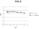
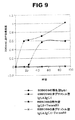


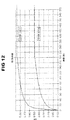


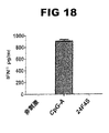
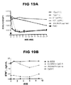

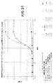


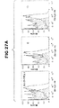


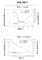


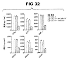


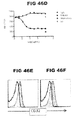


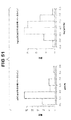
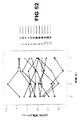


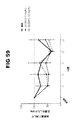


Classifications
-
- A—HUMAN NECESSITIES
- A61—MEDICAL OR VETERINARY SCIENCE; HYGIENE
- A61K—PREPARATIONS FOR MEDICAL, DENTAL OR TOILETRY PURPOSES
- A61K39/00—Medicinal preparations containing antigens or antibodies
- A61K39/395—Antibodies; Immunoglobulins; Immune serum, e.g. antilymphocytic serum
-
- A—HUMAN NECESSITIES
- A61—MEDICAL OR VETERINARY SCIENCE; HYGIENE
- A61P—SPECIFIC THERAPEUTIC ACTIVITY OF CHEMICAL COMPOUNDS OR MEDICINAL PREPARATIONS
- A61P1/00—Drugs for disorders of the alimentary tract or the digestive system
- A61P1/04—Drugs for disorders of the alimentary tract or the digestive system for ulcers, gastritis or reflux esophagitis, e.g. antacids, inhibitors of acid secretion, mucosal protectants
-
- A—HUMAN NECESSITIES
- A61—MEDICAL OR VETERINARY SCIENCE; HYGIENE
- A61P—SPECIFIC THERAPEUTIC ACTIVITY OF CHEMICAL COMPOUNDS OR MEDICINAL PREPARATIONS
- A61P13/00—Drugs for disorders of the urinary system
- A61P13/12—Drugs for disorders of the urinary system of the kidneys
-
- A—HUMAN NECESSITIES
- A61—MEDICAL OR VETERINARY SCIENCE; HYGIENE
- A61P—SPECIFIC THERAPEUTIC ACTIVITY OF CHEMICAL COMPOUNDS OR MEDICINAL PREPARATIONS
- A61P17/00—Drugs for dermatological disorders
- A61P17/06—Antipsoriatics
-
- A—HUMAN NECESSITIES
- A61—MEDICAL OR VETERINARY SCIENCE; HYGIENE
- A61P—SPECIFIC THERAPEUTIC ACTIVITY OF CHEMICAL COMPOUNDS OR MEDICINAL PREPARATIONS
- A61P19/00—Drugs for skeletal disorders
- A61P19/02—Drugs for skeletal disorders for joint disorders, e.g. arthritis, arthrosis
-
- A—HUMAN NECESSITIES
- A61—MEDICAL OR VETERINARY SCIENCE; HYGIENE
- A61P—SPECIFIC THERAPEUTIC ACTIVITY OF CHEMICAL COMPOUNDS OR MEDICINAL PREPARATIONS
- A61P21/00—Drugs for disorders of the muscular or neuromuscular system
-
- A—HUMAN NECESSITIES
- A61—MEDICAL OR VETERINARY SCIENCE; HYGIENE
- A61P—SPECIFIC THERAPEUTIC ACTIVITY OF CHEMICAL COMPOUNDS OR MEDICINAL PREPARATIONS
- A61P29/00—Non-central analgesic, antipyretic or antiinflammatory agents, e.g. antirheumatic agents; Non-steroidal antiinflammatory drugs [NSAID]
-
- A—HUMAN NECESSITIES
- A61—MEDICAL OR VETERINARY SCIENCE; HYGIENE
- A61P—SPECIFIC THERAPEUTIC ACTIVITY OF CHEMICAL COMPOUNDS OR MEDICINAL PREPARATIONS
- A61P3/00—Drugs for disorders of the metabolism
- A61P3/08—Drugs for disorders of the metabolism for glucose homeostasis
- A61P3/10—Drugs for disorders of the metabolism for glucose homeostasis for hyperglycaemia, e.g. antidiabetics
-
- A—HUMAN NECESSITIES
- A61—MEDICAL OR VETERINARY SCIENCE; HYGIENE
- A61P—SPECIFIC THERAPEUTIC ACTIVITY OF CHEMICAL COMPOUNDS OR MEDICINAL PREPARATIONS
- A61P37/00—Drugs for immunological or allergic disorders
- A61P37/02—Immunomodulators
-
- A—HUMAN NECESSITIES
- A61—MEDICAL OR VETERINARY SCIENCE; HYGIENE
- A61P—SPECIFIC THERAPEUTIC ACTIVITY OF CHEMICAL COMPOUNDS OR MEDICINAL PREPARATIONS
- A61P37/00—Drugs for immunological or allergic disorders
- A61P37/02—Immunomodulators
- A61P37/06—Immunosuppressants, e.g. drugs for graft rejection
-
- A—HUMAN NECESSITIES
- A61—MEDICAL OR VETERINARY SCIENCE; HYGIENE
- A61P—SPECIFIC THERAPEUTIC ACTIVITY OF CHEMICAL COMPOUNDS OR MEDICINAL PREPARATIONS
- A61P43/00—Drugs for specific purposes, not provided for in groups A61P1/00-A61P41/00
-
- C—CHEMISTRY; METALLURGY
- C07—ORGANIC CHEMISTRY
- C07K—PEPTIDES
- C07K16/00—Immunoglobulins [IGs], e.g. monoclonal or polyclonal antibodies
- C07K16/18—Immunoglobulins [IGs], e.g. monoclonal or polyclonal antibodies against material from animals or humans
- C07K16/28—Immunoglobulins [IGs], e.g. monoclonal or polyclonal antibodies against material from animals or humans against receptors, cell surface antigens or cell surface determinants
- C07K16/2851—Immunoglobulins [IGs], e.g. monoclonal or polyclonal antibodies against material from animals or humans against receptors, cell surface antigens or cell surface determinants against the lectin superfamily, e.g. CD23, CD72
-
- A—HUMAN NECESSITIES
- A61—MEDICAL OR VETERINARY SCIENCE; HYGIENE
- A61K—PREPARATIONS FOR MEDICAL, DENTAL OR TOILETRY PURPOSES
- A61K39/00—Medicinal preparations containing antigens or antibodies
- A61K2039/505—Medicinal preparations containing antigens or antibodies comprising antibodies
-
- C—CHEMISTRY; METALLURGY
- C07—ORGANIC CHEMISTRY
- C07K—PEPTIDES
- C07K2317/00—Immunoglobulins specific features
- C07K2317/20—Immunoglobulins specific features characterized by taxonomic origin
- C07K2317/24—Immunoglobulins specific features characterized by taxonomic origin containing regions, domains or residues from different species, e.g. chimeric, humanized or veneered
-
- C—CHEMISTRY; METALLURGY
- C07—ORGANIC CHEMISTRY
- C07K—PEPTIDES
- C07K2317/00—Immunoglobulins specific features
- C07K2317/30—Immunoglobulins specific features characterized by aspects of specificity or valency
- C07K2317/33—Crossreactivity, e.g. for species or epitope, or lack of said crossreactivity
-
- C—CHEMISTRY; METALLURGY
- C07—ORGANIC CHEMISTRY
- C07K—PEPTIDES
- C07K2317/00—Immunoglobulins specific features
- C07K2317/50—Immunoglobulins specific features characterized by immunoglobulin fragments
- C07K2317/51—Complete heavy chain or Fd fragment, i.e. VH + CH1
-
- C—CHEMISTRY; METALLURGY
- C07—ORGANIC CHEMISTRY
- C07K—PEPTIDES
- C07K2317/00—Immunoglobulins specific features
- C07K2317/50—Immunoglobulins specific features characterized by immunoglobulin fragments
- C07K2317/515—Complete light chain, i.e. VL + CL
-
- C—CHEMISTRY; METALLURGY
- C07—ORGANIC CHEMISTRY
- C07K—PEPTIDES
- C07K2317/00—Immunoglobulins specific features
- C07K2317/50—Immunoglobulins specific features characterized by immunoglobulin fragments
- C07K2317/54—F(ab')2
-
- C—CHEMISTRY; METALLURGY
- C07—ORGANIC CHEMISTRY
- C07K—PEPTIDES
- C07K2317/00—Immunoglobulins specific features
- C07K2317/50—Immunoglobulins specific features characterized by immunoglobulin fragments
- C07K2317/55—Fab or Fab'
-
- C—CHEMISTRY; METALLURGY
- C07—ORGANIC CHEMISTRY
- C07K—PEPTIDES
- C07K2317/00—Immunoglobulins specific features
- C07K2317/50—Immunoglobulins specific features characterized by immunoglobulin fragments
- C07K2317/56—Immunoglobulins specific features characterized by immunoglobulin fragments variable (Fv) region, i.e. VH and/or VL
- C07K2317/565—Complementarity determining region [CDR]
-
- C—CHEMISTRY; METALLURGY
- C07—ORGANIC CHEMISTRY
- C07K—PEPTIDES
- C07K2317/00—Immunoglobulins specific features
- C07K2317/50—Immunoglobulins specific features characterized by immunoglobulin fragments
- C07K2317/56—Immunoglobulins specific features characterized by immunoglobulin fragments variable (Fv) region, i.e. VH and/or VL
- C07K2317/567—Framework region [FR]
-
- C—CHEMISTRY; METALLURGY
- C07—ORGANIC CHEMISTRY
- C07K—PEPTIDES
- C07K2317/00—Immunoglobulins specific features
- C07K2317/60—Immunoglobulins specific features characterized by non-natural combinations of immunoglobulin fragments
- C07K2317/62—Immunoglobulins specific features characterized by non-natural combinations of immunoglobulin fragments comprising only variable region components
- C07K2317/622—Single chain antibody (scFv)
-
- C—CHEMISTRY; METALLURGY
- C07—ORGANIC CHEMISTRY
- C07K—PEPTIDES
- C07K2317/00—Immunoglobulins specific features
- C07K2317/60—Immunoglobulins specific features characterized by non-natural combinations of immunoglobulin fragments
- C07K2317/62—Immunoglobulins specific features characterized by non-natural combinations of immunoglobulin fragments comprising only variable region components
- C07K2317/626—Diabody or triabody
-
- C—CHEMISTRY; METALLURGY
- C07—ORGANIC CHEMISTRY
- C07K—PEPTIDES
- C07K2317/00—Immunoglobulins specific features
- C07K2317/70—Immunoglobulins specific features characterized by effect upon binding to a cell or to an antigen
- C07K2317/73—Inducing cell death, e.g. apoptosis, necrosis or inhibition of cell proliferation
- C07K2317/732—Antibody-dependent cellular cytotoxicity [ADCC]
-
- C—CHEMISTRY; METALLURGY
- C07—ORGANIC CHEMISTRY
- C07K—PEPTIDES
- C07K2317/00—Immunoglobulins specific features
- C07K2317/70—Immunoglobulins specific features characterized by effect upon binding to a cell or to an antigen
- C07K2317/73—Inducing cell death, e.g. apoptosis, necrosis or inhibition of cell proliferation
- C07K2317/734—Complement-dependent cytotoxicity [CDC]
-
- C—CHEMISTRY; METALLURGY
- C07—ORGANIC CHEMISTRY
- C07K—PEPTIDES
- C07K2317/00—Immunoglobulins specific features
- C07K2317/70—Immunoglobulins specific features characterized by effect upon binding to a cell or to an antigen
- C07K2317/76—Antagonist effect on antigen, e.g. neutralization or inhibition of binding
-
- C—CHEMISTRY; METALLURGY
- C07—ORGANIC CHEMISTRY
- C07K—PEPTIDES
- C07K2317/00—Immunoglobulins specific features
- C07K2317/90—Immunoglobulins specific features characterized by (pharmaco)kinetic aspects or by stability of the immunoglobulin
- C07K2317/92—Affinity (KD), association rate (Ka), dissociation rate (Kd) or EC50 value
-
- Y—GENERAL TAGGING OF NEW TECHNOLOGICAL DEVELOPMENTS; GENERAL TAGGING OF CROSS-SECTIONAL TECHNOLOGIES SPANNING OVER SEVERAL SECTIONS OF THE IPC; TECHNICAL SUBJECTS COVERED BY FORMER USPC CROSS-REFERENCE ART COLLECTIONS [XRACs] AND DIGESTS
- Y02—TECHNOLOGIES OR APPLICATIONS FOR MITIGATION OR ADAPTATION AGAINST CLIMATE CHANGE
- Y02A—TECHNOLOGIES FOR ADAPTATION TO CLIMATE CHANGE
- Y02A50/00—TECHNOLOGIES FOR ADAPTATION TO CLIMATE CHANGE in human health protection, e.g. against extreme weather
- Y02A50/30—Against vector-borne diseases, e.g. mosquito-borne, fly-borne, tick-borne or waterborne diseases whose impact is exacerbated by climate change
Landscapes
- Health & Medical Sciences (AREA)
- Chemical & Material Sciences (AREA)
- Organic Chemistry (AREA)
- Medicinal Chemistry (AREA)
- Life Sciences & Earth Sciences (AREA)
- General Health & Medical Sciences (AREA)
- Immunology (AREA)
- Bioinformatics & Cheminformatics (AREA)
- Pharmacology & Pharmacy (AREA)
- Public Health (AREA)
- Animal Behavior & Ethology (AREA)
- Engineering & Computer Science (AREA)
- Veterinary Medicine (AREA)
- Chemical Kinetics & Catalysis (AREA)
- General Chemical & Material Sciences (AREA)
- Nuclear Medicine, Radiotherapy & Molecular Imaging (AREA)
- Diabetes (AREA)
- Genetics & Genomics (AREA)
- Proteomics, Peptides & Aminoacids (AREA)
- Molecular Biology (AREA)
- Biophysics (AREA)
- Biochemistry (AREA)
- Rheumatology (AREA)
- Orthopedic Medicine & Surgery (AREA)
- Physical Education & Sports Medicine (AREA)
- Epidemiology (AREA)
- Mycology (AREA)
- Microbiology (AREA)
- Pain & Pain Management (AREA)
- Urology & Nephrology (AREA)
- Emergency Medicine (AREA)
- Endocrinology (AREA)
- Hematology (AREA)
- Obesity (AREA)
- Dermatology (AREA)
- Neurology (AREA)
- Transplantation (AREA)
- Medicines Containing Antibodies Or Antigens For Use As Internal Diagnostic Agents (AREA)
- Peptides Or Proteins (AREA)
- Medicines That Contain Protein Lipid Enzymes And Other Medicines (AREA)
Description
関連出願の引用
本願は、2012年12月10日に出願した米国仮出願第61/735,362号および2013年2月11日に出願した米国仮出願第61/763,270号の利益を主張する。
Citation of Related Application This application claims the benefit of US Provisional Application No. 61 / 735,362 filed on Dec. 10, 2012 and US Provisional Application No. 61 / 763,270 filed on Feb. 11, 2013. To do.
背景
血液樹状細胞抗原2(BDCA2)とは、ヒト形質細胞様樹状細胞(pDC)上で発現するC型レクチン(Dzionekら、J. Immunol.、165巻:6037〜6046頁(2000年))であり、toll様受容体(TLR)リガンドに応答してI型インターフェロン(IFN)を分泌する、骨髄由来細胞の特化した集団である。BDCA2は、そのC末端における、II型C型レクチン群に属する単一の細胞外炭水化物認識ドメイン(CRD)、膜貫通領域、およびシグナル伝達モチーフを保有しないそのN末端の短い細胞質尾部からなる。BDCA2は、会合する膜貫通アダプターであるFcεRIγを介して細胞内シグナルを伝達し、B細胞受容体(BCR)様シグナル伝達カスケードを誘導する。
Background Blood dendritic cell antigen 2 (BDCA2) is a C-type lectin expressed on human plasmacytoid dendritic cells (pDC) (Dzionek et al., J. Immunol., 165: 6037-6046 (2000)). ), A specialized population of bone marrow derived cells that secrete type I interferon (IFN) in response to toll-like receptor (TLR) ligands. BDCA2 consists of a single extracellular carbohydrate recognition domain (CRD) belonging to the type II C-type lectin group at its C-terminus, a transmembrane region, and its N-terminal short cytoplasmic tail that does not carry a signaling motif. BDCA2 transmits intracellular signals via FcεRIγ, an associated transmembrane adapter, and induces a B cell receptor (BCR) -like signaling cascade.
概要
本開示は、少なくとも部分的に、BDCA2に結合する抗体の同定および特徴付けに基づく。このような抗体は、炎症性サイトカインおよびケモカインの分泌を低減または阻害しうる。本明細書で記載される抗BDCA2抗体はまた、抗体依存性細胞介在性細胞傷害作用(ADCC)、または補体介在性細胞傷害作用(CDC)により、pDCを枯渇させることが可能でもある。加えて、本明細書で記載される抗BDCA2抗体は、pDCの表面におけるCD32aおよび/またはCD62Lのレベルも下方調節しうる。さらに、本開示の抗BDCA2抗体は、BDCA2の、pDCの細胞表面からの内部移行を媒介しうる。少なくともこれらの理由で、本明細書で記載される抗BDCA2抗体は、自己免疫状態および炎症性状態の処置または防止において有用である。本開示はまた、効果を改善するために、本明細書で記載される抗BDCA2抗体を、抗マラリア剤と組み合わせうることも示す。
SUMMARY The present disclosure is based at least in part on the identification and characterization of antibodies that bind to BDCA2. Such antibodies can reduce or inhibit the secretion of inflammatory cytokines and chemokines. The anti-BDCA2 antibodies described herein may also be capable of depleting pDC by antibody-dependent cell-mediated cytotoxicity (ADCC) or complement-mediated cytotoxicity (CDC). In addition, the anti-BDCA2 antibodies described herein can also downregulate CD32a and / or CD62L levels on the surface of pDC. Furthermore, the anti-BDCA2 antibodies of the present disclosure can mediate internalization of BDCA2 from the cell surface of pDC. For at least these reasons, the anti-BDCA2 antibodies described herein are useful in the treatment or prevention of autoimmune and inflammatory conditions. The present disclosure also shows that the anti-BDCA2 antibodies described herein can be combined with antimalarial agents to improve efficacy.
一態様では、本開示は、ヒトBDCA2(配列番号1)の外部ドメインに選択的に結合し、ヒトBDCA2の細胞外ドメインへの結合について、BIIB059と競合する単離抗体またはその抗原結合断片を特色とする。 In one aspect, the present disclosure is selectively coupled to an external domain of the human BDCA2 (SEQ ID NO: 1), for binding to the extracellular domain of human BDCA2, the isolated antibody or antigen binding fragment thereof that competes with BIIB059 It is a special feature.
抗BDCA2抗体またはその抗原結合断片を、BDCA2へとあらかじめ結合させることにより、BIIB059の、BDCA2への後続の結合を完全に、または部分的に阻害する場合、抗BDCA2抗体またはその抗原結合断片は、BDCA2への結合について、BIIB059と競合する。例えば、抗BDCA2抗体またはその抗原結合断片を、BDCA2へとあらかじめ結合させることにより、BIIB059の、BDCA2への後続の結合を完全に阻害する場合、抗BDCA2抗体またはその抗原結合断片は、BDCA2への結合について、BIIB059と競合する。ある種の実施形態では、抗BDCA2抗体またはその抗原結合断片を、BDCA2へとあらかじめ結合させる結果として、BIIB059の、BDCA2への後続の結合を、少なくとも30%、50%、70%、80%、90%、95%、98%、または99%阻害する。 When the anti-BDCA2 antibody or antigen-binding fragment thereof is pre-coupled to BDCA2 to completely or partially inhibit subsequent binding of BIIB059 to BDCA2, the anti-BDCA2 antibody or antigen-binding fragment thereof is Competes with BIIB059 for binding to BDCA2. For example, when the anti-BDCA2 antibody or antigen-binding fragment thereof is completely inhibited from subsequent binding of BIIB059 to BDCA2 by pre-binding to BDCA2, the anti-BDCA2 antibody or antigen-binding fragment thereof is bound to BDCA2. Competes with BIIB059 for binding. In certain embodiments, subsequent binding of BIIB059 to BDCA2 results in at least 30%, 50%, 70%, 80%, as a result of pre-binding the anti-BDCA2 antibody or antigen-binding fragment thereof to BDCA2. Inhibits 90%, 95%, 98%, or 99%.
別の態様では、本開示は、ヒトBDCA2(配列番号1)の外部ドメインに選択的に結合し、(i)他のサイトカインおよびケモカインに加えた、I型インターフェロンおよび/もしくはIII型インターフェロンの、形質細胞様樹状細胞からの分泌を阻害するか、または(ii)in vitroにおいて、形質細胞様樹状細胞の枯渇を誘導もしくは増強する、単離抗体またはその抗原結合断片を特色とする。ある種の実施形態では、抗BDCA2抗体は、pDCの表面からのCD32aおよび/またはCD62Lを下方調節する。いくつかの実施形態では、抗BDCA2抗体は、BDCA2の、pDCの細胞表面からの内部移行を媒介する。いくつかの実施形態では、抗体またはその抗原結合断片は、カニクイザルBDCA2(配列番号73)およびアカゲザルBDCA2(配列番号72)に結合する。ある種の実施形態では、単離抗体またはその抗原結合断片は、I型インターフェロン、インターロイキン6(IL−6)、腫瘍壊死因子α(TNF−α)、III型インターフェロン、マクロファージ炎症性タンパク質1(MIP−1)−α/CCL3、MIP−1β/CCL4、ケモカイン(C−Cモチーフ)リガンド5(CCL5/RANTES)、またはインターフェロンγ誘導タンパク質10(IP−10/CXCL10)の分泌または産生を阻害する。 In another aspect, the present disclosure is selectively coupled to an external domain of the human BDCA2 (SEQ ID NO: 1), (i) was added to other cytokines and chemokines, type I interferon and / or Type III interferon, Features an isolated antibody or antigen-binding fragment thereof that inhibits secretion from plasmacytoid dendritic cells, or (ii) induces or enhances depletion of plasmacytoid dendritic cells in vitro. In certain embodiments, the anti-BDCA2 antibody downregulates CD32a and / or CD62L from the surface of pDC. In some embodiments, the anti-BDCA2 antibody mediates the internalization of BDCA2 from the cell surface of pDC. In some embodiments, the antibody or antigen-binding fragment thereof binds to cynomolgus BDCA2 (SEQ ID NO: 7 3 ) and rhesus monkey BDCA2 (SEQ ID NO: 72). In certain embodiments, the isolated antibody or antigen-binding fragment thereof is type I interferon, interleukin 6 (IL-6), tumor necrosis factor α (TNF-α), type III interferon, macrophage inflammatory protein 1 ( Inhibits secretion or production of MIP-1) -α / CCL3, MIP-1β / CCL4, chemokine (CC motif) ligand 5 (CCL5 / RANTES), or interferon gamma-inducible protein 10 (IP-10 / CXCL10) .
上記の2つの態様のいくつかの実施形態では、単離抗体またはその抗原結合断片は必要に応じて、以下の特色:0.5〜3μg/mLまたは4nM〜10nMのEC50(ヒトBDCA2);0.5〜3μg/mLまたは5nM〜10nMのEC50(カニクイザルBDCA2);7〜7.5のpI;ラットClec4b2に結合しないか、またはヒトBDCA2、カニクイザルBDCA2、もしくはアカゲザルBDCA2より小さな結合アフィニティーでラットClec4b2に結合すること;MIP−1−α/CCL3、MIP−1β/CCL4、CCL5/RANTES、IP−10/CXCL10など、ケモカインの産生または分泌を阻害すること;重鎖CDR1、重鎖CDR2、および重鎖CDR3であって、重鎖CDR1が、配列番号9に示されるアミノ酸配列からなるアミノ酸配列、または配列番号8に示されるアミノ酸配列からなるアミノ酸配列を有し;重鎖CDR2が、配列番号10に示されるアミノ酸配列からなるアミノ酸配列を有し;重鎖CDR3が、配列番号11に示されるアミノ酸配列からなるアミノ酸配列を有し;可変重鎖が、配列番号24に示されるアミノ酸配列を含むかまたはこれからなる、重鎖CDR1、重鎖CDR2、および重鎖CDR3のうちの1つ、2つ、3つ、4つ、5つ、または6つをさらに含むかまたはこれらからなる。ある種の実施形態では、抗体またはその抗原結合断片は、配列番号89に示されるアミノ酸配列からなる重鎖CDR1、配列番号91に示されるアミノ酸配列からなる重鎖CDR2、および配列番号11に示されるアミノ酸配列からなる重鎖CDR3を有する。ある種の実施形態では、抗体またはその抗原結合断片は、配列番号9に示されるアミノ酸配列からなる重鎖CDR1、配列番号92に示されるアミノ酸配列からなる重鎖CDR2、および配列番号11に示されるアミノ酸配列からなる重鎖CDR3を有する。ある種の実施形態では、抗体またはその抗原結合断片は、配列番号90に示されるアミノ酸配列からなる重鎖CDR1、配列番号93に示されるアミノ酸配列からなる重鎖CDR2、および配列番号94に示されるアミノ酸配列からなる重鎖CDR3を有する。いくつかの実施形態では、単離抗体または抗原結合断片のEC50(ヒトBDCA2)は、4.5nM、4.6nM、4.7nM、4.8nM、4.9nM、5.0nM、5.1nM、5.2nM、5.3nM、5.4nM、または5.5nMである。具体的な実施形態では、単離抗体または抗原結合断片のEC50(ヒトBDCA2)は、4.9nMである。いくつかの実施形態では、単離抗体または抗原結合断片のEC50(カニクイザルBDCA2)は、4.0nM、4.1nM、4.2nM、4.3nM、4.4nM、4.5nM、4.6nM、4.7nM、4.8nM、4.9nM、または5.0nMである。具体的な実施形態では、単離抗体または抗原結合断片のEC50(カニクイザルBDCA2)は、4.4nMである。この態様のある種の実施形態では、抗体は、ヒト重鎖定常領域およびヒト軽鎖定常領域を有する。ある種の実施形態では、重鎖定常領域は、CH1ドメインおよびヒンジ領域を含む。いくつかの実施形態では、重鎖定常領域は、CH3ドメインを含む。重鎖定常領域が、置換を含む場合、このような置換は、抗体の特性を改変する(例えば、Fc受容体の結合、抗体のグリコシル化、システイン残基の数、エフェクター細胞の機能、または補体機能のうちの1または複数を増加または減少させる)。ある種の実施形態では、抗体は、IgG抗体である。具体的な実施形態では、抗体は、IgG1、IgG2、IgG3、およびIgG4からなる群から選択される。ある種の実施形態では、抗体は、7〜15μg/mLのEC50でFcγRIIa(CD32a)に結合するヒトFc領域を含む。ある種の実施形態では、抗体は、10μg/mLのEC50でFcγRIIa(CD32a)に結合するヒトFc領域を含む。ある種の実施形態では、抗体は、11μg/mLのEC50でFcγRIIa(CD32a)に結合するヒトFc領域を含む。ある種の実施形態では、抗体は、12μg/mLのEC50でFcγRIIa(CD32a)に結合するヒトFc領域を含む。 In some embodiments of the above two aspects, the isolated antibody or antigen-binding fragment thereof optionally has the following characteristics: 0.5-3 μg / mL or 4 nM-10 nM EC 50 (human BDCA2); 0.5-3 μg / mL or 5 nM-10 nM EC 50 (cynomolgus BDCA2); 7-7.5 pI; rat does not bind to rat Clec4b2 or has a lower binding affinity than human BDCA2, cynomolgus BDCA2, or rhesus monkey BDCA2 Inhibiting chemokine production or secretion, such as MIP-1-α / CCL3, MIP-1β / CCL4, CCL5 / RANTES, IP-10 / CXCL10; heavy chain CDR1, heavy chain CDR2, and A heavy chain CDR3 wherein the heavy chain CDR1 is An amino acid sequence consisting of the amino acid sequence shown in No. 9 or an amino acid sequence consisting of the amino acid sequence shown in SEQ ID No. 8; the heavy chain CDR2 has an amino acid sequence consisting of the amino acid sequence shown in SEQ ID No. 10; The heavy chain CDR3 has an amino acid sequence consisting of the amino acid sequence set forth in SEQ ID NO: 11; and the variable heavy chain comprises or consists of the amino acid sequence set forth in SEQ ID NO: 24, and heavy chain CDR2, and It further comprises or consists of one, two, three, four, five, or six of the heavy chain CDR3s. In certain embodiments, the antibody or antigen-binding fragment thereof is shown in SEQ ID NO: 11, a heavy chain CDR1 consisting of the amino acid sequence shown in SEQ ID NO: 89, a heavy chain CDR2 consisting of the amino acid sequence shown in SEQ ID NO: 91, and SEQ ID NO: 11. It has a heavy chain CDR3 consisting of an amino acid sequence. In certain embodiments, the antibody or antigen-binding fragment thereof is shown in SEQ ID NO: 11, a heavy chain CDR1 consisting of the amino acid sequence shown in SEQ ID NO: 9, a heavy chain CDR2 consisting of the amino acid sequence shown in SEQ ID NO: 92, and SEQ ID NO: 11. It has a heavy chain CDR3 consisting of an amino acid sequence. In certain embodiments, the antibody or antigen-binding fragment thereof is shown in SEQ ID NO: 94, heavy chain CDR1, consisting of the amino acid sequence shown in SEQ ID NO: 90, heavy chain CDR2, consisting of the amino acid sequence shown in SEQ ID NO: 93, and SEQ ID NO: 94. It has a heavy chain CDR3 consisting of an amino acid sequence. In some embodiments, the isolated antibody or antigen-binding fragment has an EC50 (human BDCA2) of 4.5 nM, 4.6 nM, 4.7 nM, 4.8 nM, 4.9 nM, 5.0 nM, 5.1 nM, 5.2 nM, 5.3 nM, 5.4 nM, or 5.5 nM. In a specific embodiment, the EC50 (human BDCA2) of the isolated antibody or antigen-binding fragment is 4.9 nM. In some embodiments, the isolated antibody or antigen-binding fragment EC50 (cynomolgus BDCA2) is 4.0 nM, 4.1 nM, 4.2 nM, 4.3 nM, 4.4 nM, 4.5 nM, 4.6 nM, 4.7 nM, 4.8 nM, 4.9 nM, or 5.0 nM. In a specific embodiment, the EC50 (cynomolgus BDCA2) of the isolated antibody or antigen-binding fragment is 4.4 nM. In certain embodiments of this aspect, the antibody has a human heavy chain constant region and a human light chain constant region. In certain embodiments, the heavy chain constant region comprises a CH1 domain and a hinge region. In some embodiments, the heavy chain constant region comprises a CH3 domain. Where the heavy chain constant region contains substitutions, such substitutions alter the properties of the antibody (eg, Fc receptor binding, antibody glycosylation, number of cysteine residues, effector cell function, or complementation). Increase or decrease one or more of the body functions). In certain embodiments, the antibody is an IgG antibody. In a specific embodiment, the antibody is selected from the group consisting of IgG1, IgG2, IgG3, and IgG4. In certain embodiments, the antibody comprises a human Fc region that binds FcγRIIa (CD32a) with an EC50 of 7-15 μg / mL. In certain embodiments, the antibody comprises a human Fc region that binds FcγRIIa (CD32a) with an EC50 of 10 μg / mL. In certain embodiments, the antibody comprises a human Fc region that binds to FcγRIIa (CD32a) with an EC50 of 11 μg / mL. In certain embodiments, the antibody comprises a human Fc region that binds to FcγRIIa (CD32a) with an EC50 of 12 μg / mL.
別の態様では、本開示は、ヒトBDCA2(配列番号1)の外部ドメインに選択的に結合し、重鎖CDR1、重鎖CDR2、および重鎖CDR3を含む単離抗体またはその抗原結合断片を特色とする。重鎖CDR1は、アミノ酸配列GFTFSTYTMS(配列番号9)、または1、2、3、もしくは4つのアミノ酸位置において置換を有する、配列番号9に示されるアミノ酸配列を含むかまたはこれからなる。重鎖CDR2は、アミノ酸配列TISPGDSFGYYYPDSVQG(配列番号10)、または1、2、3、もしくは4つのアミノ酸位置において置換を有する、配列番号10に示されるアミノ酸配列を含むかまたはこれからなる。重鎖CDR3は、アミノ酸配列DIYYNYGAWFAY(配列番号11)、または1、2、3、もしくは4つのアミノ酸位置において置換を有する、配列番号11に示されるアミノ酸配列を含むかまたはこれからなる。別の態様では、抗体またはその抗原結合断片は、1、2、3、または4つのアミノ酸位置において置換を有する、配列番号89に示されるアミノ酸配列からなる重鎖CDR1;1、2、3、または4つのアミノ酸位置において置換を有する、配列番号91に示されるアミノ酸配列からなる重鎖CDR2;および1、2、3、または4つのアミノ酸位置において置換を有する、配列番号11に示されるアミノ酸配列からなる重鎖CDR3を含む。別の態様では、抗体またはその抗原結合断片は、1、2、3、または4つのアミノ酸位置において置換を有する、配列番号9に示されるアミノ酸配列からなる重鎖CDR1;1、2、3、または4つのアミノ酸位置において置換を有する、配列番号92に示されるアミノ酸配列からなる重鎖CDR2;および1、2、3、または4つのアミノ酸位置において置換を有する、配列番号11に示されるアミノ酸配列からなる重鎖CDR3を含む。別の態様では、抗体またはその抗原結合断片は、1、2、3、または4つのアミノ酸位置において置換を有する、配列番号90に示されるアミノ酸配列からなる重鎖CDR1;1、2、3、または4つのアミノ酸位置において置換を有する、配列番号93に示されるアミノ酸配列からなる重鎖CDR2;および1、2、3、または4つのアミノ酸位置において置換を有する、配列番号94に示されるアミノ酸配列からなる重鎖CDR3を含む。これらの抗体は、(i)ヒトBDCA2またはカニクイザルBDCA2には結合するが、霊長動物より下等の系統発生種に由来するBDCA2には有意には結合せず、かつ/または(ii)ヒトpDCによる、TLR7/TLR9誘導I型インターフェロンおよび他のサイトカインまたはケモカインの産生を阻害し、かつ/または(iii)BDCA2の、pDCの表面からの内部移行を媒介し、かつ/または(iv)pDCの表面からのCD32aおよび/もしくはCD62Lを下方調節し、かつ/または(v)in vitroにおいて、ADCCまたはCDCにより、pDCを枯渇させる。この態様のある種の実施形態では、抗体は、ヒト重鎖定常領域およびヒト軽鎖定常領域を有する。 In another aspect, the present disclosure is selectively coupled to an external domain of the human BDCA2 (SEQ ID NO: 1), the heavy chain CDR1, a heavy chain CDR2, and isolated antibodies or antigen-binding fragment thereof, comprising a heavy chain CDR3 It is a special feature. The heavy chain CDR1 comprises or consists of the amino acid sequence GFTSTYTMS (SEQ ID NO: 9), or the amino acid sequence shown in SEQ ID NO: 9, with substitutions at 1, 2, 3, or 4 amino acid positions. The heavy chain CDR2 comprises or consists of the amino acid sequence TISPDGDSFGYYYPSVSVG (SEQ ID NO: 10), or the amino acid sequence shown in SEQ ID NO: 10, with substitutions at 1, 2, 3, or 4 amino acid positions. The heavy chain CDR3 comprises or consists of the amino acid sequence DIYYNYGAWFAY (SEQ ID NO: 11), or the amino acid sequence set forth in SEQ ID NO: 11, having substitutions at 1, 2, 3, or 4 amino acid positions. In another aspect, the antibody or antigen-binding fragment thereof has a heavy chain CDR1 consisting of the amino acid sequence set forth in SEQ ID NO: 89 with a substitution at 1, 2, 3, or 4 amino acid positions; 1, 2, 3, or A heavy chain CDR2 consisting of the amino acid sequence shown in SEQ ID NO: 91 having a substitution at four amino acid positions; and consisting of an amino acid sequence shown in SEQ ID NO: 11 having a substitution at 1, 2, 3, or 4 amino acid positions Contains heavy chain CDR3. In another aspect, the antibody or antigen-binding fragment thereof has a heavy chain CDR1 consisting of the amino acid sequence set forth in SEQ ID NO: 9 with a substitution at 1, 2, 3, or 4 amino acid positions; 1, 2, 3, or A heavy chain CDR2 consisting of the amino acid sequence shown in SEQ ID NO: 92 having a substitution at four amino acid positions; and consisting of an amino acid sequence shown in SEQ ID NO: 11 having a substitution at 1, 2, 3, or 4 amino acid positions Contains heavy chain CDR3. In another aspect, the antibody or antigen-binding fragment thereof has a heavy chain CDR1 consisting of the amino acid sequence set forth in SEQ ID NO: 90 with a substitution at 1, 2, 3, or 4 amino acid positions; 1, 2, 3, or A heavy chain CDR2 consisting of the amino acid sequence shown in SEQ ID NO: 93 having a substitution at four amino acid positions; and consisting of an amino acid sequence shown in SEQ ID NO: 94 having a substitution at 1, 2, 3, or 4 amino acid positions Contains heavy chain CDR3. These antibodies bind to (i) human BDCA2 or cynomolgus BDCA2, but not significantly to BDCA2 derived from phylogenetic species lower than primates and / or (ii) by human pDC. Inhibits the production of TLR7 / TLR9 induced type I interferon and other cytokines or chemokines and / or (iii) mediates the internalization of BDCA2 from the surface of pDC and / or (iv) from the surface of pDC Downregulates CD32a and / or CD62L and / or (v) depletes pDC by ADCC or CDC in vitro. In certain embodiments of this aspect, the antibody has a human heavy chain constant region and a human light chain constant region.
ある種の実施形態では、ヒトBDCA2に特異的に結合する単離抗体またはその抗原結合断片は、アミノ酸配列GFTFSTYTMS(配列番号9)、または1もしくは2つのアミノ酸位置において置換を有する、配列番号9に示されるアミノ酸配列を含むかまたはこれからなる重鎖CDR1;アミノ酸配列TISPGDSFGYYYPDSVQG(配列番号10)、または1もしくは2つのアミノ酸位置において置換を有する、配列番号10に示されるアミノ酸配列を含むかまたはこれからなる重鎖CDR2;およびアミノ酸配列DIYYNYGAWFAY(配列番号11)、または1もしくは2つのアミノ酸位置において置換を有する、配列番号11に示されるアミノ酸配列を含むかまたはこれからなる重鎖CDR3を有する。この態様の他の実施形態では、単離抗体または抗原結合断片は、アミノ酸配列GFTFSTYTMS(配列番号9)を含むかまたはこれからなる重鎖CDR1、アミノ酸配列TISPGDSFGYYYPDSVQG(配列番号10)を含むかまたはこれからなる重鎖CDR2、およびアミノ酸配列DIYYNYGAWFAY(配列番号11)を含むかまたはこれからなる重鎖CDR3を有する。この態様の他の実施形態では、単離抗体または抗原結合断片は、軽鎖CDR1、軽鎖CDR2、および軽鎖CDR3を含む。軽鎖CDR1は、アミノ酸配列KASQSVDYDGDSYMN(配列番号5)、または1、2、3、もしくは4つのアミノ酸位置において置換を有する、配列番号5に示されるアミノ酸配列を含むかまたはこれからなる。軽鎖CDR2は、アミノ酸配列AASTLES(配列番号6)、または1、2、3、もしくは4つのアミノ酸位置において置換を有する、配列番号6に示されるアミノ酸配列を含むかまたはこれからなる。軽鎖CDR3は、アミノ酸配列QQANEDPRT(配列番号7)、または1、2、3、もしくは4つのアミノ酸位置において置換を有する、配列番号7に示されるアミノ酸配列を含むかまたはこれからなる。ある種の実施形態では、軽鎖CDR1は、アミノ酸配列KASQSVDYDGDSYMN(配列番号5)、または1もしくは2つのアミノ酸位置において置換を有する、配列番号5に示されるアミノ酸配列を含むかまたはこれからなり;軽鎖CDR2は、アミノ酸配列AASTLES(配列番号6)、または1もしくは2つのアミノ酸位置において置換を有する、配列番号6に示されるアミノ酸配列を含むかまたはこれからなり;軽鎖CDR3は、アミノ酸配列QQANEDPRT(配列番号7)、または1もしくは2つのアミノ酸位置において置換を有する、配列番号7に示されるアミノ酸配列を含むかまたはこれからなる。他の実施形態では、単離抗体またはその抗原結合断片は、アミノ酸配列GFTFSTYTMS(配列番号9)を含むかまたはこれからなる重鎖CDR1、アミノ酸配列TISPGDSFGYYYPDSVQG(配列番号10)を含むかまたはこれからなる重鎖CDR2、アミノ酸配列DIYYNYGAWFAY(配列番号11)を含むかまたはこれからなる重鎖CDR3、アミノ酸配列KASQSVDYDGDSYMN(配列番号5)を含むかまたはこれからなる軽鎖CDR1、アミノ酸配列AASTLES(配列番号6)を含むかまたはこれからなる軽鎖CDR2、およびアミノ酸配列QQANEDPRT(配列番号7)を含むかまたはこれからなる軽鎖CDR3を有する。 In certain embodiments, an isolated antibody or antigen-binding fragment thereof that specifically binds human BDCA2 has the amino acid sequence GFTSTYTMS (SEQ ID NO: 9), or SEQ ID NO: 9, with a substitution at one or two amino acid positions. Heavy chain CDR1 comprising or consisting of the amino acid sequence shown; amino acid sequence TISPGDSFYYYPSVVQG (SEQ ID NO: 10), or a heavy chain comprising or consisting of the amino acid sequence shown in SEQ ID NO: 10 with substitution at one or two amino acid positions And has a heavy chain CDR3 comprising or consisting of the amino acid sequence shown in SEQ ID NO: 11, having a substitution at one or two amino acid positions, and the amino acid sequence DIYYNYGAWFAY (SEQ ID NO: 11). In another embodiment of this aspect, the isolated antibody or antigen-binding fragment comprises or consists of the heavy chain CDR1, comprising or consisting of the amino acid sequence GFTSTYTMS (SEQ ID NO: 9), the amino acid sequence TISPGDSFYYPDSVQG (SEQ ID NO: 10). It has a heavy chain CDR2 and a heavy chain CDR3 comprising or consisting of the amino acid sequence DIYYNYGAWFAY (SEQ ID NO: 11). In other embodiments of this aspect, the isolated antibody or antigen-binding fragment comprises a light chain CDR1, a light chain CDR2, and a light chain CDR3. The light chain CDR1 comprises or consists of the amino acid sequence KASQSVDYDGDSYMN (SEQ ID NO: 5), or the amino acid sequence shown in SEQ ID NO: 5, with substitutions at 1, 2, 3, or 4 amino acid positions. The light chain CDR2 comprises or consists of the amino acid sequence AASTLES (SEQ ID NO: 6) or the amino acid sequence shown in SEQ ID NO: 6 with substitutions at 1, 2, 3, or 4 amino acid positions. The light chain CDR3 comprises or consists of the amino acid sequence QQANEDPRT (SEQ ID NO: 7), or the amino acid sequence shown in SEQ ID NO: 7, with substitutions at 1, 2, 3, or 4 amino acid positions. In certain embodiments, the light chain CDR1 comprises or consists of the amino acid sequence KASQSVDYDGDSYMN (SEQ ID NO: 5), or the amino acid sequence set forth in SEQ ID NO: 5, having substitutions at one or two amino acid positions; CDR2 comprises or consists of the amino acid sequence AASTLES (SEQ ID NO: 6), or the amino acid sequence set forth in SEQ ID NO: 6, with a substitution at one or two amino acid positions; light chain CDR3 comprises the amino acid sequence QQANEDPRT (SEQ ID NO: 7) or comprises or consists of the amino acid sequence set forth in SEQ ID NO: 7 having a substitution at one or two amino acid positions. In other embodiments, the isolated antibody or antigen-binding fragment thereof comprises a heavy chain CDR1 comprising or consisting of the amino acid sequence GFTSTYTMS (SEQ ID NO: 9), a heavy chain comprising or consisting of the amino acid sequence TISPGDSFYYPDSVQG (SEQ ID NO: 10). CDR2, comprising the heavy chain CDR3 comprising or consisting of the amino acid sequence DIYYNYGAWFAY (SEQ ID NO: 11), light chain CDR1 comprising or consisting of the amino acid sequence KASQSVDYDGDSYMN (SEQ ID NO: 5), comprising the amino acid sequence AASTLES (SEQ ID NO: 6) It has a light chain CDR2 consisting of this, and a light chain CDR3 comprising or consisting of the amino acid sequence QQANEDPRT (SEQ ID NO: 7).
ある種の実施形態では、ヒトBDCA2に選択的に結合する単離抗体またはその抗原結合断片は、アミノ酸配列TYTMS(配列番号8)、または1もしくは2つのアミノ酸位置において置換を有する、配列番号8に示されるアミノ酸配列を含むかまたはこれからなる重鎖CDR1;アミノ酸配列TISPGDSFGYYYPDSVQG(配列番号10)、または1もしくは2つのアミノ酸位置において置換を有する、配列番号10に示されるアミノ酸配列を含むかまたはこれからなる重鎖CDR2;およびアミノ酸配列DIYYNYGAWFAY(配列番号11)、または1もしくは2つのアミノ酸位置において置換を有する、配列番号11に示されるアミノ酸配列を含むかまたはこれからなる重鎖CDR3を含む。この態様の他の実施形態では、単離抗体または抗原結合断片は、アミノ酸配列TYTMS(配列番号8)を含むかまたはこれからなる重鎖CDR1、アミノ酸配列TISPGDSFGYYYPDSVQG(配列番号10)を含むかまたはこれからなる重鎖CDR2、およびアミノ酸配列DIYYNYGAWFAY(配列番号11)を含むかまたはこれからなる重鎖CDR3を有する。この態様の他の実施形態では、単離抗体または抗原結合断片は、軽鎖CDR1、軽鎖CDR2、および軽鎖CDR3を含む。軽鎖CDR1は、アミノ酸配列KASQSVDYDGDSYMN(配列番号5)、または1、2、3、もしくは4つのアミノ酸位置において置換を有する、配列番号5に示されるアミノ酸配列を含むかまたはこれからなる。軽鎖CDR2は、アミノ酸配列AASTLES(配列番号6)、または1、2、3、もしくは4つのアミノ酸位置において置換を有する、配列番号6に示されるアミノ酸配列を含むかまたはこれからなる。軽鎖CDR3は、アミノ酸配列QQANEDPRT(配列番号7)、または1、2、3、もしくは4つのアミノ酸位置において置換を有する、配列番号7に示されるアミノ酸配列を含むかまたはこれからなる。ある種の実施形態では、軽鎖CDR1は、アミノ酸配列KASQSVDYDGDSYMN(配列番号5)、または1もしくは2つのアミノ酸位置において置換を有する、配列番号5に示されるアミノ酸配列を含むかまたはこれからなり;軽鎖CDR2は、アミノ酸配列AASTLES(配列番号6)、または1もしくは2つのアミノ酸位置において置換を有する、配列番号6に示されるアミノ酸配列を含むかまたはこれからなり;軽鎖CDR3は、アミノ酸配列QQANEDPRT(配列番号7)、または1もしくは2つのアミノ酸位置において置換を有する、配列番号7に示されるアミノ酸配列を含むかまたはこれからなる。他の実施形態では、単離抗体またはその抗原結合断片は、アミノ酸配列TYTMS(配列番号8)を含むかまたはこれからなる重鎖CDR1、アミノ酸配列TISPGDSFGYYYPDSVQG(配列番号10)を含むかまたはこれからなる重鎖CDR2、アミノ酸配列DIYYNYGAWFAY(配列番号11)を含むかまたはこれからなる重鎖CDR3、アミノ酸配列KASQSVDYDGDSYMN(配列番号5)を含むかまたはこれからなる軽鎖CDR1、アミノ酸配列AASTLES(配列番号6)を含むかまたはこれからなる軽鎖CDR2、およびアミノ酸配列QQANEDPRT(配列番号7)を含むかまたはこれからなる軽鎖CDR3を有する。 In certain embodiments, the isolated antibody or antigen-binding fragment thereof that selectively binds human BDCA2 is amino acid sequence TYTMS (SEQ ID NO: 8), or SEQ ID NO: 8, with a substitution at one or two amino acid positions. Heavy chain CDR1 comprising or consisting of the amino acid sequence shown; amino acid sequence TISPGDSFYYYPSVVQG (SEQ ID NO: 10), or a heavy chain comprising or consisting of the amino acid sequence shown in SEQ ID NO: 10 with substitution at one or two amino acid positions Chain CDR2; and the amino acid sequence DIYYNYGAWFAY (SEQ ID NO: 11), or heavy chain CDR3 comprising or consisting of the amino acid sequence shown in SEQ ID NO: 11, with substitution at one or two amino acid positions. In other embodiments of this aspect, the isolated antibody or antigen-binding fragment comprises or consists of the heavy chain CDR1, comprising or consisting of the amino acid sequence TYTMS (SEQ ID NO: 8), the amino acid sequence TIPGDSFGYYPDSVQG (SEQ ID NO: 10). It has a heavy chain CDR2 and a heavy chain CDR3 comprising or consisting of the amino acid sequence DIYYNYGAWFAY (SEQ ID NO: 11). In other embodiments of this aspect, the isolated antibody or antigen-binding fragment comprises a light chain CDR1, a light chain CDR2, and a light chain CDR3. The light chain CDR1 comprises or consists of the amino acid sequence KASQSVDYDGDSYMN (SEQ ID NO: 5), or the amino acid sequence shown in SEQ ID NO: 5, with substitutions at 1, 2, 3, or 4 amino acid positions. The light chain CDR2 comprises or consists of the amino acid sequence AASTLES (SEQ ID NO: 6) or the amino acid sequence shown in SEQ ID NO: 6 with substitutions at 1, 2, 3, or 4 amino acid positions. The light chain CDR3 comprises or consists of the amino acid sequence QQANEDPRT (SEQ ID NO: 7), or the amino acid sequence shown in SEQ ID NO: 7, with substitutions at 1, 2, 3, or 4 amino acid positions. In certain embodiments, the light chain CDR1 comprises or consists of the amino acid sequence KASQSVDYDGDSYMN (SEQ ID NO: 5), or the amino acid sequence set forth in SEQ ID NO: 5, having substitutions at one or two amino acid positions; CDR2 comprises or consists of the amino acid sequence AASTLES (SEQ ID NO: 6), or the amino acid sequence set forth in SEQ ID NO: 6, with a substitution at one or two amino acid positions; light chain CDR3 comprises the amino acid sequence QQANEDPRT (SEQ ID NO: 7) or comprises or consists of the amino acid sequence set forth in SEQ ID NO: 7 having a substitution at one or two amino acid positions. In other embodiments, the isolated antibody or antigen-binding fragment thereof comprises a heavy chain CDR1 comprising or consisting of the amino acid sequence TYTMS (SEQ ID NO: 8), a heavy chain comprising or consisting of the amino acid sequence TIPGDSFGYYPDSVQG (SEQ ID NO: 10). CDR2, comprising the heavy chain CDR3 comprising or consisting of the amino acid sequence DIYYNYGAWFAY (SEQ ID NO: 11), comprising the amino acid sequence KASQSVDYDGDSYMN (SEQ ID NO: 5), comprising the light chain CDR1, comprising the amino acid sequence AASTLES (SEQ ID NO: 6) or It has a light chain CDR2 consisting of this, and a light chain CDR3 comprising or consisting of the amino acid sequence QQANEDPRT (SEQ ID NO: 7).
ある種の実施形態では、ヒトBDCA2に選択的に結合する単離抗体またはその抗原結合断片は、アミノ酸配列GFTFSTY(配列番号89)、または1もしくは2つのアミノ酸位置において置換を有する、配列番号89に示されるアミノ酸配列を含むかまたはこれからなる重鎖CDR1;アミノ酸配列SPGDSFG(配列番号91)、または1もしくは2つのアミノ酸位置において置換を有する、配列番号91に示されるアミノ酸配列を含むかまたはこれからなる重鎖CDR2;およびアミノ酸配列DIYYNYGAWFAY(配列番号11)、または1もしくは2つのアミノ酸位置において置換を有する、配列番号11に示されるアミノ酸配列を含むかまたはこれからなる重鎖CDR3を含む。この態様の他の実施形態では、単離抗体または抗原結合断片は、アミノ酸配列GFTFSTY(配列番号89)を含むかまたはこれからなる重鎖CDR1、アミノ酸配列SPGDSFG(配列番号91)を含むかまたはこれからなる重鎖CDR2、およびアミノ酸配列DIYYNYGAWFAY(配列番号11)を含むかまたはこれからなる重鎖CDR3を有する。この態様の他の実施形態では、単離抗体または抗原結合断片は、軽鎖CDR1、軽鎖CDR2、および軽鎖CDR3を含む。軽鎖CDR1は、アミノ酸配列KASQSVDYDGDSYMN(配列番号5)、または1、2、3、もしくは4つのアミノ酸位置において置換を有する、配列番号5に示されるアミノ酸配列を含むかまたはこれからなる。軽鎖CDR2は、アミノ酸配列AASTLES(配列番号6)、または1、2、3、もしくは4つのアミノ酸位置において置換を有する、配列番号6に示されるアミノ酸配列を含むかまたはこれからなる。軽鎖CDR3は、アミノ酸配列QQANEDPRT(配列番号7)、または1、2、3、もしくは4つのアミノ酸位置において置換を有する、配列番号7に示されるアミノ酸配列を含むかまたはこれからなる。ある種の実施形態では、軽鎖CDR1は、アミノ酸配列KASQSVDYDGDSYMN(配列番号5)、または1もしくは2つのアミノ酸位置において置換を有する、配列番号5に示されるアミノ酸配列を含むかまたはこれからなり;軽鎖CDR2は、アミノ酸配列AASTLES(配列番号6)、または1もしくは2つのアミノ酸位置において置換を有する、配列番号6に示されるアミノ酸配列を含むかまたはこれからなり;軽鎖CDR3は、アミノ酸配列QQANEDPRT(配列番号7)、または1もしくは2つのアミノ酸位置において置換を有する、配列番号7に示されるアミノ酸配列を含むかまたはこれからなる。他の実施形態では、単離抗体またはその抗原結合断片は、アミノ酸配列GFTFSTY(配列番号89)を含むかまたはこれからなる重鎖CDR1、アミノ酸配列SPGDSFG(配列番号91)を含むかまたはこれからなる重鎖CDR2、アミノ酸配列DIYYNYGAWFAY(配列番号11)を含むかまたはこれからなる重鎖CDR3、アミノ酸配列KASQSVDYDGDSYMN(配列番号5)を含むかまたはこれからなる軽鎖CDR1、アミノ酸配列AASTLES(配列番号6)を含むかまたはこれからなる軽鎖CDR2、およびアミノ酸配列QQANEDPRT(配列番号7)を含むかまたはこれからなる軽鎖CDR3を有する。 In certain embodiments, an isolated antibody or antigen-binding fragment thereof that selectively binds human BDCA2 has the amino acid sequence GFTFSTY (SEQ ID NO: 89), or SEQ ID NO: 89 with a substitution at one or two amino acid positions. Heavy chain CDR1 comprising or consisting of the amino acid sequence shown; heavy amino acid sequence SPGDFG (SEQ ID NO: 91), or a heavy chain comprising or consisting of the amino acid sequence shown in SEQ ID NO: 91 having substitutions at one or two amino acid positions Chain CDR2; and the amino acid sequence DIYYNYGAWFAY (SEQ ID NO: 11), or heavy chain CDR3 comprising or consisting of the amino acid sequence shown in SEQ ID NO: 11, with substitution at one or two amino acid positions. In other embodiments of this aspect, the isolated antibody or antigen-binding fragment comprises or consists of the heavy chain CDR1, comprising or consisting of the amino acid sequence GFTSTY (SEQ ID NO: 89), the amino acid sequence SPGDFG (SEQ ID NO: 91). It has a heavy chain CDR2 and a heavy chain CDR3 comprising or consisting of the amino acid sequence DIYYNYGAWFAY (SEQ ID NO: 11). In other embodiments of this aspect, the isolated antibody or antigen-binding fragment comprises a light chain CDR1, a light chain CDR2, and a light chain CDR3. The light chain CDR1 comprises or consists of the amino acid sequence KASQSVDYDGDSYMN (SEQ ID NO: 5), or the amino acid sequence shown in SEQ ID NO: 5, with substitutions at 1, 2, 3, or 4 amino acid positions. The light chain CDR2 comprises or consists of the amino acid sequence AASTLES (SEQ ID NO: 6) or the amino acid sequence shown in SEQ ID NO: 6 with substitutions at 1, 2, 3, or 4 amino acid positions. The light chain CDR3 comprises or consists of the amino acid sequence QQANEDPRT (SEQ ID NO: 7), or the amino acid sequence shown in SEQ ID NO: 7, with substitutions at 1, 2, 3, or 4 amino acid positions. In certain embodiments, the light chain CDR1 comprises or consists of the amino acid sequence KASQSVDYDGDSYMN (SEQ ID NO: 5), or the amino acid sequence set forth in SEQ ID NO: 5, having substitutions at one or two amino acid positions; CDR2 comprises or consists of the amino acid sequence AASTLES (SEQ ID NO: 6), or the amino acid sequence set forth in SEQ ID NO: 6, with a substitution at one or two amino acid positions; light chain CDR3 comprises the amino acid sequence QQANEDPRT (SEQ ID NO: 7) or comprises or consists of the amino acid sequence set forth in SEQ ID NO: 7 having a substitution at one or two amino acid positions. In another embodiment, the isolated antibody or antigen-binding fragment thereof comprises a heavy chain CDR1 comprising or consisting of the amino acid sequence GFTSTY (SEQ ID NO: 89), a heavy chain comprising or consisting of the amino acid sequence SPGDFG (SEQ ID NO: 91). CDR2, comprising the heavy chain CDR3 comprising or consisting of the amino acid sequence DIYYNYGAWFAY (SEQ ID NO: 11), comprising the amino acid sequence KASQSVDYDGDSYMN (SEQ ID NO: 5), comprising the light chain CDR1, comprising the amino acid sequence AASTLES (SEQ ID NO: 6) or It has a light chain CDR2 consisting of this, and a light chain CDR3 comprising or consisting of the amino acid sequence QQANEDPRT (SEQ ID NO: 7).
ある種の実施形態では、ヒトBDCA2に選択的に結合する単離抗体またはその抗原結合断片は、アミノ酸配列GFTFSTYTMS(配列番号9)、または1もしくは2つのアミノ酸位置において置換を有する、配列番号9に示されるアミノ酸配列を含むかまたはこれからなる重鎖CDR1;アミノ酸配列TISPGDSFGYY(配列番号92)、または1もしくは2つのアミノ酸位置において置換を有する、配列番号92に示されるアミノ酸配列を含むかまたはこれからなる重鎖CDR2;およびアミノ酸配列DIYYNYGAWFAY(配列番号11)、または1もしくは2つのアミノ酸位置において置換を有する、配列番号11に示されるアミノ酸配列を含むかまたはこれからなる重鎖CDR3を含む。この態様の他の実施形態では、単離抗体または抗原結合断片は、アミノ酸配列GFTFSTYTMS(配列番号9)を含むかまたはこれからなる重鎖CDR1、アミノ酸配列TISPGDSFGYY(配列番号92)を含むかまたはこれからなる重鎖CDR2、およびアミノ酸配列DIYYNYGAWFAY(配列番号11)を含むかまたはこれからなる重鎖CDR3を有する。この態様の他の実施形態では、単離抗体または抗原結合断片は、軽鎖CDR1、軽鎖CDR2、および軽鎖CDR3を含む。軽鎖CDR1は、アミノ酸配列KASQSVDYDGDSYMN(配列番号5)、または1、2、3、もしくは4つのアミノ酸位置において置換を有する、配列番号5に示されるアミノ酸配列を含むかまたはこれからなる。軽鎖CDR2は、アミノ酸配列AASTLES(配列番号6)、または1、2、3、もしくは4つのアミノ酸位置において置換を有する、配列番号6に示されるアミノ酸配列を含むかまたはこれからなる。軽鎖CDR3は、アミノ酸配列QQANEDPRT(配列番号7)、または1、2、3、もしくは4つのアミノ酸位置において置換を有する、配列番号7に示されるアミノ酸配列を含むかまたはこれからなる。ある種の実施形態では、軽鎖CDR1は、アミノ酸配列KASQSVDYDGDSYMN(配列番号5)、または1もしくは2つのアミノ酸位置において置換を有する、配列番号5に示されるアミノ酸配列を含むかまたはこれからなり;軽鎖CDR2は、アミノ酸配列AASTLES(配列番号6)、または1もしくは2つのアミノ酸位置において置換を有する、配列番号6に示されるアミノ酸配列を含むかまたはこれからなり;軽鎖CDR3は、アミノ酸配列QQANEDPRT(配列番号7)、または1もしくは2つのアミノ酸位置において置換を有する、配列番号7に示されるアミノ酸配列を含むかまたはこれからなる。他の実施形態では、単離抗体またはその抗原結合断片は、アミノ酸配列GFTFSTYTMS(配列番号9)を含むかまたはこれからなる重鎖CDR1、アミノ酸配列TISPGDSFGYY(配列番号92)を含むかまたはこれからなる重鎖CDR2、アミノ酸配列DIYYNYGAWFAY(配列番号11)を含むかまたはこれからなる重鎖CDR3、アミノ酸配列KASQSVDYDGDSYMN(配列番号5)を含むかまたはこれからなる軽鎖CDR1、アミノ酸配列AASTLES(配列番号6)を含むかまたはこれからなる軽鎖CDR2、およびアミノ酸配列QQANEDPRT(配列番号7)を含むかまたはこれからなる軽鎖CDR3を有する。 In certain embodiments, an isolated antibody or antigen-binding fragment thereof that selectively binds human BDCA2 has the amino acid sequence GFTFSTYTMS (SEQ ID NO: 9), or SEQ ID NO: 9, with a substitution at one or two amino acid positions. Heavy chain CDR1 comprising or consisting of the amino acid sequence shown; amino acid sequence TISPGDSFGY (SEQ ID NO: 92), or a heavy chain comprising or consisting of the amino acid sequence shown in SEQ ID NO: 92 with substitution at one or two amino acid positions Chain CDR2; and the amino acid sequence DIYYNYGAWFAY (SEQ ID NO: 11), or heavy chain CDR3 comprising or consisting of the amino acid sequence shown in SEQ ID NO: 11, with substitution at one or two amino acid positions. In other embodiments of this aspect, the isolated antibody or antigen-binding fragment comprises or consists of the heavy chain CDR1, comprising or consisting of the amino acid sequence GFTSTYTMS (SEQ ID NO: 9), the amino acid sequence TIPGDSFGYY (SEQ ID NO: 92). It has a heavy chain CDR2 and a heavy chain CDR3 comprising or consisting of the amino acid sequence DIYYNYGAWFAY (SEQ ID NO: 11). In other embodiments of this aspect, the isolated antibody or antigen-binding fragment comprises a light chain CDR1, a light chain CDR2, and a light chain CDR3. The light chain CDR1 comprises or consists of the amino acid sequence KASQSVDYDGDSYMN (SEQ ID NO: 5), or the amino acid sequence shown in SEQ ID NO: 5, with substitutions at 1, 2, 3, or 4 amino acid positions. The light chain CDR2 comprises or consists of the amino acid sequence AASTLES (SEQ ID NO: 6) or the amino acid sequence shown in SEQ ID NO: 6 with substitutions at 1, 2, 3, or 4 amino acid positions. The light chain CDR3 comprises or consists of the amino acid sequence QQANEDPRT (SEQ ID NO: 7), or the amino acid sequence shown in SEQ ID NO: 7, with substitutions at 1, 2, 3, or 4 amino acid positions. In certain embodiments, the light chain CDR1 comprises or consists of the amino acid sequence KASQSVDYDGDSYMN (SEQ ID NO: 5), or the amino acid sequence set forth in SEQ ID NO: 5, having substitutions at one or two amino acid positions; CDR2 comprises or consists of the amino acid sequence AASTLES (SEQ ID NO: 6), or the amino acid sequence set forth in SEQ ID NO: 6, with a substitution at one or two amino acid positions; light chain CDR3 comprises the amino acid sequence QQANEDPRT (SEQ ID NO: 7) or comprises or consists of the amino acid sequence set forth in SEQ ID NO: 7 having a substitution at one or two amino acid positions. In other embodiments, the isolated antibody or antigen-binding fragment thereof comprises a heavy chain CDR1 comprising or consisting of the amino acid sequence GFTSTYTMS (SEQ ID NO: 9), a heavy chain comprising or consisting of the amino acid sequence TIPGDSFGYY (SEQ ID NO: 92). CDR2, comprising the heavy chain CDR3 comprising or consisting of the amino acid sequence DIYYNYGAWFAY (SEQ ID NO: 11), light chain CDR1 comprising or consisting of the amino acid sequence KASQSVDYDGDSYMN (SEQ ID NO: 5), comprising the amino acid sequence AASTLES (SEQ ID NO: 6) It has a light chain CDR2 consisting of this, and a light chain CDR3 comprising or consisting of the amino acid sequence QQANEDPRT (SEQ ID NO: 7).
ある種の実施形態では、ヒトBDCA2に選択的に結合する単離抗体またはその抗原結合断片は、アミノ酸配列STYTMS(配列番号90)、または1もしくは2つのアミノ酸位置において置換を有する、配列番号90に示されるアミノ酸配列を含むかまたはこれからなる重鎖CDR1;アミノ酸配列WVATISPGDSFGYY(配列番号93)、または1もしくは2つのアミノ酸位置において置換を有する、配列番号93に示されるアミノ酸配列を含むかまたはこれからなる重鎖CDR2;およびアミノ酸配列TRDIYYNYGAWFA(配列番号94)、または1もしくは2つのアミノ酸位置において置換を有する、配列番号94に示されるアミノ酸配列を含むかまたはこれからなる重鎖CDR3を含む。この態様の他の実施形態では、単離抗体または抗原結合断片は、アミノ酸配列STYTMS(配列番号90)を含むかまたはこれからなる重鎖CDR1、アミノ酸配列WVATISPGDSFGYY(配列番号93)を含むかまたはこれからなる重鎖CDR2、およびアミノ酸配列TRDIYYNYGAWFA(配列番号94)を含むかまたはこれからなる重鎖CDR3を有する。この態様の他の実施形態では、単離抗体または抗原結合断片は、軽鎖CDR1、軽鎖CDR2、および軽鎖CDR3を含む。軽鎖CDR1は、アミノ酸配列DYDGDSYMNWY(配列番号95)、または1、2、3、もしくは4つのアミノ酸位置において置換を有する、配列番号95に示されるアミノ酸配列を含むかまたはこれからなる。軽鎖CDR2は、アミノ酸配列LLIYAASTLE(配列番号96)、または1、2、3、もしくは4つのアミノ酸位置において置換を有する、配列番号96に示されるアミノ酸配列を含むかまたはこれからなる。軽鎖CDR3は、アミノ酸配列QQANEDPR(配列番号97)、または1、2、3、もしくは4つのアミノ酸位置において置換を有する、配列番号97に示されるアミノ酸配列を含むかまたはこれからなる。ある種の実施形態では、軽鎖CDR1は、アミノ酸配列DYDGDSYMNWY(配列番号95)、または1もしくは2つのアミノ酸位置において置換を有する、配列番号95に示されるアミノ酸配列を含むかまたはこれからなり;軽鎖CDR2は、アミノ酸配列LLIYAASTLE(配列番号96)、または1もしくは2つのアミノ酸位置において置換を有する、配列番号96に示されるアミノ酸配列を含むかまたはこれからなり;軽鎖CDR3は、アミノ酸配列QQANEDPR(配列番号97)、または1もしくは2つのアミノ酸位置において置換を有する、配列番号97に示されるアミノ酸配列を含むかまたはこれからなる。他の実施形態では、単離抗体またはその抗原結合断片は、アミノ酸配列STYTMS(配列番号90)を含むかまたはこれからなる重鎖CDR1、アミノ酸配列WVATISPGDSFGYY(配列番号93)を含むかまたはこれからなる重鎖CDR2、アミノ酸配列TRDIYYNYGAWFA(配列番号94)を含むかまたはこれからなる重鎖CDR3、アミノ酸配列DYDGDSYMNWY(配列番号95)を含むかまたはこれからなる軽鎖CDR1、アミノ酸配列LLIYAASTLE(配列番号96)を含むかまたはこれからなる軽鎖CDR2、およびアミノ酸配列QQANEDPR(配列番号97)を含むかまたはこれからなる軽鎖CDR3を有する。 In certain embodiments, an isolated antibody or antigen-binding fragment thereof that selectively binds human BDCA2 is amino acid sequence STYTMS (SEQ ID NO: 90), or SEQ ID NO: 90 with a substitution at one or two amino acid positions. Heavy chain CDR1 comprising or consisting of the amino acid sequence shown; amino acid sequence WVATIPGSDFGYY (SEQ ID NO: 93), or a heavy chain comprising or consisting of the amino acid sequence shown in SEQ ID NO: 93 with substitution at one or two amino acid positions Chain CDR2; and the heavy chain CDR3 comprising or consisting of the amino acid sequence TRDIYYNYGAWFA (SEQ ID NO: 94), or the amino acid sequence shown in SEQ ID NO: 94 with substitution at one or two amino acid positions. In other embodiments of this aspect, the isolated antibody or antigen-binding fragment comprises or consists of the heavy chain CDR1, comprising or consisting of the amino acid sequence STYTMS (SEQ ID NO: 90), the amino acid sequence WVATIPGSDFGYY (SEQ ID NO: 93). It has a heavy chain CDR2 and a heavy chain CDR3 comprising or consisting of the amino acid sequence TRDIYYNYGAWFA (SEQ ID NO: 94). In other embodiments of this aspect, the isolated antibody or antigen-binding fragment comprises a light chain CDR1, a light chain CDR2, and a light chain CDR3. The light chain CDR1 comprises or consists of the amino acid sequence DYDGDSYMNWY (SEQ ID NO: 95) or the amino acid sequence shown in SEQ ID NO: 95 with substitutions at 1, 2, 3, or 4 amino acid positions. The light chain CDR2 comprises or consists of the amino acid sequence LLIYAASTLE (SEQ ID NO: 96), or the amino acid sequence set forth in SEQ ID NO: 96 with substitutions at 1, 2, 3, or 4 amino acid positions. The light chain CDR3 comprises or consists of the amino acid sequence QQANEDPR (SEQ ID NO: 97) or the amino acid sequence set forth in SEQ ID NO: 97 with substitutions at 1, 2, 3, or 4 amino acid positions. In certain embodiments, the light chain CDR1 comprises or consists of the amino acid sequence DYDGDSYMNWY (SEQ ID NO: 95), or the amino acid sequence set forth in SEQ ID NO: 95 with a substitution at one or two amino acid positions; CDR2 comprises or consists of the amino acid sequence LLIYAASTLE (SEQ ID NO: 96), or the amino acid sequence set forth in SEQ ID NO: 96 with a substitution at one or two amino acid positions; the light chain CDR3 comprises the amino acid sequence QQANEDPR (SEQ ID NO: 97) or comprises or consists of the amino acid sequence set forth in SEQ ID NO: 97 with substitution at one or two amino acid positions. In other embodiments, the isolated antibody or antigen-binding fragment thereof comprises a heavy chain CDR1 comprising or consisting of the amino acid sequence STYTMS (SEQ ID NO: 90), a heavy chain comprising or consisting of the amino acid sequence WVATIPGDSFGYY (SEQ ID NO: 93). CDR2, comprising the heavy chain CDR3 comprising or consisting of the amino acid sequence TRDIYNYGAWFA (SEQ ID NO: 94), comprising the amino acid sequence DYGDSYMNWY (SEQ ID NO: 95) or comprising the light chain CDR1, comprising the amino acid sequence LLIYAASTLE (SEQ ID NO: 96) or It has a light chain CDR2, and a light chain CDR3 comprising or consisting of the amino acid sequence QQANEDPR (SEQ ID NO: 97).
上記の態様のある種の実施形態では、抗体は、ヒト重鎖定常領域およびヒト軽鎖定常領域を有する。ある種の実施形態では、重鎖定常領域は、CH1ドメインおよびヒンジ領域を含む。いくつかの実施形態では、重鎖定常領域は、CH3ドメインを含む。重鎖定常領域が、置換を含む場合、このような置換は、抗体の特性を修飾する(例えば、Fc受容体の結合、抗体のグリコシル化、システイン残基の数、エフェクター細胞の機能、または補体機能のうちの1または複数を増加または減少させる)。ある種の実施形態では、抗体は、IgG抗体である。具体的な実施形態では、抗体は、IgG1、IgG2、IgG3、およびIgG4からなる群から選択される。ある種の実施形態では、抗体は、7〜15μg/mLのEC50でFcγRIIa(CD32a)に結合するヒトFc領域を含む。ある種の実施形態では、抗体は、10μg/mLのEC50でFcγRIIa(CD32a)に結合するヒトFc領域を含む。ある種の実施形態では、抗体は、11μg/mLのEC50でFcγRIIa(CD32a)に結合するヒトFc領域を含む。ある種の実施形態では、抗体は、12μg/mLのEC50でFcγRIIa(CD32a)に結合するヒトFc領域を含む。 In certain embodiments of the above aspects, the antibody has a human heavy chain constant region and a human light chain constant region. In certain embodiments, the heavy chain constant region comprises a CH1 domain and a hinge region. In some embodiments, the heavy chain constant region comprises a CH3 domain. Where the heavy chain constant region includes substitutions, such substitutions modify the properties of the antibody (eg, Fc receptor binding, antibody glycosylation, number of cysteine residues, effector cell function, or complementation). Increase or decrease one or more of the body functions). In certain embodiments, the antibody is an IgG antibody. In a specific embodiment, the antibody is selected from the group consisting of IgG1, IgG2, IgG3, and IgG4. In certain embodiments, the antibody comprises a human Fc region that binds FcγRIIa (CD32a) with an EC50 of 7-15 μg / mL. In certain embodiments, the antibody comprises a human Fc region that binds FcγRIIa (CD32a) with an EC50 of 10 μg / mL. In certain embodiments, the antibody comprises a human Fc region that binds to FcγRIIa (CD32a) with an EC50 of 11 μg / mL. In certain embodiments, the antibody comprises a human Fc region that binds to FcγRIIa (CD32a) with an EC50 of 12 μg / mL.
別の態様では、本開示は、ヒトBDCA2(配列番号1)の外部ドメインに選択的に結合し、配列番号40、42、44、46、49、または52のうちのいずれか1つに示されるVHの重鎖CDR1、重鎖CDR2、および重鎖CDR3を含む単離抗体またはその抗原結合断片を特色とする。この態様のいくつかの実施形態では、単離抗体またはその抗原結合断片は、配列番号54、56、または58のうちのいずれか1つに示されるVLの軽鎖CDR1、軽鎖CDR2、および軽鎖CDR3を含む。CDRは、KabatによるCDRの場合もあり、代替のCDRのうちのいずれかの場合もある。ある種の実施形態では、抗体は、ヒト重鎖定常領域およびヒト軽鎖定常領域を有する。ある種の実施形態では、重鎖定常領域は、CH1ドメインおよびヒンジ領域を含む。いくつかの実施形態では、重鎖定常領域は、CH3ドメインを含む。重鎖定常領域が、置換を含む場合、このような置換は、抗体の特性を修飾する(例えば、Fc受容体の結合、抗体のグリコシル化、システイン残基の数、エフェクター細胞の機能、または補体機能のうちの1または複数を増加または減少させる)。ある種の実施形態では、抗体は、IgG抗体である。具体的な実施形態では、抗体は、IgG1、IgG2、IgG3、およびIgG4からなる群から選択される。ある種の実施形態では、抗体は、7〜15μg/mLのEC50でFcγRIIa(CD32a)に結合するヒトFc領域を含む。ある種の実施形態では、抗体は、10μg/mLのEC50でFcγRIIa(CD32a)に結合するヒトFc領域を含む。ある種の実施形態では、抗体は、11μg/mLのEC50でFcγRIIa(CD32a)に結合するヒトFc領域を含む。ある種の実施形態では、抗体は、12μg/mLのEC50でFcγRIIa(CD32a)に結合するヒトFc領域を含む。別の態様では、本開示は、ヒトBDCA2(配列番号1)の外部ドメインに選択的に結合し、BIIB059のVHドメインのアミノ酸配列(配列番号24)と少なくとも80%同一な可変重鎖(VH)ドメイン、または配列番号40、42、44、46、49、もしくは52のうちのいずれか1つに示されるVHドメインを含む単離抗体またはその抗原結合断片を特色とする。これらの抗体は、(i)ヒトBDCA2またはカニクイザルBDCA2には結合するが、霊長動物より下等の系統発生種に由来するBDCA2には有意には結合せず、かつ/または(ii)ヒトpDCによる、TLR7/TLR9誘導I型インターフェロンおよび他のサイトカインまたはケモカインの産生を阻害し、かつ/または(iii)BDCA2の、pDCの表面からの内部移行を媒介し、かつ/または(iv)pDCの表面からのCD32aおよび/もしくはCD62Lを下方調節し、かつ/または(v)in vitroにおいて、ADCCまたはCDCにより、pDCを枯渇させる。 In another aspect, the present disclosure is selectively coupled to an external domain of the human BDCA2 (SEQ ID NO: 1), shown in any one of SEQ ID NO: 40,42,44,46,49 or 52, Featuring an isolated antibody or antigen-binding fragment thereof comprising a heavy chain CDR1, heavy chain CDR2, and heavy chain CDR3 of VH. In some embodiments of this aspect, the isolated antibody or antigen-binding fragment thereof has a VL light chain CDR1, light chain CDR2, and light chain shown in any one of SEQ ID NOs: 54, 56, or 58. Contains chain CDR3. The CDR may be a Kabat CDR or any of the alternative CDRs. In certain embodiments, the antibody has a human heavy chain constant region and a human light chain constant region. In certain embodiments, the heavy chain constant region comprises a CH1 domain and a hinge region. In some embodiments, the heavy chain constant region comprises a CH3 domain. Where the heavy chain constant region includes substitutions, such substitutions modify the properties of the antibody (eg, Fc receptor binding, antibody glycosylation, number of cysteine residues, effector cell function, or complementation). Increase or decrease one or more of the body functions). In certain embodiments, the antibody is an IgG antibody. In a specific embodiment, the antibody is selected from the group consisting of IgG1, IgG2, IgG3, and IgG4. In certain embodiments, the antibody comprises a human Fc region that binds FcγRIIa (CD32a) with an EC50 of 7-15 μg / mL. In certain embodiments, the antibody comprises a human Fc region that binds FcγRIIa (CD32a) with an EC50 of 10 μg / mL. In certain embodiments, the antibody comprises a human Fc region that binds to FcγRIIa (CD32a) with an EC50 of 11 μg / mL. In certain embodiments, the antibody comprises a human Fc region that binds to FcγRIIa (CD32a) with an EC50 of 12 μg / mL. In another aspect, the present disclosure is selectively coupled to an external domain of the human BDCA2 (SEQ ID NO: 1), the amino acid sequence of the VH domain of BIIB059 (SEQ ID NO: 24) and at least 80% identical to the variable heavy chain (VH ) Featuring an isolated antibody or antigen-binding fragment thereof comprising a domain, or a VH domain set forth in any one of SEQ ID NOs: 40, 42, 44, 46, 49, or 52. These antibodies bind to (i) human BDCA2 or cynomolgus BDCA2, but not significantly to BDCA2 derived from phylogenetic species lower than primates and / or (ii) by human pDC. Inhibits the production of TLR7 / TLR9 induced type I interferon and other cytokines or chemokines and / or (iii) mediates the internalization of BDCA2 from the surface of pDC and / or (iv) from the surface of pDC Downregulates CD32a and / or CD62L and / or (v) depletes pDC by ADCC or CDC in vitro.
この態様のある種の実施形態では、抗体またはその抗体断片は、BIIB059のVHドメインのアミノ酸配列(配列番号24)と少なくとも90%同一なVHドメイン、または配列番号40、42、44、46、49、もしくは52のうちのいずれか1つに示されるVHドメインを含むかまたはこれらからなる。この態様のいくつかの実施形態では、抗体またはその抗体断片は、BIIB059のVHドメインのアミノ酸配列(配列番号24)と少なくとも95%同一なVHドメイン、または配列番号40、42、44、46、49、もしくは52のうちのいずれか1つに示されるVHドメインを含むかまたはこれらからなる。この態様の他の実施形態では、単離抗体または抗原結合断片のVHドメインは、BIIB059のVHドメインのアミノ酸配列(配列番号24)、または配列番号40、42、44、46、49、もしくは52のうちのいずれか1つに示されるVHドメインと同一である。ある種の実施形態では、重鎖は、配列番号4に示されるアミノ酸配列を含むかまたはこれからなる。この態様のある種の実施形態では、抗体またはその抗原結合断片は、BIIB059のVLドメインのアミノ酸配列(配列番号23)と少なくとも80%同一な可変軽鎖(VL)ドメイン、または配列番号54、56、もしくは58のうちのいずれか1つに示されるVLドメインを含むかまたはこれらからなる。この態様のいくつかの実施形態では、抗体またはその抗原結合断片は、BIIB059のVLドメインのアミノ酸配列(配列番号23)と少なくとも90%同一なVLドメイン、または配列番号54、56、もしくは58のうちのいずれか1つに示されるVLドメインを含むかまたはこれらからなる。この態様のいくつかの実施形態では、抗体またはその抗原結合断片は、BIIB059のVLドメインのアミノ酸配列(配列番号23)と少なくとも95%同一なVLドメイン、または配列番号54、56、もしくは58のうちのいずれか1つに示されるVLドメインを含むかまたはこれらからなる。この態様のいくつかの実施形態では、抗体またはその抗原結合断片は、BIIB059のVHドメインのアミノ酸配列(配列番号24)と同一なVHドメインおよびBIIB059のVLドメインのアミノ酸配列(配列番号23)と同一なVLドメインを含むかまたはこれらからなる。この態様のいくつかの実施形態では、抗体またはその抗原結合断片は、配列番号40、42、44、46、49、または52のうちのいずれか1つに示されるVHドメインのアミノ酸配列と同一なVHドメイン、および配列番号54、56、または58のうちのいずれか1つに示されるVLドメインを含むかまたはこれらからなる。特定の実施形態では、抗体またはその抗原結合断片は、配列番号4に示されるアミノ酸配列を含むかまたはこれからなる重鎖、および配列番号3に示されるアミノ酸配列を含むかまたはこれからなる軽鎖を含むかまたはこれらからなる。これらの実施形態は、上記の態様の全ておよびそれらの実施形態に関する。ある種の実施形態では、抗体またはその抗原結合断片は、ヒト化抗体である。いくつかの実施形態では、抗体またはその抗原結合断片は、モノクローナル抗体である。いくつかの実施形態では、抗体またはその抗原結合断片は、単鎖抗体である。他の実施形態では、抗体または抗原結合断片は、ポリクローナル抗体、キメラ抗体、Fab断片、F(ab’)2断片、Fab’断片、Fsc断片、Fv断片、scFv、sc(Fv)2、またはダイアボディである。いくつかの実施形態では、抗体は、IgG1重鎖定常領域を有する。 In certain embodiments of this aspect, the antibody or antibody fragment thereof is a VH domain that is at least 90% identical to the amino acid sequence of the VIB domain of BIIB059 (SEQ ID NO: 24), or SEQ ID NOs: 40, 42, 44, 46, 49 Or the VH domain shown in any one of 52 or consisting of these. In some embodiments of this aspect, the antibody or antibody fragment thereof is a VH domain that is at least 95% identical to the amino acid sequence of the VIB domain of BIIB059 (SEQ ID NO: 24), or SEQ ID NOs: 40, 42, 44, 46, 49 Or the VH domain shown in any one of 52 or consisting of these. In other embodiments of this aspect, the VH domain of the isolated antibody or antigen-binding fragment is the amino acid sequence of the BIH059 VH domain (SEQ ID NO: 24), or SEQ ID NO: 40, 42, 44, 46, 49, or 52. It is the same as the VH domain shown in any one of them. In certain embodiments, the heavy chain comprises or consists of the amino acid sequence set forth in SEQ ID NO: 4. In certain embodiments of this aspect, the antibody or antigen-binding fragment thereof is a variable light chain (VL) domain that is at least 80% identical to the amino acid sequence of the VL domain of BIIB059 (SEQ ID NO: 23), or SEQ ID NOs: 54, 56 Or comprises or consists of the VL domain shown in any one of 58. In some embodiments of this aspect, the antibody or antigen-binding fragment thereof is a VL domain that is at least 90% identical to the amino acid sequence of the VL domain of BIIB059 (SEQ ID NO: 23), or of SEQ ID NOs: 54, 56, or 58 The VL domain shown in any one of these is included or consists of these. In some embodiments of this aspect, the antibody or antigen-binding fragment thereof is a VL domain that is at least 95% identical to the amino acid sequence of the VL domain of BIIB059 (SEQ ID NO: 23), or of SEQ ID NOs: 54, 56, or 58 The VL domain shown in any one of these is included or consists of these. In some embodiments of this aspect, the antibody or antigen-binding fragment thereof is identical to the amino acid sequence of the VIB domain of BIIB059 (SEQ ID NO: 24) and the amino acid sequence of the VL domain of BIIB059 (SEQ ID NO: 23). Contain or consist of different VL domains. In some embodiments of this aspect, the antibody or antigen-binding fragment thereof is identical to the amino acid sequence of the VH domain set forth in any one of SEQ ID NOs: 40, 42, 44, 46, 49, or 52. It comprises or consists of a VH domain and a VL domain set forth in any one of SEQ ID NOs: 54, 56, or 58. In certain embodiments, the antibody or antigen-binding fragment thereof comprises a heavy chain comprising or consisting of the amino acid sequence set forth in SEQ ID NO: 4, and a light chain comprising or consisting of the amino acid sequence set forth in SEQ ID NO: 3. Or consist of these. These embodiments relate to all of the above aspects and embodiments thereof. In certain embodiments, the antibody or antigen-binding fragment thereof is a humanized antibody. In some embodiments, the antibody or antigen-binding fragment thereof is a monoclonal antibody. In some embodiments, the antibody or antigen-binding fragment thereof is a single chain antibody. In other embodiments, the antibody or antigen-binding fragment is a polyclonal antibody, chimeric antibody, Fab fragment, F (ab ′) 2 fragment, Fab ′ fragment, Fsc fragment, Fv fragment, scFv, sc (Fv) 2 , or dia. It is a body. In some embodiments, the antibody has an IgG1 heavy chain constant region.
別の態様では、本開示は、ヒトBDCA2(配列番号1)の外部ドメインに選択的に結合し、指定番号をPTA−13450としてATCCに寄託されたハイブリドーマにより産生される抗体の重鎖CDR1、重鎖CDR2、および重鎖CDR3を含む、単離抗体またはその抗原結合断片を提供する。この態様のある種の実施形態では、抗体またはその抗原結合断片は、指定番号をPTA−13450としてATCCに寄託されたハイブリドーマにより産生される抗体の軽鎖CDR1、軽鎖CDR2、および軽鎖CDR3をさらに含む。この態様のある種の実施形態では、抗体は、ヒト重鎖定常領域およびヒト軽鎖定常領域を有する。ある種の実施形態では、重鎖定常領域は、CH1ドメインおよびヒンジ領域を含む。いくつかの実施形態では、重鎖定常領域は、CH3ドメインを含む。重鎖定常領域が、置換を含む場合、このような置換は、抗体の特性を修飾する(例えば、Fc受容体の結合、抗体のグリコシル化、システイン残基の数、エフェクター細胞の機能、または補体機能のうちの1または複数を増加または減少させる)。ある種の実施形態では、抗体は、IgG抗体である。具体的な実施形態では、抗体は、IgG1、IgG2、IgG3、およびIgG4からなる群から選択される。ある種の実施形態では、抗体は、7〜15μg/mLのEC50でFcγRIIa(CD32a)に結合するヒトFc領域を含む。ある種の実施形態では、抗体は、10μg/mLのEC50でFcγRIIa(CD32a)に結合するヒトFc領域を含む。ある種の実施形態では、抗体は、11μg/mLのEC50でFcγRIIa(CD32a)に結合するヒトFc領域を含む。ある種の実施形態では、抗体は、12μg/mLのEC50でFcγRIIa(CD32a)に結合するヒトFc領域を含む。別の態様では、本開示は、ヒトBDCA2(配列番号1)の外部ドメインに選択的に結合し、指定番号をPTA−13450としてATCCに寄託されたハイブリドーマにより産生される抗体の変異体の重鎖CDR1、重鎖CDR2、および重鎖CDR3であって、指定番号をPTA−13450としてATCCに寄託されたハイブリドーマにより産生される抗体の重鎖CDR1、CDR2、およびCDR3のそれぞれと比較して、1、2、または3カ所のアミノ酸置換を含む、変異体の重鎖CDR1、CDR2、およびCDR3を含む、単離抗体またはその抗原結合断片を提供する。この態様のある種の実施形態では、抗体またはその抗原結合断片は、指定番号をPTA−13450としてATCCに寄託されたハイブリドーマにより産生される抗体の変異体の軽鎖CDR1、軽鎖CDR2、および軽鎖CDR3であって、指定番号をPTA−13450としてATCCに寄託されたハイブリドーマにより産生される抗体の軽鎖CDR1、CDR2、およびCDR3のそれぞれと比較して、1、2、または3カ所のアミノ酸置換を含む、変異体の軽鎖CDR1、CDR2、およびCDR3をさらに含む。この態様のある種の実施形態では、抗体は、ヒト重鎖定常領域およびヒト軽鎖定常領域を有する。ある種の実施形態では、重鎖定常領域は、CH1ドメインおよびヒンジ領域を含む。いくつかの実施形態では、重鎖定常領域は、CH3ドメインを含む。重鎖定常領域が、置換を含む場合、このような置換は、抗体の特性を修飾する(例えば、Fc受容体の結合、抗体のグリコシル化、システイン残基の数、エフェクター細胞の機能、または補体機能のうちの1または複数を増加または減少させる)。ある種の実施形態では、抗体は、IgG抗体である。具体的な実施形態では、抗体は、IgG1、IgG2、IgG3、およびIgG4からなる群から選択される。ある種の実施形態では、抗体は、7〜15μg/mLのEC50でFcγRIIa(CD32a)に結合するヒトFc領域を含む。ある種の実施形態では、抗体は、10μg/mLのEC50でFcγRIIa(CD32a)に結合するヒトFc領域を含む。ある種の実施形態では、抗体は、11μg/mLのEC50でFcγRIIa(CD32a)に結合するヒトFc領域を含む。ある種の実施形態では、抗体は、12μg/mLのEC50でFcγRIIa(CD32a)に結合するヒトFc領域を含む。別の態様では、本開示は、ヒトBDCA2(配列番号1)の外部ドメインに選択的に結合し、指定番号をPTA−13450としてATCCに寄託されたハイブリドーマにより産生される抗体の結合を交差遮断する、単離抗体またはその抗原結合断片を特色とする。ある種の実施形態では、抗体は、IgG抗体である。具体的な実施形態では、抗体は、IgG1、IgG2、IgG3、およびIgG4からなる群から選択される。ある種の実施形態では、抗体は、7〜15μg/mLのEC50でFcγRIIa(CD32a)に結合するヒトFc領域を含む。ある種の実施形態では、抗体は、10μg/mLのEC50でFcγRIIa(CD32a)に結合するヒトFc領域を含む。ある種の実施形態では、抗体は、11μg/mLのEC50でFcγRIIa(CD32a)に結合するヒトFc領域を含む。ある種の実施形態では、抗体は、12μg/mLのEC50でFcγRIIa(CD32a)に結合するヒトFc領域を含む。さらに別の態様では、本開示は、指定番号をPTA−13450としてATCCに寄託されたハイブリドーマにより産生される抗体と同じエピトープにおいて、ヒトBDCA2(配列番号1)の外部ドメインに選択的に結合する、単離抗体またはその抗原結合断片を特色とする。ある種の実施形態では、抗体は、IgG抗体である。具体的な実施形態では、抗体は、IgG1、IgG2、IgG3、およびIgG4からなる群から選択される。ある種の実施形態では、抗体は、7〜15μg/mLのEC50でFcγRIIa(CD32a)に結合するヒトFc領域を含む。ある種の実施形態では、抗体は、10μg/mLのEC50でFcγRIIa(CD32a)に結合するヒトFc領域を含む。ある種の実施形態では、抗体は、11μg/mLのEC50でFcγRIIa(CD32a)に結合するヒトFc領域を含む。ある種の実施形態では、抗体は、12μg/mLのEC50でFcγRIIa(CD32a)に結合するヒトFc領域を含む。さらなる態様では、本開示は、ヒトBDCA2(配列番号1)の外部ドメインに選択的に結合し、指定番号をPTA−13450としてATCCに寄託されたハイブリドーマにより産生される抗体のVHドメインと、少なくとも75%、少なくとも80%、少なくとも85%、少なくとも86%、少なくとも87%、少なくとも88%、少なくとも89%、少なくとも90%、少なくとも91%、少なくとも92%、少なくとも93%、少なくとも94%、少なくとも95%、少なくとも96%、少なくとも97%、または少なくとも98%同一なVHドメインを含む、単離抗体またはその抗原結合断片を特色とする。この態様のある種の実施形態では、単離抗体またはその抗原結合断片は、指定番号をPTA−13450としてATCCに寄託されたハイブリドーマにより産生される抗体のVLドメインと、少なくとも75%、少なくとも80%、少なくとも85%、少なくとも86%、少なくとも87%、少なくとも88%、少なくとも89%、少なくとも90%、少なくとも91%、少なくとも92%、少なくとも93%、少なくとも94%、少なくとも95%、少なくとも96%、少なくとも97%、または少なくとも98%同一なVLドメインを含む。 In another aspect, the present disclosure is selectively coupled to an external domain of the human BDCA2 (SEQ ID NO: 1), the heavy chain CDR1 of the antibody produced by the deposited with the ATCC designation number as PTA-13450 hybridoma, An isolated antibody or antigen-binding fragment thereof comprising a heavy chain CDR2 and a heavy chain CDR3 is provided. In certain embodiments of this aspect, the antibody or antigen-binding fragment thereof comprises the light chain CDR1, light chain CDR2, and light chain CDR3 of an antibody produced by a hybridoma deposited with the ATCC under the designation number PTA-13450. In addition. In certain embodiments of this aspect, the antibody has a human heavy chain constant region and a human light chain constant region. In certain embodiments, the heavy chain constant region comprises a CH1 domain and a hinge region. In some embodiments, the heavy chain constant region comprises a CH3 domain. Where the heavy chain constant region includes substitutions, such substitutions modify the properties of the antibody (eg, Fc receptor binding, antibody glycosylation, number of cysteine residues, effector cell function, or complementation). Increase or decrease one or more of the body functions). In certain embodiments, the antibody is an IgG antibody. In a specific embodiment, the antibody is selected from the group consisting of IgG1, IgG2, IgG3, and IgG4. In certain embodiments, the antibody comprises a human Fc region that binds FcγRIIa (CD32a) with an EC50 of 7-15 μg / mL. In certain embodiments, the antibody comprises a human Fc region that binds FcγRIIa (CD32a) with an EC50 of 10 μg / mL. In certain embodiments, the antibody comprises a human Fc region that binds to FcγRIIa (CD32a) with an EC50 of 11 μg / mL. In certain embodiments, the antibody comprises a human Fc region that binds to FcγRIIa (CD32a) with an EC50 of 12 μg / mL. In another aspect, the present disclosure is selectively coupled to an external domain of the human BDCA2 (SEQ ID NO: 1), the variant of the antibody produced by the deposited with the ATCC designation number as PTA-13450 hybridoma heavy Compared to each of the heavy chain CDR1, CDR2, and CDR3 of an antibody produced by a hybridoma deposited with the ATCC under the designation number PTA-13450, the chain CDR1, heavy chain CDR2, and heavy chain CDR3 An isolated antibody, or antigen-binding fragment thereof, comprising a variant heavy chain CDR1, CDR2, and CDR3 comprising two or three amino acid substitutions is provided. In certain embodiments of this aspect, the antibody or antigen-binding fragment thereof is an antibody variant light chain CDR1, light chain CDR2, and light chain of an antibody produced by a hybridoma deposited with the ATCC under the designation number PTA-13450. 1, 2, or 3 amino acid substitutions compared to each of the light chain CDR1, CDR2, and CDR3 of an antibody produced by a hybridoma deposited with the ATCC under the designation number PTA-13450 Further comprising variant light chains CDR1, CDR2, and CDR3. In certain embodiments of this aspect, the antibody has a human heavy chain constant region and a human light chain constant region. In certain embodiments, the heavy chain constant region comprises a CH1 domain and a hinge region. In some embodiments, the heavy chain constant region comprises a CH3 domain. Where the heavy chain constant region includes substitutions, such substitutions modify the properties of the antibody (eg, Fc receptor binding, antibody glycosylation, number of cysteine residues, effector cell function, or complementation). Increase or decrease one or more of the body functions). In certain embodiments, the antibody is an IgG antibody. In a specific embodiment, the antibody is selected from the group consisting of IgG1, IgG2, IgG3, and IgG4. In certain embodiments, the antibody comprises a human Fc region that binds FcγRIIa (CD32a) with an EC50 of 7-15 μg / mL. In certain embodiments, the antibody comprises a human Fc region that binds FcγRIIa (CD32a) with an EC50 of 10 μg / mL. In certain embodiments, the antibody comprises a human Fc region that binds to FcγRIIa (CD32a) with an EC50 of 11 μg / mL. In certain embodiments, the antibody comprises a human Fc region that binds to FcγRIIa (CD32a) with an EC50 of 12 μg / mL. In another aspect, the present disclosure provides cross-block the binding of the antibody produced by the selectively coupled to an external domain of the human BDCA2 (SEQ ID NO: 1), was deposited with the ATCC designation number as PTA-13450 hybridoma Featuring an isolated antibody or antigen-binding fragment thereof. In certain embodiments, the antibody is an IgG antibody. In a specific embodiment, the antibody is selected from the group consisting of IgG1, IgG2, IgG3, and IgG4. In certain embodiments, the antibody comprises a human Fc region that binds FcγRIIa (CD32a) with an EC50 of 7-15 μg / mL. In certain embodiments, the antibody comprises a human Fc region that binds FcγRIIa (CD32a) with an EC50 of 10 μg / mL. In certain embodiments, the antibody comprises a human Fc region that binds to FcγRIIa (CD32a) with an EC50 of 11 μg / mL. In certain embodiments, the antibody comprises a human Fc region that binds to FcγRIIa (CD32a) with an EC50 of 12 μg / mL. In yet another aspect, the present disclosure, the same epitope as the antibody produced by the deposited with the ATCC designation number as PTA-13450 hybridoma, selectively coupled to an external domain of the human BDCA2 (SEQ ID NO: 1) Featuring an isolated antibody or antigen-binding fragment thereof. In certain embodiments, the antibody is an IgG antibody. In a specific embodiment, the antibody is selected from the group consisting of IgG1, IgG2, IgG3, and IgG4. In certain embodiments, the antibody comprises a human Fc region that binds FcγRIIa (CD32a) with an EC50 of 7-15 μg / mL. In certain embodiments, the antibody comprises a human Fc region that binds FcγRIIa (CD32a) with an EC50 of 10 μg / mL. In certain embodiments, the antibody comprises a human Fc region that binds to FcγRIIa (CD32a) with an EC50 of 11 μg / mL. In certain embodiments, the antibody comprises a human Fc region that binds to FcγRIIa (CD32a) with an EC50 of 12 μg / mL. In a further aspect, the present disclosure includes a VH domain of an antibody that selectively bind to the external domain of the human BDCA2 (SEQ ID NO: 1), produced by the deposited with the ATCC designation number as PTA-13450 hybridoma, at least 75%, at least 80%, at least 85%, at least 86%, at least 87%, at least 88%, at least 89%, at least 90%, at least 91%, at least 92%, at least 93%, at least 94%, at least 95% Featuring an isolated antibody or antigen-binding fragment thereof comprising at least 96%, at least 97%, or at least 98% identical VH domains. In certain embodiments of this aspect, the isolated antibody or antigen-binding fragment thereof comprises at least 75%, at least 80% of the VL domain of an antibody produced by a hybridoma deposited with the ATCC under the designation number PTA-13450. , At least 85%, at least 86%, at least 87%, at least 88%, at least 89%, at least 90%, at least 91%, at least 92%, at least 93%, at least 94%, at least 95%, at least 96%, at least Contains 97%, or at least 98% identical VL domains.
上記の5つの態様の全てでは、抗体またはその抗原結合断片はさらに、(i)他のサイトカインおよびケモカインに加えた、I型インターフェロンおよび/もしくはIII型インターフェロンの、形質細胞様樹状細胞からの分泌を阻害するか、または(ii)in vitroにおいて、形質細胞様樹状細胞の枯渇を誘導もしくは増強する。上記の5つの態様のいくつかの実施形態では、抗体は、pDC上のCD32aおよび/またはCD62Lを下方調節する(抗BDCA2抗体と接触させていないpDCと比べて)。ある種の実施形態では、抗体は、BDCA2の、pDCの表面からの内部移行を媒介する。上記の5つの態様のいくつかの実施形態では、抗体またはその抗原結合断片は、カニクイザルBDCA2(配列番号73)およびアカゲザルBDCA2(配列番号72)に結合する。上記の5つの態様のある種の実施形態では、単離抗体またはその抗原結合断片は、I型インターフェロン、インターロイキン6(IL−6)、腫瘍壊死因子α(TNF−α)、III型インターフェロン、マクロファージ炎症性タンパク質1(MIP−1)−α/CCL3、MIP−1β/CCL4、ケモカイン(C−Cモチーフ)リガンド5(CCL5/RANTES)、またはインターフェロンγ誘導タンパク質10(IP−10/CXCL10)の分泌または産生を阻害する。上記の5つの態様のある種の実施形態では、抗体またはその抗原結合断片は、ヒト化抗体である。上記の5つの態様のいくつかの実施形態では、抗体またはその抗原結合断片は、モノクローナル抗体である。上記の5つの態様のいくつかの実施形態では、抗体またはその抗原結合断片は、単鎖抗体である。上記の5つの態様の他の実施形態では、抗体または抗原結合断片は、ポリクローナル抗体、キメラ抗体、Fab断片、F(ab’)2断片、Fab’断片、Fsc断片、Fv断片、scFv、sc(Fv)2、またはダイアボディである。上記の5つの態様のいくつかの実施形態では、抗体は、IgG1重鎖定常領域を有する。上記の5つの態様のいくつかの実施形態では、抗体は、IgG2重鎖定常領域を有する。上記の5つの態様のいくつかの実施形態では、抗体は、IgG4重鎖定常領域を有する。上記の5つの態様のいくつかの実施形態では、抗体は、IgG1重鎖定常領域とIgG4重鎖定常領域とのハイブリッドである。 In all five of the above embodiments, the antibody or antigen-binding fragment thereof further comprises (i) secretion of plasmacytoid dendritic cells of type I and / or type III interferons in addition to other cytokines and chemokines. Or (ii) induce or enhance depletion of plasmacytoid dendritic cells in vitro. In some embodiments of the above five aspects, the antibody down-regulates CD32a and / or CD62L on the pDC (as compared to pDC that has not been contacted with an anti-BDCA2 antibody). In certain embodiments, the antibody mediates the internalization of BDCA2 from the surface of pDC. In some embodiments of the above five aspects, the antibody or antigen-binding fragment thereof binds to cynomolgus BDCA2 (SEQ ID NO: 7 3 ) and rhesus monkey BDCA2 (SEQ ID NO: 72). In certain embodiments of the above five aspects, the isolated antibody or antigen-binding fragment thereof is an interferon type I, interleukin 6 (IL-6), tumor necrosis factor α (TNF-α), type III interferon, Macrophage inflammatory protein 1 (MIP-1) -α / CCL3, MIP-1β / CCL4, chemokine (CC motif) ligand 5 (CCL5 / RANTES), or interferon gamma-inducing protein 10 (IP-10 / CXCL10) Inhibits secretion or production. In certain embodiments of the above five aspects, the antibody or antigen-binding fragment thereof is a humanized antibody. In some embodiments of the above five aspects, the antibody or antigen-binding fragment thereof is a monoclonal antibody. In some embodiments of the above five aspects, the antibody or antigen-binding fragment thereof is a single chain antibody. In other embodiments of the above five aspects, the antibody or antigen-binding fragment is a polyclonal antibody, chimeric antibody, Fab fragment, F (ab ′) 2 fragment, Fab ′ fragment, Fsc fragment, Fv fragment, scFv, sc ( Fv) 2 or diabody. In some embodiments of the above five aspects, the antibody has an IgG1 heavy chain constant region. In some embodiments of the above five aspects, the antibody has an IgG2 heavy chain constant region. In some embodiments of the above five aspects, the antibody has an IgG4 heavy chain constant region. In some embodiments of the above five aspects, the antibody is a hybrid of an IgG1 heavy chain constant region and an IgG4 heavy chain constant region.
ある種の実施形態では、本開示は、上記で記載した抗体またはその抗原結合断片のうちのいずれかを産生する単離細胞を提供する。 In certain embodiments, the present disclosure provides isolated cells that produce any of the antibodies described above or antigen-binding fragments thereof.
他の実施形態では、本開示は、上記で記載した抗体またはその抗原結合断片のうちのいずれかと、薬学的に許容されるキャリアとを含む医薬組成物を提供する。いくつかの実施形態では、医薬組成物は、10〜25mMのクエン酸塩、100〜200mMの塩化ナトリウム、および5.5〜6.5のpHを含む組成物中に製剤化された、上記で記載した抗体またはその抗原結合断片のうちのいずれかを含む。ある種の実施形態では、医薬組成物は必要に応じて、Tween−80(0.01〜0.3%、例えば、0.03%)を含む。さらに他の実施形態では、医薬組成物は、20mMのクエン酸ナトリウム、150mMの塩化ナトリウム、および6.0のpHを含む組成物中に製剤化された、上記で記載した抗体またはその抗原結合断片のうちのいずれかを含む。 In other embodiments, the present disclosure provides a pharmaceutical composition comprising any of the above-described antibodies or antigen-binding fragments thereof and a pharmaceutically acceptable carrier. In some embodiments, the pharmaceutical composition is formulated in a composition comprising 10-25 mM citrate, 100-200 mM sodium chloride, and a pH of 5.5-6.5, as described above. Any of the described antibodies or antigen-binding fragments thereof. In certain embodiments, the pharmaceutical composition optionally comprises Tween-80 (0.01-0.3%, such as 0.03%). In still other embodiments, the pharmaceutical composition is an antibody or antigen-binding fragment thereof as described above, formulated in a composition comprising 20 mM sodium citrate, 150 mM sodium chloride, and a pH of 6.0. One of the following.
別の態様では、本開示は、抗BDCA2抗体を作製するための方法を提供する。方法は、BDCA2抗体の重鎖および/または軽鎖を含む細胞を供給するステップと、細胞を、抗体の発現を可能とする条件下でインキュベートするステップと、抗体を単離するステップとを伴う。方法は必要に応じて、抗体を精製するステップを含む。ある種の実施形態では、細胞は、CHO細胞である。他の実施形態では、細胞は、293細胞である。特定の実施形態では、抗BDCA2抗体は、BIIB059である。一実施形態では、抗BDCA2抗体またはその抗原結合断片は、重鎖および軽鎖であって、配列番号4に示される配列を含むかまたはこれからなる重鎖、および配列番号3に示される配列を含むかまたはこれからなる軽鎖を有する。別の実施形態では、抗BDCA2抗体またはその抗原結合断片は、配列番号9のアミノ酸配列を含むかまたはこれからなるVH CDR1、配列番号10のアミノ酸配列を含むかまたはこれからなるVH CDR2、および配列番号11のアミノ酸配列を含むかまたはこれからなるVH CDR3を含むかまたはこれらからなる。さらなる実施形態では、抗BDCA2抗体またはその抗原結合断片は、配列番号9のアミノ酸配列を含むかまたはこれからなるVH CDR1、配列番号10のアミノ酸配列を含むかまたはこれからなるVH CDR2、配列番号11のアミノ酸配列を含むかまたはこれからなるVH CDR3、配列番号5のアミノ酸配列を含むかまたはこれからなるVL CDR1、配列番号6のアミノ酸配列を含むかまたはこれからなるVL CDR2、および配列番号7のアミノ酸配列を含むかまたはこれからなるVL CDR3を含むかまたはこれらからなる。 In another aspect, the present disclosure provides a method for making an anti-BDCA2 antibody. The method involves providing a cell comprising the heavy and / or light chain of a BDCA2 antibody, incubating the cell under conditions that allow expression of the antibody, and isolating the antibody. The method optionally includes the step of purifying the antibody. In certain embodiments, the cell is a CHO cell. In other embodiments, the cells are 293 cells. In certain embodiments, the anti-BDCA2 antibody is BIIB059. In one embodiment, the anti-BDCA2 antibody or antigen-binding fragment thereof comprises a heavy chain and a light chain, comprising a heavy chain comprising or consisting of the sequence shown in SEQ ID NO: 4, and a sequence shown in SEQ ID NO: 3. Or have a light chain consisting of it. In another embodiment, the anti-BDCA2 antibody or antigen-binding fragment thereof comprises a VH CDR1 comprising or consisting of the amino acid sequence of SEQ ID NO: 9, a VH CDR2 comprising or consisting of the amino acid sequence of SEQ ID NO: 10, and SEQ ID NO: 11 VH CDR3 comprising or consisting of the amino acid sequence of or consisting of In a further embodiment, the anti-BDCA2 antibody or antigen-binding fragment thereof comprises VH CDR1 comprising or consisting of the amino acid sequence of SEQ ID NO: 9, VH CDR2 comprising or consisting of the amino acid sequence of SEQ ID NO: 10, amino acids of SEQ ID NO: 11 VH CDR3 comprising or consisting of the sequence, VL CDR1 comprising or consisting of the amino acid sequence of SEQ ID NO: 5, VL CDR2 comprising or consisting of the amino acid sequence of SEQ ID NO: 6, and the amino acid sequence of SEQ ID NO: 7 Or it comprises or consists of VL CDR3 comprising this.
別の態様では、本開示は、組織内の形質細胞様樹状細胞の存在を検出するための方法を提供する。方法は、組織を、抗BDCA2抗体と接触させるステップを含む。ある種の実施形態では、組織は、全身性エリテマトーデスを有する被験体に由来する皮膚生検である。ある種の実施形態では、組織は、強皮症を有する被験体に由来する皮膚生検である。ある種の実施形態では、組織は、斑状強皮症を有する被験体に由来する皮膚生検である。ある種の実施形態では、組織は、関節リウマチを有する被験体に由来する皮膚生検である。ある種の実施形態では、組織は、乾癬を有する被験体に由来する皮膚生検である。ある種の実施形態では、組織は、皮膚筋炎を有する被験体に由来する皮膚生検である。ある種の実施形態では、組織は、多発性筋炎を有する被験体に由来する皮膚生検である。ある種の実施形態では、組織は、炎症性腸疾患を有する被験体に由来する皮膚生検である。具体的な実施形態では、全身性エリテマトーデスは、皮膚ループス、円板状ループス、またはループス腎炎である。抗BDCA2抗体またはその抗原結合断片は、例えば、フルオロフォア(例えば、Alexa Fluor 647)により標識することができる。ある種の実施形態では、抗BDCA2抗体は、BIIB059である。他の実施形態では、抗BDCA2抗体は、クローン124B3.13(Dendritics)である。ある種の実施形態では、方法は、組織を、抗CD123抗体と接触させるステップをさらに含む。 In another aspect, the present disclosure provides a method for detecting the presence of plasmacytoid dendritic cells in tissue. The method includes contacting the tissue with an anti-BDCA2 antibody. In certain embodiments, the tissue is a skin biopsy derived from a subject with systemic lupus erythematosus. In certain embodiments, the tissue is a skin biopsy from a subject having scleroderma. In certain embodiments, the tissue is a skin biopsy derived from a subject with ecchymosis. In certain embodiments, the tissue is a skin biopsy derived from a subject with rheumatoid arthritis. In certain embodiments, the tissue is a skin biopsy from a subject having psoriasis. In certain embodiments, the tissue is a skin biopsy from a subject with dermatomyositis. In certain embodiments, the tissue is a skin biopsy from a subject with polymyositis. In certain embodiments, the tissue is a skin biopsy from a subject having inflammatory bowel disease. In a specific embodiment, the systemic lupus erythematosus is cutaneous lupus, discoid lupus, or lupus nephritis. The anti-BDCA2 antibody or antigen-binding fragment thereof can be labeled with, for example, a fluorophore (eg, Alexa Fluor 647). In certain embodiments, the anti-BDCA2 antibody is BIIB059. In other embodiments, the anti-BDCA2 antibody is clone 124B3.13 (Dendritics). In certain embodiments, the method further comprises contacting the tissue with an anti-CD123 antibody.
別の態様では、本開示は、それを必要とする被験体において、形質細胞様樹状細胞の死を誘導する方法を提供する。方法は、本明細書で記載される抗体またはその抗原結合断片のうちのいずれかを、被験体へと投与するか、またはBDCA2を発現する形質細胞様樹状細胞を、本明細書で記載される抗体またはその抗原結合断片のうちのいずれかと接触させるステップを伴う。 In another aspect, the present disclosure provides a method of inducing death of plasmacytoid dendritic cells in a subject in need thereof. A method is described herein wherein either an antibody described herein or an antigen-binding fragment thereof is administered to a subject, or a plasmacytoid dendritic cell expressing BDCA2 is described. In contact with any of the antibodies or antigen-binding fragments thereof.
別の態様では、本開示は、それを必要とする被験体において、形質細胞様樹状細胞による炎症性サイトカインまたは炎症性ケモカインの産生を低減する方法を特色とする。方法は、有効量の、本明細書で記載される抗体またはその抗原結合断片のうちのいずれかを、被験体へと投与するか、またはBDCA2を発現する形質細胞様樹状細胞を、有効量の、本明細書で記載される抗体またはその抗原結合断片のうちのいずれかと接触させるステップを含む。ある種の実施形態では、炎症性サイトカインまたは炎症性ケモカインは、I型インターフェロン、IL−6、またはTNF−α、III型インターフェロン、MIP−1α/CCL3、MIP−1β/CCL4、CCL5/RANTES、およびIP−10/CXCL10からなる群から選択される。 In another aspect, the disclosure features a method of reducing production of inflammatory cytokines or inflammatory chemokines by plasmacytoid dendritic cells in a subject in need thereof. The method comprises administering an effective amount of any of the antibodies or antigen-binding fragments thereof described herein to a subject or expressing a plasmacytoid dendritic cell expressing BDCA2 in an effective amount. Contacting with any of the antibodies or antigen-binding fragments thereof described herein. In certain embodiments, the inflammatory cytokine or inflammatory chemokine is type I interferon, IL-6, or TNF-α, type III interferon, MIP-1α / CCL3, MIP-1β / CCL4, CCL5 / RANTES, and It is selected from the group consisting of IP-10 / CXCL10.
別の態様では、本開示は、形質細胞様樹状細胞の表面におけるCD32aの発現を下方調節する方法を特色とする。方法は、形質細胞様樹状細胞を、本明細書で記載される抗BDCA2抗体と接触させるステップを含む。ある種の実施形態では、抗BDCA2抗体は、IgG1重鎖定常領域を有する。いくつかの実施形態では、抗体は、IgG2重鎖定常領域を有する。いくつかの実施形態では、抗体は、IgG4重鎖定常領域を有する。いくつかの実施形態では、抗体は、IgG1重鎖定常領域とIgG4重鎖定常領域とのハイブリッドである。ある種の実施形態では、抗体を、非グリコシル化する。具体的な実施形態では、抗体は、IgG1重鎖定常領域とIgG4重鎖定常領域との非グリコシル化ハイブリッドである。 In another aspect, the disclosure features a method of downregulating CD32a expression on the surface of a plasmacytoid dendritic cell. The method includes contacting a plasmacytoid dendritic cell with an anti-BDCA2 antibody described herein. In certain embodiments, the anti-BDCA2 antibody has an IgG1 heavy chain constant region. In some embodiments, the antibody has an IgG2 heavy chain constant region. In some embodiments, the antibody has an IgG4 heavy chain constant region. In some embodiments, the antibody is a hybrid of an IgG1 heavy chain constant region and an IgG4 heavy chain constant region. In certain embodiments, the antibody is aglycosylated. In a specific embodiment, the antibody is an aglycosylated hybrid of IgG1 heavy chain constant region and IgG4 heavy chain constant region.
別の態様では、本開示は、それを必要とするヒト被験体において、形質細胞様樹状細胞の表面におけるCD32a(FcγRIIa)の発現を下方調節する方法を特色とする。方法は、ヒト被験体へと、有効量の、本明細書で記載される抗BDCA2抗体を投与するステップを含む。ある種の実施形態では、抗BDCA2抗体は、IgG1重鎖定常領域を有する。いくつかの実施形態では、抗体は、IgG2重鎖定常領域を有する。いくつかの実施形態では、抗体は、IgG4重鎖定常領域を有する。いくつかの実施形態では、抗体は、IgG1重鎖定常領域とIgG4重鎖定常領域とのハイブリッドである。ある種の実施形態では、抗体を、非グリコシル化する。具体的な実施形態では、抗体は、IgG1重鎖定常領域とIgG4重鎖定常領域との非グリコシル化ハイブリッドである。 In another aspect, the disclosure features a method of down-regulating CD32a (FcγRIIa) expression on the surface of a plasmacytoid dendritic cell in a human subject in need thereof. The method includes administering to a human subject an effective amount of an anti-BDCA2 antibody described herein. In certain embodiments, the anti-BDCA2 antibody has an IgG1 heavy chain constant region. In some embodiments, the antibody has an IgG2 heavy chain constant region. In some embodiments, the antibody has an IgG4 heavy chain constant region. In some embodiments, the antibody is a hybrid of an IgG1 heavy chain constant region and an IgG4 heavy chain constant region. In certain embodiments, the antibody is aglycosylated. In a specific embodiment, the antibody is an aglycosylated hybrid of IgG1 heavy chain constant region and IgG4 heavy chain constant region.
別の態様では、本開示は、それを必要とするヒト被験体において、免疫複合体による形質細胞様樹状細胞の刺激を阻害する方法を特色とする。方法は、ヒト被験体へと、有効量の、本明細書で記載される抗BDCA2抗体を投与するステップを含む。いくつかの実施形態では、投与により、pDCの表面におけるCD32aのレベルが低減される。いくつかの実施形態では、被験体は、III型過敏症を有する。一実施形態では、ヒト被験体は、SLEを有する。別の実施形態では、ヒト被験体は、関節リウマチを有する。さらに別の実施形態では、被験体は、シェーグレン症候群を有する。ある種の実施形態では、抗BDCA2抗体は、IgG1重鎖定常領域を有する。いくつかの実施形態では、抗体は、IgG2重鎖定常領域を有する。いくつかの実施形態では、抗体は、IgG4重鎖定常領域を有する。いくつかの実施形態では、抗体は、IgG1重鎖定常領域とIgG4重鎖定常領域とのハイブリッドである。 In another aspect, the disclosure features a method of inhibiting stimulation of plasmacytoid dendritic cells by immune complexes in a human subject in need thereof. The method includes administering to a human subject an effective amount of an anti-BDCA2 antibody described herein. In some embodiments, administration reduces the level of CD32a at the surface of the pDC. In some embodiments, the subject has type III hypersensitivity. In one embodiment, the human subject has SLE. In another embodiment, the human subject has rheumatoid arthritis. In yet another embodiment, the subject has Sjogren's syndrome. In certain embodiments, the anti-BDCA2 antibody has an IgG1 heavy chain constant region. In some embodiments, the antibody has an IgG2 heavy chain constant region. In some embodiments, the antibody has an IgG4 heavy chain constant region. In some embodiments, the antibody is a hybrid of an IgG1 heavy chain constant region and an IgG4 heavy chain constant region.
別の態様では、本開示は、それを必要とするヒト被験体において、形質細胞様樹状細胞の表面におけるCD62L(L−セレクチン)の発現を、下方調節する(または脱落させる)方法を特色とする。方法は、ヒト被験体へと、有効量の、本明細書で記載される抗BDCA2抗体または抗原結合断片を投与するステップを含む。具体的な実施形態では、抗BDCA2抗体または抗原結合断片を投与することにより、1または複数のメタロプロテイナーゼのレベルを増加させる。ある種の実施形態では、CD62Lの下方調節は、メタロプロテイナーゼによる切断を介して生じる。ある種の実施形態では、抗BDCA2抗体は、IgG1重鎖定常領域を有する。上記の5つの態様のいくつかの実施形態では、抗体は、IgG2重鎖定常領域を有する。上記の5つの態様のいくつかの実施形態では、抗体は、IgG4重鎖定常領域を有する。上記の5つの態様のいくつかの実施形態では、抗体は、IgG1重鎖定常領域とIgG4重鎖定常領域とのハイブリッドである。 In another aspect, the disclosure features a method of downregulating (or dropping) CD62L (L-selectin) expression on the surface of a plasmacytoid dendritic cell in a human subject in need thereof. To do. The method includes administering to a human subject an effective amount of an anti-BDCA2 antibody or antigen-binding fragment described herein. In a specific embodiment, the level of one or more metalloproteinases is increased by administering an anti-BDCA2 antibody or antigen-binding fragment. In certain embodiments, CD62L downregulation occurs via cleavage by a metalloproteinase. In certain embodiments, the anti-BDCA2 antibody has an IgG1 heavy chain constant region. In some embodiments of the above five aspects, the antibody has an IgG2 heavy chain constant region. In some embodiments of the above five aspects, the antibody has an IgG4 heavy chain constant region. In some embodiments of the above five aspects, the antibody is a hybrid of an IgG1 heavy chain constant region and an IgG4 heavy chain constant region.
さらなる態様では、本開示は、それを必要とする被験体において、炎症性障害を処置する方法を特色とする。方法は、それを必要とする被験体へと、有効量の、本明細書で記載される抗BDCA2抗体またはその抗原結合断片のうちのいずれかを投与するステップを伴う。いくつかの実施形態では、炎症性障害は、全身性エリテマトーデス(SLE)、皮膚ループス、円板状ループス、ループス腎炎、関節リウマチ、炎症性腸疾患、全身性硬化症、斑状強皮症、乾癬、I型糖尿病、皮膚筋炎、多発性筋炎、およびシェーグレン病からなる群から選択される。1つの特定の実施形態では、炎症性障害は、SLEである。別の特定の実施形態では、炎症性障害は、円板状ループスである。さらに別の特定の実施形態では、炎症性障害は、ループス腎炎である。別の特定の実施形態では、炎症性障害は、皮膚ループスである。ある種の実施形態では、被験体は、一般的なSLEを有する。ある種の実施形態では、被験体は、中等度のSLEを有する。ある種の実施形態では、被験体は、重度活動性のCNSおよび/または重度活動性の腎併発を伴わない中等度のSLEを有する。ある種の実施形態では、被験体は、重度活動性のCNSおよび/または重度活動性の腎併発を伴う中等度のSLEを有する。ある種の実施形態では、被験体は、SLEの皮膚症状(例えば、頬部発疹または円板状発疹)を有する。ある種の実施形態では、被験体は、重度のSLEを有する。ある種の実施形態では、被験体は、重度活動性のCNSおよび/または重度活動性の腎併発を伴わない重度のSLEを有する。ある種の実施形態では、被験体は、重度活動性のCNSおよび/または重度活動性の腎併発を伴う重度のSLEを有する。中等度または重度のループスとは、ループスの病期分類(例えば、Guidelines for Referral and Management of Systemic Lupus Erythematosus in Adults、Arthritis & Rheumatism、42巻(9号):1785〜1795頁(1999年);Gladman、Prognosis and treatment of systemic lupus erythematosus、Curr. Opin. Rheumatol.、8巻:430〜437頁(1996年);Kalunianら、Definition, classification, activity and damage indices、Dubois’ lupus eyrthematosus、5版、Baltimore、Williams and Wilkins、19〜30頁(1997年)を参照されたい)である。 In a further aspect, the disclosure features a method of treating an inflammatory disorder in a subject in need thereof. The method involves administering to a subject in need thereof an effective amount of any of the anti-BDCA2 antibodies or antigen-binding fragments thereof described herein. In some embodiments, the inflammatory disorder is systemic lupus erythematosus (SLE), skin lupus, discoid lupus, lupus nephritis, rheumatoid arthritis, inflammatory bowel disease, systemic sclerosis, plaque scleroderma, psoriasis, Selected from the group consisting of Type I diabetes, dermatomyositis, polymyositis, and Sjogren's disease. In one particular embodiment, the inflammatory disorder is SLE. In another specific embodiment, the inflammatory disorder is discoid lupus. In yet another specific embodiment, the inflammatory disorder is lupus nephritis. In another specific embodiment, the inflammatory disorder is skin lupus. In certain embodiments, the subject has common SLE. In certain embodiments, the subject has moderate SLE. In certain embodiments, the subject has severe SLE without severely active CNS and / or severely active renal involvement. In certain embodiments, the subject has moderate SLE with severely active CNS and / or severely active renal involvement. In certain embodiments, the subject has a skin symptom of SLE (eg, a cheek rash or a discoid rash). In certain embodiments, the subject has severe SLE. In certain embodiments, the subject has severe SLE without severely active CNS and / or severely active renal involvement. In certain embodiments, the subject has severe SLE with severely active CNS and / or severely active renal involvement. Moderate or severe lupus refers to lupus staging (eg, Guidelines for Referral and Management of Systemic Lupus Erythematosus in Adults, Arthritis & Rheumatism, 42 (9): 1785-1795 (1999); Gladman , Prognosis and treatment of systemic lupus erythematosus, Curr. Opin. Rheumatol., 8: 430-437 (1996); Kalunian et al., Definition, classification, activity and damage indices, Dubois' lupus eyrthematosus, 5th edition, Baltimore, Williams and Wilkins, 19-30 (1997)).
別の態様では、本開示は、それを必要とする被験体における自己免疫疾患を処置する方法を特色とする。方法は、それを必要とする被験体へと、有効量の、本明細書で記載される抗BDCA2抗体またはその抗原結合断片のうちのいずれかを投与するステップを伴う。 In another aspect, the disclosure features a method of treating an autoimmune disease in a subject in need thereof. The method involves administering to a subject in need thereof an effective amount of any of the anti-BDCA2 antibodies or antigen-binding fragments thereof described herein.
方法に関する上記の態様のうちのいずれかの、ある種の実施形態では、被験体は、ヒトである。方法に関する上記の態様のうちのいずれかの、ある種の実施形態では、抗BDCA2抗体または抗原結合断片を、抗マラリア剤(例えば、ヒドロキシクロロキン)、TLR7シグナル伝達阻害剤、TLR9シグナル伝達阻害剤、またはコルチコステロイドのうちの少なくとも1つと組み合わせて投与する。具体的な実施形態では、抗BDCA2抗体は、BIIB059の重鎖CDRおよび軽鎖CDRを含む。一実施形態では、抗BDCA2抗体は、配列番号8、10、および11のそれぞれに示される重鎖CDR1、CDR2、およびCDR3、ならびに配列番号5、6、および7のそれぞれに示される軽鎖CDR1、CDR2、およびCDR3を含む。別の実施形態では、抗BDCA2抗体は、配列番号89、91、および11のそれぞれに示される重鎖CDR1、CDR2、およびCDR3、ならびに配列番号5、6、および7のそれぞれに示される軽鎖CDR1、CDR2、およびCDR3を含む。別の実施形態では、抗BDCA2抗体は、配列番号9、92、および11のそれぞれに示される重鎖CDR1、CDR2、およびCDR3、ならびに配列番号5、6、および7のそれぞれに示される軽鎖CDR1、CDR2、およびCDR3を含む。別の実施形態では、抗BDCA2抗体は、配列番号90、93、および94のそれぞれに示される重鎖CDR1、CDR2、およびCDR3、ならびに配列番号95、96、および97のそれぞれに示される軽鎖CDR1、CDR2、およびCDR3を含む。ある種の実施形態では、抗BDCA2抗体は、少なくとも約7〜15μg/mL(例えば、10、11、12μg/mL)のEC50でCD32aに結合するFc領域をさらに含む。具体的な実施形態では、抗BDCA2抗体は、BIIB059である。 In certain embodiments of any of the above aspects of the method, the subject is a human. In certain embodiments of any of the above aspects of the method, the anti-BDCA2 antibody or antigen-binding fragment is an antimalarial agent (eg, hydroxychloroquine), TLR7 signaling inhibitor, TLR9 signaling inhibitor, Or administered in combination with at least one of the corticosteroids. In a specific embodiment, the anti-BDCA2 antibody comprises the heavy chain and light chain CDRs of BIIB059. In one embodiment, the anti-BDCA2 antibody comprises a heavy chain CDR1, CDR2, and CDR3 as shown in SEQ ID NOs: 8, 10, and 11, respectively, and a light chain CDR1, as shown in SEQ ID NOs: 5, 6, and 7, respectively. CDR2 and CDR3 are included. In another embodiment, the anti-BDCA2 antibody is a heavy chain CDR1, CDR2, and CDR3 set forth in SEQ ID NOs: 89, 91, and 11, respectively, and a light chain CDR1 set forth in SEQ ID NOs: 5, 6, and 7, respectively. , CDR2, and CDR3. In another embodiment, the anti-BDCA2 antibody is a heavy chain CDR1, CDR2, and CDR3 as shown in SEQ ID NOs: 9, 92, and 11, respectively, and a light chain CDR1 as shown in SEQ ID NOs: 5, 6, and 7, respectively. , CDR2, and CDR3. In another embodiment, the anti-BDCA2 antibody is a heavy chain CDR1, CDR2, and CDR3 as shown in SEQ ID NOs: 90, 93, and 94, respectively, and a light chain CDR1 as shown in SEQ ID NOs: 95, 96, and 97, respectively. , CDR2, and CDR3. In certain embodiments, the anti-BDCA2 antibody further comprises an Fc region that binds to CD32a with an EC50 of at least about 7-15 μg / mL (eg, 10, 11, 12 μg / mL). In a specific embodiment, the anti-BDCA2 antibody is BIIB059.
別の態様では、本開示は、抗マラリア剤(例えば、ヒドロキシクロロキン)と、抗BDCA2抗体またはその抗原結合断片とを含む組合せを特色とする。具体的な実施形態では、抗BDCA2抗体は、配列番号24の重鎖CDR(または代替のCDR)を含む。別の実施形態では、抗BDCA2抗体は、配列番号23の軽鎖CDR(または代替のCDR)を含む。具体的な実施形態では、抗BDCA2抗体は、BIIB059の重鎖CDRおよび軽鎖CDRを含む。一実施形態では、抗BDCA2抗体は、配列番号8、10、および11のそれぞれに示される重鎖CDR1、CDR2、およびCDR3、ならびに配列番号5、6、および7のそれぞれに示される軽鎖CDR1、CDR2、およびCDR3を含む。別の実施形態では、抗BDCA2抗体は、配列番号89、91、および11のそれぞれに示される重鎖CDR1、CDR2、およびCDR3、ならびに配列番号5、6、および7のそれぞれに示される軽鎖CDR1、CDR2、およびCDR3を含む。別の実施形態では、抗BDCA2抗体は、配列番号9、92、および11のそれぞれに示される重鎖CDR1、CDR2、およびCDR3、ならびに配列番号5、6、および7のそれぞれに示される軽鎖CDR1、CDR2、およびCDR3を含む。別の実施形態では、抗BDCA2抗体は、配列番号90、93、および94のそれぞれに示される重鎖CDR1、CDR2、およびCDR3、ならびに配列番号95、96、および97のそれぞれに示される軽鎖CDR1、CDR2、およびCDR3を含む。ある種の実施形態では、抗BDCA2抗体は、少なくとも約7〜15μg/mL(例えば、9、10、11、12、13、14μg/mL)のEC50でCD32aに結合するFc領域をさらに含む。具体的な実施形態では、抗BDCA2抗体は、BIIB059である。 In another aspect, the disclosure features a combination comprising an antimalarial agent (eg, hydroxychloroquine) and an anti-BDCA2 antibody or antigen-binding fragment thereof. In a specific embodiment, the anti-BDCA2 antibody comprises the heavy chain CDR (or alternative CDR) of SEQ ID NO: 24. In another embodiment, the anti-BDCA2 antibody comprises the light chain CDRs of SEQ ID NO: 23 (or an alternative CDR). In a specific embodiment, the anti-BDCA2 antibody comprises the heavy chain and light chain CDRs of BIIB059. In one embodiment, the anti-BDCA2 antibody comprises a heavy chain CDR1, CDR2, and CDR3 as shown in SEQ ID NOs: 8, 10, and 11, respectively, and a light chain CDR1, as shown in SEQ ID NOs: 5, 6, and 7, respectively. CDR2 and CDR3 are included. In another embodiment, the anti-BDCA2 antibody is a heavy chain CDR1, CDR2, and CDR3 set forth in SEQ ID NOs: 89, 91, and 11, respectively, and a light chain CDR1 set forth in SEQ ID NOs: 5, 6, and 7, respectively. , CDR2, and CDR3. In another embodiment, the anti-BDCA2 antibody is a heavy chain CDR1, CDR2, and CDR3 as shown in SEQ ID NOs: 9, 92, and 11, respectively, and a light chain CDR1 as shown in SEQ ID NOs: 5, 6, and 7, respectively. , CDR2, and CDR3. In another embodiment, the anti-BDCA2 antibody is a heavy chain CDR1, CDR2, and CDR3 as shown in SEQ ID NOs: 90, 93, and 94, respectively, and a light chain CDR1 as shown in SEQ ID NOs: 95, 96, and 97, respectively. , CDR2, and CDR3. In certain embodiments, the anti-BDCA2 antibody further comprises an Fc region that binds to CD32a with an EC50 of at least about 7-15 μg / mL (eg, 9, 10, 11, 12, 13, 14 μg / mL). In a specific embodiment, the anti-BDCA2 antibody is BIIB059.
別の態様では、本開示は、TLR7および/またはTLR9シグナル伝達の阻害剤と、抗BDCA2抗体またはその抗原結合断片とを含む組合せを特色とする。具体的な実施形態では、抗BDCA2抗体は、配列番号24の重鎖CDR(または代替のCDR)を含む。別の実施形態では、抗BDCA2抗体は、配列番号23の軽鎖CDR(または代替のCDR)を含む。具体的な実施形態では、抗BDCA2抗体は、BIIB059の重鎖CDRおよび軽鎖CDRを含む。一実施形態では、抗BDCA2抗体は、配列番号8、10、および11のそれぞれに示される重鎖CDR1、CDR2、およびCDR3、ならびに配列番号5、6、および7のそれぞれに示される軽鎖CDR1、CDR2、およびCDR3を含む。別の実施形態では、抗BDCA2抗体は、配列番号89、91、および11のそれぞれに示される重鎖CDR1、CDR2、およびCDR3、ならびに配列番号5、6、および7のそれぞれに示される軽鎖CDR1、CDR2、およびCDR3を含む。別の実施形態では、抗BDCA2抗体は、配列番号9、92、および11のそれぞれに示される重鎖CDR1、CDR2、およびCDR3、ならびに配列番号5、6、および7のそれぞれに示される軽鎖CDR1、CDR2、およびCDR3を含む。別の実施形態では、抗BDCA2抗体は、配列番号90、93、および94のそれぞれに示される重鎖CDR1、CDR2、およびCDR3、ならびに配列番号95、96、および97のそれぞれに示される軽鎖CDR1、CDR2、およびCDR3を含む。ある種の実施形態では、抗BDCA2抗体は、少なくとも約7〜15μg/mL(例えば、10、11、12μg/mL)のEC50でCD32aに結合するFc領域をさらに含む。具体的な実施形態では、抗BDCA2抗体は、BIIB059である。 In another aspect, the disclosure features a combination comprising an inhibitor of TLR7 and / or TLR9 signaling and an anti-BDCA2 antibody or antigen-binding fragment thereof. In a specific embodiment, the anti-BDCA2 antibody comprises the heavy chain CDR (or alternative CDR) of SEQ ID NO: 24. In another embodiment, the anti-BDCA2 antibody comprises the light chain CDRs of SEQ ID NO: 23 (or an alternative CDR). In a specific embodiment, the anti-BDCA2 antibody comprises the heavy chain and light chain CDRs of BIIB059. In one embodiment, the anti-BDCA2 antibody comprises a heavy chain CDR1, CDR2, and CDR3 as shown in SEQ ID NOs: 8, 10, and 11, respectively, and a light chain CDR1, as shown in SEQ ID NOs: 5, 6, and 7, respectively. CDR2 and CDR3 are included. In another embodiment, the anti-BDCA2 antibody is a heavy chain CDR1, CDR2, and CDR3 set forth in SEQ ID NOs: 89, 91, and 11, respectively, and a light chain CDR1 set forth in SEQ ID NOs: 5, 6, and 7, respectively. , CDR2, and CDR3. In another embodiment, the anti-BDCA2 antibody is a heavy chain CDR1, CDR2, and CDR3 as shown in SEQ ID NOs: 9, 92, and 11, respectively, and a light chain CDR1 as shown in SEQ ID NOs: 5, 6, and 7, respectively. , CDR2, and CDR3. In another embodiment, the anti-BDCA2 antibody is a heavy chain CDR1, CDR2, and CDR3 as shown in SEQ ID NOs: 90, 93, and 94, respectively, and a light chain CDR1 as shown in SEQ ID NOs: 95, 96, and 97, respectively. , CDR2, and CDR3. In certain embodiments, the anti-BDCA2 antibody further comprises an Fc region that binds to CD32a with an EC50 of at least about 7-15 μg / mL (eg, 10, 11, 12 μg / mL). In a specific embodiment, the anti-BDCA2 antibody is BIIB059.
さらなる態様では、本開示は、コルチコステロイドと、抗BDCA2抗体またはその抗原結合断片とを含む組合せを特色とする。具体的な実施形態では、抗BDCA2抗体は、配列番号24の重鎖CDR(または代替のCDR)を含む。別の実施形態では、抗BDCA2抗体は、配列番号23の軽鎖CDR(または代替のCDR)を含む。具体的な実施形態では、抗BDCA2抗体は、BIIB059の重鎖CDRおよび軽鎖CDRを含む。一実施形態では、抗BDCA2抗体は、配列番号8、10、および11のそれぞれに示される重鎖CDR1、CDR2、およびCDR3、ならびに配列番号5、6、および7のそれぞれに示される軽鎖CDR1、CDR2、およびCDR3を含む。別の実施形態では、抗BDCA2抗体は、配列番号89、91、および11のそれぞれに示される重鎖CDR1、CDR2、およびCDR3、ならびに配列番号5、6、および7のそれぞれに示される軽鎖CDR1、CDR2、およびCDR3を含む。別の実施形態では、抗BDCA2抗体は、配列番号9、92、および11のそれぞれに示される重鎖CDR1、CDR2、およびCDR3、ならびに配列番号5、6、および7のそれぞれに示される軽鎖CDR1、CDR2、およびCDR3を含む。別の実施形態では、抗BDCA2抗体は、配列番号90、93、および94のそれぞれに示される重鎖CDR1、CDR2、およびCDR3、ならびに配列番号95、96、および97のそれぞれに示される軽鎖CDR1、CDR2、およびCDR3を含む。ある種の実施形態では、抗BDCA2抗体は、少なくとも約7〜15μg/mL(例えば、9、10、11、12、13、14μg/mL)のEC50でCD32aに結合するFc領域をさらに含む。具体的な実施形態では、抗BDCA2抗体は、BIIB059である。 In a further aspect, the disclosure features a combination comprising a corticosteroid and an anti-BDCA2 antibody or antigen-binding fragment thereof. In a specific embodiment, the anti-BDCA2 antibody comprises the heavy chain CDR (or alternative CDR) of SEQ ID NO: 24. In another embodiment, the anti-BDCA2 antibody comprises the light chain CDRs of SEQ ID NO: 23 (or an alternative CDR). In a specific embodiment, the anti-BDCA2 antibody comprises the heavy chain and light chain CDRs of BIIB059. In one embodiment, the anti-BDCA2 antibody comprises a heavy chain CDR1, CDR2, and CDR3 as shown in SEQ ID NOs: 8, 10, and 11, respectively, and a light chain CDR1, as shown in SEQ ID NOs: 5, 6, and 7, respectively. CDR2 and CDR3 are included. In another embodiment, the anti-BDCA2 antibody is a heavy chain CDR1, CDR2, and CDR3 set forth in SEQ ID NOs: 89, 91, and 11, respectively, and a light chain CDR1 set forth in SEQ ID NOs: 5, 6, and 7, respectively. , CDR2, and CDR3. In another embodiment, the anti-BDCA2 antibody is a heavy chain CDR1, CDR2, and CDR3 as shown in SEQ ID NOs: 9, 92, and 11, respectively, and a light chain CDR1 as shown in SEQ ID NOs: 5, 6, and 7, respectively. , CDR2, and CDR3. In another embodiment, the anti-BDCA2 antibody is a heavy chain CDR1, CDR2, and CDR3 as shown in SEQ ID NOs: 90, 93, and 94, respectively, and a light chain CDR1 as shown in SEQ ID NOs: 95, 96, and 97, respectively. , CDR2, and CDR3. In certain embodiments, the anti-BDCA2 antibody further comprises an Fc region that binds to CD32a with an EC50 of at least about 7-15 μg / mL (eg, 9, 10, 11, 12, 13, 14 μg / mL). In a specific embodiment, the anti-BDCA2 antibody is BIIB059.
別段に規定されない限り、本明細書で使用される全ての技術用語および科学用語は、本発明が属する技術分野の当業者により一般に理解される意味と同じ意味を有する。本発明の実施または試験では、本明細書で記載される方法および材料と類似または同等の方法および材料を使用しうるが、下記では、例示的な方法および材料について記載する。本明細書で言及される全ての刊行物、特許出願、特許、および他の参考文献は、参照によりそれらの全体において組み込まれる。利益相反の場合は、定義を含む本出願により管理する。材料、方法、および実施例は、例証的なものであるに過ぎず、限定的なものであることを意図するものではない。 Unless defined otherwise, all technical and scientific terms used herein have the same meaning as commonly understood by one of ordinary skill in the art to which this invention belongs. Although methods and materials similar or equivalent to those described herein can be used in the practice or testing of the present invention, exemplary methods and materials are described below. All publications, patent applications, patents, and other references mentioned herein are incorporated by reference in their entirety. In case of conflicts of interest, this application, including definitions, will be managed. The materials, methods, and examples are illustrative only and not intended to be limiting.
本発明の他の特色および利点は、以下の詳細な記載および特許請求の範囲から明らかとなる。
特定の実施形態では、例えば以下が提供される:
(項目1)
(i)ヒトBDCA2(配列番号1)の外部ドメインに選択的に結合し、(ii)ヒトBDCA2の細胞外ドメインへの結合について、BIIB059と競合する、単離抗体またはその抗原結合断片。
(項目2)
ヒトBDCA2(配列番号1)の外部ドメインに選択的に結合し、
(i)形質細胞様樹状細胞からのI型インターフェロン、IL−6、TNF−α、CCL3、CCL4、IP10、およびRANTESの、TLR誘導産生を阻害するか、または
(ii)in vitroにおいて、形質細胞様樹状細胞の枯渇を誘導もしくは増強する、
単離抗体またはその抗原結合断片。
(項目3)
カニクイザルBDCA2(配列番号73)およびアカゲザルBDCA2(配列番号72)に結合する、項目1または2に記載の単離抗体またはその抗原結合断片。
(項目4)
(i)形質細胞様樹状細胞からのI型インターフェロン、IL−6、TNF−α、CCL3、CCL4、IP10、およびRANTESの、TLR誘導産生を阻害し、
(ii)in vitroにおいて、形質細胞様樹状細胞の枯渇を誘導または増強する、
項目2に記載の単離抗体またはその抗原結合断片。
(項目5)
(i)ヒトBDCA2(配列番号1)の外部ドメインに選択的に結合し、(ii)重鎖CDR1、重鎖CDR2、および重鎖CDR3を含み、
該重鎖CDR1が、アミノ酸配列GFTFSTYTMS(配列番号9)、または1、2、3、もしくは4つのアミノ酸位置において置換を有する、配列番号9に示されるアミノ酸配列を含み、
該重鎖CDR2が、アミノ酸配列TISPGDSFGYYYPDSVQG(配列番号10)、または1、2、3、もしくは4つのアミノ酸位置において置換を有する、配列番号10に示されるアミノ酸配列を含み、
該重鎖CDR3が、アミノ酸配列DIYYNYGAWFAY(配列番号11)、または1、2、3、もしくは4つのアミノ酸位置において置換を有する、配列番号11に示されるアミノ酸配列を含む、
単離抗体またはその抗原結合断片。
(項目6)
前記重鎖CDR1が、前記アミノ酸配列GFTFSTYTMS(配列番号9)、または1もしくは2つのアミノ酸位置において置換を有する、配列番号9に示されるアミノ酸配列を含み、
前記重鎖CDR2が、前記アミノ酸配列TISPGDSFGYYYPDSVQG(配列番号10)、または1もしくは2つのアミノ酸位置において置換を有する、配列番号10に示されるアミノ酸配列を含み、
前記重鎖CDR3が、前記アミノ酸配列DIYYNYGAWFAY(配列番号11)、または1もしくは2つのアミノ酸位置において置換を有する、配列番号11に示されるアミノ酸配列を含む、
項目5に記載の単離抗体またはその抗原結合断片。
(項目7)
前記重鎖CDR1が、前記アミノ酸配列GFTFSTYTMS(配列番号9)を含み、
前記重鎖CDR2が、前記アミノ酸配列TISPGDSFGYYYPDSVQG(配列番号10)を含み、
前記重鎖CDR3が、前記アミノ酸配列DIYYNYGAWFAY(配列番号11)を含む、
項目5に記載の単離抗体またはその抗原結合断片。
(項目8)
軽鎖CDR1、軽鎖CDR2、および軽鎖CDR3を含み、
該軽鎖CDR1が、アミノ酸配列KASQSVDYDGDSYMN(配列番号5)、または1、2、3、もしくは4つのアミノ酸位置において置換を有する、配列番号5に示されるアミノ酸配列を含み、
該軽鎖CDR2が、アミノ酸配列AASTLES(配列番号6)、または1、2、3、もしくは4つのアミノ酸位置において置換を有する、配列番号6に示されるアミノ酸配列を含み、
該軽鎖CDR3が、アミノ酸配列QQANEDPRT(配列番号7)、または1、2、3、もしくは4つのアミノ酸位置において置換を有する、配列番号7に示されるアミノ酸配列を含む、
項目5から7のいずれか一項に記載の単離抗体またはその抗原結合断片。
(項目9)
前記軽鎖CDR1が、前記アミノ酸配列KASQSVDYDGDSYMN(配列番号5)、または1もしくは2つのアミノ酸位置において置換を有する、配列番号5に示されるアミノ酸配列を含み、
前記軽鎖CDR2が、前記アミノ酸配列AASTLES(配列番号6)、または1もしくは2つのアミノ酸位置において置換を有する、配列番号6に示されるアミノ酸配列を含み、
前記軽鎖CDR3が、前記アミノ酸配列QQANEDPRT(配列番号7)、または1もしくは2つのアミノ酸位置において置換を有する、配列番号7に示されるアミノ酸配列を含む、
項目8に記載の単離抗体またはその抗原結合断片。
(項目10)
前記重鎖CDR1が、前記アミノ酸配列GFTFSTYTMS(配列番号9)を含み、
前記重鎖CDR2が、前記アミノ酸配列TISPGDSFGYYYPDSVQG(配列番号10)を含み、
前記重鎖CDR3が、前記アミノ酸配列DIYYNYGAWFAY(配列番号11)を含み、
前記軽鎖CDR1が、前記アミノ酸配列KASQSVDYDGDSYMN(配列番号5)を含み、
前記軽鎖CDR2が、前記アミノ酸配列AASTLES(配列番号6)を含み、
前記軽鎖CDR3が、前記アミノ酸配列QQANEDPRT(配列番号7)を含む、
項目8に記載の単離抗体またはその抗原結合断片。
(項目11)
(i)ヒトBDCA2(配列番号1)の外部ドメインに選択的に結合し、(ii)BIIB059のVHドメインのアミノ酸配列(配列番号24)と少なくとも80%同一な可変重鎖(VH)ドメインを含む、単離抗体またはその抗原結合断片。
(項目12)
前記VHドメインが、BIIB059の前記VHドメインの前記アミノ酸配列(配列番号24)と少なくとも90%同一である、項目11に記載の単離抗体またはその抗原結合断片。
(項目13)
前記VHドメインが、BIIB059の前記VHドメインの前記アミノ酸配列(配列番号24)と少なくとも95%同一である、項目11に記載の単離抗体またはその抗原結合断片。
(項目14)
前記VHドメインが、BIIB059の前記VHドメインの前記アミノ酸配列(配列番号24)と同一である、項目11に記載の単離抗体またはその抗原結合断片。
(項目15)
前記重鎖が、配列番号4のアミノ酸配列を含む、項目11に記載の抗体またはその抗原結合断片。
(項目16)
BIIB059のVLドメインのアミノ酸配列(配列番号23)と少なくとも80%同一な可変軽鎖(VL)ドメインを含む、項目11から15のいずれか一項に記載の単離抗体またはその抗原結合断片。
(項目17)
前記VHドメインが、BIIB059の前記VHドメインの前記アミノ酸配列(配列番号24)と少なくとも90%同一であり、前記VLドメインが、BIIB059の前記VLドメインの前記アミノ酸配列(配列番号23)と少なくとも90%同一である、項目16に記載の単離抗体またはその抗原結合断片。
(項目18)
前記VHドメインが、BIIB059の前記VHドメインの前記アミノ酸配列(配列番号24)と少なくとも95%同一であり、前記VLドメインが、BIIB059の前記VLドメインの前記アミノ酸配列(配列番号23)と少なくとも95%同一である、項目16に記載の単離抗体またはその抗原結合断片。
(項目19)
前記VHドメインが、BIIB059の前記VHドメインの前記アミノ酸配列(配列番号24)と同一であり、前記VLドメインが、BIIB059の前記VLドメインの前記アミノ酸配列(配列番号23)と同一である、項目16に記載の単離抗体またはその抗原結合断片。
(項目20)
前記重鎖が、配列番号4の前記アミノ酸配列を含み、前記軽鎖が、配列番号3のアミノ酸配列を含む、項目16に記載の抗体またはその抗原結合断片。
(項目21)
ヒト化抗体である、項目1から20のいずれか一項に記載の抗体またはその抗原結合断片。
(項目22)
モノクローナル抗体である、項目1から20のいずれか一項に記載の抗体またはその抗原結合断片。
(項目23)
単鎖抗体である、項目1から20のいずれか一項のいずれかに記載の抗体またはその抗原結合断片。
(項目24)
ポリクローナル抗体、キメラ抗体、Fab断片、F(ab’) 2 断片、Fab’断片、Fsc断片、Fv断片、scFv、sc(Fv)2、またはダイアボディである、項目1から20のいずれか一項に記載の抗体またはその抗原結合断片。
(項目25)
IgG1重鎖定常領域を有する、項目1から20のいずれか一項に記載の抗体またはその抗原結合断片。
(項目26)
前記項目のいずれか一項に記載の抗体またはその抗原結合断片を産生する単離細胞。
(項目27)
項目1から25のいずれか一項に記載の抗体またはその抗原結合断片と、薬学的に許容されるキャリアとを含む医薬組成物。
(項目28)
10〜25mMのクエン酸塩、100〜200mMの塩化ナトリウム、および5.5〜6.5のpHを含む組成物中に製剤化された、項目1から25のいずれか一項に記載の抗体またはその抗原結合断片を含む医薬組成物。
(項目29)
前記抗体またはその抗原結合断片が、20mMのクエン酸ナトリウム、150mMの塩化ナトリウム、および6.0のpHを含む組成物中に製剤化された、項目28に記載の医薬組成物。
(項目30)
被験体における形質細胞様樹状細胞の死を誘導する方法であって、BDCA2を発現する形質細胞様樹状細胞を、項目1から25のいずれか一項に記載の抗体またはその抗原結合断片と接触させるステップを含む方法。
(項目31)
被験体における形質細胞様樹状細胞によるI型インターフェロン、IL−6、TNF−α、CCL3、CCL4、IP10、およびRANTESの産生を低減する方法であって、BDCA2を発現する形質細胞様樹状細胞を、ある量の、項目1から25のいずれか一項に記載の抗体またはその抗原結合断片と接触させるステップを含む方法。
(項目32)
炎症性障害の処置を必要とする被験体における炎症性障害を処置する方法であって、それを必要とする該被験体へと、有効量の、項目1から25のいずれか一項に記載の抗体またはその抗原結合断片を投与するステップを含む方法。
(項目33)
前記炎症性障害が、全身性エリテマトーデス、円板状ループス、ループス腎炎、関節リウマチ、炎症性腸疾患、全身性硬化症(強皮症)、乾癬、I型糖尿病、皮膚筋炎、および多発性筋炎からなる群から選択される、項目32に記載の方法。
(項目34)
前記炎症性障害が、全身性エリテマトーデス、円板状ループス、ループス腎炎、または皮膚ループスである、項目32に記載の方法。
(項目35)
前記炎症性障害が、活動性の中枢神経系(CNS)および/または腎併発を伴う中等度から重度のループスである、項目32に記載の方法。
(項目36)
前記炎症性障害が、活動性の中枢神経系(CNS)および/または腎併発を伴わない中等度から重度の障害である、項目32に記載の方法。
(項目37)
自己免疫疾患の処置を必要とする被験体における自己免疫疾患を処置する方法であって、それを必要とする該被験体へと、有効量の、項目1から25のいずれか一項に記載の抗体またはその抗原結合断片を投与するステップを含む方法。
(項目38)
前記被験体が、ヒトである、項目30から37のいずれか一項に記載の方法。
Other features and advantages of the invention will be apparent from the following detailed description and from the claims.
In certain embodiments, for example, the following are provided:
(Item 1)
(I) An isolated antibody or antigen-binding fragment thereof that selectively binds to the ectodomain of human BDCA2 (SEQ ID NO: 1) and (ii) competes with BIIB059 for binding of human BDCA2 to the extracellular domain.
(Item 2)
Selectively binds to the ectodomain of human BDCA2 (SEQ ID NO: 1);
(I) inhibits TLR-induced production of type I interferon, IL-6, TNF-α, CCL3, CCL4, IP10, and RANTES from plasmacytoid dendritic cells, or
(Ii) inducing or enhancing depletion of plasmacytoid dendritic cells in vitro;
Isolated antibody or antigen-binding fragment thereof.
(Item 3)
3. The isolated antibody or antigen-binding fragment thereof according to
(Item 4)
(I) inhibits TLR-induced production of type I interferon, IL-6, TNF-α, CCL3, CCL4, IP10, and RANTES from plasmacytoid dendritic cells;
(Ii) induce or enhance depletion of plasmacytoid dendritic cells in vitro;
(Item 5)
(I) selectively binds to the ectodomain of human BDCA2 (SEQ ID NO: 1), and includes (ii) heavy chain CDR1, heavy chain CDR2, and heavy chain CDR3;
The heavy chain CDR1 comprises the amino acid sequence GTFTSTYTMS (SEQ ID NO: 9) or the amino acid sequence set forth in SEQ ID NO: 9 with substitutions at 1, 2, 3, or 4 amino acid positions;
The heavy chain CDR2 comprises the amino acid sequence TISPDGDSFGYYPDSVQG (SEQ ID NO: 10), or the amino acid sequence set forth in SEQ ID NO: 10, having substitutions at 1, 2, 3, or 4 amino acid positions;
The heavy chain CDR3 comprises the amino acid sequence DIYYNYGAWFAY (SEQ ID NO: 11) or the amino acid sequence set forth in SEQ ID NO: 11 with substitutions at 1, 2, 3, or 4 amino acid positions;
Isolated antibody or antigen-binding fragment thereof.
(Item 6)
The heavy chain CDR1 comprises the amino acid sequence GFTFSTYTMS (SEQ ID NO: 9), or the amino acid sequence set forth in SEQ ID NO: 9 having a substitution at one or two amino acid positions;
The heavy chain CDR2 comprises the amino acid sequence TISPGDSFYYYPDSVQG (SEQ ID NO: 10) or the amino acid sequence set forth in SEQ ID NO: 10 having a substitution at one or two amino acid positions;
The heavy chain CDR3 comprises the amino acid sequence DIYYNYGAWFFAY (SEQ ID NO: 11), or the amino acid sequence set forth in SEQ ID NO: 11, having a substitution at one or two amino acid positions;
(Item 7)
The heavy chain CDR1 comprises the amino acid sequence GFTSTYTMS (SEQ ID NO: 9);
The heavy chain CDR2 comprises the amino acid sequence TISPGDSFYYYPSVQG (SEQ ID NO: 10);
The heavy chain CDR3 comprises the amino acid sequence DIYYNYGAWFAY (SEQ ID NO: 11);
(Item 8)
Comprising light chain CDR1, light chain CDR2, and light chain CDR3;
The light chain CDR1 comprises the amino acid sequence KASQSVDYDGDSYMN (SEQ ID NO: 5), or the amino acid sequence set forth in SEQ ID NO: 5 having substitutions at 1, 2, 3, or 4 amino acid positions;
The light chain CDR2 comprises the amino acid sequence AASTLES (SEQ ID NO: 6) or the amino acid sequence set forth in SEQ ID NO: 6 with substitutions at 1, 2, 3, or 4 amino acid positions;
The light chain CDR3 comprises the amino acid sequence QQANEDPRT (SEQ ID NO: 7) or the amino acid sequence set forth in SEQ ID NO: 7 with substitutions at 1, 2, 3, or 4 amino acid positions;
The isolated antibody or antigen-binding fragment thereof according to any one of
(Item 9)
The light chain CDR1 comprises the amino acid sequence KASQSVDYDGDSYMN (SEQ ID NO: 5) or the amino acid sequence set forth in SEQ ID NO: 5 having a substitution at one or two amino acid positions;
The light chain CDR2 comprises the amino acid sequence AASTLES (SEQ ID NO: 6) or the amino acid sequence set forth in SEQ ID NO: 6 having a substitution at one or two amino acid positions;
The light chain CDR3 comprises the amino acid sequence QQANEDPRT (SEQ ID NO: 7), or the amino acid sequence set forth in SEQ ID NO: 7 with a substitution at one or two amino acid positions;
(Item 10)
The heavy chain CDR1 comprises the amino acid sequence GFTSTYTMS (SEQ ID NO: 9);
The heavy chain CDR2 comprises the amino acid sequence TISPGDSFYYYPSVQG (SEQ ID NO: 10);
The heavy chain CDR3 comprises the amino acid sequence DIYYNYGAWFAY (SEQ ID NO: 11);
The light chain CDR1 comprises the amino acid sequence KASQSVDYDGDSYMN (SEQ ID NO: 5);
The light chain CDR2 comprises the amino acid sequence AASTLES (SEQ ID NO: 6);
The light chain CDR3 comprises the amino acid sequence QQANEDPRT (SEQ ID NO: 7);
(Item 11)
(I) selectively binds to the ectodomain of human BDCA2 (SEQ ID NO: 1) and includes (ii) a variable heavy chain (VH) domain that is at least 80% identical to the amino acid sequence of the VIB domain of BIIB059 (SEQ ID NO: 24) An isolated antibody or antigen-binding fragment thereof.
(Item 12)
12. The isolated antibody or antigen-binding fragment thereof according to item 11, wherein the VH domain is at least 90% identical to the amino acid sequence of the VH domain of BIIB059 (SEQ ID NO: 24).
(Item 13)
(Item 14)
(Item 15)
12. The antibody or antigen-binding fragment thereof according to item 11, wherein the heavy chain comprises the amino acid sequence of SEQ ID NO: 4.
(Item 16)
The isolated antibody or antigen-binding fragment thereof according to any one of items 11 to 15, comprising a variable light chain (VL) domain that is at least 80% identical to the amino acid sequence of the VL domain of BIIB059 (SEQ ID NO: 23).
(Item 17)
The VH domain is at least 90% identical to the amino acid sequence of the VH domain of BIIB059 (SEQ ID NO: 24), and the VL domain is at least 90% of the amino acid sequence of the VL domain of BIIB059 (SEQ ID NO: 23).
(Item 18)
The VH domain is at least 95% identical to the amino acid sequence of the VH domain of BIIB059 (SEQ ID NO: 24), and the VL domain is at least 95% of the amino acid sequence of the VL domain of BIIB059 (SEQ ID NO: 23).
(Item 19)
(Item 20)
The antibody or antigen-binding fragment thereof according to
(Item 21)
21. The antibody or antigen-binding fragment thereof according to any one of
(Item 22)
21. The antibody or antigen-binding fragment thereof according to any one of
(Item 23)
21. The antibody or antigen-binding fragment thereof according to any one of
(Item 24)
(Item 25)
21. The antibody or antigen-binding fragment thereof according to any one of
(Item 26)
An isolated cell that produces the antibody or antigen-binding fragment thereof according to any one of the preceding items.
(Item 27)
26. A pharmaceutical composition comprising the antibody or antigen-binding fragment thereof according to any one of
(Item 28)
26. The antibody of any one of
(Item 29)
29. The pharmaceutical composition of
(Item 30)
A method for inducing death of a plasmacytoid dendritic cell in a subject, the plasmacytoid dendritic cell expressing BDCA2 and the antibody or antigen-binding fragment thereof according to any one of
(Item 31)
A method for reducing the production of type I interferon, IL-6, TNF-α, CCL3, CCL4, IP10, and RANTES by a plasmacytoid dendritic cell in a subject, the plasmacytoid dendritic cell expressing BDCA2 26. A method comprising the step of: contacting an amount of the antibody or antigen-binding fragment thereof according to any one of
(Item 32)
26. A method of treating an inflammatory disorder in a subject in need of treatment of an inflammatory disorder, wherein an effective amount of said subject in need thereof is in any one of items 1-25. Administering the antibody or antigen-binding fragment thereof.
(Item 33)
The inflammatory disorder is from systemic lupus erythematosus, discoid lupus, lupus nephritis, rheumatoid arthritis, inflammatory bowel disease, systemic sclerosis (scleroderma), psoriasis, type I diabetes, dermatomyositis, and polymyositis 33. The method of item 32, selected from the group consisting of:
(Item 34)
34. The method of item 32, wherein the inflammatory disorder is systemic lupus erythematosus, discoid lupus, lupus nephritis, or skin lupus.
(Item 35)
33. The method of item 32, wherein the inflammatory disorder is moderate to severe lupus with active central nervous system (CNS) and / or renal involvement.
(Item 36)
33. The method of item 32, wherein the inflammatory disorder is a moderate to severe disorder without active central nervous system (CNS) and / or renal involvement.
(Item 37)
26. A method of treating an autoimmune disease in a subject in need of treatment of an autoimmune disease, wherein the effective amount of the subject is in need of administration to the subject in need thereof. Administering the antibody or antigen-binding fragment thereof.
(Item 38)
38. A method according to any one of
詳細な説明
BIIB059とは、ヒトBDCA2に特異的に結合する例示的なモノクローナル抗体である。本明細書で記載される抗BDCA2抗体は、pDCによる、炎症性サイトカインおよびケモカインの産生および/または分泌を阻害する。さらに、本明細書で記載される抗BDCA2抗体は、pDCの表面におけるCD32aおよび/またはCD62Lのレベルを下方調節しうる。また、本開示の抗BDCA2抗体は、BDCA2の、pDCの表面からの内部移行も媒介しうる。加えて、本明細書で記載される抗BDCA2抗体は、ADCCまたはCDCによりpDCを枯渇させるのにも使用することができ、炎症性状態および自己免疫状態などの免疫障害を処置または防止するのにも使用しうる。本開示はまた、抗マラリア剤を、本明細書で記載される抗BDCA2抗体と組み合わせることにより、いずれかの薬剤単独による処置と比較した効果の改善をもたらしうることも示す。
DETAILED DESCRIPTION BIIB059 is an exemplary monoclonal antibody that specifically binds to human BDCA2. The anti-BDCA2 antibodies described herein inhibit the production and / or secretion of inflammatory cytokines and chemokines by pDC. Furthermore, the anti-BDCA2 antibodies described herein can down regulate CD32a and / or CD62L levels on the surface of pDC. The anti-BDCA2 antibodies of the present disclosure can also mediate internalization of BDCA2 from the surface of pDC. In addition, the anti-BDCA2 antibodies described herein can also be used to deplete pDCs by ADCC or CDC to treat or prevent immune disorders such as inflammatory conditions and autoimmune conditions. Can also be used. The present disclosure also shows that combining antimalarial agents with the anti-BDCA2 antibodies described herein can result in improved efficacy compared to treatment with either agent alone.
BDCA2
BDCA2とは、pDC上で特異的に発現するII型C型レクチンである。BDCA2は、そのC末端における単一の細胞外炭水化物認識ドメイン(CRD)、膜貫通領域、およびシグナル伝達モチーフを保有しないそのN末端の短い細胞質尾部からなる。BDCA2は、会合する膜貫通アダプターであるFcεRIγを介して細胞内シグナルを伝達する(図1を参照されたい)。抗体に媒介されるBDCA2のライゲーションにより、脾臓チロシンキナーゼ(SYK)が、FcεRIγのリン酸化免疫受容体チロシンベース活性化モチーフ(ITAM)へと動員される。Sykが活性化すると、B細胞リンカー(Blnk)、ブルトン型チロシンキナーゼ(BTK)、およびホスホリパーゼCγ2(PLCγ2)の活性化がもたらされることから、Ca2+の移動がもたらされる。
BDCA2
BDCA2 is a type II C lectin that is specifically expressed on pDC. BDCA2 consists of a single extracellular carbohydrate recognition domain (CRD) at its C-terminus, a transmembrane region, and a short cytoplasmic tail at its N-terminus that does not carry a signaling motif. BDCA2 transmits intracellular signals through FcεRIγ, an associated transmembrane adapter (see FIG. 1). Antibody-mediated ligation of BDCA2 recruits spleen tyrosine kinase (SYK) to the phosphorylated immunoreceptor tyrosine-based activation motif (ITAM) of FcεRIγ. Activation of Syk results in activation of B cell linker (Blnk), breton-type tyrosine kinase (BTK), and phospholipase Cγ2 (PLCγ2), resulting in Ca 2+ migration.
ヒトBDCA2タンパク質のアミノ酸配列(Genbank受託番号NP_569708.1)を下記に示す(膜貫通ドメインは、斜字体とし、外部ドメインには下線を付す)。

![]()
![]()
最も類似しているラットBDCA2相同体であるラットClec4b2(Genbank受託番号NM_001005896)がヒトBDCA2と共有する同一性は、51.0%に過ぎない。これに対し、カニクイザルBDCA2およびアカゲザルBDCA2がヒトBDCA2と共有する同一性は、90.6%である。加えて、互いと同一なカニクイザルFcεRIγタンパク質配列およびアカゲザルFcεRIγタンパク質配列がヒトFcεRIγタンパク質と共有する同一性は、98.9%である。 The most similar rat BDCA2 homolog, rat Clec4b2 (Genbank accession number NM — 001005896) shares only 51.0% with human BDCA2. In contrast, the identity shared by cynomolgus monkey BDCA2 and rhesus monkey BDCA2 with human BDCA2 is 90.6%. In addition, the cynomolgus FcεRIγ protein sequence and rhesus monkey FcεRIγ protein sequence identical to each other share 98.9% with the human FcεRIγ protein.
ヒトBDCA2タンパク質、カニクイザルBDCA2タンパク質、およびアカゲザルBDCA2タンパク質を免疫原として使用して、抗BDCA2抗体を調製することができる。ヒト抗BDCA2抗体を調製するには、ヒトBDCA2タンパク質を免疫原として使用することができる。次いで、抗ヒトBDCA2抗体をスクリーニングして、本明細書で記載される特色(例えば、I型インターフェロンまたはIII型インターフェロン、IL−6、TNF−α、MIP−1−α、MIP−1β、CCL5、およびIP−10/CXCL10のうちの1または複数の産生/分泌を低減すること;pDCを枯渇させること;BDCA2の細胞外ドメインへの結合について、BIIB059と競合すること;ヒト、カニクイザルおよびアカゲザルBDCA2の外部ドメインには選択的に結合するが、ラットClec4b2には結合しないこと;ヒト乾癬異種移植モデルにおいて、疾患の発症を阻害すること)のうちの1または複数を有する抗体を同定することができる。 Anti-BDCA2 antibodies can be prepared using human BDCA2 protein, cynomolgus BDCA2 protein, and rhesus monkey BDCA2 protein as immunogens. To prepare a human anti-BDCA2 antibody, human BDCA2 protein can be used as an immunogen. The anti-human BDCA2 antibody is then screened for features described herein (eg, type I or type III interferon, IL-6, TNF-α, MIP-1-α, MIP-1β, CCL5, And reducing production / secretion of one or more of IP-10 / CXCL10; depleting pDC; competing with BIIB059 for binding to the extracellular domain of BDCA2; of human, cynomolgus monkey and rhesus monkey BDCA2 Antibodies can be identified that have one or more of the following: selectively bind to the ectodomain but not to rat Clec4b2; inhibit the development of disease in a human psoriasis xenograft model).
抗BDCA2抗体
本開示は、ヒト、カニクイザルおよびアカゲザルBDCA2には結合するが、ラットClec4b2には結合しないモノクローナル抗体であるBIIB059の配列を含む。BIIB059は、霊長動物より下等の系統発生種に由来するBDCA2に結合しないか、またはこれへの有意な結合を示さない。
Anti-BDCA2 Antibodies The present disclosure includes the sequence of BIIB059, a monoclonal antibody that binds to human, cynomolgus monkey and rhesus monkey BDCA2, but not to rat Clec4b2. BIIB059 does not bind to BDCA2 from lower phylogenetic species than primates or does not show significant binding to it.
BIIB059
BIIB059とは、形質細胞様樹状細胞の表面におけるBDCA2を特異的に認識するヒト化IgG1抗体である。BIIB059は、BDCA2に結合するマウス抗体(24F4)から以下の通りに導出した。全長ヒトBDCA2をコードするプラスミドを、遺伝子銃により、マウスへと注射した。このマウスに由来する脾細胞を、骨髄腫細胞へと融合させ、結果として得られたハイブリドーマは24F4抗体を産生した。24F4抗体を、野生型ヒトIgG1フレームワークへと操作して、完全なエフェクター機能を維持した。成熟BIIB059重鎖および成熟BIIB059軽鎖の予測アミノ酸配列を下記に示す。可変軽鎖(VL)および可変重鎖(VH)の相補性決定領域(CDR)1、2、および3を、成熟VL配列および成熟VH配列のN末端からC末端の順序で示し、下線を付し、かつ、太字とした。下記に列挙される成熟重鎖(配列番号4)および成熟軽鎖(配列番号3)からなる抗体を、BIIB059と称する。
成熟BIIB059軽鎖(LC)
![]()
BIIB059の可変重鎖(VH)は、以下のアミノ酸配列:
BII059のVL CDRのアミノ酸配列を、下記に列挙する:
BIIB059 is a humanized IgG1 antibody that specifically recognizes BDCA2 on the surface of plasmacytoid dendritic cells. BIIB059 was derived from a mouse antibody (24F4) that binds to BDCA2 as follows. A plasmid encoding full-length human BDCA2 was injected into mice with a gene gun. Spleen cells from this mouse were fused to myeloma cells and the resulting hybridoma produced 24F4 antibody. The 24F4 antibody was engineered into the wild type human IgG1 framework to maintain full effector function. The predicted amino acid sequences of the mature BIIB059 heavy chain and mature BIIB059 light chain are shown below. The variable light chain (VL) and variable heavy chain (VH) complementarity determining regions (CDRs) 1, 2, and 3 are shown in the order of N-terminal to C-terminal of the mature VL and mature VH sequences, underlined And bold. The antibody consisting of the mature heavy chain (SEQ ID NO: 4) and mature light chain (SEQ ID NO: 3) listed below is designated BIIB059.
Mature BIIB059 light chain (LC)
![]()
The variable heavy chain (VH) of BIIB059 has the following amino acid sequence:
The amino acid sequence of the BII059 VL CDRs is listed below:
上記で示した通り、VH CDR1についての改変型Chothia/AbM CDR定義は、このCDRについてのKabat定義より5アミノ酸長い。改変型Chothia/AbM VH CDR1のさらなる5つのアミノ酸は、GFTFS(配列番号12)である。 As indicated above, the modified Chothia / AbM CDR definition for VH CDR1 is 5 amino acids longer than the Kabat definition for this CDR. An additional five amino acids of the modified Chothia / AbM VH CDR1 is GFTFS (SEQ ID NO: 12).
本開示の抗BDCA2抗体はまた、BIIB059の「代替のCDR」も含みうる。「代替の」CDRとは、AbysisによるChothia定義、改変型Chothia/AbM CDR定義、または接触定義のうちのいずれか1つに従い定義されたCDR(CDR1、CDR2、およびCDR3)を意味する。これらの代替のCDRは、例えば、AbYsisデータベース(www.bioinf.org.uk/abysis/sequence_input/key_annotation/key_annotation.cgi)を使用することにより得ることができる。BIIB059の重鎖可変領域および軽鎖可変領域の「代替の」CDR1、CDR2、およびCDR3のアミノ酸配列を、下記の表でKabatに従い定義されたCDRと比較する。
抗BDCA2抗体は、Kabat定義、AbysisによるChothia定義、改変型Chothia/AbM CDR定義、または接触定義に従う、重鎖CDR1、重鎖CDR2、および重鎖CDR3、ならびに軽鎖CDR1、軽鎖CDR2、および軽鎖CDR3を包含しうる。これらの抗体は、例えば、CDRのうちの1または複数(すなわち、1つ、2つ、3つ、4つ、5つ、または6つ)の中に、1、2、または3カ所の置換を有しうる。これらの抗体は、(i)ヒトBDCA2またはカニクイザルBDCA2には結合するが、霊長動物より下等の系統発生種に由来するBDCA2には有意には結合せず、かつ/または(ii)ヒトpDCによる、TLR7/TLR9誘導I型インターフェロンおよび他のサイトカインまたはケモカインの産生を阻害し、かつ/または(iii)BDCA2の、pDCの表面からの内部移行を媒介し、かつ/または(iv)pDCの表面からのCD32aおよび/もしくはCD62Lを下方調節し、かつ/または(v)in vitroにおいて、ADCCまたはCDCにより、pDCを枯渇させる。 Anti-BDCA2 antibodies are heavy chain CDR1, heavy chain CDR2, and heavy chain CDR3, and light chain CDR1, light chain CDR2, and light chain according to the Kabat definition, the Chothia definition by Abysis, the modified Chothia / AbM CDR definition, or the contact definition. The chain CDR3 may be included. These antibodies, for example, have 1, 2, or 3 substitutions in one or more of the CDRs (ie, 1, 2, 3, 4, 5, or 6). Can have. These antibodies bind to (i) human BDCA2 or cynomolgus BDCA2, but not significantly to BDCA2 derived from phylogenetic species lower than primates and / or (ii) by human pDC. Inhibits the production of TLR7 / TLR9 induced type I interferon and other cytokines or chemokines and / or (iii) mediates the internalization of BDCA2 from the surface of pDC and / or (iv) from the surface of pDC Downregulates CD32a and / or CD62L and / or (v) depletes pDC by ADCC or CDC in vitro.
ヒトIgG抗体は、2つの軽鎖および2つの重鎖を含有する四量体分子である。BIIB059の各軽鎖は、鎖間ジスルフィド結合(LC Cys 218−HC Cys 225)を介して、重鎖へと共有結合的に連結されており、重鎖は、2つの鎖間ジスルフィド(HC Cys 231−Cys 231およびCys 234−Cys 234)により互いと対合している。他の全てのシステインは、定常ドメインおよび可変ドメインを安定化させる分子内ジスルフィドを形成する。 Human IgG antibodies are tetrameric molecules that contain two light chains and two heavy chains. Each light chain of BIIB059 is covalently linked to the heavy chain via an interchain disulfide bond (LC Cys 218-HC Cys 225), which is linked to two interchain disulfides (HC Cys 231). -Cys 231 and Cys 234-Cys 234). All other cysteines form intramolecular disulfides that stabilize the constant and variable domains.
ある種の実施形態では、抗BDCA2抗体は、ヒト重鎖定常領域およびヒト軽鎖定常領域を含む。ある種の実施形態では、重鎖定常領域は、CH1ドメインおよびヒンジ領域を含む。いくつかの実施形態では、重鎖定常領域は、CH3ドメインを含む。重鎖定常領域が、置換を含む場合、このような置換は、抗体の特性を修飾する(例えば、Fc受容体の結合、抗体のグリコシル化、システイン残基の数、エフェクター細胞の機能、または補体機能のうちの1または複数を増加または減少させる)。ある種の実施形態では、抗体は、IgG抗体である。具体的な実施形態では、抗体は、IgG1、IgG2、IgG3、およびIgG4からなる群から選択される。ある種の実施形態では、抗体は、7μg/mL〜15μg/mLのアフィニティーでFcγRIIa(CD32a)に結合するヒトFc領域を含む。ある種の実施形態では、抗体は、10μg/mLのEC50でFcγRIIa(CD32a)に結合するヒトFc領域を含む。ある種の実施形態では、抗体は、11μg/mLのEC50でFcγRIIa(CD32a)に結合するヒトFc領域を含む。ある種の実施形態では、抗体は、12μg/mLのEC50でFcγRIIa(CD32a)に結合するヒトFc領域を含む。表1は、BIIB059抗体の特性のリストを提供する。
BIIB059は、抗体治療剤に適する物理化学的特性を示す。この抗体が示す凝集は、低レベルである。野生型のIgG1フレームワークは、分子内およびBIIB059内に単一のN結合グリコシル化部位を含有し、このクラスの分子に典型的なアフィニティーでFc受容体に結合する。pIの計算値7.26は、抗体としてはやや低値である。BIIB059内で検出される電荷不均一性は、BIIB059のうちのかなりの割合が、修飾を含有することを示唆する。BIIB059の精製バッチ内で検出される最大で約10%の糖化レベルにより、この電荷不均一性が少なくとも部分的に説明される。BIIB059のフォールディングTmが、抗体について観察される典型的な値の下端にあるのに対し、CH2ドメインおよびCH3ドメインのフォールディングTmは、完全にグリコシル化されたIgG1 mAbに典型的である。示差走査蛍光定量法および粘度測定値に基づき、BIIB059は、例えば、20mMのクエン酸ナトリウム、150mMのNaCl、pH6.0中に50mg/mLで製剤化することができる。この抗体はまた、150〜300mg/mL(例えば、150mg/mL、200mg/mL、250mg/mL、300mg/mL)など、はるかに高濃度でも製剤化することができる。 BIIB059 exhibits physicochemical properties suitable for antibody therapeutics. Aggregation exhibited by this antibody is at a low level. The wild-type IgG1 framework contains a single N-linked glycosylation site within the molecule and within BIIB059 and binds to Fc receptors with the affinity typical of this class of molecules. The calculated value of pI of 7.26 is slightly lower for antibodies. The charge heterogeneity detected within BIIB059 suggests that a significant percentage of BIIB059 contains modifications. This charge heterogeneity is explained at least in part by a saccharification level of up to about 10% detected in the purified batch of BIIB059. The folding Tm of BIIB059 is at the lower end of the typical values observed for antibodies, whereas the folding Tm of the CH2 and CH3 domains is typical for fully glycosylated IgG1 mAbs. Based on differential scanning fluorimetry and viscosity measurements, BIIB059 can be formulated at 50 mg / mL in, for example, 20 mM sodium citrate, 150 mM NaCl, pH 6.0. The antibody can also be formulated at much higher concentrations, such as 150-300 mg / mL (eg, 150 mg / mL, 200 mg / mL, 250 mg / mL, 300 mg / mL).
BIIB059は、BDCA2に対する高アフィニティーを示し、天然ヒトBDCA2にも、天然カニクイザルBDCA2にも、同等に良好に結合する、完全ヒト化Fc機能保有IgG1 mAbである。BIIB059は、pDCによる、全ての、TLR9誘導I型IFNならびに他のサイトカインおよびケモカインの、強力な阻害剤である。BIIB059は、健常ヒトドナーおよびSLE患者に由来するpDCによる、TLR9誘導I型インターフェロンの阻害においても同等に強力である。BIIB059は、pDCによる、TLR9誘導I型IFNを特異的に阻害し、異なるTLRリガンドにより誘発される、他の細胞型によるIFNの産生には、影響を及ぼさない。BIIB059は、BDCA2の、細胞表面からの急速な内部移行をもたらす。刺激されると、BDCA2は、それがTLR9シグナル伝達を阻害するのに必要であると考えられるエンドソーム/リソソーム区画でのTLR9と共局在化する。BIIB059は、CD62Lの、ヒトpDCの表面からの脱落を引き起こすことが見出されたが、これは、それらの標的臓器へのホーミングに影響を及ぼしうる。in vitro抗体依存性細胞介在性細胞傷害作用(ADCC)研究およびin vitro補体依存性細胞傷害作用(CDC)研究は、BIIB059が、BDCA2を過剰発現する細胞系において、細胞枯渇活性を有しうることを示唆する。しかし、BIIB059が、BDCA2の、pDCの表面からの、急速かつ完全な内部移行をもたらすという事実は、BIIB059が、in vivoにおける、pDCの持続的な枯渇をもたらす可能性を低くする。BIIB059と、ヒドロキシクロロキン(HCQ)との組合せは、健常ヒトドナーに由来するPBMCによる、TLR7およびTLR9誘導IFNα産生に対するさらなる阻害効果をもたらした。これらのデータは、HCQなどの抗マラリア化合物と共に投与される場合の、BIIB059の潜在的なさらなる治療的利益を強調する。 BIIB059 is a fully humanized Fc-functionalized IgG1 mAb that exhibits high affinity for BDCA2 and binds equally well to native human BDCA2 as well as to natural cynomolgus BDCA2. BIIB059 is a potent inhibitor of all TLR9-induced type I IFN and other cytokines and chemokines by pDC. BIIB059 is equally potent in inhibiting TLR9-induced type I interferon by pDCs from healthy human donors and SLE patients. BIIB059 specifically inhibits TLR9-induced type I IFN by pDC and does not affect the production of IFN by other cell types induced by different TLR ligands. BIIB059 provides a rapid internalization of BDCA2 from the cell surface. When stimulated, BDCA2 co-localizes with TLR9 in the endosomal / lysosomal compartment where it appears to be necessary to inhibit TLR9 signaling. BIIB059 was found to cause detachment of CD62L from the surface of human pDC, which can affect homing to their target organs. In vitro antibody-dependent cell-mediated cytotoxicity (ADCC) and in vitro complement-dependent cytotoxicity (CDC) studies have shown that BIIB059 may have cell depleting activity in cell lines overexpressing BDCA2. I suggest that. However, the fact that BIIB059 results in a rapid and complete internalization of BDCA2 from the surface of the pDC makes it less likely that BIIB059 will result in a persistent depletion of pDC in vivo. The combination of BIIB059 and hydroxychloroquine (HCQ) resulted in a further inhibitory effect on TLR7 and TLR9 induced IFNα production by PBMC from healthy human donors. These data highlight the potential additional therapeutic benefit of BIIB059 when administered with antimalarial compounds such as HCQ.
BIIB059などの抗体は、例えば、列挙されるアミノ酸配列をコードする合成遺伝子を調製し、発現させることにより作製することもできるか、または、ヒト生殖細胞系列遺伝子を、列挙されるアミノ酸配列をコードする遺伝子をもたらすように変異させることにより作製することもできる。さらに、この抗体および他の抗BDCA2抗体は、例えば、以下の方法のうちの1または複数を使用して得ることもできる。 An antibody such as BIIB059 can be made, for example, by preparing and expressing a synthetic gene encoding the listed amino acid sequence, or a human germline gene encoding the listed amino acid sequence It can also be made by mutating to yield a gene. Further, this antibody and other anti-BDCA2 antibodies can also be obtained using, for example, one or more of the following methods.
抗BDCA2抗体を得る方法
抗体、特に、ヒト抗体を得るためには、多数の方法が利用可能である。1つの例示的な方法は、タンパク質発現ライブラリー、例えば、ファージディスプレイライブラリーまたはリボソームディスプレイライブラリーをスクリーニングするステップを含む。ファージディスプレイは、例えば、U.S.5,223,409;Smith、Science、228巻:1315〜1317頁(1985年);WO92/18619;WO91/17271;WO92/20791;WO92/15679;WO93/01288;WO92/01047;WO92/09690;およびWO90/02809において記載されている。Fabの、ファージ上の提示は、例えば、米国特許第5,658,727号;同第5,667,988号;および同第5,885,793号において記載されている。
Methods for Obtaining Anti-BDCA2 Antibodies A number of methods are available for obtaining antibodies, particularly human antibodies. One exemplary method involves screening a protein expression library, such as a phage display library or a ribosome display library. Phage display is described, for example, in U.S. Pat. S. Smith, Science, 228: 1315-1317 (1985); WO92 / 18619; WO91 / 17271; WO92 / 20791; WO92 / 15679; WO93 / 01288; WO92 / 010690; WO92 / 09690; And in WO 90/02809. The display of Fab on phage is described, for example, in US Pat. Nos. 5,658,727; 5,667,988; and 5,885,793.
ディスプレイライブラリーの使用に加えて、他の方法も、BDCA2結合性抗体を得るのに使用することができる。例えば、BDCA2タンパク質またはそのペプチドを、非ヒト動物、例えば、齧歯動物、例えば、マウス、ハムスター、またはラットにおける抗原として使用することができる。加えて、BDCA2をコードするcDNAをトランスフェクトされた細胞を、細胞表面BDCA2タンパク質に効果的に結合する抗体を産生する手段として、非ヒト動物へと注射することもできる。 In addition to using display libraries, other methods can be used to obtain BDCA2-binding antibodies. For example, a BDCA2 protein or peptide thereof can be used as an antigen in a non-human animal, such as a rodent, such as a mouse, hamster, or rat. In addition, cells transfected with cDNA encoding BDCA2 can also be injected into non-human animals as a means of producing antibodies that effectively bind to cell surface BDCA2 protein.
一実施形態では、非ヒト動物は、ヒト免疫グロブリン遺伝子の少なくとも一部を含む。例えば、マウス抗体の産生を欠損させたマウス系統であって、ヒトIg遺伝子座の大型断片を伴うマウス系統を操作することが可能である。ハイブリドーマ技術を使用して、所望の特異性を伴う、上記遺伝子に由来する抗原特異的モノクローナル抗体を作製および選択することができる。例えば、XENOMOUSE(商標)、Greenら、Nature Genetics、7巻:13〜21頁(1994年)、U.S.2003−0070185、WO96/34096、およびWO96/33735を参照されたい。 In one embodiment, the non-human animal includes at least a portion of a human immunoglobulin gene. For example, it is possible to manipulate a mouse strain that is deficient in the production of mouse antibodies and that has a large fragment of the human Ig locus. Hybridoma technology can be used to generate and select antigen-specific monoclonal antibodies derived from the above genes with the desired specificity. See, for example, XENOMOUSE ™, Green et al., Nature Genetics, 7: 13-21 (1994), U.S. Pat. S. See 2003-0070185, WO96 / 34096, and WO96 / 33735.
別の実施形態では、モノクローナル抗体を、非ヒト動物から得、次いで、改変、例えば、ヒト化または脱免疫する。Winterは、本明細書で記載されるヒト化抗体を調製するのに使用しうる例示的なCDR移植法について記載している(U.S.5,225,539)。特定のヒト抗体のCDRのうちの全部または一部を、非ヒト抗体の少なくとも一部で置きかえることができる。BDCA2に結合する有用なヒト化抗体に到達するには、このようなCDRの結合または結合決定基に必要とされるCDRを置きかえることだけが必要でありうる。 In another embodiment, monoclonal antibodies are obtained from non-human animals and then modified, eg, humanized or deimmunized. Winter describes an exemplary CDR grafting method that can be used to prepare the humanized antibodies described herein (US 5,225,539). All or part of the CDRs of a particular human antibody can be replaced with at least part of a non-human antibody. To arrive at useful humanized antibodies that bind to BDCA2, it may only be necessary to replace the CDRs required for such CDR binding or binding determinants.
ヒト化抗体は、抗原への結合に直接関与していないFv可変領域の配列を、ヒトFv可変領域に由来する同等の配列で置きかえることにより生成することができる。ヒト化抗体を生成するための一般的な方法は、Morrison, S. L.、Science、229巻:1202〜1207頁(1985年)、Oiら、BioTechniques、4巻:214頁(1986年)、ならびにUS5,585,089;US5,693,761;US5,693,762;US5,859,205;およびUS6,407,213により提供されている。これらの方法は、重鎖または軽鎖のうちの少なくとも1つに由来する免疫グロブリンFv可変領域の全部または一部をコードする核酸配列を単離し、操作し、発現させるステップを含む。当業者には、このような核酸の供給源が周知であり、例えば、上記で記載した所定の標的に対する抗体を産生するハイブリドーマから得ることもでき、生殖細胞系列の免疫グロブリン遺伝子から得ることもでき、合成構築物から得ることもできる。次いで、ヒト化抗体をコードする組換えDNAを、適切な発現ベクターへとクローニングすることができる。
Humanized antibodies can be generated by replacing sequences of the Fv variable region that are not directly involved in antigen binding with equivalent sequences derived from human Fv variable regions. General methods for generating humanized antibodies are described in Morrison, SL, Science 229: 1202-1207 (1985), Oi et al., BioTechniques, 4: 214 (1986), and
ヒト生殖細胞系列の配列は、例えば、Tomlinson, I.A.ら、J. Mol. Biol.、227巻:776〜798頁(1992年);Cook, G. P.ら、Immunol. Today、16巻:237〜242頁(1995年);Chothia, D.ら、J. Mol. Bio.、227巻:799〜817頁(1992年);およびTomlinsonら、EMBO J.、14巻:4628〜4638頁(1995年)において開示されている。V BASEディレクトリーは、ヒト免疫グロブリン可変領域配列の包括的なディレクトリー(Tomlinson,I.A.ら、MRC Centre for Protein Engineering、Cambridge、UKによりまとめられている)を提供している。これらの配列は、ヒト配列、例えば、フレームワーク領域およびCDRの供給源として使用することができる。また、例えば、米国特許第6,300,064号において記載されている通り、ヒトコンセンサスフレームワーク領域も使用することができる。 Human germline sequences are described, for example, in Tomlinson, IA et al., J. Mol. Biol., 227: 776-798 (1992); Cook, GP et al., Immunol. Today, 16: 237-242. (1995); Chothia, D. et al., J. Mol. Bio., 227: 799-817 (1992); and Tomlinson et al., EMBO J., 14: 4628-4638 (1995). It is disclosed. The V BASE directory provides a comprehensive directory of human immunoglobulin variable region sequences (summarized by Tomlinson, IA, et al., MRC Center for Protein Engineering, Cambridge, UK). These sequences can be used as a source of human sequences, eg, framework regions and CDRs. A human consensus framework region can also be used, for example, as described in US Pat. No. 6,300,064.
非ヒトBDCA2結合性抗体はまた、WO98/52976およびWO00/34317において開示されている方法による、ヒトT細胞エピトープの特異的な欠失または「脱免疫」により改変することもできる。略述すると、抗体の重鎖可変領域および軽鎖可変領域を、MHCクラスIIに結合するペプチドについて解析することができ、これらのペプチドは、潜在的なT細胞エピトープ(WO98/52976およびWO00/34317において定義されている)を表す。潜在的なT細胞エピトープを検出するために、「ペプチドスレッディング」と称するコンピュータモデル化法を適用し、加えて、ヒトMHCクラスII結合ペプチドのデータベースを、WO98/52976およびWO00/34317において記載されているVH配列内およびVL配列内に存在するモチーフについて検索することもできる。これらのモチーフは、18の主要なMHCクラスII DRアロタイプのうちのいずれかに結合し、したがって、潜在的なT細胞エピトープを構成する。検出された潜在的なT細胞エピトープは、可変領域内の少数のアミノ酸残基を置換することにより消失させることもできるか、または、好ましくは、単一のアミノ酸置換により消失させることもできる。可能な限りにおいて、保存的置換が施される。ヒト生殖細胞系列の抗体配列内の位置に共通なアミノ酸を使用しうることが多いが、これに限らない。脱免疫変化を同定した後、VHおよびVLをコードする核酸を、変異誘発または他の合成法(例えば、デノボ合成、カセットによる置きかえなど)により構築することができる。変異誘発された可変配列は、必要に応じて、ヒト定常領域、例えば、ヒトIgG1定常領域またはヒトκ定常領域に融合させることができる。 Non-human BDCA2 binding antibodies can also be modified by specific deletion or “deimmunization” of human T cell epitopes by the methods disclosed in WO 98/52976 and WO 00/34317. Briefly, the heavy and light chain variable regions of antibodies can be analyzed for peptides that bind to MHC class II and these peptides can be analyzed for potential T cell epitopes (WO 98/52976 and WO 00/34317). As defined in FIG. In order to detect potential T cell epitopes, a computer modeling method called “peptide threading” was applied, and in addition, a database of human MHC class II binding peptides was described in WO 98/52976 and WO 00/34317. it is also possible to search for motifs present in V H sequences and in V L in the array are. These motifs bind to any of the 18 major MHC class II DR allotypes and thus constitute potential T cell epitopes. Detected potential T cell epitopes can be eliminated by substituting a few amino acid residues in the variable region, or preferably by a single amino acid substitution. Wherever possible, conservative substitutions are made. Amino acids common to positions within human germline antibody sequences can often be used, but are not limited to these. After identifying deimmunization changes, nucleic acids encoding VH and VL can be constructed by mutagenesis or other synthetic methods (eg, de novo synthesis, cassette replacement, etc.). The mutagenized variable sequence can be fused to a human constant region, eg, a human IgG1 constant region or a human kappa constant region, if desired.
場合によって、潜在的なT細胞エピトープは、抗体機能に重要であることが公知であるかまたは予測される残基を含む。例えば、潜在的なT細胞エピトープは、CDRに向く傾向があることが通例である。加えて、潜在的なT細胞エピトープは、抗体の構造および結合に重要なフレームワーク残基に生じる場合もある。これらの潜在的なエピトープを消失させる変化は、場合によって、例えば、変化を伴う鎖および変化を伴わない鎖を作製し、試験することによる、一層の精査を必要とする。可能な場合、CDRと重複する潜在的なT細胞エピトープは、CDRの外側における置換により消失させることができる。場合によって、CDR内の変化が唯一の選択肢であり、したがって、この置換を有する変異体およびこの置換を伴わない変異体について試験することができる。他の場合には、潜在的なT細胞エピトープを除去するのに必要とされる置換は、抗体の結合に極めて重要でありうる、フレームワーク内の残基位置における置換である。これら場合には、この置換を有する変異体およびこの置換を伴わない変異体について試験する。したがって、場合によって、変異体の数種の脱免疫された重鎖可変領域および軽鎖可変領域をデザインし、多様な重鎖/軽鎖の組合せについて試験して、最適な脱免疫抗体を同定する。次いで、異なる変異体の結合アフィニティーを、脱免疫の程度、特に、可変領域内に残存する潜在的なT細胞エピトープの数と共に考慮することにより、最終的な脱免疫抗体の選択を行うことができる。脱免疫を使用して、任意の抗体、例えば、非ヒト配列、例えば、合成抗体、マウス抗体、他の非ヒトモノクローナル抗体、またはディスプレイライブラリーから単離された抗体を含む抗体を改変することができる。 In some cases, potential T cell epitopes include residues known or predicted to be important for antibody function. For example, potential T cell epitopes typically tend to be towards the CDRs. In addition, potential T cell epitopes may occur at framework residues important for antibody structure and binding. Changes that eliminate these potential epitopes sometimes require further scrutiny, for example, by creating and testing chains with and without changes. Where possible, potential T cell epitopes that overlap the CDRs can be eliminated by substitutions outside the CDRs. In some cases, changes within the CDRs are the only option, and therefore mutants with this substitution and mutants without this substitution can be tested. In other cases, the substitution required to remove a potential T cell epitope is a substitution at a residue position within the framework that can be crucial for antibody binding. In these cases, mutants with this substitution and mutants without this substitution are tested. Thus, in some cases, several variants of the deimmunized heavy and light chain variable regions are designed and tested for various heavy / light chain combinations to identify optimal deimmunized antibodies. . The final deimmunized antibody selection can then be made by considering the binding affinity of the different mutants along with the degree of deimmunization, particularly the number of potential T cell epitopes remaining in the variable region. . Deimmunization can be used to modify any antibody, such as an antibody, including non-human sequences, such as synthetic antibodies, mouse antibodies, other non-human monoclonal antibodies, or antibodies isolated from a display library. it can.
また、抗体をヒト化するための他の方法も使用することができる。例えば、他の方法により、抗体の三次元構造、結合決定基と三次元で近接するフレームワーク位置、および免疫原性ペプチド配列を明らかにすることもできる。例えば、WO90/07861;米国特許第5,693,762号;同第5,693,761号;同第5,585,089号;同第5,530,101号;および同第6,407,213号;Tempestら(1991年)、Biotechnology、9巻:266〜271頁を参照されたい。さらに別の方法は、「ヒューマニアリング」と称し、例えば、U.S.2005−008625において記載されている。 Other methods for humanizing antibodies can also be used. For example, other methods can reveal the three-dimensional structure of the antibody, the framework positions that are close in three dimensions to the binding determinant, and the immunogenic peptide sequence. For example, WO90 / 07861; U.S. Patent Nos. 5,693,762; 5,693,761; 5,585,089; 5,530,101; and 6,407, 213; Tempest et al. (1991), Biotechnology, 9: 266-271. Yet another method is referred to as “humania ring” and is described, for example, in U.S. Pat. S. 2005-008625.
抗体は、ヒトFc領域、例えば、野生型のFc領域または1もしくは複数の変化を含むFc領域を含みうる。一実施形態では、定常領域を変化させる、例えば、変異させて、抗体の特性を改変する(例えば、Fc受容体の結合、抗体のグリコシル化、システイン残基の数、エフェクター細胞の機能、または補体機能のうちの1または複数を増加または減少させること)。例えば、ヒトIgG1定常領域を、1または複数の残基、例えば、残基234および237のうちの1または複数(Kabatの番号付けに基づく)において変異させることができる。抗体は、重鎖のCH2領域内に、エフェクター機能、例えば、Fc受容体の結合および補体の活性化を低減するかまたは変化させる変異を有しうる。例えば、抗体は、米国特許第5,624,821号および同第5,648,260号において記載されている変異などの変異を有しうる。抗体はまた、当技術分野において開示されている、IgG4のヒンジ領域内の変異など、免疫グロブリンの2つの重鎖の間のジスルフィド結合を安定化させる変異も有しうる(例えば、Angalら(1993年)、Mol. Immunol.、30巻:105〜08頁)。例えば、また、U.S.2005−0037000も参照されたい。 The antibody may comprise a human Fc region, eg, a wild type Fc region or an Fc region comprising one or more changes. In one embodiment, the constant region is altered, eg, mutated, to alter the properties of the antibody (eg, Fc receptor binding, antibody glycosylation, number of cysteine residues, effector cell function, or complementation). Increasing or decreasing one or more of the body functions). For example, the human IgG1 constant region can be mutated at one or more residues, eg, one or more of residues 234 and 237 (based on Kabat numbering). The antibody may have mutations in the CH2 region of the heavy chain that reduce or alter effector functions, such as Fc receptor binding and complement activation. For example, the antibody may have mutations such as those described in US Pat. Nos. 5,624,821 and 5,648,260. The antibodies may also have mutations that stabilize disulfide bonds between the two heavy chains of an immunoglobulin, such as mutations in the hinge region of IgG4, as disclosed in the art (see, eg, Angal et al. (1993). Year), Mol. Immunol., 30: 105-08). For example, see also U.S. Pat. S. See also 2005-0037000.
アフィニティー成熟
一実施形態では、抗BDCA2抗体またはその抗原結合断片を、例えば、変異誘発により改変して、改変抗体のプールをもたらす。次いで、機能的特性を変化させた(例えば、結合を改善するか、安定性を改善するか、抗原性を低減するか、またはin vivoにおける安定性を増大させた)1または複数の抗体を同定するために、改変抗体を評価する。1つの実装では、ディスプレイライブラリー技術を使用して、改変抗体のプールを選択またはスクリーニングする。次いで、例えば、より高度なストリンジェンシー条件またはより競合的な結合条件および洗浄条件を使用することにより、より高アフィニティーの抗体を第2のライブラリーから同定する。また、他のスクリーニング法も使用することができる。
Affinity maturation In one embodiment, an anti-BDCA2 antibody or antigen-binding fragment thereof is modified, for example, by mutagenesis, resulting in a pool of modified antibodies. Then identify one or more antibodies with altered functional properties (eg, improved binding, improved stability, reduced antigenicity, or increased stability in vivo) In order to do so, the modified antibody is evaluated. In one implementation, display library technology is used to select or screen a pool of modified antibodies. Higher affinity antibodies are then identified from the second library, eg, by using higher stringency conditions or more competitive binding and wash conditions. Other screening methods can also be used.
いくつかの実装では、変異誘発は、結合性界面にあることが公知であるかまたはその可能性がある領域を標的とする。例えば、同定された結合性タンパク質が抗体であれば、変異誘発を、本明細書で記載される重鎖または軽鎖のCDR領域に向かわせることができる。さらに、変異誘発は、CDRの近傍であるかまたはこれに隣接するフレームワーク領域、例えば、特に、CDR接合部から10、5、または3アミノ酸内のフレームワーク領域にも向かわせることができる。抗体の場合はまた、例えば、段階的な改善を施すように、変異誘発を、CDRのうちの1または少数へと限定することもできる。 In some implementations, mutagenesis is targeted to a region known or likely to be at the binding interface. For example, if the binding protein identified is an antibody, mutagenesis can be directed to the heavy or light chain CDR regions described herein. Furthermore, mutagenesis can be directed to framework regions near or adjacent to the CDRs, for example, framework regions within 10, 5, or 3 amino acids, particularly from the CDR junctions. In the case of antibodies, mutagenesis can also be limited to one or a few of the CDRs, e.g., in a gradual improvement.
一実施形態では、変異誘発を使用して、抗体を、1または複数の生殖細胞系列の配列へとより類似させる。1つの例示的な、生殖細胞系列にする(germlining)方法は、単離抗体の配列と類似する(例えば、特定のデータベース内で最も類似する)1または複数の生殖細胞系列の配列を同定するステップを含みうる。次いで、変異(アミノ酸レベルにおける)を、単離抗体内に、付加的に施すこともでき、組合せで施すこともでき、これらの両方により施すこともできる。例えば、一部または全部の可能な生殖細胞系列の変異をコードする配列を含む核酸ライブラリーを作製する。次いで、例えば、単離抗体と比べて1または複数のさらなる生殖細胞系列の残基を有し、かつ、なおも有用な(例えば、機能的活性を有する)抗体を同定するために、変異抗体を評価する。一実施形態では、可能な限り多くの生殖細胞系列の残基を、単離抗体へと導入する。 In one embodiment, mutagenesis is used to make an antibody more similar to one or more germline sequences. One exemplary germline method identifies one or more germline sequences that are similar (eg, most similar in a particular database) to the sequence of the isolated antibody. Can be included. Mutations (at the amino acid level) can then be made additionally in the isolated antibody, in combination, or both. For example, a nucleic acid library containing sequences encoding some or all possible germline mutations is generated. The mutated antibody may then be identified, for example, to identify an antibody having one or more additional germline residues as compared to the isolated antibody and still useful (eg, having functional activity). evaluate. In one embodiment, as many germline residues as possible are introduced into the isolated antibody.
一実施形態では、変異誘発を使用して、1または複数の生殖細胞系列の残基を、CDR領域へと置換または挿入する。例えば、生殖細胞系列のCDR残基は、改変される可変領域と類似する(例えば、最も類似する)生殖細胞系列の配列に由来しうる。変異誘発の後、生殖細胞系列の1または複数の残基が許容されるのかどうかを決定するために、抗体の活性(例えば、結合活性または他の機能的活性)を評価することができる。同様の変異誘発を、フレームワーク領域内でも実施することができる。 In one embodiment, mutagenesis is used to replace or insert one or more germline residues into the CDR regions. For example, germline CDR residues may be derived from germline sequences that are similar (eg, most similar) to the variable region being modified. Following mutagenesis, the activity of the antibody (eg, binding activity or other functional activity) can be assessed to determine whether one or more germline residues are tolerated. Similar mutagenesis can be performed in the framework region.
生殖細胞系列の配列の選択は、異なる様式で実施することができる。例えば、生殖細胞系列の配列は、それが、選択性または類似性についての所定の基準、例えば、非ヒトドナー抗体と比べた、少なくともある百分率の同一性、例えば、少なくとも75、80、85、90、91、92、93、94、95、96、97、98、99、または99.5%の同一性を満たす場合に選択することができる。選択は、少なくとも2、3、5、または10の生殖細胞系列の配列を使用して実施することができる。CDR1およびCDR2の場合、類似する生殖細胞系列の配列の同定は、1つのこのような配列の選択を含みうる。CDR3の場合、類似する生殖細胞系列の配列の同定は、1つのこのような配列の選択を含みうるが、アミノ末端部分およびカルボキシ末端部分に個別に寄与する2つの生殖細胞系列の配列の使用も含みうる。他の実装では、1つまたは2つより多い生殖細胞系列の配列を使用して、例えば、コンセンサス配列を形成する。 The selection of germline sequences can be performed in different ways. For example, a germline sequence has a predetermined criteria for selectivity or similarity, eg, at least a certain percentage of identity compared to a non-human donor antibody, eg, at least 75, 80, 85, 90, It can be selected if it meets 91, 92, 93, 94, 95, 96, 97, 98, 99, or 99.5% identity. The selection can be performed using at least 2, 3, 5, or 10 germline sequences. In the case of CDR1 and CDR2, identification of similar germline sequences may involve the selection of one such sequence. In the case of CDR3, identification of similar germline sequences may involve the selection of one such sequence, but also the use of two germline sequences that contribute individually to the amino-terminal and carboxy-terminal portions. May be included. In other implementations, one or more germline sequences are used, for example, to form a consensus sequence.
2つの配列の間の「配列同一性」の計算は、以下の通りに実施する。配列を、最適な比較を目的として整列させる(例えば、最適なアライメントのために、第1のアミノ酸配列または核酸配列および第2のアミノ酸配列または核酸配列の一方または両方にギャップを導入することができ、相同でない配列は、比較の目的で無死することができる)。ギャップペナルティーを12とし、ギャップ伸長ペナルティーを4とし、フレームシフトギャップペナルティーを5とする、Blossum 62スコアリング行列を伴う、GCGソフトウェアパッケージ内のGAPプログラムを使用して、最適なアライメントを、最良のスコアとして決定する。次いで、対応するアミノ酸位置またはヌクレオチド位置におけるアミノ酸残基またはヌクレオチドを比較する。第1の配列内の位置が、第2の配列内の対応する位置と同じアミノ酸残基またはヌクレオチドで占有されていれば、その位置において、分子は同一である。2つの配列の間のパーセント同一性とは、配列により共有された同一な位置の数の関数である。 Calculations of “sequence identity” between two sequences are performed as follows. The sequences are aligned for the purpose of optimal comparison (e.g., gaps can be introduced in one or both of the first amino acid sequence or nucleic acid sequence and the second amino acid sequence or nucleic acid sequence for optimal alignment). , Non-homologous sequences can be immortalized for purposes of comparison). Use the GAP program in the GCG software package with a Blossum 62 scoring matrix with a gap penalty of 12, a gap extension penalty of 4, and a frameshift gap penalty of 5 to achieve the best alignment and the best score. Determine as. The amino acid residues or nucleotides at corresponding amino acid positions or nucleotide positions are then compared. If a position in the first sequence is occupied by the same amino acid residue or nucleotide as the corresponding position in the second sequence, then the molecules are identical at that position. Percent identity between two sequences is a function of the number of identical positions shared by the sequences.
他の実施形態では、抗体は、グリコシル化パターンを変化させる(すなわち、元のグリコシル化パターンまたは天然のグリコシル化パターンから変化させる)ように改変することができる。この文脈で使用される「変化させた」とは、1もしくは複数の炭水化物部分を欠失させ、かつ/または1もしくは複数のグリコシル化部位を元の抗体に付加することを意味する。本明細書で開示される抗体へのグリコシル化部位の付加は、グリコシル化部位のコンセンサス配列を含有するように、アミノ酸配列を変化させることにより達成することができ、当技術分野では、このような技法が周知である。抗体上の炭水化物部分の数を増大させる別の手段は、グリコシドの、抗体のアミノ酸残基への、化学的カップリングまたは酵素的カップリングによる。これらの方法は、例えば、WO87/05330、ならびにAplinおよびWriston(1981年)、CRC Crit. Rev. Biochem.、22巻:259〜306頁において記載されている。抗体上に存在する任意の炭水化物部分の除去は、当技術分野において記載されている(Hakimuddinら(1987年)、Arch. Biochem. Biophys.、259巻:52頁;Edgeら(1981年)、Anal. Biochem.、118巻:131頁;およびThotakuraら(1987年)、Meth. Enzymol.、138巻:350頁)通り、化学的にまたは酵素的に達成することができる。例えば、サルベージ受容体結合性エピトープを施すことにより、in vivoにおける半減期を延長する修飾については、米国特許第5,869,046号を参照されたい。 In other embodiments, the antibody can be modified to alter the glycosylation pattern (ie, change from the original or native glycosylation pattern). As used in this context, “altered” means deleting one or more carbohydrate moieties and / or adding one or more glycosylation sites to the original antibody. Addition of glycosylation sites to the antibodies disclosed herein can be accomplished by altering the amino acid sequence to contain a consensus sequence for the glycosylation site, and in the art The technique is well known. Another means of increasing the number of carbohydrate moieties on the antibody is by chemical or enzymatic coupling of glycosides to the amino acid residues of the antibody. These methods are described, for example, in WO 87/05330, and in Aplin and Wriston (1981), CRC Crit. Rev. Biochem., 22: 259-306. Removal of any carbohydrate moieties present on the antibody has been described in the art (Hakimuddin et al. (1987), Arch. Biochem. Biophys. 259: 52; Edge et al. (1981), Anal. Biochem., 118: 131; and Thotakura et al. (1987), Meth. Enzymol., 138: 350) and can be accomplished chemically or enzymatically. See, for example, US Pat. No. 5,869,046 for modifications that extend the half-life in vivo by applying a salvage receptor binding epitope.
一実施形態では、抗体は、BIIB059モノクローナル抗体のCDR配列と異なるCDR配列(例えば、ChothiaによるCDRまたはKabatによるCDR)を有する。BIIB059モノクローナル抗体のCDR配列と異なるCDR配列は、CDRが、5〜7アミノ酸の長さである場合は、1、2、3、もしくは4カ所のアミノ酸の置換などのアミノ酸変化を含むか、またはCDRが、10アミノ酸以上の長さである場合は、CDRの配列内のアミノ酸のうちの1、2、3、4、5、6、もしくは7カ所の置換を含む。置換されたアミノ酸は、同様の電荷特徴、疎水性特徴、または立体化学的特徴を有しうる。いくつかの実施形態では、アミノ酸置換(複数可)は、保存的置換である。他の実施形態では、アミノ酸置換(複数可)は、非保存的置換である。このような置換は、当業者の通常の技術の範囲内にある。置換CDRを含有する抗体またはその抗体断片をスクリーニングして、本明細書で記載される特色(例えば、I型インターフェロンまたはIII型インターフェロン、IL−6、TNF−α、MIP−1−α/CCL3、MIP−1β/CCL4、CCL5/RANTES、IP−10/CXCL10の産生/分泌を低減すること;pDCを枯渇させること;BDCA2の細胞外ドメインへの結合について、BIIB059と競合すること;ヒト、カニクイザルおよびアカゲザルBDCA2の外部ドメインには選択的に結合するが、ラットClec4b2には結合しないか、またはヒトBDCA2、カニクイザルBDCA2、もしくはアカゲザルBDCA2に対する場合より小さな結合アフィニティーでラットClec4b2に結合すること;ヒト乾癬異種移植モデルにおいて、疾患の発症を阻害すること)のうちの1または複数を有する抗体を同定することができる。 In one embodiment, the antibody has a CDR sequence that differs from the CDR sequence of the BIIB059 monoclonal antibody (eg, CDR by Chothia or CDR by Kabat). A CDR sequence that differs from the CDR sequence of the BIIB059 monoclonal antibody contains amino acid changes, such as substitutions of 1, 2, 3, or 4 amino acids when the CDR is 5-7 amino acids in length, or a CDR. Is 10 amino acids or more in length, it includes substitutions at 1, 2, 3, 4, 5, 6, or 7 of the amino acids in the CDR sequence. Substituted amino acids can have similar charge characteristics, hydrophobic characteristics, or stereochemical characteristics. In some embodiments, the amino acid substitution (s) is a conservative substitution. In other embodiments, the amino acid substitution (s) is a non-conservative substitution. Such substitutions are within the ordinary skill of the artisan. The antibody or antibody fragment thereof containing the substituted CDRs is screened for features described herein (eg, type I or type III interferon, IL-6, TNF-α, MIP-1-α / CCL3, Reducing production / secretion of MIP-1β / CCL4, CCL5 / RANTES, IP-10 / CXCL10; depleting pDC; competing with BIIB059 for binding to the extracellular domain of BDCA2; human, cynomolgus monkey and Selectively bind to the ectodomain of rhesus monkey BDCA2 but not to rat Clec4b2, or bind to rat Clec4b2 with a smaller binding affinity than to human BDCA2, cynomolgus BDCA2, or rhesus monkey BDCA2; In preparative psoriasis xenograft models, it can be used to identify antibodies having one or more of inhibiting the onset of disease).
CDR内の場合と異なり、フレームワーク領域(FR)の構造の、より実質的な変化は、抗体の結合特性に有害な影響を及ぼすことなくなされ得る。FRに対する変化は、非ヒト由来フレームワークのヒト化、または抗原の接触もしくは結合部位の安定化に重要なある種のフレームワーク残基の操作、例えば、定常領域のクラスまたはサブクラスの変化、Fc受容体の結合などのエフェクター機能を変化させうる特定のアミノ酸残基の変化(Lundら、J. Immun.、147巻:2657〜62頁(1991年);Morganら、Immunology、86巻:319〜24頁(1995年))、または定常領域が由来する種の変化を含むがこれらに限定されない。 Unlike within the CDRs, more substantial changes in the structure of the framework region (FR) can be made without adversely affecting the binding properties of the antibody. Changes to FRs may include humanization of non-human derived frameworks, or manipulation of certain framework residues important for antigen contact or binding site stabilization, eg, constant region class or subclass changes, Fc acceptance Changes in specific amino acid residues that can alter effector functions such as body binding (Lund et al., J. Immun., 147: 2657-62 (1991); Morgan et al., Immunology, 86: 319-24) Page (1995)), or a change in the species from which the constant region is derived.
抗BDCA2抗体は、抗BDCA2抗体の全長抗体の形態の場合もあり、抗BDCA2抗体の低分子量形態(例えば、生物学的に活性な抗体断片またはミニボディ)、例えば、Fab、Fab’、F(ab’)2、Fv、Fd、dAb、scFv、およびsc(Fv)2の場合もある。本開示により包含される他の抗BDCA2抗体は、VHまたはVLなど、単一の可変鎖を含有する単一ドメイン抗体(sdAb)またはその生物学的に活性な断片を含む。例えば、Mollerら、J. Biol. Chem.、285巻(49号):38348〜38361頁(2010年);Harmsenら、Appl. Microbiol. Biotechnol.、77巻(1号):13〜22頁(2007年);U.S.2005/0079574;およびDaviesら(1996年)、Protein Eng.、9巻(6号):531〜7頁を参照されたい。全抗体と同様に、sdAbは、特異的な抗原に選択的に結合することが可能である。分子量が12〜15kDaに過ぎないsdAbは、一般的な抗体よりはるかに小型であり、Fab断片および単鎖可変断片よりなお小型である。 An anti-BDCA2 antibody may be in the form of a full-length antibody of an anti-BDCA2 antibody, such as a low molecular weight form of an anti-BDCA2 antibody (eg, a biologically active antibody fragment or minibody), eg, Fab, Fab ′, F ( ab ′) 2 , Fv, Fd, dAb, scFv, and sc (Fv) 2. Other anti-BDCA2 antibodies encompassed by this disclosure include single domain antibodies (sdAbs) containing a single variable chain, such as VH or VL, or biologically active fragments thereof. For example, Moller et al., J. Biol. Chem., 285 (49): 38348-38361 (2010); Harmsen et al., Appl. Microbiol. Biotechnol., 77 (1): 13-22 ( 2007); S. 2005/0079574; and Davies et al. (1996), Protein Eng., 9 (6): 531-7. Like all antibodies, sdAbs can selectively bind to specific antigens. An sdAb with a molecular weight of only 12-15 kDa is much smaller than a general antibody and still smaller than Fab and single chain variable fragments.
本明細書では、抗BDCA2抗体またはその抗原結合断片と、1もしくは複数のその酸性変異体との混合物を含む組成物であって、例えば、酸性変異体(複数可)の量が、約80%、70%、60%、60%、50%、40%、30%、30%、20%、10%、5%、または1%未満である組成物が提供される。また、少なくとも1つの脱アミド化部位を含む抗BDCA2抗体またはその抗原結合断片を含む組成物であって、例えば、抗BDCA2抗体のうちの少なくとも約90%が、脱アミド化されない(すなわち、抗体のうちで脱アミド化されるのが約10%未満である)ように、組成物のpHが、約5.0〜約6.5である組成物も提供される。ある種の実施形態では、抗体のうちで脱アミド化されるのは、約5%、3%、2%、または1%未満である。pHは、5.5または6.0など、5.0〜6.0でありうる。ある種の実施形態では、組成物のpHは、5.5、5.6、5.7、5.8、5.9、6.0、6.1、6.2、6.3、6.4、または6.5である。 As used herein, a composition comprising a mixture of an anti-BDCA2 antibody or antigen-binding fragment thereof and one or more acidic variants thereof, for example, wherein the amount of acidic variant (s) is about 80% , 70%, 60%, 60%, 50%, 40%, 30%, 30%, 20%, 10%, 5%, or less than 1%. Also, a composition comprising an anti-BDCA2 antibody or antigen-binding fragment thereof comprising at least one deamidation site, eg, at least about 90% of the anti-BDCA2 antibody is not deamidated (ie, the antibody's Compositions are also provided wherein the pH of the composition is from about 5.0 to about 6.5, such that less than about 10% is deamidated. In certain embodiments, less than about 5%, 3%, 2%, or 1% of antibodies are deamidated. The pH can be 5.0-6.0, such as 5.5 or 6.0. In certain embodiments, the pH of the composition is 5.5, 5.6, 5.7, 5.8, 5.9, 6.0, 6.1, 6.2, 6.3, 6 .4, or 6.5.
「酸性変異体」とは、対象のポリペプチドの変異体であって、対象のポリペプチドよりも酸性である(例えば、カチオン交換クロマトグラフィーにより決定される場合)変異体である。酸性変異体の例は、脱アミド化変異体である。 An “acidic variant” is a variant of a polypeptide of interest that is more acidic than the polypeptide of interest (eg, as determined by cation exchange chromatography). An example of an acidic variant is a deamidated variant.
ポリペプチド分子の「脱アミド化」変異体とは、元のポリペプチドの1または複数のアスパラギン残基をアスパラギン酸へと転換したポリペプチド、すなわち、中性のアミド側鎖を、全体的な酸性特徴を有する残基へと転換したポリペプチドである。 A “deamidated” variant of a polypeptide molecule refers to a polypeptide in which one or more asparagine residues of the original polypeptide are converted to aspartic acid, ie, a neutral amide side chain, which is A polypeptide converted to a characteristic residue.
抗BDCA2抗体またはその抗原結合断片を含む組成物に言及して本明細書で使用される「混合物」という用語は、所望の抗BDCA2抗体またはその抗原結合断片および1または複数のその酸性変異体の両方の存在を意味する。酸性変異体は、主に脱アミド化抗BDCA2抗体を含みうるが、少量の他の酸性変異体(複数可)も伴う。 As used herein with reference to a composition comprising an anti-BDCA2 antibody or antigen-binding fragment thereof, the term “mixture” refers to a desired anti-BDCA2 antibody or antigen-binding fragment thereof and one or more acidic variants thereof. Meaning both exist. Acidic variants can include primarily deamidated anti-BDCA2 antibodies, but are also accompanied by small amounts of other acid variant (s).
ある種の実施形態では、脱アミド化を消失させるように変異させた抗体の結合アフィニティー(KD)、結合速度(on−rate)(KD on)、および/または解離速度(off−rate)(KD off)は、野生型抗体のKD、KD on、および/またはKD offと同様であり、例えば、差異は、約5倍、2倍、1倍(100%)、50%、30%、20%、10%、5%、3%、2%、または1%未満である。 In certain embodiments, the binding affinity (K D ), on-rate (K D on), and / or off-rate of an antibody mutated to eliminate deamidation. (K D off) is similar to K D , K D on, and / or K D off of the wild-type antibody, eg, the difference is about 5-fold, 2-fold, 1-fold (100%), 50% , 30%, 20%, 10%, 5%, 3%, 2%, or less than 1%.
ある種の実施形態では、抗BDCA2抗体またはその抗原結合断片もしくはその低分子量抗体は、pDC上のBDCA2に結合し、pDCによる、I型およびIII型IFN、IL−6、TNF−α、ならびに他の炎症性サイトカインおよびケモカイン(例えば、MIP−1α/CCL3、MIP−1β/CCL4、CCL5、およびIP−10/CXCL10)の産生および/または分泌を阻害または低減し;かつ/あるいはADCCもしくはCDCまたはアポトーシスによりpDCを枯渇させ;かつ/あるいは全身性エリテマトーデス、皮膚ループス、円板状ループス、ループス腎炎、強皮症、斑状強皮症、関節リウマチ、多発性筋炎−皮膚筋炎、乾癬、シェーグレン症候群、血管炎、およびI型糖尿病のうちの1もしくは複数を有するヒト患者またはそれらの動物モデルへと投与されると、症状の重症度を軽減する。一実施形態では、抗BDCA2抗体またはその抗原結合断片もしくはその低分子量抗体は、ヒト乾癬異種移植モデル(Nestleら、J. Exp. Med.、202巻(1号):135〜143頁(2005年))における疾患の発症を阻害する。抗BDCA2抗体またはその抗原結合断片もしくはその低分子量抗体のこれらの特色は、実施例において記載されている方法に従って測定しうるほか、当技術分野で公知の他の方法により測定することもできる。 In certain embodiments, the anti-BDCA2 antibody or antigen-binding fragment thereof or low molecular weight antibody thereof binds to BDCA2 on the pDC and is type I and III IFN, IL-6, TNF-α, and others by the pDC. Inhibit or reduce the production and / or secretion of inflammatory cytokines and chemokines (eg, MIP-1α / CCL3, MIP-1β / CCL4, CCL5, and IP-10 / CXCL10); and / or ADCC or CDC or apoptosis And / or systemic lupus erythematosus, cutaneous lupus, discoid lupus, lupus nephritis, scleroderma, maculopathy, rheumatoid arthritis, polymyositis-dermatomyositis, psoriasis, Sjogren's syndrome, vasculitis And H having one or more of type I diabetes When administered to the patient or their animal models, reducing the severity of symptoms. In one embodiment, the anti-BDCA2 antibody or antigen-binding fragment thereof or low molecular weight antibody thereof is a human psoriasis xenograft model (Nestle et al., J. Exp. Med. 202 (1): 135-143 (2005). Inhibiting the onset of disease in)). These features of the anti-BDCA2 antibody or antigen-binding fragment thereof or low molecular weight antibody thereof can be measured according to the methods described in the Examples, and can also be measured by other methods known in the art.
抗体断片
抗体断片(例えば、Fab、Fab’、F(ab’)2、Facb、およびFv)は、インタクトな抗体をタンパク質分解性消化することにより調製することができる。例えば、抗体断片は、全抗体を、パパイン、ペプシン、またはプラスミンなどの酵素で処理することにより得ることができる。全抗体をパパイン消化することにより、F(ab)2断片またはFab断片が生成し、全抗体をペプシン消化することにより、F(ab’)2またはFab’がもたらされ、全抗体をプラスミン消化することにより、Facb断片がもたらされる。
Antibody Fragments Antibody fragments (eg, Fab, Fab ′, F (ab ′) 2, Facb, and Fv) can be prepared by proteolytic digestion of intact antibodies. For example, antibody fragments can be obtained by treating whole antibodies with enzymes such as papain, pepsin, or plasmin. Papain digestion of the whole antibody produces F (ab) 2 or Fab fragments, and pepsin digestion of the whole antibody results in F (ab ′) 2 or Fab ′, which digests the whole antibody with plasmin This results in a Facb fragment.
代替的に、抗体断片は、組換えにより作製することもできる。例えば、対象の抗体断片をコードする核酸を構築し、発現ベクターへと導入し、適切な宿主細胞内で発現させることができる。例えば、Co, M.S.ら、J. Immunol.、152巻:2968〜2976頁(1994年);Better, M.およびHorwitz, A.H.、Methods in Enzymology、178巻:476〜496頁(1989年);Plueckthun, A.およびSkerra, A.、Methods in Enzymology、178巻:476〜496頁(1989年);Lamoyi, E.、Methods in Enzymology、121巻:652〜663頁(1989年);Rousseaux, J.ら、Methods in Enzymology、(1989年)、121巻:663〜669頁(1989年);およびBird, R.E.ら、TIBTECH、9巻:132〜137頁(1991年)を参照されたい。抗体断片は、E.coli内で発現させ、E.coliから分泌させることができ、これにより、大量のこれらの断片の容易な産生が可能となる。抗体断片は、抗体ファージライブラリーから単離することができる。代替的に、Fab’−SH断片をE.coliから直接回収し、化学的にカップリングして、F(ab)2断片を形成することもできる(Carterら、Bio/Technology、10巻:163〜167頁(1992年))。別の手法によれば、F(ab’)2断片は、組換え宿主細胞培養物から直接単離することができる。in vivoにおける半減期を延長したFab断片およびF(ab’)2断片であって、サルベージ受容体結合性エピトープ残基を含むFab断片およびF(ab’)2断片は、米国特許第5,869,046号において記載されている。 Alternatively, antibody fragments can be produced recombinantly. For example, a nucleic acid encoding the antibody fragment of interest can be constructed, introduced into an expression vector, and expressed in a suitable host cell. See, for example, Co, MS et al., J. Immunol., 152: 2968-2976 (1994); Better, M. and Horwitz, AH, Methods in Enzymology, 178: 476-496 (1989); Plueckthun , A. and Skerra, A., Methods in Enzymology, 178: 476-496 (1989); Lamoyi, E., Methods in Enzymology, 121: 652-663 (1989); Rousseaux, J. Et al., Methods in Enzymology, (1989), 121: 663-669 (1989); and Bird, RE et al., TIBTECH, 9: 132-137 (1991). Antibody fragments can be obtained from E. coli. expressed in E. coli; can be secreted from E. coli, which allows easy production of large quantities of these fragments. Antibody fragments can be isolated from antibody phage libraries. Alternatively, Fab'-SH fragments are It can also be recovered directly from E. coli and chemically coupled to form F (ab) 2 fragments (Carter et al., Bio / Technology, 10: 163-167 (1992)). According to another approach, F (ab ') 2 fragments can be isolated directly from recombinant host cell culture. Fab and F (ab ') 2 fragments with increased half-life in vivo, including Fab and F (ab') 2 fragments containing salvage receptor binding epitope residues are described in US Pat. No. 5,869. , 046.
ミニボディ
抗BDCA2抗体のミニボディは、ダイアボディ、単鎖(scFv)、および単鎖(Fv)2(sc(Fv)2)を含む。
Minibodies Minibodies of anti-BDCA2 antibodies include diabodies, single chain (scFv), and single chain (Fv) 2 (sc (Fv) 2).
「ダイアボディ」とは、遺伝子融合により構築される二価ミニボディである(例えば、Holliger, P.ら、Proc. Natl. Acad. Sci. U. S. A.、90巻:6444〜6448頁(1993年);EP404,097;WO93/11161を参照されたい)。ダイアボディとは、2つのポリペプチド鎖からなる二量体である。ダイアボディの各ポリペプチド鎖のVLドメインとVHドメインとは、リンカーで結合させる。リンカーを構成するアミノ酸残基の数は、2つ〜12の間の残基(例えば、3つ〜10の残基、または5つの残基もしくは約5つの残基)でありうる。ダイアボディ内のポリペプチドのリンカーは、VLとVHとが互いに結合することを可能とするには短すぎることが典型的である。したがって、同じポリペプチド鎖内でコードされるVLおよびVHは、単鎖可変領域断片を形成することが可能ではないが、代わりに、異なる単鎖可変領域断片とともに二量体を形成しうる。結果として、ダイアボディは、2つの抗原結合部位を有する。 A “diabody” is a divalent minibody constructed by gene fusion (eg, Holliger, P. et al., Proc. Natl. Acad. Sci. USA, 90: 6444-6448 (1993)); EP 404,097; see WO 93/11161). A diabody is a dimer composed of two polypeptide chains. The VL domain and VH domain of each diabody polypeptide chain are linked by a linker. The number of amino acid residues constituting the linker can be between 2 and 12 residues (eg, 3 to 10 residues, or 5 residues or about 5 residues). The polypeptide linker within the diabody is typically too short to allow VL and VH to bind to each other. Thus, VL and VH encoded within the same polypeptide chain are not capable of forming single chain variable region fragments, but may instead form dimers with different single chain variable region fragments. As a result, the diabody has two antigen binding sites.
scFvとは、VHとVLとをリンカーで連結することにより得られる単鎖ポリペプチド抗体である(例えば、Hustonら、Proc. Natl. Acad. Sci. U. S. A.、85巻:5879〜5883頁(1988年);ならびにPlickthun、「The Pharmacology of Monoclonal Antibodies」、113巻、ResenburgおよびMoore編、Springer Verlag、New York、269〜315頁(1994年)を参照されたい)。VHとVLとを連結する順序は、特に限定されておらず、任意の順序で配置することができる。配置の例は、[VH]リンカー[VL]、または[VL]リンカー[VH]を含む。scFv内のH鎖V領域およびL鎖V領域は、本明細書で記載される任意の抗BDCA2抗体またはその抗原結合断片に由来しうる。 scFv is a single-chain polypeptide antibody obtained by linking VH and VL with a linker (for example, Huston et al., Proc. Natl. Acad. Sci. USA, 85: 5879-5883 (1988) ); And Plickthun, “The Pharmacology of Monoclonal Antibodies”, 113, Ed. Resenburg and Moore, Springer Verlag, New York, 269-315 (1994)). The order in which VH and VL are connected is not particularly limited, and can be arranged in any order. Examples of arrangements include [VH] linker [VL], or [VL] linker [VH]. The H chain V region and L chain V region within the scFv may be derived from any anti-BDCA2 antibody or antigen-binding fragment thereof described herein.
sc(Fv)2とは、2つのVHと2つのVLとをリンカーにより連結して、単鎖を形成するミニボディである(Hudson、ら、J. Immunol. Methods(1999年)、231巻:177〜189頁(1999年))。sc(Fv)2は、例えば、scFvをリンカーで接続することにより調製することができる。本発明のsc(Fv)2は、好ましくは、2つのVHおよび2つのVLが、単鎖ポリペプチドのN末端から始めて、VH、VL、VH、およびVL([VH]リンカー[VL]リンカー[VH]リンカー[VL])の順序で配置された抗体を含むが、2つのVHおよび2つのVLの順序は、上記の配置に限定されず、任意の順序で配置することができる。配置の例を下記に列挙する。
および
(Ser Gly Gly Gly Gly(配列番号22)n
[配列中、nは、1または複数の整数である]
を含む。
sc (Fv) 2 is a minibody in which two VHs and two VLs are linked by a linker to form a single chain (Hudson, et al., J. Immunol. Methods (1999), Vol. 231: 177-189 (1999)). sc (Fv) 2 can be prepared, for example, by connecting scFv with a linker. The sc (Fv) 2 of the present invention preferably has two VHs and two VLs, starting from the N-terminus of a single chain polypeptide, VH, VL, VH, and VL ([VH] linker [VL] linker [ VH] linkers [VL]) are arranged in this order, but the order of the two VHs and the two VLs is not limited to the above arrangement, and can be arranged in any order. Examples of arrangements are listed below.
And (Ser Gly Gly Gly Gly (SEQ ID NO: 22) n
[N is an integer of 1 or more in the array]
including.
ある種の実施形態では、リンカーは、合成化合物リンカー(化学的架橋剤)である。市販されている架橋剤の例は、N−ヒドロキシスクシンイミド(NHS:N−hydroxysuccinimide)、ジスクシンイミジルスベレート(DSS)、ビス(スルホスクシンイミジル)スベレート(BS3)、ジチオビス(スクシンイミジルプロピオネート)(DSP)、ジチオビス(スルホスクシンイミジルプロピオネート)(DTSSP)、エチレングリコールビス(スクシンイミジルスクシネート)(EGS)、エチレングリコールビス(スルホスクシンイミジルスクシネート)(スルホ−EGS)、ジスクシンイミジルタータレート(DST)、ジスルホスクシンイミジルタータレート(スルホ−DST)、ビス[2−(スクシンイミドオキシカルボニルオキシ)エチル]スルホン(BSOCOES)、およびビス[2−(スルホスクシンイミドオキシカルボニルオキシ)エチル]スルホン(スルホ−BSOCOES)を含む。 In certain embodiments, the linker is a synthetic compound linker (chemical crosslinker). Examples of commercially available crosslinking agents include N-hydroxysuccinimide (NHS), disuccinimidyl suberate (DSS), bis (sulfosuccinimidyl) suberate (BS3), dithiobis (succinimid). Zylpropionate) (DSP), dithiobis (sulfosuccinimidylpropionate) (DTSSP), ethylene glycol bis (succinimidyl succinate) (EGS), ethylene glycol bis (sulfosuccinimidyl succinate) ) (Sulfo-EGS), disuccinimidyl tartrate (DST), disulfosuccinimidyl tartrate (sulfo-DST), bis [2- (succinimidooxycarbonyloxy) ethyl] sulfone (BSOCOES), and bis [ - including (sulfosuccinimide oxy carbonyloxy) ethyl] sulfone (sulfo-BSOCOES).
ミニボディ内のVHまたはVLのアミノ酸配列は、置換、欠失、付加、および/または挿入などの改変を含みうる。例えば、改変は、抗BDCA2抗体またはその抗原結合断片(例えば、BIIB059)のCDRのうちの1または複数においてであり得る。ある種の実施形態では、改変は、抗BDCA2ミニボディのVHドメインおよび/またはVLドメインの1または複数のCDR内の、1、2、または3カ所のアミノ酸置換を有する。このような置換は、抗BDCA2ミニボディの結合活性および/または機能活性を改善するようになされる。他の実施形態では、VHとVLとを会合させたときにBDCA2に対する結合活性および/または機能活性が存在する限りにおいて、抗BDCA2抗体またはその抗原結合断片(例えば、BIIB059)のCDRのうちの1つ、2つ、または3つのアミノ酸を、欠失させることもでき、付加することもできる。 The amino acid sequence of VH or VL within the minibody may include modifications such as substitutions, deletions, additions, and / or insertions. For example, the modification can be in one or more of the CDRs of an anti-BDCA2 antibody or antigen-binding fragment thereof (eg, BIIB059). In certain embodiments, the modification has 1, 2, or 3 amino acid substitutions within one or more CDRs of the VH domain and / or VL domain of the anti-BDCA2 minibody. Such substitutions are made to improve the binding and / or functional activity of the anti-BDCA2 minibody. In other embodiments, one of the CDRs of an anti-BDCA2 antibody or antigen-binding fragment thereof (eg, BIIB059), so long as binding and / or functional activity to BDCA2 is present when VH and VL are associated One, two, or three amino acids can be deleted or added.
二重特異性抗体
二重特異性抗体とは、少なくとも2つの異なるエピトープに対する結合特異性を有する抗体である。例示的な二重特異性抗体は、BDCA2タンパク質の2つの異なるエピトープに結合しうる。他のこのような抗体では、BDCA2結合部位を、別のタンパク質に対する結合部位と組み合わせることができる。二重特異性抗体は、全長抗体またはその低分子量形態(例えば、F(ab’)2二重特異性抗体、sc(Fv)2二重特異性抗体、ダイアボディ二重特異性抗体)として調製することができる。
Bispecific antibodies Bispecific antibodies are antibodies that have binding specificities for at least two different epitopes. Exemplary bispecific antibodies can bind to two different epitopes of the BDCA2 protein. In other such antibodies, the BDCA2 binding site can be combined with a binding site for another protein. Bispecific antibodies are prepared as full-length antibodies or low molecular weight forms thereof (eg, F (ab ′) 2 bispecific antibodies, sc (Fv) 2 bispecific antibodies, diabody bispecific antibodies) can do.
全長二重特異性抗体の従来の産生は、2つの鎖が異なる特異性を有する、2つの免疫グロブリン重鎖−軽鎖対の共発現に基づく(Millsteinら、Nature、305巻:537〜539頁(1983年))。異なる手法では、所望の結合特異性を有する抗体可変ドメインを、免疫グロブリン定常ドメイン配列へと融合させる。免疫グロブリン重鎖融合体と、所望の場合、免疫グロブリン軽鎖とをコードするDNAを、個別の発現ベクターへと挿入し、適切な宿主細胞へと共トランスフェクトする。これにより、3つのポリペプチド断片の比率の調整においてより大きな柔軟性がもたらされる。しかし、少なくとも2つのポリペプチド鎖の等しい比での発現の結果として、高収量がもたらされる場合は、2つまたは3つ全てのポリペプチド鎖のコード配列を、単一の発現ベクターへと挿入することも可能である。 Traditional production of full-length bispecific antibodies is based on the co-expression of two immunoglobulin heavy chain-light chain pairs, where the two chains have different specificities (Millstein et al., Nature, 305: 537-539). (1983)). In a different approach, antibody variable domains with the desired binding specificities are fused to immunoglobulin constant domain sequences. DNA encoding the immunoglobulin heavy chain fusion and, if desired, the immunoglobulin light chain, is inserted into a separate expression vector and cotransfected into a suitable host cell. This provides greater flexibility in adjusting the ratio of the three polypeptide fragments. However, if a high yield results as a result of expression at an equal ratio of at least two polypeptide chains, the coding sequences of two or all three polypeptide chains are inserted into a single expression vector. It is also possible.
米国特許第5,731,168号において記載されている別の手法によれば、抗体分子の対の間の界面を操作して、組換え細胞培養物から回収されるヘテロ二量体の百分率を最大化することができる。好ましい界面は、CH3ドメインの少なくとも一部を含む。この方法では、第1の抗体分子の界面に由来する1または複数の小型のアミノ酸側鎖を、大型の側鎖(例えば、チロシンまたはトリプトファン)で置きかえる。大型のアミノ酸側鎖を、小型のアミノ酸側鎖(例えば、アラニンまたはトレオニン)で置きかえることにより、大型の側鎖とサイズが同一であるかまたは類似の代償的「凹部(cavity)」を、第2の抗体分子の界面上に作製する。これにより、ヘテロ二量体の収量を、ホモ二量体など、他の望ましくない最終生成物を上回って増加させるための機構がもたらされる。 According to another approach described in US Pat. No. 5,731,168, the interface between pairs of antibody molecules is manipulated to reduce the percentage of heterodimers recovered from recombinant cell culture. Can be maximized. A preferred interface includes at least a portion of a C H3 domain. In this method, one or more small amino acid side chains derived from the interface of the first antibody molecule are replaced with large side chains (eg tyrosine or tryptophan). Replacing a larger amino acid side chain with a smaller amino acid side chain (eg, alanine or threonine) allows a second compensatory “cavity” that is the same size or similar in size to the larger side chain. On the antibody molecule interface. This provides a mechanism for increasing the yield of heterodimers over other undesirable end products such as homodimers.
二重特異性抗体は、架橋抗体または「ヘテロコンジュゲート」抗体を含む。例えば、ヘテロコンジュゲートにおける抗体のうちの一方は、アビジンへとカップリングすることができ、他方は、ビオチンへとカップリングすることができる。ヘテロコンジュゲート抗体は、任意の簡便な架橋方法を使用して作製することができる。 Bispecific antibodies include cross-linked or “heteroconjugate” antibodies. For example, one of the antibodies in the heteroconjugate can be coupled to avidin and the other can be coupled to biotin. Heteroconjugate antibodies can be made using any convenient cross-linking method.
「ダイアボディ」技術は、二重特異性抗体断片を作製するための代替の機構を提供する。断片は、同じ鎖上の2つのドメインの間の対合を可能とするには短すぎるリンカーによりVLへと接続されたVHを含む。したがって、1つの断片のVHドメインおよびVLドメインは、別の断片の相補的なVLドメインおよびVHドメインと対合するように強制され、これにより、2つの抗原結合部位を形成する。 “Diabody” technology provides an alternative mechanism for making bispecific antibody fragments. The fragment contains VH connected to VL by a linker that is too short to allow pairing between two domains on the same strand. Thus, the VH and VL domains of one fragment are forced to pair with the complementary VL and VH domains of another fragment, thereby forming two antigen binding sites.
多価抗体
多価抗体は、抗体が結合する抗原を発現させる細胞により、二価抗体より速く内部移行され(かつ/または異化され)うる。本明細書で記載される抗体は、3つ以上の抗原結合部位を伴う多価抗体(例えば、四価抗体)であって、抗体のポリペプチド鎖をコードする核酸の組換え発現によりたやすく作製しうる多価抗体でありうる。多価抗体は、二量体化ドメインおよび3つ以上の抗原結合部位を含みうる。例示的な二量体化ドメインは、Fc領域またはヒンジ領域を含む(またはこれらからなる)。多価抗体は、3〜約8つ(例えば、4つ)の抗原結合部位を含みうる(またはこれらからなることが可能である)。多価抗体は、必要に応じて、少なくとも1つのポリペプチド鎖(例えば、少なくとも2つのポリペプチド鎖)を含み、ポリペプチド鎖(複数可)は、2つ以上の可変ドメインを含む。例えば、ポリペプチド鎖(複数可)は、VD1−(X1)n−VD2−(X2)n−Fc[配列中、VD1は、第1の可変ドメインであり、VD2は、第2の可変ドメインであり、Fcは、Fc領域のポリペプチド鎖であり、X1およびX2は、アミノ酸スペーサーまたはペプチドスペーサーを表し、nは、0または1である]を含みうる。
Multivalent antibodies Multivalent antibodies can be internalized (and / or catabolized) faster than bivalent antibodies by cells that express the antigen to which the antibody binds. The antibodies described herein are multivalent antibodies (eg, tetravalent antibodies) with three or more antigen binding sites, made easier by recombinant expression of a nucleic acid encoding the antibody polypeptide chain. Can be a multivalent antibody. A multivalent antibody can comprise a dimerization domain and three or more antigen binding sites. Exemplary dimerization domains include (or consist of) an Fc region or a hinge region. A multivalent antibody can comprise (or can consist of) 3 to about 8 (eg, 4) antigen binding sites. A multivalent antibody optionally includes at least one polypeptide chain (eg, at least two polypeptide chains), and the polypeptide chain (s) include two or more variable domains. For example, the polypeptide chain (s) is VD1- (X1) n -VD2- (X2) n -Fc [wherein VD1 is the first variable domain and VD2 is the second variable domain Yes, Fc is the polypeptide chain of the Fc region, X1 and X2 represent amino acid spacers or peptide spacers, and n is 0 or 1.
コンジュゲート抗体
本明細書で開示される抗体は、ポリマー(例えば、ポリエチレングリコール(PEG)、PEGで改変されたポリエチレンイミン(PEI)(PEI−PEG)、ポリグルタミン酸(PGA)(N−(2−ヒドロキシプロピル)メタクリルアミド(HPMA)コポリマー)、ヒアルロン酸、放射性材料(例えば、90Y、131I)蛍光物質、発光物質、ハプテン、酵素、金属キレート剤、薬物、および毒素(例えば、カリケアマイシン(calcheamicin)、Pseudomonas属外毒素A、リシン(例えば、脱グリコシル化リシンA鎖))などの高分子物質を含む多様な分子に結合させたコンジュゲート抗体でありうる。
Conjugate antibodies The antibodies disclosed herein may be polymers (eg, polyethylene glycol (PEG), polyethyleneimine modified with PEG (PEI) (PEI-PEG), polyglutamic acid (PGA) (N- (2- Hydroxypropyl) methacrylamide (HPMA) copolymer), hyaluronic acid, radioactive materials (eg, 90 Y, 131 I) fluorescent materials, luminescent materials, haptens, enzymes, metal chelators, drugs, and toxins (eg, calicheamicin ( calcheamicin), Pseudomonas exotoxin A, ricin (eg, deglycosylated ricin A chain)) and the like can be a conjugated antibody conjugated to various molecules.
一実施形態では、抗BDCA2抗体の細胞傷害作用を改善し、その結果、それらの治療有効性を改善するため、抗体を、放射性同位元素および細胞傷害剤を含む、毒性の大きな物質とコンジュゲートさせる。これらのコンジュゲートは、毒性負荷を標的部位(すなわち、抗体により認識される抗原を発現させる細胞)へと選択的に送達しうる一方で、抗体により認識されない細胞は対象としない。毒性を最小化するために、コンジュゲートは一般に、血清半減期の短い分子に基づき操作する(したがって、マウス配列およびIgG3アイソタイプまたはIgG4アイソタイプを使用する)。 In one embodiment, the antibodies are conjugated with highly toxic substances, including radioisotopes and cytotoxic agents, in order to improve the cytotoxic effects of anti-BDCA2 antibodies and thus improve their therapeutic efficacy. . These conjugates can selectively deliver a toxic load to a target site (ie, a cell that expresses an antigen recognized by the antibody), while not targeting cells that are not recognized by the antibody. To minimize toxicity, conjugates are generally engineered based on molecules with short serum half-lives (thus using mouse sequences and IgG3 or IgG4 isotypes).
ある種の実施形態では、抗BDCA2抗体またはその抗原結合断片を、循環中、例えば、血中、血清中、または他の組織内のその安定化および/または保持を、例えば、少なくとも1.5、2、5、10、または50倍改善する部分により改変する。例えば、抗BDCA2抗体またはその抗原結合断片は、ポリマー、例えば、ポリアルキレンオキシドまたはポリエチレンオキシドなど、実質的に非抗原性のポリマーと会合させる(例えば、コンジュゲートさせる)ことができる。適切なポリマーは、重量により実質的に変化する。数平均分子量が約200〜約35,000ダルトン(または約1,000〜約15,000、および2,000〜約12,500ダルトン)の範囲のポリマーを使用することができる。例えば、抗BDCA2抗体またはその抗原結合断片は、水溶性ポリマー、例えば、親水性のポリビニルポリマー、例えば、ポリビニルアルコールまたはポリビニルピロリドンとコンジュゲートさせることができる。このようなポリマーの例は、ポリエチレングリコール(PEG)、またはポリプロピレングリコール、ポリオキシエチル化(polyoxyethylenated)ポリオール、これらのコポリマー、およびブロックコポリマーの水溶性が維持されると仮定した場合の、これらのブロックコポリマーなどのポリアルキレンオキシドホモポリマーを含む。さらなる有用なポリマーは、ポリオキシエチレン、ポリオキシプロピレン、ならびにポリオキシエチレンおよびポリオキシプロピレンのブロックコポリマーなどのポリオキシアルキレン;ポリメタクリレート;カルボマー;ならびに分枝状または非分枝状の多糖を含む。 In certain embodiments, the anti-BDCA2 antibody or antigen-binding fragment thereof has its stabilization and / or retention in circulation, eg, blood, serum, or other tissue, eg, at least 1.5, Modify with a 2, 5, 10, or 50-fold improvement. For example, an anti-BDCA2 antibody or antigen-binding fragment thereof can be associated (eg, conjugated) with a substantially non-antigenic polymer, such as a polymer, eg, polyalkylene oxide or polyethylene oxide. Suitable polymers vary substantially by weight. Polymers having a number average molecular weight in the range of about 200 to about 35,000 daltons (or about 1,000 to about 15,000, and 2,000 to about 12,500 daltons) can be used. For example, an anti-BDCA2 antibody or antigen-binding fragment thereof can be conjugated with a water-soluble polymer, such as a hydrophilic polyvinyl polymer, such as polyvinyl alcohol or polyvinyl pyrrolidone. Examples of such polymers are polyethylene glycol (PEG), or polypropylene glycol, polyoxyethylenated polyols, copolymers thereof, and these blocks, assuming that the water solubility of the block copolymer is maintained. Including polyalkylene oxide homopolymers such as copolymers. Further useful polymers include polyoxyethylene, polyoxypropylene, and polyoxyalkylenes such as polyoxyethylene and polyoxypropylene block copolymers; polymethacrylates; carbomers; and branched or unbranched polysaccharides.
上記で記載したコンジュゲート抗体は、本明細書で記載される抗体またはその低分子量形態に対して、化学修飾を実施することにより調製することができる。当技術分野では、抗体を改変するための方法が周知である(例えば、US5057313およびUS5156840)。 The conjugated antibodies described above can be prepared by performing chemical modifications to the antibodies described herein or low molecular weight forms thereof. Methods for modifying antibodies are well known in the art (eg, US Pat. No. 5,057,313 and US Pat. No. 5,156,840).
抗体を作製する方法
抗体は、細菌細胞内または真核細胞内で作製することができる。いくつかの抗体、例えば、Fab’は、細菌細胞内、例えば、E.coli細胞内で作製することができる。抗体はまた、形質転換細胞系(例えば、CHO、293E、COS)などの真核細胞内でも作製することができる。加えて、抗体(例えば、scFv’)は、Pichia属(例えば、Powersら、J Immunol Methods.、251巻:123〜35頁(2001年)を参照されたい)、Hanseula属、またはSaccharomyces属などの酵母細胞内でも発現させることができる。対象の抗体を作製するには、抗体をコードするポリヌクレオチドを構築し、発現ベクターへと導入し、次いで、適切な宿主細胞内で発現させる。標準的な分子生物学法を使用して、組換え発現ベクターを調製し、宿主細胞にトランスフェクトし、形質転換体を選択し、宿主細胞を培養し、抗体を回収する。
Methods for Making Antibodies Antibodies can be made in bacterial cells or eukaryotic cells. Some antibodies, such as Fab ', are intracellular in bacterial cells, such as E. coli. It can be produced in E. coli cells. Antibodies can also be produced in eukaryotic cells such as transformed cell lines (eg, CHO, 293E, COS). In addition, antibodies (e.g., scFv ') can be used in the Pichia genus (see, e.g., Powers et al., J Immunol Methods. 251: 123-35 (2001)), the Hanseula genus, or the Saccharomyces genus. It can also be expressed in yeast cells. To produce the antibody of interest, a polynucleotide encoding the antibody is constructed, introduced into an expression vector, and then expressed in a suitable host cell. Standard molecular biology methods are used to prepare the recombinant expression vector, transfect the host cells, select for transformants, culture the host cells and recover the antibody.
抗体を、細菌細胞(例えば、E.coli)内で発現させる場合、発現ベクターは、細菌細胞内のベクターの増幅を可能とする特徴を有するものとする。加えて、JM109、DH5α、HB101、またはXL1−BlueなどのE.coliを、宿主として使用する場合、ベクターは、プロモーター、例えば、lacZプロモーター(Wardら、341巻:544〜546頁(1989年))、araBプロモーター(Betterら、Science、240巻:1041〜1043頁(1988年))、またはE.coliにおける効率的な発現を可能としうるT7プロモーターを有さなければならない。このようなベクターの例は、例えば、M13シリーズのベクター、pUCシリーズのベクター、pBR322、pBluescript、pCR−Script、pGEX−5X−1(Pharmacia)、「QIAexpress system」(QIAGEN)、pEGFP、およびpET(この発現ベクターを使用する場合、宿主は、T7 RNAポリメラーゼを発現させるBL21であることが好ましい)を含む。発現ベクターは、抗体を分泌するためのシグナル配列を含有しうる。E.coliのペリプラズムへと産生させるには、pelBシグナル配列(Leiら、J. Bacteriol.、169巻:4379頁(1987年))を、抗体を分泌するためのシグナル配列として使用することができる。細菌による発現のためには、塩化カルシウム法または電気穿孔法を使用して、発現ベクターを細菌細胞へと導入することができる。 When the antibody is expressed in bacterial cells (eg, E. coli), the expression vector should have characteristics that allow amplification of the vector in bacterial cells. In addition, E.M. such as JM109, DH5α, HB101, or XL1-Blue. When E. coli is used as a host, the vector may be a promoter, such as the lacZ promoter (Ward et al., 341: 544-546 (1989)), the araB promoter (Better et al., Science, 240: 1041-1043). (1988)), or E.I. It must have a T7 promoter that can allow efficient expression in E. coli. Examples of such vectors include, for example, M13 series vectors, pUC series vectors, pBR322, pBluescript, pCR-Script, pGEX-5X-1 (Pharmacia), “QIAexpress system” (QIAGEN), pEGFP, and pET ( When this expression vector is used, the host preferably contains BL21 that expresses T7 RNA polymerase). The expression vector may contain a signal sequence for secreting the antibody. E. For production into the periplasm of E. coli, the pelB signal sequence (Lei et al., J. Bacteriol., 169: 4379 (1987)) can be used as a signal sequence to secrete antibodies. For bacterial expression, the expression vector can be introduced into bacterial cells using the calcium chloride method or electroporation.
抗体を、CHO細胞、COS細胞、およびNIH3T3細胞などの動物細胞内で発現させる場合、発現ベクターは、これらの細胞内の発現に必要なプロモーター、例えば、SV40プロモーター(Mulliganら、Nature、277巻:108頁(1979年))、MMLV−LTRプロモーター、EF1αプロモーター(Mizushimaら、Nucleic Acids Res.、18巻:5322頁(1990年))、またはCMVプロモーターを含む。免疫グロブリンまたはそのドメインをコードする核酸配列に加えて、組換え発現ベクターは、宿主細胞内のベクターの複製を調節する配列(例えば、複製起点)および選択マーカー遺伝子などのさらなる配列も保有しうる。選択マーカー遺伝子は、ベクターを導入した宿主細胞の選択を容易とする(例えば、米国特許第4,399,216号、同第4,634,665号、および同第5,179,017号を参照されたい)。例えば、選択マーカー遺伝子は、G418、ハイグロマイシン、またはメトトレキサートなどの薬物に対する耐性を、ベクターを導入した宿主細胞に対して付与することが典型的である。選択マーカーを伴うベクターの例は、pMAM、pDR2、pBK−RSV、pBK−CMV、pOPRSV、およびpOP13を含む。 When the antibody is expressed in animal cells such as CHO cells, COS cells, and NIH3T3 cells, the expression vector is a promoter required for expression in these cells, such as the SV40 promoter (Mulligan et al., Nature, 277: 108 (1979)), MMLV-LTR promoter, EF1α promoter (Mizushima et al., Nucleic Acids Res., 18: 5322 (1990)), or CMV promoter. In addition to the nucleic acid sequence encoding the immunoglobulin or domain thereof, the recombinant expression vector may also carry additional sequences, such as sequences that regulate replication of the vector in host cells (eg, origins of replication) and selectable marker genes. A selectable marker gene facilitates selection of host cells into which the vector has been introduced (see, eg, US Pat. Nos. 4,399,216, 4,634,665, and 5,179,017). I want to be) For example, the selectable marker gene typically confers resistance to drugs, such as G418, hygromycin, or methotrexate, on a host cell into which the vector has been introduced. Examples of vectors with selectable markers include pMAM, pDR2, pBK-RSV, pBK-CMV, pOPRSV, and pOP13.
一実施形態では、抗体は、哺乳動物細胞内で産生させる。抗体を発現させるための例示的な哺乳動物宿主細胞は、チャイニーズハムスター卵巣(CHO)細胞(例えば、KaufmanおよびSharp(1982年)、Mol. Biol.、159巻:601〜621頁において記載されている通り、DHFR選択マーカーと共に使用される、UrlaubおよびChasin(1980年)、Proc. Natl. Acad. Sci. USA、77巻:4216〜4220頁において記載されているdhfr−CHO細胞を含む)、ヒト胎児由来腎臓293細胞(例えば、293細胞、293E細胞、293T細胞)、COS細胞、NIH3T3細胞、リンパ球細胞系、例えば、NS0骨髄腫細胞およびSP2細胞、ならびにトランスジェニック動物、例えば、トランスジェニック哺乳動物に由来する細胞を含む。例えば、細胞は、乳腺上皮細胞である。 In one embodiment, the antibody is produced in mammalian cells. Exemplary mammalian host cells for expressing antibodies are described in Chinese hamster ovary (CHO) cells (eg, Kaufman and Sharp (1982), Mol. Biol., 159: 601-621). Including dhfr - CHO cells as described in Urlaub and Chasin (1980), Proc. Natl. Acad. Sci. USA, 77: 4216-4220), used with the DHFR selectable marker). Derived from kidney 293 cells (eg, 293 cells, 293E cells, 293T cells), COS cells, NIH3T3 cells, lymphocyte cell lines such as NS0 myeloma cells and SP2 cells, and transgenic animals such as transgenic mammals Including derived cells. For example, the cell is a mammary epithelial cell.
抗体を発現させるための例示的な系では、抗BDCA2抗体(例えば、BIIB059)の抗体重鎖および抗体軽鎖の両方をコードする組換え発現ベクターを、リン酸カルシウム媒介型トランスフェクションにより、dhfr−CHO細胞へと導入する。組換え発現ベクター内では、抗体重鎖遺伝子および抗体軽鎖遺伝子の各々を、エンハンサー/プロモーター調節エレメント(例えば、CMVエンハンサー/AdMLPプロモーター調節エレメント、またはSV40エンハンサー/AdMLPプロモーター調節エレメントなど、SV40、CMV、アデノウイルスなどに由来する)へと作動的に連結して、高レベルの遺伝子の転写を駆動する。組換え発現ベクターはまた、メトトレキサートによる選択/増幅を使用するベクターをトランスフェクトされたCHO細胞の選択を可能とするDHFR遺伝子も保有する。選択された形質転換体の宿主細胞を培養して、抗体の重鎖および軽鎖の発現を可能とし、抗体を、培養培地から回収する。 In an exemplary system for expressing an antibody, a recombinant expression vector encoding both the antibody heavy chain and antibody light chain of an anti-BDCA2 antibody (eg, BIIB059) is obtained by calcium phosphate-mediated transfection by dhfr - CHO cells. Introduce to. Within the recombinant expression vector, each of the antibody heavy chain gene and the antibody light chain gene is transferred to an enhancer / promoter regulatory element (eg, SV40, CMV, regulatory element such as CMV enhancer / AdMLP promoter regulatory element or SV40 enhancer / AdMLP promoter regulatory element). Operably linked to adenoviruses, etc.) to drive high level gene transcription. The recombinant expression vector also carries a DHFR gene that allows for selection of CHO cells transfected with the vector using selection / amplification with methotrexate. The selected transformant host cells are cultured to allow expression of the heavy and light chains of the antibody, and the antibody is recovered from the culture medium.
抗体はまた、トランスジェニック動物によって産生させることもできる。例えば、米国特許第5,849,992号は、抗体をトランスジェニック哺乳動物の乳腺内で発現させる方法について記載している。ミルク特異的プロモーター、ならびに対象の抗体および分泌のためのシグナル配列をコードする核酸を含む導入遺伝子を構築する。このようなトランスジェニック哺乳動物の雌により産生されるミルクは、その中に分泌された対象の抗体を含む。抗体は、ミルクから精製することもでき、いくつかの適用では、直接使用することもできる。また、本明細書で記載される核酸のうちの1または複数を含む動物も提供される。 Antibodies can also be produced by transgenic animals. For example, US Pat. No. 5,849,992 describes a method for expressing an antibody in the mammary gland of a transgenic mammal. A transgene is constructed that includes a milk-specific promoter and a nucleic acid encoding the antibody of interest and a signal sequence for secretion. Milk produced by such transgenic mammal females contains the antibody of interest secreted therein. The antibody can be purified from milk or, in some applications, used directly. Also provided are animals comprising one or more of the nucleic acids described herein.
本開示の抗体は、宿主細胞の内部から単離することもでき、宿主細胞の外部(培地など)から単離することもでき、実質的に純粋で均一の抗体として精製することができる。抗体を単離および精製するためには、抗体を精製するために一般に使用される単離および精製のための方法を使用することができ、いかなる特定の方法にも限定されない。抗体は、例えば、カラムクロマトグラフィー、濾過、限外濾過、塩析、溶媒沈殿、溶媒抽出、蒸留、免疫沈降、SDS−ポリアクリルアミドゲル電気泳動、等電点電気泳動、透析、および再結晶化を適切に選択し、組み合わせることにより、単離および精製することができる。クロマトグラフィーは、例えば、アフィニティークロマトグラフィー、イオン交換クロマトグラフィー、疎水性クロマトグラフィー、ゲル濾過、逆相クロマトグラフィー、および吸着クロマトグラフィー(Strategies for Protein Purification and Characterization: A Laboratory Course Manual、Daniel R. Marshakら編、Cold Spring Harbor Laboratory Press、1996年)を含む。クロマトグラフィーは、HPLCおよびFPLCなどの液相クロマトグラフィーを使用して実行することができる。アフィニティークロマトグラフィーに使用されるカラムは、プロテインAカラムおよびプロテインGカラムを含む。プロテインAカラムを使用するカラムの例は、Hyper D、POROS、およびSepharose FF(GE Healthcare Biosciences)を含む。本開示はまた、これらの精製法を使用して高度に精製された抗体も含む。 The antibody of the present disclosure can be isolated from the inside of the host cell, can be isolated from the outside of the host cell (such as medium), and can be purified as a substantially pure and homogeneous antibody. To isolate and purify antibodies, any commonly used isolation and purification method for purifying antibodies can be used and is not limited to any particular method. Antibodies can be subjected to, for example, column chromatography, filtration, ultrafiltration, salting out, solvent precipitation, solvent extraction, distillation, immunoprecipitation, SDS-polyacrylamide gel electrophoresis, isoelectric focusing, dialysis, and recrystallization. It can be isolated and purified by appropriate selection and combination. Chromatography includes, for example, affinity chromatography, ion exchange chromatography, hydrophobic chromatography, gel filtration, reverse phase chromatography, and adsorption chromatography (Strategies for Protein Purification and Characterization: A Laboratory Course Manual, Daniel R. Marshak et al. Ed., Cold Spring Harbor Laboratory Press, 1996). Chromatography can be performed using liquid phase chromatography such as HPLC and FPLC. Columns used for affinity chromatography include protein A columns and protein G columns. Examples of columns using protein A columns include Hyper D, POROS, and Sepharose FF (GE Healthcare Biosciences). The present disclosure also includes antibodies that are highly purified using these purification methods.
抗体の特徴付け
本明細書で記載される抗体のBDCA2結合特性は、任意の標準的方法、例えば、以下の方法:OCTET(登録商標)、表面プラズモン共鳴(SPR)、BIACORE(商標)解析、酵素免疫測定アッセイ(ELISA)、EIA(酵素イムノアッセイ)、RIA(ラジオイムノアッセイ)、および蛍光共鳴エネルギー移動(FRET)のうちの1または複数により測定することができる。
Antibody Characterization The BDCA2 binding properties of the antibodies described herein can be determined by any standard method, such as the following methods: OCET®, surface plasmon resonance (SPR), BIACORE ™ analysis, enzyme It can be measured by one or more of immunoassay assay (ELISA), EIA (enzyme immunoassay), RIA (radioimmunoassay), and fluorescence resonance energy transfer (FRET).
対象のタンパク質(抗BDCA2抗体)と、標的(例えば、BDCA2)との結合相互作用は、OCTET(登録商標)システムを使用して解析することができる。この方法では、ForteBio社製の測定器の複数の変形物(例えば、OCTET(登録商標)QKeおよびQK)のうちの1つを使用して、タンパク質間相互作用、結合特異性、およびエピトープマッピングを決定する。OCTET(登録商標)システムは、カスタムチップに沿って伝搬され、次いで、センサーへと戻る偏光の変化を測定することにより、リアルタイムの結合をモニタリングする容易な方法を提供する。 The binding interaction between the protein of interest (anti-BDCA2 antibody) and the target (eg, BDCA2) can be analyzed using the OCET® system. This method uses one of several variants of the instrument from ForteBio (eg OCTE ™ QK e and QK) to use for protein-protein interactions, binding specificity and epitope mapping. To decide. The OCTE ™ system provides an easy way to monitor real-time coupling by measuring the change in polarization that is propagated along a custom chip and then back to the sensor.
対象のタンパク質(抗BDCA2抗体)と、標的(例えば、BDCA2)との結合相互作用は、表面プラズモン共鳴(SPR)を使用して解析することができる。SPRまたは生体分子間相互作用解析(BIA)では、生体特異的な相互作用を、相互作用物質のうちのいずれも標識せずに、リアルタイムで検出する。BIAチップの結合表面における質量の変化(結合イベントを示す)は、表面の近傍における光の屈折率の変化(表面プラズモン共鳴(SPR)の光学現象)を結果としてもたらす。屈折度の変化は、生体分子間のリアルタイムの反応の指標として測定される、検出可能なシグナルを発生させる。SPRを使用するための方法は、例えば、米国特許第5,641,640号;Raether(1988年)、Surface Plasmons、Springer Verlag;SjolanderおよびUrbaniczky(1991年)、Anal. Chem.、63巻:2338〜2345頁;Szaboら(1995年)、Curr. Opin. Struct. Biol.、5巻:699〜705頁;ならびにBIAcore International AB(Uppsala、Sweden)により提供されているオンラインリソースにおいて記載されている。SPRからの情報を使用して、生体分子の標的への結合についての平衡解離定数(Kd)ならびにKonおよびKoffを含む反応速度パラメータについての正確で定量的な尺度を得ることができる。 The binding interaction between the protein of interest (anti-BDCA2 antibody) and the target (eg, BDCA2) can be analyzed using surface plasmon resonance (SPR). In SPR or biomolecular interaction analysis (BIA), biospecific interactions are detected in real time without labeling any of the interacting substances. A change in mass at the binding surface of the BIA chip (indicating a binding event) results in a change in the refractive index of light (surface plasmon resonance (SPR) optical phenomenon) in the vicinity of the surface. The change in refractive index generates a detectable signal that is measured as an indicator of a real-time reaction between biomolecules. Methods for using SPR are described, for example, in US Pat. No. 5,641,640; Raether (1988), Surface Plasmons, Springer Verlag; Sjolander and Urbaniczky (1991), Anal. Chem. 63: 2338. ˜2345; Szabo et al. (1995), Curr. Opin. Struct. Biol., 5: 699-705; and on-line resources provided by BIAcore International AB (Uppsala, Sweden). Information from the SPR can be used to obtain an accurate and quantitative measure of the equilibrium dissociation constant (K d ) for the binding of biomolecules to the target and kinetic parameters including K on and K off .
エピトープはまた、BIACOREクロマトグラフィー法(Pharmacia BIAtechnology Handbook、「Epitope Mapping」、6.3.2節(1994年5月);また、Johneら(1993年)、J. Immunol. Methods、160巻:191〜198頁も参照されたい)を使用して、ヒトBDCA2への結合について互いに競合する、異なる抗体の能力を評価することにより直接マッピングすることもできる。 Epitopes are also described in BIACORE chromatography methods (Pharmacia BIAtechnology Handbook, “Epitope Mapping”, section 6.3.2 (May 1994); also Johne et al. (1993) J. Immunol. Methods 160: 191. Can also be directly mapped by assessing the ability of different antibodies to compete with each other for binding to human BDCA2.
酵素イムノアッセイを援用する場合、抗体を含有する試料、例えば、抗体産生細胞の培養上清または精製抗体を、抗原でコーティングされたプレートへと添加する。アルカリホスファターゼなどの酵素で標識された二次抗体を添加し、プレートをインキュベートし、洗浄後、p−ニトロフェニルホスフェートなどの酵素基質を添加し、吸光度を測定して、抗原への結合活性を評価する。 When an enzyme immunoassay is used, a sample containing the antibody, such as a culture supernatant of antibody-producing cells or a purified antibody, is added to a plate coated with the antigen. A secondary antibody labeled with an enzyme such as alkaline phosphatase is added, the plate is incubated, washed, an enzyme substrate such as p-nitrophenyl phosphate is added, and the absorbance is measured to evaluate the binding activity to the antigen. To do.
抗体の評価、例えば、ウェスタンブロットおよび免疫沈降アッセイのためのさらなる一般的な指針は、Antibodies: A Laboratory Manual、HarlowおよびLane編、Cold Spring Harbor press(1988年)において見出すことができる。 Further general guidance for antibody evaluation, eg, Western blot and immunoprecipitation assays, can be found in Antibodies: A Laboratory Manual, edited by Harlow and Lane, Cold Spring Harbor press (1988).
寄託物
マウスハイブリドーマBDCA2−1P24F4.1.1.1と命名された、抗BDCA2モノクローナル抗体を産生するハイブリドーマは、特許手続上の微生物の寄託の国際的承認に関するブダペスト条約の条項に従い、2013年1月15日において、American Type Culture Collection(ATCC)に寄託されており、受託番号PTA−13450を保有している。本明細書に対して交付される特許の期限が切れる以前において、要求されたときに、寄託物の状態に起因して、受託者が試料を提供することが不可能な場合、出願人らは、寄託物を置きかえる彼らの義務について承知している。出願人らはまた、その時点で寄託物が一般に入手可能となる、このような特許の交付について、ATCCに通知する彼らの責任についても承知している。その時点に先立ち、寄託物は、37 C.F.R.§1.14および35 U.S.C.§112の条項に従い、Commissioner of Patentsに入手可能となる。
Deposit The mouse hybridoma BDCA2-1P24F4.1.1.1, a hybridoma producing an anti-BDCA2 monoclonal antibody, was approved in accordance with the provisions of the Budapest Treaty on the International Approval of Deposits of Microorganisms in Patent Procedures in January 2013. On the 15th, it was deposited with the American Type Culture Collection (ATCC) and has the deposit number PTA-13450. If the trustee is unable to provide a sample due to the condition of the deposit when requested, prior to the expiration of the patent issued to this specification, applicants Knows about their duty to replace deposits. Applicants are also aware of their responsibility to notify the ATCC about the delivery of such patents, at which time deposits are generally available. Prior to that time, the deposit was 37 C.I. F. R. §1.14 and 35 U.S. S. C. Available to Commissioner of Patents in accordance with the provisions of §112.
エフェクター機能を変化させた抗体
抗体および抗体−抗原複合体の、免疫系細胞との相互作用は、本明細書でエフェクター機能と称する様々な応答を誘発する。免疫介在性エフェクター機能は、2つの主要な機構:抗体依存性細胞介在性細胞傷害作用(ADCC)および補体依存性細胞傷害作用(CDC)を含む。それらのいずれもが、免疫グロブリンタンパク質の定常領域に媒介されている。したがって、抗体のFcドメインとは、免疫エフェクター機構との相互作用を規定する部分である。
Antibodies with altered effector function The interaction of antibodies and antibody-antigen complexes with immune system cells elicits various responses, referred to herein as effector functions. Immune-mediated effector functions include two major mechanisms: antibody-dependent cell-mediated cytotoxicity (ADCC) and complement-dependent cytotoxicity (CDC). Both of them are mediated by the constant region of immunoglobulin proteins. Thus, the Fc domain of an antibody is the part that defines the interaction with immune effector mechanisms.
IgG抗体は、細胞表面Fcγ受容体のファミリーのメンバーおよび補体系のC1qに結合することにより、免疫系のエフェクター経路を活性化させる。クラスタリングした抗体により、エフェクタータンパク質がライゲーションされると、炎症性サイトカインの放出、抗原産生の調節、エンドサイトーシス、および細胞の死滅化を含む様々な応答が誘発される。いくつかの臨床適用では、これらの応答は、モノクローナル抗体の有効性に極めて重要である。他の臨床適用では、これらの応答は、炎症および抗原保有細胞の消失など、望ましくない副作用を惹起する。したがって、本発明はさらに、エフェクター機能を変化させた、例えば、エフェクター機能を増大させるかまたは低減した、抗体を含むBDCA2結合性タンパク質にも関する。 IgG antibodies activate the effector pathways of the immune system by binding to members of the cell surface Fcγ receptor family and the C1q of the complement system. When effector proteins are ligated by clustered antibodies, a variety of responses are triggered including release of inflammatory cytokines, regulation of antigen production, endocytosis, and cell death. In some clinical applications, these responses are critical to the effectiveness of the monoclonal antibody. In other clinical applications, these responses cause undesirable side effects such as inflammation and loss of antigen-bearing cells. Thus, the present invention further relates to BDCA2 binding proteins, including antibodies, that have altered effector function, eg, increased or decreased effector function.
本発明の抗BDCA2抗体のエフェクター機能は、多くの公知のアッセイのうちの1つを使用して決定することができる。抗BDCA2抗体のエフェクター機能は、第2の抗BDCA2抗体と比べて増大させることもでき、低減することもできる。いくつかの実施形態では、第2の抗BDCA2抗体は、BDCA2に特異的に結合する任意の抗体でありうる。他の実施形態では、第2のBDCA2特異的抗体は、BIIB059など、本発明の抗体のうちのいずれかでありうる。対象の抗BDCA2抗体が、エフェクター機能を増大させるかまたは低減するように改変された、他の実施形態では、第2の抗BDCA2抗体は、改変されない場合もあり、抗体の親型の場合もある。 The effector function of the anti-BDCA2 antibodies of the invention can be determined using one of many known assays. The effector function of the anti-BDCA2 antibody can be increased or decreased compared to the second anti-BDCA2 antibody. In some embodiments, the second anti-BDCA2 antibody can be any antibody that specifically binds to BDCA2. In other embodiments, the second BDCA2 specific antibody can be any of the antibodies of the invention, such as BIIB059. In other embodiments, where the subject anti-BDCA2 antibody has been modified to increase or decrease effector function, the second anti-BDCA2 antibody may not be modified and may be the parent form of the antibody. .
エフェクター機能は、抗体が、細胞傷害性T細胞上、ナチュラルキラー(NK)細胞上、またはマクロファージ上のFc受容体に結合することから、細胞死がもたらされる、抗体依存性細胞介在性細胞傷害作用(ADCC)と、補体カスケードの活性化を介して誘導される細胞死である補体依存性細胞傷害作用(CDC)とを含む(Daeron、Annu. Rev. Immunol.、15巻:203〜234頁(1997年);WardおよびGhetie、Therapeutic Immunol.、2巻:77〜94頁(1995年);ならびにRavetchおよびKinet、Annu. Rev. Immunol.、9巻:457〜492頁(1991年)において総説されている)。このようなエフェクター機能は一般に、Fc領域が、結合ドメイン(例えば、抗体可変ドメイン)と組み合わされることを必要とし、当技術分野で公知の標準的アッセイ(例えば、WO05/018572、WO05/003175、およびU.S.6,242,195を参照されたい)を使用して評価することができる。 The effector function is an antibody-dependent cell-mediated cytotoxicity that results in cell death from binding of the antibody to Fc receptors on cytotoxic T cells, natural killer (NK) cells, or macrophages. (ADCC) and complement dependent cytotoxicity (CDC), which is cell death induced through activation of the complement cascade (Daeron, Annu. Rev. Immunol., 15: 203-234). (1997); Ward and Ghetie, Therapeutic Immunol., 2: 77-94 (1995); and Ravetch and Kinet, Annu. Rev. Immunol., 9: 457-492 (1991). Is reviewed). Such effector functions generally require that the Fc region be combined with a binding domain (eg, an antibody variable domain) and can be performed using standard assays known in the art (eg, WO05 / 018572, WO05 / 003175, and U.S. 6,242,195).
エフェクター機能は、Fab、Fab’2、または単鎖Fvなど、Fcドメインを欠く抗体断片を使用することにより回避することができる。代替法は、FcγRIには結合するが、C1qならびにFcγRIIおよびFcγRIIIにはそれほど結合しない、IgG4サブタイプ抗体を使用することである。また、IgG2サブタイプも、Fc受容体への結合は低減されているが、FcγRIIaのアロタイプであるH131およびC1qへの著明な結合は保持する。したがって、全てのFc受容体およびC1qへの結合を消失させるには、Fc配列のさらなる変化が必要とされる。 Effector function can be circumvented by using antibody fragments that lack an Fc domain, such as Fab, Fab'2, or single chain Fv. An alternative is to use an IgG4 subtype antibody that binds to FcγRI but binds less to C1q and FcγRII and FcγRIII. IgG2 subtypes also have reduced binding to Fc receptors, but retain significant binding to F131 and C1q allotypes of FcγRIIa. Thus, further changes in the Fc sequence are required to abolish binding to all Fc receptors and C1q.
ADCCを含む、複数の抗体エフェクター機能は、抗体のFc領域に結合するFc受容体(FcR)に媒介されている。特定のFcRに対する抗体のアフィニティーは、抗体のFcおよび/または定常領域のアミノ酸配列および/または翻訳後修飾を変化させることによりモジュレートすることができ、よって、抗体に媒介されるエフェクター活性もこれによりモジュレートすることができる。 Multiple antibody effector functions, including ADCC, are mediated by Fc receptors (FcR) that bind to the Fc region of antibodies. The affinity of an antibody for a particular FcR can be modulated by altering the amino acid sequence and / or post-translational modifications of the antibody's Fc and / or constant region, and thus antibody-mediated effector activity. Can be modulated.
FcRは、免疫グロブリンアイソタイプに対するそれらの特異性により定義される。IgG抗体のFc受容体は、FcγRと称し、IgE抗体のFc受容体は、FcεRと称し、IgA抗体のFc受容体は、FcαRと称するなどである。FcγRの3つのサブクラス:FcγRI(CD64)、FcγRII(CD32)、およびFcγRIII(CD16)が同定されている。FcγRIIおよびFcγRIIIのいずれも、2つの種類:FcγRIIa(CD32a)およびFcγRIIB(CD32b);ならびにFcγRIIIA(CD16a)およびFcγRIIIB(CD16b)を有する。各FcγRのサブクラスは、2つまたは3つの遺伝子によりコードされており、代替のRNAスプライシングは、複数の転写物をもたらすため、FcγRアイソフォームには広範な多様性が存在する。例えば、FcγRII(CD32)は、アイソフォームIIa、IIb1、IIb2、IIb3、およびIIcを含む。 FcRs are defined by their specificity for immunoglobulin isotypes. The Fc receptor of an IgG antibody is called FcγR, the Fc receptor of an IgE antibody is called FcεR, the Fc receptor of an IgA antibody is called FcαR, and so on. Three subclasses of FcγR have been identified: FcγRI (CD64), FcγRII (CD32), and FcγRIII (CD16). Both FcγRII and FcγRIII have two types: FcγRIIa (CD32a) and FcγRIIB (CD32b); and FcγRIIIA (CD16a) and FcγRIIIB (CD16b). Since each FcγR subclass is encoded by two or three genes and alternative RNA splicing results in multiple transcripts, there is a wide variety of FcγR isoforms. For example, FcγRII (CD32) includes isoforms IIa, IIb1, IIb2, IIb3, and IIc.
ヒト抗体上およびマウス抗体上の、FcγRに対する結合部位は、残基233〜239(Kabatら、Sequences of Proteins of Immunological Interest、5版、Public Health Service、National Institutes of Health、Bethesda、Md.(1991年);Woofら、Molec. Immunol.、23巻:319〜330頁(1986年);Duncanら、Nature、332巻:563頁(1988年);CanfieldおよびMorrison、J. Exp. Med.、173巻:1483〜1491頁(1991年);Chappelら、Proc. Natl. Acad. Sci USA、88巻:9036〜9040頁(1991年)におけるEUインデックスによる番号付け)からなる、いわゆる「下部ヒンジ領域」へと既にマッピングされている。残基233〜239のうち、P238およびS239は、結合におそらく関与していると言及されている残基中の残基である。FcγRへの結合におそらく関与している、他の既に言及されているエリアは、ヒトFcγRIに対するG316〜K338(ヒトIgG)(Woofら、Mol. Immunol.、23巻:319〜330頁(1986年));ヒトFcγRIIIに対するK274〜R301(ヒトIgG1)(Sarmayら、Molec. Immunol.、21巻:43〜51頁(1984年));およびヒトFcγRIIIに対するY407〜R416(ヒトIgG)(Gergelyら、Biochem. Soc. Trans.、12巻:739〜743頁(1984年)、およびShieldsら、J Biol Chem、276巻:6591〜6604頁(2001年)、Lazar GAら、Proc Natl Acad Sci、103巻:4005〜4010頁(2006年))である。FcR結合に関与する、これらおよび他のアミノ酸残基の連なりまたは領域は、Ig−FcR複合体の結晶構造の試験(例えば、Sondermannら、2000年、Nature、406巻(6793号):267〜73頁;およびSondermannら、2002年、Biochem Soc Trans.、30巻(4号):481〜6頁を参照されたい)により、当業者には自明でありうる。したがって、本発明の抗BDCA2抗体は、前述の残基のうちの1または複数の改変(必要な場合、エフェクター機能を増大または減少させる改変)を含む。 The binding sites for FcγR on human and mouse antibodies are residues 233-239 (Kabat et al., Sequences of Proteins of Immunological Interest, 5th edition, Public Health Service, National Institutes of Health, Bethesda, Md. (1991). Woof et al., Molec. Immunol., 23: 319-330 (1986); Duncan et al., Nature, 332: 563 (1988); Canfield and Morrison, J. Exp. Med., 173. 1483-1491 (1991); Chappel et al., Proc. Natl. Acad. Sci USA, 88: 9036-9040 (1991) numbered according to the EU index) to the so-called “lower hinge region” And already mapped. Of residues 233-239, P238 and S239 are among the residues that are mentioned to be probably involved in binding. Other previously mentioned areas that are probably involved in binding to FcγR are G316-K338 (human IgG) against human FcγRI (Woof et al., Mol. Immunol. 23: 319-330 (1986). )); K274-R301 (human IgG1) for human FcγRIII (Sarmay et al., Molec. Immunol., 21: 43-51 (1984)); and Y407-R416 (human IgG) for human FcγRIII (Gergely et al., Biochem. Soc. Trans., 12: 739-743 (1984), and Shields et al., J Biol Chem, 276: 6591-6604 (2001), Lazar GA et al., Proc Natl Acad Sci, 103. : 4005 to 4010 pages (2006)). A series or region of these and other amino acid residues involved in FcR binding can be determined by examining the crystal structure of the Ig-FcR complex (eg, Sondermann et al., 2000, Nature, 406 (6793): 267-73). Page; and Sondermann et al., 2002, Biochem Soc Trans., 30 (4): 481-6). Accordingly, an anti-BDCA2 antibody of the invention comprises one or more modifications of the aforementioned residues (modifications that increase or decrease effector function, if necessary).
モノクローナル抗体のエフェクター機能を変化させるための別の手法は、エフェクター結合相互作用に関与する、モノクローナル抗体の表面におけるアミノ酸を変異させることを含む(Lund, J.ら(1991年)、J. Immunol.、147巻(8号):2657〜62頁;Shields, R. L.ら(2001年)、J. Biol. Chem.、276巻(9号):6591〜604頁)。 Another approach to altering the effector function of a monoclonal antibody involves mutating amino acids on the surface of the monoclonal antibody that are involved in effector binding interactions (Lund, J. et al. (1991), J. Immunol. 147 (8): 2657-62; Shields, RL et al. (2001), J. Biol. Chem., 276 (9): 6591-604).
当技術分野では、抗体のエフェクター機能を増大させる方法が周知である(例えば、Kelleyら、Methods Mol. Biol.、901巻:277〜93頁(2012年);Natsumeら、Drug Des Devel Ther.、3巻:7〜16頁(2009年);US8,188,231、US7,960,512を参照されたい)。一実施形態では、BDCA2抗体は、221、222、223、224、225、227、228、230、231、232、233、234、235、236、237、238、239、240、241、243、244、245、246、247、249、255、258、260、262、263、264、265、266、267、268、269、270、271、272、273、274、275、276、278、280、281、282、283、284、285、286、288、290、291、292、293、294、295、296、297、298、299、300、301、302、303、304、305、313、317、318、320、322、323、324、325、326、327、328、329、330、331、332、333、334、335、336および337[ここで、Fc領域内の残基の番号付けは、KabatによるEUインデックスの番号付けである]からなる群から選択される位置において、1、2、3、4、5、6、7カ所以上のアミノ酸置換を有する。ある種の実施形態では、BDCA2抗体は、D221K、D221Y、K222E、K222Y、T223E、T223K、H224E、H224Y、T225E、T225K、T225W、P227E、P227G、P227K、P227Y、P228E、P228G、P228K、P228Y、P230A、P230E、P230G、P230Y、A231E、A231G、A231K、A231P、A231Y、P232E、P232G、P232K、P232Y、E233A、E233D、E233F、E233G、E233H、E233I、E233K、E233L、E233M、E233N、E233Q、E233R、E233S、E233T、E233V、E233W、E233Y、L234A、L234D、L234E、L234F、L234G、L234H、L234I、L234K、L234M、L234N、L234P、L234Q、L234R、L234S、L234T、L234V、L234W、L234Y、L235A、L235D、L235E、L235F、L235G、L235H、L235I、L235K、L235M、L235N、L235P、L235Q、L235R、L235S、L235T、L235V、L235W、L235Y、G236A、G236D、G236E、G236F、G236H、G236I、G236K、G236L、G236M、G236N、G236P、G236Q、G236R、G236S、G236T、G236V、G236W、G236Y、G237D、G237E、G237F、G237H、G237I、G237K、G237L、G237M、G237N、G237P、G237Q、G237R、G237S、G237T、G237V、G237W、G237Y、P238D、P238E、P238F、P238G、P238H、P238I、P238K、P238L、P238M、P238N、P238Q、P238R、P238S、P238T、P238V、P238W、P238Y、S239D、S239E、S239F、S239G、S239H、S239I、S239K、S239L、S239M、S239N、S239P、S239Q、S239R、S239T、S239V、S239W、S239Y、V240A、V240I、V240M、V240T、F241D、F241E、F241L、F241R、F241S、F241W、F241Y、F243E、F243H、F243L、F243Q、F243R、F243W、F243Y、P244H、P245A、K246D、K246E、K246H、K246Y、P247G、P247V、D249H、D249Q、D249Y、R255E、R255Y、E258H、E258S、E258Y、T260D、T260E、T260H、T260Y、V262A、V262E、V262F、V262I、V262T、V263A、V263I、V263M、V263T、V264A、V264D、V264E、V264F、V264G、V264H、V264I、V264K、V264L、V264M、V264N、V264P、V264Q、V264R、V264S、V264T、V264W、V264Y、D265F、D265G、D265H、D265I、D265K、D265L、D265M、D265N、D265P、D265Q、D265R、D265S、D265T、D265V、D265W、D265Y、V266A、V266I、V266M、V266T、S267D、S267E、S267F、S267H、S267I、S267K、S267L、S267M、S267N、S267P、S267Q、S267R、S267T、S267V、S267W、S267Y、H268D、H268E、H268F、H268G、H268I、H268K、H268L、H268M、H268P、H268Q、H268R、H268T、H268V、H268W、E269F、E269G、E269H、E269I、E269K、E269L、E269M、E269N、E269P、E269R、E269S、E269T、E269V、E269W、E269Y、D270F、D270G、D270H、D270I、D270L、D270M、D270P、D270Q、D270R、D270S、D270T、D270W、D270Y、P271A、P271D、P271E、P271F、P271G、P271H、P271I、P271K、P271L、P271M、P271N、P271Q、P271R、P271S、P271T、P271V、P271W、P271Y、E272D、E272F、E272G、E272H、E272I、E272K、E272L、E272M、E272P、E272R、E272S、E272T、E272V、E272W、E272Y、V273I、K274D、K274E、K274F、K274G、K274H、K274I、K274L、K274M、K274N、K274P、K274R、K274T、K274V、K274W、K274Y、F275L、F275W、N276D、N276E、N276F、N276G、N276H、N276I、N276L、N276M、N276P、N276R、N276S、N276T、N276V、N276W、N276Y、Y278D、Y278E、Y278G、Y278H、Y278I、Y278K、Y278L、Y278M、Y278N、Y278P、Y278Q、Y278R、Y278S、Y278T、Y278V、Y278W、D280G、D280K、D280L、D280P、D280W、G281D、G281E、G281K、G281N、G281P、G281Q、G281Y、V282E、V282G、V282K、V282P、V282Y、E283G、E283H、E283K、E283L、E283P、E283R、E283Y、V284D、V284E、V284L、V284N、V284Q、V284T、V284Y、H285D、H285E、H285K、H285Q、H285W、H285Y、N286E、N286G、N286P、N286Y、K288D、K288E、K288Y、K290D、K290H、K290L、K290N、K290W、P291D、P291E、P291G、P291H、P291I、P291Q、P291T、R292D、R292E、R292T、R292Y、E293F、E293G、E293H、E293I、E293L、E293M、E293N、E293P、E293R、E293S、E293T、E293V、E293W、E293Y、E294F、E294G、E294H、E294I、E294K、E294L、E294M、E294P、E294R、E294S、E294T、E294V、E294W、E294Y、Q295D、Q295E、Q295F、Q295G、Q295H、Q295I、Q295M、Q295N、Q295P、Q295R、Q295S、Q295T、Q295V、Q295W、Q295Y、Y296A、Y296D、Y296E、Y296G、Y296H、Y296I、Y296K、Y296L、Y296M、Y296N、Y296Q、Y296R、Y296S、Y296T、Y296V、N297D、N297E、N297F、N297G、N297H、N297I、N297K、N297L、N297M、N297P、N297Q、N297R、N297S、N297T、N297V、N297W、N297Y、S298D、S298E、S298F、S298H、S298I、S298K、S298M、S298N、S298Q、S298R、S298T、S298W、S298Y、T299A、T299D、T299E、T299F、T299G、T299H、T299I、T299K、T299L、T299M、T299N、T299P、T299Q、T299R、T299S、T299V、T299W、T299Y、Y300A、Y300D、Y300E、Y300G、Y300H、Y300K、Y300M、Y300N、Y300P、Y300Q、Y300R、Y300S、Y300T、Y300V、Y300W、R301D、R301E、R301H、R301Y、V302I、V303D、V303E、V303Y、S304D、S304H、S304L、S304N、S304T、V305E、V305T、V305Y、W313F、K317E、K317Q、E318H、E318L、E318Q、E318R、E318Y、K320D、K320F、K320G、K320H、K320I、K320L、K320N、K320P、K320S、K320T、K320V、K320W、K320Y、K322D、K322F、K322G、K322H、K322I、K322P、K322S、K322T、K322V、K322W、K322Y、V323I、S324D、S324F、S324G、S324H、S324I、S324L、S324M、S324P、S324R、S324T、S324V、S324W、S324Y、N325A、N325D、N325E、N325F、N325G、N325H、N325I、N325K、N325L、N325M、N325P、N325Q、N325R、N325S、N325T、N325V、N325W、N325Y、K326I、K326L、K326P、K326T、A327D、A327E、A327F、A327H、A327I、A327K、A327L、A327M、A327N、A327P、A327R、A327S、A327T、A327V、A327W、A327Y、L328A、L328D、L328E、L328F、L328G、L328H、L328I、L328K、L328M、L328N、L328P、L328Q、L328R、L328S、L328T、L328V、L328W、L328Y、P329D、P329E、P329F、P329G、P329H、P329I、P329K、P329L、P329M、P329N、P329Q、P329R、P329S、P329T、P329V、P329W、P329Y、A330E、A330F、A330G、A330H、A330I、A330L、A330M、A330N、A330P、A330R、A330S、A330T、A330V、A330W、A330Y、P331D、P331F、P331H、P331I、P331L、P331M、P331Q、P331R、P331T、P331V、P331W、P331Y、I332A、I332D、I332E、I332F、I332H、I332K、I332L、I332M、I332N、I332P、I332Q、I332R、I332S、I332T、I332V、I332W、I332Y、E333F、E333H、E333I、E333L、E333M、E333P、E333T、E333Y、K334F、K334I、
K334L、K334P、K334T、T335D、T335F、T335G、T335H、T335I、T335L、T335M、T335N、T335P、T335R、T335S、T335V、T335W、T335Y、I336E、I336K、I336Y、S337E、S337HおよびS337N[ここで、Fc領域内の残基の番号付けは、KabatによるEUインデックスの番号付けである]からなる群から選択されるアミノ酸置換のうちの1つ、2つ、3つ、4つ、5つ、6つ、7つ以上を有する。特定の実施形態では、BDCA2抗体は、以下の変異:S239D、S239D/I332E、S239D/I332E/A330L、S239D/I332E/G236A、S298A、A330L I332E、E333Aおよび K334Aのうちの1つ、2つ、または3つを含む。
Methods for increasing the effector function of antibodies are well known in the art (eg, Kelley et al., Methods Mol. Biol., 901: 277-93 (2012); Natsume et al., Drug Des Devel Ther., 3: 7-16 (2009); see US 8,188,231, US 7,960,512). In one embodiment, the BDCA2 antibody is 221, 222, 223, 224, 225, 227, 228, 230, 231, 232, 233, 234, 235, 236, 237, 238, 239, 240, 241, 243, 244. 245,246,247,249,255,258,260,262,263,264,265,266,267,268,269,270,271,272,273,274,275,276,278,280,281 , 282, 283, 284, 285, 286, 288, 290, 291, 292, 293, 294, 295, 296, 297, 298, 299, 300, 301, 302, 303, 304, 305, 313, 317, 318 320, 322, 323, 324, 325, 326, 327, 328, Positions selected from the group consisting of 29, 330, 331, 332, 333, 334, 335, 336 and 337, where the numbering of the residues in the Fc region is the numbering of the EU index by
K334L, K334P, K334T, T335D, T335F, T335G, T335H, T335I, T335L, T335M, T335N, T335P, T335R, T335S, T335V, T335W, T335Y, I337S, E337S, E337S The numbering of the residues in the region is the numbering of the EU index by Kabat] one, two, three, four, five, six of the amino acid substitutions selected from the group consisting of
オリゴ糖(とりわけ、IgG1のCH2ドメイン内のアスパラギン297におけるN結合オリゴ糖)の存在は、FcγRならびにC1qへの結合に重要である。抗体のフコース含量を低減することにより、エフェクター機能が改善される(例えば、US8,163,551を参照されたい)。ある種の実施形態では、BDCA2抗体は、フコシル化およびエフェクター機能を増大させるアミノ酸置換(例えば、以下の変異:S298A、E333A、およびK334Aのうちの1つ、2つ、または3つ)が低減されている。エフェクター機能はまた、約60〜200ng/mL(例えば、60ng/mL、75ng/mL、100ng/mL、150ng/ml)の阻害剤濃度における、α−マンノシダーゼI阻害剤(例えば、キフネンシン)の存在下で、本明細書で記載される抗BDCA2抗体を調製し、発現させることによっても達成することができる。α−マンノシダーゼI阻害剤の存在下で発現させた抗体は主に、オリゴマンノース型のグリカンを含有し、一般に、ADCC活性およびFcγRIIIAに対するアフィニティーを増大させるが、C1qへの結合を低減することを裏付ける。 The presence of oligosaccharides (especially N-linked oligosaccharides at asparagine 297 within the CH2 domain of IgG1) is important for binding to FcγR as well as C1q. By reducing the fucose content of the antibody, effector function is improved (see, eg, US 8,163,551). In certain embodiments, the BDCA2 antibody has reduced amino acid substitutions that increase fucosylation and effector function (eg, one, two, or three of the following mutations: S298A, E333A, and K334A). ing. Effector function is also present in the presence of an α-mannosidase I inhibitor (eg, kifunensine) at an inhibitor concentration of about 60-200 ng / mL (eg, 60 ng / mL, 75 ng / mL, 100 ng / mL, 150 ng / ml). Thus, it can also be achieved by preparing and expressing the anti-BDCA2 antibody described herein. Antibodies expressed in the presence of α-mannosidase I inhibitors mainly contain oligomannose-type glycans, generally increasing ADCC activity and affinity for FcγRIIIA, but supporting reduced binding to C1q .
エフェクター機能を増大させた、本開示の抗BDCA2抗体は、1または複数のFc受容体(FcR)に対する結合アフィニティーを、親抗BDCA2抗体または非変異体の抗BDCA2抗体と比べて増大させた抗体を含む。したがって、FcRへの結合アフィニティーを増大させた抗BDCA2抗体は、1または複数のFc受容体に対する、親抗BDCA2抗体または非変異体の抗BDCA2抗体と比較して、1.5倍、2倍、2.5倍、3倍、4倍、または5倍以上の結合アフィニティーの増大を示す抗BDCA2抗体を含む。いくつかの実施形態では、エフェクター機能を増大させた抗BDCA2抗体は、親抗体または非変異体の抗体と比べて、約10倍高いアフィニティーでFcRに結合する。他の実施形態では、エフェクター機能を増大させた抗BDCA2抗体は、親抗体または非変異体の抗体と比べて、約15倍高いアフィニティーまたは約20倍高いアフィニティーでFcRに結合する。FcR受容体は、FcγRI(CD64)、FcγRII(CD32)、およびFcγRIII、ならびにこれらのアイソフォーム、ならびにFcεR、FcμR、FcδR、および/またはFcαRのうちの1または複数でありうる。特定の実施形態では、エフェクター機能を増大させた抗BDCA2抗体は、FcγRIIaに対する結合アフィニティーの、1.5倍、2倍、2.5倍、3倍、4倍、または5倍以上の増大を示す。 An anti-BDCA2 antibody of the present disclosure with increased effector function has an antibody that has increased binding affinity for one or more Fc receptors (FcR) compared to a parent anti-BDCA2 antibody or a non-mutant anti-BDCA2 antibody. Including. Thus, an anti-BDCA2 antibody with increased binding affinity to FcR is 1.5-fold, 2-fold compared to a parent anti-BDCA2 antibody or a non-mutant anti-BDCA2 antibody against one or more Fc receptors, Including anti-BDCA2 antibodies that exhibit a 2.5-fold, 3-fold, 4-fold, or 5-fold increase in binding affinity. In some embodiments, an anti-BDCA2 antibody with increased effector function binds FcR with an affinity that is about 10-fold higher compared to the parent antibody or non-variant antibody. In other embodiments, an anti-BDCA2 antibody with increased effector function binds FcR with about 15-fold higher affinity or about 20-fold higher affinity compared to the parent antibody or non-variant antibody. The FcR receptor can be FcγRI (CD64), FcγRII (CD32), and FcγRIII, and their isoforms, and one or more of FcεR, FcμR, FcδR, and / or FcαR. In certain embodiments, an anti-BDCA2 antibody with increased effector function exhibits a 1.5-fold, 2-fold, 2.5-fold, 3-fold, 4-fold, or 5-fold or greater increase in binding affinity for FcγRIIa. .
エフェクター機能を低減するには、異なるサブタイプ配列セグメントの組合せ(例えば、IgG2とIgG4との組合せ)を使用して、いずれのサブタイプ単独より大きな、Fcγ受容体への結合の低減をもたらすことができる(Armourら、Eur. J. Immunol.、29巻:2613〜1624頁(1999年);Mol. Immunol.、40巻:585〜593頁(2003年))。加えて、N結合グリコシル化の部位は、エフェクター機能を低減する手段として除去することができる。当技術分野では、Fc受容体サブタイプの一部または全部に対するアフィニティー(および、したがって、エフェクター機能)を変化させ、かつ/または低減した多数のFc変異体が公知である。例えば、US 2007/0224188;US 2007/0148171;US 2007/0048300;US 2007/0041966;US 2007/0009523;US 2007/0036799;US 2006/0275283;US 2006/0235208;US 2006/0193856;US 2006/0160996;US 2006/0134105;US 2006/0024298;US 2005/0244403;US 2005/0233382;US 2005/0215768;US 2005/0118174;US 2005/0054832;US 2004/0228856;US 2004/132101;US 2003/158389を参照されたい;US 7,183,387;US 6,737,056;US 6,538,124;US 6,528,624;US 6,194,551;US 5,624,821;US 5,648,260もまた参照されたい。
To reduce effector function, a combination of different subtype sequence segments (eg, a combination of IgG2 and IgG4) can be used to provide greater binding to Fcγ receptors than any subtype alone. (Armour et al., Eur. J. Immunol., 29: 2613-1624 (1999); Mol. Immunol., 40: 585-593 (2003)). In addition, the site of N-linked glycosylation can be removed as a means of reducing effector function. A number of Fc variants are known in the art that have altered and / or reduced affinity (and therefore effector function) for some or all of the Fc receptor subtypes. For example,
エフェクター機能を低減した本発明の抗BDCA2抗体は、1または複数のFc受容体(FcR)に対する結合アフィニティーを、親抗BDCA2抗体または非変異体の抗BDCA2抗体と比べて低減した抗体を含む。したがって、FcRへの結合アフィニティーを低減した抗BDCA2抗体は、1または複数のFc受容体に対する、親抗BDCA2抗体または非変異体の抗BDCA2抗体と比較して、1.5倍、2倍、2.5倍、3倍、4倍、または5倍以上の結合アフィニティーの減少を示す抗BDCA2抗体を含む。いくつかの実施形態では、エフェクター機能を低減した抗BDCA2抗体は、親抗体または非変異体の抗体と比べて、約10分の1のアフィニティーでFcRに結合する。他の実施形態では、エフェクター機能を低減させた抗BDCA2抗体は、親抗体または非変異体の抗体と比べて、約15分の1のアフィニティーまたは約20分の1のアフィニティーでFcRに結合する。FcR受容体は、FcγRI(CD64)、FcγRII(CD32)、およびFcγRIII、ならびにこれらのアイソフォーム、ならびにFcεR、FcμR、FcδR、および/またはFcαRのうちの1または複数でありうる。特定の実施形態では、エフェクター機能を低減した抗BDCA2抗体は、FcγRIIaに対する結合アフィニティーの、1.5倍、2倍、2.5倍、3倍、4倍、または5倍以上の減少を示す。 Anti-BDCA2 antibodies of the invention with reduced effector function include antibodies that have reduced binding affinity for one or more Fc receptors (FcR) compared to the parent anti-BDCA2 antibody or the non-mutant anti-BDCA2 antibody. Thus, an anti-BDCA2 antibody with reduced binding affinity to FcR is 1.5-fold, 2-fold, 2-fold compared to a parent anti-BDCA2 antibody or a non-mutant anti-BDCA2 antibody against one or more Fc receptors. Including anti-BDCA2 antibodies that exhibit a 5-fold, 3-fold, 4-fold, or 5-fold or greater reduction in binding affinity. In some embodiments, an anti-BDCA2 antibody with reduced effector function binds to FcR with about one-tenth affinity compared to a parent antibody or a non-mutant antibody. In other embodiments, an anti-BDCA2 antibody with reduced effector function binds to FcR with an affinity of about 15 times or about 20 times that of a parent antibody or non-mutant antibody. The FcR receptor can be FcγRI (CD64), FcγRII (CD32), and FcγRIII, and their isoforms, and one or more of FcεR, FcμR, FcδR, and / or FcαR. In certain embodiments, an anti-BDCA2 antibody with reduced effector function exhibits a 1.5-fold, 2-fold, 2.5-fold, 3-fold, 4-fold, or 5-fold or greater decrease in binding affinity for FcγRIIa.
CDCでは、抗体−抗原複合体は、補体に結合する結果として、補体カスケードの活性化および膜攻撃複合体の生成をもたらす。古典的補体経路の活性化は、それらのコグネイト抗原に結合する抗体(適切なサブクラスの)への、補体系の第1の成分(C1q)の結合により開始され、したがって、補体カスケードの活性化は、免疫グロブリンの、C1qタンパク質への結合アフィニティーにより部分的に調節される。補体カスケードを活性化させるには、C1qが、抗原性の標的に結合した、IgG1、IgG2、またはIgG3のうちの少なくとも2つの分子に結合することが必要であるが、IgMでは1つの分子に結合すればよい(WardおよびGhetie、Therapeutic Immunology、2巻:77〜94頁(1995年)、80頁)。補体の活性化を評価するには、例えば、Gazzano-Santoroら、J. Immunol. Methods、202巻:163頁(1996年)において記載されているCDCアッセイを実施することができる。 In CDC, the antibody-antigen complex results in activation of the complement cascade and generation of a membrane attack complex as a result of binding to complement. Activation of the classical complement pathway is initiated by the binding of the first component of the complement system (C1q) to antibodies (of the appropriate subclass) that bind to their cognate antigen and thus the activity of the complement cascade The conversion is regulated in part by the binding affinity of the immunoglobulin to the C1q protein. Activating the complement cascade requires C1q to bind to at least two molecules of IgG1, IgG2, or IgG3 bound to an antigenic target, whereas IgM binds to one molecule. (Ward and Ghetie, Therapeutic Immunology, 2: 77-94 (1995), 80). To assess complement activation, the CDC assay described, for example, in Gazzano-Santoro et al., J. Immunol. Methods 202: 163 (1996) can be performed.
CH2ドメイン上のGlu318、Lys320、およびLys322残基、同じβ鎖に近接するターン上に位置するアミノ酸残基331、下部ヒンジ領域内に位置するLys235およびGly237残基、ならびにCH2ドメインのN末端領域内に位置する残基231〜238を含む、IgG分子の多様な残基が、C1qへの結合に関与することが提起されている(例えば、Xuら、J. Immunol.、150巻:152A頁(抄録)(1993年);WO94/29351;Taoら、J. Exp. Med.、178巻:661〜667頁(1993年);Brekkeら、Eur. J. lmmunol.、24巻:2542〜47頁(1994年);Burtonら;Nature、288巻:338〜344頁(1980年);DuncanおよびWinter、Nature、332巻:738〜40頁(1988年);Idusogieら、J Immunol、164巻:4178〜4184頁(2000年);U.S.5,648,260およびU.S.5,624,821を参照されたい)。 Glu318, Lys320, and Lys322 residues on the CH2 domain, amino acid residue 331 located on the turn adjacent to the same β chain, Lys235 and Gly237 residues located in the lower hinge region, and in the N-terminal region of the CH2 domain It has been proposed that various residues of the IgG molecule, including residues 231 to 238 located at 1, are involved in binding to C1q (eg, Xu et al., J. Immunol. 150: 152A). (Abstract) (1993); WO 94/29351; Tao et al., J. Exp. Med., 178: 661-667 (1993); Brekke et al., Eur. J.lmmunol., 24: 2542-47. (1994); Burton et al .; Nature, 288: 338-344 (1980); Duncan and Winter, Nature, 332: 738-40 (1988); , J Immunol, 164 Vol: 4178-4184, pp (2000); see U.S.5,648,260 and U.S.5,624,821).
C1qへの結合を改善した抗BDCA2抗体は、ヒトIgG Fc領域のアミノ酸位置326、327、333、および334[ここで、IgG Fc領域内の残基の番号付けは、KabatによるEUインデックスの番号付けである]のうちの1つ、2つ、3つ、または4つにおけるアミノ酸置換を含みうる。一実施形態では、抗BDCA2抗体は、IgG1抗体のC1qへの結合を増大させることが公知である(Steurer W.ら、J Immunol.、155巻(3号):1165〜74頁(1995年))、以下のアミノ酸置換:K326W/E333Sを含む。 Anti-BDCA2 antibodies with improved binding to C1q are amino acid positions 326, 327, 333, and 334 of the human IgG Fc region [where the numbering of the residues in the IgG Fc region is the numbering of the EU index by Kabat. Amino acid substitutions in one, two, three, or four of. In one embodiment, anti-BDCA2 antibodies are known to increase the binding of IgG1 antibodies to C1q (Steurer W. et al., J Immunol. 155 (3): 1165-74 (1995). ), Including the following amino acid substitutions: K326W / E333S.
C1qへの結合を低減した抗BDCA2抗体は、ヒトIgG Fc領域のアミノ酸位置270、322、329、および331[ここで、IgG Fc領域内の残基の番号付けは、KabatによるEUインデックスの番号付けである]のうちの1つ、2つ、3つ、または4つにおけるアミノ酸置換を含みうる。IgG1における例として述べると、ヒトIgG1のCH2ドメインのCOOH末端領域内の2つの変異(K322AおよびP329A)は、CDC経路を活性化させず、C1qへの結合の、100倍を超える減少を結果としてもたらすことが示された(US6,242,195)。 Anti-BDCA2 antibodies with reduced binding to C1q are amino acid positions 270, 322, 329, and 331 of the human IgG Fc region [where the numbering of residues in the IgG Fc region is the numbering of the EU index by Kabat. Amino acid substitutions at one, two, three, or four of. As an example in IgG1, two mutations in the COOH terminal region of the CH2 domain of human IgG1 (K322A and P329A) do not activate the CDC pathway, resulting in a more than 100-fold decrease in binding to C1q. (US 6,242,195).
したがって、ある種の実施形態では、本発明の抗BDCA2抗体は、補体タンパク質への結合の、第2の抗BDCA2抗体と比べた増加または低減を示す。ある種の実施形態では、本発明の抗BDCA2抗体は、C1qへの結合の、第2の抗BDCA2抗体と比べて約1.5倍以上、約2倍以上、約3倍以上、約4倍以上、約5倍以上、約6倍以上、約7倍以上、約8倍以上、約9倍以上、約10倍以上、または約15倍以上の増大または低減を示す。 Thus, in certain embodiments, the anti-BDCA2 antibodies of the invention exhibit increased or decreased binding to complement proteins compared to a second anti-BDCA2 antibody. In certain embodiments, an anti-BDCA2 antibody of the invention has about 1.5 times or more, about 2 times or more, about 3 times or more, about 4 times binding to C1q compared to a second anti-BDCA2 antibody. As described above, the increase or decrease is about 5 times or more, about 6 times or more, about 7 times or more, about 8 times or more, about 9 times or more, about 10 times or more, or about 15 times or more.
したがって、本発明のある種の実施形態では、本明細書で提供される抗BDCA2抗体のCDC活性を増加または減少させるように、これらの残基のうちの1または複数を改変、置換、または除去することもでき、1または複数のアミノ酸残基を挿入することもできる。 Accordingly, in certain embodiments of the invention, one or more of these residues are altered, substituted, or removed to increase or decrease the CDC activity of the anti-BDCA2 antibodies provided herein. It is also possible to insert one or more amino acid residues.
他のある種の実施形態では、本発明は、1または複数のFcR受容体への結合の低減を示すが、補体に結合するその能力(例えば、天然で非変異体の抗BDCA2抗体または親抗BDCA2抗体と同程度の、または、いくつかの実施形態では、これより小さな程度の)は維持する、抗BDCA2抗体を提供する。したがって、本発明の抗BDCA2抗体は、補体に結合し、これを活性化させながら、例えば、FcγRIIa(例えば、血小板上で発現するFcγRIIa)などのFcRへの結合の低減を示しうる。FcγRIIa(例えば、血小板上で発現するFcγRIIaなど)への結合を低減するかまたは結合しないが、少なくともある程度、C1qに結合し、補体カスケードを活性化させることが可能なこのような抗体は、血栓塞栓事象の危険性を低減しながら、おそらくは、所望のエフェクター機能は維持する。代替の実施形態では、本発明の抗BDCA2抗体は、1または複数のFcRへの結合の低減を示すが、1または複数の他のFcRに結合するその能力は維持する。例えば、FcRI、FcRII、および/またはFcRIIIへの結合を低減した抗体を生成する多様なアミノ酸修飾のほか、1つのFcRへの結合を結果として増大させるが、別のFcRへの結合は結果として減少させるアミノ酸置換についても記載している、US2007−0009523、同2006−0194290、同2005−0233382、同2004−0228856、および同2004−0191244を参照されたい。 In certain other embodiments, the invention shows reduced binding to one or more FcR receptors, but its ability to bind complement (eg, a naturally occurring non-mutant anti-BDCA2 antibody or parent An anti-BDCA2 antibody is provided that maintains (to the same extent, or in some embodiments, to a lesser extent) an anti-BDCA2 antibody. Thus, the anti-BDCA2 antibodies of the invention may exhibit reduced binding to FcR, such as, for example, FcγRIIa (eg, FcγRIIa expressed on platelets) while binding to and activating complement. Such antibodies that reduce or do not bind to FcγRIIa (eg, such as FcγRIIa expressed on platelets), but are capable of binding to C1q and activating the complement cascade at least in part. Perhaps the desired effector function is maintained while reducing the risk of embolic events. In an alternative embodiment, an anti-BDCA2 antibody of the invention exhibits reduced binding to one or more FcRs but retains its ability to bind to one or more other FcRs. For example, various amino acid modifications that produce antibodies with reduced binding to FcRI, FcRII, and / or FcRIII, as well as increased binding to one FcR, but decreased binding to another FcR See also US 2007-0009523, 2006-0694290, 2005-0233382, 2004-0228856, and 2004-0191244, which also describe the amino acid substitutions to be made.
したがって、抗BDCA2抗体の定常領域を伴うエフェクター機能は、定常領域の特性、および、特に、Fc領域の特性を変化させることによりモジュレートすることができる。ある種の実施形態では、エフェクター機能を増大または減少させた抗BDCA2抗体を、エフェクター機能を伴い、エフェクター機能を媒介する天然の定常領域またはFc領域を含む、非変異体で天然の抗体または親抗体でありうる、第2の抗体と比較する。 Thus, effector function with the constant region of anti-BDCA2 antibodies can be modulated by changing the properties of the constant region, and in particular the properties of the Fc region. In certain embodiments, an anti-BDCA2 antibody with increased or decreased effector function is a non-variant, natural antibody or parent antibody that comprises a natural constant region or Fc region that involves effector function and mediates effector function. To a second antibody, which can be
天然配列のFc領域または定常領域は、天然において見出されるFc領域または定常鎖領域のアミノ酸配列と同一なアミノ酸配列を含む。好ましくは、相対的なエフェクター機能を評価するのに使用される対照分子は、試験抗体または変異体抗体と同じ型/サブタイプのFc領域を含む。変異体のFc領域もしくは定常領域または変化させたFc領域もしくは定常領域は、少なくとも1カ所のアミノ酸修飾(例えば、翻訳後修飾、アミノ酸置換、アミノ酸挿入、またはアミノ酸欠失など)のために、天然配列の重鎖領域のアミノ酸配列と異なるアミノ酸配列を含む。したがって、変異体の定常領域は、例えば、グリコシル化パターンの変化を含む、翻訳後修飾の変化を結果としてもたらす、1カ所または複数カ所のアミノ酸置換、アミノ酸欠失、またはアミノ酸挿入を含有しうる。親抗体または親Fc領域は、例えば、エフェクター機能を変化させた、例えば、エフェクター機能を増大させた定常領域(すなわち、Fc)を構築するのに使用される、通常のエフェクター機能を有する変異体である。 A native sequence Fc region or constant region comprises an amino acid sequence identical to the amino acid sequence of an Fc region or constant chain region found in nature. Preferably, the control molecule used to assess relative effector function comprises the same type / subtype of Fc region as the test or variant antibody. A variant Fc region or constant region or an altered Fc region or constant region is a native sequence due to at least one amino acid modification (eg, post-translational modification, amino acid substitution, amino acid insertion, or amino acid deletion). An amino acid sequence that differs from the amino acid sequence of the heavy chain region. Thus, the constant region of the variant may contain one or more amino acid substitutions, amino acid deletions, or amino acid insertions that result in changes in post-translational modifications, including, for example, changes in glycosylation patterns. A parent antibody or parent Fc region is a variant having normal effector function, eg, used to construct a constant region (ie, Fc) that has altered effector function, eg, increased effector function. is there.
エフェクター機能(複数可)を変化させた(例えば、増大させた)抗体は、変異体の定常領域、Fc領域、または重鎖領域を伴う抗体を操作または作製することにより生成することができる。組換えDNA技術ならびに/または細胞培養条件および発現条件を使用して、機能および/または活性を変化させた抗体を作製することができる。例えば、組換えDNA技術を使用して、エフェクター機能を含む抗体機能に影響を及ぼす領域(例えば、Fc領域または定常領域など)内の1カ所または複数カ所のアミノ酸置換、アミノ酸欠失、またはアミノ酸挿入を操作することができる。代替的に、例えば、グリコシル化パターンなど、翻訳後修飾の変化は、宿主細胞ならびに細胞培養条件および抗体が産生される発現条件を操作することにより達成することができる。 Antibodies with altered (eg, increased) effector function (s) can be generated by manipulating or making antibodies with variant constant regions, Fc regions, or heavy chain regions. Recombinant DNA technology and / or cell culture and expression conditions can be used to produce antibodies with altered function and / or activity. For example, using recombinant DNA technology, one or more amino acid substitutions, amino acid deletions, or amino acid insertions in regions that affect antibody function, including effector functions (eg, Fc region or constant region) Can be operated. Alternatively, changes in post-translational modifications, such as glycosylation patterns, can be achieved by manipulating host cells and cell culture conditions and the expression conditions under which the antibodies are produced.
本発明のある種の実施形態は、配列番号9のVH CDR1、配列番号10のVH CDR2、および配列番号11のVH CDR3から選択される1もしくは複数の重鎖CDR配列;または配列番号8のVH CDR1、配列番号10のVH CDR2、および配列番号11のVH CDR3から選択される1もしくは複数の代替の重鎖CDR配列;または配列番号89のVH CDR1、配列番号91のVH CDR2、および配列番号11のVH CDR3から選択される1もしくは複数の代替の重鎖CDR配列;または配列番号9のVH CDR1、配列番号92のVH CDR2、および配列番号11のVH CDR3から選択される1もしくは複数の代替の重鎖CDR配列;または配列番号90のVH CDR1、配列番号93のVH CDR2、および配列番号94のVH CDR3から選択される1もしくは複数の代替の重鎖CDR配列を含む抗BDCA2抗体であって、天然Fc領域または親Fc領域と比較して、エフェクター機能の増大または低減を付与する変異体のFc領域をさらに含む抗BDCA2抗体に関する。さらなる実施形態では、抗BDCA2抗体は、CDRのうちの少なくとも2つ(または代替のCDR)を含み、他の実施形態では、該抗体は、重鎖CDR配列のうちの3つ全て(または代替のCDR)を含む。これらの抗BDCA2抗体は、i)他のサイトカインおよびケモカインに加えた、I型インターフェロンおよび/またはIII型インターフェロンの、形質細胞様樹状細胞からの分泌を阻害し、かつ/または(ii)in vitroにおいて、形質細胞様樹状細胞の枯渇を誘導または増強する。 Certain embodiments of the invention include one or more heavy chain CDR sequences selected from VH CDR1 of SEQ ID NO: 9, VH CDR2 of SEQ ID NO: 10, and VH CDR3 of SEQ ID NO: 11; or VH of SEQ ID NO: 8 One or more alternative heavy chain CDR sequences selected from CDR1, VH CDR2 of SEQ ID NO: 10, and VH CDR3 of SEQ ID NO: 11; or VH CDR1, SEQ ID NO: 89, VH CDR2 of SEQ ID NO: 91, and SEQ ID NO: 11 One or more alternative heavy chain CDR sequences selected from VH CDR3 of SEQ ID NO: 9; or one or more alternative selected from VH CDR1 of SEQ ID NO: 9, VH CDR2 of SEQ ID NO: 92, and VH CDR3 of SEQ ID NO: 11 Heavy chain CDR sequence; or VH CDR1 of SEQ ID NO: 90, VH C of SEQ ID NO: 93 An anti-BDCA2 antibody comprising DR2 and one or more alternative heavy chain CDR sequences selected from VH CDR3 of SEQ ID NO: 94, wherein the effector function is increased or decreased compared to a native Fc region or a parent Fc region The present invention relates to an anti-BDCA2 antibody further comprising an Fc region of a mutant that confers. In further embodiments, the anti-BDCA2 antibody comprises at least two of the CDRs (or alternative CDRs), and in other embodiments, the antibody comprises all three of the heavy chain CDR sequences (or alternatives). CDR). These anti-BDCA2 antibodies i) inhibit the secretion of type I and / or type III interferons from plasmacytoid dendritic cells in addition to other cytokines and chemokines and / or (ii) in vitro Inducing or enhancing depletion of plasmacytoid dendritic cells.
本発明の他の実施形態は、配列番号5のVL CDR1、配列番号6のVL CDR2、および配列番号7のVL CDR3から選択される1もしくは複数の軽鎖CDR配列;または配列番号95のVL CDR1、配列番号96のVL CDR2、および配列番号97のVL CDR3から選択される1もしくは複数の代替の軽鎖CDR配列を含む抗BDCA2抗体であって、天然Fc領域または親Fc領域と比較して、エフェクター機能の増大または低減を付与する変異体のFc領域をさらに含む抗BDCA2抗体に関する。さらなる実施形態では、抗BDCA2抗体は、軽鎖CDRのうちの少なくとも2つ(または代替のCDR)を含み、他の実施形態では、抗体は、軽鎖CDR配列のうちの3つ全て(または代替のCDR)を含む。これらの抗BDCA2抗体は、i)他のサイトカインおよびケモカインに加えた、I型インターフェロンおよび/またはIII型インターフェロンの、形質細胞様樹状細胞からの分泌を阻害し、かつ/または(ii)in vitroにおいて、形質細胞様樹状細胞の枯渇を誘導または増強する。 Other embodiments of the invention include one or more light chain CDR sequences selected from VL CDR1 of SEQ ID NO: 5, VL CDR2 of SEQ ID NO: 6, and VL CDR3 of SEQ ID NO: 7; or VL CDR1 of SEQ ID NO: 95 An anti-BDCA2 antibody comprising one or more alternative light chain CDR sequences selected from VL CDR2 of SEQ ID NO: 96, and VL CDR3 of SEQ ID NO: 97, as compared to a native Fc region or a parent Fc region; It relates to an anti-BDCA2 antibody further comprising a variant Fc region that confers increased or decreased effector function. In further embodiments, the anti-BDCA2 antibody comprises at least two of the light chain CDRs (or alternative CDRs), and in other embodiments, the antibody has all three of the light chain CDR sequences (or alternatives). CDRs). These anti-BDCA2 antibodies i) inhibit the secretion of type I and / or type III interferons from plasmacytoid dendritic cells in addition to other cytokines and chemokines and / or (ii) in vitro Inducing or enhancing depletion of plasmacytoid dendritic cells.
本発明のさらなる実施形態では、エフェクター機能を増大させるかまたは低減した抗BDCA2抗体は、3つの軽鎖CDR配列全て、または配列番号3の代替の軽鎖CDRを含み、3つの重鎖CDR配列全て、または配列番号4の代替の重鎖CDRを含む。 In a further embodiment of the invention, the anti-BDCA2 antibody with increased or decreased effector function comprises all three light chain CDR sequences, or an alternative light chain CDR of SEQ ID NO: 3, all three heavy chain CDR sequences. Or an alternative heavy chain CDR of SEQ ID NO: 4.
他の実施形態では、本発明は、配列番号23を含むVL配列を含む抗BDCA2抗体であって、天然Fc領域または親Fc領域と比較して、エフェクター機能の低減を付与する変異体のFc領域をさらに含む抗BDCA2抗体に関する。さらに他の実施形態では、本発明は、配列番号24を含むVH配列を含む抗BDCA2抗体であって、天然Fc領域または親Fc領域と比較して、エフェクター機能の低減を付与する変異体のFc領域をさらに含む抗BDCA2抗体に関する。 In another embodiment, the invention provides an anti-BDCA2 antibody comprising a VL sequence comprising SEQ ID NO: 23, wherein the variant Fc region confers reduced effector function compared to a native Fc region or a parent Fc region. Further comprising an anti-BDCA2 antibody. In yet another embodiment, the invention provides an anti-BDCA2 antibody comprising a VH sequence comprising SEQ ID NO: 24, wherein the variant Fc confers reduced effector function compared to a native Fc region or a parent Fc region. It relates to an anti-BDCA2 antibody further comprising a region.
当技術分野では、前述の抗BDCA2抗体の変異体のうちのいずれかであって、アミノ酸置換を含む変異体を生成する方法が周知である。これらの方法は、抗体または抗体のうちの少なくとも定常領域をコードする、調製されたDNA分子に対する、部位特異的(またはオリゴヌクレオチドに媒介される)変異誘発、PCR変異誘発、およびカセット変異誘発による調製を含むがこれらに限定されない。当技術分野では、部位特異的変異誘発が周知である(例えば、Carterら、Nucleic Acids Res.、13巻:4431〜4443頁(1985年)、およびKunkelら、Proc. Natl. Acad. Sci. USA、82巻:488頁(1987年)を参照されたい)。PCR変異誘発はまた、出発ポリペプチドのアミノ酸配列の変異体を作製するのにも適する。Higuchi、PCR Protocols、177〜183頁(Academic Press、1990年);およびValletteら、Nuc. Acids Res.、17巻:723〜733頁(1989年)を参照されたい。配列の変異体を調製するための別の方法であるカセット変異誘発は、Wellsら、Gene、34巻:315〜323頁(1985年)により記載されている技法に基づく。 It is well known in the art how to generate variants of any of the aforementioned anti-BDCA2 antibody variants that contain amino acid substitutions. These methods involve site-specific (or oligonucleotide-mediated) mutagenesis, PCR mutagenesis, and cassette mutagenesis on prepared DNA molecules encoding antibodies or at least constant regions of antibodies. Including, but not limited to. Site-directed mutagenesis is well known in the art (eg, Carter et al., Nucleic Acids Res., 13: 4431-4443 (1985), and Kunkel et al., Proc. Natl. Acad. Sci. USA). 82: 488 (1987)). PCR mutagenesis is also suitable for making amino acid sequence variants of the starting polypeptide. See Higuchi, PCR Protocols, 177-183 (Academic Press, 1990); and Vallette et al., Nuc. Acids Res., 17: 723-733 (1989). Cassette mutagenesis, another method for preparing sequence variants, is based on the technique described by Wells et al., Gene, 34: 315-323 (1985).
グリコシル化を変化させた抗BDCA2抗体
異なる糖型は、薬物動態、薬力学、受容体相互作用、および組織特異的ターゲティングを含む治療剤の特性に深く影響を及ぼしうる(Graddisら、2002年、Curr Pharm Biotechnol.、3巻:285〜297頁)。特に、抗体の場合、オリゴ糖構造は、抗体のエフェクター機能(例えば、CDCを誘導する、補体複合体C1への結合、およびADCC経路のモジュレーティングの一因となるFcγR受容体への結合)に加えて、プロテアーゼ耐性に関連する特性、FcRn受容体に媒介される抗体の血清半減期、食作用、および抗体フィードバックにも影響を及ぼしうる(NoseおよびWigzell、1983年;LeatherbarrowおよびDwek、1983年;Leatherbarrow ら、1985年;Walkerら、1989年;Carterら、1992年、PNAS、89巻:4285〜4289頁)。
Anti-BDCA2 Antibodies with Altered Glycosylation Different glycoforms can have a profound impact on therapeutic agent properties including pharmacokinetics, pharmacodynamics, receptor interactions, and tissue-specific targeting (Graddis et al., 2002, Curr Pharm Biotechnol., 3: 285-297). In particular, in the case of antibodies, the oligosaccharide structure is the effector function of the antibody (eg, binding to complement complex C1, which induces CDC, and binding to the FcγR receptor that contributes to modulation of the ADCC pathway). In addition, it may affect properties associated with protease resistance, serum half-life of antibodies mediated by FcRn receptors, phagocytosis, and antibody feedback (Nose and Wigzell, 1983; Leatherbarrow and Dwek, 1983) Leatherbarrow et al., 1985; Walker et al., 1989; Carter et al., 1992, PNAS, 89: 4285-4289).
したがって、抗体のエフェクター機能をモジュレートする別の手段は、抗体定常領域のグリコシル化を変化させることを含む。グリコシル化の変化は、例えば、グリコシル化された残基の数の減少または増加、グリコシル化された残基のパターンまたは位置の変化のほか、糖構造(複数可)の変化を含む。ヒトIgG上で見出されるオリゴ糖は、それらのエフェクター機能の程度に影響を及ぼし(Raju, T.S.、BioProcess International、2003年4月、44〜53頁)、ヒトIgGオリゴ糖の微小不均一性は、CDCおよびADCCなどの生物学的機能、多様なFc受容体への結合、およびC1qタンパク質への結合に影響を及ぼしうる(Wright A.およびMorrison SL.、TIBTECH、1997年、15巻、26〜32頁;Shieldsら、J Biol Chem.、2001年、276巻(9号):6591〜604頁;Shieldsら、J Biol Chem.、2002年、277巻(30号):26733〜40頁;Shinkawaら、J Biol Chem.、2003年、278巻(5号):3466〜73頁;Umanaら、Nat Biotechnol.、1999年2月、17巻(2号):176〜80頁)。例えば、C1qに結合し、補体カスケードを活性化させるIgGの能力は、2つのCH2ドメイン間に位置する炭水化物部分(通常Asn297にアンカリングされている)の存在、非存在、または修飾に依存しうる(WardおよびGhetie、Therapeutic Immunology、2巻:77〜94頁(1995年))。 Thus, another means of modulating antibody effector functions involves altering the glycosylation of antibody constant regions. Glycosylation changes include, for example, a decrease or increase in the number of glycosylated residues, changes in the pattern or position of glycosylated residues, as well as changes in sugar structure (s). Oligosaccharides found on human IgG affect the extent of their effector functions (Raju, TS, BioProcess International, April 2003, 44-53), and the microheterogeneity of human IgG oligosaccharides is Can affect biological functions such as CDC and ADCC, binding to various Fc receptors, and binding to C1q protein (Wright A. and Morrison SL., TIBTECH, 1997, 15, 26-32) Shields et al., J Biol Chem., 2001, 276 (9): 6591-604; Shields et al., J Biol Chem., 2002, 277 (30): 26733-40; Shinkawa et al. J Biol Chem., 2003, 278 (5): 3466-73; Umana et al., Nat Biotechnol., February 1999, 17 (2): 176-80). For example, the ability of IgG to bind to C1q and activate the complement cascade depends on the presence, absence, or modification of a carbohydrate moiety located between two CH2 domains (usually anchored to Asn297). (Ward and Ghetie, Therapeutic Immunology, 2: 77-94 (1995)).
Fc含有ポリペプチド内、例えば、IgG抗体などの抗体内のグリコシル化部位は、標準的技法により同定することができる。グリコシル化部位の同定は、実験による場合もあり、配列解析またはデータのモデル化に基づく場合もある。コンセンサスモチーフ、すなわち、多様なグリコシルトランスフェラーゼにより認識されるアミノ酸配列が記載されている。例えば、N結合グリコシル化モチーフのコンセンサスモチーフは、しばしば、NXTまたはNXS[ここで、Xは、プロリンを除く任意のアミノ酸でありうる]である。また、潜在的なグリコシル化モチーフを位置特定するための複数のアルゴリズムも記載されている。したがって、抗体内またはFc含有断片内の潜在的なグリコシル化部位を同定するために、例えば、Center for Biological Sequence Analysisにより提供されているウェブサイト(N結合グリコシル化部位を予測するためのNetNGlycサービスおよびO結合グリコシル化部位を予測するためのNetOGlycサービスを参照されたい)など、公表のデータベースを使用することにより、抗体の配列を試験する。 Glycosylation sites within Fc-containing polypeptides, eg, antibodies such as IgG antibodies, can be identified by standard techniques. The identification of glycosylation sites may be experimental or may be based on sequence analysis or data modeling. Consensus motifs, ie amino acid sequences recognized by various glycosyltransferases, have been described. For example, the consensus motif for N-linked glycosylation motifs is often NXT or NXS, where X can be any amino acid except proline. A number of algorithms for locating potential glycosylation motifs have also been described. Thus, to identify potential glycosylation sites within antibodies or Fc-containing fragments, for example, the website provided by the Center for Biological Sequence Analysis (NetNGlyc service for predicting N-linked glycosylation sites and Antibody sequences are tested by using publicly available databases, such as the NetOGlyc service for predicting O-linked glycosylation sites).
in vivo研究により、非グリコシル抗体のエフェクター機能の低減が確認されている。例えば、非グリコシル抗CD8抗体は、マウスにおいてCD8保有細胞を枯渇させることが不可能であり(Isaacs、1992年、J. Immunol.、148巻:3062頁)、非グリコシル抗CD3抗体は、マウスまたはヒトにおけるサイトカイン放出症候群を誘導しない(Boyd、1995年、前出;Friend、1999年、Transplantation、68巻:1632頁)。BDCA2抗体の非グリコシル化形態でもまた、エフェクター機能は低減されている。 In vivo studies have confirmed the reduction of effector function of non-glycosyl antibodies. For example, non-glycosyl anti-CD8 antibodies are unable to deplete CD8 bearing cells in mice (Isaacs, 1992, J. Immunol., 148: 3062), and non-glycosyl anti-CD3 antibodies are mice or Does not induce cytokine release syndrome in humans (Boyd, 1995, supra; Friend, 1999, Transplantation, 68: 1632). The effector function is also reduced in the non-glycosylated form of the BDCA2 antibody.
重要なことは、CH2ドメイン内のグリカンの除去が、エフェクター機能に対して著明な効果を有すると考えられるのに対し、抗体の他の機能的特性および物理的特性は、変化しないままであることである。とりわけ、グリカンの除去は、血清半減期および抗原への結合に対して、ほとんどまたは全く効果を及ぼさなかったことが示されている(Nose、1983年、前出;Tao、1989年、前出;Dorai、1991年、前出;Hand、1992年、前出;Hobbs、1992年、Mol. Immunol.、29巻:949頁)。 Importantly, removal of glycans within the CH2 domain appears to have a profound effect on effector function, whereas other functional and physical properties of the antibody remain unchanged. That is. Notably, glycan removal has been shown to have little or no effect on serum half-life and antigen binding (Nose, 1983, supra; Tao, 1989, supra; Dorai, 1991, supra; Hand, 1992, supra; Hobbs, 1992, Mol. Immunol. 29: 949).
本発明の抗BDCA2抗体を、改変するかまたは変化させて、エフェクター機能(複数可)の増大または減少(第2のBDCA2特異的抗体と比較した)を誘発することができる。抗体のグリコシル化部位を変化させるための方法は、例えば、US6,350,861およびUS5,714,350、WO05/18572およびWO05/03175において記載されており、これらの方法を使用して、グリコシル化を変化させるか、グリコシル化を低減するか、またはグリコシル化のない、本発明の抗BDCA2抗体を作製することができる。 The anti-BDCA2 antibodies of the invention can be modified or altered to induce an increase or decrease in effector function (s) (as compared to a second BDCA2 specific antibody). Methods for altering the glycosylation site of antibodies are described, for example, in US 6,350,861 and US 5,714,350, WO05 / 18572 and WO05 / 03175, and these methods are used to glycosylate. Anti-BDCA2 antibodies of the invention can be made that alter or reduce glycosylation or lack glycosylation.
適応
本明細書で記載される抗BDCA2抗体を使用して、炎症性障害および自己免疫障害など、様々な免疫障害を処置または防止することができる。抗BDCA2抗体は、少なくとも、pDCを無効化するかもしくは枯渇させ、かつ/またはpDCにより産生される炎症性サイトカインおよびケモカインを阻害し、かつ/またはCD32aを下方調節し、かつ/または免疫複合体によるpDCの刺激を阻害し、かつ/またはCD62Lを下方調節するかもしくはその脱落をもたらすため、このような障害を処置または防止するのに有用である。本開示の抗BDCA2抗体は、炎症性障害および自己免疫障害の処置における治療効果を改善するために、抗マラリア剤(例えば、HCQ)と組み合わせることができる。抗BDCA2抗体は、I型インターフェロン、III型インターフェロン、IL−6、TNF−α、MIP1−αおよびMIP1−β、CCL5、およびIP−10など、サイトカインおよびケモカインのレベルを低減するのにも使用することができる。I型IFNは、13のIFN−αサブタイプ、IFN−β、IFN−ε、IFN−κ、IFN−ω、IFN−δ、およびIFN−τを含む、複数メンバーのサイトカインファミリーを構成する(Theofilopoulos、Annu. Rev. Immunol.、23巻:307〜36頁(2005年))。III型インターフェロンは、IFN−λ1、IFN−λ2、およびIFN−λ3(また、それぞれ、IL29、IL28A、およびIL28Bとも称する)と呼ばれる3つのIFN−λ分子からなる。pDC機能を枯渇させ、かつ/または低下させることにより、本明細書で記載される抗BDCA2抗体は、中和抗体により特異的なIFNサブタイプを低減しようとする処置より頑健な処置法をもたらす。加えて、抗BDCA2抗体による、pDCに焦点を当てた処置法は、IFN応答の全般的遮断より選択的であり、かつ、潜在的に安全である。例えば、本明細書で記載される抗BDCA2抗体は、pDCに由来するI型IFNを効果的に消失させる一方で、ウイルス感染が生じた場合に必要でありうる、IFNの他の供給源は維持する。
Indications The anti-BDCA2 antibodies described herein can be used to treat or prevent a variety of immune disorders, including inflammatory disorders and autoimmune disorders. Anti-BDCA2 antibodies at least abolish or deplete pDC and / or inhibit inflammatory cytokines and chemokines produced by pDC and / or downregulate CD32a and / or by immune complexes It is useful for treating or preventing such disorders because it inhibits stimulation of pDC and / or down-regulates CD62L or results in its loss. The anti-BDCA2 antibodies of the present disclosure can be combined with antimalarial agents (eg, HCQ) to improve therapeutic effects in the treatment of inflammatory and autoimmune disorders. Anti-BDCA2 antibodies are also used to reduce levels of cytokines and chemokines such as type I interferon, type III interferon, IL-6, TNF-α, MIP1-α and MIP1-β, CCL5, and IP-10. be able to. Type I IFNs constitute a multi-member cytokine family that includes 13 IFN-α subtypes, IFN-β, IFN-ε, IFN-κ, IFN-ω, IFN-δ, and IFN-τ (Theofilopoulos Annu. Rev. Immunol., 23: 307-36 (2005)). Type III interferons consist of three IFN-λ molecules called IFN-λ1, IFN-λ2, and IFN-λ3 (also referred to as IL29, IL28A, and IL28B, respectively). By depleting and / or reducing pDC function, the anti-BDCA2 antibodies described herein provide a more robust treatment than treatments that seek to reduce specific IFN subtypes by neutralizing antibodies. In addition, treatments focused on pDC with anti-BDCA2 antibodies are selective and potentially safer than general blockade of the IFN response. For example, the anti-BDCA2 antibodies described herein effectively abolish pDC-derived type I IFN while maintaining other sources of IFN that may be necessary if a viral infection occurs To do.
「〜を処置すること」という用語は、本明細書で記載される組成物を、統計学的に有意な程度に、または当業者に検出可能な程度に、状態、症状、もしくは障害と関連するパラメータを改善するか、または障害の進行もしくは増悪(障害により引き起こされる続発性損傷を含む)を防止するのに有効な量、様式、および/または方式で投与することを指す。 The term “treating” relates to a condition, symptom, or disorder to a composition described herein to a statistically significant extent or to the extent that it can be detected by one skilled in the art. Refers to administration in an amount, manner, and / or manner effective to improve the parameter or prevent the progression or exacerbation of the disorder (including secondary damage caused by the disorder).
本明細書で記載される抗BDCA2抗体で処置しうる疾患または状態は、例えば、全身性エリテマトーデス(SLE)(例えば、中等度または重度のループス)、皮膚ループス、円板状ループス、ループス腎炎、全身性硬化症(強皮症)、斑状強皮症、乾癬、関節リウマチ、炎症性腸疾患(IBD)、皮膚筋炎、多発性筋炎、およびI型糖尿病を含む。 Diseases or conditions that can be treated with anti-BDCA2 antibodies described herein include, for example, systemic lupus erythematosus (SLE) (eg, moderate or severe lupus), skin lupus, discoid lupus, lupus nephritis, systemic Includes sclerosis (scleroderma), maculopathy, psoriasis, rheumatoid arthritis, inflammatory bowel disease (IBD), dermatomyositis, polymyositis, and type I diabetes.
SLEとは、免疫複合体および組織結合性自己抗体により、多臓器が損傷される、慢性の自己免疫疾患である(Guidelines for Referral and Management of Systemic Lupus Erythematosus in Adults、Arthritis & Rheumatism、42巻(9号):1785〜1795頁(1999年)を参照されたい)。SLEでは、自己抗体が存在し、臨床疾患の発症に先行しうる(Arbuckleら、N. Engl. J. Med.、349巻(16号):1526〜33頁(2003年))。自己抗体含有免疫複合体のFc受容体を介する内部移行は、I型インターフェロンの産生をもたらし、これにより、寛容の喪失が促進され、自己免疫の悪循環が恒常化される(Meansら、Ann N Y Acad Sci.、1062巻:242〜51頁(2005年))。SLEは、その臨床症状、経過、予後、および遺伝特性に関して異質性である。アフリカ系米国人は、SLEに対する危険性の増大を共有し、白人患者と比較して重度であることが多い。初期には、補体欠損が、SLEの発症の危険因子として認識された。より近年では、I型インターフェロン経路と関連する遺伝子多型により、易罹患性が付与されると記載されている。例えば、抗二本鎖DNA自己抗体および抗Ro自己抗体は、転写因子であるインターフェロン調節因子5(IRF5)のある種のハプロタイプと関連していた。ハプロタイプからはまた、SLE患者の血清中のIFN−αレベルが高いことも予測された(Niewoldら、Ann. Rheum. Dis.、71巻(3号):463〜8頁(2012年))。IFN−αレベルが高いことは、SLE患者における多臓器病変の程度の増大と相関している(Bengtssonら、Lupus、9巻(9号):664〜71頁(2000年))。さらに、SLEでは、いわゆる「インターフェロンシグネチャー」が顕著のようである。インターフェロンシグネチャーは、インターフェロン誘導遺伝子のmRNA発現パターンを表す。I型インターフェロンシグネチャーは、過半数のSLE患者において見出されており、疾患活動性の増大と関連している(Baechlerら、Proc. Natl. Acad. Sci USA、100巻(5号):2610〜5頁(2003年))。現在、IFN−αモノクローナル抗体は、臨床試験(clinics)に入っており、シファリムマブおよびロンタリズマブについてのフェーズ1の結果により、SLE患者の全血中の、I型IFNシグネチャーの用量依存的低減が裏付けられている(McBrideら、Arthritis Rheum.、64巻(11号):3666〜76頁(2012年);Yaoら、Arthritis Rheum.、(6号):1785〜96頁(2009年))。疾患活動性および疾患重症度(例えば、中程度、重度)を評価するための、検証された指標が開発されている(例えば、Gladman、Prognosis and treatment of systemic lupus erythematosus、Curr. Opin. Rheumatol.、8巻:430〜437頁(1996年);Kalunianら、Definition, classification, activity and damage indices、Dubois’ lupus eyrthematosus、5版、Baltimore、Williams and Wilkins、19〜30頁(1997年)を参照されたい)。
SLE is a chronic autoimmune disease in which multiple organs are damaged by immune complexes and tissue-binding autoantibodies (Guidelines for Referral and Management of Systemic Lupus Erythematosus in Adults, Arthritis & Rheumatism, Vol. 42 (9 No.): 1785-1795 (1999)). In SLE, autoantibodies exist and can precede the onset of clinical disease (Arbuckle et al., N. Engl. J. Med., 349 (16): 1526-33 (2003)). Internalization of autoantibody-containing immune complexes via the Fc receptor results in the production of type I interferons, which promotes loss of tolerance and homeostatic vicious circles (Means et al., Ann NY Acad Sci., 1062: 242-51 (2005)). SLE is heterogeneous with respect to its clinical symptoms, course, prognosis, and genetic characteristics. African Americans share an increased risk for SLE and are often more severe than Caucasian patients. Initially, complement deficiency was recognized as a risk factor for the development of SLE. More recently, it has been described that susceptibility is conferred by genetic polymorphisms associated with the type I interferon pathway. For example, anti-double-stranded DNA autoantibodies and anti-Ro autoantibodies have been associated with certain haplotypes of the transcription factor interferon regulatory factor 5 (IRF5). Haplotypes also predicted high levels of IFN-α in the serum of SLE patients (Niewold et al., Ann. Rheum. Dis., 71 (3): 463-8 (2012)). High IFN-α levels correlate with an increased degree of multi-organ lesions in SLE patients (Bengtsson et al., Lupus, Vol. 9 (9): 664-71 (2000)). Furthermore, in SLE, the so-called “interferon signature” seems prominent. The interferon signature represents the mRNA expression pattern of the interferon-inducible gene. Type I interferon signatures have been found in the majority of SLE patients and are associated with increased disease activity (Baechler et al., Proc. Natl. Acad. Sci USA, 100 (5): 2610-5 Page (2003)). Currently, IFN-α monoclonal antibodies are in clinical trials, and the results of
全身性硬化症または全身性強皮症とは、全身性自己免疫疾患または強皮症のサブタイプである全身性結合組織疾患である。全身性硬化症または全身性強皮症は、皮膚内のコラーゲンの沈着を特徴とし、それほど一般的ではないが、腎臓内、心臓内、肺内、および胃内のコラーゲンの沈着を特徴とする。この疾患の女性対男性比は、4:1である。疾患の発症のピーク年齢は、30〜50歳の間である。 Systemic sclerosis or systemic scleroderma is a systemic connective tissue disease that is a subtype of systemic autoimmune disease or scleroderma. Systemic sclerosis or systemic scleroderma is characterized by the deposition of collagen in the skin, and less commonly, it is characterized by the deposition of collagen in the kidney, heart, lung, and stomach. The female to male ratio for this disease is 4: 1. The peak age of onset of the disease is between 30-50 years old.
乾癬とは、皮膚に影響を及ぼす自己免疫疾患である。乾癬は、免疫系が皮膚細胞を病原体と誤認した場合に生じ、皮膚細胞の成長周期を加速化させる、誤ったシグナルを送る。乾癬は、脳卒中の危険性の増大と関連しており、高血中脂質レベルを処置することにより改善をもたらすことができる。5つの型の乾癬:尋常性乾癬、滴状乾癬、逆乾癬、膿疱性乾癬、および乾癬性紅皮症が存在する。最も一般的な形態である尋常性乾癬は一般に、表皮上部の第1層に現れる、うろこ状の斑点の赤および白の色相として認められる。しかし、皮膚徴候または皮膚症状を示さない患者もいる。 Psoriasis is an autoimmune disease that affects the skin. Psoriasis occurs when the immune system misidentifies a skin cell as a pathogen and sends a false signal that accelerates the growth cycle of the skin cell. Psoriasis is associated with an increased risk of stroke and can be improved by treating high blood lipid levels. There are five types of psoriasis: psoriasis vulgaris, droplet psoriasis, reverse psoriasis, pustular psoriasis, and psoriatic erythroderma. The most common form, psoriasis vulgaris, is generally seen as red and white hues of scaly spots that appear in the first layer of the upper epidermis. However, some patients do not show skin signs or symptoms.
関節リウマチとは、多くの組織および臓器に影響を及ぼすが、主に可撓性の関節を攻撃する、慢性の炎症性障害である。過程は、滑膜細胞の腫脹に続発する関節周囲における包の炎症応答、滑液の過剰、および滑膜内の線維性組織(パンヌス)の発生を伴う。疾患過程の病態は、関節軟骨の破壊および関節の強直をもたらすことが多い。関節リウマチはまた、肺、心臓周囲の膜(心膜)、肺膜(胸膜)、および目の白目の部分(強膜)におけるびまん性炎症ももたらす可能性があり、また、皮下組織で最も一般的な結節性病変ももたらす可能性がある。関節リウマチの原因は未知であるが、その慢性化および進行のいずれにおいても、自己免疫が中心的な役割を果たしており、RAは、全身性自己免疫疾患であると考えられている。関節炎を伴う患者では、TNFαおよび他の炎症促進性サイトカインの過剰発現が観察されている(Feldmannら、Prog Growth Factor Res.、4巻:247〜55頁(1992年))。さらに、ヒトTNFαを過剰発現するトランスジェニック動物は、疾患と関連する多くの特徴を伴うびらん性多発性関節炎を発症する(Kefferら、EMBO J.、10巻(13号):4025〜31頁(1991年))。鎮痛剤および、ステロイドを含む抗炎症薬が、症状を抑制するのに使用されるが、基礎をなす免疫過程を阻害するかまたは停止させ、長期にわたる損傷を防止するには、疾患修飾抗リウマチ薬(DMARD)が必要である。より近年になり、抗TNFα抗体療法(リツキシマブ)が、疾患を管理するのに使用されている(Edwardsら、N. Engl. J. Med.、350巻(25号):2572〜81頁(2004年))。 Rheumatoid arthritis is a chronic inflammatory disorder that affects many tissues and organs but primarily attacks flexible joints. The process involves the inflammatory response of the capsule around the joint, secondary to synovial cell swelling, the excess of synovial fluid, and the development of fibrous tissue (pannus) within the synovium. The pathology of the disease process often results in destruction of articular cartilage and joint stiffness. Rheumatoid arthritis can also cause diffuse inflammation in the lungs, pericardial membrane (pericardium), pulmonary membrane (pleura), and white of the eye (sclera), and is most common in subcutaneous tissue May also lead to typical nodular lesions. The cause of rheumatoid arthritis is unknown, but autoimmunity plays a central role in both its chronicity and progression, and RA is considered to be a systemic autoimmune disease. Overexpression of TNFα and other pro-inflammatory cytokines has been observed in patients with arthritis (Feldmann et al., Prog Growth Factor Res., 4: 247-55 (1992)). Furthermore, transgenic animals overexpressing human TNFα develop erosive polyarthritis with many features associated with the disease (Keffer et al., EMBO J., 10 (13): 4025-31 ( 1991)). Anti-inflammatory drugs, including analgesics and steroids, are used to suppress symptoms, but disease-modifying anti-rheumatic drugs to inhibit or stop the underlying immune process and prevent long-term damage (DMARD) is required. More recently, anti-TNFα antibody therapy (rituximab) has been used to manage disease (Edwards et al., N. Engl. J. Med., 350 (25): 2572-81 (2004). Year)).
炎症性腸疾患(IBD)とは、結腸および小腸の一群の炎症性状態である。IBDの主要な型は、クローン病および潰瘍性大腸炎(UC)である。クローン病とUCとの間の主要な差異は、炎症性変化の位置および性質である:クローン病が、口腔〜肛門にわたる消化管の任意の部分に影響を及ぼしうる(飛び石病変)が、大半の症例は回腸末端において始まるのに対し、UCは、結腸および直腸に限定される。重症度のレベルに応じて、IBDは、症状を制御するのに、プレドニゾン、TNF阻害、アザチオプリン(Imuran)、メトトレキサート、または6−メルカプトプリンなどの免疫抑制を必要としうる。より一般に、IBDの処置は、メサラジンの一形態を必要とする。 Inflammatory bowel disease (IBD) is a group of inflammatory conditions of the colon and small intestine. The main types of IBD are Crohn's disease and ulcerative colitis (UC). The major difference between Crohn's disease and UC is the location and nature of the inflammatory change: Crohn's disease can affect any part of the digestive tract from the oral cavity to the anus (stepping stone lesions), but most Cases begin at the terminal ileum, whereas UC is limited to the colon and rectum. Depending on the level of severity, IBD may require immunosuppression such as prednisone, TNF inhibition, azathioprine (Imuran), methotrexate, or 6-mercaptopurine to control symptoms. More generally, treatment of IBD requires a form of mesalazine.
皮膚筋炎(DM)とは、筋肉および皮膚の炎症を特徴とする多発性筋炎(PM)に関連する、自己免疫性結合組織疾患の一種である。DMは、皮膚および筋肉に影響を及ぼすことが最も多いが、関節、食道、肺にもまた影響を及ぼし、それほど一般的ではないが、心臓にも影響を及ぼす、全身性障害である。 Dermatomyositis (DM) is a type of autoimmune connective tissue disease associated with polymyositis (PM) characterized by muscle and skin inflammation. DM is a systemic disorder that most often affects the skin and muscles but also affects the joints, esophagus, and lungs, and less commonly affects the heart.
多発性筋炎(PM)(「多くの筋肉の炎症」)とは、皮膚筋炎および封入体筋炎に関連する、慢性の筋肉炎症(炎症性ミオパチー)の一種である。 Polymyositis (PM) (“many muscle inflammation”) is a type of chronic muscle inflammation (inflammatory myopathy) associated with dermatomyositis and inclusion body myositis.
I型糖尿病とは、インスリンを産生する膵臓β細胞の自己免疫性破壊から生じる糖尿病の一形態である。その後のインスリンの欠如は、血中および尿中のグルコースの増加をもたらす。古典的症状は、多尿症、多渇症、多食症、および体重の減少である。 Type I diabetes is a form of diabetes that results from autoimmune destruction of pancreatic beta cells that produce insulin. Subsequent lack of insulin results in an increase in glucose in the blood and urine. Classical symptoms are polyuria, polydipsia, bulimia, and weight loss.
本明細書で記載される抗BDCA2抗体による処置に適する他の疾患の例は、喘息、ベーチェット病、CREST症候群、クローン病、皮膚筋炎、若年性皮膚筋炎、糖尿病、円板状エリテマトーデス、肺線維症、自己免疫性糸球体腎炎、膜性糸球体症、若年性関節リウマチ(若年性慢性関節炎)、混合性結合組織疾患、多発性硬化症、ネフローゼ症候群、脂肪織炎、類天疱瘡、天疱瘡、紅斑性天疱瘡、落葉状天疱瘡、尋常性天疱瘡、リウマチ性多発筋痛、全身性硬化症、進行性全身性硬化症(強皮症)、斑状強皮症(限局性強皮症)、多発性硬化症、乾癬、乾癬性関節炎、肺線維症、レイノー現象/症候群、シェーグレン症候群、および潰瘍性大腸炎を含む。 Examples of other diseases suitable for treatment with the anti-BDCA2 antibodies described herein include asthma, Behcet's disease, CREST syndrome, Crohn's disease, dermatomyositis, juvenile dermatomyositis, diabetes, discoid lupus erythematosus, pulmonary fibrosis , Autoimmune glomerulonephritis, membranous glomerulopathy, juvenile rheumatoid arthritis (juvenile chronic arthritis), mixed connective tissue disease, multiple sclerosis, nephrotic syndrome, panniculitis, pemphigoid, pemphigus, Erythematous pemphigus, deciduous pemphigus, pemphigus vulgaris, polymyalgia rheumatica, systemic sclerosis, progressive systemic sclerosis (scleroderma), chyderma scleroderma (localized scleroderma), Includes multiple sclerosis, psoriasis, psoriatic arthritis, pulmonary fibrosis, Raynaud's phenomenon / syndrome, Sjogren's syndrome, and ulcerative colitis.
これらの障害のうちの1つの危険性があるか、これらの障害のうちの1つを伴うと診断されているか、またはこれらの障害のうちの1つを有する被験体には、抗BDCA2抗体を、全体的な治療効果をもたらす量で、全体的な治療効果をもたらす時間にわたり投与することができる。抗BDCA2抗体は、単独で投与する(単剤療法)こともでき、他の薬剤と組み合わせて投与する(組合せ療法)こともできる。一実施形態では、本明細書で記載される抗BDCA2抗体を用いる組合せ療法における使用のための薬剤は、抗マラリア剤である。一実施形態では、本明細書で記載される抗BDCA2抗体を用いる組合せ療法における使用のための薬剤は、TLR7および/またはTLR9シグナル伝達阻害剤である。別の実施形態では、本明細書で記載される抗BDCA2抗体を用いる組合せ療法における使用のための薬剤は、コルチコステロイドである。ある種の実施形態では、本明細書で記載される抗BDCA2抗体を用いる組合せ療法における使用のための薬剤は、抗マラリア薬および/またはキナーゼ阻害剤(例えば、BTK阻害剤(例えば、イブルチニブ(PCI−32765)、AVI−292、ONO−WG−307)、JAK1阻害剤、JAK2阻害剤、JAK3阻害剤、Tyk2阻害剤)である。具体的な実施形態では、本明細書で記載される抗BDCA2抗体を用いる組合せ療法における使用のための薬剤は、ヒドロキシクロロキンである。組合せ療法のための投与量および投与回数は、例えば、相加的治療効果または相乗的治療効果をもたらす投与量および投与回数でありうる。さらに、抗BDCA2抗体の投与(第2の薬剤を用いるかまたは用いない)は、一次処置、例えば、第一選択処置として使用することもでき、二次処置、例えば、既に施された治療(すなわち、抗BDCA2抗体を用いる治療以外の治療)に対する応答が不十分な被験体のための二次処置として使用することもできる。いくつかの実施形態では、組合せ療法は、抗BDCA2抗体ならびに以下の薬剤:グルココルチコイド、NSAID、プレドニゾン、ヒドロキシクロロキン、クロロキン、アモジアキン、ピリメタミン、プログアニル、メフロキン、ダプソン、プリマキン、メトトレキサート、ミコフェノール酸モフェチル、アザチオプリン、サリドマイド、シクロホスファミド、シクロスポリンA、ラパマイシン、プロスタサイクリン、ホスホジエステラーゼ阻害剤、エンドセリンアンタゴニスト、スタチン、ACE阻害剤、およびカルシウムチャネル遮断剤のうちの1または複数の使用を含む。他の実施形態では、組合せ療法は、抗BDCA2抗体ならびに、スルファサラジン、ドキシサイクリン、ミノサイクリン、ペニシラミン、トファシチニブ、およびレフルノミドのうちの任意の1または複数の使用を含む。 A subject who is at risk for one of these disorders, is diagnosed with one of these disorders, or has one of these disorders may receive an anti-BDCA2 antibody Can be administered in an amount that provides an overall therapeutic effect, over a period of time that provides an overall therapeutic effect. The anti-BDCA2 antibody can be administered alone (monotherapy) or in combination with other drugs (combination therapy). In one embodiment, the agent for use in combination therapy with an anti-BDCA2 antibody described herein is an antimalarial agent. In one embodiment, the agent for use in combination therapy with an anti-BDCA2 antibody described herein is a TLR7 and / or TLR9 signaling inhibitor. In another embodiment, the agent for use in combination therapy with an anti-BDCA2 antibody described herein is a corticosteroid. In certain embodiments, an agent for use in combination therapy with an anti-BDCA2 antibody described herein is an antimalarial and / or kinase inhibitor (eg, a BTK inhibitor (eg, ibrutinib (PCI -32765), AVI-292, ONO-WG-307), JAK1 inhibitor, JAK2 inhibitor, JAK3 inhibitor, Tyk2 inhibitor). In a specific embodiment, the agent for use in combination therapy with an anti-BDCA2 antibody described herein is hydroxychloroquine. The dose and number of doses for combination therapy can be, for example, the dose and number of doses that provide an additive or synergistic therapeutic effect. In addition, administration of anti-BDCA2 antibody (with or without a second agent) can also be used as a primary treatment, eg, a first line treatment, and a secondary treatment, eg, a treatment already given (ie, Can also be used as a secondary treatment for subjects with poor response to treatments other than those using anti-BDCA2 antibodies. In some embodiments, the combination therapy comprises an anti-BDCA2 antibody and the following agents: glucocorticoid, NSAID, prednisone, hydroxychloroquine, chloroquine, amodiaquine, pyrimethamine, proguanil, mefloquine, dapsone, primaquine, methotrexate, mycophenolate mofetil , Azathioprine, thalidomide, cyclophosphamide, cyclosporin A, rapamycin, prostacyclin, phosphodiesterase inhibitors, endothelin antagonists, statins, ACE inhibitors, and calcium channel blockers. In other embodiments, the combination therapy comprises the use of anti-BDCA2 antibodies and any one or more of sulfasalazine, doxycycline, minocycline, penicillamine, tofacitinib, and leflunomide.
医薬組成物
本明細書で記載される抗BDCA2抗体またはその抗原結合断片は、例えば、本明細書で記載される障害を処置するのに被験体へと投与するための医薬組成物として製剤化することができる。医薬組成物は、薬学的に許容されるキャリアを含むことが典型的である。本明細書で使用される「薬学的に許容されるキャリア」は、生理学的に適合性の、任意のおよび全ての溶媒、分散媒、コーティング、抗菌剤および抗真菌剤、等張剤および吸収遅延剤などを含む。組成物は、薬学的に許容される塩、例えば、酸付加塩または塩基付加塩を含みうる(例えば、Berge, S.M.ら(1977年)、J. Pharm. Sci.、66巻:1〜19頁を参照されたい)。
Pharmaceutical Compositions The anti-BDCA2 antibody or antigen-binding fragment thereof described herein is formulated as a pharmaceutical composition for administration to a subject, eg, to treat a disorder described herein. be able to. The pharmaceutical composition typically includes a pharmaceutically acceptable carrier. As used herein, "pharmaceutically acceptable carrier" refers to any and all solvents, dispersion media, coatings, antibacterial and antifungal agents, isotonic and absorption delays that are physiologically compatible. Including agents. The composition may comprise a pharmaceutically acceptable salt, such as an acid addition salt or a base addition salt (eg Berge, SM et al. (1977), J. Pharm. Sci., 66: 1-19). See).
医薬製剤化は、十分に確立された技術分野であり、例えば、Gennaro(編)、Remington: The Science and Practice of Pharmacy、20版、Lippincott, Williams & Wilkins(2000年)(ISBN:0683306472);Anselら、Pharmaceutical Dosage Forms and Drug Delivery Systems、7版、Lippincott Williams & Wilkins Publishers(1999年)(ISBN:0683305727);およびKibbe(編)、Handbook of Pharmaceutical Excipients American Pharmaceutical Association、3版(2000年)(ISBN:091733096X)においてさらに記載されている。 Pharmaceutical formulation is a well-established technical field, for example, Gennaro (ed.), Remington: The Science and Practice of Pharmacy, 20th edition, Lippincott, Williams & Wilkins (2000) (ISBN: 0683306472); Et al., Pharmaceutical Dosage Forms and Drug Delivery Systems, 7th edition, Lippincott Williams & Wilkins Publishers (1999) (ISBN: 0683305727); and Kibbe (ed.), Handbook of Pharmaceutical Excipients American Pharmaceutical Association, 3rd edition (2000) (ISBN) : 091733096X).
医薬組成物は、様々な形態でありうる。これらは、例えば、液体溶液(例えば、注射溶液および注入溶液)、分散液または懸濁液、錠剤、丸剤、粉末、リポソーム、および坐剤など、液体剤形、半固体剤形、および固体剤形を含む。好ましい形態は、意図される投与方式および治療適用に依存しうる。本明細書で記載される薬剤のための組成物は、注射溶液または注入溶液の形態であることが典型的である。 The pharmaceutical composition can be in various forms. These include, for example, liquid, semi-solid and solid dosage forms such as liquid solutions (eg, injection and infusion solutions), dispersions or suspensions, tablets, pills, powders, liposomes, and suppositories. Includes shape. The preferred form may depend on the intended mode of administration and therapeutic application. The compositions for the medicaments described herein are typically in the form of injection solutions or infusion solutions.
一実施形態では、本明細書で記載される抗BDCA2抗体は、塩化ナトリウム、クエン酸ナトリウム、二塩基性リン酸ナトリウム七水和物、一塩基性リン酸ナトリウム、Tween−80、および安定化剤などの賦形剤材料と共に製剤化する。本明細書で記載される抗BDCA2抗体は、例えば、適切な濃度の緩衝溶液中で提供し、2〜8℃で保管することができる。他のいくつかの実施形態では、組成物のpHは、約5.8〜6.6の間(例えば、5.8、5.9、6.0、6.1、6.2、6.3、6.4、6.5、6.6)である。 In one embodiment, the anti-BDCA2 antibodies described herein are sodium chloride, sodium citrate, dibasic sodium phosphate heptahydrate, monobasic sodium phosphate, Tween-80, and stabilizers Formulated with excipient materials such as The anti-BDCA2 antibodies described herein can be provided, for example, in a buffer solution of an appropriate concentration and stored at 2-8 ° C. In some other embodiments, the pH of the composition is between about 5.8 and 6.6 (eg, 5.8, 5.9, 6.0, 6.1, 6.2, 6. 3, 6.4, 6.5, 6.6).
医薬組成物はまた、製剤化されたときの、BDCA2抗体またはその抗原結合断片の凝集を低減する剤も含みうる。凝集低減剤の例は、メチオニン、アルギニン、リシン、アスパラギン酸、グリシン、およびグルタミン酸からなる群から選択される1または複数のアミノ酸を含む。これらのアミノ酸は、製剤へと、約0.5mM〜約145mM(例えば、0.5mM、1mM、2mM、5mM、10mM、25mM、50mM、100mM)の濃度まで添加することができる。医薬組成物はまた、糖(例えば、スクロース、トレハロース、マンニトール、ソルビトール、もしくはキシリトール)および/または等張性調節剤(例えば、塩化ナトリウム、マンニトール、もしくはソルビトール)および/または界面活性剤(例えば、ポリソルベート−20もしくはポリソルベート−80)も含みうる。 The pharmaceutical composition can also include an agent that reduces aggregation of the BDCA2 antibody or antigen-binding fragment thereof when formulated. Examples of aggregation reducing agents include one or more amino acids selected from the group consisting of methionine, arginine, lysine, aspartic acid, glycine, and glutamic acid. These amino acids can be added to the formulation to a concentration of about 0.5 mM to about 145 mM (eg, 0.5 mM, 1 mM, 2 mM, 5 mM, 10 mM, 25 mM, 50 mM, 100 mM). The pharmaceutical composition may also include sugars (eg, sucrose, trehalose, mannitol, sorbitol, or xylitol) and / or isotonicity regulators (eg, sodium chloride, mannitol, or sorbitol) and / or surfactants (eg, polysorbate). -20 or polysorbate-80).
このような組成物は、非経口方式(例えば、静脈内注射、皮下注射、腹腔内注射、または筋内注射)で投与することができる。一実施形態では、抗BDCA2抗体またはその抗原結合断片の組成物を皮下投与する。一実施形態では、抗BDCA2抗体またはその抗原結合断片の組成物を静脈内投与する。本明細書で使用される「非経口投与」および「非経口投与された」という語句は、通例注射による、腸内投与および局所投与以外の投与方式を意味し、限定せずに述べると、静脈内注射および静脈内注入、筋内注射および筋内注入、動脈内注射および動脈内注入、髄腔内注射および髄腔内注入、包内注射および包内注入、眼窩内注射および眼窩内注入、心内注射および心内注入、皮内注射および皮内注入、腹腔内注射および腹腔内注入、経気管注射および経気管注入、皮下注射および皮下注入、表皮下注射および表皮下注入、関節内注射および関節内注入、被膜下注射および被膜下注入、くも膜下注射およびくも膜下注入、脊髄内注射および脊髄内注入、硬膜外注射および硬膜外注入、ならびに胸骨内(intrasternal)注射および胸骨内注入を含む。 Such compositions can be administered in a parenteral manner (eg, intravenous, subcutaneous, intraperitoneal, or intramuscular injection). In one embodiment, the anti-BDCA2 antibody or antigen-binding fragment composition is administered subcutaneously. In one embodiment, the anti-BDCA2 antibody or antigen-binding fragment composition is administered intravenously. As used herein, the phrases “parenteral administration” and “parenterally administered” refer to modes of administration other than enteral and topical administration, usually by injection, and without limitation, intravenous Internal injection and intravenous injection, intramuscular injection and intramuscular injection, intraarterial injection and intraarterial injection, intrathecal injection and intrathecal injection, intracapsular injection and intracapsular injection, intraorbital injection and intraorbital injection, heart Intra- and intracardiac injection, intradermal and intradermal injection, intraperitoneal and intraperitoneal injection, transtracheal and transtracheal injection, subcutaneous and subcutaneous injection, subepidermal and subdermal injection, intra-articular injection and joint Intrainjection, subcapsular injection and subcapsular injection, subarachnoid injection and subarachnoid injection, intraspinal injection and intraspinal injection, epidural injection and epidural injection, and intrasternal injection and intrasternal injection .
組成物は、溶液、マイクロエマルジョン、分散液、リポソーム、または高濃度での安定的な保管に適する他の秩序だった構造として製剤化することができる。滅菌注射溶液は、本明細書で記載されている薬剤を、適切な溶媒中に必要とされる量で、必要とされるとおり、上記で列挙した成分のうちの1つまたはこれらの組合せと共に組み込んだ後で、濾過滅菌にかけることにより調製することができる。一般に、分散液は、本明細書で記載されている薬剤を、基礎分散媒および上記で列挙した成分に由来する、必要とされる他の成分を含有する滅菌媒体へと組み込むことにより調製する。滅菌注射溶液を調製するための滅菌粉末の場合、好ましい調製法は、本明細書で記載される薬剤に任意のさらなる所望の成分を加えた粉末を、あらかじめ滅菌濾過されたその溶液から得る、真空乾燥および凍結乾燥である。溶液の適正な流動性は、例えば、レシチンなどのコーティングを使用することにより、分散液の場合は必要とされる粒子サイズを維持することにより、界面活性剤を使用することにより維持することができる。注射用組成物の持続吸収は、組成物中に、吸収を遅延させる剤、例えば、モノステアリン酸塩およびゼラチンを含ませることによりもたらすことができる。 The composition can be formulated as a solution, microemulsion, dispersion, liposome, or other ordered structure suitable for stable storage at high concentration. Sterile injection solutions incorporate the agents described herein in the required amount in a suitable solvent, as required, with one of the components listed above or combinations thereof. Thereafter, it can be prepared by subjecting to filter sterilization. Generally, dispersions are prepared by incorporating the agents described herein into a sterile medium containing the basic dispersion medium and other required ingredients derived from the ingredients listed above. In the case of sterile powders for preparing sterile injectable solutions, a preferred method of preparation is to obtain a powder obtained by adding any additional desired ingredients to the agents described herein from the previously sterile filtered solution. Drying and freeze-drying. The proper fluidity of the solution can be maintained by using a surfactant, for example, by using a coating such as lecithin, by maintaining the required particle size in the case of dispersions. . Prolonged absorption of the injectable compositions can be brought about by including in the composition an agent that delays absorption, for example, monostearate salts and gelatin.
ある種の実施形態では、抗BDCA2抗体またはその抗原結合断片は、埋没物(implant)およびマイクロカプセル化された送達系を含む制御放出製剤など、化合物を急速な放出に対して保護するキャリアと共に調製することができる。エチレン酢酸ビニル、ポリ酸無水物、ポリグリコール酸、コラーゲン、ポリオルトエステル、およびポリ乳酸など、生体分解性で生体適合性のポリマーを使用することができる。このような製剤を調製するための多くの方法は、特許されているか、または一般に公知である。例えば、Sustained and Controlled Release Drug Delivery Systems、J.R. Robinson編、Marcel Dekker, Inc.、New York(1978年)を参照されたい。 In certain embodiments, an anti-BDCA2 antibody or antigen-binding fragment thereof is prepared with a carrier that protects the compound against rapid release, such as a controlled release formulation, including an implant and a microencapsulated delivery system. can do. Biodegradable and biocompatible polymers can be used, such as ethylene vinyl acetate, polyanhydrides, polyglycolic acid, collagen, polyorthoesters, and polylactic acid. Many methods for preparing such formulations are patented or generally known. See, for example, Sustained and Controlled Release Drug Delivery Systems, edited by J.R. Robinson, Marcel Dekker, Inc., New York (1978).
一実施形態では、医薬製剤は、抗BDCA2抗体またはその抗原結合断片(例えば、BIIB059)を、約0.5mg/mL〜300mg/mL(例えば、1mg/mL、5mg/mL、10mg/mL、25mg/mL、50mg/mL、75mg/mL、100mg/mL、125mg/mL、150mg/mL、175mg/mL、200mg/mL、250mg/mL)の濃度で含み、クエン酸ナトリウム、塩化ナトリウム、および必要に応じて、Tween−80(0.01〜0.1%、例えば、0.03%、0.05%、または0.7%)と共に製剤化する。製剤のpHは、5.5〜7.5の間(例えば、5.6、5.7、5.8、5.9、6.0、6.1、6.2、6.3、6.4、6.5、6.6、6.7、6.8、6.9、7.0、7.1、7.3)でありうる。 In one embodiment, the pharmaceutical formulation comprises about 0.5 mg / mL to 300 mg / mL (eg, 1 mg / mL, 5 mg / mL, 10 mg / mL, 25 mg) of an anti-BDCA2 antibody or antigen-binding fragment thereof (eg, BIIB059). / ML, 50 mg / mL, 75 mg / mL, 100 mg / mL, 125 mg / mL, 150 mg / mL, 175 mg / mL, 200 mg / mL, 250 mg / mL), sodium citrate, sodium chloride, and Depending on formulation, Tween-80 (0.01-0.1%, eg, 0.03%, 0.05%, or 0.7%) is formulated. The pH of the formulation is between 5.5 and 7.5 (eg, 5.6, 5.7, 5.8, 5.9, 6.0, 6.1, 6.2, 6.3, 6 4, 6.5, 6.6, 6.7, 6.8, 6.9, 7.0, 7.1, 7.3).
投与
抗BDCA2抗体またはその抗原結合断片は、被験体、例えば、それを必要とする被験体、例えば、ヒト被験体へと、様々な方法で投与することができる。多くの適用では、投与経路は、静脈内(IV)注射もしくはIV注入、皮下(SC)注射、腹腔内(IP)注射、または筋内注射のうちの1つである。また、関節内送達を使用することも可能である。また、非経口投与の他の方式も使用することができる。このような方式の例は、動脈内注射、髄腔内注射、包内注射、眼窩内注射、心内注射、皮内注射、経気管注射、表皮下注射、関節内注射、被膜下注射、くも膜下注射、脊髄内注射、および硬膜外注射、および胸骨内注射を含む。場合によって、投与は、経口投与でありうる。
Administration The anti-BDCA2 antibody or antigen-binding fragment thereof can be administered in various ways to a subject, eg, a subject in need thereof, eg, a human subject. For many applications, the route of administration is one of intravenous (IV) injection or IV infusion, subcutaneous (SC) injection, intraperitoneal (IP) injection, or intramuscular injection. It is also possible to use intra-articular delivery. Other modes of parenteral administration can also be used. Examples of such systems are intraarterial injection, intrathecal injection, intracapsular injection, intraorbital injection, intracardiac injection, intradermal injection, transtracheal injection, subepidermal injection, intraarticular injection, subcapsular injection, arachnoid membrane Includes sub-injection, intraspinal injection, and epidural injection, and intrasternal injection. Optionally, administration can be oral.
抗体またはその抗原結合断片の投与経路および/または投与方式はまた、個々の場合に応じて、例えば、腫瘍を視覚化するために、例えば、断層撮影を使用して、例えば、被験体をモニタリングすることにより調整することもできる。 The route of administration and / or mode of administration of the antibody or antigen-binding fragment thereof can also depend on the individual case, eg, to monitor a subject, eg, using tomography, eg, to visualize a tumor. It can also be adjusted.
抗体またはその抗原結合断片は、固定用量で投与することもでき、mg/kg単位の用量で投与することもできる。用量はまた、抗BDCA2抗体に対する抗体の産生を低減または回避するように選択することもできる。投与レジメンは、所望の応答、例えば、治療的応答または組合せ治療効果をもたらすように調整する。一般に、被験体に、薬剤を、バイオアベイラブルな量で施すような抗BDCA2抗体(および、必要に応じて、第2の薬剤)の用量を使用することができる。例えば、0.1〜100mg/kg、0.5〜100mg/kg、1mg/kg〜100mg/kg、0.5〜20mg/kg、0.1〜10mg/kg、または1〜10mg/kgの範囲の用量を投与することができる。他の用量もまた、使用することができる。具体的な実施形態では、抗BDCA2抗体による処置を必要とする被験体に、抗体を、2mg/kg、4mg/kg、5mg/kg、10mg/kg、15mg/kg、20mg/kg、30mg/kg、35mg/kg、または40mg/kgの用量で投与する。 The antibody or antigen-binding fragment thereof can be administered at a fixed dose or can be administered at a dose of mg / kg. The dose can also be selected to reduce or avoid production of antibodies to the anti-BDCA2 antibody. Dosage regimens are adjusted to provide the desired response, eg, a therapeutic response or a combined therapeutic effect. In general, a dose of anti-BDCA2 antibody (and, optionally, a second agent) can be used such that the subject is administered the agent in a bioavailable amount. For example, a range of 0.1-100 mg / kg, 0.5-100 mg / kg, 1 mg / kg-100 mg / kg, 0.5-20 mg / kg, 0.1-10 mg / kg, or 1-10 mg / kg Can be administered. Other doses can also be used. In a specific embodiment, a subject in need of treatment with an anti-BDCA2 antibody is treated with the antibody at 2 mg / kg, 4 mg / kg, 5 mg / kg, 10 mg / kg, 15 mg / kg, 20 mg / kg, 30 mg / kg. , 35 mg / kg, or 40 mg / kg.
組成物は、約1mg/mL〜100mg/mlまたは約10mg/mL〜100mg/mlまたは約50〜250mg/mLまたは約100〜150mg/mlまたは約100〜250mg/mlの抗BDCA2抗体またはその抗原結合断片を含みうる。 The composition comprises about 1 mg / mL to 100 mg / ml or about 10 mg / mL to 100 mg / ml or about 50 to 250 mg / mL or about 100 to 150 mg / ml or about 100 to 250 mg / ml of anti-BDCA2 antibody or antigen binding thereof It may contain fragments.
ある種の実施形態では、組成物中の抗BDCA2抗体またはその抗原結合断片は、主に、単量体形態にある、例えば、少なくとも約90%、92%、94%、96%、98%、98.5%、または99%は、単量体形態にある。ある種の抗BDCA2抗体またはその抗原結合断片の組成物が含む、例えば、A280nmにおけるUVにより検出される凝集物は、約5、4、3、2、1、0.5、0.3、または0.1%未満でありうる。ある種の抗BDCA2抗体またはその抗原結合断片の組成物が含む、例えば、A280nmにおけるUVにより検出される断片は、約5、4、3、2、1、0.5、0.3、0.2、または0.1%未満である。 In certain embodiments, the anti-BDCA2 antibody or antigen-binding fragment thereof in the composition is primarily in monomeric form, eg, at least about 90%, 92%, 94%, 96%, 98%, 98.5%, or 99% is in monomeric form. Certain anti-BDCA2 antibodies or antigen-binding fragment compositions thereof contain, for example, aggregates detected by UV at A280 nm of about 5, 4, 3, 2, 1, 0.5, 0.3, or It may be less than 0.1%. Certain anti-BDCA2 antibodies or antigen-binding fragment compositions thereof, for example, fragments detected by UV at A280 nm are about 5, 4, 3, 2, 1, 0.5, 0.3,. 2 or less than 0.1%.
本明細書で使用される投与量単位形態または「固定用量」とは、処置される被験体のための単位投与量として適する物理的に個別の単位を指す。各単位は、所望の治療効果をもたらすように計算された所定量の活性化合物を、必要とされる医薬キャリアと会合させ、必要に応じて、他の薬剤と会合させて含有する。単回投与を施す場合もあり、複数回投与を施す場合もある。代替的に、または加えて、抗体は、連続注入を介して投与することができる。 As used herein, dosage unit form or “fixed dose” refers to a physically discrete unit suitable as a unit dosage for a subject to be treated. Each unit contains a predetermined quantity of active compound calculated to produce the desired therapeutic effect, in association with the required pharmaceutical carrier and, optionally, in association with other agents. A single dose may be given or multiple doses may be given. Alternatively or additionally, the antibody can be administered via continuous infusion.
ある用量の抗BDCA2抗体またはその抗原結合断片を、例えば、少なくとも2用量、3用量、5用量、10用量以上を包含するのに十分な期間(処置コース)にわたる周期的間隔で、例えば、毎日1もしくは2回、または1週間に約1〜4回、または好ましくは毎週、隔週(2週間ごと)、3週間ごと、毎月、例えば、約1〜12週間の間、好ましくは2〜8週間の間、より好ましくは約3〜7週間の間にわたり、なおより好ましくは約4、5、または6週間にわたり投与することができる。被験体を効果的に処置するのに必要とされる投与量およびタイミングに影響を及ぼしうる因子は、例えば、疾患または障害の重症度、製剤、送達経路、既往の処置、被験体の全般的な健康および/または年齢、ならびに存在する他の疾患を含む。さらに、治療有効量の化合物による被験体の処置は、単回の処置を含む場合もあり、好ましくは、一連の処置を含む場合もある。 A dose of anti-BDCA2 antibody or antigen-binding fragment thereof is administered at periodic intervals, for example 1 daily, over a period of time sufficient to include, for example, at least 2 doses, 3 doses, 5 doses, 10 doses or more Or twice, or about 1-4 times a week, or preferably every week, every other week (every 2 weeks), every 3 weeks, every month, for example, between about 1-12 weeks, preferably between 2-8 weeks More preferably for about 3-7 weeks, and even more preferably for about 4, 5, or 6 weeks. Factors that can affect the dosage and timing required to effectively treat a subject include, for example, the severity of the disease or disorder, formulation, delivery route, past treatment, general subject Including health and / or age, and other diseases present. Moreover, treatment of a subject with a therapeutically effective amount of a compound can include a single treatment or, preferably, can include a series of treatments.
被験体に、本明細書で記載される免疫障害を発症する危険性がある場合は、抗体を、免疫障害の完全な発症の前に、例えば、予防的措置として投与することができる。このような予防的処置の持続期間は、抗体の単回投与の場合もあり、処置を持続する(例えば、複数回投与の)場合もある。例えば、障害の危険性があるか、または障害に対する素因を有する被験体を、数日間、数週間、数カ月間、なおまたは数年間にわたり抗体で処置して、障害が発生するかまたは劇症化することを防止することができる。 If the subject is at risk of developing an immune disorder described herein, the antibody can be administered, for example, as a prophylactic measure, prior to full development of the immune disorder. The duration of such prophylactic treatment may be a single dose of antibody or the treatment may be sustained (eg, multiple doses). For example, a subject at risk for or predisposed to a disorder is treated with the antibody for days, weeks, months, or years to develop or become fulminant This can be prevented.
医薬組成物は、「治療有効量」の、本明細書で記載される薬剤を含みうる。このような有効量は、投与された薬剤の効果に基づき決定するか、または、複数の薬剤を使用する場合は、薬剤の組合せ効果に基づき決定することができる。治療有効量の薬剤はまた、個体の疾患状態、年齢、性別、および体重、ならびに個体において所望の応答、例えば、少なくとも1つの障害パラメータの改善または障害の少なくとも1つの症状の改善を誘発する化合物の能力などの因子に従っても変化しうる。治療有効量はまた、組成物の任意の毒性または有害作用が、治療的に有益な効果により凌駕される量でもある。 The pharmaceutical composition may comprise a “therapeutically effective amount” of an agent described herein. Such an effective amount can be determined based on the effect of the administered drug or, if multiple drugs are used, based on the combined effect of the drugs. A therapeutically effective amount of an agent is also a compound that induces an individual's disease state, age, sex, and weight, and the desired response in the individual, e.g., improvement of at least one disorder parameter or improvement of at least one symptom of the disorder. It can also change according to factors such as ability. A therapeutically effective amount is also an amount by which any toxic or adverse effect of the composition is surpassed by a therapeutically beneficial effect.
ある種の実施形態では、抗BDCA2抗体またはその抗原結合断片を、約1mg/mL〜約300mg/mL(例えば、1mg/mL、5mg/mL、10mg/mL、25mg/mL、50mg/mL、75mg/mL、100mg/mL、125mg/mL、150mg/mL、175mg/mL、200mg/mL、250mg/mL)の濃度で皮下投与する。一実施形態では、抗BDCA2抗体またはその抗原結合断片を、50mg/mLの濃度で皮下投与する。別の実施形態では、抗BDCA2抗体またはその抗原結合断片を、約1mg/mL〜約300mg/mLの濃度で静脈内投与する。特定の実施形態では、抗BDCA2抗体またはその抗原結合断片を、50mg/mLの濃度で静脈内投与する。 In certain embodiments, the anti-BDCA2 antibody or antigen-binding fragment thereof is about 1 mg / mL to about 300 mg / mL (eg, 1 mg / mL, 5 mg / mL, 10 mg / mL, 25 mg / mL, 50 mg / mL, 75 mg). / ML, 100 mg / mL, 125 mg / mL, 150 mg / mL, 175 mg / mL, 200 mg / mL, 250 mg / mL). In one embodiment, the anti-BDCA2 antibody or antigen-binding fragment thereof is administered subcutaneously at a concentration of 50 mg / mL. In another embodiment, the anti-BDCA2 antibody or antigen-binding fragment thereof is administered intravenously at a concentration of about 1 mg / mL to about 300 mg / mL. In certain embodiments, the anti-BDCA2 antibody or antigen-binding fragment thereof is administered intravenously at a concentration of 50 mg / mL.
治療のためのデバイスおよびキット
抗BDCA2抗体またはその抗原結合断片を含む医薬組成物は、医療デバイスを用いて投与することができる。デバイスは、携帯可能性、室温保管、および、緊急状況下で、例えば、訓練されていない被験体が、または医療施設および他の医療設備から離れた現場での緊急職員が使用しうるような使用の容易さなどの特色を有してデザインすることができる。デバイスは、例えば、抗BDCA2抗体またはその抗原結合断片を含む医薬調製物を保管するための1または複数のハウジングを含むことが可能であり、1または複数の単位用量の抗体を送達するように構成することができる。デバイスはさらに、第2の薬剤、例えば、化学療法剤を、抗BDCA2抗体もしくはその抗原結合断片もまた含む単一の医薬組成物として投与するように構成することもでき、2つの個別の医薬組成物として投与するように構成することもできる。
Devices and kits for treatment Pharmaceutical compositions comprising an anti-BDCA2 antibody or antigen-binding fragment thereof can be administered using a medical device. The device may be used in portability, room temperature storage, and emergency situations, for example, by untrained subjects or by emergency personnel on site away from medical facilities and other medical facilities It can be designed with features such as ease of use. The device can include, for example, one or more housings for storing pharmaceutical preparations comprising an anti-BDCA2 antibody or antigen-binding fragment thereof, and is configured to deliver one or more unit doses of antibody. can do. The device can also be configured to administer a second agent, eg, a chemotherapeutic agent, as a single pharmaceutical composition that also includes an anti-BDCA2 antibody or antigen-binding fragment thereof, and two separate pharmaceutical compositions It can also be configured to be administered as a product.
医薬組成物は、シリンジにより投与することができる。医薬組成物はまた、US5,399,163;同5,383,851;同5,312,335;同5,064,413;同4,941,880;同4,790,824;または同4,596,556において開示されているデバイスなど、無針皮下注射デバイスによって投与することもできる。周知の埋没物およびモジュールの例は、医薬を制御速度で投薬するための植込み式マイクロ注入ポンプについて開示するUS4,487,603;皮膚を介して医薬を投与するための治療デバイスについて開示するUS4,486,194;医薬を正確な注入速度で送達するための医薬注入ポンプについて開示するUS4,447,233;持続的な薬物送達のための、流量可変型植込み式注入装置について開示するUS4,447,224;マルチチャンバー型区画を有する浸透圧型薬物送達系について開示するUS4,439,196;および浸透圧型薬物送達系について開示するUS4,475,196を含む。他の多くのデバイス、埋没物、送達系、およびモジュールもまた公知である。 The pharmaceutical composition can be administered by syringe. The pharmaceutical compositions are also US 5,399,163; 5,383,851; 5,312,335; 5,064,413; 4,941,880; 4,790,824; , 596,556 can also be administered by a needleless hypodermic injection device. Examples of well-known implants and modules are disclosed in US Pat. No. 4,487,603 for implantable microinfusion pumps for dispensing medication at a controlled rate; US Pat. No. 4, disclosed for therapeutic devices for administering medication through the skin. US Pat. No. 4,447,233 discloses a pharmaceutical infusion pump for delivering medication at an accurate infusion rate; US Pat. No. 4,447 discloses a variable flow rate implantable infusion device for sustained drug delivery. US Pat. No. 4,439,196 discloses an osmotic drug delivery system having a multi-chamber compartment and US Pat. No. 4,475,196 discloses an osmotic drug delivery system. Many other devices, implants, delivery systems, and modules are also known.
抗BDCA2抗体またはその抗原結合断片は、キットにより提供することができる。一実施形態では、キットは、(a)抗BDCA2抗体を含む組成物を含有する容器と、必要に応じて、(b)情報資料とを含む。情報資料は、記述的な場合もあり、指示的な場合もあり、販売促進的な場合もあり、本明細書で記載される方法および/または治療的利益のための薬剤の使用に関する他の資料の場合もある。 An anti-BDCA2 antibody or antigen-binding fragment thereof can be provided by a kit. In one embodiment, the kit includes (a) a container containing a composition comprising an anti-BDCA2 antibody and, optionally, (b) informational material. The informational material may be descriptive, instructive, or promotional, and other materials related to the methods and / or use of the drug for therapeutic benefit described herein. In some cases.
ある実施形態では、キットはまた、本明細書で記載される障害を処置するための第2の薬剤(例えば、BTK阻害剤、抗マラリア剤、グルココルチコイド、NSAID、プレドニゾン、ヒドロキシクロロキン、アモジアキン、ピリメタミン、プログアニル、スルホンアミド、メフロキン、アトバコン、プリマキン、アルテミシニンおよび誘導体、ハロファントリン、ドキシサイクリン、クリンダマイシン、メトトレキサート、ミコフェノール酸モフェチル、アザチオプリン、シクロホスファミド、スルファサラジン、またはレフルノミド)も含む。例えば、キットは、抗BDCA2抗体を含む組成物を含有する第1の容器と、第2の薬剤を含む第2の容器とを含む。 In certain embodiments, the kit may also include a second agent (eg, BTK inhibitor, antimalarial agent, glucocorticoid, NSAID, prednisone, hydroxychloroquine, amodiaquine, pyrimethamine for treating the disorders described herein. , Proguanil, sulfonamide, mefloquine, atobacon, primaquine, artemisinin and derivatives, halofantrine, doxycycline, clindamycin, methotrexate, mycophenolate mofetil, azathioprine, cyclophosphamide, sulfasalazine, or leflunomide). For example, the kit includes a first container containing a composition comprising an anti-BDCA2 antibody and a second container containing a second agent.
キットの情報資料は、その形態において制約されない。一実施形態では、情報資料は、化合物の生成、化合物の分子量、濃度、有効期限についての情報、バッチまたは製造場所についての情報などを含みうる。一実施形態では、情報資料は、抗BDCA2抗体またはその抗原結合断片を、例えば、適切な用量、剤形、または投与方式(例えば、本明細書で記載される用量、剤形、または投与方式)で投与して、本明細書で記載される免疫障害を有しているかまたはその危険性がある被験体を処置する方法に関する。情報は、印刷されたテキスト、コンピュータで読取り可能な資料、ビデオ録画、もしくは音声録音、または、例えば、インターネット上の実質的な資料へのリンクもしくはアドレスを提供する情報を含む様々なフォーマットで提供することができる。 The kit information material is not limited in its form. In one embodiment, the informational material may include information about the production of the compound, molecular weight of the compound, concentration, expiration date, information about the batch or manufacturing location, and the like. In one embodiment, the informational material comprises an anti-BDCA2 antibody or antigen-binding fragment thereof, eg, a suitable dose, dosage form, or mode of administration (eg, a dose, dosage form, or mode of administration described herein). To a method of treating a subject having or at risk of having an immune disorder as described herein. Information is provided in a variety of formats including printed text, computer readable material, video recording, or audio recording, or information that provides a link or address to, for example, substantial material on the Internet be able to.
抗体に加えて、キット内の組成物は、溶媒もしくは緩衝液、安定化剤、または保存剤など、他の成分も含みうる。抗体は、任意の形態、例えば、好ましくは実質的に純粋で、かつ/または滅菌の液体形態、乾燥形態、または凍結乾燥形態で提供することができる。薬剤を液体溶液により提供する場合、液体溶液は、水溶液であることが好ましい。薬剤を乾燥形態として提供する場合、再構成は一般に、適切な溶媒の添加による。溶媒、例えば、滅菌水または滅菌緩衝液を、必要に応じて、キットにより提供することもできる。 In addition to the antibody, the composition in the kit may also include other components such as a solvent or buffer, a stabilizer, or a preservative. The antibody can be provided in any form, for example, preferably in substantially pure and / or sterile liquid form, dry form, or lyophilized form. When the drug is provided by a liquid solution, the liquid solution is preferably an aqueous solution. When providing the drug as a dry form, reconstitution is generally by the addition of a suitable solvent. Solvents such as sterilized water or sterilized buffers can also be provided by the kit, if desired.
キットは、薬剤を含有する1または複数の組成物のための1または複数の容器を含みうる。いくつかの実施形態では、キットは、組成物および情報資料のための個別の容器、仕切り、または区画を含有する。例えば、組成物は、ボトル内、バイアル内、またはシリンジ内に含有させることが可能であり、情報資料は、プラスチック製のスリーブ内またはパケット内に含有させることが可能である。他の実施形態では、キットの個別のエレメントを、単一の仕切りのない容器内に含有させる。例えば、組成物を、ラベルの形態で情報資料を添付したボトル内、バイアル内、またはシリンジ内に含有させる。いくつかの実施形態では、キットは、各々が薬剤の1または複数の単位剤形(例えば、本明細書で記載される剤形)を含有する複数の個別容器(例えば、パック)を含む。容器は、組合せ単位投与量、例えば、抗BDCA2抗体またはその抗原結合断片および第2の薬剤の両方を、例えば、所望の比で含む単位を含みうる。例えば、キットは、例えば、各々が単一の組合せ単位用量を含有する、複数のシリンジ、アンプル、ホイルパケット、ブリスターパック、または医療デバイスを含む。キットの容器は、気密性、防水性(例えば、水分の変化または蒸発に対して不透過性)、および/または遮光性でありうる。 The kit can include one or more containers for the one or more compositions containing the drug. In some embodiments, the kit contains separate containers, dividers or compartments for the composition and informational material. For example, the composition can be contained in a bottle, vial, or syringe, and the informational material can be contained in a plastic sleeve or packet. In other embodiments, the individual elements of the kit are contained in a single undivided container. For example, the composition is contained in bottles, vials, or syringes with informational material in the form of labels. In some embodiments, the kit comprises a plurality of individual containers (eg, packs) each containing one or more unit dosage forms of the drug (eg, a dosage form described herein). The container may comprise a unit containing a combined unit dose, eg, both an anti-BDCA2 antibody or antigen-binding fragment thereof and a second agent, eg, in a desired ratio. For example, the kit includes, for example, multiple syringes, ampoules, foil packets, blister packs, or medical devices, each containing a single combined unit dose. The container of the kit can be airtight, waterproof (eg, impermeable to moisture changes or evaporation), and / or light shielding.
キットは必要に応じて、組成物の投与に適するデバイス、例えば、シリンジまたは他の適切な送達デバイスを含む。デバイスは、薬剤の一方または両方を事前に充填して提供することもでき、空ではあるが充填に適する状態であってもよい。 The kit optionally includes a device suitable for administration of the composition, eg, a syringe or other suitable delivery device. The device may also be provided with one or both of the drugs pre-filled and may be empty but suitable for filling.
診断的使用
抗BDCA2抗体またはその抗原結合断片は、BDCA2の存在をin vitro(例えば、組織、生検などの生物学的試料)で検出するための診断的方法において使用することもでき、in vivo(例えば、被験体におけるin vivoイメージング)で検出するための診断的方法において使用することもできる。例えば、ヒト抗BDCA2抗体または実効的なヒト抗BDCA2抗体を、被験体へと投与して、被験体内のBDCA2を検出することができる。例えば、抗体は、例えば、MRIで検出可能な標識または放射性標識で標識することができる。被験体は、検出可能な標識を検出するための手段を使用して評価することができる。例えば、被験体をスキャンして、被験体内の抗体の局在化を評価することができる。例えば、被験体は、例えば、NMRまたは他の断層撮影手段によりイメージングする。
Diagnostic Use Anti-BDCA2 antibodies or antigen-binding fragments thereof can also be used in diagnostic methods to detect the presence of BDCA2 in vitro (eg, biological samples such as tissues, biopsies, etc.) It can also be used in diagnostic methods for detection (eg, in vivo imaging in a subject). For example, a human anti-BDCA2 antibody or an effective human anti-BDCA2 antibody can be administered to a subject to detect BDCA2 in the subject. For example, the antibody can be labeled with, for example, an MRI detectable label or a radioactive label. A subject can be evaluated using a means for detecting a detectable label. For example, the subject can be scanned to assess antibody localization within the subject. For example, the subject is imaged, for example, by NMR or other tomographic means.
診断的イメージングに有用な標識の例は、131I、111In、123I、99mTc、32P、33P、125I、3H、14C、および188Rhなどの放射性標識、フルオレセインおよびローダミンなどの蛍光標識、核磁気共鳴活性標識、ポジトロン放射断層撮影法(「PET」)スキャナーにより検出可能なポジトロン放射同位元素、ルシフェリンなどの化学発光物質、およびペルオキシダーゼまたはホスファターゼなどの酵素的マーカーを含む。また、ショートレンジの検出プローブにより検出可能な同位元素などのショートレンジの放射体も援用することができる。タンパク質リガンドは、公知の技法を使用して、このような試薬で標識することができる。例えば、抗体の放射性標識に関する技法についての、WenselおよびMeares(1983年)、Radioimmunoimaging and Radioimmunotherapy、Elsevier、New York;ならびにColcherら(1986年)、Meth. Enzymol.、121巻:802〜816頁を参照されたい。 Examples of labels useful for diagnostic imaging include radioactive labels such as 131 I, 111 In, 123 I, 99m Tc, 32 P, 33 P, 125 I, 3 H, 14 C, and 188 Rh, fluorescein and rhodamine, etc. Fluorescent labels, nuclear magnetic resonance active labels, positron emitting isotopes detectable by positron emission tomography ("PET") scanners, chemiluminescent substances such as luciferin, and enzymatic markers such as peroxidase or phosphatase. Also, short range radiators such as isotopes that can be detected by a short range detection probe can be used. Protein ligands can be labeled with such reagents using known techniques. See, eg, Wensel and Meares (1983), Radioimmunoimaging and Radioimmunotherapy, Elsevier, New York; and Colcher et al. (1986) Meth. Enzymol. 121: 802-816 for techniques related to radiolabeling of antibodies. I want to be.
被験体は、例えば、ガンマカメラまたは放射断層撮影を使用する放射性核種走査など、公知の技法を使用して、in vivoにおいて「イメージング」することができる。例えば、A.R. Bradwellら、「Developments in Antibody Imaging」、Monoclonal Antibodies for Cancer Detection and Therapy、R.W. Baldwinら(編)、65〜85頁(Academic Press、1985年)を参照されたい。代替的に、放射性標識(例えば、11C、18F、15O、および13N)がポジトロンを放射する場合には、Brookhaven National Laboratoryに設置された、Pet VIなどと命名されたポジトロン放射体軸横断断層撮影法(positron emission transaxial tomography)スキャナーも使用することができる。 A subject can be “imaging” in vivo using known techniques, such as, for example, radionuclide scanning using a gamma camera or emission tomography. See, for example, AR Bradwell et al., “Developments in Antibody Imaging”, Monoclonal Antibodies for Cancer Detection and Therapy, RW Baldwin et al. (Ed), 65-85 (Academic Press, 1985). Alternatively, if the radioactive label (eg, 11 C, 18 F, 15 O, and 13 N) emits a positron, a positron emitter axis, such as Pet VI, installed at Brookhaven National Laboratory A transverse emission tomography scanner can also be used.
磁気共鳴イメージング(MRI)とは、NMRを使用して生きている被験体の内部の特色を視覚化するものであり、予後診断、診断、処置、および手術に有用である。MRIは、放射性トレーサー化合物なしで使用することができ、明らかな利益がある。いくつかのMRI法は、EP0502814Aにまとめられている。一般に、異なる環境内の水のプロトンの緩和時間定数T1およびT2に関する差異を使用して、画像を生成する。しかし、これらの差異は、鮮明な高解像度の画像をもたらすには不十分でありうる。 Magnetic resonance imaging (MRI) uses NMR to visualize internal features of a living subject and is useful for prognosis, diagnosis, treatment, and surgery. MRI can be used without radioactive tracer compounds and has obvious benefits. Several MRI methods are summarized in EP 0502814A. In general, the difference in the proton proton relaxation time constants T1 and T2 in different environments is used to generate an image. However, these differences may be insufficient to produce a clear high resolution image.
これらの緩和時間定数の差異は、造影剤により増強することができる。このような造影剤の例は、いくつかの磁性剤、常磁性剤(T1を主に変化させる)、および強磁性剤または超常磁性剤(T2応答を主に変化させる)を含む。キレート剤(例えば、EDTAキレート剤、DTPAキレート剤、およびNTAキレート剤)を使用して、いくつかの常磁性物質(例えば、Fe3+、Mn2+、Gd3+)を結合させる(かつ、その毒性を軽減する)ことができる。他の剤は、例えば、直径が10μm〜約10nm未満の粒子の形態でありうる。粒子は、強磁性特性、抗強磁性特性、または超常磁性特性を有しうる。粒子は、例えば、磁鉄鉱(Fe3O4)、γ−Fe2O3、フェライト、および遷移元素の他の磁性鉱物化合物を含みうる。磁性粒子は、非磁性材料を伴う1または複数の磁性結晶および非磁性材料を伴わない1または複数の磁性結晶を含みうる。非磁性材料は、合成ポリマーまたは天然ポリマー(例えば、セファロース、デキストラン、デキストリン、デンプンなど)を含みうる。 These relaxation time constant differences can be enhanced by contrast agents. Examples of such contrast agents include several magnetic agents, paramagnetic agents (which primarily change T1), and ferromagnetic or superparamagnetic agents (which mainly change T2 response). Chelating agents (eg, EDTA chelating agents, DTPA chelating agents, and NTA chelating agents) are used to bind (and reduce the toxicity of) some paramagnetic substances (eg, Fe 3+ , Mn 2+ , Gd 3+ ). Can be reduced). Other agents can be, for example, in the form of particles having a diameter of 10 μm to less than about 10 nm. The particles can have ferromagnetic properties, antiferromagnetic properties, or superparamagnetic properties. The particles can include, for example, magnetite (Fe 3 O 4 ), γ-Fe 2 O 3 , ferrites, and other magnetic mineral compounds of transition elements. The magnetic particles can include one or more magnetic crystals with a non-magnetic material and one or more magnetic crystals without a non-magnetic material. Non-magnetic materials can include synthetic or natural polymers (eg, sepharose, dextran, dextrin, starch, etc.).
抗BDCA2抗体またはその抗原結合断片はまた、(i)天然で豊富に存在するフッ素原子の実質的に全てが、19F同位元素であり、したがって、実質的に全てのフッ素含有化合物がNMR活性であり、(ii)無水トリフルオロ酢酸など、多くの化学的に活性の多フッ素化化合物が比較的廉価で市販されており、(iii)ヘモグロビン代用物として酸素を運ぶのに活用される過フッ素化ポリエーテルなど、多くのフッ素化化合物が、ヒトにおける使用のために、医学的に許容可能であることが見出される限りにおいて、NMR活性19F原子または複数のこのような原子を含有する表示基で標識することもできる。このようなインキュベーション時間を経た後、Pykett(1982年)、Scientific American、246巻:78〜88頁により記載されている装置のうちの1つなどの装置を使用して、全身MRIを実行して、BDCA2の分布を位置特定し、イメージングする。 The anti-BDCA2 antibody or antigen-binding fragment thereof also has (i) substantially all of the naturally abundant fluorine atoms are 19 F isotopes, so that substantially all of the fluorine-containing compounds are NMR active. Yes, (ii) many chemically active polyfluorinated compounds such as trifluoroacetic anhydride are commercially available at a relatively low price, and (iii) perfluorination utilized to carry oxygen as a hemoglobin substitute Insofar as many fluorinated compounds, such as polyethers, are found to be medically acceptable for use in humans, NMR active 19 F atoms or with indicating groups containing multiple such atoms It can also be labeled. After such incubation time, whole body MRI is performed using a device such as one of the devices described by Pykett (1982), Scientific American, 246: 78-88. , Locate and image the distribution of BDCA2.
別の態様では、本開示は、試料(例えば、血清、血漿、組織、生検などの生物学的試料)中のBDCA2の存在を、in vitroで検出するための方法を提供する。対象の方法を使用して、障害、例えば、自己免疫障害(例えば、SLE)を診断することもでき、試料中のpDCレベルを検出することもできる。方法は、(i)試料または対照試料を、抗BDCA2抗体と接触させるステップと、(ii)例えば、抗BDCA2抗体とBDCA2との間の複合体の形成を検出することにより、または抗体もしくはBDCA2の存在を検出することにより、試料を、BDCA2の存在について評価するステップとを含む。例えば、抗体を、例えば、支持体上に固定化することができ、抗原の、支持体上の保持を検出し、かつ/またはこの逆を検出する。使用される抗体は、例えば、フルオロフォアにより標識することができる。対照試料を含めることができる。陽性対照は、評価される疾患または障害を有することが知られている試料であることが可能であり、陰性対照は、評価される疾患または障害を有さない被験体に由来する試料でありうる。試料中の複合体の形成の、対照試料と比べて統計学的に有意な変化は、試料中のBDCA2の存在を示しうる。一般に、抗BDCA2抗体を、蛍光偏光、顕微鏡法、ELISA、遠心分離、クロマトグラフィー、および細胞分取(例えば、蛍光活性化細胞分取)を含む適用において使用することができる。ある種の実施形態では、抗BDCA2抗体は、BIIB059またはDendriticsクローン124B3.13である。いくつかの実施形態では、方法は、抗CD123抗体により組織試料を免疫染色するステップをさらに包含する。組織試料は、例えば、自己免疫状態、例えば、SLEを有するヒト患者に由来する皮膚生検でありうる。 In another aspect, the present disclosure provides a method for detecting the presence of BDCA2 in a sample (eg, a biological sample such as serum, plasma, tissue, biopsy) in vitro. The subject method can be used to diagnose a disorder, eg, an autoimmune disorder (eg, SLE), and to detect pDC levels in a sample. The method comprises (i) contacting a sample or control sample with an anti-BDCA2 antibody; and (ii) detecting, for example, the formation of a complex between the anti-BDCA2 antibody and BDCA2, or of the antibody or BDCA2 Evaluating the sample for the presence of BDCA2 by detecting the presence. For example, the antibody can be immobilized, for example, on a support to detect retention of the antigen on the support and / or vice versa. The antibody used can be labeled, for example, with a fluorophore. A control sample can be included. A positive control can be a sample known to have the disease or disorder being evaluated, and a negative control can be a sample from a subject that does not have the disease or disorder being evaluated . A statistically significant change in complex formation in the sample compared to the control sample may indicate the presence of BDCA2 in the sample. In general, anti-BDCA2 antibodies can be used in applications including fluorescence polarization, microscopy, ELISA, centrifugation, chromatography, and cell sorting (eg, fluorescence activated cell sorting). In certain embodiments, the anti-BDCA2 antibody is BIIB059 or Dendritics clone 124B3.13. In some embodiments, the method further comprises immunostaining the tissue sample with an anti-CD123 antibody. The tissue sample can be, for example, a skin biopsy derived from a human patient having an autoimmune condition, eg, SLE.
以下は、本発明の実施の実施例である。これらは、いかなる形でも、本発明の範囲を限定するとみなされるべきではない。 The following are examples of the practice of the invention. They should not be considered in any way as limiting the scope of the invention.
以下の実施例は、特許請求される本発明をよりよく例示するために提供されるものであり、本発明の範囲を限定するものとして解釈すべきではない。具体的な材料が言及される程度において、単に例示を目的とするものであり、本発明を限定することを意図するものではない。当業者は、発明能力の行使を伴わず、かつ、本発明の範囲から逸脱することなく、同等の手段または反応物質を開発することができる。 The following examples are provided to better illustrate the claimed invention and should not be construed as limiting the scope of the invention. To the extent that specific materials are mentioned, they are for illustrative purposes only and are not intended to limit the invention. One of ordinary skill in the art can develop equivalent means or reactants without exercising the inventive capability and without departing from the scope of the present invention.
(実施例1)
マウス抗BDCA2抗体の重鎖および軽鎖のクローニング
24F4マウスハイブリドーマ(IgG1、κ)は、全長ヒトBDCA2 cDNAおよびFcεRIγ cDNAを共発現させる哺乳動物用の発現ベクターである、プラスミドpEAG2456を用いての遺伝子銃により免疫したBalb/cマウスから導出した。(実施例17を参照されたい)。
Example 1
Cloning of mouse anti-BDCA2 antibody heavy and light chains 24F4 mouse hybridoma (IgG1, κ) is a gene gun using plasmid pEAG2456, a mammalian expression vector that co-expresses full-length human BDCA2 cDNA and FcεRIγ cDNA. Derived from Balb / c mice immunized with (See Example 17).
製造業者の推奨プロトコールに従い、Qiagen RNeasy miniキットを使用して、24F4マウスハイブリドーマ細胞に由来する細胞内全RNAを調製した。プライミングのためにランダムヘキサマーを使用する、製造業者の推奨プロトコールに従い、GE Healthcare First Strand cDNA Synthesisキットを使用して、RT−PCRにより、重鎖および軽鎖の可変領域をコードするcDNAを、細胞内全RNAからクローニングした。 Intracellular total RNA derived from 24F4 mouse hybridoma cells was prepared using the Qiagen RNeasy mini kit according to the manufacturer's recommended protocol. According to the manufacturer's recommended protocol using random hexamers for priming, cDNA encoding heavy and light chain variable regions by RT-PCR using a GE Healthcare First Strand cDNA Synthesis kit Cloned from total RNA.
インタクトなシグナル配列を伴うマウス免疫グロブリン可変ドメインをPCR増幅するために、複数のマウス免疫グロブリン遺伝子ファミリーのシグナル配列とハイブリダイズする縮重フォワードプライマーのカクテルと、Current Protocols in Immunology(Wiley and Sons、1999年)において記載されているマウス定常ドメインの5’端に特異的な単一のバックプライマーとを使用した。24F4重鎖可変ドメインは、以下のプライマー:5' ACT AGT CGA CAT GRA CTT TGG GYT CAG CTT GRT TT 3'(R=A/G、かつ、Y=C/T)(配列番号25)および5' AGG TCT AGA AYC TCC ACA CAC AGG RRC CAG TGG ATA GAC 3'(R=A/G、かつ、Y=C/T)(配列番号26)により増幅した。そのシグナル配列を伴う24F4軽鎖可変ドメインは、以下のプライマー:5' ACT AGT CGA CAT GGA GWC AGA CAC ACT CCT GYT ATG GGT 3'(W=A/T、かつ、Y=C/T)(配列番号27)および5' GCG TCT AGA ACT GGA TGG TGG GAG ATG GA 3'(配列番号28)により増幅した。
To PCR amplify mouse immunoglobulin variable domains with intact signal sequences, a cocktail of degenerate forward primers that hybridize with signal sequences of multiple mouse immunoglobulin gene families and Current Protocols in Immunology (Wiley and Sons, 1999). A single buck primer specific for the 5 'end of the mouse constant domain described in (year) was used. The 24F4 heavy chain variable domain has the following primers: 5 ′ ACT AGT CGA CAT GRA CTT TGG GYT CAG
製造業者の推奨プロトコールに従い、Qiagen Qiaquickゲル抽出キットを使用して、PCR産物をゲル精製した。精製されたPCR産物を、製造業者の推奨プロトコールに従い、それらのTOPOクローニングキットを使用して、Invitrogen製のpCR2.1TOPOベクターへとサブクローニングした。複数の独立のサブクローンに由来する(pYL647と命名された重鎖クローンおよびpYL651と命名された軽鎖クローンに由来する)インサートを配列決定して、コンセンサス配列を確立した。 PCR products were gel purified using the Qiagen Qiaquick gel extraction kit according to the manufacturer's recommended protocol. Purified PCR products were subcloned into the pCR2.1TOPO vector from Invitrogen using their TOPO cloning kit according to the manufacturer's recommended protocol. Inserts from multiple independent subclones (from the heavy chain clone designated pYL647 and the light chain clone designated pYL651) were sequenced to establish a consensus sequence.
クローン間の配列の変動は、プライマーの縮重位置と一致した。可変ドメイン配列についてのBLAST解析により、それらの免疫グロブリンの実体(identity)が確認された。推定成熟軽鎖および成熟重鎖のN末端配列は、Edman分解データに由来する真正の24F4鎖のN末端配列とマッチする。クローニングされたBalb/c IgG1の重鎖cDNAおよびκ軽鎖cDNAから推定される定常ドメイン配列を、推定成熟可変ドメイン配列へと付加することによりアセンブルされた仮説的配列から推定されるインタクトな質量は、質量分析により決定される、精製ハイブリドーマに由来する24F4のインタクトな質量と一致した。 Sequence variations between clones were consistent with the degenerate positions of the primers. BLAST analysis of variable domain sequences confirmed their immunoglobulin identities. The N-terminal sequence of the putative mature light chain and mature heavy chain matches the N-terminal sequence of the authentic 24F4 chain derived from Edman degradation data. The intact mass deduced from the hypothetical sequence assembled by adding the constant domain sequence deduced from the cloned Balb / c IgGl heavy and kappa light chain cDNAs to the deduced mature variable domain sequence is , Consistent with the intact mass of 24F4 derived from purified hybridoma, as determined by mass spectrometry.
マウス24F4重鎖可変ドメイン(VH)は、マウス亜群III(D)のメンバーである。CDR H1、CDR H2、およびCDR H3にこの順で下線を付した、マウス24F4成熟重鎖可変ドメインの配列を下記に示す:
(実施例2)
マウス24F4抗体のキメラ化
マウス24F4可変ドメインをコードするcDNAを使用して、mu24F4可変領域を、ヒトIgG1およびヒトκ定常領域へと連結したマウス−ヒトキメラ体(ch24F4)を発現させるためのベクターを構築した。
まず、PCRにより可変ドメインを操作して、5’側Kozak配列を付加して、ヒト配列および新たな制限部位を、ヒト免疫グロブリン定常ドメインへと融合させるためのFR4/定常ドメイン接合部に導入した。結果として得られるプラスミド内の可変領域cDNA配列は、DNA配列決定により確認した。プラスミドであるpYL647内の重鎖可変ドメインは、PCRのための鋳型として、プライマー5' GAT CCG CGG CCG CAC CAT GGA CTT TGG GTT CAG CTT G 3'(配列番号31)(NotI部位およびKozak配列を付加する)および5' GAT GGG CCC TTG GTG GAA GCT GCA GAG ACA GTG ACC AGA G 3'(配列番号32)(FR4/定常ドメイン接合部におけるヒトIgG1 CH1配列およびApaI部位を付加する)と共に使用し、0.45kbの断片を増幅し、これを精製し、Invitrogen pCRBluntIITOPOクローニングベクターへとサブクローニングすることから、pYL668を生成した。重鎖キメラ体を構築するために、24F4重鎖可変ドメイン構築物であるpYL668に由来する0.45kbのNotI−ApaI断片と、pEAG1325(配列が確認されたhuIgG1重鎖定常ドメインcDNA(IgG1のC末端リシン残基を遺伝学的に除去した)を含有するプラスミド)に由来する0.98kbのApaI−BamHI断片とを、発現ベクターであるpV90(その中の異種遺伝子の発現は、CMV−IEプロモーターおよびヒト成長ホルモンポリアデニル化シグナルにより制御され、それはdhfr選択マーカーを保有する;米国特許第7,494,805号を参照されたい)のベクター骨格へとサブクローニングして、発現ベクターであるpYL672を作製した。結果として得られるプラスミドであるpYL672内の重鎖cDNA配列は、DNA配列決定により確認した。pYL672によりコードされる、推定成熟ch24F4−huIgG1重鎖タンパク質配列を、下記に示す:
Chimerization of mouse 24F4 antibody Using cDNA encoding mouse 24F4 variable domain, a vector for expressing mouse-human chimera (ch24F4) linking mu24F4 variable region to human IgG1 and human kappa constant region is constructed. did.
First, the variable domain was manipulated by PCR to add a 5 ′ Kozak sequence and introduce the human sequence and a new restriction site into the FR4 / constant domain junction for fusion to a human immunoglobulin constant domain. . The resulting variable region cDNA sequence in the plasmid was confirmed by DNA sequencing. The heavy chain variable domain in pYL647, a plasmid, is added with primers 5 'GAT CCG CGG CCG CAC CAT GGA CTT TGG GTT CAG CTT G 3' (SEQ ID NO: 31) (SEQ ID NO: 31) as a template for PCR. And 5 ′ GAT GGG CCC TTG GTG GAA GCT GCA GAG ACA GTG
発現ベクター(ch24F4重鎖ベクターであるpYL670またはpYL672およびch24F4軽鎖ベクターであるpYL671)を、293−EBNA細胞へと共トランスフェクトし、トランスフェクトされた細胞を、抗体の分泌および特異性について試験した(空ベクター(および分子クローニングされた非関与mAbベクター)をトランスフェクトされた細胞を対照として用いた)。馴化培地についてのウェスタンブロット解析(抗ヒト重鎖抗体および抗ヒト軽鎖抗体により発色される)により、ch24F4をトランスフェクトされた細胞は、重鎖および軽鎖を合成し、効率的に分泌させることが示された。表面ヒトBDCA2への直接的なFACS結合により、ch24F4の特異性が確認された。希釈滴定FACSアッセイにより、ch24F4の両方の変異体の結合についてのEC50は、マウス24F4 mAbの、表面において発現したヒトBDCA2への直接結合によるEC50と同等であった。pYL672/pYL671およびpYL670/pYL671のそれぞれを伴う共トランスフェクションにより、ch24F4−huIgG1 κmAbおよびagly ch24F4−huIgG4/G1ハイブリッド κmAbを分泌する安定的なCHO細胞系を作製した。 Expression vectors (ch24F4 heavy chain vector pYL670 or pYL672 and ch24F4 light chain vector pYL671) were co-transfected into 293-EBNA cells and the transfected cells were tested for antibody secretion and specificity. (Cells transfected with empty vector (and molecularly cloned non-participating mAb vector) were used as controls). By Western blot analysis on conditioned medium (developed by anti-human heavy chain antibody and anti-human light chain antibody), cells transfected with ch24F4 synthesize and efficiently secrete heavy and light chains. It has been shown. Direct FACS binding to surface human BDCA2 confirmed the specificity of ch24F4. By dilution titration FACS assay, the EC50 for binding of both variants of ch24F4 was comparable to the EC50 by direct binding of the mouse 24F4 mAb to surface-expressed human BDCA2. Co-transfection with pYL672 / pYL671 and pYL670 / pYL671, respectively, generated a stable CHO cell line secreting ch24F4-huIgG1 κmAb and agly ch24F4-huIgG4 / G1 hybrid κmAb.
(実施例3)
キメラ24F4抗体のCDRL3内の対合しないシステイン残基の除去
露出したCDR内の、対合しないシステインは、産物の不均一性または不安定性をもたらしうるので、ch24F4の変異体であるC95SおよびC95Tを、ch24F4軽鎖の発現ベクタープラスミドであるpYL671を鋳型として使用する、部位特異的変異誘発により構築した。
(Example 3)
Removal of Unpaired Cysteine Residues in the CDRL3 of Chimeric 24F4 Antibodies Unpaired cysteines in exposed CDRs can lead to product heterogeneity or instability, so ch24F4 variants C95S and C95T This was constructed by site-directed mutagenesis using pYL671, a ch24F4 light chain expression vector plasmid, as a template.
部位特異的変異誘発は、製造業者の推奨プロトコールに従い、Agilent製のQuikChange II変異誘発キットを使用して実施した。C95S変異体は、変異誘発プライマー5' GCA ACC TAT TAC TGT CAA CAA AGT AAT GAG GAT CCT CGG AC 3'(配列番号38)および新たなHincII部位を導入したその逆相補体を使用し、プラスミドであるpEAG2678を作製して構築した。C95T変異体は、変異誘発プライマー5' CAA CCT ATT ACT GTC AGC AAA CTA ATG AAG ATC CTC GGA CGT TCG 3'(配列番号39)およびBamHI部位を除去したその逆相補体を使用し、プラスミドであるpEAG2679を作製して構築した。変異させたプラスミドは、導入された制限部位の変化についてスクリーニングすることにより同定した。結果として得られるプラスミド内の全長軽鎖cDNA配列は、DNA配列決定により確認した。野生型のch24F4ならびにC95S変異体mAbおよびC95T変異体mAbは、pYL672およびpYL671、pEAG2678またはpEAG2679の共トランスフェクションにより、293E細胞内で一過性に発現させた。馴化培地は、トランスフェクションの2日後に採取した。両方の変異体の力価(Octetにより、抗ヒトFcチップ上でアッセイされた)も、野生型ch24F4の力価と同様であり、非還元型SDS−PAGEのウェスタンブロットは、野生型ch24F4 mAbと比べて、粗大な凝集または明らかなクリッピングを示さなかった。FACSによる表面BDCA2上の直接結合は、C95S変異体による結合についての見かけのEC50が、野生型ch24F4の見かけのEC50と同等であるのに対し、C95T変異体の結合についてのEC50は、数分の1であることを示した。ch24F4およびC95変異体mAbを含有する馴化培地を、Octetにより、ヒトBDCA2外部ドメインへの結合についてアッセイした。一過性にトランスフェクトされた細胞に由来する馴化培地に由来する抗体を、抗ヒトFcチップへと結合させ、次いで、単量体のhuBDCA2に、Octetチップ上を流動させて、結合および解離について試験した。野生型ch24F4およびC95S変異体についての、Octetによる結合反応速度および解離反応速度は同等であったが、C95T変異体の解離速度は、野生型ch24F4の解離速度より速かった。これらの結果に基づき、C95Sを、ヒト化24F4軽鎖CDRL3へと組み込んだ。
Site-directed mutagenesis was performed using the QuikChange II mutagenesis kit from Agilent according to the manufacturer's recommended protocol. The C95S mutant is a plasmid using the mutagenic primer 5 'GCA ACC TAT TAC TGT CAA CAA AGT AAT GAG GAT CCT CGG AC 3' (SEQ ID NO: 38) and its reverse complement introducing a new HincII site. pEAG2678 was constructed and constructed. The C95T mutant uses the
(実施例4)
例示的なヒト化24F4重鎖およびヒト化24F4軽鎖
7つのヒト化(hu)24F4重鎖(huIGHV3−21*01フレームワーク/24F4 VH CDR)およびそれらの対応するDNA配列の例を下記に示す。各重鎖内のCDR1、CDR2、およびCDR3には、その順に下線を付す。フレームワークの復帰変異は、小文字の太字体で示す。マウス24F4に由来するCDR残基に対する変化は、CDR配列内に影を付すことにより示す。可変重鎖のCDR1(CDR H1)を、Chothia定義に従い定義するが、これは、Kabat定義より5アミノ酸長く、CDR H1内の斜字体の残基により、Chothia CDR H1を形成する、さらなる5つのアミノ酸(すなわち、GFTFS(配列番号12))を確認する。可変重鎖ドメインの最N末端のアミノ酸(すなわち、変化形であるH0内、H1内、H2内、およびH3内のグルタミン酸、ならびに変化形であるH4内、H5内、およびH6内のアスパラギン酸)は、抗原に直接接触し、結合アフィニティーに影響を及ぼし得る。Kabatによる49位の埋もれた残基は、重鎖(変化形であるH0、H1、H2、およびH3におけるセリン;ならびに変化形であるH4、H5、およびH6におけるアラニン)のCDR2のコンフォメーションに影響を及ぼしうる。Kabatによる93位の残基は、重鎖−軽鎖間対合(変化形であるH0、H1、H2、およびH3におけるアラニン;ならびに変化形であるH4、H5、およびH6におけるトレオニン)に対する効果を及ぼしうる。マウス24F4のCDR H1、CDR H2、およびCDR H3と異なるCDR H1領域、CDR H2領域、およびCDR H3領域におけるアミノ酸残基に影を付す。
Exemplary humanized 24F4 heavy chain and humanized 24F4 light chain Examples of seven humanized (hu) 24F4 heavy chains (huIGHV3-21 * 01 framework / 24F4 VH CDRs) and their corresponding DNA sequences are shown below . CDR1, CDR2, and CDR3 in each heavy chain are underlined in that order. Framework reversions are shown in lowercase bold font. Changes to CDR residues derived from mouse 24F4 are indicated by shading within the CDR sequence. The variable heavy chain CDR1 (CDR H1) is defined according to the Chothia definition, which is 5 amino acids longer than the Kabat definition, and italicized residues in the CDR H1 form the Chothia CDR H1. (Ie, GFTFS (SEQ ID NO: 12)). The N-terminal amino acid of the variable heavy chain domain (i.e., glutamate in modified H0, H1, H2, and H3 and modified aspartic acid in H4, H5, and H6) May directly contact the antigen and affect binding affinity. The buried residue at position 49 by Kabat affects the conformation of CDR2 of the heavy chain (variable serines in H0, H1, H2, and H3; and the alanines in variant forms H4, H5, and H6). Can be affected. Residue at position 93 by Kabat has an effect on heavy-to-light chain pairings (variants alanine in H0, H1, H2, and H3; and threonines in variants H4, H5, and H6). It can affect. The amino acid residues in the CDR H1, CDR H2, and CDR H3 regions that are different from the mouse H24 CDR4 CDR H1, CDR H2, and CDR H3 are shaded.
3つのヒト化24F4軽鎖(huIGKV1−13*02フレームワーク/24F4 VL CDR)およびそれらの対応するDNA配列の例を下記に示す。各軽鎖内のCDR1、CDR2、およびCDR3には、その順に下線を付す。全ての軽鎖におけるCys91を置換したSer91(Kabatの番号付けに従う)を強調する。可変軽鎖ドメインの最N末端のアミノ酸(すなわち、変化形であるL0内のアラニン、ならびに変化形であるL1およびL2内のアスパラギン酸)は、抗原に直接接触し、結合アフィニティーに影響を及ぼし得る。フレームワークの復帰変異は、小文字の太字体で示す。第1の変化形(L0)の含有する復帰変異は最も少なく、第3の変化形(L2)の含有する復帰変異は、最も多い(すなわち、「ヒト化」が最小限である)。
上記のヒト化VHアミノ酸配列およびヒト化VLアミノ酸配列は、潜在的なN結合グリコシル化部位またはAsn−Gly脱アミド化部位を含有しない。生殖細胞系列の配列では、VHドメインおよびVLドメインのいずれにおけるメチオニンも観察されており、表面に露出していないので、メチオニン酸化の危険性は最小限であると考えられる。 The above humanized VH amino acid sequences and humanized VL amino acid sequences do not contain potential N-linked glycosylation sites or Asn-Gly deamidation sites. In germline sequences, methionine in both the VH and VL domains has been observed and is not exposed to the surface, so the risk of methionine oxidation appears to be minimal.
タンパク質の溶解度は、それらのpIと相関しうる。デザインされた構築物のpIは、Bjellqvistら(Electrophoresis、14巻:1023〜31頁(1993年);Electrophoresis、15巻:529〜39頁(1994年))におけるアミノ酸のpKを使用して計算した。下記に示す値は、ヒトIgG1重鎖を使用して計算した。ヒト化抗体の各々のpIは、7を著明に上回り、したがって、中性のpHにおいて著明な陽性電荷を有することが期待される。表中の各数値は、完全組合せ抗体の計算されたpI値であり、括弧内に正味の電荷を記す。
分子 pIの計算値(正味の電荷)
キメラ24F4 6.94 (−2)
ヒト化H4L1 7.26 (0)
The solubility of proteins can be correlated with their pI. The pi of the designed construct was calculated using the pK of the amino acid in Bjellqvist et al. (Electrophoresis, 14: 1023-31 (1993); Electrophoresis, 15: 529-39 (1994)). The values shown below were calculated using human IgG1 heavy chain. The pI of each humanized antibody is significantly above 7 and is therefore expected to have a significant positive charge at neutral pH. Each number in the table is the calculated pI value for the fully combined antibody, with the net charge in parentheses.
Calculated value of molecule pi (net charge)
Chimera 24F4 6.94 (-2)
Humanized H4L1 7.26 (0)
(実施例5)
Hx/L1のBDCA2への結合
hu24F4重鎖およびhu24F4軽鎖の21の可能な変異体の全て(実施例4において記載されている)ならびにch24F4は、重鎖プラスミドと軽鎖プラスミドとの共トランスフェクションにより、293E細胞内で一過性に発現させた。hu24F4の全ての変化形は、ch24F4の力価(Octetの抗ヒトFcチップへの結合による、馴化培地中のmAbの定量化により決定される)を超える力価でアセンブルし、分泌させた。キメラ24F4 mAbおよびヒト化24F4 mAbについての非還元型SDS−PAGE解析のウェスタンブロットは、ch24F4と比べて、粗大な凝集または明らかなクリッピングの証拠を示さなかった。
(Example 5)
Binding of Hx / L1 to BDCA2 All 21 possible variants of hu24F4 heavy chain and hu24F4 light chain (described in Example 4) and ch24F4 were co-transfected with heavy and light chain plasmids. Thus, it was expressed transiently in 293E cells. All variants of hu24F4 were assembled and secreted at titers exceeding the titer of ch24F4 (determined by quantification of mAb in conditioned medium by binding of Octet to anti-human Fc chip). Western blot of non-reduced SDS-PAGE analysis for chimeric 24F4 mAb and humanized 24F4 mAb showed no evidence of coarse aggregation or obvious clipping compared to ch24F4.
馴化培地は、全長BDCA2とFcεRIγ cDNA(ヒトまたはカニクイザル)とを共発現させる(関連する発現ベクターは、ヒトBDCA2/FcεRIγがpEAG2456であり、カニクイザルBDCA2/FcεRIγがpEAG2668である)、安定的にトランスフェクトされたDG44 CHO細胞についての直接結合FACSによりアッセイした。表面ヒトBDCA2または表面カニクイザルBDCA2への直接結合では、hu24F4変異体のH0シリーズ、H1シリーズ、およびH2シリーズについて、結合の完全な喪失が観察され、hu24F4変異体のH3シリーズについて、結合アフィニティーの有意な喪失が観察され、hu24F4変異体のH4およびH5のいずれのシリーズについても、アフィニティーの良好な保持が観察され、hu24F4変異体のH6シリーズについて、結合の中程度の喪失が観察された(図2および3)。直接結合FACS解析における力価および見かけのEC50値に基づき、H4/L1およびH5/L1を、hu24F4の「最良の」変異体と決定した。 Conditioned media co-express full length BDCA2 and FcεRIγ cDNA (human or cynomolgus monkey) (related expression vectors are human BDCA2 / FcεRIγ is pEAG2456 and cynomolgus BDCA2 / FcεRIγ is pEAG2668), stably transfected Assayed by direct binding FACS on cultured DG44 CHO cells. In direct binding to surface human BDCA2 or surface cynomolgus BDCA2, complete loss of binding was observed for the hu24F4 variant H0 series, H1 series, and H2 series, with significant binding affinity for the hu24F4 variant H3 series. Loss was observed, good retention of affinity was observed for both the hu24F4 mutant H4 and H5 series, and a moderate loss of binding was observed for the hu24F4 mutant H6 series (Figure 2 and 3). Based on the titer in the direct binding FACS analysis and the apparent EC50 value, H4 / L1 and H5 / L1 were determined to be “best” variants of hu24F4.
ch24F4および全てのhu24F4変異体mAbを含有する馴化培地を、ヒトBDCA2外部ドメインへの結合について、Octetによりアッセイした。単量体のhuBDCA2外部ドメインは、タンパク質分解性切断により、精製muIgG2a Fc−huBDCA2融合タンパク質(関連するプラスミド:pEAG2423)から調製した。一過性にトランスフェクトされた細胞に由来する馴化培地に由来する抗体を、抗ヒトFcチップへと結合させ、次いで、単量体のhuBDCA2を、Octetチップ上を流動させて、結合および解離について試験した。hu24F4変異体のH4およびH5のシリーズは、huBDCA2に対する最良のアフィニティーを示した。
(実施例6)
hu24F4のアフィニティーの増強
(Example 6)
Increased affinity of hu24F4
24F4の変化形であるL1のCDR L3の対合しないシステイン位置における置換(hu24F4軽鎖の発現ベクターであるpYL740内のC95S)により、hu24F4のアフィニティーを増強する可能性を調べるため、変化形であるいくつかのL1変異体を、部位特異的変異誘発により構築した。対合しないシステインへの復帰変異、すなわち、S95Cは、プラスミドであるpYL749をもたらす部位特異的変異誘発により構築した。変異体であるS95T、S95A、およびS95Vは、プラスミドであるpYL750、pYL751、およびpYL752のそれぞれをもたらす部位特異的変異誘発により構築した。結果として得られるプラスミド内の全長軽鎖cDNA配列は、DNA配列決定により確認した。C95変異体であるhu24F4 mAbは、hu24F4 H4重鎖のpYL746プラスミドまたはhu24F4 H5重鎖のpYL747プラスミドの、hu24F4 L1変異体軽鎖内のC95SのpYL740プラスミド、S95CのpYL749プラスミド、S95TのpYL750プラスミド、S95AのpYL751プラスミド、またはS95VのpYL752プラスミドとの共トランスフェクションにより、293E細胞内で一過性に発現させた。馴化培地は、トランスフェクションの2日後に採取した。全ての変異体の力価(Octetにより、抗ヒトFcチップ上でアッセイされた)は同様であり、非還元型SDS−PAGEのウェスタンブロットは、粗大な凝集または明らかなクリッピングを示さなかった。C95変異体mAbを含有する馴化培地を、ヒトBDCA2外部ドメインへの結合について、Octetによりアッセイした。一過性にトランスフェクトされた細胞に由来する馴化培地に由来する抗体を、抗ヒトFcチップへと結合させ、次いで、単量体のhuBDCA2を、Octetチップ上に流動させて、結合および解離について試験した。C95A変異体の解離速度が最も遅かった。
これらの結果に基づき、見かけの解離速度が最も遅いhu24F4 H4/L1 C95T変異体およびH4/L1 C95A変異体ならびにH5/L1 C95T変異体およびH5/L1 C95A変異体のための、安定的なCHO細胞系を作製した。精製hu24F4 mAbのために、Octet結合研究を繰り返した。hu24F4 H4/L1 C95A変異体を、最有力候補変異体として選択した。プラスミドであるpYL746(hu24F4 H4重鎖)およびpYL751(hu24F4 L1軽鎖)の配列は、CHO産生細胞系を生成するための発現ベクターの再コードおよび構築のために使用した。 Based on these results, stable CHO cells for the hu24F4 H4 / L1 C95T and H4 / L1 C95A and H5 / L1 C95T and H5 / L1 C95A variants with the slowest apparent dissociation rate A system was made. The Octet binding study was repeated for purified hu24F4 mAb. The hu24F4 H4 / L1 C95A mutant was selected as the most likely candidate mutant. The sequences of plasmids pYL746 (hu24F4 H4 heavy chain) and pYL751 (hu24F4 L1 light chain) were used for recoding and construction of expression vectors to generate CHO producing cell lines.
pYL751によりコードされる、推定成熟hu24F4 L1 C95A軽鎖のアミノ酸配列を下記に示す(CDR L1、CDR L2、およびCDR L3に下線を付す):
pYL746によりコードされる、推定成熟hu24F4 H4−huIgG1重鎖のアミノ酸配列を下記に示す(CDR H1、CDR H2、およびCDR H3に下線を付す):
上記で列挙した成熟重鎖(配列番号4)および成熟軽鎖(配列番号3)からなる抗体を、BIIB059と称する。 The antibody consisting of the mature heavy chain (SEQ ID NO: 4) and mature light chain (SEQ ID NO: 3) listed above is designated BIIB059.
(実施例7)
重鎖遺伝子および軽鎖遺伝子の再コード
発現を潜在的に改善するため、アミノ酸配列を変化させずに、軽鎖遺伝子および重鎖遺伝子のヌクレオチド配列を再コードさせた。抗BDCA2軽鎖遺伝子の改変DNA配列を下記に示す。アミノ酸1〜240は、軽鎖配列を含有する。アミノ酸1〜22(小文字のヌクレオチド)は、天然軽鎖シグナルペプチドを含有する。成熟N末端は、アミノ酸23(D)で始まる。
Recoding the heavy and light chain genes To potentially improve expression, the nucleotide sequences of the light and heavy chain genes were recoded without changing the amino acid sequence. The modified DNA sequence of the anti-BDCA2 light chain gene is shown below. Amino acids 1-240 contain the light chain sequence. Amino acids 1-22 (lowercase nucleotides) contain the natural light chain signal peptide. The mature N-terminus begins with amino acid 23 (D).
抗BDCA2重鎖遺伝子の改変DNA配列を下記に示す。アミノ酸1〜470は、重鎖配列を含有する。アミノ酸1〜19(小文字のヌクレオチド)は、天然重鎖シグナルペプチドを含有する。成熟N末端は、アミノ酸20(D)で始まる。
(実施例8)
発現カセットおよび発現ベクター
重鎖遺伝子および軽鎖遺伝子を切り出し、個別の発現ベクターへとライゲーションした。各遺伝子は、ヒトサイトメガロウイルス前初期プロモーターおよびヒト成長ホルモン遺伝子ポリアデニル化配列の転写制御下にある。
(Example 8)
Expression cassette and expression vector The heavy chain gene and the light chain gene were excised and ligated into individual expression vectors. Each gene is under transcriptional control of the human cytomegalovirus immediate early promoter and the human growth hormone gene polyadenylation sequence.
軽鎖を発現させるプラスミドであるpJP009はまた、マウスホスホグリセリン酸キナーゼ(muPGK)初期プロモーター配列およびmuPGKポリアデニル化配列を含有するネオマイシンホスホトランスフェラーゼ遺伝子(neo)のための発現カセットも含有する(図4)。重鎖を発現させるプラスミドであるpJP010はまた、選択マーカーおよびメトトレキサート増幅マーカーとして使用した、dhfr遺伝子のための発現カセットも含有する。プラスミドpJP009およびpJP010の鍵となる特色を下記にまとめる。
略号:ヒトサイトメガロウイルス前初期(hCMV IE)、サルウイルス40初期(SV40E、マウスホスホグリセリン酸キナーゼ(muPGK)、ヒト成長ホルモン(hGH)、ネオマイシンホスホトランスフェラーゼ遺伝子(G418耐性)、ジヒドロ葉酸レダクターゼ遺伝子(dhfr)、アンピシリンに対する耐性のための細菌遺伝子(β−ラクタマーゼ)。
Abbreviations: human cytomegalovirus immediate early (hCMV IE),
プラスミドpJP009の完全ヌクレオチド配列を下記に示す。3つのオープンリーディングフレームは、24F4軽鎖、ネオマイシンホスホトランスフェラーゼ、およびβ−ラクタマーゼである。
プラスミドpJP010の完全ヌクレオチド配列(図5)を下記に示す。3つのオープンリーディングフレームは、24F4重鎖、マウスジヒドロ葉酸レダクターゼ、およびβ−ラクタマーゼである。
(実施例9)
細胞系の構築
使用した宿主細胞は、チャイニーズハムスター卵巣ジヒドロ葉酸レダクターゼ(dhfr)欠損宿主細胞系である、CHO−DG44であった。DG44宿主細胞バンクについて試験し、使用前に、外来作用物質の存在について陰性であることを見出した。抗BDCA2を発現する細胞系を構築するために、DG44宿主(CER−00−05−01)を使用した。
Example 9
Construction of cell line The host cell used was CHO-DG44, a Chinese hamster ovary dihydrofolate reductase (dhfr) deficient host cell line. The DG44 host cell bank was tested and found to be negative for the presence of foreign agents prior to use. DG44 host (CER-00-05-01) was used to construct a cell line expressing anti-BDCA2.
再コードさせた抗BDCA2の軽鎖および重鎖のそれぞれを発現させるプラスミドであるpJP009およびpJP010を、電気穿孔により宿主細胞系へとトランスフェクトした。αヌクレオシドを欠損させた培地を使用して、dhfrを発現させるトランスフェクト体の細胞を選択した。上記で記載したαMEMヌクレオシド非含有培地中の選択の後、蛍光活性化細胞分取とGenetix Clonepix FL測定器(CER−00−09−03)との組合せを使用して、トランスフェクトされたプールを、発現の高度な細胞系について富化した。ClonePix FLにより単離された細胞コロニーを、半固体培地から96ウェルプレートへと採取した。個々のウェルを増殖させ、生産性を評価した。特徴付けのための材料を生成するための、10Lのバイオリアクター内で成長させるために、振とうフラスコによる流加培養解析において最高の力価を示す細胞系(#49)を、研究用動物発酵(Research Animal Fermentation)へと移した。 Plasmids pJP009 and pJP010, which express the recoded anti-BDCA2 light and heavy chains, respectively, were transfected into the host cell line by electroporation. Transfectant cells expressing dhfr were selected using media lacking α-nucleosides. After selection in the αMEM nucleoside-free medium described above, the transfected pools were purified using a combination of fluorescence activated cell sorting and Genetix Clonepix FL instrument (CER-00-09-03). Enriched for highly expressed cell lines. Cell colonies isolated by ClonePix FL were picked from semi-solid media into 96 well plates. Individual wells were grown and productivity was evaluated. The cell line (# 49) showing the highest titer in a fed-batch culture analysis in a shake flask for growth in a 10 L bioreactor to produce the characterization material was used as a laboratory animal fermentation. Moved to (Research Animal Fermentation).
初回の細胞系スクリーニングをした後、増幅のために、最高の産生細胞系を選択した。最高の細胞系を、メトトレキサート(MTX)増幅にかけた。ウェル1つ当たりの細胞0.5個の理論的密度の限界希釈を使用して、増幅されたプールを、384ウェルプレートへとサブクローニングした。Cellavista測定器(Innovatis)を使用して、384ウェルプレートの個々のウェルを、ウェル1つ当たり単一の細胞の存在についてイメージングし、クローン性であることを検証した。 After the initial cell line screening, the best producer cell line was selected for amplification. The best cell line was subjected to methotrexate (MTX) amplification. The amplified pool was subcloned into a 384 well plate using a limiting dilution of theoretical density of 0.5 cells per well. Using a Cellavista instrument (Innovatis), individual wells of a 384-well plate were imaged for the presence of a single cell per well and verified to be clonal.
スケールダウン流加培養振とうフラスコおよび産物品質解析に基づき、増幅が最良の4つのクローン細胞系を選択した。バイオリアクター内で評価したこれらの増幅が最良の4つの細胞系から、プレマスター細胞バンク(Pre−MCB)を作製した。バイオリアクターでの性能および産物品質解析に基づき、1つの最有力のサブクローンを選択した。最有力の細胞系のPre−MCBバイアルを、マスター細胞バンク生成のための製造へと移した。 Four clonal cell lines with the best amplification were selected based on scale-down fed-batch shake flasks and product quality analysis. A premaster cell bank (Pre-MCB) was generated from the four cell lines with the best amplification evaluated in the bioreactor. Based on performance in the bioreactor and product quality analysis, one leading subclone was selected. The pre-MCB vial of the most prominent cell line was transferred to manufacture for master cell bank generation.
(実施例10)
抗BDCA2抗体であるBIIB059の翻訳後修飾
a)酸化
抗BDCA2であるBIIB059抗体を、エンドLysCペプチドマッピングにかけたところ、重鎖のMet−257、Met−433、およびTrp−163は、酸化を受けやすい部位であることが明らかになった。レベルは、4〜7%の範囲にわたった。実験データは、酸化の大半は、試料調製に関連することを示す。
b)脱アミド化
BIIB059抗体のエンドLysCペプチドマップ解析により、重鎖内のAsn−389、Asn−394、およびAsn−395の各々のうちの約2.5%が、脱アミド化され(脱アミド化とスクシンイミド形成との組合せ)、重鎖内のAsn−320のうちの約2.5%が脱アミド化された(スクシンイミド形態で)ことが示された。軽鎖内のAsp−32およびAsp−34についてのスクシンイミド形態の総量は、約3%であった。軽鎖内のAsp−32およびAsp−34の組合せによる異性体化は、約5%であった。酸化と同様、これらの修飾のうちの一部は、試料調製に関連しうる。
c)糖化
糖化とは、タンパク質上のアミノ基の、培養培地の成分であるグルコースとの反応により引き起こされる非酵素的修飾である。糖化は、タンパク質内で日常的に検出され、レベルは、細胞培養条件に応じて広範に変化する。BIIB059抗体では、非還元タンパク質のインタクト質量解析により測定される糖化のレベルは、約10%であった。ペプチドマッピング解析では、軽鎖のLys−107における約0.46%の糖化、軽鎖のLys−103における0.28%の糖化、およびO結合重鎖のLys−295における約0.2%の糖化が明らかとなった。
d)グリコシル化
BIIB059の検出可能なO結合グリコシル化は認められなかった。
e)他の修飾(例えば、ヒドロキシリシンなど)
解析により、BIIB059抗体の重鎖のうちの<1%が、非グリコシル形態にあることが示された。解析は、抗体内のAsnからSerへの置換を示さず、抗体内で、未知の修飾または変異が、≧1%のレベルで認められることもなかった。
(Example 10)
Post-translational modification of BIIB059, an anti-BDCA2 antibody a) Oxidation When BIIB059 antibody, an anti-BDCA2, was subjected to endo-LysC peptide mapping, heavy chains Met-257, Met-433, and Trp-163 are susceptible to oxidation. It became clear that it was a part. Levels ranged from 4-7%. Experimental data indicates that the majority of oxidation is related to sample preparation.
b) Deamidation End LysC peptide map analysis of the BIIB059 antibody results in about 2.5% of each of Asn-389, Asn-394, and Asn-395 in the heavy chain being deamidated (deamidated). A combination of succinimide and succinimide formation), indicating that about 2.5% of Asn-320 in the heavy chain was deamidated (in succinimide form). The total amount of succinimide form for Asp-32 and Asp-34 in the light chain was about 3%. The isomerization by the combination of Asp-32 and Asp-34 in the light chain was about 5%. As with oxidation, some of these modifications may be related to sample preparation.
c) Saccharification Saccharification is a non-enzymatic modification caused by the reaction of an amino group on a protein with glucose which is a component of a culture medium. Glycation is routinely detected in proteins, and levels vary widely depending on cell culture conditions. For the BIIB059 antibody, the level of glycation as measured by intact mass analysis of the non-reducing protein was approximately 10%. Peptide mapping analysis showed that about 0.46% glycation in light chain Lys-107, 0.28% glycation in light chain Lys-103, and about 0.2% in O-linked heavy chain Lys-295. Saccharification was revealed.
d) Glycosylation No detectable O-linked glycosylation of BIIB059 was observed.
e) Other modifications (eg hydroxylysine etc.)
Analysis showed that <1% of the heavy chain of the BIIB059 antibody was in the non-glycosyl form. The analysis showed no Asn to Ser substitution in the antibody, and no unknown modifications or mutations were found in the antibody at a level of ≧ 1%.
(実施例11)
BIIB059の、形質細胞様樹状細胞の細胞表面への直接的な結合
フローサイトメトリーによる全血アッセイを開発して、BIIB059の、ヒト形質細胞様樹状細胞(pDC)またはカニクイザルpDC上のBDCA2への結合を評価した。カニクイザル末梢血(Toxikon,Inc、Bedford、MA)またはヒト末梢血(Biogen Idec)を、ヘパリンナトリウムを伴う回収試験管内に回収し、室温で維持した。pDCを同定するためのFACS染色用抗体カクテルを、各全血アリコートへと添加し、CD20抗体、CD14抗体、CD123抗体、およびHLA−DR抗体を取り込ませた。Alexa647で標識されたBIIB059(Biogen Idec、ロット番号17073−057)、またはAlexa647で標識されたhIgGアイソタイプ対照を、FACS染色カクテルへと、0〜40μg/mLの濃度で添加した。血液を、光から保護された氷上で、30分間にわたりインキュベートした。30分間後、各々500μLずつの全血アリコート(カニクイザル)または各々100μLずつの全血アリコート(ヒト)を、1倍濃度のEasy Lyse Buffer(Leinco Technologies)10mL(カニクイザル)または2mL(ヒト)で処理し、37℃で少なくとも1時間にわたりインキュベートした。室温で10〜15分間にわたるインキュベーションの後、試料を、1400rpmで5分間にわたり遠心分離した。上清をデカントし、白血球(WBC)のペレットだけを残した。各WBCペレットを、5mLのFACS緩衝液(PBS中に1%のBSA+0.002%のアジ化Na+1mMのCaCl2+1mMのMgCl2)で洗浄し、1400rpmで5分間にわたり遠心分離した。上清をデカントし、各WBCペレットを、200μLのFACS緩衝液中に再懸濁させ、96ウェル丸底プレート(Fisher Scientific)へと移した。プレートを、1400rpmで5分間にわたり遠心分離した。上清をプレートから放出し、各WBCペレットを、200μLのFACS緩衝液で洗浄した。プレートを、1400rpmで5分間にわたり遠心分離し、上清をプレートから放出した。洗浄の後(上記の通り)、WBCを、PBS中の1%のパラホルムアルデヒド(PFA)200μL中に再懸濁させ、光から保護して、4℃で一晩にわたり固定した。フローサイトメトリー解析の直前に、60ミクロンのナイロンメッシュフィルタープレート(Millipore)を使用してWBCを濾過した。次いで、各ペレットを、新規の96ウェル丸底プレートへと移し、1400rpmで5分間にわたり遠心分離した。各WBCペレットを、250μLのFACS緩衝液中に再懸濁させ、蛍光強度を、LSRII 4カラーFACSマシン上で測定した。単色補正は、抗マウスIg Compensation Particleビーズセット(BD Biosciences)を使用して収集した。解析は、FlowJoソフトウェアおよびGraphPad Prismソフトウェアを使用して実施した。BIIB059は、カニクイザル細胞およびヒト細胞に、1〜2μg/mL(7〜13nM)のEC50値で同様に結合した(図6)。
(Example 11)
Direct binding of BIIB059 to the cell surface of plasmacytoid dendritic cells A whole blood assay by flow cytometry was developed to BIBD059 on BDCA2 on human plasmacytoid dendritic cells (pDC) or cynomolgus monkey pDC. The binding of was evaluated. Cynomolgus monkey peripheral blood (Toxikon, Inc, Bedford, Mass.) Or human peripheral blood (Biogen Idec) was collected in a collection tube with sodium heparin and maintained at room temperature. An antibody cocktail for FACS staining to identify pDC was added to each whole blood aliquot to incorporate CD20 antibody, CD14 antibody, CD123 antibody, and HLA-DR antibody. Alexa647-labeled BIIB059 (Biogen Idec, lot number 17073-057) or Alexa647-labeled hIgG isotype control was added to the FACS staining cocktail at a concentration of 0-40 μg / mL. The blood was incubated for 30 minutes on ice protected from light. After 30 minutes, 500 μL of each whole blood aliquot (cynomolgus) or 100 μL each of whole blood aliquot (human) was treated with 1 × Easy Lys Buffer (Leinco Technologies) 10 mL (cynomolgus) or 2 mL (human). Incubated at 37 ° C for at least 1 hour. After incubation at room temperature for 10-15 minutes, the sample was centrifuged at 1400 rpm for 5 minutes. The supernatant was decanted, leaving only a white blood cell (WBC) pellet. Each WBC pellet was washed with 5 mL FACS buffer (1% BSA + 0.002% Na azide + 1 mM CaCl 2 +1 mM MgCl 2 in PBS) and centrifuged at 1400 rpm for 5 minutes. The supernatant was decanted and each WBC pellet was resuspended in 200 μL FACS buffer and transferred to a 96-well round bottom plate (Fisher Scientific). The plate was centrifuged at 1400 rpm for 5 minutes. The supernatant was released from the plate and each WBC pellet was washed with 200 μL of FACS buffer. The plate was centrifuged at 1400 rpm for 5 minutes and the supernatant was released from the plate. After washing (as above), WBC were resuspended in 200 μL of 1% paraformaldehyde (PFA) in PBS, protected from light and fixed overnight at 4 ° C. Immediately prior to flow cytometry analysis, WBCs were filtered using a 60 micron nylon mesh filter plate (Millipore). Each pellet was then transferred to a new 96 well round bottom plate and centrifuged at 1400 rpm for 5 minutes. Each WBC pellet was resuspended in 250 μL FACS buffer and the fluorescence intensity was measured on an
(実施例12)
BIIB059の自己会合の評価
AlphaScreenアッセイとは、ヒトFcRIIa(CD32a)GSTに結合する、グルタチオンドナーおよびアクセプタービーズ(Perkin Elmer)を活用する、ホモジニアス近接アッセイである。多様な濃度の試験抗体をこの混合物に添加した。抗体のFcRIIaへの結合は、一価であるので、シグナルを生成するための方法は、ドナービーズおよびアクセプタービーズの両方が抗体を結合させ、次いで会合して、ビーズを200nm以内に近づけ、一重項酸素の生成および結果としての光の発光を可能とする場合に限られる。Envision(Perkin Elmer)測定器により検出される発光のレベルは、自己会合の程度に比例する。
(Example 12)
Assessment of BIIB059 self-association The AlphaScreen assay is a homogeneous proximity assay that utilizes glutathione donor and acceptor beads (Perkin Elmer) binding to human FcRIIa (CD32a) GST. Various concentrations of test antibody were added to this mixture. Since the binding of the antibody to FcRIIa is monovalent, the method for generating a signal is that both donor and acceptor beads bind the antibody and then associate to bring the beads closer to within 200 nm, single Only if it allows the generation of term oxygen and the resulting light emission. The level of luminescence detected by the Envision (Perkin Elmer) meter is proportional to the degree of self-association.
図7は、BIIB059についてのAlpha Screenの結果を、5c8(陰性対照)およびLT105(強力な自己会合を伴う陽性対照)と比較して示す。 FIG. 7 shows the Alpha Screen results for BIIB059 compared to 5c8 (negative control) and LT105 (positive control with strong self-association).
(実施例13)
BIIB059の非特異的結合の評価
交差相互作用クロマトグラフィー(CIC:cross−interaction chromatography)とは、mAb候補の粘着性について予備評価するためのハイスループット法(Jacobsら、Pharm Res.、27巻(1号):65〜71頁(2010年))である。この方法では、バルクのポリクローナルヒトIgGを、NHS活性化クロマトグラフィー樹脂へと化学的にカップリングさせる。次いで、非誘導体化カラム上およびIgG誘導体化カラム上のBIIB059の保持時間を、挙動が良好なmAbおよび挙動が不良なmAbの対照パネルと比較した。この方法では、BIIB059は、その短い保持時間および小さなK’値により証明される通り、非特異的結合の証拠を示さなかった。
Evaluation of non-specific binding of BIIB059 Cross-interaction chromatography (CIC) is a high-throughput method (Jacobs et al., Pharm Res., 27 (1 No.): 65-71 (2010)). In this method, bulk polyclonal human IgG is chemically coupled to NHS activated chromatography resin. The retention time of BIIB059 on the non-derivatized column and on the IgG derivatized column was then compared to a control panel of mAb with good behavior and mAb with poor behavior. In this way, BIIB059 showed no evidence of non-specific binding, as evidenced by its short retention time and small K ′ value.
(実施例14)
BIIB059の安定性の評価
示差走査蛍光測定を使用して、BIIB059の安定性を、緩衝液条件の範囲にわたり、初期研究用処方について試験した。タンパク質のアンフォールディングを、10倍濃度(Invitrogenの在庫表示の1000倍濃度に基づく)の最終濃度でSYPROオレンジフルオロフォアを補充した50μLのPBS(pH7.0)中の10μgのタンパク質を使用して、96ウェルフォーマットのMx3005pリアルタイムPCRシステム(Agilent Technologies)上でモニタリングした。試料を、1℃/分で、25℃から95℃へと加熱し、1℃ごとに3回ずつ蛍光強度を測定した。蛍光強度は、温度の関数としてプロットした。Tmは、負の導関数(Mx3005pソフトウェアにおける「−R’(T)」)を取り、導関数プロットの極小値を選択することにより、これらの曲線から導出した。20mMクエン酸ナトリウムの基礎緩衝液を使用したところ、pHは、5.0〜7.5で変化し、NaCl濃度およびスクロース濃度は、50〜250mMで変化した。
(Example 14)
Assessment of BIIB059 stability Using differential scanning fluorescence measurements, the stability of BIIB059 was tested for initial study formulations over a range of buffer conditions. Protein unfolding using 10 μg protein in 50 μL PBS (pH 7.0) supplemented with SYPRO orange fluorophore at a final concentration of 10-fold concentration (based on 1000-fold concentration of Invitrogen inventory) Monitored on a 96-well format Mx3005p real-time PCR system (Agilent Technologies). The sample was heated from 25 ° C. to 95 ° C. at 1 ° C./min, and the fluorescence intensity was measured three times at every 1 ° C. The fluorescence intensity was plotted as a function of temperature. Tm was derived from these curves by taking the negative derivative ("-R '(T)" in Mx3005p software) and selecting the minimum of the derivative plot. When 20 mM sodium citrate basal buffer was used, the pH varied from 5.0 to 7.5, and the NaCl and sucrose concentrations varied from 50 to 250 mM.
安定性は、これらの緩衝範囲を通して同様であった。図8は、150mMのNaClおよび250mMのスクロースによるデータを、pHの関数として示す。研究用遠心分離濃縮器を使用して、スクロースにより高濃度に到達することは困難であるために、スクロースではなく、20mMのクエン酸ナトリウム、150mMのNaCl、pH6.0を、研究用処方として選択した。 Stability was similar throughout these buffer ranges. FIG. 8 shows data with 150 mM NaCl and 250 mM sucrose as a function of pH. Use 20 mM sodium citrate, 150 mM NaCl, pH 6.0 instead of sucrose as the research formulation because it is difficult to reach high concentrations with sucrose using a laboratory centrifuge concentrator did.
(実施例15)
BIIB059の撹拌安定性の評価
体積0.2mLのBIIB059 mAb溶液を、20mMのクエン酸ナトリウム、pH6.0、150mMのNaCl中に1mg/mLで、600rpmに設定したLab−Line Instrumentsモデル4626 Titer Plate Shakerを使用して、2mLのガラス製バイアル(Waters;WAT270946C)内、室温で、反転振とうにかけた。凝集は、Beckman DU640分光光度計を使用して、320nmにおける濁度の増加をモニタリングすることにより評価した。BIIB059は、時間に依存する凝集を示した。通常、野生型のヒトIgG1抗体は、これらの撹拌条件下では凝集しない。図9に示す通り、一般的な製剤賦形剤である、0.03%のTween 80を添加することにより、凝集は完全に抑制された。撹拌により誘導される凝集は、場合によって、高度にpH依存的でありうる。非グリコシルIgG4/IgG1は、BIIB059より急速でより広範な凝集を示した。また、非グリコシルIgG4/IgG1の凝集も、Tween 80を添加することにより抑制された。
(Example 15)
Evaluation of Stability Stability of BIIB059 A volume of 0.2 mL of BIIB059 mAb solution was added to a Lab-Line Instruments model 4626 Titer Plate Shaker set at 600 rpm at 1 mg / mL in 20 mM sodium citrate, pH 6.0, 150 mM NaCl. Was inverted in a 2 mL glass vial (Waters; WAT270946C) at room temperature. Aggregation was assessed by monitoring the increase in turbidity at 320 nm using a Beckman DU640 spectrophotometer. BIIB059 showed time-dependent aggregation. Normally, wild-type human IgG1 antibodies do not aggregate under these stirring conditions. As shown in FIG. 9, the aggregation was completely suppressed by adding 0.03
(実施例16)
BIIB059の粘度の評価
BIIB059試料の安定性および粘度を、150mg/mL以上の高濃度で測定して、皮下投与のための生成物の潜在的な開発を裏付けた。BIIB059の溶液を、超遠心分離濃縮管(ultra−concentrator tube)内で遠心分離して、体積を縮減し、達成された濃度を、UVスキャンにより決定した。安定性は、2〜8℃で1週間および2週間にわたり保管した後で、サイズ排除クロマトグラフィーにより決定した。20mMのクエン酸塩、pH6、150mMのNaClによる緩衝液中に、200mg/mLを超えるタンパク質濃度が、少量のタンパク質に対してたやすく達成され、2〜8℃で2週間後においても、凝集物は、低量にとどまった(0.68%)。粘度は、Viscopro 2000測定器(Cambridge Viscosity)を使用して測定した。クエン酸塩/食塩緩衝液中に150mg/mLのときの粘度は、8cPに過ぎなかった。これらの結果は、高濃度のBIIB059処方が達成可能であることを裏付ける。
(Example 16)
BIIB059 Viscosity Evaluation The stability and viscosity of BIIB059 samples were measured at concentrations as high as 150 mg / mL or more to support the potential development of products for subcutaneous administration. The solution of BIIB059 was centrifuged in an ultra-concentrator tube to reduce the volume and the achieved concentration was determined by UV scanning. Stability was determined by size exclusion chromatography after storage for 1 and 2 weeks at 2-8 ° C. In buffers with 20 mM citrate,
(実施例17)
ヒトBDCA2遺伝子のクローニング
全長ヒトBDCA2(huBDCA2)のcDNAを、Open Biosystemsからの、InvitrogenのpCR4TOPOクローニングベクター内にサブクローニングし、このプラスミドをpEAG2367と命名した。DNA配列決定により、そのcDNAは、参照のGenbank受託番号NM_130441における全長ヒトBDCA2のcDNAと同一であることが確認された。pEAG2420によりコードされるhuBDCA2の全長オープンリーディングフレームを下記に示し、TM−HMMにより予測される膜貫通ドメインに下線を付す:
Cloning of the Human BDCA2 Gene The full-length human BDCA2 (huBDCA2) cDNA was subcloned into Invitrogen's pCR4TOPO cloning vector from Open Biosystems and this plasmid was designated pEAG2367. DNA sequencing confirmed that the cDNA was identical to the full-length human BDCA2 cDNA in reference Genbank accession number NM_130441. The full length open reading frame of huBDCA2 encoded by pEAG2420 is shown below and the transmembrane domain predicted by TM-HMM is underlined:
Genbank受託番号NP_004097内の参照配列と同一な、pEAG2413によりコードされるhuFcεRIγ全長オープンリーディングフレームを、下記に示す:
ヒトBDCA2 cDNAおよびヒトFcεRIγ cDNAの両方を、タンデムの転写単位で共発現させるCHO発現ベクターは、pEAG2413に由来する2.11kbのSpeI断片を、pEAG2420の、ホスファターゼ処理された線状化6.71kbのSpeIベクター骨格へとサブクローニングすることにより構築する結果として、pEAG2456と命名された「ユニベクター」をもたらした。pEAG2420内のヒトBDCA2 cDNAおよびFcεRIγ cDNAを配列確認した。BDCA2 cDNAおよびFcεRIγ cDNAを安定的に共発現させる、安定的なCHO細胞系は、pEAG2456によるトランスフェクションにより作製した。 A CHO expression vector that co-expresses both human BDCA2 cDNA and human FcεRIγ cDNA in a tandem transcription unit is a 2.11 kb SpeI fragment derived from pEAG2413, a linearized 6.71 kb phosphatase treated pEAG2420. The construction by subcloning into the SpeI vector backbone resulted in a “univector” named pEAG2456. The human BDCA2 cDNA and FcεRIγ cDNA in pEAG2420 were sequence verified. A stable CHO cell line that stably co-expresses BDCA2 cDNA and FcεRIγ cDNA was generated by transfection with pEAG2456.
(実施例18)
カニクイザルBDCA2遺伝子およびアカゲザルBDCA2遺伝子のクローニング
pEAG2384およびpEAG2383内で観察されるSNP形態のうちの1つによりコードされる、推定マカクザルBDCA2のオープンリーディングフレームを下記に示す。下記では、このSNP形態を、カニクイザルBDCA2のE73 SNP形態と称する。アカゲザルでも、カニクイザルBDCA2のE73 SNP形態と同一な単一の配列が観察された。
Cloning of Cynomolgus BDCA2 and Rhesus BDCA2 Genes The open reading frame of putative macaque BDCA2 encoded by one of the SNP forms observed in pEAG2384 and pEAG2383 is shown below. In the following, this SNP form is referred to as the E73 SNP form of cynomolgus BDCA2. In rhesus monkeys, a single sequence identical to the E73 SNP form of cynomolgus BDCA2 was also observed.
カニクイザルBDCA2の第2のSNP形態では、上記で強調した残基73(GAA=Glu、E)が、リシン(AAA=Lys、K)である。この第2のSNP形態を、カニクイザルBDCA2のK73 SNP形態と称する。ヒトBDCA2では、残基73は、グルタミン酸である。90.6%の同一性を共有する、ヒトBDCA2配列(上)と、マカクザルBDCA2配列(下)との、ギャップ処理されたアライメントを、下記に示す。潜在的なN結合グリコシル化部位に影を付す。マカクザルBDCA2は、ヒトにおいて存在する1つの潜在的なN結合グリコシル化部位を欠く(ヒトの137〜139におけるNSS対マカクザルにおけるNSA)。
コンセンサスのカニクイザルFcεRIγオープンリーディングフレームを下記に示す:
カニクイザルFcεRIγ cDNA配列は、Genbank受託番号XM_001115585において記載されている、予測されるアカゲザルcDNAの配列(ゲノムの短い読取りに基づく)、およびGenentechの科学者により、Genbank受託番号AF485816として寄託されたカニクイザル配列との完全なマッチ配列である。カニクイザルFcεRIγタンパク質配列がヒトFcεRIγタンパク質と共有する同一性は、98.9%であり、単一の保存的置換だけで異なる。ヒトFcεRIγ(上)と、カニクイザルFcεRIγ(下)との間のアライメントを下記に示す:
BDCA2 cDNAおよびFcεRIγ cDNAの両方のカニクイザルE73 SNP形態をタンデムの転写単位で共発現させるCHO発現ベクターは、pCN652に由来する2.11kbのSpeI断片を、pCN654の、ホスファターゼ処理された線状化6.72kbのSpeIベクター骨格へとサブクローニングすることにより構築する結果として、pEAG2668と命名された「ユニベクター」をもたらした。pEAG2668内のカニクイザルBDCA2 cDNAおよびFcεRIγ cDNAを配列確認した。BDCA2 cDNAおよびFcεRIγ cDNAを安定的に共発現させる、安定的なCHO細胞系は、pEAG2668によるトランスフェクションにより作製した。 The CHO expression vector, which co-expresses both BDCA2 cDNA and FcεRIγ cDNA cynomolgus E73 SNP forms in tandem transcription units, a 2.11 kb SpeI fragment derived from pCN652, a phosphatase-treated linearized pCN654. Construction by subcloning into the 72 kb SpeI vector backbone resulted in a “univector” named pEAG2668. The cynomolgus BDCA2 cDNA and FcεRIγ cDNA in pEAG2668 were sequence verified. A stable CHO cell line that stably co-expresses BDCA2 cDNA and FcεRIγ cDNA was generated by transfection with pEAG2668.
(実施例19)
ヒトBDCA2とカニクイザルBDCA2との交差反応性
カニクイザルE73/K73 BDCA2 SNPが、抗BDCA2抗体の結合に影響を及ぼしたのかどうかを決定するために、293E細胞に、EGFPレポーターを保有する発現ベクター(pEAG1458)ならびにBDCA2 cDNAおよびFcεRIγ cDNAを保有する発現ベクター(ヒトBDCA2:pEAG2420およびヒトFcεRIγ:pEAG2413;カニクイザルE73 BDCA2:pCN652またはカニクイザルK73 BDCA2:pCN656およびカニクイザルFcεRIγ:pCN652)を、1:1:1のモル比で共トランスフェクトした。トランスフェクションの3日後、細胞を採取し、直接結合希釈滴定FACSにおいて、PE結合体化Miltenyi抗ヒトBDCA2 AC144 mAb(Miltenyi Biotec;型番130−090−511)で染色し、緑色のEGFP陽性細胞に対してゲートをかけた。図10は、AC144の、ヒト表面BDCA2およびカニクイザル表面BDCA2への直接結合を示す。
(Example 19)
Cross-reactivity of human BDCA2 with cynomolgus BDCA2 Expression vector carrying EGFP reporter in 293E cells to determine whether cynomolgus monkey E73 / K73 BDCA2 SNP affected binding of anti-BDCA2 antibody (pEAG1458) And an expression vector carrying BDCA2 cDNA and FcεRIγ cDNA (human BDCA2: pEAG2420 and human FcεRIγ: pEAG2413; cynomolgus monkey E73 BDCA2: pCN652 or cynomolgus K73 BDCA2: pCN656 and cynomolgus FcεRIγ: pCN: 1: pCN65: 1) Co-transfected. Three days after transfection, cells were harvested and stained with PE-conjugated Miltenyi anti-human BDCA2 AC144 mAb (Miltenyi Biotec; model number 130-090-511) in direct binding dilution titration FACS and against green EGFP positive cells And put on the gate. FIG. 10 shows the direct binding of AC144 to human surface BDCA2 and cynomolgus monkey surface BDCA2.
見かけのEC50は、ヒトBDCA2ならびにカニクイザルBDCA2のE73 SNP形態およびK73 SNP形態の両方について本質的に同等である。この結果を踏まえ、ヒトBDCA2/FcεRIγ発現ベクターであるpEAG2456およびカニクイザルE73 SNP BDCA2/FcεRIγ発現ベクターであるpEAG2668を使用して、全長表面BDCA2のための安定的なCHOトランスフェクト体を生成した。これらの細胞系を、ヒト/カニクイザル交差反応性の抗BDCA2抗体のトリアージに使用した。 The apparent EC50 is essentially equivalent for both the E73 SNP form and the K73 SNP form of human BDCA2 and cynomolgus BDCA2. Based on this result, the human BDCA2 / FcεRIγ expression vector pEAG2456 and the cynomolgus E73 SNP BDCA2 / FcεRIγ expression vector pEAG2668 were used to generate a stable CHO transfectant for full-length surface BDCA2. These cell lines were used to triage human / cynomolgus monkey cross-reactive anti-BDCA2 antibodies.
(実施例20)
ヒトおよびカニクイザルBDCA2外部ドメインのFc融合構築物
ヒトおよびカニクイザルBDCA2 ECDの5つのFc融合構築物を操作した。構築物のうちの3つでは、BDCA2を、G4Sリンカー配列を介して、ヒトIgG1ヒンジおよびヒトIgG1 FcのC末端へと結合させた。構築物のうちの2つでは、G4Sリンカーを、TEVプロテアーゼ切断部位であるENLYFQCで置きかえた。
(Example 20)
Human and cynomolgus BDCA2 ectodomain Fc fusion constructs Five Fc fusion constructs of human and cynomolgus BDCA2 ECD were engineered. In three of the constructs, BDCA2 was linked to the C-terminus of human IgG1 hinge and human IgG1 Fc via a G4S linker sequence. In two of the constructs, the G4S linker was replaced with ENLYFQC, the TEV protease cleavage site.
BDCA2は、II型膜タンパク質(C末端は、細胞の外側にある)であるので、可溶性Fc融合タンパク質のデザインは、BDCA2のC末端外部ドメイン(ヒトBDCA2では、残基45〜213)を、操作されたIgG FcのC末端へと付加することを伴い、その分泌は、インフレームのマウスκ軽鎖シグナル配列により駆動された。全長huBDCA2構築物であるpEAG2367は、PCRのための鋳型として、プライマー5' CAG TGT CTG TTT CAC TCC CGG GGG TGG CGG TGG TAG CAA TTT TAT GTA TAG C 3'(配列番号74)(5’側XmaI(Pro−Gly)およびGly4Serリンカーを、huBDCA2外部ドメインの5’末端の直前に付加する)および5' CCA GGG AGA ATA GGA TCC TTA TAT GTA GAT CTT 3'(配列番号75)(3’側のBamHI部位を、huBDCA2ターミネーターの直後に付加する)と共に使用した。0.56kbのPCR産物を精製し、InvitrogenのpCRBluntIITOPOクローニングベクターへとサブクローニングし、pEAG2417を作製し、そのインサートのcDNA配列を確認した。pEAG2417に由来する、0.53kbのXmaI−BamHI断片と、pEAG1397に由来する、0.75kbのNotI−XmaI断片(その分泌が、インフレームの操作されたマウスκ軽鎖シグナル配列により駆動される、操作されたhuIgG1 Fcを保有する)とを、発現ベクターであるpV90に由来する、1.89kbのBamHI−XbaIベクター骨格断片および4.17kbのXbaI−NotIベクター骨格断片によりライゲーションし、huIgG1 Fc−huBDCA2融合タンパク質の発現ベクターであるpEAG2421を作製し、そのcDNAインサート配列を確認した。pEAG2421によりコードされる、推定オープンリーディングフレームを、下記に示す:
ヒトIgG1 Fc:上記の残基20〜250
G4Sリンカー:上記の残基251〜255(太字体)
huBDCA2外部ドメイン:上記の残基256〜424(下線を付した)。
Since BDCA2 is a type II membrane protein (C-terminus is outside the cell), the design of a soluble Fc fusion protein manipulates the C-terminal ectodomain of BDCA2 (residues 45-213 in human BDCA2). The secretion was driven by an in-frame mouse kappa light chain signal sequence, with addition to the C-terminus of the generated IgG Fc. PEAG2367, a full-length huBDCA2 construct, was used as a template for PCR with
Human IgG1 Fc: Residues 20-250 above
G4S linker: Residues 251 to 255 (bold)
huBDCA2 ectodomain: residues 256-424 above (underlined).
muIgG2a Fc−huBDCA2融合タンパク質のための発現ベクターを構築するために、pEAG2417に由来する、0.53kbのXmaI−BamHI断片と、pEAG1442に由来する、0.75kbのNotI−XmaI断片(その分泌が、インフレームの操作されたマウスκ軽鎖シグナル配列により駆動される、操作されたマウスIgG2a Fcを保有する)とを、発現ベクターであるpV90に由来する、1.89kbのBamHI−XbaIベクター骨格断片および4.17kbのXbaI−NotIベクター骨格断片によりライゲーションし、pEAG2423を作製し、そのcDNAインサート配列を確認した。pEAG2423によりコードされる、推定オープンリーディングフレームを、下記に示す:
マウスIgG2a Fc:上記の残基20〜251
G4Sリンカー:上記の残基252〜256(太字体)
huBDCA2外部ドメイン:上記の残基257〜425(下線を付した)。
To construct an expression vector for the muIgG2a Fc-huBDCA2 fusion protein, a 0.53 kb XmaI-BamHI fragment derived from pEAG2417 and a 0.75 kb NotI-XmaI fragment derived from pEAG1442, whose secretion is 1.89 kb BamHI-XbaI vector backbone fragment derived from pV90, an expression vector, driven by an in-frame engineered mouse kappa light chain signal sequence) Ligation was performed with a 4.17 kb XbaI-NotI vector backbone fragment to produce pEAG2423, and its cDNA insert sequence was confirmed. The putative open reading frame encoded by pEAG2423 is shown below:
Mouse IgG2a Fc: Residues 20-251 above
G4S linker: residues 252 to 256 above (boldface)
huBDCA2 ectodomain: residues 257-425 (underlined) above.
Fc−huBDCA2融合タンパク質を産生する、安定的なCHO細胞系を、発現ベクターであるpEAG2421およびpEAG2423によるトランスフェクションにより作製した。これらの融合タンパク質を、候補をスクリーニングする間の、抗体トリアージのためのELISAアッセイおよびOctet結合アッセイにおいて使用した。 Stable CHO cell lines producing Fc-huBDCA2 fusion proteins were generated by transfection with expression vectors pEAG2421 and pEAG2423. These fusion proteins were used in ELISA and Octet binding assays for antibody triage while screening candidates.
カニクイザル(cyno)BDCA2を操作して、Fc融合タンパク質を作製するために、構築物pCN648内のカニクイザルBDCA2の全長E73 SNP変異体を、プライマー5' CTC TGT GTC TGT TTC ACT CCC GGG GGT GGC GGT GGT AGC AAT TTT ATG TAT AGC 3'(配列番号78)およびその逆相補体による部位特異的変異誘発にかけて、5’側XmaI(Pro−Gly)およびGly4Serリンカーを、huBDCA2外部ドメインの5’端の直前に付加することから、構築物であるpEAG2675を作製し、そのcDNAインサート配列を確認した。muIgG2a Fc−カニクイザルBDCA2融合タンパク質のための発現ベクターを構築するために、pEAG2675に由来する、0.53kbのXmaI−BamHI断片と、pEAG1442に由来する、0.75kbのNotI−XmaI断片(その分泌が、インフレームの操作されたマウスκ軽鎖シグナル配列により駆動される、操作されたマウスIgG2a Fcを保有する)とを、発現ベクターであるpV90に由来する、1.89kbのBamHI−XbaIベクター骨格断片および4.17kbのXbaI−NotIベクター骨格断片によりライゲーションし、pEAG2677を作製し、そのcDNAインサート配列を確認した。pEAG2677によりコードされる、推定オープンリーディングフレームを、下記に示す:
マウスIgG2a Fc:上記の残基20〜251
G4Sリンカー:上記の残基252〜256(太字体)
カニクイザルBDCA2外部ドメイン:上記の残基257〜424(下線を付した)。
To engineer cynoBDCA2 to create an Fc fusion protein, the full-length E73 SNP variant of cynomolgus BDCA2 in construct pCN648 was combined with primer 5 'CTC TGT GTC TGT TTC ACT CCC GGG GGT GGC GGT GGT AGC AAT Upon site-directed mutagenesis with TTT
Mouse IgG2a Fc: Residues 20-251 above
G4S linker: residues 252 to 256 above (boldface)
Cynomolgus BDCA2 ectodomain: residues 257-424 above (underlined).
Fc−カニクイザルBDCA2融合タンパク質を産生する、安定的なCHO細胞系を、発現ベクターであるpEAG2677によるトランスフェクションにより作製した。 A stable CHO cell line producing an Fc-cynomolgus BDCA2 fusion protein was generated by transfection with the expression vector pEAG2677.
muIgG2a Fc−BDCA2融合タンパク質を、限定タンパク質分解にかけて、単量体のBDCA2外部ドメインタンパク質を単離した。組換え可溶性BDCA2外部ドメインの単離を容易とするため、新たなFc融合構築物を構築し、そのFcのC末端と、BDCA2外部ドメインのN末端との間に、TEVプロテアーゼ切断部位を挿入した。FcのC末端へと融合させるための5’側XmaI部位(Pro−Gly)に続いて、BDCA2配列の残基45へと融合させたインフレームのTEV切断部位(ENLYFQG)、およびBDCA2のターミネーターに続く3’側BamHI部位を伴う、操作されたヒトまたはカニクイザルBDCA2外部ドメインを保有するSyngeneをデザインし、それらの独自のpUC57−ampクローニングベクター内のXmaI−BamHIインサートとしてのGeneWizにより送達した。操作されたXmaI−BamHI TEV−BDCA2外部ドメインcDNA構築物であるpEAG2917(ヒト)内およびpEAG2918(カニクイザル)内のインサートの配列を確認した。huIgG1 Fc−TEV−BDCA2融合タンパク質のための、pV90−IRES−dhfrベースのCHO発現ベクターを構築するために、pEAG1397の0.75kbのNotI−XmaI断片、およびpEAG2917またはpEAG2918のいずれかに由来する0.54kbのXmaI−BamHI断片を、pXJC194の5.4kbのBglII−NotIベクター骨格断片へとサブクローニングすることから、pEAG2937(Fc−huBDCA2)またはpEAG2938(Fc−カニクイザルBDCA2)を作製した。pEAG2937内およびpEAG2938内のインサートcDNAを配列確認した。安定的なCHO細胞系を、pEAG2937およびpEAG2938によるトランスフェクションにより生成した。pEAG2937によりコードされる、huFc−TEV−huBDCA2融合タンパク質の推定オープンリーディングフレームを、下記に示す:
ヒトIgG1 Fc:上記の残基20〜250
TEV切断部位:上記の残基251〜257(太字体)
huBDCA2外部ドメイン:上記の残基258〜426。
The muIgG2a Fc-BDCA2 fusion protein was subjected to limited proteolysis to isolate the monomeric BDCA2 ectodomain protein. To facilitate isolation of the recombinant soluble BDCA2 ectodomain, a new Fc fusion construct was constructed and a TEV protease cleavage site was inserted between the C-terminus of the Fc and the N-terminus of the BDCA2 ectodomain. To the 5 ′ XmaI site (Pro-Gly) for fusion to the C-terminus of Fc, followed by the in-frame TEV cleavage site (ENLYFQG) fused to
Human IgG1 Fc: Residues 20-250 above
TEV cleavage site: Residues 251 to 257 (bold)
huBDCA2 ectodomain: residues 258-426 above.
pEAG2938によりコードされる、huFc−TEV−カニクイザルBDCA2融合タンパク質の推定オープンリーディングフレームを、下記に示す:
ヒトIgG1 Fc:上記の残基20〜250
TEV切断部位:上記の残基251〜257(太字体)
カニクイザルBDCA2外部ドメイン:上記の残基258〜425(下線を付した)。
The putative open reading frame of the huFc-TEV-cynomolgus BDCA2 fusion protein encoded by pEAG2938 is shown below:
Human IgG1 Fc: Residues 20-250 above
TEV cleavage site: Residues 251 to 257 (bold)
Cynomolgus BDCA2 ectodomain: residues 258-425 above (underlined).
(実施例21)
BIIB059のBDCA2−Fc融合タンパク質への結合
溶液中のhuBDCA2−Fcに結合するBIIB059の能力を、SECにより評価した(図11)。単独で解析したところ、BIIB059(上パネル)およびhuBDCA2(中パネル)は、約150kDaの分子質量を伴う単一の鋭利なピークとして溶出した。BIIB059とhuBDCA2−Fcとを併せて混合し、解析した(下パネル)ところ、クロマトグラムのより前の位置におけるそれらの溶出から明らかな通り、BIIB059およびhuBDCA2−Fcの、>550kDaというより大きな質量へのシフトが見られた。溶出ピークの不均一性はおそらく、BIIIB059およびBDCA2−Fcの両方とも、各々が2つの結合部位を含有し、その結果、化学量論が異なるBIIB059およびBDCA2の多数の複合体が形成されるという事実により引き起こされる。
(Example 21)
Binding of BIIB059 to BDCA2-Fc fusion protein The ability of BIIB059 to bind huBDCA2-Fc in solution was assessed by SEC (FIG. 11). When analyzed alone, BIIB059 (upper panel) and huBDCA2 (middle panel) eluted as a single sharp peak with a molecular mass of approximately 150 kDa. BIIB059 and huBDCA2-Fc were mixed together and analyzed (bottom panel), as shown by their elution at earlier positions in the chromatogram, to a larger mass of BIIB059 and huBDCA2-Fc> 550 kDa The shift was seen. The heterogeneity of the elution peak is probably the fact that both BIIIB059 and BDCA2-Fc each contain two binding sites, resulting in the formation of multiple complexes of BIIB059 and BDCA2 with different stoichiometry. Caused by.
カニクイザルBDCA2 ECDの、BIIB059への結合を、SECによってもまた評価したところ、より大きな分子質量複合体への定量的シフトが同様にもたらされた。 The binding of cynomolgus BDCA2 ECD to BIIB059 was also assessed by SEC, which also resulted in a quantitative shift to a larger molecular mass complex.
(実施例22)
カルシウムは、BIIB059の、BDCA2への結合を増強する
カルシウムまたはEDTAの存在下における、BIIB059の、マウスFcへと融合させたヒトBDCA2(huBDCA2−muFc)への結合を、Octet結合アッセイにおいて研究した。huBDCA2−muFcタンパク質を、抗マウスFcバイオセンサー上で捕捉した後、BIIB059の会合ステップおよび解離ステップを行った。全てのステップは、10mMのCaCl2または10mMのEDTAのいずれかを含有する、50mMのHEPES、pH7、100mMのNaCl、1mg/mlのBSA、0.02%のTween 20、および0.001%のアジド中で行った。
(Example 22)
Calcium enhances the binding of BIIB059 to BDCA2 The binding of BIIB059 to human BDCA2 fused to mouse Fc (huBDCA2-muFc) in the presence of calcium or EDTA was studied in the Octet binding assay. The huBDCA2-muFc protein was captured on an anti-mouse Fc biosensor followed by BIIB059 association and dissociation steps. All steps include 50 mM HEPES,
図12は、BIIB059の結合が、EDTAと比べたカルシウムの添加により増強され、約2倍のシグナルをもたらすことを示す。会合速度および解離速度のいずれも、カルシウムの影響を受けた。 FIG. 12 shows that BIIB059 binding is enhanced by the addition of calcium compared to EDTA, resulting in an approximately 2-fold signal. Both association and dissociation rates were affected by calcium.
(実施例23)
結合の測定
Octetを使用して、BIIB059の、BDCA2−Fc融合タンパク質およびBDCA2 ECDへの結合をモニタリングした。図13は、BIIB059を、20μg/mLの濃度で、抗ヒトFc Octetチップへとロードした、Octet実験を示す。会合ステップのために、ヒトBDCA2 ECDおよびカニクイザルBDCA2 ECDを、2μg/mLの濃度で添加した。この実験のための緩衝液は、50mMのHEPES、pH7、100mMのNaCl、5mMのCaCl2、1mg/mLのBSA、0.02%のTween 20および0.001%のアジドであった。これらの条件下で、BIIB059の、ヒトBDCA2 ECDへの結合と、カニクイザルBDCA2 ECDへの結合とは、同等であった。
(Example 23)
Measurement of binding Octet was used to monitor the binding of BIIB059 to the BDCA2-Fc fusion protein and BDCA2 ECD. FIG. 13 shows an Octet experiment in which BIIB059 was loaded onto an anti-human Fc Octet chip at a concentration of 20 μg / mL. For the association step, human BDCA2 ECD and cynomolgus BDCA2 ECD were added at a concentration of 2 μg / mL. The buffer for this experiment was 50 mM HEPES,
(実施例24)
TLR9誘導IFNα産生の阻害についての、BIIB059のIC50値を決定するPBMCアッセイ
BDCA2とのライゲーションはBCR様シグナル伝達カスケードを活性化させることが示されており、これにより、TLRリガンドに応答してI型IFNおよび他のサイトカインを産生するpDCの能力を強力に抑制する(Cao W.ら、PLoS Biol.、5巻(10号):e248頁(2007年))。PBMCによる、TLR9誘導IFNα産生の阻害を、スクリーニングのための初代細胞アッセイとして使用した。
(Example 24)
PBMC assay to determine IC50 value of BIIB059 for inhibition of TLR9-induced IFNα production Ligation with BDCA2 has been shown to activate the BCR-like signaling cascade, thereby responding to TLR ligand type I It strongly suppresses the ability of pDC to produce IFN and other cytokines (Cao W. et al., PLoS Biol., 5 (10): e248 (2007)). Inhibition of TLR9-induced IFNα production by PBMC was used as a primary cell assay for screening.
健常ドナーのヘパリン添加静脈血に由来するPBMCは、Ficoll上の不連続勾配遠心分離により単離し、PBS中で洗浄し、完全培養培地(3%FBSを伴うRPMI)中で再懸濁させた。ウェル1つ当たり1×10⁶個の細胞を播種し、ウェル1つ当たり200μLの総アッセイ体積中に10μg/mL〜1pg/mLの範囲の、BIIB059および24F4A−Agly(BIIB059のFcの無能化変化形)またはアイソタイプ対照mAbの存在下で、10μg/mLのTLR9リガンド(CpG−A ODN2216)により刺激した。プレートは、37℃で一晩(18時間)にわたりインキュベートし、IFNα ELISAアッセイ(PBL InterferonSource)において評価するために、上清を回収した。製造業者のプロトコールに従い、アッセイを実施した。BIIB059および24F4A aglyの力価を試験して、TLR9誘導IFNα産生の阻害についてのIC50を決定した。のべ12回の独立の実験により、平均IC50を0.001μg/mLとするBIIB059がもたらされた。非グリコシル化mAbは、それほど強力ではなく、平均IC50を0.007μg/mLを有した(図14)。 PBMCs from healthy donor heparinized venous blood were isolated by discontinuous gradient centrifugation over Ficoll, washed in PBS and resuspended in complete culture medium (RPMI with 3% FBS). Disabling changes in BIIB059 and 24F4A-Agly (BIIB059 Fc) ranging from 10 μg / mL to 1 pg / mL in a total assay volume of 200 μL per well seeded with 1 × 10 6 cells per well Form) or isotype control mAb and stimulated with 10 μg / mL TLR9 ligand (CpG-A ODN2216). Plates were incubated overnight (18 hours) at 37 ° C. and supernatants were collected for evaluation in the IFNα ELISA assay (PBL InterferonSource). The assay was performed according to the manufacturer's protocol. The titers of BIIB059 and 24F4A agly were tested to determine the IC 50 for inhibition of TLR9-induced IFNα production. A total of 12 independent experiments resulted in BIIB059 with an average IC 50 of 0.001 μg / mL. Non-glycosylated mAbs were less potent and had an average IC 50 of 0.007 μg / mL (FIG. 14).
また、生理学的に関連するリガンド、すなわち、SLEを伴う患者に由来する血清による刺激の後におけるIFNα産生を阻害する抗BDCA2 mAbの能力についても試験した。SLE血清は、TLR9を刺激する、抗DNA自己抗体と免疫刺激性低メチル化DNAとの複合体を介して、I型IFNを誘導すると考えられている。PBMCを、SLE患者に由来する血清(Dr.Gregg Silverman、NYUから提供された)で刺激し、1/5の最終希釈率で使用した。BIIB059と1アミノ酸残基異なる、抗体24F4S H4/L1C95Sは、SLE血清で刺激されたpDCからのIFNα産生を完全に阻害した(図18)。 We also tested the ability of anti-BDCA2 mAb to inhibit IFNα production following stimulation with a physiologically relevant ligand, ie serum from patients with SLE. SLE serum is believed to induce type I IFNs through a complex of anti-DNA autoantibodies and immunostimulatory hypomethylated DNA that stimulates TLR9. PBMC were stimulated with serum from SLE patients (provided by Dr. Gregg Silverman, NYU) and used at a final dilution of 1/5. Antibody 24F4S H4 / L1C95S, one amino acid residue different from BIIB059, completely inhibited IFNα production from pDC stimulated with SLE serum (FIG. 18).
(実施例25)
全血アッセイにおける、TLR9誘導IFNα産生
BIIB059の活性はまた、TLR9誘導IFNα産生の全血アッセイでも評価した。
(Example 25)
TLR9-induced IFNα production in whole blood assays The activity of BIIB059 was also assessed in whole blood assays for TLR9-induced IFNα production.
全血は、健常ドナーのヘパリン添加静脈血から採取した。BIIB059および24F4A−Aglyの用量は、ウェル1つ当たり200μlの総アッセイ体積中に10μg/mL〜1pg/mLの範囲にわたった。CpG−Aは、全血中のIFNα産生を刺激するのに最適であると決定される、200μg/mLで添加した。プレートは、37℃で18時間にわたりインキュベートし、上清は、IFNα ELISAアッセイ(PBL InterferonSource)における使用のために回収した。製造業者のプロトコールに従い、アッセイを実施した。図15Aに、実施された6回の独立の実験のうちの代表的な実験を示す。全血中のTLR9誘導IFNαアッセイにおけるBIIB059の阻害効力は、PBMCアッセイにおいて認められる効力と同様であった。pDC由来サイトカイン(IFNα、IL−6)の阻害に加えて、BIIB059処置はまた、多くのサイトカインおよびケモカインの阻害ももたらした(図15C)。 Whole blood was collected from heparinized venous blood from healthy donors. BIIB059 and 24F4A-Agly doses ranged from 10 μg / mL to 1 pg / mL in a total assay volume of 200 μl per well. CpG-A was added at 200 μg / mL, determined to be optimal for stimulating IFNα production in whole blood. The plate was incubated at 37 ° C. for 18 hours and the supernatant was collected for use in an IFNα ELISA assay (PBL InterferonSource). The assay was performed according to the manufacturer's protocol. FIG. 15A shows a representative experiment of 6 independent experiments performed. The inhibitory potency of BIIB059 in the TLR9-induced IFNα assay in whole blood was similar to that seen in the PBMC assay. In addition to inhibition of pDC-derived cytokines (IFNα, IL-6), BIIB059 treatment also resulted in inhibition of many cytokines and chemokines (FIG. 15C).
以下の実験を実施して、BIIB059が、SLE患者に由来する全血においても、健常ボランティアに由来する全血と同様に、TLR9誘導IFNα産生を阻害しうるのかどうかを決定した。この目的で、2例のSLE患者または2例の健常対照に由来する全血を、10μg/mlのBIIB059の存在下で、200μg/mlのCpGAにより刺激し、IFNα産生は、ELISAにより評価した。具体的に、2例のSLE患者または2例の健常ドナーに由来する全血は、一晩にわたる配送を介して、Bioreclamation LLCにより提供された。着荷したら、血液を、10μg/mLのBIIB059またはアイソタイプ対照で処理し、200μg/mLのCpG−Aにより刺激し、96ウェルプレートにまいた。プレートは、37℃で18時間にわたりインキュベートし、上清は、IFNα ELISAアッセイ(PBL InterferonSource)における使用のために回収した。製造業者のプロトコールに従い、アッセイを実施した。 The following experiment was performed to determine whether BIIB059 could inhibit TLR9-induced IFNα production in whole blood from SLE patients as well as whole blood from healthy volunteers. For this purpose, whole blood from 2 SLE patients or 2 healthy controls was stimulated with 200 μg / ml CpGA in the presence of 10 μg / ml BIIB059, and IFNα production was assessed by ELISA. Specifically, whole blood from 2 SLE patients or 2 healthy donors was provided by Bioreclamation LLC via overnight delivery. Upon arrival, blood was treated with 10 μg / mL BIIB059 or an isotype control, stimulated with 200 μg / mL CpG-A, and seeded in a 96-well plate. The plate was incubated at 37 ° C. for 18 hours and the supernatant was collected for use in an IFNα ELISA assay (PBL InterferonSource). The assay was performed according to the manufacturer's protocol.
図15Bに示す通り、BIIB059は、SLE患者に由来する全血中でも、健常ボランティアと比較して同様の効力を示した。 As shown in FIG. 15B, BIIB059 showed similar efficacy in whole blood derived from SLE patients compared to healthy volunteers.
(実施例26)
BIIB059を媒介するI型インターフェロンの阻害の評価
BIIB059の阻害活性はまた、合成TLRアゴニスト(CpG−A)またはより生理学的な刺激(SLE血清)により刺激された精製pDCを使用しても確認した。また、他のpDCに由来するサイトカイン(IL−6)に対するBDCA2架橋結合の阻害効果も決定した。定性的ポリメラーゼ連鎖反応およびELISAなどの様々な手法を使用して、BIIB059活性を確認した。
(Example 26)
Assessment of BIIB059-mediated inhibition of type I interferon The inhibitory activity of BIIB059 was also confirmed using purified pDC stimulated by a synthetic TLR agonist (CpG-A) or a more physiological stimulus (SLE serum). In addition, the inhibitory effect of BDCA2 cross-linking on other pDC-derived cytokines (IL-6) was also determined. Various techniques such as qualitative polymerase chain reaction and ELISA were used to confirm BIIB059 activity.
a)Q−PCR
ヒトには、13のIFNαサブタイプおよびIFNβの単一のメンバーが存在する。TLR9アゴニストによる刺激は、大半のI型IFNの上方調節を結果としてもたらす(Ito T.ら、Blood、107巻(6号):2423〜31頁(2006年))。個々のI型IFN遺伝子の阻害は、定性的ポリメラーゼ連鎖反応(Q−PCR)アッセイを使用して評価した。
a) Q-PCR
In humans, there are 13 IFNα subtypes and a single member of IFNβ. Stimulation with a TLR9 agonist results in the upregulation of most type I IFNs (Ito T. et al., Blood, 107 (6): 2423-31 (2006)). Inhibition of individual type I IFN genes was assessed using a qualitative polymerase chain reaction (Q-PCR) assay.
pDCは、2ステップの磁気ビーズ分離手順(MACSキット;Miltenyi Biotec)を使用して精製した。ウェル1つ当たり5×10⁴個のpDCを、濃度を増加させたBIIB059または10μg/mLのアイソタイプ対照の存在下または非存在下において、5μMのCPG−Aで刺激した。総アッセイ体積は、ウェル1つ当たり200μlとした。プレートは、37℃で18時間にわたりインキュベートし、RNAは、Trizol試薬(Invitrogen社)を使用して、細胞から抽出し、RNeasy miniカラム(Qiagen Sciences)を使用して、さらに精製した。全てのプライマーおよびプローブは、Applied Biosystems Inc.から購入した。Sequence Detection Software(Applied Biosystems Inc.)を使用して、オリゴヌクレオチド検量線と比較することにより、各試料について、相対転写物量を決定し、対照(GAPDH)に対して正規化した。 pDC was purified using a two-step magnetic bead separation procedure (MACS kit; Miltenyi Biotec). 5 × 10 4 pDC per well were stimulated with 5 μM CPG-A in the presence or absence of increasing concentrations of BIIB059 or 10 μg / mL isotype control. The total assay volume was 200 μl per well. Plates were incubated at 37 ° C. for 18 hours and RNA was extracted from cells using Trizol reagent (Invitrogen) and further purified using RNeasy mini columns (Qiagen Sciences). All primers and probes are available from Applied Biosystems Inc. Purchased from. For each sample, the relative transcript amount was determined and normalized to the control (GAPDH) by comparison with an oligonucleotide calibration curve using Sequence Detection Software (Applied Biosystems Inc.).
BIIB059による処理は、試験した全てのI型IFNの転写を阻害し、これにより、抗BDCA2抗体であるクローンAC144を使用する前出のデータが総括された(Cao W.ら、PLoS Biol.、5巻(10号):e248頁(2007年))。 Treatment with BIIB059 inhibited transcription of all tested type I IFNs, which summarized the previous data using the anti-BDCA2 antibody clone AC144 (Cao W. et al., PLoS Biol., 5 Volume (No. 10): e248 (2007)).
b)ELISA
ELISAを使用して、BIIB059の、pDCサイトカインの阻害に対する効果について、タンパク質レベルで試験した。ウェル1つ当たり5×10⁴個の精製pDCを、濃度を増加させたBIIB059または10μg/mLのアイソタイプ対照の存在下または非存在下において、5μMのCPG−Aで刺激した。図17に、3例の試験した健常ドナーのうちの代表的なドナーから測定される、分泌したIFNαおよびIL−6の量を示す。
b) ELISA
An ELISA was used to test the effect of BIIB059 on the inhibition of pDC cytokines at the protein level. 5 × 10 4 purified pDC per well were stimulated with 5 μM CPG-A in the presence or absence of increasing concentrations of BIIB059 or 10 μg / mL isotype control. FIG. 17 shows the amount of secreted IFNα and IL-6 measured from a representative donor of the three tested healthy donors.
BDCA2を、BIIB059とライゲーションすることにより、IFNα産生が強力に阻害され、CpG−A刺激により誘導されるIL−6の産生が大幅に低減された。 By ligating BDCA2 with BIIB059, IFNα production was strongly inhibited, and IL-6 production induced by CpG-A stimulation was greatly reduced.
(実施例27)
BIIB059に媒介される受容体の内部移行
BDCA2を、抗BDCA2 mAb(クローンAC144、Miltenyi)とライゲーションすることにより、受容体の内部移行が急速に誘導されることが示されている(Dzionek A.ら、J. Immunol.、165巻(11号):6037〜46頁(2000年))。以下の実験は、BIIB059に媒介されるBDCA2の内部移行の反応速度を決定することを対象とした。
(Example 27)
BIIB059 mediated receptor internalization Ligating BDCA2 with anti-BDCA2 mAb (clone AC144, Miltenyi) has been shown to rapidly induce receptor internalization (Dzionek A. et al. , J. Immunol., 165 (11): 6037-46 (2000)). The following experiments were directed to determining the kinetics of BDCA2 internalization mediated by BIIB059.
ヒト全血を、37℃で、示される期間にわたり、10、1、0.1、または0.01μg/mLのBIIB059またはアイソタイプ対照(10μg/ml)により処理し、次いで、FITCで標識された非交差遮断型抗BDCA2 mAb(クローン2D6)、抗HLADR、抗CD123、抗CD14、および抗CD20と共に、4℃で30分間にわたりインキュベートした。赤血球(RBC)は、1倍濃度のEasy−lyse緩衝液(BD Bioscience)を使用して溶解させ、残存する細胞を固定した。図19Aに、ゲートをかけたCD14−CD20−HLA−DR+CD123+pDCについての2D6−FITC染色の平均蛍光強度(MFI)値を示す。FMO(蛍光マイナスワン対照)は、2D6−FITCを差し引いたFACS染色カクテルからなった。この図中のデータは、実施された3回の独立の実験のうちの代表的な実験である。 Human whole blood was treated with 10, 1, 0.1, or 0.01 μg / mL BIIB059 or an isotype control (10 μg / ml) at 37 ° C. for the indicated period, and then non-labeled with FITC. Incubated with cross-blocking anti-BDCA2 mAb (clone 2D6), anti-HLADR, anti-CD123, anti-CD14, and anti-CD20 at 4 ° C. for 30 minutes. Red blood cells (RBC) were lysed using 1-fold Easy-lyse buffer (BD Bioscience) to fix the remaining cells. FIG. 19A shows the mean fluorescence intensity (MFI) value of 2D6-FITC staining for gated CD14-CD20-HLA-DR + CD123 + pDC. FMO (fluorescence minus one control) consisted of a FACS staining cocktail minus 2D6-FITC. The data in this figure is representative of 3 independent experiments performed.
図19Aに示す通り、1μg/mlのBIIB059と共にインキュベートしたところ、FITCで標識された2D6染色の強度は急速に低下し、37℃でのインキュベーションの1時間以内に、バックグラウンドレベルに到達した。10分の1のBIIB059濃度(0.1μg/ml)で、エンドサイトーシスの反応速度に影響が及び、エンドサイトーシスを、2時間遅延させた。これは、BIIB059とライゲーションすると、BDCA2が、用量依存的反応速度で内部移行されることを裏付ける。 As shown in FIG. 19A, when incubated with 1 μg / ml BIIB059, the intensity of 2D6 staining labeled with FITC decreased rapidly and reached background levels within 1 hour of incubation at 37 ° C. One-tenth BIIB059 concentration (0.1 μg / ml) affected the endocytosis reaction rate and delayed endocytosis by 2 hours. This confirms that when ligated with BIIB059, BDCA2 is internalized with a dose-dependent response rate.
以下の実験は、BIIB059に媒介される受容体の内部移行が、IFN阻害に影響を及ぼすのかどうかを確認するために実行した。全血を、健常ドナーのヘパリン添加静脈血から回収し、BIIB059(受容体の内部移行を可能とするために)またはアイソタイプ(isotope)と共に、示される持続期間にわたりプレインキュベートした。プレインキュベーション後の各時点において、細胞を、200μg/mLのCpGAでチャレンジし、37℃でさらに18時間にわたりインキュベートした。IFNα ELISAアッセイ(PBL InterferonSource)における使用のために、上清を回収した。製造業者のプロトコールに従い、アッセイを実施した。図19Bは、実施された3回の独立の実験のうちの代表的な実験である。図19Bに示す通り、9時間後、刺激前の、BIIB059を伴うプレインキュベーション(最大の内部移行に対応する)は、IFN阻害に影響を及ぼさなかったことから、BDCA2のエンドサイトーシスと、TLR9の阻害とが、潜在的に関連していることが示唆された。この仮説について試験するために、IFN阻害を媒介することが不可能な抗BDCA2 mAbを使用し、内部移行の欠如を裏付けた。加えて、本発明者らは、二価結合が、抗BDCA2抗体に媒介されるIFN阻害に必要であることも既に示した。実際、Fab断片は、内部移行もIFN阻害ももたらさなかった。まとめると、これらのデータは、BDCA2に媒介されるTLR9阻害が、エンドサイトーシスおよびTLR9を含有するエンドソーム区画への局在化を必要とする可能性を提起する。この仮説は、BIIB059のライゲーションの後における、BDCA2の内部移行および細胞における輸送を追跡する、生体イメージングを使用して試験することができる。 The following experiments were performed to determine whether BIIB059-mediated receptor internalization affects IFN inhibition. Whole blood was collected from heparinized venous blood from healthy donors and pre-incubated with BIIB059 (to allow receptor internalization) or isotype for the indicated duration. At each time point after preincubation, cells were challenged with 200 μg / mL CpGA and incubated at 37 ° C. for an additional 18 hours. Supernatants were collected for use in the IFNα ELISA assay (PBL InterferonSource). The assay was performed according to the manufacturer's protocol. FIG. 19B is a representative experiment of 3 independent experiments performed. As shown in FIG. 19B, preincubation with BIIB059 (corresponding to maximum internalization) after 9 hours and before stimulation did not affect IFN inhibition, so BDCA2 endocytosis and TLR9 It was suggested that inhibition is potentially related. To test for this hypothesis, an anti-BDCA2 mAb that was unable to mediate IFN inhibition was used to support the lack of internalization. In addition, we have already shown that bivalent binding is required for IFN inhibition mediated by anti-BDCA2 antibodies. In fact, the Fab fragment did not result in internalization or IFN inhibition. Taken together, these data raise the possibility that BDCA2-mediated TLR9 inhibition may require endocytosis and localization to the endosomal compartment containing TLR9. This hypothesis can be tested using in vivo imaging, following BDCA2 internalization and cellular transport following BIIB059 ligation.
(実施例28)
抗体のエフェクター機能
BIIB059のFcドメインは、完全にグリコシル化されたヒトIgG1であり、細胞のFcγ受容体および補体のいずれに対しても結合能を有し、抗原依存性細胞傷害作用(ADCC)および補体依存性細胞傷害作用(CDC)の両方を介する、細胞エフェクター免疫細胞応答の誘導能を有する。BIIB059の、Fc受容体への結合を確認するために、Perkin Elmer由来の増幅型ルミネッセンスプロキシミティホモジニアスアッセイ(Amplified Luminescent Proximity Homogeneous Assay)(ALPHA)技術を使用して、相対結合アフィニティーを測定した(図20)。アッセイは、試験抗体の段階希釈物を、96ウェルプレート内、4℃で一晩にわたり、受容体−GST融合タンパク質および抗GSTアクセプタービーズと共にインキュベートする、競合的フォーマットで実施した。また、ストレプトアビジンドナービーズおよびビオチニル化野生型IgG1も、個別の試験管内で、4℃で一晩にわたりインキュベートし、次いで、翌日、アッセイプレートに添加した。プレートは、静かに振とうしながら、室温で2時間にわたりインキュベートし、Envisionプレートリーダー(Perkin Elmer)により読み取った。相対結合アフィニティーを決定するために、データは、Graphpad Prismソフトウェアを使用して、4パラメータ曲線の当てはめに対してプロットして、IC50値を計算した。FcγRI:0.03μg/mL、FcγRIIa:11μg/mL、FcγRIIb:17μg/mL、およびFcγRIIIa:3μg/mLに対するBIIB059のIC50値を計算した。これらの値は、このアッセイにおける他のヒトIgG1抗体について観察される値と一致する。また、カニクイザル研究において使用した、24F4の、エフェクター機能が小さいG4P/G1 aglyについてのIC50値も決定した。予測される通り、FcγRIIa、FcγRIIb、およびFcγRIIIaに対する結合は、検出されず、FcγRIへの結合は、100倍低減された。アッセイでは、IgG1 WTフレームワークおよびG4P/G1 aglyフレームワークのいずれにも、5c8抗体を比較物(comparator)として含ませた。
(Example 28)
Antibody Effector Function The FIB domain of BIIB059 is a fully glycosylated human IgG1, capable of binding to both Fcγ receptor and complement of cells, and antigen-dependent cytotoxicity (ADCC) And the ability to induce cellular effector immune cell responses through both complement-dependent cytotoxicity (CDC). In order to confirm the binding of BIIB059 to the Fc receptor, the relative binding affinity was measured using the Amplified Luminescent Proximity Homogeneous Assay (ALPHA) technology from Perkin Elmer. 20). The assay was performed in a competitive format in which serial dilutions of test antibodies were incubated with receptor-GST fusion protein and anti-GST acceptor beads in 96-well plates overnight at 4 ° C. Streptavidin donor beads and biotinylated wild type IgG1 were also incubated overnight in individual tubes at 4 ° C. and then added to the assay plate the next day. Plates were incubated for 2 hours at room temperature with gentle shaking and read on an Envision plate reader (Perkin Elmer). To determine the relative binding affinity, the data was plotted against a 4-parameter curve fit using Graphpad Prism software to calculate IC50 values. IC50 values for BIIB059 were calculated for FcγRI: 0.03 μg / mL, FcγRIIa: 11 μg / mL, FcγRIIb: 17 μg / mL, and FcγRIIIa: 3 μg / mL. These values are consistent with those observed for other human IgG1 antibodies in this assay. The IC50 value for G4P / G1 agly with low effector function of 24F4 used in the cynomolgus monkey study was also determined. As expected, binding to FcγRIIa, FcγRIIb, and FcγRIIIa was not detected, and binding to FcγRI was reduced 100-fold. In the assay, the 5c8 antibody was included as a comparator in both the IgG1 WT framework and the G4P / G1 agly framework.
(実施例29)
補体結合
抗体による標的のコーティングは、ADCCまたはCDCを介する、強力な死滅化機構を媒介することが示されている。抗体のこれらのエフェクター機能は、抗体のFc領域により媒介される。この実験は、ELISAによりBIIB059のC1qへの結合について試験することにより、補体を動員するBIIB059の能力について試験することを対象とした。
(Example 29)
Coating of targets with complement binding antibodies has been shown to mediate a powerful killing mechanism via ADCC or CDC. These effector functions of the antibody are mediated by the Fc region of the antibody. This experiment was directed to testing for the ability of BIIB059 to recruit complement by testing for binding of BIIB059 to C1q by ELISA.
C1q結合アッセイは、Maxisorb ELISAプレートを使用して、96ウェルELISAフォーマットで実行した。2〜8℃で一晩にわたり、15μg/mLに始まる、PBS中に3倍の希釈系列の試験抗体をコーティングし、次いでウェルを、PBS、0.05%のTween 20で洗浄し、0.1Mのリン酸Na、pH7.2、0.1MのNaCl、0.1%のゼラチン、0.05%のTween 20 200μlでブロッキングした。その後、ブロッキング/希釈緩衝液中で希釈された、ウェル1つ当たり50μlの2μg/mLの、Complement Technology製のヒトC1q(A099)を添加し、室温で2時間にわたりインキュベートした。吸引および上記の通りに洗浄した後、ブロッキング/希釈緩衝液中で8,000倍に希釈された、ウェル1つ当たり50μlのニワトリIgY抗ヒトC1q(SigmaのヒトC1q:C0660を使用する、Aves Labs,Incによる注文生産)を添加した。室温で1.5時間にわたるインキュベーションの後、ウェルを吸引し、上記の通りに洗浄した。次いで、ブロッキング/希釈液中で5,000倍まで希釈されたロバF(ab’)2抗ニワトリIgY HRPコンジュゲート(Jackson ImmunoResearch;703−030−155)を、ウェル1つ当たり50μlで添加し、室温で1時間にわたりインキュベートした。吸引および上記の通りに洗浄した後、100μlのTMB基質(0.1Mの酢酸ナトリウム/クエン酸緩衝液、pH4.9中に420μMのTMB、0.004%のH2O2)を添加し、2分間にわたりインキュベートしてから、100μlの2N硫酸で停止させた。Softmax PRO測定器で、450nmにおける吸光度を読み取り、Softmaxソフトウェアを使用して、4パラメータの当てはめにより、相対結合アフィニティー(C値)を決定した。
The C1q binding assay was performed in 96 well ELISA format using Maxisorb ELISA plates.
図21は、BIIB059が、C1qに結合することが可能であるのに対し、24F4A IgG4.P/IgG1 aglyは、C1qへの結合を本質的に欠くことを示す。 FIG. 21 shows that BIIB059 can bind to C1q, whereas 24F4A IgG4. P / IgG1 agly shows essentially lacking binding to C1q.
(実施例30)
細胞枯渇研究
BIIB059は、BDCA2とのライゲーションの後、I型IFNおよびIL−6の産生を強力に阻害する。これらの実験を実行して、そのアゴニスト活性に加えて、その機能的Fcによって、BIIB059により、BDCA2を保有するpDCを枯渇させうるのかどうかも評価した。BIIB059の細胞傷害効力を調べるために、ADCCアッセイおよびCDCアッセイにおけるその活性について試験した。
(Example 30)
Cell depletion studies BIIB059 potently inhibits the production of type I IFN and IL-6 after ligation with BDCA2. These experiments were performed to assess whether BIIB059 could deplete pDC carrying BDCA2 by its functional Fc in addition to its agonist activity. In order to examine the cytotoxic potency of BIIB059, it was tested for its activity in ADCC and CDC assays.
a)ADCCアッセイ
ADCCとは、免疫系のエフェクター細胞が、その表面受容体に抗体が結合した標的細胞を活発に溶解させる機構である(図22)。
a) ADCC assay ADCC is a mechanism in which effector cells of the immune system actively lyse target cells having antibodies bound to their surface receptors (FIG. 22).
CHO細胞系(EAG2456 T1F2 クローン 34.16.7)を標的細胞として使用した。CHO細胞の表面におけるBDCA2の発現レベルは、APCで標識された抗BDCA2 mAb(クローンAC144、Miltenyi)を使用するFACSにより決定した。NK細胞を、エフェクター細胞として使用し、RosetteSep(商標)Human NK Cell Enrichment Cocktail(Stem Cells Technologies)を使用する陰性選択により、全血から分離した。室温で20分間にわたる、カクテルを伴うインキュベーションの後、NK細胞を、ficoll上の不連続勾配遠心分離により単離した。CHO細胞およびヒトNK細胞を、5:1(NK:CHO)の比で播種した。エフェクター機能保有抗BDCA2 mAb(24F4SおよびBIIB059)、Fcの無能化mAb(24F4S−Aglyおよび24F4A−Agly)、またはIgG1アイソタイプ対照の存在下、37℃で4時間にわたりインキュベートした。陰性対照は、抗体を伴わずに、CHO細胞およびNK細胞を含有するウェルからなった。Tx−100で溶解させたNK細胞およびCHO細胞は、最大死滅化を決定するのに使用した。ADCCは、製造業者の指示に従い、Vybrant Cytotoxicity Assayキット(Invitrogen)を使用して評価した。アッセイでは、530nmにおける励起の後で、590nmにおいて蛍光を発するレサズリンのG6PD依存性還元に基づき、損傷細胞に由来するG6PDを検出する。図22のパネルAで示されるADCCアッセイを、BDCA2発現が多いCHO細胞を使用して実施する(パネルC)一方で、図22のパネルBにおけるADCCアッセイでは、BDCA2発現が少ないCHO細胞を使用した(パネルD)。 The CHO cell line (EAG2456 T1F2 clone 34.16.7) was used as the target cell. The expression level of BDCA2 on the surface of CHO cells was determined by FACS using anti-BDCA2 mAb labeled with APC (clone AC144, Miltenyi). NK cells were used as effector cells and were isolated from whole blood by negative selection using RosetteSep ™ Human NK Cell Enrichment Cocktail (Stem Cells Technologies). After incubation with cocktail for 20 minutes at room temperature, NK cells were isolated by discontinuous gradient centrifugation on ficoll. CHO cells and human NK cells were seeded at a ratio of 5: 1 (NK: CHO). Incubated for 4 hours at 37 ° C. in the presence of anti-BDCA2 mAbs with effector function (24F4S and BIIB059), Fc disabled mAbs (24F4S-Agly and 24F4A-Agly), or IgG1 isotype control. The negative control consisted of wells containing CHO cells and NK cells without antibodies. NK cells and CHO cells lysed with Tx-100 were used to determine maximal killing. ADCC was evaluated using the Vybrant Cytotoxicity Assay kit (Invitrogen) according to the manufacturer's instructions. The assay detects G6PD from damaged cells based on a G6PD-dependent reduction of resazurin that fluoresces at 590 nm after excitation at 530 nm. The ADCC assay shown in FIG. 22 panel A is performed using CHO cells with high BDCA2 expression (panel C), whereas the ADCC assay in panel B of FIG. 22 uses CHO cells with low BDCA2 expression. (Panel D).
24F4Sは、triton Xによる溶解と同様に、BDCA2を保有するCHO細胞の100%の死滅化をもたらした。予測される通り、mAbの非グリコシル化変化形(24F4S−agly)は、ADCCをもたらさなかった(図22A)。24F4Sと比較したところ、BIIB059のADCC活性は同一であった(図22B)。死滅化効率がCHO細胞上のBDCA2発現のレベルと相関したことは注目に値する(図22Cおよび図22D)。 24F4S resulted in 100% killing of CHO cells harboring BDCA2, similar to lysis with triton X. As expected, the unglycosylated variant of mAb (24F4S-agly) did not result in ADCC (FIG. 22A). Compared with 24F4S, the ADCC activity of BIIB059 was identical (FIG. 22B). It is noteworthy that killing efficiency correlated with the level of BDCA2 expression on CHO cells (FIGS. 22C and 22D).
b)CDCアッセイ
補体依存性細胞傷害作用(CDC)では、C1qが抗体に結合することで、補体カスケードが作動し、細胞溶解をもたらす(図23)。実施例29の節において示される通り、BIIB059は、補体成分C1qに効率的に結合しうる。この実験は、BIIB059は、CDCを媒介しうることを確認するために実施した。
b) CDC assay In complement-dependent cytotoxicity (CDC), C1q binds to the antibody, which activates the complement cascade and results in cell lysis (Figure 23). As shown in the Example 29 section, BIIB059 can efficiently bind to complement component C1q. This experiment was performed to confirm that BIIB059 could mediate CDC.
安定的なCHO−BDCA2/FcεRIγトランスフェクト体の細胞(EAG2456 T1F2 クローン 34.16.7)を、96ウェルコラーゲンブラックウェルプレート内に細胞5×10⁴個で播種し、37℃で48時間にわたりインキュベートした。次いで、プレートを洗浄し、エフェクター機能保有抗BDCA2 mAb(24F4SおよびBIIB059)、エフェクター機能欠損mAb(24F4S−Aglyおよび24F4A−Agly)、またはIgG1アイソタイプ対照の存在下で、ウサギ血清補体およびヨウ化プロピジウム(PI)と共に、37℃で1時間にわたりインキュベートした。陰性対照は、抗体を伴わずに、CHO細胞、ウサギ血清補体、およびPIを含有するウェルからなった。T−100xで溶解させたNK細胞およびCHO細胞は、最大死滅化を決定するのに使用した。プレートは、Cytoflour Fluorescenceプレートリーダー(ex530/em645)を使用して読み取った。抗BDCA2 mAb(BIIB059および24F4S)は、Tritonによる溶解と同様に、CDCによる細胞の死滅化をもたらした。予測される通り、エフェクター欠損非グリコシル化mAb(24F4S−Aglyおよび24F4A−Agly)は、CDCを媒介しなかった(図23)。その機能的IgG1 Fc領域により、BIIB059は、BDCA2を保有するpDCを枯渇させる潜在力を有する。BIIB059は、BDCA2を過剰発現する細胞において細胞傷害活性を及ぼすことが可能であるが、BIIB059のライゲーションの後における、急速で、持続的で、ほぼ完全な受容体の内部移行に起因して、in vivoでの枯渇はないと予測される。 Stable CHO-BDCA2 / FcεRIγ transfected cells (EAG2456 T1F2 clone 34.16.7) were seeded at 5 × 10 4 cells in a 96-well collagen black well plate and incubated at 37 ° C. for 48 hours. did. Plates were then washed and rabbit serum complement and propidium iodide in the presence of effector function-bearing anti-BDCA2 mAbs (24F4S and BIIB059), effector function deficient mAbs (24F4S-Agly and 24F4A-Agly), or IgG1 isotype controls. Incubated with (PI) at 37 ° C. for 1 hour. Negative controls consisted of wells containing CHO cells, rabbit serum complement, and PI without antibodies. NK cells and CHO cells lysed with T-100x were used to determine maximal killing. Plates were read using a Cytofluor Fluorescence plate reader (ex530 / em645). Anti-BDCA2 mAbs (BIIB059 and 24F4S) resulted in cell killing by CDC, similar to lysis by Triton. As expected, effector-deficient aglycosylated mAbs (24F4S-Agly and 24F4A-Agly) did not mediate CDC (FIG. 23). Due to its functional IgG1 Fc region, BIIB059 has the potential to deplete pDC carrying BDCA2. BIIB059 is capable of exerting cytotoxic activity in cells overexpressing BDCA2, but due to rapid, persistent and nearly complete receptor internalization following BIIB059 ligation, It is predicted that there will be no depletion in vivo.
(実施例31)
ラットBDCA2相同体のクローニングおよびBIIB059による結合についてのスクリーニング
ヒトBDCA2のcDNA配列を、NCBIデータベース内のラット配列に対して、BLASTにかけたところ、最も近縁の相同体は、Genbank受託番号NM_001005896において記載されている、ラットClec4b2である。最有力のhu24F4 H4/L1 C95A mAbが、ヒトBDCA2のラット相同体への結合が可能であるのかどうかを決定するために、cDNAをクローニングし、ラットClec4b2およびラットFcεRIγのための発現ベクターを構築した。全長ラットClec4b2タンパク質配列がヒトBDCA2と共有する同一性は、51.0%に過ぎない。ヒトBDCA2(上)とラットClec4b2(下)との、ギャップ処理されたアライメントを下記に示す:
Cloning of rat BDCA2 homologues and screening for binding by BIIB059 When the cDNA sequence of human BDCA2 was subjected to BLAST against the rat sequence in the NCBI database, the closest homologue was described in Genbank accession number NM_001005896. Rat Clec4b2. To determine whether the most potent hu24F4 H4 / L1 C95A mAb is capable of binding to the rat homologue of human BDCA2, cDNA was cloned and expression vectors for rat Clec4b2 and rat FcεRIγ were constructed. . The full-length rat Clec4b2 protein sequence shares only 51.0% with human BDCA2. The gap-processed alignment between human BDCA2 (top) and rat Clec4b2 (bottom) is shown below:
ラットClec4b2は、プライマー5’GAC CTT CTG AAT ATA TGC GGC CGC CAT GAT GCA GGA AAA AC 3'(配列番号83)(5’側のNotI部位およびKozak配列を、Clec4b2のイニシエーターであるメチオニンの直前に付加する)および5' CCC ACA GCC ATG GAG GAC AGG ATC CTC ATA AGT ATA TTT TC 3'(配列番号84)(3’側のBamHI部位を、Clec4b2ターミネーターの直後に付加する)を伴うRT−PCRにより、ラット脾臓の第1鎖cDNAからクローニングした。0.64kbのRT−PCR産物を精製し、InvitrogenのpCR2.1TOPOクローニングベクターへとサブクローニングし、構築物pCN815を作製し、そのインサートを配列決定した。部位特異的変異誘発は、鋳型であるpCN815上で、プライマー5' CAG GAT TTC ATC AAC GGA ATC CTA GAC ACT CGT TGG G 3'(配列番号85)およびPCRエラーを補正するためのその逆相補体により実施し、構築物であるpCN822を結果としてもたらし、そのClec4b2の推定タンパク質配列が、NM_001005896におけるタンパク質配列と同一であることを確認した。ラットClec4b2全長cDNAのための哺乳動物用発現ベクターは、pCN822に由来する、0.64kbのNotI−BamHI断片を、発現ベクターであるpV90に由来する、1.89kbのBamHI−XbaIベクター骨格断片および4.17kbのXbaI−NotIベクター骨格断片とライゲーションすることにより構築して、発現ベクターであるpCN834を作製し、そのcDNAインサート配列を確認した。
Rat Clec4b2 has
ラットFcεRIγ cDNAは、Genbank受託番号NM_001131001において記載されている。ラットFcεRIγタンパク質配列がヒトFcεRIγと共有する同一性は、90.7%である:ヒトFcεRIγタンパク質配列(上)とラットFcεRIγタンパク質配列(下)とのアライメントを下記に示す:
ラットFcεRIγ cDNAは、プライマー5' CCC AGC GCT GCA GCC CGC GGC CGC CAT GAT CCC AGC GGT 3'(配列番号87)(NotI部位およびKozak配列を、FcεRIγのイニシエーターであるメチオニンの直前に付加する)および5' GAA CAC GTG TTG GGA TCC TAT TGG GGT GGT TTC TC 3'(配列番号88)(3’側のBamHI部位を、FcεRIγターミネーターの直後に付加する)を伴うRT−PCRにより、ラット脾臓の第1鎖cDNAからクローニングした。0.27kbのRT−PCR産物を精製し、InvitrogenのpCR2.1TOPOクローニングベクターへとサブクローニングし、構築物pCN816を作製し、そのインサートを配列決定し、そのインサートが、NM_001131001におけるインサートと同一であることを確認した。pCN816に由来する、0.27kbのNotI−BamHI断片を、pBHS103に由来する、0.66kbのBamHI−XhoIベクター骨格断片および4.16kbのXhoI−NotIベクター骨格断片とライゲーションして、哺乳動物発現ベクターであるpCN844を構築し、そのラットFcεRIγcDNAインサート配列を確認した。
Rat FcεRIγ cDNA comprises
最有力のhu24F4 H4/L1 C95A mAbが、表面ラットClec4b2への結合が可能であるのかどうかを決定するために、293E細胞に、EGFPレポーター発現ベクター(pEAG1458)、およびヒトBDCA2/FcεRIγベクター(pEAG2420およびpEAG2413)、またはラットClec4b2/FcεRIγベクター(pCN834およびpCN844)のいずれかを、1:1:1のモル比で、一過性に共トランスフェクトした。トランスフェクションの3日後、細胞を採取し、希釈滴定直接結合FACSアッセイ(dilution titration direct FACS binding assay)において、最有力のhu24F4 H4/L1 C95A mAbで染色し、生存EGFP陽性細胞に対してゲートをかけた。hu24F4の、表面ヒトBDCA2への高アフィニティー結合が観察されたが、表面ラットClec4b2への結合は、検出されなかった。これにより、hu24F4は、ヒトBDCA2の最も近縁のラット相同体に対する交差反応性を有さないことが示される。 To determine whether the most potent hu24F4 H4 / L1 C95A mAb is capable of binding to surface rat Clec4b2, 293E cells were treated with an EGFP reporter expression vector (pEAG1458), and a human BDCA2 / FcεRIγ vector (pEAG2420 and pEAG2413), or rat Clec4b2 / FcεRIγ vector (pCN834 and pCN844) were transiently co-transfected at a 1: 1: 1 molar ratio. Three days after transfection, cells were harvested and stained with the most potent hu24F4 H4 / L1 C95A mAb in a dilution titration direct FACS binding assay and gated on viable EGFP positive cells. It was. High affinity binding of hu24F4 to surface human BDCA2 was observed, but no binding to surface rat Clec4b2 was detected. This indicates that hu24F4 has no cross-reactivity to the closest rat homologue of human BDCA2.
(実施例32)
BIIB059の、健常カニクイザルへの投与は、おそらく内部移行を介して、BDCA2の、形質細胞様樹状細胞表面からの喪失を結果としてもたらす
BIIB059を、カニクイザルへと投与したときに、BDCA2の表面レベルが変化したのかどうかを評価するために、2つのアッセイを使用した。いわゆる「直接的」方法である、第1のアッセイでは、抗ヒトPEで標識された二次抗体による、表面結合BIIB059を検出する。BDCA2に対する非交差遮断型抗体を使用して、総BDCA2を検出することが理想的であるが、このような抗体は存在しない。したがって、いわゆる「間接的」方法である、第2のアッセイでは、A647とコンジュゲートさせたBIIB059を添加することにより、占有されていないBDCA2を検出する。
(Example 32)
Administration of BIIB059 to healthy cynomolgus monkeys results in loss of BDCA2 from the plasmacytoid dendritic cell surface, possibly via internalization. When BIIB059 is administered to cynomolgus monkeys, the surface level of BDCA2 is Two assays were used to assess whether there was a change. In the first assay, the so-called “direct” method, surface bound BIIB059 is detected by a secondary antibody labeled with anti-human PE. Ideally, non-cross-blocking antibodies against BDCA2 are used to detect total BDCA2, but no such antibody exists. Thus, in a second assay, a so-called “indirect” method, unoccupied BDCA2 is detected by adding BIIB059 conjugated to A647.
任意の試験品を投与する前に、各カニクイザルについて、BIIB059のpDCへの結合についての最大平均蛍光強度(MFI)を、3つの異なる時点(BIIB059の単回の注射前である、−3、−2、および−1週目)において確立した。各時点において、滴定量の、標識されていないBIIB059(40〜0.04μg/mLの最終濃度)を、血液のアリコートへと添加し、BIIB059を、PEで標識された二次抗体を使用して検出するか(「直接的」方法)、または遊離BDCA2を、BIIB059−A647により評価した(「間接的」方法)。最大値は、各アッセイのプラトー状態における値から得た(図24および25)。値を評価することにより、各カニクイザルについての最大MFIのゆらぎは極めてわずかであるが、カニクイザル間の変動は大きいと明らかになったことから、カニクイザルにおけるpDC上のBDCA2密度は変動性であることが示された(表2)。
全血を、12例のカニクイザルから、週に1回のべ3週間にわたり採取した。血液を、多様な濃度のヒトIgG1であるBIIB059(0.04〜40ug/mL、6点曲線、1:4倍の希釈物)と共にインキュベートした。フローサイトメトリーを使用して、pDCを、CD20−CD14−CD123+HLA−DR+として同定し、抗ヒトIgG PE標識二次抗体で処理して、BIIB059の、pDC上のBDCA2受容体への結合を検出した。PEのMFIは、FlowJoソフトウェアにより計算し、GraphPad PrismソフトウェアによりEC50曲線を作成した。 Whole blood was collected from 12 cynomolgus monkeys once a week for a total of 3 weeks. Blood was incubated with various concentrations of human IgG1, BIIB059 (0.04-40 ug / mL, 6 point curve, 1: 4 dilution). Using flow cytometry, pDC was identified as CD20-CD14-CD123 + HLA-DR + and treated with anti-human IgG PE labeled secondary antibody to detect binding of BIIB059 to the BDCA2 receptor on pDC. . The PE MFI was calculated with FlowJo software and an EC50 curve was generated with GraphPad Prism software.
予測される通り、媒体の投与後、BIIB059は、見出されず、BIIB059−A647(10μg/ml)の結合により評価される通り、BDCA2レベルの有意な変化も見出されなかった(図26)。 As expected, BIIB059 was not found after vehicle administration, and no significant change in BDCA2 levels was found as assessed by binding of BIIB059-A647 (10 μg / ml) (FIG. 26).
BIIB059を、10mg/kgまたは1mg/kgで静脈内(IV)投与した後、BIIB059注射の1時間後という早期であってもなお、BIIB059は、表面上で検出されなかった(図27および28)。また、BIIB059−A647の欠如により評価される通り、38日間を通して、カニクイザル5を除く全ての処置されたカニクイザルについて、遊離BDCA2はなかった;このカニクイザルにおける血清濃度は、10日目に急速に降下したが、BIIB059に対して発生した免疫原性に起因する可能性があった。
BIIB059 was not detected on the surface, even as early as 1 hour after BIIB059 injection after BIIB059 was administered intravenously (IV) at 10 mg / kg or 1 mg / kg (FIGS. 27 and 28) . Also, as assessed by the lack of BIIB059-A647, there was no free BDCA2 for all treated cynomolgus monkeys except for
低用量(0.2mg/kg)のBIIB059を皮下投与した後、BIIB059は、短時間はpDCの表面上で観察された(1時間後における;6時間後までに消失した)。同じ時点(1時間)において、ある程度の遊離BDCA2が観察された(ベースラインMFIの13%、74%、72%)。ここでもまた、研究の残りを通して、薬物は検出されず、BIIB059の注射後14日目まで、遊離BDCA2受容体は検出されなかった(図29)。
After subcutaneous administration of a low dose (0.2 mg / kg) of BIIB059, BIIB059 was observed on the surface of pDC for a short time (after 1 hour; disappeared by 6 hours). Some free BDCA2 was observed at the same time point (1 hour) (13%, 74%, 72% of baseline MFI). Again, throughout the remainder of the study, no drug was detected, and no free BDCA2 receptor was detected until
全てのカニクイザルにおいて、遊離BDCA2の再出現は、血清中の薬物レベルの、1μg/mlを下回る降下と一致した(図30および31)。したがって、1μg/mlが、全ての表面BDCA2の内部移行を媒介するのに必要とされるBIIB059の最低濃度であると考えられる。 In all cynomolgus monkeys, the reappearance of free BDCA2 was consistent with a drop in serum drug levels below 1 μg / ml (FIGS. 30 and 31). Thus, 1 μg / ml is considered to be the lowest concentration of BIIB059 required to mediate internalization of all surface BDCA2.
表3は、カニクイザル全血におけるBIIB059とライゲーションされたときの、pDC上のBDCA2受容体の内部移行についてのEC10、EC50、およびEC90をまとめる。4パラメータによる当てはめを使用する、GraphPad Prismソフトウェアにより、EC10−50−90曲線を生成した。
まとめると、この実施例において記載されている実験は、BIIB059の、in vivoにおける高用量(10および1mg/kg)のIV投与により、利用可能なBDCA2および結合薬物の両方の、細胞表面からの急速な消失がもたらされることを示し、これにより、受容体の内部移行が示唆される。低用量(0.2mg/kg)のBIIB059の皮下投与は、pDC表面におけるBIIB059のごく一過性の(1時間における)検出を結果としてもたらした。pDC細胞表面上では、6時間までに、BIIB059は、検出可能でなくなった。細胞表面における、利用可能なBDCA2の再出現は、薬物への曝露が、1μg/mLを下回って低下したときに生じた。 In summary, the experiments described in this example show that high doses of BIIB059 in vivo (10 and 1 mg / kg) IV rapidly increased both BDCA2 and binding drugs available from the cell surface. Loss, resulting in receptor internalization. Subcutaneous administration of a low dose (0.2 mg / kg) of BIIB059 resulted in a transient (at 1 hour) detection of BIIB059 on the pDC surface. By 6 hours, BIIB059 was not detectable on the pDC cell surface. The reappearance of available BDCA2 at the cell surface occurred when drug exposure decreased below 1 μg / mL.
(実施例33)
BIIB059は、全ての種類のI型IFNに加えて、炎症促進性メディエーターも阻害する
BDCA2のライゲーションは、TLRリガンドに応答してI型IFNを産生するpDCの能力を抑制する(図16を参照されたい)。抗BDCA2mAbであるBIIB059の阻害活性を確認するため、健常ヒトドナーに由来する精製pDCを、10μg/mLのBIIB059またはアイソタイプ対照mAbの存在下で、合成のTLR9リガンドであるCpG−Aにより刺激した。具体的には、ヒト健常ドナーに由来するpDCは、2ステップの磁気ビーズ分離手順(MACSキット;Miltenyi Biotec)を使用して単離した。ウェル1つ当たり5×10⁴個の精製ヒトpDCを、10μg/mLのBIIB059(CpG−A+BIIB059)、またはアイソタイプ対照(CpG−A+Iso)のいずれかの存在下で、非処理(培地)のまま放置するか、または1μMのTLR9リガンド(CPG−A)により刺激した。pDCを含有するプレートは、37℃で18時間にわたりインキュベートし、炎症性サイトカインおよびケモカインの濃度を測定するELISAアッセイまたはマルチプレックスアッセイにおける使用のために、上清を回収した。これらの実験は、BIIB059が、TLR9誘導IFNαならびにTNFαおよびIL−6など、他のpDCに由来するサイトカインのほか、CCL3、CCL4、CCL5など、TLR−9誘導ケモカインを強力に阻害することを示した(図32)。
(Example 33)
BIIB059 inhibits all types of type I IFN as well as pro-inflammatory mediators Ligation of BDCA2 suppresses the ability of pDC to produce type I IFN in response to TLR ligands (see FIG. 16). Wanna) In order to confirm the inhibitory activity of BIIB059, an anti-BDCA2 mAb, purified pDC from a healthy human donor was stimulated with a synthetic TLR9 ligand, CpG-A, in the presence of 10 μg / mL BIIB059 or an isotype control mAb. Specifically, pDCs derived from healthy human donors were isolated using a two-step magnetic bead separation procedure (MACS kit; Miltenyi Biotec). 5 × 10 4 purified human pDC per well left untreated (medium) in the presence of either 10 μg / mL BIIB059 (CpG-A + BIIB059) or isotype control (CpG-A + Iso) Or stimulated with 1 μM TLR9 ligand (CPG-A). Plates containing pDC were incubated at 37 ° C. for 18 hours and supernatants were collected for use in ELISA or multiplex assays that measure inflammatory cytokine and chemokine concentrations. These experiments showed that BIIB059 potently inhibits TLR-9 induced chemokines such as CCL3, CCL4, CCL5 as well as cytokines derived from other pDCs such as TLR9 induced IFNα and TNFα and IL-6. (FIG. 32).
また、生理学的に関連するリガンドである免疫複合体による刺激の後における、IFNαおよび炎症促進性メディエーターの産生を阻害するBIIB059の能力も調べた。具体的には、Sm/RNP免疫複合体(IC)は、ウシ胸腺に由来するsm−RNPと、SLE患者の血清から精製された抗RNP抗体とを、無血清培地中で1時間にわたり混合することによりあらかじめ形成した。ヒト健常ドナーに由来するpDCは、2ステップの磁気ビーズ分離手順(MACSキット;Miltenyi Biotec)を使用して単離した。ウェル1つ当たり5×10⁴個のpDCを、10μg/mLのBIIB059(IC+BIIB059)、またはアイソタイプ対照(IC+Iso)のいずれかの存在下で、非処理(培地)のまま放置するか、またはあらかじめ形成されたSm/RNP ICにより刺激した。pDCを含有するプレートは、37℃で18時間にわたりインキュベートし、炎症性サイトカインおよびケモカインの濃度を測定するELISAアッセイまたはマルチプレックスアッセイにおける使用のために、上清を回収した。これらの研究は、BIIB059は、Sm/RNP免疫複合体により誘導されるIFNαならびにTNFαおよびIL−6など、他のpDCに由来するサイトカインを強力に阻害することを示した。BIIB059は、CCL3およびCCL4など、Sm/RNP免疫複合体により誘導されるケモカインも阻害した(図33)。 The ability of BIIB059 to inhibit production of IFNα and pro-inflammatory mediators after stimulation with immune complexes, physiologically relevant ligands, was also examined. Specifically, Sm / RNP immune complex (IC) mixes sm-RNP derived from bovine thymus with anti-RNP antibody purified from the serum of SLE patients in serum-free medium for 1 hour. It was formed beforehand. PDCs derived from healthy human donors were isolated using a two-step magnetic bead separation procedure (MACS kit; Miltenyi Biotec). 5 × 10 4 pDC per well are left untreated (medium) or pre-formed in the presence of either 10 μg / mL BIIB059 (IC + BIIB059) or isotype control (IC + Iso) Sm / RNP IC stimulated. Plates containing pDC were incubated at 37 ° C. for 18 hours and supernatants were collected for use in ELISA or multiplex assays that measure inflammatory cytokine and chemokine concentrations. These studies showed that BIIB059 potently inhibited other pDC-derived cytokines such as IFNα and TNFα and IL-6 induced by the Sm / RNP immune complex. BIIB059 also inhibited chemokines induced by Sm / RNP immune complexes, such as CCL3 and CCL4 (FIG. 33).
(実施例34)
BIIB059は、精製ヒトpDCによる、I型IFNサブタイプのSm/RNP IC誘導転写を阻害する
ヒトには、13のIFNαサブタイプおよびIFNβの単一のメンバーが存在する。BIIB059の、Sm/RNP ICで刺激された、健常ヒトドナーに由来するpDCにおけるI型IFNサブタイプの転写に対する効果は、定性的ポリメラーゼ連鎖反応(qPCR)アッセイを使用して評価した。
(Example 34)
BIIB059 inhibits Sm / RNP IC-induced transcription of type I IFN subtypes by purified human pDC In humans there are 13 IFNα subtypes and a single member of IFNβ. The effect of BIIB059 on transcription of type I IFN subtypes in pDCs derived from healthy human donors stimulated with Sm / RNP IC was assessed using a qualitative polymerase chain reaction (qPCR) assay.
Sm/RNP免疫複合体(IC)は、ウシ胸腺に由来するsm−RNPと、SLE患者の血清から精製された抗RNP抗体とを、無血清培地中で30分間にわたり混合することによりあらかじめ形成した。ヒト健常ドナーに由来するpDCは、2ステップの磁気ビーズ分離手順(MACSキット;Miltenyi Biotec)を使用して単離した。ウェル1つ当たり7.5×10⁵個の精製ヒトpDCを、10μg/mLのBIIB059(IC+BIIB059)、またはアイソタイプ対照(IC+Iso)のいずれかの存在下で、非処理(培地)のまま放置するか、またはあらかじめ形成されたSm/RNP ICにより刺激した。pDCを含有するプレートは、37℃および5%のCO2で16時間にわたりインキュベートした。pDC細胞を回収し、qPCR反応において評価するために、pDCに由来するRNAを単離した。
The Sm / RNP immune complex (IC) was pre-formed by mixing sm-RNP from bovine thymus with anti-RNP antibody purified from the serum of SLE patients in serum-free medium for 30 minutes. . PDCs derived from healthy human donors were isolated using a two-step magnetic bead separation procedure (MACS kit; Miltenyi Biotec). Is 7.5 × 10 5 purified human pDC per well left untreated (medium) in the presence of either 10 μg / mL BIIB059 (IC + BIIB059) or isotype control (IC + Iso)? Or stimulated with preformed Sm / RNP IC. Plates containing pDC were incubated for 16 hours at 37 ° C. and 5
この実験は、BIIB059による処理が、試験した全てのI型IFNサブタイプの転写物レベルを阻害することを示した(図34)。 This experiment showed that treatment with BIIB059 inhibited transcript levels of all type I IFN subtypes tested (FIG. 34).
(実施例35)
BIIB059は、健常ドナーおよびSLE患者に由来するヒトPBMCによる、TLR9誘導IFNα産生を阻害する
pDCは、TLR7およびTLR9による刺激に応答する、IFNの主要な産生体である。pDCは、他の任意の細胞型よりも1,000倍多くのIFNを産生しうる。この実験は、pDCの単離の必要なしで、BIIB059により、末梢血単核細胞(PBMC)培養物における、TLR9誘導IFNα産生を阻害し得たかどうかを調べる。健常ヒトドナーまたはSLE患者に由来するPBMCを、1または5μMのTLR9リガンド(CpG−A)で刺激し、ウェル1つ当たり250μLの総アッセイ体積中に10μg/mL〜2ng/mLの範囲の濃度のBIIB059で処理した。プレートは、37℃および5%のCO2で、一晩(18時間)にわたりインキュベートした。IFNα ELISAアッセイにおいて評価するために、上清を回収した。
(Example 35)
BIIB059 inhibits TLR9-induced IFNα production by human PBMC from healthy donors and SLE patients. PDC is the major producer of IFN in response to stimulation by TLR7 and TLR9. pDC can produce 1,000 times more IFN than any other cell type. This experiment examines whether BIIB059 could inhibit TLR9-induced IFNα production in peripheral blood mononuclear cell (PBMC) cultures without the need for pDC isolation. PBMC from healthy human donors or SLE patients were stimulated with 1 or 5 μM TLR9 ligand (CpG-A) and concentrations of BIIB059 ranging from 10 μg / mL to 2 ng / mL in a total assay volume of 250 μL per well Was processed. Plates were incubated overnight (18 hours) at 37 ° C. and 5
この実験は、BIIB059が、健常ドナーに由来するPBMCによる、TLR9誘導IFNα産生を、0.04±0.05μg/mLの平均IC50で阻害することを示した(図35Aおよび35C)。BIIB059は、SLE患者に由来するPBMCによる、TLR誘導IFNα産生の阻害においても、平均IC50を0.03±0.01μg/mLとする同様の効力を示した(図35Bおよび35C)。 This experiment showed that BIIB059 inhibited TLR9-induced IFNα production by PBMC from healthy donors with an average IC 50 of 0.04 ± 0.05 μg / mL (FIGS. 35A and 35C). BIIB059 showed similar efficacy in inhibiting TLR-induced IFNα production by PBMC from SLE patients with an average IC 50 of 0.03 ± 0.01 μg / mL (FIGS. 35B and 35C).
(実施例36)
BIIB059は、TLR9リガンドにより刺激される、全血中のIFNα産生を阻害する
BIIB059の活性はまた、全血アッセイ(WBA)においても評価した。健常ヒトドナーに由来する全血を、濃度を増加させたBIIB059の存在下で、TLR9リガンドにより刺激し、阻害のIC50を、各個別のドナーについて計算した。具体的には、健常ヒトドナーに由来する全血を、ウェル1つ当たり200μlの総アッセイ体積中に10μg/mL〜2ng/mLの範囲で濃度を増加させたBIIB059またはアイソタイプ対照と共にインキュベートした。CpG−Aは、全血中のIFNα産生を刺激するのに最適であると決定される、75μg/mLで添加した(白色四角)。プレートは、37℃で18時間にわたりインキュベートし、IFNα ELISAアッセイ(PBL InterferonSource)における使用のために、上清を回収した。
(Example 36)
BIIB059 inhibits IFNα production in whole blood stimulated by TLR9 ligand The activity of BIIB059 was also evaluated in a whole blood assay (WBA). Whole blood from healthy human donors was stimulated with TLR9 ligand in the presence of increasing concentrations of BIIB059 and IC50 of inhibition was calculated for each individual donor. Specifically, whole blood from healthy human donors was incubated with BIIB059 or isotype control at increasing concentrations in the range of 10 μg / mL to 2 ng / mL in a total assay volume of 200 μl per well. CpG-A was added at 75 μg / mL, determined to be optimal for stimulating IFNα production in whole blood (white squares). Plates were incubated at 37 ° C. for 18 hours and supernatants were collected for use in IFNα ELISA assay (PBL InterferonSource).
BIIB059は、全血アッセイにおいて、TLR9誘導IFNα産生の用量依存的阻害を示し、PBMC培養物で認められたたものと同様のIC50を示した(図36)。 BIIB059 showed a dose-dependent inhibition of TLR9-induced IFNα production in a whole blood assay with an IC50 similar to that observed in PBMC cultures (FIG. 36).
(実施例37)
BIIB059は、健常ヒトドナーに由来するヒトPBMCによる、TLR3誘導IFNα産生を阻害しない
この実験は、異なるTLRリガンドにより誘発される他の細胞型が、BIIB059の存在下でもなお、I型IFNをやはり産生することが可能であるのかどうかを決定するために実施した。TLR3は、pDC内で発現せず、したがって、TLR3リガンドは、pDCによるIFN産生を誘導しない。ヒト健常ドナーに由来するPBMCを、主に単球によるI型IFN産生を強力に誘導しうるTLR3リガンドであるポリ:ICで刺激した。具体的には、健常ヒトドナーに由来するPBMCを、1μMのTLR3リガンド(ポリI:C)で刺激し、96ウェルプレート内のウェル1つ当たり250μLの総アッセイ体積中に10μg/mL〜0.5ng/mLの範囲の濃度のBIIB059で処理した。プレートは、37℃および5%のCO2で、一晩(18時間)にわたりインキュベートした。ELISAによりIFNαレベルを評価するために、200μLの上清を回収した。図37に示す通り、BIIB059は、健常ヒトドナーに由来するPBMCによる、TLR3誘導IFNα産生に影響を及ぼさなかった。
(Example 37)
BIIB059 does not inhibit TLR3-induced IFNα production by human PBMC from healthy human donors This experiment shows that other cell types induced by different TLR ligands still produce type I IFN even in the presence of BIIB059 Implemented to determine if it is possible. TLR3 is not expressed in pDC and therefore TLR3 ligand does not induce IFN production by pDC. PBMCs derived from healthy human donors were stimulated with poly: IC, a TLR3 ligand capable of strongly inducing type I IFN production mainly by monocytes. Specifically, PBMCs derived from healthy human donors were stimulated with 1 μM TLR3 ligand (Poly I: C) and 10 μg / mL to 0.5 ng in a total assay volume of 250 μL per well in a 96 well plate. Treated with BIIB059 at concentrations in the range of / mL. Plates were incubated overnight (18 hours) at 37 ° C. and 5
まとめると、実施例33〜37は、BIIB059は、精製pDC培養物、PBMC培養物、および全血培養物による、TLR9刺激I型インターフェロンを強力に阻害しうることを示す。BIIB059は、健常ヒトドナーおよびSLE患者に由来するpDCによる、TLR9誘導I型インターフェロンの阻害においても同等に強力である。I型IFNの阻害に加えて、BIIB059は、他のpDC由来サイトカインおよびケモカインの産生も阻害しうる。BIIB059は、pDCによる、TLR9誘導I型IFNを特異的に阻害するが、異なるTLRリガンドにより誘発される、他の細胞型によるIFN産生には、影響を及ぼさない。したがって、本明細書で提供されるin vitroデータは、pDCによる、TLR7/9誘導I型IFN産生に対するその特異性に加えて、BIIB059の薬理学的活性および効力も裏付ける。 In summary, Examples 33-37 show that BIIB059 can potently inhibit TLR9-stimulated type I interferon by purified pDC cultures, PBMC cultures, and whole blood cultures. BIIB059 is equally potent in inhibiting TLR9-induced type I interferon by pDCs from healthy human donors and SLE patients. In addition to inhibiting type I IFN, BIIB059 can also inhibit the production of other pDC-derived cytokines and chemokines. BIIB059 specifically inhibits TLR9-induced type I IFN by pDC, but does not affect IFN production by other cell types induced by different TLR ligands. Thus, the in vitro data provided herein supports the pharmacological activity and efficacy of BIIB059 in addition to its specificity for TLR7 / 9-induced type I IFN production by pDC.
(実施例38)
BIIB059は、ヒトpDCにおけるBDCA2の内部移行を媒介する
BIIB059は、BDCA2の内部移行を誘導するのかどうかを決定するために、10例の健常ヒトドナーに由来するヒト全血を、濃度を増加させたBIIB059と共に、37℃で16時間にわたりインキュベートした。残存する細胞表面BDCA2は、FITCで標識された非交差遮断型抗BDCA2 mAb(クローン2D6)を使用して検出した。
(Example 38)
BIIB059 mediates BDCA2 internalization in human pDC To determine whether BIIB059 induces BDCA2 internalization, human whole blood from 10 healthy human donors was increased in concentration to BIIB059 And incubated at 37 ° C. for 16 hours. Residual cell surface BDCA2 was detected using a non-cross-blocking anti-BDCA2 mAb labeled with FITC (clone 2D6).
具体的には、10例の健常ヒトドナーに由来する全血を、濃度を増加させたBIIB059または10μg/mlのアイソタイプ対照抗体と共に、37℃および5%のCO2で16時間にわたりインキュベートし、次いで、4℃で30分間にわたり、FITCで標識された非交差遮断型抗BDCA2 mAb(クローン2D6)、抗HLA−DR、抗CD123、抗CD14、および抗CD20と共にインキュベートした。次いで、全血を、4℃で30分間にわたり、以下のmAb:FITCで標識された非交差遮断型抗BDCA2 mAb(クローン2D6)、抗HLA−DR mAb、抗CD123 mAb、抗CD14 mAb、および抗CD20 mAbを含む50μLの染色溶液と共にインキュベートした。赤血球(RBC)は、1倍濃度の溶解/固定緩衝液(BD Bioscience)を使用して溶解させた。
図38に示す通り、BIIB059は、FITCで標識された2D6染色の強度の用量依存的減少を、0.017±0.005μg/mLの平均EC50でもたらした。
Specifically, whole blood from 10 healthy human donors was incubated with increasing concentrations of BIIB059 or 10 μg / ml isotype control antibody for 16 hours at 37 ° C. and 5
As shown in FIG. 38, BIIB059 produced a dose-dependent decrease in the intensity of 2D6 staining labeled with FITC, with an average EC 50 of 0.017 ± 0.005 μg / mL.
(実施例39)
BIIB059とライゲーションすると、BDCA2は、急速に内部移行される
BIIB059により誘導されるBDCA2内部移行の反応速度を決定するために、ヒト全血を、異なる濃度のBIIB059と共に、37℃で多様な期間にわたりインキュベートした。具体的には、全血を、10、1、0.1、もしくは0.01μg/mLのBIIB059またはアイソタイプ対照抗体(10μg/ml)により、37℃で示される期間にわたり処理した。次いで、全血を、4℃で30分間にわたり、以下のmAb:FITCで標識された非交差遮断型抗BDCA2 mAb(クローン2D6)、抗HLA−DR mAb、抗CD123 mAb、抗CD14 mAb、および抗CD20 mAbを含む50μLの染色溶液と共にインキュベートした。赤血球(RBC)は、1倍濃度の溶解/固定緩衝液(BD Bioscience)を使用して溶解させ、固定させた。図39に示す通り、1μg/mlのBIIB059と共にインキュベートしたところ、FITCで標識された2D6染色の強度は急速に低下し、インキュベーションから1時間以内に、バックグラウンドレベルに到達した。BIIB059濃度を10分の1(0.1μg/ml)とするインキュベーションにより、BDCA2の内部移行は2時間遅延した。このデータは、BDCA2内部移行の速度が、BIIB059の用量に依存することを示す。
(Example 39)
Upon ligation with BIIB059, BDCA2 is rapidly internalized. In order to determine the kinetics of BDCA2 internalization induced by BIIB059, human whole blood was incubated with different concentrations of BIIB059 at 37 ° C. for various periods of time. did. Specifically, whole blood was treated with 10, 1, 0.1, or 0.01 μg / mL BIIB059 or an isotype control antibody (10 μg / ml) for a period indicated at 37 ° C. The whole blood was then subjected to the following mAb: FITC labeled non-cross-blocking anti-BDCA2 mAb (clone 2D6), anti-HLA-DR mAb, anti-CD123 mAb, anti-CD14 mAb and anti-CD for 30 minutes at 4 ° C. Incubated with 50 μL of staining solution containing CD20 mAb. Red blood cells (RBC) were lysed and fixed using 1 × concentration of lysis / fixation buffer (BD Bioscience). As shown in FIG. 39, when incubated with 1 μg / ml BIIB059, the intensity of 2D6 staining labeled with FITC decreased rapidly and reached background levels within 1 hour of incubation. Incubation with a BIIB059 concentration of 1/10 (0.1 μg / ml) delayed internalization of BDCA2 by 2 hours. This data shows that the rate of BDCA2 internalization depends on the dose of BIIB059.
(実施例40)
BIIB059は、ヒト形質細胞様樹状細胞におけるBDCA2の内部移行を誘導する
BIIB059とのライゲーションの後におけるBDCA2の内部移行を視覚化するために、精製pDCを、A647で標識されたBIIB059と共にインキュベートし、共焦点顕微鏡法により解析した。予測される通り、BDCA2は、4℃で、pDCの細胞表面上に局在化した。37℃の短いインキュベーションの後、BDCA2は、細胞内で明確に検出された(図40)。
(Example 40)
BIIB059 induces BDCA2 internalization in human plasmacytoid dendritic cells In order to visualize BDCA2 internalization after ligation with BIIB059, purified pDC was incubated with BI647059 labeled with A647, Analyzed by confocal microscopy. As expected, BDCA2 was localized on the cell surface of pDC at 4 ° C. After a short incubation at 37 ° C., BDCA2 was clearly detected in the cells (FIG. 40).
(実施例41)
内部移行は、BIIB059に媒介される、IFN−α産生の阻害を変化させない
この実験では、BDCA2の内部移行が、pDCによる、TLR9誘導IFNα産生を阻害するBIIB059の能力を変化させるのかどうかについて調べた。細胞を、BIIB059と共に、37℃で、最大のBDCA2内部移行に対応する多様な期間にわたりプレインキュベートし、次いで、TLR9リガンドにより、さらなる18時間にわたり刺激した。具体的には、全血を、健常ドナーのヘパリン添加静脈血から回収し、BIIB059またはアイソタイプ対照抗体と共に、表示される持続期間にわたりプレインキュベートした。プレインキュベーション後の各時点において、細胞を、200μg/mLのTLR9リガンド(CpG−A)で刺激し、37℃でさらに18時間にわたりインキュベートした。IFNα ELISAアッセイ(PBL InterferonSource)における使用のために、上清を回収した。図41に示す通り、BIIB059を伴うプレインキュベーション(最長で9時間)は、健常ヒトドナーに由来する全血アッセイにおける、TLR9誘導IFNα産生を阻害するBIIB059の能力を変化させなかった。これらのデータは、BDCA2の内部移行は、TLR9シグナル伝達の阻害に必要とされうることを示唆する。
(Example 41)
Internalization does not alter BIIB059-mediated inhibition of IFN-α production In this experiment, it was examined whether internalization of BDCA2 alters the ability of BIIB059 to inhibit TLR9-induced IFNα production by pDC. . Cells were preincubated with BIIB059 at 37 ° C. for various periods corresponding to maximal BDCA2 internalization and then stimulated with TLR9 ligand for an additional 18 hours. Specifically, whole blood was collected from heparinized venous blood from healthy donors and pre-incubated with BIIB059 or isotype control antibody for the indicated duration. At each time point after preincubation, cells were stimulated with 200 μg / mL TLR9 ligand (CpG-A) and incubated at 37 ° C. for an additional 18 hours. Supernatants were collected for use in the IFNα ELISA assay (PBL InterferonSource). As shown in FIG. 41, preincubation with BIIB059 (up to 9 hours) did not alter the ability of BIIB059 to inhibit TLR9-induced IFNα production in whole blood assays from healthy human donors. These data suggest that BDCA2 internalization may be required for inhibition of TLR9 signaling.
(実施例42)
pDCにおけるBIIB059に媒介されるBDCA2の内部移行についてのEC50は、全血アッセイにおいて、pDCによる、TLR9誘導IFNα産生と、BIIB059に媒介される阻害についてのIC50とが相関する
(Example 42)
EC50 for BIIB059-mediated internalization of BDCA2 in pDC correlates TLR9-induced IFNα production by pDC with IC50 for BIIB059-mediated inhibition in whole blood assays
BDCA2の内部移行と、TLR9シグナル伝達の阻害との間の関連をさらに調査するため、BIIB059に媒介される、pDC上のBDCA2の内部移行の効力と、pDCによる、TLR媒介IFNα産生の阻害とを、10例の健常ヒトドナーにおいて比較した。 To further investigate the association between BDCA2 internalization and inhibition of TLR9 signaling, we examined the effect of BIIB059-mediated BDCA2 internalization on pDC and inhibition of TLR-mediated IFNα production by pDC. Comparison was made in 10 healthy human donors.
BIIB059に媒介されるBDCA2の内部移行を評価するため、全血を、BIIB059と共に、16時間にわたりインキュベートした。次いで、全血を回収し、溶解させ、BDCA2発現を、FITC結合体化非交差遮断型抗体である2D6を使用するフローサイトメトリーにより評価した。BIIB059に媒介される、pDCによる、TLR9誘導IFNα産生の阻害を評価するため、全血を、濃度を増加させたBIIB059と共に、16時間にわたり、TLR9リガンドの存在下でインキュベートした。上清を採取し、IFNαについて、ELISAにより評価した。BIIB059に媒介されるBDCA2の内部移行についてのEC50は、0.02μg/mLであった。BIIB059に媒介される、TLR9誘導IFNαの阻害についてのIC50は、0.07μg/mLであった。BIIB059に媒介されるBDCA2の内部移行についてのEC50と、BIIB059によるIFNαの阻害についてのIC50との相関を、0.57のR二乗値により観察した(図42)。 To assess BIIB059-mediated internalization of BDCA2, whole blood was incubated with BIIB059 for 16 hours. Whole blood was then collected and lysed and BDCA2 expression was assessed by flow cytometry using 2D6, a FITC-conjugated non-cross-blocking antibody. To evaluate the inhibition of TLR9-induced IFNα production by pDC mediated by BIIB059, whole blood was incubated with increasing concentrations of BIIB059 for 16 hours in the presence of TLR9 ligand. Supernatants were collected and evaluated for IFNα by ELISA. The EC50 for BIDA059-mediated internalization of BDCA2 was 0.02 μg / mL. The IC50 for inhibition of TLR9-induced IFNα mediated by BIIB059 was 0.07 μg / mL. A correlation between the EC50 for BIIB059-mediated internalization of BDCA2 and the IC50 for inhibition of IFNα by BIIB059 was observed with an R-square value of 0.57 (FIG. 42).
(実施例43)
TLR9の活性化は、BDCA2の、TLR9とのおよびリソソームマーカーであるLAMP1との共局在化を誘導する
BIIB059に媒介されるTLR9阻害が、BDCA2の、TLR9を含有するエンドソーム/リソソーム区画への内部移行および局在化を必要とするという仮説について試験するために、共焦点顕微鏡法を使用して、BIIB059のライゲーションの後におけるBDCA2の細胞内分布を追跡した。精製されたヒトpDCを、7日間にわたり培養し、A647で標識されたBIIB059と共に、37℃で15分間にわたりインキュベートした。インキュベーションのうちの最後の10分間の間に、細胞を、1μMのTLR9リガンドであるCpG−Aで処理するか、または非処理のまま放置した。細胞を、TLR9に対する蛍光標識抗体および後期エンドソーム/リソソームマーカーであるLAMP1で染色し、共焦点顕微鏡法により解析した。
(Example 43)
Activation of TLR9 induces co-localization of BDCA2 with TLR9 and the lysosomal marker LAMP1 BIIB059-mediated TLR9 inhibition is internal to the endosomal / lysosomal compartment of BDCA2 To test for the hypothesis that translocation and localization are required, confocal microscopy was used to follow the subcellular distribution of BDCA2 after ligation of BIIB059. Purified human pDC was cultured for 7 days and incubated with BIIB059 labeled with A647 for 15 minutes at 37 ° C. During the last 10 minutes of incubation, cells were treated with 1 μM TLR9 ligand, CpG-A, or left untreated. Cells were stained with a fluorescently labeled antibody against TLR9 and LAMP1, a late endosome / lysosome marker, and analyzed by confocal microscopy.
TLR9の、LAMP1との共局在化の増加により証明される通り、TLR9は、TLR9リガンドによる刺激の後で、後期エンドソーム/リソソーム区画へと動員された(図43)。TLR9の刺激はまた、TLR9およびLAMP1と共局在化するBDCA2画分も有意に増加させた。これらの結果は、BIIB059は、BDCA2に結合すると、活性化TLR9が存在する細胞内区画へと優先的に局在化することを示唆する。 TLR9 was recruited to the late endosomal / lysosomal compartment following stimulation with TLR9 ligand, as evidenced by increased co-localization of TLR9 with LAMP1 (FIG. 43). Stimulation of TLR9 also significantly increased the BDCA2 fraction that colocalizes with TLR9 and LAMP1. These results suggest that BIIB059, when bound to BDCA2, preferentially localizes to the intracellular compartment in which activated TLR9 is present.
まとめると、実施例38〜43は、BDCA2に対するヒト化モノクローナル抗体であるBIIB059は、BDCA2に結合し、その内部移行をもたらすことを示す。刺激されると、BDCA2は、エンドソーム/リソソーム区画でTLR9と共局在化し、そこで、TLR9シグナル伝達の阻害を媒介する。これらのデータは、BDCA2の内部移行が、pDCによる、TLR9誘導炎症促進性メディエーターの阻害を媒介するのに必要なステップであることを示唆する。 In summary, Examples 38-43 show that BIIB059, a humanized monoclonal antibody against BDCA2, binds to BDCA2 and results in its internalization. When stimulated, BDCA2 colocalizes with TLR9 in the endosomal / lysosomal compartment, where it mediates inhibition of TLR9 signaling. These data suggest that BDCA2 internalization is a necessary step in mediating inhibition of TLR9-induced pro-inflammatory mediators by pDC.
(実施例44)
BIIB059の、CD62Lレベルに対する効果
循環pDCは、高レベルのCD62L(L−セレクチン)を発現させ、高内皮細静脈(HEV)含有リンパ系組織へとホーミングする。PNAdとは、HEV上で構成的に発現し、CD62L発現細胞の、組織化されたリンパ系組織へのホーミングを媒介するCD62Lに対するリガンドである。PNAdは、皮膚エリテマトーデス病変内の皮膚内皮細胞により発現することが見出された。それらのCD62L発現のために、pDCは、PNAdを発現させる炎症性末梢組織へと動員されうる。
(Example 44)
Effect of BIIB059 on CD62L levels Circulating pDC expresses high levels of CD62L (L-selectin) and homes to high endothelial venule (HEV) -containing lymphoid tissue. PNAd is a ligand for CD62L that is constitutively expressed on HEV and mediates homing of CD62L expressing cells to organized lymphoid tissues. PAd was found to be expressed by skin endothelial cells within cutaneous lupus erythematosus lesions. Because of their CD62L expression, pDCs can be recruited into inflammatory peripheral tissues that express PNAd.
BIIB059が、ヒトpDCの表面におけるCD62Lの発現に影響を及ぼすのかどうかを決定するために、全血を、刺激なしで、種々の濃度のBIIB059により、37℃で1時間にわたり処理した。具体的には、健常ヒトドナーに由来する全血を、濃度を増加させたBIIB059により、37℃および5%のCO2で1時間にわたり処理した。CD62LのMFIは、CD14−、CD20−、HLA−DR+、およびCD123+により定義されるとおりのpDCにゲートをかけることにより決定した。
To determine whether BIIB059 affects the expression of CD62L on the surface of human pDC, whole blood was treated with various concentrations of BIIB059 for 1 hour at 37 ° C. without stimulation. Specifically, whole blood from healthy human donors was treated with BIIB059 at increasing concentrations for 1 hour at 37 ° C. and 5
フローサイトメトリーにより評価される通り、BIIB059は、ヒトpDCの表面におけるCD62L発現の、用量依存的減少を引き起こした(図44)。pDCの、TLRリガンドによる刺激は、CD62Lの発現に影響を及ぼさなかった(図44A)。 As assessed by flow cytometry, BIIB059 caused a dose-dependent decrease in CD62L expression on the surface of human pDC (FIG. 44). Stimulation of pDC with TLR ligand did not affect CD62L expression (FIG. 44A).
(実施例45)
PBMCの、GM6001による処理は、ヒトpDCの表面からの、BIIB059に媒介されるCD62Lの脱落を阻害する
メタロプロテイナーゼは、免疫細胞の表面からのCD62Lの脱落を誘導することが公知である。メタロプロテイナーゼが、BIIB059に媒介される、表面CD62Lの減少に関与するのかどうかを調べるために、PBMCを、健常ヒトドナーから調製し、GM6001(メタロプロテイナーゼ阻害剤)で、37℃および5%のCO2で30分間にわたり前処理した後、10μg/mLのBIIB059を、1時間にわたり添加した。CD62Lの表面発現は、フローサイトメトリーによりアッセイした。GM6001は、BIIB059に媒介される、CD62Lの下方モジュレーションを、用量依存的に阻害した(図45)。これらのデータは、BIIB059が、CD62Lの脱落を、メタロプロテイナーゼ依存的に誘導することを示唆する。
(Example 45)
Treatment of PBMC with GM6001 inhibits BIIB059-mediated CD62L shedding from the surface of human pDC. Metalloproteinases are known to induce shedding of CD62L from the surface of immune cells. To investigate whether metalloproteinases are involved in the reduction of surface CD62L mediated by BIIB059, PBMCs were prepared from healthy human donors, GM6001 (metalloproteinase inhibitor) at 37 ° C. and 5
まとめると、実施例44および45は、BIIB059が、ヒトpDCの表面におけるCD62Lの発現を減少させることを示す。BIIB059に媒介される、CD62Lの下方モジュレーションが、メタロプロテイナーゼ阻害剤(GM6001)により阻害されることから、BIIB059が、少なくとも部分的に、メタロプロテイナーゼの活性化を介して、CD62Lの、ヒトpDCの表面からの脱落を誘導することが示される。したがって、BIIB059処置は、SLEにおける、pDCの、標的臓器への輸送を低減または防止することが期待される。 In summary, Examples 44 and 45 show that BIIB059 reduces the expression of CD62L on the surface of human pDC. Since BIIB059-mediated down-modulation of CD62L is inhibited by a metalloproteinase inhibitor (GM6001), BIIB059 is at least partially mediated by the activation of metalloproteinase, the surface of human pDC of CD62L. It is shown to induce dropout. Therefore, BIIB059 treatment is expected to reduce or prevent the transport of pDC to the target organ in SLE.
(実施例46)
免疫複合体に媒介される、形質細胞様樹状細胞によるIFN産生に対する、BIIB059のFc領域の影響
Fcγ受容体IIA(CD32a)とは、IgGに低アフィニティーで結合する、細胞表面タンパク質である。ヒト形質細胞様樹状細胞は、CD32aである、Fcγ受容体IIAをもっぱら発現させる。pDCの、免疫複合体による刺激は、CD32に依存することが示されている。免疫複合体は、CD32により内部移行され、エンドソームのTLR7/9を刺激して、pDCによるIFN産生を誘導する。
(Example 46)
Effect of FIB region of BIIB059 on IFN production by plasmacytoid dendritic cells mediated by immune complexes Fcγ receptor IIA (CD32a) is a cell surface protein that binds IgG with low affinity. Human plasmacytoid dendritic cells exclusively express Fcγ receptor IIA, which is CD32a. Stimulation of pDC by immune complexes has been shown to be dependent on CD32. The immune complex is internalized by CD32 and stimulates endosomal TLR7 / 9 to induce IFN production by pDC.
BIIB059の、CD32a表面発現に対する効果を決定するために、単離pDCを、濃度を増加させたBIIB059、または抗体の非グリコシル化形態である24F4−Aにより処理し、37℃で16時間にわたりインキュベートした。次いで、pDCを、FITCで標識されたBDCA2およびPEで標識された抗CD32(クローンAT10)で染色し、BDCA2およびCD32の表面発現を、フローサイトメトリーにより評価した。BIIB059およびagly変化形である24F4−Aは、BDCA2の内部移行を誘導するそれらの能力において、同等に強力であった(図46A)。CD32の平均蛍光強度(MFI)の、用量依存的減少により示される、pDCの細胞表面におけるCD32の下方モジュレーションを誘導することが可能なのは、BIIB059だけであった(図46B〜D)。エフェクター機能保有アイソタイプ(istoype)対照による処理は、CD32の表面レベルに対して効果を及ぼさなかった(図46)。これらのデータは、BIIB059に媒介される、pDCの表面におけるCD32aレベルの下方モジュレーションが、BIIB059のFc領域の結合に特異的であることを示す。 To determine the effect of BIIB059 on CD32a surface expression, isolated pDCs were treated with increasing concentrations of BIIB059 or the non-glycosylated form of the antibody, 24F4-A, and incubated at 37 ° C. for 16 hours. . PDC were then stained with FITC-labeled BDCA2 and PE-labeled anti-CD32 (clone AT10), and surface expression of BDCA2 and CD32 was assessed by flow cytometry. BIIB059 and the agly variant, 24F4-A, were equally potent in their ability to induce BDCA2 internalization (FIG. 46A). Only BIIB059 was able to induce down-regulation of CD32 on the cell surface of pDC, indicated by a dose-dependent decrease in the mean fluorescence intensity (MFI) of CD32 (FIGS. 46B-D). Treatment with effector function-bearing isotype controls had no effect on CD32 surface levels (FIG. 46). These data indicate that BIIB059-mediated down-modulation of CD32a levels at the surface of pDC is specific for binding of the FIB region of BIIB059.
BIIB059のFc領域の結合が、FITCで標識された抗CD32 mAbにより認識されるCD32のエピトープを単に遮蔽するわけではないことを確認するために、pDCを、10μg/mLのBIIB059により、4℃または37℃で1時間にわたり処理し、次いで、標識された抗CD32で染色した。図46Eに示す通り、4℃でのBIIB059による処理が、CD32のMFIを低下させなかったことから、BIIB059による処理は、標識された抗CD32 mAbの結合に干渉しないことが示される。CD32aの下方モジュレーションが、37℃でBIIB059と共にインキュベートしたときに限り生じたという事実は、CD32aが、pDCの細胞表面から失われた可能性があることを示唆する。 To confirm that binding of the FIB region of BIIB059 does not simply mask the epitope of CD32 recognized by the anti-CD32 mAb labeled with FITC, pDC was treated with 10 μg / mL BIIB059 at 4 ° C. or Treated at 37 ° C. for 1 hour and then stained with labeled anti-CD32. As shown in FIG. 46E, treatment with BIIB059 at 4 ° C. did not reduce the MFI of CD32, indicating that treatment with BIIB059 does not interfere with the binding of labeled anti-CD32 mAb. The fact that CD32a down-modulation only occurred when incubated with BIIB059 at 37 ° C. suggests that CD32a may have been lost from the cell surface of pDC.
BIIB059によるCD32aの下方モジュレーションが、生物学的影響を及ぼすのかどうかを決定するために、pDCを、濃度を増加させたBIIB059、または非グリコシル化形態である24F4A−Aglyの存在下でインキュベートし、免疫複合体または合成のTLR9リガンド(CPG−A)で刺激した。予測される通り、BIIB059と、24F4A−Aglyとは、CD32非依存性の、pDCによる、CPG−A誘導IFNαを阻害するそれらの能力では識別不可能であった(図47A)。pDCを免疫複合体で刺激したところ、BIIB059と24F4A−aglyとの間では、効力の明確な隔たりがあった。BIIB059は、免疫複合体により誘導されるIFNαを、24F4A−Aglyによる1.4μg/mLのIC50と比較して、0.04のIC50で阻害した(図47B)。これらのデータは、BIIB059が、その機能的Fcのために、CD32aを下方モジュレートし、したがって、免疫複合体によるpDCの刺激を阻害することを示す。 To determine if down-regulation of CD32a by BIIB059 has a biological effect, pDC were incubated in the presence of increasing concentrations of BIIB059, or the non-glycosylated form of 24F4A-Agly, Stimulated with complex or synthetic TLR9 ligand (CPG-A). As expected, BIIB059 and 24F4A-Agly were indistinguishable by their ability to inhibit CPG-A-induced IFNα by CD32-independent, pDC (FIG. 47A). When pDC was stimulated with immune complexes, there was a clear gap in potency between BIIB059 and 24F4A-agly. BIIB059 inhibited IFNα induced by immune complexes with an IC50 of 0.04 compared to an IC50 of 1.4 μg / mL with 24F4A-Agly (FIG. 47B). These data indicate that BIIB059 down-modulates CD32a due to its functional Fc and thus inhibits stimulation of pDC by immune complexes.
CD32aの下方モジュレーションが、BIIB059に固有であることを確認するため、本発明者らは、完全ヒト化抗CD40抗体の、CD32レベルと、免疫複合体に媒介される、pDCによるIFNα産生とに対する効果を調べた。CD40とは、pDC上で発現する細胞表面タンパク質である。完全に機能的なFcを伴う抗CD40抗体は、CD40に結合し、かつ、pDCの表面のCD32に結合する能力を有する。抗CD40 mAbによる処理は、CD32の表面発現に対して効果を及ぼさず、免疫複合体で刺激されたpDCからのIFNα産生に対しても有意な効果を及ぼさなかった(図48AおよびB)。抗CD40 mAbの結合は、抗CD40で処理された細胞における、CD40への最大の結合を裏付けることにより確認した(図48C)。 In order to confirm that down-regulation of CD32a is unique to BIIB059, we have determined the effect of fully humanized anti-CD40 antibodies on CD32 levels and immune complex-mediated IFNα production by pDC. I investigated. CD40 is a cell surface protein expressed on pDC. Anti-CD40 antibodies with fully functional Fc have the ability to bind CD40 and bind to CD32 on the surface of pDC. Treatment with anti-CD40 mAb had no effect on surface expression of CD32 and no significant effect on IFNα production from pDCs stimulated with immune complexes (FIGS. 48A and B). Anti-CD40 mAb binding was confirmed by supporting maximal binding to CD40 in cells treated with anti-CD40 (FIG. 48C).
既に示した通り、BDCA2を、BIIB059または非グリコシル化形態である24F4A−Aglyとライゲーションすることにより、受容体の内部移行と、pDCによる、TLR9誘導IFNαの阻害とがもたらされる。この研究において、本発明者らは、BIIB059が、pDC上のCD32aの下方モジュレーションと、pDCによる、免疫複合体刺激IFNα産生の阻害とを、Fc依存的に引き起こすことを示す。BIIB059により誘発される、CD32aの下方モジュレーションは、単にpDC上で発現する細胞表面分子に結合することが可能な機能的Fcを有する任意の抗体から生じるわけではない。この研究は、有効性の増強をもたらす、そのFab’2領域およびFc領域の両方を介して、pDC応答を低下させうる、エフェクター機能保有抗BDCA2 mAbの、新規の治療的潜在能力を強調する。 As already indicated, ligation of BDCA2 with BIIB059 or the non-glycosylated form 24F4A-Agly results in receptor internalization and inhibition of TLR9-induced IFNα by pDC. In this study, we show that BIIB059 causes down-regulation of CD32a on pDC and inhibition of immune complex stimulated IFNα production by pDC in an Fc-dependent manner. The down-regulation of CD32a induced by BIIB059 does not simply arise from any antibody with a functional Fc capable of binding to cell surface molecules expressed on pDC. This study highlights the novel therapeutic potential of effector function-bearing anti-BDCA2 mAbs that can reduce the pDC response through both its Fab'2 and Fc regions, resulting in enhanced efficacy.
(実施例47)
BIIB059の、ヒドロキシクロロキン(HCQ)との相互作用
SLEの処置では、ヒドロキシクロロキン(HCQ)などの抗マラリア剤が使用されてきた。HCQで処置されたSLE患者に由来するpDCは、TLR7リガンドおよびTLR9リガンドで刺激したときにIFNαを産生する能力を低下させた。BIIB059およびHCQのいずれも、TLR7/9により誘導される、pDCにおけるIFNαに影響を及ぼすので、BIIB059の効果とHCQの効果とが冗長性でありうるのかどうかを調べた。
(Example 47)
Interaction of BIIB059 with hydroxychloroquine (HCQ) Antimalarial agents such as hydroxychloroquine (HCQ) have been used in the treatment of SLE. PDC from SLE patients treated with HCQ reduced the ability to produce IFNα when stimulated with TLR7 and TLR9 ligands. Since both BIIB059 and HCQ affect IFNα in pDC induced by TLR7 / 9, it was investigated whether the effects of BIIB059 and HCQ could be redundant.
この問題に取り組むため、ヒトPBMCを、健常ドナーに由来する血液から調製し、種々の濃度の、BIIB059単独、HCQ単独、またはHCQと組み合わせたBIIB059の存在下で、TLR7リガンドまたはTLR9リガンドのいずれかにより刺激した。18時間後に上清を採取し、ELISAによりIFNαについてアッセイした。HCQの添加は、BIIB059の効力を増大させ、健常ヒトドナーに由来するPBMCによる、TLR7およびTLR9誘導IFNα産生に対するさらなる阻害効果をもたらした。これらのデータは、BIIB059の活性とHCQの活性とが冗長性ではなく、HCQなどの抗マラリア化合物と共に投与される場合の、BIIB059のさらなる治療的利益を強調することを裏付ける。 To address this issue, human PBMC were prepared from blood derived from healthy donors and either TLR7 or TLR9 ligand in the presence of various concentrations of BIIB059 alone, HCQ alone, or BIIB059 in combination with HCQ. Stimulated by. Supernatants were collected after 18 hours and assayed for IFNα by ELISA. The addition of HCQ increased the potency of BIIB059 and resulted in a further inhibitory effect on TLR7 and TLR9 induced IFNα production by PBMCs derived from healthy human donors. These data confirm that the activity of BIIB059 and HCQ is not redundant and highlights the additional therapeutic benefit of BIIB059 when administered with an antimalarial compound such as HCQ.
(実施例48)
in vivoにおける、BIIB059の、BDCA2を発現するpDCに対する効果
この研究の目的は、BIIB059の、カニクイザルへの投与が、末梢血中のpDCの枯渇を媒介するのかどうかを決定することであった。
(Example 48)
Effect of BIIB059 on pDC expressing BDCA2 in vivo The purpose of this study was to determine whether administration of BIIB059 to cynomolgus monkeys mediated depletion of pDC in peripheral blood.
4回分のBIIB059投与前採血物を、毎週の間隔で、12例のカニクイザルから回収して、ベースラインのpDC頻度を、各動物について確立した(表3)。
全血を、12例のカニクイザルから、週1回のべ4週間にわたり採取した。フローサイトメトリーを使用して、pDCを、CD20−CD14−CD123+HLA−DR+として同定した。CD20−CD14−細胞のパーセントとしてのpDCは、FlowJoソフトウェアにより計算した。 Whole blood was collected from 12 cynomolgus monkeys once a week for a total of 4 weeks. Using flow cytometry, pDC was identified as CD20-CD14-CD123 + HLA-DR +. PDC as a percentage of CD20-CD14-cells was calculated by FlowJo software.
全ての統計学的解析では、pDC頻度を対数変換して、歪度を低減した(図51)。図51の左側のパネルにおけるpDC頻度の元の分布は、大きく右側に歪んでいた。しかし、対数変換の後、変換されたpDC頻度の分布(図51、右パネル)は、ほぼ正規分布に従った。これらの対数変換データは、全ての統計学的解析法に使用した。図52は、pDCレベルを、各カニクイザルについて、IV注射前の4つの時点にわたり、対数スケールで示す。固定係数としての4つの時点およびカニクイザルについてのランダム切片を伴う線形混合効果モデルを使用して、本発明者らは、全てのサルについてのpDC百分率の幾何平均が、4つの投与前時点にわたり等しいことを結論付けた(図53、F検定に基づく時間についてのp値:0.67)。この解析により、循環pDCの百分率の幾何平均は、カニクイザルについて、ある時間にわたり、比較的安定であることが示された。 In all statistical analyses, the pDC frequency was logarithmically transformed to reduce skewness (FIG. 51). The original distribution of pDC frequencies in the left panel of FIG. 51 was largely distorted to the right. However, after logarithmic transformation, the transformed pDC frequency distribution (FIG. 51, right panel) almost followed a normal distribution. These log-transformed data were used for all statistical analysis methods. FIG. 52 shows pDC levels on a log scale for each cynomolgus monkey over four time points prior to IV injection. Using a linear mixed-effects model with 4 time points as fixed factors and random intercepts for cynomolgus monkeys, we found that the geometric mean of the pDC percentage for all monkeys is equal over the 4 pre-dose time points. (FIG. 53, p-value for time based on F test: 0.67). This analysis showed that the geometric mean of the percentage of circulating pDC was relatively stable over time for cynomolgus monkeys.
これら12例のカニクイザルのうちの9例を、3つの群(群1つ当たり3例ずつ)へと分け、各群内で等しいBDCA2密度およびpDCパーセントの表示を含むように無作為化した。カニクイザルには、媒体(クエン酸ナトリウム)、10mg/kgのBIIB059、または1mg/kgのBIIB059のうちのいずれかの単一静脈内注射を施した。フローサイトメトリーを使用して、全血中の循環pDCを、CD20−CD14−CD123+HLA−DR+として同定し、各時点におけるpDC頻度(対数スケールで)を、Rソフトウェアによりグラフ化した(図54)。カニクイザルについてのランダム切片ならびに投与群および期間:1時間、6時間、1〜27日間、および28日間超についての固定効果を使用して、線形混合効果モデルを、対数(pDC)頻度へと当てはめた。pDCが、異なる期間において、異なる投与群の間で変化したのかどうかを評価するために、予備モデルにはまた、投与群および異なる期間についての相互作用項も組み入れた。0に等しい全ての相互作用項を検定するためのF検定に基づくp値は、0.81であり、これは、pDCの変化については、異なる投与群の間で差異がないことを示す。よって、最終的な当てはめモデルには、期間の係数および投与群の係数についての統計学的に有意な効果だけを組み入れた(表4)。 Nine of these 12 cynomolgus monkeys were divided into three groups (three per group) and were randomized to include equal BDCA2 density and percent pDC representation within each group. Cynomolgus monkeys received a single intravenous injection of either vehicle (sodium citrate), 10 mg / kg BIIB059, or 1 mg / kg BIIB059. Using flow cytometry, circulating pDCs in whole blood were identified as CD20-CD14-CD123 + HLA-DR + and the pDC frequency (on a logarithmic scale) at each time point was graphed by R software (FIG. 54). A linear mixed effects model was fitted to logarithmic (pDC) frequency using random sections for cynomolgus monkeys and dosing groups and duration: 1 hour, 6 hours, 1-27 days, and fixed effects for over 28 days. . To assess whether pDC changed between different dose groups at different time periods, the preliminary model also incorporated interaction terms for the dose groups and different time periods. The p-value based on the F-test for testing all interaction terms equal to 0 is 0.81, indicating that there is no difference between different dose groups for changes in pDC. Thus, the final fit model included only statistically significant effects on the period and dose group coefficients (Table 4).
表4:単回の静脈内BIIB059注射または媒体注射の後の時点についての、モデルの当てはめによる推定値 Table 4: Estimated model fit for time points after a single intravenous BIIB059 or vehicle injection
カニクイザルにおけるクエン酸ナトリウム媒体、1mg/kgのBIIB059、または10mg/kgのBIIB059のIV投与の前および後における、対数スケールによる循環pDCパーセントについての、カニクイザルについてのランダム切片と、投与群ならびに1時間、6時間、および28日間超の時点レベルについての固定係数とを使用する、線形混合効果モデルを使用する、固定効果の推定値
固定係数についてのパラメータ推定値を指数化して、それらを、BIIB059投与前と比較した、これらの期間におけるpDC頻度の比として解釈した。IV注射の1時間後におけるpDC頻度を、投与前におけるpDC頻度と比較したところ、総じて、比は、有意に1未満であった(95%CI:0.43〜0.77、p値:0.0003)。IV注射の6時間後におけるpDC頻度を、投与前におけるpDC頻度と比較したところ、比は、有意に1を超えた(95%CI:1.18〜2.12、p値:0.003)。IV注射の1〜28日後におけるpDC頻度を、投与前におけるpDC頻度と比較したところ、比は、1と有意に異ならなかった。IV注射後の28日後におけるpDC頻度を、投与前におけるpDC頻度と比較したところ、比は、有意に1未満であった(95%CI:0.55〜0.70、p値:<0.0001)。最終的な当てはめモデルを、図55にプロットした。結果から、IV注射の1時間後に、カニクイザルのin vivoにおける循環pDCの有意な枯渇があり、6時間後に、循環pDCの有意な増加があり、28日後に、有意な循環pDCの枯渇があるが、時間全体にわたるpDCパーセントの変化は、全ての処置群について同じであることが明らかになった。
The parameter estimates for the fixed factors were indexed and interpreted as the ratio of pDC frequency in these periods compared to before BIIB059 administration. When the pDC frequency at 1 hour after IV injection was compared with the pDC frequency before administration, the overall ratio was significantly less than 1 (95% CI: 0.43 to 0.77, p value: 0). .0003). When the pDC frequency at 6 hours after IV injection was compared with the pDC frequency before administration, the ratio was significantly greater than 1 (95% CI: 1.18 to 2.12, p value: 0.003). . When the
加えて、IV研究時点の完了後、これらのカニクイザルのうちの3例(4、6、および12)に、0.2mg/kgのBIIB059の単回皮下投与を施して、循環pDC頻度に対する低用量の効果を評価した。各時点におけるpDC頻度(対数スケールで)を、Rソフトウェアによりグラフ化した(図56)。線形混合効果モデルは、固定係数としての連続時間および1時間の時点、ならびにランダム切片としてのカニクイザルを使用して当てはめた。結果を、表5に示す。 In addition, after completion of the IV study time point, three of these cynomolgus monkeys (4, 6, and 12) received a single subcutaneous dose of 0.2 mg / kg BIIB059 to reduce the dose to circulating pDC frequency. The effect of was evaluated. The pDC frequency (logarithmic scale) at each time point was graphed by R software (FIG. 56). A linear mixed-effects model was fitted using continuous time and 1 hour time points as fixed factors and cynomolgus monkeys as random intercepts. The results are shown in Table 5.
表5:単回の皮下BIIB059注射の後の時点についての、モデルの当てはめによる推定値
カニクイザルにおける0.2mg/kgのBIIB059の単回皮下注射の前および後における、対数スケールによる循環pDCパーセントについての、固定係数としての連続時間および1時間の時点、ならびにランダム切片としてのカニクイザルを使用する、線形混合効果モデルを使用する、固定効果の推定値
前出の結果と同様、本発明者らは、IV注射の1時間後に、カニクイザルのin vivoにおける循環pDCの有意な枯渇(95%CI:0.34〜0.55、p値<0.0001)を観察したが、3例のカニクイザルについてのpDC%の幾何平均は、時間が延長されるにつれて、着実に増加した(95%CI:1日当たり1.00〜1.03倍の変化、p値<0.0001)。当てはめモデルを、図57にプロットした。
Similar to the previous results, we found significant depletion of circulating pDC in cynomolgus monkeys in
結論として述べると、これらのデータは、試験用量で投与された場合に、BIIB059が、カニクイザルの血液中の、pDCの持続的な枯渇を媒介しないことを示す。これは、BDCA2の内部移行に起因する可能性がある。 In conclusion, these data indicate that BIIB059 does not mediate persistent depletion of pDC in cynomolgus monkey blood when administered at the test dose. This may be due to the internal migration of BDCA2.
(実施例49)
BIIB059のカニクイザルへの投与は、ex vivoの全血アッセイにおける、TLR9誘導IFNα産生の阻害を結果としてもたらす
この研究の目的は、in vivoにおいてカニクイザルへと投与された場合のBIIB059が、ex vivoの全血アッセイ(WBA)において、TLR9による刺激に応答して、IFNαの産生を変化させうるのかどうかを決定することであった。
(Example 49)
Administration of BIIB059 to cynomolgus monkeys results in inhibition of TLR9-induced IFNα production in an ex vivo whole blood assay The purpose of this study was to allow BIIB059 to be ex vivo when administered to cynomolgus monkeys In a blood assay (WBA), it was to determine whether the production of IFNα could be altered in response to stimulation with TLR9.
静脈内投与経路および皮下投与経路を、図58に概括される実験計画に従い、MxAバイオアッセイを使用して測定される、IFNαの誘導に影響を及ぼすそれらの能力について評価した。TLR9リガンド(CpG−A)が、全ての時点にわたり、全てのカニクイザルの全血培養物中で、測定可能量のIFNαを誘導したのに対し、対照の、PBSで処理された培養物中では、IFNαは検出されなかった(データは示さない)。 Intravenous and subcutaneous routes of administration were evaluated for their ability to affect the induction of IFNα as measured using the MxA bioassay according to the experimental design outlined in FIG. TLR9 ligand (CpG-A) induced measurable amounts of IFNα in all cynomolgus monkey whole blood cultures over all time points, whereas in control, PBS-treated cultures, IFNα was not detected (data not shown).
静脈内投与されたカニクイザルでは、処理後のIFNα値を、各動物についての投与前平均値の百分率として計算した。この時点以後、10mg/kg BIIB059群については全血アッセイを実施しなかったので、14日目以降の採血についてのデータは、解析から除外した。1mg/kg BIIB059投与群および10mg/kg BIIB059投与群における薬物投与の数日後において、IFNα%の、投与前平均値と比べた低減への傾向が、媒体群と比較して観察された(図59)。
For cynomolgus monkeys administered intravenously, post-treatment IFNα values were calculated as a percentage of the average pre-dose value for each animal. Since this time, the whole blood assay was not performed on the 10 mg / kg BIIB059 group, so data on blood collection on and after
二元混合効果分散分析(ANOVA)を使用して、データについてのより包括的な解析を実施して、IV研究における各投与群についての平均IFNαおよび投与前と対比した投与後の差異を推定した。投与後最初の24時間におけるデータは、末梢血形質細胞様樹状細胞百分率において減少が観察されたために除外した。投与後31日目以降の採血についてのデータは、この時期に観察されるBDCA2発現の復帰のために、解析から除外した。媒体投与群では、投与前のIFNαの幾何平均は、1mL当たり362単位(U/mL)であり、投与後のIFNαの幾何平均は、314U/mLであり、1mg/kg投与群では、投与前の幾何平均は、399U/mLであり、投与後の幾何平均は、237U/mLであり、10mg/kg群では、投与前のIFNαの幾何平均は、211U/mLであり、投与後のIFNαの幾何平均は、102U/mLであった(図4)。平均log10 IFNαの投与後−投与前の差異は、媒体群が−0.061(p=0.511)であり、1mg/kg群が−0.226(p=0.016)であり、10mg/kg群が−0.317(p=0.004)であった。常用対数の真数変換(anti−log10 transformation)の後、これらの結果から、媒体群の、投与前と比較した投与後のIFNα濃度は、10−0.061=87%(95%CI:57%〜133%)であり、1mg/kg群の、投与前と比較した投与後のIFN濃度は、10−0.226=59%(95%CI:39%〜91%)であり、10mg/kg群の、投与前と比較した投与後のIFN濃度は、10−0.317=48%(95%CI:29%〜79%)であることが明らかになった(図60)。
A two-way mixed effects analysis of variance (ANOVA) was used to perform a more comprehensive analysis of the data to estimate the mean IFNα for each dose group in the IV study and post-dose differences compared to pre-dose. . Data for the first 24 hours after administration was excluded because a decrease in the percentage of peripheral blood plasmacytoid dendritic cells was observed. Data on blood collection after
皮下投与されたカニクイザルコホートでは、ランダム効果を伴う一元分散分析(ANOVA)を使用して、全群についての平均IFNαおよび投与前と対比した投与後の差異を推定した。投与後最初の24時間におけるデータは、末梢血形質細胞様樹状細胞百分率において減少が観察されたために除外した。投与後33日目以降の採血についてのデータは、この時期に観察されるBDCA2発現の回復のために、解析から除外した。皮下投与群では、投与前のIFNαの幾何平均は、1243U/mLであり、投与後のIFNαの幾何平均は、812U/mLであり、65%の投与後/投与前比をもたらした。平均log10の投与後−投与前の差異は、−0.185(p=0.059)であると推定され、これは、常用対数の真数変換の後、投与前幾何平均の10−0.185=65%に対応し、この効果についての95%CIは、41%〜102%である(図61)。 For cynomolgus monkey cohorts administered subcutaneously, one-way analysis of variance (ANOVA) with random effects was used to estimate mean IFNα for all groups and post-dose differences compared to pre-dose. Data for the first 24 hours after administration was excluded because a decrease in the percentage of peripheral blood plasmacytoid dendritic cells was observed. Data on blood collection on and after day 33 after administration were excluded from the analysis due to the recovery of BDCA2 expression observed at this time. In the subcutaneous administration group, the geometric mean of IFNα before administration was 1243 U / mL, and the geometric mean of IFNα after administration was 812 U / mL, resulting in a 65% post-administration / pre-dose ratio. The difference after administration of the mean log 10-before administration is estimated to be -0.185 (p = 0.059), which is 10-10 . Corresponding to 185 = 65%, the 95% CI for this effect is 41% -102% (FIG. 61).
実験では、ごく少数のカニクイザルを使用したので、各サルについて決定されるIFNα濃度は、その群の結果に高度に影響を及ぼす。静脈内研究における動物の差異に起因する変動の比率は、変動性全体の69%であり、残りは主に、カニクイザルの中の時点間の差異に起因し(26%)、少量がアッセイにおける変動源に起因した(<6%)。カニクイザル間の変動は、カニクイザルの中の時点間の変動よりはるかに大きいことから、この実験には、より多くの採血時点を追加するよりも、カニクイザルを追加することにより、研究の検定力が増大することが示唆される。皮下研究におけるカニクイザルの差異に起因する変動の比率は、変動性全体の45%であった。残りは大半が、カニクイザルの中の時点間の差異に起因し、わずかな量がアッセイにおける変動源に起因した(<2%)。 Since only a small number of cynomolgus monkeys were used in the experiment, the IFNα concentration determined for each monkey has a high impact on the outcome of the group. The proportion of variability due to animal differences in the intravenous study is 69% of the total variability, the rest mainly due to differences between time points in cynomolgus monkeys (26%), with a small amount in the assay Due to the source (<6%). Because the variability between cynomolgus monkeys is much greater than the variability between time points within the cynomolgus monkey, adding more cynomolgus monkeys to this experiment increases the study power of the study. It is suggested to do. The proportion of variation due to cynomolgus monkey differences in the subcutaneous study was 45% of the total variability. The remainder was mostly due to differences between time points in cynomolgus monkeys, and a small amount due to sources of variation in the assay (<2%).
カニクイザル全体にわたり観察される変動性およびカニクイザルの中で観察される変動性は、カニクイザルの生理学的状態のゆらぎ、血液の細胞組成のゆらぎ、細胞の分子組成のゆらぎ、および機能アッセイの精度のゆらぎを含むいくつかの因子に起因しうる。 The variability observed throughout and within cynomolgus monkeys is a variation in the physiological state of cynomolgus monkeys, fluctuations in the cellular composition of blood, fluctuations in the molecular composition of cells, and fluctuations in the accuracy of functional assays. It can be attributed to several factors including:
ある時間にわたる、各動物における形質細胞様樹状細胞百分率には、ある程度のゆらぎがあるが、血液中のpDC%は、BIIB059による処置の影響を受けず(Rsch−2013−046を参照されたい)、IFNα産生との一貫した相関を示さなかった。加えて、BDCA2の、細胞表面からの、急速かつ持続的な喪失も、BIIB059のIV投与およびSC投与の後におけるpDCにおいて観察されたことから、高レベルの受容体占有が示唆される(Rsch−2013−043を参照されたい)。カニクイザルに由来するpDCの、TLR9による刺激に対する応答性の、高レベルの変動性を考慮しても、BIIB059の静脈内投与および皮下投与の後では、IFNα応答の低下への傾向があり、10mg/kgのIV投与群における最大の低減に、0.2mg/kgのSC群が続き、次いで、1mg/kgのIV群が続いた。 There is some fluctuation in the percentage of plasmacytoid dendritic cells in each animal over time, but the pDC% in the blood is unaffected by treatment with BIIB059 (see Rsch-2013-046) Did not show a consistent correlation with IFNα production. In addition, rapid and sustained loss of BDCA2 from the cell surface was also observed in pDC after IV and SC administration of BIIB059, suggesting a high level of receptor occupancy (Rsch- See 2013-043). Considering the high level of variability in responsiveness to stimulation by TLR9 of pDC from cynomolgus monkeys, there is a trend towards decreased IFNα response after intravenous and subcutaneous administration of BIIB059, 10 mg / Maximum reduction in the kg IV dose group was followed by the 0.2 mg / kg SC group, followed by the 1 mg / kg IV group.
結論として述べると、カニクイザルへとin vivoで投与した場合のBIIB059は、ex vivo WBAにおいて、TLR9誘導IFN産生の阻害への傾向を示した。 In conclusion, BIIB059 when administered in vivo to cynomolgus monkeys showed a trend towards inhibition of TLR9-induced IFN production in ex vivo WBA.
他の実施形態
その詳細な記載と共に、本発明について記載してきたが、前出の記載は例示することを意図するものであり、付属の特許請求の範囲の範囲により定義される、本発明の範囲を限定することを意図するものではない。他の態様、利点、および改変も、以下の特許請求の範囲の範囲内にある。
Other Embodiments While the invention has been described in conjunction with the detailed description thereof, the foregoing description is intended to be exemplary and is within the scope of the invention as defined by the scope of the appended claims. It is not intended to limit. Other aspects, advantages, and modifications are within the scope of the following claims.
Claims (38)
該抗体は、重鎖可変(VH)相補性決定領域(CDR)1、VH CDR2およびVH CDR3を含む重鎖可変(VH)ドメインを含み、
該VH CDR1が、アミノ酸配列GFTFSTYTMS(配列番号9)を含み、
該VH CDR2が、アミノ酸配列TISPGDSFGYYYPDSVQG(配列番号10)を含み、
該VH CDR3が、アミノ酸配列DIYYNYGAWFAY(配列番号11)を含み、
該抗体は、軽鎖可変(VL)CDR1、VL CDR2およびVL CDR3を含む軽鎖可変(VL)ドメインを含み、
該VL CDR1が、アミノ酸配列KASQSVDYDGDSYMN(配列番号5)を含み、
該VL CDR2が、アミノ酸配列AASTLES(配列番号6)を含み、
該VL CDR3が、アミノ酸配列QQANEDPRT(配列番号7)を含む、
単離抗体。 An isolated antibody that binds to human blood dendritic cell antigen 2 (BDCA2) (SEQ ID NO: 1) comprising:
The antibody comprises a heavy chain variable (VH) domain comprising a heavy chain variable (VH) complementarity determining region (CDR) 1, VH CDR2 and VH CDR3;
The VH CDR1 comprises the amino acid sequence GFTSTYTMS (SEQ ID NO: 9);
The VH CDR2 comprises the amino acid sequence TISPGDSFYYYPSVQG (SEQ ID NO: 10);
The VH CDR3 comprises the amino acid sequence DIYYNYGAWFAY (SEQ ID NO: 11);
The antibody comprises a light chain variable (VL) domain comprising a light chain variable (VL) CDR1, VL CDR2 and VL CDR3;
The VL CDR1 comprises the amino acid sequence KASQSVDYDGDSYMN (SEQ ID NO: 5);
The VL CDR2 comprises the amino acid sequence AASTLES (SEQ ID NO: 6);
The VL CDR3 comprises the amino acid sequence QQANEDPRT (SEQ ID NO: 7);
Isolated antibody.
(i)配列番号24のアミノ酸配列と少なくとも90%同一である、
(ii)配列番号24のアミノ酸配列と少なくとも95%同一である、または
(iii)配列番号24のアミノ酸配列と同一である、
請求項1に記載の単離抗体。 The VH domain,
( I) at least 90% identical to the amino acid sequence of SEQ ID NO: 24,
(I i) is at least 95% identical to the amino acid sequence of SEQ ID NO: 24, or (i ii) identical to the amino acid sequence of SEQ ID NO: 24,
The isolated antibody of claim 1.
(i)配列番号23のアミノ酸配列と少なくとも90%同一である、
(ii)配列番号23のアミノ酸配列と少なくとも95%同一である、または
(iii)配列番号23のアミノ酸配列と同一である、
請求項1から3のいずれか一項に記載の単離抗体。 Said VL domain,
( I) at least 90% identical to the amino acid sequence of SEQ ID NO: 23,
(I i) is at least 95% identical to the amino acid sequence of SEQ ID NO: 23, or (i ii) identical to the amino acid sequence of SEQ ID NO: 23,
The isolated antibody according to any one of claims 1 to 3.
(ii)前記VHドメインが配列番号24のアミノ酸配列と少なくとも95%同一であり、前記VLドメインが配列番号23のアミノ酸配列と少なくとも95%同一である、
請求項1に記載の単離抗体。 (I) the VH domain is at least 90% identical to the amino acid sequence of SEQ ID NO: 24, and the VL domain is at least 90% identical to the amino acid sequence of SEQ ID NO: 23, or (ii) the VH domain is SEQ ID NO: 24 Wherein the VL domain is at least 95% identical to the amino acid sequence of SEQ ID NO: 23,
The isolated antibody of claim 1.
(a)ヒト化抗体、
(b)キメラ抗体、または
(c)IgG1重鎖定常領域を有する抗体
である、請求項1から7のいずれか一項に記載の単離抗体。 The antibody is
(A) a humanized antibody,
The isolated antibody according to any one of claims 1 to 7, which is (b) a chimeric antibody or (c) an antibody having an IgG1 heavy chain constant region.
該抗原結合断片は、重鎖可変(VH)相補性決定領域(CDR)1、VH CDR2およびVH CDR3を含む重鎖可変(VH)ドメインを含み、
該VH CDR1が、アミノ酸配列GFTFSTYTMS(配列番号9)を含み、
該VH CDR2が、アミノ酸配列TISPGDSFGYYYPDSVQG(配列番号10)を含み、
該VH CDR3が、アミノ酸配列DIYYNYGAWFAY(配列番号11)を含み、
該抗原結合断片は、軽鎖可変(VL)CDR1、VL CDR2およびVL CDR3を含む軽鎖可変(VL)ドメインを含み、
該VL CDR1が、アミノ酸配列KASQSVDYDGDSYMN(配列番号5)を含み、
該VL CDR2が、アミノ酸配列AASTLES(配列番号6)を含み、
該VL CDR3が、アミノ酸配列QQANEDPRT(配列番号7)を含む、
単離抗原結合断片。 An isolated antigen-binding fragment that binds to human blood dendritic cell antigen 2 (BDCA2) (SEQ ID NO: 1),
The antigen-binding fragment comprises a heavy chain variable (VH) domain comprising a heavy chain variable (VH) complementarity determining region (CDR) 1, VH CDR2 and VH CDR3;
The VH CDR1 comprises the amino acid sequence GFTSTYTMS (SEQ ID NO: 9);
The VH CDR2 comprises the amino acid sequence TISPGDSFYYYPSVQG (SEQ ID NO: 10);
The VH CDR3 comprises the amino acid sequence DIYYNYGAWFAY (SEQ ID NO: 11);
The antigen-binding fragment comprises a light chain variable (VL) domain comprising light chain variable (VL) CDR1, VL CDR2 and VL CDR3;
The VL CDR1 comprises the amino acid sequence KASQSVDYDGDSYMN (SEQ ID NO: 5);
The VL CDR2 comprises the amino acid sequence AASTLES (SEQ ID NO: 6);
The VL CDR3 comprises the amino acid sequence QQANEDPRT (SEQ ID NO: 7);
Isolated antigen binding fragment.
(i)配列番号24のアミノ酸配列と少なくとも90%同一である、
(ii)配列番号24のアミノ酸配列と少なくとも95%同一である、または
(iii)配列番号24のアミノ酸配列と同一である、
請求項9に記載の単離抗原結合断片。 The VH domain,
( I) at least 90% identical to the amino acid sequence of SEQ ID NO: 24,
(I i) is at least 95% identical to the amino acid sequence of SEQ ID NO: 24, or (i ii) identical to the amino acid sequence of SEQ ID NO: 24,
The isolated antigen-binding fragment according to claim 9.
(i)配列番号23のアミノ酸配列と少なくとも90%同一である、
(ii)配列番号23のアミノ酸配列と少なくとも95%同一である、または
(iii)配列番号23のアミノ酸配列と同一である、
請求項9または10に記載の単離抗原結合断片。 Said VL domain,
( I) at least 90% identical to the amino acid sequence of SEQ ID NO: 23,
(I i) is at least 95% identical to the amino acid sequence of SEQ ID NO: 23, or (i ii) identical to the amino acid sequence of SEQ ID NO: 23,
The isolated antigen-binding fragment according to claim 9 or 10.
請求項9に記載の単離抗原結合断片。 (I) the VH domain is at least 90% identical to the amino acid sequence of SEQ ID NO: 24, and the VL domain is at least 90% identical to the amino acid sequence of SEQ ID NO: 23, or (ii) the VH domain is SEQ ID NO: 24 Wherein the VL domain is at least 95% identical to the amino acid sequence of SEQ ID NO: 23,
The isolated antigen-binding fragment according to claim 9.
(a)単鎖抗体、
(b)Fab断片、
(c)F(ab’)2断片、
(d)Fab’断片、
(e)Fsc断片、
(f)Fv断片、
(g)scFv、
(h)sc(Fv)2、または
(i)ダイアボディ
である、請求項9から13のいずれか一項に記載の単離抗原結合断片。 The antigen-binding fragment is
(A) a single chain antibody,
(B) Fab fragment,
(C) F (ab ′) 2 fragment,
(D) Fab ′ fragment,
(E) Fsc fragment,
(F) Fv fragment,
(G) scFv,
14. The isolated antigen-binding fragment according to any one of claims 9 to 13, which is (h) sc (Fv) 2 , or (i) a diabody.
(a)請求項1から7のいずれか一項に記載の抗体をコードする核酸(複数可)を含む細胞を提供するステップと、 (A) providing a cell comprising nucleic acid (s) encoding the antibody of any one of claims 1 to 7;
(b)該細胞を、該抗体の発現を可能とする条件下で培養するステップと、 (B) culturing the cell under conditions that allow expression of the antibody;
(c)該抗体を単離するステップと (C) isolating the antibody;
を含む、方法。Including a method.
(a)請求項9から13のいずれか一項に記載の抗原結合断片をコードする核酸(複数可)を含む細胞を提供するステップと、 (A) providing a cell comprising nucleic acid (s) encoding an antigen-binding fragment according to any one of claims 9 to 13;
(b)該細胞を、該抗原結合断片の発現を可能とする条件下で培養するステップと、 (B) culturing the cell under conditions that allow expression of the antigen-binding fragment;
(c)該抗原結合断片を単離するステップと (C) isolating the antigen-binding fragment;
を含む、方法。Including a method.
Priority Applications (1)
| Application Number | Priority Date | Filing Date | Title |
|---|---|---|---|
| JP2019034198A JP6889741B2 (en) | 2012-12-10 | 2019-02-27 | Anti-blood dendritic cell antigen 2 antibody and its use |
Applications Claiming Priority (5)
| Application Number | Priority Date | Filing Date | Title |
|---|---|---|---|
| US201261735362P | 2012-12-10 | 2012-12-10 | |
| US61/735,362 | 2012-12-10 | ||
| US201361763270P | 2013-02-11 | 2013-02-11 | |
| US61/763,270 | 2013-02-11 | ||
| PCT/US2013/074208 WO2014093396A1 (en) | 2012-12-10 | 2013-12-10 | Anti-blood dendritic cell antigen 2 antibodies and uses thereof |
Related Child Applications (1)
| Application Number | Title | Priority Date | Filing Date |
|---|---|---|---|
| JP2019034198A Division JP6889741B2 (en) | 2012-12-10 | 2019-02-27 | Anti-blood dendritic cell antigen 2 antibody and its use |
Publications (3)
| Publication Number | Publication Date |
|---|---|
| JP2016505556A JP2016505556A (en) | 2016-02-25 |
| JP2016505556A5 JP2016505556A5 (en) | 2017-01-26 |
| JP6490591B2 true JP6490591B2 (en) | 2019-03-27 |
Family
ID=49883267
Family Applications (4)
| Application Number | Title | Priority Date | Filing Date |
|---|---|---|---|
| JP2015545935A Active JP6490591B2 (en) | 2012-12-10 | 2013-12-10 | Anti-blood dendritic cell antigen 2 antibody and use thereof |
| JP2019034198A Active JP6889741B2 (en) | 2012-12-10 | 2019-02-27 | Anti-blood dendritic cell antigen 2 antibody and its use |
| JP2021086053A Pending JP2021130677A (en) | 2012-12-10 | 2021-05-21 | Anti-blood dendritic cell antigen 2 antibodies and uses thereof |
| JP2024006009A Pending JP2024045253A (en) | 2012-12-10 | 2024-01-18 | Anti-blood dendritic cell antigen 2 antibodies and their uses |
Family Applications After (3)
| Application Number | Title | Priority Date | Filing Date |
|---|---|---|---|
| JP2019034198A Active JP6889741B2 (en) | 2012-12-10 | 2019-02-27 | Anti-blood dendritic cell antigen 2 antibody and its use |
| JP2021086053A Pending JP2021130677A (en) | 2012-12-10 | 2021-05-21 | Anti-blood dendritic cell antigen 2 antibodies and uses thereof |
| JP2024006009A Pending JP2024045253A (en) | 2012-12-10 | 2024-01-18 | Anti-blood dendritic cell antigen 2 antibodies and their uses |
Country Status (25)
| Country | Link |
|---|---|
| US (2) | US9902775B2 (en) |
| EP (2) | EP3686218A1 (en) |
| JP (4) | JP6490591B2 (en) |
| KR (2) | KR20150094658A (en) |
| CN (2) | CN105452295B (en) |
| AU (1) | AU2013359419B2 (en) |
| BR (1) | BR122020023058B1 (en) |
| CA (1) | CA2893375A1 (en) |
| CY (1) | CY1122956T1 (en) |
| DK (1) | DK2928923T3 (en) |
| EA (1) | EA034757B1 (en) |
| ES (1) | ES2780398T3 (en) |
| HK (1) | HK1216027A1 (en) |
| HR (1) | HRP20200174T1 (en) |
| HU (1) | HUE047874T2 (en) |
| IL (2) | IL238804B (en) |
| LT (1) | LT2928923T (en) |
| MX (1) | MX363407B (en) |
| NZ (1) | NZ708033A (en) |
| PL (1) | PL2928923T3 (en) |
| PT (1) | PT2928923T (en) |
| RS (1) | RS60084B1 (en) |
| SI (1) | SI2928923T1 (en) |
| WO (1) | WO2014093396A1 (en) |
| ZA (1) | ZA201503881B (en) |
Families Citing this family (17)
| Publication number | Priority date | Publication date | Assignee | Title |
|---|---|---|---|---|
| FR2968561B1 (en) † | 2010-12-13 | 2013-08-09 | Lfb Biotechnologies | USE OF ANTIBODY AGAINST A MEMBRANE PROTEIN |
| AU2014365838B2 (en) | 2013-12-16 | 2021-01-14 | The University Of North Carolina At Chapel Hill | Depletion of plasmacytoid dendritic cells |
| US10294301B2 (en) | 2013-12-24 | 2019-05-21 | Astellas Pharma Inc. | Anti-human BDCA-2 antibody |
| FR3034420A1 (en) * | 2015-03-31 | 2016-10-07 | Lab Francais Du Fractionnement | ANTI-CD303 MONOCLONAL ANTIBODIES |
| FR3045386B1 (en) | 2015-12-16 | 2018-02-02 | Laboratoire Francais Du Fractionnement Et Des Biotechnologies | NEW USE OF ANTIBODY DIRECTED AGAINST A MEMBRANE PROTEIN |
| BR112018072125A2 (en) * | 2016-04-28 | 2019-03-19 | Biogen Ma, Inc. | "Pharmaceutical Compositions and Dosage Regimens for Clinical Use of Dendritic Cell Blood Antigen Antibodies 2 |
| CN106085984B (en) * | 2016-06-02 | 2019-07-19 | 天津科技大学 | A kind of novel phospholipase D and its method for preparing phosphatidic acid, phosphatidylserine |
| EP3606952A1 (en) * | 2017-04-07 | 2020-02-12 | Miltenyi Biotec GmbH | POLYPEPTIDES WITH MUTANT HUMAN IgG4 |
| WO2019210034A1 (en) * | 2018-04-27 | 2019-10-31 | Advaxis, Inc. | Compositions and methods for evaluating potency of listeria-based immunotherapeutics |
| US20210238289A1 (en) * | 2018-06-04 | 2021-08-05 | Biogen Ma Inc. | Anti-vla-4 antibodies having reduced effector function |
| CA3118892A1 (en) * | 2018-11-08 | 2020-05-14 | Orionis Biosciences, Inc. | Modulation of dendritic cell lineages |
| BR112022002142A2 (en) * | 2019-08-05 | 2022-04-19 | Capella Bioscience Ltd | anti bdca-2 antibodies |
| CA3203971A1 (en) | 2020-12-03 | 2022-06-09 | Biogen Ma Inc. | Methods of treating cutaneous lupus erythematosus and systemic lupus erythematosus |
| IL303656A (en) * | 2020-12-17 | 2023-08-01 | Hoffmann La Roche | Anti-hla-g antibodies and use thereof |
| CN113484512B (en) * | 2021-06-01 | 2023-06-27 | 中国农业科学院油料作物研究所 | Aflatoxin risk early warning intelligent sensing card and application thereof |
| TW202400652A (en) * | 2022-05-25 | 2024-01-01 | 大陸商映恩生物製藥(蘇州)有限公司 | Anti-bdca2 antibody and uses thereof |
| WO2024010118A1 (en) * | 2022-07-07 | 2024-01-11 | 주식회사 유틸렉스 | Anti-bdca-2 antibody and use thereof |
Family Cites Families (88)
| Publication number | Priority date | Publication date | Assignee | Title |
|---|---|---|---|---|
| US5179017A (en) | 1980-02-25 | 1993-01-12 | The Trustees Of Columbia University In The City Of New York | Processes for inserting DNA into eucaryotic cells and for producing proteinaceous materials |
| US4399216A (en) | 1980-02-25 | 1983-08-16 | The Trustees Of Columbia University | Processes for inserting DNA into eucaryotic cells and for producing proteinaceous materials |
| US4634665A (en) | 1980-02-25 | 1987-01-06 | The Trustees Of Columbia University In The City Of New York | Processes for inserting DNA into eucaryotic cells and for producing proteinaceous materials |
| US4475196A (en) | 1981-03-06 | 1984-10-02 | Zor Clair G | Instrument for locating faults in aircraft passenger reading light and attendant call control system |
| US4447233A (en) | 1981-04-10 | 1984-05-08 | Parker-Hannifin Corporation | Medication infusion pump |
| US5156840A (en) | 1982-03-09 | 1992-10-20 | Cytogen Corporation | Amine-containing porphyrin derivatives |
| US4439196A (en) | 1982-03-18 | 1984-03-27 | Merck & Co., Inc. | Osmotic drug delivery system |
| US4447224A (en) | 1982-09-20 | 1984-05-08 | Infusaid Corporation | Variable flow implantable infusion apparatus |
| US4487603A (en) | 1982-11-26 | 1984-12-11 | Cordis Corporation | Implantable microinfusion pump system |
| US4486194A (en) | 1983-06-08 | 1984-12-04 | James Ferrara | Therapeutic device for administering medicaments through the skin |
| US4596556A (en) | 1985-03-25 | 1986-06-24 | Bioject, Inc. | Hypodermic injection apparatus |
| US5057313A (en) | 1986-02-25 | 1991-10-15 | The Center For Molecular Medicine And Immunology | Diagnostic and therapeutic antibody conjugates |
| WO1987005330A1 (en) | 1986-03-07 | 1987-09-11 | Michel Louis Eugene Bergh | Method for enhancing glycoprotein stability |
| US5225539A (en) | 1986-03-27 | 1993-07-06 | Medical Research Council | Recombinant altered antibodies and methods of making altered antibodies |
| JP3101690B2 (en) | 1987-03-18 | 2000-10-23 | エス・ビィ・2・インコーポレイテッド | Modifications of or for denatured antibodies |
| US4790824A (en) | 1987-06-19 | 1988-12-13 | Bioject, Inc. | Non-invasive hypodermic injection device |
| US4941880A (en) | 1987-06-19 | 1990-07-17 | Bioject, Inc. | Pre-filled ampule and non-invasive hypodermic injection device assembly |
| DE768377T1 (en) | 1988-09-02 | 1998-01-02 | Dyax Corp | Production and selection of recombinant proteins with different binding sites |
| US5223409A (en) | 1988-09-02 | 1993-06-29 | Protein Engineering Corp. | Directed evolution of novel binding proteins |
| US5530101A (en) | 1988-12-28 | 1996-06-25 | Protein Design Labs, Inc. | Humanized immunoglobulins |
| IL162181A (en) | 1988-12-28 | 2006-04-10 | Pdl Biopharma Inc | A method of producing humanized immunoglubulin, and polynucleotides encoding the same |
| DE3920358A1 (en) | 1989-06-22 | 1991-01-17 | Behringwerke Ag | BISPECIFIC AND OLIGO-SPECIFIC, MONO- AND OLIGOVALENT ANTI-BODY CONSTRUCTS, THEIR PRODUCTION AND USE |
| US5064413A (en) | 1989-11-09 | 1991-11-12 | Bioject, Inc. | Needleless hypodermic injection device |
| US5312335A (en) | 1989-11-09 | 1994-05-17 | Bioject Inc. | Needleless hypodermic injection device |
| US5859205A (en) | 1989-12-21 | 1999-01-12 | Celltech Limited | Humanised antibodies |
| US5427908A (en) | 1990-05-01 | 1995-06-27 | Affymax Technologies N.V. | Recombinant library screening methods |
| ES2139598T3 (en) | 1990-07-10 | 2000-02-16 | Medical Res Council | PROCEDURES FOR THE PRODUCTION OF SPECIFIC UNION COUPLE MEMBERS. |
| GB9015198D0 (en) | 1990-07-10 | 1990-08-29 | Brien Caroline J O | Binding substance |
| DE69129154T2 (en) | 1990-12-03 | 1998-08-20 | Genentech Inc | METHOD FOR ENRICHING PROTEIN VARIANTS WITH CHANGED BINDING PROPERTIES |
| US5370901A (en) | 1991-02-15 | 1994-12-06 | Bracco International B.V. | Compositions for increasing the image contrast in diagnostic investigations of the digestive tract of patients |
| EP1820858B1 (en) | 1991-03-01 | 2009-08-12 | Dyax Corporation | Chimeric protein comprising micro-protein having two or more disulfide bonds and embodiments thereof |
| ES2315612T3 (en) | 1991-04-10 | 2009-04-01 | The Scripps Research Institute | GENOTECAS OF HETERODYMERIC RECEPTORS USING PHAGEMIDS. |
| EP1400536A1 (en) | 1991-06-14 | 2004-03-24 | Genentech Inc. | Method for making humanized antibodies |
| DE4122599C2 (en) | 1991-07-08 | 1993-11-11 | Deutsches Krebsforsch | Phagemid for screening antibodies |
| DE69233528T2 (en) | 1991-11-25 | 2006-03-16 | Enzon, Inc. | Process for the preparation of multivalent antigen-binding proteins |
| ES2341666T3 (en) | 1991-12-02 | 2010-06-24 | Medimmune Limited | PRODUCTION OF AUTHORTIC BODIES OF REPERTORIES OF ANTIQUE RPOS SEGMENTS EXPRESSED ON THE FAGOS SURFACE. |
| US5667988A (en) | 1992-01-27 | 1997-09-16 | The Scripps Research Institute | Methods for producing antibody libraries using universal or randomized immunoglobulin light chains |
| US5714350A (en) | 1992-03-09 | 1998-02-03 | Protein Design Labs, Inc. | Increasing antibody affinity by altering glycosylation in the immunoglobulin variable region |
| SE9201984D0 (en) | 1992-06-29 | 1992-06-29 | Pharmacia Biosensor Ab | IMPROVEMENT IN OPTICAL ASSAYS |
| US5383851A (en) | 1992-07-24 | 1995-01-24 | Bioject Inc. | Needleless hypodermic injection device |
| US6210671B1 (en) | 1992-12-01 | 2001-04-03 | Protein Design Labs, Inc. | Humanized antibodies reactive with L-selectin |
| EP0714409A1 (en) | 1993-06-16 | 1996-06-05 | Celltech Therapeutics Limited | Antibodies |
| US5827690A (en) | 1993-12-20 | 1998-10-27 | Genzyme Transgenics Corporatiion | Transgenic production of antibodies in milk |
| US5731168A (en) | 1995-03-01 | 1998-03-24 | Genentech, Inc. | Method for making heteromultimeric polypeptides |
| US5869046A (en) | 1995-04-14 | 1999-02-09 | Genentech, Inc. | Altered polypeptides with increased half-life |
| CA2219361C (en) | 1995-04-27 | 2012-02-28 | Abgenix, Inc. | Human antibodies derived from immunized xenomice |
| WO1996034096A1 (en) | 1995-04-28 | 1996-10-31 | Abgenix, Inc. | Human antibodies derived from immunized xenomice |
| DK0859841T3 (en) | 1995-08-18 | 2002-09-09 | Morphosys Ag | Protein / (poly) peptide libraries |
| EP1500329B1 (en) | 1996-12-03 | 2012-03-21 | Amgen Fremont Inc. | Human antibodies that specifically bind human TNF alpha |
| GB2339430A (en) | 1997-05-21 | 2000-01-26 | Biovation Ltd | Method for the production of non-immunogenic proteins |
| US6194551B1 (en) | 1998-04-02 | 2001-02-27 | Genentech, Inc. | Polypeptide variants |
| US6528624B1 (en) | 1998-04-02 | 2003-03-04 | Genentech, Inc. | Polypeptide variants |
| US6242195B1 (en) | 1998-04-02 | 2001-06-05 | Genentech, Inc. | Methods for determining binding of an analyte to a receptor |
| US6201671B1 (en) | 1998-12-04 | 2001-03-13 | International Business Machines Corporation | Seed layer for a nickel oxide pinning layer for increasing the magnetoresistance of a spin valve sensor |
| CN1202128C (en) | 1998-12-08 | 2005-05-18 | 拜奥威神有限公司 | Method for reducing immunogenicity of proteins |
| BR0008758A (en) | 1999-01-15 | 2001-12-04 | Genentech Inc | Variants of parental polypeptides with altered effector function, polypeptides, isolated nucleic acid composition, vector, host cell, method for producing a polypeptide variant, method for treating a disorder in mammals and method for producing a variant fc region |
| US7183387B1 (en) | 1999-01-15 | 2007-02-27 | Genentech, Inc. | Polypeptide variants with altered effector function |
| US6737056B1 (en) | 1999-01-15 | 2004-05-18 | Genentech, Inc. | Polypeptide variants with altered effector function |
| US7030228B1 (en) * | 1999-11-15 | 2006-04-18 | Miltenyi Biotec Gmbh | Antigen-binding fragments specific for dendritic cells, compositions and methods of use thereof antigens recognized thereby and cells obtained thereby |
| JP5007007B2 (en) * | 1999-11-15 | 2012-08-22 | ミルテニイ バイオテック ゲゼルシャフト ミット ベシュレンクテル ハフツング | Antigen-binding fragments specific for dendritic cells, compositions and methods of use thereof, antigens recognized thereby and cells obtained thereby |
| US20070148171A1 (en) | 2002-09-27 | 2007-06-28 | Xencor, Inc. | Optimized anti-CD30 antibodies |
| US20040132101A1 (en) | 2002-09-27 | 2004-07-08 | Xencor | Optimized Fc variants and methods for their generation |
| US7317091B2 (en) | 2002-03-01 | 2008-01-08 | Xencor, Inc. | Optimized Fc variants |
| US8188231B2 (en) | 2002-09-27 | 2012-05-29 | Xencor, Inc. | Optimized FC variants |
| WO2004009777A2 (en) * | 2002-07-18 | 2004-01-29 | Dana-Farber Cancer Institute, Inc. | Compositions and methods to select dendritic cells from a heterogeneous cell population |
| US20060235208A1 (en) | 2002-09-27 | 2006-10-19 | Xencor, Inc. | Fc variants with optimized properties |
| US7960512B2 (en) | 2003-01-09 | 2011-06-14 | Macrogenics, Inc. | Identification and engineering of antibodies with variant Fc regions and methods of using same |
| ES2897506T3 (en) | 2003-01-09 | 2022-03-01 | Macrogenics Inc | Identification and modification of antibodies with variant Fc regions and methods of using them |
| EP1585767A2 (en) | 2003-01-16 | 2005-10-19 | Genentech, Inc. | Synthetic antibody phage libraries |
| WO2004072266A2 (en) | 2003-02-13 | 2004-08-26 | Kalobios Inc. | Antibody affinity engineering by serial epitope-guided complementarity replacement |
| JP2006517802A (en) | 2003-02-14 | 2006-08-03 | バイオジェン・アイデック・エムエイ・インコーポレイテッド | Expression cassette and expression vector for transient or stable expression of foreign molecules |
| EP1639014B1 (en) | 2003-06-13 | 2010-09-22 | Biogen Idec MA Inc. | Aglycosyl anti-cd154 (cd40 ligand) antibodies and uses thereof |
| EP1664116A4 (en) | 2003-08-22 | 2009-06-03 | Biogen Idec Inc | Improved antibodies having altered effector function and methods for making the same |
| US20060134105A1 (en) | 2004-10-21 | 2006-06-22 | Xencor, Inc. | IgG immunoglobulin variants with optimized effector function |
| CA2542612A1 (en) * | 2003-10-14 | 2005-04-21 | Kingston Henry Gordon Mills | Adenylate cyclase in the treatment and/or prophylaxis of immune-medicated disease |
| GB0324368D0 (en) | 2003-10-17 | 2003-11-19 | Univ Cambridge Tech | Polypeptides including modified constant regions |
| WO2005063815A2 (en) | 2003-11-12 | 2005-07-14 | Biogen Idec Ma Inc. | Fcϝ receptor-binding polypeptide variants and methods related thereto |
| AU2005227326B2 (en) | 2004-03-24 | 2009-12-03 | Xencor, Inc. | Immunoglobulin variants outside the Fc region |
| CN101001872A (en) * | 2004-04-16 | 2007-07-18 | 宏观基因有限公司 | Fcgamma-RIIB-specific antibodies and methods of use thereof |
| JP2007537753A (en) * | 2004-05-17 | 2007-12-27 | ボード・オブ・トラスティーズ・オブ・ザ・ユニバーシティ・オブ・イリノイ | Use of dendritic cells coated with a bispecific antibody (BiAb) pulsed with antigen |
| PL1776384T3 (en) | 2004-08-04 | 2013-10-31 | Mentrik Biotech Llc | Variant fc regions |
| SI2573114T1 (en) | 2005-08-10 | 2016-08-31 | Macrogenics, Inc. | Identification and engineering of antibodies with variant Fc regions and methods of using same |
| US8282964B2 (en) * | 2007-10-27 | 2012-10-09 | Medical Diagnostic Laboratories, Llc | Ex-vivo treatment of peripheral plasmacytoid dendritic cells with IFN-lambda |
| EP2282773B1 (en) | 2008-05-02 | 2014-01-15 | Seattle Genetics, Inc. | Methods and compositions for making antibodies and antibody derivatives with reduced core fucosylation |
| JP2012507555A (en) * | 2008-10-31 | 2012-03-29 | アボット バイオセラピューティクス コーポレイション | Use of anti-CS1 antibodies in the treatment of rare diseases |
| AR075989A1 (en) | 2009-04-10 | 2011-05-11 | Lilly Co Eli | ANTIBODY DKK -1 (DICKKOPF-1) HUMAN DESIGNED BY ENGINEERING |
| TWI412375B (en) | 2009-08-28 | 2013-10-21 | Roche Glycart Ag | Humanized anti-cdcp1 antibodies |
| FR2968561B1 (en) * | 2010-12-13 | 2013-08-09 | Lfb Biotechnologies | USE OF ANTIBODY AGAINST A MEMBRANE PROTEIN |
-
2013
- 2013-12-10 KR KR1020157017299A patent/KR20150094658A/en active IP Right Grant
- 2013-12-10 LT LTEP13814340.9T patent/LT2928923T/en unknown
- 2013-12-10 BR BR122020023058-4A patent/BR122020023058B1/en active IP Right Grant
- 2013-12-10 KR KR1020207031486A patent/KR102344907B1/en active IP Right Grant
- 2013-12-10 DK DK13814340.9T patent/DK2928923T3/en active
- 2013-12-10 AU AU2013359419A patent/AU2013359419B2/en active Active
- 2013-12-10 ES ES13814340T patent/ES2780398T3/en active Active
- 2013-12-10 MX MX2015007034A patent/MX363407B/en unknown
- 2013-12-10 RS RS20200365A patent/RS60084B1/en unknown
- 2013-12-10 PT PT138143409T patent/PT2928923T/en unknown
- 2013-12-10 JP JP2015545935A patent/JP6490591B2/en active Active
- 2013-12-10 PL PL13814340T patent/PL2928923T3/en unknown
- 2013-12-10 CN CN201380071162.8A patent/CN105452295B/en active Active
- 2013-12-10 EP EP19209959.6A patent/EP3686218A1/en active Pending
- 2013-12-10 HU HUE13814340A patent/HUE047874T2/en unknown
- 2013-12-10 US US14/649,297 patent/US9902775B2/en active Active
- 2013-12-10 WO PCT/US2013/074208 patent/WO2014093396A1/en active Application Filing
- 2013-12-10 CA CA2893375A patent/CA2893375A1/en active Pending
- 2013-12-10 EA EA201591113A patent/EA034757B1/en unknown
- 2013-12-10 NZ NZ708033A patent/NZ708033A/en active IP Right Revival
- 2013-12-10 EP EP13814340.9A patent/EP2928923B1/en active Active
- 2013-12-10 SI SI201331663T patent/SI2928923T1/en unknown
- 2013-12-10 CN CN202010418430.0A patent/CN111961134B/en active Active
-
2015
- 2015-05-13 IL IL238804A patent/IL238804B/en unknown
- 2015-05-29 ZA ZA2015/03881A patent/ZA201503881B/en unknown
-
2016
- 2016-04-06 HK HK16103917.6A patent/HK1216027A1/en unknown
-
2018
- 2018-01-12 US US15/869,514 patent/US20180362652A1/en not_active Abandoned
-
2019
- 2019-02-27 JP JP2019034198A patent/JP6889741B2/en active Active
-
2020
- 2020-02-03 HR HRP20200174TT patent/HRP20200174T1/en unknown
- 2020-03-19 CY CY20201100263T patent/CY1122956T1/en unknown
-
2021
- 2021-05-21 JP JP2021086053A patent/JP2021130677A/en active Pending
- 2021-08-09 IL IL285475A patent/IL285475A/en unknown
-
2024
- 2024-01-18 JP JP2024006009A patent/JP2024045253A/en active Pending
Also Published As
Similar Documents
| Publication | Publication Date | Title |
|---|---|---|
| JP6889741B2 (en) | Anti-blood dendritic cell antigen 2 antibody and its use | |
| JP2016515120A (en) | Treatment and prevention of acute kidney injury using anti-alpha Vbeta5 antibody | |
| US20150147322A1 (en) | Compositions and methods for targeting type 1 interferon producing cells | |
| CA2789810A1 (en) | Compositions and methods for targeting type 1 interferon-producing cells | |
| US10035860B2 (en) | Anti-alpha V beta 6 antibodies and uses thereof | |
| US10035859B2 (en) | Anti-alpha V beta 6 antibodies and uses thereof | |
| US20170362324A1 (en) | Humanized anti-alpha v beta 5 antibodies and uses thereof | |
| BR112015012942B1 (en) | ISOLATED ANTIBODIES, ISOLATED ANTIGEN BINDING FRAGMENTS, THEIR USES, ISOLATED CELL, AND PHARMACEUTICAL COMPOSITION | |
| WO2014144616A2 (en) | Anti-alpha v beta 5 antibodies and uses thereof |
Legal Events
| Date | Code | Title | Description |
|---|---|---|---|
| A521 | Request for written amendment filed |
Free format text: JAPANESE INTERMEDIATE CODE: A523 Effective date: 20161208 |
|
| A621 | Written request for application examination |
Free format text: JAPANESE INTERMEDIATE CODE: A621 Effective date: 20161208 |
|
| A977 | Report on retrieval |
Free format text: JAPANESE INTERMEDIATE CODE: A971007 Effective date: 20171011 |
|
| A131 | Notification of reasons for refusal |
Free format text: JAPANESE INTERMEDIATE CODE: A131 Effective date: 20171101 |
|
| A521 | Request for written amendment filed |
Free format text: JAPANESE INTERMEDIATE CODE: A523 Effective date: 20180131 |
|
| A131 | Notification of reasons for refusal |
Free format text: JAPANESE INTERMEDIATE CODE: A131 Effective date: 20180404 |
|
| A601 | Written request for extension of time |
Free format text: JAPANESE INTERMEDIATE CODE: A601 Effective date: 20180703 |
|
| A601 | Written request for extension of time |
Free format text: JAPANESE INTERMEDIATE CODE: A601 Effective date: 20180823 |
|
| A521 | Request for written amendment filed |
Free format text: JAPANESE INTERMEDIATE CODE: A523 Effective date: 20180927 |
|
| A521 | Request for written amendment filed |
Free format text: JAPANESE INTERMEDIATE CODE: A821 Effective date: 20180927 |
|
| TRDD | Decision of grant or rejection written | ||
| A01 | Written decision to grant a patent or to grant a registration (utility model) |
Free format text: JAPANESE INTERMEDIATE CODE: A01 Effective date: 20190201 |
|
| A61 | First payment of annual fees (during grant procedure) |
Free format text: JAPANESE INTERMEDIATE CODE: A61 Effective date: 20190227 |
|
| R150 | Certificate of patent or registration of utility model |
Ref document number: 6490591 Country of ref document: JP Free format text: JAPANESE INTERMEDIATE CODE: R150 |
|
| R250 | Receipt of annual fees |
Free format text: JAPANESE INTERMEDIATE CODE: R250 |
|
| R250 | Receipt of annual fees |
Free format text: JAPANESE INTERMEDIATE CODE: R250 |
|
| R250 | Receipt of annual fees |
Free format text: JAPANESE INTERMEDIATE CODE: R250 |