JP6324308B2 - Motion-triggered MR imaging using APT / CEST - Google Patents
Motion-triggered MR imaging using APT / CEST Download PDFInfo
- Publication number
- JP6324308B2 JP6324308B2 JP2014505742A JP2014505742A JP6324308B2 JP 6324308 B2 JP6324308 B2 JP 6324308B2 JP 2014505742 A JP2014505742 A JP 2014505742A JP 2014505742 A JP2014505742 A JP 2014505742A JP 6324308 B2 JP6324308 B2 JP 6324308B2
- Authority
- JP
- Japan
- Prior art keywords
- pulse
- preparation
- motion
- signals
- imaging
- Prior art date
- Legal status (The legal status is an assumption and is not a legal conclusion. Google has not performed a legal analysis and makes no representation as to the accuracy of the status listed.)
- Expired - Fee Related
Links
- 230000033001 locomotion Effects 0.000 title claims description 65
- 238000003384 imaging method Methods 0.000 title claims description 59
- 230000001960 triggered effect Effects 0.000 title claims description 10
- 238000002360 preparation method Methods 0.000 claims description 35
- 238000000034 method Methods 0.000 claims description 32
- 230000005291 magnetic effect Effects 0.000 claims description 31
- 230000005415 magnetization Effects 0.000 claims description 24
- 239000002872 contrast media Substances 0.000 claims description 15
- 230000000241 respiratory effect Effects 0.000 claims description 14
- 230000005284 excitation Effects 0.000 claims description 9
- 238000012546 transfer Methods 0.000 claims description 9
- 238000004590 computer program Methods 0.000 claims description 5
- 230000007704 transition Effects 0.000 claims description 4
- 229920006395 saturated elastomer Polymers 0.000 claims description 3
- 230000002123 temporal effect Effects 0.000 claims description 3
- XLYOFNOQVPJJNP-UHFFFAOYSA-N water Substances O XLYOFNOQVPJJNP-UHFFFAOYSA-N 0.000 description 6
- 210000001519 tissue Anatomy 0.000 description 5
- 230000003187 abdominal effect Effects 0.000 description 4
- 230000005540 biological transmission Effects 0.000 description 3
- 238000001514 detection method Methods 0.000 description 3
- 150000001408 amides Chemical group 0.000 description 2
- 238000013459 approach Methods 0.000 description 2
- 230000008859 change Effects 0.000 description 2
- 150000001875 compounds Chemical class 0.000 description 2
- 238000012937 correction Methods 0.000 description 2
- 238000010586 diagram Methods 0.000 description 2
- RVRCFVVLDHTFFA-UHFFFAOYSA-N heptasodium;tungsten;nonatriacontahydrate Chemical compound O.O.O.O.O.O.O.O.O.O.O.O.O.O.O.O.O.O.O.O.O.O.O.O.O.O.O.O.O.O.O.O.O.O.O.O.O.O.O.[Na+].[Na+].[Na+].[Na+].[Na+].[Na+].[Na+].[W].[W].[W].[W].[W].[W].[W].[W].[W].[W].[W] RVRCFVVLDHTFFA-UHFFFAOYSA-N 0.000 description 2
- 238000005259 measurement Methods 0.000 description 2
- 210000000056 organ Anatomy 0.000 description 2
- 230000010287 polarization Effects 0.000 description 2
- 239000000126 substance Substances 0.000 description 2
- 238000002207 thermal evaporation Methods 0.000 description 2
- WQZGKKKJIJFFOK-GASJEMHNSA-N Glucose Natural products OC[C@H]1OC(O)[C@H](O)[C@@H](O)[C@@H]1O WQZGKKKJIJFFOK-GASJEMHNSA-N 0.000 description 1
- 206010060840 Ischaemic cerebral infarction Diseases 0.000 description 1
- 238000005481 NMR spectroscopy Methods 0.000 description 1
- 208000037273 Pathologic Processes Diseases 0.000 description 1
- WDLRUFUQRNWCPK-UHFFFAOYSA-N Tetraxetan Chemical compound OC(=O)CN1CCN(CC(O)=O)CCN(CC(O)=O)CCN(CC(O)=O)CC1 WDLRUFUQRNWCPK-UHFFFAOYSA-N 0.000 description 1
- 230000033228 biological regulation Effects 0.000 description 1
- 239000000090 biomarker Substances 0.000 description 1
- 210000000746 body region Anatomy 0.000 description 1
- 201000011510 cancer Diseases 0.000 description 1
- 230000000747 cardiac effect Effects 0.000 description 1
- 238000006243 chemical reaction Methods 0.000 description 1
- 230000008878 coupling Effects 0.000 description 1
- 238000010168 coupling process Methods 0.000 description 1
- 238000005859 coupling reaction Methods 0.000 description 1
- 230000001419 dependent effect Effects 0.000 description 1
- 238000003745 diagnosis Methods 0.000 description 1
- 238000002592 echocardiography Methods 0.000 description 1
- 230000005672 electromagnetic field Effects 0.000 description 1
- 239000008103 glucose Substances 0.000 description 1
- 238000005286 illumination Methods 0.000 description 1
- 238000009434 installation Methods 0.000 description 1
- 230000003993 interaction Effects 0.000 description 1
- 210000003734 kidney Anatomy 0.000 description 1
- 210000004185 liver Anatomy 0.000 description 1
- 239000000203 mixture Substances 0.000 description 1
- 230000005298 paramagnetic effect Effects 0.000 description 1
- 230000009054 pathological process Effects 0.000 description 1
- 230000008569 process Effects 0.000 description 1
- 102000004196 processed proteins & peptides Human genes 0.000 description 1
- 108090000765 processed proteins & peptides Proteins 0.000 description 1
- 238000012545 processing Methods 0.000 description 1
- 210000002307 prostate Anatomy 0.000 description 1
- 102000004169 proteins and genes Human genes 0.000 description 1
- 108090000623 proteins and genes Proteins 0.000 description 1
- 230000005855 radiation Effects 0.000 description 1
- 230000003252 repetitive effect Effects 0.000 description 1
- 238000009738 saturating Methods 0.000 description 1
- 238000011896 sensitive detection Methods 0.000 description 1
- 230000035945 sensitivity Effects 0.000 description 1
- 210000004872 soft tissue Anatomy 0.000 description 1
- 238000011895 specific detection Methods 0.000 description 1
- 238000012800 visualization Methods 0.000 description 1
Images

Classifications
-
- G—PHYSICS
- G01—MEASURING; TESTING
- G01R—MEASURING ELECTRIC VARIABLES; MEASURING MAGNETIC VARIABLES
- G01R33/00—Arrangements or instruments for measuring magnetic variables
- G01R33/20—Arrangements or instruments for measuring magnetic variables involving magnetic resonance
- G01R33/44—Arrangements or instruments for measuring magnetic variables involving magnetic resonance using nuclear magnetic resonance [NMR]
- G01R33/48—NMR imaging systems
- G01R33/54—Signal processing systems, e.g. using pulse sequences ; Generation or control of pulse sequences; Operator console
- G01R33/56—Image enhancement or correction, e.g. subtraction or averaging techniques, e.g. improvement of signal-to-noise ratio and resolution
- G01R33/567—Image enhancement or correction, e.g. subtraction or averaging techniques, e.g. improvement of signal-to-noise ratio and resolution gated by physiological signals, i.e. synchronization of acquired MR data with periodical motion of an object of interest, e.g. monitoring or triggering system for cardiac or respiratory gating
- G01R33/5673—Gating or triggering based on a physiological signal other than an MR signal, e.g. ECG gating or motion monitoring using optical systems for monitoring the motion of a fiducial marker
-
- A—HUMAN NECESSITIES
- A61—MEDICAL OR VETERINARY SCIENCE; HYGIENE
- A61B—DIAGNOSIS; SURGERY; IDENTIFICATION
- A61B5/00—Measuring for diagnostic purposes; Identification of persons
- A61B5/05—Detecting, measuring or recording for diagnosis by means of electric currents or magnetic fields; Measuring using microwaves or radio waves
- A61B5/055—Detecting, measuring or recording for diagnosis by means of electric currents or magnetic fields; Measuring using microwaves or radio waves involving electronic [EMR] or nuclear [NMR] magnetic resonance, e.g. magnetic resonance imaging
-
- G—PHYSICS
- G01—MEASURING; TESTING
- G01R—MEASURING ELECTRIC VARIABLES; MEASURING MAGNETIC VARIABLES
- G01R33/00—Arrangements or instruments for measuring magnetic variables
- G01R33/20—Arrangements or instruments for measuring magnetic variables involving magnetic resonance
- G01R33/44—Arrangements or instruments for measuring magnetic variables involving magnetic resonance using nuclear magnetic resonance [NMR]
- G01R33/48—NMR imaging systems
- G01R33/54—Signal processing systems, e.g. using pulse sequences ; Generation or control of pulse sequences; Operator console
- G01R33/56—Image enhancement or correction, e.g. subtraction or averaging techniques, e.g. improvement of signal-to-noise ratio and resolution
- G01R33/5601—Image enhancement or correction, e.g. subtraction or averaging techniques, e.g. improvement of signal-to-noise ratio and resolution involving use of a contrast agent for contrast manipulation, e.g. a paramagnetic, super-paramagnetic, ferromagnetic or hyperpolarised contrast agent
-
- G—PHYSICS
- G01—MEASURING; TESTING
- G01R—MEASURING ELECTRIC VARIABLES; MEASURING MAGNETIC VARIABLES
- G01R33/00—Arrangements or instruments for measuring magnetic variables
- G01R33/20—Arrangements or instruments for measuring magnetic variables involving magnetic resonance
- G01R33/44—Arrangements or instruments for measuring magnetic variables involving magnetic resonance using nuclear magnetic resonance [NMR]
- G01R33/48—NMR imaging systems
- G01R33/54—Signal processing systems, e.g. using pulse sequences ; Generation or control of pulse sequences; Operator console
- G01R33/56—Image enhancement or correction, e.g. subtraction or averaging techniques, e.g. improvement of signal-to-noise ratio and resolution
- G01R33/5605—Image enhancement or correction, e.g. subtraction or averaging techniques, e.g. improvement of signal-to-noise ratio and resolution by transferring coherence or polarization from a spin species to another, e.g. creating magnetization transfer contrast [MTC], polarization transfer using nuclear Overhauser enhancement [NOE]
-
- G—PHYSICS
- G01—MEASURING; TESTING
- G01R—MEASURING ELECTRIC VARIABLES; MEASURING MAGNETIC VARIABLES
- G01R33/00—Arrangements or instruments for measuring magnetic variables
- G01R33/20—Arrangements or instruments for measuring magnetic variables involving magnetic resonance
- G01R33/44—Arrangements or instruments for measuring magnetic variables involving magnetic resonance using nuclear magnetic resonance [NMR]
- G01R33/48—NMR imaging systems
- G01R33/54—Signal processing systems, e.g. using pulse sequences ; Generation or control of pulse sequences; Operator console
- G01R33/56—Image enhancement or correction, e.g. subtraction or averaging techniques, e.g. improvement of signal-to-noise ratio and resolution
- G01R33/567—Image enhancement or correction, e.g. subtraction or averaging techniques, e.g. improvement of signal-to-noise ratio and resolution gated by physiological signals, i.e. synchronization of acquired MR data with periodical motion of an object of interest, e.g. monitoring or triggering system for cardiac or respiratory gating
Landscapes
- Physics & Mathematics (AREA)
- Health & Medical Sciences (AREA)
- Engineering & Computer Science (AREA)
- Life Sciences & Earth Sciences (AREA)
- Nuclear Medicine, Radiotherapy & Molecular Imaging (AREA)
- General Health & Medical Sciences (AREA)
- High Energy & Nuclear Physics (AREA)
- Radiology & Medical Imaging (AREA)
- Biophysics (AREA)
- Signal Processing (AREA)
- Condensed Matter Physics & Semiconductors (AREA)
- General Physics & Mathematics (AREA)
- Physiology (AREA)
- Cardiology (AREA)
- Pulmonology (AREA)
- Power Engineering (AREA)
- Pathology (AREA)
- Surgery (AREA)
- Animal Behavior & Ethology (AREA)
- Public Health (AREA)
- Veterinary Medicine (AREA)
- Molecular Biology (AREA)
- Medical Informatics (AREA)
- Heart & Thoracic Surgery (AREA)
- Biomedical Technology (AREA)
- Magnetic Resonance Imaging Apparatus (AREA)
Description
本発明は、磁気共鳴(MR)撮像の分野に関する。これは、身体の動く部分のMR撮像の方法に関する。本発明は、MR装置及びMR装置に対するコンピュータプログラムにも関する。 The present invention relates to the field of magnetic resonance (MR) imaging. This relates to a method of MR imaging of a moving part of the body. The present invention also relates to an MR apparatus and a computer program for the MR apparatus.
二次元又は三次元画像を形成するために磁場と核スピンとの間の相互作用を使用する画像形成MR方法は、軟組織の撮像に対して多くの点で他の撮像方法より優れており、電離放射線を必要とせず、通常は非侵襲的であるので、最近では、特に医療診断の分野において、幅広く使用されている。 Imaging MR methods that use the interaction between magnetic fields and nuclear spins to form two-dimensional or three-dimensional images are superior to other imaging methods in many respects for soft tissue imaging, and ionization Since it does not require radiation and is usually non-invasive, it has recently been widely used, especially in the field of medical diagnosis.
一般にMR方法によると、検査されるべき患者の身体は、強力な一様な磁場内に配置され、前記磁場の方向は、同時に、測定が基づく座標系の軸(通常はz軸)を規定する。前記磁場は、規定された周波数(いわゆるラーモア周波数又はMR周波数)の交流電磁場(RF場)の印加により励起(スピン共鳴)されることができる磁場強度に依存して個別の核スピンに対して異なるエネルギレベルを生成する。巨視的視点から、個別の核スピンの分布は、適切な周波数の電磁パルス(RFパルス)の印加により平衡状態から偏向されることができる全体磁化を生成し、前記RFパルスの磁場は、z軸に垂直に延在し、前記磁化は、z軸の周りで歳差運動を行う。前記磁化のこの運動は、円錐の表面を描き、前記円錐の開口角は、フリップ角と称される。前記フリップ角の大きさは、印加される電磁パルスの強度及び持続時間に依存する。いわゆる90°パルスの場合、スピンは、z軸から横断面に偏向される(フリップ角90°)。前記RFパルスは、前記MR装置のRFコイル構成により前記患者の身体に向けて照射される。前記RFコイル構成は、典型的には、前記患者の身体が配置される検査体積を囲む。 In general, according to MR methods, the patient's body to be examined is placed in a strong, uniform magnetic field, the direction of the magnetic field simultaneously defining the axis of the coordinate system on which the measurement is based (usually the z-axis). . The magnetic field differs for individual nuclear spins depending on the strength of the magnetic field that can be excited (spin resonance) by application of an alternating electromagnetic field (RF field) of a defined frequency (so-called Larmor frequency or MR frequency). Generate energy levels. From a macroscopic point of view, the distribution of individual nuclear spins generates an overall magnetization that can be deflected from equilibrium by the application of electromagnetic pulses (RF pulses) of appropriate frequency, and the RF pulse magnetic field is z-axis Extending perpendicular to, the magnetization precesses about the z-axis. This movement of the magnetization describes the surface of the cone, and the opening angle of the cone is called the flip angle. The magnitude of the flip angle depends on the intensity and duration of the applied electromagnetic pulse. In the case of a so-called 90 ° pulse, the spin is deflected from the z-axis to the cross section (flip angle 90 °). The RF pulse is irradiated toward the patient's body by the RF coil configuration of the MR device. The RF coil configuration typically surrounds the examination volume in which the patient's body is placed.
前記RFパルスの終了後に、前記磁化は、元の平衡状態に戻るように緩和し、z方向における磁化が、第1の時間係数T1(スピン格子又は縦緩和時間)で再び構築され、z軸に垂直な方向の磁化は、第2の時間係数T2(スピン‐スピン又は横緩和時間)で緩和する。磁化の変化は、前記磁化の変化がz軸に垂直な方向において測定されるような形で前記MR装置の前記検査体積内で配置及び配向される受信RFコイルを用いて検出されることができる。横磁化の減衰は、例えば90°パルスの印加後に、同じ位相を持つ秩序状態から、全ての位相角が一様に分布した状態への(局所的な磁場不均一性により誘発される)核スピンの遷移(位相散逸)を伴う。位相散逸は、リフォーカスパルス(例えば180°パルス)を用いて補償されることができる。これは、前記受信コイルにおいてエコー信号(スピンエコー)を生成する。 After the end of the RF pulse, the magnetization relaxes to return to its original equilibrium state, and the magnetization in the z direction is reconstructed with a first time factor T 1 (spin lattice or longitudinal relaxation time), and the z axis Magnetization in the direction perpendicular to is relaxed with a second time factor T 2 (spin-spin or transverse relaxation time). The change in magnetization can be detected using a receive RF coil that is placed and oriented in the examination volume of the MR device in such a way that the change in magnetization is measured in a direction perpendicular to the z-axis. . Transverse magnetization decay, for example, after the application of a 90 ° pulse, from the ordered state with the same phase to the state in which all phase angles are uniformly distributed (induced by local magnetic field inhomogeneities) With a transition (phase dissipation). Phase dissipation can be compensated using a refocus pulse (eg, 180 ° pulse). This generates an echo signal (spin echo) in the receiving coil.
身体内の空間解像度を実現するために、3つの主軸に沿って延在する線形磁場勾配が、前記一様磁場に重畳され、スピン共鳴周波数の線形の空間依存性を引き起こす。前記受信コイルにおいて取得された信号は、この場合、身体内の異なる場所に関連付けられることができる異なる周波数の成分を含む。前記受信コイルにより得られた信号データは、空間周波数領域に対応し、k空間データと称される。前記k空間データは、通常は、異なる位相符号化で取得された複数のラインを含む。各ラインは、複数のサンプルを収集することによりデジタル化される。k空間データのセットは、フーリエ変換を用いてMR画像に変換される。 In order to achieve spatial resolution in the body, linear magnetic field gradients extending along three principal axes are superimposed on the uniform magnetic field, causing a linear spatial dependence of the spin resonance frequency. The signal acquired at the receiving coil in this case comprises components of different frequencies that can be associated with different locations within the body. The signal data obtained by the receiving coil corresponds to the spatial frequency domain and is referred to as k-space data. The k-space data usually includes a plurality of lines acquired with different phase encodings. Each line is digitized by collecting multiple samples. The set of k-space data is converted into an MR image using Fourier transform.
一部の医療的応用において、異なる組織間の標準的なMRプロトコルからのMR信号強度の差、すなわちコントラストは、満足のいく臨床情報を得るのに十分ではないかもしれない。この場合、例えば高度なMRシーケンス若しくは常磁性造影剤(Gd−DTPA/DOTA)のようなMR造影剤、又は両方の組み合わせに依存するコントラスト強調技術が使用される。 In some medical applications, the difference in MR signal strength from a standard MR protocol between different tissues, i.e. contrast, may not be sufficient to obtain satisfactory clinical information. In this case, contrast enhancement techniques are used that rely on, for example, advanced MR sequences or MR contrast agents such as paramagnetic contrast agents (Gd-DTPA / DOTA), or a combination of both.
造影剤を使用する又はしない複数の重要なMR応用において、例えば飽和移動、異核若しくは同核分極移動、プロトンデカップリング又はスピンロッキングに対して、長い及び/又は繰り返し印加されるプリパレーションパルスを使用する高度なコントラスト強調MR技術が、好ましい。 In several important MR applications with or without contrast agents, use long and / or repeatedly applied preparation pulses, eg for saturation transfer, heteronuclear or homonuclear polarization transfer, proton decoupling or spin locking Advanced contrast enhancement MR techniques are preferred.
コントラスト強調及び(桁違いの)MR検出感度の増加に対する特に見込みのあるアプローチは、Balaban他により最初に記載された'化学交換飽和移動'(CEST)に基づく既知の方法である(例えばUS6962769B1を参照)。このCEST技術を用いて、画像コントラストは、主要な水の共鳴とはわずかに異なる周波数で共鳴する高速緩和プロトンプールを持つ造影剤の存在下で水プロトン信号の強度を変更することにより得られる。これは、水プロトン共鳴とは異なる周波数で共鳴する交換性プロトンのプールの核磁化を選択的に飽和させることにより達成される。交換性プロトンは、外からのCEST造影剤(例えばDIACEST、PARACEST又はLIPOCEST造影剤)により提供されることができるが、生体組織(例えば、元のBalaban方法ではカバーされていない、タンパク質内の内因性アミドプロトン及びブドウ糖内のペプチド)において見つけられることもできる。交換性プロトンのMR周波数にマッチされる周波数選択的プリパレーションRFパルスは、この目的で使用される。前記交換性プロトンのMR信号の飽和は、この後に、水プロトンとの双極子結合又は化学交換により被検患者の身体内の近くの水プロトンのMR信号に移動され、これにより水プロトンMR信号を減少させる。前記交換性プロトンのMR周波数における選択的飽和は、したがって、プロトン密度重み付けMR画像において負のコントラストを生じさせる。内因性交換性プロトンのアミドプロトン移動(APT)MR撮像は、悪性腫瘍組織における増加されたタンパク質濃度のように、分子レベルで病理過程の高感度かつ特異の検出を可能にする。APT信号は、交換速度がpH依存であるので、虚血性脳梗塞を特徴づけるのに使用されることができる局所的に変更されたpHレベルについて高感度で報告する。CEST造影剤は、T1及びT2ベースのMR造影剤に対して複数の重要な利点を持つ。CEST造影剤は、マルチ周波数CEST MR検査において別々にアドレスされることができる交換性プロトンを持つ単一の化合物又は化合物の混合物を使用することにより多重化することを可能にする。これは、複数のバイオマーカが複数のユニークなCEST周波数と関連付けられることができる分子撮像に対して特に興味深い。更に、APT/CEST MR撮像におけるMRコントラストは、周波数選択的プリパレーションRFパルスを用いて随意にオン及びオフにされることができる。調節可能なコントラスト強調は、多くの応用、例えば検査される身体の病変組織における造影剤の選択的摂取が遅い場合に、高度に有利である。 A particularly promising approach to contrast enhancement and (an order of magnitude) increase in MR detection sensitivity is a known method based on 'Chemical Exchange Saturation Transfer' (CEST) first described by Balaban et al. (See eg US Pat. No. 6,962,769 B1). ). Using this CEST technique, image contrast is obtained by changing the intensity of the water proton signal in the presence of a contrast agent with a fast relaxation proton pool that resonates at a frequency slightly different from the main water resonance. This is accomplished by selectively saturating the nuclear magnetization of a pool of exchangeable protons that resonate at a frequency different from water proton resonance. Exchangeable protons can be provided by exogenous CEST contrast agents (eg, DACEEST, PARACEST, or LIPOCEST contrast agents), but are endogenous to the living tissue (eg, uncovered by the original Balaban method) Amide protons and peptides within glucose). A frequency selective preparation RF pulse matched to the MR frequency of the exchangeable proton is used for this purpose. The saturation of the MR signal of the exchangeable proton is then transferred to the MR signal of a nearby water proton in the subject's body by dipolar coupling or chemical exchange with the water proton, thereby converting the water proton MR signal. Decrease. Selective saturation of the exchangeable protons at the MR frequency thus results in a negative contrast in the proton density weighted MR image. Amide proton transfer (APT) MR imaging of endogenous exchangeable protons allows sensitive and specific detection of pathological processes at the molecular level, such as increased protein concentration in malignant tumor tissue. The APT signal reports sensitively about locally altered pH levels that can be used to characterize ischemic cerebral infarction because the exchange rate is pH dependent. CEST contrast agents have several important advantages over T 1 and T 2 based MR contrast agents. CEST contrast agents allow multiplexing by using a single compound or mixture of compounds with exchangeable protons that can be addressed separately in a multi-frequency CEST MR examination. This is particularly interesting for molecular imaging where multiple biomarkers can be associated with multiple unique CEST frequencies. Furthermore, MR contrast in APT / CEST MR imaging can be turned on and off at will using frequency selective preparation RF pulses. Adjustable contrast enhancement is highly advantageous in many applications, such as when selective uptake of contrast agents in the diseased tissue of the body being examined is slow.
全ての既知のAPT/CEST MR撮像技術の問題は、画像データの実際の取得の前の選択的飽和が、比較的長い時間かかることである。前記交換性プロトンの飽和のビルドアップは、比較的遅いプロセスである(特徴的な時間スケールは、1秒のオーダである)。結果として、APT/CEST測定に対する所望の飽和期間は、典型的には2ないし5秒である。この場合、前記飽和期間の直後に、(スライス選択)励起RFパルスを含む撮像シーケンスが、通常、バルク水核磁化の励起のために印加され、1つ又は複数のMR信号が、例えばグラディエントエコー又はスピンエコーとして、記録される。撮像に使用される個別のMR信号の取得は、典型的には、数ミリ秒から数百ミリ秒かかり、フルk空間は、これらの短い信号取得のセットとして取得される。 The problem with all known APT / CEST MR imaging techniques is that the selective saturation before the actual acquisition of the image data takes a relatively long time. The exchange proton saturation build-up is a relatively slow process (characteristic time scale is on the order of 1 second). As a result, the desired saturation period for APT / CEST measurements is typically 2-5 seconds. In this case, immediately after the saturation period, an imaging sequence comprising (slice selected) excitation RF pulses is usually applied for excitation of bulk hydronuclear magnetization, and one or more MR signals are e.g. gradient echoes or Recorded as a spin echo. Acquisition of individual MR signals used for imaging typically takes from a few milliseconds to a few hundred milliseconds, and the full k-space is acquired as a set of these short signal acquisitions.
更に、Radiology 180(1991)671-675におけるR.S. Balaban他による論文'Magnetisation transfer contrast in MR imaging of the heart'において、磁気共鳴信号のゲート取得が、MRF移動に対して述べられている。オフ共鳴照射が、パルス間遅延の間に加えられる。 Further, in the paper 'Magnetisation transfer contrast in MR imaging of the heart' by R.S. Balaban et al. In Radiology 180 (1991) 671-675, the gate acquisition of magnetic resonance signals is described for MRF transfer. Off-resonance illumination is applied during the interpulse delay.
運動により影響を受ける身体領域(例えば肝臓、前立腺及び腎臓のような腹部器官)のMR検査に対する臨床的応用において、APT/CEST MR撮像は、規定された運動状態におけるMR信号を取得するように運動検出と組み合わせられるべきである。例えば、腹部器官のMR撮像は、呼吸運動により影響を受ける。したがって、APT/CEST MR撮像は、規定された呼吸相(例えば完全な呼気)におけるMR信号を取得するように呼吸トリガと組み合わせられるべきである。検出された運動信号によりMR信号取得に対する撮像シーケンスをトリガすることは、当技術分野において周知である。しかしながら、撮像シーケンスがそれぞれのトリガ信号に対して開始される従来の運動トリガは、APT/CEST MR撮像に対して不適切かつ非効率である。例えば3ないし5秒になる人間の呼吸間隔は、実際のMR信号取得の前の選択的飽和の持続時間と同様である時間スケールをカバーするので、運動相は、MR信号取得が数秒遅く開始するまで、変更される。更に、記載された例において、所望の呼吸相を示すトリガ信号を待つことは、1つの完全な呼吸サイクルまでかかり、これは、スキャン効率を低下させる。 In clinical applications for MR examinations of body regions affected by exercise (eg, abdominal organs such as liver, prostate and kidney), APT / CEST MR imaging exercises to acquire MR signals in defined exercise conditions. Should be combined with detection. For example, MR imaging of abdominal organs is affected by respiratory motion. Thus, APT / CEST MR imaging should be combined with a respiratory trigger to acquire MR signals in a defined respiratory phase (eg, full exhalation). Triggering an imaging sequence for MR signal acquisition by a detected motion signal is well known in the art. However, conventional motion triggers where an imaging sequence is initiated for each trigger signal are inappropriate and inefficient for APT / CEST MR imaging. For example, a human breath interval of 3-5 seconds covers a time scale that is similar to the duration of selective saturation prior to actual MR signal acquisition, so the motor phase starts MR signal acquisition a few seconds later. Until changed. Further, in the described example, waiting for a trigger signal indicating the desired respiratory phase takes up to one complete respiratory cycle, which reduces scan efficiency.
今日まで、APT/CEST MR撮像及び運動トリガの効率的な組み合わせは、当技術分野において記載されていなかった。 To date, an efficient combination of APT / CEST MR imaging and motion trigger has not been described in the art.
先行する記載から、改良されたMR撮像技術に対する必要性が存在することが容易に理解される。結果的に、本発明の目的は、APT/CESTを使用して動く身体部分の高品質MR撮像を可能にするMR撮像方法及びMR装置を提供することである。 From the preceding description, it is readily appreciated that there is a need for improved MR imaging techniques. Consequently, an object of the present invention is to provide an MR imaging method and an MR apparatus that enable high quality MR imaging of a moving body part using APT / CEST.
本発明によると、身体の動く部分のMR撮像の方法が開示される。前記方法は、
‐前記身体の部分に1つ又は複数のプリパレーションRFパルスを当てている間に前記身体から運動信号を検出するステップと、
‐少なくとも1つの励起RFパルス及び切り替え磁場勾配を有する撮像シーケンスを前記身体の前記部分に当てるステップであって、前記撮像シーケンスが前記検出された運動信号によりトリガされるステップと、
‐前記身体の前記部分からMR信号を取得するステップと、
‐前記取得されたMR信号からMR画像を再構成するステップと、
を有する。
According to the present invention, a method for MR imaging of a moving part of the body is disclosed. The method
Detecting a motion signal from the body while applying one or more preparation RF pulses to the body part;
Applying an imaging sequence having at least one excitation RF pulse and a switching magnetic field gradient to the part of the body, the imaging sequence being triggered by the detected motion signal;
-Acquiring MR signals from the part of the body;
-Reconstructing an MR image from the acquired MR signal;
Have
本発明は、前記身体の検査される部分のいかなる運動相の間でも連続的に前記プリパレーションRFパルスを印加し、MR信号取得が1つの事前に規定された運動状態(例えば完全な呼気)のみに実行されることを提案する。前記撮像シーケンスは、所定の運動状態が前記検出された運動信号に基づいて認識されるときにトリガされる。前記運動信号は、前記身体の検査される部分の運動の運動相を表す。本発明の方法は、例えば腹部の運動トリガAPT/CEST MR撮像を可能にする。本発明の方法は、前記プリパレーションRFパルスが連続的に照射されるので、優れた時間効率性及びロバスト性により特徴づけられる。すなわち、前記プリパレーションRFパルスは、前記検査される部分の運動が生じている間、連続系列で繰り返し印加される。実際のMR信号取得は、前記検出された運動信号に基づいて所望の運動状態の認識の直後に開始することができる。代わりに、前記実際のMR信号取得は、前記MR信号取得の将来の初期化が与えられるように、所定の時間遅延の後にトリガされることができる。前記所定の時間遅延は、呼吸運動又は心臓運動のような間近な反復的運動のタイプに基づいてセットされることができる。本発明の方法は、正確な運動補正を提供する。運動アーチファクトなしの高品質MR撮像が、このように得られることができる。 The present invention applies the preparation RF pulse continuously during any motion phase of the body part to be examined, so that MR signal acquisition is only in one pre-defined motion state (eg, complete exhalation). Propose to be implemented. The imaging sequence is triggered when a predetermined motion state is recognized based on the detected motion signal. The motion signal represents the motion phase of the motion of the body part to be examined. The method of the present invention enables, for example, abdominal motion triggered APT / CEST MR imaging. The method of the present invention is characterized by excellent time efficiency and robustness because the preparation RF pulse is continuously irradiated. That is, the preparation RF pulse is repeatedly applied in a continuous sequence while movement of the portion to be examined occurs. Actual MR signal acquisition can be started immediately after recognition of the desired motion state based on the detected motion signal. Alternatively, the actual MR signal acquisition can be triggered after a predetermined time delay so that a future initialization of the MR signal acquisition is provided. The predetermined time delay can be set based on the type of upcoming repetitive motion, such as respiratory motion or cardiac motion. The method of the present invention provides accurate motion correction. High quality MR imaging without motion artifacts can thus be obtained.
本発明の好適な実施例によると、前記運動信号は、運動センサにより検出される。本発明による応用に対して適切な運動センサは、当技術分野において周知である。例えばWO2006/099011A1は、MR信号取得中の呼吸相の検出を可能にするMR撮像に対する無線ボア内患者モニタを開示している。 According to a preferred embodiment of the present invention, the motion signal is detected by a motion sensor. Suitable motion sensors for applications according to the present invention are well known in the art. For example, WO 2006 / 099011A1 discloses a wireless in-bore patient monitor for MR imaging that allows detection of the respiratory phase during MR signal acquisition.
本発明の可能な実施例において、連続的に印加されるプリパレーションRFパルスは、前記運動信号の複数のサイクルにわたり広げられる。このようにして、MRデータ取得は、所定の運動状態の2番目、3番目(N番目)の発生毎にのみ実行される。これは、プリパレーションが単一の運動サイクル(例えば心臓又は呼吸サイクル)の典型的な持続時間より長い期間に対して要求される場合に有利である。 In a possible embodiment of the invention, a continuously applied preparation RF pulse is spread over multiple cycles of the motion signal. In this way, MR data acquisition is performed only for every second and third (Nth) occurrence of a predetermined motion state. This is advantageous when preparation is required for periods longer than the typical duration of a single exercise cycle (eg, heart or respiratory cycle).
本発明のアプローチは、異なるタイプのプリパレーションRFパルスと組み合わせて有利に使用されることができる。本発明の方法のプリパレーションRFパルスは、例えば、核磁化を飽和させる飽和RFパルス、又はT1ρを測定するスピンロッキングRFパルス、又は核オーバハウザ強調(NOE)MR撮像に対する異なる核スピン間で磁化を移動させる等核若しくは異核分極移動RFパルス、又は多核MR撮像におけるプロトンデカップリングRFパルスでありうる。特に、本発明の方法のプリパレーションRFパルスは、内因性分子又はCEST造影剤に属する交換性プールのプロトンの核磁化を飽和させる周波数選択的飽和RFパルスであることができる。 The inventive approach can be advantageously used in combination with different types of preparation RF pulses. The preparation RF pulse of the method of the present invention, for example, moves the magnetization between different nuclear spins for saturation RF pulses that saturate nuclear magnetization, or spin-locking RF pulses that measure T 1ρ , or nuclear overhauser-weighted (NOE) MR imaging. It can be a homonuclear or heteronuclear polarization transfer RF pulse, or a proton decoupling RF pulse in multinuclear MR imaging. In particular, the preparation RF pulse of the method of the invention can be a frequency selective saturation RF pulse that saturates the nuclear magnetization of protons in the exchange pool belonging to the endogenous molecule or CEST contrast agent.
APT/CEST MR撮像は、飽和中の長く強いRF照射がかなりのSAR寄与を生じるので、検査される患者の組織における熱堆積(SAR)に対する安全規則により特に制約される。本発明の好適な実施例によると、連続的に照射されるプリパレーションRFパルスの数及び/又は持続時間が、監視され、前記プリパレーションRFパルスの照射は、2つの連続的な撮像シーケンスの間の監視された数及び/又は持続時間が所定の制限を超過するとすぐに、中断される。本発明のこの実施例は、SAR安全動作を保証する。前記所定の制限は、前記プリパレーションRFパルスが繰り返し照射される時間間隔を決定する。前記運動信号に基づいて認識される実際の運動間隔が、前記制限を超過する場合、前記プリパレーションRFパルスは、自動的にオフに切り替えられる。オプションとして、使用されるMR装置のオペレータは、通知されることができる。 APT / CEST MR imaging is particularly constrained by safety regulations against thermal deposition (SAR) in the patient's tissue being examined, as long and intense RF irradiation during saturation produces a significant SAR contribution. According to a preferred embodiment of the present invention, the number and / or duration of preparation RF pulses that are continuously irradiated are monitored, and the irradiation of the preparation RF pulses is monitored between two consecutive imaging sequences. As soon as the given number and / or duration exceeds a predetermined limit, it is interrupted. This embodiment of the invention ensures SAR safe operation. The predetermined limit determines a time interval during which the preparation RF pulse is repeatedly irradiated. If the actual motion interval recognized based on the motion signal exceeds the limit, the preparation RF pulse is automatically switched off. As an option, the operator of the MR device used can be notified.
ここまで記載された本発明の方法は、検査体積内で一様な正常磁場を生成する少なくとも1つの主磁石コイルと、前記検査体積内の異なる空間的方向において切り替え磁場勾配を生成する複数のグラディエントコイルと、前記検査体積内でRFパルスを発生させる及び/又は前記検査体積内に配置された患者の身体からMR信号を受信する少なくとも1つのRFコイルと、前記身体の動く部分の運動を感知する運動センサと、RFパルス及び切り替え磁場勾配の時間的遷移を制御する制御ユニットと、前記受信されたMR信号からMR画像を再構成する再構成ユニットとを含むMR装置を用いて実行されることができる。本発明の方法は、好ましくは、前記MR装置の前記制御ユニットの対応するプログラミングにより実施される。 The method of the present invention described thus far comprises at least one main magnet coil that produces a uniform normal magnetic field within the examination volume and a plurality of gradients that produce a switching field gradient in different spatial directions within the examination volume. Sensing movement of a moving part of the body, a coil and at least one RF coil for generating RF pulses in the examination volume and / or receiving MR signals from a patient's body located in the examination volume Implemented with an MR apparatus comprising a motion sensor, a control unit for controlling temporal transitions of RF pulses and switching magnetic field gradients, and a reconstruction unit for reconstructing MR images from the received MR signals it can. The inventive method is preferably implemented by corresponding programming of the control unit of the MR device.
本発明の方法は、現在のところ臨床的使用においてほとんどのMR装置において有利に実行されることができる。このため、本発明の上で説明された方法ステップを実行するように前記MR装置が制御されるコンピュータプログラムを使用することが、単に必要とされるだけである。前記コンピュータプログラムは、データ担体上に存在するか又は前記MR装置の前記制御ユニットへのインストールのためにダウンロードされるようにデータネットワークに存在してもよい。 The method of the present invention can be advantageously performed on most MR devices at present in clinical use. For this reason, it is only necessary to use a computer program in which the MR apparatus is controlled to carry out the method steps described above. The computer program may reside on a data carrier or in a data network to be downloaded for installation on the control unit of the MR device.
添付の図面は、本発明の好適な実施例を開示する。しかしながら、図面は、本発明の限定の規定としてではなく、説明の目的のみに対して設計されていると理解されるべきである。 The accompanying drawings disclose preferred embodiments of the present invention. However, it should be understood that the drawings are designed for illustrative purposes only and not as a definition of the limits of the invention.
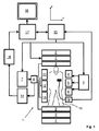
図1を参照すると、MR装置1が示されている。前記装置は、実質的に一様な時間的に一定の主磁場B0が検査体積を通るz軸に沿って作成されるように超伝導又は常伝導主磁石コイル2を有する。前記装置は、(1次、2次、及び適用可能であれば3次の)シムコイルのセットを有し、前記セット2'の個別のシムコイルを通る電流は、前記検査体積内のB0偏差を最小化する目的で制御可能である。
Referring to FIG. 1, an MR apparatus 1 is shown. The device has a superconducting or normal conducting
磁気共鳴発生及び操作システムは、MR撮像を実行するように核磁気スピンを反転又は励起し、磁気共鳴を誘発し、磁気共鳴をリフォーカスし、磁気共鳴を操作し、空間的に及び他の形で前記磁気共鳴を符号化し、スピンを飽和させる等のようにRFパルス及び切り替え磁場勾配の系列を印加する。 The magnetic resonance generation and manipulation system reverses or excites nuclear magnetic spins to perform MR imaging, induces magnetic resonance, refocuses magnetic resonance, manipulates magnetic resonance, spatially and other forms The magnetic resonance is encoded and an RF pulse and a sequence of switching magnetic field gradients are applied so as to saturate the spin.
より具体的には、勾配パルス増幅器3は、前記検査体積のx、y及びz軸に沿って全身グラディエントコイル4、5及び6の選択されたものに電流パルスを印加する。デジタルRF周波数送信器7は、送信/受信スイッチを介して、身体RFコイル9にRFパルス又はパルスパケットを送信し、前記検査体積内にRFパルスを送信する。典型的なMR撮像シーケンスは、印加された磁場勾配と組み合わせて核磁気共鳴の選択された操作を一緒に達成する短い持続時間のRFパルスセグメントのパケットからなる。前記RFパルスは、飽和させる、共鳴を励起する、磁化を反転させる、共鳴をリフォーカスする、又は共鳴を操作する及び前記検査体積内に配置された身体10の部分を選択するのに使用される。前記MR信号は、身体RFコイル9又は/及び複数の局所RF受信器アレイコイル11、12、13によっても取得される。
More specifically, the
並列撮像を用いて身体10の制限された領域のMR画像の生成に対して、局所アレイRFコイル11、12、13のセットは、撮像に対して選択された領域に隣接して配置される。アレイコイル11、12、13は、身体コイルRF送信により誘発されたMR信号を受信するのに使用されることができる。
For the generation of MR images of limited areas of the
結果として生じるMR信号は、身体RFコイル9及び/又はアレイRFコイル11、12、13により取得され、好ましくは前置増幅器(図示されない)を含む受信器14により復調される。受信器装置14は、送信/受信スイッチ8を介してRFコイル9、11、12及び13に接続される。
The resulting MR signal is acquired by the
ホストコンピュータ15は、エコープラナー撮像(EPI)、グラディエント及びスピンエコー撮像、及び高速スピンエコー撮像等のような複数のMR撮像シーケンスのいずれかを生成するようにシムコイル2'並びにグラディエントパルス増幅器3及び送信器7を通る電流を制御する。選択されたシーケンスに対して、受信器14は、各RF励起パルスの後に続いて立て続けに全てのアクティブな受信RFコイルから単一の又は複数のMRデータラインを受信する。データ取得システム16は、前記受信された信号のアナログ‐デジタル変換を実行し、各MRデータラインを更なる処理に適したデジタルフォーマットに変換する。現代のMR装置において、データ取得システム16は、生画像データの取得に特化された別のコンピュータである。
The
最後に、デジタル生画像データは、フーリエ変換又はSENSE若しくはGRAPPA等のような他の適切な再構成アルゴリズムを適用する再構成プロセッサ17により画像表現に再構成される。前記MR画像は、前記患者を通る平面スライス、平行な平面スライスのアレイ、又は三次元体積等を表すことができる。前記画像は、次いで、例えば結果として生じるMR画像の人間可読表示を提供するビデオモニタ18により、スライス、投影、又は前記画像表現の他の部分を視覚化に対する適切なフォーマットに変換するためにアクセスされることができる画像メモリに記憶される。
Finally, the digital raw image data is reconstructed into an image representation by a
描かれたMR装置1は、前記検査体積内の患者の身体10に配置されたセンサ素子に接続された運動センサ19を更に有する。運動センサ19は、描かれた実施例において、前記患者の瞬間的な呼吸相を示す運動信号を検出する。
The depicted MR apparatus 1 further comprises a
図1を参照し続け、更に図2を参照すると、本発明の方法が、示されている。図2は、本発明の異なる方法ステップを持つタイミング図を示す。 With continued reference to FIG. 1 and with further reference to FIG. 2, the method of the present invention is illustrated. FIG. 2 shows a timing diagram with different method steps of the present invention.
描かれた実施例において、呼吸運動信号が、運動センサ19により身体10から取得される。前記運動信号は、図2においてMSにより示される。
In the depicted embodiment, a respiratory motion signal is obtained from the
運動信号MSは、撮像されている身体部分が、APT/CEST技術によって交換性プロトンプールの核磁化を飽和させるように繰り返し照射される飽和RFパルスを連続的に当てている間に、身体10から検出される。完全な呼気の運動状態が、運動信号MSに基づいて認識されるとすぐに、少なくとも1つの励起RFパルスを有する撮像シーケンスがトリガされ、前記撮像シーケンスにより生成されたMR信号が、所定の取得間隔中に取得される。連続したMR信号取得ステップの間に印加されるプリパレーションRFパルスは、図1において"sat"により示されている。前記取得間隔は、"取得"により示されている。
The motion signal MS is transmitted from the
繰り返し照射されるプリパレーションRFパルスの数Nは、連続したMR信号取得ステップの間の時間間隔において監視される。前記プリパレーションRFパルスの照射は、監視された数Nが所定の制限Nmaxを超過するとすぐに自動的に中断される。このようにして、RF熱堆積(SAR)に関する安全動作は、保証される。 The number N of preparation RF pulses that are repeatedly irradiated is monitored in the time interval between successive MR signal acquisition steps. The preparation RF pulse irradiation is automatically interrupted as soon as the monitored number N exceeds a predetermined limit N max . In this way, safe operation with respect to RF thermal deposition (SAR) is ensured.
本発明の上記のMR撮像方法は、例えば腹部のAPT/CEST MR撮像に対して、運動トリガAPT/CEST応用を可能にする。本発明の方法は、次のトリガ事象が待機される時間間隔中に連続的に照射されるプリパレーションRFパルスにより、優れたスキャン時間効率及びロバスト性により特徴づけられる。正確な運動補正は、MR信号が取得される事前に規定された運動状態により達成される。SAR安全動作は、プリパレーションRFパルスの数又は持続時間が所定の制限を超過することを防ぐことにより保証される。 The above-described MR imaging method of the present invention enables motion trigger APT / CEST applications, for example, for abdominal APT / CEST MR imaging. The method of the present invention is characterized by excellent scan time efficiency and robustness with a preparation RF pulse that is continuously emitted during the time interval during which the next trigger event is awaited. Accurate motion correction is achieved by a predefined motion state from which MR signals are acquired. SAR safe operation is ensured by preventing the number or duration of the preparation RF pulses from exceeding a predetermined limit.
Claims (7)
1つ又は複数のプリパレーションRFパルスを前記身体の部分に当てる間に前記動く部分の所望の呼吸運動状態の認識に対して前記身体から運動信号を検出するステップと、 Detecting a motion signal from the body for recognizing a desired respiratory motion state of the moving part while applying one or more preparation RF pulses to the body part;
少なくとも1つの励起RFパルス及び切り替え磁場勾配を有する撮像シーケンスを前記身体の部分に当てるステップであって、前記撮像シーケンスが前記運動信号により検出された前記所望の呼吸運動状態によりトリガされるステップと、 Applying an imaging sequence having at least one excitation RF pulse and a switching magnetic field gradient to the body part, wherein the imaging sequence is triggered by the desired respiratory motion state detected by the motion signal;
前記身体の部分からMR信号を取得するステップであって、前記MR信号が、前記1つ又は複数のプリパレーションRFパルス及び前記撮像シーケンスにより影響を受ける、ステップと、 Obtaining MR signals from the body part, wherein the MR signals are affected by the one or more preparation RF pulses and the imaging sequence;
前記取得されたMR信号からMR画像を再構成するステップと Reconstructing MR images from the acquired MR signals;
を有し、Have
前記プリパレーションRFパルスが、 The preparation RF pulse is
核磁化を飽和させる飽和RFパルス、又は A saturated RF pulse that saturates the nuclear magnetization, or
スピンロッキングRFパルス、又は A spin-locking RF pulse, or
異なる核スピンの間で磁化を移動させる分極移動RFパルス、又は A polarization-transfer RF pulse that moves magnetization between different nuclear spins, or
プロトンデカップリングRFパルス、 Proton decoupling RF pulse,
であり、And
前記プリパレーションRFパルスが、交換性プロトンプール又はCEST造影剤のプロトンの核磁化を飽和させる周波数選択的飽和RFパルスである、 The preparation RF pulse is a frequency selective saturation RF pulse that saturates the nuclear magnetization of protons in the exchangeable proton pool or CEST contrast agent;
方法。Method.
1つ又は複数のプリパレーションRFパルスを前記身体の部分に当てる間に前記動く部分の所望の呼吸運動状態の認識に対して前記身体から運動信号を検出するステップと、 Detecting a motion signal from the body for recognizing a desired respiratory motion state of the moving part while applying one or more preparation RF pulses to the body part;
少なくとも1つの励起RFパルス及び切り替え磁場勾配を有する撮像シーケンスを前記身体の部分に当てるステップであって、前記撮像シーケンスが前記運動信号により検出された前記所望の呼吸運動状態によりトリガされるステップと、 Applying an imaging sequence having at least one excitation RF pulse and a switching magnetic field gradient to the body part, wherein the imaging sequence is triggered by the desired respiratory motion state detected by the motion signal;
前記身体の部分からMR信号を取得するステップであって、前記MR信号が、前記1つ又は複数のプリパレーションRFパルス及び前記撮像シーケンスにより影響を受ける、ステップと、 Obtaining MR signals from the body part, wherein the MR signals are affected by the one or more preparation RF pulses and the imaging sequence;
前記取得されたMR信号からMR画像を再構成するステップと Reconstructing MR images from the acquired MR signals;
を有し、Have
前記プリパレーションRFパルスが、交換性プロトンプール又はCEST造影剤のプロトンの核磁化を飽和させる周波数選択的飽和RFパルスである、 The preparation RF pulse is a frequency selective saturation RF pulse that saturates the nuclear magnetization of protons in the exchangeable proton pool or CEST contrast agent;
方法。Method.
前記検査体積内の異なる空間的方向において切り替え磁場勾配を生成する複数のグラディエントコイルと、 A plurality of gradient coils for generating a switching field gradient in different spatial directions within the examination volume;
前記検査体積内にRFパルスを生成する及び/又は前記検査体積内に配置された患者の身体からMR信号を受信する少なくとも1つのRFコイルと、 At least one RF coil that generates RF pulses within the examination volume and / or receives MR signals from a patient's body disposed within the examination volume;
前記身体の動く部分の運動を感知する運動センサと、 A motion sensor for sensing the motion of the moving part of the body;
前記RFパルス及び切り替え磁場勾配の時間的遷移を制御する制御ユニットと、 A control unit for controlling the temporal transition of the RF pulse and switching magnetic field gradient;
前記受信されたMR信号からMR画像を再構成する再構成ユニットと A reconstruction unit for reconstructing an MR image from the received MR signal;
を有するMR装置において、In an MR apparatus having
前記MR装置が、 The MR apparatus is
1つ又は複数のプリパレーションRFパルスを前記身体の部分に連続的に当てる間に前記運動センサにより前記身体から運動信号を検出するステップと、 Detecting a motion signal from the body by the motion sensor while continuously applying one or more preparation RF pulses to the body part;
少なくとも1つの励起RFパルス及び切り替え磁場勾配を有する撮像シーケンスを前記身体の部分に当てるステップであって、前記撮像シーケンスが前記検出された運動信号によりトリガされるステップと、 Applying an imaging sequence having at least one excitation RF pulse and a switching magnetic field gradient to the body part, wherein the imaging sequence is triggered by the detected motion signal;
前記身体の部分からMR信号を取得するステップと、 Obtaining MR signals from the body part;
前記取得されたMR信号からMR画像を再構成するステップと Reconstructing MR images from the acquired MR signals;
を実行し、Run
前記プリパレーションRFパルスが、 The preparation RF pulse is
核磁化を飽和させる飽和RFパルス、又は A saturated RF pulse that saturates the nuclear magnetization, or
スピンロッキングRFパルス、又は A spin-locking RF pulse, or
異なる核スピンの間で磁化を移動させる分極移動RFパルス、又は A polarization-transfer RF pulse that moves magnetization between different nuclear spins, or
プロトンデカップリングRFパルス、 Proton decoupling RF pulse,
であり、And
前記プリパレーションRFパルスが、交換性プロトンプール又はCEST造影剤のプロトンの核磁化を飽和させる周波数選択的飽和RFパルスである、 The preparation RF pulse is a frequency selective saturation RF pulse that saturates the nuclear magnetization of protons in the exchangeable proton pool or CEST contrast agent;
ように構成される、MR装置。An MR apparatus configured as described above.
前記検査体積内の異なる空間的方向において切り替え磁場勾配を生成する複数のグラディエントコイルと、 A plurality of gradient coils for generating a switching field gradient in different spatial directions within the examination volume;
前記検査体積内にRFパルスを生成する及び/又は前記検査体積内に配置された患者の身体からMR信号を受信する少なくとも1つのRFコイルと、 At least one RF coil that generates RF pulses within the examination volume and / or receives MR signals from a patient's body disposed within the examination volume;
前記身体の動く部分の運動を感知する運動センサと、 A motion sensor for sensing the motion of the moving part of the body;
前記RFパルス及び切り替え磁場勾配の時間的遷移を制御する制御ユニットと、 A control unit for controlling the temporal transition of the RF pulse and switching magnetic field gradient;
前記受信されたMR信号からMR画像を再構成する再構成ユニットと A reconstruction unit for reconstructing an MR image from the received MR signal;
を有するMR装置において、In an MR apparatus having
前記MR装置が、 The MR apparatus is
1つ又は複数のプリパレーションRFパルスを前記身体の部分に連続的に当てる間に前記運動センサにより前記身体から運動信号を検出するステップと、 Detecting a motion signal from the body by the motion sensor while continuously applying one or more preparation RF pulses to the body part;
少なくとも1つの励起RFパルス及び切り替え磁場勾配を有する撮像シーケンスを前記身体の部分に当てるステップであって、前記撮像シーケンスが前記検出された運動信号によりトリガされるステップと、 Applying an imaging sequence having at least one excitation RF pulse and a switching magnetic field gradient to the body part, wherein the imaging sequence is triggered by the detected motion signal;
前記身体の部分からMR信号を取得するステップと、 Obtaining MR signals from the body part;
前記取得されたMR信号からMR画像を再構成するステップと Reconstructing MR images from the acquired MR signals;
を実行し、Run
前記プリパレーションRFパルスが、交換性プロトンプール又はCEST造影剤のプロトンの核磁化を飽和させる周波数選択的飽和RFパルスである、 The preparation RF pulse is a frequency selective saturation RF pulse that saturates the nuclear magnetization of protons in the exchangeable proton pool or CEST contrast agent;
ように構成される、MR装置。An MR apparatus configured as described above.
Applications Claiming Priority (3)
| Application Number | Priority Date | Filing Date | Title |
|---|---|---|---|
| EP11163003A EP2515138A1 (en) | 2011-04-19 | 2011-04-19 | Motion triggered MR imaging using APT/CEST |
| EP11163003.4 | 2011-04-19 | ||
| PCT/IB2012/051598 WO2012143808A1 (en) | 2011-04-19 | 2012-04-02 | Motion triggered mr imaging using apt/cest |
Publications (3)
| Publication Number | Publication Date |
|---|---|
| JP2014511745A JP2014511745A (en) | 2014-05-19 |
| JP2014511745A5 JP2014511745A5 (en) | 2015-05-21 |
| JP6324308B2 true JP6324308B2 (en) | 2018-05-16 |
Family
ID=44351721
Family Applications (1)
| Application Number | Title | Priority Date | Filing Date |
|---|---|---|---|
| JP2014505742A Expired - Fee Related JP6324308B2 (en) | 2011-04-19 | 2012-04-02 | Motion-triggered MR imaging using APT / CEST |
Country Status (5)
| Country | Link |
|---|---|
| US (1) | US10175332B2 (en) |
| EP (2) | EP2515138A1 (en) |
| JP (1) | JP6324308B2 (en) |
| CN (1) | CN103477239B (en) |
| WO (1) | WO2012143808A1 (en) |
Families Citing this family (12)
| Publication number | Priority date | Publication date | Assignee | Title |
|---|---|---|---|---|
| DE102011078680B3 (en) * | 2011-07-05 | 2012-12-20 | Siemens Aktiengesellschaft | Magnetic field insensitive CEST imaging |
| CN104470475B (en) * | 2012-05-24 | 2017-04-19 | 武田有限公司 | Apparatus and process for providing a coiled collagen carrier |
| DE102014200562B4 (en) * | 2014-01-15 | 2015-10-29 | Siemens Aktiengesellschaft | MRI sequence with a number of slice-selective inversion pulses for the preparation of the magnetization and a disjoint number of saturation pulses |
| DE102017202535A1 (en) * | 2017-02-16 | 2018-08-16 | Siemens Healthcare Gmbh | Creating MR images |
| US11428768B2 (en) * | 2017-04-05 | 2022-08-30 | The General Hospital Corporation | Chemical exchange saturation transfer magnetic resonance imaging with gating synchronized acquisition |
| US20190128981A1 (en) * | 2017-10-29 | 2019-05-02 | North America Imaging Developer, Llc | Non-invasive imaging method for early detection and mapping the severity of diseases by using cest mri |
| EP4270413A3 (en) * | 2018-04-05 | 2023-12-27 | Siemens Medical Solutions USA, Inc. | Motion signal derived from imaging data |
| EP3575814A1 (en) * | 2018-05-29 | 2019-12-04 | Koninklijke Philips N.V. | Motion detection in cest magnetic resonance imaging based on z-spectrum analysis |
| US11454689B2 (en) | 2019-09-05 | 2022-09-27 | Canon Medical Systems Corporation | Magnetic resonance imaging apparatus, image processing apparatus, and image processing method |
| US11280867B2 (en) * | 2019-11-08 | 2022-03-22 | The Chinese University Of Hong Kong | System and method for quantitative magnetization transfer imaging based on spin-lock |
| US11366189B2 (en) | 2020-09-25 | 2022-06-21 | Uih America, Inc. | Systems and methods for magnetic resonance imaging |
| CN112748381B (en) * | 2020-12-07 | 2024-03-08 | 中国科学院深圳先进技术研究院 | Chemical exchange saturation transfer quantification method, device and medium for magnetic resonance |
Family Cites Families (26)
| Publication number | Priority date | Publication date | Assignee | Title |
|---|---|---|---|---|
| JPH06327651A (en) * | 1993-05-26 | 1994-11-29 | Yokogawa Medical Syst Ltd | Method of taking data in mr apparatus and apparatus therefor |
| US6066123A (en) * | 1998-04-09 | 2000-05-23 | The Board Of Trustees Of The Leland Stanford Junior University | Enhancement of bioavailability by use of focused energy delivery to a target tissue |
| AU4656400A (en) * | 1999-04-21 | 2000-11-17 | Government of The United States of America, as represented by The Secretary Department of Health & Human Services, The National Institutes of Health, The | Method for enhancing contrast produced by mri |
| KR101010496B1 (en) | 2001-04-17 | 2011-01-21 | 브레우어 사이언스 인코포레이션 | Anti-reflective composition with improved spin bowl compatibility |
| US6958604B2 (en) * | 2003-06-23 | 2005-10-25 | Schlumberger Technology Corporation | Apparatus and methods for J-edit nuclear magnetic resonance measurement |
| EP1661513A4 (en) * | 2003-09-05 | 2009-07-29 | Hitachi Medical Corp | Magnetic resonance imaging method and device |
| US20050165295A1 (en) * | 2004-01-23 | 2005-07-28 | Debiao Li | Local magnetic resonance image quality by optimizing imaging frequency |
| US20060206024A1 (en) | 2005-03-09 | 2006-09-14 | Invivo Corporation | Wireless in-bore patient monitor for MRI |
| US8306603B2 (en) * | 2005-04-26 | 2012-11-06 | Koninklijke Philips Electronics N.V. | MRI involving contrast agent with time modulated contrast enhancement |
| CN101166987B (en) * | 2005-04-26 | 2011-01-12 | 皇家飞利浦电子股份有限公司 | Method for using CEST contrast agents in MRI |
| JP4717573B2 (en) * | 2005-09-26 | 2011-07-06 | ジーイー・メディカル・システムズ・グローバル・テクノロジー・カンパニー・エルエルシー | MRI equipment |
| JP5133557B2 (en) | 2006-12-13 | 2013-01-30 | ジーイー・メディカル・システムズ・グローバル・テクノロジー・カンパニー・エルエルシー | MRI apparatus, RF coil, and magnetic resonance signal suppressing method |
| US20080150532A1 (en) * | 2006-12-21 | 2008-06-26 | General Electric Company | Method and apparatus for measuring t1 relaxation |
| US8457711B2 (en) * | 2007-02-01 | 2013-06-04 | Beth Israel Deaconess Medical Center, Inc. | Magnetic resonance imaging of coronary venous structures |
| JP2009056072A (en) * | 2007-08-31 | 2009-03-19 | Ge Medical Systems Global Technology Co Llc | Magnetic resonance imaging apparatus |
| WO2009042881A1 (en) * | 2007-09-26 | 2009-04-02 | The Johns Hopkins University | Frequency referencing for chemical exchange saturation transfer (cest) mri |
| BRPI0907989A2 (en) * | 2008-02-29 | 2015-10-06 | Koninkl Philips Electronics Nv | magnetic resonance system, method for monitoring an individual in a magnetic field, edge connector, electrocardiogram monitor, and mri system |
| WO2010058732A1 (en) | 2008-11-18 | 2010-05-27 | 株式会社 日立メディコ | Magnetic resonance imaging device and magnetic resonance imaging method |
| EP2199815A1 (en) * | 2008-12-22 | 2010-06-23 | Koninklijke Philips Electronics N.V. | MR imaging with CEST contrast enhancement |
| WO2011091365A1 (en) * | 2010-01-25 | 2011-07-28 | Beth Israel Deaconess Medical Center | Method for measuring magnetization transfer between molecules with magnetic resonance imaging |
| US8686727B2 (en) * | 2010-07-20 | 2014-04-01 | The Trustees Of The University Of Pennsylvania | CEST MRI methods for imaging of metabolites and the use of same as biomarkers |
| US9157976B2 (en) * | 2010-07-20 | 2015-10-13 | The Trustees Of The University Of Pennsylvania | CEST MRI methods for imaging glutaminolysis in cancer |
| WO2012117350A1 (en) * | 2011-03-03 | 2012-09-07 | Koninklijke Philips Electronics N.V. | Magnetic resonance using quasi-continuous rf irradiation |
| US9121917B2 (en) * | 2011-04-20 | 2015-09-01 | The Johns Hopkins University | CEST phase and magnitude imaging using a multi-parametric varied saturation scheme |
| JP5917077B2 (en) * | 2011-10-13 | 2016-05-11 | 株式会社東芝 | Magnetic resonance imaging system |
| EP2648014A1 (en) * | 2012-04-03 | 2013-10-09 | Koninklijke Philips N.V. | MR imaging using APT contrast enhancement and sampling at multiple echo times |
-
2011
- 2011-04-19 EP EP11163003A patent/EP2515138A1/en not_active Withdrawn
-
2012
- 2012-04-02 CN CN201280019084.2A patent/CN103477239B/en not_active Expired - Fee Related
- 2012-04-02 JP JP2014505742A patent/JP6324308B2/en not_active Expired - Fee Related
- 2012-04-02 US US14/009,551 patent/US10175332B2/en active Active
- 2012-04-02 WO PCT/IB2012/051598 patent/WO2012143808A1/en active Application Filing
- 2012-04-02 EP EP12720587.0A patent/EP2699931B1/en active Active
Also Published As
| Publication number | Publication date |
|---|---|
| EP2699931A1 (en) | 2014-02-26 |
| CN103477239B (en) | 2020-03-06 |
| EP2515138A1 (en) | 2012-10-24 |
| US10175332B2 (en) | 2019-01-08 |
| US20140039297A1 (en) | 2014-02-06 |
| WO2012143808A1 (en) | 2012-10-26 |
| EP2699931B1 (en) | 2021-09-15 |
| CN103477239A (en) | 2013-12-25 |
| JP2014511745A (en) | 2014-05-19 |
Similar Documents
| Publication | Publication Date | Title |
|---|---|---|
| JP6324308B2 (en) | Motion-triggered MR imaging using APT / CEST | |
| JP5599893B2 (en) | MR imaging using navigator | |
| US20110288402A1 (en) | Mr imaging with cest contrast enhancement | |
| US10156625B2 (en) | MR imaging with B1 mapping | |
| JP2014503249A (en) | MR imaging using multipoint Dixon technology | |
| KR101343029B1 (en) | Magnetic resonance imaging device and control method thereof | |
| JP6684781B2 (en) | Zero echo time MR imaging | |
| US20120046539A1 (en) | Dual-contrast mr imaging using fluid-attenuation inversion recovery (flair) | |
| JP6074126B1 (en) | Zero echo time MR imaging using sampling in the center of k-space | |
| US8169218B2 (en) | Methods and apparatus for non-contrast enhanced pulmonary magnetic resonance imaging | |
| US7511493B2 (en) | Magnetic resonance imaging method and apparatus | |
| US11360172B2 (en) | Zero echo time MR imaging with water-fat separation | |
| US20100045292A1 (en) | Magnetic resonance angiography method and apparatus | |
| WO2007004123A2 (en) | Mri-navigator sequence with restored magnetization | |
| WO2008041060A1 (en) | Mri-navigator sequence with restored magnetization | |
| Bornert et al. | Coronary Magnetic Resonance Angiography |
Legal Events
| Date | Code | Title | Description |
|---|---|---|---|
| A521 | Request for written amendment filed |
Free format text: JAPANESE INTERMEDIATE CODE: A523 Effective date: 20150401 |
|
| A621 | Written request for application examination |
Free format text: JAPANESE INTERMEDIATE CODE: A621 Effective date: 20150401 |
|
| A977 | Report on retrieval |
Free format text: JAPANESE INTERMEDIATE CODE: A971007 Effective date: 20160113 |
|
| A131 | Notification of reasons for refusal |
Free format text: JAPANESE INTERMEDIATE CODE: A131 Effective date: 20160119 |
|
| A601 | Written request for extension of time |
Free format text: JAPANESE INTERMEDIATE CODE: A601 Effective date: 20160419 |
|
| A521 | Request for written amendment filed |
Free format text: JAPANESE INTERMEDIATE CODE: A523 Effective date: 20160715 |
|
| A131 | Notification of reasons for refusal |
Free format text: JAPANESE INTERMEDIATE CODE: A131 Effective date: 20161108 |
|
| A601 | Written request for extension of time |
Free format text: JAPANESE INTERMEDIATE CODE: A601 Effective date: 20170202 |
|
| RD04 | Notification of resignation of power of attorney |
Free format text: JAPANESE INTERMEDIATE CODE: A7424 Effective date: 20170214 |
|
| A521 | Request for written amendment filed |
Free format text: JAPANESE INTERMEDIATE CODE: A523 Effective date: 20170421 |
|
| A02 | Decision of refusal |
Free format text: JAPANESE INTERMEDIATE CODE: A02 Effective date: 20170928 |
|
| A521 | Request for written amendment filed |
Free format text: JAPANESE INTERMEDIATE CODE: A523 Effective date: 20180109 |
|
| A911 | Transfer to examiner for re-examination before appeal (zenchi) |
Free format text: JAPANESE INTERMEDIATE CODE: A911 Effective date: 20180117 |
|
| TRDD | Decision of grant or rejection written | ||
| A01 | Written decision to grant a patent or to grant a registration (utility model) |
Free format text: JAPANESE INTERMEDIATE CODE: A01 Effective date: 20180322 |
|
| A61 | First payment of annual fees (during grant procedure) |
Free format text: JAPANESE INTERMEDIATE CODE: A61 Effective date: 20180410 |
|
| R150 | Certificate of patent or registration of utility model |
Ref document number: 6324308 Country of ref document: JP Free format text: JAPANESE INTERMEDIATE CODE: R150 |
|
| R250 | Receipt of annual fees |
Free format text: JAPANESE INTERMEDIATE CODE: R250 |
|
| R250 | Receipt of annual fees |
Free format text: JAPANESE INTERMEDIATE CODE: R250 |
|
| LAPS | Cancellation because of no payment of annual fees |