JP6139510B2 - 画像診断装置及びプローブ - Google Patents
画像診断装置及びプローブ Download PDFInfo
- Publication number
- JP6139510B2 JP6139510B2 JP2014507402A JP2014507402A JP6139510B2 JP 6139510 B2 JP6139510 B2 JP 6139510B2 JP 2014507402 A JP2014507402 A JP 2014507402A JP 2014507402 A JP2014507402 A JP 2014507402A JP 6139510 B2 JP6139510 B2 JP 6139510B2
- Authority
- JP
- Japan
- Prior art keywords
- deformation
- axial direction
- probe
- calculated
- measurement slit
- Prior art date
- Legal status (The legal status is an assumption and is not a legal conclusion. Google has not performed a legal analysis and makes no representation as to the accuracy of the status listed.)
- Active
Links
- 239000000523 sample Substances 0.000 title claims description 52
- 238000002059 diagnostic imaging Methods 0.000 title claims description 30
- 230000017531 blood circulation Effects 0.000 claims description 37
- 230000002107 myocardial effect Effects 0.000 claims description 36
- 210000004204 blood vessel Anatomy 0.000 claims description 33
- 238000004364 calculation method Methods 0.000 claims description 29
- 230000005540 biological transmission Effects 0.000 claims description 22
- 230000008859 change Effects 0.000 claims description 5
- 230000010412 perfusion Effects 0.000 claims description 3
- 238000005259 measurement Methods 0.000 description 129
- 238000000034 method Methods 0.000 description 34
- 230000008569 process Effects 0.000 description 16
- 238000010586 diagram Methods 0.000 description 15
- 230000003902 lesion Effects 0.000 description 15
- 230000010349 pulsation Effects 0.000 description 15
- 230000002966 stenotic effect Effects 0.000 description 14
- 238000012545 processing Methods 0.000 description 12
- 238000003384 imaging method Methods 0.000 description 9
- 208000031481 Pathologic Constriction Diseases 0.000 description 7
- 230000036262 stenosis Effects 0.000 description 7
- 208000037804 stenosis Diseases 0.000 description 7
- 230000010339 dilation Effects 0.000 description 6
- 238000012014 optical coherence tomography Methods 0.000 description 6
- 244000208734 Pisonia aculeata Species 0.000 description 4
- 238000003745 diagnosis Methods 0.000 description 3
- 238000011156 evaluation Methods 0.000 description 3
- 238000002608 intravascular ultrasound Methods 0.000 description 3
- 230000036772 blood pressure Effects 0.000 description 2
- 208000028867 ischemia Diseases 0.000 description 2
- 230000003287 optical effect Effects 0.000 description 2
- 210000003462 vein Anatomy 0.000 description 2
- 206010003210 Arteriosclerosis Diseases 0.000 description 1
- 201000000057 Coronary Stenosis Diseases 0.000 description 1
- 206010011089 Coronary artery stenosis Diseases 0.000 description 1
- 238000012276 Endovascular treatment Methods 0.000 description 1
- 210000002565 arteriole Anatomy 0.000 description 1
- 208000011775 arteriosclerosis disease Diseases 0.000 description 1
- 238000005452 bending Methods 0.000 description 1
- 238000012790 confirmation Methods 0.000 description 1
- 210000004351 coronary vessel Anatomy 0.000 description 1
- 230000003247 decreasing effect Effects 0.000 description 1
- 239000003814 drug Substances 0.000 description 1
- 229940079593 drug Drugs 0.000 description 1
- 238000000605 extraction Methods 0.000 description 1
- 239000000835 fiber Substances 0.000 description 1
- 239000003550 marker Substances 0.000 description 1
- 238000012986 modification Methods 0.000 description 1
- 230000004048 modification Effects 0.000 description 1
- 239000013307 optical fiber Substances 0.000 description 1
- 238000013146 percutaneous coronary intervention Methods 0.000 description 1
- 230000002093 peripheral effect Effects 0.000 description 1
- 230000000704 physical effect Effects 0.000 description 1
- 230000002980 postoperative effect Effects 0.000 description 1
- 238000010882 preoperative diagnosis Methods 0.000 description 1
- 230000001012 protector Effects 0.000 description 1
- 238000001931 thermography Methods 0.000 description 1
- 238000011144 upstream manufacturing Methods 0.000 description 1
- 230000002792 vascular Effects 0.000 description 1
- 210000005166 vasculature Anatomy 0.000 description 1
Images


Classifications
-
- A—HUMAN NECESSITIES
- A61—MEDICAL OR VETERINARY SCIENCE; HYGIENE
- A61B—DIAGNOSIS; SURGERY; IDENTIFICATION
- A61B8/00—Diagnosis using ultrasonic, sonic or infrasonic waves
- A61B8/44—Constructional features of the ultrasonic, sonic or infrasonic diagnostic device
- A61B8/4444—Constructional features of the ultrasonic, sonic or infrasonic diagnostic device related to the probe
- A61B8/445—Details of catheter construction
-
- A—HUMAN NECESSITIES
- A61—MEDICAL OR VETERINARY SCIENCE; HYGIENE
- A61B—DIAGNOSIS; SURGERY; IDENTIFICATION
- A61B5/00—Measuring for diagnostic purposes; Identification of persons
- A61B5/02—Detecting, measuring or recording pulse, heart rate, blood pressure or blood flow; Combined pulse/heart-rate/blood pressure determination; Evaluating a cardiovascular condition not otherwise provided for, e.g. using combinations of techniques provided for in this group with electrocardiography or electroauscultation; Heart catheters for measuring blood pressure
- A61B5/026—Measuring blood flow
- A61B5/0295—Measuring blood flow using plethysmography, i.e. measuring the variations in the volume of a body part as modified by the circulation of blood therethrough, e.g. impedance plethysmography
-
- A—HUMAN NECESSITIES
- A61—MEDICAL OR VETERINARY SCIENCE; HYGIENE
- A61B—DIAGNOSIS; SURGERY; IDENTIFICATION
- A61B5/00—Measuring for diagnostic purposes; Identification of persons
- A61B5/103—Detecting, measuring or recording devices for testing the shape, pattern, colour, size or movement of the body or parts thereof, for diagnostic purposes
- A61B5/11—Measuring movement of the entire body or parts thereof, e.g. head or hand tremor, mobility of a limb
- A61B5/1126—Measuring movement of the entire body or parts thereof, e.g. head or hand tremor, mobility of a limb using a particular sensing technique
- A61B5/1128—Measuring movement of the entire body or parts thereof, e.g. head or hand tremor, mobility of a limb using a particular sensing technique using image analysis
-
- A—HUMAN NECESSITIES
- A61—MEDICAL OR VETERINARY SCIENCE; HYGIENE
- A61B—DIAGNOSIS; SURGERY; IDENTIFICATION
- A61B8/00—Diagnosis using ultrasonic, sonic or infrasonic waves
- A61B8/06—Measuring blood flow
- A61B8/065—Measuring blood flow to determine blood output from the heart
-
- A—HUMAN NECESSITIES
- A61—MEDICAL OR VETERINARY SCIENCE; HYGIENE
- A61B—DIAGNOSIS; SURGERY; IDENTIFICATION
- A61B8/00—Diagnosis using ultrasonic, sonic or infrasonic waves
- A61B8/12—Diagnosis using ultrasonic, sonic or infrasonic waves in body cavities or body tracts, e.g. by using catheters
-
- A—HUMAN NECESSITIES
- A61—MEDICAL OR VETERINARY SCIENCE; HYGIENE
- A61B—DIAGNOSIS; SURGERY; IDENTIFICATION
- A61B8/00—Diagnosis using ultrasonic, sonic or infrasonic waves
- A61B8/44—Constructional features of the ultrasonic, sonic or infrasonic diagnostic device
- A61B8/4444—Constructional features of the ultrasonic, sonic or infrasonic diagnostic device related to the probe
- A61B8/4461—Features of the scanning mechanism, e.g. for moving the transducer within the housing of the probe
-
- A—HUMAN NECESSITIES
- A61—MEDICAL OR VETERINARY SCIENCE; HYGIENE
- A61B—DIAGNOSIS; SURGERY; IDENTIFICATION
- A61B8/00—Diagnosis using ultrasonic, sonic or infrasonic waves
- A61B8/52—Devices using data or image processing specially adapted for diagnosis using ultrasonic, sonic or infrasonic waves
- A61B8/5215—Devices using data or image processing specially adapted for diagnosis using ultrasonic, sonic or infrasonic waves involving processing of medical diagnostic data
- A61B8/5223—Devices using data or image processing specially adapted for diagnosis using ultrasonic, sonic or infrasonic waves involving processing of medical diagnostic data for extracting a diagnostic or physiological parameter from medical diagnostic data
-
- A—HUMAN NECESSITIES
- A61—MEDICAL OR VETERINARY SCIENCE; HYGIENE
- A61B—DIAGNOSIS; SURGERY; IDENTIFICATION
- A61B2562/00—Details of sensors; Constructional details of sensor housings or probes; Accessories for sensors
- A61B2562/16—Details of sensor housings or probes; Details of structural supports for sensors
- A61B2562/17—Comprising radiolucent components
-
- A—HUMAN NECESSITIES
- A61—MEDICAL OR VETERINARY SCIENCE; HYGIENE
- A61B—DIAGNOSIS; SURGERY; IDENTIFICATION
- A61B2576/00—Medical imaging apparatus involving image processing or analysis
-
- A—HUMAN NECESSITIES
- A61—MEDICAL OR VETERINARY SCIENCE; HYGIENE
- A61B—DIAGNOSIS; SURGERY; IDENTIFICATION
- A61B5/00—Measuring for diagnostic purposes; Identification of persons
- A61B5/02—Detecting, measuring or recording pulse, heart rate, blood pressure or blood flow; Combined pulse/heart-rate/blood pressure determination; Evaluating a cardiovascular condition not otherwise provided for, e.g. using combinations of techniques provided for in this group with electrocardiography or electroauscultation; Heart catheters for measuring blood pressure
- A61B5/02007—Evaluating blood vessel condition, e.g. elasticity, compliance
-
- A—HUMAN NECESSITIES
- A61—MEDICAL OR VETERINARY SCIENCE; HYGIENE
- A61B—DIAGNOSIS; SURGERY; IDENTIFICATION
- A61B5/00—Measuring for diagnostic purposes; Identification of persons
- A61B5/02—Detecting, measuring or recording pulse, heart rate, blood pressure or blood flow; Combined pulse/heart-rate/blood pressure determination; Evaluating a cardiovascular condition not otherwise provided for, e.g. using combinations of techniques provided for in this group with electrocardiography or electroauscultation; Heart catheters for measuring blood pressure
- A61B5/021—Measuring pressure in heart or blood vessels
- A61B5/0215—Measuring pressure in heart or blood vessels by means inserted into the body
-
- A—HUMAN NECESSITIES
- A61—MEDICAL OR VETERINARY SCIENCE; HYGIENE
- A61B—DIAGNOSIS; SURGERY; IDENTIFICATION
- A61B5/00—Measuring for diagnostic purposes; Identification of persons
- A61B5/02—Detecting, measuring or recording pulse, heart rate, blood pressure or blood flow; Combined pulse/heart-rate/blood pressure determination; Evaluating a cardiovascular condition not otherwise provided for, e.g. using combinations of techniques provided for in this group with electrocardiography or electroauscultation; Heart catheters for measuring blood pressure
- A61B5/026—Measuring blood flow
- A61B5/0261—Measuring blood flow using optical means, e.g. infrared light
-
- A—HUMAN NECESSITIES
- A61—MEDICAL OR VETERINARY SCIENCE; HYGIENE
- A61B—DIAGNOSIS; SURGERY; IDENTIFICATION
- A61B5/00—Measuring for diagnostic purposes; Identification of persons
- A61B5/02—Detecting, measuring or recording pulse, heart rate, blood pressure or blood flow; Combined pulse/heart-rate/blood pressure determination; Evaluating a cardiovascular condition not otherwise provided for, e.g. using combinations of techniques provided for in this group with electrocardiography or electroauscultation; Heart catheters for measuring blood pressure
- A61B5/026—Measuring blood flow
- A61B5/0265—Measuring blood flow using electromagnetic means, e.g. electromagnetic flowmeter
- A61B5/027—Measuring blood flow using electromagnetic means, e.g. electromagnetic flowmeter using catheters
-
- A—HUMAN NECESSITIES
- A61—MEDICAL OR VETERINARY SCIENCE; HYGIENE
- A61B—DIAGNOSIS; SURGERY; IDENTIFICATION
- A61B5/00—Measuring for diagnostic purposes; Identification of persons
- A61B5/02—Detecting, measuring or recording pulse, heart rate, blood pressure or blood flow; Combined pulse/heart-rate/blood pressure determination; Evaluating a cardiovascular condition not otherwise provided for, e.g. using combinations of techniques provided for in this group with electrocardiography or electroauscultation; Heart catheters for measuring blood pressure
- A61B5/026—Measuring blood flow
- A61B5/029—Measuring or recording blood output from the heart, e.g. minute volume
-
- A—HUMAN NECESSITIES
- A61—MEDICAL OR VETERINARY SCIENCE; HYGIENE
- A61B—DIAGNOSIS; SURGERY; IDENTIFICATION
- A61B5/00—Measuring for diagnostic purposes; Identification of persons
- A61B5/72—Signal processing specially adapted for physiological signals or for diagnostic purposes
- A61B5/7225—Details of analog processing, e.g. isolation amplifier, gain or sensitivity adjustment, filtering, baseline or drift compensation
-
- A—HUMAN NECESSITIES
- A61—MEDICAL OR VETERINARY SCIENCE; HYGIENE
- A61B—DIAGNOSIS; SURGERY; IDENTIFICATION
- A61B5/00—Measuring for diagnostic purposes; Identification of persons
- A61B5/72—Signal processing specially adapted for physiological signals or for diagnostic purposes
- A61B5/7271—Specific aspects of physiological measurement analysis
- A61B5/7278—Artificial waveform generation or derivation, e.g. synthesising signals from measured signals
-
- G—PHYSICS
- G16—INFORMATION AND COMMUNICATION TECHNOLOGY [ICT] SPECIALLY ADAPTED FOR SPECIFIC APPLICATION FIELDS
- G16H—HEALTHCARE INFORMATICS, i.e. INFORMATION AND COMMUNICATION TECHNOLOGY [ICT] SPECIALLY ADAPTED FOR THE HANDLING OR PROCESSING OF MEDICAL OR HEALTHCARE DATA
- G16H30/00—ICT specially adapted for the handling or processing of medical images
- G16H30/40—ICT specially adapted for the handling or processing of medical images for processing medical images, e.g. editing
Description
画像診断装置であって、
シースと、該シースに内挿され信号の送受信を行う送受信部とを有し、該送受信部が該シース内において周方向への回転を行いながら、または、周方向への回転と軸方向への移動とを行いながら信号の送受信を行うよう制御され、かつ、該軸方向における該シースの異なる位置には、外界の圧力に応じて断面形状が変形する変形部がそれぞれ設けられたプローブ部と、
前記変形部が含まれる断層画像を用いて、前記軸方向の異なる位置に設けられた変形部それぞれの変形度合いを示す複数のパラメータを算出する第1の算出手段と、
前記複数のパラメータに基づいて、心筋血流予備量比に対応する値を算出する第2の算出手段とを備えることを特徴とする。
<1.心筋血流予備量比の説明>
はじめに、生理学的な手法において用いられる評価パラメータである、心筋血流予備量比(FFR:Fractional flow reserve)の概要について非特許文献1より引用する図8を用いて説明する。
Qn=(Pa−Pv)/R
となる。
Qs=(Pd−Pv)/R
となる。
ここで、最大冠拡張時では、Pd>>Pv、Pa>>Pvとなることから、心筋血流予備量比(FFR)≒Pd/Paと表すことができる。
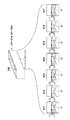
次に、画像診断装置を用いた心筋血流予備量比の算出方法について説明する。図9は、OCTを用いた心筋血流予備量比の算出方法を説明するための図であり、900は血管内の圧力に応じて、所定の面901が変形する測定スリット部(変形部)である。
Pb×Vb1=P0×Vb0
したがって、心筋血流予備量比(FFR)は、
FFR=Pb/Pf={(P0×Vb0)/Vb1}/{(P0×Vf0)/Vf1}=(Vb0×Vf1)/(Vb1×Vf0)
つまり、画像診断装置により生成される断層画像を用いて、軸方向先端側の測定スリット部の大気下での体積と血管内での体積、及び、軸方向基端側の測定スリット部の大気下での体積と血管内での体積をそれぞれ算出することにより、心筋血流予備量比を算出することができる。
次に、上記心筋血流予備量比を算出する、本実施形態に係る画像診断装置の外観構成について説明する。
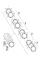
次に、プローブ部101の構成について図2を用いて説明する。図2に示すように、プローブ部101は、血管等の生体管腔内に挿入される長尺のカテーテルシース201と、ユーザが操作するために血管等の生体管腔内に挿入されることなく、ユーザの手元側に配置されるコネクタ部202とにより構成される。カテーテルシース201の先端には、ガイドワイヤルーメンを構成するガイドワイヤルーメン用チューブ203が設けられている。カテーテルシース201は、ガイドワイヤルーメン用チューブ203との接続部分からコネクタ部202との接続部分にかけて連続する管腔を形成している。
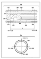
次に、プローブ部101の先端部(図2の230)の詳細構成について、図3を用いて説明する。図3は、プローブ部101の先端部の詳細構成を示す図であり、図3の3aは、プローブ部101の先端部を側面から見た場合の断面図を、図3の3bは、プローブ部101の先端部を先端側から見た場合の、図3の3aの第1の位置及び第2の位置における断面図をそれぞれ示している(なお、図3の3bでは、説明の便宜上、イメージングコア220を省略している)。
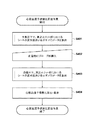
次に、画像診断装置100を用いた、心筋血流予備量比算出作業の流れについて、図4を用いて説明する。図4は、心筋血流予備量比算出作業の流れを示すフローチャートである。
次に、図4のステップS401及びS403に示す、測定スリット部におけるシート330の変形度合いを示すパラメータを算出するための処理(以下、パラメータ算出処理と称す)の詳細について、図5A、図5B及び図6A、図6Bを用いて説明する。図5A及び図5Bは、パラメータ算出処理(3種類の処理)の流れを示すフローチャートであり、図6A及び図6Bは、パラメータ算出処理に際して生成される断層画像及びその拡大図の一例を示す図である。
図5Aの5aに示すように、画像診断装置100では、ステップS501において、光の送受信を開始するとともに、ラジアル動作を開始し、カテーテルシース201の断層画像の生成を開始する。
次に、図5Aの5bに示すパラメータ算出処理について説明する。なお、図5Aの5bのステップS511〜S513までの処理は、図5Aの5aのステップS501〜S503までの処理と同じであるため、ここでは説明を省略する。
次に、図5Bに示すパラメータ算出処理について説明する。なお、図5BのステップS521〜S522までの処理は、図5Aの5aのステップS501〜S502までの処理と同様であるため、ここでは説明を省略する。
上記第1の実施形態では、測定スリット部におけるシートの変形を捉えるにあたり、拍動の影響については特に考慮しなかったが、本発明は、これに限定されず、拍動のタイミングを考慮するように構成してもよい。
上記第1の実施形態では、カテーテルシース201を切り欠くことにより、測定スリット部を形成することとしたが、本発明はこれに限定されない。カテーテルシース201の外周に、測定スリット部に相当する隙間が形成されるような形状の部材を貼り付け、その上からシート330を覆う構成としてもよい。なお、このときカテーテルシース201の外周に貼り付けられる部材は、シート330よりも高い剛性を有する部材であることが望ましい。
上記第1の実施形態では、第1の位置及び第2の位置において、測定スリット部をそれぞれ4つ設ける構成としたが、本発明はこれに限定されず、4つ以上であってもよい。
本発明は上記実施の形態に制限されるものではなく、本発明の精神及び範囲から離脱することなく、様々な変更及び変形が可能である。従って、本発明の範囲を公にするために、以下の請求項を添付する。
Claims (11)
- 画像診断装置であって、
シースと、該シースに内挿され信号の送受信を行う送受信部とを有し、該送受信部が該シース内において周方向への回転を行いながら、または、周方向への回転と軸方向への移動とを行いながら信号の送受信を行うよう制御され、かつ、該軸方向における該シースの異なる位置には、外界の圧力に応じて断面形状が変形する変形部がそれぞれ設けられたプローブ部と、
前記変形部が含まれる断層画像を用いて、前記軸方向の異なる位置に設けられた変形部それぞれの変形度合いを示す複数のパラメータを算出する第1の算出手段と、
前記複数のパラメータに基づいて、心筋血流予備量比に対応する値を算出する第2の算出手段と
を備えることを特徴とする画像診断装置。 - 前記変形部は、前記軸方向に所定の長さを有する直方体形状を有しており、該変形部のうち前記プローブ部の円周面を形成する面が、該変形部の他の面よりも剛性の低い部材により構成されていることを特徴とする請求項1に記載の画像診断装置。
- 前記第1の算出手段は、前記変形部の変形度合いを示すパラメータとして、前記変形部の体積、または、前記変形部の前記軸方向の所定の位置における面積、または、前記変形部の前記軸方向の所定の位置における前記円周面を形成する面の高さ、または、前記変形部の前記軸方向の所定の位置における前記円周面を形成する面の歪み量を算出することを特徴とする請求項2に記載の画像診断装置。
- 前記第2の算出手段は、
前記軸方向の異なる位置に設けられた変形部のうち、第1の位置に設けられた変形部について算出された、
大気圧P0下における前記パラメータをXf0、
生体管腔内における前記パラメータをXf1、
前記軸方向の異なる位置に設けられた変形部のうち、第2の位置に設けられた変形部について算出された、
大気圧P0下における前記パラメータをXb0、
生体管腔内における前記パラメータをXb1、
とした場合、前記心筋血流予備量比に対応する値を、
(Xb0×Xf1)/(Xb1×Xf0)により算出することを特徴とする請求項1乃至3のいずれか1項に記載の画像診断装置。 - 前記変形部が含まれる断層画像を抽出する抽出手段と、
前記抽出手段により抽出された断層画像に基づいて、前記軸方向の異なる位置に設けられた変形部それぞれの、前記軸方向における中心位置を算出する第3の算出手段と、を更に備え、
前記第1の算出手段は、前記第3の算出手段により算出された前記軸方向における中心位置それぞれに、前記送受信部を移動させた状態で、該送受信部を周方向に回転させることにより生成される断層画像を用いて、前記パラメータを算出することを特徴とする請求項1に記載の画像診断装置。 - 前記第1の算出手段は、前記送受信部を、所定時間、周方向に回転させることにより生成される複数の断層画像のうち、前記変形部の変形度合いが最も大きい断層画像を用いて、前記パラメータを算出することを特徴とする請求項5に記載の画像診断装置。
- 前記変形部は、前記シースにおける第1の位置と、該第1の位置より前記軸方向における基端側の第2の位置にそれぞれ設けられ、該第1の位置と該第2の位置のそれぞれにおいて、周方向に複数設けられていることを特徴とする請求項1に記載の画像診断装置。
- 前記第1の算出手段は、周方向に複数設けられている前記変形部の内の一部の変形部の変形度合いを示すパラメータを採用し、他の変形部の変形度合いを示すパラメータを採用せずに前記第2の算出手段にて用いられる前記パラメータを算出することを特徴とする請求項7に記載の画像診断装置。
- 信号の送受信を行う送受信部がシースに内挿され、該送受信部が該シース内において周方向への回転を行いながら、または、周方向への回転と軸方向への移動とを行いながら取得した信号を用いて血管内の断層画像を生成する画像診断装置に対して、該信号を送信するプローブであって、
前記軸方向の互いに異なる位置に配置され、前記プローブにかかる圧力に応じて断面形状が変形する複数の変形部を備え、
前記変形部は、前記軸方向に所定の長さを有する直方体形状を有しており、該変形部のうち前記プローブの円周面を形成する面が、該変形部の他の面よりも剛性の低い部材により構成され、
前記送受信部は、前記画像診断装置が断層画像の一部として前記変形部の画像を得ることができるように配置されていることを特徴とするプローブ。 - 前記変形部は、前記軸方向に所定の長さを有する直方体形状を有しており、該変形部のうち前記プローブの円周面を形成する面が、該変形部の他の面よりも剛性の低い部材により構成されていることを特徴とする請求項9に記載のプローブ。
- 前記変形部は、前記シースにおける第1の位置と、該第1の位置より前記軸方向における基端側の第2の位置にそれぞれ設けられ、該第1の位置と該第2の位置のそれぞれにおいて、周方向に複数設けられていることを特徴とする請求項9または10に記載のプローブ。
Priority Applications (1)
| Application Number | Priority Date | Filing Date | Title |
|---|---|---|---|
| JP2014507402A JP6139510B2 (ja) | 2012-03-29 | 2013-03-19 | 画像診断装置及びプローブ |
Applications Claiming Priority (4)
| Application Number | Priority Date | Filing Date | Title |
|---|---|---|---|
| JP2012076730 | 2012-03-29 | ||
| JP2012076730 | 2012-03-29 | ||
| JP2014507402A JP6139510B2 (ja) | 2012-03-29 | 2013-03-19 | 画像診断装置及びプローブ |
| PCT/JP2013/001866 WO2013145638A1 (ja) | 2012-03-29 | 2013-03-19 | 画像診断装置及びプローブ |
Publications (2)
| Publication Number | Publication Date |
|---|---|
| JPWO2013145638A1 JPWO2013145638A1 (ja) | 2015-12-10 |
| JP6139510B2 true JP6139510B2 (ja) | 2017-05-31 |
Family
ID=49258952
Family Applications (1)
| Application Number | Title | Priority Date | Filing Date |
|---|---|---|---|
| JP2014507402A Active JP6139510B2 (ja) | 2012-03-29 | 2013-03-19 | 画像診断装置及びプローブ |
Country Status (4)
| Country | Link |
|---|---|
| US (1) | US10213186B2 (ja) |
| EP (1) | EP2832287B1 (ja) |
| JP (1) | JP6139510B2 (ja) |
| WO (1) | WO2013145638A1 (ja) |
Families Citing this family (7)
| Publication number | Priority date | Publication date | Assignee | Title |
|---|---|---|---|---|
| US11311200B1 (en) | 2014-08-27 | 2022-04-26 | Lightlab Imaging, Inc. | Systems and methods to measure physiological flow in coronary arteries |
| EP3536230B1 (en) | 2014-08-27 | 2022-03-16 | St. Jude Medical Systems AB | System for evaluating a cardiac system by determining minimum ratio pd/pa (distal pressure / arterial pressure) |
| EP4035586A1 (en) | 2015-04-16 | 2022-08-03 | Gentuity LLC | Micro-optic probes for neurology |
| JP6981967B2 (ja) | 2015-08-31 | 2021-12-17 | ジェンテュイティ・リミテッド・ライアビリティ・カンパニーGentuity, LLC | 撮像プローブおよびデリバリデバイスを含む撮像システム |
| JP7160935B2 (ja) | 2017-11-28 | 2022-10-25 | ジェンテュイティ・リミテッド・ライアビリティ・カンパニー | 撮像システム |
| US11523788B2 (en) * | 2018-01-11 | 2022-12-13 | Canon Medical Systems Corporation | Medical information processing apparatus, medical information processing system, and medical information processing method |
| JP7160659B2 (ja) * | 2018-01-11 | 2022-10-25 | キヤノンメディカルシステムズ株式会社 | 医用情報処理装置、医用情報処理システム及び医用情報処理方法 |
Family Cites Families (12)
| Publication number | Priority date | Publication date | Assignee | Title |
|---|---|---|---|---|
| US4873990A (en) * | 1988-09-23 | 1989-10-17 | The United States Of America As Represented By The Administrator Of The National Aeronautics And Space Administration | Circumferential pressure probe |
| JP3368601B2 (ja) * | 1992-11-17 | 2003-01-20 | オリンパス光学工業株式会社 | 管路内挿入装置 |
| JPH06261905A (ja) * | 1993-01-14 | 1994-09-20 | Olympus Optical Co Ltd | 超音波内視鏡装置 |
| JPH07222748A (ja) * | 1994-02-09 | 1995-08-22 | Aloka Co Ltd | 血管用超音波プローブ |
| EP1251769A1 (en) | 1999-03-09 | 2002-10-30 | Florence Medical Ltd. | A method and system for pressure based measurements of cfr and additional clinical hemodynamic parameters |
| DE102005050344A1 (de) * | 2005-10-20 | 2007-05-03 | Siemens Ag | Kryokatheter zur Einführung in ein Körpergefäß sowie medizinische Untersuchungs- und Behandlungsvorrichtung |
| DE102005059262B4 (de) * | 2005-12-12 | 2008-02-07 | Siemens Ag | Kathetervorrichtung |
| ATE469600T1 (de) | 2006-04-28 | 2010-06-15 | Radi Medical Systems | Sensor und führungsdrahtanordnung |
| JP5746969B2 (ja) | 2008-09-11 | 2015-07-08 | アシスト・メディカル・システムズ,インコーポレイテッド | 生理学的センサ配送装置及び流体注入システム |
| WO2010033971A1 (en) | 2008-09-22 | 2010-03-25 | Dtherapeutics, Llc | Devices, systems, and methods for determining fractional flow reserve |
| JP5819823B2 (ja) * | 2009-07-14 | 2015-11-24 | ザ ジェネラル ホスピタル コーポレイション | 血管の内部の流れおよび圧力を測定する装置および装置の作動方法 |
| US8478384B2 (en) * | 2010-01-19 | 2013-07-02 | Lightlab Imaging, Inc. | Intravascular optical coherence tomography system with pressure monitoring interface and accessories |
-
2013
- 2013-03-19 EP EP13768286.0A patent/EP2832287B1/en active Active
- 2013-03-19 WO PCT/JP2013/001866 patent/WO2013145638A1/ja active Application Filing
- 2013-03-19 JP JP2014507402A patent/JP6139510B2/ja active Active
-
2014
- 2014-09-17 US US14/488,518 patent/US10213186B2/en not_active Expired - Fee Related
Also Published As
| Publication number | Publication date |
|---|---|
| US20150005615A1 (en) | 2015-01-01 |
| US10213186B2 (en) | 2019-02-26 |
| EP2832287A1 (en) | 2015-02-04 |
| EP2832287A4 (en) | 2015-11-18 |
| JPWO2013145638A1 (ja) | 2015-12-10 |
| EP2832287B1 (en) | 2017-04-26 |
| WO2013145638A1 (ja) | 2013-10-03 |
Similar Documents
| Publication | Publication Date | Title |
|---|---|---|
| JP6139510B2 (ja) | 画像診断装置及びプローブ | |
| JP7453150B2 (ja) | 医療管腔内超音波イメージングにおける脈管内病巣及びステント配備のスコアリング | |
| US20160206267A1 (en) | Image processing apparatus, image display system, imaging system, image processing method, and program | |
| EP2934307B1 (en) | Functional gain measurement technique and representation | |
| JP6479678B2 (ja) | マルチセンサ医療デバイスのための表示制御 | |
| EP3071103A1 (en) | Tracking an intraluminal catheter | |
| JP2021517034A (ja) | 管腔内病巣評価及び処置計画のための解剖学的標識の決定及び可視化 | |
| JP2017519544A (ja) | 血管評価のためのデバイス、システム及び方法 | |
| JP2012521852A5 (ja) | ||
| US10524652B2 (en) | Information processing device, imaging system, information processing method and program | |
| US20230309859A1 (en) | Intravascular imaging procedure-specific workflow guidance and associated devices, systems, and methods | |
| JP6284944B2 (ja) | 画像診断装置及びその作動方法及び記憶媒体 | |
| CN111462117A (zh) | 基于血管图像的数据处理系统及其数据处理方法 | |
| EP3048982A1 (en) | Systems and methods for producing intravascular images | |
| EP4138672B1 (en) | Automated control of intraluminal data acquisition and associated devices, systems, and methods | |
| CN212365043U (zh) | 基于血管图像的数据处理系统 | |
| JP6100911B2 (ja) | 画像診断装置及びその作動方法 | |
| WO2014162366A1 (ja) | 画像診断装置及びその制御方法、プログラム及びコンピュータ可読記憶媒体 | |
| JP6809905B2 (ja) | 画像診断装置、画像診断装置の作動方法及びプログラム | |
| US20230181140A1 (en) | Registration of intraluminal physiological data to longitudinal image body lumen using extraluminal imaging data | |
| US20230363652A1 (en) | Intravascular Pressure Sensing Using Inner Sheath |
Legal Events
| Date | Code | Title | Description |
|---|---|---|---|
| A621 | Written request for application examination |
Free format text: JAPANESE INTERMEDIATE CODE: A621 Effective date: 20160106 |
|
| A131 | Notification of reasons for refusal |
Free format text: JAPANESE INTERMEDIATE CODE: A131 Effective date: 20160930 |
|
| A521 | Request for written amendment filed |
Free format text: JAPANESE INTERMEDIATE CODE: A523 Effective date: 20161125 |
|
| TRDD | Decision of grant or rejection written | ||
| A01 | Written decision to grant a patent or to grant a registration (utility model) |
Free format text: JAPANESE INTERMEDIATE CODE: A01 Effective date: 20170414 |
|
| A61 | First payment of annual fees (during grant procedure) |
Free format text: JAPANESE INTERMEDIATE CODE: A61 Effective date: 20170427 |
|
| R150 | Certificate of patent or registration of utility model |
Ref document number: 6139510 Country of ref document: JP Free format text: JAPANESE INTERMEDIATE CODE: R150 |
|
| R250 | Receipt of annual fees |
Free format text: JAPANESE INTERMEDIATE CODE: R250 |
|
| R250 | Receipt of annual fees |
Free format text: JAPANESE INTERMEDIATE CODE: R250 |
|
| R250 | Receipt of annual fees |
Free format text: JAPANESE INTERMEDIATE CODE: R250 |
|
| R250 | Receipt of annual fees |
Free format text: JAPANESE INTERMEDIATE CODE: R250 |
|
| R250 | Receipt of annual fees |
Free format text: JAPANESE INTERMEDIATE CODE: R250 |