JP6129166B2 - Method and apparatus for detecting arterial occlusion / resumption and system for measuring systolic blood pressure - Google Patents
Method and apparatus for detecting arterial occlusion / resumption and system for measuring systolic blood pressure Download PDFInfo
- Publication number
- JP6129166B2 JP6129166B2 JP2014522181A JP2014522181A JP6129166B2 JP 6129166 B2 JP6129166 B2 JP 6129166B2 JP 2014522181 A JP2014522181 A JP 2014522181A JP 2014522181 A JP2014522181 A JP 2014522181A JP 6129166 B2 JP6129166 B2 JP 6129166B2
- Authority
- JP
- Japan
- Prior art keywords
- artery
- variable
- blood flow
- pressure
- resumption
- Prior art date
- Legal status (The legal status is an assumption and is not a legal conclusion. Google has not performed a legal analysis and makes no representation as to the accuracy of the status listed.)
- Expired - Fee Related
Links
- 238000000034 method Methods 0.000 title claims description 31
- 230000035488 systolic blood pressure Effects 0.000 title claims description 24
- 208000031104 Arterial Occlusive disease Diseases 0.000 title description 24
- 208000021328 arterial occlusion Diseases 0.000 title description 24
- 210000001367 artery Anatomy 0.000 claims description 84
- 230000017531 blood circulation Effects 0.000 claims description 76
- 238000001514 detection method Methods 0.000 claims description 24
- 238000009795 derivation Methods 0.000 claims description 3
- 238000005259 measurement Methods 0.000 description 8
- 238000010586 diagram Methods 0.000 description 6
- 238000009530 blood pressure measurement Methods 0.000 description 5
- 238000002555 auscultation Methods 0.000 description 4
- 238000002604 ultrasonography Methods 0.000 description 4
- 230000007423 decrease Effects 0.000 description 3
- 239000000523 sample Substances 0.000 description 3
- 230000003595 spectral effect Effects 0.000 description 3
- 230000036772 blood pressure Effects 0.000 description 2
- 230000008602 contraction Effects 0.000 description 2
- 230000000694 effects Effects 0.000 description 2
- 238000000691 measurement method Methods 0.000 description 2
- 206010011878 Deafness Diseases 0.000 description 1
- 206010020772 Hypertension Diseases 0.000 description 1
- 238000013459 approach Methods 0.000 description 1
- 239000008280 blood Substances 0.000 description 1
- 210000004369 blood Anatomy 0.000 description 1
- 210000002302 brachial artery Anatomy 0.000 description 1
- 230000003247 decreasing effect Effects 0.000 description 1
- 238000013461 design Methods 0.000 description 1
- 238000002592 echocardiography Methods 0.000 description 1
- 230000001631 hypertensive effect Effects 0.000 description 1
- 210000003141 lower extremity Anatomy 0.000 description 1
- 238000012986 modification Methods 0.000 description 1
- 230000004048 modification Effects 0.000 description 1
- 230000001360 synchronised effect Effects 0.000 description 1
- 210000002465 tibial artery Anatomy 0.000 description 1
- 210000001364 upper extremity Anatomy 0.000 description 1
Images


Classifications
-
- A—HUMAN NECESSITIES
- A61—MEDICAL OR VETERINARY SCIENCE; HYGIENE
- A61B—DIAGNOSIS; SURGERY; IDENTIFICATION
- A61B8/00—Diagnosis using ultrasonic, sonic or infrasonic waves
- A61B8/52—Devices using data or image processing specially adapted for diagnosis using ultrasonic, sonic or infrasonic waves
- A61B8/5215—Devices using data or image processing specially adapted for diagnosis using ultrasonic, sonic or infrasonic waves involving processing of medical diagnostic data
- A61B8/5223—Devices using data or image processing specially adapted for diagnosis using ultrasonic, sonic or infrasonic waves involving processing of medical diagnostic data for extracting a diagnostic or physiological parameter from medical diagnostic data
-
- A—HUMAN NECESSITIES
- A61—MEDICAL OR VETERINARY SCIENCE; HYGIENE
- A61B—DIAGNOSIS; SURGERY; IDENTIFICATION
- A61B5/00—Measuring for diagnostic purposes; Identification of persons
- A61B5/02—Detecting, measuring or recording pulse, heart rate, blood pressure or blood flow; Combined pulse/heart-rate/blood pressure determination; Evaluating a cardiovascular condition not otherwise provided for, e.g. using combinations of techniques provided for in this group with electrocardiography or electroauscultation; Heart catheters for measuring blood pressure
- A61B5/021—Measuring pressure in heart or blood vessels
- A61B5/022—Measuring pressure in heart or blood vessels by applying pressure to close blood vessels, e.g. against the skin; Ophthalmodynamometers
-
- A—HUMAN NECESSITIES
- A61—MEDICAL OR VETERINARY SCIENCE; HYGIENE
- A61B—DIAGNOSIS; SURGERY; IDENTIFICATION
- A61B8/00—Diagnosis using ultrasonic, sonic or infrasonic waves
- A61B8/04—Measuring blood pressure
-
- A—HUMAN NECESSITIES
- A61—MEDICAL OR VETERINARY SCIENCE; HYGIENE
- A61B—DIAGNOSIS; SURGERY; IDENTIFICATION
- A61B8/00—Diagnosis using ultrasonic, sonic or infrasonic waves
- A61B8/06—Measuring blood flow
-
- A—HUMAN NECESSITIES
- A61—MEDICAL OR VETERINARY SCIENCE; HYGIENE
- A61B—DIAGNOSIS; SURGERY; IDENTIFICATION
- A61B8/00—Diagnosis using ultrasonic, sonic or infrasonic waves
- A61B8/44—Constructional features of the ultrasonic, sonic or infrasonic diagnostic device
- A61B8/4483—Constructional features of the ultrasonic, sonic or infrasonic diagnostic device characterised by features of the ultrasound transducer
-
- A—HUMAN NECESSITIES
- A61—MEDICAL OR VETERINARY SCIENCE; HYGIENE
- A61B—DIAGNOSIS; SURGERY; IDENTIFICATION
- A61B8/00—Diagnosis using ultrasonic, sonic or infrasonic waves
- A61B8/48—Diagnostic techniques
- A61B8/488—Diagnostic techniques involving Doppler signals
-
- A—HUMAN NECESSITIES
- A61—MEDICAL OR VETERINARY SCIENCE; HYGIENE
- A61B—DIAGNOSIS; SURGERY; IDENTIFICATION
- A61B5/00—Measuring for diagnostic purposes; Identification of persons
- A61B5/68—Arrangements of detecting, measuring or recording means, e.g. sensors, in relation to patient
- A61B5/6801—Arrangements of detecting, measuring or recording means, e.g. sensors, in relation to patient specially adapted to be attached to or worn on the body surface
- A61B5/6813—Specially adapted to be attached to a specific body part
- A61B5/6824—Arm or wrist
Description
本発明は非侵襲的血圧測定、特に動脈の閉塞/再開を検出するための方法及び装置並びに収縮期血圧(SBP)を測定するためのシステムに関する。 The present invention relates to a non-invasive blood pressure measurement, in particular to a method and apparatus for detecting arterial occlusion / resumption and a system for measuring systolic blood pressure (SBP).
侵襲的血圧測定と異なり、非侵襲的血圧測定は人体の動脈内の血圧を測定する間接的方法である。現在、二つのカテゴリの非侵襲的血圧測定法がある。 Unlike invasive blood pressure measurement, non-invasive blood pressure measurement is an indirect method of measuring blood pressure in a human artery. There are currently two categories of noninvasive blood pressure measurement methods.
一つの方法は聴診法("聴く"を意味するラテン語に由来)であり、これはその精度のため臨床測定の主要な方法である。聴診法によれば、動脈内の血流を制限するように動脈に変化する圧力を加えるために血圧計の膨張カフが使用される。カフは動脈が完全に閉塞されるまで最初に膨張され、それから動脈が再開するまで収縮される。閉塞の瞬間及び再開の瞬間における圧力値は一般にそれぞれSBP‐I(膨張中SBP)及びSBP‐D(収縮中SBP)と呼ばれる。臨床医は聴診器若しくはドップラプローブで聴くことによって動脈の閉塞の瞬間と再開の瞬間を手動で検出し、血圧計からSBP値を読み取る。臨床医は聴覚障害があってはならず、全測定手順中装置に高度に集中することが要求される。その結果、聴診法を用いてSBPを測定するとき、臨床医はすぐに疲労を感じ始め、その結果検出される動脈の閉塞と再開の瞬間の精度はそれに応じて影響される。 One method is the auscultation method (derived from Latin for “listening”), which is the primary method of clinical measurement because of its accuracy. According to auscultation, an inflation cuff of a sphygmomanometer is used to apply a changing pressure on the artery to limit blood flow in the artery. The cuff is first inflated until the artery is completely occluded, and then contracted until the artery resumes. The pressure values at the moment of occlusion and at the moment of resumption are generally referred to as SBP-I (SBP during expansion) and SBP-D (SBP during contraction), respectively. The clinician manually detects the moment of arterial occlusion and the moment of resumption by listening with a stethoscope or Doppler probe and reads the SBP value from the sphygmomanometer. Clinicians should not be deaf and require a high concentration of equipment during the entire measurement procedure. As a result, when measuring SBP using auscultation, the clinician immediately begins to feel fatigue and the accuracy of the resulting arterial occlusion and resumption instants is affected accordingly.
もう一つの方法は振動測定法である。既存の自動血圧測定装置は全て振動測定法に基づく。臨床医は振動測定法によってもたらされる利便性を享受する。しかしながら、聴診法と比較して、振動測定法は測定が個人を考慮せずに統計に基づいて計算されるので、比較的不正確である。 Another method is vibration measurement. All existing automatic blood pressure measuring devices are based on vibration measurement methods. Clinicians enjoy the convenience afforded by vibration measurements. However, compared to the auscultation method, the vibration measurement method is relatively inaccurate because the measurement is calculated based on statistics without considering the individual.
従って、既存の血圧測定法は使用するには不便であるか、若しくは不正確である。 Thus, existing blood pressure measurement methods are inconvenient or inaccurate to use.
上記の技術的課題及び従来技術の認識に基づき、臨床医からの介入なしに動脈の閉塞及び/又は再開の瞬間を自動的に検出することが望ましい。依然としてよい測定精度を得ながらSBPを自動的に測定することも望ましい。 Based on the above technical challenges and prior art recognition, it is desirable to automatically detect the moment of arterial occlusion and / or resumption without intervention from the clinician. It is also desirable to automatically measure SBP while still obtaining good measurement accuracy.
上記懸念の一つ以上によりよく対処するために、本発明の一態様の一実施形態によれば、動脈に加えられる変化する圧力によって生じる身体の動脈の閉塞及び/又は再開を検出する方法が提供される。方法は次のステップを有する:
体外に取り付けられるドップラ超音波振動子を用いて、変化する圧力によって生じる動脈内の血流の変化を示す血流信号を得るステップ、
血流の大きさを示す第1の変数と、血流の周期を示す第2の変数の少なくとも一つを血流信号から導出するステップ、
少なくとも一つの変数に基づいて動脈の閉塞及び/又は再開を検出するステップ。
To better address one or more of the above concerns, according to one embodiment of an aspect of the present invention, a method is provided for detecting occlusion and / or resumption of a bodily artery caused by changing pressure applied to the artery. Is done. The method has the following steps:
Using a Doppler ultrasound transducer attached outside the body to obtain a blood flow signal indicative of a change in blood flow in the artery caused by the changing pressure;
Deriving from the blood flow signal at least one of a first variable indicating the magnitude of blood flow and a second variable indicating the period of blood flow;
Detecting arterial occlusion and / or resumption based on at least one variable.
基本的アイデアは動脈内の血流の大きさを示す第1の変数と動脈内の血流の周期を示す第2の変数の少なくとも一つに基づいて動脈の閉塞及び/又は再開を検出することである。言い換えれば、動脈の閉塞/再開が動脈内の血流の大きさ及び/又は周期の変化に基づいて自動的に検出される。さらに、変数は血流信号から導出され、血流信号は動脈内の血流の変化を示す任意の信号をあらわし、ドップラ超音波振動子を用いて得られる。 The basic idea is to detect occlusion and / or resumption of an artery based on at least one of a first variable indicating the magnitude of blood flow in the artery and a second variable indicating the period of blood flow in the artery. It is. In other words, arterial occlusion / resumption is automatically detected based on changes in blood flow magnitude and / or period in the artery. Further, the variable is derived from the blood flow signal, and the blood flow signal represents an arbitrary signal indicating a change in blood flow in the artery and is obtained using a Doppler ultrasonic transducer.
このようにして、動脈の閉塞/再開が自動的に検出され得る。聴診器若しくはドップラプローブで聴くことによって動脈の閉塞及び再開を手動で検出する必要がなくなるので、検出結果はより予測可能で再現可能になり、従ってより正確にもなる。さらに、検出手順が自動化されるので、動脈の閉塞と再開がより簡便に検出され得る。 In this way, arterial occlusion / resumption can be detected automatically. Since it is not necessary to manually detect occlusion and resumption of the artery by listening with a stethoscope or Doppler probe, the detection results are more predictable and reproducible and therefore more accurate. Furthermore, since the detection procedure is automated, arterial occlusion and resumption can be detected more easily.
一実施形態において、閉塞は第1の変数が第1の閾値未満であり、及び/又は第2の変数が第1の範囲外であるときに検出され、再開は第1の変数が第2の閾値よりも大きく、及び/又は第2の変数が第2の範囲内であるときに検出される。 In one embodiment, the occlusion is detected when the first variable is less than the first threshold and / or the second variable is outside the first range, and resumption is the first variable is the second Detected when greater than the threshold and / or the second variable is within the second range.
第1の変数が第1の閾値未満であり、従って血流の大きさが十分に低いことを示すとき、動脈内の血流は消失していると決定され得、従って動脈は閉塞されていると決定され得る。第2の変数が第1の範囲外であり、従って血流の周期が第1の範囲外であることを示すとき、動脈内の血流は消失していると決定され得、動脈は閉塞されていると決定され得る。これは血流が閉塞されていないとき、血流の周期が心拍数と同期し、一般に所定範囲内にあるという事実に帰せられ得る。従って、閉塞は第1の変数が第1の閾値未満である、及び/又は第2の変数が第1の範囲外であるときに検出される。同様に、動脈の再開は第1の変数が第2の閾値よりも大きい、及び/又は第2の変数が第2の範囲内であるときに検出される。 When the first variable is less than the first threshold, thus indicating that the blood flow magnitude is sufficiently low, it can be determined that the blood flow in the artery has disappeared and thus the artery is occluded Can be determined. When the second variable is outside the first range and thus indicates that the period of blood flow is outside the first range, it can be determined that the blood flow in the artery has disappeared and the artery is occluded. It can be determined that This can be attributed to the fact that when the blood flow is not occluded, the cycle of blood flow is synchronized with the heart rate and is generally within a predetermined range. Thus, an occlusion is detected when the first variable is less than the first threshold and / or the second variable is outside the first range. Similarly, arterial resumption is detected when the first variable is greater than the second threshold and / or the second variable is within the second range.
別の実施形態において、動脈の再開は所定期間、第1の変数が第2の閾値よりも大きい、及び/又は第2の変数が第2の範囲内であるときに検出される。 In another embodiment, arterial resumption is detected for a predetermined period of time when the first variable is greater than the second threshold and / or the second variable is within the second range.
所定期間、血流の大きさが第2の閾値よりも大きいままであることを第1の変数が示す、及び/又は血流の周期が第2の範囲内にとどまることを第2の変数が示すとき、これは血流が再び現れることだけでなく安定していること、従って動脈が完全に再開されていることも意味する。このように、動脈の再開がより確実により正確に検出され得る。例えば、動脈が再開し始めるとき、血流はしばらくの間再び現れ、そして再度消失する可能性があり、これはギャップ現象と呼ばれ、動脈が完全に再開されるときのみ、血流の再出現が安定する。この実施形態によれば、動脈の再開は血流が安定するときに検出されるので、ギャップ現象によって生じる誤検出の可能性が低減され得る。 The first variable indicates that the blood flow size remains greater than the second threshold for a predetermined period of time, and / or the second variable indicates that the period of blood flow remains within the second range. When shown, this means that the blood flow is stable as well as reappearing, so that the artery is fully resumed. In this way, the resumption of the artery can be detected more reliably and accurately. For example, when the artery begins to resume, the blood flow may reappear for a while and then disappear again, which is called the gap phenomenon and reappears only when the artery is fully resumed Is stable. According to this embodiment, since the restart of the artery is detected when the blood flow is stabilized, the possibility of false detection caused by the gap phenomenon can be reduced.
別の実施形態において、方法は血流信号から第1の閾値、第1の範囲、第2の閾値及び第2の範囲の少なくとも一つを決定するステップをさらに有する。 In another embodiment, the method further comprises determining at least one of a first threshold, a first range, a second threshold, and a second range from the blood flow signal.
血流の大きさと周期は両方とも個人によって異なる。例えば、大人の血流の大きさは子供よりも一般に高く、心拍数に対応する血流の周期は運動選手の場合比較的低い。従って、全ての個人に対して同一の閾値若しくは範囲を予め定義する代わりに、動脈の閉塞及び/又は再開がより確実により正確に検出され得るよう、第1の閾値、第1の範囲、第2の閾値及び第2の範囲が個人の血流信号から各個人に対して決定され得る。 Both the magnitude and cycle of blood flow vary from individual to individual. For example, adult blood flow is generally higher than that of children, and blood flow cycles corresponding to heart rate are relatively low for athletes. Thus, instead of predefining the same threshold or range for all individuals, the first threshold, the first range, the second, so that arterial occlusion and / or resumption can be detected more reliably and accurately. And a second range can be determined for each individual from the individual's blood flow signal.
別の実施形態において、導出するステップにおいて、ある時間窓に対する第1の変数の値と第2の変数の値の少なくとも一つがその時間窓における血流信号から計算され、検出するステップにおいて、その時間窓に対する少なくとも一つの値に基づいてその時間窓における動脈の閉塞及び/又は再開が検出される。 In another embodiment, in the deriving step, at least one of the value of the first variable and the value of the second variable for a time window is calculated from the blood flow signal in that time window and in the step of detecting the time Based on at least one value for the window, occlusion and / or resumption of the artery in that time window is detected.
変化する圧力が経時的に変化するので、現在の時間窓に対する第1の変数の値と第2の変数の値の少なくとも一つは、血流の現在の大きさ及び/又は現在の周期を示すように現在の時間窓における血流信号から計算される。動脈が閉塞及び/又は再開していることを少なくとも一つの値が示すとき、現在の時間窓において動脈が閉塞及び/又は再開していると決定され得る。 Since the changing pressure changes over time, at least one of the value of the first variable and the value of the second variable for the current time window indicates the current magnitude and / or current period of the blood flow. Is calculated from the blood flow signal in the current time window. When at least one value indicates that the artery is occluded and / or resumed, it can be determined that the artery is occluded and / or resumed in the current time window.
別の実施形態において、方法は検出される動脈の閉塞に従って変化する圧力の最大値を決定するステップをさらに有する。 In another embodiment, the method further comprises determining a maximum value of pressure that varies according to the detected occlusion of the artery.
このように、変化する圧力の最大値が自動的に決定され得る。さらに、変化する圧力の最大値は一定ではなく、動脈の実際の状態に適応されるので、過度に高い圧力による動脈への損傷が防止されながら、圧力が閉塞の発生に十分であることが保障される。 In this way, the maximum value of the changing pressure can be automatically determined. Furthermore, the maximum value of the changing pressure is not constant and is adapted to the actual condition of the artery, ensuring that the pressure is sufficient to cause occlusion while preventing damage to the artery due to excessively high pressure. Is done.
本発明の別の態様の一実施形態によれば、動脈に加えられる変化する圧力によって生じる身体の動脈の閉塞及び/又は再開を検出するための装置が提供される。装置は以下を有する:
体外に取り付けられるドップラ超音波振動子を用いて変化する圧力によって生じる動脈内の血流の変化を示す血流信号を取得するための取得ユニット、
血流の大きさを示す第1の変数と血流の周期を示す第2の変数の少なくとも一つを血流信号から導出するための導出ユニット、
少なくとも一つの変数に基づいて動脈の閉塞及び/又は再開を検出するための検出ユニット。
According to one embodiment of another aspect of the invention, an apparatus is provided for detecting occlusion and / or resumption of a bodily artery caused by changing pressure applied to the artery. The device has:
An acquisition unit for acquiring a blood flow signal indicative of a change in blood flow in an artery caused by pressure changing using a Doppler ultrasonic transducer attached outside the body;
A deriving unit for deriving from the blood flow signal at least one of a first variable indicating the size of the blood flow and a second variable indicating the period of the blood flow;
A detection unit for detecting occlusion and / or resumption of an artery based on at least one variable.
本発明のさらに別の態様の一実施形態によれば、身体の動脈の収縮期血圧を測定するためのシステムが提供される。システムは以下を有する:
動脈に変化する圧力を加えるための体外に取り付け可能な膨張カフ、
複数の時点における変化する圧力の複数の圧力値を取得するための圧力センサ、
上記の通り動脈に加えられる変化する圧力によって生じる身体の動脈の閉塞及び/又は再開を検出するための検出装置、
検出される動脈の閉塞に対応する時点における変化する圧力の第1の圧力値、及び検出される動脈の再開に対応する時点における変化する圧力の第2の圧力値の少なくとも一つから収縮期血圧を決定するための決定装置。
According to an embodiment of yet another aspect of the present invention, a system for measuring systolic blood pressure of a body artery is provided. The system has:
An inflatable cuff that can be attached outside the body to apply varying pressure to the arteries,
A pressure sensor for obtaining a plurality of pressure values of changing pressure at a plurality of time points,
A detection device for detecting occlusion and / or resumption of the body arteries caused by the changing pressure applied to the artery as described above;
A systolic blood pressure from at least one of a first pressure value of the changing pressure at a time corresponding to the detected occlusion of the artery and a second pressure value of the changing pressure at a time corresponding to the restart of the detected artery. Determining device for determining.
かかるシステムを用いて、動脈の収縮期血圧が自動的に測定され得る。聴診器若しくはドップラプローブで聴くことによって動脈の閉塞と再開を手動で検出する必要がなく、動脈の閉塞及び/又は再開が起こるときの圧力値を手動で読む必要もないので、測定結果はより予測可能で再現可能になり、従ってより正確でもある。さらに、測定手順が自動化されるので、収縮期血圧がより簡便に測定され得る。 With such a system, arterial systolic blood pressure can be automatically measured. Measurements are more predictable because there is no need to manually detect arterial occlusion and resumption by listening with a stethoscope or Doppler probe, and there is no need to manually read the pressure value when arterial occlusion and / or resumption occurs. It becomes possible and reproducible, and therefore more accurate. Furthermore, since the measurement procedure is automated, the systolic blood pressure can be measured more easily.
さらに、第1の圧力値と第2の圧力値はそれぞれSBP‐IとSBP‐Dに対応し、収縮期血圧はSBP‐I若しくはSBP‐Dのいずれか又は両方から決定される。 Furthermore, the first pressure value and the second pressure value correspond to SBP-I and SBP-D, respectively, and the systolic blood pressure is determined from either or both of SBP-I and SBP-D.
別の実施形態において、検出装置は検出される動脈の閉塞に従って変化する圧力を減らすための時点を決定するように構成され、膨張カフは決定された時点において収縮し始めるように構成される。 In another embodiment, the detection device is configured to determine a point in time to reduce the pressure that changes according to the detected occlusion of the artery, and the inflation cuff is configured to begin to contract at the determined time.
このように、膨張カフは自動的に正確に制御される。 In this way, the inflation cuff is automatically and accurately controlled.
本発明のこれらの及び他の態様は以降に記載の実施形態から明らかとなりそれらを参照して解明される。 These and other aspects of the invention will be apparent from and elucidated with reference to the embodiments described hereinafter.
本発明の上記及び他の目的と特徴は添付の図面と関連して考慮される以下の詳細な説明からより明らかとなる。 The above and other objects and features of the invention will become more apparent from the following detailed description considered in conjunction with the accompanying drawings.
同じ参照番号は図面を通して同様の部分を示すために使用される。 The same reference numbers are used throughout the drawings to indicate similar parts.
本発明の詳細な説明が添付の図面と関連して以下に与えられる。 A detailed description of the invention is given below in connection with the accompanying drawings.
図1は本発明の一実施形態にかかる動脈の収縮期血圧を測定するためのシステムの略図を描く。 FIG. 1 depicts a schematic diagram of a system for measuring arterial systolic blood pressure according to one embodiment of the present invention.
図1を参照すると、システム100は膨張カフ101、圧力センサ102、検出装置103及び決定装置104を有する。
Referring to FIG. 1, the
膨張カフ101は身体1の動脈に変化する圧力を加えるために身体1の外側に取り付けられることを目的とする。例えば、膨張カフ101は上腕動脈に変化する圧力を加えるために身体の上肢に巻き付けられ得る。別の実施例では、膨張カフ101は後脛骨動脈及び/又は足背動脈に変化する圧力を加えるために身体の下肢に巻き付けられ得る。
The
圧力センサ102は膨張カフによって加えられる変化する圧力の値を取得するように構成される。特に、圧力センサ102は複数の時点において変化する圧力の複数の圧力値を取得する。
The
検出装置103は動脈に加えられる変化する圧力によって生じる身体の動脈の閉塞及び/又は再開を検出するように構成される。周知の通り、動脈は加えられる圧力が十分に高いときに閉塞し、加えられる圧力が所定値未満に減少するときに再開される。従って、変化する圧力の増加中、検出装置103は動脈の閉塞が起こるときを検出し、変化する圧力の減少中、検出装置103は動脈が再開されるときを検出する。
The
決定装置104は検出される動脈の閉塞に対応する時点における変化する圧力の第1の圧力値、及び検出される動脈の再開に対応する時点における変化する圧力の第2の圧力値の少なくとも一つから収縮期血圧を決定するように構成される。周知の通り、収縮期血圧(SBP)はSBP‐IとSBP‐Dを有し、SBP‐Iはカフ102の膨張中に動脈が閉塞されるときにおいてカフ102によって加えられる圧力であり、SBP‐Dはカフ102の収縮中に動脈が再開されるときにカフ102によって加えられる圧力である。従って、第1の圧力値と第2の圧力値はそれぞれSBP‐I値とSBP‐D値である。
The
第1及び第2の圧力値の一方のみが利用可能であるとき、利用可能な一方が収縮期血圧であると決定される。第1及び第2の圧力値の両方が利用可能であるとき、決定装置104は異なる方法で収縮期血圧を決定し得る。例えば、収縮期血圧は第1の圧力値と第2の圧力値の一方として決定され得る。別の実施例では、収縮期血圧は第1の圧力値と第2の圧力値の高い方であると決定され得る。
When only one of the first and second pressure values is available, it is determined that the available one is systolic blood pressure. When both the first and second pressure values are available, the determining
決定装置104は異なる方法で第1及び第2の圧力値の少なくとも一つを取得し得る。一実施形態において、圧力センサ102は複数の時点における複数の圧力値を検出装置103に与え、そして検出装置103は検出される動脈の閉塞及び再開にそれぞれ対応する第1及び第2の圧力値の少なくとも一つを決定装置104に与える。別の実施形態において、圧力センサ102は複数の時点における複数の圧力値を決定装置104に与え、検出装置103は検出される動脈の閉塞と再開に対応する二つの時点の少なくとも一つを決定装置104に与え、そして決定装置104は検出装置103からの二つの時点の少なくとも一つに従って圧力センサ102からの複数の圧力値の中から第1及び第2の圧力値の少なくとも一つを選択する。
The determining
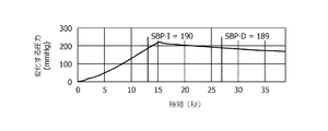
図2は本発明の一実施形態にかかる変化する圧力の図を描く。図2は変化する圧力の変化対時間を図示する。 FIG. 2 depicts a diagram of varying pressure according to one embodiment of the present invention. FIG. 2 illustrates the changing pressure change versus time.
図2を参照すると、本発明の一実施形態によれば、変化する圧力は動脈が閉塞されるために十分な高さの最大圧力値まで漸増し、そして動脈を再開させるために減少し始める。それぞれ動脈の閉塞と再開に対応する圧力値はSBP‐IとSBP‐Dとして示される。 Referring to FIG. 2, according to one embodiment of the invention, the changing pressure gradually increases to a maximum pressure value high enough to occlude the artery and begins to decrease to resume the artery. Pressure values corresponding to arterial occlusion and resumption, respectively, are denoted as SBP-I and SBP-D.
最大圧力値は異なる方法で設定され得る。 The maximum pressure value can be set in different ways.
一実施形態において、最大圧力値は予め定義され得る。代替的に、異なる最大圧力値が異なる人に対して個別に設定され得る。例えば、最大圧力値は高血圧患者に対して比較的高くなるように設定される。 In one embodiment, the maximum pressure value may be predefined. Alternatively, different maximum pressure values can be set individually for different people. For example, the maximum pressure value is set to be relatively high for hypertensive patients.
別の実施形態において、動脈の閉塞がリアルタイムに検出されるとき、最大圧力値は検出される閉塞に従って適応的に規定され得る。例えば、最大圧力値はSBP‐I値を20mmHg乃至30mmHg上回る範囲内に設定される。図2に図示の通り、変化する圧力が増加すると、閉塞が約13秒において起こると検出され、対応するSBP‐Iは190mmHgである。従って、最大圧力値は例えば220mmHgに設定され、変化する圧力は約15秒において最大圧力に達するときに減少し始める。図1を参照すると、検出装置103は膨張カフ101が検出される動脈の閉塞に従って変化する圧力を減少させ始め得るように膨張カフ101へリアルタイムフィードバックを与える。
In another embodiment, when an arterial occlusion is detected in real time, the maximum pressure value may be adaptively defined according to the detected occlusion. For example, the maximum pressure value is set within a range that exceeds the SBP-I value by 20 mmHg to 30 mmHg. As shown in FIG. 2, as the changing pressure increases, it is detected that an occlusion occurs at about 13 seconds, and the corresponding SBP-I is 190 mmHg. Thus, the maximum pressure value is set to 220 mmHg, for example, and the changing pressure begins to decrease when the maximum pressure is reached in about 15 seconds. Referring to FIG. 1, the
図3は本発明の一実施形態にかかる動脈の閉塞及び/又は再開を検出するための装置の略図を描く。図4は本発明の一実施形態にかかる動脈の閉塞及び/又は再開を検出する方法のフローチャートを描く。 FIG. 3 depicts a schematic diagram of an apparatus for detecting arterial occlusion and / or resumption according to one embodiment of the present invention. FIG. 4 depicts a flowchart of a method for detecting arterial occlusion and / or resumption according to one embodiment of the present invention.
図3を参照すると、図1の検出装置103など、動脈の閉塞及び/又は再開を検出するための装置は取得ユニット301、導出ユニット302、及び検出ユニット303を有する。
Referring to FIG. 3, a device for detecting arterial occlusion and / or resumption, such as the
図3と図4を参照すると、方法は取得ユニット301がドップラ超音波振動子を用いて血流信号を取得するステップS410を有する。血流信号は動脈内の血流の変化を示す。
Referring to FIGS. 3 and 4, the method includes a step S410 in which the
周知の通り、ドップラ超音波振動子は血流の方向と速度を評価するために使用され得る。ドップラ超音波振動子は体外に取り付けられる。特に、ドップラ超音波振動子は動脈の上に取り付けられる。ドップラ超音波振動子は高周波音波を生成し、動脈内の血流からのエコーを受信する。エコーはドップラ効果を利用することによって血流の方向と速度を決定するために評価される。ドップラ超音波振動子は連続波ドップラ超音波振動子若しくはパルス波ドップラ超音波振動子であり得る。 As is well known, Doppler ultrasound transducers can be used to assess the direction and velocity of blood flow. The Doppler ultrasonic transducer is attached outside the body. In particular, the Doppler ultrasound transducer is mounted on the artery. The Doppler ultrasonic transducer generates high frequency sound waves and receives echoes from the blood flow in the artery. Echo is evaluated to determine the direction and velocity of blood flow by utilizing the Doppler effect. The Doppler ultrasonic transducer can be a continuous wave Doppler ultrasonic transducer or a pulsed wave Doppler ultrasonic transducer.
血流信号は動脈内の血流の変化を示す任意の種類の信号であり得る。一実施形態において、血流信号はドップラ超音波振動子を用いて得られるいわゆる生の音響信号をあらわす。いわゆる生の音響信号はエコーのドップラ効果をあらわし、各周波数成分は特定ドップラシフトと関連し、各周波数成分の振幅は特定ドップラシフトに対応する速度で動いている血液の量と関連する。 The blood flow signal can be any type of signal that indicates a change in blood flow in the artery. In one embodiment, the blood flow signal represents a so-called raw acoustic signal obtained using a Doppler ultrasound transducer. A so-called raw acoustic signal represents the Doppler effect of echo, each frequency component is associated with a specific Doppler shift, and the amplitude of each frequency component is associated with the amount of blood moving at a rate corresponding to the specific Doppler shift.
図3と図4を参照すると、方法は導出ユニット302が血流信号から第1の変数と第2の変数の少なくとも一つを導出するステップS420をさらに有する。第1の変数は血流の大きさを示し、第2の変数は血流の周期を示す。
Referring to FIGS. 3 and 4, the method further comprises a step S420 in which the
一実施形態によれば、予め定義される時間窓が時間軸に沿ってスライドし、第1及び第2の変数の各々の値は各時間窓に対して計算される。例えば、時間窓は3秒の幅を持ち、1回につき1秒スライドするものとして定義され得る、従って第1及び第2の変数の各々の値は毎秒計算される。 According to one embodiment, a predefined time window slides along the time axis and the value of each of the first and second variables is calculated for each time window. For example, the time window can be defined as having a width of 3 seconds and sliding for 1 second at a time, so the value of each of the first and second variables is calculated every second.
所与の時間窓に対する第1及び第2の変数の値は異なる方法で計算され得る。時間周波数分析に基づくアプローチが以下に記載される。 The values of the first and second variables for a given time window can be calculated in different ways. An approach based on time frequency analysis is described below.
まず、所与の時間窓におけるいわゆる生の音響信号のスペクトログラムが計算される。スペクトログラムは信号のスペクトル密度がどのように時間とともに変化するかを示す時変スペクトル表現であり、ソノグラムとしても知られる。一般に、スペクトログラムは時間軸、周波数軸、及び特定周波数と特定時間における振幅を示す第3の軸を持つグラフである。一実施形態において、短時間フーリエ変換(STFT)が生の音響信号に対して実行され、STFTの振幅二乗がスペクトログラムをもたらす。代替的に、スペクトログラムはウェーブレット変換などの他の既知の方法を用いて計算されることもできる。 First, the spectrogram of the so-called raw acoustic signal in a given time window is calculated. A spectrogram is a time-varying spectral representation that shows how the spectral density of a signal changes over time, also known as a sonogram. In general, the spectrogram is a graph having a time axis, a frequency axis, and a third axis indicating a specific frequency and an amplitude at a specific time. In one embodiment, a short time Fourier transform (STFT) is performed on the raw acoustic signal and the amplitude square of the STFT results in a spectrogram. Alternatively, the spectrogram can be calculated using other known methods such as a wavelet transform.
次に、スペクトログラムは所定周波数範囲におけるスペクトル成分を除去するようにフィルタされる。例えば、所定周波数範囲は0‐100Hz及び3000Hz超を有する。 The spectrogram is then filtered to remove spectral components in the predetermined frequency range. For example, the predetermined frequency range has 0-100 Hz and over 3000 Hz.
次に、フィルタスペクトログラムの振幅が周波数軸に沿って累積され、以下血流波形と呼ばれる波形を時間の関数として取得する。代替的に、血流波形は最大振幅値を周波数軸に沿って抽出することによってフィルタスペクトログラムから抽出され得る、すなわち血流波形の値は全周波数間のフィルタスペクトログラムの最大振幅値である。 Next, the amplitude of the filter spectrogram is accumulated along the frequency axis, and a waveform called a blood flow waveform is obtained as a function of time. Alternatively, the blood flow waveform can be extracted from the filter spectrogram by extracting the maximum amplitude value along the frequency axis, i.e., the value of the blood flow waveform is the maximum amplitude value of the filter spectrogram between all frequencies.
次に、所与の時間窓に対する第1及び第2の変数の値が取得された血流波形から導出される。第1の変数の値は血流波形のピーク振幅と関連し、例えばこれは血流波形におけるピーク値の最大値若しくは平均値であり得る。第2の変数の値は血流波形の周期と関連し、例えば1秒あたりのピーク数であり得る。ピーク値とピーク数を決定する様々な方法が知られ、本明細書ではさらに論じない。 Next, the values of the first and second variables for a given time window are derived from the acquired blood flow waveform. The value of the first variable is related to the peak amplitude of the blood flow waveform, for example, this may be the maximum or average value of the peak values in the blood flow waveform. The value of the second variable is related to the period of the blood flow waveform and can be, for example, the number of peaks per second. Various methods for determining peak value and number of peaks are known and will not be discussed further herein.
その結果、所与の時間窓に対する第1及び第2の変数の値が所与の時間窓における血流信号から計算される。 As a result, the values of the first and second variables for a given time window are calculated from the blood flow signal in the given time window.
図3と図4を参照すると、方法は検出ユニット303が第1の変数と第2の変数の少なくとも一つの変数に基づいて動脈の閉塞及び/又は再開を検出するステップS430をさらに有する。
Referring to FIGS. 3 and 4, the method further includes a step S430 in which the
一実施形態において、所与の時間窓に対し、所与の時間窓に対して計算された第1の変数の値が第1の閾値未満である、及び/又は所与の時間窓に対して計算された第2の変数の値が第1の範囲外であるとき、動脈は所与の時間窓において閉塞していると検出される。 In one embodiment, for a given time window, the value of the first variable calculated for the given time window is less than the first threshold and / or for the given time window. When the calculated value of the second variable is outside the first range, the artery is detected as occluded in a given time window.
第1の閾値と第1の範囲は異なる方法で決定され得る。第1の閾値と第1の範囲の各々は一定であり統計データに従って予め決定され得る。代替的に、第1の閾値と第1の範囲の各々は血流信号から決定され、従って変化し得る。例えば、第1の閾値は血流信号におけるノイズに関連し、ノイズはフィルタスペクトログラムから計算され得る。代替的に、第1の閾値は血流波形における平均ピーク値に関連し、例えば平均ピーク値の10%乃至20%に設定され得る。例えば、第1の範囲は以前の時間窓における第2の変数の平均と標準偏差に従って決定され得る。平均と標準偏差をμとσによって示すと、第1の範囲は例えば範囲[μ−σ,μ+σ]として決定され得る。 The first threshold and the first range may be determined in different ways. Each of the first threshold and the first range is constant and can be predetermined according to statistical data. Alternatively, each of the first threshold and the first range can be determined from the blood flow signal and can therefore vary. For example, the first threshold is related to noise in the blood flow signal, and the noise can be calculated from the filter spectrogram. Alternatively, the first threshold is related to the average peak value in the blood flow waveform and may be set, for example, between 10% and 20% of the average peak value. For example, the first range may be determined according to the mean and standard deviation of the second variable in the previous time window. When the average and standard deviation are indicated by μ and σ, the first range can be determined as the range [μ−σ, μ + σ], for example.
さらに、所与の時間窓に対し、所与の時間窓に対して計算された第1の変数の値が第2の閾値よりも大きい、及び/又は所与の時間窓に対して計算された第2の変数の値が第2の範囲外であるとき、動脈は所与の時間窓において再開されていると検出される。第2の閾値と第2の範囲はそれぞれ第1の閾値と第1の範囲に同一であり得る。第1の閾値と第1の範囲が一定でなく動脈の閉塞まで各時間窓において更新されるとき、第2の閾値と第2の範囲はそれぞれ第1の閾値と第1の範囲の最新値に設定され得る。 Further, for a given time window, the value of the first variable calculated for the given time window is greater than the second threshold and / or calculated for the given time window. When the value of the second variable is outside the second range, the artery is detected as being resumed in a given time window. The second threshold and the second range may be the same as the first threshold and the first range, respectively. When the first threshold and the first range are not constant and are updated in each time window until the occlusion of the artery, the second threshold and the second range are respectively updated to the first threshold and the latest value of the first range. Can be set.
付加的に、動脈が所与の時間窓において再開されていると検出されるとき、再開は第2の閾値よりも大きい第1のピークに対応する時間において起こっているとさらに決定され得る。 Additionally, when it is detected that the artery is being resumed in a given time window, it can be further determined that resumption is occurring at a time corresponding to a first peak that is greater than the second threshold.
別の実施形態において、所定期間、第1の変数が第2の閾値よりも大きい及び/又は第2の変数が第2の範囲内であるとき、再開が検出される。好適には、所定期間は血流の少なくとも五周期を含むほど十分に長い。 In another embodiment, resumption is detected when the first variable is greater than the second threshold and / or the second variable is within the second range for a predetermined period of time. Preferably, the predetermined period is sufficiently long to include at least five cycles of blood flow.
特に、現在の時間窓に対する計算された第1の変数の値が第2の閾値よりも大きい、及び/又は現在の時間窓に対する計算された第2の変数の値が第2の範囲内であるとき、現在の時間窓がマークされる。そして、所定数の後続時間窓において、後続時間窓の各々に対して、計算された第1の変数の値が第2の閾値よりも大きい及び/又は現在の時間窓に対する計算された第2の変数の値が第2の範囲内であるかどうかが決定される。はいの場合、これは血流が安定であることを示し、マークされた時間窓において動脈が実際に再開されていると決定される。このように、いわゆるギャップ現象に起因する誤検出の可能性が低減されることができ、従って検出される動脈の再開はより信頼できる。 In particular, the value of the calculated first variable for the current time window is greater than the second threshold and / or the value of the calculated second variable for the current time window is within the second range. When the current time window is marked. And, for a predetermined number of subsequent time windows, for each subsequent time window, the value of the calculated first variable is greater than the second threshold and / or the calculated second value for the current time window. It is determined whether the value of the variable is within the second range. If yes, this indicates that the blood flow is stable and it is determined that the artery is actually being resumed in the marked time window. In this way, the possibility of false detection due to the so-called gap phenomenon can be reduced and thus the resumption of the detected artery is more reliable.
既に上述した通り、変化する圧力の最大圧力値は検出される動脈の閉塞に従って適応的に規定され得る。そうするために、動脈の閉塞をリアルタイムに検出することが必要である。従って、ステップS410乃至S430は各時間窓に対して連続的に実行される。つまり、ステップS410において取得ユニット310が現在の時間窓において血流信号を取得すると、現在の時間窓において動脈が閉塞若しくは再開しているかどうかを検出するためにステップS420及びS430が実行される。 As already mentioned above, the maximum pressure value of the changing pressure can be adaptively defined according to the detected arterial occlusion. In order to do so, it is necessary to detect occlusion of the artery in real time. Accordingly, steps S410 to S430 are executed continuously for each time window. That is, when the acquisition unit 310 acquires the blood flow signal in the current time window in step S410, steps S420 and S430 are executed to detect whether the artery is occluded or resumed in the current time window.
上述の実施形態は本発明を限定するのではなく例示するものであり、当業者は添付のクレームの範囲から逸脱することなく代替的な実施形態を設計することができることが留意されるべきである。実施形態は限定ではなく例示である。本発明は本発明の範囲と趣旨の内の図示及び記載の実施形態への全ての修正と変更を含むことが意図される。クレームにおいて、括弧の間に置かれる任意の参照符号はクレームを限定するものと解釈されてはならない。"有する"という語はクレーム若しくは記載に列挙されない要素若しくはステップの存在を除外しない。ある要素に先行する"a"若しくは"an"という語はかかる要素の複数の存在を除外しない。複数のユニットを列挙する装置クレームにおいて、これらユニットのいくつかはハードウェア若しくはソフトウェアの一つの同じ項目によって具体化され得る。第1、第2及び第3などの語の使用はいかなる順序も示さない。これらの語は名前として解釈されるものとする。 It should be noted that the above-described embodiments are illustrative rather than limiting on the present invention, and that those skilled in the art can design alternative embodiments without departing from the scope of the appended claims. . The embodiments are exemplary rather than limiting. The present invention is intended to include all modifications and variations to the illustrated and described embodiments within the scope and spirit of the present invention. In the claims, any reference signs placed between parentheses shall not be construed as limiting the claim. The word “comprising” does not exclude the presence of elements or steps not listed in a claim or in the description. The word “a” or “an” preceding an element does not exclude the presence of a plurality of such elements. In the device claim enumerating multiple units, several of these units may be embodied by one and the same item of hardware or software. The use of words such as first, second and third does not indicate any order. These words shall be interpreted as names.
Claims (12)
前記身体の外に取り付けられるドップラ超音波振動子を用いて前記変化する圧力によって生じる前記動脈内の血流の変化を示す血流信号を取得するステップと、
前記血流の大きさを示す第1の変数と前記血流の周期を示す第2の変数を前記血流信号から導出するステップと、
前記第1及び第2の変数に基づいて前記動脈の閉塞又は再開を検出するステップとを有し、
前記検出するステップにおいて、
前記第1の変数が第1の閾値未満である及び前記第2の変数が第1の範囲外であるときに前記閉塞が検出され、
前記第1の変数が第2の閾値よりも大きい及び前記第2の変数が第2の範囲内であるときに前記再開が検出される、方法。 A method for detecting occlusion or resumption of a body artery caused by changing pressure applied to an artery, comprising:
Obtaining a blood flow signal indicative of a change in blood flow in the artery caused by the changing pressure using a Doppler ultrasonic transducer attached outside the body;
Deriving from the blood flow signal a first variable indicating the magnitude of the blood flow and a second variable indicating the period of the blood flow;
Possess and detecting a blockage or resumption of the artery based on the first and second variables,
In the detecting step,
The occlusion is detected when the first variable is less than a first threshold and the second variable is outside a first range;
The method wherein the resumption is detected when the first variable is greater than a second threshold and the second variable is within a second range .
所定数の後続時間窓の各々において、前記第1の変数が第2の閾値よりも大きい及び前記第2の変数が第2の範囲内であるときに前記再開が検出される、請求項1に記載の方法。 In the detecting step,
In each subsequent time window of a predetermined number, the first variable is greater than the second threshold value and the second variable is the resumption is detected when within the second range, in claim 1 The method described.
前記検出するステップにおいて、前記時間窓に関連する前記第1及び第2の変数の値に基づいて前記時間窓における前記動脈の閉塞又は再開が検出される、請求項1に記載の方法。 In the deriving step, a value of the first variable and a value of the second variable for a time window are calculated based on the blood flow signal in the time window;
The method of claim 1, wherein the detecting step detects occlusion or resumption of the artery in the time window based on values of the first and second variables associated with the time window.
前記身体の外に取り付けられるドップラ超音波振動子を用いて前記変化する圧力によって生じる前記動脈内の血流の変化を示す血流信号を取得するための取得ユニットと、
前記血流の大きさを示す第1の変数と前記血流の周期を示す第2の変数を前記血流信号から導出するための導出ユニットと、
前記第1及び第2の変数に基づいて前記動脈の閉塞及び/又は再開を検出するための検出ユニットとを有し、
前記検出ユニットが、前記第1の変数が第1の閾値未満である及び前記第2の変数が第1の範囲外であるときに前記閉塞を検出するように構成され、
前記検出ユニットが、前記第1の変数が第2の閾値よりも大きい及び前記第2の変数が第2の範囲内であるときに前記再開を検出するように構成される、装置。 A device for detecting occlusion or resumption of a body artery caused by changing pressure applied to an artery,
An acquisition unit for acquiring a blood flow signal indicative of a change in blood flow in the artery caused by the changing pressure using a Doppler ultrasonic transducer attached outside the body;
A deriving unit for deriving from the blood flow signal a first variable indicating the magnitude of the blood flow and a second variable indicating the period of the blood flow;
It has a detection unit for detecting an occlusion and / or resumption of the artery based on the first and second variables,
The detection unit is configured to detect the occlusion when the first variable is less than a first threshold and the second variable is outside a first range;
The apparatus, wherein the detection unit is configured to detect the resumption when the first variable is greater than a second threshold and the second variable is within a second range .
前記検出ユニットが、前記時間窓に関連する前記第1及び第2の変数の値に基づいて前記時間窓における前記動脈の閉塞又は再開を検出するように構成される、請求項6に記載の装置。 The derivation unit is configured to calculate a value of the first variable and a value of the second variable for a time window from the blood flow signal in the time window;
The apparatus of claim 6 , wherein the detection unit is configured to detect occlusion or resumption of the artery in the time window based on values of the first and second variables associated with the time window. .
前記動脈に変化する圧力を加えるための前記身体の外に取り付け可能な膨張カフと、
複数の時点における前記変化する圧力の複数の圧力値を取得するための圧力センサと、
請求項6乃至10のいずれか一項に記載の、前記動脈に加えられる前記変化する圧力によって生じる前記身体の動脈の閉塞又は再開を検出するための検出装置と、
検出された前記動脈の閉塞に対応する時点における前記変化する圧力の第1の圧力値、及び検出された前記動脈の再開に対応する時点における前記変化する圧力の第2の圧力値の少なくとも一つから前記収縮期血圧を決定するための決定装置とを有する、システム。 A system for measuring systolic blood pressure in a body artery,
An inflatable cuff attachable outside the body to apply varying pressure to the artery;
A pressure sensor for obtaining a plurality of pressure values of the varying pressure at a plurality of time points;
According to any one of claims 6 to 10, a detection device for detecting a blockage or resumption of the body of the artery caused by the pressure of the change applied to the artery,
At least one of a first pressure value of the changing pressure at a time corresponding to the detected occlusion of the artery and a second pressure value of the changing pressure at a time corresponding to the resumption of the detected artery. A determination device for determining the systolic blood pressure from the system.
前記膨張カフが、前記変化する圧力が前記決定された最大値に達するときに収縮し始めるように構成される、請求項11に記載のシステム。 The detection device is configured to determine a maximum value of the changing pressure according to a detected occlusion of the artery;
The system of claim 11 , wherein the inflation cuff is configured to begin to contract when the changing pressure reaches the determined maximum value.
Applications Claiming Priority (3)
| Application Number | Priority Date | Filing Date | Title |
|---|---|---|---|
| CN2011077752 | 2011-07-28 | ||
| CNPCT/CN2011/077752 | 2011-07-28 | ||
| PCT/IB2012/053640 WO2013014575A1 (en) | 2011-07-28 | 2012-07-17 | Method and device for detecting occlusion/reopening of an artery and system for measuring systolic blood pressure |
Publications (3)
| Publication Number | Publication Date |
|---|---|
| JP2014530642A JP2014530642A (en) | 2014-11-20 |
| JP2014530642A5 JP2014530642A5 (en) | 2015-08-20 |
| JP6129166B2 true JP6129166B2 (en) | 2017-05-17 |
Family
ID=47008645
Family Applications (1)
| Application Number | Title | Priority Date | Filing Date |
|---|---|---|---|
| JP2014522181A Expired - Fee Related JP6129166B2 (en) | 2011-07-28 | 2012-07-17 | Method and apparatus for detecting arterial occlusion / resumption and system for measuring systolic blood pressure |
Country Status (4)
| Country | Link |
|---|---|
| US (1) | US10076310B2 (en) |
| EP (1) | EP2736418B1 (en) |
| JP (1) | JP6129166B2 (en) |
| WO (1) | WO2013014575A1 (en) |
Families Citing this family (6)
| Publication number | Priority date | Publication date | Assignee | Title |
|---|---|---|---|---|
| KR20150082401A (en) * | 2012-11-08 | 2015-07-15 | 르 타이 | Improved blood pressure monitor and method |
| US20190000386A1 (en) * | 2015-10-14 | 2019-01-03 | Kyocera Corporation | Measurement device |
| CN109561877A (en) | 2016-08-03 | 2019-04-02 | 皮埃-哈韦斯特控股有限公司 | A kind of includes the system and method for the internal pressure of intravascular blood pressure for non-invasive measurement |
| EP3278735A1 (en) | 2016-08-03 | 2018-02-07 | PI-Harvest Holding AG | A system and method for non-invasive measurement of pressure inside a body including intravascular blood pressure |
| EP4106632A4 (en) * | 2019-11-26 | 2024-04-17 | Bfly Operations Inc | Methods and apparatuses for pulsed wave doppler ultrasound imaging |
| CN116056635A (en) * | 2020-08-06 | 2023-05-02 | 京瓷株式会社 | Sleep estimation device, sleep estimation system, wearable device, and sleep estimation method |
Family Cites Families (15)
| Publication number | Priority date | Publication date | Assignee | Title |
|---|---|---|---|---|
| US4205688A (en) * | 1977-05-23 | 1980-06-03 | Doll Research, Inc. | Method and apparatus for developing and measuring pulsed blood flow |
| US4625277A (en) * | 1984-06-04 | 1986-11-25 | Physio-Control Corporation | Blood pressure measuring device having adaptive cuff deflation rate |
| CA1268641A (en) * | 1985-02-13 | 1990-05-08 | Keiji Yamaguchi | Blood pressure measuring apparatus |
| EP0298620A1 (en) * | 1987-07-06 | 1989-01-11 | The BOC Group, Inc. | Blood pressure monitoring methods and apparatus |
| DE4030071A1 (en) * | 1989-09-27 | 1991-08-08 | Atp Advanced Tech Promotion | MEASURING THE HEART FUNCTION |
| US5072736A (en) * | 1990-01-19 | 1991-12-17 | Nihon Kohden Corporation | Non-invasive automatic blood pressure measuring apparatus |
| JPH06125903A (en) * | 1992-10-20 | 1994-05-10 | Hayashi Denki Kk | Ultrasonic blood flowmeter with blood pressure measuring function |
| JP2873212B2 (en) | 1996-09-06 | 1999-03-24 | 信秀 前田 | Bed pat |
| US6171254B1 (en) | 1999-02-26 | 2001-01-09 | Medical Research Laboratories, Inc. | Control for automatic blood pressure monitor |
| US20060100530A1 (en) * | 2000-11-28 | 2006-05-11 | Allez Physionix Limited | Systems and methods for non-invasive detection and monitoring of cardiac and blood parameters |
| US8016761B2 (en) | 2006-10-23 | 2011-09-13 | The General Electric Company | Method and apparatus for automated flow mediated dilation |
| US20100106016A1 (en) | 2008-10-23 | 2010-04-29 | Skeletal Dynamics Llc | Non-Invasive Blood Pressure Monitoring Device and Method |
| GB2465787B (en) | 2008-11-28 | 2011-04-06 | Royal United Hospital Bath Nhs Trust | Method of measuring blood pressure and apparatus for performing the same |
| FR2939302B1 (en) | 2008-12-05 | 2011-02-11 | Atys Sarl | METHOD FOR MEASURING SYSTEMIC PRESSURE AND DEVICE USING THE SAME |
| US20100292586A1 (en) | 2009-05-13 | 2010-11-18 | Rooke Thom W | Wireless automatic ankle-brachial index (AABI) measurement system |
-
2012
- 2012-07-17 US US14/234,449 patent/US10076310B2/en active Active
- 2012-07-17 WO PCT/IB2012/053640 patent/WO2013014575A1/en active Application Filing
- 2012-07-17 EP EP12770221.5A patent/EP2736418B1/en not_active Not-in-force
- 2012-07-17 JP JP2014522181A patent/JP6129166B2/en not_active Expired - Fee Related
Also Published As
| Publication number | Publication date |
|---|---|
| EP2736418A1 (en) | 2014-06-04 |
| US20140180114A1 (en) | 2014-06-26 |
| EP2736418B1 (en) | 2018-12-05 |
| US10076310B2 (en) | 2018-09-18 |
| WO2013014575A1 (en) | 2013-01-31 |
| JP2014530642A (en) | 2014-11-20 |
Similar Documents
| Publication | Publication Date | Title |
|---|---|---|
| US7137955B2 (en) | Methods and systems for distal recording of phonocardiographic signals | |
| JP6129166B2 (en) | Method and apparatus for detecting arterial occlusion / resumption and system for measuring systolic blood pressure | |
| JP5944500B2 (en) | Method, apparatus and system for determining the moment when the state of an artery switches from open to closed and vice versa for an artery of interest under pressure fluctuations | |
| JP2001504362A (en) | Non-destructive blood pressure measurement device without pressurized zone | |
| CA2604337A1 (en) | System and method for non-invasive cardiovascular assessment from supra-systolic signals obtained with a wideband external pulse transducer in a blood pressure cuff | |
| KR101549619B1 (en) | Method and apparatus for detecting measurement location of blood pressure | |
| JP5722898B2 (en) | Device for non-invasive measurement of arterial blood pressure | |
| KR101604078B1 (en) | Blood pressure monitoring apparatus and method of low pressurization | |
| CN107106125B (en) | System and method for measuring arterial parameters | |
| US20200367766A1 (en) | Method and device for determining at least one physiological parameter | |
| Simpson et al. | A parametric approach to measuring cerebral blood flow autoregulation from spontaneous variations in blood pressure | |
| US20190200879A1 (en) | Non-invasive system and method for measuring blood pressure variability | |
| JP2011194217A (en) | Use of frequency spectrum of artifact in oscillometry | |
| JP3631639B2 (en) | Blood pressure measurement method | |
| Ferreira et al. | Determination of radial artery compliance can increase the diagnostic power of pulse wave velocity measurement | |
| CN103717143B (en) | Method and device for detecting occlusion/reopening of an artery and system for measuring systolic blood pressure | |
| WO2013014647A1 (en) | Ultrasound probe, method and device for acquiring a blood flow signal of an artery and system for measuring systolic blood pressure | |
| JP5070103B2 (en) | Shunt stenosis detector | |
| Pratiwi et al. | A review of equipment and signal processing of the automated auscultation for blood pressure measurement | |
| KR20160007052A (en) | Apparatus and method for automatic detection of arterial blood pressure | |
| JP7175304B2 (en) | SHUNT SOUND ANALYZER AND METHOD, COMPUTER PROGRAM AND STORAGE MEDIUM | |
| JP4729703B2 (en) | Blood vessel hardness measuring device | |
| JP2000287945A (en) | Sphygmomanometer | |
| JP5146995B2 (en) | Blood pressure measurement device and control method thereof | |
| Naufal et al. | Blood Pressure Measuring Device Based on Korotkoff Sound's Tapping Period and Frequency Detection |
Legal Events
| Date | Code | Title | Description |
|---|---|---|---|
| A521 | Request for written amendment filed |
Free format text: JAPANESE INTERMEDIATE CODE: A523 Effective date: 20150630 |
|
| A621 | Written request for application examination |
Free format text: JAPANESE INTERMEDIATE CODE: A621 Effective date: 20150630 |
|
| A977 | Report on retrieval |
Free format text: JAPANESE INTERMEDIATE CODE: A971007 Effective date: 20160428 |
|
| A131 | Notification of reasons for refusal |
Free format text: JAPANESE INTERMEDIATE CODE: A131 Effective date: 20160506 |
|
| A601 | Written request for extension of time |
Free format text: JAPANESE INTERMEDIATE CODE: A601 Effective date: 20160801 |
|
| A521 | Request for written amendment filed |
Free format text: JAPANESE INTERMEDIATE CODE: A523 Effective date: 20161027 |
|
| RD04 | Notification of resignation of power of attorney |
Free format text: JAPANESE INTERMEDIATE CODE: A7424 Effective date: 20170214 |
|
| TRDD | Decision of grant or rejection written | ||
| A01 | Written decision to grant a patent or to grant a registration (utility model) |
Free format text: JAPANESE INTERMEDIATE CODE: A01 Effective date: 20170315 |
|
| A61 | First payment of annual fees (during grant procedure) |
Free format text: JAPANESE INTERMEDIATE CODE: A61 Effective date: 20170411 |
|
| R150 | Certificate of patent or registration of utility model |
Ref document number: 6129166 Country of ref document: JP Free format text: JAPANESE INTERMEDIATE CODE: R150 |
|
| R250 | Receipt of annual fees |
Free format text: JAPANESE INTERMEDIATE CODE: R250 |
|
| R250 | Receipt of annual fees |
Free format text: JAPANESE INTERMEDIATE CODE: R250 |
|
| R250 | Receipt of annual fees |
Free format text: JAPANESE INTERMEDIATE CODE: R250 |
|
| LAPS | Cancellation because of no payment of annual fees |