JP5773315B2 - CXCL4L1 as a biomarker for pancreatic cancer - Google Patents
CXCL4L1 as a biomarker for pancreatic cancer Download PDFInfo
- Publication number
- JP5773315B2 JP5773315B2 JP2011537960A JP2011537960A JP5773315B2 JP 5773315 B2 JP5773315 B2 JP 5773315B2 JP 2011537960 A JP2011537960 A JP 2011537960A JP 2011537960 A JP2011537960 A JP 2011537960A JP 5773315 B2 JP5773315 B2 JP 5773315B2
- Authority
- JP
- Japan
- Prior art keywords
- cxcl4l1
- cxcl4
- pancreatic
- pancreatic cancer
- cells
- Prior art date
- Legal status (The legal status is an assumption and is not a legal conclusion. Google has not performed a legal analysis and makes no representation as to the accuracy of the status listed.)
- Expired - Fee Related
Links
Images


Classifications
-
- C—CHEMISTRY; METALLURGY
- C12—BIOCHEMISTRY; BEER; SPIRITS; WINE; VINEGAR; MICROBIOLOGY; ENZYMOLOGY; MUTATION OR GENETIC ENGINEERING
- C12Q—MEASURING OR TESTING PROCESSES INVOLVING ENZYMES, NUCLEIC ACIDS OR MICROORGANISMS; COMPOSITIONS OR TEST PAPERS THEREFOR; PROCESSES OF PREPARING SUCH COMPOSITIONS; CONDITION-RESPONSIVE CONTROL IN MICROBIOLOGICAL OR ENZYMOLOGICAL PROCESSES
- C12Q1/00—Measuring or testing processes involving enzymes, nucleic acids or microorganisms; Compositions therefor; Processes of preparing such compositions
- C12Q1/68—Measuring or testing processes involving enzymes, nucleic acids or microorganisms; Compositions therefor; Processes of preparing such compositions involving nucleic acids
- C12Q1/6876—Nucleic acid products used in the analysis of nucleic acids, e.g. primers or probes
- C12Q1/6883—Nucleic acid products used in the analysis of nucleic acids, e.g. primers or probes for diseases caused by alterations of genetic material
- C12Q1/6886—Nucleic acid products used in the analysis of nucleic acids, e.g. primers or probes for diseases caused by alterations of genetic material for cancer
-
- A—HUMAN NECESSITIES
- A61—MEDICAL OR VETERINARY SCIENCE; HYGIENE
- A61P—SPECIFIC THERAPEUTIC ACTIVITY OF CHEMICAL COMPOUNDS OR MEDICINAL PREPARATIONS
- A61P35/00—Antineoplastic agents
-
- A—HUMAN NECESSITIES
- A61—MEDICAL OR VETERINARY SCIENCE; HYGIENE
- A61P—SPECIFIC THERAPEUTIC ACTIVITY OF CHEMICAL COMPOUNDS OR MEDICINAL PREPARATIONS
- A61P35/00—Antineoplastic agents
- A61P35/04—Antineoplastic agents specific for metastasis
-
- G—PHYSICS
- G01—MEASURING; TESTING
- G01N—INVESTIGATING OR ANALYSING MATERIALS BY DETERMINING THEIR CHEMICAL OR PHYSICAL PROPERTIES
- G01N33/00—Investigating or analysing materials by specific methods not covered by groups G01N1/00 - G01N31/00
- G01N33/48—Biological material, e.g. blood, urine; Haemocytometers
- G01N33/50—Chemical analysis of biological material, e.g. blood, urine; Testing involving biospecific ligand binding methods; Immunological testing
- G01N33/5005—Chemical analysis of biological material, e.g. blood, urine; Testing involving biospecific ligand binding methods; Immunological testing involving human or animal cells
- G01N33/5008—Chemical analysis of biological material, e.g. blood, urine; Testing involving biospecific ligand binding methods; Immunological testing involving human or animal cells for testing or evaluating the effect of chemical or biological compounds, e.g. drugs, cosmetics
- G01N33/5044—Chemical analysis of biological material, e.g. blood, urine; Testing involving biospecific ligand binding methods; Immunological testing involving human or animal cells for testing or evaluating the effect of chemical or biological compounds, e.g. drugs, cosmetics involving specific cell types
- G01N33/507—Pancreatic cells
-
- C—CHEMISTRY; METALLURGY
- C12—BIOCHEMISTRY; BEER; SPIRITS; WINE; VINEGAR; MICROBIOLOGY; ENZYMOLOGY; MUTATION OR GENETIC ENGINEERING
- C12Q—MEASURING OR TESTING PROCESSES INVOLVING ENZYMES, NUCLEIC ACIDS OR MICROORGANISMS; COMPOSITIONS OR TEST PAPERS THEREFOR; PROCESSES OF PREPARING SUCH COMPOSITIONS; CONDITION-RESPONSIVE CONTROL IN MICROBIOLOGICAL OR ENZYMOLOGICAL PROCESSES
- C12Q2600/00—Oligonucleotides characterized by their use
- C12Q2600/112—Disease subtyping, staging or classification
-
- G—PHYSICS
- G01—MEASURING; TESTING
- G01N—INVESTIGATING OR ANALYSING MATERIALS BY DETERMINING THEIR CHEMICAL OR PHYSICAL PROPERTIES
- G01N2333/00—Assays involving biological materials from specific organisms or of a specific nature
- G01N2333/435—Assays involving biological materials from specific organisms or of a specific nature from animals; from humans
- G01N2333/52—Assays involving cytokines
- G01N2333/521—Chemokines
Landscapes
- Health & Medical Sciences (AREA)
- Life Sciences & Earth Sciences (AREA)
- Chemical & Material Sciences (AREA)
- Engineering & Computer Science (AREA)
- Immunology (AREA)
- Organic Chemistry (AREA)
- Pathology (AREA)
- Cell Biology (AREA)
- Biomedical Technology (AREA)
- Analytical Chemistry (AREA)
- Proteomics, Peptides & Aminoacids (AREA)
- General Health & Medical Sciences (AREA)
- Molecular Biology (AREA)
- Microbiology (AREA)
- Biotechnology (AREA)
- Medicinal Chemistry (AREA)
- Urology & Nephrology (AREA)
- Wood Science & Technology (AREA)
- Zoology (AREA)
- Biochemistry (AREA)
- Bioinformatics & Cheminformatics (AREA)
- Hematology (AREA)
- Physics & Mathematics (AREA)
- Genetics & Genomics (AREA)
- Oncology (AREA)
- Biophysics (AREA)
- Tropical Medicine & Parasitology (AREA)
- Pharmacology & Pharmacy (AREA)
- Animal Behavior & Ethology (AREA)
- Public Health (AREA)
- Veterinary Medicine (AREA)
- General Chemical & Material Sciences (AREA)
- Chemical Kinetics & Catalysis (AREA)
- Toxicology (AREA)
- Nuclear Medicine, Radiotherapy & Molecular Imaging (AREA)
- General Engineering & Computer Science (AREA)
- Hospice & Palliative Care (AREA)
- Food Science & Technology (AREA)
- General Physics & Mathematics (AREA)
- Medicines That Contain Protein Lipid Enzymes And Other Medicines (AREA)
Description
発明の属する分野:
本発明は、患者における膵癌のバイオマーカーとしてのCXCL4L1の使用に関する。本発明はさらに膵癌の処置および/または膵転移の予防のための方法に関する。
Field of Invention:
The present invention relates to the use of CXCL4L1 as a biomarker for pancreatic cancer in patients. The invention further relates to a method for the treatment of pancreatic cancer and / or prevention of pancreatic metastasis.
発明の背景
膵癌は膵臓の悪性腫瘍である。毎年、アメリカ合衆国で約35000人がこの疾患で診断され、ほぼ同数のヒトがこの疾患のために死亡する。ヨーロッパでは、60000人以上が毎年この癌のため診断される。それ故、膵癌は、すべての癌の中で最も高い致死率のうちの1つを有し(Jemal et al., 2007)、それ故、この致死の病体に対する効果的な処置を緊急に必要とする主要なヒトの健康問題となっている。
BACKGROUND OF THE INVENTION Pancreatic cancer is a malignant tumor of the pancreas. Each year, about 35,000 people are diagnosed with the disease in the United States, and approximately the same number of people die from the disease. In Europe, more than 60000 people are diagnosed for this cancer every year. Pancreatic cancer therefore has one of the highest mortality rates of all cancers (Jemal et al., 2007) and therefore urgently needs effective treatment for this deadly pathology. It has become a major human health problem.
血管新生は、毛細血管由来の新規血管形成を含み、この事象は一連の因子によって緊密に制御されている。現在、大人におこる血管新生活性の多くが病理学的であると広く認識されている。それ故、血管新生および浸潤は、癌のような疾患の進行に重要な役割を果たす2つの密接に関連した過程である(Folkman, 1995)。特に、固形腫瘍および他の癌の拡大は決定的に血管新生に依存し(Folkman, 1971)、抗血管新生の戦略を癌治療と結びつける(Folkman, 2001)。一方で、浸潤は腫瘍量の局所的拡大および転移の拡大に要求される。 Angiogenesis involves the formation of new blood vessels derived from capillaries, and this event is tightly controlled by a series of factors. Currently, many of the angiogenic activities that occur in adults are widely recognized as pathological. Angiogenesis and invasion are therefore two closely related processes that play an important role in the progression of diseases such as cancer (Folkman, 1995). In particular, the spread of solid tumors and other cancers is critically dependent on angiogenesis (Folkman, 1971), linking anti-angiogenic strategies to cancer treatment (Folkman, 2001). On the other hand, invasion is required for local expansion of tumor volume and spread of metastases.
研究された因子の中には、CXC−ケモカインファミリー(血小板因子4に対する古くからの用語法でのPF4)に属し、血小板または巨核球だけでなく単球、T細胞、血管平滑筋細胞および内皮細胞を含む異なる他の細胞型においても合成されるCXCL4がある(Lasagni et al., 2007)。CXCL4またはそのC末端ドメイン(PF4/CTF)由来のペプチドはin vitro(Maione et al., 1990; Jouan et al., 1999; Hagedorn et al., 2001)およびin vivo(Maione et al., 1990; Sharpe et al., 1990; Hagedorn et al., 2001)で有意な抗血管新生活性を示す。それらは、in vivoで様々な腫瘍の成長を抑制し(Tanaka et al., 1997; Maione et al., 1991)、転移を抑制する(Kolber et al., 1995)。この効果はそれらの抗血管新生作用に関連し、腫瘍細胞の増殖には関連しない(Sharpe et al., 1990; Tanaka et al., 1997; Maione et al., 1991; Kolber et al., 1995)。CXCL4は、ex vivoシステムにおいて抗血管新生作用を有することが発見された最初の薬剤の一つである(Maione et al., 1990)が、CXCL4の抗血管新生シグナルを伝達する特異的な受容体のメカニズムは未だほとんど理解されていない。分子の記載された他の効果の中には、特にT細胞に対する免疫調節性の機能がある(Romagnani et al., 2005)。 Among the factors studied, belong to the CXC-chemokine family (PF4 in the old terminology for platelet factor 4), not only platelets or megakaryocytes but also monocytes, T cells, vascular smooth muscle cells and endothelial cells There is also CXCL4 that is synthesized in different other cell types including (Lasagni et al., 2007). Peptides derived from CXCL4 or its C-terminal domain (PF4 / CTF) are in vitro (Maione et al., 1990; Jouan et al., 1999; Hagedorn et al., 2001) and in vivo (Maione et al., 1990; Sharpe et al., 1990; Hagedorn et al., 2001) show significant anti-angiogenic activity. They suppress the growth of various tumors in vivo (Tanaka et al., 1997; Maione et al., 1991) and suppress metastasis (Kolber et al., 1995). This effect is related to their anti-angiogenic effects and not to tumor cell proliferation (Sharpe et al., 1990; Tanaka et al., 1997; Maione et al., 1991; Kolber et al., 1995) . CXCL4 is one of the first drugs discovered to have anti-angiogenic activity in an ex vivo system (Maione et al., 1990), a specific receptor that transmits the anti-angiogenic signal of CXCL4 The mechanism is still largely unknown. Among the other described effects of the molecule are immunoregulatory functions, especially on T cells (Romagnani et al., 2005).
さらに、CXCL4L1と命名されたCXCL4の新規の相同体は、1989年に同定されたが、ごく最近でもこのケモカインの機能的特徴付けについてほとんどデータが報告されていない。これは、血小板に発現しているが、平滑筋細胞および内皮細胞にも発現している。近年この分子の分泌メカニズムが、CXCL4との比較で特徴づけられた(Lasagni et al., 2007)。CXCL4L1は、トランスフェクションされたHEK細胞の培地中にかなりの量が見い出されるが、CXCL4は見い出されない。さらに、CXCL4L1は非制御的様式で恒常的に排出されるようだが、CXCL4は有芯顆粒(DCG)からの放出を介して制御的な方法で排出される。これは、CXCL4L1が細胞からの排出および拡散について大変異なるメカニズムを有することを示唆する。 In addition, a novel homologue of CXCL4, named CXCL4L1, was identified in 1989, but very recently little data has been reported on the functional characterization of this chemokine. It is expressed on platelets but also on smooth muscle cells and endothelial cells. Recently, the secretory mechanism of this molecule has been characterized in comparison with CXCL4 (Lasagni et al., 2007). CXCL4L1 is considerable amount in the culture medium of the HEK cells transfected is found, CXCL 4 is not found. Furthermore, it appears that CXCL4L1 is constitutively excreted in an uncontrolled manner, whereas CXCL4 is excreted in a controlled manner via release from cored granules (DCG). This suggests that CXCL4L1 has a very different mechanism for efflux and diffusion from cells.
また、CXCL4L1はCXCL4よりもさらに効果的な血管新生の有力な阻害剤として特徴づけられた。例えば、国際特許出願 国際公開公報第2006/029487号は、血管新生の予防および/または縮小のための、特に癌のような血管新生疾患または病理学的血管新生を含む血管新生障害または血管新生疾患の、処置または予防のための、CXCL4L1、フラグメント、ならびにCXCL4L1およびCXCL4L1のフラグメントの改変された型に関する。 CXCL4L1 has also been characterized as a potent inhibitor of angiogenesis that is more effective than CXCL4. For example, International Patent Application Publication No. WO 2006/029477 describes angiogenic disorders or angiogenic diseases, including angiogenic diseases such as cancer or pathological angiogenesis, especially for the prevention and / or reduction of angiogenesis. Relates to modified forms of CXCL4L1, fragments, and fragments of CXCL4L1 and CXCL4L1, for treatment or prevention.
膵癌のこの根源的な治療の面と平行して、重要な必要性がそのような癌の診断にもある。 In parallel with this fundamental treatment aspect of pancreatic cancer, an important need is also in the diagnosis of such cancer.
実際に、癌のバイオマーカー分野の主要なゴールは、初期の癌の検出をできるようにする非侵害性の試験の開発である。癌の初期の検出は、長期生存に重要であり、特に膵癌、それは5%未満の5年生存率である世界中の癌死亡の主要な原因である、に関する。それ故、部分的には、癌が通常初期には症状を引き起こさないこと、そして診断の時点では、局所進行性または転移性の疾患を導いているため、膵癌と診断された患者は、典型的には悪い予後である。膵癌は初期のステージにおいて無症状なので、ほとんどの患者は癌が膵臓を超えて広がるまで診断されず、それが以前述べられているような低い長期生存率に大い寄与する。 Indeed, a major goal in the field of cancer biomarkers is the development of non-invasive tests that allow the detection of early cancers. Early detection of cancer is important for long-term survival, especially pancreatic cancer, which is the leading cause of cancer death worldwide, with a 5-year survival rate of less than 5%. Therefore, in part, patients diagnosed with pancreatic cancer are typical because cancer usually does not cause symptoms early and, at the time of diagnosis, leads to locally advanced or metastatic disease There is a bad prognosis. Because pancreatic cancer is asymptomatic at an early stage, most patients are not diagnosed until the cancer has spread beyond the pancreas, which greatly contributes to the low long-term survival rate as previously described.
しかしながら、膵癌を含むほとんどの固形腫瘍は初期に検出および処置されれば、しばしば治癒できる。 However, most solid tumors, including pancreatic cancer, can often be cured if detected and treated early.
それ故、特に膵癌発生初期の間に膵癌を診断するための方法を開発するニーズが依然として存在している。 Therefore, there remains a need to develop methods for diagnosing pancreatic cancer, particularly during the early stages of pancreatic cancer development.
発明の要約
本発明は、患者の膵癌および/または膵転移を検出する方法に関し、該方法は、該患者から得られた生物学的サンプル中のCXCL4L1遺伝子の発現レベルを決定する事を含む。
SUMMARY OF THE INVENTION The present invention relates to a method for detecting pancreatic cancer and / or pancreatic metastasis in a patient, the method comprising determining the expression level of the CXCL4L1 gene in a biological sample obtained from the patient.
本発明は、患者から得られた生物学的サンプル中のCXCL4L1遺伝子の発現レベルを決定する事を含む、膵癌を有する該患者における膵癌の病期分類のための方法にも関する。 The present invention also relates to a method for staging pancreatic cancer in a patient having pancreatic cancer comprising determining the expression level of the CXCL4L1 gene in a biological sample obtained from the patient.
本発明は、患者における膵癌のバイオマーカーとしてのCXCL4L1の使用にも関する。 The invention also relates to the use of CXCL4L1 as a biomarker for pancreatic cancer in patients.
本発明は、膵癌の処置および/または膵臓の転移の予防のために細胞障害性薬物または成長阻害剤のような抗癌剤に結合したCXCL4L1特異的結合分子にも関する。 The invention also relates to CXCL4L1-specific binding molecules conjugated to anticancer agents such as cytotoxic drugs or growth inhibitors for the treatment of pancreatic cancer and / or prevention of pancreatic metastasis.
発明の詳細な説明:
本発明者は、CXCL4L1が、初期の正確な膵癌の検出およびCXCL4L1遺伝子の発現レベルと腫瘍のグレードの重症度との間の関連づけのために使用されうるという所見を有する。
Detailed description of the invention:
The inventor has the observation that CXCL4L1 can be used for early accurate pancreatic cancer detection and correlation between the expression level of the CXCL4L1 gene and the severity of the tumor grade.
定義:
用語「CXCL4L1」は本明細書において使用される時、PF4v1、PF4var1、PF4ALTおよびSCYB4V1を含むがそれらに限定されないすべての同義語を含むことを意図する。この用語はそれ故、天然に生じるCXCL4L1、ならびにその変異体および改変型を含む。用語「成熟CXCL4L1タンパク質」は、より長いプロペプチドのプロセシングにより得ることのできる70アミノ酸の成熟CXCL4L1タンパク質を意味する(アクセッションナンバー NP_002611でGenPeptデータベースに提供される)。CXCL4L1をコードする代表的な天然のヌクレオチド配列はGenBankデータベースにアクセッションナンバーNM_002620で提供される。用語「CXCL4L1」はCXCL4タンパク質の霊長類の天然の変異体も含むことにさらに注意されなければならない。例として、霊長類のCXCL4L1タンパク質は、GenBankアクセッションナンバーXM_001102971.1(アカゲザル)およびXP_001156146.1(チンパンジー)で提供される。
Definition:
The term “CXCL4L1” as used herein is intended to include all synonyms including but not limited to PF 4 v1, PF4var1, PF4ALT and SCYB4V1. The term therefore includes naturally occurring CXCL4L1, and variants and modified forms thereof. The term “mature CXCL4L1 protein” means a 70 amino acid mature CXCL4L1 protein that can be obtained by processing a longer propeptide (provided in the GenPept database with accession number NP_002611). An exemplary natural nucleotide sequence encoding CXCL4L1 is provided in the GenBank database with accession number NM_002620. It should be further noted that the term “CXCL4L1” also includes natural primate variants of the CXCL4 protein. As an example, the primate CXCL4L1 protein is provided in GenBank accession numbers XM — 001202971.1 (Rhesus monkey) and XP — 001561466.1 (Chimpanzee).
本明細書において使用される時には、用語「CXCL4L1特異的結合分子」は、CXCL4L1を選択的に認識できるために十分な大きさおよび複雑性を有する分子を意味することを意図する。 As used herein, the term “CXCL4L1-specific binding molecule” is intended to mean a molecule having a size and complexity sufficient to selectively recognize CXCL4L1.
用語「抗CXCL4L1抗体」は選択的にCXCL4L1を認識する抗体またはそのフラグメントを意味する。 The term “anti-CXCL4L1 antibody” means an antibody or fragment thereof that selectively recognizes CXCL4L1.
本明細書おいて使用される時には、用語「膵癌」は、膵臓の細胞癌、特に膵臓腺癌(例えば膵管腺癌)ならびに膵外分泌部の他の腫瘍(例えば重大な嚢胞腺腫)、腺房細胞癌および(インスリノーマのような)膵臓の神経内分泌腫瘍を意味する。 As used herein, the term “pancreatic cancer” refers to cell carcinoma of the pancreas, particularly pancreatic adenocarcinoma (eg, pancreatic ductal adenocarcinoma) as well as other tumors of the pancreatic exocrine region (eg, significant cystadenoma), acinar cells It refers to cancer and pancreatic neuroendocrine tumors (such as insulinomas).
用語「膵転移」は、その技術分野での一般的な意味を有し、膵臓からの他の非隣接臓器または非隣接部位への腫瘍の広がりを意味する。 The term “pancreatic metastasis” has its general meaning in the art and refers to the spread of a tumor from the pancreas to other non-adjacent organs or sites.
CXCL4L1を発現または過発現し、CXCL4L1阻害剤で処置できる膵転移の例には、転移性の結腸、肺、膵臓、食道および前立腺の癌、メラノーマ、肝細胞癌ならびに神経節またはリンパ節転移を含むがそれらに限定されない。 Examples of pancreatic metastases that express or overexpress CXCL4L1 and can be treated with CXCL4L1 inhibitors include metastatic colon, lung, pancreas, esophageal and prostate cancer, melanoma, hepatocellular carcinoma, and ganglion or lymph node metastasis Is not limited to them.
本明細書おいて使用される時には、用語「規定値」は、一般的な対象の集団または選択された対象の集団から得られた生物学的サンプル中のCXCL4L1の量を意味する。例えば、選択された集団は、以前に膵癌の存在を示す兆候または症状を有しなかった個体のような、明らかに健康な対象を含みうる。別の例では、規定値は、確立した膵癌を有する対象から得られたCXCL4L1の量であろう。規定値は、閾値または範囲であり得る。規定値は、明らかに健康な対象と確立した膵癌を有する対象との間の比較計測に基づき確立されうる。 As used herein, the term “specified value” refers to the amount of CXCL4L1 in a general subject population or a biological sample obtained from a selected subject population. For example, the selected population may include clearly healthy subjects, such as individuals who have not previously had signs or symptoms of the presence of pancreatic cancer. In another example, the defined value would be the amount of CXCL4L1 obtained from a subject with established pancreatic cancer. The specified value can be a threshold value or a range. The default value can be established based on comparative measurements between clearly healthy subjects and subjects with established pancreatic cancer.
用語「患者」または「対象」は、本明細書において使用される時には、サル、チンパンジーおよびヒトを意味する。好ましくは本発明による患者はヒトである。 The term “patient” or “subject” as used herein means monkeys, chimpanzees and humans. Preferably the patient according to the invention is a human.
用語「健康な対象」は、本明細書において使用される時には、既知の病気を患う事の全くない、特に膵癌に罹患していない対象の集団を意味する。 The term “healthy subjects”, as used herein, means a population of subjects who are not at all suffering from a known disease, and in particular are not suffering from pancreatic cancer.
用語「生物学的サンプル」は、患者由来のすべての生物学的サンプルを意味する。そのようなサンプルの例は、体液、組織、細胞サンプル、臓器、生検などを含む。所望の生物学的サンプルは、膵腫瘍サンプルまたは生検である。所望の生物学的サンプルは、全血、血清または血漿である。 The term “biological sample” means all biological samples from a patient. Examples of such samples include body fluids, tissues, cell samples, organs, biopsies and the like. The desired biological sample is a pancreatic tumor sample or a biopsy. The desired biological sample is whole blood, serum or plasma.
用語「バイオマーカー」は、本明細書において使用される時には、一般的に、分子、つまり遺伝子(または該遺伝子をコードする核酸)、タンパク質を意味し、患者由来の生物学的サンプルにおけるその発現は、その技術分野における標準的な方法(および本明細書に開示されている方法)で検出でき、サンプルを得た患者の状態を予測しまたは表す。 The term “biomarker”, as used herein, generally refers to a molecule, ie, a gene (or nucleic acid encoding the gene), protein, whose expression in a biological sample from a patient is Can be detected by standard methods in the art (and methods disclosed herein) to predict or represent the condition of the patient from whom the sample was obtained.
診断方法およびキット:
本発明は、患者から得られた生物学的サンプル中のCXCL4L1遺伝子の発現レベルを決定する事を含む、該患者の膵癌および/または膵転移を検出する方法に関する。
Diagnostic methods and kits:
The present invention relates to a method for detecting pancreatic cancer and / or pancreatic metastasis in a patient, comprising determining the expression level of the CXCL4L1 gene in a biological sample obtained from the patient.
本発明は、患者から得られた生物学的サンプル中のCXCL4L1遺伝子の発現レベルを決定する事を含む、膵癌を有する患者の膵癌の病期分類のための方法にも関する。 The present invention also relates to a method for staging pancreatic cancer in a patient with pancreatic cancer comprising determining the expression level of the CXCL4L1 gene in a biological sample obtained from the patient.
本発明はさらに、患者から得られた生物学的サンプル中のCXCL4L1遺伝子の発現レベルを決定すること、そして場合により、CXCL4L1遺伝子の発現レベルを、膵癌の所定の病期を表す規定値と比較する事、規定値に対するCXCL4L1遺伝子の発現レベルが、膵癌の進行を示唆し、それ故処置の効率の程度を示唆することを含む、膵癌および/または膵転移を罹患した患者のCXCL4L1アンタゴニストでの処置をモニタリングする方法に関する。 The invention further determines the expression level of the CXCL4L1 gene in a biological sample obtained from a patient, and optionally compares the expression level of the CXCL4L1 gene with a defined value representing a predetermined stage of pancreatic cancer In particular, treatment of a patient suffering from pancreatic cancer and / or pancreatic metastasis with a CXCL4L1 antagonist, wherein the expression level of the CXCL4L1 gene relative to a prescribed value suggests progression of pancreatic cancer and therefore suggests the degree of efficiency of treatment. It relates to the method of monitoring.
特定の態様において、患者は膵管腺癌のような膵癌を罹患している。 In certain embodiments, the patient has pancreatic cancer, such as pancreatic ductal adenocarcinoma.
特定の態様では、本発明の方法に従いCXCL4L1遺伝子の発現レベルを決定する事は、該患者から得られた生物学的サンプル中のCXCL4L1をコードするmRNAの量を決定する事によって実施される。 In a particular embodiment, determining the expression level of the CXCL4L1 gene according to the methods of the invention is performed by determining the amount of mRNA encoding CXCL4L1 in a biological sample obtained from the patient.
膵腫瘍サンプルは所望のサンプルである。全RNAは、そこから簡単に抽出できる。細胞または組織サンプルはその使用の前に、例えば核酸を入手可能にするために、処置されうる。細胞またはタンパク質の溶解、核酸の濃縮または希釈の技術は当業者に公知である。 The pancreatic tumor sample is the desired sample. Total RNA can be easily extracted therefrom. A cell or tissue sample can be treated prior to its use, eg, to make the nucleic acid available. Techniques for cell or protein lysis, nucleic acid concentration or dilution are known to those skilled in the art.
遺伝子の発現レベルの決定は、様々な技術によって実施できる。一般に、決定された発現レベルは相対的な発現レベルである。 Determination of gene expression levels can be performed by various techniques. In general, the determined expression level is a relative expression level.
さらに好ましくは、決定は、サンプルをプローブ、プライマーまたはリガンドのような選択的試薬と接触させること、そしてそれによりもともとサンプル中にある対象となる核酸の存在を検出または量を測定する事を含む。 More preferably, the determination includes contacting the sample with a selective reagent such as a probe, primer or ligand, and thereby detecting or measuring the amount of the nucleic acid of interest originally in the sample.
所望の態様において、発現レベルはmRNAの量を決定することにより決定されうる。 In a desired embodiment, the expression level can be determined by determining the amount of mRNA.
mRNAの量を検出する方法は、当業者に周知である。例えば、サンプル(例えば患者から調製される生検)中に含まれる核酸は、最初に標準的な方法に従い、例えば、溶解酵素または化学的溶液を用いて抽出されるか、または製造者の説明書に従い核酸結合レジンによって抽出される。抽出されたmRNAは、それからハイブリダイゼーション(例えばノザンブロット分析)および/または増幅(例えばRT−PCR)により検出される。所望の態様において、CXCL4L1遺伝子の発現レベルはRT−PCRにより決定され、好ましくは定量的または半定量的RT−PCR、さらにもっと好ましくはリアルタイム定量的または半定量的RT−PCRによって決定される。 Methods for detecting the amount of mRNA are well known to those skilled in the art. For example, nucleic acids contained in a sample (eg, a biopsy prepared from a patient) are first extracted according to standard methods, eg, using lytic enzymes or chemical solutions, or manufacturer's instructions In accordance with the nucleic acid binding resin. The extracted mRNA is then detected by hybridization (eg Northern blot analysis) and / or amplification (eg RT-PCR). In desired embodiments, the expression level of the CXCL4L1 gene is determined by RT-PCR, preferably quantitative or semi-quantitative RT-PCR, and even more preferably real-time quantitative or semi-quantitative RT-PCR.
増幅の他の方法は、リガーゼ連鎖反応(LCR)、転写介在増幅(TMA)、鎖置換増幅(SDA)および核酸配列ベース増幅(NASBA)を含む。 Other methods of amplification include ligase chain reaction (LCR), transcription-mediated amplification (TMA), strand displacement amplification (SDA) and nucleic acid sequence-based amplification (NASBA).
少なくとも10ヌクレオチドを有し、本明細書において対象となるmRNAに対して配列相補性または相同性を示す核酸は、ハイブリダイゼーションプローブまたは増幅プライマーとして有用性がある。そのような核酸は、同一である必要はないが、典型的には相当するサイズの相同領域に少なくとも約80%の同一性、さらに好ましくは85%の同一性、さらにもっと好ましくは90〜95%の同一性であることは理解される。特定の態様では、ハイブリダイゼーションを検出するために、検出可能な標識のような適切な手段と組み合わせて核酸を使用する事は有利であろう。蛍光の、放射性の、酵素的な、または他のリガンド(例えばアビジン/ビオチン)を含む多種多様の適切な指示薬が、当業者に公知である。 Nucleic acids having at least 10 nucleotides and exhibiting sequence complementarity or homology to the mRNA of interest herein are useful as hybridization probes or amplification primers. Such nucleic acids need not be identical, but typically at least about 80% identity, more preferably 85% identity, and even more preferably 90-95%, to a homologous region of the corresponding size. It is understood that this is the identity. In certain embodiments, it may be advantageous to use the nucleic acid in combination with an appropriate means, such as a detectable label, to detect hybridization. A wide variety of suitable indicators are known to those skilled in the art including fluorescent, radioactive, enzymatic, or other ligands (eg, avidin / biotin).
プローブは典型的に10〜1000ヌクレオチドの長さの、例えば10〜800の、さらに好ましくは15〜700の、典型的には20〜500の一本鎖核酸を含む。プライマーは典型的には、増幅されるように、対象となる核酸に完全にまたはほぼ完全にマッチするように設計された、長さが10〜25ヌクレオチドの、より短い一本鎖核酸である。プローブおよびプライマーは、それらがハイブリダイズする核酸に特異的である、つまり、それらは好ましくは高ストリンジェンシーハイブリダイゼーション条件下(最も高い融解温度Tm、例えば、50%ホルムアミド、5×または6×SCC。SCCは、0.15MNaCl、0.015Mクエン酸ナトリウムに対応する)で、ハイブリダイズする。 The probe typically comprises a single-stranded nucleic acid of 10 to 1000 nucleotides in length, for example 10 to 800, more preferably 15 to 700, typically 20 to 500. A primer is typically a shorter single-stranded nucleic acid of 10-25 nucleotides in length designed to be fully or nearly perfectly matched to the nucleic acid of interest so that it can be amplified. Probes and primers are specific for the nucleic acid to which they hybridize, ie they are preferably under high stringency hybridization conditions (highest melting temperature Tm, eg 50% formamide, 5 × or 6 × SCC. SCC hybridizes with 0.15 M NaCl, corresponding to 0.015 M sodium citrate).
上述の増幅および検出方法に使用される核酸プライマーまたはプローブは、キットとして構築されてもよい。そのようなキットには、コンセンサスプライマーおよび分子プローブが含まれる。好ましいキットには、増幅がおこったかどうかを決定するために必要な成分も含む。キットは例えばPCR緩衝液および酵素;ポジティブコントロール配列、反応コントロールプライマー;および特異的な配列を増幅および検出するための説明書も含むであろう。 The nucleic acid primer or probe used in the amplification and detection methods described above may be constructed as a kit. Such kits include consensus primers and molecular probes. Preferred kits also include the components necessary to determine if amplification has occurred. The kit will also include, for example, PCR buffers and enzymes; positive control sequences, reaction control primers; and instructions for amplifying and detecting specific sequences.
別の態様では、本発明の方法によるCXCL4L1遺伝子の発現レベルの決定は、該患者から得られた生物学的サンプル中のCXCL4L1タンパク質の濃度を計測する事により実施される。 In another embodiment, the determination of the expression level of the CXCL4L1 gene by the method of the present invention is performed by measuring the concentration of CXCL4L1 protein in a biological sample obtained from the patient.
好ましい態様では、CXCL4L1タンパク質の濃度は、該患者から得られた血液サンプル、血漿サンプルまたは血清サンプル中、計測される。いったん患者から生物学的サンプルが調製されれば、CXCL4L1の濃度は当業者に公知のいずれかの方法により計測できる。 In a preferred embodiment, the concentration of CXCL4L1 protein is measured in a blood sample, plasma sample or serum sample obtained from the patient. Once a biological sample is prepared from the patient, the concentration of CXCL4L1 can be measured by any method known to those skilled in the art.
特定の態様では、そのような方法は、生物学的サンプルを、生物学的サンプル中存在するCXCL4L1と選択的に相互作用できる結合パートナーと接触させることを含む。結合パートナーはポリクローナルまたはモノクローナル、好ましくはモノクローナル抗体であり得る。別の態様では結合パートナーはアプタマーであり得る。 In certain aspects, such methods comprise contacting a biological sample with a binding partner that can selectively interact with CXCL4L1 present in the biological sample. The binding partner can be polyclonal or monoclonal, preferably a monoclonal antibody. In another aspect, the binding partner can be an aptamer.
本発明のポリクローナル抗体またはそのフラグメントは、特に、例えばブタ、ウシ、ウマ、ウサギ、ヤギ、ヒツジおよびマウスから選択される宿主動物に適切な抗原またはエピトープを投与する事により、既知の方法に従って産生できる。当業者に公知の様々なアジュバンドが抗体産生を増強するために使用できる。本発明を実施するのに有用な抗体はポリクローナルであり得るが、モノクローナルが好ましい。 The polyclonal antibodies or fragments thereof of the present invention can be produced according to known methods, in particular by administering an appropriate antigen or epitope to a host animal selected from, for example, pigs, cows, horses, rabbits, goats, sheep and mice. . Various adjuvants known to those skilled in the art can be used to enhance antibody production. While antibodies useful for practicing the invention can be polyclonal, monoclonal is preferred.
本発明のモノクローナル抗体またはそのフラグメントは、培養下の継代細胞株により抗体分子の産生を提供するいずれかの技術を用いて調製および単離できる。産生および単離の技術は、もともとKohlerおよびMilstein(1975)に記載されたハイブリドーマの技術;ヒトB細胞ハイブリドーマの技術(Cote et al., 1983);およびEBVハイブリドーマの技術(Cole et al. 1985)を含むがそれらに限定されない。例えば文献TW526269はCXCL4L1に対するモノクローナル抗体を開示している。 The monoclonal antibodies or fragments thereof of the present invention can be prepared and isolated using any technique that provides for the production of antibody molecules by passaged cell lines in culture. Production and isolation techniques were originally described by Kohler and Milstein (1975); hybridoma techniques; human B cell hybridoma techniques (Cote et al., 1983); and EBV hybridoma techniques (Cole et al. 1985). Including, but not limited to. For example, document TW526269 discloses a monoclonal antibody against CXCL4L1.
あるいは、一本鎖抗体の産生に関して記載された技術(例えば米国特許第4,946,778号を参照されたい)は、抗CXCL4L1一本鎖抗体を産生するために適応できる。本発明を実施するのに有用な抗体は、完全な抗体分子のペプシン切断により産生できるF(ab')2フラグメントおよびF(ab')2フラグメントのジスルフィド結合を還元する事により産生できるFabフラグメントを含むがそれらに限定されない抗CXCL4L1フラグメントも含む。あるいは、Fabおよび/またはscFv発現ライブラリーが、CXCL4L1に対する所望の特異性を有するフラグメントの素早い同定を可能にするために構築できる。例えば抗体のファージディスプレーが使用できる。そのような方法では、一本鎖Fv(scFv)またはFabフラグメントが適切なバクテリオファージ、例えばM13の表面に発現される。手短かに言えば、タンパク質で免疫された適切な宿主、例えばマウス、の脾臓細胞が取り除かれる。VLおよびVH鎖のコーディング領域はタンパク質に対する所望の抗体を産生する細胞から得られる。これらのコーディング領域はその後ファージ配列の末端に融合される。一度ファージが適当なキャリアー(例えば細菌)に挿入されれば、ファージが抗体フラグメントを提示する。抗体のファージディスプレーは当業者に既知のコンビナトリアル手法によっても提供できる。ファージにより提示された抗体フラグメントは、その後イムノアッセイの部分として使用してもよい。 Alternatively, techniques described for the production of single chain antibodies (see, eg, US Pat. No. 4,946,778) can be adapted to produce anti-CXCL4L1 single chain antibodies. Antibodies useful for practicing the present invention include F (ab ′) 2 fragments that can be produced by pepsin cleavage of complete antibody molecules and Fab fragments that can be produced by reducing the disulfide bonds of F (ab ′) 2 fragments. Also included are anti-CXCL4L1 fragments, including but not limited to them. Alternatively, Fab and / or scFv expression libraries can be constructed to allow rapid identification of fragments with the desired specificity for CXCL4L1. For example, an antibody phage display can be used. In such methods, single chain Fv (scFv) or Fab fragments are expressed on the surface of a suitable bacteriophage, such as M13. Briefly, spleen cells of an appropriate host, eg, a mouse, immunized with the protein are removed. The coding regions of the VL and VH chains are obtained from cells that produce the desired antibody against the protein. These coding regions are then fused to the ends of the phage sequences. Once the phage is inserted into an appropriate carrier (eg, bacteria), the phage displays the antibody fragment. Antibody phage display can also be provided by combinatorial techniques known to those skilled in the art. The antibody fragment displayed by the phage may then be used as part of an immunoassay.
別の態様では、結合パートナーはアプタマーでもよい。アプタマーは分子認識の点から抗体の代替に相当するクラスの分子である。アプタマーは、高いアフィニティーと特異性を有する、実質的にすべてのクラスの標的分子を認識する能力のあるオリゴヌクレオチドまたはオリゴペプチド配列である。そのようなリガンドは、Tuerk C. 1997に記載されているようなランダム配列ライブラリーからなる試験管内進化法(SELEX)を介して単離できる。ランダム配列ライブラリーはDNAのコンビナトリアル化学合成によって得られる。このライブラリーにおいて、各メンバーは、最終的には化学的に改変される独特な配列からなる直線的なオリゴマーである。このクラスの分子の可能な改変、使用および利点は、Jayasena S.D., 1999に概説された。ペプチドアプタマーは、ツーハイブリッド法(Colas et al., 1996)によりコンビナトリアルライブラリーから選択される、E.coliチオレドキシンAのようなプラットフォームタンパク質に提示された構造的に制限された抗体可変領域からなる。 In another aspect, the binding partner may be an aptamer. Aptamers are a class of molecules that represent an alternative to antibodies in terms of molecular recognition. Aptamers are oligonucleotide or oligopeptide sequences capable of recognizing virtually all classes of target molecules with high affinity and specificity. Such ligands can be isolated via in vitro evolution (SELEX) consisting of a random sequence library as described in Tuerk C. 1997. A random sequence library is obtained by combinatorial chemical synthesis of DNA. In this library, each member is a linear oligomer consisting of a unique sequence that is ultimately chemically modified. Possible modifications, uses and advantages of this class of molecules were reviewed in Jayasena S.D., 1999. Peptide aptamers are selected from combinatorial libraries by the two-hybrid method (Colas et al., 1996). It consists of structurally restricted antibody variable regions displayed on platform proteins such as E. coli thioredoxin A.
抗体またはアプタマーのような本発明の結合パートナーは、蛍光分子、色の基質を産生できる酵素、放射活性物質、または当業者に公知の他のいずれかの標識のような検出可能な分子または物質で標識できる。一般的にシグナルを(直接的または間接的に)提供する標識は当業者に公知である。 A binding partner of the invention, such as an antibody or aptamer, is a detectable molecule or substance such as a fluorescent molecule, an enzyme capable of producing a colored substrate, a radioactive substance, or any other label known to those skilled in the art. Can be labeled. In general, labels that provide a signal (directly or indirectly) are known to those skilled in the art.
本明細書において使用される時には、抗体に関して用語「標識された」は、放射活性剤または蛍光(例えばフルオレセインイソチオシアネート(FITC)またはフィコエリトリン(PE)またはインドシアニン(Cy5))のような検出可能な物質を抗体またはアプタマーにカップリングする(つまり物理的に結合する)ことによる抗体またはアプタマーの直接的標識、および検出可能な物質との反応性によるプローブまたは抗体の間接的標識(例えば西洋ワサビペルオキシダーゼ、HRP)を含むことを意図する。本発明の抗体またはアプタマーは当業者に公知のいずれかの方法により放射活性な分子で標識してもよい。例えば放射活性分子はI123、I124、In111、Re186、Re188などのシンチグラフィー研究のための放射活性原子を含むがそれらに限定されない。 As used herein, the term “labeled” with respect to an antibody is detectable such as radioactive agent or fluorescence (eg, fluorescein isothiocyanate (FITC) or phycoerythrin (PE) or indocyanine (Cy5)). Direct labeling of the antibody or aptamer by coupling (ie, physically binding) the substance to the antibody or aptamer, and indirect labeling of the probe or antibody by reactivity with the detectable substance (eg, horseradish peroxidase, HRP) is intended to be included. The antibody or aptamer of the present invention may be labeled with a radioactive molecule by any method known to those skilled in the art. For example, radioactive molecules include, but are not limited to, radioactive atoms for scintigraphic studies such as I123, I124, In111, Re186, Re188.
前述のアッセイは、一般的に、個体支持体中の結合パートナー(つまり抗体またはアプタマー)の結合を含む。本発明の実施に際し使用できる個体支持体はニトロセルロース(例えば、膜またはマイクロタイターウェル形状);ポリ塩化ビニル(例えばシートまたはマイクロタイターウェル);ポリスチレンラテックス(例えばビーズまたはマイクロタイタープレート);フッ化ポリビニリデン;ジアゾ化された紙;ナイロン膜;活性化ビーズ;磁力的に応答するビーズなどのような基材を含む。 Such assays generally involve binding of a binding partner (ie, antibody or aptamer) in an individual support. Solid supports that can be used in the practice of the present invention include nitrocellulose (eg, in the form of a membrane or microtiter well); polyvinyl chloride (eg, sheet or microtiter well); polystyrene latex (eg, beads or microtiter plate); Includes substrates such as vinylidene; diazotized paper; nylon membranes; activated beads; magnetically responsive beads.
特に、マイクロタイタープレートのウェルが一連の抗CXCL4L1抗体でコートされる、ELISA法を使用できる。CXCL4L1を含むまたは含む疑いのある生物学的サンプルは、その後コートされたウェルに添加される。抗体−抗原複合体の形成ができるのに十分なインキュベーション時間の後、プレートは非結合部分を除去するために洗浄でき、検出可能に標識された二次的結合分子が添加される。二次的結合分子は、すべての捕獲されたサンプルマーカータンパク質と反応するようにされ、プレートは洗浄され、当業者に周知の方法を用いて二次的結合分子の存在が検出される。ELISAの別の方法は、標識されたCXCL4L1の既知の量に加えてCXCL4L1を含むまたは含む疑いのある生物学的サンプルをコートされたウェルに添加することを含む。抗体−抗原複合体の形成ならびに標識された分子と天然に生じる非標識分子との間の競合を可能にするのに十分なインキュベーション時間の後、プレートは非結合部分を除去するために洗浄できる。例えばHRP結合CXCL4L1の結合により得られた標識の強度は、当業者に周知の方法を用いて検出される。この設定では、一般的なELISAよりも感度がより高く、標識の減少がサンプル中に存在する天然の分子の量と関連する。HRP結合CXCL4L1の場合、HRP基質の添加は検出を可能にする。色の検出は一般的に使用されるがインキュベーション時間が必要で、ところが、ルミノール由来の基質代替物は直接的(インキュベーション時間がない)で非常に感度の高い検出を可能にする。 In particular, an ELISA method can be used in which the wells of a microtiter plate are coated with a series of anti-CXCL4L1 antibodies. A biological sample containing or suspected of containing CXCL4L1 is then added to the coated wells. After an incubation period sufficient to allow antibody-antigen complex formation, the plate can be washed to remove unbound moieties and detectably labeled secondary binding molecules are added. The secondary binding molecules are allowed to react with all captured sample marker proteins, the plates are washed and the presence of secondary binding molecules is detected using methods well known to those skilled in the art. Another method of ELISA involves adding to a coated well a biological sample containing or suspected of containing CXCL4L1 in addition to a known amount of labeled CXCL4L1. After an incubation time sufficient to allow formation of antibody-antigen complexes as well as competition between the labeled molecules and the naturally occurring unlabeled molecules, the plates can be washed to remove unbound moieties. For example, the intensity of the label obtained by binding of HRP-conjugated CXCL4L1 is detected using methods well known to those skilled in the art. In this setting, it is more sensitive than a general ELISA, and the reduction in label is related to the amount of natural molecules present in the sample. In the case of HRP-conjugated CXCL4L1, the addition of HRP substrate allows detection. Color detection is commonly used but requires incubation time, whereas luminol-derived substrate substitutes allow direct (no incubation time) and very sensitive detection.
CXCL4L1の濃度は、競合、直接的反応またはサンドイッチ型のアッセイの様なイムノアッセイを含む一般的な免疫診断的技術を用いて計測できる。そのようなアッセイは凝集試験;ELISAのような酵素標識および酵素仲介イムノアッセイ;ビオチン/アビジン型アッセイ;ラジオイムノアッセイ;免疫電気泳動;免疫沈降を含むがそれらに限定されない。 The concentration of CXCL4L1 can be measured using common immunodiagnostic techniques including immunoassays such as competitive, direct reaction or sandwich type assays. Such assays include, but are not limited to, agglutination tests; enzyme-labeled and enzyme-mediated immunoassays such as ELISA; biotin / avidin type assays; radioimmunoassays; immunoelectrophoresis;
CXCL4L1の濃度を測定する事(イムノアッセイに基づく方法で、またはイムノアッセイに基づく方法なしに)は、化合物の分離も含みうる:疎水性に基づくHPLC;大きさに基づく分子ふるいクロマトグラフィー;および使用される特定の固相に対する化合物のアフィニティーに基づく固相アフィニティー。分離した時点で、CXCL4L1は、既知のその化合物の「分離プロファイル」例えば、保持時間に基づき同定でき、標準的な技術を用いて測定できる。 Measuring the concentration of CXCL4L1 (in an immunoassay based method or without an immunoassay based method) can also include separation of the compounds: hydrophobic based HPLC; size based molecular sieve chromatography; and used Solid phase affinity based on the affinity of a compound for a particular solid phase. Once separated, CXCL4L1 can be identified based on the known “separation profile” of the compound, eg, retention time, and measured using standard techniques.
あるいは、分離された化合物は、例えば質量分析計によって検出および測定できる。 Alternatively, the separated compound can be detected and measured by, for example, a mass spectrometer.
特定の態様では、本発明の方法はさらに、CXCL4L1の濃度を既定の閾値と比較するステップを含みうる。該比較は膵癌を示す。CXCL4L1は膵癌発達の間、特に膵癌発達初期の間に増加する。 In certain aspects, the methods of the invention may further comprise the step of comparing the concentration of CXCL4L1 to a predetermined threshold. The comparison indicates pancreatic cancer. CXCL4L1 increases during pancreatic cancer development, especially during the early stages of pancreatic cancer development.
本発明の更なる面は、患者における膵癌および/または膵転移を検出するための方法に関連し、該方法は、
(i)該患者から得られた生物学的サンプル中のCXCL4L1の濃度を測定し、
(ii)ステップ(i)で測定されたCXCL4L1の濃度を、膵癌に罹患していない対象からの生物学的サンプル中のCXCL4L1の濃度由来の参照値と比較し、ここで、該患者から得られた生物学的サンプル中のCXCL4L1の、該参照値と比較して増加したレベルは、患者が膵癌に罹患していることを示す
のステップを含む。
A further aspect of the invention relates to a method for detecting pancreatic cancer and / or pancreatic metastasis in a patient, the method comprising:
(I) measuring the concentration of CXCL4L1 in a biological sample obtained from the patient;
(Ii) comparing the concentration of CXCL4L1 measured in step (i) with a reference value derived from the concentration of CXCL4L1 in a biological sample from a subject not suffering from pancreatic cancer, wherein the An increased level of CXCL4L1 in a biological sample relative to the reference value includes indicating that the patient is suffering from pancreatic cancer.
本発明のさらなる面は、患者における、膵癌、特に膵臓腺癌のバイオマーカーとしてのCXCL4L1の使用に関する。 A further aspect of the invention relates to the use of CXCL4L1 as a biomarker for pancreatic cancer, particularly pancreatic adenocarcinoma, in a patient.
本発明のさらなる面は、患者の膵癌を診断するためのCXCL4L1検出キットの使用に関する。 A further aspect of the invention relates to the use of a CXCL4L1 detection kit for diagnosing pancreatic cancer in a patient.
本発明の治療法:
本発明の別の面は、膵癌の処置および/または膵転移の予防のための方法および組成物に関する。従って、本発明は、放射性同位体、化学療法剤または他の細胞障害性薬物(つまりTNFα)を膵腫瘍に運搬するための腫瘍標的化剤として使用できるCXCL4L1特異的結合分子に関する。
Treatment of the present invention:
Another aspect of the invention relates to methods and compositions for the treatment of pancreatic cancer and / or prevention of pancreatic metastasis. Thus, the present invention relates to CXCL4L1-specific binding molecules that can be used as tumor targeting agents to deliver radioisotopes, chemotherapeutic agents or other cytotoxic drugs (ie TNFα) to pancreatic tumors.
特定の態様では、CXCL4L1特異的結合分子は上述のアプタマーである。 In a particular embodiment, the CXCL4L1 specific binding molecule is an aptamer as described above.
別の特定の態様では、CXCL4L1特異的結合分子は、上述の抗体または抗体フラグメントである。 In another specific aspect, the CXCL4L1-specific binding molecule is an antibody or antibody fragment as described above.
本発明によれば、該CXCL4L1特異的結合分子は、細胞障害性薬物または成長阻害剤のような抗癌剤に結合されている。 According to the present invention, the CXCL4L1-specific binding molecule is conjugated to an anti-cancer agent such as a cytotoxic drug or growth inhibitor.
本明細書において使用される時には、「成長阻害剤」は細胞、特に膵癌細胞のin vitroまたはin vivoでの、増殖を阻害する化合物または組成物を意味する。成長阻害剤の例は、細胞サイクル進行を阻害する薬剤、例えば、G1停止およびM期停止を誘導する薬剤を含む。古典的なM期遮断薬はビンカ(ビンクリスチンおよびビンブラスチン)、タキサン、ならびにドキソルビシン、エピルビシン、ダウノルビシン、エトポシドおよびブレオマイシンのようなトポイソメラーゼII阻害剤を含む。G1を止めるこれらの薬剤、例えば、タモキシフェン、プレドニゾン、ダカルバジン、メクロレタミン、シスプラチン、メトトレキサートおよび5フルオロウラシルのようなDNAアルキル化剤はS期停止に波及もする。タキサン(パクリタキセルおよびドセタキセル)は両方ともイチイの木に由来する抗癌剤である。ヨーロッパイチイ由来のドセタキセル(TAXOTERE(登録商標)、Rhone-Poulenc Rorer)は、パクリタキセル(TAXOL(登録商標)、Bristol-Myers Squibb)の半合成類似体である。パクリタキセルおよびドセタキセルはチューブリン二量体からの微小管の集合を促進し、脱重合の阻害により微小管を安定させ、そしてそれは細胞における有糸分裂の阻害となる。 As used herein, “growth inhibitory agent” means a compound or composition that inhibits the proliferation of cells, particularly pancreatic cancer cells, in vitro or in vivo. Examples of growth inhibitory agents include agents that inhibit cell cycle progression, such as agents that induce G1 arrest and M-phase arrest. Classic M phase blockers include vinca (vincristine and vinblastine), taxanes, and topoisomerase II inhibitors such as doxorubicin, epirubicin, daunorubicin, etoposide and bleomycin. These agents that stop G1, such as tamoxifen, prednisone, dacarbazine, mechloretamine, cisplatin, methotrexate and 5-fluorouracil, also ripple into S-phase arrest. Taxanes (paclitaxel and docetaxel) are both anticancer agents derived from yew trees. Docetaxel (TAXOTER®, Rhone-Poulenc Rorer) from European yew is a semi-synthetic analog of paclitaxel (TAXOL®, Bristol-Myers Squibb). Paclitaxel and docetaxel promote microtubule assembly from tubulin dimers and stabilize microtubules by inhibiting depolymerization, which results in inhibition of mitosis in the cell.
用語「細胞障害性薬物」は、本明細書において使用される時には、細胞の機能を阻害もしくは阻止するおよび/または細胞の破壊を引き起こす物質を意味する。その用語は、放射活性アイソトープ(例えばAt211、I131、I125、Y90、Re186、Re188、Sm153、Bi212、P32、およびLuの放射活性アイソトープ)、例えばメトトレキサート、アドリアマイシン、ビンカアルカロイド(ビンクリスチン、ビンブラスチン、エトポシド)、ドキソルビシン、メルファラン、マイトマイシンC、クロラムブシル、ダウノルビシンまたは他の挿入剤のような化学療法剤、核酸分解酵素のような酵素およびそのフラグメント、抗生物質、ならびにそのフラグメントおよび/またはその変異体を含む、例えばゲロニン、リシン、サポリンのような細菌、真菌、植物、または動物起源の小分子毒素もしくは酵素的に活性な毒素のような毒素、ならびに以下に開示された様々な抗腫瘍または抗癌剤を含むことが意図される。他の細胞障害性薬物は以下に記載されている。殺腫瘍性の薬剤は腫瘍細胞の破壊を引き起こす。 The term “cytotoxic drug” as used herein means a substance that inhibits or prevents the function of cells and / or causes destruction of cells. The term includes radioactive isotopes (eg, radioactive isotopes of At211, I131, I125, Y90, Re186, Re188, Sm153, Bi212, P32, and Lu), such as methotrexate, adriamycin, vinca alkaloids (vincristine, vinblastine, etoposide), Including chemotherapeutic agents such as doxorubicin, melphalan, mitomycin C, chlorambucil, daunorubicin or other intercalating agents, enzymes and fragments thereof such as nucleolytic enzymes, antibiotics, and fragments and / or variants thereof, for example Toxins such as small molecule toxins or enzymatically active toxins of bacterial, fungal, plant or animal origin, such as gelonin, ricin, saporin, and various antitumors disclosed below Or it is intended to include anti-cancer agents. Other cytotoxic drugs are described below. Tumoricidal drugs cause destruction of tumor cells.
本発明のCXCL4L1特異的結合分子と細胞障害性薬物または成長阻害剤との結合は、N−スクシンイミジル(2−ピリジルジチオ)プロピオネート(SPDP)、スクシンイミジル(N−マレイミドメチル)シクロヘキサン−1−カルボキシラート、イミノチオラン(IT)、(アジプイミド酸ジメチルHCLのような)イミドエステルの二機能性誘導体、(スベリン酸ジサクシンイミジルのような)活性エステル、(グルタルアルデヒドのような)アルデヒド、(ビス(p−アジドベンゾイル)ヘキサンジアミンのような)ビスアジド化合物、(ビス(p−ジアゾニウムベンゾイル)エチレンジアミンのような)ビスジアゾニウム誘導体、(トルエン2、6ジイソシアネートのような)ジイソシアナートおよび(1,5ジフルオロ2,4ジニトロベンゼンのような)二活性フッ素化合物を含むがそれらに限定されない、様々な二機能性タンパク質カップリング試薬を用いて作成できる。例えばリシン免疫毒素は、Vitetta et al(1987)に記載されるように調製できる。炭素標識された1−イソチオシアナートベンジルメチルジエチレントリアミンペンタ酢酸(MX−DTPA)は、放射活性を与えられたヌクレオチドの抗体への結合のための代表的なキレート剤である(国際公開公報第94/11026号)。
The binding between the CXCL4L1-specific binding molecule of the present invention and a cytotoxic drug or growth inhibitor is N-succinimidyl (2-pyridyldithio) propionate (SPDP), succinimidyl (N-maleimidomethyl) cyclohexane-1-carboxylate, Iminothiolane (IT), bifunctional derivatives of imide esters (such as dimethyl adipimidate HCL), active esters (such as disuccinimidyl suberate), aldehydes (such as glutaraldehyde), (bis (p- Bisazide compounds (such as azidobenzoyl) hexanediamine), bisdiazonium derivatives (such as bis (p-diazoniumbenzoyl) ethylenediamine), diisocyanates (such as
リンカーは細胞障害性薬剤または成長阻害剤の細胞での放出を促進する「切断可能なリンカー」であり得る。例えば、酸不安定リンカー、ペプチダーゼ感受性リンカー、感光性リンカー、ジメチルリンカーまたはジスルフィド含有リンカー(例えば米国特許第5,208,020号を参照されたい)が使用できる。 The linker can be a “cleavable linker” that facilitates release of the cytotoxic or growth inhibitory agent in the cell. For example, acid labile linkers, peptidase sensitive linkers, photosensitive linkers, dimethyl linkers or disulfide containing linkers (see, eg, US Pat. No. 5,208,020) can be used.
あるいは、本発明のCXCL4L1特異的結合分子(例えば抗体)および細胞障害性薬物または成長阻害剤を含む融合タンパク質は、組み換え技術またはペプチド合成によって作成される。DNAの部分は、互いに隣接しているか、または結合体の所望の特性を破壊しないリンカーペプチドをコードする領域によって分離されている結合体の2つの部分をコードする、それぞれの領域を含むであろう。 Alternatively, a fusion protein comprising a CXCL4L1-specific binding molecule (eg, antibody) of the invention and a cytotoxic drug or growth inhibitor is made by recombinant techniques or peptide synthesis. The portions of DNA will include respective regions that encode two portions of the conjugate that are adjacent to each other or separated by a region that encodes a linker peptide that does not destroy the desired properties of the conjugate. .
CXCL4L1特異的結合分子は、プロドラッグ(例えば、ペプチジル化学療法剤、国際公開公報第81/01145号を参照されたい)を活性な抗癌剤(例えば国際公開公報第88/07378号および米国特許第4,975,278号を参照されたい)に変換するプロドラッグ活性化酵素と結合できる。酵素は、プロドラッグをより活性な細胞障害性型に変換するための方法で、プロドラッグに作用できるいずれかの酵素を含む。本発明の方法で有益な酵素は、リン酸塩を含むプロドラッグを遊離型薬剤に変換するために有益なアルカリホスファターゼ;硫酸を含むプロドラッグを遊離型薬剤に変換するために有益なアリルサルファターゼ;非毒性フルオロシトシンを抗癌剤、5−フルオロウラシルに変換するために有益なシトシンデアミナーゼ;ペプチド含有プロドラッグを遊離薬剤に変換するために有益なセラチアプロテアーゼ、サーモリシン、スブチリシン、カルボキシペプチダーゼならびに(カテプシンBおよびLのような)カテプシンのようなプロテアーゼ;D−アミノ酸置換基を含むプロドラッグを変換するために有用なD−アラニルカルボキシペプチダーゼ;グリコシル化されたプロドラッグを遊離薬剤に変換するために有益なO−ガラクトシダーゼおよびノイラミニダーゼのような炭水化物切断酵素;P−ラクタムで誘導体化された薬剤を遊離薬剤に変換するために有益なP−ラクタマーゼ;ならびにアミン窒素においてフェノキシアセチルまたはフェニルアセチル基で誘導体化された薬剤をおのおの遊離薬剤に変換するために有用なペニシリンVアミダーゼまたはペニシリンGアミダーゼのようなペニシリンアミダーゼを含むがそれらに限定されない。酵素は、上述のヘテロ二機能性の架橋試薬の使用のような、当業者に周知の技術によって、抗体に共有結合で結合できる。 CXCL4L1-specific binding molecules can be combined with prodrugs (eg, peptidyl chemotherapeutic agents, see WO81 / 01145) and active anticancer agents (eg, WO88 / 07378 and US Pat. 975,278) can be combined with a prodrug activating enzyme that converts to Enzymes include any enzyme that can act on a prodrug in a way to convert the prodrug to a more active cytotoxic form. Enzymes useful in the method of the present invention include alkaline phosphatase useful for converting a prodrug containing phosphate into a free drug; allylsulfatase useful for converting a prodrug containing sulfate to a free drug A cytosine deaminase useful for converting non-toxic fluorocytosine into an anticancer agent, 5-fluorouracil; a serratia protease, thermolysin, subtilisin, carboxypeptidase and (cathepsin B and L useful for converting a peptide-containing prodrug into a free drug; Proteases such as cathepsins; D-alanyl carboxypeptidases useful for converting prodrugs containing D-amino acid substituents; O useful for converting glycosylated prodrugs to free drugs -Galactosidase and Carbohydrate-cleaving enzymes such as neuraminidase; P-lactamases useful for converting drugs derivatized with P-lactams to free drugs; and each releasing drugs derivatized with phenoxyacetyl or phenylacetyl groups at the amine nitrogen Penicillin amidases such as, but not limited to, penicillin V amidase or penicillin G amidase useful for conversion to drugs. The enzyme can be covalently linked to the antibody by techniques well known to those skilled in the art, such as the use of the heterobifunctional cross-linking reagents described above.
本発明は、上述のようなCXCL4L1特異的結合分子をそれを必要としている対象に投与するステップを含む、膵癌を処置および/または膵転移を予防する方法にも関する。 The present invention also relates to a method of treating pancreatic cancer and / or preventing pancreatic metastases comprising administering a CXCL4L1-specific binding molecule as described above to a subject in need thereof.
本発明の更なる面は、膵癌に罹患した患者を処置するための上述のようなCXCL4L1特異的結合分子に関し、該患者は上述のような方法で膵癌に罹患したと診断されている。 A further aspect of the invention relates to a CXCL4L1-specific binding molecule as described above for treating a patient suffering from pancreatic cancer, said patient being diagnosed as suffering from pancreatic cancer in the manner as described above.
本発明の更なる面は、それ故、膵癌に罹患している患者を処置するためのCXCL4L1特異的結合分子に関し、ここで該患者は一般的な集団または健康な対象から得られた規定値より高いCXCL4L1遺伝子の発現レベルを有する。 A further aspect of the invention therefore relates to a CXCL4L1-specific binding molecule for treating patients suffering from pancreatic cancer, wherein the patient is based on a defined value obtained from the general population or healthy subjects. Has high CXCL4L1 gene expression level.
上述のようなCXCL4L1特異的結合分子は医薬組成物の形態で投与できる。 A CXCL4L1-specific binding molecule as described above can be administered in the form of a pharmaceutical composition.
好ましくは、該阻害剤は、治療上効果的な量投与される。「治療上効果的な量」によって、すべての医学的処置に適応できる合理的な利益/リスク割合で膵癌を処置および/または膵転移を予防するための該CXCL4L1特異的結合分子の十分な量を意味する。 Preferably, the inhibitor is administered in a therapeutically effective amount. A “therapeutically effective amount” provides a sufficient amount of the CXCL4L1-specific binding molecule to treat pancreatic cancer and / or prevent pancreatic metastases at a reasonable benefit / risk ratio applicable to all medical treatments. means.
本発明の化合物および組成物の毎日の使用量の全量が、妥当な医学的判断の範囲内で主治医によって決定されることは理解できるであろう。すべての特定の患者に対する特定の治療上効果的な投与量レベルは、処置される疾患および疾患の重症度;使用される特定の化合物の活性;使用される特定の組成物、患者の年齢、体重、一般的な健康、性別および食習慣;投与時間、投与経路および使用される特定の化合物の排出速度;処置の持続;使用される特定のポリペプチドと組み合わせてまたは同時に使用される薬剤;ならびに医療技術において周知の同様の因子を含む様々な因子に依存するであろう。例えば、所望の治療効果を達成するのに必要とされる投与量よりも低いレベルで化合物の投与量を開始し、所望の効果が達成されるまで徐々に投与量を増加させることは、十分に当業者の技能の範囲内である。しかしながら、産物の毎日の投与量は、一日、大人一人当たり0.01〜1000mgの広い範囲を超えて変化しうる。好ましくは、組成物は、処置される患者に対する投与量の症状的調節のために0.01、0.05、0.1、0.5、1.0、2.5、5.0、10.0、15.0、25.0、50.0、100、250および500mgの活性成分を含む。薬物は、典型的には約0.01mgから約500mgの活性成分を含み、好ましくは、1mgから約100mgの活性成分を含む。薬物の効果的な量は通常、一日、体重当たり0.0002mg/kgから約20mg/kg、特に一日、体重当たり約0.001mg/kgから7mg/kgの投与量レベルで供給される。 It will be appreciated that the total daily usage of the compounds and compositions of the present invention will be decided by the attending physician within the scope of sound medical judgment. The specific therapeutically effective dosage level for all specific patients is the disease being treated and the severity of the disease; the activity of the specific compound used; the specific composition used, the age of the patient, the weight General health, gender and dietary habits; administration time, route of administration and elimination rate of the particular compound used; duration of treatment; drugs used in combination with or concurrently with the particular polypeptide used; and medical It will depend on a variety of factors including similar factors well known in the art. For example, it is sufficient to start a compound dose at a level lower than that required to achieve the desired therapeutic effect and gradually increase the dose until the desired effect is achieved. It is within the skill of a person skilled in the art. However, the daily dosage of the product can vary over a wide range of 0.01 to 1000 mg per adult per day. Preferably, the composition is 0.01, 0.05, 0.1, 0.5, 1.0, 2.5, 5.0, 10 for symptomatic adjustment of dosage to the patient being treated. 0.0, 15.0, 25.0, 50.0, 100, 250 and 500 mg of active ingredient. A drug typically contains from about 0.01 mg to about 500 mg of the active ingredient, and preferably contains from 1 mg to about 100 mg of the active ingredient. Effective amounts of the drug are usually provided at dosage levels of from 0.0002 mg / kg to about 20 mg / kg per body weight per day, especially from about 0.001 mg / kg to 7 mg / kg per body weight per day.
本発明は、さらに以下の図および実施例を背景に説明される。 The present invention is further illustrated in the background of the following figures and examples.
実施例:
材料および方法
Example:
Materials and methods
細胞培養およびPDAC−CAMモデル
ヒト膵癌細胞系BxPC3およびウシ大動脈内皮細胞(BAEC)は10%ウシ胎仔血清、抗生物質(ペニシリン/ストレプトマイシン)およびL−グルタミンが追加されたDMEM1g/Lグルコース(Invitrogen, Cergy Pontoise, France)中培養された。ヒト臍帯静脈上皮細胞(HUVEC, Lonza, Levallois-Perret, France)は、2%FBSを含むEGM−2 SingleQuots(Lonza)を追加されたEBM−2(Lonza)中維持された。培養は37℃、5%C02中インキュベーションされた。腫瘍細胞はDr.C.Susini(INSERM U531, Toulouse, France)からの快い贈り物であった。受精鶏卵(Gallus gallus)(EARL Morizeau, Dangers, France)は、かつて記載された通り(Hagedorn et al., 2005)取り扱われた。胎性期10日(E10)において、最終量40μlの無血清培地に希釈された400万のBxPC3細胞は無傷のCAM表面に薄膜として沈着された。
Cell culture and PDAC-CAM model Human pancreatic cancer cell line BxPC3 and bovine aortic endothelial cells (BAEC) are 10% fetal bovine serum, DMEM 1 g / L glucose (Invitrogen, Cergy) supplemented with antibiotics (penicillin / streptomycin) and L-glutamine. Pontoise, France). Human umbilical vein epithelial cells (HUVEC, Lonza, Levallois-Perret, France) were maintained in EBM-2 (Lonza) supplemented with EGM-2 SingleQuots (Lonza) containing 2% FBS. The culture was incubated at 37 ° C. in 5% C02. Tumor cells were a pleasant gift from Dr. C. Susini (INSERM U531, Toulouse, France). Fertilized chicken eggs (Gallus gallus) (EARL Morizeau, Dangers, France) were handled as previously described (Hagedorn et al., 2005). At 10 days of embryonic period (E10), 4 million BxPC3 cells diluted in a final volume of 40 μl of serum-free medium were deposited as a thin film on the intact CAM surface.
マウス異種移植片モデル
メスのRAG−γ/cマウスはBordeaux 1 Universityの飼養施設(「Animalerie Mutualisee Bordeaux I」)に収容され、処置された。すべての動物の処置は施設のガイドラインに従って実施された。20週令の年老いたマウス(n=26)は、ケタミン(150mg/kg)およびキシラジン(15mg/kg)の腹腔内投与で麻酔され、皮下投与により100μlの無血清培地中の3.106 BxPC3細胞で異種移植された。
Mouse Xenograft Model Female RAG-γ / c mice were housed and treated in a breeding facility at Bordeaux 1 University (“Animalerie Mutualisee Bordeaux I”). All animal treatments were performed according to institutional guidelines. Aged 20 weeks old mice (n = 26) were anesthetized by intraperitoneal administration of ketamine (150 mg / kg) and xylazine (15 mg / kg) and 3.10 6 BxPC3 in 100 μl serum-free medium by subcutaneous administration. Xenotransplanted with cells.
RNA抽出および逆転写反応
細胞または瞬間凍結組織から、全RNAはRNeasy mini kit(Qiagen, Courtaboeuf, France)を用いて抽出された。RNAの質および量はアガロースゲル電気泳動および吸光度測定により評価された。第一鎖cDNAは、Quantitect Reverse Transcription kit(Qiagen)を用いて全RNAの1μgから調製された。
RNA extraction and reverse transcription reaction Total RNA was extracted from cells or snap-frozen tissue using the RNeasy mini kit (Qiagen, Courtaboeuf, France). RNA quality and quantity were assessed by agarose gel electrophoresis and absorbance measurements. First strand cDNA was prepared from 1 μg of total RNA using the Quantitative Reverse Transcription kit (Qiagen).
リアルタイムPCR
リアルタイムPCRは、SYBRグリーン色素(ABgene, Courtaboeuf, France)を使用しMx3000P thermocycler(Stratagene, La Jolla, CA)で実施された。ヒト特異的プライマーが設計され、Universal Human Reference RNA(Stratagene)を用いて増幅効率が評価された。90〜110%の増幅効率をもつプライマーペアのみが使用された。PCRの特異性は、増幅産物の解離曲線分析およびアガロースゲル電気泳動により検証された。すべてのサンプルは3回の独立した実験の最小値で試験された。
Real-time PCR
Real-time PCR was performed with Mx3000P thermocycler (Stratagene, La Jolla, Calif.) Using SYBR green dye (ABgene, Courtaboeuf, France). Human specific primers were designed and the amplification efficiency was evaluated using Universal Human Reference RNA (Stratagene). Only primer pairs with an amplification efficiency of 90-110% were used. The specificity of the PCR was verified by dissociation curve analysis of the amplification product and agarose gel electrophoresis. All samples were tested at the minimum of 3 independent experiments.
in situハイブリダイゼーション
DIG CXCL4およびCXCL4L1ヒトリボプローブは製造業者の説明書に従って合成された(Roche RNA Labeling Kit(SP6/T7)-Roche)。つまり、pCR2.1 TOPO CXCL4_UTRおよびpCR2.1 TOPO CXCL4L1_UTRベクターはXhoI(センスプローブ用)またはBamHI(アンチセンスプローブ用)酵素切断により直鎖化された。転写後、DNAテンプレートは切断され(DNaseI Invitrogen)、その後、リボプローブを塩化リチウムにより沈殿させた。
In situ hybridization DIG CXCL4 and CXCL4L1 human riboprobes were synthesized according to the manufacturer's instructions (Roche RNA Labeling Kit (SP6 / T7) -Roche). That is, the pCR2.1 TOPO CXCL4_UTR and pCR2.1 TOPO CXCL4L1_UTR vectors were linearized by enzymatic cleavage with XhoI (for sense probe) or BamHI (for antisense probe). After transcription, the DNA template was cleaved (DNase I Invitrogen) and then the riboprobes were precipitated with lithium chloride.
BxPC3小結節を伴うCAMは4℃、PAF4%で一晩固定された。PBSで洗浄後、組織は脱水され、その後パラフィン包埋された。ハイブリダイゼーションの前に、組織切片は再脱水され、プロテイナーゼKによって透過処理され、再度PAF4%で固定された。パラフィン包埋CAM切片は70℃、CXCL4またはCXCL4L1プローブで一晩ハイブリダイゼーションされた。ネガティブコントロールとして、センスプローブでのハイブリダイゼーションが実施された。組織切片はヒツジ血清中インキュベーションされ、洗浄され、抗DIGヒツジ抗体とインキュベーションされた。 CAMs with BxPC3 nodules were fixed overnight at 4 ° C. and 4% PAF. After washing with PBS, the tissue was dehydrated and then embedded in paraffin. Prior to hybridization, tissue sections were re-dehydrated, permeabilized with proteinase K, and fixed again with 4% PAF. Paraffin-embedded CAM sections were hybridized overnight at 70 ° C. with CXCL4 or CXCL4L1 probes. As a negative control, hybridization with a sense probe was performed. Tissue sections were incubated in sheep serum, washed and incubated with anti-DIG sheep antibody.
組織学的検査、免疫組織化学的検査
パラフィン包埋組織は、7μm厚のミクロトーム切片に切断され、組織学的分析、転移の局在および最も代表的な腫瘍領域の選択のためにヘマトキシリン−エオシンで染色された。使用された一次抗体は、ヤギ抗ヒトCXCL4(PF4-AF-795, R&DSystem-Minneapolis)およびCXCL4L1に対するマウスモノクローナル抗体(MabV1, clone 9E11-2D5-2G1)である。この抗体の産生および特徴は他で報告されるであろう(Dubrac et al manuscript in preparation)。対応する二次抗体は、抗ヤギまたは抗マウスHRP結合抗体(Dakocytomation)である。イメージングはNikon DXM Eclipse E600 microscopeを用いて実施された。
Histological examination, immunohistochemical examination Paraffin-embedded tissue was cut into 7 μm-thick microtome sections, and with hematoxylin-eosin for histological analysis, localization of metastases and selection of the most representative tumor areas Stained. The primary antibodies used are goat anti-human CXCL4 (PF4-AF-795, R & D System-Minneapolis) and mouse monoclonal antibody against CXCL4L1 (MabV1, clone 9E11-2D5-2G1). The production and characteristics of this antibody will be reported elsewhere (Dubrac et al manuscript in preparation). The corresponding secondary antibody is an anti-goat or anti-mouse HRP-conjugated antibody (Dakocytomation). Imaging was performed using a Nikon DXM Eclipse E600 microscope.
細胞増殖および細胞死アッセイ
細胞の生存率はWST−1アッセイによって評価された(Roche, Neuilly sur Seine Cedex, France)。アポトーシスの相対的存在量は、製造者の説明書に従って、カスパーゼ3/7活性によって測定される(Apo-one homogeneous assay kit, Promega, Charbonnieres, France)。両方の場合において、細胞は96穴プレートに濃度1×10⁴(HUVEC)または3×10³(BxPC3)細胞/ウェルで蒔かれ、一晩接着を可能にした。完全培地は、ゲムシタビン(Laboratoire Lilly, Suresnes, France)および/またはCXCL4L1有りまたは無しの無血清培地で24時間(HUVEC)もしくは48時間(BxPC3)置換された。すべてのアッセイは三つ組みのウェルで実施され、おのおのの実験は3回繰り返された。
Cell proliferation and cell death assays Cell viability was assessed by the WST-1 assay (Roche, Neuilly sur Seine Cedex, France). The relative abundance of apoptosis is measured by caspase 3/7 activity according to the manufacturer's instructions (Apo-one homogeneous assay kit, Promega, Charbonnieres, France). In both cases, cells were seeded in 96-well plates at a concentration of 1 × 10 4 (HUVEC) or 3 × 10 3 (BxPC3) cells / well, allowing overnight adherence. Complete medium was replaced with serum-free medium with or without gemcitabine (Laboratoire Lilly, Suresnes, France) and / or CXCL4L1 for 24 hours (HUVEC) or 48 hours (BxPC3). All assays were performed in triplicate wells and each experiment was repeated three times.
遊走および浸潤アッセイ
細胞遊走および浸潤アッセイは、24穴培養プレートに設置された孔径8μmのTranswell membrane filter(BD Biosciences、Le Pont-de-Claix, France)を用いて実施された。Transwell membraneの上部の表面は、100μg/mlのgrowth factor−reduced Matrigel matrix(BD Biosciences)で被覆され、またはされなかった。その後、無血清培地中の1×10⁵細胞はおのおののTranswellチャンバーに添加され、37℃で、遊走試験のために6時間、浸潤試験のために24時間、化学遊走物質として下部のチャンバーの無血清培地0、5%FBSにより、上部チャンバーの下側に遊走できるようにした。細胞はメタノール30%、酢酸10%で10分間固定され、クマシンブルー0、1%、メタノール30%、酢酸40%で3分間着色された。膜の上部の表面の細胞は、綿棒で拭う事により除去された。
Migration and Invasion Assay Cell migration and invasion assays were performed using a Transwell membrane filter (BD Biosciences, Le Pont-de-Claix, France) with a pore size of 8 μm placed in a 24-well culture plate. The top surface of the Transwell membrane was or was not coated with 100 μg / ml growth factor-reduced Matrigel matrix (BD Biosciences). Thereafter, 1 × 10 5 cells in serum-free medium were added to each Transwell chamber and at 37 ° C. for 6 hours for migration test, for 24 hours for invasion test, and in the lower chamber as a chemoattractant. The
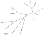
系統発生解析
BLASTでタンパク質のRefseqデータベースに対してCXCL4(PF4)とマッチするペプチドがサーチされ、ペプチド目録が引き出された。ハイライトされた配列は、首尾よくヒトに対して戻った相互BLASTの配列を示す。他の配列は対象となるためリストアップされた。ClustalWプログラムは、すべての種においてCXCL4(PF4)およびCXCL4L1(PF4V1)を示すことが見いだされた12配列に対し実施された。
Phylogenetic analysis In BLAST, peptides matching CXCL4 (PF4) were searched against the protein Refseq database, and a peptide list was derived. The highlighted sequence shows the sequence of reciprocal BLAST that has been successfully returned to humans. Other sequences were listed for inclusion. The ClustalW program was performed on 12 sequences that were found to exhibit CXCL4 (PF4) and CXCL4L1 (PF4V1) in all species.
系統樹は、最適な樹を発見するために近隣結合法を用いて1000のブートストラップを用いて構築された。ギャップのある部位は分析から除外され、アライメントは多重置換に関して補正された。 The phylogenetic tree was built with 1000 bootstraps using the neighbor join method to find the optimal tree. Gapped sites were excluded from the analysis and the alignment was corrected for multiple substitutions.
ヒト腫瘍サンプル
ヒト腺癌サンプルはProf Martin Schilling(Klinik fur Allgemeine Chirurgie, Viszeral-, Gefaeβ-und Kinderchirurgie, Homburg, Germany)によって提供された。新鮮な腫瘍組織は外科手術中に得られ、直接的に液体窒素中瞬間凍結された。患者は診療所のガイドラインに従って組織分析の前に同意した。
Human tumor samples Human adenocarcinoma samples were provided by Prof Martin Schilling (Klinik fur Allgemeine Chirurgie, Viszeral-, Gefaeβ-und Kinderchirurgie, Homburg, Germany). Fresh tumor tissue was obtained during surgery and snap frozen directly in liquid nitrogen. Patients agreed prior to histological analysis according to clinic guidelines.
結果
CXCL4L1およびCXCL4の系統発生解析
Results Phylogenetic analysis of CXCL4L1 and CXCL4
CXCL4およびCXCL4L1は相同性が高い。CXCL4との比較でのCXCL4L1の種の発現を分析するために、我々はCXCL4/CXCL4L1とマッチするペプチドをタンパク質のRefseqデータベースに対してBLASTで検索した。CXCL4L1はヒト、サルおよびチンパンジーでのみ発現している。注目すべきは、トリはCXCL4L1もCXCL4も発現していないことである。 CXCL4 and CXCL4L1 have high homology. To analyze the expression of CXCL4L1 species in comparison to CXCL4, we searched the protein Refseq database with BLAST for peptides matching CXCL4 / CXCL4L1. CXCL4L1 is expressed only in humans, monkeys and chimpanzees. It should be noted that birds do not express CXCL4L1 or CXCL4.
我々は次にClustalWプログラムを用いて、すべての種のCXCL4およびCXCL4L1を表す12個の配列について系統発生解析を実施し、最適な樹を発見するために近隣結合法を用い、1000のブートストラップと系統樹を構築した(図1)。ギャップのある部位は分析から除外され、アライメントは多重置換に関して修正された。分析からわかるように、CXCL4L1およびCXCL4は、進化の最晩期、サル、チンパンジーおよびヒトの共通の祖先のレベルで分岐した。
We then use the ClustalW program to perform a phylogenetic analysis on 12 sequences representing all species of CXCL4 and CXCL4L1, use the neighbor join method to find the optimal tree, and
ヒト組織におけるCXCL4L1またはCXCL4の発現
複数のヒト組織のmRNAにおける定量的PCRはCXCL4L1およびCXCL4に関して異なる発現プロファイルを示唆する(図2)。CXCL4L1が単に、胎児肝、結腸およびある程度脾臓に発現する一方、CXCL4は脾臓に高度に発現している。正常なヒト膵臓は、有意にCXCL4L1およびCXCL4を発現しない。
Expression of CXCL4L1 or CXCL4 in human tissues Quantitative PCR on mRNA of multiple human tissues suggests different expression profiles for CXCL4L1 and CXCL4 (FIG. 2). CXCL4L1 is simply expressed in fetal liver, colon and to some extent spleen, while CXCL4 is highly expressed in spleen. Normal human pancreas does not significantly express CXCL4L1 and CXCL4.
膵臓腺癌におけるCXCL4L1またはCXCL4の発現
トランスクリプトームのプロファイリングはPDAC−CAMモデルにおいてCXCL4L1は同定するがCXCL4を同定しない:ヒトAffymetrixまたはトリAffymetrixマイクロアレーを用いたデュアルトランスクリプトーム分析は、トリ絨毛尿膜へのBxPC3細胞の移植の腫瘍1日目(T1)および腫瘍6日目(T6)の間(胚発生のE11およびE16に対応する)に実施された。我々は、CXCL4ではなくCXCL4L1がT1と比較してT6に高度にアップレギュレーションされた(14.6倍増加)ことを明確に証明した。CXCL4L1はまたヒトAffymetrixアレイにおいてのみ検出され、トリチップが使用された時、トリのオルソログは存在しなかった。この事は、in vivoで腫瘍発生の間、CXCL4L1が腫瘍細胞において誘導される事を示唆した。
Expression of CXCL4L1 or CXCL4 in pancreatic adenocarcinoma Transcriptome profiling identifies CXCL4L1 but not CXCL4 in the PDAC-CAM model: dual transcriptome analysis using human Affymetrix or tri-Affymetrix microarrays It was performed between tumor day 1 (T1) and day 6 (T6) of the implantation of BxPC3 cells into the membrane (corresponding to E11 and E16 of embryonic development). We clearly demonstrated that CXCL4L1, but not CXCL4, was highly upregulated (14.6 fold increase) to T6 compared to T1. CXCL4L1 was also detected only in the human Affymetrix array, and there was no avian ortholog when the trichip was used. This suggested that CXCL4L1 was induced in tumor cells during tumor development in vivo.
実験的膵臓腺癌におけるCXCL4L1またはCXCL4の発現分析:この主張を強固にするために、我々はまず実験的膵臓腺癌モデルにおける特異的な発現を確認するために、CXCL4L1およびCXCL4に特異的なプライマーを用いてリアルタイムPCRを実施した。トランスクリプトームの分析にみられるように、qPCR分析は、T1腫瘍と比較した時、T6腫瘍において高度のアップレギュレーション(9.65倍)を明らかにした。培養中のBxPC3細胞には、発現は見られなかった(図3A)。 Expression analysis of CXCL4L1 or CXCL4 in experimental pancreatic adenocarcinoma: To reinforce this assertion, we first primer specific for CXCL4L1 and CXCL4 to confirm specific expression in an experimental pancreatic adenocarcinoma model Real-time PCR was performed using As seen in the transcriptome analysis, qPCR analysis revealed a high degree of upregulation (9.65 fold) in T6 tumors when compared to T1 tumors. No expression was observed in BxPC3 cells in culture (FIG. 3A).
我々はその後、ケモカインのいずれか一種を認識する特異的プローブを用いて、CXCL4との比較により、CXCL4L1の発現をin situハイブリダイゼーションで調べた(図3B)。in situハイブリダイゼーションはCXCL4ではなくCXCL4L1のみがT6腫瘍において発現していることを明確に示した。 We then examined the expression of CXCL4L1 by in situ hybridization by comparison with CXCL4 using a specific probe that recognizes any one of the chemokines (FIG. 3B). In situ hybridization clearly showed that only CXCL4L1, but not CXCL4, was expressed in T6 tumors.
我々は次にPDAC−CAMモデルの我々の実験的膵臓腫瘍中のタンパク質の発現を調べた。ヒトCXCL4またはCXCL4L1のみを認識する特異的なポリクローナルまたはモノクローナル抗体を用いたCXCL4L1の免疫局在は、T6腫瘍に有意な免疫反応性を示す(図4A)が、BxPC3細胞には示さない(データ不掲載)。さらに、特異的なモノクローナル抗CXCL4L1抗体(MABv1)は、CAM中の膵臓小結節においてCXCL4L1を明瞭に検出する(図4B)。 We next examined the expression of proteins in our experimental pancreatic tumor in the PDAC-CAM model. Immunolocalization of CXCL4L1 using specific polyclonal or monoclonal antibodies that recognize only human CXCL4 or CXCL4L1 shows significant immunoreactivity in T6 tumors (FIG. 4A) but not in BxPC3 cells (data not shown) Published). Furthermore, a specific monoclonal anti-CXCL4L1 antibody (MABv1) clearly detects CXCL4L1 in pancreatic nodules in CAM (FIG. 4B).
さらに、我々はCXCL4およびCXCL4L1の両方と相互作用できるモノクローナルまたはポリクローナル抗体を用いたウエスタンブロットにおいて7kDaの有意な免疫反応性を検出した。さらに、特異的モノクローナル抗CXCL4L1抗体(MABv1)を用いた時、有意なCXCL4L1免疫反応性が検出された(図4C)。 Furthermore, we detected a significant immunoreactivity of 7 kDa in Western blots using monoclonal or polyclonal antibodies that can interact with both CXCL4 and CXCL4L1. Furthermore, significant CXCL4L1 immunoreactivity was detected when a specific monoclonal anti-CXCL4L1 antibody (MABv1) was used (FIG. 4C).
まとめると、これらのデータは、CXCL4ではなくCXCL4L1がトリ絨毛尿膜上に成長する膵腫瘍において高度に過発現していることを示唆する。 Taken together, these data suggest that CXCL4L1, but not CXCL4, is highly overexpressed in pancreatic tumors growing on the avian chorioallantoic membrane.
膵癌のマウスモデルにおける発現分析:CXCL4L1のアップレギュレーションはトリ胎性モデルに特異的ではないことを確認するために、BxPC3細胞は最初にRAG〜g/cマウスの皮下に移植され、原発腫瘍または肺転移が移植後10週間で分析された。組織はその後ヒトCXCL4L1またはCXCL4に対する抗体を用いたqPCRまたは免疫組織化学的検査により分析された。qPCR分析は、CXCL4ではなくCXCL4L1が腫瘍サンプル中に発現している事を明確に示唆する。図5Aに見られるように、我々は次に両方の型を認識するパンCXCL4L1/CXCL4抗体を用いた免疫組織化学的検査を実施した。我々は、原発腫瘍および肺転移の両方において陽性の免疫反応性を明瞭に検出した。この抗体はヒトCXCL4/CXCL4L1に特異的であり、マウスCXCL4と交差反応しない。我々は次に、我々のCXCL4L1に特異的なモノクローナル抗体を用いて免疫組織化学的検査を実施した。再び、我々は、原発腫瘍および肺転移の両方において陽性の免疫反応性を明瞭に検出した(図5B)。これらの結果は、この腫瘍モデルにおける原発腫瘍および肺転移の両方においてCXCL4L1が有意に発現していることを示唆する。 Expression analysis in a mouse model of pancreatic cancer: To confirm that the upregulation of CXCL4L1 is not specific for the avian fetal model, BxPC3 cells were first implanted subcutaneously in RAG-g / c mice and either primary tumor or lung Metastases were analyzed 10 weeks after transplantation. Tissues were then analyzed by qPCR or immunohistochemistry using antibodies against human CXCL4L1 or CXCL4. qPCR analysis clearly suggests that CXCL4L1, but not CXCL4, is expressed in the tumor sample. As can be seen in FIG. 5A, we next performed an immunohistochemical study using pan CXCL4L1 / CXCL4 antibodies that recognize both types. We clearly detected positive immunoreactivity in both primary tumors and lung metastases. This antibody is specific for human CXCL4 / CXCL4L1 and does not cross-react with mouse CXCL4. We then performed an immunohistochemical test using a monoclonal antibody specific for our CXCL4L1. Again, we clearly detected positive immunoreactivity in both primary tumors and lung metastases (FIG. 5B). These results suggest that CXCL4L1 is significantly expressed in both the primary tumor and lung metastases in this tumor model.
ヒト腫瘍サンプル中のCXCL4/CXCL4L1の発現分析:膵腫瘍中のCXCL4L1の発現を確認するために、RNAは24人の腺癌患者サンプルから単離され、特異的なプライマーを用いたqPCRが正常な膵臓との比較により実施された(図6A)。CXCL4L1mRNAの発現はすべての試験された膵腫瘍で検出され、23/24サンプルにおいて様々な過発現レベルで(平均値+5.0)過発現した。我々はCXCL4に対する特異的なプライマーとqPCRも実施した。患者由来の腫瘍サンプルにCXCL4の発現は検出できなかった。これらの結果は異なる患者のサブグループを反映するであろう、ヒト膵癌中での不均一なCXCL4L1の過発現を示唆する。 Expression analysis of CXCL4 / CXCL4L1 in human tumor samples: To confirm the expression of CXCL4L1 in pancreatic tumors, RNA was isolated from 24 adenocarcinoma patient samples and qPCR with specific primers was normal This was done by comparison with the pancreas (FIG. 6A). CXCL4L1 mRNA expression was detected in all tested pancreatic tumors and was overexpressed at various overexpression levels (mean + 5.0) in 23/24 samples. We also performed specific primers and qPCR for CXCL4. No CXCL4 expression could be detected in patient-derived tumor samples. These results suggest heterogeneous overexpression of CXCL4L1 in human pancreatic cancer, which may reflect different patient subgroups.
我々は次に、ヒトCXCL4/CXCL4L1に特異的な抗体を用いて膵腫瘍サンプルの免疫組織学的分析を実施した(図6B及び6C)。我々は、ポリクローナル抗CXCL4/CXCL4L1抗体(図6B)または特異的モノクローナル抗体MABv1(図6C)の両方で、正常ヒト膵臓ではなく膵腫瘍サンプルにおける免疫反応性を明確に証明した。驚くべき事に腫瘍のグレードの重症度と共に免疫反応性が上昇し、最も弱い染色をグレード1において、最も強い染色をグレード3の腫瘍において示した。この事は、CXCL4L1が膵臓腺癌において高度に過発現されている事、および発現が組織学的グレードの上昇と共に上昇する事を示唆する。 We next performed an immunohistological analysis of pancreatic tumor samples using antibodies specific for human CXCL4 / CXCL4L1 (FIGS. 6B and 6C). We clearly demonstrated immunoreactivity in pancreatic tumor samples but not normal human pancreas with both polyclonal anti-CXCL4 / CXCL4L1 antibody (FIG. 6B) or specific monoclonal antibody MABv1 (FIG. 6C). Surprisingly, immunoreactivity increased with tumor grade severity, with the weakest staining shown in grade 1 and the strongest staining in grade 3 tumors. This suggests that CXCL4L1 is highly overexpressed in pancreatic adenocarcinoma and that expression increases with increasing histological grade.
膵癌におけるCXCL4L1またはCXCL4の機能研究
CXCL4L1およびCXCL4の組み換えタンパク質はGST融合タンパク質として相当量精製された。プロテアーゼの切り取りによるGSTの除去は、生物学的活性に対する有意な効果を有さず、切り取りを不必要にする。
Functional studies of CXCL4L1 or CXCL4 in pancreatic cancer Recombinant proteins of CXCL4L1 and CXCL4 were purified to a considerable extent as GST fusion proteins. Removal of GST by excision of the protease has no significant effect on biological activity and makes the excision unnecessary.
CXCL4と比較してCXCL4L1の活性を試験するために、BxPC3細胞またはHUVEC細胞は、これらの分子の増加する濃度で刺激され、増殖が測定された。CXCL4L1は、文献に報告されているのと同様に、CXCL4よりも内皮細胞増殖にかなり強い効果(43倍高い)を示した(図7A)。しかしながら、両方のケモカインの効果はBxPC3細胞において認められなかった(図8)。我々は、さらにボイデンチャンバー遊走アッセイを用いて、遊走および浸潤に対するCXCL4L1またはCXCL4の効果を研究した。再び、内皮細胞遊走および浸潤は有意に阻害されたが、BxPC3細胞の遊走は阻害されなかった(図7B、C;図8B、C)。 To test the activity of CXCL4L1 compared to CXCL4, BxPC3 cells or HUVEC cells were stimulated with increasing concentrations of these molecules and proliferation was measured. CXCL4L1 showed a much stronger effect (43 times higher) on endothelial cell proliferation than CXCL4, as reported in the literature (FIG. 7A). However, the effects of both chemokines were not observed in BxPC3 cells (Figure 8). We further studied the effects of CXCL4L1 or CXCL4 on migration and invasion using a Boyden chamber migration assay. Again, endothelial cell migration and invasion were significantly inhibited, but BxPC3 cell migration was not inhibited (FIGS. 7B, C; FIGS. 8B, C).
考察
CXCL4L1はヒト、サルおよびチンパンジーにのみ発現し、進化の最晩期にCXCL4から分岐する。腸、肝臓および脾臓を含む複数のヒト組織は、CXCL4L1およびCXCL4を発現するが、正常膵臓組織は、このケモカインの弱い発現を示すか全く発現しないことを示す。しかしながらCXCL4ではなくCXCL4L1が膵癌において有意に過発現し、新規のバイオマーカーに相当する。この事は下記の観察に基づく:
(1)qPCRまたはin situ ハイブリダイゼーションとの組み合わせによるトランスクリプトーム分析は、ヒト膵臓腺癌細胞がトリ絨毛尿膜に移植される時、有意なレベルのCXCL4L1 mRNAを発現するが、CXCL4のmRNAは発現しない事を示唆する。
(2)抗ヒトCXCL4L1/CXCL4または特異的なモノクローナル抗CXCL4L1抗体(Mabv1)を用いた免疫局在性は、トリCAMに移植される時、腫瘍細胞において強い免疫反応性を証明する。
(3)マウスに移植後の原発膵腫瘍は、CXCL4L1 mRNAを発現し、原発腫瘍および転移性腫瘍細胞も陽性の免疫反応性を示す。
(4)ヒト患者由来の膵臓腺癌サンプルはCXCL4L1 mRNAのみを発現し、CXCL4 mRNAは発現せず、強い陽性の免疫反応性を示す。
(5)膵臓腺癌細胞ではなく内皮細胞が外来性CXCL4L1に応答することは、膵腫瘍発生においてCXCL4L1の活性のパラクリンモードを示唆する。さらに、我々の結果は、培養液中の膵臓腺癌細胞がCXCL4L1もCXCL4も発現しないので、CXCL4L1発現のアップレギュレーションは特異的な腫瘍−宿主相互作用によるということを示唆する。
DISCUSSION CXCL4L1 is expressed only in humans, monkeys and chimpanzees, and branches off from CXCL4 at the end of evolution. Multiple human tissues including intestine, liver and spleen express CXCL4L1 and CXCL4, while normal pancreatic tissue shows weak or no expression of this chemokine. However, CXCL4L1, but not CXCL4, is significantly overexpressed in pancreatic cancer and represents a novel biomarker. This is based on the following observations:
(1) Transcriptome analysis in combination with qPCR or in situ hybridization shows that when human pancreatic adenocarcinoma cells are transplanted into avian chorioallantoic membrane, it expresses significant levels of CXCL4L1 mRNA, but CXCL4 mRNA is It suggests that it does not appear.
(2) Immunolocalization using anti-human CXCL4L1 / CXCL4 or a specific monoclonal anti-CXCL4L1 antibody (Mabv1) demonstrates strong immunoreactivity in tumor cells when transplanted into avian CAM.
(3) The primary pancreatic tumor after transplantation into mice expresses CXCL4L1 mRNA, and the primary tumor and metastatic tumor cells also show positive immunoreactivity.
(4) A pancreatic adenocarcinoma sample derived from a human patient expresses only CXCL4L1 mRNA, does not express CXCL4 mRNA, and exhibits a strong positive immunoreactivity.
(5) Endothelial cells but not pancreatic adenocarcinoma cells respond to exogenous CXCL4L1, suggesting a paracrine mode of CXCL4L1 activity in pancreatic tumor development. Furthermore, our results suggest that the up-regulation of CXCL4L1 expression is due to specific tumor-host interactions since pancreatic adenocarcinoma cells in culture do not express CXCL4L1 or CXCL4.
CXCL4L1は、シグナル配列をコードするアミノ末端に34%のみの違いを有し、残りの配列に4.3%の違いが認められるCXCL4に対して密接に相同なケモカインである。血小板精製CXCL4L1は細胞遊走においてCXCL4よりも少なくとも50倍強力である事が示された(Stryuf et al. 2004)。我々のデータでは、組み換えCXCL4L1は内皮細胞増殖の阻害においてCXCL4よりも50倍より強力であるが、内皮細胞遊走の阻害においてCXCL4よりも100倍より強力である。さらに、腫瘍発生の阻害が肺において認められ、メラノーマ細胞腫細胞はCXCL4L1により阻害される(Struyf et al., 2007)。さらに、かつてCXCL4L1とCXCL4の分泌およびプロセシングは異なるという事が示された(Lasagni et al., 2007)。CXCL4が有芯顆粒からPKC制御経路を介して制御された方法で排出される一方、CXCL4L1はPKCを介して制御されず、構成的に分泌される。さらに、CXCL4L1またはCXCL4は血小板−巨核球系に発現するだけでなく炎症性細胞(T細胞、白血球、単球)、血管および冠状動脈平滑筋細胞ならびに内皮細胞にも発現する(Lasagni et al., 2007)。これらの結果は、CXCL4L1が血管形成において調節機能を有するかもしれないことを示唆する。 CXCL4L1 is a chemokine that is closely homologous to CXCL4 with only a 34% difference at the amino terminus encoding the signal sequence and a 4.3% difference in the rest of the sequence. Platelet purified CXCL4L1 was shown to be at least 50 times more potent than CXCL4 in cell migration (Stryuf et al. 2004). In our data, recombinant CXCL4L1 is 50 times more potent than CXCL4 in inhibiting endothelial cell proliferation, but 100 times more potent than CXCL4 in inhibiting endothelial cell migration. Furthermore, inhibition of tumor development is observed in the lung, and melanoma cell cells are inhibited by CXCL4L1 (Struyf et al., 2007). Furthermore, it was once shown that CXCL4L1 and CXCL4 secretion and processing are different (Lasagni et al., 2007). While CXCL4 is excreted from the cored granules in a controlled manner via the PKC control pathway, CXCL4L1 is not controlled via PKC and is secreted constitutively. Furthermore, CXCL4L1 or CXCL4 is expressed not only in the platelet-megakaryocyte system but also in inflammatory cells (T cells, leukocytes, monocytes), vascular and coronary artery smooth muscle cells and endothelial cells (Lasagni et al., 2007). These results suggest that CXCL4L1 may have a regulatory function in angiogenesis.
実験室で、我々は最初に神経膠腫の浸潤および血管形成によりよい見識をえるため、トリ胚モデルを開発した(Hagedorn et al., 2005)。我々は現在さらにこのモデルを膵管腺癌のために拡大し(PDAC−CAMモデル)、CAMに移植される時、膵腫瘍細胞におけるCXCL4L1の有意なアップレギュレーション、CXCL4の非アップレギュレーションを証明した。驚くべき事に、培養中の腫瘍細胞にはCXCL4L1の発現は見い出されなかった。ヒトCXCL4L1に特異的なプライマーが使用され、CXCL4L1がヒト由来の細胞にのみ発現し他の種には発現しないので、これらの結果は、腫瘍細胞が適切な微小環境におかれる時、腫瘍細胞はCXCL4L1の発現をアップレギュレーションすることを示唆する。さらにCXCL4L1またはCXCL4に特異的なプローブを用いたin situハイブリダイゼーションは、CAMで成長した時、CXCL4L1のみに対して腫瘍に有意なシグナルを示した。さらに、細胞が培養液中成長した時ではなくCAMに移植された時に、CXCL4L1の免疫反応性が腫瘍細胞で検出された。これらの条件下では高い関心のある分子が検出されないかもしれないので、これらの結果は培養液中で行われる複数の分子プロファイリング研究の妥当性に疑問も投げかける。 In the laboratory, we first developed an avian embryo model to gain better insight into glioma infiltration and angiogenesis (Hagedorn et al., 2005). We have now further expanded this model for ductal adenocarcinoma (PDAC-CAM model) and demonstrated significant up-regulation of CXCL4L1, non-up-regulation of CXCL4 in pancreatic tumor cells when transplanted into CAM. Surprisingly, no expression of CXCL4L1 was found in tumor cells in culture. Since primers specific for human CXCL4L1 are used and CXCL4L1 is expressed only in human-derived cells and not in other species, these results indicate that when tumor cells are placed in an appropriate microenvironment, the tumor cells This suggests up-regulating the expression of CXCL4L1. Furthermore, in situ hybridization using a probe specific for CXCL4L1 or CXCL4 showed a significant signal to the tumor relative to CXCL4L1 alone when grown on CAM. Furthermore, the immunoreactivity of CXCL4L1 was detected in tumor cells when the cells were transplanted into CAM rather than when grown in culture. These results also question the validity of multiple molecular profiling studies performed in culture, as molecules of high interest may not be detected under these conditions.
我々は、次に膵腫瘍細胞で異種移植されたマウスを用いてCXCL4L1の発現を研究した。CXCL4L1mRNAを発現するがCXCL4を発現しない原発腫瘍を皮下に移植した。再び、高度の免疫反応性が抗ヒトCXCL4L1/CXCL4またはCXCL4L1抗体を用いて証明された。さらに、有意な免疫反応性も肺転移中に検出された。これらの結果は、アップレギュレーションがトリの微小環境に特異的ではなく、細胞がマウス組織に移植される時にも検出されることを明確に示唆する。 We next studied the expression of CXCL4L1 using mice xenografted with pancreatic tumor cells. Primary tumors expressing CXCL4L1 mRNA but not CXCL4 were implanted subcutaneously. Again, a high degree of immunoreactivity was demonstrated using anti-human CXCL4L1 / CXCL4 or CXCL4L1 antibodies. In addition, significant immunoreactivity was also detected during lung metastasis. These results clearly suggest that upregulation is not specific to the avian microenvironment and is also detected when cells are transplanted into mouse tissue.
我々は最終的にヒト患者由来のサンプルにおいて、CXCL4のmRNAではなくCXCL4L1のmRNAの有意なアップレギュレーションを証明した。mRNAの発現は不均一であり、多分、低、中間、高度のCXCL4L1発現を有する様々な患者グループを示唆する。我々が観察した興味深い発見は、CXCL4L1に対する免疫反応性が組織学的グレードとともに上昇することであった。有意な免疫反応性がリンパ節転移病変にも検出された。これはCXCL4L1が膵癌の新規のバイオマーカーとなり、疾患の進行および処置反応を予測するということを示唆する。 We finally demonstrated a significant upregulation of CXCL4L1 mRNA but not CXCL4 mRNA in samples from human patients. mRNA expression is heterogeneous, possibly suggesting various patient groups with low, intermediate, and high CXCL4L1 expression. An interesting finding we observed was that immunoreactivity against CXCL4L1 increased with histological grade. Significant immunoreactivity was also detected in lymph node metastatic lesions. This suggests that CXCL4L1 becomes a novel biomarker of pancreatic cancer and predicts disease progression and treatment response.
さらに、腫瘍環境におけるCXCL4L1の濃度の局所的増加により、膵腫瘍は、治療目的で、細胞障害性薬物または成長阻害剤のような抗癌剤に結合したCXCL4L1特異的結合分子の標的となりうる。 Furthermore, due to the local increase in the concentration of CXCL4L1 in the tumor environment, pancreatic tumors can be targeted for CXCL4L1-specific binding molecules conjugated to anticancer agents such as cytotoxic drugs or growth inhibitors for therapeutic purposes.
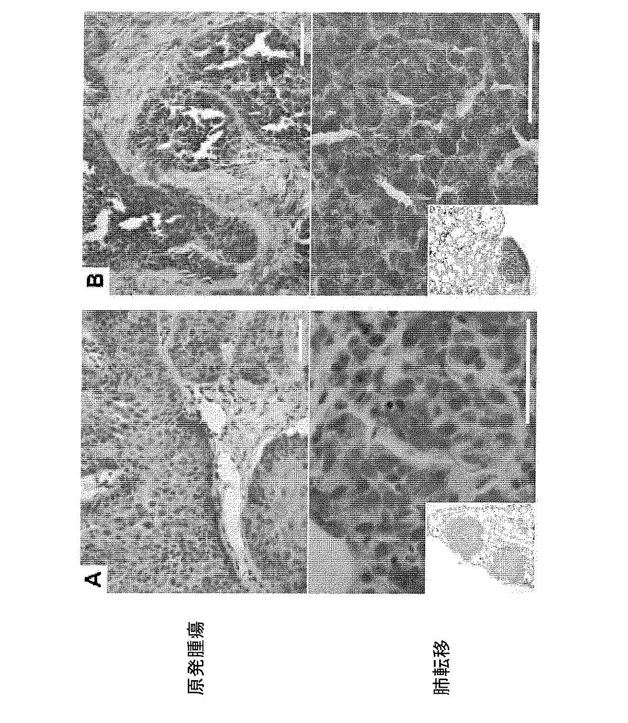
Claims (8)
Applications Claiming Priority (3)
| Application Number | Priority Date | Filing Date | Title |
|---|---|---|---|
| EP08305853 | 2008-11-27 | ||
| EP08305853.7 | 2008-11-27 | ||
| PCT/EP2009/065805 WO2010060920A1 (en) | 2008-11-27 | 2009-11-25 | Cxcl4l1 as a biomarker of pancreatic cancer |
Publications (3)
| Publication Number | Publication Date |
|---|---|
| JP2012509683A JP2012509683A (en) | 2012-04-26 |
| JP2012509683A5 JP2012509683A5 (en) | 2014-08-07 |
| JP5773315B2 true JP5773315B2 (en) | 2015-09-02 |
Family
ID=40288415
Family Applications (1)
| Application Number | Title | Priority Date | Filing Date |
|---|---|---|---|
| JP2011537960A Expired - Fee Related JP5773315B2 (en) | 2008-11-27 | 2009-11-25 | CXCL4L1 as a biomarker for pancreatic cancer |
Country Status (5)
| Country | Link |
|---|---|
| US (1) | US20110318370A1 (en) |
| EP (1) | EP2361315B1 (en) |
| JP (1) | JP5773315B2 (en) |
| ES (1) | ES2465191T3 (en) |
| WO (1) | WO2010060920A1 (en) |
Families Citing this family (6)
| Publication number | Priority date | Publication date | Assignee | Title |
|---|---|---|---|---|
| WO2012072595A1 (en) * | 2010-11-30 | 2012-06-07 | Universiteit Gent | Platelet factor 4 variant for the prognosis of cardiovascular outcome in patients having heart disease |
| WO2012095448A1 (en) * | 2011-01-11 | 2012-07-19 | INSERM (Institut National de la Santé et de la Recherche Médicale) | Methods for predicting the outcome of a cancer in a patient by analysing gene expression |
| US9164105B2 (en) | 2011-01-13 | 2015-10-20 | Industry-Academic Cooperation Foundation, Yonsei University | Pancreatic cancer biomarker using the characteristics of pancreatic cancer stem cells, and use thereof |
| WO2012101125A1 (en) * | 2011-01-24 | 2012-08-02 | INSERM (Institut National de la Santé et de la Recherche Médicale) | Specific antibodies against human cxcl4 and uses thereof |
| CA2887920C (en) * | 2012-10-12 | 2021-03-23 | Hi-Stem Ggmbh | Novel biomarkers for sub-typing pancreatic ductal adenocarcinoma |
| WO2014177662A1 (en) * | 2013-04-30 | 2014-11-06 | INSERM (Institut National de la Santé et de la Recherche Médicale) | In vitro method for determining if a subject suffering from cancer is responsive to a treatment comprising administering an effective amount of cxcl4l1 or an inhibitor of cxcl4l1 |
Family Cites Families (12)
| Publication number | Priority date | Publication date | Assignee | Title |
|---|---|---|---|---|
| WO1981001145A1 (en) | 1979-10-18 | 1981-04-30 | Univ Illinois | Hydrolytic enzyme-activatible pro-drugs |
| US4946778A (en) | 1987-09-21 | 1990-08-07 | Genex Corporation | Single polypeptide chain binding molecules |
| GB8705477D0 (en) | 1987-03-09 | 1987-04-15 | Carlton Med Prod | Drug delivery systems |
| US4975278A (en) | 1988-02-26 | 1990-12-04 | Bristol-Myers Company | Antibody-enzyme conjugates in combination with prodrugs for the delivery of cytotoxic agents to tumor cells |
| US5208020A (en) | 1989-10-25 | 1993-05-04 | Immunogen Inc. | Cytotoxic agents comprising maytansinoids and their therapeutic use |
| JP3095175B2 (en) | 1992-11-13 | 2000-10-03 | アイデック ファーマシューティカルズ コーポレイション | Therapeutic use of chimeric and radiolabeled antibodies against human B lymphocyte restricted differentiation antigen for the treatment of B cell lymphoma |
| TW526269B (en) | 1997-11-21 | 2003-04-01 | Dev Center Biotechnology | Monoclonal antibody against morphine derivatives, hybridoma producing the antibody and the use thereof |
| JP2006521562A (en) * | 2003-03-07 | 2006-09-21 | スレショルド ファーマシューティカルズ インコーポレイティッド | Methods for determining tumor sensitivity to treatment with antineoplastic agents |
| US20090011981A1 (en) * | 2004-09-15 | 2009-01-08 | K.U. Leuven Research & Development | Novel Inhibitors of Angiogenesis |
| EP2106439B1 (en) * | 2007-01-24 | 2014-11-12 | The Regents of the University of Michigan | Compositions and methods for treating and diagnosing pancreatic cancer |
| US8568993B2 (en) * | 2007-07-02 | 2013-10-29 | Purdue Research Foundation | Detection of glycopeptides and glycoproteins for medical diagnostics |
| US8481033B2 (en) * | 2008-10-07 | 2013-07-09 | INSERM (Institute National de la Santé et de la Recherche Médicale) | Neutralizing antibodies and fragments thereof directed against platelet factor-4 variant 1 (PF4V1) |
-
2009
- 2009-11-25 US US13/131,084 patent/US20110318370A1/en not_active Abandoned
- 2009-11-25 WO PCT/EP2009/065805 patent/WO2010060920A1/en active Application Filing
- 2009-11-25 EP EP09764773.9A patent/EP2361315B1/en not_active Not-in-force
- 2009-11-25 ES ES09764773.9T patent/ES2465191T3/en active Active
- 2009-11-25 JP JP2011537960A patent/JP5773315B2/en not_active Expired - Fee Related
Also Published As
| Publication number | Publication date |
|---|---|
| EP2361315A1 (en) | 2011-08-31 |
| EP2361315B1 (en) | 2014-03-05 |
| WO2010060920A1 (en) | 2010-06-03 |
| US20110318370A1 (en) | 2011-12-29 |
| ES2465191T3 (en) | 2014-06-05 |
| JP2012509683A (en) | 2012-04-26 |
Similar Documents
| Publication | Publication Date | Title |
|---|---|---|
| JP6430560B2 (en) | Phosphodiesterase 4D7 as a prostate cancer marker | |
| JP5773315B2 (en) | CXCL4L1 as a biomarker for pancreatic cancer | |
| JP6717428B2 (en) | Cancer diagnostic composition using potassium channel protein | |
| KR102054857B1 (en) | Composition for diagnosing disease | |
| KR102210333B1 (en) | Composition for diagnosing cancer | |
| KR101515700B1 (en) | Diagnostic composition for sepsis using TGFBI and pharmaceutical composition for preventing or treating of sepsis using the same and screening method thereof | |
| CN116801788A (en) | Kit, reagent and method for assessing liver disease | |
| KR20090053222A (en) | Characterization of cxcl-16 as a tumor associated marker of colorectal cancer | |
| KR101058753B1 (en) | Characterization of ESM-1 as a tumor associated marker of colorectal cancer | |
| KR100948808B1 (en) | Lipocalin 2 as a tumor associated marker of hepatocellular carcinoma and a hepatocellular carcinoma diagnostic kit using thereof | |
| KR102006999B1 (en) | A composition for diagnosing colon cancer metastasis or predicting the same, and use thereof | |
| US10316319B2 (en) | Composition for diagnosis of liver metastasis of colorectal cancer and the use thereof | |
| KR100861464B1 (en) | A carcinoma gene tip41, a protein translated from the gene and a diagnostic kit using the same | |
| US20120156681A1 (en) | Composition for diagnosis of liver metastasis of colorectal cancer and the use thereof | |
| KR101516716B1 (en) | Biomaker RORC for diagnosing liver fibrosis | |
| KR101515210B1 (en) | Biomaker ELK3 for diagnosing liver fibrosis | |
| KR102325742B1 (en) | A Composition for Diagnosing Cancer |
Legal Events
| Date | Code | Title | Description |
|---|---|---|---|
| A621 | Written request for application examination |
Free format text: JAPANESE INTERMEDIATE CODE: A621 Effective date: 20121017 |
|
| A131 | Notification of reasons for refusal |
Free format text: JAPANESE INTERMEDIATE CODE: A131 Effective date: 20140318 |
|
| A521 | Request for written amendment filed |
Free format text: JAPANESE INTERMEDIATE CODE: A523 Effective date: 20140618 |
|
| A524 | Written submission of copy of amendment under article 19 pct |
Free format text: JAPANESE INTERMEDIATE CODE: A524 Effective date: 20140618 |
|
| A131 | Notification of reasons for refusal |
Free format text: JAPANESE INTERMEDIATE CODE: A131 Effective date: 20141224 |
|
| A521 | Request for written amendment filed |
Free format text: JAPANESE INTERMEDIATE CODE: A523 Effective date: 20150323 |
|
| TRDD | Decision of grant or rejection written | ||
| A01 | Written decision to grant a patent or to grant a registration (utility model) |
Free format text: JAPANESE INTERMEDIATE CODE: A01 Effective date: 20150602 |
|
| A61 | First payment of annual fees (during grant procedure) |
Free format text: JAPANESE INTERMEDIATE CODE: A61 Effective date: 20150619 |
|
| R150 | Certificate of patent or registration of utility model |
Ref document number: 5773315 Country of ref document: JP Free format text: JAPANESE INTERMEDIATE CODE: R150 |
|
| LAPS | Cancellation because of no payment of annual fees |