JP5568461B2 - Apparatus and method for monitoring blood parameters - Google Patents
Apparatus and method for monitoring blood parameters Download PDFInfo
- Publication number
- JP5568461B2 JP5568461B2 JP2010500432A JP2010500432A JP5568461B2 JP 5568461 B2 JP5568461 B2 JP 5568461B2 JP 2010500432 A JP2010500432 A JP 2010500432A JP 2010500432 A JP2010500432 A JP 2010500432A JP 5568461 B2 JP5568461 B2 JP 5568461B2
- Authority
- JP
- Japan
- Prior art keywords
- blood
- body part
- light
- emitter
- reflectance
- Prior art date
- Legal status (The legal status is an assumption and is not a legal conclusion. Google has not performed a legal analysis and makes no representation as to the accuracy of the status listed.)
- Expired - Fee Related
Links
Images


Classifications
-
- A—HUMAN NECESSITIES
- A61—MEDICAL OR VETERINARY SCIENCE; HYGIENE
- A61B—DIAGNOSIS; SURGERY; IDENTIFICATION
- A61B5/00—Measuring for diagnostic purposes; Identification of persons
- A61B5/145—Measuring characteristics of blood in vivo, e.g. gas concentration, pH value; Measuring characteristics of body fluids or tissues, e.g. interstitial fluid, cerebral tissue
- A61B5/1455—Measuring characteristics of blood in vivo, e.g. gas concentration, pH value; Measuring characteristics of body fluids or tissues, e.g. interstitial fluid, cerebral tissue using optical sensors, e.g. spectral photometrical oximeters
- A61B5/14551—Measuring characteristics of blood in vivo, e.g. gas concentration, pH value; Measuring characteristics of body fluids or tissues, e.g. interstitial fluid, cerebral tissue using optical sensors, e.g. spectral photometrical oximeters for measuring blood gases
- A61B5/14552—Details of sensors specially adapted therefor
-
- A—HUMAN NECESSITIES
- A61—MEDICAL OR VETERINARY SCIENCE; HYGIENE
- A61B—DIAGNOSIS; SURGERY; IDENTIFICATION
- A61B5/00—Measuring for diagnostic purposes; Identification of persons
- A61B5/145—Measuring characteristics of blood in vivo, e.g. gas concentration, pH value; Measuring characteristics of body fluids or tissues, e.g. interstitial fluid, cerebral tissue
- A61B5/14535—Measuring characteristics of blood in vivo, e.g. gas concentration, pH value; Measuring characteristics of body fluids or tissues, e.g. interstitial fluid, cerebral tissue for measuring haematocrit
-
- A—HUMAN NECESSITIES
- A61—MEDICAL OR VETERINARY SCIENCE; HYGIENE
- A61B—DIAGNOSIS; SURGERY; IDENTIFICATION
- A61B5/00—Measuring for diagnostic purposes; Identification of persons
- A61B5/68—Arrangements of detecting, measuring or recording means, e.g. sensors, in relation to patient
- A61B5/6801—Arrangements of detecting, measuring or recording means, e.g. sensors, in relation to patient specially adapted to be attached to or worn on the body surface
- A61B5/6813—Specially adapted to be attached to a specific body part
- A61B5/6825—Hand
- A61B5/6826—Finger
-
- A—HUMAN NECESSITIES
- A61—MEDICAL OR VETERINARY SCIENCE; HYGIENE
- A61B—DIAGNOSIS; SURGERY; IDENTIFICATION
- A61B5/00—Measuring for diagnostic purposes; Identification of persons
- A61B5/68—Arrangements of detecting, measuring or recording means, e.g. sensors, in relation to patient
- A61B5/6801—Arrangements of detecting, measuring or recording means, e.g. sensors, in relation to patient specially adapted to be attached to or worn on the body surface
- A61B5/683—Means for maintaining contact with the body
- A61B5/6838—Clamps or clips
-
- A—HUMAN NECESSITIES
- A61—MEDICAL OR VETERINARY SCIENCE; HYGIENE
- A61B—DIAGNOSIS; SURGERY; IDENTIFICATION
- A61B5/00—Measuring for diagnostic purposes; Identification of persons
- A61B5/72—Signal processing specially adapted for physiological signals or for diagnostic purposes
- A61B5/7235—Details of waveform analysis
- A61B5/7239—Details of waveform analysis using differentiation including higher order derivatives
Landscapes
- Health & Medical Sciences (AREA)
- Life Sciences & Earth Sciences (AREA)
- Physics & Mathematics (AREA)
- Engineering & Computer Science (AREA)
- General Health & Medical Sciences (AREA)
- Biomedical Technology (AREA)
- Heart & Thoracic Surgery (AREA)
- Medical Informatics (AREA)
- Molecular Biology (AREA)
- Surgery (AREA)
- Animal Behavior & Ethology (AREA)
- Biophysics (AREA)
- Public Health (AREA)
- Veterinary Medicine (AREA)
- Pathology (AREA)
- Optics & Photonics (AREA)
- Artificial Intelligence (AREA)
- Physiology (AREA)
- Psychiatry (AREA)
- Signal Processing (AREA)
- Spectroscopy & Molecular Physics (AREA)
- Computer Vision & Pattern Recognition (AREA)
- Measurement Of The Respiration, Hearing Ability, Form, And Blood Characteristics Of Living Organisms (AREA)
- Investigating Or Analysing Materials By Optical Means (AREA)
- Investigating Or Analysing Biological Materials (AREA)
Description
開示される技術は、血液パラメータの継続的なモニタリングのための非侵襲的方法に関する。 The disclosed technique relates to a non-invasive method for continuous monitoring of blood parameters.
用語「ヘマトクリット」(Hct)は、赤血球(RBC)によって占められる血液量の比率を参照する。RBCは、肺から体内の様々な組織への酸素の供給を可能にするヘモグロビン(およそ34%含有量)を含む。個人のヘマトクリットの通常の範囲は、年齢及び性別で変化するが、成人男性について42−54%及び成人女性について38−46%である。患者のヘマトクリットの測定は、様々な医療診断に有用である。低いヘマトクリットは、通常、貧血(RBCおよびヘモグロビンの欠乏)を示す。貧血は、急性の血液損失(例えば、外傷、外科手術、結腸癌)、栄養失調(例えば、鉄、葉酸、ビタミンB−12等)、又は、様々な臨床学的因子(例えば、癌、薬物効果、遺伝的疾患等)の結果となり得る。高いヘマトクリットは、脱水症に起因し、又は、骨髄増殖性疾患(骨髄傷害の一種)や、慢性的な閉塞性肺疾患のような他の状況、及び、低酸素症(体内の酸素の欠乏)に関する他の状況を示し得る。 The term “hematocrit” (Hct) refers to the proportion of blood volume occupied by red blood cells (RBC). The RBC contains hemoglobin (approximately 34% content) that allows the supply of oxygen from the lungs to various tissues in the body. The normal range of individual hematocrit varies with age and gender, but is 42-54% for adult men and 38-46% for adult women. Measurement of a patient's hematocrit is useful for various medical diagnoses. A low hematocrit usually indicates anemia (RBC and hemoglobin deficiency). Anemia can be acute blood loss (eg, trauma, surgery, colon cancer), malnutrition (eg, iron, folic acid, vitamin B-12, etc.), or various clinical factors (eg, cancer, drug effects) , Genetic disease, etc.). High hematocrit is due to dehydration or other conditions such as myeloproliferative disease (a type of bone marrow injury) and chronic obstructive pulmonary disease, and hypoxia (lack of oxygen in the body) Other situations may be indicated.
ヘマトクリットは、通常、侵襲的技術で測定される。ある共通の技術において、血は、患者から抜かれ、その後、毛細管の内部の個別の層に異なる血液成分を分けるために、高速遠心分離機が使用される。管内のRBC層の相対的高さは、ヘマトクリット値を提供するために、総血液量に関して測定される。抜かれた血液はまた、血液の光学的測定を採用するといった、他の方法で分析され得る。ヘマトクリットは、血液検査一式の一部として、自動機器を使用して計算される。 Hematocrit is usually measured with invasive techniques. In one common technique, blood is drawn from the patient and then a high speed centrifuge is used to separate the different blood components into separate layers inside the capillary. The relative height of the RBC layer within the tube is measured in terms of total blood volume to provide a hematocrit value. The drawn blood can also be analyzed in other ways, such as employing optical measurements of blood. Hematocrit is calculated using automated equipment as part of a complete blood test.
上記の侵襲的測定は、被験者の不便さ及び不快を含み、そしてまた、被験者及び測定を実行する医療スタッフの両方に対する汚染のリスクを含む。加えて、いくつかの侵襲的方法(遠心分離技術のような)は、血液サンプルが抜かれたときと、ヘマトクリット値が得られたときとの間の遅れを伴う。さらに、ヘマトクリットの決定は(他のタイプの血液テストと同様に)、通常、個々の測定に基づく。このような状況において、被験者のヘマトクリットの連続的なモニタリングを実行し、重変な又は生命に関わる病状(例えば、集中治療中、又は外科手術のとき)を効果的に診断し、又は処置するために有益である。しかしながら、もし、侵襲的測定技術が使用されていると、即座の結果を得ることが不可能であり、処理される被験者のヘマトクリットのリアルタイムの追跡ができなくなる。 The invasive measurements described above involve the inconvenience and discomfort of the subject and also involve the risk of contamination for both the subject and the medical staff performing the measurement. In addition, some invasive methods (such as centrifugation techniques) involve a delay between when the blood sample is drawn and when the hematocrit value is obtained. In addition, the hematocrit determination (as with other types of blood tests) is usually based on individual measurements. In such situations, to perform continuous monitoring of the subject's hematocrit to effectively diagnose or treat severe or life-threatening conditions (eg, during intensive care or during surgery) It is beneficial to. However, if invasive measurement techniques are used, immediate results are not possible and real-time tracking of the hematocrit of the subject being processed is not possible.
パルス酸素濃度計は、酸素飽和度のレベルを測定するための非侵襲的方法である。酸素に結合したヘモグロビン分子は、オキシヘモグロビン(HbO2)として知られ、酸素なしのヘモグロビン分子は、デオキシヘモグロビン(Hb)として知られる。パルス酸素濃度計は、指先のような、身体の部分のいたるところで、2以上の別個の波長(例えば、赤色及び赤外線)の光を放射することを含む。2つの波長のそれぞれの全ての減衰は、センサ(例えば反射又は透過率測定に基づいて)を使用して測定される。パルス酸素濃度計は、動脈の血液を光が通過する他の組織(例えば、皮膚、骨、筋肉、脂肪、爪等)から分離するために、血管の脈動性を利用する。体内の様々な血管は、身体を循環する血液のように、心臓のポンプ作用により、周期的に変化する。オキシヘモグロビン及びデオキシヘモグロビン成分との間の血液中における比率は、オキシヘモグロビンとデオキシヘモグロビンの異なる吸収スペクトルに基づいて決定される。パルス酸素濃度計は、ヘモグロビン内の酸素減衰の百分率(例えば、デオキシヘモグロビンに対するオキシヘモグロビンの百分率)を明らかにするだけであり、血液内の総酸素含有量、血液内に溶解した酸素量、又は、ヘマトクリットの絶対値のような、血液のパラメータに関する情報を提供しない。それ故、パルス酸素濃度計は、貧血やヘマトクリットが上昇した状態(例えば、現存のRBCが十分に酸素化されているかもしれないが、十分な血球がない)のような、様々な疾患を診断するために使用することができない。
A pulse oximeter is a non-invasive method for measuring the level of oxygen saturation. Hemoglobin molecules bound to oxygen are known as oxyhemoglobin (HbO 2 ), and oxygen-free hemoglobin molecules are known as deoxyhemoglobin (Hb). Pulse oximeters involve emitting light of two or more distinct wavelengths (eg, red and infrared) throughout a body part, such as a fingertip. Each of all the attenuation of the two wavelengths is measured using a sensor (e.g., based on the reflection or transmission measurements). Pulse oximeters use the pulsatility of blood vessels to separate arterial blood from other tissues through which light passes (eg, skin, bones, muscles, fat, nails, etc.). Various blood vessels in the body change periodically by the pumping action of the heart, like blood circulating in the body. The ratio in the blood between oxyhemoglobin and deoxyhemoglobin components is determined based on the different absorption spectra of oxyhemoglobin and deoxyhemoglobin. Pulse oximeters, the percentage of oxygen decay in hemoglobin (e.g., oxy percentage of hemoglobin for deoxyhemoglobin) merely to clarify the total oxygen content in the blood, the amount of oxygen dissolved in the blood, or, Does not provide information about blood parameters, such as the absolute value of hematocrit. Therefore, pulse oximeters diagnose various diseases, such as anemia and elevated hematocrit (eg, existing RBC may be fully oxygenated but not enough blood cells) Can not be used to.
ヘマトクリットのような血液パラメータの非侵襲的測定についての従来のアプローチは、通常、身体の組織内の血液による、被験者の身体の部分を通過する光の全減衰を解析することによって、パルス酸素濃度計内で、他の組織から血液を分離することが行われるという概念に基づく。しかしながら、血液成分を分離した後でさえ、光の全減衰は、少なくとも2つの未知のパラメータ:それぞれ独立した絶対値(ヘモグロビン酸素化を提供する比率測定とは異なる)である、全血液量とヘマトクリット値の関数である。それ故、付加的な測定が、両方のパラメータを決定することができるようにするために求められる。例えば、血液の総量に関する付加的な測定は、全減衰に対する関係を引き出し、そしてそれによってヘマトクリット値を求めるために実行され得る。 Traditional approaches to non-invasive measurement of blood parameters such as hematocrit typically involve pulse oximetry by analyzing the total attenuation of light passing through a part of the subject's body by blood in the body tissue. Within the concept of separating blood from other tissues. However, even after separating blood components, the total attenuation of light is at least two unknown parameters: independent absolute values (different from ratio measurements that provide hemoglobin oxygenation), total blood volume and hematocrit It is a function of value. Therefore, additional measurements are required in order to be able to determine both parameters. For example, additional measurements on the total blood volume can be performed to derive a relationship to total attenuation and thereby determine a hematocrit value.
他の組織から血液を分離することを目的とする他の方法は、血液の流れの停止が測定位置(例えば、通常の血液の流れの方向に関して測定位置の上流位置で過度の収縮圧を適用することによって)で生じる閉塞のような特定の病状において、RBCの生理的特性に基づく。そのような方法は、Finarov et alに対する米国特許第6213952号、発明の名称「フィンガホルダを用いる信号に関する血液の非侵襲的測定のための光学装置」、及び、Fine et alに対する米国特許第6587704号、発明の名称「血液パラメータの非侵襲的光学測定のための方法」に記載される。 Other methods aimed at separating blood from other tissues are such that the cessation of blood flow applies excessive contraction pressure at the measurement location (eg, upstream of the measurement location relative to the normal blood flow direction). In certain pathologies such as obstructions that occur (by Such methods are described in US Pat. No. 6,219,952 to Finarov et al, the title “Optical Device for Noninvasive Measurement of Blood on Signals Using Finger Holders”, and US Pat. No. 6,587,704 to Fine et al. The title of the invention “Method for non-invasive optical measurement of blood parameters”.
Khalil et alに対する米国特許第6662031、発明の名称「ヘモグロビン及びヘマトクリットの非侵襲的決定のための方法及び装置」は、定常状態反射率測定に基づいて、人間の組織サンプルのヘモグロビン濃度及びヘマトクリット値の決定、及び、組織サンプルの温度の局在制御、を開示する。身体の部分は、生理的温度範囲内の(身体のコア温度よりも下の)特定の温度に設定され、皮膚表面は、約400nmから約900nmのスペクトル範囲内の波長の光によって照射される。温度が維持されている間に、特定の離隔距離に配置される検出器は、反射光(例えば空間分解拡散反射率測定技術を用いて)を収集する。第2の測定のセットは、身体の部分がもう1つの温度に設定されている間に求められる。光学パラメータが、光学パラメータの温度依存性にしたがって、それぞれの温度で決定される。ヘモグロビン濃度又はヘマトクリット値は、所定の温度での光学パラメータに関するキャリブレーション関係、及びヘモグロビン濃度又はヘマトクリット値を伴う温度における光学パラメータの依存性に基づいて決定される。 U.S. Pat. No. 6,662,131 to Khalil et al, entitled “Method and Apparatus for Noninvasive Determination of Hemoglobin and Hematocrit”, is based on steady-state reflectometry measurements of hemoglobin concentration and hematocrit value in human tissue samples. Determining and controlling the temperature localization of a tissue sample is disclosed. The body part is set to a specific temperature within the physiological temperature range (below the core temperature of the body) and the skin surface is illuminated by light of a wavelength in the spectral range from about 400 nm to about 900 nm. While the temperature is maintained, a detector positioned at a specific separation collects reflected light (eg, using a spatially resolved diffuse reflectance measurement technique). A second set of measurements is determined while the body part is set to another temperature. The optical parameters are determined at each temperature according to the temperature dependence of the optical parameters. The hemoglobin concentration or hematocrit value is determined based on the calibration relationship for the optical parameter at a given temperature and the dependence of the optical parameter on the temperature with the hemoglobin concentration or hematocrit value.
Steuer et alに対する米国特許第6671528号及び継続的な米国特許第6873865号、共に発明の名称「非侵襲的な血液の連続モニタリングのための方法及び装置」は、減衰測定及びエネルギ測定に基づいて生きた組織内の血液ヘマトクリットの決定を記述している。選択された波長での放射は、身体の部分に導かれ、そして、検出器は、透過率又は反射率測定を得る。エネルギ検出手段(例えば圧力トランスデューサ)は、(いくつかの可能な技術の1つを使用して)わずかな血液量の変化の時間率を測定する。ヘマトクリット値は、(検出器からの)減衰測定及び(エネルギ検出手段からの)エネルギ測定から計算される。 US Pat. No. 6,671,528 and Steering US Pat. No. 6,873,865 to Steuer et al, both entitled “Methods and Apparatus for Noninvasive Continuous Blood Monitoring,” are based on attenuation and energy measurements. Describes the determination of blood hematocrit in tissue. Radiation at the selected wavelength is directed to the body part and the detector obtains transmittance or reflectance measurements. The energy detection means (eg pressure transducer) measures the time rate of slight blood volume change (using one of several possible techniques). The hematocrit value is calculated from the attenuation measurement (from the detector) and the energy measurement (from the energy detection means).
Steuer et alに対する米国特許第6681128号、発明の名称「ヘマトクリットモニタリングのためのシステム」は、被験者のヘマトクリット及び他の血液パラメータの非侵襲的な決定のための方法及びシステムを対象にしている。その方法は、2つの波長で身体の組織を通過すること、及び徹照を検出することを含む。選択された波長は、望ましくは、可変な血液酸素化の効果を除くために、低下したヘモグロビン及びオキシヘモグロビンの等吸収点にある。ヘマトクリットは、脈動成分と、身体組織を通過した後に消滅する各波長についての定常状態成分に関する、減衰係数の比率に基づいて計算される。 US Pat. No. 6,681,128 to Steuer et al, entitled “System for Hematocrit Monitoring”, is directed to a method and system for non-invasive determination of hematocrit and other blood parameters in a subject. The method includes passing through body tissue at two wavelengths and detecting transillumination. The selected wavelength is desirably at the reduced absorption point of hemoglobin and oxyhemoglobin to eliminate the effects of variable blood oxygenation. The hematocrit is calculated based on the ratio of the attenuation coefficient for the pulsating component and the steady state component for each wavelength that disappears after passing through body tissue.
開示される技術によれば、それ故、被験者の血液に関係する少なくとも1つのパラメータを測定するための装置及び方法が与えられる。その方法は、380−980nmのスペクトル範囲内において、少なくとも1つの波長で、少なくとも1つの血管を含む身体の部分に向かって、エミッタで光を放射するという工程を含む。その方法は、少なくとも1つの反射率検出器で、身体の部分によって反射される光を捕捉することにより、空間的に分解された光の反射率の測定を得るという工程をさらに含む。その方法は、少なくとも1つの透過率検出器で、身体の部分を通過する光を捕捉することにより、拡散透過率測定を得るという光学的な工程をさらに含む。その方法は、少なくとも1つの各波長で、身体の部分の血液成分の吸収係数(μa)及び削減散乱係数(μs’)の少なくとも1つを測定から引き出すこと、吸収係数及び削減散乱係数の少なくとも1つの時間導関数を引き出すこと、及び、時間導関数(∂μ s ’/∂t及び∂μ a /∂t)を用いて少なくとも1つの血液パラメータを計算するという工程をさらに含む。開示される技術の一実施形態によれば、約803nmのような、オキシヘモグロビン及びデオキシヘモグロビンについて単独の等吸収波長で放射される光を使用して、被験者のヘマトクリット値がモニタされる。 In accordance with the disclosed technique, therefore, an apparatus and method for measuring at least one parameter related to the blood of a subject is provided. The method includes the step of emitting light at an emitter at a wavelength of 380-980 nm toward at least one wavelength toward a body part containing at least one blood vessel. The method further includes obtaining, at least one reflectance detector, a measure of the spatially resolved light reflectance by capturing light reflected by the body part. The method further includes the optical step of obtaining diffuse transmittance measurements by capturing light passing through the body part with at least one transmittance detector. The method of at least one of each wavelength, be derived from at least one measurement of the absorption coefficient of the blood component of the body portion (mu a) and the reduced scattering coefficient (μ s'), the absorption coefficient and the reduced scattering coefficient And at least one blood parameter is calculated using the time derivatives (∂μ s ′ / ∂t and ∂μ a / ∂t) . According to one embodiment of the disclosed technique, the hematocrit value of a subject is monitored using light emitted at a single isosbestic wavelength for oxyhemoglobin and deoxyhemoglobin, such as about 803 nm.
開示される技術によれば、被験者の血液に関係する少なくとも1つのパラメータを測定するための装置がさらに与えられる。その装置は、少なくとも1つのエミッタ、少なくとも1つの反射率検出器、及びプロセッサを含む。エミッタは、380−980nmのスペクトル範囲内で、少なくとも1つの波長で、少なくとも1つの血管を含む身体の部分に向かって、光を放射する。反射率検出器は、身体の部分によって反射される光を捕捉することによって、空間的に分解された光の反射率の測定を得る。プロセッサは、少なくとも1つの各波長で、身体の部分の血液成分における吸収係数(μa)及び削減散乱係数(μs’)の少なくとも1つを引き出し、吸収係数及び削減散乱係数の少なくとも1つにおける時間導関数(∂μ s ’/∂t及び∂μ a /∂t)を引き出し、かつ、時間導関数を用いて、血液に関係する少なくとも1つのパラメータを計算する。その装置は、身体の部分を通過する光を捕捉することにより、拡散反射率測定を得るために、少なくとも1つの透過率検出器をさらに含む。 According to the disclosed technique, a device for measuring at least one parameter related to the blood of a subject is further provided. The apparatus includes at least one emitter, at least one reflectance detector, and a processor. The emitter emits light in the spectral range of 380-980 nm at at least one wavelength toward the body part containing at least one blood vessel. The reflectance detector obtains a spatially resolved light reflectance measurement by capturing light reflected by the body part. The processor, in at least one of the wavelengths, at least one drawer, at least one of the absorption coefficient and the reduced scattering coefficient of absorption coefficient in the blood component of the body portion (mu a) and the reduced scattering coefficient (μ s') Derive the time derivatives at (∂μ s ′ / ∂t and ∂μ a / ∂t) and use the time derivatives to calculate at least one parameter related to blood. The apparatus further includes at least one transmittance detector to obtain a diffuse reflectance measurement by capturing light passing through the body part.
開示される技術は、図面を併用して、以下の詳細な説明にしたがって、より十分に理解されそして評価されるであろう。 The disclosed technology will be more fully understood and appreciated in accordance with the following detailed description in conjunction with the drawings.
開示される技術は、ヘマトクリット値(Hct)のような、被験者の血液に関するパラメータの非侵襲的で継続的なモニタリングのための装置及び方法を提供することによって、従来技術の不利な点を克服する。方法は、本質的な分析(例えばキャリブレーションに頼らない)に関するものであり、単純な光学測定に基づく。その方法は、検査される身体組織内の異なる組織成分の光学特性における時間変化を利用する。その方法は、血液成分の光学パラメータを他の組織成分から分離することによって、単独の波長での単独の光の透過を使用するヘマトクリット測定を提供する。開示される技術の一実施形態によれば、エミッタは、指のような身体の部分に向かって、オキシヘモグロビン及びデオキシヘモグロビンに関する等吸収波長で光を放射する。検出器は、空間的に分解された光の反射率の測定を求め、そして、検査される身体組織の光学パラメータは、これらの測定から引き出される。オプションとして、拡散透過率測定が反射率測定に加えて実行され得る。ヘマトクリット値は、へマットクリットに対する、検査される身体部分の時間導関数の比率に関する方程式に基づいて計算され、その方程式は、等吸収波長で有効である。
The disclosed technology overcomes the disadvantages of the prior art by providing an apparatus and method for non-invasive and continuous monitoring of parameters related to a subject's blood, such as hematocrit value (Hct). . The method relates to essential analysis (eg, without resorting to calibration) and is based on simple optical measurements. The method takes advantage of temporal changes in the optical properties of different tissue components within the body tissue being examined. The method provides a hematocrit measurement using a single light transmission at a single wavelength by separating the optical parameters of the blood component from other tissue components. According to one embodiment of the disclosed technology, the emitter emits light at an isosbestic wavelength for oxyhemoglobin and deoxyhemoglobin toward a body part such as a finger. Detector obtains reflectance measurements of spatially separated light and optical parameters of the body tissue to be examined is derived from these measurements. Optionally, diffuse transmittance measurements can be performed in addition to reflectance measurements. The hematocrit value is calculated based on an equation for the ratio of the time derivative of the body part to be examined to the hematcrit, which equation is valid at the isosbestic wavelength.
生理的組織は、光を散乱し及び吸収することを意味する混濁媒体である。生理的組織を通じた光の伝播は、放射伝達方程式(RTE)によって記述される。RTEに対する拡散近似は、3つの光学パラメータ:吸収係数(μa)、散乱係数(μs)、及び散乱位相関数、に関する式を与える。位相関数は、散乱角の平均余弦(コサイン)である異方性パラメータg=<cosθ>によって表現される。相似原理を使用することで、散乱係数(μs)及び異方性パラメータ(g)は、ここで、「削減散乱係数」として参照される、単独の輸送散乱係数μs’=μs(1−g)に結合される。したがって、生理的組織を通じた光の伝播は、2つの光学パラメータ:組織の吸収係数(μa)及び削減散乱係数(μs’)の式となり得る。軟組織の典型的な係数の値は、以下の通りである:
![]()
![]()
ランベルト・ベールの法則に関し、生理的組織サンプルを通じた光の全吸収は、その構成要素の吸収の重み付けされた組み合わせとして処理され得る。特に、全吸収は、それらの関係する濃度によって重みを付けられる、各要素の個々の吸収係数の総和と等価である。

μa,blood=組織サンプル内の全血液成分についての吸収係数;
μa,water=組織サンプル内の水分に関する吸収係数(血液内の水分は含まない);
μa,rest=水及び血液以外の組織サンプル(例えば皮膚、骨)内の全ての成分についての吸収係数;
Cb=全組織サンプル内の血液の容積百分率;
Cw=全組織サンプル(血液中の水分を含まない)内の水分の容積百分率;及び、
Crest=水分及び血液以外の全組織サンプル内の全ての成分の容積百分率。
With respect to Lambert-Beer's law, the total absorption of light through a physiological tissue sample can be treated as a weighted combination of its component absorptions. In particular, total absorption is equivalent to the sum of the individual absorption coefficients of each element, weighted by their associated concentration.
μ a, blood = absorption coefficient for all blood components in the tissue sample;
μ a, water = absorption coefficient for water in tissue samples (not including water in blood);
μ a, rest = absorption coefficient for all components in tissue samples other than water and blood (eg skin, bone);
C b = volume percentage of blood in the whole tissue sample;
C w = volume percentage of water in the whole tissue sample (not including water in blood); and
C rest = volume percentage of all components in all tissue samples except moisture and blood.
組織サンプルの全ての削減散乱係数は、組織内の散乱成分(水分散乱は無視する)の相対濃度による同様な方式で得られ得る:
ここで、
μ’s,blood =組織サンプルサンプル内の全血液成分についての削減散乱係数;及び、
μ’s,rest=水分と血液以外の組織サンプル内の全ての成分についての削減散乱係数。
here,
μ ' s, blood = reduced scatter coefficient for all blood components in the tissue sample sample; and
μ ' s, rest = Reduced scattering coefficient for all components in the tissue sample other than water and blood.
全血液成分の吸収係数(μa,blood)は、以下のように、血液内の個々の吸収成分の重みがつけられた組み合わせとして表現される:
![]()
Hct=ヘマトクリット値(血液中のRBCの容積百分率);
V=RBCの平均容量;
SAT=血液酸素減衰の比(全RBCに対する酸素化さらたRBCの量);
σox=酸素化されたRBCの吸収断面;
σdeox=非酸素化されたRBCの吸収断面;及び
μa,plasma =血液内の血漿についての吸収係数。単純化の目的のために、このパラメータは、RBC以外の血液内の全成分による吸収を含む。
The absorption coefficient (μ a, blood ) of the whole blood component is expressed as a weighted combination of individual absorption components in the blood as follows:
![]()
Hct = hematocrit value (volume percentage of RBC in blood);
V = average capacity of RBC;
SAT = ratio of blood oxygen decay (amount of RBC subjected to oxygenation relative to total RBC);
σ ox = absorption cross section of oxygenated RBC;
σ deox = absorption cross section of deoxygenated RBC; and μ a, plasma = absorption coefficient for plasma in blood. For simplicity purposes, this parameter includes absorption by all components in the blood other than RBC.
酸素化及び非酸素化されたRBCの吸収断面(σox及びσdeox)は、光を散乱させるための粒子の能力に関係し、単独のRBCの様々な特性(例えば、サイズ、形状、複素屈折率等)に依存する。これらのパラメータは、Mie理論を用いて計算でき、そして、利用可能な出版物から容易に得られる。 The absorption cross sections (σ ox and σ deox ) of oxygenated and non-oxygenated RBCs are related to the ability of the particles to scatter light, and various properties of a single RBC (eg, size, shape, complex refraction) Rate). These parameters can be calculated using Mie theory and are easily obtained from available publications.
個々の吸収係数は、μ = ρ・σ、ここで、ρは、その成分に関する媒体における数密度(例えば、全媒体量の成分の成分量部分)を示し、そして、σは、その成分の吸収断面を示す、というように一般に表現され得る。例えば、以下の近似関係が酸素化されたヘモグロビンについて適用可能である:ρ=(0.34Hct・SAT)/V。 The individual absorption coefficient is μ = ρ · σ, where ρ is the number density in the medium for that component (eg, component amount portion of the component for the total medium amount), and σ is the absorption of that component It can be generally expressed as showing a cross section. For example, the following approximate relationship is applicable for oxygenated hemoglobin: ρ = (0.34Hct · SAT) / V.
全血液成分についての削減散乱係数(μ’s,blood)は、以下の例示的な方程式(実験的に決定される)によるヘマトクリット値に関する;
σscat=RBCの輸送散乱断面。
The reduced scattering coefficient (μ ′ s, blood ) for the whole blood component relates to the hematocrit value according to the following exemplary equation (determined experimentally):
σ scat = RBC transport scattering cross section.
微分方程式(1)及び(2)は、それぞれ、時間に関し、以下の結果となる;
各組織成分の容積百分率の時間変化は、組織内の血液流の脈動特性による。身体内の様々な血管は、身体内を血液が循環するような心臓のポンプ作用により、血液の量を周期的に変化させる。それ故、組織サンプル内の血液の容積百分率の時間導関数 (∂Cb/∂t)は、血液流を脈動させることによる血液の相対的な濃度における時間変化である。 The time change of the volume percentage of each tissue component depends on the pulsation characteristics of blood flow in the tissue. Various blood vessels in the body periodically change the amount of blood by the pumping action of the heart as blood circulates in the body. Therefore, the time derivative (∂C b / ∂t) of the volume percentage of blood in the tissue sample is the time change in the relative concentration of blood by pulsing the blood flow.
血液成分の時間変化は、他の組織成分の時間変化(例えば、(∂Cb/∂t)>>(∂Cw/∂t),(∂Crest/∂t))よりもかなり大きくなる。約380−980nmの範囲内のような、他の組織成分の吸収よりも血液の吸収が重大に低いものではない(μa,blood << μa,water , μa,restが保持されない)スペクトル範囲で、方程式は以下のように単純化され得る:
同様な考えにより、μ’s,blood・(∂Cb/∂t)>>μ’s,rest・(∂Crest/∂t)-、そして、方程式(6)は、以下ように単純化される:
オキシヘモグロビン及びデオキシヘモグロビンについての等吸収波長で、オキシヘモグロビン及びデオキシヘモグロビンの吸収度は等しく(例えば、オキシヘモグロビン及びデオキシヘモグロビンについての吸収度スペクトルは、等吸収点で交差する)、つまり、σabs = σox = σdeoxである。約803nmの波長は、そのような等吸収波長の1つである。約380−980nmのスペクトル範囲において、光の吸収は、原始的にRBCのヘモグロビンに因り、つまり、μa,blood >> μa,plasmaである。それ故、等吸収波長で、方程式(3)は、以下のように単純化される:
方程式(7)及び(8)は、以下を得るために結合され得る:
方程式(4)及び(9)を方程式(10)に代入することで以下の結果となる:
方程式(11)は、組織サンプルのヘマトクリット値(Hct)に対する、組織サンプルの血液成分における削減散乱係数の変化の時間率(∂μs’/∂t)、及び組織サンプルの血液成分における吸収係数の変化の時間率(∂μa/∂t)に関係する。開示される技術は、単純な光学測定を伴うこれらの2つの変数を測定すること、及びその後、ヘマトクリット値のために方程式(11)を用いて解くことを含む。代わりに、ヘマトクリット値(Hct)に対する変数(∂μa/∂t)及び(∂μs’/∂t)に関する他の等価な方程式が使用され得る。 Equation (11) gives the time rate of change of the reduced scatter coefficient in the blood component of the tissue sample (∂μ s ′ / ∂t) relative to the hematocrit value (Hct) of the tissue sample and the absorption coefficient in the blood component of the tissue sample. It is related to the time rate of change (∂μ a / ∂t). The disclosed technique involves measuring these two variables with simple optical measurements and then solving using equation (11) for the hematocrit value. Instead, other equivalent equations for the variables (∂μ a / ∂t) and (∂μ s ' / ∂t) for the hematocrit value (Hct) can be used.
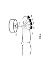
今、図1及び図2の参照がなされる。図1は、装置の概略図であり、一般に符号100で参照され、被験者のヘマトクリットをモニタリングするためのもので、開示される技術の実施形態に従って構築され、動作している。図2は、動作中における図1の装置の概略図である。装置100は、エミッタ102、複数の反射率検出器104l…104N、複数の透過率検出器124l…124N、コントローラ106、プロセッサ108、インターフェース110、ディスプレイ112、オペレータインターフェース114、身体接触手段116及び保持手段118を含む。コントローラ106は、エミッタ102、及びプロセッサ108に接続されている。インターフェース110は、検出器104l…104N及び検出器124l…124N及びプロセッサ108に接続されている。プロセッサ108は、さらにディスプレイ112及びオペレータインターフェース114に接続されている。
Reference is now made to FIGS. FIG. 1 is a schematic diagram of an apparatus, generally designated 100, for monitoring a subject's hematocrit, constructed and operating in accordance with an embodiment of the disclosed technology. FIG. 2 is a schematic diagram of the apparatus of FIG. 1 in operation. The
装置100は、コントローラモジュール128に接続されるセンサモジュール126を含む。センサモジュール126は、エミッタ102、反射率検出器104l…104N、透過率検出器124l…124N、身体接触手段116、及び保持手段118を含む。コントローラモジュール128は、コントローラ106、プロセッサ108、インターフェース110、ディスプレイ112、及びオペレータインターフェース114を含む。センサモジュール126及びコントローラモジュール128は、典型的に分離され、別個のユニット(例えばそれぞれ分離したハウジングに囲まれた)であり、これは、装置100の実施例を示し、装置100の様々な要素が異なって組織され得るということが認められる。
The
エミッタ102は、被験者の身体の部分120に光を放射する。身体の部分120は、図2において指として示されるが、身体の部分は、容易にアクセスでき、そして医療スタッフにとって放射光(放射された光)の適用が便利な体内のもう1つの範囲であり得るということが認められる。例えば、身体の部分120はまた、つま先、外側の耳、鼓膜、耳たぶ、口、目、手の他の範囲(例えば手のひら、指の間の皮膜)等であり得る。エミッタ102は、発光ダイオード(LED)、又はもう1つの種類の発光素子であり得る。エミッタ102は、導波管又は光ファイバのような光学要素を通じて、身体の部分120に光を導く。
The
反射率検出器104l…104Nは、エミッタ102の光学軸に関して可変な距離で配置される。検出器104l…104Nは、エミッタ102を囲む放射パターンで配置され、エミッタ102とともに単独のユニットに統合される(図2参照)。反射率検出器104l…104Nは、空間的に分解された光の反射率の測定技術(ここで以下に精緻化された)に従って、身体の部分120から反射される光を検出する。反射率検出器104l…104N及び透過率検出器124l…124Nは、動作可能な波長範囲(例えば、約380−980nm)で光を検出する。反射率検出器104l…104N及び透過率検出器124l…124Nは、所望の波長(例えば狭周波数帯センサ)だけで光を検出するように作動している。オプションとして、バンドパスフィルタが、反射率検出器104l…104N又は透過率検出器124l…124Nから所望の波長の外側に光をブロックするように使用され得る。反射率検出器104l…104N及び透過率検出器124l…124Nは、フォトダイオード、又は他の種類の光検出要素であり得る。反射率検出器104l…104Nは、図2に示されるように、別個の光検出要素のシリーズであり、光検出用度の連続配置であり得る。開示される技術のもう1つの実施形態に従って構築されそして作動している反射率検出器の連続配置の概略図である図3についての参照がなされる。それぞれの反射率検出器204l…204Nが、エミッタ202(図2のエミッタ102と類似)に関して放射パターンで配置される光検出要素の連続的な配列であるということは別にして、反射率検出器204l…204Nは、図2の反射率検出器104l…104Nと類似のものである。
The reflectance detectors 104 l ... 104 N are arranged at a variable distance with respect to the optical axis of the
プロセッサ108は、反射率検出器104l…104N又は透過率検出器124l…124Nから、インターフェース110を通じてデータを取得し、必要な計算及びデータ分析を実行する。インターフェース110は、ドライバ、アンプ、アナログ−デジタルコンバータのような、検出器からプロセッサ108へとデータを通過させるための関連する構成要素を含む。プロセッサ108は、ディスプレイ112に表示されるようにデータを送信する。ディスプレイは、例えば、装置100に組み込まれたマイクロディスプレイ、又は外部のコンピュータに関係する出力装置(例えばモニタ)であり得る。コントローラ106は、強度、波長、期間等のような、放射光の様々なパラメータを適合させるために使用され得る選択的な要素である。オペレータインターフェース114は、開示される技術を実行する者(例えば、医師、医療助手等)が、関係する基準(例えば、身体の部分の種類、被験者の病状等)に従って、放射光の関係するパラメータを選択することを可能にする選択的な要素である。
The
身体接触手段116は、オプションとして、光が放射される身体の部分120の範囲内に配置される。身体接触手段116は、例えば、エミッタ102と、一方の検出器1041…104Nと、他方の身体の部分120との間で、光インターフェースとして機能する光ファイバフェイスプレートである。身体接触手段116は、エミッタ102から身体の部分120に向かって、実質的に垂直な方向に光を導き、さらに、身体の部分120からの反射光(反射された光)を反射率検出器1041…104Nに向かって導く。身体接触手段116は、エミッタ102と検出器1041…104Nとの間の逆のクロストークを防止する。身体接触手段116は、さらに、身体の部分120との摩擦による可能な損傷からエミッタ102および検出器1041…104N(特にそれらのボンディングワイヤ)を保護することと同様に、エミッタ102及び検出器1041…104Nから身体の部分の組織を保護する保護層として機能する。身体接触手段116は、センサモジュール126のハウジング内に統合され得る。
The body contacting means 116 is optionally positioned within the
保持手段118は、開示される技術を実行する者が装置100を安定する方式で保持することを可能にし、正確かつ効率的に放射光を身体の部分120に導くことができるようにする。例えば、保持手段118は、バンドを取り付けるためにストラップ又は接着材料を含み得る。装置100は、オプションとして、例えば、バッテリ又は電圧主部を介して、様々な構成要素に電力を与えるための電源(図示せず)を含み得る。装置100はまた、オプションとして、データを保存する別個のメモリを含み得る。
The holding means 118 allows a person performing the disclosed technique to hold the
開示される技術、オペレーションの一実施形態によれば、エミッタ102は、光(図2の符号132参照)を等吸収波長で身体の部分120に放射する。生理的組織が、光を散乱し、吸収する混濁媒体であることから、あるフォトンは、組織内に吸収され、あるフォトンは散乱する(透過するか反射するかのどちらか)であろう。等吸収波長は、望ましくは、およそ803nm(例えば803±5nm)である。他の可能な等吸収波長は、約390nm、約422nm、約452nm、約500nm、約529nm、約545nm、約570nm及び約584nmである。
According to one embodiment of the disclosed technique and operation, the
放射光の大部分は、高い散乱度(生理的組織は低い吸収率を有する)により、身体の部分の組織に入射する表面に向かって拡散的に反射する。反射率検出器104l…104Nは、空間的に分解された光の反射率の測定技術に従って、拡散反射率(図2において符号134参照)を検出する。身体の部分120の血液成分の吸収係数及び削減散乱係数(例えばμa及びμs’)は、その後、空間的に分解された光の反射率の測定から引き出される。
Most of the emitted light reflects diffusely toward the surface incident on the tissue of the body part due to the high degree of scattering (physiological tissue has a low absorption rate). Reflectance detector 104 l ... 104 N, in accordance with measurement techniques of the reflectivity of the spatially separated light, to detect the diffuse reflectance (
望まれるパラメータを引き出すためのある技術は、組織の光学パラメータを拡散反射率測定に関係づける半解析モデルを採用する。Applied Optics, Vol.35, No.13, pp.2304-2314 (May, 1996)の、Keinle et al.に開示される、そのようなモデルの例は、以下の通りである。
ρ=光が組織に入射する点からの半径方向距離。
One technique for deriving the desired parameters employs a semi-analytical model that relates tissue optical parameters to diffuse reflectance measurements. An example of such a model disclosed in Keinle et al. In Applied Optics, Vol. 35, No. 13, pp. 2304-2314 (May, 1996) is as follows.
ρ = radial distance from the point where light enters the tissue.
理論的に、2つの点での測定R(ρ)は、吸収度係数及び削減散乱係数(μa及びμs’)を引き出すのに十分である。 Theoretically, the measurement R (ρ) at two points is sufficient to derive the absorption coefficient and the reduced scattering coefficient (μ a and μ s ′).
望まれるパラメータを引き出すためのもう1つの技術は、実験に基づくキャリブリレーションモデルを利用する。キャリブレーションモデルは、多項式回帰を使用するような、様々な光学的に明確な媒体についての反射率分布の実験的な観察の大きな連続を使用してつくられる。そのようなキャリブレーションモデルの例は、Applied Optics, Vol.40, No.7, pp.1155-1164 (March, 2001)の、Dam et al.に開示される。 Another technique for deriving the desired parameters utilizes an experimentally based calibration model. A calibration model is created using a large series of experimental observations of the reflectance distribution for various optically distinct media, such as using polynomial regression. An example of such a calibration model is disclosed in Dam et al. In Applied Optics, Vol. 40, No. 7, pp. 1155-1164 (March, 2001).
プロセッサ108は、検出器104l…104Nによって得られた連続測定値のシリーズに基づく引き出されたパラメータから時間導関数を計算する。装置100は、空間的に分解された光の反射率の測定を得るために十分ないくつかの検出器104l…104Nを含み得ることに留意する。その代わりに、装置100は、空間的に分解された光の反射率の測定を行うための複数のエミッタ及び単独の反射率検出器を含み得る。
The
開示される技術のもう1つの実施形態によれば、拡散反射率測定は、付加的な検証及び高められた精度を与えるために、拡散透過率測定に組み合わされる。反射率は、開示される技術のさらなる実施形態に従って動作するように構築される、拡散透過率測定に適合される、図1の装置の概略的例示である図4でつくられる。 According to another embodiment of the disclosed technique, diffuse reflectance measurements are combined with diffuse transmittance measurements to provide additional verification and increased accuracy. The reflectivity is produced in FIG. 4, which is a schematic illustration of the apparatus of FIG. 1, adapted for diffuse transmittance measurements, constructed to operate according to further embodiments of the disclosed technology.
ランベルト・ベールの原理は、統一的に吸収する媒体を通じて伝播する光の減衰を表す。ランベルト・ベールの原理の変形態様は、全減衰についての散乱現象の重大な貢献に占めることによって、生理的組織を通じた全減衰を記述するためのより正確なモデルを以下のように与え得る。
T=媒体の透過率;
Iout=媒体を通過する光の強度;
Iin=示される光の強度;
d =光が媒体を通過する距離;
F(μ,d)=μsとdの複合関数。
The Lambert-Beer principle represents the attenuation of light propagating through a uniformly absorbing medium. Variations on the Lambert-Beer principle can give a more accurate model for describing total attenuation through physiological tissue by accounting for a significant contribution of the scattering phenomenon to total attenuation as follows.
T = medium transmittance;
I out = intensity of light passing through the medium;
I in = the intensity of the indicated light;
d = distance that light travels through the medium;
F (μ, d) = complex function of μ s and d.
方程式(12)の対数をとり、それからdに関して微分することは、複合関数を消去し、以下の結果となる;
方程式(13)は、媒体を通じた全減衰を得る。 Equation (13) obtains the total attenuation through the medium.
図3に戻って参照すると、透過率検出器1241,1242,1243,1244は、身体の部分120を横切ってエミッタ102の位置から反対側に配置される(例えば、もし、エミッタ120が身体の部分120のすぐ上に配置される場合は、検出器1241,1242,1243,1244は、エミッタ102を横切って身体の部分120の直下に配置される)。検出器1241,1242,1243,1244は、様々な距離(d1,d2,d3,及びd4でそれぞれ参照される)で、それぞれエミッタ102に設けられる。検出器1241,1242,1243,および1244は、そのそれぞれの距離に従って、身体の部分120を通過する光を検出する。方程式(13)の距離導関数は、その後、減衰値(μ)を与えるために、検出器によって得られた測定に基づいて、(プロセッサ108によって)計算される。その結果、反射率測定及び透過率測定のための最適な適合(最小偏位)を与える値を決定することによって、単純な反復回帰アルゴリズムが、μa及びμs’の変数を引き出すために適用される。その技術分野において知られる他の数学的な技術もまた、反射率測定及び透過率測定の組合せに基づいて望まれる係数を引き出すために使用され得る。プロセッサ108は、その後、これらのパラメータから時間導関数(∂μa/∂t及び∂μs’/∂t)を計算する。例示の目的のためだけに4つの検出器が示され、そして、装置100は、透過率を検出するのに十分な数の検出器124Nを使用し得るということが認められる。一般に、単独の透過率検出器は、拡散反射率測定を得るのに十分であり、そして、付加的な透過率検出器は、測定結果の検証のために利用され得る。反射率測定及び透過率測定が、同時に実行され得る(例えば、反射率検出器104l…104Nが、空間的に分解された光の反射率の測定を実行し、かつ、透過率検出器124l…124Nが、拡散透過率測定を実行する)ということがさらに認められる。
Referring back to FIG. 3, the transmittance detectors 124 1 , 124 2 , 124 3 , 124 4 are positioned across the
開示される技術は、単独の波長だけを使用してヘマトクリットの測定を可能にする。これは、生きた生理的組織上への異なる波長の放射光が、その後の分析において、様々な歪につながり得ることから、有利である。それぞれの波長は、矛盾した組織の体積が検査されるという結果を引き起こす、異なる侵入深さを有し得る。さらに、それぞれの波長は、様々な組織成分に関する異なる依存性を有し得る。 The disclosed technique allows for hematocrit measurements using only a single wavelength. This is advantageous because different wavelengths of emitted light on living physiological tissue can lead to various distortions in subsequent analysis. Each wavelength may have a different penetration depth that results in inconsistent tissue volumes being examined. In addition, each wavelength can have different dependencies on various tissue components.
開示される技術のもう1つの実施形態によれば、ヘマトクリットは、1つの単独の等吸収波長でというよりはむしろ、2つの別個の波長での放射光によって測定され得る。この場合において、オキシヘモグロビン及びデオキシヘモグロビンの吸収度は、等しいという必要はなく、そして、方程式(3)は、方程式(9)に誘導され得ない。その結果、方程式(3)及び(4)を方程式(10)に代入して以下を得る:
方程式(14)は、2つの未知の変数を含む:SAT=酸素減衰レベル、及びHct=ヘマトクリット値。この方程式、又はもう1つの関係する方程式は、両方の変数を決定するために、2つの別個の波長(少なくとも1つが非等吸収である)で、その後解かれ得る。例えば、光は、約690nm及び約860nm(望ましくは、オキシヘモグロビン及びデオキシヘモグロビンの吸収度スペクトルが実質的に大きく離れている2つの波長)で放射され得る。同様に、測定結果に対して付加的な検証を提供するために、第3の波長が使用され得る。 Equation (14) includes two unknown variables: SAT = oxygen decay level and Hct = hematocrit value. This equation, or another related equation, can then be solved at two distinct wavelengths (at least one is unequal absorption) to determine both variables. For example, light can be emitted at about 690 nm and about 860 nm (desirably two wavelengths where the absorption spectra of oxyhemoglobin and deoxyhemoglobin are substantially separated). Similarly, a third wavelength can be used to provide additional verification for the measurement results.
開示される技術のさらなる実施形態によれば、ヘマトクリットは、これらの別個の方程式における3つの未知の変数を解くことによって、作動可能なスペクトル範囲内で、3つの別個の波長の光を放射することによって測定され得る。前述のように、作動可能なスペクトル範囲において、光の吸収は、原始的に、RBC内のヘモグロビンによる吸収に因り、そしてそれ故、μa,blood >> μa,plasmaである。 According to a further embodiment of the disclosed technique, hematocrit emits light of three distinct wavelengths within the operable spectral range by solving three unknown variables in these distinct equations. Can be measured. As mentioned above, in the operable spectral range, light absorption is primarily due to absorption by hemoglobin in the RBC and is therefore μ a, blood >> μ a, plasma .
従って、方程式(3)は以下のようになる:
方程式(15)を方程式(7)に代入して以下の結果を得る:
それ故、血液成分内の吸収係数の時間変化は、3つの変数:ヘマトクリット値(Hct)、酸素飽和レベル(SAT)、及び血液の相関濃度の時間変化(∂Cb/∂t)の関数である。方程式(15)、又は類似の関係する方程式は、これらの3つの未知のパラメータを決定するために、作動可能なスペクトル範囲内で、3つの異なる波長で解かれ得る。血液成分の吸収係数(μa,blood)の波長依存性が、波長に依存する酸素化された及び非酸素化されたRBCの吸収断面内(σox及びσdeox)に含まれる(そして、前述のように、Mie理論を用いて計算され得る)ということが留意される。このアプローチはまた、ヘマトクリット(又は他のパラメータ)を決定することにおいて、信頼性の低い測定であり得る血液成分の削減散乱係数(μs’)の使用を除外する。 Therefore, the time variation of the absorption coefficient in the blood component is a function of three variables: hematocrit value (Hct), oxygen saturation level (SAT), and time variation of blood correlation concentration (∂C b / ∂t). is there. Equation (15), or similar related equations, can be solved at three different wavelengths within the operable spectral range to determine these three unknown parameters. The wavelength dependence of the absorption coefficient (μ a, blood ) of the blood component is included in the absorption cross-sections (σ ox and σ deox ) of oxygenated and non-oxygenated RBCs depending on the wavelength (and Note that it can be calculated using Mie theory). This approach also precludes the use of blood component reduced scatter coefficients (μ s ′) in determining hematocrit (or other parameters), which can be an unreliable measurement.
開示される技術は、簡単な非侵襲的な光学測定に基づいて、被験者のヘマトクリット値の連続的なモニタリングをある期間にわたって許容する。これは、特に、様々な病状を診断し、又は処理することについて有益である。連続的なヘマトクリットのモニタリングのある有用なアプリケーションは、血液透析を経験している患者についての体液状態を最適化するためのものである。血液透析は、医療処置であり、一般に、老廃物(例えば、尿の毒、及び血漿内の過度の水)が血液外に濾過され、浄化された血液が定常回路内において身体に対して回復される、腎臓障害に苦しむ患者を処理するためのものである。患者の血漿補給率は、患者の身体が液体を、血管外スペースから血液内に戻すように移動させる率であり、透析濾過率は、この率に適合するものでなければならない。連続的にモニタリングすることによって、RBCの総数は、透析中に一定のままであり、Hctの増加が透析量の低下を示すことから、透析中のヘマトクリット、血管外スペース内の血液量の変化は検出され得る。透析濾過率は、その後、それに応じて設定され得る。液体を受けているか、又は血液交換を受けている出血患者はまた、同様な方式でモニタされる。ヘマトクリットのモニタリングはまた、貧血、脱水症、骨髄増殖性障害、慢性の閉塞性肺疾患及び他の病状のような、他の種類の診断及び処理の部分として使用され得る。ヘマトクリット測定はまた、血液又は血漿を提供するための適合性を決定するためのテストとして使用され得る。 The disclosed technology allows continuous monitoring of a subject's hematocrit value over a period of time based on simple non-invasive optical measurements. This is particularly beneficial for diagnosing or treating various medical conditions. One useful application of continuous hematocrit monitoring is for optimizing body fluid conditions for patients experiencing hemodialysis. Hemodialysis is a medical procedure and generally waste products (eg, urine toxins and excess water in plasma) are filtered out of the blood and the purified blood is restored to the body in a steady circuit. To treat patients suffering from kidney problems. The patient's plasma replenishment rate is the rate at which the patient's body moves fluid back from the extravascular space into the blood, and the diafiltration rate must be compatible with this rate. By continuously monitoring, the total number of RBCs remains constant during dialysis, and an increase in Hct indicates a decrease in dialysis volume, so the change in hematocrit during dialysis, blood volume in the extravascular space is Can be detected. The diafiltration rate can then be set accordingly. Bleeding patients who are receiving fluid or undergoing a blood exchange are also monitored in a similar manner. Hematocrit monitoring can also be used as part of other types of diagnosis and treatment, such as anemia, dehydration, myeloproliferative disorders, chronic obstructive pulmonary disease and other medical conditions. The hematocrit measurement can also be used as a test to determine suitability for providing blood or plasma.
今、開示される技術の実施形態に関して作動する被験者のヘマトクリットをモニタリングするための方法の概略図である図5に対する参照がなされる。工程152において、光は、オキシヘモグロビン及びデオキシヘモグロビンについて等吸収波長で、エミッタから少なくとも1つの血管を含む身体の部分に向けて放射される。図2を参照すると、エミッタ102は、等吸収波長(例えば、約803nm)で身体の部分120に向けて光を放射する。
Reference is now made to FIG. 5, which is a schematic diagram of a method for monitoring a subject's hematocrit operating in conjunction with an embodiment of the disclosed technology. In
工程154において、エミッタの光学軸に関して可変の距離で配置される複数の反射率検出器で、身体の部分で反射される光を捕捉することによって、空間的に分解された光の反射率の測定が得られる。図2を参照すると、反射率検出器104l…104Nは、エミッタ102の光学軸に関して可変な距離(例えば、放射パターンにおいて)で配置される。検出器104l…104Nは、一連の空間的に分解された光の反射率の測定を得ることで、身体の部分120から反射される光を検出する。
In
選択的な工程156において、身体の部分を横切ってエミッタの反対に配置される少なくとも1つの透過率検出器で、身体の部分を通過する光を捕捉することによって、透過率測定が得られる。図4を参照すると、透過率検出器1241、1242、1243、及び1244は、エミッタ102の光学軸に関して、各検出器が異なる距離で(それぞれd1,d2,d3,d4参照)配置される、身体の部分120の下側(例えば、身体の部分120を横切ってエミッタ102の位置から反対)に配置される。各検出器1241,1242,1243,1244は、それぞれの距離に従って、身体の部分120を通過する光を検出し、それによって、一連の拡散透過率測定を得る。身体の部分120を通じた全減衰(μ)は、その測定に基づいて計算される。
In
工程158において、身体の部分の血液成分の吸収係数(μa)及び削減散乱係数(μs’)は、その測定から引き出される。図1を参照すると、プロセッサ108は、空間的に分解された光の反射率の測定から、及び、オプションとして、同様に拡散反射率測定から、身体の部分120の血液成分の吸収係数及び削減散乱係数(μa 及びμs’)を計算する。パラメータは、半解析モデル又は実験に基づくキャリブレーションモデルを使用して、引き出され得る。反復回帰アルゴリズムが、反射率測定と透過率測定とを結合するために使用され得る。
In
工程160において、身体の部分の血液成分の吸収係数及び削減散乱係数における時間導関数が引き出される。図1を参照すると、プロセッサ108は、吸収係数および削減散乱係数の時間導関数(∂μ s '/∂t及び∂μ a /∂t)を計算する。
In
工程162において、身体の部分のヘマトクリット値は、時間導関数を用いて計算される。図1を参照すると、プロセッサ108は、方程式(11)又はもう1つの関係する方程式におけるHct変数を解くことによって、引き出された時間導関数に基づいてヘマトクリットを計算する。
In
開示される技術は、一般に、酸素飽和レベル、ヘモグロビン濃度、血液濃度の時間変化(∂Cb/∂t)、平均RBCサイズ及びサイズ分布等のような、ヘマトクリット値以外の他の血液パラメータを決定するのに適用され得るものである。今、開示される技術のもう1つの実施形態に従って作動する、被験者の血液に関する少なくとも1つのパラメータを測定するための方法の概略図である図6についての参照がなされる。 The disclosed techniques generally determine other blood parameters other than the hematocrit value, such as oxygen saturation level, hemoglobin concentration, blood concentration over time (∂C b / ∂t), average RBC size and size distribution, etc. It can be applied to do. Reference is now made to FIG. 6, which is a schematic diagram of a method for measuring at least one parameter relating to a subject's blood, operating in accordance with another embodiment of the disclosed technique.
工程172において、380−980nmのスペクトル範囲内の少なくとも1つの波長で、少なくとも1つのエミッタで、少なくとも1つの血管を含む身体の部分に向かって、光が放射される。工程174において、少なくとも1つの反射率検出器で、身体の部分によって反射される光を捕捉することによって、空間的に分解された光の反射率の測定が得られる。選択的な工程176において、少なくとも1つの透過率検出器で、身体の部分を通過する光を捕捉することによって、拡散反射率測定が得られる。
In
工程178において、少なくとも1つの波長での、身体の部分の血液成分の吸収係数(μa)と削減散乱係数(μs')の少なくとも1つが、測定から引き出される。工程180において、身体の部分の血液成分の吸収係数及び削減散乱係数の少なくとも1つにおける時間導関数が引き出される。工程182において、身体の部分の血液に関する少なくとも1つのパラメータが時間導関数を用いて計算される。
In
開示される技術が、特に以上に示され、かつ記述されたことに限定されないということが当業者に理解されるであろう。 It will be appreciated by persons skilled in the art that the disclosed technology is not limited to what has been particularly shown and described hereinabove.
Claims (15)
380−980nmのスペクトル範囲内において少なくとも1つの測定される波長で、少なくとも1つのエミッタで、少なくとも1つの血管を含む身体の部分に向かって、光を放射すること、
少なくとも1つの反射率検出器で、前記身体の部分によって反射された光を捕捉することにより、空間的に分解された光の反射率の測定を得ること、
前記測定から、少なくとも1つの前記測定される各波長で、前記身体の部分の血液成分の吸収係数(μa)及び削減散乱係数(μs’)のうちの少なくとも1つを引き出すこと、
前記吸収係数及び前記削減散乱係数のうちの少なくとも1つにおける時間導関数(∂μ s ’/∂t及び∂μ a /∂t)を引き出すこと、及び、
単独の波長での前記時間導関数の比率を用いて、少なくとも1つの前記測定される各波長について、前記パラメータを計算すること、
の工程を含む方法。 A method for measuring at least one parameter relating to a subject's blood, comprising:
Emitting light at at least one measured wavelength within the spectral range of 380-980 nm toward at least one emitter toward the body part containing at least one blood vessel;
Obtaining a spatially resolved light reflectance measurement by capturing light reflected by the body part with at least one reflectance detector;
Deriving from the measurement at least one of an absorption coefficient (μ a ) and a reduced scattering coefficient (μ s ′) of the blood component of the body part at at least one of the measured wavelengths;
The pulling out the absorption coefficient and in at least one time derivative of said reduced scattering coefficient (∂μ s' / ∂t and ∂μ a / ∂t), and,
Calculating the parameter for at least one of the measured wavelengths using the ratio of the time derivative at a single wavelength;
Comprising the steps of:
約390nm;
約422nm;
約452nm;
約500nm;
約529nm;
約545nm;
約570nm;
約584nm;及び
約803nm;
からなるリストから選択される請求項2に記載の方法。 The isosbestic wavelength is
About 390 nm;
About 422 nm;
About 452 nm;
About 500 nm;
About 529 nm;
About 545 nm;
About 570 nm;
About 584 nm; and about 803 nm;
The method of claim 2, wherein the method is selected from a list consisting of:
指;
つま先;
外側の耳;
鼓膜;
耳たぶ;
口;
目;
手のひら;及び
指の間の皮膜;
からなるリストから選択される請求項1の方法。 The body part is
finger;
Toes;
Outer ear;
eardrum;
Earlobe;
mouth;
Eye;
Palm; and skin between fingers;
The method of claim 1 selected from a list consisting of:
酸素飽和レベル;
ヘモグロビン濃度;
血中濃度の時間変化;
平均RBCサイズ;
平均RBCサイズ分布;
からなるリストから選択される請求項1に記載の方法。 The parameter is
Oxygen saturation level;
Hemoglobin concentration;
Change in blood concentration over time;
Average RBC size;
Average RBC size distribution;
The method of claim 1 selected from a list consisting of:
380−980nmのスペクトル範囲内で、少なくとも1つの測定される波長で、少なくとも1つの血管を含む身体の部分に向かって、光を放射するための少なくとも1つのエミッタ;
前記身体の部分によって反射される光を捕捉することにより、空間的に分解された光の反射率の測定を得るための少なくとも1つの反射率検出器;
及び、前記測定から、少なくとも1つの前記測定される各波長で、前記身体の部分の血液成分の吸収係数(μa)及び削減散乱係数(μs’)のうちの少なくとも1つを引き出し、前記吸収係数及び前記削減散乱係数のうちの少なくとも1つにおける時間導関数(∂μ s ’/∂t及び∂μ a /∂t)を引き出し、そして、単独の波長での前記時間導関数の比率を使用して、少なくとも1つの前記測定される各波長について、前記パラメータを計算するためのプロセッサ、
を含む装置。 An apparatus for measuring at least one parameter related to a subject's blood,
At least one emitter for emitting light in the spectral range of 380-980 nm toward the body part containing at least one blood vessel at at least one measured wavelength;
At least one reflectance detector for obtaining a reflectance measurement of spatially resolved light by capturing light reflected by the body part;
And extracting from the measurement at least one of an absorption coefficient (μ a ) and a reduced scattering coefficient (μ s ′) of the blood component of the body part at at least one of the measured wavelengths, in at least one of the absorption coefficient and the reduced scattering coefficient time pull the derivative (∂μ s' / ∂t and ∂μ a / ∂t), and the ratio of the time derivative of the wavelength alone A processor for calculating the parameter for at least one of the measured wavelengths using:
Including the device.
指;
つま先;
外側の耳;
鼓膜;
耳たぶ;
口;
目;
手のひら;及び
指の間の皮膜;
からなるリストから選択される請求項9に記載の装置。 The body part is
finger;
Toes;
Outer ear;
eardrum;
Earlobe;
mouth;
Eye;
Palm; and skin between fingers;
The apparatus of claim 9 selected from a list consisting of:
酸素飽和レベル;
ヘモグロビン濃度;
血中濃度の時間変化;
平均RBCサイズ;
平均RBCサイズ分布;
からなるリストから選択される請求項9に記載の装置。 The parameter is
Oxygen saturation level;
Hemoglobin concentration;
Change in blood concentration over time;
Average RBC size;
Average RBC size distribution;
The apparatus of claim 9 selected from a list consisting of:
約390nm;
約422nm;
約452nm;
約500nm;
約529nm;
約545nm;
約570nm;
約584nm;及び
約803nm;
からなるリストから選択される請求項13に記載の装置。 The isosbestic wavelength is
About 390 nm;
About 422 nm;
About 452 nm;
About 500 nm;
About 529 nm;
About 545 nm;
About 570 nm;
About 584 nm; and about 803 nm;
The apparatus of claim 13 selected from a list consisting of:
前記計算されるパラメータを表示するために、前記プロセッサに接続されるディスプレイ;
前記身体の部分に設けられ、前記身体の部分を保護し、前記放射された光及び前記反射された光を実質的に垂直な方向に導くための身体接触手段;及び、
前記装置を安定した方式で保持し、前記放射された光を正確にかつ効率的に前記身体の部分に導くための保持手段;
からなるグループから選択される少なくとも1つをさらに含む請求項9に記載の装置。 A controller connected to the emitter and the reflectance detector to control operation of the emitter and the reflectance detector;
A display connected to the processor to display the calculated parameters;
Provided in a portion of the body, to protect portions of the body, said body contact means for directing the emitted light and the reflected light in a direction substantially perpendicular; and,
Holding means for guiding the part of the stable and kept in a manner, accurately and efficiently the body of the emitted light of the device;
The apparatus of claim 9, further comprising at least one selected from the group consisting of:
Applications Claiming Priority (3)
| Application Number | Priority Date | Filing Date | Title |
|---|---|---|---|
| US92011607P | 2007-03-27 | 2007-03-27 | |
| US60/920,116 | 2007-03-27 | ||
| PCT/IL2008/000413 WO2008117286A2 (en) | 2007-03-27 | 2008-03-26 | Device and method for monitoring blood parameters |
Publications (3)
| Publication Number | Publication Date |
|---|---|
| JP2010522603A JP2010522603A (en) | 2010-07-08 |
| JP2010522603A5 JP2010522603A5 (en) | 2011-05-19 |
| JP5568461B2 true JP5568461B2 (en) | 2014-08-06 |
Family
ID=39789121
Family Applications (1)
| Application Number | Title | Priority Date | Filing Date |
|---|---|---|---|
| JP2010500432A Expired - Fee Related JP5568461B2 (en) | 2007-03-27 | 2008-03-26 | Apparatus and method for monitoring blood parameters |
Country Status (4)
| Country | Link |
|---|---|
| US (1) | US8017407B2 (en) |
| EP (1) | EP2129288B1 (en) |
| JP (1) | JP5568461B2 (en) |
| WO (1) | WO2008117286A2 (en) |
Families Citing this family (14)
| Publication number | Priority date | Publication date | Assignee | Title |
|---|---|---|---|---|
| US8781546B2 (en) * | 2008-04-11 | 2014-07-15 | Covidien Lp | System and method for differentiating between tissue-specific and systemic causes of changes in oxygen saturation in tissue and organs |
| WO2011068998A2 (en) * | 2009-12-02 | 2011-06-09 | Duke University | Systems and methods for determining hemoglobin concentration utilizing diffuse reflectance at isosbestic wavelengths |
| US9002655B2 (en) | 2010-05-03 | 2015-04-07 | Gambro Lundia Ab | Medical apparatus for extracorporeal blood treatment and method for determining a blood parameter value in a medical apparatus thereof |
| WO2012055047A1 (en) * | 2010-10-29 | 2012-05-03 | Nir Science Corporation | Method and apparatus for analyte detection |
| US20120253149A1 (en) * | 2011-03-30 | 2012-10-04 | Steuer Robert | Method and apparatus for non-invasive photometric blood constituent diagnosis |
| US10092226B2 (en) * | 2011-12-23 | 2018-10-09 | General Electric Company | Method, arrangement, sensor, and computer program product for non-invasively measuring hemoglobin concentrations in blood |
| US20160113530A1 (en) * | 2014-10-23 | 2016-04-28 | Samsung Electronics Co., Ltd. | Method and apparatus for acquiring biological information, and wrist watch-type terminal using the same |
| NZ773815A (en) * | 2015-03-16 | 2022-07-01 | Magic Leap Inc | Methods and systems for diagnosing and treating health ailments |
| CN108135544A (en) * | 2015-09-23 | 2018-06-08 | 皇家飞利浦有限公司 | Polychrome pulse oximetry |
| US10753862B2 (en) * | 2016-02-16 | 2020-08-25 | Stichting Het Nederlands Kanker Instituut-Antoni van Leeuwenhoek Ziekenhuis | Method, apparatus and software for detection and localization of hidden defects in optically diffuse media |
| CN106940290A (en) * | 2017-01-22 | 2017-07-11 | 广州地理研究所 | Reservoir water body ammonia-nitrogen content evaluation method based on high-spectral data |
| EP3843632A4 (en) * | 2018-08-29 | 2021-11-24 | Tel Hashomer Medical Research Infrastructure And Services Ltd. | System and method for determining oxygenated-blood content of biological tissue |
| US11744930B2 (en) * | 2020-04-06 | 2023-09-05 | Fresenius Medical Care Holdings, Inc. | Intradialytic monitoring of hemodynamic status based on detection of oxygen signature phase shift |
| WO2022061262A1 (en) * | 2020-09-21 | 2022-03-24 | PAVmed Inc. | Systems and methods for non-invasive solute measurement |
Family Cites Families (14)
| Publication number | Priority date | Publication date | Assignee | Title |
|---|---|---|---|---|
| JPS5725217B2 (en) * | 1974-10-14 | 1982-05-28 | ||
| US4142101B1 (en) | 1977-07-20 | 1991-02-19 | Low intensity x-ray and gamma-ray imaging device | |
| US6681128B2 (en) | 1990-10-06 | 2004-01-20 | Hema Metrics, Inc. | System for noninvasive hematocrit monitoring |
| WO1999039631A1 (en) | 1998-02-05 | 1999-08-12 | In-Line Diagnostics Corporation | Method and apparatus for non-invasive blood constituent monitoring |
| US6662031B1 (en) | 1998-05-18 | 2003-12-09 | Abbott Laboratoies | Method and device for the noninvasive determination of hemoglobin and hematocrit |
| JP2000155090A (en) * | 1998-11-20 | 2000-06-06 | Fuji Photo Film Co Ltd | Imaging device for blood vessel |
| DE19921928C2 (en) * | 1999-05-12 | 2002-11-14 | Bosch Gmbh Robert | Electric device |
| US6587704B1 (en) | 1999-06-16 | 2003-07-01 | Orsense Ltd. | Method for non-invasive optical measurements of blood parameters |
| US6213952B1 (en) | 1999-09-28 | 2001-04-10 | Orsense Ltd. | Optical device for non-invasive measurement of blood related signals utilizing a finger holder |
| US6434408B1 (en) * | 2000-09-29 | 2002-08-13 | Datex-Ohmeda, Inc. | Pulse oximetry method and system with improved motion correction |
| US6505060B1 (en) | 2000-09-29 | 2003-01-07 | Datex-Ohmeda, Inc. | Method and apparatus for determining pulse oximetry differential values |
| US6660995B1 (en) * | 2001-06-25 | 2003-12-09 | Regents Of The University Of California | Particle size analysis in a turbid media with a single-fiber, optical probe while using a visible spectrometer |
| DK1675501T3 (en) * | 2003-09-12 | 2013-11-18 | Or Nim Medical Ltd | Non-invasive optical monitoring of an area of interest |
| EP3505052A1 (en) * | 2005-04-25 | 2019-07-03 | University of Massachusetts | Systems and methods for correcting optical reflectance measurements |
-
2008
- 2008-03-26 EP EP08720038.2A patent/EP2129288B1/en not_active Not-in-force
- 2008-03-26 JP JP2010500432A patent/JP5568461B2/en not_active Expired - Fee Related
- 2008-03-26 US US12/593,246 patent/US8017407B2/en not_active Expired - Fee Related
- 2008-03-26 WO PCT/IL2008/000413 patent/WO2008117286A2/en active Search and Examination
Also Published As
| Publication number | Publication date |
|---|---|
| EP2129288A2 (en) | 2009-12-09 |
| WO2008117286A3 (en) | 2010-02-25 |
| JP2010522603A (en) | 2010-07-08 |
| US8017407B2 (en) | 2011-09-13 |
| EP2129288A4 (en) | 2013-11-06 |
| EP2129288B1 (en) | 2016-12-21 |
| US20100076281A1 (en) | 2010-03-25 |
| WO2008117286A2 (en) | 2008-10-02 |
Similar Documents
| Publication | Publication Date | Title |
|---|---|---|
| JP5568461B2 (en) | Apparatus and method for monitoring blood parameters | |
| US10463286B2 (en) | Determination of tissue oxygenation in vivo | |
| JP4176480B2 (en) | Method and apparatus for improving the accuracy of non-invasive hematocrit measurements | |
| US10912504B2 (en) | Near-infrared spectroscopy and diffuse correlation spectroscopy device and methods | |
| JP4465271B2 (en) | Apparatus for noninvasively determining blood oxygen saturation in a target tissue | |
| JP3625475B2 (en) | Non-intrusive system for monitoring hematocrit values | |
| JP4903980B2 (en) | Pulse oximeter and operation method thereof | |
| US6421549B1 (en) | Adaptive calibration pulsed oximetry method and device | |
| EP2503935B1 (en) | Apparatus for spectrophotometric blood oxygenation monitoring of organs in the body | |
| US20080004513A1 (en) | VCSEL Tissue Spectrometer | |
| Paul et al. | Design and development of non invasive glucose measurement system | |
| JP2007509718A (en) | Devices and methods for monitoring body fluid and electrolyte disorders | |
| US20210052164A1 (en) | Sensor for tissue measurements | |
| JPH11244267A (en) | Blood component concentration measuring device | |
| US10925525B2 (en) | Combined pulse oximetry and diffusing wave spectroscopy system and control method therefor | |
| JP2015506197A (en) | Method, assembly device, sensor, and computer program product for non-invasive measurement of hemoglobin concentration in blood | |
| Reuss et al. | The pulse in reflectance pulse oximetry: modeling and experimental studies | |
| US20150217056A1 (en) | Therapy systems and methods utilizing tissue oxygenation detection | |
| McMurdy et al. | Photonics‐based In Vivo total hemoglobin monitoring and clinical relevance | |
| Patil et al. | Methods and devices to determine hemoglobin non invasively: A review | |
| Kraitl et al. | Optical non-invasive methods for characterization of the human health status | |
| Kraitl et al. | Analysis of time series for non-invasive characterization of blood components and circulation patterns | |
| Lu et al. | Portable near-infrared spectroscopy for detecting peripheral arterial occlusion | |
| IL201199A (en) | Method for monitoring blood parameters | |
| Cheang | Feasibility of non-contact photoplethysmography |
Legal Events
| Date | Code | Title | Description |
|---|---|---|---|
| A521 | Request for written amendment filed |
Free format text: JAPANESE INTERMEDIATE CODE: A523 Effective date: 20110325 |
|
| A621 | Written request for application examination |
Free format text: JAPANESE INTERMEDIATE CODE: A621 Effective date: 20110325 |
|
| A977 | Report on retrieval |
Free format text: JAPANESE INTERMEDIATE CODE: A971007 Effective date: 20120921 |
|
| A131 | Notification of reasons for refusal |
Free format text: JAPANESE INTERMEDIATE CODE: A131 Effective date: 20121005 |
|
| A601 | Written request for extension of time |
Free format text: JAPANESE INTERMEDIATE CODE: A601 Effective date: 20121226 |
|
| A602 | Written permission of extension of time |
Free format text: JAPANESE INTERMEDIATE CODE: A602 Effective date: 20130108 |
|
| A601 | Written request for extension of time |
Free format text: JAPANESE INTERMEDIATE CODE: A601 Effective date: 20130204 |
|
| A602 | Written permission of extension of time |
Free format text: JAPANESE INTERMEDIATE CODE: A602 Effective date: 20130212 |
|
| A601 | Written request for extension of time |
Free format text: JAPANESE INTERMEDIATE CODE: A601 Effective date: 20130226 |
|
| A602 | Written permission of extension of time |
Free format text: JAPANESE INTERMEDIATE CODE: A602 Effective date: 20130305 |
|
| A521 | Request for written amendment filed |
Free format text: JAPANESE INTERMEDIATE CODE: A523 Effective date: 20130403 |
|
| RD04 | Notification of resignation of power of attorney |
Free format text: JAPANESE INTERMEDIATE CODE: A7424 Effective date: 20130730 |
|
| A131 | Notification of reasons for refusal |
Free format text: JAPANESE INTERMEDIATE CODE: A131 Effective date: 20130906 |
|
| TRDD | Decision of grant or rejection written | ||
| A01 | Written decision to grant a patent or to grant a registration (utility model) |
Free format text: JAPANESE INTERMEDIATE CODE: A01 Effective date: 20140606 |
|
| A61 | First payment of annual fees (during grant procedure) |
Free format text: JAPANESE INTERMEDIATE CODE: A61 Effective date: 20140623 |
|
| R150 | Certificate of patent or registration of utility model |
Ref document number: 5568461 Country of ref document: JP Free format text: JAPANESE INTERMEDIATE CODE: R150 |
|
| LAPS | Cancellation because of no payment of annual fees |