JP5523345B2 - Methods and compositions targeting K63-linked polyubiquitin - Google Patents
Methods and compositions targeting K63-linked polyubiquitin Download PDFInfo
- Publication number
- JP5523345B2 JP5523345B2 JP2010543287A JP2010543287A JP5523345B2 JP 5523345 B2 JP5523345 B2 JP 5523345B2 JP 2010543287 A JP2010543287 A JP 2010543287A JP 2010543287 A JP2010543287 A JP 2010543287A JP 5523345 B2 JP5523345 B2 JP 5523345B2
- Authority
- JP
- Japan
- Prior art keywords
- antibody
- antigen
- hvr
- amino acid
- binding fragment
- Prior art date
- Legal status (The legal status is an assumption and is not a legal conclusion. Google has not performed a legal analysis and makes no representation as to the accuracy of the status listed.)
- Active
Links
- 108010068086 Polyubiquitin Proteins 0.000 title claims description 187
- 102100037935 Polyubiquitin-C Human genes 0.000 title claims description 187
- 238000000034 method Methods 0.000 title claims description 137
- 239000000203 mixture Substances 0.000 title description 28
- 230000008685 targeting Effects 0.000 title description 3
- 230000027455 binding Effects 0.000 claims description 230
- 238000009739 binding Methods 0.000 claims description 228
- 239000000427 antigen Substances 0.000 claims description 219
- 102000036639 antigens Human genes 0.000 claims description 216
- 108091007433 antigens Proteins 0.000 claims description 216
- 239000012634 fragment Substances 0.000 claims description 171
- 108090000623 proteins and genes Proteins 0.000 claims description 154
- 102000004169 proteins and genes Human genes 0.000 claims description 102
- 125000003275 alpha amino acid group Chemical group 0.000 claims description 87
- 239000013598 vector Substances 0.000 claims description 59
- 230000000694 effects Effects 0.000 claims description 41
- 150000007523 nucleic acids Chemical class 0.000 claims description 24
- 102000039446 nucleic acids Human genes 0.000 claims description 23
- 108020004707 nucleic acids Proteins 0.000 claims description 23
- 238000004519 manufacturing process Methods 0.000 claims description 22
- 230000001404 mediated effect Effects 0.000 claims description 14
- 230000001965 increasing effect Effects 0.000 claims description 11
- 230000019491 signal transduction Effects 0.000 claims description 8
- 230000000890 antigenic effect Effects 0.000 claims description 6
- 238000012258 culturing Methods 0.000 claims description 5
- 230000015556 catabolic process Effects 0.000 claims description 4
- 238000006731 degradation reaction Methods 0.000 claims description 4
- 210000004027 cell Anatomy 0.000 description 150
- 235000001014 amino acid Nutrition 0.000 description 129
- 229940024606 amino acid Drugs 0.000 description 122
- 150000001413 amino acids Chemical class 0.000 description 111
- 241000282414 Homo sapiens Species 0.000 description 108
- 235000018102 proteins Nutrition 0.000 description 74
- 102000044159 Ubiquitin Human genes 0.000 description 72
- 108090000848 Ubiquitin Proteins 0.000 description 72
- 108090000765 processed proteins & peptides Proteins 0.000 description 68
- 102000004196 processed proteins & peptides Human genes 0.000 description 62
- 229920001184 polypeptide Polymers 0.000 description 57
- 208000037265 diseases, disorders, signs and symptoms Diseases 0.000 description 42
- 125000005647 linker group Chemical group 0.000 description 42
- -1 glycine; amino acid Chemical class 0.000 description 41
- 239000003814 drug Substances 0.000 description 40
- 229940079593 drug Drugs 0.000 description 39
- 108020004414 DNA Proteins 0.000 description 38
- 102000008394 Immunoglobulin Fragments Human genes 0.000 description 36
- 108010021625 Immunoglobulin Fragments Proteins 0.000 description 36
- 108060003951 Immunoglobulin Proteins 0.000 description 35
- 102000018358 immunoglobulin Human genes 0.000 description 35
- 201000010099 disease Diseases 0.000 description 33
- KDXKERNSBIXSRK-UHFFFAOYSA-N Lysine Natural products NCCCCC(N)C(O)=O KDXKERNSBIXSRK-UHFFFAOYSA-N 0.000 description 32
- 235000018977 lysine Nutrition 0.000 description 30
- 206010028980 Neoplasm Diseases 0.000 description 29
- 230000014509 gene expression Effects 0.000 description 28
- 239000004472 Lysine Substances 0.000 description 26
- MTCFGRXMJLQNBG-UHFFFAOYSA-N Serine Natural products OCC(N)C(O)=O MTCFGRXMJLQNBG-UHFFFAOYSA-N 0.000 description 24
- 239000000562 conjugate Substances 0.000 description 24
- 235000004400 serine Nutrition 0.000 description 24
- 238000006467 substitution reaction Methods 0.000 description 22
- OUYCCCASQSFEME-UHFFFAOYSA-N tyrosine Natural products OC(=O)C(N)CC1=CC=C(O)C=C1 OUYCCCASQSFEME-UHFFFAOYSA-N 0.000 description 22
- 235000002374 tyrosine Nutrition 0.000 description 22
- 241000588724 Escherichia coli Species 0.000 description 21
- MTCFGRXMJLQNBG-REOHCLBHSA-N (2S)-2-Amino-3-hydroxypropansäure Chemical compound OC[C@H](N)C(O)=O MTCFGRXMJLQNBG-REOHCLBHSA-N 0.000 description 19
- KDXKERNSBIXSRK-YFKPBYRVSA-N L-lysine Chemical compound NCCCC[C@H](N)C(O)=O KDXKERNSBIXSRK-YFKPBYRVSA-N 0.000 description 19
- 108010076504 Protein Sorting Signals Proteins 0.000 description 19
- 201000011510 cancer Diseases 0.000 description 19
- 125000003729 nucleotide group Chemical group 0.000 description 19
- 102000040430 polynucleotide Human genes 0.000 description 19
- 108091033319 polynucleotide Proteins 0.000 description 19
- 239000002157 polynucleotide Substances 0.000 description 19
- 108091007491 NSP3 Papain-like protease domains Proteins 0.000 description 18
- FAPWRFPIFSIZLT-UHFFFAOYSA-M Sodium chloride Chemical compound [Na+].[Cl-] FAPWRFPIFSIZLT-UHFFFAOYSA-M 0.000 description 18
- 239000002253 acid Substances 0.000 description 18
- 235000004279 alanine Nutrition 0.000 description 18
- 238000001727 in vivo Methods 0.000 description 18
- QNAYBMKLOCPYGJ-REOHCLBHSA-N L-alanine Chemical compound C[C@H](N)C(O)=O QNAYBMKLOCPYGJ-REOHCLBHSA-N 0.000 description 17
- WHUUTDBJXJRKMK-VKHMYHEASA-N L-glutamic acid Chemical compound OC(=O)[C@@H](N)CCC(O)=O WHUUTDBJXJRKMK-VKHMYHEASA-N 0.000 description 17
- AYFVYJQAPQTCCC-GBXIJSLDSA-N L-threonine Chemical compound C[C@@H](O)[C@H](N)C(O)=O AYFVYJQAPQTCCC-GBXIJSLDSA-N 0.000 description 17
- 238000003556 assay Methods 0.000 description 17
- 229940127089 cytotoxic agent Drugs 0.000 description 17
- 230000006870 function Effects 0.000 description 17
- 230000037361 pathway Effects 0.000 description 17
- 238000003752 polymerase chain reaction Methods 0.000 description 17
- OUYCCCASQSFEME-QMMMGPOBSA-N L-tyrosine Chemical compound OC(=O)[C@@H](N)CC1=CC=C(O)C=C1 OUYCCCASQSFEME-QMMMGPOBSA-N 0.000 description 16
- 241001465754 Metazoa Species 0.000 description 16
- 230000004071 biological effect Effects 0.000 description 16
- 239000003153 chemical reaction reagent Substances 0.000 description 16
- 239000013604 expression vector Substances 0.000 description 16
- 229960005558 mertansine Drugs 0.000 description 16
- 239000002773 nucleotide Substances 0.000 description 16
- 208000011580 syndromic disease Diseases 0.000 description 16
- 229940049595 antibody-drug conjugate Drugs 0.000 description 15
- 238000006243 chemical reaction Methods 0.000 description 15
- 230000035772 mutation Effects 0.000 description 15
- 241000894007 species Species 0.000 description 15
- 239000003053 toxin Substances 0.000 description 15
- 231100000765 toxin Toxicity 0.000 description 15
- 108700012359 toxins Proteins 0.000 description 15
- 108010047041 Complementarity Determining Regions Proteins 0.000 description 14
- COLNVLDHVKWLRT-QMMMGPOBSA-N L-phenylalanine Chemical compound OC(=O)[C@@H](N)CC1=CC=CC=C1 COLNVLDHVKWLRT-QMMMGPOBSA-N 0.000 description 14
- 239000002953 phosphate buffered saline Substances 0.000 description 14
- 238000002360 preparation method Methods 0.000 description 14
- 239000000611 antibody drug conjugate Substances 0.000 description 13
- 238000000338 in vitro Methods 0.000 description 13
- 239000002609 medium Substances 0.000 description 13
- 230000004048 modification Effects 0.000 description 13
- 238000012986 modification Methods 0.000 description 13
- 229920000642 polymer Polymers 0.000 description 13
- 238000011282 treatment Methods 0.000 description 13
- 125000001493 tyrosinyl group Chemical group [H]OC1=C([H])C([H])=C(C([H])=C1[H])C([H])([H])C([H])(N([H])[H])C(*)=O 0.000 description 13
- CKLJMWTZIZZHCS-REOHCLBHSA-N L-aspartic acid Chemical compound OC(=O)[C@@H](N)CC(O)=O CKLJMWTZIZZHCS-REOHCLBHSA-N 0.000 description 12
- ROHFNLRQFUQHCH-YFKPBYRVSA-N L-leucine Chemical compound CC(C)C[C@H](N)C(O)=O ROHFNLRQFUQHCH-YFKPBYRVSA-N 0.000 description 12
- ROHFNLRQFUQHCH-UHFFFAOYSA-N Leucine Natural products CC(C)CC(N)C(O)=O ROHFNLRQFUQHCH-UHFFFAOYSA-N 0.000 description 12
- 239000002246 antineoplastic agent Substances 0.000 description 12
- 230000001580 bacterial effect Effects 0.000 description 12
- 230000021615 conjugation Effects 0.000 description 12
- 235000005772 leucine Nutrition 0.000 description 12
- 125000003588 lysine group Chemical group [H]N([H])C([H])([H])C([H])([H])C([H])([H])C([H])([H])C([H])(N([H])[H])C(*)=O 0.000 description 12
- 238000002823 phage display Methods 0.000 description 12
- 239000013615 primer Substances 0.000 description 12
- 230000015572 biosynthetic process Effects 0.000 description 11
- 229930195731 calicheamicin Natural products 0.000 description 11
- 239000002254 cytotoxic agent Substances 0.000 description 11
- 231100000599 cytotoxic agent Toxicity 0.000 description 11
- 238000010494 dissociation reaction Methods 0.000 description 11
- 230000005593 dissociations Effects 0.000 description 11
- 230000016784 immunoglobulin production Effects 0.000 description 11
- ANZJBCHSOXCCRQ-FKUXLPTCSA-N mertansine Chemical compound CO[C@@H]([C@@]1(O)C[C@H](OC(=O)N1)[C@@H](C)[C@@H]1O[C@@]1(C)[C@@H](OC(=O)[C@H](C)N(C)C(=O)CCS)CC(=O)N1C)\C=C\C=C(C)\CC2=CC(OC)=C(Cl)C1=C2 ANZJBCHSOXCCRQ-FKUXLPTCSA-N 0.000 description 11
- COLNVLDHVKWLRT-UHFFFAOYSA-N phenylalanine Natural products OC(=O)C(N)CC1=CC=CC=C1 COLNVLDHVKWLRT-UHFFFAOYSA-N 0.000 description 11
- 235000008729 phenylalanine Nutrition 0.000 description 11
- 238000000746 purification Methods 0.000 description 11
- HXCHCVDVKSCDHU-PJKCJEBCSA-N s-[(2r,3s,4s,6s)-6-[[(2r,3s,4s,5r,6r)-5-[(2s,4s,5s)-5-(ethylamino)-4-methoxyoxan-2-yl]oxy-4-hydroxy-6-[[(2s,5z,9r,13e)-9-hydroxy-12-(methoxycarbonylamino)-13-[2-(methyltrisulfanyl)ethylidene]-11-oxo-2-bicyclo[7.3.1]trideca-1(12),5-dien-3,7-diynyl]oxy]-2-m Chemical compound C1[C@H](OC)[C@@H](NCC)CO[C@H]1O[C@H]1[C@H](O[C@@H]2C\3=C(NC(=O)OC)C(=O)C[C@@](C/3=C/CSSSC)(O)C#C\C=C/C#C2)O[C@H](C)[C@@H](NO[C@@H]2O[C@H](C)[C@@H](SC(=O)C=3C(=C(OC)C(O[C@H]4[C@@H]([C@H](OC)[C@@H](O)[C@H](C)O4)O)=C(I)C=3C)OC)[C@@H](O)C2)[C@@H]1O HXCHCVDVKSCDHU-PJKCJEBCSA-N 0.000 description 11
- 239000000523 sample Substances 0.000 description 11
- JJAHTWIKCUJRDK-UHFFFAOYSA-N succinimidyl 4-(N-maleimidomethyl)cyclohexane-1-carboxylate Chemical compound C1CC(CN2C(C=CC2=O)=O)CCC1C(=O)ON1C(=O)CCC1=O JJAHTWIKCUJRDK-UHFFFAOYSA-N 0.000 description 11
- 230000001225 therapeutic effect Effects 0.000 description 11
- NFGXHKASABOEEW-UHFFFAOYSA-N 1-methylethyl 11-methoxy-3,7,11-trimethyl-2,4-dodecadienoate Chemical compound COC(C)(C)CCCC(C)CC=CC(C)=CC(=O)OC(C)C NFGXHKASABOEEW-UHFFFAOYSA-N 0.000 description 10
- AOJJSUZBOXZQNB-TZSSRYMLSA-N Doxorubicin Chemical compound O([C@H]1C[C@@](O)(CC=2C(O)=C3C(=O)C=4C=CC=C(C=4C(=O)C3=C(O)C=21)OC)C(=O)CO)[C@H]1C[C@H](N)[C@H](O)[C@H](C)O1 AOJJSUZBOXZQNB-TZSSRYMLSA-N 0.000 description 10
- 102000004190 Enzymes Human genes 0.000 description 10
- 108090000790 Enzymes Proteins 0.000 description 10
- 102000035195 Peptidases Human genes 0.000 description 10
- 108091005804 Peptidases Proteins 0.000 description 10
- 239000002202 Polyethylene glycol Substances 0.000 description 10
- AYFVYJQAPQTCCC-UHFFFAOYSA-N Threonine Natural products CC(O)C(N)C(O)=O AYFVYJQAPQTCCC-UHFFFAOYSA-N 0.000 description 10
- 239000004473 Threonine Substances 0.000 description 10
- 150000001875 compounds Chemical class 0.000 description 10
- 229940088598 enzyme Drugs 0.000 description 10
- 230000004927 fusion Effects 0.000 description 10
- 229940072221 immunoglobulins Drugs 0.000 description 10
- 238000003780 insertion Methods 0.000 description 10
- 230000037431 insertion Effects 0.000 description 10
- 238000012216 screening Methods 0.000 description 10
- 125000003607 serino group Chemical group [H]N([H])[C@]([H])(C(=O)[*])C(O[H])([H])[H] 0.000 description 10
- 239000007787 solid Substances 0.000 description 10
- 239000000126 substance Substances 0.000 description 10
- 235000008521 threonine Nutrition 0.000 description 10
- QWPXBEHQFHACTK-KZVYIGENSA-N (10e,12e)-86-chloro-12,14,4-trihydroxy-85,14-dimethoxy-33,2,7,10-tetramethyl-15,16-dihydro-14h-7-aza-1(6,4)-oxazina-3(2,3)-oxirana-8(1,3)-benzenacyclotetradecaphane-10,12-dien-6-one Chemical compound CN1C(=O)CC(O)C2(C)OC2C(C)C(OC(=O)N2)CC2(O)C(OC)\C=C\C=C(C)\CC2=CC(OC)=C(Cl)C1=C2 QWPXBEHQFHACTK-KZVYIGENSA-N 0.000 description 9
- 241000894006 Bacteria Species 0.000 description 9
- IAZDPXIOMUYVGZ-UHFFFAOYSA-N Dimethylsulphoxide Chemical compound CS(C)=O IAZDPXIOMUYVGZ-UHFFFAOYSA-N 0.000 description 9
- DHMQDGOQFOQNFH-UHFFFAOYSA-N Glycine Chemical compound NCC(O)=O DHMQDGOQFOQNFH-UHFFFAOYSA-N 0.000 description 9
- 241000124008 Mammalia Species 0.000 description 9
- KZSNJWFQEVHDMF-UHFFFAOYSA-N Valine Natural products CC(C)C(N)C(O)=O KZSNJWFQEVHDMF-UHFFFAOYSA-N 0.000 description 9
- 150000001299 aldehydes Chemical class 0.000 description 9
- 108010044540 auristatin Proteins 0.000 description 9
- 230000000875 corresponding effect Effects 0.000 description 9
- 238000005516 engineering process Methods 0.000 description 9
- 238000000855 fermentation Methods 0.000 description 9
- 230000004151 fermentation Effects 0.000 description 9
- 210000004408 hybridoma Anatomy 0.000 description 9
- 125000002887 hydroxy group Chemical group [H]O* 0.000 description 9
- 229940127121 immunoconjugate Drugs 0.000 description 9
- 230000000269 nucleophilic effect Effects 0.000 description 9
- 230000010076 replication Effects 0.000 description 9
- 239000011780 sodium chloride Substances 0.000 description 9
- YBJHBAHKTGYVGT-ZKWXMUAHSA-N (+)-Biotin Chemical compound N1C(=O)N[C@@H]2[C@H](CCCCC(=O)O)SC[C@@H]21 YBJHBAHKTGYVGT-ZKWXMUAHSA-N 0.000 description 8
- 241000829100 Macaca mulatta polyomavirus 1 Species 0.000 description 8
- 101150117115 V gene Proteins 0.000 description 8
- 239000005557 antagonist Substances 0.000 description 8
- 230000000903 blocking effect Effects 0.000 description 8
- 239000000872 buffer Substances 0.000 description 8
- 238000004113 cell culture Methods 0.000 description 8
- 239000003795 chemical substances by application Substances 0.000 description 8
- 235000018417 cysteine Nutrition 0.000 description 8
- 208000035475 disorder Diseases 0.000 description 8
- 238000010828 elution Methods 0.000 description 8
- 150000002148 esters Chemical class 0.000 description 8
- 230000001976 improved effect Effects 0.000 description 8
- 239000003446 ligand Substances 0.000 description 8
- 239000013612 plasmid Substances 0.000 description 8
- 235000000346 sugar Nutrition 0.000 description 8
- 230000034512 ubiquitination Effects 0.000 description 8
- 238000010798 ubiquitination Methods 0.000 description 8
- 235000014393 valine Nutrition 0.000 description 8
- 239000004474 valine Substances 0.000 description 8
- 108091026890 Coding region Proteins 0.000 description 7
- 230000033616 DNA repair Effects 0.000 description 7
- 102100024746 Dihydrofolate reductase Human genes 0.000 description 7
- DCXYFEDJOCDNAF-REOHCLBHSA-N L-asparagine Chemical compound OC(=O)[C@@H](N)CC(N)=O DCXYFEDJOCDNAF-REOHCLBHSA-N 0.000 description 7
- FBOZXECLQNJBKD-ZDUSSCGKSA-N L-methotrexate Chemical compound C=1N=C2N=C(N)N=C(N)C2=NC=1CN(C)C1=CC=C(C(=O)N[C@@H](CCC(O)=O)C(O)=O)C=C1 FBOZXECLQNJBKD-ZDUSSCGKSA-N 0.000 description 7
- KZSNJWFQEVHDMF-BYPYZUCNSA-N L-valine Chemical compound CC(C)[C@H](N)C(O)=O KZSNJWFQEVHDMF-BYPYZUCNSA-N 0.000 description 7
- 241000699666 Mus <mouse, genus> Species 0.000 description 7
- 241000699670 Mus sp. Species 0.000 description 7
- 208000012902 Nervous system disease Diseases 0.000 description 7
- 208000018737 Parkinson disease Diseases 0.000 description 7
- 239000004365 Protease Substances 0.000 description 7
- 239000003242 anti bacterial agent Substances 0.000 description 7
- 229940088710 antibiotic agent Drugs 0.000 description 7
- 230000010056 antibody-dependent cellular cytotoxicity Effects 0.000 description 7
- 238000004587 chromatography analysis Methods 0.000 description 7
- 238000010367 cloning Methods 0.000 description 7
- 230000000295 complement effect Effects 0.000 description 7
- 108020001096 dihydrofolate reductase Proteins 0.000 description 7
- AMRJKAQTDDKMCE-UHFFFAOYSA-N dolastatin Chemical compound CC(C)C(N(C)C)C(=O)NC(C(C)C)C(=O)N(C)C(C(C)C)C(OC)CC(=O)N1CCCC1C(OC)C(C)C(=O)NC(C=1SC=CN=1)CC1=CC=CC=C1 AMRJKAQTDDKMCE-UHFFFAOYSA-N 0.000 description 7
- 229930188854 dolastatin Natural products 0.000 description 7
- 238000009472 formulation Methods 0.000 description 7
- 230000012010 growth Effects 0.000 description 7
- 239000003112 inhibitor Substances 0.000 description 7
- 229960000485 methotrexate Drugs 0.000 description 7
- 229920001223 polyethylene glycol Polymers 0.000 description 7
- 239000000047 product Substances 0.000 description 7
- 108020003175 receptors Proteins 0.000 description 7
- 230000009467 reduction Effects 0.000 description 7
- 230000002829 reductive effect Effects 0.000 description 7
- 238000011160 research Methods 0.000 description 7
- 238000013519 translation Methods 0.000 description 7
- XKRFYHLGVUSROY-UHFFFAOYSA-N Argon Chemical compound [Ar] XKRFYHLGVUSROY-UHFFFAOYSA-N 0.000 description 6
- 108091035707 Consensus sequence Proteins 0.000 description 6
- KCXVZYZYPLLWCC-UHFFFAOYSA-N EDTA Chemical compound OC(=O)CN(CC(O)=O)CCN(CC(O)=O)CC(O)=O KCXVZYZYPLLWCC-UHFFFAOYSA-N 0.000 description 6
- LFQSCWFLJHTTHZ-UHFFFAOYSA-N Ethanol Chemical compound CCO LFQSCWFLJHTTHZ-UHFFFAOYSA-N 0.000 description 6
- PEDCQBHIVMGVHV-UHFFFAOYSA-N Glycerine Chemical compound OCC(O)CO PEDCQBHIVMGVHV-UHFFFAOYSA-N 0.000 description 6
- 208000026350 Inborn Genetic disease Diseases 0.000 description 6
- QIVBCDIJIAJPQS-VIFPVBQESA-N L-tryptophane Chemical compound C1=CC=C2C(C[C@H](N)C(O)=O)=CNC2=C1 QIVBCDIJIAJPQS-VIFPVBQESA-N 0.000 description 6
- ZMXDDKWLCZADIW-UHFFFAOYSA-N N,N-Dimethylformamide Chemical compound CN(C)C=O ZMXDDKWLCZADIW-UHFFFAOYSA-N 0.000 description 6
- VYPSYNLAJGMNEJ-UHFFFAOYSA-N Silicium dioxide Chemical compound O=[Si]=O VYPSYNLAJGMNEJ-UHFFFAOYSA-N 0.000 description 6
- QIVBCDIJIAJPQS-UHFFFAOYSA-N Tryptophan Natural products C1=CC=C2C(CC(N)C(O)=O)=CNC2=C1 QIVBCDIJIAJPQS-UHFFFAOYSA-N 0.000 description 6
- 241000251539 Vertebrata <Metazoa> Species 0.000 description 6
- 238000001042 affinity chromatography Methods 0.000 description 6
- 125000000539 amino acid group Chemical group 0.000 description 6
- 238000013459 approach Methods 0.000 description 6
- 235000003704 aspartic acid Nutrition 0.000 description 6
- 210000003719 b-lymphocyte Anatomy 0.000 description 6
- OQFSQFPPLPISGP-UHFFFAOYSA-N beta-carboxyaspartic acid Natural products OC(=O)C(N)C(C(O)=O)C(O)=O OQFSQFPPLPISGP-UHFFFAOYSA-N 0.000 description 6
- 229920001577 copolymer Polymers 0.000 description 6
- XUJNEKJLAYXESH-UHFFFAOYSA-N cysteine Natural products SCC(N)C(O)=O XUJNEKJLAYXESH-UHFFFAOYSA-N 0.000 description 6
- 229960002433 cysteine Drugs 0.000 description 6
- 125000000151 cysteine group Chemical class N[C@@H](CS)C(=O)* 0.000 description 6
- 231100000433 cytotoxic Toxicity 0.000 description 6
- 230000001472 cytotoxic effect Effects 0.000 description 6
- 230000003013 cytotoxicity Effects 0.000 description 6
- 231100000135 cytotoxicity Toxicity 0.000 description 6
- 230000006378 damage Effects 0.000 description 6
- 239000003623 enhancer Substances 0.000 description 6
- 238000002523 gelfiltration Methods 0.000 description 6
- 208000016361 genetic disease Diseases 0.000 description 6
- 208000027866 inflammatory disease Diseases 0.000 description 6
- 230000017854 proteolysis Effects 0.000 description 6
- 239000011541 reaction mixture Substances 0.000 description 6
- 102000005962 receptors Human genes 0.000 description 6
- 238000010188 recombinant method Methods 0.000 description 6
- 150000003839 salts Chemical class 0.000 description 6
- 150000003384 small molecules Chemical class 0.000 description 6
- 239000000243 solution Substances 0.000 description 6
- 150000003573 thiols Chemical class 0.000 description 6
- 210000001519 tissue Anatomy 0.000 description 6
- 238000013518 transcription Methods 0.000 description 6
- 230000035897 transcription Effects 0.000 description 6
- LWIHDJKSTIGBAC-UHFFFAOYSA-K tripotassium phosphate Chemical compound [K+].[K+].[K+].[O-]P([O-])([O-])=O LWIHDJKSTIGBAC-UHFFFAOYSA-K 0.000 description 6
- 229960005486 vaccine Drugs 0.000 description 6
- 239000004475 Arginine Substances 0.000 description 5
- 208000023275 Autoimmune disease Diseases 0.000 description 5
- 206010006187 Breast cancer Diseases 0.000 description 5
- 208000026310 Breast neoplasm Diseases 0.000 description 5
- 206010009944 Colon cancer Diseases 0.000 description 5
- 201000004624 Dermatitis Diseases 0.000 description 5
- WHUUTDBJXJRKMK-UHFFFAOYSA-N Glutamic acid Natural products OC(=O)C(N)CCC(O)=O WHUUTDBJXJRKMK-UHFFFAOYSA-N 0.000 description 5
- 108010054477 Immunoglobulin Fab Fragments Proteins 0.000 description 5
- 102000001706 Immunoglobulin Fab Fragments Human genes 0.000 description 5
- ODKSFYDXXFIFQN-BYPYZUCNSA-P L-argininium(2+) Chemical compound NC(=[NH2+])NCCC[C@H]([NH3+])C(O)=O ODKSFYDXXFIFQN-BYPYZUCNSA-P 0.000 description 5
- HNDVDQJCIGZPNO-YFKPBYRVSA-N L-histidine Chemical compound OC(=O)[C@@H](N)CC1=CN=CN1 HNDVDQJCIGZPNO-YFKPBYRVSA-N 0.000 description 5
- QWPXBEHQFHACTK-UHFFFAOYSA-N Maytansinol Natural products CN1C(=O)CC(O)C2(C)OC2C(C)C(OC(=O)N2)CC2(O)C(OC)C=CC=C(C)CC2=CC(OC)=C(Cl)C1=C2 QWPXBEHQFHACTK-UHFFFAOYSA-N 0.000 description 5
- NWIBSHFKIJFRCO-WUDYKRTCSA-N Mytomycin Chemical compound C1N2C(C(C(C)=C(N)C3=O)=O)=C3[C@@H](COC(N)=O)[C@@]2(OC)[C@@H]2[C@H]1N2 NWIBSHFKIJFRCO-WUDYKRTCSA-N 0.000 description 5
- FXHOOIRPVKKKFG-UHFFFAOYSA-N N,N-Dimethylacetamide Chemical compound CN(C)C(C)=O FXHOOIRPVKKKFG-UHFFFAOYSA-N 0.000 description 5
- NQTADLQHYWFPDB-UHFFFAOYSA-N N-Hydroxysuccinimide Chemical compound ON1C(=O)CCC1=O NQTADLQHYWFPDB-UHFFFAOYSA-N 0.000 description 5
- 208000025966 Neurological disease Diseases 0.000 description 5
- 108091028043 Nucleic acid sequence Proteins 0.000 description 5
- 241000283984 Rodentia Species 0.000 description 5
- 229920005654 Sephadex Polymers 0.000 description 5
- 239000012507 Sephadex™ Substances 0.000 description 5
- IEDXPSOJFSVCKU-HOKPPMCLSA-N [4-[[(2S)-5-(carbamoylamino)-2-[[(2S)-2-[6-(2,5-dioxopyrrolidin-1-yl)hexanoylamino]-3-methylbutanoyl]amino]pentanoyl]amino]phenyl]methyl N-[(2S)-1-[[(2S)-1-[[(3R,4S,5S)-1-[(2S)-2-[(1R,2R)-3-[[(1S,2R)-1-hydroxy-1-phenylpropan-2-yl]amino]-1-methoxy-2-methyl-3-oxopropyl]pyrrolidin-1-yl]-3-methoxy-5-methyl-1-oxoheptan-4-yl]-methylamino]-3-methyl-1-oxobutan-2-yl]amino]-3-methyl-1-oxobutan-2-yl]-N-methylcarbamate Chemical compound CC[C@H](C)[C@@H]([C@@H](CC(=O)N1CCC[C@H]1[C@H](OC)[C@@H](C)C(=O)N[C@H](C)[C@@H](O)c1ccccc1)OC)N(C)C(=O)[C@@H](NC(=O)[C@H](C(C)C)N(C)C(=O)OCc1ccc(NC(=O)[C@H](CCCNC(N)=O)NC(=O)[C@@H](NC(=O)CCCCCN2C(=O)CCC2=O)C(C)C)cc1)C(C)C IEDXPSOJFSVCKU-HOKPPMCLSA-N 0.000 description 5
- 230000002159 abnormal effect Effects 0.000 description 5
- 150000007513 acids Chemical class 0.000 description 5
- 230000009824 affinity maturation Effects 0.000 description 5
- 208000026935 allergic disease Diseases 0.000 description 5
- 230000004075 alteration Effects 0.000 description 5
- 150000001412 amines Chemical class 0.000 description 5
- 230000003321 amplification Effects 0.000 description 5
- ODKSFYDXXFIFQN-UHFFFAOYSA-N arginine Natural products OC(=O)C(N)CCCNC(N)=N ODKSFYDXXFIFQN-UHFFFAOYSA-N 0.000 description 5
- 235000009697 arginine Nutrition 0.000 description 5
- 230000001588 bifunctional effect Effects 0.000 description 5
- 230000010261 cell growth Effects 0.000 description 5
- 239000002738 chelating agent Substances 0.000 description 5
- 230000004540 complement-dependent cytotoxicity Effects 0.000 description 5
- 239000000356 contaminant Substances 0.000 description 5
- 238000001514 detection method Methods 0.000 description 5
- VHJLVAABSRFDPM-QWWZWVQMSA-N dithiothreitol Chemical compound SC[C@@H](O)[C@H](O)CS VHJLVAABSRFDPM-QWWZWVQMSA-N 0.000 description 5
- 239000012636 effector Substances 0.000 description 5
- XKUKSGPZAADMRA-UHFFFAOYSA-N glycyl-glycyl-glycine Natural products NCC(=O)NCC(=O)NCC(O)=O XKUKSGPZAADMRA-UHFFFAOYSA-N 0.000 description 5
- HNDVDQJCIGZPNO-UHFFFAOYSA-N histidine Natural products OC(=O)C(N)CC1=CN=CN1 HNDVDQJCIGZPNO-UHFFFAOYSA-N 0.000 description 5
- 235000014304 histidine Nutrition 0.000 description 5
- 230000001900 immune effect Effects 0.000 description 5
- 208000026278 immune system disease Diseases 0.000 description 5
- 230000001939 inductive effect Effects 0.000 description 5
- 230000003993 interaction Effects 0.000 description 5
- RGLRXNKKBLIBQS-XNHQSDQCSA-N leuprolide acetate Chemical compound CC(O)=O.CCNC(=O)[C@@H]1CCCN1C(=O)[C@H](CCCNC(N)=N)NC(=O)[C@H](CC(C)C)NC(=O)[C@@H](CC(C)C)NC(=O)[C@@H](NC(=O)[C@H](CO)NC(=O)[C@H](CC=1C2=CC=CC=C2NC=1)NC(=O)[C@H](CC=1N=CNC=1)NC(=O)[C@H]1NC(=O)CC1)CC1=CC=C(O)C=C1 RGLRXNKKBLIBQS-XNHQSDQCSA-N 0.000 description 5
- 201000007270 liver cancer Diseases 0.000 description 5
- 208000014018 liver neoplasm Diseases 0.000 description 5
- DNIAPMSPPWPWGF-UHFFFAOYSA-N monopropylene glycol Natural products CC(O)CO DNIAPMSPPWPWGF-UHFFFAOYSA-N 0.000 description 5
- 238000002703 mutagenesis Methods 0.000 description 5
- 231100000350 mutagenesis Toxicity 0.000 description 5
- 238000003199 nucleic acid amplification method Methods 0.000 description 5
- 235000015097 nutrients Nutrition 0.000 description 5
- 230000008569 process Effects 0.000 description 5
- 230000002797 proteolythic effect Effects 0.000 description 5
- 239000011347 resin Substances 0.000 description 5
- 229920005989 resin Polymers 0.000 description 5
- 230000004044 response Effects 0.000 description 5
- 238000003756 stirring Methods 0.000 description 5
- 125000003396 thiol group Chemical group [H]S* 0.000 description 5
- 210000004881 tumor cell Anatomy 0.000 description 5
- XLYOFNOQVPJJNP-UHFFFAOYSA-N water Chemical compound O XLYOFNOQVPJJNP-UHFFFAOYSA-N 0.000 description 5
- HZAXFHJVJLSVMW-UHFFFAOYSA-N 2-Aminoethan-1-ol Chemical compound NCCO HZAXFHJVJLSVMW-UHFFFAOYSA-N 0.000 description 4
- VPFUWHKTPYPNGT-UHFFFAOYSA-N 3-(3,4-dihydroxyphenyl)-1-(5-hydroxy-2,2-dimethylchromen-6-yl)propan-1-one Chemical compound OC1=C2C=CC(C)(C)OC2=CC=C1C(=O)CCC1=CC=C(O)C(O)=C1 VPFUWHKTPYPNGT-UHFFFAOYSA-N 0.000 description 4
- FPQQSJJWHUJYPU-UHFFFAOYSA-N 3-(dimethylamino)propyliminomethylidene-ethylazanium;chloride Chemical compound Cl.CCN=C=NCCCN(C)C FPQQSJJWHUJYPU-UHFFFAOYSA-N 0.000 description 4
- STQGQHZAVUOBTE-UHFFFAOYSA-N 7-Cyan-hept-2t-en-4,6-diinsaeure Natural products C1=2C(O)=C3C(=O)C=4C(OC)=CC=CC=4C(=O)C3=C(O)C=2CC(O)(C(C)=O)CC1OC1CC(N)C(O)C(C)O1 STQGQHZAVUOBTE-UHFFFAOYSA-N 0.000 description 4
- 208000024827 Alzheimer disease Diseases 0.000 description 4
- DCXYFEDJOCDNAF-UHFFFAOYSA-N Asparagine Natural products OC(=O)C(N)CC(N)=O DCXYFEDJOCDNAF-UHFFFAOYSA-N 0.000 description 4
- IJGRMHOSHXDMSA-UHFFFAOYSA-N Atomic nitrogen Chemical compound N#N IJGRMHOSHXDMSA-UHFFFAOYSA-N 0.000 description 4
- 101710132601 Capsid protein Proteins 0.000 description 4
- 101710094648 Coat protein Proteins 0.000 description 4
- 108020004705 Codon Proteins 0.000 description 4
- 108050006400 Cyclin Proteins 0.000 description 4
- GHASVSINZRGABV-UHFFFAOYSA-N Fluorouracil Chemical compound FC1=CNC(=O)NC1=O GHASVSINZRGABV-UHFFFAOYSA-N 0.000 description 4
- 108700028146 Genetic Enhancer Elements Proteins 0.000 description 4
- JXFLPKSDLDEOQK-JHEQGTHGSA-N Gln-Gly-Thr Chemical compound C[C@@H](O)[C@@H](C(O)=O)NC(=O)CNC(=O)[C@@H](N)CCC(N)=O JXFLPKSDLDEOQK-JHEQGTHGSA-N 0.000 description 4
- 102100021181 Golgi phosphoprotein 3 Human genes 0.000 description 4
- 206010020751 Hypersensitivity Diseases 0.000 description 4
- 108010067060 Immunoglobulin Variable Region Proteins 0.000 description 4
- 102000017727 Immunoglobulin Variable Region Human genes 0.000 description 4
- 206010061218 Inflammation Diseases 0.000 description 4
- XUJNEKJLAYXESH-REOHCLBHSA-N L-Cysteine Chemical compound SC[C@H](N)C(O)=O XUJNEKJLAYXESH-REOHCLBHSA-N 0.000 description 4
- 241000880493 Leptailurus serval Species 0.000 description 4
- 101710125418 Major capsid protein Proteins 0.000 description 4
- 208000029578 Muscle disease Diseases 0.000 description 4
- 101710141454 Nucleoprotein Proteins 0.000 description 4
- 108091034117 Oligonucleotide Proteins 0.000 description 4
- 229910019142 PO4 Inorganic materials 0.000 description 4
- 101710083689 Probable capsid protein Proteins 0.000 description 4
- 102100036691 Proliferating cell nuclear antigen Human genes 0.000 description 4
- 108020004511 Recombinant DNA Proteins 0.000 description 4
- 108010039491 Ricin Proteins 0.000 description 4
- 102100021997 Synphilin-1 Human genes 0.000 description 4
- 101710140334 Synphilin-1 Proteins 0.000 description 4
- 210000001744 T-lymphocyte Anatomy 0.000 description 4
- NKANXQFJJICGDU-QPLCGJKRSA-N Tamoxifen Chemical compound C=1C=CC=CC=1C(/CC)=C(C=1C=CC(OCCN(C)C)=CC=1)/C1=CC=CC=C1 NKANXQFJJICGDU-QPLCGJKRSA-N 0.000 description 4
- PZTZYZUTCPZWJH-FXQIFTODSA-N Val-Ser-Ser Chemical compound CC(C)[C@@H](C(=O)N[C@@H](CO)C(=O)N[C@@H](CO)C(=O)O)N PZTZYZUTCPZWJH-FXQIFTODSA-N 0.000 description 4
- 238000002835 absorbance Methods 0.000 description 4
- 230000004913 activation Effects 0.000 description 4
- 125000000217 alkyl group Chemical group 0.000 description 4
- 125000003277 amino group Chemical group 0.000 description 4
- 229960000723 ampicillin Drugs 0.000 description 4
- AVKUERGKIZMTKX-NJBDSQKTSA-N ampicillin Chemical compound C1([C@@H](N)C(=O)N[C@H]2[C@H]3SC([C@@H](N3C2=O)C(O)=O)(C)C)=CC=CC=C1 AVKUERGKIZMTKX-NJBDSQKTSA-N 0.000 description 4
- 125000003118 aryl group Chemical group 0.000 description 4
- 235000009582 asparagine Nutrition 0.000 description 4
- 229960001230 asparagine Drugs 0.000 description 4
- 239000002585 base Substances 0.000 description 4
- 230000008901 benefit Effects 0.000 description 4
- 229960002685 biotin Drugs 0.000 description 4
- 235000020958 biotin Nutrition 0.000 description 4
- 239000011616 biotin Substances 0.000 description 4
- 150000001720 carbohydrates Chemical class 0.000 description 4
- 125000003178 carboxy group Chemical group [H]OC(*)=O 0.000 description 4
- 230000001413 cellular effect Effects 0.000 description 4
- 230000008859 change Effects 0.000 description 4
- 229960002173 citrulline Drugs 0.000 description 4
- 239000002299 complementary DNA Substances 0.000 description 4
- 239000007822 coupling agent Substances 0.000 description 4
- STQGQHZAVUOBTE-VGBVRHCVSA-N daunorubicin Chemical compound O([C@H]1C[C@@](O)(CC=2C(O)=C3C(=O)C=4C=CC=C(C=4C(=O)C3=C(O)C=21)OC)C(C)=O)[C@H]1C[C@H](N)[C@H](O)[C@H](C)O1 STQGQHZAVUOBTE-VGBVRHCVSA-N 0.000 description 4
- 230000002950 deficient Effects 0.000 description 4
- 238000001212 derivatisation Methods 0.000 description 4
- 239000000539 dimer Substances 0.000 description 4
- 229960002949 fluorouracil Drugs 0.000 description 4
- 210000004602 germ cell Anatomy 0.000 description 4
- 108010067216 glycyl-glycyl-glycine Proteins 0.000 description 4
- 229940088597 hormone Drugs 0.000 description 4
- 239000005556 hormone Substances 0.000 description 4
- 230000002163 immunogen Effects 0.000 description 4
- 230000006872 improvement Effects 0.000 description 4
- 230000006698 induction Effects 0.000 description 4
- 230000004054 inflammatory process Effects 0.000 description 4
- NOESYZHRGYRDHS-UHFFFAOYSA-N insulin Chemical compound N1C(=O)C(NC(=O)C(CCC(N)=O)NC(=O)C(CCC(O)=O)NC(=O)C(C(C)C)NC(=O)C(NC(=O)CN)C(C)CC)CSSCC(C(NC(CO)C(=O)NC(CC(C)C)C(=O)NC(CC=2C=CC(O)=CC=2)C(=O)NC(CCC(N)=O)C(=O)NC(CC(C)C)C(=O)NC(CCC(O)=O)C(=O)NC(CC(N)=O)C(=O)NC(CC=2C=CC(O)=CC=2)C(=O)NC(CSSCC(NC(=O)C(C(C)C)NC(=O)C(CC(C)C)NC(=O)C(CC=2C=CC(O)=CC=2)NC(=O)C(CC(C)C)NC(=O)C(C)NC(=O)C(CCC(O)=O)NC(=O)C(C(C)C)NC(=O)C(CC(C)C)NC(=O)C(CC=2NC=NC=2)NC(=O)C(CO)NC(=O)CNC2=O)C(=O)NCC(=O)NC(CCC(O)=O)C(=O)NC(CCCNC(N)=N)C(=O)NCC(=O)NC(CC=3C=CC=CC=3)C(=O)NC(CC=3C=CC=CC=3)C(=O)NC(CC=3C=CC(O)=CC=3)C(=O)NC(C(C)O)C(=O)N3C(CCC3)C(=O)NC(CCCCN)C(=O)NC(C)C(O)=O)C(=O)NC(CC(N)=O)C(O)=O)=O)NC(=O)C(C(C)CC)NC(=O)C(CO)NC(=O)C(C(C)O)NC(=O)C1CSSCC2NC(=O)C(CC(C)C)NC(=O)C(NC(=O)C(CCC(N)=O)NC(=O)C(CC(N)=O)NC(=O)C(NC(=O)C(N)CC=1C=CC=CC=1)C(C)C)CC1=CN=CN1 NOESYZHRGYRDHS-UHFFFAOYSA-N 0.000 description 4
- 210000003292 kidney cell Anatomy 0.000 description 4
- 210000004558 lewy body Anatomy 0.000 description 4
- 230000003211 malignant effect Effects 0.000 description 4
- 239000003550 marker Substances 0.000 description 4
- 238000004949 mass spectrometry Methods 0.000 description 4
- 239000011159 matrix material Substances 0.000 description 4
- 238000005259 measurement Methods 0.000 description 4
- 230000007246 mechanism Effects 0.000 description 4
- 125000002496 methyl group Chemical group [H]C([H])([H])* 0.000 description 4
- 244000005700 microbiome Species 0.000 description 4
- 239000003094 microcapsule Substances 0.000 description 4
- 235000021317 phosphate Nutrition 0.000 description 4
- 125000002924 primary amino group Chemical group [H]N([H])* 0.000 description 4
- 235000019419 proteases Nutrition 0.000 description 4
- 238000000159 protein binding assay Methods 0.000 description 4
- 230000002285 radioactive effect Effects 0.000 description 4
- 230000001105 regulatory effect Effects 0.000 description 4
- 230000035945 sensitivity Effects 0.000 description 4
- JQWHASGSAFIOCM-UHFFFAOYSA-M sodium periodate Chemical compound [Na+].[O-]I(=O)(=O)=O JQWHASGSAFIOCM-UHFFFAOYSA-M 0.000 description 4
- 239000013589 supplement Substances 0.000 description 4
- 238000002198 surface plasmon resonance spectroscopy Methods 0.000 description 4
- 238000003786 synthesis reaction Methods 0.000 description 4
- CNHYKKNIIGEXAY-UHFFFAOYSA-N thiolan-2-imine Chemical compound N=C1CCCS1 CNHYKKNIIGEXAY-UHFFFAOYSA-N 0.000 description 4
- WYWHKKSPHMUBEB-UHFFFAOYSA-N tioguanine Chemical compound N1C(N)=NC(=S)C2=C1N=CN2 WYWHKKSPHMUBEB-UHFFFAOYSA-N 0.000 description 4
- 230000005030 transcription termination Effects 0.000 description 4
- 230000009466 transformation Effects 0.000 description 4
- OGWKCGZFUXNPDA-XQKSVPLYSA-N vincristine Chemical compound C([N@]1C[C@@H](C[C@]2(C(=O)OC)C=3C(=CC4=C([C@]56[C@H]([C@@]([C@H](OC(C)=O)[C@]7(CC)C=CCN([C@H]67)CC5)(O)C(=O)OC)N4C=O)C=3)OC)C[C@@](C1)(O)CC)CC1=C2NC2=CC=CC=C12 OGWKCGZFUXNPDA-XQKSVPLYSA-N 0.000 description 4
- 229960004528 vincristine Drugs 0.000 description 4
- OGWKCGZFUXNPDA-UHFFFAOYSA-N vincristine Natural products C1C(CC)(O)CC(CC2(C(=O)OC)C=3C(=CC4=C(C56C(C(C(OC(C)=O)C7(CC)C=CCN(C67)CC5)(O)C(=O)OC)N4C=O)C=3)OC)CN1CCC1=C2NC2=CC=CC=C12 OGWKCGZFUXNPDA-UHFFFAOYSA-N 0.000 description 4
- BQWBEDSJTMWJAE-UHFFFAOYSA-N (2,5-dioxopyrrolidin-1-yl) 4-[(2-iodoacetyl)amino]benzoate Chemical compound C1=CC(NC(=O)CI)=CC=C1C(=O)ON1C(=O)CCC1=O BQWBEDSJTMWJAE-UHFFFAOYSA-N 0.000 description 3
- MFRNYXJJRJQHNW-DEMKXPNLSA-N (2s)-2-[[(2r,3r)-3-methoxy-3-[(2s)-1-[(3r,4s,5s)-3-methoxy-5-methyl-4-[methyl-[(2s)-3-methyl-2-[[(2s)-3-methyl-2-(methylamino)butanoyl]amino]butanoyl]amino]heptanoyl]pyrrolidin-2-yl]-2-methylpropanoyl]amino]-3-phenylpropanoic acid Chemical compound CN[C@@H](C(C)C)C(=O)N[C@@H](C(C)C)C(=O)N(C)[C@@H]([C@@H](C)CC)[C@H](OC)CC(=O)N1CCC[C@H]1[C@H](OC)[C@@H](C)C(=O)N[C@H](C(O)=O)CC1=CC=CC=C1 MFRNYXJJRJQHNW-DEMKXPNLSA-N 0.000 description 3
- IEUUDEWWMRQUDS-UHFFFAOYSA-N (6-azaniumylidene-1,6-dimethoxyhexylidene)azanium;dichloride Chemical compound Cl.Cl.COC(=N)CCCCC(=N)OC IEUUDEWWMRQUDS-UHFFFAOYSA-N 0.000 description 3
- YBBNVCVOACOHIG-UHFFFAOYSA-N 2,2-diamino-1,4-bis(4-azidophenyl)-3-butylbutane-1,4-dione Chemical compound C=1C=C(N=[N+]=[N-])C=CC=1C(=O)C(N)(N)C(CCCC)C(=O)C1=CC=C(N=[N+]=[N-])C=C1 YBBNVCVOACOHIG-UHFFFAOYSA-N 0.000 description 3
- RTQWWZBSTRGEAV-PKHIMPSTSA-N 2-[[(2s)-2-[bis(carboxymethyl)amino]-3-[4-(methylcarbamoylamino)phenyl]propyl]-[2-[bis(carboxymethyl)amino]propyl]amino]acetic acid Chemical compound CNC(=O)NC1=CC=C(C[C@@H](CN(CC(C)N(CC(O)=O)CC(O)=O)CC(O)=O)N(CC(O)=O)CC(O)=O)C=C1 RTQWWZBSTRGEAV-PKHIMPSTSA-N 0.000 description 3
- WEVYAHXRMPXWCK-UHFFFAOYSA-N Acetonitrile Chemical compound CC#N WEVYAHXRMPXWCK-UHFFFAOYSA-N 0.000 description 3
- SVBXIUDNTRTKHE-CIUDSAMLSA-N Ala-Arg-Glu Chemical compound [H]N[C@@H](C)C(=O)N[C@@H](CCCNC(N)=N)C(=O)N[C@@H](CCC(O)=O)C(O)=O SVBXIUDNTRTKHE-CIUDSAMLSA-N 0.000 description 3
- BUDNAJYVCUHLSV-ZLUOBGJFSA-N Ala-Asp-Ser Chemical compound C[C@H](N)C(=O)N[C@@H](CC(O)=O)C(=O)N[C@@H](CO)C(O)=O BUDNAJYVCUHLSV-ZLUOBGJFSA-N 0.000 description 3
- KXEVYGKATAMXJJ-ACZMJKKPSA-N Ala-Glu-Asp Chemical compound C[C@H](N)C(=O)N[C@@H](CCC(O)=O)C(=O)N[C@@H](CC(O)=O)C(O)=O KXEVYGKATAMXJJ-ACZMJKKPSA-N 0.000 description 3
- HQJKCXHQNUCKMY-GHCJXIJMSA-N Ala-Ile-Asp Chemical compound CC[C@H](C)[C@@H](C(=O)N[C@@H](CC(=O)O)C(=O)O)NC(=O)[C@H](C)N HQJKCXHQNUCKMY-GHCJXIJMSA-N 0.000 description 3
- GCTANJIJJROSLH-GVARAGBVSA-N Ala-Tyr-Ile Chemical compound CC[C@H](C)[C@@H](C(=O)O)NC(=O)[C@H](CC1=CC=C(C=C1)O)NC(=O)[C@H](C)N GCTANJIJJROSLH-GVARAGBVSA-N 0.000 description 3
- ZJLORAAXDAJLDC-CQDKDKBSSA-N Ala-Tyr-Leu Chemical compound [H]N[C@@H](C)C(=O)N[C@@H](CC1=CC=C(O)C=C1)C(=O)N[C@@H](CC(C)C)C(O)=O ZJLORAAXDAJLDC-CQDKDKBSSA-N 0.000 description 3
- 108010088751 Albumins Proteins 0.000 description 3
- 102000009027 Albumins Human genes 0.000 description 3
- 208000009575 Angelman syndrome Diseases 0.000 description 3
- COXMUHNBYCVVRG-DCAQKATOSA-N Arg-Leu-Ser Chemical compound [H]N[C@@H](CCCNC(N)=N)C(=O)N[C@@H](CC(C)C)C(=O)N[C@@H](CO)C(O)=O COXMUHNBYCVVRG-DCAQKATOSA-N 0.000 description 3
- PRLPSDIHSRITSF-UNQGMJICSA-N Arg-Phe-Thr Chemical compound [H]N[C@@H](CCCNC(N)=N)C(=O)N[C@@H](CC1=CC=CC=C1)C(=O)N[C@@H]([C@@H](C)O)C(O)=O PRLPSDIHSRITSF-UNQGMJICSA-N 0.000 description 3
- LMIWYCWRJVMAIQ-NHCYSSNCSA-N Asn-Val-Lys Chemical compound CC(C)[C@@H](C(=O)N[C@@H](CCCCN)C(=O)O)NC(=O)[C@H](CC(=O)N)N LMIWYCWRJVMAIQ-NHCYSSNCSA-N 0.000 description 3
- GCACQYDBDHRVGE-LKXGYXEUSA-N Asp-Thr-Ser Chemical compound OC[C@@H](C(O)=O)NC(=O)[C@H]([C@H](O)C)NC(=O)[C@@H](N)CC(O)=O GCACQYDBDHRVGE-LKXGYXEUSA-N 0.000 description 3
- UHOVQNZJYSORNB-UHFFFAOYSA-N Benzene Chemical compound C1=CC=CC=C1 UHOVQNZJYSORNB-UHFFFAOYSA-N 0.000 description 3
- WVDDGKGOMKODPV-UHFFFAOYSA-N Benzyl alcohol Chemical compound OCC1=CC=CC=C1 WVDDGKGOMKODPV-UHFFFAOYSA-N 0.000 description 3
- 241000283690 Bos taurus Species 0.000 description 3
- 125000001433 C-terminal amino-acid group Chemical group 0.000 description 3
- OKTJSMMVPCPJKN-UHFFFAOYSA-N Carbon Chemical compound [C] OKTJSMMVPCPJKN-UHFFFAOYSA-N 0.000 description 3
- 201000009030 Carcinoma Diseases 0.000 description 3
- 241000282693 Cercopithecidae Species 0.000 description 3
- 208000001333 Colorectal Neoplasms Diseases 0.000 description 3
- CMSMOCZEIVJLDB-UHFFFAOYSA-N Cyclophosphamide Chemical compound ClCCN(CCCl)P1(=O)NCCCO1 CMSMOCZEIVJLDB-UHFFFAOYSA-N 0.000 description 3
- TVYMKYUSZSVOAG-ZLUOBGJFSA-N Cys-Ala-Ala Chemical compound [H]N[C@@H](CS)C(=O)N[C@@H](C)C(=O)N[C@@H](C)C(O)=O TVYMKYUSZSVOAG-ZLUOBGJFSA-N 0.000 description 3
- 201000003883 Cystic fibrosis Diseases 0.000 description 3
- 102000004127 Cytokines Human genes 0.000 description 3
- 108090000695 Cytokines Proteins 0.000 description 3
- 102000016607 Diphtheria Toxin Human genes 0.000 description 3
- 108010053187 Diphtheria Toxin Proteins 0.000 description 3
- 238000002965 ELISA Methods 0.000 description 3
- 241000283086 Equidae Species 0.000 description 3
- LYCAIKOWRPUZTN-UHFFFAOYSA-N Ethylene glycol Chemical compound OCCO LYCAIKOWRPUZTN-UHFFFAOYSA-N 0.000 description 3
- 241000206602 Eukaryota Species 0.000 description 3
- 102000009109 Fc receptors Human genes 0.000 description 3
- 108010087819 Fc receptors Proteins 0.000 description 3
- 201000011240 Frontotemporal dementia Diseases 0.000 description 3
- MSHXWFKYXJTLEZ-CIUDSAMLSA-N Gln-Met-Asn Chemical compound CSCC[C@@H](C(=O)N[C@@H](CC(=O)N)C(=O)O)NC(=O)[C@H](CCC(=O)N)N MSHXWFKYXJTLEZ-CIUDSAMLSA-N 0.000 description 3
- FQCILXROGNOZON-YUMQZZPRSA-N Gln-Pro-Gly Chemical compound NC(=O)CC[C@H](N)C(=O)N1CCC[C@H]1C(=O)NCC(O)=O FQCILXROGNOZON-YUMQZZPRSA-N 0.000 description 3
- MIWJDJAMMKHUAR-ZVZYQTTQSA-N Glu-Trp-Val Chemical compound CC(C)[C@@H](C(=O)O)NC(=O)[C@H](CC1=CNC2=CC=CC=C21)NC(=O)[C@H](CCC(=O)O)N MIWJDJAMMKHUAR-ZVZYQTTQSA-N 0.000 description 3
- YQPFCZVKMUVZIN-AUTRQRHGSA-N Glu-Val-Gln Chemical compound [H]N[C@@H](CCC(O)=O)C(=O)N[C@@H](C(C)C)C(=O)N[C@@H](CCC(N)=O)C(O)=O YQPFCZVKMUVZIN-AUTRQRHGSA-N 0.000 description 3
- WQZGKKKJIJFFOK-GASJEMHNSA-N Glucose Natural products OC[C@H]1OC(O)[C@H](O)[C@@H](O)[C@@H]1O WQZGKKKJIJFFOK-GASJEMHNSA-N 0.000 description 3
- SXRSQZLOMIGNAQ-UHFFFAOYSA-N Glutaraldehyde Chemical compound O=CCCCC=O SXRSQZLOMIGNAQ-UHFFFAOYSA-N 0.000 description 3
- MIIVFRCYJABHTQ-ONGXEEELSA-N Gly-Leu-Val Chemical compound [H]NCC(=O)N[C@@H](CC(C)C)C(=O)N[C@@H](C(C)C)C(O)=O MIIVFRCYJABHTQ-ONGXEEELSA-N 0.000 description 3
- WNGHUXFWEWTKAO-YUMQZZPRSA-N Gly-Ser-Leu Chemical compound CC(C)C[C@@H](C(O)=O)NC(=O)[C@H](CO)NC(=O)CN WNGHUXFWEWTKAO-YUMQZZPRSA-N 0.000 description 3
- 239000004471 Glycine Substances 0.000 description 3
- 241000282412 Homo Species 0.000 description 3
- 208000023105 Huntington disease Diseases 0.000 description 3
- 102100026120 IgG receptor FcRn large subunit p51 Human genes 0.000 description 3
- 101710177940 IgG receptor FcRn large subunit p51 Proteins 0.000 description 3
- GTSAALPQZASLPW-KJYZGMDISA-N Ile-His-Trp Chemical compound CC[C@H](C)[C@@H](C(=O)N[C@@H](CC1=CN=CN1)C(=O)N[C@@H](CC2=CNC3=CC=CC=C32)C(=O)O)N GTSAALPQZASLPW-KJYZGMDISA-N 0.000 description 3
- YKZAMJXNJUWFIK-JBDRJPRFSA-N Ile-Ser-Ala Chemical compound CC[C@H](C)[C@@H](C(=O)N[C@@H](CO)C(=O)N[C@@H](C)C(=O)O)N YKZAMJXNJUWFIK-JBDRJPRFSA-N 0.000 description 3
- 102000006496 Immunoglobulin Heavy Chains Human genes 0.000 description 3
- 108010019476 Immunoglobulin Heavy Chains Proteins 0.000 description 3
- 102000013463 Immunoglobulin Light Chains Human genes 0.000 description 3
- 108010065825 Immunoglobulin Light Chains Proteins 0.000 description 3
- 102100036342 Interleukin-1 receptor-associated kinase 1 Human genes 0.000 description 3
- UGTHTQWIQKEDEH-BQBZGAKWSA-N L-alanyl-L-prolylglycine zwitterion Chemical compound C[C@H](N)C(=O)N1CCC[C@H]1C(=O)NCC(O)=O UGTHTQWIQKEDEH-BQBZGAKWSA-N 0.000 description 3
- ZDXPYRJPNDTMRX-VKHMYHEASA-N L-glutamine Chemical compound OC(=O)[C@@H](N)CCC(N)=O ZDXPYRJPNDTMRX-VKHMYHEASA-N 0.000 description 3
- AIMGJYMCTAABEN-GVXVVHGQSA-N Leu-Val-Glu Chemical compound [H]N[C@@H](CC(C)C)C(=O)N[C@@H](C(C)C)C(=O)N[C@@H](CCC(O)=O)C(O)=O AIMGJYMCTAABEN-GVXVVHGQSA-N 0.000 description 3
- QESXLSQLQHHTIX-RHYQMDGZSA-N Leu-Val-Thr Chemical compound CC(C)C[C@H](N)C(=O)N[C@@H](C(C)C)C(=O)N[C@@H]([C@@H](C)O)C(O)=O QESXLSQLQHHTIX-RHYQMDGZSA-N 0.000 description 3
- 108010000817 Leuprolide Proteins 0.000 description 3
- QUCDKEKDPYISNX-HJGDQZAQSA-N Lys-Asn-Thr Chemical compound [H]N[C@@H](CCCCN)C(=O)N[C@@H](CC(N)=O)C(=O)N[C@@H]([C@@H](C)O)C(O)=O QUCDKEKDPYISNX-HJGDQZAQSA-N 0.000 description 3
- GQFDWEDHOQRNLC-QWRGUYRKSA-N Lys-Gly-Leu Chemical compound CC(C)C[C@@H](C(O)=O)NC(=O)CNC(=O)[C@@H](N)CCCCN GQFDWEDHOQRNLC-QWRGUYRKSA-N 0.000 description 3
- 229930126263 Maytansine Natural products 0.000 description 3
- 108010006519 Molecular Chaperones Proteins 0.000 description 3
- 102000005431 Molecular Chaperones Human genes 0.000 description 3
- 229930193140 Neomycin Natural products 0.000 description 3
- 208000000609 Pick Disease of the Brain Diseases 0.000 description 3
- 206010035226 Plasma cell myeloma Diseases 0.000 description 3
- 229920003171 Poly (ethylene oxide) Polymers 0.000 description 3
- 229920001213 Polysorbate 20 Polymers 0.000 description 3
- ONIBWKKTOPOVIA-UHFFFAOYSA-N Proline Natural products OC(=O)C1CCCN1 ONIBWKKTOPOVIA-UHFFFAOYSA-N 0.000 description 3
- 241000589517 Pseudomonas aeruginosa Species 0.000 description 3
- 241000700159 Rattus Species 0.000 description 3
- 102100022501 Receptor-interacting serine/threonine-protein kinase 1 Human genes 0.000 description 3
- 108090000829 Ribosome Inactivating Proteins Proteins 0.000 description 3
- IIDJRNMFWXDHID-UHFFFAOYSA-N Risedronic acid Chemical compound OP(=O)(O)C(P(O)(O)=O)(O)CC1=CC=CN=C1 IIDJRNMFWXDHID-UHFFFAOYSA-N 0.000 description 3
- YMTLKLXDFCSCNX-BYPYZUCNSA-N Ser-Gly-Gly Chemical compound OC[C@H](N)C(=O)NCC(=O)NCC(O)=O YMTLKLXDFCSCNX-BYPYZUCNSA-N 0.000 description 3
- JFWDJFULOLKQFY-QWRGUYRKSA-N Ser-Gly-Phe Chemical compound [H]N[C@@H](CO)C(=O)NCC(=O)N[C@@H](CC1=CC=CC=C1)C(O)=O JFWDJFULOLKQFY-QWRGUYRKSA-N 0.000 description 3
- QYSFWUIXDFJUDW-DCAQKATOSA-N Ser-Leu-Arg Chemical compound [H]N[C@@H](CO)C(=O)N[C@@H](CC(C)C)C(=O)N[C@@H](CCCNC(N)=N)C(O)=O QYSFWUIXDFJUDW-DCAQKATOSA-N 0.000 description 3
- 108010090804 Streptavidin Proteins 0.000 description 3
- XSLXHSYIVPGEER-KZVJFYERSA-N Thr-Ala-Val Chemical compound [H]N[C@@H]([C@@H](C)O)C(=O)N[C@@H](C)C(=O)N[C@@H](C(C)C)C(O)=O XSLXHSYIVPGEER-KZVJFYERSA-N 0.000 description 3
- QQWNRERCGGZOKG-WEDXCCLWSA-N Thr-Gly-Leu Chemical compound [H]N[C@@H]([C@@H](C)O)C(=O)NCC(=O)N[C@@H](CC(C)C)C(O)=O QQWNRERCGGZOKG-WEDXCCLWSA-N 0.000 description 3
- HUPLKEHTTQBXSC-YJRXYDGGSA-N Thr-Ser-Tyr Chemical compound C[C@@H](O)[C@H](N)C(=O)N[C@@H](CO)C(=O)N[C@H](C(O)=O)CC1=CC=C(O)C=C1 HUPLKEHTTQBXSC-YJRXYDGGSA-N 0.000 description 3
- 208000031981 Thrombocytopenic Idiopathic Purpura Diseases 0.000 description 3
- YTHWAWACWGWBLE-MNSWYVGCSA-N Trp-Tyr-Thr Chemical compound C([C@@H](C(=O)N[C@@H]([C@H](O)C)C(O)=O)NC(=O)[C@@H](N)CC=1C2=CC=CC=C2NC=1)C1=CC=C(O)C=C1 YTHWAWACWGWBLE-MNSWYVGCSA-N 0.000 description 3
- 108060008682 Tumor Necrosis Factor Proteins 0.000 description 3
- 102000000852 Tumor Necrosis Factor-alpha Human genes 0.000 description 3
- 206010067584 Type 1 diabetes mellitus Diseases 0.000 description 3
- AZGZDDNKFFUDEH-QWRGUYRKSA-N Tyr-Gly-Ser Chemical compound OC[C@@H](C(O)=O)NC(=O)CNC(=O)[C@@H](N)CC1=CC=C(O)C=C1 AZGZDDNKFFUDEH-QWRGUYRKSA-N 0.000 description 3
- NUQZCPSZHGIYTA-HKUYNNGSSA-N Tyr-Trp-Gly Chemical compound C1=CC=C2C(=C1)C(=CN2)C[C@@H](C(=O)NCC(=O)O)NC(=O)[C@H](CC3=CC=C(C=C3)O)N NUQZCPSZHGIYTA-HKUYNNGSSA-N 0.000 description 3
- GPLTZEMVOCZVAV-UFYCRDLUSA-N Tyr-Tyr-Arg Chemical compound C([C@H](N)C(=O)N[C@@H](CC=1C=CC(O)=CC=1)C(=O)N[C@@H](CCCN=C(N)N)C(O)=O)C1=CC=C(O)C=C1 GPLTZEMVOCZVAV-UFYCRDLUSA-N 0.000 description 3
- ANHVRCNNGJMJNG-BZSNNMDCSA-N Tyr-Tyr-Cys Chemical compound C1=CC(=CC=C1C[C@@H](C(=O)N[C@@H](CC2=CC=C(C=C2)O)C(=O)N[C@@H](CS)C(=O)O)N)O ANHVRCNNGJMJNG-BZSNNMDCSA-N 0.000 description 3
- LABUITCFCAABSV-UHFFFAOYSA-N Val-Ala-Tyr Natural products CC(C)C(N)C(=O)NC(C)C(=O)NC(C(O)=O)CC1=CC=C(O)C=C1 LABUITCFCAABSV-UHFFFAOYSA-N 0.000 description 3
- JIODCDXKCJRMEH-NHCYSSNCSA-N Val-Arg-Gln Chemical compound CC(C)[C@@H](C(=O)N[C@@H](CCCN=C(N)N)C(=O)N[C@@H](CCC(=O)N)C(=O)O)N JIODCDXKCJRMEH-NHCYSSNCSA-N 0.000 description 3
- DIOSYUIWOQCXNR-ONGXEEELSA-N Val-Lys-Gly Chemical compound CC(C)[C@H](N)C(=O)N[C@@H](CCCCN)C(=O)NCC(O)=O DIOSYUIWOQCXNR-ONGXEEELSA-N 0.000 description 3
- 241000700605 Viruses Species 0.000 description 3
- VWQVUPCCIRVNHF-OUBTZVSYSA-N Yttrium-90 Chemical compound [90Y] VWQVUPCCIRVNHF-OUBTZVSYSA-N 0.000 description 3
- 230000007488 abnormal function Effects 0.000 description 3
- 239000000556 agonist Substances 0.000 description 3
- 108010008685 alanyl-glutamyl-aspartic acid Proteins 0.000 description 3
- 108010069020 alanyl-prolyl-glycine Proteins 0.000 description 3
- 108010086434 alanyl-seryl-glycine Proteins 0.000 description 3
- 206010002026 amyotrophic lateral sclerosis Diseases 0.000 description 3
- 238000004458 analytical method Methods 0.000 description 3
- 229910052786 argon Inorganic materials 0.000 description 3
- 108010068265 aspartyltyrosine Proteins 0.000 description 3
- 208000006673 asthma Diseases 0.000 description 3
- 210000004369 blood Anatomy 0.000 description 3
- 239000008280 blood Substances 0.000 description 3
- 230000004663 cell proliferation Effects 0.000 description 3
- 210000003169 central nervous system Anatomy 0.000 description 3
- 230000001684 chronic effect Effects 0.000 description 3
- 238000010276 construction Methods 0.000 description 3
- 230000007423 decrease Effects 0.000 description 3
- 238000012217 deletion Methods 0.000 description 3
- 230000037430 deletion Effects 0.000 description 3
- CYQFCXCEBYINGO-IAGOWNOFSA-N delta1-THC Chemical compound C1=C(C)CC[C@H]2C(C)(C)OC3=CC(CCCCC)=CC(O)=C3[C@@H]21 CYQFCXCEBYINGO-IAGOWNOFSA-N 0.000 description 3
- 230000001419 dependent effect Effects 0.000 description 3
- 238000011161 development Methods 0.000 description 3
- 230000029087 digestion Effects 0.000 description 3
- 125000005442 diisocyanate group Chemical group 0.000 description 3
- ZWIBGKZDAWNIFC-UHFFFAOYSA-N disuccinimidyl suberate Chemical compound O=C1CCC(=O)N1OC(=O)CCCCCCC(=O)ON1C(=O)CCC1=O ZWIBGKZDAWNIFC-UHFFFAOYSA-N 0.000 description 3
- 229960004679 doxorubicin Drugs 0.000 description 3
- 230000008482 dysregulation Effects 0.000 description 3
- 150000002222 fluorine compounds Chemical class 0.000 description 3
- 239000008103 glucose Substances 0.000 description 3
- 108010089804 glycyl-threonine Proteins 0.000 description 3
- 239000001963 growth medium Substances 0.000 description 3
- 150000004820 halides Chemical class 0.000 description 3
- 230000036541 health Effects 0.000 description 3
- 229940022353 herceptin Drugs 0.000 description 3
- 229910052739 hydrogen Inorganic materials 0.000 description 3
- 229960001001 ibritumomab tiuxetan Drugs 0.000 description 3
- 210000002865 immune cell Anatomy 0.000 description 3
- 230000003053 immunization Effects 0.000 description 3
- 238000002649 immunization Methods 0.000 description 3
- 238000003018 immunoassay Methods 0.000 description 3
- 238000001114 immunoprecipitation Methods 0.000 description 3
- 239000002596 immunotoxin Substances 0.000 description 3
- 208000015181 infectious disease Diseases 0.000 description 3
- UWKQSNNFCGGAFS-XIFFEERXSA-N irinotecan Chemical compound C1=C2C(CC)=C3CN(C(C4=C([C@@](C(=O)OC4)(O)CC)C=4)=O)C=4C3=NC2=CC=C1OC(=O)N(CC1)CCC1N1CCCCC1 UWKQSNNFCGGAFS-XIFFEERXSA-N 0.000 description 3
- 229960000310 isoleucine Drugs 0.000 description 3
- AGPKZVBTJJNPAG-UHFFFAOYSA-N isoleucine Natural products CCC(C)C(N)C(O)=O AGPKZVBTJJNPAG-UHFFFAOYSA-N 0.000 description 3
- 125000000741 isoleucyl group Chemical group [H]N([H])C(C(C([H])([H])[H])C([H])([H])C([H])([H])[H])C(=O)O* 0.000 description 3
- 150000002576 ketones Chemical class 0.000 description 3
- 208000032839 leukemia Diseases 0.000 description 3
- 229960004338 leuprorelin Drugs 0.000 description 3
- 210000004698 lymphocyte Anatomy 0.000 description 3
- 125000005439 maleimidyl group Chemical group C1(C=CC(N1*)=O)=O 0.000 description 3
- WKPWGQKGSOKKOO-RSFHAFMBSA-N maytansine Chemical compound CO[C@@H]([C@@]1(O)C[C@](OC(=O)N1)([C@H]([C@@H]1O[C@@]1(C)[C@@H](OC(=O)[C@H](C)N(C)C(C)=O)CC(=O)N1C)C)[H])\C=C\C=C(C)\CC2=CC(OC)=C(Cl)C1=C2 WKPWGQKGSOKKOO-RSFHAFMBSA-N 0.000 description 3
- 239000012528 membrane Substances 0.000 description 3
- 210000004379 membrane Anatomy 0.000 description 3
- GLVAUDGFNGKCSF-UHFFFAOYSA-N mercaptopurine Chemical compound S=C1NC=NC2=C1NC=N2 GLVAUDGFNGKCSF-UHFFFAOYSA-N 0.000 description 3
- 108020004999 messenger RNA Proteins 0.000 description 3
- 229910052751 metal Inorganic materials 0.000 description 3
- 239000002184 metal Substances 0.000 description 3
- 150000002739 metals Chemical class 0.000 description 3
- MYWUZJCMWCOHBA-VIFPVBQESA-N methamphetamine Chemical compound CN[C@@H](C)CC1=CC=CC=C1 MYWUZJCMWCOHBA-VIFPVBQESA-N 0.000 description 3
- 229960004857 mitomycin Drugs 0.000 description 3
- KKZJGLLVHKMTCM-UHFFFAOYSA-N mitoxantrone Chemical compound O=C1C2=C(O)C=CC(O)=C2C(=O)C2=C1C(NCCNCCO)=CC=C2NCCNCCO KKZJGLLVHKMTCM-UHFFFAOYSA-N 0.000 description 3
- 201000006417 multiple sclerosis Diseases 0.000 description 3
- 201000000050 myeloid neoplasm Diseases 0.000 description 3
- 229960004927 neomycin Drugs 0.000 description 3
- 210000001672 ovary Anatomy 0.000 description 3
- 239000002245 particle Substances 0.000 description 3
- 210000003819 peripheral blood mononuclear cell Anatomy 0.000 description 3
- 239000000546 pharmaceutical excipient Substances 0.000 description 3
- NBIIXXVUZAFLBC-UHFFFAOYSA-K phosphate Chemical compound [O-]P([O-])([O-])=O NBIIXXVUZAFLBC-UHFFFAOYSA-K 0.000 description 3
- 239000010452 phosphate Substances 0.000 description 3
- 230000004962 physiological condition Effects 0.000 description 3
- 239000000256 polyoxyethylene sorbitan monolaurate Substances 0.000 description 3
- 235000010486 polyoxyethylene sorbitan monolaurate Nutrition 0.000 description 3
- 229920002451 polyvinyl alcohol Polymers 0.000 description 3
- 229910000160 potassium phosphate Inorganic materials 0.000 description 3
- 235000011009 potassium phosphates Nutrition 0.000 description 3
- 238000012545 processing Methods 0.000 description 3
- 210000001236 prokaryotic cell Anatomy 0.000 description 3
- 230000002062 proliferating effect Effects 0.000 description 3
- 230000035755 proliferation Effects 0.000 description 3
- 108010020755 prolyl-glycyl-glycine Proteins 0.000 description 3
- 230000000069 prophylactic effect Effects 0.000 description 3
- GZUITABIAKMVPG-UHFFFAOYSA-N raloxifene Chemical compound C1=CC(O)=CC=C1C1=C(C(=O)C=2C=CC(OCCN3CCCCC3)=CC=2)C2=CC=C(O)C=C2S1 GZUITABIAKMVPG-UHFFFAOYSA-N 0.000 description 3
- 238000003259 recombinant expression Methods 0.000 description 3
- 230000006798 recombination Effects 0.000 description 3
- 238000005215 recombination Methods 0.000 description 3
- 238000011084 recovery Methods 0.000 description 3
- 230000003248 secreting effect Effects 0.000 description 3
- 210000002966 serum Anatomy 0.000 description 3
- 239000000377 silicon dioxide Substances 0.000 description 3
- 239000001509 sodium citrate Substances 0.000 description 3
- NLJMYIDDQXHKNR-UHFFFAOYSA-K sodium citrate Chemical compound O.O.[Na+].[Na+].[Na+].[O-]C(=O)CC(O)(CC([O-])=O)C([O-])=O NLJMYIDDQXHKNR-UHFFFAOYSA-K 0.000 description 3
- 238000002415 sodium dodecyl sulfate polyacrylamide gel electrophoresis Methods 0.000 description 3
- 238000001179 sorption measurement Methods 0.000 description 3
- 230000006641 stabilisation Effects 0.000 description 3
- 238000011105 stabilization Methods 0.000 description 3
- 150000008163 sugars Chemical class 0.000 description 3
- 238000013268 sustained release Methods 0.000 description 3
- 239000012730 sustained-release form Substances 0.000 description 3
- 230000009885 systemic effect Effects 0.000 description 3
- RCINICONZNJXQF-MZXODVADSA-N taxol Chemical compound O([C@@H]1[C@@]2(C[C@@H](C(C)=C(C2(C)C)[C@H](C([C@]2(C)[C@@H](O)C[C@H]3OC[C@]3([C@H]21)OC(C)=O)=O)OC(=O)C)OC(=O)[C@H](O)[C@@H](NC(=O)C=1C=CC=CC=1)C=1C=CC=CC=1)O)C(=O)C1=CC=CC=C1 RCINICONZNJXQF-MZXODVADSA-N 0.000 description 3
- 238000012360 testing method Methods 0.000 description 3
- 108010072986 threonyl-seryl-lysine Proteins 0.000 description 3
- 229930013292 trichothecene Natural products 0.000 description 3
- 108010003137 tyrosyltyrosine Proteins 0.000 description 3
- 238000000108 ultra-filtration Methods 0.000 description 3
- 238000011144 upstream manufacturing Methods 0.000 description 3
- 108010009962 valyltyrosine Proteins 0.000 description 3
- JXLYSJRDGCGARV-CFWMRBGOSA-N vinblastine Chemical compound C([C@H](C[C@]1(C(=O)OC)C=2C(=CC3=C([C@]45[C@H]([C@@]([C@H](OC(C)=O)[C@]6(CC)C=CCN([C@H]56)CC4)(O)C(=O)OC)N3C)C=2)OC)C[C@@](C2)(O)CC)N2CCC2=C1NC1=CC=CC=C21 JXLYSJRDGCGARV-CFWMRBGOSA-N 0.000 description 3
- 230000003612 virological effect Effects 0.000 description 3
- 238000005406 washing Methods 0.000 description 3
- XRASPMIURGNCCH-UHFFFAOYSA-N zoledronic acid Chemical compound OP(=O)(O)C(P(O)(O)=O)(O)CN1C=CN=C1 XRASPMIURGNCCH-UHFFFAOYSA-N 0.000 description 3
- JWDFQMWEFLOOED-UHFFFAOYSA-N (2,5-dioxopyrrolidin-1-yl) 3-(pyridin-2-yldisulfanyl)propanoate Chemical compound O=C1CCC(=O)N1OC(=O)CCSSC1=CC=CC=N1 JWDFQMWEFLOOED-UHFFFAOYSA-N 0.000 description 2
- WOWDZACBATWTAU-FEFUEGSOSA-N (2s)-2-[[(2s)-2-(dimethylamino)-3-methylbutanoyl]amino]-n-[(3r,4s,5s)-1-[(2s)-2-[(1r,2r)-3-[[(1s,2r)-1-hydroxy-1-phenylpropan-2-yl]amino]-1-methoxy-2-methyl-3-oxopropyl]pyrrolidin-1-yl]-3-methoxy-5-methyl-1-oxoheptan-4-yl]-n,3-dimethylbutanamide Chemical compound CC(C)[C@H](N(C)C)C(=O)N[C@@H](C(C)C)C(=O)N(C)[C@@H]([C@@H](C)CC)[C@H](OC)CC(=O)N1CCC[C@H]1[C@H](OC)[C@@H](C)C(=O)N[C@H](C)[C@@H](O)C1=CC=CC=C1 WOWDZACBATWTAU-FEFUEGSOSA-N 0.000 description 2
- AGGWFDNPHKLBBV-YUMQZZPRSA-N (2s)-2-[[(2s)-2-amino-3-methylbutanoyl]amino]-5-(carbamoylamino)pentanoic acid Chemical compound CC(C)[C@H](N)C(=O)N[C@H](C(O)=O)CCCNC(N)=O AGGWFDNPHKLBBV-YUMQZZPRSA-N 0.000 description 2
- IAKHMKGGTNLKSZ-INIZCTEOSA-N (S)-colchicine Chemical compound C1([C@@H](NC(C)=O)CC2)=CC(=O)C(OC)=CC=C1C1=C2C=C(OC)C(OC)=C1OC IAKHMKGGTNLKSZ-INIZCTEOSA-N 0.000 description 2
- 108091032973 (ribonucleotides)n+m Proteins 0.000 description 2
- WNXJIVFYUVYPPR-UHFFFAOYSA-N 1,3-dioxolane Chemical compound C1COCO1 WNXJIVFYUVYPPR-UHFFFAOYSA-N 0.000 description 2
- FZDFGHZZPBUTGP-UHFFFAOYSA-N 2-[[2-[bis(carboxymethyl)amino]-3-(4-isothiocyanatophenyl)propyl]-[2-[bis(carboxymethyl)amino]propyl]amino]acetic acid Chemical compound OC(=O)CN(CC(O)=O)C(C)CN(CC(O)=O)CC(N(CC(O)=O)CC(O)=O)CC1=CC=C(N=C=S)C=C1 FZDFGHZZPBUTGP-UHFFFAOYSA-N 0.000 description 2
- FBUTXZSKZCQABC-UHFFFAOYSA-N 2-amino-1-methyl-7h-purine-6-thione Chemical compound S=C1N(C)C(N)=NC2=C1NC=N2 FBUTXZSKZCQABC-UHFFFAOYSA-N 0.000 description 2
- GANZODCWZFAEGN-UHFFFAOYSA-N 5-mercapto-2-nitro-benzoic acid Chemical compound OC(=O)C1=CC(S)=CC=C1[N+]([O-])=O GANZODCWZFAEGN-UHFFFAOYSA-N 0.000 description 2
- VVIAGPKUTFNRDU-UHFFFAOYSA-N 6S-folinic acid Natural products C1NC=2NC(N)=NC(=O)C=2N(C=O)C1CNC1=CC=C(C(=O)NC(CCC(O)=O)C(O)=O)C=C1 VVIAGPKUTFNRDU-UHFFFAOYSA-N 0.000 description 2
- 108010022579 ATP dependent 26S protease Proteins 0.000 description 2
- 108010066676 Abrin Proteins 0.000 description 2
- 208000024893 Acute lymphoblastic leukemia Diseases 0.000 description 2
- 208000014697 Acute lymphocytic leukaemia Diseases 0.000 description 2
- 229920000936 Agarose Polymers 0.000 description 2
- OMNVYXHOSHNURL-WPRPVWTQSA-N Ala-Phe Chemical compound C[C@H](N)C(=O)N[C@H](C(O)=O)CC1=CC=CC=C1 OMNVYXHOSHNURL-WPRPVWTQSA-N 0.000 description 2
- CREYEAPXISDKSB-FQPOAREZSA-N Ala-Thr-Tyr Chemical compound [H]N[C@@H](C)C(=O)N[C@@H]([C@@H](C)O)C(=O)N[C@@H](CC1=CC=C(O)C=C1)C(O)=O CREYEAPXISDKSB-FQPOAREZSA-N 0.000 description 2
- OGSPWJRAVKPPFI-UHFFFAOYSA-N Alendronic Acid Chemical compound NCCCC(O)(P(O)(O)=O)P(O)(O)=O OGSPWJRAVKPPFI-UHFFFAOYSA-N 0.000 description 2
- 101710154825 Aminoglycoside 3'-phosphotransferase Proteins 0.000 description 2
- PAYRUJLWNCNPSJ-UHFFFAOYSA-N Aniline Chemical compound NC1=CC=CC=C1 PAYRUJLWNCNPSJ-UHFFFAOYSA-N 0.000 description 2
- BFYIZQONLCFLEV-DAELLWKTSA-N Aromasine Chemical compound O=C1C=C[C@]2(C)[C@H]3CC[C@](C)(C(CC4)=O)[C@@H]4[C@@H]3CC(=C)C2=C1 BFYIZQONLCFLEV-DAELLWKTSA-N 0.000 description 2
- CIWBSHSKHKDKBQ-JLAZNSOCSA-N Ascorbic acid Chemical compound OC[C@H](O)[C@H]1OC(=O)C(O)=C1O CIWBSHSKHKDKBQ-JLAZNSOCSA-N 0.000 description 2
- XYBJLTKSGFBLCS-QXEWZRGKSA-N Asp-Arg-Val Chemical compound NC(N)=NCCC[C@@H](C(=O)N[C@@H](C(C)C)C(O)=O)NC(=O)[C@@H](N)CC(O)=O XYBJLTKSGFBLCS-QXEWZRGKSA-N 0.000 description 2
- 101000669426 Aspergillus restrictus Ribonuclease mitogillin Proteins 0.000 description 2
- 102000014461 Ataxins Human genes 0.000 description 2
- 108010078286 Ataxins Proteins 0.000 description 2
- 108090001008 Avidin Proteins 0.000 description 2
- 241000701822 Bovine papillomavirus Species 0.000 description 2
- OYPRJOBELJOOCE-UHFFFAOYSA-N Calcium Chemical compound [Ca] OYPRJOBELJOOCE-UHFFFAOYSA-N 0.000 description 2
- 241000282465 Canis Species 0.000 description 2
- 241000282472 Canis lupus familiaris Species 0.000 description 2
- 101710158575 Cap-specific mRNA (nucleoside-2'-O-)-methyltransferase Proteins 0.000 description 2
- GAGWJHPBXLXJQN-UORFTKCHSA-N Capecitabine Chemical compound C1=C(F)C(NC(=O)OCCCCC)=NC(=O)N1[C@H]1[C@H](O)[C@H](O)[C@@H](C)O1 GAGWJHPBXLXJQN-UORFTKCHSA-N 0.000 description 2
- OKTJSMMVPCPJKN-NJFSPNSNSA-N Carbon-14 Chemical compound [14C] OKTJSMMVPCPJKN-NJFSPNSNSA-N 0.000 description 2
- DLGOEMSEDOSKAD-UHFFFAOYSA-N Carmustine Chemical compound ClCCNC(=O)N(N=O)CCCl DLGOEMSEDOSKAD-UHFFFAOYSA-N 0.000 description 2
- 206010008025 Cerebellar ataxia Diseases 0.000 description 2
- 206010008342 Cervix carcinoma Diseases 0.000 description 2
- KRKNYBCHXYNGOX-UHFFFAOYSA-K Citrate Chemical compound [O-]C(=O)CC(O)(CC([O-])=O)C([O-])=O KRKNYBCHXYNGOX-UHFFFAOYSA-K 0.000 description 2
- 241000699802 Cricetulus griseus Species 0.000 description 2
- 108700032819 Croton tiglium crotin II Proteins 0.000 description 2
- SZQCDCKIGWQAQN-FXQIFTODSA-N Cys-Arg-Ala Chemical compound [H]N[C@@H](CS)C(=O)N[C@@H](CCCNC(N)=N)C(=O)N[C@@H](C)C(O)=O SZQCDCKIGWQAQN-FXQIFTODSA-N 0.000 description 2
- UHDGCWIWMRVCDJ-CCXZUQQUSA-N Cytarabine Chemical compound O=C1N=C(N)C=CN1[C@H]1[C@@H](O)[C@H](O)[C@@H](CO)O1 UHDGCWIWMRVCDJ-CCXZUQQUSA-N 0.000 description 2
- 241000701022 Cytomegalovirus Species 0.000 description 2
- 102000053602 DNA Human genes 0.000 description 2
- 108010092160 Dactinomycin Proteins 0.000 description 2
- WEAHRLBPCANXCN-UHFFFAOYSA-N Daunomycin Natural products CCC1(O)CC(OC2CC(N)C(O)C(C)O2)c3cc4C(=O)c5c(OC)cccc5C(=O)c4c(O)c3C1 WEAHRLBPCANXCN-UHFFFAOYSA-N 0.000 description 2
- 108010053770 Deoxyribonucleases Proteins 0.000 description 2
- 102000016911 Deoxyribonucleases Human genes 0.000 description 2
- 102000001477 Deubiquitinating Enzymes Human genes 0.000 description 2
- 108010093668 Deubiquitinating Enzymes Proteins 0.000 description 2
- 229920002307 Dextran Polymers 0.000 description 2
- RTZKZFJDLAIYFH-UHFFFAOYSA-N Diethyl ether Chemical compound CCOCC RTZKZFJDLAIYFH-UHFFFAOYSA-N 0.000 description 2
- BWGNESOTFCXPMA-UHFFFAOYSA-N Dihydrogen disulfide Chemical compound SS BWGNESOTFCXPMA-UHFFFAOYSA-N 0.000 description 2
- 108010016626 Dipeptides Proteins 0.000 description 2
- 206010059866 Drug resistance Diseases 0.000 description 2
- 208000000059 Dyspnea Diseases 0.000 description 2
- 206010013975 Dyspnoeas Diseases 0.000 description 2
- 241000196324 Embryophyta Species 0.000 description 2
- 101000867232 Escherichia coli Heat-stable enterotoxin II Proteins 0.000 description 2
- PIICEJLVQHRZGT-UHFFFAOYSA-N Ethylenediamine Chemical compound NCCN PIICEJLVQHRZGT-UHFFFAOYSA-N 0.000 description 2
- 101710082714 Exotoxin A Proteins 0.000 description 2
- 241000282326 Felis catus Species 0.000 description 2
- 241000724791 Filamentous phage Species 0.000 description 2
- PXGOKWXKJXAPGV-UHFFFAOYSA-N Fluorine Chemical compound FF PXGOKWXKJXAPGV-UHFFFAOYSA-N 0.000 description 2
- 241000233866 Fungi Species 0.000 description 2
- 108700012941 GNRH1 Proteins 0.000 description 2
- 108010010803 Gelatin Proteins 0.000 description 2
- 108700004714 Gelonium multiflorum GEL Proteins 0.000 description 2
- PAQUJCSYVIBPLC-AVGNSLFASA-N Glu-Asp-Phe Chemical compound OC(=O)CC[C@H](N)C(=O)N[C@@H](CC(O)=O)C(=O)N[C@H](C(O)=O)CC1=CC=CC=C1 PAQUJCSYVIBPLC-AVGNSLFASA-N 0.000 description 2
- ZHNHJYYFCGUZNQ-KBIXCLLPSA-N Glu-Ile-Ser Chemical compound OC[C@@H](C(O)=O)NC(=O)[C@H]([C@@H](C)CC)NC(=O)[C@@H](N)CCC(O)=O ZHNHJYYFCGUZNQ-KBIXCLLPSA-N 0.000 description 2
- YGHSQRJSHKYUJY-SCZZXKLOSA-N Gly-Val-Pro Chemical compound CC(C)[C@@H](C(=O)N1CCC[C@@H]1C(=O)O)NC(=O)CN YGHSQRJSHKYUJY-SCZZXKLOSA-N 0.000 description 2
- 239000000579 Gonadotropin-Releasing Hormone Substances 0.000 description 2
- 101000619542 Homo sapiens E3 ubiquitin-protein ligase parkin Proteins 0.000 description 2
- 101000852483 Homo sapiens Interleukin-1 receptor-associated kinase 1 Proteins 0.000 description 2
- 101001018097 Homo sapiens L-selectin Proteins 0.000 description 2
- 101001012157 Homo sapiens Receptor tyrosine-protein kinase erbB-2 Proteins 0.000 description 2
- OAKJQQAXSVQMHS-UHFFFAOYSA-N Hydrazine Chemical compound NN OAKJQQAXSVQMHS-UHFFFAOYSA-N 0.000 description 2
- 108010073807 IgG Receptors Proteins 0.000 description 2
- WNQKUUQIVDDAFA-ZPFDUUQYSA-N Ile-Gln-Met Chemical compound CC[C@H](C)[C@@H](C(=O)N[C@@H](CCC(=O)N)C(=O)N[C@@H](CCSC)C(=O)O)N WNQKUUQIVDDAFA-ZPFDUUQYSA-N 0.000 description 2
- PXKACEXYLPBMAD-JBDRJPRFSA-N Ile-Ser-Ser Chemical compound CC[C@H](C)[C@@H](C(=O)N[C@@H](CO)C(=O)N[C@@H](CO)C(=O)O)N PXKACEXYLPBMAD-JBDRJPRFSA-N 0.000 description 2
- ZGKVPOSSTGHJAF-HJPIBITLSA-N Ile-Tyr-Ser Chemical compound CC[C@H](C)[C@@H](C(=O)N[C@@H](CC1=CC=C(C=C1)O)C(=O)N[C@@H](CO)C(=O)O)N ZGKVPOSSTGHJAF-HJPIBITLSA-N 0.000 description 2
- 208000028622 Immune thrombocytopenia Diseases 0.000 description 2
- 108700005091 Immunoglobulin Genes Proteins 0.000 description 2
- 108090001061 Insulin Proteins 0.000 description 2
- 102000004877 Insulin Human genes 0.000 description 2
- ZCYVEMRRCGMTRW-AHCXROLUSA-N Iodine-123 Chemical compound [123I] ZCYVEMRRCGMTRW-AHCXROLUSA-N 0.000 description 2
- XEEYBQQBJWHFJM-UHFFFAOYSA-N Iron Chemical compound [Fe] XEEYBQQBJWHFJM-UHFFFAOYSA-N 0.000 description 2
- 102100037153 Keratin, type I cytoskeletal 27 Human genes 0.000 description 2
- AGPKZVBTJJNPAG-WHFBIAKZSA-N L-isoleucine Chemical compound CC[C@H](C)[C@H](N)C(O)=O AGPKZVBTJJNPAG-WHFBIAKZSA-N 0.000 description 2
- 125000000174 L-prolyl group Chemical group [H]N1C([H])([H])C([H])([H])C([H])([H])[C@@]1([H])C(*)=O 0.000 description 2
- 102100033467 L-selectin Human genes 0.000 description 2
- CQGSYZCULZMEDE-UHFFFAOYSA-N Leu-Gln-Pro Natural products CC(C)CC(N)C(=O)NC(CCC(N)=O)C(=O)N1CCCC1C(O)=O CQGSYZCULZMEDE-UHFFFAOYSA-N 0.000 description 2
- CQGSYZCULZMEDE-SRVKXCTJSA-N Leu-Gln-Pro Chemical compound CC(C)C[C@H](N)C(=O)N[C@@H](CCC(N)=O)C(=O)N1CCC[C@H]1C(O)=O CQGSYZCULZMEDE-SRVKXCTJSA-N 0.000 description 2
- IRMLZWSRWSGTOP-CIUDSAMLSA-N Leu-Ser-Ala Chemical compound CC(C)C[C@H](N)C(=O)N[C@@H](CO)C(=O)N[C@@H](C)C(O)=O IRMLZWSRWSGTOP-CIUDSAMLSA-N 0.000 description 2
- VUBIPAHVHMZHCM-KKUMJFAQSA-N Leu-Tyr-Ser Chemical compound CC(C)C[C@H](N)C(=O)N[C@H](C(=O)N[C@@H](CO)C(O)=O)CC1=CC=C(O)C=C1 VUBIPAHVHMZHCM-KKUMJFAQSA-N 0.000 description 2
- 201000001779 Leukocyte adhesion deficiency Diseases 0.000 description 2
- 208000009829 Lewy Body Disease Diseases 0.000 description 2
- 201000002832 Lewy body dementia Diseases 0.000 description 2
- 102100029185 Low affinity immunoglobulin gamma Fc region receptor III-B Human genes 0.000 description 2
- 206010025323 Lymphomas Diseases 0.000 description 2
- AIRZWUMAHCDDHR-KKUMJFAQSA-N Lys-Leu-Leu Chemical compound [H]N[C@@H](CCCCN)C(=O)N[C@@H](CC(C)C)C(=O)N[C@@H](CC(C)C)C(O)=O AIRZWUMAHCDDHR-KKUMJFAQSA-N 0.000 description 2
- PDIDTSZKKFEDMB-UWVGGRQHSA-N Lys-Pro-Gly Chemical compound [H]N[C@@H](CCCCN)C(=O)N1CCC[C@H]1C(=O)NCC(O)=O PDIDTSZKKFEDMB-UWVGGRQHSA-N 0.000 description 2
- 108090000157 Metallothionein Proteins 0.000 description 2
- 241000699660 Mus musculus Species 0.000 description 2
- 206010028289 Muscle atrophy Diseases 0.000 description 2
- 208000021642 Muscular disease Diseases 0.000 description 2
- 206010052904 Musculoskeletal stiffness Diseases 0.000 description 2
- 238000005481 NMR spectroscopy Methods 0.000 description 2
- 241000283973 Oryctolagus cuniculus Species 0.000 description 2
- 229930012538 Paclitaxel Natural products 0.000 description 2
- 108090000526 Papain Proteins 0.000 description 2
- ISWSIDIOOBJBQZ-UHFFFAOYSA-N Phenol Natural products OC1=CC=CC=C1 ISWSIDIOOBJBQZ-UHFFFAOYSA-N 0.000 description 2
- 108090000608 Phosphoric Monoester Hydrolases Proteins 0.000 description 2
- 102000004160 Phosphoric Monoester Hydrolases Human genes 0.000 description 2
- 108091000080 Phosphotransferase Proteins 0.000 description 2
- 101100413173 Phytolacca americana PAP2 gene Proteins 0.000 description 2
- 239000004372 Polyvinyl alcohol Substances 0.000 description 2
- 208000006664 Precursor Cell Lymphoblastic Leukemia-Lymphoma Diseases 0.000 description 2
- 241000288906 Primates Species 0.000 description 2
- MKGIILKDUGDRRO-FXQIFTODSA-N Pro-Ser-Ser Chemical compound OC[C@@H](C(O)=O)NC(=O)[C@H](CO)NC(=O)[C@@H]1CCCN1 MKGIILKDUGDRRO-FXQIFTODSA-N 0.000 description 2
- NBBJYMSMWIIQGU-UHFFFAOYSA-N Propionic aldehyde Chemical compound CCC=O NBBJYMSMWIIQGU-UHFFFAOYSA-N 0.000 description 2
- 102100030086 Receptor tyrosine-protein kinase erbB-2 Human genes 0.000 description 2
- 206010039491 Sarcoma Diseases 0.000 description 2
- QGMLKFGTGXWAHF-IHRRRGAJSA-N Ser-Arg-Phe Chemical compound [H]N[C@@H](CO)C(=O)N[C@@H](CCCNC(N)=N)C(=O)N[C@@H](CC1=CC=CC=C1)C(O)=O QGMLKFGTGXWAHF-IHRRRGAJSA-N 0.000 description 2
- UIGMAMGZOJVTDN-WHFBIAKZSA-N Ser-Gly-Ser Chemical compound OC[C@H](N)C(=O)NCC(=O)N[C@@H](CO)C(O)=O UIGMAMGZOJVTDN-WHFBIAKZSA-N 0.000 description 2
- JZRYFUGREMECBH-XPUUQOCRSA-N Ser-Val-Gly Chemical compound [H]N[C@@H](CO)C(=O)N[C@@H](C(C)C)C(=O)NCC(O)=O JZRYFUGREMECBH-XPUUQOCRSA-N 0.000 description 2
- 241000607720 Serratia Species 0.000 description 2
- CDBYLPFSWZWCQE-UHFFFAOYSA-L Sodium Carbonate Chemical compound [Na+].[Na+].[O-]C([O-])=O CDBYLPFSWZWCQE-UHFFFAOYSA-L 0.000 description 2
- VMHLLURERBWHNL-UHFFFAOYSA-M Sodium acetate Chemical compound [Na+].CC([O-])=O VMHLLURERBWHNL-UHFFFAOYSA-M 0.000 description 2
- 208000009415 Spinocerebellar Ataxias Diseases 0.000 description 2
- 241000282887 Suidae Species 0.000 description 2
- 201000009594 Systemic Scleroderma Diseases 0.000 description 2
- 206010042953 Systemic sclerosis Diseases 0.000 description 2
- CYQFCXCEBYINGO-UHFFFAOYSA-N THC Natural products C1=C(C)CCC2C(C)(C)OC3=CC(CCCCC)=CC(O)=C3C21 CYQFCXCEBYINGO-UHFFFAOYSA-N 0.000 description 2
- 239000004098 Tetracycline Substances 0.000 description 2
- FOCVUCIESVLUNU-UHFFFAOYSA-N Thiotepa Chemical compound C1CN1P(N1CC1)(=S)N1CC1 FOCVUCIESVLUNU-UHFFFAOYSA-N 0.000 description 2
- KRPKYGOFYUNIGM-XVSYOHENSA-N Thr-Asp-Phe Chemical compound C[C@H]([C@@H](C(=O)N[C@@H](CC(=O)O)C(=O)N[C@@H](CC1=CC=CC=C1)C(=O)O)N)O KRPKYGOFYUNIGM-XVSYOHENSA-N 0.000 description 2
- LIXBDERDAGNVAV-XKBZYTNZSA-N Thr-Gln-Ser Chemical compound [H]N[C@@H]([C@@H](C)O)C(=O)N[C@@H](CCC(N)=O)C(=O)N[C@@H](CO)C(O)=O LIXBDERDAGNVAV-XKBZYTNZSA-N 0.000 description 2
- GXUWHVZYDAHFSV-FLBSBUHZSA-N Thr-Ile-Thr Chemical compound [H]N[C@@H]([C@@H](C)O)C(=O)N[C@@H]([C@@H](C)CC)C(=O)N[C@@H]([C@@H](C)O)C(O)=O GXUWHVZYDAHFSV-FLBSBUHZSA-N 0.000 description 2
- VRUFCJZQDACGLH-UVOCVTCTSA-N Thr-Leu-Thr Chemical compound [H]N[C@@H]([C@@H](C)O)C(=O)N[C@@H](CC(C)C)C(=O)N[C@@H]([C@@H](C)O)C(O)=O VRUFCJZQDACGLH-UVOCVTCTSA-N 0.000 description 2
- YGZWVPBHYABGLT-KJEVXHAQSA-N Thr-Pro-Tyr Chemical compound C[C@@H](O)[C@H](N)C(=O)N1CCC[C@H]1C(=O)N[C@H](C(O)=O)CC1=CC=C(O)C=C1 YGZWVPBHYABGLT-KJEVXHAQSA-N 0.000 description 2
- IQFYYKKMVGJFEH-XLPZGREQSA-N Thymidine Chemical compound O=C1NC(=O)C(C)=CN1[C@@H]1O[C@H](CO)[C@@H](O)C1 IQFYYKKMVGJFEH-XLPZGREQSA-N 0.000 description 2
- 102000006601 Thymidine Kinase Human genes 0.000 description 2
- 108020004440 Thymidine kinase Proteins 0.000 description 2
- DKJJVAGXPKPDRL-UHFFFAOYSA-N Tiludronic acid Chemical compound OP(O)(=O)C(P(O)(O)=O)SC1=CC=C(Cl)C=C1 DKJJVAGXPKPDRL-UHFFFAOYSA-N 0.000 description 2
- QOIKZODVIPOPDD-AVGNSLFASA-N Tyr-Cys-Gln Chemical compound [H]N[C@@H](CC1=CC=C(O)C=C1)C(=O)N[C@@H](CS)C(=O)N[C@@H](CCC(N)=O)C(O)=O QOIKZODVIPOPDD-AVGNSLFASA-N 0.000 description 2
- QUILOGWWLXMSAT-IHRRRGAJSA-N Tyr-Gln-Gln Chemical compound [H]N[C@@H](CC1=CC=C(O)C=C1)C(=O)N[C@@H](CCC(N)=O)C(=O)N[C@@H](CCC(N)=O)C(O)=O QUILOGWWLXMSAT-IHRRRGAJSA-N 0.000 description 2
- 108010083111 Ubiquitin-Protein Ligases Proteins 0.000 description 2
- 102000006275 Ubiquitin-Protein Ligases Human genes 0.000 description 2
- 208000006105 Uterine Cervical Neoplasms Diseases 0.000 description 2
- VJOWWOGRNXRQMF-UVBJJODRSA-N Val-Ala-Trp Chemical compound C1=CC=C2C(C[C@H](NC(=O)[C@H](C)NC(=O)[C@@H](N)C(C)C)C(O)=O)=CNC2=C1 VJOWWOGRNXRQMF-UVBJJODRSA-N 0.000 description 2
- 240000001866 Vernicia fordii Species 0.000 description 2
- JXLYSJRDGCGARV-WWYNWVTFSA-N Vinblastine Natural products O=C(O[C@H]1[C@](O)(C(=O)OC)[C@@H]2N(C)c3c(cc(c(OC)c3)[C@]3(C(=O)OC)c4[nH]c5c(c4CCN4C[C@](O)(CC)C[C@H](C3)C4)cccc5)[C@@]32[C@H]2[C@@]1(CC)C=CCN2CC3)C JXLYSJRDGCGARV-WWYNWVTFSA-N 0.000 description 2
- 238000010521 absorption reaction Methods 0.000 description 2
- 108010081404 acein-2 Proteins 0.000 description 2
- DZBUGLKDJFMEHC-UHFFFAOYSA-N acridine Chemical compound C1=CC=CC2=CC3=CC=CC=C3N=C21 DZBUGLKDJFMEHC-UHFFFAOYSA-N 0.000 description 2
- RJURFGZVJUQBHK-UHFFFAOYSA-N actinomycin D Natural products CC1OC(=O)C(C(C)C)N(C)C(=O)CN(C)C(=O)C2CCCN2C(=O)C(C(C)C)NC(=O)C1NC(=O)C1=C(N)C(=O)C(C)=C2OC(C(C)=CC=C3C(=O)NC4C(=O)NC(C(N5CCCC5C(=O)N(C)CC(=O)N(C)C(C(C)C)C(=O)OC4C)=O)C(C)C)=C3N=C21 RJURFGZVJUQBHK-UHFFFAOYSA-N 0.000 description 2
- OIRDTQYFTABQOQ-KQYNXXCUSA-N adenosine Chemical compound C1=NC=2C(N)=NC=NC=2N1[C@@H]1O[C@H](CO)[C@@H](O)[C@H]1O OIRDTQYFTABQOQ-KQYNXXCUSA-N 0.000 description 2
- 229940009456 adriamycin Drugs 0.000 description 2
- 230000002411 adverse Effects 0.000 description 2
- 238000012867 alanine scanning Methods 0.000 description 2
- 108010011559 alanylphenylalanine Proteins 0.000 description 2
- 229940100198 alkylating agent Drugs 0.000 description 2
- 239000002168 alkylating agent Substances 0.000 description 2
- 230000007815 allergy Effects 0.000 description 2
- 108010001818 alpha-sarcin Proteins 0.000 description 2
- ROBVIMPUHSLWNV-UHFFFAOYSA-N aminoglutethimide Chemical compound C=1C=C(N)C=CC=1C1(CC)CCC(=O)NC1=O ROBVIMPUHSLWNV-UHFFFAOYSA-N 0.000 description 2
- 229960003437 aminoglutethimide Drugs 0.000 description 2
- YBBLVLTVTVSKRW-UHFFFAOYSA-N anastrozole Chemical compound N#CC(C)(C)C1=CC(C(C)(C#N)C)=CC(CN2N=CN=C2)=C1 YBBLVLTVTVSKRW-UHFFFAOYSA-N 0.000 description 2
- 230000001093 anti-cancer Effects 0.000 description 2
- 230000000259 anti-tumor effect Effects 0.000 description 2
- 230000005875 antibody response Effects 0.000 description 2
- 125000004429 atom Chemical group 0.000 description 2
- 208000010668 atopic eczema Diseases 0.000 description 2
- 201000004562 autosomal dominant cerebellar ataxia Diseases 0.000 description 2
- 239000011324 bead Substances 0.000 description 2
- QZPQTZZNNJUOLS-UHFFFAOYSA-N beta-lapachone Chemical compound C12=CC=CC=C2C(=O)C(=O)C2=C1OC(C)(C)CC2 QZPQTZZNNJUOLS-UHFFFAOYSA-N 0.000 description 2
- 239000011230 binding agent Substances 0.000 description 2
- 230000008827 biological function Effects 0.000 description 2
- 238000005460 biophysical method Methods 0.000 description 2
- 201000000053 blastoma Diseases 0.000 description 2
- DQXBYHZEEUGOBF-UHFFFAOYSA-N but-3-enoic acid;ethene Chemical compound C=C.OC(=O)CC=C DQXBYHZEEUGOBF-UHFFFAOYSA-N 0.000 description 2
- 210000004899 c-terminal region Anatomy 0.000 description 2
- 239000011575 calcium Substances 0.000 description 2
- 229910052791 calcium Inorganic materials 0.000 description 2
- 235000014633 carbohydrates Nutrition 0.000 description 2
- 229910052799 carbon Inorganic materials 0.000 description 2
- 239000000969 carrier Substances 0.000 description 2
- YCIMNLLNPGFGHC-UHFFFAOYSA-N catechol Chemical compound OC1=CC=CC=C1O YCIMNLLNPGFGHC-UHFFFAOYSA-N 0.000 description 2
- 230000032823 cell division Effects 0.000 description 2
- 210000000170 cell membrane Anatomy 0.000 description 2
- 210000002421 cell wall Anatomy 0.000 description 2
- 230000033077 cellular process Effects 0.000 description 2
- 238000005119 centrifugation Methods 0.000 description 2
- 201000010881 cervical cancer Diseases 0.000 description 2
- 239000003638 chemical reducing agent Substances 0.000 description 2
- 229960004630 chlorambucil Drugs 0.000 description 2
- JCKYGMPEJWAADB-UHFFFAOYSA-N chlorambucil Chemical compound OC(=O)CCCC1=CC=C(N(CCCl)CCCl)C=C1 JCKYGMPEJWAADB-UHFFFAOYSA-N 0.000 description 2
- 238000011098 chromatofocusing Methods 0.000 description 2
- 238000003776 cleavage reaction Methods 0.000 description 2
- ACSIXWWBWUQEHA-UHFFFAOYSA-N clodronic acid Chemical compound OP(O)(=O)C(Cl)(Cl)P(O)(O)=O ACSIXWWBWUQEHA-UHFFFAOYSA-N 0.000 description 2
- 239000011248 coating agent Substances 0.000 description 2
- 238000000576 coating method Methods 0.000 description 2
- ZPUCINDJVBIVPJ-LJISPDSOSA-N cocaine Chemical compound O([C@H]1C[C@@H]2CC[C@@H](N2C)[C@H]1C(=O)OC)C(=O)C1=CC=CC=C1 ZPUCINDJVBIVPJ-LJISPDSOSA-N 0.000 description 2
- 208000029742 colonic neoplasm Diseases 0.000 description 2
- 230000002596 correlated effect Effects 0.000 description 2
- 238000010168 coupling process Methods 0.000 description 2
- 238000004132 cross linking Methods 0.000 description 2
- PSNOPSMXOBPNNV-VVCTWANISA-N cryptophycin 1 Chemical compound C1=C(Cl)C(OC)=CC=C1C[C@@H]1C(=O)NC[C@@H](C)C(=O)O[C@@H](CC(C)C)C(=O)O[C@H]([C@H](C)[C@@H]2[C@H](O2)C=2C=CC=CC=2)C/C=C/C(=O)N1 PSNOPSMXOBPNNV-VVCTWANISA-N 0.000 description 2
- PSNOPSMXOBPNNV-UHFFFAOYSA-N cryptophycin-327 Natural products C1=C(Cl)C(OC)=CC=C1CC1C(=O)NCC(C)C(=O)OC(CC(C)C)C(=O)OC(C(C)C2C(O2)C=2C=CC=CC=2)CC=CC(=O)N1 PSNOPSMXOBPNNV-UHFFFAOYSA-N 0.000 description 2
- 239000013078 crystal Substances 0.000 description 2
- 229960004397 cyclophosphamide Drugs 0.000 description 2
- 230000009089 cytolysis Effects 0.000 description 2
- 210000000805 cytoplasm Anatomy 0.000 description 2
- OPTASPLRGRRNAP-UHFFFAOYSA-N cytosine Chemical compound NC=1C=CNC(=O)N=1 OPTASPLRGRRNAP-UHFFFAOYSA-N 0.000 description 2
- 229960000975 daunorubicin Drugs 0.000 description 2
- 230000007850 degeneration Effects 0.000 description 2
- 230000006866 deterioration Effects 0.000 description 2
- 230000009504 deubiquitination Effects 0.000 description 2
- 238000000502 dialysis Methods 0.000 description 2
- 229930191339 dianthin Natural products 0.000 description 2
- 238000006471 dimerization reaction Methods 0.000 description 2
- 206010013023 diphtheria Diseases 0.000 description 2
- 229940042399 direct acting antivirals protease inhibitors Drugs 0.000 description 2
- 150000004662 dithiols Chemical class 0.000 description 2
- 201000008184 embryoma Diseases 0.000 description 2
- 239000002158 endotoxin Substances 0.000 description 2
- 108010028531 enomycin Proteins 0.000 description 2
- 230000006862 enzymatic digestion Effects 0.000 description 2
- 238000012869 ethanol precipitation Methods 0.000 description 2
- 239000005038 ethylene vinyl acetate Substances 0.000 description 2
- VJJPUSNTGOMMGY-MRVIYFEKSA-N etoposide Chemical compound COC1=C(O)C(OC)=CC([C@@H]2C3=CC=4OCOC=4C=C3[C@@H](O[C@H]3[C@@H]([C@@H](O)[C@@H]4O[C@H](C)OC[C@H]4O3)O)[C@@H]3[C@@H]2C(OC3)=O)=C1 VJJPUSNTGOMMGY-MRVIYFEKSA-N 0.000 description 2
- 210000003527 eukaryotic cell Anatomy 0.000 description 2
- 230000005284 excitation Effects 0.000 description 2
- 238000001914 filtration Methods 0.000 description 2
- OVBPIULPVIDEAO-LBPRGKRZSA-N folic acid Chemical compound C=1N=C2NC(N)=NC(=O)C2=NC=1CNC1=CC=C(C(=O)N[C@@H](CCC(O)=O)C(O)=O)C=C1 OVBPIULPVIDEAO-LBPRGKRZSA-N 0.000 description 2
- 235000008191 folinic acid Nutrition 0.000 description 2
- 239000011672 folinic acid Substances 0.000 description 2
- VVIAGPKUTFNRDU-ABLWVSNPSA-N folinic acid Chemical compound C1NC=2NC(N)=NC(=O)C=2N(C=O)C1CNC1=CC=C(C(=O)N[C@@H](CCC(O)=O)C(O)=O)C=C1 VVIAGPKUTFNRDU-ABLWVSNPSA-N 0.000 description 2
- 238000005194 fractionation Methods 0.000 description 2
- 108020001507 fusion proteins Proteins 0.000 description 2
- 102000037865 fusion proteins Human genes 0.000 description 2
- CHPZKNULDCNCBW-UHFFFAOYSA-N gallium nitrate Chemical compound [Ga+3].[O-][N+]([O-])=O.[O-][N+]([O-])=O.[O-][N+]([O-])=O CHPZKNULDCNCBW-UHFFFAOYSA-N 0.000 description 2
- 239000008273 gelatin Substances 0.000 description 2
- 229920000159 gelatin Polymers 0.000 description 2
- 235000019322 gelatine Nutrition 0.000 description 2
- 235000011852 gelatine desserts Nutrition 0.000 description 2
- 229960003297 gemtuzumab ozogamicin Drugs 0.000 description 2
- 238000010353 genetic engineering Methods 0.000 description 2
- 239000011521 glass Substances 0.000 description 2
- RWSXRVCMGQZWBV-WDSKDSINSA-N glutathione Chemical compound OC(=O)[C@@H](N)CCC(=O)N[C@@H](CS)C(=O)NCC(O)=O RWSXRVCMGQZWBV-WDSKDSINSA-N 0.000 description 2
- 235000011187 glycerol Nutrition 0.000 description 2
- 125000003630 glycyl group Chemical group [H]N([H])C([H])([H])C(*)=O 0.000 description 2
- 230000009931 harmful effect Effects 0.000 description 2
- 210000003494 hepatocyte Anatomy 0.000 description 2
- 239000000017 hydrogel Substances 0.000 description 2
- 230000002209 hydrophobic effect Effects 0.000 description 2
- NPZTUJOABDZTLV-UHFFFAOYSA-N hydroxybenzotriazole Substances O=C1C=CC=C2NNN=C12 NPZTUJOABDZTLV-UHFFFAOYSA-N 0.000 description 2
- 230000009610 hypersensitivity Effects 0.000 description 2
- 229960001101 ifosfamide Drugs 0.000 description 2
- HOMGKSMUEGBAAB-UHFFFAOYSA-N ifosfamide Chemical compound ClCCNP1(=O)OCCCN1CCCl HOMGKSMUEGBAAB-UHFFFAOYSA-N 0.000 description 2
- 230000028993 immune response Effects 0.000 description 2
- 230000005847 immunogenicity Effects 0.000 description 2
- 230000002637 immunotoxin Effects 0.000 description 2
- 229940051026 immunotoxin Drugs 0.000 description 2
- 231100000608 immunotoxin Toxicity 0.000 description 2
- 238000011534 incubation Methods 0.000 description 2
- APFVFJFRJDLVQX-AHCXROLUSA-N indium-111 Chemical compound [111In] APFVFJFRJDLVQX-AHCXROLUSA-N 0.000 description 2
- 239000000411 inducer Substances 0.000 description 2
- 230000002401 inhibitory effect Effects 0.000 description 2
- 230000005764 inhibitory process Effects 0.000 description 2
- 238000002347 injection Methods 0.000 description 2
- 239000007924 injection Substances 0.000 description 2
- 229940125396 insulin Drugs 0.000 description 2
- 239000013067 intermediate product Substances 0.000 description 2
- 230000010189 intracellular transport Effects 0.000 description 2
- 238000005342 ion exchange Methods 0.000 description 2
- 238000002955 isolation Methods 0.000 description 2
- 210000003734 kidney Anatomy 0.000 description 2
- HPJKCIUCZWXJDR-UHFFFAOYSA-N letrozole Chemical compound C1=CC(C#N)=CC=C1C(N1N=CN=C1)C1=CC=C(C#N)C=C1 HPJKCIUCZWXJDR-UHFFFAOYSA-N 0.000 description 2
- 229960001691 leucovorin Drugs 0.000 description 2
- 210000000265 leukocyte Anatomy 0.000 description 2
- 230000000670 limiting effect Effects 0.000 description 2
- RLSSMJSEOOYNOY-UHFFFAOYSA-N m-cresol Chemical compound CC1=CC=CC(O)=C1 RLSSMJSEOOYNOY-UHFFFAOYSA-N 0.000 description 2
- 210000004962 mammalian cell Anatomy 0.000 description 2
- 239000000463 material Substances 0.000 description 2
- 229960004961 mechlorethamine Drugs 0.000 description 2
- HAWPXGHAZFHHAD-UHFFFAOYSA-N mechlorethamine Chemical compound ClCCN(C)CCCl HAWPXGHAZFHHAD-UHFFFAOYSA-N 0.000 description 2
- SGDBTWWWUNNDEQ-LBPRGKRZSA-N melphalan Chemical compound OC(=O)[C@@H](N)CC1=CC=C(N(CCCl)CCCl)C=C1 SGDBTWWWUNNDEQ-LBPRGKRZSA-N 0.000 description 2
- 229960001924 melphalan Drugs 0.000 description 2
- 229960001428 mercaptopurine Drugs 0.000 description 2
- 125000001360 methionine group Chemical group N[C@@H](CCSC)C(=O)* 0.000 description 2
- HPNSFSBZBAHARI-UHFFFAOYSA-N micophenolic acid Natural products OC1=C(CC=C(C)CCC(O)=O)C(OC)=C(C)C2=C1C(=O)OC2 HPNSFSBZBAHARI-UHFFFAOYSA-N 0.000 description 2
- 230000000813 microbial effect Effects 0.000 description 2
- 239000004005 microsphere Substances 0.000 description 2
- 229960001156 mitoxantrone Drugs 0.000 description 2
- 238000010369 molecular cloning Methods 0.000 description 2
- 210000003205 muscle Anatomy 0.000 description 2
- 201000000585 muscular atrophy Diseases 0.000 description 2
- 201000006938 muscular dystrophy Diseases 0.000 description 2
- 231100000219 mutagenic Toxicity 0.000 description 2
- 230000003505 mutagenic effect Effects 0.000 description 2
- HPNSFSBZBAHARI-RUDMXATFSA-N mycophenolic acid Chemical compound OC1=C(C\C=C(/C)CCC(O)=O)C(OC)=C(C)C2=C1C(=O)OC2 HPNSFSBZBAHARI-RUDMXATFSA-N 0.000 description 2
- 229960000951 mycophenolic acid Drugs 0.000 description 2
- 239000002105 nanoparticle Substances 0.000 description 2
- 201000008383 nephritis Diseases 0.000 description 2
- 210000000944 nerve tissue Anatomy 0.000 description 2
- 230000007935 neutral effect Effects 0.000 description 2
- 229910052757 nitrogen Inorganic materials 0.000 description 2
- 230000001293 nucleolytic effect Effects 0.000 description 2
- 239000002777 nucleoside Substances 0.000 description 2
- 238000011275 oncology therapy Methods 0.000 description 2
- DWAFYCQODLXJNR-BNTLRKBRSA-L oxaliplatin Chemical compound O1C(=O)C(=O)O[Pt]11N[C@@H]2CCCC[C@H]2N1 DWAFYCQODLXJNR-BNTLRKBRSA-L 0.000 description 2
- 229960001756 oxaliplatin Drugs 0.000 description 2
- 230000001590 oxidative effect Effects 0.000 description 2
- 229960001592 paclitaxel Drugs 0.000 description 2
- WRUUGTRCQOWXEG-UHFFFAOYSA-N pamidronate Chemical compound NCCC(O)(P(O)(O)=O)P(O)(O)=O WRUUGTRCQOWXEG-UHFFFAOYSA-N 0.000 description 2
- 229940055729 papain Drugs 0.000 description 2
- 235000019834 papain Nutrition 0.000 description 2
- 102000045222 parkin Human genes 0.000 description 2
- 230000036961 partial effect Effects 0.000 description 2
- 230000001575 pathological effect Effects 0.000 description 2
- AQIXEPGDORPWBJ-UHFFFAOYSA-N pentan-3-ol Chemical compound CCC(O)CC AQIXEPGDORPWBJ-UHFFFAOYSA-N 0.000 description 2
- 239000000137 peptide hydrolase inhibitor Substances 0.000 description 2
- 238000010647 peptide synthesis reaction Methods 0.000 description 2
- 125000001151 peptidyl group Chemical group 0.000 description 2
- 230000000737 periodic effect Effects 0.000 description 2
- 210000005105 peripheral blood lymphocyte Anatomy 0.000 description 2
- 239000008194 pharmaceutical composition Substances 0.000 description 2
- 108010076042 phenomycin Proteins 0.000 description 2
- 108010051242 phenylalanylserine Proteins 0.000 description 2
- YBYRMVIVWMBXKQ-UHFFFAOYSA-N phenylmethanesulfonyl fluoride Chemical compound FS(=O)(=O)CC1=CC=CC=C1 YBYRMVIVWMBXKQ-UHFFFAOYSA-N 0.000 description 2
- 102000020233 phosphotransferase Human genes 0.000 description 2
- BASFCYQUMIYNBI-UHFFFAOYSA-N platinum Chemical compound [Pt] BASFCYQUMIYNBI-UHFFFAOYSA-N 0.000 description 2
- 229920001200 poly(ethylene-vinyl acetate) Polymers 0.000 description 2
- 229920000036 polyvinylpyrrolidone Polymers 0.000 description 2
- 239000001267 polyvinylpyrrolidone Substances 0.000 description 2
- 235000013855 polyvinylpyrrolidone Nutrition 0.000 description 2
- 239000002243 precursor Substances 0.000 description 2
- 230000002265 prevention Effects 0.000 description 2
- 108010031719 prolyl-serine Proteins 0.000 description 2
- 108010070643 prolylglutamic acid Proteins 0.000 description 2
- QELSKZZBTMNZEB-UHFFFAOYSA-N propylparaben Chemical compound CCCOC(=O)C1=CC=C(O)C=C1 QELSKZZBTMNZEB-UHFFFAOYSA-N 0.000 description 2
- 235000019833 protease Nutrition 0.000 description 2
- 125000006239 protecting group Chemical group 0.000 description 2
- 229940024999 proteolytic enzymes for treatment of wounds and ulcers Drugs 0.000 description 2
- ZCCUUQDIBDJBTK-UHFFFAOYSA-N psoralen Chemical compound C1=C2OC(=O)C=CC2=CC2=C1OC=C2 ZCCUUQDIBDJBTK-UHFFFAOYSA-N 0.000 description 2
- 230000005180 public health Effects 0.000 description 2
- RXWNCPJZOCPEPQ-NVWDDTSBSA-N puromycin Chemical compound C1=CC(OC)=CC=C1C[C@H](N)C(=O)N[C@H]1[C@@H](O)[C@H](N2C3=NC=NC(=C3N=C2)N(C)C)O[C@@H]1CO RXWNCPJZOCPEPQ-NVWDDTSBSA-N 0.000 description 2
- 238000010791 quenching Methods 0.000 description 2
- 230000000171 quenching effect Effects 0.000 description 2
- 229960004622 raloxifene Drugs 0.000 description 2
- 238000002708 random mutagenesis Methods 0.000 description 2
- 230000003362 replicative effect Effects 0.000 description 2
- GHMLBKRAJCXXBS-UHFFFAOYSA-N resorcinol Chemical compound OC1=CC=CC(O)=C1 GHMLBKRAJCXXBS-UHFFFAOYSA-N 0.000 description 2
- 230000000717 retained effect Effects 0.000 description 2
- 238000004007 reversed phase HPLC Methods 0.000 description 2
- 238000012552 review Methods 0.000 description 2
- 230000007017 scission Effects 0.000 description 2
- RODXRVNMMDRFIK-UHFFFAOYSA-N scopoletin Chemical compound C1=CC(=O)OC2=C1C=C(OC)C(O)=C2 RODXRVNMMDRFIK-UHFFFAOYSA-N 0.000 description 2
- 238000010187 selection method Methods 0.000 description 2
- 229940095743 selective estrogen receptor modulator Drugs 0.000 description 2
- 239000000333 selective estrogen receptor modulator Substances 0.000 description 2
- 238000000926 separation method Methods 0.000 description 2
- 108010026333 seryl-proline Proteins 0.000 description 2
- 230000035939 shock Effects 0.000 description 2
- 230000011664 signaling Effects 0.000 description 2
- 239000001632 sodium acetate Substances 0.000 description 2
- 235000017281 sodium acetate Nutrition 0.000 description 2
- 239000007790 solid phase Substances 0.000 description 2
- 125000006850 spacer group Chemical group 0.000 description 2
- 210000004989 spleen cell Anatomy 0.000 description 2
- 206010041823 squamous cell carcinoma Diseases 0.000 description 2
- 239000003381 stabilizer Substances 0.000 description 2
- 210000000130 stem cell Anatomy 0.000 description 2
- PVYJZLYGTZKPJE-UHFFFAOYSA-N streptonigrin Chemical compound C=1C=C2C(=O)C(OC)=C(N)C(=O)C2=NC=1C(C=1N)=NC(C(O)=O)=C(C)C=1C1=CC=C(OC)C(OC)=C1O PVYJZLYGTZKPJE-UHFFFAOYSA-N 0.000 description 2
- 229960001052 streptozocin Drugs 0.000 description 2
- ZSJLQEPLLKMAKR-GKHCUFPYSA-N streptozocin Chemical compound O=NN(C)C(=O)N[C@H]1[C@@H](O)O[C@H](CO)[C@@H](O)[C@@H]1O ZSJLQEPLLKMAKR-GKHCUFPYSA-N 0.000 description 2
- 201000000596 systemic lupus erythematosus Diseases 0.000 description 2
- 229960001603 tamoxifen Drugs 0.000 description 2
- 229960002180 tetracycline Drugs 0.000 description 2
- 229930101283 tetracycline Natural products 0.000 description 2
- 235000019364 tetracycline Nutrition 0.000 description 2
- 150000003522 tetracyclines Chemical class 0.000 description 2
- 238000002560 therapeutic procedure Methods 0.000 description 2
- 150000007970 thio esters Chemical class 0.000 description 2
- 229960001196 thiotepa Drugs 0.000 description 2
- 229960003087 tioguanine Drugs 0.000 description 2
- 230000002588 toxic effect Effects 0.000 description 2
- 230000001131 transforming effect Effects 0.000 description 2
- 238000011830 transgenic mouse model Methods 0.000 description 2
- 230000014616 translation Effects 0.000 description 2
- 150000003327 trichothecene derivatives Chemical class 0.000 description 2
- 108010044292 tryptophyltyrosine Proteins 0.000 description 2
- 108010087967 type I signal peptidase Proteins 0.000 description 2
- 241000701161 unidentified adenovirus Species 0.000 description 2
- 241001515965 unidentified phage Species 0.000 description 2
- 229910052720 vanadium Inorganic materials 0.000 description 2
- 229960003048 vinblastine Drugs 0.000 description 2
- 229920003169 water-soluble polymer Polymers 0.000 description 2
- 238000001262 western blot Methods 0.000 description 2
- 229960004276 zoledronic acid Drugs 0.000 description 2
- HDTRYLNUVZCQOY-UHFFFAOYSA-N α-D-glucopyranosyl-α-D-glucopyranoside Natural products OC1C(O)C(O)C(CO)OC1OC1C(O)C(O)C(O)C(CO)O1 HDTRYLNUVZCQOY-UHFFFAOYSA-N 0.000 description 1
- NNJPGOLRFBJNIW-HNNXBMFYSA-N (-)-demecolcine Chemical compound C1=C(OC)C(=O)C=C2[C@@H](NC)CCC3=CC(OC)=C(OC)C(OC)=C3C2=C1 NNJPGOLRFBJNIW-HNNXBMFYSA-N 0.000 description 1
- JKHVDAUOODACDU-UHFFFAOYSA-N (2,5-dioxopyrrolidin-1-yl) 3-(2,5-dioxopyrrol-1-yl)propanoate Chemical compound O=C1CCC(=O)N1OC(=O)CCN1C(=O)C=CC1=O JKHVDAUOODACDU-UHFFFAOYSA-N 0.000 description 1
- PVGATNRYUYNBHO-UHFFFAOYSA-N (2,5-dioxopyrrolidin-1-yl) 4-(2,5-dioxopyrrol-1-yl)butanoate Chemical compound O=C1CCC(=O)N1OC(=O)CCCN1C(=O)C=CC1=O PVGATNRYUYNBHO-UHFFFAOYSA-N 0.000 description 1
- PMJWDPGOWBRILU-UHFFFAOYSA-N (2,5-dioxopyrrolidin-1-yl) 4-[4-(2,5-dioxopyrrol-1-yl)phenyl]butanoate Chemical compound O=C1CCC(=O)N1OC(=O)CCCC(C=C1)=CC=C1N1C(=O)C=CC1=O PMJWDPGOWBRILU-UHFFFAOYSA-N 0.000 description 1
- IADUWZMNTKHTIN-MLSWMBHTSA-N (2,5-dioxopyrrolidin-1-yl) 4-[[3-[3-[[(2S)-1-[[(1S,2R,3S,5S,6S,16E,18E,20R,21S)-11-chloro-21-hydroxy-12,20-dimethoxy-2,5,9,16-tetramethyl-8,23-dioxo-4,24-dioxa-9,22-diazatetracyclo[19.3.1.110,14.03,5]hexacosa-10,12,14(26),16,18-pentaen-6-yl]oxy]-1-oxopropan-2-yl]-methylamino]-3-oxopropyl]sulfanyl-2,5-dioxopyrrolidin-1-yl]methyl]cyclohexane-1-carboxylate Chemical compound CO[C@@H]1\C=C\C=C(C)\Cc2cc(OC)c(Cl)c(c2)N(C)C(=O)C[C@H](OC(=O)[C@H](C)N(C)C(=O)CCSC2CC(=O)N(CC3CCC(CC3)C(=O)ON3C(=O)CCC3=O)C2=O)[C@]2(C)O[C@H]2[C@H](C)[C@@H]2C[C@@]1(O)NC(=O)O2 IADUWZMNTKHTIN-MLSWMBHTSA-N 0.000 description 1
- VLARLSIGSPVYHX-UHFFFAOYSA-N (2,5-dioxopyrrolidin-1-yl) 6-(2,5-dioxopyrrol-1-yl)hexanoate Chemical compound O=C1CCC(=O)N1OC(=O)CCCCCN1C(=O)C=CC1=O VLARLSIGSPVYHX-UHFFFAOYSA-N 0.000 description 1
- WCMOHMXWOOBVMZ-UHFFFAOYSA-N (2,5-dioxopyrrolidin-1-yl) 6-[3-(2,5-dioxopyrrol-1-yl)propanoylamino]hexanoate Chemical compound O=C1CCC(=O)N1OC(=O)CCCCCNC(=O)CCN1C(=O)C=CC1=O WCMOHMXWOOBVMZ-UHFFFAOYSA-N 0.000 description 1
- IHVODYOQUSEYJJ-UHFFFAOYSA-N (2,5-dioxopyrrolidin-1-yl) 6-[[4-[(2,5-dioxopyrrol-1-yl)methyl]cyclohexanecarbonyl]amino]hexanoate Chemical compound O=C1CCC(=O)N1OC(=O)CCCCCNC(=O)C(CC1)CCC1CN1C(=O)C=CC1=O IHVODYOQUSEYJJ-UHFFFAOYSA-N 0.000 description 1
- WDQLRUYAYXDIFW-RWKIJVEZSA-N (2r,3r,4s,5r,6r)-4-[(2s,3r,4s,5r,6r)-3,5-dihydroxy-4-[(2r,3r,4s,5s,6r)-3,4,5-trihydroxy-6-(hydroxymethyl)oxan-2-yl]oxy-6-[[(2r,3r,4s,5s,6r)-3,4,5-trihydroxy-6-(hydroxymethyl)oxan-2-yl]oxymethyl]oxan-2-yl]oxy-6-(hydroxymethyl)oxane-2,3,5-triol Chemical compound O[C@@H]1[C@@H](CO)O[C@@H](O)[C@H](O)[C@H]1O[C@H]1[C@H](O)[C@@H](O[C@H]2[C@@H]([C@@H](O)[C@H](O)[C@@H](CO)O2)O)[C@H](O)[C@@H](CO[C@H]2[C@@H]([C@@H](O)[C@H](O)[C@@H](CO)O2)O)O1 WDQLRUYAYXDIFW-RWKIJVEZSA-N 0.000 description 1
- ORFNVPGICPYLJV-YTVPMEHESA-N (2s)-2-[[(2r,3r)-3-[(2s)-1-[(3r,4s,5s)-4-[[(2s)-2-[[(2s)-2-[6-(2,5-dioxopyrrol-1-yl)hexanoyl-methylamino]-3-methylbutanoyl]amino]-3-methylbutanoyl]-methylamino]-3-methoxy-5-methylheptanoyl]pyrrolidin-2-yl]-3-methoxy-2-methylpropanoyl]amino]-3-phenylpropan Chemical compound C([C@H](NC(=O)[C@H](C)[C@@H](OC)[C@@H]1CCCN1C(=O)C[C@H]([C@H]([C@@H](C)CC)N(C)C(=O)[C@@H](NC(=O)[C@H](C(C)C)N(C)C(=O)CCCCCN1C(C=CC1=O)=O)C(C)C)OC)C(O)=O)C1=CC=CC=C1 ORFNVPGICPYLJV-YTVPMEHESA-N 0.000 description 1
- BDITVJHHJUTHBB-DNIGJBFUSA-N (2s)-2-[[(2r,3r)-3-[(2s)-1-[(3r,4s,5s)-4-[[(2s)-2-[[(2s)-2-[[4-[[(2s)-5-(carbamoylamino)-2-[[(2s)-2-[6-(2,5-dioxopyrrol-1-yl)hexanoylamino]-3-methylbutanoyl]amino]pentanoyl]amino]phenyl]methoxycarbonyl-methylamino]-3-methylbutanoyl]amino]-3-methylbutanoyl Chemical compound C([C@H](NC(=O)[C@H](C)[C@@H](OC)[C@@H]1CCCN1C(=O)C[C@H]([C@H]([C@@H](C)CC)N(C)C(=O)[C@@H](NC(=O)[C@H](C(C)C)N(C)C(=O)OCC=1C=CC(NC(=O)[C@H](CCCNC(N)=O)NC(=O)[C@@H](NC(=O)CCCCCN2C(C=CC2=O)=O)C(C)C)=CC=1)C(C)C)OC)C(O)=O)C1=CC=CC=C1 BDITVJHHJUTHBB-DNIGJBFUSA-N 0.000 description 1
- YXTKHLHCVFUPPT-YYFJYKOTSA-N (2s)-2-[[4-[(2-amino-5-formyl-4-oxo-1,6,7,8-tetrahydropteridin-6-yl)methylamino]benzoyl]amino]pentanedioic acid;(1r,2r)-1,2-dimethanidylcyclohexane;5-fluoro-1h-pyrimidine-2,4-dione;oxalic acid;platinum(2+) Chemical compound [Pt+2].OC(=O)C(O)=O.[CH2-][C@@H]1CCCC[C@H]1[CH2-].FC1=CNC(=O)NC1=O.C1NC=2NC(N)=NC(=O)C=2N(C=O)C1CNC1=CC=C(C(=O)N[C@@H](CCC(O)=O)C(O)=O)C=C1 YXTKHLHCVFUPPT-YYFJYKOTSA-N 0.000 description 1
- FLWWDYNPWOSLEO-HQVZTVAUSA-N (2s)-2-[[4-[1-(2-amino-4-oxo-1h-pteridin-6-yl)ethyl-methylamino]benzoyl]amino]pentanedioic acid Chemical compound C=1N=C2NC(N)=NC(=O)C2=NC=1C(C)N(C)C1=CC=C(C(=O)N[C@@H](CCC(O)=O)C(O)=O)C=C1 FLWWDYNPWOSLEO-HQVZTVAUSA-N 0.000 description 1
- CGMTUJFWROPELF-YPAAEMCBSA-N (3E,5S)-5-[(2S)-butan-2-yl]-3-(1-hydroxyethylidene)pyrrolidine-2,4-dione Chemical compound CC[C@H](C)[C@@H]1NC(=O)\C(=C(/C)O)C1=O CGMTUJFWROPELF-YPAAEMCBSA-N 0.000 description 1
- XRBSKUSTLXISAB-XVVDYKMHSA-N (5r,6r,7r,8r)-8-hydroxy-7-(hydroxymethyl)-5-(3,4,5-trimethoxyphenyl)-5,6,7,8-tetrahydrobenzo[f][1,3]benzodioxole-6-carboxylic acid Chemical compound COC1=C(OC)C(OC)=CC([C@@H]2C3=CC=4OCOC=4C=C3[C@H](O)[C@@H](CO)[C@@H]2C(O)=O)=C1 XRBSKUSTLXISAB-XVVDYKMHSA-N 0.000 description 1
- AESVUZLWRXEGEX-DKCAWCKPSA-N (7S,9R)-7-[(2S,4R,5R,6R)-4-amino-5-hydroxy-6-methyloxan-2-yl]oxy-6,9,11-trihydroxy-9-(2-hydroxyacetyl)-4-methoxy-8,10-dihydro-7H-tetracene-5,12-dione iron(3+) Chemical compound [Fe+3].COc1cccc2C(=O)c3c(O)c4C[C@@](O)(C[C@H](O[C@@H]5C[C@@H](N)[C@@H](O)[C@@H](C)O5)c4c(O)c3C(=O)c12)C(=O)CO AESVUZLWRXEGEX-DKCAWCKPSA-N 0.000 description 1
- INAUWOVKEZHHDM-PEDBPRJASA-N (7s,9s)-6,9,11-trihydroxy-9-(2-hydroxyacetyl)-7-[(2r,4s,5s,6s)-5-hydroxy-6-methyl-4-morpholin-4-yloxan-2-yl]oxy-4-methoxy-8,10-dihydro-7h-tetracene-5,12-dione;hydrochloride Chemical compound Cl.N1([C@H]2C[C@@H](O[C@@H](C)[C@H]2O)O[C@H]2C[C@@](O)(CC=3C(O)=C4C(=O)C=5C=CC=C(C=5C(=O)C4=C(O)C=32)OC)C(=O)CO)CCOCC1 INAUWOVKEZHHDM-PEDBPRJASA-N 0.000 description 1
- NOPNWHSMQOXAEI-PUCKCBAPSA-N (7s,9s)-7-[(2r,4s,5s,6s)-4-(2,3-dihydropyrrol-1-yl)-5-hydroxy-6-methyloxan-2-yl]oxy-6,9,11-trihydroxy-9-(2-hydroxyacetyl)-4-methoxy-8,10-dihydro-7h-tetracene-5,12-dione Chemical compound N1([C@H]2C[C@@H](O[C@@H](C)[C@H]2O)O[C@H]2C[C@@](O)(CC=3C(O)=C4C(=O)C=5C=CC=C(C=5C(=O)C4=C(O)C=32)OC)C(=O)CO)CCC=C1 NOPNWHSMQOXAEI-PUCKCBAPSA-N 0.000 description 1
- FPVKHBSQESCIEP-UHFFFAOYSA-N (8S)-3-(2-deoxy-beta-D-erythro-pentofuranosyl)-3,6,7,8-tetrahydroimidazo[4,5-d][1,3]diazepin-8-ol Natural products C1C(O)C(CO)OC1N1C(NC=NCC2O)=C2N=C1 FPVKHBSQESCIEP-UHFFFAOYSA-N 0.000 description 1
- IEXUMDBQLIVNHZ-YOUGDJEHSA-N (8s,11r,13r,14s,17s)-11-[4-(dimethylamino)phenyl]-17-hydroxy-17-(3-hydroxypropyl)-13-methyl-1,2,6,7,8,11,12,14,15,16-decahydrocyclopenta[a]phenanthren-3-one Chemical compound C1=CC(N(C)C)=CC=C1[C@@H]1C2=C3CCC(=O)C=C3CC[C@H]2[C@H](CC[C@]2(O)CCCO)[C@@]2(C)C1 IEXUMDBQLIVNHZ-YOUGDJEHSA-N 0.000 description 1
- FDKXTQMXEQVLRF-ZHACJKMWSA-N (E)-dacarbazine Chemical compound CN(C)\N=N\c1[nH]cnc1C(N)=O FDKXTQMXEQVLRF-ZHACJKMWSA-N 0.000 description 1
- LKJPYSCBVHEWIU-KRWDZBQOSA-N (R)-bicalutamide Chemical compound C([C@@](O)(C)C(=O)NC=1C=C(C(C#N)=CC=1)C(F)(F)F)S(=O)(=O)C1=CC=C(F)C=C1 LKJPYSCBVHEWIU-KRWDZBQOSA-N 0.000 description 1
- AGNGYMCLFWQVGX-AGFFZDDWSA-N (e)-1-[(2s)-2-amino-2-carboxyethoxy]-2-diazonioethenolate Chemical compound OC(=O)[C@@H](N)CO\C([O-])=C\[N+]#N AGNGYMCLFWQVGX-AGFFZDDWSA-N 0.000 description 1
- NWUYHJFMYQTDRP-UHFFFAOYSA-N 1,2-bis(ethenyl)benzene;1-ethenyl-2-ethylbenzene;styrene Chemical compound C=CC1=CC=CC=C1.CCC1=CC=CC=C1C=C.C=CC1=CC=CC=C1C=C NWUYHJFMYQTDRP-UHFFFAOYSA-N 0.000 description 1
- FJQZXCPWAGYPSD-UHFFFAOYSA-N 1,3,4,6-tetrachloro-3a,6a-diphenylimidazo[4,5-d]imidazole-2,5-dione Chemical compound ClN1C(=O)N(Cl)C2(C=3C=CC=CC=3)N(Cl)C(=O)N(Cl)C12C1=CC=CC=C1 FJQZXCPWAGYPSD-UHFFFAOYSA-N 0.000 description 1
- WDCYWAQPCXBPJA-UHFFFAOYSA-N 1,3-dinitrobenzene Chemical compound [O-][N+](=O)C1=CC=CC([N+]([O-])=O)=C1 WDCYWAQPCXBPJA-UHFFFAOYSA-N 0.000 description 1
- VILFTWLXLYIEMV-UHFFFAOYSA-N 1,5-difluoro-2,4-dinitrobenzene Chemical compound [O-][N+](=O)C1=CC([N+]([O-])=O)=C(F)C=C1F VILFTWLXLYIEMV-UHFFFAOYSA-N 0.000 description 1
- DIYPCWKHSODVAP-UHFFFAOYSA-N 1-[3-(2,5-dioxopyrrol-1-yl)benzoyl]oxy-2,5-dioxopyrrolidine-3-sulfonic acid Chemical compound O=C1C(S(=O)(=O)O)CC(=O)N1OC(=O)C1=CC=CC(N2C(C=CC2=O)=O)=C1 DIYPCWKHSODVAP-UHFFFAOYSA-N 0.000 description 1
- CULQNACJHGHAER-UHFFFAOYSA-N 1-[4-[(2-iodoacetyl)amino]benzoyl]oxy-2,5-dioxopyrrolidine-3-sulfonic acid Chemical compound O=C1C(S(=O)(=O)O)CC(=O)N1OC(=O)C1=CC=C(NC(=O)CI)C=C1 CULQNACJHGHAER-UHFFFAOYSA-N 0.000 description 1
- XQUPVDVFXZDTLT-UHFFFAOYSA-N 1-[4-[[4-(2,5-dioxopyrrol-1-yl)phenyl]methyl]phenyl]pyrrole-2,5-dione Chemical group O=C1C=CC(=O)N1C(C=C1)=CC=C1CC1=CC=C(N2C(C=CC2=O)=O)C=C1 XQUPVDVFXZDTLT-UHFFFAOYSA-N 0.000 description 1
- LOTKRQAVGJMPNV-UHFFFAOYSA-N 1-fluoro-2,4-dinitrobenzene Chemical compound [O-][N+](=O)C1=CC=C(F)C([N+]([O-])=O)=C1 LOTKRQAVGJMPNV-UHFFFAOYSA-N 0.000 description 1
- MQLACMBJVPINKE-UHFFFAOYSA-N 10-[(3-hydroxy-4-methoxyphenyl)methylidene]anthracen-9-one Chemical compound C1=C(O)C(OC)=CC=C1C=C1C2=CC=CC=C2C(=O)C2=CC=CC=C21 MQLACMBJVPINKE-UHFFFAOYSA-N 0.000 description 1
- PNDPGZBMCMUPRI-HVTJNCQCSA-N 10043-66-0 Chemical compound [131I][131I] PNDPGZBMCMUPRI-HVTJNCQCSA-N 0.000 description 1
- BTOTXLJHDSNXMW-POYBYMJQSA-N 2,3-dideoxyuridine Chemical compound O1[C@H](CO)CC[C@@H]1N1C(=O)NC(=O)C=C1 BTOTXLJHDSNXMW-POYBYMJQSA-N 0.000 description 1
- BOMZMNZEXMAQQW-UHFFFAOYSA-N 2,5,11-trimethyl-6h-pyrido[4,3-b]carbazol-2-ium-9-ol;acetate Chemical compound CC([O-])=O.C[N+]1=CC=C2C(C)=C(NC=3C4=CC(O)=CC=3)C4=C(C)C2=C1 BOMZMNZEXMAQQW-UHFFFAOYSA-N 0.000 description 1
- JKMHFZQWWAIEOD-UHFFFAOYSA-N 2-[4-(2-hydroxyethyl)piperazin-1-yl]ethanesulfonic acid Chemical compound OCC[NH+]1CCN(CCS([O-])(=O)=O)CC1 JKMHFZQWWAIEOD-UHFFFAOYSA-N 0.000 description 1
- QCXJFISCRQIYID-IAEPZHFASA-N 2-amino-1-n-[(3s,6s,7r,10s,16s)-3-[(2s)-butan-2-yl]-7,11,14-trimethyl-2,5,9,12,15-pentaoxo-10-propan-2-yl-8-oxa-1,4,11,14-tetrazabicyclo[14.3.0]nonadecan-6-yl]-4,6-dimethyl-3-oxo-9-n-[(3s,6s,7r,10s,16s)-7,11,14-trimethyl-2,5,9,12,15-pentaoxo-3,10-di(propa Chemical compound C[C@H]1OC(=O)[C@H](C(C)C)N(C)C(=O)CN(C)C(=O)[C@@H]2CCCN2C(=O)[C@H](C(C)C)NC(=O)[C@H]1NC(=O)C1=C(N=C2C(C(=O)N[C@@H]3C(=O)N[C@H](C(N4CCC[C@H]4C(=O)N(C)CC(=O)N(C)[C@@H](C(C)C)C(=O)O[C@@H]3C)=O)[C@@H](C)CC)=C(N)C(=O)C(C)=C2O2)C2=C(C)C=C1 QCXJFISCRQIYID-IAEPZHFASA-N 0.000 description 1
- BMUXBWLKTHLRQC-UHFFFAOYSA-N 2-azanylethanoic acid Chemical compound NCC(O)=O.NCC(O)=O.NCC(O)=O BMUXBWLKTHLRQC-UHFFFAOYSA-N 0.000 description 1
- VNBAOSVONFJBKP-UHFFFAOYSA-N 2-chloro-n,n-bis(2-chloroethyl)propan-1-amine;hydrochloride Chemical compound Cl.CC(Cl)CN(CCCl)CCCl VNBAOSVONFJBKP-UHFFFAOYSA-N 0.000 description 1
- BFSVOASYOCHEOV-UHFFFAOYSA-N 2-diethylaminoethanol Chemical compound CCN(CC)CCO BFSVOASYOCHEOV-UHFFFAOYSA-N 0.000 description 1
- XBBVURRQGJPTHH-UHFFFAOYSA-N 2-hydroxyacetic acid;2-hydroxypropanoic acid Chemical compound OCC(O)=O.CC(O)C(O)=O XBBVURRQGJPTHH-UHFFFAOYSA-N 0.000 description 1
- 208000010543 22q11.2 deletion syndrome Diseases 0.000 description 1
- YIMDLWDNDGKDTJ-QLKYHASDSA-N 3'-deamino-3'-(3-cyanomorpholin-4-yl)doxorubicin Chemical compound N1([C@H]2C[C@@H](O[C@@H](C)[C@H]2O)O[C@H]2C[C@@](O)(CC=3C(O)=C4C(=O)C=5C=CC=C(C=5C(=O)C4=C(O)C=32)OC)C(=O)CO)CCOCC1C#N YIMDLWDNDGKDTJ-QLKYHASDSA-N 0.000 description 1
- KIUMMUBSPKGMOY-UHFFFAOYSA-N 3,3'-Dithiobis(6-nitrobenzoic acid) Chemical compound C1=C([N+]([O-])=O)C(C(=O)O)=CC(SSC=2C=C(C(=CC=2)[N+]([O-])=O)C(O)=O)=C1 KIUMMUBSPKGMOY-UHFFFAOYSA-N 0.000 description 1
- NDMPLJNOPCLANR-UHFFFAOYSA-N 3,4-dihydroxy-15-(4-hydroxy-18-methoxycarbonyl-5,18-seco-ibogamin-18-yl)-16-methoxy-1-methyl-6,7-didehydro-aspidospermidine-3-carboxylic acid methyl ester Natural products C1C(CC)(O)CC(CC2(C(=O)OC)C=3C(=CC4=C(C56C(C(C(O)C7(CC)C=CCN(C67)CC5)(O)C(=O)OC)N4C)C=3)OC)CN1CCC1=C2NC2=CC=CC=C12 NDMPLJNOPCLANR-UHFFFAOYSA-N 0.000 description 1
- XMTQQYYKAHVGBJ-UHFFFAOYSA-N 3-(3,4-DICHLOROPHENYL)-1,1-DIMETHYLUREA Chemical compound CN(C)C(=O)NC1=CC=C(Cl)C(Cl)=C1 XMTQQYYKAHVGBJ-UHFFFAOYSA-N 0.000 description 1
- PWMYMKOUNYTVQN-UHFFFAOYSA-N 3-(8,8-diethyl-2-aza-8-germaspiro[4.5]decan-2-yl)-n,n-dimethylpropan-1-amine Chemical compound C1C[Ge](CC)(CC)CCC11CN(CCCN(C)C)CC1 PWMYMKOUNYTVQN-UHFFFAOYSA-N 0.000 description 1
- QGJZLNKBHJESQX-UHFFFAOYSA-N 3-Epi-Betulin-Saeure Natural products C1CC(O)C(C)(C)C2CCC3(C)C4(C)CCC5(C(O)=O)CCC(C(=C)C)C5C4CCC3C21C QGJZLNKBHJESQX-UHFFFAOYSA-N 0.000 description 1
- CLOUCVRNYSHRCF-UHFFFAOYSA-N 3beta-Hydroxy-20(29)-Lupen-3,27-oic acid Natural products C1CC(O)C(C)(C)C2CCC3(C)C4(C(O)=O)CCC5(C)CCC(C(=C)C)C5C4CCC3C21C CLOUCVRNYSHRCF-UHFFFAOYSA-N 0.000 description 1
- VXGRJERITKFWPL-UHFFFAOYSA-N 4',5'-Dihydropsoralen Natural products C1=C2OC(=O)C=CC2=CC2=C1OCC2 VXGRJERITKFWPL-UHFFFAOYSA-N 0.000 description 1
- AOJJSUZBOXZQNB-VTZDEGQISA-N 4'-epidoxorubicin Chemical compound O([C@H]1C[C@@](O)(CC=2C(O)=C3C(=O)C=4C=CC=C(C=4C(=O)C3=C(O)C=21)OC)C(=O)CO)[C@H]1C[C@H](N)[C@@H](O)[C@H](C)O1 AOJJSUZBOXZQNB-VTZDEGQISA-N 0.000 description 1
- CLPFFLWZZBQMAO-UHFFFAOYSA-N 4-(5,6,7,8-tetrahydroimidazo[1,5-a]pyridin-5-yl)benzonitrile Chemical compound C1=CC(C#N)=CC=C1C1N2C=NC=C2CCC1 CLPFFLWZZBQMAO-UHFFFAOYSA-N 0.000 description 1
- QFVHZQCOUORWEI-UHFFFAOYSA-N 4-[(4-anilino-5-sulfonaphthalen-1-yl)diazenyl]-5-hydroxynaphthalene-2,7-disulfonic acid Chemical compound C=12C(O)=CC(S(O)(=O)=O)=CC2=CC(S(O)(=O)=O)=CC=1N=NC(C1=CC=CC(=C11)S(O)(=O)=O)=CC=C1NC1=CC=CC=C1 QFVHZQCOUORWEI-UHFFFAOYSA-N 0.000 description 1
- DODQJNMQWMSYGS-QPLCGJKRSA-N 4-[(z)-1-[4-[2-(dimethylamino)ethoxy]phenyl]-1-phenylbut-1-en-2-yl]phenol Chemical compound C=1C=C(O)C=CC=1C(/CC)=C(C=1C=CC(OCCN(C)C)=CC=1)/C1=CC=CC=C1 DODQJNMQWMSYGS-QPLCGJKRSA-N 0.000 description 1
- ZMRMMAOBSFSXLN-UHFFFAOYSA-N 4-[4-(2,5-dioxopyrrol-1-yl)phenyl]butanehydrazide Chemical compound C1=CC(CCCC(=O)NN)=CC=C1N1C(=O)C=CC1=O ZMRMMAOBSFSXLN-UHFFFAOYSA-N 0.000 description 1
- TVZGACDUOSZQKY-LBPRGKRZSA-N 4-aminofolic acid Chemical compound C1=NC2=NC(N)=NC(N)=C2N=C1CNC1=CC=C(C(=O)N[C@@H](CCC(O)=O)C(O)=O)C=C1 TVZGACDUOSZQKY-LBPRGKRZSA-N 0.000 description 1
- 101710169336 5'-deoxyadenosine deaminase Proteins 0.000 description 1
- IDPUKCWIGUEADI-UHFFFAOYSA-N 5-[bis(2-chloroethyl)amino]uracil Chemical compound ClCCN(CCCl)C1=CNC(=O)NC1=O IDPUKCWIGUEADI-UHFFFAOYSA-N 0.000 description 1
- NMUSYJAQQFHJEW-KVTDHHQDSA-N 5-azacytidine Chemical compound O=C1N=C(N)N=CN1[C@H]1[C@H](O)[C@H](O)[C@@H](CO)O1 NMUSYJAQQFHJEW-KVTDHHQDSA-N 0.000 description 1
- UGJOTJHSQWBROP-AXJCKIDNSA-N 6-(2,5-dioxopyrrol-1-yl)-n-[(2s)-1-[[(2s)-1-[[(3r,4s,5s)-1-[(2s)-2-[(1r,2r)-3-[[(1s,2r)-1-hydroxy-1-phenylpropan-2-yl]amino]-1-methoxy-2-methyl-3-oxopropyl]pyrrolidin-1-yl]-3-methoxy-5-methyl-1-oxoheptan-4-yl]-methylamino]-3-methyl-1-oxobutan-2-yl]amino]- Chemical compound C1([C@H](O)[C@@H](C)NC(=O)[C@H](C)[C@@H](OC)[C@@H]2CCCN2C(=O)C[C@H]([C@H]([C@@H](C)CC)N(C)C(=O)[C@@H](NC(=O)[C@H](C(C)C)N(C)C(=O)CCCCCN2C(C=CC2=O)=O)C(C)C)OC)=CC=CC=C1 UGJOTJHSQWBROP-AXJCKIDNSA-N 0.000 description 1
- WYXSYVWAUAUWLD-SHUUEZRQSA-N 6-azauridine Chemical compound O[C@@H]1[C@H](O)[C@@H](CO)O[C@H]1N1C(=O)NC(=O)C=N1 WYXSYVWAUAUWLD-SHUUEZRQSA-N 0.000 description 1
- 229960005538 6-diazo-5-oxo-L-norleucine Drugs 0.000 description 1
- YCWQAMGASJSUIP-YFKPBYRVSA-N 6-diazo-5-oxo-L-norleucine Chemical compound OC(=O)[C@@H](N)CCC(=O)C=[N+]=[N-] YCWQAMGASJSUIP-YFKPBYRVSA-N 0.000 description 1
- ZGXJTSGNIOSYLO-UHFFFAOYSA-N 88755TAZ87 Chemical compound NCC(=O)CCC(O)=O ZGXJTSGNIOSYLO-UHFFFAOYSA-N 0.000 description 1
- FUXVKZWTXQUGMW-FQEVSTJZSA-N 9-Aminocamptothecin Chemical compound C1=CC(N)=C2C=C(CN3C4=CC5=C(C3=O)COC(=O)[C@]5(O)CC)C4=NC2=C1 FUXVKZWTXQUGMW-FQEVSTJZSA-N 0.000 description 1
- HDZZVAMISRMYHH-UHFFFAOYSA-N 9beta-Ribofuranosyl-7-deazaadenin Natural products C1=CC=2C(N)=NC=NC=2N1C1OC(CO)C(O)C1O HDZZVAMISRMYHH-UHFFFAOYSA-N 0.000 description 1
- 102000007469 Actins Human genes 0.000 description 1
- 108010085238 Actins Proteins 0.000 description 1
- 208000030090 Acute Disease Diseases 0.000 description 1
- 208000031261 Acute myeloid leukaemia Diseases 0.000 description 1
- 206010001052 Acute respiratory distress syndrome Diseases 0.000 description 1
- 208000026872 Addison Disease Diseases 0.000 description 1
- 208000010507 Adenocarcinoma of Lung Diseases 0.000 description 1
- 102100036664 Adenosine deaminase Human genes 0.000 description 1
- 208000008190 Agammaglobulinemia Diseases 0.000 description 1
- PEEYDECOOVQKRZ-DLOVCJGASA-N Ala-Ser-Phe Chemical compound [H]N[C@@H](C)C(=O)N[C@@H](CO)C(=O)N[C@@H](CC1=CC=CC=C1)C(O)=O PEEYDECOOVQKRZ-DLOVCJGASA-N 0.000 description 1
- NCQMBSJGJMYKCK-ZLUOBGJFSA-N Ala-Ser-Ser Chemical compound [H]N[C@@H](C)C(=O)N[C@@H](CO)C(=O)N[C@@H](CO)C(O)=O NCQMBSJGJMYKCK-ZLUOBGJFSA-N 0.000 description 1
- 108010012934 Albumin-Bound Paclitaxel Proteins 0.000 description 1
- 208000011403 Alexander disease Diseases 0.000 description 1
- 108020004774 Alkaline Phosphatase Proteins 0.000 description 1
- 102000002260 Alkaline Phosphatase Human genes 0.000 description 1
- 206010027654 Allergic conditions Diseases 0.000 description 1
- 201000004384 Alopecia Diseases 0.000 description 1
- 102100023635 Alpha-fetoprotein Human genes 0.000 description 1
- CEIZFXOZIQNICU-UHFFFAOYSA-N Alternaria alternata Crofton-weed toxin Natural products CCC(C)C1NC(=O)C(C(C)=O)=C1O CEIZFXOZIQNICU-UHFFFAOYSA-N 0.000 description 1
- QGZKDVFQNNGYKY-OUBTZVSYSA-N Ammonia-15N Chemical compound [15NH3] QGZKDVFQNNGYKY-OUBTZVSYSA-N 0.000 description 1
- 206010002198 Anaphylactic reaction Diseases 0.000 description 1
- 208000003343 Antiphospholipid Syndrome Diseases 0.000 description 1
- 108020000948 Antisense Oligonucleotides Proteins 0.000 description 1
- 101100519158 Arabidopsis thaliana PCR2 gene Proteins 0.000 description 1
- 241000203069 Archaea Species 0.000 description 1
- DNLQVHBBMPZUGJ-BQBZGAKWSA-N Arg-Ser-Gly Chemical compound [H]N[C@@H](CCCNC(N)=N)C(=O)N[C@@H](CO)C(=O)NCC(O)=O DNLQVHBBMPZUGJ-BQBZGAKWSA-N 0.000 description 1
- 208000002109 Argyria Diseases 0.000 description 1
- 102000014654 Aromatase Human genes 0.000 description 1
- 108010078554 Aromatase Proteins 0.000 description 1
- 206010003591 Ataxia Diseases 0.000 description 1
- 201000001320 Atherosclerosis Diseases 0.000 description 1
- 101710192393 Attachment protein G3P Proteins 0.000 description 1
- 208000032116 Autoimmune Experimental Encephalomyelitis Diseases 0.000 description 1
- 206010050245 Autoimmune thrombocytopenia Diseases 0.000 description 1
- 241000713842 Avian sarcoma virus Species 0.000 description 1
- 208000010839 B-cell chronic lymphocytic leukemia Diseases 0.000 description 1
- 208000003950 B-cell lymphoma Diseases 0.000 description 1
- 102100022005 B-lymphocyte antigen CD20 Human genes 0.000 description 1
- 102000036578 BRCA1–BARD1 Human genes 0.000 description 1
- 108091007356 BRCA1–BARD1 Proteins 0.000 description 1
- 241000193830 Bacillus <bacterium> Species 0.000 description 1
- 244000063299 Bacillus subtilis Species 0.000 description 1
- 235000014469 Bacillus subtilis Nutrition 0.000 description 1
- 231100000699 Bacterial toxin Toxicity 0.000 description 1
- 208000009137 Behcet syndrome Diseases 0.000 description 1
- VGGGPCQERPFHOB-MCIONIFRSA-N Bestatin Chemical compound CC(C)C[C@H](C(O)=O)NC(=O)[C@@H](O)[C@H](N)CC1=CC=CC=C1 VGGGPCQERPFHOB-MCIONIFRSA-N 0.000 description 1
- DWRXFEITVBNRMK-UHFFFAOYSA-N Beta-D-1-Arabinofuranosylthymine Natural products O=C1NC(=O)C(C)=CN1C1C(O)C(O)C(CO)O1 DWRXFEITVBNRMK-UHFFFAOYSA-N 0.000 description 1
- DIZWSDNSTNAYHK-XGWVBXMLSA-N Betulinic acid Natural products CC(=C)[C@@H]1C[C@H]([C@H]2CC[C@]3(C)[C@H](CC[C@@H]4[C@@]5(C)CC[C@H](O)C(C)(C)[C@@H]5CC[C@@]34C)[C@@H]12)C(=O)O DIZWSDNSTNAYHK-XGWVBXMLSA-N 0.000 description 1
- 229940122361 Bisphosphonate Drugs 0.000 description 1
- 206010005003 Bladder cancer Diseases 0.000 description 1
- 108010006654 Bleomycin Proteins 0.000 description 1
- ZOXJGFHDIHLPTG-UHFFFAOYSA-N Boron Chemical compound [B] ZOXJGFHDIHLPTG-UHFFFAOYSA-N 0.000 description 1
- 108091003079 Bovine Serum Albumin Proteins 0.000 description 1
- 208000014644 Brain disease Diseases 0.000 description 1
- 206010006542 Bulbar palsy Diseases 0.000 description 1
- 108010037003 Buserelin Proteins 0.000 description 1
- COVZYZSDYWQREU-UHFFFAOYSA-N Busulfan Chemical compound CS(=O)(=O)OCCCCOS(C)(=O)=O COVZYZSDYWQREU-UHFFFAOYSA-N 0.000 description 1
- 239000002126 C01EB10 - Adenosine Substances 0.000 description 1
- UXVMQQNJUSDDNG-UHFFFAOYSA-L Calcium chloride Chemical compound [Cl-].[Cl-].[Ca+2] UXVMQQNJUSDDNG-UHFFFAOYSA-L 0.000 description 1
- 241000282421 Canidae Species 0.000 description 1
- GAGWJHPBXLXJQN-UHFFFAOYSA-N Capecitabine Natural products C1=C(F)C(NC(=O)OCCCCC)=NC(=O)N1C1C(O)C(O)C(C)O1 GAGWJHPBXLXJQN-UHFFFAOYSA-N 0.000 description 1
- OKTJSMMVPCPJKN-OUBTZVSYSA-N Carbon-13 Chemical compound [13C] OKTJSMMVPCPJKN-OUBTZVSYSA-N 0.000 description 1
- 229920002134 Carboxymethyl cellulose Polymers 0.000 description 1
- AOCCBINRVIKJHY-UHFFFAOYSA-N Carmofur Chemical compound CCCCCCNC(=O)N1C=C(F)C(=O)NC1=O AOCCBINRVIKJHY-UHFFFAOYSA-N 0.000 description 1
- 102000005600 Cathepsins Human genes 0.000 description 1
- 108010084457 Cathepsins Proteins 0.000 description 1
- 206010008027 Cerebellar atrophy Diseases 0.000 description 1
- JWBOIMRXGHLCPP-UHFFFAOYSA-N Chloditan Chemical compound C=1C=CC=C(Cl)C=1C(C(Cl)Cl)C1=CC=C(Cl)C=C1 JWBOIMRXGHLCPP-UHFFFAOYSA-N 0.000 description 1
- XCDXSSFOJZZGQC-UHFFFAOYSA-N Chlornaphazine Chemical compound C1=CC=CC2=CC(N(CCCl)CCCl)=CC=C21 XCDXSSFOJZZGQC-UHFFFAOYSA-N 0.000 description 1
- 241000282552 Chlorocebus aethiops Species 0.000 description 1
- MKQWTWSXVILIKJ-LXGUWJNJSA-N Chlorozotocin Chemical compound OC[C@@H](O)[C@@H](O)[C@H](O)[C@H](C=O)NC(=O)N(N=O)CCCl MKQWTWSXVILIKJ-LXGUWJNJSA-N 0.000 description 1
- 208000017667 Chronic Disease Diseases 0.000 description 1
- 206010009900 Colitis ulcerative Diseases 0.000 description 1
- 208000020406 Creutzfeldt Jacob disease Diseases 0.000 description 1
- 208000003407 Creutzfeldt-Jakob Syndrome Diseases 0.000 description 1
- 208000010859 Creutzfeldt-Jakob disease Diseases 0.000 description 1
- 241000699800 Cricetinae Species 0.000 description 1
- 208000011231 Crohn disease Diseases 0.000 description 1
- 239000004971 Cross linker Substances 0.000 description 1
- 229930188224 Cryptophycin Natural products 0.000 description 1
- XZMCDFZZKTWFGF-UHFFFAOYSA-N Cyanamide Chemical compound NC#N XZMCDFZZKTWFGF-UHFFFAOYSA-N 0.000 description 1
- FBPFZTCFMRRESA-FSIIMWSLSA-N D-Glucitol Natural products OC[C@H](O)[C@H](O)[C@@H](O)[C@H](O)CO FBPFZTCFMRRESA-FSIIMWSLSA-N 0.000 description 1
- FBPFZTCFMRRESA-KVTDHHQDSA-N D-Mannitol Chemical compound OC[C@@H](O)[C@@H](O)[C@H](O)[C@H](O)CO FBPFZTCFMRRESA-KVTDHHQDSA-N 0.000 description 1
- FBPFZTCFMRRESA-JGWLITMVSA-N D-glucitol Chemical compound OC[C@H](O)[C@@H](O)[C@H](O)[C@H](O)CO FBPFZTCFMRRESA-JGWLITMVSA-N 0.000 description 1
- WQZGKKKJIJFFOK-QTVWNMPRSA-N D-mannopyranose Chemical compound OC[C@H]1OC(O)[C@@H](O)[C@@H](O)[C@@H]1O WQZGKKKJIJFFOK-QTVWNMPRSA-N 0.000 description 1
- HMFHBZSHGGEWLO-SOOFDHNKSA-N D-ribofuranose Chemical compound OC[C@H]1OC(O)[C@H](O)[C@@H]1O HMFHBZSHGGEWLO-SOOFDHNKSA-N 0.000 description 1
- 230000007035 DNA breakage Effects 0.000 description 1
- 230000005778 DNA damage Effects 0.000 description 1
- 231100000277 DNA damage Toxicity 0.000 description 1
- 239000003155 DNA primer Substances 0.000 description 1
- 230000004568 DNA-binding Effects 0.000 description 1
- 102000016928 DNA-directed DNA polymerase Human genes 0.000 description 1
- 108010014303 DNA-directed DNA polymerase Proteins 0.000 description 1
- 108090000626 DNA-directed RNA polymerases Proteins 0.000 description 1
- 102000004163 DNA-directed RNA polymerases Human genes 0.000 description 1
- XXGMIHXASFDFSM-UHFFFAOYSA-N Delta9-tetrahydrocannabinol Natural products CCCCCc1cc2OC(C)(C)C3CCC(=CC3c2c(O)c1O)C XXGMIHXASFDFSM-UHFFFAOYSA-N 0.000 description 1
- NNJPGOLRFBJNIW-UHFFFAOYSA-N Demecolcine Natural products C1=C(OC)C(=O)C=C2C(NC)CCC3=CC(OC)=C(OC)C(OC)=C3C2=C1 NNJPGOLRFBJNIW-UHFFFAOYSA-N 0.000 description 1
- 206010012289 Dementia Diseases 0.000 description 1
- 108010008532 Deoxyribonuclease I Proteins 0.000 description 1
- 102000007260 Deoxyribonuclease I Human genes 0.000 description 1
- 206010012438 Dermatitis atopic Diseases 0.000 description 1
- 239000004375 Dextrin Substances 0.000 description 1
- 229920001353 Dextrin Polymers 0.000 description 1
- 208000000398 DiGeorge Syndrome Diseases 0.000 description 1
- AUGQEEXBDZWUJY-ZLJUKNTDSA-N Diacetoxyscirpenol Chemical compound C([C@]12[C@]3(C)[C@H](OC(C)=O)[C@@H](O)[C@H]1O[C@@H]1C=C(C)CC[C@@]13COC(=O)C)O2 AUGQEEXBDZWUJY-ZLJUKNTDSA-N 0.000 description 1
- AUGQEEXBDZWUJY-UHFFFAOYSA-N Diacetoxyscirpenol Natural products CC(=O)OCC12CCC(C)=CC1OC1C(O)C(OC(C)=O)C2(C)C11CO1 AUGQEEXBDZWUJY-UHFFFAOYSA-N 0.000 description 1
- XEHFSYYAGCUKEN-UHFFFAOYSA-N Dihydroscopoletin Natural products C1CC(=O)OC2=C1C=C(OC)C(O)=C2 XEHFSYYAGCUKEN-UHFFFAOYSA-N 0.000 description 1
- 206010061818 Disease progression Diseases 0.000 description 1
- CYQFCXCEBYINGO-DLBZAZTESA-N Dronabinol Natural products C1=C(C)CC[C@H]2C(C)(C)OC3=CC(CCCCC)=CC(O)=C3[C@H]21 CYQFCXCEBYINGO-DLBZAZTESA-N 0.000 description 1
- 239000006144 Dulbecco’s modified Eagle's medium Substances 0.000 description 1
- 201000009084 Dysgammaglobulinemia Diseases 0.000 description 1
- 102000001301 EGF receptor Human genes 0.000 description 1
- 108060006698 EGF receptor Proteins 0.000 description 1
- 101150029707 ERBB2 gene Proteins 0.000 description 1
- 208000032274 Encephalopathy Diseases 0.000 description 1
- 206010014733 Endometrial cancer Diseases 0.000 description 1
- 206010014759 Endometrial neoplasm Diseases 0.000 description 1
- AFMYMMXSQGUCBK-UHFFFAOYSA-N Endynamicin A Natural products C1#CC=CC#CC2NC(C=3C(=O)C4=C(O)C=CC(O)=C4C(=O)C=3C(O)=C3)=C3C34OC32C(C)C(C(O)=O)=C(OC)C41 AFMYMMXSQGUCBK-UHFFFAOYSA-N 0.000 description 1
- SAMRUMKYXPVKPA-VFKOLLTISA-N Enocitabine Chemical compound O=C1N=C(NC(=O)CCCCCCCCCCCCCCCCCCCCC)C=CN1[C@H]1[C@@H](O)[C@H](O)[C@@H](CO)O1 SAMRUMKYXPVKPA-VFKOLLTISA-N 0.000 description 1
- 241000588914 Enterobacter Species 0.000 description 1
- 101710146739 Enterotoxin Proteins 0.000 description 1
- 101710181478 Envelope glycoprotein GP350 Proteins 0.000 description 1
- 102400001368 Epidermal growth factor Human genes 0.000 description 1
- 101800003838 Epidermal growth factor Proteins 0.000 description 1
- YQYJSBFKSSDGFO-UHFFFAOYSA-N Epihygromycin Natural products OC1C(O)C(C(=O)C)OC1OC(C(=C1)O)=CC=C1C=C(C)C(=O)NC1C(O)C(O)C2OCOC2C1O YQYJSBFKSSDGFO-UHFFFAOYSA-N 0.000 description 1
- HTIJFSOGRVMCQR-UHFFFAOYSA-N Epirubicin Natural products COc1cccc2C(=O)c3c(O)c4CC(O)(CC(OC5CC(N)C(=O)C(C)O5)c4c(O)c3C(=O)c12)C(=O)CO HTIJFSOGRVMCQR-UHFFFAOYSA-N 0.000 description 1
- OBMLHUPNRURLOK-XGRAFVIBSA-N Epitiostanol Chemical compound C1[C@@H]2S[C@@H]2C[C@]2(C)[C@H]3CC[C@](C)([C@H](CC4)O)[C@@H]4[C@@H]3CC[C@H]21 OBMLHUPNRURLOK-XGRAFVIBSA-N 0.000 description 1
- 101710178665 Error-prone DNA polymerase Proteins 0.000 description 1
- 241000588722 Escherichia Species 0.000 description 1
- 101100409165 Escherichia coli (strain K12) prc gene Proteins 0.000 description 1
- 241001522878 Escherichia coli B Species 0.000 description 1
- 229930189413 Esperamicin Natural products 0.000 description 1
- 108090000371 Esterases Proteins 0.000 description 1
- 102100038595 Estrogen receptor Human genes 0.000 description 1
- JOYRKODLDBILNP-UHFFFAOYSA-N Ethyl urethane Chemical compound CCOC(N)=O JOYRKODLDBILNP-UHFFFAOYSA-N 0.000 description 1
- DBVJJBKOTRCVKF-UHFFFAOYSA-N Etidronic acid Chemical compound OP(=O)(O)C(O)(C)P(O)(O)=O DBVJJBKOTRCVKF-UHFFFAOYSA-N 0.000 description 1
- 206010073306 Exposure to radiation Diseases 0.000 description 1
- 206010015866 Extravasation Diseases 0.000 description 1
- 241000282324 Felis Species 0.000 description 1
- 230000005526 G1 to G0 transition Effects 0.000 description 1
- 229910052688 Gadolinium Inorganic materials 0.000 description 1
- 108010015133 Galactose oxidase Proteins 0.000 description 1
- 206010017993 Gastrointestinal neoplasms Diseases 0.000 description 1
- JRZJKWGQFNTSRN-UHFFFAOYSA-N Geldanamycin Natural products C1C(C)CC(OC)C(O)C(C)C=C(C)C(OC(N)=O)C(OC)CCC=C(C)C(=O)NC2=CC(=O)C(OC)=C1C2=O JRZJKWGQFNTSRN-UHFFFAOYSA-N 0.000 description 1
- 108700023863 Gene Components Proteins 0.000 description 1
- 229930182566 Gentamicin Natural products 0.000 description 1
- CEAZRRDELHUEMR-URQXQFDESA-N Gentamicin Chemical compound O1[C@H](C(C)NC)CC[C@@H](N)[C@H]1O[C@H]1[C@H](O)[C@@H](O[C@@H]2[C@@H]([C@@H](NC)[C@@](C)(O)CO2)O)[C@H](N)C[C@@H]1N CEAZRRDELHUEMR-URQXQFDESA-N 0.000 description 1
- 201000004311 Gilles de la Tourette syndrome Diseases 0.000 description 1
- OOLCSQQPSLIETN-JYJNAYRXSA-N Gln-His-Tyr Chemical compound C1=CC(=CC=C1C[C@@H](C(=O)O)NC(=O)[C@H](CC2=CN=CN2)NC(=O)[C@H](CCC(=O)N)N)O OOLCSQQPSLIETN-JYJNAYRXSA-N 0.000 description 1
- UBRQJXFDVZNYJP-AVGNSLFASA-N Gln-Tyr-Ser Chemical compound C1=CC(=CC=C1C[C@@H](C(=O)N[C@@H](CO)C(=O)O)NC(=O)[C@H](CCC(=O)N)N)O UBRQJXFDVZNYJP-AVGNSLFASA-N 0.000 description 1
- IRDASPPCLZIERZ-XHNCKOQMSA-N Glu-Ala-Pro Chemical compound C[C@@H](C(=O)N1CCC[C@@H]1C(=O)O)NC(=O)[C@H](CCC(=O)O)N IRDASPPCLZIERZ-XHNCKOQMSA-N 0.000 description 1
- XTZDZAXYPDISRR-MNXVOIDGSA-N Glu-Ile-Lys Chemical compound CC[C@H](C)[C@@H](C(=O)N[C@@H](CCCCN)C(=O)O)NC(=O)[C@H](CCC(=O)O)N XTZDZAXYPDISRR-MNXVOIDGSA-N 0.000 description 1
- 102100041003 Glutamate carboxypeptidase 2 Human genes 0.000 description 1
- 108010024636 Glutathione Proteins 0.000 description 1
- BYYNJRSNDARRBX-YFKPBYRVSA-N Gly-Gln-Gly Chemical compound NCC(=O)N[C@@H](CCC(N)=O)C(=O)NCC(O)=O BYYNJRSNDARRBX-YFKPBYRVSA-N 0.000 description 1
- SOEGEPHNZOISMT-BYPYZUCNSA-N Gly-Ser-Gly Chemical compound NCC(=O)N[C@@H](CO)C(=O)NCC(O)=O SOEGEPHNZOISMT-BYPYZUCNSA-N 0.000 description 1
- 102000003886 Glycoproteins Human genes 0.000 description 1
- 108090000288 Glycoproteins Proteins 0.000 description 1
- 208000024869 Goodpasture syndrome Diseases 0.000 description 1
- 108010069236 Goserelin Proteins 0.000 description 1
- 239000007995 HEPES buffer Substances 0.000 description 1
- 208000031886 HIV Infections Diseases 0.000 description 1
- 208000037357 HIV infectious disease Diseases 0.000 description 1
- 229940121710 HMGCoA reductase inhibitor Drugs 0.000 description 1
- 101100508941 Halobacterium salinarum (strain ATCC 700922 / JCM 11081 / NRC-1) ppa gene Proteins 0.000 description 1
- 208000030836 Hashimoto thyroiditis Diseases 0.000 description 1
- HTTJABKRGRZYRN-UHFFFAOYSA-N Heparin Chemical compound OC1C(NC(=O)C)C(O)OC(COS(O)(=O)=O)C1OC1C(OS(O)(=O)=O)C(O)C(OC2C(C(OS(O)(=O)=O)C(OC3C(C(O)C(O)C(O3)C(O)=O)OS(O)(=O)=O)C(CO)O2)NS(O)(=O)=O)C(C(O)=O)O1 HTTJABKRGRZYRN-UHFFFAOYSA-N 0.000 description 1
- 241000700721 Hepatitis B virus Species 0.000 description 1
- 208000009889 Herpes Simplex Diseases 0.000 description 1
- 208000017604 Hodgkin disease Diseases 0.000 description 1
- 101000897405 Homo sapiens B-lymphocyte antigen CD20 Proteins 0.000 description 1
- 101000892862 Homo sapiens Glutamate carboxypeptidase 2 Proteins 0.000 description 1
- 101001050288 Homo sapiens Transcription factor Jun Proteins 0.000 description 1
- 101000851376 Homo sapiens Tumor necrosis factor receptor superfamily member 8 Proteins 0.000 description 1
- 101000761737 Homo sapiens Ubiquitin-conjugating enzyme E2 L3 Proteins 0.000 description 1
- 108090000144 Human Proteins Proteins 0.000 description 1
- 102000003839 Human Proteins Human genes 0.000 description 1
- 241000701109 Human adenovirus 2 Species 0.000 description 1
- 241000701024 Human betaherpesvirus 5 Species 0.000 description 1
- 101100273566 Humulus lupulus CCL10 gene Proteins 0.000 description 1
- VEXZGXHMUGYJMC-UHFFFAOYSA-N Hydrochloric acid Chemical compound Cl VEXZGXHMUGYJMC-UHFFFAOYSA-N 0.000 description 1
- UFHFLCQGNIYNRP-UHFFFAOYSA-N Hydrogen Chemical compound [H][H] UFHFLCQGNIYNRP-UHFFFAOYSA-N 0.000 description 1
- VSNHCAURESNICA-UHFFFAOYSA-N Hydroxyurea Chemical compound NC(=O)NO VSNHCAURESNICA-UHFFFAOYSA-N 0.000 description 1
- 206010020772 Hypertension Diseases 0.000 description 1
- 206010020983 Hypogammaglobulinaemia Diseases 0.000 description 1
- 102000001284 I-kappa-B kinase Human genes 0.000 description 1
- 108060006678 I-kappa-B kinase Proteins 0.000 description 1
- MPBVHIBUJCELCL-UHFFFAOYSA-N Ibandronate Chemical compound CCCCCN(C)CCC(O)(P(O)(O)=O)P(O)(O)=O MPBVHIBUJCELCL-UHFFFAOYSA-N 0.000 description 1
- XDXDZDZNSLXDNA-TZNDIEGXSA-N Idarubicin Chemical compound C1[C@H](N)[C@H](O)[C@H](C)O[C@H]1O[C@@H]1C2=C(O)C(C(=O)C3=CC=CC=C3C3=O)=C3C(O)=C2C[C@@](O)(C(C)=O)C1 XDXDZDZNSLXDNA-TZNDIEGXSA-N 0.000 description 1
- XDXDZDZNSLXDNA-UHFFFAOYSA-N Idarubicin Natural products C1C(N)C(O)C(C)OC1OC1C2=C(O)C(C(=O)C3=CC=CC=C3C3=O)=C3C(O)=C2CC(O)(C(C)=O)C1 XDXDZDZNSLXDNA-UHFFFAOYSA-N 0.000 description 1
- 208000010159 IgA glomerulonephritis Diseases 0.000 description 1
- 206010021263 IgA nephropathy Diseases 0.000 description 1
- DGAQECJNVWCQMB-PUAWFVPOSA-M Ilexoside XXIX Chemical compound C[C@@H]1CC[C@@]2(CC[C@@]3(C(=CC[C@H]4[C@]3(CC[C@@H]5[C@@]4(CC[C@@H](C5(C)C)OS(=O)(=O)[O-])C)C)[C@@H]2[C@]1(C)O)C)C(=O)O[C@H]6[C@@H]([C@H]([C@@H]([C@H](O6)CO)O)O)O.[Na+] DGAQECJNVWCQMB-PUAWFVPOSA-M 0.000 description 1
- 206010061598 Immunodeficiency Diseases 0.000 description 1
- 208000029462 Immunodeficiency disease Diseases 0.000 description 1
- 206010021460 Immunodeficiency syndromes Diseases 0.000 description 1
- 102000009786 Immunoglobulin Constant Regions Human genes 0.000 description 1
- 108010009817 Immunoglobulin Constant Regions Proteins 0.000 description 1
- 208000022559 Inflammatory bowel disease Diseases 0.000 description 1
- 238000012695 Interfacial polymerization Methods 0.000 description 1
- 102000003996 Interferon-beta Human genes 0.000 description 1
- 108090000467 Interferon-beta Proteins 0.000 description 1
- 101710199015 Interleukin-1 receptor-associated kinase 1 Proteins 0.000 description 1
- 208000000209 Isaacs syndrome Diseases 0.000 description 1
- 208000008839 Kidney Neoplasms Diseases 0.000 description 1
- 241000588748 Klebsiella Species 0.000 description 1
- RHGKLRLOHDJJDR-BYPYZUCNSA-N L-citrulline Chemical compound NC(=O)NCCC[C@H]([NH3+])C([O-])=O RHGKLRLOHDJJDR-BYPYZUCNSA-N 0.000 description 1
- FFEARJCKVFRZRR-BYPYZUCNSA-N L-methionine Chemical compound CSCC[C@H](N)C(O)=O FFEARJCKVFRZRR-BYPYZUCNSA-N 0.000 description 1
- LRQKBLKVPFOOQJ-YFKPBYRVSA-N L-norleucine Chemical compound CCCC[C@H]([NH3+])C([O-])=O LRQKBLKVPFOOQJ-YFKPBYRVSA-N 0.000 description 1
- 125000000510 L-tryptophano group Chemical group [H]C1=C([H])C([H])=C2N([H])C([H])=C(C([H])([H])[C@@]([H])(C(O[H])=O)N([H])[*])C2=C1[H] 0.000 description 1
- GUBGYTABKSRVRQ-QKKXKWKRSA-N Lactose Natural products OC[C@H]1O[C@@H](O[C@H]2[C@H](O)[C@@H](O)C(O)O[C@@H]2CO)[C@H](O)[C@@H](O)[C@H]1O GUBGYTABKSRVRQ-QKKXKWKRSA-N 0.000 description 1
- 208000005870 Lafora disease Diseases 0.000 description 1
- 208000014161 Lafora myoclonic epilepsy Diseases 0.000 description 1
- 235000019687 Lamb Nutrition 0.000 description 1
- 229920001491 Lentinan Polymers 0.000 description 1
- 208000009625 Lesch-Nyhan syndrome Diseases 0.000 description 1
- GQYIWUVLTXOXAJ-UHFFFAOYSA-N Lomustine Chemical compound ClCCN(N=O)C(=O)NC1CCCCC1 GQYIWUVLTXOXAJ-UHFFFAOYSA-N 0.000 description 1
- 208000005777 Lupus Nephritis Diseases 0.000 description 1
- 241001625930 Luria Species 0.000 description 1
- 206010025327 Lymphopenia Diseases 0.000 description 1
- 208000030289 Lymphoproliferative disease Diseases 0.000 description 1
- IXHKPDJKKCUKHS-GARJFASQSA-N Lys-Ala-Pro Chemical compound C[C@@H](C(=O)N1CCC[C@@H]1C(=O)O)NC(=O)[C@H](CCCCN)N IXHKPDJKKCUKHS-GARJFASQSA-N 0.000 description 1
- UGCIQUYEJIEHKX-GVXVVHGQSA-N Lys-Val-Glu Chemical compound [H]N[C@@H](CCCCN)C(=O)N[C@@H](C(C)C)C(=O)N[C@@H](CCC(O)=O)C(O)=O UGCIQUYEJIEHKX-GVXVVHGQSA-N 0.000 description 1
- 241000282567 Macaca fascicularis Species 0.000 description 1
- 241000282560 Macaca mulatta Species 0.000 description 1
- 239000004907 Macro-emulsion Substances 0.000 description 1
- FYYHWMGAXLPEAU-UHFFFAOYSA-N Magnesium Chemical compound [Mg] FYYHWMGAXLPEAU-UHFFFAOYSA-N 0.000 description 1
- PEEHTFAAVSWFBL-UHFFFAOYSA-N Maleimide Chemical compound O=C1NC(=O)C=C1 PEEHTFAAVSWFBL-UHFFFAOYSA-N 0.000 description 1
- PWHULOQIROXLJO-UHFFFAOYSA-N Manganese Chemical compound [Mn] PWHULOQIROXLJO-UHFFFAOYSA-N 0.000 description 1
- 229930195725 Mannitol Natural products 0.000 description 1
- VJRAUFKOOPNFIQ-UHFFFAOYSA-N Marcellomycin Natural products C12=C(O)C=3C(=O)C4=C(O)C=CC(O)=C4C(=O)C=3C=C2C(C(=O)OC)C(CC)(O)CC1OC(OC1C)CC(N(C)C)C1OC(OC1C)CC(O)C1OC1CC(O)C(O)C(C)O1 VJRAUFKOOPNFIQ-UHFFFAOYSA-N 0.000 description 1
- 241001441512 Maytenus serrata Species 0.000 description 1
- 201000009906 Meningitis Diseases 0.000 description 1
- 102000003792 Metallothionein Human genes 0.000 description 1
- VFKZTMPDYBFSTM-KVTDHHQDSA-N Mitobronitol Chemical compound BrC[C@@H](O)[C@@H](O)[C@H](O)[C@H](O)CBr VFKZTMPDYBFSTM-KVTDHHQDSA-N 0.000 description 1
- 229930192392 Mitomycin Natural products 0.000 description 1
- 241000218984 Momordica Species 0.000 description 1
- 244000302512 Momordica charantia Species 0.000 description 1
- 235000009811 Momordica charantia Nutrition 0.000 description 1
- 208000001089 Multiple system atrophy Diseases 0.000 description 1
- 241001529936 Murinae Species 0.000 description 1
- 208000010428 Muscle Weakness Diseases 0.000 description 1
- 206010028372 Muscular weakness Diseases 0.000 description 1
- 102100038895 Myc proto-oncogene protein Human genes 0.000 description 1
- 101710135898 Myc proto-oncogene protein Proteins 0.000 description 1
- 208000033776 Myeloid Acute Leukemia Diseases 0.000 description 1
- 201000009623 Myopathy Diseases 0.000 description 1
- QPCDCPDFJACHGM-UHFFFAOYSA-N N,N-bis{2-[bis(carboxymethyl)amino]ethyl}glycine Chemical compound OC(=O)CN(CC(O)=O)CCN(CC(=O)O)CCN(CC(O)=O)CC(O)=O QPCDCPDFJACHGM-UHFFFAOYSA-N 0.000 description 1
- OVBPIULPVIDEAO-UHFFFAOYSA-N N-Pteroyl-L-glutaminsaeure Natural products C=1N=C2NC(N)=NC(=O)C2=NC=1CNC1=CC=C(C(=O)NC(CCC(O)=O)C(O)=O)C=C1 OVBPIULPVIDEAO-UHFFFAOYSA-N 0.000 description 1
- ZDZOTLJHXYCWBA-VCVYQWHSSA-N N-debenzoyl-N-(tert-butoxycarbonyl)-10-deacetyltaxol Chemical compound O([C@H]1[C@H]2[C@@](C([C@H](O)C3=C(C)[C@@H](OC(=O)[C@H](O)[C@@H](NC(=O)OC(C)(C)C)C=4C=CC=CC=4)C[C@]1(O)C3(C)C)=O)(C)[C@@H](O)C[C@H]1OC[C@]12OC(=O)C)C(=O)C1=CC=CC=C1 ZDZOTLJHXYCWBA-VCVYQWHSSA-N 0.000 description 1
- 108010002311 N-glycylglutamic acid Proteins 0.000 description 1
- RHGKLRLOHDJJDR-UHFFFAOYSA-N Ndelta-carbamoyl-DL-ornithine Natural products OC(=O)C(N)CCCNC(N)=O RHGKLRLOHDJJDR-UHFFFAOYSA-N 0.000 description 1
- 206010029240 Neuritis Diseases 0.000 description 1
- 206010072359 Neuromyotonia Diseases 0.000 description 1
- 208000002537 Neuronal Ceroid-Lipofuscinoses Diseases 0.000 description 1
- 101100407828 Neurospora crassa (strain ATCC 24698 / 74-OR23-1A / CBS 708.71 / DSM 1257 / FGSC 987) ptr-3 gene Proteins 0.000 description 1
- KGTDRFCXGRULNK-UHFFFAOYSA-N Nogalamycin Natural products COC1C(OC)(C)C(OC)C(C)OC1OC1C2=C(O)C(C(=O)C3=C(O)C=C4C5(C)OC(C(C(C5O)N(C)C)O)OC4=C3C3=O)=C3C=C2C(C(=O)OC)C(C)(O)C1 KGTDRFCXGRULNK-UHFFFAOYSA-N 0.000 description 1
- 208000015914 Non-Hodgkin lymphomas Diseases 0.000 description 1
- 101710163270 Nuclease Proteins 0.000 description 1
- 108010079246 OMPA outer membrane proteins Proteins 0.000 description 1
- 240000007817 Olea europaea Species 0.000 description 1
- 108020005187 Oligonucleotide Probes Proteins 0.000 description 1
- 229930187135 Olivomycin Natural products 0.000 description 1
- 108700020796 Oncogene Proteins 0.000 description 1
- 108010049358 Oncogene Protein p65(gag-jun) Proteins 0.000 description 1
- 108700005126 Ornithine decarboxylases Proteins 0.000 description 1
- 102000052812 Ornithine decarboxylases Human genes 0.000 description 1
- BPQQTUXANYXVAA-UHFFFAOYSA-N Orthosilicate Chemical compound [O-][Si]([O-])([O-])[O-] BPQQTUXANYXVAA-UHFFFAOYSA-N 0.000 description 1
- 101100081884 Oryza sativa subsp. japonica OSA15 gene Proteins 0.000 description 1
- 208000010191 Osteitis Deformans Diseases 0.000 description 1
- 206010033128 Ovarian cancer Diseases 0.000 description 1
- 206010061535 Ovarian neoplasm Diseases 0.000 description 1
- 238000012408 PCR amplification Methods 0.000 description 1
- 101150082245 PSAG gene Proteins 0.000 description 1
- 241001364096 Pachycephalidae Species 0.000 description 1
- 208000027868 Paget disease Diseases 0.000 description 1
- 241000609499 Palicourea Species 0.000 description 1
- 241000282577 Pan troglodytes Species 0.000 description 1
- 108010067372 Pancreatic elastase Proteins 0.000 description 1
- 102000016387 Pancreatic elastase Human genes 0.000 description 1
- 206010061902 Pancreatic neoplasm Diseases 0.000 description 1
- 206010033661 Pancytopenia Diseases 0.000 description 1
- 241001504519 Papio ursinus Species 0.000 description 1
- 241001057811 Paracoccus <mealybug> Species 0.000 description 1
- 206010033799 Paralysis Diseases 0.000 description 1
- 208000030852 Parasitic disease Diseases 0.000 description 1
- 206010034277 Pemphigoid Diseases 0.000 description 1
- 241000721454 Pemphigus Species 0.000 description 1
- 108010087702 Penicillinase Proteins 0.000 description 1
- 108010057150 Peplomycin Proteins 0.000 description 1
- 102000057297 Pepsin A Human genes 0.000 description 1
- 108090000284 Pepsin A Proteins 0.000 description 1
- 102000007079 Peptide Fragments Human genes 0.000 description 1
- 108010033276 Peptide Fragments Proteins 0.000 description 1
- 208000031845 Pernicious anaemia Diseases 0.000 description 1
- YNPNZTXNASCQKK-UHFFFAOYSA-N Phenanthrene Natural products C1=CC=C2C3=CC=CC=C3C=CC2=C1 YNPNZTXNASCQKK-UHFFFAOYSA-N 0.000 description 1
- ABLZXFCXXLZCGV-UHFFFAOYSA-N Phosphorous acid Chemical group OP(O)=O ABLZXFCXXLZCGV-UHFFFAOYSA-N 0.000 description 1
- 208000024571 Pick disease Diseases 0.000 description 1
- KMSKQZKKOZQFFG-HSUXVGOQSA-N Pirarubicin Chemical compound O([C@H]1[C@@H](N)C[C@@H](O[C@H]1C)O[C@H]1C[C@@](O)(CC=2C(O)=C3C(=O)C=4C=CC=C(C=4C(=O)C3=C(O)C=21)OC)C(=O)CO)[C@H]1CCCCO1 KMSKQZKKOZQFFG-HSUXVGOQSA-N 0.000 description 1
- 108090000316 Pitrilysin Proteins 0.000 description 1
- 229920000805 Polyaspartic acid Polymers 0.000 description 1
- 206010036105 Polyneuropathy Diseases 0.000 description 1
- 241001505332 Polyomavirus sp. Species 0.000 description 1
- 208000010366 Postpoliomyelitis syndrome Diseases 0.000 description 1
- HFVNWDWLWUCIHC-GUPDPFMOSA-N Prednimustine Chemical compound O=C([C@@]1(O)CC[C@H]2[C@H]3[C@@H]([C@]4(C=CC(=O)C=C4CC3)C)[C@@H](O)C[C@@]21C)COC(=O)CCCC1=CC=C(N(CCCl)CCCl)C=C1 HFVNWDWLWUCIHC-GUPDPFMOSA-N 0.000 description 1
- 102000029797 Prion Human genes 0.000 description 1
- 108091000054 Prion Proteins 0.000 description 1
- 208000024777 Prion disease Diseases 0.000 description 1
- JDJMFMVVJHLWDP-UNQGMJICSA-N Pro-Thr-Phe Chemical compound [H]N1CCC[C@H]1C(=O)N[C@@H]([C@@H](C)O)C(=O)N[C@@H](CC1=CC=CC=C1)C(O)=O JDJMFMVVJHLWDP-UNQGMJICSA-N 0.000 description 1
- 241000677647 Proba Species 0.000 description 1
- XBDQKXXYIPTUBI-UHFFFAOYSA-M Propionate Chemical compound CCC([O-])=O XBDQKXXYIPTUBI-UHFFFAOYSA-M 0.000 description 1
- 206010060862 Prostate cancer Diseases 0.000 description 1
- 208000000236 Prostatic Neoplasms Diseases 0.000 description 1
- 101710127332 Protease I Proteins 0.000 description 1
- 108090000708 Proteasome Endopeptidase Complex Proteins 0.000 description 1
- 102000004245 Proteasome Endopeptidase Complex Human genes 0.000 description 1
- 102000001253 Protein Kinase Human genes 0.000 description 1
- 108010001267 Protein Subunits Proteins 0.000 description 1
- 102000002067 Protein Subunits Human genes 0.000 description 1
- 102100024924 Protein kinase C alpha type Human genes 0.000 description 1
- 101710109947 Protein kinase C alpha type Proteins 0.000 description 1
- 108090000412 Protein-Tyrosine Kinases Proteins 0.000 description 1
- 102000004022 Protein-Tyrosine Kinases Human genes 0.000 description 1
- 241000588769 Proteus <enterobacteria> Species 0.000 description 1
- 241000589516 Pseudomonas Species 0.000 description 1
- 201000004681 Psoriasis Diseases 0.000 description 1
- 201000004035 RIDDLE syndrome Diseases 0.000 description 1
- 239000012980 RPMI-1640 medium Substances 0.000 description 1
- AHHFEZNOXOZZQA-ZEBDFXRSSA-N Ranimustine Chemical compound CO[C@H]1O[C@H](CNC(=O)N(CCCl)N=O)[C@@H](O)[C@H](O)[C@H]1O AHHFEZNOXOZZQA-ZEBDFXRSSA-N 0.000 description 1
- 102000007056 Recombinant Fusion Proteins Human genes 0.000 description 1
- 108010008281 Recombinant Fusion Proteins Proteins 0.000 description 1
- 208000033464 Reiter syndrome Diseases 0.000 description 1
- 206010038389 Renal cancer Diseases 0.000 description 1
- 206010038997 Retroviral infections Diseases 0.000 description 1
- 208000006289 Rett Syndrome Diseases 0.000 description 1
- 206010039020 Rhabdomyolysis Diseases 0.000 description 1
- 108010083644 Ribonucleases Proteins 0.000 description 1
- 102000006382 Ribonucleases Human genes 0.000 description 1
- 108091028664 Ribonucleotide Proteins 0.000 description 1
- PYMYPHUHKUWMLA-LMVFSUKVSA-N Ribose Natural products OC[C@@H](O)[C@@H](O)[C@@H](O)C=O PYMYPHUHKUWMLA-LMVFSUKVSA-N 0.000 description 1
- 241000714474 Rous sarcoma virus Species 0.000 description 1
- 108091007047 SCF complex Proteins 0.000 description 1
- 208000004337 Salivary Gland Neoplasms Diseases 0.000 description 1
- 206010061934 Salivary gland cancer Diseases 0.000 description 1
- 241000607142 Salmonella Species 0.000 description 1
- 241000293871 Salmonella enterica subsp. enterica serovar Typhi Species 0.000 description 1
- 241000277331 Salmonidae Species 0.000 description 1
- 229920002305 Schizophyllan Polymers 0.000 description 1
- 239000012506 Sephacryl® Substances 0.000 description 1
- 229920002684 Sepharose Polymers 0.000 description 1
- ULVMNZOKDBHKKI-ACZMJKKPSA-N Ser-Gln-Asp Chemical compound [H]N[C@@H](CO)C(=O)N[C@@H](CCC(N)=O)C(=O)N[C@@H](CC(O)=O)C(O)=O ULVMNZOKDBHKKI-ACZMJKKPSA-N 0.000 description 1
- FMDHKPRACUXATF-ACZMJKKPSA-N Ser-Gln-Ser Chemical compound OC[C@H](N)C(=O)N[C@@H](CCC(N)=O)C(=O)N[C@@H](CO)C(O)=O FMDHKPRACUXATF-ACZMJKKPSA-N 0.000 description 1
- VZQRNAYURWAEFE-KKUMJFAQSA-N Ser-Leu-Phe Chemical compound OC[C@H](N)C(=O)N[C@@H](CC(C)C)C(=O)N[C@H](C(O)=O)CC1=CC=CC=C1 VZQRNAYURWAEFE-KKUMJFAQSA-N 0.000 description 1
- XGQKSRGHEZNWIS-IHRRRGAJSA-N Ser-Pro-Tyr Chemical compound N[C@@H](CO)C(=O)N1CCC[C@H]1C(=O)N[C@@H](Cc1ccc(O)cc1)C(O)=O XGQKSRGHEZNWIS-IHRRRGAJSA-N 0.000 description 1
- VVKVHAOOUGNDPJ-SRVKXCTJSA-N Ser-Tyr-Ser Chemical compound [H]N[C@@H](CO)C(=O)N[C@@H](CC1=CC=C(O)C=C1)C(=O)N[C@@H](CO)C(O)=O VVKVHAOOUGNDPJ-SRVKXCTJSA-N 0.000 description 1
- 108010071390 Serum Albumin Proteins 0.000 description 1
- 102000007562 Serum Albumin Human genes 0.000 description 1
- 241000607768 Shigella Species 0.000 description 1
- 241000700584 Simplexvirus Species 0.000 description 1
- 208000013738 Sleep Initiation and Maintenance disease Diseases 0.000 description 1
- 206010041067 Small cell lung cancer Diseases 0.000 description 1
- 102100022467 Something about silencing protein 10 Human genes 0.000 description 1
- 206010072148 Stiff-Person syndrome Diseases 0.000 description 1
- 229930006000 Sucrose Natural products 0.000 description 1
- CZMRCDWAGMRECN-UGDNZRGBSA-N Sucrose Chemical compound O[C@H]1[C@H](O)[C@@H](CO)O[C@@]1(CO)O[C@@H]1[C@H](O)[C@@H](O)[C@H](O)[C@@H](CO)O1 CZMRCDWAGMRECN-UGDNZRGBSA-N 0.000 description 1
- 206010048327 Supranuclear palsy Diseases 0.000 description 1
- 208000034799 Tauopathies Diseases 0.000 description 1
- 206010043189 Telangiectasia Diseases 0.000 description 1
- CGMTUJFWROPELF-UHFFFAOYSA-N Tenuazonic acid Natural products CCC(C)C1NC(=O)C(=C(C)/O)C1=O CGMTUJFWROPELF-UHFFFAOYSA-N 0.000 description 1
- 101710137710 Thioesterase 1/protease 1/lysophospholipase L1 Proteins 0.000 description 1
- RYYWUUFWQRZTIU-UHFFFAOYSA-N Thiophosphoric acid Chemical class OP(O)(S)=O RYYWUUFWQRZTIU-UHFFFAOYSA-N 0.000 description 1
- DXPURPNJDFCKKO-RHYQMDGZSA-N Thr-Lys-Val Chemical compound CC(C)[C@H](NC(=O)[C@H](CCCCN)NC(=O)[C@@H](N)[C@@H](C)O)C(O)=O DXPURPNJDFCKKO-RHYQMDGZSA-N 0.000 description 1
- BIBYEFRASCNLAA-CDMKHQONSA-N Thr-Phe-Gly Chemical compound C[C@@H](O)[C@H](N)C(=O)N[C@H](C(=O)NCC(O)=O)CC1=CC=CC=C1 BIBYEFRASCNLAA-CDMKHQONSA-N 0.000 description 1
- ZESGVALRVJIVLZ-VFCFLDTKSA-N Thr-Thr-Pro Chemical compound C[C@H]([C@@H](C(=O)N[C@@H]([C@@H](C)O)C(=O)N1CCC[C@@H]1C(=O)O)N)O ZESGVALRVJIVLZ-VFCFLDTKSA-N 0.000 description 1
- 108091036066 Three prime untranslated region Proteins 0.000 description 1
- 206010043561 Thrombocytopenic purpura Diseases 0.000 description 1
- 208000024770 Thyroid neoplasm Diseases 0.000 description 1
- 101710183280 Topoisomerase Proteins 0.000 description 1
- 239000000365 Topoisomerase I Inhibitor Substances 0.000 description 1
- IWEQQRMGNVVKQW-OQKDUQJOSA-N Toremifene citrate Chemical compound OC(=O)CC(O)(C(O)=O)CC(O)=O.C1=CC(OCCN(C)C)=CC=C1C(\C=1C=CC=CC=1)=C(\CCCl)C1=CC=CC=C1 IWEQQRMGNVVKQW-OQKDUQJOSA-N 0.000 description 1
- 208000035317 Total hypoxanthine-guanine phosphoribosyl transferase deficiency Diseases 0.000 description 1
- 208000000323 Tourette Syndrome Diseases 0.000 description 1
- 208000016620 Tourette disease Diseases 0.000 description 1
- 101710120037 Toxin CcdB Proteins 0.000 description 1
- 108700009124 Transcription Initiation Site Proteins 0.000 description 1
- 102100023132 Transcription factor Jun Human genes 0.000 description 1
- 101710150448 Transcriptional regulator Myc Proteins 0.000 description 1
- 102000004338 Transferrin Human genes 0.000 description 1
- 108090000901 Transferrin Proteins 0.000 description 1
- HDTRYLNUVZCQOY-WSWWMNSNSA-N Trehalose Natural products O[C@@H]1[C@@H](O)[C@@H](O)[C@@H](CO)O[C@@H]1O[C@@H]1[C@H](O)[C@@H](O)[C@@H](O)[C@@H](CO)O1 HDTRYLNUVZCQOY-WSWWMNSNSA-N 0.000 description 1
- UMILHIMHKXVDGH-UHFFFAOYSA-N Triethylene glycol diglycidyl ether Chemical compound C1OC1COCCOCCOCCOCC1CO1 UMILHIMHKXVDGH-UHFFFAOYSA-N 0.000 description 1
- FYAMXEPQQLNQDM-UHFFFAOYSA-N Tris(1-aziridinyl)phosphine oxide Chemical compound C1CN1P(N1CC1)(=O)N1CC1 FYAMXEPQQLNQDM-UHFFFAOYSA-N 0.000 description 1
- 108090000704 Tubulin Proteins 0.000 description 1
- 102000004243 Tubulin Human genes 0.000 description 1
- 102100036857 Tumor necrosis factor receptor superfamily member 8 Human genes 0.000 description 1
- 108010005656 Ubiquitin Thiolesterase Proteins 0.000 description 1
- 102100037587 Ubiquitin carboxyl-terminal hydrolase BAP1 Human genes 0.000 description 1
- 102000018478 Ubiquitin-Activating Enzymes Human genes 0.000 description 1
- 108010091546 Ubiquitin-Activating Enzymes Proteins 0.000 description 1
- 102100024861 Ubiquitin-conjugating enzyme E2 L3 Human genes 0.000 description 1
- 201000006704 Ulcerative Colitis Diseases 0.000 description 1
- 208000006657 Unverricht-Lundborg syndrome Diseases 0.000 description 1
- XSQUKJJJFZCRTK-UHFFFAOYSA-N Urea Natural products NC(N)=O XSQUKJJJFZCRTK-UHFFFAOYSA-N 0.000 description 1
- 208000007097 Urinary Bladder Neoplasms Diseases 0.000 description 1
- 208000024780 Urticaria Diseases 0.000 description 1
- 208000002495 Uterine Neoplasms Diseases 0.000 description 1
- 206010046851 Uveitis Diseases 0.000 description 1
- 244000000188 Vaccinium ovalifolium Species 0.000 description 1
- PVPAOIGJYHVWBT-KKHAAJSZSA-N Val-Asn-Thr Chemical compound C[C@H]([C@@H](C(=O)O)NC(=O)[C@H](CC(=O)N)NC(=O)[C@H](C(C)C)N)O PVPAOIGJYHVWBT-KKHAAJSZSA-N 0.000 description 1
- 208000018756 Variant Creutzfeldt-Jakob disease Diseases 0.000 description 1
- 206010047115 Vasculitis Diseases 0.000 description 1
- 229940122803 Vinca alkaloid Drugs 0.000 description 1
- 108020005202 Viral DNA Proteins 0.000 description 1
- 241000863000 Vitreoscilla Species 0.000 description 1
- 238000012452 Xenomouse strains Methods 0.000 description 1
- ZYVSOIYQKUDENJ-ASUJBHBQSA-N [(2R,3R,4R,6R)-6-[[(6S,7S)-6-[(2S,4R,5R,6R)-4-[(2R,4R,5R,6R)-4-[(2S,4S,5S,6S)-5-acetyloxy-4-hydroxy-4,6-dimethyloxan-2-yl]oxy-5-hydroxy-6-methyloxan-2-yl]oxy-5-hydroxy-6-methyloxan-2-yl]oxy-7-[(3S,4R)-3,4-dihydroxy-1-methoxy-2-oxopentyl]-4,10-dihydroxy-3-methyl-5-oxo-7,8-dihydro-6H-anthracen-2-yl]oxy]-4-[(2R,4R,5R,6R)-4-hydroxy-5-methoxy-6-methyloxan-2-yl]oxy-2-methyloxan-3-yl] acetate Chemical class COC([C@@H]1Cc2cc3cc(O[C@@H]4C[C@@H](O[C@@H]5C[C@@H](O)[C@@H](OC)[C@@H](C)O5)[C@H](OC(C)=O)[C@@H](C)O4)c(C)c(O)c3c(O)c2C(=O)[C@H]1O[C@H]1C[C@@H](O[C@@H]2C[C@@H](O[C@H]3C[C@](C)(O)[C@@H](OC(C)=O)[C@H](C)O3)[C@H](O)[C@@H](C)O2)[C@H](O)[C@@H](C)O1)C(=O)[C@@H](O)[C@@H](C)O ZYVSOIYQKUDENJ-ASUJBHBQSA-N 0.000 description 1
- SPJCRMJCFSJKDE-ZWBUGVOYSA-N [(3s,8s,9s,10r,13r,14s,17r)-10,13-dimethyl-17-[(2r)-6-methylheptan-2-yl]-2,3,4,7,8,9,11,12,14,15,16,17-dodecahydro-1h-cyclopenta[a]phenanthren-3-yl] 2-[4-[bis(2-chloroethyl)amino]phenyl]acetate Chemical compound O([C@@H]1CC2=CC[C@H]3[C@@H]4CC[C@@H]([C@]4(CC[C@@H]3[C@@]2(C)CC1)C)[C@H](C)CCCC(C)C)C(=O)CC1=CC=C(N(CCCl)CCCl)C=C1 SPJCRMJCFSJKDE-ZWBUGVOYSA-N 0.000 description 1
- IFJUINDAXYAPTO-UUBSBJJBSA-N [(8r,9s,13s,14s,17s)-17-[2-[4-[4-[bis(2-chloroethyl)amino]phenyl]butanoyloxy]acetyl]oxy-13-methyl-6,7,8,9,11,12,14,15,16,17-decahydrocyclopenta[a]phenanthren-3-yl] benzoate Chemical compound C([C@@H]1[C@@H](C2=CC=3)CC[C@]4([C@H]1CC[C@@H]4OC(=O)COC(=O)CCCC=1C=CC(=CC=1)N(CCCl)CCCl)C)CC2=CC=3OC(=O)C1=CC=CC=C1 IFJUINDAXYAPTO-UUBSBJJBSA-N 0.000 description 1
- DGEZNRSVGBDHLK-UHFFFAOYSA-N [1,10]phenanthroline Chemical compound C1=CN=C2C3=NC=CC=C3C=CC2=C1 DGEZNRSVGBDHLK-UHFFFAOYSA-N 0.000 description 1
- IHGLINDYFMDHJG-UHFFFAOYSA-N [2-(4-methoxyphenyl)-3,4-dihydronaphthalen-1-yl]-[4-(2-pyrrolidin-1-ylethoxy)phenyl]methanone Chemical compound C1=CC(OC)=CC=C1C(CCC1=CC=CC=C11)=C1C(=O)C(C=C1)=CC=C1OCCN1CCCC1 IHGLINDYFMDHJG-UHFFFAOYSA-N 0.000 description 1
- XZSRRNFBEIOBDA-CFNBKWCHSA-N [2-[(2s,4s)-4-[(2r,4s,5s,6s)-4-amino-5-hydroxy-6-methyloxan-2-yl]oxy-2,5,12-trihydroxy-7-methoxy-6,11-dioxo-3,4-dihydro-1h-tetracen-2-yl]-2-oxoethyl] 2,2-diethoxyacetate Chemical compound O([C@H]1C[C@](CC2=C(O)C=3C(=O)C4=CC=CC(OC)=C4C(=O)C=3C(O)=C21)(O)C(=O)COC(=O)C(OCC)OCC)[C@H]1C[C@H](N)[C@H](O)[C@H](C)O1 XZSRRNFBEIOBDA-CFNBKWCHSA-N 0.000 description 1
- JLCPHMBAVCMARE-UHFFFAOYSA-N [3-[[3-[[3-[[3-[[3-[[3-[[3-[[3-[[3-[[3-[[3-[[5-(2-amino-6-oxo-1H-purin-9-yl)-3-[[3-[[3-[[3-[[3-[[3-[[5-(2-amino-6-oxo-1H-purin-9-yl)-3-[[5-(2-amino-6-oxo-1H-purin-9-yl)-3-hydroxyoxolan-2-yl]methoxy-hydroxyphosphoryl]oxyoxolan-2-yl]methoxy-hydroxyphosphoryl]oxy-5-(5-methyl-2,4-dioxopyrimidin-1-yl)oxolan-2-yl]methoxy-hydroxyphosphoryl]oxy-5-(6-aminopurin-9-yl)oxolan-2-yl]methoxy-hydroxyphosphoryl]oxy-5-(6-aminopurin-9-yl)oxolan-2-yl]methoxy-hydroxyphosphoryl]oxy-5-(6-aminopurin-9-yl)oxolan-2-yl]methoxy-hydroxyphosphoryl]oxy-5-(6-aminopurin-9-yl)oxolan-2-yl]methoxy-hydroxyphosphoryl]oxyoxolan-2-yl]methoxy-hydroxyphosphoryl]oxy-5-(5-methyl-2,4-dioxopyrimidin-1-yl)oxolan-2-yl]methoxy-hydroxyphosphoryl]oxy-5-(4-amino-2-oxopyrimidin-1-yl)oxolan-2-yl]methoxy-hydroxyphosphoryl]oxy-5-(5-methyl-2,4-dioxopyrimidin-1-yl)oxolan-2-yl]methoxy-hydroxyphosphoryl]oxy-5-(5-methyl-2,4-dioxopyrimidin-1-yl)oxolan-2-yl]methoxy-hydroxyphosphoryl]oxy-5-(6-aminopurin-9-yl)oxolan-2-yl]methoxy-hydroxyphosphoryl]oxy-5-(6-aminopurin-9-yl)oxolan-2-yl]methoxy-hydroxyphosphoryl]oxy-5-(4-amino-2-oxopyrimidin-1-yl)oxolan-2-yl]methoxy-hydroxyphosphoryl]oxy-5-(4-amino-2-oxopyrimidin-1-yl)oxolan-2-yl]methoxy-hydroxyphosphoryl]oxy-5-(4-amino-2-oxopyrimidin-1-yl)oxolan-2-yl]methoxy-hydroxyphosphoryl]oxy-5-(6-aminopurin-9-yl)oxolan-2-yl]methoxy-hydroxyphosphoryl]oxy-5-(4-amino-2-oxopyrimidin-1-yl)oxolan-2-yl]methyl [5-(6-aminopurin-9-yl)-2-(hydroxymethyl)oxolan-3-yl] hydrogen phosphate Polymers Cc1cn(C2CC(OP(O)(=O)OCC3OC(CC3OP(O)(=O)OCC3OC(CC3O)n3cnc4c3nc(N)[nH]c4=O)n3cnc4c3nc(N)[nH]c4=O)C(COP(O)(=O)OC3CC(OC3COP(O)(=O)OC3CC(OC3COP(O)(=O)OC3CC(OC3COP(O)(=O)OC3CC(OC3COP(O)(=O)OC3CC(OC3COP(O)(=O)OC3CC(OC3COP(O)(=O)OC3CC(OC3COP(O)(=O)OC3CC(OC3COP(O)(=O)OC3CC(OC3COP(O)(=O)OC3CC(OC3COP(O)(=O)OC3CC(OC3COP(O)(=O)OC3CC(OC3COP(O)(=O)OC3CC(OC3COP(O)(=O)OC3CC(OC3COP(O)(=O)OC3CC(OC3COP(O)(=O)OC3CC(OC3COP(O)(=O)OC3CC(OC3CO)n3cnc4c(N)ncnc34)n3ccc(N)nc3=O)n3cnc4c(N)ncnc34)n3ccc(N)nc3=O)n3ccc(N)nc3=O)n3ccc(N)nc3=O)n3cnc4c(N)ncnc34)n3cnc4c(N)ncnc34)n3cc(C)c(=O)[nH]c3=O)n3cc(C)c(=O)[nH]c3=O)n3ccc(N)nc3=O)n3cc(C)c(=O)[nH]c3=O)n3cnc4c3nc(N)[nH]c4=O)n3cnc4c(N)ncnc34)n3cnc4c(N)ncnc34)n3cnc4c(N)ncnc34)n3cnc4c(N)ncnc34)O2)c(=O)[nH]c1=O JLCPHMBAVCMARE-UHFFFAOYSA-N 0.000 description 1
- NLMBVBUNULOTNS-HOKPPMCLSA-N [4-[[(2s)-5-(carbamoylamino)-2-[[(2s)-2-[6-(2,5-dioxopyrrol-1-yl)hexanoylamino]-3-methylbutanoyl]amino]pentanoyl]amino]phenyl]methyl n-[(2s)-1-[[(2s)-1-[[(3r,4s,5s)-1-[(2s)-2-[(1r,2r)-3-[[(1s,2r)-1-hydroxy-1-phenylpropan-2-yl]amino]-1-methoxy-2-methyl-3-o Chemical compound C1([C@H](O)[C@@H](C)NC(=O)[C@H](C)[C@@H](OC)[C@@H]2CCCN2C(=O)C[C@H]([C@H]([C@@H](C)CC)N(C)C(=O)[C@@H](NC(=O)[C@H](C(C)C)N(C)C(=O)OCC=2C=CC(NC(=O)[C@H](CCCNC(N)=O)NC(=O)[C@@H](NC(=O)CCCCCN3C(C=CC3=O)=O)C(C)C)=CC=2)C(C)C)OC)=CC=CC=C1 NLMBVBUNULOTNS-HOKPPMCLSA-N 0.000 description 1
- 108010023617 abarelix Proteins 0.000 description 1
- AIWRTTMUVOZGPW-HSPKUQOVSA-N abarelix Chemical compound C([C@@H](C(=O)N[C@H](CC(N)=O)C(=O)N[C@@H](CC(C)C)C(=O)N[C@@H](CCCCNC(C)C)C(=O)N1[C@@H](CCC1)C(=O)N[C@H](C)C(N)=O)N(C)C(=O)[C@H](CO)NC(=O)[C@@H](CC=1C=NC=CC=1)NC(=O)[C@@H](CC=1C=CC(Cl)=CC=1)NC(=O)[C@@H](CC=1C=C2C=CC=CC2=CC=1)NC(C)=O)C1=CC=C(O)C=C1 AIWRTTMUVOZGPW-HSPKUQOVSA-N 0.000 description 1
- 229960002184 abarelix Drugs 0.000 description 1
- 229940028652 abraxane Drugs 0.000 description 1
- 239000002250 absorbent Substances 0.000 description 1
- 230000002745 absorbent Effects 0.000 description 1
- 239000003929 acidic solution Substances 0.000 description 1
- 229930183665 actinomycin Natural products 0.000 description 1
- RJURFGZVJUQBHK-IIXSONLDSA-N actinomycin D Chemical compound C[C@H]1OC(=O)[C@H](C(C)C)N(C)C(=O)CN(C)C(=O)[C@@H]2CCCN2C(=O)[C@@H](C(C)C)NC(=O)[C@H]1NC(=O)C1=C(N)C(=O)C(C)=C2OC(C(C)=CC=C3C(=O)N[C@@H]4C(=O)N[C@@H](C(N5CCC[C@H]5C(=O)N(C)CC(=O)N(C)[C@@H](C(C)C)C(=O)O[C@@H]4C)=O)C(C)C)=C3N=C21 RJURFGZVJUQBHK-IIXSONLDSA-N 0.000 description 1
- 230000009471 action Effects 0.000 description 1
- 239000004480 active ingredient Substances 0.000 description 1
- 229940037127 actonel Drugs 0.000 description 1
- 230000001154 acute effect Effects 0.000 description 1
- 125000002015 acyclic group Chemical group 0.000 description 1
- 239000000654 additive Substances 0.000 description 1
- 229960005305 adenosine Drugs 0.000 description 1
- 230000001464 adherent effect Effects 0.000 description 1
- 210000004100 adrenal gland Anatomy 0.000 description 1
- 201000000028 adult respiratory distress syndrome Diseases 0.000 description 1
- 230000002776 aggregation Effects 0.000 description 1
- 238000004220 aggregation Methods 0.000 description 1
- 229960004343 alendronic acid Drugs 0.000 description 1
- 239000003513 alkali Substances 0.000 description 1
- 125000003342 alkenyl group Chemical group 0.000 description 1
- 229940045714 alkyl sulfonate alkylating agent Drugs 0.000 description 1
- 150000008052 alkyl sulfonates Chemical class 0.000 description 1
- SHGAZHPCJJPHSC-YCNIQYBTSA-N all-trans-retinoic acid Chemical compound OC(=O)\C=C(/C)\C=C\C=C(/C)\C=C\C1=C(C)CCCC1(C)C SHGAZHPCJJPHSC-YCNIQYBTSA-N 0.000 description 1
- 230000000172 allergic effect Effects 0.000 description 1
- HDTRYLNUVZCQOY-LIZSDCNHSA-N alpha,alpha-trehalose Chemical compound O[C@@H]1[C@@H](O)[C@H](O)[C@@H](CO)O[C@@H]1O[C@@H]1[C@H](O)[C@@H](O)[C@H](O)[C@@H](CO)O1 HDTRYLNUVZCQOY-LIZSDCNHSA-N 0.000 description 1
- HMFHBZSHGGEWLO-UHFFFAOYSA-N alpha-D-Furanose-Ribose Natural products OCC1OC(O)C(O)C1O HMFHBZSHGGEWLO-UHFFFAOYSA-N 0.000 description 1
- SRBFZHDQGSBBOR-STGXQOJASA-N alpha-D-lyxopyranose Chemical compound O[C@@H]1CO[C@H](O)[C@@H](O)[C@H]1O SRBFZHDQGSBBOR-STGXQOJASA-N 0.000 description 1
- 108010026331 alpha-Fetoproteins Proteins 0.000 description 1
- 229960000473 altretamine Drugs 0.000 description 1
- 229940126574 aminoglycoside antibiotic Drugs 0.000 description 1
- 239000002647 aminoglycoside antibiotic agent Substances 0.000 description 1
- 229960002749 aminolevulinic acid Drugs 0.000 description 1
- 229960003896 aminopterin Drugs 0.000 description 1
- UMGDCJDMYOKAJW-UHFFFAOYSA-N aminothiocarboxamide Natural products NC(N)=S UMGDCJDMYOKAJW-UHFFFAOYSA-N 0.000 description 1
- 238000012870 ammonium sulfate precipitation Methods 0.000 description 1
- 238000003453 ammonium sulfate precipitation method Methods 0.000 description 1
- 229960001220 amsacrine Drugs 0.000 description 1
- XCPGHVQEEXUHNC-UHFFFAOYSA-N amsacrine Chemical compound COC1=CC(NS(C)(=O)=O)=CC=C1NC1=C(C=CC=C2)C2=NC2=CC=CC=C12 XCPGHVQEEXUHNC-UHFFFAOYSA-N 0.000 description 1
- 230000036783 anaphylactic response Effects 0.000 description 1
- 208000003455 anaphylaxis Diseases 0.000 description 1
- 229960002932 anastrozole Drugs 0.000 description 1
- BBDAGFIXKZCXAH-CCXZUQQUSA-N ancitabine Chemical compound N=C1C=CN2[C@@H]3O[C@H](CO)[C@@H](O)[C@@H]3OC2=N1 BBDAGFIXKZCXAH-CCXZUQQUSA-N 0.000 description 1
- 229950000242 ancitabine Drugs 0.000 description 1
- 239000003098 androgen Substances 0.000 description 1
- 229940030486 androgens Drugs 0.000 description 1
- 208000007502 anemia Diseases 0.000 description 1
- 210000004102 animal cell Anatomy 0.000 description 1
- 238000010171 animal model Methods 0.000 description 1
- 239000003957 anion exchange resin Substances 0.000 description 1
- 150000001450 anions Chemical class 0.000 description 1
- 230000002280 anti-androgenic effect Effects 0.000 description 1
- 230000000844 anti-bacterial effect Effects 0.000 description 1
- 229940046836 anti-estrogen Drugs 0.000 description 1
- 230000001833 anti-estrogenic effect Effects 0.000 description 1
- 230000003432 anti-folate effect Effects 0.000 description 1
- 230000000843 anti-fungal effect Effects 0.000 description 1
- 230000000340 anti-metabolite Effects 0.000 description 1
- 230000000840 anti-viral effect Effects 0.000 description 1
- 239000000051 antiandrogen Substances 0.000 description 1
- 229940030495 antiandrogen sex hormone and modulator of the genital system Drugs 0.000 description 1
- 238000011091 antibody purification Methods 0.000 description 1
- 210000000628 antibody-producing cell Anatomy 0.000 description 1
- 229940127074 antifolate Drugs 0.000 description 1
- 238000010913 antigen-directed enzyme pro-drug therapy Methods 0.000 description 1
- 239000013059 antihormonal agent Substances 0.000 description 1
- 229940100197 antimetabolite Drugs 0.000 description 1
- 239000002256 antimetabolite Substances 0.000 description 1
- 229940045686 antimetabolites antineoplastic purine analogs Drugs 0.000 description 1
- 229940045687 antimetabolites folic acid analogs Drugs 0.000 description 1
- 229940045713 antineoplastic alkylating drug ethylene imines Drugs 0.000 description 1
- 229940045688 antineoplastic antimetabolites pyrimidine analogues Drugs 0.000 description 1
- 229940041181 antineoplastic drug Drugs 0.000 description 1
- 239000003963 antioxidant agent Substances 0.000 description 1
- 235000006708 antioxidants Nutrition 0.000 description 1
- 239000003418 antiprogestin Substances 0.000 description 1
- 239000000074 antisense oligonucleotide Substances 0.000 description 1
- 238000012230 antisense oligonucleotides Methods 0.000 description 1
- 230000006907 apoptotic process Effects 0.000 description 1
- 239000007864 aqueous solution Substances 0.000 description 1
- PYMYPHUHKUWMLA-WDCZJNDASA-N arabinose Chemical compound OC[C@@H](O)[C@@H](O)[C@H](O)C=O PYMYPHUHKUWMLA-WDCZJNDASA-N 0.000 description 1
- PYMYPHUHKUWMLA-UHFFFAOYSA-N arabinose Natural products OCC(O)C(O)C(O)C=O PYMYPHUHKUWMLA-UHFFFAOYSA-N 0.000 description 1
- 150000008209 arabinosides Chemical class 0.000 description 1
- 229940078010 arimidex Drugs 0.000 description 1
- 229940087620 aromasin Drugs 0.000 description 1
- 239000003886 aromatase inhibitor Substances 0.000 description 1
- 229940046844 aromatase inhibitors Drugs 0.000 description 1
- 210000001367 artery Anatomy 0.000 description 1
- 235000010323 ascorbic acid Nutrition 0.000 description 1
- 229960005070 ascorbic acid Drugs 0.000 description 1
- 239000011668 ascorbic acid Substances 0.000 description 1
- QVGXLLKOCUKJST-UHFFFAOYSA-N atomic oxygen Chemical compound [O] QVGXLLKOCUKJST-UHFFFAOYSA-N 0.000 description 1
- 201000008937 atopic dermatitis Diseases 0.000 description 1
- 230000001363 autoimmune Effects 0.000 description 1
- 229960002756 azacitidine Drugs 0.000 description 1
- 229950011321 azaserine Drugs 0.000 description 1
- 150000001541 aziridines Chemical class 0.000 description 1
- 239000000688 bacterial toxin Substances 0.000 description 1
- 230000009286 beneficial effect Effects 0.000 description 1
- 229960000686 benzalkonium chloride Drugs 0.000 description 1
- 229960001950 benzethonium chloride Drugs 0.000 description 1
- UREZNYTWGJKWBI-UHFFFAOYSA-M benzethonium chloride Chemical compound [Cl-].C1=CC(C(C)(C)CC(C)(C)C)=CC=C1OCCOCC[N+](C)(C)CC1=CC=CC=C1 UREZNYTWGJKWBI-UHFFFAOYSA-M 0.000 description 1
- WPYMKLBDIGXBTP-UHFFFAOYSA-N benzoic acid Chemical compound OC(=O)C1=CC=CC=C1 WPYMKLBDIGXBTP-UHFFFAOYSA-N 0.000 description 1
- 235000019445 benzyl alcohol Nutrition 0.000 description 1
- CADWTSSKOVRVJC-UHFFFAOYSA-N benzyl(dimethyl)azanium;chloride Chemical compound [Cl-].C[NH+](C)CC1=CC=CC=C1 CADWTSSKOVRVJC-UHFFFAOYSA-N 0.000 description 1
- SRBFZHDQGSBBOR-UHFFFAOYSA-N beta-D-Pyranose-Lyxose Natural products OC1COC(O)C(O)C1O SRBFZHDQGSBBOR-UHFFFAOYSA-N 0.000 description 1
- WQZGKKKJIJFFOK-VFUOTHLCSA-N beta-D-glucose Chemical compound OC[C@H]1O[C@@H](O)[C@H](O)[C@@H](O)[C@@H]1O WQZGKKKJIJFFOK-VFUOTHLCSA-N 0.000 description 1
- IQFYYKKMVGJFEH-UHFFFAOYSA-N beta-L-thymidine Natural products O=C1NC(=O)C(C)=CN1C1OC(CO)C(O)C1 IQFYYKKMVGJFEH-UHFFFAOYSA-N 0.000 description 1
- QGJZLNKBHJESQX-FZFNOLFKSA-N betulinic acid Chemical compound C1C[C@H](O)C(C)(C)[C@@H]2CC[C@@]3(C)[C@]4(C)CC[C@@]5(C(O)=O)CC[C@@H](C(=C)C)[C@@H]5[C@H]4CC[C@@H]3[C@]21C QGJZLNKBHJESQX-FZFNOLFKSA-N 0.000 description 1
- 229960000997 bicalutamide Drugs 0.000 description 1
- 238000002306 biochemical method Methods 0.000 description 1
- 230000008238 biochemical pathway Effects 0.000 description 1
- 230000003115 biocidal effect Effects 0.000 description 1
- 229960000074 biopharmaceutical Drugs 0.000 description 1
- HUTDDBSSHVOYJR-UHFFFAOYSA-H bis[(2-oxo-1,3,2$l^{5},4$l^{2}-dioxaphosphaplumbetan-2-yl)oxy]lead Chemical compound [Pb+2].[Pb+2].[Pb+2].[O-]P([O-])([O-])=O.[O-]P([O-])([O-])=O HUTDDBSSHVOYJR-UHFFFAOYSA-H 0.000 description 1
- 229950008548 bisantrene Drugs 0.000 description 1
- 150000004663 bisphosphonates Chemical class 0.000 description 1
- 229960001561 bleomycin Drugs 0.000 description 1
- OYVAGSVQBOHSSS-UAPAGMARSA-O bleomycin A2 Chemical compound N([C@H](C(=O)N[C@H](C)[C@@H](O)[C@H](C)C(=O)N[C@@H]([C@H](O)C)C(=O)NCCC=1SC=C(N=1)C=1SC=C(N=1)C(=O)NCCC[S+](C)C)[C@@H](O[C@H]1[C@H]([C@@H](O)[C@H](O)[C@H](CO)O1)O[C@@H]1[C@H]([C@@H](OC(N)=O)[C@H](O)[C@@H](CO)O1)O)C=1N=CNC=1)C(=O)C1=NC([C@H](CC(N)=O)NC[C@H](N)C(N)=O)=NC(N)=C1C OYVAGSVQBOHSSS-UAPAGMARSA-O 0.000 description 1
- 238000006664 bond formation reaction Methods 0.000 description 1
- 229910021538 borax Inorganic materials 0.000 description 1
- 229910052796 boron Inorganic materials 0.000 description 1
- 108010006025 bovine growth hormone Proteins 0.000 description 1
- 229940098773 bovine serum albumin Drugs 0.000 description 1
- 208000005881 bovine spongiform encephalopathy Diseases 0.000 description 1
- 210000004556 brain Anatomy 0.000 description 1
- 229960005520 bryostatin Drugs 0.000 description 1
- MJQUEDHRCUIRLF-TVIXENOKSA-N bryostatin 1 Chemical compound C([C@@H]1CC(/[C@@H]([C@@](C(C)(C)/C=C/2)(O)O1)OC(=O)/C=C/C=C/CCC)=C\C(=O)OC)[C@H]([C@@H](C)O)OC(=O)C[C@H](O)C[C@@H](O1)C[C@H](OC(C)=O)C(C)(C)[C@]1(O)C[C@@H]1C\C(=C\C(=O)OC)C[C@H]\2O1 MJQUEDHRCUIRLF-TVIXENOKSA-N 0.000 description 1
- MUIWQCKLQMOUAT-AKUNNTHJSA-N bryostatin 20 Natural products COC(=O)C=C1C[C@@]2(C)C[C@]3(O)O[C@](C)(C[C@@H](O)CC(=O)O[C@](C)(C[C@@]4(C)O[C@](O)(CC5=CC(=O)O[C@]45C)C(C)(C)C=C[C@@](C)(C1)O2)[C@@H](C)O)C[C@H](OC(=O)C(C)(C)C)C3(C)C MUIWQCKLQMOUAT-AKUNNTHJSA-N 0.000 description 1
- 229960002719 buserelin Drugs 0.000 description 1
- CUWODFFVMXJOKD-UVLQAERKSA-N buserelin Chemical compound CCNC(=O)[C@@H]1CCCN1C(=O)[C@H](CCCN=C(N)N)NC(=O)[C@H](CC(C)C)NC(=O)[C@@H](COC(C)(C)C)NC(=O)[C@@H](NC(=O)[C@H](CO)NC(=O)[C@H](CC=1C2=CC=CC=C2NC=1)NC(=O)[C@H](CC=1NC=NC=1)NC(=O)[C@H]1NC(=O)CC1)CC1=CC=C(O)C=C1 CUWODFFVMXJOKD-UVLQAERKSA-N 0.000 description 1
- 229960002092 busulfan Drugs 0.000 description 1
- LRHPLDYGYMQRHN-UHFFFAOYSA-N butyl alcohol Substances CCCCO LRHPLDYGYMQRHN-UHFFFAOYSA-N 0.000 description 1
- 125000000484 butyl group Chemical group [H]C([*])([H])C([H])([H])C([H])([H])C([H])([H])[H] 0.000 description 1
- ZTQSAGDEMFDKMZ-UHFFFAOYSA-N butyric aldehyde Natural products CCCC=O ZTQSAGDEMFDKMZ-UHFFFAOYSA-N 0.000 description 1
- 108700002839 cactinomycin Proteins 0.000 description 1
- 229950009908 cactinomycin Drugs 0.000 description 1
- 239000001110 calcium chloride Substances 0.000 description 1
- 229910001628 calcium chloride Inorganic materials 0.000 description 1
- IVFYLRMMHVYGJH-PVPPCFLZSA-N calusterone Chemical compound C1C[C@]2(C)[C@](O)(C)CC[C@H]2[C@@H]2[C@@H](C)CC3=CC(=O)CC[C@]3(C)[C@H]21 IVFYLRMMHVYGJH-PVPPCFLZSA-N 0.000 description 1
- 229950009823 calusterone Drugs 0.000 description 1
- 229940088954 camptosar Drugs 0.000 description 1
- 230000005907 cancer growth Effects 0.000 description 1
- 230000000711 cancerogenic effect Effects 0.000 description 1
- 229950007296 cantuzumab mertansine Drugs 0.000 description 1
- 229960004117 capecitabine Drugs 0.000 description 1
- 150000004657 carbamic acid derivatives Chemical class 0.000 description 1
- OWIUPIRUAQMTTK-UHFFFAOYSA-N carbazic acid Chemical compound NNC(O)=O OWIUPIRUAQMTTK-UHFFFAOYSA-N 0.000 description 1
- 125000004432 carbon atom Chemical group C* 0.000 description 1
- 239000002041 carbon nanotube Substances 0.000 description 1
- 229910021393 carbon nanotube Inorganic materials 0.000 description 1
- 125000002915 carbonyl group Chemical group [*:2]C([*:1])=O 0.000 description 1
- YAYRGNWWLMLWJE-UHFFFAOYSA-L carboplatin Chemical compound O=C1O[Pt](N)(N)OC(=O)C11CCC1 YAYRGNWWLMLWJE-UHFFFAOYSA-L 0.000 description 1
- 229960004562 carboplatin Drugs 0.000 description 1
- 239000001768 carboxy methyl cellulose Substances 0.000 description 1
- 235000010948 carboxy methyl cellulose Nutrition 0.000 description 1
- 239000008112 carboxymethyl-cellulose Substances 0.000 description 1
- 231100000315 carcinogenic Toxicity 0.000 description 1
- XREUEWVEMYWFFA-CSKJXFQVSA-N carminomycin Chemical compound C1[C@H](N)[C@H](O)[C@H](C)O[C@H]1O[C@@H]1C2=C(O)C(C(=O)C3=C(O)C=CC=C3C3=O)=C3C(O)=C2C[C@@](O)(C(C)=O)C1 XREUEWVEMYWFFA-CSKJXFQVSA-N 0.000 description 1
- 229930188550 carminomycin Natural products 0.000 description 1
- XREUEWVEMYWFFA-UHFFFAOYSA-N carminomycin I Natural products C1C(N)C(O)C(C)OC1OC1C2=C(O)C(C(=O)C3=C(O)C=CC=C3C3=O)=C3C(O)=C2CC(O)(C(C)=O)C1 XREUEWVEMYWFFA-UHFFFAOYSA-N 0.000 description 1
- 229960003261 carmofur Drugs 0.000 description 1
- 229960005243 carmustine Drugs 0.000 description 1
- 229950001725 carubicin Drugs 0.000 description 1
- 108010047060 carzinophilin Proteins 0.000 description 1
- 238000012219 cassette mutagenesis Methods 0.000 description 1
- 239000003729 cation exchange resin Substances 0.000 description 1
- 230000008568 cell cell communication Effects 0.000 description 1
- 230000030833 cell death Effects 0.000 description 1
- 230000012292 cell migration Effects 0.000 description 1
- 230000007248 cellular mechanism Effects 0.000 description 1
- 230000004640 cellular pathway Effects 0.000 description 1
- 238000007385 chemical modification Methods 0.000 description 1
- 239000007795 chemical reaction product Substances 0.000 description 1
- 229940044683 chemotherapy drug Drugs 0.000 description 1
- 210000004978 chinese hamster ovary cell Anatomy 0.000 description 1
- 229950008249 chlornaphazine Drugs 0.000 description 1
- 229960001480 chlorozotocin Drugs 0.000 description 1
- 230000002759 chromosomal effect Effects 0.000 description 1
- 208000025302 chronic primary adrenal insufficiency Diseases 0.000 description 1
- 208000017580 chronic wasting disease Diseases 0.000 description 1
- DQLATGHUWYMOKM-UHFFFAOYSA-L cisplatin Chemical compound N[Pt](N)(Cl)Cl DQLATGHUWYMOKM-UHFFFAOYSA-L 0.000 description 1
- 229960004316 cisplatin Drugs 0.000 description 1
- 235000013477 citrulline Nutrition 0.000 description 1
- 239000008395 clarifying agent Substances 0.000 description 1
- 229960002286 clodronic acid Drugs 0.000 description 1
- HJKBJIYDJLVSAO-UHFFFAOYSA-L clodronic acid disodium salt Chemical compound [Na+].[Na+].OP([O-])(=O)C(Cl)(Cl)P(O)([O-])=O HJKBJIYDJLVSAO-UHFFFAOYSA-L 0.000 description 1
- 230000004186 co-expression Effects 0.000 description 1
- 238000005354 coacervation Methods 0.000 description 1
- 229960003920 cocaine Drugs 0.000 description 1
- 229960001338 colchicine Drugs 0.000 description 1
- 238000002648 combination therapy Methods 0.000 description 1
- 238000012875 competitive assay Methods 0.000 description 1
- 230000009137 competitive binding Effects 0.000 description 1
- 230000002860 competitive effect Effects 0.000 description 1
- 230000024203 complement activation Effects 0.000 description 1
- 238000010668 complexation reaction Methods 0.000 description 1
- 239000008139 complexing agent Substances 0.000 description 1
- 238000004590 computer program Methods 0.000 description 1
- 230000001268 conjugating effect Effects 0.000 description 1
- 239000005289 controlled pore glass Substances 0.000 description 1
- 230000001276 controlling effect Effects 0.000 description 1
- 238000007796 conventional method Methods 0.000 description 1
- 150000004696 coordination complex Chemical class 0.000 description 1
- 230000008878 coupling Effects 0.000 description 1
- 238000005859 coupling reaction Methods 0.000 description 1
- COFJBSXICYYSKG-OAUVCNBTSA-N cph2u7dndy Chemical compound OS(O)(=O)=O.C([C@H](C[C@]1(C(=O)OC)C=2C(=CC3=C([C@]45[C@H]([C@@]([C@H](O)[C@]6(CC)C=CCN([C@H]56)CC4)(O)C(N)=O)N3C)C=2)OC)C[C@@](C2)(O)CC)N2CCC2=C1NC1=CC=CC=C21 COFJBSXICYYSKG-OAUVCNBTSA-N 0.000 description 1
- 239000003431 cross linking reagent Substances 0.000 description 1
- 201000003278 cryoglobulinemia Diseases 0.000 description 1
- 108010006226 cryptophycin Proteins 0.000 description 1
- 108010089438 cryptophycin 1 Proteins 0.000 description 1
- 239000012228 culture supernatant Substances 0.000 description 1
- 125000000392 cycloalkenyl group Chemical group 0.000 description 1
- 125000000753 cycloalkyl group Chemical group 0.000 description 1
- HPXRVTGHNJAIIH-UHFFFAOYSA-N cyclohexanol Chemical compound OC1CCCCC1 HPXRVTGHNJAIIH-UHFFFAOYSA-N 0.000 description 1
- OOTFVKOQINZBBF-UHFFFAOYSA-N cystamine Chemical compound CCSSCCN OOTFVKOQINZBBF-UHFFFAOYSA-N 0.000 description 1
- 229940099500 cystamine Drugs 0.000 description 1
- UFULAYFCSOUIOV-UHFFFAOYSA-N cysteamine Chemical compound NCCS UFULAYFCSOUIOV-UHFFFAOYSA-N 0.000 description 1
- 229960000684 cytarabine Drugs 0.000 description 1
- 208000024389 cytopenia Diseases 0.000 description 1
- 229940104302 cytosine Drugs 0.000 description 1
- 230000001086 cytosolic effect Effects 0.000 description 1
- 239000000824 cytostatic agent Substances 0.000 description 1
- 238000002784 cytotoxicity assay Methods 0.000 description 1
- 231100000263 cytotoxicity test Toxicity 0.000 description 1
- 229960003901 dacarbazine Drugs 0.000 description 1
- 229960000640 dactinomycin Drugs 0.000 description 1
- 230000007547 defect Effects 0.000 description 1
- 230000007812 deficiency Effects 0.000 description 1
- 230000003111 delayed effect Effects 0.000 description 1
- 229960005052 demecolcine Drugs 0.000 description 1
- 239000005547 deoxyribonucleotide Substances 0.000 description 1
- 125000002637 deoxyribonucleotide group Chemical group 0.000 description 1
- 229950003913 detorubicin Drugs 0.000 description 1
- 235000019425 dextrin Nutrition 0.000 description 1
- 206010012601 diabetes mellitus Diseases 0.000 description 1
- WVYXNIXAMZOZFK-UHFFFAOYSA-N diaziquone Chemical compound O=C1C(NC(=O)OCC)=C(N2CC2)C(=O)C(NC(=O)OCC)=C1N1CC1 WVYXNIXAMZOZFK-UHFFFAOYSA-N 0.000 description 1
- 229950002389 diaziquone Drugs 0.000 description 1
- 239000012954 diazonium Substances 0.000 description 1
- PZXJOHSZQAEJFE-UHFFFAOYSA-N dihydrobetulinic acid Natural products C1CC(O)C(C)(C)C2CCC3(C)C4(C)CCC5(C(O)=O)CCC(C(C)C)C5C4CCC3C21C PZXJOHSZQAEJFE-UHFFFAOYSA-N 0.000 description 1
- LOKCTEFSRHRXRJ-UHFFFAOYSA-I dipotassium trisodium dihydrogen phosphate hydrogen phosphate dichloride Chemical compound P(=O)(O)(O)[O-].[K+].P(=O)(O)([O-])[O-].[Na+].[Na+].[Cl-].[K+].[Cl-].[Na+] LOKCTEFSRHRXRJ-UHFFFAOYSA-I 0.000 description 1
- 150000002016 disaccharides Chemical class 0.000 description 1
- 230000006806 disease prevention Effects 0.000 description 1
- 230000005750 disease progression Effects 0.000 description 1
- 238000009826 distribution Methods 0.000 description 1
- 125000002228 disulfide group Chemical group 0.000 description 1
- 150000002019 disulfides Chemical class 0.000 description 1
- VHJLVAABSRFDPM-ZXZARUISSA-N dithioerythritol Chemical compound SC[C@H](O)[C@H](O)CS VHJLVAABSRFDPM-ZXZARUISSA-N 0.000 description 1
- NAGJZTKCGNOGPW-UHFFFAOYSA-N dithiophosphoric acid Chemical class OP(O)(S)=S NAGJZTKCGNOGPW-UHFFFAOYSA-N 0.000 description 1
- 125000005414 dithiopyridyl group Chemical group 0.000 description 1
- 230000003828 downregulation Effects 0.000 description 1
- NOTIQUSPUUHHEH-UXOVVSIBSA-N dromostanolone propionate Chemical compound C([C@@H]1CC2)C(=O)[C@H](C)C[C@]1(C)[C@@H]1[C@@H]2[C@@H]2CC[C@H](OC(=O)CC)[C@@]2(C)CC1 NOTIQUSPUUHHEH-UXOVVSIBSA-N 0.000 description 1
- 229960004242 dronabinol Drugs 0.000 description 1
- 229950004683 drostanolone propionate Drugs 0.000 description 1
- 239000003937 drug carrier Substances 0.000 description 1
- 238000012377 drug delivery Methods 0.000 description 1
- 238000009510 drug design Methods 0.000 description 1
- 229960005501 duocarmycin Drugs 0.000 description 1
- VQNATVDKACXKTF-XELLLNAOSA-N duocarmycin Chemical compound COC1=C(OC)C(OC)=C2NC(C(=O)N3C4=CC(=O)C5=C([C@@]64C[C@@H]6C3)C=C(N5)C(=O)OC)=CC2=C1 VQNATVDKACXKTF-XELLLNAOSA-N 0.000 description 1
- 229930184221 duocarmycin Natural products 0.000 description 1
- AFMYMMXSQGUCBK-AKMKHHNQSA-N dynemicin a Chemical compound C1#C\C=C/C#C[C@@H]2NC(C=3C(=O)C4=C(O)C=CC(O)=C4C(=O)C=3C(O)=C3)=C3[C@@]34O[C@]32[C@@H](C)C(C(O)=O)=C(OC)[C@H]41 AFMYMMXSQGUCBK-AKMKHHNQSA-N 0.000 description 1
- 230000004064 dysfunction Effects 0.000 description 1
- VLCYCQAOQCDTCN-UHFFFAOYSA-N eflornithine Chemical compound NCCCC(N)(C(F)F)C(O)=O VLCYCQAOQCDTCN-UHFFFAOYSA-N 0.000 description 1
- 229960002759 eflornithine Drugs 0.000 description 1
- 238000004520 electroporation Methods 0.000 description 1
- 229950000549 elliptinium acetate Drugs 0.000 description 1
- 229940120655 eloxatin Drugs 0.000 description 1
- 239000012149 elution buffer Substances 0.000 description 1
- 206010014599 encephalitis Diseases 0.000 description 1
- 230000012202 endocytosis Effects 0.000 description 1
- 230000002357 endometrial effect Effects 0.000 description 1
- 230000002708 enhancing effect Effects 0.000 description 1
- JOZGNYDSEBIJDH-UHFFFAOYSA-N eniluracil Chemical compound O=C1NC=C(C#C)C(=O)N1 JOZGNYDSEBIJDH-UHFFFAOYSA-N 0.000 description 1
- 229950010213 eniluracil Drugs 0.000 description 1
- 229950011487 enocitabine Drugs 0.000 description 1
- 239000000147 enterotoxin Substances 0.000 description 1
- 231100000655 enterotoxin Toxicity 0.000 description 1
- 230000002255 enzymatic effect Effects 0.000 description 1
- 238000006911 enzymatic reaction Methods 0.000 description 1
- 238000001976 enzyme digestion Methods 0.000 description 1
- 229940116977 epidermal growth factor Drugs 0.000 description 1
- 102000052116 epidermal growth factor receptor activity proteins Human genes 0.000 description 1
- 108700015053 epidermal growth factor receptor activity proteins Proteins 0.000 description 1
- YJGVMLPVUAXIQN-UHFFFAOYSA-N epipodophyllotoxin Natural products COC1=C(OC)C(OC)=CC(C2C3=CC=4OCOC=4C=C3C(O)C3C2C(OC3)=O)=C1 YJGVMLPVUAXIQN-UHFFFAOYSA-N 0.000 description 1
- 229960001904 epirubicin Drugs 0.000 description 1
- 201000004403 episodic ataxia Diseases 0.000 description 1
- 208000010932 epithelial neoplasm Diseases 0.000 description 1
- 229950002973 epitiostanol Drugs 0.000 description 1
- 229930013356 epothilone Natural products 0.000 description 1
- 150000003883 epothilone derivatives Chemical class 0.000 description 1
- ITSGNOIFAJAQHJ-BMFNZSJVSA-N esorubicin Chemical compound O([C@H]1C[C@@](O)(CC=2C(O)=C3C(=O)C=4C=CC=C(C=4C(=O)C3=C(O)C=21)OC)C(=O)CO)[C@H]1C[C@H](N)C[C@H](C)O1 ITSGNOIFAJAQHJ-BMFNZSJVSA-N 0.000 description 1
- 229950002017 esorubicin Drugs 0.000 description 1
- LJQQFQHBKUKHIS-WJHRIEJJSA-N esperamicin Chemical compound O1CC(NC(C)C)C(OC)CC1OC1C(O)C(NOC2OC(C)C(SC)C(O)C2)C(C)OC1OC1C(\C2=C/CSSSC)=C(NC(=O)OC)C(=O)C(OC3OC(C)C(O)C(OC(=O)C=4C(=CC(OC)=C(OC)C=4)NC(=O)C(=C)OC)C3)C2(O)C#C\C=C/C#C1 LJQQFQHBKUKHIS-WJHRIEJJSA-N 0.000 description 1
- 235000020774 essential nutrients Nutrition 0.000 description 1
- 229960001842 estramustine Drugs 0.000 description 1
- FRPJXPJMRWBBIH-RBRWEJTLSA-N estramustine Chemical compound ClCCN(CCCl)C(=O)OC1=CC=C2[C@H]3CC[C@](C)([C@H](CC4)O)[C@@H]4[C@@H]3CCC2=C1 FRPJXPJMRWBBIH-RBRWEJTLSA-N 0.000 description 1
- 229940011871 estrogen Drugs 0.000 description 1
- 239000000262 estrogen Substances 0.000 description 1
- 239000000328 estrogen antagonist Substances 0.000 description 1
- 108010038795 estrogen receptors Proteins 0.000 description 1
- 229960005237 etoglucid Drugs 0.000 description 1
- 229960005420 etoposide Drugs 0.000 description 1
- 238000011156 evaluation Methods 0.000 description 1
- 229940085363 evista Drugs 0.000 description 1
- 229960000255 exemestane Drugs 0.000 description 1
- 230000036251 extravasation Effects 0.000 description 1
- 229950011548 fadrozole Drugs 0.000 description 1
- 206010016165 failure to thrive Diseases 0.000 description 1
- XUFQPHANEAPEMJ-UHFFFAOYSA-N famotidine Chemical compound NC(N)=NC1=NC(CSCCC(N)=NS(N)(=O)=O)=CS1 XUFQPHANEAPEMJ-UHFFFAOYSA-N 0.000 description 1
- 229940043168 fareston Drugs 0.000 description 1
- 229940087476 femara Drugs 0.000 description 1
- 229960000961 floxuridine Drugs 0.000 description 1
- ODKNJVUHOIMIIZ-RRKCRQDMSA-N floxuridine Chemical compound C1[C@H](O)[C@@H](CO)O[C@H]1N1C(=O)NC(=O)C(F)=C1 ODKNJVUHOIMIIZ-RRKCRQDMSA-N 0.000 description 1
- 229960000390 fludarabine Drugs 0.000 description 1
- GIUYCYHIANZCFB-FJFJXFQQSA-N fludarabine phosphate Chemical compound C1=NC=2C(N)=NC(F)=NC=2N1[C@@H]1O[C@H](COP(O)(O)=O)[C@@H](O)[C@@H]1O GIUYCYHIANZCFB-FJFJXFQQSA-N 0.000 description 1
- MKXKFYHWDHIYRV-UHFFFAOYSA-N flutamide Chemical compound CC(C)C(=O)NC1=CC=C([N+]([O-])=O)C(C(F)(F)F)=C1 MKXKFYHWDHIYRV-UHFFFAOYSA-N 0.000 description 1
- 229960002074 flutamide Drugs 0.000 description 1
- 235000019152 folic acid Nutrition 0.000 description 1
- 239000011724 folic acid Substances 0.000 description 1
- 229960000304 folic acid Drugs 0.000 description 1
- 239000004052 folic acid antagonist Substances 0.000 description 1
- 150000002224 folic acids Chemical class 0.000 description 1
- 229940001490 fosamax Drugs 0.000 description 1
- 230000005714 functional activity Effects 0.000 description 1
- 125000000524 functional group Chemical group 0.000 description 1
- UIWYJDYFSGRHKR-UHFFFAOYSA-N gadolinium atom Chemical compound [Gd] UIWYJDYFSGRHKR-UHFFFAOYSA-N 0.000 description 1
- 229940044658 gallium nitrate Drugs 0.000 description 1
- 238000001502 gel electrophoresis Methods 0.000 description 1
- QTQAWLPCGQOSGP-GBTDJJJQSA-N geldanamycin Chemical compound N1C(=O)\C(C)=C/C=C\[C@@H](OC)[C@H](OC(N)=O)\C(C)=C/[C@@H](C)[C@@H](O)[C@H](OC)C[C@@H](C)CC2=C(OC)C(=O)C=C1C2=O QTQAWLPCGQOSGP-GBTDJJJQSA-N 0.000 description 1
- 229940020967 gemzar Drugs 0.000 description 1
- 238000003500 gene array Methods 0.000 description 1
- 230000030279 gene silencing Effects 0.000 description 1
- 238000001415 gene therapy Methods 0.000 description 1
- 238000007429 general method Methods 0.000 description 1
- 208000005017 glioblastoma Diseases 0.000 description 1
- 108060003196 globin Proteins 0.000 description 1
- 102000018146 globin Human genes 0.000 description 1
- 229930195712 glutamate Natural products 0.000 description 1
- 229940049906 glutamate Drugs 0.000 description 1
- 229960002989 glutamic acid Drugs 0.000 description 1
- 229960003180 glutathione Drugs 0.000 description 1
- 150000004676 glycans Chemical class 0.000 description 1
- 229930182470 glycoside Natural products 0.000 description 1
- 108010010147 glycylglutamine Proteins 0.000 description 1
- 108010015792 glycyllysine Proteins 0.000 description 1
- 229960003690 goserelin acetate Drugs 0.000 description 1
- 239000003102 growth factor Substances 0.000 description 1
- 229940093915 gynecological organic acid Drugs 0.000 description 1
- 208000024963 hair loss Diseases 0.000 description 1
- 230000003676 hair loss Effects 0.000 description 1
- 201000009277 hairy cell leukemia Diseases 0.000 description 1
- 201000010536 head and neck cancer Diseases 0.000 description 1
- 208000014829 head and neck neoplasm Diseases 0.000 description 1
- 210000003958 hematopoietic stem cell Anatomy 0.000 description 1
- 208000007475 hemolytic anemia Diseases 0.000 description 1
- 229920000669 heparin Polymers 0.000 description 1
- 229960002897 heparin Drugs 0.000 description 1
- 230000002440 hepatic effect Effects 0.000 description 1
- 206010073071 hepatocellular carcinoma Diseases 0.000 description 1
- 231100000844 hepatocellular carcinoma Toxicity 0.000 description 1
- 239000000833 heterodimer Substances 0.000 description 1
- 238000005734 heterodimerization reaction Methods 0.000 description 1
- UUVWYPNAQBNQJQ-UHFFFAOYSA-N hexamethylmelamine Chemical compound CN(C)C1=NC(N(C)C)=NC(N(C)C)=N1 UUVWYPNAQBNQJQ-UHFFFAOYSA-N 0.000 description 1
- 238000004128 high performance liquid chromatography Methods 0.000 description 1
- 229920001519 homopolymer Polymers 0.000 description 1
- 208000033519 human immunodeficiency virus infectious disease Diseases 0.000 description 1
- 210000004754 hybrid cell Anatomy 0.000 description 1
- 229940088013 hycamtin Drugs 0.000 description 1
- 239000001257 hydrogen Substances 0.000 description 1
- 230000007062 hydrolysis Effects 0.000 description 1
- 238000006460 hydrolysis reaction Methods 0.000 description 1
- 229920001477 hydrophilic polymer Polymers 0.000 description 1
- 229960001330 hydroxycarbamide Drugs 0.000 description 1
- 229910052588 hydroxylapatite Inorganic materials 0.000 description 1
- 229920003063 hydroxymethyl cellulose Polymers 0.000 description 1
- 229940031574 hydroxymethyl cellulose Drugs 0.000 description 1
- 125000004029 hydroxymethyl group Chemical group [H]OC([H])([H])* 0.000 description 1
- 239000002471 hydroxymethylglutaryl coenzyme A reductase inhibitor Substances 0.000 description 1
- 229940015872 ibandronate Drugs 0.000 description 1
- 229960000908 idarubicin Drugs 0.000 description 1
- 238000003384 imaging method Methods 0.000 description 1
- 150000002463 imidates Chemical class 0.000 description 1
- RAXXELZNTBOGNW-UHFFFAOYSA-N imidazole Substances C1=CNC=N1 RAXXELZNTBOGNW-UHFFFAOYSA-N 0.000 description 1
- 230000014726 immortalization of host cell Effects 0.000 description 1
- 230000008076 immune mechanism Effects 0.000 description 1
- 210000000987 immune system Anatomy 0.000 description 1
- 230000007813 immunodeficiency Effects 0.000 description 1
- DBIGHPPNXATHOF-UHFFFAOYSA-N improsulfan Chemical compound CS(=O)(=O)OCCCNCCCOS(C)(=O)=O DBIGHPPNXATHOF-UHFFFAOYSA-N 0.000 description 1
- 229950008097 improsulfan Drugs 0.000 description 1
- 238000000099 in vitro assay Methods 0.000 description 1
- 238000011065 in-situ storage Methods 0.000 description 1
- 230000000415 inactivating effect Effects 0.000 description 1
- 229940055742 indium-111 Drugs 0.000 description 1
- 230000002458 infectious effect Effects 0.000 description 1
- 230000008595 infiltration Effects 0.000 description 1
- 238000001764 infiltration Methods 0.000 description 1
- 230000002757 inflammatory effect Effects 0.000 description 1
- 230000028709 inflammatory response Effects 0.000 description 1
- 150000002484 inorganic compounds Chemical class 0.000 description 1
- 229910010272 inorganic material Inorganic materials 0.000 description 1
- 229910052816 inorganic phosphate Inorganic materials 0.000 description 1
- 206010022437 insomnia Diseases 0.000 description 1
- 230000010354 integration Effects 0.000 description 1
- 239000000138 intercalating agent Substances 0.000 description 1
- 230000002452 interceptive effect Effects 0.000 description 1
- 230000007154 intracellular accumulation Effects 0.000 description 1
- 230000003834 intracellular effect Effects 0.000 description 1
- 238000004255 ion exchange chromatography Methods 0.000 description 1
- 229960004768 irinotecan Drugs 0.000 description 1
- 229910052742 iron Inorganic materials 0.000 description 1
- 238000005304 joining Methods 0.000 description 1
- 229930027917 kanamycin Natural products 0.000 description 1
- 229960000318 kanamycin Drugs 0.000 description 1
- SBUJHOSQTJFQJX-NOAMYHISSA-N kanamycin Chemical compound O[C@@H]1[C@@H](O)[C@H](O)[C@@H](CN)O[C@@H]1O[C@H]1[C@H](O)[C@@H](O[C@@H]2[C@@H]([C@@H](N)[C@H](O)[C@@H](CO)O2)O)[C@H](N)C[C@@H]1N SBUJHOSQTJFQJX-NOAMYHISSA-N 0.000 description 1
- 229930182823 kanamycin A Natural products 0.000 description 1
- 125000000468 ketone group Chemical group 0.000 description 1
- 201000010982 kidney cancer Diseases 0.000 description 1
- 150000002596 lactones Chemical class 0.000 description 1
- 239000008101 lactose Substances 0.000 description 1
- 229960001320 lapatinib ditosylate Drugs 0.000 description 1
- 210000002429 large intestine Anatomy 0.000 description 1
- 238000011031 large-scale manufacturing process Methods 0.000 description 1
- 229940115286 lentinan Drugs 0.000 description 1
- 231100000518 lethal Toxicity 0.000 description 1
- 230000001665 lethal effect Effects 0.000 description 1
- 229960003881 letrozole Drugs 0.000 description 1
- 125000001909 leucine group Chemical group [H]N(*)C(C(*)=O)C([H])([H])C(C([H])([H])[H])C([H])([H])[H] 0.000 description 1
- 239000002502 liposome Substances 0.000 description 1
- 239000007791 liquid phase Substances 0.000 description 1
- 244000144972 livestock Species 0.000 description 1
- 229960002247 lomustine Drugs 0.000 description 1
- 230000007774 longterm Effects 0.000 description 1
- 210000004072 lung Anatomy 0.000 description 1
- 201000005249 lung adenocarcinoma Diseases 0.000 description 1
- 210000005265 lung cell Anatomy 0.000 description 1
- 108010078259 luprolide acetate gel depot Proteins 0.000 description 1
- 229940087857 lupron Drugs 0.000 description 1
- 206010025135 lupus erythematosus Diseases 0.000 description 1
- RVFGKBWWUQOIOU-NDEPHWFRSA-N lurtotecan Chemical compound O=C([C@]1(O)CC)OCC(C(N2CC3=4)=O)=C1C=C2C3=NC1=CC=2OCCOC=2C=C1C=4CN1CCN(C)CC1 RVFGKBWWUQOIOU-NDEPHWFRSA-N 0.000 description 1
- 229950002654 lurtotecan Drugs 0.000 description 1
- 210000004324 lymphatic system Anatomy 0.000 description 1
- 231100001023 lymphopenia Toxicity 0.000 description 1
- 230000002101 lytic effect Effects 0.000 description 1
- 229920002521 macromolecule Polymers 0.000 description 1
- 239000011777 magnesium Substances 0.000 description 1
- 229910052749 magnesium Inorganic materials 0.000 description 1
- 238000002595 magnetic resonance imaging Methods 0.000 description 1
- 230000014759 maintenance of location Effects 0.000 description 1
- 230000036210 malignancy Effects 0.000 description 1
- 208000015486 malignant pancreatic neoplasm Diseases 0.000 description 1
- 208000027202 mammary Paget disease Diseases 0.000 description 1
- 229910052748 manganese Inorganic materials 0.000 description 1
- 239000011572 manganese Substances 0.000 description 1
- 239000000594 mannitol Substances 0.000 description 1
- 235000010355 mannitol Nutrition 0.000 description 1
- 229940099262 marinol Drugs 0.000 description 1
- 229960004296 megestrol acetate Drugs 0.000 description 1
- RQZAXGRLVPAYTJ-GQFGMJRRSA-N megestrol acetate Chemical compound C1=C(C)C2=CC(=O)CC[C@]2(C)[C@@H]2[C@@H]1[C@@H]1CC[C@@](C(C)=O)(OC(=O)C)[C@@]1(C)CC2 RQZAXGRLVPAYTJ-GQFGMJRRSA-N 0.000 description 1
- 229960003151 mercaptamine Drugs 0.000 description 1
- 229930182817 methionine Natural products 0.000 description 1
- VJRAUFKOOPNFIQ-TVEKBUMESA-N methyl (1r,2r,4s)-4-[(2r,4s,5s,6s)-5-[(2s,4s,5s,6s)-5-[(2s,4s,5s,6s)-4,5-dihydroxy-6-methyloxan-2-yl]oxy-4-hydroxy-6-methyloxan-2-yl]oxy-4-(dimethylamino)-6-methyloxan-2-yl]oxy-2-ethyl-2,5,7,10-tetrahydroxy-6,11-dioxo-3,4-dihydro-1h-tetracene-1-carboxylat Chemical compound O([C@H]1[C@@H](O)C[C@@H](O[C@H]1C)O[C@H]1[C@H](C[C@@H](O[C@H]1C)O[C@H]1C[C@]([C@@H](C2=CC=3C(=O)C4=C(O)C=CC(O)=C4C(=O)C=3C(O)=C21)C(=O)OC)(O)CC)N(C)C)[C@H]1C[C@H](O)[C@H](O)[C@H](C)O1 VJRAUFKOOPNFIQ-TVEKBUMESA-N 0.000 description 1
- 239000004292 methyl p-hydroxybenzoate Substances 0.000 description 1
- 235000010270 methyl p-hydroxybenzoate Nutrition 0.000 description 1
- 229960002216 methylparaben Drugs 0.000 description 1
- YACKEPLHDIMKIO-UHFFFAOYSA-N methylphosphonic acid Chemical compound CP(O)(O)=O YACKEPLHDIMKIO-UHFFFAOYSA-N 0.000 description 1
- 239000004530 micro-emulsion Substances 0.000 description 1
- 230000008880 microtubule cytoskeleton organization Effects 0.000 description 1
- 231100000324 minimal toxicity Toxicity 0.000 description 1
- 229960005485 mitobronitol Drugs 0.000 description 1
- 230000002297 mitogenic effect Effects 0.000 description 1
- 229960003539 mitoguazone Drugs 0.000 description 1
- MXWHMTNPTTVWDM-NXOFHUPFSA-N mitoguazone Chemical compound NC(N)=N\N=C(/C)\C=N\N=C(N)N MXWHMTNPTTVWDM-NXOFHUPFSA-N 0.000 description 1
- VFKZTMPDYBFSTM-GUCUJZIJSA-N mitolactol Chemical compound BrC[C@H](O)[C@@H](O)[C@@H](O)[C@H](O)CBr VFKZTMPDYBFSTM-GUCUJZIJSA-N 0.000 description 1
- 229950010913 mitolactol Drugs 0.000 description 1
- 229960000350 mitotane Drugs 0.000 description 1
- 230000000394 mitotic effect Effects 0.000 description 1
- 238000002156 mixing Methods 0.000 description 1
- 108010010621 modeccin Proteins 0.000 description 1
- 239000000178 monomer Substances 0.000 description 1
- 210000005087 mononuclear cell Anatomy 0.000 description 1
- 150000002772 monosaccharides Chemical class 0.000 description 1
- 238000000465 moulding Methods 0.000 description 1
- 229940126619 mouse monoclonal antibody Drugs 0.000 description 1
- 208000037890 multiple organ injury Diseases 0.000 description 1
- 230000020763 muscle atrophy Effects 0.000 description 1
- 206010028417 myasthenia gravis Diseases 0.000 description 1
- 208000025113 myeloid leukemia Diseases 0.000 description 1
- AZBFJBJXUQUQLF-UHFFFAOYSA-N n-(1,5-dimethylpyrrolidin-3-yl)pyrrolidine-1-carboxamide Chemical compound C1N(C)C(C)CC1NC(=O)N1CCCC1 AZBFJBJXUQUQLF-UHFFFAOYSA-N 0.000 description 1
- NJSMWLQOCQIOPE-OCHFTUDZSA-N n-[(e)-[10-[(e)-(4,5-dihydro-1h-imidazol-2-ylhydrazinylidene)methyl]anthracen-9-yl]methylideneamino]-4,5-dihydro-1h-imidazol-2-amine Chemical compound N1CCN=C1N\N=C\C(C1=CC=CC=C11)=C(C=CC=C2)C2=C1\C=N\NC1=NCCN1 NJSMWLQOCQIOPE-OCHFTUDZSA-N 0.000 description 1
- BLCLNMBMMGCOAS-UHFFFAOYSA-N n-[1-[[1-[[1-[[1-[[1-[[1-[[1-[2-[(carbamoylamino)carbamoyl]pyrrolidin-1-yl]-5-(diaminomethylideneamino)-1-oxopentan-2-yl]amino]-4-methyl-1-oxopentan-2-yl]amino]-3-[(2-methylpropan-2-yl)oxy]-1-oxopropan-2-yl]amino]-3-(4-hydroxyphenyl)-1-oxopropan-2-yl]amin Chemical compound C1CCC(C(=O)NNC(N)=O)N1C(=O)C(CCCN=C(N)N)NC(=O)C(CC(C)C)NC(=O)C(COC(C)(C)C)NC(=O)C(NC(=O)C(CO)NC(=O)C(CC=1C2=CC=CC=C2NC=1)NC(=O)C(CC=1NC=NC=1)NC(=O)C1NC(=O)CC1)CC1=CC=C(O)C=C1 BLCLNMBMMGCOAS-UHFFFAOYSA-N 0.000 description 1
- YOHYSYJDKVYCJI-UHFFFAOYSA-N n-[3-[[6-[3-(trifluoromethyl)anilino]pyrimidin-4-yl]amino]phenyl]cyclopropanecarboxamide Chemical compound FC(F)(F)C1=CC=CC(NC=2N=CN=C(NC=3C=C(NC(=O)C4CC4)C=CC=3)C=2)=C1 YOHYSYJDKVYCJI-UHFFFAOYSA-N 0.000 description 1
- 239000002088 nanocapsule Substances 0.000 description 1
- 210000000822 natural killer cell Anatomy 0.000 description 1
- 229930014626 natural product Natural products 0.000 description 1
- 229940086322 navelbine Drugs 0.000 description 1
- QZGIWPZCWHMVQL-UIYAJPBUSA-N neocarzinostatin chromophore Chemical compound O1[C@H](C)[C@H](O)[C@H](O)[C@@H](NC)[C@H]1O[C@@H]1C/2=C/C#C[C@H]3O[C@@]3([C@@H]3OC(=O)OC3)C#CC\2=C[C@H]1OC(=O)C1=C(O)C=CC2=C(C)C=C(OC)C=C12 QZGIWPZCWHMVQL-UIYAJPBUSA-N 0.000 description 1
- MQYXUWHLBZFQQO-UHFFFAOYSA-N nepehinol Natural products C1CC(O)C(C)(C)C2CCC3(C)C4(C)CCC5(C)CCC(C(=C)C)C5C4CCC3C21C MQYXUWHLBZFQQO-UHFFFAOYSA-N 0.000 description 1
- 210000005036 nerve Anatomy 0.000 description 1
- 210000000653 nervous system Anatomy 0.000 description 1
- 230000004770 neurodegeneration Effects 0.000 description 1
- 208000015122 neurodegenerative disease Diseases 0.000 description 1
- 210000002682 neurofibrillary tangle Anatomy 0.000 description 1
- 201000008051 neuronal ceroid lipofuscinosis Diseases 0.000 description 1
- 230000003472 neutralizing effect Effects 0.000 description 1
- XWXYUMMDTVBTOU-UHFFFAOYSA-N nilutamide Chemical compound O=C1C(C)(C)NC(=O)N1C1=CC=C([N+]([O-])=O)C(C(F)(F)F)=C1 XWXYUMMDTVBTOU-UHFFFAOYSA-N 0.000 description 1
- 229960002653 nilutamide Drugs 0.000 description 1
- 229960001420 nimustine Drugs 0.000 description 1
- VFEDRRNHLBGPNN-UHFFFAOYSA-N nimustine Chemical compound CC1=NC=C(CNC(=O)N(CCCl)N=O)C(N)=N1 VFEDRRNHLBGPNN-UHFFFAOYSA-N 0.000 description 1
- YMVWGSQGCWCDGW-UHFFFAOYSA-N nitracrine Chemical compound C1=CC([N+]([O-])=O)=C2C(NCCCN(C)C)=C(C=CC=C3)C3=NC2=C1 YMVWGSQGCWCDGW-UHFFFAOYSA-N 0.000 description 1
- 229950008607 nitracrine Drugs 0.000 description 1
- KGTDRFCXGRULNK-JYOBTZKQSA-N nogalamycin Chemical compound CO[C@@H]1[C@@](OC)(C)[C@@H](OC)[C@H](C)O[C@H]1O[C@@H]1C2=C(O)C(C(=O)C3=C(O)C=C4[C@@]5(C)O[C@H]([C@H]([C@@H]([C@H]5O)N(C)C)O)OC4=C3C3=O)=C3C=C2[C@@H](C(=O)OC)[C@@](C)(O)C1 KGTDRFCXGRULNK-JYOBTZKQSA-N 0.000 description 1
- 229950009266 nogalamycin Drugs 0.000 description 1
- 229940085033 nolvadex Drugs 0.000 description 1
- 108091027963 non-coding RNA Proteins 0.000 description 1
- 102000042567 non-coding RNA Human genes 0.000 description 1
- 208000002154 non-small cell lung carcinoma Diseases 0.000 description 1
- 239000002736 nonionic surfactant Substances 0.000 description 1
- 230000009871 nonspecific binding Effects 0.000 description 1
- 231100000252 nontoxic Toxicity 0.000 description 1
- 230000003000 nontoxic effect Effects 0.000 description 1
- 239000012038 nucleophile Substances 0.000 description 1
- 150000003833 nucleoside derivatives Chemical class 0.000 description 1
- 239000002751 oligonucleotide probe Substances 0.000 description 1
- 238000002515 oligonucleotide synthesis Methods 0.000 description 1
- CZDBNBLGZNWKMC-MWQNXGTOSA-N olivomycin Chemical class O([C@@H]1C[C@@H](O[C@H](C)[C@@H]1O)OC=1C=C2C=C3C[C@H]([C@@H](C(=O)C3=C(O)C2=C(O)C=1)O[C@H]1O[C@@H](C)[C@H](O)[C@@H](OC2O[C@@H](C)[C@H](O)[C@@H](O)C2)C1)[C@H](OC)C(=O)[C@@H](O)[C@@H](C)O)[C@H]1C[C@H](O)[C@H](OC)[C@H](C)O1 CZDBNBLGZNWKMC-MWQNXGTOSA-N 0.000 description 1
- 229950011093 onapristone Drugs 0.000 description 1
- 231100000590 oncogenic Toxicity 0.000 description 1
- 230000002246 oncogenic effect Effects 0.000 description 1
- 230000008816 organ damage Effects 0.000 description 1
- 150000007524 organic acids Chemical class 0.000 description 1
- 235000005985 organic acids Nutrition 0.000 description 1
- 230000003204 osmotic effect Effects 0.000 description 1
- 229940043515 other immunoglobulins in atc Drugs 0.000 description 1
- 239000007800 oxidant agent Substances 0.000 description 1
- 150000002923 oximes Chemical class 0.000 description 1
- 239000001301 oxygen Substances 0.000 description 1
- 229910052760 oxygen Inorganic materials 0.000 description 1
- LXCFILQKKLGQFO-UHFFFAOYSA-N p-hydroxybenzoic acid methyl ester Natural products COC(=O)C1=CC=C(O)C=C1 LXCFILQKKLGQFO-UHFFFAOYSA-N 0.000 description 1
- 230000000242 pagocytic effect Effects 0.000 description 1
- 229960003978 pamidronic acid Drugs 0.000 description 1
- 210000000496 pancreas Anatomy 0.000 description 1
- 201000002528 pancreatic cancer Diseases 0.000 description 1
- 208000008443 pancreatic carcinoma Diseases 0.000 description 1
- 238000004091 panning Methods 0.000 description 1
- 230000005298 paramagnetic effect Effects 0.000 description 1
- 230000008506 pathogenesis Effects 0.000 description 1
- 230000007170 pathology Effects 0.000 description 1
- 229950009506 penicillinase Drugs 0.000 description 1
- XYJRXVWERLGGKC-UHFFFAOYSA-D pentacalcium;hydroxide;triphosphate Chemical compound [OH-].[Ca+2].[Ca+2].[Ca+2].[Ca+2].[Ca+2].[O-]P([O-])([O-])=O.[O-]P([O-])([O-])=O.[O-]P([O-])([O-])=O XYJRXVWERLGGKC-UHFFFAOYSA-D 0.000 description 1
- 229960002340 pentostatin Drugs 0.000 description 1
- FPVKHBSQESCIEP-JQCXWYLXSA-N pentostatin Chemical compound C1[C@H](O)[C@@H](CO)O[C@H]1N1C(N=CNC[C@H]2O)=C2N=C1 FPVKHBSQESCIEP-JQCXWYLXSA-N 0.000 description 1
- QIMGFXOHTOXMQP-GFAGFCTOSA-N peplomycin Chemical compound N([C@H](C(=O)N[C@H](C)[C@@H](O)[C@H](C)C(=O)N[C@@H]([C@H](O)C)C(=O)NCCC=1SC=C(N=1)C=1SC=C(N=1)C(=O)NCCCN[C@@H](C)C=1C=CC=CC=1)[C@@H](O[C@H]1[C@H]([C@@H](O)[C@H](O)[C@H](CO)O1)O[C@@H]1[C@H]([C@@H](OC(N)=O)[C@H](O)[C@@H](CO)O1)O)C=1NC=NC=1)C(=O)C1=NC([C@H](CC(N)=O)NC[C@H](N)C(N)=O)=NC(N)=C1C QIMGFXOHTOXMQP-GFAGFCTOSA-N 0.000 description 1
- 229950003180 peplomycin Drugs 0.000 description 1
- 229940111202 pepsin Drugs 0.000 description 1
- KHIWWQKSHDUIBK-UHFFFAOYSA-N periodic acid Chemical compound OI(=O)(=O)=O KHIWWQKSHDUIBK-UHFFFAOYSA-N 0.000 description 1
- 210000001428 peripheral nervous system Anatomy 0.000 description 1
- 210000001322 periplasm Anatomy 0.000 description 1
- 201000002628 peritoneum cancer Diseases 0.000 description 1
- 230000000144 pharmacologic effect Effects 0.000 description 1
- 108010073101 phenylalanylleucine Proteins 0.000 description 1
- 125000002467 phosphate group Chemical group [H]OP(=O)(O[H])O[*] 0.000 description 1
- 150000004713 phosphodiesters Chemical class 0.000 description 1
- 150000003013 phosphoric acid derivatives Chemical group 0.000 description 1
- 230000026731 phosphorylation Effects 0.000 description 1
- 238000006366 phosphorylation reaction Methods 0.000 description 1
- 230000001766 physiological effect Effects 0.000 description 1
- 230000035790 physiological processes and functions Effects 0.000 description 1
- 229930000184 phytotoxin Natural products 0.000 description 1
- 229960000952 pipobroman Drugs 0.000 description 1
- NJBFOOCLYDNZJN-UHFFFAOYSA-N pipobroman Chemical compound BrCCC(=O)N1CCN(C(=O)CCBr)CC1 NJBFOOCLYDNZJN-UHFFFAOYSA-N 0.000 description 1
- 229960001221 pirarubicin Drugs 0.000 description 1
- 239000013600 plasmid vector Substances 0.000 description 1
- 229910052697 platinum Inorganic materials 0.000 description 1
- 150000003057 platinum Chemical class 0.000 description 1
- 229960001237 podophyllotoxin Drugs 0.000 description 1
- YJGVMLPVUAXIQN-XVVDYKMHSA-N podophyllotoxin Chemical compound COC1=C(OC)C(OC)=CC([C@@H]2C3=CC=4OCOC=4C=C3[C@H](O)[C@@H]3[C@@H]2C(OC3)=O)=C1 YJGVMLPVUAXIQN-XVVDYKMHSA-N 0.000 description 1
- YVCVYCSAAZQOJI-UHFFFAOYSA-N podophyllotoxin Natural products COC1=C(O)C(OC)=CC(C2C3=CC=4OCOC=4C=C3C(O)C3C2C(OC3)=O)=C1 YVCVYCSAAZQOJI-UHFFFAOYSA-N 0.000 description 1
- 229920001983 poloxamer Polymers 0.000 description 1
- 229920000191 poly(N-vinyl pyrrolidone) Polymers 0.000 description 1
- 229920003192 poly(bis maleimide) Polymers 0.000 description 1
- 229920000747 poly(lactic acid) Polymers 0.000 description 1
- 229920003229 poly(methyl methacrylate) Polymers 0.000 description 1
- 229920001583 poly(oxyethylated polyols) Polymers 0.000 description 1
- 229920001481 poly(stearyl methacrylate) Polymers 0.000 description 1
- 238000002264 polyacrylamide gel electrophoresis Methods 0.000 description 1
- 230000008488 polyadenylation Effects 0.000 description 1
- 108010064470 polyaspartate Proteins 0.000 description 1
- 229920000728 polyester Polymers 0.000 description 1
- 229920002338 polyhydroxyethylmethacrylate Polymers 0.000 description 1
- 239000004926 polymethyl methacrylate Substances 0.000 description 1
- 208000005987 polymyositis Diseases 0.000 description 1
- 230000007824 polyneuropathy Effects 0.000 description 1
- 229920001282 polysaccharide Polymers 0.000 description 1
- 239000005017 polysaccharide Substances 0.000 description 1
- 229920000136 polysorbate Polymers 0.000 description 1
- 239000011148 porous material Substances 0.000 description 1
- 229940106205 potassium 20 mg Drugs 0.000 description 1
- 239000008057 potassium phosphate buffer Substances 0.000 description 1
- 229960004694 prednimustine Drugs 0.000 description 1
- OIGNJSKKLXVSLS-VWUMJDOOSA-N prednisolone Chemical compound O=C1C=C[C@]2(C)[C@H]3[C@@H](O)C[C@](C)([C@@](CC4)(O)C(=O)CO)[C@@H]4[C@@H]3CCC2=C1 OIGNJSKKLXVSLS-VWUMJDOOSA-N 0.000 description 1
- 229960005205 prednisolone Drugs 0.000 description 1
- 239000003755 preservative agent Substances 0.000 description 1
- CPTBDICYNRMXFX-UHFFFAOYSA-N procarbazine Chemical compound CNNCC1=CC=C(C(=O)NC(C)C)C=C1 CPTBDICYNRMXFX-UHFFFAOYSA-N 0.000 description 1
- 229960000624 procarbazine Drugs 0.000 description 1
- 230000003623 progesteronic effect Effects 0.000 description 1
- 238000004393 prognosis Methods 0.000 description 1
- 201000002241 progressive bulbar palsy Diseases 0.000 description 1
- 230000002035 prolonged effect Effects 0.000 description 1
- 108010077112 prolyl-proline Proteins 0.000 description 1
- 235000010232 propyl p-hydroxybenzoate Nutrition 0.000 description 1
- 239000004405 propyl p-hydroxybenzoate Substances 0.000 description 1
- 229960003415 propylparaben Drugs 0.000 description 1
- 108010043383 protease V Proteins 0.000 description 1
- 230000004845 protein aggregation Effects 0.000 description 1
- 230000012846 protein folding Effects 0.000 description 1
- 108060006633 protein kinase Proteins 0.000 description 1
- 238000001742 protein purification Methods 0.000 description 1
- WOLQREOUPKZMEX-UHFFFAOYSA-N pteroyltriglutamic acid Chemical compound C=1N=C2NC(N)=NC(=O)C2=NC=1CNC1=CC=C(C(=O)NC(CCC(=O)NC(CCC(=O)NC(CCC(O)=O)C(O)=O)C(O)=O)C(O)=O)C=C1 WOLQREOUPKZMEX-UHFFFAOYSA-N 0.000 description 1
- 150000003212 purines Chemical class 0.000 description 1
- 229950010131 puromycin Drugs 0.000 description 1
- NHKZQYJGWDGXMY-UHFFFAOYSA-N pyridin-2-ylsulfanyl pentanoate Chemical compound CCCCC(=O)OSC1=CC=CC=N1 NHKZQYJGWDGXMY-UHFFFAOYSA-N 0.000 description 1
- WHMDPDGBKYUEMW-UHFFFAOYSA-N pyridine-2-thiol Chemical group SC1=CC=CC=N1 WHMDPDGBKYUEMW-UHFFFAOYSA-N 0.000 description 1
- 150000003230 pyrimidines Chemical class 0.000 description 1
- 230000005855 radiation Effects 0.000 description 1
- 238000003127 radioimmunoassay Methods 0.000 description 1
- 229920005604 random copolymer Polymers 0.000 description 1
- 229960002185 ranimustine Drugs 0.000 description 1
- BMKDZUISNHGIBY-UHFFFAOYSA-N razoxane Chemical compound C1C(=O)NC(=O)CN1C(C)CN1CC(=O)NC(=O)C1 BMKDZUISNHGIBY-UHFFFAOYSA-N 0.000 description 1
- 229960000460 razoxane Drugs 0.000 description 1
- 208000002574 reactive arthritis Diseases 0.000 description 1
- 230000008707 rearrangement Effects 0.000 description 1
- 230000022532 regulation of transcription, DNA-dependent Effects 0.000 description 1
- 230000008439 repair process Effects 0.000 description 1
- 230000004043 responsiveness Effects 0.000 description 1
- 108091008146 restriction endonucleases Proteins 0.000 description 1
- 229930002330 retinoic acid Natural products 0.000 description 1
- 206010039073 rheumatoid arthritis Diseases 0.000 description 1
- 239000002336 ribonucleotide Substances 0.000 description 1
- 125000002652 ribonucleotide group Chemical group 0.000 description 1
- 229960000759 risedronic acid Drugs 0.000 description 1
- NSFWWJIQIKBZMJ-PAGWOCKZSA-N roridin a Chemical compound C([C@@]12[C@]3(C)[C@H]4C[C@H]1O[C@@H]1C=C(C)CC[C@@]13COC(=O)[C@@H](O)[C@H](C)CCO[C@H](\C=C\C=C/C(=O)O4)[C@H](O)C)O2 NSFWWJIQIKBZMJ-PAGWOCKZSA-N 0.000 description 1
- 201000000306 sarcoidosis Diseases 0.000 description 1
- FWYIBGHGBOVPNL-UHFFFAOYSA-N scopoletin Natural products COC=1C=C2C=CC(OC2=C(C1)O)=O FWYIBGHGBOVPNL-UHFFFAOYSA-N 0.000 description 1
- 208000008864 scrapie Diseases 0.000 description 1
- 238000012163 sequencing technique Methods 0.000 description 1
- 230000009919 sequestration Effects 0.000 description 1
- 210000000717 sertoli cell Anatomy 0.000 description 1
- 208000002491 severe combined immunodeficiency Diseases 0.000 description 1
- 238000003998 size exclusion chromatography high performance liquid chromatography Methods 0.000 description 1
- 210000002027 skeletal muscle Anatomy 0.000 description 1
- 229940112726 skelid Drugs 0.000 description 1
- 208000000587 small cell lung carcinoma Diseases 0.000 description 1
- 210000002460 smooth muscle Anatomy 0.000 description 1
- 239000011734 sodium Substances 0.000 description 1
- 229910052708 sodium Inorganic materials 0.000 description 1
- PTLRDCMBXHILCL-UHFFFAOYSA-M sodium arsenite Chemical compound [Na+].[O-][As]=O PTLRDCMBXHILCL-UHFFFAOYSA-M 0.000 description 1
- 229910000029 sodium carbonate Inorganic materials 0.000 description 1
- 235000010339 sodium tetraborate Nutrition 0.000 description 1
- MKNJJMHQBYVHRS-UHFFFAOYSA-M sodium;1-[11-(2,5-dioxopyrrol-1-yl)undecanoyloxy]-2,5-dioxopyrrolidine-3-sulfonate Chemical compound [Na+].O=C1C(S(=O)(=O)[O-])CC(=O)N1OC(=O)CCCCCCCCCCN1C(=O)C=CC1=O MKNJJMHQBYVHRS-UHFFFAOYSA-M 0.000 description 1
- ULARYIUTHAWJMU-UHFFFAOYSA-M sodium;1-[4-(2,5-dioxopyrrol-1-yl)butanoyloxy]-2,5-dioxopyrrolidine-3-sulfonate Chemical compound [Na+].O=C1C(S(=O)(=O)[O-])CC(=O)N1OC(=O)CCCN1C(=O)C=CC1=O ULARYIUTHAWJMU-UHFFFAOYSA-M 0.000 description 1
- VUFNRPJNRFOTGK-UHFFFAOYSA-M sodium;1-[4-[(2,5-dioxopyrrol-1-yl)methyl]cyclohexanecarbonyl]oxy-2,5-dioxopyrrolidine-3-sulfonate Chemical compound [Na+].O=C1C(S(=O)(=O)[O-])CC(=O)N1OC(=O)C1CCC(CN2C(C=CC2=O)=O)CC1 VUFNRPJNRFOTGK-UHFFFAOYSA-M 0.000 description 1
- MIDXXTLMKGZDPV-UHFFFAOYSA-M sodium;1-[6-(2,5-dioxopyrrol-1-yl)hexanoyloxy]-2,5-dioxopyrrolidine-3-sulfonate Chemical compound [Na+].O=C1C(S(=O)(=O)[O-])CC(=O)N1OC(=O)CCCCCN1C(=O)C=CC1=O MIDXXTLMKGZDPV-UHFFFAOYSA-M 0.000 description 1
- 238000000527 sonication Methods 0.000 description 1
- 239000000600 sorbitol Substances 0.000 description 1
- 230000009870 specific binding Effects 0.000 description 1
- 238000009987 spinning Methods 0.000 description 1
- 229950006315 spirogermanium Drugs 0.000 description 1
- 238000010561 standard procedure Methods 0.000 description 1
- SFVFIFLLYFPGHH-UHFFFAOYSA-M stearalkonium chloride Chemical compound [Cl-].CCCCCCCCCCCCCCCCCC[N+](C)(C)CC1=CC=CC=C1 SFVFIFLLYFPGHH-UHFFFAOYSA-M 0.000 description 1
- 238000011146 sterile filtration Methods 0.000 description 1
- 230000000638 stimulation Effects 0.000 description 1
- 210000002784 stomach Anatomy 0.000 description 1
- 125000001424 substituent group Chemical group 0.000 description 1
- 239000000758 substrate Substances 0.000 description 1
- 239000005720 sucrose Substances 0.000 description 1
- 239000006228 supernatant Substances 0.000 description 1
- 238000004114 suspension culture Methods 0.000 description 1
- 208000024891 symptom Diseases 0.000 description 1
- 238000001308 synthesis method Methods 0.000 description 1
- 230000009897 systematic effect Effects 0.000 description 1
- 238000007910 systemic administration Methods 0.000 description 1
- FQZYTYWMLGAPFJ-OQKDUQJOSA-N tamoxifen citrate Chemical compound [H+].[H+].[H+].[O-]C(=O)CC(O)(CC([O-])=O)C([O-])=O.C=1C=CC=CC=1C(/CC)=C(C=1C=CC(OCCN(C)C)=CC=1)/C1=CC=CC=C1 FQZYTYWMLGAPFJ-OQKDUQJOSA-N 0.000 description 1
- RCINICONZNJXQF-XAZOAEDWSA-N taxol® Chemical compound O([C@@H]1[C@@]2(CC(C(C)=C(C2(C)C)[C@H](C([C@]2(C)[C@@H](O)C[C@H]3OC[C@]3(C21)OC(C)=O)=O)OC(=O)C)OC(=O)[C@H](O)[C@@H](NC(=O)C=1C=CC=CC=1)C=1C=CC=CC=1)O)C(=O)C1=CC=CC=C1 RCINICONZNJXQF-XAZOAEDWSA-N 0.000 description 1
- 229940063683 taxotere Drugs 0.000 description 1
- 208000009056 telangiectasis Diseases 0.000 description 1
- NRUKOCRGYNPUPR-QBPJDGROSA-N teniposide Chemical compound COC1=C(O)C(OC)=CC([C@@H]2C3=CC=4OCOC=4C=C3[C@@H](O[C@H]3[C@@H]([C@@H](O)[C@@H]4O[C@@H](OC[C@H]4O3)C=3SC=CC=3)O)[C@@H]3[C@@H]2C(OC3)=O)=C1 NRUKOCRGYNPUPR-QBPJDGROSA-N 0.000 description 1
- 229960001278 teniposide Drugs 0.000 description 1
- SRVJKTDHMYAMHA-WUXMJOGZSA-N thioacetazone Chemical compound CC(=O)NC1=CC=C(\C=N\NC(N)=S)C=C1 SRVJKTDHMYAMHA-WUXMJOGZSA-N 0.000 description 1
- 125000000101 thioether group Chemical group 0.000 description 1
- 229940071127 thioglycolate Drugs 0.000 description 1
- CWERGRDVMFNCDR-UHFFFAOYSA-M thioglycolate(1-) Chemical compound [O-]C(=O)CS CWERGRDVMFNCDR-UHFFFAOYSA-M 0.000 description 1
- 125000000341 threoninyl group Chemical group [H]OC([H])(C([H])([H])[H])C([H])(N([H])[H])C(*)=O 0.000 description 1
- 229940104230 thymidine Drugs 0.000 description 1
- 201000002510 thyroid cancer Diseases 0.000 description 1
- 230000036964 tight binding Effects 0.000 description 1
- 229960005324 tiludronic acid Drugs 0.000 description 1
- 230000000451 tissue damage Effects 0.000 description 1
- RUELTTOHQODFPA-UHFFFAOYSA-N toluene 2,6-diisocyanate Chemical compound CC1=C(N=C=O)C=CC=C1N=C=O RUELTTOHQODFPA-UHFFFAOYSA-N 0.000 description 1
- UCFGDBYHRUNTLO-QHCPKHFHSA-N topotecan Chemical compound C1=C(O)C(CN(C)C)=C2C=C(CN3C4=CC5=C(C3=O)COC(=O)[C@]5(O)CC)C4=NC2=C1 UCFGDBYHRUNTLO-QHCPKHFHSA-N 0.000 description 1
- 229960005026 toremifene Drugs 0.000 description 1
- XFCLJVABOIYOMF-QPLCGJKRSA-N toremifene Chemical compound C1=CC(OCCN(C)C)=CC=C1C(\C=1C=CC=CC=1)=C(\CCCl)C1=CC=CC=C1 XFCLJVABOIYOMF-QPLCGJKRSA-N 0.000 description 1
- 231100000331 toxic Toxicity 0.000 description 1
- 230000001988 toxicity Effects 0.000 description 1
- 231100000419 toxicity Toxicity 0.000 description 1
- 239000011573 trace mineral Substances 0.000 description 1
- 235000013619 trace mineral Nutrition 0.000 description 1
- 238000012546 transfer Methods 0.000 description 1
- 239000012581 transferrin Substances 0.000 description 1
- 230000009261 transgenic effect Effects 0.000 description 1
- 230000014621 translational initiation Effects 0.000 description 1
- 229950001353 tretamine Drugs 0.000 description 1
- IUCJMVBFZDHPDX-UHFFFAOYSA-N tretamine Chemical compound C1CN1C1=NC(N2CC2)=NC(N2CC2)=N1 IUCJMVBFZDHPDX-UHFFFAOYSA-N 0.000 description 1
- 229960001727 tretinoin Drugs 0.000 description 1
- PXSOHRWMIRDKMP-UHFFFAOYSA-N triaziquone Chemical compound O=C1C(N2CC2)=C(N2CC2)C(=O)C=C1N1CC1 PXSOHRWMIRDKMP-UHFFFAOYSA-N 0.000 description 1
- 229960004560 triaziquone Drugs 0.000 description 1
- LZAJKCZTKKKZNT-PMNGPLLRSA-N trichothecene Chemical compound C12([C@@]3(CC[C@H]2OC2C=C(CCC23C)C)C)CO1 LZAJKCZTKKKZNT-PMNGPLLRSA-N 0.000 description 1
- 150000005671 trienes Chemical class 0.000 description 1
- 229960001670 trilostane Drugs 0.000 description 1
- KVJXBPDAXMEYOA-CXANFOAXSA-N trilostane Chemical compound OC1=C(C#N)C[C@]2(C)[C@H]3CC[C@](C)([C@H](CC4)O)[C@@H]4[C@@H]3CC[C@@]32O[C@@H]31 KVJXBPDAXMEYOA-CXANFOAXSA-N 0.000 description 1
- NOYPYLRCIDNJJB-UHFFFAOYSA-N trimetrexate Chemical compound COC1=C(OC)C(OC)=CC(NCC=2C(=C3C(N)=NC(N)=NC3=CC=2)C)=C1 NOYPYLRCIDNJJB-UHFFFAOYSA-N 0.000 description 1
- 229960001099 trimetrexate Drugs 0.000 description 1
- 229950000212 trioxifene Drugs 0.000 description 1
- BSVBQGMMJUBVOD-UHFFFAOYSA-N trisodium borate Chemical compound [Na+].[Na+].[Na+].[O-]B([O-])[O-] BSVBQGMMJUBVOD-UHFFFAOYSA-N 0.000 description 1
- UMKFEPPTGMDVMI-UHFFFAOYSA-N trofosfamide Chemical compound ClCCN(CCCl)P1(=O)OCCCN1CCCl UMKFEPPTGMDVMI-UHFFFAOYSA-N 0.000 description 1
- 229960000875 trofosfamide Drugs 0.000 description 1
- 229950010147 troxacitabine Drugs 0.000 description 1
- RXRGZNYSEHTMHC-BQBZGAKWSA-N troxacitabine Chemical compound O=C1N=C(N)C=CN1[C@H]1O[C@@H](CO)OC1 RXRGZNYSEHTMHC-BQBZGAKWSA-N 0.000 description 1
- 101150057627 trxB gene Proteins 0.000 description 1
- HDZZVAMISRMYHH-LITAXDCLSA-N tubercidin Chemical compound C1=CC=2C(N)=NC=NC=2N1[C@@H]1O[C@@H](CO)[C@H](O)[C@H]1O HDZZVAMISRMYHH-LITAXDCLSA-N 0.000 description 1
- 201000008827 tuberculosis Diseases 0.000 description 1
- 230000036326 tumor accumulation Effects 0.000 description 1
- 230000004614 tumor growth Effects 0.000 description 1
- 208000029729 tumor suppressor gene on chromosome 11 Diseases 0.000 description 1
- 231100000588 tumorigenic Toxicity 0.000 description 1
- 230000000381 tumorigenic effect Effects 0.000 description 1
- 208000035408 type 1 diabetes mellitus 1 Diseases 0.000 description 1
- 229950009811 ubenimex Drugs 0.000 description 1
- 241001430294 unidentified retrovirus Species 0.000 description 1
- 229960001055 uracil mustard Drugs 0.000 description 1
- 201000005112 urinary bladder cancer Diseases 0.000 description 1
- VBEQCZHXXJYVRD-GACYYNSASA-N uroanthelone Chemical compound C([C@@H](C(=O)N[C@H](C(=O)N[C@@H](CS)C(=O)N[C@@H](CC(N)=O)C(=O)N[C@@H](CS)C(=O)N[C@H](C(=O)N[C@@H]([C@@H](C)CC)C(=O)NCC(=O)N[C@@H](CC=1C=CC(O)=CC=1)C(=O)N[C@@H](CO)C(=O)NCC(=O)N[C@@H](CC(O)=O)C(=O)N[C@@H](CCCNC(N)=N)C(=O)N[C@@H](CS)C(=O)N[C@@H](CCC(N)=O)C(=O)N[C@@H]([C@@H](C)O)C(=O)N[C@@H](CCCNC(N)=N)C(=O)N[C@@H](CC(O)=O)C(=O)N[C@@H](CC(C)C)C(=O)N[C@@H](CCCNC(N)=N)C(=O)N[C@@H](CC=1C2=CC=CC=C2NC=1)C(=O)N[C@@H](CC=1C2=CC=CC=C2NC=1)C(=O)N[C@@H](CCC(O)=O)C(=O)N[C@@H](CC(C)C)C(=O)N[C@@H](CCCNC(N)=N)C(O)=O)C(C)C)[C@@H](C)O)NC(=O)[C@H](CO)NC(=O)[C@H](CC(O)=O)NC(=O)[C@H](CC(C)C)NC(=O)[C@H](CO)NC(=O)[C@H](CCC(O)=O)NC(=O)[C@@H](NC(=O)[C@H](CC=1NC=NC=1)NC(=O)[C@H](CCSC)NC(=O)[C@H](CS)NC(=O)[C@@H](NC(=O)CNC(=O)CNC(=O)[C@H](CC(N)=O)NC(=O)[C@H](CC(C)C)NC(=O)[C@H](CS)NC(=O)[C@H](CC=1C=CC(O)=CC=1)NC(=O)CNC(=O)[C@H](CC(O)=O)NC(=O)[C@H](CC=1C=CC(O)=CC=1)NC(=O)[C@H](CO)NC(=O)[C@H](CO)NC(=O)[C@H]1N(CCC1)C(=O)[C@H](CS)NC(=O)CNC(=O)[C@H]1N(CCC1)C(=O)[C@H](CC=1C=CC(O)=CC=1)NC(=O)[C@H](CO)NC(=O)[C@@H](N)CC(N)=O)C(C)C)[C@@H](C)CC)C1=CC=C(O)C=C1 VBEQCZHXXJYVRD-GACYYNSASA-N 0.000 description 1
- 206010046766 uterine cancer Diseases 0.000 description 1
- 108700026220 vif Genes Proteins 0.000 description 1
- UGGWPQSBPIFKDZ-KOTLKJBCSA-N vindesine Chemical compound C([C@@H](C[C@]1(C(=O)OC)C=2C(=CC3=C([C@]45[C@H]([C@@]([C@H](O)[C@]6(CC)C=CCN([C@H]56)CC4)(O)C(N)=O)N3C)C=2)OC)C[C@@](C2)(O)CC)N2CCC2=C1N=C1[C]2C=CC=C1 UGGWPQSBPIFKDZ-KOTLKJBCSA-N 0.000 description 1
- 229960004355 vindesine Drugs 0.000 description 1
- GBABOYUKABKIAF-IELIFDKJSA-N vinorelbine Chemical compound C1N(CC=2C3=CC=CC=C3NC=22)CC(CC)=C[C@H]1C[C@]2(C(=O)OC)C1=CC([C@]23[C@H]([C@@]([C@H](OC(C)=O)[C@]4(CC)C=CCN([C@H]34)CC2)(O)C(=O)OC)N2C)=C2C=C1OC GBABOYUKABKIAF-IELIFDKJSA-N 0.000 description 1
- 229960002066 vinorelbine Drugs 0.000 description 1
- CILBMBUYJCWATM-PYGJLNRPSA-N vinorelbine ditartrate Chemical compound OC(=O)[C@H](O)[C@@H](O)C(O)=O.OC(=O)[C@H](O)[C@@H](O)C(O)=O.C1N(CC=2C3=CC=CC=C3NC=22)CC(CC)=C[C@H]1C[C@]2(C(=O)OC)C1=CC([C@]23[C@H]([C@@]([C@H](OC(C)=O)[C@]4(CC)C=CCN([C@H]34)CC2)(O)C(=O)OC)N2C)=C2C=C1OC CILBMBUYJCWATM-PYGJLNRPSA-N 0.000 description 1
- 239000013603 viral vector Substances 0.000 description 1
- 210000002845 virion Anatomy 0.000 description 1
- XLYOFNOQVPJJNP-OUBTZVSYSA-N water-17o Chemical compound [17OH2] XLYOFNOQVPJJNP-OUBTZVSYSA-N 0.000 description 1
- 230000003313 weakening effect Effects 0.000 description 1
- 229940053867 xeloda Drugs 0.000 description 1
- 150000003742 xyloses Chemical class 0.000 description 1
- 210000005253 yeast cell Anatomy 0.000 description 1
- 229950009268 zinostatin Drugs 0.000 description 1
- 229940002005 zometa Drugs 0.000 description 1
- 229960000641 zorubicin Drugs 0.000 description 1
- FBTUMDXHSRTGRV-ALTNURHMSA-N zorubicin Chemical compound O([C@H]1C[C@@](O)(CC=2C(O)=C3C(=O)C=4C=CC=C(C=4C(=O)C3=C(O)C=21)OC)C(\C)=N\NC(=O)C=1C=CC=CC=1)[C@H]1C[C@H](N)[C@H](O)[C@H](C)O1 FBTUMDXHSRTGRV-ALTNURHMSA-N 0.000 description 1
Images


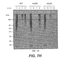


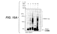


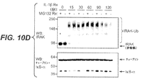

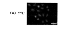


Classifications
-
- C—CHEMISTRY; METALLURGY
- C07—ORGANIC CHEMISTRY
- C07K—PEPTIDES
- C07K16/00—Immunoglobulins [IGs], e.g. monoclonal or polyclonal antibodies
- C07K16/18—Immunoglobulins [IGs], e.g. monoclonal or polyclonal antibodies against material from animals or humans
-
- A—HUMAN NECESSITIES
- A61—MEDICAL OR VETERINARY SCIENCE; HYGIENE
- A61P—SPECIFIC THERAPEUTIC ACTIVITY OF CHEMICAL COMPOUNDS OR MEDICINAL PREPARATIONS
- A61P21/00—Drugs for disorders of the muscular or neuromuscular system
-
- A—HUMAN NECESSITIES
- A61—MEDICAL OR VETERINARY SCIENCE; HYGIENE
- A61P—SPECIFIC THERAPEUTIC ACTIVITY OF CHEMICAL COMPOUNDS OR MEDICINAL PREPARATIONS
- A61P21/00—Drugs for disorders of the muscular or neuromuscular system
- A61P21/04—Drugs for disorders of the muscular or neuromuscular system for myasthenia gravis
-
- A—HUMAN NECESSITIES
- A61—MEDICAL OR VETERINARY SCIENCE; HYGIENE
- A61P—SPECIFIC THERAPEUTIC ACTIVITY OF CHEMICAL COMPOUNDS OR MEDICINAL PREPARATIONS
- A61P25/00—Drugs for disorders of the nervous system
-
- A—HUMAN NECESSITIES
- A61—MEDICAL OR VETERINARY SCIENCE; HYGIENE
- A61P—SPECIFIC THERAPEUTIC ACTIVITY OF CHEMICAL COMPOUNDS OR MEDICINAL PREPARATIONS
- A61P25/00—Drugs for disorders of the nervous system
- A61P25/14—Drugs for disorders of the nervous system for treating abnormal movements, e.g. chorea, dyskinesia
- A61P25/16—Anti-Parkinson drugs
-
- A—HUMAN NECESSITIES
- A61—MEDICAL OR VETERINARY SCIENCE; HYGIENE
- A61P—SPECIFIC THERAPEUTIC ACTIVITY OF CHEMICAL COMPOUNDS OR MEDICINAL PREPARATIONS
- A61P25/00—Drugs for disorders of the nervous system
- A61P25/28—Drugs for disorders of the nervous system for treating neurodegenerative disorders of the central nervous system, e.g. nootropic agents, cognition enhancers, drugs for treating Alzheimer's disease or other forms of dementia
-
- A—HUMAN NECESSITIES
- A61—MEDICAL OR VETERINARY SCIENCE; HYGIENE
- A61P—SPECIFIC THERAPEUTIC ACTIVITY OF CHEMICAL COMPOUNDS OR MEDICINAL PREPARATIONS
- A61P29/00—Non-central analgesic, antipyretic or antiinflammatory agents, e.g. antirheumatic agents; Non-steroidal antiinflammatory drugs [NSAID]
-
- A—HUMAN NECESSITIES
- A61—MEDICAL OR VETERINARY SCIENCE; HYGIENE
- A61P—SPECIFIC THERAPEUTIC ACTIVITY OF CHEMICAL COMPOUNDS OR MEDICINAL PREPARATIONS
- A61P35/00—Antineoplastic agents
-
- A—HUMAN NECESSITIES
- A61—MEDICAL OR VETERINARY SCIENCE; HYGIENE
- A61P—SPECIFIC THERAPEUTIC ACTIVITY OF CHEMICAL COMPOUNDS OR MEDICINAL PREPARATIONS
- A61P35/00—Antineoplastic agents
- A61P35/02—Antineoplastic agents specific for leukemia
-
- A—HUMAN NECESSITIES
- A61—MEDICAL OR VETERINARY SCIENCE; HYGIENE
- A61P—SPECIFIC THERAPEUTIC ACTIVITY OF CHEMICAL COMPOUNDS OR MEDICINAL PREPARATIONS
- A61P37/00—Drugs for immunological or allergic disorders
-
- A—HUMAN NECESSITIES
- A61—MEDICAL OR VETERINARY SCIENCE; HYGIENE
- A61P—SPECIFIC THERAPEUTIC ACTIVITY OF CHEMICAL COMPOUNDS OR MEDICINAL PREPARATIONS
- A61P43/00—Drugs for specific purposes, not provided for in groups A61P1/00-A61P41/00
-
- C—CHEMISTRY; METALLURGY
- C07—ORGANIC CHEMISTRY
- C07K—PEPTIDES
- C07K14/00—Peptides having more than 20 amino acids; Gastrins; Somatostatins; Melanotropins; Derivatives thereof
- C07K14/435—Peptides having more than 20 amino acids; Gastrins; Somatostatins; Melanotropins; Derivatives thereof from animals; from humans
- C07K14/46—Peptides having more than 20 amino acids; Gastrins; Somatostatins; Melanotropins; Derivatives thereof from animals; from humans from vertebrates
- C07K14/47—Peptides having more than 20 amino acids; Gastrins; Somatostatins; Melanotropins; Derivatives thereof from animals; from humans from vertebrates from mammals
-
- G—PHYSICS
- G01—MEASURING; TESTING
- G01N—INVESTIGATING OR ANALYSING MATERIALS BY DETERMINING THEIR CHEMICAL OR PHYSICAL PROPERTIES
- G01N33/00—Investigating or analysing materials by specific methods not covered by groups G01N1/00 - G01N31/00
- G01N33/48—Biological material, e.g. blood, urine; Haemocytometers
- G01N33/50—Chemical analysis of biological material, e.g. blood, urine; Testing involving biospecific ligand binding methods; Immunological testing
- G01N33/68—Chemical analysis of biological material, e.g. blood, urine; Testing involving biospecific ligand binding methods; Immunological testing involving proteins, peptides or amino acids
-
- C—CHEMISTRY; METALLURGY
- C07—ORGANIC CHEMISTRY
- C07K—PEPTIDES
- C07K2317/00—Immunoglobulins specific features
- C07K2317/10—Immunoglobulins specific features characterized by their source of isolation or production
- C07K2317/14—Specific host cells or culture conditions, e.g. components, pH or temperature
-
- C—CHEMISTRY; METALLURGY
- C07—ORGANIC CHEMISTRY
- C07K—PEPTIDES
- C07K2317/00—Immunoglobulins specific features
- C07K2317/20—Immunoglobulins specific features characterized by taxonomic origin
- C07K2317/21—Immunoglobulins specific features characterized by taxonomic origin from primates, e.g. man
-
- C—CHEMISTRY; METALLURGY
- C07—ORGANIC CHEMISTRY
- C07K—PEPTIDES
- C07K2317/00—Immunoglobulins specific features
- C07K2317/20—Immunoglobulins specific features characterized by taxonomic origin
- C07K2317/24—Immunoglobulins specific features characterized by taxonomic origin containing regions, domains or residues from different species, e.g. chimeric, humanized or veneered
-
- C—CHEMISTRY; METALLURGY
- C07—ORGANIC CHEMISTRY
- C07K—PEPTIDES
- C07K2317/00—Immunoglobulins specific features
- C07K2317/30—Immunoglobulins specific features characterized by aspects of specificity or valency
- C07K2317/31—Immunoglobulins specific features characterized by aspects of specificity or valency multispecific
-
- C—CHEMISTRY; METALLURGY
- C07—ORGANIC CHEMISTRY
- C07K—PEPTIDES
- C07K2317/00—Immunoglobulins specific features
- C07K2317/30—Immunoglobulins specific features characterized by aspects of specificity or valency
- C07K2317/35—Valency
-
- C—CHEMISTRY; METALLURGY
- C07—ORGANIC CHEMISTRY
- C07K—PEPTIDES
- C07K2317/00—Immunoglobulins specific features
- C07K2317/50—Immunoglobulins specific features characterized by immunoglobulin fragments
- C07K2317/51—Complete heavy chain or Fd fragment, i.e. VH + CH1
-
- C—CHEMISTRY; METALLURGY
- C07—ORGANIC CHEMISTRY
- C07K—PEPTIDES
- C07K2317/00—Immunoglobulins specific features
- C07K2317/50—Immunoglobulins specific features characterized by immunoglobulin fragments
- C07K2317/515—Complete light chain, i.e. VL + CL
-
- C—CHEMISTRY; METALLURGY
- C07—ORGANIC CHEMISTRY
- C07K—PEPTIDES
- C07K2317/00—Immunoglobulins specific features
- C07K2317/50—Immunoglobulins specific features characterized by immunoglobulin fragments
- C07K2317/55—Fab or Fab'
-
- C—CHEMISTRY; METALLURGY
- C07—ORGANIC CHEMISTRY
- C07K—PEPTIDES
- C07K2317/00—Immunoglobulins specific features
- C07K2317/50—Immunoglobulins specific features characterized by immunoglobulin fragments
- C07K2317/56—Immunoglobulins specific features characterized by immunoglobulin fragments variable (Fv) region, i.e. VH and/or VL
- C07K2317/565—Complementarity determining region [CDR]
-
- C—CHEMISTRY; METALLURGY
- C07—ORGANIC CHEMISTRY
- C07K—PEPTIDES
- C07K2317/00—Immunoglobulins specific features
- C07K2317/90—Immunoglobulins specific features characterized by (pharmaco)kinetic aspects or by stability of the immunoglobulin
- C07K2317/92—Affinity (KD), association rate (Ka), dissociation rate (Kd) or EC50 value
-
- G—PHYSICS
- G01—MEASURING; TESTING
- G01N—INVESTIGATING OR ANALYSING MATERIALS BY DETERMINING THEIR CHEMICAL OR PHYSICAL PROPERTIES
- G01N2333/00—Assays involving biological materials from specific organisms or of a specific nature
- G01N2333/435—Assays involving biological materials from specific organisms or of a specific nature from animals; from humans
- G01N2333/46—Assays involving biological materials from specific organisms or of a specific nature from animals; from humans from vertebrates
- G01N2333/47—Assays involving proteins of known structure or function as defined in the subgroups
-
- G—PHYSICS
- G01—MEASURING; TESTING
- G01N—INVESTIGATING OR ANALYSING MATERIALS BY DETERMINING THEIR CHEMICAL OR PHYSICAL PROPERTIES
- G01N2440/00—Post-translational modifications [PTMs] in chemical analysis of biological material
- G01N2440/36—Post-translational modifications [PTMs] in chemical analysis of biological material addition of addition of other proteins or peptides, e.g. SUMOylation, ubiquitination
Landscapes
- Health & Medical Sciences (AREA)
- Life Sciences & Earth Sciences (AREA)
- Chemical & Material Sciences (AREA)
- Organic Chemistry (AREA)
- Engineering & Computer Science (AREA)
- General Health & Medical Sciences (AREA)
- Medicinal Chemistry (AREA)
- Veterinary Medicine (AREA)
- Pharmacology & Pharmacy (AREA)
- Chemical Kinetics & Catalysis (AREA)
- Animal Behavior & Ethology (AREA)
- Nuclear Medicine, Radiotherapy & Molecular Imaging (AREA)
- General Chemical & Material Sciences (AREA)
- Public Health (AREA)
- Biomedical Technology (AREA)
- Bioinformatics & Cheminformatics (AREA)
- Molecular Biology (AREA)
- Immunology (AREA)
- Neurology (AREA)
- Biochemistry (AREA)
- Proteomics, Peptides & Aminoacids (AREA)
- Hematology (AREA)
- Neurosurgery (AREA)
- Urology & Nephrology (AREA)
- Biophysics (AREA)
- Genetics & Genomics (AREA)
- General Physics & Mathematics (AREA)
- Pathology (AREA)
- Cell Biology (AREA)
- Microbiology (AREA)
- Biotechnology (AREA)
- Analytical Chemistry (AREA)
- Food Science & Technology (AREA)
- Physics & Mathematics (AREA)
- Physical Education & Sports Medicine (AREA)
- Orthopedic Medicine & Surgery (AREA)
- Zoology (AREA)
- Gastroenterology & Hepatology (AREA)
- Toxicology (AREA)
- Oncology (AREA)
Description
本発明は、抗ポリユビキチン抗体の分野、および特にモノユビキチンに特異的に結合せず、異なるイソペプチド結合を有するポリユビキチンを区別することができる抗ポリユビキチン抗体に関する。 The present invention relates to the field of anti-polyubiquitin antibodies, and in particular to anti-polyubiquitin antibodies that can distinguish between polyubiquitins that do not specifically bind to monoubiquitin and have different isopeptide bonds.
ユビキチンは、多種多様な細胞経路において重要な調整の役割を有する小タンパク質である。中でもタンパク質分解でのユビキチンの役割は最も周知であり、標的タンパク質にユビキチンが共有結合するとその標的タンパク質が26Sのプロテアソームによって認識され、破壊される(Wilkinson, Semin. Cell Devel. Biol. 11(3): 141-148 (2000)を参照)。様々なシグナル伝達経路のプロテインキナーゼ制御はユビキチン化とも相関している(Sun and Chen, Curr. Opin. Cell Biol. 16: 119-126 (2004)を参照)。例えば、IκBキナーゼによるIκBのリン酸化により、IκBのユビキチン化とその後の26Sのプロテアソームによる分解が促される;IκBはNKκBのインヒビターであるので、IκBの分解はNKκBを活性化する(Ghosh and Karin, Cell 109 (Suppl.): S81-S96 (2002);Palombella等, Cell 78: 773-785 (1994))。また、ユビキチン化はDNA修復を媒介する(Sun and Chen, Curr. Opin. Cell Biol. 16:119-126 (2004)を参照)。DNAが損傷されると、増殖性細胞核抗原(PCNA)のモノユビキチン化により、DNAが破壊されていてもDNAを合成することができる損傷耐性ポリメラーゼが活性化される(Stelter and Ulrich, Nature 425: 188-191 (2003)。ユビキチン化が伴うことが知られている他の生理学的なプロセスには、細胞分裂、細胞増殖、細胞移動、及びアポトーシス/細胞死が含まれる(Johnson, Nat. Cell Biol. 4: E295-E298 (2002);Pickart, Mol. Cell. 8: 499-504 (2001))。 Ubiquitin is a small protein that has an important regulatory role in a wide variety of cellular pathways. Among them, the role of ubiquitin in proteolysis is the most well-known. When ubiquitin is covalently bound to a target protein, the target protein is recognized and destroyed by the 26S proteasome (Wilkinson, Semin. Cell Devel. Biol. 11 (3) : 141-148 (2000)). Protein kinase control of various signaling pathways has also been correlated with ubiquitination (see Sun and Chen, Curr. Opin. Cell Biol. 16: 119-126 (2004)). For example, phosphorylation of IκB by IκB kinase promotes ubiquitination of IκB and subsequent degradation by the 26S proteasome; since IκB is an inhibitor of NKκB, degradation of IκB activates NKκB (Ghosh and Karin, Karin, Cell 109 (Suppl.): S81-S96 (2002); Palombella et al., Cell 78: 773-785 (1994)). Ubiquitination also mediates DNA repair (see Sun and Chen, Curr. Opin. Cell Biol. 16: 119-126 (2004)). When DNA is damaged, monoubiquitination of proliferating cell nuclear antigen (PCNA) activates a damage-resistant polymerase that can synthesize DNA even when the DNA is disrupted (Stelter and Ulrich, Nature 425: 188-191 (2003) Other physiological processes known to be associated with ubiquitination include cell division, cell proliferation, cell migration, and apoptosis / cell death (Johnson, Nat. Cell Biol 4: E295-E298 (2002); Pickart, Mol. Cell. 8: 499-504 (2001)).
標的タンパク質への76アミノ酸タンパク質であるユビキチンの共有結合は、3-工程酵素法である(Pickart, Annu. Rev. Biochem. 70: 503-533 (2001))。第一に、ユビキチン-活性化酵素E1は、ATP依存性反応においてユビキチン-E1チオエステルを形成する。第二工程において、ユビキチンは、ユビキチン-E1チオエステルから、ユビキチンコンジュゲート酵素(E2)ファミリのメンバーに転移される。第三工程では、ユビキチン-プロテインリガーゼ(E3)の援助により、ユビキチンのカルボキシル末端と標的タンパク質上のリジン残基のεアミノ基との間にイソペプチド結合が形成される。デユビキチナーゼと呼ばれる酵素は標的タンパク質からユビキチン部分を取り除く(Guterman and Glickman, Curr. Prot. Pep. Sci. 5: 201-210 (2004))。重要な制御分子としてのユビキチンの役割に注目すると、ヒトゲノムはユビキチン化又は脱ユビキチン化に関与する多くの異なるタンパク質を有する。今までのところ少なくとも40の異なるE2s、500の異なるE3、及び80の異なるデユビキチナーゼが同定されている(Wong等, Drug. Discov. Today 8: 746-754 (2003))。 The covalent linkage of ubiquitin, a 76 amino acid protein, to the target protein is a three-step enzymatic method (Pickart, Annu. Rev. Biochem. 70: 503-533 (2001)). First, ubiquitin-activating enzyme E1 forms a ubiquitin-E1 thioester in an ATP-dependent reaction. In the second step, ubiquitin is transferred from a ubiquitin-E1 thioester to a member of the ubiquitin conjugate enzyme (E2) family. In the third step, with the aid of ubiquitin-protein ligase (E3), an isopeptide bond is formed between the carboxyl terminus of ubiquitin and the ε-amino group of the lysine residue on the target protein. An enzyme called deubiquitinase removes the ubiquitin moiety from the target protein (Guterman and Glickman, Curr. Prot. Pep. Sci. 5: 201-210 (2004)). Focusing on the role of ubiquitin as an important regulatory molecule, the human genome has many different proteins involved in ubiquitination or deubiquitination. To date, at least 40 different E2s, 500 different E3, and 80 different deubiquitinases have been identified (Wong et al., Drug. Discov. Today 8: 746-754 (2003)).
ユビキチンは7つのリジン残基(Lys6、Lys11、Lys27、Lys33、Lys29、Lys48およびLys63)を含有するため、ユビキチン自体がユビキチン化の標的タンパク質として役立ちうる(Peng等, Nat. Biotechnol. 21: 921-926 (2003);Pickart and Fushman, Curr. Opin. Chem. Biol. 8:610-616 (2004))。ユビキチンタンパク質のユビキチン化の際に産生される分子はポリユビキチン分子と呼ばれ、2以上のユビキチン成分を含みうる。ユビキチンのユビキチン化は理論的には7つのリジン残基のいずれかで生じており(Peng等, Nat. Biotechnol. 21: 921-926 (2003))、異なる種のポリユビキチンはユビキチン内の異なるリジン残基へのイソペプチド結合を有している。2つのユビキチン部分よりも大きな単一のポリユビキチン部分は1より多い種類のリジン結合を有する可能性がある。いくつかの研究から、E2酵素があるユビキチン分子と他のユビキチン分子との間で作製される種類のリジン結合に影響することが示されている(Tenno等, Genes to Cells 9: 865-875 (2004);Deng等 (2000); Hofmann and Pickart (2001))。ポリユビキチンおよびユビキチンは、遊離分子として存在するし、標的タンパク質と共有結合して存在する。 Since ubiquitin contains seven lysine residues (Lys6, Lys11, Lys27, Lys33, Lys29, Lys48 and Lys63), ubiquitin itself can serve as a target protein for ubiquitination (Peng et al., Nat. Biotechnol. 21: 921- 926 (2003); Pickart and Fushman, Curr. Opin. Chem. Biol. 8: 610-616 (2004)). Molecules produced upon ubiquitination of ubiquitin proteins are called polyubiquitin molecules and can contain two or more ubiquitin components. The ubiquitination of ubiquitin theoretically occurs at any of the seven lysine residues (Peng et al., Nat. Biotechnol. 21: 921-926 (2003)), and different species of polyubiquitin have different lysines within ubiquitin. Has an isopeptide bond to the residue. A single polyubiquitin moiety that is larger than two ubiquitin moieties may have more than one type of lysine linkage. Several studies have shown that the E2 enzyme affects the type of lysine binding produced between one ubiquitin molecule and another ubiquitin molecule (Tenno et al., Genes to Cells 9: 865-875 ( 2004); Deng et al. (2000); Hofmann and Pickart (2001)). Polyubiquitin and ubiquitin exist as free molecules or exist covalently with target proteins.
ユビキチンのように、ポリユビキチンの関与は、細胞内輸送、エンドサイトーシス、遺伝子発現/サイレンシング、タンパク質分解、キナーゼ活性化、翻訳、及びDNA修復を含む多くの細胞性プロセスに見られる(Hoege等, Nature 419:135-141 (2002);Spence等, Mol. Cell. Biol. 15:1265-1273 (1995);Hofmann and Pickart, Cell 96: 645-653 (1999)。しかしながら、ポリユビキチンおよびポリユビキチン化は、同じ経路のモノユビキチン及びモノユビキチン化と著しく異なる生理学的役割を有しうる。例えば、DNA損傷後のPCNAのモノユビキチン化により誤りがちなDNAポリメラーゼの活性化が生じるのに対して、モノユビキチン化が観察される同定された残基でのPCNAのポリユビキチン化により誤りのないDNA修復が生じる(Stelter and Ulrich, Nature 425: 188-191 (2003);Hoege等, Nature 419:135-141 (2002);Spence等, Mol. Cell. Biol. 15:1265-1273 (1995);及びHofmann and Pickart, Cell 96: 645-653 (1999))。 Like ubiquitin, polyubiquitin involvement is found in many cellular processes including intracellular transport, endocytosis, gene expression / silencing, proteolysis, kinase activation, translation, and DNA repair (Hoege et al. , Nature 419: 135-141 (2002); Spence et al., Mol. Cell. Biol. 15: 1265-1273 (1995); May have a physiological role that is significantly different from monoubiquitin and monoubiquitination of the same pathway, for example, the monoubiquitination of PCNA after DNA damage results in the activation of error-prone DNA polymerases Polyubiquitination of PCNA at identified residues where monoubiquitination is observed results in error-free DNA repair (Stelter and Ulrich, Nature 425: 188-191 (2003) Hoege like, Nature 419: 135-141 (2002); Spence like, Mol Cell Biol 15:... 1265-1273 (1995); and Hofmann and Pickart, Cell 96: 645-653 (1999)).
さらに、異なるリジン結合を有するポリユビキチンは異なる生理学的役割を有するようである。最も研究されている2つはLys48-結合型とLys63-結合型のポリユビキチンであり、この2つの構造的研究により、異なるリジン-結合型ポリユビキチンは著しく異なる立体構造を有しているので、選択した結合パートナーと異なる相互作用を可能にすることが示唆されている(Tenno等, Genes to Cells 9: 865-875 (2004))。典型的に、Lys48-結合型ポリユビキチンによる共有結合的修飾はタンパク質分解の標的タンパク質を明らかにするが、Lys48-結合型ポリユビキチンもタンパク質分解性でない方法で特定のタンパク質を調節しうるという証拠がいくつかある(Chau等, Science 243: 1576-1583 (1989);Finley等, Mol. Cell. Biol. 14: 5501-5509 (1994);Flick等, Nat. Cell. Biol. 6:634-641 (2004))。それに対して、Lys63-結合型ポリユビキチンは、DNA修復(K63R-ユビキチンを発現する酵母細胞はDNA修復を欠失する)、キナーゼ活性化、細胞内輸送、及び翻訳を含む様々な非タンパク質分解性の細胞内経路に関係している(Pickart and Fushman, Curr. Opin. Chem. Biol. 8: 610-616 (2004);Hicke and Dunn, Annu Rev. Cell Dev. Biol. 19: 141-172 (2003);Spece等, Mol. Cell Biol. 15: 1265-1273 (1995);Ulrich, Eukaryot. Cell 1: 1-10 (2002);Spence等, Cell 102: 67-76 (2000);Seibenhener等, Mol. Cell. Biol. 24(18): 8055-8068 (2004))。ある具体例では、synphilin-1は通常、プロテアソーム非依存的な方法でパーキンによるK63-結合型ポリユビキチンにてユビキチン化されるが、synphilin-1は交互にK48-結合型ポリユビキチンによるユビキチン化による破壊の標的となりうる(Lim等, J. Neurosci. 25(8): 2002-9 (2005))。パーキンソン病患者の分析により、synphilin-1のK63-ポリユビキチン化がこの病気に関係するレビー小体封入対の形成に伴うことが示される(Lim等, J. Neurosci. 25(8): 2002-9 (2005))。 Furthermore, polyubiquitins with different lysine bonds appear to have different physiological roles. The two most studied are Lys48-linked and Lys63-linked polyubiquitins, and these two structural studies indicate that different lysine-linked polyubiquitins have significantly different conformations. It has been suggested to allow different interactions with selected binding partners (Tenno et al., Genes to Cells 9: 865-875 (2004)). Typically, covalent modification with Lys48-linked polyubiquitin reveals a target protein for proteolysis, but there is evidence that Lys48-linked polyubiquitin can also modulate specific proteins in a non-proteolytic manner. There are several (Chau et al., Science 243: 1576-1583 (1989); Finley et al., Mol. Cell. Biol. 14: 5501-5509 (1994); Flick et al., Nat. Cell. Biol. 6: 634-641 ( 2004)). In contrast, Lys63-linked polyubiquitin has various non-proteolytic properties including DNA repair (yeast cells expressing K63R-ubiquitin lack DNA repair), kinase activation, intracellular transport, and translation. (Pickart and Fushman, Curr. Opin. Chem. Biol. 8: 610-616 (2004); Hicke and Dunn, Annu Rev. Cell Dev. Biol. 19: 141-172 (2003). Spec; et al., Mol. Cell Biol. 15: 1265-1273 (1995); Ulrich, Eukaryot. Cell 1: 1-10 (2002); Spence et al., Cell 102: 67-76 (2000); Seibenhener et al., Mol. Cell. Biol. 24 (18): 8055-8068 (2004)). In one embodiment, synphilin-1 is normally ubiquitinated with parkin-induced K63-linked polyubiquitin in a proteasome-independent manner, whereas synphilin-1 is alternatively ubiquitinated with K48-linked polyubiquitin. It can be a target for destruction (Lim et al., J. Neurosci. 25 (8): 2002-9 (2005)). Analysis of Parkinson's disease patients shows that K63-polyubiquitination of synphilin-1 is associated with the formation of Lewy body inclusion pairs associated with this disease (Lim et al., J. Neurosci. 25 (8): 2002- 9 (2005)).
他のリジン-結合型ポリユビキチンは、これらを区別することが難しいので、広範囲に大規模に研究されていない。今までの研究は、一又は複数のリジンが除去されている変異したユビキチンを発現する細胞、特定の結合の酵素的に合成したポリユビキチン、又はある種のポリユビキチンと他種のポリユビキチンとを区別するための質量分析法などの技術に頼っている。これらの方法のそれぞれは、特定のリジン-結合型ポリユビキチンの正常な生理学的性質の分析に不適当であるか面倒なものである。モノユビキチンとは対照的にポリユビキチンに特異的である抗体が存在するのに対して(Fujimoro等, FEBS Lett. 349: 173-180 (1994))、異なるリジン結合のポリユビキチン間を区別することができる抗体はまだない。 Other lysine-linked polyubiquitins have not been extensively studied extensively because they are difficult to distinguish. Previous studies have shown that cells expressing mutated ubiquitin from which one or more lysines have been removed, enzymatically synthesized polyubiquitin of a specific linkage, or certain polyubiquitins and other polyubiquitins. Rely on techniques such as mass spectrometry to distinguish them. Each of these methods is unsuitable or cumbersome for analyzing the normal physiological properties of a specific lysine-linked polyubiquitin. In contrast to monoubiquitin, there are antibodies that are specific for polyubiquitin (Fujimoro et al., FEBS Lett. 349: 173-180 (1994)), but to distinguish between different lysine-bound polyubiquitins. There are no antibodies yet.
驚くべきことではないが、様々な細胞性プロセスに重要な役割があるので、ユビキチンおよびポリユビキチンも多くの疾患に関係していた(Argiles, Ubiquitin and Disease, R. G. Landes (1998)を参照)。筋消耗にユビキチン調節不全が観察される(Mitch and Goldberg, New Engl. J. Med. 335: 1897-905 (1996);Bodine等, Science 294: 1704-1708 (2001))。遺伝病の中には、嚢胞性線維症(Ward等, Cell 83: 121-127 (1995))、アンジェルマン症候群(Kishino等, Nature Genet. 15: 70-73 (1997))およびリデル症候群(Staub等, EMBO J 16: 6325-6336 (1997))を含む異常なユビキチン活性に関連しているものがある。また、ユビキチンは免疫及び炎症性の応答に役割がある。例えば、細胞外ユビキチンはサイトカインのような働きがあることが明らかになっており、末梢血単核細胞のエンドトキシンに対するTNFα応答を阻害し、エンドトキシン応答性低下を制御する(Majetschak等, Blood 101: 1882-1890 (2003);Ciechanover, EMBO J 17: 7151-7160 (1998))。また、ユビキチンとポリユビキチンはヒトの血清中に見られ、寄生虫病やアレルギー疾患を有する患者の血清では両分子のレベルが高い(Takada等, Clinical Chem. 43: 1188-1195 (1997))。 Not surprisingly, ubiquitin and polyubiquitin have also been implicated in many diseases because they have important roles in various cellular processes (see Argiles, Ubiquitin and Disease, R. G. Landes (1998)). Ubiquitin dysregulation is observed in muscle wasting (Mitch and Goldberg, New Engl. J. Med. 335: 1897-905 (1996); Bodine et al., Science 294: 1704-1708 (2001)). Among the genetic diseases are cystic fibrosis (Ward et al., Cell 83: 121-127 (1995)), Angelman syndrome (Kishino et al., Nature Genet. 15: 70-73 (1997)) and Ridder syndrome (Staub Et al., EMBO J 16: 6325-6336 (1997)) are associated with abnormal ubiquitin activity. Ubiquitin also has a role in immune and inflammatory responses. For example, extracellular ubiquitin has been shown to act like a cytokine and inhibits the TNFα response to endotoxin in peripheral blood mononuclear cells and controls the decline in endotoxin responsiveness (Majetschak et al., Blood 101: 1882 -1890 (2003); Ciechanover, EMBO J 17: 7151-7160 (1998)). Ubiquitin and polyubiquitin are also found in human serum, and the levels of both molecules are high in the serum of patients with parasitic diseases and allergic diseases (Takada et al., Clinical Chem. 43: 1188-1195 (1997)).
また、いくつかのユビキチンが媒介する経路の調節不全は癌に関与する(Spataro等, Br. J. Cancer 77: 448-55 (1998);Beckmann等, Hum. Mutat. 25: 507-12 (2005))。例えば、ヘテロダイマーユビキチンリガーゼBRCA1-BARD1の突然変異は乳癌と関係しており(Hashizume等, J. Biol. Chem. 276: 14537-40 (2001))、ユビキチン経路によって分解されるMycの能力を破壊する突然変異は、c-Mycの発癌能力を活性化し(Salghetti等, EMBO J. 18: 717-726 (1999))、発癌性相関と関係のないものであるc-Junは制御されない増殖が生じるので、形質転換したv-Junはユビキチン化も分解もされない(Ciechanover, EMBO J. 17: 7151-7160 (1998);Trier等, Cell 78: 787-798 (1994))。 Also, dysregulation of some ubiquitin-mediated pathways is implicated in cancer (Spataro et al., Br. J. Cancer 77: 448-55 (1998); Beckmann et al., Hum. Mutat. 25: 507-12 (2005 )). For example, mutations in the heterodimer ubiquitin ligase BRCA1-BARD1 are associated with breast cancer (Hashizume et al., J. Biol. Chem. 276: 14537-40 (2001)) and disrupt the ability of Myc to be degraded by the ubiquitin pathway. Mutations activate c-Myc's oncogenic potential (Salghetti et al., EMBO J. 18: 717-726 (1999)) and c-Jun, which has nothing to do with carcinogenic correlations, results in uncontrolled proliferation. Therefore, transformed v-Jun is neither ubiquitinated nor degraded (Ciechanover, EMBO J. 17: 7151-7160 (1998); Trier et al., Cell 78: 787-798 (1994)).
ユビキチンおよびポリユビキチンは、特に神経学的疾患において研究されている(Chung等, TINS 24(11 Suppl.) S7-S14 (2001))。ハンチントン病、脊髄小脳失調症、プリオン脳症、ピック病、レビー小体病、パーキンソン病、及びアルツハイマー病に蓄積する封入対、小体、及び神経原線維変化は、モノ及び/又はポリユビキチンについて免疫陽性染色される(Alves-Rodrigues等, Trends Neurosci. 21: 516-520 (1998);Cammarata等, Neurosci Lett. 156: 96-98 (1993);Kalchman等, J. Biol. Chem. 271: 19385-94 (1996);Holmberg等, Human Mol. Genet. 7: 913-918 (1998);Yedidia等, EMBO J. 20: 5383-91 (2001);Mori等, Science 235: 1641-44 (1987);Leigh等, Acta Neuropathol. (Berl.) 79: 61-72 (1989);及びKuzuhara等, Acta Neuropathologica 75: 345-353 (1988))。様々な形態のパーキンソン病は、デユビキチナーゼであるユビキチンカルボキシ末端ヒドロラーゼL1(UCH-L1)の突然変異と関係していたのに対して(Leroy等, Nature 395: 451-452 (1998))、他の形態のパーキンソン病は、ユビキチンコンジュゲート酵素であるUbcH7と相互作用してsynphilin-1をユビキチン化することが知られているE2-依存性ユビキチン-タンパク質リガーゼであるパーキンの不活性化突然変異と関係していた(Shimura等, Nature Genet. 25: 302-305 (2000)、Zhang等, Proc. Natl. Acad. Sci. 97: 13354-13359 (2000);Lim等, J. Neurosci. 25(8): 2002-9 (2005))。両タイプの突然変異により異常なタンパク分解性プロセシングとタンパク質の不適当な凝集が生じる(McNaught等, Nature Rev. Neurosci. 2: 589-594 (2001)を参照)。また、UCH-L1突然変異は、ハンチントン舞踏病によって分かれることが明らかとなっている(Naze等, Neurosci. Lett. 328: 1: 1-4 (2002))。突然変異型のユビキチンはポリユビキチン鎖に高率に組み込まれているが、一度形成された脱ユビキチン化に抵抗性があるので、潜在的に正常な細胞性タンパク質分解性のプロセシング系を優位に阻害することが、アルツハイマー病の脳において同定された(Lam等, Proc. Natl. Acad. Sci. 97: 9902-9906 (2000))。 Ubiquitin and polyubiquitin have been studied particularly in neurological diseases (Chung et al., TINS 24 (11 Suppl.) S7-S14 (2001)). Encapsulated pair, body, and neurofibrillary tangles that accumulate in Huntington's disease, spinocerebellar ataxia, prion encephalopathy, Pick's disease, Lewy body disease, Parkinson's disease, and Alzheimer's disease are immunopositive for mono and / or polyubiquitin Stained (Alves-Rodrigues et al., Trends Neurosci. 21: 516-520 (1998); Cammarata et al., Neurosci Lett. 156: 96-98 (1993); Kalchman et al., J. Biol. Chem. 271: 19385-94 Holmberg et al., Human Mol. Genet. 7: 913-918 (1998); Yedidia et al., EMBO J. 20: 5383-91 (2001); Mori et al., Science 235: 1641-44 (1987); Leigh Et al., Acta Neuropathol. (Berl.) 79: 61-72 (1989); and Kuzuhara et al., Acta Neuropathologica 75: 345-353 (1988)). Various forms of Parkinson's disease have been associated with mutations in the deubiquitin ubiquitin carboxy-terminal hydrolase L1 (UCH-L1) (Leroy et al., Nature 395: 451-452 (1998)), Another form of Parkinson's disease is an inactivating mutation in Parkin, an E2-dependent ubiquitin-protein ligase known to interact with the ubiquitin-conjugating enzyme UbcH7 to ubiquitinate synphilin-1. (Shimura et al., Nature Genet. 25: 302-305 (2000), Zhang et al., Proc. Natl. Acad. Sci. 97: 13354-13359 (2000); Lim et al., J. Neurosci. 25 ( 8): 2002-9 (2005)). Both types of mutations result in abnormal proteolytic processing and inappropriate protein aggregation (see McNaught et al., Nature Rev. Neurosci. 2: 589-594 (2001)). It has also been revealed that UCH-L1 mutations are separated by Huntington's chorea (Naze et al., Neurosci. Lett. 328: 1: 1-4 (2002)). Mutant ubiquitin is highly integrated into the polyubiquitin chain, but is resistant to deubiquitination once formed, preferentially inhibits potentially normal cellular proteolytic processing systems Has been identified in the brain of Alzheimer's disease (Lam et al., Proc. Natl. Acad. Sci. 97: 9902-9906 (2000)).
リジン結合が異なるポリユビキチン間を区別することができる組成物及び方法をゆうするだけでなく、ユビキチン及びポリユビキチンが媒介する経路を標的とする及び調節する際に有効な組成物及び方法を有することが有利であることは明らかである。本明細書において提供される本発明はこのような組成物および方法に関する。 Having compositions and methods that are effective in targeting and modulating ubiquitin and polyubiquitin-mediated pathways as well as compositions and methods that can distinguish between polyubiquitins with different lysine linkages Is clearly advantageous. The invention provided herein relates to such compositions and methods.
特許文献及び出版物を含む、本明細書中で引用したすべての参考文献は、その全体が出典明記によって援用される。 All references cited herein, including patent documents and publications, are incorporated by reference in their entirety.
本発明は、ポリユビキチンに結合する及び/又はポリユビキチンと関係する生物学的活性を制御することができる新規の抗体を提供する。
一実施態様では、本発明は、K63結合型ポリユビキチンと特異的に結合する単離された抗体又はその抗原結合性断片であって、該抗体はK48結合型ポリユビキチン又はモノユビキチオンに特異的に結合しないものを提供する。一態様では、本発明は、それぞれ配列番号59−110と112、及び配列番号7−58と111の任意のHVR−H2及びHVR−L2から選択される、少なくとも一つの超可変(HVR)配列を含む単離された抗体又は抗原結合性断片を提供する。
The present invention provides novel antibodies capable of binding to and / or controlling biological activity associated with polyubiquitin.
In one embodiment, the invention provides an isolated antibody or antigen-binding fragment thereof that specifically binds to K63-conjugated polyubiquitin, wherein the antibody is specific for K48-conjugated polyubiquitin or monoubiquition. Provide something that doesn't bind to. In one aspect, the invention provides at least one hypervariable (HVR) sequence selected from SEQ ID NOs: 59-110 and 112 and any HVR-H2 and HVR-L2 of SEQ ID NOs: 7-58 and 111, respectively. An isolated antibody or antigen-binding fragment is provided.
別の実施態様では、本発明は、K63結合型ポリユビキチンと特異的に結合する単離された抗体又は抗原結合性断片を提供し、ここで、抗体又は抗原結合性断片は、K48結合型ポリユビキチン又はモノユビキチンと特異的に結合せず、抗体または抗原結合性断片は、少なくとも一つのHVR−H2配列を含んでなり、HVR−H2は、アミノ酸配列a b c d e f g h i j k l m n o p(配列番号221)を含み、ここでアミノ酸aはアミノ酸チロシン、アスパラギン酸及びトリプトファンから選択され;アミノ酸bはイソロイシンであり;アミノ酸cは、アミノ酸セリン、スレオニン、アラニン、フェニルアラニン、チロシン及びバリンから選択され;アミノ酸dはプロリンであり;アミノ酸eはチロシンであり;アミノ酸fは、アミノ酸チロシン、フェニルアラニン、ロイシン及びヒスチジンから選択され;アミノ酸gはグリシンであり;アミノ酸hは、アミノ酸セリン、グリシン、アラニン、フェニルアラニン及びトリプトファンから選択され;アミノ酸iはスレオニンであり;アミノ酸jはセリンであり;アミノ酸kはチロシンであり;アミノ酸lはアラニンであり;アミノ酸mはアスパラギン酸であり;アミノ酸nはセリンであり;アミノ酸oはバリンであり、アミノ酸pはリジンである。 In another embodiment, the invention provides an isolated antibody or antigen-binding fragment that specifically binds to K63-conjugated polyubiquitin, wherein the antibody or antigen-binding fragment is a K48-conjugated poly The antibody or antigen-binding fragment does not specifically bind to ubiquitin or monoubiquitin, and the antibody or antigen-binding fragment comprises at least one HVR-H2 sequence, and the HVR-H2 has the amino acid sequence a b c d e f g h i j k lmnop (SEQ ID NO: 221), wherein amino acid a is selected from the amino acids tyrosine, aspartic acid and tryptophan; amino acid b is isoleucine; amino acid c is the amino acids serine, threonine, alanine, phenylalanine, tyrosine And amino acid d is proline; amino acid e is tyrosine; No acid f is selected from the amino acids tyrosine, phenylalanine, leucine and histidine; amino acid g is glycine; amino acid h is selected from the amino acids serine, glycine, alanine, phenylalanine and tryptophan; amino acid i is threonine; j is serine; amino acid k is tyrosine; amino acid l is alanine; amino acid m is aspartic acid; amino acid n is serine; amino acid o is valine and amino acid p is lysine.
別の実施態様では、本発明は、K63結合型ポリユビキチンと特異的に結合する単離された抗体又はその抗原結合性断片であって、該抗体はK48結合型ポリユビキチン又はモノユビキチオンに特異的に結合しないものであり、配列番号59−110及び112のHVR−H2配列から選択される少なくとも一つのHVR−H2配列を含む抗体または抗原結合性断片を提供する。一態様では、抗体または抗原結合性断片は配列番号60、63及び66のHVR−H2配列から選択される少なくとも一つのHVR−H2配列を含む。 In another embodiment, the present invention provides an isolated antibody or antigen-binding fragment thereof that specifically binds to K63-conjugated polyubiquitin, wherein the antibody is specific for K48-conjugated polyubiquitin or monoubiquition. An antibody or antigen-binding fragment is provided that comprises at least one HVR-H2 sequence selected from the HVR-H2 sequences of SEQ ID NOs: 59-110 and 112. In one aspect, the antibody or antigen-binding fragment comprises at least one HVR-H2 sequence selected from the HVR-H2 sequences of SEQ ID NOs: 60, 63 and 66.
別の実施態様では、本発明は、K63結合型ポリユビキチンと特異的に結合する単離された抗体又は抗原結合性断片を提供し、ここで、抗体又は抗原結合性断片は、K48結合型ポリユビキチン又はモノユビキチンと特異的に結合せず、抗体または抗原結合性断片は、少なくとも一つのHVR−L2配列を含んでなり、抗体または抗原結合性断片は、少なくとも一つのHVR−L2配列を含んでなり、HVR−L2は、アミノ酸配列q r s t u v w x(配列番号222)を含み、ここで、アミノ酸qは、アミノ酸チロシン及びフェニルアラニンから選択され;アミノ酸rは、アミノ酸アラニン及びセリンから選択され;アミノ酸sはアラニンであり;アミノ酸tは、アミノ酸セリン、アルギニン、バリン、スレオニン、アラニン、アスパラギン及びロイシンから選択され;アミノ酸uはセリンであり;アミノ酸vはロイシンであり;アミノ酸wはチロシンであり、アミノ酸xはセリンである。 In another embodiment, the invention provides an isolated antibody or antigen-binding fragment that specifically binds to K63-conjugated polyubiquitin, wherein the antibody or antigen-binding fragment is a K48-conjugated poly The antibody or antigen-binding fragment does not specifically bind to ubiquitin or monoubiquitin, and the antibody or antigen-binding fragment comprises at least one HVR-L2 sequence, and the antibody or antigen-binding fragment comprises at least one HVR-L2 sequence. HVR-L2 comprises the amino acid sequence q r s t u v w x (SEQ ID NO: 222), wherein amino acid q is selected from the amino acids tyrosine and phenylalanine; amino acid r is selected from the amino acids alanine and serine The amino acid s is alanine; the amino acid t is the amino acids serine, arginine, valine, threonine, alanine, Is selected from asparagine and leucine; amino acid u is serine; amino acid v is leucine; amino acid w is tyrosine, amino acid x is serine.
別の実施態様では、本発明は、K63結合型ポリユビキチンと特異的に結合する単離された抗体又はその抗原結合性断片であって、該抗体又はその抗原結合性断片はK48結合型ポリユビキチン又はモノユビキチオンに特異的に結合しないものであり、配列番号7−58及び111のHVR−L2配列から選択される少なくとも一つのHVR−L2配列を含む抗体または抗原結合性断片を提供する。一態様では、抗体または抗原結合性断片は配列番号8、11及び14のHVR−L2配列から選択される少なくとも一つのHVR−L2配列を含む。 In another embodiment, the present invention provides an isolated antibody or antigen-binding fragment thereof that specifically binds to K63-conjugated polyubiquitin, wherein the antibody or antigen-binding fragment thereof is K48-conjugated polyubiquitin. Alternatively, an antibody or antigen-binding fragment that does not specifically bind to monoubiquition and includes at least one HVR-L2 sequence selected from the HVR-L2 sequences of SEQ ID NOs: 7-58 and 111 is provided. In one aspect, the antibody or antigen-binding fragment comprises at least one HVR-L2 sequence selected from the HVR-L2 sequences of SEQ ID NOs: 8, 11, and 14.
別の実施態様では、本発明は、K63結合型ポリユビキチンと特異的に結合する単離された抗体又はその抗原結合性断片を提供するものであって、該抗体又はその抗原結合性断片はK48結合型ポリユビキチン又はモノユビキチオンに特異的に結合しないものであり、抗体または抗原結合性断片はHVR−H2及びHVR−L2から選択される少なくとも一つの配列を含んでなり、ここでHVR−H2は、アミノ酸配列a b c d e f g h i j k l m n o p(配列番号221)を含み、アミノ酸aは、アミノ酸チロシン、アスパラギン酸及びトリプトファンから選択され;アミノ酸bはイソロイシンであり;アミノ酸cは、アミノ酸セリン、スレオニン、アラニン、フェニルアラニン、チロシン及びバリンから選択され;アミノ酸dはプロリンであり;アミノ酸eはチロシンであり;アミノ酸fは、アミノ酸チロシン、フェニルアラニン、ロイシン及びヒスチジンから選択され;アミノ酸gはグリシンであり;アミノ酸hは、アミノ酸セリン、グリシン、アラニン、フェニルアラニン及びトリプトファンから選択され;アミノ酸iはスレオニンであり;アミノ酸jはセリンであり;アミノ酸kはチロシンであり;アミノ酸lはアラニンであり;アミノ酸mはアスパラギン酸であり;アミノ酸nはセリンであり;アミノ酸oはバリンであり、アミノ酸pはリジンであり;更にHVR−L2は、アミノ酸配列q r s t u v w x(配列番号222)を含み、ここで、アミノ酸qは、アミノ酸チロシン及びフェニルアラニンから選択され;アミノ酸rは、アミノ酸アラニン及びセリンから選択され;アミノ酸sはアラニンであり;アミノ酸tは、アミノ酸セリン、アルギニン、バリン、スレオニン、アラニン、アスパラギン及びロイシンから選択され;アミノ酸uはセリンであり;アミノ酸vはロイシンであり;アミノ酸wはチロシンであり、アミノ酸xはセリンである。 In another embodiment, the present invention provides an isolated antibody or antigen-binding fragment thereof that specifically binds to K63-conjugated polyubiquitin, wherein the antibody or antigen-binding fragment thereof is K48. The antibody or antigen-binding fragment does not specifically bind to conjugated polyubiquitin or monoubiquition, and the antibody or antigen-binding fragment comprises at least one sequence selected from HVR-H2 and HVR-L2, wherein HVR-H2 Comprises the amino acid sequence a b c de f g h i j k l m n op (SEQ ID NO: 221), wherein amino acid a is selected from the amino acids tyrosine, aspartic acid and tryptophan; amino acid b is isoleucine; Amino acid c is selected from the amino acids serine, threonine, alanine, phenylalanine, tyrosine and valine; The amino acid d is proline; the amino acid e is tyrosine; the amino acid f is selected from the amino acids tyrosine, phenylalanine, leucine and histidine; the amino acid g is glycine; the amino acid h is the amino acids serine, glycine, alanine, phenylalanine. Amino acid i is threonine; amino acid j is serine; amino acid k is tyrosine; amino acid l is alanine; amino acid m is aspartic acid; amino acid n is serine; o is valine and amino acid p is lysine; furthermore, HVR-L2 comprises the amino acid sequence q r s t uv v x (SEQ ID NO: 222), wherein amino acid q is selected from the amino acids tyrosine and phenylalanine Amino acid r is amino The amino acid s is alanine; the amino acid t is selected from the amino acids serine, arginine, valine, threonine, alanine, asparagine and leucine; the amino acid u is serine; the amino acid v is leucine Amino acid w is tyrosine and amino acid x is serine.
別の実施態様では、本発明は、K63結合型ポリユビキチンと特異的に結合する単離された抗体又はその抗原結合性断片を提供するものであって、該抗体又はその抗原結合性断片はK48結合型ポリユビキチン又はモノユビキチオンと特異的に結合しないものであり、抗体または抗原結合性断片は少なくとも1つのHVR−H2配列及び少なくとも1つのHVR−L2配列を含んでなり、ここでHVR−H2は、アミノ酸配列a b c d e f g h i j k l m n o p(配列番号221)を含み、ここで、アミンの酸アミノ酸チロシン、アスパラギン酸及びトリプトファンから選択され;アミノ酸bはイソロイシンであり;アミノ酸cは、アミノ酸セリン、スレオニン、アラニン、フェニルアラニン、チロシン及びバリンから選択され;アミノ酸dはプロリンであり;アミノ酸eはチロシンであり;アミノ酸fは、アミノ酸チロシン、フェニルアラニン、ロイシン及びヒスチジンから選択され;アミノ酸gはグリシンであり;アミノ酸hは、アミノ酸セリン、グリシン、アラニン、フェニルアラニン及びトリプトファンから選択され;アミノ酸iはスレオニンであり;アミノ酸jはセリンであり;アミノ酸kはチロシンであり;アミノ酸lはアラニンであり;アミノ酸mはアスパラギン酸であり;アミノ酸nはセリンであり;アミノ酸oはバリンであり、アミノ酸pはリジンであり;更にHVR−L2は、アミノ酸配列q r s t u v w x(配列番号222)を含み、ここでアミノ酸qは、アミノ酸チロシン及びフェニルアラニンから選択され;アミノ酸rは、アミノ酸アラニン及びセリンから選択され;アミノ酸sはアラニンであり;アミノ酸tはアミノ酸セリン、アルギニン、バリン、スレオニン、アラニン、アスパラギン及びロイシンから選択され;アミノ酸uはセリンであり;アミノ酸vはロイシンであり;アミノ酸wはチロシンであり、アミノ酸xはセリンである。 In another embodiment, the present invention provides an isolated antibody or antigen-binding fragment thereof that specifically binds to K63-conjugated polyubiquitin, wherein the antibody or antigen-binding fragment thereof is K48. The antibody or antigen-binding fragment comprises at least one HVR-H2 sequence and at least one HVR-L2 sequence, wherein the antibody or antigen-binding fragment does not specifically bind to conjugated polyubiquitin or monoubiquition. Comprises the amino acid sequence a b c de f g h i j k l m n op (SEQ ID NO: 221), wherein the acid acid amino acid tyrosine, aspartic acid and tryptophan of the amine is selected; amino acid b is isoleucine Yes; amino acid c from the amino acids serine, threonine, alanine, phenylalanine, tyrosine and valine Amino acid d is proline; amino acid e is tyrosine; amino acid f is selected from amino acids tyrosine, phenylalanine, leucine and histidine; amino acid g is glycine; amino acid h is amino acids serine, glycine, alanine Amino acid i is threonine; amino acid j is serine; amino acid k is tyrosine; amino acid l is alanine; amino acid m is aspartic acid; amino acid n is serine Amino acid o is valine, amino acid p is lysine; and HVR-L2 further comprises the amino acid sequence q r s t u v w x (SEQ ID NO: 222), wherein amino acid q is derived from amino acids tyrosine and phenylalanine; The amino acid r is The amino acid s is alanine; the amino acid t is selected from the amino acids serine, arginine, valine, threonine, alanine, asparagine and leucine; the amino acid u is serine; the amino acid v is leucine Amino acid w is tyrosine and amino acid x is serine.
別の実施態様では、本発明は、K63結合型ポリユビキチンと特異的に結合する単離された抗体又はその抗原結合性断片であって、該抗体又はその抗原結合性断片はK48結合型ポリユビキチン又はモノユビキチオンと特異的に結合しないものであり、配列番号59−110と112のHVR−H2配列から選択される少なくとも1つのHVR−H2配列及び配列番号7−58と111のHVR−L2配列から選択される少なくとも1つのHVR−L2配列を含む抗体又はその抗原結合性断片を提供する。 In another embodiment, the present invention provides an isolated antibody or antigen-binding fragment thereof that specifically binds to K63-conjugated polyubiquitin, wherein the antibody or antigen-binding fragment thereof is K48-conjugated polyubiquitin. Or at least one HVR-H2 sequence selected from the HVR-H2 sequences of SEQ ID NOs: 59-110 and 112 and the HVR-L2 sequences of SEQ ID NOs: 7-58 and 111, which do not specifically bind to monoubiquition An antibody or antigen-binding fragment thereof comprising at least one HVR-L2 sequence selected from
別の実施態様では、本発明は、K63結合型ポリユビキチンと特異的に結合する単離された抗体又はその抗原結合性断片であって、該抗体又はその抗原結合性断片はK48結合型ポリユビキチン又はモノユビキチオンと特異的に結合しないものであり、配列番号60、63及び66のHVR−H2配列から選択される少なくとも1つのHVR−H2配列及び配列番号8、11及び14のHVR−L2配列から選択される少なくとも1つのHVR−L2配列を含む抗体又はその抗原結合性断片を提供する。 In another embodiment, the present invention provides an isolated antibody or antigen-binding fragment thereof that specifically binds to K63-conjugated polyubiquitin, wherein the antibody or antigen-binding fragment thereof is K48-conjugated polyubiquitin. Or at least one HVR-H2 sequence selected from HVR-H2 sequences of SEQ ID NOs: 60, 63 and 66 and HVR-L2 sequences of SEQ ID NOs: 8, 11 and 14 that do not specifically bind to monoubiquition An antibody or antigen-binding fragment thereof comprising at least one HVR-L2 sequence selected from
別の実施態様では、本発明は、K63結合型ポリユビキチンと特異的に結合する単離された抗体又はその抗原結合性断片であって、該抗体又はその抗原結合性断片はK48結合型ポリユビキチン又はモノユビキチオンと特異的に結合しないものであり、表BのApu3.A8−Apu3.H10の何れか一つに示されるHVR−H2及びHVR−L2アミノ酸配列から選択されるHVRアミノ酸配列を含む抗体又はその抗原結合性断片を提供する。一態様では、HVRアミノ酸配列は、表BのApu3.A8、Apu3.A12及びApu3.B3の何れか一つに示されるHVR−H2及びHVR−L2アミノ酸配列から選択される。一態様では、前記抗体または抗原結合性断片の何れかは、配列番号5のHVR−H1配列、配列番号6のHVR−H3配列、配列番号3のHVR−L1配列及び配列番号4のHVR−L3配列から選択される少なくとも一つのHVR配列を含む。一態様では、前記抗体または抗原結合性断片の何れかは、配列番号5のHVR−H1配列、配列番号6のHVR−H3配列、配列番号3のHVR−L1配列及び配列番号4のHVR−L3配列から選択される少なくとも2つのHVR配列を含む。一態様では、前記抗体または抗原結合性断片の何れかは、配列番号5のHVR−H1配列、配列番号6のHVR−H3配列、配列番号3のHVR−L1配列及び配列番号4のHVR−L3配列から選択される少なくとも3つのHVR配列を含む。一態様では、前記抗体または抗原結合性断片の何れかは、配列番号5のHVR−H1配列、配列番号6のHVR−H3配列、配列番号3のHVR−L1配列及び配列番号4のHVR−L3配列を含む。一態様では、前記抗体または抗原結合性断片の何れかは、K63結合型ポリユビキチンについての親のFab Apu2.16の親和性に対して、改良されたK63結合型ポリユビキチンに対する親和性を有する。一態様では、前記抗体または抗原結合性断片の何れかは、10nM又はそれ未満の、K63結合型ポリユビキチンについてのKd値を有する。 In another embodiment, the present invention provides an isolated antibody or antigen-binding fragment thereof that specifically binds to K63-conjugated polyubiquitin, wherein the antibody or antigen-binding fragment thereof is K48-conjugated polyubiquitin. Or those that do not specifically bind to monoubiquition, and Apu3. A8-Apu3. Provided is an antibody or antigen-binding fragment thereof comprising an HVR amino acid sequence selected from the HVR-H2 and HVR-L2 amino acid sequences shown in any one of H10. In one aspect, the HVR amino acid sequence is Apu3. A8, Apu3. A12 and Apu3. It is selected from the HVR-H2 and HVR-L2 amino acid sequences shown in any one of B3. In one aspect, either the antibody or the antigen-binding fragment is an HVR-H1 sequence of SEQ ID NO: 5, an HVR-H3 sequence of SEQ ID NO: 6, an HVR-L1 sequence of SEQ ID NO: 3, and an HVR-L3 of SEQ ID NO: 4. At least one HVR sequence selected from the sequences. In one aspect, either the antibody or the antigen-binding fragment is an HVR-H1 sequence of SEQ ID NO: 5, an HVR-H3 sequence of SEQ ID NO: 6, an HVR-L1 sequence of SEQ ID NO: 3, and an HVR-L3 of SEQ ID NO: 4. It comprises at least two HVR sequences selected from the sequences. In one aspect, either the antibody or the antigen-binding fragment is an HVR-H1 sequence of SEQ ID NO: 5, an HVR-H3 sequence of SEQ ID NO: 6, an HVR-L1 sequence of SEQ ID NO: 3, and an HVR-L3 of SEQ ID NO: 4. It contains at least three HVR sequences selected from the sequences. In one aspect, either the antibody or the antigen-binding fragment is an HVR-H1 sequence of SEQ ID NO: 5, an HVR-H3 sequence of SEQ ID NO: 6, an HVR-L1 sequence of SEQ ID NO: 3, and an HVR-L3 of SEQ ID NO: 4. Contains an array. In one aspect, either the antibody or antigen-binding fragment has an improved affinity for K63-linked polyubiquitin relative to the affinity of the parent Fab Apu 2.16 for K63-linked polyubiquitin. In one aspect, either the antibody or antigen-binding fragment has a Kd value for K63-conjugated polyubiquitin of 10 nM or less.
別の実施態様では、本発明は、上記抗体又は抗原結合性断片の何れかと、ポリユビキチン上の同じ抗原決定基に結合する、単離された抗体または抗原結合性断片であって、モノユビキチンと特異的に結合しない抗体または抗原結合性断片を提供する。別の実施態様では、本発明は、上記抗体又は抗原結合性断片の何れかはポリユビキチンとの結合について競合する、単離された抗体または抗原結合性断片であって、モノユビキチンと特異的に結合しない抗体または抗原結合性断片を提供する。 ポリユビキチンとの結合について 別の実施態様では、本発明は、K63結合型ポリユビキチンのエピトープと結合する単離された抗体または抗原結合性断片を提供する。一態様では、エピトープは、K63結合型ポリユビキチンの第1のユビキチンサブユニット及び第2のユビキチンサブユニットの残基に含む。別態様では、エピトープは、Glu−18、Pro−19,Ser−20、Asp−21、Thr−55、Leu−56、Ser−57、Asp−58、Asn−60、Ile−61、及びGln−62から選択される第1のユビキチンサブユニットの少なくとも1つの残基を含む。別態様では、エピトープは、Leu−8、Thr−9、Glu−34、Gly−35、Ile−36、Pro−37、Asp−39、Gln−40、Leu−71、Arg−72、Leu−73、Arg−74、及びGly−75から選択される第2のユビキチンサブユニットの少なくとも1つの残基を含む。別態様では、エピトープは、Glu−18、Pro−19、Ser−20、Asp−21、Thr−55、Leu−56、Ser−57、Asp−58、Asn−60、Ile−61、及びGln−62から選択される第1のユビキチンサブユニットの少なくとも1つの残基、及びLeu−8、Thr−9、Glu−34、Gly−35、Ile−36、Pro−37、Asp−39、Gln−40、Leu−71、Arg−72、Leu−73、Arg−74、及びGly−75から選択される第2のユビキチンサブユニットの少なくとも1つの残基を含む。別態様では、HVRH3のN末端部分は、C末端残基及び供与体ユビキチンのQ40と接触する。別態様では、HVRH3の末端部分は、K63受容体ユビキチンの50Sループと接触する。別態様では、HVRH3のN末端部分はC末端残基72−74と供与体ユビキチンのQ40と接触し、その一方でHVRH3の残基R102及びY103はK63受容体ユビキチンの50Sループと接触する。別態様では、供与体ユビキチンのC末端残基は、抗体または抗原結合性断片と接触する。別態様では、接触に関係する供与体ユビキチン残基は、L73とR74である。 In another embodiment, the invention provides an isolated antibody or antigen-binding fragment that binds to any of the above-described antibodies or antigen-binding fragments and the same antigenic determinant on polyubiquitin, comprising monoubiquitin and Antibodies or antigen-binding fragments that do not specifically bind are provided. In another embodiment, the present invention provides an isolated antibody or antigen-binding fragment, wherein any of the above antibodies or antigen-binding fragments competes for binding to polyubiquitin, specifically with monoubiquitin. An antibody or antigen-binding fragment that does not bind is provided. About Binding to Polyubiquitin In another embodiment, the present invention provides an isolated antibody or antigen-binding fragment that binds to an epitope of K63-linked polyubiquitin. In one aspect, the epitope is comprised in the residues of the first ubiquitin subunit and the second ubiquitin subunit of K63-linked polyubiquitin. In another aspect, the epitope is Glu-18, Pro-19, Ser-20, Asp-21, Thr-55, Leu-56, Ser-57, Asp-58, Asn-60, Ile-61, and Gln- At least one residue of a first ubiquitin subunit selected from 62. In another aspect, the epitope is Leu-8, Thr-9, Glu-34, Gly-35, Ile-36, Pro-37, Asp-39, Gln-40, Leu-71, Arg-72, Leu-73. , Arg-74, and Gly-75, comprising at least one residue of a second ubiquitin subunit. In another aspect, the epitope is Glu-18, Pro-19, Ser-20, Asp-21, Thr-55, Leu-56, Ser-57, Asp-58, Asn-60, Ile-61, and Gln- At least one residue of a first ubiquitin subunit selected from 62, and Leu-8, Thr-9, Glu-34, Gly-35, Ile-36, Pro-37, Asp-39, Gln-40 And at least one residue of a second ubiquitin subunit selected from Leu-71, Arg-72, Leu-73, Arg-74, and Gly-75. In another embodiment, the N-terminal portion of HVRH3 contacts the C-terminal residue and Q40 of the donor ubiquitin. In another aspect, the terminal portion of HVRH3 contacts the 50S loop of the K63 receptor ubiquitin. In another embodiment, the N-terminal portion of HVRH3 contacts C-terminal residues 72-74 and Q40 of donor ubiquitin, while residues R102 and Y103 of HVRH3 contact the 50S loop of K63 receptor ubiquitin. In another embodiment, the C-terminal residue of the donor ubiquitin contacts the antibody or antigen-binding fragment. In another aspect, the donor ubiquitin residues involved in contact are L73 and R74.
別の実施態様では、本発明は、その軽鎖とK63受容体ユビキチンの表面との間の改良された荷電相補性を含む、単離された抗体または抗原結合性断片を提供する。一態様では、抗体または抗原結合性断片は、HVRL2の位置52にArgを含む。別態様では、抗体または抗原結合性断片は、軽鎖フレームワーク領域の位置66にArgを含む。別態様では、抗体または抗原結合性断片は、HVRL2の位置52にArgを、軽鎖フレームワーク領域の位置66にArgを含む。
In another embodiment, the present invention provides an isolated antibody or antigen-binding fragment comprising improved charge complementarity between its light chain and the surface of the K63 receptor ubiquitin. In one aspect, the antibody or antigen-binding fragment comprises Arg at position 52 of HVRL2. In another aspect, the antibody or antigen-binding fragment comprises Arg at
別の実施形態では、上記の抗体または抗原結合性断片の何れかは、K63結合型ポリユビキチン化タンパク質と特異的に結合する。一態様では、抗体または抗原結合性断片は、K63結合型ポリユビキチン化タンパク質の分解を阻害する。別態様では、抗体または抗原結合性断片は、少なくとも一つのポリユビキチン介在性シグナル伝達経路を調整する。別態様では、抗体または抗原結合性断片は、少なくとも一つのポリユビキチン介在性シグナル伝達経路を阻害する。別態様では、抗体または抗原結合性断片は、少なくとも一つのポリユビキチン介在性シグナル伝達経路を刺激する。 In another embodiment, any of the above-described antibodies or antigen-binding fragments specifically binds to a K63-linked polyubiquitinated protein. In one aspect, the antibody or antigen-binding fragment inhibits degradation of K63-linked polyubiquitinated protein. In another embodiment, the antibody or antigen-binding fragment modulates at least one polyubiquitin-mediated signaling pathway. In another embodiment, the antibody or antigen-binding fragment inhibits at least one polyubiquitin-mediated signaling pathway. In another embodiment, the antibody or antigen-binding fragment stimulates at least one polyubiquitin-mediated signaling pathway.
別の実施態様では、本発明は、上記抗体または抗原結合性断片の何れかをコードする核酸分子を提供する。別の実施態様では、本発明は、このような核酸分子を含むベクターを提供する。別の実施態様では、本発明は、このようなベクターを含む宿主細胞を提供する。別の実施態様では、本発明は、上記抗体または抗原結合性断片の何れかを産生することができる細胞株を提供する。別の実施態様では、本発明は、本発明は、上記抗体または抗原結合性断片の何れかを産生する方法であって、抗体または抗原結合性断片が産生される条件下で、抗体または抗原結合性断片をコードする核酸分子を含む宿主細胞を培養することを含む方法を提供する。 In another embodiment, the present invention provides a nucleic acid molecule encoding any of the above antibodies or antigen binding fragments. In another embodiment, the present invention provides a vector comprising such a nucleic acid molecule. In another embodiment, the present invention provides a host cell comprising such a vector. In another embodiment, the present invention provides a cell line capable of producing any of the above antibodies or antigen binding fragments. In another embodiment, the invention provides a method for producing any of the above antibodies or antigen-binding fragments, wherein the antibody or antigen-binding fragment is produced under conditions that produce the antibody or antigen-binding fragment. A method comprising culturing a host cell comprising a nucleic acid molecule encoding a sex fragment is provided.
別の実施態様では、本発明は、上記抗体または抗原結合性断片の有効量及び薬学的に許容可能な担体を含む組成物を提供する。一態様では、組成物は、2以上の上記抗体または抗原結合性断片を含む。 In another embodiment, the present invention provides a composition comprising an effective amount of the antibody or antigen-binding fragment and a pharmaceutically acceptable carrier. In one aspect, the composition comprises two or more of the above antibodies or antigen-binding fragments.
別の実施態様では、本発明は、試料中のK63結合型ポリユビキチン又はK63結合型ポリユビキチン化タンパク質の存在を同定する方法であって、少なくとも1つの上記抗体または抗原結合性断片に、該試料を接触させることを含む方法を提供する。 In another embodiment, the present invention provides a method for identifying the presence of K63-conjugated polyubiquitin or K63-conjugated polyubiquitinated protein in a sample, wherein at least one of said antibody or antigen-binding fragment comprises There is provided a method comprising contacting.
別の実施態様では、本発明は、患者のポリユビキチンの調節不全に関連する疾患または状態の治療方法であって、患者に少なくとも1つの上記抗体または抗原結合性断片の有効量を投与することを含む方法を提供する。一態様では、患者は、哺乳動物の患者である。別態様では、患者はヒトである。別態様では、疾患は、癌、筋疾患、ユビキチン経路関連遺伝的疾患、免疫性/炎症性疾患及び神経疾患から選択される。別態様では、疾患は、癌腫、リンパ腫、芽細胞腫、肉腫、白血病、筋ジストロフィー、多発性硬化症、筋萎縮性側索硬化症、嚢胞性線維症、アンジェルマン症候群、リデル症候群、アルツハイマー病、パーキンソン病、ピック病及びパジェット病から選択される。 In another embodiment, the invention provides a method of treating a disease or condition associated with dysregulation of polyubiquitin in a patient, comprising administering to the patient an effective amount of at least one of the above antibodies or antigen binding fragments. A method of including is provided. In one aspect, the patient is a mammalian patient. In another aspect, the patient is a human. In another aspect, the disease is selected from cancer, muscle disease, ubiquitin pathway related genetic disease, immune / inflammatory disease and neurological disease. In another aspect, the disease is carcinoma, lymphoma, blastoma, sarcoma, leukemia, muscular dystrophy, multiple sclerosis, amyotrophic lateral sclerosis, cystic fibrosis, Angelman syndrome, Ridder syndrome, Alzheimer's disease, Parkinson Disease, Pick's disease and Paget's disease.
別の実施態様では、本発明は、K63結合型ポリユビキチンまたはK63結合型ポリユビキチン化タンパク質を含むことが疑われる試料中のK63結合型ポリユビキチンまたはK63結合型ポリユビキチン化タンパク質の存在を決定する方法であって、試料を上記抗体または抗原結合性断片のうちの少なくとも1つに暴露すること及び試料中のK63結合型ポリユビキチン又はK63結合型ポリユビキチン化タンパク質に少なくとも一つの上記抗体または抗原結合性断片の結合性を測定することを含んでなる方法を提供する。 In another embodiment, the present invention determines the presence of K63-conjugated polyubiquitin or K63-conjugated polyubiquitinated protein in a sample suspected of containing K63-conjugated polyubiquitin or K63-conjugated polyubiquitinated protein. A method comprising exposing a sample to at least one of the antibody or antigen-binding fragment and binding at least one of the antibody or antigen to a K63-conjugated polyubiquitin or a K63-conjugated polyubiquitinated protein in the sample. A method comprising measuring the binding of a sex fragment is provided.
別の実施態様では、本発明は、試料中のK63結合型ポリユビキチン化タンパク質を非K63結合型ポリユビキチン化タンパク質から分離する方法であって、少なくとも1つの上記抗体又は抗原結合性断片に試料を接触させることを含む方法を提供する。 In another embodiment, the present invention provides a method for separating K63-conjugated polyubiquitinated proteins in a sample from non-K63-conjugated polyubiquitinated proteins, wherein the sample is separated into at least one of the above antibodies or antigen-binding fragments. A method comprising contacting is provided.
別の実施態様では、本発明は、細胞中のK63結合型ポリユビキチンの機能及び/又は活性を測定する方法であって、少なくとも1つの上記抗体又は抗原結合性断片に細胞を接触させること及び細胞における上記接触工程の効果を評価することを含む方法を提供する。 In another embodiment, the present invention relates to a method for measuring the function and / or activity of K63-conjugated polyubiquitin in a cell, comprising contacting the cell with at least one of the above antibodies or antigen-binding fragments and the cell. A method comprising evaluating the effect of the contacting step in the method is provided.
別の実施態様では、本発明は、試料中のK63結合型ポリユビキチンの機能及び/又は活性を測定する方法であって、少なくとも1つの上記抗体又は抗原結合性断片に試料を接触させること及び試料における上記接触工程の効果を評価することを含む方法を提供する。 In another embodiment, the present invention provides a method for measuring the function and / or activity of K63-conjugated polyubiquitin in a sample, comprising contacting the sample with at least one of the above antibodies or antigen-binding fragments and the sample. A method comprising evaluating the effect of the contacting step in the method is provided.
本発明の抗体は多くの形態でありうる。例えば、本発明の抗体は、キメラ抗体、ヒト化抗体又はヒト抗体でありうる。ある実施態様では、本発明の抗体はヒト抗体ではなく、例えばゼノマウスで産生された抗体ではない(例えば、国際公開第96/33735号に記載)。本発明の抗体は完全長又はその断片であってもよい(例えば、抗原結合成分を含む断片)。他の実施態様では、本発明は上記の抗体のいずれかの抗原結合性断片を提供する。 The antibodies of the present invention can be in many forms. For example, the antibody of the present invention can be a chimeric antibody, a humanized antibody or a human antibody. In certain embodiments, the antibody of the invention is not a human antibody, eg, an antibody produced in a xenomouse (see, eg, WO 96/33735). An antibody of the invention may be full length or a fragment thereof (eg, a fragment comprising an antigen binding component). In other embodiments, the invention provides an antigen-binding fragment of any of the above-described antibodies.
一般的技術
特に示さない限り、本発明の実施には、当業者の技量内にある分子生物学(組換え技術を含む)、細菌学、細胞生物学、生化学、及び免疫学の一般的な技術を用いる。このような技術は、文献、例えば「Molecular Cloning:A Laboratory Manual」第3版(Sambrook等, 2001);「Oligonucleotide Synthesis」(M. J. Gait編, 1984);「Animal Cell Culture」(R. I. Freshney編, 1987);「Methods in Enzymology」(Academic Press, Inc.);「Current Protocols in Molecular Biology」(F. M. Ausubel等編, 1987, 及び定期更新物);「PCR:The Polymerase Chain Reaction」(Mullis等編, 1994);PCR2:A Practical Approach (M. J. MacPherson, B. D. Hames 及び G. R. Taylor編(1995)); Harlow及びLane, 編 (1988 Antibodies, A Laboratory Manual; 「A Practical Guide to Molecular Cloning」(Perbal Bernard V., 1988;及び「Phage Display: A Laboratory Manual」(Barbas等、2001)に十分に説明されている。
General Technology Use technology. Such techniques are described in literature such as “Molecular Cloning: A Laboratory Manual” 3rd edition (Sambrook et al., 2001); “Oligonucleotide Synthesis” (MJ Gait, 1984); “Animal Cell Culture” (RI Freshney, 1987). "Methods in Enzymology" (Academic Press, Inc.); "Current Protocols in Molecular Biology" (FM Ausubel et al., 1987, and periodic updates); "PCR: The Polymerase Chain Reaction" (Mullis et al., 1994) ); PCR2: A Practical Approach (MJ MacPherson, BD Hames and GR Taylor (1995)); Harlow and Lane, ed. (1988 Antibodies, A Laboratory Manual; “A Practical Guide to Molecular Cloning” (Perbal Bernard V., 1988) And “Phage Display: A Laboratory Manual” (Barbas et al., 2001).
定義
本明細書において使用する「ユビキチン」及び「モノユビキチン」という用語は互換可能に使用され、アミノ酸6、アミノ酸11、アミノ酸27、アミノ酸29、アミノ酸33、アミノ酸48、及び/又はアミノ酸63に少なくとも1のリジン残基を有する76アミノ酸タンパク質と実質的に同じ天然ヒト及び合成ユビキチンの全ての種と定義される。
本明細書において使用する「ポリユビキチン」という用語は、ユビキチンの異なる重合体結合のヒト及び合成クラスに含まれる天然ヒト及び合成のユビキチン重合体鎖として定義され、それらには、限定されるものではないが、K6結合型ポリユビキチン、K11結合型ポリユビキチン、K27結合型ポリユビキチン、K29結合型ポリユビキチン、K33結合型ポリユビキチン、K48結合型ポリユビキチン及びK63結合型ポリユビキチンが含まれる。ポリユビキチンは、どのような長さを有してもよく、少なくとも2のユビキチン部分を含む。ポリユビキチンは、元来一本鎖タンパク質として発現されるユビキチンのタンデムリピートとは区別される。
Definitions As used herein, the terms “ubiquitin” and “monoubiquitin” are used interchangeably and are at least one in
As used herein, the term “polyubiquitin” is defined as the natural human and synthetic ubiquitin polymer chains included in the human and synthetic classes of different polymer linkages of ubiquitin, including but not limited to These include, but are not limited to, K6 linked polyubiquitin, K11 linked polyubiquitin, K27 linked polyubiquitin, K29 linked polyubiquitin, K33 linked polyubiquitin, K48 linked polyubiquitin and K63 linked polyubiquitin. The polyubiquitin may have any length and includes at least two ubiquitin moieties. Polyubiquitin is distinguished from tandem repeats of ubiquitin that are originally expressed as single chain proteins.
本明細書において使用する「K*-結合型ポリユビキチン」及び「Lys*結合型ポリユビキチン」という用語は互換可能であり、1のユビキチン部分のC末端と別のユビキチン部分の位置*のリジンの間に少なくとも1のイソペプチド結合を含むポリユビキチン分子を指す。例えば、「K63-結合型ポリユビキチン」は、「Lys63-結合型ポリユビキチン」と互換可能に使用され、これらの用語はいずれも、分子中のユビキチン部分の一方のC末端と、他方のユビキチン部分の位置63のリジンの間にイソペプチド結合を含むポリユビキチン分子を指す。
As used herein, the terms “K * -linked polyubiquitin” and “Lys * -linked polyubiquitin” are interchangeable, and the C-terminus of one ubiquitin moiety and the lysine at position * of another ubiquitin moiety. A polyubiquitin molecule that includes at least one isopeptide bond in between. For example, “K63-linked polyubiquitin” is used interchangeably with “Lys63-linked polyubiquitin” and both of these terms refer to one C-terminus of the ubiquitin moiety in the molecule and the other ubiquitin moiety. Refers to a polyubiquitin molecule containing an isopeptide bond between the lysines at
本明細書において使用する、第1リジン結合が第2リジン結合と「異なる」という表現は、1のユビキチン部分と別のユビキチン部分の間の第1リジン結合が、1のユビキチン部分と別のユビキチン部分の間の第2リジン結合とは異なるリジン残基(例えば、K6、K11、K27、K29、K33、K48、及び/又はK63)を含むことを示唆する。
本明細書において使用する、「ユビキチン経路」という用語は、ユビキチン及び/又はポリユビキチンを含む細胞又は再構成されたインビトロ中の生化学的経路を指す。
As used herein, the expression that the first lysine bond is “different” from the second lysine bond means that the first lysine bond between one ubiquitin moiety and another ubiquitin moiety is one ubiquitin moiety and another ubiquitin. It is suggested to contain a lysine residue (eg, K6, K11, K27, K29, K33, K48, and / or K63) that is different from the second lysine bond between the moieties.
As used herein, the term “ubiquitin pathway” refers to a cell containing ubiquitin and / or polyubiquitin or a reconstituted in vitro biochemical pathway.
「単離された」抗体とは、その自然環境の成分から同定され分離され及び/又は回収されたものである。その自然環境の汚染成分とは、抗体の診断又は治療的な使用を妨害する物質であり、酵素、ホルモン、及び他のタンパク質様又は非タンパク質様溶質が含まれる。一実施態様では、タンパク質は、(1)例えばローリー法で測定した場合95%を越える抗体、一部の実施態様では99重量%を超えるまで、(2)例えばスピニングカップシークエネーターを使用することにより、少なくとも15のN末端あるいは内部アミノ酸配列の残基を得るのに充分なほど、あるいは、(3)例えばクーマシーブルーあるいは銀染色を用いた非還元あるいは還元条件下でのSDS-PAGEにより均一になるまで充分なほど精製される。抗体の自然環境の少なくとも一の成分が存在しないため、単離された抗体には、組換え細胞内のインサイツでの抗体が含まれる。しかしながら、通常は、単離された抗体は少なくとも一の精製工程により調製される。 An “isolated” antibody is one that has been identified and separated and / or recovered from a component of its natural environment. Contaminants in the natural environment are substances that interfere with the diagnostic or therapeutic use of antibodies, including enzymes, hormones, and other proteinaceous or non-proteinaceous solutes. In one embodiment, the protein is (1) by using, for example, a spinning cup sequenator, up to more than 95% antibody, for example by the Raleigh method, in some embodiments more than 99% by weight. Enough to obtain at least 15 N-terminal or internal amino acid sequence residues, or (3) uniform by SDS-PAGE under non-reducing or reducing conditions using eg Coomassie blue or silver staining It is refined enough to become. Isolated antibody includes the antibody in situ within recombinant cells since at least one component of the antibody's natural environment will not be present. Ordinarily, however, isolated antibody will be prepared by at least one purification step.
本明細書で使用する「抗ユビキチン抗体」及び「抗モノユビキチン抗体」という用語は互換可能に使用され、ユビキチン分子に対する特異的結合能を有する抗体を指す。
本明細書で使用する「抗ポリユビキチン抗体」という用語は、ポリユビキチン分子に対する特異的結合能を有する抗体を指す。
As used herein, the terms “anti-ubiquitin antibody” and “anti-monoubiquitin antibody” are used interchangeably and refer to an antibody that has the ability to specifically bind to a ubiquitin molecule.
As used herein, the term “anti-polyubiquitin antibody” refers to an antibody that has the ability to specifically bind to a polyubiquitin molecule.
本明細書で使用する「抗K48-結合型ポリユビキチン抗体」とは、K48-結合型ポリユビキチンに対する特異的結合能を有する抗体を指す。
本明細書で使用する「抗K63-結合型ポリユビキチン抗体」とは、K63-結合型ポリユビキチンに対する結合能を有する抗体を指す。
As used herein, “anti-K48-linked polyubiquitin antibody” refers to an antibody having a specific binding ability to K48-linked polyubiquitin.
As used herein, “anti-K63-linked polyubiquitin antibody” refers to an antibody having the ability to bind to K63-linked polyubiquitin.
ここで用いる「実質的に類似」、「実質的に同じ」、「等価な」、又は「実質的に等価な」という句は、当業者が2つの数値(例えば、分子に関連するもの、及び参照/比較分子に関連する他のもの)の差異に、該値(例えばKd値、抗ウイルス効果等)によって測定される生物学的性質上わずかに又は全く生物学的及び/又は統計学的有意差がないと認められるほど、2つの数値が有意に類似していることを意味する。前記2つの値間の差異は、参照/比較分子の値の例えば約50%以下、約40%以下、約30%以下、約20%以下、及び/又は約10%以下である。 As used herein, the phrases “substantially similar”, “substantially the same”, “equivalent”, or “substantially equivalent” are used by those skilled in the art to refer to two numerical values (e.g., Slight or no biological and / or statistical significance in terms of the biological properties measured by the value (eg Kd value, antiviral effect, etc.) It is recognized that there is no difference, the two numbers are significantly similar. The difference between the two values is, for example, about 50% or less, about 40% or less, about 30% or less, about 20% or less, and / or about 10% or less of the value of the reference / comparison molecule.
ここで用いる「実質的に減少」、又は「実質的に異なる」という句は、当業者が2つの数値(一般に、分子に関連するもの、及び参照/比較分子に関連する他のもの)の差異に、該値(例えばKd値)によって測定される生物学的性質上統計学的に有意であると認められるほど、2つの数値が有意に異なっていることを意味する。前記2つの値間の差異は、参照/比較分子の値の、例えば約10%より大きく、約20%より大きく、約30%より大きく、約40%より大きく、及び/又は約50%より大きい。 As used herein, the phrase “substantially reduced” or “substantially different” is used by those skilled in the art to differentiate between two numerical values (generally those related to a molecule and others related to a reference / comparison molecule). In other words, the two numerical values are significantly different from each other so that the biological property measured by the value (for example, Kd value) is statistically significant. The difference between the two values is, for example, greater than about 10%, greater than about 20%, greater than about 30%, greater than about 40%, and / or greater than about 50% of the value of the reference / comparison molecule. .
一般的に「結合親和性」は、分子(例えば抗体)の単一結合部位とその結合パートナー(例えば抗原)との間の非共有結合的な相互作用の総合的な強度を意味する。特に明記しない限り、「結合親和性」は、結合対のメンバー(例えば抗体と抗原)間の1:1相互作用を反映する内因性結合親和性を意味する。一般的に、分子XのそのパートナーYに対する親和性は、解離定数(Kd)として表される。親和性は、本明細書中に記載のものを含む当業者に公知の共通した方法によって測定することができる。低親和性抗体は抗原にゆっくり結合して素早く解離する傾向があるのに対し、高親和性抗体は抗原により密接により長く結合したままとなる。結合親和性の様々な測定方法が当分野で公知であり、それらの何れかを本発明のために用いることができる。以下に具体的な例示的実施態様を記載する。 In general, “binding affinity” refers to the overall strength of a non-covalent interaction between a single binding site of a molecule (eg, an antibody) and its binding partner (eg, an antigen). Unless otherwise specified, “binding affinity” means endogenous binding affinity that reflects a 1: 1 interaction between members of a binding pair (eg, antibody and antigen). In general, the affinity of a molecule X for its partner Y is expressed as the dissociation constant (Kd). Affinity can be measured by common methods known to those skilled in the art, including those described herein. Low affinity antibodies tend to bind slowly to the antigen and dissociate quickly, whereas high affinity antibodies remain more closely bound to the antigen. Various methods of measuring binding affinity are known in the art, any of which can be used for the present invention. Specific exemplary embodiments are described below.
一実施態様では、本発明の「Kd」又は「Kd値」は、所望の抗体のFab型(バージョン)とその抗原を用いて実行される放射性標識した抗原結合アッセイ(RIA)で測定される。抗原に対するFabの溶液結合親和性は、段階的な力価の非標識抗原の存在下で、最小濃度の(125I)-標識抗原にてFabを均衡化して、抗Fab抗体コートプレートと結合した抗原を捕獲することによって測定する(Chen,等, (1999) J. Mol Biol 293:865-881)。アッセイの条件を決めるために、ミクロタイタープレート(Dynex)を5μg/mlの捕獲抗Fab抗体(Cappel Labs)を含む50mM炭酸ナトリウム(pH9.6)にて一晩コートして、その後2%(w/v)のウシ血清アルブミンを含むPBSにて室温(およそ23℃)で2〜5時間、ブロックする。非吸着プレート(Nunc#269620)に、100pM又は26pMの[125I]抗原を段階希釈した所望のFabと混合する(例えば、Presta等, (1997) Cancer Res. 57: 4593-4599の抗VEGF抗体、Fab-12の評価と一致する)。ついで所望のFabを一晩インキュベートする;しかし、インキュベーションは確実に平衡状態に達するまでに長時間(例えば65時間)かかるかもしれない。その後、混合物を捕獲プレートに移し、室温で(例えば1時間)インキュベートする。そして、溶液を取り除き、プレートを0.1%のTween20を含むPBSにて8回洗浄する。プレートが乾燥したら、150μl/ウェルの閃光物質(MicroScint-20; Packard)を加え、プレートをTopcountγ計測器(Packard)にて10分間計測する。最大結合の20%か又はそれ以下濃度のFabを選択してそれぞれ競合結合測定に用いる。他の実施態様によると、〜10反応単位(RU)の固定した抗原CM5チップを用いて25℃のBIAcoreTM-2000又はBIAcoreTM-3000(BIAcore, Inc., Piscataway, NJ)にて表面プラズモン共鳴アッセイを行ってKd又はKd値を測定する。簡単に言うと、カルボキシメチル化デキストランバイオセンサーチップ(CM5, BIAcore Inc.)を、提供者の指示書に従ってN-エチル-N'-(3-ジメチルアミノプロピル)-カルボジイミド塩酸塩(EDC)及びN-ヒドロキシスクシニミド(NHS)で活性化した。抗原を10mM 酢酸ナトリウム(pH4.8)で5μg/ml(〜0.2μM)に希釈し、結合したタンパク質の反応単位(RU)がおよそ10になるように5μl/分の流速で注入した。抗原の注入後、反応しない群をブロックするために1Mのエタノールアミンを注入した。動力学的な測定のために、2倍の段階希釈したFab(0.78nMから500nM)を25℃、およそ25μl/分の流速で0.05%Tween20(PBST)を含むPBSに注入した。会合及び解離のセンサーグラムを同時にフィットさせることによる単純一対一ラングミュア結合モデル(simple one-to-one Langmuir binding model) (BIAcore Evaluationソフトウェアバージョン3.2)を用いて、会合速度(Kon)と解離速度(Koff)を算出した。平衡解離定数(Kd)をKoff/Kon比として算出した。例として、Chen, Y.,等, (1999) J. Mol Biol 293:865-881を参照。しかしながら、上記の表面プラスモン共鳴アッセイによる結合速度が106M−1S−1を上回る場合、結合速度は、好ましくは分光計、例えば、流動停止を備えた分光光度計(stop-flow equipped spectrophometer)(Aviv Instruments)又は撹拌キュベットを備えた8000シリーズSLM-Aminco分光光度計(ThermoSpectronic)で測定される、漸増濃度の抗原の存在下にて、PBS(pH7.2)、25℃の、20nMの抗抗原抗体(Fab型)の蛍光放出強度(励起=295nm;放出=340nm、帯域通過=16nm)における増加又は減少を測定する蛍光消光技術を用いて測定される。
In one embodiment, the “Kd” or “Kd value” of the present invention is measured in a radiolabeled antigen binding assay (RIA) performed with the Fab type (version) of the desired antibody and its antigen. The solution binding affinity of the Fab to the antigen was bound to the anti-Fab antibody coated plate by balancing the Fab with a minimal concentration of ( 125I ) -labeled antigen in the presence of graded titers of unlabeled antigen. Measure by capturing antigen (Chen, et al., (1999) J. Mol Biol 293: 865-881). To determine the assay conditions, microtiter plates (Dynex) were coated overnight with 50 mM sodium carbonate (pH 9.6) containing 5 μg / ml capture anti-Fab antibody (Cappel Labs) and then 2% (w / v) in PBS containing bovine serum albumin at room temperature (approximately 23 ° C.) for 2-5 hours. A non-adsorbed plate (Nunc # 269620) is mixed with 100 pM or 26 pM [ 125 I] antigen with the desired Fab serially diluted (eg, Presta et al., (1997) Cancer Res. 57: 4593-4599 anti-VEGF antibody. , Consistent with Fab-12 evaluation). The desired Fab is then incubated overnight; however, the incubation may take a long time (
また、本発明の「結合速度」又は「会合の速度」又は「会合速度」又は「kon」は、〜10反応単位(RU)の固定した抗原CM5チップを用いて25℃のBIAcoreTM-2000又はBIAcoreTM-3000(BIAcore, Inc., Piscataway, NJ)を用いた前述と同じ表面プラズモン共鳴アッセイにて測定される。簡単に言うと、カルボキシメチル化デキストランバイオセンサーチップ(CM5, BIAcore Inc.)を、提供者の指示書に従ってN-エチル-N'-(3-ジメチルアミノプロピル)-カルボジイミド塩酸塩(EDC)及びN-ヒドロキシスクシニミド(NHS)で活性化した。抗原を10mM 酢酸ナトリウム(pH4.8)で5μg/ml(〜0.2μM)に希釈し、結合したタンパク質の反応単位(RU)がおよそ10になるように5μl/分の流速で注入した。反応しない群をブロックするために1Mのエタノールアミンを注入した。動力学的な測定のために、2倍の段階希釈したFab(0.78nMから500nM)を25℃、およそ25μl/分の流速で0.05%Tween20(PBST)を含むPBSに注入した。会合及び解離のセンサーグラムを同時にフィットさせることによる単純一対一ラングミュア結合モデル(simple one-to-one Langmuir binding model) (BIAcore Evaluationソフトウェアバージョン3.2)を用いて、会合速度(Kon)と解離速度(Koff)を算出した。平衡解離定数(Kd)をKoff/Kon比として算出した。例として、Chen, Y.,等, (1999) J. Mol Biol 293:865-881を参照。しかしながら、上記の表面プラスモン共鳴アッセイによる結合速度が106M−1S−1を上回る場合、結合速度は、好ましくは分光計、例えば、流動停止を備えた分光光度計(stop-flow equipped spectrophometer)(Aviv Instruments)又は撹拌キュベットを備えた8000シリーズSLM-Aminco分光光度計(ThermoSpectronic)で測定される、漸増濃度の抗原の存在下にて、PBS(pH7.2)、25℃の、20nMの抗抗原抗体(Fab型)の蛍光放出強度(励起=295nm;放出=340nm、帯域通過=16nm)における増加又は減少を測定する蛍光消光技術を用いて測定される。 Moreover, "binding rate" or "rate of association" or "association rate" or "k on" according to the present invention, 25 ° C. of BIAcore TM- 2000 using fixed antigen CM5 chips 10 response units (RU) Alternatively, it is measured by the same surface plasmon resonance assay as described above using BIAcore ™ -3000 (BIAcore, Inc., Piscataway, NJ). Briefly, a carboxymethylated dextran biosensor chip (CM5, BIAcore Inc.) was prepared using N-ethyl-N ′-(3-dimethylaminopropyl) -carbodiimide hydrochloride (EDC) and N -Activated with hydroxysuccinimide (NHS). Antigen was diluted to 5 μg / ml (˜0.2 μM) with 10 mM sodium acetate (pH 4.8) and injected at a flow rate of 5 μl / min such that the reaction units (RU) of the bound protein was approximately 10. 1M ethanolamine was injected to block the unreacted group. For kinetic measurements, 2-fold serially diluted Fab (0.78 nM to 500 nM) was injected into PBS containing 0.05% Tween 20 (PBST) at a flow rate of approximately 25 μl / min at 25 ° C. Using a simple one-to-one Langmuir binding model (BIAcore Evaluation software version 3.2) by fitting the association and dissociation sensorgrams simultaneously, the association rate (K on ) and dissociation rate ( K off ) was calculated. The equilibrium dissociation constant (Kd) was calculated as the K off / K on ratio. See, for example, Chen, Y., et al. (1999) J. Mol Biol 293: 865-881. However, if the binding rate by the surface plasmon resonance assay above is greater than 10 6 M −1 S −1 , the binding rate is preferably a spectrometer, eg, a stop-flow equipped spectrophometer. ) (Aviv Instruments) or PBS (pH 7.2), 25 nC, 20 nM in the presence of increasing concentrations of antigen as measured with an 8000 series SLM-Aminco spectrophotometer (ThermoSpectronic) equipped with a stirring cuvette. It is measured using a fluorescence quenching technique that measures the increase or decrease in the fluorescence emission intensity (excitation = 295 nm; emission = 340 nm, bandpass = 16 nm) of the anti-antigen antibody (Fab type).
ここで使用される「ベクター」という用語は、それが結合している他の核酸を輸送することのできる核酸分子を意味するものである。一つのタイプのベクターは「プラスミド」であり、これは付加的なDNAセグメントが結合されうる円形の二重鎖DNAループを意味する。他の型のベクターはファージベクターである。他の型のベクターはウイルスベクターであり、付加的なDNAセグメントをウイルスゲノムへ結合させうる。所定のベクターは、それらが導入される宿主細胞内において自己複製することができる(例えば、細菌の複製開始点を有する細菌ベクターとエピソーム哺乳動物ベクター)。他のベクター(例えば、非エピソーム哺乳動物ベクター)は、宿主細胞への導入時に宿主細胞のゲノム中に組み込まれ、宿主ゲノムと共に複製する。更に、所定のベクターは、それらが作用可能に結合している遺伝子の発現を指令し得る。このようなベクターはここでは「組換え発現ベクター」(又は単に「組換えベクター」)と呼ぶ。一般に、組換えDNA技術で有用な発現ベクターはしばしばプラスミドの形をとる。本明細書では、プラスミドが最も広く使用されているベクターの形態であるので、「プラスミド」と「ベクター」を相互交換可能に使用する場合が多い。 As used herein, the term “vector” is intended to mean a nucleic acid molecule capable of transporting another nucleic acid to which it has been linked. One type of vector is a “plasmid”, which means a circular double stranded DNA loop into which additional DNA segments can be ligated. Another type of vector is a phage vector. Another type of vector is a viral vector, which can join additional DNA segments to the viral genome. Certain vectors are capable of self-replication in the host cell into which they are introduced (eg, bacterial vectors having a bacterial origin of replication and episomal mammalian vectors). Other vectors (eg, non-episomal mammalian vectors) integrate into the host cell's genome upon introduction into the host cell and replicate along with the host genome. In addition, certain vectors can direct the expression of genes to which they are operably linked. Such vectors are referred to herein as “recombinant expression vectors” (or simply “recombinant vectors”). In general, expression vectors useful in recombinant DNA technology often take the form of plasmids. In the present specification, “plasmid” and “vector” are often used interchangeably as the plasmid is the most widely used form of vector.
ここで交換可能に使用される「ポリヌクレオチド」又は「核酸」は、任意の長さのヌクレオチドのポリマーを意味し、DNA及びRNAが含まれる。ヌクレオチドは、デオキシリボヌクレオチド、リボヌクレオチド、修飾されたヌクレオチド又は塩基、及び/又はそれらの類似体(アナログ)、又はDNAもしくはRNAポリメラーゼにより、もしくは合成反応によりポリマー中に取り込み可能な任意の基質とすることができる。ポリヌクレオチドは、修飾されたヌクレオチド、例えばメチル化ヌクレオチド及びそれらの類似体を含み得る。存在するならば、ヌクレオチド構造に対する修飾は、ポリマーの組み立ての前又は後になされ得る。ヌクレオチドの配列は非ヌクレオチド成分により中断されてもよい。ポリヌクレオチドは合成後に、例えば標識との結合により、さらに修飾されてもよい。他のタイプの修飾には、例えば「キャップ(caps)」、類似体との自然に生じたヌクレオチドの一又は複数の置換、ヌクレオチド間修飾、例えば非荷電連結(例えばホスホン酸メチル、ホスホトリエステル、ホスホアミダート、カルバマート等)及び荷電連結(ホスホロチオアート、ホスホロジチオアート等)を有するもの、ペンダント部分、例えばタンパク質(例えばヌクレアーゼ、毒素、抗体、シグナルペプチド、ply-L-リジン等)を含むもの、インターカレータ(intercalators)を有するもの(例えばアクリジン、ソラレン等)、キレート剤(例えば金属、放射性金属、ホウ素、酸化的金属等)を含むもの、アルキル化剤を含むもの、修飾された連結を含むもの(例えばアルファアノマー核酸等)、並びにポリヌクレオチド(類)の未修飾形態が含まれる。更に、糖類中に通常存在する任意のヒドロキシル基は、例えばホスホナート基、ホスファート基で置き換えられてもよく、標準的な保護基で保護されてもよく、又は付加的なヌクレオチドへのさらなる連結を調製するように活性化されてもよく、もしくは固体又は半固体担体に結合していてもよい。5'及び3'末端のOHはホスホリル化可能であり、又は1〜20の炭素原子を有する有機キャップ基部分又はアミンで置換することもできる。また他のヒドロキシルは標準的な保護基に誘導体化されてもよい。またポリヌクレオチドは当該分野で一般的に知られているリボース又はデオキシリボース糖類の類似形態のものをさらに含み得、これらには例えば2'-O-メチル-、2'-O-アリル、2'-フルオロ又は2'-アジド-リボース、炭素環式糖の類似体、アルファ-アノマー糖、エピマー糖、例えばアラビノース、キシロース類又はリキソース類、ピラノース糖、フラノース糖、セドヘプツロース、非環式類似体、及び非塩基性ヌクレオシド類似体、例えばメチルリボシドが含まれる。一又は複数のホスホジエステル連結は代替の連結基で置き換えてもよい。これらの代替の連結基には、限定されるものではないが、ホスファートがP(O)S(「チオアート」)、P(S)S(「ジチオアート」)、「(O)NR2(「アミダート」)、P(O)R、P(O)OR'、CO又はCH2(「ホルムアセタール」)と置き換えられた実施態様のものが含まれ、ここでそれぞれのR及びR'は独立して、H又は、エーテル(-O-)結合を含んでいてもよい置換もしくは未置換のアルキル(1-20C)、アリール、アルケニル、シクロアルキル、シクロアルケニル又はアラルジル(araldyl)である。ポリヌクレオチド中の全ての結合が同一である必要はない。先の記述は、RNA及びDNAを含むここで引用される全てのポリヌクレオチドに適用される。 As used herein interchangeably, “polynucleotide” or “nucleic acid” refers to a polymer of nucleotides of any length, and includes DNA and RNA. Nucleotides should be deoxyribonucleotides, ribonucleotides, modified nucleotides or bases, and / or their analogs, or any substrate that can be incorporated into a polymer by DNA or RNA polymerase, or by a synthetic reaction. Can do. A polynucleotide may comprise modified nucleotides, such as methylated nucleotides and their analogs. If present, modifications to the nucleotide structure can be made before or after assembly of the polymer. The sequence of nucleotides may be interrupted by non-nucleotide components. A polynucleotide may be further modified after synthesis, such as by conjugation with a label. Other types of modifications include, for example, “caps”, one or more substitutions of naturally occurring nucleotides with analogs, internucleotide modifications, such as uncharged linkages (eg, methyl phosphonate, phosphotriester, Phosphoamidates, carbamates, etc.) and those with charged linkages (phosphorothioates, phosphorodithioates, etc.), pendant moieties such as proteins (e.g. nucleases, toxins, antibodies, signal peptides, ply-L-lysine, etc.) Containing, intercalators (e.g. acridine, psoralen, etc.), containing chelating agents (e.g. metals, radioactive metals, boron, oxidative metals, etc.), containing alkylating agents, modified linkages (Eg, alpha anomeric nucleic acids) as well as unmodified forms of polynucleotide (s). In addition, any hydroxyl group normally present in the saccharide may be replaced with, for example, a phosphonate group, a phosphate group, protected with standard protecting groups, or prepared for further linkage to additional nucleotides. May be activated as such, or may be bound to a solid or semi-solid support. The 5 ′ and 3 ′ terminal OH can be phosphorylated or substituted with an organic cap group moiety or amine having from 1 to 20 carbon atoms. Other hydroxyls may also be derivatized to standard protecting groups. Polynucleotides can also further include similar forms of ribose or deoxyribose sugars commonly known in the art, including, for example, 2'-O-methyl-, 2'-O-allyl, 2 ' -Fluoro or 2'-azido-ribose, analogs of carbocyclic sugars, alpha-anomeric sugars, epimeric sugars such as arabinose, xyloses or lyxose, pyranose sugars, furanose sugars, cedoheptulose, acyclic analogs, and Non-basic nucleoside analogs such as methyl riboside are included. One or more phosphodiester linkages may be replaced by alternative linking groups. These alternative linking groups include, but are not limited to, phosphates such as P (O) S (“thioate”), P (S) S (“dithioate”), “(O) NR 2 (“ Amidato ”), P (O) R, P (O) OR ′, CO or CH 2 (“ formacetal ”), wherein each R and R ′ are independently H or substituted or unsubstituted alkyl (1-20C), aryl, alkenyl, cycloalkyl, cycloalkenyl or araldyl which may contain an ether (—O—) bond. Not all bonds in a polynucleotide need be identical. The preceding description applies to all polynucleotides cited herein, including RNA and DNA.
ここで使用される「オリゴヌクレオチド」とは、短く、一般的に単鎖であり、また必ずしもそうではないが、一般的に約200未満のヌクレオチド長さの、一般的に合成のポリヌクレオチドを意味する。「オリゴヌクレオチド」及び「ポリヌクレオチド」なる用語は、相互に排他的なものではない。ポリヌクレオチドについての上述した記載はオリゴヌクレオチドと等しく、十分に適用可能である。 As used herein, “oligonucleotide” means a generally synthetic polynucleotide that is short, generally single-stranded, and not necessarily, but generally less than about 200 nucleotides in length. To do. The terms “oligonucleotide” and “polynucleotide” are not mutually exclusive. The above description of polynucleotides is equivalent to oligonucleotides and is fully applicable.
「抗体」(Ab)及び「免疫グロブリン」(Ig)は、同じ構造的特徴を有する糖タンパク質である。抗体は、特定の抗原に対して結合特異性を示すが、免疫グロブリンは、抗体と、一般に抗原特異性を欠く抗体様分子の双方を含む。後者の種類のポリペプチドは、例えばリンパ系では低レベルで、骨髄腫では高レベルで産出される。 “Antibodies” (Abs) and “immunoglobulins” (Igs) are glycoproteins having the same structural characteristics. While antibodies exhibit binding specificity for a particular antigen, immunoglobulins include both antibodies and antibody-like molecules that generally lack antigen specificity. The latter type of polypeptide is produced, for example, at low levels in the lymphatic system and at high levels in myeloma.
「抗体」及び「免疫グロブリン」なる用語は、最も広義で相互に交換可能に使用され、モノクローナル抗体(例えば、全長又は無傷のモノクローナル抗体)、ポリクローナル抗体、一価抗体、多価抗体、多重特異性抗体(例えば、所望の生物活性を示す限り二重特異性抗体)が含まれ、さらにある種の抗体断片(ここに詳細に記載されるもの)も含まれ得る。抗体は、キメラ、ヒト、ヒト化及び/又は親和成熟したものであってよい。 The terms “antibody” and “immunoglobulin” are used interchangeably in the broadest sense and are monoclonal antibodies (eg, full-length or intact monoclonal antibodies), polyclonal antibodies, monovalent antibodies, multivalent antibodies, multispecificity Antibodies (eg, bispecific antibodies as long as they exhibit the desired biological activity) are included, and certain antibody fragments (as described in detail herein) may also be included. The antibody may be chimeric, human, humanized and / or affinity matured.
抗体の「可変領域」又は「可変ドメイン」とは、抗体の重鎖又は軽鎖のアミノ末端ドメインを意味する。これらのドメインは一般に抗体の最も可変の部分であり、抗原結合部位を含む。
「可変」という用語は、可変ドメインのある部位が、抗体の中で配列が広範囲に異なっており、その特定の抗原に対する各特定の抗体の結合性及び特異性に使用されているという事実を意味する。しかしながら、可変性は抗体の可変ドメインにわたって一様には分布していない。軽鎖及び重鎖の可変ドメインの両方の相補性決定領域(CDR)又は高頻度可変領域と呼ばれる3つのセグメントに濃縮される。可変ドメインのより高度に保持された部分はフレームワーク領域(FR)と呼ばれる。天然の重鎖及び軽鎖の可変ドメインは、βシート構造を結合し、ある場合にはその一部を形成するループ結合を形成する、3つのCDRにより連結されたβシート配置を主にとる4つのFR領域をそれぞれ含んでいる。各鎖のCDRは、FRによって近接して結合され、他の鎖のCDRと共に、抗体の抗原結合部位の形成に寄与している(Kabatら, Sequence of Proteins ofImmunological Interest, 5th Ed. National Institutes of Health, BEthesda, MD. (1991))。定常ドメインは、抗体の抗原への結合に直接関連しているものではないが、種々のエフェクター機能、例えば抗体依存性細胞毒性への抗体の関与を示す。
By “variable region” or “variable domain” of an antibody is meant the amino-terminal domain of the heavy or light chain of the antibody. These domains are generally the most variable parts of the antibody and contain the antigen binding site.
The term “variable” refers to the fact that certain sites in the variable domain vary widely in sequence within an antibody and are used for the binding and specificity of each particular antibody to that particular antigen. To do. However, variability is not uniformly distributed across the variable domains of antibodies. It is enriched in three segments called complementarity determining regions (CDRs) or hypervariable regions of both the light and heavy chain variable domains. The more highly conserved portions of variable domains are called the framework region (FR). The natural heavy and light chain variable domains primarily take a β-sheet configuration linked by three CDRs that bind the β-sheet structure and in some cases form a loop bond that forms part of it. Each of the FR regions is included. The CDRs of each chain are closely bound by FRs and, together with the CDRs of other chains, contribute to the formation of the antigen-binding site of the antibody (Kabat et al., Sequence of Proteins of Immunological Interest, 5th Ed. National Institutes of Health. , BEthesda, MD. (1991)). The constant domain is not directly related to the binding of the antibody to the antigen, but indicates the involvement of the antibody in various effector functions, such as antibody-dependent cytotoxicity.
抗体のパパイン消化は、「Fab」断片と呼ばれる2つの同一の抗体結合断片を生成し、その各々は単一の抗原結合部位を持ち、残りは容易に結晶化する能力を反映して「Fc」断片と命名される。ペプシン処理はF(ab')2断片を生じ、それは2つの抗原結合部位を持ち、抗原を交差結合することができる。 Papain digestion of antibodies produces two identical antibody-binding fragments called “Fab” fragments, each of which has a single antigen-binding site, the rest reflecting the ability to crystallize easily. It is named a fragment. Pepsin treatment yields an F (ab ′) 2 fragment that has two antigen-binding sites and can cross-link antigen.
「Fv」は、完全な抗原認識及び抗原結合部位を含む最小抗体断片である。二本鎖Fv種において、この領域は、堅固な非共有結合をなした一つの重鎖及び一つの軽鎖可変ドメインの二量体からなる。一本鎖Fv種では、柔軟なペプチドリンカーによって1の重鎖及び1の軽鎖可変ドメインは共有結合性に結合することができ、よって軽鎖及び重鎖は、二本鎖Fv種におけるものと類似の「二量体」構造に連結することができる。この配置において、各可変ドメインの3つのCDRは相互に作用してVH-VL二量体表面に抗原結合部位を形成する。集合的に、6つのCDRが抗体に抗原結合特異性を付与する。しかし、単一の可変ドメイン(又は抗原に対して特異的な3つのCDRのみを含むFvの半分)でさえ、全結合部位よりも親和性が低くなるが、抗原を認識して結合する能力を有している。 “Fv” is the minimum antibody fragment which contains a complete antigen recognition and antigen binding site. In double-chain Fv species, this region consists of a dimer of one heavy chain and one light chain variable domain in a tight non-covalent association. In single chain Fv species, one heavy chain and one light chain variable domain can be covalently linked by a flexible peptide linker, so that the light and heavy chains are as in the double chain Fv species. It can be linked to similar “dimer” structures. In this arrangement, the three CDRs of each variable domain interact to form an antigen binding site on the V H -V L dimer surface. Collectively, the six CDRs confer antigen binding specificity to the antibody. However, even a single variable domain (or half of an Fv containing only three CDRs specific for the antigen) has a lower affinity than the entire binding site, but has the ability to recognize and bind to the antigen. Have.
またFab断片は、軽鎖の定常ドメインと重鎖の第一定常領域(CH1)を有する。Fab'断片は、抗体ヒンジ領域からの一又は複数のシステインを含む重鎖CH1領域のカルボキシ末端に数個の残基が付加している点でFab断片とは異なる。Fab'-SHは、定常ドメインのシステイン残基が一つの遊離チオール基を担持しているFab'に対するここでの命名である。F(ab')2抗体断片は、間にヒンジシステインを有するFab'断片の対として生産された。また、抗体断片の他の化学結合も知られている。 The Fab fragment also has the constant domain of the light chain and the first constant region (CH1) of the heavy chain. Fab ′ fragments differ from Fab fragments by the addition of a few residues at the carboxy terminus of the heavy chain CH1 region including one or more cysteines from the antibody hinge region. Fab′-SH is the nomenclature here for Fab ′ in which the cysteine residues of the constant domain carry one free thiol group. F (ab ′) 2 antibody fragments were produced as pairs of Fab ′ fragments which have hinge cysteines between them. Other chemical bonds of antibody fragments are also known.
任意の脊椎動物種からの抗体(イムノグロブリン)の「軽鎖」には、その定常ドメインのアミノ酸配列に基づいて、カッパ(κ)及びラムダ(λ)と呼ばれる2つの明確に区別される型の一つが割り当てられる。 The “light chain” of an antibody (immunoglobulin) from any vertebrate species has two distinct types, called kappa (κ) and lambda (λ), based on the amino acid sequence of its constant domain. One is assigned.
その重鎖の定常ドメインのアミノ酸配列に基づいて、抗体(免疫グロブリン)は異なるクラスが割り当てられる。免疫グロブリンには5つの主なクラスがある:IgA、IgD、IgE、IgG及びIgM、更にそれらは、IgG1、IgG2、IgG3、IgG4、IgA1、及びIgA2等のサブクラス(アイソタイプ)に分かれる。免疫グロブリンの異なるクラスに対応する重鎖定常ドメインはそれぞれα、δ、ε、γ、及びμと呼ばれる。免疫グロブリンの異なるクラスのサブユニット構造及び三次元立体配位はよく知られており、例えば、Abbas等.Cellular and Mol. Immunology, 第4版(2000)に概説されている。抗体は、抗体と1以上の他のタンパク質又はペプチドとの共有結合性又は非共有結合性の会合により形成された融合大分子の一部であり得る。 Based on the amino acid sequence of the constant domain of its heavy chain, antibodies (immunoglobulins) are assigned different classes. There are five main classes of immunoglobulins: IgA, IgD, IgE, IgG and IgM, as well as subclasses (isotypes) such as IgG 1 , IgG 2 , IgG 3 , IgG 4 , IgA 1 and IgA 2. Divided into The heavy chain constant domains that correspond to the different classes of immunoglobulins are called α, δ, ε, γ, and μ, respectively. The subunit structures and three-dimensional configurations of different classes of immunoglobulins are well known, see for example Abbas et al. Outlined in Cellular and Mol. Immunology, 4th edition (2000). An antibody can be part of a fusion macromolecule formed by covalent or non-covalent association of an antibody with one or more other proteins or peptides.
「完全長抗体」、「インタクトな抗体」、及び「全抗体」という用語は、本明細書では交換可能に使用され、ほぼインタクトな形態の抗体を指し、以下に定義するような抗体断片は意味しない。この用語は、特にFc領域を含む重鎖を有する抗体を指す。 The terms “full-length antibody”, “intact antibody”, and “total antibody” are used interchangeably herein to refer to an approximately intact form of an antibody, meaning an antibody fragment as defined below. do not do. The term refers to an antibody having a heavy chain that specifically includes an Fc region.
「抗体断片」は完全な抗体の一部のみを含んでなるものであり、その一部は、完全な抗体に存在する場合のその一部に通常関連する機能の少なくとも一、及び多ければその殆ど又は全てを保持する。一実施態様では、抗体断片は完全な抗体の抗原結合部位を含んでなるために、抗原結合能を有する。他の実施態様では、抗体断片は、例えばFc領域を含んでなるものは、完全な抗体に存在する場合のFc領域に通常関連する生物学的な機能、例えばFcRn結合、抗体半減期の調節、ADCC機能及び補体結合の少なくとも一を保持する。一実施態様では、抗体断片は、完全な抗体と実質的に類似したインビボ半減期を有する一価性抗体である。例えば、このような抗体断片は、インビボ安定性を断片に与えることができるFc配列に結合した抗原結合アームを含んでもよい。 An “antibody fragment” comprises only a portion of a complete antibody, which portion is at least one of the functions normally associated with that portion when present in a complete antibody, and most if not many Or keep everything. In one embodiment, the antibody fragment comprises the antigen binding site of a complete antibody and thus has antigen binding ability. In other embodiments, the antibody fragment comprises, for example, an Fc region, a biological function normally associated with the Fc region when present in a complete antibody, eg, modulation of FcRn binding, antibody half-life, Retain at least one of ADCC function and complement binding. In one embodiment, the antibody fragment is a monovalent antibody that has an in vivo half-life substantially similar to the intact antibody. For example, such antibody fragments may include an antigen binding arm linked to an Fc sequence that can confer in vivo stability to the fragment.
ここで使用される「モノクローナル抗体」なる用語は、実質的に均一な抗体の集団から得られる抗体を指し、すなわち、集団に含まれる個々の抗体は、少量で存在しうる自然に生じる可能性がある突然変異を除いて同一である。従って、「モノクローナル」との形容は、個別の抗体の混合物ではないという抗体の性質を示す。このようなモノクローナル抗体は、通常、標的に結合するポリペプチド配列を含む抗体を含み、この場合、標的に結合するポリペプチド配列は、複数のポリペプチド配列から単一の標的結合ポリペプチド配列を選択することを含むプロセスにより得られる。例えば、この選択プロセスは、雑種細胞クローン、ファージクローン又は組換えDNAクローンのプールのような複数のクローンからの、唯一のクローンの選択とすることができる。重要なのは、選択された標的結合配列を更に変化させることにより、例えば標的への親和性の向上、標的結合配列のヒト化、細胞培養液中におけるその産生の向上、インビボでの免疫原性の低減、多選択性抗体の生成等が可能になること、並びに、変化させた標的結合配列を含む抗体も、本発明のモノクローナル抗体であることである。異なる決定基(エピトープ)に対する異なる抗体を典型的に含むポリクローナル抗体の調製物とは異なり、モノクローナル抗体の調製物の各モノクローナル抗体は、抗原の単一の決定基に対するものである。それらの特異性に加えて、モノクローナル抗体の調製物は、それらが他の免疫グロブリンで通常汚染されていないという点で有利である。「モノクローナル」との形容は、抗体の、実質的に均一な抗体の集団から得られたものであるという特性を示し、抗体を何か特定の方法で生産しなければならないことを意味するものではない。例えば、本発明に従って使用されるモノクローナル抗体は様々な技術によって作製することができ、それらの技術には、例えば、ハイブリドーマ法(例えば、Kohler等、Nature, 256:495 (1975); Harlow等、Antibodies: A Laboratory Manual, (Cold Spring Harbor Laboratory Press, 2nd ed. 1988); Hammerling等: Monoclonal Antibodies and T-Cell hybridomas 563-681 (Elsevier, N. Y., 1981))、組換えDNA法(例えば、米国特許第4816567号参照)、ファージディスプレイ技術(例えば、Clackson等、Nature, 352:624-628 (1991); Marks等、J. Mol. Biol. 222:581-597 (1992); Sidhu等、J. Mol. Biol. 338(2): 299-310 (2004); Lee等、J. Mol. Biol. 340(5);1073-1093 (2004); Fellouse, Proc. Natl. Acad. Sci. USA 101(34):12467-12472 (2004); 及びLee等、J. Immounol. Methods 284(1-2): 119-132 (2004))、並びに、ヒト免疫グロブリン座位の一部又は全部、或るいはヒト免疫グロブリン配列をコードする遺伝子を有する動物にヒト又はヒト様抗体を生成する技術(例えば、WO98/24893; WO96/34096; WO96/33735; WO91/10741; Jakobovits等、Proc. Natl. Acad. Sci. USA 90: 2551 (1993); Jakobovits等、Nature 362: 255-258 (1993); Bruggemann等、Year in Immunol. 7:33 (1993); 米国特許第5545807号; 同第5545806号; 同第5569825号;同第5625126号; 同第5633425号; 同第5661016号; Marks等、Bio.Technology 10: 779-783 (1992); Lonberg等、Nature 368: 856-859 (1994); Morrison, Nature 368: 812-813 (1994); Fishwild等、Nature Biotechnol. 14: 845-851 (1996); Neuberger, Nature Biotechnol. 14: 826 (1996)及びLonberg及びHuszar, Intern. Rev. Immunol. 13: 65-93 (1995)参照)が含まれる。 The term "monoclonal antibody" as used herein refers to an antibody obtained from a substantially homogeneous population of antibodies, i.e., the individual antibodies contained in the population may naturally occur in small amounts. Identical except for certain mutations. Thus, the form “monoclonal” indicates the character of the antibody as not being a mixture of individual antibodies. Such monoclonal antibodies typically include an antibody that includes a polypeptide sequence that binds to a target, wherein the polypeptide sequence that binds to the target selects a single target-binding polypeptide sequence from a plurality of polypeptide sequences. Obtained by a process comprising: For example, the selection process can be the selection of a single clone from multiple clones such as a hybrid cell clone, a phage clone or a pool of recombinant DNA clones. Importantly, by further changing the selected target binding sequence, for example, increasing affinity for the target, humanizing the target binding sequence, improving its production in cell culture, reducing in vivo immunogenicity It is possible to generate a multi-selective antibody and the like, and an antibody containing an altered target binding sequence is also a monoclonal antibody of the present invention. Unlike polyclonal antibody preparations that typically include different antibodies directed against different determinants (epitopes), each monoclonal antibody in a monoclonal antibody preparation is directed against a single determinant of the antigen. In addition to their specificity, monoclonal antibody preparations are advantageous in that they are not normally contaminated with other immunoglobulins. The term “monoclonal” refers to the property of an antibody being derived from a substantially homogeneous population of antibodies, and does not mean that the antibody must be produced in any particular way. Absent. For example, monoclonal antibodies used in accordance with the present invention can be generated by a variety of techniques including, for example, hybridoma methods (eg, Kohler et al., Nature, 256: 495 (1975); Harlow et al., Antibodies). : A Laboratory Manual, (Cold Spring Harbor Laboratory Press, 2nd ed. 1988); Hammerling et al .: Monoclonal Antibodies and T-Cell hybridomas 563-681 (Elsevier, NY, 1981)), recombinant DNA methods (eg, US Patent No. 4816567), phage display technology (eg, Clackson et al., Nature, 352: 624-628 (1991); Marks et al., J. Mol. Biol. 222: 581-597 (1992); Sidhu et al., J. Mol. Biol. 338 (2): 299-310 (2004); Lee et al., J. Mol. Biol. 340 (5); 1073-1093 (2004); Fellouse, Proc. Natl. Acad. Sci. USA 101 (34) : 12467-12472 (2004); and Lee et al., J. Immounol. Methods 284 (1-2): 119-132 (2004)), and some or all of human immunoglobulin loci or human immunoglobulins Techniques for generating human or human-like antibodies in animals having a gene encoding a gene sequence (for example, WO98 / 24893; WO96 / 34096; WO96 / 33735; WO91 / 10741; Jakobovits et al., Proc. Natl. Acad. Sci. USA) 90: 2551 (1993); Jakobovits et al., Nature 362: 255-258 (1993); Bruggemann et al., Year in Immunol. 7:33 (1993); US Pat. Nos. 5,545,807; 5,545,806; No. 5625126; No. 5633425; No. 5661016; Marks et al., Bio. Technology 10: 779-783 (1992); Lonberg et al., Nature 368: 856-859 (1994); Morrison, Nature 368: 812- 813 (1994); Fishwild et al., Nature Biotechnol. 14: 845-851 (1996); Neuberger, Nature Biotechnol. 14: 826 (1996) and Lonberg and Huszar, Intern. Rev. Immunol. 13: 65-93 (1995) Reference).
ここでモノクローナル抗体は、特に「キメラ」抗体を含み、それは特定の種由来又は特定の抗体クラスもしくはサブクラスに属する抗体において、対応する配列に一致する又は類似する重鎖及び/又は軽鎖の一部であり、残りの鎖は、所望の生物学的活性を表す限り、抗体断片のように他の種由来又は他の抗体クラスもしくはサブクラスに属する抗体において、対応する配列に一致するか又は類似するものである(米国特許第4816567号;及びMorrison等, Proc. Natl. Acad. Sci. USA 81:6851-6855(1984))。 Monoclonal antibodies herein include in particular “chimeric” antibodies, which are portions of heavy and / or light chains that match or are similar to corresponding sequences in antibodies of a particular species or belonging to a particular antibody class or subclass. And the remaining chain matches or is similar to the corresponding sequence in antibodies from other species or belonging to other antibody classes or subclasses, such as antibody fragments, as long as they exhibit the desired biological activity (U.S. Pat. No. 4,816,567; and Morrison et al., Proc. Natl. Acad. Sci. USA 81: 6851-6855 (1984)).
非ヒト(例えばマウス)の抗体の「ヒト化」型は、非ヒト免疫グロブリンに由来する最小配列を含むキメラ抗体である。一実施態様では、ヒト化抗体は、レシピエントの高頻度可変領域の残基が、マウス、ラット、ウサギ又は所望の特異性、親和性及び/又は能力を有する非ヒト霊長類のような非ヒト種(ドナー抗体)からの高頻度可変領域の残基によって置換されたヒト免疫グロブリン(レシピエント抗体)である。例として、ヒト免疫グロブリンのフレームワーク領域(FR)残基は、対応する非ヒト残基によって置換される。さらに、ヒト化抗体は、レシピエント抗体にも、もしくはドナー抗体にも見出されない残基を含んでいてもよい。これらの修飾は抗体の特性をさらに洗練するために行われる。一般に、ヒト化抗体は、全てあるいは実質的に全ての高頻度可変ループが非ヒト免疫グロブリンのものに対応し、全てあるいは実質的に全てのFRが、ヒト免疫グロブリン配列のものである少なくとも一又は典型的には2つの可変ドメインの実質的に全てを含むであろう。また、ヒト化抗体は、場合によっては免疫グロブリン定常領域(Fc)の少なくとも一部、典型的にはヒト免疫グロブリンのものの少なくとも一部も含む。さらなる詳細については、Jones等, Nature 321:522-525(1986);Riechmann等, Nature 332:323-329(1988);及びPresta, Curr. Op. Struct. Biol. 2:593-596(1992)を参照のこと。また次の文献とそこに引用されている文献を参考のこと:Vaswani及びHamilton, Ann. Allergy, Asthma & Immunol. 1:105-115 (1998);Harris, Biochem. Soc. Transactions 23:1035-1038 (1995);Hurle及びGross, Curr. Op. Biotech. 5:428-433 (1994)。 “Humanized” forms of non-human (eg, murine) antibodies are chimeric antibodies that contain minimal sequence derived from non-human immunoglobulin. In one embodiment, the humanized antibody is a non-human, such as a mouse, rat, rabbit or non-human primate having the desired specificity, affinity and / or ability in the recipient's hypervariable region residues. Human immunoglobulin (recipient antibody) substituted by hypervariable region residues from a species (donor antibody). By way of example, framework region (FR) residues of a human immunoglobulin are replaced by corresponding non-human residues. Furthermore, humanized antibodies may contain residues that are found neither in the recipient antibody nor in the donor antibody. These modifications are made to further refine antibody properties. Generally, a humanized antibody has at least one or all or substantially all hypervariable loops corresponding to those of a non-human immunoglobulin and all or substantially all of the FRs are of human immunoglobulin sequences. Typically, it will contain substantially all of the two variable domains. A humanized antibody optionally also will comprise at least a portion of an immunoglobulin constant region (Fc), typically at least a portion of that of a human immunoglobulin. For further details, see Jones et al., Nature 321: 522-525 (1986); Riechmann et al., Nature 332: 323-329 (1988); and Presta, Curr. Op. Struct. Biol. 2: 593-596 (1992). checking ... See also the following and references cited therein: Vaswani and Hamilton, Ann. Allergy, Asthma & Immunol. 1: 105-115 (1998); Harris, Biochem. Soc. Transactions 23: 1035-1038 (1995); Hurle and Gross, Curr. Op. Biotech. 5: 428-433 (1994).
ここで使用される「高頻度可変領域」、「HVR」又は「HV」なる用語は、配列において高頻度可変であり、及び/又は構造的に定まったループを形成する抗体可変ドメインの領域を意味する。一般に、抗体は6つの高頻度可変領域を含み;VHに3つ(H1、H2、H3)、VLに3つ(L1、L2、L3)である。多数の高頻度可変領域の描写が使用され、ここに含まれる。カバット相補性決定領域(CDR)は配列変化に基づいており、最も一般的に使用されている(Kabat等, Sequences of Proteins of Immunological Interest, 5版 Public Health Service, National Institutes of Health, Bethesda, MD. (1991))。Chothiaは、代わりに構造的ループの位置に言及している(Chothia and Lesk J. Mol. Biol. 196:901-917 (1987))。AbM高頻度可変領域は、カバットCDRとChothia構造的ループの間の妥協を表し、Oxford MolecularのAbM抗体モデリングソフトウェアにより使用される。「接触」高頻度可変領域は、利用できる複合体結晶構造の分析に基づく。これら高頻度可変領域のそれぞれからの残基を以下に示す。
カバットループ AbM Chothia 接触
L1 L24-L34 L24-L34 L26-L32 L30-L36
L2 L50-L56 L50-L56 L50-L52 L46-L55
L3 L89-L97 L89-L97 L91-L96 L89-L96
H1 H31-H35B H26-H35B H26-H32 H30-H35B
(カバット番号付け)
H1 H31-H35 H26-H35 H26-H32 H30-H35
(Chothia番号付け)
H2 H50-H65 H50-H58 H53-H55 H47-H58
H3 H95-H102 H95-H102 H96-H101 H93-H101
The term “hypervariable region”, “HVR” or “HV” as used herein refers to a region of an antibody variable domain that is hypervariable in sequence and / or forms a structurally defined loop. To do. In general, antibodies contain six hypervariable regions; three for VH (H1, H2, H3) and three for VL (L1, L2, L3). A number of hypervariable region descriptions are used and are included here. Kabat complementarity determining regions (CDRs) are based on sequence changes and are most commonly used (Kabat et al., Sequences of Proteins of Immunological Interest, 5th edition Public Health Service, National Institutes of Health, Bethesda, MD. (1991)). Chothia refers instead to the location of structural loops (Chothia and Lesk J. Mol. Biol. 196: 901-917 (1987)). The AbM hypervariable region represents a compromise between Kabat CDR and Chothia structural loop and is used by Oxford Molecular's AbM antibody modeling software. The “contact” hypervariable region is based on an analysis of available complex crystal structures. The residues from each of these hypervariable regions are shown below.
Kabat loop AbM Chothia Contact
L1 L24-L34 L24-L34 L26-L32 L30-L36
L2 L50-L56 L50-L56 L50-L52 L46-L55
L3 L89-L97 L89-L97 L91-L96 L89-L96
H1 H31-H35B H26-H35B H26-H32 H30-H35B
(Kabat numbering)
H1 H31-H35 H26-H35 H26-H32 H30-H35
(Chothia numbering)
H2 H50-H65 H50-H58 H53-H55 H47-H58
H3 H95-H102 H95-H102 H96-H101 H93-H101
高頻度可変領域は、次のような「拡大高頻度可変領域」を含むことができる、即ち、VLの24−36又は24−34(L1)、46−56又は50−56又は49−56(L2)及び89−97又は89−96(L3)と、VHの26−35(H1)、50−65又は49−65(H2)及び93−102、94−102、又は95−102(H3)である。可変ドメイン残基には、これら各々を規定するために、Kabat等、上掲に従って番号を付した。 The hypervariable region can include an “expanded hypervariable region” as follows: VL 24-36 or 24-34 (L1), 46-56 or 50-56 or 49-56 ( L2) and 89-97 or 89-96 (L3) and VH 26-35 (H1), 50-65 or 49-65 (H2) and 93-102, 94-102, or 95-102 (H3). It is. The variable domain residues were numbered according to Kabat et al., Supra, to define each of these.
「フレームワーク」又は「FR」残基は、ここで定義する高頻度可変領域残基以外の可変ドメイン残基である。
「カバット(Kabat)による可変ドメイン残基番号付け」又は「カバットに記載のアミノ酸位番号付け」なる用語およびその異なる言い回しは、Kabat 等, Sequences of Proteins of Immunological Interest, 5th Ed. Public Health Service, National Institutes of Health, Bethesda, MD. (1991)の抗体の編集の軽鎖可変ドメイン又は重鎖可変ドメインに用いられる番号付けシステムを指す。この番号付けシステムを用いると、実際の線形アミノ酸配列は、可変ドメインのFR又はHVR内の短縮又は挿入に相当する2、3のアミノ酸又は付加的なアミノ酸を含みうる。例えば、重鎖可変ドメインには、重鎖FR残基82の後に挿入された残基(例えばカバットによる残基82a、82bおよび82cなど)と、H2の残基52の後に単一アミノ酸の挿入(Kabatによる残基52a)を含んでもよい。残基のKabat番号は、「標準の」カバット番号付け配列によって抗体の配列の相同領域でアライメントすることによって与えられる抗体について決定してもよい。
“Framework” or “FR” residues are those variable domain residues other than the hypervariable region residues as herein defined.
The terms “variable domain residue numbering according to Kabat” or “amino acid numbering according to Kabat” and their different phrases are described in Kabat et al., Sequences of Proteins of Immunological Interest, 5th Ed. Public Health Service, National Refers to the numbering system used for editing light chain variable domains or heavy chain variable domains of antibodies of the Institutes of Health, Bethesda, MD. (1991). Using this numbering system, the actual linear amino acid sequence may contain a few amino acids or additional amino acids corresponding to truncations or insertions within the FR or HVR of the variable domain. For example, in the heavy chain variable domain, a residue inserted after heavy chain FR residue 82 (eg, residues 82a, 82b and 82c by Kabat) and a single amino acid insertion after residue 52 of H2 ( It may contain residue 52a) according to Kabat. The Kabat number of the residue may be determined for the antibody given by aligning in the homologous region of the antibody sequence with a “standard” Kabat numbering sequence.
「一本鎖Fv」又は「scFv」抗体断片は、抗体のVH及びVLドメインを含み、これらのドメインは単一のポリペプチド鎖に存在する。通常、scFvポリペプチドはVH及びVLドメイン間にポリペプチドリンカーを更に含み、それはscFvが抗原結合に望まれる構造を形成するのを可能にする。scFvの概説については、The Pharmacology of Monoclonal Antibodies, vol. 113, Rosenburg及びMoore編, Springer-Verlag, New York, pp. 269-315 (1994)のPluckthunを参照のこと。 “Single-chain Fv” or “scFv” antibody fragments comprise the V H and V L domains of antibody, wherein these domains are present in a single polypeptide chain. Usually, the scFv polypeptide further comprises a polypeptide linker between the VH and VL domains, which allows the scFv to form the desired structure for antigen binding. For a review of scFv, see Pluckthun in The Pharmacology of Monoclonal Antibodies, vol. 113, Rosenburg and Moore, Springer-Verlag, New York, pp. 269-315 (1994).
「ダイアボディ」なる用語は、二つの抗原結合部位を持つ小さい抗体断片を指し、その断片は同一のポリペプチド鎖(VH−VL)内で軽鎖可変ドメイン(VL)に重鎖可変ドメイン(VH)が結合してなる。非常に短いために同一鎖上で二つのドメインの対形成が可能であるリンカーを使用して、ドメインを他の鎖の相補ドメインと強制的に対形成させ、二つの抗原結合部位を創製する。ダイアボディは、例えば、欧州特許第404,097号;国際公報93/11161;及びHollingerら, Proc.Natl.Acad.Sci. USA 90:6444-6448 (1993)に更に詳細に記載されている。
「ヒト抗体」は、ヒトによって生産される抗体のアミノ酸配列に相当するアミノ酸配列を有するもの、及び/又はここに開示されたヒト抗体を作製する任意の技術を使用して製造されたものである。ヒト抗体のこの定義は、特に非ヒト抗原結合残基を含んでなるヒト化抗体を除く。
The term “diabody” refers to a small antibody fragment having two antigen-binding sites, the fragment being variable in the light chain variable domain (V L ) within the same polypeptide chain (V H -V L ). A domain (V H ) is bound. Using a linker that is so short that it allows pairing of two domains on the same chain, the domain is forced to pair with the complementary domain of the other chain, creating two antigen binding sites. Diabodies are described in more detail, for example, in European Patent No. 404,097; International Publication 93/11161; and Hollinger et al., Proc. Natl. Acad. Sci. USA 90: 6444-6448 (1993).
“Human antibodies” are those having an amino acid sequence corresponding to the amino acid sequence of an antibody produced by a human and / or produced using any technique for making a human antibody disclosed herein. . This definition of a human antibody specifically excludes a humanized antibody comprising a non-human antigen binding residue.
「親和成熟」抗体とは、その改変を有していない親抗体と比較して、抗原に対する抗体の親和性に改良を生じせしめる、その一又は複数のHVRにおいて一又は複数の改変を持つものである。一実施態様では、親和成熟抗体は、標的抗原に対してナノモル又はピコモルの親和性を有する。親和成熟抗体は、当該分野において知られている手順によって生産される。Marks等, Bio/Technology, 10:779-783(1992)は、VH及びVLドメインシャッフリングによる親和成熟について記載している。CDR及び/又はフレームワーク残基のランダム突然変異誘導は、Barbas等, Proc Nat Acad. Sci, USA 91:3809-3813(1994);Schier等, Gene, 169:147-155(1995);Yelton等, J. Immunol.155:1994-2004(1995);Jackson等, J. Immunol.154(7):3310-9(1995);及びHawkins等, J. Mol. Biol.226:889-896(1992)に記載されている。 An “affinity matured” antibody is one that has one or more alterations in one or more HVRs that result in an improvement in the affinity of the antibody for the antigen compared to a parent antibody that does not have that alteration. is there. In one embodiment, the affinity matured antibody has nanomolar or picomolar affinity for the target antigen. Affinity matured antibodies are produced by procedures known in the art. Marks et al., Bio / Technology, 10: 779-783 (1992) describes affinity maturation by VH and VL domain shuffling. Random mutagenesis of CDR and / or framework residues is described by Barbas et al., Proc Nat Acad. Sci, USA 91: 3809-3813 (1994); Schier et al., Gene, 169: 147-155 (1995); J. Immunol. 155: 1994-2004 (1995); Jackson et al., J. Immunol. 154 (7): 3310-9 (1995); and Hawkins et al., J. Mol. Biol. 226: 889-896 (1992). )It is described in.
「阻止(ブロック)」抗体又は「アンタゴニスト」抗体とは、結合する抗原の生物学的活性を阻害するか又は低下させる抗体である。特定の阻止抗体又はアンタゴニスト抗体は、抗原の生物学的活性を、ほぼ又は完全に阻害する。
本明細書において使用する「アゴニスト」抗体は、対象とするポリペプチドの機能活性の少なくとも一つを模倣する抗体である。
A “blocking” or “antagonist” antibody is an antibody that inhibits or reduces the biological activity of the antigen to which it binds. Certain blocking antibodies or antagonist antibodies substantially or completely inhibit the biological activity of the antigen.
As used herein, an “agonist” antibody is an antibody that mimics at least one functional activity of a polypeptide of interest.
「疾患」とは、本発明の抗体を用いた治療が有益である任意の状態である。これには、慢性及び急性の疾患、又は対象疾患に哺乳動物がかかりやすい病的状態を含む疾病が含まれる。ここで治療される疾患の非限定的例には、癌、筋疾患、ユビキチン経路関連遺伝的疾患、免疫性/炎症性疾患、神経障害、及びその他のユビキチン経路に関連する疾患が含まれる。
「細胞増殖不全」及び「増殖不全」という用語は、ある程度の異常な細胞増殖に関連する障害を指す。ある実施態様では、細胞増殖不全は癌である。
A “disease” is any condition that would benefit from treatment with an antibody of the invention. This includes diseases including chronic and acute diseases, or pathological conditions in which the subject disease is predisposed to mammals. Non-limiting examples of diseases to be treated here include cancer, muscle disease, ubiquitin pathway related genetic diseases, immune / inflammatory diseases, neurological disorders, and other diseases associated with the ubiquitin pathway.
The terms “cell growth failure” and “proliferation failure” refer to disorders associated with some degree of abnormal cell growth. In certain embodiments, the cell proliferative disorder is cancer.
ここで用いられる「腫瘍」は、悪性又は良性に関わらず、全ての腫瘍形成細胞成長及び増殖、及び全ての前癌性及び癌性細胞及び組織を意味する。「癌」、「癌性」、「癌増殖性疾患」、「増殖性疾患」及び「腫瘍」という用語はここで意味するように互いに排除するものではない。
「癌」及び「癌性」という用語は、典型的には調節されない細胞成長/増殖を特徴とする、哺乳動物における生理学的状態を指すか記述する。癌の例には、これらに限定されるものではないが、癌腫、リンパ腫(ホジキン及び非ホジキンリンパ腫)、芽細胞腫、肉腫、及び白血病が含まれる。このような癌のより具体的な例には、扁平細胞癌、小細胞肺癌、非小細胞肺癌、肺の腺癌、肺の扁平癌腫、腹膜癌、肝細胞癌、胃腸癌、膵臓癌、神経膠芽細胞腫、子宮頸管癌、卵巣癌、肝臓癌、膀胱癌、肝癌、乳癌、大腸癌、結腸直腸癌、子宮内膜又は子宮癌、唾液腺癌、腎臓癌、肝臓癌、前立腺癌、産卵口癌、甲状腺癌、肝癌、白血病及びその他リンパ球増殖性疾患、並びに頭頸部癌の様々なタイプが含まれる。
“Tumor” as used herein means all tumorigenic cell growth and proliferation, whether malignant or benign, and all precancerous and cancerous cells and tissues. The terms “cancer”, “cancerous”, “cancer proliferative disorder”, “proliferative disorder” and “tumor” are not mutually exclusive as meant herein.
The terms “cancer” and “cancerous” refer to or describe the physiological condition in mammals that is typically characterized by unregulated cell growth / proliferation. Examples of cancer include, but are not limited to, carcinoma, lymphoma (Hodgkin and non-Hodgkin lymphoma), blastoma, sarcoma, and leukemia. More specific examples of such cancers include squamous cell carcinoma, small cell lung cancer, non-small cell lung cancer, lung adenocarcinoma, lung squamous carcinoma, peritoneal cancer, hepatocellular carcinoma, gastrointestinal cancer, pancreatic cancer, nerve Glioblastoma, cervical cancer, ovarian cancer, liver cancer, bladder cancer, liver cancer, breast cancer, colorectal cancer, colorectal cancer, endometrial or uterine cancer, salivary gland cancer, kidney cancer, liver cancer, prostate cancer, spawning mouth Various types of cancer, thyroid cancer, liver cancer, leukemia and other lymphoproliferative diseases, and head and neck cancer are included.
「筋疾患」という用語は、筋肉を有する動物の、健常な筋肉の機能を著しく低減するような骨格筋及び/又は平滑筋の劣化又は弱化を典型的な特徴とする生理学的状態を指す又は表わす。筋疾患の例には、これらに限定されるものではないが、筋ジストロフィー、多発性硬化症、筋萎縮性側索硬化症、アイザックス症候群、全身硬直症候群、家族性周期性四肢麻痺、ミオパチー、横紋筋融解症、筋萎縮症、及び様々なタイプの筋力低下及び筋固縮が含まれる。 The term “muscle disease” refers to or represents a physiological condition characterized by deterioration or weakening of skeletal and / or smooth muscle that significantly reduces the function of healthy muscle in an animal with muscle. . Examples of muscular disorders include, but are not limited to, muscular dystrophy, multiple sclerosis, amyotrophic lateral sclerosis, Isaacs syndrome, generalized stiffness syndrome, familial periodic limb paralysis, myopathy, lateral Rhabdomyolysis, muscle atrophy, and various types of muscle weakness and stiffness are included.
「ユビキチン経路関連遺伝的疾患」という用語は、ユビキチン経路の異常機能を典型的な特徴とする遺伝子に基づく疾患、又は同異常機能によって起こる遺伝子に基づく疾患を指す又は表わす。ユビキチン経路関連遺伝的疾患の例には、これらに限定されるものではないが、嚢胞性繊維症、アンジェルマン症候群、及びリドル症候群が含まれる。 The term “ubiquitin pathway-related genetic disease” refers to or represents a disease based on a gene that is typically characterized by an abnormal function of the ubiquitin pathway, or a gene-based disease caused by the abnormal function. Examples of ubiquitin pathway related genetic disorders include, but are not limited to, cystic fibrosis, Angelman syndrome, and Riddle syndrome.
「神経障害」又は「神経疾患」という用語は、神経組織の劣化又は神経組織中の細胞間の通信の悪化を典型的特徴とする、哺乳動物の中枢及び/又は末梢神経系の疾患又は不全を指す又は表わす。神経障害の例には、これらに限定されるものではないが、神経変性疾患、例えば非限定的に、レビー小体病、ポリオ後症候群、シャイ・ドレーガー症候群、オリーブ橋小脳萎縮症、パーキンソン病、多系統萎縮症、線条体黒質変性症、タウオパチー、(アルツハイマー病、及び核上性麻痺を含むがこれらに限定されない)、プリオン病(ウシ海綿状脳症、スクレイピー、クロイツフェルト・ヤコブ病、クールー病、ゲルストマン・ストロイスラー・シャインカー病、慢性消耗病、及び致死性家族性不眠症を含むがこれらに限定されない)、球麻痺、運動ニューロン疾患、及び神経系ヘテロ変性疾患(カナバン病、ハンチントン病、神経セロイドリポフスチン症、アレキサンダー病、トゥレット症候群、メンケス縮毛症候群、コケイン症候群、ハラーホルデン・スパッツ症候群、ラフォラ病、レット症候群、肝レンズ核変性症、レッシュ・ナイハン症候群、及びウンフェルリヒト・ルントボルク症候群を含むがこれらに限定されない)、認知症(ピック病、及び脊髄小脳失調症を含むがこれらに限定されない)が含まれる。 The term “neurological disorder” or “neurological disorder” refers to a disease or disorder in the central and / or peripheral nervous system of a mammal, typically characterized by deterioration of the nerve tissue or communication between cells in the nerve tissue. Point or represent. Examples of neurological disorders include, but are not limited to, neurodegenerative diseases such as, but not limited to, Lewy body disease, post-polio syndrome, Shy Dräger syndrome, olive bridge cerebellar atrophy, Parkinson's disease, Multiple system atrophy, striatal nigra degeneration, tauopathy (including but not limited to Alzheimer's disease and supranuclear palsy), prion disease (bovine spongiform encephalopathy, scrapie, Creutzfeldt-Jakob disease, Kool Diseases, including, but not limited to, Gerstmann-Streisler-Scheinker disease, chronic wasting disease, and lethal familial insomnia), bulbar paralysis, motor neuron disease, and nervous system heterodegenerative disease (Canaban disease, Huntington's disease) , Neuronal ceroid lipofuscinosis, Alexander disease, Tourette syndrome, Menkes hair loss syndrome, cocaine syndrome, Ralholden-Spatz syndrome, Lafora disease, Rett syndrome, hepatic lens nuclear degeneration, Lesch-Nyhan syndrome, and Unverricht-Lundborg syndrome), dementia (Pick disease, and spinocerebellar ataxia) Including, but not limited to).
「炎症性疾患」及び「免疫疾患」という用語は、異常な免疫性機構及び/又は異常なサイトカインシグナル伝達により引き起こされる疾患を指す又は表わす。炎症性疾患及び免疫疾患の例には、これらに限定されるものではないが、自己免疫疾患、免疫不全症候群、及び過敏症が含まれる。ここで「自己免疫疾患」とは、個体自身の組織から生じ、個体自身の組織に対する非悪性の疾病又は疾患のことである。ここでの自己免疫疾患は、悪性又は癌性の疾病又は状態、特にB細胞リンパ腫、急性リンパ性白血病(ALL)、慢性リンパ性白血病(CLL)、ヘアリー細胞白血病及び慢性骨髄芽球性白血病を除く。自己免疫疾病又は疾患の例には、限定されるものではないが、炎症反応、例えば乾癬及び皮膚炎(例えばアトピー性皮膚炎)を含む炎症性皮膚病;全身性強皮症及び硬化症;炎症性腸疾患(例えばクローン病及び潰瘍性大腸炎)に関連した反応;呼吸困難症候群(成人性呼吸困難症候群;ARDSを含む);皮膚炎;髄膜炎;脳炎;ブドウ膜炎;大腸炎;糸球体腎炎;アレルギー病状、例えば湿疹及び喘息及びT細胞の浸潤に関連した他の病状及び慢性炎症反応;アテローム性動脈硬化症;白血球接着不全症;関節リウマチ;全身性エリテマトーデス(SLE)(ループス腎炎、皮膚ループスを含むがこれらに限定されない);真性糖尿病(例えば、I型真性糖尿病又はインシュリン依存性真性糖尿病);多発性硬化症;レノー症候群;自己免疫甲状腺炎;橋本甲状腺炎;アレルギー性脳脊髄炎;シェールゲン症候群;若年発症糖尿病;及び結核に典型的に見出されるサイトカイン及びTリンパ球により媒介される急性及び遅延型高血圧に関連した免疫反応、サルコイドーシス、多発性筋炎、肉芽種症及び血管炎;悪性貧血(アジソン病);白血球血管外遊出に関連した疾病;中枢神経系(CNS)炎症性疾患;多臓器損傷症候群;溶血性貧血(限定されるものではないが、クリオグロブリン血症又はクームズ陽性貧血を含む);重症筋無力症;抗原抗体複合体媒介性疾病;抗糸球体基底膜疾患;抗リン脂質症候群;アレルギー性神経炎;クレーブス病;ランベルト-イートン筋無力症症候群;類天疱瘡;天疱瘡;自己免疫多腺性内分泌障害;ライター病;スティフマン症候群;ベーチェット病;巨細胞性動脈炎;免疫複合体腎炎;IgA腎症;IgM多発性神経障害;免疫血小板減少性紫斑病(ITP)又は自己免疫血小板減少病等が含まれる。 The terms “inflammatory disease” and “immune disease” refer to or represent a disease caused by abnormal immune mechanisms and / or abnormal cytokine signaling. Examples of inflammatory and immune diseases include, but are not limited to, autoimmune diseases, immune deficiency syndromes, and hypersensitivity. As used herein, “autoimmune disease” refers to a non-malignant disease or disorder that arises from an individual's own tissue and that is against the individual's own tissue. Autoimmune diseases here exclude malignant or cancerous diseases or conditions, in particular B cell lymphoma, acute lymphocytic leukemia (ALL), chronic lymphocytic leukemia (CLL), hairy cell leukemia and chronic myeloblastic leukemia . Examples of autoimmune diseases or disorders include, but are not limited to, inflammatory reactions such as inflammatory skin diseases including psoriasis and dermatitis (eg, atopic dermatitis); systemic scleroderma and sclerosis; inflammation Reactions related to inflammatory bowel disease (eg Crohn's disease and ulcerative colitis); dyspnea syndrome (including adult dyspnea syndrome; including ARDS); dermatitis; meningitis; encephalitis; uveitis; Spherical nephritis; allergic conditions such as eczema and other conditions and chronic inflammatory reactions associated with asthma and T cell infiltration; atherosclerosis; leukocyte adhesion deficiency; rheumatoid arthritis; systemic lupus erythematosus (SLE) (lupus nephritis, Including but not limited to skin lupus); diabetes mellitus (eg, type I diabetes mellitus or insulin-dependent diabetes mellitus); multiple sclerosis; Renault syndrome; autoimmune thyroiditis; Allergic encephalomyelitis; Sjörgen syndrome; juvenile-onset diabetes; and immune responses associated with acute and delayed hypertension mediated by cytokines and T lymphocytes typically found in tuberculosis, sarcoidosis, polymyositis, Granulomatosis and vasculitis; pernicious anemia (Addison's disease); leukocyte extravasation-related disease; central nervous system (CNS) inflammatory disease; multiple organ injury syndrome; hemolytic anemia (but not limited to (Including cryoglobulinaemia or Coombs-positive anemia); myasthenia gravis; antigen-antibody complex-mediated disease; anti-glomerular basement membrane disease; anti-phospholipid syndrome; allergic neuritis; Syndrome; pemphigoid; pemphigus; autoimmune polyadenoendocrine disorder; Reiter's disease; stiff man syndrome; Behcet's disease; giant cell artery ; It includes immune thrombocytopenic purpura (ITP) or autoimmune thrombocytopenia disease etc.; immune complex nephritis; IgA nephropathy; IgM polyneuropathy.
免疫不全症候群の例には、これらに限定されるものではないが、毛細血管拡張性運動失調症、白血球粘着不全症、リンパ球減少症、異常ガンマグロブリン血症、HIV又はデルタレトロウイルス感染、分類不全型免疫不全症、重症複合免疫不全症、貪食殺菌機能障害、無ガンマグロブリン血症、ディジョージ症候群、及びウィスコット・アルドリッチ症候群が含まれる。過敏症の例には、これらに限定されるものではないが、アレルギー、喘息、皮膚炎、蕁麻疹、アナフィラキシー、ヴィスラー症候群、及び血小板減少性紫斑病が含まれる。 Examples of immunodeficiency syndromes include, but are not limited to, telangiectasia ataxia, leukocyte adhesion deficiency, lymphopenia, dysgammaglobulinemia, HIV or delta retroviral infection, classification Incomplete immunodeficiency, severe combined immunodeficiency, phagocytic bactericidal dysfunction, agammaglobulinemia, DiGeorge syndrome, and Wiscott-Aldrich syndrome. Examples of hypersensitivity include, but are not limited to, allergy, asthma, dermatitis, urticaria, anaphylaxis, Whistler syndrome, and thrombocytopenic purpura.
ここで使用される「治療」とは、治療される個体又は細胞の自然の経過を変化させる試みにおける臨床的介入を意味し、予防のため、又は臨床病理経過中に実施することができる。治療の所望する効果には、疾患の発症又は再発の予防、症状の緩和、疾病の任意の直接的又は間接的な病理学的結果の減少、炎症及び/又は組織/器官の損傷の防止又は減少、疾病の進行速度の低減、病状の回復又は緩和、及び寛解又は予後の改善が含まれる。一部の実施態様では、本発明の抗体は、疾病又は疾患の進行を遅延化させるために使用される。 “Treatment” as used herein refers to clinical intervention in an attempt to alter the natural course of the individual or cell being treated and can be performed for prevention or during the course of a clinical pathology. Desired effects of treatment include prevention of disease onset or recurrence, symptom relief, reduction of any direct or indirect pathological consequences of the disease, prevention or reduction of inflammation and / or tissue / organ damage Reduction of disease progression rate, recovery or alleviation of disease state, and improvement of remission or prognosis. In some embodiments, the antibodies of the invention are used to delay the progression of a disease or disorder.
「個体」は脊椎動物である。特定の実施形態では、脊椎動物は哺乳動物である。哺乳動物には、これらに限定されるものではないが、家畜(例えばウシ)、スポーツ用動物、愛玩動物(例えばネコ、イヌ及びウマ)、霊長類、マウス及びラットが含まれる。特定の実施形態では、脊椎動物はヒトである。
治療目的の「哺乳動物」とは、ヒト、家畜、並びに動物園、スポーツ、又はペット動物、例えばイヌ、ウマ、ネコ、ウシ、等を含む、哺乳動物に分類されるあらゆる動物を類意味する。特定の実施形態では、哺乳動物はヒトである。
An “individual” is a vertebrate. In certain embodiments, the vertebrate is a mammal. Mammals include, but are not limited to, livestock (eg, cattle), sport animals, pets (eg, cats, dogs and horses), primates, mice and rats. In certain embodiments, the vertebrate is a human.
“Mammal” for therapeutic purposes refers to any animal classified as a mammal, including humans, farm animals, and zoo, sports, or pet animals such as dogs, horses, cats, cows, and the like. In certain embodiments, the mammal is a human.
「有効量」とは、所望される治療的又は予防的結果を達成するのに必要な期間、必要な用量での有効量を意味する。
本発明の物質/分子の「治療的有効量」は、病状、年齢、性別、個体の体重、及び個体に所望する反応を引き出すための物質/分子の能力等の要因に応じて変わり得る。また、治療的有効量とは、物質/分子の任意の毒性又は有害な影響を、治療的に有益な効果が上回る量である。「予防的有効量」は、所望する予防的結果を達成するのに必要な期間、用量で有効な量を意味する。必ずではないが、典型的には、予防的用量は、疾病の前又は初期の段階に患者に使用されるために、予防的有効量は治療的有効量よりも少ない。
ここで用いられる「細胞障害性剤」という用語は、細胞の機能を阻害又は阻止し及び/又は細胞破壊を生ずる物質を意味する。この用語は、放射性同位元素(例えば、At211、I131、I125、Y90、Re186、Re188、Sm153、Bi212、P32、Pb212及びLuの放射性同位元素)、化学治療薬、例えばメトトレキセート、アドリアマイシン、ビンカアルカロイド類(ビンクリスチン、ビンブラスチン、エトポシド)、ドキソルビシン、メルファラン、マイトマイシンC、クロラムブシル、ダウノルビシン又は他の挿入剤、酵素及びその断片、例えば核酸分解酵素、抗生物質、及び毒素、例えばその断片及び/又は変異体を含む小分子毒素又は細菌、糸状菌、植物又は動物起源の酵素的に活性な毒素、そして下記に開示する種々の抗腫瘍又は抗癌剤を含むように意図されている。他の細胞毒性剤を以下に記載する。殺腫瘍性剤は、腫瘍細胞の破壊を引き起こす。
“Effective amount” means an effective amount at the required dose for the period necessary to achieve the desired therapeutic or prophylactic result.
A “therapeutically effective amount” of a substance / molecule of the invention can vary depending on factors such as the disease state, age, sex, individual weight, and the ability of the substance / molecule to elicit the desired response in the individual. A therapeutically effective amount is also an amount that exceeds the therapeutically beneficial effect of any toxic or harmful effects of the substance / molecule. “Prophylactically effective amount” means an amount that is effective in dosage for the period of time necessary to achieve the desired prophylactic result. Typically, but not necessarily, since a prophylactic dose is used in patients prior to or at an earlier stage of disease, the prophylactically effective amount is less than the therapeutically effective amount.
The term “cytotoxic agent” as used herein refers to a substance that inhibits or prevents the function of cells and / or causes destruction of cells. The term refers to radioisotopes (eg, radioisotopes of At 211 , I 131 , I 125 , Y 90 , Re 186 , Re 188 , Sm 153 , Bi 212 , P 32 , Pb 212 and Lu), chemotherapeutic drugs. E.g. methotrexate, adriamycin, vinca alkaloids (vincristine, vinblastine, etoposide), doxorubicin, melphalan, mitomycin C, chlorambucil, daunorubicin or other intercalating agents, enzymes and fragments thereof such as nucleolytic enzymes, antibiotics and toxins, It is intended to include, for example, small molecule toxins including fragments and / or variants thereof or enzymatically active toxins of bacterial, filamentous fungi, plant or animal origin, and various antitumor or anticancer agents disclosed below. . Other cytotoxic agents are described below. Tumoricides cause destruction of tumor cells.
「化学療法剤」は、癌の治療に有用な化学的化合物である。化学療法剤の例には、チオテパ及びシクロホスファミド(CYTOXAN(商標登録))のようなアルキル化剤;ブスルファン、インプロスルファン及びピポスルファンのようなスルホン酸アルキル類;ベンゾドーパ(benzodopa)、カルボコン、メツレドーパ(meturedopa)、及びウレドーパ(uredopa)のようなアジリジン類;アルトレートアミン(altretamine)、トリエチレンメラミン、トリエチレンホスホラミド、トリエチレンチオホスホラミド(triethiylenethiophosphoramide)及びトリメチローロメラミン(trimethylolomelamine)を含むエチレンイミン類及びメチラメラミン類;アセトゲニン(特にブラタシン及びブラタシノン);デルタ−9−テトラヒドロカンナビノール(ドロナビロール、MARINOL(登録商標));β−ラパコン;ラパコール;コルヒチン;ベツリン酸;カンプトテシン(合成アナログトポテカン(HYCAMTIN(登録商標)、CPT−11(イリノテカン、CAMPTOSAR(登録商標))、アセチルカンプトテシン、スコポレチン、及び9−アミノカンプトテシンを含む);ブリオスタチン;カリスタチン;CC-1065(そのアドゼレシン、カルゼレシン及びビゼレシン合成アナログを含む);ポドフィロトキシン;ポドフィリン酸;テニポシド;クリプトフィシン(特にクリプトフィシン1及びクリプトフィシン8);ドラスタチン;ドゥオカルマイシン(合成アナログ、KW-2189及びCB1-TM1を含む);エリュテロビン;パンクラチスタチン;サルコジクチン;スポンジスタチン;クロランブシル、クロルナファジン(chlornaphazine)、チョロホスファミド(cholophosphamide)、エストラムスチン、イフォスファミド、メクロレタミン、メクロレタミンオキシドヒドロクロリド、メルファラン、ノベンビチン(novembichin)、フェネステリン(phenesterine)、プレドニムスチン(prednimustine)、トロフォスファミド(trofosfamide)、ウラシルマスタードなどのナイトロジェンマスタード;カルムスチン、クロロゾトシン(chlorozotocin)、フォテムスチン(fotemustine)、ロムスチン、ニムスチン、ラニムスチンなどのニトロスレアス(nitrosureas);抗生物質、例えばエネジイン抗生物質(例えば、カリケアマイシン(calicheamicin)、特にカリケアマイシンγ1I及びカリケアマイシンωI1(例えばAgnew, Chem Intl. Ed. Engl., 33:183-186(1994)参照);ダイネマイシン(dynemicin)Aを含むダイネマイシン;エスペラマイシン;並びにネオカルチノスタチン発色団及び関連する色素タンパクエネジイン抗生物質発色団)、アクラシノマイシン類(aclacinomysins)、アクチノマイシン、オースラマイシン(authramycin)、アザセリン、ブレオマイシン、カクチノマイシン(cactinomycin)、カラビシン(carabicin)、カルミノマイシン、カルジノフィリン(carzinophilin)、クロモマイシン類、ダクチノマイシン、ダウノルビシン、デトルビシン(detorbicin)、6-ジアゾ-5-オキソ-L-ノルロイシン、ADRIAMYCIN(登録商標)ドキソルビシン(モルホリノ-ドキソルビシン、シアノモルホリノ-ドキソルビシン、2-ピロリノ-ドキソルビシン及びデオキシドキソルビシンを含む)、エピルビシン、エソルビシン、イダルビシン、マーセロマイシン(marcellomycin)、マイトマイシンCのようなマイトマイシン、マイコフェノール酸(mycophenolic acid)、ノガラマイシン(nogalamycin)、オリボマイシン(olivomycins)、ペプロマイシン、ポトフィロマイシン(potfiromycin)、ピューロマイシン、ケラマイシン(quelamycin)、ロドルビシン(rodorubicin)、ストレプトニグリン、ストレプトゾシン、ツベルシジン(tubercidin)、ウベニメクス、ジノスタチン(zinostatin)、ゾルビシン(zorubicin);代謝拮抗剤、例えばメトトレキセート及び5-フルオロウラシル(5-FU);葉酸アナログ、例えばデノプテリン(denopterin)、メトトレキセート、プテロプテリン(pteropterin)、トリメトレキセート(trimetrexate);プリンアナログ、例えばフルダラビン(fludarabine)、6-メルカプトプリン、チアミプリン、チオグアニン;ピリミジンアナログ、例えばアンシタビン、アザシチジン(azacitidine)、6-アザウリジン(azauridine)、カルモフール、シタラビン、ジデオキシウリジン、ドキシフルリジン、エノシタビン(enocitabine)、フロキシウリジン(floxuridine);アンドロゲン類、例えばカルステロン(calusterone)、プロピオン酸ドロモスタノロン、エピチオスタノール、メピチオスタン、テストラクトン(testolactone);抗副腎剤、例えばアミノグルテチミド、ミトタン、トリロスタン;葉酸リプレニッシャー(replenisher)、例えばフロリン酸(frolinic acid);アセグラトン;アルドホスファミドグリコシド;アミノレブリン酸;エニルウラシル;アムサクリン(amsacrine);ベストラブシル(bestrabucil);ビサントレン(bisantrene);エダトラキセート(edatraxate);デフォファミン(defofamine);デメコルシン(demecolcine);ジアジコン(diaziquone);エルフォルニチン(elfornithine);酢酸エリプチニウム(elliptinium);エポチロン(epothilone);エトグルシド(etoglucid);硝酸ガリウム;ヒドロキシ尿素;レンチナン;ロニダミン(lonidainine);メイタンシノイド(maytansinoid)類、例えばメイタンシン(maytansine)及びアンサミトシン(ansamitocine);ミトグアゾン(mitoguazone);ミトキサントロン;モピダモール(mopidanmol);ニトラクリン(nitracrine);ペントスタチン;フェナメット(phenamet);ピラルビシン;ロソキサントロン;2-エチルヒドラジド;プロカルバジン;PSK(登録商標)多糖複合体(JHS Natural Products, Eugene, OR);ラゾキサン(razoxane);リゾキシン;シゾフィラン;スピロゲルマニウム(spirogermanium);テニュアゾン酸(tenuazonic acid);トリアジコン(triaziquone);2,2',2''-トリクロロトリエチルアミン;トリコテセン類(特にT-2毒素、ベラクリン(verracurin)A、ロリジン(roridine)A及びアングイジン(anguidine));ウレタン;ビンデシン(ELIDISINE(登録商標)、FILDESIN(登録商標));ダカルバジン;マンノムスチン(mannomustine);ミトブロニトール;ミトラクトール(mitolactol);ピポブロマン(pipobroman);ガシトシン(gacytosine);アラビノシド(「Ara-C」);チオテパ;タキソイド類、例えばTAXOL(登録商標)パクリタキセル(Bristol-Myers Squibb Oncology, Princeton, NJ)、ABRAXANETMパクリタキセルのクレモフォー無添加アルブミン操作ナノ粒子製剤(American Pharmaceutical Partners, Schaumberg, Illinois)、及びTAXOTERE(登録商標)ドキセタキセル(Rhone-Poulenc Rorer, Antony, France);クロランブシル;ゲムシタビン(gemcitabine)(GEMZAR(登録商標));6-チオグアニン;メルカプトプリン;メトトレキセート;プラチナアナログ、例えばシスプラチン及びカルボプラチン;ビンブラスチン(VELBAN(登録商標);プラチナ;エトポシド(VP-16);イホスファミド;マイトキサントロン;ビンクリスチン(ONCOVIN(登録商標));オキサリプラチン;ロイコボリン;ビノレルビン(NAVELBINE(登録商標));ノバントロン(novantrone);エダトレキセート;ダウノマイシン;アミノプテリン;イバンドロナート(ibandronate);トポイソメラーゼ阻害剤RFS2000;ジフルオロメチロールニチン(DMFO);レチノイン酸のようなレチノイド;カペシタビン(capecitabine)(XELODA(登録商標));及び上述したものの薬学的に許容可能な塩類、酸類又は誘導体、並びに、上記のうちの2つ以上の組み合わせ、例えば、CHOP(シクロホスファミド、ドキソルビシン、ビンクリスチン、及びプレドニゾロンの併用療法の略称)、及びFOLFOX(5−FU及びロイコボリンと組み合わせたオキサリプラチン(ELOXATINTM)を用いる治療形悪の略称)
が含まれる。
A “chemotherapeutic agent” is a chemical compound useful in the treatment of cancer. Examples of chemotherapeutic agents include alkylating agents such as thiotepa and cyclophosphamide (CYTOXAN®); alkyl sulfonates such as busulfan, improsulfan and piperosulfan; benzodopa, carbocon, Aziridines such as meturedopa and ureedopa; including altretamine, triethylenemelamine, triethylenephosphoramide, triethiylenethiophosphoramide and trimethylolomelamine Ethyleneimines and methylamelamines; acetogenins (especially bratacin and bratacinone); delta-9-tetrahydrocannabinol (dronabinol, MARINOL®); β-lapachone; rapacol; colchicine; betulinic acid; Nalogtopotecan (including HYCAMTIN®, CPT-11 (Irinotecan, CAMPTOSAR®), acetylcamptothecin, scopoletin, and 9-aminocamptothecin); bryostatin; calistatin; CC-1065 (its adzelesin, calzeresin and Podophyllotoxin; podophyllic acid; teniposide; cryptophycin (especially cryptophycin 1 and cryptophysin 8); dolastatin; duocarmycin (including synthetic analogs, KW-2189 and CB1-TM1); Eluterobin; panclastatin; sarcodictin; sponge statins; chlorambucil, chlornaphazine, cholophosphamide, estramustine, ifosfamide, mechlorethamine, mec Nitrogen mustard such as letamine oxide hydrochloride, melphalan, novembichin, phenesterine, prednimustine, trofosfamide, uracil mustard; carmustine, chlorozotocin, fotemine ), Nitrosureas such as lomustine, nimustine, ranimustine; antibiotics such as ennein antibiotics (eg calicheamicin, in particular calicheamicin γ1I and calicheamicin ωI1 (eg Agnew, Chem Intl. Ed Engl., 33: 183-186 (1994)); dynemycin including dynemicin A; esperamicin; and the neocalcinostatin chromophore and related chromoprotein energin antibiotic chromophore), acracino Mycins (ac lacinomysins), actinomycin, authramycin, azaserine, bleomycin, cactinomycin, carabicin, carminomycin, carzinophilin, chromomycins, dactinomycin, daunorubicin, detorubicin (detorbicin), 6-diazo-5-oxo-L-norleucine, ADRIAMYCIN® doxorubicin (including morpholino-doxorubicin, cyanomorpholino-doxorubicin, 2-pyrrolino-doxorubicin and dexoxorubicin), epirubicin, esorubicin, idarubicin Marcellomycin, mitomycin such as mitomycin C, mycophenolic acid, nogalamycin, olivomycins, peplomycin, potfiro mycin), puromycin, quelamycin, rhodorubicin, streptonigrin, streptozocin, tubercidin, ubenimex, zinostatin, zorubicin; antimetabolites such as methotrexate and 5-fluorouracil (5-FU); folic acid analogues such as denopterin, methotrexate, pteropterin, trimetrexate; purine analogues such as fludarabine, 6-mercaptopurine, thiapurine, thioguanine; pyrimidine analogues, For example, ancitabine, azacitidine, 6-azauridine, carmofur, cytarabine, dideoxyuridine, doxyfluridine, enocitabine, floxuridine; andro Gens such as calusterone, drostanolone propionate, epithiostanol, mepithiostane, test lactone; anti-adrenal agents such as aminoglutethimide, mitotane, trilostane; folic acid replenishers such as floric acid acegraton; aldophosphamide glycoside; aminolevulinic acid; eniluracil; amsacrine; bestrabucil; bisantrene; edatraxate; defofamine; demecolcine ); Diaziquone; elfornithine; elliptinium acetate; epothilone; etoglucid; gallium nitrate; hydroxyurea; lentinan; lonidainine; maytans inoids such as maytansine and ansamitocine; mitoguazone; mitoxantrone; mopidanmol; nitracrine; pentostatin; phennamet; pirarubicin; Procarbazine; PSK® polysaccharide complex (JHS Natural Products, Eugene, OR); razoxane; lysoxine; schizophyllan; spirogermanium; tenuazonic acid; triaziquone; 2 ′, 2 ″ -trichlorotriethylamine; trichothecenes (especially T-2 toxins, verracurin A, roridine A and anguidine); urethane; vindesine (ELIDISINE®, FILDESIN®) Trademark)); Dacarbazine; Manno Mustectin; mitobronitol; mitolactol; pipobroman; gacytosine; arabinoside ("Ara-C");thiotepa; taxoids such as TAXOL® paclitaxel (Bristol-Myers Squibb Oncology) Princeton, NJ), ABRAXANE ™ Paclitaxel Cremophor-free albumin engineered nanoparticle formulation (American Pharmaceutical Partners, Schaumberg, Illinois), and TAXOTERE® Doxetaxel (Rhone-Poulenc Rorer, Antony, France); ) (GEMZAR®); 6-thioguanine; mercaptopurine; methotrexate; platinum analogues such as cisplatin and carboplatin; vinblastine (VELBAN®; platinum; etoposide (VP-16); ifosfamide; mitoxantrone; Listin (ONCOVIN®); oxaliplatin; leucovorin; vinorelbine (NAVELBINE®); novantrone; edantrexate; daunomycin; aminopterin; ibandronate; (DMFO); retinoids such as retinoic acid; capecitabine (XELODA®); and pharmaceutically acceptable salts, acids or derivatives of the foregoing, and combinations of two or more of the above For example, CHOP (abbreviation for cyclophosphamide, doxorubicin, vincristine, and prednisolone combination therapy), and FOLFOX (oxaliplatin combined with 5-FU and leucovorin (ELOXATIN ™) ) Is an abbreviation for treatment-type evil)
Is included.
またこのような「化学療法剤」の定義に含まれるものには、癌の増殖を促進するホルモンの影響を調節、低減、遮断又は阻害するように働く抗ホルモン剤で、多くの場合全身性の治療の形態のものがある。それらはそれ自体がホルモンであり、例えば抗エストロゲン及び選択的エストロゲン受容体調節物質(SERM)、例えば、タモキシフェン(NOLVADEX(登録商標)タモキシフェンを含む)、EVISTA(登録商標)ラロキシフェン(raloxifene)、ドロロキシフェン、4-ヒドロキシタモキシフェン、トリオキシフェン(trioxifene)、ケオキシフェン(keoxifene)、LY117018、オナプリストーン(onapristone)、及びFARESTON(登録商標)トレミフェン;抗プロゲステロン;エストロゲンレセプター下方制御(ERD)、卵巣を抑制又は停止させる機能を有する薬剤、例えば、LUPRON(登録商標)及びELIGARD(登録商標)等の黄体形成ホルモン放出ホルモン(LHRH)アゴニスト、リュープロリド酢酸塩、ゴセレリン酢酸塩、ブセレリン酢酸塩及びトリプトレリン;フルタミド、ニルタミド及びビカルタミド等のその他抗アンドロゲン;及び副腎のエストロゲン産生を調節する酵素アロマターゼを阻害するアロマターゼ阻害剤、例えば4(5)-イミダゾール、アミノグルテチミド、MEGASE(登録商標)酢酸メゲストロール、AROMASIN(登録商標)エキセメスタン、フォルメスタニー(formestanie)、ファドロゾール、RIVISOR(登録商標)ボロゾール、FEMARA(登録商標)レトロゾール、及びARIMIDEX(登録商標)アナストロゾールを含む。加えて、このような化学療法剤の定義には、クロドロン酸等のビスホスホネート(例えば、BONEFOS(登録商標)又はOSTAC(登録商標)、DIDROCAL(登録商標)エチドロン酸、NE−58095、ZOMETA(登録商標)ゾレドロン酸/ゾレドロン酸、FOSAMAX(登録商標)アレンドロン酸、AREDIA(登録商標)パミドロン酸、SKELID(登録商標)チルドロン酸、又はACTONEL(登録商標)リセドロン酸、並びにトロキサシタビン(troxacitabine)(1,3-ジオキソランヌクレオシドシトシン類似体);アンチセンスオリゴヌクレオチド、特に付着細胞の増殖に結びつくシグナル伝達経路における遺伝子の発現を阻害するもの、例えばPKC-α、Ralf、H-Ras、及び上皮成長因子受容体(EGF−R);THERATOPE(登録商標)ワクチン及び遺伝子治療ワクチン等のワクチン、例えばALLOVECTIN(登録商標)ワクチン、LEUVECTIN(登録商標)ワクチン、及びVAXID(登録商標)ワクチン;LURTOTECAN(登録商標)トポイソメラーゼ1阻害剤;ABARELIX(登録商標)rmRH;ラパチニブditosylate(GW572016としても知られるErbB−2及びEGFR二重チロシンキナーゼ小分子阻害剤)及び上記のものの製薬的に許容される塩類、酸類又は誘導体を含む。
Also included in this definition of “chemotherapeutic agent” are antihormonal agents that act to regulate, reduce, block or inhibit the effects of hormones that promote cancer growth, often systemic. There are some forms of treatment. They are themselves hormones such as antiestrogens and selective estrogen receptor modulators (SERMs) such as tamoxifen (including NOLVADEX® tamoxifen), EVISTA® raloxifene, droloxi Phen, 4-hydroxy tamoxifen, trioxifene, keoxifene, LY11018, onapristone, and FARESTON® toremifene; antiprogesterone; estrogen receptor downregulation (ERD), suppresses ovary Or drugs having a function of stopping, for example, luteinizing hormone-releasing hormone (LHRH) agonists such as LUPRON (registered trademark) and ELIGARD (registered trademark), leuprolide acetate, goserelin acetate, buserelin Acid salts and triptorelins; other antiandrogens such as flutamide, nilutamide and bicalutamide; and aromatase inhibitors that inhibit the enzyme aromatase that regulates estrogen production in the adrenal glands, eg 4 (5) -imidazole, aminoglutethimide, MEGASE® ) Megestrol acetate, AROMASIN® exemestane, formestanie, fadrozole, RIVISOR® borosol, FEMARA® letrozole, and ARIMIDEX® anastrozole. In addition, the definition of such chemotherapeutic agents includes bisphosphonates such as clodronic acid (e.g. BONEFOS <(R)> or OSTAC <(R)>, DIDROCAL <(R)> etidronic acid, NE-58095, ZOMETA <(R)>. ) Zoledronic acid / zoledronic acid, FOSAMAX® alendronic acid, AREDIA® pamidronic acid, SKELID® tiludronic acid, or ACTONEL® risedronic acid, and troxacitabine (1,3 -Dioxolane nucleoside cytosine analogs); antisense oligonucleotides, particularly those that inhibit the expression of genes in signal transduction pathways that lead to the proliferation of adherent cells, such as PKC-α, Ralf, H-Ras, and epidermal growth factor receptor ( EG -R); vaccines such as THERATOPE (R) vaccines and gene therapy vaccines, such as ALLOVECTIN (R) vaccines, LEUVECTIN (R) vaccines, and VAXID (R) vaccines; LURTOTECAN (R)
組成物とその作製方法
本発明は、ポリユビキチンに特異的に結合するが、モノユビキチンには結合しない抗体を提供する。具体的には、K63結合を有するポリユビキチンに対する結合能を有するが、第2の、異なるリジン結合を有するポリユビキチンに対する結合能を有さない抗体が提供される。驚くべきことに、本発明の抗体は、K63結合型ポリユビキチンについて、既知の抗体を超える改良された親和性を提供する。この改良された親和性は、免疫沈降反応のようなK63結合型ポリユビキチン化タンパク質に対する抗体のしっかりした結合性が必要条件である様々なアッセイにおける幅広い使用を可能にする。また、本発明の抗体は、以前に同定された抗K63結合型ポリユビキチン特異的抗体よりも治療としての使用によりよく適しており、これは、それらが同じK63結合型ポリユビキチンについてより小さい親和性を有する抗体よりも、低い投与量及び低頻度で投与される可能性を有するためである。
Composition and Method for Producing the Same The present invention provides an antibody that specifically binds to polyubiquitin but does not bind to monoubiquitin. Specifically, there is provided a second antibody that has the ability to bind to polyubiquitin having a K63 bond, but does not have the ability to bind to a polyubiquitin having a different lysine bond. Surprisingly, the antibodies of the present invention provide improved affinity over known antibodies for K63-linked polyubiquitin. This improved affinity allows for widespread use in a variety of assays where the tight binding of the antibody to K63-linked polyubiquitinated proteins such as immunoprecipitation is a requirement. The antibodies of the present invention are also better suited for therapeutic use than previously identified anti-K63 conjugated polyubiquitin specific antibodies because they have a lower affinity for the same K63 conjugated polyubiquitin. This is because the antibody has a lower dose and the possibility of being administered at a lower frequency than an antibody having the above.
一態様において、本発明は、配列番号60−110のうちの少なくとも一つの配列を有するHVR−H2領域を含んでなる抗体を提供する。一態様では、本発明は、配列番号112のHVR−H2領域コンセンサス配列を含んでなる抗体を提供する。 In one aspect, the invention provides an antibody comprising an HVR-H2 region having at least one sequence of SEQ ID NOs: 60-110. In one aspect, the invention provides an antibody comprising the HVR-H2 region consensus sequence of SEQ ID NO: 112.
一態様において、本発明は、配列番号8−58のうちの少なくとも一つの配列を有するHVR−L2領域を含んでなる抗体を提供する。一態様では、本発明は、配列番号111のHVR−L3領域コンセンサス配列を含んでなる抗体を提供する。 In one aspect, the invention provides an antibody comprising an HVR-L2 region having at least one sequence of SEQ ID NOs: 8-58. In one aspect, the invention provides an antibody comprising the HVR-L3 region consensus sequence of SEQ ID NO: 111.
一態様では、本発明は、以下の内の少なくとも1又は少なくとも2を含んでなる抗体を提供する:
(i) 配列番号60−110の内の少なくとも一つの配列を含むHVR−H2配列;
(ii) 配列番号8−58の内の少なくとも一つの配列を含むHVR−L2配列。
In one aspect, the invention provides an antibody comprising at least one or at least two of the following:
(i) an HVR-H2 sequence comprising at least one sequence of SEQ ID NOs: 60-110;
(ii) an HVR-L2 sequence comprising at least one sequence of SEQ ID NOs: 8-58.
一態様では、本発明は、K63結合型ポリユビキチンに対し、高い親和性で特異的に結合するが、K48結合型ポリユビキチンにはそれよりも実質的に小さい親和性で結合する抗体であって、以下の内の少なくとも1又は少なくとも2を含んでなる抗体を提供する:
(i) 配列番号60−110の内の少なくとも一つの配列を含むHVR−H2配列;
(ii) 配列番号8−58の内の少なくとも一つの配列を含むHVR−L2配列。
In one aspect, the present invention provides an antibody that specifically binds to K63-linked polyubiquitin with high affinity, but binds to K48-linked polyubiquitin with substantially lower affinity. An antibody comprising at least one or at least two of the following is provided:
(i) an HVR-H2 sequence comprising at least one sequence of SEQ ID NOs: 60-110;
(ii) an HVR-L2 sequence comprising at least one sequence of SEQ ID NOs: 8-58.
表Bに示すように、配列番号8−112のアミノ酸配列には個々のHVR(すなわちH2、L2)に関して番号を付けた。後述するように、この番号付けは、カバット番号付けシステムと整合している。一実施形態において、本発明の抗体は、上記配列(i)〜(ii)の内の1又は2を含み、配列番号3のHVR−L1、配列番号4のHVR−L3、配列番号5のHVR−H1、及び配列番号6のHVR−H3の少なくとも1を含む。
一態様では、本発明は、表Bに示すように、重鎖HVR H2配列を含んでなる抗体を提供する。一実施形態において、抗体は、表Bに示す軽鎖HVR L2配列を更に含む。
As shown in Table B, the amino acid sequences of SEQ ID NOs: 8-112 were numbered with respect to individual HVRs (ie, H2, L2). As will be described later, this numbering is consistent with the Kabat numbering system. In one embodiment, the antibody of the present invention comprises 1 or 2 of the above sequences (i) to (ii), HVR-L1 of SEQ ID NO: 3, HVR-L3 of SEQ ID NO: 4, HVR of SEQ ID NO: 5 -H1, and at least one of HVR-H3 of SEQ ID NO: 6.
In one aspect, the invention provides an antibody comprising a heavy chain HVR H2 sequence as shown in Table B. In one embodiment, the antibody further comprises a light chain HVR L2 sequence shown in Table B.
本発明の抗体の一部の実施形態は、下記の配列番号224に示すように、ヒト化4D5抗体(huMAb4D5-8)(HERCEPTIN(登録商標)Genentech, Inc.、米国カリフォルニア州サウスサンフランシスコ)(米国特許第6407213号及びLee等、J. Mol. Biol (2004), 340(5):1073-93においても言及される)の軽鎖可変ドメインを含む。
Asp1 Ile Gln Met Thr Gln Ser Pro Ser Ser Leu Ser Ala Ser Val Gly Asp Arg Val Thr Ile Thr Cys Arg Ala Ser Gln Asp Val Asn Thr Ala Val Ala Trp Tyr Gln Gln Lys Pro Gly Lys Ala Pro Lys Leu Leu Ile Tyr Ser Ala Ser Phe Leu Tyr Ser Gly Val Pro Ser Arg Phe Ser Gly Ser Arg Ser Gly Thr Asp Phe Thr Leu Thr Ile Ser Ser Leu Gln Pro Glu Asp Phe Ala Thr Tyr Tyr Cys Gln Gln His Tyr Thr Thr Pro Pro Thr Phe Gly Gln Gly Thr Lys Val Glu Ile Lys 107 (配列番号224)(HVR残基を下線で示す)。
Some embodiments of the antibodies of the invention include a humanized 4D5 antibody (huMAb4D5-8) (HERCEPTIN® Genentech, Inc., South San Francisco, Calif., USA) as shown in SEQ ID NO: 224 below (US No. 6,407,213 and Lee et al., J. Mol. Biol (2004), 340 (5): 1073-93).
Asp 1 Ile Gln Met Thr Gln Ser Pro Ser Ser Leu Ser Ala Ser Val Gly Asp Arg Val Thr Ile Thr Cys Arg Ala Ser Gln Asp Val Asn Thr Ala Val Ala Trp Tyr Gln Gln Lys Pro Gly Lys Ala Pro Lys Leu Leu Ile Tyr Ser Ala Ser Phe Leu Tyr Ser Gly Val Pro Ser Arg Phe Ser Gly Ser Arg Ser Gly Thr Asp Phe Thr Leu Thr Ile Ser Ser Leu Gln Pro Glu Asp Phe Ala Thr Tyr Tyr Cys Gln Gln His Tyr Thr Thr Pro Pro Thr Phe Gly Gln Gly Thr Lys Val Glu Ile Lys 107 (SEQ ID NO: 224) (HVR residues are underlined).
一実施形態において、huMAb4D5-8軽鎖可変ドメイン配列は、位置28、30、31、42、53、66及び91−96の一つ以上(それぞれ、上記に太字及び斜体で示されたAsp、Asn、Thr、Lys、Phe、Arg、His、Tyr、Thr、Thr、Pro及びPro)において修飾される。一実施形態において、修飾されたhuMAb4D5-8配列は、位置28にSer、位置30にSer、位置31にSer、位置42にGlu、位置53にSer、位置66にGly、位置91にTyr、位置92にSer、位置93にSer、位置94にTyr、位置95にSer、位置96にSer及び/又は位置96のSerと位置97のThrの間にLeuとPheの挿入を含む。別の実施形態において、修飾されたhuMAb4D5-8配列は、位置1の直前(すなわちタンパク質のN末端)にSerの挿入、位置28にSer、位置30にSer、位置31にSer、位置42にGlu、位置53にSer、位置66にGly、位置91にTyr、位置92にSer、位置93にSer、位置94にTyr、位置95にSer、位置96にSer及び/又は位置96のSerと位置97のThrの間にLeuとPheの挿入を含む。したがって、一実施形態において、本発明の抗体は、下記の配列番号225に示される配列を含む軽鎖可変ドメインを含む。
Ser-1 Asp1 Ile Gln Met Thr Gln Ser Pro Ser Ser Leu Ser Ala Ser Val Gly Asp Arg Val Thr Ile Thr Cys Arg Ala Ser Gln Ser Val Ser Ser Ala Val Ala Trp Tyr Gln Gln Lys Pro Gly Glu Ala Pro Lys Leu Leu Ile Tyr Ser Ala Ser Ser Leu Tyr Ser Gly Val Pro Ser Arg Phe Ser Gly Ser Gly Ser Gly Thr Asp Phe Thr Leu Thr Ile Ser Ser Leu Gln Pro Glu Asp Phe Ala Thr Tyr Tyr Cys Gln Gln Tyr Ser Ser Tyr Ser Ser Leu Phe Thr Phe Gly Gln Gly Thr Lys Val Glu Ile Lys (配列番号225)(HVR残基を下線で示す)。
huMAb4D5-8に関する置換された残基は、上記に太字及び斜体で示される。
In one embodiment, the huMAb4D5-8 light chain variable domain sequence comprises one or more of
Ser -1 Asp 1 Ile Gln Met Thr Gln Ser Pro Ser Ser Leu Ser Ala Ser Val Gly Asp Arg Val Thr Ile Thr Cys Arg Ala Ser Gln Ser Val Ser Ser Ala Val Ala Trp Tyr Gln Gln Lys Pro Gly Glu Ala Pro Lys Leu Leu Ile Tyr Ser Ala Ser Ser Leu Tyr Ser Gly Val Pro Ser Arg Phe Ser Gly Ser Gly Ser Gly Thr Asp Phe Thr Leu Thr Ile Ser Ser Leu Gln Pro Glu Asp Phe Ala Thr Tyr Tyr Cys Gln Gln Tyr Ser Ser Tyr Ser Ser Leu Phe Thr Phe Gly Gln Gly Thr Lys Val Glu Ile Lys (SEQ ID NO: 225) (HVR residues are underlined).
The substituted residues for huMAb4D5-8 are shown above in bold and italics.
本発明の抗体の特定の実施態様は、以下の配列を有する重鎖可変ドメインを含む:
Glu Ile Ser Glu Val Gln Leu Val Glu Ser Gly Gly Gly Leu Val Gln Pro Gly Gly Ser Leu Arg Leu Ser Cys Ala Ala Ser Gly Phe Asn Val Lys Thr Gly Leu Ile His Trp Val Arg Gln Ala Pro Gly Lys Gly Leu Glu Trp Val Ala Tyr Ile Ser Pro Tyr Tyr Gly Ser Thr Ser Tyr Ala Asp Ser Val Lys Gly Arg Phe Thr Ile Ser Ala Asp Thr Ser Lys Asn Thr Ala Tyr Leu Gln Met Asn Ser Leu Arg Ala Glu Asp Thr Ala Val Tyr Tyr Cys Ala Arg Glu Tyr Tyr Arg Trp Tyr Thr Ala Ile Asp Tyr Trp Gly Gln Gly Thr Leu Val Thr Val Ser Ser (配列番号226)(HVR残基を下線で示す)。
一実施態様では、重鎖可変ドメイン配列は、太字、斜字の位置で修飾されている。別の実施態様では、太字、斜字のSerはThrに変えられている。別の実施態様では配列番号226に示す重鎖可変ドメイン配列の最初の3残基が存在しない。
Particular embodiments of the antibodies of the invention comprise a heavy chain variable domain having the following sequence:
Glu Ile Ser Glu Val Gln Leu Val Glu Ser Gly Gly Gly Leu Val Gln Pro Gly Gly Ser Leu Arg Leu Ser Cys Ala Ala Ser Gly Phe Asn Val Lys Thr Gly Leu Ile His Trp Val Arg Gln Ala Pro Gly Lys Gly Leu Glu Trp Val Ala Tyr Ile Ser Pro Tyr Tyr Gly Ser Thr Ser Tyr Ala Asp Ser Val Lys Gly Arg Phe Thr Ile Ser Ala Asp Thr Ser Lys Asn Thr Ala Tyr Leu Gln Met Asn Ser Leu Arg Ala Glu Asp Thr Ala Val Tyr Tyr Cys Ala Arg Glu Tyr Tyr Arg Trp Tyr Thr Ala Ile Asp Tyr Trp Gly Gln Gly Thr Leu Val Thr Val Ser Ser (SEQ ID NO: 226) (HVR residue is underlined).
In one embodiment, the heavy chain variable domain sequence is modified at bold, italic positions. In another embodiment, bold and italicized Ser is changed to Thr. In another embodiment, the first three residues of the heavy chain variable domain sequence shown in SEQ ID NO: 226 are absent.
別の実施態様では、本発明の抗体は、以下の配列を有する重鎖可変ドメインを含む:
Glu Ile Ser Glu Val Gln Leu Val Glu Ser Gly Gly Gly Leu Val Gln Pro Gly Gly Ser Leu Arg Leu Ser Cys Ala Ala Ser Gly Phe Asn Val Lys Thr Gly Leu Ile His Trp Val Arg Gln Ala Pro Gly Lys Gly Leu Glu Trp Val Ala Tyr Ile Thr Pro Tyr Tyr Gly Ser Thr Ser Tyr Ala Asp Ser Val Lys Gly Arg Phe Thr Ile Ser Ala Asp Thr Ser Lys Asn Thr Ala Tyr Leu Gln Met Asn Ser Leu Arg Ala Glu Asp Thr Ala Val Tyr Tyr Cys Ala Arg Glu Tyr Tyr Arg Trp Tyr Thr Ala Ile Asp Tyr Trp Gly Gln Gly Thr Leu Val Thr Val Ser Ser (配列番号227)(HVR残基を下線で示す)。
In another embodiment, an antibody of the invention comprises a heavy chain variable domain having the following sequence:
Glu Ile Ser Glu Val Gln Leu Val Glu Ser Gly Gly Gly Leu Val Gln Pro Gly Gly Ser Leu Arg Leu Ser Cys Ala Ala Ser Gly Phe Asn Val Lys Thr Gly Leu Ile His Trp Val Arg Gln Ala Pro Gly Lys Gly Leu Glu Trp Val Ala Tyr Ile Thr Pro Tyr Tyr Gly Ser Thr Ser Tyr Ala Asp Ser Val Lys Gly Arg Phe Thr Ile Ser Ala Asp Thr Ser Lys Asn Thr Ala Tyr Leu Gln Met Asn Ser Leu Arg Ala Glu Asp Thr Ala Val Tyr Tyr Cys Ala Arg Glu Tyr Tyr Arg Trp Tyr Thr Ala Ile Asp Tyr Trp Gly Gln Gly Thr Leu Val Thr Val Ser Ser (SEQ ID NO: 227) (HVR residue is underlined).
別の実施態様では、本発明の抗体は、以下の配列を有する重鎖可変ドメインを含む:
Glu Val Gln Leu Val Glu Ser Gly Gly Gly Leu Val Gln Pro Gly Gly Ser Leu Arg Leu Ser Cys Ala Ala Ser Gly Phe Asn Val Lys Thr Gly Leu Ile His Trp Val Arg Gln Ala Pro Gly Lys Gly Leu Glu Trp Val Ala Tyr Ile Thr Pro Tyr Tyr Gly Ser Thr Ser Tyr Ala Asp Ser Val Lys Gly Arg Phe Thr Ile Ser Ala Asp Thr Ser Lys Asn Thr Ala Tyr Leu Gln Met Asn Ser Leu Arg Ala Glu Asp Thr Ala Val Tyr Tyr Cys Ala Arg Glu Tyr Tyr Arg Trp Tyr Thr Ala Ile Asp Tyr Trp Gly Gln Gly Thr Leu Val Thr Val Ser Ser (配列番号228)(HVR残基を下線で示す)。
In another embodiment, an antibody of the invention comprises a heavy chain variable domain having the following sequence:
Glu Val Gln Leu Val Glu Ser Gly Gly Gly Leu Val Gln Pro Gly Gly Ser Leu Arg Leu Ser Cys Ala Ala Ser Gly Phe Asn Val Lys Thr Gly Leu Ile His Trp Val Arg Gln Ala Pro Gly Lys Gly Leu Glu Trp Val Ala Tyr Ile Thr Pro Tyr Tyr Gly Ser Thr Ser Tyr Ala Asp Ser Val Lys Gly Arg Phe Thr Ile Ser Ala Asp Thr Ser Lys Asn Thr Ala Tyr Leu Gln Met Asn Ser Leu Arg Ala Glu Asp Thr Ala Val Tyr Tyr Cys Ala Arg Glu Tyr Tyr Arg Trp Tyr Thr Ala Ile Asp Tyr Trp Gly Gln Gly Thr Leu Val Thr Val Ser Ser (SEQ ID NO: 228) (HVR residues are underlined).
特定のリジン結合を含むポリユビキチンに対する結合活性が実質的に保持されるならば、本発明の抗体はいかなる適切なフレームワーク可変ドメイン配列をも含むことができる。例えば、一部の実施形態では、本発明の抗体は、ヒトサブグループIII重鎖フレームワークコンセンサス配列を含んでなる。これらの抗体の一実施形態において、フレームワークコンセンサス配列は、位置71、73及び/又は78に置換を含む。
これらの抗体の一部の実施形態において、位置71はAであり、73はTであり、及び/又は、78はAである。一実施形態において、これらの抗体はhuMAb4D5-8(HERCEPTIN(登録商標)Genentech, Inc.、米国カリフォルニア州サウスサンフランシスコ)(米国特許番号6407213号及び同第5821337、及びLee等、J. Mol. Biol. (2004), 340(5):1073-93においても言及される)の重鎖可変ドメインフレームワーク配列を含む。一実施形態において、これらの抗体は、ヒトкI軽鎖フレームワークコンセンサス配列を更に含む。一実施形態において、これらの抗体は、配列番号3、4、8−58及び111の軽鎖HVR配列配の内の少なくとも1、2又は全てを含む。一実施形態において、これらの抗体は、米国特許番号6407213号及び同第5821337号に記載されているような、huMAb4D5-8の軽鎖HVR配列を含む。一実施形態において、これらの抗体は、huMAb4D5-8(配列番号783及び784)(HERCEPTIN(登録商標)Genentech, Inc.、米国カリフォルニア州サウスサンフランシスコ)(米国特許番号6407213及び5821337、及びLee等、J. Mol. Biol. (2004), 340(5):1073-93においても言及されている)の軽鎖可変ドメイン配列を含む。
An antibody of the invention can comprise any suitable framework variable domain sequence, provided that binding activity to polyubiquitin containing a particular lysine bond is substantially retained. For example, in some embodiments, an antibody of the invention comprises a human subgroup III heavy chain framework consensus sequence. In one embodiment of these antibodies, the framework consensus sequence comprises a substitution at positions 71, 73 and / or 78.
In some embodiments of these antibodies, position 71 is A, 73 is T, and / or 78 is A. In one embodiment, these antibodies are huMAb4D5-8 (HERCEPTIN® Genentech, Inc., South San Francisco, Calif.) (US Pat. Nos. 6,407,213 and 5,821,337, and Lee et al., J. Mol. Biol. (2004), 340 (5): 1073-93)). In one embodiment, these antibodies further comprise a human кI light chain framework consensus sequence. In one embodiment, these antibodies comprise at least 1, 2 or all of the light chain HVR sequences of SEQ ID NOs: 3, 4, 8-58 and 111. In one embodiment, these antibodies comprise the light chain HVR sequence of huMAb4D5-8, as described in US Pat. Nos. 6,407,213 and 5,821,337. In one embodiment, these antibodies are huMAb4D5-8 (SEQ ID NOs: 783 and 784) (HERCEPTIN® Genentech, Inc., South San Francisco, Calif.) (US Pat. Nos. 6,407,213 and 5821337, and Lee et al. Mol. Biol. (2004), 340 (5): 1073-93)).
一実施形態において、本発明の抗体は重鎖可変ドメインを含み、そのフレームワーク配列は、配列番号113−188、209−212、及び217−220の内の少なくとも一つを含み、HVR H1配列は配列番号5であり、HVR H2配列は配列番号59−110及び112から選択され、HVR H3配列は配列番号6である。一実施形態において、本発明の抗体は、軽鎖可変ドメインを含み、そのフレームワーク配列は、配列番号189−204、205−208及び213−216の内の少なくとも一つの配列を含み、HVR−L1配列は配列番号3であり、HVR−L2配列は配列番号8−58の少なくとも1つから選択され、HVR−L3配列は配列番号4である。 In one embodiment, an antibody of the invention comprises a heavy chain variable domain, the framework sequence of which comprises at least one of SEQ ID NOs: 113-188, 209-212, and 217-220, and the HVR H1 sequence is SEQ ID NO: 5, the HVR H2 sequence is selected from SEQ ID NOs: 59-110 and 112, and the HVR H3 sequence is SEQ ID NO: 6. In one embodiment, an antibody of the invention comprises a light chain variable domain, the framework sequence comprising at least one of SEQ ID NOs: 189-204, 205-208, and 213-216, and HVR-L1 The sequence is SEQ ID NO: 3, the HVR-L2 sequence is selected from at least one of SEQ ID NOs: 8-58, and the HVR-L3 sequence is SEQ ID NO: 4.
一実施形態において、本発明の抗体は重鎖可変ドメインを含み、そのフレームワーク配列は、配列番号113−188の少なくとも一つの配列を含み、HVR H1、H2及びH3配列はそれぞれ配列番号5、60及び6である(クローンApu3.A8)。同様に、他の実施形態では、本願明細書の表Bに示すクローンApu3.A9−H10の各々の抗体は重鎖可変ドメインを含み、そのフレームワーク配列は配列番号113−188の少なくとも一つの配列を含み、HVR−H1は配列番号5であり、HVR−H2配列は表Bの各クローンについて特に列挙される配列であり、HVR−H3は配列番号6である。一実施形態において、本発明の抗体は軽鎖可変ドメインを含み、そのフレームワーク配列は、配列番号189−204の少なくとも一つの配列を含み、HVR L1、L2及びL3配列は、それぞれ配列番号3、8及び4である(Apu3.A8)。同様に、他の実施形態では、本願明細書の表Bに示すクローンApu3.A9−H10の各々の抗体は軽鎖可変ドメインを含み、そのフレームワーク配列は配列番号189−204の少なくとも一つの配列を含み、HVR−L1は配列番号3であり、HVR−L2配列は表Bの各クローンについて特に列挙される配列であり、HVR−L3は配列番号4である。 In one embodiment, an antibody of the invention comprises a heavy chain variable domain, the framework sequence of which comprises at least one sequence of SEQ ID NOs: 113-188, and the HVR H1, H2, and H3 sequences are SEQ ID NOs: 5, 60, respectively. And 6 (clone Apu3.A8). Similarly, in other embodiments, clone Apu3. Each antibody of A9-H10 comprises a heavy chain variable domain, the framework sequence of which comprises at least one sequence of SEQ ID NOs: 113-188, HVR-H1 is SEQ ID NO: 5, and the HVR-H2 sequence is shown in Table B. The sequences listed specifically for each of the clones, HVR-H3 is SEQ ID NO: 6. In one embodiment, an antibody of the invention comprises a light chain variable domain, the framework sequence of which comprises at least one sequence of SEQ ID NOs: 189-204, and the HVR L1, L2, and L3 sequences are SEQ ID NO: 3, 8 and 4 (Apu3.A8). Similarly, in other embodiments, clone Apu3. Each antibody of A9-H10 comprises a light chain variable domain, the framework sequence of which comprises at least one sequence of SEQ ID NO: 189-204, HVR-L1 is SEQ ID NO: 3, and the HVR-L2 sequence is shown in Table B Are the sequences specifically listed for each of the clones, HVR-L3 is SEQ ID NO: 4.
一実施形態において、本発明の抗体は重鎖可変ドメインを含み、そのフレームワーク配列は、配列番号209−212の少なくとも一つの配列を含み、HVR H1、H2及びH3配列はそれぞれ配列番号5、60及び6である(クローンApu3.A8)。同様に、他の実施形態では、本願明細書の表Bに示すクローンApu3.A9−H10の各々の抗体は重鎖可変ドメインを含み、そのフレームワーク配列は配列番号209−212の少なくとも一つの配列を含み、HVR−H1は配列番号5であり、HVR−H2配列は表Bの各クローンについて特に列挙される配列であり、HVR−H3は配列番号6である。一実施形態において、本発明の抗体は軽鎖可変ドメインを含み、そのフレームワーク配列は、配列番号205−208の少なくとも一つの配列を含み、HVR L1、L2及びL3配列は、それぞれ配列番号3、8及び4である(Apu3.A8)。同様に、他の実施形態では、本願明細書の表Bに示すクローンApu3.A9−H10の各々の抗体は軽鎖可変ドメインを含み、そのフレームワーク配列は配列番号205−208の少なくとも一つの配列を含み、HVR−L1は配列番号3であり、HVR−L2配列は表Bの各クローンについて特に列挙される配列であり、HVR−L3は配列番号4である。 In one embodiment, an antibody of the invention comprises a heavy chain variable domain, the framework sequence of which comprises at least one sequence of SEQ ID NOs: 209-212, and the HVR H1, H2, and H3 sequences are SEQ ID NOs: 5, 60, respectively. And 6 (clone Apu3.A8). Similarly, in other embodiments, clone Apu3. Each antibody of A9-H10 comprises a heavy chain variable domain, the framework sequence of which comprises at least one sequence of SEQ ID NOs: 209-212, HVR-H1 is SEQ ID NO: 5, and the HVR-H2 sequence is shown in Table B. The sequences listed specifically for each of the clones, HVR-H3 is SEQ ID NO: 6. In one embodiment, an antibody of the invention comprises a light chain variable domain, the framework sequence of which comprises at least one sequence of SEQ ID NOs: 205-208, and the HVR L1, L2, and L3 sequences are SEQ ID NO: 3, 8 and 4 (Apu3.A8). Similarly, in other embodiments, clone Apu3. Each antibody of A9-H10 comprises a light chain variable domain, the framework sequence of which comprises at least one sequence of SEQ ID NOs: 205-208, HVR-L1 is SEQ ID NO: 3, and the HVR-L2 sequence is defined in Table B. Are the sequences specifically listed for each of the clones, HVR-L3 is SEQ ID NO: 4.
一実施形態において、本発明の抗体は重鎖可変ドメインを含み、そのフレームワーク配列は、配列番号217−220の少なくとも一つの配列を含み、HVR H1、H2及びH3配列はそれぞれ配列番号5、60及び6である(クローンApu3.A8)。同様に、他の実施形態では、本願明細書の表Bに示すクローンApu3.A9−H10の各々の抗体は重鎖可変ドメインを含み、そのフレームワーク配列は配列番号217−220の少なくとも一つの配列を含み、HVR−H1は配列番号5であり、HVR−H2配列は表Bの各クローンについて特に列挙される配列であり、HVR−H3は配列番号6である。一実施形態において、本発明の抗体は軽鎖可変ドメインを含み、そのフレームワーク配列は、配列番号213−216の少なくとも一つの配列を含み、HVR L1、L2及びL3配列は、それぞれ配列番号3、8及び4である(Apu3.A8)。同様に、他の実施形態では、本願明細書の表Bに示すクローンApu3.A9−H10の各々の抗体は軽鎖可変ドメインを含み、そのフレームワーク配列は配列番号213−216の少なくとも一つの配列を含み、HVR−L1は配列番号3であり、HVR−L2配列は表Bの各クローンについて特に列挙される配列であり、HVR−L3は配列番号4である。 In one embodiment, an antibody of the invention comprises a heavy chain variable domain, the framework sequence comprising at least one sequence of SEQ ID NOs: 217-220, and the HVR H1, H2, and H3 sequences are SEQ ID NOs: 5, 60, respectively. And 6 (clone Apu3.A8). Similarly, in other embodiments, clone Apu3. Each antibody of A9-H10 comprises a heavy chain variable domain, the framework sequence of which comprises at least one sequence of SEQ ID NOs: 217-220, HVR-H1 is SEQ ID NO: 5, and the HVR-H2 sequence is shown in Table B. The sequences listed specifically for each of the clones, HVR-H3 is SEQ ID NO: 6. In one embodiment, an antibody of the invention comprises a light chain variable domain, the framework sequence of which comprises at least one sequence of SEQ ID NOs: 213-216, and the HVR L1, L2, and L3 sequences are SEQ ID NO: 3, 8 and 4 (Apu3.A8). Similarly, in other embodiments, clone Apu3. Each antibody of A9-H10 comprises a light chain variable domain, the framework sequence of which comprises at least one sequence of SEQ ID NO: 213-216, HVR-L1 is SEQ ID NO: 3, and the HVR-L2 sequence is shown in Table B Are the sequences specifically listed for each of the clones, HVR-L3 is SEQ ID NO: 4.
別の例では、高い親和性でK63結合型ポリユビキチンと特異的に結合するが、それよりも実質的に低い親和性でK48結合型ポリユビキチンと結合する本発明の親和成熟抗体は、HVR−H2アミノ酸位置50、52、53、54及び56に置換を含む。別の実施例では、高い親和性でK63結合型ポリユビキチンと特異的に結合するが、それよりも実質的に低い親和性でK48結合型ポリユビキチンと結合する本発明の親和成熟抗体は、HVR−L2アミノ酸位置49、50、52及び53に置換を含む。 In another example, an affinity matured antibody of the present invention that specifically binds to K63-linked polyubiquitin with high affinity but binds to K48-linked polyubiquitin with substantially lower affinity is HVR- H2 amino acid positions 50, 52, 53, 54 and 56 contain substitutions. In another example, an affinity matured antibody of the invention that specifically binds to K63-linked polyubiquitin with high affinity but binds to K48-linked polyubiquitin with substantially lower affinity is HVR. -Contains substitutions at amino acid positions 49, 50, 52 and 53 of L2.
一実施形態において、本発明の抗体は、配列番号5、8及び6の配列を含む重鎖可変ドメインを含んでなる。一実施形態において、本発明の抗体は、配列番号3、60及び4の配列を含む軽鎖可変ドメインを含んでなる。一実施形態において、本発明の抗体は、配列番号5、8及び6の配列を含む重鎖可変ドメインと、更に、配列番号3、60及び4の配列を含む軽鎖可変ドメインとを含んでなる。一実施形態において、本発明の抗体は、配列番号5、63及び6の配列を含む重鎖可変ドメインを含んでなる。一実施形態において、本発明の抗体は、配列番号3、11及び4の配列を含む軽鎖可変ドメインを含んでなる。一実施形態において、本発明の抗体は、配列番号5、63及び6の配列を含む重鎖可変ドメインと、更に、配列番号3、11及び4の配列を含む軽鎖可変ドメインとを含んでなる。別の実施形態において、本発明の抗体は、配列番号5、66及び6の配列を含む重鎖可変ドメインを含んでなる。一実施形態において、本発明の抗体は、配列番号3、14及び4の配列を含む軽鎖可変ドメインを含んでなる。一実施形態において、本発明の抗体は、配列番号5、66及び6の配列を含む重鎖可変ドメインと、更に、配列番号3、14及び4の配列を含む軽鎖可変ドメインとを含んでなる。 In one embodiment, the antibody of the invention comprises a heavy chain variable domain comprising the sequences of SEQ ID NOs: 5, 8, and 6. In one embodiment, the antibody of the invention comprises a light chain variable domain comprising the sequences of SEQ ID NOs: 3, 60 and 4. In one embodiment, the antibody of the invention comprises a heavy chain variable domain comprising the sequences of SEQ ID NOs: 5, 8 and 6 and a light chain variable domain comprising the sequences of SEQ ID NOs: 3, 60 and 4. . In one embodiment, the antibody of the invention comprises a heavy chain variable domain comprising the sequences of SEQ ID NOs: 5, 63 and 6. In one embodiment, the antibody of the invention comprises a light chain variable domain comprising the sequences of SEQ ID NOs: 3, 11, and 4. In one embodiment, the antibody of the invention comprises a heavy chain variable domain comprising the sequences of SEQ ID NOs: 5, 63 and 6 and a light chain variable domain comprising the sequences of SEQ ID NOs: 3, 11 and 4. . In another embodiment, an antibody of the invention comprises a heavy chain variable domain comprising the sequences of SEQ ID NOs: 5, 66, and 6. In one embodiment, the antibody of the invention comprises a light chain variable domain comprising the sequences of SEQ ID NOs: 3, 14, and 4. In one embodiment, the antibody of the invention comprises a heavy chain variable domain comprising the sequences of SEQ ID NOs: 5, 66 and 6 and a light chain variable domain comprising the sequences of SEQ ID NOs: 3, 14 and 4. .
一態様では、本発明は、ポリユビキチンに対する結合について上述の抗体のいずれかと競合する抗体を提供する。一態様では、本発明は、上述の抗体のいずれかと同じポリユビキチン上の抗原決定基に結合する抗体を提供する。
本願明細書において示されるように、本発明の抗体は、K63リジン結合を有する単離されたポリユビキチンと特異的に結合する。本願明細書において示されるように、そのポリユビキチンを異種タンパク質に付着させるとき、本発明の抗体も特定のリジン結合を有するポリユビキチンと特異的に結合する(例えば実施例2及び3を参照)。
In one aspect, the invention provides an antibody that competes with any of the above-described antibodies for binding to polyubiquitin. In one aspect, the invention provides an antibody that binds to the same antigenic determinant on polyubiquitin as any of the above-described antibodies.
As demonstrated herein, the antibodies of the present invention specifically bind to isolated polyubiquitin having a K63 lysine linkage. As shown herein, when the polyubiquitin is attached to a heterologous protein, the antibody of the present invention also specifically binds to polyubiquitin having a specific lysine bond (see, eg, Examples 2 and 3).
少なくとも一つの抗ポリユビキチン抗体、又は抗ポリユビキチン抗体をコードする配列を含む少なくとも一つのポリヌクレオチドを含んでなる組成物が提供される。特定の実施形態では、組成物は、医薬品組成物とすることができる。本明細書で使用する場合、組成物は、一つ以上のポリユビキチンと結合する一つ以上の抗体か、又は一つ以上のポリユビキチンと結合する一つ以上の抗体をコードする配列を含む一つ以上のポリヌクレオチドを含んでなる。これらの組成物は、公知の適切な担体、例えばバッファーを含む製薬的に許容可能な賦形剤等を更に含むことができる。
単離された抗体及びポリヌクレオチドも提供される。特定の実施形態では、単離された抗体及びポリヌクレオチドは実質的に純粋である。
Compositions comprising at least one anti-polyubiquitin antibody or at least one polynucleotide comprising a sequence encoding an anti-polyubiquitin antibody are provided. In certain embodiments, the composition can be a pharmaceutical composition. As used herein, a composition comprises one or more antibodies that bind to one or more polyubiquitins, or a sequence that encodes one or more antibodies that bind to one or more polyubiquitins. One or more polynucleotides. These compositions can further comprise a known suitable carrier, such as a pharmaceutically acceptable excipient containing a buffer.
Isolated antibodies and polynucleotides are also provided. In certain embodiments, isolated antibodies and polynucleotides are substantially pure.
一実施形態において、抗ポリユビキチン抗体はモノクローナル抗体である。別の実施形態では、抗ポリユビキチン抗体の断片(例えばFab、Fab'−SH及びF(ab')2断片)が提供される。これらの抗体断片は、酵素消化等の従来の手段によって作成することができるか又は組換え体技術によって生成することができる。このような抗体断片は、キメラ抗体、ヒト化抗体、又はヒト抗体でもよい。これらの断片は、後述する診断及び治療目的に有用である。 In one embodiment, the anti-polyubiquitin antibody is a monoclonal antibody. In another embodiment, fragments of anti-polyubiquitin antibodies (eg, Fab, Fab′-SH and F (ab ′) 2 fragments) are provided. These antibody fragments can be made by conventional means such as enzymatic digestion or can be produced by recombinant techniques. Such antibody fragments may be chimeric antibodies, humanized antibodies, or human antibodies. These fragments are useful for diagnostic and therapeutic purposes as described below.
ファージディスプレイライブラリを用いた抗ポリユビキチン抗体の生成
対象とする抗体が得られるファージディスプレイライブラリを生成するための様々な方法が従来技術に既知である。対象とする抗体の一生成法では、Lee等、J. Mol. Biol. (2004), 340(5):1073-93に記載されているファージ抗体ライブラリを使用する。
本発明の抗ポリユビキチン抗体は、所望される活性を有する合成抗体クローンをスクリーニングするために、コンビナトリアルライブラリを用いて同定することができる。原則として、合成抗体クローンを、ファージコートタンパク質と融合した抗体可変領域(Fv)の種々の断片を表示するファージを有するファージライブラリをスクリーニングすることによって選択される。このようなファージライブラリは、所望される抗原に対するアフィニティークロマトグラフィーによって選別される。所望される抗原と結合することができるFv断片を発現するクローンは抗原へ吸収され、それによって、ライブラリの非結合クローンから分離される。次いで、この結合クローンは、抗原から溶出させることが可能であり、抗原吸収/溶出の付加的サイクルによってさらに濃縮することができる。本発明の任意の抗ポリユビキチン抗体は、興味の対象であるファージクローンを選択するために適切な抗原スクリーニング手法を設計し、続いて、興味の対象であるファージクローンからのFv配列、及びKabatら, Sequences of Proteins of Immunological Interest, Fifth Edition, NIH Publication 91-3242, Bethesda MD (1991), vols. 1-3に記載の適切な定常領域(Fc)配列を用いての全長抗ポリユビキチン抗体クローンの構築によって得ることができる。
Generation of anti-polyubiquitin antibodies using phage display libraries Various methods are known in the art for generating phage display libraries from which antibodies of interest can be obtained. One antibody production method uses a phage antibody library described in Lee et al., J. Mol. Biol. (2004), 340 (5): 1073-93.
The anti-polyubiquitin antibody of the present invention can be identified using a combinatorial library for screening synthetic antibody clones having a desired activity. In principle, synthetic antibody clones are selected by screening phage libraries with phage that display various fragments of antibody variable regions (Fv) fused to phage coat proteins. Such phage libraries are selected by affinity chromatography against the desired antigen. Clones expressing Fv fragments that can bind to the desired antigen are absorbed into the antigen and thereby separated from unbound clones in the library. This binding clone can then be eluted from the antigen and can be further enriched by additional cycles of antigen absorption / elution. Any of the anti-polyubiquitin antibodies of the present invention designed an appropriate antigen screening procedure to select the phage clone of interest, followed by Fv sequences from the phage clone of interest, and Kabat et al. , Sequences of Proteins of Immunological Interest, Fifth Edition, NIH Publication 91-3242, Bethesda MD (1991), vols. 1-3. Can be obtained by construction.
抗体の抗原結合ドメインは、約110アミノ酸の2つの可変(V)領域である軽(VL)及び重(VH)鎖で形成され、その双方には、3つの超可変ループ又は相補鎖決定領域(CDRs)が存在する。可変ドメインは、Winterら,Ann. Rev. Immunol., 12: 433-455(1994)に記載のように、VH及びVLが短くて柔軟なペプチドを介して共有結合している一本鎖Fv(scFv)断片として、又は定常ドメインと融合して非共有的に相互作用しているFab断片のいずれかとしてファージ上に機能的に表示することができる。ここで用いられているように、scFvコード化ファージクローン、及びFabコード化ファージクローンは、総称して「Fvファージクローン」又は「Fvクローン」と呼ぶ。 The antigen binding domain of an antibody is formed of two variable (V) regions of about 110 amino acids, light (VL) and heavy (VH) chains, both of which contain three hypervariable loops or complementary chain determining regions ( CDRs) are present. The variable domain is a single chain Fv (VH and VL covalently linked via a short and flexible peptide as described in Winter et al., Ann. Rev. Immunol., 12: 433-455 (1994). scFv) fragments, or functionally displayed on phage as either Fab fragments that are fused non-covalently with a constant domain. As used herein, scFv-encoding phage clones and Fab-encoded phage clones are collectively referred to as “Fv phage clones” or “Fv clones”.
VH及びVL遺伝子のレパートリーを、ポリメラーゼ連鎖反応(PCR)によって分離してクローンし、ファージライブラリにおいてランダムに組み換えられることが可能であり、それは、Winterら,Ann. Rev. Immunol., 12: 433-455(1994)に記載のように抗原結合クローンについて探索することが可能である。免疫化したソースからのライブラリは、ハイブリドーマを構成する必要がなく、免疫原に対する高親和性抗体を提供する。あるいは、天然レパートリーをクローニングして、Griffithsら,EMBO J, 12: 725-734(1993)に記載のようにどんな免疫化もせずに、幅広い非自己及びまた自己抗原に対するヒト抗体の単一のソースを提供することが可能である。最終的には、天然ライブラリは、また、Hoogenboom及びWinter, J. Mol. Biol. 227: 381-388(1992)に記載のように、幹細胞からの再配列されていないV遺伝子セグメントをクローニングし、及びランダム配列を有するPCRプライマーを利用して高度可変CDR3領域をコードし、インビトロでの再配列を完成させることによって合成的に作製することができる。 The repertoire of VH and VL genes can be separated and cloned by polymerase chain reaction (PCR) and randomly recombined in a phage library, which is described by Winter et al., Ann. Rev. Immunol., 12: 433- It is possible to search for antigen-binding clones as described in 455 (1994). Libraries from immunized sources do not need to constitute hybridomas and provide high affinity antibodies to the immunogen. Alternatively, the natural repertoire can be cloned and a single source of human antibodies against a wide range of non-self and also self-antigens without any immunization as described in Griffiths et al., EMBO J, 12: 725-734 (1993) Can be provided. Eventually, the natural library also cloned unrearranged V gene segments from stem cells, as described in Hoogenboom and Winter, J. Mol. Biol. 227: 381-388 (1992) And can be made synthetically by encoding a highly variable CDR3 region utilizing PCR primers having a random sequence and completing in vitro rearrangement.
繊維状ファージは、マイナーコートタンパク質pIIIへの融合によって、抗体断片を表示するのに用いられる。この抗体断片は、一本鎖Fv断片として表示することが可能であり、そのVH及びVLドメインは、例えば、Marksら,J. Mol. Biol. 222: 581-597(1991)に記載のような、又は、例えば、Hoogenboomら,Nucl. Acids. Res., 19: 4133-4137(1991)に記載のような、1つの鎖はpIIIと融合し、もう一方の鎖は、幾つかの野生型コートタンパク質を置換することによってファージ表面上に表示されるようになるFabコートタンパク質構造のアセンブリがある細菌宿主細胞のペリプラズムへ分泌されるFab断片のように、柔軟なポリペプチドスペーサーによって同じポリペプチド鎖上に連結されている。 Filamentous phage are used to display antibody fragments by fusion to the minor coat protein pIII. This antibody fragment can be displayed as a single chain Fv fragment, the VH and VL domains of which are described, for example, in Marks et al., J. Mol. Biol. 222: 581-597 (1991). Or one strand is fused to pIII and the other strand is some wild type coat, eg as described in Hoogenboom et al., Nucl. Acids. Res., 19: 4133-4137 (1991). On the same polypeptide chain by a flexible polypeptide spacer, such as a Fab fragment that is secreted into the periplasm of a bacterial host cell that has an assembly of Fab coat protein structures that become visible on the phage surface by substituting the protein It is connected to.
一般的に、抗体遺伝子断片をコードする核酸は、ヒト又は動物から収集した免疫細胞から得られる。抗ポリユビキチンクローンに有利になるように偏ったライブラリが望ましい場合には、検体をポリユビキチンで免疫化して抗体応答を生成させ、そして、脾臓細胞及び/又は循環B細胞又は他の末梢血リンパ球(PBL)を、ライブラリ構築のために回収する。一実施態様では、ポリユビキチン免疫化により、ポリユビキチンに対するヒト抗体を産生するB細胞が生じるように、抗ヒトポリユビキチンクローンに好ましいヒト抗体遺伝子断片ライブラリは、機能的ヒト免疫グロブリン遺伝子アレイを有する(及び、機能的な内因性抗体産生系を欠く)トランスジェニックマウスにおける抗ヒトポリユビキチン抗体応答を生成することによって得られる。ヒト抗体産生トランスジェニックマウスの作製は以下の(III)(b)の章に記載する。 In general, nucleic acids encoding antibody gene fragments are obtained from immune cells collected from humans or animals. If a biased library is desired to favor anti-polyubiquitin clones, the specimen is immunized with polyubiquitin to generate an antibody response and spleen cells and / or circulating B cells or other peripheral blood lymphocytes (PBL) is collected for library construction. In one embodiment, a preferred human antibody gene fragment library for anti-human polyubiquitin clones has a functional human immunoglobulin gene array, such that polyubiquitin immunization results in B cells producing human antibodies against polyubiquitin. And lacking a functional endogenous antibody production system) by generating an anti-human polyubiquitin antibody response in transgenic mice. Production of human antibody-producing transgenic mice is described in the following sections (III) and (b).
抗ポリユビキチン反応細胞集団のさらなる濃縮は、適切なスクリーニング手法を利用してポリユビキチン特異的膜結合抗体を発現するB細胞を単離すること、例えば、ポリユビキチンアフィニティクロマトグラフィーによる細胞分離、又は蛍光色素標識ポリユビキチンへの細胞の吸着とその後の蛍光標示式細胞分取器(FACS)によって得ることができる。
あるいは、非免疫化供与体からの脾臓細胞及び/又はB細胞又は他のPBLの利用によって可能性のある抗体レパートリーのより良い表示が提供され、また、ポリユビキチンが免疫原ではない任意の動物(ヒト又は非ヒト)種を利用した抗体ライブラリの構築が可能となる。インビトロの抗体遺伝子コンストラクトを取り込むライブラリに関しては、幹細胞を被検体から収集して非再配列の抗体遺伝子セグメントをコードする核酸を提供する。対象の免疫細胞は、種々の動物種、例えばヒト、マウス、ラット、ウサギ目、オオカミ、犬科、ネコ科、ブタ、ウシ、ウマ、及びトリ種等から得ることができる。
Further enrichment of the anti-polyubiquitin-responsive cell population can be achieved by isolating B cells expressing polyubiquitin-specific membrane-bound antibodies using appropriate screening techniques, such as cell separation by polyubiquitin affinity chromatography, or fluorescence. It can be obtained by adsorption of cells to dye-labeled polyubiquitin followed by fluorescence activated cell sorter (FACS).
Alternatively, the use of spleen cells and / or B cells or other PBLs from non-immunized donors provides a better representation of possible antibody repertoires, and any animal where polyubiquitin is not an immunogen ( It is possible to construct an antibody library using human or non-human species. For libraries that incorporate in vitro antibody gene constructs, stem cells are collected from the subject to provide nucleic acids encoding non-rearranged antibody gene segments. The subject immune cells can be obtained from various animal species such as humans, mice, rats, rabbits, wolves, canines, felines, pigs, cows, horses, and bird species.
抗体可変遺伝子セグメント(VH及びVLセグメントを含む)をコードする核酸を、興味の対象の細胞から回収して増幅した。再配列したVH及びVL遺伝子ライブラリの場合では、その所望するDNAは、Orlandiら,Proc. Natl. Acad. Sci. (USA), 86: 3833-3837 (1989)に記載されているように、リンパ球からのゲノムDNA又はmRNAを単離し、再配列したVH及びVL遺伝子の5'及び3'末端と一致するプライマーによるポリメラーゼ連鎖反応(PCR)を行うことによって得ることが可能であり、よって発現のための多様なV遺伝子レパートリーを作製することができる。このV遺伝子は、Orlandiら, (1989)及びWardら,Nature, 341: 544-546(1989)に記載のように、成熟Vドメインをコードするエクソンの5'末端のバックプライマーとJセグメントに基づいた前方向プライマーにより、cDNA及びゲノムDNAから増幅することが可能である。しかしながら、cDNAからの増幅のためには、バックプライマーは、また、Jonesら,Biotechnol., 9:88-89(1991)に記載のようにリーダーエクソンに、前方向プライマーは、Sastryら,Proc. Natl. Acad. Sci. (USA) 86:5728-5732(1989)に記載のように定常領域内に基づくことが可能である。相補性を最大にするために、Orlandiら(1989)又はSastryら(1989)に記載のように、縮重をプライマーへ取り込むことが可能である。特定の実施形態では、例えば、Marksら,J. Mol. Biol., 222: 581-597(1991)の方法に記載のように、又はOrumら,Nucleic Acids Res., 21: 4491-4498(1993)の方法に記載のように、免疫細胞の核酸試料に存在するすべての入手可能なVH及びVL配列を増幅するために、各V遺伝子ファミリーを標的にしたPCRプライマーを用いて、そのライブラリの多様性を最大にする。発現ベクターへの増幅DNAのクローニングに関しては、希な制限部位を、Orlandiら(1989)に記載のように、又はClacksonら,Nature, 352: 624-628(1991)に記載のようにタグ付加したプライマーによるさらなるPCR増幅によって、PCRプライマー内の1つの末端へタグとして導入することができる。 Nucleic acids encoding antibody variable gene segments (including VH and VL segments) were recovered from the cells of interest and amplified. In the case of rearranged VH and VL gene libraries, the desired DNA is expressed as described in Orlandi et al., Proc. Natl. Acad. Sci. (USA), 86: 3833-3837 (1989). Can be obtained by isolating genomic DNA or mRNA from the sphere and performing a polymerase chain reaction (PCR) with primers matching the 5 'and 3' ends of the rearranged VH and VL genes A variety of V gene repertoires can be created. This V gene is based on the back primer and J segment of the 5 ′ end of the exon encoding the mature V domain, as described in Orlandi et al. (1989) and Ward et al., Nature, 341: 544-546 (1989). It is possible to amplify from cDNA and genomic DNA with a forward primer. However, for amplification from cDNA, the back primer can also be the leader exon as described in Jones et al., Biotechnol., 9: 88-89 (1991), and the forward primer can be found in Sastry et al., Proc. Natl. Acad. Sci. (USA) 86: 5728-5732 (1989). To maximize complementarity, degeneracy can be incorporated into the primers as described in Orlandi et al. (1989) or Sastry et al. (1989). In certain embodiments, for example, as described in the method of Marks et al., J. Mol. Biol., 222: 581-597 (1991), or Orum et al., Nucleic Acids Res., 21: 4491-4498 (1993). ) To amplify all available VH and VL sequences present in the nucleic acid sample of immune cells, using PCR primers targeted to each V gene family, and Maximize sex. For cloning of amplified DNA into expression vectors, rare restriction sites were tagged as described in Orlando et al. (1989) or as described in Clackson et al., Nature, 352: 624-628 (1991). Further PCR amplification with primers can be introduced as a tag at one end within the PCR primer.
合成的に再配列したV遺伝子のレパートリーは、V遺伝子セグメントからインビボで誘導することができる。殆どのヒトVH遺伝子セグメントはクローニング及び配列決定(Tomlinsonら, J. Mol. Biol. 227: 776-798(1992)に報告されている)、そしてマッピングがされている(Matsudaら,Nature Genet., 3: 88-94(1993));これらクローニングされたセグメント(H1及びH2ループのすべての主要なコンホメーションを含む)は、Hoogenboom及びWinter, J. Mol. Biol. 227: 381-388(1992)に記載のように、多様な配列と長さのH3ループをコードするPCRプライマーによる多様なVH遺伝子レパートリーを作製するのに用いられる。VHレパートリーは、また、Barbasら, Proc. Natl. Acad. Sci. USA, 89: 4457-4461(1992)に記載されているように、単一の長さの長いH3ループに焦点を合わせたすべての配列多様性をともなって作製することができる。ヒトVκ及びVλセグメントはクローニング及び配列決定がなされ(Williams及びWinter, Eur. J. Immunol., 23: 1456-1461(1993))、合成軽鎖レパートリーを作製するのに利用することができる。VH及びVLフォールドの範囲及びL3及びH3の長さに基づく合成的V遺伝子レパートリーは、相当に構造的多様性を有する抗体をコードする。DNAをコードするV遺伝子の増幅に続いて、生殖系のV遺伝子セグメントは、Hoogenboom及びWinter, J. Mol. Biol. 227: 381-388(1992)の方法に従ってインビトロで再配列することができる。 A repertoire of synthetically rearranged V genes can be derived in vivo from V gene segments. Most human VH gene segments have been cloned and sequenced (reported in Tomlinson et al., J. Mol. Biol. 227: 776-798 (1992)) and mapped (Matsuda et al., Nature Genet., 3: 88-94 (1993)); these cloned segments (including all major conformations of the H1 and H2 loops) are found in Hoogenboom and Winter, J. Mol. Biol. 227: 381-388 (1992). ) To generate diverse VH gene repertoires with PCR primers encoding various sequences and lengths of the H3 loop. The VH repertoire is also all focused on a single long H3 loop, as described in Barbas et al., Proc. Natl. Acad. Sci. USA, 89: 4457-4461 (1992). Can be produced with the sequence diversity. Human Vκ and Vλ segments have been cloned and sequenced (Williams and Winter, Eur. J. Immunol., 23: 1456-1461 (1993)) and can be used to create a synthetic light chain repertoire. A synthetic V gene repertoire based on the range of VH and VL folds and the length of L3 and H3 encodes antibodies with considerable structural diversity. Following amplification of the V gene encoding DNA, germline V gene segments can be rearranged in vitro according to the method of Hoogenboom and Winter, J. Mol. Biol. 227: 381-388 (1992).
抗体断片のレパートリーは、幾つかの方法でVH及びVL遺伝子レパートリーを共に組み合わせることによって構築することができる。各レパートリーを異なるベクターで作製し、そのベクターを、例えばHogrefeら, Gene, 128: 119-126(1993)に記載のようにインビトロで、又はコンビナトリアル・インフェクション、例えばWaterhouseら, Nucl. Acids Res., 21: 2265-2266(1993)に記載のloxP系によってインビボで作製することが可能である。このインビボの組み換え手法では、大腸菌の形質転換効率によって強いられるライブラリの大きさの限界を克服するために、二本鎖種のFabフラグメントが利用される。ナイーブのVH及びVLレパートリーは、1つはファージミドへ、そして他はファージベクターへと個別にクローニングされる。この2つのライブラリは、その後、各細胞が異なる組み合わせを有し、そのライブラリの大きさが、存在する細胞の数(約1012クローン)によってのみ限定されるように、ファージミド含有細菌のファージ感染によって組み合わせられる。双方のベクターは、VH及びVL遺伝子が単一のレプリコンへ組み換えられ、ファージビリオンへ共にパッケージされるように、インビボの組み換えシグナルを有する。これら巨大なライブラリは、良好な親和性(約10-8MのKd -1)の多くの多様な抗体を提供する。 The repertoire of antibody fragments can be constructed by combining the VH and VL gene repertoires together in several ways. Each repertoire is made with a different vector and the vector can be generated in vitro, for example as described in Hogrefe et al., Gene, 128: 119-126 (1993), or combinatorial infections such as Waterhouse et al., Nucl. Acids Res., 21: Can be made in vivo by the loxP system described in 2265-2266 (1993). This in vivo recombination approach utilizes double-stranded species of Fab fragments to overcome the library size limitations imposed by E. coli transformation efficiency. The naive VH and VL repertoires are individually cloned, one into a phagemid and the other into a phage vector. The two libraries are then subjected to phage infection of phagemid-containing bacteria so that each cell has a different combination and the size of the library is limited only by the number of cells present (approximately 10 12 clones). Can be combined. Both vectors have in vivo recombination signals so that the VH and VL genes are recombined into a single replicon and packaged together into phage virions. These huge libraries provide many diverse antibodies with good affinity (about 10-8 M Kd- 1 ).
別法として、このレパートリーは、例えばBarbasら, Proc. Natl. Acad. Sci. USA 88: 7978-7982(1991)に記載のように、同じベクターへ連続してクローニングするか、又は、Claksonら, Nature, 352: 624-628(1991)に記載のようにPCR後に、クローニングすることでアセンブリすることができる。PCRアセンブリは、また、柔軟なペプチドスペーサーをコードしているDNAとVH及びVL DNAを連結させて、単鎖のFv(scFv)レパートリーを形成することに利用することができる。さらに他の技術では、「細胞内でのPCRアセンブリ」は、Embletonら, Nucl. Acids Res., 20: 3831-3837(1992)に記載のように、PCRによってリンパ球内のVH及びVL遺伝子を組み合わせて、その後、連結した遺伝子のレパートリーをクローニングするのに利用される。
ライブラリのスクリーニングは、いずれかの既知の技術によって行うことができる。例えば、ポリユビキチンを使用して、吸着プレートの壁をコーティングし、吸着プレートに固定した宿主細胞上に発現させることができるか、細胞選別に使用することができるか、ビオチンにコンジュゲートさせて、ストレプトアビジンでコーティングしたビーズにて捕捉することができるか、又はファージディスプレイライブラリをパニングするための何れかの既知の方法に使用することができる。
Alternatively, this repertoire can be cloned sequentially into the same vector, for example as described in Barbas et al., Proc. Natl. Acad. Sci. USA 88: 7978-7982 (1991), or Clakson et al., It can be assembled by cloning after PCR as described in Nature, 352: 624-628 (1991). PCR assembly can also be used to link DNA encoding a flexible peptide spacer with VH and VL DNA to form a single-chain Fv (scFv) repertoire. In yet another technique, “intracellular PCR assembly” involves the VH and VL genes in lymphocytes by PCR as described in Embleton et al., Nucl. Acids Res., 20: 3831-3837 (1992). In combination, it is then used to clone the repertoire of linked genes.
Library screening can be performed by any known technique. For example, polyubiquitin can be used to coat the walls of the adsorption plate and be expressed on host cells immobilized on the adsorption plate, used for cell sorting, conjugated to biotin, It can be captured on beads coated with streptavidin or can be used in any known method for panning phage display libraries.
ファージライブラリサンプルを、少なくとも一部のファージ粒子を吸収剤と結合させるのに適した条件下で、固定化したポリユビキチンに接触させる。通常、pH、イオン強度、温度等を含むこの条件は、生理的条件に近似するように選択される。固体ファージに結合させるファージを洗浄し、次いで、Barbas等、Proc. Natl. Acad. Sci USA, 88: 7978-7982 (1991)に記載のように酸により、Marks等、J. Mol. Biol., 222: 581-597 (1991)に記載のようにアルカリにより、或いは、例えばClackson等、Nature, 352: 624-628 (1991)の抗原競合法に類似の手順での、ポリユビキチン抗原競合により、溶出する。ファージは、単一回の選択で20〜1,000倍に濃縮することができる。更に、濃縮ファージを細菌培養で増殖させ、更なる選択回を行うことができる。
選択の効率は、洗浄の間の解離の動力学、及び単一のファージ上の多数の抗体断片が同時に抗原と結合できるかどうかを含む、多数の要因によって決まる。短時間の洗浄、多価のファージディスプレイ、及び固体相の抗原の高いコーティング密度により、速い解離動態(及び弱い結合親和性)を有する抗体を保持することができる。密度が高いことは、多価の相互作用によりファージを安定させるだけでなく、解離したファージの再結合に有利である。遅い解離動態(及び良好な結合親和性)を有する抗体の選択を、長時間の洗浄及びBass等、Proteins, 8: 309-314 (1990)及びWO 92/09690に記載のような単価ファージディスプレイ、並びにMarks等Biotechnol., 10: 779-783 (1992)に記載のような抗原の低いコーティング密度によって促進することができる。
The phage library sample is contacted with the immobilized polyubiquitin under conditions suitable for binding at least some of the phage particles to the absorbent. Usually, this condition, including pH, ionic strength, temperature, etc., is selected to approximate physiological conditions. The phage to be bound to the solid phage is washed and then acidified as described in Barbas et al., Proc. Natl. Acad. Sci USA, 88: 7978-7982 (1991), Marks et al., J. Mol. Biol., 222: 581-597 (1991), elution by alkali or by polyubiquitin antigen competition, for example, in a procedure similar to the antigen competition method of Clackson et al., Nature, 352: 624-628 (1991) To do. Phages can be enriched 20-1,000 times in a single selection. In addition, concentrated phage can be grown in bacterial culture and further selection rounds can be performed.
The efficiency of selection depends on a number of factors, including the dissociation kinetics during the wash, and whether multiple antibody fragments on a single phage can bind the antigen simultaneously. Due to the short washing time, multivalent phage display, and high coating density of the solid phase antigen, antibodies with fast dissociation kinetics (and weak binding affinity) can be retained. The high density not only stabilizes the phage by multivalent interactions, but is advantageous for rebinding dissociated phage. Selection of antibodies with slow dissociation kinetics (and good binding affinity) can be achieved by long-term washing and unitary phage display as described in Bass et al., Proteins, 8: 309-314 (1990) and WO 92/09690, As well as a low antigen coating density as described in Marks et al. Biotechnol., 10: 779-783 (1992).
ポリユビキチンのために、異なる親和性のファージ抗体間で選択を行うことが、親和性の差異がわずかであっても、可能である。しかしながら、選択された抗体をランダムに変異させることにより(例えば、上記親和性成熟技術の一部において行われるように)、多数の変異が生じ、大部分が抗原に結合し、且つ少数に親和性の向上が生じると思われる。ポリユビキチンが制限されている場合、珍しい高親和性ファージを計算することができた。親和性の高い変異体全てを保持するため、過剰ビオチン標識したポリユビキチンであるが、ポリユビキチンの標的モル親和性定数より低いモル濃度でビオチン標識したポリユビキチンを用いてファージをインキュベートすることができる。次いで、ストレプトアビジンでコーティングした常磁性ビーズで高親和性結合ファージを捕捉することができる。このような「平衡捕捉」により、結合の親和性に従って抗体を選択することができ、その際の感受性は、親和性の低い、過剰なファージの2倍に過ぎない親和性を有する変異クローンを単離可能なものである。固体相に結合したファージの洗浄に使用する条件を操作して、解離動態に基づく識別を行なうこともできる。 For polyubiquitin, it is possible to select between phage antibodies of different affinity, even if the affinity difference is slight. However, by randomly mutating selected antibodies (eg, as done in some of the affinity maturation techniques above), a large number of mutations will occur, the majority will bind to the antigen, and a few will have affinity. It seems that the improvement will occur. When polyubiquitin was restricted, unusual high affinity phage could be calculated. Phages can be incubated with polyubiquitin labeled with biotin at a molar concentration lower than the target molar affinity constant of polyubiquitin to retain all high-affinity variants, but with excess biotin-labeled polyubiquitin . High affinity binding phage can then be captured with paramagnetic beads coated with streptavidin. Such “equilibrium capture” allows the selection of antibodies according to their binding affinity, with the sensitivity being limited to single low-affinity mutant clones with only twice the affinity of excess phage. It can be separated. It is also possible to perform discrimination based on dissociation kinetics by manipulating the conditions used for washing the phage bound to the solid phase.
抗ポリユビキチンクローンは、活性により選択することもできる。一実施形態では、本発明はポリユビキチンリガンドとポリユビキチンとの結合を遮断するが、ポリユビキチンリガンドと第2のタンパク質との結合を遮断しない抗ポリユビキチン抗体を提供する。そのような抗ポリユビキチン抗体に対応するFvクローンは、(1)上記B(I)(2)に記載のように、ファージライブラリから抗ポリユビキチンクローンを単離し、場合によって、単離されたファージクローンの集団を、当該集団を適切な細菌宿主中で増殖させることにより増幅すること、(2)それぞれ、所望の遮断活性及び非遮断活性を有する、ポリユビキチン及び第2タンパク質を選択すること、(3)抗ポリユビキチンファージクローンを吸収してポリユビキチンを固定すること、(4)過剰な第2タンパク質を使用して、第2タンパク質の結合決定基と重複するか、同決定基と共有されるポリユビキチン結合決定基を認識するすべての望ましくないクローンを溶出すること、及び(5)(4)の後に吸収されたままのクローンを溶出することにより、選択することができる。場合によって、所望の遮断/非遮断特性を有するクローンを、本明細書に開示される選択手順を1回以上繰り返すことにより、更に濃縮することができる。 Anti-polyubiquitin clones can also be selected by activity. In one embodiment, the present invention provides an anti-polyubiquitin antibody that blocks the binding between a polyubiquitin ligand and polyubiquitin, but does not block the binding between the polyubiquitin ligand and a second protein. Fv clones corresponding to such anti-polyubiquitin antibodies can be obtained by (1) isolating anti-polyubiquitin clones from a phage library, as described in B (I) (2) above, and optionally Amplifying the population of clones by growing the population in a suitable bacterial host; (2) selecting polyubiquitin and a second protein, respectively, having the desired blocking and non-blocking activities; 3) Absorbing anti-polyubiquitin phage clones to immobilize polyubiquitin, (4) Using excess second protein, overlapping or shared with second protein binding determinant Eluting all undesired clones that recognize polyubiquitin binding determinants and (5) clones that remain absorbed after (4) By elution, may be selected. In some cases, clones having the desired blocking / non-blocking properties can be further enriched by repeating the selection procedure disclosed herein one or more times.
本発明のFvクローンをコードするDNAは、従来の手順により容易に単離及び配列決定することができる(例えば、ハイブリドーマ又はファージDNAテンプレートから対象の重鎖及び軽鎖コード化領域を特異的に増幅させるように設計されたオリゴヌクレオチドプライマーを用いることにより)。単離後、DNAを発現ベクターに配置し、次いで当該ベクターを、大腸菌細胞、サルのCOS細胞、チャイニーズハムスターの卵巣(CHO)細胞、又は他の場合には免疫グロブリンタンパク質を生成しない骨髄腫細胞等の宿主細胞に形質移入し、よって組換え宿主細胞中に所望のモノクローナル抗体の合成を得る。抗体コード化DNAの細菌における組換え発現に関する参考文献には、Skerra等、Curr. Opinion in Immunol., 5: 256 (1993)及びPluckthun, Immunol. Revs, 130: 151 (1992)が含まれる。
本発明のDNAコード化Fvクローンを、重鎖及び/又は軽鎖の定常領域をコードする既知のDNA配列(例えば、適切なDNA配列をKabat等、上掲から得ることができる)と組み合わせ、完全長又は部分長の重鎖及び軽鎖を形成することができる。IgG、IgM、IgA、IgD、及びIgE定常領域を含む、任意のアイソタイプの定常領域をこの目的のために使用できること、及びそのような定常領域は任意のヒト又は動物の種から採取できることを理解されたい。「ハイブリッド」の完全長重鎖及び/又は軽鎖のコード化配列を形成するために、一の動物(例えばヒト)の種の可変ドメインDNAから採取し、次いで別の動物種の定常ドメインDNAに融合させるFvクローンは、本明細書で使用する「キメラ」抗体及び「ハイブリッド」抗体の定義に含まれる。一実施形態では、ヒト可変DNA由来のFvクローンをヒトの定常領域DNAに融合させ、全ヒトの完全長又は部分長重鎖及び/又は軽鎖のコード化配列を形成する。
DNA encoding the Fv clone of the present invention can be easily isolated and sequenced by conventional procedures (eg, specifically amplifying the heavy and light chain coding regions of interest from hybridoma or phage DNA templates). By using oligonucleotide primers designed to be). After isolation, the DNA is placed in an expression vector, and then the vector is placed in E. coli cells, monkey COS cells, Chinese hamster ovary (CHO) cells, or myeloma cells that do not otherwise produce immunoglobulin proteins, etc. The desired monoclonal antibody synthesis in the recombinant host cell. References regarding recombinant expression in bacteria of antibody-encoding DNA include Skerra et al., Curr. Opinion in Immunol., 5: 256 (1993) and Pluckthun, Immunol. Revs, 130: 151 (1992).
The DNA-encoded Fv clone of the present invention is combined with a known DNA sequence encoding the heavy and / or light chain constant regions (eg, suitable DNA sequences can be obtained from Kabat et al., Supra) Long or partial length heavy and light chains can be formed. It is understood that constant regions of any isotype can be used for this purpose, including IgG, IgM, IgA, IgD, and IgE constant regions, and that such constant regions can be taken from any human or animal species. I want. To form a "hybrid" full-length heavy and / or light chain coding sequence, taken from the variable domain DNA of one animal (eg, human) species and then into the constant domain DNA of another animal species Fv clones to be fused are included in the definition of “chimeric” and “hybrid” antibodies as used herein. In one embodiment, Fv clones derived from human variable DNA are fused to human constant region DNA to form a full human full-length or partial-length heavy and / or light chain coding sequence.
ナイーブのライブラリ(天然又は合成のいずれか)によって産生された抗体は中度の親和性(約106〜107M-1のKd -1)である可能性があるが、Winterら(1994), 上掲に記載のように第二番目のライブラリから構築して遊離することによって、親和性成熟をもインビトロで模倣することが可能である。例えば、Hawkinsら, J. Mol. Biol. 226: 889-896(1992)の方法、又はGramら, Proc. Natl. Acad. Sci USA, 89: 3576-3580(1992)の方法においてエラー・プローンポリメラーゼ(Leungら, Technique, 1:11-15(1989)で報告されている)を利用することによって、突然変異をインビトロでランダムに導入することができる。さらには、1つ又はそれより多いCDRをランダムに変異させることによって、例えば、選択した個々のFvクローンにおいて、対象のCDRまで及ぶランダム配列を有するプライマーによるPCRを利用して、そしてより高い親和性クローンをスクリーニングすることで親和性成熟をおこなうことが可能である。国際公開9607754(1996年3月14日に公開)は、免疫グロブリン軽鎖の相補性決定領域へ突然変異生成を誘導して軽鎖遺伝子のライブラリを作製する方法を記載している。その他の有効な手法は、Marksら, Biotechnol. 10: 779-783(1992)に記載のように、非免疫化供与体から得られた天然で発生するVドメイン変異体のレパートリーによるファージディスプレイによって選択されたVH又はVLドメインを組み換えること、及び数回のチェーン・シャッフリングにおいてより高い親和性についてスクリーニングすることである。この技術は、10-9Mの範囲の親和性の抗体及び抗体断片の産生を可能にする。 Although antibodies produced by naive libraries (either natural or synthetic) is likely to be the be of moderate affinity (K d -1 of about 10 6 ~10 7 M -1), Winter et al. (1994 ), Affinity maturation can also be mimicked in vitro by constructing and releasing from the second library as described above. For example, in the method of Hawkins et al., J. Mol. Biol. 226: 889-896 (1992) or Gram et al., Proc. Natl. Acad. Sci USA, 89: 3576-3580 (1992) (Reported by Leung et al., Technique, 1: 11-15 (1989)), mutations can be introduced randomly in vitro. Furthermore, by randomly mutating one or more CDRs, for example in selected individual Fv clones, using PCR with primers with random sequences extending to the CDRs of interest and higher affinity Affinity maturation can be achieved by screening clones. International Publication 9607754 (published March 14, 1996) describes a method for inducing mutagenesis into the complementarity determining region of an immunoglobulin light chain to create a library of light chain genes. Other effective techniques are selected by phage display with a repertoire of naturally occurring V domain variants obtained from non-immunized donors as described in Marks et al., Biotechnol. 10: 779-783 (1992). Recombination of the generated VH or VL domain and screening for higher affinity in several chain shufflings. This technique allows the production of antibodies and antibody fragments with an affinity in the range of 10 -9 M.
抗ポリユビキチン抗体を生成する他の方法
更に、抗体を生成して親和性を評価する他の方法が従来技術に既知であり、例えばKohler等、Nature 256: 495 (1975);米国特許第4816567号;Goding, Monoclonal Antibodies: Principles and Practice, pp. 59-103 (Academic Press, 1986;Kozbor, J. Immunol., 133:3001 (1984);Brodeur等、Monoclonal Antibody Production Techniques and Applications, 51-63頁(Marcel Dekker, Inc., New York, 1987;Munson等、Anal. Biochem., 107:220 (1980);Engels等、Agnew. Chem. Int. Ed. Engl., 28: 716-734 (1989);Abrahmsen等、EMBO J., 4: 3901 (1985);Methods in Enzymology, vol. 44 (1976);Morrison等、Proc. Natl. Acad. Sci. USA, 81: 6851-6855 (1984)に記載されている。
Other methods of generating anti-polyubiquitin antibodies Further methods of generating antibodies and assessing affinity are known in the prior art, for example Kohler et al., Nature 256: 495 (1975); US Pat. No. 4,816,567. Goding, Monoclonal Antibodies: Principles and Practice, pp. 59-103 (Academic Press, 1986; Kozbor, J. Immunol., 133: 3001 (1984); Brodeur et al., Monoclonal Antibody Production Techniques and Applications, pages 51-63 ( Marcel Dekker, Inc., New York, 1987; Munson et al., Anal. Biochem., 107: 220 (1980); Engels et al., Agnew. Chem. Int. Ed. Engl., 28: 716-734 (1989); Abrahmsen Et al., EMBO J., 4: 3901 (1985); Methods in Enzymology, vol. 44 (1976); Morrison et al., Proc. Natl. Acad. Sci. USA, 81: 6851-6855 (1984). .
一般的方法
総じて、本発明は、親和性成熟された抗K63結合型ポリユビキチン抗体を提供する。これらの抗体は、米国特許公開番号第US2007−0218069号(本願明細書において完全に引用したものとする)に記載されている抗体と比較して、K63結合型ポリユビキチンに対して親和性及び特異性を増大している。その刊行物において同定された最も良い抗K63結合型Fab(Apu2.16)は、約100nMのKd及びK48結合型ポリユビキチンに対する少量の人為的な結合性を有する。本願明細書において提供する改良されたFabと抗体はApu2.16の親和性成熟されたバージョンであり、最も良いバインダー(例えば、Fab Apu3.A8、Apu3.A12及びApu3.B3及びそれらの抗体を参照)はK63結合型ポリユビキチンに対するApu2.16の親和性において10倍を超える増大、及びK48結合型ポリユビキチンに対するごくわずかな結合性を有する。この親和性と感受性の増加によって、本発明の分子は、(a)本発明の分子の中で増大した感受性及び/又は(b)本発明の分子によるK63結合型ポリユビキチンの厳密な結合及び/又は(c) K48結合型ポリユビキチンと比較して、親の分子Apu2.16と比較してK63結合型ポリユビキチンについての本発明の分子の増大した特異性によって、恩恵をうけられる適用と方法のために用いられることができる。
General Methods In general, the present invention provides affinity matured anti-K63 linked polyubiquitin antibodies. These antibodies have affinity and specificity for K63-linked polyubiquitin compared to the antibodies described in US Patent Publication No. US2007-0218069 (which is hereby fully incorporated herein). The sex is increasing. The best anti-K63 conjugated Fab (Apu 2.16) identified in that publication has a small amount of artificial binding to about 100 nM Kd and K48 conjugated polyubiquitin. The improved Fabs and antibodies provided herein are affinity matured versions of Apu 2.16, see best binders (eg, Fab Apu3.A8, Apu3.A12 and Apu3.B3 and their antibodies) ) Has a greater than 10-fold increase in the affinity of Apu 2.16 for K63-linked polyubiquitin and very little binding to K48-linked polyubiquitin. Due to this increased affinity and sensitivity, the molecules of the invention can be used to (a) increase sensitivity in the molecules of the invention and / or (b) strict binding of K63-linked polyubiquitin by the molecules of the invention and / or Or (c) of the applications and methods that can benefit from the increased specificity of the molecules of the invention for K63-linked polyubiquitin compared to the parent molecule Apu 2.16 compared to the K48-linked polyubiquitin. Can be used for.
一実施態様では、本発明は、K63結合型ポリユビキチンが介在する疾患であって1又は複数のK63結合型ポリユビキチン活性の部分的又は全体的遮断が要求される疾患の治療に役立つ抗K63結合型ポリユビキチン抗体を提供する一実施態様では、本発明の抗K63結合型ポリユビキチン抗体は、癌を治療するために用いられる。別の実施態様では、本願明細書において提供される抗K63結合型ポリユビキチン抗体は、例えば上記の筋疾患を治療するために用いられる。別の実施態様では、本願明細書において提供される抗K63結合型ポリユビキチン抗体は、例えば上記の神経疾患を治療するために用いられる。別の実施態様では、本願明細書において提供される抗K63結合型ポリユビキチン抗体は、遺伝病を治療するために用いられる。別の実施態様では、本願明細書において提供される抗K63結合型ポリユビキチン抗体は、免疫性/炎症性疾患を治療するために用いられる。 In one embodiment, the invention provides anti-K63 binding that is useful in the treatment of diseases mediated by K63-linked polyubiquitin that require partial or total blockade of one or more K63-linked polyubiquitin activities. In one embodiment providing a type polyubiquitin antibody, the anti-K63 binding type polyubiquitin antibody of the present invention is used to treat cancer. In another embodiment, the anti-K63 conjugated polyubiquitin antibodies provided herein are used, for example, to treat the above-mentioned muscle diseases. In another embodiment, the anti-K63 conjugated polyubiquitin antibodies provided herein are used, for example, to treat the neurological disorders described above. In another embodiment, the anti-K63 conjugated polyubiquitin antibodies provided herein are used to treat genetic diseases. In another embodiment, the anti-K63 conjugated polyubiquitin antibodies provided herein are used to treat immune / inflammatory diseases.
本発明の抗K63結合型ポリユビキチン抗体のユニークな特性は、扱いにくい高価な遺伝子操作または生物物理学的な方法(例えば質量分析)を用いずに、それらを特に細胞機構のポリユビキチンの異なるリジン結合型形態の間で区別することに役立つ。本発明の抗K63結合型ポリユビキチン抗体は、インビトロ及びインビボの両方でK63結合型ポリユビキチンの機能と活性を特徴づけるために用いることができる。例えば、本願明細書において示されるように、RIP及びIRAK1は、それぞれTNFαまたはIL−1β刺激によって、K48結合型又はK63結合型ポリユビキチンを有する代替物タギングよって機能と活性において調整される。本発明のK63結合型ポリユビキチン抗体は、K63ユビキチンバージョンのRIP及び/又はIRAKを、直接に及び免疫沈降、ELISAまたは免疫顕微鏡法のようなルーチンの生体分子アッセイで、質量分析または遺伝子操作を必要とせず、感度が高く特異的な検出ができるようにする。次に、これは、これらの経路の中で正常に機能しているものを観察し解明すること及び経路が異常に機能している場合を検出することの両方に相当な利点を提供する。 The unique properties of the anti-K63 conjugated polyubiquitin antibodies of the present invention are that they differ in particular from the different lysines of polyubiquitin in cellular mechanisms, without the use of cumbersome and expensive genetic manipulation or biophysical methods (eg mass spectrometry). It helps to distinguish between combined forms. The anti-K63 conjugated polyubiquitin antibodies of the present invention can be used to characterize the function and activity of K63 conjugated polyubiquitin both in vitro and in vivo. For example, as shown herein, RIP and IRAK1 are modulated in function and activity by alternative tagging with K48-linked or K63-linked polyubiquitin by TNFα or IL-1β stimulation, respectively. The K63-conjugated polyubiquitin antibodies of the present invention require K63 ubiquitin versions of RIP and / or IRAK, directly and in routine biomolecular assays such as immunoprecipitation, ELISA or immunomicroscopy, and require mass spectrometry or genetic manipulation. Instead, the detection should be highly sensitive and specific. This in turn provides considerable advantages both in observing and elucidating what is functioning normally in these pathways and detecting when the pathway is functioning abnormally.
本発明の抗K63結合型ポリユビキチン抗体は、また、疾患の発現と病因における特異的なリジン結合型ポリユビキチンの役割を決定するために用いることができる。上記の通り、例えば本発明の抗K63結合型ポリユビキチンは、RIP又はIRAK1が異常にK63ユビキチン化されているかどうかを測定するために使用することができ、それは次に、1又は複数の疾病状態と相関している可能性があるTNFαまたはIL−1βシグナル伝達の正常又は異常な機能について情報を提供する。 The anti-K63-linked polyubiquitin antibodies of the present invention can also be used to determine the role of specific lysine-linked polyubiquitin in disease expression and pathogenesis. As described above, for example, the anti-K63 conjugated polyubiquitin of the present invention can be used to determine whether RIP or IRAK1 is abnormally K63 ubiquitinated, which is then one or more disease states. Provides information on the normal or abnormal function of TNFα or IL-1β signaling that may be correlated.
本発明の抗K63結合型ポリユビキチン抗体は、更に1又は複数のK63結合型ポリユビキチンが異常に調整されるか又は異常に機能している疾患を、本発明の抗ポリユビキチン抗体が特異的でないポリユビキチンの正常な活性を妨げずに、治療するために用いることができる。
別の態様では、本発明の抗K63結合型ポリユビキチン抗体は、K63結合型ポリユビキチンの細胞集団内及び所与の細胞内の密度と分布の測定、及びK63結合型ポリユビキチンの存在又は量に基づく細胞選別を含むさまざまな細胞型と組織におけるK63結合型ポリユビキチンの検出のようなK63結合型ポリユビキチンの検出と隔離のための試薬として有用性を見い出される。本発明の抗体は、特にさまざまな鎖長の単離されたK63結合型ポリユビキチンだけでなく、K63結合型ポリユビキチンによってポリユビキチン化されたタンパク質にも結合することが可能であり、したがって、遊離型(すなわち異種タンパク質にコンジュゲートしていない)又はK63結合型ポリユビキチン化タンパク質(すなわち、異種タンパク質にコンジュゲートされている)および/または両方の検出と結合性は本願明細書において考察される。
さらに他の態様では、本抗K63結合型ポリユビキチン抗体は、本発明の対象抗体のそれらに類似した遮断活性パターンを有するポリユビキチン・アンタゴニストの発現に役立つ。例えば、本発明の抗K63結合型ポリユビキチン抗体は、同じK63結合型ポリユビキチン結合特性および/またはK63結合型ポリユビキチンによって介在される経路を遮断する能力を有する他の抗体を決定し同定するために用いることができる。
さらなる例として、本発明の抗K63結合型ポリユビキチン抗体は、直鎖状及び立体配置のエピトープを含む本願明細書において例証した抗体と実質的に同じK63結合型ポリユビキチンの抗原決定基を結合する他の抗ポリユビキチン抗体を同定するために用いることができる。
The anti-K63-linked polyubiquitin antibody of the present invention is further characterized by a disease in which one or more K63-linked polyubiquitin is abnormally regulated or abnormally functioning, and the anti-polyubiquitin antibody of the present invention is not specific It can be used for treatment without interfering with the normal activity of polyubiquitin.
In another aspect, the anti-K63 conjugated polyubiquitin antibodies of the present invention measure the density and distribution of K63 conjugated polyubiquitin within a cell population and within a given cell, and the presence or amount of K63 conjugated polyubiquitin. It finds utility as a reagent for the detection and sequestration of K63-linked polyubiquitin, such as the detection of K63-linked polyubiquitin in various cell types and tissues including cell sorting based. The antibodies of the present invention are capable of binding not only to isolated K63-linked polyubiquitin of various chain lengths, but also to proteins polyubiquitinated by K63-linked polyubiquitin, and thus free Detection and binding of type (ie, not conjugated to a heterologous protein) or K63-linked polyubiquitinated protein (ie, conjugated to a heterologous protein) and / or both is discussed herein.
In yet another aspect, the anti-K63 conjugated polyubiquitin antibodies are useful for expression of polyubiquitin antagonists having a blocking activity pattern similar to those of the subject antibodies of the invention. For example, the anti-K63 conjugated polyubiquitin antibodies of the present invention may be used to determine and identify other antibodies that have the same K63 conjugated polyubiquitin binding properties and / or the ability to block pathways mediated by K63 conjugated polyubiquitin. Can be used.
As a further example, an anti-K63 conjugated polyubiquitin antibody of the present invention binds substantially the same antigenic determinants of K63 conjugated polyubiquitin as the antibodies exemplified herein, including linear and conformational epitopes. It can be used to identify other anti-polyubiquitin antibodies.
本発明の抗ポリユビキチン抗体は、抗体がするように、K63結合型ポリユビキチンに対する1又は複数の結合パートナーの結合性を遮断することと類似した薬理効果を呈するK63結合型ポリユビキチンの小分子アンタゴニストを検査するためにK63結合型ポリユビキチンが関与する生理学的経路に基づくアッセイにおいて用いることができる。例えば、K63結合型ポリユビキチンはDNA修復に関係していることが知られており(例えばPickart and Fushman, Curr. Opin. Chem. Biol. 8: 610-616 (2004)を参照)、したがって、DNA修復経路に対抗する抗K63結合型ポリユビキチン抗体の活性はその同じDNA修復経路のK63結合型ポリユビキチンの1又は複数の潜在的な小分子アンタゴニストの活性と比較することができる。同様に、別の例では、K63結合型ポリユビキチンはパーキンソン病のレヴィー小体の形成に関係していることが知られており(例えばLim et al., J. Neurosci. 25(8): 2002-9 (2005)参照)、したがって、レヴィー小体の形成を中和する抗K63結合型ポリユビキチン抗体の活性は、レヴィー小体の形成を中和における、K63結合型ポリユビキチンの1又は複数の潜在的な小分子アンタゴニストの活性と比較されることができる。 The anti-polyubiquitin antibody of the present invention is a small molecule antagonist of K63-linked polyubiquitin that exhibits a pharmacological effect similar to blocking the binding of one or more binding partners to K63-bound polyubiquitin, as the antibody does. Can be used in assays based on physiological pathways involving K63-linked polyubiquitin. For example, K63-linked polyubiquitin is known to be involved in DNA repair (see, for example, Pickart and Fushman, Curr. Opin. Chem. Biol. 8: 610-616 (2004)) and thus DNA The activity of an anti-K63 conjugated polyubiquitin antibody against the repair pathway can be compared to the activity of one or more potential small molecule antagonists of K63 conjugated polyubiquitin of that same DNA repair pathway. Similarly, in another example, K63-linked polyubiquitin is known to be involved in the formation of Lewy bodies in Parkinson's disease (eg Lim et al., J. Neurosci. 25 (8): 2002). -9 (2005)), therefore, the activity of an anti-K63-conjugated polyubiquitin antibody that neutralizes the formation of Lewy bodies is one or more of K63-conjugated polyubiquitin in neutralizing the formation of Lewy bodies. It can be compared to the activity of potential small molecule antagonists.
抗体の生成は、本願明細書において記載されているものを含む当該分野の通常の技術、例えばハイブリドーマ技術、バインダー分子のファージ・ディスプレイ・ライブラリーのスクリーニングを使用して行うことができる。これらの方法は、従来技術において確立されている。 Antibody production can be performed using conventional techniques in the art, including those described herein, such as hybridoma technology, screening of phage molecule display libraries of binder molecules. These methods are established in the prior art.
簡潔に説明すると、本発明の抗ポリユビキチン抗体は、一又は複数の所望の活性を有する合成抗体クローンをスクリーニングするために、コンビナトリアルライブラリを用いて作製することができる。原則として、合成抗体クローンは、ファージコートタンパク質と融合した抗体可変領域(Fv)の種々の断片を表示するファージを有するファージライブラリをスクリーニングすることによって選択される。このようなファージライブラリは、所望される抗原に対するアフィニティークロマトグラフィーによって選別される。所望される抗原と結合することができるFv断片を発現するクローンは抗原に吸収され、それによってライブラリーの非結合クローンから分離される。次いで、この結合クローンを抗原から溶出することができ、抗原吸収/溶出のサイクルを繰り返すことによってさらに濃縮することができる。本発明の任意の抗ポリユビキチン抗体は、対象のファージクローンを選択するために適切な抗原スクリーニング手法を設計し、続いて対象のファージクローン由来のFv配列、及びKabat等, Sequences of Proteins of Immunological Interest, Fifth Edition, NIH Publication 91-3242, Bethesda MD (1991), vols. 1-3に記載の適切な定常領域(Fc)配列を用いて完全長抗ポリユビキチン抗体クローンを構築することにより、得ることができる。国際公開第03/102157号パンフレット及びその引用文献も参照されたい。 Briefly, the anti-polyubiquitin antibodies of the present invention can be generated using combinatorial libraries to screen for synthetic antibody clones having one or more desired activities. In principle, synthetic antibody clones are selected by screening phage libraries with phage that display various fragments of antibody variable regions (Fv) fused to phage coat proteins. Such phage libraries are selected by affinity chromatography against the desired antigen. Clones that express Fv fragments capable of binding the desired antigen are absorbed by the antigen and thereby separated from unbound clones in the library. This binding clone can then be eluted from the antigen and further enriched by repeating the antigen absorption / elution cycle. Any anti-polyubiquitin antibody of the present invention may be designed using appropriate antigen screening techniques to select the subject phage clones, followed by Fv sequences from the subject phage clones, and Kabat et al., Sequences of Proteins of Immunological Interest. , Fifth Edition, NIH Publication 91-3242, Bethesda MD (1991), vols. 1-3, by constructing full-length anti-polyubiquitin antibody clones using appropriate constant region (Fc) sequences Can do. See also WO 03/102157 pamphlet and references cited therein.
一実施態様では、本発明の抗ポリユビキチン抗体はモノクローナルである。また、ここに提供される抗ポリユビキチン抗体のFab、Fab'、Fab'-SH及びF(ab')2断片、並びにそれらの変形形態も本発明の範囲に含まれる。これらの抗体断片は、酵素消化等の常套的な手段により作製することができるか、又は組換え技術により生成することができる。このような抗体断片は、キメラ、ヒト又はヒト化とすることができる。これらの断片は、本明細書に定める実験、診断及び治療の目的に有用である。
モノクローナル抗体は、ほぼ同種の抗体の集団から得ることができる。つまり、集団を構成する個々の抗体は、少量だけ存在する可能性のある天然に発生する突然変異を別にすれば同一である。従って、形容詞「モノクローナル」とは、別個の抗体の混合物ではないという抗体の特性を示す。
本発明の抗ポリユビキチンモノクローナル抗体は、Kohler等、Nature, 256:495 (1975)によって最初に開示されたハイブリドーマ法を含む、従来技術に既知の様々な方法を用いて作製することができるか、或いは組換えDNA法によって作製することができる(例えば、米国特許第4816567号)。
In one embodiment, the anti-polyubiquitin antibody of the present invention is monoclonal. In addition, Fab, Fab ′, Fab′-SH and F (ab ′) 2 fragments of the anti-polyubiquitin antibodies provided herein, and variations thereof are also included in the scope of the present invention. These antibody fragments can be produced by conventional means such as enzymatic digestion or can be produced by recombinant techniques. Such antibody fragments can be chimeric, human or humanized. These fragments are useful for experimental, diagnostic and therapeutic purposes as defined herein.
Monoclonal antibodies can be obtained from a population of nearly homogeneous antibodies. That is, the individual antibodies that make up the population are identical except for naturally occurring mutations that may be present in small amounts. Thus, the adjective “monoclonal” indicates the character of the antibody as not being a mixture of discrete antibodies.
The anti-polyubiquitin monoclonal antibodies of the present invention can be made using various methods known in the prior art, including the hybridoma method first disclosed by Kohler et al., Nature, 256: 495 (1975), Alternatively, it can be produced by a recombinant DNA method (for example, US Pat. No. 4,816,567).
ベクター、宿主細胞及び組換え方法
本発明の抗体の組み換え生成のために、コードする核酸を単離して、更なるクローニング(DNAの増幅)又は発現のために複製ベクターに挿入する。抗体をコードするDNAは従来の手順で簡単に単離し、配列決定される(例えば、抗体の重鎖及び軽鎖をコードする遺伝子に特異的に結合することができるオリゴヌクレオチドプローブを用いて)。多くのベクターが利用可能である。用いる宿主細胞にある程度依存してベクターを選択する。宿主細胞は、限定するものではないが、原核生物又は真核生物(一般的に哺乳動物)由来の細胞を含む。この目的のために、IgG、IgM、IgA、IgD、及びIgEの定常領域を含め、いずれかのアイソタイプの定常領域を使用することができ、そのような定常領域は何らかのヒト又は動物種から採取することができる。
Vectors, host cells and recombinant methods For recombinant production of the antibodies of the invention, the encoding nucleic acid is isolated and inserted into a replicating vector for further cloning (amplification of DNA) or expression. The DNA encoding the antibody is easily isolated and sequenced using conventional procedures (eg, using oligonucleotide probes that can specifically bind to the genes encoding the heavy and light chains of the antibody). Many vectors are available. The vector is selected to some extent depending on the host cell used. Host cells include, but are not limited to, cells from prokaryotes or eukaryotes (generally mammals). For this purpose, constant regions of any isotype can be used, including IgG, IgM, IgA, IgD, and IgE constant regions, such constant regions taken from any human or animal species. be able to.
原核生物の宿主細胞を用いた抗体生成
ベクターの構築
本発明の抗体のポリペプチド成分をコードしているポリヌクレオチド配列は標準的な組換え技術を使用して得ることができる。所望のポリヌクレオチド配列はハイブリドーマ細胞のような抗体産生細胞から単離し配列決定することができる。あるいは、ポリヌクレオチドはヌクレオチド合成機又はPCR法を使用して合成することができる。ひとたび得られると、ポリペプチドをコードしている配列は原核生物宿主中で異種ポリヌクレオチドを複製し、発現することが可能な組換えベクター中に挿入される。当該分野において入手でき知られている多くのベクターを本発明の目的のために使用することができる。適切なベクターの選択は、主として、ベクターに挿入される核酸のサイズとベクターで形質転換される特定の宿主に依存する。各ベクターは、機能(異種性ポリヌクレオチドの増幅又は発現ないしその両方)及び属する特定の宿主細胞への適合性に応じて、様々な成分を含む。一般的に、限定するものではないが、ベクター成分には複製起源、選択マーカー遺伝子、プロモーター、リボゾーム結合部位(RBS)、シグナル配列、異種性核酸挿入及び転写終末配列が含まれる。
Antibody Production Vectors Using Prokaryotic Host Cells Polynucleotide sequences encoding the polypeptide components of the antibodies of the invention can be obtained using standard recombinant techniques. Desired polynucleotide sequences can be isolated and sequenced from antibody producing cells such as hybridoma cells. Alternatively, polynucleotides can be synthesized using nucleotide synthesizers or PCR methods. Once obtained, the sequence encoding the polypeptide is inserted into a recombinant vector capable of replicating and expressing the heterologous polynucleotide in a prokaryotic host. Many vectors available and known in the art can be used for the purposes of the present invention. The selection of an appropriate vector mainly depends on the size of the nucleic acid inserted into the vector and the particular host transformed with the vector. Each vector contains various components depending on its function (amplification and / or expression of heterologous polynucleotides) and suitability for the particular host cell to which it belongs. In general, but not limited to, vector components include origins of replication, selectable marker genes, promoters, ribosome binding sites (RBS), signal sequences, heterologous nucleic acid insertions and transcription termination sequences.
一般には、レプリコン及び宿主細胞と適合性のある種に由来するコントロール配列を含んでいるプラスミドベクターが、宿主細胞と関連して使用される。そのベクターは、通常、複製開始点並びに形質転換細胞において表現型の選択を提供可能なマーキング配列を有する。例えば、一般的に大腸菌は、大腸菌種由来のプラスミドであるpBR322を用いて形質転換する。pBR322はアンピシリン(Amp)及びテトラサイクリン(Tet)耐性のコード遺伝子を含んでいるため、形質転換細胞を容易に同定することができる。pBR322、その誘導体又は他の微生物プラスミド又はバクテリオファージも外来性タンパク質を発現する微生物によって使用可能なプロモーターを含むか、含むように変更される。特定の抗体の発現に使用されるpBR322誘導体の例はCarter等の米国特許第5648237号に詳細に記載されている。
また、レプリコン及び宿主微生物と適合性のあるコントロール配列を含んでいるファージベクターを、これらの宿主との関連でトランスフォーミングベクターとして使用することができる。例えば、λGEM.TM.-11のようなバクテリオファージを、大腸菌LE392のような感受性の宿主細胞を形質転換するために使用できる組換えベクターを作製する際に利用することができる。
In general, plasmid vectors containing replicon and control sequences derived from a species compatible with the host cell are used in connection with the host cell. The vector usually has a marking sequence capable of providing a phenotypic selection in the origin of replication as well as in transformed cells. For example, E. coli is generally transformed with pBR322, a plasmid derived from E. coli species. Since pBR322 contains coding genes for ampicillin (Amp) and tetracycline (Tet) resistance, transformed cells can be easily identified. pBR322, its derivatives or other microbial plasmids or bacteriophages also contain or are modified to contain a promoter that can be used by the microorganism expressing the foreign protein. Examples of pBR322 derivatives used for the expression of specific antibodies are described in detail in US Pat. No. 5,648,237 to Carter et al.
In addition, phage vectors containing replicon and control sequences compatible with the host microorganism can be used as transforming vectors in connection with these hosts. For example, a bacteriophage such as λGEM.TM.-11 can be utilized in making a recombinant vector that can be used to transform a sensitive host cell such as E. coli LE392.
本発明の発現ベクターは各ポリペプチド成分をコードする2又はそれ以上のプロモーター-シストロン(翻訳単位)対を含みうる。プロモーターはその発現を調節するシストロンの上流(5')に位置している非翻訳配列である。原核生物のプロモーターは典型的には誘導性と構成的との二つのクラスのものがある。誘導性プロモーターは、例えば栄養分の有無又は温度の変化のような、培養条件の変化に応答してその調節下でシストロンの転写レベルを増大させるように誘導するプロモーターである。
様々な潜在的宿主細胞によって認識されるプロモーターが非常にたくさん公知となっている。選択したプロモーターを、制限酵素消化によって供給源DNAからプロモーターを除去し、本発明のベクター内に単離したプロモーターを挿入することによって軽鎖又は重鎖をコードするシストロンDNAに作用可能に連結することができる。天然プロモーター配列と多くの異種プロモーターの双方を、標的遺伝子の増幅及び/又は発現を生じさせるために使用することができる。ある実施態様では、天然の標的ポリペプチドプロモーターと比較して、一般的に発現する標的遺伝子をより多く転写させ、効率をよくするので、異種プロモーターが有用である。
The expression vector of the present invention may contain two or more promoter-cistron (translation unit) pairs encoding each polypeptide component. A promoter is an untranslated sequence located upstream (5 ′) to a cistron that regulates its expression. Prokaryotic promoters are typically of two classes, inducible and constitutive. An inducible promoter is a promoter that induces to increase the transcription level of cistron under its control in response to changes in culture conditions, such as the presence or absence of nutrients or changes in temperature.
A large number of promoters that are recognized by a variety of potential host cells are known. Operatively linking the selected promoter to the cistron DNA encoding the light or heavy chain by removing the promoter from the source DNA by restriction enzyme digestion and inserting the isolated promoter into the vector of the invention. Can do. Both native promoter sequences and many heterologous promoters can be used to cause amplification and / or expression of the target gene. In some embodiments, heterologous promoters are useful because they generally transcribe more expressed target genes and increase efficiency compared to native target polypeptide promoters.
原核生物宿主での使用に好適なプロモーターには、PhoAプロモーター、βガラクタマーゼ及びラクトースプロモーター系、トリプトファン(trp)プロモーター系及びハイブリッドプロモーター、例えばtac又はtrcプロモーターが含まれる。しかし、細菌中で機能性である他のプロモーター(例えば他の既知の細菌又はファージプロモーター)も好適である。そのヌクレオチド配列は発表されており、よって当業者は、任意の必要とされる制限部位を供給するリンカー又はアダプターを使用して標的軽鎖及び重鎖をコードするシストロンにそれらを作用可能に結合させることができる(Siebenlist等 (1980) Cell 20:269)。
本発明の一態様では、組換えベクター内の各シストロンは、膜を貫通して発現されるポリペプチドの転写を誘導する分泌シグナル配列成分を含む。一般に、シグナル配列はベクターの成分でありうるか、ベクター中に挿入される標的ポリペプチドDNAの一部でありうる。この発明の目的のために選択されるシグナル配列は宿主細胞によって認識されプロセシングされる(つまりシグナルペプチダーゼにより切断される)ものでなければならない。異種ポリペプチドに天然のシグナル配列を認識せずプロセシングする原核生物宿主細胞に対しては、シグナル配列は、例えばアルカリホスファターゼ、ペニシリナーゼ、Ippあるいは熱安定性エンテロトキシンII(STII)リーダー、LamB、PhoE、PelB、OmpA及びMBPからなる群から選択される原核生物シグナル配列によって置換される。一実施態様では、発現系の双方のシストロンに使用されるシグナル配列はSTIIシグナル配列又はその変異体である。
Suitable promoters for use in prokaryotic hosts include the PhoA promoter, β-galactamase and lactose promoter system, tryptophan (trp) promoter system and hybrid promoters such as the tac or trc promoter. However, other promoters that are functional in bacteria (eg, other known bacterial or phage promoters) are also suitable. The nucleotide sequences have been published, so one skilled in the art will operably link them to the cistrons encoding the target light and heavy chains using linkers or adapters that supply any required restriction sites. (Siebenlist et al. (1980) Cell 20: 269).
In one aspect of the invention, each cistron in the recombinant vector includes a secretory signal sequence component that directs transcription of a polypeptide expressed across the membrane. In general, the signal sequence can be a component of the vector, or it can be a part of the target polypeptide DNA that is inserted into the vector. The signal sequence selected for the purposes of this invention must be one that is recognized and processed (ie, cleaved by a signal peptidase) by the host cell. For prokaryotic host cells that do not recognize the native signal sequence in a heterologous polypeptide, the signal sequence can be, for example, alkaline phosphatase, penicillinase, Ipp or thermostable enterotoxin II (STII) leader, LamB, PhoE, PelB Substituted with a prokaryotic signal sequence selected from the group consisting of OmpA and MBP. In one embodiment, the signal sequence used for both cistrons of the expression system is the STII signal sequence or a variant thereof.
他の態様では、本発明による免疫グロブリンは宿主細胞の細胞質内で産生されるので、各シストロン内に分泌シグナル配列の存在は必要でない。この点において、免疫グロブリン軽鎖及び重鎖は発現され、折り畳まれ、集合して細胞質内に機能的免疫グロブリンを形成する。ジスルフィド結合形成に好適な細胞質条件を示し、発現したタンパク質サブユニットを好適に折り畳み、集合することができる宿主系が存在する(例として大腸菌trxB−系)。Proba及びPluckthun Gene, 159:203 (1995)。
本発明の抗体は、発現されるポリペプチド成分の量的な比を変更することにより、分泌されて適切に集合体化(アセンブル)された本発明の抗体の産出を最大化することができる発現系を用いても生成することができる。このような変更は、少なくとも部分的にはポリペプチド成分の翻訳強度を同時に変更することにより行われる。
In other embodiments, the presence of a secretory signal sequence in each cistron is not required since the immunoglobulin according to the invention is produced in the cytoplasm of the host cell. In this regard, immunoglobulin light and heavy chains are expressed, folded and assembled to form functional immunoglobulins in the cytoplasm. There are host systems that exhibit suitable cytoplasmic conditions for disulfide bond formation and are capable of suitably folding and assembling the expressed protein subunits (eg, the E. coli trxB − system). Proba and Pluckthun Gene, 159: 203 (1995).
Expression of the antibodies of the present invention can maximize the production of secreted and properly assembled (assembled) antibodies of the present invention by changing the quantitative ratio of expressed polypeptide components It can also be generated using a system. Such changes are made, at least in part, by simultaneously changing the translation strength of the polypeptide component.
翻訳の強度を変更するための一つの技術は、Simmons等による米国特許第5840523号に開示されている。この方法は、シストロン内の翻訳開始領域(TIR)の変異体を利用する。任意のTIRについて、一連のアミノ酸又は核酸配列変異体を一定の範囲の翻訳強度を有するように作成することができ、これにより特定の鎖に所望の発現レベルが得られるようにこの因子を調節する便利な手段が提供される。TIR変異体は、アミノ酸配列を変更しうるコドンの変化を起こす常套的な変異原性技術により生成することができる。特定の実施形態では、ヌクレオチド配列における変化はサイレントである。TIRにおける変更は、例えば、シャイン・ダルガノ配列の数又はスペーシングの変更、並びにシグナル配列の変更を含みうる。変異シグナル配列を生成するための一つの方法は、シグナル配列のアミノ酸配列を変更しない(つまり、変化がサイレントである)コード化配列の始めに「コドンバンク」を生成することである。これは、各コドンの3番目のヌクレオチド位置を変更することにより達成することができる。加えて、いくつかのアミノ酸、例えばロイシン、セリン、及びアルギニンは、1番目及び2番目の位置を複数有しており、これによって前記バンクの作製が複雑になり得る。変異原性の方法は、Yansura等(1992)METHODS: A Companion to Methods in Enzymol. 4:151-158に詳細に記載されている。 One technique for changing the strength of translation is disclosed in US Pat. No. 5,840,523 by Simmons et al. This method utilizes a variant of the translation initiation region (TIR) within the cistron. For any TIR, a series of amino acid or nucleic acid sequence variants can be made to have a range of translation strengths, thereby adjusting this factor to give the desired expression level for a particular strand. A convenient means is provided. TIR variants can be generated by routine mutagenic techniques that cause codon changes that can alter the amino acid sequence. In certain embodiments, the change in nucleotide sequence is silent. Changes in TIR can include, for example, changes in the number or spacing of Shine-Dalgarno sequences, as well as changes in signal sequences. One way to generate a mutated signal sequence is to generate a “codon bank” at the beginning of the coding sequence that does not change the amino acid sequence of the signal sequence (ie, the change is silent). This can be achieved by changing the third nucleotide position of each codon. In addition, some amino acids, such as leucine, serine, and arginine, have multiple first and second positions, which can complicate the creation of the bank. Mutagenic methods are described in detail in Yansura et al. (1992) METHODS: A Companion to Methods in Enzymol. 4: 151-158.
一実施形態では、含有される各シストロンについて一定の範囲のTIR強度を有する一組のベクターを生成する。この限定された組により、各鎖の発現レベル、及び種々のTIR強度の組み合わせにおける所望の抗体産物の産出を比較することができる。TIR強度は、Simmons等による米国特許第5840523号に詳細に記載されているレセプター遺伝子の発現レベルを定量化することにより決定することができる。翻訳強度の比較に基づいて、所望の個々のTIRを選択し、本発明の発現ベクターコンストラクトと組み合わせる。
本発明の抗体を発現するのに適した原核生物宿主細胞には、古細菌及び真正細菌、例えばグラム陰性又はグラム陽性生物が含まれる。有用な細菌の例には、エシェリキア属(例えば大腸菌)、バシラス属(例えば枯草菌)、エンテロバクター属、シュードモナス種(例えば緑膿菌)、ネズミチフス菌、霊菌(Serratia marcescans)、クレブシエラ属、プロテウス属、赤痢菌、根粒菌、ビトレオシラ(Vitreoscilla)又はパラコッカス(Paracoccus)が含まれる。一実施態様では、グラム陰性菌が使用される。一実施態様では、大腸菌細胞が本発明の宿主として使用される。大腸菌株の例として、遺伝子型W3110 ΔfhuA (ΔtonA) ptr3 lac Iq lacL8 ΔompTΔ(nmpc-fepE) degP41 kanR を有する33D3株(米国特許第5639635号)を含むW3110株 (Bachmann, Cellular and Molecular Biology, vol. 2 (Washington, D.C.: American Society for Microbiology, 1987), 1190-1219頁;ATCC寄託番号27,325)及びその誘導体が含まれる。また、大腸菌294 (ATCC 31,446), 大腸菌B, 大腸菌λ 1776 (ATCC 31,537)及び大腸菌RV308(ATCC 31,608) など、他の株及びその誘導体も好適である。この例は限定的なものでなく例示的なものである。定義した遺伝子型を有する上記の何れかの細菌の誘導体の構築方法は当業者に公知であり、例として, Bass等, Proteins, 8:309-314 (1990)に記載されている。一般的に、細菌細胞中でのレプリコンの複製能を考慮して適した細菌を選択することが必要である。pBR322、pBR325、pACYC177、又はpKN410のようなよく知られたプラスミドを使用してレプリコンを供給する場合、例えば、大腸菌、セラシア属、又はサルモネラ種を宿主として好適に用いることができる。典型的に、宿主細胞は最小量のタンパク質分解酵素を分泌しなければならず、望ましくは更なるプロテアーゼインヒビターを細胞培養中に導入することができる。
In one embodiment, a set of vectors is generated that has a range of TIR intensities for each cistron contained. This limited set makes it possible to compare the expression level of each chain and the production of the desired antibody product at various TIR intensity combinations. TIR intensity can be determined by quantifying the expression level of the receptor gene described in detail in US Pat. No. 5,840,523 by Simmons et al. Based on the translation strength comparison, the desired individual TIR is selected and combined with the expression vector construct of the present invention.
Prokaryotic host cells suitable for expressing the antibodies of the present invention include archaea and eubacteria such as gram negative or gram positive organisms. Examples of useful bacteria include Escherichia (e.g., E. coli), Bacillus (e.g., Bacillus subtilis), Enterobacter, Pseudomonas (e.g., Pseudomonas aeruginosa), Salmonella typhi, Serratia marcescans, Klebsiella, Proteus The genera, Shigella, rhizobia, Vitreoscilla or Paracoccus are included. In one embodiment, Gram negative bacteria are used. In one embodiment, E. coli cells are used as the host of the present invention. Examples of E. coli strains, genotype W3110 ΔfhuA (ΔtonA) ptr3 lac Iq lacL8 ΔompTΔ (nmpc-fepE) degP41 kan 33D3 strain with R W3110 strain containing (U.S. Pat. No. 5639635) (Bachmann, Cellular and Molecular Biology, vol 2 (Washington, DC: American Society for Microbiology, 1987), 1190-1219; ATCC Deposit No. 27,325) and derivatives thereof. Other strains and derivatives thereof such as E. coli 294 (ATCC 31,446), E. coli B, E. coli λ 1776 (ATCC 31,537) and E. coli RV308 (ATCC 31,608) are also suitable. This example is illustrative rather than limiting. Methods for constructing any of the above bacterial derivatives having defined genotypes are known to those skilled in the art and are described, for example, in Bass et al., Proteins, 8: 309-314 (1990). In general, it is necessary to select a suitable bacterium in consideration of the replication ability of the replicon in bacterial cells. When a replicon is supplied using a well-known plasmid such as pBR322, pBR325, pACYC177, or pKN410, for example, Escherichia coli, Serratia spp., or Salmonella species can be preferably used as the host. Typically, the host cell must secrete minimal amounts of proteolytic enzymes and desirably additional protease inhibitors can be introduced into the cell culture.
抗体産生
上述した発現ベクターで宿主細胞を形質転換又は形質移入し、プロモーターを誘導し、形質転換体を選択し、又は所望の配列をコードする遺伝子を増幅するのに適するように修飾された通常の栄養培地中で培養する。
形質転換とは、DNAを原核生物宿主中に導入し、そのDNAを染色体外要素として、又は染色体組込みによって複製可能にすることを意味する。使用される宿主細胞に応じて、形質転換はそのような細胞に適した標準的技術を使用してなされる。塩化カルシウムを用いるカルシウム処理は実質的な細胞壁障害を含む細菌細胞のために一般に使用される。形質転換のための他の方法はポリエチレングリコール/DMSOを用いる。さらに別の方法はエレクトロポレーションである。
本発明のポリペプチドを生産するために使用される原核生物細胞は当該分野で知られ、選択された宿主細胞の培養に適した培地中で増殖させられる。好適な培地の例には、ルリア培地(LB)プラス必須栄養分サプリメントが含まれる。ある実施態様では、培地は発現ベクターを含む原核生物細胞の増殖を選択的に可能にするために、発現ベクターの構成に基づいて選択される選択剤をまた含む。例えば、アンピシリンがアンピシリン耐性遺伝子を発現する細胞の増殖用培地に加えられる。
Antibody production Ordinarily modified to be suitable for transforming or transfecting host cells with the expression vectors described above, inducing promoters, selecting transformants, or amplifying the gene encoding the desired sequence Incubate in nutrient medium.
Transformation means introducing DNA into a prokaryotic host so that it can replicate as an extrachromosomal element or by chromosomal integration. Depending on the host cell used, transformation is done using standard techniques appropriate to such cells. Calcium treatment with calcium chloride is commonly used for bacterial cells that contain substantial cell wall damage. Another method for transformation uses polyethylene glycol / DMSO. Yet another method is electroporation.
Prokaryotic cells used to produce the polypeptides of the invention are grown in media known in the art and suitable for culturing selected host cells. Examples of suitable media include Luria medium (LB) plus essential nutrient supplements. In some embodiments, the medium also includes a selection agent that is selected based on the construction of the expression vector to selectively allow growth of prokaryotic cells containing the expression vector. For example, ampicillin is added to the growth medium for cells that express the ampicillin resistance gene.
炭素、窒素及び無機リン酸源の他に任意の必要なサプリメントを、単独で、又は複合窒素源のような他のサプリメント又は培地との混合物として導入される適切な濃度で含有させられうる。場合によっては、培養培地はグルタチオン、システイン、シスタミン、チオグリコレート、ジチオエリトリトール及びジチオトレイトールからなる群から選択される一又は複数の還元剤を含みうる。
原核生物宿主細胞は適切な温度で培養される。例えば、大腸菌の増殖に対しては、限定するものではないが、約20℃から約39℃、約25℃から約37℃の範囲、及び約30℃を含む温度範囲で増殖が起こる。培地のpHは、主として宿主生物に応じて、約5から約9の範囲の任意のpHでありうる。大腸菌に対しては、pHは約6.8から約7.4、又は約7.0とすることができる。
本発明の発現ベクターに誘導性プロモーターが用いられる場合、プロモーターの活性に適する条件下でタンパク質発現を誘導する。本発明の一態様では、ポリペプチドの転写制御のためにPhoAプロモーターが用いられる。したがって、形質転換した宿主細胞を誘導のためにリン酸限定培地で培養する。一実施態様では、リン酸限定培地はC.R.A.P培地である(例として、Simmons等, J. Immunol. Methods (2002), 263:133-147を参照)。様々な他の誘導因子は用いるベクターコンストラクトに応じて当業者に知りうるように用いてよい。
In addition to the carbon, nitrogen and inorganic phosphate sources, any necessary supplements may be included at appropriate concentrations introduced alone or as a mixture with other supplements or media such as complex nitrogen sources. In some cases, the culture medium may include one or more reducing agents selected from the group consisting of glutathione, cysteine, cystamine, thioglycolate, dithioerythritol, and dithiothreitol.
Prokaryotic host cells are cultured at a suitable temperature. For example, for E. coli growth, the growth occurs in a temperature range including, but not limited to, about 20 ° C. to about 39 ° C., about 25 ° C. to about 37 ° C., and about 30 ° C. The pH of the medium can be any pH in the range of about 5 to about 9, mainly depending on the host organism. For E. coli, the pH can be from about 6.8 to about 7.4, or about 7.0.
When an inducible promoter is used in the expression vector of the present invention, protein expression is induced under conditions suitable for the activity of the promoter. In one embodiment of the present invention, a PhoA promoter is used for transcriptional control of a polypeptide. Therefore, transformed host cells are cultured in a phosphate-limited medium for induction. In one embodiment, the phosphate limited medium is C.I. R. A. P medium (see, for example, Simmons et al., J. Immunol. Methods (2002), 263: 133-147). Various other inducers may be used as known to those skilled in the art depending on the vector construct used.
一実施態様では、発現された本発明のポリペプチドは宿主細胞の細胞膜周辺中に分泌され、そこから回収される。タンパク質の回収は、一般的には浸透圧ショック、超音波処理又は溶解のような手段によって典型的には微生物を破壊することを含む。ひとたび細胞が破壊されると、細胞片又は全細胞を遠心分離又は濾過によって除去することができる。タンパク質は、例えばアフィニティー樹脂クロマトグラフィーによって更に精製することができる。あるいは、タンパク質は培養培地に輸送しそこで分離することができる。細胞を培養物から除去することができ、培養上清は濾過され、生成したタンパク質の更なる精製のために濃縮される。発現されたポリペプチドを更に単離し、ポリアクリルアミドゲル電気泳動法(PAGE)及びウェスタンブロットアッセイ法のような一般的に知られている方法を使用して同定することができる。
本発明の一態様では、抗体産生は発酵法によって多量に受け継がれる。組換えタンパク質の生産には様々な大規模流加発酵法を利用することができる。大規模発酵は少なくとも1000リットルの容量、例えば約1000から100000リットルの容量である。これらの発酵槽は、酸素と栄養分、特にグルコース(一般的炭素/エネルギー源)を分散させる撹拌翼を使用する。小規模発酵とは一般におよそ100リットル以下の容積で、約1リットルから約100リットルの範囲でありうる発酵槽での発酵を意味する。
In one embodiment, the expressed polypeptide of the invention is secreted into and recovered from the cell membrane periphery of the host cell. Protein recovery typically involves disrupting the microorganism, typically by means such as osmotic shock, sonication or lysis. Once the cells are destroyed, cell debris or whole cells can be removed by centrifugation or filtration. The protein can be further purified, for example, by affinity resin chromatography. Alternatively, the protein can be transported to the culture medium and separated there. Cells can be removed from the culture and the culture supernatant is filtered and concentrated for further purification of the protein produced. The expressed polypeptide can be further isolated and identified using commonly known methods such as polyacrylamide gel electrophoresis (PAGE) and Western blot assay.
In one aspect of the invention, antibody production is inherited in large quantities by fermentation methods. Various large-scale fed-batch fermentation methods can be used to produce recombinant proteins. Large scale fermentation has a capacity of at least 1000 liters, for example a volume of about 1000 to 100,000 liters. These fermenters use stirring blades that disperse oxygen and nutrients, particularly glucose (a common carbon / energy source). Small scale fermentation generally means fermentation in a fermentor with a volume of about 100 liters or less and can range from about 1 liter to about 100 liters.
発酵法では、タンパク質の発現の誘導は、典型的には、細胞が適切な条件下にて、初期定常期に細胞があるステージで、所望の密度、例えば約180−220のOD550まで増殖したところで開始される。当該分野で知られ上述されているように、用いられるベクターコンストラクトに応じて、様々なインデューサーを用いることができる。細胞を誘導前の短い時間の間、増殖させてもよい。細胞は通常約12−50時間の間、誘導されるが、更に長い又は短い誘導時間としてもよい。
本発明のポリペプチドの生産収量と品質を改善するために、様々な発酵条件を変更することができる。例えば、分泌される抗体ポリペプチドの正しい組み立てとフォールディングを改善するために、例えばDsbタンパク質(DsbA、DsbB、DsbC、DsbD及び/又はDsbG)又はFkpA(シャペロン活性を持つペプチジルプロピルシス、トランス-イソメラーゼ)のようなシャペロンタンパク質を過剰発現する更なるベクターを用いて宿主原核細胞を同時形質転換させることができる。シャペロンタンパク質は細菌宿主細胞中で生産される異種性タンパク質の適切な折り畳みと溶解性を容易にすることが実証されている。Chen等 (1999) J Bio Chem 274:19601-19605;Georgiou等, 米国特許第6083715号;Georgiou等, 米国特許第6027888号;Bothmann及びPluckthun (2000) J. Biol. Chem. 275:17100-17105;Ramm及びPluckthun (2000) J. Biol. Chem. 275:17106-17113;Arie等 (2001) Mol. Microbiol. 39:199-210。
In fermentation methods, induction of protein expression typically grew to the desired density, eg, OD 550 of about 180-220, at the stage where the cells are in the early stationary phase under appropriate conditions. By the way it starts. Various inducers can be used, depending on the vector construct used, as known in the art and described above. Cells may be allowed to grow for a short time prior to induction. Cells are usually induced for about 12-50 hours, although longer or shorter induction times may be used.
Various fermentation conditions can be varied to improve the production yield and quality of the polypeptides of the invention. For example, to improve the correct assembly and folding of the secreted antibody polypeptide, e.g. Dsb protein (DsbA, DsbB, DsbC, DsbD and / or DsbG) or FkpA (peptidylpropylcis, trans-isomerase with chaperone activity) Host prokaryotic cells can be co-transformed with additional vectors overexpressing chaperone proteins such as Chaperone proteins have been demonstrated to facilitate proper folding and solubility of heterologous proteins produced in bacterial host cells. Chen et al. (1999) J Bio Chem 274: 19601-19605; Georgiou et al., US Pat. No. 6,083,715; Georgiou et al., US Pat. No. 6,027,888; Bothmann and Pluckthun (2000) J. Biol. Chem. 275: 17100-17105; Ramm and Pluckthun (2000) J. Biol. Chem. 275: 17106-17113; Arie et al. (2001) Mol. Microbiol. 39: 199-210.
発現された異種タンパク質(特にタンパク分解を受けやすいもの)のタンパク質分解を最小にするために、タンパク質分解酵素を欠くある種の宿主株を本発明に用いることができる。例えば、原核生物宿主細胞株を改変して、プロテアーゼIII、OmpT、DegP、Tsp、プロテアーゼI、プロテアーゼMi、プロテアーゼV、プロテアーゼVI及びその組合せのような既知の細菌プロテアーゼをコードしている遺伝子に遺伝子突然変異を生じさせることができる。幾つかの大腸菌プロテアーゼ欠損株が利用でき、例えば、上掲のJoly等 (1998);Georgiou等, 米国特許第5264365号;Georgiou等, 米国特許第5508192号;Hara等 (1996) Microbial Drug Resistance 2:63-72に記載されている。
ある実施態様では、タンパク質溶解性酵素を欠損した、一以上のシャペロンタンパク質を過剰発現するプラスミドで形質転換した大腸菌株を本発明の発現系の宿主細胞として用いる。
In order to minimize proteolysis of expressed heterologous proteins (especially those prone to proteolysis), certain host strains lacking proteolytic enzymes can be used in the present invention. For example, a prokaryotic host cell line is modified to gene into a gene encoding a known bacterial protease such as protease III, OmpT, DegP, Tsp, protease I, protease Mi, protease V, protease VI and combinations thereof Mutations can be generated. Several E. coli protease deficient strains are available, for example, Joly et al. (1998) supra; Georgiou et al., US Pat. No. 5,264,365; Georgiou et al., US Pat. No. 5,508,192; Hara et al. (1996) Microbial Drug Resistance 2: 63-72.
In one embodiment, E. coli strains that lack a proteolytic enzyme and are transformed with a plasmid that overexpresses one or more chaperone proteins are used as host cells for the expression system of the present invention.
抗体精製
一実施態様では、ここで生成される抗体タンパク質を更に精製することにより、更なるアッセイ及び使用のためにほぼ同種の調製物を得る。当分野で公知の標準的なタンパク質精製方法を用いることができる。以下の方法は好適な精製手順の例である:免疫親和性又はイオン交換カラムによる分画化、エタノール沈降法、逆相HPLC、シリカ又はDEAEなどの陽性交換樹脂によるクロマトグラフィー、クロマトフォーカシング、SDS-PAGE、硫酸アンモニウム沈降法及び、例えばSephadex G-75を用いたゲル濾過法。
一態様では、固形層に固定したプロテインAを本発明の抗体産物の免疫親和性精製法に用いる。プロテインAは抗体のFc領域に高い親和性で結合する黄色ブドウ球菌から単離した41kDの細胞壁タンパク質である。Lindmark 等 (1983) J. Immunol. Meth. 62:1-13。プロテインAを固定した固形層は、ガラス又はシリカ表面、或いは孔を調節したガラスカラム又はケイ酸カラムを含むカラムとすることができる。ある方法では、カラムは非特異的な混入物の接着をできるだけ防ぐように、グリセロールなどの試薬でコートされる。
精製の初めの工程では、上記に記載のように細胞培養物からの調製物をプロテインA固定固形層に適応し、プロテインAに対象とする抗体を特異的に結合させることができる。ついで、固形層を洗浄して、固形層に非特異的に結合した混入物を除去する。最後に、対象とする抗体を溶出により固形層から除去する。
Antibody Purification In one embodiment, the antibody protein produced here is further purified to obtain a nearly homogeneous preparation for further assay and use. Standard protein purification methods known in the art can be used. The following methods are examples of suitable purification procedures: fractionation with immunoaffinity or ion exchange columns, ethanol precipitation, reverse phase HPLC, chromatography with positive exchange resins such as silica or DEAE, chromatofocusing, SDS- PAGE, ammonium sulfate precipitation and gel filtration using, for example, Sephadex G-75.
In one embodiment, protein A immobilized on a solid layer is used in the immunoaffinity purification method of the antibody product of the present invention. Protein A is a 41 kD cell wall protein isolated from S. aureus that binds with high affinity to the Fc region of an antibody. Lindmark et al. (1983) J. Immunol. Meth. 62: 1-13. The solid layer to which protein A is immobilized can be a glass or silica surface, or a column containing a glass column or silicate column with controlled pores. In some methods, the column is coated with a reagent such as glycerol to avoid as much as possible non-specific contaminant adhesion.
In the first step of purification, the preparation from the cell culture can be applied to the protein A-fixed solid layer as described above, and the antibody of interest can be specifically bound to protein A. The solid layer is then washed to remove contaminants that are non-specifically bound to the solid layer. Finally, the antibody of interest is removed from the solid layer by elution.
真核生物の宿主細胞を用いた抗体の生成
一般的に、ベクターは、限定するものではないが、以下の一以上を含む:シグナル配列、複製起点、一以上のマーカ遺伝子、エンハンサー因子、プロモーター及び転写終末因子。
Generation of antibodies using eukaryotic host cells In general, vectors include, but are not limited to, one or more of the following: a signal sequence, an origin of replication, one or more marker genes, an enhancer element, a promoter, and Transcription termination factor.
(i) シグナル配列成分
真核生物の宿主細胞に用いるベクターは、シグナル配列あるいは成熟タンパク質あるいは対象とするポリペプチドのN末端に特異的切断部位を有する他のポリペプチドを含んでいてもよい。通常選択された異種シグナル配列は宿主細胞によって認識され加工される(すなわち、シグナルペプチダーゼによって切断される)ものである。哺乳動物細胞での発現においては、哺乳動物のシグナル配列並びにウイルス分泌リーダー、例えば単純ヘルペスgDシグナルが利用できる。
このような前駆体領域のDNAは、多価抗体をコードするDNAに読み取り枠を一致させて結合される。
(i) Signal Sequence Component A vector used for a eukaryotic host cell may contain a signal sequence, a mature protein, or another polypeptide having a specific cleavage site at the N-terminus of the polypeptide of interest. The heterologous signal sequence normally selected is one that is recognized and processed (ie, cleaved by a signal peptidase) by the host cell. For expression in mammalian cells, mammalian signal sequences as well as viral secretory leaders such as the herpes simplex gD signal can be utilized.
Such precursor region DNA is bound to the DNA encoding the multivalent antibody in a matching reading frame.
(ii) 複製開始点
一般には、哺乳動物の発現ベクターには複製開始点成分は不要である。例えば、SV40開始点は典型的にはただ初期プロモーターを有しているために用いられる。
(ii) Origin of replication Generally, the origin of replication component is not required for mammalian expression vectors. For example, the SV40 starting point is typically used only because it has an early promoter.
(iii) 選択遺伝子成分
発現及びクローニングベクターは、選択可能マーカーとも称される選択遺伝子を含む。典型的な選択遺伝子は、(a)アンピシリン、ネオマイシン、メトトレキセートあるいはテトラサイクリンのような抗生物質あるいは他の毒素に耐性を与え、(b)必要があれば栄養要求性欠陥を補い、又は(c)複合培地から得られない重要な栄養素を供給するタンパク質をコードする。
選択方法の一例では、宿主細胞の成長を抑止する薬物が用いられる。異種性遺伝子で首尾よく形質転換した細胞は、薬物耐性を付与するタンパク質を生産し、よって選択工程を生存する。このような優性選択の例は、薬剤ネオマイシン、ミコフェノール酸及びハイグロマイシンを使用する。
(iii) Selection gene component Expression and cloning vectors contain a selection gene, also termed a selectable marker. Typical selection genes are (a) tolerant to antibiotics or other toxins such as ampicillin, neomycin, methotrexate or tetracycline, (b) compensate for auxotrophic defects if necessary, or (c) complex It encodes a protein that supplies important nutrients that cannot be obtained from the medium.
In one example of the selection method, a drug that inhibits the growth of the host cell is used. Cells that have been successfully transformed with a heterologous gene produce a protein conferring drug resistance and thus survive the selection process. Examples of such dominant selection use the drugs neomycin, mycophenolic acid and hygromycin.
哺乳動物細胞に適切な選択可能なマーカーの他の例は、抗体核酸を捕捉することのできる細胞成分を同定することを可能にするもの、例えばDHFR、チミジンキナーゼ、メタロチオネインI及びII、霊長類メタロチオネイン遺伝子、アデノシンデアミナーゼ、オルニチンデカルボキシラーゼ等々である。
例えば、DHFR選択遺伝子によって形質転換された細胞は、先ず、DHFRの競合的アンタゴニストであるメトトリキセート(Mtx)を含む培地において形質転換物の全てを培養することで同定される。野生型DHFRを用いた場合の好適な宿主細胞は、例えば、DHFR活性に欠陥のあるチャイニーズハムスター卵巣(CHO)株化細胞 (例として、ATCC CRL-9096)を含む。
あるいは、抗体をコードするDNA配列、野生型DHFRタンパク質、及びアミノグリコシド3'-ホスホトランスフェラーゼ(APH)のような他の選択可能マーカーで形質転換あるいは同時形質転換した宿主細胞(特に、内在性DHFRを含む野生型宿主)は、カナマイシン、ネオマイシンあるいはG418のようなアミノグリコシド抗生物質のような選択可能マーカーの選択剤を含む培地中での細胞増殖により選択することができる。米国特許第4965199号を参照のこと。
Other examples of selectable markers suitable for mammalian cells are those that make it possible to identify cellular components capable of capturing antibody nucleic acids, such as DHFR, thymidine kinase, metallothioneins I and II, primate metallothioneins Genes, adenosine deaminase, ornithine decarboxylase and so on.
For example, cells transformed with the DHFR selection gene are first identified by culturing all of the transformants in a medium containing methotrexate (Mtx), a competitive antagonist of DHFR. Suitable host cells using wild type DHFR include, for example, Chinese hamster ovary (CHO) cell lines (eg, ATCC CRL-9096) that are defective in DHFR activity.
Alternatively, host cells transformed or co-transformed with other selectable markers such as antibody-encoding DNA sequences, wild-type DHFR protein, and aminoglycoside 3'-phosphotransferase (APH), particularly including endogenous DHFR Wild type hosts) can be selected by cell growth in media containing a selectable marker selection agent such as kanamycin, neomycin or an aminoglycoside antibiotic such as G418. See U.S. Pat. No. 4,965,199.
(iv) プロモーター成分
発現及びクローニングベクターは通常、宿主生物体によって認識され、対象のポリペプチドをコードする核酸(例えば抗体)に作用可能に結合しているプロモーターを含む。真核生物のプロモーター配列が知られている。実質的に全ての真核生物の遺伝子が、転写開始部位からおよそ25ないし30塩基上流に見出されるATリッチ領域を有している。多数の遺伝子の転写開始位置から70ないし80塩基上流に見出される他の配列は、Nが任意のヌクレオチドであるCNCAAT領域である。大部分の真核生物遺伝子の3'末端には、コード配列の3'末端へのポリA尾部の付加に対するシグナルであるAATAAA配列がある。これらの配列は全て真核生物の発現ベクターに適切に挿入される。
哺乳動物の宿主細胞におけるベクターからの抗体ポリペプチドの転写は、例えば、ポリオーマウイルス、伝染性上皮腫ウイルス、アデノウイルス(例えばアデノウイルス2)、ウシ乳頭腫ウイルス、トリ肉腫ウイルス、サイトメガロウイルス、レトロウイルス、B型肝炎ウイルス及びサルウイルス40(SV40)のようなウイルスのゲノムから得られるプロモーター、異種性哺乳動物プロモーター、例えばアクチンプロモーター又は免疫グロブリンプロモーター、或いは熱ショックプロモーターによって、このようなプロモーターが宿主細胞系に適合し得る限り、調節されうる。
SV40ウイルスの初期及び後期プロモーターは、SV40ウイルスの複製起点を更に含むSV40制限断片として簡便に得られる。ヒトサイトメガロウイルスの最初期プロモーターは、HindIIIE制限断片として簡便に得られる。ベクターとしてウシ乳頭腫ウイルスを用いて哺乳動物宿主中でDNAを発現させる系が、米国特許第4419446号に開示されている。この系の変形例は米国特許第4601978号に開示されている。単純ヘルペスウイルス由来のチミジンキナーゼプロモーターの制御下におけるマウス細胞内のヒトβインターフェロンcDNAの発現に関するReyes等、Nature 297:598-601 (1982)も参照されたい。あるいは、ラウス肉腫ウイルス長末端反復をプロモーターとして使用することができる。
(iv) Promoter Component Expression and cloning vectors usually contain a promoter that is recognized by the host organism and is operably linked to the nucleic acid (eg, antibody) encoding the polypeptide of interest. Eukaryotic promoter sequences are known. Virtually all eukaryotic genes have an AT-rich region found approximately 25 to 30 bases upstream from the transcription start site. Another sequence found 70 to 80 bases upstream from the start of transcription of many genes is a CNCAAT region where N is any nucleotide. At the 3 ′ end of most eukaryotic genes is an AATAAA sequence that is a signal for the addition of a poly A tail to the 3 ′ end of the coding sequence. All of these sequences are properly inserted into eukaryotic expression vectors.
Transcription of the antibody polypeptide from the vector in mammalian host cells is, for example, polyoma virus, infectious epithelioma virus, adenovirus (eg adenovirus 2), bovine papilloma virus, avian sarcoma virus, cytomegalovirus, Such promoters are driven by promoters derived from the genomes of viruses such as retrovirus, hepatitis B virus and simian virus 40 (SV40), heterologous mammalian promoters such as actin promoters or immunoglobulin promoters, or heat shock promoters. As long as it is compatible with the host cell system, it can be regulated.
The early and late promoters of the SV40 virus are conveniently obtained as an SV40 restriction fragment that further contains the SV40 viral origin of replication. The early promoter of human cytomegalovirus is conveniently obtained as a HindIIIE restriction fragment. A system for expressing DNA in mammalian hosts using the bovine papilloma virus as a vector is disclosed in US Pat. No. 4,419,446. A variation of this system is disclosed in US Pat. No. 4,601,978. See also Reyes et al., Nature 297: 598-601 (1982) on expression of human β-interferon cDNA in mouse cells under the control of a thymidine kinase promoter from herpes simplex virus. Alternatively, the rous sarcoma virus long terminal repeat can be used as a promoter.
(v) エンハンサーエレメント成分
より高等の真核生物による本発明の抗体ポリペプチドをコードしているDNAの転写は、ベクター中にエンハンサー配列を挿入することによってしばしば増強され得る。哺乳動物遺伝子由来の多くのエンハンサー配列が現在知られている(グロビン、エラスターゼ、アルブミン、α-フェトプロテイン及びインスリン)。しかしながら、典型的には、真核細胞ウイルス由来のエンハンサーが用いられるであろう。例としては、複製起点の後期側のSV40エンハンサー(100−270塩基対)、サイトメガロウイルス初期プロモーターエンハンサー、複製起点の後期側のポリオーマエンハンサー及びアデノウイルスエンハンサーが含まれる。真核生物プロモーターの活性化のための増強要素については、Yaniv, Nature, 297:17-18 (1982)もまた参照のこと。エンハンサーは、抗体ポリペプチドコード配列の5'又は3'位でベクター中にスプライシングされうるが、通常プロモーターから5'位に位置している。
(v) Enhancer element component Transcription of the DNA encoding the antibody polypeptide of the invention by higher eukaryotes can often be enhanced by inserting an enhancer sequence into the vector. Many enhancer sequences are now known from mammalian genes (globin, elastase, albumin, α-fetoprotein and insulin). Typically, however, one will use an enhancer from a eukaryotic cell virus. Examples include the late SV40 enhancer (100-270 base pairs), the cytomegalovirus early promoter enhancer, the late polyoma enhancer and the adenovirus enhancer of the origin of replication. See also Yaniv, Nature, 297: 17-18 (1982) on enhancing elements for activation of eukaryotic promoters. Enhancers can be spliced into the vector at the 5 'or 3' position of the antibody polypeptide coding sequence, but are usually located 5 'from the promoter.
(vi) 転写終末成分
また、真核生物宿主細胞に用いられる発現ベクターは、典型的には、転写の終結及びmRNAの安定化に必要な配列を含む。このような配列は、真核生物又はウイルスのDNA又はcDNAの5'、時には3'の非翻訳領域から一般に取得できる。これらの領域は、抗体をコードしているmRNAの非翻訳部分にポリアデニル化断片として転写されるヌクレオチドセグメントを含む。一つの有用な転写終結成分はウシ成長ホルモンポリアデニル化領域である。国際公開第94/11026号とそこに開示された発現ベクターを参照のこと。
(vi) Transcription termination component Expression vectors used in eukaryotic host cells typically contain sequences necessary for termination of transcription and stabilization of mRNA. Such sequences can generally be obtained from the 5 ′ and sometimes 3 ′ untranslated regions of eukaryotic or viral DNA or cDNA. These regions contain nucleotide segments that are transcribed as polyadenylated fragments in the untranslated portion of the mRNA encoding the antibody. One useful transcription termination component is the bovine growth hormone polyadenylation region. See WO94 / 11026 and the expression vector disclosed therein.
(vii) 宿主細胞の選択及び形質転換
ここに記載のベクター中のDNAをクローニングあるいは発現させるために適切な宿主細胞は、脊椎動物の宿主細胞を含む本明細書中に記載の高等真核生物細胞を含む。培養(組織培養)中での脊椎動物細胞の増殖は常套的な手順になっている。有用な哺乳動物宿主株化細胞の例は、SV40によって形質転換されたサル腎臓CV1株 (COS-7, ATCC CRL1651);ヒト胚腎臓株(293又は懸濁培養での増殖のためにサブクローン化された293細胞、Graham等, J. Gen Virol., 36:59 (1977));ハムスター乳児腎細胞(BHK, ATCC CCL10);チャイニーズハムスター卵巣細胞/-DHFR(CHO, Urlaub等, Proc. Natl. Acad. Sci. USA, 77:4216 (1980));マウスのセルトリ細胞(TM4, Mather, Biol. Reprod., 23:243-251 (1980));サルの腎細胞 (CV1 ATCC CCL70);アフリカミドリザルの腎細胞(VERO-76, ATCC CRL-1587);ヒト子宮頸癌細胞 (HELA, ATCC CCL2);イヌ腎細胞 (MDCK, ATCC CCL34);バッファーローラット肝細胞 (BRL3A, ATCC CRL1442);ヒト肺細胞 (W138, ATCC CCL75);ヒト肝細胞 (Hep G2, HB8065);マウス乳房腫瘍細胞 (MMT060562, ATCC CCL51);TRI細胞(Mather等, Annals N.Y. Acad. Sci., 383:44-68 (1982));MRC5細胞;FS4細胞;及びヒト肝癌株(HepG2)である。
宿主細胞は、抗体生産のために上述の発現又はクローニングベクターで形質転換され、プロモーターを誘導し、形質転換体を選択し、又は所望の配列をコードしている遺伝子を増幅するために適切に修飾された常套的栄養培地で培養される。
(vii) Selection and transformation of host cells Suitable host cells for cloning or expressing the DNA in the vectors described herein include higher eukaryotic cells as described herein, including vertebrate host cells. including. Propagation of vertebrate cells in culture (tissue culture) has become a routine procedure. Examples of useful mammalian host cell lines are monkey kidney CV1 strain transformed with SV40 (COS-7, ATCC CRL1651); human embryonic kidney strain (293 or subcloned for growth in suspension culture) 293 cells, Graham et al., J. Gen Virol., 36:59 (1977)); Hamster infant kidney cells (BHK, ATCC CCL10); Chinese hamster ovary cells / -DHFR (CHO, Urlaub et al., Proc. Natl. Acad. Sci. USA, 77: 4216 (1980)); mouse Sertoli cells (TM4, Mather, Biol. Reprod., 23: 243-251 (1980)); monkey kidney cells (CV1 ATCC CCL70); African green monkey Kidney cells (VERO-76, ATCC CRL-1587); human cervical cancer cells (HELA, ATCC CCL2); canine kidney cells (MDCK, ATCC CCL34); buffer rorat hepatocytes (BRL3A, ATCC CRL1) 442); human lung cells (W138, ATCC CCL75); human hepatocytes (Hep G2, HB8065); mouse breast tumor cells (MMT060562, ATCC CCL51); TRI cells (Mather et al., Annals NY Acad. Sci., 383: 44) -68 (1982)); MRC5 cells; FS4 cells; and a human liver cancer line (HepG2).
Host cells are transformed with the above-described expression or cloning vectors for antibody production, appropriately modified to induce promoters, select for transformants, or amplify genes encoding the desired sequences. Cultured in a conventional nutrient medium.
(viii) 宿主細胞の培養
本発明の抗体を産生するために用いられる宿主細胞は種々の培地において培養することができる。市販培地の例としては、ハム(Ham)のF10(Sigma)、最小必須培地((MEM),(Sigma)、RPMI-1640(Sigma)及びダルベッコの改良イーグル培地((DMEM),Sigma)が宿主細胞の培養に好適である。また、Ham等, Meth. Enz. 58:44 (1979), Barnes等, Anal. Biochem. 102:255 (1980), 米国特許第4767704号;同4657866号;同4927762号;同4560655号;又は同5122469号;国際公開第90/03430号;国際公開第87/00195号;又は米国再発行特許第30985号に記載された何れの培地も宿主細胞に対する培地として使用できる。これらの培地には何れもホルモン及び/又は他の成長因子(例えばインシュリン、トランスフェリン、又は表皮成長因子)、塩類(例えば、塩化ナトリウム、カルシウム、マグネシウム及びリン酸塩)、バッファー(例えばHEPES)、ヌクレオチド(例えばアデノシン及びチミジン)、抗生物質(例えば、GENTAMYCINTM薬)、微量元素(最終濃度がマイクロモル範囲で通常存在する無機化合物として定義される)及びグルコース又は等価なエネルギー源を必要に応じて補充することができる。任意の他の必要な補充物質もまた当業者に知られている適当な濃度で含むことができる。培養条件、例えば温度、pH等々は、発現のために選ばれた宿主細胞について過去に用いられているものであり、当業者には明らかであろう。
(viii) Culture of host cells The host cells used to produce the antibodies of the present invention can be cultured in various media. Examples of commercial media include Ham's F10 (Sigma), minimal essential media ((MEM), (Sigma), RPMI-1640 (Sigma), and Dulbecco's modified Eagle's media ((DMEM), Sigma). Also suitable for culturing cells, Ham et al., Meth. Enz. 58:44 (1979), Barnes et al., Anal. Biochem. 102: 255 (1980), U.S. Patent Nos. 4,767,704; No. 4560655; or 5122469; WO 90/03430; WO 87/00195; or US Reissue Patent 30985 can be used as a medium for host cells. Any of these media may contain hormones and / or other growth factors (eg, insulin, transferrin, or epidermal growth factor), salts (eg, sodium chloride, calcium, magnesium and Phosphate), buffers (such as HEPES), nucleotides (such as adenosine and thymidine), antibiotics (e.g., GENTAMYCIN TM drug), trace elements (final concentration defined as inorganic compounds usually present at micromolar range), and Glucose or an equivalent energy source can be supplemented as needed, and any other necessary supplements can also be included at appropriate concentrations known to those skilled in the art: culture conditions such as temperature, pH Etc. are those used in the past for the host cell chosen for expression and will be apparent to those skilled in the art.
(ix) 抗体の精製
組換え技術を用いる場合、抗体は細胞内で生成され、又は培地内に直接分泌される。抗体が細胞内に生成された場合、第1の工程として、宿主細胞か溶解された断片の何れにしても、粒子状の細片が、例えば遠心分離又は限外濾過によって除去される。抗体が培地に分泌された場合は、そのような発現系からの上清を、一般的には先ず市販のタンパク質濃縮フィルター、例えばAmicon又はPelliconの限外濾過装置を用いて濃縮する。PMSFなどのプロテアーゼ阻害剤を上記の任意の工程に含めて、タンパク質分解を阻害してもよく、また抗生物質を含めて外来性の汚染物の成長を防止してもよい。
(ix) Purification of antibodies When using recombinant techniques, antibodies are produced intracellularly or secreted directly into the medium. When antibodies are produced intracellularly, as a first step, particulate debris is removed, for example, by centrifugation or ultrafiltration, whether in host cells or lysed fragments. If the antibody is secreted into the medium, the supernatant from such an expression system is generally first concentrated using a commercially available protein concentration filter, such as an Amicon or Pellicon ultrafiltration device. Protease inhibitors such as PMSF may be included in any of the above steps to inhibit proteolysis and may include antibiotics to prevent the growth of extraneous contaminants.
細胞から調製した抗体組成物は、例えば、ヒドロキシアパタイトクロマトグラフィー、ゲル電気泳動、透析、及びアフィニティークロマトグラフィーを用いて精製でき、アフィニティークロマトグラフィーは一般に許容可能な精製技術である。プロテインA等のアフィニティー試薬の適合性は、抗体中に存在する免疫グロブリンFc領域の種及びアイソタイプに依存する。プロテインAは、ヒトγ1、γ2、又はγ4重鎖に基づく抗体の精製に用いることができる(Lindmark等, J. immunol. Meth. 62: 1-13 (1983))。プロテインGは、全てのマウスアイソタイプ及びヒトγ3に推奨されている(Guss等, EMBO J. 5: 16571575 (1986))。アフィニティーリガンドが結合されるマトリクスはアガロースであることが最も多いが、他の材料も使用可能である。孔制御ガラスやポリ(スチレンジビニル)ベンゼン等の機械的に安定なマトリクスは、アガロースで達成できるものより早い流速及び短い処理時間を可能にする。抗体がCH3ドメインを含む場合、Bakerbond ABXTM樹脂(J.T. Baker, Phillipsburg, NJ)が精製に有用である。イオン交換カラムでの分画、エタノール沈殿、逆相HPLC、シリカでのクロマトグラフィー、ヘパリンでのクロマトグラフィー、アニオン又はカチオン交換樹脂上でのSEPHAROSETMクロマトグラフィー(ポリアスパラギン酸カラム)、クロマトフォーカシング、SDS-PAGE、及び硫酸アンモニウム沈殿法も、回収される多価抗体に応じて利用可能である。
任意の予備的精製工程に続いて、目的の抗体及び混入物を含む混合液を、例えばpH約2.5−4.5、一般的には低塩濃度(例として、約0−0.25M塩)の溶出緩衝液を用いて低pH疎水性作用クロマトグラフィーにより、必要な精製ステップを更に行う。
一般に、研究、試験及び臨床用に使用される抗体を調製するための技術及び方法は従来技術において既に確立されており、上記と合致している及び/又は対象とする特定の抗体について当業者により適切とみなされることに注意されたい。
Antibody compositions prepared from cells can be purified using, for example, hydroxyapatite chromatography, gel electrophoresis, dialysis, and affinity chromatography, and affinity chromatography is generally an acceptable purification technique. The compatibility of affinity reagents such as protein A depends on the species and isotype of the immunoglobulin Fc region present in the antibody. Protein A can be used to purify antibodies based on human γ1, γ2, or γ4 heavy chains (Lindmark et al., J. immunol. Meth. 62: 1-13 (1983)). Protein G is recommended for all mouse isotypes and human γ3 (Guss et al., EMBO J. 5: 16571575 (1986)). The matrix to which the affinity ligand is bound is most often agarose, although other materials can be used. Mechanically stable matrices such as controlled pore glass and poly (styrenedivinyl) benzene allow for faster flow rates and shorter processing times than can be achieved with agarose. If the antibody contains a
Following any pre-purification steps, the mixture containing the antibody of interest and contaminants is removed, eg, at a pH of about 2.5-4.5, typically at low salt concentrations (eg, about 0-0.25M Further purification steps are carried out by low pH hydrophobic action chromatography using a salt) elution buffer.
In general, techniques and methods for preparing antibodies used for research, testing and clinical use are already established in the prior art, and are known to those skilled in the art for specific antibodies that are consistent with and / or of interest. Note that it is considered appropriate.
活性アッセイ
本発明の抗体は、従来技術に既知の様々なアッセイにより、その物理的/化学的特性及び生物学的機能について特徴付けることができる。
精製された抗体は、N末端配列決定、アミノ酸分析、非変性サイズ排除高速液体クロマトグラフィー(HPLC)、質量分析、イオン交換クロマトグラフィー及びパパイン消化を含むがこれらに限定されない一連のアッセイにより、更に特徴付けることができる。
必要に応じて、抗体の生物学的活性が分析される。一部の実施態様では、本発明の抗体の抗原結合活性について試験する。従来技術に既知の、本発明に使用可能な抗原結合アッセイには、ウエスタンブロット、ラジオイムノアッセイ、ELISA(酵素結合免疫吸着検定法)、「サンドイッチ」イムノアッセイ、免疫沈降アッセイ、蛍光イムノアッセイ、及びプロテインAイムノアッセイ等のあらゆる直接的又は競合的結合アッセイを含むがこれらに限定されない。
Activity Assays The antibodies of the present invention can be characterized for their physical / chemical properties and biological functions by various assays known in the art.
The purified antibody is further characterized by a series of assays including but not limited to N-terminal sequencing, amino acid analysis, non-denaturing size exclusion high performance liquid chromatography (HPLC), mass spectrometry, ion exchange chromatography and papain digestion. be able to.
If necessary, the biological activity of the antibody is analyzed. In some embodiments, the antibodies of the invention are tested for antigen binding activity. Antigen binding assays known to the prior art that can be used in the present invention include Western blot, radioimmunoassay, ELISA (enzyme linked immunosorbent assay), “sandwich” immunoassay, immunoprecipitation assay, fluorescent immunoassay, and protein A immunoassay. Including, but not limited to, any direct or competitive binding assay.
一実施態様では、本発明は、全てではないが幾つかのエフェクター機能を有する変更された抗体を考慮し、この抗体は、抗体のインビボ半減期が重要であり、更に特定のエフェクター機能(補体又はADCCなど)が不要又は有害である多くの用途の好ましい候補となる。特定の実施態様では、抗体のFc活性を測定して、所望の特性だけが維持されていることを確認する。インビボ及び/又はインビトロ細胞障害アッセイを行って、CDC及び/又はADCC活性の減少/枯渇を確認することができる。例えば、Fcレセプター(FcR)結合アッセイを行って、抗体がFcγR結合を欠損している(すなわちADCC活性を欠損していると思われる)が、FcRn結合能は維持していることを確認することができる。ADCCを仲介する第一細胞であるNK細胞は、FcγRIIIのみを発現し、その一方で単核細胞はFcγRI、FcγRII及びFcγRIIIを発現する。造血系細胞でのFcR発現については、Ravetch及びKinet, Annu. Rev. Immunol 9:457-92 (1991)の464頁の表3に要約されている。対象とする分子のADCC活性を評価するためのインビトロアッセイの一例は、米国特許第5500362号又は同第5821337号に記載されている。そのようなアッセイに有用なエフェクター細胞には、末梢血単核細胞(PBMC)及びナチュラルキラー(NK)細胞が含まれる。あるいは、又は加えて、対象とする分子のADCC活性は、例えばClynes 等のPNAS (USA) 95:652-656 (1998)に開示されているような動物モデルにおいてインビボに評価することができる。また、C1q結合アッセイを行って、抗体がC1qに結合できない、つまりCDC活性を欠損していることを確認してもよい。補体活性化を評価するために、例えばGazzano-Santoro等、J. Immunol. Methods 202:163 (1996)に記載のように、CDCアッセイを行ってもよい。また、FcRn結合及びインビボクリアランス/半減期測定を、当分野で公知の方法を用いて行うことができる。 In one embodiment, the present invention contemplates an altered antibody that has some, but not all, effector functions, where the in vivo half-life of the antibody is important and the specific effector function (complement Or ADCC, etc.) is a good candidate for many applications where unnecessary or harmful are. In certain embodiments, the Fc activity of the antibody is measured to confirm that only the desired properties are maintained. In vivo and / or in vitro cytotoxicity assays can be performed to confirm the reduction / depletion of CDC and / or ADCC activity. For example, performing an Fc receptor (FcR) binding assay to confirm that the antibody is deficient in FcγR binding (ie, appears to be deficient in ADCC activity) but maintains FcRn binding ability. Can do. NK cells, the first cells that mediate ADCC, express only FcγRIII, whereas mononuclear cells express FcγRI, FcγRII and FcγRIII. FcR expression in hematopoietic cells is summarized in Table 3 on page 464 of Ravetch and Kinet, Annu. Rev. Immunol 9: 457-92 (1991). An example of an in vitro assay for assessing ADCC activity of a molecule of interest is described in US Pat. Nos. 5,500,362 or 5,821,337. Effector cells useful for such assays include peripheral blood mononuclear cells (PBMC) and natural killer (NK) cells. Alternatively, or in addition, the ADCC activity of the molecule of interest can be assessed in vivo in an animal model such as that disclosed in Clynes et al. PNAS (USA) 95: 652-656 (1998). Also, a C1q binding assay may be performed to confirm that the antibody cannot bind to C1q, that is, lacks CDC activity. In order to assess complement activation, a CDC assay may be performed as described, for example, in Gazzano-Santoro et al., J. Immunol. Methods 202: 163 (1996). In addition, FcRn binding and in vivo clearance / half-life measurements can be performed using methods known in the art.
抗体断片
本発明は抗体断片を包含する。特定の場合では、全抗体よりも抗体断片の利用に利点がある。より小さいサイズの断片によりクリアランスが速くなり、固形腫瘍へのアクセスが改善されうる。
抗体断片を生産するために様々な技術が開発されている。伝統的には、これらの断片は、完全な抗体のタンパク分解性消化を介して誘導されていた(例えば、Morimotoら, Journal of Biochemical and Biophysical Methods 24:107-117 (1992)及びBrennanら, Science, 229:81(1985)を参照されたい)。しかし、これらの断片は現在は組換え宿主細胞により直接生産することができる。例えば、Fab、Fv及びScFv抗体断片はすべて、大腸菌で発現され、分泌されるため、この断片の大規模産生が容易となる。抗体断片は上述において検討した抗体ファージライブラリーから分離することができる。別法として、Fab'-SH断片は大腸菌から直接回収することができ、化学的に結合してF(ab')2断片を形成することができる(Carterら, Bio/Technology 10:163-167(1992))。他のアプローチ法では、F(ab')2断片を組換え宿主細胞培養から直接分離することができる。サルベージレセプター結合エピトープ残基を含有する、インビボ半減期が増加したFab及びF(ab')2断片は米国特許第5869046号に記載される。抗体断片の生産のための他の方法は当業者には明らかであろう。他の実施態様では、選択抗体は単鎖Fv断片(scFV)である。国際公開93/16185;米国特許第5571894号;及び米国特許第5587458号を参照のこと。Fv及びsFvは、定常領域が欠けている完全な結合部を有する唯一の種である;したがって、それらは、インビボでの使用の間の非特異的結合を減らすために適する。sFv融合タンパク質は、sFvのアミノ末端又はカルボキシ末端の何れかで、エフェクタータンパク質の融合物を得るために構築されうる。上掲のAntibody Engineering, ed. Borrebaeckを参照。また、抗体断片は、例えば米国特許第5641870号に記載されているような「直鎖状抗体」であってもよい。このような直線状の断片は単特異的又は二重特異的であってもよい。
Antibody Fragments The present invention includes antibody fragments. In certain cases, there are advantages to using antibody fragments over whole antibodies. Smaller sized pieces can provide faster clearance and improved access to solid tumors.
Various techniques have been developed to produce antibody fragments. Traditionally, these fragments have been derived through proteolytic digestion of intact antibodies (e.g. Morimoto et al., Journal of Biochemical and Biophysical Methods 24: 107-117 (1992) and Brennan et al., Science , 229: 81 (1985)). However, these fragments can now be produced directly by recombinant host cells. For example, Fab, Fv and ScFv antibody fragments are all expressed and secreted in E. coli, facilitating large-scale production of this fragment. Antibody fragments can be isolated from the antibody phage libraries discussed above. Alternatively, Fab′-SH fragments can be recovered directly from E. coli and chemically combined to form F (ab ′) 2 fragments (Carter et al., Bio / Technology 10: 163-167 (1992)). In another approach, F (ab ′) 2 fragments can be isolated directly from recombinant host cell culture. Fab and F (ab ′) 2 fragments with increased in vivo half-life containing salvage receptor binding epitope residues are described in US Pat. No. 5,869,046. Other methods for the production of antibody fragments will be apparent to those skilled in the art. In other embodiments, the selection antibody is a single chain Fv fragment (scFV). See WO 93/16185; US Pat. No. 5,571,894; and US Pat. No. 5,587,458. Fv and sFv are the only species with a complete binding region that lacks a constant region; therefore, they are suitable for reducing non-specific binding during in vivo use. sFv fusion proteins can be constructed to obtain a fusion of effector proteins, either at the amino terminus or the carboxy terminus of sFv. See Antibody Engineering, ed. Borrebaeck, supra. The antibody fragment may also be a “linear antibody” as described in, for example, US Pat. No. 5,641,870. Such linear fragments may be monospecific or bispecific.
ヒト化抗体
本発明は、ヒト化抗体を含む。非ヒト抗体をヒト化するための様々な方法が従来技術に既知である。例えば、ヒト化抗体は、非ヒトのソースからそれに導入された一以上のアミノ酸残基を有することができる。これらの非ヒトアミノ酸残基は、しばしば「重要な」残基と呼ばれ、一般に「重要な」可変ドメインに由来する。ヒト化は、基本的にヒト抗体の該当する配列を高頻度可変領域配列で置換することにより、Winter及び共同研究者(Jones等(1986)Nature 321:522-525;Riechmann等(1988)Nature, 332:323-327;Verhoeyen等(1988)Science 239:1534-1536)の方法に従って実施される。よって、このような「ヒト化」抗体は、インタクトなヒト可変ドメインより実質的に少ない分が非ヒト種由来の対応する配列で置換されたキメラ抗体(米国特許第4816567号)である。実際には、ヒト化抗体は典型的には幾つかの高頻度可変領域残基が、及び場合によっては幾つかのFR残基が齧歯類抗体の類似する部位由来の残基によって置換されたヒト抗体である。
Humanized antibodies The present invention includes humanized antibodies. Various methods for humanizing non-human antibodies are known in the prior art. For example, a humanized antibody can have one or more amino acid residues introduced into it from a non-human source. These non-human amino acid residues are often referred to as “important” residues and are generally derived from “important” variable domains. Humanization basically consists of replacing the relevant sequence of a human antibody with a hypervariable region sequence, so that Winter and co-workers (Jones et al. (1986) Nature 321: 522-525; Riechmann et al. (1988) Nature, 332: 323-327; Verhoeyen et al. (1988) Science 239: 1534-1536). Accordingly, such “humanized” antibodies are chimeric antibodies (US Pat. No. 4,816,567) wherein substantially less than an intact human variable domain has been substituted by the corresponding sequence from a non-human species. In practice, humanized antibodies typically have some hypervariable region residues and possibly some FR residues replaced by residues from similar sites in rodent antibodies. It is a human antibody.
ヒト化抗体の作製に使用されるヒト可変ドメインの選択は、軽鎖及び重鎖いずれも、抗原性を減らすために重要である。いわゆる「最良に適合する(ベストフィット)」方法によれば、齧歯類抗体の可変ドメインの配列を、既知のヒト可変ドメイン配列のライブラリー全体に対してスクリーニングする。次いで、齧歯類の配列に最も近いヒト配列を、ヒト化抗体のヒトフレームワークとして受け入れる(Sims等(1993)J. Immunol. 151:2296; Chothia等(1987)J. Mol. Biol. 196:901)。別の方法では、軽鎖又は重鎖の特定のサブグループの全ヒト抗体のコンセンサス配列から得られた特定のフレームワークを使用する。同じフレームワークを複数の異なるヒト化抗体に使用することができる(Carter等(1992)Proc. Natl. Acad. Sci. USA, 89:4285; Presta等(1993)J. Immunol., 151:2623)。
更には、抗体は、抗原に対する高い親和性及びその他の望ましい生物学的特性を保持してヒト化されることが一般に好ましい。この目的を達成するために、一方法では、親の配列及びヒト化配列の三次元モデルを用いて、親配列及び様々な概念上のヒト化産物を分析するプロセスにより、ヒト化抗体を調製する。三次元免疫グロブリンモデルは一般に利用可能であり、当技術分野の当業者に周知である。選択された候補免疫グロブリン配列の、有望な三次元立体配置的構造を図解及び表示するコンピュータプログラムが利用可能である。このような表示を検査することにより、候補免疫グロブリン配列が機能する際に残基が果たすと思われる役割を分析することができ、つまり候補免疫グロブリンの、その抗原に対する結合能に影響する残基を分析することができる。このように、レシピエント及び重要な配列からFR残基を選択して組み合わせることにより、所望の抗体特性、例えば標的とする抗原に対する親和性の増大を達成することができる。一般に、高頻度可変領域残基は、抗原の結合に対する影響に、直接的に且つ最も有意に関わっている。
The choice of human variable domains used to make humanized antibodies is important for reducing both antigenicity and light and heavy chains. According to the so-called “best fit” method, the sequence of the variable domain of a rodent antibody is screened against the entire library of known human variable domain sequences. The human sequence closest to the rodent sequence is then accepted as the human framework for the humanized antibody (Sims et al. (1993) J. Immunol. 151: 2296; Chothia et al. (1987) J. Mol. Biol. 196: 901). Another method uses a particular framework derived from the consensus sequence of all human antibodies of a particular subgroup of light or heavy chains. The same framework can be used for several different humanized antibodies (Carter et al. (1992) Proc. Natl. Acad. Sci. USA, 89: 4285; Presta et al. (1993) J. Immunol., 151: 2623) .
Furthermore, it is generally preferred that antibodies be humanized with retention of high affinity for the antigen and other desirable biological properties. To achieve this goal, one method prepares a humanized antibody by a process of analyzing the parental sequence and various conceptual humanized products using a three-dimensional model of the parental sequence and the humanized sequence. . Three-dimensional immunoglobulin models are commonly available and are well known to those skilled in the art. Computer programs are available that illustrate and display promising three-dimensional conformational structures of selected candidate immunoglobulin sequences. By examining such an indication, it is possible to analyze the role that the residues are likely to play when the candidate immunoglobulin sequence functions, that is, residues that affect the ability of the candidate immunoglobulin to bind its antigen. Can be analyzed. Thus, by selecting and combining FR residues from the recipient and the critical sequence, it is possible to achieve desired antibody properties, such as increased affinity for the targeted antigen. In general, hypervariable region residues are directly and most significantly involved in the effect on antigen binding.
ヒト抗体
本発明のヒト抗ポリユビキチン抗体は、上記のように、ヒト由来のファージディスプレイライブラリから選択したFvクローン可変ドメイン配列を公知のヒト定常ドメイン配列と結合することによって構築することができる。あるいは、本発明のヒトモノクローナル抗ポリユビキチン抗体は、ハイブリドーマ法によって作製することができる。ヒトモノクローナル抗体の生産のためのヒトミエローマ及びマウス-ヒトヘテロミエローマ細胞株は,例えば,Kozbor, J. Immunol. 133, 3001(1984);Brodeur等, Monoclonal Antibody Production Techniques and Applications, pp.51-63(Marcel Dekker, Inc., New York, 1987);及びBoerner 等, J. Immunol., 147: 86 (1991)によって記載されている。
免疫化することで、内因性免疫グロブリンの生産なしに、ヒト抗体の完全なレパートリーを生産することが可能なトランスジェニック動物(例えばマウス)を生産することが現在は可能である。例えば、キメラ及び生殖細胞系変異体マウスでの抗体重鎖結合領域(JH)遺伝子のホモ接合体欠失は、内因性抗体の生産の完全な阻害をもたらすことが記載されている。そのような生殖細胞系変異体マウスでのヒト生殖細胞系免疫グロブリン遺伝子配列の転移は、抗原の挑戦によってヒト抗体の生産を引き起こす。例えば、Jakobovits等, Proc. Natl. Acad. Sci. USA 90, 2551-255(1993);Jakobovits等, Nature 362, 255-258(1993)を参照のこと。
Human Antibody The human anti-polyubiquitin antibody of the present invention can be constructed by combining an Fv clone variable domain sequence selected from a human-derived phage display library with a known human constant domain sequence as described above. Alternatively, the human monoclonal anti-polyubiquitin antibody of the present invention can be produced by a hybridoma method. Human myeloma and mouse-human heteromyeloma cell lines for the production of human monoclonal antibodies are described, for example, in Kozbor, J. Immunol. 133, 3001 (1984); Brodeur et al., Monoclonal Antibody Production Techniques and Applications, pp. 51-63. (Marcel Dekker, Inc., New York, 1987); and Boerner et al., J. Immunol., 147: 86 (1991).
By immunization, it is now possible to produce transgenic animals (eg, mice) that can produce a complete repertoire of human antibodies without the production of endogenous immunoglobulins. For example, it has been described that homozygous deletion of the antibody heavy chain joining region (J H ) gene in chimeric and germ-line mutant mice results in complete inhibition of endogenous antibody production. Transfer of human germline immunoglobulin gene sequences in such germline mutant mice causes human antibody production by antigenic challenge. See, for example, Jakobovits et al., Proc. Natl.
また、遺伝子シャフリングは、ヒト抗体が開始非ヒト、例えば齧歯類の抗体と類似した親和性および特性を有している場合、非ヒト、例えば齧歯類の抗体からヒト抗体を得るために使用することもできる。「エピトープインプリンティング」とも呼ばれるこの方法により、上記のファージディスプレイ技術により得られた非ヒト抗体断片の重鎖可変領域遺伝子又は軽鎖可変領域遺伝子の何れかをヒトVドメイン遺伝子のレパートリーで置換し、非ヒト鎖/ヒト鎖scFvないしFabキメラの集団を作成する。抗原を選択することにより、ヒト鎖が初めのファージディスプレイクローンにおいて一致した非ヒト鎖の除去により破壊された抗原結合部位を回復する、非ヒト鎖/ヒト鎖キメラscFvないしFabが単離される、つまり、エピトープがヒト鎖のパートナーの選択をつかさどる(インプリントする)。残りの非ヒト鎖を置換するためにこの工程を繰り返すと、ヒト抗体が得られる(1993年4月1日公開のPCT特許出願WO 93/06213を参照)。伝統的なCDR移植による非ヒト抗体のヒト化と異なり、この技術により、非ヒト起源のFR又はCDR残基を全く持たない完全なヒト抗体が得られる。 Gene shuffling can also be used to obtain human antibodies from non-human, eg, rodent antibodies, when the human antibody has similar affinity and properties as the starting non-human, eg, rodent antibody. It can also be used. By this method, also called “epitope imprinting”, either the heavy chain variable region gene or the light chain variable region gene of the non-human antibody fragment obtained by the above phage display technology is replaced with a repertoire of human V domain genes, A population of non-human chain / human chain scFv to Fab chimeras is generated. By selecting the antigen, a non-human chain / human chain chimeric scFv-Fab is isolated that restores the antigen binding site where the human chain was destroyed by removal of the matched non-human chain in the original phage display clone, ie The epitope governs the selection of human chain partners (imprints). Repeating this process to replace the remaining non-human chains yields human antibodies (see PCT patent application WO 93/06213 published April 1, 1993). Unlike the humanization of non-human antibodies by traditional CDR grafting, this technique yields fully human antibodies that have no FR or CDR residues of non-human origin.
二重特異性抗体
二重特異性抗体は、少なくとも2つの異なるエピトープに対して結合特異性を有するモノクローナル抗体である。特定の実施形態では、二重特異性抗体は、ヒト抗体ないしヒト化抗体である。特定の実施形態では、結合特異性の一つは特定のリジン結合を含むポリユビキチンに対するものであり、他は他の任意の抗原に対するものである。特定の実施形態では、二重特異性抗体は、2つの異なるリジン結合を含む2つの異なるポリユビキチンに結合しうる。二重特異性抗体は完全長抗体又は抗体断片(例えばF(ab')2二重特異性抗体)として調製することができる。
Bispecific Antibodies Bispecific antibodies are monoclonal antibodies that have binding specificities for at least two different epitopes. In certain embodiments, the bispecific antibody is a human antibody or a humanized antibody. In certain embodiments, one of the binding specificities is for polyubiquitin containing a specific lysine bond and the other is for any other antigen. In certain embodiments, bispecific antibodies may bind to two different polyubiquitins that include two different lysine bonds. Bispecific antibodies can be prepared as full length antibodies or antibody fragments (eg F (ab ′) 2 bispecific antibodies).
二重特異性抗体を作成する方法は当該分野において既知である。二重特異性抗体の伝統的な組み換え産生は二つの免疫グロブリン重鎖-軽鎖対の同時発現に基づき、ここで二つの重鎖は異なる特異性を持っている(Millsteinら, Nature, 305:537-539(1983))。免疫グロブリン重鎖及び軽鎖が無作為に取り揃えられているため、これらのハイブリドーマ(四部雑種)は10個の異なる抗体分子の可能性ある混合物を産生し、そのうちただ一つが正しい二重特異性構造を有する。通常、アフィニティークロマトグラフィー工程により行われる正しい分子の精製は、かなり煩わしく、生成物収率は低い。同様の方法が1993年5月13日に公開の国際公報93/08829及びTrauneckerら、EMBO J. 10:3655-3659(1991)に開示されている。
異なる実施形態によれば、所望の結合特異性を有する抗体可変ドメイン(抗原−抗体結合部位)を免疫グロブリン定常ドメイン配列と融合させる。該融合は、例えば、少なくともヒンジの一部、CH2及びCH3領域を含む免疫グロブリン重鎖定常ドメインとの融合である。特定の実施形態では、軽鎖の結合に必要な部位を含む第一の重鎖定常領域(CH1)を、融合の少なくとも一つに存在させることが望ましい。免疫グロブリン重鎖の融合、望まれるならば免疫グロブリン軽鎖をコードしているDNAを、別個の発現ベクター中に挿入し、適当な宿主生物に同時形質移入する。これにより、コンストラクトに使用される三つのポリペプチド鎖の等しくない比率が最適な収率をもたらす態様において、三つのポリペプチド断片の相互の割合の調節に大きな融通性が与えられる。しかし、少なくとも二つのポリペプチド鎖の等しい比率での発現が高収率をもたらすとき、又はその比率が特に重要性を持たないときは、2または3個全てのポリペプチド鎖のためのコード化配列を一つの発現ベクターに挿入することが可能である。
Methods for making bispecific antibodies are known in the art. Traditional recombinant production of bispecific antibodies is based on the co-expression of two immunoglobulin heavy chain-light chain pairs, where the two heavy chains have different specificities (Millstein et al., Nature, 305: 537-539 (1983)). Because of the random selection of immunoglobulin heavy and light chains, these hybridomas (four-part hybrids) produce a possible mixture of 10 different antibody molecules, only one of which is the correct bispecific structure. Have Usually, the purification of the correct molecule performed by the affinity chromatography step is quite cumbersome and the product yield is low. A similar method is disclosed in International Publication 93/08829 published on May 13, 1993 and Traunecker et al., EMBO J. 10: 3655-3659 (1991).
According to a different embodiment, antibody variable domains with the desired binding specificity (antigen-antibody combining sites) are fused to immunoglobulin constant domain sequences. The fusion is, for example, with an immunoglobulin heavy chain constant domain comprising at least part of the hinge, CH2 and CH3 regions. In certain embodiments, it is desirable to have a first heavy chain constant region (CH1) containing the site necessary for light chain binding, present in at least one of the fusions. DNA encoding an immunoglobulin heavy chain fusion, if desired, an immunoglobulin light chain, is inserted into a separate expression vector and cotransfected into a suitable host organism. This provides great flexibility in adjusting the mutual proportions of the three polypeptide fragments in an embodiment in which unequal proportions of the three polypeptide chains used in the construct yield optimal yields. However, when expression at an equal ratio of at least two polypeptide chains yields a high yield, or when the ratio is not particularly important, the coding sequence for all two or three polypeptide chains Can be inserted into one expression vector.
このアプローチ法の好適な実施態様では、二重特異性抗体は、第一の結合特異性を有する一方のアームのハイブリッド免疫グロブリン重鎖と他方のアームのハイブリッド免疫グロブリン重鎖-軽鎖対(第二の結合特異性を提供する)とからなる。二重特異性分子の半分にしか免疫グロブリン軽鎖がないと容易な分離法が提供されるため、この非対称的構造は、所望の二重特異性化合物を不要な免疫グロブリン鎖の組み合わせから分離することを容易にすることが分かった。このアプローチ法は、国際公報94/04690に開示されている。二重特異性抗体を産生する更なる詳細については、例えばSureshら, Methods in Enzymology, 121:210 (1986)を参照されたい。
他のアプローチ法によれば、一対の抗体分子間の界面を操作して組換え細胞培養から回収されるヘテロダイマーのパーセントを最大にすることができる。界面は抗体定常ドメインのCH3ドメインの少なくとも一部を含む。この方法では、第1抗体分子の界面からの一又は複数の小さいアミノ酸側鎖がより大きな側鎖(例えばチロシン又はトリプトファン)と置き換えられる。大きな側鎖と同じ又は類似のサイズの相補的「キャビティ」を、大きなアミノ酸側鎖を小さいもの(例えばアラニン又はスレオニン)と置き換えることにより第2の抗体分子の界面に作り出す。これにより、ホモダイマーのような不要の他の最終産物に対してヘテロダイマーの収量を増大させるメカニズムが提供される。
In a preferred embodiment of this approach, the bispecific antibody comprises a hybrid immunoglobulin heavy chain of one arm having a first binding specificity and a hybrid immunoglobulin heavy chain-light chain pair (first antibody) of the other arm. Providing a second binding specificity). This asymmetric structure separates the desired bispecific compound from unwanted immunoglobulin chain combinations, since only half of the bispecific molecule has an immunoglobulin light chain, providing an easy separation method. It turns out that it makes it easier. This approach is disclosed in International Publication 94/04690. For further details of generating bispecific antibodies see, for example, Suresh et al., Methods in Enzymology, 121: 210 (1986).
According to other approaches, the interface between a pair of antibody molecules can be manipulated to maximize the percentage of heterodimers recovered from recombinant cell culture. The interface includes at least a portion of the
二特異性抗体とは架橋抗体や「ヘテロ抱合抗体」を含む。例えば、ヘテロ抱合体の一方の抗体がアビジンと結合し、他方はビオチンと結合していても良い。このような抗体は、例えば、免疫系細胞を不要な細胞に対してターゲティングさせること(米国特許第4676980号)及びHIV感染の治療(国際公報91/00360、国際公報92/00373及び欧州特許第03089号)等の用途が提案されている。ヘテロ抱合抗体は適当な架橋方法によって生成できる。当技術分野においては、適切な架橋剤は周知であり、それらは複数の架橋法と共に米国特許第4676980号などに記されている。
抗体断片から二重特異性抗体を産生する技術もまた文献に記載されている。例えば、化学結合を使用して二重特異性抗体を調製することができる。Brennanら, Science, 229:81 (1985) は完全な抗体をタンパク分解性に切断してF(ab')2断片を産生する手順を記述している。これらの断片は、ジチオール錯体形成剤亜砒酸ナトリウムの存在下で還元して近接ジチオールを安定化させ、分子間ジスルヒド形成を防止する。産生されたFab'断片はついでチオニトロベンゾアート(TNB)誘導体に転換される。Fab'-TNB誘導体の一つをついでメルカプトエチルアミンでの還元によりFab'-チオールに再転換し、他のFab'-TNB誘導体の等モル量と混合して二重特異性抗体を形成する。作られた二重特異性抗体は酵素の選択的固定化用の薬剤として使用することができる。
Bispecific antibodies include cross-linked antibodies and “heteroconjugate antibodies”. For example, one antibody of the heteroconjugate may be bound to avidin and the other may be bound to biotin. Such antibodies, for example, target immune system cells to unwanted cells (US Pat. No. 4,676,980) and treatment of HIV infection (International Publication 91/00360, International Publication 92/00373 and European Patent 03089). Etc.) has been proposed. Heteroconjugate antibodies can be generated by appropriate cross-linking methods. Suitable cross-linking agents are well known in the art and are described in US Pat. No. 4,676,980, along with a number of cross-linking methods.
Techniques for producing bispecific antibodies from antibody fragments have also been described in the literature. For example, bispecific antibodies can be prepared using chemical linkage. Brennan et al., Science, 229: 81 (1985) describes a procedure in which intact antibodies are proteolytically cleaved to produce F (ab ′) 2 fragments. These fragments are reduced in the presence of the dithiol complexing agent sodium arsenite to stabilize vicinal dithiols and prevent intermolecular disulfide formation. The produced Fab ′ fragment is then converted to a thionitrobenzoate (TNB) derivative. One of the Fab′-TNB derivatives is then reconverted to Fab′-thiol by reduction with mercaptoethylamine and mixed with an equimolar amount of the other Fab′-TNB derivative to form a bispecific antibody. The produced bispecific antibody can be used as a drug for selective immobilization of an enzyme.
最近の進歩により大腸菌からFab'-SH断片を直接回収することが容易となっており、これにより化学的にカップリングされて二重特異性抗体にを形成する。Shalaby 等, J. Exp. Med., 175: 217-225 (1992)は、完全なヒト化二重特異性抗体F(ab')2分子の産生について記述している。各々のFab'断片は大腸菌から別々に分泌されて、インビトロで化学的にカップリングされて、二重特異性抗体を形成する。したがって、形成された二重特異性抗体は、HER2を過剰発現する細胞及び正常ヒトT細胞に結合するだけでなく、ヒト胸部腫瘍の標的に対するヒト細胞毒性リンパ球の溶解活性を引き起こすことができた。 Recent advances have facilitated the direct recovery of Fab′-SH fragments from E. coli, which are chemically coupled to form bispecific antibodies. Shalaby et al., J. Exp. Med., 175: 217-225 (1992) describes the production of fully humanized bispecific antibody F (ab ′) 2 molecules. Each Fab ′ fragment is secreted separately from E. coli and chemically coupled in vitro to form a bispecific antibody. Thus, the formed bispecific antibody not only binds to HER2 overexpressing cells and normal human T cells, but was also able to cause lytic activity of human cytotoxic lymphocytes against human breast tumor targets .
組換え細胞培養から直接的に二重特異性抗体断片を作成し分離する様々な方法もまた記述されている。例えば、二重特異性抗体はロイシンジッパーを使用して生産された。Kostelnyら, J.Immunol., 148(5):1547-1553 (1992)。Fos及びJunタンパク質からのロイシンジッパーペプチドを遺伝子融合により二つの異なった抗体のFab'部分に結合させられた。抗体ホモダイマーはヒンジ領域で還元されてモノマーを形成し、ついで再酸化させて抗体ヘテロダイマーを形成する。この方法はまた抗体ホモダイマーの生産に対して使用することができる。Hollingerら, Proc.Natl.Acad.Sci. USA, 90:6444-6448 (1993)により記述された「ダイアボディ」技術は二重特異性抗体断片を作成する別のメカニズムを提供した。断片は、同一鎖上の2つのドメイン間の対形成を可能にするのに十分に短いリンカーにより軽鎖可変ドメイン(VL)に重鎖可変ドメイン(VH)を結合してなる。従って、一つの断片のVH及びVLドメインは他の断片の相補的VL及びVHドメインと強制的に対形成させられ、2つの抗原結合部位を形成する。単鎖Fv(sFv)ダイマーを使用する他の二重特異性抗体断片製造方策もまた報告されている。Gruberら, J.Immunol., 152:5368 (1994)を参照されたい。
二価より多い抗体も考えられる。例えば、三重特異性抗体を調製することができる。Tuttら J.Immunol. 147:60(1991)。
Various methods for making and isolating bispecific antibody fragments directly from recombinant cell culture have also been described. For example, bispecific antibodies have been produced using leucine zippers. Kostelny et al., J. Immunol., 148 (5): 1547-1553 (1992). Leucine zipper peptides from Fos and Jun proteins were conjugated to the Fab ′ portions of two different antibodies by gene fusion. Antibody homodimers are reduced at the hinge region to form monomers and then reoxidized to form antibody heterodimers. This method can also be used for the production of antibody homodimers. The “diabody” technique described by Hollinger et al., Proc. Natl. Acad. Sci. USA, 90: 6444-6448 (1993) provided an alternative mechanism for generating bispecific antibody fragments. A fragment consists of a heavy chain variable domain (V H ) linked to a light chain variable domain (V L ) by a linker that is short enough to allow pairing between two domains on the same chain. Thus, the V H and V L domains of one fragment are forced to pair with the complementary V L and V H domains of another fragment to form two antigen binding sites. Other bispecific antibody fragment production strategies using single chain Fv (sFv) dimers have also been reported. See Gruber et al., J. Immunol., 152: 5368 (1994).
More than bivalent antibodies are also contemplated. For example, trispecific antibodies can be prepared. Tutt et al. J. Immunol. 147: 60 (1991).
多価抗体
多価抗体は、抗体が結合する抗原を発現する細胞により、二価抗体よりも早くインターナリゼーション(及び/又は異化)されうる。本発明の抗体は、3又はそれ以上の結合部位を有する多価抗体(IgMクラス以外のもの)であり得(例えば四価抗体)、抗体のポリペプチド鎖をコードする核酸の組換え発現により容易に生成することができる。多価抗体は二量化ドメインと3又はそれ以上の抗原結合部位を有する。二量化ドメインは、例えば、Fc領域又はヒンジ領域を有する(又はそれらからなる)。このシナリオにおいて、抗体はFc領域と、Fc領域のアミノ末端に3又はそれ以上の抗原結合部位を有しているであろう。一実施形態では、多価抗体は、例えば、3ないし8、又は4の抗原結合部位を有する(又はそれらからなる)。多価抗体は少なくとも1つのポリペプチド鎖(例えば2つのポリペプチド鎖)を有し、ポリペプチド鎖(類)は2又はそれ以上の可変ドメインを有する。例えば、ポリペプチド鎖(類)はVD1-(X1)n-VD2-(X2)n-Fcを有し、ここでVD1は第1の可変ドメインであり、VD2は第2の可変ドメインであり、FcはFc領域のポリペプチド鎖の一つであり、X1及びX2はアミノ酸又はポリペプチドを表し、nは0又は1である。例えば、ポリペプチド鎖(類)は:VH-CH1-柔軟なリンカー-VH-CH1-Fc領域鎖;又はVH-CH1-VH-CH1-Fc領域鎖を有し得る。ここで多価抗体は、少なくとも2つ(例えば4つ)の軽鎖可変ドメインポリペプチドをさらに有することができる。ここで多価抗体は、例えば約2〜約8の軽鎖可変ドメインポリペプチドを有する。ここで考察される軽鎖可変ドメインポリペプチドは軽鎖可変ドメインを有し、場合によってはCLドメインを更に有する。
Multivalent antibodies Multivalent antibodies can be internalized (and / or catabolized) faster than bivalent antibodies by cells expressing the antigen to which the antibody binds. The antibody of the present invention can be a multivalent antibody (other than the IgM class) having 3 or more binding sites (eg, a tetravalent antibody), and is easily obtained by recombinant expression of a nucleic acid encoding the antibody polypeptide chain. Can be generated. Multivalent antibodies have a dimerization domain and three or more antigen binding sites. The dimerization domain has (or consists of), for example, an Fc region or a hinge region. In this scenario, the antibody will have an Fc region and three or more antigen binding sites at the amino terminus of the Fc region. In one embodiment, the multivalent antibody has (or consists of), for example, 3 to 8, or 4 antigen binding sites. A multivalent antibody has at least one polypeptide chain (eg, two polypeptide chains), and the polypeptide chain (s) has two or more variable domains. For example, the polypeptide chain (s) has VD1- (X1) n-VD2- (X2) n-Fc, where VD1 is the first variable domain and VD2 is the second variable domain; Fc is one of the polypeptide chains of the Fc region, X1 and X2 represent amino acids or polypeptides, and n is 0 or 1. For example, the polypeptide chain (s) may have: VH-CH1-flexible linker-VH-CH1-Fc region chain; or VH-CH1-VH-CH1-Fc region chain. Here, the multivalent antibody can further have at least two (eg, four) light chain variable domain polypeptides. The multivalent antibody herein has, for example, from about 2 to about 8 light chain variable domain polypeptides. The light chain variable domain polypeptides discussed herein have a light chain variable domain, and optionally further a CL domain.
抗体変異体
一部の実施態様では、ここで記載の抗体のアミノ酸配列の修飾を考察する。例えば、抗体の結合親和性及び/又は他の生物学的特性が改善されることが望ましい。抗体のアミノ酸配列変異体は、適当なヌクレオチド変化を抗体核酸に導入することにより、又はペプチド合成により調製される。そのような修飾は、例えば、抗体のアミノ酸配列内の残基の欠失、及び/又は挿入及び/又は置換を含む。欠失、挿入、及び置換の任意の組み合わせは、最終コンストラクトに達するまでなされるが、その最終コンストラクトは所望の特徴を有する。アミノ酸変化は、配列を作製する時点で対象とする抗体のアミノ酸配列に導入してもよい。
突然変異のための好ましい位置にある抗体の特定の残基又は領域の同定のために有用な方法は、Cunningham及びWells (1989) Science 244:1081-1085に記載されているように「アラニンスキャンニング突然変異誘発」と呼ばれる。ここで、標的残基の残基又は基が同定され(例えば、arg、asp、his、lys及びglu等の荷電残基)、中性又は負荷電アミノ酸(例えばアラニン又はポリペプチドアニリン)に置換され、アミノ酸と抗原との相互作用に影響を及ぼす。次いで置換に対する機能的感受性を示すこれらのアミノ酸の位置は、置換部位において又はそれに対して更に又は他の置換を導入することにより精密にされる。即ち、アミノ酸配列変異を導入する部位は予め決定されるが、変異自体の性質は予め決める必要はない。例えば、与えられた部位における変異の性能を分析するために、alaスキャンニング又はランダム突然変異誘発を標的コドン又は領域で実施し、発現された免疫グロブリンを所望の活性についてスクリーニングする。
Antibody Variants In some embodiments, modifications of the amino acid sequences of the antibodies described herein are contemplated. For example, it may be desirable to improve the binding affinity and / or other biological properties of the antibody. Amino acid sequence variants of the antibody are prepared by introducing appropriate nucleotide changes into the antibody nucleic acid, or by peptide synthesis. Such modifications include, for example, deletion of residues within the amino acid sequence of the antibody, and / or insertion and / or substitution. Any combination of deletions, insertions, and substitutions is made until the final construct is reached, which has the desired characteristics. Amino acid changes may be introduced into the amino acid sequence of the antibody of interest at the time the sequence is generated.
A useful method for the identification of specific residues or regions of an antibody in a preferred position for mutation is described in "Alanine Scanning" as described in Cunningham and Wells (1989) Science 244: 1081-1085. It is called “mutagenesis”. Here, the residue or group of the target residue is identified (e.g. charged residues such as arg, asp, his, lys and glu) and replaced with a neutral or negatively charged amino acid (e.g. alanine or polypeptide aniline). Affects the interaction between amino acids and antigens. Those amino acid positions that exhibit functional sensitivity to the substitution are then refined by introducing further or other substitutions at or against the substitution site. That is, the site for introducing an amino acid sequence variation is predetermined, but the nature of the mutation per se need not be predetermined. For example, to analyze the performance of a mutation at a given site, ala scanning or random mutagenesis is performed at the target codon or region and the expressed immunoglobulin is screened for the desired activity.
アミノ酸配列挿入は、1残基から100以上の残基を含むポリペプチドの長さの範囲のアミノ-及び/又はカルボキシル末端融合物、並びに一又は複数のアミノ酸残基の配列内挿入物を含む。末端挿入物の例は、N-末端メチオニル残基を持つ抗体又は細胞障害ポリペプチドに融合した抗体を含む。抗体分子の他の挿入変異体は、抗体の血清半減期を向上させる酵素(例えばADEPT)又はポリペプチドの抗体のN-又はC-末端への融合物を含む。
他の型の変異体はアミノ酸置換変異体である。これらの変異体は、異なる残基によって置換された抗体分子に少なくとも一つのアミノ酸残基を有する。置換突然変異について最も対象となる部位は高度可変領域を含むが、FR変化も考慮される。保存的置換は、「好ましい置換」と題して表Aに示す。これらの置換により生物学的活性に変化が生じる場合、表Aに「例示的置換」と称した又はアミノ酸の分類を参照して以下に更に記載するような、より実質的な変化を導入し、生成物をスクリーニングしてよい。
Amino acid sequence insertions include amino- and / or carboxyl-terminal fusions ranging from 1 residue to 100 or more residues in length, as well as intrasequence insertions of one or more amino acid residues. Examples of terminal inserts include antibodies having an N-terminal methionyl residue or antibodies fused to a cytotoxic polypeptide. Other insertional variants of the antibody molecule include an enzyme (eg, ADEPT) or a fusion of the polypeptide to the N- or C-terminus of the antibody that improves the serum half-life of the antibody.
Another type of variant is an amino acid substitution variant. These variants have at least one amino acid residue in the antibody molecule replaced by a different residue. The sites of most interest for substitution mutations include hypervariable regions, but FR changes are also considered. Conservative substitutions are shown in Table A under the heading of “preferred substitutions”. If these substitutions result in changes in biological activity, introduce more substantial changes, referred to in Table A as “exemplary substitutions” or as described further below with reference to the amino acid classification, The product may be screened.
表A

抗体の生物学的性質における実質的な修飾は、(a)置換領域のポリペプチド骨格の構造、例えばシート又は螺旋配置、(b)標的部位の分子の電荷又は疎水性、又は(c)側鎖の嵩を維持するそれらの効果において実質的に異なる置換を選択することにより達成される。アミノ酸は、その側鎖の特性の類似性に従ってグループ化することができる(A. L. Lehninger, in Biochemistry, second ed., pp. 73-75, Worth Publishers, New York (1975)):
(1)無極性:Ala (A), Val (V), Leu (L), Ile (I), Pro (P), Phe (F), Trp (W), Met (M)
(2)無電荷極性:Gly (G), Ser (S), Thr (T), Cys (C), Tyr (Y), Asn (N), Gln (Q)
(3)酸性:Asp (D), Glu (E)
(4)塩基性:Lys (K), Arg (R), His(H)
別法では、天然に生じる残基は共通の側鎖特性に基づいて群に分けることができる:
(1)疎水性:ノルロイシン、Met、Ala、Val、Leu、Ile;
(2)中性の親水性:Cys、Ser、Thr、Asn、Gln;
(3)酸性:Asp、Glu;
(4)塩基性:His、Lys、Arg;
(5)鎖配向に影響する残基:Gly、Pro; 及び
(6)芳香族:Trp、Tyr、Phe。
非保存的置換は、これらの分類の一つのメンバーを他の分類に交換することを必要とするであろう。このような置換された残基も、保存的置換部位か、残存する(非保存的)部位に、導入することができる。
Substantial modifications in the biological properties of the antibody include (a) the structure of the polypeptide backbone in the substitution region, eg a sheet or helical arrangement, (b) the charge or hydrophobicity of the molecule at the target site, or (c) the side chain This is accomplished by selecting substitutions that differ substantially in their effect of maintaining the bulk of the. Amino acids can be grouped according to the similarity of their side chain properties (AL Lehninger, in Biochemistry, second ed., Pp. 73-75, Worth Publishers, New York (1975)):
(1) Nonpolar: Ala (A), Val (V), Leu (L), Ile (I), Pro (P), Phe (F), Trp (W), Met (M)
(2) Uncharged polarity: Gly (G), Ser (S), Thr (T), Cys (C), Tyr (Y), Asn (N), Gln (Q)
(3) Acidity: Asp (D), Glu (E)
(4) Basicity: Lys (K), Arg (R), His (H)
Alternatively, naturally occurring residues can be grouped based on common side chain properties:
(1) Hydrophobicity: norleucine, Met, Ala, Val, Leu, Ile;
(2) Neutral hydrophilicity: Cys, Ser, Thr, Asn, Gln;
(3) Acidity: Asp, Glu;
(4) Basicity: His, Lys, Arg;
(5) Residues that affect chain orientation: Gly, Pro; and
(6) Aromatic: Trp, Tyr, Phe.
Non-conservative substitutions will require exchanging one member of these classes for another. Such substituted residues can also be introduced at conservative substitution sites or at remaining (non-conservative) sites.
一つの種類の置換による変異体は、親抗体(例えばヒト化抗体又はヒト抗体)の一以上の高頻度可変領域残基を置換することを含む。一般に、更なる開発用に選択される結果として得られた変異体の生物学的特性は、それらが生成された親抗体と比べて変更(例えば改善)される。このような置換による変異体を生成する便利な方法では、ファージディスプレイを用いた親和性成熟を使用する。簡単には、複数の高頻度可変領域部位(例えば6〜7の部位)を変異させることにより、各部位に可能な全てのアミノ酸置換を生じさせる。このようにして生成された抗体は、各粒子内にパッケージングされたファージコートタンパク質(例えば、M13の遺伝子III産物)の少なくとも一部への融合物として、糸状のファージ粒子から表示される。次いで、ファージディスプレイされた変異体は、本明細書に開示されるように、その生物的活性(例えば結合親和性)についてスクリーニングされる。修飾のための候補高頻度可変領域部位を同定するために、系統的変異導入法(例えばアラニンスキャニング)を行って、抗原結合に有意に貢献する高頻度可変領域残基を同定することができる。別法として、又は付加的に、抗原-抗体複合体の結晶構造を分析することにより、抗体と抗原との接触点を同定することが有効である。このような接触残基隣接残基は、従来技術に既知の技術による置換の候補であり、それにはここに説明するものが含まれる。そのような変異体が生成されたら、本明細書に記載のものを含む従来技術に既知の技術を用いて変異体パネルのスクリーニングを行い、更なる開発のために一以上の関連アッセイにおいて優れた特性を有する抗体を選択することができる。 One type of substitutional variant involves substituting one or more hypervariable region residues of a parent antibody (eg a humanized or human antibody). In general, the biological properties of the resulting mutants selected for further development are altered (eg, improved) relative to the parent antibody from which they were generated. A convenient way to generate such substitutional variants uses affinity maturation using phage display. Briefly, mutating multiple hypervariable region sites (eg, 6-7 sites) results in all possible amino acid substitutions at each site. The antibodies thus generated are displayed from filamentous phage particles as a fusion to at least a portion of the phage coat protein (eg, the gene III product of M13) packaged within each particle. The phage-displayed variants are then screened for their biological activity (eg, binding affinity) as disclosed herein. In order to identify candidate hypervariable region sites for modification, systematic mutagenesis (eg, alanine scanning) can be performed to identify hypervariable region residues that contribute significantly to antigen binding. Alternatively or additionally, it is useful to identify contact points between the antibody and the antigen by analyzing the crystal structure of the antigen-antibody complex. Such contact residue adjacent residues are candidates for substitution by techniques known in the art, including those described herein. Once such variants are generated, the variant panel is screened using techniques known in the prior art, including those described herein, and is superior in one or more related assays for further development. Antibodies with properties can be selected.
従来技術に既知の様々な方法により、本抗体の、アミノ酸配列変異体をコードする核酸分子を調製した。これらの方法は、天然源からの単離(天然に生じるアミノ酸配列変異体の場合)又はオリゴヌクレオチド媒介性(又は部位特異的)突然変異による調製、PCR突然変異誘発、及び前もって調製された変異体又は抗体の非変異バージョンのカセット変異導入法を含むが、これらに限定されない。
本発明の抗体のFc領域に一以上のアミノ酸修飾を導入することにより、Fc領域変異体を生成することが望ましい場合がある。Fc領域変異体は、ヒンジシステインのアミノ酸位置を含む一以上のアミノ酸位置に一のアミノ酸修飾(例えば置換)を含むヒトFc領域配列(例えば、ヒトIgG1、IgG2、IgG3又はIgG4のFc領域)を含みうる。
本明細書及び従来技術の教示によれば、一部の実施態様では、本発明の抗体は、対応する野生型の抗体と比較した場合、例えばFc領域内に、一以上の変更を有すると考えられる。それでも、これらの抗体は、その野生型の同等物と比較した場合、治療的有効性に必要なほぼ同一の特性を保持している。例えば、国際公開第99/51642号等に記載されているように、Fc領域に、C1q結合及び/又は補体依存性細胞障害性(CDC)に変化(つまり効果の改善又は低減)をもたらす特定の変更を実施することが考慮される。Fc領域の変異体の他の例に関し、Ducan及びWinterによるNature 322:738-40 (1998);米国特許第5648260号;同第5624821号;及び国際公開第94/29351号も参照されたい。
一態様では、本発明は、Fc領域を含むFcポリペプチドのインターフェースに変更を含む抗体を提供し、この場合前記変更によりヘテロ二量体化が促進及び/又は増長される。これらの変更には、第1のFcポリペプチドへの隆起の導入及び第2のFcポリペプチドへの空洞の導入を含み、前記隆起が前記空洞に配置可能であることにより、第1及び第2のFcポリペプチドの複合が促進される。このような変更を有する抗体の生成方法は、米国特許第5731168号に記載のように、従来技術に既知である。
Nucleic acid molecules encoding amino acid sequence variants of the antibody were prepared by various methods known in the art. These methods include isolation from natural sources (in the case of naturally occurring amino acid sequence variants) or preparation by oligonucleotide-mediated (or site-specific) mutagenesis, PCR mutagenesis, and pre-prepared variants Or including, but not limited to, cassette mutagenesis of non-mutated versions of antibodies.
It may be desirable to generate Fc region variants by introducing one or more amino acid modifications into the Fc region of the antibodies of the invention. An Fc region variant comprises a human Fc region sequence (eg, an Fc region of human IgG1, IgG2, IgG3, or IgG4) that includes one amino acid modification (eg, substitution) at one or more amino acid positions including the amino acid position of hinge cysteine. sell.
In accordance with the teachings of the present specification and prior art, in some embodiments, an antibody of the invention is considered to have one or more alterations, for example, within the Fc region, when compared to the corresponding wild-type antibody. It is done. Nevertheless, these antibodies retain approximately the same properties required for therapeutic efficacy when compared to their wild type counterparts. For example, as described in WO 99/51642, etc., the Fc region has a change in C1q binding and / or complement dependent cytotoxicity (CDC) (that is, an improvement or reduction in the effect). It is considered to implement changes. See also Ducan and Winter, Nature 322: 738-40 (1998); US Pat. Nos. 5,648,260; 5,624,821; and WO 94/29351 for other examples of Fc region variants.
In one aspect, the invention provides an antibody comprising an alteration in the interface of an Fc polypeptide comprising an Fc region, wherein said alteration promotes and / or increases heterodimerization. These modifications include the introduction of a ridge into the first Fc polypeptide and the introduction of a cavity into the second Fc polypeptide, wherein the ridge can be placed in the cavity, so that the first and second Fc polypeptide complexation is promoted. Methods for producing antibodies with such changes are known in the prior art as described in US Pat. No. 5,731,168.
免疫コンジュゲート
別の態様では、本発明は、化学療法剤、薬剤、成長阻害剤、毒素(例えば、細菌、糸状菌、植物又は動物由来の酵素活性性毒素、又はその断片)、又は放射性同位体(すなわち放射性コンジュゲート)などの細胞障害剤にコンジュゲートした抗体を含んでなる、免疫コンジュゲート又は抗体-薬剤コンジュゲート(ADC)を提供する。
細胞障害性又は細胞分裂停止性の薬剤、すなわち癌治療における腫瘍細胞を殺す又は阻害するための薬剤の局部運搬に抗体-薬剤コンジュゲートを用いると(Syrigos及びEpenetos (1999) Anticancer Research 19:605-614;Niculescu-Duvaz and Springer (1997) Adv. Drg Del. Rev. 26:151-172;米国特許第4975278号)、腫瘍への薬剤成分の標的とする運搬とそこでの細胞内集積が可能となるものであり、この非コンジュゲート薬物作用剤の全身性投与により正常細胞並びに除去しようとする腫瘍細胞への毒性が容認できないレベルとなりうる(Baldwin等, (1986) Lancet pp. (Mar. 15, 1986):603-05;Thorpe, (1985) 「Antibody Carriers Of Cytotoxic Agents In Cancer Therapy: A Review,」 in Monoclonal Antibodies '84: Biological And Clinical Applications, A. Pinchera等 (編), pp. 475-506)。これによって、最小限の毒性で最大限の効果を求める。ポリクローナル抗体及びモノクローナル抗体はこの方策に有用であるとして報告されている(Rowland等, (1986) Cancer Immunol. Immunother., 21:183-87)。この方法に用いる薬物には、ダウノマイシン、ドキソルビジン、メトトレキサート及びビンデジンが含まれる(Rowland等, (1986)、上掲)。抗体-毒素コンジュゲートに用いる毒素には、ジフテリア毒素などの細菌性毒素、ゲルダナマイシン(Mandler等(2000) Jour. of the Nat. Cancer Inst. 92(19):1573-1581;Mandler等(2000) Bioorganic & Med. Chem. Letters 10:1025-1028;Mandler等(2002) Bioconjugate Chem. 13:786-791)、メイタンシノイド(欧州特許第1391213号;Liu等, (1996) Proc. Natl. Acad. Sci. USA 93:8618-8623)、及びカリケアマイシン(Lode等 (1998) Cancer Res. 58:2928;Hinman等 (1993) Cancer Res. 53:3336-3342)などのリシン、小分子毒素などの植物毒が含まれる。該毒素は、チューブリン結合、DNA結合又はトポイソメラーゼ阻害を含む機能によりその細胞障害性及び細胞分裂停止性の効果に影響しうる。ある種の細胞障害性剤は、大きな抗体又はタンパク質レセプターリガンドにコンジュゲートした場合に、不活性又は活性が低減する傾向がある。
In another aspect, the invention provides a chemotherapeutic agent, drug, growth inhibitory agent, toxin (e.g., an enzyme-active toxin from a bacterium, filamentous fungus, plant or animal, or a fragment thereof), or a radioisotope. Provided is an immunoconjugate or antibody-drug conjugate (ADC) comprising an antibody conjugated to a cytotoxic agent such as (ie, a radioconjugate).
When antibody-drug conjugates are used for local delivery of cytotoxic or cytostatic drugs, ie drugs to kill or inhibit tumor cells in cancer therapy (Syrigos and Epenetos (1999) Anticancer Research 19: 605- 614; Niculescu-Duvaz and Springer (1997) Adv. Drg Del. Rev. 26: 151-172; US Pat. No. 4,975,278), enabling targeted delivery of drug components to the tumor and intracellular accumulation there. Systemic administration of this unconjugated drug agonist can result in unacceptable levels of toxicity to normal cells and tumor cells to be removed (Baldwin et al., (1986) Lancet pp. (Mar. 15, 1986 ): 603-05; Thorpe, (1985) "Antibody Carriers Of Cytotoxic Agents In Cancer Therapy: A Review," in Monoclonal Antibodies '84: Biological And Clinical Applications, A. Pinchera et al. (Ed.), Pp. 475-506) . This seeks maximum effect with minimal toxicity. Polyclonal and monoclonal antibodies have been reported as useful in this strategy (Rowland et al. (1986) Cancer Immunol. Immunother., 21: 183-87). Drugs used in this method include daunomycin, doxorubidin, methotrexate and vindezine (Rowland et al. (1986), supra). Toxins used in antibody-toxin conjugates include bacterial toxins such as diphtheria toxin, geldanamycin (Mandler et al. (2000) Jour. Of the Nat. Cancer Inst. 92 (19): 1573-1581; Mandler et al. (2000 ) Bioorganic & Med. Chem. Letters 10: 1025-1028; Mandler et al. (2002) Bioconjugate Chem. 13: 786-791), maytansinoids (European Patent No. 1391213; Liu et al., (1996) Proc. Natl. Acad Sci. USA 93: 8618-8623) and calicheamicins (Lode et al. (1998) Cancer Res. 58: 2928; Hinman et al. (1993) Cancer Res. 53: 3336-3342) Of phytotoxins. The toxin can affect its cytotoxic and mitogenic effects by functions including tubulin binding, DNA binding or topoisomerase inhibition. Certain cytotoxic agents tend to be less inactive or less active when conjugated to large antibodies or protein receptor ligands.
ゼバリン(ZEVALIN)(登録商標)(イブリツモマブチウキセタン(ibritumomab tiuxetan), Biogen/Idec)は正常及び悪性のBリンパ球の細胞表面上にみられるCD20抗原に対するマウスIgG1κモノクローナル抗体と111In又は90Y放射性同位体とがチオウレアリンカーキレート剤にて結合した抗体-放射性同位体コンジュゲートである(Wiseman等 (2000) Eur. Jour. Nucl. Med. 27(7):766-77;Wiseman等 (2002) Blood 99(12):4336-42;Witzig等 (2002) J. Clin. Oncol. 20(10):2453-63;Witzig等 (2002) J. Clin. Oncol. 20(15):3262-69)。ゼバリンはB細胞非ホジキン性リンパ球(NHL)に対して活性を有するが、投与によってほとんどの患者に重症で長期の血球減少を引き起こす。カリケアマイシンに連結したhuCD33抗体からなる抗体薬剤コンジュゲートであるマイロターグ(MYLOTARG)(登録商標)(ゲムツズマブオゾガミシン(gemtuzumab ozogamicin), Wyeth Pharmaceuticals)は、急性骨髄性白血病の治療用注射剤として2000年に認可された(Drugs of the Future (2000) 25(7):686;米国特許第4970198号;同第5079233号;同第5585089号;同第5606040号;同第5693762号;同第5739116号;同第5767285号;同第5773001号)。ジスルフィドリンカーSPPを介してメイタンシノイド薬剤分子DM1と連結しているhuC242抗体からなる抗体薬剤コンジュゲートであるカンツズマブメルタンシン(Cantuzumab mertansine)(Immunogen, Inc.)を、CanAgを発現する癌、例として大腸、膵臓、胃などの治療について試験する。メイタンシノイド薬剤分子DM1と連結している抗前立腺特異的膜抗原(PSMA)モノクローナル抗体からなる抗体薬剤コンジュゲートであるMLN-2704(Millennium Pharm., BZL Biologics, Immunogen Inc.)は、前立腺癌の潜在的治療用に試験する。アウリスタチン(auristatin)ペプチド、アウリスタチンE(AE)及びモノメチルアウリスタチン(MMAE)、ドラスタチン(dolastatin)の合成類似体は、キメラモノクローナル抗体cBR96(癌細胞上のルイスYに特異的)及びcAC10(血液系悪性腫瘍上のCD30に特異的)(Doronina等 (2003) Nature Biotechnology 21(7):778-784)にコンジュゲートしており、治療的開発段階にある。 ZEVALIN® (ibritumomab tiuxetan, Biogen / Idec) is a mouse IgG1κ monoclonal antibody against the CD20 antigen found on the cell surface of normal and malignant B lymphocytes and 111 In or 90 An antibody-radioisotope conjugate in which a Y radioisotope is bound by a thiourea linker chelator (Wiseman et al. (2000) Eur. Jour. Nucl. Med. 27 (7): 766-77; Wiseman et al. (2002) ) Blood 99 (12): 4336-42; Witzig et al. (2002) J. Clin. Oncol. 20 (10): 2453-63; Witzig et al. (2002) J. Clin. Oncol. 20 (15): 3262-69 ). Zevalin is active against B-cell non-Hodgkin's lymphocytes (NHL), but administration causes severe and prolonged cytopenia in most patients. MYLOTARG® (gemtuzumab ozogamicin, Wyeth Pharmaceuticals), an antibody drug conjugate consisting of a huCD33 antibody linked to calicheamicin, is a therapeutic injection for acute myeloid leukemia Authorized in 2000 as an agent (Drugs of the Future (2000) 25 (7): 686; U.S. Pat. Nos. 4,970,198; 5,079,233; 5,585,089; 5,606,040; 5,693,762; No. 5739116; No. 5767285; No. 5773001). Cantuzumab mertansine (Immunogen, Inc.), an antibody drug conjugate consisting of a huC242 antibody linked to a maytansinoid drug molecule DM1 via a disulfide linker SPP, a cancer expressing CanAg, For example, the treatment of large intestine, pancreas, stomach, etc. will be tested. MLN-2704 (Millennium Pharm., BZL Biologics, Immunogen Inc.), an antibody drug conjugate consisting of an anti-prostate specific membrane antigen (PSMA) monoclonal antibody linked to a maytansinoid drug molecule DM1, is Test for potential treatment. Auristatin peptide, auristatin E (AE) and monomethyl auristatin (MMAE), synthetic analogues of dolastatin are chimeric monoclonal antibodies cBR96 (specific for Lewis Y on cancer cells) and cAC10 (blood Conjugated to CD30 on systemic malignancies) (Doronina et al. (2003) Nature Biotechnology 21 (7): 778-784) and is in the therapeutic development stage.
免疫コンジュゲートの生成に有用な化学治療薬を本明細書中(上記)に記載した。用いることのできる酵素活性毒素及びその断片には、ジフテリアA鎖、ジフテリア毒素の非結合活性断片、(緑膿菌からの)外毒素A鎖、リシンA鎖、アブリンA鎖、モデクシン(modeccin)A鎖、アルファ-サルシン、アレウリテス・フォーディ(Aleurites fordii)タンパク質、ジアンチン(dianthin)タンパク質、フィトラカ・アメリカーナ(Phytolaca americana)タンパク質(PAPI、PAPII、及びPAP-S)、モモルディカ・チャランチア(momordica charantia)インヒビター、クルシン(curcin)、クロチン(crotin)、サパオナリア・オフィシナリス(sapaonaria officinalis)インヒビター、ゲロニン(gelonin)、ミトゲリン(mitogellin)、レストリクトシン(restrictocin)、フェノマイシン(phenomycin)、エノマイシン(enomycin)及びトリコテセン(tricothecene)が含まれる。例として1993年10月28日に公開の国際公開第93/21232号を参照のこと。放射性コンジュゲート抗体の生成には、様々な放射性ヌクレオチドが利用可能である。例としては、212Bi、131I、131In、90Y及び186Reが含まれる。抗体及び細胞障害剤の複合体は、種々の二官能性タンパク質カップリング剤、例えば、N-スクシンイミジル-3-(2-ピリジルジチオール)プロピオナート(SPDP)、イミノチオラン(IT)、イミドエステルの二官能性誘導体(ジメチルアジピミデートHCL等)、活性エステル(ジスクシンイミジルスベレート等)、アルデヒド(グルタルアルデヒド等)、ビス-アジド化合物(ビス(p-アジドベンゾイル)ヘキサンジアミン等)、ビス-ジアゾニウム誘導体(ビス-(p-ジアゾニウムベンゾイル)-エチレンジアミン等)、ジイソシアネート(トリエン2,6-ジイソシアネート等)、及びビス-活性フッ素化合物(1,5-ジフルオロ-2,4-ジニトロベンゼン等)を用いて作成できる。例えば、リシン免疫毒素は、Vitetta等, Science 238: 1098 (1987)に記載されているように調製することができる。カーボン-
14-標識1-イソチオシアナトベンジル-3-メチルジエチレントリアミン五酢酸(MX-DTPA)は、放射性ヌクレオチドの抗体への抱合のためのキレート剤の例である。国際公開94/11026参照。
抗体のコンジュゲートと一又は複数の小分子毒素、例えばカリケアマイシン、メイタンシノイド、ドラスタチン、アウロスタチン、トリコセン(trichothene)及びCC1065、及び毒性活性を有するこれらの毒素の誘導体が、ここで考察される。
Chemotherapeutic agents useful for the production of immunoconjugates are described herein (above). Enzyme-active toxins and fragments thereof that can be used include diphtheria A chain, non-binding active fragment of diphtheria toxin, exotoxin A chain (from Pseudomonas aeruginosa), ricin A chain, abrin A chain, modeccin A Chain, alpha-sarcin, Aleurites fordii protein, dianthin protein, Phytolaca americana protein (PAPI, PAPII, and PAP-S), momordica charantia inhibitor, Curcin, crotin, sapaonaria officinalis inhibitor, gelonin, mitogellin, restrictocin, phenomycin, enomycin and trichothecene ( tricothecene). See, for example, International Publication No. 93/21232, published October 28, 1993. A variety of radioactive nucleotides are available for the production of radioconjugated antibodies. Examples include 212 Bi, 131 I, 131 In, 90 Y, and 186 Re. Antibody and cytotoxic agent conjugates can be selected from various bifunctional protein coupling agents such as N-succinimidyl-3- (2-pyridyldithiol) propionate (SPDP), iminothiolane (IT), imidoester bifunctionality. Derivatives (such as dimethyl adipimidate HCL), active esters (such as disuccinimidyl suberate), aldehydes (such as glutaraldehyde), bis-azide compounds (such as bis (p-azidobenzoyl) hexanediamine), bis-diazonium Using derivatives (bis- (p-diazoniumbenzoyl) -ethylenediamine, etc.), diisocyanates (
14-labeled 1-isothiocyanatobenzyl-3-methyldiethylenetriaminepentaacetic acid (MX-DTPA) is an example of a chelating agent for conjugation of radionucleotide to the antibody. See WO94 / 11026.
Conjugates of antibodies and one or more small molecule toxins such as calicheamicin, maytansinoids, dolastatin, aurostatin, trichothene and CC1065, and derivatives of these toxins with toxic activity are discussed herein. The
メイタンシン及びメイタンシノイド
いくつかの実施態様では、イムノコンジュゲートは、一又は複数のメイタンシノイド分子にコンジュゲートした本発明の抗体を含んでなる。
メイタンシノイドは、チューブリン重合を阻害するように作用する分裂阻害剤である。メイタンシンは、最初、東アフリカシラブMaytenus serrataから単離されたものである(米国特許第3896111号)。その後、ある種の微生物がメイタンシノイド類、例えばメイタンシノール及びC-3メイタンシノールエステルを生成することが発見された(米国特許第4151042号)。合成メイタンシノール及びその誘導体及び類似体は、例えば米国特許第4137230号;同4248870号;同4256746号;同4260608号;同4265814号;同4294757号;同4307016号;同4308268号;同4308269号;同4309428号;同4313946号;同4315929号;同4317821号;同4322348号;同4331598号;同4361650号;同4364866号;同4424219号;同4450254号;同4362663号;及び同4371533号に開示されている。
メイタンシノイド薬剤成分は、(i) 発酵又は化学修飾、発酵産物の誘導体化によって調製するために相対的に利用可能である(ii) 抗体に対する非ジスルフィドリンカーによる共役に好適な官能基による誘導体化に従う、(iii) 血漿中で安定、そして(iv) 様々な腫瘍細胞株に対して有効であるため、抗体薬剤コンジュゲートの魅力的な薬剤成分である。
Maytansine and maytansinoids In some embodiments, the immunoconjugate comprises an antibody of the invention conjugated to one or more maytansinoid molecules.
Maytansinoids are mitotic inhibitors that act to inhibit tubulin polymerization. Maytansine was first isolated from the East African shrub Maytenus serrata (US Pat. No. 3,896,111). It was subsequently discovered that certain microorganisms produce maytansinoids such as maytansinol and C-3 maytansinol esters (US Pat. No. 4,115,042). Synthetic maytansinol and its derivatives and analogs are described, for example, in U.S. Pat. Nos. 4,137,230; 4,248,870; 4,256,746; 4,260,608; 4,265,814; 4,294,776; 4330928; 4331529; 4317821; 43232348; 43331598; 43361650; 43364866; 44424219; 4436263; 43621663; and 43671533 It is disclosed.
Maytansinoid drug components are (i) relatively available for preparation by fermentation or chemical modification, derivatization of fermentation products (ii) derivatization with functional groups suitable for conjugation to non-disulfide linkers to antibodies (Iii) is stable in plasma, and (iv) is an attractive drug component of antibody drug conjugates because it is effective against a variety of tumor cell lines.
メイタンシノイド薬剤分子の例示的な実施態様には、以下の構造を有するDM1;DM3及びDM4が含まれる。メイタンシノイドを含有する免疫コンジュゲート、その作製方法及びそれらの治療用途は、例えば米国特許第5208020号、同5416064号、欧州特許第0425235号B1に開示されており、その開示は出典を明示してここに取り込まれる。Liu等, Proc. Natl. Acad. Sci. USA 93:8618-8623(1996)には、ヒト結腸直腸癌に対するモノクローナル抗体C242に結合するDM1と命名されたメイタンシノイドを含有する免疫コンジュゲートが記載されている。前記コンジュゲートは培養された結腸癌細胞に対して高い細胞障害性を有することが見出されており、インビボ腫瘍成長アッセイにおいて抗腫瘍活性を示す。Chari等, Cancer Research, 52:127-131(1992)には、メイタンシノイドが、ジスルフィド結合を介して、ヒト結腸癌株化細胞の抗原に結合するマウス抗体A7、又はHER-2/neuオンコジーンに結合する他のマウスモノクローナル抗体TA.1に結合している免疫コンジュゲートが記載されている。TA.1-メイタンシノイドコンジュゲートの細胞障害性はヒト乳癌株化細胞SK-BR-3におけるインビトロで試験され、細胞当たり3×105HER-2表面抗原が発現した。薬剤コンジュゲートにより、遊離のメイタンシノイド剤に類似した細胞障害度が達成され、該細胞障害度は、抗体分子当たりのメイタンシノイド分子の数を増加させることにより増加する。A7-メイタンシノイドコンジュゲートはマウスにおいては低い全身性細胞障害性を示した。 Exemplary embodiments of maytansinoid drug molecules include DM1 having the following structures; DM3 and DM4. Immunoconjugates containing maytansinoids, methods for their preparation and their therapeutic uses are disclosed, for example, in US Pat. Nos. 5,208,020 and 5,410,064, EP 0425235 B1, the disclosure of which is clearly identified. Is taken in here. Liu et al., Proc. Natl. Acad. Sci. USA 93: 8618-8623 (1996) describes an immunoconjugate containing a maytansinoid designated DM1 that binds to the monoclonal antibody C242 against human colorectal cancer. Has been. The conjugate has been found to be highly cytotoxic to cultured colon cancer cells and exhibits antitumor activity in an in vivo tumor growth assay. Chari et al., Cancer Research, 52: 127-131 (1992), describes a mouse antibody A7 in which a maytansinoid binds to an antigen of a human colon cancer cell line via a disulfide bond, or a HER-2-neu oncogene. Immunoconjugates have been described that bind to another mouse monoclonal antibody TA.1 that binds to. The cytotoxicity of the TA.1-maytansinoid conjugate was tested in vitro in the human breast cancer cell line SK-BR-3 and expressed 3 × 10 5 HER-2 surface antigen per cell. Drug conjugates achieve a degree of cytotoxicity similar to that of the free maytansinoid agent, which is increased by increasing the number of maytansinoid molecules per antibody molecule. The A7-maytansinoid conjugate showed low systemic cytotoxicity in mice.
抗体-メイタンシノイドコンジュゲートは、抗体又はメイタンシノイド分子のいずれの生物学的活性もほとんど低減することなく、メイタンシノイド分子に抗体を化学的に結合させることにより調製される。例えば、米国特許第5208020号(この開示内容は出典明記により特別に組み込まれる)を参照。1分子の毒素/抗体は、裸抗体の使用において細胞障害性を高めることが予期されているが、抗体分子当たり、平均3−4のメイタンシノイド分子が結合したものは、抗体の機能又は溶解性に悪影響を与えることなく、標的細胞に対する細胞障害性を向上させるといった効力を示す。メイタンシノイドは当該技術分野でよく知られており、公知の技術で合成することも、天然源から単離することもできる。適切なメイタンシノイドは、例えば米国特許第5208020号、及び他の特許、及び上述の非特許文献に開示されている。メイタンシノイドは、限定するものではないが、メイタンシノール、及び種々のメイタンシノールエステル等の、メイタンシノール分子の芳香環又は他の位置が修飾されたメイタンシノール類似体を含む。
例えば、米国特許第5208020号又は欧州特許第0425235号B1、Chari等, Cancer Research, 52:127-131(1992)、及び2004年10月8日に出願の米国特許出願番号10/960,602(これらの開示内容は出典明記により特別に組み込まれる)に開示されているもの等を含め、抗体-メイタンシノイドコンジュゲートを作製するために、当該技術で公知の多くの結合基がある。リンカー成分SMCCを含んでなる抗体-メイタンシノイドコンジュゲートは、2004年10月8日に出願の米国公開特許第10/960602号に開示されるように調製されうる。結合基には、上述した特許に開示されているようなジスルフィド基、チオエーテル基、酸不安定性基、光不安定性基、ペプチターゼ不安定性基、又はエステラーゼ不安定性基が含まれる。更なる結合基を本願明細書中に記載し、例示する。
Antibody-maytansinoid conjugates are prepared by chemically coupling an antibody to a maytansinoid molecule with little reduction in the biological activity of either the antibody or the maytansinoid molecule. See, for example, US Pat. No. 5,208,020, the disclosure of which is specifically incorporated by reference. A single molecule of toxin / antibody is expected to increase cytotoxicity in the use of naked antibodies, but an average of 3-4 maytansinoid molecules bound per antibody molecule is the function or lysis of the antibody. It exhibits the effect of improving cytotoxicity against target cells without adversely affecting sex. Maytansinoids are well known in the art and can be synthesized by known techniques or isolated from natural sources. Suitable maytansinoids are disclosed, for example, in US Pat. No. 5,208,020 and other patents, and in the non-patent literature mentioned above. Maytansinoids include, but are not limited to, maytansinol analogs, such as maytansinol, and various maytansinol esters modified at the aromatic ring or other position of the maytansinol molecule.
See, for example, US Pat. No. 5,208,020 or European Patent No. 0425235 B1, Chari et al., Cancer Research, 52: 127-131 (1992), and US patent application Ser. No. 10 / 960,602 filed Oct. 8, 2004. There are many linking groups known in the art for making antibody-maytansinoid conjugates, including those disclosed in these disclosures are specifically incorporated by reference. Antibody-maytansinoid conjugates comprising the linker component SMCC can be prepared as disclosed in US patent application Ser. No. 10 / 960,602, filed Oct. 8, 2004. The linking groups include disulfide groups, thioether groups, acid labile groups, photolabile groups, peptidase labile groups, or esterase labile groups as disclosed in the aforementioned patents. Additional linking groups are described and exemplified herein.
抗体とメイタンシノイドとのコンジュゲートは、種々の二官能性タンパク質カップリング剤、例えばN-スクシンイミジル-3-(2-ピリジルジチオ)プロピオナート(SPDP)、スクシンイミジル-4-(N-マレイミドメチル)シクロヘキサン-1-カルボキシラート(SMCC)、イミノチオラン(IT)、イミドエステル類の二官能性誘導体(例えばジメチルアジピミダートHCL)、活性エステル類(例えば、スベリン酸ジスクシンイミジル)、アルデヒド類(例えば、グルタルアルデヒド)、ビスアジド化合物(例えば、ビス(p-アジドベンゾイル)ヘキサンジアミン)、ビス-ジアゾニウム誘導体(例えば、ビス-(p-ジアゾニウムベンゾイル)エチレンジアミン)、ジイソシアネート(例えば、トルエン-2,6-ジイソシアネート)、及び二活性フッ素化合物(例えば、1,5-ジフルオロ-2,4-ジニトロベンゼン)を使用して作製することができる。カップリング剤には、限定されるものではないが、ジスルフィド結合により提供されるN-スクシンイミジル-4-(2-ピリジルチオ)ペンタノアート(SPP)及びN-スクシンイミジル-3-(2-ピリジルジチオ)プロピオナート(SPDP)(Carlsson等, Biochem. J. 173:723-737[1978])が含まれる。
リンカーは結合の種類に応じて、種々の位置でメイタンシノイド分子に結合し得る。例えば、従来からのカップリング技術を使用してヒドロキシル基と反応させることによりエステル結合を形成することができる。反応はヒドロキシル基を有するC-3位、ヒドロキシメチルで修飾されたC-14位、ヒドロキシル基で修飾されたC-15位、及びヒドロキシル基を有するC-20位で生じる。一実施態様では、結合はメイタンシノール又はメイタンシノールの類似体のC-3位で形成される。
Conjugates of antibodies and maytansinoids have various bifunctional protein coupling agents such as N-succinimidyl-3- (2-pyridyldithio) propionate (SPDP), succinimidyl-4- (N-maleimidomethyl) cyclohexane -1-carboxylate (SMCC), iminothiolane (IT), bifunctional derivatives of imide esters (eg dimethyl adipimidate HCL), active esters (eg disuccinimidyl suberate), aldehydes (eg , Glutaraldehyde), bisazide compounds (eg bis (p-azidobenzoyl) hexanediamine), bis-diazonium derivatives (eg bis- (p-diazoniumbenzoyl) ethylenediamine), diisocyanates (eg toluene-2,6-diisocyanate) ) And diactive fluorine compounds (eg, 1,5- It can be made using fluoro-2,4-dinitrobenzene). Coupling agents include, but are not limited to, N-succinimidyl-4- (2-pyridylthio) pentanoate (SPP) and N-succinimidyl-3- (2-pyridyldithio) provided by disulfide bonds. Propionate (SPDP) (Carlsson et al., Biochem. J. 173: 723-737 [1978]) is included.
The linker can be attached to the maytansinoid molecule at various positions depending on the type of linkage. For example, ester linkages can be formed by reacting with hydroxyl groups using conventional coupling techniques. The reaction occurs at the C-3 position with a hydroxyl group, the C-14 position modified with hydroxymethyl, the C-15 position modified with a hydroxyl group, and the C-20 position with a hydroxyl group. In one embodiment, the bond is formed at the C-3 position of maytansinol or an analogue of maytansinol.
アウリスタチン類及びドラスタチン類
いくつかの実施態様では、イムノコンジュゲートは、ドラスタチン又はドロスタチンペプチジル類似体及び誘導体、アウリスタチン(auristatin) (米国特許第5635483号;同第5780588号)にコンジュゲートした本発明の抗体を含んでなる。ドラスタチン及びアウリスタチンは、微小管動態、GTP加水分解及び核と細胞の分割を妨げ(Woyke 等 (2001) Antimicrob. Agents and Chemother. 45(12): 3580-3584)、抗癌活性(米国特許第5663149号)及び抗真菌性活性(Pettit 等 (1998) Antimicrob. Agents Chemother. 42:2961-2965)を有することが示されている。ドラスタチン又はアウリスタチン薬剤成分は、ペプチジル薬剤分子のN(アミノ)末端又はC(カルボキシル)末端により抗体に接着しうる(国際公開第02/088172号)。
例示的なアウリスタチンの実施態様は、N末端連結モノメチルアウリスタチン薬剤成分DE及びDFを含み、"Monomethylvaline Compounds Capable of Conjugation to Ligands"、2004年11月5日に出願の米国公開特許第10/983340号に開示される。この開示内容は出典明記によってその全体が特別に組み込まれる。
Auristatins and Dolastatins In some embodiments, the immunoconjugate is a book conjugated to dolastatin or drostatin peptidyl analogs and derivatives, auristatin (US Pat. Nos. 5,635,483; 5,780,588). It comprises an antibody of the invention. Dolastatin and auristatin prevent microtubule dynamics, GTP hydrolysis and nuclear and cell division (Woyke et al. (2001) Antimicrob. Agents and Chemother. 45 (12): 3580-3584), anticancer activity (US Pat. 5663149) and antifungal activity (Pettit et al. (1998) Antimicrob. Agents Chemother. 42: 2961-2965). The dolastatin or auristatin drug component can be attached to the antibody by the N (amino) terminus or C (carboxyl) terminus of the peptidyl drug molecule (WO 02/088172).
An exemplary auristatin embodiment includes N-terminally linked monomethyl auristatin drug components DE and DF, “Monomethylvaline Compounds Capable of Conjugation to Ligands”, US Published Patent Application No. 10/893340, filed Nov. 5, 2004. Disclosed in the issue. This disclosure is specifically incorporated by reference in its entirety.
例示的なアウリスタチンの実施態様はMMAE及びMMAFである。それ以外の例示的実施態様には、MMAE又はMMAF、及び様々なリンカー成分(後述で更に説明)であるAb-MC-vc-PAB-MMAF、Ab-MC-vc-PAB-MMAE、Ab-MC-MMAE及びAb-MC-MMAFが含まれる。
一般的に、ペプチドベースの薬剤成分は、2以上のアミノ酸及び/又はペプチド断片間でペプチド結合を形成することによって調製されうる。このようなペプチド結合は、例えば、ペプチド化学の分野において周知の液相合成方法に従って調製することができる(E. Schroder and K. Lubke, "The Peptides", volume 1, pp 76-136, 1965, Academic Pressを参照)。アウリスタチン/ドラスタチン薬剤成分は、US 5635483; US 5780588;Pettit 等 (1989) J. Am. Chem. Soc. 111: 5463-5465;Pettit 等 (1998) Anti-Cancer Drug Design 13:243-277;Pettit, G.R., 等 Synthesis, 1996, 719-725;及びPettit 等 (1996) J. Chem. Soc. Perkin Trans. 1 5:859-863の方法に従って調製されうる。また、Doronina (2003) Nat Biotechnol 21(7): 778-784; "Monomethylvaline Compounds Capable of Conjugation to Ligands"、2004年11月5日に出願の米国公開特許第10/983340号も参照のこと。これらは出典明記によってその全体が本願明細書中に組み込まれる(例えば、リンカー及びモノメチルバリン化合物、例えばMMAE及びリンカーにコンジュゲートしたMMAFの調整方法を開示している)。
Exemplary auristatin embodiments are MMAE and MMAF. Other exemplary embodiments include MMAE or MMAF, and various linker components (further described below), Ab-MC-vc-PAB-MMAF, Ab-MC-vc-PAB-MMAE, Ab-MC -MMAE and Ab-MC-MMAF are included.
In general, peptide-based drug components can be prepared by forming a peptide bond between two or more amino acids and / or peptide fragments. Such peptide bonds can be prepared, for example, according to liquid phase synthesis methods well known in the field of peptide chemistry (E. Schroder and K. Lubke, “The Peptides”,
カリケアマイシン
他の実施態様では、イムノコンジュゲートは、一又は複数のカリケアマイシン分子と結合した本発明の抗体を含んでなる。抗生物質のカリケアマイシンファミリーはサブ-ピコモルの濃度で二重鎖DNA破壊を生じることができる。カリケアマイシンファミリーのコンジュゲートの調製については、米国特許第5712374号、同5714586号、同5739116号、同5767285号、同5770701号、同5770710号、同5773001号、同5877296号(全て、American Cyanamid Company)を参照のこと。使用可能なカリケアマイシンの構造類似体には、限定するものではないが、γ1 I、α2 I、α3 I、N-アセチル-γ1 I、PSAG及びθI 1(Hinman等, Cancer Research, 53:3336-3342(1993)、Lode等 Cancer Research, 58:2925-2928(1998)及び上述したAmerican Cyanamidの米国特許)が含まれる。抗体が結合可能な他の抗腫瘍剤は、葉酸代謝拮抗薬であるQFAである。カリケアマイシン及びQFAは双方共、細胞内に作用部位を有し、原形質膜を容易に通過しない。よって抗体媒介性インターナリゼーションによるこれらの薬剤の細胞への取込により、細胞障害効果が大きく向上する。
Calicheamicin In other embodiments, the immunoconjugate comprises an antibody of the invention conjugated to one or more calicheamicin molecules. The calicheamicin family of antibiotics can cause double-stranded DNA breakage at sub-picomolar concentrations. For the preparation of conjugates of the calicheamicin family, U.S. Pat. See Company). Structural analogs of calicheamicin that can be used include, but are not limited to, γ 1 I , α 2 I , α 3 I , N-acetyl-γ 1 I , PSAG, and θ I 1 (Hinman et al., Cancer Research, 53: 3336-3342 (1993), Lode et al. Cancer Research, 58: 2925-2928 (1998) and the aforementioned American Cyanamid US patent). Another anti-tumor agent to which the antibody can bind is QFA, which is an antifolate. Both calicheamicin and QFA have a site of action within the cell and do not easily cross the plasma membrane. Thus, the uptake of these drugs into cells by antibody-mediated internalization greatly improves the cytotoxic effect.
他の細胞障害剤
本発明の抗体と結合可能な他の抗腫瘍剤には、BCNU、ストレプトゾイシン、ビンクリスチン及び5-フルオロウラシル、米国特許第5053394号、同5770710号に記載されており、集合的にLL-E33288複合体として公知の薬剤のファミリー、並びにエスペラマイシン(esperamicine)(米国特許第5877296号)が含まれる。
使用可能な酵素活性毒及びその断片には、ジフテリアA鎖、ジフテリア毒素の非結合性活性断片、外毒素A鎖(シュードモナス・アエルギノーサ(Pseudomonas aeruginosa))、リシンA鎖、アブリンA鎖、モデシン(modeccin)A鎖、アルファ-サルシン(sarcin)、アレウライツ・フォルディイ(Aleurites fordii)プロテイン、ジアンシン(dianthin)プロテイン、フィトラッカ・アメリカーナ(Phytolaca americana)プロテイン(PAPI、PAPII及びPAP-S)、モモルディカ・キャランティア(momordica charantia)インヒビター、クルシン(curcin)、クロチン、サパオナリア(sapaonaria)オフィシナリスインヒビター、ゲロニン(gelonin)、マイトゲリン(mitogellin)、レストリクトシン(restrictocin)、フェノマイシン、エノマイシン及びトリコセセンス(tricothecenes)が含まれる。例えば、1993年10月28日公開の国際公開第93/21232号を参照のこと。
本発明は、抗体と核酸分解活性を有する化合物(例えばリボヌクレアーゼ又はDNAエンドヌクレアーゼ、例えばデオキシリボヌクレアーゼ;DNアーゼ)との間に形成される免疫コンジュゲートをさらに考察する。
Other cytotoxic agents Other anti-tumor agents that can be conjugated to the antibodies of the present invention are described in BCNU, streptozocin, vincristine and 5-fluorouracil, US Pat. Nos. 5,053,394, 5,770,710, and collectively Include the family of drugs known as the LL-E33288 complex, as well as esperamicine (US Pat. No. 5,877,296).
Usable enzyme active toxins and fragments thereof include diphtheria A chain, non-binding active fragment of diphtheria toxin, exotoxin A chain (Pseudomonas aeruginosa), ricin A chain, abrin A chain, modecin ) A chain, alpha-sarcin, Aleurites fordii protein, dianthin protein, Phytolaca americana protein (PAPI, PAPII and PAP-S), momordica momordica Included are charantia inhibitors, curcin, crotin, sapaonaria officinalis inhibitors, gelonin, mitogellin, restrictocin, phenomycin, enomycin and trichothecenes. See, for example, International Publication No. 93/21232 published October 28, 1993.
The present invention further contemplates immunoconjugates formed between an antibody and a compound having nucleolytic activity (eg, a ribonuclease or DNA endonuclease such as deoxyribonuclease; DNase).
腫瘍を選択的に破壊するため、抗体は高い放射性を有する原子を含有してよい。放射性コンジュゲートした抗体を生成するために、種々の放射性同位体が利用される。例には、At211、I131、I125、Y90、Re186、Re188、Sm153、Bi212、P32、Pb212及びLuの放射性同位体が含まれる。コンジュゲートが検出に使用される場合、それはシンチグラフィー研究用の放射性原子、例えばtc99m又はI123、又は核磁気共鳴(NMR)映像(磁気共鳴映像、mriとしても公知)用のスピン標識、例えばヨウ素-123、ヨウ素-131、インジウム-111、フッ素-19、炭素-13、窒素-15、酸素-17、ガドリニウム、マンガン又は鉄を含有し得る。
放射又は他の標識が、公知の方法でコンジュゲートに導入される。例えば、ペプチドは生物合成されるか、又は水素の代わりにフッ素-19を含む適切なアミノ酸前駆体を使用する化学的なアミノ酸合成により合成される。標識、例えばtc99m又はI123、Re186、Re188及びIn111は、ペプチドのシステイン残基を介して結合可能である。イットリウム-90はリジン残基を介して結合可能である。IODOGEN法(Fraker等(1978) Biochem. Biophys. Res. Commun. 80:49-57)は、ヨウ素-123の導入に使用することができる。他の方法の詳細は、「Monoclonal Antibodies in Immunoscintigraphy」(Chatal, CRC Press 1989)に記載されている。
In order to selectively destroy the tumor, the antibody may contain a highly radioactive atom. A variety of radioisotopes are utilized to generate radioconjugated antibodies. Examples include the radioisotopes of At 211 , I 131 , I 125 , Y 90 , Re 186 , Re 188 , Sm 153 , Bi 212 , P 32 , Pb 212 and Lu. If the conjugate is used for detection, it may be a radioactive atom for scintigraphic studies, such as tc 99m or I 123 , or a spin label for nuclear magnetic resonance (NMR) imaging (magnetic resonance imaging, also known as mri), for example It may contain iodine-123, iodine-131, indium-111, fluorine-19, carbon-13, nitrogen-15, oxygen-17, gadolinium, manganese or iron.
Radiation or other label is introduced into the conjugate in a known manner. For example, peptides are biosynthesized or synthesized by chemical amino acid synthesis using a suitable amino acid precursor containing fluorine-19 instead of hydrogen. Labels such as tc 99m or I 123 , Re 186 , Re 188 and In 111 can be attached via the cysteine residue of the peptide. Yttrium-90 can be attached via a lysine residue. The IODOGEN method (Fraker et al. (1978) Biochem. Biophys. Res. Commun. 80: 49-57) can be used to introduce iodine-123. Details of other methods are described in “Monoclonal Antibodies in Immunoscintigraphy” (Chatal, CRC Press 1989).
抗体と細胞障害剤のコンジュゲートは、種々の二官能性タンパク質カップリング剤、例えばN-スクシンイミジル-3-(2-ピリジルジチオ)プロピオナート(SPDP)、スクシンイミジル-4-(N-マレイミドメチル)シクロヘキサン-1-カルボキシラート(SMCC)、イミノチオラン(IT)、イミドエステル類の二官能性誘導体(例えばジメチルアジピミダートHCL)、活性エステル類(例えば、スベリン酸ジスクシンイミジル)、アルデヒド類(例えば、グルタルアルデヒド)、ビスアジド化合物(例えば、ビス(p-アジドベンゾイル)ヘキサンジアミン)、ビス-ジアゾニウム誘導体(例えば、ビス-(p-ジアゾニウムベンゾイル)エチレンジアミン)、ジイソシアネート(例えば、トリエン-2,6-ジイソシアネート)、及び二活性フッ素化合物(例えば、1,5-ジフルオロ-2,4-ジニトロベンゼン)を使用して作製することができる。例えば、リシン免疫毒素は、Vitetta等, Science 238:1098(1987)に記載されているようにして調製することができる。炭素-14標識1-イソチオシアナトベンジル-3-メチルジエチレン-トリアミン五酢酸(MX-DTPA)が抗体に放射性ヌクレオチドをコンジュゲートするためのキレート剤の例である。国際公開第94/11026号を参照されたい。リンカーは細胞中の細胞障害剤の放出を容易にするための「切断可能リンカー」であってよい。例えば、酸不安定性リンカー、ペプチダーゼ過敏性リンカー、光不安定性リンカー、ジメチルリンカー又はジスルフィド含有リンカーが使用され得る(Chari等, Cancer Research, 52:127-131(1992);米国特許第5208020号)。
本発明の化合物は、限定するものではないが、架橋剤:市販されている(例えば、Pierce Biotechnology, Inc., Rockford, IL., U.S.Aより)BMPS、EMCS、GMBS、HBVS、LC-SMCC、MBS、MPBH、SBAP、SIA、SIAB、SMCC、SMPB、SMPH、スルホ-EMCS、スルホ-GMBS、スルホ-KMUS、スルホ-MBS、スルホ-SIAB、スルホ-SMCC、及びスルホ-SMPB、及びSVSB (succinimidyl-(4-ビニルスルホン)安息香酸塩)にて調製したADCが特に考えられる。2003-2004 Applications Handbook and Catalogの467-498頁を参照。
Conjugates of antibodies and cytotoxic agents are available for various bifunctional protein coupling agents such as N-succinimidyl-3- (2-pyridyldithio) propionate (SPDP), succinimidyl-4- (N-maleimidomethyl) cyclohexane- 1-carboxylate (SMCC), iminothiolane (IT), bifunctional derivatives of imide esters (eg dimethyl adipimidate HCL), active esters (eg disuccinimidyl suberate), aldehydes (eg Glutaraldehyde), bisazide compounds (eg bis (p-azidobenzoyl) hexanediamine), bis-diazonium derivatives (eg bis- (p-diazoniumbenzoyl) ethylenediamine), diisocyanates (eg triene-2,6-diisocyanate) And diactive fluorine compounds (eg, 1,5-difluoro Can be made using 2,4-dinitrobenzene). For example, a ricin immunotoxin can be prepared as described in Vitetta et al., Science 238: 1098 (1987). Carbon-14 labeled 1-isothiocyanatobenzyl-3-methyldiethylene-triaminepentaacetic acid (MX-DTPA) is an example of a chelating agent for conjugating radionucleotide to an antibody. See WO94 / 11026. The linker may be a “cleavable linker” to facilitate release of the cytotoxic agent in the cell. For example, acid labile linkers, peptidase-sensitive linkers, photolabile linkers, dimethyl linkers or disulfide-containing linkers can be used (Chari et al., Cancer Research, 52: 127-131 (1992); US Pat. No. 5,208,020).
Compounds of the invention include, but are not limited to, crosslinkers: commercially available (eg, from Pierce Biotechnology, Inc., Rockford, IL., USA) BMPS, EMCS, GMBS, HBVS, LC-SMCC, MBS , MPBH, SBAP, SIA, SIAB, SMCC, SMPB, SMPH, Sulfo-EMCS, Sulfo-GMBS, Sulfo-KMUS, Sulfo-MBS, Sulfo-SIAB, Sulfo-SMCC, and Sulfo-SMPB, and SVSB (succinimidyl- ( Particularly contemplated are ADCs prepared with 4-vinylsulfone) benzoate). See pages 467-498 of the 2003-2004 Applications Handbook and Catalog.
抗体薬剤コンジュゲートの調製
本発明の抗体薬剤コンジュゲート(ADC)において、抗体(Ab)を、リンカー(L)を介して、一つ以上の薬剤部分(D)、例えば抗体につき約1〜約20の薬剤部分にコンジュゲートする。式IのADCはいくつかの手段、当業者に公知の有機化学反応、状態及び試薬を用いて調製されうる:(1) 共有結合の後に薬剤部分Dと反応してAb-Lを形成するための、二価のリンカー試薬を用いた抗体の求核基の反応;及び(2) 共有結合の後に抗体の求核基と反応してD-Lを形成するための、二価のリンカー試薬を用いた薬剤部分の求核基の反応、が含まれる。ADCを調製するための更なる方法は本願明細書中に記載される。
Ab−(L−D)p I
リンカーは、一つ以上のリンカー成分から成ってもよい。例示的なリンカー成分は、6-マレイミドカプロイル(「MC」)、マレイミドプロパノイル(「MP」)、バリン-シトルリン(「val-cit」)、アラニン-フェニルアラニン(「ala-phe」)、p-アミノベンジルオキシカルボンイル(「PAB」)、N-スクシンイミジル4(2-ピリジルチオ)ペンタノエート(「SPP」)、N-スクシンイミジル4-(N-マレイミドメチル)シクロヘキサン-1カルボキシレート(「SMCC」)、及びN-スクシンイミジル(4-イオド-アセチル)アミノ安息香酸エステル(「SIAB」)を含む。更なるリンカー成分は当分野で公知であり、そのいくつかは本願明細書において、記述される。また、"Monomethylvaline Compounds Capable of Conjugation to Ligands"、2004年11月5日に出願した米国公開特許第10/983340号を参照。その内容は出典明記により本願明細書に組み込まれる。
Preparation of Antibody Drug Conjugates In the antibody drug conjugates (ADC) of the present invention, the antibody (Ab) is conjugated via a linker (L) to one or more drug moieties (D), for example from about 1 to about 20 per antibody. To the drug moiety. ADCs of formula I can be prepared using several means, organic chemical reactions, conditions and reagents known to those skilled in the art: (1) To react with drug moiety D to form Ab-L after covalent attachment Reaction of the nucleophilic group of the antibody with a divalent linker reagent; and (2) a divalent linker reagent for reacting with the nucleophilic group of the antibody after covalent bonding to form DL. Reaction of the nucleophilic group of the drug moiety used. Additional methods for preparing ADC are described herein.
Ab- (LD) p I
The linker may consist of one or more linker components. Exemplary linker components are 6-maleimidocaproyl (“MC”), maleimidopropanoyl (“MP”), valine-citrulline (“val-cit”), alanine-phenylalanine (“ala-phe”), p -Aminobenzyloxycarbonyl ("PAB"), N-succinimidyl 4 (2-pyridylthio) pentanoate ("SPP"), N-succinimidyl 4- (N-maleimidomethyl) cyclohexane-1 carboxylate ("SMCC"), And N-succinimidyl (4-iodo-acetyl) aminobenzoate ("SIAB"). Additional linker components are known in the art, some of which are described herein. See also “Monomethylvaline Compounds Capable of Conjugation to Ligands”, US Published Patent Application No. 10 / 893,340, filed on Nov. 5, 2004. The contents thereof are incorporated herein by reference.
いくつかの実施態様では、リンカーはアミノ酸残基を含みうる。例示的なアミノ酸リンカー成分には、ジペプチド、トリペプチド、テトラペプチド又はペンタペプチドなどがある。例示的なジペプチドは、バリン-シトルリン(vc又はval-cit)、アラニン-フェニルアラニン(af又はala-phe)を含む。例示的なトリペプチドは、グリシン-バリン-シトルリン(gly-val-cit)及びグリシン-グリシン-グリシン(gly-gly-gly)を含む。アミノ酸リンカー成分を含んでなるアミノ酸残基は、天然に生じるもの、並びに微量のアミノ酸及び非天然に生じるアミノ酸類似体、例えばシトルリンを含む。アミノ酸リンカー成分は設定され、特に酵素、例えば腫瘍関連プロテアーゼ、カテプシンB、C及びD又はプラスミンプロテアーゼによる酵素的切断の選択性に最適化できる。 In some embodiments, the linker can include amino acid residues. Exemplary amino acid linker components include dipeptides, tripeptides, tetrapeptides or pentapeptides. Exemplary dipeptides include valine-citrulline (vc or val-cit), alanine-phenylalanine (af or ala-phe). Exemplary tripeptides include glycine-valine-citrulline (gly-val-cit) and glycine-glycine-glycine (gly-gly-gly). Amino acid residues comprising an amino acid linker component include naturally occurring as well as trace amino acids and non-naturally occurring amino acid analogs such as citrulline. The amino acid linker component is set and can be optimized specifically for the selectivity of enzymatic cleavage by enzymes such as tumor-associated proteases, cathepsins B, C and D or plasmin proteases.
例示的なリンカー構成成分の構造を以下に示す(ここで、波形の線はADCの他の構成成分への共有結合の部位を示す):
更なる例示的なリンカー構成成分及び略号は以下のものを含む(ここで、抗体(Ab)及びリンカーが示されており、pは1〜約8である):
抗体上の求核基には、限定するものでなく、以下のものを含む:(i) N末端アミン基、(ii) 側鎖アミン基、例えばリシン、(iii) 側鎖チオール基、例えばシステイン、及び(iv) 抗体がグリコシル化される糖水酸基又はアミノ基。アミン、チオール及び水酸基は、求核であり、反応して、リンカー部分上の求電子性の群及びリンカー試薬により共有結合を形成することができる:(i) 活性エステル、例えばNHSエステル、HOBtエステル、ハロギ酸及び酸ハロゲン化物;(ii) アルキル及びベンジルハライド、例えばハロアセトアミド;(iii) アルデヒド、ケトン、カルボキシル及びマレイミド群、が含まれる。特定の抗体は、還元しうる鎖間ジスルフィド、すなわちシステイン架橋を有する。抗体は、還元剤、例えばDTT(ジチオトレイトール)による処置によって、リンカー試薬を用いたコンジュゲート反応を行ってもよい。ゆえに、各々のシステイン架橋は、理論的には、2の反応性のチオール求核基を形成する。チオールにアミンを転換させる2-イミノチオラン(トラウトの試薬)を用いてリシンを反応させることによって抗体に付加的な求核基を導入することができる。反応性のチオール基は、1、2、3、4又はそれ以上のシステイン残基を導入する(例えば、一又は複数の非天然のシステインアミノ酸残基を含んでなる変異体抗体を調製する)ことによって抗体(又は、その断片)に導入されてもよい。 Nucleophilic groups on antibodies include, but are not limited to: (i) N-terminal amine groups, (ii) side chain amine groups such as lysine, (iii) side chain thiol groups such as cysteine And (iv) a sugar hydroxyl group or amino group to which the antibody is glycosylated. Amines, thiols and hydroxyl groups are nucleophilic and can react to form covalent bonds with electrophilic groups on the linker moiety and linker reagents: (i) active esters such as NHS esters, HOBt esters Haloformic acid and acid halides; (ii) alkyl and benzyl halides such as haloacetamide; (iii) aldehyde, ketone, carboxyl and maleimide groups. Certain antibodies have reducible interchain disulfides, ie cysteine bridges. The antibody may be subjected to a conjugation reaction using a linker reagent by treatment with a reducing agent such as DTT (dithiothreitol). Thus, each cysteine bridge theoretically forms two reactive thiol nucleophiles. Additional nucleophilic groups can be introduced into antibodies by reacting lysine with 2-iminothiolane (Trout's reagent) that converts amines to thiols. A reactive thiol group introduces 1, 2, 3, 4 or more cysteine residues (eg, preparing a variant antibody comprising one or more unnatural cysteine amino acid residues). May be introduced into the antibody (or fragment thereof).
また、本発明の抗体薬剤コンジュゲートは、抗体を修飾して求電子性の部分を導入する(リンカー試薬又は薬剤上の求核置換基と反応させることができる)ことによって生成してもよい。グリコシル化された抗体の糖質を、例えば過ヨウ素酸塩酸化剤を用いて酸化して、リンカー試薬又は薬剤部分のアミン基と反応するアルデヒド又はケトン基を形成させてもよい。生じたイミンシッフ塩基群が安定結合を形成するか、又は例えば安定アミン結合を形成させるホウ化水素試薬によって、還元してもよい。一実施態様では、ガラクトースオキシダーゼ又はナトリウムメタ過ヨウ素酸塩の何れかによるグリコシル化抗体の炭水化物部分の反応により、薬剤(Hermanson, Bioconjugate Techniques)上の適当な基と反応することができるタンパク質のカルボニル(アルデヒド及びケトン)基が生じうる。他の実施態様では、N末端セリン又はスレオニン残基を含んでいるタンパク質はナトリウムメタ過ヨウ素酸塩と反応して、第一のアミノ酸の代わりにアルデヒドを生成する(Geoghegan & Stroh, (1992) Bioconjugate Chem. 3:138-146;US 5362852)。このようなアルデヒドは、薬剤部分又はリンカー求核基と反応することができる。 The antibody drug conjugates of the invention may also be generated by modifying the antibody to introduce an electrophilic moiety (which can be reacted with a linker reagent or a nucleophilic substituent on the drug). Glycosylated antibody carbohydrates may be oxidized using, for example, a periodate oxidant to form an aldehyde or ketone group that reacts with an amine group of a linker reagent or drug moiety. The resulting imine Schiff base group may form a stable bond or may be reduced, for example, with a borohydride reagent that forms a stable amine bond. In one embodiment, the reaction of the carbohydrate moiety of a glycosylated antibody with either galactose oxidase or sodium metaperiodate, the protein carbonyl (which can react with an appropriate group on a drug (Hermanson, Bioconjugate Techniques) ( Aldehyde and ketone) groups can be formed. In another embodiment, a protein containing an N-terminal serine or threonine residue reacts with sodium metaperiodate to produce an aldehyde instead of the first amino acid (Geoghegan & Stroh, (1992) Bioconjugate Chem. 3: 138-146; US 5362852). Such aldehydes can react with drug moieties or linker nucleophilic groups.
同様に、薬剤部分上の求核基には、限定するものではないが、以下のものを含む:反応して、リンカー部分及びリンカー試薬上の求電子性の基と共有結合することができるアミン、チオール、ヒドロキシル、ヒドラジド、オキシム、ヒドラジン、チオセミカルバゾン、ヒドラジンカルボン酸エステル及びアリールヒドラジド基:(i) 活性エステル(例えばNHSエステル、HOBtエステル、ハロギ酸及び酸ハロゲン化物);(ii) アルキル及びベンジルハライド、例えばハロアセトアミド;(iii) アルデヒド、ケトン、カルボキシル及びマレイミド基、が含まれる。
別法として、抗体及び細胞障害剤を含有する融合タンパク質は、例えば組換え技術又はペプチド合成により作製される。DNAの長さは、コンジュゲートの所望する特性を破壊しないリンカーペプチドをコードする領域により離間しているか、又は互いに隣接しているコンジュゲートの2つの部分をコードする領域をそれぞれ含有する。
他の実施態様において、腫瘍の事前ターゲティングに利用するために、「レセプター」(例えばストレプトアビジン)に抗体をコンジュゲートし、ここで抗体-レセプターコンジュゲートを患者に投与し、続いて清澄剤を使用し、循環から非結合コンジュゲートを除去し、細胞障害剤(例えば放射性ヌクレオチド)にコンジュゲートする「リガンド」(例えばアビジン)を投与する。
Similarly, nucleophilic groups on the drug moiety include, but are not limited to: amines that can react to covalently bond with electrophilic groups on the linker moiety and linker reagent. Thiol, hydroxyl, hydrazide, oxime, hydrazine, thiosemicarbazone, hydrazine carboxylic acid ester and aryl hydrazide groups: (i) active ester (eg NHS ester, HOBt ester, haloformic acid and acid halide); (ii) alkyl And benzyl halides such as haloacetamide; (iii) aldehyde, ketone, carboxyl and maleimide groups.
Alternatively, a fusion protein containing the antibody and cytotoxic agent is made, for example, by recombinant techniques or peptide synthesis. The length of the DNA each contains regions encoding two portions of the conjugate that are separated by regions that encode linker peptides that do not destroy the desired properties of the conjugate or are adjacent to each other.
In other embodiments, the antibody is conjugated to a “receptor” (eg, streptavidin) for use in tumor pre-targeting, where the antibody-receptor conjugate is administered to the patient, followed by a clarifying agent. The unbound conjugate is then removed from the circulation and a “ligand” (eg, avidin) conjugated to a cytotoxic agent (eg, radionucleotide) is administered.
抗体(Ab)-MC-MMAEは、本明細書中に提供される何れかの抗体と以下のMC-MMAEとのコンジュゲートにより調製されうる。抗体は、pH8.0の500mM ホウ酸ナトリウムと500mM 塩化ナトリウムに溶解して、過剰量の100mM ジチオトレイトール(DTT)で処理した。37℃で30分インキュベートした後、Sephadex G25樹脂で溶出することによって、バッファーを交換して、1mM DTPAを含むPBSにて溶出した。溶液の280nmの吸光度とDTNB (Aldrich, Milwaukee, WI)と反応させて412nmの吸光度の測定によるチオール濃度から減少した抗体濃度を決定することによって、チオール/Ab値を調べる。PBSに溶解した減少した抗体を氷上で冷やす。薬剤リンカー試薬であるマレイミドカプロイル-モノメチルアウリスタチンE(MMAE)、すなわちMC-MMAEをDMSOに溶解して、濃度がわかっているアセトニトリルと水にて希釈して、冷やした減少した抗体2H9を含むPBSに添加する。およそ1時間後に、過剰量のマレイミドを添加して反応を止め、反応していない抗体チオール基を覆った。反応混合物を遠心限外濾過によって、濃縮し、2H9-MC-MMAEを精製して、PBSのG25樹脂による溶出によって、脱塩して、無菌条件下で0.2mmのフィルターに濾過して、保存のために凍結した。 Antibody (Ab) -MC-MMAE can be prepared by conjugation of any of the antibodies provided herein with the following MC-MMAE. The antibody was dissolved in 500 mM sodium borate and 500 mM sodium chloride at pH 8.0 and treated with an excess of 100 mM dithiothreitol (DTT). After incubating at 37 ° C. for 30 minutes, the buffer was changed by elution with Sephadex G25 resin, and elution was performed with PBS containing 1 mM DTPA. The thiol / Ab value is determined by reacting the absorbance at 280 nm of the solution with DTNB (Aldrich, Milwaukee, Wis.) To determine the antibody concentration reduced from the thiol concentration by measuring the absorbance at 412 nm. Cool the reduced antibody dissolved in PBS on ice. Drug linker reagent maleimidocaproyl-monomethyl auristatin E (MMAE), ie MC-MMAE, dissolved in DMSO and diluted with acetonitrile and water of known concentration to contain chilled reduced antibody 2H9 Add to PBS. Approximately 1 hour later, an excess amount of maleimide was added to stop the reaction and cover the unreacted antibody thiol group. The reaction mixture is concentrated by centrifugal ultrafiltration, 2H9-MC-MMAE is purified, desalted by elution with PBS G25 resin, filtered through a 0.2 mm filter under aseptic conditions and stored. Frozen for.
抗体-MC-MMAFは、Ab-MC-MMAEの調製のためのプロトコールによるMC-MMAFと本明細書において、提供される何れかの抗体とのコンジュゲートにより調製されうる。
抗体-MC-val-cit-PAB-MMAEは、Ab-MC-MMAEの調製のためのプロトコールによるMC-val-cit-PAB-MMAEと本明細書において、提供される何れかの抗体とのコンジュゲートにより調製される。
抗体-MC-val-cit-PAB-MMAFは、Ab-MC-MMAEの調製のためのプロトコールによるMC-val-cit-PAB-MMAFと本明細書において、提供される何れかの抗体とのコンジュゲートにより調製される。
抗体-SMCC-DM1は、以下のSMCC-DM1と本明細書において、提供される何れかの抗体とのコンジュゲートにより調製される。精製された抗体は、(スクシンイミジル4(N-マレイミドメチル)シクロヘキサン-1-カルボキシレート(SMCC、Pierce Biotechnology, Inc) で誘導体化して、SMCCリンカーを導入する。具体的には、50mM リン酸カリウム/50mM 塩化ナトリウム/2mM EDTA、pH6.5中で、7.5モル等量のSMCC(DMSO中に20mM、6.7mg/ml)にて20mg/mlの抗体を処理した。室温のアルゴン下で2時間撹拌した後に、反応混合物を、50mM リン酸カリウム/50mM 塩化ナトリウム/2mM EDTA、pH6.5にて平衡化したSephadex G25カラムにてろ過する。抗体を含有する分画をプールして、アッセイする。
Antibody-MC-MMAF can be prepared by conjugation of MC-MMAF with any of the antibodies provided herein according to the protocol for the preparation of Ab-MC-MMAE.
Antibody-MC-val-cit-PAB-MMAE is a conjugate of MC-val-cit-PAB-MMAE with any of the antibodies provided herein according to the protocol for the preparation of Ab-MC-MMAE. Prepared by the gate.
Antibody-MC-val-cit-PAB-MMAF is a conjugate of MC-val-cit-PAB-MMAF with any of the antibodies provided herein according to the protocol for the preparation of Ab-MC-MMAE. Prepared by the gate.
Antibody-SMCC-DM1 is prepared by conjugation of the following SMCC-DM1 with any of the antibodies provided herein. The purified antibody is derivatized with (succinimidyl 4 (N-maleimidomethyl) cyclohexane-1-carboxylate (SMCC, Pierce Biotechnology, Inc) to introduce a SMCC linker, specifically 50 mM potassium phosphate / 20 mg / ml antibody was treated with 7.5 molar equivalents of SMCC (20 mM in DMSO, 6.7 mg / ml) in 50 mM sodium chloride / 2 mM EDTA, pH 6.5. After stirring for a period of time, the reaction mixture is filtered through a Sephadex G25 column equilibrated with 50 mM potassium phosphate / 50 mM sodium chloride / 2 mM EDTA, pH 6.5.The antibody containing fractions are pooled and assayed. .
このようにして調製される抗体-SMCCは、50mM リン酸カリウム/50mM 塩化ナトリウム/2mM EDTA、pH6.5で希釈して、最終濃度およそ10mg/mlとし、10mMのDM1の溶液を含むジメチルアセトアミドにて反応させる。反応は、16.5時間に亘り、室温、アルゴン下にて撹拌して行う撹拌して行う。コンジュゲート反応混合物は、pH6.5の1×PBSによるSephadex G25ゲル濾過カラム(1.5×4.9cm)にろ過する。252nmと280nmの吸光度で測定されるように、抗体に対するDM1薬剤の比率(p)はおよそ2〜5でありうる。
Ab-SPP-DM1は、本明細書中で提供される何れかの抗体と以下のSPP-DM1とのコンジュゲートにより調製される。精製された抗体は、ジチオピリジル基を導入するために、N-スクシンイミジル-4-(2-ピリジルチオ)ペンタノエートによって、誘導体化される。NaCl(50mM)及びEDTA(1mM)を含有する44.7mlの50mM リン酸カリウムバッファー(pH6.5)中の抗体(376.0mg、8mg/ml)を、SPP(2.3ml エタノール中に5.3のモル等量)にて処理した。室温、アルゴン下にて90分間インキュベートした後、抗応混合物を、35mMのクエン酸ナトリウム、154mM NaCl、2mM EDTAバッファーにて平衡化したSephadex G25カラムにろゲル濾過する。抗体含有分画をプールして、アッセイした。抗体の修飾の程度は、上記の通りに決定される。
The antibody-SMCC thus prepared is diluted with 50 mM potassium phosphate / 50 mM sodium chloride / 2 mM EDTA, pH 6.5 to a final concentration of approximately 10 mg / ml in dimethylacetamide containing a solution of 10 mM DM1. To react. The reaction is stirred for 16.5 hours with stirring at room temperature under argon. The conjugate reaction mixture is filtered through a Sephadex G25 gel filtration column (1.5 × 4.9 cm) with 1 × PBS at pH 6.5. The ratio (p) of DM1 drug to antibody can be approximately 2-5, as measured by absorbance at 252 nm and 280 nm.
Ab-SPP-DM1 is prepared by conjugation of any of the antibodies provided herein with the following SPP-DM1. The purified antibody is derivatized with N-succinimidyl-4- (2-pyridylthio) pentanoate to introduce a dithiopyridyl group. Antibody (376.0 mg, 8 mg / ml) in 44.7 ml of 50 mM potassium phosphate buffer (pH 6.5) containing NaCl (50 mM) and EDTA (1 mM) was added to SPP (2.3 ml ethanol). 3 molar equivalents). After incubation for 90 minutes at room temperature under argon, the reaction mixture is filtered and filtered through a Sephadex G25 column equilibrated with 35 mM sodium citrate, 154 mM NaCl, 2 mM EDTA buffer. Antibody containing fractions were pooled and assayed. The degree of antibody modification is determined as described above.
抗体-SPP-Py(およそ10mmolの解放可能な2-チオピリジン基)を上記の35mM クエン酸ナトリウムバッファー、pH6.5にて希釈して、およそ2.5mg/mlの終濃度にした。次いで、DM1(1.7等量、17mmole)を含む3.0mMのジメチルアセトアミド(DMA、最終反応混合物中3%v/v)を抗体溶液に添加する。およそ20時間、室温、アルゴン下にて反応を行う。反応物を、35mM クエン酸ナトリウム、154mM NaCl、pH6.5にて平衡化したセファクリルS300ゲル濾過カラム(5.0cm×90.0cm、1.77L)に流す。流速はおよそ5.0ml/分でよく、65の分画(各々20.0ml)を回収する。抗体分子当たりの結合されるDM1薬剤分子の数(p')は、252nm及び280nmの吸光度を測定して決定し、抗体当たりのDM1薬剤成分をおよそ2〜4としてもよい。
抗体-BMPEO-DM1は、本明細書中に示される何れかの抗体と以下のBMPEO-DM1とのコンジュゲートにより調製される。抗体を、ビスマレイミド試薬BM(PEO)4 (Pierce Chemical)にて修飾して、抗体の表面上の反応していないマレイミド基を除去する。これは、BM(PEO)4を50%のエタノール/水混合液に10mMの濃度になるまで溶解して、およそ1.6mg/ml(10マイクロモル)の濃度でリン酸緩衝食塩水に抗体を含有する溶液に10倍のモル過剰量を加え、1時間反応させて、抗体-リンカー中間生成物である2H9-BMPEOを形成させることにより達成される。150mMのNaClバッファーと0mMのクエン酸塩、pH6のゲル濾過(HiTrap column, Pharmacia)によって、過剰なBM(PEO)4を取り除く。およそ10倍のモル過剰DM1を、ジメチルアセトアミド(DMA)に溶解して、2H9-BMPEO中間生成物に加える。また、ジメチルホルムアミド(DMF)を用いて薬剤成分試薬を溶解してもよい。反応混合物を終夜反応させて、PBSでゲル濾過ないし透析を行って反応していないDM1を取り除く。PBSのS200カラムによるゲル濾過を用いて、高分子量凝集塊を取り除いて、精製された2H9-BMPEO-DM1に供給する。
Antibody-SPP-Py (approximately 10 mmol of releasable 2-thiopyridine group) was diluted with the above 35 mM sodium citrate buffer, pH 6.5 to a final concentration of approximately 2.5 mg / ml. Then 3.0 mM dimethylacetamide (DMA, 3% v / v in final reaction mixture) containing DM1 (1.7 eq, 17 mmole) is added to the antibody solution. The reaction is carried out for about 20 hours at room temperature under argon. The reaction is run over a Sephacryl S300 gel filtration column (5.0 cm × 90.0 cm, 1.77 L) equilibrated with 35 mM sodium citrate, 154 mM NaCl, pH 6.5. The flow rate may be approximately 5.0 ml / min and 65 fractions (20.0 ml each) are collected. The number of DM1 drug molecules bound per antibody molecule (p ′) may be determined by measuring absorbance at 252 nm and 280 nm, and the DM1 drug component per antibody may be approximately 2-4.
Antibody-BMPEO-DM1 is prepared by conjugation of any of the antibodies shown herein with the following BMPEO-DM1. The antibody is modified with bismaleimide reagent BM (PEO) 4 (Pierce Chemical) to remove unreacted maleimide groups on the surface of the antibody. This is done by dissolving BM (PEO) 4 in a 50% ethanol / water mixture to a concentration of 10 mM and adding the antibody to phosphate buffered saline at a concentration of approximately 1.6 mg / ml (10 micromolar). This is accomplished by adding a 10-fold molar excess to the containing solution and reacting for 1 hour to form the antibody-linker intermediate product 2H9-BMPEO. Excess BM (PEO) 4 is removed by gel filtration with 150 mM NaCl buffer and 0 mM citrate, pH 6 (HiTrap column, Pharmacia). Approximately 10-fold molar excess DM1 is dissolved in dimethylacetamide (DMA) and added to the 2H9-BMPEO intermediate product. Further, the drug component reagent may be dissolved using dimethylformamide (DMF). The reaction mixture is allowed to react overnight and the unreacted DM1 is removed by gel filtration or dialysis with PBS. High molecular weight aggregates are removed using gel filtration through an S200 column of PBS and fed to purified 2H9-BMPEO-DM1.
抗体誘導体
本発明の抗体を更に変更し、従来技術に既知で容易に入手可能な非タンパク質性成分を更に含有させる。一実施態様では、抗体の誘導体化に適した成分は、水溶性ポリマーである。水溶性ポリマーの非限定的な例には、限定されないが、ポリエチレングリコール(PEG)、エチレングリコール/プロピレングリコールの共重合体、カルボキシメチルセルロース、デキストラン、ポリビニルアルコール、ポリビニルピロリドン、ポリ-1,3-ジオキソラン、ポリ-1,3,6-トリオキサン、エチレン/マレイン無水物共重合体、ポリアミノ酸(ホモポリマー又はランダムな共重合体)、及びデキストラン又はポリ(n-ビニルピロリドン)ポリエチレングリコール、プロプロピレングリコールホモポリマー、プロリプロピレンオキシド/エチレンオキシド共重合体、ポリオキシエチル化ポリオール(例えばグリセロール)、ポリビニルアルコール、及びそれらの混合物が含まれる。ポリエチレングリコールプロピオンアルデヒドは、水に対する適性を有しており、製造するのに有利である。ポリマーは任意の分子量を有することができ、分枝していてもしていなくともよい。抗体に付着しているポリマーの数は変動し、複数のポリマーが付着している場合、それらは同じ分子であるか、又は異なる分子である。一般に、誘導体化に使用されるポリマーの数及び/又は種類は、改善される抗体の特定の特性又は機能、抗体誘導体が決まった条件の下に治療に使用されるかどうか等を含むがこれらに限定されない検討材料に基づいて決定される。
別の実施形態では、放射線照射に暴露することにより選択的に加熱することができる非タンパク質性部分と抗体とのコンジュゲートが提供される。一実施形態では、非タンパク質性部分はカーボンナノチューブである(Kam等, Proc. Natl. Acad. Sci. 102: 11600-11605 (2005))。照射はどのような波長のものでもよく、限定するものではないが、通常の細胞を傷つけないが、非タンパク質性の部分を抗体−非タンパク質性部分に近接する細胞が死滅する温度まで加熱する波長を含む。
Antibody Derivatives The antibodies of the present invention are further modified to further contain non-proteinaceous components that are known in the art and readily available. In one embodiment, the component suitable for derivatization of the antibody is a water soluble polymer. Non-limiting examples of water-soluble polymers include, but are not limited to, polyethylene glycol (PEG), ethylene glycol / propylene glycol copolymers, carboxymethyl cellulose, dextran, polyvinyl alcohol, polyvinyl pyrrolidone, poly-1,3-dioxolane. , Poly-1,3,6-trioxane, ethylene / maleic anhydride copolymer, polyamino acid (homopolymer or random copolymer), and dextran or poly (n-vinylpyrrolidone) polyethylene glycol, propylene glycol homo Included are polymers, propropylene oxide / ethylene oxide copolymers, polyoxyethylated polyols (eg, glycerol), polyvinyl alcohol, and mixtures thereof. Polyethylene glycol propionaldehyde has water suitability and is advantageous to manufacture. The polymer can have any molecular weight and may or may not be branched. The number of polymers attached to the antibody varies, and if multiple polymers are attached, they are the same molecule or different molecules. In general, the number and / or type of polymer used for derivatization includes, but is not limited to, the specific properties or functions of the antibody being improved, whether the antibody derivative is used for therapy under certain conditions, etc. Determined based on non-limiting considerations.
In another embodiment, a conjugate of a non-proteinaceous moiety and an antibody that can be selectively heated by exposure to radiation is provided. In one embodiment, the non-proteinaceous moiety is a carbon nanotube (Kam et al., Proc. Natl. Acad. Sci. 102: 11600-11605 (2005)). Irradiation can be of any wavelength, but is not limited to, but does not damage normal cells, but heats the non-proteinaceous part to a temperature at which cells adjacent to the antibody-non-proteinaceous part die. including.
薬学的製剤
本発明の抗体を含んでなる治療用製剤は、所望の純度を持つ抗体と、場合によっては生理学的に許容される担体、賦形剤又は安定化剤を混合することにより(Remington's Pharmaceutical Sciences 16th edition, Osool, A. Ed. (1980))、水溶液、凍結乾燥又は他の乾燥製剤の形態に調製されて保存される。許容される担体、賦形剤又は安定化剤は、用いられる用量と濃度でレシピエントに非毒性であり、ホスフェート、シトレート、ヒスチジン及び他の有機酸等のバッファー;アスコルビン酸及びメチオニンを含む酸化防止剤;保存料(例えばオクタデシルジメチルベンジルアンモニウムクロリド;ヘキサメトニウムクロリド;ベンザルコニウムクロリド、ベンズエトニウムクロリド;フェノール、ブチル又はベンジルアルコール;メチル又はプロピルパラベン等のアルキルパラベン;カテコール;レゾルシノール;シクロヘキサノール;3-ペンタノール;及びm-クレゾール);低分子量(約10残基未満)のポリペプチド;血清アルブミン、ゼラチン、又は免疫グロブリン等のタンパク質;ポリビニルピロリドン等の親水性ポリマー;グリシン、グルタミン、アスパラギン、ヒスチジン、アルギニン又はリシン等のアミノ酸;グルコース、マンノース、又はデキストリンを含む単糖類、二糖類、及び他の炭水化物;EDTA等のキレート化剤;スクロース、マンニトール、トレハロース又はソルビトール等の糖;ナトリウム等の塩形成対イオン;金属錯体(例えば、Zn-タンパク質複合体);及び/又はTWEENTM、PLURONICSTM又はポリエチレングリコール(PEG)等の非イオン性界面活性剤を含む。
Pharmaceutical Formulations A therapeutic formulation comprising an antibody of the present invention can be prepared by mixing an antibody of the desired purity and optionally a physiologically acceptable carrier, excipient or stabilizer (Remington's
ここでの製剤は、限定しないが、互いに悪影響を与えない相補的活性を持つものを含め、治療される特定の徴候のために必要ならば一以上の活性化合物も含んでよい。そのような分子は、好適には、意図する目的のために有効な量で組み合わされて存在する。
また活性成分は、例えばコアセルベーション技術あるいは界面重合により調製されたマイクロカプセル、例えばそれぞれヒドロキシメチルセルロース又はゼラチンマイクロカプセル及びポリ-(メタクリル酸メチル)マイクロカプセルに、コロイド状ドラッグデリバリー系(例えば、リポソーム、アルブミンミクロスフィア、マイクロエマルション、ナノ-粒子及びナノカプセル)に、あるいはマクロエマルションに捕捉させてもよい。このような技術は、Remington's Pharmaceutical sciences 16th edition, Osol, A. Ed. (1980)に開示されている。
インビボ投与に使用される製剤は無菌でなければならない。これは、滅菌濾過膜を通して濾過することにより容易に達成される。
The formulations herein may also contain more than one active compound as necessary for the particular indication being treated, including but not limited to those with complementary activities that do not adversely affect each other. Such molecules are preferably present in combination in amounts that are effective for the intended purpose.
The active ingredient can also be incorporated into microcapsules prepared by, for example, coacervation techniques or interfacial polymerization, such as hydroxymethylcellulose or gelatin microcapsules and poly- (methyl methacrylate) microcapsules, respectively, in colloidal drug delivery systems (e.g. liposomes, Albumin microspheres, microemulsions, nano-particles and nanocapsules) or macroemulsions. Such techniques, Remington's Pharmaceutical sciences 16 th edition , Osol, are disclosed in A. Ed. (1980).
Formulations used for in vivo administration must be sterile. This is easily accomplished by filtering through a sterile filtration membrane.
徐放性調合物を調製してもよい。徐放性調合物の好ましい例は、本発明の免疫グロブリンを含む疎水性固体ポリマーの半透性マトリクスを含み、そのマトリクスは成形物、例えばフィルム又はマイクロカプセルの形態である。徐放性マトリクスの例には、ポリエステル、ヒドロゲル(例えば、ポリ(2-ヒドロキシエチルメタクリレート)、又はポリ(ビニルアルコール))、ポリラクチド(米国特許第3773919号)、L-グルタミン酸とγエチル-L-グルタメートのコポリマー、非分解性エチレン-酢酸ビニル、分解性乳酸-グリコール酸コポリマー、例えばLUPRON DEPOTTM(乳酸-グリコール酸コポリマー及び酢酸ロイプロリドからなる注射可能なミクロスフィア)、及びポリ-D-(-)-3-ヒドロキシブチル酸が含まれる。エチレン-酢酸ビニル及び乳酸-グリコール酸等のポリマーは、分子を100日以上かけて放出することを可能にするが、ある種のヒドロゲルはタンパク質をより短い時間で放出する。カプセル化された抗体が体内に長時間残ると、37℃の水分に暴露された結果として変性又は凝集し、生物活性を喪失させ免疫原性を変化させるおそれがある。合理的な戦略を、関与するメカニズムに応じて安定化のために案出することができる。例えば、凝集機構がチオ-ジスルフィド交換による分子間S-S結合の形成であることが見いだされた場合、安定化はスルフヒドリル残基を修飾し、酸性溶液から凍結乾燥させ、水分含有量を制御し、適当な添加剤を使用し、また特定のポリマーマトリクス組成物を開発することによって達成されうる。 Sustained release formulations may be prepared. A preferred example of a sustained release formulation comprises a semi-permeable matrix of a hydrophobic solid polymer comprising the immunoglobulins of the present invention, which matrix is in the form of a molding, such as a film or microcapsule. Examples of sustained release matrices include polyesters, hydrogels (eg, poly (2-hydroxyethyl methacrylate) or poly (vinyl alcohol)), polylactide (US Pat. No. 3,773,919), L-glutamic acid and γ ethyl-L- Copolymers of glutamate, non-degradable ethylene-vinyl acetate, degradable lactic acid-glycolic acid copolymers such as LUPRON DEPOT ™ (injectable microspheres consisting of lactic acid-glycolic acid copolymer and leuprolide acetate), and poly-D-(-) -3-hydroxybutyric acid is included. While polymers such as ethylene-vinyl acetate and lactic acid-glycolic acid allow molecules to be released over 100 days, certain hydrogels release proteins in a shorter time. If the encapsulated antibody remains in the body for a long time, it may denature or aggregate as a result of exposure to moisture at 37 ° C, losing biological activity and altering immunogenicity. Rational strategies can be devised for stabilization depending on the mechanism involved. For example, if the aggregation mechanism is found to be the formation of intermolecular SS bonds by thio-disulfide exchange, stabilization modifies sulfhydryl residues, freeze-dries from acidic solutions, and controls water content. Can be achieved by using suitable additives and developing specific polymer matrix compositions.
使用
本発明の抗体を、例えば、インビトロ、エクスビボ及びインビボの治療法に用いてもよい。本発明の抗体をアンタゴニストとして使用し、インビトロ、エキソビボ及び/又はインビボにおいて、特定の抗原活性を部分的又は完全に遮断することができる。更に、本発明の少なくともいくつかの抗体は、他の種由来の抗原活性を中和することができる。従って、本発明の抗体を使用することにより、抗原を含む細胞培養物、或いはヒト被験者又は本発明の抗体と交差反応する抗原を有する他の哺乳類の被験体(例えばチンバンジー、ヒヒ、マモセット、カニクイザル及びアカゲザル、ブタ又はマウス)において特定の抗原活性を阻害することができる。一実施態様では、本発明の抗体は、抗体に抗原を接触させて抗原活性を阻害することにより、抗原活性を阻害するために使用することができる。一実施態様では、抗原はヒトタンパク質分子である。
一実施態様では、本発明の抗体は、抗原活性が有害な疾患に罹患している被験体の抗原を阻害する方法に使用することができる。この方法では、本発明の抗体を被験体に投与することにより、被験体の抗原活性を阻害する。一実施態様では、抗原はヒトタンパク質分子であり、被験体はヒト被験者である。或いは、被験体は、本発明の抗体が結合する抗原を発現している哺乳動物とすることができる。更には、対象は、(例えば、抗原の投与によるか、又は抗原導入遺伝子の発現により)抗原が導入された哺乳動物でもよい。本発明の抗体は、治療的目的のためにヒト被験者に投与することができる。更に、獣医学的な目的のために、又はヒトの疾病の動物モデルとして、当該抗体に交差反応する抗原を発現する非ヒト哺乳動物(例えば霊長類、ブタ又はマウス)に本発明の抗体を投与することができる。動物モデルに関して言えば、このようなモデルは、本発明の抗体の治療有効性を評価するために有用であり得る(例えば、投与量及び時間経過の試験)。本発明の抗体は、ポリユビキチン及びポリユビキチン化したタンパク質の異常発現及び/又は活性に関連する疾病、障害又は症状を、治療、阻害、進行を遅延、再発を予防/遅延、寛解、或いは予防に使用することができ、前記疾病、障害又は症状には、癌、筋疾患、ユビキチン経路に関連する遺伝的疾患、免疫異常/炎症性疾患、神経障害、及びその他ユビキチン経路に関連する疾患が含まれるがこれらに限定されない。
Use The antibodies of the invention may be used in, for example, in vitro, ex vivo and in vivo therapeutic methods. The antibodies of the present invention can be used as antagonists to partially or completely block a specific antigen activity in vitro, ex vivo and / or in vivo. Furthermore, at least some antibodies of the present invention can neutralize antigen activity from other species. Thus, by using an antibody of the present invention, a cell culture containing the antigen, or a human subject or other mammalian subject having an antigen that cross-reacts with the antibody of the present invention (e.g., chimpanzee, baboon, mamoset, cynomolgus monkey). And can inhibit specific antigenic activity in rhesus monkeys, pigs or mice). In one embodiment, the antibodies of the present invention can be used to inhibit antigen activity by contacting the antibody with an antigen to inhibit antigen activity. In one embodiment, the antigen is a human protein molecule.
In one embodiment, the antibodies of the invention can be used in a method of inhibiting an antigen in a subject suffering from a disease in which antigenic activity is detrimental. In this method, the antigenic activity of a subject is inhibited by administering the antibody of the present invention to the subject. In one embodiment, the antigen is a human protein molecule and the subject is a human subject. Alternatively, the subject can be a mammal expressing an antigen to which the antibody of the present invention binds. Furthermore, the subject may be a mammal into which an antigen has been introduced (eg, by administration of the antigen or by expression of an antigen transgene). The antibodies of the invention can be administered to human subjects for therapeutic purposes. Further, the antibody of the present invention is administered to a non-human mammal (for example, primate, pig or mouse) expressing an antigen that cross-reacts with the antibody for veterinary purposes or as an animal model of human disease. can do. With regard to animal models, such models can be useful for assessing the therapeutic efficacy of antibodies of the invention (eg, dosage and time course studies). The antibody of the present invention is used to treat, inhibit, delay progression, prevent / delay recurrence, remission, or prevent diseases, disorders or symptoms associated with abnormal expression and / or activity of polyubiquitin and polyubiquitinated proteins. The disease, disorder or condition that can be used includes cancer, muscle disease, genetic diseases associated with the ubiquitin pathway, immune disorders / inflammatory diseases, neurological disorders, and other diseases associated with the ubiquitin pathway However, it is not limited to these.
一態様では、本発明の阻止抗体は、K63リジン結合を有するポリユビキチンに特異的であり、K63結合型ポリユビキチンとK63結合型ポリユビキチンと相互作用するタンパク質との相互作用による遮断又は妨害により、正常なK63結合型ポリユビキチン活性を阻害し、それによって対応するシグナル伝達経路及びその他関連分子又は細胞イベントを阻害する。別の実施態様では、本発明の阻止抗体は、K63結合型ポリユビキチンとコンジュゲートされた1又は複数のタンパク質と相互作用するK63リジン結合を有するポリユビキチンに特異的であり、それによってタンパク質のシグナル伝達経路との相互作用及びその他関連分子又は細胞イベントとの干渉を阻害する
ある実施態様では、一の細胞障害性剤とコンジュゲートした本発明の抗体を含んでなる免疫コンジュゲートを患者に投与する。いくつかの実施態様では、免疫コンジュゲート及び/又はそれが結合する抗原が、K63結合型ポリユビキチンに関連する1又は複数のタンパク質をその表面に発現する細胞に内在化されており、結合する標的細胞を殺す際の免疫コンジュゲートの治療効果が増す。一実施態様において、細胞障害性剤は標的細胞内の核酸を標的とするか又は妨げる。このような細胞障害性剤の例には、本明細書に記載の何れかの化学療法剤(例えばメイタンシノイド、又はカリケアマイシン)、放射性同位元素、又はRNA分解酵素ないしDNAエンドヌクレアーゼが含まれる。
In one aspect, the blocking antibody of the present invention is specific for polyubiquitin having a K63 lysine bond, and is blocked or hindered by the interaction between K63-linked polyubiquitin and a protein that interacts with K63-linked polyubiquitin, Inhibits normal K63-linked polyubiquitin activity, thereby inhibiting the corresponding signaling pathway and other related molecules or cellular events. In another embodiment, the blocking antibody of the invention is specific for polyubiquitin having a K63 lysine linkage that interacts with one or more proteins conjugated to K63-conjugated polyubiquitin, thereby causing the signal of the protein Inhibiting Interactions with Transduction Pathways and Other Related Molecules or Cellular Events In one embodiment, an immunoconjugate comprising an antibody of the invention conjugated to a cytotoxic agent is administered to a patient . In some embodiments, the immunoconjugate and / or the antigen to which it binds is internalized in a cell that expresses one or more proteins associated with K63-linked polyubiquitin on its surface and binds to the target The therapeutic effect of the immunoconjugate in killing cells is increased. In one embodiment, the cytotoxic agent targets or prevents nucleic acid in the target cell. Examples of such cytotoxic agents include any of the chemotherapeutic agents described herein (eg, maytansinoids or calicheamicins), radioisotopes, or RNases or DNA endonucleases. It is.
本発明の抗体は、単独で、又は、他の組成物と組み合わせて治療に用いることができる。例えば、本発明の抗体は、他の抗体、及び/又はアジュバント/治療薬(例えばステロイド)と同時に投与してもよい。例えば、本発明の抗体は、治療計画において、例えば癌、筋疾患、ユビキチン経路に関連する遺伝的疾患、免疫異常/炎症性疾患、神経障害、及びその他ユビキチン経路に関連する疾患を含む、本明細書に記載するいずれかの疾病の治療において、抗炎症薬及び/又は消毒薬と組み合わせてもよい。上記の併用治療には、併用投与(2以上の作用剤が同じか又は別の製剤に包含される)及び別々の投与、別々の場合には、本発明の抗体は補助治療(一又は複数)の前及び/又はその後に投与することができる。
本発明の抗体(及び補助治療薬)は、非経口的、皮下、腹膜内、肺内、鼻腔内、そして、必要に応じて局所の治療のために、病巣内投与を含む任意の好適な手段によって投与することができる。非経口注入には、筋肉内、静脈内、動脈内、腹膜内、又は皮下的な投与を含む。加えて、抗体を、特に抗体の用量を減少して、パルス注入によって好適に投与する。投与が短期のものであるか長期のものであるかにある程度依存して、任意の好適な経路、例えば、静脈内又は皮下注射などの注射によって投与することができる。
The antibodies of the present invention can be used for therapy alone or in combination with other compositions. For example, the antibodies of the invention may be administered concurrently with other antibodies and / or adjuvant / therapeutic agents (eg, steroids). For example, the antibodies of the present invention include, for example, cancer, muscle disease, genetic diseases associated with the ubiquitin pathway, immune disorders / inflammatory diseases, neurological disorders, and other diseases associated with the ubiquitin pathway in a therapeutic regime. It may be combined with anti-inflammatory and / or antiseptic agents in the treatment of any of the diseases described in the document. In the above combination therapy, combination administration (two or more agents are included in the same or different formulations) and separate administration, in separate cases, the antibody of the present invention is an adjunct therapy (s) Can be administered before and / or after.
The antibodies (and adjunct therapeutics) of the present invention may be administered by any suitable means including parenteral, subcutaneous, intraperitoneal, intrapulmonary, intranasal, and, optionally, topical treatment, including intralesional administration. Can be administered. Parenteral injection includes intramuscular, intravenous, intraarterial, intraperitoneal, or subcutaneous administration. In addition, the antibody is suitably administered by pulse infusion, particularly with a reduced dose of antibody. Depending on whether the administration is short-term or long-term, it can be administered by any suitable route, for example, injection such as intravenous or subcutaneous injection.
抗体の調製及び投与において、本発明の抗体の結合標的の位置を考慮することができる。結合標的が細胞内分子である場合、本発明の特定の実施形態では、結合標的が位置する細胞に導入される抗体又はその抗原結合性断片が提供される。一実施形態では、本発明の抗体は、細胞内に細胞内抗体として発現させることができる。本明細書で使用する「細胞内抗体(intrabody)」という用語は、Marasco, Gene Therapy 4: 11-15 (1997); Kontermann, Methods 34: 163-170 (2004); U.S. Patent Nos. 6,004,940 and 6,329,173; U.S. Patent Application Publication No. 2003/0104402, and PCT Publication No. WO2003/077945に記載のように、標的分子に特異的に結合することができる、細胞内で発現される抗体又はその抗原結合タンパク質を指す。細胞内抗体の細胞内発現は、標的細胞内に、所望の抗体をコードする核酸又はその抗原結合タンパク質(当該抗体又は抗原結合性断片をコードする遺伝子に通常関連付けられる野生型リーダー配列及び分泌性シグナルを欠いている)を導入することにより達成することができる。細胞に核酸を導入するための何らかの標準的方法を使用することができ、これらの方法には、限定するものではないが、微量注入、弾道的注入、電気泳動、リン酸カルシウム沈降、リポソーム、及び対象の核酸を有するレトロウイルス、アデノウイルス、アデノ関連ウイルス及びワクシニアベクターによる形質移入が含まれる。本発明の抗ポリユビキチン抗体の全部又は一部をコードする一以上の核酸を標的細胞に送達することにより、ポリユビキチンに細胞内で結合し、一以上のポリユビキチン媒介性細胞経路を調節できる、一以上の細胞内抗体を発現させることができる。
別の実施形態では、内部移行する抗体が提供される。抗体は、細胞内への抗体の送達を増強する特定の特徴を有することができるか、又はそのような特徴を有するように修飾することができる。これを達成する技術は従来技術に既知である。例えば、抗体のカチオン化は、細胞内へのその取り込みを容易にすることが知られている(例えば、米国特許第6703019号参照)。リポフェクション又はリポソームも、細胞内に抗体を送達するために使用することができる。抗体断片を使用する場合、標的タンパク質の結合ドメインに特異的に結合する最小の抑制性断片が一般に有利である。例えば、抗体の可変領域配列に基づいて、標的のタンパク質配列に対する結合能を有するペプチド分子を設計することができる。このようなペプチドは、化学的に合成することができる、及び/又は組換えDNA技術によって製造することができる。例えば、Marasco等、Proc. Natl. Acad. Sci. USA, 90: 7889-7893 (1993)を参照されたい。
In preparing and administering the antibody, the location of the binding target of the antibody of the invention can be considered. When the binding target is an intracellular molecule, certain embodiments of the invention provide an antibody or antigen-binding fragment thereof that is introduced into the cell in which the binding target is located. In one embodiment, the antibodies of the invention can be expressed intracellularly as intracellular antibodies. As used herein, the term “intrabody” refers to Marasco, Gene Therapy 4: 11-15 (1997); Kontermann, Methods 34: 163-170 (2004); US Patent Nos. 6,004,940 and 6,329,173. As described in US Patent Application Publication No. 2003/0104402, and PCT Publication No. WO2003 / 077945, an antibody expressed in a cell or an antigen-binding protein thereof that can specifically bind to a target molecule. Point to. Intracellular expression of an intracellular antibody can be accomplished in a target cell by a nucleic acid encoding the desired antibody or antigen binding protein thereof (a wild type leader sequence and a secretory signal normally associated with the gene encoding the antibody or antigen binding fragment). Can be achieved by introducing Any standard method for introducing nucleic acids into cells can be used, including, but not limited to, microinjection, ballistic injection, electrophoresis, calcium phosphate precipitation, liposomes, and subject Transfection with retroviruses with nucleic acids, adenoviruses, adeno-associated viruses and vaccinia vectors are included. Delivering one or more nucleic acids encoding all or part of the anti-polyubiquitin antibody of the present invention to a target cell, thereby binding to polyubiquitin intracellularly and modulating one or more polyubiquitin-mediated cellular pathways, One or more intracellular antibodies can be expressed.
In another embodiment, an internalizing antibody is provided. The antibody can have certain characteristics that enhance delivery of the antibody into the cell, or can be modified to have such characteristics. Techniques for accomplishing this are known in the prior art. For example, cationization of an antibody is known to facilitate its uptake into cells (see, eg, US Pat. No. 6,703,019). Lipofection or liposomes can also be used to deliver antibodies into cells. When using antibody fragments, the smallest inhibitory fragment that specifically binds to the binding domain of the target protein is generally advantageous. For example, a peptide molecule capable of binding to a target protein sequence can be designed based on the variable region sequence of an antibody. Such peptides can be synthesized chemically and / or produced by recombinant DNA technology. See, for example, Marasco et al., Proc. Natl. Acad. Sci. USA, 90: 7889-7893 (1993).
標的細胞への修飾因子ポリペプチドの移入は、従来技術に既知の方法によって増強することができる。例えば、HIV Tat又はアンテナペディアホメオドメインタンパク質由来の配列のような特定の配列は、細胞膜全体に異種タンパク質の効率的な取り込みを導くことができる。例えば、Chen等、Proc. Natl. Acad. Sci. USA (1999), 96:4325-4329参照。
結合標的が脳に位置する場合、本発明の特定の実施形態は、血液脳関門を横切る抗体又はその抗原結合性断片を提供する。特定の神経変性疾患は、血液脳関門の透過性の増大に関連しており、これにより抗体又は抗原結合性断片を脳に容易に導入できる。血液脳関門が完全に保持されているとき、そこに分子を搬送するための複数の従来技術に既知の手法が存在し、それらには、限定するものではないが、物理的方法、脂質に基づく方法、及びレセプターとチャネルに基づく方法が含まれる。
Transfer of the modulator polypeptide to the target cell can be enhanced by methods known in the art. For example, certain sequences, such as sequences from HIV Tat or Antennapedia homeodomain proteins, can lead to efficient uptake of heterologous proteins across the cell membrane. See, for example, Chen et al., Proc. Natl. Acad. Sci. USA (1999), 96: 4325-4329.
When the binding target is located in the brain, certain embodiments of the invention provide antibodies or antigen-binding fragments thereof that cross the blood brain barrier. Certain neurodegenerative diseases are associated with increased permeability of the blood brain barrier, which facilitates the introduction of antibodies or antigen-binding fragments into the brain. When the blood brain barrier is fully retained, there are a number of prior art known methods for delivering molecules there, including but not limited to physical methods, lipid based Methods and receptor and channel based methods are included.
血液脳関門に抗体又は抗原結合性断片を搬送する物理的方法には、限定するものではないが、血液脳関門を完全に包囲すること、又は血液脳関門に開口部を形成することが含まれる。包囲法には、限定するものではないが、脳への直接注入(例えば、Papanastassiou等、Gene Therapy 9: 398-406 (2002))、間質性注入/対流強化送達(例えば、Bobo等、Proc. Natl. Acad. Sci. USA 91: 2076-2080 (1994)参照)、及び脳への送達装置の移植(例えば、Gill等、Nature Med. 9: 589-595 (2003);及びGliadel WafersTM, Guildford Pharmaceutical参照)が含まれる。関門に開口を形成する方法には、限定するものではないが、超音波(例えば、米国特許出願公開第2004/0038086号参照)、浸透圧(例えば、高浸透圧性マンニトールの投与による(Neuwelt, E. A., Implication of the Blood-Brain Barrier and its Manipulation, Vols 1 & 2, Plenum Press, N.Y. (1989)))、例えば、ブラジキニン又はパーミアライザーA−7による透過性化(例えば、米国特許第5112596号、同第5268164号、同第5506206号、及び同第5686416号参照)、及び抗体又は抗原結合性断片をコードする遺伝子を含むベクターによる血液脳関門にまたがるニューロンの形質移入(例えば、米国特許出願公開第2003/0083299号)が含まれる。
血液脳関門に抗体又は抗原結合性断片を搬送する脂質に基づく方法には、限定されるものではないが、血液脳関門の血液内皮上のレセプターに結合する抗体結合断片に連結されるリポソームに抗体又は抗原結合性断片を封入すること(例えば、米国特許出願公開第2002/0025313号参照)、及び低密度リポプロテイン粒子(例えば、米国特許出願公開第2004/0204354号参照)、又はアポリポタンパク質E(例えば、米国特許出願公開第2004/0131692号参照)中の抗体又は抗原結合性断片をコーティングすることが含まれる。
Physical methods of delivering antibodies or antigen-binding fragments to the blood brain barrier include, but are not limited to, completely enclosing the blood brain barrier or forming an opening in the blood brain barrier. . Surrounding methods include, but are not limited to, direct injection into the brain (eg, Papanastassiou et al., Gene Therapy 9: 398-406 (2002)), interstitial infusion / convection enhanced delivery (eg, Bobo et al., Proc Natl. Acad. Sci. USA 91: 2076-2080 (1994)), and implantation of delivery devices into the brain (eg, Gill et al., Nature Med. 9: 589-595 (2003); and Gliadel Wafers ™ , Guildford Pharmaceutical). Methods for forming an opening at the barrier include, but are not limited to, ultrasound (see, eg, US Patent Application Publication No. 2004/0038086), osmotic pressure (eg, by administration of hyperosmotic mannitol (Neuwelt, EA , Implication of the Blood-Brain Barrier and its Manipulation,
Lipid-based methods of delivering antibodies or antigen binding fragments to the blood brain barrier include, but are not limited to, antibodies to liposomes linked to antibody binding fragments that bind to receptors on the blood endothelium of the blood brain barrier. Or encapsulating antigen-binding fragments (see, for example, US Patent Application Publication No. 2002/0025313) and low density lipoprotein particles (see, for example, US Patent Application Publication No. 2004/0204354), or apolipoprotein E ( For example, coating an antibody or antigen-binding fragment in US Patent Application Publication No. 2004/0131692).
血液脳関門に抗体又は抗原結合性断片を搬送するレセプター及びチャネルに基づく方法には、限定するものではないが、グリココルチコイド遮断薬を用いて血液脳関門の透過性を増大させること(例えば、米国特許出願公開第2002/0065259号、同第2003/0162695号、及び同第2005/0124533号参照)、カリウムチャネルを活性化させること(例えば、米国特許出願公開第2005/0089473号参照)、ABC薬の搬送を抑制すること(例えば、米国特許出願公開第2003/0073713号参照)、抗体をトランスフェリンでコーティングし、一以上のトランスフェリンレセプターの活性を調節すること(例えば、米国特許出願公開第2003/0129186号参照)、及び抗体のカチオン化(例えば、米国特許第5004697号参照)が含まれる。
本発明の抗体組成物は、医学的実用性に合わせた様式で調製し、1回分に分けて、投与される。ここで考慮する要因は、治療する特定の疾患、治療する特定の哺乳動物、個々の患者の臨床状態、疾患の原因、薬剤の運搬部位、投与の方法、投与の日程計画、及び医師が知る他の因子を含む。必要ではないが場合によっては、問題の疾患を予防するか又は治療するために一般に用いられる一つ以上の作用剤と抗体とを調製する。そのような他の作用剤の有効量は、製剤中の本発明の抗体の量、疾患の型又は治療、及び上記の他の因子に依存する。これらは、一般的に、ここに記載されるものと同じ用量及び投与経路で、又はここに記載される用量の1〜99%で、或いは経験的/臨床的に適切と判断される任意の用量及任意の投与経路で、用いられる。
Receptor and channel-based methods for delivering antibodies or antigen-binding fragments to the blood brain barrier include, but are not limited to, increasing the permeability of the blood brain barrier using glycocorticoid blockers (eg, the US Patent Application Publication Nos. 2002/0065259, 2003/0162695, and 2005/0124533), activating potassium channels (see, for example, US Patent Application Publication No. 2005/0089473), ABC drugs (Eg, see US 2003/0073713), coating an antibody with transferrin and modulating the activity of one or more transferrin receptors (eg, US 2003/0129186). ) And cationization of antibodies For example, see U.S. Pat. No. 5004697) is included.
The antibody composition of the present invention is prepared in a manner suitable for medical practical use, and is administered in divided doses. Factors considered here include the specific disease being treated, the particular mammal being treated, the clinical condition of the individual patient, the cause of the disease, the site of drug delivery, the method of administration, the schedule of administration, and other information known to the physician Including the factors. In some cases, although not necessary, one or more agents and antibodies commonly used to prevent or treat the disease in question are prepared. The effective amount of such other agents depends on the amount of antibodies of the invention in the formulation, the type or treatment of the disease, and other factors discussed above. These are generally at the same dose and route of administration as described herein, or 1-99% of the dose described herein, or any dose deemed empirically / clinically appropriate. And can be used by any route of administration.
疾患の予防又は治療のために、本発明の抗体の好適な用量は(単独で用いる場合、又は化学療法剤などの他の作用剤と組み合わせて用いる場合)、治療する疾患のタイプ、抗体のタイプ、疾患の重症度及び経過、抗体を予防目的で投与するか治療目的で投与するか、以前の治療法、患者の病歴及び抗体への応答性、及び担当医師の判断に依存するであろう。抗体は一時的又は一連の治療にわたって好適に患者に投与される。疾患のタイプ及び重症度に応じて、約1μg/kg〜15mg/kg(例えば0.1mg/kg〜10mg/kg)の抗体を、例えば一以上の分割投与又は連続注入による患者投与の初期候補用量とすることができる。ある典型的な1日量は、上記の要因に応じて、約1μg/kg〜100mg/kg以上の範囲であろう。症状に応じて、数日間以上にわたる繰り返し投与は、通常、所望の疾患症状の抑制が得られるまで持続する。抗体の用量の例は、約0.05mg/kg〜約10mg/kgの範囲であろう。ゆえに、約0.5mg/kg、2.0mg/kg、4.0mg/kg又は10mg/kgの一以上の用量を(又はそれらを組み合わせて)患者に投与してもよい。このような用量は、間欠的に、例えば週ごと又は3週ごとに投与してもよい(例えば患者に約2〜約20、例えば約6用量の抗体が投与される)。初期のより高い負荷投与量の後、一以上のより低い用量を投与してもよい。例示的用量療法は、約4mg/kgの初期負荷投与量の後、約2mg/kgの毎週の維持用量抗体を投与することを含む。しかしながら、他の投与計画が有効かもしれない。この治療の進行は、従来技術及びアッセイにより容易にモニターすることができる。 For the prevention or treatment of disease, a suitable dose of an antibody of the invention (when used alone or in combination with other agents such as chemotherapeutic agents) is the type of disease to be treated, the type of antibody Depending on the severity and course of the disease, whether the antibody is administered for prophylactic or therapeutic purposes, previous therapy, patient history and responsiveness to the antibody, and the judgment of the attending physician. The antibody is suitably administered to the patient temporarily or over a series of treatments. Depending on the type and severity of the disease, an initial candidate dose of about 1 μg / kg to 15 mg / kg (eg 0.1 mg / kg to 10 mg / kg) of antibody administered to a patient, for example by one or more divided or continuous infusions It can be. One typical daily dose will range from about 1 μg / kg to 100 mg / kg or more, depending on the above factors. Depending on the symptoms, repeated administration over several days usually lasts until suppression of the desired disease symptoms is obtained. Examples of antibody doses will range from about 0.05 mg / kg to about 10 mg / kg. Thus, one or more doses of about 0.5 mg / kg, 2.0 mg / kg, 4.0 mg / kg, or 10 mg / kg (or combinations thereof) may be administered to the patient. Such doses may be administered intermittently, eg, every week or every 3 weeks (eg, the patient is administered about 2 to about 20, eg, about 6 doses of antibody). One or more lower doses may be administered after the initial higher loading dose. An exemplary dose therapy comprises administering an initial loading dose of about 4 mg / kg followed by a weekly maintenance dose antibody of about 2 mg / kg. However, other dosing regimes may be effective. The progress of this therapy can be easily monitored by conventional techniques and assays.
製造品
本発明の他の態様では、上記の疾患の治療、予防及び/又は診断に有用な物質を含む製造品が提供される。該製造品は容器と該容器上又は該容器に付随するラベル又はパッケージ挿入物を具備する。好適な容器には、例えば、ビン、バイアル、シリンジ等々が含まれる。容器は、様々な材料、例えばガラス又はプラスチックから形成されうる。容器は、症状を治療、予防及び/又は診断するのに有効な組成物を収容し、滅菌アクセスポートを有しうる(例えば、容器は皮下注射針が貫通可能なストッパーを有するバイアル又は静脈内投与溶液バッグでありうる)。組成物中の少なくとも一つの活性剤は本発明の抗体である。ラベル又はパッケージ挿入物は、組成物が選択された症状の治療に使用されることを示す。更に、製造品は、(a)本発明の抗体を含有する組成物を中に収容する第一の容器;と(b)更なる細胞障害剤又はそれ以外の治療薬を含有する組成物を中に収容する第二の容器とを含みうる。本発明のこの実施態様における製造品は、組成物を特定の症状の治療に使用することができることを示しているパッケージ挿入物を更に含む。あるいは、もしくは付加的に、製造品は、薬学的に許容されるバッファー、例えば注射用の静菌水(BWFI)、リン酸緩衝生理食塩水、リンガー液及びデキストロース溶液を含む第二の(又は第三の)容器を更に具備してもよい。更に、他のバッファー、希釈剤、フィルター、針、及びシリンジを含む、商業上及び使用者の見地から望ましい他の材料を含んでもよい。
Articles of Manufacture In another aspect of the invention, an article of manufacture containing materials useful for the treatment, prevention and / or diagnosis of the above diseases is provided. The article of manufacture comprises a container and a label or package insert on or associated with the container. Suitable containers include, for example, bottles, vials, syringes and the like. The container can be formed from a variety of materials, such as glass or plastic. The container may contain a composition effective for treating, preventing and / or diagnosing symptoms and may have a sterile access port (e.g., the container may be a vial having a stopper through which a hypodermic needle can be penetrated or administered intravenously) Can be a solution bag). At least one active agent in the composition is an antibody of the invention. The label or package insert indicates that the composition is used for the treatment of the selected condition. Further, the article of manufacture comprises (a) a first container containing a composition containing the antibody of the present invention; and (b) a composition containing a further cytotoxic agent or other therapeutic agent. And a second container contained in the container. The article of manufacture in this embodiment of the present invention further comprises a package insert indicating that the composition can be used to treat a particular condition. Alternatively or additionally, the article of manufacture comprises a second (or second) comprising a pharmaceutically acceptable buffer, such as bacteriostatic water for injection (BWFI), phosphate buffered saline, Ringer's solution and dextrose solution. (Iii) A container may be further provided. It may further include other materials desirable from a commercial and user standpoint, including other buffers, diluents, filters, needles, and syringes.
以下は、本発明の方法及び組成物の例である。上記に示す一般的な説明により、様々な他の実施態様が実施しうることは理解される。 The following are examples of the methods and compositions of the present invention. It will be understood from the general description given above that various other embodiments may be implemented.
実施例1:K63結合型ジユビキチン特異的抗体断片の親和性成熟
K48結合型ポリユビキチンとK63結合型ポリユビキチンとを区別することが可能な抗体のパネルは、米国特許公開番号US2007−0218069に記載されており、本願明細書において完全に引用したものとする。その刊行物において同定された最高の抗K63結合型Fabの親和性及び特異性(Apu2.16、約100nMのKd及びK48結合型ポリユビキチンに対する少量の人工的な結合)は、その刊行物において同定された最高の抗−K48結合型Fabの親和性と特異性(Apu2.07、約1nMのKd、K63結合型ポリユビキチンに対する結合は観察されない)より劣っていた。改良されたK63結合型ポリユビキチン特異的Fab/抗体は、Fab/抗体のより優れた特異性または親和性が望まれる適用を促進することが求められた。
(A)ライブラリー作成
Apu2.16抗K63結合型ジユビキチン抗体断片(Fab)は、抗体を親和性成熟するために、突然変異生成に供した。Apu2.16とK63結合型ジユビキチンの共結晶構造に示すように(図2及び本願明細書に完全に組み込まれる米国特許公開番号US2007−0218069を参照)、CDR L2の残基49、50、52及び53、及びCDR H2の残基50、52、53、54及び56は、K63結合型ジユビキチンと、それらの接触に基づいて、突然変異生成のために選択された。CDR L2の位置51とCDR H2の52aにTAA終止コドンを有するApu2.16停止テンプレートは、両方のCDR領域の突然変異生成を押し進めるために用いた。この構造物の発現は、細菌性アルカリホスファターゼ(PhoA)プロモータの制御下にあった。軽鎖と重鎖は、大腸菌において分泌ができるようにするために、アミノ末端基細菌性stIIシグナル配列を含んでいた。重鎖カルボキシル末端は、M13バクテリオファージの遺伝子産物IIIが続くアンバー終止コードンにインフレームで融合し、アンバーサプレッサ大腸菌菌株において発現される場合にファージ上に一価のFab提示を可能にする。時間の50%はApu2.16野生型残基が保持され、時間の50%は残りの19アミノ酸の一つがコードされるように、変性オリゴヌクレオチドは接触の位置をソフトランダム化するために合成された。ソフトランダム化を達成するために、特定のヌクレオチド位置が、時間の70%は、表示の塩基により占められており、時間の10%は、3分の1の他の塩基によって占められるようにオリゴヌクレオチドは設計された(Gallop et al., J. Med. Chem. 37: 1233 (1994))。このようなソフトランダム化が特定塩基が含まれる場所では、ソフトランダム化の存在は、その塩基位置における数の存在により示される。数「5」は、塩基アデニンがその位置で時間の70%存在することを示し、その一方で、塩基グアニン、シトシン及びチミンはそれぞれ時間の10%で存在した。同様に、数「6」はグアニンを、「7」はシトシンを、及び「8」はチミンを指し、ここで、いずれの場合においても、各々のその他3つの塩基は時間の10%だけ存在した。
突然変異誘発性オリゴヌクレオチド310887
(CCGAAGCTTCTGATT857876GCA877567CTCTACTCTGGAGTC)(配列番号1)及び
310890
(GGCCTGGAATGGGTTGCA858ATT878CCT858858GGC878ACTTCTTATGCCGATAGC)(配列番号2)は、Kunkel突然変異生成反応において、40μgのApu2.16停止テンプレートのKunkel DNAを使用した(Kunkel, Proc. Natl. Acad. Sci. USA 82: 488 (1985) and Sidhu et al., Meth. Enzymol. 328: 333 (2000)を参照)。突然変異生成反応は、エレクトロテン・ブルー大腸菌(ストラタジーン)にエレクトロポーレイションして、25mlのSOC培地において37℃で45分間振盪して回復した。20μlを取り出して、10倍の連続希釈物をカルベニシリン含有の固体寒天プレート上に塗布されて、ライブラリーサイズを決定するために37℃で一晩培養した。残りの培養は、50μg/mlのカルベニシリン、50μg/mlのカナマイシン及び1010 ファージ/mL M13K07ヘルパーファージ(ニューイングランド・バイオラボ)を含有する500mlの2YTブロスへ移された。培養は、30℃で14時間振盪培養した。ライブラリーは、約3×10¹⁰のCFUを含んでいた。ファージは、1/5量の20%のポリエチレングリコール(PEG)/2.5MのNaClにより2ラウンドの沈殿によって、培養液上清から精製された。
Example 1: Affinity maturation of K63-linked diubiquitin-specific antibody fragments A panel of antibodies capable of distinguishing between K48-linked polyubiquitin and K63-linked polyubiquitin is described in US Patent Publication No. US2007-0218069. And are hereby fully incorporated by reference. The affinity and specificity of the best anti-K63 binding Fab identified in that publication (Apu 2.16, about 100 nM Kd and a small amount of artificial binding to K48 linked polyubiquitin) identified in that publication It was inferior to the affinity and specificity of the best anti-K48 conjugated Fab (Apu 2.07, about 1 nM Kd, no binding to K63 conjugated polyubiquitin observed). An improved K63-linked polyubiquitin-specific Fab / antibody was sought to facilitate applications where greater specificity or affinity of the Fab / antibody is desired.
(A) Library preparation The Apu 2.16 anti-K63-linked diubiquitin antibody fragment (Fab) was subjected to mutagenesis in order to affinity mature the antibody. As shown in the co-crystal structure of Apu 2.16 and K63-linked diubiquitin (see FIG. 2 and US Patent Publication No. US2007-0218069, fully incorporated herein),
Mutagenic oligonucleotide 310887
(CCGAAGCTTCTGATT8577876GCA877567CTCACTCTGGAGTC) (SEQ ID NO: 1) and 310890
(GGCCTGGAATGGGTTGCA858ATT878CCCT858858GGC878ACTTCTTATGCGCGATAGC) (SEQ ID NO: 2) used Kunkel DNA of 40 μg of Apu 2.16 stop template (Kunkel, Proc. Natl. Acad Sci. 1985). et al., Meth. Enzymol. 328: 333 (2000)). The mutagenesis reaction was recovered by electroporation into Electroten Blue E. coli (Stratagene) and shaking in 25 ml SOC medium at 37 ° C. for 45 minutes. 20 μl was removed and 10-fold serial dilutions were spread on solid agar plates containing carbenicillin and incubated overnight at 37 ° C. to determine library size. The remaining culture was transferred to 500 ml of 2YT broth containing 50 μg / ml carbenicillin, 50 μg / ml kanamycin and 10 10 phage / mL M13K07 helper phage (New England Biolabs). The culture was carried out at 30 ° C. for 14 hours with shaking. The library contained approximately 3 × 10 10 CFUs. The phage was purified from the culture supernatant by two rounds of precipitation with 1/5 volume of 20% polyethylene glycol (PEG) /2.5M NaCl.
(B)ライブラリー選別
増幅されたファージは、96ウェルのマキシソーブ・イムノプレート(Maxisorb immunoplates)(Nunc)に固定された酵素的に合成されたK63結合型ジユビキチン(Boston Biochem)に対する選別において使われた。プレートは、5μg/mlのK63結合型ジユビキチンの50mMの炭酸ナトリウム・バッファー(pH9.6)溶液で、4℃で一晩被覆された。被覆プレートは、2.5%ミルクの0.05%Tween−20(PBST)含有PBS溶液によって、25℃で1時間ブロックされた。ファージは、2.5%のミルク/PBSTにOD268の5に希釈されて、1時間の氷上でインキュベートされた。ブロッキング後、プレートは、PBSTで5回洗われた。100μl/ウェルのファージを加えて、25℃で1時間振盪しながらインキュベートした。結合後、プレートは、PBSTで10回洗われた。ファージは、100μL/ウェルの100mMのHClによって、25℃で20分間振盪し、溶出した。溶出液は、1/10量の1Mのトリス(pH11.0)によって中和され、続いてM13K07ヘルパーファージの付加によってXL−1ブルー大腸菌(ストラタジーン)において増殖させた。
増殖したファージが、選別の次のラウンドにおいて使われた。第2の選別は、100nMのビオチン化K63結合型ジユビキチンを溶液中の標的として使用し、ファージがOD268で1.0で使われたことを除いては、上記の通りに行われた。ビオチン化K63結合型ジユビキチンは、結合したファージと共に、5μg/mLのニュートラアビジン被覆されたイムノプレートに捕獲された。第3の選別ラウンドは、ファージ・ブロッキング工程とファージ・結合工程の競争相手として、可溶性100nMのK48結合型ジユビキチン及び100nMのモノユビキチン(Boston Biochem)を加えて、第2ラウンドと同様に実施された。第4の選別ラウンドにおいて、ビオチン化K63結合型ジユビキチン標的濃度は10nMまで減少させ、K48結合型ジユビキチンとモノユビキチンの競争相手濃度は各々1μMに増大させた。ニュートラアビジン単独と比較したビオチン化K63結合型ジユビキチンに対する結合性のための濃縮は、ラウンド3と4の後に、観察された。第4選別由来の96個体のクローンは、96ウェル様式で、50μg/mlのカルベニシリンと1010ファージ/mLのM13K07ヘルパーファージを含有する1mlの2YTブロスにおいて増殖させた。それらの培養からの上清は、K63結合型ジユビキチン、K48結合型ジユビキチン、モノユビキチン、又はプレートの非被覆ウェルへの結合性について、ハイスループット・ファージELISAにおいて使われた。51クローンについてK63結合型ジユビキチンへの特異的な結合性が証明され、それらのDNAは標準的方法を使用して配列決定された。各クローンのCDR L2及びH2の配列は、表Bに示される。CDR L1、L3、H1及びH3は配列決定されなかったが、それらは突然変異生成の標的とされなかったので、突然変異生成のために標的とされなかったApu2.16テンプレートCDR L1配列(RASQSVSSAVA)(配列番号3),CDR L3配列(QQYSSYSSLFT)(配列番号4),CDR H1配列(VKTGLI)(配列番号5)、及びCDR H3配列(EYYRWYTAI)(配列番号6)を有すると期待される。
Proliferated phage were used in the next round of selection. The second selection was performed as described above except that 100 nM biotinylated K63-conjugated diubiquitin was used as a target in solution and the phage was used at OD 268 at 1.0. Biotinylated K63-conjugated diubiquitin was captured on 5 μg / mL neutravidin-coated immunoplate along with the bound phage. The third selection round was performed in the same manner as the second round, with the addition of soluble 100 nM K48-conjugated diubiquitin and 100 nM monoubiquitin (Boston Biochem) as competitors for the phage blocking and phage binding steps. It was. In the fourth selection round, the biotinylated K63-conjugated diubiquitin target concentration was reduced to 10 nM and the competitor concentrations of K48-conjugated diubiquitin and monoubiquitin were each increased to 1 μM. Enrichment for binding to biotinylated K63-conjugated diubiquitin compared to neutravidin alone was observed after
(C) Fab産生
最も特異的な抗−K63結合型ジユビキチン・クローンの24(上記のファージ・スポットELISAにおけるK48結合型ジユビキチンと比較してK63結合型ジユビキチンへの結合が10を超えるシグナル比率によって判断された)は、可溶性Fab産生のために選択された。これらのFabをコードしているプラスミドは、大腸菌に形質転換され、カルベニシリン含有の固体の寒天に塗布された。一つのコロニーを、50μg/mLカルベニシリン含有の25mlの2YTブロスに接種をするために用いた。培養は、37℃で一晩増殖させ、5mlを、50μg/mlのカルベニシリンを有する500mlの完全C.R.A.P.培地(3.57gの(NH4)2SO4、0.71gのクエン酸ナトリウム2H2O、1.07gのKCl、5.36gのイースト抽出物、5.36gのHycase SF(シェフィールド)、pHはKOHの付加によって7.3に合わせられ、体積は超純水で872mlに合わせられ、オートクレーブされ、55℃まで冷却後、1MのMOPS(pH7.3)、11mLの50%グルコース及び7mLの1MのMgSO4の110mlを加えた)に接種するために用いた。培養は、30℃で24時間振盪培養した。細胞は遠心によって回収し、ペレットは−20℃で保存された。Fabは、10μg/mLのDNase、0.2mg/mlのリゾチーム及び1mMのフェニルメチルスルホニルフロリド(PMSF)を含有する35mlの冷たい洗浄バッファー(PBS+150mMのNaCl)の各ペレットを再懸濁することによって精製された。ペレットは、速やかに、25℃で45分間のボルテックスによって再懸濁された。細胞残屑は遠心によってペレット化され、ライセートは冷たい洗浄バッファーによって前もって平衡化された1mlのプロテインA−セファロース・カラム(GE Health Sciences)上にロードされた。カラムは、50mlの冷たい洗浄バッファーによって洗われて、3mlの0.1Mの酢酸によって溶出させられて、150μLの2Mのトリス(pH11.0)によって中和された。Fabは、Amicon・Ultra−15の遠心濾過機ユニット(5KDカットオフ、ミリポア)を使用して濃縮された。結果として生じたFab濃度は、分光測光法で決定され、濃縮Fabは、4℃で保存された。
(D)単離されたFabの親和性解析
24の選択されたFabの親和性(上の実施例1(C)を参照)は、選択して、BIACORETM A100システム(Biacore)を使用して、表面プラスモン共鳴(SPR)によって決定された。K63結合型ジユビキチンまたはK48結合型ジユビキチンの約50反応単位は、製造業者によって提供されたアミン・カップリング・プロトコルを使用して、CM5チップの4つのフローセルのうちの2つに固定された。各CM5チップ上の1つのフローセルは活性化され、固定タンパク質なしにブロックしたエタノールアミンは、対照標準として使われた。各Fabの二倍連続希釈法(31.25−500nM)は、各フローセル上に注入された(30μL/分の流速で、計60μL)。対象標準の信号は、各フローセル・シグナルから減算された。溶出期間後(10分)、チップ表面は、15μLの10mMのHClによって再生した。速度定数及び結合定数は、製造業者によって提供されるソフトウェアを用いて非線形回帰分析法によって同時に算出して、表Cに示した。親のApu2.16 Fabの速度定数及び結合定数の測定は3回の独立した測定値、及び24の親和性成熟されたFabについての単一測定値からのの平均(Avg)を示す。親のApu2.16 Fab又は24の親和性成熟されたFabのいずれについても、K48結合型ジユビキチンへの検出可能な結合性を証明されなかった。
実施例1(D)のSPR解析から得られた結合定数は、3つのクローン(Apu3.A8、Apu3.A12及びApu3.B3)が一桁のナノモル結合性を発揮することを示した。親のApu2.16 Fabに加えてこれらの3つのFabは、ウエスタンブロットにおいてK63結合型ジユビキチン及びK48結合型ジユビキチンの検出が試験された。K63結合型ジユビキチン(31−1000ng)の6つの濃度及びK48結合型ジユビキチン(250−1000ng)の3つの濃度は、4−12%のNuPAGEゲル(インビトロゲン)に泳動し、フッ化ポリビニリデン(PVDF)膜に移され、親のクローン及び3つの親和性成熟Fabによりブロットした。Fabは、カルボキシ末端6x−Hisタグを含んでいたので、抗−ペンタHis−HRP抱合型2次抗体(Qiagen)により、ケミルミネセンス発現に続いて検出された。全4つのFabは、特異的にK63結合型ジユビキチン(図3を参照)を検出した。K48結合型ジユビキチンに対する結合性は、試験された4つのFabの何れについても観察されなかった。
(F)IgGへの変換
親のFab Apu2.16及び3つの親和性成熟されたFabであるApu3.A8、Apu3.A12、Apu3.B3は、ヒトIgGとして、HEK293細胞において発現された。発現コンストラクトは、ヒトIgGの重鎖および軽鎖をコードするpRK哺乳類発現コンストラクト(Gorman et al., DNA Prot. Eng. Tech. 2: 3-10 (1990))に、Fab可変ドメインをクローニングすることによって生成された。IgGは、標準方法によってプロテインA−セファロース・カラム上のアフィニティークロマトグラフィーによって精製された。
(C)
(D) Affinity analysis of isolated Fabs The affinity of 24 selected Fabs (see Example 1 (C) above) was selected using the BIACORE ™ A100 system (Biacore). Determined by surface plasmon resonance (SPR). Approximately 50 reaction units of K63-conjugated diubiquitin or K48-conjugated diubiquitin were immobilized on two of the four flow cells of the CM5 chip using the amine coupling protocol provided by the manufacturer. . One flow cell on each CM5 chip was activated and ethanolamine blocked without immobilized protein was used as a control. A two-fold serial dilution method (31.25-500 nM) of each Fab was injected onto each flow cell (60 μL total, with a flow rate of 30 μL / min). The signal of interest standard was subtracted from each flow cell signal. After the elution period (10 minutes), the chip surface was regenerated with 15 μL of 10 mM HCl. Rate constants and binding constants were calculated simultaneously by non-linear regression analysis using the software provided by the manufacturer and are shown in Table C. Measurements of the rate and binding constants of the parent Apu 2.16 Fab show the average (Avg) from three independent measurements and a single measurement for 24 affinity matured Fabs. Neither the parent Apu 2.16 Fab or the 24 affinity matured Fabs demonstrated any detectable binding to K48-linked diubiquitin.
(F) Conversion to IgG Parent Fab Apu2.16 and three affinity matured Fabs, Apu3. A8, Apu3. A12, Apu3. B3 was expressed in HEK293 cells as human IgG. The expression construct consists of cloning the Fab variable domain into a pRK mammalian expression construct (Gorman et al., DNA Prot. Eng. Tech. 2: 3-10 (1990)) encoding the heavy and light chains of human IgG. Generated by. IgG was purified by affinity chromatography on a protein A-Sepharose column by standard methods.
実施例2:内因的ユビキチン化タンパク質の検出
実施例1(F)に記載の親和性成熟された抗K63結合型ポリユビキチンIgGの活性は、評価された。ウエスタンブロット分析のために、ポリユビキチンまたはポリユビキチン化タンパク質は、ポリアクリルアミドゲル上で泳動され、ゲルの内容は従来技術の標準的方法に従ってニトロセルロース・ブロットへ移された。結果として生じるニトロセルロース・ブロットは、5%の脱脂粉乳を含む10mMのトリス−HCl(pH7.5)、150mMのNaCl、0.1%のTween−20(TBST)で約1時間ブロックされた。主要な抗K63結合型ポリユビキチン抗体(IgG形態の親のApu2.16抗体、またはIgG形態のApu3.A8、Apu3.A12、又はApu3.B3のどちらか)は、室温で最短1時間の間に、最終濃度5μg/mlに加えられた。一晩のインキュベーションは、4℃で行われた。ブロットは、TBSTによって、1洗浄につき10分間、3回洗われた。結合した抗K63結合型ポリユビキチン抗体は、5%の脱脂粉乳を含有するTBSTに、1:10,000に希釈されたペルオキシダーゼ抱合型抗ヒトIgG(ICN Cappel)によって検出された。室温で1時間後に、ブロットはTBSTにおいて3〜6回洗浄し、製造業者の指示に従ってSupersignal(ピアス)においてインキュベートされ、フィルムに露出された。内因的にユビキチン結合したタンパク質のウエスタン・ブロットについて、293Tの細胞は、Lipofectamine 2000(インビトロゲン)を使用して、3xHA−タグ付TRAF6を発現するベクターの有無にかかわらずトランスフェクションした。細胞は、最後の時間に、25μMのMg−132(Calbiochem)における培養のトランスフェクションの2日後に回収された。細胞はリン酸塩緩衝食塩水で洗われ、次にタンパク質抽出物は完全プロテアーゼインヒビター・カクテル(ロシュ)を補充した氷冷溶解緩衝液(20mMのトリス−Cl(pH 7.5)、135mMのNaCl、1.5mMのMgCl2、1mMのEGTA、1%のTritonX−100、10%のグリセリン、1mMのジチオスレイトール、2mMのN−エチル−マレイミド)において調製された。
図4及び5に示すように、3つの試験された親和性成熟された抗K63結合型IgGの各々は、ウエスタンブロットにおいて親のApu2.16 IgGより良好に機能した。図4は、2から7つのユビキチン・サブユニットを含む、固定され精製されたK48結合型又はK63結合型ポリユビキチンに対するウエスタンブロットの結果を表す。親のIgG Apu2.16は、試験された如何なる濃度でも、如何なる数のユビキチン・サブユニットの、K63結合型またはK48結合型ポリユビキチンを検出しなかった。親和性成熟されたIgG Apu3.A8、Apu3.A12、またはApu3.B3は試験された各濃度において、2から6つのサブユニットを含むK63結合型ポリユビキチンを検出し、試験された最も低い濃度以外の全てにおいて、7つのサブユニットのものを検出した。K48結合型ポリユビキチンに対する非特異的結合性は、3つの親和性成熟されたIgGの何れでも観察されなかった。親和性成熟された抗体が内因的にユビキチン結合されたタンパク質を検出することが可能だったかどうか確認するために、293T細胞はTRAF6によってトランスフェクションされ、MG−132によって処理されて、それはインビボでTRAF6のK63結合型ユビキチン化に結果としてなることが見いだされた(Wertz et al., Nature (2004) 430: 694-699)。図4の固定された精製ポリユビキチンによるウエスタンブロットと同様に、親のApu2.16 IgGは、K63結合型ポリユビキチン化TRAF6も又は他のいかなるK63結合型ポリユビキチン化タンパク質も、ウエスタンブロットアッセイにおいて検出することが不可能だった(図5)。しかしながら、各々の抗体Apu3.A8、Apu3.A12及びApu3.B3は、同じウエスタンブロット・アッセイにおいて、K63結合型ポリユビキチン化TRAF6及び他のK63結合型ポリユビキチン化タンパク質を特異的に検出した(図5)。
前の実験は、親和性成熟された抗体が、固定されたポリユビキチン化タンパク質を検出することができることを確認した。更なる実験は、これらの抗体がポリユビキチン化タンパク質を免疫沈降させることが可能であるかどうか確認するために行われた。免疫沈降アッセイのために、先に述べた溶解緩衝液は6Mの尿素を更に含んでおり、細胞は15分間の室温で溶解された。次に、不溶性物質は遠心によって除去されて、可溶性ライセートは通常の溶解緩衝液に希釈されて、尿素濃度を0.29Mまで下げた。ライセートは、プロテインA−セファロース(GE)によって、4℃で1時間、前もってクリアにされてから、次に5μgの示された抗体と共に4℃で1時間インキュベートされた。抗体複合体は、プロテインA−セファロースによって4℃で1時間捕獲されて、溶解バッファーで十分に洗われて、2.5%の2−メルカプトエタノールを補充されたNovexトリス−グリシンSDS−試料バッファー(インビトロゲン)中で沸騰することによって溶出させられた。
TRAF6含有ライセートは、他のユビキチン結合タンパク質からそれを分離するために、1%のSDSの存在下で変性され、免疫沈降は0.05%の低いSDSの希釈に従って行われた場合、TRAF6は、K63特異的抗体によって捕獲されなかった(データを示さない)。しかしながら、TRAF6を発現している細胞が代替の変性剤である6Mの尿素の存在下で溶解された場合は、TRAF6の免疫沈降は成功した。図6Aに示すように、3つの親和性成熟された抗体は、親の抗体Apu2.16よりも、K63結合型ポリユビキチン化Traf6をより良く免疫沈降しており、これは、親和性成熟された抗体は、親の抗K63ポリユビキチン抗体と比較して、溶液中の内因性のK63ユビキチン化タンパク質に対する結合性が改良されたことを示す。更なる実験において、免疫沈降したK63結合型ポリユビキチン化物質の、無差別の脱ユビキチン酵素Usp2による処理は、図6A及び図6Bの第2レーンにおいて観察された、より高分子量バンドのスメアを崩壊させた時間を増大させ、免疫沈降したTRAF6がユビキチン化されていたことを確認した(図6B、レーン3から5)。Usp2に関連する可能性がある非特異的タンパク分解性活性は、システインプロテアーゼ・インヒビターであるN−エチルマレイミド(NEM)を使用したUsp2脱ユビキチン化活性の阻害がスメアの崩壊を妨げたので(図6B、レーン6)、より遅く移動する種の観察された消失の原因となっている可能性は低い。K63特異的抗体Apu3.A8によって免疫沈降したTRAF6の一部は、その泳動から、変更されていないTRAF6であるように見えた。そして、非修飾TRAF6をトラップする自己オリゴマーの形成を誘導するTRAF6の過剰発現に起因する可能性が高い。
これらの結果は、質量分析実験によって確認された。ポリユビキチン鎖がトリプシンによって消化された場合、リジン結合の7つの異なるタイプの各々に対応するユニークなペプチドが観察されることがある。これは、間違って切断されたリジン上のGlyGlyのモティーフの一般的なコンセンサスに続く。7つの可能なポリユビキチン鎖結合の各々は、ユニークな質量(10ppmの質量精度で測定される)、更には衡突による解離に応じたユニークな断片化パターンを有するユニークなペプチドを生ずる。従って、高分解能質量分析計を使用して、興味がある特異的なシグネチャー・ペプチドに対する標的を定めた解析とともに、K48かK63ポリユビキチン鎖結合の存在率のレベルを監視すること及び数量化することができる。簡潔には、BJAB細胞からの免疫沈降反応は、抗K48結合型抗体、3つの親和性成熟された抗K63結合型ポリユビキチンIgG Apu3.A8、Apu3.A12及びApu3.B3の混合物または対照抗体(ハーセプチン(登録商標))を使用して、上記の通りに行われた。タンパク質は化学的にアセトニトリル:水+0.1%のTFAを使用して、還元化、アルキル化され及びトリプシンによって消化されて、ビーズから抽出された。nanoACQUITYTM UPLC(Thermo-Fisher Scientific)に結合するハイブリッドLTQ−Orbitrap質量分析計が、1時間の勾配(溶媒A:水+0.1%のギ酸;溶媒B:アセトニトリル+0.1%ギ酸)を有する1μL/分の流速で、使われた。計測器は調整されて、標的を定めた解析研究のための最適なチャージ状態を確証するために、トリプシンにより消化された合成テトラユビキチン(Boston Biochem)を使用して調整された。K48結合型ポリユビキチンについて決定されたm/zは487.6、730.89及び1460.78であり、K63結合型ポリユビキチンについて決定されたm/zは561.80、748.73及び1122.6であった。トリプシンによる消化、逆相分離及び標的を定めたタンデム型質量分析の後、K48及びK63ポリユビキチン・ペプチドのためのイオン・クロマトグラムは抽出されて、ピーク面積は濃度を推定するために算出された。
免疫沈降解析の結果は、図7Aと7Bに記載される。図7Aに示すように、3つの親和性成熟された抗K63結合型ポリユビキチン抗体の混合物は、特異的にK63結合型ポリユビキチン化タンパク質を免疫沈降させた。図7Bの結果は、抗K63結合型ポリユビキチン抗体がK48結合型ポリユビキチン・ペプチドについて交差反応を起こさないことを示す(一方で、比較のために、K48結合型ポリユビキチン抗体は、K48結合型ポリユビキチン・ペプチドについて相当な濃縮を示す)。
更なる質量分析実験において、ポリユビキチン化タンパク質は、上記の通りにヒトBJAB細胞から免疫沈降して、次に標準分析法を使用してSDS−ポリアクリルアミドゲル電気泳動によって分解した。免疫沈降したゲル分解されたタンパク質は、イン−ゲル・トリプシン消化を受けて、次にユビキチン−AQUA法の変形法を使用して分析された(Kirkpatrick et al., Nat. Cell Biol. 8: 700-710 (2006))。簡潔には、トリプシン消化物は、ポリユビキチン鎖結合及び非分枝型ユビキチンを各々表しているアイソトープ標識された内標準ペプチドで補充され、次に標準と加えてそれに対応する天然検体は、狭いレンジで抽出されたイオン・クロマトグラムを使用して、高解像度前駆体イオン・スキャンにおいて検出された(Crosas et al., Cell 127: 1401-1413 (2006))。各々のポリユビキチン鎖結合の存在率とBJAB免疫沈降物のユビキチンの総量は、その対応する内標準に比較して、各々の消化されたペプチドからのシグナルを比較することによって定量化された。特に、BJAB細胞は、スピナー・フラスコにおいて、100μMのL−アスパラギン、50μMの2−メルカプトエタノール及び10%のウシ胎仔血清で補充されたダルベッコの改質イーグル培地の高グルコース・バージョンにおいて培養された。細胞(>109)は、PBSで洗われて、6Mの尿素、2mMのN−エチルマレイミド(NEM)及び完全プロテアーゼインヒビター・カクテル(ロシュ)を含有する溶解バッファー(20mMのトリス−HCl(pH7.5)、135mMのNaCl、1.5mMのMgCl2、1mMのEGTA、1%のトリトンX−100、10%のグリセリン)中に室温で30分間溶解した。不溶性物質は遠心によって除去し、次にライセートはプロテアーゼインヒビター、2mMのNEM及び1mMのDTTで補充された溶解バッファーで4Mの尿素に希釈された。1時間のプロテインA−セファロース(GE Healthcare)によって前もってクリアにされた後、ライセートは、Apu3.A8抗−K63(IgG形態)、Apu2.07抗K48(IgG形態)又はアイソタイプ・コントロール抗体(抗Her2)のいずれかの30μgを受けた3つの試料に分けられた。試料は、2時間室温でインキュベートされ、沈澱した物質を取り除くために、続いて遠心分離機にかけられた。更なる抗体(10μg)は同様に可溶性ライセートに加えられ、更にプロテインA−セファロースと試料が一晩室温でインキュベートされた。セファロース・ビーズは、溶解バッファー、次にPBSで十分に洗われた。ビーズは、還元SDSサンプルバッファーに溶出されて、ヨードアセトアミドでアルキル化されて、次に4−20%のトリス・グリシン・ゲル類(インビトロゲン)のSDS−ポリアクリルアミドゲル電気泳動によって分析された。(沈降抗体由来の重鎖及び軽鎖を表す25及び55kDaの領域を除いて)ゲルの全ての領域は、切り出され、つぶされ、50:50のアセトニトリル:水中の25mMのNH4HCO3で洗浄し、脱水された。ゲルは、次にトリプシン(プロメガ)を含む25mMのNH4HCO3で再水和され、37℃で一晩インキュベートされた。反応は、0.008%のTFAの付加によって停止された。ペプチドは5%の酢酸で2度抽出され、次に100%のアセトニトリルで抽出された。これらの試料は、AQUAペプチド標準(1pmol)によってスパイクをつけられて、NanoAcquity UPLCシステム(Waters)上の逆相クロマトグラフィーによって、分離のためにオートサンプラーを経て注入された。プレカラム(5μm、シンメトリー(登録商標)C18、180×20mm)上にロードされたペプチドは、分析カラム(1.7μmのBEH−130 C18カラム100×100mm(Waters))を使用して、1分につき1μLの流速で、2%の溶媒Bから90%の溶媒Vの勾配を70分に渡って適用して、全分析時間90分により、分離した。ペプチドは、2kVのスプレー電圧によりナノスプレー・イオン化源に直接溶出し、LTQ XL−Orbitrap質量分析計(ThermoFisher)を使用して分析された。前駆体イオンは、60,000の分解能でFTMSにおいて分析された。定量化は各ペプチドの重いバージョンと軽いバージョンのためのピーク面積を比較することによって行われ、4小数位までの10ppmの質量精度でイオン・クロマトグラムを抽出した。
MuRF1自己ユビキチン化反応は、製造業者の指示(Boston Biochem)に従って行われた。免疫沈降のために、試料は、4Mの尿素を含有する100倍の溶解バッファーに溶解され、プロテインA−セファロースによって前もってクリアにされ、次に抗体(20μg)とプロテインA−セファロースと共に一晩免疫沈降した。トータル・ユビキチンは、P4D1抗体(Santa Cruz Biotechnology)でブロットした。質量分析によって分析されるMuRF1試料は、5%の溶媒Bから30%の溶媒Bへの15分の勾配によりAgilent1100 LC モジュール上で、全分析時間30分で分析された。イオン・クロマトグラムは、期待されるm/zから、1ウインドウ±15 ppmを抽出することによって作成された。
Example 2: Detection of endogenous ubiquitinated proteins
The activity of the affinity matured anti-K63 conjugated polyubiquitin IgG described in Example 1 (F) was evaluated. For Western blot analysis, polyubiquitin or polyubiquitinated proteins were run on polyacrylamide gels and the gel contents were transferred to nitrocellulose blots according to standard methods of the prior art. The resulting nitrocellulose blot was blocked for about 1 hour with 10 mM Tris-HCl (pH 7.5), 150 mM NaCl, 0.1% Tween-20 (TBST) containing 5% nonfat dry milk. Major anti-K63 conjugated polyubiquitin antibodies (either the parental Apu2.16 antibody in IgG form, or Apu3.A8, Apu3.A12, or Apu3.B3 in IgG form) are at least one hour at room temperature To a final concentration of 5 μg / ml. Overnight incubation was performed at 4 ° C. The blot was washed 3 times with TBST for 10 minutes per wash. Bound anti-K63 conjugated polyubiquitin antibody was detected by peroxidase-conjugated anti-human IgG (ICN Cappel) diluted 1: 10,000 in TBST containing 5% nonfat dry milk. After 1 hour at room temperature, blots were washed 3-6 times in TBST, incubated in Supersignal (Pierce) according to the manufacturer's instructions, and exposed to film. For a Western blot of endogenously ubiquitin-bound protein, 293T cells were transfected with or without a vector expressing 3xHA-tagged TRAF6 using Lipofectamine 2000 (Invitrogen). Cells were harvested 2 days after transfection of the culture in 25 μM Mg-132 (Calbiochem) at the last time. The cells are washed with phosphate buffered saline and then the protein extract is ice-cold lysis buffer (20 mM Tris-Cl (pH 7.5), 135 mM NaCl supplemented with complete protease inhibitor cocktail (Roche)). , 1.5 mM of MgCl 2, 1mM of EGTA, 1% of TritonX-100, 10% glycerol, 1mM dithiothreitol, 2mM of N- ethyl - prepared maleimide) in.
As shown in FIGS. 4 and 5, each of the three tested affinity matured anti-K63 conjugated IgGs performed better than the parent Apu 2.16 IgG in Western blots. FIG. 4 represents the results of Western blots for fixed and purified K48-conjugated or K63-conjugated polyubiquitin containing 2 to 7 ubiquitin subunits. Parental IgG Apu 2.16 did not detect any number of ubiquitin subunits, K63- or K48-linked polyubiquitin, at any concentration tested. Affinity matured IgG Apu3. A8, Apu3. A12, or Apu3. B3 detected K63-conjugated polyubiquitin containing 2 to 6 subunits at each concentration tested, and 7 subunits were detected at all but the lowest concentration tested. Nonspecific binding to K48-linked polyubiquitin was not observed with any of the three affinity matured IgGs. To ascertain whether affinity matured antibodies were able to detect endogenously ubiquitin-bound proteins, 293T cells were transfected with TRAF6 and treated with MG-132, which was treated in vivo with TRAF6. Has been found to result in K63-linked ubiquitination of (Wertz et al., Nature (2004) 430: 694-699). Similar to the Western blot with immobilized purified polyubiquitin in FIG. 4, the parent Apu2.16 IgG detected either K63-conjugated polyubiquitinated TRAF6 or any other K63-conjugated polyubiquitinated protein in the Western blot assay. It was impossible to do (Figure 5). However, each antibody Apu3. A8, Apu3. A12 and Apu3. B3 specifically detected K63-linked polyubiquitinated TRAF6 and other K63-linked polyubiquitinated proteins in the same Western blot assay (FIG. 5).
Previous experiments confirmed that affinity matured antibodies can detect immobilized polyubiquitinated proteins. Further experiments were performed to see if these antibodies were able to immunoprecipitate polyubiquitinated proteins. For the immunoprecipitation assay, the previously described lysis buffer further contained 6M urea and the cells were lysed at room temperature for 15 minutes. The insoluble material was then removed by centrifugation and the soluble lysate was diluted in normal lysis buffer to reduce the urea concentration to 0.29M. The lysate was previously cleared with protein A-Sepharose (GE) for 1 hour at 4 ° C. and then incubated with 5 μg of the indicated antibody for 1 hour at 4 ° C. The antibody complex was captured with Protein A-Sepharose at 4 ° C. for 1 hour, washed thoroughly with lysis buffer and supplemented with 2.5% 2-mercaptoethanol Novex Tris-Glycine SDS-sample buffer ( Elution by boiling in Invitrogen).
If a TRAF6-containing lysate is denatured in the presence of 1% SDS to separate it from other ubiquitin-binding proteins and immunoprecipitation was performed according to a low dilution of 0.05% SDS, Not captured by K63 specific antibody (data not shown). However, TRAF6 immunoprecipitation was successful when cells expressing TRAF6 were lysed in the presence of an alternative denaturing agent, 6M urea. As shown in FIG. 6A, the three affinity matured antibodies were better immunoprecipitated with K63-linked polyubiquitinated Traf6 than the parent antibody Apu2.16, which was affinity matured The antibody shows improved binding to endogenous K63 ubiquitinated protein in solution compared to the parent anti-K63 polyubiquitin antibody. In further experiments, treatment of the immunoprecipitated K63-linked polyubiquitinated substance with the indiscriminate deubiquitinase Usp2 disrupts the higher molecular weight band smear observed in the second lane of FIGS. 6A and 6B. It was confirmed that the immunoprecipitated TRAF6 was ubiquitinated (FIG. 6B,
These results were confirmed by mass spectrometry experiments. When polyubiquitin chains are digested with trypsin, unique peptides corresponding to each of the seven different types of lysine linkages may be observed. This follows the general consensus of the GlyGly motif on lysine that has been incorrectly cleaved. Each of the seven possible polyubiquitin chain bonds yields a unique peptide with a unique mass (measured with a mass accuracy of 10 ppm), as well as a unique fragmentation pattern in response to collisional dissociation. Therefore, monitoring and quantifying the level of abundance of K48 or K63 polyubiquitin chain binding, along with targeted analysis for specific signature peptides of interest using a high resolution mass spectrometer Can do. Briefly, immunoprecipitation reactions from BJAB cells consist of anti-K48 conjugated antibodies, three affinity matured anti-K63 conjugated polyubiquitin IgG Apu3. A8, Apu3. A12 and Apu3. This was done as described above using a mixture of B3 or a control antibody (Herceptin®). Proteins were chemically reduced using acetonitrile: water + 0.1% TFA, reduced, alkylated and digested with trypsin and extracted from the beads. 1 μL of hybrid LTQ-Orbitrap mass spectrometer binding to nanoACQUITY ™ UPLC (Thermo-Fisher Scientific) with a 1 hour gradient (solvent A: water + 0.1% formic acid; solvent B: acetonitrile + 0.1% formic acid) Used at a flow rate of / min. The instrument was tuned and tuned using synthetic tetraubiquitin digested with trypsin (Boston Biochem) to ensure optimal charge conditions for targeted analytical studies. The m / z determined for K48-linked polyubiquitin is 487.6, 730.89 and 1460.78, and the m / z determined for K63-linked polyubiquitin is 561.80, 748.73 and 1122. 6. After trypsin digestion, reverse phase separation and targeted tandem mass spectrometry, ion chromatograms for K48 and K63 polyubiquitin peptides were extracted and peak areas were calculated to estimate concentration. .
The results of the immunoprecipitation analysis are described in FIGS. 7A and 7B. As shown in FIG. 7A, a mixture of three affinity matured anti-K63 conjugated polyubiquitin antibodies specifically immunoprecipitated K63 conjugated polyubiquitinated proteins. The results in FIG. 7B show that the anti-K63-conjugated polyubiquitin antibody does not cross-react with the K48-conjugated polyubiquitin peptide (for comparison purposes, the K48-conjugated polyubiquitin antibody is the K48-conjugated polyubiquitin antibody. Shows considerable enrichment for polyubiquitin peptides).
In further mass spectrometry experiments, polyubiquitinated proteins were immunoprecipitated from human BJAB cells as described above and then resolved by SDS-polyacrylamide gel electrophoresis using standard analytical methods. The immunoprecipitated gel-degraded protein was subjected to in-gel trypsin digestion and then analyzed using a variant of the ubiquitin-AQUA method (Kirkpatrick et al., Nat. Cell Biol. 8: 700 -710 (2006)). Briefly, tryptic digests are supplemented with an isotope-labeled internal standard peptide representing polyubiquitin chain linkage and unbranched ubiquitin, respectively, and then the standard plus the corresponding natural sample is in a narrow range. Detected in a high resolution precursor ion scan (Crosas et al., Cell 127: 1401-1413 (2006)). The abundance of each polyubiquitin chain bond and the total amount of ubiquitin in the BJAB immunoprecipitate was quantified by comparing the signal from each digested peptide compared to its corresponding internal standard. In particular, BJAB cells were cultured in spinner flasks in a high glucose version of Dulbecco's modified Eagle's medium supplemented with 100 μM L-asparagine, 50 μM 2-mercaptoethanol and 10% fetal calf serum. Cells (> 10 9 ) were washed with PBS and lysis buffer (20 mM Tris-HCl (
MuRF1 auto-ubiquitination reaction was performed according to manufacturer's instructions (Boston Biochem). For immunoprecipitation, samples were dissolved in 100X lysis buffer containing 4M urea, precleared with protein A-sepharose, and then immunoprecipitated overnight with antibody (20 μg) and protein A-sepharose. did. Total ubiquitin was blotted with P4D1 antibody (Santa Cruz Biotechnology). MuRF1 samples analyzed by mass spectrometry were analyzed on an Agilent 1100 LC module with a 15 minute gradient from 5% solvent B to 30% solvent B for a total analysis time of 30 minutes. Ion chromatograms were generated by extracting one window ± 15 ppm from the expected m / z.
抗K48結合型ポリユビキチン抗体Apu2.07、抗K63結合型ポリユビキチン抗体Apu3.A8及びHer2を認識するアイソタイプ対照抗体は、それぞれ49.1pmol、2.2pmol及び0.2pmolのトータル・ユビキチンを免疫沈降させた(図7C)。この結果は、K48結合型ポリユビキチン鎖が、免疫沈降のために使用された投入ライセート中のK63結合型ポリユビキチン鎖よりも豊富だったという観察と一致していた(図7D)。抗K48抗体によって免疫沈降したポリユビキチン結合の直接的検証は、12.7pmolのK48−GGシグネチャーペプチド及び特定の他の結合についてシグネチャーペプチドの残存する量を明らかにした(0.1pmolのK63, 1.1pmolのK11, 及び2.0pmolのK6)(図7E)。同様に、抗K63結合型ポリユビキチン抗体Apu3.A8によって免疫沈降したポリユビキチン鎖は、主にK63−GGシグネチャーペプチド(0.3pmolのK63)及びより小さい量の他の結合ペプチドを産生した(0.18pmolのK48、0.08pmolのK11及び0.06pmolのK6)(図7F)。表面プラスモン共鳴によって抗ポリユビキチン抗体について見られる強い結合性選択を想定すると、これらの質量分析結果は、ユビキチンによって修飾される細胞基質の有意分画が不均一なポリユビキチン鎖結合を呈することを示唆する。
BJAB細胞ライセートから免疫沈降するこれらの「的外れの」結合が不均一なポリユビキチン鎖を生じている基質に由来していることを確認するために、非特異的結合性とは対照的に、「的外れの」結合の免疫沈降のための標的結合の必要性を調査した。K48又はK63結合の何れかを欠いている混合された結合ポリユビキチン鎖は、無差別のE2酵素UbcH5cと組合わせたE3 MuRF1を使用して、インビトロで生成された (Kim et al. J.Biol. Chem. 282: 17375-17386 (2007))。K48又はK63結合は、アルギニンについて突然変異したK48又はK63のどちらかを有した変異体ユビキチンを使用したMuRF1自己ユビキチン化反応から除外された(図7G)。汎ユビキチン抗体によるウエスタンブロッティングによって、MuRF−1自己ユビキチン化反応は、野生型か、K48R、又はK63Rユビキチンが使われるかどうかにかかわりなく高分子量スメアを産生した(図7H、レーン1、5及び9)。野生型ユビキチンによる反応のユビキチン−AQUA解析は、3つの主な結合を明らかにした:K48、K63及びK11(図7I、レーン1)。期待通りに、K48Rユビキチンによって行われる反応は、K48結合型ポリユビキチン鎖を欠いていた(図7I、レーン9)。MuRF1が野生型ユビキチンによって修飾された場合に(図7H、レーン3−4)、抗K48結合型及び抗K63結合型ポリユビキチン抗体は、ポリユビキチン化種を免疫沈降させたが、特異的であるものに対する標的結合が存在しないならば、各々の抗体はそうすることが出来なかった(図7H、レーン7及び12)。アイソタイプが一致した対照抗体(抗HER2)は、何れの反応由来のユビキチン化MuRF1を免疫沈降させなかった(図7H、レーン2、6及び10)。濃縮に使用する抗体に関係なく、野生型Ub反応由来の免疫沈降した種のユビキチン−AQUA解析は、K48−、K63−及びK11−結合鎖の存在を証明した(図7J、レーン3及び図7K、レーン4)。ウエスタンブロッティングによって予測されるように(図7H、レーン7と12)、それらの標的の結合が更に存在していた場合だけ、K11結合鎖は、抗K48結合型及び抗K63結合型ポリユビキチン抗体によって免疫沈降した(図7J及び7K)。これらの結果は、抗K48結合型及び抗K63結合型ポリユビキチン抗体が、免疫沈降に関してそれらの正確さを保持することを証明した。更に、それらは、抗K48結合型及び抗K63結合型ポリユビキチン抗体によって、細胞から免疫沈降された別の結合が、標的及び的外れの結合の両方を生じる個々の基質に由来するにちがいないことを示す。
Anti-K48 conjugated polyubiquitin antibody Apu2.07, anti-K63 conjugated polyubiquitin antibody Apu3. Isotype control antibodies that recognize A8 and Her2 immunoprecipitated 49.1 pmol, 2.2 pmol and 0.2 pmol of total ubiquitin, respectively (FIG. 7C). This result was consistent with the observation that K48-conjugated polyubiquitin chains were more abundant than K63-conjugated polyubiquitin chains in the input lysate used for immunoprecipitation (FIG. 7D). Direct verification of polyubiquitin binding immunoprecipitated by anti-K48 antibodies revealed 12.7 pmol of K48-GG signature peptide and the remaining amount of signature peptide for certain other bindings (0.1 pmol of K63, 1 1 pmol K11, and 2.0 pmol K6) (FIG. 7E). Similarly, anti-K63-linked polyubiquitin antibody Apu3. Polyubiquitin chains immunoprecipitated by A8 produced mainly K63-GG signature peptide (0.3 pmol K63) and smaller amounts of other binding peptides (0.18 pmol K48, 0.08 pmol K11 and 0). .06 pmol of K6) (Figure 7F). Assuming the strong binding selection seen for anti-polyubiquitin antibodies by surface plasmon resonance, these mass spectrometry results show that a significant fraction of the cell substrate modified by ubiquitin exhibits heterogeneous polyubiquitin chain binding. Suggest.
In order to confirm that these “off-target” bindings that immunoprecipitate from BJAB cell lysates are derived from substrates that give rise to heterogeneous polyubiquitin chains, in contrast to non-specific binding, “ The need for target binding for immunoprecipitation of off-target binding was investigated. Mixed bound polyubiquitin chains lacking either K48 or K63 binding were generated in vitro using E3 MuRF1 in combination with the promiscuous E2 enzyme UbcH5c (Kim et al. J. Biol Chem. 282: 17375-17386 (2007)). K48 or K63 binding was excluded from the MuRF1 self-ubiquitination reaction using mutant ubiquitin with either K48 or K63 mutated for arginine (FIG. 7G). By Western blotting with pan-ubiquitin antibodies, the MuRF-1 auto-ubiquitination reaction produced high molecular weight smears regardless of whether wild type, K48R, or K63R ubiquitin was used (FIG. 7H,
実施例3:親和性成熟抗体を使用したユビキチン化経路の検出
ポリユビキチン化の主要なタンパク質によって調整される多くの細胞経路は同定されている。本発明の親和性成熟された抗K63結合型ポリユビキチン抗体は、細胞における特定のタンパク質のK63結合型ポリユビキチン化の範囲を決定するために用いた。それはK63結合型ポリユビキチンによるユビキチン化が役割を果たす細胞シグナル伝達経路の説明を助ける。
(A)TNFR1のTNFα活性化
腫瘍壊死因子受容体1(TNFR1)のTNFα活性化は、RIPとTRAF2の会合につながる(図8Aを参照)。TRAF2は、RIPにK63結合型ポリユビキチン鎖を与え、TAK1/TAB2/TAB3の補充及びNFκBシグナル伝達経路の続く活性化を可能にする。この経路によるシグナル伝達のダウンレギュレーションは、RIP上のK63結合型ポリユビキチン鎖を除去し、それらをK48結合型ポリユビキチン鎖と交換し、分解のためのプロテアソームについてRIPを標的とする、脱ユビキチナーゼ A20によって起こる。本発明の親和性成熟された抗K63結合型ポリユビキチン抗体は、経路のさまざまな混乱の間に、RIPのポリユビキチン化の程度と種類を評価するために用いた。
簡潔には、HeLa S3細胞は、10分間の21μMのMG−132によって処理された。その後、細胞は、100ng/mlのTNFによって、0から25分まで処理された。各時点において、細胞はペレット化されて、PBSで一度洗われた。洗われたペレットは、プロテアーゼインヒビター、25μMのMG−132、10mMのN−エチルマレイミド(NEM)及び50mMのNaFを含む、20mLのTNFR1免疫沈降バッファー(20mMのTris、150mMのNaCl、1%のトリトンX−100及び1mMのEDTA)に、回転により、4℃で10分間、溶解した。ライセートは、10,000×gで5分間遠心分離機にかけた。透明なライセートは、回転させながら、4℃で1時間、200μLのプロテインAビーズと共にインキュベートされた。ビーズとデブリは、5分間2,000rpm/分の遠心によってペレット化された。試料は、上記の通りに、ウエスタンブロット分析のための各時点のライセートから取り出された。免疫沈降試料のために、20μLの抗TNFR1抗体は各試料に加えられ、試料は4℃で2.5時間、回転させた。200μLの非ブロック化プロテインAを各試料に加え、試料を再度4℃で2.5時間回転させた。ビーズは免疫沈降バッファーによって2回、1MのNaClを含む免疫沈降バッファーで2回、免疫沈降バッファーによって2回洗浄した。ビーズに特異的に結合したタンパク質は、6Mの尿素を含んでいるユビキチン鎖溶解バッファー(20mMのトリス−Cl(pH7.5)、135mMのNaCl、1.5mMのMgCl2、1mMのEGTA、1%のトリトンX−100及び10%のグリセリン)により、室温で15分間、穏やかな振動により回収された。ビーズは、遠心によってペレット化された。上清は、プロテアーゼインヒビター、10mMのNEM及び0.5mMのジチオスレイトールを含むユビキチン鎖溶解バッファーにより25倍に希釈され、続いて50μLのプロテインAビーズ+10μgのハーセプチンで2時間、前もってクリアにした。抗K48結合型ポリユビキチン抗体と抗K63結合型ポリユビキチン抗体(Apu3.A8とApu3.B3の1:1混合物)は、4℃で3時間のプロテインAビーズに前もって結合された。免疫沈降反応試料は、前もって結合させた抗体の一つと組み合せて、4℃で2時間回転させ、続いて溶解バッファーで洗浄し及び試料バッファーの付加をした。試料は、上の実施例2に記載の手順に従って、還元され、アルキル化されて、4−12%のTris/Gly 1.5 mm 15−ウェル Novexゲルに泳動し、続いて移して、イムノブロッティングし、ウエスタンブロット分析した。
既知のRIP修飾経路の簡単な形態を、図9Bに図式的に示す。TNFαによるHeLaS3細胞の処理は、TNFR1との複合体である、RIPのK63結合型ポリユビキチンを刺激する。2段階の免疫沈降(第1のTNFR1複合体の免疫沈降、次にK48結合型ポリユビキチン化タンパク質又はK63結合型ポリユビキチン化タンパク質の免疫沈降)は、TNFα処理に応じてRIPのユビキチン化状態の解析を可能にする。図9Aに図示するように、RIPタンパク質がTNFα処理の最初の5分以内に、急速にTNFR1と結びつき、結合したRIPの量は、その後のTNFα治療の時間を問わず一定のままである(ブロット4)。TNFR1と最初に結合するRIPは、K63結合型ポリユビキチン化されているが、TNFαによるより長い処理は鎖の編集につながり、K48結合型ポリユビキチン化RIPに結果としてなる(ブロット6と7を比較する)。したがって、K63結合型ポリユビキチン標識を有するRIPとK48結合型ポリユビキチン標識を有するRIPを区別する能力は、TNFαが引き金となる細胞経路について重要な情報を提供するものであり、本発明の親和性成熟された抗体は、質量分析または他の生物物理学的な解析を行うこと無しに、このRIP介在経路を調べることについて、便利で役立つツールを提供する。
Example 3: Detection of ubiquitination pathways using affinity matured antibodies A number of cellular pathways that are regulated by the major proteins of polyubiquitination have been identified. The affinity matured anti-K63-linked polyubiquitin antibodies of the present invention were used to determine the extent of K63-linked polyubiquitination of specific proteins in cells. It helps explain the cellular signaling pathway in which ubiquitination by K63-linked polyubiquitin plays a role.
(A) TNFα activation of TNFR1 TNFα activation of tumor necrosis factor receptor 1 (TNFR1) leads to the association of RIP and TRAF2 (see FIG. 8A). TRAF2 gives RIP a K63-linked polyubiquitin chain, allowing TAK1 / TAB2 / TAB3 recruitment and subsequent activation of the NFκB signaling pathway. Down-regulation of signaling by this pathway removes K63-linked polyubiquitin chains on RIP, replaces them with K48-linked polyubiquitin chains, and targets RIP for the proteasome for degradation, deubiquitinase Caused by A20. The affinity matured anti-K63 conjugated polyubiquitin antibodies of the present invention were used to assess the extent and type of polyubiquitination of RIP during various disruptions of the pathway.
Briefly, HeLa S3 cells were treated with 21 μM MG-132 for 10 minutes. Cells were then treated with 100 ng / ml TNF from 0 to 25 minutes. At each time point, the cells were pelleted and washed once with PBS. The washed pellet was 20 mL of TNFR1 immunoprecipitation buffer (20 mM Tris, 150 mM NaCl, 1% Triton) containing protease inhibitor, 25 μM MG-132, 10 mM N-ethylmaleimide (NEM) and 50 mM NaF. X-100 and 1 mM EDTA) by rotation for 10 minutes at 4 ° C. by rotation. The lysate was centrifuged at 10,000 × g for 5 minutes. The clear lysate was incubated with 200 μL of protein A beads for 1 hour at 4 ° C. with rotation. The beads and debris were pelleted by centrifugation at 2,000 rpm / min for 5 minutes. Samples were removed from each time point lysate for Western blot analysis as described above. For immunoprecipitation samples, 20 μL of anti-TNFR1 antibody was added to each sample and the sample was rotated at 4 ° C. for 2.5 hours. 200 μL of unblocked protein A was added to each sample and the sample was again rotated at 4 ° C. for 2.5 hours. The beads were washed twice with immunoprecipitation buffer, twice with immunoprecipitation buffer containing 1M NaCl, and twice with immunoprecipitation buffer. Proteins specifically bound to the beads, ubiquitin chains lysis buffer containing urea 6M (20 mM Tris -Cl (pH 7.5), NaCl of 135 mM, 1.5 mM of
A simple form of a known RIP modification pathway is shown schematically in FIG. 9B. Treatment of HeLaS3 cells with TNFα stimulates RIP K63-linked polyubiquitin, a complex with TNFR1. Two-step immunoprecipitation (immunoprecipitation of the first TNFR1 complex, followed by immunoprecipitation of K48-conjugated polyubiquitinated protein or K63-conjugated polyubiquitinated protein) is a process of ubiquitination of RIP in response to TNFα treatment. Enable analysis. As illustrated in FIG. 9A, RIP protein rapidly associates with TNFR1 within the first 5 minutes of TNFα treatment, and the amount of bound RIP remains constant regardless of the time of subsequent TNFα treatment (blots). 4). The first RIP that binds to TNFR1 is K63-linked polyubiquitinated, but longer treatment with TNFα leads to chain editing, resulting in K48-linked polyubiquitinated RIP (compare
(B)IRAK1のポリユビキチン鎖編集
キナーゼRIP1は、順にK63−及び次にK48結合型ポリユビキチン鎖を修飾するものであって、後者はE3脱ユビキチナーゼA20によって与えられ、更にA20は特定のTLRによるシグナル伝達の既知の負の調節剤であること(Boone et al. Nat. Immunol. 5: 1052-1060 (2004))を考慮すると、TLRとインターロイキン1受容体(IL−1R)に取り込まれる他のキナーゼ・アダプターは、同じように調節のために調査された。翻訳後に修飾されたIRAK が活性化された受容体複合体に迅速に取り込まれ、続いて劣化するので、IRAK1はポリユビキチン鎖編集を受けることについて潜在的な候補であった(Yamin and Miller J. Biol. Chem. 272: 21540-21547 (1997))。IL−1RとIL1R−AcPを安定に発現している293の細胞は、10ng/mlのIL−1β(eBIoscience)の付加の直前に、25μMのMG−132によって処理された。細胞はPBSにより洗われて、6Mの尿素を含むIRAK IPバッファー(20mMのHEPES(pH7.6)、150mMのNaCl、1.5mMのMgCl2、2mMのEGTA、10mMのNaF、2mMのDTT及び0.5%のトリトンX−100)に溶解した。可溶性ライセートは溶解バッファーに約20倍に希釈され、抗K48結合型ポリユビキチン抗体Apu2.07又は抗K63結合型ポリユビキチン抗体Apu3.A8とApu3.B3の1:1配合物のどちらかによって免疫沈降が、実施例3(A)でRIP1について記載したとおりに実施された。IRAK1抗体は、Santa Cruz Biotechnologyから入手した。
IL−1βによる処理に応じて、IL−1Rを安定して発現している細胞は、2分以内に、改質IRAK1のスメアを明らかにした(図10A)。IRAK1のより遅い移動の形態は、10分でより豊富になり、NEM感度が高い様式のUsp2によって、IRAK1の非修飾形態に変換される可能性がある。これらの結果は、IRAK1のより高分子量形態がユビキチン化によることを示唆した。ユビキチン化IRAK1の出現は、IκBα分解によって証明されたように、下流のNF−κBシグナル伝達の活性化と同時だった(図10B)。IL−1βによる処理後の異なる時間に、細胞ライセートは、6Mの尿素中に調製されて、IRAK1のウエスタンブロッティング前に、結合型特異的ポリユビキチン抗体によって免疫沈降した(図10C)。K63結合型ポリユビキチン鎖によって修飾されたIRAK1はIL−1β処理の5分後に検出されて、処理後30−60分に最大となり、高分子量IRAK1スメアの出現のタイミングを反映していた(図10Bの上パネルと図10Cの下パネルとの比較)。トータルIRAK1シグナルの強度が鎮静し始めた時間の後に、K48結合型ポリユビキチンの量は、IRAK1に、はっきりと90−150分でピークに達した。細胞がプロテアソーム・インヒビターMG−132の存在下でIL−1βで処理した場合、ユビキチン化IRAK1のこの分解は阻害されて(図10D)、プロテアソーム性分解のためのIRAK1を標的とするK48結合型ポリユビキチン鎖と一致していた。したがって、IL−1βで処理される細胞のIRAK1のポリユビキチン鎖編集は、TNFによって処理される細胞のものと似ており、ポリユビキチン鎖編集のこの処理は下流のシグナル伝達イベントを終了する一般的機構を表しうる。K63結合型ポリユビキチン標識を有するIRAK1とK48結合型ポリユビキチン標識を有するIRAK1を区別する能力は、TNFαが引き金となる細胞経路について重要な情報を提供するものであり、本発明の親和性成熟された抗体は、質量分析または他の生物物理学的な解析を行うこと無しにこのIRAK1介在経路を調べることについて、便利で役立つツールを提供する。
(B) IRAK1 polyubiquitin chain editing Kinase RIP1 in turn modifies K63- and then K48-linked polyubiquitin chain, the latter being provided by E3 deubiquitinase A20, further A20 being a specific TLR Taking into account that it is a known negative regulator of signal transduction by Boone (Boone et al. Nat. Immunol. 5: 1052-1060 (2004)), it is taken up by TLR and
In response to treatment with IL-1β, cells stably expressing IL-1R revealed a smear of modified IRAK1 within 2 minutes (FIG. 10A). The slower migratory form of IRAK1 becomes more abundant in 10 minutes and may be converted to the unmodified form of IRAK1 by Nsp-sensitive mode Usp2. These results suggested that the higher molecular weight form of IRAK1 is due to ubiquitination. The appearance of ubiquitinated IRAK1 was concomitant with the activation of downstream NF-κB signaling, as evidenced by IκBα degradation (FIG. 10B). At different times after treatment with IL-1β, cell lysates were prepared in 6M urea and immunoprecipitated with conjugated specific polyubiquitin antibodies prior to Western blotting of IRAK1 (FIG. 10C). IRAK1 modified with a K63-linked polyubiquitin chain was detected 5 minutes after IL-1β treatment and was maximized 30-60 minutes after treatment, reflecting the timing of appearance of high molecular weight IRAK1 smears (FIG. 10B). Comparison of the upper panel with the lower panel of FIG. 10C). After the time when the intensity of the total IRAK1 signal began to subside, the amount of K48-conjugated polyubiquitin peaked clearly in IRAK1 at 90-150 minutes. When cells were treated with IL-1β in the presence of the proteasome inhibitor MG-132, this degradation of ubiquitinated IRAK1 was inhibited (FIG. 10D), and K48-binding polymorphism targeting IRAK1 for proteasomal degradation. It was consistent with the ubiquitin chain. Therefore, IRAK1 polyubiquitin chain editing of cells treated with IL-1β is similar to that of cells treated with TNF, and this processing of polyubiquitin chain editing generally terminates downstream signaling events. Can represent a mechanism. The ability to distinguish between IRAK1 with a K63-linked polyubiquitin tag and IRAK1 with a K48-linked polyubiquitin tag provides important information about the cellular pathways triggered by TNFα, and is the affinity matured of the present invention. Antibodies provide a convenient and useful tool for examining this IRAK1-mediated pathway without performing mass spectrometry or other biophysical analysis.
実施例4:K63dUBと複合したAPU3.A8の結晶構造
Apu3.A8の観察された親和性と特異性の改良点の構造的な結果を理解するために、Fab断片はK63結合型ジユビキチンと複合して結晶化され、構造は2.6Å分解能で解明された(図2C)。簡潔には、Apu3.A8のFab断片は、大腸菌において発現され、タンパク質G−セファロースを使用して精製され、0.58%の酢酸により溶出させた。Fabを含有する分画は、20mMのMES(pH5.5)において平衡化したSP HiTrapカラム(GE Healthcare)を使用して精製され、NaCl勾配により溶出した。更に、タンパク質は、20mMのトリス−HCl(pH7.3)、150mMのNaClで、S−200カラム(GE Healthcare)に通すことによって、精製された。K63結合型ジユビキチンは、50mMのトリス−HCl、5mMのMgCl2、0.5mMのDTT、2.5mMのATP中の4.1mMのユビキチン(K63R及びD77)、0.1μMのE1(Boston Biochem)及び20μMのE2(K63についてUbcH13/Uev1a)(Boston Biochem)とともに、37℃で一晩インキュベートすることで作成した(参照,例えば, Pickart and Raasi, Meth. Enzymol. 399: 21-36 (2005))。ジユビキチンは、NaCl勾配及び20mMのMES(pH5.5)中のMonoSカチオン交換カラムを使用して精製され、次に0.5mg/mlに濃縮した。ジユビキチン−Fab複合体は、ジユビキチンと共にFabの3倍のモル過剰から調製された。複合タンパク質は、20mMのトリス−HCl(pH7.3)、150mMのNaClのSuperdex 75カラムを通して精製された。複合分画はプールされて、結晶化スクリーニング実験のために17mg/mlまで濃縮した。K63Rジユビキチン−Apu3.A8(90μmx90μmx150μm)の結晶は、タンパク質(20mMのトリス−HCl(pH7.3)、150mMのNaCl中の17mg/ml)とウェル溶液(0.1Mのトリス−HCl(pH8.0)、1.6MのLiSO4)の1:1混合物からの滴を温めて、18℃で2週間後に成長した。結晶は、30%グリセリンを付加したウェル溶液を使用して、抗凍結保護した。結晶学的データは、SSRLビームライン7−1(表D)で収集され、HKL(HKL)によって処理された。構造は、プログラムPHASER(CCP4)を使用している分子置換、続くREFMAC5(CCP4)による微調整によって解析された。Apu3.A8複合体は、サーチモデルとして厳正なApu2.16複合体を使用して解析された。(図2A及び米国特許公開番号US2007−0218069を参照)(図2C及び2D)。全ての図は、PyMol(www.pymol.org)によって製作された。
驚くべきことに、L2のR52も、H2のT52もジユビキチンとの密接な接触はせず、どちらの残基もあまりApu3.A8上のユビキチン−結合性表面に寄与しない。その代わりに、これらの2つの突然変異の効果は、K63アクセプター・ユビキチンと軽鎖(図2E)の表面との間の改良された荷電相補性によって主に促進されているように見える。Apu3.A8軽鎖において、R52(Apu3.A8に導入された)とR66は、ユビキチン表面上の負に荷電する領域に近い付近にある陽領域に寄与し、K63受容体ユビキチンからの残基D21、D58及びE18によって、部分的に作成される。
Example 4: APU combined with K63dUB3. Crystal structure of A8 Apu3. To understand the structural results of the observed affinity and specificity improvements of A8, the Fab fragment was crystallized in complex with K63-linked diubiquitin and the structure was elucidated at 2.6Å resolution. (FIG. 2C). Briefly, Apu3. The Fab fragment of A8 was expressed in E. coli, purified using the protein G-Sepharose and eluted with 0.58% acetic acid. The fraction containing Fab was purified using SP HiTrap column (GE Healthcare) equilibrated in 20 mM MES (pH 5.5) and eluted with a NaCl gradient. In addition, the protein was purified by passing through an S-200 column (GE Healthcare) with 20 mM Tris-HCl (pH 7.3), 150 mM NaCl. K63-linked diubiquitin is tris -HCl of 50 mM, MgCl 2 of 5 mM, 0.5 mM of DTT, ubiquitin 4.1mM in ATP of 2.5 mM (K63R and D77), 0.1 [mu] M of E1 (Boston Biochem ) And 20 μM E2 (UbcH13 / Uev1a for K63) (Boston Biochem) (see, eg, Pickart and Raasi, Meth. Enzymol. 399: 21-36 (2005) ). Diubiquitin was purified using a MonoS cation exchange column in NaCl gradient and 20 mM MES (pH 5.5) and then concentrated to 0.5 mg / ml. Diubiquitin-Fab complexes were prepared from a 3-fold molar excess of Fab with diubiquitin. The complex protein was purified through a Superdex 75 column of 20 mM Tris-HCl (pH 7.3), 150 mM NaCl. The composite fractions were pooled and concentrated to 17 mg / ml for crystallization screening experiments. K63R diubiquitin-Apu3. Crystals of A8 (90 μm × 90 μm × 150 μm) were obtained from protein (20 mM Tris-HCl (pH 7.3), 17 mg / ml in 150 mM NaCl) and well solution (0.1 M Tris-HCl (pH 8.0), 1.6 M). Drops from a 1: 1 mixture of LiSO 4 ) were grown and grown after 2 weeks at 18 ° C. Crystals were anti-freeze protected using well solutions supplemented with 30% glycerin. Crystallographic data was collected at SSRL beamline 7-1 (Table D) and processed by HKL (HKL). The structure was analyzed by molecular replacement using the program PHASER (CCP4) followed by fine tuning with REFMAC5 (CCP4). Apu3. The A8 complex was analyzed using the strict Apu 2.16 complex as a search model. (See FIG. 2A and US Patent Publication No. US2007-0218069) (FIGS. 2C and 2D). All figures were made by PyMol (www.pymol.org).
Surprisingly, neither R2 R52 nor H2 T52 is in intimate contact with diubiquitin, and neither residue is much Apu3. Does not contribute to the ubiquitin-binding surface on A8. Instead, the effect of these two mutations appears to be primarily facilitated by improved charge complementarity between the K63 acceptor ubiquitin and the surface of the light chain (FIG. 2E). Apu3. In the A8 light chain, R52 (introduced into Apu3.A8) and R66 contribute to the positive region near the negatively charged region on the ubiquitin surface and residues D21, D58 from the K63 receptor ubiquitin. And E18.
実施例5:細胞内のポリユビキチン鎖の局在
前の実験は、本発明の親和性成熟された抗体は、ウエスタンブロットにおいてK63結合型ポリユビキチン化タンパク質の感度が高く特異的な検出ができること、細胞ライセートからのK63結合型ポリユビキチン化タンパク質を特異的に免疫沈降させることができること、及び一つ以上のタンパク質のK63結合型ポリユビキチン化を含んでいる細胞経路の解明を助けるために使用できることを証明する。更なる実験は、細胞内のポリユビキチン化種、及び任意の当該検出タンパク質が局所化する場所の視覚化のための間接免疫蛍光法鏡検において使用する抗体の能力を確認するために行われた。2ウェル・チャンバー・スライドで増殖させたHeLa細胞は、20分間室温で、4%パラホルムアルデヒドのPBS溶液で固定され、PBSで2度リンスし、5分間の0.1%のトリトンX−100を含むPBSによって透過させた。10%のヤギ血清、0.1%のトリトンX−100及び0.1%のサポニンで補充したアールの平衡塩類溶液で1時間ブロッキング後、細胞は、ブロッキング溶液中の1μg/mlのApu2.07抗−K48、1μg/mlのApu3.A8抗−K63、又は5μg/mlのPW8155抗−プロテアソーム抗体(Biomol International)によって1晩4℃で標識した。細胞は、0.1%のトリトンX−100を含むPBSで3回洗浄し、次に4滴のImage−IT Fxシグナル・エンハンサー(インビトロゲン)により15分間インキュベートした。細胞は、0.1%のトリトンX−100を含むPBSで3回洗浄し、、次にブロッキング溶液に希釈したテキサスレッド−抱合型の抗−ウサギ抗体(Jackson ImmunoResearch)及びCy2抱合型抗ヒト抗体により1時間染色した。細胞は、0.1%のトリトンX−100を含むPBSで3回洗浄し、、水で2回リンスし、次にDAPI(インビトロゲン)を含有するProLong GoldTM及び1.5mmのカバーグラスでマウンドした。細胞は、Axioplan2光学顕微鏡(ツァイス)下で調べ、画像は、SlideBookTMソフトウェア(Intelligent Imaging Innovations)によって制御されたCoolSNAPHQ CCDカメラ(Photometrics)で記録した。加えて、Z−シリーズ画像を撮って、SlideBookTMまたはAutoQuantTM(Media Cybernetics)ソフトウェアを使用して、デコンボリューション鏡検によって分析された(データは示されない)。シグナルの局在化を確認するために、更に、試料は、LSM510META・レーザー読取り共焦点顕微鏡(ツァイス)で調べられた。提出した画像の全ては、一回キャプチャーの広視野の蛍光性顕微鏡写真である。
結果を図11に示す。抗K48結合型ポリユビキチン抗体Apu2.07は、HeLa細胞の核と細胞質の両方に着色したが、核小体を標識しなかった(図11A)。この染色パターンは、コア20Sサブユニットを認識するポリクローナル抗体により標識されたプロテアソームとほぼ完全に一致し(図11B及びC)、K48結合型ポリユビキチン鎖が一般的にプロテアソーム分解のためのタンパク質を標的とするという考えと整合した。プロテアソームでなく、K48結合型ポリユビキチン鎖は、有糸分裂の間、中央体で検出された。対照的に、HeLa細胞は、個々の細胞間の数及びサイズの両方が異なる細胞質スペックルで標識された、抗K63結合型ポリユビキチン抗体Apu3.A8で着色された(図11D)。プロテアソームによるK63結合型ポリユビキチン鎖の共局在性は、それらのスペックルにおいて検出されなかった(図11E及びF)。対照実験において、標的結合が存在する場合だけ(データは示さない)、結合特異的抗体による染色は、合成されたポリユビキチン鎖によって競争される可能性がある。これらの結果は、K48−及びK63結合型ポリユビキチン鎖が異なった細胞内領域に生じうること、及び抗体Apu2.07及びApu3.A8は、K48結合型又はK63結合型ポリユビキチンをは間接免疫蛍光法アッセイを検出することができることを示す。
Example 5: Localization of intracellular polyubiquitin chain The previous experiment shows that the affinity matured antibody of the present invention can detect and specifically detect K63-linked polyubiquitinated protein in Western blots, The ability to specifically immunoprecipitate K63-linked polyubiquitinated proteins from cell lysates and to be used to help elucidate cellular pathways involving K63-linked polyubiquitination of one or more proteins. Prove it. Further experiments were conducted to confirm the ability of the antibodies to be used in indirect immunofluorescence microscopy for visualization of intracellular polyubiquitinated species and where any of the detection proteins are localized. . HeLa cells grown in 2-well chamber slides were fixed with 4% paraformaldehyde in PBS for 20 minutes at room temperature, rinsed twice with PBS, and 0.1% Triton X-100 for 5 minutes. Permeabilized with PBS containing. After blocking for 1 hour with Earl's balanced salt solution supplemented with 10% goat serum, 0.1% Triton X-100 and 0.1% saponin, the cells are treated with 1 μg / ml Apu 2.07 in blocking solution. Anti-K48, 1 μg / ml Apu3. Labeled overnight at 4 ° C. with A8 anti-K63, or 5 μg / ml PW8155 anti-proteasome antibody (Biomol International). Cells were washed 3 times with PBS containing 0.1% Triton X-100 and then incubated for 15 minutes with 4 drops of Image-IT Fx signal enhancer (Invitrogen). Cells were washed 3 times with PBS containing 0.1% Triton X-100 and then diluted in blocking solution, Texas red-conjugated anti-rabbit antibody (Jackson ImmunoResearch) and Cy2 conjugated anti-human antibody For 1 hour. Cells are washed 3 times with PBS containing 0.1% Triton X-100, rinsed twice with water, then with ProLong Gold ™ and 1.5 mm coverslips containing DAPI (Invitrogen). I mounded. Cells were examined under an
The results are shown in FIG. The anti-K48-conjugated polyubiquitin antibody Apu2.07 colored both the nucleus and cytoplasm of HeLa cells, but did not label the nucleolus (FIG. 11A). This staining pattern almost completely matches the proteasome labeled with a polyclonal antibody that recognizes the core 20S subunit (FIGS. 11B and C), and the K48-linked polyubiquitin chain generally targets proteins for proteasome degradation. Consistent with the idea of K48-linked polyubiquitin chains, but not the proteasome, were detected in the midbody during mitosis. In contrast, HeLa cells are labeled with anti-K63-conjugated polyubiquitin antibody Apu3., Labeled with cytoplasmic speckles that differ in both number and size between individual cells. Colored with A8 (FIG. 11D). Colocalization of K63-linked polyubiquitin chains by the proteasome was not detected in their speckles (FIGS. 11E and F). In control experiments, staining with binding specific antibodies can only be competed by synthesized polyubiquitin chains if target binding is present (data not shown). These results indicate that K48- and K63-linked polyubiquitin chains can occur in different intracellular regions, and that antibodies Apu2.07 and Apu3. A8 indicates that K48-linked or K63-linked polyubiquitin can be detected in an indirect immunofluorescence assay.
Claims (34)
Applications Claiming Priority (5)
| Application Number | Priority Date | Filing Date | Title |
|---|---|---|---|
| US1157708P | 2008-01-18 | 2008-01-18 | |
| US61/011,577 | 2008-01-18 | ||
| US12786208P | 2008-05-16 | 2008-05-16 | |
| US61/127,862 | 2008-05-16 | ||
| PCT/US2009/031310 WO2009126350A2 (en) | 2008-01-18 | 2009-01-16 | Methods and compositions for targeting polyubiquitin |
Publications (3)
| Publication Number | Publication Date |
|---|---|
| JP2011511624A JP2011511624A (en) | 2011-04-14 |
| JP2011511624A5 JP2011511624A5 (en) | 2012-03-01 |
| JP5523345B2 true JP5523345B2 (en) | 2014-06-18 |
Family
ID=40899467
Family Applications (1)
| Application Number | Title | Priority Date | Filing Date |
|---|---|---|---|
| JP2010543287A Active JP5523345B2 (en) | 2008-01-18 | 2009-01-16 | Methods and compositions targeting K63-linked polyubiquitin |
Country Status (7)
| Country | Link |
|---|---|
| US (7) | US8133488B2 (en) |
| EP (1) | EP2247617B1 (en) |
| JP (1) | JP5523345B2 (en) |
| CN (1) | CN101977934B (en) |
| CA (1) | CA2710398A1 (en) |
| ES (1) | ES2414804T3 (en) |
| WO (1) | WO2009126350A2 (en) |
Families Citing this family (24)
| Publication number | Priority date | Publication date | Assignee | Title |
|---|---|---|---|---|
| WO2007120334A2 (en) | 2005-12-15 | 2007-10-25 | Genentech, Inc. | Methods and compositions for targeting polyubiquitin |
| CA2710398A1 (en) | 2008-01-18 | 2009-10-15 | Genentech, Inc. | Methods and compositions for targeting polyubiquitin |
| US20110053197A1 (en) * | 2009-08-25 | 2011-03-03 | Quest Diagnostics Investments Incorporated | Ubiquitin proteasome system profiling and the use thereof in clinical applications for cancer diagnosis |
| US20110053198A1 (en) * | 2009-08-25 | 2011-03-03 | Quest Diagnostics Investments Incorporated | Ubiquitin proteasome system profiling and the use thereof in clinical applications for proliferative hematological disorders |
| US20110053199A1 (en) * | 2009-08-25 | 2011-03-03 | Quest Diagnostics Investments Incorporated | Ubiquitin proteasome system profiling and the use thereof in clinical applications for cancer diagnosis |
| WO2011069009A1 (en) * | 2009-12-04 | 2011-06-09 | Quest Diagnostics Investments Incorporated | Polyubiquitin levels as a prognostic indicator in leukemia |
| KR20130062264A (en) | 2010-04-15 | 2013-06-12 | 제넨테크, 인크. | Anti-polyubiquitin antibodies and methods of use |
| WO2012089814A1 (en) * | 2010-12-30 | 2012-07-05 | INSERM (Institut National de la Santé et de la Recherche Médicale) | Antigen binding formats for use in therapeutic treatments or diagnostic assays |
| RU2630637C2 (en) | 2011-08-05 | 2017-09-11 | Дженентек, Инк. | Anti-poly-ubiquitinated antibodies and methods of application |
| SG10202007103TA (en) | 2014-11-05 | 2020-09-29 | Voyager Therapeutics Inc | Aadc polynucleotides for the treatment of parkinson's disease |
| US10597660B2 (en) | 2014-11-14 | 2020-03-24 | Voyager Therapeutics, Inc. | Compositions and methods of treating amyotrophic lateral sclerosis (ALS) |
| CA2975583A1 (en) | 2014-11-14 | 2016-05-19 | Voyager Therapeutics, Inc. | Modulatory polynucleotides |
| WO2017189964A2 (en) | 2016-04-29 | 2017-11-02 | Voyager Therapeutics, Inc. | Compositions for the treatment of disease |
| US11326182B2 (en) | 2016-04-29 | 2022-05-10 | Voyager Therapeutics, Inc. | Compositions for the treatment of disease |
| CA3024448A1 (en) | 2016-05-18 | 2017-11-23 | Voyager Therapeutics, Inc. | Modulatory polynucleotides |
| JP7133477B2 (en) * | 2016-06-24 | 2022-09-08 | ジェネンテック, インコーポレイテッド | Anti-polyubiquitin multispecific antibody |
| AU2018261790A1 (en) | 2017-05-05 | 2019-11-28 | Voyager Therapeutics, Inc. | Compositions and methods of treating amyotrophic lateral sclerosis (ALS) |
| JOP20190269A1 (en) | 2017-06-15 | 2019-11-20 | Voyager Therapeutics Inc | Aadc polynucleotides for the treatment of parkinson's disease |
| SG11202003652QA (en) | 2017-10-27 | 2020-05-28 | Univ New York | Anti-galectin-9 antibodies and uses thereof |
| EP3947462A4 (en) * | 2019-03-25 | 2023-01-11 | New York University | Anti-galectin-9 antibodies and uses thereof |
| AU2020266677A1 (en) * | 2019-05-01 | 2022-01-06 | New York University | Anti-Galectin-9 antibodies and uses thereof |
| WO2022031772A1 (en) * | 2020-08-07 | 2022-02-10 | Cornell University | Therapeutic composition of cure-pro compounds for targeted degradation of bet domain proteins, and methods of making and usage |
| WO2023034750A1 (en) | 2021-08-30 | 2023-03-09 | Genentech, Inc. | Anti-polyubiquitin multispecific antibodies |
| WO2023238127A1 (en) * | 2022-06-06 | 2023-12-14 | Technion Research & Development Foundation Limited | Ubiquitin high affinity cyclic peptides and methods of use thereof |
Family Cites Families (37)
| Publication number | Priority date | Publication date | Assignee | Title |
|---|---|---|---|---|
| US3896111A (en) | 1973-02-20 | 1975-07-22 | Research Corp | Ansa macrolides |
| US4151042A (en) | 1977-03-31 | 1979-04-24 | Takeda Chemical Industries, Ltd. | Method for producing maytansinol and its derivatives |
| US4137230A (en) | 1977-11-14 | 1979-01-30 | Takeda Chemical Industries, Ltd. | Method for the production of maytansinoids |
| US4307016A (en) | 1978-03-24 | 1981-12-22 | Takeda Chemical Industries, Ltd. | Demethyl maytansinoids |
| US4265814A (en) | 1978-03-24 | 1981-05-05 | Takeda Chemical Industries | Matansinol 3-n-hexadecanoate |
| JPS5562090A (en) | 1978-10-27 | 1980-05-10 | Takeda Chem Ind Ltd | Novel maytansinoid compound and its preparation |
| US4256746A (en) | 1978-11-14 | 1981-03-17 | Takeda Chemical Industries | Dechloromaytansinoids, their pharmaceutical compositions and method of use |
| JPS5566585A (en) | 1978-11-14 | 1980-05-20 | Takeda Chem Ind Ltd | Novel maytansinoid compound and its preparation |
| JPS55164687A (en) | 1979-06-11 | 1980-12-22 | Takeda Chem Ind Ltd | Novel maytansinoid compound and its preparation |
| JPS55102583A (en) | 1979-01-31 | 1980-08-05 | Takeda Chem Ind Ltd | 20-acyloxy-20-demethylmaytansinoid compound |
| JPS55162791A (en) | 1979-06-05 | 1980-12-18 | Takeda Chem Ind Ltd | Antibiotic c-15003pnd and its preparation |
| JPS55164685A (en) | 1979-06-08 | 1980-12-22 | Takeda Chem Ind Ltd | Novel maytansinoid compound and its preparation |
| JPS55164686A (en) | 1979-06-11 | 1980-12-22 | Takeda Chem Ind Ltd | Novel maytansinoid compound and its preparation |
| US4309428A (en) | 1979-07-30 | 1982-01-05 | Takeda Chemical Industries, Ltd. | Maytansinoids |
| JPS5645483A (en) | 1979-09-19 | 1981-04-25 | Takeda Chem Ind Ltd | C-15003phm and its preparation |
| JPS5645485A (en) | 1979-09-21 | 1981-04-25 | Takeda Chem Ind Ltd | Production of c-15003pnd |
| EP0028683A1 (en) | 1979-09-21 | 1981-05-20 | Takeda Chemical Industries, Ltd. | Antibiotic C-15003 PHO and production thereof |
| WO1982001188A1 (en) | 1980-10-08 | 1982-04-15 | Takeda Chemical Industries Ltd | 4,5-deoxymaytansinoide compounds and process for preparing same |
| US4450254A (en) | 1980-11-03 | 1984-05-22 | Standard Oil Company | Impact improvement of high nitrile resins |
| US4315929A (en) | 1981-01-27 | 1982-02-16 | The United States Of America As Represented By The Secretary Of Agriculture | Method of controlling the European corn borer with trewiasine |
| US4313946A (en) | 1981-01-27 | 1982-02-02 | The United States Of America As Represented By The Secretary Of Agriculture | Chemotherapeutically active maytansinoids from Trewia nudiflora |
| JPS57192389A (en) | 1981-05-20 | 1982-11-26 | Takeda Chem Ind Ltd | Novel maytansinoid |
| US5667988A (en) | 1992-01-27 | 1997-09-16 | The Scripps Research Institute | Methods for producing antibody libraries using universal or randomized immunoglobulin light chains |
| JP3095175B2 (en) | 1992-11-13 | 2000-10-03 | アイデック ファーマシューティカルズ コーポレイション | Therapeutic use of chimeric and radiolabeled antibodies against human B lymphocyte restricted differentiation antigen for the treatment of B cell lymphoma |
| JPH07238096A (en) | 1994-02-25 | 1995-09-12 | S R L:Kk | Antipolyubiquitin-monoclonal antibody and method for measuring polyubiquitin |
| CA2488441C (en) | 2002-06-03 | 2015-01-27 | Genentech, Inc. | Synthetic antibody phage libraries |
| PT1517921E (en) * | 2002-06-28 | 2006-09-29 | Domantis Ltd | DIFFERENTLY SPECIFIED LIGANDS WITH SEMI-LIFE IN THE INCREASED SERUM |
| US20050106667A1 (en) | 2003-08-01 | 2005-05-19 | Genentech, Inc | Binding polypeptides with restricted diversity sequences |
| WO2005108573A2 (en) | 2004-05-12 | 2005-11-17 | MAX-PLANCK-Gesellschaft zur Förderung der Wissenschaften e.V. | Method to induce rnai in prokaryotic organisms |
| CN103212071B (en) | 2005-08-25 | 2017-12-12 | 亚利桑那大学董事会 | Stem cell fusion model of carcinogenesis |
| WO2007120334A2 (en) | 2005-12-15 | 2007-10-25 | Genentech, Inc. | Methods and compositions for targeting polyubiquitin |
| EP1808493A3 (en) | 2006-01-13 | 2007-11-21 | Hybrigenics S.A. | Substrates and methods for assaying deubiquitinating enzymes activity |
| WO2008121813A2 (en) | 2007-03-30 | 2008-10-09 | Genentech, Inc. | Modulation of cytokine production |
| CA2710398A1 (en) | 2008-01-18 | 2009-10-15 | Genentech, Inc. | Methods and compositions for targeting polyubiquitin |
| KR20130062264A (en) | 2010-04-15 | 2013-06-12 | 제넨테크, 인크. | Anti-polyubiquitin antibodies and methods of use |
| RU2630637C2 (en) | 2011-08-05 | 2017-09-11 | Дженентек, Инк. | Anti-poly-ubiquitinated antibodies and methods of application |
| WO2018067198A1 (en) * | 2016-10-03 | 2018-04-12 | The Regents Of The University Of California | Inhibitory antibodies and methods of use thereof |
-
2009
- 2009-01-16 CA CA2710398A patent/CA2710398A1/en not_active Abandoned
- 2009-01-16 ES ES09731152T patent/ES2414804T3/en active Active
- 2009-01-16 EP EP09731152A patent/EP2247617B1/en active Active
- 2009-01-16 JP JP2010543287A patent/JP5523345B2/en active Active
- 2009-01-16 US US12/355,531 patent/US8133488B2/en active Active
- 2009-01-16 WO PCT/US2009/031310 patent/WO2009126350A2/en active Application Filing
- 2009-01-16 CN CN200980109579.2A patent/CN101977934B/en active Active
-
2012
- 2012-01-30 US US13/361,093 patent/US9081015B2/en active Active
-
2015
- 2015-06-05 US US14/732,243 patent/US9365642B2/en active Active
-
2016
- 2016-05-12 US US15/153,644 patent/US10035849B2/en active Active
-
2018
- 2018-06-28 US US16/021,248 patent/US10808028B2/en active Active
-
2020
- 2020-09-16 US US17/022,387 patent/US11753464B2/en active Active
-
2023
- 2023-07-18 US US18/354,203 patent/US20240067709A1/en active Pending
Also Published As
| Publication number | Publication date |
|---|---|
| US20240067709A1 (en) | 2024-02-29 |
| EP2247617A2 (en) | 2010-11-10 |
| US10808028B2 (en) | 2020-10-20 |
| EP2247617B1 (en) | 2013-02-27 |
| CN101977934A (en) | 2011-02-16 |
| US8133488B2 (en) | 2012-03-13 |
| WO2009126350A3 (en) | 2009-12-03 |
| US20090191209A1 (en) | 2009-07-30 |
| CA2710398A1 (en) | 2009-10-15 |
| JP2011511624A (en) | 2011-04-14 |
| US20190023774A1 (en) | 2019-01-24 |
| US11753464B2 (en) | 2023-09-12 |
| CN101977934B (en) | 2014-09-17 |
| US10035849B2 (en) | 2018-07-31 |
| US9365642B2 (en) | 2016-06-14 |
| US20210115120A1 (en) | 2021-04-22 |
| US20120135008A1 (en) | 2012-05-31 |
| US20160009791A1 (en) | 2016-01-14 |
| ES2414804T3 (en) | 2013-07-22 |
| WO2009126350A2 (en) | 2009-10-15 |
| US20160355577A1 (en) | 2016-12-08 |
| US9081015B2 (en) | 2015-07-14 |
Similar Documents
| Publication | Publication Date | Title |
|---|---|---|
| US11753464B2 (en) | Methods and compositions for targeting polyubiquitin | |
| JP6143538B2 (en) | Methods and compositions targeting polyubiquitin | |
| AU2006342025C1 (en) | Methods and compositions for targeting polyubiquitin | |
| MX2008007225A (en) | Methods and compositions for targeting polyubiquitin |
Legal Events
| Date | Code | Title | Description |
|---|---|---|---|
| A521 | Request for written amendment filed |
Free format text: JAPANESE INTERMEDIATE CODE: A523 Effective date: 20120111 |
|
| A621 | Written request for application examination |
Free format text: JAPANESE INTERMEDIATE CODE: A621 Effective date: 20120111 |
|
| A131 | Notification of reasons for refusal |
Free format text: JAPANESE INTERMEDIATE CODE: A131 Effective date: 20131022 |
|
| A521 | Request for written amendment filed |
Free format text: JAPANESE INTERMEDIATE CODE: A523 Effective date: 20140121 |
|
| TRDD | Decision of grant or rejection written | ||
| A01 | Written decision to grant a patent or to grant a registration (utility model) |
Free format text: JAPANESE INTERMEDIATE CODE: A01 Effective date: 20140311 |
|
| A61 | First payment of annual fees (during grant procedure) |
Free format text: JAPANESE INTERMEDIATE CODE: A61 Effective date: 20140408 |
|
| R150 | Certificate of patent or registration of utility model |
Ref document number: 5523345 Country of ref document: JP Free format text: JAPANESE INTERMEDIATE CODE: R150 |
|
| R250 | Receipt of annual fees |
Free format text: JAPANESE INTERMEDIATE CODE: R250 |
|
| R250 | Receipt of annual fees |
Free format text: JAPANESE INTERMEDIATE CODE: R250 |
|
| R250 | Receipt of annual fees |
Free format text: JAPANESE INTERMEDIATE CODE: R250 |
|
| R250 | Receipt of annual fees |
Free format text: JAPANESE INTERMEDIATE CODE: R250 |
|
| R250 | Receipt of annual fees |
Free format text: JAPANESE INTERMEDIATE CODE: R250 |
|
| R250 | Receipt of annual fees |
Free format text: JAPANESE INTERMEDIATE CODE: R250 |
|
| R250 | Receipt of annual fees |
Free format text: JAPANESE INTERMEDIATE CODE: R250 |
|
| R250 | Receipt of annual fees |
Free format text: JAPANESE INTERMEDIATE CODE: R250 |