JP5432644B2 - Extraction method and apparatus for blood vessel intersection / branch site - Google Patents
Extraction method and apparatus for blood vessel intersection / branch site Download PDFInfo
- Publication number
- JP5432644B2 JP5432644B2 JP2009209387A JP2009209387A JP5432644B2 JP 5432644 B2 JP5432644 B2 JP 5432644B2 JP 2009209387 A JP2009209387 A JP 2009209387A JP 2009209387 A JP2009209387 A JP 2009209387A JP 5432644 B2 JP5432644 B2 JP 5432644B2
- Authority
- JP
- Japan
- Prior art keywords
- extraction
- region
- outer peripheral
- blood vessel
- determining
- Prior art date
- Legal status (The legal status is an assumption and is not a legal conclusion. Google has not performed a legal analysis and makes no representation as to the accuracy of the status listed.)
- Expired - Fee Related
Links
Images


Classifications
-
- G—PHYSICS
- G06—COMPUTING; CALCULATING OR COUNTING
- G06T—IMAGE DATA PROCESSING OR GENERATION, IN GENERAL
- G06T7/00—Image analysis
- G06T7/10—Segmentation; Edge detection
- G06T7/13—Edge detection
-
- G—PHYSICS
- G06—COMPUTING; CALCULATING OR COUNTING
- G06T—IMAGE DATA PROCESSING OR GENERATION, IN GENERAL
- G06T7/00—Image analysis
- G06T7/30—Determination of transform parameters for the alignment of images, i.e. image registration
- G06T7/33—Determination of transform parameters for the alignment of images, i.e. image registration using feature-based methods
-
- G—PHYSICS
- G06—COMPUTING; CALCULATING OR COUNTING
- G06T—IMAGE DATA PROCESSING OR GENERATION, IN GENERAL
- G06T2207/00—Indexing scheme for image analysis or image enhancement
- G06T2207/20—Special algorithmic details
- G06T2207/20112—Image segmentation details
- G06T2207/20164—Salient point detection; Corner detection
-
- G—PHYSICS
- G06—COMPUTING; CALCULATING OR COUNTING
- G06T—IMAGE DATA PROCESSING OR GENERATION, IN GENERAL
- G06T2207/00—Indexing scheme for image analysis or image enhancement
- G06T2207/30—Subject of image; Context of image processing
- G06T2207/30004—Biomedical image processing
- G06T2207/30041—Eye; Retina; Ophthalmic
-
- G—PHYSICS
- G06—COMPUTING; CALCULATING OR COUNTING
- G06T—IMAGE DATA PROCESSING OR GENERATION, IN GENERAL
- G06T2207/00—Indexing scheme for image analysis or image enhancement
- G06T2207/30—Subject of image; Context of image processing
- G06T2207/30004—Biomedical image processing
- G06T2207/30101—Blood vessel; Artery; Vein; Vascular
Landscapes
- Engineering & Computer Science (AREA)
- Computer Vision & Pattern Recognition (AREA)
- Physics & Mathematics (AREA)
- General Physics & Mathematics (AREA)
- Theoretical Computer Science (AREA)
- Eye Examination Apparatus (AREA)
- Image Analysis (AREA)
- Image Processing (AREA)
Description
本発明は、血管交差・分岐部位の抽出方法及び装置に関し、特に、眼底画像の血管交差・分岐部位の抽出方法及び装置に関するものである。 The present invention relates to a method and apparatus for extracting a blood vessel intersection / branch site, and more particularly to a method and apparatus for extracting a blood vessel crossing / branch site of a fundus image.
近年の高齢化社会に伴い、血管障害の早期診断が国民のQOL向上のために重要になってきた。血管障害の早期診断のためには、血管が簡便に観察できることが望ましく、そのためには眼底撮像が有用である。 With the recent aging society, early diagnosis of vascular disorders has become important for improving QOL of the people. For early diagnosis of vascular disorders, it is desirable that blood vessels can be observed easily, and fundus imaging is useful for that purpose.
眼底撮像を用いた診療では、特に眼底の血管交差・分岐部位を観察することにより、臨床上重要な情報が得られる。たとえば、血管交差部からは、動脈硬化等による血管交差部の閉塞、狭窄の情報が得られる。また、血管分岐部からは、とくに蛍光眼底撮像による観察により、血流評価において重要な情報が得られる。 In medical treatment using fundus imaging, clinically important information can be obtained particularly by observing blood vessel intersection / branch sites of the fundus. For example, information on the blockage or stenosis of the blood vessel crossing due to arteriosclerosis or the like can be obtained from the blood vessel crossing. In addition, information important in blood flow evaluation can be obtained from the blood vessel bifurcation, particularly by observation with fluorescent fundus imaging.
さらに、複数の眼底画像を比較してその経時変化を評価する際に、比較する眼底画像同士を正しく位置合わせする際にも、特徴点として血管交差・分岐部位を抽出することで、位置合わせが容易となる。そのため、眼底画像から血管交差・分岐部位を自動的に抽出する技術が開発されてきた。 Further, when comparing a plurality of fundus images and evaluating the temporal change thereof, when aligning correctly the fundus images to be compared, the blood vessel crossing / branching site is extracted as a feature point, thereby aligning the images. It becomes easy. Therefore, a technique for automatically extracting a blood vessel intersection / branch site from a fundus image has been developed.
主な技術としては、眼底画像を二値化することで血管を抽出し、抽出された血管を細線化して交差・分岐部位を求めるものがある。しかし、この技術には、眼底画像はコントラストが悪く、また血管の画像上での連続性の保障もないため、血管の抽出そのものが比較的難しく、そのために血管交差・分岐部位の抽出が正しく行えないという問題点があった。 As a main technique, there is a technique in which a blood vessel is extracted by binarizing a fundus image, and the extracted blood vessel is thinned to obtain a crossing / branching portion. However, with this technique, the fundus image has poor contrast and there is no guarantee of continuity on the blood vessel image, so it is relatively difficult to extract the blood vessel itself. There was no problem.
そこで特許文献1では、眼底画像から候補領域を選択し、当該領域において、その外周部を血管が4本以上通ることと、候補領域の中心部を血管が走っていることを条件に、その候補領域内での血管交差の有無を検出する方法が開示されている。この方法においては、当該候補領域を複数の正方形の小領域に分割し、そのうち、外周部にある各小領域の画素値の平均値を算出する。さらに、それぞれ隣り合う小領域同士との間で平均値の差をとり、閾値処理を行うことによってその小領域に血管が通っているかどうかを判定する。その後、血管が通っていると判定された外周部の小領域と中央部の小領域との画素値を比較して中心部の小領域に血管が走っているかどうかを判定する。
Therefore, in
血管の抽出が難しい眼底画像から血管交差・分岐部位を抽出する上で、特許文献1の技術は有用な手段である。しかし、特許文献1の技術では、候補領域の中央部を走る血管があり、他にもう一本血管が候補領域内に走っていれば、その2本の血管が交差していない場合でも交差があると誤判定してしまう。また、交差がある場合でも、少なくとも1本の血管が候補領域の中央部を走っていればその領域の画像を抽出してしまうため、交差部位が候補領域内の中央になく、診断上重要な交差部位の観察が行いにくい領域の画像をも抽出してしまうという問題があった。
かかる問題に鑑みて、本発明は、上述した誤判定の確率を低減することと、抽出部位である交差・分岐部位が領域の中央部にあるものを高い確率で抽出することを目的とする。
The technique disclosed in
In view of such a problem, an object of the present invention is to reduce the above-described misjudgment probability and to extract a crossing / branching part that is an extraction part at the center of a region with a high probability.
上記課題を解決するために本発明に係る抽出方法は、被検眼の眼底画像の抽出候補領域の外周部における外周小領域を決定する工程と、前記決定された外周小領域に存在する複数の外周部血管の座標平均が前記抽出候補領域の中央部に含まれるかどうかを判定する工程と、前記中央部に含まれると判定された場合に、前記抽出候補領域を血管交差・分岐部位を含む領域として抽出する工程とを含む。
また、本発明に係る抽出装置は、被検眼の眼底画像の抽出候補領域の外周部における外周小領域を決定する決定手段と、前記決定された外周小領域に存在する複数の外周部血管の座標平均が前記抽出候補領域の中央部に含まれるかどうかを判定する中央部判定手段と、前記中央部に含まれると判定された場合に、前記抽出候補領域を血管交差・分岐部位を含む領域として抽出する抽出手段とを含む。
In order to solve the above-described problem, an extraction method according to the present invention includes a step of determining an outer peripheral small region in an outer peripheral portion of an extraction candidate region of a fundus image of an eye to be examined, and a plurality of outer peripheries existing in the determined outer peripheral small region A step of determining whether or not a coordinate average of a partial blood vessel is included in a central portion of the extraction candidate region, and a region including a blood vessel intersection / branch site when it is determined to be included in the central portion And extracting as a step.
In addition, the extraction device according to the present invention includes a determination unit that determines an outer peripheral small region in the outer peripheral portion of the extraction candidate region of the fundus image of the eye to be examined, and coordinates of a plurality of outer peripheral blood vessels existing in the determined outer peripheral small region A central part determination unit that determines whether or not the average is included in the central part of the extraction candidate area; Extracting means for extracting.
本発明によれば、抽出候補領域の中央部を通る血管があるときに、交差・分岐部位以外を交差・分岐部位と誤判定して抽出する確率が低減される。また、領域の中央部に血管の交差・分岐部位がある領域を抽出する確率が高まる。 According to the present invention, when there is a blood vessel that passes through the central portion of the extraction candidate region, the probability of misjudging other than the intersection / branch part as the intersection / branch part and extracting it is reduced. In addition, the probability of extracting a region having a blood vessel intersection / branch site at the center of the region is increased.
[実施例1]
本発明の一実施例を、図面を用いて詳細に説明する。
本実施例においては、走査型レーザー検眼鏡(SLO)により撮像されたSLO像をグレイスケールのデジタルデータ化し、得られたSLO像全域から眼底の血管の交差・分岐部位を抽出する。以下に、本実施例の手順を図1(a)のフローチャートと図2に示す本実施例に用いる装置の全体構成図を用いて示す。
なお、本実施例において用いる画像はSLO像には限定されず、眼底写真など、眼底の血管が写っている二次元画像であり、血管がその他の領域に比べて低輝度であれば、いかなる画像でも用いることができる。
[Example 1]
An embodiment of the present invention will be described in detail with reference to the drawings.
In this embodiment, the SLO image captured by the scanning laser ophthalmoscope (SLO) is converted into grayscale digital data, and the crossing / branching portion of the fundus blood vessel is extracted from the entire SLO image obtained. The procedure of this embodiment will be described below with reference to the flowchart of FIG. 1A and the overall configuration diagram of the apparatus used in this embodiment shown in FIG.
Note that the image used in this embodiment is not limited to an SLO image, and is a two-dimensional image in which a fundus blood vessel is captured, such as a fundus photograph, and any image as long as the blood vessel has a lower brightness than other regions. But it can also be used.
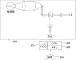
図2は、本実施例を実現するための血管交差・分岐部位の抽出装置の構成図である。901は被検眼を撮像してSLO像を取得するため取得手段として機能するSLO撮像系であり、既存のSLO撮像系で実現できるため、その構成を簡略化して記載してある。902は以下に説明する設定手段、判定手段、演算手段、抽出手段として機能するCPU、903はメモリ、905はハードディスクであり、不図示の表示装置や入力装置と共に本実施例を実現するための情報処理装置を構成する。この構成により、検者904に情報を提示したり、検者904からの指示を受けたりして、種々の情報処理を行う。なお、図1(a)のフローチャートを実現するプログラムはハードディスク905に記憶され、CPU902が読み出して実行する。
FIG. 2 is a configuration diagram of a blood vessel intersection / branch extraction device for realizing the present embodiment.
ここで、用いる眼底画像について説明する。SLO撮像系901が撮像したSLO像をCPU902がメモリ903に保存する。SLO像の大きさは縦2000×横2000画素で、画素の輝度は−32768から32767までである。画像のサイズや輝度はこれらに限定されない。ただし、眼底画像のサイズは、血管の交差・分岐部位が入る程度以上に大きいことが必要である。また、輝度スケールは2のn乗のスケールに設定すると後の計算が容易であるので好ましい。また、用いる画像の形状も、正方形には限定されず、長方形や円形等、いかなる形状としてもよい。
Here, the fundus image used will be described. The
図1(a)のフローチャートに沿って、本実施例における血管交差・分岐部位の抽出の手順を説明する。
まず、CPU902が設定手段として機能して、SLO像の左上隅に位置する正方形の領域を抽出候補領域として選択し、この領域の情報をメモリに保存する。これ以降の処理において、この選択された抽出候補領域に血管の交差・分岐部位が存在するかを判定し、交差・分岐部位があると判定されれば、この抽出候補領域が血管交差・分岐部位として抽出されることになる。抽出候補領域の大きさは、あらかじめ検者904が決定してメモリに保存する。なお、抽出候補領域の大きさに特に制限はなく、画像中の血管交差・分岐部位が入る程度の面積よりも大きければよい。図3(a)に示すように、本実施例では縦140×横140画素とした。なお、これは、本発明者が、本発明を用いて血管交差・分岐部位を抽出し、抽出した領域をテンプレート画像として同一被検者のSLO像にパターンマッチングをかけた際にパターンマッチングでの誤検知がなかった最小のサイズである。また、抽出候補領域の形状も正方形に限定されず、長方形や円形等、いかなる形状としてもよい。
A procedure for extracting a blood vessel intersection / branch site in this embodiment will be described with reference to the flowchart of FIG.
First, the
次に、ステップ101は、CPU902が判定手段として機能して、外周小領域を定め、外周小領域に含まれる各画素の輝度の平均値を計算し、メモリ903に保存させるステップである。まず、CPU902は検者904が指定した外周小領域のサイズに従い、選択された抽出候補領域の外周部に沿って正方形の領域を連続して隙間なく設定する。この、外周部に沿って設定された正方形の領域を、本実施例において外周小領域という。外周小領域の形状も正方形には限定されず、抽出候補領域の外周に沿って連続して設定できれば、いかなる形状としてもよい。ただし、外周小領域の、抽出候補領域の外周に対する幅方向の大きさは血管の太さと同程度であることが必要である。本実施例の画像においては、血管の太さは約20画素にあたるため、検者904は、外周小領域の大きさを縦20×横20画素と決め、不図示の入力装置から入力することによりメモリ903に保存した。
Next,
ここで、図3(a)は、本実施例においてCPU902により抽出された抽出候補領域及び外周小領域を示している。抽出候補領域のサイズは140×140画素で、その外周部に、20×20画素の外周小領域が24個設定されている。
Here, FIG. 3A shows the extraction candidate area and the outer peripheral small area extracted by the
次にCPU902は各外周小領域内に含まれる全ての画素の輝度値を平均し、その値を、各外周小領域を代表する値(これ以降、輝度平均値とも表す)としてメモリ903に保存する。図3(b)にその概念図を示す。
Next, the
ステップ102は、CPU902が、外周小領域の3箇所以上を血管が走っているかどうか判定する数判定のステップである。
まず、CPU902は、隣り合う各外周小領域間の輝度平均値の差の絶対値を求め、メモリ903に保存する。図3(c)にその概念図を示す。その後CPU902は、隣り合う外周小領域間の輝度平均値の差の絶対値と第1の閾値Aの比較、及び隣り合う外周小領域間の低いほうの輝度平均値と第2の閾値Bの比較を実行する。外周小領域間の輝度平均値の差の絶対値が第1の閾値A以上であり、かつ輝度平均値の低いほうの外周小領域の輝度平均値が第2の閾値B以下である場合、低いほうの輝度平均値で表される外周小領域には血管が走っていると判定する。
Step 102 is a number determination step in which the
First, the
第1の閾値A及び第2の閾値Bは、検者904が任意に定めることができる。本実施例においては、検者904は第1の閾値Aを8000、第2の閾値Bを−10000と定めた。そのため、図3(b)と図3(c)から、図3(d)で黒く塗った外周小領域に血管が走っていると判定される。
The
次に、ステップ102においてCPU902は、血管の走っている外周小領域が3つ以上あるか判定する。
血管の走っている外周小領域が3つ以上あると判定した場合、CPU902はステップ103へ進む。3つ未満の場合は、CPU902はステップ105の処理に進む。
ステップ103は、CPU902が演算手段として機能して、外周部血管の座標平均が中央部の範囲内にあるかを判定する中央部判定のステップである。
Next, in
If it is determined that there are three or more outer peripheral small regions where the blood vessel is running, the
Step 103 is a central part determination step in which the
本実施例において、外周部血管とは、CPU902がステップ102で血管が走っていると判定した外周小領域の血管である。抽出候補領域内に2本の血管が存在し、互いに交差している場合、その抽出候補領域の外周部血管の各座標を平均した位置(座標平均)にその血管の交差の中心が存在すると考えられる。そのため、座標平均が抽出候補領域の中央部にあるような抽出候補領域を選択して抽出することで、抽出された画像の中央部に交差部が存在する可能性が高まる。
In the present embodiment, the peripheral blood vessel is a blood vessel in a small peripheral region that the
CPU902は、外周部血管が走っている複数の外周小領域のそれぞれの抽出候補領域での座標位置を用いて、その外周部血管の重心の位置座標を求める。即ち、すべての外周部血管の走っている外周小領域の位置座標値をX軸、Y軸毎に足し合わせ、外周部血管の走っている外周小領域の数で割ることで、外周部血管の座標平均を得る。
The
本実施例において、中央部とは、抽出候補領域と同じ重心を持ち、所定の面積を持つ抽出候補領域内の正方形の領域である。中央部の面積は任意に定めることができるが、1辺の長さが抽出候補領域の5分の1以下(9分の1以上)の大きさを持つ領域(抽出候補領域全体の1/25以下、1/81以上の面積を占める領域)であると、誤判定が改善されるために好ましい(図4(a))。 In this embodiment, the central portion is a square area in the extraction candidate area having the same center of gravity as the extraction candidate area and having a predetermined area. The area of the central portion can be arbitrarily determined, but an area in which the length of one side is 1/5 or less (1/9 or more) of the extraction candidate area (1/25 of the entire extraction candidate area) Hereinafter, it is preferable for the area to occupy an area of 1/81 or more because erroneous determination is improved (FIG. 4A).
ここで、図4(a)の横軸は、抽出候補領域の1辺の長さを1としたときの抽出候補領域に対する中央部の大きさを、それぞれの正方形の1辺の長さの比で表したものである。例えば、横軸に表す中央部の大きさが1/2のときは、中央部の正方形の各1辺は抽出候補領域の1/2となるため、中央部は抽出候補領域の面積の1/4を占める領域ということになる。また、図4(a)の縦軸は、それぞれの大きさの中央部において、等しいSLO像から血管の交差・分岐部位として抽出された抽出候補領域のうち、目視で血管の交差・分岐部位が中央部に認められなかったもの(誤判定)の数である。なお、本実施例では、検者904は、中央部の1辺の長さを抽出候補領域の7分の1と決定した。
Here, the horizontal axis of FIG. 4A represents the size of the central portion with respect to the extraction candidate region when the length of one side of the extraction candidate region is 1, and the ratio of the length of one side of each square. It is represented by. For example, when the size of the central portion represented on the horizontal axis is ½, each side of the central square is ½ of the extraction candidate region, so the central portion is 1 / of the area of the extraction candidate region. This is an area that occupies four. Also, the vertical axis of FIG. 4 (a) shows the intersection / branch portion of the blood vessel visually among the extraction candidate regions extracted as equal to the intersection / branch portion of the blood vessel from the same SLO image at the center of each size. This is the number of items that were not recognized in the center (false determination). In this embodiment, the
外周部血管の平均座標が中央部の領域内に含まれる場合、ステップ104の処理に進み、CPU902が抽出手段として機能して当該抽出候補領域を血管の交差・分岐部位として抽出する。外周部血管の平均座標が中央部の領域内に含まれない場合は、CPU902は抽出候補領域に血管交差・分岐部位があると判定せず、その抽出候補領域は抽出されない。この場合、CPU902はステップ105に進む。
When the average coordinates of the peripheral blood vessels are included in the central region, the process proceeds to step 104, and the
本実施例の図3の例においては、上記のとおり、図3(d)のように外周部血管が分布していると判定されるため、外周部血管の座標平均401は抽出候補領域の中央部402の領域内に含まれる。そのため、CPU902はステップ103の判定をyesとし、次のステップ104において抽出候補領域を血管交差・分岐部位を含む領域として抽出する。
In the example of FIG. 3 of the present embodiment, as described above, since it is determined that the peripheral blood vessels are distributed as shown in FIG. 3D, the coordinate average 401 of the peripheral blood vessels is the center of the extraction candidate region. It is included in the area of the
その後CPU902は、ステップ105に進む。ステップ105はCPU902が終了条件を満たすかどうか判定するステップである。終了条件は検者が任意に決定することができる。本実施例では「眼底画像を全てスキャンしたか」を終了条件とする。CPU902は、現在のステータスが終了条件を満たしていないと判定するとき、抽出候補領域をずらして(ステップ106)、ステップ101の処理へ戻る。ここで、ずらす画素数は検者904が任意に決定することができる。本実施例では画像上で抽出候補領域を右に1画素ずつずらしていき、画像の右端にたどりついたら、その後下方向へ1画素ずらして画像の左端に戻り、再度ステップ101の処理へ戻るとした。
Thereafter, the
CPU902が終了条件(本実施例では「眼底画像を全てスキャンしたか」)を満たすと判定するまで、CPU902は抽出候補領域をずらしながら(ステップ106)上記ステップ101からステップ105までの判定・処理を繰り返し、眼底画像をスキャンしていく。CPU902が終了条件を満たす(本実施例では眼底画像を全てスキャンし終えた)と判定したタイミングで処理を終了する。
Until the
仮に、終了条件を満たすまで条件を満たす領域がステップ104で1つも抽出されない場合は、最も条件に近い領域をステップ103のNoの判定の後でメモリ903に保存しておいてその領域を抽出するようにする、あるいは、条件を緩めて再度始めから処理を実施するようにすることもできる。
If no region that satisfies the condition until the end condition is satisfied is extracted in
以上の処理を行うことで、抽出候補領域の中央部を通る血管があるとき、交差・分岐部位以外を交差・分岐部位と誤判定した領域を抽出する確率が低減される。また、抽出領域の中央部に血管の交差・分岐部位の中心がある領域を抽出する確率を高めることができる。 By performing the above processing, when there is a blood vessel passing through the center of the extraction candidate region, the probability of extracting a region erroneously determined as a crossing / branching part other than a crossing / branching part is reduced. In addition, it is possible to increase the probability of extracting a region having the center of the crossing / branching portion of the blood vessel at the center of the extraction region.
[実施例2]
実施例2では、実施例1に示した交差・分岐部位の抽出方法において、抽出候補領域の中央部を血管が通ることを抽出の条件として構成する。
[Example 2]
In the second embodiment, in the method for extracting the intersection / branch portion shown in the first embodiment, the extraction condition is configured such that the blood vessel passes through the center of the extraction candidate region.
図1(b)のフローチャートに沿って、実施例2を説明する。なお、本実施例においても図2の装置を用いる。装置の構成は実施例1と同様であるため、その説明は省略する。
SLO像は実施例1で用いたものと同じである。図1(b)のフローチャートの各ステップを実現するプログラムは、ハードディスク905に記憶され、CPU902が読み出して実行する。
Example 2 will be described with reference to the flowchart of FIG. In this embodiment, the apparatus shown in FIG. 2 is also used. Since the configuration of the apparatus is the same as that of the first embodiment, the description thereof is omitted.
The SLO image is the same as that used in Example 1. A program for realizing each step of the flowchart of FIG. 1B is stored in the
まず、CPU902がSLO像の左上隅を抽出候補領域として選ぶ。抽出候補領域の大きさは、検者904があらかじめ決定してメモリ903に保存する。本実施例においても、抽出候補領域の大きさや形状に特に制限はなく、血管交差・分岐部位が入る程度よりも大きければよい。抽出候補領域の大きさは、実施例1と同様に、縦140×横140画素とした。ここで、ステップ101、102は実施例1における図1(a)のステップ101、102と基本的には同じステップである。ただし、ステップ102で、CPU902が、血管の走っている外周小領域が3つ以上あると判定した場合、本実施例ではステップ107に進む。
First, the
ステップ107は、CPU902が、抽出候補領域の中央小領域を血管が走っているかどうか判定する血管判定の工程である。ここで、中央小領域は抽出候補領域と同じ重心をもつ正方形の領域である。中央小領域の大きさは検者904が任意に定めることができるが、血管の太さと同じ程度の幅を持っていることが望ましい。本実施例においては、前述のように、血管の太さは20画素程度であるので、検者904は中央小領域の大きさを20×20画素と決定し、不図示の入力装置から入力することによりメモリに保存した。
Step 107 is a blood vessel determination process in which the
CPU902は、抽出候補領域の中央小領域を血管が走っているかどうかを以下のように判定する。まず、CPU902は中央小領域の各画素の輝度を平均し、平均値を取る。この平均値と、検者904があらかじめメモリに保存した第3の閾値Dを比較し、この平均値が第3の閾値D以下であるとき、中央小領域には血管が走っていると判定する。
The
第3の閾値Dは、検者904が任意に定めることができる。本実施例において、検者904は、第3の閾値Dを、外周小領域に血管があるかどうか判定した際の第2の閾値Bと等しく−10000と定めた。CPU902が中央小領域に血管が走っていると判定した場合、ステップ103へ進む。中央小領域を血管が走っていると判定しなかった場合は、抽出候補領域をずらし(ステップ106)、上記ステップ101へ戻る。
The
ステップ103以降の処理は、実施例1と同様である。ここで、ステップ103の「中央部」について、実施例1と同様にそれぞれの大きさの中央部において血管の交差・分岐部位として抽出された抽出候補領域のうちの誤判定数を計測した(図4(b))。その結果、「中央部」は抽出候補領域の大きさの5分の1以下(9分の1以上)の大きさを持つ領域であることが望ましいことがわかった。さらに、中央部が抽出候補領域の大きさの5分の1以下(9分の1以上)の場合、誤判定は検出されず、本実施例においては血管の交差・分岐部位を実施例1よりもより正確に抽出できることが分かった。本実施例では、検者904が、抽出候補領域の7分の1の大きさを持ち、その重心が抽出候補領域の重心と等しい領域を中央部と決定した。
The processing after
以降は実施例1と同じである。
このように構成することにより、実施例1と比較して、抽出領域の中央部に血管の交差・分岐部位の中心がある領域を抽出する確率をより高めることができる。
The subsequent steps are the same as in the first embodiment.
By configuring in this way, it is possible to further increase the probability of extracting a region having the center of the crossing / branching portion of the blood vessel at the center of the extraction region as compared with the first embodiment.
[実施例3]
実施例3では、血管の交差・分岐部位を抽出する眼底画像としてフルオレッセン蛍光眼底画像(FA)やインドシアニングリーン眼底造影画像(IA)を用いる。FAやIAの画像内では、血管はその他の領域に比べて高輝度になる。なお、本実施例においても、血管がその他の領域に比べて高輝度である二次元画像であれば、どのような眼底画像をも用いることができる。
[Example 3]
In Example 3, a fluorescein fluorescence fundus image (FA) or an indocyanine green fundus angiogram (IA) is used as a fundus image for extracting a blood vessel intersection / branch site. In the FA or IA image, the blood vessel has a higher brightness than other regions. Also in this embodiment, any fundus image can be used as long as the blood vessel is a two-dimensional image having a higher luminance than other regions.
手順は実施例2とほぼ同じで、同様に図2に記載の装置を用いる。ハードディスク905に記憶された図1(b)のフローチャートを実現するためのプログラムをCPU902が実行して処理が進むが、実施例2とは図1(b)のフローチャートのステップ102とステップ107が異なる。以下、実施例2と重複する部分の説明は省略し、異なる部分について説明する。
The procedure is almost the same as in Example 2, and the apparatus shown in FIG. 2 is used in the same manner. The
検者904はあらかじめ、任意に定めた第1の閾値Aと第2の閾値Cと第3の閾値Eを、不図示の入力装置から入力しメモリに保存させる。
The
図1(b)のフローチャートのステップ102では、まず、CPU902が隣り合う各外周小領域間の輝度平均値の差の絶対値を求める。CPU902が、輝度平均値の差の絶対値が閾値A以上であり、また輝度平均値の高いほうが閾値C以上であると判定した場合、閾値C以上の輝度平均値で代表される外周小領域には血管が走っているとする。
In
図1(b)のフローチャートのステップ107では、CPU902が中央小領域の画素の輝度を平均し、平均値を取る。この平均値が閾値E以上であるとき、CPU902は中央小領域には血管が走っていると判定する。
In
他の処理は実施例2と同じである。
このように構成することにより、血管がその他の領域に比べて高輝度で撮影される場合でも、実施例2と同じ効果を得ることができる。
Other processes are the same as those in the second embodiment.
By configuring in this way, the same effect as in the second embodiment can be obtained even when the blood vessel is imaged with higher brightness than other regions.
401 座標平均
402 中央部
901 SLO撮像系
902 CPU
903 メモリ
904 検者
905 ハードディスク
401 Coordinate average 402
903
Claims (22)
前記眼底画像の抽出候補領域の外周部における外周小領域を決定する工程と、
前記決定された外周小領域に存在する複数の外周部血管の座標平均が前記抽出候補領域の中央部に含まれるかどうかを判定する工程と、
前記中央部に含まれると判定された場合に、前記抽出候補領域を血管交差・分岐部位を含む領域として抽出する工程と、を含むことを特徴とする抽出方法。 A method for extracting a blood vessel crossing / branching part that extracts a region including a blood vessel crossing / branching part from a fundus image containing a blood vessel of a subject eye,
Determining an outer peripheral small region in an outer peripheral portion of the extraction candidate region of the fundus image;
Determining whether a coordinate average of a plurality of outer peripheral blood vessels present in the determined outer peripheral small region is included in a central portion of the extraction candidate region; and
Extracting the extraction candidate region as a region including a blood vessel intersection / branch portion when it is determined that the extraction candidate region is included in the central portion.
前記抽出する工程において、前記数を判定する工程と前記中央部に含まれるかどうかを判定する工程との判定結果に従って、前記抽出候補領域を血管交差・分岐部位を含む領域として抽出することを特徴とする、請求項1に記載の抽出方法。 Further comprising determining a number of the plurality of peripheral blood vessels,
In the extracting step, the extraction candidate region is extracted as a region including a blood vessel intersection / branch site according to a determination result between the step of determining the number and the step of determining whether or not the number is included in the central portion. The extraction method according to claim 1.
前記決定された外周小領域における複数の外周部血管の座標を平均して得た座標が前記抽出候補領域の中央部に含まれるか否かを判定する工程と、
前記中央部に含まれると判定され、且つ前記決定された複数の外周部血管の数が所定の数以上である場合に、前記抽出候補領域を血管交差・分岐部位を含む領域として抽出する工程と、
を含むことを特徴とする抽出方法。 Determining the outer peripheral small region in the outer peripheral portion of the extraction candidate region of the fundus image of the eye to be examined;
A step of determining whether the coordinate obtained by averaging the coordinates of a plurality of outer peripheral portion blood vessels are included in the central portion of the extraction candidate region before Symbol determined circumferential small regions,
A step of extracting the extraction candidate region as a region including a blood vessel intersection / branch site when it is determined that the blood vessel is included in the central portion and the determined number of the plurality of peripheral blood vessels is equal to or greater than a predetermined number; ,
The extraction method characterized by including.
前記中央部に含まれるか否かを判定する工程において、前記判定された複数の外周部血管の座標を平均して得た座標が前記抽出候補領域の中央部に含まれるか否かを判定することを特徴とする請求項7に記載の抽出方法。 And the absolute value of the difference before Symbol determined circumferential small region above the first threshold, and a small area lower luminance value is below the second threshold value of the adjacent small regions, Further comprising the step of determining as a peripheral blood vessel ,
In determining whether included in the central portion, the determined plurality of outer peripheral portions coordinates obtained by averaging the coordinates of the vessel to determine whether contained in a central portion of the extraction candidate region The extraction method according to claim 7 .
前記中央部に含まれるか否かを判定する工程において、前記判定された複数の外周部血管の座標を平均して得た座標が前記抽出候補領域の中央部に含まれるか否かを判定することを特徴とする請求項7に記載の抽出方法。 Wherein when said on the fundus image blood vessel cross-luminance region including the branch portion is higher than the luminance of another area when the absolute value of the difference before Symbol determined circumferential small region above the first threshold value, And a step of determining, as an outer peripheral blood vessel, a small region having a higher luminance value than the second threshold value among the adjacent small regions ,
In determining whether included in the central portion, the determined plurality of outer peripheral portions coordinates obtained by averaging the coordinates of the vessel to determine whether contained in a central portion of the extraction candidate region The extraction method according to claim 7 .
前記眼底画像と、前記眼底画像とは異なる時刻に得られた前記被検眼の他の眼底画像とのマッチングを行なう工程と、
をさらに含むことを特徴とする請求項1から13のいずれか1項に記載の抽出方法。 Imaging the eye to be examined and obtaining an SLO image as the fundus image;
Matching the fundus image with another fundus image of the eye to be examined obtained at a different time from the fundus image;
Further extraction method according to any one of claims 1 to 13, characterized in that it comprises a.
前記眼底画像の抽出候補領域の外周部における外周小領域を決定する決定手段と、
前記決定された外周小領域に存在する複数の外周部血管の座標平均が前記抽出候補領域の中央部に含まれるかどうかを判定する中央部判定手段と、
前記中央部に含まれると判定された場合に、前記抽出候補領域を血管交差・分岐部位を含む領域として抽出する抽出手段と、
を含むことを特徴とする抽出装置。 A blood vessel crossing / branching site extraction device that extracts a region including a blood vessel crossing / branching site from a fundus image including a blood vessel of a subject eye,
Determining means for determining an outer peripheral small region in an outer peripheral portion of the extraction candidate region of the fundus image;
A central portion determination means for determining whether or not a coordinate average of a plurality of peripheral blood vessels present in the determined peripheral small region is included in a central portion of the extraction candidate region;
An extraction means for extracting the extraction candidate region as a region including a blood vessel intersection / branch site when it is determined to be included in the central portion;
The extraction apparatus characterized by including.
前記抽出手段は、前記数判定手段と前記中央部判定手段との判定結果に従って、前記抽出候補領域を血管交差・分岐部位を含む領域として抽出することを特徴とする請求項17に記載の抽出装置。 A number determining means for determining the number of the plurality of peripheral blood vessels,
18. The extraction apparatus according to claim 17, wherein the extraction unit extracts the extraction candidate region as a region including a blood vessel intersection / branch site according to the determination results of the number determination unit and the central portion determination unit. .
前記決定された外周小領域における複数の外周部血管の座標を平均して得た座標が前記抽出候補領域の中央部に含まれるか否かを判定する中央部判定手段と、
前記中央部判定手段により前記中央部に含まれると判定され、且つ前記決定された複数の外周部血管の数が所定の数以上である場合に、前記抽出候補領域を血管交差・分岐部位を含む領域として抽出する抽出手段と、
を有することを特徴とする抽出装置。 Determining means for determining the outer peripheral small area in the outer peripheral part of the extraction candidate area of the fundus image of the eye to be examined;
A central portion determining means for determining whether the coordinates obtained by averaging the coordinates of a plurality of outer peripheral portions vessel prior Symbol determined circumferential small region is included in a central portion of the extraction candidate region,
When it is determined that the central part is included in the central part by the central part determination unit, and the determined number of outer peripheral blood vessels is equal to or greater than a predetermined number, the extraction candidate region includes a blood vessel intersection / branch site. Extraction means for extracting as a region ;
An extraction device comprising:
前記眼底画像と、前記眼底画像とは異なる時刻に得られた前記被検眼の他の眼底画像とのマッチングを行なう手段と、
をさらに有することを特徴とする請求項17から20のいずれか1項に記載の抽出装置。 Acquisition means for imaging the eye to be examined and acquiring an SLO image as the fundus image;
Means for matching the fundus image with another fundus image of the eye to be examined obtained at a time different from the fundus image;
The extraction device according to claim 17, further comprising:
前記中央部判定手段は、前記決定された複数の外周小領域のうち少なくとも3つに存在する複数の外周部血管の座標を平均して得た座標が前記抽出候補領域の中央部に含まれるか否かを判定することを特徴とする請求項17から21のいずれか1項に記載の抽出装置。 Whether the central part determination means includes coordinates obtained by averaging coordinates of a plurality of peripheral blood vessels existing in at least three of the determined plurality of peripheral small areas in the central part of the extraction candidate area. The extraction device according to any one of claims 17 to 21, wherein it is determined whether or not.
Priority Applications (4)
| Application Number | Priority Date | Filing Date | Title |
|---|---|---|---|
| JP2009209387A JP5432644B2 (en) | 2009-09-10 | 2009-09-10 | Extraction method and apparatus for blood vessel intersection / branch site |
| US12/874,939 US8634600B2 (en) | 2009-09-10 | 2010-09-02 | Extracting method and apparatus of blood vessel crossing/branching portion |
| CN2010102771744A CN102024254A (en) | 2009-09-10 | 2010-09-07 | Method and apparatus for detection of crossing/branching of blood vessels |
| EP10175755A EP2294970A1 (en) | 2009-09-10 | 2010-09-08 | Detection of crossing/branching of blood vessels |
Applications Claiming Priority (1)
| Application Number | Priority Date | Filing Date | Title |
|---|---|---|---|
| JP2009209387A JP5432644B2 (en) | 2009-09-10 | 2009-09-10 | Extraction method and apparatus for blood vessel intersection / branch site |
Publications (3)
| Publication Number | Publication Date |
|---|---|
| JP2011056068A JP2011056068A (en) | 2011-03-24 |
| JP2011056068A5 JP2011056068A5 (en) | 2013-05-16 |
| JP5432644B2 true JP5432644B2 (en) | 2014-03-05 |
Family
ID=43086174
Family Applications (1)
| Application Number | Title | Priority Date | Filing Date |
|---|---|---|---|
| JP2009209387A Expired - Fee Related JP5432644B2 (en) | 2009-09-10 | 2009-09-10 | Extraction method and apparatus for blood vessel intersection / branch site |
Country Status (4)
| Country | Link |
|---|---|
| US (1) | US8634600B2 (en) |
| EP (1) | EP2294970A1 (en) |
| JP (1) | JP5432644B2 (en) |
| CN (1) | CN102024254A (en) |
Families Citing this family (11)
| Publication number | Priority date | Publication date | Assignee | Title |
|---|---|---|---|---|
| JP5355316B2 (en) * | 2009-09-10 | 2013-11-27 | キヤノン株式会社 | Template image evaluation method and biological motion detection apparatus |
| US8805038B2 (en) * | 2011-06-30 | 2014-08-12 | National Taiwan University | Longitudinal image registration algorithm for infrared images for chemotherapy response monitoring and early detection of breast cancers |
| US9418423B2 (en) * | 2011-08-09 | 2016-08-16 | Optovue, Inc. | Motion correction and normalization of features in optical coherence tomography |
| JP5912358B2 (en) * | 2011-09-14 | 2016-04-27 | 株式会社トプコン | Fundus observation device |
| WO2015013632A1 (en) * | 2013-07-26 | 2015-01-29 | The Regents Of The University Of Michigan | Automated measurement of changes in retinal, retinal pigment epithelial, or choroidal disease |
| US9390327B2 (en) * | 2013-09-16 | 2016-07-12 | Eyeverify, Llc | Feature extraction and matching for biometric authentication |
| CN105635611A (en) * | 2014-11-03 | 2016-06-01 | 中兴通讯股份有限公司 | Projector self-adaptive adjusting method and device |
| US9757023B2 (en) | 2015-05-27 | 2017-09-12 | The Regents Of The University Of Michigan | Optic disc detection in retinal autofluorescence images |
| JP6995485B2 (en) * | 2017-03-03 | 2022-01-14 | キヤノン株式会社 | Ophthalmic appliances, device control methods and programs |
| CN108073918B (en) * | 2018-01-26 | 2022-04-29 | 浙江大学 | Method for extracting blood vessel arteriovenous cross compression characteristics of fundus retina |
| CN115409690B (en) * | 2021-05-28 | 2023-09-29 | 南京博视医疗科技有限公司 | Real-time fundus image mapping method and device |
Family Cites Families (7)
| Publication number | Priority date | Publication date | Assignee | Title |
|---|---|---|---|---|
| US6104828A (en) * | 1994-03-24 | 2000-08-15 | Kabushiki Kaisha Topcon | Ophthalmologic image processor |
| US20040208343A1 (en) * | 1998-07-09 | 2004-10-21 | Colorado State University Research Foundation | Apparatus and method for creating a record using biometric information |
| JP2001070247A (en) * | 1999-09-07 | 2001-03-21 | Takku Kk | Method of detecting blood vessel crossing part in image of eyeground |
| CN1147815C (en) | 2002-04-29 | 2004-04-28 | 华南理工大学 | Computer aided characteristic registration identifying method for medical homolateral fundus image |
| JP2007097633A (en) * | 2005-09-30 | 2007-04-19 | Gifu Univ | Image analysis system and image analysis program |
| CN101002682A (en) | 2007-01-19 | 2007-07-25 | 哈尔滨工程大学 | Method for retrieval and matching of hand back vein characteristic used for identification of status |
| JP2009106532A (en) * | 2007-10-30 | 2009-05-21 | Topcon Corp | System and program for processing ophthalmologic information |
-
2009
- 2009-09-10 JP JP2009209387A patent/JP5432644B2/en not_active Expired - Fee Related
-
2010
- 2010-09-02 US US12/874,939 patent/US8634600B2/en not_active Expired - Fee Related
- 2010-09-07 CN CN2010102771744A patent/CN102024254A/en active Pending
- 2010-09-08 EP EP10175755A patent/EP2294970A1/en not_active Withdrawn
Also Published As
| Publication number | Publication date |
|---|---|
| CN102024254A (en) | 2011-04-20 |
| JP2011056068A (en) | 2011-03-24 |
| EP2294970A1 (en) | 2011-03-16 |
| US20110058718A1 (en) | 2011-03-10 |
| US8634600B2 (en) | 2014-01-21 |
Similar Documents
| Publication | Publication Date | Title |
|---|---|---|
| JP5432644B2 (en) | Extraction method and apparatus for blood vessel intersection / branch site | |
| US9872614B2 (en) | Image processing apparatus, method for image processing, image pickup system, and computer-readable storage medium | |
| US9959622B2 (en) | Method and apparatus for supporting diagnosis of region of interest by providing comparison image | |
| JP5355316B2 (en) | Template image evaluation method and biological motion detection apparatus | |
| EP2453790A1 (en) | Image processing apparatus, image processing method, and program | |
| JP2013031527A (en) | Ophthalmic diagnosis support apparatus and method | |
| AU2010286345A1 (en) | Feature detection and measurement in retinal images | |
| CN106846293B (en) | Image processing method and device | |
| JP2011056068A5 (en) | ||
| JP2008022928A (en) | Image analysis apparatus and image analysis program | |
| US10573007B2 (en) | Image processing apparatus, image processing method, and image processing program | |
| JP6128841B2 (en) | Image processing device | |
| KR20160118037A (en) | Apparatus and method for detecting lesion from medical image automatically | |
| JP5412242B2 (en) | Ultrasonic tomographic image processing device | |
| KR101501515B1 (en) | Diagnosis image apparatus and operating method thereof | |
| EP3138472A1 (en) | Image-processing device, image-processing method and image-processing program | |
| JP2009189586A (en) | Fundus image analysis method, its instrument and program | |
| CN111712851B (en) | Image processing device, image processing method, and image processing program | |
| EP3129955B1 (en) | Method for the analysis of image data representing a three-dimensional volume of biological tissue | |
| JP2008079682A (en) | Image analysis apparatus and image analysis program | |
| JP7467875B2 (en) | IMAGE PROCESSING APPARATUS, IMAGE PROCESSING METHOD, AND IMAGE PROCESSING PROGRAM | |
| JP6419249B2 (en) | Image processing apparatus, image processing method, and image processing program | |
| EP3338620A1 (en) | Image processing device, image processing method, and image processing program |
Legal Events
| Date | Code | Title | Description |
|---|---|---|---|
| RD05 | Notification of revocation of power of attorney |
Free format text: JAPANESE INTERMEDIATE CODE: A7425 Effective date: 20120727 |
|
| RD05 | Notification of revocation of power of attorney |
Free format text: JAPANESE INTERMEDIATE CODE: A7425 Effective date: 20120730 |
|
| RD05 | Notification of revocation of power of attorney |
Free format text: JAPANESE INTERMEDIATE CODE: A7425 Effective date: 20120731 |
|
| RD03 | Notification of appointment of power of attorney |
Free format text: JAPANESE INTERMEDIATE CODE: A7423 Effective date: 20120831 |
|
| A621 | Written request for application examination |
Free format text: JAPANESE INTERMEDIATE CODE: A621 Effective date: 20120910 |
|
| A521 | Written amendment |
Free format text: JAPANESE INTERMEDIATE CODE: A523 Effective date: 20130325 |
|
| A977 | Report on retrieval |
Free format text: JAPANESE INTERMEDIATE CODE: A971007 Effective date: 20130702 |
|
| RD05 | Notification of revocation of power of attorney |
Free format text: JAPANESE INTERMEDIATE CODE: A7425 Effective date: 20130701 |
|
| A131 | Notification of reasons for refusal |
Free format text: JAPANESE INTERMEDIATE CODE: A131 Effective date: 20130716 |
|
| A521 | Written amendment |
Free format text: JAPANESE INTERMEDIATE CODE: A523 Effective date: 20130912 |
|
| TRDD | Decision of grant or rejection written | ||
| A01 | Written decision to grant a patent or to grant a registration (utility model) |
Free format text: JAPANESE INTERMEDIATE CODE: A01 Effective date: 20131107 |
|
| A61 | First payment of annual fees (during grant procedure) |
Free format text: JAPANESE INTERMEDIATE CODE: A61 Effective date: 20131206 |
|
| R151 | Written notification of patent or utility model registration |
Ref document number: 5432644 Country of ref document: JP Free format text: JAPANESE INTERMEDIATE CODE: R151 |
|
| LAPS | Cancellation because of no payment of annual fees |