JP5219513B2 - Polypeptide compounds for angiogenesis and tumor growth inhibition - Google Patents
Polypeptide compounds for angiogenesis and tumor growth inhibition Download PDFInfo
- Publication number
- JP5219513B2 JP5219513B2 JP2007533653A JP2007533653A JP5219513B2 JP 5219513 B2 JP5219513 B2 JP 5219513B2 JP 2007533653 A JP2007533653 A JP 2007533653A JP 2007533653 A JP2007533653 A JP 2007533653A JP 5219513 B2 JP5219513 B2 JP 5219513B2
- Authority
- JP
- Japan
- Prior art keywords
- ephb4
- polypeptide
- ephrin
- protein
- cells
- Prior art date
- Legal status (The legal status is an assumption and is not a legal conclusion. Google has not performed a legal analysis and makes no representation as to the accuracy of the status listed.)
- Active
Links
- 108090000765 processed proteins & peptides Proteins 0.000 title claims description 309
- 102000004196 processed proteins & peptides Human genes 0.000 title claims description 301
- 229920001184 polypeptide Polymers 0.000 title claims description 289
- 230000033115 angiogenesis Effects 0.000 title claims description 60
- 230000005764 inhibitory process Effects 0.000 title claims description 34
- 230000004614 tumor growth Effects 0.000 title claims description 22
- 150000001875 compounds Chemical class 0.000 title description 67
- 102100031983 Ephrin type-B receptor 4 Human genes 0.000 claims description 453
- 108010044090 Ephrin-B2 Proteins 0.000 claims description 187
- 102000006396 Ephrin-B2 Human genes 0.000 claims description 186
- 206010028980 Neoplasm Diseases 0.000 claims description 155
- 230000000694 effects Effects 0.000 claims description 84
- 239000003814 drug Substances 0.000 claims description 74
- 125000003275 alpha amino acid group Chemical group 0.000 claims description 60
- 102000030797 EphB4 Receptor Human genes 0.000 claims description 48
- 108010055323 EphB4 Receptor Proteins 0.000 claims description 46
- 201000011510 cancer Diseases 0.000 claims description 42
- 210000001519 tissue Anatomy 0.000 claims description 39
- 208000007766 Kaposi sarcoma Diseases 0.000 claims description 34
- 239000000203 mixture Substances 0.000 claims description 34
- 108091006905 Human Serum Albumin Proteins 0.000 claims description 30
- 102000008100 Human Serum Albumin Human genes 0.000 claims description 30
- 208000037265 diseases, disorders, signs and symptoms Diseases 0.000 claims description 26
- 238000001727 in vivo Methods 0.000 claims description 24
- 210000002966 serum Anatomy 0.000 claims description 21
- 230000012010 growth Effects 0.000 claims description 20
- 239000000178 monomer Substances 0.000 claims description 19
- 230000002401 inhibitory effect Effects 0.000 claims description 16
- 201000010099 disease Diseases 0.000 claims description 15
- 206010027406 Mesothelioma Diseases 0.000 claims description 12
- 230000003993 interaction Effects 0.000 claims description 11
- 239000008194 pharmaceutical composition Substances 0.000 claims description 7
- 206010006187 Breast cancer Diseases 0.000 claims description 6
- 208000026310 Breast neoplasm Diseases 0.000 claims description 6
- 230000001404 mediated effect Effects 0.000 claims description 6
- 102100037362 Fibronectin Human genes 0.000 claims description 5
- 108010067306 Fibronectins Proteins 0.000 claims description 5
- 125000000539 amino acid group Chemical group 0.000 claims description 5
- 208000032839 leukemia Diseases 0.000 claims description 5
- 108091000080 Phosphotransferase Proteins 0.000 claims description 4
- 230000001419 dependent effect Effects 0.000 claims description 4
- 239000003937 drug carrier Substances 0.000 claims description 4
- 102000020233 phosphotransferase Human genes 0.000 claims description 4
- 239000002537 cosmetic Substances 0.000 claims description 3
- 206010009944 Colon cancer Diseases 0.000 claims description 2
- 208000029742 colonic neoplasm Diseases 0.000 claims description 2
- 208000037819 metastatic cancer Diseases 0.000 claims description 2
- 208000011575 metastatic malignant neoplasm Diseases 0.000 claims description 2
- 208000023958 prostate neoplasm Diseases 0.000 claims description 2
- 201000001513 prostate squamous cell carcinoma Diseases 0.000 claims description 2
- 238000012447 xenograft mouse model Methods 0.000 claims description 2
- 230000011664 signaling Effects 0.000 claims 2
- 210000004027 cell Anatomy 0.000 description 244
- 108090000623 proteins and genes Proteins 0.000 description 165
- 230000014509 gene expression Effects 0.000 description 144
- 102000004169 proteins and genes Human genes 0.000 description 139
- 235000018102 proteins Nutrition 0.000 description 133
- 238000000034 method Methods 0.000 description 91
- 229920001223 polyethylene glycol Polymers 0.000 description 82
- 239000002202 Polyethylene glycol Substances 0.000 description 71
- 230000027455 binding Effects 0.000 description 62
- 235000001014 amino acid Nutrition 0.000 description 53
- 229940024606 amino acid Drugs 0.000 description 52
- 238000009739 binding Methods 0.000 description 52
- 208000000236 Prostatic Neoplasms Diseases 0.000 description 49
- 150000001413 amino acids Chemical class 0.000 description 49
- 206010060862 Prostate cancer Diseases 0.000 description 48
- FAPWRFPIFSIZLT-UHFFFAOYSA-M Sodium chloride Chemical compound [Na+].[Cl-] FAPWRFPIFSIZLT-UHFFFAOYSA-M 0.000 description 46
- 108020004459 Small interfering RNA Proteins 0.000 description 45
- 239000003446 ligand Substances 0.000 description 43
- 229940079593 drug Drugs 0.000 description 39
- 102000037865 fusion proteins Human genes 0.000 description 39
- 108020001507 fusion proteins Proteins 0.000 description 39
- 238000012360 testing method Methods 0.000 description 39
- 239000012634 fragment Substances 0.000 description 37
- 108010082117 matrigel Proteins 0.000 description 36
- 238000011282 treatment Methods 0.000 description 36
- 238000003556 assay Methods 0.000 description 34
- 230000004927 fusion Effects 0.000 description 32
- 239000000523 sample Substances 0.000 description 32
- 241000699670 Mus sp. Species 0.000 description 30
- 210000004881 tumor cell Anatomy 0.000 description 30
- 239000003153 chemical reaction reagent Substances 0.000 description 29
- 102000012803 ephrin Human genes 0.000 description 28
- 108060002566 ephrin Proteins 0.000 description 28
- 239000013598 vector Substances 0.000 description 25
- 150000007523 nucleic acids Chemical class 0.000 description 24
- 239000011780 sodium chloride Substances 0.000 description 24
- 102000009027 Albumins Human genes 0.000 description 23
- 108010088751 Albumins Proteins 0.000 description 23
- 230000006907 apoptotic process Effects 0.000 description 23
- 241001502974 Human gammaherpesvirus 8 Species 0.000 description 22
- 210000002889 endothelial cell Anatomy 0.000 description 22
- -1 polyethylene Polymers 0.000 description 21
- 102000005789 Vascular Endothelial Growth Factors Human genes 0.000 description 20
- 108010019530 Vascular Endothelial Growth Factors Proteins 0.000 description 20
- 239000003102 growth factor Substances 0.000 description 20
- 108020004707 nucleic acids Proteins 0.000 description 20
- 102000039446 nucleic acids Human genes 0.000 description 20
- 102000001301 EGF receptor Human genes 0.000 description 19
- 108060006698 EGF receptor Proteins 0.000 description 19
- 102000050554 Eph Family Receptors Human genes 0.000 description 19
- 108091008815 Eph receptors Proteins 0.000 description 19
- 239000011159 matrix material Substances 0.000 description 19
- 239000012528 membrane Substances 0.000 description 19
- 230000006320 pegylation Effects 0.000 description 19
- 230000026731 phosphorylation Effects 0.000 description 18
- 238000006366 phosphorylation reaction Methods 0.000 description 18
- 230000001965 increasing effect Effects 0.000 description 17
- 230000001225 therapeutic effect Effects 0.000 description 17
- 238000001262 western blot Methods 0.000 description 17
- 239000000463 material Substances 0.000 description 16
- 230000004048 modification Effects 0.000 description 16
- 238000012986 modification Methods 0.000 description 16
- 239000013612 plasmid Substances 0.000 description 16
- 108091003079 Bovine Serum Albumin Proteins 0.000 description 15
- 241001465754 Metazoa Species 0.000 description 15
- OKKJLVBELUTLKV-UHFFFAOYSA-N Methanol Chemical compound OC OKKJLVBELUTLKV-UHFFFAOYSA-N 0.000 description 15
- 241000699666 Mus <mouse, genus> Species 0.000 description 15
- 230000000692 anti-sense effect Effects 0.000 description 15
- 230000015572 biosynthetic process Effects 0.000 description 15
- 238000000338 in vitro Methods 0.000 description 15
- 230000001105 regulatory effect Effects 0.000 description 15
- 230000004044 response Effects 0.000 description 15
- PEDCQBHIVMGVHV-UHFFFAOYSA-N Glycerine Chemical compound OCC(O)CO PEDCQBHIVMGVHV-UHFFFAOYSA-N 0.000 description 14
- 108091028043 Nucleic acid sequence Proteins 0.000 description 14
- 239000002609 medium Substances 0.000 description 14
- 238000013508 migration Methods 0.000 description 14
- 230000035772 mutation Effects 0.000 description 14
- 229940046166 oligodeoxynucleotide Drugs 0.000 description 14
- 102000005962 receptors Human genes 0.000 description 14
- 108020003175 receptors Proteins 0.000 description 14
- 230000002829 reductive effect Effects 0.000 description 14
- 108090000379 Fibroblast growth factor 2 Proteins 0.000 description 13
- 102000003974 Fibroblast growth factor 2 Human genes 0.000 description 13
- 108060003951 Immunoglobulin Proteins 0.000 description 13
- 206010028851 Necrosis Diseases 0.000 description 13
- 229940098773 bovine serum albumin Drugs 0.000 description 13
- 239000000872 buffer Substances 0.000 description 13
- 230000007423 decrease Effects 0.000 description 13
- 239000013604 expression vector Substances 0.000 description 13
- 102000018358 immunoglobulin Human genes 0.000 description 13
- 210000004379 membrane Anatomy 0.000 description 13
- 230000005012 migration Effects 0.000 description 13
- 230000017074 necrotic cell death Effects 0.000 description 13
- 238000010186 staining Methods 0.000 description 13
- 229940124597 therapeutic agent Drugs 0.000 description 13
- 230000002792 vascular Effects 0.000 description 13
- LFQSCWFLJHTTHZ-UHFFFAOYSA-N Ethanol Chemical compound CCO LFQSCWFLJHTTHZ-UHFFFAOYSA-N 0.000 description 12
- 230000004913 activation Effects 0.000 description 12
- 230000003833 cell viability Effects 0.000 description 12
- 239000000047 product Substances 0.000 description 12
- 239000000243 solution Substances 0.000 description 12
- 239000000126 substance Substances 0.000 description 12
- 238000001890 transfection Methods 0.000 description 12
- 230000035899 viability Effects 0.000 description 12
- XUJNEKJLAYXESH-REOHCLBHSA-N L-Cysteine Chemical compound SC[C@H](N)C(O)=O XUJNEKJLAYXESH-REOHCLBHSA-N 0.000 description 11
- 238000006243 chemical reaction Methods 0.000 description 11
- 239000000306 component Substances 0.000 description 11
- 230000000875 corresponding effect Effects 0.000 description 11
- 239000012149 elution buffer Substances 0.000 description 11
- 238000011534 incubation Methods 0.000 description 11
- 239000002243 precursor Substances 0.000 description 11
- 238000000159 protein binding assay Methods 0.000 description 11
- 238000005406 washing Methods 0.000 description 11
- 108020004414 DNA Proteins 0.000 description 10
- 102100033237 Pro-epidermal growth factor Human genes 0.000 description 10
- 108010076504 Protein Sorting Signals Proteins 0.000 description 10
- 102000007562 Serum Albumin Human genes 0.000 description 10
- 108010071390 Serum Albumin Proteins 0.000 description 10
- RJURFGZVJUQBHK-UHFFFAOYSA-N actinomycin D Natural products CC1OC(=O)C(C(C)C)N(C)C(=O)CN(C)C(=O)C2CCCN2C(=O)C(C(C)C)NC(=O)C1NC(=O)C1=C(N)C(=O)C(C)=C2OC(C(C)=CC=C3C(=O)NC4C(=O)NC(C(N5CCCC5C(=O)N(C)CC(=O)N(C)C(C(C)C)C(=O)OC4C)=O)C(C)C)=C3N=C21 RJURFGZVJUQBHK-UHFFFAOYSA-N 0.000 description 10
- 125000003277 amino group Chemical group 0.000 description 10
- 208000035475 disorder Diseases 0.000 description 10
- 230000006870 function Effects 0.000 description 10
- 238000012216 screening Methods 0.000 description 10
- 229920000936 Agarose Polymers 0.000 description 9
- 102100025064 Cellular tumor antigen p53 Human genes 0.000 description 9
- 101000721661 Homo sapiens Cellular tumor antigen p53 Proteins 0.000 description 9
- 101000668250 Human herpesvirus 8 type P (isolate GK18) viral G-protein coupled receptor Proteins 0.000 description 9
- KDXKERNSBIXSRK-YFKPBYRVSA-N L-lysine Chemical compound NCCCC[C@H](N)C(O)=O KDXKERNSBIXSRK-YFKPBYRVSA-N 0.000 description 9
- KDXKERNSBIXSRK-UHFFFAOYSA-N Lysine Natural products NCCCCC(N)C(O)=O KDXKERNSBIXSRK-UHFFFAOYSA-N 0.000 description 9
- 101710098940 Pro-epidermal growth factor Proteins 0.000 description 9
- DNIAPMSPPWPWGF-UHFFFAOYSA-N Propylene glycol Chemical compound CC(O)CO DNIAPMSPPWPWGF-UHFFFAOYSA-N 0.000 description 9
- 108010008281 Recombinant Fusion Proteins Proteins 0.000 description 9
- 102000007056 Recombinant Fusion Proteins Human genes 0.000 description 9
- 239000011324 bead Substances 0.000 description 9
- 210000004899 c-terminal region Anatomy 0.000 description 9
- 210000003527 eukaryotic cell Anatomy 0.000 description 9
- 238000009472 formulation Methods 0.000 description 9
- 230000006698 induction Effects 0.000 description 9
- 239000003112 inhibitor Substances 0.000 description 9
- 239000007787 solid Substances 0.000 description 9
- 101001017828 Homo sapiens Leucine-rich repeat flightless-interacting protein 1 Proteins 0.000 description 8
- 102100033303 Leucine-rich repeat flightless-interacting protein 1 Human genes 0.000 description 8
- 239000004472 Lysine Substances 0.000 description 8
- 102000014160 PTEN Phosphohydrolase Human genes 0.000 description 8
- 108010011536 PTEN Phosphohydrolase Proteins 0.000 description 8
- 230000002491 angiogenic effect Effects 0.000 description 8
- 230000008859 change Effects 0.000 description 8
- 239000002773 nucleotide Substances 0.000 description 8
- 125000003729 nucleotide group Chemical group 0.000 description 8
- 239000008188 pellet Substances 0.000 description 8
- 230000008569 process Effects 0.000 description 8
- 210000002307 prostate Anatomy 0.000 description 8
- 238000000746 purification Methods 0.000 description 8
- QKNYBSVHEMOAJP-UHFFFAOYSA-N 2-amino-2-(hydroxymethyl)propane-1,3-diol;hydron;chloride Chemical compound Cl.OCC(N)(CO)CO QKNYBSVHEMOAJP-UHFFFAOYSA-N 0.000 description 7
- LYCAIKOWRPUZTN-UHFFFAOYSA-N Ethylene glycol Chemical compound OCCO LYCAIKOWRPUZTN-UHFFFAOYSA-N 0.000 description 7
- 108091030071 RNAI Proteins 0.000 description 7
- 229920002684 Sepharose Polymers 0.000 description 7
- 238000007792 addition Methods 0.000 description 7
- 239000005557 antagonist Substances 0.000 description 7
- 230000001772 anti-angiogenic effect Effects 0.000 description 7
- 230000000118 anti-neoplastic effect Effects 0.000 description 7
- 230000004071 biological effect Effects 0.000 description 7
- 230000022131 cell cycle Effects 0.000 description 7
- 230000012292 cell migration Effects 0.000 description 7
- 230000035605 chemotaxis Effects 0.000 description 7
- 238000003776 cleavage reaction Methods 0.000 description 7
- 230000021615 conjugation Effects 0.000 description 7
- XUJNEKJLAYXESH-UHFFFAOYSA-N cysteine Natural products SCC(N)C(O)=O XUJNEKJLAYXESH-UHFFFAOYSA-N 0.000 description 7
- 235000018417 cysteine Nutrition 0.000 description 7
- 125000000151 cysteine group Chemical group N[C@@H](CS)C(=O)* 0.000 description 7
- 230000009368 gene silencing by RNA Effects 0.000 description 7
- 238000004519 manufacturing process Methods 0.000 description 7
- 239000003550 marker Substances 0.000 description 7
- 125000002496 methyl group Chemical group [H]C([H])([H])* 0.000 description 7
- 231100000350 mutagenesis Toxicity 0.000 description 7
- 230000000717 retained effect Effects 0.000 description 7
- 230000007017 scission Effects 0.000 description 7
- 230000019491 signal transduction Effects 0.000 description 7
- 210000005166 vasculature Anatomy 0.000 description 7
- XLYOFNOQVPJJNP-UHFFFAOYSA-N water Substances O XLYOFNOQVPJJNP-UHFFFAOYSA-N 0.000 description 7
- 108091032973 (ribonucleotides)n+m Proteins 0.000 description 6
- 239000006144 Dulbecco’s modified Eagle's medium Substances 0.000 description 6
- 206010016654 Fibrosis Diseases 0.000 description 6
- 101710154606 Hemagglutinin Proteins 0.000 description 6
- KFZMGEQAYNKOFK-UHFFFAOYSA-N Isopropanol Chemical compound CC(C)O KFZMGEQAYNKOFK-UHFFFAOYSA-N 0.000 description 6
- 206010029113 Neovascularisation Diseases 0.000 description 6
- 239000000020 Nitrocellulose Substances 0.000 description 6
- 108091034117 Oligonucleotide Proteins 0.000 description 6
- 101710093908 Outer capsid protein VP4 Proteins 0.000 description 6
- 101710135467 Outer capsid protein sigma-1 Proteins 0.000 description 6
- 101710176177 Protein A56 Proteins 0.000 description 6
- 108010073923 Vascular Endothelial Growth Factor C Proteins 0.000 description 6
- 102000009520 Vascular Endothelial Growth Factor C Human genes 0.000 description 6
- 238000001574 biopsy Methods 0.000 description 6
- 210000004369 blood Anatomy 0.000 description 6
- 239000008280 blood Substances 0.000 description 6
- 239000003795 chemical substances by application Substances 0.000 description 6
- 239000013078 crystal Substances 0.000 description 6
- 235000019441 ethanol Nutrition 0.000 description 6
- 238000002474 experimental method Methods 0.000 description 6
- 230000004761 fibrosis Effects 0.000 description 6
- 239000000185 hemagglutinin Substances 0.000 description 6
- 239000010410 layer Substances 0.000 description 6
- 238000002703 mutagenesis Methods 0.000 description 6
- 229920001220 nitrocellulos Polymers 0.000 description 6
- 239000000816 peptidomimetic Substances 0.000 description 6
- 239000000843 powder Substances 0.000 description 6
- 210000001236 prokaryotic cell Anatomy 0.000 description 6
- 210000003491 skin Anatomy 0.000 description 6
- 230000000087 stabilizing effect Effects 0.000 description 6
- 238000006467 substitution reaction Methods 0.000 description 6
- 239000000725 suspension Substances 0.000 description 6
- 210000005239 tubule Anatomy 0.000 description 6
- MTCFGRXMJLQNBG-REOHCLBHSA-N (2S)-2-Amino-3-hydroxypropansäure Chemical compound OC[C@H](N)C(O)=O MTCFGRXMJLQNBG-REOHCLBHSA-N 0.000 description 5
- XZKIHKMTEMTJQX-UHFFFAOYSA-N 4-Nitrophenyl Phosphate Chemical compound OP(O)(=O)OC1=CC=C([N+]([O-])=O)C=C1 XZKIHKMTEMTJQX-UHFFFAOYSA-N 0.000 description 5
- 108010092160 Dactinomycin Proteins 0.000 description 5
- DHMQDGOQFOQNFH-UHFFFAOYSA-N Glycine Chemical compound NCC(O)=O DHMQDGOQFOQNFH-UHFFFAOYSA-N 0.000 description 5
- 101001049392 Homo sapiens Ephrin-B2 Proteins 0.000 description 5
- 101001093899 Homo sapiens Retinoic acid receptor RXR-alpha Proteins 0.000 description 5
- 102000006496 Immunoglobulin Heavy Chains Human genes 0.000 description 5
- 108010019476 Immunoglobulin Heavy Chains Proteins 0.000 description 5
- DCXYFEDJOCDNAF-REOHCLBHSA-N L-asparagine Chemical compound OC(=O)[C@@H](N)CC(N)=O DCXYFEDJOCDNAF-REOHCLBHSA-N 0.000 description 5
- 102100035178 Retinoic acid receptor RXR-alpha Human genes 0.000 description 5
- 208000000102 Squamous Cell Carcinoma of Head and Neck Diseases 0.000 description 5
- CZMRCDWAGMRECN-UGDNZRGBSA-N Sucrose Chemical compound O[C@H]1[C@H](O)[C@@H](CO)O[C@@]1(CO)O[C@@H]1[C@H](O)[C@@H](O)[C@H](O)[C@@H](CO)O1 CZMRCDWAGMRECN-UGDNZRGBSA-N 0.000 description 5
- 229930006000 Sucrose Natural products 0.000 description 5
- 102100037090 V-type proton ATPase subunit S1 Human genes 0.000 description 5
- 206010052428 Wound Diseases 0.000 description 5
- 208000027418 Wounds and injury Diseases 0.000 description 5
- RJURFGZVJUQBHK-IIXSONLDSA-N actinomycin D Chemical compound C[C@H]1OC(=O)[C@H](C(C)C)N(C)C(=O)CN(C)C(=O)[C@@H]2CCCN2C(=O)[C@@H](C(C)C)NC(=O)[C@H]1NC(=O)C1=C(N)C(=O)C(C)=C2OC(C(C)=CC=C3C(=O)N[C@@H]4C(=O)N[C@@H](C(N5CCC[C@H]5C(=O)N(C)CC(=O)N(C)[C@@H](C(C)C)C(=O)O[C@@H]4C)=O)C(C)C)=C3N=C21 RJURFGZVJUQBHK-IIXSONLDSA-N 0.000 description 5
- 238000013459 approach Methods 0.000 description 5
- 230000000903 blocking effect Effects 0.000 description 5
- 210000004204 blood vessel Anatomy 0.000 description 5
- 230000036952 cancer formation Effects 0.000 description 5
- 238000004113 cell culture Methods 0.000 description 5
- 230000001413 cellular effect Effects 0.000 description 5
- 230000000295 complement effect Effects 0.000 description 5
- 239000003636 conditioned culture medium Substances 0.000 description 5
- 229960000640 dactinomycin Drugs 0.000 description 5
- 230000003247 decreasing effect Effects 0.000 description 5
- 238000012217 deletion Methods 0.000 description 5
- 230000037430 deletion Effects 0.000 description 5
- 238000010790 dilution Methods 0.000 description 5
- 239000012895 dilution Substances 0.000 description 5
- 231100000673 dose–response relationship Toxicity 0.000 description 5
- 238000010828 elution Methods 0.000 description 5
- 210000000981 epithelium Anatomy 0.000 description 5
- 235000011187 glycerol Nutrition 0.000 description 5
- 201000010536 head and neck cancer Diseases 0.000 description 5
- 208000014829 head and neck neoplasm Diseases 0.000 description 5
- 201000000459 head and neck squamous cell carcinoma Diseases 0.000 description 5
- 102000047954 human EFNB2 Human genes 0.000 description 5
- 238000003364 immunohistochemistry Methods 0.000 description 5
- 238000002347 injection Methods 0.000 description 5
- 239000007924 injection Substances 0.000 description 5
- 239000007788 liquid Substances 0.000 description 5
- 239000003921 oil Substances 0.000 description 5
- 239000000546 pharmaceutical excipient Substances 0.000 description 5
- 229920002338 polyhydroxyethylmethacrylate Polymers 0.000 description 5
- 229920000642 polymer Polymers 0.000 description 5
- 239000003755 preservative agent Substances 0.000 description 5
- 230000035755 proliferation Effects 0.000 description 5
- 230000000638 stimulation Effects 0.000 description 5
- 239000005720 sucrose Substances 0.000 description 5
- 239000003981 vehicle Substances 0.000 description 5
- STQGQHZAVUOBTE-UHFFFAOYSA-N 7-Cyan-hept-2t-en-4,6-diinsaeure Natural products C1=2C(O)=C3C(=O)C=4C(OC)=CC=CC=4C(=O)C3=C(O)C=2CC(O)(C(C)=O)CC1OC1CC(N)C(O)C(C)O1 STQGQHZAVUOBTE-UHFFFAOYSA-N 0.000 description 4
- QTBSBXVTEAMEQO-UHFFFAOYSA-M Acetate Chemical compound CC([O-])=O QTBSBXVTEAMEQO-UHFFFAOYSA-M 0.000 description 4
- 102100022900 Actin, cytoplasmic 1 Human genes 0.000 description 4
- 108010085238 Actins Proteins 0.000 description 4
- GUBGYTABKSRVRQ-XLOQQCSPSA-N Alpha-Lactose Chemical compound O[C@@H]1[C@@H](O)[C@@H](O)[C@@H](CO)O[C@H]1O[C@@H]1[C@@H](CO)O[C@H](O)[C@H](O)[C@H]1O GUBGYTABKSRVRQ-XLOQQCSPSA-N 0.000 description 4
- 206010005003 Bladder cancer Diseases 0.000 description 4
- CMSMOCZEIVJLDB-UHFFFAOYSA-N Cyclophosphamide Chemical compound ClCCN(CCCl)P1(=O)NCCCO1 CMSMOCZEIVJLDB-UHFFFAOYSA-N 0.000 description 4
- AOJJSUZBOXZQNB-TZSSRYMLSA-N Doxorubicin Chemical compound O([C@H]1C[C@@](O)(CC=2C(O)=C3C(=O)C=4C=CC=C(C=4C(=O)C3=C(O)C=21)OC)C(=O)CO)[C@H]1C[C@H](N)[C@H](O)[C@H](C)O1 AOJJSUZBOXZQNB-TZSSRYMLSA-N 0.000 description 4
- 108010055334 EphB2 Receptor Proteins 0.000 description 4
- 102100031968 Ephrin type-B receptor 2 Human genes 0.000 description 4
- 102000002090 Fibronectin type III Human genes 0.000 description 4
- 108050009401 Fibronectin type III Proteins 0.000 description 4
- 102000004144 Green Fluorescent Proteins Human genes 0.000 description 4
- 108010043121 Green Fluorescent Proteins Proteins 0.000 description 4
- WZUVPPKBWHMQCE-UHFFFAOYSA-N Haematoxylin Chemical compound C12=CC(O)=C(O)C=C2CC2(O)C1C1=CC=C(O)C(O)=C1OC2 WZUVPPKBWHMQCE-UHFFFAOYSA-N 0.000 description 4
- AYFVYJQAPQTCCC-GBXIJSLDSA-N L-threonine Chemical compound C[C@@H](O)[C@H](N)C(O)=O AYFVYJQAPQTCCC-GBXIJSLDSA-N 0.000 description 4
- GUBGYTABKSRVRQ-QKKXKWKRSA-N Lactose Natural products OC[C@H]1O[C@@H](O[C@H]2[C@H](O)[C@@H](O)C(O)O[C@@H]2CO)[C@H](O)[C@@H](O)[C@H]1O GUBGYTABKSRVRQ-QKKXKWKRSA-N 0.000 description 4
- 241000124008 Mammalia Species 0.000 description 4
- 206010027476 Metastases Diseases 0.000 description 4
- 108091005804 Peptidases Proteins 0.000 description 4
- 102100024616 Platelet endothelial cell adhesion molecule Human genes 0.000 description 4
- 239000004365 Protease Substances 0.000 description 4
- 201000004681 Psoriasis Diseases 0.000 description 4
- 102100037486 Reverse transcriptase/ribonuclease H Human genes 0.000 description 4
- 206010043189 Telangiectasia Diseases 0.000 description 4
- 208000007097 Urinary Bladder Neoplasms Diseases 0.000 description 4
- 238000002835 absorbance Methods 0.000 description 4
- 230000035508 accumulation Effects 0.000 description 4
- 238000009825 accumulation Methods 0.000 description 4
- 239000004480 active ingredient Substances 0.000 description 4
- 239000004037 angiogenesis inhibitor Substances 0.000 description 4
- 230000000259 anti-tumor effect Effects 0.000 description 4
- 229940034982 antineoplastic agent Drugs 0.000 description 4
- 230000001580 bacterial effect Effects 0.000 description 4
- 239000013592 cell lysate Substances 0.000 description 4
- 238000005859 coupling reaction Methods 0.000 description 4
- STQGQHZAVUOBTE-VGBVRHCVSA-N daunorubicin Chemical compound O([C@H]1C[C@@](O)(CC=2C(O)=C3C(=O)C=4C=CC=C(C=4C(=O)C3=C(O)C=21)OC)C(C)=O)[C@H]1C[C@H](N)[C@H](O)[C@H](C)O1 STQGQHZAVUOBTE-VGBVRHCVSA-N 0.000 description 4
- 229960000975 daunorubicin Drugs 0.000 description 4
- 239000000539 dimer Substances 0.000 description 4
- 239000002552 dosage form Substances 0.000 description 4
- 239000003995 emulsifying agent Substances 0.000 description 4
- 230000003511 endothelial effect Effects 0.000 description 4
- 210000003038 endothelium Anatomy 0.000 description 4
- VJJPUSNTGOMMGY-MRVIYFEKSA-N etoposide Chemical compound COC1=C(O)C(OC)=CC([C@@H]2C3=CC=4OCOC=4C=C3[C@@H](O[C@H]3[C@@H]([C@@H](O)[C@@H]4O[C@H](C)OC[C@H]4O3)O)[C@@H]3[C@@H]2C(OC3)=O)=C1 VJJPUSNTGOMMGY-MRVIYFEKSA-N 0.000 description 4
- 239000000499 gel Substances 0.000 description 4
- 238000001415 gene therapy Methods 0.000 description 4
- 239000005090 green fluorescent protein Substances 0.000 description 4
- 238000007901 in situ hybridization Methods 0.000 description 4
- 239000000411 inducer Substances 0.000 description 4
- 208000015181 infectious disease Diseases 0.000 description 4
- 238000004255 ion exchange chromatography Methods 0.000 description 4
- 239000008101 lactose Substances 0.000 description 4
- 210000001165 lymph node Anatomy 0.000 description 4
- HQKMJHAJHXVSDF-UHFFFAOYSA-L magnesium stearate Chemical compound [Mg+2].CCCCCCCCCCCCCCCCCC([O-])=O.CCCCCCCCCCCCCCCCCC([O-])=O HQKMJHAJHXVSDF-UHFFFAOYSA-L 0.000 description 4
- 230000003211 malignant effect Effects 0.000 description 4
- 208000006178 malignant mesothelioma Diseases 0.000 description 4
- 210000004962 mammalian cell Anatomy 0.000 description 4
- 108020004999 messenger RNA Proteins 0.000 description 4
- 206010061289 metastatic neoplasm Diseases 0.000 description 4
- 210000004088 microvessel Anatomy 0.000 description 4
- 235000019198 oils Nutrition 0.000 description 4
- 239000012188 paraffin wax Substances 0.000 description 4
- 238000002264 polyacrylamide gel electrophoresis Methods 0.000 description 4
- 239000011148 porous material Substances 0.000 description 4
- 230000002265 prevention Effects 0.000 description 4
- 230000009467 reduction Effects 0.000 description 4
- 238000006722 reduction reaction Methods 0.000 description 4
- 238000003757 reverse transcription PCR Methods 0.000 description 4
- 239000011734 sodium Substances 0.000 description 4
- 239000001509 sodium citrate Substances 0.000 description 4
- NLJMYIDDQXHKNR-UHFFFAOYSA-K sodium citrate Chemical compound O.O.[Na+].[Na+].[Na+].[O-]C(=O)CC(O)(CC([O-])=O)C([O-])=O NLJMYIDDQXHKNR-UHFFFAOYSA-K 0.000 description 4
- 238000002415 sodium dodecyl sulfate polyacrylamide gel electrophoresis Methods 0.000 description 4
- 239000007790 solid phase Substances 0.000 description 4
- 241000894007 species Species 0.000 description 4
- 239000007921 spray Substances 0.000 description 4
- UCSJYZPVAKXKNQ-HZYVHMACSA-N streptomycin Chemical compound CN[C@H]1[C@H](O)[C@@H](O)[C@H](CO)O[C@H]1O[C@@H]1[C@](C=O)(O)[C@H](C)O[C@H]1O[C@@H]1[C@@H](NC(N)=N)[C@H](O)[C@@H](NC(N)=N)[C@H](O)[C@H]1O UCSJYZPVAKXKNQ-HZYVHMACSA-N 0.000 description 4
- 239000000758 substrate Substances 0.000 description 4
- 238000003786 synthesis reaction Methods 0.000 description 4
- 208000009056 telangiectasis Diseases 0.000 description 4
- 230000009466 transformation Effects 0.000 description 4
- 241000701447 unidentified baculovirus Species 0.000 description 4
- 201000005112 urinary bladder cancer Diseases 0.000 description 4
- 239000001993 wax Substances 0.000 description 4
- 239000000080 wetting agent Substances 0.000 description 4
- VBICKXHEKHSIBG-UHFFFAOYSA-N 1-monostearoylglycerol Chemical compound CCCCCCCCCCCCCCCCCC(=O)OCC(O)CO VBICKXHEKHSIBG-UHFFFAOYSA-N 0.000 description 3
- AOJJSUZBOXZQNB-VTZDEGQISA-N 4'-epidoxorubicin Chemical compound O([C@H]1C[C@@](O)(CC=2C(O)=C3C(=O)C=4C=CC=C(C=4C(=O)C3=C(O)C=21)OC)C(=O)CO)[C@H]1C[C@H](N)[C@@H](O)[C@H](C)O1 AOJJSUZBOXZQNB-VTZDEGQISA-N 0.000 description 3
- WOVKYSAHUYNSMH-RRKCRQDMSA-N 5-bromodeoxyuridine Chemical compound C1[C@H](O)[C@@H](CO)O[C@H]1N1C(=O)NC(=O)C(Br)=C1 WOVKYSAHUYNSMH-RRKCRQDMSA-N 0.000 description 3
- 102000002260 Alkaline Phosphatase Human genes 0.000 description 3
- 108020004774 Alkaline Phosphatase Proteins 0.000 description 3
- 241000416162 Astragalus gummifer Species 0.000 description 3
- 108010006654 Bleomycin Proteins 0.000 description 3
- KLWPJMFMVPTNCC-UHFFFAOYSA-N Camptothecin Natural products CCC1(O)C(=O)OCC2=C1C=C3C4Nc5ccccc5C=C4CN3C2=O KLWPJMFMVPTNCC-UHFFFAOYSA-N 0.000 description 3
- DLGOEMSEDOSKAD-UHFFFAOYSA-N Carmustine Chemical compound ClCCNC(=O)N(N=O)CCCl DLGOEMSEDOSKAD-UHFFFAOYSA-N 0.000 description 3
- PTOAARAWEBMLNO-KVQBGUIXSA-N Cladribine Chemical compound C1=NC=2C(N)=NC(Cl)=NC=2N1[C@H]1C[C@H](O)[C@@H](CO)O1 PTOAARAWEBMLNO-KVQBGUIXSA-N 0.000 description 3
- 108091026890 Coding region Proteins 0.000 description 3
- 206010012689 Diabetic retinopathy Diseases 0.000 description 3
- 102000004190 Enzymes Human genes 0.000 description 3
- 108090000790 Enzymes Proteins 0.000 description 3
- HTIJFSOGRVMCQR-UHFFFAOYSA-N Epirubicin Natural products COc1cccc2C(=O)c3c(O)c4CC(O)(CC(OC5CC(N)C(=O)C(C)O5)c4c(O)c3C(=O)c12)C(=O)CO HTIJFSOGRVMCQR-UHFFFAOYSA-N 0.000 description 3
- 241000588724 Escherichia coli Species 0.000 description 3
- XEKOWRVHYACXOJ-UHFFFAOYSA-N Ethyl acetate Chemical compound CCOC(C)=O XEKOWRVHYACXOJ-UHFFFAOYSA-N 0.000 description 3
- 108091006020 Fc-tagged proteins Proteins 0.000 description 3
- 108010010803 Gelatin Proteins 0.000 description 3
- 206010018691 Granuloma Diseases 0.000 description 3
- 108010093488 His-His-His-His-His-His Proteins 0.000 description 3
- 241000282412 Homo Species 0.000 description 3
- XDXDZDZNSLXDNA-TZNDIEGXSA-N Idarubicin Chemical compound C1[C@H](N)[C@H](O)[C@H](C)O[C@H]1O[C@@H]1C2=C(O)C(C(=O)C3=CC=CC=C3C3=O)=C3C(O)=C2C[C@@](O)(C(C)=O)C1 XDXDZDZNSLXDNA-TZNDIEGXSA-N 0.000 description 3
- XDXDZDZNSLXDNA-UHFFFAOYSA-N Idarubicin Natural products C1C(N)C(O)C(C)OC1OC1C2=C(O)C(C(=O)C3=CC=CC=C3C3=O)=C3C(O)=C2CC(O)(C(C)=O)C1 XDXDZDZNSLXDNA-UHFFFAOYSA-N 0.000 description 3
- 108090001005 Interleukin-6 Proteins 0.000 description 3
- WHUUTDBJXJRKMK-VKHMYHEASA-N L-glutamic acid Chemical compound OC(=O)[C@@H](N)CCC(O)=O WHUUTDBJXJRKMK-VKHMYHEASA-N 0.000 description 3
- OUYCCCASQSFEME-QMMMGPOBSA-N L-tyrosine Chemical compound OC(=O)[C@@H](N)CC1=CC=C(O)C=C1 OUYCCCASQSFEME-QMMMGPOBSA-N 0.000 description 3
- WIDZHJTYKYBLSR-DCAQKATOSA-N Leu-Glu-Glu Chemical compound [H]N[C@@H](CC(C)C)C(=O)N[C@@H](CCC(O)=O)C(=O)N[C@@H](CCC(O)=O)C(O)=O WIDZHJTYKYBLSR-DCAQKATOSA-N 0.000 description 3
- 235000010643 Leucaena leucocephala Nutrition 0.000 description 3
- 240000007472 Leucaena leucocephala Species 0.000 description 3
- 239000012097 Lipofectamine 2000 Substances 0.000 description 3
- 229930192392 Mitomycin Natural products 0.000 description 3
- 241001529936 Murinae Species 0.000 description 3
- 241000699660 Mus musculus Species 0.000 description 3
- NWIBSHFKIJFRCO-WUDYKRTCSA-N Mytomycin Chemical compound C1N2C(C(C(C)=C(N)C3=O)=O)=C3[C@@H](COC(N)=O)[C@@]2(OC)[C@@H]2[C@H]1N2 NWIBSHFKIJFRCO-WUDYKRTCSA-N 0.000 description 3
- ZMXDDKWLCZADIW-UHFFFAOYSA-N N,N-Dimethylformamide Chemical compound CN(C)C=O ZMXDDKWLCZADIW-UHFFFAOYSA-N 0.000 description 3
- ZDZOTLJHXYCWBA-VCVYQWHSSA-N N-debenzoyl-N-(tert-butoxycarbonyl)-10-deacetyltaxol Chemical compound O([C@H]1[C@H]2[C@@](C([C@H](O)C3=C(C)[C@@H](OC(=O)[C@H](O)[C@@H](NC(=O)OC(C)(C)C)C=4C=CC=CC=4)C[C@]1(O)C3(C)C)=O)(C)[C@@H](O)C[C@H]1OC[C@]12OC(=O)C)C(=O)C1=CC=CC=C1 ZDZOTLJHXYCWBA-VCVYQWHSSA-N 0.000 description 3
- 229930193140 Neomycin Natural products 0.000 description 3
- 229910019142 PO4 Inorganic materials 0.000 description 3
- 229930012538 Paclitaxel Natural products 0.000 description 3
- 102000004160 Phosphoric Monoester Hydrolases Human genes 0.000 description 3
- 108090000608 Phosphoric Monoester Hydrolases Proteins 0.000 description 3
- 108010038912 Retinoid X Receptors Proteins 0.000 description 3
- 102000034527 Retinoid X Receptors Human genes 0.000 description 3
- 230000018199 S phase Effects 0.000 description 3
- 240000004808 Saccharomyces cerevisiae Species 0.000 description 3
- WOUIMBGNEUWXQG-VKHMYHEASA-N Ser-Gly Chemical compound OC[C@H](N)C(=O)NCC(O)=O WOUIMBGNEUWXQG-VKHMYHEASA-N 0.000 description 3
- 229920002472 Starch Polymers 0.000 description 3
- FOCVUCIESVLUNU-UHFFFAOYSA-N Thiotepa Chemical compound C1CN1P(N1CC1)(=S)N1CC1 FOCVUCIESVLUNU-UHFFFAOYSA-N 0.000 description 3
- 229920001615 Tragacanth Polymers 0.000 description 3
- 108010053099 Vascular Endothelial Growth Factor Receptor-2 Proteins 0.000 description 3
- 241000251539 Vertebrata <Metazoa> Species 0.000 description 3
- JXLYSJRDGCGARV-WWYNWVTFSA-N Vinblastine Natural products O=C(O[C@H]1[C@](O)(C(=O)OC)[C@@H]2N(C)c3c(cc(c(OC)c3)[C@]3(C(=O)OC)c4[nH]c5c(c4CCN4C[C@](O)(CC)C[C@H](C3)C4)cccc5)[C@@]32[C@H]2[C@@]1(CC)C=CCN2CC3)C JXLYSJRDGCGARV-WWYNWVTFSA-N 0.000 description 3
- 108010067390 Viral Proteins Proteins 0.000 description 3
- JLCPHMBAVCMARE-UHFFFAOYSA-N [3-[[3-[[3-[[3-[[3-[[3-[[3-[[3-[[3-[[3-[[3-[[5-(2-amino-6-oxo-1H-purin-9-yl)-3-[[3-[[3-[[3-[[3-[[3-[[5-(2-amino-6-oxo-1H-purin-9-yl)-3-[[5-(2-amino-6-oxo-1H-purin-9-yl)-3-hydroxyoxolan-2-yl]methoxy-hydroxyphosphoryl]oxyoxolan-2-yl]methoxy-hydroxyphosphoryl]oxy-5-(5-methyl-2,4-dioxopyrimidin-1-yl)oxolan-2-yl]methoxy-hydroxyphosphoryl]oxy-5-(6-aminopurin-9-yl)oxolan-2-yl]methoxy-hydroxyphosphoryl]oxy-5-(6-aminopurin-9-yl)oxolan-2-yl]methoxy-hydroxyphosphoryl]oxy-5-(6-aminopurin-9-yl)oxolan-2-yl]methoxy-hydroxyphosphoryl]oxy-5-(6-aminopurin-9-yl)oxolan-2-yl]methoxy-hydroxyphosphoryl]oxyoxolan-2-yl]methoxy-hydroxyphosphoryl]oxy-5-(5-methyl-2,4-dioxopyrimidin-1-yl)oxolan-2-yl]methoxy-hydroxyphosphoryl]oxy-5-(4-amino-2-oxopyrimidin-1-yl)oxolan-2-yl]methoxy-hydroxyphosphoryl]oxy-5-(5-methyl-2,4-dioxopyrimidin-1-yl)oxolan-2-yl]methoxy-hydroxyphosphoryl]oxy-5-(5-methyl-2,4-dioxopyrimidin-1-yl)oxolan-2-yl]methoxy-hydroxyphosphoryl]oxy-5-(6-aminopurin-9-yl)oxolan-2-yl]methoxy-hydroxyphosphoryl]oxy-5-(6-aminopurin-9-yl)oxolan-2-yl]methoxy-hydroxyphosphoryl]oxy-5-(4-amino-2-oxopyrimidin-1-yl)oxolan-2-yl]methoxy-hydroxyphosphoryl]oxy-5-(4-amino-2-oxopyrimidin-1-yl)oxolan-2-yl]methoxy-hydroxyphosphoryl]oxy-5-(4-amino-2-oxopyrimidin-1-yl)oxolan-2-yl]methoxy-hydroxyphosphoryl]oxy-5-(6-aminopurin-9-yl)oxolan-2-yl]methoxy-hydroxyphosphoryl]oxy-5-(4-amino-2-oxopyrimidin-1-yl)oxolan-2-yl]methyl [5-(6-aminopurin-9-yl)-2-(hydroxymethyl)oxolan-3-yl] hydrogen phosphate Polymers Cc1cn(C2CC(OP(O)(=O)OCC3OC(CC3OP(O)(=O)OCC3OC(CC3O)n3cnc4c3nc(N)[nH]c4=O)n3cnc4c3nc(N)[nH]c4=O)C(COP(O)(=O)OC3CC(OC3COP(O)(=O)OC3CC(OC3COP(O)(=O)OC3CC(OC3COP(O)(=O)OC3CC(OC3COP(O)(=O)OC3CC(OC3COP(O)(=O)OC3CC(OC3COP(O)(=O)OC3CC(OC3COP(O)(=O)OC3CC(OC3COP(O)(=O)OC3CC(OC3COP(O)(=O)OC3CC(OC3COP(O)(=O)OC3CC(OC3COP(O)(=O)OC3CC(OC3COP(O)(=O)OC3CC(OC3COP(O)(=O)OC3CC(OC3COP(O)(=O)OC3CC(OC3COP(O)(=O)OC3CC(OC3COP(O)(=O)OC3CC(OC3CO)n3cnc4c(N)ncnc34)n3ccc(N)nc3=O)n3cnc4c(N)ncnc34)n3ccc(N)nc3=O)n3ccc(N)nc3=O)n3ccc(N)nc3=O)n3cnc4c(N)ncnc34)n3cnc4c(N)ncnc34)n3cc(C)c(=O)[nH]c3=O)n3cc(C)c(=O)[nH]c3=O)n3ccc(N)nc3=O)n3cc(C)c(=O)[nH]c3=O)n3cnc4c3nc(N)[nH]c4=O)n3cnc4c(N)ncnc34)n3cnc4c(N)ncnc34)n3cnc4c(N)ncnc34)n3cnc4c(N)ncnc34)O2)c(=O)[nH]c1=O JLCPHMBAVCMARE-UHFFFAOYSA-N 0.000 description 3
- 230000002159 abnormal effect Effects 0.000 description 3
- 125000000217 alkyl group Chemical group 0.000 description 3
- 230000004075 alteration Effects 0.000 description 3
- XCPGHVQEEXUHNC-UHFFFAOYSA-N amsacrine Chemical compound COC1=CC(NS(C)(=O)=O)=CC=C1NC1=C(C=CC=C2)C2=NC2=CC=CC=C12 XCPGHVQEEXUHNC-UHFFFAOYSA-N 0.000 description 3
- 229960001220 amsacrine Drugs 0.000 description 3
- 238000004458 analytical method Methods 0.000 description 3
- 229940121369 angiogenesis inhibitor Drugs 0.000 description 3
- 230000006427 angiogenic response Effects 0.000 description 3
- 230000003042 antagnostic effect Effects 0.000 description 3
- 239000003242 anti bacterial agent Substances 0.000 description 3
- 230000001093 anti-cancer Effects 0.000 description 3
- 230000001028 anti-proliverative effect Effects 0.000 description 3
- 239000003146 anticoagulant agent Substances 0.000 description 3
- 239000002246 antineoplastic agent Substances 0.000 description 3
- 206010003246 arthritis Diseases 0.000 description 3
- VSRXQHXAPYXROS-UHFFFAOYSA-N azanide;cyclobutane-1,1-dicarboxylic acid;platinum(2+) Chemical compound [NH2-].[NH2-].[Pt+2].OC(=O)C1(C(O)=O)CCC1 VSRXQHXAPYXROS-UHFFFAOYSA-N 0.000 description 3
- 210000002469 basement membrane Anatomy 0.000 description 3
- 235000012216 bentonite Nutrition 0.000 description 3
- 230000003115 biocidal effect Effects 0.000 description 3
- 230000033228 biological regulation Effects 0.000 description 3
- 229960001561 bleomycin Drugs 0.000 description 3
- OYVAGSVQBOHSSS-UAPAGMARSA-O bleomycin A2 Chemical compound N([C@H](C(=O)N[C@H](C)[C@@H](O)[C@H](C)C(=O)N[C@@H]([C@H](O)C)C(=O)NCCC=1SC=C(N=1)C=1SC=C(N=1)C(=O)NCCC[S+](C)C)[C@@H](O[C@H]1[C@H]([C@@H](O)[C@H](O)[C@H](CO)O1)O[C@@H]1[C@H]([C@@H](OC(N)=O)[C@H](O)[C@@H](CO)O1)O)C=1N=CNC=1)C(=O)C1=NC([C@H](CC(N)=O)NC[C@H](N)C(N)=O)=NC(N)=C1C OYVAGSVQBOHSSS-UAPAGMARSA-O 0.000 description 3
- 229960002092 busulfan Drugs 0.000 description 3
- VSJKWCGYPAHWDS-FQEVSTJZSA-N camptothecin Chemical compound C1=CC=C2C=C(CN3C4=CC5=C(C3=O)COC(=O)[C@]5(O)CC)C4=NC2=C1 VSJKWCGYPAHWDS-FQEVSTJZSA-N 0.000 description 3
- 229940127093 camptothecin Drugs 0.000 description 3
- 239000002775 capsule Substances 0.000 description 3
- 229960004562 carboplatin Drugs 0.000 description 3
- 125000003178 carboxy group Chemical group [H]OC(*)=O 0.000 description 3
- 238000000423 cell based assay Methods 0.000 description 3
- 230000025084 cell cycle arrest Effects 0.000 description 3
- 230000010261 cell growth Effects 0.000 description 3
- 238000002512 chemotherapy Methods 0.000 description 3
- JCKYGMPEJWAADB-UHFFFAOYSA-N chlorambucil Chemical compound OC(=O)CCCC1=CC=C(N(CCCl)CCCl)C=C1 JCKYGMPEJWAADB-UHFFFAOYSA-N 0.000 description 3
- 229960004630 chlorambucil Drugs 0.000 description 3
- 238000013375 chromatographic separation Methods 0.000 description 3
- 229960004316 cisplatin Drugs 0.000 description 3
- DQLATGHUWYMOKM-UHFFFAOYSA-L cisplatin Chemical compound N[Pt](N)(Cl)Cl DQLATGHUWYMOKM-UHFFFAOYSA-L 0.000 description 3
- 239000003086 colorant Substances 0.000 description 3
- 238000011443 conventional therapy Methods 0.000 description 3
- 210000004087 cornea Anatomy 0.000 description 3
- 230000008878 coupling Effects 0.000 description 3
- 238000010168 coupling process Methods 0.000 description 3
- 229960004397 cyclophosphamide Drugs 0.000 description 3
- 229940127089 cytotoxic agent Drugs 0.000 description 3
- 239000002254 cytotoxic agent Substances 0.000 description 3
- 231100000599 cytotoxic agent Toxicity 0.000 description 3
- 238000003745 diagnosis Methods 0.000 description 3
- 239000006185 dispersion Substances 0.000 description 3
- VSJKWCGYPAHWDS-UHFFFAOYSA-N dl-camptothecin Natural products C1=CC=C2C=C(CN3C4=CC5=C(C3=O)COC(=O)C5(O)CC)C4=NC2=C1 VSJKWCGYPAHWDS-UHFFFAOYSA-N 0.000 description 3
- 230000003828 downregulation Effects 0.000 description 3
- 229940121647 egfr inhibitor Drugs 0.000 description 3
- 239000000839 emulsion Substances 0.000 description 3
- 239000003623 enhancer Substances 0.000 description 3
- 229940088598 enzyme Drugs 0.000 description 3
- 229960001904 epirubicin Drugs 0.000 description 3
- 229960005420 etoposide Drugs 0.000 description 3
- 239000008273 gelatin Substances 0.000 description 3
- 229920000159 gelatin Polymers 0.000 description 3
- 235000019322 gelatine Nutrition 0.000 description 3
- 235000011852 gelatine desserts Nutrition 0.000 description 3
- 230000009036 growth inhibition Effects 0.000 description 3
- 239000001963 growth medium Substances 0.000 description 3
- GPRLSGONYQIRFK-UHFFFAOYSA-N hydron Chemical compound [H+] GPRLSGONYQIRFK-UHFFFAOYSA-N 0.000 description 3
- 229960000908 idarubicin Drugs 0.000 description 3
- 230000003834 intracellular effect Effects 0.000 description 3
- 230000009545 invasion Effects 0.000 description 3
- 230000004807 localization Effects 0.000 description 3
- 230000007774 longterm Effects 0.000 description 3
- 230000009401 metastasis Effects 0.000 description 3
- 230000001394 metastastic effect Effects 0.000 description 3
- 229960004857 mitomycin Drugs 0.000 description 3
- KKZJGLLVHKMTCM-UHFFFAOYSA-N mitoxantrone Chemical compound O=C1C2=C(O)C=CC(O)=C2C(=O)C2=C1C(NCCNCCO)=CC=C2NCCNCCO KKZJGLLVHKMTCM-UHFFFAOYSA-N 0.000 description 3
- 229960001156 mitoxantrone Drugs 0.000 description 3
- 230000001338 necrotic effect Effects 0.000 description 3
- 229960004927 neomycin Drugs 0.000 description 3
- 238000006386 neutralization reaction Methods 0.000 description 3
- 230000003472 neutralizing effect Effects 0.000 description 3
- 238000011580 nude mouse model Methods 0.000 description 3
- 239000002674 ointment Substances 0.000 description 3
- 230000002018 overexpression Effects 0.000 description 3
- 229960001592 paclitaxel Drugs 0.000 description 3
- 230000037361 pathway Effects 0.000 description 3
- 239000010452 phosphate Substances 0.000 description 3
- NBIIXXVUZAFLBC-UHFFFAOYSA-K phosphate Chemical compound [O-]P([O-])([O-])=O NBIIXXVUZAFLBC-UHFFFAOYSA-K 0.000 description 3
- 239000006187 pill Substances 0.000 description 3
- 239000004417 polycarbonate Substances 0.000 description 3
- 229920000515 polycarbonate Polymers 0.000 description 3
- CPTBDICYNRMXFX-UHFFFAOYSA-N procarbazine Chemical compound CNNCC1=CC=C(C(=O)NC(C)C)C=C1 CPTBDICYNRMXFX-UHFFFAOYSA-N 0.000 description 3
- 229960000624 procarbazine Drugs 0.000 description 3
- 229960004063 propylene glycol Drugs 0.000 description 3
- 230000017854 proteolysis Effects 0.000 description 3
- 229920005989 resin Polymers 0.000 description 3
- 239000011347 resin Substances 0.000 description 3
- 150000003839 salts Chemical class 0.000 description 3
- RMAQACBXLXPBSY-UHFFFAOYSA-N silicic acid Chemical compound O[Si](O)(O)O RMAQACBXLXPBSY-UHFFFAOYSA-N 0.000 description 3
- 235000012239 silicon dioxide Nutrition 0.000 description 3
- 235000019698 starch Nutrition 0.000 description 3
- 238000001356 surgical procedure Methods 0.000 description 3
- 239000000375 suspending agent Substances 0.000 description 3
- 239000003826 tablet Substances 0.000 description 3
- 239000000454 talc Substances 0.000 description 3
- 229910052623 talc Inorganic materials 0.000 description 3
- 235000012222 talc Nutrition 0.000 description 3
- RCINICONZNJXQF-MZXODVADSA-N taxol Chemical compound O([C@@H]1[C@@]2(C[C@@H](C(C)=C(C2(C)C)[C@H](C([C@]2(C)[C@@H](O)C[C@H]3OC[C@]3([C@H]21)OC(C)=O)=O)OC(=O)C)OC(=O)[C@H](O)[C@@H](NC(=O)C=1C=CC=CC=1)C=1C=CC=CC=1)O)C(=O)C1=CC=CC=C1 RCINICONZNJXQF-MZXODVADSA-N 0.000 description 3
- NRUKOCRGYNPUPR-QBPJDGROSA-N teniposide Chemical compound COC1=C(O)C(OC)=CC([C@@H]2C3=CC=4OCOC=4C=C3[C@@H](O[C@H]3[C@@H]([C@@H](O)[C@@H]4O[C@@H](OC[C@H]4O3)C=3SC=CC=3)O)[C@@H]3[C@@H]2C(OC3)=O)=C1 NRUKOCRGYNPUPR-QBPJDGROSA-N 0.000 description 3
- 229960001278 teniposide Drugs 0.000 description 3
- 238000002560 therapeutic procedure Methods 0.000 description 3
- 235000010487 tragacanth Nutrition 0.000 description 3
- 238000013518 transcription Methods 0.000 description 3
- 230000035897 transcription Effects 0.000 description 3
- OUYCCCASQSFEME-UHFFFAOYSA-N tyrosine Natural products OC(=O)C(N)CC1=CC=C(O)C=C1 OUYCCCASQSFEME-UHFFFAOYSA-N 0.000 description 3
- 125000001493 tyrosinyl group Chemical group [H]OC1=C([H])C([H])=C(C([H])=C1[H])C([H])([H])C([H])(N([H])[H])C(*)=O 0.000 description 3
- 238000000108 ultra-filtration Methods 0.000 description 3
- 241001529453 unidentified herpesvirus Species 0.000 description 3
- 229960003048 vinblastine Drugs 0.000 description 3
- JXLYSJRDGCGARV-XQKSVPLYSA-N vincaleukoblastine Chemical compound C([C@@H](C[C@]1(C(=O)OC)C=2C(=CC3=C([C@]45[C@H]([C@@]([C@H](OC(C)=O)[C@]6(CC)C=CCN([C@H]56)CC4)(O)C(=O)OC)N3C)C=2)OC)C[C@@](C2)(O)CC)N2CCC2=C1NC1=CC=CC=C21 JXLYSJRDGCGARV-XQKSVPLYSA-N 0.000 description 3
- OGWKCGZFUXNPDA-XQKSVPLYSA-N vincristine Chemical compound C([N@]1C[C@@H](C[C@]2(C(=O)OC)C=3C(=CC4=C([C@]56[C@H]([C@@]([C@H](OC(C)=O)[C@]7(CC)C=CCN([C@H]67)CC5)(O)C(=O)OC)N4C=O)C=3)OC)C[C@@](C1)(O)CC)CC1=C2NC2=CC=CC=C12 OGWKCGZFUXNPDA-XQKSVPLYSA-N 0.000 description 3
- 229960004528 vincristine Drugs 0.000 description 3
- OGWKCGZFUXNPDA-UHFFFAOYSA-N vincristine Natural products C1C(CC)(O)CC(CC2(C(=O)OC)C=3C(=CC4=C(C56C(C(C(OC(C)=O)C7(CC)C=CCN(C67)CC5)(O)C(=O)OC)N4C=O)C=3)OC)CN1CCC1=C2NC2=CC=CC=C12 OGWKCGZFUXNPDA-UHFFFAOYSA-N 0.000 description 3
- 239000013603 viral vector Substances 0.000 description 3
- 230000003612 virological effect Effects 0.000 description 3
- 230000029663 wound healing Effects 0.000 description 3
- YBJHBAHKTGYVGT-ZKWXMUAHSA-N (+)-Biotin Chemical compound N1C(=O)N[C@@H]2[C@H](CCCCC(=O)O)SC[C@@H]21 YBJHBAHKTGYVGT-ZKWXMUAHSA-N 0.000 description 2
- PUPZLCDOIYMWBV-UHFFFAOYSA-N (+/-)-1,3-Butanediol Chemical compound CC(O)CCO PUPZLCDOIYMWBV-UHFFFAOYSA-N 0.000 description 2
- JNYAEWCLZODPBN-JGWLITMVSA-N (2r,3r,4s)-2-[(1r)-1,2-dihydroxyethyl]oxolane-3,4-diol Chemical compound OC[C@@H](O)[C@H]1OC[C@H](O)[C@H]1O JNYAEWCLZODPBN-JGWLITMVSA-N 0.000 description 2
- MWWSFMDVAYGXBV-MYPASOLCSA-N (7r,9s)-7-[(2r,4s,5s,6s)-4-amino-5-hydroxy-6-methyloxan-2-yl]oxy-6,9,11-trihydroxy-9-(2-hydroxyacetyl)-4-methoxy-8,10-dihydro-7h-tetracene-5,12-dione;hydrochloride Chemical compound Cl.O([C@@H]1C[C@@](O)(CC=2C(O)=C3C(=O)C=4C=CC=C(C=4C(=O)C3=C(O)C=21)OC)C(=O)CO)[C@H]1C[C@H](N)[C@H](O)[C@H](C)O1 MWWSFMDVAYGXBV-MYPASOLCSA-N 0.000 description 2
- FPVKHBSQESCIEP-UHFFFAOYSA-N (8S)-3-(2-deoxy-beta-D-erythro-pentofuranosyl)-3,6,7,8-tetrahydroimidazo[4,5-d][1,3]diazepin-8-ol Natural products C1C(O)C(CO)OC1N1C(NC=NCC2O)=C2N=C1 FPVKHBSQESCIEP-UHFFFAOYSA-N 0.000 description 2
- FDKXTQMXEQVLRF-ZHACJKMWSA-N (E)-dacarbazine Chemical compound CN(C)\N=N\c1[nH]cnc1C(N)=O FDKXTQMXEQVLRF-ZHACJKMWSA-N 0.000 description 2
- LKJPYSCBVHEWIU-KRWDZBQOSA-N (R)-bicalutamide Chemical compound C([C@@](O)(C)C(=O)NC=1C=C(C(C#N)=CC=1)C(F)(F)F)S(=O)(=O)C1=CC=C(F)C=C1 LKJPYSCBVHEWIU-KRWDZBQOSA-N 0.000 description 2
- IAKHMKGGTNLKSZ-INIZCTEOSA-N (S)-colchicine Chemical compound C1([C@@H](NC(C)=O)CC2)=CC(=O)C(OC)=CC=C1C1=C2C=C(OC)C(OC)=C1OC IAKHMKGGTNLKSZ-INIZCTEOSA-N 0.000 description 2
- 102100025573 1-alkyl-2-acetylglycerophosphocholine esterase Human genes 0.000 description 2
- LMDZBCPBFSXMTL-UHFFFAOYSA-N 1-ethyl-3-(3-dimethylaminopropyl)carbodiimide Chemical compound CCN=C=NCCCN(C)C LMDZBCPBFSXMTL-UHFFFAOYSA-N 0.000 description 2
- VSNHCAURESNICA-NJFSPNSNSA-N 1-oxidanylurea Chemical compound N[14C](=O)NO VSNHCAURESNICA-NJFSPNSNSA-N 0.000 description 2
- VPFUWHKTPYPNGT-UHFFFAOYSA-N 3-(3,4-dihydroxyphenyl)-1-(5-hydroxy-2,2-dimethylchromen-6-yl)propan-1-one Chemical compound OC1=C2C=CC(C)(C)OC2=CC=C1C(=O)CCC1=CC=C(O)C(O)=C1 VPFUWHKTPYPNGT-UHFFFAOYSA-N 0.000 description 2
- WYWHKKSPHMUBEB-UHFFFAOYSA-N 6-Mercaptoguanine Natural products N1C(N)=NC(=S)C2=C1N=CN2 WYWHKKSPHMUBEB-UHFFFAOYSA-N 0.000 description 2
- 229920001817 Agar Polymers 0.000 description 2
- OGSPWJRAVKPPFI-UHFFFAOYSA-N Alendronic Acid Chemical compound NCCCC(O)(P(O)(O)=O)P(O)(O)=O OGSPWJRAVKPPFI-UHFFFAOYSA-N 0.000 description 2
- 102100024321 Alkaline phosphatase, placental type Human genes 0.000 description 2
- 108010024976 Asparaginase Proteins 0.000 description 2
- 238000011729 BALB/c nude mouse Methods 0.000 description 2
- 241000701822 Bovine papillomavirus Species 0.000 description 2
- COVZYZSDYWQREU-UHFFFAOYSA-N Busulfan Chemical compound CS(=O)(=O)OCCCCOS(C)(=O)=O COVZYZSDYWQREU-UHFFFAOYSA-N 0.000 description 2
- VTYYLEPIZMXCLO-UHFFFAOYSA-L Calcium carbonate Chemical compound [Ca+2].[O-]C([O-])=O VTYYLEPIZMXCLO-UHFFFAOYSA-L 0.000 description 2
- GAGWJHPBXLXJQN-UORFTKCHSA-N Capecitabine Chemical compound C1=C(F)C(NC(=O)OCCCCC)=NC(=O)N1[C@H]1[C@H](O)[C@H](O)[C@@H](C)O1 GAGWJHPBXLXJQN-UORFTKCHSA-N 0.000 description 2
- GAGWJHPBXLXJQN-UHFFFAOYSA-N Capecitabine Natural products C1=C(F)C(NC(=O)OCCCCC)=NC(=O)N1C1C(O)C(O)C(C)O1 GAGWJHPBXLXJQN-UHFFFAOYSA-N 0.000 description 2
- BVKZGUZCCUSVTD-UHFFFAOYSA-L Carbonate Chemical compound [O-]C([O-])=O BVKZGUZCCUSVTD-UHFFFAOYSA-L 0.000 description 2
- 108010035532 Collagen Proteins 0.000 description 2
- 102000008186 Collagen Human genes 0.000 description 2
- 229920000742 Cotton Polymers 0.000 description 2
- UHDGCWIWMRVCDJ-CCXZUQQUSA-N Cytarabine Chemical compound O=C1N=C(N)C=CN1[C@H]1[C@@H](O)[C@H](O)[C@@H](CO)O1 UHDGCWIWMRVCDJ-CCXZUQQUSA-N 0.000 description 2
- FBPFZTCFMRRESA-FSIIMWSLSA-N D-Glucitol Natural products OC[C@H](O)[C@H](O)[C@@H](O)[C@H](O)CO FBPFZTCFMRRESA-FSIIMWSLSA-N 0.000 description 2
- FBPFZTCFMRRESA-JGWLITMVSA-N D-glucitol Chemical compound OC[C@H](O)[C@@H](O)[C@H](O)[C@H](O)CO FBPFZTCFMRRESA-JGWLITMVSA-N 0.000 description 2
- 230000004544 DNA amplification Effects 0.000 description 2
- IAZDPXIOMUYVGZ-UHFFFAOYSA-N Dimethylsulphoxide Chemical compound CS(C)=O IAZDPXIOMUYVGZ-UHFFFAOYSA-N 0.000 description 2
- 238000002965 ELISA Methods 0.000 description 2
- ULGZDMOVFRHVEP-RWJQBGPGSA-N Erythromycin Chemical compound O([C@@H]1[C@@H](C)C(=O)O[C@@H]([C@@]([C@H](O)[C@@H](C)C(=O)[C@H](C)C[C@@](C)(O)[C@H](O[C@H]2[C@@H]([C@H](C[C@@H](C)O2)N(C)C)O)[C@H]1C)(C)O)CC)[C@H]1C[C@@](C)(OC)[C@@H](O)[C@H](C)O1 ULGZDMOVFRHVEP-RWJQBGPGSA-N 0.000 description 2
- 108010073385 Fibrin Proteins 0.000 description 2
- 102000009123 Fibrin Human genes 0.000 description 2
- BWGVNKXGVNDBDI-UHFFFAOYSA-N Fibrin monomer Chemical compound CNC(=O)CNC(=O)CN BWGVNKXGVNDBDI-UHFFFAOYSA-N 0.000 description 2
- 102000018233 Fibroblast Growth Factor Human genes 0.000 description 2
- 108050007372 Fibroblast Growth Factor Proteins 0.000 description 2
- 102100037680 Fibroblast growth factor 8 Human genes 0.000 description 2
- GHASVSINZRGABV-UHFFFAOYSA-N Fluorouracil Chemical compound FC1=CNC(=O)NC1=O GHASVSINZRGABV-UHFFFAOYSA-N 0.000 description 2
- WSFSSNUMVMOOMR-UHFFFAOYSA-N Formaldehyde Chemical compound O=C WSFSSNUMVMOOMR-UHFFFAOYSA-N 0.000 description 2
- 102000005720 Glutathione transferase Human genes 0.000 description 2
- 108010070675 Glutathione transferase Proteins 0.000 description 2
- BLCLNMBMMGCOAS-URPVMXJPSA-N Goserelin Chemical compound C([C@@H](C(=O)N[C@H](COC(C)(C)C)C(=O)N[C@@H](CC(C)C)C(=O)N[C@@H](CCCN=C(N)N)C(=O)N1[C@@H](CCC1)C(=O)NNC(N)=O)NC(=O)[C@H](CO)NC(=O)[C@H](CC=1C2=CC=CC=C2NC=1)NC(=O)[C@H](CC=1NC=NC=1)NC(=O)[C@H]1NC(=O)CC1)C1=CC=C(O)C=C1 BLCLNMBMMGCOAS-URPVMXJPSA-N 0.000 description 2
- 108010069236 Goserelin Proteins 0.000 description 2
- 102100031573 Hematopoietic progenitor cell antigen CD34 Human genes 0.000 description 2
- HTTJABKRGRZYRN-UHFFFAOYSA-N Heparin Chemical compound OC1C(NC(=O)C)C(O)OC(COS(O)(=O)=O)C1OC1C(OS(O)(=O)=O)C(O)C(OC2C(C(OS(O)(=O)=O)C(OC3C(C(O)C(O)C(O3)C(O)=O)OS(O)(=O)=O)C(CO)O2)NS(O)(=O)=O)C(C(O)=O)O1 HTTJABKRGRZYRN-UHFFFAOYSA-N 0.000 description 2
- 241000238631 Hexapoda Species 0.000 description 2
- 101001064456 Homo sapiens Ephrin type-B receptor 4 Proteins 0.000 description 2
- 101000777663 Homo sapiens Hematopoietic progenitor cell antigen CD34 Proteins 0.000 description 2
- 101000716102 Homo sapiens T-cell surface glycoprotein CD4 Proteins 0.000 description 2
- AVXURJPOCDRRFD-UHFFFAOYSA-N Hydroxylamine Chemical compound ON AVXURJPOCDRRFD-UHFFFAOYSA-N 0.000 description 2
- 108010050904 Interferons Proteins 0.000 description 2
- 102000014150 Interferons Human genes 0.000 description 2
- 102000004889 Interleukin-6 Human genes 0.000 description 2
- 108090001007 Interleukin-8 Proteins 0.000 description 2
- 208000005016 Intestinal Neoplasms Diseases 0.000 description 2
- AGPKZVBTJJNPAG-WHFBIAKZSA-N L-isoleucine Chemical compound CC[C@H](C)[C@H](N)C(O)=O AGPKZVBTJJNPAG-WHFBIAKZSA-N 0.000 description 2
- FBOZXECLQNJBKD-ZDUSSCGKSA-N L-methotrexate Chemical compound C=1N=C2N=C(N)N=C(N)C2=NC=1CN(C)C1=CC=C(C(=O)N[C@@H](CCC(O)=O)C(O)=O)C=C1 FBOZXECLQNJBKD-ZDUSSCGKSA-N 0.000 description 2
- 239000005089 Luciferase Substances 0.000 description 2
- 208000007433 Lymphatic Metastasis Diseases 0.000 description 2
- 206010025323 Lymphomas Diseases 0.000 description 2
- 102000019149 MAP kinase activity proteins Human genes 0.000 description 2
- 108040008097 MAP kinase activity proteins Proteins 0.000 description 2
- 238000000719 MTS assay Methods 0.000 description 2
- 231100000070 MTS assay Toxicity 0.000 description 2
- 108700020482 Maltose-Binding protein Proteins 0.000 description 2
- XUMBMVFBXHLACL-UHFFFAOYSA-N Melanin Chemical compound O=C1C(=O)C(C2=CNC3=C(C(C(=O)C4=C32)=O)C)=C2C4=CNC2=C1C XUMBMVFBXHLACL-UHFFFAOYSA-N 0.000 description 2
- 206010027459 Metastases to lymph nodes Diseases 0.000 description 2
- SECXISVLQFMRJM-UHFFFAOYSA-N N-Methylpyrrolidone Chemical compound CN1CCCC1=O SECXISVLQFMRJM-UHFFFAOYSA-N 0.000 description 2
- 125000001429 N-terminal alpha-amino-acid group Chemical group 0.000 description 2
- KYRVNWMVYQXFEU-UHFFFAOYSA-N Nocodazole Chemical compound C1=C2NC(NC(=O)OC)=NC2=CC=C1C(=O)C1=CC=CS1 KYRVNWMVYQXFEU-UHFFFAOYSA-N 0.000 description 2
- 108090000630 Oncostatin M Proteins 0.000 description 2
- 102000004140 Oncostatin M Human genes 0.000 description 2
- 229930182555 Penicillin Natural products 0.000 description 2
- JGSARLDLIJGVTE-MBNYWOFBSA-N Penicillin G Chemical compound N([C@H]1[C@H]2SC([C@@H](N2C1=O)C(O)=O)(C)C)C(=O)CC1=CC=CC=C1 JGSARLDLIJGVTE-MBNYWOFBSA-N 0.000 description 2
- 108010043958 Peptoids Proteins 0.000 description 2
- ISWSIDIOOBJBQZ-UHFFFAOYSA-N Phenol Chemical compound OC1=CC=CC=C1 ISWSIDIOOBJBQZ-UHFFFAOYSA-N 0.000 description 2
- 102000013566 Plasminogen Human genes 0.000 description 2
- 108010051456 Plasminogen Chemical group 0.000 description 2
- 108010069381 Platelet Endothelial Cell Adhesion Molecule-1 Proteins 0.000 description 2
- 239000004698 Polyethylene Substances 0.000 description 2
- ATUOYWHBWRKTHZ-UHFFFAOYSA-N Propane Chemical compound CCC ATUOYWHBWRKTHZ-UHFFFAOYSA-N 0.000 description 2
- 102000004022 Protein-Tyrosine Kinases Human genes 0.000 description 2
- 108090000412 Protein-Tyrosine Kinases Proteins 0.000 description 2
- 102000004278 Receptor Protein-Tyrosine Kinases Human genes 0.000 description 2
- 108090000873 Receptor Protein-Tyrosine Kinases Proteins 0.000 description 2
- 108020004511 Recombinant DNA Proteins 0.000 description 2
- 206010039710 Scleroderma Diseases 0.000 description 2
- MTCFGRXMJLQNBG-UHFFFAOYSA-N Serine Natural products OCC(N)C(O)=O MTCFGRXMJLQNBG-UHFFFAOYSA-N 0.000 description 2
- CDBYLPFSWZWCQE-UHFFFAOYSA-L Sodium Carbonate Chemical compound [Na+].[Na+].[O-]C([O-])=O CDBYLPFSWZWCQE-UHFFFAOYSA-L 0.000 description 2
- DBMJMQXJHONAFJ-UHFFFAOYSA-M Sodium laurylsulphate Chemical compound [Na+].CCCCCCCCCCCCOS([O-])(=O)=O DBMJMQXJHONAFJ-UHFFFAOYSA-M 0.000 description 2
- 108010090804 Streptavidin Proteins 0.000 description 2
- 102100036011 T-cell surface glycoprotein CD4 Human genes 0.000 description 2
- NKANXQFJJICGDU-QPLCGJKRSA-N Tamoxifen Chemical compound C=1C=CC=CC=1C(/CC)=C(C=1C=CC(OCCN(C)C)=CC=1)/C1=CC=CC=C1 NKANXQFJJICGDU-QPLCGJKRSA-N 0.000 description 2
- MUMGGOZAMZWBJJ-DYKIIFRCSA-N Testostosterone Chemical compound O=C1CC[C@]2(C)[C@H]3CC[C@](C)([C@H](CC4)O)[C@@H]4[C@@H]3CCC2=C1 MUMGGOZAMZWBJJ-DYKIIFRCSA-N 0.000 description 2
- AYFVYJQAPQTCCC-UHFFFAOYSA-N Threonine Natural products CC(O)C(N)C(O)=O AYFVYJQAPQTCCC-UHFFFAOYSA-N 0.000 description 2
- 239000004473 Threonine Substances 0.000 description 2
- 108090000190 Thrombin Proteins 0.000 description 2
- 229920004890 Triton X-100 Polymers 0.000 description 2
- 206010054094 Tumour necrosis Diseases 0.000 description 2
- 108010073929 Vascular Endothelial Growth Factor A Proteins 0.000 description 2
- 108010053100 Vascular Endothelial Growth Factor Receptor-3 Proteins 0.000 description 2
- 102100033177 Vascular endothelial growth factor receptor 2 Human genes 0.000 description 2
- 241000700605 Viruses Species 0.000 description 2
- 208000013058 Weber syndrome Diseases 0.000 description 2
- XLOMVQKBTHCTTD-UHFFFAOYSA-N Zinc monoxide Chemical compound [Zn]=O XLOMVQKBTHCTTD-UHFFFAOYSA-N 0.000 description 2
- 230000003187 abdominal effect Effects 0.000 description 2
- 238000010521 absorption reaction Methods 0.000 description 2
- 239000000654 additive Substances 0.000 description 2
- 238000001042 affinity chromatography Methods 0.000 description 2
- 239000008272 agar Substances 0.000 description 2
- 235000010419 agar Nutrition 0.000 description 2
- 230000002776 aggregation Effects 0.000 description 2
- 238000004220 aggregation Methods 0.000 description 2
- 230000001270 agonistic effect Effects 0.000 description 2
- 235000010443 alginic acid Nutrition 0.000 description 2
- 229920000615 alginic acid Polymers 0.000 description 2
- SHGAZHPCJJPHSC-YCNIQYBTSA-N all-trans-retinoic acid Chemical compound OC(=O)\C=C(/C)\C=C\C=C(/C)\C=C\C1=C(C)CCCC1(C)C SHGAZHPCJJPHSC-YCNIQYBTSA-N 0.000 description 2
- 229960000473 altretamine Drugs 0.000 description 2
- XAGFODPZIPBFFR-UHFFFAOYSA-N aluminium Chemical compound [Al] XAGFODPZIPBFFR-UHFFFAOYSA-N 0.000 description 2
- 229910052782 aluminium Inorganic materials 0.000 description 2
- 150000001408 amides Chemical class 0.000 description 2
- 229960003437 aminoglutethimide Drugs 0.000 description 2
- ROBVIMPUHSLWNV-UHFFFAOYSA-N aminoglutethimide Chemical compound C=1C=C(N)C=CC=1C1(CC)CCC(=O)NC1=O ROBVIMPUHSLWNV-UHFFFAOYSA-N 0.000 description 2
- 229960002932 anastrozole Drugs 0.000 description 2
- YBBLVLTVTVSKRW-UHFFFAOYSA-N anastrozole Chemical compound N#CC(C)(C)C1=CC(C(C)(C#N)C)=CC(CN2N=CN=C2)=C1 YBBLVLTVTVSKRW-UHFFFAOYSA-N 0.000 description 2
- 230000006481 angiogenic pathway Effects 0.000 description 2
- 229940045799 anthracyclines and related substance Drugs 0.000 description 2
- 230000000702 anti-platelet effect Effects 0.000 description 2
- 229940088710 antibiotic agent Drugs 0.000 description 2
- 239000000427 antigen Substances 0.000 description 2
- 108091007433 antigens Proteins 0.000 description 2
- 102000036639 antigens Human genes 0.000 description 2
- 239000003963 antioxidant agent Substances 0.000 description 2
- 235000006708 antioxidants Nutrition 0.000 description 2
- 229960001230 asparagine Drugs 0.000 description 2
- 239000012298 atmosphere Substances 0.000 description 2
- 230000003305 autocrine Effects 0.000 description 2
- 230000008901 benefit Effects 0.000 description 2
- 239000000440 bentonite Substances 0.000 description 2
- 229910000278 bentonite Inorganic materials 0.000 description 2
- SVPXDRXYRYOSEX-UHFFFAOYSA-N bentoquatam Chemical compound O.O=[Si]=O.O=[Al]O[Al]=O SVPXDRXYRYOSEX-UHFFFAOYSA-N 0.000 description 2
- SESFRYSPDFLNCH-UHFFFAOYSA-N benzyl benzoate Chemical compound C=1C=CC=CC=1C(=O)OCC1=CC=CC=C1 SESFRYSPDFLNCH-UHFFFAOYSA-N 0.000 description 2
- 229960000997 bicalutamide Drugs 0.000 description 2
- 239000012148 binding buffer Substances 0.000 description 2
- 238000010256 biochemical assay Methods 0.000 description 2
- 229920002988 biodegradable polymer Polymers 0.000 description 2
- 239000004621 biodegradable polymer Substances 0.000 description 2
- PFYXSUNOLOJMDX-UHFFFAOYSA-N bis(2,5-dioxopyrrolidin-1-yl) carbonate Chemical compound O=C1CCC(=O)N1OC(=O)ON1C(=O)CCC1=O PFYXSUNOLOJMDX-UHFFFAOYSA-N 0.000 description 2
- 230000036760 body temperature Effects 0.000 description 2
- 210000001185 bone marrow Anatomy 0.000 description 2
- 201000007293 brain stem infarction Diseases 0.000 description 2
- 229960004117 capecitabine Drugs 0.000 description 2
- 229910002091 carbon monoxide Inorganic materials 0.000 description 2
- 125000002915 carbonyl group Chemical group [*:2]C([*:1])=O 0.000 description 2
- 229960005243 carmustine Drugs 0.000 description 2
- 239000000969 carrier Substances 0.000 description 2
- 239000012876 carrier material Substances 0.000 description 2
- 230000030833 cell death Effects 0.000 description 2
- 230000003915 cell function Effects 0.000 description 2
- 230000004709 cell invasion Effects 0.000 description 2
- 230000004663 cell proliferation Effects 0.000 description 2
- 238000005119 centrifugation Methods 0.000 description 2
- 238000010382 chemical cross-linking Methods 0.000 description 2
- OSASVXMJTNOKOY-UHFFFAOYSA-N chlorobutanol Chemical compound CC(C)(O)C(Cl)(Cl)Cl OSASVXMJTNOKOY-UHFFFAOYSA-N 0.000 description 2
- 238000004587 chromatography analysis Methods 0.000 description 2
- 229960002436 cladribine Drugs 0.000 description 2
- 238000010367 cloning Methods 0.000 description 2
- 238000003501 co-culture Methods 0.000 description 2
- 238000012761 co-transfection Methods 0.000 description 2
- 239000011248 coating agent Substances 0.000 description 2
- 229920001436 collagen Polymers 0.000 description 2
- 238000002648 combination therapy Methods 0.000 description 2
- 238000002742 combinatorial mutagenesis Methods 0.000 description 2
- 239000002299 complementary DNA Substances 0.000 description 2
- 238000007796 conventional method Methods 0.000 description 2
- 239000002826 coolant Substances 0.000 description 2
- 239000006071 cream Substances 0.000 description 2
- 229960000684 cytarabine Drugs 0.000 description 2
- 230000001086 cytosolic effect Effects 0.000 description 2
- 239000000824 cytostatic agent Substances 0.000 description 2
- 230000034994 death Effects 0.000 description 2
- 231100000517 death Toxicity 0.000 description 2
- 230000007547 defect Effects 0.000 description 2
- 230000008021 deposition Effects 0.000 description 2
- 238000013461 design Methods 0.000 description 2
- 230000001627 detrimental effect Effects 0.000 description 2
- 238000011161 development Methods 0.000 description 2
- 230000018109 developmental process Effects 0.000 description 2
- 238000010586 diagram Methods 0.000 description 2
- 230000029087 digestion Effects 0.000 description 2
- 229960003668 docetaxel Drugs 0.000 description 2
- 230000007783 downstream signaling Effects 0.000 description 2
- 229960004679 doxorubicin Drugs 0.000 description 2
- YQGOJNYOYNNSMM-UHFFFAOYSA-N eosin Chemical compound [Na+].OC(=O)C1=CC=CC=C1C1=C2C=C(Br)C(=O)C(Br)=C2OC2=C(Br)C(O)=C(Br)C=C21 YQGOJNYOYNNSMM-UHFFFAOYSA-N 0.000 description 2
- 210000003743 erythrocyte Anatomy 0.000 description 2
- 239000011536 extraction buffer Substances 0.000 description 2
- 239000012091 fetal bovine serum Substances 0.000 description 2
- 229950003499 fibrin Drugs 0.000 description 2
- 229940126864 fibroblast growth factor Drugs 0.000 description 2
- 239000000945 filler Substances 0.000 description 2
- 239000000796 flavoring agent Substances 0.000 description 2
- 229960002949 fluorouracil Drugs 0.000 description 2
- 235000013355 food flavoring agent Nutrition 0.000 description 2
- 235000003599 food sweetener Nutrition 0.000 description 2
- SDUQYLNIPVEERB-QPPQHZFASA-N gemcitabine Chemical compound O=C1N=C(N)C=CN1[C@H]1C(F)(F)[C@H](O)[C@@H](CO)O1 SDUQYLNIPVEERB-QPPQHZFASA-N 0.000 description 2
- 229960005277 gemcitabine Drugs 0.000 description 2
- 230000002068 genetic effect Effects 0.000 description 2
- 229940045109 genistein Drugs 0.000 description 2
- 235000006539 genistein Nutrition 0.000 description 2
- TZBJGXHYKVUXJN-UHFFFAOYSA-N genistein Natural products C1=CC(O)=CC=C1C1=COC2=CC(O)=CC(O)=C2C1=O TZBJGXHYKVUXJN-UHFFFAOYSA-N 0.000 description 2
- ZCOLJUOHXJRHDI-CMWLGVBASA-N genistein 7-O-beta-D-glucoside Chemical compound O[C@@H]1[C@@H](O)[C@H](O)[C@@H](CO)O[C@H]1OC1=CC(O)=C2C(=O)C(C=3C=CC(O)=CC=3)=COC2=C1 ZCOLJUOHXJRHDI-CMWLGVBASA-N 0.000 description 2
- 125000003827 glycol group Chemical group 0.000 description 2
- 230000013595 glycosylation Effects 0.000 description 2
- 238000006206 glycosylation reaction Methods 0.000 description 2
- 229960002913 goserelin Drugs 0.000 description 2
- 239000008187 granular material Substances 0.000 description 2
- BXWNKGSJHAJOGX-UHFFFAOYSA-N hexadecan-1-ol Chemical compound CCCCCCCCCCCCCCCCO BXWNKGSJHAJOGX-UHFFFAOYSA-N 0.000 description 2
- UUVWYPNAQBNQJQ-UHFFFAOYSA-N hexamethylmelamine Chemical compound CN(C)C1=NC(N(C)C)=NC(N(C)C)=N1 UUVWYPNAQBNQJQ-UHFFFAOYSA-N 0.000 description 2
- 229940088597 hormone Drugs 0.000 description 2
- 239000005556 hormone Substances 0.000 description 2
- 238000009396 hybridization Methods 0.000 description 2
- JYGXADMDTFJGBT-VWUMJDOOSA-N hydrocortisone Chemical compound O=C1CC[C@]2(C)[C@H]3[C@@H](O)C[C@](C)([C@@](CC4)(O)C(=O)CO)[C@@H]4[C@@H]3CCC2=C1 JYGXADMDTFJGBT-VWUMJDOOSA-N 0.000 description 2
- 230000001969 hypertrophic effect Effects 0.000 description 2
- HOMGKSMUEGBAAB-UHFFFAOYSA-N ifosfamide Chemical compound ClCCNP1(=O)OCCCN1CCCl HOMGKSMUEGBAAB-UHFFFAOYSA-N 0.000 description 2
- 229960001101 ifosfamide Drugs 0.000 description 2
- 238000010191 image analysis Methods 0.000 description 2
- 238000010166 immunofluorescence Methods 0.000 description 2
- 238000010185 immunofluorescence analysis Methods 0.000 description 2
- 230000005847 immunogenicity Effects 0.000 description 2
- 238000012744 immunostaining Methods 0.000 description 2
- 230000001939 inductive effect Effects 0.000 description 2
- 239000003701 inert diluent Substances 0.000 description 2
- 208000027866 inflammatory disease Diseases 0.000 description 2
- 229940079322 interferon Drugs 0.000 description 2
- 201000002313 intestinal cancer Diseases 0.000 description 2
- UWKQSNNFCGGAFS-XIFFEERXSA-N irinotecan Chemical compound C1=C2C(CC)=C3CN(C(C4=C([C@@](C(=O)OC4)(O)CC)C=4)=O)C=4C3=NC2=CC=C1OC(=O)N(CC1)CCC1N1CCCCC1 UWKQSNNFCGGAFS-XIFFEERXSA-N 0.000 description 2
- 229960004768 irinotecan Drugs 0.000 description 2
- 238000002955 isolation Methods 0.000 description 2
- 210000003292 kidney cell Anatomy 0.000 description 2
- 229940043355 kinase inhibitor Drugs 0.000 description 2
- 239000002523 lectin Substances 0.000 description 2
- 239000008297 liquid dosage form Substances 0.000 description 2
- 239000006166 lysate Substances 0.000 description 2
- 125000003588 lysine group Chemical group [H]N([H])C([H])([H])C([H])([H])C([H])([H])C([H])([H])C([H])(N([H])[H])C(*)=O 0.000 description 2
- 235000019359 magnesium stearate Nutrition 0.000 description 2
- 238000012423 maintenance Methods 0.000 description 2
- 230000014759 maintenance of location Effects 0.000 description 2
- 230000036210 malignancy Effects 0.000 description 2
- 230000010534 mechanism of action Effects 0.000 description 2
- 201000001441 melanoma Diseases 0.000 description 2
- SGDBTWWWUNNDEQ-LBPRGKRZSA-N melphalan Chemical compound OC(=O)[C@@H](N)CC1=CC=C(N(CCCl)CCCl)C=C1 SGDBTWWWUNNDEQ-LBPRGKRZSA-N 0.000 description 2
- 229960001924 melphalan Drugs 0.000 description 2
- GLVAUDGFNGKCSF-UHFFFAOYSA-N mercaptopurine Chemical compound S=C1NC=NC2=C1NC=N2 GLVAUDGFNGKCSF-UHFFFAOYSA-N 0.000 description 2
- 229960001428 mercaptopurine Drugs 0.000 description 2
- 229960000485 methotrexate Drugs 0.000 description 2
- 125000000956 methoxy group Chemical group [H]C([H])([H])O* 0.000 description 2
- 239000004530 micro-emulsion Substances 0.000 description 2
- 238000010232 migration assay Methods 0.000 description 2
- 210000003205 muscle Anatomy 0.000 description 2
- 230000002107 myocardial effect Effects 0.000 description 2
- 239000013642 negative control Substances 0.000 description 2
- 230000007935 neutral effect Effects 0.000 description 2
- XWXYUMMDTVBTOU-UHFFFAOYSA-N nilutamide Chemical compound O=C1C(C)(C)NC(=O)N1C1=CC=C([N+]([O-])=O)C(C(F)(F)F)=C1 XWXYUMMDTVBTOU-UHFFFAOYSA-N 0.000 description 2
- 229960002653 nilutamide Drugs 0.000 description 2
- 229950006344 nocodazole Drugs 0.000 description 2
- 231100000065 noncytotoxic Toxicity 0.000 description 2
- 230000000269 nucleophilic effect Effects 0.000 description 2
- 239000004006 olive oil Substances 0.000 description 2
- 235000008390 olive oil Nutrition 0.000 description 2
- 230000003287 optical effect Effects 0.000 description 2
- DWAFYCQODLXJNR-BNTLRKBRSA-L oxaliplatin Chemical compound O1C(=O)C(=O)O[Pt]11N[C@@H]2CCCC[C@H]2N1 DWAFYCQODLXJNR-BNTLRKBRSA-L 0.000 description 2
- 229960001756 oxaliplatin Drugs 0.000 description 2
- 230000003076 paracrine Effects 0.000 description 2
- 239000006072 paste Substances 0.000 description 2
- 229940049954 penicillin Drugs 0.000 description 2
- FPVKHBSQESCIEP-JQCXWYLXSA-N pentostatin Chemical compound C1[C@H](O)[C@@H](CO)O[C@H]1N1C(N=CNC[C@H]2O)=C2N=C1 FPVKHBSQESCIEP-JQCXWYLXSA-N 0.000 description 2
- 229960002340 pentostatin Drugs 0.000 description 2
- 239000002304 perfume Substances 0.000 description 2
- YBYRMVIVWMBXKQ-UHFFFAOYSA-N phenylmethanesulfonyl fluoride Chemical compound FS(=O)(=O)CC1=CC=CC=C1 YBYRMVIVWMBXKQ-UHFFFAOYSA-N 0.000 description 2
- 239000003757 phosphotransferase inhibitor Substances 0.000 description 2
- 108010031345 placental alkaline phosphatase Proteins 0.000 description 2
- 229920000573 polyethylene Polymers 0.000 description 2
- 102000054765 polymorphisms of proteins Human genes 0.000 description 2
- 229920005862 polyol Polymers 0.000 description 2
- 150000003077 polyols Chemical class 0.000 description 2
- 229920001451 polypropylene glycol Polymers 0.000 description 2
- 230000004481 post-translational protein modification Effects 0.000 description 2
- 238000011533 pre-incubation Methods 0.000 description 2
- 125000002924 primary amino group Chemical group [H]N([H])* 0.000 description 2
- 239000003380 propellant Substances 0.000 description 2
- 210000005267 prostate cell Anatomy 0.000 description 2
- 125000006239 protecting group Chemical group 0.000 description 2
- 108020001580 protein domains Proteins 0.000 description 2
- 230000012743 protein tagging Effects 0.000 description 2
- 238000001959 radiotherapy Methods 0.000 description 2
- ZAHRKKWIAAJSAO-UHFFFAOYSA-N rapamycin Natural products COCC(O)C(=C/C(C)C(=O)CC(OC(=O)C1CCCCN1C(=O)C(=O)C2(O)OC(CC(OC)C(=CC=CC=CC(C)CC(C)C(=O)C)C)CCC2C)C(C)CC3CCC(O)C(C3)OC)C ZAHRKKWIAAJSAO-UHFFFAOYSA-N 0.000 description 2
- 239000011541 reaction mixture Substances 0.000 description 2
- 238000006268 reductive amination reaction Methods 0.000 description 2
- 230000010076 replication Effects 0.000 description 2
- YGSDEFSMJLZEOE-UHFFFAOYSA-N salicylic acid Chemical compound OC(=O)C1=CC=CC=C1O YGSDEFSMJLZEOE-UHFFFAOYSA-N 0.000 description 2
- 238000000926 separation method Methods 0.000 description 2
- 239000012679 serum free medium Substances 0.000 description 2
- 229960002930 sirolimus Drugs 0.000 description 2
- QFJCIRLUMZQUOT-HPLJOQBZSA-N sirolimus Chemical compound C1C[C@@H](O)[C@H](OC)C[C@@H]1C[C@@H](C)[C@H]1OC(=O)[C@@H]2CCCCN2C(=O)C(=O)[C@](O)(O2)[C@H](C)CC[C@H]2C[C@H](OC)/C(C)=C/C=C/C=C/[C@@H](C)C[C@@H](C)C(=O)[C@H](OC)[C@H](O)/C(C)=C/[C@@H](C)C(=O)C1 QFJCIRLUMZQUOT-HPLJOQBZSA-N 0.000 description 2
- 150000003384 small molecules Chemical class 0.000 description 2
- 239000002904 solvent Substances 0.000 description 2
- 239000000600 sorbitol Substances 0.000 description 2
- 206010041823 squamous cell carcinoma Diseases 0.000 description 2
- 238000010561 standard procedure Methods 0.000 description 2
- 239000008107 starch Substances 0.000 description 2
- 229960005322 streptomycin Drugs 0.000 description 2
- 229960001052 streptozocin Drugs 0.000 description 2
- ZSJLQEPLLKMAKR-GKHCUFPYSA-N streptozocin Chemical compound O=NN(C)C(=O)N[C@H]1[C@@H](O)O[C@H](CO)[C@@H](O)[C@@H]1O ZSJLQEPLLKMAKR-GKHCUFPYSA-N 0.000 description 2
- 238000007920 subcutaneous administration Methods 0.000 description 2
- 239000000829 suppository Substances 0.000 description 2
- 230000004083 survival effect Effects 0.000 description 2
- 239000003765 sweetening agent Substances 0.000 description 2
- 239000006188 syrup Substances 0.000 description 2
- 235000020357 syrup Nutrition 0.000 description 2
- 238000007910 systemic administration Methods 0.000 description 2
- 125000003396 thiol group Chemical group [H]S* 0.000 description 2
- 229960001196 thiotepa Drugs 0.000 description 2
- 229960004072 thrombin Drugs 0.000 description 2
- 229960003087 tioguanine Drugs 0.000 description 2
- MNRILEROXIRVNJ-UHFFFAOYSA-N tioguanine Chemical compound N1C(N)=NC(=S)C2=NC=N[C]21 MNRILEROXIRVNJ-UHFFFAOYSA-N 0.000 description 2
- 230000000699 topical effect Effects 0.000 description 2
- UCFGDBYHRUNTLO-QHCPKHFHSA-N topotecan Chemical compound C1=C(O)C(CN(C)C)=C2C=C(CN3C4=CC5=C(C3=O)COC(=O)[C@]5(O)CC)C4=NC2=C1 UCFGDBYHRUNTLO-QHCPKHFHSA-N 0.000 description 2
- 229960000303 topotecan Drugs 0.000 description 2
- 239000000196 tragacanth Substances 0.000 description 2
- 229940116362 tragacanth Drugs 0.000 description 2
- 230000001052 transient effect Effects 0.000 description 2
- 238000013519 translation Methods 0.000 description 2
- 238000002054 transplantation Methods 0.000 description 2
- 229960000575 trastuzumab Drugs 0.000 description 2
- 229960001727 tretinoin Drugs 0.000 description 2
- 230000005747 tumor angiogenesis Effects 0.000 description 2
- 230000005748 tumor development Effects 0.000 description 2
- 210000003606 umbilical vein Anatomy 0.000 description 2
- 241000701161 unidentified adenovirus Species 0.000 description 2
- 241001430294 unidentified retrovirus Species 0.000 description 2
- GBABOYUKABKIAF-IELIFDKJSA-N vinorelbine Chemical compound C1N(CC=2C3=CC=CC=C3NC=22)CC(CC)=C[C@H]1C[C@]2(C(=O)OC)C1=CC([C@]23[C@H]([C@@]([C@H](OC(C)=O)[C@]4(CC)C=CCN([C@H]34)CC2)(O)C(=O)OC)N2C)=C2C=C1OC GBABOYUKABKIAF-IELIFDKJSA-N 0.000 description 2
- 229960002066 vinorelbine Drugs 0.000 description 2
- 239000011534 wash buffer Substances 0.000 description 2
- 230000003442 weekly effect Effects 0.000 description 2
- 238000009736 wetting Methods 0.000 description 2
- FQWZLZJRZNURGX-BYPYZUCNSA-N (2s)-2-(2-diazohydrazinyl)-4-methylsulfanylbutanoic acid Chemical group CSCC[C@@H](C(O)=O)NN=[N+]=[N-] FQWZLZJRZNURGX-BYPYZUCNSA-N 0.000 description 1
- DEQANNDTNATYII-OULOTJBUSA-N (4r,7s,10s,13r,16s,19r)-10-(4-aminobutyl)-19-[[(2r)-2-amino-3-phenylpropanoyl]amino]-16-benzyl-n-[(2r,3r)-1,3-dihydroxybutan-2-yl]-7-[(1r)-1-hydroxyethyl]-13-(1h-indol-3-ylmethyl)-6,9,12,15,18-pentaoxo-1,2-dithia-5,8,11,14,17-pentazacycloicosane-4-carboxa Chemical compound C([C@@H](N)C(=O)N[C@H]1CSSC[C@H](NC(=O)[C@H]([C@@H](C)O)NC(=O)[C@H](CCCCN)NC(=O)[C@@H](CC=2C3=CC=CC=C3NC=2)NC(=O)[C@H](CC=2C=CC=CC=2)NC1=O)C(=O)N[C@H](CO)[C@H](O)C)C1=CC=CC=C1 DEQANNDTNATYII-OULOTJBUSA-N 0.000 description 1
- 229940058015 1,3-butylene glycol Drugs 0.000 description 1
- BJHCYTJNPVGSBZ-YXSASFKJSA-N 1-[4-[6-amino-5-[(Z)-methoxyiminomethyl]pyrimidin-4-yl]oxy-2-chlorophenyl]-3-ethylurea Chemical compound CCNC(=O)Nc1ccc(Oc2ncnc(N)c2\C=N/OC)cc1Cl BJHCYTJNPVGSBZ-YXSASFKJSA-N 0.000 description 1
- AXTGDCSMTYGJND-UHFFFAOYSA-N 1-dodecylazepan-2-one Chemical compound CCCCCCCCCCCCN1CCCCCC1=O AXTGDCSMTYGJND-UHFFFAOYSA-N 0.000 description 1
- IIZPXYDJLKNOIY-JXPKJXOSSA-N 1-palmitoyl-2-arachidonoyl-sn-glycero-3-phosphocholine Chemical compound CCCCCCCCCCCCCCCC(=O)OC[C@H](COP([O-])(=O)OCC[N+](C)(C)C)OC(=O)CCC\C=C/C\C=C/C\C=C/C\C=C/CCCCC IIZPXYDJLKNOIY-JXPKJXOSSA-N 0.000 description 1
- GZCWLCBFPRFLKL-UHFFFAOYSA-N 1-prop-2-ynoxypropan-2-ol Chemical compound CC(O)COCC#C GZCWLCBFPRFLKL-UHFFFAOYSA-N 0.000 description 1
- FUFLCEKSBBHCMO-UHFFFAOYSA-N 11-dehydrocorticosterone Natural products O=C1CCC2(C)C3C(=O)CC(C)(C(CC4)C(=O)CO)C4C3CCC2=C1 FUFLCEKSBBHCMO-UHFFFAOYSA-N 0.000 description 1
- WNWHHMBRJJOGFJ-UHFFFAOYSA-N 16-methylheptadecan-1-ol Chemical class CC(C)CCCCCCCCCCCCCCCO WNWHHMBRJJOGFJ-UHFFFAOYSA-N 0.000 description 1
- VOXZDWNPVJITMN-ZBRFXRBCSA-N 17β-estradiol Chemical compound OC1=CC=C2[C@H]3CC[C@](C)([C@H](CC4)O)[C@@H]4[C@@H]3CCC2=C1 VOXZDWNPVJITMN-ZBRFXRBCSA-N 0.000 description 1
- HKAVADYDPYUPRD-UHFFFAOYSA-N 1h-pyrazine-2-thione Chemical compound SC1=CN=CC=N1 HKAVADYDPYUPRD-UHFFFAOYSA-N 0.000 description 1
- NHBKXEKEPDILRR-UHFFFAOYSA-N 2,3-bis(butanoylsulfanyl)propyl butanoate Chemical compound CCCC(=O)OCC(SC(=O)CCC)CSC(=O)CCC NHBKXEKEPDILRR-UHFFFAOYSA-N 0.000 description 1
- JNODDICFTDYODH-UHFFFAOYSA-N 2-hydroxytetrahydrofuran Chemical compound OC1CCCO1 JNODDICFTDYODH-UHFFFAOYSA-N 0.000 description 1
- CTRPRMNBTVRDFH-UHFFFAOYSA-N 2-n-methyl-1,3,5-triazine-2,4,6-triamine Chemical class CNC1=NC(N)=NC(N)=N1 CTRPRMNBTVRDFH-UHFFFAOYSA-N 0.000 description 1
- HSTOKWSFWGCZMH-UHFFFAOYSA-N 3,3'-diaminobenzidine Chemical compound C1=C(N)C(N)=CC=C1C1=CC=C(N)C(N)=C1 HSTOKWSFWGCZMH-UHFFFAOYSA-N 0.000 description 1
- NDMPLJNOPCLANR-UHFFFAOYSA-N 3,4-dihydroxy-15-(4-hydroxy-18-methoxycarbonyl-5,18-seco-ibogamin-18-yl)-16-methoxy-1-methyl-6,7-didehydro-aspidospermidine-3-carboxylic acid methyl ester Natural products C1C(CC)(O)CC(CC2(C(=O)OC)C=3C(=CC4=C(C56C(C(C(O)C7(CC)C=CCN(C67)CC5)(O)C(=O)OC)N4C)C=3)OC)CN1CCC1=C2NC2=CC=CC=C12 NDMPLJNOPCLANR-UHFFFAOYSA-N 0.000 description 1
- FPQQSJJWHUJYPU-UHFFFAOYSA-N 3-(dimethylamino)propyliminomethylidene-ethylazanium;chloride Chemical compound Cl.CCN=C=NCCCN(C)C FPQQSJJWHUJYPU-UHFFFAOYSA-N 0.000 description 1
- OSJPPGNTCRNQQC-UWTATZPHSA-N 3-phospho-D-glyceric acid Chemical compound OC(=O)[C@H](O)COP(O)(O)=O OSJPPGNTCRNQQC-UWTATZPHSA-N 0.000 description 1
- FWMNVWWHGCHHJJ-SKKKGAJSSA-N 4-amino-1-[(2r)-6-amino-2-[[(2r)-2-[[(2r)-2-[[(2r)-2-amino-3-phenylpropanoyl]amino]-3-phenylpropanoyl]amino]-4-methylpentanoyl]amino]hexanoyl]piperidine-4-carboxylic acid Chemical compound C([C@H](C(=O)N[C@H](CC(C)C)C(=O)N[C@H](CCCCN)C(=O)N1CCC(N)(CC1)C(O)=O)NC(=O)[C@H](N)CC=1C=CC=CC=1)C1=CC=CC=C1 FWMNVWWHGCHHJJ-SKKKGAJSSA-N 0.000 description 1
- FHVDTGUDJYJELY-UHFFFAOYSA-N 6-{[2-carboxy-4,5-dihydroxy-6-(phosphanyloxy)oxan-3-yl]oxy}-4,5-dihydroxy-3-phosphanyloxane-2-carboxylic acid Chemical compound O1C(C(O)=O)C(P)C(O)C(O)C1OC1C(C(O)=O)OC(OP)C(O)C1O FHVDTGUDJYJELY-UHFFFAOYSA-N 0.000 description 1
- VVIAGPKUTFNRDU-UHFFFAOYSA-N 6S-folinic acid Natural products C1NC=2NC(N)=NC(=O)C=2N(C=O)C1CNC1=CC=C(C(=O)NC(CCC(O)=O)C(O)=O)C=C1 VVIAGPKUTFNRDU-UHFFFAOYSA-N 0.000 description 1
- 230000005730 ADP ribosylation Effects 0.000 description 1
- 108010066676 Abrin Proteins 0.000 description 1
- 102000013563 Acid Phosphatase Human genes 0.000 description 1
- 108010051457 Acid Phosphatase Proteins 0.000 description 1
- 101710105077 Agglutinin-1 Proteins 0.000 description 1
- 241000710929 Alphavirus Species 0.000 description 1
- 239000005995 Aluminium silicate Substances 0.000 description 1
- 241000024188 Andala Species 0.000 description 1
- 102400000068 Angiostatin Human genes 0.000 description 1
- 108010079709 Angiostatins Proteins 0.000 description 1
- BFYIZQONLCFLEV-DAELLWKTSA-N Aromasine Chemical compound O=C1C=C[C@]2(C)[C@H]3CC[C@](C)(C(CC4)=O)[C@@H]4[C@@H]3CC(=C)C2=C1 BFYIZQONLCFLEV-DAELLWKTSA-N 0.000 description 1
- DCXYFEDJOCDNAF-UHFFFAOYSA-N Asparagine Natural products OC(=O)C(N)CC(N)=O DCXYFEDJOCDNAF-UHFFFAOYSA-N 0.000 description 1
- BSYNRYMUTXBXSQ-UHFFFAOYSA-N Aspirin Chemical compound CC(=O)OC1=CC=CC=C1C(O)=O BSYNRYMUTXBXSQ-UHFFFAOYSA-N 0.000 description 1
- 241000972773 Aulopiformes Species 0.000 description 1
- 241000271566 Aves Species 0.000 description 1
- 239000005552 B01AC04 - Clopidogrel Substances 0.000 description 1
- 239000005528 B01AC05 - Ticlopidine Substances 0.000 description 1
- 241000894006 Bacteria Species 0.000 description 1
- 206010005081 Bladder squamous cell carcinoma stage unspecified Diseases 0.000 description 1
- 241000283690 Bos taurus Species 0.000 description 1
- 101000645291 Bos taurus Metalloproteinase inhibitor 2 Proteins 0.000 description 1
- 238000010599 BrdU assay Methods 0.000 description 1
- 108010037003 Buserelin Proteins 0.000 description 1
- FERIUCNNQQJTOY-UHFFFAOYSA-M Butyrate Chemical compound CCCC([O-])=O FERIUCNNQQJTOY-UHFFFAOYSA-M 0.000 description 1
- 125000001433 C-terminal amino-acid group Chemical group 0.000 description 1
- 101100381481 Caenorhabditis elegans baz-2 gene Proteins 0.000 description 1
- 101710132601 Capsid protein Proteins 0.000 description 1
- 229920002134 Carboxymethyl cellulose Polymers 0.000 description 1
- 208000005623 Carcinogenesis Diseases 0.000 description 1
- 102000014914 Carrier Proteins Human genes 0.000 description 1
- 102000011727 Caspases Human genes 0.000 description 1
- 108010076667 Caspases Proteins 0.000 description 1
- 102000016289 Cell Adhesion Molecules Human genes 0.000 description 1
- 108010067225 Cell Adhesion Molecules Proteins 0.000 description 1
- DQFBYFPFKXHELB-UHFFFAOYSA-N Chalcone Natural products C=1C=CC=CC=1C(=O)C=CC1=CC=CC=C1 DQFBYFPFKXHELB-UHFFFAOYSA-N 0.000 description 1
- 108010077544 Chromatin Proteins 0.000 description 1
- 101710094648 Coat protein Proteins 0.000 description 1
- 108020004705 Codon Proteins 0.000 description 1
- 229940122097 Collagenase inhibitor Drugs 0.000 description 1
- 206010010071 Coma Diseases 0.000 description 1
- 206010055665 Corneal neovascularisation Diseases 0.000 description 1
- MFYSYFVPBJMHGN-ZPOLXVRWSA-N Cortisone Chemical compound O=C1CC[C@]2(C)[C@H]3C(=O)C[C@](C)([C@@](CC4)(O)C(=O)CO)[C@@H]4[C@@H]3CCC2=C1 MFYSYFVPBJMHGN-ZPOLXVRWSA-N 0.000 description 1
- MFYSYFVPBJMHGN-UHFFFAOYSA-N Cortisone Natural products O=C1CCC2(C)C3C(=O)CC(C)(C(CC4)(O)C(=O)CO)C4C3CCC2=C1 MFYSYFVPBJMHGN-UHFFFAOYSA-N 0.000 description 1
- 108700032819 Croton tiglium crotin II Proteins 0.000 description 1
- 108090000259 Cyclin D Proteins 0.000 description 1
- 102000003910 Cyclin D Human genes 0.000 description 1
- 108010009392 Cyclin-Dependent Kinase Inhibitor p16 Proteins 0.000 description 1
- 102000009508 Cyclin-Dependent Kinase Inhibitor p16 Human genes 0.000 description 1
- PMATZTZNYRCHOR-CGLBZJNRSA-N Cyclosporin A Chemical compound CC[C@@H]1NC(=O)[C@H]([C@H](O)[C@H](C)C\C=C\C)N(C)C(=O)[C@H](C(C)C)N(C)C(=O)[C@H](CC(C)C)N(C)C(=O)[C@H](CC(C)C)N(C)C(=O)[C@@H](C)NC(=O)[C@H](C)NC(=O)[C@H](CC(C)C)N(C)C(=O)[C@H](C(C)C)NC(=O)[C@H](CC(C)C)N(C)C(=O)CN(C)C1=O PMATZTZNYRCHOR-CGLBZJNRSA-N 0.000 description 1
- 108010036949 Cyclosporine Proteins 0.000 description 1
- 102000004127 Cytokines Human genes 0.000 description 1
- 108090000695 Cytokines Proteins 0.000 description 1
- 241000701022 Cytomegalovirus Species 0.000 description 1
- FBPFZTCFMRRESA-KVTDHHQDSA-N D-Mannitol Chemical compound OC[C@@H](O)[C@@H](O)[C@H](O)[C@H](O)CO FBPFZTCFMRRESA-KVTDHHQDSA-N 0.000 description 1
- VVNCNSJFMMFHPL-VKHMYHEASA-N D-penicillamine Chemical compound CC(C)(S)[C@@H](N)C(O)=O VVNCNSJFMMFHPL-VKHMYHEASA-N 0.000 description 1
- 239000012623 DNA damaging agent Substances 0.000 description 1
- 102100033215 DNA nucleotidylexotransferase Human genes 0.000 description 1
- 108010008286 DNA nucleotidylexotransferase Proteins 0.000 description 1
- 230000033616 DNA repair Effects 0.000 description 1
- 101100481404 Danio rerio tie1 gene Proteins 0.000 description 1
- 101100481408 Danio rerio tie2 gene Proteins 0.000 description 1
- 241000702421 Dependoparvovirus Species 0.000 description 1
- 238000009007 Diagnostic Kit Methods 0.000 description 1
- 235000019739 Dicalciumphosphate Nutrition 0.000 description 1
- KCXVZYZYPLLWCC-UHFFFAOYSA-N EDTA Chemical compound OC(=O)CN(CC(O)=O)CCN(CC(O)=O)CC(O)=O KCXVZYZYPLLWCC-UHFFFAOYSA-N 0.000 description 1
- 238000012286 ELISA Assay Methods 0.000 description 1
- LVGKNOAMLMIIKO-UHFFFAOYSA-N Elaidinsaeure-aethylester Natural products CCCCCCCCC=CCCCCCCCC(=O)OCC LVGKNOAMLMIIKO-UHFFFAOYSA-N 0.000 description 1
- 241000196324 Embryophyta Species 0.000 description 1
- 206010014733 Endometrial cancer Diseases 0.000 description 1
- 206010014759 Endometrial neoplasm Diseases 0.000 description 1
- 108010067770 Endopeptidase K Proteins 0.000 description 1
- 108010079505 Endostatins Proteins 0.000 description 1
- 101800003838 Epidermal growth factor Proteins 0.000 description 1
- 102000003951 Erythropoietin Human genes 0.000 description 1
- 108090000394 Erythropoietin Proteins 0.000 description 1
- 241000701959 Escherichia virus Lambda Species 0.000 description 1
- IAYPIBMASNFSPL-UHFFFAOYSA-N Ethylene oxide Chemical compound C1CO1 IAYPIBMASNFSPL-UHFFFAOYSA-N 0.000 description 1
- 101710082714 Exotoxin A Proteins 0.000 description 1
- 208000001382 Experimental Melanoma Diseases 0.000 description 1
- 206010063599 Exposure to chemical pollution Diseases 0.000 description 1
- 102000010834 Extracellular Matrix Proteins Human genes 0.000 description 1
- 108010037362 Extracellular Matrix Proteins Proteins 0.000 description 1
- 108010074860 Factor Xa Proteins 0.000 description 1
- 102000009109 Fc receptors Human genes 0.000 description 1
- 108010087819 Fc receptors Proteins 0.000 description 1
- 108090000368 Fibroblast growth factor 8 Proteins 0.000 description 1
- 108010029961 Filgrastim Proteins 0.000 description 1
- 239000004606 Fillers/Extenders Substances 0.000 description 1
- 238000012413 Fluorescence activated cell sorting analysis Methods 0.000 description 1
- 229940123414 Folate antagonist Drugs 0.000 description 1
- 108090000045 G-Protein-Coupled Receptors Proteins 0.000 description 1
- 102000003688 G-Protein-Coupled Receptors Human genes 0.000 description 1
- 230000035519 G0 Phase Effects 0.000 description 1
- 230000010337 G2 phase Effects 0.000 description 1
- 108010043685 GPI-Linked Proteins Proteins 0.000 description 1
- 102000002702 GPI-Linked Proteins Human genes 0.000 description 1
- 108700039691 Genetic Promoter Regions Proteins 0.000 description 1
- 208000010412 Glaucoma Diseases 0.000 description 1
- KOSRFJWDECSPRO-WDSKDSINSA-N Glu-Glu Chemical compound OC(=O)CC[C@H](N)C(=O)N[C@@H](CCC(O)=O)C(O)=O KOSRFJWDECSPRO-WDSKDSINSA-N 0.000 description 1
- WQZGKKKJIJFFOK-GASJEMHNSA-N Glucose Natural products OC[C@H]1OC(O)[C@H](O)[C@@H](O)[C@@H]1O WQZGKKKJIJFFOK-GASJEMHNSA-N 0.000 description 1
- SXRSQZLOMIGNAQ-UHFFFAOYSA-N Glutaraldehyde Chemical compound O=CCCCC=O SXRSQZLOMIGNAQ-UHFFFAOYSA-N 0.000 description 1
- 239000004471 Glycine Substances 0.000 description 1
- AEMRFAOFKBGASW-UHFFFAOYSA-N Glycolic acid Polymers OCC(O)=O AEMRFAOFKBGASW-UHFFFAOYSA-N 0.000 description 1
- 102100021181 Golgi phosphoprotein 3 Human genes 0.000 description 1
- 102100039619 Granulocyte colony-stimulating factor Human genes 0.000 description 1
- 208000002250 Hematologic Neoplasms Diseases 0.000 description 1
- 239000004705 High-molecular-weight polyethylene Substances 0.000 description 1
- 108091006054 His-tagged proteins Proteins 0.000 description 1
- 101001052035 Homo sapiens Fibroblast growth factor 2 Proteins 0.000 description 1
- 101001027382 Homo sapiens Fibroblast growth factor 8 Proteins 0.000 description 1
- 101000669513 Homo sapiens Metalloproteinase inhibitor 1 Proteins 0.000 description 1
- 108010001336 Horseradish Peroxidase Proteins 0.000 description 1
- 241000701044 Human gammaherpesvirus 4 Species 0.000 description 1
- 102000009786 Immunoglobulin Constant Regions Human genes 0.000 description 1
- 108010009817 Immunoglobulin Constant Regions Proteins 0.000 description 1
- 102000018071 Immunoglobulin Fc Fragments Human genes 0.000 description 1
- 108010091135 Immunoglobulin Fc Fragments Proteins 0.000 description 1
- 206010061218 Inflammation Diseases 0.000 description 1
- 102100037852 Insulin-like growth factor I Human genes 0.000 description 1
- 102000004890 Interleukin-8 Human genes 0.000 description 1
- 241000581650 Ivesia Species 0.000 description 1
- QNAYBMKLOCPYGJ-REOHCLBHSA-N L-alanine Chemical compound C[C@H](N)C(O)=O QNAYBMKLOCPYGJ-REOHCLBHSA-N 0.000 description 1
- CKLJMWTZIZZHCS-REOHCLBHSA-N L-aspartic acid Chemical compound OC(=O)[C@@H](N)CC(O)=O CKLJMWTZIZZHCS-REOHCLBHSA-N 0.000 description 1
- KZSNJWFQEVHDMF-BYPYZUCNSA-N L-valine Chemical compound CC(C)[C@H](N)C(O)=O KZSNJWFQEVHDMF-BYPYZUCNSA-N 0.000 description 1
- 239000005517 L01XE01 - Imatinib Substances 0.000 description 1
- 108090001090 Lectins Proteins 0.000 description 1
- 102000004856 Lectins Human genes 0.000 description 1
- ROHFNLRQFUQHCH-UHFFFAOYSA-N Leucine Natural products CC(C)CC(N)C(O)=O ROHFNLRQFUQHCH-UHFFFAOYSA-N 0.000 description 1
- 108010000817 Leuprolide Proteins 0.000 description 1
- HLFSDGLLUJUHTE-SNVBAGLBSA-N Levamisole Chemical compound C1([C@H]2CN3CCSC3=N2)=CC=CC=C1 HLFSDGLLUJUHTE-SNVBAGLBSA-N 0.000 description 1
- 108060001084 Luciferase Proteins 0.000 description 1
- 101000759330 Lymantria dispar multicapsid nuclear polyhedrosis virus Uncharacterized protein in LEF8-FP intergenic region Proteins 0.000 description 1
- 101710125418 Major capsid protein Proteins 0.000 description 1
- 240000003183 Manihot esculenta Species 0.000 description 1
- 235000016735 Manihot esculenta subsp esculenta Nutrition 0.000 description 1
- 229930195725 Mannitol Natural products 0.000 description 1
- 229920000877 Melamine resin Polymers 0.000 description 1
- XOGTZOOQQBDUSI-UHFFFAOYSA-M Mesna Chemical compound [Na+].[O-]S(=O)(=O)CCS XOGTZOOQQBDUSI-UHFFFAOYSA-M 0.000 description 1
- 102100039364 Metalloproteinase inhibitor 1 Human genes 0.000 description 1
- 102100023174 Methionine aminopeptidase 2 Human genes 0.000 description 1
- 108090000192 Methionyl aminopeptidases Proteins 0.000 description 1
- 229920000168 Microcrystalline cellulose Polymers 0.000 description 1
- 102000029749 Microtubule Human genes 0.000 description 1
- 108091022875 Microtubule Proteins 0.000 description 1
- 101100481410 Mus musculus Tek gene Proteins 0.000 description 1
- 101100481406 Mus musculus Tie1 gene Proteins 0.000 description 1
- 102000008300 Mutant Proteins Human genes 0.000 description 1
- 108010021466 Mutant Proteins Proteins 0.000 description 1
- 102100038895 Myc proto-oncogene protein Human genes 0.000 description 1
- 101710135898 Myc proto-oncogene protein Proteins 0.000 description 1
- FXHOOIRPVKKKFG-UHFFFAOYSA-N N,N-Dimethylacetamide Chemical compound CN(C)C(C)=O FXHOOIRPVKKKFG-UHFFFAOYSA-N 0.000 description 1
- NQTADLQHYWFPDB-UHFFFAOYSA-N N-Hydroxysuccinimide Chemical compound ON1C(=O)CCC1=O NQTADLQHYWFPDB-UHFFFAOYSA-N 0.000 description 1
- 230000004988 N-glycosylation Effects 0.000 description 1
- 201000004404 Neurofibroma Diseases 0.000 description 1
- 244000061176 Nicotiana tabacum Species 0.000 description 1
- 235000002637 Nicotiana tabacum Nutrition 0.000 description 1
- MWUXSHHQAYIFBG-UHFFFAOYSA-N Nitric oxide Chemical class O=[N] MWUXSHHQAYIFBG-UHFFFAOYSA-N 0.000 description 1
- 108010051791 Nuclear Antigens Proteins 0.000 description 1
- 102000019040 Nuclear Antigens Human genes 0.000 description 1
- 101710141454 Nucleoprotein Proteins 0.000 description 1
- MSHZHSPISPJWHW-UHFFFAOYSA-N O-(chloroacetylcarbamoyl)fumagillol Chemical compound O1C(CC=C(C)C)C1(C)C1C(OC)C(OC(=O)NC(=O)CCl)CCC21CO2 MSHZHSPISPJWHW-UHFFFAOYSA-N 0.000 description 1
- 108010016076 Octreotide Proteins 0.000 description 1
- 206010030113 Oedema Diseases 0.000 description 1
- 239000012124 Opti-MEM Substances 0.000 description 1
- 238000012408 PCR amplification Methods 0.000 description 1
- 101150012394 PHO5 gene Proteins 0.000 description 1
- 208000034038 Pathologic Neovascularization Diseases 0.000 description 1
- 235000019483 Peanut oil Nutrition 0.000 description 1
- 241001494479 Pecora Species 0.000 description 1
- 241001315609 Pittosporum crassifolium Species 0.000 description 1
- 108010001014 Plasminogen Activators Proteins 0.000 description 1
- 102000001938 Plasminogen Activators Human genes 0.000 description 1
- 102000004211 Platelet factor 4 Human genes 0.000 description 1
- 108090000778 Platelet factor 4 Proteins 0.000 description 1
- 208000002151 Pleural effusion Diseases 0.000 description 1
- 102100037265 Podoplanin Human genes 0.000 description 1
- 101710118150 Podoplanin Proteins 0.000 description 1
- 229920003171 Poly (ethylene oxide) Polymers 0.000 description 1
- 239000004952 Polyamide Substances 0.000 description 1
- 229920002732 Polyanhydride Polymers 0.000 description 1
- 229920000954 Polyglycolide Polymers 0.000 description 1
- 229920001710 Polyorthoester Polymers 0.000 description 1
- 239000004743 Polypropylene Substances 0.000 description 1
- 101710083689 Probable capsid protein Proteins 0.000 description 1
- 241001415846 Procellariidae Species 0.000 description 1
- 101710172522 Protein LANA1 Proteins 0.000 description 1
- 101800001646 Protein n Proteins 0.000 description 1
- 239000012979 RPMI medium Substances 0.000 description 1
- 238000010240 RT-PCR analysis Methods 0.000 description 1
- 208000037323 Rare tumor Diseases 0.000 description 1
- 101100372762 Rattus norvegicus Flt1 gene Proteins 0.000 description 1
- 208000017442 Retinal disease Diseases 0.000 description 1
- 206010038923 Retinopathy Diseases 0.000 description 1
- 108010039491 Ricin Proteins 0.000 description 1
- 239000006146 Roswell Park Memorial Institute medium Substances 0.000 description 1
- 108091006274 SLC5A8 Proteins 0.000 description 1
- 102000017852 Saposin Human genes 0.000 description 1
- 108050007079 Saposin Proteins 0.000 description 1
- 102100027215 Sodium-coupled monocarboxylate transporter 1 Human genes 0.000 description 1
- 244000061456 Solanum tuberosum Species 0.000 description 1
- 235000002595 Solanum tuberosum Nutrition 0.000 description 1
- 238000003800 Staudinger reaction Methods 0.000 description 1
- 108010023197 Streptokinase Proteins 0.000 description 1
- 238000000692 Student's t-test Methods 0.000 description 1
- 241000282887 Suidae Species 0.000 description 1
- QAOWNCQODCNURD-UHFFFAOYSA-L Sulfate Chemical compound [O-]S([O-])(=O)=O QAOWNCQODCNURD-UHFFFAOYSA-L 0.000 description 1
- NINIDFKCEFEMDL-UHFFFAOYSA-N Sulfur Chemical compound [S] NINIDFKCEFEMDL-UHFFFAOYSA-N 0.000 description 1
- 108700005078 Synthetic Genes Proteins 0.000 description 1
- 101710137500 T7 RNA polymerase Proteins 0.000 description 1
- QJJXYPPXXYFBGM-LFZNUXCKSA-N Tacrolimus Chemical compound C1C[C@@H](O)[C@H](OC)C[C@@H]1\C=C(/C)[C@@H]1[C@H](C)[C@@H](O)CC(=O)[C@H](CC=C)/C=C(C)/C[C@H](C)C[C@H](OC)[C@H]([C@H](C[C@H]2C)OC)O[C@@]2(O)C(=O)C(=O)N2CCCC[C@H]2C(=O)O1 QJJXYPPXXYFBGM-LFZNUXCKSA-N 0.000 description 1
- 229940123237 Taxane Drugs 0.000 description 1
- BPEGJWRSRHCHSN-UHFFFAOYSA-N Temozolomide Chemical compound O=C1N(C)N=NC2=C(C(N)=O)N=CN21 BPEGJWRSRHCHSN-UHFFFAOYSA-N 0.000 description 1
- 239000004098 Tetracycline Substances 0.000 description 1
- 102100036407 Thioredoxin Human genes 0.000 description 1
- 229940122388 Thrombin inhibitor Drugs 0.000 description 1
- 102100026966 Thrombomodulin Human genes 0.000 description 1
- 108010079274 Thrombomodulin Proteins 0.000 description 1
- 102000002938 Thrombospondin Human genes 0.000 description 1
- 108060008245 Thrombospondin Proteins 0.000 description 1
- IVTVGDXNLFLDRM-HNNXBMFYSA-N Tomudex Chemical compound C=1C=C2NC(C)=NC(=O)C2=CC=1CN(C)C1=CC=C(C(=O)N[C@@H](CCC(O)=O)C(O)=O)S1 IVTVGDXNLFLDRM-HNNXBMFYSA-N 0.000 description 1
- 101710120037 Toxin CcdB Proteins 0.000 description 1
- 108700009124 Transcription Initiation Site Proteins 0.000 description 1
- 101710150448 Transcriptional regulator Myc Proteins 0.000 description 1
- 206010052779 Transplant rejections Diseases 0.000 description 1
- 206010044565 Tremor Diseases 0.000 description 1
- 108010078814 Tumor Suppressor Protein p53 Proteins 0.000 description 1
- 102000015098 Tumor Suppressor Protein p53 Human genes 0.000 description 1
- 241000219871 Ulex Species 0.000 description 1
- 108090000435 Urokinase-type plasminogen activator Proteins 0.000 description 1
- 102000003990 Urokinase-type plasminogen activator Human genes 0.000 description 1
- KZSNJWFQEVHDMF-UHFFFAOYSA-N Valine Natural products CC(C)C(N)C(O)=O KZSNJWFQEVHDMF-UHFFFAOYSA-N 0.000 description 1
- 102000009524 Vascular Endothelial Growth Factor A Human genes 0.000 description 1
- 108010053096 Vascular Endothelial Growth Factor Receptor-1 Proteins 0.000 description 1
- 102000016549 Vascular Endothelial Growth Factor Receptor-2 Human genes 0.000 description 1
- 102000016663 Vascular Endothelial Growth Factor Receptor-3 Human genes 0.000 description 1
- 102100033178 Vascular endothelial growth factor receptor 1 Human genes 0.000 description 1
- 102100033179 Vascular endothelial growth factor receptor 3 Human genes 0.000 description 1
- 229940122803 Vinca alkaloid Drugs 0.000 description 1
- 108010031318 Vitronectin Proteins 0.000 description 1
- 102100035140 Vitronectin Human genes 0.000 description 1
- JOOSFXXMIOXKAZ-UHFFFAOYSA-H [Au+3].[Au+3].[O-]C(=O)CC(S)C([O-])=O.[O-]C(=O)CC(S)C([O-])=O.[O-]C(=O)CC(S)C([O-])=O Chemical compound [Au+3].[Au+3].[O-]C(=O)CC(S)C([O-])=O.[O-]C(=O)CC(S)C([O-])=O.[O-]C(=O)CC(S)C([O-])=O JOOSFXXMIOXKAZ-UHFFFAOYSA-H 0.000 description 1
- OTROUIPMYQVNGX-QRPNPIFTSA-N [P].N[C@@H](CC1=CC=C(C=C1)O)C(=O)O Chemical compound [P].N[C@@H](CC1=CC=C(C=C1)O)C(=O)O OTROUIPMYQVNGX-QRPNPIFTSA-N 0.000 description 1
- 229960000446 abciximab Drugs 0.000 description 1
- 239000002250 absorbent Substances 0.000 description 1
- 230000002745 absorbent Effects 0.000 description 1
- 239000003070 absorption delaying agent Substances 0.000 description 1
- 230000021736 acetylation Effects 0.000 description 1
- 238000006640 acetylation reaction Methods 0.000 description 1
- 229960001138 acetylsalicylic acid Drugs 0.000 description 1
- 208000004064 acoustic neuroma Diseases 0.000 description 1
- 230000009471 action Effects 0.000 description 1
- 239000012190 activator Substances 0.000 description 1
- 239000012042 active reagent Substances 0.000 description 1
- 230000001154 acute effect Effects 0.000 description 1
- 239000002671 adjuvant Substances 0.000 description 1
- 238000012382 advanced drug delivery Methods 0.000 description 1
- 230000002411 adverse Effects 0.000 description 1
- 238000001261 affinity purification Methods 0.000 description 1
- 235000004279 alanine Nutrition 0.000 description 1
- 238000012867 alanine scanning Methods 0.000 description 1
- 150000001298 alcohols Chemical class 0.000 description 1
- 150000001299 aldehydes Chemical class 0.000 description 1
- 229940072056 alginate Drugs 0.000 description 1
- 239000000783 alginic acid Substances 0.000 description 1
- 229960001126 alginic acid Drugs 0.000 description 1
- 150000004781 alginic acids Chemical class 0.000 description 1
- 150000001350 alkyl halides Chemical class 0.000 description 1
- 230000002152 alkylating effect Effects 0.000 description 1
- KOSRFJWDECSPRO-UHFFFAOYSA-N alpha-L-glutamyl-L-glutamic acid Natural products OC(=O)CCC(N)C(=O)NC(CCC(O)=O)C(O)=O KOSRFJWDECSPRO-UHFFFAOYSA-N 0.000 description 1
- WNROFYMDJYEPJX-UHFFFAOYSA-K aluminium hydroxide Chemical compound [OH-].[OH-].[OH-].[Al+3] WNROFYMDJYEPJX-UHFFFAOYSA-K 0.000 description 1
- 235000012211 aluminium silicate Nutrition 0.000 description 1
- 150000001412 amines Chemical class 0.000 description 1
- 230000003321 amplification Effects 0.000 description 1
- 229940125364 angiotensin receptor blocker Drugs 0.000 description 1
- 230000003527 anti-angiogenesis Effects 0.000 description 1
- 230000000844 anti-bacterial effect Effects 0.000 description 1
- 230000000340 anti-metabolite Effects 0.000 description 1
- 238000011319 anticancer therapy Methods 0.000 description 1
- 229940127219 anticoagulant drug Drugs 0.000 description 1
- 229940121375 antifungal agent Drugs 0.000 description 1
- 239000003429 antifungal agent Substances 0.000 description 1
- 229940100197 antimetabolite Drugs 0.000 description 1
- 239000002256 antimetabolite Substances 0.000 description 1
- 229940045719 antineoplastic alkylating agent nitrosoureas Drugs 0.000 description 1
- 229940045713 antineoplastic alkylating drug ethylene imines Drugs 0.000 description 1
- 239000012062 aqueous buffer Substances 0.000 description 1
- 239000008365 aqueous carrier Substances 0.000 description 1
- 125000000637 arginyl group Chemical group N[C@@H](CCCNC(N)=N)C(=O)* 0.000 description 1
- 239000003886 aromatase inhibitor Substances 0.000 description 1
- 229940046844 aromatase inhibitors Drugs 0.000 description 1
- 210000004618 arterial endothelial cell Anatomy 0.000 description 1
- 239000010425 asbestos Substances 0.000 description 1
- 229960003272 asparaginase Drugs 0.000 description 1
- DCXYFEDJOCDNAF-UHFFFAOYSA-M asparaginate Chemical compound [O-]C(=O)C(N)CC(N)=O DCXYFEDJOCDNAF-UHFFFAOYSA-M 0.000 description 1
- 235000009582 asparagine Nutrition 0.000 description 1
- 229940009098 aspartate Drugs 0.000 description 1
- FZCSTZYAHCUGEM-UHFFFAOYSA-N aspergillomarasmine B Natural products OC(=O)CNC(C(O)=O)CNC(C(O)=O)CC(O)=O FZCSTZYAHCUGEM-UHFFFAOYSA-N 0.000 description 1
- 238000011717 athymic nude mouse Methods 0.000 description 1
- 230000035578 autophosphorylation Effects 0.000 description 1
- 108010015059 autophosphorylation-dependent multifunctional protein kinase Proteins 0.000 description 1
- 229960002170 azathioprine Drugs 0.000 description 1
- LMEKQMALGUDUQG-UHFFFAOYSA-N azathioprine Chemical compound CN1C=NC([N+]([O-])=O)=C1SC1=NC=NC2=C1NC=N2 LMEKQMALGUDUQG-UHFFFAOYSA-N 0.000 description 1
- 238000005452 bending Methods 0.000 description 1
- 230000009286 beneficial effect Effects 0.000 description 1
- 229960002903 benzyl benzoate Drugs 0.000 description 1
- WQZGKKKJIJFFOK-FPRJBGLDSA-N beta-D-galactose Chemical compound OC[C@H]1O[C@@H](O)[C@H](O)[C@@H](O)[C@H]1O WQZGKKKJIJFFOK-FPRJBGLDSA-N 0.000 description 1
- WQZGKKKJIJFFOK-VFUOTHLCSA-N beta-D-glucose Chemical compound OC[C@H]1O[C@@H](O)[C@H](O)[C@@H](O)[C@@H]1O WQZGKKKJIJFFOK-VFUOTHLCSA-N 0.000 description 1
- 108010005774 beta-Galactosidase Proteins 0.000 description 1
- 239000011230 binding agent Substances 0.000 description 1
- 108091008324 binding proteins Proteins 0.000 description 1
- 230000000975 bioactive effect Effects 0.000 description 1
- 229960002685 biotin Drugs 0.000 description 1
- 235000020958 biotin Nutrition 0.000 description 1
- 239000011616 biotin Substances 0.000 description 1
- 108091006004 biotinylated proteins Proteins 0.000 description 1
- 230000006287 biotinylation Effects 0.000 description 1
- 238000007413 biotinylation Methods 0.000 description 1
- WBCHEYYIPIQTKY-UHFFFAOYSA-N bis(2h-benzotriazol-4-yl) carbonate Chemical compound C=1C=CC=2NN=NC=2C=1OC(=O)OC1=CC=CC2=C1N=NN2 WBCHEYYIPIQTKY-UHFFFAOYSA-N 0.000 description 1
- 201000006598 bladder squamous cell carcinoma Diseases 0.000 description 1
- 239000012503 blood component Substances 0.000 description 1
- 210000001124 body fluid Anatomy 0.000 description 1
- 239000010839 body fluid Substances 0.000 description 1
- 230000037396 body weight Effects 0.000 description 1
- CUWODFFVMXJOKD-UVLQAERKSA-N buserelin Chemical compound CCNC(=O)[C@@H]1CCCN1C(=O)[C@H](CCCN=C(N)N)NC(=O)[C@H](CC(C)C)NC(=O)[C@@H](COC(C)(C)C)NC(=O)[C@@H](NC(=O)[C@H](CO)NC(=O)[C@H](CC=1C2=CC=CC=C2NC=1)NC(=O)[C@H](CC=1NC=NC=1)NC(=O)[C@H]1NC(=O)CC1)CC1=CC=C(O)C=C1 CUWODFFVMXJOKD-UVLQAERKSA-N 0.000 description 1
- 229960002719 buserelin Drugs 0.000 description 1
- 239000001273 butane Substances 0.000 description 1
- 235000019437 butane-1,3-diol Nutrition 0.000 description 1
- GHWVXCQZPNWFRO-UHFFFAOYSA-N butane-2,3-diamine Chemical compound CC(N)C(C)N GHWVXCQZPNWFRO-UHFFFAOYSA-N 0.000 description 1
- 239000006227 byproduct Substances 0.000 description 1
- 235000010216 calcium carbonate Nutrition 0.000 description 1
- 229910000019 calcium carbonate Inorganic materials 0.000 description 1
- 239000001506 calcium phosphate Substances 0.000 description 1
- 239000000378 calcium silicate Substances 0.000 description 1
- 229910052918 calcium silicate Inorganic materials 0.000 description 1
- CJZGTCYPCWQAJB-UHFFFAOYSA-L calcium stearate Chemical compound [Ca+2].CCCCCCCCCCCCCCCCCC([O-])=O.CCCCCCCCCCCCCCCCCC([O-])=O CJZGTCYPCWQAJB-UHFFFAOYSA-L 0.000 description 1
- 239000008116 calcium stearate Substances 0.000 description 1
- 235000013539 calcium stearate Nutrition 0.000 description 1
- OYACROKNLOSFPA-UHFFFAOYSA-N calcium;dioxido(oxo)silane Chemical compound [Ca+2].[O-][Si]([O-])=O OYACROKNLOSFPA-UHFFFAOYSA-N 0.000 description 1
- 150000001720 carbohydrates Chemical class 0.000 description 1
- 230000006315 carbonylation Effects 0.000 description 1
- 238000005810 carbonylation reaction Methods 0.000 description 1
- 239000001768 carboxy methyl cellulose Substances 0.000 description 1
- 235000010948 carboxy methyl cellulose Nutrition 0.000 description 1
- 239000008112 carboxymethyl-cellulose Substances 0.000 description 1
- 231100000357 carcinogen Toxicity 0.000 description 1
- 231100000504 carcinogenesis Toxicity 0.000 description 1
- 239000003183 carcinogenic agent Substances 0.000 description 1
- 239000004359 castor oil Substances 0.000 description 1
- 235000019438 castor oil Nutrition 0.000 description 1
- 230000015556 catabolic process Effects 0.000 description 1
- 230000021164 cell adhesion Effects 0.000 description 1
- 239000006143 cell culture medium Substances 0.000 description 1
- 210000000170 cell membrane Anatomy 0.000 description 1
- 230000009087 cell motility Effects 0.000 description 1
- 239000006285 cell suspension Substances 0.000 description 1
- 238000003570 cell viability assay Methods 0.000 description 1
- 230000030570 cellular localization Effects 0.000 description 1
- 230000033077 cellular process Effects 0.000 description 1
- 230000036755 cellular response Effects 0.000 description 1
- 229920002678 cellulose Polymers 0.000 description 1
- 239000001913 cellulose Substances 0.000 description 1
- 235000010980 cellulose Nutrition 0.000 description 1
- 210000003679 cervix uteri Anatomy 0.000 description 1
- 229960000541 cetyl alcohol Drugs 0.000 description 1
- 235000005513 chalcones Nutrition 0.000 description 1
- 239000013522 chelant Substances 0.000 description 1
- 125000003636 chemical group Chemical group 0.000 description 1
- 238000001311 chemical methods and process Methods 0.000 description 1
- 239000007795 chemical reaction product Substances 0.000 description 1
- 230000003399 chemotactic effect Effects 0.000 description 1
- 230000000973 chemotherapeutic effect Effects 0.000 description 1
- 229940044683 chemotherapy drug Drugs 0.000 description 1
- 238000009104 chemotherapy regimen Methods 0.000 description 1
- 229960004926 chlorobutanol Drugs 0.000 description 1
- 150000005827 chlorofluoro hydrocarbons Chemical class 0.000 description 1
- 210000003483 chromatin Anatomy 0.000 description 1
- 210000000349 chromosome Anatomy 0.000 description 1
- 230000001684 chronic effect Effects 0.000 description 1
- 229960001265 ciclosporin Drugs 0.000 description 1
- 239000004927 clay Substances 0.000 description 1
- 229960002286 clodronic acid Drugs 0.000 description 1
- ACSIXWWBWUQEHA-UHFFFAOYSA-N clodronic acid Chemical compound OP(O)(=O)C(Cl)(Cl)P(O)(O)=O ACSIXWWBWUQEHA-UHFFFAOYSA-N 0.000 description 1
- GKTWGGQPFAXNFI-HNNXBMFYSA-N clopidogrel Chemical compound C1([C@H](N2CC=3C=CSC=3CC2)C(=O)OC)=CC=CC=C1Cl GKTWGGQPFAXNFI-HNNXBMFYSA-N 0.000 description 1
- 229960003009 clopidogrel Drugs 0.000 description 1
- 230000004186 co-expression Effects 0.000 description 1
- 238000000576 coating method Methods 0.000 description 1
- 229910017052 cobalt Inorganic materials 0.000 description 1
- 239000010941 cobalt Substances 0.000 description 1
- GUTLYIVDDKVIGB-UHFFFAOYSA-N cobalt atom Chemical compound [Co] GUTLYIVDDKVIGB-UHFFFAOYSA-N 0.000 description 1
- 229940110456 cocoa butter Drugs 0.000 description 1
- 235000019868 cocoa butter Nutrition 0.000 description 1
- 229960001338 colchicine Drugs 0.000 description 1
- 239000002442 collagenase inhibitor Substances 0.000 description 1
- 238000004737 colorimetric analysis Methods 0.000 description 1
- 230000001143 conditioned effect Effects 0.000 description 1
- 238000004624 confocal microscopy Methods 0.000 description 1
- 238000010276 construction Methods 0.000 description 1
- 235000005687 corn oil Nutrition 0.000 description 1
- 239000002285 corn oil Substances 0.000 description 1
- 201000000159 corneal neovascularization Diseases 0.000 description 1
- 230000002596 correlated effect Effects 0.000 description 1
- 239000003246 corticosteroid Substances 0.000 description 1
- 229960001334 corticosteroids Drugs 0.000 description 1
- 229960004544 cortisone Drugs 0.000 description 1
- 235000012343 cottonseed oil Nutrition 0.000 description 1
- 239000002385 cottonseed oil Substances 0.000 description 1
- MKNXBRLZBFVUPV-UHFFFAOYSA-L cyclopenta-1,3-diene;dichlorotitanium Chemical compound Cl[Ti]Cl.C=1C=C[CH-]C=1.C=1C=C[CH-]C=1 MKNXBRLZBFVUPV-UHFFFAOYSA-L 0.000 description 1
- 229930182912 cyclosporin Natural products 0.000 description 1
- 229960003843 cyproterone Drugs 0.000 description 1
- DUSHUSLJJMDGTE-ZJPMUUANSA-N cyproterone Chemical compound C1=C(Cl)C2=CC(=O)[C@@H]3C[C@@H]3[C@]2(C)[C@@H]2[C@@H]1[C@@H]1CC[C@@](C(=O)C)(O)[C@@]1(C)CC2 DUSHUSLJJMDGTE-ZJPMUUANSA-N 0.000 description 1
- 230000002559 cytogenic effect Effects 0.000 description 1
- 210000000805 cytoplasm Anatomy 0.000 description 1
- 239000004087 cytostatic antimetabolite Substances 0.000 description 1
- 230000001085 cytostatic effect Effects 0.000 description 1
- 231100000433 cytotoxic Toxicity 0.000 description 1
- 230000001472 cytotoxic effect Effects 0.000 description 1
- 229960003901 dacarbazine Drugs 0.000 description 1
- 230000002950 deficient Effects 0.000 description 1
- 238000006731 degradation reaction Methods 0.000 description 1
- 230000001934 delay Effects 0.000 description 1
- 230000003111 delayed effect Effects 0.000 description 1
- 230000001066 destructive effect Effects 0.000 description 1
- 230000014155 detection of activity Effects 0.000 description 1
- 229960003957 dexamethasone Drugs 0.000 description 1
- UREBDLICKHMUKA-CXSFZGCWSA-N dexamethasone Chemical compound C1CC2=CC(=O)C=C[C@]2(C)[C@]2(F)[C@@H]1[C@@H]1C[C@@H](C)[C@@](C(=O)CO)(O)[C@@]1(C)C[C@@H]2O UREBDLICKHMUKA-CXSFZGCWSA-N 0.000 description 1
- NEFBYIFKOOEVPA-UHFFFAOYSA-K dicalcium phosphate Chemical compound [Ca+2].[Ca+2].[O-]P([O-])([O-])=O NEFBYIFKOOEVPA-UHFFFAOYSA-K 0.000 description 1
- 229940038472 dicalcium phosphate Drugs 0.000 description 1
- 229910000390 dicalcium phosphate Inorganic materials 0.000 description 1
- NFDFQCUYFHCNBW-SCGPFSFSSA-N dienestrol Chemical compound C=1C=C(O)C=CC=1\C(=C/C)\C(=C\C)\C1=CC=C(O)C=C1 NFDFQCUYFHCNBW-SCGPFSFSSA-N 0.000 description 1
- 229960003839 dienestrol Drugs 0.000 description 1
- AZFLJNIPTRTECV-FUMNGEBKSA-N dienogest Chemical compound C1CC(=O)C=C2CC[C@@H]([C@H]3[C@@](C)([C@](CC3)(O)CC#N)CC3)C3=C21 AZFLJNIPTRTECV-FUMNGEBKSA-N 0.000 description 1
- 229960003309 dienogest Drugs 0.000 description 1
- 235000014113 dietary fatty acids Nutrition 0.000 description 1
- RGLYKWWBQGJZGM-ISLYRVAYSA-N diethylstilbestrol Chemical compound C=1C=C(O)C=CC=1C(/CC)=C(\CC)C1=CC=C(O)C=C1 RGLYKWWBQGJZGM-ISLYRVAYSA-N 0.000 description 1
- 229960000452 diethylstilbestrol Drugs 0.000 description 1
- 230000004069 differentiation Effects 0.000 description 1
- UGMCXQCYOVCMTB-UHFFFAOYSA-K dihydroxy(stearato)aluminium Chemical compound CCCCCCCCCCCCCCCCCC(=O)O[Al](O)O UGMCXQCYOVCMTB-UHFFFAOYSA-K 0.000 description 1
- 239000003085 diluting agent Substances 0.000 description 1
- 238000006471 dimerization reaction Methods 0.000 description 1
- 206010013023 diphtheria Diseases 0.000 description 1
- IZEKFCXSFNUWAM-UHFFFAOYSA-N dipyridamole Chemical compound C=12N=C(N(CCO)CCO)N=C(N3CCCCC3)C2=NC(N(CCO)CCO)=NC=1N1CCCCC1 IZEKFCXSFNUWAM-UHFFFAOYSA-N 0.000 description 1
- 229960002768 dipyridamole Drugs 0.000 description 1
- GCSAXWHQFYOIFE-UHFFFAOYSA-N dipyridin-2-yl carbonate Chemical compound C=1C=CC=NC=1OC(=O)OC1=CC=CC=N1 GCSAXWHQFYOIFE-UHFFFAOYSA-N 0.000 description 1
- 239000007884 disintegrant Substances 0.000 description 1
- KAKKHKRHCKCAGH-UHFFFAOYSA-L disodium;(4-nitrophenyl) phosphate;hexahydrate Chemical compound O.O.O.O.O.O.[Na+].[Na+].[O-][N+](=O)C1=CC=C(OP([O-])([O-])=O)C=C1 KAKKHKRHCKCAGH-UHFFFAOYSA-L 0.000 description 1
- 239000002270 dispersing agent Substances 0.000 description 1
- 238000004090 dissolution Methods 0.000 description 1
- 238000009826 distribution Methods 0.000 description 1
- 239000003534 dna topoisomerase inhibitor Substances 0.000 description 1
- 239000008298 dragée Substances 0.000 description 1
- 229940000406 drug candidate Drugs 0.000 description 1
- 238000012377 drug delivery Methods 0.000 description 1
- 230000000857 drug effect Effects 0.000 description 1
- 238000007878 drug screening assay Methods 0.000 description 1
- 239000000975 dye Substances 0.000 description 1
- 230000008482 dysregulation Effects 0.000 description 1
- 230000002900 effect on cell Effects 0.000 description 1
- 238000001962 electrophoresis Methods 0.000 description 1
- 230000008030 elimination Effects 0.000 description 1
- 238000003379 elimination reaction Methods 0.000 description 1
- 230000012202 endocytosis Effects 0.000 description 1
- 210000003989 endothelium vascular Anatomy 0.000 description 1
- 238000005516 engineering process Methods 0.000 description 1
- 230000002708 enhancing effect Effects 0.000 description 1
- 108010028531 enomycin Proteins 0.000 description 1
- 230000002255 enzymatic effect Effects 0.000 description 1
- 238000001976 enzyme digestion Methods 0.000 description 1
- 229940116977 epidermal growth factor Drugs 0.000 description 1
- YJGVMLPVUAXIQN-UHFFFAOYSA-N epipodophyllotoxin Natural products COC1=C(OC)C(OC)=CC(C2C3=CC=4OCOC=4C=C3C(O)C3C2C(OC3)=O)=C1 YJGVMLPVUAXIQN-UHFFFAOYSA-N 0.000 description 1
- 229930013356 epothilone Natural products 0.000 description 1
- 150000003883 epothilone derivatives Chemical class 0.000 description 1
- 230000003628 erosive effect Effects 0.000 description 1
- 229960003276 erythromycin Drugs 0.000 description 1
- 229940105423 erythropoietin Drugs 0.000 description 1
- 210000003238 esophagus Anatomy 0.000 description 1
- 150000002148 esters Chemical class 0.000 description 1
- 229960005309 estradiol Drugs 0.000 description 1
- 229930182833 estradiol Natural products 0.000 description 1
- 229960001842 estramustine Drugs 0.000 description 1
- FRPJXPJMRWBBIH-RBRWEJTLSA-N estramustine Chemical compound ClCCN(CCCl)C(=O)OC1=CC=C2[C@H]3CC[C@](C)([C@H](CC4)O)[C@@H]4[C@@H]3CCC2=C1 FRPJXPJMRWBBIH-RBRWEJTLSA-N 0.000 description 1
- 229940011871 estrogen Drugs 0.000 description 1
- 239000000262 estrogen Substances 0.000 description 1
- BEFDCLMNVWHSGT-UHFFFAOYSA-N ethenylcyclopentane Chemical compound C=CC1CCCC1 BEFDCLMNVWHSGT-UHFFFAOYSA-N 0.000 description 1
- 150000002170 ethers Chemical class 0.000 description 1
- 229940093499 ethyl acetate Drugs 0.000 description 1
- LVGKNOAMLMIIKO-QXMHVHEDSA-N ethyl oleate Chemical compound CCCCCCCC\C=C/CCCCCCCC(=O)OCC LVGKNOAMLMIIKO-QXMHVHEDSA-N 0.000 description 1
- 229940093471 ethyl oleate Drugs 0.000 description 1
- 230000007717 exclusion Effects 0.000 description 1
- 229960000255 exemestane Drugs 0.000 description 1
- 239000013613 expression plasmid Substances 0.000 description 1
- 210000002744 extracellular matrix Anatomy 0.000 description 1
- 238000000605 extraction Methods 0.000 description 1
- 239000003925 fat Substances 0.000 description 1
- 235000013861 fat-free Nutrition 0.000 description 1
- 235000019197 fats Nutrition 0.000 description 1
- 239000000194 fatty acid Substances 0.000 description 1
- 229930195729 fatty acid Natural products 0.000 description 1
- 239000012894 fetal calf serum Substances 0.000 description 1
- 230000001605 fetal effect Effects 0.000 description 1
- 239000000835 fiber Substances 0.000 description 1
- 239000003527 fibrinolytic agent Substances 0.000 description 1
- 230000003480 fibrinolytic effect Effects 0.000 description 1
- 210000002950 fibroblast Anatomy 0.000 description 1
- 229960004177 filgrastim Drugs 0.000 description 1
- ODKNJVUHOIMIIZ-RRKCRQDMSA-N floxuridine Chemical compound C1[C@H](O)[C@@H](CO)O[C@H]1N1C(=O)NC(=O)C(F)=C1 ODKNJVUHOIMIIZ-RRKCRQDMSA-N 0.000 description 1
- 229960000961 floxuridine Drugs 0.000 description 1
- 229960000390 fludarabine Drugs 0.000 description 1
- GIUYCYHIANZCFB-FJFJXFQQSA-N fludarabine phosphate Chemical compound C1=NC=2C(N)=NC(F)=NC=2N1[C@@H]1O[C@H](COP(O)(O)=O)[C@@H](O)[C@@H]1O GIUYCYHIANZCFB-FJFJXFQQSA-N 0.000 description 1
- 239000012530 fluid Substances 0.000 description 1
- GNBHRKFJIUUOQI-UHFFFAOYSA-N fluorescein Chemical compound O1C(=O)C2=CC=CC=C2C21C1=CC=C(O)C=C1OC1=CC(O)=CC=C21 GNBHRKFJIUUOQI-UHFFFAOYSA-N 0.000 description 1
- MHMNJMPURVTYEJ-UHFFFAOYSA-N fluorescein-5-isothiocyanate Chemical compound O1C(=O)C2=CC(N=C=S)=CC=C2C21C1=CC=C(O)C=C1OC1=CC(O)=CC=C21 MHMNJMPURVTYEJ-UHFFFAOYSA-N 0.000 description 1
- MKXKFYHWDHIYRV-UHFFFAOYSA-N flutamide Chemical compound CC(C)C(=O)NC1=CC=C([N+]([O-])=O)C(C(F)(F)F)=C1 MKXKFYHWDHIYRV-UHFFFAOYSA-N 0.000 description 1
- 229960002074 flutamide Drugs 0.000 description 1
- 150000002224 folic acids Chemical class 0.000 description 1
- VVIAGPKUTFNRDU-ABLWVSNPSA-N folinic acid Chemical compound C1NC=2NC(N)=NC(=O)C=2N(C=O)C1CNC1=CC=C(C(=O)N[C@@H](CCC(O)=O)C(O)=O)C=C1 VVIAGPKUTFNRDU-ABLWVSNPSA-N 0.000 description 1
- 235000008191 folinic acid Nutrition 0.000 description 1
- 239000011672 folinic acid Substances 0.000 description 1
- 239000003205 fragrance Substances 0.000 description 1
- 239000012737 fresh medium Substances 0.000 description 1
- 230000005714 functional activity Effects 0.000 description 1
- 230000009454 functional inhibition Effects 0.000 description 1
- 230000002538 fungal effect Effects 0.000 description 1
- 210000001035 gastrointestinal tract Anatomy 0.000 description 1
- 238000001502 gel electrophoresis Methods 0.000 description 1
- 238000001641 gel filtration chromatography Methods 0.000 description 1
- 239000007903 gelatin capsule Substances 0.000 description 1
- 238000002523 gelfiltration Methods 0.000 description 1
- 238000007429 general method Methods 0.000 description 1
- 238000009650 gentamicin protection assay Methods 0.000 description 1
- 239000011521 glass Substances 0.000 description 1
- 239000008103 glucose Substances 0.000 description 1
- 229930195712 glutamate Natural products 0.000 description 1
- 108010055341 glutamyl-glutamic acid Proteins 0.000 description 1
- RWSXRVCMGQZWBV-WDSKDSINSA-N glutathione Chemical compound OC(=O)[C@@H](N)CCC(=O)N[C@@H](CS)C(=O)NCC(O)=O RWSXRVCMGQZWBV-WDSKDSINSA-N 0.000 description 1
- YQEMORVAKMFKLG-UHFFFAOYSA-N glycerine monostearate Natural products CCCCCCCCCCCCCCCCCC(=O)OC(CO)CO YQEMORVAKMFKLG-UHFFFAOYSA-N 0.000 description 1
- SVUQHVRAGMNPLW-UHFFFAOYSA-N glycerol monostearate Natural products CCCCCCCCCCCCCCCCC(=O)OCC(O)CO SVUQHVRAGMNPLW-UHFFFAOYSA-N 0.000 description 1
- ZEMPKEQAKRGZGQ-XOQCFJPHSA-N glycerol triricinoleate Natural products CCCCCC[C@@H](O)CC=CCCCCCCCC(=O)OC[C@@H](COC(=O)CCCCCCCC=CC[C@@H](O)CCCCCC)OC(=O)CCCCCCCC=CC[C@H](O)CCCCCC ZEMPKEQAKRGZGQ-XOQCFJPHSA-N 0.000 description 1
- 230000002414 glycolytic effect Effects 0.000 description 1
- 230000036541 health Effects 0.000 description 1
- 210000002216 heart Anatomy 0.000 description 1
- 210000003566 hemangioblast Anatomy 0.000 description 1
- 201000011066 hemangioma Diseases 0.000 description 1
- 201000005787 hematologic cancer Diseases 0.000 description 1
- 208000019691 hematopoietic and lymphoid cell neoplasm Diseases 0.000 description 1
- 229960002897 heparin Drugs 0.000 description 1
- 229920000669 heparin Polymers 0.000 description 1
- 230000002440 hepatic effect Effects 0.000 description 1
- 206010073071 hepatocellular carcinoma Diseases 0.000 description 1
- 231100000844 hepatocellular carcinoma Toxicity 0.000 description 1
- 238000013537 high throughput screening Methods 0.000 description 1
- HNDVDQJCIGZPNO-UHFFFAOYSA-N histidine Natural products OC(=O)C(N)CC1=CN=CN1 HNDVDQJCIGZPNO-UHFFFAOYSA-N 0.000 description 1
- 239000003668 hormone analog Substances 0.000 description 1
- 206010066435 human herpesvirus 8 infection Diseases 0.000 description 1
- 239000003906 humectant Substances 0.000 description 1
- 229930195733 hydrocarbon Natural products 0.000 description 1
- 150000002430 hydrocarbons Chemical class 0.000 description 1
- 229960000890 hydrocortisone Drugs 0.000 description 1
- 230000003301 hydrolyzing effect Effects 0.000 description 1
- 125000001165 hydrophobic group Chemical group 0.000 description 1
- 125000004356 hydroxy functional group Chemical group O* 0.000 description 1
- WGCNASOHLSPBMP-UHFFFAOYSA-N hydroxyacetaldehyde Natural products OCC=O WGCNASOHLSPBMP-UHFFFAOYSA-N 0.000 description 1
- 206010020718 hyperplasia Diseases 0.000 description 1
- 238000003384 imaging method Methods 0.000 description 1
- 229960002411 imatinib Drugs 0.000 description 1
- KTUFNOKKBVMGRW-UHFFFAOYSA-N imatinib Chemical compound C1CN(C)CCN1CC1=CC=C(C(=O)NC=2C=C(NC=3N=C(C=CN=3)C=3C=NC=CC=3)C(C)=CC=2)C=C1 KTUFNOKKBVMGRW-UHFFFAOYSA-N 0.000 description 1
- 229940072221 immunoglobulins Drugs 0.000 description 1
- 238000011532 immunohistochemical staining Methods 0.000 description 1
- 229960003444 immunosuppressant agent Drugs 0.000 description 1
- 230000001861 immunosuppressant effect Effects 0.000 description 1
- 239000003018 immunosuppressive agent Substances 0.000 description 1
- 238000009169 immunotherapy Methods 0.000 description 1
- 230000001771 impaired effect Effects 0.000 description 1
- 239000007943 implant Substances 0.000 description 1
- 238000002513 implantation Methods 0.000 description 1
- 238000010874 in vitro model Methods 0.000 description 1
- 230000005917 in vivo anti-tumor Effects 0.000 description 1
- 238000005462 in vivo assay Methods 0.000 description 1
- 230000002779 inactivation Effects 0.000 description 1
- 201000001371 inclusion conjunctivitis Diseases 0.000 description 1
- 238000010348 incorporation Methods 0.000 description 1
- 230000008595 infiltration Effects 0.000 description 1
- 238000001764 infiltration Methods 0.000 description 1
- 230000004054 inflammatory process Effects 0.000 description 1
- 239000004615 ingredient Substances 0.000 description 1
- 238000013383 initial experiment Methods 0.000 description 1
- 239000007972 injectable composition Substances 0.000 description 1
- 229940102223 injectable solution Drugs 0.000 description 1
- 238000007689 inspection Methods 0.000 description 1
- 238000007918 intramuscular administration Methods 0.000 description 1
- 230000002601 intratumoral effect Effects 0.000 description 1
- 238000001990 intravenous administration Methods 0.000 description 1
- 238000010253 intravenous injection Methods 0.000 description 1
- 229960000310 isoleucine Drugs 0.000 description 1
- AGPKZVBTJJNPAG-UHFFFAOYSA-N isoleucine Natural products CCC(C)C(N)C(O)=O AGPKZVBTJJNPAG-UHFFFAOYSA-N 0.000 description 1
- 239000000644 isotonic solution Substances 0.000 description 1
- 238000005304 joining Methods 0.000 description 1
- NLYAJNPCOHFWQQ-UHFFFAOYSA-N kaolin Chemical compound O.O.O=[Al]O[Si](=O)O[Si](=O)O[Al]=O NLYAJNPCOHFWQQ-UHFFFAOYSA-N 0.000 description 1
- 239000003410 keratolytic agent Substances 0.000 description 1
- 210000003734 kidney Anatomy 0.000 description 1
- 238000000021 kinase assay Methods 0.000 description 1
- 238000002372 labelling Methods 0.000 description 1
- 239000000787 lecithin Substances 0.000 description 1
- 235000010445 lecithin Nutrition 0.000 description 1
- 229940067606 lecithin Drugs 0.000 description 1
- 229960003881 letrozole Drugs 0.000 description 1
- HPJKCIUCZWXJDR-UHFFFAOYSA-N letrozole Chemical compound C1=CC(C#N)=CC=C1C(N1N=CN=C1)C1=CC=C(C#N)C=C1 HPJKCIUCZWXJDR-UHFFFAOYSA-N 0.000 description 1
- 125000001909 leucine group Chemical class [H]N(*)C(C(*)=O)C([H])([H])C(C([H])([H])[H])C([H])([H])[H] 0.000 description 1
- 229960001691 leucovorin Drugs 0.000 description 1
- GFIJNRVAKGFPGQ-LIJARHBVSA-N leuprolide Chemical compound CCNC(=O)[C@@H]1CCCN1C(=O)[C@H](CCCNC(N)=N)NC(=O)[C@H](CC(C)C)NC(=O)[C@@H](CC(C)C)NC(=O)[C@@H](NC(=O)[C@H](CO)NC(=O)[C@H](CC=1C2=CC=CC=C2NC=1)NC(=O)[C@H](CC=1N=CNC=1)NC(=O)[C@H]1NC(=O)CC1)CC1=CC=C(O)C=C1 GFIJNRVAKGFPGQ-LIJARHBVSA-N 0.000 description 1
- 229960004338 leuprorelin Drugs 0.000 description 1
- 229960001614 levamisole Drugs 0.000 description 1
- 150000002632 lipids Chemical class 0.000 description 1
- 239000002502 liposome Substances 0.000 description 1
- 239000006193 liquid solution Substances 0.000 description 1
- 239000006194 liquid suspension Substances 0.000 description 1
- 210000004185 liver Anatomy 0.000 description 1
- 239000012160 loading buffer Substances 0.000 description 1
- 238000011068 loading method Methods 0.000 description 1
- 230000033001 locomotion Effects 0.000 description 1
- 239000006210 lotion Substances 0.000 description 1
- 239000007937 lozenge Substances 0.000 description 1
- 239000000314 lubricant Substances 0.000 description 1
- 210000004072 lung Anatomy 0.000 description 1
- 210000004698 lymphocyte Anatomy 0.000 description 1
- 229940124302 mTOR inhibitor Drugs 0.000 description 1
- 229920002521 macromolecule Polymers 0.000 description 1
- 201000005282 malignant pleural mesothelioma Diseases 0.000 description 1
- 239000003628 mammalian target of rapamycin inhibitor Substances 0.000 description 1
- 239000000594 mannitol Substances 0.000 description 1
- 235000010355 mannitol Nutrition 0.000 description 1
- 229960004961 mechlorethamine Drugs 0.000 description 1
- HAWPXGHAZFHHAD-UHFFFAOYSA-N mechlorethamine Chemical class ClCCN(C)CCCl HAWPXGHAZFHHAD-UHFFFAOYSA-N 0.000 description 1
- 229960001786 megestrol Drugs 0.000 description 1
- JBVNBBXAMBZTMQ-CEGNMAFCSA-N megestrol Chemical compound C1=CC2=CC(=O)CC[C@]2(C)[C@@H]2[C@@H]1[C@@H]1CC[C@@](C(=O)C)(O)[C@@]1(C)CC2 JBVNBBXAMBZTMQ-CEGNMAFCSA-N 0.000 description 1
- JDSHMPZPIAZGSV-UHFFFAOYSA-N melamine Chemical compound NC1=NC(N)=NC(N)=N1 JDSHMPZPIAZGSV-UHFFFAOYSA-N 0.000 description 1
- 229940091868 melamine Drugs 0.000 description 1
- 229960004635 mesna Drugs 0.000 description 1
- 208000010658 metastatic prostate carcinoma Diseases 0.000 description 1
- WSFSSNUMVMOOMR-NJFSPNSNSA-N methanone Chemical compound O=[14CH2] WSFSSNUMVMOOMR-NJFSPNSNSA-N 0.000 description 1
- 230000011987 methylation Effects 0.000 description 1
- 238000007069 methylation reaction Methods 0.000 description 1
- 244000005700 microbiome Species 0.000 description 1
- 229940016286 microcrystalline cellulose Drugs 0.000 description 1
- 235000019813 microcrystalline cellulose Nutrition 0.000 description 1
- 239000008108 microcrystalline cellulose Substances 0.000 description 1
- 210000004688 microtubule Anatomy 0.000 description 1
- 235000013336 milk Nutrition 0.000 description 1
- 239000008267 milk Substances 0.000 description 1
- 210000004080 milk Anatomy 0.000 description 1
- CFCUWKMKBJTWLW-BKHRDMLASA-N mithramycin Chemical compound O([C@@H]1C[C@@H](O[C@H](C)[C@H]1O)OC=1C=C2C=C3C[C@H]([C@@H](C(=O)C3=C(O)C2=C(O)C=1C)O[C@@H]1O[C@H](C)[C@@H](O)[C@H](O[C@@H]2O[C@H](C)[C@H](O)[C@H](O[C@@H]3O[C@H](C)[C@@H](O)[C@@](C)(O)C3)C2)C1)[C@H](OC)C(=O)[C@@H](O)[C@@H](C)O)[C@H]1C[C@@H](O)[C@H](O)[C@@H](C)O1 CFCUWKMKBJTWLW-BKHRDMLASA-N 0.000 description 1
- 230000004065 mitochondrial dysfunction Effects 0.000 description 1
- 238000002156 mixing Methods 0.000 description 1
- 108091005601 modified peptides Proteins 0.000 description 1
- 239000003607 modifier Substances 0.000 description 1
- 239000006082 mold release agent Substances 0.000 description 1
- 238000001823 molecular biology technique Methods 0.000 description 1
- 238000010369 molecular cloning Methods 0.000 description 1
- 210000001616 monocyte Anatomy 0.000 description 1
- CQDGTJPVBWZJAZ-UHFFFAOYSA-N monoethyl carbonate Chemical compound CCOC(O)=O CQDGTJPVBWZJAZ-UHFFFAOYSA-N 0.000 description 1
- 210000005087 mononuclear cell Anatomy 0.000 description 1
- 150000002772 monosaccharides Chemical class 0.000 description 1
- 239000002324 mouth wash Substances 0.000 description 1
- 229940051866 mouthwash Drugs 0.000 description 1
- 210000004877 mucosa Anatomy 0.000 description 1
- 210000004400 mucous membrane Anatomy 0.000 description 1
- 229960004866 mycophenolate mofetil Drugs 0.000 description 1
- RTGDFNSFWBGLEC-SYZQJQIISA-N mycophenolate mofetil Chemical compound COC1=C(C)C=2COC(=O)C=2C(O)=C1C\C=C(/C)CCC(=O)OCCN1CCOCC1 RTGDFNSFWBGLEC-SYZQJQIISA-N 0.000 description 1
- NALMPLUMOWIVJC-UHFFFAOYSA-N n,n,4-trimethylbenzeneamine oxide Chemical compound CC1=CC=C([N+](C)(C)[O-])C=C1 NALMPLUMOWIVJC-UHFFFAOYSA-N 0.000 description 1
- IJDNQMDRQITEOD-UHFFFAOYSA-N n-butane Chemical compound CCCC IJDNQMDRQITEOD-UHFFFAOYSA-N 0.000 description 1
- OFBQJSOFQDEBGM-UHFFFAOYSA-N n-pentane Natural products CCCCC OFBQJSOFQDEBGM-UHFFFAOYSA-N 0.000 description 1
- 229930014626 natural product Natural products 0.000 description 1
- 229940086322 navelbine Drugs 0.000 description 1
- 230000035407 negative regulation of cell proliferation Effects 0.000 description 1
- 230000001613 neoplastic effect Effects 0.000 description 1
- 201000003142 neovascular glaucoma Diseases 0.000 description 1
- 239000002840 nitric oxide donor Substances 0.000 description 1
- OSTGTTZJOCZWJG-UHFFFAOYSA-N nitrosourea Chemical compound NC(=O)N=NO OSTGTTZJOCZWJG-UHFFFAOYSA-N 0.000 description 1
- 231100000344 non-irritating Toxicity 0.000 description 1
- 239000012457 nonaqueous media Substances 0.000 description 1
- 238000003199 nucleic acid amplification method Methods 0.000 description 1
- 210000004940 nucleus Anatomy 0.000 description 1
- 230000035764 nutrition Effects 0.000 description 1
- 235000016709 nutrition Nutrition 0.000 description 1
- 229960002700 octreotide Drugs 0.000 description 1
- 238000011275 oncology therapy Methods 0.000 description 1
- 238000012803 optimization experiment Methods 0.000 description 1
- 210000000056 organ Anatomy 0.000 description 1
- 150000002894 organic compounds Chemical class 0.000 description 1
- 150000002895 organic esters Chemical class 0.000 description 1
- 230000008520 organization Effects 0.000 description 1
- 239000007800 oxidant agent Substances 0.000 description 1
- 102000002574 p38 Mitogen-Activated Protein Kinases Human genes 0.000 description 1
- 108010068338 p38 Mitogen-Activated Protein Kinases Proteins 0.000 description 1
- WRUUGTRCQOWXEG-UHFFFAOYSA-N pamidronate Chemical compound NCCC(O)(P(O)(O)=O)P(O)(O)=O WRUUGTRCQOWXEG-UHFFFAOYSA-N 0.000 description 1
- 229940046231 pamidronate Drugs 0.000 description 1
- 206010033675 panniculitis Diseases 0.000 description 1
- FJKROLUGYXJWQN-UHFFFAOYSA-N papa-hydroxy-benzoic acid Natural products OC(=O)C1=CC=C(O)C=C1 FJKROLUGYXJWQN-UHFFFAOYSA-N 0.000 description 1
- 239000002245 particle Substances 0.000 description 1
- 235000010603 pastilles Nutrition 0.000 description 1
- 230000001717 pathogenic effect Effects 0.000 description 1
- 239000000312 peanut oil Substances 0.000 description 1
- 230000035515 penetration Effects 0.000 description 1
- 229960001639 penicillamine Drugs 0.000 description 1
- WXZMFSXDPGVJKK-UHFFFAOYSA-N pentaerythritol Chemical compound OCC(CO)(CO)CO WXZMFSXDPGVJKK-UHFFFAOYSA-N 0.000 description 1
- 238000010647 peptide synthesis reaction Methods 0.000 description 1
- 125000001151 peptidyl group Chemical group 0.000 description 1
- 230000010412 perfusion Effects 0.000 description 1
- 210000004303 peritoneum Anatomy 0.000 description 1
- 230000008823 permeabilization Effects 0.000 description 1
- 102000013415 peroxidase activity proteins Human genes 0.000 description 1
- 108040007629 peroxidase activity proteins Proteins 0.000 description 1
- 108010076042 phenomycin Proteins 0.000 description 1
- WVDDGKGOMKODPV-ZQBYOMGUSA-N phenyl(114C)methanol Chemical compound O[14CH2]C1=CC=CC=C1 WVDDGKGOMKODPV-ZQBYOMGUSA-N 0.000 description 1
- 239000008363 phosphate buffer Substances 0.000 description 1
- 238000001126 phototherapy Methods 0.000 description 1
- 229940127126 plasminogen activator Drugs 0.000 description 1
- 229920003023 plastic Polymers 0.000 description 1
- 239000004033 plastic Substances 0.000 description 1
- 229910052697 platinum Inorganic materials 0.000 description 1
- BASFCYQUMIYNBI-UHFFFAOYSA-N platinum Substances [Pt] BASFCYQUMIYNBI-UHFFFAOYSA-N 0.000 description 1
- 210000003281 pleural cavity Anatomy 0.000 description 1
- 229960003171 plicamycin Drugs 0.000 description 1
- 229960001237 podophyllotoxin Drugs 0.000 description 1
- YJGVMLPVUAXIQN-XVVDYKMHSA-N podophyllotoxin Chemical compound COC1=C(OC)C(OC)=CC([C@@H]2C3=CC=4OCOC=4C=C3[C@H](O)[C@@H]3[C@@H]2C(OC3)=O)=C1 YJGVMLPVUAXIQN-XVVDYKMHSA-N 0.000 description 1
- YVCVYCSAAZQOJI-UHFFFAOYSA-N podophyllotoxin Natural products COC1=C(O)C(OC)=CC(C2C3=CC=4OCOC=4C=C3C(O)C3C2C(OC3)=O)=C1 YVCVYCSAAZQOJI-UHFFFAOYSA-N 0.000 description 1
- 229920002647 polyamide Polymers 0.000 description 1
- 229920002704 polyhistidine Polymers 0.000 description 1
- 229920001155 polypropylene Polymers 0.000 description 1
- 229920001282 polysaccharide Polymers 0.000 description 1
- 239000005017 polysaccharide Substances 0.000 description 1
- 150000004804 polysaccharides Chemical class 0.000 description 1
- 239000001267 polyvinylpyrrolidone Substances 0.000 description 1
- 229920000036 polyvinylpyrrolidone Polymers 0.000 description 1
- 235000013855 polyvinylpyrrolidone Nutrition 0.000 description 1
- 239000005373 porous glass Substances 0.000 description 1
- OXCMYAYHXIHQOA-UHFFFAOYSA-N potassium;[2-butyl-5-chloro-3-[[4-[2-(1,2,4-triaza-3-azanidacyclopenta-1,4-dien-5-yl)phenyl]phenyl]methyl]imidazol-4-yl]methanol Chemical compound [K+].CCCCC1=NC(Cl)=C(CO)N1CC1=CC=C(C=2C(=CC=CC=2)C2=N[N-]N=N2)C=C1 OXCMYAYHXIHQOA-UHFFFAOYSA-N 0.000 description 1
- 229960004618 prednisone Drugs 0.000 description 1
- XOFYZVNMUHMLCC-ZPOLXVRWSA-N prednisone Chemical compound O=C1C=C[C@]2(C)[C@H]3C(=O)C[C@](C)([C@@](CC4)(O)C(=O)CO)[C@@H]4[C@@H]3CCC2=C1 XOFYZVNMUHMLCC-ZPOLXVRWSA-N 0.000 description 1
- 230000003449 preventive effect Effects 0.000 description 1
- 230000001023 pro-angiogenic effect Effects 0.000 description 1
- 230000001686 pro-survival effect Effects 0.000 description 1
- 238000004393 prognosis Methods 0.000 description 1
- 230000002062 proliferating effect Effects 0.000 description 1
- 230000002035 prolonged effect Effects 0.000 description 1
- 239000001294 propane Substances 0.000 description 1
- 230000000069 prophylactic effect Effects 0.000 description 1
- 238000011321 prophylaxis Methods 0.000 description 1
- 235000013772 propylene glycol Nutrition 0.000 description 1
- 239000003223 protective agent Substances 0.000 description 1
- 230000004952 protein activity Effects 0.000 description 1
- 238000000751 protein extraction Methods 0.000 description 1
- 238000001742 protein purification Methods 0.000 description 1
- 230000002797 proteolythic effect Effects 0.000 description 1
- 238000010379 pull-down assay Methods 0.000 description 1
- 150000003212 purines Chemical class 0.000 description 1
- 150000003230 pyrimidines Chemical class 0.000 description 1
- HNJBEVLQSNELDL-UHFFFAOYSA-N pyrrolidin-2-one Chemical compound O=C1CCCN1 HNJBEVLQSNELDL-UHFFFAOYSA-N 0.000 description 1
- 238000011002 quantification Methods 0.000 description 1
- 150000003856 quaternary ammonium compounds Chemical class 0.000 description 1
- 230000002285 radioactive effect Effects 0.000 description 1
- 229960004432 raltitrexed Drugs 0.000 description 1
- 238000002708 random mutagenesis Methods 0.000 description 1
- 102000016914 ras Proteins Human genes 0.000 description 1
- 238000010188 recombinant method Methods 0.000 description 1
- 230000007115 recruitment Effects 0.000 description 1
- 210000000664 rectum Anatomy 0.000 description 1
- 238000011160 research Methods 0.000 description 1
- 230000029058 respiratory gaseous exchange Effects 0.000 description 1
- 108091008146 restriction endonucleases Proteins 0.000 description 1
- 230000002441 reversible effect Effects 0.000 description 1
- 238000012552 review Methods 0.000 description 1
- 206010039073 rheumatoid arthritis Diseases 0.000 description 1
- 229910052895 riebeckite Inorganic materials 0.000 description 1
- 238000007151 ring opening polymerisation reaction Methods 0.000 description 1
- 229960004641 rituximab Drugs 0.000 description 1
- 229960001860 salicylate Drugs 0.000 description 1
- YGSDEFSMJLZEOE-UHFFFAOYSA-M salicylate Chemical compound OC1=CC=CC=C1C([O-])=O YGSDEFSMJLZEOE-UHFFFAOYSA-M 0.000 description 1
- 229960004889 salicylic acid Drugs 0.000 description 1
- 235000019515 salmon Nutrition 0.000 description 1
- 239000012266 salt solution Substances 0.000 description 1
- 238000007790 scraping Methods 0.000 description 1
- 230000003248 secreting effect Effects 0.000 description 1
- 230000028327 secretion Effects 0.000 description 1
- 230000035945 sensitivity Effects 0.000 description 1
- 230000000405 serological effect Effects 0.000 description 1
- 239000008159 sesame oil Substances 0.000 description 1
- 235000011803 sesame oil Nutrition 0.000 description 1
- 150000004760 silicates Chemical class 0.000 description 1
- 125000005624 silicic acid group Chemical class 0.000 description 1
- 229920002050 silicone resin Polymers 0.000 description 1
- 239000002356 single layer Substances 0.000 description 1
- 238000002741 site-directed mutagenesis Methods 0.000 description 1
- 238000003998 size exclusion chromatography high performance liquid chromatography Methods 0.000 description 1
- 229910000029 sodium carbonate Inorganic materials 0.000 description 1
- 239000011697 sodium iodate Substances 0.000 description 1
- 235000015281 sodium iodate Nutrition 0.000 description 1
- 229940032753 sodium iodate Drugs 0.000 description 1
- CMZUMMUJMWNLFH-UHFFFAOYSA-N sodium metavanadate Chemical compound [Na+].[O-][V](=O)=O CMZUMMUJMWNLFH-UHFFFAOYSA-N 0.000 description 1
- 239000007909 solid dosage form Substances 0.000 description 1
- 239000008247 solid mixture Substances 0.000 description 1
- 239000004334 sorbic acid Substances 0.000 description 1
- 229940075582 sorbic acid Drugs 0.000 description 1
- 235000010199 sorbic acid Nutrition 0.000 description 1
- 238000001179 sorption measurement Methods 0.000 description 1
- 230000006641 stabilisation Effects 0.000 description 1
- 238000011105 stabilization Methods 0.000 description 1
- 239000003381 stabilizer Substances 0.000 description 1
- 238000003153 stable transfection Methods 0.000 description 1
- 238000012289 standard assay Methods 0.000 description 1
- 150000003431 steroids Chemical class 0.000 description 1
- 210000000434 stratum corneum Anatomy 0.000 description 1
- 229960005202 streptokinase Drugs 0.000 description 1
- 238000010254 subcutaneous injection Methods 0.000 description 1
- 239000007929 subcutaneous injection Substances 0.000 description 1
- 210000004304 subcutaneous tissue Anatomy 0.000 description 1
- 235000000346 sugar Nutrition 0.000 description 1
- 150000008163 sugars Chemical class 0.000 description 1
- 239000011593 sulfur Substances 0.000 description 1
- 229910052717 sulfur Inorganic materials 0.000 description 1
- 230000010741 sumoylation Effects 0.000 description 1
- 239000006228 supernatant Substances 0.000 description 1
- 239000013589 supplement Substances 0.000 description 1
- 230000001629 suppression Effects 0.000 description 1
- 229960005314 suramin Drugs 0.000 description 1
- FIAFUQMPZJWCLV-UHFFFAOYSA-N suramin Chemical compound OS(=O)(=O)C1=CC(S(O)(=O)=O)=C2C(NC(=O)C3=CC=C(C(=C3)NC(=O)C=3C=C(NC(=O)NC=4C=C(C=CC=4)C(=O)NC=4C(=CC=C(C=4)C(=O)NC=4C5=C(C=C(C=C5C(=CC=4)S(O)(=O)=O)S(O)(=O)=O)S(O)(=O)=O)C)C=CC=3)C)=CC=C(S(O)(=O)=O)C2=C1 FIAFUQMPZJWCLV-UHFFFAOYSA-N 0.000 description 1
- 239000004094 surface-active agent Substances 0.000 description 1
- 230000002459 sustained effect Effects 0.000 description 1
- 208000024891 symptom Diseases 0.000 description 1
- 238000001308 synthesis method Methods 0.000 description 1
- 230000002194 synthesizing effect Effects 0.000 description 1
- QJJXYPPXXYFBGM-SHYZHZOCSA-N tacrolimus Natural products CO[C@H]1C[C@H](CC[C@@H]1O)C=C(C)[C@H]2OC(=O)[C@H]3CCCCN3C(=O)C(=O)[C@@]4(O)O[C@@H]([C@H](C[C@H]4C)OC)[C@@H](C[C@H](C)CC(=C[C@@H](CC=C)C(=O)C[C@H](O)[C@H]2C)C)OC QJJXYPPXXYFBGM-SHYZHZOCSA-N 0.000 description 1
- 229960001603 tamoxifen Drugs 0.000 description 1
- 229940063683 taxotere Drugs 0.000 description 1
- 229960004964 temozolomide Drugs 0.000 description 1
- 230000002123 temporal effect Effects 0.000 description 1
- 238000011191 terminal modification Methods 0.000 description 1
- 125000005931 tert-butyloxycarbonyl group Chemical group [H]C([H])([H])C(OC(*)=O)(C([H])([H])[H])C([H])([H])[H] 0.000 description 1
- 229960003604 testosterone Drugs 0.000 description 1
- 229960002180 tetracycline Drugs 0.000 description 1
- 229930101283 tetracycline Natural products 0.000 description 1
- 235000019364 tetracycline Nutrition 0.000 description 1
- 150000003522 tetracyclines Chemical class 0.000 description 1
- 239000002562 thickening agent Substances 0.000 description 1
- RYYWUUFWQRZTIU-UHFFFAOYSA-K thiophosphate Chemical compound [O-]P([O-])([O-])=S RYYWUUFWQRZTIU-UHFFFAOYSA-K 0.000 description 1
- 108060008226 thioredoxin Proteins 0.000 description 1
- 229940094937 thioredoxin Drugs 0.000 description 1
- 210000000779 thoracic wall Anatomy 0.000 description 1
- 239000003868 thrombin inhibitor Substances 0.000 description 1
- PHWBOXQYWZNQIN-UHFFFAOYSA-N ticlopidine Chemical compound ClC1=CC=CC=C1CN1CC(C=CS2)=C2CC1 PHWBOXQYWZNQIN-UHFFFAOYSA-N 0.000 description 1
- 229960005001 ticlopidine Drugs 0.000 description 1
- 239000012049 topical pharmaceutical composition Substances 0.000 description 1
- 229940044693 topoisomerase inhibitor Drugs 0.000 description 1
- 206010044325 trachoma Diseases 0.000 description 1
- DQFBYFPFKXHELB-VAWYXSNFSA-N trans-chalcone Chemical compound C=1C=CC=CC=1C(=O)\C=C\C1=CC=CC=C1 DQFBYFPFKXHELB-VAWYXSNFSA-N 0.000 description 1
- 230000002103 transcriptional effect Effects 0.000 description 1
- 230000001131 transforming effect Effects 0.000 description 1
- 230000010474 transient expression Effects 0.000 description 1
- 230000007704 transition Effects 0.000 description 1
- 102000035160 transmembrane proteins Human genes 0.000 description 1
- 108091005703 transmembrane proteins Proteins 0.000 description 1
- 230000005740 tumor formation Effects 0.000 description 1
- 230000007306 turnover Effects 0.000 description 1
- 238000010798 ubiquitination Methods 0.000 description 1
- 230000034512 ubiquitination Effects 0.000 description 1
- 241000712461 unidentified influenza virus Species 0.000 description 1
- VBEQCZHXXJYVRD-GACYYNSASA-N uroanthelone Chemical group C([C@@H](C(=O)N[C@H](C(=O)N[C@@H](CS)C(=O)N[C@@H](CC(N)=O)C(=O)N[C@@H](CS)C(=O)N[C@H](C(=O)N[C@@H]([C@@H](C)CC)C(=O)NCC(=O)N[C@@H](CC=1C=CC(O)=CC=1)C(=O)N[C@@H](CO)C(=O)NCC(=O)N[C@@H](CC(O)=O)C(=O)N[C@@H](CCCNC(N)=N)C(=O)N[C@@H](CS)C(=O)N[C@@H](CCC(N)=O)C(=O)N[C@@H]([C@@H](C)O)C(=O)N[C@@H](CCCNC(N)=N)C(=O)N[C@@H](CC(O)=O)C(=O)N[C@@H](CC(C)C)C(=O)N[C@@H](CCCNC(N)=N)C(=O)N[C@@H](CC=1C2=CC=CC=C2NC=1)C(=O)N[C@@H](CC=1C2=CC=CC=C2NC=1)C(=O)N[C@@H](CCC(O)=O)C(=O)N[C@@H](CC(C)C)C(=O)N[C@@H](CCCNC(N)=N)C(O)=O)C(C)C)[C@@H](C)O)NC(=O)[C@H](CO)NC(=O)[C@H](CC(O)=O)NC(=O)[C@H](CC(C)C)NC(=O)[C@H](CO)NC(=O)[C@H](CCC(O)=O)NC(=O)[C@@H](NC(=O)[C@H](CC=1NC=NC=1)NC(=O)[C@H](CCSC)NC(=O)[C@H](CS)NC(=O)[C@@H](NC(=O)CNC(=O)CNC(=O)[C@H](CC(N)=O)NC(=O)[C@H](CC(C)C)NC(=O)[C@H](CS)NC(=O)[C@H](CC=1C=CC(O)=CC=1)NC(=O)CNC(=O)[C@H](CC(O)=O)NC(=O)[C@H](CC=1C=CC(O)=CC=1)NC(=O)[C@H](CO)NC(=O)[C@H](CO)NC(=O)[C@H]1N(CCC1)C(=O)[C@H](CS)NC(=O)CNC(=O)[C@H]1N(CCC1)C(=O)[C@H](CC=1C=CC(O)=CC=1)NC(=O)[C@H](CO)NC(=O)[C@@H](N)CC(N)=O)C(C)C)[C@@H](C)CC)C1=CC=C(O)C=C1 VBEQCZHXXJYVRD-GACYYNSASA-N 0.000 description 1
- 229960005356 urokinase Drugs 0.000 description 1
- 210000001215 vagina Anatomy 0.000 description 1
- 239000004474 valine Substances 0.000 description 1
- 229960004295 valine Drugs 0.000 description 1
- 210000003556 vascular endothelial cell Anatomy 0.000 description 1
- 235000015112 vegetable and seed oil Nutrition 0.000 description 1
- 235000019871 vegetable fat Nutrition 0.000 description 1
- 239000008158 vegetable oil Substances 0.000 description 1
- UGGWPQSBPIFKDZ-KOTLKJBCSA-N vindesine Chemical compound C([C@@H](C[C@]1(C(=O)OC)C=2C(=CC3=C([C@]45[C@H]([C@@]([C@H](O)[C@]6(CC)C=CCN([C@H]56)CC4)(O)C(N)=O)N3C)C=2)OC)C[C@@](C2)(O)CC)N2CCC2=C1N=C1[C]2C=CC=C1 UGGWPQSBPIFKDZ-KOTLKJBCSA-N 0.000 description 1
- 229960004355 vindesine Drugs 0.000 description 1
- CILBMBUYJCWATM-PYGJLNRPSA-N vinorelbine ditartrate Chemical compound OC(=O)[C@H](O)[C@@H](O)C(O)=O.OC(=O)[C@H](O)[C@@H](O)C(O)=O.C1N(CC=2C3=CC=CC=C3NC=22)CC(CC)=C[C@H]1C[C@]2(C(=O)OC)C1=CC([C@]23[C@H]([C@@]([C@H](OC(C)=O)[C@]4(CC)C=CCN([C@H]34)CC2)(O)C(=O)OC)N2C)=C2C=C1OC CILBMBUYJCWATM-PYGJLNRPSA-N 0.000 description 1
- 210000001835 viscera Anatomy 0.000 description 1
- QYSXJUFSXHHAJI-YRZJJWOYSA-N vitamin D3 Chemical class C1(/[C@@H]2CC[C@@H]([C@]2(CCC1)C)[C@H](C)CCCC(C)C)=C\C=C1\C[C@@H](O)CCC1=C QYSXJUFSXHHAJI-YRZJJWOYSA-N 0.000 description 1
- 229920003169 water-soluble polymer Polymers 0.000 description 1
- 239000012130 whole-cell lysate Substances 0.000 description 1
- QDLHCMPXEPAAMD-QAIWCSMKSA-N wortmannin Chemical compound C1([C@]2(C)C3=C(C4=O)OC=C3C(=O)O[C@@H]2COC)=C4[C@@H]2CCC(=O)[C@@]2(C)C[C@H]1OC(C)=O QDLHCMPXEPAAMD-QAIWCSMKSA-N 0.000 description 1
- QDLHCMPXEPAAMD-UHFFFAOYSA-N wortmannin Natural products COCC1OC(=O)C2=COC(C3=O)=C2C1(C)C1=C3C2CCC(=O)C2(C)CC1OC(C)=O QDLHCMPXEPAAMD-UHFFFAOYSA-N 0.000 description 1
- 210000005253 yeast cell Anatomy 0.000 description 1
- 239000011787 zinc oxide Substances 0.000 description 1
- 229910000166 zirconium phosphate Inorganic materials 0.000 description 1
Images


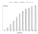


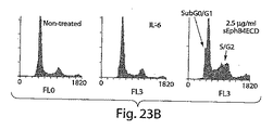


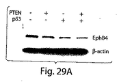


Classifications
-
- A—HUMAN NECESSITIES
- A61—MEDICAL OR VETERINARY SCIENCE; HYGIENE
- A61K—PREPARATIONS FOR MEDICAL, DENTAL OR TOILETRY PURPOSES
- A61K38/00—Medicinal preparations containing peptides
- A61K38/16—Peptides having more than 20 amino acids; Gastrins; Somatostatins; Melanotropins; Derivatives thereof
- A61K38/17—Peptides having more than 20 amino acids; Gastrins; Somatostatins; Melanotropins; Derivatives thereof from animals; from humans
- A61K38/38—Albumins
- A61K38/385—Serum albumin
-
- A—HUMAN NECESSITIES
- A61—MEDICAL OR VETERINARY SCIENCE; HYGIENE
- A61K—PREPARATIONS FOR MEDICAL, DENTAL OR TOILETRY PURPOSES
- A61K47/00—Medicinal preparations characterised by the non-active ingredients used, e.g. carriers or inert additives; Targeting or modifying agents chemically bound to the active ingredient
- A61K47/50—Medicinal preparations characterised by the non-active ingredients used, e.g. carriers or inert additives; Targeting or modifying agents chemically bound to the active ingredient the non-active ingredient being chemically bound to the active ingredient, e.g. polymer-drug conjugates
- A61K47/51—Medicinal preparations characterised by the non-active ingredients used, e.g. carriers or inert additives; Targeting or modifying agents chemically bound to the active ingredient the non-active ingredient being chemically bound to the active ingredient, e.g. polymer-drug conjugates the non-active ingredient being a modifying agent
- A61K47/56—Medicinal preparations characterised by the non-active ingredients used, e.g. carriers or inert additives; Targeting or modifying agents chemically bound to the active ingredient the non-active ingredient being chemically bound to the active ingredient, e.g. polymer-drug conjugates the non-active ingredient being a modifying agent the modifying agent being an organic macromolecular compound, e.g. an oligomeric, polymeric or dendrimeric molecule
- A61K47/59—Medicinal preparations characterised by the non-active ingredients used, e.g. carriers or inert additives; Targeting or modifying agents chemically bound to the active ingredient the non-active ingredient being chemically bound to the active ingredient, e.g. polymer-drug conjugates the non-active ingredient being a modifying agent the modifying agent being an organic macromolecular compound, e.g. an oligomeric, polymeric or dendrimeric molecule obtained otherwise than by reactions only involving carbon-to-carbon unsaturated bonds, e.g. polyureas or polyurethanes
- A61K47/60—Medicinal preparations characterised by the non-active ingredients used, e.g. carriers or inert additives; Targeting or modifying agents chemically bound to the active ingredient the non-active ingredient being chemically bound to the active ingredient, e.g. polymer-drug conjugates the non-active ingredient being a modifying agent the modifying agent being an organic macromolecular compound, e.g. an oligomeric, polymeric or dendrimeric molecule obtained otherwise than by reactions only involving carbon-to-carbon unsaturated bonds, e.g. polyureas or polyurethanes the organic macromolecular compound being a polyoxyalkylene oligomer, polymer or dendrimer, e.g. PEG, PPG, PEO or polyglycerol
-
- A—HUMAN NECESSITIES
- A61—MEDICAL OR VETERINARY SCIENCE; HYGIENE
- A61P—SPECIFIC THERAPEUTIC ACTIVITY OF CHEMICAL COMPOUNDS OR MEDICINAL PREPARATIONS
- A61P35/00—Antineoplastic agents
-
- A—HUMAN NECESSITIES
- A61—MEDICAL OR VETERINARY SCIENCE; HYGIENE
- A61P—SPECIFIC THERAPEUTIC ACTIVITY OF CHEMICAL COMPOUNDS OR MEDICINAL PREPARATIONS
- A61P35/00—Antineoplastic agents
- A61P35/02—Antineoplastic agents specific for leukemia
-
- A—HUMAN NECESSITIES
- A61—MEDICAL OR VETERINARY SCIENCE; HYGIENE
- A61P—SPECIFIC THERAPEUTIC ACTIVITY OF CHEMICAL COMPOUNDS OR MEDICINAL PREPARATIONS
- A61P35/00—Antineoplastic agents
- A61P35/04—Antineoplastic agents specific for metastasis
-
- A—HUMAN NECESSITIES
- A61—MEDICAL OR VETERINARY SCIENCE; HYGIENE
- A61P—SPECIFIC THERAPEUTIC ACTIVITY OF CHEMICAL COMPOUNDS OR MEDICINAL PREPARATIONS
- A61P43/00—Drugs for specific purposes, not provided for in groups A61P1/00-A61P41/00
-
- A—HUMAN NECESSITIES
- A61—MEDICAL OR VETERINARY SCIENCE; HYGIENE
- A61P—SPECIFIC THERAPEUTIC ACTIVITY OF CHEMICAL COMPOUNDS OR MEDICINAL PREPARATIONS
- A61P9/00—Drugs for disorders of the cardiovascular system
-
- C—CHEMISTRY; METALLURGY
- C07—ORGANIC CHEMISTRY
- C07K—PEPTIDES
- C07K14/00—Peptides having more than 20 amino acids; Gastrins; Somatostatins; Melanotropins; Derivatives thereof
- C07K14/435—Peptides having more than 20 amino acids; Gastrins; Somatostatins; Melanotropins; Derivatives thereof from animals; from humans
- C07K14/705—Receptors; Cell surface antigens; Cell surface determinants
- C07K14/715—Receptors; Cell surface antigens; Cell surface determinants for cytokines; for lymphokines; for interferons
-
- A—HUMAN NECESSITIES
- A61—MEDICAL OR VETERINARY SCIENCE; HYGIENE
- A61K—PREPARATIONS FOR MEDICAL, DENTAL OR TOILETRY PURPOSES
- A61K38/00—Medicinal preparations containing peptides
Landscapes
- Health & Medical Sciences (AREA)
- Life Sciences & Earth Sciences (AREA)
- Chemical & Material Sciences (AREA)
- Medicinal Chemistry (AREA)
- General Health & Medical Sciences (AREA)
- Veterinary Medicine (AREA)
- Public Health (AREA)
- Animal Behavior & Ethology (AREA)
- Pharmacology & Pharmacy (AREA)
- Organic Chemistry (AREA)
- Bioinformatics & Cheminformatics (AREA)
- Engineering & Computer Science (AREA)
- Chemical Kinetics & Catalysis (AREA)
- Nuclear Medicine, Radiotherapy & Molecular Imaging (AREA)
- General Chemical & Material Sciences (AREA)
- Zoology (AREA)
- Proteomics, Peptides & Aminoacids (AREA)
- Immunology (AREA)
- Gastroenterology & Hepatology (AREA)
- Epidemiology (AREA)
- Biochemistry (AREA)
- Toxicology (AREA)
- Molecular Biology (AREA)
- Cell Biology (AREA)
- Biophysics (AREA)
- Genetics & Genomics (AREA)
- Oncology (AREA)
- Hematology (AREA)
- Cardiology (AREA)
- Heart & Thoracic Surgery (AREA)
- Medicines That Contain Protein Lipid Enzymes And Other Medicines (AREA)
- Peptides Or Proteins (AREA)
Description
本出願は、米国仮出願番号第60/612,488号(2004年9月23日出願)の出願日の利益を主張し、その全開示を本明細書に援用する。 This application claims the benefit of the filing date of US Provisional Application No. 60 / 612,488 (filed September 23, 2004), the entire disclosure of which is incorporated herein by reference.
血管形成、すなわち予め存在している脈管構造の内皮からの新血管の発生は、ホスト内での充実性腫瘍の増殖、プログレッション、及び転移において重要なプロセスである。生理学的に正常な血管形成の間は、血管内皮とその周囲の間質成分とのオートクリン、パラクリン、及びアンフィクリン相互作用は、空間的及び時間的に緊密に調整される。更に、血管新生促進及び血管新生阻害サイトカイン並びに増殖因子のレベル及び活性はバランスが保たれている。反対に、活発な腫瘍増殖に必要な病理学上の血管形成は保持されて持続し、正常な血管形成システムの調節不全を示す。充実性腫瘍及び造血腫瘍種は高レベルの異常な血管形成に特に関係する。 Angiogenesis, the generation of new blood vessels from preexisting vasculature endothelium, is an important process in the growth, progression, and metastasis of solid tumors within the host. During physiologically normal angiogenesis, autocrine, paracrine, and amphiclin interactions between the vascular endothelium and its surrounding stromal components are tightly coordinated spatially and temporally. Furthermore, the levels and activities of pro-angiogenic and angiogenic inhibiting cytokines and growth factors are balanced. Conversely, the pathological angiogenesis necessary for active tumor growth is retained and persists, indicating a dysregulation of the normal angiogenic system. Solid tumors and hematopoietic tumor types are particularly associated with high levels of abnormal angiogenesis.
一般的に腫瘍の発生は、急成長する潜在能力を有する自律性クローンの発生を引き起こす連続的で且つ相関性のあるステップから構成されると考えられる。これらのステップは持続的な成長及び制限のない自己複製を含む。腫瘍中の細胞母集団は、一般的に成長シグナルの自給自足、成長抑制シグナルへの低い感応性、及び耐アポトーシス性に特徴付けられる。異常な成長を開始する遺伝子的又は細胞遺伝学的事象は、アポトーシスを防止することにより細胞を長期の「準備」状態に維持する。 In general, tumor development is thought to consist of successive and correlated steps that lead to the generation of autonomous clones with the potential for rapid growth. These steps include sustained growth and unrestricted self-replication. Cell populations in tumors are generally characterized by growth signal self-sufficiency, low sensitivity to growth inhibitory signals, and resistance to apoptosis. Genetic or cytogenetic events that initiate abnormal growth maintain cells in a long-term “ready” state by preventing apoptosis.
本発明は、血管形成及び腫瘍増殖阻害用の薬剤及び治療的処置を提供することを目的とする。 The present invention aims to provide agents and therapeutic treatments for angiogenesis and tumor growth inhibition.
本発明の概要
ある観点において、本発明はEphB4又はエフリンB2で仲介される機能を阻害するポリペプチド薬であり、EphB4及びエフリンB2蛋白質のモノマー性リガンド結合部を含むものを提供する。本明細書において示すように、EphB4及びエフリンB2は癌及び望ましくない又は過度の血管形成に関連した病気を含む種々の病状に関与する。従って、本発明の特定のポリペプチド薬は、これらの病気を治療するために使用できる。更なる観点において、本発明はEphB4及び/又はエフリンB2が、しばしば高レベルで、種々の腫瘍中で発現されるという発見に基づく。従って、EphB4又はエフリンB2機能をダウンレギュレートするポリペプチド薬は、腫瘍細胞への直接的効果及び腫瘍が採用する血管形成プロセスへの間接的効果により腫瘍へ影響を与える。ある実施形態において、本発明はEphB4又はエフリンB2の機能をダウンレギュレートする薬剤を用いた治療に特に適した腫瘍種を特定する。好ましい実施形態において、本明細書に開示するポリペプチドは生体内での血清中半減期が長くなるように修飾される。
SUMMARY OF THE INVENTION In one aspect, the present invention provides a polypeptide drug that inhibits a function mediated by EphB4 or ephrinB2, and that includes a monomeric ligand binding site of EphB4 and ephrinB2 protein. As shown herein, EphB4 and ephrinB2 are involved in a variety of medical conditions including cancer and diseases associated with undesirable or excessive angiogenesis. Thus, certain polypeptide drugs of the invention can be used to treat these diseases. In a further aspect, the present invention is based on the discovery that EphB4 and / or ephrinB2 are expressed in various tumors, often at high levels. Thus, polypeptide drugs that down-regulate EphB4 or ephrinB2 function affect tumors by direct effects on tumor cells and indirect effects on the angiogenic process employed by tumors. In certain embodiments, the present invention identifies tumor types that are particularly suitable for treatment with agents that down-regulate EphB4 or ephrinB2 function. In preferred embodiments, the polypeptides disclosed herein are modified to have a longer serum half-life in vivo.
ある観点において、本発明はEphB4蛋白質の細胞外領域のアミノ酸配列を含有する可溶性EphB4ポリペプチドを提供する。この可溶性EphB4ポリペプチドは特異的にエフリンB2ポリペプチドへ結合する。用語「可溶性」は単に、これらのポリペプチドが膜貫通領域を含有しないか、又は生理食塩水中のポリペプチドの溶解度を損なうのに充分な膜貫通領域部分を含有しないことを表すために使用される。可溶性ポリペプチドは単量体として調製されるのが好ましく、エフリンB2等のリガンドへ結合するEphB4と競合し、EphB4の活性化で引き起こされるシグナルを阻害する。随意的に、可溶性ポリペプチドは、例えばFc融合蛋白質として発現することにより、又は別の多量体形成領域と融合することにより、多量体の形態で調製してもよい。これら多量体の形態は、文脈に応じてアゴニスト的又はアンタゴニスト的効果を有する複合的活性を有する。ある実施形態においては、可溶性EphB4ポリペプチドはEphB4蛋白質の球状領域を含有する。可溶性EphB4ポリペプチドは、図65(SEQ ID NO:10)により特定されるアミノ酸配列の残基1〜522と少なくとも90%は同一の配列を含有し得る。可溶性EphB4ポリペプチドは、図65(SEQ ID NO:10)により特定されるアミノ酸配列の残基1〜412と少なくとも90%は同一の配列を含有し得る。可溶性EphB4ポリペプチドは、図65(SEQ ID NO:10)により特定されるアミノ酸配列の残基1〜312と少なくとも90%は同一の配列を含有し得る。可溶性EphB4ポリペプチドは球状(G)領域(図65のアミノ酸29-197, SEQ ID NO:10)を包含する配列を含有することができ、随意的に追加的領域、例えばシステインリッチな領域(図65のアミノ酸239-321, SEQ ID NO:10)、第一のフィブロネクチンタイプ3領域(図65のアミノ酸324-429, SEQ ID NO:10)及び第二のフィブロネクチンタイプ3領域(図65のアミノ酸434-526, SEQ ID NO:10)を含有することができる。本明細書に記載され、リガンド結合活性をもつとして示された好ましいポリペプチとしては図65(SEQ ID NO: 10)に示すアミノ酸配列の1-537, 1-427及び1-326それぞれに対応するポリペプチドが挙げられる。可溶性EphB4ポリペプチドは図1又は2(SEQ ID Nos. 1又は2)に記載の配列を含有することができる。斯界で周知のように、HEK293T細胞系といった適切な細胞内で該EphB4ポリペプチドが発現するとリーダーペプチドが切断される。該切断は常に完全であるわけでも、単一部位での完璧な一貫性があるわけでもないが、EphB4は図65(SEQ ID NO:10)に示される配列の最初の15個のアミノ酸を除去するように切断される傾向にあることが知られている。従って、具体例として、本発明はエフリンB2に結合し、そして次の群(番号は図65のSEQ ID NO:10に対応):1-197, 29-197, 1-312, 29-132, 1-321, 29-321, 1-326, 29-326, 1-412, 29-412, 1-427, 29-427, 1-429, 29-429, 1-526, 29-526, 1-537及び29-537から選択されるアミノ酸配列を含有するプロセシングを受けていない可溶性EphB4ポリペプチドを提供する。更に、異種のリーダーペプチドが内発的リーダー配列に取って代わることもできる。ポリペプチドは次の群(番号は図65のSEQ ID NO:10に対応):16-197, 16-312, 16-321, 16-326, 16-412, 16-427, 16-429, 16-526及び16-537から選択される予測アミノ酸配列を有するプロセシングを受けた形態で使用してもよい。更に、可溶性EphB4ポリペプチドは、エフリンB2結合活性を保持しつつ、先行するアミノ酸配列の何れかに少なくとも90%(随意に95%又は99%)は同一であるアミノ酸配列を含有するものとすることができる。好ましくは、図65(SEQ ID NO:10)に示される配列からのアミノ酸配列の変異は何れも1,2,3,4又は5個以下のアミノ酸の保存的変化又は欠失であり、とりわけ表面ループ部位での変異である。ある実施形態においては、可溶性EphB4ポリペプチドは、エフリンB2とEphB4の間の相互作用を阻害し得る。可溶性EphB4ポリペプチドは、エフリンB2又はEphB4のクラスター化又はリン酸化を阻害し得る。エフリンB2又はEphB4のリン酸化は一般的にこれらの蛋白質により調節される細胞内シグナル経路を刺激する初期事象の一つであると考えられる。上述したように、可溶性EphB4ポリペプチドは単量体又は多量体融合蛋白質として調製し得る。可溶性ポリペプチドは1以上の修飾アミノ酸を含有してもよい。これらアミノ酸はプロテアーゼ消化耐性の増加等の望ましい性質に寄与し得る。
In one aspect, the present invention provides a soluble EphB4 polypeptide containing the amino acid sequence of the extracellular region of the EphB4 protein. This soluble EphB4 polypeptide specifically binds to the ephrin B2 polypeptide. The term “soluble” is merely used to indicate that these polypeptides do not contain a transmembrane region or do not contain sufficient transmembrane region portions to impair the solubility of the polypeptide in saline. . Soluble polypeptides are preferably prepared as monomers, compete with EphB4 binding to ligands such as ephrin B2, and inhibit signals caused by EphB4 activation. Optionally, soluble polypeptides may be prepared in multimeric form, for example, by expressing as an Fc fusion protein or by fusing with another multimer-forming region. These multimeric forms have a complex activity with agonistic or antagonistic effects depending on the context. In certain embodiments, the soluble EphB4 polypeptide contains the globular region of the EphB4 protein. A soluble EphB4 polypeptide may contain a sequence that is at least 90% identical to residues 1-522 of the amino acid sequence specified by FIG. 65 (SEQ ID NO: 10). A soluble EphB4 polypeptide may contain a sequence that is at least 90% identical to residues 1-412 of the amino acid sequence identified by FIG. 65 (SEQ ID NO: 10). A soluble EphB4 polypeptide may contain a sequence that is at least 90% identical to residues 1-312 of the amino acid sequence specified by FIG. 65 (SEQ ID NO: 10). A soluble EphB4 polypeptide can contain a sequence that includes a globular (G) region (amino acids 29-197 of FIG. 65, SEQ ID NO: 10), optionally an additional region, such as a cysteine-rich region (FIG. 65 amino acids 239-321, SEQ ID NO: 10),
本発明はエフリンB2結合活性を保持しつつ、血清中半減期を長くする追加的成分を有する可溶性EphB4ポリペプチドを提供する。ある実施形態においては、可溶性EphB4ポリペプチドは単量体であり、1又は2以上のポリオキシアルキレン基(例:ポリエチレン、ポリプロピレン)、好ましくはポリエチレングリコール(PEG)基に共有結合する。従って、本発明は一観点において、修飾がポリペプチドに共有結合した単一のポリエチレングリコール基を含む修飾EphB4ポリペプチドを提供する。別の観点においては、本発明は1、2、3又は4以上のポリエチレングリコール基に共有結合した修飾EphB4ポリペプチドを提供する。 The present invention provides soluble EphB4 polypeptides having additional components that increase serum half-life while retaining ephrin B2 binding activity. In certain embodiments, the soluble EphB4 polypeptide is a monomer and is covalently linked to one or more polyoxyalkylene groups (eg, polyethylene, polypropylene), preferably polyethylene glycol (PEG) groups. Thus, in one aspect, the present invention provides a modified EphB4 polypeptide comprising a single polyethylene glycol group where the modification is covalently attached to the polypeptide. In another aspect, the invention provides a modified EphB4 polypeptide covalently linked to 1, 2, 3 or 4 or more polyethylene glycol groups.
1又は2以上のPEGは約1kDa〜約100kDa、好ましくは約10kDa〜約60kDa又は約10kDa〜約40kDaの分子量を有し得る。PEG基は直鎖状PEG及び分枝鎖状PEGであってよい。好ましい実施形態においては、可溶性で単量体のEphB4共役体は約10kDa〜約40kDa又は約15kDa〜30kDaのPEG基に、好ましくはEphB4リジンのε−アミノ基又はN末端アミノ基を介して、共有結合した1つのEphB4ポリペプチド(モノPEG化EphB4)を含む。最も好ましくは、EphB4はEphB4リジンのε−アミノ基及びN末端アミノ基からなる群からの1つのアミノ基のところでランダムにPEG化される。 The one or more PEGs can have a molecular weight of about 1 kDa to about 100 kDa, preferably about 10 kDa to about 60 kDa, or about 10 kDa to about 40 kDa. The PEG group may be linear PEG and branched PEG. In preferred embodiments, the soluble, monomeric EphB4 conjugate is covalently linked to a PEG group of about 10 kDa to about 40 kDa or about 15 kDa to 30 kDa, preferably via the ε-amino group or the N-terminal amino group of EphB4 lysine. Contains one EphB4 polypeptide linked (mono-PEGylated EphB4). Most preferably, EphB4 is randomly PEGylated at one amino group from the group consisting of the ε-amino group and the N-terminal amino group of EphB4 lysine.
一実施形態において、本発明により提供されるPEG化ポリペプチドは生体内における血清中半減期が非修飾EphB4ポリペプチドよりも少なくとも50%、75%、100%、150%又は200%長い。別の一実施形態において、本発明により提供されるPEG化ポリペプチドはエフリンB2の活性を阻害する。特定の実施形態において、本発明により提供されるPEG化ポリペプチドはエフリンB2受容体のクラスター化、エフリンB2のリン酸化及び/又はエフリンB2キナーゼ活性を阻害する。 In one embodiment, the PEGylated polypeptide provided by the present invention has a serum half-life in vivo that is at least 50%, 75%, 100%, 150% or 200% longer than the unmodified EphB4 polypeptide. In another embodiment, the PEGylated polypeptide provided by the present invention inhibits the activity of ephrin B2. In certain embodiments, PEGylated polypeptides provided by the present invention inhibit ephrin B2 receptor clustering, ephrin B2 phosphorylation and / or ephrin B2 kinase activity.
驚くべきことに、本発明に係るモノPEG化EphB4は非修飾の可溶性EphB4ポリペプチド及びポリPEG化EphB4よりも治療上の利用性に対して優れた特性をもつことを見出した。但し、本発明ではPEGを2箇所以上有するポリPEG化EphB4も提供する。そのようなポリPEG化した形態も非修飾の形態よりは血清中半減期が向上する。 Surprisingly, it has been found that monoPEGylated EphB4 according to the present invention has superior properties for therapeutic utility than unmodified soluble EphB4 polypeptide and polyPEGylated EphB4. However, the present invention also provides polyPEGylated EphB4 having two or more PEGs. Such polyPEGylated forms also have improved serum half-life over unmodified forms.
ある実施形態において、可溶性EphB4ポリペプチドはEphrinB2結合性を実質的に減ずることなく半減期を向上させる第二の安定化ポリペプチドに安定的に結合する。安定化ポリペプチドは好ましくはヒト患者(又は獣医学的使用が企図される場合には動物患者)と免疫適合性のあるものであり、有意な生物学的活性がほとんど又は全くないものである。 In certain embodiments, the soluble EphB4 polypeptide stably binds to a second stabilizing polypeptide that improves half-life without substantially reducing EphrinB2 binding. The stabilizing polypeptide is preferably immunocompatible with a human patient (or animal patient if veterinary use is intended) and has little or no significant biological activity.
好ましい実施形態においては、安定化ポリペプチドはヒト血清アルブミン又はその一部である。ヒト血清アルブミンはEphB4ポリペプチドと安定的に共有結合又は非共有結合する。共有結合はヒト血清アルブミンとの同時翻訳融合体としてEphB4ポリペプチドが発現することで達成され得る。アルブミンの配列は可溶性EphB4ポリペプチドにおけるN末端、C末端又は非破壊的な内部位置で融合することができる。EphB4の露出したループはアルブミンの配列を挿入するのに適切な位置であろう。アルブミンは、例えば化学的架橋によって、EphB4ポリペプチドの翻訳後に結合してもよい。また、EphB4ポリペプチドは2以上のアルブミンポリペプチドと安定的に結合することもできる。ある実施形態においては、アルブミンはヒト血清アルブミン(HSA)及びウシ血清アルブミン(BSA)よりなる群から選択される。他の実施形態においては、アルブミンは自然に生じる変異体である。好ましい実施形態においては、EphB4−HSA融合体はエフリンB2及びEphB4の間の相互作用、エフリンB2又はEphB4のクラスター化、エフリンB2又はEphB4のリン酸化、或いはこれらの組合せを阻害する。別の一実施形態においては、EphB4−HSA融合体は非修飾野生型ポリペプチドよりも生体内での安定性が高い。 In a preferred embodiment, the stabilizing polypeptide is human serum albumin or a part thereof. Human serum albumin is stably covalently or non-covalently bound to EphB4 polypeptide. Covalent binding can be achieved by expressing the EphB4 polypeptide as a cotranslational fusion with human serum albumin. The albumin sequence can be fused at the N-terminal, C-terminal, or non-destructive internal position in the soluble EphB4 polypeptide. The exposed loop of EphB4 would be a suitable position to insert the albumin sequence. Albumin may be bound after translation of the EphB4 polypeptide, for example by chemical cross-linking. An EphB4 polypeptide can also stably bind to two or more albumin polypeptides. In certain embodiments, the albumin is selected from the group consisting of human serum albumin (HSA) and bovine serum albumin (BSA). In other embodiments, albumin is a naturally occurring variant. In preferred embodiments, the EphB4-HSA fusion inhibits the interaction between ephrin B2 and EphB4, clustering of ephrin B2 or EphB4, phosphorylation of ephrin B2 or EphB4, or a combination thereof. In another embodiment, the EphB4-HSA fusion is more stable in vivo than the unmodified wild type polypeptide.
ある観点において、本発明はエフリンB2蛋白質の細胞外領域のアミノ酸配列を含有する可溶性エフリンB2ポリペプチドを提供する。該可溶性エフリンB2ポリペプチドはEphB4ポリペプチドに特異的に結合する。用語「可溶性」は単に、これらのポリペプチドが膜貫通領域を含有しないか、又は生理食塩水中のポリペプチドの溶解度を損なうのに充分な膜貫通領域の部分を含有しないことを表すために使用される。可溶性ポリペプチドは単量体として調製されるのが好ましく、EphB4等のリガンドへ結合するエフリンB2と競合し、エフリンB2の活性化で引き起こされるシグナルを阻害する。随意的に、可溶性ポリペプチドは、例えばFc融合蛋白質として発現することにより、又は別の多量体形成領域と融合することにより、多量体の形態で調製してもよい。これら多量体の形態は、文脈に応じてアゴニスト的又はアンタゴニスト的効果を有する複合的活性を有する。可溶性エフリンB2ポリペプチドは図66(SEQ ID NO:11)により特定されるアミノ酸配列の残基1〜225を有し得る。可溶性エフリンB2ポリペプチドは図3によって特定される配列を有し得る。斯界で周知のように、HEK293Tのような適切な細胞内で該エフリンB2ポリペプチドを発現するとリーダーペプチドを切断することになる。該切断は常に完全というわけでも、単一部位のところで完璧に一致するわけでもないが、エフリンB2は図66(SEQ ID NO:11)に示す配列の最初の26個のアミノ酸を除去するように切断される傾向があることが知られている。従って、具体例として、本発明はEphB4に結合し、図66(SEQ ID NO:11)のアミノ酸1−225に対応するアミノ酸配列を含有するプロセシングを受けていない可溶性エフリンB2ポリペプチドを提供する。該ポリペプチドは次の群(番号は図66のSEQ ID NO:11に対応):26−225から選択される予測アミノ酸配列を有するプロセシングを受けた形態で使用してもよい。ある実施形態においては、可溶性エフリンB2ポリペプチドは、エフリンB2とEphB4の間の相互作用を阻害し得る。可溶性エフリンB2ポリペプチドは、エフリンB2又はEphB4のクラスター化又はリン酸化を阻害し得る。上述したように、可溶性エフリンB2ポリペプチドは単量体又は多量体融合蛋白質として調製し得る。可溶性ポリペプチドは1以上の修飾アミノ酸を含有してもよい。これらアミノ酸はプロテアーゼ消化耐性の増加等の望ましい性質に寄与し得る。 In one aspect, the present invention provides a soluble ephrin B2 polypeptide containing the amino acid sequence of the extracellular region of the ephrin B2 protein. The soluble ephrin B2 polypeptide specifically binds to an EphB4 polypeptide. The term “soluble” is simply used to indicate that these polypeptides do not contain a transmembrane region or do not contain sufficient portions of the transmembrane region to impair the solubility of the polypeptide in saline. The Soluble polypeptides are preferably prepared as monomers, compete with ephrin B2 binding to ligands such as EphB4 and inhibit signals caused by ephrin B2 activation. Optionally, soluble polypeptides may be prepared in multimeric form, for example, by expressing as an Fc fusion protein or by fusing with another multimer-forming region. These multimeric forms have a complex activity with agonistic or antagonistic effects depending on the context. The soluble ephrin B2 polypeptide may have residues 1-225 of the amino acid sequence specified by FIG. 66 (SEQ ID NO: 11). The soluble ephrin B2 polypeptide may have the sequence specified by FIG. As is well known in the art, expression of the ephrin B2 polypeptide in an appropriate cell such as HEK293T will cleave the leader peptide. Although the cleavage is not always perfect or perfectly matched at a single site, ephrin B2 will remove the first 26 amino acids of the sequence shown in FIG. 66 (SEQ ID NO: 11). It is known to tend to be disconnected. Thus, as a specific example, the present invention provides an unprocessed soluble ephrin B2 polypeptide that binds to EphB4 and contains an amino acid sequence corresponding to amino acids 1-225 of FIG. 66 (SEQ ID NO: 11). The polypeptide may be used in a processed form having a predicted amino acid sequence selected from the following group (numbers correspond to SEQ ID NO: 11 in FIG. 66): 26-225. In certain embodiments, a soluble ephrin B2 polypeptide can inhibit the interaction between ephrin B2 and EphB4. Soluble ephrin B2 polypeptides can inhibit clustering or phosphorylation of ephrin B2 or EphB4. As mentioned above, soluble ephrin B2 polypeptides can be prepared as monomeric or multimeric fusion proteins. The soluble polypeptide may contain one or more modified amino acids. These amino acids can contribute to desirable properties such as increased resistance to protease digestion.
ある観点において、本発明はポリペプチド薬及び製薬上許容し得る担体を含有する医薬処方物を提供する。ポリペプチド薬は、例えば可溶性EphB4又はエフリンB2ポリペプチドを含めて、本明細書に記載の何れともすることができる。更なる処方物としては化粧組成物及び診断用キットが挙げられる。 In one aspect, the present invention provides a pharmaceutical formulation comprising a polypeptide drug and a pharmaceutically acceptable carrier. The polypeptide drug can be any of those described herein, including, for example, a soluble EphB4 or ephrinB2 polypeptide. Additional formulations include cosmetic compositions and diagnostic kits.
ある観点において、本発明は、細胞内でエフリンB2/EphB4経路を通るシグナルの阻害方法を提供する。上記方法は、細胞を有効量のポリペプチド薬と接触させるステップを含む。ポリペプチド薬としては例えば(a)EphB4蛋白質の細胞外領域のアミノ酸配列を含有する可溶性ポリペプチド(但し、上記EphB4ポリペプチドは単量体であり、エフリンB2ポリペプチドへ特異的に結合する);(b)エフリンB2蛋白質の細胞外領域のアミノ酸配列を含有する可溶性ポリペプチド(但し、上記可溶性エフリンB2ポリペプチドは単量体であり、EphB4ポリペプチドへ高親和性で結合する)が挙げられる。 In one aspect, the present invention provides a method for inhibiting a signal through the ephrinB2 / EphB4 pathway in a cell. The method includes contacting the cell with an effective amount of a polypeptide drug. Examples of the polypeptide drug include (a) a soluble polypeptide containing the amino acid sequence of the extracellular region of EphB4 protein (however, the EphB4 polypeptide is a monomer and specifically binds to ephrin B2 polypeptide); (B) A soluble polypeptide containing the amino acid sequence of the extracellular region of the ephrin B2 protein (however, the soluble ephrin B2 polypeptide is a monomer and binds to the EphB4 polypeptide with high affinity).
ある観点において、本発明は腫瘍の増殖速度を減少させるのに充分な量のポリペプチド薬を投与することを含む腫瘍の増殖速度を減少させる方法である。ポリペプチド薬は:
(a)EphB4蛋白質の細胞外領域のアミノ酸配列を含有する可溶性ポリペプチド(但し、上記EphB4ポリペプチドは単量体であってエフリンB2ポリペプチドへ特異的に結合する。随意的に、上記EphB4ポリペプチドは血清中半減期を長くするためのPEG化、血清アルブミン又はその両方といった追加的修飾を含有する。);
(b)エフリンB2蛋白質の細胞外領域のアミノ酸配列を含有する可溶性ポリペプチド(但し、上記可溶性エフリンB2ポリペプチドは単量体であり、EphB4ポリペプチドへ高親和性で結合する。);
よりなる群から選択することができる。
随意的に、腫瘍は同等組織の非癌化細胞よりも高いレベルでエフリンB2及び/又はEphB4を発現する細胞を含む。
In one aspect, the invention is a method of reducing tumor growth rate comprising administering an amount of a polypeptide drug sufficient to reduce tumor growth rate. Polypeptide drugs are:
(A) a soluble polypeptide containing the amino acid sequence of the extracellular region of the EphB4 protein (provided that the EphB4 polypeptide is a monomer and specifically binds to the ephrinB2 polypeptide. Optionally, the EphB4 polypeptide Peptides contain additional modifications such as PEGylation, serum albumin or both to increase serum half-life.);
(B) a soluble polypeptide containing the amino acid sequence of the extracellular region of the ephrin B2 protein (provided that the soluble ephrin B2 polypeptide is a monomer and binds to the EphB4 polypeptide with high affinity);
It can be selected from the group consisting of:
Optionally, the tumor comprises cells that express ephrin B2 and / or EphB4 at higher levels than non-cancerous cells of comparable tissue.
ある観点において、本発明は癌患者を治療する方法を提供する。上記方法はポリペプチド薬を患者へ投与することを含む。ポリペプチド薬は:
(a)EphB4蛋白質の細胞外領域のアミノ酸配列を含有する可溶性ポリペプチド(但し、上記EphB4ポリペプチドは単量体であってエフリンB2ポリペプチドへ特異的に結合する。随意的に、上記EphB4ポリペプチドは血清中半減期を長くするためのPEG化、血清アルブミン又はその両方といった追加的修飾を含有する。);
(b)エフリンB2蛋白質の細胞外領域のアミノ酸配列を含有する可溶性ポリペプチド(但し、上記可溶性エフリンB2ポリペプチドは単量体であり、EphB4ポリペプチドへ高親和性で結合する。);
よりなる群から選択することができる。
随意的に、癌は同等組織の非癌化細胞よりも高いレベルでエフリンB2及び/又はEphB4を発現する癌細胞を含む。癌は転移性癌でもよい。癌は結腸癌、乳癌、中皮腫、前立腺腫瘍、扁平上皮細胞癌、カポジ肉腫及び白血病よりなる群から選択され得る。随意的に、上記癌は血管形成に依存する癌又は血管形成に依存しない癌である。本発明で使用されるポリペプチド薬は、エフリンB2又はEphB4のクラスター化又はリン酸化を阻害し得る。ポリペプチド薬は、ポリペプチド薬と付加的又は相乗的に癌細胞を抑制する1種以上の追加的抗癌化学療法薬と共投与してもよい。
In one aspect, the present invention provides a method of treating a cancer patient. The method includes administering a polypeptide drug to the patient. Polypeptide drugs are:
(A) a soluble polypeptide containing the amino acid sequence of the extracellular region of the EphB4 protein (provided that the EphB4 polypeptide is a monomer and specifically binds to the ephrinB2 polypeptide. Optionally, the EphB4 polypeptide Peptides contain additional modifications such as PEGylation, serum albumin or both to increase serum half-life.);
(B) a soluble polypeptide containing the amino acid sequence of the extracellular region of the ephrin B2 protein (provided that the soluble ephrin B2 polypeptide is a monomer and binds to the EphB4 polypeptide with high affinity);
It can be selected from the group consisting of:
Optionally, the cancer comprises cancer cells that express ephrin B2 and / or EphB4 at higher levels than non-cancerous cells of comparable tissue. The cancer may be a metastatic cancer. The cancer can be selected from the group consisting of colon cancer, breast cancer, mesothelioma, prostate tumor, squamous cell carcinoma, Kaposi's sarcoma and leukemia. Optionally, the cancer is a cancer that depends on angiogenesis or a cancer that does not depend on angiogenesis. The polypeptide drug used in the present invention can inhibit the clustering or phosphorylation of ephrin B2 or EphB4. The polypeptide drug may be co-administered with one or more additional anticancer chemotherapeutic drugs that suppress cancer cells in addition or synergistically with the polypeptide drug.
ある観点において、本発明は血管形成阻害方法を提供する。上記方法は血管形成の阻害に充分な量のポリペプチド薬と細胞を接触させるステップを含む。ポリペプチド薬は:
(a)EphB4蛋白質の細胞外領域のアミノ酸配列を含有する可溶性ポリペプチド(但し、上記EphB4ポリペプチドは単量体であってエフリンB2ポリペプチドへ特異的に結合する。随意的に、上記EphB4ポリペプチドは血清中半減期を長くするためのPEG化、血清アルブミン又はその両方といった追加的修飾を含有する。);
(b)エフリンB2蛋白質の細胞外領域のアミノ酸配列を含有する可溶性ポリペプチド(但し、上記可溶性エフリンB2ポリペプチドは単量体であり、EphB4ポリペプチドへ高親和性で結合する。);
よりなる群から選択することができる。
In one aspect, the present invention provides a method for inhibiting angiogenesis. The method includes contacting the cell with an amount of a polypeptide drug sufficient to inhibit angiogenesis. Polypeptide drugs are:
(A) a soluble polypeptide containing the amino acid sequence of the extracellular region of the EphB4 protein (provided that the EphB4 polypeptide is a monomer and specifically binds to the ephrinB2 polypeptide. Optionally, the EphB4 polypeptide Peptides contain additional modifications such as PEGylation, serum albumin or both to increase serum half-life.);
(B) a soluble polypeptide containing the amino acid sequence of the extracellular region of the ephrin B2 protein (provided that the soluble ephrin B2 polypeptide is a monomer and binds to the EphB4 polypeptide with high affinity);
It can be selected from the group consisting of:
ある観点において、本発明はポリペプチド薬を患者へ投与するステップを含む血管形成関連疾患の患者を治療する方法を提供する。ポリペプチド薬は:
(a)EphB4蛋白質の細胞外領域のアミノ酸配列を含有する可溶性ポリペプチド(但し、上記EphB4ポリペプチドは単量体であってエフリンB2ポリペプチドへ特異的に結合する。随意的に、上記EphB4ポリペプチドは血清中半減期を長くするためのPEG化、血清アルブミン又はその両方といった追加的修飾を含有する。);
(b)エフリンB2蛋白質の細胞外領域のアミノ酸配列を含有する可溶性ポリペプチド(但し、上記可溶性エフリンB2ポリペプチドは単量体であり、EphB4ポリペプチドへ高親和性で結合する。);
よりなる群から選択することができる。可溶性ポリペプチドは製薬上許容し得る担体と共に処方することができる。血管形成関連疾患又は望ましくない血管形成関連プロセスは血管形成依存性癌、良性腫瘍、炎症性障害、慢性関節リューマチ及び乾癬、眼性血管形成病、オスラーウェバー症候群、心筋性血管形成、プラーク新生血管形成、毛細血管拡張症、血友病性関節、繊維血管腫、毛細血管拡張乾癬強皮症、化膿性肉芽腫、ルベオーシス、関節炎、糖尿病性新生血管形成及び脈管形成よりなる群から選択され得る。ポリペプチド薬は可溶性ポリペプチドと追加的又は相乗的に血管形成を抑制する少なくとも1種の追加的抗血管形成薬と共投与してもよい。
In one aspect, the present invention provides a method of treating a patient with an angiogenesis-related disorder comprising administering a polypeptide drug to the patient. Polypeptide drugs are:
(A) a soluble polypeptide containing the amino acid sequence of the extracellular region of the EphB4 protein (provided that the EphB4 polypeptide is a monomer and specifically binds to the ephrinB2 polypeptide. Optionally, the EphB4 polypeptide Peptides contain additional modifications such as PEGylation, serum albumin or both to increase serum half-life.);
(B) a soluble polypeptide containing the amino acid sequence of the extracellular region of the ephrin B2 protein (provided that the soluble ephrin B2 polypeptide is a monomer and binds to the EphB4 polypeptide with high affinity);
It can be selected from the group consisting of: The soluble polypeptide can be formulated with a pharmaceutically acceptable carrier. Angiogenesis-related diseases or undesirable angiogenesis-related processes are angiogenesis-dependent cancer, benign tumors, inflammatory disorders, rheumatoid arthritis and psoriasis, ocular angiogenesis disease, Osler-Weber syndrome, myocardial angiogenesis, plaque neovascularization , Telangiectasia, hemophilic joints, fibroangioma, telangiectasis psoriasis scleroderma, purulent granulomas, lebeosis, arthritis, diabetic neovascularization and angiogenesis. The polypeptide agent may be co-administered with at least one additional anti-angiogenic agent that inhibits angiogenesis additionally or synergistically with the soluble polypeptide.
ある観点においては、本発明は癌又は血管形成関連障害の治療薬の製造におけるポリペプチド薬の使用を提供する。ポリペプチド薬は:
(a)EphB4蛋白質の細胞外領域のアミノ酸配列を含有する可溶性ポリペプチド(但し、上記EphB4ポリペプチドは単量体であってエフリンB2ポリペプチドへ特異的に結合する。随意的に、上記EphB4ポリペプチドは血清中半減期を長くするためのPEG化、血清アルブミン又はその両方といった追加的修飾を含有する。);
(b)エフリンB2蛋白質の細胞外領域のアミノ酸配列を含有する可溶性ポリペプチド(但し、上記可溶性エフリンB2ポリペプチドは単量体であり、EphB4ポリペプチドへ高親和性で結合する。);
よりなる群から選択することができる。
In one aspect, the invention provides the use of a polypeptide drug in the manufacture of a therapeutic for cancer or angiogenesis related disorders. Polypeptide drugs are:
(A) a soluble polypeptide containing the amino acid sequence of the extracellular region of the EphB4 protein (provided that the EphB4 polypeptide is a monomer and specifically binds to the ephrinB2 polypeptide. Optionally, the EphB4 polypeptide Peptides contain additional modifications such as PEGylation, serum albumin or both to increase serum half-life.);
(B) a soluble polypeptide containing the amino acid sequence of the extracellular region of the ephrin B2 protein (provided that the soluble ephrin B2 polypeptide is a monomer and binds to the EphB4 polypeptide with high affinity);
It can be selected from the group consisting of:
ある観点においては、本発明は下記:
(a)患者中のEphB4及び/又はエフリンB2を発現する複数の癌細胞を有する腫瘍を特定するステップ;並びに
(b)ポリペプチド薬を患者へ投与するステップ;
を含有する。
ポリペプチド薬は:
(a)EphB4蛋白質の細胞外領域のアミノ酸配列を含有する可溶性ポリペプチド(但し、上記EphB4ポリペプチドは単量体であってエフリンB2ポリペプチドへ特異的に結合する。随意的に、上記EphB4ポリペプチドは血清中半減期を長くするためのPEG化、血清アルブミン又はその両方といった追加的修飾を含有する。);
(b)エフリンB2蛋白質の細胞外領域のアミノ酸配列を含有する可溶性ポリペプチド(但し、上記可溶性エフリンB2ポリペプチドは単量体であり、EphB4ポリペプチドへ高親和性で結合する。);
よりなる群から選択することができる。
In one aspect, the invention provides:
(A) identifying a tumor having a plurality of cancer cells expressing EphB4 and / or ephrinB2 in the patient; and (b) administering a polypeptide drug to the patient;
Containing.
Polypeptide drugs are:
(A) a soluble polypeptide containing the amino acid sequence of the extracellular region of the EphB4 protein (provided that the EphB4 polypeptide is a monomer and specifically binds to the ephrinB2 polypeptide. Optionally, the EphB4 polypeptide Peptides contain additional modifications such as PEGylation, serum albumin or both to increase serum half-life.);
(B) a soluble polypeptide containing the amino acid sequence of the extracellular region of the ephrin B2 protein (provided that the soluble ephrin B2 polypeptide is a monomer and binds to the EphB4 polypeptide with high affinity);
It can be selected from the group consisting of:
ある観点においては、本発明はエフリンB2又はEphB4アンタゴニストでの治療が適切である腫瘍を特定する方法を提供する。上記方法は、腫瘍細胞中に1以上の下記特徴を検出する方法を含有してもよい:(a)EphB4蛋白質及び/又はmRNAの発現;(b)エフリンB2蛋白質及び/又はmRNAの発現;(c)EphB4遺伝子の遺伝子増幅(例:遺伝子複製数の増加);又は(d)エフリンB2遺伝子の遺伝子増幅。上記特徴(a)〜(d)の1以上を有する腫瘍細胞は、本発明のポリペプチド薬等の、エフリンB2又はEphB4アンタゴニストでの治療が適切であろう。 In one aspect, the invention provides a method for identifying tumors that are amenable to treatment with ephrin B2 or EphB4 antagonists. The method may include a method of detecting one or more of the following characteristics in tumor cells: (a) expression of EphB4 protein and / or mRNA; (b) expression of ephrin B2 protein and / or mRNA; c) Gene amplification of EphB4 gene (eg, increase in gene replication number); or (d) Gene amplification of ephrin B2 gene. Tumor cells having one or more of the above characteristics (a)-(d) may be suitable for treatment with ephrin B2 or EphB4 antagonists, such as the polypeptide drugs of the present invention.
驚くべきことに、本発明者は球状領域を欠失したEphB4ポリペプチドが異種移植モデル内の腫瘍増殖を実際に阻害し、血管内皮細胞の管形成を阻害し、及びエフリンB2によって活性化されるEphB4の自己リン酸化蛋白質(autokinase)活性を阻害することができることを見出した。如何なる作用機構にも拘束されることを望まないが、該ポリペプチドはEphB4の凝集を予防するか又は原形質膜からのEphB4の排除を(例:エンドサイトーシスにより)刺激すると予測される。しかして、本発明はEphB4蛋白質のフィブロネクチンタイプ3領域のアミノ酸配列を含有する単離された可溶性ポリペプチドを提供する。該ポリペプチドは好ましくはEphB4又はエフリンB2蛋白質の活性(例:凝集やキナーゼ活性)を阻害するような、とりわけヒト又は癌のマウス異種移植モデル中の腫瘍増殖を阻害するような生物学的活性を有する。該ポリペプチドは生体内又は細胞ベースのアッセイ系で脈管形成を阻害することもできる。該ポリペプチドはエフリンB2には結合せずに、EphB4蛋白質の球状領域の全て又は機能性(例:エフリンB2結合性)部分を特異的に排除する場合がある。該ポリペプチドは好ましくは図65(SEQ ID NO:10)の配列のアミノ酸324−429及び/又は434−526に対応するアミノ酸、或いはこれと少なくとも90%、95%、98%、99%は同一の配列を含有する。該ポリペプチドの一例をSEQ ID NO:15に示す。該ポリペプチドは本明細書に記載の任意の方法で修飾してもよく、単量体として又は2以上のポリペプチドを含有する二量体若しくは多量体(例えばFc融合コンストラクト)として製造してもよい。二量体又は多量体が該ポリペプチドの効果を高めるのに望ましいであろう。該ポリペプチドを製造及び使用するための方法の全てはその他のEphB4ポリペプチドに関して本明細書で記載する方法と同様である。
Surprisingly, the inventors have found that EphB4 polypeptides lacking globular regions actually inhibit tumor growth in xenograft models, inhibit vascular endothelial cell tube formation, and are activated by ephrin B2 The present inventors have found that EphB4 can inhibit autophosphorylated protein (autokinase) activity. Without wishing to be bound by any mechanism of action, it is expected that the polypeptide will prevent EphB4 aggregation or stimulate (eg, by endocytosis) elimination of EphB4 from the plasma membrane. Thus, the present invention provides an isolated soluble polypeptide containing the amino acid sequence of the
I.概説
本発明の一部は、エフリン/エフリンレセプター(エフリン/eph)経路を経由するシグナルが腫瘍生成に貢献することを発見したことに基づいている。発明者らは、腫瘍組織中のエフリンB2及びEphB4の発現を検出し、エフリン/エフリンレセプターを経由するシグナルをブロックする抗腫瘍治療薬を開発した。更に、本発明は、EphB4及び/又はエフリンB2のポリペプチド-ベースの機能阻害のためのポリペプチド治療薬及び方法を提供する。従って、本発明はある観点において、癌並びに血管形成関連障害及び望ましくない血管形成関連プロセスを治療するために使用できる数多くのポリペプチド化合物(薬)を提供する。本発明者は修飾された形態のエフリンB2及びEphB4ポリペプチドを作り出し、該修飾形態が薬物動態学を顕著に向上させることを実証した。従って、本発明はある観点において、癌並びに血管形成関連障害及び望ましくない血管形成関連プロセスを治療するために使用できる数多くのポリペプチド化合物(薬)を提供する。
I. Overview Part of the present invention is based on the discovery that signals via the ephrin / ephrin receptor (ephrin / eph) pathway contribute to tumorigenesis. The inventors have developed anti-tumor therapeutic agents that detect the expression of ephrin B2 and EphB4 in tumor tissue and block signals through the ephrin / ephrin receptor. Furthermore, the present invention provides polypeptide therapeutics and methods for polypeptide-based functional inhibition of EphB4 and / or ephrinB2. Thus, in one aspect, the present invention provides a number of polypeptide compounds (drugs) that can be used to treat cancer and angiogenesis-related disorders and undesirable angiogenesis-related processes. The inventors have created modified forms of ephrin B2 and EphB4 polypeptides and have demonstrated that the modified forms significantly improve pharmacokinetics. Thus, in one aspect, the present invention provides a number of polypeptide compounds (drugs) that can be used to treat cancer and angiogenesis-related disorders and undesirable angiogenesis-related processes.
ここで使用される用語「エフリン」及び「Eph」は、それぞれリガンド及びレセプターを指すものとして使用される。それらは種々の動物(例えば、ほ乳類/非ほ乳類、脊椎動物/非脊椎動物、ヒト等)のいずれかからでも得られる。この分野の命名法は急速に変化しており、ここで使用される用語法は、Eph命名法委員会による作業の結果として提案されたものであり、以前使用されていた名前と共にウェッブサイトhttp://www.eph-nomenclature.com.でアクセスできる。 As used herein, the terms “ephrin” and “Eph” are used to refer to a ligand and a receptor, respectively. They can be obtained from any of a variety of animals (eg, mammals / non-mammals, vertebrates / invertebrates, humans, etc.). The nomenclature in this area is changing rapidly, and the terminology used here was proposed as a result of work by the Eph Nomenclature Committee, and the website http: // Access at www.eph-nomenclature.com.
本発明の研究は、とりわけ実施例中では、エフリンB2及びEphB4に関する。しかし、本発明は、腫瘍中で発現されるそれぞれのファミリー中のエフリンリガンド及び/又はEphレセプターのいずれかを意図する。エフリン(リガンド)は2種の構造的タイプからなり、それは配列関係を基礎として、及び機能的にはそれらが2種の対応するレセプターサブグループを示す選択的結合を基礎として更に分けられる。構造的に、2種のエフリンが存在する。即ち、グリセロホスファチジルイノシトール(GPI)リンケージにより膜結合するものと、膜貫通領域を通して結合するものである。従来リガンドは、選択的にEphAレセプターへ結合するGPI-結合された蛋白質であるエフリン-Aサブクラス、及び一般的に選択的にEphBレセプターへ結合する膜貫通蛋白質であるエフリン-Bサブクラスへ分割されている。 The work of the present invention relates to ephrin B2 and EphB4, especially in the examples. However, the present invention contemplates either ephrin ligands and / or Eph receptors in each family that are expressed in tumors. Ephrins (ligands) consist of two structural types, which are further divided on the basis of sequence relationships and functionally on the basis of selective binding where they represent two corresponding receptor subgroups. Structurally, there are two types of ephrins. That is, one that is membrane-bound by glycerophosphatidylinositol (GPI) linkage and one that is bound through a transmembrane region. Conventional ligands are divided into the ephrin-A subclass, which is a GPI-linked protein that selectively binds to the EphA receptor, and the ephrin-B subclass, which is generally a transmembrane protein that selectively binds to the EphB receptor. Yes.
Ephファミリーレセプターは、レセプター蛋白質-チロシンキナーゼのファミリーであり、それはエリスロポエチン-生産ヒト肝細胞性癌細胞系中でのその発現のために命名されたEphに関係する。それらはそれらの細胞外領域配列の関連性及びそれらのエフリン-A蛋白質又はエフリン-B蛋白質へ選択的に結合する能力を基礎として2個のサブグループへ分離される。エフリン-A蛋白質と選択的に相互作用するレセプターは、EphAレセプターであり、エフリン-B蛋白質と選択的に相互作用するものはEphBレセプターである。 The Eph family receptors are a family of receptor protein-tyrosine kinases, which are related to Ephs named for their expression in erythropoietin-producing human hepatocellular carcinoma cell lines. They are segregated into two subgroups on the basis of their extracellular domain sequence relevance and their ability to selectively bind to ephrin-A or ephrin-B proteins. The receptor that selectively interacts with ephrin-A protein is the EphA receptor, and the one that selectively interacts with ephrin-B protein is the EphB receptor.
Ephレセプターは、リガンド結合球状領域、その次に一対のフィブロネクチンタイプIIIリピートが続くシステインリッチ領域から構成される細胞外領域を有する(例えば、図16参照)。細胞質領域は、2種の蛋白質チロシンキナーゼ領域、即ち無菌性α-モチーフ(SAM)及びPDZ-領域結合モチーフの保存されたチロシン残基を含有する膜近傍(juxtamembrane)領域から構成される。EphB4は膜結合性リガンドエフリンB2に対して特異的である(Sakano、S.ら1996;Brambilla R.ら1995)。エフリンB2は、膜貫通領域及び5個の保存されたチロシン残基及びPDZ領域を有する細胞質領域を有するEphリガンドの種類に属する。Ephレセプターは、クラスター化され、膜へ結合したエフリンの結合により活性化され(Davis S.ら、1994)、レセプターを発現している細胞及びリガンドを発現している細胞間を接触することがEph活性化に要求されることを示す。 The Eph receptor has an extracellular region composed of a ligand-binding globular region followed by a cysteine-rich region followed by a pair of fibronectin type III repeats (see, eg, FIG. 16). The cytoplasmic region is composed of two protein tyrosine kinase regions, a juxtamembrane region containing a conserved tyrosine residue of a sterile α-motif (SAM) and a PDZ-region binding motif. EphB4 is specific for the membrane-bound ligand ephrin B2 (Sakano, S. et al. 1996; Brambilla R. et al. 1995). Ephrin B2 belongs to a class of Eph ligands that have a transmembrane region and a cytoplasmic region with five conserved tyrosine residues and a PDZ region. The Eph receptor is clustered and activated by the binding of membrane-bound ephrin (Davis S. et al., 1994) to contact the cells expressing the receptor and the cells expressing the ligand. Indicates that it is required for activation.
リガンド結合において、Ephレセプターは二量体化し、自己リン酸化して膜近傍チロシン残基を完全活性化させる(Kalo MSら、1999、BinnsKS、2000)。更にEphレセプターを経由するシグナルに加えて、リバースシグナルがエフリンBsを経由して発生できる。エフリンのEph結合(engagement)は、保存された細胞内チロシンの急速なリン酸化を生じ(Bruckner K、1997)、PDZ結合蛋白質のレクルートメントをいくらか遅延させる(Palmer A 2002)。近年、いくつかの研究でEph/エフリンの高い発現は腫瘍増殖、腫瘍原性、及び転移の可能性の増大に関係することが示された(Easty DJ、1999;Kiyokawa E、1994;Tang XX、1999;Vogt T、1998;Liu W、2002;Stephenson SA、2001;Steube KG 1999;Berclaz G、1996)。 Upon ligand binding, Eph receptors dimerize and autophosphorylate to fully activate near-membrane tyrosine residues (Kalo MS et al., 1999, BinnsKS, 2000). Furthermore, in addition to the signal via the Eph receptor, a reverse signal can be generated via ephrin Bs. The Eph engagement of ephrin results in rapid phosphorylation of conserved intracellular tyrosine (Bruckner K, 1997), and somewhat delays the recruitment of PDZ binding proteins (Palmer A 2002). Recently, several studies have shown that high Eph / ephrin expression is associated with increased tumor growth, tumorigenicity, and metastatic potential (Easty DJ, 1999; Kiyokawa E, 1994; Tang XX, 1999; Vogt T, 1998; Liu W, 2002; Stephenson SA, 2001; Steube KG 1999; Berclazz G, 1996).
ある実施形態において、本発明はエフリンB2、EphB4、又は両方の活性を阻害するポリペプチド治療薬を提供する。ここで使用される用語「ポリペプチド治療薬」又は「ポリペプチド薬」は、エフリンB2/EphB4経路を経由するシグナルをブロックするいずれのポリペプチドをも含む総称名である。本発明の好ましいポリペプチド治療薬は、エフリンB2又はEphB4の可溶性ポリペプチドである。本発明の別の好ましいポリペプチド治療薬は、エフリンB2又はEphB4へ結合するアンタゴニスト抗体である。例えば、これらポリペプチド治療薬は、エフリンB2又はEphB4の機能を阻害でき、エフリンB2及びEphB4間の相互作用を阻害でき、エフリンB2又はEphB4のリン酸化を阻害でき、又はエフリンB2のEphB4への結合に対する下流シグナルイベントのいずれかを阻害できる。そのようなポリペプチドには、例えばPEG化により又は血清アルブミン蛋白質と安定に結合することにより、血清中半減期を改善するように修飾されているEphB4又はエフリンB2を挙げることができる。 In certain embodiments, the present invention provides polypeptide therapeutics that inhibit the activity of ephrin B2, EphB4, or both. The term “polypeptide therapeutic agent” or “polypeptide drug” as used herein is a generic name that includes any polypeptide that blocks signals through the ephrinB2 / EphB4 pathway. A preferred polypeptide therapeutic of the invention is a soluble polypeptide of ephrin B2 or EphB4. Another preferred polypeptide therapeutic of the invention is an antagonist antibody that binds to ephrin B2 or EphB4. For example, these polypeptide therapeutics can inhibit the function of ephrin B2 or EphB4, can inhibit the interaction between ephrin B2 and EphB4, can inhibit phosphorylation of ephrin B2 or EphB4, or the binding of ephrin B2 to EphB4 Can inhibit any of the downstream signaling events. Such polypeptides can include EphB4 or ephrin B2 that has been modified to improve serum half-life, for example, by PEGylation or by stably binding to serum albumin protein.
II.可溶性ポリペプチド
ある観点において、本発明はエフリンB2蛋白質の細胞外領域を含有する可溶性ポリペプチド(ここでエフリンB2可溶性ポリペプチドと言う)又はEphB4蛋白質の細胞外領域を含有する可溶性ポリペプチド(ここでEphB4可溶性ポリペプチドと言う)に関する。好ましくは、本発明の可溶性ポリペプチドは単量体であり、高親和でエフリンB2又はEphB4へ結合できる。特に、本発明のEphB4可溶性ポリペプチドは、EphB4蛋白質の球状領域を含有する。一例としてEphB4可溶性ポリペプチドを図1、2及び15に示す。エフリンB2可溶性ポリペプチドの一例は図3及び14に示す。
II. Soluble polypeptide In one aspect, the present invention relates to a soluble polypeptide containing the extracellular region of ephrin B2 protein (herein referred to as ephrin B2 soluble polypeptide) or a soluble polypeptide containing the extracellular region of EphB4 protein (herein EphB4 soluble polypeptide). Preferably, the soluble polypeptide of the present invention is monomeric and can bind to ephrin B2 or EphB4 with high affinity. In particular, the EphB4 soluble polypeptide of the present invention contains the globular region of the EphB4 protein. As an example, an EphB4 soluble polypeptide is shown in FIGS. An example of an ephrin B2 soluble polypeptide is shown in FIGS.
ここで使用される本発明の可溶性ポリペプチドは、EphB4可溶性ポリペプチド又はエフリンB2可溶性ポリペプチドのフラグメント、機能的変異体及び修飾体を含む。これらの本発明の可溶性ポリペプチドのフラグメント、機能的変異体及び修飾体は、EphB4、エフリンB2又は両方の機能をアンタゴナイズする。 As used herein, soluble polypeptides of the present invention include EphB4 soluble polypeptides or ephrinB2 soluble polypeptide fragments, functional variants and modifications. These soluble polypeptide fragments, functional variants and modifications of the invention antagonize the function of EphB4, ephrinB2 or both.
ある実施形態において、本発明の可溶性ポリペプチドの単離されたフラグメントは、EphB4又はエフリンB2可溶性ポリペプチドをエンコードする核酸の対応するフラグメントから組み換え的に生産されるポリペプチドのスクリーニングにより得ることができる。更に、フラグメントは、従来のMerrifield固体相(合成法)f-Moc又はt-Boc化学等の公知の技術を使用して化学的に合成できる。フラグメントは(組み換え的に又は化学合成により)生産され、EphB4又はエフリンB2の機能を阻害するように働くことのできるそれらのペプチジルフラグメントを特定するために、例えば血管形成又は腫瘍増殖を阻害するフラグメントの能力の試験により試験されてもよい。 In certain embodiments, an isolated fragment of a soluble polypeptide of the invention can be obtained by screening a polypeptide recombinantly produced from a corresponding fragment of a nucleic acid encoding EphB4 or ephrinB2 soluble polypeptide. . Furthermore, the fragments can be chemically synthesized using known techniques such as conventional Merrifield solid phase (synthesis methods) f-Moc or t-Boc chemistry. Fragments are produced (recombinantly or by chemical synthesis) to identify those peptidyl fragments that can act to inhibit the function of EphB4 or ephrinB2, for example of fragments that inhibit angiogenesis or tumor growth It may be tested by a capability test.
ある実施形態において、EphB4可溶性ポリペプチドの機能的変異体は、図65(SEQ ID NO:10)によって特定されるアミノ酸配列の残基1-197, 29-197, 1-312, 29-132, 1-321, 29-321, 1-326, 29-326, 1-412, 29-412, 1- 427, 29-427, 1-429, 29-429, 1-526, 29-526, 1-537及び29-537に少なくとも90%、95%、97%、99%又は100%同一であるアミノ酸配列を含有する。該ポリペプチドはプロセシングを受けた形態であってもよく、従って、ある実施形態においては、EphB4可溶性ポリペプチドは図65(SEQ ID NO:10)によって特定されるアミノ酸配列の残基16-197, 16-312, 16-321, 16-326, 16-412, 16-427, 16-429, 16-526及び16-537に少なくとも90%、95%、97%、99%又は100%同一であるアミノ酸配列を含有する。 In certain embodiments, a functional variant of an EphB4 soluble polypeptide is residues 1-197, 29-197, 1-312, 29-132, of the amino acid sequence identified by FIG. 65 (SEQ ID NO: 10). 1-321, 29-321, 1-326, 29-326, 1-412, 29-412, 1- 427, 29-427, 1-429, 29-429, 1-526, 29-526, 1- Contains amino acid sequences that are at least 90%, 95%, 97%, 99% or 100% identical to 537 and 29-537. The polypeptide may be in a processed form, and thus, in certain embodiments, an EphB4 soluble polypeptide is residues 16-197, of the amino acid sequence identified by FIG. 65 (SEQ ID NO: 10). 16-312, 16-321, 16-326, 16-412, 16-427, 16-429, 16-526 and 16-537 are at least 90%, 95%, 97%, 99% or 100% identical Contains the amino acid sequence.
別の実施形態においては、エフリンB2可溶性ポリペプチドの機能的変異体は、図66(SEQ ID NO:11)によって特定されるアミノ酸配列の残基1-225に少なくとも90%、95%、97%、99%又は100%同一である配列を含有するか、或いは、図66(SEQ ID NO:11)によって特定されるアミノ酸配列の残基26-225に少なくとも90%、95%、97%、99%又は100%同一であるアミノ酸配列を含有するようなプロセシングを受けた形態である。 In another embodiment, the functional variant of ephrin B2 soluble polypeptide is at least 90%, 95%, 97% at residues 1-225 of the amino acid sequence specified by FIG. 66 (SEQ ID NO: 11). , 99% or 100% identical, or at least 90%, 95%, 97%, 99 at residues 26-225 of the amino acid sequence specified by FIG. 66 (SEQ ID NO: 11) Processed forms containing amino acid sequences that are% or 100% identical.
ある実施形態において、本発明は治療用若しくは予防的効能、又は安定性(例えば、ex vivoでの貯蔵寿命及びin vivoでの耐蛋白質分解性変質(degradation))の強化を目的として、本発明の可溶性ポリペプチドの構造を改質することによる機能的変異体の製造も意図する。これら改質可溶性ポリペプチドは天然EphB4又はエフリンB2可溶性ポリペプチドの機能的均等物と考えられる。改質可溶性ポリペプチドは、例えばアミノ酸の置換、欠失又は添加により生産できる。本発明では、例えばロイシンをイソロイシン又はバリンと、アスパルテートをグルタメートと、スレオニンをセリンとの単離された置換、又はそのアミノ酸と構造的に関係するアミノ酸との同様な置換(例えば、保存的突然変異)は分子を生じる生物学的活性へ大きな影響を及ぼさないと理解することは当然である。保存的置換とは、それらの側鎖に関係するアミノ酸のファミリー内で置換することである。 In certain embodiments, the present invention is aimed at enhancing therapeutic or prophylactic efficacy, or stability (eg, ex vivo shelf life and in vivo proteolytic degradation). Also contemplated is the production of functional variants by modifying the structure of the soluble polypeptide. These modified soluble polypeptides are considered functional equivalents of native EphB4 or ephrinB2 soluble polypeptides. Modified soluble polypeptides can be produced, for example, by amino acid substitution, deletion or addition. In the present invention, for example, an isolated substitution of leucine with isoleucine or valine, aspartate with glutamate, and threonine with serine, or similar substitution with an amino acid structurally related to that amino acid (eg, conservative mutations). It will be understood that (mutation) does not have a major effect on the biological activity that produces the molecule. Conservative substitutions are substitutions within a family of amino acids that are related to their side chains.
本発明は更に、EphB4又はエフリンB2可溶性ポリペプチドの一連の組み合わせ突然変異体の製造方法、並びに突然変異体のトランケーション方法、機能的変異体配列の特定に特に有用である方法を意図する。組み合わせライブラリー等のスクリーニングの目的は、例えば、EphB4、EphB2又は両方のアンタゴニストとして作用する可溶性ポリペプチド変異体の製造にもある。それは、組み合わせ由来の変異体が天然可溶性ポリペプチドに関する選択的能力を有するように生成できる。これら変異体蛋白質は、組み換え型DNA構築物から発現された場合、遺伝子治療法プロトコル中に使用できる。同様に、突然変異誘発は、対応する野生型可溶性ポリペプチドとは劇的に異なる細胞内半減期を有する変異体を生み出すことが出来る。例えば、変異した蛋白質は、蛋白質分解性変質又は本発明の蛋白質(例えば、可溶性ポリペプチド)の分解さもなければ活性化を生じる他の細胞性プロセスに対してより安定又はより不安定のいずれにも設定できる。これら変異体、及びそれらをエンコードする遺伝子は、それらの半減期の調整により本発明の可溶性ポリペプチドレベルを変えるために使用できる。例えば、短い半減期は、より一時的な生物学的効果を生じ、誘導可能な発現システムの一部である場合には、細胞中の組み換え型可溶性ポリペプチドレベルのより精密なコントロールを可能にする。上記のように、これら蛋白質、及び好ましくはそれらの組み換え型核酸構築物は、遺伝子治療プロトコル中で使用できる。 The present invention further contemplates methods for producing a series of combinatorial mutants of EphB4 or ephrinB2 soluble polypeptides, as well as methods for truncation of mutants and identification of functional variant sequences. The purpose of screening combinatorial libraries and the like is, for example, in the production of soluble polypeptide variants that act as antagonists of EphB4, EphB2 or both. It can be generated so that the variant derived from the combination has a selective capacity for the naturally soluble polypeptide. These mutant proteins can be used in gene therapy protocols when expressed from recombinant DNA constructs. Similarly, mutagenesis can produce variants with an intracellular half-life that is dramatically different from the corresponding wild-type soluble polypeptide. For example, a mutated protein is either more stable or more unstable with respect to proteolytic alterations or other cellular processes that result in degradation or otherwise activation of the proteins of the invention (eg, soluble polypeptides). Can be set. These variants, and the genes that encode them, can be used to alter the soluble polypeptide levels of the present invention by adjusting their half-life. For example, a short half-life produces a more transient biological effect and allows more precise control of recombinant soluble polypeptide levels in the cell when it is part of an inducible expression system. . As noted above, these proteins, and preferably their recombinant nucleic acid constructs, can be used in gene therapy protocols.
能力のある(potential)ホモログのライブラリーが縮重(degenerate)オリゴヌクレオチド配列からの生成可能である方法が多く知られている。縮重遺伝子配列の化学合成は、自動DNA合成機中で実施でき、及び合成遺伝子は次に発現用の適切な遺伝子へライゲートされる。遺伝子の縮重セットの目的は、1の混合物中に能力のある可溶性ポリペプチド配列の目的とするセットをエンコードする全ての配列を提供することである。縮重オリゴヌクレオチドの合成法は当分野で公知である(例えば、Narang、SA(1983)tetrahedron 39:3;Itakuraら、(1981)組み換え型DNA、Proc.第3回クリーブランドシンポジウム、Macromolecules、A G Walton編、アムステルダム:Elsevier pp273-289;Itakuraら、(1984)Annu.Rev.Biochem.53:323;Itakuraら、(1984)Science198: 1056;Ikeら、(1983)「核酸」Res. 11:477参照)。これら技術は、他の蛋白質の直接的発展に使用されてきた(例えば、Scottら、(1990)Science、249:386-390;Robertsら、(1992)PNAS USA89:2429-2433;Devlinら、(1990)Science、249:404-406;Cwirlaら、(1990)PNAS USA87:6378-6382;並びに米国特許番号第5223409号、5198346号、及び5096815号参照)。 Many methods are known in which a library of potential homologs can be generated from a degenerate oligonucleotide sequence. Chemical synthesis of degenerate gene sequences can be performed in an automated DNA synthesizer, and the synthetic gene is then ligated to the appropriate gene for expression. The purpose of the degenerate set of genes is to provide all the sequences that encode the desired set of competent soluble polypeptide sequences in one mixture. Methods for synthesizing degenerate oligonucleotides are known in the art (eg, Narang, SA (1983) tetrahedron 39: 3; Itakura et al. (1981) Recombinant DNA, Proc. 3rd Cleveland Symposium, Macromolecules, AG Walton Ed., Amsterdam: Elsevier pp273-289; Itakura et al. (1984) Annu. Rev. Biochem. 53: 323; Itakura et al. (1984) Science 198: 1056; Ike et al. (1983) “Nucleic acids” Res. 11: 477 ). These techniques have been used for the direct development of other proteins (eg, Scott et al. (1990) Science 249: 386-390; Roberts et al. (1992) PNAS USA 89: 2429-2433; Devlin et al. ( 1990) Science 249: 404-406; Cwirla et al. (1990) PNAS USA 87: 6378-6382; and U.S. Pat. Nos. 5,223,409, 5,198,346, and 5,096,815).
又、他の突然変異誘発の形も、組み合わせライブラリーを生成するために使用出来る。例えば、可溶性ポリペプチド変異体(例えば、アンタゴニスト体)は、合成してライブラリーから下記方法により単離できる:例えば、アラニンスキャニング突然変異誘発等を使用したスクリーニング(Rufら、(1994)Biochemistry33: 1565-1572;Wangら、(1994)J.Biol.Chem.269:3095-3099;Balintら、(1993)Gene 137: 109-118;Grodbergら、(1993)Eur.J.Biochem.218:597-601;Nagashimaら、(1993)J.Biol.Chem.268:2888-2892;Lowmanら、(1991)Biochemistry30:10832-10838;及びCunninghamら、(1989)Science244:1081-1085);リンカースキャニング突然変異誘発(Gustinら、(1993)Virology193:653-660;Brownら、(1992)Mol.Cell Biol.12:2644-2652;McKnightら、(1982)Science232:316);飽和突然変異誘発(Meyersら、(1986)Science232:613);PCR突然変異誘発(Leungら、(1989)Method Cell Mol Biol 1:11-19);又は化学突然変異誘発等のランダム突然変異誘発(Millerら、(1992)「細菌遺伝子学短期コース」CSHL出版、コールドスプリングハーバー、NY;及びGreenerら、(1994)Strategies in Mol Biol 7:32-34)合成できる。リンカースキャニング突然変異誘発は、好ましくは組み合わせ的設定で、本発明の可溶性ポリペプチドの短縮された(生物活性な)形を特定するために魅力的な方法である。 Other forms of mutagenesis can also be used to generate combinatorial libraries. For example, soluble polypeptide variants (eg, antagonists) can be synthesized and isolated from a library by the following method: for example, screening using alanine scanning mutagenesis and the like (Ruf et al. (1994) Biochemistry 33: 1565 -1572; Wang et al. (1994) J. Biol. Chem. 269: 3095-3099; Balint et al. (1993) Gene 137: 109-118; Grodberg et al. (1993) Eur. J. Biochem. 218: 597- 601; Nagashima et al. (1993) J. Biol. Chem. 268: 2888-2892; Lowman et al. (1991) Biochemistry 30: 10832-10838; and Cunningham et al. (1989) Science 244: 1081-1085); Linker scanning mutation Induction (Gustin et al. (1993) Virology 193: 653-660; Brown et al. (1992) Mol. Cell Biol. 12: 2644-2652; McKnight et al. (1982) Science 232: 316); Saturation mutagenesis (Meyers et al., (1986) Science 232: 613); PCR mutagenesis (Leung et al. (1989) Method Cell Mol Biol 1: 11-19); or random mutagenesis such as chemical mutagenesis (Miller et al., (1992) “Bacterial Genetics Short Course” CSHL Publishing, Cold Spring Harbor, NY; and Greener et al. (1994) Strategies in Mol Biol 7: 32-34). Linker scanning mutagenesis is an attractive method for identifying truncated (bioactive) forms of the soluble polypeptides of the invention, preferably in a combinatorial setting.
点突然変異及びトランケーションにより構築された(遺伝子)組み合わせライブラリーの遺伝子生成物のスクリーニング、詳しくは所定の性質を有する遺伝子生成物用のcDNAライブラリーのスクリーニング用には広い範囲の技術が当分野で公知である。これら技術は、本発明の可溶性ポリペプチドの組み合わせ突然変異誘発により作成された遺伝子ライブラリーの素早いスクリーニングに一般的に適用できる。最も広く使用されている巨大遺伝子ライブラリーのスクリーニング技術として一般的には、遺伝子ライブラリーの複製可能な発現ベクターへのクローニング、ベクターのライブラリーを生じる適切な細胞の形質変換、及び、目的とする活性の検出がその生成物が検出される遺伝子をエンコードするベクターの比較的容易な単離を促進する条件下での組み合わせ的遺伝子の発現が挙げられる。組み合わせ突然変異誘発技術により作成された大量の縮重配列をスクリーンするために必要な、それぞれの下記例示的アッセイが、高スループット分析に適している A wide range of techniques are available in the art for screening gene products of (gene) combinatorial libraries constructed by point mutations and truncations, in particular for screening cDNA libraries for gene products with certain properties. It is known. These techniques are generally applicable to the rapid screening of gene libraries created by combinatorial mutagenesis of the soluble polypeptides of the invention. The most widely used large gene library screening techniques generally include cloning a gene library into a replicable expression vector, transforming the appropriate cells that yield the library of vectors, and the target Examples include combinatorial gene expression under conditions where detection of activity facilitates relatively easy isolation of the vector encoding the gene for which the product is detected. Each of the following exemplary assays required to screen large amounts of degenerate sequences generated by combinatorial mutagenesis techniques is suitable for high-throughput analysis
ある実施形態において、本発明の可溶性ポリペプチドは、ペプチド及びペプチドミメティック等の小分子を含む。ここで使用される用語「ペプチドミメティック」は、化学的に改質ペプチド及び非天然アミノ酸、ペプトイド等を含むペプチド様分子を包含する。ペプチドミメティックは、元のペプチドへ被験者へ投与された際に強化された安定性を含む種々の利点を与える。ペプチドミメティックの特定方法は当分野で公知であり、能力のあるペプチドミメティックのライブラリーを含むデータベースのスクリーニングを含む。例えば、ケンブリッジ構造データベースは、公知の結晶構造を有する300000を超える化合物のコレクションを含む(Allenら、Acta Crystallogr.セクションB、35:2331(1979))。ターゲット分子の非結晶構造が可能な場合、ある構造は例えばプログラムCONCORD(商標)(Rusinkoら、J.Chem.Inf.Comput.Sci.29:251(1989))を使用して製造できる。別のデータベース、「Available Chemicals Directory」(Molecular Design Limited社製、情報システム;サンレアンドロ カリフォルニア)は、市販されている約100000の化合物を含有し、それらも又、EphB4又はエフリンB2可溶性ポリペプチドの能力のあるペプチドミメティックを特定できる。 In certain embodiments, the soluble polypeptides of the invention include small molecules such as peptides and peptidomimetics. The term “peptidomimetic” as used herein encompasses peptide-like molecules that contain chemically modified peptides and unnatural amino acids, peptoids, and the like. Peptidomimetics offer a variety of advantages, including enhanced stability when administered to a subject to the original peptide. Methods for identifying peptidomimetics are known in the art and include screening a database containing a library of competent peptidomimetics. For example, the Cambridge Structural Database contains a collection of over 300,000 compounds with known crystal structures (Allen et al., Acta Crystallogr. Section B, 35: 2331 (1979)). Where an amorphous structure of the target molecule is possible, a structure can be produced using, for example, the program CONCORD ™ (Rusinko et al., J. Chem. Inf. Comput. Sci. 29: 251 (1989)). Another database, “Available Chemicals Directory” (Molecular Design Limited, Information System; San Leandro California) contains about 100,000 compounds that are commercially available, which are also capable of EphB4 or ephrin B2 soluble polypeptides. A peptidomimetic can be identified.
ある実施形態において、本発明の可溶性ポリペプチドは、更に翻訳後修飾を含有してもよい。翻訳後の蛋白質修飾の例にはリン酸化、アセチル化、メチル化、ADP-リボシル化、ユビキチン化、グリコシル化、カルボニル化、SUMO化、ビオチニル化、又はポリペプチド側鎖若しくは疎水性基の付加が挙げられる。その結果、修飾された可溶性ポリペプチドは脂質、多糖若しくは単糖、及びリン酸等の非アミノ酸要素を含有してもよい。これら非アミノ酸要素が可溶性ポリペプチドの機能に与える効果については、EphB4又はエフリンB2機能でのそのアンタゴナイズ的性質、例えばそれが阻害的に血管形成又は腫瘍増殖に影響するか、を試験することができる。 In certain embodiments, the soluble polypeptides of the present invention may further contain post-translational modifications. Examples of post-translational protein modifications include phosphorylation, acetylation, methylation, ADP-ribosylation, ubiquitination, glycosylation, carbonylation, SUMOylation, biotinylation, or addition of polypeptide side chains or hydrophobic groups. Can be mentioned. As a result, the modified soluble polypeptide may contain lipids, polysaccharides or monosaccharides, and non-amino acid elements such as phosphate. The effect of these non-amino acid elements on the function of a soluble polypeptide may be tested for its antagonizing properties in EphB4 or ephrinB2 function, such as whether it negatively affects angiogenesis or tumor growth. it can.
本発明の一具体例において、主題の可溶性ポリペプチドの修飾形態は主題の可溶性ポリペプチドに結合した非蛋白質性ポリマーを含有する。一具体例においては、該ポリマーは米国特許第4,640,835号; 同第4,496,689号; 同第4,301,144号; 同第4,670,417号; 同第4,791,192号又は同第4,179,337号に記載のような、ポリエチレングリコール(“PEG”)、ポリプロピレングリコール、又はポリオキシアルキレンである。本発明の修飾ポリペプチドの例にはEPG化された可溶性エフリンB2及びPEG化された可溶性EphB4が挙げられる。 In one embodiment of the invention, a modified form of the subject soluble polypeptide contains a non-proteinaceous polymer conjugated to the subject soluble polypeptide. In one embodiment, the polymer is a polyethylene glycol ("PEG" as described in U.S. Pat. Nos. 4,640,835; 4,496,689; 4,301,144; 4,670,417; 4,791,192 or 4,179,337. "), Polypropylene glycol, or polyoxyalkylene. Examples of modified polypeptides of the present invention include EPG soluble ephrin B2 and PEGylated soluble EphB4.
PEGは周知の水溶性ポリマーであって、商業的に入手可能であり又は斯界に周知の方法(Sandler and Karo, Polymer Synthesis, Academic Press, New York, Vol. 3, pages 138-161)によってエチレングリコールの開環重合により調製可能である。用語“PEG”は、PEGの末端における大きさや修飾に関係なく、任意のポリプロピレングリコール分子を包含するように広く使用され、次式:
X-O(CH2CH2O)n-1CH2CH2OH (1)(式中、nは20〜2300であり、XはH又は末端修飾(例:C1-4アルキル)である。一実施形態において、本発明のPEGは一端がヒドロキシ若しくはメトキシ、すなわち、H又はCH3(“メトキシ”PEG)で終了する。PEGは結合反応に必要な化学基(分子の化学合成によるもの又は分子の部分に最適距離を与えるためのスペーサー)を更に含有することができる。加えて、PEGは相互に結合した1又は2以上のPEG側鎖から構成されることができる。分枝状PEGは例えばポリエチレンオキシドを種々のポリオール、例えばグリセロール、ペンタエリスリトール及びソルビトールに付加することで調製可能である。分枝状PEGは例えばEP-A 0 473 084及び米国特許第5,932,462号に記載されている。PEGの一形態としては、二つのPEG側鎖(PEG2)がリジンの第1級アミノ基を介して結合しているものが挙げられる(Monfardini, C, et al., Bioconjugate Chem. 6 (1995) 62-69)。
PEG is a well-known water-soluble polymer that is commercially available or ethylene glycol by methods well known in the art (Sandler and Karo, Polymer Synthesis, Academic Press, New York, Vol. 3, pages 138-161). It can be prepared by ring-opening polymerization. The term “PEG” is used broadly to encompass any polypropylene glycol molecule, regardless of size or modification at the end of the PEG, and has the formula:
XO (CH 2 CH 2 O) n-1 CH 2 CH 2 OH (1) (wherein n is 20 to 2300, X is H or terminal modification (eg C 1-4 alkyl). In embodiments, the PEG of the invention is terminated at one end with hydroxy or methoxy, ie, H or CH 3 (“methoxy” PEG), which is the chemical group required for the conjugation reaction (either by chemical synthesis of the molecule or of the molecule). In addition, the PEG can be composed of one or more PEG side chains attached to each other, such as poly (polyethylene). Ethylene oxide can be prepared by adding to various polyols such as glycerol, pentaerythritol and sorbitol Branched PEGs are described, for example, in EP-
ペプチド又は蛋白質へのPEG共役は一般にPEGの活性化と、活性化されたPEG中間体の目標となる蛋白質/ペプチドへの直接的な連結又はリンカー(リンカーはその後活性化されて目標となる蛋白質/ペプチドへ連結する)への連結を伴う(Abuchowski, A. et al, J. Biol. Chem., 252, 3571 (1977) and J. Biol. Chem., 252, 3582 (1977), Zalipsky, et al., and Harris et. al., in: Poly(ethylene glycol) Chemistry: Biotechnical and Biomedical Applications; (J. M. Harris ed.) Plenum Press: New York, 1992; Chap.21 and 22参照)。PEG含有EphB4は共役蛋白質としても知られているが、結合PEG分子を欠いている蛋白質は非共役と呼ぶことができるに注意されたい。 PEG conjugation to a peptide or protein generally involves activation of PEG and direct linking of the activated PEG intermediate to the target protein / peptide or linker (the linker is then activated to target protein / (Abuchowski, A. et al, J. Biol. Chem., 252, 3571 (1977) and J. Biol. Chem., 252, 3582 (1977), Zalipsky, et al. , and Harris et. al., in: Poly (ethylene glycol) Chemistry: Biotechnical and Biomedical Applications; (JM Harris ed.) Plenum Press: New York, 1992; Chap. 21 and 22). Note that while PEG-containing EphB4 is also known as a conjugated protein, proteins lacking conjugated PEG molecules can be referred to as unconjugated.
任意の分子量のPEGを実用上望ましいように使用することができ、例えば約1,000ダルトン(Da)〜100,000Da(nは20〜2300)として、可溶性EphB4又はエフリンB2ペプチドに共役させることができる。PEG中の反復単位“n”はダルトンで表された分子量に対する近似である。活性化されたリンカー上のPEGの連結分子量は医薬用途に適している。従って、一実施形態においては、PEG分子の分子量は100,000Daを超えない。例えば、リンカーにPEG分子が三つ結合しているとき、各PEG分子の分子量が同じ12,000Da(各nは約270)である場合は、リンカー上のPEGの全分子量は約36,000Da(nの合計は約820)である。リンカーに結合したPEGの分子量は異なる可能性もあり、例えば、リンカー上の三分子のうち、二つのPEG分子がそれぞれ5,000Da(各nは約110)で、一つのPEG分子が12,000Da(nは約270)となり得る。 PEG of any molecular weight can be used as desired in practice, for example, about 1,000 Daltons (Da) to 100,000 Da (n is 20 to 2300) and can be conjugated to soluble EphB4 or ephrin B2 peptides it can. The repeat unit “n” in PEG is an approximation to the molecular weight expressed in daltons. The linking molecular weight of PEG on the activated linker is suitable for pharmaceutical use. Thus, in one embodiment, the molecular weight of the PEG molecule does not exceed 100,000 Da. For example, when three PEG molecules are bonded to the linker, and the molecular weight of each PEG molecule is the same 12,000 Da (each n is about 270), the total molecular weight of PEG on the linker is about 36,000 Da ( The sum of n is about 820). The molecular weight of the PEG attached to the linker may be different, for example, of the three molecules on the linker, two PEG molecules are 5,000 Da each (n is about 110), and one PEG molecule is 12,000 Da (N can be about 270).
本発明の具体例では、EphB4ポリペプチドは次式:−CO−(CH2)X−(OCH2CH2)m−ORで表される一つのポリ(エチレングリコール)基に共有結合し、ポリ(エチレングリコール)基の−CO(カルボニル)はEphB4のアミノ基の一つとアミド結合を形成する。ここで、Rは低級アルキルであり、xは2又は3であり、mは約450〜約950であり、n及びmは共役体の分子量からEphB4蛋白質の分子量を引いた値が約10〜40kDaとなるように選択される。一実施形態においては、EphB4におけるリジンのε-アミノ基が利用可能な(遊離)アミノ基である。 In an embodiment of the invention, the EphB4 polypeptide is covalently bonded to one poly (ethylene glycol) group represented by the formula: —CO— (CH 2 ) X — (OCH 2 CH 2 ) m —OR, The —CO (carbonyl) of the (ethylene glycol) group forms an amide bond with one of the amino groups of EphB4. Here, R is lower alkyl, x is 2 or 3, m is about 450 to about 950, and n and m are values obtained by subtracting the molecular weight of the EphB4 protein from the molecular weight of the conjugate, about 10 to 40 kDa. Is selected. In one embodiment, the ε-amino group of lysine in EphB4 is an available (free) amino group.
上記共役体はより具体的には次式(II):P−NHCO−(CH2)X−(OCH2CH2)m−OR (II)で表すことができる。式中、Pは本明細書に記載のEphB4蛋白質(すなわち、式(II)に示すカルボニルとアミド結合を形成する1又は2以上のアミノ基が存在しないもの)の基であり、Rは低級アルキルであり、xは2又は3であり、mは約450〜約950であって、共役体の分子量からEphB4蛋白質の分子量を引いた値が約10〜40kDaとなるように選択される。本明細書においては、所与の範囲の“m”は配向性に関する意味を有する。“m”の範囲は何れの場合もPEG基の分子量によって正確に測定される。
PEG化したEphB4をどのように治療上使用するのか、望まれる投薬量、循環時間、タンパク質加水分解に対する耐性、免疫原性及びその他の考慮事項に基づき、当業者であればPEGの適切な分子量を選択することができる。PEGについての議論及び蛋白質の特性を高めるためのEPGの使用に関しては、「N. V. Katre, Advanced Drug Delivery Reviews 10: 91-114 (1993)」を参照されたい。
More specifically, the conjugate can be represented by the following formula (II): P—NHCO— (CH 2 ) X — (OCH 2 CH 2 ) m —OR (II). Wherein P is a group of the EphB4 protein described in the present specification (that is, one in which one or more amino groups forming an amide bond with the carbonyl shown in formula (II) are not present), and R is a lower alkyl Wherein x is 2 or 3, m is about 450 to about 950, and is selected such that the value obtained by subtracting the molecular weight of the EphB4 protein from the molecular weight of the conjugate is about 10 to 40 kDa. As used herein, a given range of “m” has a meaning for orientation. The range of “m” is measured accurately by the molecular weight of the PEG group in each case.
Based on the therapeutic use of PEGylated EphB4, the desired dosage, circulation time, resistance to proteolysis, immunogenicity and other considerations, one skilled in the art will determine the appropriate molecular weight for PEG. You can choose. See "NV Katre, Advanced Drug Delivery Reviews 10: 91-114 (1993)" for a discussion of PEG and the use of EPG to enhance protein properties.
本発明の一実施形態においては、PEG分子は活性化されてEphB4上のアミノ基、例えばリジンと反応することができる(Bencham C. O. et al., Anal. Biochem., 131, 25 (1983); Veronese, F. M. et al., Appl. Biochem., 11, 141 (1985).; Zalipsky, S. et al., Polymeric Drugs and Drug Delivery Systems, adrs 9-110 ACS Symposium Series 469 (1999); Zalipsky, S. et al., Europ. Polym. J., 19, 1177-1183 (1983); Delgado, C. et al., Biotechnology and Applied Biochemistry, 12, 119-128 (1990))。 In one embodiment of the invention, PEG molecules can be activated to react with amino groups on EphB4, such as lysine (Bencham CO et al., Anal. Biochem., 131, 25 (1983); Veronese , FM et al., Appl. Biochem., 11, 141 (1985) .; Zalipsky, S. et al., Polymeric Drugs and Drug Delivery Systems, adrs 9-110 ACS Symposium Series 469 (1999); Zalipsky, S. et al., Europ. Polym. J., 19, 1177-1183 (1983); Delgado, C. et al., Biotechnology and Applied Biochemistry, 12, 119-128 (1990)).
一具体例では、PEGの炭酸エステルを使用してPEG−EphB4蛋白質の共役体を形成することができる。PEGとの反応にN,N’−ジスクシンンイミジルカーボネート(DSC)を使用して活性なPEG−スクシンイミジルカーボネート混合物を形成し、次いでリンカーの求核性基又はEphB4のアミノ基と反応させることができる(米国特許第5,281,698号及び米国特許第5,932,462号)。類似の反応において、1,1’−(ジベンゾトリアゾリル)カーボネート及びジ(2−ピリジル)カーボネートをPEGと反応させてPEG−ベンゾトリアゾリル及びPEG−ピリジルが混合したカーボネートを形成することができる(米国特許第5,382,657)。 In one embodiment, a carbonate of PEG can be used to form a conjugate of PEG-EphB4 protein. N, N'-Disuccinimidyl carbonate (DSC) is used to react with PEG to form an active PEG-succinimidyl carbonate mixture and then reacted with the linker nucleophilic group or the amino group of EphB4 (US Pat. No. 5,281,698 and US Pat. No. 5,932,462). In a similar reaction, 1,1 ′-(dibenzotriazolyl) carbonate and di (2-pyridyl) carbonate can be reacted with PEG to form a carbonate mixed with PEG-benzotriazolyl and PEG-pyridyl. Yes (US Pat. No. 5,382,657).
一実施形態においては、1又は2以上のリジン残基を導入する部位特異的突然変異誘発の導入によって、PEG化のための追加部位が導入される。例えば、1又は2以上のアルギニン残基をリジン残基に突然変異させてもよい。別の一実施形態においては、EphB4上のアミノ酸を修飾することによって追加的なPEG化部位を化学的に導入する。一具体例では、EphB4内のカルボキシル基をジアミノブタンと共役させ、カルボキシルをアミド化する(Li et al., Anal Biochem. 2004;330(2):264-71)。この反応は水溶性カルボジイミドである1−エチル−3−(3−ジメチルアミノプロピル)カルボジイミドが触媒することができる。その後、得られるアミドをPEGと共役させることができる。 In one embodiment, additional sites for PEGylation are introduced by introduction of site-directed mutagenesis that introduces one or more lysine residues. For example, one or more arginine residues may be mutated to lysine residues. In another embodiment, additional PEGylation sites are chemically introduced by modifying amino acids on EphB4. In one embodiment, the carboxyl group in EphB4 is conjugated with diaminobutane and the carboxyl is amidated (Li et al., Anal Biochem. 2004; 330 (2): 264-71). This reaction can be catalyzed by 1-ethyl-3- (3-dimethylaminopropyl) carbodiimide, which is a water-soluble carbodiimide. The resulting amide can then be conjugated with PEG.
EphB4のPEG化は公知の方法に従って、例えばEphB4と親電子的活性PEG(供給元:Shearwater Corp., USA, www.shearwatercorp.com)の反応により、行うことができる。本発明の好ましいPEG試薬は例えばN−ヒドロキシスクシンイミジルプロピオネート(PEG−SPA)、ブタノアート(PEG−SBA)、PEG−スクシンイミジルプロピオナート又は分枝状N−ヒドロキシスクシンイミド、例えばmPEG2−NHS(Monfardini, C, et al., Bioconjugate Chem. 6 (1995) 62-69)である。該方法を使用することで、EphB4のリジンのεアミノ基又はEphB4のN末端アミノ基のところでPEG化することができる。 PEGylation of EphB4 can be performed according to a known method, for example, by reaction of EphB4 with electrophilic active PEG (supplier: Shearwater Corp., USA, www.shearwatercorp.com). Preferred PEG reagents of the invention are for example N-hydroxysuccinimidylpropionate (PEG-SPA), butanoate (PEG-SBA), PEG-succinimidylpropionate or branched N-hydroxysuccinimide, for example mPEG2-NHS (Monfardini, C, et al., Bioconjugate Chem. 6 (1995) 62-69). By using this method, PEGylation can be performed at the ε-amino group of lysine of EphB4 or the N-terminal amino group of EphB4.
別の一実施形態においては、PEG分子はEphB4上のスルフヒドリル基に結合し得る(Sartore, L., et al., Appl. Biochem. Biotechnol., 27, 45 (1991); Morpurgo et al., Biocon. Chem., 7, 363-368 (1996); Goodson et al., Bio/Technology (1990) 8, 343; U.S. Patent No. 5,766,897)。米国特許第6,610,281号及び同第5,766,897号はスルフヒドリル基に結合可能な反応性PEG類の例を記載している。 In another embodiment, the PEG molecule can be attached to a sulfhydryl group on EphB4 (Sartore, L., et al., Appl. Biochem. Biotechnol., 27, 45 (1991); Morpurgo et al., Biocon Chem., 7, 363-368 (1996); Goodson et al., Bio / Technology (1990) 8, 343; US Patent No. 5,766,897). US Pat. Nos. 6,610,281 and 5,766,897 describe examples of reactive PEGs that can be attached to sulfhydryl groups.
PEG分子がEphB4上のシステイン残基に共役する場合、ある実施形態においてはシステイン残基はEphB4に元々存在する一方で、他の実施形態においては、1又は2以上のシステイン残基がEphB4内に構築される。EphB4をコードする配列内に突然変異を導入することによってシステイン残基を生じさせてもよい。例えば、1又は2以上のアミノ酸残基をシステインに突然変異させることによって達成され得る。システイン残基に突然変異させるのに好ましいアミノ酸としてはセリン、スレオニン、アラニン及びその他の親水性残基が挙げられる。好ましくは、システインに突然変異されるべき残基は表面に露出した残基である。蛋白質の一次配列に基づいて残基の表面アクセス性を予測するためのアルゴリズムは斯界で周知である。或いは、表面残基はEphB4及びEphB2のアミノ酸配列を比較することによって予測することができる。EphB2の結晶構造は解明されており(Himanen et al., Nature. (2001) 20-27;414(6866):933-8)、表面露出残基が特定されたためである。一実施形態においては、システイン残基をN−及び/又はC−末端のところ若しくはその近傍で、又はループ領域中でEphB4内に導入する。ループ領域はEphB4とEphB2の配列を比較することで特定することができる。 Where the PEG molecule is conjugated to a cysteine residue on EphB4, in some embodiments the cysteine residue is originally present in EphB4, while in other embodiments, one or more cysteine residues are present in EphB4. Built. Cysteine residues may be generated by introducing mutations into the sequence encoding EphB4. For example, it can be achieved by mutating one or more amino acid residues to cysteine. Preferred amino acids for mutating to cysteine residues include serine, threonine, alanine and other hydrophilic residues. Preferably, the residue to be mutated to cysteine is a surface exposed residue. Algorithms for predicting surface accessibility of residues based on the primary sequence of a protein are well known in the art. Alternatively, surface residues can be predicted by comparing the amino acid sequences of EphB4 and EphB2. This is because the crystal structure of EphB2 has been elucidated (Himanen et al., Nature. (2001) 20-27; 414 (6866): 933-8) and the surface-exposed residue has been identified. In one embodiment, cysteine residues are introduced into EphB4 at or near the N- and / or C-terminus or in the loop region. The loop region can be identified by comparing the sequences of EphB4 and EphB2.
ある実施形態においては、PEG化したEphB4はN末端アミノ酸のαアミノ基に共有結合したPEG分子を含有する。部位特異的なN末端還元的アミノ化が「Pepinsky et al., (2001) JPET, 297,1059」及び米国特許第5,824,784号に記載されている。他の利用可能な求核性アミノ基を利用した蛋白質の還元的アミノ化のためのPEG-アルデヒドの使用が米国特許第4,002,531号, 「Wieder et al., (1979) J. Biol. Chem. 254,12579」及び「Chamow et al., (1994) Bioconjugate Chem. 5, 133」に記載されている。 In certain embodiments, PEGylated EphB4 contains a PEG molecule covalently linked to the α-amino group of the N-terminal amino acid. Site-specific N-terminal reductive amination is described in “Pepinsky et al., (2001) JPET, 297,1059” and US Pat. No. 5,824,784. The use of PEG-aldehyde for the reductive amination of proteins utilizing other available nucleophilic amino groups is described in US Pat. No. 4,002,531, “Wieder et al., (1979) J. Biol. Chem. 254 , 12579 "and" Chamow et al., (1994) Bioconjugate Chem. 5, 133 ".
別の一実施形態においては、PEG化したEphB4では、1又は2以上のPEG分子がリンカーに共有結合しており、リンカーはEphB4のN末端のところでアミノ酸残基のαアミノ基に結合している。該方法が米国特許出願公開第2002/0044921号及びWO94/01451に開示されている。 In another embodiment, in PEGylated EphB4, one or more PEG molecules are covalently attached to the linker, which is attached to the α-amino group of the amino acid residue at the N-terminus of EphB4. . The method is disclosed in US Patent Application Publication No. 2002/0044921 and WO94 / 01451.
一実施形態においては、EphB4はC末端のところでPEG化される。一具体例では、C末端アジド−メチオニンの導入及びその後のシュタウディンガー反応を介したメチル−PEG−トリアリールホスフィン化合物の共役により蛋白質がC末端のところでPEG化される。このC末端共役法は「Cazalis et al., C-Terminal Site-Specific PEGylation of a Truncated Thrombomodulin Mutant with Retention of Full Bioactivity, Bioconjug Chem. 2004;15(5):1005-1009」に記載されている。 In one embodiment, EphB4 is PEGylated at the C-terminus. In one embodiment, the protein is PEGylated at the C-terminus by introduction of a C-terminal azido-methionine and subsequent conjugation of a methyl-PEG-triarylphosphine compound via a Staudinger reaction. This C-terminal conjugation method is described in “Cazalis et al., C-Terminal Site-Specific PEGylation of a Truncated Thrombomodulin Mutant with Retention of Full Bioactivity, Bioconjug Chem. 2004; 15 (5): 1005-1009”.
EphB4のモノPEG化はWO94/01451に記載の一般的方法に従って行うことができる。WO94/01451は修飾された末端アミノ酸のα炭素の反応性基をもつ組み換えポリペプチドを調製する方法を記載している。該方法は、組み換えポリペプチドを形成し、これをN末端αアミン及びC末端αカルボキシルのところで生物学的に付加された1又は2以上の保護基により保護する工程を含む。次いで、該ポリペプチドを化学的保護剤と反応させることで選択的に反応性側鎖基を保護し、これによって側鎖基が修飾されるのを防止する。次いで、該ポリペプチドを生物学的保護基に特異的な切断試薬を用いて切断し、保護されていない末端アミノ酸のα炭素の反応性基を形成する。この保護されていない末端アミノ酸のα炭素の反応性基を化学的修飾剤で修飾する。この保護された側鎖をもつ修飾された単一コピーポリペプチドを側鎖基のところで脱保護し、末端修飾された組み換え型の単一コピーポリペプチドを形成する。該方法における工程の数及び順序を変更し、ポリペプチドのN及び/又はC末端アミノ酸のところで選択的な修飾を行うことができる。 EphB4 can be monoPEGylated according to the general method described in WO94 / 01451. WO94 / 01451 describes a method for preparing a recombinant polypeptide having a modified terminal amino acid α-carbon reactive group. The method includes the steps of forming a recombinant polypeptide and protecting it with one or more protecting groups biologically added at the N-terminal α-amine and C-terminal α-carboxyl. The polypeptide is then reacted with a chemical protecting agent to selectively protect reactive side chain groups, thereby preventing side chain groups from being modified. The polypeptide is then cleaved using a cleavage reagent specific for the biological protecting group to form an α-carbon reactive group on the unprotected terminal amino acid. The α-carbon reactive group of this unprotected terminal amino acid is modified with a chemical modifier. The modified single copy polypeptide with this protected side chain is deprotected at the side chain group to form a terminally modified recombinant single copy polypeptide. The number and order of steps in the method can be altered and selective modifications can be made at the N and / or C terminal amino acids of the polypeptide.
共役反応において、活性化されたPEGに対するEphB4(又はエフリンB2)の比率は約1:0.5〜1:50、約1:1〜1:30又は約1:5〜1:15とすることができる。本方法においてはEphB4へのpEGの共有付加を触媒するために種々の水性緩衝剤を使用することができる。一実施形態においては、使用する緩衝剤のpHは約7.0〜9.0である。別の一実施形態では、pHは僅かに塩基性であり、例えば約7.5〜8.5である。中性のpHに近いpKaをもつ緩衝剤を使用してもよい(例:リン酸塩緩衝剤)。 In the coupling reaction, the ratio of EphB4 (or ephrin B2) to activated PEG should be about 1: 0.5 to 1:50, about 1: 1 to 1:30, or about 1: 5 to 1:15. Can do. In this process, various aqueous buffers can be used to catalyze the covalent addition of pEG to EphB4. In one embodiment, the buffer used has a pH of about 7.0 to 9.0. In another embodiment, the pH is slightly basic, such as from about 7.5 to 8.5. A buffer with a pKa close to neutral pH may be used (eg, phosphate buffer).
一実施形態においては、モノ−PEG−EphB4を調製するための温度範囲は約4℃〜40℃、約18℃〜25℃である。別の一実施形態では、温度は室温である。 In one embodiment, the temperature range for preparing mono-PEG-EphB4 is about 4 ° C to 40 ° C, about 18 ° C to 25 ° C. In another embodiment, the temperature is room temperature.
PEG化反応は3〜48時間、又は10〜24時間続けることができる。反応はEphB4、モノ−PEG-EphB4及びポリ−PEG-EphB4を区別するためにSE-HPLCを使用してモニタリングするモノ−PEG-EphB4はジ−PEG-EphB4の前に形成されることに注意すべきである。モノ−PEG-EphB4濃度が横ばい状態に達したとき、未反応のPEGと反応する冷却剤を添加することにより反応を終了させることができる。ある実施形態では、冷却剤はグリシン、システイン又はリジンのような遊離アミノ酸である。 The PEGylation reaction can last from 3 to 48 hours, or from 10 to 24 hours. Note that the reaction is monitored using SE-HPLC to distinguish EphB4, mono-PEG-EphB4 and poly-PEG-EphB4, mono-PEG-EphB4 is formed before di-PEG-EphB4 Should. When the mono-PEG-EphB4 concentration reaches a level, the reaction can be terminated by adding a coolant that reacts with unreacted PEG. In certain embodiments, the coolant is a free amino acid such as glycine, cysteine or lysine.
斯界に公知の慣例的な分離及び精製技術、例えばサイズ排除(例:ゲル濾過)及びイオン交換クロマトグラフィを使用することでPEG化されたEphB4又はエフフィンB2生成物を精製することができる。生成物はSDS-PAGEを用いて分離してもよい。分離され得る生成物にはモノ−、ジ−、トリ−、ポリ−、及び未−PEG化EphB4、並びに遊離PEGが含まれる。モノ−PEG共役体の約90%は収率と活性の良好なバランスを示す。例えば共役体の少なくとも92%又は少なくとも96%モノ−PEG種である組成物が望ましいであろう。本発明の一実施形態においては、モノ−PEG共役体のパーセンテージは90〜96%である。 PEGylated EphB4 or ephfin B2 products can be purified by using conventional separation and purification techniques known in the art, such as size exclusion (eg, gel filtration) and ion exchange chromatography. Products may be separated using SDS-PAGE. Products that can be separated include mono-, di-, tri-, poly-, and un-PEGylated EphB4, as well as free PEG. About 90% of the mono-PEG conjugate shows a good balance between yield and activity. For example, a composition that is at least 92% or at least 96% mono-PEG species of the conjugate would be desirable. In one embodiment of the invention, the percentage of mono-PEG conjugate is 90-96%.
一実施形態においては、本発明のPEG化されたEphB4蛋白質は1、2又は3以上のPEG部分を含有する。一実施形態においては、PEG部分は蛋白質の表面上にある及び/又はエフフィンB2に接触する表面から離れているアミノ酸残基に結合する。一実施形態においては、PEG-EphB4内にあるPEGの合算又は全体分子量は約3,000Da〜60,000Daであり、随意的に約10,000Da〜36,000Daである。一実施形態においては、PEG化されたEphB4内にあるPEGは実質的に線状で直鎖のPEGである。 In one embodiment, the PEGylated EphB4 protein of the invention contains one, two or more PEG moieties. In one embodiment, the PEG moiety binds to an amino acid residue that is on the surface of the protein and / or away from the surface that contacts ephfin B2. In one embodiment, the combined or total molecular weight of the PEG within PEG-EphB4 is about 3,000 Da to 60,000 Da, and optionally about 10,000 Da to 36,000 Da. In one embodiment, the PEG within PEGylated EphB4 is a substantially linear and linear PEG.
本発明の一実施形態においては、PEG化されたEphB4又はエフリンB2内のPEGは、ヒドロキシアミンでアッセイ(例えば、450mMのヒドロキシルアミン(pH6.5)で8〜16時間室温)しても、PEG化したアミノ酸残基から加水分解せず、安定である。一実施形態においては、組成物の80%超、より好ましくは少なくとも90%、最も好ましくは少なくとも95%が安定なモノ−PEG−EphB4である。 In one embodiment of the invention, PEGylated EphB4 or PEG in EphrinB2 can be assayed with hydroxyamine (eg, at room temperature with 450 mM hydroxylamine, pH 6.5 for 8-16 hours). It is stable without hydrolyzing from the converted amino acid residue. In one embodiment, more than 80%, more preferably at least 90%, and most preferably at least 95% of the composition is stable mono-PEG-EphB4.
別の一実施形態においては、本発明のPEG化したEphB4蛋白質は非修飾の蛋白質nに対して好ましくは少なくとも25%、50%、60%、70%、80%、85%、90%、95%又は100%の生物学的活性を保持することとなるだろう。一実施形態においては、生物学的活性とはエフリンB2に結合する能力を指す。一具体例では、PEG化したEphB4蛋白質はPEG化していないEphB4蛋白質に対してエフリンB2への結合性が上昇する。 In another embodiment, the PEGylated EphB4 protein of the present invention is preferably at least 25%, 50%, 60%, 70%, 80%, 85%, 90%, 95 relative to unmodified protein n. % Or 100% biological activity will be retained. In one embodiment, biological activity refers to the ability to bind to ephrin B2. In one embodiment, PEGylated EphB4 protein has increased binding to ephrinB2 relative to non-PEGylated EphB4 protein.
好ましい実施形態においては、PEG-EphB4は非修飾蛋白質に対して半減期(t1/2)が長くなる。好ましくは、PEG-EphB4の半減期は少なくとも10%, 20%, 30%, 40%, 50%, 60%, 70%, 80%, 90%, 100%, 125%, 150%, 175%, 200%, 250%, 300%, 400% 又は500%, 又は更には1000%向上する。ある実施形態においては、蛋白質の半減期は緩衝塩溶液又は血清中のようなin vitroで決定される。他の実施形態においては、蛋白質の半減期は、動物の血清又はその他の体液中での蛋白質の半減期のような、生体内での半減期である。 In a preferred embodiment, PEG-EphB4 has a longer half-life (t 1/2 ) relative to the unmodified protein. Preferably, the half-life of PEG-EphB4 is at least 10%, 20%, 30%, 40%, 50%, 60%, 70%, 80%, 90%, 100%, 125%, 150%, 175%, Increase by 200%, 250%, 300%, 400% or 500%, or even 1000%. In certain embodiments, the half-life of the protein is determined in vitro, such as in a buffered salt solution or serum. In other embodiments, the half-life of the protein is an in vivo half-life, such as the half-life of the protein in animal serum or other body fluids.
ある観点において、主題の可溶性ポリペプチドの機能的変異体又は修飾形態には、可溶性ポリペプチドの少なくとも一部及び1又は2以上の融合領域の部分を有する融合蛋白質が含まれる。融合領域の周知例には、限定的ではないが、ポリヒスチジン、Glu-Glu、グルタチオンS転移酵素(GST)、チオレドキシン、プロテインA、プロテインG、及び免疫グロブリン重鎖定常領域(Fc)、マルトース結合蛋白質(MBP)が挙げられ、これらは親和性クロマトグラフィによる融合蛋白質の単離に特に有用である。アフィニティー精製のために、親和性クロマトグラフィ用の関連するマトリックス、例えばグルタチオン−、アミラーゼ−、及びニッケル−又はコバルト−共役樹脂が使用される。斯界で周知のその他の融合領域は緑色蛍光蛋白質(GFP)である。また、融合領域は特定の抗体が利用できる短いペプチド配列である“エピトープ標識”を含有する。特定のモノクローナル抗体が容易に利用できる周知のエピトープ標識にはFLAG、インフルエンザウイルスヘマグルチニン(HA)、及びc-myc標識が挙げられる。あるケースでは、融合領域はファクターXaやトロンビンといったプロテアーゼ切断部位を有し、これによって関連するプロテアーゼは融合蛋白質を部分的に消化することが可能となり、組み換え蛋白質をそこから遊離する。その後、遊離された蛋白質はクロマトグラフ分離により融合領域から単離することができる。 In one aspect, a functional variant or modified form of the subject soluble polypeptide includes a fusion protein having at least a portion of the soluble polypeptide and a portion of one or more fusion regions. Well-known examples of fusion regions include, but are not limited to, polyhistidine, Glu-Glu, glutathione S-transferase (GST), thioredoxin, protein A, protein G, and immunoglobulin heavy chain constant region (Fc), maltose binding Proteins (MBP), which are particularly useful for isolation of fusion proteins by affinity chromatography. For affinity purification, relevant matrices for affinity chromatography, such as glutathione-, amylase-, and nickel- or cobalt-conjugated resins are used. Another fusion region well known in the art is green fluorescent protein (GFP). The fusion region also contains an “epitope tag”, which is a short peptide sequence available to a particular antibody. Well known epitope tags for which specific monoclonal antibodies are readily available include FLAG, influenza virus hemagglutinin (HA), and c-myc tags. In some cases, the fusion region has a protease cleavage site such as Factor Xa or thrombin, which allows the associated protease to partially digest the fusion protein and release the recombinant protein therefrom. The liberated protein can then be isolated from the fusion region by chromatographic separation.
ある実施形態においては、本発明の可溶性ポリペプチドは可溶性ポリペプチドを安定化することが可能な1又は2以上の修飾を含有する。例えば、該修飾によって可溶性ポリペプチドのin vitroでの半減期が向上し、可溶性ポリペプチドの循環系半減期が向上し、又は可溶性ポリペプチドの蛋白分解劣化が減少する。 In certain embodiments, the soluble polypeptides of the present invention contain one or more modifications that are capable of stabilizing the soluble polypeptide. For example, the modification increases the in vitro half-life of the soluble polypeptide, improves the circulating half-life of the soluble polypeptide, or decreases proteolytic degradation of the soluble polypeptide.
更なる実施形態においては、本発明の可溶性ポリペプチドは細胞毒性薬に融合する。この方法では、融合体は細胞毒性薬が特定の組織又は細胞(例:腫瘍組織又は細胞)を標的とするように作用し、罹患細胞の数を減少させる。斯かる方法により癌及び血管形成関連障害に伴う症状を減少することができる。細胞毒性薬には、限定的ではないが、ジフテリアA鎖、外毒素A鎖、リシンA鎖、アブリンA鎖、クルシン、クロチン、フェノマイシン、エノマイシン等、及び放射性化学物質が挙げられる。 In a further embodiment, the soluble polypeptide of the invention is fused to a cytotoxic agent. In this method, the fusion acts to target the cytotoxic agent to a specific tissue or cell (eg, tumor tissue or cell), reducing the number of affected cells. Such methods can reduce the symptoms associated with cancer and angiogenesis-related disorders. Cytotoxic agents include, but are not limited to, diphtheria A chain, exotoxin A chain, ricin A chain, abrin A chain, cursin, crotin, phenomycin, enomycin, and the like, and radioactive chemicals.
ある実施形態においては、本発明の可溶性ポリペプチドはその他の治療用蛋白質に融合してもよく、又は薬物動態学的目的のためにFc若しくは血清アルブミンのような別の蛋白質に融合してもよい。例えば、米国特許第5,766,883号及び同第5,876,969号を参照のこと(これら両方を本明細書に援用する。)。ある実施形態においては、本発明の可溶性ポリペプチドFc変異体に融合する。一具体例においては、可溶性ポリペプチドは同質二量体化しないFc(例えば他のFc鎖とシステイン結合を形成するシステイン残基が欠失したもの)に融合する。 In certain embodiments, the soluble polypeptides of the invention may be fused to other therapeutic proteins or may be fused to another protein such as Fc or serum albumin for pharmacokinetic purposes. . See, for example, US Pat. Nos. 5,766,883 and 5,876,969, both of which are incorporated herein by reference. In certain embodiments, it is fused to a soluble polypeptide Fc variant of the invention. In one embodiment, the soluble polypeptide is fused to an Fc that does not homodimerize (eg, those lacking cysteine residues that form cysteine bonds with other Fc chains).
ある実施形態においては、本発明の修飾されたポリペプチドは免疫グロブリンのFc領域をもつ融合蛋白質を含有する。知られていることだが、各免疫グロブリンの重鎖不変領域は4又は5つのドメインを有する。該ドメインは順に、CHl−ヒンジ−CH2−CH3(−CH4)と名付けられている。重鎖ドメインのDNA配列は免疫グロブリンのクラス間で交差相同性を有する。例えば、IgGのCH2ドメインはIgA及びIgDのCH2ドメインと、そしてIgM及びIgEのCH3ドメインと相同性がある。本明細書では、用語“免疫グロブリンFc領域”とは、免疫グロブリン鎖不変領域のカルボキシル末端部分、好ましくは免疫グロブリン重鎖不変領域、又はその一部を意味するものとして理解される。例えば、免疫グロブリンFc領域は、1)CH1ドメイン、CH2ドメイン及びCH3ドメイン、2)CH1ドメイン及びCH2ドメイン、3)CH1ドメイン及びCH3ドメイン、4)CH2ドメイン及びCH3ドメイン、又は5)2以上のドメインの組合せ及び免疫グロブリンヒンジ領域を含有し得る。好ましい実施形態においては、免疫グロブリンFc領域は免疫グロブリンヒンジ領域、CH2ドメイン及びCH3ドメインを少なくとも含有し、好ましくはCH1ドメインを欠くものである。 In certain embodiments, a modified polypeptide of the invention contains a fusion protein with an immunoglobulin Fc region. As is known, the heavy chain constant region of each immunoglobulin has 4 or 5 domains. The domains are in turn named CHl-hinge-CH2-CH3 (-CH4). The DNA sequence of the heavy chain domain has cross-homology between immunoglobulin classes. For example, the CH2 domain of IgG is homologous to the CH2 domain of IgA and IgD, and the CH3 domain of IgM and IgE. As used herein, the term “immunoglobulin Fc region” is understood to mean the carboxyl terminal portion of an immunoglobulin chain constant region, preferably the immunoglobulin heavy chain constant region, or a portion thereof. For example, the immunoglobulin Fc region is 1) CH1 domain, CH2 domain and CH3 domain, 2) CH1 domain and CH2 domain, 3) CH1 domain and CH3 domain, 4) CH2 domain and CH3 domain, or 5) two or more domains And a combination of immunoglobulin hinge regions. In a preferred embodiment, the immunoglobulin Fc region contains at least an immunoglobulin hinge region, a CH2 domain and a CH3 domain, and preferably lacks a CH1 domain.
一実施形態においては、重鎖不変領域が誘導される免疫グロブリンのクラスはIgG(Igγ)(γサブクラス1,2,3又は4)である。ヒトFcガンマ−1のヌクレオチド及びアミノ酸配列がSEQ ID NOS:5及び6に記載されている。マウスFcガンマ−2aのヌクレオチド及びアミノ酸配列がSEQ ID NOS:7及び8に記載されている。免疫グロブリンのその他のクラスであるIgA (Igα), IgD (Igδ), IgE (Ige)及びIgM (Igμ)を使用してもよい。適切な免疫グロブリン重鎖不変領域の選択について、詳細が米国特許第5,541,087号及び同第5,726,044号に記載されている。特定の結果を達成するために幾つかの免疫グロブリンのクラス及びサブクラスから特定の免疫グロブリン重鎖不変領域を選択することは当業者であれば可能であると考えられる。免疫グロブリンFc領域をコード化するDNAコンストラクトの部分は好ましくはヒンジ領域の少なくとも一部を含有し、好ましくはIgA、IgD、IgE又はIgMの何れかのFcγのCH3ドメイン又はその相同性ドメインの少なくとも一部を含有する。
In one embodiment, the immunoglobulin class from which the heavy chain constant region is derived is IgG (Igγ) (
更に、免疫グロブリン重鎖不変領域内にあるアミノ酸が置換又は欠失すると本発明の実施に有益であると考えられる。一例は上部CH2領域内でアミノ酸を置換してFc受容体に対する親和性の低いFc変異体を作り出すことであろう(Cole et al. (1997) J. IMMUNOL. 159:3613)。当業者であれば周知の分子生物学に関する技術を使用することで斯かるコンストラクトを調製することができる。 Furthermore, substitutions or deletions of amino acids within the immunoglobulin heavy chain constant region are considered beneficial for the practice of the present invention. An example would be to substitute an amino acid in the upper CH2 region to create an Fc variant with low affinity for the Fc receptor (Cole et al. (1997) J. IMMUNOL. 159: 3613). One skilled in the art can prepare such constructs using well known molecular biology techniques.
本発明の一具体例においては、主題の可溶性ポリペプチドの修飾された形態は、可溶性ポリペプチド(例:エフリンB2又はEphB4の細胞外ドメイン)の少なくとも一部及びアルブミンのような安定化ドメインを有する融合蛋白質である。本明細書において、“アルブミン”とはアルブミンの蛋白質若しくはアミノ酸配列か、或いはアルブミンの1又は2以上の機能的活性を示すアルブミンの断片若しくは変異体を集合的に指す。とりわけ、“アルブミン”とはヒトのアルブミン又はその断片(EP 201 239, EP 322 094, WO 97/24445, WO95/23857を参照)、特にヒトの成熟型アルブミン、又は他の脊椎動物のアルブミン若しくはその断片、又は上記分子若しくはその断片の類似体若しくは変異体を指す。
In one embodiment of the invention, a modified form of the subject soluble polypeptide has at least a portion of a soluble polypeptide (eg, the extracellular domain of ephrin B2 or EphB4) and a stabilizing domain such as albumin. It is a fusion protein. As used herein, “albumin” collectively refers to a protein or amino acid sequence of albumin, or a fragment or variant of albumin that exhibits one or more functional activities of albumin. In particular, “albumin” means human albumin or a fragment thereof (see
本発明では該融合蛋白質は対応する野生型可溶性蛋白質に比べて安定であることを記述する。例えば、主題の可溶性ポリペプチド(例:エフフィンB2又はEphB4の細胞外ドメイン)がヒト血清アルブミン(HSA)、ウシ血清アルブミン(BSA)、又は安定化活性をもつアルブミン蛋白質の任意の断片と融合することができる。そのような安定化ドメインにはヒト血清アルブミン(HSA)及びウシ血清アルブミン(BSA)が挙げられる。 The present invention describes that the fusion protein is more stable than the corresponding wild-type soluble protein. For example, the subject soluble polypeptide (eg, extracellular domain of ephfin B2 or EphB4) is fused with human serum albumin (HSA), bovine serum albumin (BSA), or any fragment of albumin protein with stabilizing activity Can do. Such stabilization domains include human serum albumin (HSA) and bovine serum albumin (BSA).
とりわけ、本発明のアルブミン融合蛋白質にはヒトアルブミンの自然界で生じる多型性変異体及びヒトアルブミンの断片が挙げられ(WO95/23857)、例えばEP 322 094に記載の断片(すなわちHA(Pn)、nは369〜419)が挙げられる。アルブミンは任意の脊椎動物、特に任意の哺乳動物、例えばヒト、ウシ、ヒツジ又はブタ由来のものとしてよい。非哺乳動物のアルブミンとしては、限定的ではないが、メンドリ及びサケが挙げられる。アルブミン融合蛋白質のアルブミン部分はEphB4と異なる動物由来であってよい。 In particular, the albumin fusion protein of the present invention includes naturally occurring polymorphic variants of human albumin and fragments of human albumin (WO95 / 23857), such as the fragment described in EP 322 094 (ie HA (Pn), n includes 369 to 419). Albumin may be derived from any vertebrate, especially any mammal, such as humans, cows, sheep or pigs. Non-mammalian albumin includes, but is not limited to, hens and salmon. The albumin portion of the albumin fusion protein may be derived from an animal different from EphB4.
ある実施形態においては、アルブミン融合蛋白質のアルブミン蛋白質部分は血清アルブミンの断片に相当する。血清アルブミンポリペプチドの断としては、1又は2以上の残基がアミノ末端又はC末端から欠失したポリペプチドが挙げられる。一般に、HA断片又は変異体は少なくとも100のアミノ酸長、好ましくは少なくとも150のアミノ酸長であろう。HA変異体はHAの少なくとも一つの全体ドメインから成るか又はこれを含む。米国特許出願公開2004/0171123に記載のSEQ ID NOS:18に対するドメインは:ドメイン1 (アミノ酸1-194), ドメイン2 (アミノ酸195-387), ドメイン3 (アミノ酸388-585), 1+2 (1-387), 2+3 (195- 585) 又は1+3 (アミノ酸1-194 +アミノ酸388-585)である。各ドメインそれ自体は、Lys l06〜Glu 119, Glu 292〜Val 315及びGlu 492〜Ala 511を有する柔軟性のあるサブドメイン間リンカー領域をもつ、相同性のある二つのサブドメイン、すなわち1-105, 120-194, 195-291, 316-387, 388-491及び512- 585で構成されている。 In certain embodiments, the albumin protein portion of the albumin fusion protein corresponds to a fragment of serum albumin. Examples of the interruption of serum albumin polypeptide include polypeptides in which one or more residues are deleted from the amino terminus or the C terminus. Generally, an HA fragment or variant will be at least 100 amino acids long, preferably at least 150 amino acids long. An HA variant consists of or comprises at least one entire domain of HA. The domains for SEQ ID NOS: 18 described in US Patent Application Publication 2004/0171123 are: domain 1 (amino acids 1-194), domain 2 (amino acids 195-387), domain 3 (amino acids 388-585), 1 + 2 ( 1-387), 2 + 3 (195-585) or 1 + 3 (amino acids 1-194 + amino acids 388-585). Each domain itself has two homologous subdomains with flexible intersubdomain linker regions with Lys l06-Glu 119, Glu 292-Val 315 and Glu 492-Ala 511, i.e. 1-105 , 120-194, 195-291, 316-387, 388-491 and 512-585.
一実施形態においては、EphB4−HSA融合体は一つのHSA分子に連結した一つのEphB4可溶性ポリペプチドを有するが、その他の形態も本発明の範囲内である。例えば、EphB4−HSA融合蛋白質は:R1-L-R2;R2-L-R1;R1-L-R2-L-R1;又はR2-L-Rl-L-R2; R1-R2; R2-R1; R1-R2-R1;又はR2-R1-R2の何れを有することもできる。ここで、R1は可溶性ポリペプチドの配列であり、R2はHSAであり、Lはペプチドリンカーの配列である。 In one embodiment, the EphB4-HSA fusion has one EphB4 soluble polypeptide linked to one HSA molecule, although other forms are within the scope of the invention. For example, the EphB4-HSA fusion protein is: R 1 -LR 2 ; R 2 -LR 1 ; R 1 -LR 2 -LR 1 ; or R 2 -LR 1 -LR 2 ; R 1 -R 2 ; R 2 -R 1 ; R 1 -R 2 -R 1 ; or R 2 -R 1 -R 2 . Here, R 1 is a soluble polypeptide sequence, R 2 is HSA, and L is a peptide linker sequence.
一具体例では、EphB4及びHSAのドメインは、好ましくはリンカー配列を介して、互いに連結している。リンカー配列はEphB4及びHSAのドメインを、各ドメインが適切に折りたたまれて2次及び3次構造を確実に形成するのに充分な距離だけ、分離する。好ましいリンカー配列は(1)柔軟な伸長構造を有するべきであり、(2)機能的なEphB4及びHSAのドメインと相互作用し得る秩序だった二次構造を発達させる傾向を示さないべきであり、(3)機能的蛋白質のドメインとの相互作用を促進し得る疎水性又は荷電性は最小限とすべきである。柔軟な蛋白質領域内の典型的な表面アミノ酸としてはGly、Asn及びSerが挙げられる。Gly、Asn及びSerを含有するアミノ酸配列の順列はリンカー配列に対する上記基準を満足することが期待される。Thr及びAlaといったその他の中性に近いアミノ酸もリンカー配列内に使用することができる。 In one embodiment, the EphB4 and HSA domains are linked to each other, preferably via a linker sequence. The linker sequence separates the EphB4 and HSA domains by a distance sufficient to ensure that each domain is properly folded to form secondary and tertiary structures. Preferred linker sequences should (1) have a flexible extension structure, (2) have no tendency to develop an ordered secondary structure that can interact with functional EphB4 and HSA domains; (3) Hydrophobicity or chargeability that can promote interaction with functional protein domains should be minimized. Typical surface amino acids within the flexible protein region include Gly, Asn and Ser. A permutation of amino acid sequences containing Gly, Asn and Ser is expected to satisfy the above criteria for linker sequences. Other near neutral amino acids such as Thr and Ala can also be used in the linker sequence.
一具体例では、機能性蛋白質のドメイン間に適度な分離を与えるためにリンカー配列の長さが約20であるアミノ酸を使用することができる。但し、より長い又は短いリンカー配列も使用可能である。EphB4及びHASを分離するリンカー配列の長さはアミノ酸長5〜500、好ましくはアミノ酸長5〜100とすることができる。好ましくは、リンカー配列はアミノ酸長が約5〜30である。好ましい実施形態では、リンカー配列はアミノ酸長が約5〜約20であり、有利にはアミノ酸長が約10〜約20である。EphB4及びHASのリンカーとして有用なアミノ酸配列には、限定的ではないが、(SerGly4)y(式中、yは8以上)又はGly4SerGly5Serが挙げられる。好ましいリンカー配列は(SerGly4)4の式を有する。別の好ましいリンカーは((Ser−Ser−Ser−Ser−Gly)3−Ser−Pro)の配列を有する。 In one embodiment, an amino acid with a linker sequence length of about 20 can be used to provide adequate separation between functional protein domains. However, longer or shorter linker sequences can also be used. The length of the linker sequence separating EphB4 and HAS can be 5 to 500 amino acids, preferably 5 to 100 amino acids. Preferably, the linker sequence is about 5-30 amino acids in length. In preferred embodiments, the linker sequence is about 5 to about 20 amino acids in length, advantageously about 10 to about 20 amino acids in length. Amino acid sequences useful as EphB4 and HAS linkers include, but are not limited to, (SerGly 4 ) y (where y is 8 or more) or Gly 4 SerGly 5 Ser. A preferred linker sequence has the formula (SerGly 4 ) 4 . Another preferred linker has the sequence ((Ser-Ser-Ser-Ser-Gly) 3 -Ser-Pro).
一実施形態においては、本発明のポリペプチド及びHSA蛋白質はリンカー配列なしで直接融合する。好ましい実施形態では、可溶性EphB4ポリペプチドのC末端はHSAのN末端に直接融合することができ、又はHSAのC末端は可溶性EphB4のN末端に直接融合することができる。 In one embodiment, the polypeptides of the invention and the HSA protein are fused directly without a linker sequence. In a preferred embodiment, the C-terminus of the soluble EphB4 polypeptide can be fused directly to the N-terminus of HSA, or the C-terminus of HSA can be fused directly to the N-terminus of soluble EphB4.
ある実施形態においては、米国特許出願公開2003/0166877に記載のように、融合蛋白質をつなぐ接合領域内のT細胞エピトープの候補を特定し、接合領域内にあるアミノ酸を変化させることによって、HAS及びEphB4間の融合接合部の免疫原性を減少させることができる。 In certain embodiments, as described in U.S. Patent Application Publication 2003/0166877, HAS and HST and The immunogenicity of the fusion junction between EphB4 can be reduced.
ある実施形態においては、本発明の(未改質又は改質)可溶性ポリペプチドは、当分野の種々の公知技術により製造できる。例えば、これら可溶性ポリペプチドは、Bodansky、M.「ペプチド合成原則」Springer Verlag、ベルリン(1993)及びGrant G.A.編「合成ペプチド」:「使用者ガイド」W.H.Freeman and Company、ニューヨーク(1992)等に記載された標準的な蛋白質化学技術を使用して合成できる。更に、自動化ペプチド合成機は市販されている(例えば、Advanced Chem Techモデル396;Milligen/Biosearch社9600)。又、可溶性ポリペプチド、フラグメント又はそれらの変異体は、公知の種々の発現系を使用して組み換え的に生産してもよい(同様に下記参照)。 In certain embodiments, the (unmodified or modified) soluble polypeptides of the present invention can be produced by various known techniques in the art. For example, these soluble polypeptides are described in Bodansky, M. “Peptide Synthesis Principles” Springer Verlag, Berlin (1993) and Grant GA “Synthetic Peptides”: “User Guide” WHFreeman and Company, New York (1992), etc. Synthesized using standard protein chemistry techniques. Furthermore, automated peptide synthesizers are commercially available (eg, Advanced Chem Tech model 396; Milligen / Biosearch 9600). Soluble polypeptides, fragments or variants thereof may also be produced recombinantly using various known expression systems (see also below).
III.可溶性ポリペプチドをエンコードする核酸
ある観点においては、本発明はEphB4又はエフリンB2可溶性ポリペプチドをエンコードする単離された及び/又は組み換え型核酸に関する。本発明の核酸は、単鎖又は二重鎖、DNA又はRNA分子でもよい。これらの核酸は治療薬として有用である。例えば、これらの核酸は、治療用として細胞又は患者へ投与された組み換え型可溶性ポリペプチドの製造に有用である。又、これら核酸は、遺伝子治療法等の治療用として細胞又は患者へ直接に投与できる。
III. Nucleic acids encoding soluble polypeptides In certain aspects, the invention relates to isolated and / or recombinant nucleic acids encoding EphB4 or ephrinB2 soluble polypeptides. The nucleic acids of the invention may be single or double stranded, DNA or RNA molecules. These nucleic acids are useful as therapeutic agents. For example, these nucleic acids are useful for the production of recombinant soluble polypeptides that are administered to cells or patients for therapeutic purposes. In addition, these nucleic acids can be directly administered to cells or patients for gene therapy and the like.
ある実施形態において、本発明は、SEQ ID番号6−9に示されたヌクレオチド配列の領域と少なくとも80%、85%、90%、95%、97%、98%、99%又は100%同一である単離型又は組み換え型核酸配列を提供する。当業者は、本発明の核酸と相補的な核酸配列及び本発明の核酸の変異体も又本発明の範囲内であることを理解できる。更に、本発明の核酸配列は、単離され、組み換えられ、及び/又は非相同のヌクレオチド配列と融合されてもよく、DNAライブラリ中のものでもよい。 In certain embodiments, the invention is at least 80%, 85%, 90%, 95%, 97%, 98%, 99% or 100% identical to the region of the nucleotide sequence shown in SEQ ID NOs: 6-9. An isolated or recombinant nucleic acid sequence is provided. One skilled in the art can appreciate that nucleic acid sequences complementary to the nucleic acids of the invention and variants of the nucleic acids of the invention are also within the scope of the invention. Furthermore, the nucleic acid sequences of the present invention may be isolated, recombined, and / or fused with heterologous nucleotide sequences, and may be in a DNA library.
他の実施形態において、本発明の核酸は又、高い緊縮条件下で図6−9中に示されたヌクレオチド配列、又はそれらの相補配列とハイブリダイズするヌクレオチド配列を含む。上記のように、当業者はDNAハイブリダイゼーションを促進する適切な緊縮条件は変化できることが容易に理解できる。例えば、6.0×塩化ナトリウム/クエン酸ナトリウム(SSC)約45℃でハイブリダイゼーションを行い、次に2.0×SSCで50℃で洗浄してもよい。例えば、洗浄ステップ中の塩濃度は約2.0×SSCで50℃の低い緊縮から約0.2×SSCの50℃での高い緊縮まで選択出来る。更に、洗浄ステップ中の温度は、室温(約22℃)での低い緊縮条件から、約65℃での高い緊縮条件へ増加できる。温度及び塩の両方は変化してもよく、又温度若しくは塩濃度は他の変更可能な因子が変化する間一定に保持されてもよい。例えば、本発明は6×SSC室温での低い緊縮条件下でハイブリダイズされ、次に2×SSC室温で洗浄される核酸も提供する。 In other embodiments, the nucleic acids of the invention also include nucleotide sequences that hybridize under high stringency conditions to the nucleotide sequences shown in FIGS. 6-9, or their complementary sequences. As noted above, one skilled in the art can readily appreciate that the appropriate stringency conditions that promote DNA hybridization can vary. For example, hybridization may be performed at 6.0 x sodium chloride / sodium citrate (SSC) at about 45 ° C and then washed at 2.0 x SSC at 50 ° C. For example, the salt concentration during the wash step can be selected from a low stringency of about 2.0 × SSC at 50 ° C. to a high stringency of about 0.2 × SSC at 50 ° C. Furthermore, the temperature during the washing step can be increased from a low stringency condition at room temperature (about 22 ° C.) to a high stringency condition at about 65 ° C. Both temperature and salt may vary, and temperature or salt concentration may be held constant while other variable factors change. For example, the invention also provides nucleic acids that are hybridized under low stringency conditions at 6 × SSC room temperature and then washed at 2 × SSC room temperature.
遺伝暗号中の縮重を原因とする目的の核酸とは異なる単離された核酸も又本発明の範囲内である。例えば、多くのアミノ酸が1を超えるトリプレットにより指定される。同一のアミノ酸を特定するコドン又はそのシノニム(例えば、CAU及びCACはヒスチジンに対するシノニムである)は、蛋白質のアミノ酸配列に作用しない「沈黙」突然変異を生じる。しかし、本発明の蛋白質のアミノ酸配列中の変化へ導くDNA配列多型性はほ乳類細胞間に存在することが予測されている。当業者は、特別な蛋白質をエンコードする核酸の1以上のヌクレオチド(約3〜5%までのヌクレオチド)中のこれらの変異は、天然対立遺伝子的変異により所定の種の被験体間に存在することを理解できる。これらヌクレオチド変異及び得られるアミノ酸多型性のいずれか及び全ては本発明の範囲内である。 Isolated nucleic acids that differ from the nucleic acid of interest due to degeneracy in the genetic code are also within the scope of the invention. For example, many amino acids are specified by more than one triplet. Codons that specify the same amino acid or synonyms thereof (eg, CAU and CAC are synonyms for histidine) result in “silent” mutations that do not affect the amino acid sequence of the protein. However, DNA sequence polymorphisms leading to changes in the amino acid sequence of the protein of the present invention are expected to exist between mammalian cells. Those skilled in the art will recognize that these mutations in one or more nucleotides (up to about 3-5% nucleotides) of a nucleic acid encoding a particular protein are present between subjects of a given species due to natural allelic variation. Can understand. Any and all of these nucleotide variations and resulting amino acid polymorphisms are within the scope of the invention.
ある実施形態において、本発明の組み換え型核酸は、発現構築物中の1以上の調節ヌクレオチド配列へ作動可能なように結合されてもよい。調節ヌクレオチド配列は、発現に使用されるホスト細胞として一般的に適切である。数多くの種類の適切な発現ベクター及び適切な調節配列が、種々のホスト細胞用に当分野で公知である。通常、上記1以上の調節ヌクレオチド配列として、本発明を限定するものではないが、プロモーター配列、リーダー配列又はシグナル配列、リボソーム結合部位、転写開始及び停止配列、翻訳開始及び停止配列、並びにエンハンサー又は活性化配列が挙げられる。 In certain embodiments, the recombinant nucleic acids of the invention may be operably linked to one or more regulatory nucleotide sequences in the expression construct. Regulatory nucleotide sequences are generally suitable as host cells used for expression. Numerous types of appropriate expression vectors and suitable regulatory sequences are known in the art for a variety of host cells. Usually, the one or more regulatory nucleotide sequences are not intended to limit the invention, but include promoter sequences, leader sequences or signal sequences, ribosome binding sites, transcription start and stop sequences, translation start and stop sequences, and enhancers or activities. For example.
当分野で公知の構成成分となる又は誘導可能なプロモーターも本発明では意図される。プロモーターは、天然プロモーター又は1を超えるプロモーターの要素を組み合わせるハイブリッドプロモーターのいずれでもよい。発現構築物はプラスミド等のエピソーム上の細胞中に存在しても良く、発現構築物は染色体中に挿入されてもよい。好ましくは、発現ベクターは形質変換されたホスト細胞の選択を可能とするため選択可能なマーカー遺伝子を含有する。選択可能なマーカー遺伝子は当分野で公知であり、使用されるホスト細胞に応じて変化できる。 Promoters that are component or inducible components known in the art are also contemplated by the present invention. The promoter can be either a natural promoter or a hybrid promoter that combines elements of more than one promoter. The expression construct may be present in cells on an episome such as a plasmid, and the expression construct may be inserted into the chromosome. Preferably, the expression vector contains a selectable marker gene to allow selection of transformed host cells. Selectable marker genes are known in the art and can vary depending on the host cell used.
本発明のある観点において、核酸は、EphB4又はエフリンB2可溶性ポリペプチドをエンコードし、作動可能なように少なくとも1の調節配列へ結合されたヌクレオチド配列を含有する発現ベクター中に提供される。調節配列は当分野で認識され、可溶性ポリペプチドの直接的発現のために選択される。従って、用語「調節配列」は、プロモーター、エンハンサー、及び他の発現調節要素を含有する。例示的調節配列は、Goeddel「遺伝子発現技術:酵素学的方法」Academic Press、サンディエゴ、CA(1990)」に記載されている。例えば、動作可能なように結合された場合にDNA配列の発現を調節する広い範囲の種々の発現調節配列のいずれでも、可溶性ポリペプチドをエンコードするDNA配列を発現するためにこれらのベクター中に使用できる。これら有用な発現調節配列として例えば、SV40の上流(early)及び下流(late)プロモーター、tetプロモーター、アデノウィルス又はサイトメガロウィルス直前プロモーター、lacシステム、trpシステム、TAC又はTRCシステム、その発現はT7RNAポリメラーゼにより誘導されるT7プロモーター、ファージラムダの主要オペレーター及びプロモーター領域、fd被覆蛋白質用調節領域、3-ホスホグリセリン酸キナーゼ又は他の解糖酵素用プロモーター、Pho5等の酸ホスファターゼのプロモーター、酵母α-接合因子のプロモーター、バキュロウィルスシステムの多面体プロモーター及び原核細胞若しくは真核細胞又はそれらのウィルスの遺伝子の発現を調節することが知られている他の配列、並びにこれらの種々の組み合わせが挙げられる。発現ベクターの設計は、形質変換されるホスト細胞、及び/又は発現されることを目的とする蛋白質の種類等の選択の因子に依存すると考えられる。更に、ベクターのコピー数、コピー数調節能力及び抗生物質マーカー等のベクターによりエンコードされる他の蛋白質の発現を調節する能力も又考慮される必要がある。 In one aspect of the invention, the nucleic acid is provided in an expression vector encoding a EphB4 or ephrinB2 soluble polypeptide and containing a nucleotide sequence operably linked to at least one regulatory sequence. Regulatory sequences are recognized in the art and are selected for direct expression of the soluble polypeptide. Thus, the term “regulatory sequence” includes promoters, enhancers, and other expression control elements. Exemplary regulatory sequences are described in Goeddel "Gene Expression Technique: Enzymological Methods" Academic Press, San Diego, CA (1990). For example, any of a wide variety of expression control sequences that regulate expression of a DNA sequence when operably linked can be used in these vectors to express a DNA sequence encoding a soluble polypeptide. it can. These useful expression control sequences include, for example, SV40 early and late promoters, tet promoter, adenovirus or cytomegalovirus immediate promoter, lac system, trp system, TAC or TRC system, the expression of which is expressed by T7 RNA polymerase. Induced T7 promoter, major operator and promoter region of phage lambda, regulatory region for fd coat protein, promoter for 3-phosphoglycerate kinase or other glycolytic enzymes, promoter of acid phosphatase such as Pho5, yeast α-joining factor Promoters, polyhedral promoters of the baculovirus system and other sequences known to regulate gene expression of prokaryotic or eukaryotic cells or their viruses, and various of these See fit, and the like. The design of the expression vector will depend on selection factors such as the host cell to be transformed and / or the type of protein intended to be expressed. In addition, the ability to regulate the expression of other proteins encoded by the vector, such as the copy number of the vector, copy number control ability and antibiotic markers, also needs to be considered.
本発明は又、1以上の本発明の可溶性ポリペプチドのためのコーディング配列を含有する組み換え型遺伝子でトランスフェクトされたホスト細胞に関する。ホスト細胞は、原核細胞でも真核細胞でもよい。例えば、本発明の可溶性ポリペプチドは、E.coli等の細菌細胞、昆虫細胞(例えばバキュロウィルス発現系を使用)、酵母、又はほ乳類細胞中で発現される。他の適切なホスト細胞も当業者に公知である。 The present invention also relates to a host cell transfected with a recombinant gene containing a coding sequence for one or more soluble polypeptides of the present invention. The host cell may be a prokaryotic cell or a eukaryotic cell. For example, the soluble polypeptides of the present invention can be obtained from E. coli. It is expressed in bacterial cells such as E. coli, insect cells (eg, using a baculovirus expression system), yeast, or mammalian cells. Other suitable host cells are known to those skilled in the art.
又、本発明は更に本発明の可溶性ポリペプチドの製造方法に関する。例えば、EphB4可溶性ポリペプチドをエンコードする発現ベクターでトランスフェクトされたホスト細胞は、適切な条件下で培養され、EphB4可溶性ポリペプチドの発現を行うことができる。EphB4可溶性ポリペプチドは、細胞及び可溶性ポリペプチドを含有する培地の混合物から分泌されて単離される。可溶性ポリペプチドは細胞質中又は膜フラクション中に保持され、細胞が採取され、溶解され蛋白質が単離される。細胞培養物はホスト細胞、培地及び他の副成物を含む。細胞培養物用の適切な培地は当分野で公知である。可溶性ポリペプチドは、蛋白質精製用の公知技術を使用して、細胞培養培地、ホスト細胞又はそれら両方から単離できる。その技術としてイオン交換クロマトグラフィー、ゲルろ過クロマトグラフィー、超ろ過、電気泳動、及び可溶性ポリペプチドの特別なエピトープに特異的な抗体との免疫親和性精製等が挙げられる。好ましくは、可溶性ポリペプチドは、その精製を容易にする領域を含有する融合蛋白質である。 The present invention further relates to a method for producing the soluble polypeptide of the present invention. For example, host cells transfected with an expression vector encoding an EphB4 soluble polypeptide can be cultured under appropriate conditions to express the EphB4 soluble polypeptide. EphB4 soluble polypeptide is secreted and isolated from a mixture of cells and media containing the soluble polypeptide. The soluble polypeptide is retained in the cytoplasm or membrane fraction, the cells are harvested, lysed and the protein is isolated. A cell culture includes host cells, media and other byproducts. Suitable media for cell culture are known in the art. Soluble polypeptides can be isolated from cell culture media, host cells, or both using known techniques for protein purification. Such techniques include ion exchange chromatography, gel filtration chromatography, ultrafiltration, electrophoresis, and immunoaffinity purification with an antibody specific for a particular epitope of a soluble polypeptide. Preferably, the soluble polypeptide is a fusion protein containing a region that facilitates its purification.
本発明の組み換え型核酸は、原核細胞、真核細胞(酵母、鳥類、昆虫又はほ乳類)又はそれら両方のいずれか中で発現に適切なベクター中にクローンされた遺伝子又はそれらの一部をライゲートすることにより生産できる。組み換え型可溶性ポリペプチドの作成用の発現ビヒクルとして、プラスミド及び他のベクターが挙げられる。例えば、適切なベクターとして下記種類のプラスミドが挙げられる:E.coli等の原核細胞中の発現用の、pBR322由来プラスミド、pEMBL由来プラスミド、pEX由来プラスミド、pBTac由来プラスミド及びpUC由来プラスミド。 The recombinant nucleic acid of the present invention ligates genes or portions thereof cloned into a vector suitable for expression in either prokaryotic cells, eukaryotic cells (yeast, birds, insects or mammals) or both. Can be produced. Expression vehicles for the production of recombinant soluble polypeptides include plasmids and other vectors. For example, suitable vectors include the following types of plasmids: pBR322-derived plasmid, pEMBL-derived plasmid, pEX-derived plasmid, pBTac-derived plasmid, and pUC-derived plasmid for expression in prokaryotic cells such as Escherichia coli.
好ましいほ乳類発現ベクターは、細菌中のベクターの増殖を促進する原核配列及び真核細胞中で発現された1以上の真核転写単位の両方を含む。pcDNAI/amp、pcDNAI/neo、pRc/CMV、pSV2gpt、pSV2neo、pSV2-dhfr、pTk2、pRSVneo、pMSG、pSVT7、pko-neo及びpHyg由来ベクターは、真核細胞のトランスフェクションに適切なほ乳類発現ベクターの例である。これらのベクターのいくつかはpBR322等の細菌性プラスミドからの配列で改質されて、原核及び真核細胞の両方での複製及び耐薬品性選択を促進してもよい。又、ウシパピローマウィルス(BPV-1)、又はエプスタイン-バーウィルス(pHEBo、pREP由来及びp205由来)等のウィルス誘導体は、真核細胞中の蛋白質の一時的発現に使用出来る。他のウィルス性(レトロウィルスを含む)発現システムの例は、下記遺伝子治療デリバリーシステムの記載中に示す。プラスミドの作成及びホスト有機体の形質転換で使用される種々の方法は、当分野で公知である。原核及び真核細胞の両方について、他の適切な発現システム並びに一般的組み換え型手順は、分子クローニング「実験マニュアル」第2版、Sambrook、Fritsch及びManiatis編(コールドスプリングハーバー研究所出版、1989)16及び17章が参照できる。例えば、バキュロウィルス発現システムの使用により組み換え型SLC5A8ポリペプチドを発現することも好ましい。これらバキュロウィルス発現システムの例として、pVL由来ベクター(pVL1392、pVL1393及びpVL941等)、pAcUW由来ベクター(pAcUW1等)、並びにpBlueBac由来ベクター(pBlueBac III含有β-gal等)が挙げられる。 Preferred mammalian expression vectors contain both prokaryotic sequences that facilitate the propagation of the vector in bacteria and one or more eukaryotic transcription units expressed in eukaryotic cells. pcDNAI / amp, pcDNAI / neo, pRc / CMV, pSV2gpt, pSV2neo, pSV2-dhfr, pTk2, pRSVneo, pMSG, pSVT7, pko-neo and pHyg-derived vectors suitable for transfection of eukaryotic cells It is an example. Some of these vectors may be modified with sequences from bacterial plasmids such as pBR322 to facilitate replication and chemical resistance selection in both prokaryotic and eukaryotic cells. In addition, viral derivatives such as bovine papilloma virus (BPV-1) or Epstein-Barr virus (pHEBo, pREP-derived and p205-derived) can be used for the temporary expression of proteins in eukaryotic cells. Examples of other viral (including retrovirus) expression systems are given in the description of gene therapy delivery systems below. The various methods used in plasmid construction and host organism transformation are known in the art. For both prokaryotic and eukaryotic cells, other suitable expression systems and general recombinant procedures are described in Molecular Cloning “Experimental Manual” Second Edition, Sambrook, Fritsch and Maniatis (Cold Spring Harbor Laboratory Publishing, 1989) 16 And Chapter 17 can be referred to. It is also preferred to express recombinant SLC5A8 polypeptide, for example, by use of a baculovirus expression system. Examples of these baculovirus expression systems include pVL-derived vectors (pVL1392, pVL1393, pVL941, etc.), pAcUW-derived vectors (pAcUW1, etc.), and pBlueBac-derived vectors (pBlueBlue-containing β-gal, etc.).
融合遺伝子作成技術は公知である。本質的に、異なるポリペプチド配列をコードする種々のDNAフラグメントの結合は、従来技術に従い行うことができ、例えばライゲーション用のブラントエンド末端又は相補末端(stagger-ended)の採用、適切な末端を得るための制限酵素消化、適切な付着末端の補完、目的外の結合を避けるためのアルカリホスファターゼ処理、及び酵素的ライゲーションが挙げられる。又、融合遺伝子は自動化DNA合成機等の従来の技術により合成できる。又、遺伝子フラグメントのPCR増幅は、2個の連続する遺伝子フラグメント間に相補的オーバーハングを生じるアンカープライマーを使用して実施でき、それは次にキメラ遺伝子配列を生成するためにアニールできる(例えば、「分子生物学の現在のプロトコル」Ausubelら編、John Wiley&Sons:1992)。 Fusion gene production techniques are known. In essence, the conjugation of various DNA fragments encoding different polypeptide sequences can be performed according to conventional techniques, for example employing a blunt end or a stagger-ended end for ligation, obtaining an appropriate end. Restriction enzyme digestion, suitable cohesive end complementation, alkaline phosphatase treatment to avoid unintended binding, and enzymatic ligation. The fusion gene can be synthesized by conventional techniques such as an automated DNA synthesizer. Alternatively, PCR amplification of a gene fragment can be performed using an anchor primer that produces a complementary overhang between two consecutive gene fragments, which can then be annealed to generate a chimeric gene sequence (eg, “ Current protocol of molecular biology, edited by Ausubel et al., John Wiley & Sons: 1992).
IV. ドラッグスクリーニングアッセイ
ポリペプチド治療薬を、EphB4、エフリンB2又は両方のアンタゴニストとしてスクリーニングする数多くのアプローチがある。例えば、化合物又は分子の高スループットスクリーニングが、血管形成を阻害するか腫瘍増殖を阻害する試薬又はドラッグを特定するために実施できる。試験試薬は、(化学)合成的に製造され、組み換え型技術により製造され、又は天然原料から単離されたいずれの化学製品(元素、分子、化合物、ドラッグ)でもよい。例えば、試験試薬はペプチド、ポリペプチド、ペプトイド、糖類、ホルモン類、又は核酸分子でもよい。更に、試験試薬は、小分子又は化学的組み合わせにより、例えばライブラリへ組み込まれて製造されたより大きい複合体でもよい。これらのライブラリーとして、例えば、アルコール、アルキルハライド、アミン、アミド、エステル、アルデヒド、エーテル及び他の有機的化合物種が挙げられる。試験試薬も又、溶解産物又は細胞の増殖培地(細菌性、動物又は植物性)から単離された天然物若しくは遺伝子的に設計された製品、又は細胞溶解産物若しくは増殖培地自身でもよい。試験系への試験化合物の形態は、単離体又は、特に初期スクリーニングステップ中では、化合物の混合物のいずれでもよい。
IV. Drug Screening Assays There are numerous approaches to screening polypeptide therapeutics as antagonists of EphB4, ephrinB2 or both. For example, high throughput screening of compounds or molecules can be performed to identify reagents or drugs that inhibit angiogenesis or inhibit tumor growth. The test reagent may be any chemical product (element, molecule, compound, drug) produced (chemically) synthetically, produced by recombinant techniques, or isolated from natural sources. For example, the test reagent may be a peptide, polypeptide, peptoid, saccharide, hormone, or nucleic acid molecule. Further, the test reagent may be a larger complex produced by small molecule or chemical combination, eg, incorporated into a library. These libraries include, for example, alcohols, alkyl halides, amines, amides, esters, aldehydes, ethers and other organic compound species. The test reagent may also be a lysate or a natural or genetically engineered product isolated from the cell growth medium (bacterial, animal or plant), or the cell lysate or growth medium itself. The form of the test compound to the test system can be either an isolate or a mixture of compounds, especially during the initial screening step.
例えば、アッセイは、エフリンB2(リガンド)のEphB4(レセプター)への結合又はその反対の結合を特異的に阻害する化合物のスクリーンを、例えば、標識化リガンド-又はレセプター-Fc融合蛋白質の不死化細胞への結合の阻害により実施できる。このスクリーニングで特定された化合物は次に、それらのin vivoでの抗血管形成又は抗腫瘍活性を評価するために動物中で試験される。 For example, the assay may include a screen of compounds that specifically inhibit the binding of ephrin B2 (ligand) to EphB4 (receptor) or vice versa, eg, immortalized cells of labeled ligand- or receptor-Fc fusion proteins. Can be carried out by inhibiting the binding to. The compounds identified in this screen are then tested in animals to assess their in vivo anti-angiogenic or anti-tumor activity.
2個の細胞表面分子(例えば、エフリンB2及びEphB4)の相互作用を妨害する物質を特定するアッセイの一具体例として、ある種の細胞表面分子を発現する細胞のサンプル(例えば、EphB4)は、標識化リガンド(例えば、エフリンB2若しくはその可溶性部分、又は細胞外領域及びIgGのFc領域の融合等の融合蛋白質)又は標識化リガンドプラス試験化合物(若しくは試験化合物群)のいずれかと接触させる。細胞へ結合した標識化リガンドの量が測定される。(標識が例えば、放射性同位体、蛍光性又は比色分析用標識の場合)試験化合物と接触したサンプル中の標識量がより少なくても、試験化合物が結合を阻害することが示される。リガンド(例えば、エフリンB2リガンド又はその可溶体)を発現する細胞を使用した相互アッセイは、Ephレセプター又はその可溶性部分の結合に影響する物質を試験するために使用できる。 As a specific example of an assay that identifies substances that interfere with the interaction of two cell surface molecules (eg, ephrin B2 and EphB4), a sample of cells that express certain cell surface molecules (eg, EphB4) Contact with either a labeled ligand (eg, ephrin B2 or soluble portion thereof, or a fusion protein such as a fusion of the extracellular region and the Fc region of IgG) or labeled ligand plus test compound (or group of test compounds). The amount of labeled ligand bound to the cells is measured. (If the label is, for example, a radioisotope, fluorescent or colorimetric label) a smaller amount of label in the sample contacted with the test compound indicates that the test compound inhibits binding. A reciprocal assay using cells expressing a ligand (eg, ephrin B2 ligand or a soluble form thereof) can be used to test substances that affect the binding of the Eph receptor or a soluble portion thereof.
Ephレセプター及びエフリン間の相互作用に影響する物質を特定するアッセイは、試験化合物と競合しない成分(例えば、細胞、融合蛋白質及び結合活性部位を含む精製蛋白質)を使用して、固体担持体へ結合されて行うことが出来る。固体担持体は、いずれの適切な固体相又はマトリックスでもよく、例えばビーズ、プレート壁又は他の適切な表面等(例えば、マイクロタイタープレートのウェル)、カラム多孔質ガラス(CPG)又はウェルの溶液中に浸漬されるピンが挙げられる。細胞又は精製蛋白質の固体担持体への結合は、直接的でも1以上のリンカー分子を介してでもよい。 Assays that identify substances that affect the interaction between the Eph receptor and ephrin bind to the solid support using components that do not compete with the test compound (eg, cells, fusion proteins, and purified proteins containing binding active sites) Can be done. The solid support can be any suitable solid phase or matrix, such as beads, plate walls or other suitable surfaces (eg, wells of a microtiter plate), column porous glass (CPG) or in a solution in a well. Pins that are immersed in are included. Binding of the cell or purified protein to the solid support may be direct or via one or more linker molecules.
一実施形態において、単離され又は精製された蛋白質(例えば、Ephレセプター又はエフリン)は、化学架橋の標準技術により、又は単離され又は精製された蛋白質に対して生じる抗体を介して、適切な親和性マトリックス上に固定化され、固体担持体へ結合されてもよい。マトリックスはカラム又は他の適切な容器中に充填され、1以上の試験用化合物(例えば、混合物)とその化合物の蛋白質への結合に適切な条件下で接触させられる。例えば、化合物含有溶液はマトリックスを通過するようにも調製できる。マトリックスは、結合していない化合物、非特異的に結合した化合物を除去するために、適切な洗浄バッファで洗浄されてもよい。結合を保持する化合物は、適切な溶離バッファにより溶離される。例えば、溶離バッファのイオン強度又はpHの変化は化合物の溶離を引き起こす。又、溶離バッファは、溶離成分又は化合物の結合を切断するように設計された成分(例えば、1以上のリガンド若しくはレセプター、適切な場合にはそれらのアナログであって、結合を切断できるか、試験化合物の蛋白質への結合を競合的に阻害できるもの)を含有してもよい。 In one embodiment, an isolated or purified protein (eg, Eph receptor or ephrin) can be obtained by any standard technique of chemical cross-linking or via an antibody raised against the isolated or purified protein. It may be immobilized on an affinity matrix and bound to a solid support. The matrix is packed into a column or other suitable container and contacted under conditions suitable for binding one or more test compounds (eg, a mixture) and the compounds to the protein. For example, the compound-containing solution can be prepared to pass through the matrix. The matrix may be washed with a suitable wash buffer to remove unbound and non-specifically bound compounds. Compounds that retain binding are eluted with a suitable elution buffer. For example, a change in ionic strength or pH of the elution buffer causes the compound to elute. An elution buffer is also a component designed to cleave the elution component or compound (eg, one or more ligands or receptors, where appropriate analogs thereof, which can be tested for cleaving binding). A compound capable of competitively inhibiting the binding of a compound to a protein).
その性質上、その蛋白質中で発生しない第二成分へ結合された蛋白質(例えば、Ephレセプター又はエフリン)の全て又は一部を含有する融合蛋白質も、本発明の方法で使用するために作成できる。この目的のために適切な融合蛋白質は、第二成分が親和性リガンド(例えば、酵素、抗原、エピトープ)を含有するようなものを含む。融合蛋白質は、蛋白質(例えば、Ephレセプター又はエフリン)又はその一部を、親和性リガンドをエンコードする適切な発現ベクター中へ挿入することにより作成できる。発現ベクターは、適切な発現用ホスト細胞中へ導入できる。ホスト細胞は破壊され、融合蛋白質を含有する細胞材料は、融合蛋白質の親和性リガンド部位が親和性マトリックスへ結合するのに適切な条件下で、細胞材料を親和性マトリックスと接触させることにより、適切な親和性マトリックスへ結合できる。 A fusion protein containing all or part of a protein (eg, Eph receptor or ephrin) bound to a second component that does not occur in the protein by its nature can also be made for use in the methods of the present invention. Suitable fusion proteins for this purpose include those in which the second component contains an affinity ligand (eg, enzyme, antigen, epitope). A fusion protein can be made by inserting a protein (eg, Eph receptor or ephrin) or a portion thereof into an appropriate expression vector encoding an affinity ligand. The expression vector can be introduced into an appropriate host cell for expression. The host cell is destroyed and the cellular material containing the fusion protein is suitable by contacting the cellular material with the affinity matrix under conditions suitable for the affinity ligand site of the fusion protein to bind to the affinity matrix. Can bind to any affinity matrix.
本実施形態の一観点では、融合蛋白質は融合蛋白質の親和性リガンド部位がマトリックスへ結合するのに適切な条件下で適切な親和性マトリックス上に固定化され、その化合物が結合された融合蛋白質のレセプター又はリガンド蛋白質部位へ結合するために適切な条件下で1以上の試験用化合物(例えば混合物)と接触させられる。次に、結合された融合蛋白質を有する親和性マトリックスは、適切な洗浄バッファで洗浄され、特異的に結合された化合物の結合を非常に破壊せずに、結合していない化合物及び非特異的に結合された化合物を除去してもよい。結合を保持する化合物は、それへ結合している融合蛋白質を有する親和性マトリックスを適切な溶離バッファ(化合物溶離バッファ)へ接触させることにより溶離出来る。この見地から、化合物溶離バッファは親和性マトリックスによる融合蛋白質の保持を可能とするようにも配合できるが、試験する化合物の、融合蛋白質のレセプター又はリガンド蛋白質部位への結合を阻害するように配合してもよい。例えば、溶離バッファのイオン強度又はpHの変化は化合物の溶離を引き起こしてもよく、溶離バッファは溶離成分又は化合物の融合蛋白質のレセプター又はリガンド蛋白質部位への結合を破壊するために設計された成分(例えば、化合物の融合蛋白質のレセプター又はリガンド蛋白質部位への結合を破壊できる、1以上のリガンド又はレセプター又はそれらのアナログ)を含有してもよい。固定化は、融合蛋白質を化合物と接触させる前、同時、又は後に随時行っても良い。本発明の方法は、試験される化合物、選択された親和性マトリックス、及び溶離バッファ配合等の因子に依存して、種々の順序の変更が可能である。例えば、洗浄ステップ後、それに結合した化合物を有する融合蛋白質は、親和性マトリックスから適切な溶離バッファ(マトリックス溶離バッファ)により溶離されてもよい。融合蛋白質がトロンビン切断部位等の開裂可能なリンカーを含有する場合、親和性リガンドからの開裂はそれへ結合した化合物との融合物の一部を溶離できる。結合された化合物は、次に融合蛋白質又はその開裂生成物から抽出等の適切な方法により溶離できる。 In one aspect of this embodiment, the fusion protein is immobilized on an appropriate affinity matrix under conditions appropriate for binding of the affinity ligand site of the fusion protein to the matrix, and the fusion protein to which the compound is bound is immobilized. It is contacted with one or more test compounds (eg, a mixture) under suitable conditions to bind to a receptor or ligand protein site. The affinity matrix with the bound fusion protein is then washed with an appropriate wash buffer to unbound compounds and non-specifically without significantly disrupting the binding of specifically bound compounds. The bound compound may be removed. Compounds that retain binding can be eluted by contacting an affinity matrix with the fusion protein bound thereto to a suitable elution buffer (compound elution buffer). From this point of view, the compound elution buffer can be formulated to allow the fusion protein to be retained by the affinity matrix, but it is formulated to inhibit the binding of the compound to be tested to the receptor or ligand protein site of the fusion protein. May be. For example, a change in ionic strength or pH of the elution buffer may cause elution of the compound, which is an elution buffer or component designed to break the binding of the compound to the receptor or ligand protein site of the fusion protein ( For example, it may contain one or more ligands or receptors or analogs thereof that can break the binding of the compound to the receptor or ligand protein site of the fusion protein. Immobilization may be performed at any time before, simultaneously with, or after bringing the fusion protein into contact with the compound. The method of the invention can be varied in various orders depending on factors such as the compound being tested, the affinity matrix selected, and the elution buffer formulation. For example, after the washing step, the fusion protein with the compound bound thereto may be eluted from the affinity matrix with a suitable elution buffer (matrix elution buffer). If the fusion protein contains a cleavable linker, such as a thrombin cleavage site, cleavage from the affinity ligand can elute a portion of the fusion with the compound bound thereto. The bound compound can then be eluted from the fusion protein or its cleavage product by a suitable method such as extraction.
V.治療方法
ある実施形態において、本発明は血管形成阻害方法及び血管形成関連障害治療方法を提供する。又、例えば、本発明は腫瘍増殖阻害又は減少方法及び癌患者治療方法を提供する。これらの方法は、治療的有効量の1以上の上記ポリペプチド治療薬を患者への投与を含む。これらの方法は好ましくは動物、更に好ましくはヒトの治療用及び予防的処置を目的とする。
V. In certain embodiments, the present invention provides methods for inhibiting angiogenesis and treating angiogenesis-related disorders. For example, the present invention provides a method for inhibiting or reducing tumor growth and a method for treating cancer patients. These methods comprise administering to the patient a therapeutically effective amount of one or more of the above polypeptide therapeutic agents. These methods are preferably intended for therapeutic and prophylactic treatment of animals, more preferably humans.
ここで示される血管形成関連障害は、本発明を限定するものではないが、下記が挙げられる;充実性腫瘍等の血管形成依存性癌、白血病等の血液性腫瘍、及び腫瘍転移;良性腫瘍、例えば血管腫、聴神経腫、神経線維腫、トラコーマ、及び化膿性肉芽腫;免疫及び非免疫性炎症等の炎症性障害;慢性間接リューマチ及び乾癬;眼性血管形成病、例えば糖尿病性網膜症、未熟児網膜症、黄斑改質、角膜移植拒絶、新血管の緑内症、後水晶体繊維増殖症、ルベオーシス;オスラーウェバー症候群;心筋性血管形成;プラーク新生血管形成;毛細血管拡張症;血友病性関節;繊維血管腫;毛細血管拡張乾癬強皮症、化膿性肉芽腫、ルベオーシス、関節炎、糖尿病性新生血管形成、脈管形成、血液新生。 The angiogenesis-related disorders shown here include, but are not limited to, the following: angiogenesis-dependent cancers such as solid tumors, hematologic tumors such as leukemia, and tumor metastases; benign tumors, Eg hemangiomas, acoustic neuromas, neurofibromas, trachoma and purulent granulomas; inflammatory disorders such as immune and non-immune inflammation; chronic indirect rheumatism and psoriasis; ocular angiogenic diseases such as diabetic retinopathy, immaturity Retinopathy of childhood, macular modification, corneal transplant rejection, neovascular glaucoma, posterior lens fiber hyperplasia, lebeosis; Osler-Weber syndrome; myocardial angiogenesis; plaque neovascularization; telangiectasia; Joints; fibroangioma; telangiectasis psoriasis scleroderma, purulent granulomas, rubeosis, arthritis, diabetic neovascularization, angiogenesis, hematogenesis.
本発明の方法及び組成物は又、いずれの血管形成独立性癌(腫瘍)を治療するためにも有用である。ここで使用される用語「血管形成独立性癌」とは、腫瘍組織中に新生血管形成がないかほんの僅かである癌(腫瘍)を言う。
特に、本発明のポリペプチド治療薬は癌(腫瘍)の治療又は防止に有用であり、その例として本発明を限定するものではないが、腸癌、乳癌、中皮腫、前立腺癌、膀胱癌、頭部癌及び頚部癌等の扁平上皮細胞癌(HNSCC)、カポジ肉腫、及び白血病が挙げられる。
The methods and compositions of the present invention are also useful for treating any angiogenesis-independent cancer (tumor). As used herein, the term “angiogenesis-independent cancer” refers to a cancer (tumor) that has no or very little neovascularization in tumor tissue.
In particular, the polypeptide therapeutic agent of the present invention is useful for the treatment or prevention of cancer (tumor), and examples thereof include, but are not limited to, intestinal cancer, breast cancer, mesothelioma, prostate cancer, bladder cancer And squamous cell carcinoma (HNSCC) such as head and neck cancer, Kaposi's sarcoma, and leukemia.
該方法のある実施形態においては、1以上のポリペプチド治療薬は、一緒(同時)に又は異なる時(逐次的)に投与できる。更に、本発明のポリペプチド治療薬は、別の種類の癌治療用又は血管形成阻害用化合物とともに投与できる。
ある実施形態において、本発明の方法は単独で実施できる。又、本発明の方法は、増殖性障害(例えば、腫瘍)の治療又は防止を目的とする他の従来の抗癌治療用アプローチと組み合わせても使用できる。例えば、これら方法は、予防的癌防止、外科手術後の癌再発及び癌転移の防止、及び他の従来の癌治療法の補強剤(adjuvant)として使用できる。本発明では従来の癌治療法(例えば、化学療法、放射線治療法、光線療法、免疫療法、及び外科的治療)の有効性が本発明のポリペプチド治療薬の使用により強化されることを認識できる。
In certain embodiments of the method, the one or more polypeptide therapeutics can be administered together (simultaneously) or at different times (sequentially). Furthermore, the polypeptide therapeutic agent of the present invention can be administered together with another kind of compound for cancer treatment or angiogenesis inhibition.
In certain embodiments, the methods of the invention can be performed alone. The methods of the invention can also be used in combination with other conventional anti-cancer therapeutic approaches aimed at treating or preventing proliferative disorders (eg, tumors). For example, these methods can be used as preventive cancer prevention, prevention of cancer recurrence and cancer metastasis after surgery, and other conventional cancer treatment adjuvants. The present invention recognizes that the effectiveness of conventional cancer therapies (eg, chemotherapy, radiation therapy, phototherapy, immunotherapy, and surgical treatment) is enhanced by the use of the polypeptide therapeutics of the present invention. .
広い範囲の従来の化合物が抗新生物性活性を有することが示された。これらの化合物は、化学療法中で充実性腫瘍を縮小させ、転移及び更なる増殖を防止し、又は白血病又は骨髄悪性腫瘍での悪性細胞数を減少させるための医薬的試薬として使用されてきている。化学療法は種々の悪性疾患種の治療に効果的ではあるが、多くの抗新生物性化合物は望ましくない副次的効果を誘起する。2以上の異なる治療が組み合わされた場合に、それらの治療は相乗的に作用してそれぞれの治療の投薬量を減少させることを可能とすることが示され、その結果より高い投薬量ではそれぞれの化合物が発生させる有害な副作用効果を減少させる。又例えば、治療困難な悪性疾患でも2以上の異なる治療の併合治療法が効果を示す場合がある。 A wide range of conventional compounds have been shown to have antineoplastic activity. These compounds have been used as pharmaceutical reagents to reduce solid tumors during chemotherapy, prevent metastasis and further growth, or reduce the number of malignant cells in leukemia or bone marrow malignancies. . Although chemotherapy is effective in the treatment of various malignant disease types, many anti-neoplastic compounds induce undesirable side effects. When two or more different treatments are combined, they have been shown to be able to act synergistically to reduce the dosage of each treatment, resulting in higher doses for each Reduces adverse side effects produced by compounds. In addition, for example, a combination therapy of two or more different treatments may be effective even for a malignant disease that is difficult to treat.
本発明のポリペプチド治療薬が別の従来の抗新生物性試薬と組み合わせて、併合して又は逐次的に投与される場合、これら治療薬は、抗新生物性試薬の治療的効果を強化するかこれら抗新生物性試薬への細胞の耐性を克服することを示す。これは抗新生物性試薬の投薬の減少を可能とし、その結果目的としない副次的効果を減少させ、耐性細胞中の抗新生物性試薬の効果を回復する。 When the polypeptide therapeutics of the present invention are administered in combination or sequentially with another conventional anti-neoplastic reagent, these therapeutic agents enhance the therapeutic effect of the antineoplastic reagent. It shows that the resistance of cells to these antineoplastic reagents is overcome. This allows for a reduction in the dosage of the anti-neoplastic reagent, thereby reducing unwanted side effects and restoring the effect of the anti-neoplastic reagent in resistant cells.
組み合わせ的抗腫瘍治療法に使用できる医薬的化合物として、単に例示のためであるが下記が挙げられる:アミノグルテチミド、アムサクリン、アナストロゾール、アスパラギナーゼ、bcg、ビカルタミド、ブレオマイシン、ブセレリン、ブスルファン、カンプトテシン(campothecin)、カペシタビン、カルボプラチン、カルムスチン、クロラムブシル、シスプラチン、クラドリビン、クロドロネート、コルヒチン、シクロホスファミド、シプロテロン、シタラビン、ダカルバジン、ダクチノマイシン、ダウノルビシン、ジエネストロール、ジエチルスチルベストロール、ドセタキセル、ドキソルビシン、エピルビシン、エストラジオール、エストラムスチン、エトポシド、エキセメスタン、フィルグラスチム、フルダラビン、フルドロコルチゾン、フルオロウラシル、フルオロキシメステロン、フルタミド、ゲムシタビン、ゲニステイン、ゴセレリン、ヒドロキシウレア、イダルビシン、イホスファミド、イマチニブ、インターフェロン、イリノテカン、イロノテカン(ironotecan)、レトロゾール、ロイコボリン、ロイプロリド、レバミゾール、ロムスチン、メクロレタミン、メドロキシプロゲステロン、メゲストロール、メルファラン、メルカプトプリン、メスナ、メトトレキサート、マイトマイシン、ミトーテン、ミトキサントロン、ニルタミド、ノコダゾール、オクトレオチド、オキサリプラチン、パクリタキセル、パミドロネート、ペントスタチン、プリカマイシン、ポルフィマー、プロカルバジン、ラルチトレキセド、リツキシマブ、ストレプトゾシン、スラミン、タモキシフェン、テモゾロマイド、テニポシド、テストステロン、チオグアニン、チオテパ、チタノセンジクロリド、トポテカン、トラスツヅマブ、トレチノイン、ビンブラスチン、ビンクリスチン、ビンデシン及びビノレルビン。 Pharmaceutical compounds that can be used in combinatorial anti-tumor therapies include, by way of example only, the following: aminoglutethimide, amsacrine, anastrozole, asparaginase, bcg, bicalutamide, bleomycin, buserelin, busulfan, camptothecin (Campothecin), capecitabine, carboplatin, carmustine, chlorambucil, cisplatin, cladribine, clodronate, colchicine, cyclophosphamide, cyproterone, cytarabine, dacarbazine, dactinomycin, daunorubicin, dienestrol, diethylstilbestrol, docetaxel, doxorubicin Epirubicin, estradiol, estramustine, etoposide, exemestane, filgrastim, fludarabine, fludrocor Zon, fluorouracil, fluoroximesterone, flutamide, gemcitabine, genistein, goserelin, hydroxyurea, idarubicin, ifosfamide, imatinib, interferon, irinotecan, ironotecan, letrozole, leucovorin, leuprolide, levamisole, romprostine, romprostem , Megestrol, melphalan, mercaptopurine, mesna, methotrexate, mitomycin, mitoten, mitoxantrone, nilutamide, nocodazole, octreotide, oxaliplatin, paclitaxel, pamidronate, pentostatin, prikamycin, porfimer, procarbazine, raltitrexed, rituximab Streptozocin, suramin, tammo Shifen, temozolomide, teniposide, testosterone, thioguanine, thiotepa, titanocene dichloride, topotecan, trastuzumab, tretinoin, vinblastine, vincristine, vindesine and vinorelbine.
これらの化学療法の抗腫瘍化合物は、それらの作用機構により、例えば下記グループに分類できる:ピリミジンアナログ(5-フルオロウラシル、フロクスウリジン、カペシタビン、ゲムシタビン及びシタラビン)及びプリンアナログ、葉酸アンタゴニスト並びに関連する抑制剤(メルカプトプリン、チオグアニン、ペントスタチン及び2-クロロデオキシアデノシン(クラドリビン))等の代謝拮抗物質/抗癌試薬;ビンカアルカロイド(ビンブラスチン、ビンクリスチン及びビノレルビン)等の天然生成物、タキサン(パクリタキセル、ドセタキセル)、ビンクリスチン、ビンブラスチン、ノコダゾール、エポチロン及びナベルビン等の微少管破壊剤、エピジ(epidi)ポドフィロトキシン系(エトポシド、テニポシド)、DNA損傷剤(アクチノマイシン、アムサクリン、アンスラサイクリン系、ブレオマイシン、ブスルファン、カンプトセシン、カルボプラチン、クロラムブシル、シスプラチン、シクロホスファミド、サイトキサン、ダクチノマイシン、ダウノルビシン、ドキソルビシン、エピルビシン、ヘキサメチルメラミンオキサリプラチン、イフォスファミド、メルファラン、merchlorehtamine、マイトマイシン、ミトキサントロン、ニトロソウレア、プリカマイシン、プロカルバジン、タキソール、タキソテール、テニポシド、トリエチレンチオホスホルアミド及びエトポシド(VP16))等の抗増殖性/細胞分裂抑制剤;ダクチノマイシン(アクチノマイシンD)、ダウノルビシン、ドキソルビシン(アドリアマイシン)、イダルビシン、アンスラサイクリン系、ミトキサントロン、ブレオマイシン、プリカマイシン(ミトラマイシン)及びマイトマイシン等の抗生物質;酵素(L-アスパラギンを全身的に新陳代謝させ、それら自身アスパラギンの合成能力を有さない細胞を除去するL-アスパラギナーゼ);抗血小板試薬;窒素マスタード(メクロレタミン、シクロホスファミド及びアナログ、メルファラン、クロラムブシル)、エチレンイミン類及びメチルメラミン類(ヘキサメチルメラミン及びチオテパ)、アルキルスルホネート-ブスルファン、ニトロソウレア類(カルムスチン(BCNU)及びアナログ、ストレプトゾシン)、トラゼン-ダカルバジニン(DTIC)等の抗増殖性/細胞分裂抑制アルキル化試薬;葉酸アナログ(メトトレキサート)等の抗増殖性/細胞分裂抑制代謝拮抗物質;白金配位錯体(シスプラチン、カルボプラチン)、プロカルバジン、ヒドロキシウレア、ミトーテン、アミノグルテチミド;ホルモン類、ホルモンアナログ(エストロゲン、タモキシフェン、ゴセレリン、ビカルタミド、ニルタミド)及びアロマターゼ阻害薬(レトロゾール、アナストロゾール);抗凝血剤(ヘパリン、合成ヘパリン塩及び他のトロンビン阻害薬);フィブリン溶解性試薬(組織プラスミノゲン活性化剤、ストレプトキナーゼ及びウロキナーゼ等)、アスピリン、ジピリダモール、チクロピジン、クロピドグレル、アブシキシマブ;抗遊走性試薬;抗分泌性試薬(breveldin);免疫抑制剤(シクロスポリン、タクロリムス(FK-506)、シロリムス(ラパマイシン)、アザチオプリン、ミコフェノレート・モフェチル);抗血管形成化合物(TNP-470、ゲニステイン)及び増殖因子抑制剤(血管の内皮性増殖因子(VEGF)抑制剤、繊維芽細胞増殖因子(FGF)抑制剤);アンギオテンシンレセプターブロッカー;一酸化窒素ドナー類;アンチセンスオリゴヌクレオチド;抗体(トラスツヅマブ);細胞周期停止剤及び分化誘起剤(トレチノイン);mTOR抑制剤、トポイソメラーゼ抑制剤(ドキソルビシン(アドリアマイシン)、アムサクリン、カンプトセシン、ダウノルビシン、ダクチノマイシン、エニポシド(eniposide)、エピルビシン、エトポシド、イダルビシン及びミトキサントロン、トポテカン、イリノテカン)、コルチコステロイド類(コーチゾン、デキサメタゾン、ハイドロコーチゾン、メチルペドニソロン、プレドニゾン及びプレニソロン);増殖因子シグナル変換キナーゼ抑制剤;ミトコンドリア性機能障害誘起剤及びカスパーゼ活性化剤;及びクロマチンかく乱剤。 These chemotherapeutic anti-tumor compounds can be classified, for example, into the following groups by their mechanism of action: pyrimidine analogs (5-fluorouracil, floxuridine, capecitabine, gemcitabine and cytarabine) and purine analogs, folate antagonists and related inhibition Antimetabolites / anticancer reagents such as drugs (mercaptopurine, thioguanine, pentostatin and 2-chlorodeoxyadenosine (Cladribine)); natural products such as vinca alkaloids (vinblastine, vincristine and vinorelbine), taxanes (paclitaxel, docetaxel) , Microtubule disruptors such as vincristine, vinblastine, nocodazole, epothilone and navelbine, epidi podophyllotoxin (etoposide, teniposide), DNA damaging agents (activities) Tinomycin, amsacrine, anthracycline, bleomycin, busulfan, camptothecin, carboplatin, chlorambucil, cisplatin, cyclophosphamide, cytoxan, dactinomycin, daunorubicin, doxorubicin, epirubicin, hexamethylmelamine oxaliplatin, ifosfamide, melamine Anti-proliferative / cytostatic agents such as mitomycin, mitoxantrone, nitrosourea, pricamycin, procarbazine, taxol, taxotere, teniposide, triethylenethiophosphoramide and etoposide (VP16); dactinomycin (actinomycin D), daunorubicin, doxorubicin (adriamycin), idarubicin, anthracycline, mitoxa Antibiotics such as nthrone, bleomycin, plicamycin (mitromycin) and mitomycin; enzyme (L-asparaginase that systematically metabolizes L-asparagine and removes cells that do not themselves have the ability to synthesize asparagine); antiplatelet Reagents; nitrogen mustard (mechloretamine, cyclophosphamide and analogs, melphalan, chlorambucil), ethyleneimines and methylmelamines (hexamethylmelamine and thiotepa), alkylsulfonate-busulfan, nitrosoureas (carmustine (BCNU)) and analogs , Streptozocin), anti-proliferative / cytostatic alkylating reagents such as trazen-dacarbazinin (DTIC); antiproliferative / cytostatic antimetabolites such as folic acid analog (methotrexate); platinum coordination complex Body (cisplatin, carboplatin), procarbazine, hydroxyurea, mitoten, aminoglutethimide; hormones, hormone analogs (estrogen, tamoxifen, goserelin, bicalutamide, nilutamide) and aromatase inhibitors (retrozole, anastrozole); anticoagulant Blood agents (heparin, synthetic heparin salts and other thrombin inhibitors); fibrinolytic reagents (tissue plasminogen activators, streptokinase and urokinase, etc.), aspirin, dipyridamole, ticlopidine, clopidogrel, abciximab; Secretory reagent (breveldin); Immunosuppressant (cyclosporine, tacrolimus (FK-506), sirolimus (rapamycin), azathioprine, mycophenolate mofetil); antiangiogenic Compound (TNP-470, genistein) and growth factor inhibitor (vascular endothelial growth factor (VEGF) inhibitor, fibroblast growth factor (FGF) inhibitor); angiotensin receptor blocker; nitric oxide donors; Sense oligonucleotide; antibody (trastuzumab); cell cycle arrester and differentiation inducer (tretinoin); mTOR inhibitor, topoisomerase inhibitor (doxorubicin (adriamycin), amsacrine, camptothecin, daunorubicin, dactinomycin, eniposide, epirubicin , Etoposide, idarubicin and mitoxantrone, topotecan, irinotecan), corticosteroids (cortisone, dexamethasone, hydrocortisone, methyl pedonisolone, prednisone and prnisone) Growth factor signal transduction kinase inhibitors; mitochondrial dysfunction inducers and caspase activators; and chromatin disruptors.
ある実施形態においては、組み合わせ抗血管形成治療法に使用できる医薬的化合物として下記が挙げられる。(1)bFGF(基本的繊維芽細胞増殖因子)等の「血管形成分子」の放出抑制剤;(2)抗βbFGF抗体等の血管形成分子中和剤;並びに(3)コラゲナーゼ阻害剤、基底膜代謝回転抑制剤、血管新生阻害ステロイド、菌由来血管形成抑制剤、血小板因子4、トロンボスポンジン、D-ペニシルアミン及びチオリンゴ酸金塩等の関節炎薬、ビタミンD3アナログ、α-インターフェロン等の血管形成刺激への内皮細胞応答抑制剤。更に血管形成抑制剤として、Bloodら、Bioch.Biophys.Acta、1032:89-118(1990)、Mosesら、Science、248:1408-1410(1990)、Ingberら、Lab.Invest.59:44-51(1988)、及び米国特許番号第5092885号、5112946号、5192744号、5202352号、及び6573256号に記載されているものが挙げられる。更に、血管形成を阻害するために使用できる広い範囲の化合物があり、下記に例示される:VEGF-仲介された血管形成経路をブロックするペプチド又は試薬、エンドスタチン蛋白質又は誘導体、アンギオスタチンのリシン結合フラグメント、メラニン又はメラニン-促進化合物、プラスミノゲンフラグメント(例えばプラスミノゲンのクリングル1〜3)、トロポイン(tropoin)サブユニット、ビトロネクチンαvβ3のアンタゴニスト、サポシンB由来ペプチド、抗生物質又はアナログ(例えば、テトラサイクリン又はネオマイシン)、ジエノゲスト含有組成物、ペプチドへ連結されたMetAP-2阻害コアを含有する化合物、化合物EM-138、カルコン及びそのアナログ、並びにnaaladase抑制剤。例えば、米国特許番号第6395718号、6462075号、6465431号、6475784号、6482802号、6482810号、6500431号、6500924号、6518298号、6521439号、6525019号、6538103号、6544758号、6544947号、6548477号、6559126号、及び6569845号参照。
In certain embodiments, pharmaceutical compounds that can be used in combination anti-angiogenic treatments include: (1) “Angiogenic molecule” release inhibitor such as bFGF (basic fibroblast growth factor); (2) Angiogenic molecule neutralizing agent such as anti-βbFGF antibody; and (3) Collagenase inhibitor, basement membrane turnover inhibitors, angiogenesis inhibitors steroids, fungal-derived angiogenesis inhibitors,
組み合わせ治療法の性質に応じて、本発明のポリペプチド治療薬の投与は、他の治療法の実施中及び/又はその後に続けられてもよい。このポリペプチド治療薬の投与は、単回投与でも複数回投与でもよい。例えば、本発明のポリペプチド治療薬の投与は、従来の治療法の少なくとも数日前に開始されてもよく、又その投与は従来の治療法の直前又はその投与時のいずれかで開始されてもよい。 Depending on the nature of the combination therapy, administration of the polypeptide therapeutics of the invention may be continued during and / or after the implementation of other therapies. The polypeptide therapeutic agent may be administered once or multiple times. For example, administration of a polypeptide therapeutic of the invention may be initiated at least several days prior to a conventional therapy, and administration may be initiated either immediately before or at the time of the conventional therapy. Good.
VI.投与方法及び医薬的組成物
ある実施形態において、本発明のポリペプチド治療薬(例えば、可溶性ポリペプチド又は抗体)は、医薬的に適用可能なキャリアと共に配合できる。これら治療薬は、単体として又は医薬的配合物(組成物)の成分として投与できる。化合物は、ヒトへの使用又は動物用医薬に適した方法で投与するために配合できる。ドデシル硫酸ナトリウム及びステアリン酸マグネシウム等の、湿潤剤、乳化剤及び潤滑剤、並びに着色剤、離型剤、コーティング剤、甘味剤、矯味剤及び香料、保存薬及び抗酸化剤も又本発明の組成物中に存在できる。
VI. Administration Methods and Pharmaceutical Compositions In certain embodiments, the polypeptide therapeutics (eg, soluble polypeptides or antibodies) of the invention can be formulated with a pharmaceutically applicable carrier. These therapeutic agents can be administered alone or as a component of a pharmaceutical formulation (composition). The compounds can be formulated for administration in a manner suitable for human use or veterinary medicine. Wetting agents, emulsifiers and lubricants, such as sodium dodecyl sulfate and magnesium stearate, and colorants, mold release agents, coating agents, sweeteners, flavoring and flavoring agents, preservatives and antioxidants are also compositions of the present invention. Can exist inside.
本発明のポリペプチド治療薬の配合物には、経口/経鼻、局所的、非経口(腸管外)、直腸内、及び/又は膣内投与へ適切なものが含まれる。配合物は単位剤形の形で便利に提供でき、製薬分野で公知の方法により調製できる。単一剤形を生産するためにキャリア材料へ組み合わせできる活性成分量は、治療されるホスト、特別な投与モードに応じて変化できる。単一剤形を生産するためにキャリア材料へ組み合わせできる活性成分量は、一般的に治療的効果を得られる化合物量である。 Formulations of polypeptide therapeutics of the present invention include those suitable for oral / nasal, topical, parenteral (parenteral), rectal, and / or vaginal administration. The formulations can be conveniently provided in unit dosage form and can be prepared by methods known in the pharmaceutical arts. The amount of active ingredient that can be combined into a carrier material to produce a single dosage form can vary depending upon the host being treated, the particular mode of administration. The amount of active ingredient that can be combined into a carrier material to produce a single dosage form is generally the amount of compound that can achieve a therapeutic effect.
例えば、本発明の配合物又は組成物の調製方法として、異なる種類の抗腫瘍薬又は抗血管形成治療薬及びキャリア、任意で1以上の補助成分を組み合わせてもよい。一般的に、配合物は液体キャリア、又は微細粉砕固体キャリア、又は両方と共に調製し、次に必要な場合には成形して製品化してもよい。 For example, as a method of preparing the formulation or composition of the present invention, different types of anti-tumor agents or anti-angiogenic therapeutic agents and carriers, and optionally one or more auxiliary ingredients may be combined. In general, the formulations may be prepared with a liquid carrier, or a finely divided solid carrier, or both, and then molded into a product if necessary.
経口投与用配合物は、カプセル、カシェ剤、丸薬、錠剤、ロゼンジ(香料ベース、通常スクロース及びアカシア又はトラガカントを使用)、粉剤、粒剤の形状でもよく、水性若しくは非水性液体の溶液若しくは懸濁液、水中油型若しくは油中水型の液体エマルジョン、エリキシル剤若しくはシロップ、又は香錠(ゼラチン及びグリセリン、又はスクロース及びアカシア等の不活性基材を使用)及び/又は口内洗浄剤等の形状としてもよく、それぞれ活性成分として所定量の本発明のポリペプチド治療薬を含有する。 Formulations for oral administration may be in the form of capsules, cachets, pills, tablets, lozenges (perfume base, usually using sucrose and acacia or tragacanth), powders, granules, aqueous or non-aqueous liquid solutions or suspensions Liquid, oil-in-water or water-in-oil liquid emulsions, elixirs or syrups, or pastilles (using inert substrates such as gelatin and glycerin, or sucrose and acacia) and / or mouthwash Alternatively, each contains a predetermined amount of the therapeutic agent of the polypeptide of the present invention as an active ingredient.
経口投与用固体剤形(カプセル、錠剤、丸薬、糖衣錠、粉剤、粒剤等)として、本発明の1以上のポリペプチド治療薬は、クエン酸ナトリウム又はリン酸二カルシウム等の1以上の医薬的に適用可能なキャリア及び/又は下記いずれかと混合してもよい:(1)デンプン、ラクトース、スクロース、グルコース、マンニトール及び/又はケイ酸等の充填剤又は増量剤;(2)カルボキシメチルセルロース、アルギン酸塩、ゼラチン、ポリビニルピロリドン、スクロース及び/又はアカシア等の結合剤;(3)グリセロール等の湿潤剤;(4)寒天、炭酸カルシウム、ポテト又はタピオカデンプン、アルギン酸、所定のケイ酸塩及び炭酸ナトリウム等の崩壊剤;(5)パラフィン等の溶解遅延剤;(6)第四級アンモニウム化合物等の吸収促進剤;(7)セチルアルコール、グリセロールモノステアラート等の湿潤剤;(8)カオリン及びベントナイトクレイ等の吸着担体;
(9)タルク、ステアリン酸カルシウム、ステアリン酸マグネシウム、固形ポリエチルグリコール、ドデシル硫酸ナトリウム及びそれらの混合物等の吸収剤;並びに(10)着色剤。カプセル、錠剤及び丸薬の場合、本発明の医薬的組成物は又、緩衝剤を含有してもよい。類似種の固体組成物も又、ラクトース又は乳糖類、並びに高分子量ポリエチレングリコール等の賦形剤を使用した、軟質及び硬質充填ゼラチンカプセル中の充填剤として採用できる。
As solid dosage forms for oral administration (capsules, tablets, pills, dragees, powders, granules, etc.), one or more polypeptide therapeutics of the present invention may be one or more pharmaceuticals such as sodium citrate or dicalcium phosphate. And / or may be mixed with any of the following: (1) Fillers or extenders such as starch, lactose, sucrose, glucose, mannitol and / or silicic acid; (2) carboxymethylcellulose, alginate Binders such as gelatin, polyvinylpyrrolidone, sucrose and / or acacia; (3) wetting agents such as glycerol; (4) agar, calcium carbonate, potato or tapioca starch, alginic acid, certain silicates and sodium carbonate, etc. Disintegrants; (5) dissolution retardants such as paraffin; (6) accelerated absorption of quaternary ammonium compounds, etc. ; (7) cetyl alcohol, humectants such as glycerol monostearate; (8) The adsorption carrier such as kaolin and bentonite clay;
(9) Absorbents such as talc, calcium stearate, magnesium stearate, solid polyethyl glycol, sodium dodecyl sulfate and mixtures thereof; and (10) a colorant. In the case of capsules, tablets and pills, the pharmaceutical composition of the invention may also contain a buffer. Similar types of solid compositions can also be employed as fillers in soft and hard-filled gelatin capsules using excipients such as lactose or lactose and high molecular weight polyethylene glycols.
経口投与用液体剤形として、医薬学的に適用可能なエマルジョン、ミクロエマルジョン、溶液、懸濁液、シロップ及びエリキシル剤が挙げられる。活性成分に加え、液体剤形は、当分野で通常使用される不活性希釈剤を含有してもよく、その例として、水又は、エチルアルコール、イソプロピルアルコール、炭酸エチル、酢酸エチル、ベンジルアルコール、ベンジルベンゾアート、プロピレングリコール、1,3-ブチレングリコール、オイル(特に、綿実油、ピーナッツ油、コーン油、胚芽油、オリーブ油、ひまし油及びゴマ油)、グリセロール、テトラヒドロフリルアルコール、ポリエチレングリコール及びソルビタンの脂肪酸エステル、並びにそれらの混合物等の他の溶剤、可溶化剤及び乳化剤が挙げられる。一方、不活性希釈剤、経口組成物も又湿潤剤、乳化剤及び懸濁剤、甘味剤、矯味剤、着色剤、香料及び保存薬等の添加剤を含有してもよい。 Liquid dosage forms for oral administration include pharmaceutically applicable emulsions, microemulsions, solutions, suspensions, syrups and elixirs. In addition to the active ingredient, liquid dosage forms may contain inert diluents commonly used in the art, such as water or ethyl alcohol, isopropyl alcohol, ethyl carbonate, ethyl acetate, benzyl alcohol, Benzyl benzoate, propylene glycol, 1,3-butylene glycol, oils (especially cottonseed oil, peanut oil, corn oil, germ oil, olive oil, castor oil and sesame oil), glycerol, tetrahydrofuryl alcohol, fatty acid esters of polyethylene glycol and sorbitan, And other solvents such as mixtures thereof, solubilizers and emulsifiers. On the other hand, inert diluents and oral compositions may also contain additives such as wetting agents, emulsifying agents and suspending agents, sweetening agents, flavoring agents, coloring agents, perfumes and preservatives.
懸濁液は活性化合物に加え、エトキシル化イソステアリルアルコール、ポリオキシエチレンソルビトール及びソルビタンエステル、微結晶性セルロース、アルミニウムメタハイドロオキシド、ベントナイト、寒天及びトラガカント並びにそれらの混合物等の懸濁剤を含有してもよい。 Suspensions contain, in addition to the active compound, suspending agents such as ethoxylated isostearyl alcohol, polyoxyethylene sorbitol and sorbitan esters, microcrystalline cellulose, aluminum metahydroxide, bentonite, agar and tragacanth and mixtures thereof. May be.
特に、本発明の方法は、局所的に、(子宮)頸部及び膣上の皮膚又は粘膜のいずれか上にも投与できる。これは、腫瘍へ直接的に投薬する最も大きな機会であり、かつ副次的効果を誘導する最も低い機会を与える。この局部用配合物は、皮膚に効果的であり、角質層浸透強化剤として公知の、1以上の種々の試薬を更に含有してもよい。これらの例として、2-ピロリドン、N-メチル-2-ピロリドン、ジメチルアセトタミド、ジメチルホルムアミド、プロピレングリコール、メチルアルコール又はイソプロピルアルコール、ジメチルスルホキシド及びアゾン(azone)が挙げられる。追加的試薬は、配合物を化粧品として適用可能とするためにも更に含有できる。これらの例として、脂肪、ワックス、オイル、染料、香料、保存薬、安定剤及び表面活発な試薬が挙げられる。当分野で公知の角質溶解剤も又含有できる。その例示としてサリチル酸及びイオウが挙げられる。 In particular, the method of the invention can be administered topically, either on the (uterine) cervix and on the skin or mucosa on the vagina. This is the greatest opportunity to dose directly to the tumor and the lowest opportunity to induce side effects. This topical formulation is effective for the skin and may further contain one or more various reagents known as stratum corneum penetration enhancers. Examples of these include 2-pyrrolidone, N-methyl-2-pyrrolidone, dimethylacetamide, dimethylformamide, propylene glycol, methyl alcohol or isopropyl alcohol, dimethyl sulfoxide and azone. Additional reagents can also be included to make the formulation applicable as a cosmetic product. Examples of these are fats, waxes, oils, dyes, fragrances, preservatives, stabilizers and surface active reagents. Keratolytic agents known in the art can also be included. Examples thereof include salicylic acid and sulfur.
局所的又は経皮的投与用の剤形として、粉剤、スプレー、軟膏、ペースト、スプレー、クリーム、ローション、ゲル、溶液、パッチ及び吸入剤が挙げられる。本発明のポリペプチド治療薬は、無菌性条件下で医薬的に適用可能なキャリアと、及び必要ないずれかの保存薬、バッファ又は噴射剤と混合されてもよい。軟膏、ペースト、クリーム及びゲルは、本発明のポリペプチド薬へ加え更に、動物性及び植物性脂肪、オイル、ワックス、パラフィン、デンプン、トラガカント、セルロース誘導体、ポリエチレングリコール、シリコーン樹脂、ベントナイト、ケイ酸、タルク及び酸化亜鉛又はそれらの混合物等の賦形剤を含有してもよい。 Dosage forms for topical or transdermal administration include powders, sprays, ointments, pastes, sprays, creams, lotions, gels, solutions, patches and inhalants. The polypeptide therapeutics of the invention may be mixed with a pharmaceutically applicable carrier under aseptic conditions and any preservatives, buffers or propellants needed. Ointments, pastes, creams and gels, in addition to the polypeptide drugs of the present invention, further comprise animal and vegetable fats, oils, waxes, paraffins, starches, tragacanths, cellulose derivatives, polyethylene glycols, silicone resins, bentonites, silicic acids, It may contain excipients such as talc and zinc oxide or mixtures thereof.
粉剤及びスプレーは、本発明のポリペプチド治療薬に加え更に、ラクトース、タルク、ケイ酸、水酸化アルミニウム、ケイ酸カルシウム及びポリアミド粉、又はこれらの物質の混合物等の賦形剤を含有してもよい。スプレーは更にクロロフルオロ炭化水素並びにブタン及びプロパン等の揮発性非置換炭化水素等の通常の噴射剤を含有できる。 Powders and sprays may contain excipients such as lactose, talc, silicic acid, aluminum hydroxide, calcium silicate and polyamide powder, or a mixture of these substances in addition to the polypeptide therapeutic agent of the present invention. Good. The spray can further contain conventional propellants such as chlorofluorohydrocarbons and volatile unsubstituted hydrocarbons such as butane and propane.
非経口(腸管外)投与用に適切な医薬的組成物は、1以上の医薬学的に適用可能な無菌性等張水又は非水性溶液、分散体、懸濁液若しくはエマルジョン、又は使用直前に無菌性注射用溶液又は分散体内で再構成される無菌性粉剤と組み合わせた1以上のポリペプチド治療薬を含有してもよく、それは抗酸化剤、バッファ、静菌薬、対象とするレシピエントの血液と等張な配合物とする溶質、又は懸濁剤若しくは増粘剤を含有してもよい。本発明の医薬的組成物で使用できる適切な水性及び非水性キャリアの例として、水、エタノール、ポリオール(グリセロール、プロピレングリコール、ポリエチレングリコール等)及びそれらの適切な混合物、オリーブオイル等の植物性オイル、並びに、エチルオレアート等の注射用有機的エステルが挙げられる。適切な流動性は、例えば、レシチン等のコーティング材料の使用、分散体の場合必要な粒子径の維持及び界面活性剤の使用を行うことにより維持できる。 Pharmaceutical compositions suitable for parenteral (parenteral) administration are one or more pharmaceutically applicable sterile isotonic or non-aqueous solutions, dispersions, suspensions or emulsions, or just prior to use. It may contain one or more polypeptide therapeutics in combination with a sterile injectable solution or sterile powder that is reconstituted in a dispersion that contains antioxidants, buffers, bacteriostats, the intended recipient Solutes that are isotonic with blood, or may contain suspending or thickening agents. Examples of suitable aqueous and non-aqueous carriers that can be used in the pharmaceutical compositions of the present invention include water, ethanol, polyols (glycerol, propylene glycol, polyethylene glycol, etc.) and suitable mixtures thereof, vegetable oils such as olive oil And organic esters for injection such as ethyl oleate. The proper fluidity can be maintained, for example, by the use of coating materials such as lecithin, by the maintenance of the required particle size in the case of dispersions and by the use of surfactants.
本発明の組成物は又、保存薬、湿潤剤、乳化剤及び分散剤等の添加剤を含有してもよい。微生物の活動防止は、例えばパラベン、クロロブタノール、フェノールソルビン酸等の種々の抗細菌性及び抗真菌剤の含有により確保できる。糖類、塩化ナトリウム等の等張性試薬を本発明の組成物中に含有させることも又好ましい。更に、注射可能な医薬形態の長期間吸収は、アルミニウムモノステアレート及びゼラチン等の吸収遅延剤の包含により達成できる。 The compositions of the present invention may also contain additives such as preservatives, wetting agents, emulsifying agents and dispersing agents. Prevention of the activity of microorganisms can be ensured by the inclusion of various antibacterial and antifungal agents such as parabens, chlorobutanol and phenol sorbic acid. It is also preferable to include isotonic reagents such as sugars and sodium chloride in the composition of the present invention. In addition, long-term absorption of injectable pharmaceutical forms can be achieved by the inclusion of absorption delaying agents such as aluminum monostearate and gelatin.
注射可能な貯蔵体は、ポリラクチド-ポリグリコリド等の生分解性ポリマー中の1以上のポリペプチド治療薬のマイクロカプセル化マトリックスを形成することにより製造できる。薬のポリマーに対する割合及び使用される所定のポリマーの性質に依存して、薬放出速度が制御できる。他の生分解性ポリマーの例として、ポリ(オルソエステル)及びポリ(アンハイドライド)が挙げられる。注射可能な配合貯蔵体は又、リポソーム中又は体組織適合性のあるミクロエマルジョン中にドラッグを捕捉することにより調製できる。 Injectable reservoirs can be made by forming a microencapsulated matrix of one or more polypeptide therapeutic agents in a biodegradable polymer such as polylactide-polyglycolide. Depending on the ratio of drug to polymer and the nature of the given polymer used, the drug release rate can be controlled. Examples of other biodegradable polymers include poly (orthoesters) and poly (anhydrides). Injectable formulation reservoirs can also be prepared by entrapping the drug in liposomes or tissue compatible microemulsions.
膣内又は直腸内投与用配合物は、1以上の本発明の化合物を1以上の適切な非刺激性賦形剤又はキャリアと混合して製造される座薬として提供できる。上記賦形剤等として例えば、ココアバター、ポリエチレングリコール、座薬ワックス又はサリチラートが挙げられ、それらは室温で固体であるが体温で液体であるため、直腸内又は膣腔内で溶解して活性化合物を放出する。 Formulations for intravaginal or rectal administration may be provided as suppositories prepared by mixing one or more compounds of the present invention with one or more suitable nonirritating excipients or carriers. Examples of such excipients include cocoa butter, polyethylene glycol, suppository wax or salicylate, which are solid at room temperature but liquid at body temperature, so that they dissolve in the rectum or vaginal cavity to dissolve the active compound. discharge.
他の実施形態において、本発明のポリペプチド治療薬は、真核プロモーターから細胞内で発現できる。例えば、EphB4又はエフリンB2の可溶性ポリペプチドは、適切なベクターから真核細胞中で発現できる。ベクターは好ましくはDNAプラスミド又はウィルス性ベクターである。ウィルス性ベクターは本発明を限定するものではないが、アデノ関連ウィルス、レトロウィルス、アデノウィルス、又はアルファウィルスをベースとして構成できる。好ましくは、ベクターは確実にターゲット細胞中に導入されその中で存続する。又、ウィルス性ベクターは一時的発現を起こすためにも使用できる。これらのベクターは必要に応じて繰り返して投与できる。本発明のポリペプチド治療薬をエンコードするベクターのデリバリーは、静脈内又は筋肉内投与により全身的に行える。それは、患者から外植されたターゲット細胞内へ導入してから患者中へ再投入することにより、又は目的とするターゲット細胞を導入することを可能とする他の手段により行える(Coutureら、1996、TIG、12、510参照)。 In other embodiments, the polypeptide therapeutics of the invention can be expressed intracellularly from a eukaryotic promoter. For example, EphB4 or EphrinB2 soluble polypeptides can be expressed in eukaryotic cells from an appropriate vector. The vector is preferably a DNA plasmid or a viral vector. Viral vectors do not limit the invention, but can be constructed based on adeno-associated viruses, retroviruses, adenoviruses, or alphaviruses. Preferably, the vector is reliably introduced into the target cell and persists therein. Viral vectors can also be used to cause transient expression. These vectors can be repeatedly administered as necessary. Delivery of vectors encoding the polypeptide therapeutics of the invention can be accomplished systemically by intravenous or intramuscular administration. It can be done by introducing into a target cell explanted from the patient and then re-injecting it into the patient, or by other means that allow the target cell of interest to be introduced (Couture et al., 1996, TIG, 12, 510).
本発明を上記概説したが、下記実施例により更に容易に理解できる。下記実施例は単に本発明の例示を目的として記載されており、本発明を限定するものではない。 Although the invention has been outlined above, it can be more easily understood by the following examples. The following examples are described solely for the purpose of illustrating the invention and are not intended to limit the invention.
実施例1(ヒトエフリンB2及びEphB4蛋白質の細胞外領域の可溶性誘導体)
ヒトエフリンB2及びEphB4蛋白質の細胞外領域の可溶性誘導体は、予測される短縮された完全長のエフリンB2(B4ECv3、B2EC)の細胞外領域、又はその領域とB2EC-FC、B4ECv2-FC及びB4ECv3-FC等のヒト免疫グロブリンの定常領域(IgGl Fcフラグメント)との翻訳上の(translational)融合のいずれかを表す。代表的ヒトエフリンB2構築物及びヒトEphB4構築物を図14及び図15に示す。
Example 1 (Soluble derivative of extracellular region of human ephrin B2 and EphB4 proteins)
Soluble derivatives of the extracellular region of human ephrin B2 and EphB4 proteins are the predicted shortened full-length ephrin B2 (B4ECv3, B2EC) extracellular region or its region and B2EC-FC, B4ECv2-FC and B4ECv3-FC Or any translational fusion with a human immunoglobulin constant region (IgGl Fc fragment). Representative human ephrin B2 and human EphB4 constructs are shown in FIGS.
これらの組み換え型蛋白質をエンコードするcDNAフラグメントは、材料及び方法段落に詳細に記載されるように(下記参照)、ほ乳類発現ベクターへサブクローンされ、一時的に発現するか、ほ乳類細胞系を確実にトランスフェクトして、精製されて均質となった。蛋白質の推定アミノ酸配列を図1〜図5に示す。高純度の単離された蛋白質及びそれらの認識を、対応する抗エフリンB2及び抗EphB4モノクローナル又はポリクローナル抗体により確認した。組み換え型蛋白質は、予測される高親和性結合、結合競合及びそれらの対応する結合パートナーとの特異性性質を示し、それらは生物化学的アッセイにより確認された(例えば図6〜8参照)。 The cDNA fragments encoding these recombinant proteins can be subcloned into a mammalian expression vector and transiently expressed, as described in detail in the Materials and Methods section (see below) or ensure mammalian cell lines. Transfected and purified to homogeneity. The deduced amino acid sequence of the protein is shown in FIGS. High purity isolated proteins and their recognition were confirmed by corresponding anti-ephrin B2 and anti-EphB4 monoclonal or polyclonal antibodies. Recombinant proteins showed the expected high affinity binding, binding competition and specificity properties with their corresponding binding partners, which were confirmed by biochemical assays (see, eg, FIGS. 6-8).
これら可溶性誘導体蛋白質ヒトエフリンB2及びEphB4は、数個の細胞-ベースアッセイ及び血管形成又は抗癌活性を測定するin vivoアッセイによる生物学的活性の能力を示し、従ってそれは抗血管形成及び抗癌治療用の将来性のあるドラッグ候補である。B4ECv3並びにB2EC及びB2EC-FC蛋白質は、ヒト内皮細胞の走化性をブロックし(へその緒及び肝臓性AEC又はVECで試験した)、細胞外マトリックス(マトリゲル、商標)の変質の減少及び増殖因子刺激に対する応答における移動を減少させた(図9〜11)。B4ECv3及びB2EC-FC蛋白質はそれらの内皮細胞管形成阻害により示される抗血管形成効果の能力を有する(図12〜13)。 These soluble derivative proteins human ephrin B2 and EphB4 show the ability of biological activity in several cell-based assays and in vivo assays to measure angiogenesis or anticancer activity, and therefore it is used for antiangiogenesis and anticancer therapy Is a promising drug candidate. B4ECv3 and B2EC and B2EC-FC proteins block chemotaxis of human endothelial cells (tested with umbilical and hepatic AEC or VEC), reduced extracellular matrix (Matrigel ™) alteration and growth factor stimulation Movement in response was reduced (FIGS. 9-11). B4ECv3 and B2EC-FC proteins have the ability of anti-angiogenic effects shown by their inhibition of endothelial cell tube formation (FIGS. 12-13).
本実施例についての材料及び方法の詳細な説明は米国特許出願公開第20050084873号に見出すことができる。 A detailed description of the materials and methods for this example can be found in US Patent Application Publication No. 20050084873.
球状領域+Cysリッチ領域(B4EC-GC)、前駆蛋白質の配列は(SEQ ID NO:12):
MELRVLLCWASLAAALEETLLNTKLETADLKWVTFPQVDGQWEELSGLDE EQHSVRTYEVCEVQRAPGQ AHWLRTGWVPRRGAVHVYATLRFTMLECLSLPRAG RSCKETFTVFYYESDADTATALTPAWMENPYIKVDTVAAEHLTRKRPGAEATGKV NVKTLRLGPLSKAGFYLAFQDQGACMALLSLHLFYKKCAQLTVNLTRFPETVPRE LVVPVAGSCVVDAVPAPGPSPSLYCREDGQWAEQPVTGCSCAPGFEAAEGNTKCR ACAQGTFKPLSGEGSCQPCPANSHSNTIGSAVCQCRVGYFRARTDPRGAPCTTPPS AHHHHHH
である。
Spherical region + Cys rich region (B4EC-GC), precursor protein sequence (SEQ ID NO: 12):
MELRVLLCWASLAAALEETLLNTKLETADLKWVTFPQVDGQWEELSGLDE EQHSVRTYEVCEVQRAPGQ AHWLRTGWVPRRGAVHVYATLRFTMLECLSLPRAG RSCKETFTVFYYESDADTATALTPAWMENPYIKVDTVAAEHLTRKRPGAEATGKV NVKTLRLGPLSKAGFYLAFQDQGACMALLSLHLFYKKCAQLTVNLTRFPETVPRE LVVPVAGSCVVDAVPAPGPSPSLYCREDGQWAEQPVTGCSCAPGFEAAEGNTKCR ACAQGTFKPLSGEGSCQPCPANSHSNTIGSAVCQCRVGYFRARTDPRGAPCTTPPS AHHHHHH
It is.
治療用途を含む多くの用途に対して、リーダー配列(最初の15アミノ酸、プロセシングを受けた形ではLeu-Glu-Gluで始まるように)及びC末端ヘキサヒスチジンタグを除去又は省略することができる。 For many applications, including therapeutic applications, the leader sequence (first 15 amino acids, starting with Leu-Glu-Glu in the processed form) and the C-terminal hexahistidine tag can be removed or omitted.
GCF前駆蛋白質の配列(SEQ ID NO:13):
MELRVLLCWASLAAALEETLLNTKLETADLKWVTFPQVDGQWEELSGLDE EQHSVRTYEVCEVQRAPGQAHWLRTGWVPRRGAVHVYATLRFTMLECLSLPRAG RSCKETFTVFYYESDADTATALTPAWMENPYIKVDTVAAEHLTRKRPGAEATGKV NVKTLRLGPLSKAGFYLAFQDQGACMALLSLHLFYKKCAQLTVNLTRFPETVPRE LVVPVAGSCVVDAVPAPGPSPSLYCREDGQWAEQPVTGCSCAPGFAEGNTKCRAC AQGTFKPLSGEGSCQPCPANSHSNTIGSAVCQCRVGYFRARTDPRGAPCTTPPSAPR SVVSRLNGSSLHLEWSAPLESGGREDLTYALRCRECRPGGSCAPCGGDLTFDPGPR DLVEPWVVVRGLRPDFTYTFEVTALNGVSSLATGPVPFEPVNVHHHHHH
GCF precursor protein sequence (SEQ ID NO: 13):
MELRVLLCWASLAAALEETLLNTKLETADLKWVTFPQVDGQWEELSGLDE EQHSVRTYEVCEVQRAPGQAHWLRTGWVPRRGAVHVYATLRFTMLECLSLPRAG RSCKETFTVFYYESDADTATALTPAWMENPYIKVDTVAAEHLTRKRPGAEATGKV NVKTLRLGPLSKAGFYLAFQDQGACMALLSLHLFYKKCAQLTVNLTRFPETVPRE LVVPVAGSCVVDAVPAPGPSPSLYCREDGQWAEQPVTGCSCAPGFAEGNTKCRAC AQGTFKPLSGEGSCQPCPANSHSNTIGSAVCQCRVGYFRARTDPRGAPCTTPPSAPR SVVSRLNGSSLHLEWSAPLESGGREDLTYALRCRECRPGGSCAPCGGDLTFDPGPR DLVEPWVVVRGLRPDFTYTFEVTALNGVSSLATGPVPFEPVNVHHHHHH
治療用途を含む多くの用途に対して、リーダー配列(最初の15アミノ酸、プロセシングを受けた形ではLeu-Glu-Gluで始まるように)及びC末端ヘキサヒスチジンタグを除去又は省略することができる。 For many applications, including therapeutic applications, the leader sequence (first 15 amino acids, starting with Leu-Glu-Glu in the processed form) and the C-terminal hexahistidine tag can be removed or omitted.
コード化されたFL-hB4EC前駆体のアミノ酸配列(Hisタグ有り)(SEQ ID:14):
MELRVLLCWASLAAALEETLLNTKLETADLKWVTFPQVDGQWEELSGLDE EQHSVRTYEVCEVQRAPGQAHWLRTGWVPRRGAVHVYATLRFTMLECLSLPRAG RSCKETFTVFYYESDADTATALTPAWMENPYIKVDTVAAEHLTRKRPGAEATGKV NVKTLRLGPLSKAGFYLAFQDQGACMALLSLHLFYKKCAQLTVNLTRFPETVPRE LVVPVAGSCVVDAVPAPGPSPSLYCREDGQWAEQPVTGCSCAPGFEAAEGNTKCR ACAQGTFKPLSGEGSCQPCPANSHSNTIGSAVCQCRVGYFRARTDPRGAPCTTPPS APRSVVSRLNGSSLHLEWSAPLESGGREDLTYALRCRECRPGGSCAPCGGDLTFDP GPRDLVEPWVVVRGLRPDFTYTFEVTALNGVSSLATGPVPFEPVNVTTDREVPPAV SDIRVTRSSPSSLSLAWAVPRAPSGAWLDYEVKYHEKGAEGPSSVRFLKTSENRAE LRGLKRGASYLVQVRARSEAGYGPFGQEHHSQTQLDESEGWREQGSKRAILQIEG KPIPNPLLGLDSTRTGHHHHHH
Amino acid sequence of encoded FL-hB4EC precursor (with His tag) (SEQ ID: 14):
MELRVLLCWASLAAALEETLLNTKLETADLKWVTFPQVDGQWEELSGLDE EQHSVRTYEVCEVQRAPGQAHWLRTGWVPRRGAVHVYATLRFTMLECLSLPRAG RSCKETFTVFYYESDADTATALTPAWMENPYIKVDTVAAEHLTRKRPGAEATGKV NVKTLRLGPLSKAGFYLAFQDQGACMALLSLHLFYKKCAQLTVNLTRFPETVPRE LVVPVAGSCVVDAVPAPGPSPSLYCREDGQWAEQPVTGCSCAPGFEAAEGNTKCR ACAQGTFKPLSGEGSCQPCPANSHSNTIGSAVCQCRVGYFRARTDPRGAPCTTPPS APRSVVSRLNGSSLHLEWSAPLESGGREDLTYALRCRECRPGGSCAPCGGDLTFDP GPRDLVEPWVVVRGLRPDFTYTFEVTALNGVSSLATGPVPFEPVNVTTDREVPPAV SDIRVTRSSPSSLSLAWAVPRAPSGAWLDYEVKYHEKGAEGPSSVRFLKTSENRAE LRGLKRGASYLVQVRARSEAGYGPFGQEHHSQTQLDESEGWREQGSKRAILQIEG KPIPNPLLGLDSTRTGHHHHHH
治療用途を含む多くの用途に対して、リーダー配列(最初の15アミノ酸、プロセシングを受けた形ではLeu-Glu-Gluで始まるように)及びC末端ヘキサヒスチジンタグを除去又は省略することができる。 For many applications, including therapeutic applications, the leader sequence (first 15 amino acids, starting with Leu-Glu-Glu in the processed form) and the C-terminal hexahistidine tag can be removed or omitted.
EphB4 CF2蛋白質、先駆体(SEQ ID NO:15):
MELRVLLCWASLAAALEETLLNTKLETQLTVNLTRFPETVPRELVVPVAGS CVVDAVPAPGPSPSLYCREDGQWAEQPVTGCSCAPGFEAAEGNTKCRACAQGTFK PLSGEGSCQPCPANSHSNTIGSAVCQCRVGYFRARTDPRGAPCTTPPSAPRSVVSRL NGSSLHLEWSAPLESGGREDLTYALRCRECRPGGSCAPCGGDLTFDPGPRDLVEPW VVVRGLRPDFTYTFEVTALNGVSSLATGPVPFEPVNVTTDREVPPAVSDIRVTRSSP SSLSLAWAVPRAPSGAWLDYEVKYHEKGAEGPSSVRFLKTSENRAELRGLKRGAS YLVQVRARSEAGYGPFGQEHHSQTQLDESEGWREQGGRSSLEGPRFEGKPIPNPLL GLDSTRTGHHHHHH
EphB4 CF2 protein, precursor (SEQ ID NO: 15):
MELRVLLCWASLAAALEETLLNTKLETQLTVNLTRFPETVPRELVVPVAGS CVVDAVPAPGPSPSLYCREDGQWAEQPVTGCSCAPGFEAAEGNTKCRACAQGTFK PLSGEGSCQPCPANSHSNTIGSAVCQCRVGYFRARTDPRGAPCTTPPSAPRSVVSRL NGSSLHLEWSAPLESGGREDLTYALRCRECRPGGSCAPCGGDLTFDPGPRDLVEPW VVVRGLRPDFTYTFEVTALNGVSSLATGPVPFEPVNVTTDREVPPAVSDIRVTRSSP SSLSLAWAVPRAPSGAWLDYEVKYHEKGAEGPSSVRFLKTSENRAELRGLKRGAS YLVQVRARSEAGYGPFGQEHHSQTQLDESEGWREQGGRSSLEGPRFEGKPIPNPLL GLDSTRTGHHHHHH
好ましいGCF2蛋白質(GCF2Fともいう)の前駆配列は(SEQ ID NO:16):
MELRVLLCWASLAAALEETLLNTKLETADLKWVTFPQVDGQWEELSGLDEEQHS VRTYEVCEVQRAPGQAHWLRTGWVPRRGAVHVYATLRFTMLECLSLPRAGRSCK ETFTVFYYESDADTATALTPAWMENPYIKVDTVAAEHLTRKRPGAEATGKVNVKT LRLGPLSKAGFYLAFQDQGACMALLSLHLFYKKCAQLTVNLTRFPETVPRELVVPV AGSCVVDAVPAPGPSPSLYCREDGQWAEQPVTGCSCAPGFEAAEGNTKCRACAQG TFKPLSGEGSCQPCPANSHSNTIGSAVCQCRVGYFRARTDPRGAPCTTPPSAPRSVV SRLNGSSLHLEWSAPLESGGREDLTYALRCRECRPGGSCAPCGGDLTFDPGPRDLV EPWVVVRGLRPDFTYTFEVTALNGVSSLATGPVPFEPVNVTTDREVPPAVSDIRVT RSSPSSLSLAWAVPRAPSGAWLDYEVKYHEKGAEGPSSVRFLKTSENRAELRGLKR GASYLVQVRARSEAGYGPFGQEHHSQTQLDESEGWREQ
The preferred GCF2 protein (also referred to as GCF2F) precursor sequence is (SEQ ID NO: 16):
MELRVLLCWASLAAALEETLLNTKLETADLKWVTFPQVDGQWEELSGLDEEQHS VRTYEVCEVQRAPGQAHWLRTGWVPRRGAVHVYATLRFTMLECLSLPRAGRSCK ETFTVFYYESDADTATALTPAWMENPYIKVDTVAAEHLTRKRPGAEATGKVNVKT LRLGPLSKAGFYLAFQDQGACMALLSLHLFYKKCAQLTVNLTRFPETVPRELVVPV AGSCVVDAVPAPGPSPSLYCREDGQWAEQPVTGCSCAPGFEAAEGNTKCRACAQG TFKPLSGEGSCQPCPANSHSNTIGSAVCQCRVGYFRARTDPRGAPCTTPPSAPRSVV SRLNGSSLHLEWSAPLESGGREDLTYALRCRECRPGGSCAPCGGDLTFDPGPRDLV EPWVVVRGLRPDFTYTFEVTALNGVSSLATGPVPFEPVNVTTDREVPPAVSDIRVT RSSPSSLSLAWAVPRAPSGAWLDYEVKYHEKGAEGPSSVRFLKTSENRAELRGLKR GASYLVQVRARSEAGYGPFGQEHHSQTQLDESEGWREQ
プロセシングを受けた配列は(SEQ ID NO:17):
LEETLLNTKLETADLKWVTFPQVDGQWEELSGLDEEQHSVRTYEVCEVQR APGQAHWLRTGWVPRRGAVHVYATLRFTMLECLSLPRAGRSCKETFTVFYYESD ADTATALTPAWMENPYIKVDTVAAEHLTRKRPGAEATGKVNVKTLRLGPLSKAGF YLAFQDQGACMALLSLHLFYKKCAQLTVNLTRFPETVPRELVVPVAGSCVVDAVP APGPSPSLYCREDGQWAEQPVTGCSCAPGFEAAEGNTKCRACAQGTFKPLSGEGS CQPCPANSHSNTIGSAVCQCRVGYFRARTDPRGAPCTTPPSAPRSWSRLNGSSLHL EWSAPLESGGREDLTYALRCRECRPGGSCAPCGGDLTFDPGPRDLVEPWVVVRGL RPDFTYTFEVTALNGVSSLATGPVPFEPVNVTTDREVPPAVSDIRVTRSSPSSLSLA WAVPRAPSGAWLDYEVKYHEKGAEGPSSVRFLKTSENRAELRGLKRGASYLVQV RARSEAGYGPFGQEHHSQTQLDESEGWREQ
The processed sequence is (SEQ ID NO: 17):
LEETLLNTKLETADLKWVTFPQVDGQWEELSGLDEEQHSVRTYEVCEVQR APGQAHWLRTGWVPRRGAVHVYATLRFTMLECLSLPRAGRSCKETFTVFYYESD ADTATALTPAWMENPYIKVDTVAAEHLTRKRPGAEATGKVNVKTLRLGPLSKAGF YLAFQDQGACMALLSLHLFYKKCAQLTVNLTRFPETVPRELVVPVAGSCVVDAVP APGPSPSLYCREDGQWAEQPVTGCSCAPGFEAAEGNTKCRACAQGTFKPLSGEGS CQPCPANSHSNTIGSAVCQCRVGYFRARTDPRGAPCTTPPSAPRSWSRLNGSSLHL EWSAPLESGGREDLTYALRCRECRPGGSCAPCGGDLTFDPGPRDLVEPWVVVRGL RPDFTYTFEVTALNGVSSLATGPVPFEPVNVTTDREVPPAVSDIRVTRSSPSSLSLA WAVPRAPSGAWLDYEVKYHEKGAEGPSSVRFLKTSENRAELRGLKRGASYLVQV RARSEAGYGPFGQEHHSQTQLDESEGWREQ
生物化学的アッセイ;
A.結合アッセイ:
10μlのNi-NTA-アガロースをマイクロ遠心機用試験管中で、結合バッファBB(20mM Tris-HCl、0.15M NaCl、0.1%ウシ血清アルブミンpH8)中で稀釈した50μl表示量のB4ECv3を使用してインキュベートした。振とう台上でのインキュベーション30分後、Ni-NTAビーズを1.4mlのBBで2回洗浄し、次に50μlのB2-APを使用して最終濃度50nMとした。結合を30分間振とう台上で行い、次に試験管を遠心分離し、1.4mlのBBで1回洗浄した。沈降したAP量をPNPP処理後に比色分析で測定した。
Biochemical assays;
A. Binding assay:
10 μl of Ni-NTA-agarose diluted in binding buffer BB (20 mM Tris-HCl, 0.15 M NaCl, 0.1% bovine serum albumin pH 8) in a microcentrifuge tube, 50 μl of the indicated amount of B4ECv3 Incubated using. After 30 minutes incubation on the shaking platform, the Ni-NTA beads were washed twice with 1.4 ml BB and then 50 μl B2-AP was used to a final concentration of 50 nM. Binding was performed on a shaking table for 30 minutes, then the tubes were centrifuged and washed once with 1.4 ml BB. The amount of precipitated AP was measured by colorimetry after PNPP treatment.
B.阻害アッセイ;
(溶液中の阻害)
50μlのBB中に稀釈された異なる量のB4ECv3を、50μlの5nM B2EC-AP試薬で予備インキュベートした(エフリンB2外部ドメインの胎盤性アルカリホスファターゼとの蛋白質融合)。1時間インキュベーション後、結合していないB2EC-APを膜結合した完全長EphB4を発現するHEK293細胞5000と共に20分間沈降させた。結合反応を1.2mlのBBで希釈して停止し、次に10分間遠心分離した。上澄みを廃棄し、採取された細胞に関するアルカリホスファターゼ活性をパラ-ニトロフェニルホスフェート(PNPP)基質を添加して測定した。
B. Inhibition assay;
(Inhibition in solution)
Different amounts of B4ECv3 diluted in 50 μl of BB were preincubated with 50 μl of 5 nM B2EC-AP reagent (protein fusion with ephrin B2 ectodomain placental alkaline phosphatase). After 1 hour incubation, unbound B2EC-AP was precipitated with
(細胞ベースの阻害)
B4ECv3を20mM Tris-HCl、0.15M NaCl、0.1%BSA、pH8中で順に稀釈し、膜結合した完全長エフリンB2を発現するHEK293細胞5000と混合した。インキュベーション1時間後、50μlの5nM B4EC-AP試薬(EphB4外部ドメインと胎盤性アルカリホスファターゼとの蛋白質融合)をそれぞれの試験管中へ30分間添加し、未結合のエフリンB2結合部位を検出した。結合反応を1.2mlのBBでの希釈により停止し、遠心分離した。細胞-沈降したAPの比色分析的反応をPNPP基質で展開した。
(Cell-based inhibition)
B4ECv3 was diluted in turn in 20 mM Tris-HCl, 0.15 M NaCl, 0.1% BSA,
C.B4EC-FC結合アッセイ;
(プロテインA-アガロースベースアッセイ)10μlのプロテインA-アガロースをエッペンドルフ型試験管中で、結合バッファBB(20mM Tris-HCl、0.15M NaCl、0.1%BSA、pH8)中に稀釈した50μlの表示量のB4EC-FCと共にインキュベートした。振とう台上でインキュベーション30分間後、プロテインAアガロースビーズを1.4mlのBBで2回洗浄し、次に50μlのB2ECAP試薬を適用して最終濃度50nMとした。結合を30分間振とう台上で行い、次に試験管を遠心分離し、1.4mlのBBで1回洗浄した。沈降したAPの比色分析的反応をPNPPの適用後に測定した(図6)。
C. B4EC-FC binding assay;
(Protein A-agarose base assay) 50 μl of 10 μl protein A-agarose diluted in binding buffer BB (20 mM Tris-HCl, 0.15 M NaCl, 0.1% BSA, pH 8) in an Eppendorf tube. Incubated with the indicated amount of B4EC-FC. After 30 minutes incubation on a shaking platform, the Protein A agarose beads were washed twice with 1.4 ml BB and then 50 μl B2ECAP reagent was applied to a final concentration of 50 nM. Binding was performed on a shaking table for 30 minutes, then the tubes were centrifuged and washed once with 1.4 ml BB. The colorimetric response of the precipitated AP was measured after application of PNPP (FIG. 6).
(ニトロセルロースベースのアッセイ)B4EC-FCを順に20mM Tris-HCl、0.15MNaCl、50μg/mlBSA、pH8中に稀釈した。2μlのそれぞれのフラクションをニトロセルロースストリップ上に適用し、スポットを3分間乾燥させた。ニトロセルロースストリップを5%無脂肪乳で30分間ブロックし、次に5nM B2EC-AP試薬でインキュベーションした。結合用インキュベーション45分後、ニトロセルロースを20mM Tris-HCl、0.15M NaCl、50μg/ml BSA、pH8で2回洗浄し、アルカリホスファターゼ基質シグマFast(シグマ社製)を適用して色を展開した。
(Nitrocellulose-based assay) B4EC-FC was diluted in turn into 20 mM Tris-HCl, 0.15 M NaCl, 50 μg / ml BSA,
D.B4EC-FC阻害アッセイ;
(溶液中阻害)B4ECv3について記載した上記参照。結果を図7に示す。
(細胞ベースの阻害)B4ECv3について記載した上記参照。
E.B2EC-FC結合アッセイ;
(プロテインA-アガロースベースアッセイ)B4EC-FCについて記載した上記参照。結果を図8に示す。
(ニトロセルロースベースのアッセイ)B4EC-FCについて記載した上記参照。
D. B4EC-FC inhibition assay;
(Inhibition in solution) See above for B4ECv3. The results are shown in FIG.
(Cell-based inhibition) See above for B4ECv3.
E. B2EC-FC binding assay;
(Protein A-agarose based assay) See above for B4EC-FC. The results are shown in FIG.
(Nitrocellulose-based assay) See above for B4EC-FC.
6)細胞-ベースのアッセイ;
A.増殖阻害アッセイ:
ヒトへその緒静脈内皮細胞(HUVEC)(1.5×10³)を100μlのEBM-2(Clonetic社製、番号CC3162)を含む96-ウェルプレート中にプレートする。24時間後(第0日)、試験用組み換え型蛋白質(100μl)をそれぞれのウェルへEBM-2培地中に2×目的濃度(5〜7濃度レベル)で添加する。第0日に、1のプレートを20%メタノール中0.5%クリスタルバイオレットで10分間染色し、水で洗浄し、空気乾燥する。残っているプレートを72時間37℃でインキュベートする。72時間後、プレートを20%メタノール中0.5%クリスタルバイオレットで染色し、水で洗浄し、空気乾燥する。染色は、エタノール:0.1Mクエン酸ナトリウム1:1溶液で溶離し(第0日プレートを含む)、吸光度540nmでELISAリーダー(Dynatechラボラトリーズ社製)を使用して測定する。第0日吸光度を72時間プレート結果から減じ、データをコントロール増殖(ビヒクル処理した細胞)割合としてプロットする。IC50(50%阻害を発生する薬剤濃度)をプロットしたデータから算出する。
6) Cell-based assay;
A. Growth inhibition assay:
Human umbilical vein endothelial cells (HUVEC) (1.5 × 10 3 ) are plated in 96-well plates containing 100 μl EBM-2 (Clonetic, number CC3162). 24 hours later (day 0), test recombinant protein (100 μl) is added to each well at 2 × target concentration (5-7 concentration level) in EBM-2 medium. On
B.コード形成アッセイ(内皮細胞管形成アッセイ);
マトリゲル(商標)(60μlの10mg/ml;Collaborative Lab社製、番号35423)を氷冷96-ウェルプレートのそれぞれのウェル中に配置した。プレートを室温で15分間放置し、次に37℃30分間インキュベートし、マトリゲルをポリマー化した。その間に、HUVECをEGM-2(Clonetic社製、番号CC3162)中に濃度2×10⁵細胞/mlで調製する。試験化合物を2×目的濃度(5濃度レベル)で同一の培地中に調製する。細胞(500μl)及び2×ドラッグ(500μl)を混合し、200μlのこの懸濁液をポリマー化したマトリゲル上で複製配置する。インキュベーション24時間後、三個の試験画像をBioquant画像分析システムを使用してそれぞれの濃度で得る。未処理のコントロールと比較した薬剤効果(IC50)を、形成されたコードの長さ及び結合数を測定して評価する。
B. Code formation assay (endothelial cell tube formation assay);
Matrigel ™ (60 μl of 10 mg / ml; Collaborative Lab, number 35423) was placed in each well of an ice-cold 96-well plate. The plate was left at room temperature for 15 minutes and then incubated at 37 ° C. for 30 minutes to polymerize the matrigel. Meanwhile, HUVEC are prepared in EGM-2 (Clonetic, number CC3162) at a concentration of 2 × 10 5 cells / ml. Test compounds are prepared in the same medium at 2 × target concentration (5 concentration levels). Cells (500 μl) and 2 × drug (500 μl) are mixed and 200 μl of this suspension is replicated on a polymerized Matrigel. After 24 hours of incubation, three test images are obtained at each concentration using the Bioquant image analysis system. Drug effect (IC 50 ) compared to untreated controls is assessed by measuring the length of cord formed and the number of bonds.
C.細胞移動アッセイ;
移動を、48-ウェルボイデンチャンバー(商標)及び8μm孔径のコラーゲン被覆された(10μg/mlラット尾コラーゲン;Collaborative Lab社製)ポリカーボネートフィルター(Osmonics、Inc.)を使用して評価する。底部チャンバーウェルに27〜29μlのDMEM培地のみ(ベースライン)又は化学走性誘因物質含有培地(bFGF、VEGF又はスイス3T3細胞条件付け培地)を配置する。上部チャンバーに試験化合物を含む又は含まないDMEM+1%BSA中で調製される45μlのHUVEC細胞懸濁液(1×10⁶細胞/ml)を配する。37℃で5時間インキュベーション後、膜をPBS中で洗浄し、Diff-Quick溶液中に固定化して染色する。フィルターをガラススライド上に、移動した細胞を下向きに配置し、上部の細胞をKimwipe(商標)を使用して除去する。試験は4〜6複製物及びそれぞれのウェルからの5視野を計測して行う。陰性無刺激コントロール値を刺激されたコントロール及びドラッグ処理した値から除き、データを平均移動細胞±S.D.としてプロットする。IC50をプロットしたデータから算出する。
C. Cell migration assay;
Migration is assessed using a 48-well Boyden chamber ™ and a 8 μm pore collagen coated (10 μg / ml rat tail collagen; Collaborative Lab) polycarbonate filter (Osmonics, Inc.). Place 27-29 μl of DMEM medium alone (baseline) or chemotactic inducer-containing medium (bFGF, VEGF or Swiss 3T3 cell conditioned medium) in the bottom chamber well.
実施例2(EphB4レセプターの細胞外領域フラグメントは、血管形成及び腫瘍増殖を阻害する。)
A.EphB4の球状領域が内皮性管形成アッセイ中で、エフリンB2結合のために、及びEphB4由来の可溶性蛋白質活性を測定するために必要である。
レセプターの可溶性組み換え型誘導体の抗血管形成活性に必要十分なEphB4の異所性部位のサブドメインを特定するために、EphB4ECの4種の組み換え型欠失変異体を生産し、試験した(図16)。EphB4の細胞外部位は、EphB及びEphAレセプターファミリーの他の構成員と同様に、N-末端リガンド結合球状領域、次にシステイン-リッチ領域及び2個のフィブロネクチン型IIIリピート(FNIII)を含有する。EphB4の完全異所性部位を含有する組み換え型B4-GCF2蛋白質に加え、球状領域及びCys-リッチ領域(B4-GC)を含有するEphB4ECの3個の欠失変異体を構築した;即ち、球状、Cys-リッチ及び第一のFNIII領域(GCF1)並びに削除された球状領域(CF2)を有するECD変異体。球状領域のみを含有する短縮されたEphB4EC蛋白質の数個のバージョンを生産するための試みは成功せず、その理由はこれら全ての構築物から発現した蛋白質の分泌の欠如、及び細胞内で発現された組み換え型蛋白質により結合されたリガンドの不存在であった。更に、B4-GCF2の非標識化体(GCF2-Fという)は、追加的に融合されたアミノ酸を有さないEphB4の完全な細胞外領域を含有するものであり、発現され、精製され、ここで記載される実験の幾つかで使用された。
Example 2 (EphB4 Receptor Extracellular Fragment Inhibits Angiogenesis and Tumor Growth)
A. The globular region of EphB4 is required for ephrin B2 binding and to measure soluble protein activity from EphB4 in endothelial tube formation assays.
In order to identify EphB4 ectopic site subdomains necessary and sufficient for the anti-angiogenic activity of soluble recombinant derivatives of the receptor, four recombinant deletion mutants of EphB4EC were produced and tested (FIG. 16). ). The extracellular site of EphB4, like other members of the EphB and EphA receptor families, contains an N-terminal ligand-binding globular region, followed by a cysteine-rich region and two fibronectin type III repeats (FNIII). In addition to the recombinant B4-GCF2 protein containing the completely ectopic site of EphB4, three deletion mutants of EphB4EC containing the globular region and the Cys-rich region (B4-GC) were constructed; ECD variant with Cys-rich and first FNIII region (GCF1) and deleted globular region (CF2). Attempts to produce several versions of the truncated EphB4EC protein containing only the globular region were unsuccessful because of the lack of secretion of the protein expressed from all these constructs and the expression in the cell There was no ligand bound by the recombinant protein. Furthermore, the unlabeled form of B4-GCF2 (referred to as GCF2-F) contains the complete extracellular region of EphB4 without any additional fused amino acids, and is expressed and purified, Used in some of the experiments described in.
4個全てのC末端6xHis標識化組み換え型蛋白質を一時的にトランスフェクトされた培養ほ乳類細胞中で予備的に発現し、条件付けした増殖培地からNi2+キレート樹脂上のクロマトグラフィーを使用して親和性精製して均質化した(図17)。明らかにそれらのグリコシル化を原因として、この蛋白質は34.7kDa(GC)、41.5(CF2)、45.6kDa(GCF1)及び57.8kDa(GCF2)のそれらの予測分子量により示唆されるよりもいくらか高くSDS-PAAG上を移動する。ヒトEphB4の細胞外領域の配列は、Cys-リッチ領域中の、第一フィブロネクチン型IIIリピート中にあり第一及び第二フィブロネクチンリピート間に位置する、3個の予測N-グリコシル化部位(NXS/T)を含有する。 All four C-terminal 6xHis-tagged recombinant proteins are pre-expressed in transiently transfected cultured mammalian cells and affinity from conditioned growth medium using chromatography on Ni 2+ chelate resin Purified to homogeneity (FIG. 17). Apparently due to their glycosylation, this protein is more than suggested by their predicted molecular weights of 34.7 kDa (GC), 41.5 (CF2), 45.6 kDa (GCF1) and 57.8 kDa (GCF2) Move somewhat higher on SDS-PAAG. The sequence of the extracellular region of human EphB4 is the three predicted N-glycosylation sites (NXS /) in the first fibronectin type III repeat located between the first and second fibronectin repeats in the Cys-rich region. T).
エフリンB2へ結合される精製された組み換え型蛋白質の能力を確定するために、それらをin vitro結合アッセイで試験した。予測されるように、CF2ではなくGC、GCF1及びGCF2は同系のリガンドエフリンB2を結合し、それはエフリンB2-アルカリホスファターゼ(エフリンB2-AP)融合蛋白質とNi2+樹脂又はニトロセルロース膜上に固定化されたB4蛋白質との相互作用により確認される(図17)。 To determine the ability of purified recombinant proteins to bind to ephrin B2, they were tested in an in vitro binding assay. As expected, GC, GCF1 and GCF2, but not CF2, bind the cognate ligand ephrin B2, which is immobilized on ephrin B2-alkaline phosphatase (ephrin B2-AP) fusion protein and Ni 2+ resin or nitrocellulose membrane. This is confirmed by the interaction with the B4 protein (FIG. 17).
4個全ての蛋白質も又、それらの、リガンド-依存性二量体化、及びPC3細胞中のEphB4レセプターキナーゼの活性化をブロックする能力も試験された。PC3ヒト前立腺癌細胞系は、上昇したレベルのヒトEphB4を発現することは公知である。PC3細胞のエフリンB2IgG Fc融合蛋白質による刺激は、レセプターのチロシンリン酸化の急速な誘導を導く。しかし、リガンドのCF2ではなくGCF2、GCF1又はGC蛋白質とのプレインキュベーションは、その次に続くEphB4自動リン酸化を抑制する。蛋白質単独のPC3細胞への添加又は細胞の蛋白質とのプレインキュベーション及び次の培地交換及びリガンド添加は、EphB4リン酸化段階に作用しない。
更に、EphB4の球状領域は、内皮性管形成アッセイ中のEphB4由来の可溶性蛋白質の活性に必要であることが発見された。
All four proteins were also tested for their ability to block ligand-dependent dimerization and activation of EphB4 receptor kinase in PC3 cells. The PC3 human prostate cancer cell line is known to express elevated levels of human EphB4. Stimulation of PC3 cells with ephrin B2IgG Fc fusion protein leads to rapid induction of receptor tyrosine phosphorylation. However, preincubation with GCF2, GCF1 or GC protein but not the ligand CF2 suppresses subsequent EphB4 autophosphorylation. Addition of protein alone to PC3 cells or preincubation with cellular proteins and subsequent media change and ligand addition does not affect the EphB4 phosphorylation step.
Furthermore, it was discovered that the globular region of EphB4 is required for the activity of soluble proteins from EphB4 during the endothelial tube formation assay.
B.HUV/AECの可溶性EphB4のin vitro効果;
可溶性EphB4が血管形成経路中の主要な3段階に作用したか否かを決定するために、初期実験を行った。これらは、HUV/AECによるin vitroの移動/侵潤、増殖及び細管形成への可溶性EphB4の効果を確定することにより実施した。可溶性EphB4への曝露は、投薬量依存性挙動でのボイデンチャンバー(商標)アッセイ中のbFGF及びVEGF-両方の誘起移動を非常に阻害し、nMでの重要性を確定した(図18)。マトリゲル(商標)被覆されたウェル上の、HUV/AECSによる細管形成は、bFGF及びVEGFの不存在及び存在のいずれでも可溶性EphB4により投薬量依存性挙動で非常に阻害された(図19)。又in vitroで可溶性EphB4のnMはHUVECSへ細胞毒性であったか否かを評価した。
MTSアッセイで評価したように、可溶性EphB4のこれらの投薬量では細胞毒性効果が検出されなかった(図20)。
B. In vitro effect of soluble EphB4 of HUV / AEC;
Initial experiments were performed to determine whether soluble EphB4 affected three major steps in the angiogenic pathway. These were performed by determining the effect of soluble EphB4 on in vitro migration / invasion, proliferation and tubule formation by HUV / AEC. Exposure to soluble EphB4 greatly inhibited the induced migration of both bFGF and VEGF- during the Boyden Chamber ™ assay in dose-dependent behavior, confirming its importance at nM (Figure 18). Tubulization with HUV / AECS on Matrigel ™ -coated wells was greatly inhibited in a dose-dependent manner by soluble EphB4, both in the absence and presence of bFGF and VEGF (FIG. 19). It was also evaluated in vitro whether soluble EphB4 nM was cytotoxic to HUVECS.
As assessed by the MTS assay, no cytotoxic effect was detected at these dosages of soluble EphB4 (FIG. 20).
C.可溶性EphB4レセプターは、マトリゲル(商標)プラグのin vivo血管形成を抑制する;
可溶性EphB4がin vivo血管形成を直接に阻害することを示すために、ネズミマトリゲルプラグ実験を行った。bFGF及びVEGFを可溶性EphB4の存在又は非存在下で補充したマトリゲル(商標)をBalb/C nu/nuマウスへs.c.注射し、半固体プラグを6日間で形成した。増殖因子無しのプラグは、6日後に実質的に血管形成又は脈管構造が生じなかった(図21)。反対に、bFGF及びVEGFを補充されたプラグは、プラグ全体に充分な血管形成及び脈管を生じた。μgの可溶性EphB4で処理したマウスから取り出されたプラグでは、増殖因子なしのプラグと比較してプラグの血管形成が顕著に減少した(図21)。更に、プラグの組織試験は脈管染色が減少したことを示した(図21)。
C. Soluble EphB4 receptor inhibits in vivo angiogenesis of Matrigel ™ plugs;
To show that soluble EphB4 directly inhibits in vivo angiogenesis, murine matrigel plug experiments were performed. Matrigel ™ supplemented with bFGF and VEGF in the presence or absence of soluble EphB4 was transferred to Balb / C nu / nu mice. c. A semi-solid plug was formed in 6 days. Plugs without growth factors produced virtually no angiogenesis or vasculature after 6 days (FIG. 21). Conversely, plugs supplemented with bFGF and VEGF produced sufficient angiogenesis and vasculature throughout the plug. Plugs removed from mice treated with μg soluble EphB4 significantly reduced plug angiogenesis compared to plugs without growth factors (FIG. 21). Furthermore, plug histology showed a decrease in vascular staining (FIG. 21).
μg/投薬量での治療は、コントロールと比較してマトリゲルプラグ中への湿潤を非常に阻害した(図21)。可溶性EphB4-処理した細胞からの溶解産物及びリン酸(phosphor)-チロシンに対する抗体を使用してウェスタンブロット法分析を実施して、HUVEC中のEphB4レセプターリン酸化を試験した。血清-飢餓状態にしたHUVECの可溶性EphB4処理は、エフリンB2Fc、EphB4リガンド二量体の存在下で、リン酸化EphB4のレベルの急速及び一時的な減少を刺激した。可溶性EphB4蛋白質なしのエフリンB2Fcは、EphB4レセプターのリン酸化を誘起した(図22)。 Treatment with μg / dose greatly inhibited wetting into Matrigel plugs compared to controls (FIG. 21). Western blot analysis was performed using lysates from soluble EphB4-treated cells and antibodies against phosphor-tyrosine to test for EphB4 receptor phosphorylation in HUVEC. Treatment of serum-starved HUVECs with soluble EphB4 stimulated a rapid and transient decrease in the level of phosphorylated EphB4 in the presence of ephrinB2Fc, EphB4 ligand dimer. Ephrin B2Fc without soluble EphB4 protein induced phosphorylation of the EphB4 receptor (FIG. 22).
D.腫瘍増殖に対する可溶性EphB4のin vitro効果;
可溶性EphB4はBalb/C Nu/Nuマウス中のSCC15腫瘍の増殖を抑制することが発見された(図23)。
E.可溶性EphB4は角膜新生血管形成を阻害した;
可溶性EphB4のin vivo抗血管形成活性を更に検討するために、bFGFで誘起されたマウス角膜中の新生血管形成に対する可溶性EphB4投与の阻害的効果を試験した。角膜マイクロポケット中へ移植されたHydron(商標)ペレットは、通常の血管面積中の血管形成を増殖因子の存在下で誘起できる。マウス角膜中の血管形成応答は中程度であり、血管芽の発生は遅れ、新しい毛細血管はまばらでゆっくりとしか成長しなかった。コントロールグループと比べて、移植7日目に、bFGFによりマウス角膜中で誘起された新生血管形成は、可溶性EphB4-処理したグループ中で顕著に阻害された(図24)。
D. In vitro effect of soluble EphB4 on tumor growth;
Soluble EphB4 was found to inhibit the growth of SCC15 tumors in Balb / C Nu / Nu mice (FIG. 23).
E. Soluble EphB4 inhibited corneal neovascularization;
To further investigate the in vivo anti-angiogenic activity of soluble EphB4, the inhibitory effect of soluble EphB4 administration on bFGF-induced neovascularization in mouse cornea was examined. Hydron ™ pellets implanted into corneal micropockets can induce angiogenesis in the normal vascular area in the presence of growth factors. The angiogenic response in the mouse cornea was moderate, the development of hemangioblasts was delayed, and new capillaries grew sparse and only slowly. Compared to the control group, neovascularization induced by bFGF in the mouse cornea on
F.腫瘍増殖に対する可溶性EphB4のin vivo効果;
同一のモデルを、可溶性EphB4のin vivo効果を決定するために使用した。SCC15腫瘍をマトリゲルと共に増殖因子有り又は無しでプレインキュベートして皮下移植し、又sc単独で移植し、マウスを一日1〜5ugの可溶性EphB4でsc又はip処理して実験した。
F. In vivo effect of soluble EphB4 on tumor growth;
The same model was used to determine the in vivo effect of soluble EphB4. SCC15 tumors were preincubated with Matrigel with or without growth factors and implanted subcutaneously, or implanted with sc alone, and mice were tested sc or ip with 1-5 ug of soluble EphB4 daily.
コントロールグループ中の腫瘍は、処理期間中連続して着実に増殖し、最終腫瘍体積mm3に達した。しかし、可溶性EphB4を注射した動物では増殖速度が非常に(p<0.0/)減少し、最終腫瘍体積はたったmm3であった(図25)。類似の結果が更に2個のこれら腫瘍保持マウスの集団でも得られた。可溶性EphB4の投与は、動物の体重又は一般的健康に対する非常な効果を示さないでin vivoで耐薬物性に優れることが明らかになった(昏睡、断続的な背中の曲げ、震え又は不安定な呼吸パターンの不存在により決定された)。 Tumors in the control group grew continuously and steadily throughout the treatment period, reaching a final tumor volume of mm 3 . However, in animals injected with soluble EphB4, the growth rate was greatly reduced (p <0.0 /) and the final tumor volume was only mm 3 (FIG. 25). Similar results were also obtained with a population of two of these tumor-bearing mice. Soluble EphB4 has been shown to be highly drug-resistant in vivo without significant effects on animal weight or general health (coma, intermittent back bending, tremors or unstable breathing). Determined by the absence of a pattern).
G.腫瘍組織に対する可溶性EphB4の効果;
組織分析は、通常腫瘍細胞の生存縁部um幅で囲まれている、全SCC15腫瘍中の壊死の中心面積の存在を明らかにした。中心壊死面積はしばしば大きく接触性(confluent)であり、細胞詳細部の喪失を示した。腫瘍断片面積の割合として評価した壊死は、可溶性EphB4-処理したグループで非常に(p<0.02)多かった(コントロールに対する処理体の%壊死)。可溶性EphB4処理した腫瘍体積の減少はこの蛋白質の腫瘍血管供給に対する効果によるのか否かを決定するために、血管中の内皮細胞を、腫瘍断片中で抗血小板細胞付着分子(PECAM-1;CD31)抗体を使用した免疫染色法を使用して特定し、微細血管の密度を評価した(図26)。微細血管密度は、コントロール中及び可溶性EphB4-処理した腫瘍中の、腫瘍細胞の外側の生存縁部(よく区別される核を有する腫瘍の周辺部へ隣接する細胞の均一層)と類似した。微細血管密度は非常に内側にあり、コントロール腫瘍より可溶性EphB4-処理した壊死性中心面積に接触する腫瘍細胞の生存領域がより少なかった。マッソン社製トリクローム(商標)染色で特定されるように、繊維素沈着はコントロール腫瘍より、内側生存縁部中の血管中及び周囲並びに可溶性EphB4処理した中心壊死性コアで増加した。可溶性EphB4処理した腫瘍の生存縁部の外側では、脈管管腔は空腔を保持し、赤血細胞を含有するが、繊維素沈着は多くの脈管周囲で顕著であった。可溶性EphB4は、試験された正常組織(肺、肝臓及び腎臓)中の内皮に対してはこれらの効果が無いことが発見された。
G. The effect of soluble EphB4 on tumor tissue;
Histological analysis revealed the presence of a central area of necrosis in all SCC15 tumors, usually surrounded by the living margin um width of the tumor cells. The central necrotic area was often large and confluent, indicating loss of cellular details. Necrosis, assessed as a percentage of the tumor fragment area, was very high (p <0.02) in the soluble EphB4-treated group (% necrosis of the treatment relative to the control). To determine whether the decrease in soluble EphB4-treated tumor volume is due to the effect of this protein on tumor vascular supply, endothelial cells in blood vessels were treated with antiplatelet cell adhesion molecules (PECAM-1; CD31) in tumor fragments. It was identified using immunostaining using antibodies and the density of microvessels was evaluated (FIG. 26). Microvessel density was similar to the outer living margin of tumor cells (a uniform layer of cells adjacent to the periphery of the tumor with well-differentiated nuclei) in control and soluble EphB4-treated tumors. The microvessel density was very internal, with less viable area of tumor cells contacting the soluble EphB4-treated necrotic central area than the control tumor. Fibrin deposition was increased in the blood vessels in and around the medial survival margin and in the central necrotic core treated with soluble EphB4, as determined by Masson's Trichrome ™ staining. Outside the living margin of soluble EphB4-treated tumors, the vascular lumen retained the cavities and contained red blood cells, but fibrin deposition was prominent around many vessels. Soluble EphB4 was found not to have these effects on the endothelium in the normal tissues tested (lung, liver and kidney).
H.材料及び方法;
本実施例の材料及び方法の詳細な説明は米国特許出願公開第20050084873号明細書に見出すことができる。
H. Materials and methods;
A detailed description of the materials and methods of this example can be found in US Patent Application Publication No. 20050084873.
細胞-ベースのEphB4チロシンキナーゼアッセイ;
ヒト前立腺癌細胞系PC3細胞を10%半透膜ろ過したウシ胎仔血清及び1%ペニシリン/ストレプトマイシン/ネオマイシン抗生物質混合物と共にRPMI培地中で維持した。細胞を5%CO2/95%空気湿生雰囲気下37℃で保持した。通常、細胞を接触するまで60mmディッシュ中で増殖し、10分間1μg/mlのRPMI中でマウスエフリンB2-Fc融合で処理してEphB4レセプター活性化するか、コントロールとして単純(plain)培地で処理した。EphB4レセプター活性化に対する可溶性EphB4ECD蛋白質の異なる誘導体の効果を試験するために、3セットの細胞を使用した。第一のセットで、細胞を種々の蛋白質(5蛋白質;GC、GCF1、GCF2、GCF2-F、CF2)で5μg/ml 20分間処理した。第二セットの細胞では、適用前に、蛋白質を1:5(EphB4蛋白質:B2-Fc)モル比でエフリンB2-Fcと予備混合し、20分間インキュベートし、細胞上に10分間適用した。第三セットの細胞では、細胞を最初に蛋白質と20分間5μg/mlで処理し、培地を1μg/mlのエフリンB2-Fcを含有する新しい培地で置換し、更に10分間インキュベートした。
Cell-based EphB4 tyrosine kinase assay;
Human prostate cancer cell line PC3 cells were maintained in RPMI medium with 10% semipermeable membrane fetal bovine serum and 1% penicillin / streptomycin / neomycin antibiotic mixture. Cells were maintained at 37 ° C. in a 5% CO 2 /95% air-humid atmosphere. Cells were usually grown in 60 mm dishes until contacted and treated with mouse ephrin B2-Fc fusion in 1 μg / ml RPMI for 10 min to activate EphB4 receptor or treated with plain medium as a control. . Three sets of cells were used to test the effect of different derivatives of soluble EphB4 ECD protein on EphB4 receptor activation. In the first set, cells were treated with various proteins (5 proteins; GC, GCF1, GCF2, GCF2-F, CF2) at 5 μg / ml for 20 minutes. For the second set of cells, the protein was premixed with ephrin B2-Fc at a 1: 5 (EphB4 protein: B2-Fc) molar ratio prior to application, incubated for 20 minutes and applied on the cells for 10 minutes. In the third set of cells, the cells were first treated with protein for 20 minutes at 5 μg / ml and the medium was replaced with fresh medium containing 1 μg / ml ephrin B2-Fc and incubated for an additional 10 minutes.
刺激後、細胞を直ちに、20mM Tris-HCl、pH7.4、150mM NaCl、1%(v/v)Triton X100(商標)、1mMのEDTA、1mMのPMSF、1mMバナジン酸ナトリウムを含有する蛋白質抽出バッファで採取した。蛋白質抽出物を、14000rpm、20分間4℃遠心分離で清澄化した。清澄化蛋白質サンプルを抗EphB4モノクローナル抗体予備コートされたプロテインA/G結合したアガロースビーズと共に一晩インキュベートした。IP複合体を同じ0.1%Triton X100(商標)を含有する抽出バッファで2回洗浄した。免疫沈降した蛋白質を1X SDS-PAGEサンプルローディングバッファ中で可溶化し、10%SDS-PAGEで分離した。EphB4レセプター活性化試験のために、電気ブロッティングした膜を1:1000希釈の抗pTyr特異的抗体4G10、次に1:5000希釈のプロテインG-HRP複合体でプローブした。 After stimulation, the cells were immediately extracted with protein extraction buffer containing 20 mM Tris-HCl, pH 7.4, 150 mM NaCl, 1% (v / v) Triton X100 ™, 1 mM EDTA, 1 mM PMSF, 1 mM sodium vanadate. Collected at The protein extract was clarified by centrifugation at 14000 rpm for 20 minutes at 4 ° C. Clarified protein samples were incubated overnight with protein A / G conjugated agarose beads precoated with anti-EphB4 monoclonal antibody. The IP complex was washed twice with the same extraction buffer containing 0.1% Triton X100 ™. The immunoprecipitated protein was solubilized in 1X SDS-PAGE sample loading buffer and separated by 10% SDS-PAGE. For EphB4 receptor activation studies, electroblotted membranes were probed with a 1: 1000 dilution of anti-pTyr specific antibody 4G10 followed by a 1: 5000 dilution of protein G-HRP conjugate.
内皮細胞管形成アッセイ;
マトリゲル(商標)(60μlの10mg/ml;Collaborative Lab社製、カタログ番号35423)を氷冷96-ウェルプレートのそれぞれのウェル中に設置した。プレートを室温で15分間放置し、次に37℃で30分間インキュベートしてマトリゲル(商標)をポリマー化した。その間に、ヒトへその緒の静脈内皮細胞を、EGM-2(Clonetic社製、カタログ番号CC3162)中、濃度2×10⁵細胞/mlで調製した。試験蛋白質を同じ培地中の2×目的濃度(5濃度レベル)で調製した。細胞(500μl)及び2×蛋白質(500μl)を混合し、200μlのこの懸濁液をポリマー化したマトリゲル(商標)の複製物中に加えた。インキュベーション24時間後、三個の試験画像をそれぞれの濃度でBioquant(商標)画像分析システムを使用して取った。蛋白質添加効果(IC50)を形成されたコード長さ及び結合数を測定して、未処理のコントロールと比較して評価した。
Endothelial cell tube formation assay;
Matrigel ™ (60 μl of 10 mg / ml; Collaborative Lab, catalog number 35423) was placed in each well of an ice-cold 96-well plate. The plate was left at room temperature for 15 minutes and then incubated at 37 ° C. for 30 minutes to polymerize Matrigel ™. Meanwhile, human umbilical vein endothelial cells were prepared at a concentration of 2 × 10 5 cells / ml in EGM-2 (Clonetic, catalog number CC3162). Test proteins were prepared at 2 × target concentration (5 concentration levels) in the same medium. Cells (500 μl) and 2 × protein (500 μl) were mixed and 200 μl of this suspension was added into a replicate of polymerized Matrigel ™. After 24 hours of incubation, three test images were taken at each concentration using the Bioquant ™ image analysis system. The protein addition effect (IC 50 ) was evaluated by measuring the length of cords formed and the number of linkages, compared to an untreated control.
細胞移動アッセイ;
HUVECのVEGFへの走化性を、modifiedボイデンチャンバー(商標、マトリゲルでコートしたトランスウェル膜フィルターインサート;24ウェルプレート、6.5mm直径、8μm孔径、10μm厚ポリカーボネート膜(BD Biosciences社製))を使用して評価した。200μlのEBM中のHUVEC細胞の懸濁液(2×10⁵細胞/ml)を、上室チャンバーに接種し、可溶性EphB4蛋白質を刺激剤(VEGF又はbFGF)と同時にチャンバーの下室へ添加し、100nM〜1μM試験化合物の存在又は非存在下で10〜20ng/mlのVEGFに応答してポリカーボネートフィルターを通過するそれらの移動を測定した。37℃で4〜24時間インキュベーション後、フィルターの上部表面を綿棒でスクレープし、フィルターを固定してDiff Quick(商標)で染色した。200×倍率で10のランダム視野を計測し、結果を視野当りの平均数として示した。刺激されたコントロール及び蛋白質処理したサンプル値から陰性無刺激コントロール値を除き、データを平均移動した細胞±S.D.としてプロットした。IC50をプロットしたデータから算出した。
Cell migration assay;
HUVEC chemotaxis to VEGF, modified Boyden chamber (trademark, Matrigel coated transwell membrane filter insert; 24 well plate, 6.5 mm diameter, 8 μm pore size, 10 μm thick polycarbonate membrane (BD Biosciences)) Was used to evaluate. 200 μl of a suspension of HUVEC cells in EBM (2 × 10 5 cells / ml) is inoculated into the upper chamber and soluble EphB4 protein is added to the lower chamber of the chamber simultaneously with the stimulator (VEGF or bFGF) Their migration through polycarbonate filters was measured in response to 10-20 ng / ml VEGF in the presence or absence of 100 nM to 1 μM test compound. After 4-24 hours incubation at 37 ° C., the upper surface of the filter was scraped with a cotton swab, the filter was fixed and stained with Diff Quick ™. Ten random fields were measured at 200 × magnification and the results were shown as the average number per field. Negative unstimulated control values were removed from stimulated control and protein treated sample values, and data were plotted as mean migrated cells ± SD. IC 50 was calculated from the plotted data.
増殖阻害アッセイ;
HUVEC(1.5×10³細胞)を100μlのEBM-2(Clonetic社製、カタログ番号CC3162)を含む96-ウェルプレート中にプレートした。24時間後(第0日)、試験用組み換え型蛋白質(100μl)をそれぞれのウェルへEBM-2培地中に2×目的濃度(5〜7濃度レベル)で添加した。第0日に、1のプレートを20%メタノール中0.5%クリスタルバイオレットで10分間染色し、水で洗浄し、空気乾燥した。残っているプレートを72時間37℃でインキュベートした。72時間後、プレートを20%メタノール中0.5%クリスタルバイオレットで染色し、水で洗浄し、空気乾燥した。染色は、エタノール:0.1Mクエン酸ナトリウム1:1溶液で溶離し(第0日プレートを含む)、吸光度540nmでELISAリーダー(Dynatechラボラトリーズ社製)を使用して測定した。第0日吸光度を72時間プレート結果から減じ、データをコントロール増殖(ビヒクル処理した細胞)割合としてプロットした。IC50値をプロットしたデータから算出した。
Growth inhibition assay;
HUVEC (1.5 × 10 3 cells) were plated in 96-well plates containing 100 μl of EBM-2 (Clonetic, catalog number CC3162). 24 hours later (day 0), the test recombinant protein (100 μl) was added to each well at 2 × target concentration (5-7 concentration level) in EBM-2 medium. On
ネズミマトリゲル(商標)プラグ血管形成アッセイ;
In vivo血管形成をマウス中の皮下組織からマトリゲルプラグ含有試験サンプル中への血管の成長としてアッセイした。マトリゲル(商標)は急速に体温で固体ゲルを形成し、因子をトラップして除放及び周囲の組織への長期間の曝露を可能とする。4℃で液状のマトリゲル(商標)(8.13mg/ml、0.5ml)を内皮細胞成長補充物(ECGS)、試験蛋白質プラスECGS又はマトリゲル(商標)プラスビヒクル単独(PBS含有0.25%BSA)と混合した。マトリゲル(商標)(0.5ml)をメスnu/nuマウス(6週齢)の腹部皮下組織へ腹膜中心線に沿って注射した。それぞれのグループに3マウスを使用した。動物は、慣習及びNIHガイドラインに従って取り扱った。6日目に、マウスを犠牲にし、プラグを回収し、組織を処理した。通常表面の皮膚を除去し、ゲルをサポート用に腹膜ライニングを保持したまま取り出し、10%緩衝ホルマリンのPBS溶液中で固定化してパラフィン中に包埋した。3μmの断片に切断し、H&E又はマッソン社製トリクローム(商標)染色で染色し、光学顕微鏡で試験した。
Murine Matrigel ™ plug angiogenesis assay;
In vivo angiogenesis was assayed as the growth of blood vessels from subcutaneous tissue in mice into test samples containing Matrigel plugs. Matrigel ™ rapidly forms a solid gel at body temperature, trapping factors to allow release and prolonged exposure to surrounding tissues. Matrigel ™ (8.13 mg / ml, 0.5 ml) in liquid form at 4 ° C was added to endothelial cell growth supplement (ECGS), test protein plus ECGS or Matrigel ™ plus vehicle alone (0.25% BSA in PBS). ). Matrigel ™ (0.5 ml) was injected along the peritoneal centerline into the subcutaneous abdominal tissue of female nu / nu mice (6 weeks old). Three mice were used for each group. Animals were handled according to customary and NIH guidelines. On
マウス角膜マイクロポケットアッセイ;
マウス角膜マイクロポケットアッセイをKenyonら、1996の詳細に従い行った。簡潔には、90ngのbFGF(R&D)又は180ngのVEGF(R&Dシステムズ社製、ミネアポリス、MN、米国)のいずれか及び40μgのスクロース硫酸アルミニウム(シグマ社製)を含有するhydronペレット(ポリヒドロキシエチルメタクリレート[ポリHEMA]、Interferon Sciences社製、ニューブランズウィック、NJ、米国)を作成した。手術用顕微鏡を使用して、間質性直線状角膜切開を外科用ブレード(商標、Bard-Parker社製、no.15)を使用して麻酔した動物の外側直筋の挿入物と平行に行った。基質内のマイクロポケットを、改良フォングレーフェ刀(2’30mm)を使用して切開した。1個のペレットを移植し、一時的(temporal)角膜輪部(bFGFペレット用に0±7±1±0mm及びVEGFペレット用に0±5mm内)に対して進め、それぞれの増殖因子用のペレット位置内での相違は、このモデル内でVEGFの比較的弱い血管形成刺激を生じるために必要であるように決定された。次に抗生物質軟膏(エリスロマイシン)を手術した目へ適用し、感染を防止し表面不規則性を減少させた。次に角膜輪部脈管構造からペレットに向かって延びる血管の応答を測定した。ここで示される新生血管形成の隣接した外周部のデータ及び臨床性写真はペレット移植後6日目に得られ、その日は最大血管形成応答日であることが明らかになった。
Mouse corneal micropocket assay;
The mouse corneal micropocket assay was performed according to the details of Kenyon et al., 1996. Briefly, hydron pellets (polyhydroxyethyl methacrylate) containing either 90 ng bFGF (R & D) or 180 ng VEGF (R & D Systems, Minneapolis, MN, USA) and 40 μg aluminum sucrose sulfate (Sigma). [Poly-HEMA], manufactured by Interferon Sciences, New Brunswick, NJ, USA). Using a surgical microscope, an interstitial linear corneal incision was made parallel to the lateral rectus muscle insert of the anesthetized animal using a surgical blade (trademark, Bard-Parker, no. 15) It was. The micropocket in the substrate was incised using a modified von Greife sword (2'30 mm). Implant one pellet and proceed against the temporal limbus (within 0 ± 7 ± 1 ± 0 mm for bFGF pellet and 0 ± 5 mm for VEGF pellet), pellet for each growth factor Differences in location were determined to be necessary to produce a relatively weak angiogenic stimulus of VEGF within this model. Antibiotic ointment (erythromycin) was then applied to the operated eye to prevent infection and reduce surface irregularities. The response of blood vessels extending from the limbal vasculature toward the pellet was then measured. The data and clinical photographs of the adjacent peripheries of neovascularization shown here were obtained on the 6th day after pellet implantation, and it was revealed that the day was the maximum angiogenesis response day.
In vitro侵潤アッセイ;
「マトリゲル(商標)」マトリックス被覆された9-mm細胞培養インサート(孔径8μm;ベクトンディッキンソン社製、フランクリンレイクス、NJ)を24-ウェルプレート中に配置した。HUVEC細胞を密度5×10³/ウェル細胞で培養インサートの上層中に接種し、24時間EphB4ECDの存在下で血清フリーEBMで培養した。コントロールグループを同じ培地中にEphB4無しに培養した。次に0.5mlのヒトSCC15細胞系、条件付け培地を、培養インサートの下層中に化学走性誘因物質として充填した。細胞を24時間インキュベートし、次に上層中に残っている細胞を綿棒でかきとり下層中の通過した細胞を5%グルタルアルデヒドで固定化し、Diff Quick(商標)で染色した。マトリゲル(商標)マトリックス及びそれぞれの8μm孔径の培養インサートを通過した細胞合計数を光学的顕微鏡を使用して測定し、侵潤インデックスとして規定した(細胞数/面積)。
In vitro invasion assay;
A “Matrigel ™” matrix-coated 9-mm cell culture insert (pore
マウス中のSCC15腫瘍の成長;
対数的に増殖するSCC15(頭部及び頚部扁平上皮細胞癌細胞系)を5×10⁶細胞密度で、EphB4ECDの存在又は非存在下で、ヒトbFGFの存在又は非存在下で、胸腺欠損マウスBalb/cヌードマウス中に、マトリゲル(商標、BD Biosciences社製)合成基底膜(1:1v/v)と共に皮下注射し、2週間腫瘍を検査した。EphB4ECDグループ中の腫瘍体積は、ビヒクルグループ中のそれよりも移植後の増殖因子の存在及び不存在で3倍小さかった。グループ間に体重の違いはなかった。切除された腫瘍の横断面の免疫組織化学試験及びTUNEL-陽性アポトーシス又は壊死、CD34免疫染色法、及びBrdU増殖速度試験を、脱パラフィン化後、再水和し、内因性ペルオキシダーゼ活性のためにクエンチし、プロテアーゼKでの透過化処理で10分後行った。血管密度の量的評価も又行った。局部的腫瘍内デリバリー又はEphB4ECDのIVデリバリーも又、週2回行った。
Growth of SCC15 tumors in mice;
Logarithmically growing SCC15 (head and neck squamous cell carcinoma cell line) at a density of 5 × 10 6 cells in the presence or absence of EphB4ECD, in the presence or absence of human bFGF, athymic mouse Balb / c nude mice were injected subcutaneously with matrigel (trademark, BD Biosciences) synthetic basement membrane (1: 1 v / v) and tumors were examined for 2 weeks. Tumor volume in the EphB4 ECD group was 3 times smaller in the presence and absence of post-transplant growth factors than in the vehicle group. There was no difference in weight between groups. Cross-section immunohistochemistry and TUNEL-positive apoptosis or necrosis, CD34 immunostaining, and BrdU proliferation rate tests of resected tumors are dehydrated, rehydrated and quenched for
30胸腺欠損ヌードマウス、BALB/c(nu/nu)へ、0.1mlマトリゲルと混合した0.1mlPBS中の1×106B16黒色腫細胞をそれぞれ注射し、又は200μlのDMEM血清フリー培地中に再懸濁した1.5×106SCC15細胞を0日目にマウスの右肩領域へ皮下注射した。蛋白質を、開始1日目にローディング投薬量4μg/mgで腫瘍周囲に静脈内又は皮下注射し、2ug/mgの注射を週間毎に行い(10μg/g、50μg/kg/日)、接種後2週間検査した。マウスは14日目に犠牲にされた。コントロールマウスはPBS50μlを毎日投与された。
30 athymic nude mice, BALB / c (nu / nu), were each injected with 1 × 10 6 B16 melanoma cells in 0.1 ml PBS mixed with 0.1 ml Matrigel, or in 200 μl DMEM serum-free medium. Resuspended 1.5 × 10 6 SCC15 cells were injected subcutaneously on
ヌードマウス中の腫瘍形成;
全動物は動物愛護協会組織に承認されたプロトコルに従って取り扱った。癌細胞(5×10⁶)をヌードマウスの背中皮膚に皮下接種した。腫瘍が約100mm3の大きさまで増殖した時に(通常12日かかった)、sEphB4を腹膜内又は皮下に1回/日注射し、腫瘍発生を2週間監視した。腫瘍体積を式a2×bに従って算出した、但しa及びbはそれぞれ最小及び最大直径である。スチューデントt試験を腫瘍体積を比較するために使用し、P<0.05は優位であるとみなした。
Tumor formation in nude mice;
All animals were handled according to protocols approved by the Animal Welfare Society organization. Cancer cells (5 × 10 6 ) were inoculated subcutaneously on the back skin of nude mice. When tumors grew to a size of about 100 mm 3 (usually took 12 days), sEphB4 was injected once per day intraperitoneally or subcutaneously and tumor development was monitored for 2 weeks. Tumor volume was calculated according to the formula a 2 × b, where a and b are the minimum and maximum diameters, respectively. Student's t test was used to compare tumor volumes and P <0.05 was considered dominant.
微細血管密度の定量化;
腫瘍を4%ホルムアルデヒド中に固定化し、パラフィン中に包埋し、5μmに断片化し、ヘマトキシリンエオシンで染色した。脈管密度をコンピュータベースの画像分析機を使用して半定量化した(それぞれのグループのマウス3頭からの断片当たり5視野)。
Quantification of microvessel density;
Tumors were fixed in 4% formaldehyde, embedded in paraffin, fragmented to 5 μm, and stained with hematoxylin eosin. Vascular density was semi-quantified using a computer-based image analyzer (5 fields per fragment from 3 mice in each group).
実施例3(EphB4は、アップレギュレートされて前立腺癌に成長促進を付与する。)
A.前立腺癌細胞系中のEphB4の発現;
最初にウェスタンブロット法により種々の前立腺癌細胞系中でEphB4蛋白質の発現について試験した。前立腺癌細胞系は顕著な変異体を120kD EphB4中に豊富に示すことが見いだされた。レベルはPC3中で比較的高く、PC3M(PC3の転移性クローン)でさえ高い。一方、正常前立腺由来の細胞系(MLC)はEphB4の発現を僅か又は全く示さない(図27A)。次にPC3細胞中のEphB4の活性化段階をリン酸化試験により確認した。正常培養条件下でさえ、EphB4はそのリガンド、エフリンB2により更に誘起されるにもかかわらず、ホスホリル化されることが明らかになった(図27B)。
Example 3 (EphB4 is upregulated to confer growth promotion to prostate cancer)
A. Expression of EphB4 in prostate cancer cell lines;
First, the expression of EphB4 protein was tested in various prostate cancer cell lines by Western blotting. Prostate cancer cell lines were found to show abundant mutants in 120 kD EphB4. Levels are relatively high in PC3, even PC3M (PC3 metastatic clone). On the other hand, normal prostate-derived cell lines (MLC) show little or no expression of EphB4 (FIG. 27A). Next, the activation stage of EphB4 in PC3 cells was confirmed by a phosphorylation test. Even under normal culture conditions, EphB4 was found to be phosphorylated despite being further induced by its ligand, ephrinB2 (FIG. 27B).
B.臨床性前立腺癌サンプル中のEphB4の発現;
EphB4が臨床性前立腺サンプル中で発現したか否かを決定するために、前立腺癌外科的標本からの腫瘍組織及び隣接した正常組織を試験した。前立腺標本中のEphB4の組織的分布を免疫組織化学法により決定した。明らかに、EphB4発現は新生物性上皮に限定され(図28、左上)、間質性及び正常前立腺上皮では存在しない(図28、右上)。前立腺組織アレイ中、試験された32前立腺癌の24が陽性であった。量的RT-PCRによりEphB4mRNAは、臨床性サンプルの正常及び腫瘍組織の両方で発現したことが明らかになった。しかし、腫瘍EphB4mRNAレベルは、このケースでは正常な場合よりも少なくとも3倍高かった(図28、右下)。
B. Expression of EphB4 in clinical prostate cancer samples;
To determine whether EphB4 was expressed in clinical prostate samples, tumor tissue and adjacent normal tissue from prostate cancer surgical specimens were examined. The tissue distribution of EphB4 in prostate specimens was determined by immunohistochemistry. Clearly, EphB4 expression is restricted to neoplastic epithelium (FIG. 28, upper left) and absent in stromal and normal prostate epithelium (FIG. 28, upper right). In the prostate tissue array, 24 of the 32 prostate cancers tested were positive. Quantitative RT-PCR revealed that EphB4 mRNA was expressed in both normal and tumor tissues of clinical samples. However, tumor EphB4 mRNA levels were at least 3 times higher in this case than normal (Figure 28, lower right).
C.p53及びPTENはPC3細胞中のEphB4の発現を阻害した;
PC3細胞は、PTEN発現(Davisら、1994、Science.266:816-819)及び野生型p53機能(Galeら、1997、Cell Tissue Res.290:227-241)を欠如することは当分野で公知である。EphB4の比較的高い発現はp53及び/又はPTENに関係するか否かを、野生型p53及び/又はPTENのPC3細胞の再導入により検討した。トランスフェクション効率及び希釈効果を補填するために、トランスフェクトされた細胞は共トランスフェクトされた短縮されたCD4マーカーについて分類された。PC3細胞中のEphB4の発現は、野生型p53又はPTENのいずれかの再導入により減少した。p53及びPTENの共トランスフェクションは、EphB4の発現を更に阻害しなかった(図29A)。
C. p53 and PTEN inhibited the expression of EphB4 in PC3 cells;
It is known in the art that PC3 cells lack PTEN expression (Davis et al., 1994, Science. 266: 816-819) and wild type p53 function (Gale et al., 1997, Cell Tissue Res. 290: 227-241). It is. Whether the relatively high expression of EphB4 is related to p53 and / or PTEN was examined by reintroduction of wild-type p53 and / or PTEN in PC3 cells. In order to compensate for transfection efficiency and dilution effects, transfected cells were sorted for co-transfected shortened CD4 markers. EphB4 expression in PC3 cells was reduced by reintroduction of either wild type p53 or PTEN. Co-transfection of p53 and PTEN did not further inhibit EphB4 expression (FIG. 29A).
D.レチノイドXレセプター(RXRα)はEphB4の発現をレギュレートする;
発明者らは以前にRXRαは前立腺癌細胞系中でダウンレギュレーションされることを発見したが(Zhongら、2003、Cancer Biol Ther.2:179-184)、EphB4発現は、「正常」前立腺(MLC)、前立腺癌(PC3)及び転移性前立腺癌(PC3M)を検討した時には逆の発現パターンを有することを今回発見し(図27A)、RXRαはEphB4の発現をレギュレートするのではないかと考えた。この関係を確認するために、EphB4の発現を構成的にRXRαを発現するCWR22R及びCWR22R-RXRα間で比較した。RXRα過発現細胞系でのEphB4発現の適度な減少が発見された一方、FGF8はEphB4発現に効果がなかった。初期結果に適合するように、EphB4は「正常」良性前立腺肥大性細胞系BPH-1中に発見されなかった(図29B)。
D. Retinoid X receptor (RXRα) regulates EphB4 expression;
Although the inventors have previously discovered that RXRα is down-regulated in prostate cancer cell lines (Zhong et al., 2003, Cancer Biol Ther. 2: 179-184), EphB4 expression is associated with “normal” prostate (MLC). ), When investigating prostate cancer (PC3) and metastatic prostate cancer (PC3M), it was discovered this time that it has an opposite expression pattern (FIG. 27A), and RXRα might regulate EphB4 expression. . To confirm this relationship, the expression of EphB4 was compared between CWR22R and CWR22R-RXRα that constitutively express RXRα. While a moderate decrease in EphB4 expression in the RXRα overexpressing cell line was found, FGF8 had no effect on EphB4 expression. To match the initial results, EphB4 was not found in the “normal” benign prostatic hypertrophic cell line BPH-1 (FIG. 29B).
E.EGFR及びIGF-1Rの増殖因子シグナル経路はEphB4発現をレギュレートする;
EGFR及びIGF-1Rは両方共PC3細胞増殖に対するオートクリン及びパラクリン作用を有することを示した。EphB4発現はより悪性の細胞系ではより高いために、EphB4発現はこれらのpro-生存増殖因子と相関関係を有することが予測された。独立してEGFR及びIGF-1Rシグナルをブロッキングすることによりその関係を試験した。EphB4は、EGFRキナーゼ阻害剤AG1478を使用したEGFRシグナルのブロッキング後(図30A)又はIGF-1R中和抗体を使用したIGF-1Rシグナル経路のブロックにおいて(図30B)ダウンレギュレーションされた。
E. The growth factor signaling pathways of EGFR and IGF-1R regulate EphB4 expression;
Both EGFR and IGF-1R have been shown to have autocrine and paracrine effects on PC3 cell proliferation. Since EphB4 expression is higher in more malignant cell lines, it was predicted that EphB4 expression correlates with these pro-survival growth factors. The relationship was tested by blocking EGFR and IGF-1R signals independently. EphB4 was down-regulated after blocking of the EGFR signal using the EGFR kinase inhibitor AG1478 (FIG. 30A) or in the block of the IGF-1R signal pathway using the IGF-1R neutralizing antibody (FIG. 30B).
F.EphB4 siRNA及びアンチセンスODNはPC3細胞生存能力を阻害する;
前立腺癌モデル中のこのEphB4過発現の有意性を確定するために、比較的高いEphB4の発現を示すPC3細胞に研究を集中した。EphB4発現を減少させる2つのアプローチは、siRNA及びAS-ODNであった。EphB4コーディング領域の異なるセグメントを相補する多くの異なるホスホロチオアート-改質AS-ODNについて、EphB4阻害の特異性及び効率を試験した。完全長EphB4発現ベクターAS-10で一時的にトランスフェクトされた293細胞を使用すると、最も効果的であることが発見された(図31B)。類似のアプローチを特異的siRNAの選択へ適用した。EphB4 siRNA472は、EphB4蛋白質発現を効果的にノックダウンした(図31A)。siRNA472及びアンチセンスAS-10ODNの両方は、投薬量依存性挙動でPC3細胞の生存能力を減少した(図31C、D)。関係のないsiRNA又はセンスオリゴヌクレオチドは生存能力に効果がなかった。
F. EphB4 siRNA and antisense ODN inhibit PC3 cell viability;
In order to determine the significance of this EphB4 overexpression in a prostate cancer model, research was concentrated on PC3 cells that showed relatively high EphB4 expression. Two approaches to reduce EphB4 expression were siRNA and AS-ODN. The specificity and efficiency of EphB4 inhibition was tested on a number of different phosphorothioate-modified AS-ODNs that complement different segments of the EphB4 coding region. It was found that using 293 cells transiently transfected with the full-length EphB4 expression vector AS-10 was most effective (FIG. 31B). A similar approach was applied to the selection of specific siRNA.
G.EphB4 siRNA及びアンチセンスODNはPC3細胞の移動性を阻害する;
PC3細胞は、マウス内へ同所的に注射した場合に局部的に進行的に増殖し、リンパ節転移を形成できる。PC3細胞のIn vitro移動に対するEphB4の機能を検討するために創傷治癒アッセイを行った。創傷がPC3細胞の単層中に導入された場合、次の20時間のコースで、細胞はクリアな領域中へ進行的に移動した。しかし、細胞がsiRNA472でトランスフェクトされ、創傷が導入された場合、この移動は非常に阻害された(図31E)。PC3細胞の10μMのEphB4 AS-10による12時間の予備処理は、同じ効果を生じた(図31F)。更に、siRNA472を有するPC3細胞中EphB4発現のノックダウンは、コントロールsiRNAと比べてこれらの細胞のマトリゲル(商標)侵入能力を激しく減少させたことがダブル-チャンバー侵潤アッセイにより評価された(図31G)。
G. EphB4 siRNA and antisense ODN inhibit PC3 cell mobility;
PC3 cells can proliferate locally and form lymph node metastases when injected orthotopically into mice. To investigate the function of EphB4 on in vitro migration of PC3 cells, a wound healing assay was performed. When the wound was introduced into a monolayer of PC3 cells, the cells migrated progressively into a clear area over the next 20 hour course. However, when cells were transfected with siRNA472 and wounds were introduced, this migration was greatly inhibited (FIG. 31E). Pretreatment of PC3 cells with 10 μM EphB4 AS-10 for 12 hours produced the same effect (FIG. 31F). Furthermore, knockdown of EphB4 expression in PC3 cells with
H.EphB4 siRNAは、PC3細胞中で細胞周期停止及びアポトーシスを誘起する;
EphB4のノックダウンは細胞生存能力減少を生じたために(図31C)、これが細胞周期に対する効果に起因するのかを検討した。コントロールsiRNAトランスフェクトした細胞との比較において、siRNA472はコントロールsiRNA処理した細胞と比べてサブG0及びS期フラクション中の細胞の蓄積を生じた。コントロールsiRNA処理した細胞と比べてsiRNA472処理細胞中で、サブG0フラクションは1%から7.9%へ増加し、S期フラクションは14.9%から20.8%へ増加した(図32A)。サブG0及びG2での細胞周期停止はアポトーシスを表す。EphB4ノックダウンの結果としてのアポトーシスは、ELISAアッセイにより確認された。アポトーシスの投薬量依存性増加は、PC3細胞がコントロールsiRNAではなくsiRNA472でトランスフェクトされた場合に確認された(図32B)。100nMで、siRNA472トランスフェクトされた細胞中ではコントロールsiRNAトランスフェクトされたPC3細胞中よりも15倍多くアポトーシスを生じた。
H. EphB4 siRNA induces cell cycle arrest and apoptosis in PC3 cells;
Since knockdown of EphB4 caused a decrease in cell viability (FIG. 31C), it was investigated whether this was due to an effect on the cell cycle. In comparison to control siRNA transfected cells,
I.材料及び方法;
本実施例の材料及び方法の詳細な説明は米国特許出願公開第20050084873号明細書に見出すことができる。
I. Materials and methods;
A detailed description of the materials and methods of this example can be found in US Patent Application Publication No. 20050084873.
実施例4(中皮腫中のEphB4の発現:治療のターゲット候補)
悪性中皮腫(MM)は、稀な新生物であり、最もしばしば胸膜性及び腹腔性漿液表面から生じる。胸膜腔は、最もしばしば患部となる部位であり(>90%)、次が腹膜である(6〜10%)(Carboneら、2002、Semin Oncol.29:2-17)。これはアスベスト暴露とは強い関連性があり、約80%の悪性中皮腫病はアスベストを摂取又は吸入した患者に生じる。この腫瘍は、現在に至るまで従来の治療法ではあまり効き目がなく、これらの患者の予後は非常に不良である(Leeら、2000、Curr Opin Pulm Med.6:267-74)。
悪性中皮腫の診断及び処理に関するいくつかの臨床性問題は、未だ解決されていない。胸膜又は腹部流体からの中皮腫の診断は非常に困難であり、しばしば胸腔鏡又は腹腔鏡又は開胸バイオプシー並びにこの腫瘍に選択的に発現するmeosthelin等の所定のマーカー用の免疫組織化学染色法を必要とする。現在まで、攻撃的な化学療法養生法及び長期間の放射線治療法であっても介入が治療効果のあるものとは証明されていない。多くのケースで生存平均値は、診断後たったの12〜18月である。
新規な診断用マーカー及び新しい診断用及び治療用アプローチに使用される標的を特定するために、中皮腫細胞及び臨床性サンプル系中のEphB4及びそのリガンドエフリンB2の発現を評価した。
Example 4 (Expression of EphB4 in mesothelioma: candidate treatment target)
Malignant mesothelioma (MM) is a rare neoplasm, most often arising from the pleural and peritoneal serous surfaces. The pleural cavity is the most frequently affected area (> 90%), followed by the peritoneum (6-10%) (Carbone et al., 2002, Semin Oncol. 29: 2-17). This is strongly associated with asbestos exposure, with approximately 80% of malignant mesothelioma disease occurring in patients who have taken or inhaled asbestos. To date, this tumor has been less effective with conventional therapies and the prognosis for these patients is very poor (Lee et al., 2000, Curr Opin Pulm Med. 6: 267-74).
Several clinical problems related to the diagnosis and treatment of malignant mesothelioma have not yet been solved. Diagnosis of mesothelioma from pleural or abdominal fluid is very difficult, often immunohistochemical staining for thoracoscope or laparoscope or thoracotomy biopsy and certain markers such as meosthelin that are selectively expressed in this tumor Need. To date, interventions have not proven to be effective even for aggressive chemotherapy regimens and long-term radiation therapy. In many cases, the survival average is only 12-18 months after diagnosis.
In order to identify new diagnostic markers and targets used in new diagnostic and therapeutic approaches, the expression of EphB4 and its ligand ephrin B2 in mesothelioma cells and clinical sample systems was evaluated.
A.EphB4及びエフリンB2は中皮腫細胞系中で発現した;
悪性中皮腫細胞系中のエフリンB2及びEphB4の発現を、種々の方法によりRNA及び蛋白質レベルで決定した。RT-PCRは全ての4個の細胞系はエフリンB2及びEphB4を発現することを示した(図33A)。これらの細胞系中の蛋白質発現をウェスタンブロット法により決定した。EphB4の特異的バンドは120kDに見られた。更に、エフリンB2を、ウェスタンブロット法で試験された全細胞系中に37kDバンドとして検出した(図33B)。エフリンB2の特異的バンドは、陰性コントロールとして試験された293ヒト胎児腎臓細胞中に観察されなかった。
中皮腫細胞中のEphB4転写の存在を確認するために、in situハイブリダイゼーションをチャンバースライド上で培養されたNCI H28細胞系に対して実施した。EphB4に対する特異的シグナルをアンチセンスプローブを使用して検出した。エフリンB2転写物も又同じ細胞系中に検出した。EphB4及びエフリンB2両方用のセンスプローブは陰性コントロールとして働き、細胞とハイブリダイズしなかった(図34)。EphB4及びエフリンB2蛋白質の発現は、免疫蛍光分析により細胞系中に確認された(図35)。3個の細胞系は強いEphB4の発現を示し、一方エフリンB2の発現はH28及びH2052中に存在し、H2373中では弱く検出可能であった。
A. EphB4 and ephrinB2 were expressed in mesothelioma cell lines;
Expression of ephrin B2 and EphB4 in malignant mesothelioma cell lines was determined at the RNA and protein levels by various methods. RT-PCR showed that all four cell lines expressed ephrin B2 and EphB4 (FIG. 33A). Protein expression in these cell lines was determined by Western blot. A specific band for EphB4 was seen at 120 kD. In addition, ephrin B2 was detected as a 37 kD band in all cell lines tested by Western blot (FIG. 33B). No specific band of ephrin B2 was observed in 293 human fetal kidney cells tested as a negative control.
To confirm the presence of EphB4 transcription in mesothelioma cells, in situ hybridization was performed on the NCI H28 cell line cultured on chamber slides. A specific signal for EphB4 was detected using an antisense probe. Ephrin B2 transcript was also detected in the same cell line. Sense probes for both EphB4 and EphrinB2 served as negative controls and did not hybridize to cells (FIG. 34). Expression of EphB4 and ephrinB2 protein was confirmed in the cell line by immunofluorescence analysis (FIG. 35). Three cell lines showed strong EphB4 expression, while ephrinB2 expression was present in H28 and H2052, and weakly detectable in H2373.
B.臨床性サンプル中のEphB4及びエフリンB2の発現の証拠;
胸膜の悪性中皮腫と診断された患者の胸水から培養された腫瘍細胞を単離し、パッセージ1でEphB4及びエフリンB2の両方について陽性染色を示した(図35、最下列)。これらの結果は、中皮腫細胞系中でのEphB4及びエフリンB2の共発現を確認する。腫瘍細胞系中で見られるこれらの結果が、病状において発現を真に反映するかどうかを決定するために、腫瘍バイオプシーサンプルをEphB4及びエフリンB2のために免疫組織化学染色した。両方の蛋白質に対する抗体は、腫瘍細胞中で陽性染色を示した。代表的データを図36に示す。
B. Evidence for expression of EphB4 and ephrinB2 in clinical samples;
Tumor cells cultured from pleural effusions of patients diagnosed with pleural malignant mesothelioma were isolated and showed positive staining for both EphB4 and EphrinB2 in passage 1 (FIG. 35, bottom row). These results confirm the co-expression of EphB4 and ephrinB2 in the mesothelioma cell line. To determine if these results seen in tumor cell lines truly reflect expression in the disease state, tumor biopsy samples were immunohistochemically stained for EphB4 and ephrinB2. Antibodies against both proteins showed positive staining in tumor cells. Representative data is shown in FIG.
C.EphB4は中皮腫の細胞増殖及び移動に関係する;
EphB4の細胞増殖中の作用を、EphB4特異的消毒(antisepses)オリゴヌクレオチド及びsiRNAを使用して試験した。培養されたH28のEphB4アンチセンスによる処理は、細胞生存能力を減少した。最も有効なEphB4発現阻害剤は、EphB4 AS-10(図37A)であった。EphB4 siRNA472のトランスフェクションは、同じ効果を生じた(図37B)。
MMは局部的に進行する病気であり、しばしば拡張し胸壁、心臓及び食道等の隣接する重要器官中へ成長する。このin vitroプロセスを研究する試みにおいて、発明者らは以前に記載した技術を使用して創傷治癒アッセイを実施した(3:36)。創傷がサブ接触性H28細胞中へ導入された場合に、次の28時間の間、細胞は創傷範囲内へ進行的に移動する。しかし、細胞をEphB4 AS-10で24時間予備処理してから創傷が導入された場合、この移動は実質的に完全に防止される(図38A)。ボイデンチャンバー(商標)アッセイによりEphB4 siRNAを使用した移動研究は、EphB4発現の阻害と共に細胞移動が非常に阻害されたことを示した(図38B)。
C. EphB4 is involved in mesothelioma cell proliferation and migration;
The effect of EphB4 during cell growth was tested using EphB4 specific antisepses oligonucleotides and siRNA. Treatment of cultured H28 with EphB4 antisense reduced cell viability. The most effective EphB4 expression inhibitor was EphB4 AS-10 (FIG. 37A). Transfection with EphB4 siRNA472 produced the same effect (FIG. 37B).
MM is a locally advanced disease that often expands and grows into adjacent vital organs such as the chest wall, heart, and esophagus. In an attempt to study this in vitro process, the inventors performed a wound healing assay using previously described techniques (3:36). When the wound is introduced into sub-contacting H28 cells, the cells move progressively into the wound area for the next 28 hours. However, if cells are pretreated with EphB4 AS-10 for 24 hours and then wounds are introduced, this migration is virtually completely prevented (FIG. 38A). Migration studies using EphB4 siRNA by Boyden Chamber ™ assay showed that cell migration was greatly inhibited along with inhibition of EphB4 expression (FIG. 38B).
D.材料及び方法;
本実施例の材料及び方法の詳細な説明は米国特許出願公開第20050084873号明細書に見出すことができる。
D. Materials and methods;
A detailed description of the materials and methods of this example can be found in US Patent Application Publication No. 20050084873.
実施例5(EphB4は頭部癌及び頚部癌の扁平上皮細胞癌中で発現した:表皮増殖因子シグナル経路によるレギュレーション及び成長効果)
頭部癌及び頚部癌の扁平上皮細胞癌(HNSCC)は、世界で最も頻度の高い第6位の癌であり、毎年900000病例が診断されると見積もられる。それはいくつかの発展途上国では全悪性疾患のほとんど50%である。米国では、50000人の新患者及び8000人の死亡が毎年報告されている。タバコ発癌物質は、重要因子としてアルコール消費、年齢、性別及び民族的背景と共にこの病気の主要な病因性要因であると考えられる。
Example 5 (EphB4 was expressed in squamous cell carcinoma of head and neck cancer: regulation and growth effect by epidermal growth factor signal pathway)
Head and neck cancer, squamous cell carcinoma (HNSCC), is the sixth most common cancer in the world, and it is estimated that 900,000 cases are diagnosed each year. It is almost 50% of all malignancies in some developing countries. In the United States, 50,000 new cases and 8,000 deaths are reported each year. Tobacco carcinogens are considered to be the main etiological factors of this disease, with alcohol consumption, age, gender and ethnic background as important factors.
上部気管消化管の正常上皮及びその組織から生じる癌細胞間の相違は、特定の遺伝子中の突然変異及びそれらの発現の変更の結果である。これらの遺伝子は、DNA修復、増殖、不死化、アポトーシス、侵潤及び血管形成を調節する。頭部癌及び頚部癌に関して、3個のシグナル経路の変更は充分な頻度で発生し、この病気の決定的な形質変換事由であると考えられる劇的な表現型変化を生じる。これらの変化として、p53腫瘍サプレッサーの突然変異、表皮増殖因子レセプター(EGFR)の過発現及びサイクリン依存性キナーゼインヒビターp16の不活性化が挙げられる。他の変化として、Rb突然変異、ras活性化、サイクリンD増幅、及びmyc過発現等は、HNSCC中ではより頻度が低い。
高いEphB4の発現は血液性悪性疾患、乳癌、子宮内膜癌及び腸癌中で報告されているが、EphB4蛋白質レベルに対しては限られたデータしかなく、HNSCC等のこの蛋白質の腫瘍生物学に対する生物学的有意性データは完全に欠落している。
Differences between cancer cells arising from the normal epithelium of the upper tracheal digestive tract and its tissues are the result of mutations in specific genes and alterations in their expression. These genes regulate DNA repair, proliferation, immortalization, apoptosis, invasion and angiogenesis. For head and neck cancers, changes in the three signal pathways occur with sufficient frequency, resulting in dramatic phenotypic changes that are believed to be the definitive transformation cause of the disease. These changes include mutations in the p53 tumor suppressor, overexpression of epidermal growth factor receptor (EGFR) and inactivation of the cyclin-dependent kinase inhibitor p16. Other changes such as Rb mutation, ras activation, cyclin D amplification, and myc overexpression are less frequent in HNSCC.
High expression of EphB4 has been reported in hematological malignancies, breast cancer, endometrial cancer and intestinal cancer, but there is limited data on EphB4 protein level, and tumor biology of this protein such as HNSCC Biological significance data for are completely missing.
A.HNSCC腫瘍はEphB4を発現する;
免疫組織化学法(in situハイブリダイゼーション)及びウェスタンブロット法によりヒト腫瘍組織中でEphB4の発現を研究した。20個の採取された有望な腫瘍組織を、IRB認定に従って、EphB及びEphAファミリーの他の構成員と反応しない特異的EphB4モノクローナル抗体と評価した。EphB4発現を染色強度を変化させて全ケースで観察する。図39A(左上)は代表的ケースを示し、H&E腫瘍構造で明らかなように、EphB4は腫瘍領域中のみで発現したことを示す(図39A左下)。ストロマ中のEphB4の着色がないことに注目すべきである。第二に、リンパ節中の転移性腫瘍部位は陽性染色法を示し、一方リンパ節の残りの部分は陰性のままである(図39A、右上)。
A. HNSCC tumors express EphB4;
The expression of EphB4 was studied in human tumor tissues by immunohistochemistry (in situ hybridization) and Western blotting. Twenty harvested promising tumor tissues were evaluated as specific EphB4 monoclonal antibodies that did not react with other members of the EphB and EphA families according to IRB certification. EphB4 expression is observed in all cases with varying staining intensity. FIG. 39A (upper left) shows a representative case, indicating that EphB4 was expressed only in the tumor region, as evident in the H & E tumor structure (lower left of FIG. 39A). Note that there is no coloration of EphB4 in the stroma. Secondly, metastatic tumor sites in the lymph nodes show positive staining, while the rest of the lymph nodes remain negative (Figure 39A, upper right).
In situハイブリダイゼーションを、腫瘍組織中のEphB4転写物の存在及び位置を決定するために実施した。EphB4特異的アンチセンスプローブに対する強いシグナルは、転写物の存在の表示を検出した(図39B、左上)。腫瘍構造を示すH&E染色(図39B、左下)との比較は、シグナルが腫瘍細胞に局在化し、間質性領域には存在しないことを示す。エフリンB2転写物も又、EphB4と共に腫瘍サンプル中に検出され、そのシグナルは腫瘍細胞に局在化した(図39B、右上)。EphB4又はエフリンB2センスプローブのいずれも、断片に対してハイブリッドせず、シグナルの特異性を示した。 In situ hybridization was performed to determine the presence and location of EphB4 transcripts in tumor tissue. A strong signal for the EphB4-specific antisense probe detected an indication of the presence of the transcript (Figure 39B, upper left). Comparison with H & E staining showing tumor structure (FIG. 39B, lower left) shows that the signal is localized to the tumor cells and not in the stromal region. Ephrin B2 transcript was also detected in tumor samples along with EphB4, and the signal was localized to tumor cells (FIG. 39B, upper right). Neither EphB4 nor ephrin B2 sense probe hybridized to the fragment, indicating signal specificity.
B.HNSCCの主要な転移性部位での高いEphB4の発現;
主要な腫瘍(リンパ節転移)からの組織及び包含されていない組織のウェスタンブロット法を実施して、これらの部位中のEphB4発現の相対的なレベルを決定した。20症例の腫瘍及び正常な隣接組織を採取し、陽性腫瘍のリンパ節でこれらの20症例中の9例を採取した。代表的症例を図39Cに示す。EphB4発現をそれぞれの腫瘍サンプルで観察した。同様に、全腫瘍陽性リンパ節は主要な腫瘍以上のEphB4発現を示した。正常な隣接組織中には発現は無いか最小で観察された。
B. High EphB4 expression at the major metastatic site of HNSCC;
Western blots of tissues from major tumors (lymph node metastases) and unincorporated tissues were performed to determine the relative levels of EphB4 expression in these sites. Twenty cases of tumors and normal adjacent tissues were collected, and 9 of these 20 cases were collected in lymph nodes with positive tumors. A representative case is shown in FIG. 39C. EphB4 expression was observed in each tumor sample. Similarly, all tumor positive lymph nodes showed EphB4 expression above that of the primary tumor. There was no or minimal expression in normal adjacent tissues.
C.HNSCC細胞系中のEGFR活性によるEphB4発現及びレギュレーション;
腫瘍細胞に限定されるEphB4の発現を示した後、次にHNSCC中のEphB4発現のin vitroモデルがあるかどうかの決定を試みた。6個のHNSCC細胞系をEphB4蛋白質発現についてウェスタンブロット法により検討した(図40A)。これらの大部分は強いEphB4発現を示し、その結果次の研究の基礎を確立した。EGFRは強くHNSCCと関係するために、EphB4発現はEGFRの活性化と関係するかを検討した。EGFR陽性細胞系であるSCC-15でのパイロット実験は、最適時間は24時間であり濃度1mMの特異的EGFRキナーゼ阻害剤AG1478(図40B)がEphB4の発現を阻害することを確認した。全細胞系を試験して、全体で活発なEGFR発現があるが、SCC-4では検出可能であるが強くないことをが示された(図40C、上段)。EGFR阻害剤AG1478への応答において、EphB4合計量の顕著な損失が所定の細胞系(SCC-15及びSCC-25)で観察された一方、他では効果は観察されなかった(SCC-9、-12、-13及び-71)。従って、SCC-15及び-25は、EGFR活性によりレギュレートされるEphB4のモデルとして使用できる一方、SCC-9、-12、-13及び-71は、EGFR活性とは関係のないHNSCC中のEphB4のレギュレーションのモデルであり、その場合、p53、PTEN、IL-6等の他の因子からインプットされてもよい、又、試験された全ての細胞系中でEphB4のリガンド、即ちエフリンB2の発現を表した。いくつかの系でEphB4と共に、エフリンB2発現はEGFR活性によりレギュレートされる一方、それは他の細胞系とは関係ないことを示した。
C. EphB4 expression and regulation by EGFR activity in HNSCC cell lines;
After showing expression of EphB4 limited to tumor cells, we next attempted to determine if there is an in vitro model of EphB4 expression in HNSCC. Six HNSCC cell lines were examined by Western blot for EphB4 protein expression (FIG. 40A). Most of these showed strong EphB4 expression, thus establishing the basis for further studies. Since EGFR is strongly associated with HNSCC, it was investigated whether EphB4 expression is associated with EGFR activation. Pilot experiments with SCC-15, an EGFR positive cell line, confirmed that the optimal time was 24 hours and that the specific EGFR kinase inhibitor AG1478 (FIG. 40B) at a concentration of 1 mM inhibited EphB4 expression. A whole cell line was tested and showed that there was active EGFR expression overall, but it was detectable but not strong in SCC-4 (FIG. 40C, top panel). In response to the EGFR inhibitor AG1478, a significant loss of total EphB4 was observed in certain cell lines (SCC-15 and SCC-25) while no effect was observed elsewhere (SCC-9,- 12, -13 and -71). Thus, SCC-15 and -25 can be used as models for EphB4 regulated by EGFR activity, while SCC-9, -12, -13 and -71 are EphB4 in HNSCC that are unrelated to EGFR activity. In this case, it may be input from other factors such as p53, PTEN, IL-6, etc., and the expression of EphB4 ligand, ie ephrin B2, in all cell lines tested. expressed. In some systems, along with EphB4, ephrinB2 expression was regulated by EGFR activity, whereas it was shown to be unrelated to other cell lines.
明らかに、構成的EGFRシグナルの阻害はSCC15細胞中のEphB4レベルを抑制した。次にEGFはEphB4を誘起できるかを試験した。その結果、図41Bに示すようにEphB4レベルは、10ng/mlEGFでの24時間処理の前に24時間血清飢餓状態にされたSCC15細胞中で誘起された(列1及び2)。図41Aに示されるEGFR活性化用に公知の下流シグナル経路(参考としてYarden & Slikowski 2001参照)について、次にEGF仲介されたEphB4の誘導へのそれらのインプットを検討した。特異的キナーゼ抑制剤U73122、SH-5及びSP600125での、PLCg、AKT及びJNKリン酸化のブロッキングはそれぞれ基底レベルを減少し、EGF刺激されたEphB4の誘導をブロックした(図41B、列3〜8)。反対に、ERK1/2のPD098095での阻害及びP13-KのLY294002又はウォルトマニンでの阻害は、EphB4レベルのEGF誘導に対する検出可能な効果を示さなかった。しかし、ERK1/2リン酸化が阻害された時に、EphB4基底レベルは減少した。興味深いことには、SB203580でのp38MAPK活性の阻害は基底を増加したが、EGFはEphB4レベルを誘起しなかった。類似の結果がSCC25細胞系でも見られた(データは記載しない)。
Clearly, inhibition of the constitutive EGFR signal suppressed EphB4 levels in SCC15 cells. Next, it was tested whether EGF could induce EphB4. As a result, as shown in FIG. 41B, EphB4 levels were induced in SCC15 cells that had been serum starved for 24 hours prior to treatment with 10 ng / ml EGF for 24 hours (
D.高発現細胞系中のEphB4の阻害は生存能力を減少させ細胞周期停止を生じる;
次に、siRNA又はAS-ODN法を使用して発現除去効果を検討して、HNSCCにおけるEphB4発現作用を研究した。EphB4配列への数個のsiRNAを展開し、それは、図42Aで表示したように程度を変化させるEphB4発現をノックダウンした。siRNA50及び472でトランスフェクトされたSCC-15、-25及び-71細胞系で、生存能力は減少し、それはEphB4発現のブロッキングに最も効果的であった(図42B)。EphB4 siRNA1562及び2302又はエフリンB2siRNA254では生存能力への影響はほとんど観察されなかった。EphB4を発現しないSCC-4中では(参照図40A)、細胞生存能力の減少は生じなかった。siRNA50及び472処理で見られた細胞生存能力減少はサブG0での細胞の蓄積のせいであり、アポトーシスを示す。この効果は、時間及び投薬量両方に依存する(図42C及び表1)。反対に、EphB4レベルを減少するのに効果的でなかく、生存能力にわずかな影響しか示さなかったsiRNA2302は、mockリポフェクタミン2000(商標)トランスフェクションと比べて細胞周期中の変化を全く生じなかった。
D. Inhibition of EphB4 in high-expressing cell lines reduces viability and results in cell cycle arrest;
Next, expression removal effect was examined using siRNA or AS-ODN method, and EphB4 expression action in HNSCC was studied. Several siRNAs were developed into the EphB4 sequence that knocked down EphB4 expression to varying degrees as indicated in FIG. 42A. Viability was reduced in SCC-15, -25 and -71 cell lines transfected with
本実施例のsiRNAコンストラクトの詳細な説明は米国特許出願公開第20050084873号明細書に見出すことができる。 A detailed description of the siRNA construct of this example can be found in US Patent Application Publication No. 20050084873.
異なるEphB4のsiRNAが細胞周期に対して与える影響

更に、ヒトEphB4コーディング配列へ相補的な50を超えるホスホロチオアートAS-ODNを合成し、完全長EphB4発現プラスミドで一時的にトランスフェクトされた293細胞中でそれらのEphB4発現阻害能力を試験した。図43Aは、これらAS-ODNのいくつかのEphB4発現に対する効果の代表的例を示す。AS-10で発現は完全に阻止され、AS-11はわずかな効果のみを示したことは重要である。SCC15細胞中の細胞生存能力への効果は、図43Bに示されるようにEphB4発現の阻害に最も効果的であるAS-ODNで最も顕著であった。AS-10用のIC50は約1μMであった一方、10μMのAS-11でさえ生存能力の50%減少を達成するためには充分ではなかった。AS-10が細胞周期に対して有する効果を検討すると、サブG0フラクションは未処理細胞と比較して1.9%から10.5%まで増加してアポトーシスを示したことが明らかになった(図43C)。 In addition, more than 50 phosphorothioate AS-ODNs complementary to the human EphB4 coding sequence were synthesized and tested for their ability to inhibit EphB4 expression in 293 cells transiently transfected with a full-length EphB4 expression plasmid. . FIG. 43A shows representative examples of the effect of these AS-ODNs on some EphB4 expression. Importantly, expression was completely blocked with AS-10 and AS-11 showed only a minor effect. The effect on cell viability in SCC15 cells was most pronounced with AS-ODN, which is most effective at inhibiting EphB4 expression, as shown in FIG. 43B. The IC 50 for AS-10 was about 1 μM, but even 10 μM AS-11 was not sufficient to achieve a 50% reduction in viability. Examination of the effect of AS-10 on the cell cycle revealed that the sub-G0 fraction increased from 1.9% to 10.5% compared to untreated cells and showed apoptosis ( FIG. 43C).
E.EphB4は細胞移動をレギュレートする;
次にEphB4がHNSCCの移動に作用するかを決定しようとした。
移動への寄与は、成長及び転移と関係する。移動は、創傷治癒/スクレープアッセイを使用して評価した。接触性SCC15及びSCC25培養物を、無菌性プラスチックパスツールピペットを使用した単回スクレープにより創傷し、境界を明確に定める3mm帯を残した。試験化合物の存在下での細胞の清澄化領域中への移動を、24、48及び72時間後に評価して定量化した。図43Dに示されるように、細胞移動はEphB4発現をブロックするAS-10へ応答して顕著に減少した一方、不活性化合物のAS-1及びスクランブルされたODNは効果が無いかほとんどなかった。AS-10による移動の阻害も又ボイデンダブルチャンバー(商標)アッセイを使用して示された(図43E)。
E. EphB4 regulates cell migration;
Next, an attempt was made to determine whether EphB4 affects HNSCC migration.
The contribution to migration is related to growth and transition. Migration was assessed using a wound healing / scraping assay. Tactile SCC15 and SCC25 cultures were wounded with a single scrape using a sterile plastic Pasteur pipette, leaving a well-defined 3 mm band. The migration of cells into the clarified area in the presence of the test compound was evaluated and quantified after 24, 48 and 72 hours. As shown in FIG. 43D, cell migration was significantly reduced in response to AS-10 blocking EphB4 expression, while the inactive compound AS-1 and scrambled ODN had little or no effect. Inhibition of migration by AS-10 was also shown using the Boyden Double Chamber ™ assay (FIG. 43E).
F.EphB4 AS-10のin vivo抗腫瘍活性;
細胞生存能力及び運動性を減少するEphB4 AS-10の効果を、Balb/Cヌードマウス中のSCC15腫瘍異種移植で決定した。マウスへ毎日20mg/kgAS-10、センスODN又は同体積のPBSをI.P.注射する処理を、腫瘍細胞移植の翌日から開始した。AS-10を投与されるマウス中の腫瘍の成長は、センスODN又はPBS希釈剤単独のいずれかを投与されるマウスと比較して非常に遅滞した(図44)。センスODN処理した及びPBS処理したグループ間に相違がないため、ODNを原因とする非特異的効果は観察されなかった。
F. In vivo anti-tumor activity of EphB4 AS-10;
The effect of EphB4 AS-10 on reducing cell viability and motility was determined in SCC15 tumor xenografts in Balb / C nude mice. Mice were given 20 mg / kg AS-10, sense ODN or the same volume of PBS daily. P. The injection process was started the day after tumor cell transplantation. Tumor growth in mice receiving AS-10 was very slow compared to mice receiving either sense ODN or PBS diluent alone (FIG. 44). Because there was no difference between the sense ODN treated and PBS treated groups, no non-specific effects due to ODN were observed.
G.材料及び方法;
本実施例の材料及び方法の詳細な説明は米国特許出願公開第20050084873号明細書に見出すことができる。
G. Materials and methods;
A detailed description of the materials and methods of this example can be found in US Patent Application Publication No. 20050084873.
実施例6(カポジ肉腫中でのエフリンB2発現はヒトヘルペスウィルスタイプ8により誘起される:静脈性から動脈性内皮へのフェノタイプ変換)
カポジ肉腫(KS)は多病巣性血管増殖性病として、最も通常は皮膚及び粘膜に現れ、次に内蔵器官への拡大する(1)。この病気の典型的特徴は、血管形成、水腫、リンパ単球核細胞の湿潤及び紡錘体腫瘍細胞の成長である。病理学上の、確立された障害は、スリット状空間の高密度の血管網を示す。KS血管網は、その基底膜の欠損及びこれらの脈管内部を覆う異常な紡錘体内皮細胞(腫瘍細胞)により正常な脈管からは明らかに区別できる。不良な脈管構造は、障害中のアルブミン、赤血球及び単核細胞を含む血液成分の蓄積を生じる(1)。KS腫瘍は内皮性起源であり;腫瘍細胞は多くの内皮性マーカーを発現し、マーカーとして例えば、「Ulex europeaus」凝集素-1(UEA-1)用レクチン結合部位、CD34、EN-4、PAL-E(2)、及び内皮細胞特異的チロシンキナーゼレセプター、VEGFR-1(Flt-1)、VEGFR-2(Flk-1/KDR)、VEGFR-3(Flt-4)、Tie-1及びTie-2(3、RM&PSG未公開データ)が挙げられる。KS細胞はLYVE及びポドプラニン等のリンパ性内皮細胞関係蛋白質と共発現する(4)。
Example 6 (Ephrin B2 expression in Kaposi's sarcoma is induced by human herpesvirus type 8: phenotype conversion from venous to arterial endothelium)
Kaposi's sarcoma (KS) is a multifocal angioproliferative disease that most commonly appears in the skin and mucous membranes and then spreads to internal organs (1). Typical features of the disease are angiogenesis, edema, wetting of monocyte lymphocytes and growth of spindle tumor cells. Pathologically established disorders show a dense vascular network of slit-like spaces. The KS vascular network is clearly distinguishable from normal vasculature by its basement membrane defects and the abnormal spindle endothelial cells (tumor cells) that line these vessels. Poor vasculature results in the accumulation of blood components including impaired albumin, red blood cells and mononuclear cells (1). KS tumors are of endothelial origin; tumor cells express a number of endothelial markers, such as “Ulex europeaus” agglutinin-1 (UEA-1) lectin binding site, CD34, EN-4, PAL -E (2), and endothelial cell specific tyrosine kinase receptors, VEGFR-1 (Flt-1), VEGFR-2 (Flk-1 / KDR), VEGFR-3 (Flt-4), Tie-1 and Tie- 2 (3, RM & PSG unpublished data). KS cells co-express with lymphoid endothelial cell related proteins such as LYVE and podoplanin (4).
ヘルペスウィルスHHV-8はこの病気の病因性要因であると考えられている。この新しいヘルペスウィルス中の1994配列がKS腫瘍組織中で特定され(5)、それに続き分子-疫学研究によりほとんど全KS腫瘍がウィルス性ゲノムを含有することが示された。血清疫学研究は、KSでHIV感染した患者は最高のHHV-8有病率を有すること、及び第二にHIV感染しているがKSに罹患していない患者は、同様にHHV-8血清陽性である場合、それ以降の数年にわたりKS発生のリスクが増加することを示した(6)。HHV-8のKSへの作用の直接的証拠は、HHV-8感染後の骨髄内皮細胞の形質転換(改質)である(7)。多くのHHV-8エンコードする遺伝子は、細胞性形質転換(改質)をもたらす(8参照)。しかし、最大の証拠は、この役割におけるGプロテイン結合レセプター(vGPCR)について蓄積された(9)。
KS腫瘍細胞は動脈性又は静脈性内皮由来かを検討した。更に、HHV-8はKSモデル中で動脈性又は静脈性マーカーの発現に対して効果を有するかを検討した。KS腫瘍細胞は、エフリンB2動脈性マーカーを発現することが発見された。更に、エフリンB2発現は、KS及び内皮細胞系中のHHV-8vGPCRにより誘起された。エフリンB2は、エフリンB2発現又はシグナルの阻害は、KS細胞の生存能力及び機能に有害であるため、KS処理用の可能なターゲットである。
Herpesvirus HHV-8 is believed to be the etiological factor of this disease. The 1994 sequence in this new herpesvirus was identified in KS tumor tissue (5), followed by molecular-epidemiological studies showing that almost all KS tumors contain a viral genome. Serological epidemiological studies have shown that KS HIV-infected patients have the highest prevalence of HHV-8, and secondly, HIV-infected but not KS patients are also HHV-8 seropositive (6), the risk of developing KS increased over the next few years. Direct evidence for the effect of HHV-8 on KS is transformation (modification) of bone marrow endothelial cells following HHV-8 infection (7). Many HHV-8 encoding genes result in cellular transformation (modification) (see 8). However, the greatest evidence has accumulated for G protein-coupled receptors (vGPCRs) in this role (9).
It was examined whether KS tumor cells were derived from arterial or venous endothelium. Furthermore, it was investigated whether HHV-8 had an effect on the expression of arterial or venous markers in the KS model. KS tumor cells were found to express the ephrin B2 arterial marker. Furthermore, ephrin B2 expression was induced by HK and HHV-8 vGPCR in endothelial cell lines. Ephrin B2 is a possible target for KS treatment because inhibition of ephrin B2 expression or signal is detrimental to KS cell viability and function.
A.KS腫瘍は、EphB4ではなくエフリンB2を発現する;
KS障害の非常な血管関連性及び腫瘍細胞の可能性のある内皮細胞起源は、それぞれ静脈性及び動脈性内皮細胞のマーカーである、EphB4及びエフリンB2の発現の検討に寄与した。EphB4転写物ではなくエフリンB2は、KSバイオプシーの腫瘍細胞中にin situハイブリダイゼーションにより検出された(図45A)。エフリンB2アンチセンスプローブ及びH&E染色で示される腫瘍細胞との陽性シグナルの比較は、エフリンB2発現は腫瘍細胞を含有するバイオプシーの面積に限られていることを示す。EphB4アンチセンスプローブでのKS中のシグナルの欠如は、プローブの欠陥のせいではなく、この蛋白質を発現することが示されてた扁平上皮細胞癌中の転写物を検出したせいである(18)。KS腫瘍組織中のエフリンB2の発現の追加的証拠は、FITC複合抗ヒトFc抗体により検出された、EphB4/Fcシグナルの腫瘍細胞への局在化により与えられる。エフリンB2はEphB4用のたった一つのリガンドであるために、この試薬はエフリンB2の発現に特異的である(図45B、左)。第二試薬のみで処理した隣接する断片は、特異的シグナルを示さない。二色共焦点顕微鏡は、エフリンB2陽性細胞中にHHV-8潜伏蛋白質LANA1の存在を表し(図45C、左)、それは腫瘍細胞であり、腫瘍脈管ではなくこの動脈性マーカーを発現していることを示した。腫瘍バイオプシーのPECAM-1抗体での染色は、この腫瘍の高度な血管的性質を明らかにした(図45C、右)。KSバイオプシーに対するエフリンB2及びEphB4発現パターンの有病率のパイロット研究はRT-PCR分析により行った。6例のサンプル全部がエフリンB2に陽性であった一方、ただ2例がEphB4に弱陽性であった(データは記載しない)。
A. KS tumors express ephrin B2 but not EphB4;
The highly vascular association of KS disorders and the potential endothelial cell origin of tumor cells contributed to the study of expression of EphB4 and ephrinB2, markers of venous and arterial endothelial cells, respectively. Ephrin B2, but not EphB4 transcript, was detected by in situ hybridization in KS biopsy tumor cells (FIG. 45A). Comparison of positive signals with ephrin B2 antisense probe and tumor cells shown by H & E staining shows that ephrin B2 expression is limited to the area of the biopsy containing the tumor cells. The lack of signal in KS with the EphB4 antisense probe is not due to a defect in the probe, but to detect a transcript in squamous cell carcinoma that has been shown to express this protein (18). . Additional evidence for the expression of ephrin B2 in KS tumor tissue is given by the localization of EphB4 / Fc signal to tumor cells detected by FITC-conjugated anti-human Fc antibody. Since ephrin B2 is the only ligand for EphB4, this reagent is specific for the expression of ephrin B2 (Figure 45B, left). Adjacent fragments treated with the second reagent alone do not show a specific signal. Two-color confocal microscopy shows the presence of the HHV-8 latent protein LANA1 in ephrin B2 positive cells (FIG. 45C, left), which is a tumor cell and expresses this arterial marker rather than tumor vasculature Showed that. Staining of the tumor biopsy with PECAM-1 antibody revealed the high vascular nature of the tumor (Figure 45C, right). A pilot study of the prevalence of ephrin B2 and EphB4 expression patterns for KS biopsy was performed by RT-PCR analysis. All 6 samples were positive for ephrin B2, while only 2 were weakly positive for EphB4 (data not shown).
B.静脈性内皮細胞のHHV-8での感染は動脈性マーカーへのフェノタイプ変換を生じる;
次にKSの病因性要因と思われるHHV-8は、それ自身で内皮細胞中でのエフリンB2の発現を誘起しEphB4発現を抑制できるかを検討した。HUVEC及びHHV-8と有効感染させたBC-1リンパ腫細胞の共培養は、内皮細胞の効果的感染を生じた(16)。充分な洗浄後に残っている内皮細胞の付着単層を、エフリンB2及びEphB4についてRT-PCR及び免疫蛍光により試験した。HUVECは、EphB4静脈性マーカーをRNAレベルで強く発現したが、エフリンB2は発現しなかった(図46B)。反対に、HHV-8感染させた培養物(HUVEC/BC-1及びHUVEC/BC-3)はエフリンB2を発現する一方、EphB4転写物はほとんど見られなかった。
B. Infection of venous endothelial cells with HHV-8 results in phenotypic conversion to arterial markers;
Next, it was examined whether HHV-8, which seems to be a pathogenic factor of KS, can induce ephrin B2 expression in endothelial cells and suppress EphB4 expression by itself. Co-culture of BC-1 lymphoma cells effectively infected with HUVEC and HHV-8 resulted in effective infection of endothelial cells (16). Endothelial cell adhesion monolayers remaining after thorough washing were tested for ephrin B2 and EphB4 by RT-PCR and immunofluorescence. HUVEC strongly expressed the EphB4 venous marker at the RNA level, but did not express ephrin B2 (FIG. 46B). In contrast, HHV-8 infected cultures (HUVEC / BC-1 and HUVEC / BC-3) expressed ephrin B2, while few EphB4 transcripts were seen.
HUVEC及びHUVEC/HHV-8の培養物の、動脈/静脈マーカー及びウィルス性蛋白質についての免疫蛍光分析を、RNA中に見られるものを反映する蛋白質発現中に変化があるかを決定するために行った。更に、蛋白質の細胞性局在化も決定できた。RT-PCRデータに適合するように、HUVECはエフリンB2陰性及びEphB4陽性であった(図46A(a&m))。予測されるように、それらは核抗原(LANA1)と関連するHHV-8潜伏を全く発現しなかった(図46A(b、n))。HHV-8で有効に感染させたBC-1細胞の共培養は、ウィルス性蛋白質LANA1及びORF59の存在で示されるようにHUVECの感染を生じた(図46A(f、r))。HHV-8感染させたHUVECは、今やエフリンB2を発現するがEphB4を発現しない(図46Aそれぞれ(e、q、u))。エフリンB2及びLANA1共クラスターの発現は、併合画像中に黄色シグナルで示される(図46A(h))。エフリンB2陽性でEphB4陰性のHHV-8感染させたHUVECは、同様に動脈性マーカーCD148を発現する(19)(図46A(j、v))。エフリンB2及びCD148共クラスターの発現は、併合画像中に黄色シグナルで示される(図46A(l))。未感染の、EphB4発現HUVECはCD148に陰性であった(記載せず)。 Immunofluorescence analysis of HUVEC and HUVEC / HHV-8 cultures for arterial / venous markers and viral proteins is performed to determine if there is a change in protein expression that reflects what is found in RNA. It was. In addition, the cellular localization of the protein could be determined. HUVEC were ephrin B2 negative and EphB4 positive to fit the RT-PCR data (FIG. 46A (a & m)). As expected, they did not express any HHV-8 latency associated with the nuclear antigen (LANA1) (FIG. 46A (b, n)). Co-culture of BC-1 cells effectively infected with HHV-8 resulted in infection with HUVEC as shown by the presence of the viral proteins LANA1 and ORF59 (FIG. 46A (f, r)). HUVECs infected with HHV-8 now express ephrin B2 but not EphB4 (FIG. 46A, respectively (e, q, u)). The expression of ephrin B2 and LANA1 co-cluster is indicated by a yellow signal in the merged image (FIG. 46A (h)). HUVEC infected with ephrin B2-positive and EphB4-negative HHV-8 similarly expresses the arterial marker CD148 (19) (FIG. 46A (j, v)). Expression of ephrin B2 and CD148 co-cluster is indicated by a yellow signal in the merged image (FIG. 46A (l)). Uninfected, EphB4-expressing HUVEC was negative for CD148 (not shown).
C.HHV-8vGPCRはエフリンB2発現を誘起する;
個々のウィルス性蛋白質は全体のウィルスで観察されるエフリンB2の発現を誘起できるかを試験するために、KS-SLK細胞をHHV-8LANA、又はLANA△440又はvGPCRで確実にトランスフェクトした。安定なクローンのウェスタンブロット法で、vGPCRでトランスフェクトされたKS-SLK中では、SLK-LANA又はSLK-LANA△440と比較してエフリンB2が5倍誘導されることが明らかになった(図47A)。ベクター単独(pCEFL)でトランスフェクトされたSLKを、コントロールとして使用した。SLK-vGPCR及びSLK-pCEFL細胞も又、一時的にトランスフェクトされたKS-SLK細胞中で、エフリンB2及びEphB4発現を免疫蛍光により試験した。図47Bは、SLK-pCEFLと比較してSLK-vGPCR細胞中でより高いエフリンB2発現を示す。SLK-pCEFLと比較してSLK-vGPCR中ではEphB4について変化は観察されなかった。これは明らかにSLK-vGPCR細胞はSLK-pCEFL細胞と比較して高レベルのエフリンB2を発現したことを示す。これは、HHV-8のvGPCRは、KS中でエフリンB2の誘導及び動脈性フェノタイプ変換に直接含まれることを示唆する。HHV-8はHUVEC中のエフリンB2の発現を誘起することは上記に示されているので、次にこれは、転写的効果により仲介されるかどうかを試験した。エフリンB25’-フランキングDNA-ルシフェラーゼレポータープラスミドは、下記「材料及び方法」欄に示されるように構成され、HUVEC中に一時的にトランスフェクトされた。エフリンB25’-フランキングDNA配列-2491/-11は、HUVEC細胞中で最小活性を有する(図47C)。これは、エフリンB2は動脈性であり静脈性マーカーではないことに適合する。しかし、培養物中のHUVECはいくつかのエフリンB2をRNAレベルで発現することが示された。HHV-8vGPCRの共トランスフェクションは、コントロール発現ベクターpCEFLと比較してエフリンB2転写を約10倍誘起する。だいたい同等な誘導がエフリンB2配列-2491/-11、-1242/-11、又は-577/-11で観察され、それは、最大活性は-1242/-11ルシフェラーゼ構築物で観察されるにもかかわらず、-577及び-11間の要素がvGPCRへの応答を媒介するのに充分であることを表す。
C. HHV-8 vGPCR induces ephrin B2 expression;
To test whether individual viral proteins could induce the expression of ephrin B2 observed in whole virus, KS-SLK cells were reliably transfected with HHV-8 LANA, or LANAΔ440 or vGPCR. Western blotting of stable clones revealed that ephrinB2 was induced five-fold in KS-SLK transfected with vGPCR compared to SLK-LANA or SLK-LANAΔ440 (Fig. 47A). SLK transfected with vector alone (pCEFL) was used as a control. SLK-vGPCR and SLK-pCEFL cells were also examined by immunofluorescence for expression of ephrin B2 and EphB4 in transiently transfected KS-SLK cells. FIG. 47B shows higher ephrin B2 expression in SLK-vGPCR cells compared to SLK-pCEFL. No changes were observed for EphB4 in SLK-vGPCR compared to SLK-pCEFL. This clearly indicates that SLK-vGPCR cells expressed higher levels of ephrin B2 compared to SLK-pCEFL cells. This suggests that the HHV-8 vGPCR is directly involved in the induction of ephrin B2 and arterial phenotype conversion in KS. Since HHV-8 has been shown above to induce expression of ephrin B2 in HUVEC, it was next tested whether this is mediated by a transcriptional effect. The ephrin B25′-flanking DNA-luciferase reporter plasmid was constructed as shown in the “Materials and Methods” section below and transiently transfected into HUVEC. Ephrin B25'-flanking DNA sequence -2491 / -11 has minimal activity in HUVEC cells (Figure 47C). This is compatible with ephrin B2 being arterial and not a venous marker. However, HUVECs in culture were shown to express some ephrin B2 at the RNA level. Co-transfection of HHV-8 vGPCR induces ephrin B2 transcription approximately 10-fold compared to the control expression vector pCEFL. Approximately equivalent induction was observed with the ephrin B2 sequences -2491 / -11, -1242 / -11, or -577 / -11, even though maximum activity was observed with the -1242 / -11 luciferase construct , -577 and -11 represent sufficient to mediate response to vGPCR.
D.エフリンB2の発現はVEGF及びVEGF-Cによりレギュレートされる;
次に公知のKS増殖因子はエフリンB2発現のvGPCR-仲介された誘導中に含まれるかを検討した。SLK-vGPCR細胞をオンコスタチン-M、IL-6、IL-8、VEGF又はVEGF-Cの中和抗体で36時間処理した。図48Aは、VEGFの中和は、SLK-vGPCR細胞中でエフリンB2の発現を完全にブロックしたことを示す。より少ないが、非常なエフリンB2の減少も、VEGF-C及びIL-8の中和で観察された。オンコスタチン-M又はIL-6の中和で測定可能な効果は観察されなかった。VEGF及びVEGF-CはエフリンB2発現の誘導に必須であることを確認するために、HUVECをVEGF、VEGF-C又はEGFで処理した。HUVECは、2種の異なる濃度の個々の増殖因子(10ng、100ng/ml)を有する5%FBSを含有するEBM-2培地中で48時間増殖した。VEGF-A又はVEGF-Cのみが投薬量依存性挙動でエフリンB2発現を誘起した(図48B)。反対に、EGFはエフリンB2の発現に効果がなかった。
D. Ephrin B2 expression is regulated by VEGF and VEGF-C;
Next, it was examined whether known KS growth factors were involved in vGPCR-mediated induction of ephrin B2 expression. SLK-vGPCR cells were treated with neutralizing antibodies for oncostatin-M, IL-6, IL-8, VEGF or VEGF-C for 36 hours. FIG. 48A shows that neutralization of VEGF completely blocked ephrinB2 expression in SLK-vGPCR cells. A lesser but significant decrease in ephrin B2 was also observed with neutralization of VEGF-C and IL-8. No measurable effect was observed with neutralization of oncostatin-M or IL-6. To confirm that VEGF and VEGF-C are essential for induction of ephrin B2 expression, HUVECs were treated with VEGF, VEGF-C or EGF. HUVECs were grown for 48 hours in EBM-2 medium containing 5% FBS with two different concentrations of individual growth factors (10 ng, 100 ng / ml). Only VEGF-A or VEGF-C induced ephrin B2 expression in a dose-dependent manner (FIG. 48B). In contrast, EGF had no effect on ephrin B2 expression.
E.エフリンB2siRNAはKS中のエフリンB2発現を抑制する;
3個のエフリンB2siRNAを「方法」段落記載のように合成した。KS-SLK細胞をsiRNAでトランスフェクトして48時間後、エフリンB2発現をウェスタンブロット法により決定した。エフリンB2siRNA137又は254は、siRNA EphB4 50又はsiRNAGFP等のコントロールsiRNAと比較して、エフリンB2発現を約70%阻害した。エフリンB2 63siRNAは、上記2種のsiRNAエフリンB2よりも効果が低かった(図49A)。
E. Ephrin B2 siRNA suppresses ephrin B2 expression in KS;
Three ephrin B2 siRNAs were synthesized as described in the “Methods” paragraph. 48 hours after KS-SLK cells were transfected with siRNA, ephrin B2 expression was determined by Western blot.
F.エフリンB2は完全KS及びEC生存能力、コード形成及びin vivo血管形成活性に必要である;
最も効果的エフリンB2siRNA(254)を、次にエフリンB2の発現阻害がKS-SLK又はHUVEC細胞の成長に効果を及ぼすかを決定するために使用した。KS-SLK細胞の生存能力は、エフリンB2蛋白質レベルを阻害するのと同じsiRNAにより減少した(図49B)。KS-SLKは高レベルのエフリンB2を発現し、その結果、エフリンB2発現の維持はこの設定では細胞生存能力に必須であることが示された。図48Bに示されるように、HUVECはVEGFにより刺激された場合以外はエフリンB2を発現しない。エフリンB2 siRNA264は、単独の増殖因子としてVEGFと共に培養されたHUVECの成長を劇的に減少した。反対に、HUVECをIGF、EGF及びbFGFと共に培養した場合は、顕著な効果を示さなかった。コントロールとしてEphB4 siRNA50は、どちらの培養条件下でもHUVECへ有害な効果を示さなかった(図49C)。KS及び主要な内皮細胞の生存能力の阻害に加えて、EphB4-ECDはHUVEC及びKS-SLK中のコード形成及びマトリゲルプラグ(商標)アッセイ中のin vivo血管形成を抑制する(図50)。
F. Ephrin B2 is required for full KS and EC viability, coding and in vivo angiogenic activity;
The most effective ephrin B2 siRNA (254) was then used to determine if inhibition of ephrin B2 expression had an effect on the growth of KS-SLK or HUVEC cells. Viability of KS-SLK cells was reduced by the same siRNA that inhibited ephrin B2 protein levels (FIG. 49B). KS-SLK expresses high levels of ephrin B2, indicating that maintenance of ephrin B2 expression is essential for cell viability in this setting. As shown in FIG. 48B, HUVEC does not express ephrin B2 except when stimulated by VEGF. Ephrin B2 siRNA 264 dramatically reduced the growth of HUVECs cultured with VEGF as the sole growth factor. Conversely, when HUVECs were cultured with IGF, EGF and bFGF, they did not show significant effects. As a control, EphB4 siRNA50 did not show a detrimental effect on HUVEC under either culture condition (FIG. 49C). In addition to inhibiting the viability of KS and major endothelial cells, EphB4-ECD inhibits cord formation in HUVEC and KS-SLK and in vivo angiogenesis in the Matrigel Plug ™ assay (FIG. 50).
G.材料及び方法;
本実施例の材料及び方法の詳細な説明は米国特許出願公開第20050084873号明細書に見出すことができる。
G. Materials and methods;
A detailed description of the materials and methods of this example can be found in US Patent Application Publication No. 20050084873.
実施例7(膀胱癌中のEphB4の発現:本発明の治療法の候補ターゲット)
図51は膀胱癌細胞系中でのEphB4の発現(A)及びEGFRシグナル経路によるEphB4発現のレギュレーション(B)を示す。
図52は、p53のトランスフェクションは5637細胞中のEphB4の発現を阻害することを示す。
図53はEphB4 siRNA472での処理による膀胱癌細胞系(5637)の増殖阻害を示す。
図54はEphB4 siRNA472でトランスフェクトされた5637細胞のアポトーシス研究の結果を示す。
図55はEphB4アンチセンスプローブの細胞移動に対する効果を示す。5637細胞は、EphB4 AS-10(10μM)で処理された。
図56は、EphB4 siRNAの細胞侵潤に対する効果を示す。5637細胞はsiRNA472又はコントロールsiRNAでトランスフェクトされた。
Example 7 (Expression of EphB4 in bladder cancer: candidate target for the therapeutic method of the present invention)
FIG. 51 shows EphB4 expression in bladder cancer cell lines (A) and regulation of EphB4 expression by the EGFR signaling pathway (B).
FIG. 52 shows that p53 transfection inhibits EphB4 expression in 5637 cells.
FIG. 53 shows growth inhibition of bladder cancer cell line (5637) by treatment with EphB4 siRNA472.
FIG. 54 shows the results of an apoptosis study of 5637 cells transfected with EphB4 siRNA472.
FIG. 55 shows the effect of EphB4 antisense probe on cell migration. 5637 cells were treated with EphB4 AS-10 (10 μM).
FIG. 56 shows the effect of EphB4 siRNA on cell invasion. 5637 cells were transfected with
実施例8(EphB4アンチセンスプローブ及びRNAiプローブによるEphB4遺伝子発現の阻害)
EphB4を発現する細胞系を合成ホスホロチオアート改質オリゴヌクレオチドで処理し、24時間後に採取した。細胞溶解産物を調製し、ウェスタンブロット法分析により、EphB4の相対的量を未処理のコントロール細胞と比較してプローブした。
細胞増殖の阻害に関する研究は、発現EphB4発現を特徴とするHNSCC細胞系中で行った。細胞生存能力の損失は、EphB4発現のノックダウンにより示された。細胞はin vitro処理され、48-ウェルプレート中で培養され、10000細胞/ウェルで接種された。試験化合物を添加し、細胞生存能力を3日目に試験した。EphB4アンチセンスプローブに対する結果を下記表6にまとめた。EphB4RNAiプローブに対する結果を下記表7にまとめた。
Example 8 (EphB4 gene expression inhibition by EphB4 antisense probe and RNAi probe)
Cell lines expressing EphB4 were treated with synthetic phosphorothioate modified oligonucleotides and harvested 24 hours later. Cell lysates were prepared and probed by Western blot analysis for the relative amount of EphB4 compared to untreated control cells.
Studies on inhibition of cell proliferation were performed in HNSCC cell lines characterized by expressed EphB4 expression. Loss of cell viability was indicated by knockdown of EphB4 expression. Cells were treated in vitro, cultured in 48-well plates and seeded at 10,000 cells / well. Test compounds were added and cell viability was tested on
本実施例のアンチセンス及びsiRNAコンストラクトの詳細な説明は米国特許出願公開第20050084873号明細書に見出すことができる。 A detailed description of the antisense and siRNA constructs of this example can be found in US Patent Application Publication No. 20050084873.
実施例9(エフリンB2アンチセンスプローブ及びRNAiプローブによるエフリンB2の遺伝子発現阻害)
KS SLK;内因性高レベルのエフリンB2を発現する細胞系。細胞生存能力を、一定の投薬量のそれぞれのオリゴヌクレオチド(5μM)を使用して試験した。遺伝子発現ダウンレギュレーションを、完全長エフリンB2を安定して発現するように設計された細胞系293を使用して行った。エフリンB2を発現するKS SLKも又一定の投薬量50nMで試験されたRNAiプローブへの応答である生存能力を試験するために使用した。蛋白質発現レベルを完全長エフリンB2を安定して発現する293細胞を使用して、一定の(fixed)50nMのRNAiプローブで24時間処理後の細胞溶解産物中で測定した。
エフリンB2アンチセンスプローブについての結果は、下記表8にまとめた。エフリンB2RNAiプローブについての結果は、下記表9にまとめた。
Example 9 (Ephrin B2 gene expression inhibition by ephrin B2 antisense probe and RNAi probe)
KS SLK; a cell line that expresses endogenous high levels of ephrin B2. Cell viability was tested using a fixed dosage of each oligonucleotide (5 μM). Gene expression down-regulation was performed using a
The results for the ephrin B2 antisense probe are summarized in Table 8 below. The results for the ephrin B2 RNAi probe are summarized in Table 9 below.
本実施例のアンチセンス及びsiRNAコンストラクトの詳細な説明は米国特許出願公開第20050084873号明細書に見出すことができる。 A detailed description of the antisense and siRNA constructs of this example can be found in US Patent Application Publication No. 20050084873.
実施例10(EphB4抗体は腫瘍増殖を阻害する。)
図57は、G250及びプルダウンアッセイによるEphB4モノクローナル抗体の比較結果を示す。
図58は、EphB4抗体はマトリゲル及び増殖因子の存在下でSCC15細胞のin vivo腫瘍増殖を阻害することを示す。
BaIbCヌードマウスへ2.5×10⁶の生存腫瘍細胞SCC15を皮下注射した。SCC15は、頭部及び頚部扁平上皮細胞癌系である。腫瘍は、nu/nuマウス中に、マトリゲル及び増殖因子並びにAb’と予備混合した2.5〜5×106細胞を皮下注射して開始され、腫瘍異種移植が開始された。マウスを注射14日後に解剖した。SCC15は頭部及び頚部扁平上皮細胞癌系であり、B16は黒色腫細胞系であり、MCF-7は乳癌系である。これらの処理に対する腫瘍の応答を、PBS注射を受けたコントロール処理マウスと比較した。動物の腫瘍増殖を毎日観察し、皮下腫瘍をカリパスを使用して毎2日に測定した。抗体番号1及び23は、コントロールと比較して、特に追加的増殖因子添加なしのコントロールではSCC15腫瘍サイズの顕著な退縮を示した。
図59は、EphB4抗体は、SCC15頭部癌及び頚部癌腫瘍種の、アポトーシス、壊死及び血管形成減少を引き起こすことを示す。
Example 10 (EphB4 antibody inhibits tumor growth)
FIG. 57 shows the comparison results of EphB4 monoclonal antibody by G250 and pull-down assay.
FIG. 58 shows that EphB4 antibody inhibits in vivo tumor growth of SCC15 cells in the presence of Matrigel and growth factors.
BaIbC nude mice were injected subcutaneously with 2.5 × 10 6 viable tumor cells SCC15. SCC15 is a head and neck squamous cell carcinoma line. Tumors were initiated by subcutaneous injection of 2.5-5 × 10 6 cells premixed with Matrigel and growth factors and Ab ′ in nu / nu mice and tumor xenograft was initiated. Mice were dissected 14 days after injection. SCC15 is a head and neck squamous cell carcinoma line, B16 is a melanoma cell line, and MCF-7 is a breast cancer line. Tumor responses to these treatments were compared to control treated mice that received PBS injections. Animals were observed daily for tumor growth and subcutaneous tumors were measured every 2 days using calipers. Antibody Nos. 1 and 23 showed significant regression of SCC15 tumor size compared to the control, especially in the control without the addition of additional growth factors.
FIG. 59 shows that EphB4 antibody causes apoptosis, necrosis and decreased angiogenesis in SCC15 head and neck cancer tumor types.
血管形成をCD-31免疫組織化学法により評価した。処理及び未処理のマウスからの腫瘍組織断片をCD31用に染色した。アポトーシスを免疫組織化学TUNNEL法により評価し、BrdUアッセイにより増殖した。外科的除去に続き、腫瘍を標準手順に従い直ちに2mm連続断片へスライスし、パラフィン中に包埋した。パラフィン包埋した組織を5μmへ断片化し、ワックスを除去し、組織を再水和した。再水和した組織に、抗原賦活(retreival)液中でマイクロ波を照射した。スライドをPBSで洗浄し、TUNNEL法反応混合物(末端デオキシヌクレオチジルトランスフェラーゼ及びフルオレセイン標識化ヌクレオチド溶液)及びBrdUを完全に光を遮断した湿性チャンバー中に加えた。次にTUNNEL及びBrdU反応混合物を除去し、スライドを洗浄し、ホースラディッシュペルオキシダーゼと結合している抗フルオレセイン抗体を添加した。インキュベーション及び洗浄後、3,3’ジアミノベンジジンを添加した。マッソン社製トリクローム(商標)及びヘマトキシリン及びエオシンも又スライドを染色して形態を画像化するために使用した。マッソン社製トリクローム(商標)は、壊死及び線維症の画像化を可能とした。腫瘍は腫瘍/皮膚、筋肉境界から血液サポートを得た。腫瘍が成長するに従い、内側領域は栄養が欠乏した。これは壊死(細胞死)を、好ましくは腫瘍中央で導く。細胞死後、(腫瘍)組織は、繊維芽細胞組織で置換される。スライドを、得られたデジタル画像を20倍拡大して画像化した。SCC腫瘍の、投与された抗体それぞれで異なる形態が得られた。Ab番号1は、アポトーシスではなく壊死及び線維症の増加部分を示した。Ab番号23は、アポトーシス、壊死及び線維症の増加及び脈管湿潤物の減少を示した。Ab番号35は、壊死及び線維症の増加、並びにアポトーシスのわずかな増加並びに脈管湿潤物の減少を示した。Ab番号79はアポトーシス、ネクローシス及び線維症の大きな増加を示した。Ab番号91は、アポトーシスに変化は無く、増殖の増加を示した。Ab番号138は、アポトーシス、壊死、線維症の増加並びに増殖及び脈管湿潤物の減少を示した。コントロールPBSで処理した腫瘍は、高い腫瘍密度及び活発な血管形成応答を示した。EphB4抗体で処理した腫瘍は、生存腫瘍細胞の領域中の腫瘍細胞密度の減少及び腫瘍血管形成の顕著な阻害、並びに腫瘍壊死及びアポトーシスを示した。
Angiogenesis was assessed by CD-31 immunohistochemistry. Tumor tissue fragments from treated and untreated mice were stained for CD31. Apoptosis was assessed by immunohistochemistry TUNNEL method and proliferated by BrdU assay. Following surgical removal, tumors were immediately sliced into 2 mm serial pieces according to standard procedures and embedded in paraffin. The paraffin-embedded tissue was fragmented to 5 μm, the wax was removed, and the tissue was rehydrated. The rehydrated tissue was irradiated with microwaves in an antigen retrieval solution. Slides were washed with PBS and TUNNEL reaction mixture (terminal deoxynucleotidyl transferase and fluorescein labeled nucleotide solution) and BrdU were added into a wet chamber completely blocked from light. The TUNNEL and BrdU reaction mixture was then removed, the slides were washed, and anti-fluorescein antibody conjugated with horseradish peroxidase was added. After incubation and washing, 3,3 'diaminobenzidine was added. Masson's Trichrome ™ and hematoxylin and eosin were also used to stain the slide and image the morphology. Masson's Trichrome ™ enabled imaging of necrosis and fibrosis. The tumor gained blood support from the tumor / skin, muscle boundary. As the tumor grew, the inner area became deficient in nutrition. This leads to necrosis (cell death), preferably in the middle of the tumor. After cell death, the (tumor) tissue is replaced with fibroblast tissue. The slide was imaged by magnifying the resulting
図60は、異種移植における抗体の全身的投与は、異種移植でのSCC15腫瘍の腫瘍退縮を導くことを示す。
又、EphB4モノクローナル抗体又はコントロールとして同体積のPBSでの隔日治療は、第4日、腫瘍が確立された後に開始して14日間連続した。IP又はSCのいずれかの全身的投与を投与したが顕著な違いは無かった。全実験を実験者の先入観を除くためにダブル-ブラインド手順で実施した。マウスを2週間の治療期間終結時に犠牲にした。腫瘍を死後直ちに採取し、固定化し、免疫組織化学的に処理した。EphB4抗体40mg/kg体重を投与した。EphB4抗体での処理は、コントロール処理マウスと比べてヒトSCC腫瘍の増殖を非常に阻害した(p<0.05)。EphB4抗体での処理は、コントロール処理マウスと比べて腫瘍重量を非常に阻害した(p<0.05)。
FIG. 60 shows that systemic administration of antibodies in xenografts leads to tumor regression of SCC15 tumors in xenografts.
Also, every other day treatment with the EphB4 monoclonal antibody or the same volume of PBS as a control was continued for 14 days starting on the fourth day after the tumor was established. Systemic administration of either IP or SC was administered with no significant difference. All experiments were performed in a double-blind procedure to remove the preconceptions of the experimenter. Mice were sacrificed at the end of the 2 week treatment period. Tumors were harvested immediately after death, fixed and processed immunohistochemically.
実施例11(HSA-EphB4細胞外領域融合及びPEG修飾EphB4細胞外領域)
A.HSA−EphB4細胞外領域融合の発生
XbaI-NotI形のヒト血清アルブミン断片をGCF2との融合体を作りだすためにPCR増幅し、TAクローニングしてpEF6に導入した。次いで、得られたベクターをXbaIで切断(部分消化)し、HSA断片(1.8kb)をクローニングしてpEF6-GCF2-XbaのXbaIサイトに導入し、融合体発現ベクターを作り出した。得られたベクターは、成熟蛋白質の4位がCからTへの(ThrからIleへの変異を導く)点突然変異を有していた。これをpEF6-GCF2-HSAmutと呼んだ。次のクローニング工程において、該突然変異は該突然変異部分をpEF6-GCF2-IF(ベクター片及びGCF2のN末端部分を含有)からの野生型KpnI断片に置換することによって除去した。この最終的なベクターをpEF6-GCF2と呼んだ。pEF6-GCF2のDNA配列を確認した。
Example 11 (HSA-EphB4 extracellular region fusion and PEG-modified EphB4 extracellular region)
A. Generation of HSA-EphB4 extracellular region fusion
The XbaI-NotI form of human serum albumin fragment was PCR amplified to create a fusion with GCF2, TA cloned and introduced into pEF6. The resulting vector was then cleaved (partially digested) with XbaI, and the HSA fragment (1.8 kb) was cloned and introduced into the XbaI site of pEF6-GCF2-Xba to create a fusion expression vector. The resulting vector had a point mutation at
HSA-EphB4前駆体蛋白質の予測されるアミノ酸は(SEQ ID NO:18):
MELRVLLCWASLAAALEETLLNTKLETADLKWVTFPQVDGQWEELSGLDEEQHS VRTYEVCDVQRAPGQAHWLRTGWVPRRGAVHVYATLRFTMLECLSLPRAGRSCK ETFTVFYYESDADTATALTPAWMENPYIKVDTVAAEHLTRKRPGAEATGKVNVKT LRLGPLSKAGFYLAFQDQGACMALLSLHLFYKKCAQLTVNLTRFPETVPRELVVPV AGSCWDAVPAPGPSPSLYCREDGQWAEQPVTGCSCAPGFEAAEGNTKCRACAQG TFKPLSGEGSCQPCPANSHSNTIGSAVCQCRVGYFRARTDPRGAPCTTPPSAPRSW SRLNGSSLHLEWSAPLESGGREDLTYALRCRECRPGGSCAPCGGDLTFDPGPRDLV EPWVWRGLRPDFTYTFEVTALNGVSSLATGPVPFEPVNVTTDREVPPAVSDIRVT RSSPSSLSLAWAVPRAPSGAVLDYEVKYHEKGAEGPSSVRFLKTSENRAELRGLKR GASYLVQVRARSEAGYGPFGQEHHSQTQLDESEGWREQSRDAHKSEVAHRFKDL GEENFKALVLIAFAQYLQQCPFEDHVKLVNEVTEFAKTCVADESAENCDKSLHTLF GDKLCTVATLRETYGEMADCCAKQEPERNECFLQHKODNPNLPRLVRPEVDVMC TAFHDNEETFLKKYLYEIARRHPYFYAPELLFFAKRYKAAFTECCQAADKAACLLP KLDELRDEGKASSAKQRLKCASLQKFGERAFKAWAVARLSQRFPKAEFAEVSKLV TDLTKVHTECCHGDLLECADDRADLAKYICENQDSISSKLKECCEKPLLEKSHCIAE VENDEMPADLPSLAADFVESKDVCKNYAEAKDVFLGMFLYEYARRHPDYSWLL LRLAKTYETTLEKCCAAADPHECYAKVFDEFKPLVEEPQNLIKQNCELFEQLGEYK FQNALLVRYTKKVPQVSTPTLVEVSRNLGKVGSKCCKHPEAKRMPCAEDYLSWL NQLCVLHEKTPVSDRVTKCCTESLVNRRPCFSALEVDETYVPKEFNAETFTFHADIC TLSEKERQIKKQTALVELVKHKPKATKEQLKAVMDDFAAFVEKCCKADDKETCF A EEGKKLVAASQAALGLである。
The predicted amino acid of the HSA-EphB4 precursor protein is (SEQ ID NO: 18):
MELRVLLCWASLAAALEETLLNTKLETADLKWVTFPQVDGQWEELSGLDEEQHS VRTYEVCDVQRAPGQAHWLRTGWVPRRGAVHVYATLRFTMLECLSLPRAGRSCK ETFTVFYYESDADTATALTPAWMENPYIKVDTVAAEHLTRKRPGAEATGKVNVKT LRLGPLSKAGFYLAFQDQGACMALLSLHLFYKKCAQLTVNLTRFPETVPRELVVPV AGSCWDAVPAPGPSPSLYCREDGQWAEQPVTGCSCAPGFEAAEGNTKCRACAQG TFKPLSGEGSCQPCPANSHSNTIGSAVCQCRVGYFRARTDPRGAPCTTPPSAPRSW SRLNGSSLHLEWSAPLESGGREDLTYALRCRECRPGGSCAPCGGDLTFDPGPRDLV EPWVWRGLRPDFTYTFEVTALNGVSSLATGPVPFEPVNVTTDREVPPAVSDIRVT RSSPSSLSLAWAVPRAPSGAVLDYEVKYHEKGAEGPSSVRFLKTSENRAELRGLKR GASYLVQVRARSEAGYGPFGQEHHSQTQLDESEGWREQSRDAHKSEVAHRFKDL GEENFKALVLIAFAQYLQQCPFEDHVKLVNEVTEFAKTCVADESAENCDKSLHTLF GDKLCTVATLRETYGEMADCCAKQEPERNECFLQHKODNPNLPRLVRPEVDVMC TAFHDNEETFLKKYLYEIARRHPYFYAPELLFFAKRYKAAFTECCQAADKAACLLP KLDELRDEGKASSAKQRLKCASLQKFGERAFKAWAVARLSQRFPKAEFAEVSKLV TDLTKVHTECCHGDLLECADDRADLAKYICENQDSISSKLKECCEKPLLEKSHCIAE VENDEMPADLPSLAADFVESKDVCKNYAEAKDVFLGMFLYEYARRHPDYSWLL LRLAKTYETTLEKCCAAADPHECYAKVFDEFKPLVEEPQNLIKQNCELF E QLGEYK FQNALLVRYTKKVPQVSTPTLVEVSRNLGKVGSKCCKHPEAKRM PCAEDYLSWL NQLCVLHEKTPVSDRVTKCCTESLVNRRPCFSALEVDETYVPKEFNAETFTFHADIC TLSEKERQIKKQTALVELVKHKPKATKEQLKAVMDDFAAFVEKCCKADDKETCF A EEGKKLVAASQAALGL
成熟型HSA-EphB4蛋白質の予測されるアミノ酸は(SEQ ID NO:19):
LEETLLNTKLETADLKWVTFPQVDGQWEELSGLDEEQHSVRTYEVCDVQRAPGQ AHWLRTGWVPRRGAVHVYATLRFTMLECLSLPRAGRSCKETFTVFYYESDADTAT ALTPAWMENPYIKVDTVAAEHLTRKRPGAEATGKVNVKTLRLGPLSKAGFYLAFQ DQGACMALLSLHLFYKKCAQLTVNLTRFPETVPREL WPVAGSCWDAVPAPGPSPSLYCREDGQWAEQPVTGCSCAPGFEAAEGNTKCRACAQGTFKPLSGEGSCQPCP ANSHSNTIGSAVCQCRVGYFRARTDPRGAPCTTPPSAPRSWSRLNGSSLHLEWSA PLESGGREDLTYALRCRECRPGGSCAPCGGDLTFDPGPRDLVEPWWVRGLRPDFT YTFEVTALNGVSSLATGPVPFEPVNVTTDREVPPAVSDIRVTRSSPSSLSLAWAVPR APSGAVLDYEVKYHEKGAEGPSSVRFLKTSENRAELRGLKRGASYLVQVRARSEA GYGPFGQEHHSQTQLDESEGWREQSRDAHKSEVAHRFKDLGEENFKALVLIAFAQ YLQQCPFEDHVKLVNEVTEFAKTCVADESAENCDKSLHTLFGDKLCTVATLRETY GEMADCCAKQEPERNECFLQHKDDNPNLPRLVRPEVDVMCTAFHDNEETFLKKY LYEIARRHPYFYAPELLFFAKRYKAAFTECCQAADKAACLLPKLDELRDEGKASSA KQRLKCASLQKFGERAFKAWAVARLSQRFPKAEFAEVSKLVTDLTKVHTECCHGD LLECADDRADLAKYICENQDSISSKLKECCEKPLLEKSHCIAEVENDEMPADLPSLA ADFVESKDVCKNYAEAKDVFLGMFLYEYARRHPDYSVVLLLRLAKTYETTLEKCC AAADPHECYAKVFDEFKPLVEEPQNLIKQNCELFEQLGEYKFQNALLVRYTKKVP QVSTPTLVEVSRNLGKVGSKCCKHPEAKRMPCAEDYLSWLNQLCVLHEKTPVSD RVTKCCTESLVNRRPCFSALEVDETYVPKEFNAETFTFHADICTLSEKERQIKKQTA LVELVKHKPKATKEQLKAVMDDFAAFVEKCCKADDKETCFAEEGKKLVAASQAALGL
である。
The predicted amino acid of mature HSA-EphB4 protein is (SEQ ID NO: 19):
LEETLLNTKLETADLKWVTFPQVDGQWEELSGLDEEQHSVRTYEVCDVQRAPGQ AHWLRTGWVPRRGAVHVYATLRFTMLECLSLPRAGRSCKETFTVFYYESDADTAT ALTPAWMENPYIKVDTVAAEHLTRKRPGAEATGKVNVKTLRLGPLSKAGFYLAFQ DQGACMALLSLHLFYKKCAQLTVNLTRFPETVPREL WPVAGSCWDAVPAPGPSPSLYCREDGQWAEQPVTGCSCAPGFEAAEGNTKCRACAQGTFKPLSGEGSCQPCP ANSHSNTIGSAVCQCRVGYFRARTDPRGAPCTTPPSAPRSWSRLNGSSLHLEWSA PLESGGREDLTYALRCRECRPGGSCAPCGGDLTFDPGPRDLVEPWWVRGLRPDFT YTFEVTALNGVSSLATGPVPFEPVNVTTDREVPPAVSDIRVTRSSPSSLSLAWAVPR APSGAVLDYEVKYHEKGAEGPSSVRFLKTSENRAELRGLKRGASYLVQVRARSEA GYGPFGQEHHSQTQLDESEGWREQSRDAHKSEVAHRFKDLGEENFKALVLIAFAQ YLQQCPFEDHVKLVNEVTEFAKTCVADESAENCDKSLHTLFGDKLCTVATLRETY GEMADCCAKQEPERNECFLQHKDDNPNLPRLVRPEVDVMCTAFHDNEETFLKKY LYEIARRHPYFYAPELLFFAKRYKAAFTECCQAADKAACLLPKLDELRDEGKASSA KQRLKCASLQKFGERAFKAWAVARLSQRFPKAEFAEVSKLVTDLTKVHTECCHGD LLECADDRADLAKYICENQDSISSKLKECCEKPLLEKSHCIAEVENDEMPADLPSLA ADFVESKDVCKNYAEAKDVFLGMFLYEYARRHPDYSVVLLLRLAKTYETTLEKCC AAADPHECYAKVFDEFKPLVEEPQNLIKQNCELF E QLGEYKFQNALLVRYTKKVP QVSTPTLVEVSRNLGKVGSKCCKHPEAKRMPCAEDYLSWLNQLCV LHEKTPVSD RVTKCCTESLVNRRPCFSALEVDETYVPKEFNAETFTFHADICTLSEKERQIKKQTA LVELVKHKPKATKEQLKAVMDDFAAFVEKCCKADDKETCFAEEGKKLVAASQAALGL
It is.
pEF6-GCF2プラスミドの核酸配列は(SEQ ID NO:20):
aatattattgaagcatttatcagggttattgtctcatgagcggatacatatttgaatgtatttagaaaaataaacaaataggggttccgcg cacatttccccgaaaagtgccacctgacgtcgacggatcgggagatctcccgateccctatggtcgactctcagtacaatctgctctg atgccgcatagttaagccagtatctgctccctgcttgtgtgttggaggtcgctgagtagtgcgcgagcaaaatttaagctacaacaag gcaaggcttgaccgacaattgcatgaagaatctgcttagggttaggcgttttgcgctgcttcgcgatgtacgggccagatatacgcgt tgacattgattattgactaggcttttgcaaaaagctttgcaaagatggataaagttttaaacagagaggaatctttgcagctaatggacct tctaggtcttgaaaggagtgcctcgtgaggctccggtgcccgtcagtgggcagagcgcacatcgcccacagtccccgagaagttg gggggaggggtcggcaattgaaccggtgcctagagaaggtggcgcggggtaaactgggaaagtgatgtcgtgtactggctccgc ctttttcccgagggtgggggagaaccgtatataagtgcagtagtcgccgtgaacgttctttttcgcaacgggtttgccgccagaacac aggtaagtgccgtgtgtggttcccgcgggcctggcctctttacgggttatggcccttgcgtgccttgaattacttccacctggctgcag tacgtgattcttgatcccgagcttcgggttggaagtgggtgggagagttcgaggccttgcgcttaaggagccccttcgcctcgtgctt gagttgaggcctggcctgggcgctggggccgccgcgtgcgaatctggtggcaccttcgcgcctgtctcgctgctttcgataagtctc tagccatttaaaatttttgatgacctgctgcgacgctttttttctggcaagatagtcttgtaaatgcgggccaagatctgcacactggtatt tcggtttttggggccgcgggcggcgacggggcccgtgcgtcccagcgcacatgttcggcgaggcggggcctgcgagcgcggc caccgagaatcggacgggggtagtctcaagctggccggcctgctctggtgcctggcctcgcgccgccgtgtatcgccccgccctg ggcggcaaggctggcccggtcggcaccagttgcgtgagcggaaagatggccgcttcccggccctgctgcagggagctcaaaat ggaggacgcggcgctcgggagagcgggcgggtgagtcacccacacaaaggaaaagggcctttccgtcctcagccgtcgcttca tgtgactccacggagtaccgggcgccgtccaggcacctcgattagttctcgagcttttggagtacgtcgtctttaggttggggggag gggttttatgcgatggagtttccccacactgagtgggtggagactgaagttaggccagcttggcacttgatgtaattctccttggaattt gcccttittgagtttggatcttggttcattctcaagcctcagacagtggttcaaagtttttttcttccatttcaggtgtcgtgaggaattagctt ggtactaatacgactcactatagggagacccaagctggctaggtaagcttggtaccgagctcggatccactagtccagtgtggtgg aattgcccttCAAGCTTGCCGCCACCATGGAGCTCCGGGTGCTGCTCTGCTGGGCTTC GTTGGCCGCAGCTTTGGAAGAGACCCTGCTGAACACAAAATTGGAAACTGCTG ATCTGAAGTGGGTGACATTCCCTCAGGTGGACGGGCAGTGGGAGGAACTGAGC GGCCTGGATGAGGAACAGCACAGCGTGCGCACCTACGAAGTGTGTGACGTGCA GCGTGCCCCGGGCCAGGCCCACTGGCTTCGCACAGGTTGGGTCCCACGGCGGG GCGCCGTCCACGTGTACGCCACGCTGCGCTTCACCATGCTCGAGTGCCTGTCCC TGCCTCGGGCTGGGCGCTCCTGCAAGGAGACCTTCACCGTCTTCTACTATGAGA GCGATGCGGACACGGCCACGGCCCTCACGCCAGCCTGGATGGAGAACCCCTAC ATCAAGGTGGACACGGTGGCCGCGGAGCATCTCACCCGGAAGCGCCCTGGGGC CGAGGCCACCGGGAAGGTGAATGTCAAGACGCTGCGCCTGGGACCGCTCAGCA AGGCTGGCTTCTACCTGGCCTTCCAGGACCAGGGTGCCTGCATGGCCCTGCTAT CCCTGCACCTCTTCTACAAAAAGTGCGCCCAGCTGACTGTGAACCTGACTCGAT TCCCGGAGACTGTGCCTCGGGAGCTGGTTGTGCCCGTGGCCGGTAGCTGCGTGG TGGATGCCGTCCCCGCCCCTGGCCCCAGCCCCAGCCTCTACTGCCGTGAGGATG GCCAGTGGGCCGAACAGCCGGTCACGGGCTGCAGCTGTGCTCCGGGGTTCGAG GCAGCTGAGGGGAACACCAAGTGCCGAGCCTGTGCCCAGGGCACCTTCAAGCC CCTGTCAGGAGAAGGGTCCTGCCAGCCATGCCCAGCCAATAGCCACTCTAACA CCATTGGATCAGCCGTCTGCCAGTGCCGCGTCGGGTACTTCCGGGCACGCACAG ACCCCCGGGGTGCACCCTGCACCACCCCTCCTTCGGCTCCGCGGAGCGTGGTTT CCCGCCTGAACGGCTCCTCCCTGCACCTGGAATGGAGTGCCCCCCTGGAGTCTG GTGGCCGAGAGGACCTCACCTACGCCCTCCGCTGCCGGGAGTGTCGACCCGGA GGCTCCTGTGCGCCCTGCGGGGGAGACCTGACTTTTGACCCCGGCCCCCGGGAC CTGGTGGAGCCCTGGGTGGTGGTTCGAGGGCTACGTCCTGACTTCACCTATACC TTTGAGGTCACTGCATTGAACGGGGTATCCTCCTTAGCCACGGGGCCCGTCCCA TTTGAGCCTGTCAATGTCACCACTGACCGAGAGGTACCTCCTGCAGTGTCTGAC ATCCGGGTGACGCGGTCCTCACCCAGCAGCTTGAGCCTGGCCTGGGCTGTTCCC CGGGCACCCAGTGGGGCTGTGCTGGACTACGAGGTCAAATACCATGAGAAGGG CGCCGAGGGTCCCAGCAGCGTGCGGTTCCTGAAGACGTCAGAAAACCGGGCAG AGCTGCGGGGGCTGAAGCGGGGAGCCAGCTACCTGGTGCAGGTACGGGCGCGC TCTGAGGCCGGCTACGGGCCCTTCGGCCAGGAACATCACAGCCAGACCCAACT GGATGAGAGCGAGGGCTGGCGGGAGCAGtctagaGATGCACACAAGAGTGAGGTT GCTCATCGGTTTAAAGATTTGGGAGAAGAAAATTTCAAAGCCTTGGTGTTGATT GCCTTTGCTCAGTATCTTCAGCAGTGTCCATTTGAAGATCATGTAAAATTAGTG AATGAAGTAACTGAATTTGCAAAAACATGTGTAGCTGATGAGTCAGCTGAAAA TTGTGACAAATCACTTCATACCCTTTTTGGAGACAAATTATGCACAGTTGCAAC TCTTCGTGAAACCTATGGTGAAATGGCTGACTGCTGTGCAAAACAAGAACCTG AGAGAAATGAATGCTTCTTGCAACACAAAGATGACAACCCAAACCTCCCCCGA TTGGTGAGACCAGAGGTTGATGTGATGTGCACTGCTTTTCATGACAATGAAGAG ACATTTTTGAAAAAATACTTATATGAAATTGCCAGAAGACATCCTTACTTTTAT GCCCCGGAACTCCTTTTCTTTGCTAAAAGGTATAAAGCTGCTTTTACAGAATGT TGCCAAGCTGCTGATAAAGCTGCCTGCCTGTTGCCAAAGCTCGATGAACTTCGG GATGAAGGGAAGGCTTCGTCTGCCAAACAGAGACTCAAATGTGCCAGTCTCCA AAAATTTGGAGAAAGAGCTTTCAAAGCATGGGCAGTGGCTCGCCTGAGCCAGA GATTTCCCAAAGCTGAGTTTGCAGAAGTTTCCAAGTTAGTGACAGATCTTACCA AAGTCCACACGGAATGCTGCCATGGAGATCTGCTTGAATGTGCTGATGACAGG GCGGACCTTGCCAAGTATATCTGTGAAAATCAGGATTCGATCTCCAGTAAACTG AAGGAATGCTGTGAAAAACCTCTGTTGGAAAAATCCCACTGCATTGCCGAAGT GGAAAATGATGAGATGCCTGCTGACTTGCCTTCATTAGCTGCTGATTTTGTTGA AAGTAAGGATGTTTGCAAAAACTATGCTGAGGCAAAGGATGTCTTCCTGGGCA TGTTTTTGTATGAATATGCAAGAAGGCATCCTGATTACTCTGTCGTGCTGCTGCT GAGACTTGCCAAGACATATGAAACCACTCTAGAGAAGTGCTGTGCCGCTGCAG ATCCTCATGAATGCTATGCCAAAGTGTTCGATGAATTTAAACCTCTTGTGGAAG AGCCTCAGAATTTAATCAAACAAAACTGTGAGCTTTTTAAGCAGCTTGGAGAGT ACAAATTCCAGAATGCGCTATTAGTTCGTTACACCAAGAAAGTACCCCAAGTGT CAACTCCAACTCTTGTAGAGGTCTCAAGAAACCTAGGAAAAGTGGGCAGCAAA TGTTGTAAACATCCTGAAGCAAAAAGAATGCCCTGTGCAGAAGACTATCTATCC GTGGTCCTGAACCAGTTATGTGTGTTGCATGAGAAAACGCCAGTAAGTGACAG AGTCACAAAATGCTGCACAGAGTCCTTGGTGAACAGGCGACCATGCTTTTCAGC TCTGGAAGTCGATGAAACATACGTTCCCAAAGAGTTTAATGCTGAAACATTCAC CTTCCATGCAGATATATGCACACTTTCTGAGAAGGAGAGACAAATCAAGAAAC AAACTGCACTTGTTGAGCTTGTGAAACACAAGCCCAAGGCAACAAAAGAGCAA CTGAAAGCTGTTATGGATGATTTCGCAGCTTTTGTAGAGAAGTGCTGCAAGGCT GACGATAAGGAGACCTGCTTTGCCGAGGAGGGTAAAAAACTTGTTGCTGCAAG TCAAGCTGCCTTAGGCTTATAAtagcggccgcttaagggcaattctgcagatatccagcacagtggcggccgc tcgagtctagagggcccgcggttcgaaggtaagcctatccctaaccctctcctcggtctcgattctacgcgtaccggtcatcatcacc atcaccattgagtttaaacccgctgatcagcctcgactgtgccttctagttgccagccatctgttgtttgcccctcccccgtgccttcctt gaccctggaaggtgccactcccactgtcctttcctaataaaatgaggaaattgcatcgcattgtctgagtaggtgtcattctattctggg gggtggggtggggcaggacagcaagggggaggattgggaagacaatagcaggcatgctggggatgcggtgggctctatggctt ctgaggcggaaagaaccagctggggctctagggggtatccccacgcgccctgtagcggcgcattaagcgcggcgggtgtggtg gttacgcgcagcgtgaccgctacacttgccagcgccctagcgcccgctcctttcgctttcttcccttcctttctcgccacgttcgccgg ctttccccgtcaagctctaaatcggggcatccctttagggttccgatttagtgctttacggcacctcgaccccaaaaaacttgattaggg tgatggttcacgtagtgggccatcgccctgatagacggtttttcgccctttgacgttggagtccacgttctttaatagtggactcttgttcc aaactggaacaacactcaaccctatctcggtctattcttttgatttataagggattttggggatttcggcctattggttaaaaaatgagctg atttaacaaaaatttaacgcgaattaattctgtggaatgtgtgtcagttagggtgtggaaagtccccaggctccccaggcaggcagaa gtatgcaaagcatgcatctcaattagtcagcaaccaggtgtggaaagtccccaggctccccagcaggcagaagtatgcaaagcat gcatctcaattagtcagcaaccatagtcccgcccctaactccgcccatcccgcccctaactccgcccagttccgcccattctccgccc catggctgactaattttttttatttatgcagaggccgaggccgcctctgcctctgagctattccagaagtagtgaggaggcttttttggag gcctaggcttttgcaaaaagctcccgggagcttgtatatccattttcggatctgatcagcacgtgttgacaattaatcatcggcatagtat atcggcatagtataatacgacaaggtgaggaactaaaccatggccaagcctttgtctcaagaagaatccaccctcattgaaagagca acggctacaatcaacagcatccccatctctgaagactacagcgtcgccagcgcagctctctctagcgacggccgcatcttcactggt gtcaatgtatatcattttactgggggaccttgtgcagaactcgtggtgctgggcactgctgctgctgcggcagctggcaacctgactt gtatcgtcgcgatcggaaatgagaacaggggcatcttgagcccctgcggacggtgtcgacaggtgcttctcgatctgcatcctggg atcaaagcgatagtgaaggacagtgatggacagccgacggcagttgggattcgtgaattgctgccctctggttatgtgtgggaggg ctaagcacttcgtggccgaggagcaggactgacacgtgctacgagatttcgattccaccgccgccttctatgaaaggttgggcttcg gaatcgttttccgggacgccggctggatgatcctccagcgcggggatctcatgctggagttcttcgcccaccccaacttgtttattgca gcttataatggttacaaataaagcaatagcatcacaaatttcacaaataaagcatttttttcactgcattctagttgtggtttgtccaaactc atcaatgtatcttatcatgtctgtataccgtcgacctctagctagagcttggcgtaatcatggtcatagctgtttcctgtgtgaaattgttat ccgctcacaattccacacaacatacgagccggaagcataaagtgtaaagcctggggtgcctaatgagtgagctaactcacattaatt gcgttgcgctcactgcccgctttccagtcgggaaacctgtcgtgccagctgcattaatgaatcggccaacgcgcggggagaggcg gtttgcgtattgggcgctcttccgcttcctcgctcactgactcgctgcgctcggtcgttcggctgcggcgagcggtatcagctcactca aaggcggtaatacggttatccacagaatcaggggataacgcaggaaagaacatgtgagcaaaaggccagcaaaaggccaggaa ccgtaaaaaggccgcgttgctggcgtttttccataggctccgcccccctgacgagcatcacaaaaatcgacgctcaagtcagaggt ggcgaaacccgacaggactataaagataccaggcgtttccGcctggaagctccctcgtgcgctctcctgttccgaccctgccgctta ccggatacctgtccgcctttctcccttcgggaagcgtggcgctttctcaatgctcacgctgtaggtatctcagttcggtgtaggtcgttc gctccaagctgggctgtgtgcacgaaccccccgttcagcccgaccgctgcgccttatccggtaactatcgtcttgagtccaacccgg taagacacgacttatcgccactggcagcagccactggtaacaggattagcagagcgaggtatgtaggcggtgctacagagttcttg aagtggtggcctaactacggctacactagaaggacagtatttggtatctgcgctctgctgaagccagttaccttcggaaaaagagttg gtagctcttgatccggcaaacaaaccaccgctggtagcggtggtttttttgtttgcaagcagcagattacgcgcagaaaaaaaggatc tcaagaagatcctttgatcttttctacggggtctgacgctcagtggaacgaaaactcacgttaagggattttggtcatgagattatcaaa aaggatcttcacctagatccttttaaattaaaaatgaagttttaaatcaatctaaagtatatatgagtaaacttggtctgacagttaccaatg cttaatcagtgaggcacctatctcagcgatctgtctatttcgttcatccatagttgcctgactccccgtcgtgtagataactacgatacgg gagggcttaccatctggccccagtgctgcaatgataccgcgagacccacgctcaccggctccagatttatcagcaataaaccagcc agccggaagggccgagcgcagaagtggtcctgcaactttatccgcctccatccagtctattaattgttgccgggaagctagagtaag tagttcgccagttaatagtttgcgcaacgttgttgccattgctacaggcatcgtggtgtcacgctcgtcgtttggtatggcttcattcagct ccggttcccaacgatcaaggcgagttacatgatcccccatgttgtgcaaaaaagcggttagctccttcggtcctccgatcgttgtcag aagtaagttggccgcagtgttatcactcatggttatggcagcactgcataattctcttactgtcatgccatccgtaagatgcttttctgtga ctggtgagtactcaaccaagtcattctgagaatagtgtatgcggcgaccgagttgctcttgcccggcgtcaatacgggataataccg cgccacatagcagaactttaaaagtgctcatcattggaaaacgttcttcggggcgaaaactctcaaggatcttaccgctgttgagatc cagttcgatgtaacccactcgtgcacccaactgatcttcagcatcttttactttcaccagcgtttctgggtgagcaaaaacaggaaggc aaaatgccgcaaaaaagggaataagggcgacacggaaatgttgaatactcatactcttcctttttc
である。
The nucleic acid sequence of the pEF6-GCF2 plasmid is (SEQ ID NO: 20):
aatattattgaagcatttatcagggttattgtctcatgagcggatacatatttgaatgtatttagaaaaataaacaaataggggttccgcg cacatttccccgaaaagtgccacctgacgtcgacggatcgggagatctcccgateccctatggtcgactctcagtacaatctgctctg atgccgcatagttaagccagtatctgctccctgcttgtgtgttggaggtcgctgagtagtgcgcgagcaaaatttaagctacaacaag gcaaggcttgaccgacaattgcatgaagaatctgcttagggttaggcgttttgcgctgcttcgcgatgtacgggccagatatacgcgt tgacattgattattgactaggcttttgcaaaaagctttgcaaagatggataaagttttaaacagagaggaatctttgcagctaatggacct tctaggtcttgaaaggagtgcctcgtgaggctccggtgcccgtcagtgggcagagcgcacatcgcccacagtccccgagaagttg gggggaggggtcggcaattgaaccggtgcctagagaaggtggcgcggggtaaactgggaaagtgatgtcgtgtactggctccgc ctttttcccgagggtgggggagaaccgtatataagtgcagtagtcgccgtgaacgttctttttcgcaacgggtttgccgccagaacac aggtaagtgccgtgtgtggttcccgcgggcctggcctctttacgggttatggcccttgcgtgccttgaattacttccacctggctgcag tacgtgattcttgatcccgagcttcgggttggaagtgggtgggagagttcgaggccttgcgcttaaggagccccttcgcctcgtgctt gagttgaggcctggcctgggcgctggggccgccgcgtgcgaatctggtggcaccttcgcgcctgtctcgctgctttcgataagtctc tagccatttaaaatttttgatg acctgctgcgacgctttttttctggcaagatagtcttgtaaatgcgggccaagatctgcacactggtatt tcggtttttggggccgcgggcggcgacggggcccgtgcgtcccagcgcacatgttcggcgaggcggggcctgcgagcgcggc caccgagaatcggacgggggtagtctcaagctggccggcctgctctggtgcctggcctcgcgccgccgtgtatcgccccgccctg ggcggcaaggctggcccggtcggcaccagttgcgtgagcggaaagatggccgcttcccggccctgctgcagggagctcaaaat ggaggacgcggcgctcgggagagcgggcgggtgagtcacccacacaaaggaaaagggcctttccgtcctcagccgtcgcttca tgtgactccacggagtaccgggcgccgtccaggcacctcgattagttctcgagcttttggagtacgtcgtctttaggttggggggag gggttttatgcgatggagtttccccacactgagtgggtggagactgaagttaggccagcttggcacttgatgtaattctccttggaattt gcccttittgagtttggatcttggttcattctcaagcctcagacagtggttcaaagtttttttcttccatttcaggtgtcgtgaggaattagctt ggtactaatacgactcactatagggagacccaagctggctaggtaagcttggtaccgagctcggatccactagtccagtgtggtgg aattgcccttCAAGCTTGCCGCCACCATGGAGCTCCGGGTGCTGCTCTGCTGGGCTTC GTTGGCCGCAGCTTTGGAAGAGACCCTGCTGAACACAAAATTGGAAACTGCTG ATCTGAAGTGGGTGACATTCCCTCAGGTGGACGGGCAGTGGGAGGAACTGAGC GGCCTGGATGAGGAACAGCACAGCGTGCGCACCTACGAAGTGTGTGACGTGCA GCGTGCCCC GGGCCAGGCCCACTGGCTTCGCACAGGTTGGGTCCCACGGCGGG GCGCCGTCCACGTGTACGCCACGCTGCGCTTCACCATGCTCGAGTGCCTGTCCC TGCCTCGGGCTGGGCGCTCCTGCAAGGAGACCTTCACCGTCTTCTACTATGAGA GCGATGCGGACACGGCCACGGCCCTCACGCCAGCCTGGATGGAGAACCCCTAC ATCAAGGTGGACACGGTGGCCGCGGAGCATCTCACCCGGAAGCGCCCTGGGGC CGAGGCCACCGGGAAGGTGAATGTCAAGACGCTGCGCCTGGGACCGCTCAGCA AGGCTGGCTTCTACCTGGCCTTCCAGGACCAGGGTGCCTGCATGGCCCTGCTAT CCCTGCACCTCTTCTACAAAAAGTGCGCCCAGCTGACTGTGAACCTGACTCGAT TCCCGGAGACTGTGCCTCGGGAGCTGGTTGTGCCCGTGGCCGGTAGCTGCGTGG TGGATGCCGTCCCCGCCCCTGGCCCCAGCCCCAGCCTCTACTGCCGTGAGGATG GCCAGTGGGCCGAACAGCCGGTCACGGGCTGCAGCTGTGCTCCGGGGTTCGAG GCAGCTGAGGGGAACACCAAGTGCCGAGCCTGTGCCCAGGGCACCTTCAAGCC CCTGTCAGGAGAAGGGTCCTGCCAGCCATGCCCAGCCAATAGCCACTCTAACA CCATTGGATCAGCCGTCTGCCAGTGCCGCGTCGGGTACTTCCGGGCACGCACAG ACCCCCGGGGTGCACCCTGCACCACCCCTCCTTCGGCTCCGCGGAGCGTGGTTT CCCGCCTGAACGGCTCCTCCCTGCACCTGGAATGGAGTGCCCCCCTGGAGTCTG GTGGCCGAGAGGACCTCACCTACGCCCTCCGCTGCCGGGAGTGTCGACCCGGA GGCTCCTGTGCGCCCTGCGGGGGAGACCTGACTTTTGACCCCGGCCCCCGGGAC CTGGTGGAGCCCTGGGTGGTGGTTCGA GGGCTACGTCCTGACTTCACCTATACC TTTGAGGTCACTGCATTGAACGGGGTATCCTCCTTAGCCACGGGGCCCGTCCCA TTTGAGCCTGTCAATGTCACCACTGACCGAGAGGTACCTCCTGCAGTGTCTGAC ATCCGGGTGACGCGGTCCTCACCCAGCAGCTTGAGCCTGGCCTGGGCTGTTCCC CGGGCACCCAGTGGGGCTGTGCTGGACTACGAGGTCAAATACCATGAGAAGGG CGCCGAGGGTCCCAGCAGCGTGCGGTTCCTGAAGACGTCAGAAAACCGGGCAG AGCTGCGGGGGCTGAAGCGGGGAGCCAGCTACCTGGTGCAGGTACGGGCGCGC TCTGAGGCCGGCTACGGGCCCTTCGGCCAGGAACATCACAGCCAGACCCAACT GGATGAGAGCGAGGGCTGGCGGGAGCAGtctagaGATGCACACAAGAGTGAGGTT GCTCATCGGTTTAAAGATTTGGGAGAAGAAAATTTCAAAGCCTTGGTGTTGATT GCCTTTGCTCAGTATCTTCAGCAGTGTCCATTTGAAGATCATGTAAAATTAGTG AATGAAGTAACTGAATTTGCAAAAACATGTGTAGCTGATGAGTCAGCTGAAAA TTGTGACAAATCACTTCATACCCTTTTTGGAGACAAATTATGCACAGTTGCAAC TCTTCGTGAAACCTATGGTGAAATGGCTGACTGCTGTGCAAAACAAGAACCTG AGAGAAATGAATGCTTCTTGCAACACAAAGATGACAACCCAAACCTCCCCCGA TTGGTGAGACCAGAGGTTGATGTGATGTGCACTGCTTTTCATGACAATGAAGAG ACATTTTTGAAAAAATACTTATATGAAATTGCCAGAAGACATCCTTACTTTTAT GCCCCGGAACTCCTTTTCTTTGCTAAAAGGTATAAAGCTGCTTTTACAGAATGT TGCCAAGCTGCTGATAAAGCTGCCTGCCTGTTGCCAAAGCTCG ATGAACTTCGG GATGAAGGGAAGGCTTCGTCTGCCAAACAGAGACTCAAATGTGCCAGTCTCCA AAAATTTGGAGAAAGAGCTTTCAAAGCATGGGCAGTGGCTCGCCTGAGCCAGA GATTTCCCAAAGCTGAGTTTGCAGAAGTTTCCAAGTTAGTGACAGATCTTACCA AAGTCCACACGGAATGCTGCCATGGAGATCTGCTTGAATGTGCTGATGACAGG GCGGACCTTGCCAAGTATATCTGTGAAAATCAGGATTCGATCTCCAGTAAACTG AAGGAATGCTGTGAAAAACCTCTGTTGGAAAAATCCCACTGCATTGCCGAAGT GGAAAATGATGAGATGCCTGCTGACTTGCCTTCATTAGCTGCTGATTTTGTTGA AAGTAAGGATGTTTGCAAAAACTATGCTGAGGCAAAGGATGTCTTCCTGGGCA TGTTTTTGTATGAATATGCAAGAAGGCATCCTGATTACTCTGTCGTGCTGCTGCT GAGACTTGCCAAGACATATGAAACCACTCTAGAGAAGTGCTGTGCCGCTGCAG ATCCTCATGAATGCTATGCCAAAGTGTTCGATGAATTTAAACCTCTTGTGGAAG AGCCTCAGAATTTAATCAAACAAAACTGTGAGCTTTTTAAGCAGCTTGGAGAGT ACAAATTCCAGAATGCGCTATTAGTTCGTTACACCAAGAAAGTACCCCAAGTGT CAACTCCAACTCTTGTAGAGGTCTCAAGAAACCTAGGAAAAGTGGGCAGCAAA TGTTGTAAACATCCTGAAGCAAAAAGAATGCCCTGTGCAGAAGACTATCTATCC GTGGTCCTGAACCAGTTATGTGTGTTGCATGAGAAAACGCCAGTAAGTGACAG AGTCACAAAATGCTGCACAGAGTCCTTGGTGAACAGGCGACCATGCTTTTCAGC TCTGGAAGTCGATGAAACATACGTTCCCAAAGAGTTTAATGCTGAAACATTCAC CTTCC ATGCAGATATATGCACACTTTCTGAGAAGGAGAGACAAATCAAGAAAC AAACTGCACTTGTTGAGCTTGTGAAACACAAGCCCAAGGCAACAAAAGAGCAA CTGAAAGCTGTTATGGATGATTTCGCAGCTTTTGTAGAGAAGTGCTGCAAGGCT GACGATAAGGAGACCTGCTTTGCCGAGGAGGGTAAAAAACTTGTTGCTGCAAG TCAAGCTGCCTTAGGCTTATAAtagcggccgcttaagggcaattctgcagatatccagcacagtggcggccgc tcgagtctagagggcccgcggttcgaaggtaagcctatccctaaccctctcctcggtctcgattctacgcgtaccggtcatcatcacc atcaccattgagtttaaacccgctgatcagcctcgactgtgccttctagttgccagccatctgttgtttgcccctcccccgtgccttcctt gaccctggaaggtgccactcccactgtcctttcctaataaaatgaggaaattgcatcgcattgtctgagtaggtgtcattctattctggg gggtggggtggggcaggacagcaagggggaggattgggaagacaatagcaggcatgctggggatgcggtgggctctatggctt ctgaggcggaaagaaccagctggggctctagggggtatccccacgcgccctgtagcggcgcattaagcgcggcgggtgtggtg gttacgcgcagcgtgaccgctacacttgccagcgccctagcgcccgctcctttcgctttcttcccttcctttctcgccacgttcgccgg ctttccccgtcaagctctaaatcggggcatccctttagggttccgatttagtgctttacggcacctcgaccccaaaaaacttgattaggg tgatggttcacgtagtgggccatcgccctgatagacggtttttcgccctttgacgttggagtccacgttctttaatagtggactcttgttcc aaactggaacaacactcaaccctatctcggtctattcttttgatttataagggattttggggatttcggcctattggttaaaaaatgagctg atttaacaaaaatttaacgcgaattaattctgtggaatgtgtgtcagttagggtgtggaaagtccccaggctccccaggcaggcagaa gtatgcaaagcatgcatctcaattagtcagcaaccaggtgtggaaagtccccaggctccccagcaggcagaagtatgcaaagcat gcatctcaattagtcagcaaccatagtcccgcccctaactccgcccatcccgcccctaactccgcccagttccgcccattctccgccc catggctgactaattttttttatttatgcagaggccgaggccgcctctgcctctgagctattccagaagtagtgaggaggcttttttggag gcctaggcttttgcaaaaagctcccgggagcttgtatatccattttcggatctgatcagcacgtgttgacaattaatcatcggcatagtat atcggcatagtataatacgacaaggtgaggaactaaaccatggccaagcctttgtctcaagaagaatccaccctcattgaaagagca acggctacaatcaacagcatccccatctctgaagactacagcgtcgccagcgcagctctctctagcgacggccgcatcttcactggt gtcaatgtatatcattttactgggggaccttgtgcagaactcgtggtgctgggcactgctgctgctgcggcagctggcaacctgactt gtatcgtcgcgatcggaaatgagaacaggggcatcttgagcccctgcggacggtgtcgacaggtgcttctcgatctgcatcctggg atcaaagcgatagtgaaggacagtgatggacagccgacggcagttgggattcgtgaattgctgccctctggttatgtgtgggaggg ctaagcacttcgtggccgag gagcaggactgacacgtgctacgagatttcgattccaccgccgccttctatgaaaggttgggcttcg gaatcgttttccgggacgccggctggatgatcctccagcgcggggatctcatgctggagttcttcgcccaccccaacttgtttattgca gcttataatggttacaaataaagcaatagcatcacaaatttcacaaataaagcatttttttcactgcattctagttgtggtttgtccaaactc atcaatgtatcttatcatgtctgtataccgtcgacctctagctagagcttggcgtaatcatggtcatagctgtttcctgtgtgaaattgttat ccgctcacaattccacacaacatacgagccggaagcataaagtgtaaagcctggggtgcctaatgagtgagctaactcacattaatt gcgttgcgctcactgcccgctttccagtcgggaaacctgtcgtgccagctgcattaatgaatcggccaacgcgcggggagaggcg gtttgcgtattgggcgctcttccgcttcctcgctcactgactcgctgcgctcggtcgttcggctgcggcgagcggtatcagctcactca aaggcggtaatacggttatccacagaatcaggggataacgcaggaaagaacatgtgagcaaaaggccagcaaaaggccaggaa ccgtaaaaaggccgcgttgctggcgtttttccataggctccgcccccctgacgagcatcacaaaaatcgacgctcaagtcagaggt ggcgaaacccgacaggactataaagataccaggcgtttccGcctggaagctccctcgtgcgctctcctgttccgaccctgccgctta ccggatacctgtccgcctttctcccttcgggaagcgtggcgctttctcaatgctcacgctgtaggtatctcagttcggtgtaggtcgttc gctccaagctgggctgtgtgcacgaaccccccgttcagcc cgaccgctgcgccttatccggtaactatcgtcttgagtccaacccgg taagacacgacttatcgccactggcagcagccactggtaacaggattagcagagcgaggtatgtaggcggtgctacagagttcttg aagtggtggcctaactacggctacactagaaggacagtatttggtatctgcgctctgctgaagccagttaccttcggaaaaagagttg gtagctcttgatccggcaaacaaaccaccgctggtagcggtggtttttttgtttgcaagcagcagattacgcgcagaaaaaaaggatc tcaagaagatcctttgatcttttctacggggtctgacgctcagtggaacgaaaactcacgttaagggattttggtcatgagattatcaaa aaggatcttcacctagatccttttaaattaaaaatgaagttttaaatcaatctaaagtatatatgagtaaacttggtctgacagttaccaatg cttaatcagtgaggcacctatctcagcgatctgtctatttcgttcatccatagttgcctgactccccgtcgtgtagataactacgatacgg gagggcttaccatctggccccagtgctgcaatgataccgcgagacccacgctcaccggctccagatttatcagcaataaaccagcc agccggaagggccgagcgcagaagtggtcctgcaactttatccgcctccatccagtctattaattgttgccgggaagctagagtaag tagttcgccagttaatagtttgcgcaacgttgttgccattgctacaggcatcgtggtgtcacgctcgtcgtttggtatggcttcattcagct ccggttcccaacgatcaaggcgagttacatgatcccccatgttgtgcaaaaaagcggttagctccttcggtcctccgatcgttgtcag aagtaagttggccgcagtgttatcactcatggttatggcagcactgcataatt ctcttactgtcatgccatccgtaagatgcttttctgtga ctggtgagtactcaaccaagtcattctgagaatagtgtatgcggcgaccgagttgctcttgcccggcgtcaatacgggataataccg cgccacatagcagaactttaaaagtgctcatcattggaaaacgttcttcggggcgaaaactctcaaggatcttaccgctgttgagatc cagttcgatgtaacccactcgtgcacccaactgatcttcagcatcttttactttcaccagcgtttctgggtgagcaaaaacaggaaggc aaaatgccgcaaaaaagggaataagggcgacacggaaatgttgaatactcatactcttcctttttc
It is.
B.細胞培養及びトランスフェクション
ヒト胎児腎細胞系(293T細胞)を10%の透析ウシ胎仔血清及び1%のペニシリン/ストレプトマイシン/ネオマイシン抗体と共にDMEM中に保持した。細胞を5%CO2/95%空気の加湿した大気中にて37℃で保持した。
B. Cell culture and transfection Human embryonic kidney cell lines (293T cells) were maintained in DMEM with 10% dialyzed fetal calf serum and 1% penicillin / streptomycin / neomycin antibody. Cells were kept at 37 ° C. in a humidified atmosphere of 5% CO 2 /95% air.
EphB4細胞外領域、その断片及びEphB4-HSA融合体をコード化するプラスミドのトランスフェクションを、与えられたプロトコルに従ってリポフェクタミン2000試薬(インビトロゲン社)を用いて実施した。トランスフェクションの前日に、トランスフェクション時に80%のコンフルエンスに達するように293T細胞を高密度で接種した。プラスミドDNA及びリポフェクタミン試薬を1:3の比率としてOpti-MEM Iの血清低減培地(インビトロゲン社)中で5分間希釈して混合し、DNA-リポフェクタミン複合体を形成した。各10cm培養皿に対して、10μgのプラスミドDNAを使用した。20分後、上記複合体を培地中の細胞に直接添加した。トランスフェクションから16時間後に、培地を吸引し、無血清DMEMで一度洗浄し、そして無血清DMEMに取り替えた。分泌された蛋白質を48時間後に条件培地を回収することにより採取した。条件培地は10,000gで20分間遠心分離することにより浄化し、0.2μmのフィルターで濾過し、精製に使用した。
Transfection of plasmids encoding EphB4 extracellular region, fragments thereof and EphB4-HSA fusion was performed using
C.EphB4細胞外領域及びEphB4細胞外領域−HSA融合体のクロマトグラフィ分離
HSAに融合したEphB4細胞外領域は以下のように精製した。無血清培地中で増殖した一時的にトランスフェクトされた293細胞から700mLの培地を採取し、最終的な体積である120mLに濃縮した[膜:(オメガ、76mm)、50kDa C/O]。濃縮後、サンプルのpHを6mLの1M NaAc(pH5.5)を加えることにより調節した。次いで、サンプルを開始用緩衝剤(SB):20 mM NaAc, 20 mM NaCl, pH 5.5(O/N)で透析した。5mLのSP−セファロースをSBで平衡させてサンプルを搭載した。洗浄:100mLのSB。NaCl溶出:12mL/画分及び20mMの増加。エフリンB2結合活性の大部分は100mM及び120mM画分中に溶出した。
C. Chromatographic separation of EphB4 extracellular region and EphB4 extracellular region-HSA fusion The EphB4 extracellular region fused to HSA was purified as follows. 700 mL of medium was collected from transiently transfected 293 cells grown in serum-free medium and concentrated to a final volume of 120 mL [membrane: (Omega, 76 mm), 50 kDa C / O]. After concentration, the pH of the sample was adjusted by adding 6 mL of 1M NaAc (pH 5.5). The sample was then dialyzed against starting buffer (SB): 20 mM NaAc, 20 mM NaCl, pH 5.5 (O / N). Samples were loaded by
エフリンB2結合活性アッセイで活性な画分(SPクロマトグラフィ、画分100−120mM参照)を第二ステップであるQカラム上での精製に使用した。取り出した画分を開始用緩衝剤#2(SB2):20 mM Tris-HCl, 20 mM NaCl, pH 8(O/N)で透析し、2mLのQセファロース上に搭載した。20mLのSB2で洗浄した後、吸収された蛋白質をNaClで溶出した(3mL/画分、25mMの濃度増加)。得られた画分をPAGE及びエフリンB2結合アッセイで分析した。200mM及び225mMの画分が最も多くの蛋白質及び最も高いB2結合活性を有していた。 The fraction active in the ephrin B2 binding activity assay (SP chromatography, see fractions 100-120 mM) was used for the second step purification on the Q column. The removed fraction was dialyzed against starting buffer # 2 (SB2): 20 mM Tris-HCl, 20 mM NaCl, pH 8 (O / N) and mounted on 2 mL of Q Sepharose. After washing with 20 mL SB2, the absorbed protein was eluted with NaCl (3 mL / fraction, 25 mM concentration increase). The resulting fractions were analyzed by PAGE and ephrin B2 binding assay. The 200 mM and 225 mM fractions had the most protein and the highest B2 binding activity.
可溶性EphB4細胞外領域蛋白質を以下のように精製した。300mLの条件培地(「細胞培養及びトランスフェクション」参照)を、30kDa C/Oの限外濾過膜を用いて、最終体積である100mLに濃縮した。濃縮後、サンプルのpHを5mLの1M 酢酸Na(pH5.5)を加えることにより調節した。次いで、サンプルを開始用緩衝剤(StB):20 mM 酢酸Na, 20 mM NaCl, pH 5.5(O/N)で透析した。5mLのSP−セファロースをSBで平衡させてサンプルを搭載した。20mLのStBでカラムを洗浄した後、吸収された蛋白質を線形濃度勾配のNaClで溶出した(20〜250mM、全溶出体積は20カラムの体積)。蛋白質の純度をPAGEにより分析した。
Soluble EphB4 extracellular domain protein was purified as follows. 300 mL of conditioned medium (see “Cell culture and transfection”) was concentrated to a final volume of 100 mL using a 30 kDa C / O ultrafiltration membrane. After concentration, the pH of the sample was adjusted by adding 5 mL of 1M Na acetate (pH 5.5). The sample was then dialyzed against starting buffer (StB): 20 mM Na acetate, 20 mM NaCl, pH 5.5 (O / N). Samples were loaded by
D.sB4及びsB4−HSA融合蛋白質のビオチン化
溶解性EphB4細胞外領域蛋白質(sB4)及びHSAに融合したEphB4細胞外領域(HSA−sB4)の両方を、メーカーのプロトコルに従い、酸化剤としてのメタ−過ヨウ素酸ナトリウム及びEZ-Link Biotin Hydrazide (PIERCE社, Cat. # 21339)を用いてビオチン標識化した。ビオチン化sB4蛋白質のIn vitro安定性を、40μLのマウス血清を用いて2.0×10⁻⁹を37℃で0、0.5、1,2及び3日間インキュベートすることにより試験した。2μLの磁性ビーズ及びB2-APを加えて更に1時間室温置いた。緩衝剤で2度洗浄した後、pnPPを加えて1時間置いた。ビオチン化したsB4蛋白質は3日間にわたり非常に安定であり、この期間内でB2結合活性は10%未満しか失われなかった。
D. Both sB4 and sB4-HSA fusion proteins biotinylated soluble EphB4 extracellular region protein (sB4) and EphB4 extracellular region fused to HSA (HSA-sB4) were meta-oxidized as oxidants according to the manufacturer's protocol. Biotin labeling was performed using sodium iodate and EZ-Link Biotin Hydrazide (PIERCE, Cat. # 21339). The in vitro stability of biotinylated sB4 protein was tested by incubating 2.0 × 10 −9 at 37 ° C. for 0, 0.5, 1, 2, and 3 days with 40 μL of mouse serum. 2 μL of magnetic beads and B2-AP were added and left at room temperature for an additional hour. After washing twice with buffer, pnPP was added and left for 1 hour. The biotinylated sB4 protein was very stable over 3 days, with less than 10% loss of B2 binding activity within this period.
E.B4−HSAのエフリンB2結合特性
B4−HSA融合体蛋白質がエフリンB2へ結合するためのEphB4細胞外領域の能力を保持しているかどうかを試験するため、精製したB4−HSA融合体の能力をGCF2F, GCF2, GC, CF及びB4-Fc融合体(実施例1に記載のようにhIgGIに融合したB4の細胞外領域を含有)のそれと比較した。ビオチン化又はHisタグ化した蛋白質のサンプルを対応するアフィニティ磁性ビーズ及びB2-APに1時間室温で接種した後、PnPPを加えた。結合アッセイの結果を図67に示す。B4−HSAはエフリンB2に対する結合活性の大部分を保持することが分かった。驚くべきことに、B4−HSA蛋白質はエフリンB2に対する結合性がB4−Fc融合体よりも優れていた。
HSAのC末端へのEphB4細胞外領域の融合も生じ、これはエフリンB2へ結合する能力を保持すること、及びEphB4細胞外領域にわたって生体内での安定性が向上することが分かった。
E. Efflin B2 binding properties of B4-HSA To test whether the B4-HSA fusion protein retains the ability of the EphB4 extracellular region to bind to ephrin B2, the ability of the purified B4-HSA fusion to , GCF2, GC, CF and B4-Fc fusions (containing the extracellular region of B4 fused to hIgGI as described in Example 1). A biotinylated or His-tagged protein sample was inoculated into the corresponding affinity magnetic beads and B2-AP for 1 hour at room temperature, and then PnPP was added. The results of the binding assay are shown in FIG. B4-HSA was found to retain most of the binding activity for ephrin B2. Surprisingly, the B4-HSA protein was superior to B4-Fc fusion in binding to ephrin B2.
Fusion of the EphB4 extracellular region to the C-terminus of HSA also occurred, which was found to retain the ability to bind to ephrin B2 and to improve stability in vivo across the EphB4 extracellular region.
F.マウス中でのB4−HSA及びsB4の安定性
精製したビオチン化sB4及びsB4−HSAの生体内での安定性をアッセイした。各蛋白質をマウス当たり0.5nモルの量でマウスの尾に静脈内注射した。15分(0日)、1、2,3及び6日の時間枠で各マウスの眼から血液を採取した。得られた血清の10mLを使用して、エフリンB2−アルカリホスファターゼ融合蛋白質及び固相としてのストレプトアビジン被覆磁性ビーズを用いた結合アッセイを行った。これら2種の蛋白質の安定性を図68に示す。sB4−HSAはsB4よりも優れた安定性を示すことが分かった。例えば、注射から1日後では、マウス血液中のsB4−HSAのレベルはsB4の5倍であった。
F. Stability of B4-HSA and sB4 in mice Stability of purified biotinylated sB4 and sB4-HSA in vivo was assayed. Each protein was injected intravenously into the mouse tail in an amount of 0.5 nmol per mouse. Blood was collected from the eyes of each mouse over a time frame of 15 minutes (day 0), 1, 2, 3 and 6 days. Using 10 mL of the obtained serum, a binding assay using ephrin B2-alkaline phosphatase fusion protein and streptavidin-coated magnetic beads as a solid phase was performed. The stability of these two proteins is shown in FIG. It was found that sB4-HSA exhibits better stability than sB4. For example, one day after injection, the level of sB4-HSA in mouse blood was 5 times that of sB4.
G.ビオチン化したsB4のPEG化
PEG化する前に、上述したビオチン化sB4蛋白質を30kDaのMWCO限外膜を用いて最終濃度2mg/mLまで濃縮した。サンプルをカップリング緩衝剤:30mMリン酸塩、75mMのNaCl、pH8.00でO/N透析した。特に断りのない限り、PEGへの結合は反応性の直鎖状PEGを10倍モル過剰として4℃で18時間行った。使用した反応性PEGはPEG−スクシンイミジルプロピオナート(分子量:約20kDa)であった。分枝鎖状PEG(例えば10kDa、20kDa又は40kDa)を用いてPEGへの結合を同様に行ってもよい。10又は40kDaの他の直鎖状PEG分子を使用してもよい。
G. Before PEG of PEG of sB4 biotinylated, biotinylated sB4 proteins described above was concentrated to a final concentration of 2 mg / mL with a
PEG化後、EphB4細胞外領域を含有する蛋白質のサンプルをStBでO/N透析した。3mLのSP−セファロースをStBで平衡化し、サンプルを搭載した。一点の変更:全容出量は40カラム体積であることを除いて、吸収した蛋白質の洗浄及び溶出を上記(「可溶性EphB4細胞外領域及びそのHSAへの融合体の精製」参照)と同様に実施した。図69はSP−セファロースカラム上におけるEphB4蛋白質のPEG誘導体のクロマトグラフィ分離を示す。PEG修飾EphB4蛋白質の純度をSDS−PAGEにより分析した。 After PEGylation, a protein sample containing the EphB4 extracellular region was O / N dialyzed with StB. 3 mL of SP-Sepharose was equilibrated with StB and the sample was loaded. One change: Washing and elution of absorbed protein is performed as described above (see “Purification of soluble EphB4 extracellular region and its fusion to HSA”), except that the total volume is 40 column volumes. did. FIG. 69 shows chromatographic separation of PEG derivatives of EphB4 protein on SP-Sepharose columns. The purity of the PEG-modified EphB4 protein was analyzed by SDS-PAGE.
二重修飾(PEG化及びビオチン化)したsB4をイオン交換クロマトグラフィにかけて非PEG化、モノPEG化及びポリPEG化蛋白質をそれぞれ分離した。PEG化したサンプルを20mMの酢酸Na、20mMのNaCl、pH5.5でO/N透析して2mLのSP−セファロース上に搭載した。10mLの緩衝剤で洗浄した後、吸収された蛋白質をNaClで徐々に溶出する(3mL/画分、25mMのNaClの増加)ことによって分離した。得られた画分をPAGE及びエフリンB2結合アッセイで分析した。 Double modified (PEGylated and biotinylated) sB4 was subjected to ion exchange chromatography to separate non-PEGylated, monoPEGylated and polyPEGylated proteins, respectively. The PEGylated sample was O / N dialyzed against 20 mM Na acetate, 20 mM NaCl, pH 5.5 and loaded onto 2 mL SP-Sepharose. After washing with 10 mL of buffer, the absorbed protein was separated by gradually eluting with NaCl (3 mL / fraction, 25 mM NaCl increase). The resulting fractions were analyzed by PAGE and ephrin B2 binding assay.
H.PEG化条件がsB4のエフリンB2結合性に与える影響
異なるpH条件下でビオチン化sB4をPEG化する影響を測定した。sB4をpH6,7又は8でPEG化し、PEG化した産物に対してエフリンB2への結合性を試験した(図69)。エフリンB2結合活性はPEG化をpH6及びpH7で行ったときは保持されたが、pH8で行ったときは部分的に失われた。
H. Effect of PEGylation conditions on ephrin B2 binding of sB4 The effect of PEGylation of biotinylated sB4 under different pH conditions was measured. sB4 was PEGylated at
温度、pH、sB4に対するPEG化剤のモル比、及びPEG化反応の産物のエフリンB2への結合能力を含めたパラメータの追加的な組み合わせを試験した。最適化実験の結果を図70に示す。これらの結果から塩基性pHではB2結合活性が徐々に低下することが確認される。 Additional combinations of parameters were tested including temperature, pH, molar ratio of PEGylating agent to sB4, and the ability of the PEGylation reaction product to bind to ephrin B2. The result of the optimization experiment is shown in FIG. From these results, it is confirmed that B2 binding activity gradually decreases at basic pH.
I.PEG化sB4種の精製
ビオチン化sB4蛋白質を30kDaのMWCO限外膜を用いて最終濃度2mg/mLまで濃縮した。サンプルをカップリング緩衝剤:30mMのリン酸塩、75mMのNaCl、pH8.00でO/N透析した。PEGへの結合は反応性PEGを10倍モル過剰として4℃で18時間行った。二重修飾(PEG化及びビオチン化)したsB4をイオン交換クロマトグラフィにかけて非PEG化、モノPEG化及びポリPEG化蛋白質をそれぞれ分離した。サンプルを20mMの酢酸Na、20mMのNaCl、pH5.5でO/N透析して2mLのSP−セファロース上に搭載した。10mLの緩衝剤で洗浄した後、吸収された蛋白質をNaClで徐々に溶出する(3mL/画分、25mMのNaClの増加)ことによって分離した。図71に示すように、得られた画分をPAGEで分析した。画分1,2及び3がポリPEG化、モノPEG化及び未PEG化のビオチン化sB4に対応することが分かった。
I. The purified biotinylated sB4 protein of PEGylated sB4 species was concentrated to a final concentration of 2 mg / mL using a 30 kDa MWCO ultramembrane. Samples were O / N dialyzed with coupling buffer: 30 mM phosphate, 75 mM NaCl, pH 8.00. Conjugation to PEG was performed at 4 ° C. for 18 hours with a 10-fold molar excess of reactive PEG. Double modified (PEGylated and biotinylated) sB4 was subjected to ion exchange chromatography to separate non-PEGylated, monoPEGylated and polyPEGylated proteins, respectively. Samples were O / N dialyzed against 20 mM Na acetate, 20 mM NaCl, pH 5.5 and loaded onto 2 mL SP-Sepharose. After washing with 10 mL of buffer, the absorbed protein was separated by gradually eluting with NaCl (3 mL / fraction, 25 mM NaCl increase). As shown in FIG. 71, the obtained fractions were analyzed by PAGE.
J.PEG化したEphB4誘導体のIn vitro特性
SPカラム精製からのビオチン化及びPEG化したsB4の画分1,2及び3(ポリPEG化、モノPEG化及び未PEG化のビオチン化sB4に対応)のエフリンB2への結合能力を標準的なアッセイを用いて試験した。本実験の結果を図72に示す。結合活性の順は未PEG>モノPEG>ポリPEGであることが分かった。
J. et al. In vitro properties of PEGylated EphB4 derivatives Biotinylated from SP column purification and
画分のin vitroでの安定性も試験した。マウス血清中にて37℃で3日間インキュベートした後のエフリンB2への結合活性の保持について画分を試験した。本実験の結果を図73に示す。In vitro安定性の順はモノPEG>未PEG>ポリPEGであることが分かった。 The in vitro stability of the fractions was also tested. Fractions were tested for retention of binding activity to ephrin B2 after 3 days incubation at 37 ° C. in mouse serum. The results of this experiment are shown in FIG. The in vitro stability order was found to be monoPEG> unPEG> polyPEG.
K.マウス中におけるEphB4細胞外領域のPEG化誘導体の生体内安定性分析
SPカラム精製からのビオチン化及びPEG化したsB4の画分1,2及び3(ポリPEG化、モノPEG化及び未PEG化のビオチン化sB4に対応)を静脈内注射によってマウス内に0.5nモル/マウスの量で導入した。10分、1、2及び3日の時間枠で各マウスから血液を採取した。得られた血清の10mLを使用して、エフリンB2−アルカリホスファターゼ融合蛋白質及び固相としてのストレプトアビジン被覆磁性ビーズを用いた結合アッセイを行った。10分のときに得られたシグナルを100%とした。各蛋白質に対する2匹のマウスは異なる血統とした。結果を図74に示す。PEG化は未PEG化のEphB4よりも生体内でのEphB4の安定性を増加させることが分かった。
K. In vivo stability analysis of PEGylated derivatives of EphB4 extracellular region in mice Biotinylated and
援用
ここで挙げた全ての刊行物及び特許は、それぞれの刊行物又は特許がまるで個別具体的に援用されることが指示されているかのように、それらの全部を本明細書に援用する。
本発明の具体例について議論してきたが、上記説明は例示のためであり本発明を限定するためではない。特許請求の範囲及び明細書の記載を基にして、本発明の多くの変形が当業者には理解できるだろう。本発明の範囲は特許請求の範囲(全ての均等物を含む)及び明細書(上記変形も含む)を参照することにより決定されるべきである。
Incorporation All publications and patents cited herein are hereby incorporated by reference in their entirety as if each publication or patent was instructed to be incorporated individually.
While specific examples of the invention have been discussed, the above description is illustrative and not restrictive. Many variations of the invention will be apparent to those skilled in the art based on the claims and the description. The scope of the present invention should be determined by referring to the claims (including all equivalents) and the specification (including the above variations).
Claims (26)
(a)エフリンB2活性の阻害;
(b)エフリンB2キナーゼ活性の阻害;
(c)EphB4キナーゼ活性の阻害;
(d)エフリンB2のクラスター化の阻害;及び
(e)EphB4のクラスター化の阻害;
から選択される1又は2以上の活性を有する請求項1〜5何れか一項記載のポリペプチド。 The polypeptide is:
(A) inhibition of ephrin B2 activity;
(B) inhibition of ephrin B2 kinase activity;
(C) inhibition of EphB4 kinase activity;
(D) inhibition of ephrin B2 clustering; and (e) inhibition of EphB4 clustering;
The polypeptide according to any one of claims 1 to 5, which has one or more activities selected from:
Applications Claiming Priority (3)
| Application Number | Priority Date | Filing Date | Title |
|---|---|---|---|
| US61248804P | 2004-09-23 | 2004-09-23 | |
| US60/612,488 | 2004-09-23 | ||
| PCT/US2005/034176 WO2006034455A2 (en) | 2004-09-23 | 2005-09-23 | Polipeptide compounds for inhibiting angiogenesis and tumor growth |
Publications (3)
| Publication Number | Publication Date |
|---|---|
| JP2008514618A JP2008514618A (en) | 2008-05-08 |
| JP2008514618A5 JP2008514618A5 (en) | 2013-01-31 |
| JP5219513B2 true JP5219513B2 (en) | 2013-06-26 |
Family
ID=36090699
Family Applications (1)
| Application Number | Title | Priority Date | Filing Date |
|---|---|---|---|
| JP2007533653A Active JP5219513B2 (en) | 2004-09-23 | 2005-09-23 | Polypeptide compounds for angiogenesis and tumor growth inhibition |
Country Status (7)
| Country | Link |
|---|---|
| US (4) | US20060204512A1 (en) |
| EP (2) | EP2301963A1 (en) |
| JP (1) | JP5219513B2 (en) |
| AU (1) | AU2005286662B2 (en) |
| CA (1) | CA2581423A1 (en) |
| ES (1) | ES2529451T3 (en) |
| WO (1) | WO2006034455A2 (en) |
Families Citing this family (52)
| Publication number | Priority date | Publication date | Assignee | Title |
|---|---|---|---|---|
| US6887674B1 (en) * | 1998-04-13 | 2005-05-03 | California Institute Of Technology | Artery- and vein-specific proteins and uses therefor |
| US7381410B2 (en) * | 2003-03-12 | 2008-06-03 | Vasgene Therapeutics, Inc. | Polypeptide compounds for inhibiting angiogenesis and tumor growth |
| CA2518912A1 (en) * | 2003-03-12 | 2004-09-23 | Vasgene Therapeutics, Inc. | Polypeptide compounds for inhibiting angiogenesis and tumor growth |
| PT2368553E (en) | 2003-04-08 | 2015-03-03 | Progenics Pharm Inc | Pharmaceutical formulations containing methylnaltrexone |
| AU2005224081B2 (en) * | 2004-03-12 | 2011-06-30 | Vasgene Therapeutics, Inc. | Polypeptide compounds for inhibiting angiogenesis and tumor growth |
| DK1730196T3 (en) | 2004-03-12 | 2011-03-28 | Vasgene Therapeutics Inc | EphB4-binding antibodies to inhibit antiogenesis and tumor growth |
| WO2006034455A2 (en) | 2004-09-23 | 2006-03-30 | Vasgene Therapeutics, Inc. | Polipeptide compounds for inhibiting angiogenesis and tumor growth |
| US9662325B2 (en) | 2005-03-07 | 2017-05-30 | The University Of Chicago | Use of opioid antagonists to attenuate endothelial cell proliferation and migration |
| US8518962B2 (en) | 2005-03-07 | 2013-08-27 | The University Of Chicago | Use of opioid antagonists |
| US8524731B2 (en) | 2005-03-07 | 2013-09-03 | The University Of Chicago | Use of opioid antagonists to attenuate endothelial cell proliferation and migration |
| EP1861096B1 (en) | 2005-03-07 | 2018-12-26 | The University of Chicago | Use of opioid antagonists to attenuate endothelial cell proliferation and migration |
| AR057035A1 (en) | 2005-05-25 | 2007-11-14 | Progenics Pharm Inc | SYNTHESIS OF (R) -N-METHYLNTREXONE, PHARMACEUTICAL COMPOSITIONS AND USES |
| AR057325A1 (en) | 2005-05-25 | 2007-11-28 | Progenics Pharm Inc | SYNTHESIS OF (S) -N-METHYLNTREXONE, PHARMACEUTICAL COMPOSITIONS AND USES |
| EP1937291A2 (en) * | 2005-09-23 | 2008-07-02 | Vasgene Therapeutics, Inc. | Use of ephrinb2 directed agents for the treatment of prevention of viral infections |
| JP5072275B2 (en) * | 2006-07-03 | 2012-11-14 | テルモ株式会社 | Method for separating closed vesicles, method for producing preparation and evaluation method |
| WO2008104803A2 (en) * | 2007-02-26 | 2008-09-04 | Oxford Genome Sciences (Uk) Limited | Proteins |
| PA8774201A1 (en) | 2007-03-29 | 2009-06-23 | Progenics Pharm Inc | RECEIVER ANTAGONIST |
| ES2493590T3 (en) | 2007-03-29 | 2014-09-12 | Wyeth Llc | Peripheral opioid receptor antagonists and uses thereof |
| MX351611B (en) | 2007-03-29 | 2017-10-20 | Wyeth Llc | Crystal forms of (r) -n-methylnaltrexone bromide and uses thereof. |
| WO2008156642A1 (en) * | 2007-06-15 | 2008-12-24 | Vasgene Therapeutics, Inc. | Non-immunoglobulin antigen binding scaffolds for inhibiting angiogenesis and tumor growth |
| KR101581480B1 (en) | 2008-02-06 | 2015-12-30 | 프로제닉스 파머슈티컬스, 인코포레이티드 | Preparation and use of (r),(r)-2,2'-bis-methylnaltrexone |
| JP5647098B2 (en) | 2008-03-21 | 2014-12-24 | ザ ユニヴァーシティー オヴ シカゴ | Treatment with opioid antagonists and mTOR inhibitors |
| EP2127640A1 (en) | 2008-05-27 | 2009-12-02 | Koninklijke Philips Electronics N.V. | Azide modified proteins |
| CA2676881C (en) | 2008-09-30 | 2017-04-25 | Wyeth | Peripheral opioid receptor antagonists and uses thereof |
| WO2010096173A2 (en) * | 2009-02-17 | 2010-08-26 | The Trustees Of Columbia University In The City Of New York | Identification of extracellular form of pten that can be used to treat tumors |
| JP5429865B2 (en) * | 2009-09-15 | 2014-02-26 | 学校法人日本大学 | In vitro wound healing test tool or wound making method |
| ES2792062T3 (en) | 2011-05-09 | 2020-11-06 | Mayo Found Medical Education & Res | Cancer treatments |
| US10314594B2 (en) | 2012-12-14 | 2019-06-11 | Corquest Medical, Inc. | Assembly and method for left atrial appendage occlusion |
| US10813630B2 (en) | 2011-08-09 | 2020-10-27 | Corquest Medical, Inc. | Closure system for atrial wall |
| US10307167B2 (en) | 2012-12-14 | 2019-06-04 | Corquest Medical, Inc. | Assembly and method for left atrial appendage occlusion |
| ES2899643T3 (en) | 2012-10-01 | 2022-03-14 | Mayo Found Medical Education & Res | cancer treatments |
| US20140142689A1 (en) | 2012-11-21 | 2014-05-22 | Didier De Canniere | Device and method of treating heart valve malfunction |
| CN103360464A (en) * | 2013-07-17 | 2013-10-23 | 武汉摩尔生物科技有限公司 | Polypeptide, DNA molecule encoding polypeptide, vector, preparation method and application thereof |
| US9566443B2 (en) | 2013-11-26 | 2017-02-14 | Corquest Medical, Inc. | System for treating heart valve malfunction including mitral regurgitation |
| AU2015277494A1 (en) | 2014-06-16 | 2017-01-12 | Mayo Foundation For Medical Education And Research | Treating myelomas |
| US9446148B2 (en) | 2014-10-06 | 2016-09-20 | Mayo Foundation For Medical Education And Research | Carrier-antibody compositions and methods of making and using the same |
| US10842626B2 (en) | 2014-12-09 | 2020-11-24 | Didier De Canniere | Intracardiac device to correct mitral regurgitation |
| TW201707725A (en) | 2015-08-18 | 2017-03-01 | 美國馬友醫藥教育研究基金會 | Carrier-antibody compositions and methods of making and using the same |
| TW201713360A (en) | 2015-10-06 | 2017-04-16 | Mayo Foundation | Methods of treating cancer using compositions of antibodies and carrier proteins |
| US11571469B2 (en) | 2016-01-07 | 2023-02-07 | Mayo Foundation For Medical Education And Research | Methods of treating cancer with interferon wherein the cancer cells are HLA negative or have reduced HLA expression |
| AU2017217881B2 (en) | 2016-02-12 | 2022-11-17 | Mayo Foundation For Medical Education And Research | Hematologic cancer treatments |
| EP3432928A4 (en) | 2016-03-21 | 2019-11-20 | Mayo Foundation for Medical Education and Research | Methods for improving the therapeutic index for a chemotherapeutic drug |
| WO2017165440A1 (en) | 2016-03-21 | 2017-09-28 | Mayo Foundation For Medical Education And Research | Methods for reducing toxicity of a chemotherapeutic drug |
| US10618969B2 (en) | 2016-04-06 | 2020-04-14 | Mayo Foundation For Medical Education And Research | Carrier-binding agent compositions and methods of making and using the same |
| AU2017307618A1 (en) * | 2016-08-05 | 2019-02-21 | Mayo Foundation For Medical Education And Research | Modified antibody-albumin nanoparticle complexes for cancer treatment |
| EP3506950A1 (en) | 2016-09-01 | 2019-07-10 | Mayo Foundation for Medical Education and Research | Methods and compositions for targeting t-cell cancers |
| MX2019002474A (en) | 2016-09-01 | 2019-09-18 | Mayo Found Medical Education & Res | Carrier-pd-l1 binding agent compositions for treating cancers. |
| AU2017324335A1 (en) | 2016-09-06 | 2019-03-28 | Mayo Foundation For Medical Education And Research | Methods of treating PD-L1 expressing cancer |
| EP3509643A1 (en) | 2016-09-06 | 2019-07-17 | Mayo Foundation for Medical Education and Research | Paclitaxel-albumin-binding agent compositions and methods for using and making the same |
| US11590098B2 (en) | 2016-09-06 | 2023-02-28 | Mayo Foundation For Medical Education And Research | Methods of treating triple-negative breast cancer using compositions of antibodies and carrier proteins |
| CN114007633A (en) * | 2019-02-13 | 2022-02-01 | 血管基因治疗公司 | EPHB 4-ephrin B2 receptor ligand pair as novel marker for treating prostate cancer |
| WO2021038296A2 (en) * | 2019-08-27 | 2021-03-04 | Tonix Pharma Holdings Limited | Modified tff2 polypeptides |
Family Cites Families (117)
| Publication number | Priority date | Publication date | Assignee | Title |
|---|---|---|---|---|
| US4179337A (en) | 1973-07-20 | 1979-12-18 | Davis Frank F | Non-immunogenic polypeptides |
| US4002531A (en) | 1976-01-22 | 1977-01-11 | Pierce Chemical Company | Modifying enzymes with polyethylene glycol and product produced thereby |
| JPS6023084B2 (en) | 1979-07-11 | 1985-06-05 | 味の素株式会社 | blood substitute |
| US4640835A (en) | 1981-10-30 | 1987-02-03 | Nippon Chemiphar Company, Ltd. | Plasminogen activator derivatives |
| US4496689A (en) | 1983-12-27 | 1985-01-29 | Miles Laboratories, Inc. | Covalently attached complex of alpha-1-proteinase inhibitor with a water soluble polymer |
| GB8510219D0 (en) | 1985-04-22 | 1985-05-30 | Bass Plc | Isolation of fermentation products |
| DE3675588D1 (en) | 1985-06-19 | 1990-12-20 | Ajinomoto Kk | HAEMOGLOBIN TIED TO A POLY (ALKENYLENE OXIDE). |
| US4791192A (en) | 1986-06-26 | 1988-12-13 | Takeda Chemical Industries, Ltd. | Chemically modified protein with polyethyleneglycol |
| US5092885A (en) | 1987-02-12 | 1992-03-03 | The Government Of The United States Of America As Represented By The Secretary Of The Department Of Health And Human Services | Peptides with laminin activity |
| GB8725529D0 (en) | 1987-10-30 | 1987-12-02 | Delta Biotechnology Ltd | Polypeptides |
| US5223409A (en) | 1988-09-02 | 1993-06-29 | Protein Engineering Corp. | Directed evolution of novel binding proteins |
| US5530101A (en) * | 1988-12-28 | 1996-06-25 | Protein Design Labs, Inc. | Humanized immunoglobulins |
| US5096815A (en) | 1989-01-06 | 1992-03-17 | Protein Engineering Corporation | Generation and selection of novel dna-binding proteins and polypeptides |
| US5198346A (en) | 1989-01-06 | 1993-03-30 | Protein Engineering Corp. | Generation and selection of novel DNA-binding proteins and polypeptides |
| US5766883A (en) | 1989-04-29 | 1998-06-16 | Delta Biotechnology Limited | Polypeptides |
| US5112946A (en) | 1989-07-06 | 1992-05-12 | Repligen Corporation | Modified pf4 compositions and methods of use |
| US5192744A (en) | 1990-01-12 | 1993-03-09 | Northwestern University | Method of inhibiting angiogenesis of tumors |
| JPH04218000A (en) * | 1990-02-13 | 1992-08-07 | Kirin Amgen Inc | Modified polypeptide |
| US5766897A (en) | 1990-06-21 | 1998-06-16 | Incyte Pharmaceuticals, Inc. | Cysteine-pegylated proteins |
| US5202352A (en) | 1990-08-08 | 1993-04-13 | Takeda Chemical Industries, Ltd. | Intravascular embolizing agent containing angiogenesis-inhibiting substance |
| JP3051145B2 (en) | 1990-08-28 | 2000-06-12 | 住友製薬株式会社 | Novel polyethylene glycol derivative modified peptide |
| AU1411992A (en) | 1991-01-15 | 1992-08-27 | Robert A Bok | A composition containing a tetracycline and use for inhibiting angiogenesis |
| CA2111006C (en) | 1991-06-21 | 2008-04-08 | Andrew D. Boyd | A novel receptor-type tyrosine kinase and use thereof |
| US5281698A (en) | 1991-07-23 | 1994-01-25 | Cetus Oncology Corporation | Preparation of an activated polymer ester for protein conjugation |
| AU3482493A (en) | 1992-01-22 | 1993-09-01 | New England Deaconess Hospital | Novel protein tyrosine kinases |
| US6331302B1 (en) * | 1992-01-22 | 2001-12-18 | Genentech, Inc. | Protein tyrosine kinase agonist antibodies |
| US5635177A (en) * | 1992-01-22 | 1997-06-03 | Genentech, Inc. | Protein tyrosine kinase agonist antibodies |
| FR2686899B1 (en) | 1992-01-31 | 1995-09-01 | Rhone Poulenc Rorer Sa | NOVEL BIOLOGICALLY ACTIVE POLYPEPTIDES, THEIR PREPARATION AND PHARMACEUTICAL COMPOSITIONS CONTAINING THEM. |
| JP3210670B2 (en) | 1992-07-13 | 2001-09-17 | 出光興産株式会社 | Pyrazole derivatives |
| JPH07509128A (en) | 1992-07-13 | 1995-10-12 | バイオネブラスカ・インコーポレーテッド | Methods for modifying recombinant polypeptides |
| US5382657A (en) | 1992-08-26 | 1995-01-17 | Hoffmann-La Roche Inc. | Peg-interferon conjugates |
| AU687727B2 (en) | 1992-10-28 | 1998-03-05 | Genentech Inc. | Vascular endothelial cell growth factor antagonists |
| US5824303A (en) * | 1992-11-13 | 1998-10-20 | Amgen Inc. | Eck receptor ligands |
| IL107599A0 (en) | 1992-11-13 | 1994-02-27 | Amgen Inc | Eck receptor ligands |
| US5512591A (en) * | 1993-02-18 | 1996-04-30 | President And Fellows Of Harvard College | Treatments for diseases characterized by neovascularization |
| US6228879B1 (en) | 1997-10-16 | 2001-05-08 | The Children's Medical Center | Methods and compositions for inhibition of angiogenesis |
| ES2152753B1 (en) | 1993-03-29 | 2001-09-01 | Ct Investig Energeticas Ciemat | TRANSGENIC ANIMALS FOR THE DETERMINATION OF AGENTS THAT STIMULATE OR REPRESSE EPIDERMIC HYPERPROLIFERATION AND HAIR GROWTH |
| US5446090A (en) | 1993-11-12 | 1995-08-29 | Shearwater Polymers, Inc. | Isolatable, water soluble, and hydrolytically stable active sulfones of poly(ethylene glycol) and related polymers for modification of surfaces and molecules |
| GB9404270D0 (en) | 1994-03-05 | 1994-04-20 | Delta Biotechnology Ltd | Yeast strains and modified albumins |
| US6500924B1 (en) | 1996-05-31 | 2002-12-31 | The Scripps Research Institute | Methods and compositions useful for inhibition of angiogenesis |
| US5837682A (en) | 1996-03-08 | 1998-11-17 | The Children's Medical Center Corporation | Angiostatin fragments and method of use |
| US6303769B1 (en) * | 1994-07-08 | 2001-10-16 | Immunex Corporation | Lerk-5 dna |
| US5864020A (en) * | 1994-07-20 | 1999-01-26 | Genentech, Inc. | HTK ligand |
| WO1996003043A1 (en) | 1994-07-26 | 1996-02-08 | Rutgers, The State University Of New Jersey | Human brain specific kinase |
| US5541087A (en) | 1994-09-14 | 1996-07-30 | Fuji Immunopharmaceuticals Corporation | Expression and export technology of proteins as immunofusins |
| US5795734A (en) * | 1994-09-19 | 1998-08-18 | President And Fellows Of Harvard College | EPH receptor ligands, and uses related thereto |
| US5824784A (en) | 1994-10-12 | 1998-10-20 | Amgen Inc. | N-terminally chemically modified protein compositions and methods |
| ATE263781T1 (en) | 1994-10-27 | 2004-04-15 | Genentech Inc | NEUROTROPHIC AL-1 FACTOR, A LIGAND RELATED TO THE EPH TYROSINE KINASE RECEPTOR |
| US5932462A (en) | 1995-01-10 | 1999-08-03 | Shearwater Polymers, Inc. | Multiarmed, monofunctional, polymer for coupling to molecules and surfaces |
| US6057124A (en) | 1995-01-27 | 2000-05-02 | Amgen Inc. | Nucleic acids encoding ligands for HEK4 receptors |
| WO1996026958A2 (en) | 1995-02-27 | 1996-09-06 | President And Fellows Of Harvard College | Eph RECEPTOR LIGAND ELF-2 |
| GB9508538D0 (en) * | 1995-04-27 | 1995-06-14 | Zeneca Ltd | Quinazoline derivatives |
| DE69628652T3 (en) | 1995-09-08 | 2012-05-03 | Genentech, Inc. | VEGF-RELATED PROTEIN |
| DE69629826T2 (en) | 1995-10-23 | 2004-07-01 | Children's Medical Center Corp., Boston | THERAPEUTIC ANTIANGIOGENIC COMPOSITIONS AND METHODS |
| US5837534A (en) * | 1995-11-14 | 1998-11-17 | The Board Of Regents Of The University Of Texas System | Smooth muscle 22α promoter, gene transfer vectors containing the same, and method of use of the same to target gene expression in arterial smooth muscle cells |
| AUPN727795A0 (en) | 1995-12-22 | 1996-01-18 | Walter And Eliza Hall Institute Of Medical Research, The | A novel receptor-type tyrosine kinase and use thereof |
| GB9526733D0 (en) | 1995-12-30 | 1996-02-28 | Delta Biotechnology Ltd | Fusion proteins |
| US6465431B1 (en) | 1999-11-17 | 2002-10-15 | Boston Life Sciences, Inc. | Pharmaceutical compositions comprising troponin subunits, fragments and homologs thereof and methods of their use to inhibit angiogenesis |
| US6696557B1 (en) | 1996-04-19 | 2004-02-24 | Genentech, Inc. | AL-2 neurotrophic factor nucleic acid |
| US6100071A (en) | 1996-05-07 | 2000-08-08 | Genentech, Inc. | Receptors as novel inhibitors of vascular endothelial growth factor activity and processes for their production |
| US5797954A (en) | 1996-05-24 | 1998-08-25 | Shaffer; Terry M. | Accessing and deaccessing tools and methods |
| WO1998001548A1 (en) | 1996-07-05 | 1998-01-15 | Mount Sinai Hospital Corporation | Oligomerized receptors which affect pathways regulated by transmembrane ligands for elk-related receptor tyrosine kinases |
| US6573256B2 (en) | 1996-12-30 | 2003-06-03 | Bone Care International, Inc. | Method of inhibiting angiogenesis using active vitamin D analogues |
| UA73073C2 (en) | 1997-04-03 | 2005-06-15 | Уайт Холдінгз Корпорейшн | Substituted 3-cyan chinolines |
| DE122005000026I1 (en) | 1997-04-07 | 2005-08-04 | Genentech Inc | Anti-VEGF antibodies. |
| WO1998045708A1 (en) | 1997-04-08 | 1998-10-15 | Sugen, Inc. | Study and treatment of diseases related to specific cellular functions of receptor protein tyrosine kinases |
| US6440954B1 (en) * | 1997-04-18 | 2002-08-27 | President And Fellows Of Harvard College | Inhibition of vascular smooth muscle cell proliferation |
| AU9024198A (en) | 1997-08-19 | 1999-03-08 | Vanderbilt University | Methods for determining cell responses through ephb receptors |
| JP2001518318A (en) | 1997-10-02 | 2001-10-16 | ミレニアム・ファーマシューティカルズ・インコーポレイテッド | Regulation of LERK-2-mediated cell adhesion |
| AU1690399A (en) | 1997-12-26 | 1999-07-19 | Mochida Pharmaceutical Co., Ltd. | Neovascularization inhibitor containing dienogest as the active ingredient |
| JPH11215126A (en) | 1998-01-28 | 1999-08-06 | Sharp Corp | Information processor |
| JP2002505338A (en) * | 1998-03-05 | 2002-02-19 | カイロン コーポレイション | Methods for increasing the serum half-life of biologically active molecules |
| US6887674B1 (en) * | 1998-04-13 | 2005-05-03 | California Institute Of Technology | Artery- and vein-specific proteins and uses therefor |
| US6864227B1 (en) * | 1998-04-13 | 2005-03-08 | California Institute Of Technology | Artery-and vein-specific proteins and uses therefor |
| CA2331620A1 (en) | 1998-05-11 | 1999-11-18 | The Endowment For Research In Human Biology, Inc. | Use of neomycin for treating angiogenesis-related diseases |
| US6413513B1 (en) | 1998-05-22 | 2002-07-02 | Entremed, Inc. | Compositions and methods for inhibiting endothelial cell proliferation and regulating angiogenesis using cancer markers |
| US6395718B1 (en) | 1998-07-06 | 2002-05-28 | Guilford Pharmaceuticals Inc. | Pharmaceutical compositions and methods of inhibiting angiogenesis using naaladase inhibitors |
| GB9814926D0 (en) | 1998-07-09 | 1998-09-09 | Smithkline Beecham Plc | Novel compounds |
| WO2000002902A1 (en) | 1998-07-13 | 2000-01-20 | Gill Parkash S | Novel inhibitors of angiogenesis and tumor growth |
| WO2000003726A1 (en) | 1998-07-14 | 2000-01-27 | Bristol-Myers Squibb Company | Lysine binding fragments of angiostatin |
| EP1119252A4 (en) | 1998-08-21 | 2004-03-03 | Childrens Medical Center | Use of melanin for inhibition of angiogenesis and macular degeneration |
| CA2246664A1 (en) | 1998-10-02 | 2000-04-02 | Universite Paris 7 | Vascular specific regulatory elements contained in the desmin 5' flanking region |
| AUPP674898A0 (en) | 1998-10-27 | 1998-11-19 | Walter And Eliza Hall Institute Of Medical Research, The | A method of treatment |
| ATE293989T1 (en) * | 1998-11-20 | 2005-05-15 | Genentech Inc | USE OF EPH RECEPTOR ANTAGONISTS AND AGONISTS FOR THE TREATMENT OF VASCULAR DISEASES |
| US6462075B1 (en) | 1999-12-23 | 2002-10-08 | The University Of Georgia Research Foundation, Inc. | Chalcone and its analogs as agents for the inhibition of angiogensis and related disease states |
| US7205113B2 (en) | 2000-01-06 | 2007-04-17 | The Hospital For Sick Children | Methods of modulation of the immune system |
| EP1268544A2 (en) | 2000-03-31 | 2003-01-02 | Institut Pasteur | Peptides blocking vascular endothelial growth factor (vegf)-mediated angiogenesis, polynucleotides encoding said peptides and methods of use thereof |
| CA2405563A1 (en) | 2000-04-12 | 2001-10-25 | Human Genome Sciences, Inc. | Albumin fusion proteins |
| AU2001257174A1 (en) * | 2000-04-21 | 2001-11-07 | Amgen Inc. | Integrin/adhesion antagonists |
| JP2004505620A (en) | 2000-08-03 | 2004-02-26 | ザ ユニバーシティ オブ ユタ リサーチ ファウンデーション | Manipulation of arteriovenous characteristics |
| US7052686B2 (en) | 2000-09-29 | 2006-05-30 | Schering Corporation | Pegylated interleukin-10 |
| AU2002212292A1 (en) * | 2000-09-29 | 2002-04-08 | Novartis Ag | Extracellular polypeptides of eph b receptors and ephrin b ligands and the corresponding nucleic acid molecules |
| US6548477B1 (en) | 2000-11-01 | 2003-04-15 | Praecis Pharmaceuticals Inc. | Therapeutic agents and methods of use thereof for the modulation of angiogenesis |
| WO2002040540A2 (en) * | 2000-11-20 | 2002-05-23 | California Institute Of Technology | Artery smooth muscle- and vein smooth muscle-specific proteins and uses therefor |
| AU2002243623A1 (en) | 2001-01-26 | 2002-08-06 | Regeneron Pharmaceuticals, Inc. | Methods of imaging and targeting vasculature |
| CA2437194A1 (en) | 2001-01-31 | 2002-08-08 | Mount Sinai Hospital | Methods for regulating the kinase domain of ephb2 |
| US6919313B2 (en) | 2001-03-30 | 2005-07-19 | President & Fellows Of Harvard College | Protein waving a PDZ and a RGS domain |
| WO2002079415A2 (en) | 2001-03-30 | 2002-10-10 | Lexigen Pharmaceuticals Corp. | Reducing the immunogenicity of fusion proteins |
| AU2002329540A1 (en) | 2001-06-20 | 2003-01-02 | Morphosys Ag | Antibodies that block receptor protein tyrosine kinase activation, methods of screening for and uses thereof |
| KR100607612B1 (en) | 2001-06-20 | 2006-08-02 | 제넨테크, 인크. | Compositions and Methods for the Diagnosis and Treatment of Tumor |
| WO2003004057A1 (en) | 2001-07-03 | 2003-01-16 | The Hospital For Sick Children | Ephrin and eph receptor mediated immune modulation |
| US20060241027A1 (en) * | 2002-02-07 | 2006-10-26 | Hans-Peter Hauser | Hiv inhibiting proteins |
| WO2004014292A2 (en) | 2002-05-10 | 2004-02-19 | Purdue Research Foundation | EphA2 AGONISTIC MONOCLONAL ANTIBODIES AND METHODS OF USE THEREOF |
| WO2003094859A2 (en) | 2002-05-10 | 2003-11-20 | Medimmune, Inc. | Epha2 monoclonal antibodies and methods of use thereof |
| US20040110150A1 (en) * | 2002-12-10 | 2004-06-10 | Isis Pharmaceuticals Inc. | Modulation of Ephrin-B2 expression |
| WO2004020468A2 (en) * | 2002-08-28 | 2004-03-11 | Maxygen Aps | Interferon beta-like molecules for treatment of cancer |
| AU2002951409A0 (en) | 2002-09-16 | 2002-09-26 | North Western Adelaide Health Services | Methods for regulating cancer |
| CA2518912A1 (en) * | 2003-03-12 | 2004-09-23 | Vasgene Therapeutics, Inc. | Polypeptide compounds for inhibiting angiogenesis and tumor growth |
| NZ567952A (en) * | 2003-03-24 | 2009-12-24 | Sequoia Pharmaceuticals Inc | Long acting biologically active conjugates |
| EP1617864A4 (en) | 2003-04-11 | 2006-06-21 | Medimmune Inc | Epha2 and non-neoplastic hyperproliferative cell disorders |
| EP1618184A4 (en) * | 2003-04-11 | 2006-06-21 | Medimmune Inc | Epha2, hypoproliferative cell disorders and epithelial and endothelial reconstitution |
| AU2004291026A1 (en) | 2003-06-06 | 2005-06-02 | Medimmune, Llc | Use of EphA4 and modulator of EphA4 for diagnosis, treatment and prevention of cancer |
| DK1677735T3 (en) * | 2003-10-17 | 2014-10-27 | Joslin Diabetes Center Inc | METHODS AND COMPOSITIONS FOR MODULATING ADIPOCYTE FUNCTION |
| JP4860477B2 (en) | 2003-11-20 | 2012-01-25 | メディミューン,エルエルシー | EphA2 agonistic monoclonal antibody and method of use thereof |
| AU2005224081B2 (en) * | 2004-03-12 | 2011-06-30 | Vasgene Therapeutics, Inc. | Polypeptide compounds for inhibiting angiogenesis and tumor growth |
| JP2008506366A (en) * | 2004-05-14 | 2008-03-06 | レセプター バイオロジックス インコーポレイテッド | Cell surface receptor isoforms and methods for their identification and use |
| WO2006034455A2 (en) * | 2004-09-23 | 2006-03-30 | Vasgene Therapeutics, Inc. | Polipeptide compounds for inhibiting angiogenesis and tumor growth |
-
2005
- 2005-09-23 WO PCT/US2005/034176 patent/WO2006034455A2/en active Application Filing
- 2005-09-23 AU AU2005286662A patent/AU2005286662B2/en active Active
- 2005-09-23 EP EP10176882A patent/EP2301963A1/en not_active Withdrawn
- 2005-09-23 CA CA002581423A patent/CA2581423A1/en not_active Abandoned
- 2005-09-23 JP JP2007533653A patent/JP5219513B2/en active Active
- 2005-09-23 ES ES05816212.4T patent/ES2529451T3/en active Active
- 2005-09-23 EP EP05816212.4A patent/EP1799713B1/en active Active
- 2005-09-23 US US11/234,482 patent/US20060204512A1/en not_active Abandoned
-
2009
- 2009-09-14 US US12/584,993 patent/US20100261653A1/en not_active Abandoned
-
2012
- 2012-03-15 US US13/420,776 patent/US20120277162A1/en not_active Abandoned
-
2013
- 2013-05-06 US US13/887,819 patent/US9533026B2/en active Active
Also Published As
| Publication number | Publication date |
|---|---|
| US20100261653A1 (en) | 2010-10-14 |
| AU2005286662B2 (en) | 2011-10-06 |
| US20060204512A1 (en) | 2006-09-14 |
| US20140107030A1 (en) | 2014-04-17 |
| ES2529451T3 (en) | 2015-02-20 |
| US20120277162A1 (en) | 2012-11-01 |
| JP2008514618A (en) | 2008-05-08 |
| CA2581423A1 (en) | 2006-03-30 |
| US9533026B2 (en) | 2017-01-03 |
| EP1799713A2 (en) | 2007-06-27 |
| AU2005286662A1 (en) | 2006-03-30 |
| EP2301963A1 (en) | 2011-03-30 |
| WO2006034455A3 (en) | 2006-07-20 |
| WO2006034455A2 (en) | 2006-03-30 |
| EP1799713B1 (en) | 2014-11-05 |
Similar Documents
| Publication | Publication Date | Title |
|---|---|---|
| JP5219513B2 (en) | Polypeptide compounds for angiogenesis and tumor growth inhibition | |
| AU2006287228B2 (en) | Methods for using and identifying modulators of Delta-like 4 | |
| US7906116B2 (en) | Methods for using and identifying modulators of Delta-like 4 | |
| US8063183B2 (en) | Polypeptide compounds for inhibiting angiogenesis and tumor growth | |
| KR101951410B1 (en) | Inhibition of axl signaling in anti-metastatic therapy | |
| AU2007338734A1 (en) | Methods for using and identifying modulators of delta-like 4 | |
| EP2931265B3 (en) | Modified axl peptides and their use in inhibition of axl signaling in anti-metastatic therapy |
Legal Events
| Date | Code | Title | Description |
|---|---|---|---|
| A621 | Written request for application examination |
Free format text: JAPANESE INTERMEDIATE CODE: A621 Effective date: 20080904 |
|
| A131 | Notification of reasons for refusal |
Free format text: JAPANESE INTERMEDIATE CODE: A131 Effective date: 20110607 |
|
| A601 | Written request for extension of time |
Free format text: JAPANESE INTERMEDIATE CODE: A601 Effective date: 20110906 |
|
| A602 | Written permission of extension of time |
Free format text: JAPANESE INTERMEDIATE CODE: A602 Effective date: 20110913 |
|
| A521 | Request for written amendment filed |
Free format text: JAPANESE INTERMEDIATE CODE: A523 Effective date: 20111207 |
|
| A131 | Notification of reasons for refusal |
Free format text: JAPANESE INTERMEDIATE CODE: A131 Effective date: 20120529 |
|
| A601 | Written request for extension of time |
Free format text: JAPANESE INTERMEDIATE CODE: A601 Effective date: 20120823 |
|
| A602 | Written permission of extension of time |
Free format text: JAPANESE INTERMEDIATE CODE: A602 Effective date: 20120830 |
|
| A521 | Request for written amendment filed |
Free format text: JAPANESE INTERMEDIATE CODE: A523 Effective date: 20121129 |
|
| A524 | Written submission of copy of amendment under article 19 pct |
Free format text: JAPANESE INTERMEDIATE CODE: A524 Effective date: 20121129 |
|
| TRDD | Decision of grant or rejection written | ||
| A01 | Written decision to grant a patent or to grant a registration (utility model) |
Free format text: JAPANESE INTERMEDIATE CODE: A01 Effective date: 20130205 |
|
| A61 | First payment of annual fees (during grant procedure) |
Free format text: JAPANESE INTERMEDIATE CODE: A61 Effective date: 20130305 |
|
| FPAY | Renewal fee payment (event date is renewal date of database) |
Free format text: PAYMENT UNTIL: 20160315 Year of fee payment: 3 |
|
| R150 | Certificate of patent or registration of utility model |
Ref document number: 5219513 Country of ref document: JP Free format text: JAPANESE INTERMEDIATE CODE: R150 Free format text: JAPANESE INTERMEDIATE CODE: R150 |
|
| R250 | Receipt of annual fees |
Free format text: JAPANESE INTERMEDIATE CODE: R250 |
|
| R250 | Receipt of annual fees |
Free format text: JAPANESE INTERMEDIATE CODE: R250 |
|
| R250 | Receipt of annual fees |
Free format text: JAPANESE INTERMEDIATE CODE: R250 |
|
| R250 | Receipt of annual fees |
Free format text: JAPANESE INTERMEDIATE CODE: R250 |
|
| R250 | Receipt of annual fees |
Free format text: JAPANESE INTERMEDIATE CODE: R250 |
|
| R250 | Receipt of annual fees |
Free format text: JAPANESE INTERMEDIATE CODE: R250 |
|
| R250 | Receipt of annual fees |
Free format text: JAPANESE INTERMEDIATE CODE: R250 |
|
| R250 | Receipt of annual fees |
Free format text: JAPANESE INTERMEDIATE CODE: R250 |
|
| R250 | Receipt of annual fees |
Free format text: JAPANESE INTERMEDIATE CODE: R250 |