JP5008977B2 - Magnetic nanoparticles - Google Patents
Magnetic nanoparticles Download PDFInfo
- Publication number
- JP5008977B2 JP5008977B2 JP2006516378A JP2006516378A JP5008977B2 JP 5008977 B2 JP5008977 B2 JP 5008977B2 JP 2006516378 A JP2006516378 A JP 2006516378A JP 2006516378 A JP2006516378 A JP 2006516378A JP 5008977 B2 JP5008977 B2 JP 5008977B2
- Authority
- JP
- Japan
- Prior art keywords
- nanoparticles
- magnetic
- core
- ligands
- magnetic nanoparticle
- Prior art date
- Legal status (The legal status is an assumption and is not a legal conclusion. Google has not performed a legal analysis and makes no representation as to the accuracy of the status listed.)
- Expired - Fee Related
Links
Images


Classifications
-
- A—HUMAN NECESSITIES
- A61—MEDICAL OR VETERINARY SCIENCE; HYGIENE
- A61K—PREPARATIONS FOR MEDICAL, DENTAL OR TOILETRY PURPOSES
- A61K47/00—Medicinal preparations characterised by the non-active ingredients used, e.g. carriers or inert additives; Targeting or modifying agents chemically bound to the active ingredient
- A61K47/50—Medicinal preparations characterised by the non-active ingredients used, e.g. carriers or inert additives; Targeting or modifying agents chemically bound to the active ingredient the non-active ingredient being chemically bound to the active ingredient, e.g. polymer-drug conjugates
- A61K47/69—Medicinal preparations characterised by the non-active ingredients used, e.g. carriers or inert additives; Targeting or modifying agents chemically bound to the active ingredient the non-active ingredient being chemically bound to the active ingredient, e.g. polymer-drug conjugates the conjugate being characterised by physical or galenical forms, e.g. emulsion, particle, inclusion complex, stent or kit
- A61K47/6921—Medicinal preparations characterised by the non-active ingredients used, e.g. carriers or inert additives; Targeting or modifying agents chemically bound to the active ingredient the non-active ingredient being chemically bound to the active ingredient, e.g. polymer-drug conjugates the conjugate being characterised by physical or galenical forms, e.g. emulsion, particle, inclusion complex, stent or kit the form being a particulate, a powder, an adsorbate, a bead or a sphere
- A61K47/6923—Medicinal preparations characterised by the non-active ingredients used, e.g. carriers or inert additives; Targeting or modifying agents chemically bound to the active ingredient the non-active ingredient being chemically bound to the active ingredient, e.g. polymer-drug conjugates the conjugate being characterised by physical or galenical forms, e.g. emulsion, particle, inclusion complex, stent or kit the form being a particulate, a powder, an adsorbate, a bead or a sphere the form being an inorganic particle, e.g. ceramic particles, silica particles, ferrite or synsorb
-
- A—HUMAN NECESSITIES
- A61—MEDICAL OR VETERINARY SCIENCE; HYGIENE
- A61K—PREPARATIONS FOR MEDICAL, DENTAL OR TOILETRY PURPOSES
- A61K47/00—Medicinal preparations characterised by the non-active ingredients used, e.g. carriers or inert additives; Targeting or modifying agents chemically bound to the active ingredient
- A61K47/50—Medicinal preparations characterised by the non-active ingredients used, e.g. carriers or inert additives; Targeting or modifying agents chemically bound to the active ingredient the non-active ingredient being chemically bound to the active ingredient, e.g. polymer-drug conjugates
- A61K47/69—Medicinal preparations characterised by the non-active ingredients used, e.g. carriers or inert additives; Targeting or modifying agents chemically bound to the active ingredient the non-active ingredient being chemically bound to the active ingredient, e.g. polymer-drug conjugates the conjugate being characterised by physical or galenical forms, e.g. emulsion, particle, inclusion complex, stent or kit
- A61K47/6921—Medicinal preparations characterised by the non-active ingredients used, e.g. carriers or inert additives; Targeting or modifying agents chemically bound to the active ingredient the non-active ingredient being chemically bound to the active ingredient, e.g. polymer-drug conjugates the conjugate being characterised by physical or galenical forms, e.g. emulsion, particle, inclusion complex, stent or kit the form being a particulate, a powder, an adsorbate, a bead or a sphere
- A61K47/6927—Medicinal preparations characterised by the non-active ingredients used, e.g. carriers or inert additives; Targeting or modifying agents chemically bound to the active ingredient the non-active ingredient being chemically bound to the active ingredient, e.g. polymer-drug conjugates the conjugate being characterised by physical or galenical forms, e.g. emulsion, particle, inclusion complex, stent or kit the form being a particulate, a powder, an adsorbate, a bead or a sphere the form being a solid microparticle having no hollow or gas-filled cores
- A61K47/6929—Medicinal preparations characterised by the non-active ingredients used, e.g. carriers or inert additives; Targeting or modifying agents chemically bound to the active ingredient the non-active ingredient being chemically bound to the active ingredient, e.g. polymer-drug conjugates the conjugate being characterised by physical or galenical forms, e.g. emulsion, particle, inclusion complex, stent or kit the form being a particulate, a powder, an adsorbate, a bead or a sphere the form being a solid microparticle having no hollow or gas-filled cores the form being a nanoparticle, e.g. an immuno-nanoparticle
-
- A—HUMAN NECESSITIES
- A61—MEDICAL OR VETERINARY SCIENCE; HYGIENE
- A61K—PREPARATIONS FOR MEDICAL, DENTAL OR TOILETRY PURPOSES
- A61K49/00—Preparations for testing in vivo
- A61K49/06—Nuclear magnetic resonance [NMR] contrast preparations; Magnetic resonance imaging [MRI] contrast preparations
- A61K49/18—Nuclear magnetic resonance [NMR] contrast preparations; Magnetic resonance imaging [MRI] contrast preparations characterised by a special physical form, e.g. emulsions, microcapsules, liposomes
- A61K49/1818—Nuclear magnetic resonance [NMR] contrast preparations; Magnetic resonance imaging [MRI] contrast preparations characterised by a special physical form, e.g. emulsions, microcapsules, liposomes particles, e.g. uncoated or non-functionalised microparticles or nanoparticles
- A61K49/1821—Nuclear magnetic resonance [NMR] contrast preparations; Magnetic resonance imaging [MRI] contrast preparations characterised by a special physical form, e.g. emulsions, microcapsules, liposomes particles, e.g. uncoated or non-functionalised microparticles or nanoparticles coated or functionalised microparticles or nanoparticles
- A61K49/1824—Nuclear magnetic resonance [NMR] contrast preparations; Magnetic resonance imaging [MRI] contrast preparations characterised by a special physical form, e.g. emulsions, microcapsules, liposomes particles, e.g. uncoated or non-functionalised microparticles or nanoparticles coated or functionalised microparticles or nanoparticles coated or functionalised nanoparticles
- A61K49/1827—Nuclear magnetic resonance [NMR] contrast preparations; Magnetic resonance imaging [MRI] contrast preparations characterised by a special physical form, e.g. emulsions, microcapsules, liposomes particles, e.g. uncoated or non-functionalised microparticles or nanoparticles coated or functionalised microparticles or nanoparticles coated or functionalised nanoparticles having a (super)(para)magnetic core, being a solid MRI-active material, e.g. magnetite, or composed of a plurality of MRI-active, organic agents, e.g. Gd-chelates, or nuclei, e.g. Eu3+, encapsulated or entrapped in the core of the coated or functionalised nanoparticle
- A61K49/1833—Nuclear magnetic resonance [NMR] contrast preparations; Magnetic resonance imaging [MRI] contrast preparations characterised by a special physical form, e.g. emulsions, microcapsules, liposomes particles, e.g. uncoated or non-functionalised microparticles or nanoparticles coated or functionalised microparticles or nanoparticles coated or functionalised nanoparticles having a (super)(para)magnetic core, being a solid MRI-active material, e.g. magnetite, or composed of a plurality of MRI-active, organic agents, e.g. Gd-chelates, or nuclei, e.g. Eu3+, encapsulated or entrapped in the core of the coated or functionalised nanoparticle having a (super)(para)magnetic core coated or functionalised with a small organic molecule
- A61K49/1845—Nuclear magnetic resonance [NMR] contrast preparations; Magnetic resonance imaging [MRI] contrast preparations characterised by a special physical form, e.g. emulsions, microcapsules, liposomes particles, e.g. uncoated or non-functionalised microparticles or nanoparticles coated or functionalised microparticles or nanoparticles coated or functionalised nanoparticles having a (super)(para)magnetic core, being a solid MRI-active material, e.g. magnetite, or composed of a plurality of MRI-active, organic agents, e.g. Gd-chelates, or nuclei, e.g. Eu3+, encapsulated or entrapped in the core of the coated or functionalised nanoparticle having a (super)(para)magnetic core coated or functionalised with a small organic molecule the small organic molecule being a carbohydrate (monosaccharides, discacharides)
-
- A—HUMAN NECESSITIES
- A61—MEDICAL OR VETERINARY SCIENCE; HYGIENE
- A61K—PREPARATIONS FOR MEDICAL, DENTAL OR TOILETRY PURPOSES
- A61K49/00—Preparations for testing in vivo
- A61K49/06—Nuclear magnetic resonance [NMR] contrast preparations; Magnetic resonance imaging [MRI] contrast preparations
- A61K49/18—Nuclear magnetic resonance [NMR] contrast preparations; Magnetic resonance imaging [MRI] contrast preparations characterised by a special physical form, e.g. emulsions, microcapsules, liposomes
- A61K49/1818—Nuclear magnetic resonance [NMR] contrast preparations; Magnetic resonance imaging [MRI] contrast preparations characterised by a special physical form, e.g. emulsions, microcapsules, liposomes particles, e.g. uncoated or non-functionalised microparticles or nanoparticles
- A61K49/1821—Nuclear magnetic resonance [NMR] contrast preparations; Magnetic resonance imaging [MRI] contrast preparations characterised by a special physical form, e.g. emulsions, microcapsules, liposomes particles, e.g. uncoated or non-functionalised microparticles or nanoparticles coated or functionalised microparticles or nanoparticles
- A61K49/1824—Nuclear magnetic resonance [NMR] contrast preparations; Magnetic resonance imaging [MRI] contrast preparations characterised by a special physical form, e.g. emulsions, microcapsules, liposomes particles, e.g. uncoated or non-functionalised microparticles or nanoparticles coated or functionalised microparticles or nanoparticles coated or functionalised nanoparticles
- A61K49/1878—Nuclear magnetic resonance [NMR] contrast preparations; Magnetic resonance imaging [MRI] contrast preparations characterised by a special physical form, e.g. emulsions, microcapsules, liposomes particles, e.g. uncoated or non-functionalised microparticles or nanoparticles coated or functionalised microparticles or nanoparticles coated or functionalised nanoparticles the nanoparticle having a magnetically inert core and a (super)(para)magnetic coating
- A61K49/1881—Nuclear magnetic resonance [NMR] contrast preparations; Magnetic resonance imaging [MRI] contrast preparations characterised by a special physical form, e.g. emulsions, microcapsules, liposomes particles, e.g. uncoated or non-functionalised microparticles or nanoparticles coated or functionalised microparticles or nanoparticles coated or functionalised nanoparticles the nanoparticle having a magnetically inert core and a (super)(para)magnetic coating wherein the coating consists of chelates, i.e. chelating group complexing a (super)(para)magnetic ion, bound to the surface
-
- A—HUMAN NECESSITIES
- A61—MEDICAL OR VETERINARY SCIENCE; HYGIENE
- A61P—SPECIFIC THERAPEUTIC ACTIVITY OF CHEMICAL COMPOUNDS OR MEDICINAL PREPARATIONS
- A61P35/00—Antineoplastic agents
-
- B—PERFORMING OPERATIONS; TRANSPORTING
- B82—NANOTECHNOLOGY
- B82Y—SPECIFIC USES OR APPLICATIONS OF NANOSTRUCTURES; MEASUREMENT OR ANALYSIS OF NANOSTRUCTURES; MANUFACTURE OR TREATMENT OF NANOSTRUCTURES
- B82Y5/00—Nanobiotechnology or nanomedicine, e.g. protein engineering or drug delivery
-
- A—HUMAN NECESSITIES
- A61—MEDICAL OR VETERINARY SCIENCE; HYGIENE
- A61K—PREPARATIONS FOR MEDICAL, DENTAL OR TOILETRY PURPOSES
- A61K9/00—Medicinal preparations characterised by special physical form
- A61K9/48—Preparations in capsules, e.g. of gelatin, of chocolate
- A61K9/50—Microcapsules having a gas, liquid or semi-solid filling; Solid microparticles or pellets surrounded by a distinct coating layer, e.g. coated microspheres, coated drug crystals
- A61K9/5094—Microcapsules containing magnetic carrier material, e.g. ferrite for drug targeting
-
- Y—GENERAL TAGGING OF NEW TECHNOLOGICAL DEVELOPMENTS; GENERAL TAGGING OF CROSS-SECTIONAL TECHNOLOGIES SPANNING OVER SEVERAL SECTIONS OF THE IPC; TECHNICAL SUBJECTS COVERED BY FORMER USPC CROSS-REFERENCE ART COLLECTIONS [XRACs] AND DIGESTS
- Y10—TECHNICAL SUBJECTS COVERED BY FORMER USPC
- Y10T—TECHNICAL SUBJECTS COVERED BY FORMER US CLASSIFICATION
- Y10T428/00—Stock material or miscellaneous articles
- Y10T428/29—Coated or structually defined flake, particle, cell, strand, strand portion, rod, filament, macroscopic fiber or mass thereof
- Y10T428/2982—Particulate matter [e.g., sphere, flake, etc.]
- Y10T428/2991—Coated
Abstract
Description
本発明は磁性ナノ粒子に関し、特に、固定化配位子を有する磁性ナノ粒子及びこれらの粒子と他の種との相互作用の研究におけるそれらの使用に関するものである。本発明はまた、例えば、スクリーニング、診断及び治療へのナノ粒子の応用に関するものである。 The present invention relates to magnetic nanoparticles, and more particularly to magnetic nanoparticles with immobilized ligands and their use in the study of the interaction of these particles with other species. The invention also relates to the application of nanoparticles, for example in screening, diagnosis and therapy.
生体(バイオ)応答性粒子を有するナノ粒子を製造する方法の開発は、分子診断、治療及びバイオテクノロジーに役立つツールを製造するための道を開いてきた[1]。金属、半導体及び磁気コロイド状ナノ粒子は現在、応用可能性についての精力的な研究中である[2]。 The development of methods for producing nanoparticles with bio-responsive particles has paved the way for producing tools useful for molecular diagnostics, therapy and biotechnology [1]. Metals, semiconductors and magnetic colloidal nanoparticles are currently under intense research on applicability [2].
外部の磁場の下で非常に強い磁気的性質を示す酸化鉄のような常磁性材料を含むナノ粒子を作製してきた。これらの磁性ナノ粒子は、細胞分離、インビボでの細胞及び組織ラベリング、磁気共鳴イメージングにおけるコントラストの強調、腫瘍ターゲッティング、温熱療法、及び、ドラッグ・デリバリーを含む多くの生物医学的用途で用いることができる。 Nanoparticles have been made containing paramagnetic materials such as iron oxide that exhibit very strong magnetic properties under an external magnetic field. These magnetic nanoparticles can be used in many biomedical applications including cell separation, in vivo cell and tissue labeling, contrast enhancement in magnetic resonance imaging, tumor targeting, hyperthermia, and drug delivery. .
このような用途で、ナノ粒子は好適には、免疫反応が誘発されるのを回避し、必要なら細胞に取りこまれるのに十分小さいのが好ましい。同じ磁気的特性を示すべく粒子がほぼ同じサイズであるように粒子サイズがコントロールできるならば、それも有効である。粒子は体内で壊れないように化学的に安定であるのが好ましい。 In such applications, the nanoparticles are preferably small enough to avoid inducing an immune response and to be taken up by cells if necessary. It is also useful if the particle size can be controlled so that the particles are approximately the same size to exhibit the same magnetic properties. The particles are preferably chemically stable so that they do not break in the body.
また、生物医学的用途用の磁性ナノ粒子は、それらが効果的に格納され、管理されるように、特に水中で安定であるのが好ましい。このような粒子は、使用前に格納されるとき、又は、体内において、溶液中で安定であり、凝集しないのが理想的である。磁性ナノ粒子は、互いに当たり合うので溶液中で凝集しやすい。これが体内で起こると、これが血流を妨げ、危険となり得;コロイド溶液においてはコロイドの使用を困難にしている。 Also, magnetic nanoparticles for biomedical applications are preferably stable, especially in water, so that they can be stored and managed effectively. Ideally, such particles are stable in solution and do not aggregate when stored prior to use or in the body. Since magnetic nanoparticles hit each other, they tend to aggregate in solution. If this happens in the body, this can interfere with blood flow and can be dangerous; making colloids difficult to use in colloidal solutions.
これまで、市販されている鉄酸化物粒子を細胞選別及び分離で使用されてきた[3]。Fe/Pt[4]、Co及びCo/Fe[5]、Fe[6]、及び鉄酸化物[7]の単分散磁性ナノ粒子が最近、材料用途のために溶液化学によって合成されている[8]。凝集を防止するために架橋デキストランで被覆された鉄酸化物磁性ナノ粒子についてに記載されている。例えば、国際公開第03/005029号パンフレットを参照されたい。 Until now, commercially available iron oxide particles have been used in cell sorting and separation [3]. Monodisperse magnetic nanoparticles of Fe / Pt [4], Co and Co / Fe [5], Fe [6], and iron oxide [7] have recently been synthesized by solution chemistry for material applications [ 8]. It describes iron oxide magnetic nanoparticles coated with cross-linked dextran to prevent agglomeration. For example, see International Publication No. 03/005029 pamphlet.
理想的には、磁性ナノ粒子は金属酸化物ではない元素金属からなり、元素金属は磁気イメージングの良好な強調材である。しかしながら、このようなナノ粒子は金属が化学的に安定でないこともよくある。磁性ナノ粒子の化学的安定性を向上させる可能性の一つは、磁性金属を安定化するために不動態化金属を用いて磁気金属からそれらを合成することである。 Ideally, magnetic nanoparticles consist of elemental metals that are not metal oxides, which are good emphasis materials for magnetic imaging. However, such nanoparticles often have metals that are not chemically stable. One possibility to improve the chemical stability of magnetic nanoparticles is to synthesize them from magnetic metals using passivating metals to stabilize the magnetic metals.
米国特許出願公開第2002/0068187号明細書には、逆ミセルを用いて合成した表面活性剤(サーファクタント)で保護された金−鉄コアシェルナノ粒子が開示されている。しかしながら、この方法は複雑であり、3つの合成段階を要する。また、できた粒子の多層の組成は、非常に小さい粒子を要するならば、不都合となり得る粒子についてのサイズの小ささの限界を増大する。 US 2002/0068187 discloses gold-iron core-shell nanoparticles protected with surfactants (surfactants) synthesized using reverse micelles. However, this method is complex and requires three synthesis steps. Also, the multilayer composition of the resulting particles increases the small size limit for particles that can be inconvenient if very small particles are required.
米国特許第6,254,662号明細書は、超高密度記録媒体を作製するのに用いる、、固体表面上にナノ結晶薄膜を形成するためのFePt及びCoPt合金ナノ粒子の使用について開示している。磁性バイアス膜及び磁力顕微鏡用磁気探針としての使用を含むこの膜の他の使用についても記載されているが、生物医学的用途については想定されていない。このような US Pat. No. 6,254,662 discloses the use of FePt and CoPt alloy nanoparticles to form a nanocrystalline thin film on a solid surface for use in making ultra high density recording media. Yes. Other uses of this film have been described, including use as a magnetic bias film and a magnetic probe for a magnetic force microscope, but no biomedical applications are envisioned. like this
上述の用途の多くは、細胞内若しくは細胞外分子に結合する配位子のような生物活性(アクティブ)のある分子にナノ粒子を連鎖することを要する。このような配位子は例えば、糖質(炭水化物)、核酸、又はタンパク質である。 Many of the applications described above require linking nanoparticles to biologically active molecules such as ligands that bind to intracellular or extracellular molecules. Such ligands are, for example, carbohydrates (carbohydrates), nucleic acids, or proteins.
米国特許第6,514,481号明細書は、スペーサーを介してペプチドのようなターゲット分子にリンクされたシリカシェルで被覆された鉄酸化物磁性ナノ粒子を開示している。国際公開第02/098364号パンフレット及び国際公開第01/19405号パンフレットは、デキストランで被覆され、ペプチド及びオリゴヌクレオチドで機能化された磁性金属酸化物ナノ粒子を開示する。同様な戦略は、細胞内ラベリング用ナノ粒子[9]及びナノセンサとしてのナノ粒子[10]を準備するのに用いられてきた。これらの方法はすべて、ナノ粒子をデキストラン若しくはシリカで被覆し、被覆されたナノ粒子は配位子に結合するように機能化され、最終的に配位子がナノ粒子に結合されることを要する時間のかかる多段階法である。 US Pat. No. 6,514,481 discloses iron oxide magnetic nanoparticles coated with a silica shell linked via a spacer to a target molecule such as a peptide. WO 02/098364 and WO 01/19405 disclose magnetic metal oxide nanoparticles coated with dextran and functionalized with peptides and oligonucleotides. Similar strategies have been used to prepare nanoparticles for intracellular labeling [9] and nanoparticles as nanosensors [10]. All of these methods require the nanoparticles to be coated with dextran or silica, the coated nanoparticles are functionalized to bind to the ligand, and ultimately the ligand must be bound to the nanoparticle. This is a time-consuming multi-step method.
国際公開第03/073444号パンフレットは、少なくとも3:7の比のAu及びFe金属原子からなるコアを有する超常磁性ナノ粒子を開示している。この文献では、配位子が硫化物群を介してコアにリンクでき、ナノ粒子はナノ電子デバイスを作製するのに用いられることが述べてられている。ナノ粒子のコアは5nm〜50nmの範囲の直径を有する。 WO 03/073444 discloses superparamagnetic nanoparticles having a core composed of Au and Fe metal atoms in a ratio of at least 3: 7. This document states that the ligand can be linked to the core via a group of sulfides, and the nanoparticles are used to make nanoelectronic devices. The core of the nanoparticles has a diameter in the range of 5 nm to 50 nm.
国際公開第03/073444号パンフレットは、一又は二以上のセグメントと該セグメントの少なくとも一つで結合された官能基若しくは配位子とを備えた磁性ナノワイヤを開示している。ナノワイヤは約10〜300nmの範囲の直径と、10nm〜数10ミクロンの範囲の長さを有する。ナノワイヤのセグメントは、金、銀、白金、銅、鉄及びコバルトが単体若しくは合金でなる材料から成ってもよく、官能基はさらに、配位子をワイヤに付けるため若しくはターゲット分子と結合させるために配位子と反応するというような化学的反応が可能である原子若しくは原子群であってもよい。配位子とナノ粒子を結合する可能な方法の範囲を提案するが、例はカルボン酸基を含む配位子とナノワイヤとの間のイオン反応に基づく。 WO 03/073444 discloses a magnetic nanowire comprising one or more segments and a functional group or ligand bound by at least one of the segments. Nanowires have diameters in the range of about 10-300 nm and lengths in the range of 10 nm to several tens of microns. The nanowire segment may consist of a material consisting of gold, silver, platinum, copper, iron and cobalt alone or in an alloy, and the functional group may be further attached to the wire or bonded to the target molecule. It may be an atom or a group of atoms capable of chemical reaction such as reacting with a ligand. While a range of possible methods for binding ligands and nanoparticles is proposed, examples are based on ionic reactions between ligands containing carboxylic acid groups and nanowires.
米国特許第6,531,304号明細書は、反応され、かつ、多糖若しくは糖の“変性体(モディファイヤ)”に共有でなく結合する金属合金から形成されたナノスケールコロイドを開示している。 US Pat. No. 6,531,304 discloses nanoscale colloids formed from metal alloys that are reacted and bind non-covalently to polysaccharides or sugar “modifiers”. .
国際公開第02/32404号パンフレットは、糖質媒介反応を研究する[11][12]ための水溶性ナノツールを開示している。これらのツールは、金グリコ(糖)ナノ粒子と、糖質抗原を組み込んだカドミウム硫化物グリコナノドットである。これらの水溶性金及び半導体ナノドットは 生理溶液内で数ヶ月安定であり、極めて小さなコアサイズを提供する。これらは、グリコシダーゼに対して耐性があり、細胞毒性を示さない。これらはまた、糖質相互作用の基礎研究用の有用なプラットホームであり[13]、バイオテクノロジー用途及び生物医学的用途用のツールである。しかしながら、これらのナノ粒子は磁性はない。 WO 02/32404 discloses water-soluble nanotools for studying carbohydrate-mediated reactions [11] [12]. These tools are gold glyco (sugar) nanoparticles and cadmium sulfide glyco nanodots incorporating carbohydrate antigens. These water-soluble gold and semiconductor nanodots are stable for months in physiological solutions and provide extremely small core sizes. They are resistant to glycosidases and are not cytotoxic. They are also useful platforms for basic studies of carbohydrate interactions [13] and tools for biotechnology and biomedical applications. However, these nanoparticles are not magnetic.
従って、生物医学的用途について安定化するために配位子に結合され、所望のサイズに合成され得、簡単で信頼性の高い合成法によって生成され得る、安定な磁性ナノ粒子について依然としてニーズがある。 Thus, there remains a need for stable magnetic nanoparticles that can be bound to ligands to be stabilized for biomedical applications, synthesized to the desired size, and produced by simple and reliable synthetic methods. .
広く言えば、本発明は、生物医学的用途に使用するのに特に適した磁性ナノ粒子を製造する材料及びその方法を提供する。特に、本発明は複数の配位子を固定するための基板として使用される磁性ナノ粒子であって、配位子がナノ粒子のコアに共有結合(連鎖)されたものを提供する。配位子は、糖質群、ペプチド、タンパク質ドメイン、核酸セグメント又は蛍光性群を備えてもよい。従って、これらのナノ粒子は、例えば、他の糖質、タンパク質又は核酸との配位子媒介の相互作用を研究するために使用され、治療薬及び診断薬として使用され得る。実施形態によっては、粒子はさらに、例えば水及び有機溶媒において溶解性であり、種々の同種の用途において使用できるという利点がある。 Broadly speaking, the present invention provides materials and methods for producing magnetic nanoparticles that are particularly suitable for use in biomedical applications. In particular, the present invention provides magnetic nanoparticles used as a substrate for immobilizing a plurality of ligands, wherein the ligands are covalently linked (linked) to the core of the nanoparticles. The ligand may comprise a carbohydrate group, peptide, protein domain, nucleic acid segment or fluorescent group. Thus, these nanoparticles can be used, for example, to study ligand-mediated interactions with other carbohydrates, proteins or nucleic acids and can be used as therapeutic and diagnostic agents. In some embodiments, the particles are further advantageous, for example, that they are soluble in water and organic solvents and can be used in a variety of similar applications.
本発明は、安定なコロイド水溶液(強磁性流体)を形成するナノスケールサイズの磁性ナノ粒子を提供するものである。ここに記載の方法は、球状でかつ高多価糖質表面を有する安定で水溶性の磁性グリコナノ粒子を製造する方法として、重要な糖質のネオ複合糖質が金/鉄クラスターに共有結合する簡単で可変性の方法を構成する。この方法は、多くの他の分子をナノクラスターに直接付けることもできる。 The present invention provides nanoscale magnetic nanoparticles that form a stable aqueous colloidal solution (ferrofluid). The method described here is a method for producing stable and water-soluble magnetic glyco-nanoparticles having a spherical and highly polyvalent carbohydrate surface, and an important carbohydrate neo-complex carbohydrate is covalently bonded to a gold / iron cluster. Construct a simple and variable method. This method can also attach many other molecules directly to the nanocluster.
従って、第1の態様では、本発明は、複数の配位子に結合(連鎖)された金属コアのような磁性コアを備える粒子を提供する。配位子は、糖質、ペプチド、タンパク質ドメイン、核酸セグメント又は他の生物マクロ分子を備えてもよい。配位子はさらに、又は、代替として蛍光性群を備えてもよい。 Accordingly, in a first aspect, the present invention provides a particle comprising a magnetic core, such as a metal core bonded (linked) to a plurality of ligands. The ligand may comprise a carbohydrate, peptide, protein domain, nucleic acid segment or other biological macromolecule. The ligand may additionally or alternatively comprise a fluorescent group.
ここで、磁性コアが不動態化金属原子及び磁性金属原子を備え、コアにおいて磁性金属原子に対する不動態化金属原子の比は約5:0.1から約2:5の間であるのが好ましい。その比が約5:0.1から約5:1の間であるのがさらに好ましい。 Wherein the magnetic core comprises passivated metal atoms and magnetic metal atoms, wherein the ratio of passivated metal atoms to magnetic metal atoms in the core is preferably between about 5: 0.1 and about 2: 5. . More preferably, the ratio is between about 5: 0.1 and about 5: 1.
ここに使用されているように、用語“不動態化金属”は、磁性を示さず、酸化に対して化学的に安定である金属を意味する。 As used herein, the term “passivating metal” means a metal that does not exhibit magnetism and is chemically stable to oxidation.
本発明の不動態化金属は反磁性体であってもよい。“反磁性体”は、一原子あたり、ネットで永久磁気モーメントを有さない全てが対の電子を有する材料を意味する。“磁性”材料は対でない電子も有し、外部の磁場に対して正の感受性をを有する−すなわち、外部磁場が、電子の磁気モーメントを並ばせるように、印加した場に対して電子を整列するようにする。 The passivating metal of the present invention may be a diamagnetic material. “Diamagnetic” refers to a material that has a pair of electrons all per atom that do not have a permanent magnetic moment in the net. “Magnetic” materials also have unpaired electrons and are positively sensitive to an external magnetic field—that is, align the electrons to the applied field so that the external magnetic field aligns the magnetic moments of the electrons. To do.
磁性材料は、常磁性、超常磁性、又は強磁性であってもよい。常磁性材料は外部磁場に対して感受性は高くなく、外部磁場が動いたら、それらの磁性は保持されていない。強磁性材料は外部磁場に対して感受性が非常に高く、電子スピンが平行になるように隣接原子が協同するので、外部磁場が存在しないときでさえ、磁気ドメインを含んでいる。外部磁場は隣接ドメインの磁気モーメントを揃え、磁気的影響を拡大する。協同効果が300nm以下の粒子では起こらず、これによって材料が永久磁力を有しないので、通常強磁性を有する材料の非常に小さい粒子は強磁性ではない。しかしながら、粒子がまた外部磁場に対して非常に感受性が高く、強い常磁性を有し、超常磁性として知られている。本発明のナノ粒子は超常磁性であることが好ましい。 The magnetic material may be paramagnetic, superparamagnetic, or ferromagnetic. Paramagnetic materials are not sensitive to external magnetic fields, and their magnetic properties are not retained when the external magnetic field moves. Ferromagnetic materials are very sensitive to external magnetic fields and include magnetic domains even when no external magnetic field is present because adjacent atoms cooperate so that the electron spins are parallel. The external magnetic field aligns the magnetic moments of adjacent domains and expands the magnetic influence. A very small particle of a material that normally has ferromagnetism is not ferromagnetic because the cooperative effect does not occur with particles below 300 nm, and thus the material does not have a permanent magnetic force. However, the particles are also very sensitive to external magnetic fields, have strong paramagnetism and are known as superparamagnetism. The nanoparticles of the present invention are preferably superparamagnetic.
一実施形態では、ナノ粒子は不動態化金属原子と磁性金属原子とを備えたコアからなるものであって、コアが複数の配位子に共有結合されているものである。コアにおいて磁性金属原子に対する不動態化金属原子の比は約5:0.1から約2:5の間であるのが好ましい。その比が約5:0.1から約5:1の間であるのがさらに好ましい。 In one embodiment, the nanoparticles consist of a core with passivated metal atoms and magnetic metal atoms, wherein the core is covalently bonded to a plurality of ligands. The ratio of passivated metal atoms to magnetic metal atoms in the core is preferably between about 5: 0.1 and about 2: 5. More preferably, the ratio is between about 5: 0.1 and about 5: 1.
他の態様では、本発明は、一又は二以上の数の上述の粒子を備えた組成物を提供する。実施形態によっては、ナノ粒子の数はコアに結合された異なる密度の同じか又は異なる配位子を有してもよい。 In another aspect, the present invention provides a composition comprising one or more of the above-mentioned particles. In some embodiments, the number of nanoparticles may have different densities of the same or different ligands attached to the core.
他の態様では、本発明は医学的な取り扱い法における使用のために上述で定義した粒子を提供する。 In another aspect, the present invention provides a particle as defined above for use in medical handling.
さらに他の態様では、本発明は、配位子の管理による回復された状態の取り扱いのための薬剤の作製用の上述で定義した粒子の使用を提供する。例として、これは、配位子が病状につながる傾向がある糖質の媒介した相互作用をブロックするように起こってもよい。 In yet another aspect, the present invention provides the use of a particle as defined above for the preparation of a medicament for the handling of a recovered state through ligand management. As an example, this may occur to block carbohydrate mediated interactions where the ligand tends to lead to a disease state.
本実施形態では、本発明は、糖質媒介相互作用を含む状態を取り扱うための従来のアプローチに対する利点を有する。上述のように、相互作用は典型的には多価であり、相互作用を扱うのに用いられる薬剤はこれらの相互作用の一つ又はいくつかを調整することができるだけのことが多い。このため、所望の治療効果のための相互作用を信頼性高く調整できる相互作用のサイトに薬剤をデリバリーすることが困難となっている。この問題に比して、本発明は、糖質媒介相互作用を調整し、多価相互作用の調整の困難さを克服するための複数の配位子を有する薬剤を提供する。 In this embodiment, the present invention has advantages over conventional approaches for handling conditions involving carbohydrate-mediated interactions. As mentioned above, the interactions are typically multivalent and the agents used to handle the interaction can often only modulate one or several of these interactions. For this reason, it is difficult to deliver a drug to an interaction site where the interaction for a desired therapeutic effect can be adjusted with high reliability. In contrast to this problem, the present invention provides agents with multiple ligands for modulating carbohydrate-mediated interactions and overcoming the difficulties of modulating multivalent interactions.
好適な実施形態では、コア好適には金属コアの平均直径は0.5〜100nm、好適には1〜50nmであり、さらに好適には1〜20nmである。さらには、コアの平均直径は5nm以下であり、さらに好適には2.5nm以下であり、もっと好適には2nm以下である。平均直径は透過電子顕微鏡ような重体技術を用いて測定することができる。 In a preferred embodiment, the average diameter of the core, preferably the metal core, is 0.5-100 nm, preferably 1-50 nm, more preferably 1-20 nm. Furthermore, the average diameter of the core is 5 nm or less, more preferably 2.5 nm or less, and even more preferably 2 nm or less. The average diameter can be measured using heavy technology such as transmission electron microscopy.
コア材料は金属(例えば、金若しくは他の不動態化金属原子)からなり得るし、又、2種類以上の原子から成ってもよい。コア材料はコンポジット又は不動態化金属原子と磁性金属との合金であるのが好ましい。好適な不動態化金属は、Au,Ag,Pt,又はCuであり、好適な磁性金属はFe及びCoであり、最も好適なコンポジットはAu/Feである。他のコンポジット若しくは合金を用いてもよい。ナノ粒子コアは、Au/Fe,Au/Cu,Au/Gd,Au/Zn,Au/Fe/Cu,Au/Fe/Gd,Au/Fe/Cu/Gdを含む合金からなってもよく、本発明で用いてもよい。好適なコア材料はAuとFeであり、最も好適な材料はAuである。ナノ粒子のコアはナノメートルの範囲のコア径を有するように、100個から500個(例えば、金原子)であるのが好ましく、約20個から500個であるのがさらに好ましく、50個から500個であるのがもっと好ましい。本発明のナノ粒子のさらに好適な例はAu原子及びGd原子例えばGd IIIから成るコアであって、例えば10nm以下の平均直径、好適には5nm以下の平均直径、さらに好適には2.5nmの平均直径を有する。この種の好適な粒子は、ナノ粒子のコアに存在する金属原子の比で、約1−20%のGd原子と約99−80%のAu原子、好適には約1−10%のGd原子と約99−90%のAu原子を備える。 The core material can be made of a metal (eg, gold or other passivating metal atom) or can be made of two or more types of atoms. The core material is preferably a composite or an alloy of passivated metal atoms and magnetic metals. The preferred passivating metal is Au, Ag, Pt, or Cu, the preferred magnetic metal is Fe and Co, and the most preferred composite is Au / Fe. Other composites or alloys may be used. The nanoparticle core may be made of an alloy including Au / Fe, Au / Cu, Au / Gd, Au / Zn, Au / Fe / Cu, Au / Fe / Gd, and Au / Fe / Cu / Gd. It may be used in the invention. The preferred core materials are Au and Fe, and the most preferred material is Au. The core of the nanoparticles is preferably from 100 to 500 (eg, gold atoms), more preferably from about 20 to 500, so as to have a core diameter in the nanometer range, from 50 to More preferably 500. A more preferred example of a nanoparticle of the present invention is a core composed of Au atoms and Gd atoms such as Gd III, for example having an average diameter of 10 nm or less, preferably an average diameter of 5 nm or less, more preferably 2.5 nm. Having an average diameter. Preferred particles of this type are about 1-20% Gd atoms and about 99-80% Au atoms, preferably about 1-10% Gd atoms, in the ratio of metal atoms present in the core of the nanoparticle. And about 99-90% Au atoms.
用途によっては、コア材料にNMR活性な一又は二以上の原子をドープし又はラベリングし、ナノ粒子をインビトロ又はインビボのいずれでもNMRを用いて検出可能とする。NMR活性な原子の例には、Mn+2、Gd+3、Eu+2、V+2、Co+2、Ni+2、Fe+2、Fe+3及びランタニド+3、又は、本願に記載した量子ドットを含む。 Depending on the application, the core material may be doped or labeled with one or more NMR active atoms to allow the nanoparticles to be detected using NMR either in vitro or in vivo. Examples of NMR active atoms include Mn +2 , Gd +3 , Eu +2 , V +2 , Co +2 , Ni +2 , Fe +2 , Fe +3 and lanthanide +3 , or the quantum dots described herein.
ナノメートルスケールの半導体結晶は量子ドットとして作用できるので、半導体原子を備えたナノ粒子のコアを検出することができる。すなわち、これらは光を吸収できるので、これによって材料内の電子をより高いエネルギー準位に励起して、続いて材料に特徴的な周波数でフォトンを放出できる。半導体コア材料の例にはセレン化カドミウム、硫化カドミウム、テルル化カドミウムがある。また、硫化亜鉛のような亜鉛化合物も含まれる。 Since nanometer-scale semiconductor crystals can act as quantum dots, the core of nanoparticles with semiconductor atoms can be detected. That is, since they can absorb light, this can excite electrons in the material to higher energy levels and subsequently emit photons at a frequency characteristic of the material. Examples of semiconductor core materials include cadmium selenide, cadmium sulfide, and cadmium telluride. Also included are zinc compounds such as zinc sulfide.
実施形態によっては、本願発明のナノ粒子又は配位子は検出可能なラベルを含んでもよい。ラベルはナノ粒子又は配位子のコアの元素であってもよい。ラベルは、ナノ粒子の元素の固有の特性のため、又は、検出可能な他の部分(moiety)と結合(リンク)し、共役し又は連結することによって、検出可能であってもよい。ラベルの好適な例には、蛍光性基、放射性核種、磁気ラベル又はダイであるラベルを含む。蛍光性基いは、フルオレセイン、ローダミン若しくはテトラメチルローダミン、テキサスレッド、Cy3、Cy5、等を含み、蛍光ラベルの励起及びラマン散乱スペクトロスコピーを用いて放出された光の検出によって検出してもよい(Y.C.Cao、R.Jin、C.A.Mirkin、Science、2002、297:1536-1539)。 In some embodiments, the nanoparticles or ligands of the present invention may include a detectable label. The label may be a nanoparticle or a core element of the ligand. The label may be detectable because of the intrinsic properties of the elements of the nanoparticle, or by binding, linking or linking to other detectable moieties. Suitable examples of labels include labels that are fluorescent groups, radionuclides, magnetic labels or dies. Fluorescent groups include fluorescein, rhodamine or tetramethylrhodamine, Texas Red, Cy3, Cy5, etc., and may be detected by excitation of fluorescent labels and detection of emitted light using Raman scattering spectroscopy ( YCCao, R.Jin, CAMirkin, Science, 2002, 297: 1536-1539).
実施形態によっては、ナノ粒子は、例えばPET若しくはSPECTを用いることによって放射性核種から放出された放射線を用いてナノ粒子を検出するのに使用するために、、又は、治療のためすなわちターゲット細胞を殺すために、放射性核種を備えてもよい。本発明で使用するのに適した従来通常用いれている放射性核種の例には、TcO4−が最も安定だが種々の酸化状態で存在する99mTc;32P又は33P;57Co;59Fe;Cu2+塩として使用されることが多い67Cu;Ga3+塩例えばクエン酸ガリウムとして通常使用される67Ga;68Ge;82Sr;99Mo;103Pd;In3+塩として一般に使用される111In;ヨウ化ナトリウムとして一般に使用される125In又は131In;137Cs;153Gd;153Sm;158Au;186Re;塩化タリウムのようなTl+として一般に使用される201Tl;39Y3+;71Lu3+;及び24Cr2+;が含まれる。ラベル又はトレーサーとしての放射性核種の一般的な使用は従来周知であり、本願発明の態様での使用は当業者には容易に適用できる。放射性核種は、ナノ粒子のコアをドープすることによって、又は、それらをナノ粒子上に固定された配位子の一部として存在するラベルとして含むことによって、非常に容易に使用されてもよい。 In some embodiments, the nanoparticles are used to detect nanoparticles using radiation emitted from the radionuclide, for example by using PET or SPECT, or for therapy, ie killing target cells Therefore, a radionuclide may be provided. Examples of conventionally used radionuclides suitable for use in the present invention include 99m Tc in which TcO 4- is most stable but present in various oxidation states; 32 P or 33 P; 57 Co; 59 Fe; Cu 2+ 67 is often used as a salt Cu; Ga 3+ salt e.g. 67 is usually used as a gallium citrate Ga; 68 Ge; 82 Sr; 99 Mo; 103 Pd; in 3+ 111 in commonly used as a salt ; 125 an in or 131 an in commonly used as sodium iodide; 137 Cs; 153 Gd; 153 Sm; 158 Au; 186 Re; 201 is commonly used as Tl +, such as thallium chloride Tl; 39 Y 3+; 71 Lu 3+ ; and 24 Cr 2+ ; The general use of radionuclides as labels or tracers is well known in the art and can be readily applied by those skilled in the art for use in embodiments of the present invention. Radionuclides may be used very easily by doping the core of the nanoparticle or by including them as a label that is present as part of a ligand immobilized on the nanoparticle.
前述の生物学的用途の磁性ナノ粒子は大抵は磁気金属酸化物、通常は鉄酸化物(マグネタイト)からなる。Fe及びAuを備えたナノ粒子は上述のように作製されるが、生物学的用途で用いたり、生物活性分子に結合されたりしない。これらのナノ粒子は、酸化を防止するために金で被覆された鉄の殻で囲繞された金コアを備えた“ナノ・オニオン”として合成される。金原子及び鉄原子のいずれをも備えたヘテロコアを有するここに記載のナノ粒子は上述の粒子を改良したものである。というのは、これらは、異なる殻のナノ・オニオンを作製するのに多段階の合成段階を必要とせず、簡単な一段階で合成できるからである。 The aforementioned magnetic nanoparticles for biological applications are usually composed of magnetic metal oxides, usually iron oxides (magnetites). Nanoparticles comprising Fe and Au are made as described above, but are not used in biological applications or bound to bioactive molecules. These nanoparticles are synthesized as “nano onions” with a gold core surrounded by an iron shell coated with gold to prevent oxidation. The nanoparticles described herein having a heterocore with both gold and iron atoms are modifications of the above-described particles. This is because they do not require multi-step synthesis steps to produce different shell nano-onions and can be synthesized in one simple step.
ナノ粒子及びこれらの相互作用の結果は、従来周知の多くの技術を用いて検出できる。これらは、例えば簡単な外観検査によって又は光散乱(ナノ粒子を含む溶液の透過率)を用いることによってナノ粒子が他の種と結合するときに生ずる凝集を検出することから、透過型電子顕微鏡(TEM)又は原子間力顕微鏡(AFM)のような洗練された手法を用いてナノ粒子を視覚化することまで含むことができる。金属粒子を検出する他の手法はプラズモン共鳴すなわち、通常光放射によって生ずる金属の表面での電子の励起を用いることである。金属(例えば、銀若しくは金)と空気若しくは水のような誘電体材料との間の界面に表面プラズモン共鳴(SPR)の現象が存在する。被分析物がナノ粒子の表面上に固定した配位子に結合して界面の屈折率が変化すると、SPRの変化が生ずる。SPRの他の利点は、リアルタイムの相互作用をモニターするのに利用できることである。上述のように、ナノ粒子がNMR活性の原子を含み若しくはドープされると、この手法は従来周知の方法を用いて、インビトロ又はインビボのいずれの場合でも、ナノ粒子を検出することができる。銀(I)のナノ粒子促進の還元を用い、さらに、リーダーとして平床スキャナーを用いて、量的な信号増幅に基づくシステムを用いて、ナノ粒子は文献[18]に記載しているように検出できる。ナノ粒子が糖質基と蛍光プローブとを組み合わせた配位子を含むならば、蛍光スペクトロスコピーを用いることができる。また、糖質の同位元素ラベリングを用いてこれらの検出を容易にすることができる。 Nanoparticles and the results of these interactions can be detected using a number of conventional techniques. They detect the aggregation that occurs when nanoparticles bind to other species, for example by simple visual inspection or by using light scattering (transmittance of solutions containing nanoparticles), so that transmission electron microscopy ( It can even include visualizing the nanoparticles using sophisticated techniques such as TEM) or atomic force microscopy (AFM). Another approach to detecting metal particles is to use plasmon resonance, ie excitation of electrons at the surface of the metal, usually caused by light radiation. There is a surface plasmon resonance (SPR) phenomenon at the interface between a metal (eg, silver or gold) and a dielectric material such as air or water. When the analyte binds to a ligand immobilized on the surface of the nanoparticle and the refractive index of the interface changes, the SPR changes. Another advantage of SPR is that it can be used to monitor real-time interactions. As described above, once the nanoparticles contain or are doped with NMR active atoms, this technique can detect the nanoparticles using either well known methods, either in vitro or in vivo. Nanoparticles are detected as described in Ref. [18], using nanoparticle-promoted reduction of silver (I), and using a system based on quantitative signal amplification, using a flatbed scanner as the reader. it can. If the nanoparticles contain a ligand that combines a carbohydrate group and a fluorescent probe, fluorescence spectroscopy can be used. Also, detection of these can be facilitated using carbohydrate isotope labeling.
コアに結合した配位子は、一又は二以上の糖質(糖類)基、例えば、多糖類、オリゴ糖、又は単糖類の基を備えてもよい。配位子は糖脂質又は糖タンパク質のようなグリカノコンジュゲートであってもよい。糖質基に加えて、配位子はさらに、一又は二以上のペプチド基、タンパク質ドメイン、核酸分子(例えば、DNAセグメント、一重若しくは二重核酸分子、一重若しくは二重RNA分子、17個から30個のリボヌクレオチドを有するRNA分子(例えば、siRNA若しくはmiRNA配位子)、及び/又は、蛍光プローブを備えてもよい。 The ligand attached to the core may comprise one or more saccharide (saccharide) groups, eg, polysaccharide, oligosaccharide, or monosaccharide groups. The ligand may be a glycanoconjugate such as a glycolipid or glycoprotein. In addition to the carbohydrate group, the ligand further includes one or more peptide groups, protein domains, nucleic acid molecules (eg, DNA segments, single or double nucleic acid molecules, single or double RNA molecules, 17 to 30 An RNA molecule having one ribonucleotide (eg, siRNA or miRNA ligand) and / or a fluorescent probe may be provided.
他の実施形態では、配位子はペプチド若しくはタンパク質であってもよい。これらは、細胞上のレセプターに結合するペプチドであってもよいし、抗体若しくは治療用タンパク質であってもよい。 In other embodiments, the ligand may be a peptide or protein. These may be peptides that bind to receptors on cells, antibodies or therapeutic proteins.
他の実施形態では、配位子は核酸分子であってもよい。核酸は細胞内のシーケンスに結合するオリゴヌクレオチドであってもよい。代替として、核酸は細胞にデリバリするためにエンコーティング遺伝子シーケンスを備えてもよい。 In other embodiments, the ligand may be a nucleic acid molecule. The nucleic acid may be an oligonucleotide that binds to a sequence in the cell. Alternatively, the nucleic acid may comprise an encoding gene sequence for delivery to the cell.
粒子はこれに固定した一種類以上の配位子、例えば2,3,4,5,10,20又は100個の異なる配位子を有してもよい。代替として若しくは加えて、複数の異なる種類の粒子を一緒に用いることができる。多数の付着サイトを有する配位子が複数のナノ粒子コア例えば、2,3若しくは4個の粒子に結合してもよい。この例としては、ポリペプチド若しくは核酸分子の端部に結合したナノ粒子コアがある。 The particles may have one or more ligands fixed thereto, for example 2,3,4,5,10,20 or 100 different ligands. Alternatively or additionally, a plurality of different types of particles can be used together. A ligand with multiple attachment sites may bind to multiple nanoparticle cores, eg, 2, 3 or 4 particles. An example of this is a nanoparticle core attached to the end of a polypeptide or nucleic acid molecule.
好適な実施形態では、粒子の個々の金属コアに結合した平均数の配位子は少なくとも20個の配位子であり、好適には少なくとも50個の配位子であり、さらに好適には少なくとも60個の配位子である。 In a preferred embodiment, the average number of ligands bound to the individual metal cores of the particle is at least 20 ligands, preferably at least 50 ligands, more preferably at least There are 60 ligands.
配位子が粒子のコアに共有結合しているのが好ましい。これを実施するプロトコルは従来公知であるが、ここに記載に仕事は粒子のコアに供給結合した配位子に使用される反応の最初の報告である。 It is preferred that the ligand be covalently bonded to the core of the particle. Although protocols to do this are known in the art, the work described here is the first report of the reaction used for ligands fed to the core of particles.
これは、還元条件のもとで金及び鉄を用いて還元端部を有する配位子を還元することによって実施してもよい。粒子を作製する好適な方法は、配位子を粒子に結合するのにチオール変性(誘導)された糖質部分を用いる。一態様では、本発明は、例えば金、又は、金及び鉄を備えたコアであって複数の配位子に共有結合したコアを有する、上述で規定した粒子を準備する方法を提供する。この方法は:
(a)配位子の硫化物誘導体を合成する段階と;
(b)粒子を製造するための還元剤の存在下で、硫化物誘導体配位子を、HAuCl4(テトラクロロ金酸)及び任意でコア内に鉄原子が存在する第二鉄塩と反応する段階と ;を備える。好適な鉄塩はFeCl3である。
This may be done by reducing the ligand having the reducing end with gold and iron under reducing conditions. A preferred method of making particles uses a thiol-modified (derivatized) carbohydrate moiety to attach a ligand to the particle. In one aspect, the present invention provides a method of preparing a particle as defined above having a core comprising, for example, gold or gold and iron and covalently bonded to a plurality of ligands. This method is:
(A) synthesizing a sulfide derivative of the ligand;
(B) reacting a sulfide derivative ligand with HAuCl 4 (tetrachloroauric acid) and optionally a ferric salt in which iron atoms are present in the core in the presence of a reducing agent to produce particles. And a stage. A preferred iron salt is FeCl 3 .
実施形態によっては、配位子はリンカーによって誘導体にされる。リンカーはジスルフィドリンカーであり、例えば、混合ジスルフィドリンカーである。リンカーはさらに、鎖エチレン基、ペプチド若しくはアミノ酸基、ポリヌクレオチド若しくはヌクレオチド基でなってもよい。 In some embodiments, the ligand is derivatized with a linker. The linker is a disulfide linker, for example, a mixed disulfide linker. The linker may further comprise a chain ethylene group, a peptide or amino acid group, a polynucleotide or a nucleotide group.
リンカー基の例としては、一般式HO−(CH2)n−S−S−(CH2)m−OH(ここで、n及びmはそれぞれ独立に1から5の間の整数)が代表的である。配位子は適切な基を介してスペーサーに好都合に結合でき、好適な混合ジスルフィドリンカーの場合はリンカーターミナルヒドロキシル基の一つを介して結合される。ナノ粒子が合成されるときは、リンカーのーS−S−が分離して、−S−を介してそれぞれナノ粒子のコアに共有結合できる2つのチオリンカーを形成する。好適な実施形態では、配位子は保護されたジスルフィドとして誘導体化される。好都合にも、メタノール若しくは水中においてジスルフィド保護された配位子は、テトラクロロ金酸の水溶液に加えることができる。好適な還元剤は水素化ホウ素ナトリウムである。本願方法の他の好適な特徴を以下に例で示す。 Examples of linker groups are typically represented by the general formula HO— (CH 2 ) n —S—S— (CH 2 ) m —OH (where n and m are each independently an integer between 1 and 5). It is. The ligand can be conveniently attached to the spacer via a suitable group, and in the case of a suitable mixed disulfide linker is attached via one of the linker terminal hydroxyl groups. When nanoparticles are synthesized, the linker -S-S- separates to form two thiolinkers that can each be covalently bonded to the nanoparticle core via -S-. In a preferred embodiment, the ligand is derivatized as a protected disulfide. Conveniently, the disulfide protected ligand in methanol or water can be added to an aqueous solution of tetrachloroauric acid. A preferred reducing agent is sodium borohydride. Other preferred features of the method of the present application are shown below by way of example.
本発明は、従来技術で提案された他のアレイ型のものより利点を配位子の球型アレイを提供する。特に、ナノ粒子はほとんどの有機溶媒及び特に水の中で可溶性である。これはこれらの精製において使用でき、重要なことにはこれらが粒子の表面上に固定された配位子を用意するために溶液中で使用できることを意味する。ナノ粒子が可溶性であるという事実は、自然のコンフォメーションの配位子を用意する利点を有する。治療用途では、ナノ粒子は生理学的条件において無毒性、可溶性、及び安定である。 The present invention provides a spherical array of ligands over other array types proposed in the prior art. In particular, the nanoparticles are soluble in most organic solvents and especially water. This can be used in these purifications, and importantly they can be used in solution to provide ligands immobilized on the surface of the particles. The fact that the nanoparticles are soluble has the advantage of providing natural conformation ligands. For therapeutic applications, the nanoparticles are nontoxic, soluble, and stable at physiological conditions.
溶液中のナノ粒子は強磁性流体として知られる磁気コロイドを形成する。強磁性流体は流体の流体特性と固体の磁気特性を有する。これらは以下に記載するように広範囲の用途を有する。従来公知の強磁性流体が直面する主要な問題は安定性の欠如である:これは、磁気粒子が互いに引き合うからであり、これらは一定時間後に凝集する。凝集を防止するためにこれまで使用された方法は、粒子をサーファクタントで被覆すること、ポリマー若しくは多糖類を架橋することが含まれる。ナノ粒子を配位子若しくはターゲット分子に結合すれば、他の合成段階も必要となる。 The nanoparticles in solution form a magnetic colloid known as a ferrofluid. A ferrofluid has the fluid properties of a fluid and the magnetic properties of a solid. These have a wide range of uses as described below. A major problem faced by previously known ferrofluids is the lack of stability: because the magnetic particles attract each other, they aggregate after a certain time. Methods previously used to prevent agglomeration include coating the particles with a surfactant, cross-linking the polymer or polysaccharide. If the nanoparticles are bound to a ligand or target molecule, other synthetic steps are also required.
本発明の粒子は水中で高い可溶性を有し、このため、強磁性流体を製造するのに理想的である。さらに、できる強磁性流体は極めて安定であり、凝集することなく何ヶ月にもわたって維持できる。本発明の強磁性流体は凝集の兆候なしで一年間維持した。本発明の方法は、粒子が予め被覆され、配位子に結合されることを必要とせず、安定で機能性配位子にすでに結合された磁性ナノ粒子が単一の反応で合成されるのを可能とするものである。 The particles of the present invention are highly soluble in water and are therefore ideal for producing ferrofluids. Furthermore, the resulting ferrofluid is extremely stable and can be maintained for months without agglomeration. The ferrofluid of the present invention was maintained for one year without any signs of aggregation. The method of the present invention does not require the particles to be pre-coated and bound to a ligand, and magnetic nanoparticles already bound to a stable and functional ligand are synthesized in a single reaction. Is possible.
安定性は目で評価してもよく−コロイド溶液は凝集がない状態では透明のままであるが、一旦凝集が始まると不透明になる。代替として、凝結の存在を、透過型電子顕微鏡(TEM)によって、又は、重水中の粒子のプロトンNMRスペクトルと新しく準備(製造)されたナノ粒子のそれとを比較することによって、決定してもよい。磁気粒子は準備後、少なくとも1年は凝集の兆候は示さないのが好ましい。 Stability may be assessed visually-the colloidal solution remains transparent in the absence of agglomeration, but becomes opaque once agglomeration begins. Alternatively, the presence of condensation may be determined by transmission electron microscopy (TEM) or by comparing the proton NMR spectrum of particles in heavy water with that of newly prepared (manufactured) nanoparticles. . The magnetic particles preferably do not show any signs of aggregation for at least one year after preparation.
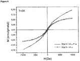
ここに記載の方法では、コアの形成と配位子の共有結合は同時の工程であり、このため、ネオ複合糖質の存在がナノクラスターの形状及びサイズをコントロールする。この方法で製造した糖質(グリコ)ナノ粒子は、従来公知の磁性ナノ粒子よりも小さく2nm以下のコアを有する。超常磁性挙動を全温度で示し、超伝導量子干渉素子(SQUID)計は、室温での強磁性成分の存在も示す。この異常磁気特性はイメージング及び細胞分離には重要である。 In the method described here, core formation and ligand covalent bonding are simultaneous steps, so the presence of neo-conjugate carbohydrates controls the shape and size of the nanoclusters. Carbohydrate (glyco) nanoparticles produced by this method are smaller than conventionally known magnetic nanoparticles and have a core of 2 nm or less. Superparamagnetic behavior is shown at all temperatures, and superconducting quantum interference device (SQUID) meters also show the presence of ferromagnetic components at room temperature. This anomalous magnetic property is important for imaging and cell separation.
磁性ナノ粒子及び強磁性流体の応用についての以下の例は、ここに記載の技術の広範な応用性をサポートするために、例として挙げたのであって、限定的なものではない。 The following examples for applications of magnetic nanoparticles and ferrofluids are given as examples and are not limiting to support the wide applicability of the techniques described herein.
本発明の一の態様では、本発明のナノ粒子の磁気特性は、カラム若しくは遠心分離の必要性を排除する細胞分離技術において利用される。これによって、非常に純粋な細胞群が迅速にかつ容易に得ることができる。一実施形態では、ナノ粒子は、対象の細胞上のレセプターに特定的に結合する配位子に結合してもよい。次いでナノ粒子を、細胞懸濁液及び磁場の印加によって懸濁液の残りから分離された粒子に結合した細胞に加えてもよい。 In one aspect of the invention, the magnetic properties of the nanoparticles of the invention are utilized in cell separation techniques that eliminate the need for columns or centrifugation. Thereby, a very pure cell group can be obtained quickly and easily. In one embodiment, the nanoparticles may bind to a ligand that specifically binds to a receptor on the cell of interest. The nanoparticles may then be added to the cells bound to the cell suspension and particles separated from the rest of the suspension by application of a magnetic field.
これは、例えば腫瘍細胞の存在のために体液を検査することによって腫瘍の診断において、多くの用途で使用できる非常に感度が良くかつ効率的な方法である。この手法の感度はこの点で大きな利点である。 This is a very sensitive and efficient method that can be used in many applications in the diagnosis of tumors, for example by examining body fluids for the presence of tumor cells. The sensitivity of this approach is a great advantage in this regard.
他の態様では、本発明は配位子との相互作用が生ずるか否かを決定する方法を提供し、この方法は、一又は二以上の配位子に結合したナノ粒子をパートナーと結合する候補に接触させる段階と、結合が生ずるか否かを決定する段階とを備える。 In another aspect, the present invention provides a method for determining whether interaction with a ligand occurs, which method binds nanoparticles bound to one or more ligands to a partner. Contacting the candidate and determining whether a bond occurs.
さらに他の態様では、本発明は配位子に結合できる物質をスクリーニングする方法を提供し、この方法は:
不動態化金属、又は、不動態化金属及び磁気金属を備えたコアであって、複数の配位子に共有結合されたコアを有するここに規定したナノ粒子を、一又は二以上の候補化合物に接触させる段階と;
候補化合物が配位子に結合するか否かを検出する段階と;を備える。
In yet another aspect, the invention provides a method of screening for substances that can bind to a ligand, the method comprising:
One or more candidate compounds comprising a passivated metal or a nanoparticle as defined herein having a core comprising a passivated metal and a magnetic metal and covalently bonded to a plurality of ligands Contacting with
Detecting whether the candidate compound binds to the ligand.
コアにおける磁気金属原子に対する不動態化金属原子の比は約5:0.1〜約2:5であるのが好ましい。比が約5:0.1〜約5:1であるばさらに好ましい。 The ratio of passivated metal atoms to magnetic metal atoms in the core is preferably about 5: 0.1 to about 2: 5. More preferably, the ratio is from about 5: 0.1 to about 5: 1.
他の態様では、本発明は、配位子に結合できる物質の試料において存在を決定する方法を提供するものであり、この方法は、試料を配位子に結合したナノ粒子に接触される段階と、結合が生ずるか否かを決定する段階とを備える。この方法は、例えば被分析物の存在に関連した病気状態の診断を助けるのに用いるために、試料内に一又は二以上の被分析物の存在若しくは量を決定するのに用いてもよい。被分析物の存在は被分析物とナノ粒子との凝集の形成によって信号化されてもよく、この存在は試料中に流体の緩和特性を測定することによって検出し得るものである。緩和特性の変化は凝集の存在、従ってターゲット分子の存在を示すものである。 In another aspect, the present invention provides a method for determining the presence in a sample of a substance capable of binding to a ligand, the method comprising contacting the sample with nanoparticles bound to the ligand. And determining whether coupling occurs. This method may be used to determine the presence or amount of one or more analytes in a sample, eg, to help diagnose a disease state associated with the presence of the analyte. The presence of the analyte may be signaled by the formation of an agglomeration between the analyte and the nanoparticles, which presence can be detected by measuring the relaxation properties of the fluid in the sample. A change in the relaxation properties indicates the presence of aggregation and hence the presence of the target molecule.
配位子が糖質である場合、異なる糖質が介在する相互作用の範囲は従来公知であり、ここに開示したナノ粒子を用いて研究若しくは変調され得る。これらには、白血球−内皮細胞付着、糖質−抗体相互作用、糖質−タンパク質バクテリア及びウィルス相互作用、腫瘍細胞の免疫学的認識、腫瘍細胞−内皮細胞(例えば、転移研究のために)、及び、異組織と細胞との認識が含まれる。 When the ligand is a saccharide, the range of interactions mediated by different saccharides is known in the art and can be studied or modulated using the nanoparticles disclosed herein. These include leukocyte-endothelial cell adhesion, carbohydrate-antibody interactions, carbohydrate-protein bacteria and virus interactions, immunological recognition of tumor cells, tumor cells-endothelial cells (eg for metastasis studies), And recognition of different tissues and cells.
他の態様では、本発明の磁性ナノ粒子及び強磁性流体を癌の治療に用いることができる。磁性ナノ粒子は腫瘍の温熱治療に用いてもよく、この場合、磁性ナノ粒子を腫瘍に注入し、高周波AC又はDC磁場にさらす。代替として、近赤外光を用いてもよい。磁気材料の緩和磁気エネルギーによって生ずる熱が粒子の周りの腫瘍組織を殺す。本発明の一実施形態では、腫瘍細胞は腫瘍特定抗原をナノ粒子に組み込むことによって特定的にターゲットとしてもよい。これによって、注射によって容易には到達しない腫瘍を治療粒子によって標的にすることを可能とし、通常の健康な細胞が殺されるのを回避する。 In other embodiments, the magnetic nanoparticles and ferrofluids of the present invention can be used to treat cancer. Magnetic nanoparticles may be used for hyperthermia treatment of tumors, in which case magnetic nanoparticles are injected into the tumor and exposed to a high frequency AC or DC magnetic field. Alternatively, near infrared light may be used. Heat generated by the relaxed magnetic energy of the magnetic material kills the tumor tissue around the particle. In one embodiment of the invention, tumor cells may be specifically targeted by incorporating tumor specific antigens into the nanoparticles. This allows tumors that are not easily reached by injection to be targeted by therapeutic particles, avoiding the killing of normal healthy cells.
所定の励起周波数について、最大特定吸収率(SAR)、さらに最大効率加熱を得る最適ナノ粒子サイズが存在する。この技術は、治療の効率を最大にし、管理される強磁性流体の量を最小にするために、狭いコアサイズ分散を有する磁性ナノ粒子を必要とする。本発明の磁性ナノ粒子は、合成法がナノ粒子の厳密な制御を可能にするので、特にこの用途に適している。 For a given excitation frequency, there is an optimal nanoparticle size that gives maximum specific absorption rate (SAR), and also maximum efficiency heating. This technique requires magnetic nanoparticles with a narrow core size dispersion to maximize therapeutic efficiency and minimize the amount of ferrofluid to be managed. The magnetic nanoparticles of the present invention are particularly suitable for this application because the synthesis method allows strict control of the nanoparticles.
他の実施形態において、ナノ粒子は抗体又は殺腫瘍ドラッグのような治療活性物質に結合していてもよい。ナノ粒子の磁気特性は、ナノ粒子を腫瘍細胞にガイドする磁場を用いることによって腫瘍を標的にするのにも用いることができる。しかしながら、腫瘍細胞にナノ粒子を向けるために磁場を用いるだけでは必ずしも実現可能若しくは正確ではなく、そのため、本発明はナノ粒子を腫瘍特定配位子を介して腫瘍細胞に特定して方向付けることができるという利点を提供する。これによって、使用するドラッグの量を減少することができ、副作用の可能性を低減する。というのも、ドラッグが必要とされる細胞にだけ向き、健康な細胞には向かわないからである。 In other embodiments, the nanoparticles may be conjugated to a therapeutically active agent such as an antibody or tumoricidal drug. The magnetic properties of the nanoparticles can also be used to target the tumor by using a magnetic field that guides the nanoparticles to the tumor cells. However, just using a magnetic field to direct the nanoparticles to the tumor cells is not always feasible or accurate, so the present invention can direct the nanoparticles to the tumor cells via a tumor specific ligand. Provides the advantage of being able to. This can reduce the amount of drug used and reduce the possibility of side effects. This is because it only works for cells that need a drug, not healthy cells.
他の態様では、本発明の磁性ナノ粒子を磁気共鳴イメージング(MRI)の質を改善するのに用いられる。MRIは、腫瘍のような構造を効率的に見ることができるのに十分なコントラストを必ずしも提供しないが、得られたイメージをコントラスト媒体のような磁性ナノ粒子を用いることによってエンハンスすることができる。得られたエンハンス感度によって腫瘍を検出することができるが、まだ非常に小さく、治療が成功するチャンスが高いときに非常に早い段階で腫瘍の検出が可能となる。 In other embodiments, the magnetic nanoparticles of the present invention are used to improve the quality of magnetic resonance imaging (MRI). MRI does not necessarily provide sufficient contrast to be able to efficiently see tumor-like structures, but the resulting image can be enhanced by using magnetic nanoparticles such as contrast media. Tumors can be detected with the resulting enhancement sensitivity, but they are still very small and can be detected very early when the chances of successful treatment are high.
このように腫瘍細胞の検出を温熱治療とくみあわせることもできる:腫瘍細胞が一旦同定されると、レーザー若しくは近赤外光を腫瘍サイトに向けて細胞を殺す。 Thus, detection of tumor cells can also be combined with hyperthermia: once tumor cells are identified, they are killed by directing a laser or near infrared light to the tumor site.
さらに、現在、肺はMRIイメージングによってイメージすることができない。ポジトロン放出断層撮影(PET)は肺をイメージすることができるが、放射線の繰り返し照射の危険のために、喘息及び肺気腫患者のような規則的な走査を要する患者については使用することができない。最近の研究では、過分極化気体MRIが喘息のような病気に適用できることを示している。これは、これらの気体の磁化が数秒で得られる肺全体のイメージ用に十分だからであり、患者に息を吸い込ませ、息を保持し、息を吐き出させる。患者が息を吸い、吐きときにイメージをとる能力は、MRIを用いて患者が息を吸い、吐くときに動的なイメージを精製することもできる。磁化された糖質ナノ粒子、及び、特にガドリニウムを含むこれらの粒子はせいぜい0.8nmで製造できる。この小さな粒子は“磁化された気体”として有効に考えることができるので、従って、過分極化気体の使用よりはるかに便利なセッティングにおいて肺イメージング用に使用できる。 Furthermore, currently the lung cannot be imaged by MRI imaging. Positron emission tomography (PET) can image the lung, but cannot be used for patients that require regular scanning, such as asthma and emphysema patients, due to the danger of repeated exposure to radiation. Recent studies have shown that hyperpolarized gas MRI can be applied to diseases such as asthma. This is because the magnetization of these gases is sufficient for an image of the entire lung that can be obtained in seconds, causing the patient to inhale, hold in, and exhale. The ability to take images as the patient inhales and exhales can also use MRI to refine dynamic images as the patient inhales and exhales. Magnetized carbohydrate nanoparticles, and especially those particles containing gadolinium, can be produced at most 0.8 nm. This small particle can be effectively considered as a “magnetized gas” and can therefore be used for lung imaging in a setting that is much more convenient than the use of hyperpolarized gases.
本発明の配位子結合粒子は腫瘍細胞に特定してデリバリーされ得て、そのため、もともとの腫瘍サイトから離れた腫瘍細胞さえ治療のために標的化される。 The ligand-binding particles of the present invention can be delivered specifically to tumor cells, so that even tumor cells that are distant from the original tumor site are targeted for therapy.
磁性元素金属を含むコアを有する本発明の実施形態は、元素金属は金属酸化物をイメージングする効率的なエンハンサーなので、特にイメージング用途によく適している。コア内の不動態化金属の存在は、これが磁性金属の酸化を防止するので有利である。不動態化金属はナノ粒子の生体適合性も増大し、コアを配位子に結合することを許容し、生物学的使用に加えて、酸化から磁性金属を保護し、凝集の可能性を低減する。 Embodiments of the invention having a core that includes a magnetic elemental metal are particularly well suited for imaging applications because elemental metals are efficient enhancers for imaging metal oxides. The presence of a passivating metal in the core is advantageous because it prevents the oxidation of the magnetic metal. Passivating metals also increase the biocompatibility of the nanoparticles, allowing the core to bind to the ligand, protecting the magnetic metal from oxidation and reducing the possibility of aggregation in addition to biological use To do.
本発明のナノ粒子の他の利点は、標的分子又は治療分子に結合されるときにさえ、細胞に捕獲されるのに適した極端に小さいサイズであることである。 Another advantage of the nanoparticles of the present invention is that they are extremely small in size suitable for being captured by cells, even when bound to target or therapeutic molecules.
他の点では、本発明の磁性ナノ粒子はドラッグデリバリー用のトレーサーとして使用される放射性材料の代わりに用いてもよい。放射性材料の代わりに磁気粒子を使用することによって、ドラッグデリバリーが磁気変動を測定することが可能となり、これによって放射線からの潜在的な害を排除する。 In other respects, the magnetic nanoparticles of the present invention may be used in place of radioactive materials used as tracers for drug delivery. By using magnetic particles instead of radioactive materials, drug delivery can measure magnetic fluctuations, thereby eliminating potential harm from radiation.
一般的に、糖質のタンパク質のような他の種類又は他の糖質への結合が非常に弱く、多価の傾向があるので、糖質が媒介した相互作用を検出又は変調するのに従来困難な問題がある。従って、検出のために結合は弱く、また、相互作用を変調するために、一価の薬剤は多価の糖質をベースにした相互作用を破壊するのに限定的にしかうまくいかない。 In general, the binding of carbohydrates to other types or other carbohydrates, such as proteins, is very weak and tends to be multivalent, so traditionally used to detect or modulate carbohydrate-mediated interactions There is a difficult problem. Thus, the binding is weak for detection, and in order to modulate the interaction, the monovalent agent only works limitedly to destroy the multivalent carbohydrate-based interaction.
糖質−糖質相互作用に関連した本発明の実施形態では、2種類の相互作用を同定することができる。同種親和性相互作用では、同一の糖質同士が互いに相互作用し、凝集が起こるまで表面に固定した単一種の配位子を有する粒子の濃度の安定的な増大によって検出され得る。これは、光散乱又は電子的効果によって検出される。 In embodiments of the invention related to carbohydrate-carbohydrate interactions, two types of interactions can be identified. In homophilic interactions, the same carbohydrates can interact with each other and can be detected by a stable increase in the concentration of particles with a single type of ligand immobilized on the surface until aggregation occurs. This is detected by light scattering or electronic effects.
異好性相互作用は2又は3以上の異なるナノ粒子を混合し、粒子の凝集状態を決定することによって検出できる。 A heterophilic interaction can be detected by mixing two or more different nanoparticles and determining the aggregation state of the particles.
本発明は、糖質を媒介した相互作用を研究し、変形するための多目的なプラットフォームを提供する。例えば、粒子は抗糖質抗体を検出するのに用いられ、粒子の凝集を検出するために光散乱を介して抗体の粒子上の配位子への結合を検出し、又は、粒子クラスター内の金属原子が一緒になるときに変わる表面プラズモン共鳴のような磁場効果を介してその配位子への結合を検出する。 The present invention provides a versatile platform for studying and transforming carbohydrate-mediated interactions. For example, the particles can be used to detect anti-carbohydrate antibodies, detect binding of antibodies to ligands on the particles via light scattering to detect particle aggregation, or within particle clusters The binding to the ligand is detected through magnetic field effects such as surface plasmon resonance that change when the metal atoms come together.
本発明は、糖質を媒介する相互作用が生ずるかどうかを決定する方法を提供するものであり、方法は、糖質を媒介した相互作用を介して本発明のナノ粒子と相互作用させるために、疑わしい一又は二以上の種を接触させる段階と、ナノ粒子が糖質媒介相互作用を変化させるかどうかを決定する段階と、を備える。 The present invention provides a method for determining whether a carbohydrate mediated interaction occurs, the method for interacting with a nanoparticle of the present invention via a sugar mediated interaction. Contacting one or more suspect species, and determining whether the nanoparticles alter carbohydrate-mediated interactions.
本発明は、糖質と結合するパートナーとの間の相互作用を壊す方法を提供し、この方法は、糖質及び結合パートナーを本発明のナノ粒子と接触させる段階を備え、ナノ粒子は糖質と結合パートナーとの相互作用を壊すことができる糖質基を備える。 The present invention provides a method of breaking the interaction between a carbohydrate and a binding partner, the method comprising contacting the carbohydrate and the binding partner with a nanoparticle of the present invention, wherein the nanoparticle is a carbohydrate With a carbohydrate group that can break the interaction between and the binding partner.
他の態様では、配位子が抗原であるナノ粒子は例えば、表皮の外側層を介して経皮的通過を促進するためにデリバリーガンを用いることによって、ワクチンとして管理することができる。ナノ粒子は、例えばリンパ系を拡散しながら成熟する樹枝状細胞を細くすることができ、免疫応答の変形及び抗原に対するワクチン接種につながる。 In other embodiments, nanoparticles whose ligand is an antigen can be administered as a vaccine, for example, by using a delivery gun to facilitate percutaneous passage through the outer layer of the epidermis. Nanoparticles can, for example, thin dendritic cells that mature as they diffuse through the lymphatic system, leading to altered immune responses and vaccination against antigens.
配位子が抗原をエンコーディングする核酸であるナノ粒子はワクチンとしても管理してもよい。ナノ粒子は特にこのような用途によく合っている。というのは、粒子が十分に小さくて使用される細胞のダメージなしで細胞膜を貫通するように、核酸ワクチンは有効である個々の細胞に入らなければならないからである。 Nanoparticles whose ligands are nucleic acids encoding antigens may also be managed as vaccines. Nanoparticles are particularly well suited for such applications. This is because the nucleic acid vaccine must enter individual cells that are effective so that the particles are small enough to penetrate the cell membrane without damage to the cells used.
従来公知のワクチンデリバリーガンは、圧縮された空気又は気体通常はヘリウムガスの使用によってデリバリーを動かす。これは痛みを伴い、皮膚上にみみず腫れを生じ得る。本発明の磁性ナノ粒子は代替のデリバリーシステムで使用され、ここで、粒子をデリバリーする駆動力は磁場の印加によって供給する。磁場の反転によって、外側表皮層を介してナノ粒子を進ませるのに十分な急速な加速を生ずる。これによって、痛みを軽減し、圧縮ガスの使用によって生ずるみみず腫れの形成が低減される。 Previously known vaccine delivery guns move the delivery by use of compressed air or gas, usually helium gas. This is painful and can cause swelling on the skin. The magnetic nanoparticles of the present invention are used in an alternative delivery system, where the driving force for delivering the particles is supplied by the application of a magnetic field. The reversal of the magnetic field results in rapid acceleration sufficient to advance the nanoparticles through the outer skin layer. This relieves pain and reduces the formation of worm swelling caused by the use of compressed gas.
他の用途では、細胞表面糖質はウィルス若しくはバクテリアレセプター用の配位子(いわゆる付着因子)として作用すること、及び、糖質のレセプターへの結合が感染中に要する事象であることが公知である。これらの相互作用を変調できる合成糖質例えば、複合糖質は本発明のナノ粒子内に固定することができ、これらの相互作用を研究するための試薬として、また、ウィルス又はワクチン感染を防止するための治療のために、使用することができる。 In other applications, cell surface carbohydrates are known to act as ligands for viral or bacterial receptors (so-called attachment factors) and that carbohydrate binding to receptors is an event required during infection. is there. Synthetic carbohydrates that can modulate these interactions, such as glycoconjugates, can be immobilized within the nanoparticles of the present invention, as reagents for studying these interactions, and to prevent viral or vaccine infection Can be used for treatment.
他の用途では、本発明は免疫反応の変調(モジュレーション)例えば、以下の移植可能性で有効である。組織の免疫学的認識が移植された組織上に存在する表面糖質と抗体のようなホストの免疫システムのコンポーネントとの間の糖質が媒介する相互作用によって始まる。これによって、これは、この相互作用を起因とする免疫反応を改善するためにターゲットとなり得る。例えば、糖質Galα1−3Galβ1−4GlnAc(“αGal”エピトープ(抗原決定基))が、移植された組織の拒絶に含まれる重要な抗原エピトープとして関係している。こうして、αGalエピトープと免疫系との相互作用の変調はここに記載したナノ粒子用の治療ターゲットでありえる。 In other applications, the invention is useful in modulating immune responses, such as the following transplantability. Tissue immunological recognition begins with carbohydrate-mediated interactions between surface carbohydrates present on the transplanted tissue and components of the host immune system such as antibodies. Thereby, this can be a target to improve the immune response due to this interaction. For example, the carbohydrate Galα1-3Galβ1-4GlnAc (“αGal” epitope (antigenic determinant)) has been implicated as an important antigenic epitope involved in rejection of transplanted tissue. Thus, modulation of the interaction between the αGal epitope and the immune system may be a therapeutic target for the nanoparticles described herein.
代替アプローチは、多くの腫瘍関連抗原若しくは腫瘍自己分泌因子が糖質ベースなので、癌の治療に有効である。この事象においては、ナノ粒子はワクチンとして備えられ、免疫系を準備して、糖質を表す腫瘍細胞が抗体上を攻撃することができるところの抗体を生成する。この点では、多くの細胞が、通常の健康な細胞とは対照的に、ナノ粒子によって刺激された免疫応答を腫瘍細胞に特定して向けることができる異常なグリコシル化(糖化)パターンを保有することは公知である。ナノ粒子は、例えば、内皮細胞を介した腫瘍細胞のマイグレーションを通して、癌の転移を阻止するのに使用することもできる。 An alternative approach is effective in treating cancer because many tumor-associated antigens or tumor autocrine factors are carbohydrate based. In this event, the nanoparticles are provided as a vaccine and prepare the immune system to produce antibodies where tumor cells that represent carbohydrates can attack on the antibodies. In this regard, many cells possess an unusual glycosylation (glycation) pattern that, in contrast to normal healthy cells, can direct the immune response stimulated by nanoparticles to tumor cells. This is well known. Nanoparticles can also be used, for example, to block cancer metastasis through migration of tumor cells via endothelial cells.
前立腺癌におけるリンパ節転移の非侵襲的検出はすでに、MRIと組み合わせてリンパ向性超常磁性ナノ粒子を用いることによって立証されている。転移用の親和性若しくは特異性を増大した糖(グリコ)ナノ粒子の例のリストを以下に示す。
Lex−GNP Ley−GNP STn−GNP
Glogo−H−GNP Gg3−GNP Gluco−GNP
Malto−GNP Lacto−GNP Man−GNP
Noninvasive detection of lymph node metastasis in prostate cancer has already been demonstrated by using lymphotropic superparamagnetic nanoparticles in combination with MRI. A list of examples of sugar (glyco) nanoparticles with increased affinity or specificity for transfer is shown below.
Le x -GNP Le y -GNP STn-GNP
Glogo-H-GNP Gg 3 -GNP Gluco-GNP
Malto-GNP Lacto-GNP Man-GNP
糖ナノ粒子のような存在する他の配位子に加えて、エストロゲン、DHEAのようなホルモンをナノ粒子に付けて、可溶性にし得る。これらは乳癌のような癌の検出における使用を有してもよい。ペプチドは細胞表面腫瘍遺伝子コード化レセプターのような特定のレセプターに局在化したナノ粒子に付けることもできる。脂質、特にトールレセプターに結合するものを付けることもできる。メチレンブルーのような化学的配位子が、メラノーマ転移の検出に使用され得る糖ナノ粒子に付くことができる。最後に、siRNAナノ粒子は、細胞内へ取りこんだ後に、腫瘍遺伝子若しくはウィルス特定mRNAの発現をイメージし得る。 In addition to other ligands present such as sugar nanoparticles, hormones such as estrogen, DHEA can be attached to the nanoparticles to make them soluble. These may have use in the detection of cancers such as breast cancer. The peptide can also be attached to nanoparticles localized to a specific receptor, such as a cell surface oncogene encoded receptor. It is also possible to attach lipids, particularly those that bind to the toll receptor. Chemical ligands such as methylene blue can be attached to sugar nanoparticles that can be used to detect melanoma transitions. Finally, siRNA nanoparticles can image the expression of oncogenes or virus specific mRNAs after incorporation into cells.
他の態様において、ナノ粒子は配位子に特定的に結合し得る抗体を増加させるキャリアとして使用できる。これは配位子が糖質である場合に特に好都合である。というのは、抗体は小さいことが多く、強い免疫応答を生じないので、これは糖質含有部分に対する抗体を増大することが従来のチャレンジングな問題であり得るためである。 In other embodiments, the nanoparticles can be used as a carrier to increase antibodies that can specifically bind to the ligand. This is particularly advantageous when the ligand is a carbohydrate. This is because antibodies are often small and do not produce a strong immune response, so increasing antibodies to carbohydrate-containing moieties can be a traditional challenge.
他の態様では、糖質は、ナノ粒子によって必要となる細胞構成にガイドされ得る量子ドットを提供するために、セレン化カドミウムのナノ結晶に付けることができる。セレン化物に加えて硫化物のような他のアニオンを使用してもよい。量子ドットは生物学的イメージングにおける使用の可能性、電子デバイス及び光デバイスのいずれにおいても、量子コンピュータ及び候補ドラッグのスクリーニングにおける使用の可能性を有する。 In other embodiments, carbohydrates can be attached to cadmium selenide nanocrystals to provide quantum dots that can be guided by the nanoparticles to the cellular configuration required. In addition to selenides, other anions such as sulfides may be used. Quantum dots have the potential to be used in biological imaging, in both electronic devices and optical devices, in quantum computers and candidate drug screening.
他の態様では、本発明は、心筋救済の査定例えば、心臓麻痺後に生存している心臓の組織の量のために、本願で定義するナノ粒子の使用を含む。現在、これは、テクネチウムのような放射性核種と組み合わせて、細胞によって捕獲(取り込み)され得るセスタミビ(sestamibi)やテトロフォスミン(tetrofosmin)のような化合物を用いて−シンチグラフ技術(例えば、SPECT)によって大部分はモニターされる。放射性トレーサーの取り込みは局在的な血流に比例し、心筋救済の程度の示唆を与えることになる−取り込みが大きいほど、心筋救済も大きくなる。 In another aspect, the invention includes the use of nanoparticles as defined herein for assessment of myocardial salvage, eg, the amount of cardiac tissue that is alive after cardiac paralysis. Currently, this is accomplished by using compounds such as sestamibi and tetrofosmin that can be captured (incorporated) by cells in combination with a radionuclide such as technetium-by scintigraphic techniques (eg SPECT). The part is monitored. The uptake of radioactive tracer is proportional to the local blood flow and gives an indication of the extent of myocardial rescue-the greater the uptake, the greater the myocardial rescue.
セスタミビやテトロフォスミンのような化合物は、これらが細胞膜を介して受動的に拡散する脂溶性陽イオン複合体なので作用する。このような複合体の機能化された配位子は、磁化したナノ粒子のセルフアセンブル中の表面配位子として容易に組み込まれ得る。他の広範囲の新規な化学的配位子がナノ粒子に付いて、それらを自由に拡散可能にし得る。 Compounds such as sestamibi and tetrofosmin work because they are fat-soluble cation complexes that passively diffuse through the cell membrane. Such complex functionalized ligands can be readily incorporated as surface ligands in the self-assembly of magnetized nanoparticles. A wide range of other novel chemical ligands can attach to the nanoparticles and make them freely diffusible.
本願に記載するナノ粒子は、拡散が細胞へ拡散し得るセスタミビ、テトロフォスミン等の他の化合物の誘導体の存在の下でセルフアセンブルしてもよい。その結果のナノ粒子を用いて、心筋救済を、放射能の必要なしで、磁気イメージングによってモニターし得る。磁気共鳴イメージングをナノ粒子の検出のために用いてもよい;放射性トレーサーについて、ナノ粒子の取り込み量は局部的な血流に比例する。現在通常用いられているシンチグラフィートレーサーは99mTc−セスタミビ、及び、99m−テトロフォスミンである(Recent Advances in 99Tc Radiopharmaceutiocals - Annals of Nuclear Medicine 16:79-93(2003);Contributions of Nuclear Cardiology to Diagnosis and Prognosis of Patients with Coronary Artery Disease - Circulation 2000: 100: 1465-1478)。好適な態様では、本発明のナノ粒子はセスタミビに共役され、磁気イメージング用に用いられる。このように、ナノ粒子は心筋救済をモニターするために、99mTcの代用をするのに用いてもよい。 The nanoparticles described in this application may be self-assembled in the presence of derivatives of other compounds such as sestamibi, tetrofosmin, etc., whose diffusion can diffuse into cells. Using the resulting nanoparticles, myocardial rescue can be monitored by magnetic imaging without the need for radioactivity. Magnetic resonance imaging may be used for nanoparticle detection; for radioactive tracers, nanoparticle uptake is proportional to local blood flow. Currently used scintigraphy tracers are 99mTc-sestamibi and 99m-tetrofosmin (Recent Advances in 99Tc Radiopharmaceutiocals-Annals of Nuclear Medicine 16: 79-93 (2003); Contributions of Nuclear Cardiology to Diagnosis and Prognosis of Patients with Coronary Artery Disease-Circulation 2000: 100: 1465-1478). In a preferred embodiment, the nanoparticles of the present invention are conjugated to sestamibi and used for magnetic imaging. Thus, nanoparticles may be used to substitute for 99mTc to monitor myocardial rescue.
他の用途では、ここに開示されている磁性ナノ粒子は磁気記録メディアの製造の際に用いてもよい。 In other applications, the magnetic nanoparticles disclosed herein may be used in the manufacture of magnetic recording media.
本発明の実施形態を例として、添付図面を参照して非限定的に説明する。 Embodiments of the present invention will be described by way of example and not limitation with reference to the accompanying drawings.
本願に記載したナノ粒子又はこれの誘導体は、薬剤組成で配合(定式化)でき、種々の形で患者に投与できる。従って、ナノ粒子は、インビボ細胞及び組織ラベリングについて、腫瘍ターゲッティング及び温熱治療用の薬品として、又は、磁気共鳴イメージングにおいてコントラスト強調媒体(メディア)として使用してもよい。 The nanoparticles or derivatives thereof described herein can be formulated (formulated) with a pharmaceutical composition and administered to patients in various forms. Thus, the nanoparticles may be used for in vivo cell and tissue labeling, as tumor targeting and hyperthermia drugs, or as contrast enhancement media in magnetic resonance imaging.
経口投与用の薬剤組成物はタブレット、カプセル、粉末又は液体であってもよい。タブレットは、ゼラチン、補助薬又は不活性希釈剤のような固形キャリアを含んでもよい。液体薬剤組成物は一般に、水、石油、動物性若しくは植物性油、鉱油若しくは合成油のような液体キャリアを含んでもよい。生理食塩水、又は、エチレングリコール、プロピレングリコール若しくはポリエチレングリコールのようなグリコールを含んでもよい。このような組成物及び調合液は一般に合成物の少なくとも0.1重量%を含む。 Pharmaceutical compositions for oral administration may be tablets, capsules, powders or liquids. A tablet may include a solid carrier such as gelatin, an adjuvant or an inert diluent. Liquid pharmaceutical compositions generally may include a liquid carrier such as water, petroleum, animal or vegetable oils, mineral oil or synthetic oil. Saline or glycols such as ethylene glycol, propylene glycol or polyethylene glycol may be included. Such compositions and preparations generally comprise at least 0.1% by weight of the composition.
非経口投与は以下のルートによる投与を含む:静脈、皮膚若しくは皮下、鼻、筋肉内、眼球、経上皮、腹腔、及び、局所的(皮膚、眼、直腸、鼻、吸入、噴霧器)、及び、直腸系のルート。静脈、皮膚若しくは皮下の注射又は痛みサイトの注射については、有効成分は、発熱物質フリーでかつ適当なpH、等張性及び安定性を有する非経口的に受け入れられる水溶液の形である。当業者は例えば化合物若しくはその誘導体の溶液を用いる適当な溶液を、例えば、生理食塩水で、グリセロール、液体ポリエチレングリコール若しくはオイルを用いて準備した分散液で準備することができる。 Parenteral administration includes administration by the following routes: intravenous, cutaneous or subcutaneous, nasal, intramuscular, ocular, transepithelial, abdominal, and topical (skin, eye, rectum, nasal, inhalation, nebulizer), and The rectal route. For intravenous, cutaneous or subcutaneous injections or pain site injections, the active ingredient is in the form of a parenterally acceptable aqueous solution that is pyrogen-free and has an appropriate pH, isotonicity and stability. One skilled in the art can prepare a suitable solution using, for example, a solution of the compound or derivative thereof, for example, in saline, with a dispersion prepared using glycerol, liquid polyethylene glycol or oil.
任意で他の活性材料と組み合わせた一又は二以上の化合物に加えて、組成物は調合可能な賦形剤、キャリア、バッファ、安定剤、等浸透圧剤、防腐剤若しくは抗酸化剤若しくは当業者に周知の他の材料の一又は二以上を含むことができる。このような材料は非毒性であり、活性材料の有効性と干渉すべきではない。キャリア若しくは他の材料の精確な性質は投与ルート例えば、経口又は非経口に依存してもよい。 In addition to one or more compounds, optionally in combination with other active ingredients, the composition can be formulated with excipients, carriers, buffers, stabilizers, isotonic agents, preservatives or antioxidants, or one skilled in the art. Can include one or more of the other known materials. Such materials are non-toxic and should not interfere with the effectiveness of the active material. The precise nature of the carrier or other material may depend on the route of administration, eg, oral or parenteral.
液体薬剤組成物は典型的には、約3.0から9.0の間、より好適には約4.5から8.5の間、さらに好適には約5.0から8.0の間のpHを有するように配合される。組成物のpHは、典型的には約1mMから50mMの範囲で使用される、酢酸塩、クエン酸塩、リン酸塩、コハク酸塩、トリス又はヒスチジンのようなバッファーの使用によって維持できる。さもなければ、組成物のpHは生理学的に受け入れられる酸若しくは塩基を用いて調整できる。 The liquid pharmaceutical composition is typically between about 3.0 and 9.0, more preferably between about 4.5 and 8.5, and even more preferably between about 5.0 and 8.0. Is formulated to have a pH of The pH of the composition can be maintained by the use of buffers such as acetate, citrate, phosphate, succinate, tris or histidine, typically used in the range of about 1 mM to 50 mM. Otherwise, the pH of the composition can be adjusted using a physiologically acceptable acid or base.
防腐剤は一般に薬剤組成物に含まれて細菌の繁殖を抑制し、組成物の保存期間を延ばし、多様な使用パッキングを可能とする。防腐剤の例には、フェノール、メタクレゾール、ベンジルアルコール、パラヒドロキシ安息香酸及びそのエステル、メチルパラベン、塩化ベンザルコニウム、及び、塩化ベンゼトニウムを含む。防腐剤は典型的には約0.1から1.0%の範囲で使用される。 Preservatives are generally included in pharmaceutical compositions to inhibit bacterial growth, extend the shelf life of the composition, and allow for diverse use packing. Examples of preservatives include phenol, metacresol, benzyl alcohol, parahydroxybenzoic acid and its esters, methyl paraben, benzalkonium chloride, and benzethonium chloride. Preservatives are typically used in the range of about 0.1 to 1.0%.
薬剤組成物は、“予防的な有効量”又は“治療的な有効量”(この場合、病気予防は治療と考えてもよい)で個人に与えられ、これは個々に対する効果を示すのに十分である。典型的には、これは、個々に効果を出す治療的に有効な作用を引き起こす。投与される組成物の実際の量、及び投与の頻度は処置される状態の性質や深刻さに依存する。処置の処方箋、例えば、投与量の決定等は一般的な処置者や他の医師の責任の範囲内であり、典型的には処置される疾患、個々の患者の状態、デリバリーの場所、投与の方法及び処置者が知っている他の要素が考慮される。上述の手法及びプロトコルの例は、レミントン(Remington)のPhamaceutical Sciences(Mack Publishing 社, イーストン、ペンシルバニア州、第19版、1995年)、Handbook of Phamaceutical Additives(第2版、アッシュ(M.Ash)及びアッシュ(I.Ash)編著、2001年(Synapse Information Resources社、エディコット、ニューヨーク州、米国)、Handbook of Phamaceutical Excipents(第2版、1994年)。例えば、、組成物は体重の単位kgあたり約0.01から100mgの活性組成物のドーズ量を患者に投与するのが好ましく、体重の単位kgあたり約0.5から10mgであればもっと好ましい。 The pharmaceutical composition is given to an individual in a “prophylactically effective amount” or “therapeutically effective amount” (in which case disease prevention may be considered a treatment), which is sufficient to show an effect on the individual. It is. This typically causes a therapeutically effective action that is individually effective. The actual amount of composition administered, and the frequency of administration, will depend on the nature and severity of the condition being treated. Treatment prescriptions, such as dose determination, are within the responsibility of the general practitioner and other physicians, typically the disease being treated, the individual patient condition, the location of delivery, the administration Other factors known to the method and the practitioner are considered. Examples of the methods and protocols described above include Remington's Phamaceutical Sciences (Mack Publishing, Easton, Pennsylvania, 19th edition, 1995), Handbook of Phamaceutical Additives (2nd edition, M. Ash) and Edited by I. Ash, 2001 (Synapse Information Resources, Edikot, New York, USA), Handbook of Phamaceutical Excipents (2nd edition, 1994) For example, the composition is about Preferably, a dose of 0.01 to 100 mg of active composition is administered to the patient, more preferably about 0.5 to 10 mg per kg body weight.
抗体
ナノ粒子はコア粒子に結合した配位子に対して抗体応答を生ずるキャリアとして使用してもよい。これらの抗体は従来標準の技術を用いて変成できる。ここにはじめて例示するものと類似の抗体も公知の方法に関連してここに教示したものを用いて生成できる。これらの抗体を製造する方法は、ナノ粒子を用いてほ乳類(例えば、マウス、ラット、うさぎ、馬、やぎ、羊、又は猿)に免疫性を与える(免疫化する)ことを含む。抗体は従来公知の種々の技術のいずれを用いて、好ましくは抗体の対象の抗原への結合を用いることによってスクリーニングして免疫性が与えられた動物から得られてもよい。動物から、抗体及び/又は細胞生成抗体を分離することは、動物の犠牲の段階を伴ってもよい。
The antibody nanoparticles may be used as a carrier that produces an antibody response to a ligand bound to the core particle. These antibodies can be denatured using conventional standard techniques. Antibodies similar to those exemplified here for the first time can be generated using those taught herein in connection with known methods. Methods for producing these antibodies include immunizing (immunizing) mammals (eg, mice, rats, rabbits, horses, goats, sheep, or monkeys) using nanoparticles. The antibody may be obtained from an animal immunized by screening using any of a variety of techniques known in the art, preferably by using the binding of the antibody to the antigen of interest. Separating the antibody and / or cell-generating antibody from the animal may involve an animal sacrifice step.
ほ乳類をナノ粒子を用いて免疫性を与えることと代替で若しくはこれに補助して、配位子及び/又はナノ粒子に対して特定的な抗体が、発現免疫グロブリン可変ドメインの組み込み技術によって生成されたライブラリから、例えば、それらの表面上に機能的な免疫グロブリン結合ドメインを表示するラムダバクテリオファージ又は繊維状バクテリオファージを用いて、得られてもよい;例えば、国際公開92/01047号パンフレットを参照されたい。ライブラリは素朴なものであってもよい。すなわち、ナノ粒子のいずれを用いても免疫性を与えられていない有機体(生命体)から得られたシーケンスから構築してもよいし、対象の抗原に曝された有機体から得られたシーケンスを用いて構築されてもよい。 In lieu of or in addition to immunizing mammals with nanoparticles, antibodies specific to ligands and / or nanoparticles are generated by techniques for incorporating expressed immunoglobulin variable domains. For example, using lambda bacteriophages or filamentous bacteriophages that display functional immunoglobulin binding domains on their surface; see, eg, WO 92/01047 I want to be. The library may be simple. That is, it may be constructed from a sequence obtained from an organism that is not immunized with any of the nanoparticles (living organism), or a sequence obtained from an organism exposed to the antigen of interest. It may be constructed using
“単クローン性抗体”という語は、抗体の実質的に均一な個体群から得られた抗体例えば、個体群を構成する個々の抗体は、少量存在する突然変異体に自然に生ずる可能性があるものとは異なり、同一であることを意味する。単クローン性抗体は、コーラ(Kohler)及びミルスタイン(Milstein)(Nature, 256:495, 1975年)に最初に記載された方法によって生産でき、又は、組み換え方法(カビリー(Cabilly))らによる米国特許第4,816,567号明細書、又は、単クローン性抗体生産技術及び応用(Monoclonal Antibody Production Techniques and Applications、79-97、Marcel Dekker社、ニューヨーク州、1987年)においてマージ(Mage)及びラモイ(Lamoyi)を参照されたい)。 The term “monoclonal antibody” refers to an antibody obtained from a substantially homogeneous population of antibodies, for example, the individual antibodies that make up the population may naturally arise in mutants that are present in small amounts Unlike things, it means the same thing. Monoclonal antibodies can be produced by the method first described in Kohler and Milstein (Nature, 256: 495, 1975) or in the United States by recombinant methods (Cabilly et al. No. 4,816,567 or Monoclonal Antibody Production Techniques and Applications, 79-97, Marcel Dekker, New York, 1987) with Mage and Lamoyi. See).
ハイブリドーマ法ににおいて、マウス若しくは他の適当なホスト動物を、免疫化(免疫処置)に使用するナノ粒子に特定して結合する抗体を生産する若しくは生産し得るリンパ球を誘発する(発現する、引き出す)ために皮下、腹腔内、又は筋肉内のルートによって抗体を用いて免疫化する。代替として、リンパ球をインビトロで免疫化してもよい。次いで、リンパ球を、ポリエチレングリコールのような適当な融剤を使用して骨髄腫細胞を用いて融合し、ハイブリドーマ細胞を生成する(ゴーディング(Goding)によるMonoclonal Antibodies:Principles and Practice, 59-103 (Academic Press, 1996)を参照されたい)。 In hybridoma methods, mice or other suitable host animals are induced (expressed, elicited) to produce or produce antibodies that specifically bind to the nanoparticles used for immunization (immunization). To immunize with antibodies by subcutaneous, intraperitoneal, or intramuscular routes. Alternatively, lymphocytes may be immunized in vitro. Lymphocytes are then fused with myeloma cells using an appropriate fluxing agent such as polyethylene glycol to generate hybridoma cells (Monoclonal Antibodies by Goding: Principles and Practice, 59-103). (See Academic Press, 1996).
こうして準備したハイブリドーマ細胞を、融合されていない親ハイブリドーマ細胞の成長若しくは生存を防止する一又は二以上の物質を好適に含む適当な培地(培養液)に種がまかれ、成長し得る。例えば、親ハイブリドーマ細胞が酵素ヒポキサンチン・グアニン・ホスホリボシルトランスフェラーゼ(HGPRT又はHPRT)を欠くならば、ハイブリドーマ用の培地は典型的には、HGPRT不足細胞の成長を防止するヒポキサンチン、アミノプテリン、チミジン(HAT媒体)を含む。 The hybridoma cells thus prepared can be seeded and grown in a suitable medium (culture solution) that preferably contains one or more substances that prevent the growth or survival of the unfused parent hybridoma cells. For example, if the parent hybridoma cell lacks the enzyme hypoxanthine guanine phosphoribosyltransferase (HGPRT or HPRT), the culture medium for the hybridoma typically contains hypoxanthine, aminopterin, thymidine that prevents the growth of HGPRT-deficient cells. (HAT medium).
好適な骨髄腫細胞は、効率的に融合し、選択された抗体生成細胞によって抗体の安定的な高レベルの発現を維持し、HAT媒体のような媒体に敏感な細胞である。 Preferred myeloma cells are cells that fuse efficiently, maintain a stable high level of expression of the antibody by the selected antibody-producing cells, and are sensitive to a medium such as a HAT medium.
ハイブリドーマ細胞が成長する培地は、ナノ粒子/配位子に対して向かう単クローン抗体の生成のために検査(評価、分析)される。好適には、結合特異性(選択性)は、酵素結合免疫吸着体検査によって決定される。本発明の単クローン抗体はナノ粒子/配位子に特定的に結合する抗体である。 The medium in which the hybridoma cells are growing is examined (evaluated, analyzed) for the production of monoclonal antibodies directed against the nanoparticles / ligand. Suitably, binding specificity (selectivity) is determined by enzyme-linked immunosorbent assay. The monoclonal antibodies of the present invention are antibodies that specifically bind to nanoparticles / ligands.
本発明の好適な実施形態では、例えば、スキャチャード分析(ムンセン(Munson)及びポラード(Pollard)のAnal. Biochem., 107:220, 1980年参照)単クローン抗体はマイクロモル又はそれ以上の親和性(例えば、10−6mol以上の親和性)を有する。 In a preferred embodiment of the invention, for example, Scatchard analysis (see Munson and Pollard Anal. Biochem., 107: 220, 1980) monoclonal antibodies may have micromolar or higher affinity. (For example, an affinity of 10 −6 mol or more).
所望の特異性及び親和性の抗体を中和(中性)抗体を生成するハイブリドーマ細胞を生成し、クローンを希釈手順を制限することによってサブクローンして、標準法によって成長させ得る。この目的のための適当な培地には、 ダルベッコ変性イーグル培地若しくはPRMI−1640培地が含まれる。また、ハイブリドーマ細胞は、動物中の腹水腫瘍としてインビボで成長してもよい。 Hybridoma cells that produce antibodies that neutralize antibodies of the desired specificity and affinity are generated, and clones can be subcloned by limiting the dilution procedure and grown by standard methods. Suitable culture media for this purpose include Dulbecco's Modified Eagle's Medium or PRMI-1640 medium. The hybridoma cells may also grow in vivo as ascites tumors in animals.
サブクローンによって分泌された単クローン抗体は、例えばタンパク質A−セファロース、水酸燐灰石クロマトグラフィ、ゲル電気泳動、透析、又は親和性クロマトグラフィのような従来の免疫グロブリン精製手法によって、培地、腹水流体若しくは血清から適当に分離される。 Monoclonal antibodies secreted by subclones can be isolated from medium, ascites fluid or serum by conventional immunoglobulin purification techniques such as protein A-Sepharose, hydroxyapatite chromatography, gel electrophoresis, dialysis, or affinity chromatography. Separated appropriately.
本発明の単クローン抗体をエンコーディングする核酸は、例えば、ネズミの抗体の重鎖及び軽鎖をエンコーディングする遺伝子に特定的に結合し得るオリゴヌクレオチドプローブを用いることによって、従来周知の手法を用いて容易に分離(単離)され、配列が決定される。本発明のハイブリドーマ細胞は抗体若しくはそのフラグメントをエンコーディングする核酸の好適な源である。一旦分離されると、核酸は発現ベクター若しくはクローニング・ベクターに縛られ、次いで、ホスト細胞に転写され、単クローン抗体を組み換えホスト細胞培地で生成されるように培養され得る。 The nucleic acid encoding the monoclonal antibody of the present invention can be easily obtained using a conventionally known method, for example, by using an oligonucleotide probe that can specifically bind to the gene encoding the heavy and light chains of a murine antibody. Are separated (isolated) and sequenced. The hybridoma cells of the invention are a suitable source of nucleic acid encoding an antibody or fragment thereof. Once isolated, the nucleic acid can be bound to an expression vector or cloning vector and then transcribed into host cells and cultured to produce monoclonal antibodies in recombinant host cell media.
所望の結合特性で抗体を生成できるハイブリドーマは本発明の範囲内であり、抗体(抗体フラグメントを含む)をエンコーディングする核酸を含むホスト細胞であり、それを発現し得る。本発明はまた、抗体が生成され、好適には分泌されている状態で抗体を生成できる細胞を成長させることを含む抗体の製法も提供する。 Hybridomas capable of producing antibodies with the desired binding characteristics are within the scope of the present invention and are host cells containing nucleic acids encoding antibodies (including antibody fragments) that can express them. The invention also provides a method for producing an antibody comprising growing cells capable of producing the antibody in a state where the antibody is produced and preferably secreted.
本発明による抗体は多くの方法で変性してもよい。実際、“抗体”との語は必要な特異性を有する結合ドメインを有するいかなる結合物質をもカバーするように解釈されたい。本発明は、合成分子や、抗原若しくはエピトープ、ここに記載したような糖質配位子に結合し得る抗体を形状が類似する分子を含む、抗体フラグメント、誘導体、抗体の機能同等物及び同族体をカバーする。 The antibodies according to the invention may be denatured in a number of ways. Indeed, the term “antibody” should be construed to cover any binding substance having a binding domain with the requisite specificity. The present invention relates to antibody fragments, derivatives, functional equivalents and homologues of antibodies, including synthetic molecules, antigens or epitopes, and molecules similar in shape to antibodies capable of binding to carbohydrate ligands as described herein. To cover.
抗原若しくは他の結合パートナーを結合し得る抗体フラグメントの例には、VL、VH、Cl、及びCHlドメインからなるFabフラグメント;VH及びCHlドメインからなるFVフラグメント;抗体の単一アームのVL及びVHドメインからなるFvフラグメント;VHドメインからなるdAbフラグメント;分離されたCDR領域及びF(ab’)2フラグメント、ヒンジ領域でジスルフィドブリッジによって結合された2個のFabフラグメントを含む二価フラグメントを含む。単一鎖Fvフラグメントも含まれる。 Examples of antibody fragments that can bind antigen or other binding partners include Fab fragments composed of VL, VH, Cl, and CHl domains; FV fragments composed of VH and CHl domains; VL and VH domains of a single arm of an antibody A DAb fragment consisting of a VH domain; a bivalent fragment comprising a separated CDR region and an F (ab ′) 2 fragment, two Fab fragments joined by a disulfide bridge in the hinge region. Single chain Fv fragments are also included.
本発明による単クローン抗体を生成するハイブリドーマは遺伝突然変異若しくは他の変異を受けてもよい。単クローン抗体に組み換えDNA技術の手法を用いて他の抗体、ヒト化抗体、又はオリジナル抗体の特異性を維持するキメラ分子を生成することができることは当業者は理解する。このような手法には、免疫グロブリン可変領域、又は、異なる免疫グロブリンの、抗体を一定(コンスタント)領域若しくは一定領域及びフレームワーク領域の相補決定領域(CDR:complementarity determining region)をエンコーディングするDNAを導入することを含む。例えば、欧州特許出願公開第0,184,187号明細書、英国特許出願公開第2,188,638号明細書、又は欧州特許出願公開第0,239,400号明細書を参照されたい。キメラ抗体のクローニング及び発現は欧州特許出願公開第0,120,694号明細書及び欧州特許出願公開第0,125,023号明細書に記載されている。 Hybridomas producing monoclonal antibodies according to the present invention may undergo genetic mutations or other mutations. Those skilled in the art will appreciate that recombinant DNA technology techniques can be used on monoclonal antibodies to generate chimeric molecules that maintain the specificity of other antibodies, humanized antibodies, or original antibodies. In such a technique, an immunoglobulin variable region or DNA of a different immunoglobulin encoding a constant (constant) region or a complementarity determining region (CDR) of a constant region and a framework region is introduced. Including doing. See, for example, European Patent Application Publication No. 0,184,187, British Patent Application Publication No. 2,188,638, or European Patent Application Publication No. 0,239,400. Cloning and expression of chimeric antibodies are described in EP 0,120,694 and EP 0,125,023.
実験セクション
例1−Au−Feナノ粒子
配位子に共有結合した磁気糖ナノ粒子を合成する方法を見出した。例として、2つの重要なオリゴ糖のチオール誘導ネオ複合糖質1及び2、非抗原性二糖マルトース(Glcα(1→4)Glcβ1−OR)、及び、抗原性ラクトース(Galβ(1→4)Galβ1−OR)を、インサーチュー磁性ナノ粒子を機能化するように準備した(図3,スキーム1)。ジスルフィド1及び2の合成は、都合よく保護されたマルトース及びラクトース誘導体を、11-アセチルチオ-アンデカノール(11-acetylthio-undecanol)、11-アセチルチオ-3,6,9-トリオキサ-アンデカノール(11-acetylthio-3,6,9-trioxa-undecanol)のそれぞを用いてグリコシド化することによって実施した[12]。いずれのリンカーも、材料全体の特性における疎水性若しくは親水性の影響をテストするために用いられてきた。化合物1及び2はジスルフィドの形で分離し、金−鉄保護糖ナノ粒子の準備のためにこの形で用いた。水溶性糖ナノ粒子1−AuFe(マルト(malto)−AuFe)及び2−AuFe(ラクト−AuFe)はワンポット合成を用いてメタノール/水混合物において得た。1:4の比のFeCl3及びHAuCl4をジスルフィド1又は2の存在下でNaBH4を用いて還元した。金属コアをネオ複合糖質単層で保護することにより、高安定性でバイオ機能化ナノクラスターとなる。これらは遠心濾過によって精製され、1H−NMR、UV−vis、ICP、TEM、EDX及びSQUIDによってキャラクタライズされてきた。
Experimental section
Example 1 A method for synthesizing magnetic sugar nanoparticles covalently bound to Au-Fe nanoparticle ligands was found. By way of example, two important oligosaccharide thiol-derived
誘導的にカップリングされたプラズマ−原子エミッションスペクトロスコピー(ICP)によって実施された粒子の鉄分析は、1−AuFe及び2−AuFeについてそれぞれ、0.27%、2.81%の鉄含有量を示した。これらのデータはそれぞれ5:0.1、5:1の平均Au:Fe比に対応する。図1は、500Oeの磁場で5kから300kの範囲で超伝導量子干渉素子(SQUID)によって、ラクト(lacto)−AuFe(A)及びマルト−AuFe(B)のナノ粒子についてゼロ磁場冷却及び磁場冷却磁化曲線を示す。磁気測定から、5kから300kの範囲で超常磁性及び強磁性挙動の両方が存在していることが推察される。SQUID測定で、2nm直径の磁性ナノ粒子について予測される、5k以下のブロッキング温度(TB)を有する糖ナノ粒子の超常磁性を確認している。超常磁性成分は、a)磁化の実験的な熱依存性をキュリー−ワイス則に部分的にフィッティングしたもの;b)ヒステリシスループの印加磁場と温度との比(H/T)に対する部分的依存性;c)ZFC曲線とFC曲線との間の差;から明確に観察される。 The iron analysis of the particles performed by inductively coupled plasma-atom emission spectroscopy (ICP) showed an iron content of 0.27% and 2.81% for 1-AuFe and 2-AuFe, respectively. Indicated. These data correspond to an average Au: Fe ratio of 5: 0.1 and 5: 1, respectively. FIG. 1 shows zero field cooling and field cooling for lacto-AuFe (A) and malto-AuFe (B) nanoparticles by a superconducting quantum interference device (SQUID) in the range of 5k to 300k in a magnetic field of 500 Oe. The magnetization curve is shown. From magnetic measurements it is inferred that both superparamagnetic and ferromagnetic behavior exists in the range of 5k to 300k. SQUID measurement confirms the superparamagnetism of sugar nanoparticles having a blocking temperature (TB) of 5 k or less, which is expected for magnetic nanoparticles with a diameter of 2 nm. Superparamagnetic components are: a) a partial fitting of the experimental thermal dependence of magnetization to the Curie-Weiss law; b) a partial dependence on the ratio (H / T) between applied magnetic field and temperature of the hysteresis loop. Clearly observed from c) the difference between the ZFC curve and the FC curve.
図2は、ラクト−AuFe(A)及びマルト−AuFe(B)のナノ粒子について透過電子顕微鏡写真(左)及びコアサイズ分布ヒストグラム(右)を示している。各黒点は単一の粒子に対応する。ドットは、コアに付いた配位子(ネオ複合糖質)によって規則的に分離されており、これらは秩序化した単層を形成している。TEMはフィリップス社のCM200マイクロスコープで200kV電子ビームエネルギーで記録された。 FIG. 2 shows a transmission electron micrograph (left) and a core size distribution histogram (right) for lacto-AuFe (A) and malto-AuFe (B) nanoparticles. Each sunspot corresponds to a single particle. The dots are regularly separated by a ligand (neoglycosaccharide) attached to the core, which forms an ordered monolayer. The TEM was recorded on a Philips CM200 microscope with 200 kV electron beam energy.
2−AuFe試料(ラクト−AuFe)の場合では、糖ナノ粒子は分散され、球形で均一である。金/鉄クラスターの平均直径は2nmと評価された。約10nmのサイズの数個の分離された粒子はグリッドのいくつかの領域で発見されたが、これらの粒子は、対応するヒストグラムで示したように、二峰性粒子サイズ分布を示す(図2B)。平均直径約2.5nm及び1.5nm以下の金/鉄クラスターを有する粒子が見つけられた。グリッドの拡張領域における一列に並んだ鎖の自発的な形成であって付加的な静磁力を示すものに留意する価値がある(図2B)。この挙動はナノ粒子間の双極子−双極子磁力又は量子トンネルに起因し得る。並んだ配置は2−AuFeナノドットについて得られた顕微鏡写真に観察されなかったが、高秩序の単層が観察された。 In the case of a 2-AuFe sample (lacto-AuFe), the sugar nanoparticles are dispersed, spherical and uniform. The average diameter of the gold / iron cluster was estimated to be 2 nm. Several isolated particles with a size of about 10 nm were found in several areas of the grid, but these particles show a bimodal particle size distribution as shown in the corresponding histogram (FIG. 2B). ). Particles having an average diameter of about 2.5 nm and gold / iron clusters of 1.5 nm or less were found. It is worth noting that the spontaneous formation of a chain in a row in the extended region of the grid, which shows an additional magnetostatic force (FIG. 2B). This behavior can be attributed to dipole-dipole magnetic forces or quantum tunnels between the nanoparticles. A side-by-side arrangement was not observed in the micrographs obtained for 2-AuFe nanodots, but a highly ordered monolayer was observed.
準備
マルトCu11SauFe:水(0.5mL)にFeCl3(2mg;0.013mmol;0.25当量)が溶解した溶液を、MeOH(11.5mL)にジスフフィド1(80mg;0.075mol;3当量)を溶解した溶液に入れ、次いで、水(2mL)にHauCl4(17mg;0.05mmol;1当量)が溶解した溶液を入れた。次いで、NaBH41M(52mg;1.38mmol;27.5当量)を高速撹拌しながら少量加えた。生成した黒い懸濁液をさらに2時間撹拌し、真空中で溶媒を除去した。糖ナノ粒子はMeOHで不溶性であるが、水には可溶である。
Preparation Malto Cu11SauFe: A solution of FeCl 3 (2 mg; 0.013 mmol; 0.25 eq) dissolved in water (0.5 mL) and disulfide 1 (80 mg; 0.075 mol; 3 eq) in MeOH (11.5 mL) Was then added to the dissolved solution followed by a solution of HauCl 4 (17 mg; 0.05 mmol; 1 eq) in water (2 mL). Then NaBH 4 1M (52 mg; 1.38 mmol; 27.5 eq) was added in small amounts with rapid stirring. The resulting black suspension was stirred for an additional 2 hours and the solvent was removed in vacuo. Sugar nanoparticles are insoluble in MeOH but soluble in water.
ラクトEG4SauFe:水(0.25mL)にFeCl3(1mg;0.0065mmol;0.25当量)が溶解した溶液を、MeOH(12mL)にジスフフィド2(70mg;0.07mol;5.5当量)を溶解した溶液に入れ、次いで、水(1mL)にHAuCl4(8mg;0.25mmol;1当量)が溶解した溶液を入れた。次いで、NaBH41M(26mg;0.69mmol;27.5当量)を高速撹拌しながら少量加えた。生成した黒い懸濁液をさらに2時間撹拌し、真空中で溶媒を除去した。糖ナノ粒子はMeOHで不溶性であるが、水には可溶である。 Lacto EG 4 SauFe: A solution of FeCl 3 (1 mg; 0.0065 mmol; 0.25 equivalents) in water (0.25 mL), disulfide 2 (70 mg; 0.07 mol; 5.5 equivalents) in MeOH (12 mL) ) Was added to the dissolved solution, followed by a solution of HAuCl 4 (8 mg; 0.25 mmol; 1 equivalent) in water (1 mL). Then NaBH 4 1M (26 mg; 0.69 mmol; 27.5 equiv) was added in small amounts with rapid stirring. The resulting black suspension was stirred for an additional 2 hours and the solvent was removed in vacuo. Sugar nanoparticles are insoluble in MeOH but soluble in water.
精製
精製を遠心濾過によって実施した。粗生成物を水(〜15mL)NANOpureで溶解し、溶液を遠心濾過器(CENTRIPLUS YM30,MICROCON,MWCO=30000)内にロードし、遠心力(3000×g,40分)を印加した。黒っぽい糖ナノ粒子残留物をMeOH及び水で洗い、開始材料がもはや薄層クロマトグラフィ(TLC)で検出できなくなるまで複数回繰り返した。残留物を水に溶解し、複数回遠心力をかけて、不溶性材料を除去した。透明な溶液を凍結乾燥し、得られた生成物は塩及び開始材料がなかった(1H及び23NaNMRスペクトロスコピ−においてジスルフィド及びNa+からの信号がなかった)。
Purification and purification were performed by centrifugal filtration. The crude product was dissolved in water (˜15 mL) NANOpure, the solution was loaded into a centrifugal filter (CENTRIPLUS YM30, MICROCON, MWCO = 30000), and centrifugal force (3000 × g, 40 minutes) was applied. The dark sugar nanoparticle residue was washed with MeOH and water and repeated several times until the starting material could no longer be detected by thin layer chromatography (TLC). The residue was dissolved in water and centrifuged multiple times to remove insoluble material. The clear solution was lyophilized and the resulting product was free of salt and starting material (no signal from disulfide and Na + in 1 H and 23 Na NMR spectroscopy).
キャラクタリゼーション
サンプルのTEM検査を200kV(フィリップス社のCM200マイクロスコープ)で実施した。Au/Fe糖ナノ粒子の水溶液の一滴(20μL)をカーボン膜で被覆された銅グリッド上に置いた。グリッドは室温で数時間空気中で乾燥した。Au/Feクラスターの粒子サイズ分布を、自動イメージアナライザーを用いて複数の顕微鏡写真から評価した。EDX分析は顕微鏡に取り付けたフィリップス社のDX4装置で実施した。ICP分析はPEC−009プロトコルに従ってアグルクエムS.L.によって実施した。UVスペクトルをUV/visパーキンエルマーラムダ12分光光度計によって得た。1H−NMRスペクトルはブルーカーDRX−500スペクトロメータによって得た。化学シフトをD2Oに対してppm(δ)で与えられている。
TEM inspection of the characterization samples was performed at 200 kV (Phillips CM200 microscope). A drop (20 μL) of an aqueous solution of Au / Fe sugar nanoparticles was placed on a copper grid coated with a carbon film. The grid was dried in air for several hours at room temperature. The particle size distribution of the Au / Fe cluster was evaluated from a plurality of micrographs using an automatic image analyzer. EDX analysis was performed on a Philips DX4 instrument attached to a microscope. ICP analysis is performed according to the PEC-009 protocol. L. Carried out by. UV spectra were obtained with a UV / vis Perkin Elmer Lambda 12 spectrophotometer. 1 H-NMR spectra were obtained with a Bruker DRX-500 spectrometer. Chemical shifts are given in ppm (δ) relative to D 2 O.
1−AuFe:TEM:金属コアの平均直径、1.5nmと2.5nm
ICP:0.27%Fe
UV(H2O):λ=500nm、表面プラズモン共鳴1H−NMR(500MHz、D2O)δ:5.32(brd、1H、H−1’)、4.37(brd、1H、H−1)、4.00−3.30(m、13H)、2.70(s,2H,CH2S)、1.85−1.20(m、17H)
1-AuFe: TEM: average diameter of the metal core, 1.5 nm and 2.5 nm
ICP: 0.27% Fe
UV (H 2 O): λ = 500 nm, surface plasmon resonance 1 H-NMR (500 MHz, D 2 O) δ: 5.32 (brd, 1H, H-1 ′), 4.37 (brd, 1H, H -1), 4.00-3.30 (m, 13H ), 2.70 (s, 2H, CH 2 S), 1.85-1.20 (m, 17H)
2−AuFe:TEM:金属コアの平均直径、2nm
ICP:2.81%Fe
UV(H2O):λ=500nm、表面プラズモン共鳴1H−NMR(500MHz、D2O)δ:4.49(brd、1H、H−1’)、4.40(brd、1H、H−1)、4.10−3.30(m、23H)、2.92(m、0.5H)
2-AuFe: TEM: average diameter of metal core, 2 nm
ICP: 2.81% Fe
UV (H 2 O): λ = 500 nm, surface plasmon resonance 1 H-NMR (500 MHz, D 2 O) δ: 4.49 (brd, 1H, H-1 ′), 4.40 (brd, 1H, H -1), 4.10-3.30 (m, 23H), 2.92 (m, 0.5H)
例2−磁性Auナノ粒子
異なる糖質分子の自己組織化単層(SAM)で安定化された水溶性金糖ナノ粒子(GNP)は、過剰なチオール誘導ネオ複合糖質の存在下で、水溶液中に金属塩前駆体の化学的還元によって準備した。サンプル準備手順では、出発として、金属クラスタが同時に保護されかつ有機分子で機能化された金GNPを生成するペナデらによるもの[11]、[19]を用いた。Au−S共有結合の形成は、その成長を妨害する金属クラスター(コア直径〜2nm)を分離し、溶液中においてナノクラスター上で大きな安定性を付与する。
Example 2 Magnetic Au Nanoparticles Water-soluble gold sugar nanoparticles (GNP) stabilized with a self-assembled monolayer (SAM) of different carbohydrate molecules are prepared in an aqueous solution in the presence of excess thiol-derived neoconjugate carbohydrate. Prepared by chemical reduction of the metal salt precursor. The sample preparation procedure used as a starting point [11], [19] by Penade et al. To produce gold GNPs in which metal clusters were simultaneously protected and functionalized with organic molecules. The formation of the Au—S covalent bond separates the metal clusters (core diameter ˜2 nm) that hinder its growth and imparts great stability on the nanoclusters in solution.
この例では、我々は平均1.4nmから1.5nmの平均直径を有する金糖ナノ粒子において室温まで磁気ヒステリシスを実験的に観察したことを報告する。ペナデ法においてチオール:金の比を増大することによって、1.5nm以下の直径を有するGNPサンプルを得ることができる。これは、チオールリンカー種としてマルトースネオ複合糖質1を用いて得られたAu−GNPの準備及び磁気特性によって図解している(図4)。
In this example, we report experimental observations of magnetic hysteresis to room temperature in gold sugar nanoparticles with an average diameter of 1.4 nm to 1.5 nm. By increasing the thiol: gold ratio in the Penade method, a GNP sample having a diameter of 1.5 nm or less can be obtained. This is illustrated by the preparation and magnetic properties of Au-GNP obtained using
金糖ナノ粒子の準備 マルト−Au:
四塩化金酸(HAuCl4、0.018mmol)及び過剰なジスルフィドネオ複合糖質(0.2mmol)の水溶液を質問で水素化ホウ素ナトリウム(NaBH4,22当量)で還元した。茶色の懸濁液が直ちに形成した。懸濁液を約2時間振り、次いで、溶媒を除去し、糖ナノ粒子(GNP)を水で洗いかつ遠心濾過(CENTRIPLUS、Mr30000、1時間、3000xg)することによって精製した。フィルタにおける残留物は水に溶解し、凍結乾燥した。GNPは透過型電子顕微鏡(TEM)、1HNMR、UV−可視スペクトロスコピー、導結合高周波プラズマ(ICP)、及び元素分析によってキャラクタライズした。TEM:平均直径;Au原子数、それぞれ1.5nm、79個。UV−VIS(H2O):λ=520nm。ICP:28%Au。(C23H43O11S)nAun(n=79)について計算した元素分析:C 38.18;H 5.98;S 4.40;Au 27.18。見つかった:C 39.5;H 6.07;Au 28.0。
Preparation of gold sugar nanoparticles Marut-Au:
An aqueous solution of tetrachloroauric acid (HAuCl 4 , 0.018 mmol) and excess disulfide neoconjugate (0.2 mmol) was interrogated with sodium borohydride (NaBH 4 , 22 equivalents). A brown suspension formed immediately. The suspension was shaken for about 2 hours, then the solvent was removed and the sugar nanoparticles (GNP) were purified by washing with water and centrifugal filtration (CENTRIPLUS, Mr30000, 1 hour, 3000 × g). The residue in the filter was dissolved in water and lyophilized. GNP was characterized by transmission electron microscopy (TEM), 1 HNMR, UV-visible spectroscopy, inductively coupled plasma (ICP), and elemental analysis. TEM: average diameter; number of Au atoms, 1.5 nm and 79 respectively. UV-VIS (H 2 O) : λ = 520nm. ICP: 28% Au. Elemental analysis calculated for (C 23 H 43 O 11 S) n A n (n = 79): C 38.18; H 5.98; S 4.40; Au 27.18. Found: C 39.5; H 6.07; Au 28.0.
図4は、a)において、マルトAuGNP、マルトAuGNPについてのTEM顕微鏡写真、サンプルについての対応する粒子サイズ分布ヒストグラムについての合成スキームを;b)において、D2O及びDMSO−d6についてのスペクトルを示している。マルトAuGNPは、いずれの場合も、平均1.5nm以下の狭い粒子サイズ分布を示す。チオール保護されたマルトAuGNPのfcc構造を示す高解像度電子顕微鏡(HRTEM)写真を図5に示す。 FIG. 4 shows a synthetic scheme for maltoAuGNP, TEM micrographs for maltoAuGNP, corresponding particle size distribution histograms for samples in a); spectra for D 2 O and DMSO-d 6 in b). Show. In all cases, malt AuGNP shows a narrow particle size distribution with an average of 1.5 nm or less. FIG. 5 shows a high-resolution electron microscope (HRTEM) photograph showing the fcc structure of thiol-protected malto-AuGNP.
超伝導量子干渉装置(SQUID)磁気計は室温まで強磁性挙動を示した。5Kで測定したヒステリシスループは120Oeの保磁力を示している。保磁力の熱依存性から得られたブロッキング温度は、白金(III)表面上の単一のCo原子において観察されるものと類似の有効異方的定数10meV/原子に対応する395Kであることがわかった[12]。磁化はキュリ−ワイス則に沿わなかず、かなりゆっくりしたT依存性を示した。Au原子当たり0.003μBの原子磁気モーメントを低T磁気測定から導出した。 The superconducting quantum interference device (SQUID) magnetometer showed ferromagnetic behavior up to room temperature. The hysteresis loop measured at 5K shows a coercivity of 120 Oe. The blocking temperature obtained from the thermal dependence of the coercivity is 395 K, corresponding to an effective anisotropic constant of 10 meV / atom similar to that observed for a single Co atom on the platinum (III) surface. Okay [12]. Magnetization did not follow the Curie-Weiss law and showed a fairly slow T dependence. The atomic magnetic moment of Au atoms per 0.003Myu B derived from low T magnetic measurements.
図6は、金チオールでキャップされたマルトAuGNPについて5Kで測定したヒステリシスループを示す。図5から、チオールで保護された糖ナノ粒子の磁化工程は、室温でさえヒステリシスループを示す典型的な強磁性材料と同様な挙動を示す。加えて、サンプルはいかなる温度でも飽和しなかったことが観察された。1Tでの磁化の半値程度の残留磁化が測定され、これは、GNPと共に原子が永久磁気モーメントを保持し、金GNP系がランダムに方向を向いた磁気モーメントのアセンブリからなることを示している。 FIG. 6 shows a hysteresis loop measured at 5K for maltoAuGNP capped with gold thiol. From FIG. 5, the magnetization process of thiol-protected sugar nanoparticles behaves similarly to typical ferromagnetic materials that exhibit a hysteresis loop even at room temperature. In addition, it was observed that the sample did not saturate at any temperature. A remanent magnetization of about half the magnetization at 1T was measured, indicating that the atoms with GNP hold a permanent magnetic moment and that the gold GNP system consists of an assembly of magnetic moments oriented randomly.
観察された挙動が強磁性不純物の存在に起因するか否かを議論できる。誘導結合高周波プラズマ(ICP)分析は、マルト−AuにおけるFe不純物の量(0.007重量%)が得られる磁化を説明するのには非常に低いことを示していた。この分析にもかかわらず、0.2重量%の鉄を含むマルト−AuFeGNPのサンプルは、磁気的挙動におけるFeの影響をキャラクタライズするのに準備した。図6は、GNPの両セットについての5Kで測定されたヒステリシスループを示している。マルト−AuFeナノ粒子における鉄(強磁性不純物)の増大した量の存在がこの温度での強磁性挙動を低減し、他方、マルト−Auサンプルにはヒステリシスループが残留している。GNPが分散されるので、粒子間相互作用は静磁気性となり得る。金コア間の平均距離はマルトースネオ複合糖質1の連続する分子の長さ(6nm)によって決定される。各粒子の永久磁気モーメントは非常に低いので、単一の隣接GNPによってGNPに作用する磁場は10Oeより低い。従って、漂遊磁界の影響は無視できる。
It can be argued whether the observed behavior is due to the presence of ferromagnetic impurities. Inductively coupled radio frequency plasma (ICP) analysis showed that the amount of Fe impurities in malto-Au (0.007 wt%) was very low to explain the resulting magnetization. Despite this analysis, a sample of malto-AuFeGNP containing 0.2 wt% iron was prepared to characterize the effect of Fe on the magnetic behavior. FIG. 6 shows the hysteresis loop measured at 5K for both sets of GNP. The presence of an increased amount of iron (ferromagnetic impurities) in the malto-AuFe nanoparticles reduces the ferromagnetic behavior at this temperature, while the malto-Au sample remains with a hysteresis loop. Since GNP is dispersed, the interparticle interaction can be magnetostatic. The average distance between the gold cores is determined by the length of continuous molecules (6 nm) of
バルクAuは反磁性なので、強磁性挙動はサイズと結合効果との組み合わせに起因してもよい[21]。離散的な電子エネルギー構造[22]、スタッキングフォルトの存在[23]、Sに共有結合した表面原子の極端に高い割合(≧80%)[24]は強磁性の開始に起因し得てもよい。 Since bulk Au is diamagnetic, the ferromagnetic behavior may be due to a combination of size and coupling effects [21]. Discrete electron energy structure [22], presence of stacking faults [23], extremely high percentage of surface atoms covalently bonded to S (≧ 80%) [24] may be due to onset of ferromagnetism .
結論として、非常に小さなチオールで保護された金(ゴールド)糖ナノ粒子は、他の非チオール保護金ナノ粒子若しくは金バルクの金属反磁性特性に比して、局所的な永久磁性を示すことを示している(図6)。この観察は、チオール−金の結合が、金結合近傍に局在した余分のdホールと関連すると考えれる金糖ナノ粒子永久磁気モーメントを誘起することを示唆している。これは、磁気記録についての本発明のナノ粒子の技術的用途を示唆する。さらに、これらのGNPの水溶性及び生物的ラベルは生物分野においてそれらの用途を大きく増幅する。 In conclusion, very small thiol-protected gold (gold) sugar nanoparticles show local permanent magnetism compared to the metal diamagnetic properties of other non-thiol protected gold nanoparticles or gold bulk. This is shown (FIG. 6). This observation suggests that the thiol-gold bond induces a permanent magnetic moment of gold sugar nanoparticles that is thought to be associated with extra d holes located near the gold bond. This suggests a technical application of the nanoparticles of the present invention for magnetic recording. Furthermore, the water-soluble and biological labels of these GNPs greatly amplify their use in the biological field.
例3−Au−Gd(III)ナノ粒子
金糖ナノ粒子(GNP)はGd(III)及び他のランタノイドに複合されて新しいコントラスト剤を提供する。GNPに存在するネオ複合糖質配位子(60から100分子)はキレート部分である。
Example 3 Au-Gd (III) Nanoparticles Gold sugar nanoparticles (GNP) are complexed to Gd (III) and other lanthanoids to provide a new contrast agent. Neo-conjugate carbohydrate ligands (60 to 100 molecules) present in GNP are chelating moieties.
ラクトEG4−Au(Gd)糖ナノ粒子の準備:水(1mL)に対応する金糖ナノ粒子を溶解した溶液に、GdCl3(0.5M、1.08mL)を加えた。混合物を20時間光のないところで撹拌した。溶液は遠心濾過した(MICROCON YM30、13000rpm、8分)。残留物を洗浄した(8×0.5mL、メタノール/水、1/3)。ナノ粒子を水に溶解し、凍結乾燥すると、17.5mgの暗紫色ナノ粒子ができる。TEM:平均直径2.5nm、EDX:Gd6.8%;Au33.2原子%。 Preparation of lacto EG 4 -Au (Gd) sugar nanoparticles: GdCl 3 (0.5 M, 1.08 mL) was added to a solution in which gold sugar nanoparticles corresponding to water (1 mL) were dissolved. The mixture was stirred for 20 hours in the absence of light. The solution was filtered by filtration (MICROCON YM30, 13000 rpm, 8 minutes). The residue was washed (8 x 0.5 mL, methanol / water, 1/3). When the nanoparticles are dissolved in water and lyophilized, 17.5 mg of dark purple nanoparticles are produced. TEM: Average diameter 2.5 nm, EDX: Gd 6.8%; Au 33.2 atom%.
緩和能の決定:水溶液中における水プロトンの1H NMR緩和時間T1及びT2(37℃、pH7.2)はブルッカー・ミニスペックNMRスペクトロメータにおいて1.5テスラで測定した。T1値は反転回復法によって決定し、T2値はカール−パーセル−マイブーム−ギルシーケンスによって決定されした。5つの異なる濃度(0.01,0.1,1,10,100μg/mL)でラクト−Au(Gd)ナノ粒子の溶液を、150mMのNaClを用いてヘペスバッファに準備した。緩和能は、糖ナノ粒子の存否における水プロトンの縦緩和時間及び横緩和時間の差、及び、mMで表したGd(III)の濃度の差から計算した。図7及び図8は結果を示している。 Determination of relaxation ability: 1 H NMR relaxation times T 1 and T 2 (37 ° C., pH 7.2) of water protons in aqueous solution were measured at 1.5 Tesla on a Bruker Minispec NMR spectrometer. T 1 values were determined by the inversion recovery method and T 2 values were determined by the Karl-Parcel-My Boom-Gill sequence. Solutions of lacto-Au (Gd) nanoparticles at 5 different concentrations (0.01, 0.1, 1, 10, 100 μg / mL) were prepared in Hepes buffer using 150 mM NaCl. The relaxation ability was calculated from the difference between the longitudinal relaxation time and the transverse relaxation time of water protons in the presence or absence of sugar nanoparticles, and the difference in the concentration of Gd (III) expressed in mM. 7 and 8 show the results.
結論として、ここに示した例では、本発明は、抗原性オリゴ糖に共有結合した水溶性超常磁性ナノ粒子を形成するための簡単な方法を提供する。この方法は糖質及び他の分子を組み込んだハイブリッドナノ粒子の形成に拡張できる。糖質−レセプター相互作用は磁性糖ナノ粒子をターゲット細胞及び組織にそれらの選択的ラベリングを許容しながら、向けることができる。これは、この種の多価磁性糖ナノ粒子がほとんど利用できなかった生物活性磁性ナノ粒子を補完することを立証する[9]、[10]、[11]。従って、本発明のナノ粒子の生理学的状態における安定性及び溶解性、簡単な作成及び精製は、診断用の潜在的候補、腫瘍ターゲッティング[15]、温熱治療[16]、及び磁気共鳴イメージング[17]への応用においてこれらのツールを変えるものである。 In conclusion, in the example shown here, the present invention provides a simple method for forming water-soluble superparamagnetic nanoparticles covalently attached to an antigenic oligosaccharide. This method can be extended to the formation of hybrid nanoparticles incorporating carbohydrates and other molecules. Glucose-receptor interactions can direct magnetic sugar nanoparticles to target cells and tissues while allowing their selective labeling. This proves that this type of multivalent magnetic sugar nanoparticles complements bioactive magnetic nanoparticles that were hardly available [9], [10], [11]. Thus, the stability and solubility of the nanoparticles of the invention in the physiological state, simple generation and purification are potential candidates for diagnosis, tumor targeting [15], thermotherapy [16], and magnetic resonance imaging [17. ] To change these tools in application.
参考文献
ここに記載した参考文献はすべて参考として組み込まれている。
[1] Niemeyer, C.M. Angew. Chem. Int. Ed. 2001, 40, 4128-4158.
[2] Bergemann, C.; Mueller-Schulte, D.; Oster, J.; Brassard, L.; Luebbe, A.S. J. Magn. Magn. Mater. 1999, 194, 45.
[3] Whitesides, G.M.; Kazlauskas R.J.; I Josephson L. Trends Biotechno1. 1983, 1, 144-148.
[4] Sun, S.; Murray, C.B.; Weller, D.; Folksm, L.;. Moser, A. Science 2000, 287, 1989.
[5] a) Shafi, K.V.P.M.; Gedanken, A.; Prozorov, R. Adv. Mater. 1998, 10, 590-593. b) Fried, T.; Shemer, G.; Markovich, G. Adv. Mater. 2001, 13, 1158-1161. c) Moumen, N.; Veillet, P.; Pileni, M.P.. J. Magn. Magn. Mater. 1995, 149, 67-71.
[6] Park, S.-J.; Kin, S.; Lee, S.,. Khim, a.G.,I Chart K.; Hyeon, T. J. Am. Chem. Soc. 2000, 122, 8581-8282.
[7] a) Suslick, K.S.; Fangr M.I. Hyeonr T. J. Am. Chem. Soc. 1996, 118,11960-11961. b) Sun, S.; ZengH. J. Am. Chem. Soc 2002, 124, 8204-8205. c) Guo, Q.; Teng' X.; Rahman, S.; Yangr H. J. Am. Chem. Soc. 2003, 125, 630-631.
[8] Sun, S.; Anders, S.; Hamann H.F.; Thiele,J.-U.; BaglinI J.E.E.; ThomsonI T.; Fullerton, E.E.; Hurray,C.B.; Terrisr B.D. J. Am. Chem. Soc. 2002, 124, 2884-2885.
[9] a) Josephson, L.; Tung, C.-H.; Moore, A.; Weissleder, R. Bioconjugate Chem. 1999, 10, 186-191. b) Lewin, M.; Carlesso, N.; Tung, C.-H.; Tang, X.-W; Cory, D.; Scadden, D.T.; Weissleder, R. Nat. Biotechno1. 2000, 18, 410-414.
[10] Josephson, L.; Perez, J.M.; Weissleder, R. Angew.Chem. Int. Ed. 2001, 40, 3204-3206.
[11] de la Fuente, J.M.; Barrientos, A.G.; Rojas, T.C.; Rojo, J.; Canada, J.; Fernandez, A.; Penades, S. Angew. Chem. Int. Ed. 2001, 40, 2257-2261.
[12] Barrientos, A.G.; de la Fuente, J.M.; Rojas, T.C.; Fernandez, A.,. Penades, S. Chem. Eur.J. 2002, 9, 1909-2001.
[13] Hernaiz, M.J.; de la Fuente, J.M.; Barrientos, A.G.; Penades, S. Angew. Chem. Int. Ed. 2002, 41, 1554-1557.
[14] Zhou, W.L.; Carpenter, E.E.; Lin, J.; Kumbhar, A.; Sims, J.; O'Connor, C.J. Eur. Phys. J. D. 2001, 16, 289-292.
[15] Mykhaylyk O.; Cherchenko A.; Ilkin A.; Dudchenko N.; Ruditsa V.; Novoseletz M.; Zozulya Y. J. Magn. Magn. Mater. 2001, 225, 241-247.
[16] Jordan, A.; Scholz, R.,. Wust, P.; Fahling, H.; Felix, R. J. Magn. Magn. Mater. 1999, 201, 413-419.
[17] Josephson, L.; Kircher M.F.; Mahmood, U.; Tang, Y,; Weissleder R. Bioconjugate Chem. 2002, 13, 554-560.
[18] Taton et a1, Science 2000 289..1757-1760.
[19] Barrientos A.G. et a1., Chem. Eur. J. 9, 2003, 1909-1921.
[20] Gambardela, P. et a1, Giant Magnetic Anisotropy of Single Cobalt Atoms and Nanoparticles, Science, 2003, 300, 1130-1133.
[21] Di Felice, R.; Selloni, A.; Molinari, E.; J. Phys. Chem. B., 2003, 107, 1151-1156.
[22] D. Davidovic and M. Tinkham, Phys. Rev. Lett., 1999, 83 (8), 1644-1647.
[23] Vitos, L., Johansson, B., Phys. Rev. B., 2000, 62 (18), R11957.
[24] Villas, I.M.IJ., Chatelain, A., de Beer, W.A., Science, 1994, 265, 1682- 1684.
References All references listed here are incorporated by reference.
[1] Niemeyer, CM Angew. Chem. Int. Ed. 2001, 40, 4128-4158.
[2] Bergemann, C .; Mueller-Schulte, D .; Oster, J .; Brassard, L .; Luebbe, ASJ Magn. Magn. Mater. 1999, 194, 45.
[3] Whitesides, GM; Kazlauskas RJ; I Josephson
[4] Sun, S .; Murray, CB; Weller, D .; Folksm, L.;. Moser,
[5] a) Shafi, KVPM; Gedanken, A .; Prozorov, R. Adv. Mater. 1998, 10, 590-593. B) Fried, T .; Shemer, G .; Markovich, G. Adv. Mater. 2001, 13, 1158-1161.c) Moumen, N .; Veillet, P .; Pileni, MP. J. Magn. Magn. Mater. 1995, 149, 67-71.
[6] Park, S.-J .; Kin, S .; Lee, S.,. Khim, aG, I Chart K .; Hyeon, TJ Am. Chem. Soc. 2000, 122, 8581-8282.
[7] a) Suslick, KS; Fangr MI Hyeonr TJ Am. Chem. Soc. 1996, 118, 11960-11961. B) Sun, S .; ZengH. J. Am. Chem. Soc 2002, 124, 8204-8205 c) Guo, Q .; Teng 'X .; Rahman, S .; Yangr HJ Am. Chem. Soc. 2003, 125, 630-631.
[8] Sun, S .; Anders, S .; Hamann HF; Thiele, J.-U .; BaglinI JEE; ThomsonI T .; Fullerton, EE; Hurray, CB; Terrisr BDJ Am. Chem. Soc. 2002, 124 , 2884-2885.
[9] a) Josephson, L .; Tung, C.-H .; Moore, A .; Weissleder, R. Bioconjugate Chem. 1999, 10, 186-191.b) Lewin, M .; Carlesso, N .; Tung, C.-H .; Tang, X.-W; Cory, D .; Scadden, DT; Weissleder, R. Nat.
[10] Josephson, L .; Perez, JM; Weissleder, R. Angew. Chem. Int. Ed. 2001, 40, 3204-3206.
[11] de la Fuente, JM; Barrientos, AG; Rojas, TC; Rojo, J .; Canada, J .; Fernandez, A .; Penades, S. Angew. Chem. Int. Ed. 2001, 40, 2257- 2261.
[12] Barrientos, AG; de la Fuente, JM; Rojas, TC; Fernandez, A.,. Penades, S. Chem. Eur. J. 2002, 9, 1909-2001.
[13] Hernaiz, MJ; de la Fuente, JM; Barrientos, AG; Penades, S. Angew. Chem. Int. Ed. 2002, 41, 1554-1557.
[14] Zhou, WL; Carpenter, EE; Lin, J .; Kumbhar, A .; Sims, J .; O'Connor, CJ Eur. Phys.
[15] Mykhaylyk O .; Cherchenko A .; Ilkin A .; Dudchenko N .; Ruditsa V .; Novoseletz M .; Zozulya YJ Magn. Magn. Mater. 2001, 225, 241-247.
[16] Jordan, A .; Scholz, R.,. Wust, P .; Fahling, H .; Felix, RJ Magn. Magn. Mater. 1999, 201, 413-419.
[17] Josephson, L .; Kircher MF; Mahmood, U .; Tang, Y ,; Weissleder R. Bioconjugate Chem. 2002, 13, 554-560.
[18] Taton et a1,
[19] Barrientos AG et a1., Chem. Eur. J. 9, 2003, 1909-1921.
[20] Gambardela, P. et a1, Giant Magnetic Anisotropy of Single Cobalt Atoms and Nanoparticles, Science, 2003, 300, 1130-1133.
[21] Di Felice, R .; Selloni, A .; Molinari, E .; J. Phys. Chem. B., 2003, 107, 1151-1156.
[22] D. Davidovic and M. Tinkham, Phys. Rev. Lett., 1999, 83 (8), 1644-1647.
[23] Vitos, L., Johansson, B., Phys. Rev. B., 2000, 62 (18), R11957.
[24] Villas, IMIJ., Chatelain, A., de Beer, WA, Science, 1994, 265, 1682- 1684.
Claims (24)
Applications Claiming Priority (3)
| Application Number | Priority Date | Filing Date | Title |
|---|---|---|---|
| GB0313259.4 | 2003-06-09 | ||
| GBGB0313259.4A GB0313259D0 (en) | 2003-06-09 | 2003-06-09 | Magnetic nanoparticles |
| PCT/GB2004/002408 WO2004108165A2 (en) | 2003-06-09 | 2004-06-07 | Magnetic nanoparticles linked to a lingand |
Publications (2)
| Publication Number | Publication Date |
|---|---|
| JP2006527245A JP2006527245A (en) | 2006-11-30 |
| JP5008977B2 true JP5008977B2 (en) | 2012-08-22 |
Family
ID=27589724
Family Applications (1)
| Application Number | Title | Priority Date | Filing Date |
|---|---|---|---|
| JP2006516378A Expired - Fee Related JP5008977B2 (en) | 2003-06-09 | 2004-06-07 | Magnetic nanoparticles |
Country Status (12)
| Country | Link |
|---|---|
| US (2) | US8557607B2 (en) |
| EP (3) | EP1631318B1 (en) |
| JP (1) | JP5008977B2 (en) |
| AT (1) | ATE487496T1 (en) |
| AU (1) | AU2004244811B9 (en) |
| CA (1) | CA2528460C (en) |
| DE (1) | DE602004030003D1 (en) |
| DK (1) | DK1631318T3 (en) |
| ES (3) | ES2355728T3 (en) |
| GB (1) | GB0313259D0 (en) |
| HK (1) | HK1168051A1 (en) |
| WO (1) | WO2004108165A2 (en) |
Cited By (1)
| Publication number | Priority date | Publication date | Assignee | Title |
|---|---|---|---|---|
| CN102243058A (en) * | 2011-04-15 | 2011-11-16 | 中国船舶重工集团公司第七○二研究所 | Calibration device and calibration method for sensitivity coefficient of strain transducer |
Families Citing this family (103)
| Publication number | Priority date | Publication date | Assignee | Title |
|---|---|---|---|---|
| ITRM20030376A1 (en) | 2003-07-31 | 2005-02-01 | Univ Roma | PROCEDURE FOR THE ISOLATION AND EXPANSION OF CARDIOC STAMIN CELLS FROM BIOPSIA. |
| JP5398982B2 (en) * | 2004-05-24 | 2014-01-29 | ミダテック リミテッド | Nanoparticles containing RNA ligands |
| JP3912391B2 (en) * | 2004-05-26 | 2007-05-09 | ソニー株式会社 | Metallic magnetic nanoparticles and production method thereof |
| KR100604976B1 (en) * | 2004-09-03 | 2006-07-28 | 학교법인연세대학교 | Water-Soluble Nanoparticles Stabilized with Multi-Functional Group Ligands |
| US11660317B2 (en) | 2004-11-08 | 2023-05-30 | The Johns Hopkins University | Compositions comprising cardiosphere-derived cells for use in cell therapy |
| WO2006138145A1 (en) | 2005-06-14 | 2006-12-28 | Northwestern University | Nucleic acid functionalized nanoparticles for therapeutic applications |
| WO2007015105A2 (en) * | 2005-08-04 | 2007-02-08 | Thomas William Rademacher | Nanoparticles comprising antibacterial ligands |
| DE102005042768A1 (en) * | 2005-09-08 | 2007-03-15 | Ludwig-Maximilian-Universität | Magnetic field-controlled drug transfer for aerosol therapy |
| CN1772303A (en) * | 2005-10-25 | 2006-05-17 | 朱宏 | Nanometer magnetic powder-antibody targeting medicine for magneto thermical threrapy of malignant tumor |
| KR100713745B1 (en) * | 2006-02-27 | 2007-05-07 | 연세대학교 산학협력단 | Water-soluble magnetic or metal oxide nanoparticles coated with ligands and preparation method thereof |
| JP2007269770A (en) * | 2006-03-09 | 2007-10-18 | Mitsubishi Chemicals Corp | Functional magnetic super-nanoparticle and use thereof |
| CA2652759C (en) | 2006-04-13 | 2014-07-08 | Midatech Limited | Nanoparticles for providing immune responses against infectious agents |
| KR100803213B1 (en) * | 2006-06-27 | 2008-02-14 | 삼성전자주식회사 | Patterned magnetic recording media and manufacturing method thereof |
| EP2099496A2 (en) * | 2006-12-08 | 2009-09-16 | Massachusetts Institute of Technology | Delivery of nanoparticles and/or agents to cells |
| CA2591496C (en) | 2006-12-18 | 2014-09-02 | Japan Science And Technology Agency | Method of measuring interaction between biomaterial and sugar chain, method of evaluating biomaterial in sugar chain selectivity, method of screening biomaterial, method of patterning biomaterials, and kits for performing these methods |
| CN104042214A (en) * | 2007-01-24 | 2014-09-17 | 皇家飞利浦电子股份有限公司 | Method For Influencing And/or Detecting Magnetic Particles In A Region Of Action, Magnetic Particles And The Use Of Magnetic Particles |
| WO2008098248A2 (en) | 2007-02-09 | 2008-08-14 | Northwestern University | Particles for detecting intracellular targets |
| AT503845B1 (en) * | 2007-04-11 | 2008-03-15 | Arc Austrian Res Centers Gmbh | Determining the relaxation behavior e.g. relaxation time of micro- or nano-particles, on which target molecules e.g. viruses are bound, comprises aligning the particles in dispersion and/or suspension by electrical and/or magnetic field |
| US10118834B2 (en) * | 2007-04-27 | 2018-11-06 | The Regents Of The University Of California | Superparamagnetic colloidal photonic structures |
| EP2160464B1 (en) | 2007-05-30 | 2014-05-21 | Northwestern University | Nucleic acid functionalized nanoparticles for therapeutic applications |
| US8409463B1 (en) | 2007-07-16 | 2013-04-02 | University Of Central Florida Research Foundation, Inc. | Aqueous method for making magnetic iron oxide nanoparticles |
| ES2320837B1 (en) | 2007-07-26 | 2010-03-04 | Consejo Superior De Investigaciones Cientificas | HYPERTHERMIA DEVICE AND ITS USE WITH NANOPARTICLES. |
| DE102007036570A1 (en) * | 2007-08-03 | 2009-02-19 | Siemens Ag | Screening test for detection of prostate disease and device and diagnostic substance for performing the test |
| US9175015B2 (en) | 2007-08-22 | 2015-11-03 | Colorado School Of Mines | Gold nanoparticle conjugates and uses thereof |
| US9057094B1 (en) | 2007-10-25 | 2015-06-16 | University Of Central Florida Research Foundation, Inc. | Nanoparticle-mediated methods for antimicrobial susceptibility testing of bacteria |
| US9107858B2 (en) * | 2007-12-05 | 2015-08-18 | Wisconsin Alumni Research Foundation | Dendritic cell targeting compositions and uses thereof |
| WO2009075817A1 (en) * | 2007-12-06 | 2009-06-18 | Minerva Biotechnologies Corporation | Method for treating cancer using interference rna |
| KR101050401B1 (en) * | 2008-05-09 | 2011-07-19 | 경북대학교 산학협력단 | Dual system PET / MRR contrast agent |
| US8968705B2 (en) | 2008-08-22 | 2015-03-03 | Colorado School Of Mines | Gold/lanthanide nanoparticle conjugates and uses thereof |
| EP3335705A1 (en) | 2008-11-24 | 2018-06-20 | Northwestern University | Polyvalent rna-nanoparticle compositions |
| US20120121717A1 (en) * | 2008-12-30 | 2012-05-17 | Xi'an Goldmag Nanobiotech Co. Ltd | DRUG-LOADED POLYSACCHARIDE-COATED GOLDMAG PARTICLES (DPGPs) AND ITS SYNTHESIS METHOD |
| US20100233270A1 (en) | 2009-01-08 | 2010-09-16 | Northwestern University | Delivery of Oligonucleotide-Functionalized Nanoparticles |
| WO2011002760A1 (en) * | 2009-06-30 | 2011-01-06 | Boston Scientific Scimed, Inc. | Biodegradable implant for treating or preventing reperfusion injury |
| WO2011008534A1 (en) * | 2009-06-30 | 2011-01-20 | Boston Scientific Scimed, Inc. | Implantable self-powered biodegradable medical device to treat or prevent reperfusion injury |
| US20120277283A1 (en) * | 2009-08-04 | 2012-11-01 | Mirkin Chad A | Localized Delivery of Gold Nanoparticles for Therapeutic and Diagnostic Applications |
| US20120244205A1 (en) * | 2009-08-25 | 2012-09-27 | The Regents Of The University Of California | Nanotechnological Delivery of Microbicides and Other Substances |
| WO2011038370A1 (en) * | 2009-09-25 | 2011-03-31 | N3D Biosciences, Inc. | Materials for magnetizing cells and magnetic manipulation |
| DK2494075T3 (en) | 2009-10-30 | 2018-07-23 | Univ Northwestern | TABLE-MANAGED NANOCONJUGATES |
| US8565892B2 (en) | 2009-10-31 | 2013-10-22 | Qteris, Inc. | Nanoparticle-sized magnetic absorption enhancers having three-dimensional geometries adapted for improved diagnostics and hyperthermic treatment |
| US9089512B2 (en) | 2009-12-18 | 2015-07-28 | President And Fellows Of Harvard College | Active scaffolds for on-demand drug and cell delivery |
| US9476812B2 (en) | 2010-04-21 | 2016-10-25 | Dna Electronics, Inc. | Methods for isolating a target analyte from a heterogeneous sample |
| US20110262989A1 (en) | 2010-04-21 | 2011-10-27 | Nanomr, Inc. | Isolating a target analyte from a body fluid |
| US8841104B2 (en) | 2010-04-21 | 2014-09-23 | Nanomr, Inc. | Methods for isolating a target analyte from a heterogeneous sample |
| US9249392B2 (en) | 2010-04-30 | 2016-02-02 | Cedars-Sinai Medical Center | Methods and compositions for maintaining genomic stability in cultured stem cells |
| US9845457B2 (en) | 2010-04-30 | 2017-12-19 | Cedars-Sinai Medical Center | Maintenance of genomic stability in cultured stem cells |
| ES2718303T3 (en) | 2010-06-10 | 2019-07-01 | Midatech Ltd | Nanoparticles that carry peptides |
| WO2011156711A1 (en) | 2010-06-10 | 2011-12-15 | Schobel Alexander M | Nanoparticle film delivery systems |
| US8790704B2 (en) * | 2010-06-10 | 2014-07-29 | Monosol Rx, Llc | Combination peptide-nanoparticles and delivery systems incorporating same |
| ES2706913T3 (en) * | 2010-06-25 | 2019-04-01 | Univ Aston | Glycoproteins that have lipid mobilization properties and therapeutic uses thereof |
| WO2012050810A2 (en) * | 2010-09-29 | 2012-04-19 | The Board Of Trustees Of The University Of Alabama | Shape-controlled magnetic nanoparticles as t1 contrast agents for magnetic resonance imaging |
| KR101376675B1 (en) * | 2011-04-19 | 2014-03-20 | 광주과학기술원 | Nanoparticle-based Vaccine Delivery System Having Dual Function of Imaging and Delivery |
| WO2012159121A2 (en) * | 2011-05-19 | 2012-11-22 | University Of Central Florida Research Foundation, Inc. | Microbe detection via hybridizing magnetic relaxation nanosensors |
| WO2013012924A2 (en) | 2011-07-18 | 2013-01-24 | President And Fellows Of Harvard College | Engineered microbe-targeting molecules and uses thereof |
| MX345883B (en) | 2011-09-07 | 2017-02-22 | Midatech Ltd | Nanoparticle-peptide compositions. |
| WO2013034741A1 (en) | 2011-09-07 | 2013-03-14 | Midatech Limited | Nanoparticle tumour vaccines |
| ES2856091T3 (en) | 2011-09-14 | 2021-09-27 | Univ Northwestern | Nanoconjugates capable of crossing the blood-brain barrier |
| WO2013056092A1 (en) * | 2011-10-12 | 2013-04-18 | Case Western Reserve University | Multi-component nanochains |
| TW201340996A (en) * | 2012-03-12 | 2013-10-16 | Univ Nat Cheng Kung | Usage of Fe-contained alloy particles for manufacturing pharmaceutical composition of treating cancer |
| EP2861238A4 (en) | 2012-06-05 | 2016-03-16 | Capricor Inc | Optimized methods for generation of cardiac stem cells from cardiac tissue and their use in cardiac therapy |
| ES2826203T3 (en) * | 2012-06-08 | 2021-05-17 | Ethris Gmbh | Pulmonary supply of messenger RNA |
| EP2882445B1 (en) | 2012-08-13 | 2019-04-24 | Cedars-Sinai Medical Center | Exosomes and micro-ribonucleic acids for tissue regeneration |
| US9599610B2 (en) | 2012-12-19 | 2017-03-21 | Dnae Group Holdings Limited | Target capture system |
| US9995742B2 (en) | 2012-12-19 | 2018-06-12 | Dnae Group Holdings Limited | Sample entry |
| US9551704B2 (en) | 2012-12-19 | 2017-01-24 | Dna Electronics, Inc. | Target detection |
| US9804069B2 (en) | 2012-12-19 | 2017-10-31 | Dnae Group Holdings Limited | Methods for degrading nucleic acid |
| EP2935613B1 (en) * | 2012-12-19 | 2020-05-27 | DNAE Group Holdings Limited | Target capture system |
| US10000557B2 (en) | 2012-12-19 | 2018-06-19 | Dnae Group Holdings Limited | Methods for raising antibodies |
| US9434940B2 (en) | 2012-12-19 | 2016-09-06 | Dna Electronics, Inc. | Methods for universal target capture |
| US20140179941A1 (en) * | 2012-12-20 | 2014-06-26 | Board Of Trustees Of The University Of Alabama | Synthesis and Surface Functionalization of Particles |
| GB201301991D0 (en) | 2013-02-05 | 2013-03-20 | Midatech Ltd | Permeation enhanced active-carrying nanoparticles |
| GB201302427D0 (en) | 2013-02-12 | 2013-03-27 | Midatech Ltd | Nanoparticle delivery compositions |
| GB201303771D0 (en) | 2013-03-04 | 2013-04-17 | Midatech Ltd | Nanoparticles peptide compositions |
| GB201303787D0 (en) | 2013-03-04 | 2013-04-17 | Midatech Ltd | Nanoparticle peptide compositions |
| US10551379B2 (en) | 2013-03-15 | 2020-02-04 | President And Fellows Of Harvard College | Methods and compositions for improving detection and/or capture of a target entity |
| AU2014268603B2 (en) | 2013-05-21 | 2018-03-22 | President And Fellows Of Harvard College | Engineered heme-binding compositions and uses thereof |
| JP6697384B2 (en) | 2013-07-25 | 2020-05-20 | イグジキュア, インコーポレーテッドExicure, Inc. | Spherical nucleic acid-based constructs as immunostimulants for prophylactic and therapeutic use |
| KR101536325B1 (en) | 2013-10-16 | 2015-07-14 | 주식회사 지니스 | The thermosensitizer composition for electromagnetic wave-based hyperthermia and its use to treat cancer |
| EP3912986B1 (en) | 2013-12-18 | 2023-12-13 | President and Fellows of Harvard College | Crp capture/detection of bacteria |
| GB201401706D0 (en) | 2014-01-31 | 2014-03-19 | Midatech Ltd | Nanoparticle-insulin and insulin analogue compositions |
| MX2016016507A (en) | 2014-06-12 | 2017-04-27 | Cedars Sinai Medical Center | Compositions and methods for treating cancers. |
| US9910035B1 (en) | 2014-07-16 | 2018-03-06 | Verily Life Sciences Llc | Polyvalent functionalized nanoparticle-based in vivo diagnostic system |
| AU2015327812B2 (en) | 2014-10-03 | 2021-04-15 | Cedars-Sinai Medical Center | Cardiosphere-derived cells and exosomes secreted by such cells in the treatment of muscular dystrophy |
| CA2966215A1 (en) * | 2014-10-30 | 2016-05-06 | Sightline Innovation Inc. | System, method and apparatus for pathogen detection |
| US11213593B2 (en) | 2014-11-21 | 2022-01-04 | Northwestern University | Sequence-specific cellular uptake of spherical nucleic acid nanoparticle conjugates |
| US10688125B2 (en) | 2014-12-23 | 2020-06-23 | Midatech Ltd. | Nanoparticles and their use in cancer therapy |
| GB2541166A (en) | 2015-07-24 | 2017-02-15 | Midatech Ltd | Nanoparticle-based liver-targeting therapy and imaging |
| CN108289928A (en) | 2015-08-06 | 2018-07-17 | 哈佛大学校长及研究员协会 | Improved microorganism-binding molecule and its purposes |
| WO2017123662A1 (en) | 2016-01-11 | 2017-07-20 | Cedars-Sinai Medical Center | Cardiosphere-derived cells and exosomes secreted by such cells in the treatment of heart failure with preserved ejection fraction |
| US11351200B2 (en) | 2016-06-03 | 2022-06-07 | Cedars-Sinai Medical Center | CDC-derived exosomes for treatment of ventricular tachyarrythmias |
| WO2018057542A1 (en) | 2016-09-20 | 2018-03-29 | Cedars-Sinai Medical Center | Cardiosphere-derived cells and their extracellular vesicles to retard or reverse aging and age-related disorders |
| GB201701745D0 (en) | 2017-02-02 | 2017-03-22 | Midatech Ltd | Nanoparticle-based liver-targeting therapy and imaging |
| AU2018255346B2 (en) | 2017-04-19 | 2024-05-02 | Capricor Inc | Methods and compositions for treating skeletal muscular dystrophy |
| US11433131B2 (en) | 2017-05-11 | 2022-09-06 | Northwestern University | Adoptive cell therapy using spherical nucleic acids (SNAs) |
| US20190112731A1 (en) | 2017-10-12 | 2019-04-18 | Julianne Holloway | Manufacturing gradient materials using magnetically-assisted electrospinning |
| WO2019126068A1 (en) | 2017-12-20 | 2019-06-27 | Cedars-Sinai Medical Center | Engineered extracellular vesicles for enhanced tissue delivery |
| GB201819430D0 (en) | 2018-11-29 | 2019-01-16 | Midatech Ltd | Therapeutic compounds, nanoparticles and uses thereof |
| GB201820470D0 (en) | 2018-12-14 | 2019-01-30 | Midatech Ltd | Antifolate-carrying nanoparticles and their use in medicine |
| GB201820471D0 (en) | 2018-12-14 | 2019-01-30 | Midatech Ltd | Nanoparticle-based therapy of inflammatory disorders |
| WO2020148206A1 (en) | 2019-01-14 | 2020-07-23 | INSERM (Institut National de la Santé et de la Recherche Médicale) | Methods and kits for generating and selecting a variant of a binding protein with increased binding affinity and/or specificity |
| CN110470688B (en) * | 2019-07-29 | 2021-07-13 | 华中农业大学 | Nano chelating sieve mediated low-field nuclear magnetic resonance immunosensor and application thereof |
| WO2021123074A1 (en) | 2019-12-18 | 2021-06-24 | F. Hoffmann-La Roche Ag | Methods of sequencing by synthesis using a consecutive labeling scheme |
| IT202000001048A1 (en) * | 2020-01-21 | 2021-07-21 | Univ Degli Studi Padova | Multifunctional nanoparticles based on metal nano alloys for diagnostic and therapeutic uses. |
| IT202100001049A1 (en) * | 2021-01-21 | 2022-07-21 | Univ Degli Studi Padova | MULTIFUNCTIONAL NANOPARTICLES BASED ON METALLIC NANOALLOYS FOR DIAGNOSTIC AND THERAPEUTIC USES. |
Family Cites Families (33)
| Publication number | Priority date | Publication date | Assignee | Title |
|---|---|---|---|---|
| US87204A (en) * | 1869-02-23 | Improved velocipede | ||
| US4452773A (en) * | 1982-04-05 | 1984-06-05 | Canadian Patents And Development Limited | Magnetic iron-dextran microspheres |
| US4735796A (en) * | 1983-12-08 | 1988-04-05 | Gordon Robert T | Ferromagnetic, diamagnetic or paramagnetic particles useful in the diagnosis and treatment of disease |
| GB8308235D0 (en) | 1983-03-25 | 1983-05-05 | Celltech Ltd | Polypeptides |
| US4816567A (en) | 1983-04-08 | 1989-03-28 | Genentech, Inc. | Recombinant immunoglobin preparations |
| US4662359A (en) * | 1983-08-12 | 1987-05-05 | Robert T. Gordon | Use of magnetic susceptibility probes in the treatment of cancer |
| JPS61134325A (en) | 1984-12-04 | 1986-06-21 | Teijin Ltd | Expression of hybrid antibody gene |
| US4758429A (en) * | 1985-11-04 | 1988-07-19 | Gordon Robert T | Method for the treatment of arthritis and inflammatory joint diseases |
| GB8607679D0 (en) | 1986-03-27 | 1986-04-30 | Winter G P | Recombinant dna product |
| US5260050A (en) * | 1988-09-29 | 1993-11-09 | Ranney David F | Methods and compositions for magnetic resonance imaging comprising superparamagnetic ferromagnetically coupled chromium complexes |
| GR1000686B (en) | 1989-11-16 | 1992-10-08 | Ortho Diagnostic Systems Inc | Method of producing a metal for reagent containing colloidal metal particles of a preselected size |
| US6048897A (en) * | 1993-06-15 | 2000-04-11 | Brigham And Women's Hospital | Lipoxin compounds and their use in treating cell proliferative disorders |
| US5521289A (en) * | 1994-07-29 | 1996-05-28 | Nanoprobes, Inc. | Small organometallic probes |
| RU2147243C1 (en) * | 1994-09-27 | 2000-04-10 | Нюкомед Имагинг А/С | Contrast agent |
| AU1743397A (en) * | 1995-12-28 | 1997-07-28 | James R. Heath | Organically-functionalized monodisperse nanocrystals of metals |
| AUPP008197A0 (en) * | 1997-10-29 | 1997-11-20 | Paragon Medical Limited | Improved targeted hysteresis hyperthermia as a method for treating diseased tissue |
| EP1073902A2 (en) | 1998-04-20 | 2001-02-07 | University Of North Carolina At Chapel Hill | Nanometer particles containing a reactive monolayer |
| DE19821968A1 (en) | 1998-05-18 | 1999-11-25 | Studiengesellschaft Kohle Mbh | Production of transition metal colloid for use e.g. as coating, catalyst, fuel cell component and in ink jet printing, laser etching, information storage and cell labeling and cell separation |
| WO2000006244A2 (en) * | 1998-07-30 | 2000-02-10 | Hainfeld James F | Loading metal particles into cell membrane vesicles and metal particle use for imaging and therapy |
| EP1031354A3 (en) * | 1999-01-19 | 2003-02-05 | Rohm And Haas Company | Polymeric MRI Contrast agents |
| US6254662B1 (en) | 1999-07-26 | 2001-07-03 | International Business Machines Corporation | Chemical synthesis of monodisperse and magnetic alloy nanocrystal containing thin films |
| US6767635B1 (en) | 1999-09-14 | 2004-07-27 | Biomedical Apherese Systeme Gmbh | Magnetic nanoparticles having biochemical activity, method for the production thereof and their use |
| AU4305101A (en) | 1999-11-22 | 2001-06-04 | Research Foundation Of The State University Of New York, The | Magnetic nanoparticles for selective therapy |
| US6773823B2 (en) | 2000-04-07 | 2004-08-10 | University Of New Orleans Research And Technology Foundation, Inc. | Sequential synthesis of core-shell nanoparticles using reverse micelles |
| GB0025414D0 (en) | 2000-10-16 | 2000-11-29 | Consejo Superior Investigacion | Nanoparticles |
| US7132275B2 (en) | 2001-05-14 | 2006-11-07 | The John Hopkins University | Multifunctional magnetic nanowires |
| US20030092029A1 (en) | 2001-06-06 | 2003-05-15 | Lee Josephson | Magneitc-nanoparticle conjugates and methods of use |
| WO2003005029A2 (en) | 2001-07-03 | 2003-01-16 | Governors Of The University Of Alberta | Magnetic nanoparticles for bioseparation application and methods for producing same |
| ATE519438T1 (en) * | 2001-09-26 | 2011-08-15 | Rice University | OPTICALLY ABSORBING NANOPARTICLES FOR IMPROVED TISSUE REPAIR |
| FR2832671B1 (en) * | 2001-11-23 | 2004-01-16 | Michelin Soc Tech | AUTOMOTIVE WHEEL DISC, ESPECIALLY FOR A PASSENGER VEHICLE |
| WO2003072830A1 (en) * | 2002-02-22 | 2003-09-04 | Purdue Research Foundation | Magnetic nanomaterials and methods for detection of biological materials |
| WO2003086660A1 (en) * | 2002-04-09 | 2003-10-23 | The Government Of The United States Of America As Represented By The Secretary Of The Navy | Magnetic nanoparticles having passivated metallic cores |
| CA2499318A1 (en) | 2002-09-18 | 2004-04-22 | Angela M. Belcher | Peptide mediated synthesis of metallic and magnetic materials |
-
2003
- 2003-06-09 GB GBGB0313259.4A patent/GB0313259D0/en not_active Ceased
-
2004
- 2004-06-07 DE DE602004030003T patent/DE602004030003D1/en active Active
- 2004-06-07 EP EP04736221A patent/EP1631318B1/en not_active Not-in-force
- 2004-06-07 JP JP2006516378A patent/JP5008977B2/en not_active Expired - Fee Related
- 2004-06-07 DK DK04736221.5T patent/DK1631318T3/en active
- 2004-06-07 US US10/559,957 patent/US8557607B2/en not_active Expired - Fee Related
- 2004-06-07 CA CA2528460A patent/CA2528460C/en not_active Expired - Fee Related
- 2004-06-07 EP EP12167788.4A patent/EP2486944B1/en not_active Not-in-force
- 2004-06-07 ES ES04736221T patent/ES2355728T3/en active Active
- 2004-06-07 ES ES10009913T patent/ES2401957T3/en active Active
- 2004-06-07 AU AU2004244811A patent/AU2004244811B9/en not_active Ceased
- 2004-06-07 ES ES12167788.4T patent/ES2441235T3/en active Active
- 2004-06-07 AT AT04736221T patent/ATE487496T1/en active
- 2004-06-07 WO PCT/GB2004/002408 patent/WO2004108165A2/en active Search and Examination
- 2004-06-07 EP EP10009913A patent/EP2277548B1/en not_active Not-in-force
-
2012
- 2012-09-13 HK HK12109012.1A patent/HK1168051A1/en not_active IP Right Cessation
-
2013
- 2013-10-02 US US14/044,113 patent/US20140107738A1/en not_active Abandoned
Cited By (1)
| Publication number | Priority date | Publication date | Assignee | Title |
|---|---|---|---|---|
| CN102243058A (en) * | 2011-04-15 | 2011-11-16 | 中国船舶重工集团公司第七○二研究所 | Calibration device and calibration method for sensitivity coefficient of strain transducer |
Also Published As
| Publication number | Publication date |
|---|---|
| US8557607B2 (en) | 2013-10-15 |
| AU2004244811B9 (en) | 2008-04-24 |
| ES2441235T3 (en) | 2014-02-03 |
| JP2006527245A (en) | 2006-11-30 |
| WO2004108165A2 (en) | 2004-12-16 |
| CA2528460C (en) | 2012-10-30 |
| EP1631318A2 (en) | 2006-03-08 |
| ES2401957T3 (en) | 2013-04-25 |
| EP2277548A3 (en) | 2011-04-27 |
| ES2355728T3 (en) | 2011-03-30 |
| EP2277548B1 (en) | 2013-01-16 |
| AU2004244811B2 (en) | 2008-02-07 |
| GB0313259D0 (en) | 2003-07-16 |
| DK1631318T3 (en) | 2011-02-21 |
| CA2528460A1 (en) | 2004-12-16 |
| DE602004030003D1 (en) | 2010-12-23 |
| EP1631318B1 (en) | 2010-11-10 |
| HK1168051A1 (en) | 2012-12-21 |
| US20060233712A1 (en) | 2006-10-19 |
| US20140107738A1 (en) | 2014-04-17 |
| EP2486944A1 (en) | 2012-08-15 |
| WO2004108165A3 (en) | 2005-06-16 |
| ATE487496T1 (en) | 2010-11-15 |
| AU2004244811A1 (en) | 2004-12-16 |
| EP2277548A2 (en) | 2011-01-26 |
| EP2486944B1 (en) | 2013-12-04 |
Similar Documents
| Publication | Publication Date | Title |
|---|---|---|
| JP5008977B2 (en) | Magnetic nanoparticles | |
| Zarschler et al. | Ultrasmall inorganic nanoparticles: State-of-the-art and perspectives for biomedical applications | |
| Yen et al. | Multifunctional iron oxide nanoparticles for diagnostics, therapy and macromolecule delivery | |
| Chen et al. | Nanoparticles for improving cancer diagnosis | |
| Weissig et al. | Nanopharmaceuticals (part 2): products in the pipeline | |
| Vivero-Escoto et al. | Inorganic-organic hybrid nanomaterials for therapeutic and diagnostic imaging applications | |
| KR100924786B1 (en) | A magnetic metal nano composite for the diagnosis and treatment | |
| Bárcena et al. | Applications of magnetic nanoparticles in biomedicine | |
| Soufi et al. | Eco-friendly and sustainable synthesis of biocompatible nanomaterials for diagnostic imaging: current challenges and future perspectives | |
| Cormode et al. | Inorganic nanocrystals as contrast agents in MRI: synthesis, coating and introduction of multifunctionality | |
| García et al. | Magnetic glyconanoparticles as a versatile platform for selective immunolabeling and imaging of cells | |
| US20030228260A1 (en) | Particulate agents | |
| JP2009519316A (en) | Targeting nanoparticles for magnetic resonance imaging | |
| JP2009114066A (en) | Organic magnetic nanocomplex having functional molecule introduced therein | |
| IT202000028445A1 (en) | NANOSYSTEM FOR THE DIAGNOSIS AND PHOTOTHERMAL TREATMENT OF TUMORS | |
| KR101233439B1 (en) | Stimuli sensitive magnetic nanocomposites using pyrene conjugated polymer and contrast compositions | |
| WO1992004916A2 (en) | Particulate agents | |
| Pournoori et al. | Magnetic resonance imaging of tumor‐infiltrating lymphocytes by anti‐CD3‐conjugated iron oxide nanoparticles | |
| AU2008202025B2 (en) | Magnetic nanoparticles | |
| Penadés et al. | Magnetic nanoparticles | |
| Ghalandarlaki et al. | In vitro evaluation of gadolinium-silica mesoporous nanoparticles-monoclonal antibody: Potential nanoprobe for prostate cancer cell imaging | |
| KR20120045690A (en) | Advanced biocompatible, non-polymeric surface modified, iron oxide nanoparticles with application of optimal amount of gluconic acid and a composition for diagnosing and treating cancer comprising the same | |
| Efthimiadou et al. | Molecular Targets and Optical Probes | |
| Singh et al. | Nanocarriers in Early Diagnosis of Cancer | |
| Wu et al. | Combinatorial Nanoparticles and Their Applications in Biomedicine |
Legal Events
| Date | Code | Title | Description |
|---|---|---|---|
| A621 | Written request for application examination |
Free format text: JAPANESE INTERMEDIATE CODE: A621 Effective date: 20070605 |
|
| A131 | Notification of reasons for refusal |
Free format text: JAPANESE INTERMEDIATE CODE: A131 Effective date: 20100831 |
|
| A601 | Written request for extension of time |
Free format text: JAPANESE INTERMEDIATE CODE: A601 Effective date: 20101130 |
|
| A602 | Written permission of extension of time |
Free format text: JAPANESE INTERMEDIATE CODE: A602 Effective date: 20101207 |
|
| A521 | Request for written amendment filed |
Free format text: JAPANESE INTERMEDIATE CODE: A523 Effective date: 20110225 |
|
| A02 | Decision of refusal |
Free format text: JAPANESE INTERMEDIATE CODE: A02 Effective date: 20110927 |
|
| A521 | Request for written amendment filed |
Free format text: JAPANESE INTERMEDIATE CODE: A523 Effective date: 20120127 |
|
| A911 | Transfer to examiner for re-examination before appeal (zenchi) |
Free format text: JAPANESE INTERMEDIATE CODE: A911 Effective date: 20120315 |
|
| TRDD | Decision of grant or rejection written | ||
| A01 | Written decision to grant a patent or to grant a registration (utility model) |
Free format text: JAPANESE INTERMEDIATE CODE: A01 Effective date: 20120501 |
|
| A01 | Written decision to grant a patent or to grant a registration (utility model) |
Free format text: JAPANESE INTERMEDIATE CODE: A01 |
|
| A61 | First payment of annual fees (during grant procedure) |
Free format text: JAPANESE INTERMEDIATE CODE: A61 Effective date: 20120530 |
|
| R150 | Certificate of patent or registration of utility model |
Ref document number: 5008977 Country of ref document: JP Free format text: JAPANESE INTERMEDIATE CODE: R150 Free format text: JAPANESE INTERMEDIATE CODE: R150 |
|
| FPAY | Renewal fee payment (event date is renewal date of database) |
Free format text: PAYMENT UNTIL: 20150608 Year of fee payment: 3 |
|
| R250 | Receipt of annual fees |
Free format text: JAPANESE INTERMEDIATE CODE: R250 |
|
| R250 | Receipt of annual fees |
Free format text: JAPANESE INTERMEDIATE CODE: R250 |
|
| R250 | Receipt of annual fees |
Free format text: JAPANESE INTERMEDIATE CODE: R250 |
|
| R250 | Receipt of annual fees |
Free format text: JAPANESE INTERMEDIATE CODE: R250 |
|
| R250 | Receipt of annual fees |
Free format text: JAPANESE INTERMEDIATE CODE: R250 |
|
| R250 | Receipt of annual fees |
Free format text: JAPANESE INTERMEDIATE CODE: R250 |
|
| LAPS | Cancellation because of no payment of annual fees |