JP4966470B2 - Selective targeting method - Google Patents
Selective targeting method Download PDFInfo
- Publication number
- JP4966470B2 JP4966470B2 JP2001577462A JP2001577462A JP4966470B2 JP 4966470 B2 JP4966470 B2 JP 4966470B2 JP 2001577462 A JP2001577462 A JP 2001577462A JP 2001577462 A JP2001577462 A JP 2001577462A JP 4966470 B2 JP4966470 B2 JP 4966470B2
- Authority
- JP
- Japan
- Prior art keywords
- target
- ligand
- library
- peptide
- binding
- Prior art date
- Legal status (The legal status is an assumption and is not a legal conclusion. Google has not performed a legal analysis and makes no representation as to the accuracy of the status listed.)
- Expired - Fee Related
Links
Images


Classifications
-
- G—PHYSICS
- G01—MEASURING; TESTING
- G01N—INVESTIGATING OR ANALYSING MATERIALS BY DETERMINING THEIR CHEMICAL OR PHYSICAL PROPERTIES
- G01N33/00—Investigating or analysing materials by specific methods not covered by groups G01N1/00 - G01N31/00
- G01N33/48—Biological material, e.g. blood, urine; Haemocytometers
- G01N33/50—Chemical analysis of biological material, e.g. blood, urine; Testing involving biospecific ligand binding methods; Immunological testing
- G01N33/53—Immunoassay; Biospecific binding assay; Materials therefor
- G01N33/543—Immunoassay; Biospecific binding assay; Materials therefor with an insoluble carrier for immobilising immunochemicals
- G01N33/54393—Improving reaction conditions or stability, e.g. by coating or irradiation of surface, by reduction of non-specific binding, by promotion of specific binding
-
- A—HUMAN NECESSITIES
- A61—MEDICAL OR VETERINARY SCIENCE; HYGIENE
- A61P—SPECIFIC THERAPEUTIC ACTIVITY OF CHEMICAL COMPOUNDS OR MEDICINAL PREPARATIONS
- A61P19/00—Drugs for skeletal disorders
- A61P19/02—Drugs for skeletal disorders for joint disorders, e.g. arthritis, arthrosis
-
- A—HUMAN NECESSITIES
- A61—MEDICAL OR VETERINARY SCIENCE; HYGIENE
- A61P—SPECIFIC THERAPEUTIC ACTIVITY OF CHEMICAL COMPOUNDS OR MEDICINAL PREPARATIONS
- A61P29/00—Non-central analgesic, antipyretic or antiinflammatory agents, e.g. antirheumatic agents; Non-steroidal antiinflammatory drugs [NSAID]
-
- A—HUMAN NECESSITIES
- A61—MEDICAL OR VETERINARY SCIENCE; HYGIENE
- A61P—SPECIFIC THERAPEUTIC ACTIVITY OF CHEMICAL COMPOUNDS OR MEDICINAL PREPARATIONS
- A61P35/00—Antineoplastic agents
-
- C—CHEMISTRY; METALLURGY
- C07—ORGANIC CHEMISTRY
- C07K—PEPTIDES
- C07K7/00—Peptides having 5 to 20 amino acids in a fully defined sequence; Derivatives thereof
- C07K7/04—Linear peptides containing only normal peptide links
- C07K7/06—Linear peptides containing only normal peptide links having 5 to 11 amino acids
-
- C—CHEMISTRY; METALLURGY
- C07—ORGANIC CHEMISTRY
- C07K—PEPTIDES
- C07K7/00—Peptides having 5 to 20 amino acids in a fully defined sequence; Derivatives thereof
- C07K7/04—Linear peptides containing only normal peptide links
- C07K7/08—Linear peptides containing only normal peptide links having 12 to 20 amino acids
-
- C—CHEMISTRY; METALLURGY
- C12—BIOCHEMISTRY; BEER; SPIRITS; WINE; VINEGAR; MICROBIOLOGY; ENZYMOLOGY; MUTATION OR GENETIC ENGINEERING
- C12N—MICROORGANISMS OR ENZYMES; COMPOSITIONS THEREOF; PROPAGATING, PRESERVING, OR MAINTAINING MICROORGANISMS; MUTATION OR GENETIC ENGINEERING; CULTURE MEDIA
- C12N15/00—Mutation or genetic engineering; DNA or RNA concerning genetic engineering, vectors, e.g. plasmids, or their isolation, preparation or purification; Use of hosts therefor
- C12N15/09—Recombinant DNA-technology
- C12N15/10—Processes for the isolation, preparation or purification of DNA or RNA
- C12N15/1034—Isolating an individual clone by screening libraries
- C12N15/1037—Screening libraries presented on the surface of microorganisms, e.g. phage display, E. coli display
-
- C—CHEMISTRY; METALLURGY
- C12—BIOCHEMISTRY; BEER; SPIRITS; WINE; VINEGAR; MICROBIOLOGY; ENZYMOLOGY; MUTATION OR GENETIC ENGINEERING
- C12N—MICROORGANISMS OR ENZYMES; COMPOSITIONS THEREOF; PROPAGATING, PRESERVING, OR MAINTAINING MICROORGANISMS; MUTATION OR GENETIC ENGINEERING; CULTURE MEDIA
- C12N15/00—Mutation or genetic engineering; DNA or RNA concerning genetic engineering, vectors, e.g. plasmids, or their isolation, preparation or purification; Use of hosts therefor
- C12N15/09—Recombinant DNA-technology
- C12N15/10—Processes for the isolation, preparation or purification of DNA or RNA
- C12N15/1034—Isolating an individual clone by screening libraries
- C12N15/1072—Differential gene expression library synthesis, e.g. subtracted libraries, differential screening
-
- C—CHEMISTRY; METALLURGY
- C40—COMBINATORIAL TECHNOLOGY
- C40B—COMBINATORIAL CHEMISTRY; LIBRARIES, e.g. CHEMICAL LIBRARIES
- C40B40/00—Libraries per se, e.g. arrays, mixtures
- C40B40/02—Libraries contained in or displayed by microorganisms, e.g. bacteria or animal cells; Libraries contained in or displayed by vectors, e.g. plasmids; Libraries containing only microorganisms or vectors
-
- G—PHYSICS
- G01—MEASURING; TESTING
- G01N—INVESTIGATING OR ANALYSING MATERIALS BY DETERMINING THEIR CHEMICAL OR PHYSICAL PROPERTIES
- G01N33/00—Investigating or analysing materials by specific methods not covered by groups G01N1/00 - G01N31/00
- G01N33/48—Biological material, e.g. blood, urine; Haemocytometers
- G01N33/50—Chemical analysis of biological material, e.g. blood, urine; Testing involving biospecific ligand binding methods; Immunological testing
- G01N33/68—Chemical analysis of biological material, e.g. blood, urine; Testing involving biospecific ligand binding methods; Immunological testing involving proteins, peptides or amino acids
- G01N33/6803—General methods of protein analysis not limited to specific proteins or families of proteins
- G01N33/6845—Methods of identifying protein-protein interactions in protein mixtures
-
- G—PHYSICS
- G01—MEASURING; TESTING
- G01N—INVESTIGATING OR ANALYSING MATERIALS BY DETERMINING THEIR CHEMICAL OR PHYSICAL PROPERTIES
- G01N2500/00—Screening for compounds of potential therapeutic value
- G01N2500/02—Screening involving studying the effect of compounds C on the interaction between interacting molecules A and B (e.g. A = enzyme and B = substrate for A, or A = receptor and B = ligand for the receptor)
Landscapes
- Health & Medical Sciences (AREA)
- Life Sciences & Earth Sciences (AREA)
- Chemical & Material Sciences (AREA)
- Organic Chemistry (AREA)
- Engineering & Computer Science (AREA)
- Genetics & Genomics (AREA)
- Molecular Biology (AREA)
- Biomedical Technology (AREA)
- General Health & Medical Sciences (AREA)
- Biochemistry (AREA)
- Biotechnology (AREA)
- Bioinformatics & Cheminformatics (AREA)
- General Engineering & Computer Science (AREA)
- Wood Science & Technology (AREA)
- Zoology (AREA)
- Medicinal Chemistry (AREA)
- Biophysics (AREA)
- Microbiology (AREA)
- Physics & Mathematics (AREA)
- Immunology (AREA)
- Chemical Kinetics & Catalysis (AREA)
- Plant Pathology (AREA)
- Crystallography & Structural Chemistry (AREA)
- Proteomics, Peptides & Aminoacids (AREA)
- General Chemical & Material Sciences (AREA)
- Hematology (AREA)
- Urology & Nephrology (AREA)
- Bioinformatics & Computational Biology (AREA)
- Veterinary Medicine (AREA)
- Nuclear Medicine, Radiotherapy & Molecular Imaging (AREA)
- Public Health (AREA)
- Pharmacology & Pharmacy (AREA)
- Animal Behavior & Ethology (AREA)
- Cell Biology (AREA)
- General Physics & Mathematics (AREA)
- Analytical Chemistry (AREA)
- Pathology (AREA)
- Food Science & Technology (AREA)
- Virology (AREA)
- Rheumatology (AREA)
Abstract
Description
【0001】
発明の背景
本発明は、類似の化合物の複数のライブラリーを用いて、不所望なバックグラウンド標的(抗−標的)存在下での、標的に特異的に結合できる化合物の選択及び同定のための方法に向けられる。一つの特定の側面において、本発明は、複数のペプチドライブラリーからの複数のリガンドの選択に関する。本発明の方法により同定されたリガンドペプチドは、抗体の結合親和性及び選択性に類似の、標的に対する結合親和性及び選択性を有する。
【0002】
化合物、特にペプチドの大きなライブラリープールをスクリーニングする方法における最近の進歩の例と共に、文献は充実している。予め選択された標的に結合する分子を同定するためにこれらの化合物をスクリーニングする方法も進歩した。一つのよく知られた方法は、Smith,G.P.,(1985),Science 228:1315により最初に開発されたバイオパニングである。そのもっとも単純な形態のバイオパニングは、ファージディスプレイされたペプチドのライブラリーを標的とインキュベートする、インビトロ選択プロセスである。標的とファージが結合することを許容し、そして、未結合のファージを洗い流す。特異的に結合したファージを次に酸で溶出する。ファージの溶出したプールはインビボで増幅して、上記プロセスが繰返される。複数ラウンドの後に、個々のクローンを単離して配列決定する。
【0003】
Smithにより最初に導入されたバイオパニングの多くのバリエーションが記載されており、Christian et al.,(1992)J.Mol.Biol.,227:711;Cwirla et al.,(1990)Proc.Natl.Acad.Sci.USA,87:6378;Cull et al.,(1992)Proc.Natl.Acad.Sci.USA,89:1865;Huls et al.,(1996)Nature Biotechnolo.,7:276;及びBartoli et al.,(1998)Nature Biotechnol.,16:1068を引用する。
【0004】
Huls et al.,1996前記は、完全な腫瘍細胞上でのファージ抗体のフローサイトメトリーに基づく減法選択を含む方法を記載する。ファージディスプレイされた抗体は、フローサイトメトリー選択の間、標的に結合したままである。しかしながら、増幅の前に、細胞結合したファージを標的から溶出する。WO 98/54312は、リボソーム上でディスプレイされた抗体ライブラリーを用いた、抗原に関して高い親和性を伴う、マイルドな条件下での抗体の選択を開示する。
【0005】
多くの先行技術の方法においては、標的結合リガンドの溶出が、ライブラリー中でもっとも強く結合するリガンドを同定するのに十分であると、一般的には想定する。しかしながら、多くの研究論文は、溶出技術を用いた低親和性バインダーについて報告する(米国特許第5,582,981号)。にもかかわらず、増幅又は同定前の標的からのリガンドの物理的な分離は、予め選択された標的に結合するリガンドを選択する標準の方法である。
【0006】
Balass et al.,(1996)Anal.Biochem.,243:264は、ニトロストレプトアビジンマトリックス上に固定化されたビオチン化標的を用いた、ファージ−ペプチドライブラリーからの高親和性ファージ−ペプチドの選択を記載する。相互作用するファージ粒子は慣用の酸溶出下で放出された。さらに、酸溶出後に、標的複合体を結合したファージに関して分析した。これらの粒子をアルカリ溶液又は遊離のビオチンに露出することにより、固相支持体から標的結合ファージ粒子を放出させた。単離されたファージの親和性は、伝統的な酸溶出方法により放出されるファージよりも多いことが発見された。しかしながら、合成により製造されたペプチドは、酸溶出ファージにより得られる配列から製造されたペプチドよりも、標的に対して低い親和性を呈した。
【0007】
他の標的化方法は、例えば、SELEXを含む。これは、ランダム化された配列のライブラリーからのオリゴヌクレオチドを核酸プールに埋め込む手法である。異種RNA又はDNAからのオリゴヌクレオチドの標的に対する多数回のサイクルの親和性の選択が起こる。増幅工程に移るため、選択された核酸は分配後に標的から放出されなければならない(米国特許第5,475,096号)。
【0008】
a)化学、生化学又は遺伝学のレベルでは明確でないが標的に対して望まれる顕微鏡上の特性を有する標的に、強く且つ特異的に結合する化合物、b)不所望な標的の大きなバックグラウンド(抗−標的)から容易に物理的に分離できない標的に強く且つ特異的に結合する化合物、及びc)酸性pH,高い洗浄剤濃度又は高温のような苛酷な条件下で標的に結合する化合物には、化合物のライブラリーをスクリーニング及び選択するための様々な方法が存在するが、複数のラウンドの選択を必要としない改良法が特に要求される。
【0009】
本発明による選択性標的化方法は、従来技術の方法の上記の欠点のいくつかを克服し、そして高い親和性をもって選択的に標的に結合する化合物、特にペプチドを迅速に同定する利点を特に提供する。
【0010】
発明の概要
一つの側面において、発明は、リガンドライブラリーをスクリーニングする方法に関し、リガンドライブラリーを抗−標的と接触させることにより、リガンドが抗−標的と結合することを可能にさせ;未結合のリガンドを分離して、未結合のリガンドと選択された標的を接触させて上記未結合リガンドが標的に結合して標的−結合リガンド複合体を形成することを可能にさせ;標的に結合しないリガンドから上記標的−結合リガンド複合体を分離し;そして上記標的−結合リガンド複合体上で標的−結合リガンドを同定することを含む。
【0011】
別の側面において、発明は、リガンドライブラリーをスクリーニングする方法に関し、リガンドライブラリーを、本質的には同時に、選択された標的及び抗−標的と接触させることにより、リガンドが抗−標的と結合することを可能にさせて標的−結合リガンド複合体を形成させ;抗−標的、抗−標的結合リガンド及び遊離リガンドから標的−結合リガンド複合体を分離し;そして標的−結合リガンド複合体のリガンドを同定することを含む。
【0012】
一つの好ましい態様において、標的へ結合するリガンドの、抗−標的に結合するリガンドに比べての選択性はおよそ少なくとも10:1である。第2の好ましい態様において、リガンドはペプチドであるが抗体ではなく、そして少なくとも約10-7MのKDにおいて標的に結合し、そして好ましくは約10-7Mから10-10Mの範囲である。第3の好ましい態様において、リガンドライブラリーはペプチドライブラリーである。好ましくは、上記方法により同定されたペプチドは、25アミノ酸未満の長さであり、より好ましくは4から15アミノ酸の長さの間である。第4の態様において、koffは約10-4秒-1またはそれ未満である。第5の態様において、標的は汚れ(stain)、そして特に織物上の汚れであり、但し当該汚れはポルフィリン由来の汚れ、タンニン由来の汚れ、カルテノイド色素由来の汚れ、アントシアニン色素由来の汚れ、しみ(soil)に基づく汚れ、オイルに基づく汚れ又はヒトの身体のしみ汚れ(soil stains)である。
【0013】
さらに別の側面において、発明はリガンド、特にペプチドリガンドに向けられ、発明の方法を選択的に標的化することにより同定される。
発明の別の態様は、洗浄組成物において有用なペプチドを同定する方法に関し、ペプチドライブラリーを抗−標的に接触させることにより上記ペプチドが抗−標的に結合することを可能にさせるが、但し、抗−標的は、織物、セラミックス、ガラス、ステンレス鋼、及びプラスチックからなる群から選択され;未結合の抗−標的ペプチドを分離し、未結合の抗−標的ペプチドを標的に接触させるが、但し標的は汚れ、そして特に織物上の汚れであり、但し当該汚れはポルフィリン由来の汚れ、タンニン由来の汚れ、カルテノイド色素由来の汚れ、アントシアニン色素由来の汚れ、しみに基づく汚れ、及びヒトの身体のしみ汚れからなる群から選択され、未結合のペプチドを汚れと結合させることにより、汚れ−結合ペプチド複合体を形成させ;そして汚れ−結合ペプチド複合体上で汚れ−結合ペプチドを同定することを含む。少なくとも一つの態様において、上記ペプチドは約10-7Mから10-10Mの範囲のKDにて汚れに結合する。
【0014】
発明の詳細な説明
A.他に定義しない場合、本明細書において使用される全ての技術用語及び科学用語は、本発明が属する分野の当業者の一人により通常理解されるのと同じ意味を有する。本発明の目的のため、以下の用語を用いて本明細書において発明を記載する。
【0015】
用語「リガンド」は、特定の標的又は抗−標的により認識される分子又は化合物を意味する。当該用語は分子サイズ又はその組成とは無関係である。リガンドは、酵素−触媒反応のための基質として、アゴニストとして、アンタゴニストとして、機能するか、シグナルメッセンジャーとして作用するか、又は代謝経路を刺激するか又は阻害してよい。リガンドは、核酸、ペプチド、ペプチド誘導体、ペプチド模倣物、ポリペプチド、小有機分子、糖質及び所望の様式にて標的上に作用する候補混合物から単離された他の分子であってよい。好ましくは、所望の様式は標的を結合することであるが、例えば、標的を修飾又は変化させるように標的を触媒で変化させること又は標的と反応させることを含み得る。一つの好ましい態様において、上記リガンドは選択された受容体に関する抗体結合親和性の範囲で標的に関する結合親和性を有する。
【0016】
用語「ライブラリー」は、単一の貯蔵書内で創製でき、そして同時に所望の特性に関してスクリーニングできる化学又は生物学上の完全性のコレクションを意味する。本明細書にて使用されるとおり、ライブラリーは、少なくとも2つのメンバーの最少のサイズを有することができ、そして1015メンバーほどを含んでよい。一つの側面において、ライブラリーは少なくとも102のメンバーを有する。別の側面において、ライブラリーは少なくとも103のメンバーを有する。また別の側面において、ライブラリーは少なくとも106のメンバーを有する。さらなる側面において、ライブラリーは109のメンバーを有する。ライブラリーのサイズは、メンバーが同じか又は異なる場合のライブラリーを含む実在物の全数を意味する。
【0017】
「ペプチドライブラリー」は、モノマーユニットがアミド結合で結合したアミノ酸(典型的には、限定ではないがL−アミノ酸)である、オリゴマーを意味する。ペプチドは2つ又はそれ以上のアミノ酸の長さであってよい。発明に従い同定されたペプチドは、好ましくは50アミノ酸より短い長さであり、より好ましくは30アミノ酸より短い長さであり、また好ましくは25アミノ酸より短い長さであり、そして好ましくは20アミノ酸より短い長さである。一つの好ましい態様において、発明の方法により同定されたペプチドは4から15アミノ酸の長さである。しかしながら、通常、ペプチドは100アミノ酸までの長さである。100アミノ酸より長いペプチドは通常ポリペプチドと呼ばれる。アミノ酸の標準的な略語を本明細書にて用いる。(Singleton et al.,(1987)Dictionary of Microbiology and Molecular Biology,Second Ed.,page 35,引用により本明細書に編入される。)
ペプチド又はポリペプチドは融合ペプチド又は蛋白質として提供してよい。ペプチドは、アミノ酸配列が公知の合成ペプチド類似体を含む。用語ペプチドは、ペプチドに構造上関連する分子、例えばその構造が標準の配列決定方法論では決定できないがより複雑な方法論、例えば質量分光分析法を用いて決定されるに違いないペプチド誘導体又はペプチド模倣物を含まない。ペプチド模倣物(peptidomimetics)(ペプチド模倣物(peptide mimetics)としても知られる)は、ペプチド類似体であるが、非ペプチド化合物である。通常、一つ又は複数のペプチド結合が任意に置換されている。(Evans et al.,(1987)J.Med.Chem.30:1229)。用語「蛋白質」はよく知られており、大きなポリペプチドを意味する。
【0018】
用語「核酸」は、DNA、RNA、一本鎖又は二本鎖及びそれらの化学修飾物を意味する。修飾は限定ではないが、修飾された塩基、バックボーンの修飾、メチル化、非通常の塩基対修飾、及びキャッピング修飾を含んでよい。核酸ライブラリーを発明の選択性標的化方法に用いる場合、核酸リガンドは通常4から250ヌクレオチドの長さであり、そして好ましくは4から60ヌクレオチドの長さである。
【0019】
発明は、さらに、リガンド、好ましくは核酸、ペプチド又はポリペプチドリガンド、そしてもっとも好ましくは核酸と実質上同じ能力で標的に結合するペプチドリガンド、本明細書に記載される選択性標的化により同定されたペプチド又は蛋白質を含む。実質上、標的に結合する同じ能力とは、親和性及び選択性が本明細書に請求された方法により選択されたリガンドの親和性と選択性とほぼ同じであることを意味する。
【0020】
さらに、標的を結合する実質上同じ能力を有するリガンドは、開示された選択性標的化方法により同定されるリガンドに実質上相同であることになる。核酸配列に関して、同定されたリガンドに実質上相同とは、一次配列の相同の程度が80%を超え、好ましくは85%を超え、より好ましくは90%を超え、そしてさらに好ましくは95%を超え、さらにより好ましくは97%を超え、そしてもっとも好ましくは99%を超えることを意味する。当業者は、遺伝子コードの縮重の結果として、ヌクレオチド配列をコードする多数のペプチドが生じてよいことを認識することになる。ペプチド又はポリペプチドが、最大に整列化した場合に、少なくとも85%同一性を有するか、好ましくは少なくとも90%から95%同一性を有するか、より好ましくは少なくとも97%同一性を有するか、そしてもっとも好ましくは99%同一性を有するか又は参照配列と均等であるなら、参照配列と実質上相同である。配列の最大の整列化は、公知の様々な方法及び公知の計算法のコンピューター化手段により実施してよい(例えば、TFASTA,BESTFIT、ウイスコンシンジェネティックスソフトウエアパッケージ、リリース7.0、ジェネティックスコンピューターグループ、マジソン、WI)。均等なアミノ酸の一般的な分類は、1)グルタミン酸とアスパラギン酸;2)リジン、アルギニンとヒスチジン;3)アラニン、バリン、ロイシンとイソロイシン;4)アスパラギンとグルタミン;5)スレオニンとセリン;6)フェニルアラニン、チロシンとトリプトファン;7)グリシンとアラニンである。当業者であれば、本明細書において同定された配列と実質上相同な提供された配列が、標的を、実質上同じ能力をもって結合するか否かをうまく決定する。
【0021】
本明細書で定義された小有機分子は、約1000ドルトン又はそれ未満であって好ましくは500ドルトン未満の分子量を有する分子、好ましくは非ポリマー分子である。「ペプトイド」は本明細書では酵素耐性ペプチド類似体として定義される。
【0022】
用語「標的」又は「抗−標的」は、所定のリガンドに関して本明細書で定義された結合親和性を有する分子又は異種分子を意味する。標的及び抗−標の両者は天然分子又は合成分子又は異種分子であってよい。
【0023】
その標的又は抗−標的に関するリガンドの結合親和性は、解離定数(KD)、50%有効結合に必要な濃度(EC50)、又は標的に結合する別の化合物の結合の50%阻害(IC50)により記述されてよい。KDはkoff/konにより規定される。koffは標的−リガンド複合体が別個に壊れるか又は分離する率を規定する。この用語はときどき当業界において、標的−リガンド複合体の動力学上の安定性又は結合親和性の率を反映する他の測定可能な量の率、例えば酵素結合免疫吸着アッセイ(ELISA)シグナル又は放射活性標識シグナルを意味する。選択性は、結合親和性の率又はリガンド−複合体の解離に関するkoff(標的KD/抗−標的KD)により規定される。kon値は、標的とリガンドが結合して標的−リガンド複合体を形成する率を記述する。
【0024】
用語「接触する」は、リガンドライブラリーと標的又は抗−標的をすぐ近くにおくか又は対合させることを意味し、そしてインビトロ及びインビボの接触を含むように広く定義される。当該用語は、タッチする、アソシエートさせる、ジョインさせる、コンバインさせる、静脈内注射、経口投与、腹膜内、局所適用、筋肉内、吸入、皮下適用等を含む。本明細書で使用される用語「分離する」は、選択する(select)、隔離する(segregate)、仕切る(partition)、単離する(isolate)、回収する(collect)、別々にする(keep apart)、及び分離する(disunite)を意味する。
【0025】
「増幅する」は、分子又は分子のクラスの量又はコピー数を増加させるプロセス又はプロセスの工程の組み合わせを意味する。一つの側面において、増幅は、当業界でよく知られたポリメラーゼ鎖反応(PCR)技術を用いて実施する核酸配列の追加のコピーの生産を意味する。別の側面において、増幅は、宿主の感染によるファージウイルス粒子濃度生産を意味する。
【0026】
本明細書及び特許請求の範囲において使用される、単独の「a」、「an」及び「the」は、他に明確に事情を指図しない限り、複数の参照を含む。例えば、用語「a protease」は、複数のプロテアーゼを含んでよい。
【0027】
以下の文献は、本明細書で採用された一般的技術を記載する:Sambrook et al.,(1989)Molecular Cloning:A Laboratory Manual Harbor Laboratory Press,Cold Spring Harbor,NY;Innis et al.,PCR Protocols − A Guide to Methods and Applications (1990),Academic Press,Inc.;Kay et al.,(1996)Phage Display of Peptides and Proteins,Academic Press;Ausubel et al.,(1987)Current Protocol in Molecular Biology,Greene−Publishing & Wiley Interscience NY(1999から増補);Berger and Kimmel,(1987)Methods in Enzymology,Vol.152.Academic Press Inc.,San Diego,CAである。
【0028】
この出願を通して引用された全ての文献、特許及び公表された特許出願のコンテクストは、それの全体を引用することにより本明細書に編入する。
B.一般的方法
本明細書に記載されるのは、選択された標的に関して結合親和性及び選択性を有するリガンドのライブラリーをスクリーニングする選択性標的化方法である。そのもっとも基本的な形態において、上記選択性標的化方法は以下のとおりに定義してよい:リガンド、好ましくは異なる配列の複数のペプチドのライブラリー、そしてより好ましくはランダムなペプチドライブラリーを作成するか又は得る。上記ライブラリーのリガンドと抗−標的の間の結合に関して好ましい条件下でリガンドライブラリーを抗−標的と接触させることにより抗−標的と結合するリガンドを選択から除外する(deselecting);抗−標的がリガンドと結合することを可能にする;そして抗−標的リガンド結合した分子又はあらゆる遊離のリガンドから抗−標的非バインダー(未結合リガンド)を分離する。当該抗−標的非バインダーを選択された標的に適切な条件下で接触させ、そしてそれらを結合させることを許容する。標的に親和性を有するリガンドは結合して標的結合リガンド複合体を形成することになる。抗−標的に結合したリガンドの除去及び弱く標的結合したリガンドの除去は、一般にライブラリー枯渇(depletion)と呼ぶ。標的結合リガンド複合体を次に未結合のリガンドを含む残りの混合物から分離する。標的結合リガンド複合体又は標的結合リガンドは、次に、任意に増幅、配列決定又はさらなるラウンドの選択に供してよい(図1)。発明は、さらに、発明の選択性標的化方法により同定されたリガンドを含む。
【0029】
発明の実施に際して、試験される化合物のライブラリーが通常提供されることになる。リガンドのライブラリーは、限定ではないが、ランダムペプチドライブラリー、合成ペプチド又はペプチド模倣物のコンビナトリアルライブラリー、ペプチドループライブラリー、コンビナトリアルケミカルライブラリー、及びオリゴヌクレオチドライブラリーを含んでよい。これらのライブラリーは、当業界においてよく知られており、上記ライブラリーを作成する方法も知られている。Barbas,C.F.(1993)Current Opinion in Biotech.,4:526;Cwirla et al.,(1990)前記;Scott and Smith,(1990)Science,249:386;Cull et al.,(1992)前記;Pinilla et al.,(1994)Biochem.J.301:847;Sambrook et al.,(1989)前記;Ausubel et al.,(1987)前記;及びGubler and Hoffman,(1983)Gene 25:263を参照されたく、各々は引用により本明細書に編入される。
【0030】
ライブラリーの一つの好ましい種類は、ランダムペプチドライブラリーである(当業界においては時々エピトープライブラリーとも呼ばれる)。これらのライブラリーは、細胞表面ライブラリー、例えば酵母ライブラリー(Boder and Wittrup(1997)Nat.Biotechnol.,15:553);蛋白質に挿入されたペプチドライブラリー(Lenstra et al.,(1992)J.Immunol.Methods,152:149及び米国特許第5,837,500号);ポリソ−ム上のペプチドの直接スクリーニング(Tuerk et al.,(1990)Science 249:505)及びファージディスプレイライブラリー(Delvin et al.,(1990)Science 249:404;WO91/18980;Dowe et al.WO91/19818;及びParmley et al.,(1988)Gene 73:305)を含んでよい。ファージディスプレイライブラリーが特に好ましい。ファージディスプレイライブラリーは多数のライブラリーをバクテリオファージ、例えば繊維状ファージの上でディスプレイするライブラリーである。ペプチド又は蛋白質をバクテリオファージのコート蛋白質との融合物として発現して、ウイルス粒子の表面上に融合蛋白質をディスプレイし、当該融合物をコードするDNAはウイルス粒子中に存在する。ファージライブラリーの構築のためのベクターの適切な非限定例は、fAFF1;fUSEシリーズ、例えばfUSE5;ラムダファージベクター;及びT7セレクト(非繊維状)ファージベクターを含む。(Smith and Scott(1993)Methods in Enzymol.217:228;及びCwirla et al.,(1990)Proc.Natl.Acad.Sci.USA 87:6378)。ファージペプチドライブラリーキットが利用可能であり、Chiron Corp.(Emeryville,CA),New England BioLabs Inc.,カタログ番号8100(Beverly,MA)及びNovagenカタログ番号70550−3(Madison,WI)を参照されたい。ファージ上の抗体ディスプレイライブラリー(de Bruin et al.,(1999)Nat.Biotechnol.,17:397)を含む様々な抗体ライブラリーが公知であるが、本発明の一つの好ましい側面において、発明による選択性標的化方法に使用されるリガンドのライブラリーは抗体を含まない。
【0031】
核酸によりコードされる別の種のペプチドライブラリーは、ペプチドが別の蛋白質、例えば細胞表面例えば又は宿主の内部蛋白質との融合物として発現されるライブラリーを含む。ペプチドをコードするヌクレオチドを内部蛋白質コード遺伝子に挿入する。この種のライブラリーの様々な例が、ペプチドの、lacリプレッサー、GAL4、チオレドキシン、及び様々な抗体への融合を含む(米国特許第5,283,173号;5,270,181;及び5,292,646)。Cull et al.(1992)Proc.Natl.Acad.Sci.USA 89:1865は、ペプチドライブラリーメンバーとLaclの融合蛋白質をコードする融合遺伝子の構築を教示する。ペプチドのライブラリーをコードする核酸をLaclコーディング遺伝子に挿入する。当該融合蛋白質と当該融合蛋白質をコードする融合プラスミドを、プラスミド中でlacオペレーター配列に上記ペプチドを結合することにより物理的に連結する。宿主細胞はライブラリープラスミドにより形質転換してよい。上記融合蛋白質を発現する細胞を溶解することにより、融合蛋白質及び結合したDNAを放出する(例えば、米国特許第5,733,731号を参照)。上記ライブラリーを次にスクリーニングするか又は選択することができる。DNAシャッフルされたライブラリーも公知であり、DNA組換え法の間か又は合成法によりDNA断片の相同交換により構築される(例えば、米国特許第5,605,793号及びStemmer(1994),Proc.Natl.Acad.Sci.USA 91:10747を参照)。
【0032】
いわゆるアンカーライブラリーは、PCT US96/09383及びWO97/22617に記載された。これは、ペプチドが特にデザインされたアミノ酸により分離されたランダムアミノ酸の非連続領域を有するペプチドランダムである。これらのライブラリーは遺伝学手段又は化学手段により作成される。
【0033】
コンビナトリアルケミカルライブラリー及び特にペプチドライブラリーは、当業界公知の方法により直接合成してもよく、限定ではないが、アレイによる合成(Foder et al.,(1991)Science 251:767);固相指示体上の合成(WO97/35198);及び別の化学方法、例えばLam et al.,(1993)Bioorg.Med.Chem.Lett.,3:419,Tjoeng et al.,(1990)Int.J.Pept.Protein Res.35:141、及びWO96/33010に記載された方法を含む。
【0034】
コンビナトリアルケミカルライブラリーを創製する方法も当業界公知である。コンビナトリアルライブラリーは、ペプチド、オリゴヌクレオチド、ペプトイド、糖質、小有機分子及び固体状態の物質にさえも関する、多数の化学変更物(variants)を含む(Schultz et al.,(1995)Science,268:1738)。コアの構造は、置換体を付加すること又は異なる分子構築ブロックを連結することにより変更されることになる。ライブラリーは、固相粒子又はビーズに結合させるか又は修飾された生物の表面上にアレイ化させた、溶液中で固定しない分子を含んでよい。理想的には、コア分子の回りの置換基を変更することにより、如何なるクラスの化合物も修飾してよい。コンビナトリアルライブラリーのための化合物のクラスの様々な非限定例は、ベンゾジアゼピン類;メルカプトアシルプロリン類;カルバメート類;カルコンライブラリー;ケトアミドコンジュゲート類;ポリケトン類;パクリタキセルライブラリー;アニリド類;アリルオキシフェノキシプロピオネート類;オキサゾリジノン類;糖質;及び多数の他のクラスを含む。コンジュゲートライブラリーを作成するための方法は刊行物に十分に記載されているが、これらの方法は極めて時間を浪費するかもしれない。様々な会社が、今、溶液及び固相合成の両方からコンジュゲートライブラリーを生成する装置を作成している(CombiChem Inc.(san Diego,CA);Advanced Chem Tech(Louisville);Zymark Corp.(MA);及びHewlette Packard(CA))。ライブラリーを作成したら、例えば高速液体クロマトグラフィー(HPLC)により任意に精製することができる。発明の選択性標的化方法に従い小有機分子がスクリーニングされて同定されたなら、当業界公知の有機合成手段により大規模に生産してよい。
【0035】
本明細書に教示されるのは、よく知られたリガンドライブラリーを生成する標準法のみならず、リガンドライブラリーを例えばSigma(St.Louis,Mo.)又は様々な公的な源、例えばアメリカンタイプカルチャーコレクション(ATCC)及びナショナルインスティチュートオブヘルス(NIH)から商業上得てよいことである。
【0036】
発明の選択性標的化方法において使用される適切な標的及び抗−標的は、限定ではないが、蛋白質、ペプチド、核酸、糖質、脂質、ポリサッカライド、糖蛋白質、ホルモン、受容体、抗原、抗体、ウイルス、病原体、毒性物質、代謝産物、阻害剤、薬剤、染料、栄養物、成長因子、細胞又は組織を含む。
【0037】
細胞又は組織の源は、ヒト、動物、細菌、真菌、ウイルス及び植物を含む。組織は複雑な標的であり、単細胞タイプ、細胞タイプのコレクション又は特定の種類の一般的な細胞の集合物を意味する。組織は完全であるか又は修飾されてよい。ヒトの一般的なクラスの組織は、限定ではないが、上皮、結合組織、神経組織、及び筋肉組織を含む。
【0038】
好ましいヒト細胞標的又は抗−標的は、造血細胞、癌細胞及びレトロウイルス媒介形質導入細胞を含む。造血細胞は、造血幹細胞、赤血球、好中球、単球、血小板、乳房細胞、好酸球、好塩基球、B及びT細胞、マクロファージ及びナチュラルキラー細胞を包含する。
【0039】
発明により包含される蛋白質及びケミカル標的の非限定例は、ケモカイン類及びサイトカイン類及びそれらの受容体を包含する。ここで使用されるサイトカイン類は、細胞に対する様々な作用、例えば成長又は増殖を誘導する多くの因子のいずれか一つを意味する。非限定例は、インターロイキン(IL)、IL−2,IL−3,IL−4,IL−6,IL−10,IL−12,IL−13,IL−14及びIL−16;可溶性IL−2受容体;可溶性IL−6受容体;エリスロポエチン(EPO);トロンボポエチン(TPO);顆粒球マクロファージコロニー刺激因子(GM−CSF);幹細胞因子(SCF);白血病阻害因子(LIF);インターフェロン;オンコスタチンM(OM);イムノグロブリンスーパーファミリー;腫瘍壊死因子(TNF)ファミリー;特にTNF−α;TGF−β;及びIL−1α;及び血管内皮成長因子(VEGF)ファミリー;特にVEGF(当業界ではVEGF−Aとも呼ばれる),VEGF−B,VEGF−C,VEGF−D及び胎盤成長因子(PLGF)を含む。
【0040】
ケモカイン類は、細胞輸送及び炎症において重要な役割を担う小蛋白質のファミリーである。ケモカインファミリーのメンバーは、限定ではないが、IL−8,ストロマ由来の因子−1(SDF−1)、血小板因子4、好中球活性化蛋白質−2(NAP−2)及び単球化学誘因蛋白質−1(MCP−1)を含む。
【0041】
他の蛋白質及びケミカル標的は、免疫制御変調蛋白質、例えば可溶性ヒト白血球抗原(HLA,クラスI及び/又はクラスII,及び非古典クラスI HLA(E,F及びG);表面蛋白質、例えば可溶性T又はB表面蛋白質;ヒト血清アルブミン;アラキドン酸代謝産物、例えばプロスタグランジン類、ロイコトリエン類、トロンボキサン及びプロスタサイクリン;自己免疫又はアロ免疫又は異物免疫に関するIgE、自己抗体又はアロ抗体,Ig Fc受容体又はFc受容体結合因子;Gプロテイン共役された受容体;細胞表面糖質;脈管形成因子;接着分子;イオン、例えば、カルシウム、カリウム、マグネシウム、アルミニウム、及び鉄;原繊維蛋白質、例えばプリオン及びチューブリン;酵素、例えばプロテアーゼ類、アミノペプチダーゼ類、キナーゼ類、ホスファターゼ類、DNAse類、RNAse類、リパーゼ類、エステラーゼ類、デヒドロゲナーゼ類、オキシダーゼ類、ヒドロラーゼ類、スルファターゼ類、サイクラーゼ類、トランセフェラーゼ類、トランスアミナーゼ類、カルボキシラーゼ類、デカルボキシラーゼ類、スーパーオキシドジスムターゼ類、及びそれらの天然基質又は類似体;ホルモン類及びそれらの対応する受容体、例えば絨毛刺激ホルモン(FSH)、黄体ホルモン(LH)、チロキシン(T4及びT3)、アポリポ蛋白質類、低密度リポ蛋白質類(LDL)、極低密度リポ蛋白質類(VLDL)、コルチゾール、アルドステロン、エストリオール、エストラジオール、プロゲステロン、テストステロン、デヒドロエピアンドロステロン(DHBA)及びその硫酸塩(DHBA−S);ペプチドホルモン、例えばレニン、インスリン、カルシトニン、パラチロイドホルモン(PTH)、ヒト成長ホルモン(hGH)、バソプレシン及び抗利尿ホルモン(AD)、プロラクチン、副腎皮質刺激ホルモン(ACTH)、LHRH,甲状腺刺激ホルモン放出ホルモン(THRH)、血管作動性腸ペプチド(VIP)、ブラジキニン及び対応するプロホルモン;カテコールアミン、例えばアドレナリン及び代謝物;アトリオナトリウティック因子(AdF)、ビタミンA,B,C,D,E及びK及びセロトニンを含む共因子;凝固因子、例えばプロトロンビン、トロンビン、フィブリン、フィブリノーゲン、ファクターVIII、ファクターIX,ファクターXI及びフォンウイルブラント因子、プラスミノーゲン因子、例えばプラスミン、補体活性化因子、LDL及びそれらのリガンド、及び尿酸;凝固を制御する化合物、例えばヒルジン、ヒルログ、ヘメンチン、ヘプリン、及び組織プラスミノーゲン活性化因子(TPA);遺伝子治療用核酸;酵素のアンタゴニストの化合物;及びリガンドを結合する化合物、例えば炎症因子を含む。
【0042】
非ヒト由来の標的及び抗−標的は、限定ではないが、薬剤、特に乱用になりやすい薬剤、例えばカンナビス、ヘロイン及び他のアヘン剤、フェンシクリジン(PCP)、バルビツール酸塩、コカイン及びその誘導体、及びベンザジアゼピン;毒素、例えば重金属様水銀及び鉛、ヒ素及び放射性の化合物;化学治療剤、例えばパラセタモール、ジゴキシン及び遊離ラジカル;細菌の毒素、例えばリポポリサッカライド類(LPS)及び他のグラム陰性毒素、スタフィロコッカスの毒素、トキシンA,破傷風毒素、ジフテリア毒素及び百日咳毒素;植物及び海洋性の毒素;ヘビ及び他の毒液、有毒因子、例えばエアロバクチン類、又は病原性微生物;感染性ウイルス、例えば肝炎ウイルス、サイトメガロウイルス(CMV)、単純ヘルペスウイルス(HSVタイプ1、2及び6)、エプスタイン−バールウイルス(EBV)、水痘帯状疱疹ウイルス(VZV)、ヒト免疫不全ウイルス(HIV−1、−2)及び他のレトロウイルス、アデノウイルス、ロタウイルス、インフルエンザ、ライノウイルス、パーボウイルス、風疹、麻疹、ポリオ、パラリクソウイルス、パポバウイルス、ポックスウイルス及びピコルナウイルス、プリオン、プラスモディウム組織因子、プロトゾアン、例えばエンタモエバヒストリチカ、フィラリア、ジアルディア、黒熱病、及びトキソプラズマ;細菌、敗血症及び院内感染の原因であるグラム陰性細菌、例えばエシェリヒアコリ、アシネトバクター、シュードモナス、プロテウス及びクレブシエラ、またグラム陽性細菌、例えばスタフィロコッカス、ストレプトコッカス、メニンゴコッカス及びリーコバクテリア、クラミジアレジオネラ及びアネアローブ;真菌、例えばカンジダ、ニューモシスティス、アスペルギルス及びマイコプラズマを含む。
【0043】
一つの側面において、標的は、酵素、例えば、プロテアーゼ類、アミノペプチダーゼ類、キナーゼ類、ホスファターゼ類、DNアーゼ類、RNアーゼ類、リパーゼ類、エステラーゼ類、デヒドロゲナーゼ類、オキシダーゼ類、ヒドロラーゼ類、スルファターゼ類、セルラーゼ類、サイクラーゼ類、トランスフェラーゼ類、トランスアミナーゼ類、カルボキシラーゼ類、デカルボキシラーゼ類、スーパーオキシドジスムターゼ類、及びそれらの天然基質又は類似体を含む。特に好ましい酵素は、ヒドロラーゼ類、特にアルファ/ベータヒドロラーゼ類;セリンプロテアーゼ類、例えばズブチリシン類、及びキモトリプシンセリンプロテアーゼ類;セルラーゼ類;及びリパーゼ類である。
【0044】
別の側面において、標的は、汚れ又は織物又は他の表面材料、例えばセラミックス、ガラス、シリカ、木、紙、金属及びアロイ、及び生物組織、例えば皮膚である。汚れは、以下の非限定の汚れ群から選択してよい:ポルフィリン由来の汚れ、タンニン由来の汚れ、カルテノイド色素由来の汚れ、アントシアニン色素由来の汚れ、しみに基づく汚れ、オイルに基づく汚れ、及びヒトの身体由来の汚れ。特に、汚れは血液由来の汚れ又はクロロフィル由来の汚れであってよい。より特定すれば、汚れは、ガラス;パプリカ;茶由来の汚れ;又は果実又は野菜由来の汚れ、例えばワイン、トマト及びベリー類であってよい。特に好ましい汚れは、ヒトの身体のオイル、そしてより特別な汚れは襟のしみと呼ばれる。
【0045】
また別の側面において、標的は、造血幹細胞(HSCs)を含む。HSCsの特に好ましい表面抗原発現プロフィールは、CD34+Thy−1+であり、そして好ましくはCD34+Thy−1+Lin-である。Lin-は、少なくとも一つの系列の特定のマーカーの発現の欠如を基に選択された細胞集団を意味する。HSCsを単離して選択する方法は当業界公知であり、米国特許第5,061,620号;第5,677,136号;及び第5,750,397号を参照されたい。
【0046】
さらなる側面において、好ましい標的は、サイトカイン類、特にIL−2,IL−3,IL−6,IL−10,IL−12,IL−13,IL−14及びIL−16;EPO;GM−CSF;TNFファミリー;VEGFファミリー、GFβ;及びIL−1αを含む。サイトカイン類はいくつかの売り主から市販されており、アムジェン(サウザンドオークス、CA)、イミュネックス(シアトル、WA)及びジェネンテック(サウスサンフランシスコ、CA)を含む。特に好ましいのは、VEGFとTNF−αである。TNF−αに対する抗体は、TNF−αとその受容体とのブロッキング相互作用が、敗血症性ショック、慢性関節リウマチ、又は他の炎症の進行のようないくつかの疾患状態においてTNF−αの過剰発現を変調するのに有用である。VEGFは、血管形成誘発物質であり、血管透過性の媒介因子であり、そして内皮細胞特異的有糸分裂促進物質である。VEGFは腫瘍においても関わった。VEGFファミリーの標的化メンバー及びそれらの受容体は有意な治療上の応用性を有するかもしれず、例えば、VEGFをブロッキングすることは、卵巣高刺激症候群(OHSS)において治療上の価値を有するかもしれない。N.Ferrara et al.,(1999)Nat.Med.5:1359及びGerber et al.,(1999)Nat.Med.5:623を参照されたい。他の好ましい標的は、細胞表面受容体、例えばT細胞受容体を含む。
【0047】
標的と抗−標的は、構造レベル、化学レベル又は遺伝子レベル上のいくつかの詳細において特性決定されることにより、精製度、安定性及び標的濃度にわたりいくつかの制御を可能にさせることが好ましい。しかしながら、よく特性決定されていない標的と抗−標的を使用してよい。可能性としてのよく特性決定されていない標的の非限定例は、襟のしみ、腫瘍細胞、ヒトの皮膚及び毛髪を含む。
【0048】
好ましい抗−標的は、コットン、ウール、シルク、ポリエステル、レーヨン、リネン、ナイロン及びそれらのブレンドからなる群から選択される織物を含む。 別の側面において、標的が損傷した細胞、組織又は器官の場合、抗−標的は健常な(非損傷)細胞、組織、器官又はその組み合わせである。特定の非限定抗−標的例は、健常な全血、皮膚、毛髪、歯、そして爪を含む。
【0049】
いくつかの応用において、標的と抗−標的は興味のある特定の応用に依存して逆転できる。例えば、ヒトの毛髪ではなく皮膚を標的とすることが望ましい場合、複数の応用が存在するかもしれない。よって、抗−標的は毛髪である。類似の応用において、ヒトの毛髪が標的であって対応する抗−標的、皮膚が標的ではないことを望んでよい。
【0050】
同じ応用において使用される標的/抗−標的の以下の一般例は、例示の目的のみに提供されて、本明細書に開示された選択性標的化方法に限定することを意図しない:腫瘍細胞/正常細胞;受容体細胞/受容体を発現しない細胞;新生物(neoplastic)細胞/正常細胞:しみ汚れ/コットン織物;食物汚れ/セラミック;特定のプロテアーゼ/他のプロテアーゼ;セリンプロテアーゼ/全血;造血幹細胞/全血;特定の酵素変異体/当該酵素の他の形態;細胞中のウイルス/細胞;TNF−アルファ/血液成分;特定の昆虫の酵素/動物中の相同な酵素;造血幹細胞/他の造血細胞;毛髪/皮膚;核/ミトコンドリア;細胞質/核;アルファ/ベータヒドロラーゼ/他のヒドロラーゼ;及び光合成に関与する特定の酵素/葉の組織。
【0051】
選択性標的化方法に使用される標的と抗−標的の両者の濃度は、使用されるリガンドライブラリー、抗−標的及び標的の種類に依存して変わる。本明細書にて考察されるとおり、開示された方法は多くの異なる標的及び抗−標的に広い応用性を有するので、上記方法に使用される濃度は約1.0Mから10-15Mの範囲で変更してよく、好ましくは上記濃度は10-9Mの範囲内である。通常、標的の量に対して過剰量の抗−標的が必要である。発明を限定するわけではないが、この過剰量は少なくとも10倍から1000倍を超える範囲であってよい。初期標的濃度は10-3Mから10-15Mの範囲で提供されるのが好ましいかもしれない。一つの好ましい態様において、標的が酵素の場合、標的濃度は約10-3Mから10-12Mの範囲にて提供してよい。別の好ましい態様において、標的がサイトカインの場合、標的は約10-3Mから10-12Mの範囲の濃度にて提供してよい。また別の態様においては、標的が造血細胞の場合に、標的濃度を約10から109細胞の範囲に提供してよい。
【0052】
一つの好ましい態様において、抗−標的が血液蛋白質であるか又は酵素である場合、抗−標的濃度は約1.0Mから10-12Mの濃度範囲で提供してよい。
ある好ましい態様において、抗−標的又は標的は物質又は表面、例えば織物、セラミック又はマイクロ流体チップであってよい。この例において、標的又は抗−標的の面積は重要になる。如何なる様式にも発明を限定する意図はないが、通常、抗−標的又は標的物質のサイズは約1.0mmから1.5cm;より好ましくは約25.0mmから0.5cmである;しかしながら、直径又は面積はこの値より低いか又は高くてよい。
【0053】
一つの側面において、発明は選択された標的に結合するリガンドのスクリーニングと同定に向けられることにより、抗原に関する抗体の親和性の範囲で結合親和性をもって非共有標的−リガンド複合体を形成させる。KDに関する本発明によるリガンド結合親和性、EC50又はIC50は、約10-7Mから10-15Mの範囲であるが、より高いか又はより低い親和性を達成してよい。一つの側面において、上記親和性は少なくとも約10-7Mの範囲、また少なくとも約10-8M、好ましくは少なくとも約10-9M、そしてまた好ましくは少なくとも約10-12Mの範囲である。別の態様において、親和性は約10-7M未満である。別の側面において、リガンド−標的複合体に関するkoff値は、約10-3秒-1未満、約10-4秒-1未満、そして約10-5秒-1未満でもある。発明の選択性標的化方法により同定されたリガンドは、如何なる優位性をもっても、抗−標的には結合しない。発明を限定するわけではないが、本明細書に記載された選択性標的化方法により同定された好ましいリガンドは、抗−標的に関するKDが約10-4Mより大きく、そして好ましくは約10-1Mより大きい。
【0054】
発明による選択性標的化方法は、リガンドの標的に対する結合親和性により特徴付けされるのみならず、リガンド−標的複合体の選択性により特徴付されてよい。抗−標的に対するリガンド結合に比較した、標的に関するリガンド結合の選択性は、約3:1から500:1の範囲のKD,EC50又はIC50の比率により定義することができる。一つの側面において、選択性は少なくとも5:1、好ましくは少なくとも10:1、より好ましくは少なくとも約20:1、さらにより好ましくは少なくとも約30:1、さらにより好ましくは少なくとも約50:1、そしてさらにより好ましくは少なくとも約100:1である。
【0055】
別の側面において、選択性標的化方法は、標的に関して低い親和性であるが標的に関して高い選択性を伴ってリガンドを選択するのに使用してよい。この側面において、抗−標的に結合する上記リガンドに比較した、標的に関するリガンド結合親和性の選択性は、少なくとも約5:1、好ましくは少なくとも約10:1、また好ましくは少なくとも約20:1、より好ましくは少なくとも約50:1、そしてさらに好ましくは少なくとも100:1である。しかしながら、標的結合親和性は約10-3Mから10-7Mの範囲である。
【0056】
結合親和性及び選択性を測定する方法は当業界においてよく知られており、そしてこれらの方法は限定ではないが放射性標識放出及び競合アッセイによる測定を含み;恒温力価熱量測定;バイオセンサー結合アッセイ(Morton & Myszka,(1998)Methods Enzymol.295:268−294);蛍光及び化学発光分光分析;及び質量分光分析による(Gao et al.,(1996),J.Med,Chem.,39:1949)。
【0057】
一つの側面においては、抗−標的をリガンドのライブラリーと化合し、そしてリガンドのライブラリーの標的への暴露の前にインキュベートすることを許容する。別の側面において、抗−標的及び標的は本質的には同時にリガンドのライブラリーと化合する。本質的に同時にとは、リガンドライブラリーを抗−標的及び標的とあらゆる分離工程前に暴露する場合の、同時か又は極めて近い時間を意味する。
【0058】
本明細書に記載された選択性標的化方法はインビトロ又はインビボで実施してよい。インビトロで実施する場合、リガンドのライブラリーと抗−標的(及び任意に標的)を容器中又は上で化合する。当該容器は適切な材料又は貯蔵所、例えば、プレート、培養管、マイクロタイタープレート、マイクロ流体チップ、ペトリ皿等であってよい。
【0059】
好ましくは、抗−標的と標的は、非特異的結合事象が最少になる環境において利用可能である。これは様々な手段により達成してよく、限定ではないが、1)リガンドライブラリー及び標的/抗−標的を含む容器をBSA,スキムミルク又は他の吸着蛋白質でコートすることにより非特異的結合をブロックするか、2)標的分子を捕捉剤(capture agent)、例えばビオチン化化合物、例えばビオチン、アビジン又はのちにストレプトアビジン又はストレプトアビジン誘導体、例えばニトロストレプトアビジンによりトラップできるそれらの変異形態で標識するか、3)ライブラリーから物理的に分離できる磁気ビーズ上に標的/抗−標的をディスプレイするか、又は4)低いバックグラウンド吸着特性でライブラリーディスプレイベクターを使用すること、を含む。これらの方法は当業界公知であり、Parmley et al.(1988)前記;及びBayer et al.,(1990)Methods Enzymol.184:138を参照されたい。
【0060】
リガンドのライブラリー及び抗―標的を含む組成物は、追加の化合物、例えばバッファー及び任意には界面活性剤及び有機溶剤と共に、リガンドの抗−標的との結合を可能にさせる条件下で化合してよい。当業者は、有用なバッファーをよく知っている。非限定例は、トリス(ヒドロキシメチル)アミノメタン(Tris)バッファー;N−2−ヒドロキシエチルピペラジン−N’−2−エタンスルフォン酸(HEPES)バッファー;モルホリノ−エタンスルフォン酸(MES)バッファー;緩衝塩溶液、例えばN,N−ビス[2−ヒドロキシエチル]2−アミノエタンスルフォン酸(BES)、トリス、及びリン酸緩衝塩(PBS)、好ましくは緩衝塩溶液を含む(Sambrook et al.,(1989)前記)。市販のバッファーは例えばSuperBlock(商標名)(ピアス、ロックフォード、IL)から入手できる。他の成分、例えば界面活性剤、例えばツイーン及びトライトンを溶液中で使用することができる。
【0061】
標的に依存して、リガンドライブラリーと抗−標的を含む組成物を約1分から約96時間の間インキュベートして、リガンドを抗−標的に結合させる。しかしながら、標的又は抗−標的の安定性に依存して、より長い時間を使用してよい。未結合の抗−標的リガンドを含む成分を、インキュベーション後に、抗−標的結合リガンドから分離する。本質的ではないが、未結合の抗−標的リガンドを含む分離された成分は、任意に、抗−標的を含む新たな容器に移し、インキュベートし、そして次に未結合の抗−標的リガンドを含む成分を再び結合した抗−標的リガンドから分離することができる。この移すプロセスは多数回繰返してよく、例えば、2から10回もしくはそれ以上繰返してよい。繰返された移す工程はさらに、抗−標的に結合するリガンドの数を減少させる。しかしながら、リガンドのライブラリーの抗−標的との接触、及び抗−標的結合リガンドの未結合リガンドからの分離は、1ラウンドにて達成してよい。インキュベーションを含む接触、及び分離工程は、1ラウンドで完了するか複数ラウンドで完了するか否かに拘わらず、一般に排除(deselection)と呼んでよい。
【0062】
一般に、除外の間の温度条件は2から30℃の間であってよい。上記温度は成分の安定性に依存し、そして決定する当業者の範囲内である。
未結合の抗−標的リガンドは、当業界公知の方法により抗−標的結合リガンドから分離してよい。これらの方法のいくつかは、移し替え、洗浄、遠心分離、濾過、クロマトグラフィー、マイクロ−解剖及び蛍光活性化細胞分類(FACS)を含む。
【0063】
抗−標的結合リガンドを枯渇されて未結合リガンドを含むリガンドライブラリーは、リガンドライブラリーの一つ又は複数のメンバーが標的に結合して、それにより標的−結合リガンド複合体を形成する適切な条件下で、標的を含む容器に移し替える。一つの側面において、リガンドを同じ標的に接触させてよい。別の側面において、リン酸を同時に標的のアレイに接触させてよい。標的のアレイの非限定例の一つは、リガンドの、表面上の複数の汚れへの接触を含む。標的に結合することを可能にする条件下で、通常約1分から約96時間の範囲の間、リガンドをインキュベートする。インキュベーション時間は標的の安定性に依存する。標的が汚れの場合、インキュベーション期間は通常約5分から約90分間になる。容器はさらに上記のとおりにバッファーを含んでよい。温度の範囲は通常約2から30℃、そして好ましくは約18から25℃である。
【0064】
当業者は、細胞、器官、及び組織培養を記載する文献をよく知っており、そしてAtlas and Parks(編纂)(1993),The Handbook of Microbiological Media,CRC Press,Boca Raton FL;Gamborg and Phillips(編纂)(1995)Plant Cell Tissue and Organ Culture,Fundamental Methods,Springer Lab Manual Springer−Verlagを参照されたい。
【0065】
標的−結合リガンド複合体を1又は複数の洗浄工程に供してよい。洗浄化合物は、培養(例えばTBS及びPBS)、界面活性剤、酸(グリシン)、有機溶媒、塩基、酵素、音波処理、又はそれらの組み合わせを含んでよく、その際、未結合リガンドを洗浄する。標的−結合リガンド複合体を酸溶出に供する場合、酸溶出のpHは約1.5から4.5の範囲、好ましくは約2.0から3.5の範囲であってよい。酸溶出は2から20分の間で起こり、通常は約10分より長くかからない。洗浄工程は複数回繰返してよく、通常は特定の標的及びリガンドライブラリーに依存して2から6回繰返すことができる。特に、洗浄工程に酸を用いる場合、様々な公知の化合物及びバッファー、例えばTRIS−HCLを用いて中和した後に、洗浄するのが一般的になる。洗浄工程は、本明細書に定義されたKD,koff及び選択性の値を有する強く結合するリガンドを含む標的結合リガンド複合体をもたらす。
【0066】
リガンドライブラリーを抗−標的及び標的と連続的にではなく本質的には同時に接触させる場合、リガンドライブラリー、抗−標的及び標的組成物は、抗−標的及び標的の連続暴露のために、上記全ての物質をさらに含んでよい。
【0067】
さらに、リガンドライブラリーを抗−標的及び標的と本質的に同時に接触させる場合、上記方法はインビボにおいて実施してもよい。この側面において、リガンドのライブラリーは当業界でよく知られた手段により投与して良いが、好ましくは宿主への注射による。ライブラリーがファージペプチドライブラリーなら、変換ユニットの数は104−1010の範囲であってよい。宿主は動物、例えばヒト、マウス、チキン又はブタであってよく、好ましくはマウスである。標的は、例えば、全器官又は損傷した組織又は腫瘍組織、より特定すれば腫瘍血管であってよい。標的が血液中の組織又は細胞なら、リガンドのライブラリーは、約1分から約10分の間、血液を循環させてよく、標的との結合を可能にさせる。標的−結合リガンド複合体は、潅流して組織を切開した後に回収してよい(Koivunen et al.,(1999)Nature Biotech.17:768及びArap et al.,(1998)Science 279:377)。
【0068】
標的−結合リガンドの、混合物中の抗−標的未結合リガンド又は遊離リガンドからの分離も、当業界でよく知られた手段により達成してよく、これらの方法は、親和性クロマトグラフィー;遠心分離;高速液体クロマトグラフィー(HPLC);濾過、例えばゲル濾過;酵素結合免疫吸着アッセイ(ELISA);及び蛍光活性化細胞分類(FACS)を含む。分離方法の選択は、様々な因子、例えば標的、抗−標的及びリガンド分子に依存することになる。分離方法の選択は、当業者にはよく知られており、これらの分離方法のために使用される様々な装置は市販されている。(Kenny and Fowell(編纂)(1992)Practical Protein Chromatography Methods in Molecular Biology,vol.11,Humana Press,Totowa NJを参照)
標的−結合複合体上の標的−結合リガンドは様々な技術により同定してよく、ポリメラーゼ鎖反応(PCR)、質量分光光度分析(MS)、表面プラズモン共鳴、免疫沈殿及び核磁気共鳴(NMR)分光分析を含む(米国特許第4,683,202号;Szabo et al.,(1995)Curr.Opin.Struct.Bio.5:699:Harlow et al.,(1999)Using Antibodies,A Laboratory Manual,Cold Spring Harbor Press;及びHajduk et al.,(1999)J.Med Chem.,42:2315)。非対称PCRも標的−結合リガンドの同定に用いてよく、その際、異なる濃度の単一プライマー種又は複数プライマーを用いてよい。当業者にはよく知られているとおり、ライブラリーのメンバーを遺伝学上ペプチド又は蛋白質に連結する場合、DNA又はmRNAをPCRにより増幅することができ、そして対応する配列を配列決定及び同定用のベクターにサブクローン化することができる。
【0069】
同定工程のプロセスの間、標的−結合リガンドを標的−結合リガンド複合体から分離してよいが、同定工程は分離を必要とせず、そして好ましくはリガンドの同定前に、標的−結合リガンドを標的−結合リガンド複合体から分離しない。例えば、質量分光光度分析(MS)の場合、標的−結合リガンド複合体を質量分光光度計に注入したら、標的−結合リガンドを標的複合体から分離してよい。さらに、PCRは直接標的−結合リガンド複合体上で実施してよい。
【0070】
発明による選択性標的化法は、好ましくはPCRを含むことにより、標的−結合ペプチドを同定する。発明によれば、PCRの使用が、酸溶出を利用した慣用のバイオパニング法により回収されなかったペプチドの回収をもたらす。通常、リガンドをコードするDNAは適切なプライマーを用いてPCRにより増幅される。
【0071】
特定のPCR産物の存在は、標的結合リガンドをコードするDNAが存在することを示す。標的結合リガンドの量は定量性PCRにより測定される。洗浄ストリンジェンシーの程度は、所望のレベルまで、及び極めて低い検出レベル、例えばアトモルレベルまで監視することができる。非特異的リガンドバインダーは、例えば野生型ファージを加え、そして上記リガンドライブラリーを増幅するのみのプライマーをデザインすることにより、競合させてよい。シグナル対ノイズ比の低下を阻止するため、リガンドをコードするDNAの周囲の配列を選択ラウンドの間にしばしば変更してよい。標的−結合リガンドの分析の感度は、標的濃度、PCR増幅サイクルの数、PCRプライマーの特異性、及びPCR産物の検出法を変更することにより制御してよい。
【0072】
一つの態様において、標的が腫瘍抗原の場合、標的−結合ファージライブラリーを含む腫瘍組織は腫瘍から切り出してよく、そして適切なPCRプライマー、ヌクレオチド及びポリメラーゼの添加が、増幅されたPCR産物を生じさせてよい。PCRの様々な阻害反応は、ウシ血清アルブミン、カチオン性アミン、及び有機溶剤を含む賦形剤の添加により軽減されてよく、Roux,(1995)”Optimization and Troubleshooting in PCR” in PCR Primer:A Laboratory Manual,Cold Spring Harbor Pressを参照されたい。DMSO及びグリセロールはPCRの増幅効率及び特異性を改善するために使用してよい。標的−結合リガンドのDNAは、標準の技術を使用して、抽出及び精製しても良い。
【0073】
所望のクローンの配列決定又は不所望の非特異的ファージからの分離を促進するためには、PCRにより生成されたポリヌクレオチド産物を、例えばビオチン標識モイエティ又は蛍光標識モイエティを用いてポリメラーゼ媒介触媒の間に導入することにより標識してよい。所望のPCR産物が発明の方法に従い選択性標的化の追加のラウンドについてベクターにクローン化される場合、変異原性PCR法により多様性を導入することを望んでよい(Stemmer,in Kay et al.,前記を参照)。これらは、カセット変異導入、エラー傾向のPCR、DNAシフト化、ITCHY−SCRATCY等を含む、当業者にはよく知られているとおりである。Tillett and Neilan,(1999)”Enzyme−free Clining:A Rapid Methods to Clone PCR Products Independent of Vector Restriction Enzyme Sites”Nucl.Acids.Res.,27:26eも参照されたい。
【0074】
上記及び当業界でよく知られているとおり、PCR断片は配列決定のために様々なベクターにクローン化されてよく、それらはペプチド蛋白質融合の形成において使用されるか又は追加のディスプレイベクターにクローン化されてよい。
【0075】
標的結合ライブラリーのメンバーは質量分光分析法により好ましくは同定されてもよい。これは、化合物の質量及び質量分光測光により生じた化合物の断片に基づいた、化合物の構造の同定である。化合物の構造を特定するための質量分光測定の使用は、Cao et al.,(1997)Techniques in Protein Chemistry VIII,Academic Press pages 177−184;及びYoungquist et al.,(1995)J.AmChem.Soc.117:3900に報告されている。また、Cheng et al.,(1995)J.Am.Chem.Soc.,117:8859及びWalk et al.,(1999)Angrew.Che.Int.Ed.,38:1763も参照されたい。一つの質量分光測定技術はタンデム質量分光測定(MS/MS)であり、液体クロマトグラフィーにおいて質量分光測定をタンデムに実施する。興味のあるリガンドを精製及び分離するために、この種のMSは、質量分光測定にリガンドを注入する前に生物系から標的−結合リガンドを分離及び精製する要求のために、ファージ−タイプのペプチド以外の標的−結合リガンドをスクリーニングするのに使用することが好ましい。様々な最近開発されたMS技術が標的−結合リガンドの同定に利用可能である(Wu et al.,(1997)in Chemistry and Biology,vol.14(9):653,Marshall et al.,(1998),Mass Spectrometry Reviews 17:1,及びNelson et al.,(1999)J.Mol,Recognition,12:77を参照されたい)。
【0076】
一つ又は複数のリガンドメンバー、特にペプチドリガンドのスクリーニング後に、アミノ酸配列を当業界で公知の標準技術により決定してよく、例えば、ペプチド配列決定機、MS/MS又はマニュアルを用いて選択されたペプチドの直接のアミノ酸配列決定又は上記ペプチドをコードするヌクレオチド配列を決定することによる。発明は、さらに、上記選択性標的化法により同定された、標的−結合リガンド、特に標的−結合ペプチドを含む。上記選択性標的化法により同定された標的−結合ペプチドは、配列番号:3−17;配列番号:18−26;配列番号:29−49;配列番号:50−63;配列番号:64−77及び配列番号:79−102のアミノ酸配列を有するペプチドを含む。
【0077】
複数のリガンドを初期リガンドライブラリーから選択する場合、そしてライブラリーがペプチドライブラリーである場合、整列化させたときのリガンドのアミノ酸配列は保存された領域またはペプチドモチーフを呈する必然性はなく、本明細書では、選択されたペプチドの全てにおいて好ましいアミノ酸配列を示すアミノ酸コンセンサス配列として定義される。
【0078】
特定の態様において、上記方法は、約10-7Mから約10-10Mの間の標的への結合親和性を有するペプチドライブラリーからペプチドを選択することに関し、ライブラリー中のペプチドが抗−標的に結合するのを可能にさせるようにペプチドライブラリーを抗−標的に接触させ;抗−標的結合ペプチドから未結合のペプチドを分離し;分離した未結合のペプチドを標的に接触させるが、当該未結合のペプチドが標的と結合することを可能にして標的−結合ペプチド複合体を形成する条件下で行い;標的−結合ペプチド複合体を、標的に結合しないペプチドから分離し;そして標的−結合ペプチド複合体を上の結合ペプチドを同定するが、その際、ペプチドは約50アミノ酸より短い長さであり、約10:1から約50:1の範囲の選択性を有する。好ましくは、標的−結合ペプチド複合体上で同定されたペプチドは25アミノ酸より短く、約20:1の範囲の選択性を有する。
【0079】
標的−結合リガンドが同定されたら、リガンドを選択性標的化法の繰返しラウンドに暴露してよく、図1を参照されたい。標的−結合リガンドは多様化に供されてよい。多様化は化学多様性を含み、多数の変異導入技術を含んでよい。Saiki et al.,(1988)Science 239:487;Zoller et al.,(1982)Nucl.Acids.Res.10:6487;及びSmith(1985)Ann Rev.Genetics.19:423を参照。結合したリガンドの正体を確認するために、標的−結合リガンドを配列決定してよく、そして次に、当該配列を基にしてオリゴヌクレオチドを作成してよいが、小さなバリエーションを含んでよい。PCRは、リガンドのヌクレオチドコーディング配列ないに小さな変化を起こさせるのに使用してよい。このPCR変異導入はコーディング配列内のあらゆる位置に変異をもたらすことができる。多様化は、同定されたリガンドの小さいサブセットの変異導入により生じさせてもよい。通常、多様化されたリガンドはヌクレオチドレベルにおいて標的−結合リガンドに対して少なくとも80%、85%、90%、95%、97%又は99%アミノ酸配列同一性を有することになる。多様化されたリガンドは本発明の選択性標的化方法の1又は複数のラウンドに暴露してよい。多様化されたリガンドは、それらが由来する他の同定された標的−結合リガンドを用いてスクリーニングしてよく、リガンドが最初にスクリーニングされた適切な応用においてアッセイしてよい。
【0080】
標的に結合するリガンドのライブラリーをスクリーニングするための本発明の選択性標的化方法は、多くの応用に関して広い利用性を有する。一つの特定の応用において、本明細書に記載される選択性標的化方法は、苛酷な条件下で標的に結合するリガンドを同定するのに使用してよい。苛酷な条件は、限定ではないが、酸性pH、高温、及び界面活性剤への暴露を含んでよく、例えば、家庭の洗濯場の活性剤において見いだされるような条件である。この関係において、発明による一つの例示の応用は、洗浄の応用に有用な、リガンド、特にペプチドのスクリーニングと同定である。洗浄の応用とは、限定ではないが、洗浄剤組成物、汚れ除去組成物、及び織物処理組成物を含む。特定の汚れ標的は、胴体のしみ汚れ、ポルフィリン由来の汚れ、タンニン由来の汚れ、又カルテノイド色素由来の汚れ、アントシアニン色素由来の汚れ、しみに基づく汚れ、又はオイルに基づく汚れを含む。様々な洗浄組成物及び特定の界面活性剤の成分は、当業界においてよく知られており、本明細書においては、たいして詳細には繰返さない。上記組成物は、限定ではないが、以下の成分:界面活性剤、例えばアニオン性、非イオン性、カチオン性、両性、石鹸性、及びそれらの混合物;結合剤、例えば;リン酸結合剤、例えば3リン酸、アルミノ珪酸ナトリウム結合剤、例えば、ゼオライト;有機結合剤、例えばポリカルボキシル酸ポリマー;酵素、例えば、プロテアーゼ類、セルラーゼ類、リパーゼ類及びその他;酵素−安定剤;漂白剤;染料;マスキング剤;柔軟剤;及びその他を含んでよい。以下の文献、米国特許第3,929,678号;第4,760,025号;第4,800,197号;第5,011,681号;及びMcCutheon’s Detergents and Emulsifiers,North American Edition(1986)Allured Publishing Coを参照されたい。
【0081】
別の特定の応用において、発明による選択性標的化方法は、治療上の介入のために有用なリガンドをスクリーニング及び同定するために使用してよい。この関係において、リガンドのライブラリーをスクリーニングすることにより、腫瘍−結合リガンドを同定してよい。腫瘍は、カルシノーマ、サルコーマ又はメラノーマであってよい。当業者は如何なる数の抗−標的も予見することができるが、一つの好ましい抗−標的は正常な細胞である。腫瘍−結合リガンドが同定されたなら、リガンドを使用して、腫瘍細胞の転移、腫瘍細胞の馴化及び/又は腫瘍細胞のインビボ成長を阻止してよい。
【0082】
さらに別の特定の治療上の介入の応用においては、リガンドのライブラリーを発明に従いスクリーニングすることにより、サイトカイン、そして特にはTNF又はVEGFを同定してよい。サイトカイン結合リガンドは、サイトカインがその対応する受容体へ結合することを阻止してよい。この阻害は、サイトカインを不活性化して、様々な疾患状態を制御する下流のシグナルトランスダクションを阻害する。当業者は如何なる数の抗−標的も予見できるが、一つの好ましい抗−標的は血液である。別の好ましい抗−標的は対応する受容体又はアイソフォームである。
【0083】
さらなる応用において、発明による選択性標的化方法は、個人的なケアの応用、例えばスキンケア又はヘアケアに有用なリガンド、特にペプチドを同定するために使用してよい。
【0084】
別の応用においては、発明による選択性標的化方法は、細胞種特異的表面分子を同定するのに使用してよい。好ましい抗−標的は、一つ又は複数の異なる細胞種、異なる状態の細胞、又は表面分子を提示しない細胞を含む。
【0085】
上記選択性標的化方法及び当該方法により同定されたリガンドは、広い応用において使用してよい。本明細書において前に議論された応用に加えて、特にペプチドリガンドに関しての他の非限定の応用は:1)抗体のエピトープをマッピングするための応用;2)重要な結合分子、例えば酵素及びホルモン受容体の新規なリガンドを提供する応用;3)農薬特性を有する有力な農芸用化合物を提供する応用;4)新規な薬剤リードを開発して現在のリードを開拓する応用;5)工業触媒を同定する応用;6)感度の高いインビボ及びインビトロの診断剤を同定する応用;7)金属及び他の共因子を結合させることにより酵素触媒の効率を増大させる応用;8)インビボにおいてプロテアーゼの作用を制御する応用;9)標的化された蛋白質の阻害特性を変化させるための応用;10)標的化酵素を開発する使用;11)遺伝子治療ベクターを特定の組織又は細胞種に選択的に送達する使用;及び12)薬剤送達又は標的化活性における使用を含む。
【0086】
従って、以下の実施例は、例示のために提供され、如何なる様式においても発明を限定することを意味しない。当業者は、日常の実験以上のものを使用せずに、本明細書に記載された特定の態様に対して多くの均等物を認識又は確かめることになる。
【0087】
実施例
制限消化、ライゲーション、塩化カルシウムを使用したコンピテント細胞の調製、20mg/mlイソプロピル(IPTG)の調製、20mg/ml 5−ブロモ−4−クロロ−3−インドリル−β−D−ガラクトシド(X−gal)の調製、及びリン酸緩衝塩溶液(PBS)の調製は、当業界公知の方法に従い、そしてSambrook et al.(1989)前記に見いだすことができる。ファージディスプレイライブラリー(環状7マー、直鎖7マー及び直鎖12マー)はニューイングランドバイオラブズ(NEB;ビバリー、MA)により供給された。制限エンドヌクレアーゼEagI及びAcc561,10X NEBバッファー、T4 DNAライゲース、アルカリ子ウシ腸ホスファターゼ、E.coli ER2537宿主細胞、及びM13KE gIIIクローニングベクターはNEBにより供給され、そして他に記述しない限りは製造者の指示に従い使用した。Taqポリメラーゼ、10X PCRバッファー、及びdNTPミックスは、ロッシュモリキュラーバイオケミカルズ(インディアナポリス、IN)により供給された。PCRは、E&Kサイエンティフィックプロダクツ(キャンベル、CA)からのHYBRID Omn−E温度循環器を用いて実施した。
【0088】
キアゲンゲル抽出キット及びキアクイックPCR精製キットはキアゲン(QIAGEN)(バレンシア、CA)から得た。アンプリワックス(商標名)PCRジェムはパーキンエルマーから得た。フェノール/クロロホルム抽出は、5プライム3プライム社(ボウルダー、CO)からのフェーズロックゲル(商標名)I(ライト)を用いて実施した。非変性ポリアクリルアミドゲル(8%)及びD−15 DNAマーカーはノベックス(サンディエゴ、CA)から得た。
実施例1
腫瘍ネクローシス因子α(TNF−α)に結合するファージ−ペプチドの高親和性ファージ−ペプチドクローン同定用PCRを用いた選択:
薄壁PCR管を標的ヒト(h)TNF−α(バイオソースインターナショナル;カマリロ、CA)でコートしたが、PBS中の0.5mg/ml精製TNF−α 100μlを4℃においてPCR管の中でインキュベートすることによりコートした。過剰の未結合のTNF−αを除去し、そして管を一晩4℃において、Tris緩衝塩溶液(TBS)中の100μlのスーパーブロック(商標名)ブロッキングバッファー(ピアス:ロックフォード、IL)中でコートした。抗−標的(スーパーブロック(商標名)ブロッキングバッファー)は、100μlのスーパーブロック(商標名)で一晩4℃においてコートされた別のPCR管の中で調製した。ファージディスプレイされた7マーのランダムペプチドライブラリー(10μlの2x10¹³プラーク形成ユニット(pfu)/ml)を50μlのPBS中で希釈して、抗−標的PBS管の中で30分間振盪しながら4℃においてインキュベートした。上清を別の抗−標的PCR管に移して、この手続を3回繰返すことにより、抗−標的に結合するファージ−ディスプレイされたペプチドの数を大きく減少させた。
【0089】
ファージ−ペプチドライブラリーを含む上清(抗−標的バインダーを含まない)を、TNF−αでコートした標的PCRに移して、4時間4℃において振盪しながらインキュベートすることにより、ファージディスプレイされたペプチドが標的に結合することを可能にさせた。0.1%のツイーン−20を含むPBS 150μlを用いて室温において管を5回洗浄することにより、未結合ファージを除去した。60μlの0.2M グリシン(pH2.2)と共に6分間インキュベートし、次に9μlの1M Tris−Cl(pH9.1)で中和することにより、低親和性バインダーを洗い流した。管を次に再び3回150μlのPBSで洗浄した。
【0090】
TNF−αに結合した残りのファージ−ペプチドに、54μlの溶解バッファーA(10mM Tris−Cl,pH8.4,0.1% Triton−X 100)及びアンプリワックス(商標名)PCRジェム(パーキンエルマー、ノーウオーク、アメリカ合衆国)を加えた。管を95℃において15分間加熱して、次に冷やした。以下のPCR試薬:
10mM dNTPs 2.5μl
50μM CMM13−01プライマー 10μl
50μM CMM13−02プライマー 10μl
10 X PCRバッファー 7.5μl
Taqポリメラーゼ(5U/ml) 1μl
を次に加えた。
【0091】
PCR増幅は、94℃15秒間の変性、55℃20秒間のアニーリング、及び72℃30秒間の伸長合成の20サイクルを用いて実施した。プライマー(ギブコBRLにより合成された)の配列は:
CMM13-01 5'CCTCGAAAGCAAGCTGATAAC 3'(配列番号:1)
CMM13-02 5'CATTCCACAGACAACCCTCATAG 3'(配列番号:2)
であった。
【0092】
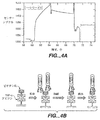
PCR産物(267塩基対(bp))を8%ポリアクリルアミドゲル上で、単一ファージペプチドクローンからのPCR産物(陽性クローン)及び分子量マーカーと共に分析した(図2)。拡散するバンドとして出現した移動の遅い産物が約500−700bp付近に観察された。これは、PCR反応中に鋳型(即ち、ファージ)が多すぎたためであり、そしてファージ濃度を低下させることによるか、又はPCRサイクルの数を減らすことにより、緩和することができる(図3)。500−700bpの拡散バンドの量を減らすため、この出発物質から次のPCR反応のためにPCR産物をおおまかに希釈することにより、サブクローン化の目的でより多くの産物を生成させた。所望の産物(267bp)が増幅されたら、それをEagI及びAcc651制限エンドヌクレアーゼにより消化して、ランダムペプチドをコードするDNAを含む45bpの断片を生成した。当該45bp断片を次にM13KEベクター(ニューイングランドバイオラブズ;ビバリー、MA)中にEagI及びAcc651制限部位において標準技術(SAmbrook,et al.,(1989)前記)を用いてサブクローン化した。当該45bp断片をM13KEベクターに連結した後、ライゲーション反応物で、化学的にコンピテントにしたER2537 E.coli細胞を形質転換した。当該細胞は、標準プロトコルを用いて(Sambrook,et al.,(1989)前記)塩化カルシウムによりコンピテントにした。ニューイングランドバイオラブズのM13 DNA調製用プロトコルから修飾したプロトコルを用いて、様々な形質転換体からM13のDNAを単離し、そして配列決定した。修飾は、管とは反対に、96ウエルのプレートの使用を含む。対応するペプチドを表1に示す。
【0093】
【表1】

実施例2:
TNF−αに結合するファージ−ペプチドの結合親和性及び選択性の特性決定:
ファージクローンA1、アミノ酸配列:RYWQDIP;表1、TNF−αに対する(配列番号:3)の結合及び解離を、IAsys AutoPlus Biosensorを用いて、次にLabsystems Affinity Sensors IAsys Protocol 2.4「蛋白質層への固定化:アビジンにカップリングしたチオール」(Thermo BioAnalysis Corp.フランクリン、MA)を用いて監視した。2つのキュベットを最初にアビジンでコートして、一方(対照)を次にビオチンでブロックした。これを次にリジン基で活性化した。15μlのアリコートの1mg/ml(h)TNF−α溶液を各キュベットに加えた。表面への蛋白質の結合は対照キュベットにおいて観察されなかったが、(h)TNF−αは明らかに未ブロックアビジンコートキュベットに固定化された(示さず)。この複合体は安定であり、そして10分にわたって解離しなかった。ブロッキング及び洗浄の後に、ファージクローA1,RYWQDIP(配列番号:3)を加えて、最終力価を5x1011pfu/mlとした。図4に示すとおり、サンプルキュベットにおいてはファージのTNF−αへの有意な結合があるが、対照キュベットへ結合したファージは極めてわずかであった。
【0095】
TNF−αからのファージの解離は極めて遅く、koff<10-4秒-1(10mM HEPES/0.05%ツイーン)と見積もられる解離定数であった。10Xバッファー濃度による洗浄により(70分時間点において)、除去されたのはファージのほんの小さな一部のみであった。10mM HClによる追加の洗浄は、標的からファージペプチドを完全に除去しそこなった。ファージ−ディスプレイされたペプチド配列RYWQDIP(配列番号:3)の結合は特異的であるが、挿入物を欠く野生型のファージが固定化TNFに結合しなかったからである。
実施例3:
IL−6及びIL−8に結合するファージ−ペプチドの選択:
実施例1に記載されたのと同じ方法を用いて、ヒトIL−6とIL−8標的として用い、そしてスーパーブロック(商標名)ブロッキングバッファーを抗−標的として用いた。PCR管を組換えヒトIL−6(0.1mg/ml)及びIL−8(0.25mg/ml)(バイオソースインターナショナル)でコートした。選択は、標的からのファージの酸溶出においてさえ、予測されたサイズ(267bp)のPCRバンドを生じた(図2B)。
実施例4
VEGFに結合するファージ−ペプチドの選択:
滅菌マイクロタイタープレート(5ウエル/サンプル)を200μlの1% PBS/BSA(PBS + 1%ウシ血清アルブミン)でコートし、次に、200μlの0.25% PBSTで洗浄した。ウエルを満たしたままにした。100μlの新鮮な全血を10μlのファージライブラリーと混合して、最初にコートされた細胞を添加し、そして30分間室温において(RT)インキュベートすることにより、ファージペプチドのライブラリーを、抗−標的としてのヒト全血に対して排除した(deselected)。30分後、上記溶液を吸引し、次にコートされたウエルに送達した(delivered)。この手法を4回繰返すことにより、抗−標的非結合ファージのライブラリーを生じさせた。標的に関しては、5mgの200μmポリスチレンビーズをヒトVEGFでコートしたが、100μlの100μg/ml組換えヒトVEGF(バイオソースインターナショナル;カマリロ、CA)と共に一晩4℃においてゆるやかに振盪しながらインキュベートすることによった。PBST(1 x PBS中の0.25%ツイーン−20)により3回洗浄することにより、過剰の未結合のVEGFを除去した。次に、ビーズを2%ツイーン−20 a x PBSTと共に2時間室温(RT)においてブロックした。ファージ−ディスプレイされた環状の7マーのランダムペプチドライブラリーを使用した。選択手法は本質的に実施例1に記載されたのと同じであった。最初のラウンドの選択の後に、標的−結合リジンからのPCR断片を精製し、EagI及びAcc651により消化し、そして上記制限酵素を加熱変性した。断片を直接M13KE切断ベクターへTakaraライゲーションキット(プロメガコープ)を用いてライゲーションした。ライゲーションミックスで形質転換し、そして標準手法(Sambrook et al.(1989)前記)に従い増幅した。第2ラウンドの選択を実施することにより、VEGFに結合するファージ−ペプチドをさらに富裕化した。対応するペプチド配列を以下の表に示す:
【0096】
【表2】
本発明の選択性標的化方法を慣用のバイオパンニング方法と比較するため、慣用の酸−溶出方法を用いた平行実験を実施した。Smith and Scott(1990)Science 249:386により記載された方法による3ラウンドのバイオパンニングは、表3に要約された配列プロフィールを生じた。これらの配列は本発明による選択性標的化方法により同定された配列とオーバーラップしない(表2)。
【0098】
【表3】
実施例5:
襟のしみに結合するファージ−ペプチドの選択:
コットン又は標的(襟のしみ)を含む65%ポリエステル:35%コットン及び抗―標的EMPA213ポリエステルコットン織物(試験織物、フリーホールド、NJ)上のしみ化シャツの襟を、ダイスを用いて直径7/32”に切り、NAEFパンチプレスに適合させる排除を伴った(MSインスツルメントカンパニー、ストニークリーク、NY)。96ウエルの平底マイクロタイタープレート(コスター、カタログ番号3598)をスーパーブロック(商標名)ブロッキングバッファーで一晩コートして、次に200−250μlのTBST(0.1%ツイーン−20)でEL403オートプレート洗浄機(バイオテックインスツルメンツ、ウイノスキー、VT)を用いて洗浄した。織物片をウエルの中にいれ、M13繊維状ファージ上にディスプレイされたファージ−ペプチド12マーライブラリーの10μlストック溶液を、100μlの界面活性剤(3.4g/Lヨーロピアン界面活性剤)に、抗−標的としてポリエステル−コットンを含むウエル中で加えた。20分間のインキュベーション後に、未結合ファージ(抗−標的非バインダー)を含む上清を、ポリエステル−コットン織物を含む第2のウエルに移した。これをもう一度繰返した。上清を次に、しみ化シャツの襟の織物を含むウエルに移し、そして汚れと10−60分間インキュベーションすることにより、残りのファージペプチド集団を「汚れバインダー」に関して選択した。0.1−2%のツイーン−20又は3.4g/Lの界面活性剤の何れかによる一連の洗浄工程に汚れを供した。洗浄工程は所望のストリンジェンシーに対して操作することができる。汚れを含む最初のウエルの中での初期の洗浄後に、あらゆる残りの結合ファージを含む汚された織物片を、第2洗浄工程のために次のウエルに移した。
【0100】
結合ファージを含む汚された織物の一部を、60μlの溶解バッファーB(10mM Tris−Cl,pH8.4,1% Triton−X100;10mM EDTA)及びアンプリワックス(商標名)(パーキンエルマー、ノーウオーク、アメリカ合衆国)を含むPCR管に移した。管を次に95℃において20分間加熱して、次に、冷やした。標的−結合ファージのPCR増幅は、マイナーな修飾を伴って実施例1に記載された通りに実施した。図3は、20サイクル未満のPCRをホモ二重鎖DNAの単一バンドの増幅が要求することを示す(レーン4)。長いサイクル時間(レーン3)はヘテロ二重鎖DNA形成の実質上の画分を生じるのに対し、短いサイクル時間(レーン5)は測定可能なPCR産物を生じない。正確なサイズのPCR産物を8%ポリアクリルアミドゲル上で精製して、M13KEにサブクローン化し戻して、実施例1に記載されたとおりに配列決定した。界面活性剤中で襟のしみに結合してポリエステルコットン織物に結合しないファージペプチドクローンに相当するアミノ酸配列を表4に要約する。
【0101】
【表4】
実施例6:
織物上の標的の襟のしみへの放射線標識ペプチドの選択性結合:
標的(襟のしみ)及び抗―標的EMPA213ポリエステルコットン織物(試験織物、フリーホールド、NJ)を含むしみ化シャツの襟(コットン又は65%ポリエステル:35%コットン)を、直径1/2”に切り、コスター24ウエルプレートに入れた。本発明により同定されたしみ標的化ペプチドSISSTPRSYHWT(配列番号:20)を14CグリシンでN末端標識した。10μlの[1−14C−G]SISSTPRSYHWT(シンペップ、ダブリン、CA)(配列番号:114)400μM溶液を4mlの0.002%ツイーン−20含有50mM CAPSバッファー、pH10.4に加えた。放射性標識ペプチドの950μLアリコートを各ウエルに加え、そしてサンプル回転振盪機上で30℃において30分間振盪した。サンプルを取り出し、そして4mLのバッファーで洗浄して、次に、4mLのミリQ水で20分間洗浄した。サンプルをワットマンフィルターペーパー上で空気乾燥、そしてヒューレットパッカードスキャナー(パロアルト、CA)上でデジタルスキャンした。放射性標識したスワッチを次にリン蛍光体スクリーン(モリキュラーダイナミックス;サニーベール、CA)に30時間−70℃において暴露した。結果のリンイメージをモリキュラーダイナミックスストーム(登録商標)を用いてスキャンした。図5は、汚された織物及び対照の織物の対応するリンイメージと共に、標的(汚れ)及び抗−標的の可視イメージを示す。リンイメージの相対強度をイメージクアント(ImageQuant)(登録商標)イメージ分析ソフトウエア(モリキュラーダイナミックス;サニーベール、CA)を用いて定量し、そして織物の結合に対する汚れの結合の比は>15:1であることを示す。
実施例7:
汚れ標的化ペプチドの放出に関する遅いk off 速度定数の証明:
標的(襟のしみ)及び抗―標的を対照抗−標的(同じシャツからの汚されていないポリエステル:コットン)と共に含むしみ化シャツの襟(65%ポリエステル:35%コットン)を、直径7/32”に切り、96ウエルマイクロタイタープレート(ミリポアコーポ、0.22μM デュラポア膜;カタログ番号MAGV N22 50)に入れた。14Cグリシンで末端標識された、しみ標的化ペプチドSISSTPRSYHWT(配列番号:20)及びペプチド対照NFFPTWILPEHT(配列番号:78)の400μMストック溶液の連続希釈を、1g/Lのタイド界面活性剤溶液(プロクターアンドギャンブル、シンシナチ、OH)に加え、そして60μLのアリコートをマイクロタイタープレートのウエルに入れた。プレートを振盪しながら30分間32℃においてインキュベートし、次に過剰の未結合の放射線標識ペプチド(バキュームマイフォールド;ミリポアコープ、カタログ番号MAVM 096 OR)の吸引濾過を行った。サンプルは3回200μLの蒸留水で洗浄し、洗浄と洗浄の間は振盪し、約40分かけた。サンプルに結合した残りの放射活性を液体シンチレーションカウンティングによりウオーラックマイクロベータカウンター内で定量した。図6Aは、全放射線標識の50%より多くが、40分間の洗浄後でさえ、汚れ標的化ペプチドに関して汚された織物に結合したままである。これは、しみ標的化ペプチドの放出に関する速度定数koff≦2 x 10⁻⁴秒-1に相当する。対照的に、対照ペプチドは同じアッセイにおいて親和性又は選択性を何も示さない、図6B。
実施例8:
発明の選択性標的化方法に比較した酸溶出に関するペプチドの選択性と親和性:
コットン上の襟のしみに結合するファージペプチド配列HTFQHQWTHQTR(配列番号:27)は、Scott and Smith(1990)Science 249:386に記載された方法を用いて各ラウンドの後にファージペプチドを酸溶出した以外は、実施例5に記載されたとおりに、バイオパンニング5ラウンド後に同定した。選択性(汚れ対コットン結合)及び親和性(koff)は、以下のとおりに襟のしみに結合する対応のペプチドに関して測定した:NiキレートしたGGHTFQHQWTHQTR(配列番号:28)の1mM溶液をコットン上の襟のしみ又はコットンのみと90分間室温において撹拌しながらマイクロタイタープレート中でインキュベートした。対照ペプチドキレートNi GGHも同じ条件下で試験した。ウエルからインキュベーション溶液をピペットオフした後に、織物の布きれを200μLの水で撹拌しながら3分間撹拌した。200μLのo−フェニレンジアミン(OPD)及び50μLの100mM H2O2を加え、そして420nmにおいて酸化されたOPDの吸収を測定することにより、残りの結合ペプチドをアッセイした。表5及び図7において要約されるとおり、織物に対する汚れの結合の比は、≦3:1より低いか又は同等であり、そして親和性はkoff=1 x 10⁻³秒-1と測定された。これらのデータは、特異的且つ選択的に強固に結合するペプチドは、本発明による選択性標的化方法を用いて同定されるのが好ましいことを証明する。
【0103】
【表5】
実施例9:
界面活性剤マトリックス内のファージディスプレイされたライブラリーの安定性:
ファージペプチドライブラリーの安定性に対する家庭内の洗濯場の界面活性剤の作用を試験するため、1013pfu/mLを含むM13繊維状ファージ(ニューイングランドバイオラブズ、ビバリーMA,アメリカ合衆国)上にディスプレイされたペプチド12マーのライブラリーのストック溶液を、a)100mM Tris HCl,pH7.5,0.1%ツイーン−20(TBST対照)、b)ガロンあたり3グラム(gpg)の硬化剤を含む0.7g/LのAriel Futur(プロクターアンドギャンブル。シンシナチ、OH)、及びc)15gpgの硬化剤を含む3.4g/LのAriel Futurの中で、1012pfu/mLに希釈した。TBS中のスーパーブロッキングバッファー(ピアス、カタログ番号37535)でブロックされた96ウエル平底マイクロタイタープレート(コスター、カタログ番号3598)のウエルに、アリコート(100μL)を加えた。サンプルを25℃においてゆるやかに振盪しながら90分間インキュベートした。10μLのアリコートを取り出して、標準手法(Kay et al.,(1996)前記)に従ってファージ力価測定するためにルリアブロスに連続希釈した。TBST中の対照ファージライブラリーに比して、界面活性剤溶液においてはファージ力価の損失は観察されなかった。
実施例10:
ポリウレタンに結合してコットン、ポリエステル、又はポリエステル−コットン織物には結合しないファージ−ペプチドの選択:
1.5mLのマイクロ遠心分離管を一晩ブロッキングバッファー−PBSによりブロックし、1mLの3.4g/L界面活性剤により洗浄し、そして水分を切った。500μLの3.4g/L界面活性剤を4つの管に加え、一片のコットン、ポリコットン、及びポリエステル織物をそれぞれ共にした。ファージペプチドライブラリーは以下の通りであった:
管1 10μLのPh.D.−C7Cライブラリー
管2 10μLのPh.D.−12ライブラリー
管3 10μLの野生型ファージ対照
管4 ファージ対照なし。
【0105】
管を室温において20分間1000rpmにてエッペンドルフサーモミクサー中でインキュベートし、そして織物片を取り出した。この取り出し工程は全部で3回繰返し、次に、きれいなピペットチップで30分間搾ること(squeezing)により湿潤させたポリウレタンプラグと上記ファージライブラリーをインキュベーションした。
【0106】
バキュームラインに接続したきれいなピペットチップを用いて上清をプラグから吸引し、そして1mLの3.4g/L界面活性剤溶液を管に加えた。きれいな接種用ループを用いて搾ることによりプラグを再度湿潤させ、そして管をエッペンドルフサーモミクサー中に入れて全部で10回洗浄した。プラグを100mLディスポーザブルフィルターシステム(コーニング)に移し、そして洗浄溶液をフィルターシステムに送達することにより、3x40mL PBST(0.25% v/vツイーン−20)洗浄を実施した。プラグは、きれいなピペットチップを用いて搾ることにより再度湿潤させ、PBSTと共に瞬インキュベートし、次に吸引により乾燥させた。上記フィルターシステムを真空下にしている間、洗浄溶液を直接プラグにピペッティングすることにより、10回の1mLのPBSによる洗浄を実施した。
【0107】
プラグをきれいな0.5mLマイクロ遠心分離管に移した。100μLの溶解バッファー(0.1% Triton X−100m10mM Tris pH8.4)を加え、そして管を95℃において20分間インキュベートすることにより、ファージを溶解した。溶解したファージは同じ管において以下のとおりにPCRを行った:
50μL HotStarTaq(登録商標)マスターミックス(キアゲン) 25μL 溶解バッファー
5μL BSA(10μg/μL)
1.25μL CMM13−01プライマー(50μM)
1.25μL CMM13−02プライマー(50μM)
17.5μL H2O
PCR増幅は、95℃において15秒間の変性、58℃において30秒間のアニーリング、そして72℃において30秒間の伸長合成を30サイクル、そして次に72℃において5分間を1サイクル使用することにより、実施した。PCR産物は、製造者の指示に従い、ゼロブラントクローニングにより、TOPO(登録商標)−10ベクター(インビトロジェン、サンディエゴ、CA)へクローン化した。クローンは標準配列決定法を用いて配列決定し、表6に要約した。
【0108】
【表6】
ペプチド配列を合成して、当該ペプチドがポリウレタン酸化に対して防御する能力を測定した。
実施例11
ステンレス鋼又はガラス上にて標的のベークドオンされた卵のしみに対して選択されたペプチドの選択性結合:
卵のしみは、新鮮な卵からの黄身を使用して調製した。黄身を冷たい水で洗って、次にストレーナーを通してビーカーに押し出した。ビーカーを140°F水浴に入れて、卵の黄身を30分間一定の撹拌を伴って調理した。30分後に、ビーカーを氷の浴槽に入れて、一定撹拌を伴って黄身を室温まで冷やした。#316ステンレス鋼フォイルディスクを、ダイスを用いて7/32”の直径に切って、NAEFパンチプレスに適合させる排除を伴った(MSインスツルメントカンパニー、ストニークリーク、NY)。これらは、標的、ベークドオンされた卵のしみ、及び抗−標的、未だしみ化していないディスクの両方の基質として使用した。使用の前に、当該ディスクをマイルドな界面活性剤の中で洗浄し、そして脱イオン水中で徹底的に洗い流した。
【0110】
卵のしみ化316ステンレス鋼ディスク及び卵のしみ化ガラスビーズをコスター96ウエル平底プレートに入れた。各ペプチドライブラリーに関して、3つのきれいなステンレス鋼(SS)ディスク又はガラスビーズ(抗−標的)を隣のウエルに入れた。きれいなディスク又はビーズを含む最初のウエル(A)には、150μLの界面活性剤及び10μLのファージライブラリー−C7C、直鎖7マー、又は野生型ファージを加えた。当該サンプルを室温において20分間ゆるやかな振盪と共にインキュベートし、そして未結合ファージを含む上清を次にウエルに移し、そして上記プロセスを全部で3回繰返した。上清を次に卵のしみ化クーポン又はビーズに移し、そして30分間ゆるやかに混合しながらインキュベートした。当該サンプルを次に新たなウエルに移して、以下のとおり全部で38回洗浄した:200μL界面活性剤溶液、3.5g/lの粉末のオートマチック皿洗い用界面活性剤中で3x、250μLのPBST中で30x、そして200μLのPBS中で5x。
【0111】
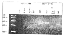
洗浄した皿又はガラスビーズを0.5mLのPCR管に移した。PCR反応は、200μLの反応混合物を用いてキアゲンホットスポット(登録商標)キット及び50μLのミネラルオイルを用いて、直接に卵のしみ化ディスク又はビーズ上で稼働させた。PCR増幅は、反応開始のための95℃において15分間を1サイクル、次に、変性の94℃30分間、アニーリングの58℃30秒間、そして伸長合成の72℃30秒間を30サイクル行い、そしてエロンゲーションの72℃10分間を1サイクルで終わらせた。278bpの産物が、分子量マーカーと共に2%アガロースゲル上で分析された。抗−標的に関するステンレス鋼に関する図8に示すとおり、PCR産物は直鎖7マーライブラリーに関して可視化され、そして野生型(WT)ファージ対照に関しては可視シグナルはなかった。第2のPCR増幅を実施して、PCR産物をTOPO(登録商標)TAベクター(インビトロジェン)にクローン化して、表7に要約されたとおりに配列決定した。
【0112】
【表7】
上記配列をズブチリシンプロテアーゼ遺伝子にクローン化して、卵のしみに関する親和性を蛋白質分解アッセイにおいて測定した。
実施例12
セラミック上の標的の茶の汚れに対する選択されたペプチドの特異的且つ選択的 結合:
実施例1に記載された方法を用いて、オートマチック皿洗い用界面活性剤の存在下でセラミック上のお茶に結合するペプチドを2ラウンドの選択性標的化の後に同定した。標的結合ペプチド配列を表8に要約する。
【0114】
【表8】
実施例13
ヒトの皮膚を標的化するが毛髪を標的化しないように選択されたペプチドのスクリーニング:
濃い(dark)ヒトの毛髪3インチ片2つ(インターナショナルヘアインポーターアンドプロダクツ、ホワイトプレインズ、NY)を、DI水中の2%ニュートロジーナ(登録商標)ボディーウオッシュ(ニュートロジーナコーポ)溶液10mlを含む、BSAブロックされた50mlのコニカル管に入れた。10μLの環状7マー又は直鎖12マーペプチドライブラリー(1010pfu/ml)、又は野生型ファージ(109pfu/ml)を加え、そしてサンプルを15分間室温において回転撹拌しながら(30rpm)混合した。未結合の上清を、さらに2つの濃い毛髪の3インチ片を含む新たな管に移し、そして室温において15分間回転撹拌しながらインキュベートした。この第2の毛髪のインキュベーションの後に、500μlの上記溶液を、0.9mL組織培養培地(MatTek Corp)を含む6ウエルの培養プレート中のヒト皮膚組織(EpiDerm(登録商標),MatTek Corp.アシュランド、MA)の表面上に30分間室温においてゆるやかに撹拌しながら移した。皮膚組織を取り出して、ブロックされたコニカル管の中で、50mlsの2%ボディーウオッシュ中で2回各5分間洗浄し、そして50mlsのPBSで3回各5分間洗浄した。最後のPBS洗浄後に、皮膚組織を−20℃に凍結して、次に標的結合リガンドファージのPCRを行った。
実施例14
ヒトの毛髪を標的化するが皮膚を標的化しないように選択されたペプチドのスクリーニング:
予め平衡化した皮膚組織を新しい0.9mLの組織培養培地を含む6ウエルの培養プレートに入れ、そして10μLの環状7マー又は直鎖12マーペプチドライブラリー(1010pfu/ml)、又は野生型ファージ(109pfu/ml)を含む、300μlの2%ニュートロジーナ(登録商標)ボディーウオッシュ溶液を皮膚表面に加えた。サンプルを15分間室温において回転撹拌しながら混合した。未結合の上清を、さらに皮膚組織を含む新たなウエルに移し、そして上記手法を繰返した。インキュベーション溶液を、2%ボディーウオッシュ10mlを含む50ml管内の、9つの3インチの濃い毛髪(インターナショナルヘアインポーターアンドプロダクツ、ホワイトプレインズ、NY)片に、30分間ゆるやかに撹拌しながら(30rpm)室温において移した。毛髪サンプルを次に1 X 50mls,2 X 50mls,又は4 X 50mlsの2%ボディーウオッシュで洗浄した;PBSの洗浄サイクルを続けた(1 X 25mls5分間、1 X 25mls2分間、2 X 50mls各5分間、全部で150mls)。最後のPBS洗浄後に、結合したファージペプチドを含む毛髪サンプルを−20℃に保存した。
【0116】
標的−結合ファージのPCR増幅は実施例1に記載されたとおりに実施したが、マイナーな修飾を伴った。PCR反応物は50μgのBSAを含むことにより、毛髪又は皮膚によりPCR反応の阻害を予防した。
実施例15
ヒトの毛髪を標的化するが皮膚を標的化しないペプチド又はヒトの皮膚を標的化するが毛髪を標的化しないペプチドの選択性結合に関するELISAアッセイ:
実施例13及び14において同定されたペプチド配列並びにランダムな対照ペプチドは、配列GGGK(ビオチン)でC末端標識した。配列LESTPKMK(配列番号:115)はコンセンサス配列LESTを含み、そして毛髪上で単離された。FTQSLPR(配列番号:116)はコンセンサス配列TQSLを含み、そして皮膚上で単離された。YGGFMTSE(配列番号:117)は対照ペプチドである。
【0117】
濃いブラウンの毛髪(3”の長さ、各4)を2%ボディーウオッシュ及び予め平衡化したヒト皮膚組織で湿潤し、24ウエルプレートのウエルに入れた。2%ニュートロジーナボディー中のビオチン化ペプチドの200μM溶液1mlを毛髪サンプル及び皮膚サンプルに加え、そしてゆるやかに撹拌しながら室温において30分間インキュベートした。当該溶液を次にピペットオフして、毛髪サンプル及び皮膚サンプルをきれいな毛抜きを用いて50mlコニカル管に移し、50mlの2%ボディーウオッシュで1回洗浄し、水50mlで2回洗浄し、そして50mlのPBSで1回洗浄した;各洗浄工程は5分かけて、回転シェーカー上で20rpmにて実施した。毛髪サンプル及び皮膚サンプルは次にきれいな毛抜きを用いて24ウエルのプレートに移したが、その際、1mlのストレプトアビジン配合ホースラディッシュパーオキシダーゼ(PBS中で1/1000に希釈)を1時間室温においてゆるやかに振盪しながら加えた。50mLコニカル管の中で、50mlのPBSで2回洗浄することにより(5分間、各20rpm)、過剰なストレプトアビジンHRPを除去した。毛髪及び皮膚のサンプルを新たなウエルに移し、そして1mlのH2O2/OPD溶液を加え、そして発色するまで室温に放置した。図9は、対照ペプチドに比較して、ペプチド結合が各々の標的に関して選択性であることを示す。
【図面の簡単な説明】
【図1】 図1は、本明細書に開示された選択的標的化の一般的な模式図である。方法は、工程:a)抗−標的結合リガンドを減らしたリガンドのライブラリーを提供する抗−標的に対する選択、b)標的−結合リガンド複合体の形成による標的に関する選択、c)標的−結合リガンド複合体の分離、d)標的−結合リガンドの同定、及びe)任意に標的−結合リガンドを配列決定し、選択性標的化の追加のラウンドに上記標的−結合リガンドを暴露し、そして/又は分散(diversification)を含む。
【図2】 図2A及び2Bは、標的結合ファージの溶解後のPCR増幅DNA断片のゲルの写真である。図2AはTNF−α結合ファージを示し、そして図2BはIL−6及びIL−8結合ファージを示す。
【図3】 図3は、しみ標的化ペプチドに関するPCR増幅DNA断片のゲルの写真である。
【図4】 図4は、IAsysバイオセンサーキュベット上に固定されたTNF−αからのRYWQDIP(配列番号:3)に対応するファージペプチドクローンA1の結合、解離及び試みられた溶出を示す。
【図5】 図5A及び5Bは、襟のしみ及び対応するポリエステル織物のイメージであり、それぞれデジタルイメージング及びオートラジオグラフィーにより見た。
【図6】 図6A及び6Bは、ポリエステルコットン織物上の襟のしみに結合した函数パーセント14C標識ペプチドを示す。図6Aは、14Cグリシンで末端標識された、しみ標的化ペプチド、SISSTPRSYHWT、(配列番号:20)を示し、白丸は1番を描写し、黒四角は2番を描写し、そして白四角はブルーポリコットンを描写し、そして6Bは14Cグリシンで末端標識されたランダムペプチド、NFFPTWILPEHT(配列番号:78)を示す。
【図7】 図7は、襟のしみ(・)及び対応するコットン織物(o)由来の、Ni−キレートペプチドGGHTFQHQWTHQTR(配列番号:28)の解離の動力学を示す。ラインのスロープは速度定数koff=1x10⁻³秒-1に相当する。
【図8】 図8は、卵のしみ標的及びステンレス鋼又はガラスビーズ抗−標的のPCR増幅断片のゲルの写真である。
【図9】 図9は、3つのペプチドの結合に関するELISAアッセイの結果を示す。LESTPKMK(配列番号:115)は毛髪に結合し、そしてFTQSLPR(配列番号:116)は選択的に標的皮膚に結合し、毛髪に結合しない(黒四角は毛髪を描写し、そして白四角は皮膚を描写する)。[0001]
Background of the Invention
The present invention is directed to a method for the selection and identification of compounds that can specifically bind to a target in the presence of an undesired background target (anti-target) using multiple libraries of similar compounds. It is done. In one particular aspect, the present invention relates to the selection of multiple ligands from multiple peptide libraries. The ligand peptides identified by the methods of the invention have binding affinity and selectivity for the target that is similar to the binding affinity and selectivity of the antibody.
[0002]
The literature is enriched with examples of recent advances in methods of screening large library pools of compounds, especially peptides. Advances have also been made in the screening of these compounds to identify molecules that bind to preselected targets. One well known method is described by Smith, G. et al. P. (1985), Science 228: 1315, the first biopanning developed. The simplest form of biopanning is an in vitro selection process in which a library of phage-displayed peptides is incubated with a target. Allow target and phage to bind and wash away unbound phage. The specifically bound phage is then eluted with acid. The eluted pool of phage is amplified in vivo and the above process is repeated. After multiple rounds, individual clones are isolated and sequenced.
[0003]
Many variations of biopanning originally introduced by Smith have been described and are described in Christian et al. (1992) J. Am. Mol. Biol. , 227: 711; Cwirla et al. (1990) Proc. Natl. Acad. Sci. USA, 87: 6378; Cull et al. (1992) Proc. Natl. Acad. Sci. USA, 89: 1865; Huls et al. (1996) Nature Biotechnolo. 7: 276; and Bartoli et al. (1998) Nature Biotechnol. 16: 1068.
[0004]
Huls et al. , 1996 supra describe a method comprising subtractive selection based on flow cytometry of phage antibodies on intact tumor cells. The phage-displayed antibody remains bound to the target during flow cytometry selection. However, prior to amplification, cell bound phage is eluted from the target. WO 98/54312 discloses the selection of antibodies under mild conditions with high affinity for antigen using antibody libraries displayed on ribosomes.
[0005]
In many prior art methods, it is generally assumed that elution of the target binding ligand is sufficient to identify the ligand that binds most strongly in the library. However, many research papers report on low affinity binders using elution techniques (US Pat. No. 5,582,981). Nevertheless, physical separation of the ligand from the target prior to amplification or identification is a standard method for selecting a ligand that binds to a preselected target.
[0006]
Balass et al. (1996) Anal. Biochem. , 243: 264 describe selection of high affinity phage-peptides from phage-peptide libraries using biotinylated targets immobilized on a nitrostreptavidin matrix. The interacting phage particles were released under conventional acid elution. In addition, after acid elution, the target complex was analyzed for bound phage. By exposing these particles to an alkaline solution or free biotin, target-bound phage particles were released from the solid support. It has been discovered that the affinity of the isolated phage is greater than the phage released by traditional acid elution methods. However, synthetically produced peptides exhibited lower affinity for the target than peptides produced from sequences obtained by acid-eluting phage.
[0007]
Other targeting methods include, for example, SELEX. This is a technique for embedding oligonucleotides from a library of randomized sequences into a nucleic acid pool. Multiple cycles of affinity selection occur for oligonucleotide targets from heterologous RNA or DNA. In order to proceed to the amplification process, the selected nucleic acid must be released from the target after distribution (US Pat. No. 5,475,096).
[0008]
a) a compound that binds strongly and specifically to a target that is not clear at the chemical, biochemical or genetic level but has the desired microscopic properties for the target, b) a large background of unwanted targets ( Compounds that bind strongly and specifically to targets that cannot be easily physically separated from anti-targets), and c) compounds that bind to targets under harsh conditions such as acidic pH, high detergent concentration or high temperature There are a variety of methods for screening and selecting libraries of compounds, but there is a particular need for improved methods that do not require multiple rounds of selection.
[0009]
The selective targeting method according to the present invention overcomes some of the above disadvantages of the prior art methods and provides in particular the advantage of rapidly identifying compounds, particularly peptides, that selectively bind to the target with high affinity. To do.
[0010]
Summary of the Invention
In one aspect, the invention relates to a method for screening a ligand library, allowing the ligand to contact the anti-target by contacting the ligand library with an anti-target; separating unbound ligand Contacting an unbound ligand with a selected target to allow the unbound ligand to bind to the target to form a target-bound ligand complex; from a ligand that does not bind to the target to the target- Separating the binding ligand complex; and identifying the target-binding ligand on the target-binding ligand complex.
[0011]
In another aspect, the invention relates to a method for screening a ligand library, wherein the ligand binds to the anti-target by contacting the ligand library with the selected target and the anti-target essentially simultaneously. Allowing the target-binding ligand complex to form; separating the target-binding ligand complex from the anti-target, anti-target binding ligand and free ligand; and identifying the ligand of the target-binding ligand complex Including doing.
[0012]
In one preferred embodiment, the selectivity of the ligand that binds to the target relative to the ligand that binds to the anti-target is approximately at least 10: 1. In a second preferred embodiment, the ligand is a peptide but not an antibody, and at least about 10-7K of MDBinds to the target at, and preferably about 10-7M to 10-TenThe range of M. In a third preferred embodiment, the ligand library is a peptide library. Preferably, the peptide identified by the above method is less than 25 amino acids in length, more preferably between 4 and 15 amino acids in length. In the fourth aspect, koffIs about 10-FourSecond-1Or less. In a fifth embodiment, the target is a stain, and in particular a stain on the fabric, wherein the stain is a porphyrin-derived stain, a tannin-derived stain, a carotenoid-derived stain, an anthocyanin-derived stain, a stain ( soil based soil, oil based soil or human body stain stains.
[0013]
In yet another aspect, the invention is directed to ligands, particularly peptide ligands, identified by selectively targeting the inventive methods.
Another aspect of the invention relates to a method of identifying peptides useful in a cleaning composition, wherein contacting the peptide library with an anti-target allows the peptide to bind to the anti-target, provided that The anti-target is selected from the group consisting of textiles, ceramics, glass, stainless steel, and plastics; separating unbound anti-target peptide and contacting unbound anti-target peptide to the target, provided that the target Are soils, and in particular soils on fabrics, where the soils are from porphyrins, from tannins, from carotenoid pigments, from anthocyanin pigments, from stains from stains, and from human bodies A dirt-bound peptide complex is formed by binding unbound peptide selected from the group consisting of The dirt - dirt on binding peptide complex - comprises identifying binding peptides. In at least one embodiment, the peptide is about 10-7M to 10-TenK in the range of MDBonds to dirt.
[0014]
Detailed Description of the Invention
A. Unless defined otherwise, all technical and scientific terms used herein have the same meaning as commonly understood by one of ordinary skill in the art to which this invention belongs. For purposes of the present invention, the following terms are used to describe the invention herein.
[0015]
The term “ligand” means a molecule or compound that is recognized by a particular target or anti-target. The term is independent of molecular size or its composition. A ligand may function as a substrate for an enzyme-catalyzed reaction, as an agonist, as an antagonist, act as a signal messenger, or stimulate or inhibit a metabolic pathway. The ligand may be a nucleic acid, peptide, peptide derivative, peptidomimetic, polypeptide, small organic molecule, carbohydrate, and other molecules isolated from candidate mixtures that act on the target in the desired manner. Preferably, the desired mode is to bind the target, but may include, for example, catalytically changing or reacting with the target to modify or change the target. In one preferred embodiment, the ligand has a binding affinity for the target in the range of antibody binding affinities for the selected receptor.
[0016]
The term “library” means a collection of chemical or biological integrity that can be created within a single repository and simultaneously screened for desired properties. As used herein, a library can have a minimum size of at least two members and 1015May include members as much. In one aspect, the library is at least 102Have members. In another aspect, the library is at least 10ThreeHave members. In yet another aspect, the library is at least 106Have members. In a further aspect, the library is 109Have members. The size of the library means the total number of entities including the library when the members are the same or different.
[0017]
“Peptide library” refers to an oligomer in which the monomer units are amino acids (typically but not limited to L-amino acids) linked by amide bonds. The peptide may be two or more amino acids in length. The peptides identified according to the invention are preferably shorter than 50 amino acids, more preferably shorter than 30 amino acids, and preferably shorter than 25 amino acids, and preferably shorter than 20 amino acids Length. In one preferred embodiment, the peptide identified by the method of the invention is 4 to 15 amino acids in length. Usually, however, peptides are up to 100 amino acids in length. Peptides longer than 100 amino acids are usually called polypeptides. Standard abbreviations for amino acids are used herein. (Singleton et al., (1987) Dictionary of Microbiology and Molecular Biology, Second Ed., Page 35, incorporated herein by reference).
The peptide or polypeptide may be provided as a fusion peptide or protein. Peptides include synthetic peptide analogs whose amino acid sequences are known. The term peptide refers to a molecule that is structurally related to the peptide, such as a peptide derivative or peptidomimetic whose structure cannot be determined by standard sequencing methodologies, but must be determined using more complex methodologies, such as mass spectrometry. Not included. Peptidomimetics (also known as peptide mimetics) are peptide analogs but are non-peptide compounds. Usually, one or more peptide bonds are optionally substituted. (Evans et al., (1987) J. Med. Chem. 30: 1229). The term “protein” is well known and refers to large polypeptides.
[0018]
The term “nucleic acid” means DNA, RNA, single-stranded or double-stranded and chemical modifications thereof. Modifications may include, but are not limited to, modified bases, backbone modifications, methylation, unusual base pair modifications, and capping modifications. When using a nucleic acid library in the selective targeting method of the invention, the nucleic acid ligand is usually 4 to 250 nucleotides in length, and preferably 4 to 60 nucleotides in length.
[0019]
The invention was further identified by selective targeting as described herein, ligands, preferably nucleic acid, peptide or polypeptide ligands, and most preferably peptide ligands that bind to a target with substantially the same ability as nucleic acids. Includes peptides or proteins. Substantially the same ability to bind to the target means that the affinity and selectivity are about the same as the affinity and selectivity of the ligand selected by the method claimed herein.
[0020]
Furthermore, ligands having substantially the same ability to bind the target will be substantially homologous to the ligand identified by the disclosed selective targeting method. For nucleic acid sequences, substantially homologous to the identified ligand means that the degree of homology of the primary sequence is greater than 80%, preferably greater than 85%, more preferably greater than 90%, and even more preferably greater than 95%. , Even more preferably greater than 97% and most preferably greater than 99%. One skilled in the art will recognize that a large number of peptides encoding nucleotide sequences may result as a result of the degeneracy of the genetic code. The peptides or polypeptides have at least 85% identity, preferably at least 90% to 95% identity, more preferably at least 97% identity, when aligned to the maximum, and Most preferably it is substantially homologous to the reference sequence if it has 99% identity or is equivalent to the reference sequence. Maximum alignment of sequences may be performed by various known methods and computerized means of known computational methods (eg, TFASTA, BESTFIT, Wisconsin Genetics Software Package, Release 7.0, Genetics Computer Group) , Madison, WI). The general classification of equivalent amino acids is: 1) glutamic acid and aspartic acid; 2) lysine, arginine and histidine; 3) alanine, valine, leucine and isoleucine; 4) asparagine and glutamine; 5) threonine and serine; Tyrosine and tryptophan; 7) glycine and alanine. One skilled in the art successfully determines whether a provided sequence that is substantially homologous to the sequences identified herein binds the target with substantially the same ability.
[0021]
Small organic molecules as defined herein are molecules, preferably non-polymeric molecules, having a molecular weight of about 1000 daltons or less and preferably less than 500 daltons. A “peptoid” is defined herein as an enzyme resistant peptide analog.
[0022]
The term “target” or “anti-target” means a molecule or a heterologous molecule that has a binding affinity as defined herein for a given ligand. Both the target and the anti-marker may be natural or synthetic or heterologous molecules.
[0023]
The binding affinity of a ligand for its target or anti-target is determined by the dissociation constant (KD), Concentration required for 50% effective binding (EC50), Or 50% inhibition of binding of another compound that binds to the target (IC50). KD is koff/ KonIt is prescribed by. koffDefines the rate at which the target-ligand complex breaks or separates separately. This term is sometimes referred to in the art as a rate of other measurable quantities that reflect the rate of kinetic stability or binding affinity of the target-ligand complex, such as an enzyme linked immunosorbent assay (ELISA) signal or radiation. Means an activity label signal. The selectivity is related to the rate of binding affinity or the dissociation of the ligand-complex.off(Target KD/ Anti-target KD). konThe value describes the rate at which the target and ligand bind to form a target-ligand complex.
[0024]
The term “contacting” means placing or pairing a ligand library with a target or anti-target in close proximity and is broadly defined to include in vitro and in vivo contacts. The term includes touching, associating, joining, combining, intravenous injection, oral administration, intraperitoneal, topical application, intramuscular, inhalation, subcutaneous application and the like. As used herein, the term “separate” refers to select, segregate, partition, isolate, collect, keep apart. ) And disunite.
[0025]
“Amplify” means a process or combination of process steps that increases the amount or copy number of a molecule or class of molecules. In one aspect, amplification refers to the production of additional copies of a nucleic acid sequence performed using polymerase chain reaction (PCR) techniques well known in the art. In another aspect, amplification refers to phage virus particle concentration production by host infection.
[0026]
The single “a”, “an”, and “the” as used in the specification and claims include multiple references unless the context clearly dictates otherwise. For example, the term “a protease” may include multiple proteases.
[0027]
The following documents describe the general techniques employed herein: Sambrook et al. (1989) Molecular Cloning: A Laboratory Manual Harbor Laboratory Press, Cold Spring Harbor, NY; Innis et al. , PCR Protocols-A Guide to Methods and Applications (1990), Academic Press, Inc. Kay et al. (1996) Page Display of Peptides and Proteins, Academic Press; Ausubel et al. (1987) Current Protocol in Molecular Biology, Greene-Publishing & Wiley Interscience NY (augmented from 1999); Berger and Kimmel, (1987) Methods in Enzymolology. 152. Academic Press Inc. San Diego, CA.
[0028]
The context of all documents, patents and published patent applications cited throughout this application are incorporated herein by reference in their entirety.
B. General method
Described herein is a selective targeting method that screens a library of ligands that have binding affinity and selectivity for a selected target. In its most basic form, the selective targeting method may be defined as follows: creating a ligand, preferably a library of peptides of different sequences, and more preferably a random peptide library Or get. Removing ligands that bind to the anti-target from the selection by contacting the ligand library with the anti-target under conditions favorable for binding between the ligand of the library and the anti-target; Allows binding to the ligand; and separates the anti-target non-binder (unbound ligand) from the anti-target ligand bound molecule or any free ligand. The anti-target non-binder is allowed to contact the selected target under suitable conditions and allow them to bind. Ligands that have affinity for the target will bind to form a target binding ligand complex. Removal of ligand bound to anti-target and removal of weakly target-bound ligand is generally referred to as library depletion. The target binding ligand complex is then separated from the remaining mixture containing unbound ligand. The target binding ligand complex or target binding ligand may then optionally be subjected to amplification, sequencing or further rounds of selection (Figure 1). The invention further includes a ligand identified by the selective targeting method of the invention.
[0029]
In the practice of the invention, a library of compounds to be tested will usually be provided. Ligand libraries may include, but are not limited to, random peptide libraries, synthetic peptide or peptidomimetic combinatorial libraries, peptide loop libraries, combinatorial chemical libraries, and oligonucleotide libraries. These libraries are well known in the art, and methods for creating such libraries are also known. Barbas, C.I. F. (1993) Current Opinion in Biotech. 4: 526; Cwirla et al. (1990) supra; Scott and Smith, (1990) Science, 249: 386; Cull et al. (1992) supra; Pinilla et al. (1994) Biochem. J. et al. 301: 847; Sambrook et al. (1989) supra; Ausubel et al. (1987) supra; and Gubler and Hoffman, (1983) Gene 25: 263, each of which is incorporated herein by reference.
[0030]
One preferred type of library is a random peptide library (also sometimes referred to in the art as an epitope library). These libraries are cell surface libraries such as yeast libraries (Boder and Wittrup (1997) Nat. Biotechnol., 15: 553); peptide libraries inserted into proteins (Lenstra et al., (1992) J Immunol. Methods, 152: 149 and US Pat. No. 5,837,500); direct screening of peptides on polysomes (Tuerk et al., (1990) Science 249: 505) and phage display library (Delvin). et al., (1990) Science 249: 404; WO 91/18980; Dowe et al. WO 91/19818; and Palmley et al., (1988) Gen. e 73: 305). Phage display libraries are particularly preferred. A phage display library is a library that displays a large number of libraries on bacteriophages, such as filamentous phage. A peptide or protein is expressed as a fusion with a bacteriophage coat protein to display the fusion protein on the surface of the viral particle, and the DNA encoding the fusion is present in the viral particle. Suitable non-limiting examples of vectors for the construction of phage libraries include fAFF1; fUSE series such as fUSE5; lambda phage vector; and T7 select (non-filamentous) phage vector. (Smith and Scott (1993) Methods in Enzymol. 217: 228; and Cwirla et al., (1990) Proc. Natl. Acad. Sci. USA 87: 6378). Phage peptide library kits are available and are available from Chiron Corp. (Emeryville, CA), New England BioLabs Inc. , Catalog number 8100 (Beverly, MA) and Novagen catalog number 70550-3 (Madison, WI). While various antibody libraries are known, including antibody display libraries on phage (de Bruin et al., (1999) Nat. Biotechnol., 17: 397), in one preferred aspect of the invention, according to the invention The library of ligands used in the selective targeting method does not include antibodies.
[0031]
Another type of peptide library encoded by a nucleic acid includes a library in which the peptide is expressed as a fusion with another protein, eg, a cell surface, eg, or an internal protein of the host. Nucleotides encoding peptides are inserted into the internal protein encoding gene. Various examples of this type of library include fusion of peptides to the lac repressor, GAL4, thioredoxin, and various antibodies (US Pat. Nos. 5,283,173; 5,270,181; and 5 , 292, 646). Cull et al. (1992) Proc. Natl. Acad. Sci. USA 89: 1865 teaches the construction of a fusion gene encoding a fusion protein of peptide library members and Lacl. A nucleic acid encoding a library of peptides is inserted into the Lacl coding gene. The fusion protein and a fusion plasmid encoding the fusion protein are physically linked by linking the peptide to a lac operator sequence in the plasmid. The host cell may be transformed with a library plasmid. The fusion protein and bound DNA are released by lysing cells expressing the fusion protein (see, for example, US Pat. No. 5,733,731). The library can then be screened or selected. DNA shuffled libraries are also known and are constructed by homologous exchange of DNA fragments during DNA recombination methods or by synthetic methods (see, eg, US Pat. No. 5,605,793 and Stemmer (1994), Proc Natl.Acad.Sci.USA 91: 10747).
[0032]
So called anchor libraries were described in PCT US96 / 09383 and WO97 / 22617. This is a peptide random with non-contiguous regions of random amino acids separated by amino acids that the peptide is specifically designed for. These libraries are created by genetic or chemical means.
[0033]
Combinatorial chemical libraries and particularly peptide libraries may be synthesized directly by methods known in the art, including but not limited to synthesis by arrays (Foder et al., (1991) Science 251: 767); On-body synthesis (WO 97/35198); and other chemical methods such as Lam et al. (1993) Bioorg. Med. Chem. Lett. 3: 419, Tjoeng et al. , (1990) Int. J. et al. Pept. Protein Res. 35: 141, and the methods described in WO 96/33010.
[0034]
Methods for creating combinatorial chemical libraries are also known in the art. Combinatorial libraries contain numerous chemical variants that involve peptides, oligonucleotides, peptoids, carbohydrates, small organic molecules and even solid state materials (Schultz et al., (1995) Science, 268. : 1738). The structure of the core will be altered by adding substitutions or linking different molecular building blocks. The library may include molecules that are not immobilized in solution, bound to solid phase particles or beads, or arrayed on the surface of a modified organism. Ideally, any class of compound may be modified by changing the substituents around the core molecule. Various non-limiting examples of compound classes for combinatorial libraries include: benzodiazepines; mercaptoacylprolines; carbamates; chalcone libraries; ketoamide conjugates; polyketones; paclitaxel libraries; Includes phenoxypropionates; oxazolidinones; carbohydrates; and many other classes. Although methods for creating conjugate libraries are well documented in the publications, these methods can be very time consuming. Various companies are now creating devices to generate conjugate libraries from both solution and solid phase synthesis (CombiChem Inc. (San Diego, Calif.); Advanced Chem Tech (Louisville)); Zymark Corp. MA); and Hewlett Packard (CA)). Once the library is created, it can be optionally purified by, for example, high performance liquid chromatography (HPLC). Once small organic molecules have been screened and identified according to the selective targeting method of the invention, they may be produced on a large scale by organic synthesis means known in the art.
[0035]
What is taught herein is not only standard methods for generating well-known ligand libraries, but also ligand libraries such as Sigma (St. Louis, Mo.) or various public sources such as American It may be obtained commercially from the Type Culture Collection (ATCC) and National Institute of Health (NIH).
[0036]
Suitable targets and anti-targets used in the selective targeting method of the invention include, but are not limited to, proteins, peptides, nucleic acids, carbohydrates, lipids, polysaccharides, glycoproteins, hormones, receptors, antigens, antibodies , Viruses, pathogens, toxic substances, metabolites, inhibitors, drugs, dyes, nutrients, growth factors, cells or tissues.
[0037]
Cell or tissue sources include humans, animals, bacteria, fungi, viruses and plants. Tissue is a complex target, meaning a single cell type, a collection of cell types, or a collection of common cells of a particular type. The tissue may be complete or modified. The general class of human tissues includes, but is not limited to, epithelium, connective tissue, neural tissue, and muscle tissue.
[0038]
Preferred human cell targets or anti-targets include hematopoietic cells, cancer cells and retrovirus-mediated transduced cells. Hematopoietic cells include hematopoietic stem cells, erythrocytes, neutrophils, monocytes, platelets, breast cells, eosinophils, basophils, B and T cells, macrophages and natural killer cells.
[0039]
Non-limiting examples of protein and chemical targets encompassed by the invention include chemokines and cytokines and their receptors. Cytokines as used herein refers to any one of a number of factors that induce various effects on cells, such as growth or proliferation. Non-limiting examples include interleukin (IL), IL-2, IL-3, IL-4, IL-6, IL-10, IL-12, IL-13, IL-14 and IL-16; soluble IL- Soluble IL-6 receptor; erythropoietin (EPO); thrombopoietin (TPO); granulocyte macrophage colony stimulating factor (GM-CSF); stem cell factor (SCF); leukemia inhibitory factor (LIF); interferon; M (OM); immunoglobulin superfamily; tumor necrosis factor (TNF) family; especially TNF-α; TGF-β; and IL-1α; and vascular endothelial growth factor (VEGF) family; especially VEGF (VEGF- A)), VEGF-B, VEGF-C, VEGF-D and placental growth factor (PLGF) .
[0040]
Chemokines are a family of small proteins that play important roles in cell transport and inflammation. Members of the chemokine family include, but are not limited to, IL-8, stromal factor-1 (SDF-1), platelet factor 4, neutrophil activation protein-2 (NAP-2) and monocyte chemoattractant protein -1 (MCP-1).
[0041]
Other proteins and chemical targets include immunoregulatory modulating proteins such as soluble human leukocyte antigens (HLA, class I and / or class II, and nonclassical class I HLA (E, F and G); surface proteins such as soluble T or B surface protein; human serum albumin; arachidonic acid metabolites such as prostaglandins, leukotrienes, thromboxane and prostacyclin; IgE for autoimmunity or alloimmunity or foreign immunity, autoantibody or alloantibody, Ig Fc receptor or G protein-coupled receptors; cell surface carbohydrates; angiogenic factors; adhesion molecules; ions such as calcium, potassium, magnesium, aluminum, and iron; fibrillar proteins such as prions and tubes Phosphorus; enzymes such as proteases, aminopeptides Dases, kinases, phosphatases, DNAses, RNAses, lipases, esterases, dehydrogenases, oxidases, hydrolases, sulfatases, cyclases, tranferases, transaminases, carboxylases, decarboxylases, Superoxide dismutases, and their natural substrates or analogs; hormones and their corresponding receptors, such as chorionic stimulating hormone (FSH), luteinizing hormone (LH), thyroxine (T4 and T3), apolipoproteins, low Density lipoproteins (LDL), very low density lipoproteins (VLDL), cortisol, aldosterone, estriol, estradiol, progesterone, testosterone, dehydroepiandrosterone (DH) A) and its sulfates (DHBA-S); peptide hormones such as renin, insulin, calcitonin, parathroid hormone (PTH), human growth hormone (hGH), vasopressin and antidiuretic hormone (AD), prolactin, adrenal cortex Stimulating hormone (ACTH), LHRH, thyroid stimulating hormone releasing hormone (THRH), vasoactive intestinal peptide (VIP), bradykinin and the corresponding prohormones; catecholamines such as adrenaline and metabolites; atrionatritic factor (AdF), vitamin A , B, C, D, E and K and serotonin; coagulation factors such as prothrombin, thrombin, fibrin, fibrinogen, factor VIII, factor IX, factor XI and von Willebrand factor Plasminogen factors such as plasmin, complement activators, LDL and their ligands, and uric acid; compounds that regulate clotting, such as hirudin, hirulog, hementin, hepurin, and tissue plasminogen activator (TPA) A nucleic acid for gene therapy; a compound of an antagonist of an enzyme; and a compound that binds a ligand, such as an inflammatory factor.
[0042]
Non-human-derived targets and anti-targets include, but are not limited to drugs, particularly drugs that are prone to abuse, such as cannabis, heroin and other opiates, phencyclidine (PCP), barbiturates, cocaine and the like Derivatives and benzdiazepines; toxins such as heavy metal-like mercury and lead, arsenic and radioactive compounds; chemotherapeutic agents such as paracetamol, digoxin and free radicals; bacterial toxins such as lipopolysaccharides (LPS) and other grams Negative toxins, staphylococcal toxins, toxin A, tetanus toxin, diphtheria toxin and pertussis toxin; plant and marine toxins; snakes and other venoms, toxic factors such as aerobactins, or pathogenic microorganisms; For example, hepatitis virus, cytomegalovirus (CMV), herpes simplex virus (HSV types 1, 2 and 6), Epstein-Barr virus (EBV), varicella-zoster virus (VZV), human immunodeficiency virus (HIV-1, -2) and other retroviruses, adenoviruses, rotaviruses, Influenza, rhinovirus, parvovirus, rubella, measles, polio, paralyxovirus, papovavirus, poxvirus and picornavirus, prion, plasmodium tissue factor, protozoan, eg Entamoeva historica, filaria, giardia, black fever Gram-negative bacteria such as Escherichia coli, Acinetobacter, Pseudomonas, Proteus and Klebsiella, and Gram-positive bacteria such as Staphylococcus, Streptococcus; Including fungi, e.g. Candida, Pneumocystis, Aspergillus, and Mycoplasma; Tokokkasu, Meningokokkasu and Riko bacteria, Chlamydia Legionella and Anearobu.
[0043]
In one aspect, the target is an enzyme such as proteases, aminopeptidases, kinases, phosphatases, DNases, RNases, lipases, esterases, dehydrogenases, oxidases, hydrolases, sulfatases Cellulases, cyclases, transferases, transaminases, carboxylases, decarboxylases, superoxide dismutases, and their natural substrates or analogs. Particularly preferred enzymes are hydrolases, in particular alpha / beta hydrolases; serine proteases such as subtilisins, and chymotrypsin serine proteases; cellulases; and lipases.
[0044]
In another aspect, the target is dirt or fabric or other surface material such as ceramics, glass, silica, wood, paper, metals and alloys, and biological tissue such as skin. The soil may be selected from the following non-limiting soil groups: soil from porphyrin, soil from tannin, soil from carotenoid pigment, soil from anthocyanin pigment, stain based on stain, oil based soil, and human Dirt from the body. In particular, the soil may be blood-derived soil or chlorophyll-derived soil. More particularly, the soil may be glass; paprika; tea-derived soil; or fruit or vegetable-derived soil, such as wine, tomatoes and berries. Particularly preferred stains are human body oils, and the more specific stains are called collar stains.
[0045]
In yet another aspect, the target includes hematopoietic stem cells (HSCs). A particularly preferred surface antigen expression profile for HSCs is CD34.+Thy-1+And preferably CD34+Thy-1+Lin-It is. Lin-Means a population of cells selected based on the lack of expression of at least one lineage of a particular marker. Methods for isolating and selecting HSCs are known in the art, see US Pat. Nos. 5,061,620; 5,677,136; and 5,750,397.
[0046]
In a further aspect, preferred targets are cytokines, in particular IL-2, IL-3, IL-6, IL-10, IL-12, IL-13, IL-14 and IL-16; EPO; GM-CSF; Includes the TNF family; the VEGF family, GFβ; and IL-1α. Cytokines are commercially available from several vendors and include Amgen (Thousand Oaks, CA), Immunex (Seattle, WA) and Genentech (South San Francisco, CA). Particularly preferred are VEGF and TNF-α. Antibodies against TNF-α have been shown to block the interaction of TNF-α with its receptor and overexpress TNF-α in several disease states such as septic shock, rheumatoid arthritis, or other inflammation progression. Useful for modulating VEGF is an angiogenesis inducer, a vascular permeability mediator, and an endothelial cell specific mitogen. VEGF has also been implicated in tumors. Targeting members of the VEGF family and their receptors may have significant therapeutic applicability, for example, blocking VEGF may have therapeutic value in ovarian hyperstimulation syndrome (OHSS) . N. Ferrara et al. (1999) Nat. Med. 5: 1359 and Gerber et al. (1999) Nat. Med. 5: 623. Other preferred targets include cell surface receptors, such as T cell receptors.
[0047]
Targets and anti-targets are preferably characterized in several details on the structural level, chemical level or genetic level to allow some control over purity, stability and target concentration. However, less well characterized targets and anti-targets may be used. Non-limiting examples of possible poorly characterized targets include collar spots, tumor cells, human skin and hair.
[0048]
Preferred anti-targets include fabrics selected from the group consisting of cotton, wool, silk, polyester, rayon, linen, nylon and blends thereof. In another aspect, if the target is a damaged cell, tissue or organ, the anti-target is a healthy (undamaged) cell, tissue, organ or combination thereof. Specific non-limiting anti-target examples include healthy whole blood, skin, hair, teeth, and nails.
[0049]
In some applications, the target and anti-target can be reversed depending on the particular application of interest. For example, multiple applications may exist where it is desirable to target the skin rather than human hair. Thus, the anti-target is hair. In similar applications, it may be desired that human hair is the target and the corresponding anti-target, skin is not the target.
[0050]
The following general examples of targets / anti-targets used in the same application are provided for illustrative purposes only and are not intended to be limited to the selective targeting methods disclosed herein: tumor cells / Normal cells; receptor cells / cells that do not express receptors; neoplastic cells / normal cells: stain stains / cotton fabrics; food stains / ceramics; specific proteases / other proteases; serine proteases / whole blood; hematopoiesis Stem cells / whole blood; specific enzyme variants / other forms of the enzyme; viruses / cells in cells; TNF-alpha / blood components; specific insect enzymes / homologous enzymes in animals; hematopoietic stem cells / others Hematopoietic cells; hair / skin; nucleus / mitochondrion; cytoplasm / nucleus; alpha / beta hydrolase / other hydrolase; and specific enzymes / leaf tissue involved in photosynthesis
[0051]
The concentration of both the target and anti-target used in the selective targeting method will vary depending on the ligand library, anti-target and target type used. As discussed herein, since the disclosed methods have broad applicability to many different targets and anti-targets, concentrations used in the above methods range from about 1.0 M to 10-15The concentration may be varied in the range of M, preferably the concentration is 10-9Within the range of M. Usually an excess of anti-target is required relative to the amount of target. While not limiting the invention, this excess may range from at least 10 to over 1000 times. Initial target concentration is 10-3M to 10-15It may be preferred to be provided in the M range. In one preferred embodiment, when the target is an enzyme, the target concentration is about 10-3M to 10-12You may provide in the range of M. In another preferred embodiment, when the target is a cytokine, the target is about 10-3M to 10-12Concentrations in the M range may be provided. In another embodiment, the target concentration is about 10 to 10 when the target is hematopoietic cells.9May be provided to a range of cells.
[0052]
In one preferred embodiment, when the anti-target is a blood protein or an enzyme, the anti-target concentration is about 1.0 M to 10-12It may be provided in a concentration range of M.
In certain preferred embodiments, the anti-target or target may be a substance or surface, such as a textile, ceramic or microfluidic chip. In this example, the target or anti-target area becomes important. There is no intent to limit the invention in any way, but typically the size of the anti-target or target substance is about 1.0 mm to 1.5 cm; more preferably about 25.0 mm to 0.5 cm; however, the diameter Or the area may be lower or higher than this value.
[0053]
In one aspect, the invention is directed to screening and identification of ligands that bind to a selected target, thereby forming a non-covalent target-ligand complex with binding affinity in the range of antibody affinities for the antigen. KDLigand binding affinity according to the invention, EC50Or IC50Is about 10-7M to 10-15Although in the M range, higher or lower affinities may be achieved. In one aspect, the affinity is at least about 10-7M range, and at least about 10-8M, preferably at least about 10-9M, and also preferably at least about 10-12The range of M. In another embodiment, the affinity is about 10-7Less than M. In another aspect, k for a ligand-target complexoffThe value is about 10-3Second-1Less than 10-FourSecond-1Less than and about 10-FiveSecond-1Is also less. Ligands identified by the inventive selective targeting method do not bind the anti-target in any way. While not limiting the invention, preferred ligands identified by the selective targeting methods described herein are those for anti-targets.DAbout 10-FourGreater than M and preferably about 10-1Greater than M.
[0054]
The selective targeting method according to the invention may be characterized not only by the binding affinity of the ligand to the target, but also by the selectivity of the ligand-target complex. The selectivity of ligand binding for the target compared to ligand binding for the anti-target is a K ranging from about 3: 1 to 500: 1.D, EC50Or IC50It can be defined by the ratio of In one aspect, the selectivity is at least 5: 1, preferably at least 10: 1, more preferably at least about 20: 1, even more preferably at least about 30: 1, even more preferably at least about 50: 1, and Even more preferred is at least about 100: 1.
[0055]
In another aspect, selective targeting methods may be used to select ligands with low affinity for the target but with high selectivity for the target. In this aspect, the selectivity of the ligand binding affinity for the target relative to the ligand binding to the anti-target is at least about 5: 1, preferably at least about 10: 1, and preferably at least about 20: 1. More preferably it is at least about 50: 1 and even more preferably at least 100: 1. However, the target binding affinity is about 10-3M to 10-7The range of M.
[0056]
Methods for measuring binding affinity and selectivity are well known in the art, and these methods include, but are not limited to, measurement by radiolabel release and competition assays; isothermal valence calorimetry; biosensor binding assays (Morton & Myszka, (1998) Methods Enzymol. 295: 268-294); by fluorescence and chemiluminescence spectroscopy; and by mass spectrometry (Gao et al., (1996), J. Med, Chem., 39: 949. ).
[0057]
In one aspect, the anti-target is combined with a library of ligands and allowed to incubate prior to exposure of the ligand library to the target. In another aspect, the anti-target and the target combine with the library of ligands at essentially the same time. By essentially simultaneously is meant the same or very close time when the ligand library is exposed to the anti-target and target prior to any separation step.
[0058]
The selective targeting methods described herein may be performed in vitro or in vivo. When performed in vitro, the library of ligands and anti-target (and optionally target) are combined in or on a container. The container may be any suitable material or reservoir, such as a plate, culture tube, microtiter plate, microfluidic chip, petri dish, and the like.
[0059]
Preferably, anti-targets and targets are available in an environment where non-specific binding events are minimized. This may be achieved by various means, including but not limited to: 1) Block non-specific binding by coating the vessel containing the ligand library and target / anti-target with BSA, skim milk or other adsorbed protein. Or 2) labeling the target molecules with capture agents such as biotinylated compounds such as biotin, avidin or later streptavidin or streptavidin derivatives such as nitrostreptavidin, which can be trapped by 3) display the target / anti-target on magnetic beads that can be physically separated from the library, or 4) use the library display vector with low background adsorption properties. These methods are well known in the art and are described in Parmley et al. (1988) supra; and Bayer et al. (1990) Methods Enzymol. 184: 138.
[0060]
A composition comprising a library of ligands and an anti-target is combined with additional compounds, such as buffers and optionally detergents and organic solvents, under conditions that allow the ligand to bind to the anti-target. Good. Those skilled in the art are familiar with useful buffers. Non-limiting examples include tris (hydroxymethyl) aminomethane (Tris) buffer; N-2-hydroxyethylpiperazine-N′-2-ethanesulfonic acid (HEPES) buffer; morpholino-ethanesulfonic acid (MES) buffer; buffer salt Solutions, such as N, N-bis [2-hydroxyethyl] 2-aminoethanesulfonic acid (BES), Tris, and phosphate buffered salt (PBS), preferably buffered salt solution (Sambrook et al., (1989). ) Said). Commercially available buffers are available, for example, from SuperBlock ™ (Pierce, Rockford, IL). Other ingredients such as surfactants such as Tween and Triton can be used in the solution.
[0061]
Depending on the target, the composition comprising the ligand library and the anti-target is incubated for about 1 minute to about 96 hours to allow the ligand to bind to the anti-target. However, longer times may be used depending on the stability of the target or anti-target. The component containing unbound anti-target ligand is separated from the anti-target binding ligand after incubation. Although not essential, the separated component containing unbound anti-target ligand is optionally transferred to a new container containing the anti-target, incubated, and then contains unbound anti-target ligand. The components can be separated from the rebound anti-target ligand. This transfer process may be repeated many times, for example, 2 to 10 times or more. The repeated transfer step further reduces the number of ligands that bind to the anti-target. However, contacting the library of ligands with the anti-target and separating the anti-target bound ligand from the unbound ligand may be accomplished in one round. Contact, including incubation, and separation steps may be generally referred to as elimination, whether completed in one round or multiple rounds.
[0062]
In general, the temperature conditions during exclusion may be between 2 and 30 ° C. The temperature will depend on the stability of the components and is within the skill of the art to determine.
Unbound anti-target ligand may be separated from the anti-target binding ligand by methods known in the art. Some of these methods include transfer, washing, centrifugation, filtration, chromatography, micro-dissection and fluorescence activated cell sorting (FACS).
[0063]
A ligand library that is depleted of anti-target binding ligands and contains unbound ligands is a suitable condition in which one or more members of the ligand library bind to the target, thereby forming a target-binding ligand complex. Below, transfer to the container containing the target. In one aspect, the ligand may be contacted with the same target. In another aspect, the phosphate may be contacted with the target array simultaneously. One non-limiting example of an array of targets involves contacting the ligand with multiple soils on the surface. The ligand is incubated for a period usually ranging from about 1 minute to about 96 hours under conditions that allow binding to the target. Incubation time depends on the stability of the target. If the target is dirty, the incubation period will usually be from about 5 minutes to about 90 minutes. The container may further contain a buffer as described above. The temperature range is usually about 2 to 30 ° C, and preferably about 18 to 25 ° C.
[0064]
Those skilled in the art are familiar with literature describing cells, organs, and tissue culture and are edited by Atlas and Parks (eds.) (1993), The Handbook of Microbiological Media, CRC Press, Boca Raton FL; edited by Gamborg and Phils. (1995) Plant Cell Tissue and Organ Culture, Fundamental Methods, Springer Lab Manual Springer-Verlag.
[0065]
The target-binding ligand complex may be subjected to one or more washing steps. The cleaning compound may include culture (eg, TBS and PBS), surfactant, acid (glycine), organic solvent, base, enzyme, sonication, or combinations thereof, in which unbound ligand is washed. When the target-binding ligand complex is subjected to acid elution, the acid elution pH may be in the range of about 1.5 to 4.5, preferably in the range of about 2.0 to 3.5. Acid elution occurs between 2 and 20 minutes and usually does not take longer than about 10 minutes. The washing process may be repeated multiple times, usually 2 to 6 times depending on the particular target and ligand library. In particular, when an acid is used in the washing step, washing is generally performed after neutralization with various known compounds and buffers such as TRIS-HCL. The cleaning step is K as defined herein.D, KoffAnd a target binding ligand complex comprising a strongly binding ligand having a selectivity value.
[0066]
When the ligand library is contacted with the anti-target and target essentially simultaneously rather than sequentially, the ligand library, anti-target and target composition can be used for continuous exposure of the anti-target and target as described above. All substances may further be included.
[0067]
Furthermore, the method may be performed in vivo when the ligand library is contacted with the anti-target and target essentially simultaneously. In this aspect, the library of ligands may be administered by means well known in the art, but preferably by injection into the host. If the library is a phage peptide library, the number of conversion units is 10Four-10TenRange. The host may be an animal such as a human, mouse, chicken or pig, preferably a mouse. The target may be, for example, a whole organ or damaged tissue or tumor tissue, more particularly a tumor blood vessel. If the target is a tissue or cell in the blood, the library of ligands may circulate the blood for about 1 to about 10 minutes, allowing binding to the target. The target-binding ligand complex may be recovered after perfusion and incision of the tissue (Koivunen et al., (1999) Nature Biotech. 17: 768 and Arap et al., (1998) Science 279: 377).
[0068]
Separation of target-bound ligand from anti-target unbound or free ligand in the mixture may also be accomplished by means well known in the art, such as affinity chromatography; centrifugation; Includes high performance liquid chromatography (HPLC); filtration, eg gel filtration; enzyme-linked immunosorbent assay (ELISA); and fluorescence activated cell sorting (FACS). The choice of separation method will depend on various factors such as target, anti-target and ligand molecules. The selection of separation methods is well known to those skilled in the art, and various devices used for these separation methods are commercially available. (See Kenny and Fowell (eds.) (1992) Practical Protein Chromatography Methods in Molecular Biology, vol. 11, Humana Press, Toyota NJ).
Target-binding ligands on target-binding complexes may be identified by a variety of techniques: polymerase chain reaction (PCR), mass spectrophotometric analysis (MS), surface plasmon resonance, immunoprecipitation and nuclear magnetic resonance (NMR) spectroscopy. Analysis (US Pat. No. 4,683,202; Szabo et al., (1995) Curr. Opin. Struct. Bio. 5: 699: Harlow et al., (1999) Using Antibodies, A Laboratory Manual, Cold. Spring Harbor Press; and Hajduk et al., (1999) J. Med Chem., 42: 2315). Asymmetric PCR may also be used to identify target-binding ligands, with different concentrations of single primer species or multiple primers. As is well known to those skilled in the art, when a library member is genetically linked to a peptide or protein, DNA or mRNA can be amplified by PCR, and the corresponding sequence is used for sequencing and identification. Can be subcloned into a vector.
[0069]
During the process of the identification step, the target-binding ligand may be separated from the target-binding ligand complex, but the identification step does not require separation, and preferably the target-binding ligand is targeted- Does not separate from bound ligand complex. For example, in the case of mass spectrophotometry (MS), once the target-bound ligand complex is injected into the mass spectrophotometer, the target-bound ligand may be separated from the target complex. Furthermore, PCR may be performed directly on the target-binding ligand complex.
[0070]
The selective targeting method according to the invention identifies target-binding peptides, preferably by including PCR. According to the invention, the use of PCR results in the recovery of peptides that were not recovered by conventional biopanning methods utilizing acid elution. Usually, DNA encoding a ligand is amplified by PCR using appropriate primers.
[0071]
The presence of a particular PCR product indicates the presence of DNA encoding the target binding ligand. The amount of target binding ligand is measured by quantitative PCR. The degree of washing stringency can be monitored to a desired level and to very low detection levels, for example attomole levels. Non-specific ligand binders may be competed, for example, by adding wild-type phage and designing primers that only amplify the ligand library. In order to prevent a decrease in the signal to noise ratio, the sequence surrounding the DNA encoding the ligand may often be changed during the selection round. The sensitivity of target-binding ligand analysis may be controlled by changing the target concentration, the number of PCR amplification cycles, the specificity of the PCR primers, and the detection method of the PCR product.
[0072]
In one embodiment, when the target is a tumor antigen, the tumor tissue containing the target-binding phage library may be excised from the tumor, and the addition of appropriate PCR primers, nucleotides and polymerase will yield an amplified PCR product. It's okay. Various inhibition reactions of PCR may be mitigated by the addition of excipients including bovine serum albumin, cationic amines, and organic solvents, and Roux, (1995) “Optimization and Troubleshooting in PCR” in PCR Primer: A Laboratory. See Manual, Cold Spring Harbor Press. DMSO and glycerol may be used to improve PCR amplification efficiency and specificity. Target-binding ligand DNA may be extracted and purified using standard techniques.
[0073]
In order to facilitate sequencing of the desired clone or separation from undesired non-specific phage, the PCR-generated polynucleotide product can be ligated between polymerase-mediated catalysts using, for example, biotin-labeled or fluorescently-labeled moieties. May be labeled. If the desired PCR product is cloned into a vector for additional rounds of selective targeting according to the method of the invention, it may be desired to introduce diversity by mutagenic PCR methods (Stemmer, in Kay et al. , See above). These are as well known to those skilled in the art, including cassette mutagenesis, error-prone PCR, DNA shifting, ITCHY-SCRATCY, and the like. Tillet and Neilan, (1999) “Enzyme-free Clining: A Rapid Methods to Clone PCR Products Independent of Vector Restriction Enzymes Sites” Nucl. Acids. Res. 27: 26e.
[0074]
As described above and well known in the art, PCR fragments may be cloned into various vectors for sequencing, which are used in the formation of peptide protein fusions or cloned into additional display vectors. May be.
[0075]
Members of the target binding library may preferably be identified by mass spectrometry. This is the identification of the structure of the compound based on the mass of the compound and the fragment of the compound produced by mass spectrophotometry. The use of mass spectrometry to identify the structure of compounds is described in Cao et al. (1997) Techniques in Protein Chemistry VIII, Academic Press pages 177-184; and Youngquist et al. (1995) J. MoI. AmChem. Soc. 117: 3900. See also Cheng et al. (1995) J. MoI. Am. Chem. Soc. , 117: 8859 and Walk et al. (1999) Angrew. Che. Int. Ed. 38: 1763. One mass spectrometry technique is tandem mass spectrometry (MS / MS), which performs mass spectrometry in tandem in liquid chromatography. In order to purify and separate the ligand of interest, this type of MS is a phage-type peptide due to the need to separate and purify the target-bound ligand from the biological system before injecting the ligand into mass spectrometry. Preferably, it is used to screen other target-binding ligands. Various recently developed MS techniques are available for identification of target-binding ligands (Wu et al., (1997) in Chemistry and Biology, vol. 14 (9): 653, Marshall et al., (1998). ), Mass Spectrometry Reviews 17: 1, and Nelson et al., (1999) J. Mol, Recognition, 12:77).
[0076]
After screening for one or more ligand members, particularly peptide ligands, the amino acid sequence may be determined by standard techniques known in the art, eg selected peptides using a peptide sequencer, MS / MS or manual. By direct amino acid sequencing or by determining the nucleotide sequence encoding the peptide. The invention further includes target-binding ligands, particularly target-binding peptides, identified by the selective targeting method described above. The target-binding peptides identified by the selective targeting method are: SEQ ID NO: 3-17; SEQ ID NO: 18-26; SEQ ID NO: 29-49; SEQ ID NO: 50-63; SEQ ID NO: 64-77 And a peptide having the amino acid sequence of SEQ ID NO: 79-102.
[0077]
When multiple ligands are selected from the initial ligand library, and when the library is a peptide library, the amino acid sequences of the ligands when aligned are not necessarily conserved regions or peptide motifs. The text is defined as an amino acid consensus sequence that indicates the preferred amino acid sequence for all of the selected peptides.
[0078]
In certain embodiments, the method comprises about 10-7About 10 from M-TenFor selecting a peptide from a peptide library that has a binding affinity for a target between M, the peptide library is made anti-target to allow the peptides in the library to bind to the anti-target. Separating the unbound peptide from the anti-target binding peptide; contacting the separated unbound peptide to the target, but allowing the unbound peptide to bind to the target and the target-binding peptide complex The target-binding peptide complex is separated from peptides that do not bind to the target; and the target-binding peptide complex is identified as the above binding peptide, wherein the peptide is about 50 It is shorter than amino acids and has a selectivity in the range of about 10: 1 to about 50: 1. Preferably, the peptides identified on the target-binding peptide complex are shorter than 25 amino acids and have a selectivity in the range of about 20: 1.
[0079]
Once the target-binding ligand has been identified, the ligand may be exposed to repeated rounds of selective targeting methods, see FIG. The target-binding ligand may be subjected to diversification. Diversification includes chemical diversity and may include a number of mutagenesis techniques. Saiki et al. (1988) Science 239: 487; Zoller et al. , (1982) Nucl. Acids. Res. 10: 6487; and Smith (1985) Ann Rev. Genetics. 19: 423. To confirm the identity of the bound ligand, the target-bound ligand may be sequenced and then oligonucleotides may be made based on the sequence, but may include minor variations. PCR may be used to make small changes in the nucleotide coding sequence of the ligand. This PCR mutagenesis can introduce mutations at any position within the coding sequence. Diversification may occur by mutagenesis of a small subset of identified ligands. Typically, diversified ligands will have at least 80%, 85%, 90%, 95%, 97% or 99% amino acid sequence identity to the target-binding ligand at the nucleotide level. Diversified ligands may be exposed to one or more rounds of the selective targeting method of the present invention. Diversified ligands may be screened with other identified target-binding ligands from which they are derived and may be assayed in the appropriate application in which the ligands were initially screened.
[0080]
The selective targeting method of the present invention for screening a library of ligands that bind to a target has wide utility for many applications. In one particular application, the selective targeting methods described herein may be used to identify ligands that bind to a target under harsh conditions. Severe conditions may include, but are not limited to, acidic pH, high temperature, and exposure to surfactants, such as those found in home laundry activators. In this context, one exemplary application according to the invention is the screening and identification of ligands, particularly peptides, useful for washing applications. Cleaning applications include, but are not limited to, cleaning compositions, soil removal compositions, and textile treatment compositions. Specific soil targets include trunk stains, porphyrin stains, tannin stains, carotenoid stains, anthocyanin stains, stain stains, or oil based stains. Various cleaning compositions and specific surfactant components are well known in the art and will not be repeated in great detail herein. The composition includes, but is not limited to, the following components: surfactants such as anionic, nonionic, cationic, amphoteric, soapy, and mixtures thereof; binders such as; phosphate binders such as Triphosphate, sodium aluminosilicate binders such as zeolites; organic binders such as polycarboxylic acid polymers; enzymes such as proteases, cellulases, lipases and others; enzyme-stabilizers; bleaches; dyes; Agents; softeners; and others. U.S. Pat. Nos. 3,929,678; 4,760,025; 4,800,197; 5,011,681; and McCutheon's Detergents and Emulsifiers, North American Edition ( 1986) See Allured Publishing Co.
[0081]
In another specific application, selective targeting methods according to the invention may be used to screen and identify ligands useful for therapeutic intervention. In this context, tumor-bound ligands may be identified by screening a library of ligands. The tumor may be a carcinoma, sarcoma or melanoma. One skilled in the art can foresee any number of anti-targets, but one preferred anti-target is a normal cell. Once a tumor-binding ligand has been identified, the ligand may be used to prevent tumor cell metastasis, tumor cell habituation and / or tumor cell in vivo growth.
[0082]
In yet another particular therapeutic intervention application, cytokines, and in particular TNF or VEGF, may be identified by screening a library of ligands according to the invention. A cytokine binding ligand may block the cytokine from binding to its corresponding receptor. This inhibition inactivates cytokines and inhibits downstream signal transduction that controls various disease states. One skilled in the art can foresee any number of anti-targets, but one preferred anti-target is blood. Another preferred anti-target is the corresponding receptor or isoform.
[0083]
In a further application, the selective targeting method according to the invention may be used to identify ligands, in particular peptides, useful for personal care applications such as skin care or hair care.
[0084]
In another application, selective targeting methods according to the invention may be used to identify cell type specific surface molecules. Preferred anti-targets include one or more different cell types, cells in different states, or cells that do not present surface molecules.
[0085]
The selective targeting method and the ligand identified by the method may be used in a wide range of applications. In addition to the applications previously discussed herein, other non-limiting applications, particularly with respect to peptide ligands, are: 1) applications for mapping antibody epitopes; 2) key binding molecules such as enzymes and hormones Applications that provide new ligands for receptors; 3) Applications that provide potent agrochemical compounds with agrochemical properties; 4) Applications that develop new drug leads and pioneer current leads; 5) Industrial catalysts Applications that identify; 6) Applications that identify sensitive in vivo and in vitro diagnostic agents; 7) Applications that increase the efficiency of enzyme catalysis by binding metals and other cofactors; 8) In vivo effects of proteases Application to control; 9) Application to alter the inhibitory properties of targeted proteins; 10) Use to develop targeted enzymes; 11) Gene therapy vectors Using selectively deliver a constant tissue or cell type; and 12) including use in drug delivery or targeting activity.
[0086]
Accordingly, the following examples are provided for purposes of illustration and are not meant to limit the invention in any manner. One skilled in the art will recognize or ascertain many equivalents to the specific embodiments described herein without using more than routine experimentation.
[0087]
Example
Restriction digestion, ligation, preparation of competent cells using calcium chloride, preparation of 20 mg / ml isopropyl (IPTG), 20 mg / ml 5-bromo-4-chloro-3-indolyl-β-D-galactoside (X-gal ) And phosphate buffered saline (PBS) are prepared according to methods known in the art and described by Sambrook et al. (1989) can be found above. Phage display libraries (circular 7mer, linear 7mer and linear 12mer) were supplied by New England Biolabs (NEB; Beverly, MA). Restriction endonucleases EagI and Acc561, 10X NEB buffer, T4 DNA ligase, alkaline calf intestinal phosphatase, E. coli E. coli ER2537 host cells and the M13KE gIII cloning vector were supplied by NEB and were used according to manufacturer's instructions unless otherwise stated. Taq polymerase, 10X PCR buffer, and dNTP mix were supplied by Roche Molecular Biochemicals (Indianapolis, IN). PCR was performed using a HYBRID Omni-E temperature circulator from E & K Scientific Products (Campbell, CA).
[0088]
Qiagen gel extraction kit and Qiaquick PCR purification kit were obtained from QIAGEN (Valencia, CA). Ampliwax ™ PCR gem was obtained from Perkin Elmer. Phenol / chloroform extraction was performed using Phase Lock Gel ™ I (Lite) from 5 Prime 3 Prime (Boulder, CO). Non-denaturing polyacrylamide gel (8%) and D-15 DNA markers were obtained from Novex (San Diego, CA).
Example 1
Selection of phage-peptides that bind to tumor necrosis factor alpha (TNF-alpha) using PCR for identification of high affinity phage-peptide clones:
Thin wall PCR tubes were coated with target human (h) TNF-α (Biosource International; Camarillo, Calif.) But 100 μl of 0.5 mg / ml purified TNF-α in PBS was incubated in the PCR tube at 4 ° C. To coat. Excess unbound TNF-α was removed and the tubes were overnight at 4 ° C. in 100 μl Superblock ™ blocking buffer (Pierce: Rockford, IL) in Tris buffered salt solution (TBS). Coated. Anti-target (Superblock ™ blocking buffer) was prepared in a separate PCR tube coated with 100 μl Superblock ™ overnight at 4 ° C. Phage-displayed 7-mer random peptide library (10 μl of 2 × 10¹³Plaque forming units (pfu) / ml) were diluted in 50 μl PBS and incubated at 4 ° C. with shaking for 30 minutes in anti-target PBS tubes. The number of phage-displayed peptides that bind to the anti-target was greatly reduced by transferring the supernatant to another anti-target PCR tube and repeating this procedure three times.
[0089]
The phage-displayed peptide was obtained by transferring the supernatant containing the phage-peptide library (without anti-target binder) to the TNF-α coated target PCR and incubating for 4 hours at 4 ° C. with shaking. Allowed to bind to the target. Unbound phages were removed by washing the tubes 5 times at room temperature with 150 μl of PBS containing 0.1% Tween-20. The low affinity binder was washed away by incubation with 60 μl 0.2 M glycine (pH 2.2) for 6 minutes and then neutralizing with 9 μl 1 M Tris-Cl (pH 9.1). The tube was then washed again three times with 150 μl PBS.
[0090]
The remaining phage-peptide bound to TNF-α was added to 54 μl lysis buffer A (10 mM Tris-Cl, pH 8.4, 0.1% Triton-X 100) and Ampliwax ™ PCR gem (Perkin Elmer, Norwalk, USA). The tube was heated at 95 ° C. for 15 minutes and then cooled. The following PCR reagents:
10 mM dNTPs 2.5 μl
50 μM CMM13-01 primer 10 μl
50 μM CMM13-02 primer 10 μl
10 X PCR buffer 7.5 μl
Taq polymerase (5 U / ml) 1 μl
Was then added.
[0091]
PCR amplification was performed using 20 cycles of denaturation at 94 ° C. for 15 seconds, annealing at 55 ° C. for 20 seconds, and extension synthesis at 72 ° C. for 30 seconds. The sequence of the primer (synthesized by Gibco BRL) is:
CMM13-01 5'CCTCGAAAGCAAGCTGATAAC 3 '(SEQ ID NO: 1)
CMM13-02 5'CATTCCACAGACAACCCTCATAG 3 '(SEQ ID NO: 2)
Met.
[0092]
PCR products (267 base pairs (bp)) were analyzed on 8% polyacrylamide gels with PCR products from single phage peptide clones (positive clones) and molecular weight markers (FIG. 2). Slowly moving products that appeared as diffusing bands were observed around 500-700 bp. This is due to too much template (ie, phage) during the PCR reaction and can be mitigated by reducing the phage concentration or by reducing the number of PCR cycles (FIG. 3). To reduce the amount of the 500-700 bp diffusion band, more product was generated for subcloning purposes by roughly diluting the PCR product from this starting material for the next PCR reaction. Once the desired product (267 bp) was amplified, it was digested with EagI and Acc651 restriction endonuclease to produce a 45 bp fragment containing DNA encoding the random peptide. The 45 bp fragment was then subcloned into the M13KE vector (New England Biolabs; Beverly, MA) using standard techniques (SAmbrook, et al., (1989) supra) at EagI and Acc651 restriction sites. The 45 bp fragment was ligated to the M13KE vector and then chemically competed with ligation reaction ER2537. E. coli cells were transformed. The cells were made competent with calcium chloride using standard protocols (Sambrook, et al., (1989) supra). M13 DNA was isolated and sequenced from various transformants using a modified protocol from the New England Biolabs M13 DNA preparation protocol. Modification involves the use of a 96 well plate as opposed to a tube. The corresponding peptides are shown in Table 1.
[0093]
[Table 1]
Example 2:
Characterization of binding affinity and selectivity of phage-peptides that bind to TNF-α:
Phage clone A1, amino acid sequence: RYWQDIP; Table 1, Binding and dissociation of (SEQ ID NO: 3) to TNF-α using IAsys AutoPlus Biosensor, then Labsystems Affinity Sensors IAsys Protocol 2.4 “To Protein Layer Monitoring using "Immobilization: Thiol coupled to avidin" (Thermo BioAnalysis Corp. Franklin, MA). Two cuvettes were first coated with avidin and one (control) was then blocked with biotin. This was then activated with a lysine group. A 15 μl aliquot of 1 mg / ml (h) TNF-α solution was added to each cuvette. Protein binding to the surface was not observed in the control cuvette, but (h) TNF-α was clearly immobilized on the unblocked avidin-coated cuvette (not shown). The complex was stable and did not dissociate over 10 minutes. After blocking and washing, phage claw A1, RYWQDIP (SEQ ID NO: 3) is added to give a final titer of 5 × 10¹¹pfu / ml. As shown in FIG. 4, there was significant binding of phage to TNF-α in the sample cuvette, but very little phage bound to the control cuvette.
[0095]
The dissociation of phage from TNF-α is very slow, koff<10-FourSecond-1The dissociation constant estimated to be (10 mM HEPES / 0.05% Tween). Only a small portion of the phage was removed by washing with 10X buffer concentration (at the 70 minute time point). Additional washing with 10 mM HCl failed to completely remove the phage peptide from the target. This is because the binding of the phage-displayed peptide sequence RYWQDIP (SEQ ID NO: 3) is specific, but the wild type phage lacking the insert did not bind to the immobilized TNF.
Example 3:
Selection of phage-peptides that bind to IL-6 and IL-8:
Using the same method described in Example 1, human IL-6 and IL-8 target were used, and Superblock ™ blocking buffer was used as the anti-target. PCR tubes were coated with recombinant human IL-6 (0.1 mg / ml) and IL-8 (0.25 mg / ml) (Biosource International). Selection resulted in a PCR band of the expected size (267 bp) even in the acid elution of phage from the target (FIG. 2B).
Example 4
Selection of phage-peptides that bind to VEGF:
Sterile microtiter plates (5 wells / sample) were coated with 200 μl of 1% PBS / BSA (PBS + 1% bovine serum albumin) and then washed with 200 μl of 0.25% PBST. The well was left filled. By mixing 100 μl fresh whole blood with 10 μl phage library, adding the first coated cells and incubating for 30 minutes at room temperature (RT), the library of phage peptides is As a result, it was eliminated from human whole blood. After 30 minutes, the solution was aspirated and then delivered to the coated wells. This procedure was repeated four times to generate a library of anti-target unbound phage. For the target, 5 mg of 200 μm polystyrene beads were coated with human VEGF but incubated with 100 μl of 100 μg / ml recombinant human VEGF (Biosource International; Camarillo, Calif.) Overnight at 4 ° C. with gentle shaking. I did. Excess unbound VEGF was removed by washing 3 times with PBST (0.25% Tween-20 in 1 × PBS). The beads were then blocked with 2% Tween-20 a x PBST for 2 hours at room temperature (RT). A phage-displayed circular 7-mer random peptide library was used. The selection procedure was essentially the same as described in Example 1. After the first round of selection, the PCR fragment from target-bound lysine was purified, digested with EagI and Acc651, and the restriction enzyme was heat denatured. The fragment was directly ligated to the M13KE cleavage vector using the Takara ligation kit (Promega Corp). It was transformed with the ligation mix and amplified according to standard procedures (Sambrook et al. (1989) supra). By performing a second round of selection, phage-peptides that bind to VEGF were further enriched. The corresponding peptide sequences are shown in the following table:
[0096]
[Table 2]
To compare the selective targeting method of the present invention with a conventional biopanning method, a parallel experiment using a conventional acid-elution method was performed. Three rounds of biopanning according to the method described by Smith and Scott (1990) Science 249: 386 resulted in the sequence profile summarized in Table 3. These sequences do not overlap with the sequences identified by the selective targeting method according to the present invention (Table 2).
[0098]
[Table 3]
Example 5:
Selection of phage-peptides that bind to collar spots:
65% polyester with cotton or target (collar stain): 35% cotton and
[0100]
A portion of the soiled fabric containing the bound phage was aliquoted with 60 μl of lysis buffer B (10 mM Tris-Cl, pH 8.4, 1% Triton-X100; 10 mM EDTA) and Ampliwax ™ (Perkin Elmer, Norwalk). , USA). The tube was then heated at 95 ° C. for 20 minutes and then cooled. PCR amplification of target-bound phage was performed as described in Example 1 with minor modifications. FIG. 3 shows that amplification of a single band of homoduplex DNA requires less than 20 cycles of PCR (lane 4). Long cycle times (lane 3) result in a substantial fraction of heteroduplex DNA formation, whereas short cycle times (lane 5) do not yield measurable PCR products. The correct size PCR product was purified on an 8% polyacrylamide gel, subcloned back into M13KE and sequenced as described in Example 1. Table 4 summarizes the amino acid sequences corresponding to phage peptide clones that bind to collar spots in surfactant and not to polyester cotton fabric.
[0101]
[Table 4]
Example 6:
Selective binding of radiolabeled peptides to target collar spots on the fabric:
Cut the collar (stained cotton or 65% polyester: 35% cotton) of the impregnated shirt containing the target (collar stain) and anti-target EMPA213 polyester cotton fabric (test fabric, freehold, NJ) to 1/2 "diameter The blot targeting peptide SISSTPRSYHWT (SEQ ID NO: 20) identified by the present invention was placed in a Coster 24 well plate.14N-terminally labeled with C-glycine. 10 μl of [1-14CG] SISTSTRSYHWT (Simpep, Dublin, CA) (SEQ ID NO: 114) 400 μM solution was added to 4 ml of 0.002% Tween-20 containing 50 mM CAPS buffer, pH 10.4. A 950 μL aliquot of radiolabeled peptide was added to each well and shaken for 30 minutes at 30 ° C. on a sample rotary shaker. The sample was removed and washed with 4 mL of buffer, then washed with 4 mL of milliQ water for 20 minutes. Samples were air dried on Whatman filter paper and digitally scanned on a Hewlett Packard scanner (Palo Alto, CA). The radiolabeled swatch was then exposed to a phosphor phosphor screen (Molecular Dynamics; Sunnyvale, CA) for 30 hours at -70 ° C. The resulting phosphorus image was scanned using a Molecular Dynamic Storm®. FIG. 5 shows the target (dirt) and anti-target visible images along with the corresponding phosphorous images of the soiled fabric and the control fabric. The relative intensity of the phosphorus image was quantified using ImageQuant® image analysis software (Molecular Dynamics; Sunnyvale, Calif.) And the ratio of soil bond to fabric bond is> 15: 1 is shown.
Example 7:
Slow k for release of soil targeting peptides off Proof of rate constant:
A stained shirt collar (65% polyester: 35% cotton) containing a target (collar stain) and anti-target with a control anti-target (unstained polyester: cotton from the same shirt) is 7/32 in diameter. And placed in a 96 well microtiter plate (Millipore Corp, 0.22 μM Durapore membrane; catalog number MAGV N2250).14Serial dilutions of a 400 μM stock solution of blot-targeting peptide SISSTPRSYHWT (SEQ ID NO: 20) and peptide control NFFPTWILPHEHT (SEQ ID NO: 78) end-labeled with C-glycine were added to a 1 g / L tide surfactant solution (Procter and Gambling, Cincinnati, OH) and a 60 μL aliquot was placed in a well of a microtiter plate. Plates were incubated for 30 minutes at 32 ° C. with shaking, followed by aspiration filtration of excess unbound radiolabeled peptide (vacuum myfold; Millipore Corp, catalog number MAVM 096 OR). The sample was washed three times with 200 μL of distilled water, shaken between washes and took about 40 minutes. The remaining radioactivity bound to the sample was quantified in a Wallac microbeta counter by liquid scintillation counting. FIG. 6A shows that more than 50% of the total radiolabel remains bound to the soiled soil for the soil targeting peptide even after 40 minutes of washing. This is the rate constant k for the release of the stain targeting peptide.off≦ 2 x 10-FourSecond-1It corresponds to. In contrast, the control peptide shows no affinity or selectivity in the same assay, FIG. 6B.
Example 8:
Peptide selectivity and affinity for acid elution compared to the inventive selective targeting method:
The phage peptide sequence HTFQHQWTHQTR (SEQ ID NO: 27), which binds to collar spots on cotton, except that the peptide peptide was acid-eluted after each round using the method described in Scott and Smith (1990) Science 249: 386. Were identified after 5 rounds of biopanning as described in Example 5. Selectivity (dirt vs. cotton binding) and affinity (koff) Was measured for the corresponding peptide that binds to the collar stain as follows: 1 mM solution of Ni-chelated GGHTFQHQWTHQTR (SEQ ID NO: 28) was stirred with the collar stain on cotton or cotton alone for 90 minutes at room temperature. Incubated in a microtiter plate. A control peptide chelate Ni GGH was also tested under the same conditions. After pipetting off the incubation solution from the wells, the fabric cloth was agitated for 3 minutes with agitation with 200 μL of water. 200 μL o-phenylenediamine (OPD) and 50
[0103]
[Table 5]
Example 9:
Stability of phage-displayed libraries in detergent matrices:
To test the effect of home laundry detergents on the stability of phage peptide libraries, 1013Peptide 12-mer library stock solution displayed on M13 filamentous phage (New England Biolabs, Beverly MA, USA) containing pfu / mL was used as a) 100 mM Tris HCl, pH 7.5, 0.1% Tween-20 (TBST control), b) 0.7 g / L Ariel Futur (Procter & Gamble. Cincinnati, OH) containing 3 grams (gpg) hardener per gallon, and c) 3 containing 15 gpg hardener. .10 g in 4 g / L Ariel Futur12Dilute to pfu / mL. An aliquot (100 μL) was added to the wells of a 96 well flat bottom microtiter plate (Costar, catalog number 3598) blocked with superblocking buffer (Pierce, catalog number 37535) in TBS. Samples were incubated for 90 minutes at 25 ° C. with gentle shaking. A 10 μL aliquot was removed and serially diluted in Luria broth for phage titration according to standard procedures (Kay et al., (1996) supra). No phage titer loss was observed in the detergent solution compared to the control phage library in TBST.
Example 10:
Selection of phage-peptides that bind to polyurethane and do not bind to cotton, polyester, or polyester-cotton fabrics:
A 1.5 mL microcentrifuge tube was blocked overnight with blocking buffer-PBS, washed with 1 mL of 3.4 g / L surfactant and drained. 500 μL of 3.4 g / L surfactant was added to the four tubes, and a piece of cotton, polycotton, and polyester fabric were combined together. The phage peptide library was as follows:
Tube 2 10 μL of Ph. D. -12 library
Tube 3 10 μL of wild type phage control
Tube 4 No phage control.
[0105]
The tube was incubated in an Eppendorf thermomixer at 1000 rpm for 20 minutes at room temperature and the fabric piece was removed. This removal process was repeated a total of 3 times and then the phage library was incubated with a polyurethane plug wetted by squeezing for 30 minutes with a clean pipette tip.
[0106]
The supernatant was aspirated from the plug using a clean pipette tip connected to a vacuum line, and 1 mL of 3.4 g / L surfactant solution was added to the tube. The plug was rewetted by squeezing with a clean inoculation loop and the tube was placed in an Eppendorf thermomixer and washed a total of 10 times. A 3 × 40 mL PBST (0.25% v / v Tween-20) wash was performed by transferring the plug to a 100 mL disposable filter system (Corning) and delivering the wash solution to the filter system. The plug was rewetted by squeezing with a clean pipette tip, briefly incubated with PBST and then dried by aspiration. While the filter system was under vacuum, 10 washes with 1 mL PBS were performed by pipetting the wash solution directly onto the plug.
[0107]
The plug was transferred to a clean 0.5 mL microcentrifuge tube. Phage was lysed by adding 100 μL of lysis buffer (0.1% Triton X-100 m10 mM Tris pH 8.4) and incubating the tube at 95 ° C. for 20 minutes. Lysed phage were PCRed in the same tube as follows:
50 μL HotStarTaq® master mix (Qiagen) 25 μL lysis buffer
5 μL BSA (10 μg / μL)
1.25 μL CMM13-01 primer (50 μM)
1.25 μL CMM13-02 primer (50 μM)
17.5 μL H2O
PCR amplification was performed by using 15 cycles of denaturation at 95 ° C. for 15 seconds, annealing at 58 ° C. for 30 seconds, and extension synthesis at 72 ° C. for 30 seconds, and then one cycle of 5 minutes at 72 ° C. did. The PCR product was cloned into TOPO®-10 vector (Invitrogen, San Diego, Calif.) By zero blunt cloning according to the manufacturer's instructions. Clones were sequenced using standard sequencing methods and summarized in Table 6.
[0108]
[Table 6]
Peptide sequences were synthesized to determine the ability of the peptides to protect against polyurethane oxidation.
Example 11
Selective binding of selected peptides to target baked-on egg stains on stainless steel or glass:
Egg stains were prepared using yolk from fresh eggs. The yolk was washed with cold water and then pushed through a strainer into a beaker. The beaker was placed in a 140 ° F. water bath and the egg yolks were cooked with constant agitation for 30 minutes. After 30 minutes, the beaker was placed in an ice tub and the yolks were cooled to room temperature with constant stirring. A # 316 stainless steel foil disc was cut into a 7/32 "diameter with a die, with the exclusion to fit a NAEF punch press (MS Instrument Company, Stony Creek, NY). Used as a substrate for both baked-on egg blots and anti-target, unstained discs, which were washed in mild detergent and used in deionized water before use. Rinse thoroughly.
[0110]
Egg Stained 316 Stainless Steel Disc and Egg Stained Glass Beads were placed in a Coster 96 well flat bottom plate. For each peptide library, three clean stainless steel (SS) discs or glass beads (anti-target) were placed in the adjacent wells. To the first well (A) containing clean discs or beads, 150 μL detergent and 10 μL phage library-C7C, linear 7mer, or wild type phage were added. The sample was incubated at room temperature for 20 minutes with gentle shaking, and the supernatant containing unbound phage was then transferred to the wells and the process was repeated a total of 3 times. The supernatant was then transferred to an egg-stained coupon or bead and incubated for 30 minutes with gentle mixing. The sample was then transferred to a new well and washed a total of 38 times as follows: 200 μL surfactant solution, 3.5 g / l powder in an automatic dishwashing surfactant 3 ×, 250 μL in PBST. 30x and 5x in 200 μL PBS.
[0111]
Washed dishes or glass beads were transferred to a 0.5 mL PCR tube. PCR reactions were run directly on egg-stained disks or beads using a Qiagen Hotspot® kit and 50 μL mineral oil with 200 μL reaction mixture. PCR amplification was performed for 1 cycle at 95 ° C for 15 minutes to initiate the reaction, then 30 cycles of denaturation at 94 ° C for 30 minutes, annealing at 58 ° C for 30 seconds, and extension synthesis at 72 ° C for 30 seconds and The gating at 72 ° C. for 10 minutes was completed in one cycle. The 278 bp product was analyzed on a 2% agarose gel with molecular weight markers. As shown in FIG. 8 for stainless steel for anti-targets, PCR products were visualized for a linear 7-mer library and no visible signal for the wild type (WT) phage control. A second PCR amplification was performed and the PCR product was cloned into a TOPO® TA vector (Invitrogen) and sequenced as summarized in Table 7.
[0112]
[Table 7]
The above sequence was cloned into the subtilisin protease gene and the affinity for egg stain was measured in a proteolytic assay.
Example 12
Specific and selective of selected peptides against target tea stains on ceramics Join:
Using the method described in Example 1, peptides that bind to tea on the ceramic in the presence of an automatic dishwashing surfactant were identified after two rounds of selective targeting. Target binding peptide sequences are summarized in Table 8.
[0114]
[Table 8]
Example 13
Screening for peptides selected to target human skin but not hair:
Two dark 3 inch human hair pieces (International Hair Importer and Products, White Plains, NY), BSA blocked, containing 10 ml of 2% Neutrogena® Body Wash (Nutology Nacorp) solution in DI water. In a 50 ml conical tube. 10 μL of cyclic 7-mer or linear 12-mer peptide library (10Tenpfu / ml), or wild type phage (109pfu / ml) was added and the sample was mixed for 15 minutes at room temperature with rotary stirring (30 rpm). Unbound supernatant was further transferred to a new tube containing two 3 inch pieces of dark hair and incubated at room temperature for 15 minutes with rotational agitation. After this second hair incubation, 500 μl of the above solution was added to human skin tissue (EpiDerm®, MatTek Corp. Ashland) in a 6-well culture plate containing 0.9 mL tissue culture medium (MatTek Corp). , MA) on the surface for 30 minutes at room temperature with gentle stirring. The skin tissue was removed and washed twice for 5 minutes each in 50 mls 2% body wash and 3 times for 5 minutes each in 50 mls PBS in a blocked conical tube. After the final PBS wash, the skin tissue was frozen at −20 ° C. and then PCR for target binding ligand phage was performed.
Example 14
Screening for peptides selected to target human hair but not skin:
Pre-equilibrated skin tissue is placed in a 6-well culture plate containing fresh 0.9 mL tissue culture medium and 10 μL of a cyclic 7-mer or linear 12-mer peptide library (10Tenpfu / ml), or wild type phage (109300 μl of 2% Neutrogena® body wash solution containing pfu / ml) was added to the skin surface. The sample was mixed for 15 minutes at room temperature with rotary stirring. Unbound supernatant was transferred to a new well containing additional skin tissue and the above procedure was repeated. Transfer the incubation solution to nine 3 inch dark hair pieces (International Hair Importer and Products, White Plains, NY) in a 50 ml tube containing 10 ml of 2% body wash at room temperature with gentle agitation (30 rpm) for 30 minutes. did. The hair samples were then washed with 1 X 50 mls, 2 X 50 mls, or 4 X 50 mls 2% body wash; the PBS wash cycle was continued (1 X 25 mls for 5 minutes, 1 X 25 mls for 2 minutes, 2 X 50 mls for 5 minutes each , 150 mls in total). After the last PBS wash, hair samples containing bound phage peptide were stored at -20 ° C.
[0116]
PCR amplification of target-bound phage was performed as described in Example 1 with minor modifications. The PCR reaction contained 50 μg BSA to prevent inhibition of the PCR reaction by hair or skin.
Example 15
ELISA assay for selective binding of peptides that target human hair but not skin or peptides that target human skin but not hair:
The peptide sequences identified in Examples 13 and 14 and the random control peptide were C-terminally labeled with the sequence GGGK (biotin). The sequence LESTKMKMK (SEQ ID NO: 115) contains the consensus sequence LEST and was isolated on the hair. FTQSLPR (SEQ ID NO: 116) contains the consensus sequence TQSL and was isolated on the skin. YGGGFMTSE (SEQ ID NO: 117) is a control peptide.
[0117]
Dark brown hair (3 ″ length, 4 each) was moistened with 2% body wash and pre-equilibrated human skin tissue and placed in the wells of a 24-well plate. Biotinylated peptide in 2
[Brief description of the drawings]
FIG. 1 is a general schematic diagram of selective targeting disclosed herein. The method comprises the steps: a) selection for anti-targets providing a library of ligands with reduced anti-target binding ligands, b) selection for targets by formation of target-binding ligand complexes, c) target-binding ligand complexes Separation of the body, d) identification of the target-binding ligand, and e) optionally sequencing the target-binding ligand, exposing the target-binding ligand to additional rounds of selective targeting and / or dispersion ( diversification).
FIGS. 2A and 2B are photographs of gels of PCR amplified DNA fragments after lysis of target-binding phages. FIG. 2A shows TNF-α binding phage and FIG. 2B shows IL-6 and IL-8 binding phage.
FIG. 3 is a photograph of a gel of PCR amplified DNA fragments for a blot targeting peptide.
FIG. 4 shows the binding, dissociation and attempted elution of phage peptide clone A1 corresponding to RYWQDIP (SEQ ID NO: 3) from TNF-α immobilized on an IAsys biosensor cuvette.
FIGS. 5A and 5B are images of collar stains and corresponding polyester fabrics, as seen by digital imaging and autoradiography, respectively.
FIGS. 6A and 6B are function percent bound to a collar stain on a polyester cotton fabric.14C labeled peptide is shown. FIG.14Shows a blot targeting peptide, SISSTPRSYHWT, (SEQ ID NO: 20), end-labeled with C-glycine, white circles depict
FIG. 7 shows the dissociation kinetics of the Ni-chelating peptide GGHTFQHQWTHQTR (SEQ ID NO: 28) from the collar stain (•) and the corresponding cotton fabric (o). The slope of the line is the speed constant koff= 1x10⁻³Second-1It corresponds to.
FIG. 8 is a photograph of a gel of an egg blot target and a PCR amplified fragment of a stainless steel or glass bead anti-target.
FIG. 9 shows the results of an ELISA assay for binding of three peptides. LESTPKMK (SEQ ID NO: 115) binds to the hair and FTQSLPR (SEQ ID NO: 116) selectively binds to the target skin and does not bind to the hair (the black square depicts the hair and the white square depicts the skin. Depict).
Claims (8)
(a)リガンドライブラリーを抗−標的に接触させることによりリガンドが抗−標的に結合することを可能にし;
(b)未結合のリガンドを分離し;
(c)未結合のリガンドを選択された標的に接触させることにより未結合のリガンドが標的に結合することを可能にして標的とリガンドが結合した複合体を形成させ;
(d)標的とリガンドが結合した複合体を、標的に結合しないリガンドから分離し;そして
(e)標的とリガンドが結合した複合体上の標的に結合したリガンドを同定すること
を含み、上記抗−標的が単離された毛髪でありそして上記標的が単離された皮膚であるか、又は、上記抗−標的が単離された皮膚でありそして上記標的が単離された毛髪である、リガンドライブラリーをインビトロにおいてスクリーニングする方法。Process:
(A) allowing the ligand to bind to the anti-target by contacting the ligand library with the anti-target;
(B) separating unbound ligand;
(C) contacting the unbound ligand with a selected target to allow the unbound ligand to bind to the target, forming a complex of bound target and ligand;
(D) separating the target-ligand bound complex from ligands that do not bind to the target; and (e) identifying the ligand bound to the target on the target-ligand bound complex, - whether the target is hair isolated and the target is skin isolated, or the anti - is a skin target is isolated and hair which the target is isolated, the ligand A method of screening a library in vitro .
(a)リガンドライブラリーを選択された標的及び抗−標的に接触させることによりリガンドが選択された標的に結合することを可能にして、標的とリガンドが結合した複合体を形成し;
(b)標的とリガンドが結合した複合体を、抗−標的、抗−標的結合リガンド及び遊離リガンドから分離し;そして
(c)標的とリガンドが結合した複合体上の標的に結合したリガンドを同定すること
を含み、上記抗−標的が単離された毛髪でありそして上記標的が単離された皮膚であるか、又は、上記抗−標的が単離された皮膚でありそして上記標的が単離された毛髪である、リガンドライブラリーをインビトロにおいてスクリーニングする方法。Process:
(A) contacting the ligand library with a selected target and an anti-target to allow the ligand to bind to the selected target to form a target-ligand-bound complex;
(B) separating the target-ligand bound complex from anti-target, anti-target binding ligand and free ligand; and (c) identifying the ligand bound to the target on the target-ligand bound complex. The anti-target is isolated hair and the target is isolated skin, or the anti-target is isolated skin and the target is isolated A method for screening a ligand library in vitro, which is made hair.
Applications Claiming Priority (3)
| Application Number | Priority Date | Filing Date | Title |
|---|---|---|---|
| US19725900P | 2000-04-14 | 2000-04-14 | |
| US60/197,259 | 2000-04-14 | ||
| PCT/US2001/011811 WO2001079479A2 (en) | 2000-04-14 | 2001-04-11 | Methods for selective targeting |
Related Child Applications (1)
| Application Number | Title | Priority Date | Filing Date |
|---|---|---|---|
| JP2011164796A Division JP5689764B2 (en) | 2000-04-14 | 2011-07-27 | Selective targeting method |
Publications (3)
| Publication Number | Publication Date |
|---|---|
| JP2004518403A JP2004518403A (en) | 2004-06-24 |
| JP2004518403A5 JP2004518403A5 (en) | 2012-04-05 |
| JP4966470B2 true JP4966470B2 (en) | 2012-07-04 |
Family
ID=22728668
Family Applications (2)
| Application Number | Title | Priority Date | Filing Date |
|---|---|---|---|
| JP2001577462A Expired - Fee Related JP4966470B2 (en) | 2000-04-14 | 2001-04-11 | Selective targeting method |
| JP2011164796A Expired - Fee Related JP5689764B2 (en) | 2000-04-14 | 2011-07-27 | Selective targeting method |
Family Applications After (1)
| Application Number | Title | Priority Date | Filing Date |
|---|---|---|---|
| JP2011164796A Expired - Fee Related JP5689764B2 (en) | 2000-04-14 | 2011-07-27 | Selective targeting method |
Country Status (12)
| Country | Link |
|---|---|
| US (5) | US20020098524A1 (en) |
| EP (1) | EP1360500B1 (en) |
| JP (2) | JP4966470B2 (en) |
| AT (1) | ATE455304T1 (en) |
| AU (1) | AU2001257012A1 (en) |
| CA (1) | CA2405715C (en) |
| CY (1) | CY1109876T1 (en) |
| DE (1) | DE60141088D1 (en) |
| DK (1) | DK1360500T3 (en) |
| ES (1) | ES2338097T3 (en) |
| PT (1) | PT1360500E (en) |
| WO (1) | WO2001079479A2 (en) |
Families Citing this family (60)
| Publication number | Priority date | Publication date | Assignee | Title |
|---|---|---|---|---|
| US7129326B2 (en) * | 2000-04-14 | 2006-10-31 | Genencor International, Inc. | Methods for selective targeting |
| ES2338097T3 (en) | 2000-04-14 | 2010-05-04 | Genencor International, Inc. | SELECTIVE IDENTIFICATION PROCEDURE. |
| WO2002047717A2 (en) * | 2000-12-14 | 2002-06-20 | Genencor International, Inc. | Targeted enzyme prodrug therapy |
| US8623822B2 (en) | 2002-03-01 | 2014-01-07 | Bracco Suisse Sa | KDR and VEGF/KDR binding peptides and their use in diagnosis and therapy |
| US7261876B2 (en) | 2002-03-01 | 2007-08-28 | Bracco International Bv | Multivalent constructs for therapeutic and diagnostic applications |
| US20050100963A1 (en) | 2002-03-01 | 2005-05-12 | Dyax Corporation | KDR and VEGF/KDR binding peptides and their use in diagnosis and therapy |
| US7794693B2 (en) | 2002-03-01 | 2010-09-14 | Bracco International B.V. | Targeting vector-phospholipid conjugates |
| AU2003275473A1 (en) | 2002-10-08 | 2004-05-04 | Genencor International, Inc. | Phenolic binding peptides |
| CN102584951A (en) * | 2002-11-25 | 2012-07-18 | 金克克国际有限公司 | Skin or hair binding peptides |
| US20050054752A1 (en) * | 2003-09-08 | 2005-03-10 | O'brien John P. | Peptide-based diblock and triblock dispersants and diblock polymers |
| US7585495B2 (en) | 2003-09-08 | 2009-09-08 | E. I. Du Pont De Nemours And Company | Method for identifying shampoo-resistant hair-binding peptides and hair benefit agents therefrom |
| US20050226839A1 (en) * | 2003-09-08 | 2005-10-13 | Xueying Huang | Pepetide-based body surface reagents for personal care |
| US7220405B2 (en) * | 2003-09-08 | 2007-05-22 | E. I. Du Pont De Nemours And Company | Peptide-based conditioners and colorants for hair, skin, and nails |
| US7807141B2 (en) * | 2003-09-08 | 2010-10-05 | E.I. Du Pont De Nemours And Company | Peptide-based oral care surface reagents for personal care |
| US20100311641A1 (en) * | 2003-09-08 | 2010-12-09 | E. I. Du Pont De Nemours And Company | Peptide-based body surface coloring reagents |
| US7285264B2 (en) * | 2003-09-08 | 2007-10-23 | E.I. Du Pont De Nemours And Company | Peptide-based body surface coloring reagents |
| EP1663118A4 (en) * | 2003-09-08 | 2008-10-15 | Du Pont | Peptide-based conditioners and colorants for hair, skin, and nails |
| AU2006246425B2 (en) * | 2003-11-06 | 2012-03-22 | Genencor International, Inc. | Personal care compositions and methods for their use |
| ATE486087T1 (en) * | 2003-11-06 | 2010-11-15 | Danisco Us Inc | FGF-5 BINDING AND SUPPORTED PEPTIDES |
| US9062392B2 (en) * | 2003-12-30 | 2015-06-23 | Intel Corporation | Methods for isolating a peptide methods for identifying a peptide |
| US20070196305A1 (en) * | 2005-03-01 | 2007-08-23 | Hong Wang | Method for identifying hair conditioner-resistant hair-binding peptides and hair benefit agents therefrom |
| US20060286047A1 (en) * | 2005-06-21 | 2006-12-21 | Lowe David J | Methods for determining the sequence of a peptide motif having affinity for a substrate |
| US20070065387A1 (en) * | 2005-09-16 | 2007-03-22 | Beck William A | Method for enhancing the effect of particulate benefit agents |
| US7736633B2 (en) * | 2005-09-28 | 2010-06-15 | E.I. Du Pont De Nemours And Company | Method for enhancing effects of colorants and conditioners |
| US8318659B2 (en) * | 2005-11-15 | 2012-11-27 | E I Du Pont De Nemours And Company | Peptide-based organic sunscreens |
| US7928076B2 (en) * | 2005-12-15 | 2011-04-19 | E. I. Du Pont De Nemours And Company | Polypropylene binding peptides and methods of use |
| US7906617B2 (en) | 2005-12-15 | 2011-03-15 | E. I. Du Pont De Nemours And Company | Polyethylene binding peptides and methods of use |
| US7700716B2 (en) * | 2005-12-15 | 2010-04-20 | E. I. Du Pont De Nemours And Company | Polytetrafluoroethylene binding peptides and methods of use |
| US7858581B2 (en) | 2005-12-15 | 2010-12-28 | E. I. Du Pont De Nemours And Company | PMMA binding peptides and methods of use |
| US7632919B2 (en) | 2005-12-15 | 2009-12-15 | E.I. Du Pont De Nemours And Company | Polystyrene binding peptides and methods of use |
| US7709601B2 (en) | 2005-12-15 | 2010-05-04 | E. I. Du Pont De Nemours And Company | Nylon binding peptides and methods of use |
| WO2007109299A2 (en) * | 2006-03-21 | 2007-09-27 | Duke University | Peptide |
| US7749957B2 (en) | 2006-04-06 | 2010-07-06 | E.I. Du Pont De Nemours And Company | Clay-binding peptides and methods of use |
| DE102006030028A1 (en) * | 2006-06-29 | 2008-02-14 | Forschungszentrum Jülich GmbH | A method for finding bait-binding specific molecules and bait-binding molecules and their use |
| US20080280810A1 (en) * | 2006-10-30 | 2008-11-13 | O'brien John P | Peptides having affinity for body surfaces |
| US20080175798A1 (en) * | 2006-12-11 | 2008-07-24 | Beck William A | Peptide-based hair protectants |
| US7678883B2 (en) | 2007-07-25 | 2010-03-16 | E.I. Du Pont De Nemours And Company | Solubility tags for the expression and purification of bioactive peptides |
| US7829311B2 (en) | 2007-07-25 | 2010-11-09 | E.I. Du Pont De Nemours And Company | Ketosteroid isomerase inclusion body tag engineered to be acid-resistant by replacing aspartates with glutamate |
| US7951559B2 (en) * | 2007-07-25 | 2011-05-31 | E.I. Du Pont De Nemours And Company | Recombinant peptide production using a cross-linkable solubility tag |
| US7754680B2 (en) | 2007-07-26 | 2010-07-13 | E. I. Du Pont De Nemours And Company | Peptides for binding calcium carbonates and methods of use |
| US20090142283A1 (en) * | 2007-11-30 | 2009-06-04 | E. I. Du Pont De Nemours And Company | Peptide-based antidandruff reagents |
| US7998702B2 (en) * | 2008-09-17 | 2011-08-16 | E. I. Du Pont De Nemours And Company | Mutant arabinose promoter for inducible gene expression |
| US7803902B2 (en) | 2008-10-15 | 2010-09-28 | Danisco Us Inc. | Modified variant bowman birk protease inhibitors |
| US7772181B2 (en) * | 2008-10-15 | 2010-08-10 | Danisco Us Inc. | Personal care compositions comprising modified variant Bowman Birk Protease Inhibitors |
| WO2010044786A1 (en) | 2008-10-15 | 2010-04-22 | Danisco Us Inc., Genencor Division | Modified variant bowman birk protease inhibitors |
| US8697654B2 (en) | 2008-12-18 | 2014-04-15 | E I Du Pont De Nemours And Company | Peptide linkers for effective multivalent peptide binding |
| US20100158837A1 (en) | 2008-12-18 | 2010-06-24 | E. I. Du Pont De Nemours And Company | Iron oxide-binding peptides |
| US7915011B2 (en) * | 2009-03-05 | 2011-03-29 | E.I. Du Pont De Nemours And Company | Host cell modifications that improve peptide production and downstream processing |
| WO2010114638A1 (en) | 2009-03-30 | 2010-10-07 | E. I. Du Pont De Nemours And Company | Peptide-based tooth whitening reagents |
| KR20120073161A (en) * | 2009-03-30 | 2012-07-04 | 존슨 앤드 존슨 컨수머 캄파니즈, 인코포레이티드 | Peptide-based systems for delivery of cosmetic agents |
| US20100310495A1 (en) * | 2009-06-08 | 2010-12-09 | E. I. Du Pont De Nemours And Company | Peptides having affinity for poly (benzyl methacrylate-co-methacrylic acid) potassium salt copolymers and methods of use |
| WO2012134416A2 (en) * | 2009-11-18 | 2012-10-04 | Arizona Board Of Regents Acting For And On Behalf Of Arizona State University | Peptide ligands |
| US8609621B2 (en) | 2010-11-15 | 2013-12-17 | E I Du Pont De Nemours And Company | Acid-cleavable linkers exhibiting altered rates of acid hydrolysis |
| US8652455B2 (en) | 2010-12-20 | 2014-02-18 | E I Du Pont De Nemours And Company | Targeted perhydrolases |
| MX2013006994A (en) | 2010-12-20 | 2013-07-29 | Du Pont | Enzymatic peracid generation for use in oral care products. |
| US9273336B2 (en) | 2011-02-21 | 2016-03-01 | E. I. Du Pont De Nemours And Company | Recombinant host cells having an increase in buoyant density |
| US9062312B2 (en) | 2011-06-21 | 2015-06-23 | Danisco Us Inc. | Fusion peptides comprising multi-functional peptidic solubility tags for efficient production, processing and surface applications |
| WO2014179363A1 (en) | 2013-04-29 | 2014-11-06 | Adimab, Llc | Polyspecificity reagents, methods for their preparation and use |
| EP3399033A4 (en) * | 2015-12-28 | 2019-06-19 | Idemitsu Kosan Co., Ltd. | Peptide tag and tagged protein including same |
| CN110453292B (en) * | 2019-06-28 | 2021-06-29 | 浙江大学 | Method for efficiently screening fibrinogen by using phage antibody library |
Family Cites Families (32)
| Publication number | Priority date | Publication date | Assignee | Title |
|---|---|---|---|---|
| DE2437090A1 (en) * | 1974-08-01 | 1976-02-19 | Hoechst Ag | CLEANING SUPPLIES |
| US3929676A (en) * | 1974-08-02 | 1975-12-30 | Int Flavors & Fragrances Inc | Bicyclic fragrance materials and processes therefor |
| US4760025A (en) * | 1984-05-29 | 1988-07-26 | Genencor, Inc. | Modified enzymes and methods for making same |
| US4683202A (en) * | 1985-03-28 | 1987-07-28 | Cetus Corporation | Process for amplifying nucleic acid sequences |
| US4800197A (en) * | 1987-07-17 | 1989-01-24 | Richardson-Vicks Inc. | Anti-acne composition |
| US5223409A (en) * | 1988-09-02 | 1993-06-29 | Protein Engineering Corp. | Directed evolution of novel binding proteins |
| US5011681A (en) * | 1989-10-11 | 1991-04-30 | Richardson-Vicks, Inc. | Facial cleansing compositions |
| US5283173A (en) * | 1990-01-24 | 1994-02-01 | The Research Foundation Of State University Of New York | System to detect protein-protein interactions |
| US5061620A (en) * | 1990-03-30 | 1991-10-29 | Systemix, Inc. | Human hematopoietic stem cell |
| DK0600866T3 (en) | 1990-06-01 | 1998-03-09 | Chiron Corp | Preparations and Methods for Identifying Biologically Active Molecules |
| WO1991019813A1 (en) * | 1990-06-11 | 1991-12-26 | The University Of Colorado Foundation, Inc. | Nucleic acid ligands |
| US5723286A (en) | 1990-06-20 | 1998-03-03 | Affymax Technologies N.V. | Peptide library and screening systems |
| US5270181A (en) * | 1991-02-06 | 1993-12-14 | Genetics Institute, Inc. | Peptide and protein fusions to thioredoxin and thioredoxin-like molecules |
| US5292646A (en) * | 1991-02-06 | 1994-03-08 | Genetics Institute, Inc. | Peptide and protein fusions to thioredoxin and thioredoxin-like molecules |
| US5665539A (en) * | 1991-07-12 | 1997-09-09 | The Regents Of The University Of California | Immuno-polymerase chain reaction system for antigen detection |
| US5582981A (en) * | 1991-08-14 | 1996-12-10 | Gilead Sciences, Inc. | Method for identifying an oligonucleotide aptamer specific for a target |
| US5733731A (en) * | 1991-10-16 | 1998-03-31 | Affymax Technologies N.V. | Peptide library and screening method |
| US5270170A (en) * | 1991-10-16 | 1993-12-14 | Affymax Technologies N.V. | Peptide library and screening method |
| US5605793A (en) * | 1994-02-17 | 1997-02-25 | Affymax Technologies N.V. | Methods for in vitro recombination |
| US5677136A (en) * | 1994-11-14 | 1997-10-14 | Systemix, Inc. | Methods of obtaining compositions enriched for hematopoietic stem cells, compositions derived therefrom and methods of use thereof |
| US6341256B1 (en) * | 1995-03-31 | 2002-01-22 | Curagen Corporation | Consensus configurational bias Monte Carlo method and system for pharmacophore structure determination |
| US5609826A (en) | 1995-04-17 | 1997-03-11 | Ontogen Corporation | Methods and apparatus for the generation of chemical libraries |
| US6475806B1 (en) | 1995-06-07 | 2002-11-05 | Praecis Pharmaceuticals, Inc. | Anchor libraries and identification of peptide binding sequences |
| US20030100007A1 (en) | 1995-12-18 | 2003-05-29 | Howard Benjamin | Methods for identifying compounds that bind to a target |
| EP0904540A1 (en) | 1996-03-22 | 1999-03-31 | Ontogen Corporation | Methods for spatially-dispersed positionally-encoded combinatorial library synthesis |
| CA2291721C (en) | 1997-05-28 | 2011-08-09 | Babraham Institute | Ribosome complexes as selection particles for in vitro display and evolution of proteins |
| GB9716094D0 (en) * | 1997-07-30 | 1997-10-01 | Univ Glasgow | Mehods for screening bacteriophage libraries |
| US6639051B2 (en) * | 1997-10-20 | 2003-10-28 | Curis, Inc. | Regulation of epithelial tissue by hedgehog-like polypeptides, and formulations and uses related thereto |
| GB9805793D0 (en) * | 1998-03-18 | 1998-05-13 | Glaxo Group Ltd | Sodium ion channels |
| US6861227B2 (en) * | 1998-03-19 | 2005-03-01 | Human Genome Sciences, Inc. | Antibodies to cytokine receptor common gamma chain like |
| US6201104B1 (en) | 1998-12-04 | 2001-03-13 | Entremed, Inc. | Angiogenesis—inhibiting protein binding peptides and proteins and methods of use |
| ES2338097T3 (en) | 2000-04-14 | 2010-05-04 | Genencor International, Inc. | SELECTIVE IDENTIFICATION PROCEDURE. |
-
2001
- 2001-04-11 ES ES01930480T patent/ES2338097T3/en not_active Expired - Lifetime
- 2001-04-11 US US09/832,723 patent/US20020098524A1/en not_active Abandoned
- 2001-04-11 DE DE60141088T patent/DE60141088D1/en not_active Expired - Lifetime
- 2001-04-11 EP EP01930480A patent/EP1360500B1/en not_active Expired - Lifetime
- 2001-04-11 AT AT01930480T patent/ATE455304T1/en active
- 2001-04-11 AU AU2001257012A patent/AU2001257012A1/en not_active Abandoned
- 2001-04-11 PT PT01930480T patent/PT1360500E/en unknown
- 2001-04-11 CA CA2405715A patent/CA2405715C/en not_active Expired - Fee Related
- 2001-04-11 WO PCT/US2001/011811 patent/WO2001079479A2/en active Application Filing
- 2001-04-11 JP JP2001577462A patent/JP4966470B2/en not_active Expired - Fee Related
- 2001-04-11 DK DK01930480.7T patent/DK1360500T3/en active
-
2004
- 2004-10-19 US US10/968,732 patent/US20050112692A1/en not_active Abandoned
-
2008
- 2008-04-08 US US12/099,632 patent/US8318640B2/en not_active Expired - Lifetime
-
2010
- 2010-03-10 CY CY20101100233T patent/CY1109876T1/en unknown
-
2011
- 2011-07-27 JP JP2011164796A patent/JP5689764B2/en not_active Expired - Fee Related
-
2012
- 2012-10-23 US US13/658,713 patent/US8779091B2/en not_active Expired - Fee Related
-
2014
- 2014-05-29 US US14/289,799 patent/US20140342930A1/en not_active Abandoned
Also Published As
| Publication number | Publication date |
|---|---|
| WO2001079479A2 (en) | 2001-10-25 |
| US20050112692A1 (en) | 2005-05-26 |
| DK1360500T3 (en) | 2010-05-25 |
| US8779091B2 (en) | 2014-07-15 |
| ES2338097T3 (en) | 2010-05-04 |
| CA2405715A1 (en) | 2001-10-25 |
| US20020098524A1 (en) | 2002-07-25 |
| CY1109876T1 (en) | 2014-09-10 |
| AU2001257012A1 (en) | 2001-10-30 |
| US20090093393A1 (en) | 2009-04-09 |
| DE60141088D1 (en) | 2010-03-04 |
| JP2004518403A (en) | 2004-06-24 |
| US20130102757A1 (en) | 2013-04-25 |
| US8318640B2 (en) | 2012-11-27 |
| EP1360500A2 (en) | 2003-11-12 |
| US20140342930A1 (en) | 2014-11-20 |
| CA2405715C (en) | 2011-11-15 |
| JP2012002818A (en) | 2012-01-05 |
| WO2001079479A3 (en) | 2003-08-21 |
| PT1360500E (en) | 2010-04-19 |
| EP1360500B1 (en) | 2010-01-13 |
| JP5689764B2 (en) | 2015-03-25 |
| ATE455304T1 (en) | 2010-01-15 |
Similar Documents
| Publication | Publication Date | Title |
|---|---|---|
| JP4966470B2 (en) | Selective targeting method | |
| US9120840B2 (en) | Methods for selective targeting | |
| JP3210342B2 (en) | Total synthetic affinity reagent | |
| JP5255841B2 (en) | Competitive differential screening | |
| JP3143477B2 (en) | Method based on the use of bacteriophage for the detection of biological molecules in biological samples | |
| JP4564062B2 (en) | Methods for screening antibody libraries | |
| US10351970B2 (en) | Method for screening anti-ligand libraries for identifying anti-ligands specific for differentially and infrequently expressed ligands | |
| JP2018154627A (en) | Screening methods and uses thereof | |
| Anni et al. | Selection of phage-display library peptides recognizing ethanol targets on proteins | |
| Messmer et al. | Sequential determination of ligands binding to discrete components in heterogeneous mixtures by iterative panning and blocking (IPAB) | |
| JP4729043B2 (en) | Methods for screening antibody libraries | |
| CN114410640B (en) | Aptamer for detecting measles virus, kit and application | |
| Cho et al. | Construction of a hexapeptide library using phage display for bio-panning |
Legal Events
| Date | Code | Title | Description |
|---|---|---|---|
| A621 | Written request for application examination |
Free format text: JAPANESE INTERMEDIATE CODE: A621 Effective date: 20080407 |
|
| A131 | Notification of reasons for refusal |
Free format text: JAPANESE INTERMEDIATE CODE: A131 Effective date: 20110127 |
|
| A601 | Written request for extension of time |
Free format text: JAPANESE INTERMEDIATE CODE: A601 Effective date: 20110425 |
|
| A602 | Written permission of extension of time |
Free format text: JAPANESE INTERMEDIATE CODE: A602 Effective date: 20110506 |
|
| A601 | Written request for extension of time |
Free format text: JAPANESE INTERMEDIATE CODE: A601 Effective date: 20110624 |
|
| A602 | Written permission of extension of time |
Free format text: JAPANESE INTERMEDIATE CODE: A602 Effective date: 20110701 |
|
| A521 | Request for written amendment filed |
Free format text: JAPANESE INTERMEDIATE CODE: A523 Effective date: 20110727 |
|
| A131 | Notification of reasons for refusal |
Free format text: JAPANESE INTERMEDIATE CODE: A131 Effective date: 20110816 |
|
| A601 | Written request for extension of time |
Free format text: JAPANESE INTERMEDIATE CODE: A601 Effective date: 20111109 |
|
| A602 | Written permission of extension of time |
Free format text: JAPANESE INTERMEDIATE CODE: A602 Effective date: 20111116 |
|
| A524 | Written submission of copy of amendment under article 19 pct |
Free format text: JAPANESE INTERMEDIATE CODE: A524 Effective date: 20120216 |
|
| TRDD | Decision of grant or rejection written | ||
| A01 | Written decision to grant a patent or to grant a registration (utility model) |
Free format text: JAPANESE INTERMEDIATE CODE: A01 Effective date: 20120308 |
|
| A01 | Written decision to grant a patent or to grant a registration (utility model) |
Free format text: JAPANESE INTERMEDIATE CODE: A01 |
|
| A61 | First payment of annual fees (during grant procedure) |
Free format text: JAPANESE INTERMEDIATE CODE: A61 Effective date: 20120402 |
|
| R150 | Certificate of patent or registration of utility model |
Free format text: JAPANESE INTERMEDIATE CODE: R150 |
|
| FPAY | Renewal fee payment (event date is renewal date of database) |
Free format text: PAYMENT UNTIL: 20150406 Year of fee payment: 3 |
|
| R250 | Receipt of annual fees |
Free format text: JAPANESE INTERMEDIATE CODE: R250 |
|
| LAPS | Cancellation because of no payment of annual fees |