JP4874259B2 - Steerable device for accessing the target site - Google Patents
Steerable device for accessing the target site Download PDFInfo
- Publication number
- JP4874259B2 JP4874259B2 JP2007543512A JP2007543512A JP4874259B2 JP 4874259 B2 JP4874259 B2 JP 4874259B2 JP 2007543512 A JP2007543512 A JP 2007543512A JP 2007543512 A JP2007543512 A JP 2007543512A JP 4874259 B2 JP4874259 B2 JP 4874259B2
- Authority
- JP
- Japan
- Prior art keywords
- steerable
- target site
- distal end
- tissue
- steerable member
- Prior art date
- Legal status (The legal status is an assumption and is not a legal conclusion. Google has not performed a legal analysis and makes no representation as to the accuracy of the status listed.)
- Expired - Fee Related
Links
Images


Classifications
-
- A—HUMAN NECESSITIES
- A61—MEDICAL OR VETERINARY SCIENCE; HYGIENE
- A61M—DEVICES FOR INTRODUCING MEDIA INTO, OR ONTO, THE BODY; DEVICES FOR TRANSDUCING BODY MEDIA OR FOR TAKING MEDIA FROM THE BODY; DEVICES FOR PRODUCING OR ENDING SLEEP OR STUPOR
- A61M25/00—Catheters; Hollow probes
- A61M25/01—Introducing, guiding, advancing, emplacing or holding catheters
- A61M25/0105—Steering means as part of the catheter or advancing means; Markers for positioning
- A61M25/0133—Tip steering devices
- A61M25/0147—Tip steering devices with movable mechanical means, e.g. pull wires
-
- A—HUMAN NECESSITIES
- A61—MEDICAL OR VETERINARY SCIENCE; HYGIENE
- A61B—DIAGNOSIS; SURGERY; IDENTIFICATION
- A61B10/00—Other methods or instruments for diagnosis, e.g. instruments for taking a cell sample, for biopsy, for vaccination diagnosis; Sex determination; Ovulation-period determination; Throat striking implements
- A61B10/02—Instruments for taking cell samples or for biopsy
- A61B10/0233—Pointed or sharp biopsy instruments
- A61B10/0266—Pointed or sharp biopsy instruments means for severing sample
- A61B10/0275—Pointed or sharp biopsy instruments means for severing sample with sample notch, e.g. on the side of inner stylet
-
- A—HUMAN NECESSITIES
- A61—MEDICAL OR VETERINARY SCIENCE; HYGIENE
- A61B—DIAGNOSIS; SURGERY; IDENTIFICATION
- A61B10/00—Other methods or instruments for diagnosis, e.g. instruments for taking a cell sample, for biopsy, for vaccination diagnosis; Sex determination; Ovulation-period determination; Throat striking implements
- A61B10/02—Instruments for taking cell samples or for biopsy
- A61B10/04—Endoscopic instruments
-
- A—HUMAN NECESSITIES
- A61—MEDICAL OR VETERINARY SCIENCE; HYGIENE
- A61B—DIAGNOSIS; SURGERY; IDENTIFICATION
- A61B17/00—Surgical instruments, devices or methods, e.g. tourniquets
- A61B17/34—Trocars; Puncturing needles
- A61B17/3403—Needle locating or guiding means
-
- A—HUMAN NECESSITIES
- A61—MEDICAL OR VETERINARY SCIENCE; HYGIENE
- A61B—DIAGNOSIS; SURGERY; IDENTIFICATION
- A61B17/00—Surgical instruments, devices or methods, e.g. tourniquets
- A61B17/34—Trocars; Puncturing needles
- A61B17/3417—Details of tips or shafts, e.g. grooves, expandable, bendable; Multiple coaxial sliding cannulas, e.g. for dilating
-
- A—HUMAN NECESSITIES
- A61—MEDICAL OR VETERINARY SCIENCE; HYGIENE
- A61B—DIAGNOSIS; SURGERY; IDENTIFICATION
- A61B17/00—Surgical instruments, devices or methods, e.g. tourniquets
- A61B17/34—Trocars; Puncturing needles
- A61B17/3468—Trocars; Puncturing needles for implanting or removing devices, e.g. prostheses, implants, seeds, wires
-
- A—HUMAN NECESSITIES
- A61—MEDICAL OR VETERINARY SCIENCE; HYGIENE
- A61B—DIAGNOSIS; SURGERY; IDENTIFICATION
- A61B17/00—Surgical instruments, devices or methods, e.g. tourniquets
- A61B17/34—Trocars; Puncturing needles
- A61B17/3478—Endoscopic needles, e.g. for infusion
-
- A—HUMAN NECESSITIES
- A61—MEDICAL OR VETERINARY SCIENCE; HYGIENE
- A61B—DIAGNOSIS; SURGERY; IDENTIFICATION
- A61B18/00—Surgical instruments, devices or methods for transferring non-mechanical forms of energy to or from the body
- A61B18/04—Surgical instruments, devices or methods for transferring non-mechanical forms of energy to or from the body by heating
- A61B18/12—Surgical instruments, devices or methods for transferring non-mechanical forms of energy to or from the body by heating by passing a current through the tissue to be heated, e.g. high-frequency current
- A61B18/14—Probes or electrodes therefor
- A61B18/1477—Needle-like probes
-
- A—HUMAN NECESSITIES
- A61—MEDICAL OR VETERINARY SCIENCE; HYGIENE
- A61B—DIAGNOSIS; SURGERY; IDENTIFICATION
- A61B18/00—Surgical instruments, devices or methods for transferring non-mechanical forms of energy to or from the body
- A61B18/04—Surgical instruments, devices or methods for transferring non-mechanical forms of energy to or from the body by heating
- A61B18/12—Surgical instruments, devices or methods for transferring non-mechanical forms of energy to or from the body by heating by passing a current through the tissue to be heated, e.g. high-frequency current
- A61B18/14—Probes or electrodes therefor
- A61B18/1492—Probes or electrodes therefor having a flexible, catheter-like structure, e.g. for heart ablation
-
- A—HUMAN NECESSITIES
- A61—MEDICAL OR VETERINARY SCIENCE; HYGIENE
- A61F—FILTERS IMPLANTABLE INTO BLOOD VESSELS; PROSTHESES; DEVICES PROVIDING PATENCY TO, OR PREVENTING COLLAPSING OF, TUBULAR STRUCTURES OF THE BODY, e.g. STENTS; ORTHOPAEDIC, NURSING OR CONTRACEPTIVE DEVICES; FOMENTATION; TREATMENT OR PROTECTION OF EYES OR EARS; BANDAGES, DRESSINGS OR ABSORBENT PADS; FIRST-AID KITS
- A61F7/00—Heating or cooling appliances for medical or therapeutic treatment of the human body
-
- A—HUMAN NECESSITIES
- A61—MEDICAL OR VETERINARY SCIENCE; HYGIENE
- A61B—DIAGNOSIS; SURGERY; IDENTIFICATION
- A61B17/00—Surgical instruments, devices or methods, e.g. tourniquets
- A61B17/28—Surgical forceps
- A61B17/29—Forceps for use in minimally invasive surgery
- A61B17/2909—Handles
-
- A—HUMAN NECESSITIES
- A61—MEDICAL OR VETERINARY SCIENCE; HYGIENE
- A61B—DIAGNOSIS; SURGERY; IDENTIFICATION
- A61B10/00—Other methods or instruments for diagnosis, e.g. instruments for taking a cell sample, for biopsy, for vaccination diagnosis; Sex determination; Ovulation-period determination; Throat striking implements
- A61B10/02—Instruments for taking cell samples or for biopsy
- A61B10/04—Endoscopic instruments
- A61B2010/045—Needles
-
- A—HUMAN NECESSITIES
- A61—MEDICAL OR VETERINARY SCIENCE; HYGIENE
- A61B—DIAGNOSIS; SURGERY; IDENTIFICATION
- A61B17/00—Surgical instruments, devices or methods, e.g. tourniquets
- A61B17/00234—Surgical instruments, devices or methods, e.g. tourniquets for minimally invasive surgery
- A61B2017/00292—Surgical instruments, devices or methods, e.g. tourniquets for minimally invasive surgery mounted on or guided by flexible, e.g. catheter-like, means
- A61B2017/003—Steerable
-
- A—HUMAN NECESSITIES
- A61—MEDICAL OR VETERINARY SCIENCE; HYGIENE
- A61B—DIAGNOSIS; SURGERY; IDENTIFICATION
- A61B17/00—Surgical instruments, devices or methods, e.g. tourniquets
- A61B2017/00831—Material properties
- A61B2017/00867—Material properties shape memory effect
-
- A—HUMAN NECESSITIES
- A61—MEDICAL OR VETERINARY SCIENCE; HYGIENE
- A61B—DIAGNOSIS; SURGERY; IDENTIFICATION
- A61B17/00—Surgical instruments, devices or methods, e.g. tourniquets
- A61B17/28—Surgical forceps
- A61B17/29—Forceps for use in minimally invasive surgery
- A61B2017/2926—Details of heads or jaws
- A61B2017/2927—Details of heads or jaws the angular position of the head being adjustable with respect to the shaft
-
- A—HUMAN NECESSITIES
- A61—MEDICAL OR VETERINARY SCIENCE; HYGIENE
- A61B—DIAGNOSIS; SURGERY; IDENTIFICATION
- A61B17/00—Surgical instruments, devices or methods, e.g. tourniquets
- A61B17/34—Trocars; Puncturing needles
- A61B17/3403—Needle locating or guiding means
- A61B2017/3405—Needle locating or guiding means using mechanical guide means
-
- A—HUMAN NECESSITIES
- A61—MEDICAL OR VETERINARY SCIENCE; HYGIENE
- A61M—DEVICES FOR INTRODUCING MEDIA INTO, OR ONTO, THE BODY; DEVICES FOR TRANSDUCING BODY MEDIA OR FOR TAKING MEDIA FROM THE BODY; DEVICES FOR PRODUCING OR ENDING SLEEP OR STUPOR
- A61M25/00—Catheters; Hollow probes
Description
本出願は、”Steerable Biopsy Needle Apparatus and Method”と題された2004年11月23日出願の米国仮出願第60/630,803号(Mathis et al.)の利益を請求し、該仮出願は参照することによりその全体が本書にて援用される。 This application claims the benefit of US Provisional Application No. 60 / 630,803 (Mathis et al.) Filed Nov. 23, 2004, entitled “Steable Biopsy Needle Apparatus and Method”. The entirety of which is incorporated herein by reference.
本出願はまた、”Steerable Needle System”と題された2005年3月29日出願の米国仮出願第60/666,746号(Yankelevitz)の利益を請求し、該仮出願は参照することによりその全体が本書にて援用される。 This application also claims the benefit of US Provisional Application No. 60 / 666,746 (Yankelevitz), filed March 29, 2005, entitled “Steable Needle System,” which is incorporated herein by reference. The whole is incorporated herein.
本発明は一般に組織に安全かつ効果的にアクセスするための装置及びシステムの設計に関する。本発明は、患者の身体の外側の場所から患者内の組織を通じて容易に操縦できる装置及びシステムを提供する。システムはまた、身体内の標的部位又は解剖学的箇所に材料及び装置を送出するためのプラットフォームを提供する。 The present invention relates generally to the design of devices and systems for safe and effective access to tissues. The present invention provides an apparatus and system that can be easily maneuvered from locations outside the patient's body through tissue within the patient. The system also provides a platform for delivering materials and devices to a target site or anatomical site within the body.
検査し、診断し、手当し又は患者から組織を除去するためのさまざまな針、ランセット、トロカール、スタイレット、カニューレ、装置及びシステムが当該技術分野において公知である。米国特許である、Cannula Connector and Director Indicator Means for Injection Systemと題された第4,013,080号(Froning);Self−Sealing Infusion Manifold and Catheter Connectorと題された第4,769,017号(Fath et al);Motorized Biopsy Needle Positionerと題された第5,240,011号(Assa);Biopsy Needle with Sample Retaining Meansと題された第5,526,821号(Jamshidi);Image−Guided Biopsy Apparatus with Enhanced Imaging and Methodsと題された第5,660,185号(Shmulewitz);Motorized Mammographic Biopsy Apparatusと題された第5,735,264号(Siczek et al);Biopsy Needle for a Biopsy Instrumentと題された第6,315,737号B1(Skinner);Biopsy Needle and Surgical Instrumentと題された第6,328,701号B1(Terwilliger);Biopsy Needle Instrumentと題された第6,402,701号B1(Kaplan);Biopsy Device and Remote Control Device Thereforと題された第6,464,648号B1(Nakamura);Pressure−Assisted Biopsy Needle Apparatus and Techniqueと題された第6,485,436号B1(Truckai et al);Positioner for Medical Devices such as Biopsy Needlesと題された第6,558,337号B2(Dvorak et al);Dual Action Aspiration Biopsy Needleと題された第6,709,408号B2(Fisher);Dual Action Aspiration Biopsy Needleと題された第6,908,440号B2(Fisher);及びBiopsy Needle with Integrated Guide Pinと題された第6,918,881号B2(Miller et al)を参照。Steerable Needleと題された米国公開公報US第2004/0133168号A1(Salcudean et al);並びにPCT公開公報であるDevice for Receiving and Actuating a Biopsy Needleと題されたWO第00/13592号A1(Heinrich);Biopsy Needle and Biopsy Needle Module that Can be Inserted into the Biopsy Deviceと題されたWO第03/077768号A1(Heske et al);Flexible Biopsy Needleと題されたWO第2004/062505号A1(Bates et al);及びCoaxial Cannula Provided with a Sealing Elementと題されたWO第2004/086977号A1(Heske et al)を参照。 Various needles, lancets, trocars, stylets, cannulas, devices and systems for examining, diagnosing, treating or removing tissue from a patient are known in the art. US Patent Nos. 4,013,080 (Froning) entitled Cannula Connector and Director Indicator Means for Injection System; Self-Sealing Infection Manifest and Cat7 et al); No. 5,240,011 (Assa) entitled Motorized Biopsy Needle Positioner; No. 5,526,821 (Jamshipi), entitled Biopsy Needle With Retaining Means; No. 5,660,185 (Shmulewitz) entitled Enhanced Imaging and Methods; No. 5,735,264 (Siczek et al) entitled “Motorized Mammographic Biopsy Apparatus”; No. 6,315,737 B1 (Skinner); No. 6,328,701 B1 (Terwilliger) entitled Biopsy Needle and Surgical Instrument; No. 6,402,701 B1 (Kaplan) entitled Biopsy Needle Instrument ); Biopsy Device and Remote Con No. 6,464,648, B1 (Nakamura), entitled “Rol Device Theforford”; No. 6,485,436, B1 (TrucaiD), entitled “Pressure-Assisted Biopsy Needle Apparatus and Technique”; No. 6,558,337 B2 (Dvorak et al) entitled as Biopsy Needles; No. 6,709,408 B2 (Fisher) entitled Dual Action Asspiration Biopsy Needle; No. 6,908,440 B2 ( isher); and see Biopsy Needle with Integrated Guide Pin and entitled No. 6,918,881 B2 (Miller et al). US Publication No. US 2004/0133168 entitled Steerable Needle US 2004/0133168 A1 (Salcadean et al); and PCT Publication No. Device for Receiving and Acting a Biopsy Needle, WO 00/13592 A1 Biopsy Needle and Biopsy Needle Module that Can be inserted into the Biopsy Device WO 03/0777768 A1 (Hekes et al); Flexible Biopsy A4 et al. ); And Coaxial Can See ula Provided with a Sealing Element and entitled No. WO 2004/086977 A1 (Heske et al).
例えば、生検針は医療分野において検査及び診断試験のため身体から組織、細胞又は液を除去するため使用される。生検針は生検システムの一部を形成できる。現在、生検又は組織標本を得るために使用される3つの主な種類の処置がある。第1に、外科医は外科用メス又は他の適当な切断器具を使用して、試験すべき組織に外科医がアクセスするのに十分な大きさの切り口を患者に作る。腫瘍、病変部、細胞又は液等の標的部位の1つ又は複数の大きな断片をその後除去し悪性かどうか試験する。この処置は通常全身麻酔の下に行われる。 For example, biopsy needles are used in the medical field to remove tissue, cells or fluids from the body for examination and diagnostic tests. A biopsy needle can form part of a biopsy system. There are currently three main types of procedures used to obtain biopsies or tissue specimens. First, the surgeon uses a scalpel or other suitable cutting instrument to make a cut in the patient large enough for the surgeon to access the tissue to be tested. One or more large fragments of the target site, such as a tumor, lesion, cell or fluid, are then removed and tested for malignancy. This procedure is usually performed under general anesthesia.
別の技術のコア組織生検処置は大口径針を使用して、腫瘍又は病変部の1つ又は複数の目に見える断片を切除又はせん断する。大口径針を使用して得られた組織の断片は肉眼に見え、顕微鏡を通じて視認するためさらなる処理を要求しうる(すなわち、得られた組織断片のサイズ及び厚さのため)。 Another technique, core tissue biopsy procedure, uses a large caliber needle to excise or shear one or more visible fragments of a tumor or lesion. Tissue fragments obtained using large diameter needles are visible to the naked eye and may require further processing to be viewed through a microscope (ie, due to the size and thickness of the resulting tissue fragments).
なおも別の技術は、口径の小さい微細針吸引(FNA)針を使用して組織標本を得ることである。針は注射器と共に使用されて標的部位にアクセスする。注射器内に負圧が創出され、注射器と体内との差圧の結果、細胞材料を注射器に引き込み除去することができる。通常、注射器を出し入れして、検査し診断するのに十分な組織又は材料を得るのを容易にする。 Yet another technique is to obtain a tissue specimen using a fine needle aspiration (FNA) needle with a small bore. The needle is used with a syringe to access the target site. A negative pressure is created in the syringe and, as a result of the differential pressure between the syringe and the body, cellular material can be drawn into the syringe and removed. Usually, a syringe is taken in and out to facilitate obtaining sufficient tissue or material for examination and diagnosis.
標的部位へのアクセスを得、又は患者から組織又は材料の標本を得たいと医師が望む医療状況は多くある。例えば、肺疾患は毎年何百万もの米国人、及び世界中のより多くの人々を冒す。いくつかの肺疾患は慢性的なものであるが(例えば、慢性閉塞性肺疾患(COPD))、多くは急性で致命的である。例えば、肺癌は男女の両方に関し癌に起因する死亡原因の第1位である。乳癌、前立腺癌及び結腸癌を併せた死亡者より多くの人々が肺癌で死亡している。米国だけでも毎年170,000を越える新たな肺癌の症例が診断されていると見積もられる。肺癌と診断された人々の予後は厳しいものである。10人中6人が診断後1年以内に死亡し、7から8人が診断後2年以内に死亡する。 There are many medical situations in which a physician wishes to gain access to a target site or to obtain a sample of tissue or material from a patient. For example, lung disease affects millions of Americans each year and more people around the world. Some lung diseases are chronic (eg, chronic obstructive pulmonary disease (COPD)), but many are acute and fatal. For example, lung cancer is the leading cause of death from cancer for both men and women. More people die from lung cancer than deaths from combined breast, prostate and colon cancer. In the United States alone, it is estimated that more than 170,000 new cases of lung cancer are diagnosed each year. The prognosis for people diagnosed with lung cancer is severe. Six out of ten people die within one year after diagnosis, and seven to eight die within two years after diagnosis.
肺癌は肺の他の部分でも起こり得るが、ほとんどの肺癌は気管支の内層で起こる。一般に肺癌が現れるには何年もかかるので、肺癌が形成されるずっと前に前癌性変化の領域があり得る。現在利用できる技術では、前癌性変化はX線写真にも写らず、患者に診察を受けさせる原因となる徴候も初期には現れないので、しばしば検出されない。肺癌をかかえるほとんどの人々がこの病気の決定的に重要な初期の段階で診断されないのは、そのためである。 Although lung cancer can occur in other parts of the lung, most lung cancers occur in the lining of the bronchi. Since lung cancer generally takes many years to appear, there can be areas of precancerous changes long before lung cancer is formed. With currently available techniques, precancerous changes are often not detected because they are not visible on radiographs and the signs that cause the patient to see the patient do not appear initially. That is why most people with lung cancer are not diagnosed at a critically early stage of the disease.
癌細胞を見つけるべく胸部X線写真を撮ることや、唾液を顕微鏡で調べることがスクリーニングのため行われていたが、信頼性に欠けることがわかり、危険性の高い人たち(例えば、喫煙をする人々)のスクリーニングのためにも推奨されない。最近では、スパイラルCTスキャンが初期段階の肺癌を発見する潜在的なスクリーニング手段として将来性を示している。しかしながら、この重大な局面に当って、スパイラルCTスキャンの使用がその病気の初期検出を増加させることにより長期生存の予後を改善するかどうか不明である。スキャンにより前癌組織の存在の可能性が示されたとしても、肺を潰してしまうことなく試験のため生検を行うことは困難であり、そうなれば病院に入院することを余儀なくされることにもなる。 Taking a chest X-ray to find cancer cells and examining the saliva under a microscope were performed for screening, but it was found to be unreliable and people with high risk (for example, smoking) Not recommended for screening of people). Recently, spiral CT scans have shown promise as a potential screening tool to detect early stage lung cancer. However, it is unclear whether using this spiral CT scan improves the prognosis of long-term survival by increasing the initial detection of the disease. Even if the scan indicates the presence of pre-cancerous tissue, it is difficult to perform a biopsy for the test without destroying the lungs, which would force you to be admitted to the hospital It also becomes.
病気を検査又は診断するための組織へのアクセス、又は生検を行うことが望ましい各々の病気はそれ自身の課題を提示する。しかしながら、肺は、標的部位にアクセスし手当することや、生検を行うことに関する問題を理解するための有用なプラットフォームを提示する。 Access to tissue for testing or diagnosing disease, or each disease where it is desirable to perform a biopsy presents its own challenges. However, the lung presents a useful platform for understanding the problems associated with accessing and treating target sites and performing biopsies.
肺において、処置上胸壁の切り口を通じて器具を挿入することが要求される時はいつでも、肺を取り巻く胸膜層は穴を開けられ又は傷つけられる。経胸腔的な処置は、例えば気胸をもたらす傾向がある結果、これらの処置に使用される器具の外径には制限がある。これは、介在者が胸壁を通じて生検針を導入する経皮経胸腔的な肺組織生検等の処置に対する重要な欠点である。経胸腔的な処置に適用される時に制限される他の処置には、経皮経胸腔的針吸引(PTNA)、縦隔鏡検査、胸腔鏡検査及び胸膜滲出のドレイン排出が含まれる。胸腔の胸膜内層の開口を通じる装置の挿入又は除去中に空気漏れや出血がしばしば生じる。19〜23ゲージの小さい針を使用する時でさえ、気胸の発生率は30〜40%の範囲と比較的高く、血胸症の発生率は25%である。肺の解剖学的課題及び生理学的力学のため、最初の試みで標的部位又は解剖学的箇所にアクセスすることは非常に重要である。 In the lungs, whenever the procedure requires a device to be inserted through a cut in the chest wall, the pleural layer surrounding the lung is punctured or damaged. Transthoracic procedures tend to result in, for example, pneumothorax, resulting in limitations on the outer diameter of the instruments used for these procedures. This is an important drawback for procedures such as percutaneous transthoracic lung tissue biopsy where an intervenor introduces a biopsy needle through the chest wall. Other procedures that are limited when applied to transthoracic procedures include percutaneous transthoracic needle aspiration (PTNA), mediastinoscopy, thoracoscopy, and drainage drainage of pleural effusion. Air leaks and bleeding often occur during insertion or removal of the device through an opening in the pleural lining of the thoracic cavity. Even when using a small 19-23 gauge needle, the incidence of pneumothorax is relatively high, in the range of 30-40%, and the incidence of hemothorax is 25%. Due to lung anatomical challenges and physiological mechanics, it is very important to access the target site or anatomical site in the first attempt.
現在実践されている生検処理中でも、例えば気胸の可能性を減少させながら生検効率を増加しようとして、できるだけ小さいゲージの針を通じて多数の組織標本又はコアを取得することがある。しかしながら、針を再挿入する度に気胸又は出血の危険は増加する。加えて、多数の標本が小さいサイズのため、病理医は診断の正確さを改善するのに十分な大きさの標本サイズの利益を有することができない。 Even during currently practiced biopsy procedures, multiple tissue specimens or cores may be obtained through as small a gauge needle as possible, for example, to increase biopsy efficiency while reducing the possibility of pneumothorax. However, each time the needle is reinserted, the risk of pneumothorax or bleeding increases. In addition, because many specimens are small in size, a pathologist cannot have the benefit of a specimen size that is large enough to improve diagnostic accuracy.
従って、診断及び手当のため肺組織等の標的部位又は解剖学的箇所への低侵襲的なアクセスを提供し、より正確に標的部位にアクセスできる装置及び方法の必要性が存在する。肺の文脈において、肺を潰し、又は空気若しくは血液を胸膜腔に進入させる危険を増加させない、そのような装置の必要性がある。本発明はこれらの必要性を満たし、かつ関連する利点をも提供する。 Accordingly, there is a need for an apparatus and method that provides minimally invasive access to a target site or anatomical site, such as lung tissue, for diagnosis and treatment, and allows more accurate access to the target site. In the pulmonary context, there is a need for such a device that does not crush the lungs or increase the risk of air or blood entering the pleural space. The present invention fulfills these needs and also provides related advantages.
さまざまな操縦可能な針、ランセット、トロカール、スタイレット、カニューレ、装置及びシステムが組織、細胞又は液を検査し、診断し、手当し又は除去するため提供される。操縦可能な針、ランセット、トロカール、スタイレット、カニューレ、装置及びシステムはまた、治療薬、生物学的製剤、ポリマー、接着剤その他等の標的材料を患者内の標的部位に送出するためのプラットフォームを提供する。 A variety of steerable needles, lancets, trocars, stylets, cannulas, devices and systems are provided for examining, diagnosing, treating or removing tissue, cells or fluids. Steerable needles, lancets, trocars, stylets, cannulas, devices and systems also provide a platform for delivering target materials such as therapeutic agents, biologics, polymers, adhesives, etc. to target sites within a patient. provide.
本発明の実施の形態は患者内の標的部位にアクセスする際に使用する操縦可能な装置を含み、該装置は、組織を貫くよう適合された操縦可能な部材と、使用者により操作されて曲げ力を適用し、操縦可能な部材を曲げて標的部位にアクセスさせるよう適合された操縦機構とを備える。 Embodiments of the present invention include a steerable device for use in accessing a target site within a patient, the device being steerable adapted to penetrate tissue and a user operated bend. A steering mechanism adapted to apply force and bend the steerable member to access the target site.
本発明の別の実施の形態は患者内の標的部位にアクセスする際に使用する操縦可能な装置を含み、該装置は、組織を貫くよう適合された操縦可能な部材と、使用者により操作されて操縦部材の形状を能動的に変化させ標的部位にアクセスさせるよう適合された操縦機構とを備える。 Another embodiment of the present invention includes a steerable device for use in accessing a target site within a patient, the device being steered by a user and adapted to penetrate tissue. And a steering mechanism adapted to actively change the shape of the steering member to access the target site.
本発明のさらに別の実施の形態は患者内の標的部位にアクセスする際に使用する操縦可能な装置を含み、該装置は、応力が加えられていない状態の時に実質的に真っすぐな形状を有する操縦可能な部材と、使用者により操作されて操縦可能な部材を曲げ標的部位にアクセスさせるよう適合された操縦機構とを備える。 Yet another embodiment of the present invention includes a steerable device for use in accessing a target site in a patient, the device having a substantially straight shape when unstressed. A steerable member and a steering mechanism adapted to be manipulated by a user to cause the steerable member to bend and access the target site.
本発明のこれらの実施の形態のいずれにおいても、操縦機構は操縦可能な部材が組織を貫いた後に曲げ力を適用するよう適合させることができる。加えて、操縦可能な部材の歪を増加させて湾曲を引起す曲げ力を適用するよう適合された機構を提供することができる。その上、操縦可能な部材は、これらの実施の形態において、操作中に標的部位への進路を創出するようさらに適合させることができる。操縦可能な装置は直接又は間接的に、すなわち、組織を貫くよう適合された装置内に位置決めされることにより組織を貫くよう適合させることができる。 In any of these embodiments of the invention, the steering mechanism can be adapted to apply a bending force after the steerable member has penetrated the tissue. In addition, a mechanism adapted to apply a bending force that increases the distortion of the steerable member and causes curvature can be provided. Moreover, the steerable member can be further adapted in these embodiments to create a path to the target site during operation. The steerable device can be adapted to penetrate tissue directly or indirectly, i.e. by being positioned within a device adapted to penetrate tissue.
さらに他の実施の形態において、外さやを設けることができる。外さやを有する実施の形態に関し、操縦可能な部材の遠位端と外さやの遠位端との相対位置は曲げ力の適用時同一又は実質的に同一のままとなるよう適合させることができる。 In yet another embodiment, an outer sheath can be provided. For embodiments having an outer sheath, the relative position of the steerable member distal end and the outer sheath distal end can be adapted to remain the same or substantially the same when a bending force is applied. .
なおも他の実施の形態において、操縦可能な装置は、少なくとも1つの引張ワイヤ、又は複数の作動ワイヤ若しくは引張ワイヤを備えた操縦機構を有することができる。他の実施の形態に関し、操縦可能な部材は同軸部材を備えるよう形態づけることができる。同軸部材を備えた実施の形態に関し、同軸部材は外針と、該針内に配置され操縦機構によって曲げられるよう適合されたランセット装置を備えることができる。従って、例えば、同軸部材は第1の形態のランセット装置と、第2の形態の吸引装置とを備えるよう形態づけることができる。他の組合せ及び形態も可能である。本装置は別の器具を標的部位に案内するのに使用することもできる。 In still other embodiments, the steerable device can have a steering mechanism with at least one puller wire or a plurality of actuating wires or pullers. With respect to other embodiments, the steerable member can be configured to comprise a coaxial member. For embodiments with a coaxial member, the coaxial member can include an outer needle and a lancet device disposed within the needle and adapted to be bent by a steering mechanism. Thus, for example, the coaxial member can be configured to include a lancet device of the first form and a suction device of the second form. Other combinations and forms are possible. The device can also be used to guide another instrument to the target site.
本発明のさらに別の実施の形態は患者内の標的部位又は解剖学的箇所にアクセスする際に使用する操縦可能な装置を含み、該装置は、外さやと、外さや内に位置決めされた操縦可能な部材にして、操縦可能な部材の第1の端部及び操縦可能な部材の第2の端部と係合するよう適合された変形可能な制御ワイヤを有する操縦可能な部材と、操縦可能な装置の遠位端の近位端からの制御を提供するよう適合され、患者内のアクセス内腔を通じた問題の標的箇所へのアクセスを提供するよう適合された制御機構とを備える。 Yet another embodiment of the present invention includes a steerable device for use in accessing a target site or anatomical site in a patient, the device comprising an outer sheath and a steerer positioned in the outer sheath. A steerable member having a deformable control wire adapted to engage a first end of the steerable member and a second end of the steerable member; A control mechanism adapted to provide control from the proximal end of the distal end of the device, and adapted to provide access to the target site in question through an access lumen in the patient.
本発明の別の実施の形態は患者内の標的部位又は解剖学的箇所にアクセスする際に使用する操縦可能な装置を含み、該装置は、フランジに印された任意の位置指示器を備えた該フランジを有する外さやと、外さや内に位置決めされた操縦可能な部材と、制御機構の近位面に少なくとも1つの位置指示器を有する該制御機構にして、操縦可能な装置の遠位端の近位端からの制御を提供するよう適合され、かつ患者内のアクセス内腔を通じて問題の標的箇所へのアクセスを提供するよう適合された制御機構とを備える。 Another embodiment of the present invention includes a steerable device for use in accessing a target site or anatomical site in a patient, the device comprising an optional position indicator marked on a flange. A distal end of a steerable device having an outer sheath having the flange, a steerable member positioned in the outer sheath, and at least one position indicator on a proximal surface of the control mechanism And a control mechanism adapted to provide control from the proximal end of the patient and to provide access to the target site in question through an access lumen in the patient.
本発明のなおも別の実施の形態は患者内の標的部位又は解剖学的箇所にアクセスする際に使用する操縦可能な装置を含み、該装置は、外さやと、外さや内に位置決めされた操縦可能な部材にして、操縦可能な部材の第1の端部及び操縦可能な部材の第2の端部と係合するよう適合された複数の制御ワイヤを有する操縦可能な部材と、操縦可能な装置の遠位端の近位端からの制御を提供するよう適合され、患者内のアクセス内腔を通じて問題の標的箇所へのアクセスを提供するよう適合された制御機構とを備える。 Yet another embodiment of the present invention includes a steerable device for use in accessing a target site or anatomical site in a patient, the device positioned in the outer sheath and in the outer sheath. A steerable member having a plurality of control wires adapted to engage a first end of the steerable member and a second end of the steerable member; A control mechanism adapted to provide control from the proximal end of the distal end of the device, and adapted to provide access to the target site in question through an access lumen in the patient.
本発明のさらに別の実施の形態は患者内の標的部位にアクセスする際に使用する操縦可能な経皮装置を含み、該装置は、外さやと、外さや内に位置決めされた操縦可能な部材にして、ノッチ付制御部材内に収容された操縦ワイヤを有する操縦可能な部材と、操縦可能なシステムの遠位端の近位端からの制御を提供するよう適合され、患者内のアクセス孔を通じて問題の標的部位へのアクセスを提供するよう適合された制御機構とを備える。アクセスは所望により経皮的に、又は本書にて論じる他の機構によりなすことができる。 Yet another embodiment of the present invention includes a steerable transcutaneous device for use in accessing a target site within a patient, the device comprising an outer sheath and a steerable member positioned within the outer sheath. And is adapted to provide control from a proximal end of the steerable system distal end with a steerable member having a steerable wire housed within the notched control member and through an access hole in the patient And a control mechanism adapted to provide access to the target site in question. Access can be made percutaneously if desired or by other mechanisms discussed herein.
実施の形態のいずれも軟質材料で形成される外さやを含むこともできる。加えて、実施の形態は近位端にフランジを備えた外さやを提供することができる。フランジにはさらに位置指示器を設けることができる。本発明のさらに他の実施の形態において、外さやは近位端にてカップを形成し、操縦可能な部材の少なくとも1つの軸における移動を制御するのに使用されるばね又は軸方向制御機構と係合させることができる。 Any of the embodiments can also include an outer sheath formed of a soft material. In addition, embodiments can provide a sheath with a flange at the proximal end. The flange may further be provided with a position indicator. In yet another embodiment of the invention, a sheath or axial control mechanism used to control movement of the steerable member in at least one axis, forming a cup at the proximal end of the sheath. Can be engaged.
本装置の実施の形態はまた、遠隔の場所から有線又は無線でアクセスできる外部制御装置の使用を考える。そのような制御機構は操縦可能な部材、外さや、制御機構又はそれらの組合せと係合するよう形態づけることができる。遠隔アクセスは、部屋内の、患者が装置を制御する介在者と物理的に接触しない別の部屋、別の場所又は位置から行うことができる。 Embodiments of the device also contemplate the use of an external control device that can be accessed by wire or wireless from a remote location. Such a control mechanism can be configured to engage a steerable member, a detachment, a control mechanism, or a combination thereof. Remote access can be from another room, location or location within the room where the patient does not physically contact the person controlling the device.
説明した各々の実施の形態の制御機構は、操縦可能な経皮装置の遠位端が第1の軸の周りを360°まで及び/又は第2の軸の周りを180°まで若しくはそれ以上移動するのを可能にする。 The control mechanism of each described embodiment is such that the distal end of the steerable transcutaneous device moves up to 360 ° about the first axis and / or up to 180 ° or more about the second axis. Make it possible to do.
本発明の実施の形態は、ハンドル、握り、つまみねじ、つまみワイヤ、ボール制御装置及び/又は操縦棒等の適切な制御機構を含む。
操縦可能な装置はカニューレ化できる。操縦可能な装置はまた、標的組織、細胞又は液を除去し、標的部位(組織、細胞又は液を含む)に治療薬を送出し又は標的部位を診断するよう適合させることができる。いくつかの実施の形態において、一たび装置を標的部位まで前進させた時等には操縦可能な部材を外さやの内腔から除去できるよう操縦可能な部材を適合させ形態づけることが望ましい。一たび除去されれば、標的部位を除去し、標的部位に治療薬を送出し、又は標的部位を診断するよう適合された部材で操縦可能な部材を置換できる。
Embodiments of the present invention include suitable control mechanisms such as handles, grips, thumbscrews, thumbwires, ball control devices and / or control rods.
The steerable device can be cannulated. The steerable device can also be adapted to remove target tissue, cells or fluid, deliver a therapeutic agent to the target site (including tissue, cell or fluid) or diagnose the target site. In some embodiments, it may be desirable to adapt and shape the steerable member so that the steerable member can be removed from the outer lumen, such as once the device has been advanced to the target site. Once removed, the steerable member can be replaced with a member adapted to remove the target site, deliver a therapeutic agent to the target site, or diagnose the target site.
本発明のなおも別の態様は、標本採取先端が患者の外側からより容易に操縦できる生検針を提供する。本発明のさらに別の態様は、標本採取先端が撮像放射線場から遠隔の位置から操縦及び制御できる生検針を提供する。本発明の別の態様は、撮像中に位置を所定の場所に保つことができる操縦可能な生検針である。生検針は標的部位から組織、細胞又は液を除去するよう適合され形態づけられている。 Yet another aspect of the present invention provides a biopsy needle in which the sampling tip can be steered more easily from outside the patient. Yet another aspect of the present invention provides a biopsy needle whose sampling tip can be steered and controlled from a position remote from the imaging radiation field. Another aspect of the present invention is a steerable biopsy needle that can keep its position in place during imaging. The biopsy needle is adapted and configured to remove tissue, cells or fluid from the target site.
本発明の別の態様は、操縦可能な針、ランセット、トロカール、スタイレット、カニューレ、装置及び/又はシステムであり、これらはa)意図された標的部位又は標的標本に向けて針を案内し、b)癌細胞を破壊又は除去するためエネルギを提供し又は抜き取る装置を案内し、かつc)アクセスするのに支援を要求する体腔内に又は体腔外に流体、固体又は接着剤を注入し又は抜き取るためポートを案内するよう患者の外側から容易に操縦できる。操縦可能な針、ランセット、トロカール、スタイレット、カニューレ、装置及び/又はシステムは、使用を簡単にしかつ処置中いつでも使用者が操縦するのを許容するため、これらの装置のいずれとも取外し可能又は一体とすることができる。本発明の操縦可能な態様の態様を組込む装置には例えば以下のものが含まれる。
a.同軸二重部材:ここで外針は操縦可能な針、ランセット、トロカール、スタイレット、カニューレ、装置及び/又はシステムにより案内され、かつ装置は、生検及び診断評価のため癌細胞等の標的組織を吸引するのに使用される第2の内部装置と置換されることができる。外針は所定の場所に残され、内針が多数の連続的な標本を採取するガイドとして使用されることができる。
b.別の方法ではアクセスできない、又はアクセスする際に解剖学的な困難が伴う患者の身体内の領域へと軟質のカニューレを操縦できる操縦可能な針、ランセット、トロカール、スタイレット、カニューレ、装置及び/又はシステム。操縦可能な針、ランセット、トロカール、スタイレット、カニューレ、装置及び/又はシステムを除去してポートの内腔サイズを増加させ、液体、固体又は接着剤等の固化する材料のドレイン排出又は注入を高めることができる。
c.組織除去装置で構成され又はこれを案内するようにされた操縦可能な針、ランセット、トロカール、スタイレット、カニューレ、装置及び/又はシステム。そのような実施の形態は、蓄積エネルギを使用して組織をせん断しその状態を標本採取及び検査することができる装置を含むであろう。装置は癌組織又は全体の腫瘍を標本採取又は抜き取るのに使用できる。装置はまた高周波を使用して同時に組織を切断し血液を凝固させることができるが、これは別の方法では出血性合併症を引起しかねない。
d.操縦可能な針、ランセット、トロカール、スタイレット、カニューレ、装置及び/又はシステムは、病理組織を凍結し破壊するため熱エネルギを抜き取るよう考案することができる。
e.操縦可能な針、ランセット、トロカール、スタイレット、カニューレ、装置及び/又はシステムは、病理組織を加熱し破壊するためエネルギを送出するよう考案することができる。エネルギは、高周波、マイクロ波、超音波、レーザー由来光、又はX線エネルギ等の放射線波として組織に送出されることができる。低温アブレーションと他の形のエネルギとの任意の組合せを送出する装置は異なったレベルの強度及び深度でもって組織を破壊するよう形態づけることができる。この適合性は広範囲に及ぶ密集した腫瘍のために有用である。操縦機能は、異なったエネルギ療法が患者内の異なった領域に便利かつ迅速に適用され使用されるのを許容する。
Another aspect of the invention is a steerable needle, lancet, trocar, stylet, cannula, device and / or system, which a) guides the needle towards the intended target site or target specimen, b) guides devices that provide or extract energy to destroy or remove cancer cells; and c) injects or extracts fluids, solids or adhesives into or out of body cavities that require assistance to access. Therefore, it can be easily steered from outside the patient to guide the port. A steerable needle, lancet, trocar, stylet, cannula, device and / or system is removable or integral with any of these devices to simplify use and allow the user to steer at any time during the procedure. It can be. Devices incorporating aspects of the steerable aspect of the present invention include, for example:
a. Coaxial double member: where the outer needle is guided by a steerable needle, lancet, trocar, stylet, cannula, device and / or system, and the device is a target tissue such as cancer cells for biopsy and diagnostic evaluation Can be replaced with a second internal device used to aspirate. The outer needle is left in place and the inner needle can be used as a guide to collect a number of consecutive specimens.
b. Steerable needle, lancet, trocar, stylet, cannula, device and / or capable of maneuvering a soft cannula to an area within a patient's body that is otherwise inaccessible or has anatomical difficulties to access Or system. Remove steerable needles, lancets, trocars, stylets, cannulas, devices and / or systems to increase port lumen size and enhance drainage or injection of solidifying materials such as liquids, solids or adhesives be able to.
c. A steerable needle, lancet, trocar, stylet, cannula, device and / or system comprised of or adapted to guide a tissue removal device. Such an embodiment would include a device that can use stored energy to shear tissue and sample and examine its condition. The device can be used to sample or extract cancer tissue or the entire tumor. The device can also use radio frequency to simultaneously cut tissue and coagulate blood, which could otherwise cause bleeding complications.
d. Steerable needles, lancets, trocars, stylets, cannulas, devices and / or systems can be devised to extract thermal energy to freeze and destroy pathological tissue.
e. Steerable needles, lancets, trocars, stylets, cannulas, devices and / or systems can be devised to deliver energy to heat and destroy pathological tissue. The energy can be delivered to the tissue as a radiation wave, such as high frequency, microwave, ultrasound, laser-derived light, or X-ray energy. Devices that deliver any combination of cryoablation and other forms of energy can be configured to break tissue with different levels of intensity and depth. This compatibility is useful for a wide range of dense tumors. The steering function allows different energy therapies to be applied and used conveniently and quickly in different areas within the patient.
本発明の方法の実施の形態において、患者内の標的部位に装置を送出する方法が提供され、該方法は、操縦可能な部材で組織を貫くステップと、操縦可能な部材を曲げて装置を標的部位に送出するため、組織を貫いた後に曲げ力を適用するステップとを備える。 In an embodiment of the method of the present invention, a method for delivering a device to a target site within a patient is provided, the method comprising penetrating tissue with a steerable member and bending the steerable member to target the device. Applying a bending force after penetrating the tissue for delivery to the site.
本発明の方法の別の実施の形態において、患者内の標的部位に装置を送出する方法が提供され、該方法は、操縦可能な部材で組織を貫くステップと、装置を標的部位に送出するため、組織を貫いた後に操縦部材の形状を能動的に変化させるステップとを備える。 In another embodiment of the method of the present invention, a method is provided for delivering a device to a target site within a patient, the method comprising penetrating tissue with a steerable member and delivering the device to the target site. Actively changing the shape of the steering member after penetrating the tissue.
本発明の方法のさらに別の実施の形態において、患者内の標的部位に装置を送出する方法が提供され、該方法は、操縦可能な部材をスコープを通じて導入するステップと、操縦可能な部材を曲げて装置を標的部位に送出するため曲げ力を適用するステップとを備える。 In yet another embodiment of the method of the present invention, a method is provided for delivering a device to a target site in a patient, the method comprising introducing a steerable member through a scope, bending the steerable member. Applying a bending force to deliver the device to the target site.
これらの方法のいくつかの実施の形態において、操縦可能な部材を組織を通じて前進させるさらなるステップが提供される。他の実施の形態において、曲げ力を適用する方法は、操縦部材の曲げ部分が組織内に位置決めされている間に操縦部材の曲げ部分を曲げるステップをさらに備える。方法のいくつかの実施の形態において、標的部位にて吸引するさらなるステップを提供することができる。方法のさらに他の実施の形態において、標的部位にて標的材料(例えば、組織、細胞又は液)を除去し、標的部位のドレイン排出を行い、標的部位にマーキング、治療又は診断材料を注入し、標的部位にエネルギを送出し、標的部位から熱エネルギを抜き取り、及び/又は強敵部位にて標的材料を破壊するさらなるステップを含ませることができる。 In some embodiments of these methods, an additional step is provided to advance the steerable member through the tissue. In another embodiment, the method of applying a bending force further comprises bending the bending portion of the steering member while the bending portion of the steering member is positioned in the tissue. In some embodiments of the method, an additional step of aspirating at the target site can be provided. In yet another embodiment of the method, target material (eg, tissue, cells or fluid) is removed at the target site, draining the target site, injecting marking, therapeutic or diagnostic material into the target site, Additional steps can be included to deliver energy to the target site, extract thermal energy from the target site, and / or destroy target material at the hostile site.
本発明の実施の形態はまた、外さや及び操縦可能な部材を有する操縦可能な装置の使用方法を含み、該方法は、操縦可能な装置を導入するステップと、標的部位に向けて装置を前進させるステップと、装置の遠位先端を装置の軸線から変形させるステップとを備える。いくつかの方法において、装置の遠位先端に力を適用するステップは遠隔的に達成される。力を適用することには遠位先端を曲げ又は変形させることが含まれる。すくなくともいくつかの実施の形態において、力の適用によりもたらされる曲げは第1の軸の周りで360°まで、及び/又は第2の軸の周りで180°まで若しくはそれ以上とすることができる。いくつかの方法において、実施の形態は、操縦可能な部材を除去し、該部材を、標的組織、細胞又は液を除去し標的組織、細胞又は液に治療薬を送出し又は標的組織、細胞又は液を診断するよう適合された部材で置換する追加的なステップを含む。 Embodiments of the present invention also include a method of using a steerable device having a sheath and a steerable member, the method including introducing a steerable device and advancing the device toward a target site. And deforming the distal tip of the device from the axis of the device. In some methods, the step of applying a force to the distal tip of the device is accomplished remotely. Applying the force includes bending or deforming the distal tip. In at least some embodiments, the bending caused by the application of force can be up to 360 ° about the first axis and / or up to 180 ° or more about the second axis. In some methods, embodiments remove a steerable member, remove the target tissue, cells or fluid and deliver a therapeutic agent to the target tissue, cells or fluid, or target tissue, cells or fluid. Including the additional step of replacing the fluid with a member adapted to diagnose.
本発明の別の態様は方法を含み、該方法は、診断試験を行って、操縦可能な装置を身体内の特定の箇所に前進させなければならないことを決定するステップと、装置を身体中に導入するステップと、装置の新たな前進路に影響を与えるため装置が身体内にある間に装置の形状を操って形状変化をもたらすステップとを備える。本方法は、開示された操縦可能な装置の遠隔アクセス及び制御を可能とする装置によって達成できる。 Another aspect of the invention includes a method that performs a diagnostic test to determine that a steerable device must be advanced to a specific location within the body; and the device into the body. Introducing, and manipulating the shape of the device while the device is in the body to effect a new advancement of the device to effect a shape change. The method can be achieved by a device that allows remote access and control of the disclosed steerable device.
本発明のなおも別の態様は方法を含み、該方法は、診断試験装置を使用して、装置を身体内の特定の箇所に前進させなければならないことを決定するステップと、操縦装置を身体中に導入するステップと、新たな前進路に影響を与えるため操縦装置が身体内にある間にそれの形状を操って形状変化をもたらすステップと、身体中に器具を導入するステップとを備える。 Yet another aspect of the present invention includes a method that uses a diagnostic test device to determine that the device must be advanced to a specific location in the body, and the steering device to the body. Introducing into the body, manipulating the shape of the maneuvering device while it is in the body to influence the new path of advance, and causing a change in shape, and introducing the instrument into the body.
本発明のさらに別の態様は方法を含み、該方法は、診断試験装置を使用して、患者の身体中に標本採取しなければならない異物が存在することを決定するステップと、標本採取器具を身体中に導入するステップと、新たな前進路に影響を与えるため器具が身体内にある間に器具の形状を操って形状変化をもたらすステップとを備える。 Yet another aspect of the present invention includes a method that uses a diagnostic test device to determine that there is a foreign object in the patient's body that must be sampled; Introducing into the body, and manipulating the shape of the device while the device is in the body to effect a new path of advance to effect a shape change.
本発明のさらに別の態様は、患者の身体内に包含された操縦可能な装置と共に患者の身体の画像を得るため装置を使用するステップを備える方法を含む。画像は当該技術分野で利用できる技術を使用して不連続的な間隔で又は装置を前進させ操縦するのと同時に得ることができる。 Yet another aspect of the present invention includes a method comprising using the device to obtain an image of the patient's body with a steerable device contained within the patient's body. Images can be obtained at discrete intervals or simultaneously with advancing and maneuvering the device using techniques available in the art.
本発明のなおも別の態様は方法を含み、該方法は、診断試験装置を使用して、患者の身体内に標本採取しなければならない異物が存在することを決定するステップと、操縦要素を身体中に導入するステップと、新たな前進路に影響を与えるため該要素が身体内にある間にそれの形状を操って形状変化をもたらすステップと、標本採取器具を身体中に導入するステップと、身体及び装置を撮像するステップとを備える。 Still another aspect of the present invention includes a method that uses a diagnostic test device to determine that there is a foreign object in the patient's body that must be sampled; Introducing into the body, manipulating the shape of the element while it is in the body to affect a new path of advancement, and causing a change in shape; introducing a sampling device into the body; Imaging the body and device.
本発明のさらに別の態様は方法を含み、該方法は、診断試験装置を使用して、患者の身体内に標本採取しなければならない異物が存在することを決定するステップと、操縦要素を身体中に導入するステップと、新たな前進路に影響を与えるため器具が身体内にある間に器具の形状を操って形状変化をもたらすステップと、身体及び装置を撮像するステップとを備える。 Yet another aspect of the present invention includes a method that uses a diagnostic test device to determine that there is a foreign object in the patient's body that must be sampled; Introducing into the device, manipulating the shape of the device while the device is in the body to affect a new path of advance, and causing the shape to change, and imaging the body and device.
本発明の別の態様は方法を含み、該方法は、診断試験装置を使用して、患者の身体内に標本採取しなければならない異物が存在することを決定するステップと、操縦できる針器具を身体中に導入するステップと、新たな前進路に影響を与えるため器具が身体内にある間に器具の形状を操って形状変化をもたらすステップと、身体及び装置を撮像するステップとを備える。 Another aspect of the present invention includes a method that uses a diagnostic test device to determine that there is a foreign object in the patient's body that must be sampled, and a steerable needle device. Introducing into the body, manipulating the shape of the device while the device is in the body to affect a new path of advance, and causing a change in shape; and imaging the body and device.
本発明のさらに別の態様は方法を含み、該方法は、診断試験装置を使用して、患者の身体内に標本採取しなければならない異物が存在することを決定するステップと、標本採取器具を身体中に導入するステップと、身体進入ポイントから5.08センチメートル(2インチ)より離れた箇所からの新たな前進路に影響を与えるため、器具が身体内にある間に器具の形状を操って形状変化をもたらすステップと、身体及び装置を撮像するステップとを備える。 Yet another aspect of the invention includes a method that uses a diagnostic test device to determine that there is a foreign object in the patient's body that must be sampled; Manipulate the shape of the instrument while it is in the body to affect the steps it introduces into the body and the new path of advance from 5.08 centimeters (2 inches) away from the body entry point. Providing a shape change and imaging the body and the device.
本発明の別の態様は方法を含み、該方法は、診断試験装置を使用して、患者の身体内に標本採取しなければならない異物が存在することを決定するステップと、操縦要素を身体中に導入するステップと、身体進入ポイントから5.08センチメートル(2インチ)より離れた箇所からの新たな前進路に影響を与えるため、器具が身体内にある間に要素の形状を操って形状変化をもたらすステップと、身体及び装置を撮像するステップとを備える。 Another aspect of the present invention includes a method that uses a diagnostic test device to determine that there is a foreign object in the body of the patient that must be sampled, and a steering element in the body. To manipulate the shape of the element while the instrument is in the body to affect the new step forward from 5.08 centimeters (2 inches) away from the body entry point Providing a change, and imaging the body and device.
本発明のなおも別の態様は方法を含み、該方法は、診断試験装置を使用して、患者の身体内に標本採取しなければならない異物が存在することを決定するステップと、操縦要素を身体中に導入するステップと、身体進入ポイントから5.08センチメートル(2インチ)より離れた箇所からの新たな前進路に影響を与えるため、器具が身体内にある間に操縦要素の形状を操って形状変化をもたらすステップと、撮像装置を使用して身体及び装置を撮像するステップと、標本採取器具を身体中に導入するステップとを備える。 Still another aspect of the present invention includes a method that uses a diagnostic test device to determine that there is a foreign object in the patient's body that must be sampled; Introduce into the body and change the shape of the steering element while the instrument is in the body to affect the new path of advance from 5.08 centimeters (2 inches) away from the body entry point Maneuvering to cause a shape change, imaging the body and device using an imaging device, and introducing a sampling device into the body.
本発明のさらに別の態様は、操縦可能な装置を標的部位に前進させることにより、患者の身体内の標的組織、細胞又は液を触診し、封入し、分離し、除去し及び破壊する方法を含む。 Yet another aspect of the present invention provides a method for palpating, encapsulating, separating, removing and destroying target tissue, cells or fluid in a patient's body by advancing a steerable device to a target site. Including.
本発明の別の態様は開示された装置及び材料のキットとしての提供を含む。 Another aspect of the invention includes providing the disclosed apparatus and materials as a kit.
本明細書で言及するすべての文献及び特許出願は、各個の文献又は特許出願について具体的かつ個別に参照により援用されると表示するのと同一の範囲で参照により援用される。 All documents and patent applications mentioned in this specification are incorporated by reference to the same extent as indicated for each individual document or patent application, specifically and individually incorporated by reference.
本発明の新規な特徴は付属の請求の範囲に詳細に明らかにされる。本発明の特徴及び利点のより良い理解は、本発明の原理が利用された例示的な実施の形態を明らかにする以下の詳細な説明及び添付の図面を参照することにより得られる。 The novel features of the invention are set forth with particularity in the appended claims. A better understanding of the features and advantages of the present invention will be obtained by reference to the following detailed description that sets forth illustrative embodiments, in which the principles of the invention are utilized, and the accompanying drawings of which:
上述のように、本発明は身体内の標的部位に経皮的にアクセスする際、並びに身体上のアクセスポイントと身体内の標的部位間の組織及び内腔を横切る際に使用するのに適している。本発明はまた、気管及び血管系等の身体内腔を通じて標的部位にアクセスするのにも適している。標的部位は身体のいかなる解剖学的箇所にも位置し得る。通常、部位は医師又は放射線医により特定され、その部位の組織、細胞及び/又は液若しくはその他の材料はその後標的材料として特定され、アクセスのため選択され又は標的とされる。従って、例えば、標的組織、細胞又は液は、脳、心臓、肝臓、腎臓、甲状腺、肺、膵臓、腸、子宮、卵巣、前立腺、リンパ液、脾臓、皮膚、胆汁、副甲状腺、下垂体、副腎、縦隔、膀胱、結合組織、乳房、胃腸管、関節、筋肉その他からアクセスのため特定された標的材料である。加えて、いくつかの場合には、臓器間の空間等の空所、内腔その他内に位置する標的部位にアクセスすることが望ましい。その場合には、標的部位は液又はアクセスのための標的であるその他の材料を含み得る。一たび標的部位にアクセスし、アクセスのための標的材料(例えば、組織、細胞及び/又は液)が特定されると、1つ又は複数の診断、治療又は送出処置を用いて標的部位を除去、手当及び/又はマーキングすることができる。 As described above, the present invention is suitable for use in percutaneously accessing a target site in the body and in traversing tissue and lumen between an access point on the body and the target site in the body. Yes. The present invention is also suitable for accessing target sites through body lumens such as the trachea and vasculature. The target site can be located in any anatomical part of the body. Typically, the site is identified by a physician or radiologist, and the tissue, cells and / or fluid or other material at that site is then identified as the target material and selected or targeted for access. Thus, for example, the target tissue, cell or fluid is brain, heart, liver, kidney, thyroid, lung, pancreas, intestine, uterus, ovary, prostate, lymph, spleen, skin, bile, parathyroid gland, pituitary gland, adrenal gland, Target material identified for access from mediastinum, bladder, connective tissue, breast, gastrointestinal tract, joints, muscles and others. In addition, in some cases, it is desirable to access target sites located in voids, lumens, etc., such as spaces between organs. In that case, the target site may include fluids or other materials that are targets for access. Once the target site is accessed and the target material (eg, tissue, cells and / or fluid) for access is identified, the target site is removed using one or more diagnostic, therapeutic or delivery procedures; Allowances and / or markings can be made.
装置の適用は、肺機能を維持するために要求される負圧に影響を及ぼすことなく安全に経胸腔的処置を行うことを含む。従って、他の適用例に加えて、本装置は、アクセス処置と関連づけられる合併症の危険を少なくする一方、肺又はこれを囲む組織の内部にアクセスして治療又は診断機能を行うことを許容する。本発明は開示される装置を、例えば気管支鏡と共に使用することを含む。例えば、Lung Access Deviceと題された2005年6月18日出願の米国特許出願第11/153,296号(Mathis)を参照のこと。開示される装置は、本発明の範囲を逸脱することなく他の装置と共に使用するよう適合させることもできる。 Application of the device involves performing a transthoracic procedure safely without affecting the negative pressure required to maintain lung function. Thus, in addition to other applications, the device allows access to the interior of the lung or surrounding tissue to perform therapeutic or diagnostic functions while reducing the risk of complications associated with access procedures. . The present invention includes the use of the disclosed device, for example, with a bronchoscope. See, for example, US patent application Ser. No. 11 / 153,296 (Mathis), filed Jun. 18, 2005, entitled Lung Access Device. The disclosed device can also be adapted for use with other devices without departing from the scope of the invention.
本発明は肺組織等の標的材料を身体から得るための方法及び装置を提供する。本装置は身体内の組織等のさまざまな標的材料を得るのに使用できるが、例示の目的のため、肺組織の文脈において本装置及びその作用を論じるが、肺組織は生検捕捉に関し追加的な課題を提示し、これは本発明の設計によっても扱われるものである。その上、本装置は気管支鏡等の適当な硬質、軟質の及び操縦可能なスコープと組み合わせて使用することができる。非限定的に、結腸鏡、胸腔鏡、腹腔鏡及び/又は内視鏡を含む他のスコープも、アクセスすべき標的部位の場所次第で使用することができる。スコープについての追加的な情報は、例えば、米国特許である、Zoom Laparoscopeと題された第6,478,730号(Bala et al.);Laparascope Apparatusと題された第6,387,044号(Tachibana et al.);Method and System for Performing Thoracoscopic Cardiac Bypass Surgeryと題された第6,494,897号(Sterman et al.);Endoscopic Forceps Instrumentと題された第6,964,662号(Kidooka);Electronic Endoscope System with Color−Balance Alteration Processと題された第6、967,673号(Ozawa et al)により入手できる。 The present invention provides a method and apparatus for obtaining a target material, such as lung tissue, from the body. Although the device can be used to obtain a variety of target materials, such as tissue within the body, for illustrative purposes, the device and its operation will be discussed in the context of lung tissue, but lung tissue may be additional for biopsy capture. Presents a challenge that is also addressed by the design of the present invention. Moreover, the device can be used in combination with a suitable rigid, soft and steerable scope such as a bronchoscope. Other scopes including, but not limited to, a colonoscope, thoracoscope, laparoscope and / or endoscope can also be used depending on the location of the target site to be accessed. Additional information about the scope can be found, for example, in US Pat. Nos. 6,478,730 (Bala et al.) Entitled Zoom Laparascope; No. 6,387,044 (Laparascope Apparatus) Tachibana et al.); Method and System for Performing Thorocscopic Cardiac Bypass Surgery No. 6,494,897 (Sterman et al.); Endoscopic Fork 96 Electronic Endoscope System with Color-Balance Altera No. 6,967,673 (Ozawa et al) entitled “Tion Process”.
本発明はまた、標的材料を封入し、筋肉、神経、結合及び表皮組織、及び間質液を含む標的材料を破壊し、標的部位を触診する機構を提供し、並びに標的部位に標的マーカー及び生物活性及び/又は治療化合物を送出する方法を提供する。 The present invention also provides a mechanism for encapsulating the target material, destroying the target material including muscle, nerve, connective and epidermal tissue, and interstitial fluid, palpating the target site, and target markers and organisms at the target site. Methods of delivering active and / or therapeutic compounds are provided.
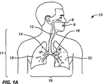
既述のように、肺を使用して開示される装置の利点及び作用を説明する。図1Aは主として胸腔11内に位置する呼吸器系10を図示する。呼吸器系10は気管12を含み、該気管は鼻8又は口9から右の主気管支14及び左の主気管支16に空気をもたらす。右の主気管支14から空気は右肺18に入り、左の主気管支16から空気は左肺20に入る。右肺18及び左肺20は合わせて肺19を構成する。左肺20は2つの肺葉のみから構成され、右肺18は3つの肺葉から構成されるが、それは一つには、通常胸腔11の左側に位置する心臓のために空間を提供するためである。
As described above, the advantages and operation of the disclosed device using the lungs will be described. FIG. 1A illustrates the
図1Bにより詳細に示すように、肺例えば左肺20中に通じる主気管支例えば左主気管支16は、葉気管支22に、その後さらに三次気管支24に、さらに細気管支26に、次いで終末細気管支28に、そして最後に肺胞30に分岐する。胸膜腔38は肺と胸壁との間の空間である。胸膜腔38は肺18、20を保護し、呼吸中に肺が動くのを許容する。図1Cに示すように、胸膜40は胸膜腔38を規定し、胸膜液の薄い層を間に挟んだ2つの層、すなわち臓側胸膜42及び壁側胸膜44からなる。胸膜液で占められた空間は胸膜腔46と呼ばれる。2つの胸膜層42、44の各々は、非常に多孔質の間葉性漿膜で構成され、該漿膜を通じて少量の間質液が胸膜腔46に絶えず浸出する。胸膜腔46内の液の総量は通常わずかである。正常な状態では過剰な液は通常リンパ管によって胸膜腔46の外に送り出される。
As shown in more detail in FIG. 1B, the main bronchus, such as the left
肺19は胸腔11内で浮動する弾性構造体である。肺19を囲む胸膜液の薄い層は胸腔11内での肺の運動を円滑にする。胸膜腔46からリンパ管中への過剰な液の吸引により、肺胸膜42の臓側胸膜面と胸腔44の壁側胸膜面との間のわずかな吸引が維持される。このわずかな吸引は、肺19が胸腔11内で膨れ、浮動するのを保つ負圧を創出する。負圧がなければ、肺19は風船のように潰れ、気管12を通じて空気を追い出してしまう。従って、息を吐き出す自然のプロセスは、肺19及び胸かご構造体の弾性反動によるほとんど全く受動的なものである。この生理学的配列の結果、胸膜42、44が破られた時は、肺19を吊るされた状態に保つ負圧は失われ、肺19は弾性反動効果のため潰れてしまう。
The
十分に膨張した時は、肺19は胸膜腔38を完全に満たし、壁側胸膜44及び臓側胸膜42は互いに接触する。空気の吸入及び吐出しを伴う膨張及び収縮のプロセス中に、肺19は胸膜腔38内で前後に滑動する。胸膜腔38内での運動は、壁側胸膜44及び臓側胸膜42間の胸膜腔46内にある粘液状の液の薄い層により容易にされる。
When fully inflated, the
例示の目的のため、図1Dは胸膜腔に血液50が溜まった肺20(血胸症とも呼ばれる)を図示する。図面からも明らかなように、胸膜腔46内の血液50の存在は肺20をずっと小さいサイズに収縮させることになる。呼吸作用は負圧の状態に吊るされた肺に依存するので、臨床的に、患者は潰れた肺に空気を送り込むのに困難な時を持とう。当業者によって認識されるように、胸膜腔46内の液又は空気は、胸腔に対する肺のサイズに図1Dに図示する血胸症と類似の臨床的影響を与える。肺の解剖学的設計、及び肺を吊るされた状態に維持するのに要求される負圧のため、肺から組織標本を得ることは他の組織に関しては存在しない追加的な課題を提示する。
For illustrative purposes, FIG. 1D illustrates a lung 20 (also called hemothorax) with
図2A〜Cは、肺から標的標本82又は標的材料を得るため生検装置80を展開して胸膜を破る処置中の肺19を描く。破りの結果、冒された肺20内の空気88は、装置80によって内層に作られた開口84をめぐって肺から出る(矢印で示す)。先の例におけるように、冒された肺20内の空気は、装置80が気管支14の壁部に穴を開けた時に創出された開口84を巡って肺から出る(矢印で示す)。加えて、当業者によって認識されるように、装置80の軌道は、図2Cに図示するように生検のための標的部位へのアクセスに失敗するものとなり得る。
FIGS. 2A-C depict the
上述のように、本発明及びその実施の形態は例示の目的のため肺における標的組織、細胞又は液へのアクセス、診断手当及び除去に関して説明する。しかしながら、本装置及び方法の態様は身体内の他の標的組織、細胞又は液の診断及び治療処置にも適用できる。 As mentioned above, the present invention and its embodiments will be described with respect to target tissue, cell or fluid access in the lungs, diagnostic benefits and removal for illustrative purposes. However, the apparatus and method aspects are also applicable to diagnostic and therapeutic treatments of other target tissues, cells or fluids in the body.
図3は、X線機器を有する部屋等の画像捕捉室54内の患者52と、例えば別の部屋等の場所58から画像捕捉処理を監視する医師又は技師56を図示する。画像捕捉処理は画像捕捉に適した機器60を用いる。患者52が標的標本取得処置を受ける時はしばしば、標的材料が取得されたことを確実にするため、標的部位にアクセスするよう装置を位置決めし、その後確認の画像を取る試みがなされる。当業者によって認識されるように、標的材料にアクセスするのに使用される装置の標的部位に対する位置を決定する適当な機構としては、別異の表示がない限り、当該技術分野で公知の従来の装置60及び方法が用いられる。これらの従来の装置及び技術には、X線撮像及び処理、X線トモシンセシス(tomosynthesis)、Aスキャン、Bスキャン及びCスキャンを含む超音波検査、コンピュータ断層撮影(CTスキャン)、スパイラルCT、磁気共鳴撮像法(MRI)、光コヒーレンス断層撮影、単光子放射型コンピュータ断層撮影(SPECT)、ポジトロン放射断層撮影(PET)、蛍光透視法、並びにこれらの組合せ及び携帯バージョンが含まれる。これらは当該技術分野の技術内のものである。そのような技術は文献に十分記述されており本書で説明する必要はない。例えば、X−Ray Structure Determination:A Practical Guide、第2版、Stout及びJensen編集、1989年、John Wiley & Sons社出版;Body CT:A Practical Approach、Stone編集、1999年、McGraw−Hill社出版;X−ray Diagnosis:A Physician’s Approach、Lam編集、1998年、Springer−Verlag社出版、を参照。
FIG. 3 illustrates a patient 52 in an
図4A〜Eは、標的部位から標的の材料標本にアクセスできる操縦可能な装置100の斜視図及び断面図を示す。装置100の構成部品には、二三例を挙げるとカニューレ、軟質管又は皮下管(hypotube)とし得る任意の外さや110と、操縦可能な部材120を含む。さやは、チタン及びニッケル−チタン合金(Nitinol)、ステンレス鋼、フルオロポリマー、ポリエーテルエーテルケトン(PEEK)、ポリテトラフルオロエチレン(PTFE)、発泡ポリテトラフルオロエチレン(ePTFE)、ポリウレタン、ナイロン、ポリイミド薄膜(Kapton(登録商標))その他等の適当な生体適合性ポリマー及び金属で作ることができる。本発明で使用できる適当なポリマーについての言及は、Bio−Compatible Polymeric Materialsと題された2002年1月10日付のPCT公開公報WO第02/02158号A1;Bio−Compatible Polymeric Materialsと題された2002年1月3日付のPCT公開公報WO第02/00275号A1;及びBio−Compatible Polymeric Materialsと題された2002年1月3日付のPCT公開公報WO第02/00270号A1に見出すことができる。
4A-E show perspective and cross-sectional views of a
技師等の使用者により制御されるよう適合された制御機構130は、操縦を可能とするため近位端102に設けられる。通常患者の身体の外側に(又は使用者に最も近く)位置決めされる近位端から離れて位置決めされる遠位端104は、装置の診断又は治療目的を達成するようさまざまなやり方で適合させ、形態づけることができる。例えば、遠位端104は、二三例を挙げると、トロカール、ランセット、スタイレット、針、治療用送出装置、マーカー又は診断用送出装置として形態づけることができる。図示の実施の形態において、近位端102の制御機構130は、握り132及びばね134又はコイルを含む。近位及び遠位は、しかしながら、相対的な用語であり、本説明の範囲を限定するものではない。
A
ばね134は、所望のばね張力を維持できる適当な材料で形成されたコイルワイヤとすることができる。コイル体の複数の巻きは、制御機構130の外面の周囲に嵌合するよう寸法づけられ、適合された内腔を形成する。いくつかの実施の形態は第1のコイル体と共に第2のコイル体を含む。図4B及び4Cの長手方向断面図に図示するように、操縦可能な部材120は外さや110の内腔112内を長手方向移動106できるようさや110内に位置する。近位端102のばね134は、近位端102に内腔112’を有するさや110の一セクション内に嵌合し、該セクションは操縦可能な部材120及びばね134を収容するのに十分大きな直径を有する。従って、さやはその近位端にてばねを覆う。ばね134は、例えば圧縮スリーブから形成できるが、通常、最適のばね力を備えたばねを形成できるステンレス鋼等の材料で形成されたばね134が提供されることが予定される。しかしながら、本書で論じられるように、操縦可能な部材120の移動を制御するばね力を装置に提供できるすべての構造体は、当業者によって認識されるように、適切である。
The
握り132を近位及び遠位に引き、押すことによって達成できる軸線Lに沿った長手方向移動に加えて、握り132を所望により時計回り及び反時計回りに回転させることにより回転運動108を達成することもできる。従って、操縦可能な装置の遠位端は少なくとも1つの軸の周りを360度運動できる。
In addition to the longitudinal movement along axis L that can be achieved by pulling and pushing
図4Bの線D−Dに沿って、かつ装置100の軸線Lと垂直に切った図4Dに示す断面図から、外さや110は操縦可能な部材120を受け入れるよう寸法づけられた内腔112を有し、操縦可能な部材120は外さや110の内腔112内を移動できることがわかる。図4BのE−Eの線に沿って切った図4Eの断面図に図示するように、外さや110の直径は操縦可能な部材120の直径に対しより大きく、少なくともばね134の一部がこれら2つの構成部品間に位置決めされる。従って、操縦可能な部材120は中心内腔122と共に図示されている。そのような形態は、操縦可能な部材120が例えば、二三例を挙げると、治療薬(例えば、標的部位への材料)を送出するよう、又は標的組織、細胞若しくは液を除去するよう適合されている場合は有用である。外さや120の近位端はより大きな直径を有し、ばねの少なくとも一部を保持し又はこれと係合するカップを形成する。操縦可能な部材120は形状記憶ニッケル−チタン合金(ニチノール)を含む任意の適当な材料で形成できる。外さやはステンレス鋼、チタン管又は生体適合性ポリマー等の任意の適当な材料から形成できる。操縦可能な部材120は、外さや110を係止又はこれと係合するよう形態づけ外さや110に対する操縦可能な部材120の相対運動を制御するようにすることができる。
From the cross-sectional view shown in FIG. 4D taken along line DD in FIG. 4B and perpendicular to the axis L of the
少なくとも1つの実施の形態において、装置100は少なくともその遠位端にて放射線不透過性である。外さや110はまた、先端に金属の付いたプラスチック、又はビスマス、タンタル、白金若しくは他の濃厚金属を添加したポリマーから形成できる。さやはまた、正規、オーステナイト又はマルテンサイト形を含むニッケル−チタン超弾性形状記憶合金(ニチノール)から形成できる。さやの外径又は外形は10−28ゲージ、より典型的にはおよそ23ゲージとすることができる。装置100の全長は、2.54センチメートル(1インチ)から、例えば43.18センチメートル(17インチ)までの間、又は任意の適当な長さとすることができる。
In at least one embodiment,
操縦機能の操作において、操縦可能な部材120が遠位方向に前進し、さや110の遠位端104から出ると、操縦可能な部材120の遠位端は、装置100の軸線Lから偏移して(角度α)湾曲形状を呈する。外さや110又は操縦可能な部材120は拡張器として作用し得る。中心軸Lからの偏移量は、使用者、及び操縦可能な部材120の遠位端104がさや110の外に延長する距離量により制御される。操縦可能な部材120がさや110中に引き戻される(すなわち、使用者及び/又は制御装置に向けて近位に引かれる)につれて、角度αは減少する。角度αの減少は、さや110の内面113によって操縦可能な部材120に適用される圧力によりもたらされることができ、該内面は操縦可能な部材120を真っすぐにする。従って、標的部位に向けて前進する時は、機構の全体(さや110及び操縦可能な部材120)が組織に向けて前進される。標的部位に対する装置100の場所が判定され(例えば、図3に関して論じた画像捕捉機60を使用して)、装置100の軌道が所望の標的部位から偏移したことが決定されると(例えば、図2B〜C参照)、さや110を固定又はほぼ固定された場所に維持しながら操縦可能な部材120を組織に向けて遠位方向に前進させ、装置100を標的部位に到達させることができる。当業者によって認識されるように、装置100を前進させるステップ及び操縦可能な部材120のみを前進させるステップは、必要に応じて交互に行い、標的部位へのアクセスを最適化することができる。装置100及び/又は操縦可能な部材120を前進させることにより装置100の場所を制御することに加えて、握り132を時計回り及び反時計回りに回転させることによりさらなる制御を達成することができる。装置の任意の又は全ての構成部品の位置は、例えば操縦可能な部材120と外さや110とを係合させることにより適所に係止させ、所望により装置100又は装置構成部品のさらなる移動を防止することができる。
In operation of the steering function, when the
装置100は、例えば、軸線L等の少なくとも1つの軸の周囲を360°まで、また操縦可能な部材の湾曲次第で任意の残りの軸の周囲を180°まで又はそれ以上運動することができる。本書に開示される装置の設計を変更することによって、より大きい又はより小さい操縦性が提供される。一たび装置が適所にあれば、操縦可能な部材120を外さや110から取出し、例えば注射器又は他の吸引源と置換して、その後組織標本を外さや中に吸引し患者から取出すことができる。加えて、この2部形態により、外さや110の壁部をより薄くでき、全体の外形(すなわち直径又は円周)を小さくして、装置をより侵襲的でないものとすることができる。あるいは、操縦可能な部材120を標的部位に治療を施す装置又はシステムと置換できる。
The
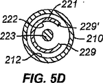
図5A〜Fは標的部位にアクセスできる別の操縦可能な装置200の斜視図及び断面図を示す。操縦可能な装置200は近位端202及び遠位端204を有する。操縦可能な部材220を受け入れるための内腔212を有する任意のさや210が設けられている。図5に示す形態において、操縦可能な部材220は内部制御部材223を収容するノッチ付管状部材221を有する。
5A-F show perspective and cross-sectional views of another
内部制御部材223は、ノッチ付管状部材221の内径より大きい直径を有する遠位端を有し、遠位端は管状部材の遠位端を越えて延長し、かつノッチ付環状部材の内腔内を引かれることから防止される。従って、内部制御部材223の遠位端は、図示のように、ボール状又は球根状端部等の端部224、又はノッチ付管状部材に引っかかるフランジを形成することができる。当業者により認識されるように、内部制御部材223の遠位端は除去可能とすることもできる。1つの形態において、端部224は、適切な機構、例えば端部224の雌ねじ端部と係合する制御部材223の雄ねじ端部によって制御部材223の端部に除去可能に取付けることができる。他の形態において、端部224は必要なら制御部材223に半田付けすることができる。端部224及び制御部材223が統一的に作用する設計も、制御部材223及び端部224が一体である設計を含め、考えられた設計内のものである。内部制御部材223は装置200の軸線Lに沿った移動206のみならず、装置200の軸線Lの周囲を時計回り及び反時計回りに回転運動208することもできる。
The
ノッチ付管状部材221は内腔222を有し、該内腔は制御部材223を囲み、かつ環状部材221の遠位端にて端部224と係合するよう形態づけられる。ノッチ付管状部材221はまた、図示のように、任意のさや210の内腔212内に嵌合するよう適合され形態づけることもできる。さや210内に配された時、ノッチ付管状部材221は、装置200の軸線Lに沿って移動206ができる一部を少なくとも有する。加えて、環状部材221の少なくとも一部はさや210内に固定される。1つの形態において、ノッチ付環状部材223は近位端202にてさや210と固定的に係合するよう適合されている。例えば、ノッチ付環状部材223は近位箇所にてさや210に接着することができ、又は近位箇所にて解除可能に係合(例えば、ねじ又はさねはぎ設計を使用して)させることができる。
The notched
装置200の長さに沿った軸線Lと垂直の平面に沿って切った図5D〜Eに示す断面図を参照すると、制御部材223がノッチ付環状部材221の内腔222内に位置決めされている。ノッチ付環状部材221のノッチ付セクションを横切った管状部材221の断面図の場合、その断面にて環状部材221によって規定される内腔222は、図5Dに示すように、さや210により規定された内腔212と連通する。逆に、ノッチ付環状部材221のノッチ付でないセクションを横切った環状部材221の断面図の場合、その断面にて環状部材221により規定される内腔221は、さや210により規定された内腔212と連通しない。ノッチ226は、環状部材221の軸線に沿った外形又は側面が半円形、又はU形(図5Bに図示のように)、三角形、四角形その他を形成するよう形態づけることができる。ノッチ226は切れ目又は波形とすることもできる。ノッチ226の幾何学的外形にかかわらず、少なくとも一方から見て、ノッチの対向上縁228、228’は、制御部材223を動かすことによりノッチが圧縮されると互いに接近するよう位置決めされる。いくつかの形態において、ノッチ226の対向上縁228、228’が圧縮されると、ノッチ226の少なくとも一部の縁部226は完全に姿を消すように見え得る、例えば、ノッチ229、229’の側部が互いに接触して継目を形成するように見える場合である。しかしながら、当業者によって認識されるように、環状部材の他の形態も可能である。例えば、図5Eの断面図は継目を形成すること等によりその円周に沿ったある箇所にて係合するよう適合させることができる。
Referring to the cross-sectional views shown in FIGS. 5D-E, taken along a plane perpendicular to the axis L along the length of the
断面において、例えば、内部制御機構223は中実の円形断面を有し、ノッチ付管状部材221の内腔222内に嵌合するよう位置決めされる。図5Bの線D−Dに沿った図5Dに示すように、この断面図はノッチ付環状部材221のノッチ226を横切ったものであり、従って環状部材221は、”C”等の部分的な円形断面形状を有する。環状部材221及び内部制御機構223はさや210の内腔212内に嵌合する。図5Fに図示するように、内部制御機構223を軸方向に移動させると、ノッチ226は集められ、ノッチの縁部間の隙間が小さくなる。従って、例えば、図5Fに示すように、装置200が図5Cに示す湾曲形態を呈するにつれて、図5Cに示す部分的な円形断面形状は軸線Lと垂直な断面に関し楕円形状となり、ノッチ付環状部材221の断面形状は縁部が接触するように接近した楕円形状又は実質的に楕円形状の”C”となる。
In cross section, for example, the
図5Bの線E−Eに沿った図5Eに示す断面図において、ノッチ付環状部材221の近位端の外面はさや210の近端部の内面と係合するよう形態づけられ、これら2つの部材間に永久的又は半永久的な関係が維持される(従って、さや210の回転運動なしにノッチ付環状部材220が回転運動することは防止される)。図5Eの断面図において、各部品は、1つの構成部品を他の構成部品と係合させる1つ又は複数のさねはぎ接合218の使用により維持される。例えば、1つの部材に戻り止、別の部材に凹所を使用したスナップ嵌め配列を含む他の、さや210とノッチ付環状部材221との係合機構も当業者には明らかであろう。
In the cross-sectional view shown in FIG. 5E along line EE of FIG. 5B, the outer surface of the proximal end of the notched
さや210及びノッチ付管状部材220の各々は、使用者による取扱いを容易にするためフランジ217、227を有することができ、ノッチ付管状部材220のフランジ227の場合、フランジ227は、内部制御機構223の取扱い時ノッチ付環状部材220の全体がさや210の内腔212中に入り込んでしまうのを防止するさらなる機構を提供することができる。
Each of the
操縦用構成部品の操作において、内部制御部材223を軸方向206に引き又は押すと、操縦可能な部材220は装置200の軸線Lから離れるように変形する。中心軸Lからの遠位端の偏移量は、使用者、及び操縦可能な部材220の内部制御部材223の押し/引き量に基づく量により制御される。内部制御部材223が近位に引かれる(すなわ、使用者及び/又は装置制御装置に向けて近位に引かれる)につれて、軸線Lからの偏移角αは増加するが、それは内部制御部材223が側部を引くと制御部材のノッチ226が変形し(図5Cに図示するように)、これによって操縦可能な部材220がある方向に曲り、1つ又は複数の平面内で軸線から例えば180°又はそれ以上移動するからである。加えて、1つの構成部品を引き、同時に別の構成部品を押して同一の結果を達成することができる。
In operation of the steering component, pulling or pushing the
制御機構及び/又はフランジを係合させる使用者の動作は曲げ力を適用させ、これにより装置は標的部位に向けて操縦される。曲げ力が増加すると操縦可能な部材に対する応力が増加し、これにより装置の湾曲が引起される。従って、制御機構を係合させる使用者より操縦可能な部材が歪められると歪が生じる。曲げ力の適用により、本発明で説明した設計品は能動的に操縦され、このことは予備形成形状の変形に起因する受動的な操縦と対照をなす。能動的及び受動的な操縦の組合せは本発明の範囲を逸脱することなく使用できる。さらに、装置の各々の構成部品の曲線長さは、操縦及び前進プロセス中の長手方向長さ(曲げられていない装置の)と同一又は実質的に同一のまま留まることができる。装置はそれ自身の標的部位までの進路を規定し創出するよう適合され形態づけられている。進路の規定及び創出は、装置が組織中を前進するときに動的に生じる。従って、例えば、装置が組織中を前進するにつれて、組織の稠密性又は他の特徴により、装置を標的部位に向かう軌道から偏移させる応力又は歪が装置に加えられる。制御機構を係合させ、該制御機構を使用して反対の歪又は曲げ力等の歪を装置に加え、これによって装置の遠位端の場所及び方向を制御することにより、装置は標的部位に向けて操縦される。 The user's action of engaging the control mechanism and / or the flange applies a bending force that causes the device to be steered towards the target site. Increasing the bending force increases the stress on the steerable member, thereby causing the device to bend. Therefore, distortion occurs when a member that can be steered by a user engaging the control mechanism is distorted. By applying bending force, the design described in the present invention is actively steered, which contrasts with passive maneuvering due to deformation of the preformed shape. A combination of active and passive maneuvers can be used without departing from the scope of the present invention. Furthermore, the curved length of each component of the device can remain the same or substantially the same as the longitudinal length (of the unbent device) during the steering and advancement process. The device is adapted and configured to define and create a path to its own target site. Path definition and creation occurs dynamically as the device advances through the tissue. Thus, for example, as the device is advanced through the tissue, stresses or strains are applied to the device that cause the device to deviate from its trajectory toward the target site due to tissue denseness or other characteristics. By engaging the control mechanism and using the control mechanism to apply a strain, such as an opposite strain or bending force, to the device, thereby controlling the location and orientation of the distal end of the device, the device is moved to the target site. It is steered towards.
従って、標的部位に向けて前進する時、機構の全体(さや210及び操縦可能な部材220)が標的部位に向けて前進する。標的部位に対する装置200の場所が判定され(例えば、図3の画像捕捉機60を使用して)、軌道が所望の標的部位に到達するのに要求される軌道から偏移している(例えば、図2B−C参照)と決定されると、操縦可能な部材220を係合させ、装置200の遠位端204を曲げることにより、装置の遠位端204をして元の軌道を維持させ又は偏移させることができる。当業者により認識されるように、制御部材223を調節するステップは必要に応じて交互に行い標的部位へのアクセスを最適化することができる。加えて、他の実施の形態で図示したもの等の握りを近位端に設け、係合させて、装置200が軸線Lの周囲を360°まで移動するのを可能としつつ該装置の回転制御をさらに提供することができる。機構同士の係合が解除された場合、例えばさねはぎが外され、又は雄ねじと雌ねじの係合が解除された場合、制御部材220に対するさや200の別個の移動も達成できる。
Thus, when moving forward toward the target site, the entire mechanism (
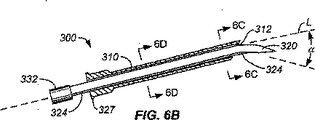
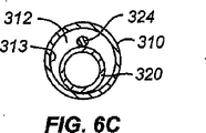
図6A〜Eは、標的標本にアクセスできるなおも別の操縦可能な装置300の斜視図及び断面図を示す。この実施の形態において、任意のさや310には操縦可能な部材320が設けられ、該部材はさや310の内腔312の少なくとも一部内に位置決めされる。操縦可能な部材320は、操縦可能な部材320とその長さに沿った少なくとも2つのポイントで係合するよう適合された制御ワイヤ324又は引張ワイヤを有する。制御ワイヤ324は、作動中に操縦可能な部材300の遠位先端の場所を相違させるために使用できる。従って、ワイヤは装置の先端の場所を相違させるための差動ワイヤと考えることができる。制御ワイヤ324は操縦可能な部材320の長さより短い長さを有する。制御ワイヤ324は、少なくとも1つの方向において弾性特性を有する材料から形成できる。制御ワイヤ324がさや310の内腔312の内面313と係合すると、制御ワイヤ324は変形し、これにより操縦可能な部材320が変形する。握り332が近位端302に設けられ、該握りは使用時直接又は間接的にさや310内での操縦可能な部材320の軸方向306及び回転308運動を制御する。当業者によって認識されるように、制御ワイヤ324は、円形断面形状(図示のように)を有するワイヤとすることができ、又は帯又はリボン(例えば、正方形又は長方形の断面形状を有する平らな条片)若しくは本装置設計の操作目的を達成する任意の他の形状とすることができる。
6A-E show perspective and cross-sectional views of yet another
図6Bを参照すると、操縦可能な部材320は斜角付の中心針340の形をとり、該操縦可能な部材に取付けられたガイドワイヤの形をとる単一の制御ワイヤ324又は引張ワイヤを備える。制御ワイヤ324は、操縦可能な部材320の長さに沿った部分にて、図6Cの断面図に示すように、さや310の内壁又は内腔313に隣接し、又は外さやがなければ操縦可能な部材から離れるように曲る形態を呈する。制御ワイヤ324は、他の箇所では操縦可能な部材320の表面に隣接する形態を呈することができる。制御ワイヤ324は、さらに他の箇所では、さや310の内壁313と制御ワイヤ324の表面323から等距離に位置決めされる形態を呈する。制御ワイヤ324は、なおも他の箇所では、さや310の内腔313及び操縦可能な部材320の外面の両方と接触する形態を呈することができる。当業者により認識されるように、さや310の内腔の直径はその長さに沿って一定とし、又はその長さに沿って変化させて、制御ワイヤ324に機械的な圧力を提供し及び/又は制御ワイヤ324を変形させることができる。
Referring to FIG. 6B,
操縦機能の操作において、操縦可能な部材320が遠位方向に前進しさや310の遠位端304から出ると、操縦可能な部材320の遠位端は装置300の軸線Lから偏移(角度α)した湾曲形状を呈するが、これは制御ワイヤ324によって制御される。軸線Lからの偏移量は、使用者、操縦可能な部材320の遠位端304がさや310の外に延長する距離量、並びに弾性、変形性、強度その他等の制御ワイヤ324の材料特性によって制御される。操縦可能な部材320がさや310中に引き戻されると(すなわち、使用者及び/又は制御装置に向けて近位に引かれると)、角度αは減少するが、それはさや310の内壁により制御ワイヤ324に圧力が適用されて操縦可能な部材320を真っすぐにするからである。従って、標的部位に向けて前進する時、機構の全体(さや310及び操縦可能な部材320)が組織に向けて前進できる。標的部位に対する装置300の場所が判定され(例えば、図3に関して論じた像捕捉機60を使用して)、軌道が所望の標的部位から偏移している(例えば、図2B〜C参照)と決定されると、さや310を固定又はほぼ固定された場所に維持しながら操縦可能な部材320を標的部位に向けて遠位方向に前進させ、装置300を標的部位に到達させることができる。当業者によって認識されるように、装置300を前進させるステップ及び操縦可能な部材320のみを前進させるステップは、必要に応じて交互に行い、標的部位へのアクセスを最適化することができる。いくつかの場合には、装置の操縦は、曲げられていない装置の長さと長さが同等又は実質的に同等な曲線形状を形成するのと同時に能動的に生じ得る。装置300及び/又は操縦可能な部材320を前進させることにより装置300の場所を制御することに加えて、握り332を時計回り及び反時計回りに回転させることによってさらなる制御を達成することができる。
In operation of the steering function, when the
別の操作において、操縦可能な部材320のフランジ327を係合させる時に制御ワイヤ324を押し又は引く。この動作により操縦可能な部材320は制御ワイヤ324の移動に対し静止状態に保たれる。上述のように係止機構を組み込むこともできる。
In another operation, the
図6Eは、ばね334を設けて装置300に加えられる制御量を増加させる代替の断面図を示す。
図7A〜Eは、標的部位にアクセスできるさらに別の操縦可能な装置400の斜視図及び断面図を示す。図7に図示する装置400はさや410を含み、該さやは操縦可能な部材420及び制御部材430を有する。図6の設計では単一の制御ワイヤ424が設けられるが、図7の設計では2つ以上の制御ワイヤ424、424’又は4つの制御ワイヤ(図示のように)が使用される。制御レバー433と係合する1つのワイヤ及び4つの側部制御ワイヤ424、424’の形をとることができる中心の操縦可能な部材425が設けられる。制御レバー433のタブ435をある方向に移動させると、装置400の遠位端の制御された移動が生じ、標的組織、細胞又は液等の標的領域に向けて装置の遠位端を湾曲させる。制御ワイヤは炭素又はアラミド繊維(Kevlar(登録商標))等の金属、ポリマー又は有機繊維とすることができる。加えて、制御ワイヤはガラス又はセラミックとすることができる。
FIG. 6E shows an alternative cross-sectional view that provides a
7A-E show perspective and cross-sectional views of yet another steerable device 400 that can access the target site. The apparatus 400 illustrated in FIG. 7 includes a
操縦機能の操作において、例えば、タブ435を右に向けて移動させると、制御レバー433の右側と係合する制御ワイヤ424は近位方向に前進し、装置400の遠位端404を左に移動させる(すなわち、第1の方向におけるタブの移動は、タブの移動方向と反対の方向に装置400の遠位端404を移動させる)。図7Cに図示するように、制御ワイヤ424、424’は制御レバー433の外面に半田付け427することができる。これに加えて、又は半田付けの代わりに、制御ワイヤ424、424’をかしめ、接着し又はこれらを組合せ、及び/又は近位端にて曲げて近位端が制御レバー433の内腔内に配されるよう形態づけることもできる。操縦機能の操作により、装置の遠位端の操縦、及び標的部位への進路に沿った装置の前進の両方を片手で高度に正確に制御することが可能となる。
In operation of the steering function, for example, when the
さらに、上述のように、制御機構及び/又はフランジを係合させる使用者の動作により曲げ力が適用され、これにより装置が標的部位に向けて操縦される。曲げ力が増加すると、湾曲を引起す操縦可能な部材に対する歪が増加する。曲げ力の適用により、予備形成形状の変形に起因する受動的な操縦と対照的に、本発明で説明した設計品は能動的に操縦される。さらに、装置の各々の構成部品の曲線長さは、操縦及び前進プロセス中の長手方向長さ(曲げられていない装置の)と同一又は実質的に同一のままである。装置はそれ自身の標的部位までの進路を創出するよう形態づけられている。この設計は装置の有用性、一貫性及び操作の正確性、並びに使用者及びヒューマンファクターズ設計考察との人間工学的仲立ちを改善する。 Further, as described above, a bending force is applied by a user action to engage the control mechanism and / or the flange, thereby maneuvering the device toward the target site. As the bending force increases, the strain on the steerable member that causes curvature increases. By applying bending force, the design described in the present invention is actively maneuvered, as opposed to passive maneuvering due to deformation of the preformed shape. Furthermore, the curved length of each component of the device remains the same or substantially the same as the longitudinal length (of the unbent device) during the steering and advancement process. The device is configured to create a path to its own target site. This design improves device usability, consistency and operational accuracy, as well as ergonomic interactions with user and human factors design considerations.
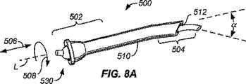
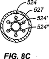
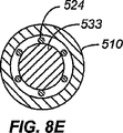
図8A〜Eは、標的標本にアクセスできる別の操縦可能な装置500の斜視図及び断面図を示す。図8に図示する装置500は、操縦可能な部材520及び制御部材530を有する任意のさや510を含む。図6の設計では単一の制御ワイヤ324を設けるが、図8の設計では複数の制御ワイヤ524、524’を使用する。追加的な制御ワイヤ524、524’を設けることにより、制御レバー533は装置500の遠位端504のより正確な制御を達成できるようになる。当業者により認識されるように、装置500に追加的な回転運動を与えるため、握り535又は他の適当な機構を近位端502に設けることができる。図8の装置の操作は、図7に図示した実施の形態に関し説明した操作と類似する。
8A-E show perspective and cross-sectional views of another
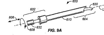
図9A〜Eは、標的標本にアクセスできる別の操縦可能な部材600の斜視図及び断面図を示す。この実施の形態において、さや610の遠位端604の内腔は、軸線Lから離れるよう、又は軸線Lから遠位端に近いさやのセクションよりさらに離れるように湾曲し又は角度をなすよう形態づけられている。従って、さや610の遠位開口611は曲り部613を形成し、これにより遠位開口611は軸線Lの全部又は一部と交差しないよう位置決めされる。
9A-E show perspective and cross-sectional views of another
操縦機能の操作において、操縦可能な部材620が長手方向606に移動されると、操縦可能な部材620の遠位端は前進し、さや610の内腔内部の曲り部613がさや610の内腔内部の該曲り部に対応する量だけ操縦可能な部材620を曲げる。操縦可能な部材620の遠位端がさや610の内腔を通じて前進し続け、さやの遠位端を越えて延長すると、操縦可能な部材は装置の中心軸線Lから所定の又は確定できる量だけ曲げられる。さらなる回転運動608は、さや握り614等の制御機構を時計回り及び/又は反時計回り方向に回転させることにより達成できる。装置600の軸線と垂直な軸に沿った図9C〜Dに示す断面図によって図示するように、さや610の内腔612は、第1の箇所(例えば、装置の長さに沿った中間点)においては第2の箇所(例えば、遠位端位置)におけるよりも中心寄りに位置決めされる。一たび制御部材620の遠位端がさや610の遠位端を越えて前進すると、機構600の全体の回転運動が握り632を係合させることによって達成できる。図9Eに図示するように、装置600はばね634の使用を含むよう形態づけることができる。さらに、一たび制御ワイヤ623が所望の場所を達成したら、制御ワイヤを除去し別の構成部品(例えば、診断装置又は組織生検装置)を投与するためのオープン内腔を残すよう、装置600を形態づけることができる。
In operation of the steering function, when the
図10A〜Cは、標的標本にアクセスできるなおも別の操縦可能な装置700の斜視図及び断面図を示す。装置700は内腔712を有する外さや710を有し、該内腔にはスタイレット等の操縦可能な部材720が受け入れられる。操縦可能な部材720は、位置指示器736(図示のように、位置指示器736は握り732の表面に設けられた矢印である)を含むトップキャップ又は握り732と係合する。追加的な指示マーキングを外さや710のリップ又はフランジ717に設け、位置指示器736をフランジ717の1つ又は複数のマーキング736’、736”と関連づけ、装置700又はさや710に対するスタイレット720の相対方向又は移動を表すようにすることができる。この実施の形態において、図4に示す実施の形態のように、操縦可能な部材710は、該操縦可能な部材710を外さや710の遠位端を越えて前進させると再形成(又は湾曲形態に復帰する)軟質の湾曲遠位先端を有する。当業者により認識されるように、マーキングは、本発明の範囲を逸脱することなく、本書で提供される他の設計品のいずれにも設けることができる。位置指示器の形式は所望により改変することもできる。例えば、矢印のマーキングばかりでなく又はこれに追加して、1つ又は複数の方向における程度の表示を含めることができよう。例えば操縦可能な部材又は外さやの胴部に追加的なマーキングを設けて追加的な平面内の追加的な移動表示を提供することができよう。
10A-C show perspective and cross-sectional views of yet another
マーキングの実施の形態において、例えばスタイレットの尖った先端729に対応する矢印の位置指示器が設けられる。肉太及び肉細(上部−下部)の指示器736’、736”をフランジ717に設けて、使用者がさや710に対するスタイレットの尖った先端729の場所を決定できるようにすることができる。尖った先端はまた、一たびスタイレットが外さやの遠位先端の端部を越えて前進すればスタイレットの外部湾曲と相関するよう形態づけることができる。握り732はさらに外さや710の近位端と係合して軟質部材720の位置を適所に係止させるよう形態づけることができる。例えば、さねはぎ、戻り止め及び溝、又は他の任意の適当な設計又は形態を使用して1つの構成部品を別の構成部品と係合させることができる。マーキングの実装及び設計はヒューマンファクターズの考察を組込むよう改変できる。
In the marking embodiment, for example, an arrow position indicator corresponding to the
操作において、使用者は位置指示器736を使用して標的部位に対する装置の先端の配向を決定するであろう。さらなる操縦は、例えば画像を検討し、組織に係合するには装置を例えば右に10°前進させることが必要であったとの決定に基づいて果すことができるであろう。使用者は、位置指示器を使用して装置の遠位先端(患者内に位置する)を操縦し、所望の移動を達成して標的部位に向けて装置を前進させるであろう。
In operation, the user will use the
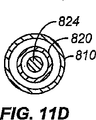
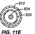
図11A〜Gは標的標本にアクセスできるさらに別の操縦可能な装置800の斜視図及び断面図を示す。装置800は外さや810及び内部の操縦可能な部材820を有する。内部の操縦可能な部材820は操縦可能な中心ワイヤ824と係合するさらなる内腔822を有し、該ワイヤは2つ以上の接続可能セクション824’、824”を有する。図示するように、操縦可能な中心ワイヤ824は、近位端の近くで軟質接合部826により接続された2つの構成部品を有する。軟質接合部826は該構成部品を互いに軟質関係に置き、操縦可能な部材820が遠位方向に前進された時に、装置800の近位端を曲げ又は回転させると遠位端804を操縦することとなるようにする。図11Cに図示するように、握り832は第1の方向に移動されると装置800の遠位端を曲げさせる。遠位位置における装置の軸線と垂直な断面図に示すように、カニューレ部分又は制御ワイヤ824は操縦可能なさや820内に位置決めされることができ、該さやは同様に外さや810内に嵌合する。代替の実施の形態において、カニューレ部分824は、図11Bに示すように1つ又は複数の操縦ワイヤ825、825’内に嵌合され得る。ワイヤは、該ワイヤの差動長さを作り出すことによって装置の操縦動作を支援するのに使用できる。例えば、図7〜8に示す実施の形態も操縦機構においてワイヤを使用する。カニューレ824が内嵌する内腔822は、図11Dに示すD−Dに沿った断面に近い箇所にてカニューレ824の断面に対しサイズが増加する。この関係は図11BのE−Eに沿った断面図である図11Eに図示される。図11Fに示すように、カニューレの第1のセクション824’はその近位端にて軟質材料又は接合部826と係合し、これによりカニューレの第1のセクション824’は、近位制御機構又は握り832と係合するカニューレの第2のセクション824”に対し1つの軸の周囲を移動できるようにされる。少なくとも装置800の一部は、図11Gに図示する断面G−Gにて示すように、カニューレを囲まない軟質さや810を有する。
11A-G show perspective and cross-sectional views of yet another
装置の操縦において、装置800の操作は上述の装置と類似する。しかしながら、操縦可能な部材820の二部構造により装置の近位端は接合部の周りを装置の遠位端に対し回転可能である。達成できる回転運動量は接合部にて使用される材料の軟質性により制御できるであろう。加えて、操縦可能な部材を外さや中に前進させ、操縦可能な部材の接合セクションを外さや内の、接合部の軟質性が減じられる箇所に位置決めすることにより、軟質性を低下することができよう。
In maneuvering the device, the operation of the
当業者により認識されるように、装置が標的部位に向けて前進するため動作し操縦及び長手方向移動に関わる時に操縦可能な部材が全体的に又は部分的に組織内に位置決めされるよう装置を操作することができる。これに加えて、又は代わりに、任意のさや又はスコープ等のそれ自身で組織を貫き操縦移動に関わるよう適合された別の部材内に操縦可能な部材を全体的に又は部分的に位置決めすることができる。 As will be appreciated by those skilled in the art, the device can be positioned so that the steerable member is wholly or partially positioned within the tissue when the device operates to advance toward the target site and is involved in steering and longitudinal movement. Can be operated. In addition or alternatively, to position in whole or in part a steerable member within another member adapted to engage in steer movement through tissue with any sheath or scope itself, etc. Can do.
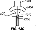
図12A〜Cは、図4〜11に示す操縦可能な装置のすべてと共に使用するのに適したさまざまな遠位先端設計品900の断面図を示す。認識されるように、先端設計品900はカニューレ化し(従って、該先端の中心内に内腔を提供する)、又は中実の針を形成することができる。遠位先端はまた切断装置を形成し、又は治療若しくは診断用送出機構を提供するよう形態づけることができる。図12Aに示すように、針内に拘束される斜角コアを備えた二斜角付針二重針又は同軸形態が提供される。第1の針940は第2のカニューレ化針942内に嵌合する。図12Bに図示する実施の形態において、四斜角付コア針944はカニューレ化針942内に設けられる。図12Cは四斜角付カニューレ化針946内の四斜角付コア針944を図示する。他の設計品及び実施の形態を本発明の範囲を逸脱することなく使用できる。さらに、コア針形態を上述の操縦可能な部材のいずれにも対応させることができ、かつカニューレ化針を上述の外さや形態に対応させることができることが認識されよう。加えて、中心のコア針は、カニューレ化に加えて、単独で又はさやと組合わせて除去可能とし、外さやの内腔内にある装置を位置決めするのを許容するよう形態づけることができる。
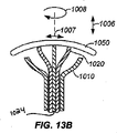
12A-C show cross-sectional views of various distal tip designs 900 suitable for use with all of the steerable devices shown in FIGS. As will be appreciated, the
図13A〜Fは、図4〜11に示す操縦可能な装置のすべてと共に使用するのに適したさまざまな近位制御機構の断面図を示し、近位制御機構は使用者(医師又は技師等)により直接制御することができ、又は使用者が離れた距離(例えば、別の部屋)から装置を制御するのを可能にする別の機構に係合させることができる。装置の近位端にルーアー取付具を設けてさまざまな構成部品の制御及び係合を容易にすることもできる。第1の実施の形態において、外さや1010(例えば、110、210、310、410、510、610、710、810、910に対応)はコイル部材又はばね1034(例えば、134、334、634に対応)と係合する。コイル部材1034は装置の操縦可能な部材1020(例えば、120、220、320、420,520、620、720、820、920に対応)の軸に沿った長手方向移動1006の制御、並びに曲線移動(例えば、コイルが一方の側で圧縮され、他方の側では圧縮されず、遠位先端がある角度軸線から偏移する場合)の制御を提供する。図13Bは、外さや1010に対し操縦可能な部材1020の追加的な前方後方1006、左右1007又は回転1008運動を提供できるハンドル1050を図示する。図13Cは、操縦可能な部材1020の回転運動を容易にする握り1034と組合せた図13Bのハンドルを図示する。図13Dは操縦可能な部材1020のワイヤと組合せた回転ボール又はタブ1035を図示し、ここでワイヤは、ボールを中心軸から逸れるよう任意の方向に旋回させるとワイヤが外さや1010の内腔内で移動し、装置の操縦可能な遠位端1004が移動するようボール又はタブ1035に接続される。図13Eの装置は、装置の長手方向/軸方向の移動の追加的な制御を提供できるコイル部材1034又はばねと組合せた回転ボールを図示する。図13Fは、装置の追加的な回転制御を提供する握り1034と組合せた回転ボールを図示する。ボール又はタブ1035を用いた実施の形態は、上記のように装置の片手での操縦及び制御を容易にする。
13A-F show cross-sectional views of various proximal control mechanisms suitable for use with all of the steerable devices shown in FIGS. 4-11, where the proximal control mechanism is a user (such as a physician or technician). Can be directly controlled, or can be engaged to another mechanism that allows the user to control the device from a distance (eg, another room). A luer fitting may be provided at the proximal end of the device to facilitate control and engagement of various components. In the first embodiment, an outer sheath 1010 (eg, corresponding to 110, 210, 310, 410, 510, 610, 710, 810, 910) corresponds to a coil member or spring 1034 (eg, corresponds to 134, 334, 634). ). The
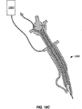
図14A〜Bは図4〜11に示す操縦可能な装置の遠隔制御のための機構を図示する。本形態は遠位先端のリアルタイム又はほぼリアルタイムの案内を許容する。作動レバー1112はハンドル1116内のリンク仕掛に接続される。ハンドル1116内のリンク仕掛は操縦可能な部材1110に接続された作動器と連絡する。これにより、標的部位にアクセスするため装置の位置の判定中に介在者(X線技師又は医師等)が患者の身体の外側に接触することなく、上述の装置を制御できるようになる。その結果、装置の操縦動作は針進入部位から隔たって行うことができる。従って、使用者の手の位置を針進入部位から分離することにより、図16に示すように、使用者の手を放射線に曝すことなく操縦可能な装置を操作しながらCTスキャンを行うことができる。図14Bに示す別の実施の形態において、放射線から隔たった完全なギャンブル制御のための機構が提供される。操縦可能な装置の前進及び後進制御を許容するつまみ握り1132がハンドルに設けられる。加えて、左右の移動及び回転並びにこれらの組合せも可能とされ、操縦可能な装置の遠位端の移動を最適化する。
14A-B illustrate a mechanism for remote control of the steerable device shown in FIGS. This configuration allows real-time or near real-time guidance of the distal tip. The
図15A〜Bは図4〜11に示す操縦可能な装置の制御のための追加的な機構を図示する。図15Aに示す実施の形態において、指リング1202、1202’は操縦可能な装置1200の近位端に形成された通路を通る制御ワイヤ1204、1204’に接続される。ヒンジ1206がその後自動調心軸受又はボールジンバル1208との枢動接続部に取付けられる。ボールジンバルは2つの垂直軸の周りの回転自由を提供する。第2の制御ワイヤがリング1202’からボールジンバル1208上の固定接続部に延長する。ボールジンバル1208は、上述のように操縦可能な装置1200の操縦機構の一部として制御ワイヤ(図示せず)に接続される。操縦可能な装置1200の遠位端はリング1202、1202’を動かすことにより移動させ操縦することができる。操縦可能な装置1200は、リング1202をリング1202’に向けて下方に移動させることにより疑わしい病変部に向けて患者の中に前進させることができる。装置の全体は、例えば接着剤を含む任意の適当な機構により患者の身体に取付けることができる。図15Bに図示する装置は、操作棒1242を使用して操縦可能な装置1200と連絡する遠隔制御機器1240を図示し、該操作棒は遠位ポイントが表面上を移動するよう枢軸の周りを回転する。操作棒1242は、プッシュプル制御ワイヤを使用して左右並びに前後に移動できるリンク仕掛1244に接続され、これによって操縦可能な針を曲げる。
15A-B illustrate additional mechanisms for control of the steerable device shown in FIGS. In the embodiment shown in FIG. 15A,
図16は、例えば図14及び15に示す遠隔アクセス装置を使用して患者52の標的部位にアクセスするため、図4〜11に示す操縦可能な装置を制御する介在者56、例えば医師又は技師を図示する。当業者により認識されるように、遠隔アクセス及び制御は別の部屋内の位置から(図3に図示するように)、又は画像捕捉機への暴露を最小にする場所等の任意の適当な場所から行うことができる。加えて、遠隔制御無線装置及び機構は介在者56によって使用されて装置の能動操縦及び移動を制御することができる。例えば、米国特許である、Medical Apparatus Remote Control and Methodと題された第6,768,425号(Flaherty et al.)及びWireless Medical Diagnosis and Monitoring Equipmentと題された第6,577,893号(Blesson et al.)を参照。 FIG. 16 illustrates an intermediary 56, such as a physician or technician, controlling the steerable device shown in FIGS. 4-11 to access the target site of the patient 52 using, for example, the remote access device shown in FIGS. Illustrated. As will be appreciated by those skilled in the art, remote access and control can be from any location in another room (as illustrated in FIG. 3) or any suitable location, such as a location that minimizes exposure to the image capture device. Can be done from. In addition, the remote control radio device and mechanism can be used by an intermediary 56 to control the active steering and movement of the device. For example, US Patent Nos. 6,768,425 (Flaherty et al.) Entitled Medical Apparatus Remote Control and Method, and Wireless Medical Diagnosis and Monitoring No. 6, 7th title of 57 (Flaterty et al.). et al.).
図17は、遠位端にて標的標本を捕捉するため生検装置として使用される操縦可能な装置の断面図を示す。この実施の形態において、外さや1310は操縦可能な部材1320を囲み、該操縦可能な部材は組織82を捕捉するよう形態づけられた遠位端を有する。操縦可能な近位端は、装置の遠位端1304の軸方向移動を制御するためばね1334を、操縦及び曲げを制御するため握り1335を、使用する。遠位端の捕捉設計を改変して細胞又は液の捕集を容易にすることは本発明の範囲を逸脱することなく行うことができる。
FIG. 17 shows a cross-sectional view of a steerable device used as a biopsy device to capture a target specimen at the distal end. In this embodiment, an outer sheath 1310 surrounds a steerable member 1320 that has a distal end configured to capture
図18A〜Cは、上述の装置の形態及び使用に組込むことができるさまざまなシステムを図示する。例えば、図18Aに図示するように、温度感知及び熱又は冷寒の送出を制御する機構を設けることができる。エネルギ送出源1362と連絡するエネルギ源1360が設けられる。エネルギ送出源1362は熱及び/又は冷寒を送出することができる。感知器1364を設けて、さらなる熱及び/又は冷寒の適用を制御する環状回路をシステムに設けることもできる。別の実施の形態において、エネルギ送出源1362及び感知器1364に、図18Bに図示するように加圧ガス1370を送出するシステムを設けることができる。図18Cに図示するもの等のなおも別のシステムにおいて、装置1300の遠位先端にRFエネルギを送出する電源1360を設け、これにより組織の切断又は焼灼を行い、及び切断及び凝固のためRFを送出することができる。これらのシステムのいずれも図4〜11に示すすべての操縦可能な装置と関連させて使用することができる。
18A-C illustrate various systems that can be incorporated into the form and use of the devices described above. For example, as illustrated in FIG. 18A, a mechanism to control temperature sensing and heat or cold delivery can be provided. An
上述し及び本書に開示したものを含む装置及び装置のすべての構成部品を製造するための適当な材料は当業者には明らかであろう。適当な材料には無機材料(金属、セラミック及びガラス)及びポリマー材料(合成及び天然の)等の生体適合性材料が含まれる。従って、例えばステンレス鋼、形状記憶合金(ニッケル−チタン合金等)は装置に使用するのに適当であろう。加えて、適当なポリマー材料は、ポリスチレン、ポリホスホエステル、ポリホスファゼン、脂肪族ポリエステル、並びにポリカプロラクトン、ヒドロキシ酪酸及びブチレンスクシネート等のそれらのコポリマーに分類されるもの等の多様な公知の生体適合性及び生分解性ポリマーから選択できる。ナイロン等の他のポリエステル及び改質ポリサッカリド等の天然ポリマーも用途次第で適切となり得る。いくつかの場合には、大きな歪を保存し記録する能力を有する形状記憶ポリマーを使用する望ましいといえる。さらに他のポリマーには、ポリエーテルエーテルケトン、ポリエーテルケトンケトン、ポリエチレン、フルオロポリマー、エラストマーその他が含まれる。 Appropriate materials for manufacturing the devices and all components of the devices, including those described above and disclosed herein, will be apparent to those skilled in the art. Suitable materials include biocompatible materials such as inorganic materials (metal, ceramic and glass) and polymeric materials (synthetic and natural). Thus, for example, stainless steel, shape memory alloys (such as nickel-titanium alloys) would be suitable for use in the apparatus. In addition, suitable polymeric materials include a variety of known biological materials such as those classified into polystyrene, polyphosphoesters, polyphosphazenes, aliphatic polyesters, and copolymers thereof such as polycaprolactone, hydroxybutyric acid and butylene succinate. Choose from compatible and biodegradable polymers. Other polyesters such as nylon and natural polymers such as modified polysaccharides may be appropriate depending on the application. In some cases, it may be desirable to use a shape memory polymer that has the ability to store and record large strains. Still other polymers include polyetheretherketone, polyetherketoneketone, polyethylene, fluoropolymers, elastomers, and the like.
構成部品又は装置に使用できる他の適切なポリマーは以下の文献に記載されており、該文献のすべては参照することにより本書に援用される。Bio−Compatible Polymeric Materialsと題された2002年1月10日付のPCT公開公報WO第02/02158号A1;Bio−Compatible Polymeric Materialsと題された2002年1月3日付のPCT公開公報WO第02/00275号A1;及びBio−Compatible Polymeric Materialsと題された2002年1月3日付のPCT公開公報WO第02/00270号A1。米国カルフォルニア州バークリーのPolymer Technology Group社から入手可能なポリカーボネートウレタンであるBionate(登録商標)等のさらに他の材料も、良好な酸化安定性、生体適合性、機械的強度及び耐摩耗性のため適切である。ここに列記した材料を含む任意の適当な材料の組合せも本発明の範囲を逸脱することなく使用できる。 Other suitable polymers that can be used in the component or device are described in the following references, all of which are hereby incorporated by reference. PCT Publication No. WO 02/02158 dated January 10, 2002 entitled Bio-Compatible Polymer Materials WO 02/02158 A1; PCT Publication No. WO 02/02158 dated January 3, 2002 entitled Bio-Compatible Polymer Materials PCT Publication No. WO 02/00270 A1, dated January 3, 2002, entitled No.00275 A1; and Bio-Compatible Polymeric Materials. Still other materials such as Bionate®, a polycarbonate urethane available from Polymer Technology Group of Berkeley, California, USA are also suitable for good oxidative stability, biocompatibility, mechanical strength and wear resistance It is. Any suitable combination of materials, including those listed herein, can be used without departing from the scope of the present invention.
図19は、両肺19のうちの、操縦可能な装置1600が標的部位82にアクセスするよう位置決めされた標的肺20を図示する。装置1600は、上述の操縦性能を使用して第1の軌道(破線で示す装置)から離れるよう操縦されたところである。その結果、操縦可能な装置1600は標的部位82に係合し、これにより操縦装置を使用して上述の診断、治療及びマーキング処置のいずれをも達成することができる。装置1600がそれ自身の進路を創出し、標的部位に向けて前進しながら該進路を能動的に操縦し修正できるという事実は、繰り返し装置を引込めたり再前進させたりする必要性なしに装置を標的部位まで前進させることを可能にする。肺の文脈において、この簡単化された方法は気胸が生じる可能性を減少させる。
FIG. 19 illustrates
スコープと共に本書に開示した装置を使用する場合を図示する目的のため、図20は作動チャネルを備えた軟質の気管支鏡90を図示し、該作動チャネルには操縦可能な装置1500が挿入されている。操縦可能な装置1500は、上述の装置及び方法を使用した操作中に生じるであろう湾曲配向において図示されている。当業者により認識されるように、患者の中に気管支鏡90を挿入する前に、ガイドワイヤ(図示せず)等のアクセス用付属品をスコープの遠位端中に挿入することができる。ガイドワイヤをその後スコープ端部の周囲で曲げ、スコープの長さに沿ってスコープの外側に置くことができる。気管支鏡を患者の肺に挿入すると、ガイドワイヤの近位端は患者の外側に残る。しかしながら、ガイドワイヤを使用して、診断、治療又は生検用具を作動チャネル中を通過させることなくこれらの用具を気管支鏡の遠位端に送出することができる。これらの用具は気管支鏡に並べて同時に、又は気管支鏡が患者の肺内の選択された部位に配された後に、送出することができる。
For purposes of illustrating the use of the device disclosed herein with a scope, FIG. 20 illustrates a
図21は本書で説明した装置を操作及び展開する方法のステップを図示する。第1のステップ1400において、患者内の標的部位を特定する。一たび1400で標的部位が特定されたら、1402において操縦可能な装置を導入する。1402での操縦可能な装置の導入は通常、組織への最も直接的でスムーズなアクセスを提供する場所にてなされる。しかしながら、当業者により認識されるように、解剖学的障害物のため、進路が必ずしも標的部位までの最短距離となるとは限らない。それが望ましければ、アクセスポイントを標的部位まではより長い進路を有する、異なった箇所に移動させることができる。例えば、操縦可能な装置の企図された箇所に到達する能力が密集組織又は骨により妨げられる場合は、操縦が要求されよう。
FIG. 21 illustrates the steps of the method for operating and deploying the apparatus described herein. In a
一たび組織が特定され、操縦可能な装置が導入されると、1404において装置を標的部位に向けて前進される。上述のように、装置全体を前進させることができ(例えば、外さや及び操縦可能な部材)、又はある構成部品だけを前進させることができる。標的部位まで前進する間のある時点で、装置の前進を停止し、又は1406において標的部位に対する装置の遠位先端の場所を判定することが望ましいことがある。判定は一連の画像を得ることによって、又は技術は発展し続けるので、入手可能な撮像技術を使用してリアルタイムに又はほぼリアルタイムに標的部位に対する装置の場所及び操縦を判定することによって行うことができる。一たび操縦可能な装置の遠位先端の場所が判定されたら、1408において操縦可能な装置の遠位端の形態を操縦可能な装置を標的部位に向けるよう形態づけることができる(例えば、操縦可能な装置の軌道がもはや標的部位の場所と交差しない場合)。操縦可能な装置は1410において、所望により遠位先端の調節をし又はしないで、標的へと前進させる。 Once the tissue is identified and a steerable device is introduced, the device is advanced at 1404 toward the target site. As described above, the entire device can be advanced (eg, a detachable and steerable member), or only certain components can be advanced. At some point during advancement to the target site, it may be desirable to stop the advancement of the device or to determine the location of the distal tip of the device relative to the target site at 1406. The determination can be made by obtaining a series of images or by determining the location and maneuver of the device relative to the target site in real time or near real time using available imaging techniques as the technology continues to evolve. . Once the location of the steerable device distal tip has been determined, the steerable device distal end configuration can be configured at 1408 to direct the steerable device to the target site (eg, steerable). The device trajectory no longer intersects the target site location). The steerable device is advanced at 1410 to the target with or without distal tip adjustment as desired.
一たび操縦可能な装置の遠位先端が標的部位に位置決めされたら、使用者は1420において操縦可能なコアを除去し(除去可能なコアが設けられている場合)、該コアを1422において組織除去装置と、1424において加療又は場所マーキング装置と、又は1426において診断判定装置と置換し、或いは1428において標的部位を加熱又は凍結するためエネルギを導入又は抜き取り、又は1430において標的部位に追加的な装置を前進又は送出するためガイド装置を使用することができる。当業者により認識されるように、本発明の範囲を逸脱することなく、図示した各々のプロセスは繰り返すことができ(円形の矢印の進路で表示するように)、また患者との単一のセッション中にこれらのステップを組合わせて実践することができる。操縦機構は治療、手当、診断又はマーキング装置に組込むことができる。 Once the steerable device distal tip is positioned at the target site, the user removes the steerable core at 1420 (if a removable core is provided) and removes the core at 1422 Replace the device with a treatment or location marking device at 1424, or a diagnostic determination device at 1426, or introduce or extract energy to heat or freeze the target site at 1428, or add additional devices to the target site at 1430 A guide device can be used to advance or deliver. As will be appreciated by those skilled in the art, each of the illustrated processes can be repeated (as indicated by the circular arrow path) without departing from the scope of the present invention, and a single session with the patient. You can practice these steps in combination. The steering mechanism can be incorporated into a treatment, care, diagnosis or marking device.
操縦可能な装置は治療及び/又は診断を行う多くの装置と組合せて使用することができる。例えば、米国特許である、Device for Biopsy and Treatment of Breast Tumorsと題された第6,945,942号(Van Bladel et al.);Method and System for Cryoablating Fibrodenomasと題された第6,789,545号(Littrup et al.);Device for Biopsy of Tumorsと題された第6,551,255号(Van Bladel et al.);Hand Held Cryosurgical Probe Systemと題された第5,916,212号(Baust et al.);Endoscopic Cryospray Deviceと題された第5,846,235号(Pasricha et al.);Solutions for Tissue Preservation and Bloodless Surgery and Methods Using Sameと題された第5,514,536号(Taylor);System and Method for MRI−Guided Cryosurgeryと題された第5,978,697号(Maytal et al.);Cryoplasty Apparatus and Methodと題された第6,875,209号(Zvuloni et al.);Method for Detecting and Treating Tumors Using Localized Impedance Measurementと題された第6,962,587号(Johnson et al.);RF Treatment Apparatusと題された第6,663,624号(Edwards et al.);Cell Necrosis Apparatusと題された第6,652,516号(Gough);Tissue Ablation Apparatusと題された第6,632,222号(Edwards et al.);Endoscopic Electrosurgical Apparatusと題された第5,334,183号(Wuchinich);Piezo Ultrasonic and Electrosurgical Handpieceと題された第5,312,329号(Beaty et al.);Localization Element with Energized Tipと題された第6,752,767号(Turovskiy et al.);Modular Biopsy and Microwave Ablation Needle Delivery Apparatus Adapted to In Situ Assembly and Method of Useと題された第6,652,520号(Moorman et al.);Monopole Phased Array Thermotherapy Applicator for Deep Tumor Therapyと題された第6,807,446号(Fenn et al.);Thermotherapy Method for Treatment and Prevention of Breast Cancer and Cancer in Other Organsと題された第6,690,976号(Fenn et al.);Combination X−Ray Radiation and Drug Delivery Devices and Methods for Inhibiting Hyperplasiaと題された第6,537,195号;Radiation for Inhibiting Hyperplasia After Intravascular Interfentionと題された第6,390,967号(Forman et al.);Device for Removal of Tissue Lesionsと題された第6,840,948号(Albrecht et al.);Surgical Biopsy Device Having a Flexible Cutterと題された第6,942,627号(Huitema);Biopsy Apparatusと題された第6,758,824号(Miller et al.);Methods and Apparatus for Therapeutic Cauterization of Predetermined Volumes of Biological Tissueと題された第6,312,428号(Eggers et al.);Interstitial Cautherization of Tissue Volumes with Electrosurgically Deployed Electrodesと題された第6,287,304号(Eggers et al.);Devices and Methods for Performing Procedures on a Breastと題された第6,936,014号(Vetter et al.);Excisional Biopsy Devices and Methodsと題された第6,863,676号(Lee et al.);及びPeritoneal Fluid Treatment Apparatus, Package and Methodと題された第4,479,792号(Lazarus et al.)を参照。 The steerable device can be used in combination with many devices that provide therapy and / or diagnosis. For example, U.S. Patents 6,945,942 (Van Bladel et al.) Entitled Device for Biopsy and Treatment of Breast Tumors; Method and System for Cryoblatting, 45 (Littlerup et al.); No. 6,551,255 (Van Bladel et al.) Entitled Device for Biopsy of Tumors; No. 5,916,212 (Handle) titled Hand Held Cryosurgical Probe System et al.); No. 5,846, entitled Endoscopic Cryospray Device. No. 235 (Pasricha et al.); Solutions for Tissue Preservation and Bloodless Surgery and Methods Using Same No. 5, 514, 536 (Taylor); System and Med 978,697 (Maytal et al.); No. 6,875,209 (Zvuloni et al.) Entitled Cryoplasty Apparatus and Method; Method for Detecting and Treating Tumors Using Led No. 6,962,587 (Johnson et al.); No. 6,663,624 (Edwards et al.) Entitled RF Treatment Apparatus; No. 6,652,516 entitled Cell Necrosis Apparatus No. (Google); No. 6,632,222 entitled Edwards et al .; Edwards et al .; No. 5,334 and 183 (Wuchinich) entitled Endoscopic Electrical Apparatus; No. 5,312,329 (Beaty et al. ); Localization Element with Energized Tip No. 6,752,767 (Turoskysky et al. 6) and Modal Biopsy and Microbe ab be used in the Affiliation of the United States. , 520 (Moorman et al.); Monopole Phased Array Thermotherapy Applicator for Deep Tumor Therapy, No. 6,807,446 (Fenn et al.); No. 6,690,976 (Fenn et al.), entitled “Tment and Prevention of Breast Cancer and Cancer in Other Organs”; Combination X-Ray Federation and Drug Delivery No. 537,195; Radiation for Inhibiting Hyperplasma After Intravascular Information No. 6,390,967 (Forman et al.); Device for Removal of Tissue No. 6,840,948 (Albrecht et al.); No. 6,942,627 (Huitema) entitled Surgical Biopsy Device Having a Flexible Cutter; No. 6,758,824 entitled Biopsy Apparatus Miller et al.); Methods and Apparatus for Therapeutic Catalysis of Predetermined Volumes of Biological Tissue, No. 6,312,428 (Eggers et al.). No. 6,287, 304 (Egers et al.) And Devices and Methods, which were titled Interstitial Causalization of Tissue Volumes with Electrosurgically Deployed Electrodes, No. 6, 287, 304 (Egers et al.). Vetter et al.); No. 6,863,676 (Lee et al.); Entitled “Executive Biopsy Devices and Methods”; and “Peritoneal Fluid Treatment Apparatus, Method 4”. 79, 792 (Lazarus et al.).
本発明の設計及び目的を組込むよう改変できる他の装置には操縦可能な針、ランセット、トロカール、スタイレット、カニューレ、装置及び/又はシステムが含まれる。米国特許である、Cannula Connector and Director Indicator Means for Injection Systemと題された第4,013,080号(Froning);Self−Sealing Infusion Manifold and Catheter Connectorと題された第4,769,017号(Fath et al.);Motorized Biopsy Needle Positionerと題された第5,240,011号(Assa);Biopsy Needle with Sample Retaining Meansと題された第5,526,821号(Jamshidi);Image−Guided Biopsy Apparatus with Enhanced Imaging and Methodsと題された第5,660,185号(Shmulewitz);Motorized Mammographic Biopsy Apparatusと題された第5,735,264号(Siczek et al.);Biopsy Needle for a Biopsy Instrumentと題された第6,315,737号B1(Skinner);Biopsy Needle and Surgical Instrumentと題された第6,328,701号B1(Terwilliger);Biopsy Needle Instrumentと題された第6,402,701号B1(Kaplan);Biopsy Device and Remote Control Device Thereforと題された第6,464,648号B1(Nakamura);Pressure−Assisted Biopsy Needle Apparatus and Techniqueと題された第6,485,436号B1(Truckai et al.);Positioner for Medical Devices such as Biopsy Needlesと題された第6,558,337号B2(Dvorak et al.);Dual Action Aspiration Biopsy Needleと題された第6,709,408号B2(Fisher);Dual Action Aspiration Biopsy Needleと題された第6,908,440号B2(Fisher);及びBiopsy Needle with Integrated Guide Pinと題された第6,918,881号B2(Miller et al.)を参照。Steerable Needleと題された米国特許公開公報US第2004/0133168号A1(Salcudean et al.);並びにPCT公開公報であるDevice for Receiving and Actuating a Biopsy Needleと題されたWO第00/13592号A1(Heinrich);Biopsy Needle and Biopsy Needle Module that Can be Inserted into the Biopsy Deviceと題されたWO第03/077768号A1(Heske et al.);Flexible Biopsy Needleと題されたWO第2004/062505号A1(Bates et al.);及びCoaxial Cannula Provided with a Sealing Elementと題されたWO第2004/086977号A1(Heske et al.)。 Other devices that can be modified to incorporate the design and purpose of the present invention include steerable needles, lancets, trocars, stylets, cannulas, devices and / or systems. US Patent Nos. 4,013,080 (Froning) entitled Cannula Connector and Director Indicator Means for Injection System; Self-Sealing Infection Manifest and Cat7 et al.); No. 5,240,011 (Assa), entitled Motorized Biopsy Needle Positioner; No. 5,526,821 (Jamshipi), entitled Biopsy Needle With Retaining Means; wit No. 5,660,185 (Shmulewitz) entitled h Enhanced Imaging and Methods; No. 5,735,264 (Siczek et al.) Bio title entitled Motorized Mammal Biopsy Apparatus; No. 6,315,737 B1 (Skinner); No. 6,328,701 B1 (Terwilliger) entitled Biopsy Needle and Surgical Instrument; No. 6,402,701 B1 entitled Biopsy Needle Instrument (Kaplan); Biopsy Device and Remote C No. 6,464,648, B1 (Nakamura) entitled ntrol Device Thefor; No. 6,485,436 B1 (TruckDiot.PetalM), Pressure-Assisted Biopsy Needle Apparatus and Technique i. No. 6,558,337 B2 (Dvorak et al.) entitled “such as Biopsy Needles”; No. 6,709,408 B2 (Fisher) entitled “Dual Action Aspiration Biopsy Needle”; Titled 6,908,440 B2 (Fisher); and Biopsy Needle with Integrated Guide Pin and entitled No. 6,918,881 B2 (Miller et al. See). US Patent Publication No. US 2004/0133168 A1 (Salcadean et al.) Entitled Steerable Needle; and WO 00/13592 A1 (Device for Receiving and Actuating a Biopsy Needle, PCT Publication). Heinrich); Biopsy Needle and Biopsy Needle Module that can be inserted into the Biopsy Device, WO 03/077776 A1 (Heske et al.); Bates et al.); And Coaxia 1 WO 2004/086977 A1 (Heske et al.) entitled Cannula Provided with a Sealing Element.
操縦可能な針、ランセット、トロカール、スタイレット、カニューレ、本発明の装置及び/又はシステムを導入するためスコープを使用することに加えて、さまざまな他の技術も本発明の操縦可能な装置を患者中に挿入するのに適している。1つのそのような適当な技術は、動脈系の経皮的穿刺及びカテーテル法のための方法を提供するためスウェーデンの放射線医であるスヴェン−イヴァル・セルディンガー(Sven−Ivar Seldinger)により開発されたセルディンガー技術である。セルディンガー技術は患者にアクセスするのに針等の薄肉経皮装置を用いる。ガイドワイヤを針の内腔に挿通する。ガイドワイヤを組織中に前進させ(針の遠位端を越えて)、針を取出す。この時点で所望により穿刺部位(針及びガイドワイヤが患者に進入した箇所)を拡大することができる。その後外さや(上述したもの等)を標的部位に向けてガイドワイヤ上を前進させる。外さやが位置決めされた後、ガイドワイヤを除去し、操縦可能な部材を挿入する。その後、システム全体を直接又は上述の操縦機構のいずれかを用いて標的部位に向けて前進させることができる。この技術、及び本発明の装置及びシステム設計を考慮に入れた改変例を用いることもできる。 In addition to using steerable needles, lancets, trocars, stylets, cannulas, scopes to introduce the devices and / or systems of the present invention, various other techniques can also be used with patient steerable devices of the present invention. Suitable for inserting inside. One such suitable technique was developed by Swedish radiologist Sven-Ivar Seldinger to provide a method for percutaneous puncture and catheterization of the arterial system. Seldinger technology. Seldinger technology uses thin-walled transcutaneous devices such as needles to access the patient. A guide wire is inserted through the lumen of the needle. The guide wire is advanced into the tissue (beyond the distal end of the needle) and the needle is removed. At this point, the puncture site (where the needle and guide wire have entered the patient) can be enlarged as desired. Thereafter, the sheath (such as that described above) is directed toward the target site and advanced over the guide wire. After the sheath is positioned, the guidewire is removed and a steerable member is inserted. Thereafter, the entire system can be advanced toward the target site either directly or using any of the steering mechanisms described above. Modifications that take into account this technique and the apparatus and system design of the present invention can also be used.
本装置の追加的な適用例には標的部位への正確な材料の送出が含まれる。材料には、液体、固体又はその他の形の治療及び診断物質が含まれる。例えば、放射性同位元素、テトラサイクリン、化学療法薬及びタルクを含む化学的胸膜癒着術に適した薬剤を本発明の装置及び技術を使用して送出することができる。 Additional applications of the device include accurate material delivery to the target site. Materials include liquid, solid or other forms of therapeutic and diagnostic substances. For example, agents suitable for chemical pleurodesis, including radioisotopes, tetracyclines, chemotherapeutic drugs and talc, can be delivered using the devices and techniques of the present invention.
本装置及び方法の別の適用例において、標的部位への正確な材料の送出には、1.5psiまで又はそれ以上の強度値、好ましくは0.2〜0.6psiの強度値を有するもの等の接着材料の送出が含まれる。加えて、本発明の方法の実施の形態のいずれにも適した接着材料は1.1センチポアズ以上の粘度レベルを有する。さらに、本発明のいずれの方法を行うのにも適した材料は、ヒドロゲル、蛋白質、ポリマー及び架橋剤を備える群から選択できる。ヒドロゲル接着剤は、ヒアルロン、ヒアルロン酸、アルギン酸塩、キチン、キトサン及びそれらの誘導体からなる群から選択された材料を含むことができる。蛋白質材料は、アルブミン、ブタアルブミン、コラーゲン及びゼラチンからなる群から選択できる材料を備える。ポリマー材料は、ポリ(乳酸)及びポリ(グリコリド)からなる群から選択された材料を備える。架橋剤材料は、グルタルアルデヒド及び安定ポリアルデヒドからなる群から選択できる材料を備える。例えば、接着材料は標的部位に送出し、小さい病変箇所にて硬化させることができる。材料の硬化は、自然組織と異なる稠度を有する材料の塊りを現場にて提供し、医師はこれを使用して標的病変箇所を決定することができよう。材料の場所の決定は触診によって決定できよう。別の実施の形態において、内部部材で組織を囲み、重合接着材料を外部部材と内部部材の間から押出し、この押出しを装置を引き出す時に行い、これにより標的部位の周囲にさやを形成しこれを封入することができよう。封入された組織は、血液流から滋養を得ることを妨げられ、その後壊死し後の処置で除去することができよう。例えば、患者が化学療法を受けていて免疫システムに欠陥がある場合、標的病変部を封入し、そして患者が化学療法から回復した後に封入された病変部を除去することが望ましいであろう。 In another application of the apparatus and method, for accurate material delivery to the target site, those having intensity values up to or above 1.5 psi, preferably 0.2-0.6 psi, etc. Delivery of adhesive materials. In addition, an adhesive material suitable for any of the method embodiments of the present invention has a viscosity level of 1.1 centipoise or greater. Furthermore, materials suitable for performing any of the methods of the present invention can be selected from the group comprising hydrogels, proteins, polymers and crosslinkers. The hydrogel adhesive can include a material selected from the group consisting of hyaluron, hyaluronic acid, alginate, chitin, chitosan and derivatives thereof. The protein material comprises a material that can be selected from the group consisting of albumin, porcine albumin, collagen and gelatin. The polymeric material comprises a material selected from the group consisting of poly (lactic acid) and poly (glycolide). The crosslinker material comprises a material that can be selected from the group consisting of glutaraldehyde and stable polyaldehyde. For example, the adhesive material can be delivered to the target site and cured at a small lesion site. The hardening of the material will provide a clump of material in the field that has a different consistency than the natural tissue, which can be used by the physician to determine the target lesion site. The location of the material can be determined by palpation. In another embodiment, the inner member surrounds the tissue, the polymeric adhesive material is extruded from between the outer member and the inner member, and the extrusion is performed when the device is withdrawn, thereby forming a sheath around the target site and Could be encapsulated. The encapsulated tissue will be prevented from obtaining nourishment from the blood stream and will then be necrotic and can be removed in a later procedure. For example, if the patient is receiving chemotherapy and the immune system is defective, it may be desirable to encapsulate the target lesion and remove the encapsulated lesion after the patient recovers from chemotherapy.
多くの代替のシーラント処方が本書で説明した目的に適するが、好ましいシーラントは主として、追加的な添加剤を備えた又は備えない安定ポリアルデヒド、アルブミン及びコラーゲンの組合せからなろう。シーラントは止血剤としても使用できるように凝固カスケードを開始又は促進する薬剤を有することができる。例えば、適当な材料は米国特許公開公報第2004/0081676号に説明されている。接着剤の固有粘度は、送出システムを通じた速い又は遅い送出を許容するよう変えることができ、1.1センチポアズを越える接着剤粘度を含む。この接着剤処方は肺系統並びに肺血管系内のすべての肺組織及び構造体と共に使用するのに適切である。それはまた、血管及び気管支/細気管支の吻合を含む任意の接着又は抗接着目的のため、並びに空気漏れ、出血又は液漏れから肺構造体を密閉するよう処方し使用できる。理想的には、シーラントは数分で硬化し、湿気のある又は濡れた環境で良好に働き、かつ空気又は液が胸腔に入るのを防ぐ。通常、接着剤は、ブタアルブミンを含む架橋アルブミンからなるグルタルアルデヒドの縮合物で構成される。接着剤の接着強さは1.5psiまで、より好ましくは0.2〜0.6psiとすることができる。X線を吸収する薬剤を接着剤に含ませて標的部位を視覚化する能力を高めることができる。 Although many alternative sealant formulations are suitable for the purposes described herein, the preferred sealant will consist primarily of a combination of stable polyaldehyde, albumin and collagen with or without additional additives. The sealant can have an agent that initiates or promotes the coagulation cascade so that it can also be used as a hemostatic agent. For example, suitable materials are described in US Patent Publication No. 2004/0081676. The inherent viscosity of the adhesive can be varied to allow fast or slow delivery through the delivery system, including an adhesive viscosity greater than 1.1 centipoise. This adhesive formulation is suitable for use with the lung system and all lung tissues and structures within the pulmonary vasculature. It can also be formulated and used for any adhesion or anti-adhesion purposes, including vascular and bronchial / bronchiolostomy, and to seal lung structures from air leaks, bleeding or fluid leaks. Ideally, the sealant cures in minutes, works well in damp or wet environments, and prevents air or fluid from entering the thoracic cavity. Usually, the adhesive is composed of a condensate of glutaraldehyde composed of cross-linked albumin containing butalbumin. The adhesive strength of the adhesive can be up to 1.5 psi, more preferably 0.2-0.6 psi. An agent that absorbs X-rays can be included in the adhesive to enhance the ability to visualize the target site.
本装置及び方法のさらに別の適用例は、化学療法薬及び生理活性物質等の治療材料の標的部位への正確な送出を提供する。本装置及び方法のなおも別の適用例は、化学療法薬及び生理活性物質等の治療材料の標的部位への、送出媒体を使用した正確な送出を提供する。例えば、治療材料を接着剤等の送出される材料中に組込ませることができる。治療材料を治療材料の持続放出型送出を提供する材料中に組込ませることができる。 Yet another application of the device and method provides for the accurate delivery of therapeutic materials such as chemotherapeutic drugs and bioactive agents to target sites. Yet another application of the present device and method provides accurate delivery using delivery media to target sites of therapeutic materials such as chemotherapeutic drugs and bioactive agents. For example, the therapeutic material can be incorporated into the delivered material, such as an adhesive. The therapeutic material can be incorporated into a material that provides sustained release delivery of the therapeutic material.
本装置及び方法のなおも別の適用例は、生体吸収性材料又は薬品送出材料からなる操縦可能な装置(例えば、薬品溶出型送出装置)を使用した正確な送出を提供する。例えば、送出される材料のすべて又は一部を生体再吸収性ポリマーにて送出することが望ましいことがある。生体再吸収性材料は、人体に自然に生じるのと本質的に同一の乳酸分子構成ブロックから作られる材料である。長いポリマー鎖が創出されてポリラクチド(PLa)を形成する。従って、例えば、生物学的及び生体力学的に活性なPLaを操縦可能な装置を使用して送出し、その後回復プロセス中に再吸収させることができる。 Yet another application of the present device and method provides accurate delivery using a steerable device (eg, a drug eluting delivery device) made of bioabsorbable material or drug delivery material. For example, it may be desirable to deliver all or a portion of the delivered material with a bioresorbable polymer. Bioresorbable materials are materials made from essentially the same lactic acid molecule building blocks that occur naturally in the human body. Long polymer chains are created to form polylactide (PLa). Thus, for example, biologically and biomechanically active PLa can be delivered using a steerable device and then reabsorbed during the recovery process.
さらに他の実施の形態において、生体適合性ポリマー、熱可塑性シンタクチックフォーム等の生体適合性フォーム、ゲル、フィルム及びスポンジ状のヒアルロン酸の不水溶性誘導体、ポリグリコール酸、低密度網状ガラス質炭素(RVC)及びヒドロゲルを本発明の操縦可能な装置を使用して送出することができる。送出される材料は、染料又は染剤を含ませる色付で調合し、処理中又はその後の取扱い及び視覚化の容易化を支援することができる。送出される材料はまた、体温に近づくにつれて多少なりとも粘性となり又は成長因子、抗生物質又は他の薬剤を部位に提供するその能力により選択されることができる。材料には透過性又は半透過性膜によって部位に送出される医薬品を添加することもできる。 In still other embodiments, biocompatible foams such as biocompatible polymers, thermoplastic syntactic foams, gels, films and water-insoluble derivatives of hyaluronic acid in the form of sponges, polyglycolic acid, low density reticulated vitreous carbon (RVC) and hydrogel can be delivered using the steerable device of the present invention. The material to be delivered can be formulated with a color that includes dyes or dyes to help facilitate handling and visualization during or after processing. The material to be delivered can also be more or less viscous as it approaches body temperature or can be selected for its ability to provide growth factors, antibiotics or other drugs to the site. Drugs delivered to the site by a permeable or semi-permeable membrane can also be added to the material.
本発明の装置、構成部品及び材料を用いたキットを用いることもできる。
本書において本発明の好ましい実施の形態を図示し説明したが、そのような実施の形態が例としてのみ提供されたものであることは当業者には明らかであろう。多数の変形例、変更例及び置換例が本発明から逸脱することなく当業者に想着されよう。本書で説明した本発明の実施の形態のさまざまな代替例は本発明を実践する際に用いることができることは理解されるべきである。添付の請求の範囲は本発明の範囲を規定し、かつその請求の範囲内の方法及び構造体並びにそれらの均等物はそれによって包含されることが意図されている。
Kits using the apparatus, components and materials of the present invention can also be used.
While preferred embodiments of the invention have been illustrated and described herein, it will be apparent to those skilled in the art that such embodiments are provided by way of example only. Numerous variations, modifications and substitutions will occur to those skilled in the art without departing from the invention. It should be understood that various alternatives to the embodiments of the invention described herein can be used in practicing the invention. The appended claims define the scope of the invention and are intended to encompass the methods and structures within the scope of these claims and their equivalents.
100・・・・・・操縦可能な装置
110・・・・・・外さや
120・・・・・・操縦可能な部材
134・・・・・・コイル部材(ばね)
200・・・・・・操縦可能な装置
210・・・・・・外さや
220・・・・・・操縦可能な部材
300・・・・・・操縦可能な装置
310・・・・・・外さや
320・・・・・・操縦可能な部材
334・・・・・・コイル部材(ばね)
400・・・・・・操縦可能な装置
410・・・・・・外さや
420・・・・・・操縦可能な部材
500・・・・・・操縦可能な装置
510・・・・・・外さや
520・・・・・・操縦可能な部材
600・・・・・・操縦可能な装置
610・・・・・・外さや
620・・・・・・操縦可能な部材
634・・・・・・コイル部材(ばね)
700・・・・・・操縦可能な装置
710・・・・・・外さや
720・・・・・・操縦可能な部材
800・・・・・・操縦可能な装置
810・・・・・・外さや
820・・・・・・操縦可能な部材
910・・・・・・外さや
920・・・・・・操縦可能な部材
1010・・・・・外さや
1020・・・・・操縦可能な部材
1034・・・・・コイル部材(ばね)
1200・・・・・操縦可能な装置
1300・・・・・操縦可能な装置
1334・・・・・コイル部材(ばね)
1500・・・・・操縦可能な装置
1600・・・・・操縦可能な装置
100...
200...
400...
700...
1200...
1500...
Claims (11)
外さや(110、210、310、410、510、610、710、810、1010、1310)と操縦可能な部材(120,220,320,420,520,620,720,820,1320)とを含む同軸部材(110、210、・・・1310;120,220・・・1320)であって、該操縦可能な部材(120,・・・1320)は該外さや内に除去可能に配置され、且つ前記同軸部材は組織を貫き且つ手術中に前記標的部位(82)への進路を生成する遠位端(104、204、304、404、504、604、704、804、1304)を有する前記同軸部材(110、210、・・・1310;120,220・・・1320)と、
前記操縦可能な部材(120・・・1320)の近位端に接続された操縦制御機構(130、430、530、1240)であって、前記操縦可能な部材が使用者により前記進路を配向し直すよう操作される期間中、該使用者が該操縦可能な部材に曲げ力を加えて、該操縦可能な部材と該操縦可能な部材を囲む前記外さやとを曲げて、該操縦可能な部材を該標的部位(82)にアクセスさせるよう適合された前記操縦制御機構(130、430、530、1240)とを備える、操縦可能な装置。A steerable device (100, 200, 300, 400, 500, 600, 700, 800, 1200, 1300, 1600) for use in accessing a target site (82) in a patient,
Includes outer sheath (110, 210, 310, 410, 510, 610, 710, 810, 1010, 1310) and steerable member (120, 220, 320, 420, 520, 620, 720, 820, 1320) A coaxial member (110, 210,... 1310; 120, 220... 1320), wherein the steerable member (120,... 1320) is removably disposed within the outer sheath; and the coaxial member is said coaxial with a distal end (104,204,304,404,504,604,704,804,1304) for generating a path of tissue to the target site in the transmembrane-out and surgery (82) Members (110, 210,... 1310; 120, 220,... 1320);
A steering control mechanism (130, 430, 530, 1240) connected to a proximal end of the steerable member (120... 1320), wherein the steerable member directs the path by a user. The steerable member bends the steerable member and the outer sheath that surrounds the steerable member by applying a bending force to the steerable member during a period of manipulation to repair the steerable member. And a steering control mechanism (130, 430, 530, 1240) adapted to allow access to the target site (82).
Applications Claiming Priority (5)
| Application Number | Priority Date | Filing Date | Title |
|---|---|---|---|
| US63080304P | 2004-11-23 | 2004-11-23 | |
| US60/630,803 | 2004-11-23 | ||
| US66674605P | 2005-03-29 | 2005-03-29 | |
| US60/666,746 | 2005-03-29 | ||
| PCT/US2005/042705 WO2006058195A2 (en) | 2004-11-23 | 2005-11-23 | Steerable device for accessing a target site and methods |
Publications (3)
| Publication Number | Publication Date |
|---|---|
| JP2008520398A JP2008520398A (en) | 2008-06-19 |
| JP2008520398A5 JP2008520398A5 (en) | 2008-12-18 |
| JP4874259B2 true JP4874259B2 (en) | 2012-02-15 |
Family
ID=36498543
Family Applications (1)
| Application Number | Title | Priority Date | Filing Date |
|---|---|---|---|
| JP2007543512A Expired - Fee Related JP4874259B2 (en) | 2004-11-23 | 2005-11-23 | Steerable device for accessing the target site |
Country Status (5)
| Country | Link |
|---|---|
| US (3) | US9125639B2 (en) |
| EP (1) | EP1816945B1 (en) |
| JP (1) | JP4874259B2 (en) |
| CA (1) | CA2587857C (en) |
| WO (1) | WO2006058195A2 (en) |
Families Citing this family (276)
| Publication number | Priority date | Publication date | Assignee | Title |
|---|---|---|---|---|
| US8123698B2 (en) * | 2002-10-07 | 2012-02-28 | Suros Surgical Systems, Inc. | System and method for minimally invasive disease therapy |
| US6923754B2 (en) | 2002-11-06 | 2005-08-02 | Senorx, Inc. | Vacuum device and method for treating tissue adjacent a body cavity |
| US8328710B2 (en) | 2002-11-06 | 2012-12-11 | Senorx, Inc. | Temporary catheter for biopsy site tissue fixation |
| US20120289859A9 (en) * | 2003-08-27 | 2012-11-15 | Nicoson Zachary R | System and method for minimally invasive disease therapy |
| US8172770B2 (en) * | 2005-09-28 | 2012-05-08 | Suros Surgical Systems, Inc. | System and method for minimally invasive disease therapy |
| JP4767252B2 (en) | 2004-06-14 | 2011-09-07 | ヌームアールエックス・インコーポレーテッド | Lung access device |
| US20050281799A1 (en) | 2004-06-16 | 2005-12-22 | Glen Gong | Targeting damaged lung tissue using compositions |
| US20050281739A1 (en) * | 2004-06-16 | 2005-12-22 | Glen Gong | Imaging damaged lung tissue using compositions |
| US20050281798A1 (en) * | 2004-06-16 | 2005-12-22 | Glen Gong | Targeting sites of damaged lung tissue using composition |
| US7678767B2 (en) * | 2004-06-16 | 2010-03-16 | Pneumrx, Inc. | Glue compositions for lung volume reduction |
| US7608579B2 (en) * | 2004-06-16 | 2009-10-27 | Pneumrx, Inc. | Lung volume reduction using glue compositions |
| US20050281740A1 (en) * | 2004-06-16 | 2005-12-22 | Glen Gong | Imaging damaged lung tissue |
| US7549984B2 (en) * | 2004-06-16 | 2009-06-23 | Pneumrx, Inc. | Method of compressing a portion of a lung |
| US7766891B2 (en) | 2004-07-08 | 2010-08-03 | Pneumrx, Inc. | Lung device with sealing features |
| EP1781182B1 (en) | 2004-07-08 | 2019-11-13 | PneumRx, Inc. | Pleural effusion treatment device |
| US20060079736A1 (en) | 2004-10-13 | 2006-04-13 | Sing-Fatt Chin | Method and device for percutaneous left ventricular reconstruction |
| US8613745B2 (en) | 2004-10-15 | 2013-12-24 | Baxano Surgical, Inc. | Methods, systems and devices for carpal tunnel release |
| US20100331883A1 (en) | 2004-10-15 | 2010-12-30 | Schmitz Gregory P | Access and tissue modification systems and methods |
| US8257356B2 (en) | 2004-10-15 | 2012-09-04 | Baxano, Inc. | Guidewire exchange systems to treat spinal stenosis |
| US8221397B2 (en) | 2004-10-15 | 2012-07-17 | Baxano, Inc. | Devices and methods for tissue modification |
| US8048080B2 (en) | 2004-10-15 | 2011-11-01 | Baxano, Inc. | Flexible tissue rasp |
| US7887538B2 (en) | 2005-10-15 | 2011-02-15 | Baxano, Inc. | Methods and apparatus for tissue modification |
| US7857813B2 (en) | 2006-08-29 | 2010-12-28 | Baxano, Inc. | Tissue access guidewire system and method |
| US9101386B2 (en) | 2004-10-15 | 2015-08-11 | Amendia, Inc. | Devices and methods for treating tissue |
| US8062300B2 (en) | 2006-05-04 | 2011-11-22 | Baxano, Inc. | Tissue removal with at least partially flexible devices |
| US20110190772A1 (en) | 2004-10-15 | 2011-08-04 | Vahid Saadat | Powered tissue modification devices and methods |
| CA2583906C (en) | 2004-10-15 | 2011-12-06 | Baxano, Inc. | Devices and methods for tissue removal |
| US9247952B2 (en) | 2004-10-15 | 2016-02-02 | Amendia, Inc. | Devices and methods for tissue access |
| US7959577B2 (en) | 2007-09-06 | 2011-06-14 | Baxano, Inc. | Method, system, and apparatus for neural localization |
| US7938830B2 (en) | 2004-10-15 | 2011-05-10 | Baxano, Inc. | Powered tissue modification devices and methods |
| US7578819B2 (en) | 2005-05-16 | 2009-08-25 | Baxano, Inc. | Spinal access and neural localization |
| US20110004207A1 (en) | 2004-10-15 | 2011-01-06 | Baxano, Inc. | Flexible Neural Localization Devices and Methods |
| US8430881B2 (en) | 2004-10-15 | 2013-04-30 | Baxano, Inc. | Mechanical tissue modification devices and methods |
| US8167944B2 (en) | 2004-10-20 | 2012-05-01 | The Board Of Trustees Of The Leland Stanford Junior University | Systems and methods for posterior dynamic stabilization of the spine |
| US8277488B2 (en) | 2004-10-20 | 2012-10-02 | Vertiflex, Inc. | Interspinous spacer |
| US9119680B2 (en) | 2004-10-20 | 2015-09-01 | Vertiflex, Inc. | Interspinous spacer |
| US8128662B2 (en) | 2004-10-20 | 2012-03-06 | Vertiflex, Inc. | Minimally invasive tooling for delivery of interspinous spacer |
| WO2009009049A2 (en) | 2004-10-20 | 2009-01-15 | Vertiflex, Inc. | Interspinous spacer |
| US8123782B2 (en) | 2004-10-20 | 2012-02-28 | Vertiflex, Inc. | Interspinous spacer |
| US8945183B2 (en) | 2004-10-20 | 2015-02-03 | Vertiflex, Inc. | Interspinous process spacer instrument system with deployment indicator |
| US7763074B2 (en) | 2004-10-20 | 2010-07-27 | The Board Of Trustees Of The Leland Stanford Junior University | Systems and methods for posterior dynamic stabilization of the spine |
| US8425559B2 (en) | 2004-10-20 | 2013-04-23 | Vertiflex, Inc. | Systems and methods for posterior dynamic stabilization of the spine |
| US8409282B2 (en) | 2004-10-20 | 2013-04-02 | Vertiflex, Inc. | Systems and methods for posterior dynamic stabilization of the spine |
| US8613747B2 (en) | 2004-10-20 | 2013-12-24 | Vertiflex, Inc. | Spacer insertion instrument |
| US8152837B2 (en) * | 2004-10-20 | 2012-04-10 | The Board Of Trustees Of The Leland Stanford Junior University | Systems and methods for posterior dynamic stabilization of the spine |
| US8123807B2 (en) | 2004-10-20 | 2012-02-28 | Vertiflex, Inc. | Systems and methods for posterior dynamic stabilization of the spine |
| US8317864B2 (en) | 2004-10-20 | 2012-11-27 | The Board Of Trustees Of The Leland Stanford Junior University | Systems and methods for posterior dynamic stabilization of the spine |
| US9023084B2 (en) | 2004-10-20 | 2015-05-05 | The Board Of Trustees Of The Leland Stanford Junior University | Systems and methods for stabilizing the motion or adjusting the position of the spine |
| US9161783B2 (en) | 2004-10-20 | 2015-10-20 | Vertiflex, Inc. | Interspinous spacer |
| US7662082B2 (en) | 2004-11-05 | 2010-02-16 | Theragenics Corporation | Expandable brachytherapy device |
| AU2008343092B2 (en) | 2004-12-06 | 2014-09-11 | Vertiflex, Inc. | Spacer insertion instrument |
| EP1933756B1 (en) | 2005-08-19 | 2016-07-20 | CHF Technologies Inc. | Steerable lesion excluding heart implants for congestive heart failure |
| US8506474B2 (en) | 2005-08-19 | 2013-08-13 | Bioventrix, Inc. | Method and device for treating dysfunctional cardiac tissue |
| US20080200834A1 (en) * | 2005-09-28 | 2008-08-21 | Mark Joseph L | Introducer device for improved imaging |
| US8366712B2 (en) | 2005-10-15 | 2013-02-05 | Baxano, Inc. | Multiple pathways for spinal nerve root decompression from a single access point |
| US8092456B2 (en) | 2005-10-15 | 2012-01-10 | Baxano, Inc. | Multiple pathways for spinal nerve root decompression from a single access point |
| US8062298B2 (en) | 2005-10-15 | 2011-11-22 | Baxano, Inc. | Flexible tissue removal devices and methods |
| US8079946B2 (en) * | 2005-11-18 | 2011-12-20 | Senorx, Inc. | Asymmetrical irradiation of a body cavity |
| US7413539B2 (en) | 2005-11-18 | 2008-08-19 | Senorx, Inc. | Treatment of a body cavity |
| US8273006B2 (en) * | 2005-11-18 | 2012-09-25 | Senorx, Inc. | Tissue irradiation |
| US9402633B2 (en) | 2006-03-13 | 2016-08-02 | Pneumrx, Inc. | Torque alleviating intra-airway lung volume reduction compressive implant structures |
| US8157837B2 (en) | 2006-03-13 | 2012-04-17 | Pneumrx, Inc. | Minimally invasive lung volume reduction device and method |
| US8888800B2 (en) | 2006-03-13 | 2014-11-18 | Pneumrx, Inc. | Lung volume reduction devices, methods, and systems |
| US8721734B2 (en) | 2009-05-18 | 2014-05-13 | Pneumrx, Inc. | Cross-sectional modification during deployment of an elongate lung volume reduction device |
| JP5091229B2 (en) | 2006-04-24 | 2012-12-05 | シネコー・エルエルシー | Transluminal surgical system |
| US8518024B2 (en) | 2006-04-24 | 2013-08-27 | Transenterix, Inc. | System and method for multi-instrument surgical access using a single access port |
| US7734341B2 (en) * | 2006-06-06 | 2010-06-08 | Cardiac Pacemakers, Inc. | Method and apparatus for gastrointestinal stimulation via the lymphatic system |
| EP2061379A2 (en) * | 2006-08-25 | 2009-05-27 | O'Halloran, Jonathan James | Device |
| US20100297218A1 (en) * | 2006-09-20 | 2010-11-25 | Pneumrx, Inc. | Tissue adhesive compositions and methods thereof |
| US9211115B2 (en) | 2006-09-28 | 2015-12-15 | Bioventrix, Inc. | Location, time, and/or pressure determining devices, systems, and methods for deployment of lesion-excluding heart implants for treatment of cardiac heart failure and other disease states |
| US8845726B2 (en) | 2006-10-18 | 2014-09-30 | Vertiflex, Inc. | Dilator |
| EP3510959B1 (en) * | 2007-01-02 | 2021-03-03 | AquaBeam LLC | Minimally invasive devices for the treatment of prostate diseases |
| US9232959B2 (en) | 2007-01-02 | 2016-01-12 | Aquabeam, Llc | Multi fluid tissue resection methods and devices |
| US20100022946A1 (en) * | 2007-01-22 | 2010-01-28 | Kassab Ghassan S | Endovascular periaortic magnetic glue delivery |
| US7655004B2 (en) | 2007-02-15 | 2010-02-02 | Ethicon Endo-Surgery, Inc. | Electroporation ablation apparatus, system, and method |
| US7815662B2 (en) | 2007-03-08 | 2010-10-19 | Ethicon Endo-Surgery, Inc. | Surgical suture anchors and deployment device |
| US8287442B2 (en) | 2007-03-12 | 2012-10-16 | Senorx, Inc. | Radiation catheter with multilayered balloon |
| EP2155121B1 (en) | 2007-04-16 | 2015-06-17 | Vertiflex, Inc. | Interspinous spacer |
| US8075572B2 (en) | 2007-04-26 | 2011-12-13 | Ethicon Endo-Surgery, Inc. | Surgical suturing apparatus |
| US8100922B2 (en) | 2007-04-27 | 2012-01-24 | Ethicon Endo-Surgery, Inc. | Curved needle suturing tool |
| US8128592B2 (en) * | 2007-07-11 | 2012-03-06 | Apollo Endosurgery, Inc. | Methods and systems for performing submucosal medical procedures |
| US9226748B2 (en) * | 2007-07-26 | 2016-01-05 | Alpha Scientific Corporation | Surgical suturing device, method and tools used therewith |
| US8568410B2 (en) | 2007-08-31 | 2013-10-29 | Ethicon Endo-Surgery, Inc. | Electrical ablation surgical instruments |
| US8262655B2 (en) | 2007-11-21 | 2012-09-11 | Ethicon Endo-Surgery, Inc. | Bipolar forceps |
| US8579897B2 (en) | 2007-11-21 | 2013-11-12 | Ethicon Endo-Surgery, Inc. | Bipolar forceps |
| EP3782556B1 (en) | 2007-10-03 | 2024-04-17 | Bioventrix, Inc. | System for treating dysfunctional cardiac tissue |
| US8480657B2 (en) | 2007-10-31 | 2013-07-09 | Ethicon Endo-Surgery, Inc. | Detachable distal overtube section and methods for forming a sealable opening in the wall of an organ |
| US20090112059A1 (en) | 2007-10-31 | 2009-04-30 | Nobis Rudolph H | Apparatus and methods for closing a gastrotomy |
| US8192436B2 (en) | 2007-12-07 | 2012-06-05 | Baxano, Inc. | Tissue modification devices |
| WO2009082596A1 (en) * | 2007-12-18 | 2009-07-02 | Wilson-Cook Medical, Inc. | Device and method for placement of tissue anchors |
| WO2009091922A2 (en) | 2008-01-15 | 2009-07-23 | Vertiflex, Inc. | Interspinous spacer |
| US8360950B2 (en) | 2008-01-24 | 2013-01-29 | Senorx, Inc. | Multilumen brachytherapy balloon catheter |
| US8246752B2 (en) | 2008-01-25 | 2012-08-21 | Clear Catheter Systems, Inc. | Methods and devices to clear obstructions from medical tubes |
| CA3166875A1 (en) | 2008-01-25 | 2009-10-01 | The Cleveland Clinic Foundation | Methods and devices to clear obstructions from medical tubes |
| US20090204005A1 (en) * | 2008-02-07 | 2009-08-13 | Broncus Technologies, Inc. | Puncture resistant catheter for sensing vessels and for creating passages in tissue |
| EP2259742B1 (en) | 2008-03-06 | 2020-01-01 | AquaBeam LLC | Tissue ablation and cautery with optical energy carried in fluid stream |
| US8262680B2 (en) | 2008-03-10 | 2012-09-11 | Ethicon Endo-Surgery, Inc. | Anastomotic device |
| US8317806B2 (en) | 2008-05-30 | 2012-11-27 | Ethicon Endo-Surgery, Inc. | Endoscopic suturing tension controlling and indication devices |
| US8114072B2 (en) | 2008-05-30 | 2012-02-14 | Ethicon Endo-Surgery, Inc. | Electrical ablation device |
| US8070759B2 (en) | 2008-05-30 | 2011-12-06 | Ethicon Endo-Surgery, Inc. | Surgical fastening device |
| US8771260B2 (en) | 2008-05-30 | 2014-07-08 | Ethicon Endo-Surgery, Inc. | Actuating and articulating surgical device |
| US8679003B2 (en) | 2008-05-30 | 2014-03-25 | Ethicon Endo-Surgery, Inc. | Surgical device and endoscope including same |
| US8652150B2 (en) | 2008-05-30 | 2014-02-18 | Ethicon Endo-Surgery, Inc. | Multifunction surgical device |
| US8906035B2 (en) | 2008-06-04 | 2014-12-09 | Ethicon Endo-Surgery, Inc. | Endoscopic drop off bag |
| US8403926B2 (en) | 2008-06-05 | 2013-03-26 | Ethicon Endo-Surgery, Inc. | Manually articulating devices |
| EP2303157A1 (en) | 2008-06-25 | 2011-04-06 | Koninklijke Philips Electronics N.V. | Nested cannulae for minimally invasive surgery |
| US8361112B2 (en) | 2008-06-27 | 2013-01-29 | Ethicon Endo-Surgery, Inc. | Surgical suture arrangement |
| US8409206B2 (en) | 2008-07-01 | 2013-04-02 | Baxano, Inc. | Tissue modification devices and methods |
| WO2010009093A2 (en) | 2008-07-14 | 2010-01-21 | Baxano, Inc | Tissue modification devices |
| US9314253B2 (en) | 2008-07-01 | 2016-04-19 | Amendia, Inc. | Tissue modification devices and methods |
| US20100010287A1 (en) * | 2008-07-09 | 2010-01-14 | Senorx, Inc. | Brachytherapy device with one or more toroidal balloons |
| US8262563B2 (en) | 2008-07-14 | 2012-09-11 | Ethicon Endo-Surgery, Inc. | Endoscopic translumenal articulatable steerable overtube |
| US8888792B2 (en) | 2008-07-14 | 2014-11-18 | Ethicon Endo-Surgery, Inc. | Tissue apposition clip application devices and methods |
| US8211125B2 (en) | 2008-08-15 | 2012-07-03 | Ethicon Endo-Surgery, Inc. | Sterile appliance delivery device for endoscopic procedures |
| US8123739B2 (en) * | 2008-08-19 | 2012-02-28 | Cook Medical Technologies Llc | Drainage catheter and method for catheterizing a patient |
| US8529563B2 (en) | 2008-08-25 | 2013-09-10 | Ethicon Endo-Surgery, Inc. | Electrical ablation devices |
| US8241204B2 (en) | 2008-08-29 | 2012-08-14 | Ethicon Endo-Surgery, Inc. | Articulating end cap |
| US8480689B2 (en) | 2008-09-02 | 2013-07-09 | Ethicon Endo-Surgery, Inc. | Suturing device |
| US8409200B2 (en) | 2008-09-03 | 2013-04-02 | Ethicon Endo-Surgery, Inc. | Surgical grasping device |
| US8114119B2 (en) | 2008-09-09 | 2012-02-14 | Ethicon Endo-Surgery, Inc. | Surgical grasping device |
| US8632605B2 (en) | 2008-09-12 | 2014-01-21 | Pneumrx, Inc. | Elongated lung volume reduction devices, methods, and systems |
| US8968210B2 (en) | 2008-10-01 | 2015-03-03 | Covidien LLP | Device for needle biopsy with integrated needle protection |
| US8337394B2 (en) | 2008-10-01 | 2012-12-25 | Ethicon Endo-Surgery, Inc. | Overtube with expandable tip |
| US9782565B2 (en) | 2008-10-01 | 2017-10-10 | Covidien Lp | Endoscopic ultrasound-guided biliary access system |
| US9332973B2 (en) | 2008-10-01 | 2016-05-10 | Covidien Lp | Needle biopsy device with exchangeable needle and integrated needle protection |
| US9186128B2 (en) | 2008-10-01 | 2015-11-17 | Covidien Lp | Needle biopsy device |
| US11298113B2 (en) | 2008-10-01 | 2022-04-12 | Covidien Lp | Device for needle biopsy with integrated needle protection |
| US8157834B2 (en) | 2008-11-25 | 2012-04-17 | Ethicon Endo-Surgery, Inc. | Rotational coupling device for surgical instrument with flexible actuators |
| US8172772B2 (en) | 2008-12-11 | 2012-05-08 | Ethicon Endo-Surgery, Inc. | Specimen retrieval device |
| US8828031B2 (en) | 2009-01-12 | 2014-09-09 | Ethicon Endo-Surgery, Inc. | Apparatus for forming an anastomosis |
| US8361066B2 (en) | 2009-01-12 | 2013-01-29 | Ethicon Endo-Surgery, Inc. | Electrical ablation devices |
| US20120136381A1 (en) * | 2009-01-28 | 2012-05-31 | Aprio Medical Ab | Steerable medical puncture instrument |
| US8252057B2 (en) | 2009-01-30 | 2012-08-28 | Ethicon Endo-Surgery, Inc. | Surgical access device |
| US9226772B2 (en) | 2009-01-30 | 2016-01-05 | Ethicon Endo-Surgery, Inc. | Surgical device |
| US8037591B2 (en) | 2009-02-02 | 2011-10-18 | Ethicon Endo-Surgery, Inc. | Surgical scissors |
| US9248311B2 (en) | 2009-02-11 | 2016-02-02 | Hologic, Inc. | System and method for modifying a flexibility of a brachythereapy catheter |
| US9579524B2 (en) | 2009-02-11 | 2017-02-28 | Hologic, Inc. | Flexible multi-lumen brachytherapy device |
| US8632468B2 (en) * | 2009-02-25 | 2014-01-21 | Koninklijke Philips N.V. | Method, system and devices for transjugular intrahepatic portosystemic shunt (TIPS) procedures |
| US9848904B2 (en) | 2009-03-06 | 2017-12-26 | Procept Biorobotics Corporation | Tissue resection and treatment with shedding pulses |
| US10524822B2 (en) | 2009-03-06 | 2020-01-07 | Procept Biorobotics Corporation | Image-guided eye surgery apparatus |
| US8517955B2 (en) | 2009-05-08 | 2013-08-27 | Broncus Medical Inc. | Tissue sampling devices, systems and methods |
| US10207126B2 (en) | 2009-05-11 | 2019-02-19 | Cytyc Corporation | Lumen visualization and identification system for multi-lumen balloon catheter |
| JP5524516B2 (en) * | 2009-06-17 | 2014-06-18 | 株式会社パイロットコーポレーション | Biological needle body and tissue collection device |
| US8394102B2 (en) | 2009-06-25 | 2013-03-12 | Baxano, Inc. | Surgical tools for treatment of spinal stenosis |
| WO2011028203A1 (en) * | 2009-09-02 | 2011-03-10 | Reflow Medical Inc. | Systems, methods and devices for ablation, crossing, and cutting of occlusions |
| US8929998B2 (en) | 2009-09-30 | 2015-01-06 | Mayo Foundation For Medical Education And Research | Percutaneous placement of electrodes |
| US20110098704A1 (en) | 2009-10-28 | 2011-04-28 | Ethicon Endo-Surgery, Inc. | Electrical ablation devices |
| US8608652B2 (en) | 2009-11-05 | 2013-12-17 | Ethicon Endo-Surgery, Inc. | Vaginal entry surgical devices, kit, system, and method |
| US8469953B2 (en) | 2009-11-16 | 2013-06-25 | Covidien Lp | Twin sealing chamber hub |
| WO2011062736A1 (en) * | 2009-11-17 | 2011-05-26 | Cook Incorporated | Deflectable biopsy device |
| WO2011084477A2 (en) | 2009-12-15 | 2011-07-14 | Vertifex, Inc. | Spinal spacer for cervical and other vertebra, and associated systems and methods |
| US8496574B2 (en) | 2009-12-17 | 2013-07-30 | Ethicon Endo-Surgery, Inc. | Selectively positionable camera for surgical guide tube assembly |
| US8353487B2 (en) | 2009-12-17 | 2013-01-15 | Ethicon Endo-Surgery, Inc. | User interface support devices for endoscopic surgical instruments |
| US9028483B2 (en) | 2009-12-18 | 2015-05-12 | Ethicon Endo-Surgery, Inc. | Surgical instrument comprising an electrode |
| US8506564B2 (en) | 2009-12-18 | 2013-08-13 | Ethicon Endo-Surgery, Inc. | Surgical instrument comprising an electrode |
| US9005198B2 (en) | 2010-01-29 | 2015-04-14 | Ethicon Endo-Surgery, Inc. | Surgical instrument comprising an electrode |
| US8672837B2 (en) | 2010-06-24 | 2014-03-18 | Hansen Medical, Inc. | Methods and devices for controlling a shapeable medical device |
| US9352172B2 (en) | 2010-09-30 | 2016-05-31 | Hologic, Inc. | Using a guide member to facilitate brachytherapy device swap |
| WO2012082644A2 (en) * | 2010-12-14 | 2012-06-21 | Ontium, Llc | Medical suction clearing apparatus |
| US10342992B2 (en) | 2011-01-06 | 2019-07-09 | Hologic, Inc. | Orienting a brachytherapy applicator |
| WO2012096816A1 (en) * | 2011-01-10 | 2012-07-19 | Spotlight Technology Partners Llc | Apparatus and methods for accessing and treating a body cavity, lumen, or ostium |
| US10092291B2 (en) | 2011-01-25 | 2018-10-09 | Ethicon Endo-Surgery, Inc. | Surgical instrument with selectively rigidizable features |
| US9254169B2 (en) | 2011-02-28 | 2016-02-09 | Ethicon Endo-Surgery, Inc. | Electrical ablation devices and methods |
| US9314620B2 (en) | 2011-02-28 | 2016-04-19 | Ethicon Endo-Surgery, Inc. | Electrical ablation devices and methods |
| US9233241B2 (en) | 2011-02-28 | 2016-01-12 | Ethicon Endo-Surgery, Inc. | Electrical ablation devices and methods |
| US9049987B2 (en) | 2011-03-17 | 2015-06-09 | Ethicon Endo-Surgery, Inc. | Hand held surgical device for manipulating an internal magnet assembly within a patient |
| WO2012166467A1 (en) * | 2011-05-27 | 2012-12-06 | Stryker Corporation | Assembly for percutaneously inserting an implantable medical device, steering the device to a target location and deploying the device |
| US9119639B2 (en) | 2011-08-09 | 2015-09-01 | DePuy Synthes Products, Inc. | Articulated cavity creator |
| CA2850181A1 (en) | 2011-09-30 | 2013-04-04 | Bioventrix, Inc. | Trans-catheter ventricular reconstruction structures, methods, and systems for treatment of congestive heart failure and other conditions |
| US10238837B2 (en) | 2011-10-14 | 2019-03-26 | Intuitive Surgical Operations, Inc. | Catheters with control modes for interchangeable probes |
| US9452276B2 (en) | 2011-10-14 | 2016-09-27 | Intuitive Surgical Operations, Inc. | Catheter with removable vision probe |
| US20130303944A1 (en) | 2012-05-14 | 2013-11-14 | Intuitive Surgical Operations, Inc. | Off-axis electromagnetic sensor |
| ES2836119T3 (en) * | 2011-10-21 | 2021-06-24 | Viking Systems Inc | Steerable Electronic Stereoscopic Endoscope |
| US8986199B2 (en) | 2012-02-17 | 2015-03-24 | Ethicon Endo-Surgery, Inc. | Apparatus and methods for cleaning the lens of an endoscope |
| JP2013172803A (en) * | 2012-02-24 | 2013-09-05 | Masanori Saeki | Pharmaceutical formulation injecting implement |
| GB2500784B (en) * | 2012-02-28 | 2015-07-22 | Spiration Inc | Lung Biopsy Needle |
| EP3351196A1 (en) | 2012-02-29 | 2018-07-25 | Procept Biorobotics Corporation | Automated image-guided tissue resection and treatment |
| US9427255B2 (en) | 2012-05-14 | 2016-08-30 | Ethicon Endo-Surgery, Inc. | Apparatus for introducing a steerable camera assembly into a patient |
| WO2013192561A1 (en) | 2012-06-22 | 2013-12-27 | Covidien Lp | Microwave thermometry for microwave ablation systems |
| US9078662B2 (en) | 2012-07-03 | 2015-07-14 | Ethicon Endo-Surgery, Inc. | Endoscopic cap electrode and method for using the same |
| US9545290B2 (en) | 2012-07-30 | 2017-01-17 | Ethicon Endo-Surgery, Inc. | Needle probe guide |
| US9572623B2 (en) | 2012-08-02 | 2017-02-21 | Ethicon Endo-Surgery, Inc. | Reusable electrode and disposable sheath |
| US10314649B2 (en) | 2012-08-02 | 2019-06-11 | Ethicon Endo-Surgery, Inc. | Flexible expandable electrode and method of intraluminal delivery of pulsed power |
| US9370398B2 (en) | 2012-08-07 | 2016-06-21 | Covidien Lp | Microwave ablation catheter and method of utilizing the same |
| US9277957B2 (en) | 2012-08-15 | 2016-03-08 | Ethicon Endo-Surgery, Inc. | Electrosurgical devices and methods |
| US10004483B2 (en) | 2012-10-11 | 2018-06-26 | Pave, Llc | Scoop cannula for a coring biopsy device |
| CN104869914B (en) | 2012-10-19 | 2018-03-09 | 菲利普·布伦纳 | Medical treatment device containing looper |
| US20140114255A1 (en) * | 2012-10-19 | 2014-04-24 | Cook Medical Technologies Llc | Self-coiling stylet needle device |
| US9439693B2 (en) | 2013-02-01 | 2016-09-13 | DePuy Synthes Products, Inc. | Steerable needle assembly for use in vertebral body augmentation |
| US10098527B2 (en) | 2013-02-27 | 2018-10-16 | Ethidcon Endo-Surgery, Inc. | System for performing a minimally invasive surgical procedure |
| WO2014136045A1 (en) * | 2013-03-04 | 2014-09-12 | Rambam Health Corporation | Multiple-tissue fna sampling |
| US9057600B2 (en) | 2013-03-13 | 2015-06-16 | Hansen Medical, Inc. | Reducing incremental measurement sensor error |
| US9629595B2 (en) | 2013-03-15 | 2017-04-25 | Hansen Medical, Inc. | Systems and methods for localizing, tracking and/or controlling medical instruments |
| US9271663B2 (en) | 2013-03-15 | 2016-03-01 | Hansen Medical, Inc. | Flexible instrument localization from both remote and elongation sensors |
| US9675303B2 (en) | 2013-03-15 | 2017-06-13 | Vertiflex, Inc. | Visualization systems, instruments and methods of using the same in spinal decompression procedures |
| US9014851B2 (en) | 2013-03-15 | 2015-04-21 | Hansen Medical, Inc. | Systems and methods for tracking robotically controlled medical instruments |
| EP2978382B1 (en) | 2013-03-29 | 2018-05-02 | Covidien LP | Step-down coaxial microwave ablation applicators and methods for manufacturing same |
| JPWO2014162608A1 (en) * | 2013-04-05 | 2017-02-16 | テルモ株式会社 | Long member |
| BR112015029211A2 (en) | 2013-05-24 | 2017-07-25 | Bioventrix Inc | cardiac tissue penetration devices, methods, and systems for treating congestive heart failure and other conditions |
| US11020016B2 (en) | 2013-05-30 | 2021-06-01 | Auris Health, Inc. | System and method for displaying anatomy and devices on a movable display |
| US9907570B2 (en) * | 2013-08-23 | 2018-03-06 | Oscor Inc. | Steerable medical devices |
| EP3038540B1 (en) | 2013-08-30 | 2019-11-06 | Bioventrix, Inc. | Cardiac tissue anchoring devices and systems for treatment of congestive heart failure and other conditions |
| CA2922132A1 (en) | 2013-08-30 | 2015-03-05 | Bioventrix, Inc. | Heart anchor positioning devices, methods, and systems for treatment of congestive heart failure and other conditions |
| KR101522662B1 (en) * | 2013-11-27 | 2015-05-27 | 주식회사 스타메드 | An electrode device for a high frequency treatment |
| US9682218B2 (en) * | 2013-12-23 | 2017-06-20 | Carefusion 2200, Inc. | Pleurodesis device and method |
| WO2015112486A1 (en) | 2014-01-22 | 2015-07-30 | Regenerative Sciences, Llc | Percutaneous delivery device and method for tendon-ligament-muscle repair |
| WO2015123350A2 (en) * | 2014-02-11 | 2015-08-20 | Shifamed Holdings, Llc | Methods and device for bronchial intervention |
| CA2939624C (en) | 2014-02-17 | 2023-04-11 | Clearflow, Inc. | Medical tube clearance |
| CA2939622C (en) | 2014-02-17 | 2022-12-06 | Clearflow, Inc. | Medical tube clearance device |
| CN110833382B (en) * | 2014-04-02 | 2023-05-30 | 直观外科手术操作公司 | Devices, systems, and methods using steerable stylet and flexible needle |
| FR3020566B1 (en) * | 2014-04-30 | 2019-07-26 | Commissariat A L'energie Atomique Et Aux Energies Alternatives | DEVICE FOR IN VIVO COLLECTION OF BIOLOGICAL SPECIES |
| EP3139848A4 (en) | 2014-05-07 | 2018-06-13 | Vertiflex, Inc. | Spinal nerve decompression systems, dilation systems, and methods of using the same |
| EP3171764B1 (en) * | 2014-07-22 | 2023-06-07 | Koninklijke Philips N.V. | Intravascular devices, systems, and methods having a core wire with multiple flattened sections |
| US10390838B1 (en) | 2014-08-20 | 2019-08-27 | Pneumrx, Inc. | Tuned strength chronic obstructive pulmonary disease treatment |
| US10624697B2 (en) | 2014-08-26 | 2020-04-21 | Covidien Lp | Microwave ablation system |
| US11172912B2 (en) | 2014-09-23 | 2021-11-16 | Innovasci Llc | Biopsy needle and medical device incorporating the same |
| US10813691B2 (en) | 2014-10-01 | 2020-10-27 | Covidien Lp | Miniaturized microwave ablation assembly |
| WO2016057090A1 (en) * | 2014-10-09 | 2016-04-14 | Stephen Solomon | Implantable bone marrow access apparatus |
| US9986983B2 (en) | 2014-10-31 | 2018-06-05 | Covidien Lp | Computed tomography enhanced fluoroscopic system, device, and method of utilizing the same |
| US10166084B2 (en) * | 2014-11-06 | 2019-01-01 | Devicor Medical Products, Inc. | Spring-ejected biopsy marker |
| WO2016089558A1 (en) * | 2014-12-03 | 2016-06-09 | Boston Scientific Scimed, Inc. | Accessory device for eus-fna needle for guidewire passage |
| US20160178519A1 (en) * | 2014-12-23 | 2016-06-23 | Boston Scientific Scimed, Inc. | Marker For Detection And Confirmation Of Peripheral Lung Nodules |
| US20180116645A1 (en) * | 2015-04-28 | 2018-05-03 | Michael NOSLER | Stylet and Needle Combinations Used to Collect Tissue Samples During Endoscopic Procedures |
| CN104771182A (en) * | 2015-04-29 | 2015-07-15 | 桂林医学院 | Blood taking needle for experimental animal heart |
| WO2017037720A1 (en) | 2015-09-06 | 2017-03-09 | Fna Technologies Ltd. | Needle—handling device |
| WO2017044518A1 (en) | 2015-09-10 | 2017-03-16 | Bioventrix, Inc. | Systems and methods for deploying a cardiac anchor |
| JP6824967B2 (en) | 2015-09-18 | 2021-02-03 | オーリス ヘルス インコーポレイテッド | Tubular net navigation |
| US20180318538A1 (en) * | 2015-11-03 | 2018-11-08 | John William Nowlin | Guidable intubation stylet |
| US10143526B2 (en) | 2015-11-30 | 2018-12-04 | Auris Health, Inc. | Robot-assisted driving systems and methods |
| JP2019502473A (en) * | 2016-01-04 | 2019-01-31 | アルキオーネ・ライフサイエンシズ・インコーポレイテッドAlcyone Lifesciences, Inc. | Method and apparatus for treating stroke |
| EP3407802B1 (en) | 2016-01-29 | 2024-01-10 | Bioventrix, Inc. | Percutaneous arterial access to position transmyocardial implant devices |
| US10813692B2 (en) | 2016-02-29 | 2020-10-27 | Covidien Lp | 90-degree interlocking geometry for introducer for facilitating deployment of microwave radiating catheter |
| WO2017155999A1 (en) * | 2016-03-07 | 2017-09-14 | Hansa Medical Products, Inc. | Apparatus and method for forming an opening in patient's tissue |
| US10376309B2 (en) | 2016-08-02 | 2019-08-13 | Covidien Lp | Ablation cable assemblies and a method of manufacturing the same |
| US11197715B2 (en) | 2016-08-02 | 2021-12-14 | Covidien Lp | Ablation cable assemblies and a method of manufacturing the same |
| US11065053B2 (en) | 2016-08-02 | 2021-07-20 | Covidien Lp | Ablation cable assemblies and a method of manufacturing the same |
| BR112019006145A2 (en) | 2016-09-30 | 2019-06-18 | Pneumrx Inc | medical device containers |
| CN109788972B (en) * | 2016-09-30 | 2022-07-01 | 波士顿科学医学有限公司 | Access device and method of producing the same |
| CN109789297A (en) | 2016-09-30 | 2019-05-21 | 纽姆克斯股份有限公司 | Seal wire |
| CN110177507A (en) | 2016-10-05 | 2019-08-27 | 宾夕法尼亚大学理事会 | That sharply turns turns to needle |
| US10517576B2 (en) | 2016-10-05 | 2019-12-31 | Aperture Medical Technology Llc | Bone marrow access apparatus |
| US10244926B2 (en) | 2016-12-28 | 2019-04-02 | Auris Health, Inc. | Detecting endolumenal buckling of flexible instruments |
| CN110325122B (en) * | 2017-02-28 | 2024-03-19 | 波士顿科学国际有限公司 | Hinge pin |
| CN108990412B (en) | 2017-03-31 | 2022-03-22 | 奥瑞斯健康公司 | Robot system for cavity network navigation compensating physiological noise |
| US11202590B2 (en) * | 2017-04-09 | 2021-12-21 | Intelligent Optical Systems, Inc. | Sensor integrated biopsy needle system |
| US11123509B2 (en) | 2017-05-12 | 2021-09-21 | Provincial Health Services Authority | Respiratory treatment apparatus |
| CN110730632B (en) * | 2017-06-02 | 2022-05-17 | 奥林巴斯株式会社 | Puncture needle for endoscope |
| US10022192B1 (en) | 2017-06-23 | 2018-07-17 | Auris Health, Inc. | Automatically-initialized robotic systems for navigation of luminal networks |
| US11058493B2 (en) | 2017-10-13 | 2021-07-13 | Auris Health, Inc. | Robotic system configured for navigation path tracing |
| US10555778B2 (en) | 2017-10-13 | 2020-02-11 | Auris Health, Inc. | Image-based branch detection and mapping for navigation |
| WO2019099036A1 (en) * | 2017-11-17 | 2019-05-23 | Convergascent Llc | Devices and systems for intraluminal local drug delivery |
| JP7322026B2 (en) | 2017-12-14 | 2023-08-07 | オーリス ヘルス インコーポレイテッド | System and method for instrument localization |
| WO2019125964A1 (en) | 2017-12-18 | 2019-06-27 | Auris Health, Inc. | Methods and systems for instrument tracking and navigation within luminal networks |
| WO2019191143A1 (en) | 2018-03-28 | 2019-10-03 | Auris Health, Inc. | Systems and methods for displaying estimated location of instrument |
| WO2019191144A1 (en) | 2018-03-28 | 2019-10-03 | Auris Health, Inc. | Systems and methods for registration of location sensors |
| US20210161599A1 (en) * | 2018-04-13 | 2021-06-03 | Laith H. JAMIL | Steerable tip needle |
| CN110831486B (en) | 2018-05-30 | 2022-04-05 | 奥瑞斯健康公司 | System and method for location sensor based branch prediction |
| EP3801348B1 (en) | 2018-05-31 | 2024-05-01 | Auris Health, Inc. | Image-based airway analysis and mapping |
| CN112236083A (en) | 2018-05-31 | 2021-01-15 | 奥瑞斯健康公司 | Robotic system and method for navigating a luminal network detecting physiological noise |
| WO2019231891A1 (en) | 2018-05-31 | 2019-12-05 | Auris Health, Inc. | Path-based navigation of tubular networks |
| US10470797B1 (en) | 2018-07-17 | 2019-11-12 | SlipStream, LLC | Systems and methods for vascular access |
| US11534145B2 (en) | 2018-10-12 | 2022-12-27 | Aperture Medical Technology Llc | Bone marrow access apparatus and methods for locating same |
| JP7250639B2 (en) * | 2019-07-31 | 2023-04-03 | 真 小林 | High-frequency knife device for endoscope |
| WO2021026636A1 (en) * | 2019-08-15 | 2021-02-18 | North Star Specialists Inc. | Steerable catheter or sheath and method of use thereof |
| CN114828725A (en) * | 2019-08-16 | 2022-07-29 | 身体视觉医疗有限公司 | Devices for interventional and surgical procedures and methods of use thereof |
| US20210052258A1 (en) * | 2019-08-23 | 2021-02-25 | Behnoush Mortazavi Moghadam | Multidirectional Device for Percutaneous Procedures |
| KR20220058569A (en) | 2019-08-30 | 2022-05-09 | 아우리스 헬스, 인코포레이티드 | System and method for weight-based registration of position sensors |
| WO2021038495A1 (en) | 2019-08-30 | 2021-03-04 | Auris Health, Inc. | Instrument image reliability systems and methods |
| US11253236B2 (en) | 2019-10-21 | 2022-02-22 | Onepass Medical Ltd. | Needle-handling device |
| CA3147452A1 (en) * | 2019-11-20 | 2021-05-27 | Boston Scientific Scimed, Inc. | Needle biopsy device |
| EP4084722A4 (en) | 2019-12-31 | 2024-01-10 | Auris Health Inc | Alignment interfaces for percutaneous access |
| CN114901194A (en) | 2019-12-31 | 2022-08-12 | 奥瑞斯健康公司 | Anatomical feature identification and targeting |
| EP4084720A4 (en) | 2019-12-31 | 2024-01-17 | Auris Health Inc | Alignment techniques for percutaneous access |
| EP4093465A4 (en) * | 2020-01-23 | 2024-03-20 | Sureax Inc | Needle devices for accessing lymph nodes and thoracic ducts |
| WO2022109006A1 (en) | 2020-11-17 | 2022-05-27 | Clearflow, Inc. | Medical tube clearance device |
| CN112244967B (en) * | 2020-11-30 | 2021-08-03 | 武孟孟 | Disposable thyroid puncture device positioning device |
Citations (4)
| Publication number | Priority date | Publication date | Assignee | Title |
|---|---|---|---|---|
| JPH07194610A (en) * | 1993-12-28 | 1995-08-01 | Olympus Optical Co Ltd | Medical tool |
| JPH1176403A (en) * | 1997-07-11 | 1999-03-23 | Olympus Optical Co Ltd | Surgical treatment instrument |
| JP2002500530A (en) * | 1997-04-30 | 2002-01-08 | シー.アール.バート インコーポレイテッド | Directional surgical device used with an endoscope, gastroscope or colonoscope, etc. |
| JP2004154485A (en) * | 2002-11-08 | 2004-06-03 | Olympus Corp | Trans-endoscopic medical implement |
Family Cites Families (267)
| Publication number | Priority date | Publication date | Assignee | Title |
|---|---|---|---|---|
| US3559652A (en) | 1968-08-05 | 1971-02-02 | Minnesota Mining & Mfg | Method of adhesively repairing body tissue with alkoxyalkyl 2-cyanoacrylate |
| US3605725A (en) * | 1968-08-07 | 1971-09-20 | Medi Tech Inc | Controlled motion devices |
| US3559662A (en) * | 1968-11-25 | 1971-02-02 | Republic Steel Corp | Method and valve apparatus for metering the flow of liquid metal |
| US4013080A (en) | 1974-10-03 | 1977-03-22 | Froning Edward C | Cannula connector and direction indicator means for injection system |
| US4153058A (en) | 1977-07-05 | 1979-05-08 | Nehme Alexander E | Pleural decompression catheter |
| US4233984A (en) | 1978-06-30 | 1980-11-18 | Walling Peter T | Respiratory ventilating device |
| US4479792A (en) | 1980-08-22 | 1984-10-30 | Harrison Lazarus | Peritoneal fluid treatment apparatus, package and method |
| US4766906A (en) | 1981-05-06 | 1988-08-30 | Ko Pen Wang | Bronchoscopic needle assembly |
| US4468216A (en) * | 1982-05-20 | 1984-08-28 | Rudolph Muto | Irrigation suction catheter |
| US5370675A (en) | 1992-08-12 | 1994-12-06 | Vidamed, Inc. | Medical probe device and method |
| US4532935A (en) | 1982-11-01 | 1985-08-06 | Wang Ko P | Bronchoscopic needle assembly |
| US5190546A (en) | 1983-10-14 | 1993-03-02 | Raychem Corporation | Medical devices incorporating SIM alloy elements |
| US4769017A (en) | 1985-04-04 | 1988-09-06 | Fath John J | Self-sealing infusion manifold and catheter connector |
| US4702260A (en) | 1985-04-16 | 1987-10-27 | Ko Pen Wang | Flexible bronchoscopic needle assembly |
| US4750488A (en) | 1986-05-19 | 1988-06-14 | Sonomed Technology, Inc. | Vibration apparatus preferably for endoscopic ultrasonic aspirator |
| US4739760A (en) | 1986-06-06 | 1988-04-26 | Thomas J. Fogarty | Vein valve cutter apparatus |
| US4880015A (en) | 1988-06-03 | 1989-11-14 | Nierman David M | Biopsy forceps |
| US5108368A (en) * | 1990-01-04 | 1992-04-28 | Pilot Cardiovascular System, Inc. | Steerable medical device |
| US5415169A (en) | 1989-11-21 | 1995-05-16 | Fischer Imaging Corporation | Motorized mammographic biopsy apparatus |
| US5240011A (en) | 1991-11-27 | 1993-08-31 | Fischer Imaging Corporation | Motorized biopsy needle positioner |
| US5916210A (en) | 1990-01-26 | 1999-06-29 | Intraluminal Therapeutics, Inc. | Catheter for laser treatment of atherosclerotic plaque and other tissue abnormalities |
| US5350360A (en) * | 1990-03-01 | 1994-09-27 | Michigan Transtech Corporation | Implantable access devices |
| US5056529A (en) | 1990-04-03 | 1991-10-15 | Groot William J De | Apparatus and method for performing a transbroncheal biopsy |
| US5186167A (en) | 1990-10-31 | 1993-02-16 | The United States Of America As Represented By The Department Of Health And Human Services | Catheter tip for intratracheal ventilation and intratracheal pulmonary ventilation |
| US5165420A (en) | 1990-12-21 | 1992-11-24 | Ballard Medical Products | Bronchoalveolar lavage catheter |
| US5354287A (en) | 1991-01-16 | 1994-10-11 | Senetek Plc | Injector for delivering fluid to internal target tissue |
| US5219895A (en) | 1991-01-29 | 1993-06-15 | Autogenesis Technologies, Inc. | Collagen-based adhesives and sealants and methods of preparation and use thereof |
| US5084012A (en) * | 1991-03-22 | 1992-01-28 | Kelman Charles D | Apparatus and method for irrigation and aspiration of interior regions of the human eye |
| US5452733A (en) | 1993-02-22 | 1995-09-26 | Stanford Surgical Technologies, Inc. | Methods for performing thoracoscopic coronary artery bypass |
| US5285795A (en) * | 1991-09-12 | 1994-02-15 | Surgical Dynamics, Inc. | Percutaneous discectomy system having a bendable discectomy probe and a steerable cannula |
| DE4137950A1 (en) | 1991-11-18 | 1993-05-19 | Frimberger Eckart Dr Med | PROBE FOR MEDICAL INTERVENTIONS IN BODIES |
| US6770066B1 (en) | 1992-05-11 | 2004-08-03 | Ballard Medical Products | Multi-lumen endoscopic catheter |
| CN1091315A (en) | 1992-10-08 | 1994-08-31 | E·R·斯奎布父子公司 | Fibrin sealant compositions and using method thereof |
| AU5665694A (en) | 1992-11-04 | 1994-05-24 | Denver Biomaterials Inc. | Apparatus for removal of pleural effusion fluid |
| US5261889A (en) | 1992-11-24 | 1993-11-16 | Boston Scientific Corporation | Injection therapy catheter |
| US5315992A (en) | 1993-03-10 | 1994-05-31 | Dalton William J | Triple cuff endobronchial tube with selective multiple outlets served by a single airflow passage |
| US5312329A (en) | 1993-04-07 | 1994-05-17 | Valleylab Inc. | Piezo ultrasonic and electrosurgical handpiece |
| US5312331A (en) | 1993-04-15 | 1994-05-17 | Knoepfler Dennis J | Method and apparatus for talc pleurodesis |
| US5526821A (en) | 1993-06-03 | 1996-06-18 | Medical Biopsy, Inc. | Biopsy needle with sample retaining means |
| US5549904A (en) | 1993-06-03 | 1996-08-27 | Orthogene, Inc. | Biological adhesive composition and method of promoting adhesion between tissue surfaces |
| US5423830A (en) | 1993-07-07 | 1995-06-13 | Schneebaum; Cary W. | Polyp retrieval method and associated instrument assembly |
| US5405742A (en) | 1993-07-16 | 1995-04-11 | Cyromedical Sciences, Inc. | Solutions for tissue preservation and bloodless surgery and methods using same |
| DE4329898A1 (en) | 1993-09-04 | 1995-04-06 | Marcus Dr Besson | Wireless medical diagnostic and monitoring device |
| US5599345A (en) | 1993-11-08 | 1997-02-04 | Zomed International, Inc. | RF treatment apparatus |
| US5536267A (en) | 1993-11-08 | 1996-07-16 | Zomed International | Multiple electrode ablation apparatus |
| US5454827A (en) * | 1994-05-24 | 1995-10-03 | Aust; Gilbert M. | Surgical instrument |
| US20030109866A1 (en) | 1994-06-24 | 2003-06-12 | Neomend, Inc. | Vascular sealing device with microwave antenna |
| US5599305A (en) | 1994-10-24 | 1997-02-04 | Cardiovascular Concepts, Inc. | Large-diameter introducer sheath having hemostasis valve and removable steering mechanism |
| US6312428B1 (en) | 1995-03-03 | 2001-11-06 | Neothermia Corporation | Methods and apparatus for therapeutic cauterization of predetermined volumes of biological tissue |
| US5660185A (en) | 1995-04-13 | 1997-08-26 | Neovision Corporation | Image-guided biopsy apparatus with enhanced imaging and methods |
| US6080150A (en) | 1995-08-15 | 2000-06-27 | Rita Medical Systems, Inc. | Cell necrosis apparatus |
| US5660175A (en) | 1995-08-21 | 1997-08-26 | Dayal; Bimal | Endotracheal device |
| US5697365A (en) | 1996-01-18 | 1997-12-16 | Pell; Donald M. | Endotracheal tube construction and method for intubating a patient |
| US6549800B1 (en) | 1996-04-25 | 2003-04-15 | Johns Hopkins Unversity School Of Medicine | Methods for in vivo magnetic resonance imaging |
| US6102926A (en) * | 1996-12-02 | 2000-08-15 | Angiotrax, Inc. | Apparatus for percutaneously performing myocardial revascularization having means for sensing tissue parameters and methods of use |
| US5938635A (en) | 1996-12-30 | 1999-08-17 | Kuhle; William G. | Biopsy needle with flared tip |
| US6010449A (en) * | 1997-02-28 | 2000-01-04 | Lumend, Inc. | Intravascular catheter system for treating a vascular occlusion |
| US20030191496A1 (en) | 1997-03-12 | 2003-10-09 | Neomend, Inc. | Vascular sealing device with microwave antenna |
| US6733515B1 (en) | 1997-03-12 | 2004-05-11 | Neomend, Inc. | Universal introducer |
| US20040176801A1 (en) | 1997-03-12 | 2004-09-09 | Neomend, Inc. | Pretreatment method for enhancing tissue adhesion |
| US6371975B2 (en) | 1998-11-06 | 2002-04-16 | Neomend, Inc. | Compositions, systems, and methods for creating in situ, chemically cross-linked, mechanical barriers |
| US6488673B1 (en) | 1997-04-07 | 2002-12-03 | Broncus Technologies, Inc. | Method of increasing gas exchange of a lung |
| US6283988B1 (en) | 1997-04-07 | 2001-09-04 | Broncus Technologies, Inc. | Bronchial stenter having expandable electrodes |
| US6083255A (en) | 1997-04-07 | 2000-07-04 | Broncus Technologies, Inc. | Bronchial stenter |
| US6273907B1 (en) | 1997-04-07 | 2001-08-14 | Broncus Technologies, Inc. | Bronchial stenter |
| US7027869B2 (en) | 1998-01-07 | 2006-04-11 | Asthmatx, Inc. | Method for treating an asthma attack |
| US6411852B1 (en) | 1997-04-07 | 2002-06-25 | Broncus Technologies, Inc. | Modification of airways by application of energy |
| US5972026A (en) | 1997-04-07 | 1999-10-26 | Broncus Technologies, Inc. | Bronchial stenter having diametrically adjustable electrodes |
| US6634363B1 (en) | 1997-04-07 | 2003-10-21 | Broncus Technologies, Inc. | Methods of treating lungs having reversible obstructive pulmonary disease |
| US6200333B1 (en) | 1997-04-07 | 2001-03-13 | Broncus Technologies, Inc. | Bronchial stenter |
| US5846235A (en) | 1997-04-14 | 1998-12-08 | Johns Hopkins University | Endoscopic cryospray device |
| GB2324729B (en) | 1997-04-30 | 2002-01-02 | Bradford Hospitals Nhs Trust | Lung treatment device |
| US6752812B1 (en) | 1997-05-15 | 2004-06-22 | Regent Of The University Of Minnesota | Remote actuation of trajectory guide |
| US6066090A (en) | 1997-06-19 | 2000-05-23 | Yoon; Inbae | Branched endoscope system |
| US5957919A (en) | 1997-07-02 | 1999-09-28 | Laufer; Michael D. | Bleb reducer |
| US5954636A (en) | 1997-07-15 | 1999-09-21 | Schwartz; Roy E. | Pediatric endotracheal tube with bronchial blocker and method for selectively blocking respiratory airflow to a pediatric patient's lung |
| US6080113A (en) | 1998-09-11 | 2000-06-27 | Imagyn Medical Technologies California, Inc. | Incisional breast biopsy device |
| US5954766A (en) | 1997-09-16 | 1999-09-21 | Zadno-Azizi; Gholam-Reza | Body fluid flow control device |
| AU712738B2 (en) * | 1997-09-24 | 1999-11-18 | Eclipse Surgical Technologies, Inc. | Steerable catheter |
| US6554794B1 (en) * | 1997-09-24 | 2003-04-29 | Richard L. Mueller | Non-deforming deflectable multi-lumen catheter |
| US5964770A (en) | 1997-09-30 | 1999-10-12 | Litana Ltd. | High strength medical devices of shape memory alloy |
| JP3139986B2 (en) | 1997-12-26 | 2001-03-05 | 三鷹光器株式会社 | Biopsy device |
| US6022324A (en) | 1998-01-02 | 2000-02-08 | Skinner; Bruce A. J. | Biopsy instrument |
| US5978697A (en) | 1998-01-05 | 1999-11-02 | Galil Medical Ltd. | System and method for MRI-guided cryosurgery |
| US5916212A (en) | 1998-01-23 | 1999-06-29 | Cryomedical Sciences, Inc. | Hand held cyrosurgical probe system |
| US6514522B2 (en) | 1998-04-08 | 2003-02-04 | Chondros, Inc. | Polymer constructs |
| US6200328B1 (en) | 1998-05-01 | 2001-03-13 | Sub Q, Incorporated | Device and method for facilitating hemostasis of a biopsy tract |
| CA2329538C (en) | 1998-05-06 | 2008-12-09 | Bristol-Myers Squibb Company | Directional endoscopic delivery of material |
| US6599311B1 (en) | 1998-06-05 | 2003-07-29 | Broncus Technologies, Inc. | Method and assembly for lung volume reduction |
| US6997189B2 (en) | 1998-06-05 | 2006-02-14 | Broncus Technologies, Inc. | Method for lung volume reduction |
| US6174323B1 (en) | 1998-06-05 | 2001-01-16 | Broncus Technologies, Inc. | Method and assembly for lung volume reduction |
| US7198635B2 (en) | 2000-10-17 | 2007-04-03 | Asthmatx, Inc. | Modification of airways by application of energy |
| US20020022588A1 (en) | 1998-06-23 | 2002-02-21 | James Wilkie | Methods and compositions for sealing tissue leaks |
| US6110129A (en) | 1998-07-13 | 2000-08-29 | Medical Device Technologies, Inc. | Biopsy needle and surgical instrument |
| US6352503B1 (en) | 1998-07-17 | 2002-03-05 | Olympus Optical Co., Ltd. | Endoscopic surgery apparatus |
| US6152943A (en) | 1998-08-14 | 2000-11-28 | Incept Llc | Methods and apparatus for intraluminal deposition of hydrogels |
| US6440147B1 (en) | 1998-09-03 | 2002-08-27 | Rubicor Medical, Inc. | Excisional biopsy devices and methods |
| US6936014B2 (en) | 2002-10-16 | 2005-08-30 | Rubicor Medical, Inc. | Devices and methods for performing procedures on a breast |
| DE19840978A1 (en) | 1998-09-08 | 2000-03-09 | Gip Medizin Technik Gmbh | Device for receiving and actuating a biopsy needle |
| US6478730B1 (en) | 1998-09-09 | 2002-11-12 | Visionscope, Inc. | Zoom laparoscope |
| US7279001B2 (en) | 1998-11-06 | 2007-10-09 | Neomend, Inc. | Systems, methods, and compositions for achieving closure of vascular puncture sites |
| US6830756B2 (en) | 1998-11-06 | 2004-12-14 | Neomend, Inc. | Systems, methods, and compositions for achieving closure of vascular puncture sites |
| JP3739592B2 (en) | 1998-12-02 | 2006-01-25 | 株式会社モリタ製作所 | Laparoscopic device |
| AU736964B2 (en) | 1998-12-09 | 2001-08-09 | Cook Medical Technologies Llc | Hollow, curved, superelastic medical needle |
| DE19906191A1 (en) | 1999-02-15 | 2000-08-17 | Ingo F Herrmann | Mouldable endoscope for transmitting light and images with supplementary device has non-round cross section along longitudinal section for inserting in human or animal body opening |
| US6402701B1 (en) | 1999-03-23 | 2002-06-11 | Fna Concepts, Llc | Biopsy needle instrument |
| US6436064B1 (en) * | 1999-04-30 | 2002-08-20 | Richard J. Kloecker | Compression garment for selective application for treatment of lymphedema and related illnesses manifested at various locations of the body |
| ATE342089T1 (en) * | 1999-06-02 | 2006-11-15 | Boston Scient Ltd | DRUG DELIVERY DEVICES |
| US6478793B1 (en) * | 1999-06-11 | 2002-11-12 | Sherwood Services Ag | Ablation treatment of bone metastases |
| US6306132B1 (en) | 1999-06-17 | 2001-10-23 | Vivant Medical | Modular biopsy and microwave ablation needle delivery apparatus adapted to in situ assembly and method of use |
| US6287290B1 (en) | 1999-07-02 | 2001-09-11 | Pulmonx | Methods, systems, and kits for lung volume reduction |
| AU5629300A (en) * | 1999-07-29 | 2001-02-19 | Scope Medical, Inc. | Steerable medical device |
| US20040073155A1 (en) | 2000-01-14 | 2004-04-15 | Broncus Technologies, Inc. | Methods and devices for maintaining patency of surgically created channels in tissue |
| US6712812B2 (en) | 1999-08-05 | 2004-03-30 | Broncus Technologies, Inc. | Devices for creating collateral channels |
| AU6530800A (en) | 1999-08-05 | 2001-03-05 | Broncus Technologies, Inc. | Methods and devices for creating collateral channels in the lungs |
| US7422563B2 (en) | 1999-08-05 | 2008-09-09 | Broncus Technologies, Inc. | Multifunctional tip catheter for applying energy to tissue and detecting the presence of blood flow |
| US20030130657A1 (en) | 1999-08-05 | 2003-07-10 | Tom Curtis P. | Devices for applying energy to tissue |
| US7175644B2 (en) | 2001-02-14 | 2007-02-13 | Broncus Technologies, Inc. | Devices and methods for maintaining collateral channels in tissue |
| US20030070676A1 (en) | 1999-08-05 | 2003-04-17 | Cooper Joel D. | Conduits having distal cage structure for maintaining collateral channels in tissue and related methods |
| US7022088B2 (en) | 1999-08-05 | 2006-04-04 | Broncus Technologies, Inc. | Devices for applying energy to tissue |
| US7422584B2 (en) | 2002-07-05 | 2008-09-09 | Broncus Technologies, Inc. | Extrapleural airway device and method |
| US6749606B2 (en) | 1999-08-05 | 2004-06-15 | Thomas Keast | Devices for creating collateral channels |
| US7462162B2 (en) | 2001-09-04 | 2008-12-09 | Broncus Technologies, Inc. | Antiproliferative devices for maintaining patency of surgically created channels in a body organ |
| US6310166B1 (en) | 1999-08-12 | 2001-10-30 | Closure Medical Corporation | Sterilized cyanoacrylate solutions containing thickeners |
| US6610043B1 (en) | 1999-08-23 | 2003-08-26 | Bistech, Inc. | Tissue volume reduction |
| US6293951B1 (en) | 1999-08-24 | 2001-09-25 | Spiration, Inc. | Lung reduction device, system, and method |
| US6416554B1 (en) | 1999-08-24 | 2002-07-09 | Spiration, Inc. | Lung reduction apparatus and method |
| US6328689B1 (en) | 2000-03-23 | 2001-12-11 | Spiration, Inc., | Lung constriction apparatus and method |
| AU7828800A (en) | 1999-09-14 | 2001-04-17 | Papillon Surgical | Over the wire breast biopsy system |
| ATE446776T1 (en) | 1999-09-15 | 2009-11-15 | Cryolife Inc | VESSEL COATING COMPOSITION |
| US6183498B1 (en) | 1999-09-20 | 2001-02-06 | Devore Dale P. | Methods and products for sealing a fluid leak in a tissue |
| AU7985700A (en) | 1999-10-14 | 2001-04-23 | Trustees Of Boston University | Variable peak pressure ventilation method and system |
| US6287304B1 (en) | 1999-10-15 | 2001-09-11 | Neothermia Corporation | Interstitial cauterization of tissue volumes with electrosurgically deployed electrodes |
| US6402754B1 (en) | 1999-10-20 | 2002-06-11 | Spiration, Inc. | Apparatus for expanding the thorax |
| US6398775B1 (en) | 1999-10-21 | 2002-06-04 | Pulmonx | Apparatus and method for isolated lung access |
| WO2001035882A1 (en) | 1999-11-15 | 2001-05-25 | The Texas A & M University System | Wound sealant formed in situ |
| KR100350675B1 (en) | 2000-01-26 | 2002-08-28 | 삼성전자 주식회사 | method of manufacturing semiconductor memory device and thereof structure |
| US6402781B1 (en) | 2000-01-31 | 2002-06-11 | Mitralife | Percutaneous mitral annuloplasty and cardiac reinforcement |
| US6904909B2 (en) | 2000-03-04 | 2005-06-14 | Emphasys Medical, Inc. | Methods and devices for use in performing pulmonary procedures |
| US6679264B1 (en) | 2000-03-04 | 2004-01-20 | Emphasys Medical, Inc. | Methods and devices for use in performing pulmonary procedures |
| US6817973B2 (en) | 2000-03-16 | 2004-11-16 | Immersion Medical, Inc. | Apparatus for controlling force for manipulation of medical instruments |
| US6770070B1 (en) * | 2000-03-17 | 2004-08-03 | Rita Medical Systems, Inc. | Lung treatment apparatus and method |
| CA2404923A1 (en) | 2000-03-31 | 2001-10-11 | Rita Medical Systems, Inc. | Tissue biopsy and treatment apparatus and method |
| US6514290B1 (en) | 2000-03-31 | 2003-02-04 | Broncus Technologies, Inc. | Lung elastic recoil restoring or tissue compressing device and method |
| US8517923B2 (en) * | 2000-04-03 | 2013-08-27 | Intuitive Surgical Operations, Inc. | Apparatus and methods for facilitating treatment of tissue via improved delivery of energy based and non-energy based modalities |
| US6690976B2 (en) | 2000-04-13 | 2004-02-10 | Celsion Corporation | Thermotherapy method for treatment and prevention of breast cancer and cancer in other organs |
| US6443944B1 (en) | 2000-05-19 | 2002-09-03 | Rajiv Doshi | Surgical devices comprising articulated members and methods for using the same |
| US6494844B1 (en) | 2000-06-21 | 2002-12-17 | Sanarus Medical, Inc. | Device for biopsy and treatment of breast tumors |
| GB0015430D0 (en) | 2000-06-24 | 2000-08-16 | Victrex Mfg Ltd | Bio-compatible polymeric materials |
| GB0015424D0 (en) | 2000-06-24 | 2000-08-16 | Victrex Mfg Ltd | Bio-compatible polymeric materials |
| GB0015433D0 (en) | 2000-06-24 | 2000-08-16 | Victrex Mfg Ltd | Bio-compatible polymeric materials |
| WO2002005884A2 (en) | 2000-07-19 | 2002-01-24 | University Of Florida | Method for treating chronic obstructive pulmonary disorder |
| JP2004520865A (en) | 2000-07-25 | 2004-07-15 | リタ メディカル システムズ インコーポレイテッド | Apparatus for tumor detection and treatment using localized impedance measurements |
| US6485436B1 (en) | 2000-08-10 | 2002-11-26 | Csaba Truckai | Pressure-assisted biopsy needle apparatus and technique |
| JP4448243B2 (en) * | 2000-09-13 | 2010-04-07 | 株式会社堀場製作所 | Phosphate ion selective electrode and method for producing the same |
| US6390967B1 (en) | 2000-09-14 | 2002-05-21 | Xoft Microtube, Inc. | Radiation for inhibiting hyperplasia after intravascular intervention |
| US6638275B1 (en) | 2000-10-05 | 2003-10-28 | Medironic, Inc. | Bipolar ablation apparatus and method |
| US6730044B2 (en) | 2000-10-13 | 2004-05-04 | Ethicon Endo-Surgery, Inc. | Firing mechanism for use in a surgical biopsy device |
| US6540694B1 (en) | 2000-10-16 | 2003-04-01 | Sanarus Medical, Inc. | Device for biopsy tumors |
| US7104987B2 (en) | 2000-10-17 | 2006-09-12 | Asthmatx, Inc. | Control system and process for application of energy to airway walls and other mediums |
| AU2001295765B2 (en) | 2000-10-23 | 2006-02-23 | Tissuemed Limited | Self-adhesive hydratable matrix for topical therapeutic use |
| US6527761B1 (en) | 2000-10-27 | 2003-03-04 | Pulmonx, Inc. | Methods and devices for obstructing and aspirating lung tissue segments |
| US6585639B1 (en) | 2000-10-27 | 2003-07-01 | Pulmonx | Sheath and method for reconfiguring lung viewing scope |
| US6758824B1 (en) | 2000-11-06 | 2004-07-06 | Suros Surgical Systems, Inc. | Biopsy apparatus |
| US6840952B2 (en) | 2000-12-07 | 2005-01-11 | Mark B. Saker | Tissue tract sealing device |
| US6768425B2 (en) | 2000-12-21 | 2004-07-27 | Insulet Corporation | Medical apparatus remote control and method |
| WO2002054941A2 (en) | 2001-01-11 | 2002-07-18 | Rita Medical Systems Inc | Bone-treatment instrument and method |
| WO2002056794A2 (en) | 2001-01-18 | 2002-07-25 | The Brigham And Women's Hospital, Inc. | Prosthetic device for respiratory patients |
| US20020176893A1 (en) | 2001-02-02 | 2002-11-28 | Wironen John F. | Compositions, implants, methods, and kits for closure of lumen openings, repair of ruptured tissue, and for bulking of tissue |
| US6685626B2 (en) | 2001-02-02 | 2004-02-03 | Regeneration Technologies, Inc. | Compositions, devices, methods, and kits for induction of adhesions |
| US20020112729A1 (en) | 2001-02-21 | 2002-08-22 | Spiration, Inc. | Intra-bronchial obstructing device that controls biological interaction with the patient |
| US6676141B1 (en) * | 2001-03-01 | 2004-01-13 | Ray Hadley | Alternative bin cart |
| US7798147B2 (en) | 2001-03-02 | 2010-09-21 | Pulmonx Corporation | Bronchial flow control devices with membrane seal |
| US7186264B2 (en) | 2001-03-29 | 2007-03-06 | Viacor, Inc. | Method and apparatus for improving mitral valve function |
| US6558337B2 (en) | 2001-04-17 | 2003-05-06 | Wisconsin Alumni Research Foundation | Positioner for medical devices such as biopsy needles |
| US6537195B2 (en) | 2001-05-07 | 2003-03-25 | Xoft, Microtube, Inc. | Combination x-ray radiation and drug delivery devices and methods for inhibiting hyperplasia |
| JP2003000536A (en) | 2001-06-26 | 2003-01-07 | Pentax Corp | Electronic endoscope |
| US6491706B1 (en) | 2001-07-10 | 2002-12-10 | Spiration, Inc. | Constriction device including fixation structure |
| US6595979B1 (en) * | 2001-07-10 | 2003-07-22 | Myocardial Therapeutics, Inc. | Methods for sterile aspiration/reinjection of bodily fluid |
| US6860847B2 (en) | 2001-07-10 | 2005-03-01 | Spiration, Inc. | Constriction device viewable under X ray fluoroscopy |
| US6942627B2 (en) | 2001-07-19 | 2005-09-13 | Ethicon Endo-Surgery, Inc. | Surgical biopsy device having a flexible cutter |
| US7077842B1 (en) * | 2001-08-03 | 2006-07-18 | Cosman Jr Eric R | Over-the-wire high frequency electrode |
| US6709408B2 (en) | 2001-08-09 | 2004-03-23 | Biopsy Sciences, Llc | Dual action aspiration biopsy needle |
| US6808525B2 (en) | 2001-08-27 | 2004-10-26 | Gyrus Medical, Inc. | Bipolar electrosurgical hook probe for cutting and coagulating tissue |
| US6552172B2 (en) | 2001-08-30 | 2003-04-22 | Habto Biotech, Inc. | Fibrin nanoparticles and uses thereof |
| US7708712B2 (en) | 2001-09-04 | 2010-05-04 | Broncus Technologies, Inc. | Methods and devices for maintaining patency of surgically created channels in a body organ |
| US20050060042A1 (en) | 2001-09-04 | 2005-03-17 | Broncus Technologies, Inc. | Methods and devices for maintaining surgically created channels in a body organ |
| US20050060041A1 (en) | 2001-09-04 | 2005-03-17 | Broncus Technologies, Inc. | Methods and devices for maintaining surgically created channels in a body organ |
| JP4301945B2 (en) | 2001-09-10 | 2009-07-22 | パルモンクス | Method and apparatus for endobronchial diagnosis |
| WO2003022134A2 (en) | 2001-09-11 | 2003-03-20 | Pulmonx | Methods of endobronchial diagnosis using imaging |
| US20030050648A1 (en) | 2001-09-11 | 2003-03-13 | Spiration, Inc. | Removable lung reduction devices, systems, and methods |
| EP1429820A4 (en) | 2001-09-27 | 2007-11-14 | Galil Medical Ltd | Cryoplasty apparatus and method |
| US6632239B2 (en) | 2001-10-02 | 2003-10-14 | Spiration, Inc. | Constriction device including reinforced suture holes |
| AU2002347900B8 (en) | 2001-10-11 | 2006-01-12 | Emphasys Medical, Inc. | Bronchial flow control devices and methods of use |
| US6589161B2 (en) | 2001-10-18 | 2003-07-08 | Spiration, Inc. | Constriction device including tear resistant structures |
| US6592594B2 (en) | 2001-10-25 | 2003-07-15 | Spiration, Inc. | Bronchial obstruction device deployment system and method |
| US20030127090A1 (en) | 2001-11-14 | 2003-07-10 | Emphasys Medical, Inc. | Active pump bronchial implant devices and methods of use thereof |
| US7331968B2 (en) * | 2004-06-14 | 2008-02-19 | Ethicon Endo-Surgery, Inc. | Endoscopic clip applier with threaded clip |
| US20040063813A1 (en) * | 2001-12-10 | 2004-04-01 | Bin Wu | Powder coating compositions containing reactive nanoparticles |
| US6723095B2 (en) | 2001-12-28 | 2004-04-20 | Hemodynamics, Inc. | Method of spinal fixation using adhesive media |
| US6695791B2 (en) | 2002-01-04 | 2004-02-24 | Spiration, Inc. | System and method for capturing body tissue samples |
| US6827086B2 (en) | 2002-02-01 | 2004-12-07 | Spiration, Inc. | Device and method of isolating deleterious body tissue located within healthy body tissue |
| US6736822B2 (en) | 2002-02-20 | 2004-05-18 | Mcclellan Scott B. | Device and method for internal ligation of tubular structures |
| US20030154988A1 (en) | 2002-02-21 | 2003-08-21 | Spiration, Inc. | Intra-bronchial device that provides a medicant intra-bronchially to the patient |
| US6929637B2 (en) | 2002-02-21 | 2005-08-16 | Spiration, Inc. | Device and method for intra-bronchial provision of a therapeutic agent |
| US6648839B2 (en) | 2002-02-28 | 2003-11-18 | Misonix, Incorporated | Ultrasonic medical treatment device for RF cauterization and related method |
| CA2518794A1 (en) | 2002-03-11 | 2003-09-25 | Aeris Therapeutics, Inc. | Compositions and methods for treating emphysema |
| US6797001B2 (en) | 2002-03-11 | 2004-09-28 | Cardiac Dimensions, Inc. | Device, assembly and method for mitral valve repair |
| MXPA04008781A (en) | 2002-03-19 | 2005-12-15 | Bard Dublin Itc Ltd | Biopsy device and biopsy needle module that can be inserted into the biopsy device. |
| US20030181922A1 (en) | 2002-03-20 | 2003-09-25 | Spiration, Inc. | Removable anchored lung volume reduction devices and methods |
| JP4131011B2 (en) | 2002-04-09 | 2008-08-13 | Hoya株式会社 | Endoscopic sputum treatment device |
| US6752767B2 (en) | 2002-04-16 | 2004-06-22 | Vivant Medical, Inc. | Localization element with energized tip |
| US20030195385A1 (en) | 2002-04-16 | 2003-10-16 | Spiration, Inc. | Removable anchored lung volume reduction devices and methods |
| US20030195511A1 (en) | 2002-04-16 | 2003-10-16 | Spiration, Inc. | Device for and method of removing deleterious body tissue from a site within a patient |
| US7329414B2 (en) | 2002-05-03 | 2008-02-12 | Biopsy Sciences, Llc | Biodegradable polymer for marking tissue and sealing tracts |
| US20030212412A1 (en) | 2002-05-09 | 2003-11-13 | Spiration, Inc. | Intra-bronchial obstructing device that permits mucus transport |
| PT1501566E (en) | 2002-05-09 | 2008-11-13 | Hemoteq Ag | Medical products comprising a haemocompatible coating, production and use thereof |
| US7207946B2 (en) | 2002-05-09 | 2007-04-24 | Spiration, Inc. | Automated provision of information related to air evacuation from a chest cavity |
| US6852108B2 (en) | 2002-05-14 | 2005-02-08 | Spiration, Inc. | Apparatus and method for resecting and removing selected body tissue from a site inside a patient |
| US6918881B2 (en) | 2002-05-31 | 2005-07-19 | Promex Technologies, Llc | Biopsy needle with integrated guide pin |
| US6840948B2 (en) | 2002-06-06 | 2005-01-11 | Ethicon-Endo Surgery, Inc. | Device for removal of tissue lesions |
| FR2840796B1 (en) | 2002-06-13 | 2004-09-10 | Novatech Inc | BRONCHICAL SHUTTER |
| JP4004342B2 (en) * | 2002-07-02 | 2007-11-07 | 日本発条株式会社 | Crew restraint system |
| US20040059263A1 (en) | 2002-09-24 | 2004-03-25 | Spiration, Inc. | Device and method for measuring the diameter of an air passageway |
| US20040010209A1 (en) | 2002-07-15 | 2004-01-15 | Spiration, Inc. | Device and method for measuring the diameter of an air passageway |
| US7086398B2 (en) | 2002-07-31 | 2006-08-08 | Cordis Corporation | Long term oxygen therapy system |
| EP1545411B1 (en) | 2002-08-06 | 2018-09-26 | Baxter International, Inc. | Biocompatible phase invertable proteinaceous compostions and methods for making and using the same |
| US20040052850A1 (en) | 2002-09-13 | 2004-03-18 | Kemal Schankereli | Proteinaceous hemostatic tissue sealant |
| US6886558B2 (en) | 2002-08-28 | 2005-05-03 | Cordis Corporation | Collateral ventilation bypass trap system |
| US6807446B2 (en) | 2002-09-03 | 2004-10-19 | Celsion Corporation | Monopole phased array thermotherapy applicator for deep tumor therapy |
| US6789545B2 (en) | 2002-10-04 | 2004-09-14 | Sanarus Medical, Inc. | Method and system for cryoablating fibroadenomas |
| US20040073241A1 (en) | 2002-10-11 | 2004-04-15 | Spiration, Inc. | Implantable tissue constriction device and method for suppressing leakage of fluid from resectioned body tissue |
| US6902526B2 (en) | 2002-10-23 | 2005-06-07 | Orthopaedic Development, Llc | Visualizing ablation device and procedure |
| US7351202B2 (en) * | 2002-12-05 | 2008-04-01 | Ethicon Endo-Surgery, Inc. | Medical device with track and method of use |
| US7037290B2 (en) * | 2002-12-16 | 2006-05-02 | Medtronic, Inc. | Multi-lumen steerable catheter |
| US20040215139A1 (en) * | 2002-12-20 | 2004-10-28 | Todd Cohen | Apparatus and method for implanting left ventricular pacing leads within the coronary sinus |
| US7371345B2 (en) | 2002-12-23 | 2008-05-13 | Closure Medical Corporation | Sterilization of medical adhesive kits |
| US7662128B2 (en) * | 2002-12-23 | 2010-02-16 | Salcudean Septimiu E | Steerable needle |
| US6790185B1 (en) | 2002-12-31 | 2004-09-14 | Biopsy Sciences, Llc | Sealant plug delivery methods |
| US20040133124A1 (en) | 2003-01-06 | 2004-07-08 | Cook Incorporated. | Flexible biopsy needle |
| DE10302310A1 (en) | 2003-01-20 | 2004-07-29 | Freitag, Lutz, Dr. | Patient lung reduction method, e.g. for treating pulmonary emphysema, whereby a bronchial catheter is inserted into an over-swollen lung area and the supplying bronchopulmonary closed in synchronism with patient breathing |
| US20050004599A1 (en) | 2003-06-30 | 2005-01-06 | Mcnally-Heintzelman Karen M. | Non-light activated adhesive composite, system, and methods of use thereof |
| US20040210248A1 (en) | 2003-03-12 | 2004-10-21 | Spiration, Inc. | Apparatus, method and assembly for delivery of intra-bronchial devices |
| DE20305093U1 (en) | 2003-03-29 | 2003-09-11 | Heske Norbert F | Coaxial cannula with sealing element |
| US7811274B2 (en) | 2003-05-07 | 2010-10-12 | Portaero, Inc. | Method for treating chronic obstructive pulmonary disease |
| US7426929B2 (en) | 2003-05-20 | 2008-09-23 | Portaero, Inc. | Intra/extra-thoracic collateral ventilation bypass system and method |
| US7533667B2 (en) | 2003-05-29 | 2009-05-19 | Portaero, Inc. | Methods and devices to assist pulmonary decompression |
| US7056314B1 (en) | 2003-05-30 | 2006-06-06 | Pacesetter, Inc. | Steerable obturator |
| DE10324704B4 (en) * | 2003-05-30 | 2008-08-21 | Olympus Winter & Ibe Gmbh | Ureter resectoscope |
| US7252086B2 (en) | 2003-06-03 | 2007-08-07 | Cordis Corporation | Lung reduction system |
| US7377278B2 (en) | 2003-06-05 | 2008-05-27 | Portaero, Inc. | Intra-thoracic collateral ventilation bypass system and method |
| EP1660164B1 (en) * | 2003-07-31 | 2009-04-29 | Wilson-Cook Medical Inc. | System for introducing multiple medical devices |
| US7371248B2 (en) * | 2003-10-14 | 2008-05-13 | Medtronic Vascular, Inc. | Steerable distal protection guidewire and methods of use |
| CA2542280A1 (en) * | 2003-11-06 | 2005-05-26 | Nmt Medical, Inc. | Transseptal puncture apparatus |
| JP4767252B2 (en) | 2004-06-14 | 2011-09-07 | ヌームアールエックス・インコーポレーテッド | Lung access device |
| US7608579B2 (en) | 2004-06-16 | 2009-10-27 | Pneumrx, Inc. | Lung volume reduction using glue compositions |
| US20050281740A1 (en) | 2004-06-16 | 2005-12-22 | Glen Gong | Imaging damaged lung tissue |
| US7549984B2 (en) | 2004-06-16 | 2009-06-23 | Pneumrx, Inc. | Method of compressing a portion of a lung |
| US20050281796A1 (en) | 2004-06-16 | 2005-12-22 | Glen Gong | Targeting damaged lung tissue |
| US20050281798A1 (en) | 2004-06-16 | 2005-12-22 | Glen Gong | Targeting sites of damaged lung tissue using composition |
| US20050281739A1 (en) | 2004-06-16 | 2005-12-22 | Glen Gong | Imaging damaged lung tissue using compositions |
| US7553810B2 (en) | 2004-06-16 | 2009-06-30 | Pneumrx, Inc. | Lung volume reduction using glue composition |
| US7678767B2 (en) | 2004-06-16 | 2010-03-16 | Pneumrx, Inc. | Glue compositions for lung volume reduction |
| US20050281799A1 (en) | 2004-06-16 | 2005-12-22 | Glen Gong | Targeting damaged lung tissue using compositions |
| US7766891B2 (en) | 2004-07-08 | 2010-08-03 | Pneumrx, Inc. | Lung device with sealing features |
| EP1781182B1 (en) | 2004-07-08 | 2019-11-13 | PneumRx, Inc. | Pleural effusion treatment device |
| US8157837B2 (en) | 2006-03-13 | 2012-04-17 | Pneumrx, Inc. | Minimally invasive lung volume reduction device and method |
-
2005
- 2005-11-23 US US11/286,445 patent/US9125639B2/en not_active Expired - Fee Related
- 2005-11-23 EP EP05852170.9A patent/EP1816945B1/en not_active Not-in-force
- 2005-11-23 JP JP2007543512A patent/JP4874259B2/en not_active Expired - Fee Related
- 2005-11-23 WO PCT/US2005/042705 patent/WO2006058195A2/en active Application Filing
- 2005-11-23 CA CA2587857A patent/CA2587857C/en not_active Expired - Fee Related
-
2015
- 2015-07-30 US US14/814,349 patent/US10034999B2/en not_active Expired - Fee Related
-
2018
- 2018-06-21 US US16/014,490 patent/US20190030286A1/en not_active Abandoned
Patent Citations (8)
| Publication number | Priority date | Publication date | Assignee | Title |
|---|---|---|---|---|
| JPH07194610A (en) * | 1993-12-28 | 1995-08-01 | Olympus Optical Co Ltd | Medical tool |
| JP2003061968A (en) * | 1993-12-28 | 2003-03-04 | Olympus Optical Co Ltd | Manipulator for operation in body cavity |
| JP2003061979A (en) * | 1993-12-28 | 2003-03-04 | Olympus Optical Co Ltd | Medical instrument |
| JP2003079638A (en) * | 1993-12-28 | 2003-03-18 | Olympus Optical Co Ltd | Operating robot |
| JP2003093403A (en) * | 1993-12-28 | 2003-04-02 | Olympus Optical Co Ltd | Medical instrument |
| JP2002500530A (en) * | 1997-04-30 | 2002-01-08 | シー.アール.バート インコーポレイテッド | Directional surgical device used with an endoscope, gastroscope or colonoscope, etc. |
| JPH1176403A (en) * | 1997-07-11 | 1999-03-23 | Olympus Optical Co Ltd | Surgical treatment instrument |
| JP2004154485A (en) * | 2002-11-08 | 2004-06-03 | Olympus Corp | Trans-endoscopic medical implement |
Also Published As
| Publication number | Publication date |
|---|---|
| WO2006058195A3 (en) | 2007-03-01 |
| US20060167416A1 (en) | 2006-07-27 |
| EP1816945B1 (en) | 2019-08-21 |
| US20190030286A1 (en) | 2019-01-31 |
| CA2587857A1 (en) | 2006-06-01 |
| WO2006058195A2 (en) | 2006-06-01 |
| CA2587857C (en) | 2017-10-10 |
| US9125639B2 (en) | 2015-09-08 |
| US10034999B2 (en) | 2018-07-31 |
| EP1816945A2 (en) | 2007-08-15 |
| JP2008520398A (en) | 2008-06-19 |
| WO2006058195A8 (en) | 2007-04-05 |
| EP1816945A4 (en) | 2009-10-21 |
| US20150328435A1 (en) | 2015-11-19 |
Similar Documents
| Publication | Publication Date | Title |
|---|---|---|
| JP4874259B2 (en) | Steerable device for accessing the target site | |
| US9986981B2 (en) | Endoscopic ultrasound-guided notched biopsy needle | |
| AU2001245794B2 (en) | Lung treatment apparatus | |
| CA2792548C (en) | Endoscopic ultrasound-guided biopsy needle | |
| US20080058590A1 (en) | Tissue visualization device having multi-segmented frame | |
| US20090287087A1 (en) | Devices for creating passages and sensing for blood vessels | |
| AU2001245794A1 (en) | Lung treatment apparatus | |
| CN104869914A (en) | Medical device comprising a curved needle | |
| US20220347394A1 (en) | Sheaths for needle delivery | |
| US11167095B2 (en) | Variable pitch flexible needle | |
| JP2024014942A (en) | Catheter assembly with offset device for tissue sampling | |
| JP7338994B2 (en) | CATHETER ASSEMBLY WITH TISSUE COLLECTION OFFSET DEVICE, SYSTEM, AND METHOD FOR COLLECTING A TARGET REGION OF TISSUE | |
| Wu et al. | Robot-assisted thoracoscopic excision of a giant leiomyoma of the upper esophagus: A technical report | |
| Menciassi et al. | Novel design and concepts for biopsy in navigated bronchoscopy | |
| IE20110141U1 (en) | Endoscopic ultrasound-guided biopsy needle | |
| IES85915Y1 (en) | Endoscopic ultrasound-guided biopsy needle |
Legal Events
| Date | Code | Title | Description |
|---|---|---|---|
| A521 | Request for written amendment filed |
Free format text: JAPANESE INTERMEDIATE CODE: A523 Effective date: 20081030 |
|
| A621 | Written request for application examination |
Free format text: JAPANESE INTERMEDIATE CODE: A621 Effective date: 20081030 |
|
| A131 | Notification of reasons for refusal |
Free format text: JAPANESE INTERMEDIATE CODE: A131 Effective date: 20110511 |
|
| A521 | Request for written amendment filed |
Free format text: JAPANESE INTERMEDIATE CODE: A523 Effective date: 20110809 |
|
| RD04 | Notification of resignation of power of attorney |
Free format text: JAPANESE INTERMEDIATE CODE: A7424 Effective date: 20110909 |
|
| TRDD | Decision of grant or rejection written | ||
| A01 | Written decision to grant a patent or to grant a registration (utility model) |
Free format text: JAPANESE INTERMEDIATE CODE: A01 Effective date: 20111025 |
|
| A01 | Written decision to grant a patent or to grant a registration (utility model) |
Free format text: JAPANESE INTERMEDIATE CODE: A01 |
|
| A61 | First payment of annual fees (during grant procedure) |
Free format text: JAPANESE INTERMEDIATE CODE: A61 Effective date: 20111122 |
|
| FPAY | Renewal fee payment (event date is renewal date of database) |
Free format text: PAYMENT UNTIL: 20141202 Year of fee payment: 3 |
|
| R150 | Certificate of patent or registration of utility model |
Ref document number: 4874259 Country of ref document: JP Free format text: JAPANESE INTERMEDIATE CODE: R150 Free format text: JAPANESE INTERMEDIATE CODE: R150 |
|
| R250 | Receipt of annual fees |
Free format text: JAPANESE INTERMEDIATE CODE: R250 |
|
| R250 | Receipt of annual fees |
Free format text: JAPANESE INTERMEDIATE CODE: R250 |
|
| R250 | Receipt of annual fees |
Free format text: JAPANESE INTERMEDIATE CODE: R250 |
|
| R250 | Receipt of annual fees |
Free format text: JAPANESE INTERMEDIATE CODE: R250 |
|
| R250 | Receipt of annual fees |
Free format text: JAPANESE INTERMEDIATE CODE: R250 |
|
| R250 | Receipt of annual fees |
Free format text: JAPANESE INTERMEDIATE CODE: R250 |
|
| LAPS | Cancellation because of no payment of annual fees |