JP4860869B2 - Method for amplifying and detecting a plurality of polynucleotides on a solid support - Google Patents
Method for amplifying and detecting a plurality of polynucleotides on a solid support Download PDFInfo
- Publication number
- JP4860869B2 JP4860869B2 JP2001548754A JP2001548754A JP4860869B2 JP 4860869 B2 JP4860869 B2 JP 4860869B2 JP 2001548754 A JP2001548754 A JP 2001548754A JP 2001548754 A JP2001548754 A JP 2001548754A JP 4860869 B2 JP4860869 B2 JP 4860869B2
- Authority
- JP
- Japan
- Prior art keywords
- primer
- polynucleotide
- target
- immobilized
- amplification
- Prior art date
- Legal status (The legal status is an assumption and is not a legal conclusion. Google has not performed a legal analysis and makes no representation as to the accuracy of the status listed.)
- Expired - Fee Related
Links
Images


Classifications
-
- C—CHEMISTRY; METALLURGY
- C12—BIOCHEMISTRY; BEER; SPIRITS; WINE; VINEGAR; MICROBIOLOGY; ENZYMOLOGY; MUTATION OR GENETIC ENGINEERING
- C12Q—MEASURING OR TESTING PROCESSES INVOLVING ENZYMES, NUCLEIC ACIDS OR MICROORGANISMS; COMPOSITIONS OR TEST PAPERS THEREFOR; PROCESSES OF PREPARING SUCH COMPOSITIONS; CONDITION-RESPONSIVE CONTROL IN MICROBIOLOGICAL OR ENZYMOLOGICAL PROCESSES
- C12Q1/00—Measuring or testing processes involving enzymes, nucleic acids or microorganisms; Compositions therefor; Processes of preparing such compositions
- C12Q1/68—Measuring or testing processes involving enzymes, nucleic acids or microorganisms; Compositions therefor; Processes of preparing such compositions involving nucleic acids
- C12Q1/6809—Methods for determination or identification of nucleic acids involving differential detection
-
- C—CHEMISTRY; METALLURGY
- C12—BIOCHEMISTRY; BEER; SPIRITS; WINE; VINEGAR; MICROBIOLOGY; ENZYMOLOGY; MUTATION OR GENETIC ENGINEERING
- C12Q—MEASURING OR TESTING PROCESSES INVOLVING ENZYMES, NUCLEIC ACIDS OR MICROORGANISMS; COMPOSITIONS OR TEST PAPERS THEREFOR; PROCESSES OF PREPARING SUCH COMPOSITIONS; CONDITION-RESPONSIVE CONTROL IN MICROBIOLOGICAL OR ENZYMOLOGICAL PROCESSES
- C12Q1/00—Measuring or testing processes involving enzymes, nucleic acids or microorganisms; Compositions therefor; Processes of preparing such compositions
- C12Q1/68—Measuring or testing processes involving enzymes, nucleic acids or microorganisms; Compositions therefor; Processes of preparing such compositions involving nucleic acids
- C12Q1/6813—Hybridisation assays
- C12Q1/6834—Enzymatic or biochemical coupling of nucleic acids to a solid phase
- C12Q1/6837—Enzymatic or biochemical coupling of nucleic acids to a solid phase using probe arrays or probe chips
-
- C—CHEMISTRY; METALLURGY
- C12—BIOCHEMISTRY; BEER; SPIRITS; WINE; VINEGAR; MICROBIOLOGY; ENZYMOLOGY; MUTATION OR GENETIC ENGINEERING
- C12Q—MEASURING OR TESTING PROCESSES INVOLVING ENZYMES, NUCLEIC ACIDS OR MICROORGANISMS; COMPOSITIONS OR TEST PAPERS THEREFOR; PROCESSES OF PREPARING SUCH COMPOSITIONS; CONDITION-RESPONSIVE CONTROL IN MICROBIOLOGICAL OR ENZYMOLOGICAL PROCESSES
- C12Q1/00—Measuring or testing processes involving enzymes, nucleic acids or microorganisms; Compositions therefor; Processes of preparing such compositions
- C12Q1/68—Measuring or testing processes involving enzymes, nucleic acids or microorganisms; Compositions therefor; Processes of preparing such compositions involving nucleic acids
- C12Q1/6844—Nucleic acid amplification reactions
- C12Q1/6851—Quantitative amplification
-
- C—CHEMISTRY; METALLURGY
- C12—BIOCHEMISTRY; BEER; SPIRITS; WINE; VINEGAR; MICROBIOLOGY; ENZYMOLOGY; MUTATION OR GENETIC ENGINEERING
- C12Q—MEASURING OR TESTING PROCESSES INVOLVING ENZYMES, NUCLEIC ACIDS OR MICROORGANISMS; COMPOSITIONS OR TEST PAPERS THEREFOR; PROCESSES OF PREPARING SUCH COMPOSITIONS; CONDITION-RESPONSIVE CONTROL IN MICROBIOLOGICAL OR ENZYMOLOGICAL PROCESSES
- C12Q2600/00—Oligonucleotides characterized by their use
- C12Q2600/158—Expression markers
Landscapes
- Chemical & Material Sciences (AREA)
- Organic Chemistry (AREA)
- Life Sciences & Earth Sciences (AREA)
- Zoology (AREA)
- Wood Science & Technology (AREA)
- Proteomics, Peptides & Aminoacids (AREA)
- Health & Medical Sciences (AREA)
- Engineering & Computer Science (AREA)
- Analytical Chemistry (AREA)
- Biophysics (AREA)
- Immunology (AREA)
- Microbiology (AREA)
- Molecular Biology (AREA)
- Biotechnology (AREA)
- Physics & Mathematics (AREA)
- Biochemistry (AREA)
- Bioinformatics & Cheminformatics (AREA)
- General Engineering & Computer Science (AREA)
- General Health & Medical Sciences (AREA)
- Genetics & Genomics (AREA)
- Chemical Kinetics & Catalysis (AREA)
- Measuring Or Testing Involving Enzymes Or Micro-Organisms (AREA)
Description
【0001】
関係する出願に対する相互参照
この出願は、仮出願第60/173,618号、1999年12月29日出願の優先権を主張する。
【0002】
発明の技術分野
本発明は、一般に、核酸生物学的の分野に関する。さらに詳しくは、本発明は、診断および治療に使用するための生物学的試料における高い処理量の増幅、検出および遺伝子発現の比較のための方法および組成物を提供する。
【0003】
背景
少量の遺伝物質の検出は、生物学的研究および臨床的診断における主要な課題である。ポリメラーゼ連鎖反応(PCR)は、特定のポリヌクレオチド配列、例えば、ゲノムDNA、一本鎖cDNAまたはmRNAを高い感度および特異性でin vitro増幅する強力なツールを提供する。この1つの用途は、例えば、環境、食物および医学的源、およびその他からの生物学的試料中のターゲット遺伝子配列を増幅して、試料の中に存在する原因となる、病原性、腐敗性またはインジケーター生物の同定を可能とすることである。
【0004】
基本的PCR技術は、米国特許第4,683,202号、第4,683,195号、および第4,800,159号(それらの開示は引用することによって本明細書の一部とされる)に記載されているように、典型的には問題のターゲット配列をフランクする特定の核酸配列に対してハイブリダイゼーションすることができる2つのオリゴヌクレオチドプライマーを含む。鋳型変性、プライマーのアニーリングおよび鎖伸長の複数のサイクルを反復することによって、ターゲット配列を指数的に複製することができる。
【0005】
標準的PCR法を使用するときの主要な問題は汚染である。PCRは少量のターゲットポリヌクレオチドを検出し、増幅する感度のよい方法を提供するが、PCRは非特異的核酸配列を増幅することができ、これにより最終的検出およびアッセイにおいて誤った陽性生成物をつくる。標準的溶液相PCRにおいて、プライマーは鋳型に結合し、溶液中に新生鎖合成を開始し、反応混合物および生成物は最終的検出およびアッセイのために数回移動をしばしば必要とし、汚染の機会を増加させる。
【0006】
KohsakaおよびCarson(1994)「J. Clin. Lab. Anal.」8:452−455は、移動なしに同一マイクロウェル中のターゲット遺伝子配列の増幅および検出を可能とする、固相PCRアプローチを記載している。2つのオリゴヌクレオチドプライマーの一方をマイクロタイタープレートのウェルに結合し、他方のプライマーは溶液中に止まる。固定化されたプライマーは鋳型に結合し、新生相補的鎖の伸長を開始する。新しく合成された鎖は、変性による鋳型の除去後、プレートに結合されたままであり、PCRの完結時に、標識化プローブで検出することができる。増幅前に既知量の内部競合的DNA鋳型を添加することによって、ターゲット核酸の定量に固相PCRアプローチを使用することもできる。例えば、米国特許第5,747,251号(その開示は引用することによって本明細書の一部とされる)参照。
【0007】
Kohsaka他の固相PCRは、96ウェルのマイクロタイタープレート上のウェル中で1つの単一ターゲットポリヌクレオチドを検出することに制限された。競合鋳型を使用する定量固相PCRは、1つの種または組織からのターゲットの検出に制限された。さらに、標識化プローブとのハイブリダイゼーションにより検出するために、プレートに結合される増幅された生成物は一本鎖でなくてはならず、これにより検出感度を制限する。
【0008】
米国特許第5,641,658号(Adams他)には、単一固相支持体に結合した2つのプライマーで核酸を増幅する方法を記載している。この方法はターゲット配列をフランクする2つのプライマーの選択、および固相支持体上への両方のプライマーの固定化を必要とする。プライマー対を使用して、支持体上のターゲットポリヌクレオチドを検出し、増幅する。増幅された生成物を支持体上に固定し、2つの隣接する鎖は、合理的に互いに離れている場合、さらに一緒にハイブリダイゼーションさせて「ループ」を形成することができる。2つのプライマーの増幅系は試料中の特定のターゲット核酸の存在または不存在下に感受性であることが約束されているが、各ターゲットについて2つの固定化されたプライマーの使用は支持体上のプライマーの注意した配置を必要とするので、プライマーアレイはループの形成を可能とし、しかも追加の鎖の増幅を妨害しないであろう。換言すると、これらの方法は高い密度、高い処理量のアッセイについて理想的ではなことがある。
【0009】
特定の生物学的試料における遺伝子の発現パターンは、ほとんどすべての生物学的機能および活性の分子的基本の洞察を提供する。異なる生物学的源における遺伝子発現レベルを検出し、比較する多数の方法がこの分野において知られている。このような比較の1つの標準的方法はノザンブロットである。この技術において、RNAを試料から抽出し、RNAの分析のために適当な種々のゲルのいずれか上に負荷し、次いで標準的方法に従い、これを展開してRNAをサイズにより分離する(例えば、下記の文献を参照のこと:Sambrook J. 他、「Molecular Cloning:A Laboratory Manual」、Cold Spring Harbor Laboratory Press、Cold Spring Harbor、NY(第2版、1989))。次いで、ゲルをブロットし(Sambrook、前掲、に記載されているように)、問題のRNAのためのプローブにハイブリダイゼーションさせる。
【0010】
Sutcliffeの米国特許第5,807,680号は、示差的に発現したmRNAを同時に同定し、それらの相対濃度を測定する方法を記載している。この技術は、アンカープライマーを使用するcDNAの形成に続くPCRを含み、組織により発現されたmRNAのほとんどすべてをゲル上に離散バンドとして可視化することができ、バンドの強度はmRNAの濃度にほぼ対応する。
【0011】
技術の他のグループは、相対的転写発現レベルの分析を使用する。包括的な、高い処理量の分析を可能とするために、4つのこのようなアプローチが開発された。第1に、試料中のRNAからcDNAを逆転写することができ(上記参考文献に記載されているように)、5'および3'末端を単一通過配列決定して、被験試料および対照試料中で発現された遺伝子の発現された配列タグを明らかにすることができる。異なる試料からタグの相対発現量を計数することによって、試料内の遺伝子転写物の相対発現量を概算することができる。
【0012】
第2に、遺伝子発現の系統的分析、または「SAGE」として知られている、EST上の変動が開発され、これは多数の転写物の定量的同時分析を可能とする。この技術は短い診断配列タグを単離し、配列決定してターゲット機能の特徴を示す遺伝子発現パターンを明らかにし、そして、例えば、正常細胞および腫瘍細胞における数千の遺伝子の、発現レベルを比較するために使用されてきている。例えば、下記の文献を参照のこと:Velculescu他、「Science」270:368−369(1995);Zhang他、「Science」276:1268−1272(1997)。
【0013】
第3に、示差的表示に基づくアプローチが開発された。これらのアプローチにおいて、発現された遺伝子内のフラグメントの長さについての情報を結合させると、特異的配列デリミッターにより規定されたフラグメントを遺伝子のユニーク識別子として使用することができる。次いで細胞内の発現された遺伝子の相発現量をその遺伝子に関連しフラグメントの相対発現量により推定することができる。この考えを利用するために開発されたいくつかのアプローチのいくつかの例は、ジーン・ロジック・インコーポレーテッド(Gene Logic,Inc.)が使用する示差的に発現された配列の制限酵素分析(「READS」)、およびディジタル・ジーン・テクノロジーズ・インコーポレーテッド(Digital Gene Technologies,Inc.)(カリフォルニア州パロアルト)が使用する全遺伝子発現分析(「TOGA」)であり、例えば、PCRによる示差的に発現された遺伝子を同定するためのデルタ・ディファレンシャル・ディスプレイ・キット(Delta(商標)Differential Display Kit)が販売されている。
【0014】
第4に、好ましい態様において、検出はハイブリダイゼーション分析の多数の技術の1つにより実行される。これらのアプローチにおいて、問題の試料からのRNAを通常逆転写して標識化cDNAを形成する。次いで、典型的には既知順序のチップまたは他の表面上に配置された既知配列のオリゴヌクレオチドまたはcDNAに対してcDNAをハイブリダイゼーションさせる。標識化cDNAがハイブリダイゼーションするオリゴヌクレオチドの位置はcDNAに対する情報を提供するが、ハイブリダイゼーションした標識化RNAまたはcDNAの量は問題のRNAまたはcDNAの相対発現量の推定値を提供する。さらに、この技術は2以上の異なる検出可能な標識との同時のハイブリダイゼーションを可能とする。次いで、ハイブリダイゼーションは試料の相対的発現の直接的比較を生ずる。
【0015】
DNAミクロアレイ技術の最近の開発は、単一固相支持体上で複数のターゲット分子の大規模アッセイの実行を可能とする。米国特許第5,837,832号(Chee他)および関係する特許出願には、試料における特異的核酸配列のハイブリダイゼーションおよび検出のためのオリゴヌクレオチドプライマーのアレイの固定化が記載されている。ミクロアレイ分析の制限は、小さい体積および少量のみのミクロアレイ検出に利用可能である核酸を検出するという困難を包含する。核酸ハイブリダイゼーションをベースとする技術として、ミクロアレイハイブリダイゼーションの感度は利用可能なターゲット核酸の数、すなわち、遺伝子発現の存在量により主として制限される。現在、核酸ターゲットに結合した標識化シグナル(例えば、蛍光タグからの)を増幅することによって、これらの制限をある程度克服することができる。
【0016】
発明の要約
本発明は、複数のポリヌクレオチドを高い処理量の方式で増幅し、検出する新規なアプローチを提供する。1つの面において、これらの方法は、固相核酸増幅に適当なプライマーの固相ミクロアレイを使用することによって、複数のターゲットポリヌクレオチドを検出することを包含する。各プライマーは特定のターゲット配列に対して特異的であり、そして異なるプライマーのグループはミクロアレイ内の離散位置において固定化される。固定化されたプライマーは固相支持体上で特定のターゲットポリヌクレオチドの「in situ」ハイブリダイゼーションおよび増幅することができる。各プライマー部位における新生鎖は、増幅の間に鎖の中に組込まれた標識で定量的に検出可能である。1つの好ましい態様において、本発明を実行する増幅手段はPCRである。固相支持体上のミクロアレイは約100,000までのグループのプライマーを含んでなることができる。それ自体、この方法は試料中の約100,000までのターゲットポリヌクレオチドを検出することができる。大部分の用途のために、多数のグループを検出することができるが、支持体上の存在できるグループの数に対する下限は存在しないことが明らかである。
【0017】
本発明の1つの態様によれば、各プライマー部位において固相に結合された単一相補的鎖を生ずる特定のターゲットポリヌクレオチドの非対称的PCRのために、固定化されたプライマーを単独で使用し、必要に応じて鎖の中に組込まれた標識で検出する。本発明の他の態様によれば、各ターゲットポリヌクレオチドのための他のプライマーを溶液の中に存在させ、こうしてターゲットポリヌクレオチドの両方の鎖を合成し、各プライマーの中に保持させて、検出を増強させることができる。溶液相プライマーは特定のターゲットポリヌクレオチドに対して特異的であるか、あるいは、ターゲットポリヌクレオチドのすべてまたは下位集団に結合するできる万能プライマーのいずれかであることができる。
【0018】
本発明は、また、少なくとも2つの異なる生物学的源からの複数のターゲットポリヌクレオチドの発現パターンを検出し、比較する方法が提供され、この方法は下記の工程を含んでなる:a)少なくとも2つの異なる生物学的源からの複数のターゲットポリヌクレオチドを含んでなる試料を、固相支持体に固定化されたオリゴヌクレオチドプライマーの複数のグループのアレイと接触させ、オリゴヌクレオチドプライマーの各グループは特定のターゲットポリヌクレオチドについて選択されかつターゲットポリヌクレオチドの配列に対して相補的なプライマーを含んでなり、ここで各生物学的源からの前記ターゲットポリヌクレオチドは生物学的源に対してユニークである共有結合された配列を含有し;b)ポリヌクレオチドのハイブリダイゼーションおよび増幅に適当な条件下にポリメラーゼ仲介ポリヌクレオチド増幅の第1ラウンドを実行し、ここで異なる生物学的源からのターゲットポリヌクレオチドは、固定化されたプライマーから伸長する相補的新生ポリヌクレオチド合成のための初期の鋳型として働き;c)各生物学的源に対してユニークである配列タグに対してハイブリダイゼーションする溶液相プライマーの存在下に、ポリメラーゼ仲介ポリヌクレオチド増幅の第2ラウンドを実行し、ここで配列タグはプライマーとして働き、そして工程b)からの固定化された新生ポリヌクレオチド鎖は溶液相配列タグから伸長する新しい増幅生成物の合成のための鋳型として働き;そしてd)異なる生物学的源からのターゲットポリヌクレオチドの固定化増幅生成物を検出し、比較する。
【0019】
本発明は、さらに、本明細書に開示する対称PCRまたは非対称PCRアプローチを使用して、複数のターゲットポリヌクレオチドを検出するキットを提供する。キットはPCRプライマーのミクロアレイと、PCR反応および検出に必要な試薬とを含んでなる。プライマーのミクロアレイは、特定のターゲットポリヌクレオチド配列に対する調製されたプライマーの約100,000グループまでを含んでなることができる。本発明の1つの態様において、キットはPCR反応間に合成された鎖の中に組込むことができる標識化ヌクレオチドを含んでなる。
【0020】
好ましい態様の詳細な説明
本発明は、高い処理量の方式の、感度がよく、しかも簡単な核酸ターゲットを増幅し、検出する新規な方法および組成物を提供する。本発明は、基本的生物学的研究および医学的診断および療法の両方において実用性を見出す、ゲノム解析の種々の面において使用することができる。
【0021】
「ポリヌクレオチド」は、任意の長さのヌクレオチドのポリマーの形態であって、リボヌクレオチドまたはデオキシリボヌクレオチドである。この用語はこの分子の一次構造のみを意味する。こうして、この用語は二本鎖および一本鎖のDNAおよびRNAを包含する。また、それは既知の型の修飾、例えば、この分野において知られている標識、メチル化、「キャップ」、1以上の天然に存在するヌクレオチドのアナローグによる置換、ヌクレオチド間の修飾、例えば、荷電されていない結合を有するもの(例えば、ホスホロチオエート、ホスホロジチオエート、およびその他)、ペンダント部分を含有するもの、例えば、タンパク質(例えば、ヌクレアーゼ、トキシン、抗体、シグナルペプチド、ポリ−L−リシン、およびその他を包含する)、インターカレーターを有するもの(例えば、アクリジン、プソラレン、およびその他)、キレーターを含有するもの(例えば、金属、放射性金属、およびその他)、アルキレーターを含有するもの、修飾された結合を含有するもの(例えば、アルファ芳香族核酸、およびその他)、ならびに非修飾形態のポリヌクレオチドを包含する。
【0022】
用語「プライマー」は、本明細書において使用するとき、ポリヌクレオチドに対して相補的であるプライマー伸長生成物の合成が触媒される条件下に置かれるとき、相補的鎖に沿ったポリヌクレオチド合成の開始点として作用することができるオリゴヌクレオチドを意味する。このような条件は、適当な緩衝液(「緩衝液」はコファクターであるか、またはpH、イオン強度、およびその他に影響を与える代替物を包含する)中の適当な温度における、4つの異なるヌクレオシド三リン酸またはヌクレオシドアナローグおよび1以上の重合因子、例えば、DNAポリメラーゼおよび/または逆転写酵素の存在を包含する。プライマーは、ポリメラーゼのための因子の存在下に伸長生成物の合成を開始するために十分に長くなくてはならない。典型的なプライマーは、ターゲット配列に対して実質的に相補的である、少なくとも約5ヌクレオチド長さの配列を含有するが、多少より長いプライマーが好ましい。通常プライマーは約15〜26のヌクレオチドを含有するが、35ヌクレオチドまでの、より長いプライマーを使用することもできる。
【0023】
プライマーは、それをアニールすることができるターゲット配列、すなわち、増幅すべき特定の配列、に対して実質的に相補的である配列を常に含有する。プライマーは、必要に応じて、ターゲット配列に対して相補的である配列に加えて配列を含むことができる。このような配列は、プライマー中のターゲット−相補的配列から上流(すなわち、5'末端)に存在することが好ましい。例えば、1以上の制限酵素認識部位を含んでなる配列(「リンカー」または「アダプター」)は、ターゲット−相補的配列から上流プライマーの中に存在するとき、増幅生成物のクローニングおよび引き続く操作を促進する。プライマーの中に含めるために有効な他の配列は、配列決定プライマーに対して相補的な配列およびプロモーター配列を特定する配列を包含する。用語「プロモーター配列」は核酸配列の一本鎖を規定し、この一本鎖は認識された配列に結合するRNAポリメラーゼにより特異的に認識され、かつRNA転写物が産生される転写プロセスを開始する。原理的には、開始配列を認識することができる既知の入手可能なポリメラーゼが存在する、任意のプロモーター配列を使用することができる。既知の有用なプロモーターは、ある種のバクテリオファージポリメラーゼ、例えば、バクテリオファージT3、T7またはSP6により認識されるプロモーターである。
【0024】
本明細書において使用するとき、用語「タグ」または「配列タグ」または「プライマータグ配列」は、このようなタグをその中に支持するポリヌクレオチドのバッチを同定する働きをする特異的核酸配列を有するオリゴヌクレオチドを意味する。同一生物学的源からのポリヌクレオチドは特異的配列タグと共有結合的に標識されるので、引き続く分析において、ポリヌクレオチドはその由来源に従い同定可能である。また、配列タグは核酸増幅反応のプライマーとして働く。
【0025】
「ミクロアレイ」は、好ましくは離散領域の線形または二次元のアレイであり、各離散領域は、固体支持体の表面上に形成された、定められた領域を有する。ミクロアレイ上の離散領域の密度は、単一固相支持体表面上で検出すべきターゲットポリヌクレオチドの総数により決定され、好ましくは少なくとも約50/cm2、より好ましくは少なくとも約100/cm2、なおより好ましくは少なくとも約500/cm2、最も好ましくは少なくとも約1,000/cm2である。本明細書において使用するとき、DNAミクロアレイは、ターゲットポリヌクレオチドを増幅またはクローニングするために使用するチップまたは他の表面上に配置された、オリゴヌクレオチドプライマーのアレイである。プライマーの各特定のグループの位置は既知であるので、ターゲットポリヌクレオチドの同定はミクロアレイ中の特定の位置に対するターゲットポリヌクレオチドの結合に基づいて決定することができる。
【0026】
「リンカー」は制限部位を含有する合成オリゴデオキシリボヌクレオチドである。リンカーをDNAフラグメントの末端上に平滑末端結合して制限部位をつくり、引き続いて制限部位を使用してフラグメントをベクター分子の中にクローニングすることができる。
【0027】
用語「標識」は、アッセイ試料中のターゲットポリヌクレオチドの存在を示す検出可能なシグナルを産生できる組成物を意味する。適当な標識は、放射性同位体、ヌクレオチド発色団、酵素、基質、蛍光分子、化学発光成分、磁気粒子、生物発光成分、およびその他を包含する。それ自体、標識は分光的、光化学的、生化学的、免疫化学的、電気的、光学的または化学的手段により検出可能な任意の組成物である。
【0028】
用語「支持体」は、慣用支持体、例えば、ビーズ、粒子、ディップスティック、繊維、フィルター、膜およびシランまたはシリケートの支持体、例えば、ガラススライドを意味する。
【0029】
用語「増幅」は、広い意味において、増幅生成物をつくることを意味するために使用し、増幅生成物は、例えば、追加のターゲット分子、またはターゲット様分子またはターゲット分子に対して相補的な分子を包含することができ、このような分子は試料中のターゲット分子の存在によりつくられる。ターゲットが核酸である状況において、増幅生成物は酵素的にDNAまたはRNAポリメラーゼまたは転写酵素を使用して作ることができる。
【0030】
本明細書において使用するとき、「生物学的試料」は個体から単離された組織または流体の試料を意味し、これらは下記のものを包含するが、これらに限定されない:血液、血漿、血清、脊髄液、リンパ液、皮膚の外部切片、呼吸器、腸、および生殖管、涙、唾液、乳、細胞(血球を包含するが、これに限定されない)、腫瘍、器官、およびまたin vitro細胞培養構成成分の試料。
【0031】
用語「生物学的源」は、本明細書において使用するとき、ターゲットポリヌクレオチドが由来する源を意味する。源は前述した「試料」の任意の形態であることができ、下記のものを包含するが、これらに限定されない:細胞、組織または流体。「異なる生物学的源」は、同一個体の異なる細胞/組織/器官、または同一種の異なる個体からの細胞/組織/器官、または異なる種からの細胞/組織/器官。
【0032】
本発明の1つの面において、1つの生物学的試料からのターゲットポリヌクレオチドの固相増幅を実行し、ここでオリゴヌクレオチドプライマーの複数のグループを固相支持体上に固定化する。好ましい態様において、グループ内のプライマーは配列が同一であり、そして1つの特定のターゲットポリヌクレオチドの定めた配列に対して相補的であるように選択または設計し、適当な条件下にターゲットポリヌクレオチドに対してハイブリダイゼーションすることができ、核酸合成(すなわち、鎖の伸長または拡張)のための開始プライマーとして適当である。各ターゲットポリヌクレオチドについて選択されたプライマーを、グループとして、固体支持体上に離散位置において固定化する。好ましくは、グループ間の距離は増幅された生成物を検出するために使用する検出手段の分解能より大きい。好ましい態様において、好ましい態様において、プライマーを固定化してミクロアレイまたはチップを形成し、これらを自動化プロセスを介してプロセシングし、分析することができる。核酸増幅手段に適当な条件下にターゲットポリヌクレオチドを固相増幅させるために、固定化されたプライマーを使用する。
【0033】
本発明の1つの面によれば、初期ターゲットポリヌクレオチドは、センス鎖(「陽性鎖」)と相補的鎖(「陰性鎖」)とを有する二本鎖形態である。増幅を実施する前に、ターゲットポリヌクレオチドを変性し、例えば、熱変性し、これにより2つの鎖を変性し、反応溶液中で分離する。好ましくは、本発明において使用するプライマーは、推定されるターゲットポリヌクレオチド濃度に対して非常に大きいモル過剰で存在して、2つのターゲットポリヌクレオチド鎖の復元と反作用させる。あるいは、初期ターゲットポリヌクレオチドは一本鎖であり、一本鎖のDNAまたはRNAである。
【0034】
本発明の1つの好ましい態様において、核酸増幅はポリメラーゼにより仲介される。より好ましくは、増幅はPCR反応に適当な条件下に実行する。当業者は理解するように、PCR反応は一般に変化する反応温度におけるアニーリング−伸長−変性工程の複数のサイクルを含み、その間に鋳型として初期ターゲットポリヌクレオチドに基づいて新生鎖の複数のコピーを合成する。その結果、初期ターゲット配列は、PCR反応の条件および拘束に依存して、線形的または指数的に「増幅」される。
【0035】
本発明によるPCR反応間に、固定化されたプライマーのアレイを反応混合物中でターゲットポリヌクレオチドと接触させる。ターゲットポリヌクレオチドが二本鎖形態である場合、接触は変性後に実施する。アニーリングに適当な条件下に、一本鎖ターゲットポリヌクレオチド内の定められた配列領域に対して相補的な配列を含有する固定化されたシグナルプライマーに対して、一本鎖ターゲットポリヌクレオチドをハイブリダイゼーションさせる。鎖伸長に適当な条件(DNAポリメラーゼおよび遊離ヌクレオチドdNTPを包含するが、これらに限定されない)下に、各ターゲットポリヌクレオチド鎖は新生相補的鎖合成の開始鋳型として働き、この合成はアニーリングされたプライマーの3'−ヒドロキシルから開始され、ターゲット鋳型の5'末端に及ぶ。鎖伸長が完結した後、反応条件を変性を可能とするように変化させ、その間にターゲット鎖および新生鎖は分離されるので、ターゲット鎖は試料溶液の中へ解放され、新生鎖は固定化されたプライマーを介して固体支持体上に保持される。
【0036】
本発明を実施するとき、固定化されたシグナルプライマーを単独で使用するか、あるいは、新生固定化された鎖の3'末端において配列に対して相補的である反応溶液中のプライマーと組み合わせて使用することができる。さらに、溶液相プライマーは、すべてのターゲットポリヌクレオチド、または各々が特定のターゲット配列に対して特異的である特異的プライマーのプールを増幅することができる万能プライマーであることができる。
【0037】
本発明の1つの面において、使用する溶液相プライマーが存在しない。したがって、前述の初期増幅反応は、伸長後に変性工程を導入するか否かに依存して、一本鎖としてまたは初期ターゲットポリヌクレオチド鎖とアニールさせて、各プライマー部位において固体支持体に付着された新生鎖を産生する。そして、これらの新生鎖の存在は、さらに後述するように、適当な検出手段により検出することができる。
【0038】
本発明の他の面において、溶液相プライマーを複数のターゲットポリヌクレオチドの増幅のために固体支持体固定化プライマーと組み合わせて使用する。初期増幅反応に引き続いて、増幅反応の他のラウンドを実行し、その間に溶液相プライマーに対して3'末端において相補的な、以前に形成した新生鎖は、溶液相プライマーに対してアニールし、そしてターゲットポリヌクレオチドと実質的に同一な第2新生鎖を引き続いて合成するための鋳型として働く。その結果、二本鎖新生ポリヌクレオチドを形成し、固相支持体上に各プライマー部位において付着させることができる。
【0039】
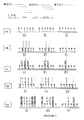
本発明の固相増幅アプローチの実施例は図1および図2に描写されている。
【0040】
図1は、各組が特定のターゲット遺伝子に対して特異的な、3組の固定化5'末端プライマーを有する固相支持体上の、3つのターゲット遺伝子A、BおよびCの増幅および検出を描写する。追加のプライマーは溶液の中に存在しない。1Aにおいて、遺伝子A、BおよびCに対して特異的な固定化プライマーのアレイは閉じた矢印で描写されている。1Bにおいて、一本鎖ターゲットポリヌクレオチドは、アニーリング条件下にそれらの相補的配列において固定化5'末端に対してハイブリダイゼーションする。次いで1Cにおいて、条件を伸長にシフトさせ、その間に5'末端プライマーは鋳型としてターゲットポリヌクレオチドを使用する新生鎖の合成のための開始部位として働く。その中に組込まれた標識、ビオチン−dCTPを有する新生鎖を合成し、こうして新生鎖は後に検出可能である。伸長が完結したとき、条件を変性にシフトさせ、その間に当初のターゲット鋳型鎖はアレイから溶液の中に解放され、標識化新生一本鎖は固定化5'末端プライマーを介して支持体に共有結合される。1Dにおいて、アニーリングの第2ラウンドが起こり、こうしてターゲット鋳型鎖はそれ以上の増幅のための追加の固定化プライマーに対してハイブリダイゼーションする。1Eは固定化プライマー部位において生ずる増幅生成物を示し、あるものは一本鎖のビオチン標識化ポリヌクレオチドであり、そして他のものは二本鎖である。なぜなら、ターゲット鋳型は変性しない新生鎖とアニールしたままであるからである。固定化プライマーはターゲット鋳型の推定濃度よりも非常に高いモル過剰量で存在するで、それは溶液相プライマーが存在しない場合でさえ一本鎖および二本鎖の両方の増幅生成物を存在させる。
【0041】
図2は、固定化特異的5'末端プライマーおよび溶液相3'末端プライマーの両方を使用するターゲット遺伝子(A、BおよびC)の増幅および検出を描写する。3'−プライマーはポリアデニル化mRNA鋳型に対する結合のための万能プライマー、例えば、ポリ−dTであることができる。2A〜2Cは本質的に1A〜1Cに対応し、各固定化5'末端プライマー部位におけるPCRの第2ラウンド、および組込まれたビオチン標識で検出できる生ずる一本鎖新生鎖を図解する。変性後、ターゲット鋳型は新生鎖から分離され、溶液の中に解放される。2Dは第2ラウンドのPCRを描写し、その間に各新生鎖は溶液相万能プライマーから開始された、他の新生鎖合成のための鋳型として働く。その間に、当初のターゲット鋳型はそれ以上の増幅のための追加の固定化5'−プライマー部位に対してハイブリダイゼーションする。固定化プライマー部位上の二本鎖増幅生成物を生じ、各鎖は2Eに示すようにビオチニル化されている。
【0042】
本発明の固相増幅法を使用して、異なる生物学的源における遺伝子発現を検出し、比較する。この面において、増幅反応を実施するために、固定化プライマーを溶液相プライマーと組み合わせて使用する。1つの態様によれば、異なる源は同一被検体の異なる組織または細胞であることができる。選択的に、異なる源は同一種の2以上の被検体の匹敵する組織、例えば、健康な対照からの1つおよび患者からの他のものである。なお他の態様において、異なる源は2以上の異なる種または異なる動物からもの、例えば、ヒトの1つおよびマウスの他のものである。
【0043】
本発明による遺伝子発現の示差的分析は、本質的にプライマーの固相アレイ上で異なる生物学的源からのターゲットポリヌクレオチドを一緒に増幅することによって実行される。この態様において、各源からの初期ターゲットポリヌクレオチドのバッチをオリゴヌクレオチド配列で示差的に標識付けし、こうして目的の増幅生成物はこのような配列タグを有し、初期ターゲットの源を示す。「源タグ」で標識化した増幅生成物検出し、比較することによって、異なる源中のターゲットポリヌクレオチドの存在および相対存在量を決定し、比較することができる。
【0044】
本発明のこの態様において例示するように、異なる生物学的源からのオリジナルターゲットポリヌクレオチドはその中で発現された全mRNAである。この分野において知られている方法および物質を使用して、各源から全mRNAを単離する。次いで各生物学的源から単離されたmRNAの全プールを使用して特異的に標識付けされたcDNAのバッチを調製し、これを引き続く増幅のための「ターゲットポリヌクレオチド鋳型」として使用する。1つの態様において、逆転写された各バッチを特異的配列タグでそれらの3'末端において標識付けする。特異的配列タグは未修飾ターゲットポリヌクレオチドのいずれの中にも存在しない。例えば、検出すべきターゲットポリヌクレオチドがヒト細胞/組織からのものである場合、配列タグは細菌またはウイルスゲノムに由来することができ、そして配列はハイブリダイゼーション/増幅条件下にmRNAに転写されるヒト配列とアニールしない。このようにして、1つのバッチからの配列タグは交差ハイブリダイゼーションのために他のバッチの人工的増幅を引き起こさないであろう。
【0045】
さらに、cDNAターゲットの各バッチの配列タグはcDNAターゲットの他のバッチのそれと異なるので、それらを比較することができる。配列タグは、逆転写のための特別に設計されたプライマーを使用することによって、逆転写cDNAの中に導入することができる。例えば、プライマーは3'末端にポリ−dT部分を有し、そして5'末端に細菌SP6配列を有することができる。逆転写間に、mRNA鋳型のポリ−Aテイルとアニールするポリ−dT部分の5'末端から開始されるcDNA合成のための鋳型として、mRNAは働く。次いで、生ずるcDNA生成物は5'末端にSP6「配列タグ」を有し、これはcDNAのこのバッチに対してユニークである。同様に、他の源からのcDNAの異なるバッチを異なる配列タグ、例えば、細菌T7配列で「標識付け」することができる。
【0046】
例えば、SP6およびT7で示差的に標識付けされた、cDNAの2つの異なるバッチを、増幅および検出のための一緒に混合する。示差的に標識化される遊離SP6およびT7配列タグは、増幅反応混合物の中に存在する。例えば、2つの配列タグを直接的検出のために2つの異なる蛍光色素(例えば、1つの赤色色素および1つの緑色色素)で標識化することができるか、あるいは、引き続く色検出のために2つの異なる化学的成分(例えば、1つのビオチンおよび1つのジゴキシゲニン)で標識化することができる。配列タグが後に増幅反応におけるプライマーとして働くように、標識は配列タグの3'末端に存在しないことが重要である。
【0047】
本発明の好ましい増幅手段はPCR反応である。cDNAの2つの示差的に標識付けされたバッチの混合物を特異的プライマーの複数のグループの固相アレイと接触させ、各グループは前述の特定のターゲットcDNAに対応する。PCRの初期ラウンドにおいて、固定化プライマーは両方の源からのターゲットポリヌクレオチドとアニールし、鎖伸長のために十分な条件下に新生相補的鎖を合成する。新生相補的鎖はターゲット配列領域を通して伸び、ターゲットcDNA鋳型の末端における配列タグに対して相補的な配列を3'末端に含有する。例えば、第1新生鎖は、それを源1からのSP6標識付けcDNA鋳型上で増幅された場合、SP6配列に対して相補的な3'末端を有する;そして第2新生鎖は、それを源2からのT7標識付けcDNA鋳型上で増幅された場合、T7配列に対して相補的な3'末端を有する。こうして、固相アレイ上で固定化された各新生鎖はその源に対して特異的配列タグを「受け継ぐ」。
【0048】
PCRの引き続くラウンドにおいて、異なる源について標識付けされた新生鎖の第1組は新生鎖の第2組の合成のための鋳型として働く。この時、PCRの初期プライマーは溶液中で示差的に標識化された遊離配列タグ、例えば、蛍光標識化SP6プライマーおよびリサミン標識化T7プライマーである。したがって、標識化配列タグから伸長した新生鎖の第2組は、PCR反応の最初のラウンドにおけるターゲットcDNA鋳型の当初の源に対応して、示差的に標識化される。非結合試薬および変性しない当初鋳型を洗浄除去した後、各固定化プライマー部位は当初の生物学的源を示す標識を1つの鎖上に有する二本鎖ポリヌクレオチドを有する。それ自体、異なる標識の最終検出は異なる生物学的源における特定のターゲットポリヌクレオチドの存在および存在量を明らかにするであろう。
【0049】
当業者は理解するように、各生物学的源からの遺伝子発現の正確な量を測定することは不必要であるが、このような正確な測定は可能である。むしろ、本発明は異なる源における遺伝子発現の比較分析のアプローチを提供する。アレイ上の各プライマースポットにおいて、異なる標識の検出を使用して異なる生物学的源からの各ターゲットポリヌクレオチドの比を決定し、引き続いて相対存在量を決定する。
【0050】
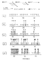
異なる生物学的源の示差的発現分析の1例は図3に図解されている。
【0051】
図3は2つの異なる試料からのターゲット遺伝子の増幅および検出を描写し、各ターゲット遺伝子は試料特異的配列決定タグで標識付けされている。増幅反応は溶液相の、示差的に標識化された配列タグ、例えば、Cy3−配列タグ1およびCy5−配列タグ2の存在下に実施される。3Aにおいて、各遺伝子に対して特異的な5'末端プライマーを固体支持体上でグループで固定化する。3Bにおいて、以前に配列タグで5'末端において標識付けされたターゲット鎖を固定化5'末端プライマーに対してアニールし、PCR増幅の鋳型として働かせる。3Cにおいて、ターゲット鋳型に対して相補的な新生鎖は各固定化プライマー部位から開始され、ターゲット配列を通して配列タグに対して相補的である3'末端に伸びる。それ自体、各新生鎖もまた相補的タグ配列で標識付けされる。増幅の第1ラウンドが完結したとき、当初のターゲット鋳型は変性の間に新生鎖から解放される。次いで、3DはPCRの第2ラウンドを描写し、その間に各固定化新生鎖は鋳型として働き、そして溶液相、Cy3−またはCy5−標識化配列タグは他の新生鎖合成のプライマーとして働く。その結果、対応するターゲット遺伝子が来る試料起源に依存して、新しい鎖の各々はCy3またはCy5標識を支持するであろう。また、3Dにおいて、当初のターゲット鋳型は追加の固定化5'末端プライマー部位に対してアニールして増幅の新しいラウンドを開始する。PCRの第2ラウンドが完結すると、変性が起こらないので、Cy3またはCy5−標識化新生鎖は固定化新生鎖にアニールしたままである。したがって、3Eに示すように、固定化増幅生成物をCy3またはCy5の存在について検出することができ、これらの存在は当初のターゲット遺伝子源を示す。
【0052】
本発明の示差的発現分析は生物学的研究の種々の面ならびに臨床的用途において実用性を有する。例えば、複数の系における問題の特定遺伝子の発現パターンを1つの試料混合物内で比較して、変動を最小にすることができる。それは、例えば、異なる種間のある種の遺伝子の発現レベルの比較を可能とし、こうして問題の遺伝子の機能性の評価を可能とする。また、本発明は、ターゲット遺伝子の発現レベルを健康な個体に対して比較することによって、疾患を有する患者のある種の遺伝子欠陥の臨床的診断において有効である。その上、本発明を使用して、異なる細胞/組織/生物における遺伝子発現レベルを比較して、特定の生物学的経路におけるある種の遺伝子の役割を評価することができる。
【0053】
前述の増幅法は、単一固相支持体上の複数のターゲットポリヌクレオチドの高い処理量のアッセイに使用することができる。単一形態または対の、プライマーの複数のグループを固相支持体上に固定化して、前もって決定したパターンを有するミクロアレイを形成する。プライマーの各グループは特定のターゲットポリヌクレオチドに対応し、ミクロアレイ内の離散位置を占有する。前述したように、複数のターゲットポリヌクレオチドを含有するか、あるいは含有することが推測される試料をPCR反応に適当な条件下にミクロアレイと接触させるとき、各ターゲットポリヌクレオチドは増幅され、離散位置において付着され、ミクロアレイはそれに対して固定化された対応するプライマーを有する。
【0054】
本発明によれば、潜在的ターゲットポリヌクレオチドの数は、小さい密なミクロアレイを製造しかつ分析する入手可能な技術によってのみ制限される。例えば、プライマー対の約100,000までの異なる集団を固体支持体上の離散位置に準備し、支持体をPCR溶液および試料と接触させることによって、既知の技術を使用して、約100,000までのポリヌクレオチドを単一固体支持体上で分析することができ、ここで試料は設計されたプライマーが検出するターゲットポリヌクレオチドの少なくとも1つのコピーを含んでなる。
【0055】
方法および材料
1. ターゲットポリヌクレオチド
本発明の目的に対して、ターゲットポリヌクレオチドは二本鎖DNA、一本鎖DNA、またはRNAであることができる。ターゲットポリヌクレオチドの例は、ゲノムDNA、cDNA、mRNA、ミトコンドリアDNA、ウイルスDNA、増幅されたDNA、およびウイルスDNAを包含するが、これらに限定されない。二本鎖ターゲットポリヌクレオチドは増幅反応の開始において変性を行い、一本鎖鋳型を形成する。
【0056】
mRNAターゲットポリヌクレオチドは、逆転写により仲介される増幅のための鋳型として直接使用することができる。各固定化プライマー部位において発生する鎖伸長が完結した後、ハイブリダイゼーションしたRNA鋳型鎖を、例えば、RNアーゼHにより、破壊して、新生相補的DNA鎖を固相支持体に付着させて残す。第2プライマー(特異的または万能)が溶液相の中に存在する場合、第1新生cDNA鎖は他の新生鎖を合成する鋳型として働き、これにより各固定化プライマー部位において二本鎖新生DNA分子を形成するか、あるいは2つの固定化プライマーを結合する。
【0057】
選択的に、試料中のmRNAターゲットポリヌクレオチドをまず相補的DNAに逆転写し、引き続いてこのDNAは本発明の固相PCR反応のための初期鋳型として働く。逆転写は、例えば、ポリ−dT万能プライマーから開始させることができる。ポリ−dT万能プライマーは、また、本発明によるPCR増幅反応の溶液相中の万能プライマーとして使用することができる。ポリ−dT開始cDNA生成物は固相支持体上に固定化された特異的プライマーにその3'末端でアニールし、ポリ−A配列をその3'末端に有する新生相補的鎖の引き続く合成のための鋳型として働く。変性工程後、単一固定化新生鎖は溶液相中でポリ−dT万能プライマーに対してハイブリダイゼーションすることができ、固相支持体に固定された二本鎖新生ポリヌクレオチドのPCR増幅および形成の引き続くラウンドのための鋳型として働く。
【0058】
本発明の複数のポリヌクレオチドは1つの単一生物学的源からのものであるか、あるいは、複数の生物学的源、例えば、異なる種または組織からのものであることができる。例えば、詳細に前述したように、1つのPCR反応において、既知の検出法により2つの源の増幅された生成物の区別を可能とする条件下に、健康な個体から単離されたターゲットポリヌクレオチドの1集団を、問題の疾患を有する患者から単離されたターゲットポリヌクレオチドの他の目的と混合することができる。したがって、本発明はターゲットポリヌクレオチドの交差種の比較分析に使用することができる。
【0059】
2. オリゴヌクレオチドプライマー
本発明は、オリゴヌクレオチドプライマーの別々の、固定化されたグループを含んでなる調製された固体支持体を提供する。各プライマーは、特定のターゲットポリヌクレオチドの増幅を実施するために適当である。こうして、例えば、標準PCRプライマーの選択プログラム、例えば、プライマー3(Massachusetts Institute of Technology(MIT)からの)を使用して、ターゲットポリヌクレオチド中の既知の配列に基づいて、プライマーを選択または設計することができる。
【0060】
固相支持体は約5〜約100平方マイクロメートルの領域を提供することができ、その上で前もって決定したパターンに従い離散領域において単一プライマーの約100,000までのグループを固定化することができる。調製した固体支持体は、支持体上の任意の所定の位置にプライマーまたはプライマー対の配列の関連する書かれたまたは電子的記録を有することができ、こうして増幅されたターゲットの支持体上の位置をさらに同定することができる。
【0061】
ミクロアレイ上の引き続く計画した増幅反応の必要性により、特定のターゲットヌクレオチドに対応する各グループ内のプライマーの数を決定し、制限することができる。こうして、例えば、特に反応体積およびターゲット鋳型ポリヌクレオチド分子の期待する数、およびPCRの提案されたサイクル数が与えられると、ミクロアレイ上の特異的部位においてPCRを実施するために必要と思われるプライマーの数は、反応を成功させるために支持体上の各位置にどれだけ多くのオリゴヌクレオチドプライマーのコピーをグループとして適用するかを正確に決定する助けとなるであろう。好ましくは、プライマーの量(すなわち、プライマー分子数またはプライマー濃度)は所定の固体支持体上の各準備された位置においてほぼ同一であろう(例えば、約100,000までのターゲットポリヌクレオチドを増幅または検出するためにプライマーの1000〜10,000、約100,000までの集団を有するDNAミクロアレイのフォーマットにおいて)。
【0062】
検出すべきポリヌクレオチドに基づく特定の用途のために、プライマー配列を使用して固体支持体を調製することができる。特に増幅すべきターゲットポリヌクレオチドの配列および品質を考慮して、オリゴヌクレオチドプライマーは特定のPCRに適当な任意の長さを有することができる。1例として、プライマーは約4〜約30ヌクレオチド長さであることができる。
【0063】
本発明の核酸プライマーは、変更が得られる収率または生成物に有意な程度に悪影響を及ぼさない程度に、核酸塩基の小さい欠失、付加および/または置換を含有することができることが理解される。
【0064】
オリゴヌクレオチドプライマーは、通常核酸の中に見出される天然に存在する複素環式塩基(ウラシル、シトシン、チミン、アデニンおよびグアニン)、ならびに修飾された塩基および塩基アナローグを含むことができる。ターゲット配列に対するプライマーのハイブリダイゼーションと適合性の任意の修飾された塩基および塩基アナローグは、本発明において有効である。プライマーの糖またはグリコシド部分は、デオキシリボース、リボース、および/またはこれらの糖の修飾された形態、例えば、2'−O−アルキルリボースを含むことができる。好ましい態様において、糖部分は2'−デオキシリボースである;しかしながら、ターゲット配列に対してハイブリダイゼーションするプライマーの能力と適合性である任意の糖を使用することができる。
【0065】
1つの態様において、プライマーのヌクレオシド単位は、この分野においてよく知られているように、ホスホジエステル主鎖により結合される。追加の態様において、ヌクレオシド間の結合はプライマーの特異的ハイブリダイゼーションと適合性であるこの分野において知られている任意の連鎖を包含することができ、ホスホロチオエート、メチルホスホネート、スルファメート(例えば、米国特許第5,470,967号)およびポリアミド(すなわち、ペプチド核酸)を包含するが、これらに限定されない。ペプチド核酸は下記の文献に記載されている:Nielsen他(1991)「Science」254:1497−1500;米国特許第5,714,331号;およびNielsen(1999)「Cur. Opin. Biotechnol.」10:71−75。
【0066】
ある態様において、プライマーはキメラ分子であることができ、すなわち、2以上の型の塩基または糖サブユニットを含んでなることができ、および/または結合は同一プライマー内の2以上の型であることができる。プライマーは、この分野において知られているように、例えば、インターカレーターおよび/または小さいグルーブバインダー組込むことによって、そのターゲット配列に対するハイブリダイゼーションを促進する部分を含むことができる。
【0067】
塩基、糖、およびヌクレオシド間主鎖、ならびにプライマー上の任意のペンダント基の存在の変動は、配列特異的方式で、ターゲット配列に結合するプライマーの能力と適合性であろう。既知および発生すべき、多数の構造的修飾はこれらの拘束内で可能である。その上、プライマーを形成する種々の複素環式塩基、糖、ヌクレオシドおよびヌクレオチドを製造する方法、および前もって決定した特異的配列のオリゴヌクレオチドの製造は、十分に開発され、この分野において知られている。オリゴヌクレオチド合成の好ましい方法は、米国特許第5,419,966号において教示されている。
【0068】
特定のPCRまたは引き続く操作、例えば、増幅されたターゲットポリヌクレオチドの単離を助けるまたは促進する、任意の特別の追加の成分または配列を使用して、オリゴヌクレオチドプライマーを設計することができる。例えば、プライマーはターゲット配列に対して相補的であるものに加えて配列を含むことができる。このような配列は通常プライマー中のターゲット相補的配列から上流(すなわち、5'側に対して)に存在する。例えば、1以上の制限酵素認識部位を含んでなる配列(いわゆる「リンカー」または「アダプター」)は、プライマーの中のターゲット相補的配列から上流に存在するとき、増幅生成物のクローニングおよび引き続く操作を促進する。プライマーの中に含めるために有用な他の配列は、配列決定プライマーに対して相補的な配列、およびバクテリオファージRNAポリメラーゼ、例えば、T3RNAポリメラーゼ、T7RNAポリメラーゼおよびSP6RNAポリメラーゼのプロモーターを特定する配列を包含する。
【0069】
PCR増幅を実施するために溶液相プライマーをまた使用するとき、溶液相プライマーは万能プライマーであることができる。異なる万能プライマーを比較すべき組織または種の試料について使用することができる。これらの異なるプライマーを示差的に標識化し(例えば、1つについて緑色、他のについて赤色)、こうして検出の間に、1つの種または組織の試料からのターゲットを他の種または組織の試料からの対応するターゲットと区別することができる。
【0070】
野生型対応物に比較して特異的なヌクレオチド突然変異を含有する突然変異体ポリヌクレオチドを検出するように、本発明のオリゴヌクレオチドプライマーを設計することができる。野生型ポリヌクレオチドであるが、少なくとも3'末端のヌクレオチドが異なる配列に基づいて、プライマーを設計することができる。それ自体、これらの3'末端置換プライマーは野生型ターゲットポリヌクレオチドに結合し、PCR増幅することができない。その代わりに、それらはヌクレオチド置換に合致する配列突然変異を有する突然変異体ポリヌクレオチドを認識し、増幅することができる。問題の領域に広がる3'末端置換プライマーのアレイを使用することによって、その領域内の突然変異を検出することができる。
【0071】
3. 固相支持体
本発明の固相支持体は、ヌクレオチドのハイブリダイゼーションおよび合成を支持するために適当な、任意の固体の材料および構造を有することができる。好ましくは、固相支持体は少なくとも1つの実質的に剛性の表面を含んでなり、その表面上にプライマーを固定化し、PCR反応を実行することができる。固相支持体は、例えば、ガラス、合成ポリマー、プラスチック、硬質非メッシュ状ナイロンまたはセラミックから作ることができる。他の適当な固体支持体材料は既知であり、当業者に容易に入手可能である。固体支持体のサイズはDNAミクロアレイ技術に有効な、任意の標準ミクロアレイのサイズであり、そしてサイズは本発明の反応を実施するために使用する特定の装置に適合するように調整することができる。オリゴヌクレオチドを固定化する目的の固相支持体の誘導化するための方法および材料はこの分野において知られており、そして、例えば、米国特許第5,919,523号に記載されており、その開示は引用することによって本明細書の一部とされる。
【0072】
固体支持体を流体を含有する容器の中に準備するか、あるいはその一部分であることができる。例えば、固体支持体をチャンバーの中に入れることができる。このチャンバーは固体支持体のへりに沿ってシールをつくった側面を有し、支持体上にポリメラーゼ連鎖反応(PCR)を含有する。特定の例において、チャンバーは長方形の支持体の各側に壁を有して、PCR混合物が支持体上に止まり、かつまたプライマーを提供するために有効な全表面をつくることができるようにする。
【0073】
4. プライマーの固定化
本発明のオリゴヌクレオチドプライマーは、固体支持体上の特定の位置にオリゴヌクレオチドを固定し、固定化し、準備し、および/または適用するための任意の入手可能な手段を使用して、固体支持体の表面に付着させ、固定化し、準備し、および/または適用することができる。例えば、下記の特許に記載されているように、写真平版(Affymetrix、カリフォルニア州サンタクララ)を使用して、チップまたは固体支持体上の特定の位置にオリゴヌクレオチドプライマーを適用することができる:米国特許第5,919,523号、第5,837,832号、第5,831,070号および第5,770,722号に記載されており、それらの開示は引用することによって本明細書の一部とされる。また、オリゴヌクレオチドプライマーは、米国特許第5,807,522号(1998)に記載されているように、固体支持体に適用することができる。さらに、ロボット・システム、例えば、ジェネティック・ミクロ・システムス(Genetic MicroSystems、マサチュセッツ州ウォバーン)、ジーンマシンズ(GeneMachines、カリフォルニア州サンカルロス)またはカーテシアン・テクノロジー(Cartesian Technologies、カリフォルニア州アービン)により製作されているシステムを使用して、プライマーを固体支持体に適用することができる。
【0074】
5. PCR反応
本発明を実施するとき、ポリメラーゼ連鎖反応(PCR)を実施するために必要な試薬と混合された適当なターゲットポリヌクレオチドを含んでなる反応混合物を、固体支持体上の各固定化されたプライマー対または単一プライマーの集団と接触させて配置する。適当なターゲットポリヌクレオチドは、二本鎖DNA、RNA鋳型の逆転写により発生した一本鎖cDNA、またはmRNA集団であることができる。反応混合物はターゲット鎖に対して相補的なポリヌクレオチドの合成を促進する酵素を含有する。適当なポリメラーゼは、熱安定性ポリメラーゼ酵素、例えば、Taq DNAポリメラーゼ、TthI DNAポリメラーゼ、Tne DNAポリメラーゼ、Tma DNAポリメラーゼ、Pfu DNAポリメラーゼ、Vent DNAポリメラーゼまたは任意の他の熱安定性DNAポリメラーゼを包含する。また、反応混合物はポリメラーゼ連鎖反応間に新生鎖の中に組込むことができる標識分子を含有し、こうして増幅された生成物をPCR後に固体支持体で検出できるようにすることができる。標識はこの分野においてよく知られている方法に従い直接的または間接的に検出することができる。直接的検出に適当な標識は、任意の蛍光分子、例えば、フルオレセインイソチオシアネート、テキサス・レッドまたはローダミンであることができる。また、間接的検出を促進する分子、例えば、ビオチンまたはジゴキシゲニンをPCR間に新生鎖の中に組込むことができる。ビオチンを引き続いて標識化ストレプトアビジンまたは標識化抗−ビオチン抗体に結合させることによって検出することができる。同様に、組込まれたジゴキシゲニンは標識化または非標識化抗−ジゴキシゲニン抗体により検出することができ、そして非標識化抗−ジゴキシゲニン抗体は標識化抗−抗−ジゴキシゲニン抗体を結合させることによって検出することができる。
【0075】
PCRを実施する試薬がミクロアレイ上の固定化されたプライマーと接触した後、例えば、自動化システム、例えば、in situ PCR装置を使用して、PCRの発生を促進する条件下にミクロアレイを配置する。PCR手順のための反応条件はin situ PCR装置のマニュアルにより推奨される通りであることができ、使用する鋳型の特質またはプライマーおよび鋳型のハイブリダイゼーションを使用するとき予測される困難が分かったとき、適当に変化させることができる。温度およびサイクル数は、推奨されるように、また、プライマーの選択および鋳型配列、および他の関係する因子が分かったとき適当に、選択することができる。ミクロアレイ上のin situ型PCR反応は、本質的に下記の文献に記載されているように、実施することができる:Embretson他、「Nature」362:359−362(1993);Gosden他、「Bio Techniques」15(1):78−80(1993);Heniford他、「Nucl. Acid Res.」21(14):3159−3166(1993);Long他、「Histochemistry」99:151−162(1993);Nuvo他、「PCR Methods and Applications」2(4):305−312(1993);Patterson他、「Science」260:976−979(1993)。
【0076】
6. 標識化および検出
本発明のPCR法は、試料中の複数のターゲットポリヌクレオチドの検出を提供する。適当な標識化用試薬の存在下にPCRが完結した後、増幅され、標識化されたターゲットポリヌクレオチドをミクロアレイ上の当初のプライマー位置の各々において検出することができる。増幅され、標識化されたターゲットポリヌクレオチドの検出は、標識化配列を検出するために使用し、例えば、増幅されたまたは新しく合成されたDNA鎖の中に組込まれた標識の検出を包含する、標準的方法により実施することができる。こうして、例えば、蛍光標識または放射能標識を直接的検出することができる。他の標識化技術は、鎖合成間にDNAの中に組込まれた標識、例えば、ビオチンまたはジゴキシゲニンを標識化された、または標識化された分子それ自体に結合することができる抗体または他の結合性分子(例えば、ストレプトアビジン)により検出することを必要とし、例えば、標識化分子は、例えば、蛍光分子(例えば、フルオレセインイソチオシアネート、テキサス・レッドおよびローダミン)に結合させた、または酵素的に活性化が可能な分子に結合した抗ストレプトアビジン抗体または抗ジゴキシゲニン抗体であることができる。新しく合成された分子上の標識がなんであっても、かつ標識が直接的にDNAの中に存在するか、またはDNAに結合する(またはDNAに結合する分子に結合する)分子に結合されているかどうかにかかわらず、標識(例えば、蛍光、酵素的、化学発光、または比色)は、標識、または特定の標識を検出する他の適当な手段に依存して、レーザースキャナーまたはCCDカメラ、またはX線フィルムにより検出することができる。
【0077】
ターゲットポリヌクレオチドは、PCRの間に増幅されたDNAの中に組込まれる標識化ヌクレオチド(例えば、直接的標識化のためにdNTP−蛍光標識;間接的標識化のためにdNTP−ビオチンまたはdNTP−ジゴキシゲニン)を使用することによって検出することができる。間接的に標識化されたDNAについて、検出は蛍光または酵素結合ストレプトアビジンまたは抗ジゴキシゲニン抗体により実施される。PCR法において、ポリヌクレオチドターゲットに対する新しく合成された補体の中に組込まれた標識を検出することによって、ポリヌクレオチドを検出する。この目的のために、前述したように、DNAが合成されているときDNAの中に組込むことができる任意の標識、例えば、フルオロ−dNTP、ビオチン−dNTP、またはジゴキシゲニン−ジゴキシゲニンを使用することができ、そしてそれらの標識はこの分野において知られている。溶液中で1以上の万能プライマーを使用して実施されるPCR増幅は、万能プライマーを検出することによって、固体支持体上の位置において増幅されたターゲットを検出するオプションを提供する。こうして、2以上の万能プライマーを使用する場合、異なる源からのターゲット鎖を固体支持体上で示差的に検出することができる。
【0078】
示差的発現系において、異なる生物学的源に由来する増幅生成物は、節「C. 異なる生物学的源からの示差的発現を比較する」に記載されているように、それらの由来に基づいて増幅された鎖を示差的に標識化することによって検出することができる。1つの面において、本発明において使用する検出法は単一源ターゲットについての検出法と異なり、示差的標識(例えば、赤色色素および緑色色素)を、増幅の間に新生鎖の中に組込むよりは、むしろ、溶液中のプライマータグ上に前もって組込む。あるいは、示差的標識に加えて、第3標識をまた新生鎖の中に組込むことができ、こうして示差的発現の比較に対する全体の感度は増強される。
【0079】
7. 検出キット
本発明は、本発明の方法を実施するキットを提供する。キットは、例えば、そうでなければ固体支持体上の検出が困難である、複数のターゲットポリヌクレオチドを検出する物質および試薬を含むことができる。キットは、例えば、固体支持体、ターゲットポリヌクレオチドの特定の組のためのターゲットポリヌクレオチド、ポリメラーゼ連鎖反応の試薬および成分、例えば、DNA合成のための酵素、標識化物質、および他の緩衝剤および洗浄のための試薬を含むことができる。また、キットは固体支持体上で特定のターゲットを増幅するためにキットを使用するための使用説明書を含むことができる。キットは固体支持体上に既に固定されたプライマーの組、例えば、ターゲットポリヌクレオチドの特定の組を増幅のためのプライマーの組を有する調製された固体支持体を含有する場合、このような調製された固体支持体の設計および構築は前述した通りである。このような固体支持体は、検出しようとするターゲットポリヌクレオチドに依存して個々のキットのためにあつらえて作ったものであることができる。また、固体支持体がin situ型のPCR装置を使用してPCR増幅することができる場合、例えば、in situ型または固相型のPCR手順を使用して、固体支持体上でPCRを実施するために必要な試薬をキットは含むことができる。支持体をPCRの試薬と接触させることができる。反応前に、潜在的に複数のターゲットポリヌクレオチドを含有する試料をPCR反応混合物に添加する。PCR試薬は、通常のPCR緩衝剤、熱安定性ポリメラーゼ(例えば、Taq DNAポリメラーゼ)、ヌクレオチド(例えば、dNTP)、および他の成分および標識化用分子(例えば、前述したように直接的または間接的標識化のための)を包含する。固体支持体は、固体支持体上に表示した位置に付着されたプライマーを提供する。PCRを実施するために、固定化プライマーを有する支持体を、PCRを実施のための試薬および反応混合物中のターゲットポリヌクレオチド鋳型と接触させ、PCR(例えば、in situ型または固相型のPCR)条件に暴露する。キットに使用するための使用説明書は、例えば、上記方法の説明において示したような手順についての詳細な説明を含むことができる。固定化プライマーを単独で、または、代替的に、溶液相プライマーと一緒に使用するPCRの増幅の実行を支持するように、キットを組立てることができる。
【0080】
実施例
実施例1 ミクロアレイ上の4つのDNA鋳型の増幅
4つのcDNAターゲットポリヌクレオチド、ヒトG3PDH、PKC−α、c−Raf、およびサイクリンAをミクロアレイ上の増幅により検出のために選択した。この増幅は、プライマー対の両方のメンバーが支持体上に固定化されている対称PCR、またはプライマー対の一方のメンバーが固定化されており、そして他のメンバーが溶液の中に存在する、非対称PCRにより実施される。
【0081】
a. G3PDHの対称増幅
これらのターゲットポリヌクレオチドの仮定または推定した配列に基づいて、プライマー対の集団を4つのターゲットの各々について設計し、合成する。固体支持体に対するプライマーの付着を促進するためにアミンの5'末端の修飾を使用して、プライマーを合成した。シラン化ガラススライド(Sigma Chemicals、ミゾリー州セントルイス、から購入した)上に、異なる濃度で、プライマーを対としてまたは単一プライマーとしてスポットした。
【0082】
準備されたプライマーを有するシラン化スライドを、室温において飽和NaClチャンバー中で一夜水和した。水和されたスライドを4×SSCで5分間リンスし、次いで水で洗浄した。スライドをSurModics(ウイスコンシン州マディソン)ブロッキング溶液(0.1%SDSを含む)で50℃において15分間ブロックし、次いで水で2回洗浄し、次いで空気乾燥した。そこでスライドは使用できる状態にある。
【0083】
PCR溶液を下記の最終濃度に調製した:200μMの各dATP、dGTP、およびdTTP;100μMのdCTPおよび100μMのビオチン−14−dCTP。また、反応溶液は1×Taq反応緩衝液(1.5mMのMgCl2を含む);DNA鋳型としてヒトG3PDH遺伝子プラスミド(100ngのファージミドDNAまたは500ngの一本鎖cDNAライブラリー)および2.5単位のTaq酵素を含有する。次のような70μlの反応溶液が発生した:
7μl 2mMのd3TP(dATP、dGTP、およびdTTP)
12.5μl 0.4mMのdCTP
12.5μl 0.4mMのビオチン−14−dCTP
7μl 10×反応緩衝液(w/15mMのMgCl2)
5μl DNA鋳型
25.5μl 水
0.5μl 5単位/μlのTaq DNAポリメラーゼ
70μlの総体積について。
【0084】
HyBaidチャンバー(マサチュセッツ州フランクリン)をスライド上に配列された位置を中心にして配置し、反応溶液をチャンバーに移し、プラスチックカバーでシールした。PCR装置を予備加温し、下記のサイクリングプロトコルを適用した:

PCRが完結した後、スライドをジゴキシゲニンブロッキング溶液(Boehringer Mannheim、インジアナ州インジアナポリス)で室温において30分間ブロックした。スライドをストレプトアビジン(5μg/ml)(ジゴキシゲニンブロッキング溶液中で1:250に希釈した)で室温において30分間おだやかに震盪しながら染色した。ジゴキシゲニン洗浄緩衝液を使用して、スライドを室温において15分間、2回洗浄した。スライドをジゴキシゲニンブロッキング溶液で室温において30分間ブロックした。
【0086】
スライドをジゴキシゲニンブロッキング溶液中で1:100に希釈した第1抗体(ウサギ抗ストレプトアビジン)と室温において1時間インキュベートした。スライドをジゴキシゲニン洗浄緩衝液で室温において15分間、2回洗浄した。スライドをジゴキシゲニンブロッキング溶液中で1:100に希釈した第2抗体(Cy3結合ヤギ抗ウサギ抗体)と室温において30分間インキュベートした。スライドをジゴキシゲニン洗浄緩衝液で室温において15分間、2回洗浄した。スライドをジェネティック・ミクロシステム(Genetic MicroSystem)、GMS418からの緑色ビームで走査した。
【0087】
対称PCRの結果を図4Aに示す。照明されたスポットは、ヒトG3PDHプライマーが固定化されているスポットにおけるhu G3PDH鋳型の増幅の成功を示す。対照的に、他の非ヒトG3PDHプライマーが固定化されているスポットにおいて、ターゲットヒトG3PDH鋳型の検出は見られない。
【0088】
b. hu G3PDHの非対称増幅
4つのターゲットcDNA、hu G3PDH、PKC−α、c−Raf、およびサイクリンAの各々についてプライマーの組を使用して、非対称PCR反応を実行した。装置上の各スポットは、図4Bに示めされたように定められた濃度で、ターゲットcDNAに対して特異的なプライマーの組を含有する。固定化プライマーに加えて、hu G3PDHアンチセンスプライマーをまた25pmolで反応溶液に添加した。鋳型として1pgのhu G3PDH cDNAを使用するPCR増幅を前述した条件下に実施した。
【0089】
hu G3PDH cDNAの非対称PCRの結果を図4Bに示す。欄1〜6のスポットしたプライマー濃度はそれぞれ3.125、6.25、12.5、25、50および100pmol/μlである。照明されたスポットは、hu G3PDHプライマーを含有するスポットにおいてhu G3PDHの増幅生成物のみを検出することができることを示し、アッセイの強い特異性を示唆する。さらに、非対称PCRの結果の解像能は図2Aに示す対称PCRの結果よりも高いように見え、非対称方法を使用するときのよりすぐれた感度を示唆する。
【0090】
c. 1つのミクロアレイ上の4つのターゲットcDNAの非対称増幅
1つの単一ミクロアレイ上で複数のターゲットポリヌクレオチドを検出するために、本発明の非対称方法をさらに試験した。
【0091】
ミクロアレイおよびその上のプライマーは1bにおけるプライマーと同一であり、各スポットはターゲットcDNAに対して特異的な単一プライマーの組を含有し、決定された濃度を有する。反応溶液は、各々25pMolで、4つのターゲットcDNAの各々のためのアンチセンス鎖プライマーを含有した。また、反応溶液はPCR増幅のための鋳型として4つのcDNAの各々を含有した。PCR反応を実施例1aに記載されているように実施した。
【0092】
図4Cに示す結果は、プライマーの最低濃度においてさえ、すべてのターゲット鋳型が首尾よく増幅されたことを示す。
【0093】
実施例2 RNAターゲットの固相増幅
この実施例において、生物学的源中のmRNA発現の検出について、非対称PCR法を試験した。
【0094】
5μgのmRNAをOVC1.1細胞から単離し、一本鎖cDNAの逆転写に使用した。SuperScript IIキット(Life Technology Inc.)を使用して逆転写反応を実施した。反応溶液中で25pMolの万能プライマー(Uni−1)を使用する非対称PCRを実行するための鋳型として、0.5μgの調製したcDNAを使用し、そして4つのターゲット遺伝子、hu G3PDH、PKC−α、サイクリンAおよびc−Rafに対して特異的プライマーを種々の濃度でミクロアレイの異なるスポット上に固定化した。上記実施例1(a)に記載されているような条件および手順に従い、PCR反応を実施した。
【0095】
結果を表5に示す。4つのターゲット遺伝子の発現パターンは異なる発現量を示し、G3PDHは最大量であり、そしてc−Rafは最小量である。
【0096】
実施例3 ハイブリダイゼーション前の固相増幅によるシグナルの増強
この実施例において、ハイブリダイゼーション前にPCRによる固相増幅を実施したとき発生したシグナルと、ハイブリダイゼーション単独により発生したシグナルを比較した。図6に図解されているように、ミクロアレイ上のハイブリダイゼーション前に固相PCRを実行しかつ実行しないで、卵巣癌腫細胞系統OVC1.1および胎児の脳の遺伝子発現分析からのシグナルを比較した。
【0097】
図6A〜図6Bは、標準方法に従う酵素的標識化により全mRNAから調製した蛍光プローブとハイブリダイゼーションした、OVC1.1細胞からのアレイ因子を含有するDNAミクロアレイを図解する。(下記の文献を参照のこと:Schena M.、およびDavis R.W.(1998)、「genes,Genomes and Chips. in DNA Microarray:A Practical Approach」(編者M. Schena)、Oxford University Press、英国オックスフォード)。図6Aは標準プロトコル後のハイブリダイゼーションシグナルを図解するが、図6Bは上記実施例1(a)に記載されている条件および手順に従い実施した固相PCR反応後のハイブリダイゼーションの結果を図解する。
【0098】
同一ラインに沿った他の実験の結果を図6C〜図6Dに示す。図6Cは胎児の脳のcDNAターゲットを含んでなるミクロアレイに対する蛍光標識化細胞のmRNAプローブのハイブリダイゼーション後のシグナルを図解するが、図6Dは本発明による固相PCR反応後のハイブリダイゼーションの結果を示す。
【0099】
実施例4 固相増幅後のハイブリダイゼーションシグナルの特異性
下記の実施例において、固相増幅後のハイブリダイゼーション反応の特異性を試験した。4つのcDNAターゲットポリヌクレオチド、ヒトG3PDH、PKC−α、c−Raf、およびサイクリンAをミクロアレイ上の増幅による特異的検出について選択した。
【0100】
a. G3PDHの特異的増幅
アミン被覆ガラスチップを、ヒト遺伝子G3PDH、PKC−α、c−Raf、およびサイクリンAからの4つの異なるセンス鎖プライマーでスポットした。pG3PDHからの1pg(約4×10⁵コピー)の一本鎖ファージミドDNAを25pMolのG3PDHアンチセンスプライマーとともに溶液中で使用するDNAポリメラーゼ連鎖反応により、ヒトG3PDHを特異的に増幅し、PCR後検出した。
【0101】
5μgのmRNAをOVC1.1細胞から単離し、一本鎖cDNAの逆転写に使用した。SuperScript IIキット(Life Technology Inc.)を使用して逆転写反応を実施した。0.5μgの調製したcDNAを鋳型として使用して、反応溶液中で25pMolの万能プライマー(Uni−1)を用いる非対称PCRを実施し、そして4つのターゲット遺伝子、hu G3PDH、PKC−α、サイクリンAおよびc−Rafに対して特異的なプライマーを種々の濃度でミクロアレイの異なるスポット上に固定化した。PCR反応を上記実施例1(a)に記載されている条件および手順に従い実施した。
【0102】
結果を図7に示す。欄1〜6のスポッティングプライマー濃度はそれぞれ3.125、6.25、12.5、25、50および100pmol/μlである。照明されたスポットは、hu G3PDHプライマーを含有するスポットにおいてhu G3PDHの増幅生成物のみを検出することができることを示し、アッセイの強い特異性を示唆する。
【0103】
b. G3PDHおよびc−Rafの特異的増幅
アミン被覆ガラスチップを、ヒト遺伝子G3PDH、PKC−α、c−Raf、およびサイクリンAからの4つの異なるセンス鎖プライマーでスポットした。100ngのc−Raf DNA、およびpG3PDHからの1pg(約4×10⁵コピー)の一本鎖ファージミドDNAを25pMolのG3PDHアンチセンスプライマーとともに、しかしc−Rafアンチセンスプライマーの不存在下に、溶液中で使用するDNAポリメラーゼ連鎖反応により、ヒトG3PDHを特異的に増幅し、PCR後検出した。
【0104】
4つのターゲット遺伝子、hu G3PDH、PKC−α、サイクリンAおよびc−Rafに対して特異的プライマーを種々の濃度においてミクロアレイの異なるスポット上に固定化した。PCR反応を上記実施例1(a)に記載されている条件および手順に従い実施した。結果を図8に示す。欄1〜6のスポッティングプライマー濃度はそれぞれ3.125、6.25、12.5、25、50および100pmol/μlである。照明されたスポットは、hu G3PDHプライマーを含有するスポットにおいてhu G3PDHの増幅生成物を検出することができることを示し、シグナルはc−Rafプライマーを含有するするスポットにおいて検出されたc−Rafよりも強く、アッセイの強い特異性ならびにhu G3PDHに対する非対称PCRの感度の増強を示唆する。
【0105】
c. G3PDH、PKC−α、c−Raf、およびサイクリンAの特異的増幅
アミン被覆ガラスチップを、ヒト遺伝子G3PDH、PKC−α、c−Raf、およびサイクリンAからの4つの異なるセンス鎖プライマーでスポットした。鋳型として4つの遺伝子集団および25pMolのG3PDHアンチセンスプライマーを溶液中で使用するDNAポリメラーゼ連鎖反応により、すべての4つの遺伝子を特異的に増幅し、PCR後検出した。
【0106】
結果を図9に示す。欄1〜6のスポッティングプライマー濃度はそれぞれ3.125、6.25、12.5、25、50および100pmol/μlである。照明されたスポットは、すべての4つの増幅生成物を検出できることを示す。
【0107】
d. 胎児の脳のcDNAライブラリー遺伝子発現分析に対する特異的増幅についての試験
ミクロアレイチップ上のDNA PCRの鋳型として、ヒトの胎児の脳のcDNAライブラリー(Stratagene)を使用した。4つのターゲット遺伝子、hu G3PDH、PKC−α、サイクリンAおよびc−Rafに対して特異的なプライマーを種々の濃度でスポットしたミクロアレイ上で、0.05μgの調製したcDNAを鋳型として使用して、反応溶液中で25pMolの万能プライマー(Uni−1)を用いるPCR増幅を実施した。PCR反応を上記実施例1(a)に記載されている条件および手順に従い実施した。
【0108】
結果を図10に示す。欄1〜6のスポッティングプライマー濃度はそれぞれ3.125、6.25、12.5、25、50および100pmol/μlである。データは同一DNA試料を使用するHO1 ExpressChip(商標)(Mergen,Ltd.、カリフォルニア州サンレアンドロ)上のハイブリダイゼーションの結果と合致した。
【0109】
e. OVC1.1細胞遺伝子発現分析に対する特異的増幅についての試験
アミン被覆ガラスチップを、ヒト遺伝子G3PDH、PKC−α、c−Raf、およびサイクリンAからの4つの異なるセンス鎖プライマーでスポットした。pG3PDHからの1pg(約4×10⁵コピー)の一本鎖ファージミドDNAを25pMolのG3PDHアンチセンスプライマーとともに溶液中で使用するDNAポリメラーゼ連鎖反応により、ヒトG3PDHを特異的に増幅し、PCR後検出した。
【0110】
5μgのmRNAをOVC1.1細胞から単離し、SuperScript IIキット(Life Technology Inc.)を使用して一本鎖cDNAに逆転写した。0.05μgのcDNAを鋳型として使用して、反応溶液中で25pMolの万能プライマー(Uni−1)を用いる非対称PCRを実施し、そしてターゲット遺伝子、hu G3PDH、PKC−α、およびサイクリンAのためのセンス鎖オリゴヌクレオチドプライマーを種々の濃度でミクロアレイの異なるスポット上に固定化した。
【0111】
結果を図11に示す。欄1〜6のスポッティングプライマー濃度はそれぞれ3.125、6.25、12.5、25、50および100pmol/μlである。照明されたスポットは、固相増幅による相対的シグナルの非常にわずかの歪みを示す。
【0112】
この明細書の中に引用したすべての刊行物および特許出願は、個々の刊行物または特許出願が特別にかつ個々に引用することによって本明細書の一部とされると示される場合、引用することによって本明細書の一部とされる。
【0113】
本発明を理解を明瞭にする目的で例示および実施例により多少詳細に記載したが、当業者にとって容易に明らかなように、添付の特許請求の範囲の精神または範囲から逸脱しないで本発明の教示を考慮すればある種の変化および変更が可能である。
【図面の簡単な説明】
【図1】 図1(1A〜1E)は、固定化されたプライマーを使用して、ターゲットポリヌクレオチド(A、B、C)を増幅し、検出する固相増幅方法を概略的に描写する。
【図2】 図2(2A〜2E)は、固定化されたプライマーを溶液相万能プライマーと組み合わせて使用して、ターゲットポリヌクレオチド(A、B、C)を増幅し、検出する固相増幅方法を概略的に描写する。
【図3】 図3(3A〜3E)は、固定化されたプライマーを溶液相プライマーとして働く示差的に標識化された配列タグと組み合わせて使用して、2つの異なる生物学的源からのターゲットポリヌクレオチド(A、B、C)を増幅し、検出し、比較する固相増幅方法を概略的に描写する。
【図4】 図4(4A〜4C)は、鋳型として4つのターゲットcDNAを使用する固相増幅実験の結果を示す。
【図5】 図5は、ターゲットmRNAの固相増幅の結果を示す。
【図6】 図6は、ハイブリダイゼーション前の固相PCR増幅によるハイブリダイゼーションシグナルの増強を示す。図6Aは標準プロトコル後のハイブリダイゼーションシグナルを図解するが、図6Bは固相PCR反応後のハイブリダイゼーションシグナルを図解する。図6Cは胎児の脳のcDNAターゲットを含んでなるミクロアレイに対するハイブリダイゼーション後のシグナルを図解するが、図6Dは固相PCR反応後のハイブリダイゼーションシグナルの結果を図解する。
【図7】 図7は、鋳型としてG3PDHターゲットcDNAを使用する固相増幅後のハイブリダイゼーションの特異性を図解する。
【図8】 図8は、鋳型としてG3PDHおよびc−RafターゲットcDNAを使用する固相増幅後のハイブリダイゼーションの特異性を図解する。
【図9】 図9は、鋳型として4つの異なるターゲットcDNAを使用する固相増幅後のハイブリダイゼーションの特異性を図解する。
【図10】 図10は、鋳型として4つの異なるターゲットcDNAを使用する胎児の脳のライブラリーからのターゲットmRNAの固相増幅後のハイブリダイゼーションの特異性を図解する。
【図11】 図11は、鋳型として3つの異なるターゲットcDNAを使用するOVC1.1からのターゲットmRNAの固相増幅後のハイブリダイゼーションの特異性を図解する。[0001]
Cross references to related applications
This application claims the priority of provisional application 60 / 173,618, filed December 29, 1999.
[0002]
TECHNICAL FIELD OF THE INVENTION
The present invention relates generally to the field of nucleic acid biology. More particularly, the present invention provides methods and compositions for high throughput amplification, detection and comparison of gene expression in biological samples for use in diagnosis and therapy.
[0003]
background
The detection of small amounts of genetic material is a major challenge in biological research and clinical diagnosis. Polymerase chain reaction (PCR) provides a powerful tool for in vitro amplification of specific polynucleotide sequences such as genomic DNA, single-stranded cDNA or mRNA with high sensitivity and specificity. This one application is, for example, amplifying target gene sequences in biological samples from the environment, food and medical sources, and others, causing pathogenicity, spoilage or It is possible to identify indicator organisms.
[0004]
Basic PCR techniques are typically as described in US Pat. Nos. 4,683,202, 4,683,195, and 4,800,159, the disclosures of which are hereby incorporated by reference. Contains two oligonucleotide primers that can hybridize to specific nucleic acid sequences that flank the target sequence of interest. By repeating multiple cycles of template denaturation, primer annealing and chain extension, the target sequence can be replicated exponentially.
[0005]
A major problem when using standard PCR methods is contamination. Although PCR provides a sensitive way to detect and amplify small amounts of target polynucleotide, PCR can amplify non-specific nucleic acid sequences, which can lead to false positive products in the final detection and assay. to make. In standard solution phase PCR, the primer binds to the template and initiates nascent strand synthesis in solution, and the reaction mixture and product often require several transfers for final detection and assay, creating an opportunity for contamination. increase.
[0006]
Kohsaka and Carson (1994) “J. Clin. Lab. Anal.” 8: 452-455 describe a solid-phase PCR approach that allows amplification and detection of target gene sequences in the same microwell without migration. ing. One of the two oligonucleotide primers binds to the well of the microtiter plate and the other primer remains in solution. The immobilized primer binds to the template and initiates extension of the nascent complementary strand. The newly synthesized strand remains attached to the plate after removal of the template by denaturation and can be detected with a labeled probe upon completion of PCR. A solid phase PCR approach can also be used for quantification of the target nucleic acid by adding a known amount of an internal competitive DNA template prior to amplification. See, for example, US Pat. No. 5,747,251, the disclosure of which is incorporated herein by reference.
[0007]
Kohsaka et al. Solid phase PCR was limited to detecting one single target polynucleotide in a well on a 96 well microtiter plate. Quantitative solid phase PCR using competing templates has been limited to detecting targets from one species or tissue. Furthermore, in order to detect by hybridization with a labeled probe, the amplified product bound to the plate must be single stranded, thereby limiting the detection sensitivity.
[0008]
US Pat. No. 5,641,658 (Adams et al.) Describes a method for amplifying nucleic acids with two primers attached to a single solid support. This method requires the selection of two primers that flank the target sequence and the immobilization of both primers on a solid support. The primer pair is used to detect and amplify the target polynucleotide on the support. When the amplified product is immobilized on a support and two adjacent strands are reasonably separated from each other, they can be further hybridized together to form a “loop”. The two primer amplification system is promised to be sensitive in the presence or absence of a specific target nucleic acid in the sample, but the use of two immobilized primers for each target is a primer on the support The primer array will allow loop formation and will not interfere with amplification of additional strands. In other words, these methods may not be ideal for high density, high throughput assays.
[0009]
The expression pattern of a gene in a particular biological sample provides an insight into the molecular basis of almost all biological functions and activities. Numerous methods for detecting and comparing gene expression levels in different biological sources are known in the art. One standard method of such comparison is Northern blot. In this technique, RNA is extracted from a sample, loaded onto any of a variety of gels suitable for RNA analysis, and then developed according to standard methods to separate RNA by size (eg, See: Sambrook J. et al., “Molecular Cloning: A Laboratory Manual”, Cold Spring Harbor Laboratory Press, Cold Spring Harbor, NY (2nd edition, 1989)). The gel is then blotted (as described in Sambrook, supra) and hybridized to a probe for the RNA in question.
[0010]
Sutcliffe US Pat. No. 5,807,680 describes a method for simultaneously identifying differentially expressed mRNAs and measuring their relative concentrations. This technique involves PCR following the formation of cDNA using anchor primers, allowing almost all of the mRNA expressed by the tissue to be visualized as discrete bands on the gel, with the intensity of the bands roughly corresponding to the mRNA concentration. To do.
[0011]
Another group of technologies uses analysis of relative transcriptional expression levels. Four such approaches have been developed to enable comprehensive, high throughput analysis. First, cDNA can be reverse transcribed from RNA in the sample (as described in the above references), single-pass sequencing of the 5 ′ and 3 ′ ends, and test and control samples The expressed sequence tag of the gene expressed in can be revealed. By counting the relative expression level of the tag from different samples, the relative expression level of the gene transcript in the sample can be estimated.
[0012]
Second, systematic analysis of gene expression, or variation on EST, known as “SAGE”, has been developed, which allows quantitative simultaneous analysis of multiple transcripts. This technique isolates and sequences short diagnostic sequence tags to reveal gene expression patterns that characterize the target function, and compares, for example, the expression levels of thousands of genes in normal and tumor cells Has been used. See, for example, the following references: Velculescu et al., “Science” 270: 368-369 (1995); Zhang et al., “Science” 276: 1268-272 (1997).
[0013]
Third, an approach based on differential presentation has been developed. In these approaches, when combined with information about the length of the fragment within the expressed gene, the fragment defined by the specific sequence delimiter can be used as a unique identifier for the gene. The phase expression level of the expressed gene in the cell can then be estimated by the relative expression level of the fragment associated with that gene. Some examples of several approaches that have been developed to take advantage of this idea include restriction enzyme analysis of differentially expressed sequences used by Gene Logic, Inc. (" READS "), and Total Gene Expression Analysis (" TOGA ") used by Digital Gene Technologies, Inc. (Palo Alto, CA), eg, differentially expressed by PCR Delta ™ Differential Display Kits are available for identifying the genes identified.
[0014]
Fourth, in a preferred embodiment, detection is performed by one of a number of techniques for hybridization analysis. In these approaches, RNA from the sample in question is usually reverse transcribed to form labeled cDNA. The cDNA is then hybridized to a known sequence of oligonucleotides or cDNA, typically placed on a known sequence of chips or other surfaces. The location of the oligonucleotide to which the labeled cDNA hybridizes provides information for the cDNA, while the amount of labeled RNA or cDNA hybridized provides an estimate of the relative expression level of the RNA or cDNA in question. Furthermore, this technique allows simultaneous hybridization with two or more different detectable labels. Hybridization then results in a direct comparison of the relative expression of the samples.
[0015]
Recent developments in DNA microarray technology allow the performance of large-scale assays of multiple target molecules on a single solid support. US Pat. No. 5,837,832 (Chee et al.) And related patent applications describe the immobilization of arrays of oligonucleotide primers for hybridization and detection of specific nucleic acid sequences in a sample. The limitations of microarray analysis include the difficulty of detecting nucleic acids that are available for microarray detection only in small volumes and small volumes. As a technique based on nucleic acid hybridization, the sensitivity of microarray hybridization is mainly limited by the number of available target nucleic acids, ie, the abundance of gene expression. Currently, these limitations can be overcome to some extent by amplifying a labeled signal (eg, from a fluorescent tag) bound to a nucleic acid target.
[0016]
Summary of invention
The present invention provides a novel approach to amplify and detect multiple polynucleotides in a high throughput manner. In one aspect, these methods include detecting a plurality of target polynucleotides by using a solid phase microarray of primers suitable for solid phase nucleic acid amplification. Each primer is specific for a particular target sequence, and different primer groups are immobilized at discrete locations within the microarray. Immobilized primers can be “in situ” hybridized and amplified with a specific target polynucleotide on a solid support. The nascent strand at each primer site can be quantitatively detected with a label incorporated into the strand during amplification. In one preferred embodiment, the amplification means for carrying out the present invention is PCR. The microarray on the solid support can comprise up to about 100,000 groups of primers. As such, this method can detect up to about 100,000 target polynucleotides in a sample. For most applications, a large number of groups can be detected, but it is clear that there is no lower limit to the number of groups that can exist on the support.
[0017]
According to one embodiment of the invention, the immobilized primer is used alone for asymmetric PCR of a specific target polynucleotide resulting in a single complementary strand attached to the solid phase at each primer site. If necessary, it is detected with a label incorporated in the chain. According to another aspect of the invention, other primers for each target polynucleotide are present in the solution, thus synthesizing both strands of the target polynucleotide and retaining in each primer for detection. Can be strengthened. Solution phase primers can be either specific for a particular target polynucleotide or can be a universal primer that can bind to all or a subpopulation of target polynucleotides.
[0018]
The present invention also provides a method for detecting and comparing the expression patterns of a plurality of target polynucleotides from at least two different biological sources, the method comprising the following steps: a) at least 2 A sample comprising multiple target polynucleotides from two different biological sources is contacted with an array of multiple groups of oligonucleotide primers immobilized on a solid support, each group of oligonucleotide primers being identified A primer selected for the target polynucleotide and complementary to the sequence of the target polynucleotide, wherein the target polynucleotide from each biological source is unique to the biological source B) polynucleotide hybridization; b) polynucleotide hybridization Complementary nascent polynucleotide that performs a first round of polymerase-mediated polynucleotide amplification under conditions suitable for amplification and amplification, wherein target polynucleotides from different biological sources are extended from immobilized primers Acts as an initial template for synthesis; c) performs a second round of polymerase-mediated polynucleotide amplification in the presence of solution phase primers that hybridize to sequence tags that are unique to each biological source Where the sequence tag serves as a primer and the immobilized nascent polynucleotide strand from step b) serves as a template for the synthesis of a new amplification product extending from the solution phase sequence tag; and d) different The immobilized amplification product of the target polynucleotide from the biological source is detected and compared.
[0019]
The present invention further provides kits for detecting a plurality of target polynucleotides using the symmetric PCR or asymmetric PCR approaches disclosed herein. The kit comprises a microarray of PCR primers and reagents necessary for PCR reaction and detection. A primer microarray can comprise up to about 100,000 groups of prepared primers for a particular target polynucleotide sequence. In one embodiment of the invention, the kit comprises labeled nucleotides that can be incorporated into the strand synthesized during the PCR reaction.
[0020]
Detailed Description of the Preferred Embodiment
The present invention provides novel methods and compositions for amplifying and detecting highly sensitive and simple nucleic acid targets in a high throughput regime. The present invention can be used in various aspects of genomic analysis that find utility in both basic biological research and medical diagnosis and therapy.
[0021]
A “polynucleotide” is a polymeric form of nucleotides of any length and is a ribonucleotide or deoxyribonucleotide. This term refers only to the primary structure of this molecule. Thus, this term encompasses double- and single-stranded DNA and RNA. It is also known types of modifications, such as labels, methylation, “caps”, substitutions of one or more naturally occurring nucleotides, analog modifications, internucleotide modifications, eg, charged, known in the art. Those that have no linkage (eg, phosphorothioate, phosphorodithioate, and others), those that contain pendant moieties, such as proteins (eg, nucleases, toxins, antibodies, signal peptides, poly-L-lysine, and others) Including), having an intercalator (eg, acridine, psoralen, and others), containing a chelator (eg, metals, radioactive metals, and others), containing an alklator, containing modified bonds (E.g., alpha aromatic nucleic acid, And other), as well as unmodified forms of the polynucleotide.
[0022]
The term “primer” as used herein refers to the synthesis of a polynucleotide along a complementary strand when placed under conditions that catalyze the synthesis of a primer extension product that is complementary to the polynucleotide. An oligonucleotide that can act as a starting point. Such conditions are four different at the appropriate temperature in a suitable buffer ("buffer" is a cofactor or includes alternatives that affect pH, ionic strength, and others). Includes the presence of a nucleoside triphosphate or nucleoside analog and one or more polymerization factors such as DNA polymerase and / or reverse transcriptase. The primer must be long enough to initiate the synthesis of extension products in the presence of factors for the polymerase. A typical primer contains a sequence of at least about 5 nucleotides in length that is substantially complementary to the target sequence, although somewhat longer primers are preferred. Usually primers contain about 15-26 nucleotides, but longer primers up to 35 nucleotides can be used.
[0023]
A primer always contains a sequence that is substantially complementary to a target sequence that can anneal it, ie, the particular sequence to be amplified. A primer can optionally include a sequence in addition to the sequence that is complementary to the target sequence. Such sequences are preferably present upstream (ie, 5 ′ end) from the target-complementary sequence in the primer. For example, a sequence comprising one or more restriction enzyme recognition sites (“linker” or “adapter”) facilitates cloning and subsequent manipulation of the amplification product when present in the upstream primer from the target-complementary sequence. To do. Other sequences that are useful for inclusion in the primer include sequences that are complementary to the sequencing primer and sequences that specify a promoter sequence. The term “promoter sequence” defines a single strand of a nucleic acid sequence that is specifically recognized by an RNA polymerase that binds to the recognized sequence and initiates a transcription process in which an RNA transcript is produced. . In principle, any promoter sequence for which there is a known available polymerase capable of recognizing the start sequence can be used. Known useful promoters are those recognized by certain bacteriophage polymerases, such as bacteriophage T3, T7 or SP6.
[0024]
As used herein, the term “tag” or “sequence tag” or “primer tag sequence” refers to a specific nucleic acid sequence that serves to identify a batch of polynucleotides that support such a tag therein. It means the oligonucleotide having. Since polynucleotides from the same biological source are covalently labeled with a specific sequence tag, in subsequent analysis, the polynucleotide can be identified according to its source. The sequence tag serves as a primer for nucleic acid amplification reaction.
[0025]
A “microarray” is preferably a linear or two-dimensional array of discrete regions, each discrete region having a defined region formed on the surface of a solid support. The density of discrete regions on the microarray is determined by the total number of target polynucleotides to be detected on a single solid support surface, preferably at least about 50 / cm. 2 , More preferably at least about 100 / cm 2 , Even more preferably at least about 500 / cm 2 , Most preferably at least about 1,000 / cm 2 It is. As used herein, a DNA microarray is an array of oligonucleotide primers placed on a chip or other surface used to amplify or clone a target polynucleotide. Since the location of each particular group of primers is known, the identification of the target polynucleotide can be determined based on the binding of the target polynucleotide to a particular location in the microarray.
[0026]
A “linker” is a synthetic oligodeoxyribonucleotide containing a restriction site. A linker can be blunt ended on the end of a DNA fragment to create a restriction site, which can then be used to clone the fragment into a vector molecule.
[0027]
The term “label” means a composition capable of producing a detectable signal indicative of the presence of a target polynucleotide in an assay sample. Suitable labels include radioisotopes, nucleotide chromophores, enzymes, substrates, fluorescent molecules, chemiluminescent components, magnetic particles, bioluminescent components, and others. As such, a label is any composition detectable by spectroscopic, photochemical, biochemical, immunochemical, electrical, optical or chemical means.
[0028]
The term “support” means a conventional support such as beads, particles, dipsticks, fibers, filters, membranes and silane or silicate supports such as glass slides.
[0029]
The term “amplification” is used in a broad sense to mean creating an amplification product, which is, for example, an additional target molecule, or a molecule that is complementary to a target-like molecule or target molecule. And such molecules are created by the presence of target molecules in the sample. In the situation where the target is a nucleic acid, the amplification product can be made enzymatically using DNA or RNA polymerase or transcriptase.
[0030]
As used herein, “biological sample” means a sample of tissue or fluid isolated from an individual, including but not limited to: blood, plasma, serum , Spinal fluid, lymph, external sections of skin, respiratory, intestine, and genital tract, tears, saliva, milk, cells (including but not limited to blood cells), tumors, organs, and also in vitro cell culture Component sample.
[0031]
The term “biological source” as used herein means the source from which the target polynucleotide is derived. The source can be any form of the “sample” described above, including but not limited to: cells, tissues or fluids. “Different biological sources” are different cells / tissues / organs of the same individual, or cells / tissues / organs from different individuals of the same species, or cells / tissues / organs from different species.
[0032]
In one aspect of the invention, solid phase amplification of a target polynucleotide from a biological sample is performed, where multiple groups of oligonucleotide primers are immobilized on a solid support. In a preferred embodiment, the primers within a group are selected or designed to be identical in sequence and complementary to a defined sequence of one particular target polynucleotide, and under appropriate conditions to target polynucleotide. It can hybridize to and is suitable as a starting primer for nucleic acid synthesis (ie, strand extension or extension). Primers selected for each target polynucleotide are immobilized as a group on the solid support at discrete locations. Preferably, the distance between the groups is greater than the resolution of the detection means used to detect the amplified product. In preferred embodiments, in preferred embodiments, primers can be immobilized to form a microarray or chip, which can be processed and analyzed via an automated process. In order to solid-phase amplify the target polynucleotide under conditions suitable for the nucleic acid amplification means, immobilized primers are used.
[0033]
According to one aspect of the invention, the initial target polynucleotide is in double-stranded form with a sense strand (“positive strand”) and a complementary strand (“negative strand”). Prior to performing amplification, the target polynucleotide is denatured, eg, heat denatured, thereby denaturing the two strands and separating in the reaction solution. Preferably, the primers used in the present invention are present in a very large molar excess relative to the estimated target polynucleotide concentration to react with the restoration of the two target polynucleotide strands. Alternatively, the initial target polynucleotide is single stranded and is single stranded DNA or RNA.
[0034]
In one preferred embodiment of the invention, nucleic acid amplification is mediated by a polymerase. More preferably, the amplification is performed under conditions suitable for the PCR reaction. As those skilled in the art will appreciate, a PCR reaction generally involves multiple cycles of annealing-extension-denaturation steps at varying reaction temperatures, during which multiple copies of the nascent strand are synthesized based on the initial target polynucleotide as a template. . As a result, the initial target sequence is “amplified” linearly or exponentially, depending on the conditions and constraints of the PCR reaction.
[0035]
During the PCR reaction according to the present invention, an array of immobilized primers is contacted with the target polynucleotide in the reaction mixture. If the target polynucleotide is in double stranded form, the contacting is performed after denaturation. Hybridization of a single-stranded target polynucleotide to an immobilized signal primer containing a sequence complementary to a defined sequence region within the single-stranded target polynucleotide under conditions suitable for annealing Let Under conditions appropriate for strand extension (including but not limited to DNA polymerase and free nucleotide dNTPs), each target polynucleotide strand serves as an initiation template for nascent complementary strand synthesis, and this synthesis is performed with an annealed primer. Starting from the 3′-hydroxyl of the 5 ′ end of the target template. After chain extension is complete, the reaction conditions are changed to allow denaturation, during which time the target strand and nascent strand are separated, so that the target strand is released into the sample solution and the nascent strand is immobilized. Held on a solid support via a primer.
[0036]
When practicing the present invention, the immobilized signal primer is used alone or in combination with a primer in the reaction solution that is complementary to the sequence at the 3 ′ end of the nascent immobilized strand. can do. In addition, solution phase primers can be universal primers that can amplify all target polynucleotides or a pool of specific primers, each specific for a particular target sequence.
[0037]
In one aspect of the invention, no solution phase primer is used. Thus, the initial amplification reaction described above was attached to the solid support at each primer site, either as a single strand or annealed to the initial target polynucleotide strand, depending on whether a denaturation step was introduced after extension. Produces nascent chains. The presence of these nascent chains can be detected by an appropriate detection means as will be described later.
[0038]
In another aspect of the invention, solution phase primers are used in combination with solid support immobilized primers for amplification of multiple target polynucleotides. Following the initial amplification reaction, another round of amplification reaction is performed during which the previously formed nascent strand complementary at the 3 ′ end to the solution phase primer anneals to the solution phase primer, It then serves as a template for subsequent synthesis of a second nascent strand that is substantially identical to the target polynucleotide. As a result, a double-stranded nascent polynucleotide can be formed and attached at each primer site on a solid support.
[0039]
An example of the solid phase amplification approach of the present invention is depicted in FIGS.
[0040]
Figure 1 shows the amplification and detection of three target genes A, B, and C on a solid support with three sets of immobilized 5 'end primers, each set specific for a particular target gene. Depict. There are no additional primers in solution. In 1A, an array of immobilized primers specific for genes A, B and C is depicted by closed arrows. In 1B, single-stranded target polynucleotides hybridize to the immobilized 5 ′ end in their complementary sequences under annealing conditions. Then at 1C, the conditions are shifted to extension, during which the 5 ′ end primer serves as the initiation site for synthesis of the nascent strand using the target polynucleotide as a template. A nascent strand with a label incorporated therein, biotin-dCTP, is synthesized, so that the nascent strand can later be detected. When extension is complete, the conditions are shifted to denaturation, during which time the original target template strand is released from the array into solution, and the labeled nascent single strand is shared to the support via the immobilized 5 'end primer. Combined. In 1D, a second round of annealing occurs so that the target template strand hybridizes to additional immobilized primers for further amplification. 1E represents the amplification product that occurs at the immobilized primer site, some are single-stranded biotin-labeled polynucleotides and others are double-stranded. This is because the target template remains annealed with the nascent strand that does not denature. The immobilized primer is present in a molar excess much higher than the estimated concentration of the target template, which causes both single and double stranded amplification products to be present even in the absence of solution phase primer.
[0041]
FIG. 2 depicts amplification and detection of target genes (A, B and C) using both immobilized specific 5 ′ end primers and
[0042]
The solid phase amplification method of the present invention is used to detect and compare gene expression in different biological sources. In this aspect, an immobilized primer is used in combination with a solution phase primer to perform an amplification reaction. According to one embodiment, the different sources can be different tissues or cells of the same subject. Optionally, the different sources are comparable tissues of two or more subjects of the same species, such as one from a healthy control and the other from a patient. In yet other embodiments, the different sources are from two or more different species or different animals, such as one in humans and the other in mice.
[0043]
Differential analysis of gene expression according to the present invention is performed by essentially amplifying together target polynucleotides from different biological sources on a solid phase array of primers. In this embodiment, a batch of initial target polynucleotides from each source is differentially labeled with oligonucleotide sequences, so that the amplification product of interest has such a sequence tag, indicating the source of the initial target. By detecting and comparing amplification products labeled with “source tags”, the presence and relative abundance of target polynucleotides in different sources can be determined and compared.
[0044]
As illustrated in this aspect of the invention, the original target polynucleotide from a different biological source is the total mRNA expressed therein. Total mRNA is isolated from each source using methods and materials known in the art. A batch of specifically labeled cDNA is then prepared using the entire pool of mRNA isolated from each biological source and used as a “target polynucleotide template” for subsequent amplification. In one embodiment, each reverse transcribed batch is labeled at their 3 ′ end with a specific sequence tag. The specific sequence tag is not present in any of the unmodified target polynucleotides. For example, if the target polynucleotide to be detected is from a human cell / tissue, the sequence tag can be derived from a bacterial or viral genome and the sequence is transcribed into mRNA under hybridization / amplification conditions. Do not anneal with array. In this way, sequence tags from one batch will not cause artificial amplification of the other batch due to cross-hybridization.
[0045]
Furthermore, because the sequence tag of each batch of cDNA targets is different from that of other batches of cDNA targets, they can be compared. Sequence tags can be introduced into reverse transcribed cDNA by using specifically designed primers for reverse transcription. For example, a primer can have a poly-dT moiety at the 3 ′ end and a bacterial SP6 sequence at the 5 ′ end. During reverse transcription, the mRNA serves as a template for cDNA synthesis initiated from the 5 ′ end of the poly-dT portion that anneals with the poly-A tail of the mRNA template. The resulting cDNA product then has an SP6 “sequence tag” at the 5 ′ end, which is unique to this batch of cDNA. Similarly, different batches of cDNA from other sources can be “tagged” with different sequence tags, eg, bacterial T7 sequences.
[0046]
For example, two different batches of cDNA, differentially labeled with SP6 and T7, are mixed together for amplification and detection. Differentially labeled free SP6 and T7 sequence tags are present in the amplification reaction mixture. For example, two sequence tags can be labeled with two different fluorescent dyes (eg, one red dye and one green dye) for direct detection, or two for subsequent color detection It can be labeled with different chemical components (eg, one biotin and one digoxigenin). It is important that no label is present at the 3 ′ end of the sequence tag so that the sequence tag can later serve as a primer in the amplification reaction.
[0047]
A preferred amplification means of the present invention is a PCR reaction. A mixture of two differentially labeled batches of cDNA is contacted with multiple groups of solid phase arrays of specific primers, each group corresponding to a particular target cDNA as described above. In the initial round of PCR, the immobilized primer anneals to the target polynucleotide from both sources and synthesizes the nascent complementary strand under conditions sufficient for strand extension. The nascent complementary strand extends through the target sequence region and contains a sequence at the 3 ′ end that is complementary to the sequence tag at the end of the target cDNA template. For example, the first nascent strand has a 3 ′ end that is complementary to the SP6 sequence when it is amplified on an SP6 labeled cDNA template from
[0048]
In subsequent rounds of PCR, the first set of nascent strands labeled for different sources serves as a template for the synthesis of the second set of nascent strands. At this time, the initial primers for PCR are free sequence tags that are differentially labeled in solution, such as fluorescently labeled SP6 primer and Lisamin labeled T7 primer. Thus, the second set of nascent strands extending from the labeled sequence tag is differentially labeled, corresponding to the original source of the target cDNA template in the first round of the PCR reaction. After washing away unbound reagents and the initial template that does not denature, each immobilized primer site has a double-stranded polynucleotide with a label on one strand that indicates the original biological source. As such, final detection of different labels will reveal the presence and abundance of a particular target polynucleotide in different biological sources.
[0049]
As those skilled in the art will appreciate, it is not necessary to measure the exact amount of gene expression from each biological source, but such an accurate measurement is possible. Rather, the present invention provides an approach for comparative analysis of gene expression in different sources. At each primer spot on the array, detection of different labels is used to determine the ratio of each target polynucleotide from different biological sources, followed by relative abundance.
[0050]
An example of differential expression analysis of different biological sources is illustrated in FIG.
[0051]
FIG. 3 depicts the amplification and detection of target genes from two different samples, each target gene being labeled with a sample specific sequencing tag. The amplification reaction is performed in the presence of a solution phase, differentially labeled sequence tag, eg, Cy3-
[0052]
The differential expression analysis of the present invention has utility in various aspects of biological research as well as clinical applications. For example, the expression pattern of a particular gene of interest in multiple systems can be compared within a single sample mixture to minimize variation. It allows, for example, a comparison of the expression level of certain genes between different species, thus allowing an assessment of the functionality of the gene in question. The present invention is also effective in clinical diagnosis of certain genetic defects in patients with disease by comparing the expression level of the target gene to healthy individuals. Moreover, the present invention can be used to compare gene expression levels in different cells / tissues / organisms to assess the role of certain genes in specific biological pathways.
[0053]
The amplification methods described above can be used for high throughput assays of multiple target polynucleotides on a single solid support. Multiple groups of primers, in a single form or pair, are immobilized on a solid support to form a microarray having a predetermined pattern. Each group of primers corresponds to a particular target polynucleotide and occupies a discrete position within the microarray. As noted above, when a sample containing or suspected of containing multiple target polynucleotides is contacted with the microarray under conditions suitable for the PCR reaction, each target polynucleotide is amplified and a discrete location. And the microarray has corresponding primers immobilized thereto.
[0054]
According to the present invention, the number of potential target polynucleotides is limited only by available techniques for producing and analyzing small dense microarrays. For example, up to about 100,000 polynucleotides using known techniques by preparing up to about 100,000 different populations of primer pairs at discrete locations on a solid support and contacting the support with a PCR solution and sample. Can be analyzed on a single solid support, wherein the sample comprises at least one copy of the target polynucleotide detected by the designed primer.
[0055]
Methods and materials
1. Target polynucleotide
For purposes of the present invention, the target polynucleotide can be double stranded DNA, single stranded DNA, or RNA. Examples of target polynucleotides include, but are not limited to, genomic DNA, cDNA, mRNA, mitochondrial DNA, viral DNA, amplified DNA, and viral DNA. The double stranded target polynucleotide undergoes denaturation at the start of the amplification reaction to form a single stranded template.
[0056]
The mRNA target polynucleotide can be used directly as a template for amplification mediated by reverse transcription. After the strand extension occurring at each immobilized primer site is complete, the hybridized RNA template strand is broken, for example, with RNase H, leaving the nascent complementary DNA strand attached to the solid support. When a second primer (specific or universal) is present in the solution phase, the first nascent cDNA strand serves as a template to synthesize other nascent strands, thereby creating a double-stranded nascent DNA molecule at each immobilized primer site. Or bind two immobilized primers.
[0057]
Optionally, the mRNA target polynucleotide in the sample is first reverse transcribed into complementary DNA, which subsequently serves as the initial template for the solid phase PCR reaction of the present invention. Reverse transcription can be initiated, for example, from a poly-dT universal primer. Poly-dT universal primers can also be used as universal primers in the solution phase of PCR amplification reactions according to the present invention. The poly-dT-initiated cDNA product anneals at its 3 'end to a specific primer immobilized on a solid support for subsequent synthesis of a nascent complementary strand having a poly-A sequence at its 3' end Work as a mold. After the denaturation step, the single immobilized nascent strand can be hybridized to the poly-dT universal primer in solution phase, and PCR amplification and formation of the double stranded nascent polynucleotide immobilized on the solid support. Serves as a template for subsequent rounds.
[0058]
The plurality of polynucleotides of the present invention can be from one single biological source or from multiple biological sources, eg, different species or tissues. For example, as previously described in detail, a target polynucleotide isolated from a healthy individual under conditions that allow the differentiation of two sources of amplified product by known detection methods in a single PCR reaction Can be mixed with other purposes of target polynucleotides isolated from patients with the disease in question. Therefore, the present invention can be used for comparative analysis of target polynucleotide cross-species.
[0059]
2. Oligonucleotide primer
The present invention provides a prepared solid support comprising a separate, immobilized group of oligonucleotide primers. Each primer is suitable for performing amplification of a specific target polynucleotide. Thus, for example, using standard PCR primer selection programs such as primer 3 (from Massachusetts Institute of Technology (MIT)) to select or design primers based on known sequences in the target polynucleotide. Can do.
[0060]
The solid support can provide an area of about 5 to about 100 square micrometers, on which up to about 100,000 groups of single primers can be immobilized in discrete areas according to a predetermined pattern. The prepared solid support can have an associated written or electronic record of the sequence of the primer or primer pair at any given position on the support, thus the position of the amplified target on the support Can be further identified.
[0061]
The need for subsequent planned amplification reactions on the microarray can determine and limit the number of primers in each group that correspond to a particular target nucleotide. Thus, for example, given the reaction volume and the expected number of target template polynucleotide molecules, and the proposed number of cycles of PCR, primers that may be required to perform PCR at specific sites on the microarray This number will help determine exactly how many copies of the oligonucleotide primer are applied as a group at each position on the support for a successful reaction. Preferably, the amount of primer (ie, the number of primer molecules or primer concentration) will be approximately the same at each prepared position on a given solid support (eg, amplify or detect up to about 100,000 target polynucleotides). 1000-10,000 for primers in a DNA microarray format with a population of up to about 100,000).
[0062]
For specific applications based on the polynucleotide to be detected, a primer support can be used to prepare a solid support. In particular, considering the sequence and quality of the target polynucleotide to be amplified, the oligonucleotide primers can have any length suitable for a particular PCR. As an example, the primer can be about 4 to about 30 nucleotides in length.
[0063]
It will be appreciated that the nucleic acid primers of the invention can contain small deletions, additions and / or substitutions of nucleobases to the extent that alterations do not have a significant adverse effect on the yield or product obtained. .
[0064]
Oligonucleotide primers can include naturally occurring heterocyclic bases (uracil, cytosine, thymine, adenine and guanine) normally found in nucleic acids, and modified base and base analogs. Any modified base and base analog compatible with primer hybridization to the target sequence is useful in the present invention. The sugar or glycoside portion of the primer can include deoxyribose, ribose, and / or modified forms of these sugars, eg, 2′-O-alkyl ribose. In a preferred embodiment, the sugar moiety is 2'-deoxyribose; however, any sugar that is compatible with the ability of the primer to hybridize to the target sequence can be used.
[0065]
In one embodiment, the nucleoside units of the primer are joined by a phosphodiester backbone, as is well known in the art. In additional embodiments, the linkage between nucleosides can include any linkage known in the art that is compatible with the specific hybridization of the primer and can be phosphorothioate, methylphosphonate, sulfamate (e.g., U.S. Pat. No. 5,470,967) and polyamides (ie, peptide nucleic acids). Peptide nucleic acids are described in the following literature: Nielsen et al. (1991) “Science” 254: 1497-1500; US Pat. No. 5,714,331; and Nielsen (1999) “Cur. Opin. Biotechnol.” 10: 71-75. .
[0066]
In certain embodiments, a primer can be a chimeric molecule, ie, can comprise more than one type of base or sugar subunit, and / or the linkage can be more than one type within the same primer. Can do. A primer can include a moiety that facilitates hybridization to its target sequence, eg, by incorporating an intercalator and / or a small groove binder, as is known in the art.
[0067]
Variations in the presence of bases, sugars, and internucleoside backbones and any pendant groups on the primer will be compatible with the ability of the primer to bind to the target sequence in a sequence specific manner. Numerous structural modifications, known and to occur, are possible within these constraints. In addition, methods for producing the various heterocyclic bases, sugars, nucleosides and nucleotides that form the primers, and the production of oligonucleotides of a predetermined specific sequence are well developed and known in the art . A preferred method of oligonucleotide synthesis is taught in US Pat. No. 5,419,966.
[0068]
Oligonucleotide primers can be designed using any particular additional component or sequence that aids or facilitates specific PCR or subsequent manipulation, eg, isolation of amplified target polynucleotides. For example, a primer can include a sequence in addition to one that is complementary to a target sequence. Such sequences are usually present upstream (ie, 5 ′) from the target complementary sequence in the primer. For example, when a sequence comprising one or more restriction enzyme recognition sites (a so-called “linker” or “adapter”) is present upstream from a target complementary sequence in a primer, the cloning and subsequent manipulation of the amplification product is facilitated. Facilitate. Other sequences useful for inclusion in the primer include sequences that are complementary to the sequencing primer and sequences that specify the promoters of bacteriophage RNA polymerases such as T3 RNA polymerase, T7 RNA polymerase, and SP6 RNA polymerase. .
[0069]
When solution phase primers are also used to perform PCR amplification, the solution phase primers can be universal primers. Different universal primers can be used for the tissue or species samples to be compared. These different primers are differentially labeled (eg, green for one, red for the other), thus, during detection, targets from one species or tissue sample can be from another species or tissue sample. It can be distinguished from the corresponding target.
[0070]
The oligonucleotide primers of the invention can be designed to detect mutant polynucleotides that contain specific nucleotide mutations compared to their wild type counterparts. Primers can be designed based on sequences that are wild type polynucleotides but differ in at least the 3 'terminal nucleotide. As such, these 3 ′ end replacement primers bind to the wild-type target polynucleotide and cannot be PCR amplified. Instead, they can recognize and amplify mutant polynucleotides having sequence mutations that match nucleotide substitutions. By using an array of 3 ′ end-substituted primers that span the region of interest, mutations within that region can be detected.
[0071]
3. Solid support
The solid support of the present invention can have any solid material and structure suitable to support nucleotide hybridization and synthesis. Preferably, the solid support comprises at least one substantially rigid surface on which primers can be immobilized and a PCR reaction can be performed. The solid support can be made, for example, from glass, synthetic polymer, plastic, hard non-mesh nylon or ceramic. Other suitable solid support materials are known and readily available to those skilled in the art. The size of the solid support is any standard microarray size that is valid for DNA microarray technology, and the size can be adjusted to suit the particular equipment used to carry out the reaction of the invention. it can. Methods and materials for derivatizing a solid support for the purpose of immobilizing oligonucleotides are known in the art and are described, for example, in US Pat. No. 5,919,523, the disclosure of which is incorporated by reference. Is made a part of this specification.
[0072]
The solid support can be provided in or part of a container containing fluid. For example, a solid support can be placed in a chamber. The chamber has a sealed side along the edge of the solid support and contains the polymerase chain reaction (PCR) on the support. In a particular example, the chamber has walls on each side of a rectangular support so that the PCR mixture can rest on the support and also create an effective whole surface to provide primers. .
[0073]
4. Immobilization of primers
The oligonucleotide primers of the invention can be used to immobilize, immobilize, prepare, and / or apply oligonucleotides at specific locations on a solid support using any available means. Can be attached, immobilized, prepared, and / or applied to the surface of For example, as described in the following patents, photolithographic (Affymetrix, Santa Clara, Calif.) Can be used to apply oligonucleotide primers to specific locations on a chip or solid support: US Patents 5,919,523, 5,837,832, 5,831,070, and 5,770,722, the disclosures of which are hereby incorporated by reference. The oligonucleotide primer can also be applied to a solid support as described in US Pat. No. 5,807,522 (1998). In addition, it is made by robotic systems such as Genetic MicroSystems (Woburn, Mass.), GeneMachines (San Carlos, CA) or Cartesian Technologies (Irvine, CA). The system can be used to apply the primer to a solid support.
[0074]
5. PCR reaction
In practicing the present invention, a reaction mixture comprising a suitable target polynucleotide mixed with reagents necessary to perform a polymerase chain reaction (PCR) is added to each immobilized primer pair on a solid support. Or place it in contact with a population of single primers. Suitable target polynucleotides can be double stranded DNA, single stranded cDNA generated by reverse transcription of an RNA template, or an mRNA population. The reaction mixture contains an enzyme that facilitates the synthesis of a polynucleotide complementary to the target strand. Suitable polymerases include thermostable polymerase enzymes such as Taq DNA polymerase, TthI DNA polymerase, Tne DNA polymerase, Tma DNA polymerase, Pfu DNA polymerase, Vent DNA polymerase or any other thermostable DNA polymerase. The reaction mixture can also contain a labeled molecule that can be incorporated into the nascent strand during the polymerase chain reaction, so that the amplified product can be detected on a solid support after PCR. The label can be detected directly or indirectly according to methods well known in the art. A suitable label for direct detection can be any fluorescent molecule, such as fluorescein isothiocyanate, Texas Red or rhodamine. Also, molecules that facilitate indirect detection, such as biotin or digoxigenin, can be incorporated into the nascent strand during PCR. Biotin can be detected by subsequent binding to labeled streptavidin or labeled anti-biotin antibody. Similarly, incorporated digoxigenin can be detected by a labeled or unlabeled anti-digoxigenin antibody, and unlabeled anti-digoxigenin antibody can be detected by binding a labeled anti-anti-digoxigenin antibody. Can do.
[0075]
After the reagents performing the PCR are in contact with the immobilized primers on the microarray, the microarray is placed under conditions that promote PCR generation, for example, using an automated system, eg, an in situ PCR instrument . The reaction conditions for the PCR procedure can be as recommended by the in situ PCR instrument manual, and when the characteristics of the template used or the difficulties expected when using primer and template hybridization are known, It can be changed appropriately. The temperature and number of cycles can be selected as recommended and as appropriate when the choice of primer and template sequence and other relevant factors are known. In situ PCR reactions on microarrays can be performed essentially as described in the following literature: Embretson et al., “Nature” 362: 359-362 (1993); Gosden et al., “ Bio Techniques "15 (1): 78-80 (1993); Heniford et al.," Nucl. Acid Res. "21 (14): 3159-3166 (1993); Long et al.," Histochemistry "99: 151-162 (1993) Nuvo et al., “PCR Methods and Applications” 2 (4): 305-312 (1993); Patterson et al., “Science” 260: 976-979 (1993).
[0076]
6. Labeling and detection
The PCR method of the present invention provides for the detection of multiple target polynucleotides in a sample. After PCR is completed in the presence of a suitable labeling reagent, the amplified and labeled target polynucleotide can be detected at each of the original primer positions on the microarray. Detection of the amplified and labeled target polynucleotide is used to detect the labeled sequence and includes, for example, detection of the label incorporated into the amplified or newly synthesized DNA strand, It can be performed by standard methods. Thus, for example, fluorescent labels or radioactive labels can be detected directly. Other labeling techniques include antibodies or other linkages that can be bound to a label incorporated into the DNA during strand synthesis, such as biotin or digoxigenin, or to the labeled molecule itself. For example, labeled molecules are conjugated to, for example, fluorescent molecules (eg fluorescein isothiocyanate, Texas Red and rhodamine) or enzymatically active It can be an anti-streptavidin antibody or an anti-digoxigenin antibody conjugated to a molecule that can be activated. Whatever the label on the newly synthesized molecule, and whether the label is present directly in DNA or bound to a molecule that binds to DNA (or binds to a molecule that binds to DNA) Regardless, the label (eg, fluorescent, enzymatic, chemiluminescent, or colorimetric) depends on the label, or other suitable means of detecting the particular label, a laser scanner or CCD camera, or X It can be detected by a line film.
[0077]
The target polynucleotide is a labeled nucleotide (eg dNTP-fluorescent label for direct labeling; dNTP-biotin or dNTP-digoxigenin for indirect labeling) that is incorporated into the amplified DNA during PCR. ) Can be detected. For indirectly labeled DNA, detection is performed with fluorescent or enzyme-linked streptavidin or anti-digoxigenin antibody. In the PCR method, a polynucleotide is detected by detecting a label incorporated into the newly synthesized complement to the polynucleotide target. For this purpose, as described above, any label that can be incorporated into the DNA when it is being synthesized, such as fluoro-dNTP, biotin-dNTP, or digoxigenin-digoxigenin can be used. , And their labels are known in the art. PCR amplification performed using one or more universal primers in solution provides the option of detecting the amplified target at a location on the solid support by detecting the universal primer. Thus, when two or more universal primers are used, target strands from different sources can be differentially detected on the solid support.
[0078]
In differential expression systems, amplification products derived from different biological sources are based on their origin as described in Section C. “Comparing Differential Expression from Different Biological Sources”. The amplified strands can be detected by differential labeling. In one aspect, the detection method used in the present invention differs from detection methods for single source targets, rather than incorporating differential labels (eg, red and green dyes) into the nascent strand during amplification. Rather, it is pre-assembled onto the primer tag in solution. Alternatively, in addition to the differential label, a third label can also be incorporated into the nascent strand, thus enhancing the overall sensitivity to comparison of differential expression.
[0079]
7. Detection kit
The present invention provides a kit for performing the method of the present invention. The kit can include, for example, substances and reagents that detect multiple target polynucleotides that would otherwise be difficult to detect on a solid support. Kits include, for example, solid supports, target polynucleotides for a particular set of target polynucleotides, polymerase chain reaction reagents and components, such as enzymes for DNA synthesis, labeling agents, and other buffers and Reagents for washing can be included. The kit can also include instructions for using the kit to amplify a specific target on the solid support. Such a kit is prepared if the kit contains a prepared solid support with a set of primers already immobilized on the solid support, e.g. a set of primers for amplification of a specific set of target polynucleotides. The design and construction of the solid support is as described above. Such solid supports can be tailored for individual kits depending on the target polynucleotide to be detected. Alternatively, if the solid support can be amplified using an in situ PCR device, the PCR is performed on the solid support using, for example, an in situ or solid phase PCR procedure. The kit can contain the reagents necessary for this. The support can be contacted with a PCR reagent. Prior to the reaction, a sample potentially containing multiple target polynucleotides is added to the PCR reaction mixture. PCR reagents include conventional PCR buffers, thermostable polymerases (eg, Taq DNA polymerase), nucleotides (eg, dNTPs), and other components and labeling molecules (eg, directly or indirectly as described above) For labeling). The solid support provides a primer attached at the indicated position on the solid support. To perform PCR, a support with immobilized primers is contacted with reagents for performing PCR and a target polynucleotide template in a reaction mixture, and PCR (eg, in situ or solid phase PCR) Exposure to conditions. Instructions for use in the kit can include, for example, a detailed description of the procedure as indicated in the description of the method above. Kits can be assembled to support the performance of PCR amplification using immobilized primers alone or alternatively with solution phase primers.
[0080]
Example
Example 1 Amplification of four DNA templates on a microarray
Four cDNA target polynucleotides, human G3PDH, PKC-α, c-Raf, and cyclin A were selected for detection by amplification on a microarray. This amplification can be symmetric PCR where both members of the primer pair are immobilized on the support, or asymmetric, where one member of the primer pair is immobilized and the other member is in solution. Performed by PCR.
[0081]
a. Symmetric amplification of G3PDH
Based on the hypothesized or deduced sequence of these target polynucleotides, a population of primer pairs is designed and synthesized for each of the four targets. Primers were synthesized using modifications at the 5 'end of the amine to facilitate primer attachment to the solid support. Primers were spotted as pairs or as single primers at different concentrations on silanized glass slides (purchased from Sigma Chemicals, St. Louis, Missouri).
[0082]
The silanized slide with the prepared primer was hydrated overnight in a saturated NaCl chamber at room temperature. Hydrated slides were rinsed with 4 × SSC for 5 minutes and then washed with water. Slides were blocked with SurModics (Madison, Wis.) Blocking solution (containing 0.1% SDS) at 50 ° C. for 15 minutes, then washed twice with water and then air dried. So the slide is ready for use.
[0083]
PCR solutions were prepared to the following final concentrations: 200 μM of each dATP, dGTP, and dTTP; 100 μM dCTP and 100 μM biotin-14-dCTP. The reaction solution was 1 x Taq reaction buffer (1.5 mM MgCl 2 A human G3PDH gene plasmid (100 ng phagemid DNA or 500 ng single stranded cDNA library) and 2.5 units of Taq enzyme as DNA templates. A 70 μl reaction solution was generated as follows:
7
12.5μl 0.4mM dCTP
12.5 μl 0.4 mM biotin-14-dCTP
7 μl 10 × reaction buffer (w / 15 mM MgCl 2 )
5 μl DNA template
25.5μl water
0.5
For a total volume of 70 μl.
[0084]
A HyBaid chamber (Franklin, Mass.) Was placed around the position aligned on the slide, the reaction solution was transferred to the chamber and sealed with a plastic cover. The PCR instrument was pre-warmed and the following cycling protocol was applied:
After PCR was complete, slides were blocked with digoxigenin blocking solution (Boehringer Mannheim, Indianapolis, IN) for 30 minutes at room temperature. Slides were stained with streptavidin (5 μg / ml) (diluted 1: 250 in digoxigenin blocking solution) for 30 minutes at room temperature with gentle shaking. Slides were washed twice for 15 minutes at room temperature using digoxigenin wash buffer. Slides were blocked with digoxigenin blocking solution for 30 minutes at room temperature.
[0086]
Slides were incubated for 1 hour at room temperature with primary antibody (rabbit anti-streptavidin) diluted 1: 100 in digoxigenin blocking solution. Slides were washed twice with digoxigenin wash buffer for 15 minutes at room temperature. Slides were incubated for 30 minutes at room temperature with a second antibody (Cy3-conjugated goat anti-rabbit antibody) diluted 1: 100 in digoxigenin blocking solution. Slides were washed twice with digoxigenin wash buffer for 15 minutes at room temperature. The slides were scanned with a green beam from a Genetic MicroSystem, GMS418.
[0087]
The result of symmetric PCR is shown in FIG. 4A. Illuminated spots indicate successful amplification of the hu G3PDH template in spots where human G3PDH primers are immobilized. In contrast, no detection of target human G3PDH template is seen in spots where other non-human G3PDH primers are immobilized.
[0088]
b. Asymmetric amplification of hu G3PDH
Asymmetric PCR reactions were performed using primer sets for each of the four target cDNAs, hu G3PDH, PKC-α, c-Raf, and cyclin A. Each spot on the device contains a set of primers specific for the target cDNA at a defined concentration as shown in FIG. 4B. In addition to the immobilized primer, hu G3PDH antisense primer was also added to the reaction solution at 25 pmol. PCR amplification using 1 pg hu G3PDH cDNA as template was performed under the conditions described above.
[0089]
The result of asymmetric PCR of hu G3PDH cDNA is shown in FIG. 4B. The spotted primer concentrations in columns 1-6 are 3.125, 6.25, 12.5, 25, 50 and 100 pmol / μl, respectively. Illuminated spots indicate that only hu G3PDH amplification products can be detected in spots containing hu G3PDH primers, suggesting a strong specificity of the assay. Furthermore, the resolution of the asymmetric PCR results appears to be higher than the symmetric PCR results shown in FIG. 2A, suggesting better sensitivity when using the asymmetric method.
[0090]
c. Asymmetric amplification of four target cDNAs on one microarray
The asymmetric method of the present invention was further tested to detect multiple target polynucleotides on one single microarray.
[0091]
The microarray and the primers on it are identical to the primers in 1b, each spot contains a single primer set specific for the target cDNA and has a determined concentration. The reaction solution was 25 pMol each and contained antisense strand primers for each of the four target cDNAs. The reaction solution contained each of the four cDNAs as a template for PCR amplification. PCR reactions were performed as described in Example 1a.
[0092]
The results shown in FIG. 4C show that all target templates were successfully amplified even at the lowest primer concentration.
[0093]
Example 2 Solid-phase amplification of RNA target
In this example, an asymmetric PCR method was tested for detection of mRNA expression in a biological source.
[0094]
5 μg of mRNA was isolated from OVC1.1 cells and used for reverse transcription of single stranded cDNA. Reverse transcription reaction was performed using SuperScript II kit (Life Technology Inc.). Using 0.5 μg of the prepared cDNA as a template for performing asymmetric PCR using 25 pMol of universal primer (Uni-1) in the reaction solution, and four target genes, hu G3PDH, PKC-α, cyclin Primers specific for A and c-Raf were immobilized on different spots of the microarray at various concentrations. The PCR reaction was performed according to the conditions and procedures as described in Example 1 (a) above.
[0095]
The results are shown in Table 5. The expression patterns of the four target genes show different expression levels, G3PDH is the maximum amount, and c-Raf is the minimum amount.
[0096]
Example 3 Signal enhancement by solid phase amplification prior to hybridization
In this example, the signal generated when solid phase amplification by PCR was performed before hybridization was compared with the signal generated by hybridization alone. As illustrated in Figure 6, signals from ovarian carcinoma cell line OVC1.1 and fetal brain gene expression analysis were compared with and without performing solid-phase PCR prior to hybridization on the microarray .
[0097]
FIGS. 6A-6B illustrate a DNA microarray containing array factors from OVC1.1 cells hybridized with a fluorescent probe prepared from total mRNA by enzymatic labeling according to standard methods. (See literature: Schena M. and Davis RW (1998), “genes, Genomes and Chips. In DNA Microarray: A Practical Approach” (editor M. Schena), Oxford University Press, Oxford, UK). FIG. 6A illustrates the hybridization signal after the standard protocol, while FIG. 6B illustrates the results of hybridization after the solid phase PCR reaction performed according to the conditions and procedures described in Example 1 (a) above.
[0098]
The results of other experiments along the same line are shown in FIGS. 6C-6D. FIG. 6C illustrates the signal after hybridization of a fluorescently labeled cell mRNA probe to a microarray comprising fetal brain cDNA target, while FIG. 6D shows the hybridization results after solid-phase PCR reaction according to the present invention. Indicates.
[0099]
Example 4 Specificity of hybridization signal after solid phase amplification
In the examples below, the specificity of the hybridization reaction after solid phase amplification was tested. Four cDNA target polynucleotides, human G3PDH, PKC-α, c-Raf, and cyclin A were selected for specific detection by amplification on a microarray.
[0100]
a. Specific amplification of G3PDH
Amine-coated glass chips were spotted with four different sense strand primers from human genes G3PDH, PKC-α, c-Raf, and cyclin A. 1pg from pG3PDH (about 4 × 10 Five Human G3PDH was specifically amplified by DNA polymerase chain reaction using a single-stranded phagemid DNA in a solution together with 25 pMol of G3PDH antisense primer and detected after PCR.
[0101]
5 μg of mRNA was isolated from OVC1.1 cells and used for reverse transcription of single stranded cDNA. Reverse transcription reaction was performed using SuperScript II kit (Life Technology Inc.). Asymmetric PCR with 25 pMol universal primer (Uni-1) was performed in the reaction solution using 0.5 μg of the prepared cDNA as a template, and four target genes, hu G3PDH, PKC-α, cyclin A and Primers specific for c-Raf were immobilized on different spots of the microarray at various concentrations. The PCR reaction was performed according to the conditions and procedures described in Example 1 (a) above.
[0102]
The results are shown in FIG. The spotting primer concentrations in columns 1-6 are 3.125, 6.25, 12.5, 25, 50 and 100 pmol / μl, respectively. Illuminated spots indicate that only hu G3PDH amplification products can be detected in spots containing hu G3PDH primers, suggesting a strong specificity of the assay.
[0103]
b. Specific amplification of G3PDH and c-Raf
Amine-coated glass chips were spotted with four different sense strand primers from human genes G3PDH, PKC-α, c-Raf, and cyclin A. 100 ng c-Raf DNA and 1 pg from pG3PDH (approximately 4 × 10 Five Copy) single-stranded phagemid DNA with 25 pMol G3PDH antisense primer, but in the absence of c-Raf antisense primer, specifically amplifies human G3PDH by DNA polymerase chain reaction used in solution, Detected after PCR.
[0104]
Primers specific for the four target genes, hu G3PDH, PKC-α, cyclin A and c-Raf were immobilized on different spots on the microarray at various concentrations. The PCR reaction was performed according to the conditions and procedures described in Example 1 (a) above. The results are shown in FIG. The spotting primer concentrations in columns 1-6 are 3.125, 6.25, 12.5, 25, 50 and 100 pmol / μl, respectively. Illuminated spots indicate that hu G3PDH amplification products can be detected in spots containing hu G3PDH primers, and the signal is stronger than c-Raf detected in spots containing c-Raf primers Suggests enhanced assay specificity as well as enhanced sensitivity of asymmetric PCR to hu G3PDH.
[0105]
c. Specific amplification of G3PDH, PKC-α, c-Raf, and cyclin A
Amine-coated glass chips were spotted with four different sense strand primers from human genes G3PDH, PKC-α, c-Raf, and cyclin A. All four genes were specifically amplified and detected after PCR by DNA polymerase chain reaction using 4 gene populations and 25 pMol G3PDH antisense primer in solution as templates.
[0106]
The results are shown in FIG. The spotting primer concentrations in columns 1-6 are 3.125, 6.25, 12.5, 25, 50 and 100 pmol / μl, respectively. The illuminated spot shows that all four amplification products can be detected.
[0107]
d. Test for specific amplification for fetal brain cDNA library gene expression analysis
A human fetal brain cDNA library (Stratagene) was used as a template for DNA PCR on a microarray chip. On microarrays spotted at various concentrations with primers specific for the four target genes, hu G3PDH, PKC-α, cyclin A and c-Raf, using 0.05 μg of the prepared cDNA as a template, PCR amplification using 25 pMol of universal primer (Uni-1) was performed in the reaction solution. The PCR reaction was performed according to the conditions and procedures described in Example 1 (a) above.
[0108]
The results are shown in FIG. The spotting primer concentrations in columns 1-6 are 3.125, 6.25, 12.5, 25, 50 and 100 pmol / μl, respectively. Data were consistent with hybridization results on HO1 ExpressChip ™ (Mergen, Ltd., San Leandro, Calif.) Using the same DNA sample.
[0109]
e. Test for specific amplification for OVC1.1 cell gene expression analysis
Amine-coated glass chips were spotted with four different sense strand primers from human genes G3PDH, PKC-α, c-Raf, and cyclin A. 1pg from pG3PDH (about 4 × 10 Five Human G3PDH was specifically amplified by DNA polymerase chain reaction using a single-stranded phagemid DNA in a solution together with 25 pMol of G3PDH antisense primer and detected after PCR.
[0110]
5 μg of mRNA was isolated from OVC1.1 cells and reverse transcribed into single stranded cDNA using SuperScript II kit (Life Technology Inc.). Perform asymmetric PCR with 25 pMol universal primer (Uni-1) in reaction solution using 0.05 μg cDNA as template and sense for target gene, hu G3PDH, PKC-α, and cyclin A Strand oligonucleotide primers were immobilized at different concentrations on different spots of the microarray.
[0111]
The results are shown in FIG. The spotting primer concentrations in columns 1-6 are 3.125, 6.25, 12.5, 25, 50 and 100 pmol / μl, respectively. The illuminated spot shows very little distortion of the relative signal due to solid phase amplification.
[0112]
All publications and patent applications cited in this specification are to be cited if the individual publication or patent application is specifically and individually indicated to be part of this specification. Is made a part of this specification.
[0113]
While the invention has been described in some detail by way of illustration and example for purposes of clarity of understanding, it will be readily apparent to those skilled in the art that the teachings of the invention may be made without departing from the spirit or scope of the appended claims. Certain changes and modifications are possible.
[Brief description of the drawings]
FIG. 1 (1A-1E) schematically depicts a solid-phase amplification method using immobilized primers to amplify and detect target polynucleotides (A, B, C).
FIG. 2 (2A to 2E) is a solid phase amplification method in which an immobilized primer is used in combination with a solution phase universal primer to amplify and detect a target polynucleotide (A, B, C). Is schematically depicted.
FIG. 3 (3A-3E) shows targets from two different biological sources using immobilized primers in combination with differentially labeled sequence tags that serve as solution phase primers. 1 schematically depicts a solid phase amplification method for amplifying, detecting and comparing polynucleotides (A, B, C).
FIG. 4 (4A-4C) shows the results of a solid phase amplification experiment using four target cDNAs as templates.
FIG. 5 shows the results of solid phase amplification of target mRNA.
FIG. 6 shows enhancement of hybridization signal by solid phase PCR amplification prior to hybridization. FIG. 6A illustrates the hybridization signal after the standard protocol, while FIG. 6B illustrates the hybridization signal after the solid phase PCR reaction. FIG. 6C illustrates the signal after hybridization to a microarray comprising fetal brain cDNA target, while FIG. 6D illustrates the result of the hybridization signal after solid phase PCR reaction.
FIG. 7 illustrates the specificity of hybridization after solid phase amplification using G3PDH target cDNA as a template.
FIG. 8 illustrates the specificity of hybridization after solid phase amplification using G3PDH and c-Raf target cDNA as a template.
FIG. 9 illustrates the specificity of hybridization after solid phase amplification using four different target cDNAs as templates.
FIG. 10 illustrates the specificity of hybridization after solid phase amplification of target mRNA from a fetal brain library using four different target cDNAs as templates.
FIG. 11 illustrates the specificity of hybridization after solid phase amplification of target mRNA from OVC1.1 using three different target cDNAs as templates.
Claims (33)
a)オリゴヌクレオチドプライマーのアレイを準備し、各アレイは固相支持体の離散領域上に固定化されたオリゴヌクレオチドプライマーの少なくとも2つのグループを含有し、オリゴヌクレオチドプライマーの各グループは特定のターゲットポリヌクレオチドについて選択可能であり、そして特定のターゲットポリヌクレオチドの配列に対して相補的であるプライマー配列を含んでなり;
b)試料を含んでなる反応混合物とアレイを接触させ、前記試料は複数のターゲットポリヌクレオチドを含有するか、あるいは含有することが推測され、前記反応混合物はポリヌクレオチドのハイブリダイゼーションおよび増幅に適当な試薬をさらに含んでなり;
c)第1ラウンドのポリメラーゼ仲介ポリヌクレオチド増幅を実行し、ここでターゲットポリヌクレオチドを初期の鋳型として、固定化されたプライマーから伸長される、検出可能な新生ポリヌクレオチドが合成され;
d)溶液相プライマーの存在下、第2ラウンドのポリメラーゼ仲介ポリヌクレオチド増幅を実行し、ここで、(i)溶液相プライマーをプライマーとして、及び工程c)の固定化された新生ポリヌクレオチド鎖を鋳型として、溶液相プライマーから伸長される検出可能な増幅産物が合成され、並びに(ii)固定化されたプライマーをプライマーとして、及びターゲットポリヌクレオチドを鋳型として、固定化されたプライマーから伸長される検出可能な新生ポリヌクレオチドが合成され;そして
e)対応する固定化されたプライマーを介して固相支持体の離散領域上に捕捉された、合成されたポリヌクレオチドの存在および量を検出すること;
を含んでなる、前記方法。A method for detecting a plurality of target polynucleotides in a sample, comprising:
a) preparing an array of oligonucleotide primers, each array containing at least two groups of oligonucleotide primers immobilized on discrete regions of a solid support, each group of oligonucleotide primers having a specific target poly Comprising a primer sequence that is selectable for nucleotides and that is complementary to the sequence of a particular target polynucleotide;
b) contacting the array with a reaction mixture comprising a sample, wherein the sample contains or is presumed to contain a plurality of target polynucleotides, said reaction mixture being suitable for polynucleotide hybridization and amplification Further comprising a reagent;
c) performing a first round of polymerase-mediated polynucleotide amplification, wherein a detectable nascent polynucleotide is synthesized that is extended from an immobilized primer using the target polynucleotide as an initial template;
d) Perform a second round of polymerase-mediated polynucleotide amplification in the presence of solution phase primer, where (i) template with the solution phase primer as primer and the immobilized nascent polynucleotide strand of step c) A detectable amplification product is synthesized that is extended from a solution phase primer, and (ii) a detectable extension that is extended from the immobilized primer using the immobilized primer as a primer and the target polynucleotide as a template. E) detecting the presence and amount of the synthesized polynucleotide captured on the discrete regions of the solid support via the corresponding immobilized primers;
Comprising said method.
a)固相支持体上に固定化されたオリゴヌクレオチドプライマーの複数のグループのアレイを準備し、オリゴヌクレオチドプライマーの各グループは特定のターゲットポリヌクレオチドについて選択され、そしてターゲットポリヌクレオチドの配列に対して相補的であるプライマーを含んでなり、ここで各生物学的源からのターゲットポリヌクレオチドは、共有結合された配列タグを含有し、ここで前記配列タグは、生物学的源ごとに異なるオリゴヌクレオチドであり、各ターゲットポリヌクレオチドを同定するために使用され;
b)少なくとも2つの異なる生物学的源からの複数のターゲットポリヌクレオチドを含む試料とアレイを接触させ;
c)ポリヌクレオチドのハイブリダイゼーションおよび増幅に適当な条件下にポリメラーゼ仲介ポリヌクレオチド増幅の第1ラウンドを実行し、ここで異なる生物学的源からのターゲットポリヌクレオチドを初期の鋳型として、固定化されたプライマーから伸長する相補的新生ポリヌクレオチド鎖が合成され;
d)溶液相配列タグの存在下でポリメラーゼ仲介ポリヌクレオチド増幅の第2ラウンドを実行し、ここで前記溶液相配列タグは生物学的源ごとに異なるオリゴヌクレオチドであり、前記溶液相配列タグをその中に有するポリヌクレオチドを同定するために使用され、前記溶液相配列タグをプライマーとして、及び工程c)の固定化された新生ポリヌクレオチド鎖を鋳型として、溶液相配列タグから伸長する新しい増幅生成物が合成され;そして
e)異なる生物学的源からのターゲットポリヌクレオチドの固定化増幅生成物を検出し、比較すること;
を含んでなる、前記方法。A method for detecting and comparing expression patterns of a plurality of target polynucleotides from at least two different biological sources, comprising:
a) preparing an array of multiple groups of oligonucleotide primers immobilized on a solid support, each group of oligonucleotide primers being selected for a particular target polynucleotide and against the sequence of the target polynucleotide Comprising a primer that is complementary, wherein the target polynucleotide from each biological source contains a covalently linked sequence tag , wherein said sequence tag is a different oligonucleotide for each biological source Used to identify each target polynucleotide ;
b) contacting the array with a sample comprising a plurality of target polynucleotides from at least two different biological sources;
c) performing a first round of polymerase-mediated polynucleotide amplification under conditions suitable for polynucleotide hybridization and amplification, where the target polynucleotide from a different biological source was immobilized as the initial template A complementary nascent polynucleotide strand extending from the primer is synthesized;
performs a second round of polymerase-mediated polynucleotide amplification in the presence of d) soluble liquid phase sequence tags, wherein the solution phase sequence tag is different oligonucleotides for each biological source, the solution phase sequence tag New amplification generation used to identify the polynucleotides contained therein , extending from the solution phase sequence tag using the solution phase sequence tag as a primer and the immobilized nascent polynucleotide chain of step c) as a template Objects are synthesized; and
e) detecting and comparing immobilized amplification products of target polynucleotides from different biological sources;
Comprising said method.
a)オリゴヌクレオチドプライマーの複数の固定化され且つ別々の集団を含んでなる固相支持体であって、各集団は異なるターゲットポリヌクレオチドに対してハイブリダイゼーションするように選択され、ここで各プライマーは、特定のターゲットポリヌクレオチドを用いてポリメラーゼ連鎖反応を実施し、ターゲットポリヌクレオチドを増幅するために適当である、前記固相支持体;
b)溶液相プライマーであって、特定のターゲットポリヌクレオチドに特異的であり、且つ、ポリメラーゼ連鎖反応において、前記溶液相プライマーから伸長される検出可能な増幅産物の合成のためのプライマーとして機能し得る、前記溶液相プライマー;
c)固体支持体上でポリメラーゼ仲介増幅反応を実施するために必要な試薬:および
d)支持体上の増幅されたポリヌクレオチドを検出する検出手段;
を含んでなる、前記キット。 A kit for carrying out the method according to any one of claims 1 to 16 , comprising:
a) a solid support comprising a plurality of immobilized and separate populations of oligonucleotide primers, each population selected to hybridize to a different target polynucleotide, wherein each primer Said solid support, suitable for performing a polymerase chain reaction with a specific target polynucleotide and amplifying the target polynucleotide;
b) A solution phase primer that is specific for a particular target polynucleotide and can function as a primer for the synthesis of a detectable amplification product that is extended from the solution phase primer in a polymerase chain reaction. The solution phase primer;
c) Reagents necessary to perform a polymerase mediated amplification reaction on a solid support: and
d) detection means for detecting the amplified polynucleotide on the support;
Comprising the kit.
a)固相支持体の離散領域上に固定化されたオリゴヌクレオチドプライマーの少なくとも2つのグループを含むアレイであって、ここで前記オリゴヌクレオチドプライマーの各グループは、特定のターゲットポリヌクレオチドについて選択され、特定のターゲットポリヌクレオチドの配列に対して相補的なプライマーを含んでなり、並びに固体支持体上でその位置により同定可能であり、ここで各生物学的源からのターゲットポリヌクレオチドは、共有結合された配列タグを含み、ここで前記配列タグは生物学的源ごとに異なるオリゴヌクレオチドであり、各ターゲットポリヌクレオチドを同定するために使用される、前記アレイ;
b)溶液相配列タグであって、生物学的源ごとに異なるオリゴヌクレオチドであり、前記溶液相配列タグをその中に有するポリヌクレオチドを同定するために使用され、プライマーとして使用される、前記溶液相配列タグ;
c)前記アレイ上でポリメラーゼ仲介増幅反応を実施するために必要な試薬:および
d)前記アレイ上の増幅されたポリヌクレオチドを検出するための検出手段;
を含んで成る、前記キット。 27. A kit for carrying out the method according to any one of claims 17 to 26, comprising:
a) an array comprising at least two groups of oligonucleotide primers immobilized on discrete regions of a solid support , wherein each group of said oligonucleotide primers is selected for a particular target polynucleotide; It comprises a primer complementary to the sequence of a specific target polynucleotide, and Ri identifiable der by its location on a solid support, wherein the target polynucleotide from the biological source is a covalent bond The array , wherein the array tag is an oligonucleotide that differs from biological source to biological source and is used to identify each target polynucleotide;
b) a solution phase sequence tag, which is an oligonucleotide different for each biological source, used to identify a polynucleotide having the solution phase sequence tag therein, and used as a primer. Phase array tag;
c) Reagents necessary to perform a polymerase-mediated amplification reaction on the array: and
d) detection means for detecting amplified polynucleotides on the array;
Comprising the kit .
Applications Claiming Priority (3)
| Application Number | Priority Date | Filing Date | Title |
|---|---|---|---|
| US17361899P | 1999-12-29 | 1999-12-29 | |
| US60/173,618 | 1999-12-29 | ||
| PCT/US2000/034677 WO2001048242A2 (en) | 1999-12-29 | 2000-12-19 | Methods for amplifying and detecting multiple polynucleotides on a solid phase support |
Publications (2)
| Publication Number | Publication Date |
|---|---|
| JP2003523183A JP2003523183A (en) | 2003-08-05 |
| JP4860869B2 true JP4860869B2 (en) | 2012-01-25 |
Family
ID=22632827
Family Applications (1)
| Application Number | Title | Priority Date | Filing Date |
|---|---|---|---|
| JP2001548754A Expired - Fee Related JP4860869B2 (en) | 1999-12-29 | 2000-12-19 | Method for amplifying and detecting a plurality of polynucleotides on a solid support |
Country Status (6)
| Country | Link |
|---|---|
| US (1) | US6500620B2 (en) |
| EP (1) | EP1255860A2 (en) |
| JP (1) | JP4860869B2 (en) |
| CN (1) | CN1500150A (en) |
| AU (1) | AU2282801A (en) |
| WO (1) | WO2001048242A2 (en) |
Families Citing this family (106)
| Publication number | Priority date | Publication date | Assignee | Title |
|---|---|---|---|---|
| CA2255599C (en) | 1996-04-25 | 2006-09-05 | Bioarray Solutions, Llc | Light-controlled electrokinetic assembly of particles near surfaces |
| US9709559B2 (en) | 2000-06-21 | 2017-07-18 | Bioarray Solutions, Ltd. | Multianalyte molecular analysis using application-specific random particle arrays |
| DE60117556T2 (en) | 2000-06-21 | 2006-11-02 | Bioarray Solutions Ltd. | MULTI-ANALYTIC MOLECULAR ANALYSIS THROUGH THE USE OF APPLICATION SPECIFIC RAPID PARTICLE ARRAYS |
| DE60233310D1 (en) * | 2001-04-02 | 2009-09-24 | Point 2 Point Genomics Ltd | ANALYSIS OF POLYNUCLEOTIDES BY COMBINATORY PCR |
| US7262063B2 (en) | 2001-06-21 | 2007-08-28 | Bio Array Solutions, Ltd. | Directed assembly of functional heterostructures |
| US20040161741A1 (en) | 2001-06-30 | 2004-08-19 | Elazar Rabani | Novel compositions and processes for analyte detection, quantification and amplification |
| US9777312B2 (en) * | 2001-06-30 | 2017-10-03 | Enzo Life Sciences, Inc. | Dual polarity analysis of nucleic acids |
| CN1186457C (en) * | 2001-08-29 | 2005-01-26 | 曹卫 | Homogeneous genetic matrix |
| US20040002073A1 (en) | 2001-10-15 | 2004-01-01 | Li Alice Xiang | Multiplexed analysis of polymorphic loci by concurrent interrogation and enzyme-mediated detection |
| US20070264641A1 (en) * | 2001-10-15 | 2007-11-15 | Li Alice X | Multiplexed analysis of polymorphic loci by concurrent interrogation and enzyme-mediated detection |
| US20030087298A1 (en) * | 2001-11-02 | 2003-05-08 | Roland Green | Detection of hybridization on oligonucleotide microarray through covalently labeling microarray probe |
| DE10160983B4 (en) * | 2001-12-05 | 2004-12-09 | Epigenomics Ag | Method and integrated device for the detection of cytosine methylation |
| US7288641B1 (en) * | 2002-01-30 | 2007-10-30 | Mayo Foundation For Medical Education And Research | Sulfotransferase 1E1 sequence variants |
| US20030186252A1 (en) * | 2002-04-01 | 2003-10-02 | Ilsley Diane D. | Array based hybridization assays employing enzymatically generated labeled target nucleic acids and compositions for practicing the same |
| US20030224370A1 (en) * | 2002-05-31 | 2003-12-04 | William Rassman | Method and apparatus for recognizing molecular compounds |
| US20030232379A1 (en) * | 2002-06-14 | 2003-12-18 | Amorese Douglas A. | Methods of performing array based assays and compositions for practicing the same |
| US7582470B2 (en) | 2002-07-31 | 2009-09-01 | Gen-Probe Incorporated | Device for amplifying and detecting a target nucleic acid |
| JP2005537002A (en) * | 2002-09-02 | 2005-12-08 | パムジーン ベー.ベー. | New integrated microarray analysis |
| US7288642B1 (en) * | 2002-11-07 | 2007-10-30 | Mayo Foundation For Medical Education And Research | Sulfotransferase 2B1 pharmacogenetics |
| AU2003298655A1 (en) | 2002-11-15 | 2004-06-15 | Bioarray Solutions, Ltd. | Analysis, secure access to, and transmission of array images |
| US20040146883A1 (en) * | 2003-01-28 | 2004-07-29 | Affymetrix, Inc. | Methods for prenatal diagnosis |
| CA2513889A1 (en) | 2003-01-29 | 2004-08-19 | 454 Corporation | Double ended sequencing |
| CN1745180B (en) * | 2003-02-14 | 2013-03-13 | 卫材R&D管理有限公司 | Signal amplification method for detecting expressed gene |
| CA2897376A1 (en) * | 2003-02-26 | 2004-09-10 | Radoje T. Drmanac | Modular system and probes for dna analysis |
| US7321881B2 (en) | 2004-02-27 | 2008-01-22 | Aureon Laboratories, Inc. | Methods and systems for predicting occurrence of an event |
| WO2005012500A2 (en) * | 2003-08-01 | 2005-02-10 | Spectral Genomics, Inc. | Arrays, methods and kits for preparation and use of syntenic genomic arrays for diagnostics and toxicology |
| DE602004026077D1 (en) * | 2003-09-10 | 2010-04-29 | Althea Technologies Inc | PREPARATION OF EXPRESSION PROFILES USING MICROARRAYS |
| WO2005029705A2 (en) | 2003-09-18 | 2005-03-31 | Bioarray Solutions, Ltd. | Number coding for identification of subtypes of coded types of solid phase carriers |
| EP1664722B1 (en) | 2003-09-22 | 2011-11-02 | Bioarray Solutions Ltd | Surface immobilized polyelectrolyte with multiple functional groups capable of covalently bonding to biomolecules |
| NZ547492A (en) | 2003-10-28 | 2009-12-24 | Bioarray Solutions Ltd | Optimization of gene expression analysis using immobilized capture probes of different lengths and densities |
| JP2007509629A (en) | 2003-10-29 | 2007-04-19 | バイオアレイ ソリューションズ リミテッド | Complex nucleic acid analysis by cleavage of double-stranded DNA |
| DE10353887A1 (en) * | 2003-11-18 | 2005-06-16 | Febit Ag | Highly parallel matrix-based DNA synthesizer |
| ES2432040T3 (en) * | 2004-01-28 | 2013-11-29 | 454 Life Sciences Corporation | Nucleic acid amplification with continuous flow emulsion |
| JP4541731B2 (en) * | 2004-03-12 | 2010-09-08 | キヤノン株式会社 | Nucleic acid detection method |
| JP4122317B2 (en) * | 2004-06-29 | 2008-07-23 | 株式会社日立製作所 | Genetic testing method |
| US7566532B1 (en) | 2004-07-02 | 2009-07-28 | Quest Diagnostics Investments Incorporated | Methods for detecting retroviruses |
| US7848889B2 (en) | 2004-08-02 | 2010-12-07 | Bioarray Solutions, Ltd. | Automated analysis of multiplexed probe-target interaction patterns: pattern matching and allele identification |
| US20060078889A1 (en) * | 2004-10-08 | 2006-04-13 | Arindam Bhattacharjee | Array-based methods for producing ribonucleic acids |
| US20060281108A1 (en) * | 2005-05-03 | 2006-12-14 | Althea Technologies, Inc. | Compositions and methods for the analysis of degraded nucleic acids |
| US7968290B2 (en) * | 2005-05-17 | 2011-06-28 | Sumitomo Bakelite Co., Ltd. | Method of detecting gene |
| US8486629B2 (en) | 2005-06-01 | 2013-07-16 | Bioarray Solutions, Ltd. | Creation of functionalized microparticle libraries by oligonucleotide ligation or elongation |
| EP3257949A1 (en) | 2005-06-15 | 2017-12-20 | Complete Genomics Inc. | Nucleic acid analysis by random mixtures of non-overlapping fragments |
| US20090264299A1 (en) | 2006-02-24 | 2009-10-22 | Complete Genomics, Inc. | High throughput genome sequencing on DNA arrays |
| DE102005029810B4 (en) * | 2005-06-27 | 2008-11-13 | Siemens Ag | Method for detecting nucleotide sequences, use of the method and test set |
| DE102005045560B4 (en) * | 2005-09-23 | 2009-02-12 | Advalytix Ag | A method of quantitatively determining the copy number of a predetermined sequence in a cell |
| US7960104B2 (en) | 2005-10-07 | 2011-06-14 | Callida Genomics, Inc. | Self-assembled single molecule arrays and uses thereof |
| US8841069B2 (en) | 2005-12-29 | 2014-09-23 | Korea Materials & Analysis Corporation | Dendron-mediated DNA virus detection |
| KR101329565B1 (en) * | 2005-12-29 | 2013-11-14 | 케이맥(주) | One step diagnosis by dna chip |
| US20070168197A1 (en) * | 2006-01-18 | 2007-07-19 | Nokia Corporation | Audio coding |
| US20070196832A1 (en) * | 2006-02-22 | 2007-08-23 | Efcavitch J William | Methods for mutation detection |
| CA2643700A1 (en) | 2006-02-24 | 2007-11-22 | Callida Genomics, Inc. | High throughput genome sequencing on dna arrays |
| US20100291548A1 (en) * | 2006-03-12 | 2010-11-18 | Applera Corporation | Methods of Detecting Target Nucleic Acids |
| US20070243534A1 (en) * | 2006-04-12 | 2007-10-18 | Michael Seul | Probe density considerations and elongation of self-complementary looped probes where probes are attached to a solid phase |
| US8088580B2 (en) | 2006-06-07 | 2012-01-03 | Sumitomo Bakelite Company, Ltd. | RNA detection method |
| US7910302B2 (en) | 2006-10-27 | 2011-03-22 | Complete Genomics, Inc. | Efficient arrays of amplified polynucleotides |
| WO2008070375A2 (en) * | 2006-11-09 | 2008-06-12 | Complete Genomics, Inc. | Selection of dna adaptor orientation |
| CN107090480A (en) * | 2007-02-02 | 2017-08-25 | 杰纳罗生物系统有限公司 | The generation of nucleic acid molecules |
| JP2009000070A (en) * | 2007-06-22 | 2009-01-08 | Hitachi Ltd | Method for large-scale parallel analysis of nucleic acid |
| JP5020734B2 (en) * | 2007-07-31 | 2012-09-05 | 株式会社日立ハイテクノロジーズ | Nucleic acid analysis method and apparatus |
| JP2009060859A (en) * | 2007-09-07 | 2009-03-26 | Sumitomo Bakelite Co Ltd | Method for detecting gene |
| WO2009052214A2 (en) | 2007-10-15 | 2009-04-23 | Complete Genomics, Inc. | Sequence analysis using decorated nucleic acids |
| US8415099B2 (en) | 2007-11-05 | 2013-04-09 | Complete Genomics, Inc. | Efficient base determination in sequencing reactions |
| US8518640B2 (en) | 2007-10-29 | 2013-08-27 | Complete Genomics, Inc. | Nucleic acid sequencing and process |
| US7897344B2 (en) | 2007-11-06 | 2011-03-01 | Complete Genomics, Inc. | Methods and oligonucleotide designs for insertion of multiple adaptors into library constructs |
| US8617811B2 (en) | 2008-01-28 | 2013-12-31 | Complete Genomics, Inc. | Methods and compositions for efficient base calling in sequencing reactions |
| US8298768B2 (en) | 2007-11-29 | 2012-10-30 | Complete Genomics, Inc. | Efficient shotgun sequencing methods |
| WO2009061840A1 (en) | 2007-11-05 | 2009-05-14 | Complete Genomics, Inc. | Methods and oligonucleotide designs for insertion of multiple adaptors employing selective methylation |
| US20090286286A1 (en) * | 2007-11-06 | 2009-11-19 | Ambergen , Inc. | Methods for controlling amplification |
| US8592150B2 (en) | 2007-12-05 | 2013-11-26 | Complete Genomics, Inc. | Methods and compositions for long fragment read sequencing |
| US9524369B2 (en) | 2009-06-15 | 2016-12-20 | Complete Genomics, Inc. | Processing and analysis of complex nucleic acid sequence data |
| US8835358B2 (en) | 2009-12-15 | 2014-09-16 | Cellular Research, Inc. | Digital counting of individual molecules by stochastic attachment of diverse labels |
| ES2860945T3 (en) | 2010-12-27 | 2021-10-05 | Abbott Molecular Inc | Quantification of high titer samples by digital PCR |
| US20130130917A1 (en) * | 2011-01-13 | 2013-05-23 | Halgen Corporation | Method for specific enrichment of nucleic acid sequences |
| JP5216928B2 (en) * | 2011-08-05 | 2013-06-19 | 株式会社東芝 | Multi-nucleic acid amplification reaction tool |
| WO2013021958A1 (en) * | 2011-08-05 | 2013-02-14 | 株式会社 東芝 | Multiple nucleic acid amplification reaction instrument |
| CN102277442A (en) * | 2011-09-08 | 2011-12-14 | 中国人民解放军第三军医大学第二附属医院 | Han people mitochondria DNA (Deoxyribonucleic Acid) haplotype detection primer array as well as preparation method and application thereof |
| WO2013035867A1 (en) * | 2011-09-08 | 2013-03-14 | 株式会社 東芝 | Multi-nucleic acid reaction tool, and detection method using same |
| GB2495909A (en) * | 2011-10-19 | 2013-05-01 | Genome Analysis Ct | Arrays comprising immobilized primer pair spots for amplifying target nucleic acids |
| JP6375230B2 (en) | 2012-02-27 | 2018-08-15 | セルラー リサーチ, インコーポレイテッド | Compositions and kits for molecular counting |
| CN102604934B (en) * | 2012-03-31 | 2015-04-08 | 盛司潼 | Method for amplifying and sequencing nucleic acid based on solid phase carrier |
| KR20230074639A (en) | 2013-08-28 | 2023-05-30 | 벡톤 디킨슨 앤드 컴퍼니 | Massively parallel single cell analysis |
| WO2015191815A1 (en) | 2014-06-13 | 2015-12-17 | Life Technologies Corporation | Multiplex nucleic acid amplification |
| CN107208158B (en) | 2015-02-27 | 2022-01-28 | 贝克顿迪金森公司 | Spatially addressable molecular barcode |
| EP4180535A1 (en) | 2015-03-30 | 2023-05-17 | Becton, Dickinson and Company | Methods and compositions for combinatorial barcoding |
| WO2016172373A1 (en) | 2015-04-23 | 2016-10-27 | Cellular Research, Inc. | Methods and compositions for whole transcriptome amplification |
| DK3303614T3 (en) | 2015-05-29 | 2020-05-18 | Illumina Cambridge Ltd | Improved application of surface primers in clusters |
| JP6940484B2 (en) | 2015-09-11 | 2021-09-29 | セルラー リサーチ, インコーポレイテッド | Methods and compositions for library normalization |
| US10301677B2 (en) | 2016-05-25 | 2019-05-28 | Cellular Research, Inc. | Normalization of nucleic acid libraries |
| US10202641B2 (en) | 2016-05-31 | 2019-02-12 | Cellular Research, Inc. | Error correction in amplification of samples |
| US10640763B2 (en) | 2016-05-31 | 2020-05-05 | Cellular Research, Inc. | Molecular indexing of internal sequences |
| KR102638006B1 (en) | 2016-09-26 | 2024-02-20 | 셀룰러 리서치, 인크. | Measurement of protein expression using reagents with barcoded oligonucleotide sequences |
| EP3577232A1 (en) | 2017-02-01 | 2019-12-11 | Cellular Research, Inc. | Selective amplification using blocking oligonucleotides |
| KR102438495B1 (en) | 2017-06-05 | 2022-09-01 | 백톤 디킨슨 앤드 컴퍼니 | Sample indexing for single cells |
| EP3788171B1 (en) | 2018-05-03 | 2023-04-05 | Becton, Dickinson and Company | High throughput multiomics sample analysis |
| WO2019213237A1 (en) | 2018-05-03 | 2019-11-07 | Becton, Dickinson And Company | Molecular barcoding on opposite transcript ends |
| US11639517B2 (en) | 2018-10-01 | 2023-05-02 | Becton, Dickinson And Company | Determining 5′ transcript sequences |
| WO2020097315A1 (en) | 2018-11-08 | 2020-05-14 | Cellular Research, Inc. | Whole transcriptome analysis of single cells using random priming |
| US11492660B2 (en) | 2018-12-13 | 2022-11-08 | Becton, Dickinson And Company | Selective extension in single cell whole transcriptome analysis |
| WO2020154247A1 (en) | 2019-01-23 | 2020-07-30 | Cellular Research, Inc. | Oligonucleotides associated with antibodies |
| CN114051534A (en) | 2019-07-22 | 2022-02-15 | 贝克顿迪金森公司 | Single cell chromatin immunoprecipitation sequencing assay |
| US11773436B2 (en) | 2019-11-08 | 2023-10-03 | Becton, Dickinson And Company | Using random priming to obtain full-length V(D)J information for immune repertoire sequencing |
| EP4090763A1 (en) | 2020-01-13 | 2022-11-23 | Becton Dickinson and Company | Methods and compositions for quantitation of proteins and rna |
| CN115605614A (en) | 2020-05-14 | 2023-01-13 | 贝克顿迪金森公司(Us) | Primers for immune repertoire profiling |
| US11932901B2 (en) | 2020-07-13 | 2024-03-19 | Becton, Dickinson And Company | Target enrichment using nucleic acid probes for scRNAseq |
| CN116635533A (en) | 2020-11-20 | 2023-08-22 | 贝克顿迪金森公司 | Profiling of high and low expressed proteins |
| CN115656150B (en) * | 2022-09-26 | 2023-09-08 | 苏州浦隆生物有限公司 | Device and method for detecting biomolecules |
Citations (3)
| Publication number | Priority date | Publication date | Assignee | Title |
|---|---|---|---|---|
| WO1993017126A1 (en) * | 1992-02-19 | 1993-09-02 | The Public Health Research Institute Of The City Of New York, Inc. | Novel oligonucleotide arrays and their use for sorting, isolating, sequencing, and manipulating nucleic acids |
| WO1997027317A1 (en) * | 1996-01-23 | 1997-07-31 | Affymetrix, Inc. | Nucleic acid analysis techniques |
| WO1998044151A1 (en) * | 1997-04-01 | 1998-10-08 | Glaxo Group Limited | Method of nucleic acid amplification |
Family Cites Families (44)
| Publication number | Priority date | Publication date | Assignee | Title |
|---|---|---|---|---|
| US5750380A (en) | 1981-06-30 | 1998-05-12 | City Of Hope Research Institute | DNA polymerase mediated synthesis of double stranded nucleic acids |
| US4683202A (en) | 1985-03-28 | 1987-07-28 | Cetus Corporation | Process for amplifying nucleic acid sequences |
| US4683195A (en) | 1986-01-30 | 1987-07-28 | Cetus Corporation | Process for amplifying, detecting, and/or-cloning nucleic acid sequences |
| US4800159A (en) | 1986-02-07 | 1989-01-24 | Cetus Corporation | Process for amplifying, detecting, and/or cloning nucleic acid sequences |
| SE8801070D0 (en) | 1988-03-23 | 1988-03-23 | Pharmacia Ab | METHOD FOR IMMOBILIZING A DNA SEQUENCE ON A SOLID SUPPORT |
| US5700637A (en) | 1988-05-03 | 1997-12-23 | Isis Innovation Limited | Apparatus and method for analyzing polynucleotide sequences and method of generating oligonucleotide arrays |
| US6054270A (en) | 1988-05-03 | 2000-04-25 | Oxford Gene Technology Limited | Analying polynucleotide sequences |
| NO165894C (en) * | 1988-05-24 | 1991-04-24 | Gunnar Paulsen | METHOD OF ANALYSIS OF GENES. |
| US5858652A (en) | 1988-08-30 | 1999-01-12 | Abbott Laboratories | Detection and amplification of target nucleic acid sequences |
| GB8822228D0 (en) | 1988-09-21 | 1988-10-26 | Southern E M | Support-bound oligonucleotides |
| US5527681A (en) | 1989-06-07 | 1996-06-18 | Affymax Technologies N.V. | Immobilized molecular synthesis of systematically substituted compounds |
| US5800992A (en) | 1989-06-07 | 1998-09-01 | Fodor; Stephen P.A. | Method of detecting nucleic acids |
| US5744101A (en) | 1989-06-07 | 1998-04-28 | Affymax Technologies N.V. | Photolabile nucleoside protecting groups |
| US5470967A (en) | 1990-04-10 | 1995-11-28 | The Dupont Merck Pharmaceutical Company | Oligonucleotide analogs with sulfamate linkages |
| US5714331A (en) | 1991-05-24 | 1998-02-03 | Buchardt, Deceased; Ole | Peptide nucleic acids having enhanced binding affinity, sequence specificity and solubility |
| US5419966A (en) | 1991-06-10 | 1995-05-30 | Microprobe Corporation | Solid support for synthesis of 3'-tailed oligonucleotides |
| WO1993004199A2 (en) * | 1991-08-20 | 1993-03-04 | Scientific Generics Limited | Methods of detecting or quantitating nucleic acids and of producing labelled immobilised nucleic acids |
| WO1993009250A1 (en) | 1991-11-01 | 1993-05-13 | Adelaide Children's Hospital | Solid phase amplification process |
| GB9211979D0 (en) | 1992-06-05 | 1992-07-15 | Buchard Ole | Uses of nucleic acid analogues |
| AU5298393A (en) | 1992-10-08 | 1994-05-09 | Regents Of The University Of California, The | Pcr assays to determine the presence and concentration of a target |
| US6001568A (en) | 1992-10-26 | 1999-12-14 | Institut Belka | Solid medium for amplification and expression of nucleic acids as colonies |
| US5643765A (en) | 1993-04-06 | 1997-07-01 | University Of Rochester | Method for quantitative measurement of gene expression using multiplex competitive reverse transcriptase-polymerase chain reaction |
| US6153379A (en) | 1993-06-22 | 2000-11-28 | Baylor College Of Medicine | Parallel primer extension approach to nucleic acid sequence analysis |
| US5861242A (en) | 1993-06-25 | 1999-01-19 | Affymetrix, Inc. | Array of nucleic acid probes on biological chips for diagnosis of HIV and methods of using the same |
| US5837832A (en) | 1993-06-25 | 1998-11-17 | Affymetrix, Inc. | Arrays of nucleic acid probes on biological chips |
| US5459037A (en) | 1993-11-12 | 1995-10-17 | The Scripps Research Institute | Method for simultaneous identification of differentially expressed mRNAs and measurement of relative concentrations |
| US5928905A (en) | 1995-04-18 | 1999-07-27 | Glaxo Group Limited | End-complementary polymerase reaction |
| US5807522A (en) | 1994-06-17 | 1998-09-15 | The Board Of Trustees Of The Leland Stanford Junior University | Methods for fabricating microarrays of biological samples |
| US6060288A (en) * | 1994-08-03 | 2000-05-09 | Mosaic Technologies | Method for performing amplification of nucleic acid on supports |
| US5641658A (en) | 1994-08-03 | 1997-06-24 | Mosaic Technologies, Inc. | Method for performing amplification of nucleic acid with two primers bound to a single solid support |
| US5556752A (en) | 1994-10-24 | 1996-09-17 | Affymetrix, Inc. | Surface-bound, unimolecular, double-stranded DNA |
| GB9422891D0 (en) | 1994-11-12 | 1995-01-04 | Univ Nottingham | Improvements in and relating to the amplification and identification of nucleotide sequences |
| US5599695A (en) | 1995-02-27 | 1997-02-04 | Affymetrix, Inc. | Printing molecular library arrays using deprotection agents solely in the vapor phase |
| GB9507238D0 (en) | 1995-04-07 | 1995-05-31 | Isis Innovation | Detecting dna sequence variations |
| US5624711A (en) | 1995-04-27 | 1997-04-29 | Affymax Technologies, N.V. | Derivatization of solid supports and methods for oligomer synthesis |
| US5856174A (en) | 1995-06-29 | 1999-01-05 | Affymetrix, Inc. | Integrated nucleic acid diagnostic device |
| US5723320A (en) | 1995-08-29 | 1998-03-03 | Dehlinger; Peter J. | Position-addressable polynucleotide arrays |
| US5759779A (en) | 1995-08-29 | 1998-06-02 | Dehlinger; Peter J. | Polynucleotide-array assay and methods |
| US5817479A (en) | 1996-08-07 | 1998-10-06 | Incyte Pharmaceuticals, Inc. | Human kinase homologs |
| NZ501917A (en) * | 1997-07-22 | 2001-10-26 | Qiagen Genomics Inc | Method and apparatus for enzymatic reactions performed on nucleic acid arrays conducted at dew point |
| US5922617A (en) | 1997-11-12 | 1999-07-13 | Functional Genetics, Inc. | Rapid screening assay methods and devices |
| US6221635B1 (en) * | 1999-05-06 | 2001-04-24 | The Wistar Institute | Methods for solid-phase amplification of DNA template (SPADT) using multiarrays |
| US6300070B1 (en) * | 1999-06-04 | 2001-10-09 | Mosaic Technologies, Inc. | Solid phase methods for amplifying multiple nucleic acids |
| US6642000B1 (en) * | 1999-11-12 | 2003-11-04 | University Of Chicago | PCR amplification on microarrays of gel immobilized oligonucleotides |
-
2000
- 2000-12-19 US US09/741,983 patent/US6500620B2/en not_active Expired - Fee Related
- 2000-12-19 EP EP00986629A patent/EP1255860A2/en not_active Withdrawn
- 2000-12-19 CN CNA008180008A patent/CN1500150A/en active Pending
- 2000-12-19 JP JP2001548754A patent/JP4860869B2/en not_active Expired - Fee Related
- 2000-12-19 WO PCT/US2000/034677 patent/WO2001048242A2/en not_active Application Discontinuation
- 2000-12-19 AU AU22828/01A patent/AU2282801A/en not_active Abandoned
Patent Citations (3)
| Publication number | Priority date | Publication date | Assignee | Title |
|---|---|---|---|---|
| WO1993017126A1 (en) * | 1992-02-19 | 1993-09-02 | The Public Health Research Institute Of The City Of New York, Inc. | Novel oligonucleotide arrays and their use for sorting, isolating, sequencing, and manipulating nucleic acids |
| WO1997027317A1 (en) * | 1996-01-23 | 1997-07-31 | Affymetrix, Inc. | Nucleic acid analysis techniques |
| WO1998044151A1 (en) * | 1997-04-01 | 1998-10-08 | Glaxo Group Limited | Method of nucleic acid amplification |
Also Published As
| Publication number | Publication date |
|---|---|
| JP2003523183A (en) | 2003-08-05 |
| US6500620B2 (en) | 2002-12-31 |
| AU2282801A (en) | 2001-07-09 |
| US20010036632A1 (en) | 2001-11-01 |
| WO2001048242A2 (en) | 2001-07-05 |
| WO2001048242A9 (en) | 2001-10-18 |
| CN1500150A (en) | 2004-05-26 |
| EP1255860A2 (en) | 2002-11-13 |
| WO2001048242A3 (en) | 2002-07-18 |
Similar Documents
| Publication | Publication Date | Title |
|---|---|---|
| JP4860869B2 (en) | Method for amplifying and detecting a plurality of polynucleotides on a solid support | |
| US6376191B1 (en) | Microarray-based analysis of polynucleotide sequence variations | |
| JP4718181B2 (en) | Reverse transcription on microarray | |
| JP5249581B2 (en) | Nucleic acid isolation and amplification method | |
| US6221635B1 (en) | Methods for solid-phase amplification of DNA template (SPADT) using multiarrays | |
| JP2003530116A (en) | Identification of biological (micro) organisms by detection of homologous nucleotide sequences on arrays | |
| JP2005502346A (en) | Method for blocking non-specific hybridization of nucleic acid sequences | |
| JP5663491B2 (en) | Target nucleic acid detection method | |
| US20020127575A1 (en) | Partially double-stranded nucleic acids, methods of making, and use thereof | |
| JP4972104B2 (en) | Oligonucleotide microarray for pathogen identification | |
| JP2022145606A (en) | Highly sensitive methods for accurate parallel quantification of nucleic acids | |
| EP1951894A1 (en) | Microarrays for genotyping and methods of use | |
| JP4794832B2 (en) | Nucleic acid detection probe set and carrier | |
| JP2004520838A (en) | Methods for detecting nucleic acid molecules | |
| JP2022145605A (en) | Methods for accurate parallel quantification of nucleic acids in dilute or non-purified samples | |
| US20050202449A1 (en) | Methods for detecting and assaying nucleic acid sequences using temperature cycling | |
| US20090136916A1 (en) | Methods and microarrays for detecting enteric viruses | |
| JP2024035109A (en) | Methods for accurate parallel detection and quantification of nucleic acids | |
| JP2024035110A (en) | Sensitive method for accurate parallel quantification of mutant nucleic acids | |
| AU2001297868A1 (en) | Partially double-stranded nucleic acids, methods of making, and use thereof |
Legal Events
| Date | Code | Title | Description |
|---|---|---|---|
| A621 | Written request for application examination |
Free format text: JAPANESE INTERMEDIATE CODE: A621 Effective date: 20071219 |
|
| A711 | Notification of change in applicant |
Free format text: JAPANESE INTERMEDIATE CODE: A711 Effective date: 20080512 |
|
| A521 | Request for written amendment filed |
Free format text: JAPANESE INTERMEDIATE CODE: A821 Effective date: 20080512 |
|
| A131 | Notification of reasons for refusal |
Free format text: JAPANESE INTERMEDIATE CODE: A131 Effective date: 20101019 |
|
| A601 | Written request for extension of time |
Free format text: JAPANESE INTERMEDIATE CODE: A601 Effective date: 20110118 |
|
| A602 | Written permission of extension of time |
Free format text: JAPANESE INTERMEDIATE CODE: A602 Effective date: 20110125 |
|
| A521 | Request for written amendment filed |
Free format text: JAPANESE INTERMEDIATE CODE: A523 Effective date: 20110413 |
|
| A131 | Notification of reasons for refusal |
Free format text: JAPANESE INTERMEDIATE CODE: A131 Effective date: 20110517 |
|
| A601 | Written request for extension of time |
Free format text: JAPANESE INTERMEDIATE CODE: A601 Effective date: 20110816 |
|
| A602 | Written permission of extension of time |
Free format text: JAPANESE INTERMEDIATE CODE: A602 Effective date: 20110823 |
|
| A521 | Request for written amendment filed |
Free format text: JAPANESE INTERMEDIATE CODE: A523 Effective date: 20110909 |
|
| TRDD | Decision of grant or rejection written | ||
| A01 | Written decision to grant a patent or to grant a registration (utility model) |
Free format text: JAPANESE INTERMEDIATE CODE: A01 Effective date: 20111004 |
|
| A01 | Written decision to grant a patent or to grant a registration (utility model) |
Free format text: JAPANESE INTERMEDIATE CODE: A01 |
|
| A61 | First payment of annual fees (during grant procedure) |
Free format text: JAPANESE INTERMEDIATE CODE: A61 Effective date: 20111104 |
|
| R150 | Certificate of patent or registration of utility model |
Free format text: JAPANESE INTERMEDIATE CODE: R150 |
|
| FPAY | Renewal fee payment (event date is renewal date of database) |
Free format text: PAYMENT UNTIL: 20141111 Year of fee payment: 3 |
|
| LAPS | Cancellation because of no payment of annual fees |