JP4801892B2 - Medical image display device - Google Patents
Medical image display device Download PDFInfo
- Publication number
- JP4801892B2 JP4801892B2 JP2004263472A JP2004263472A JP4801892B2 JP 4801892 B2 JP4801892 B2 JP 4801892B2 JP 2004263472 A JP2004263472 A JP 2004263472A JP 2004263472 A JP2004263472 A JP 2004263472A JP 4801892 B2 JP4801892 B2 JP 4801892B2
- Authority
- JP
- Japan
- Prior art keywords
- data file
- volume
- volume data
- image display
- medical image
- Prior art date
- Legal status (The legal status is an assumption and is not a legal conclusion. Google has not performed a legal analysis and makes no representation as to the accuracy of the status listed.)
- Expired - Fee Related
Links
- 230000000877 morphologic effect Effects 0.000 claims description 27
- 238000009877 rendering Methods 0.000 claims description 25
- 230000017531 blood circulation Effects 0.000 claims description 19
- 210000005013 brain tissue Anatomy 0.000 claims description 18
- 210000004204 blood vessel Anatomy 0.000 claims description 13
- 239000002872 contrast media Substances 0.000 claims description 10
- 238000002591 computed tomography Methods 0.000 claims description 6
- 230000006870 function Effects 0.000 description 25
- 238000000034 method Methods 0.000 description 7
- 238000002595 magnetic resonance imaging Methods 0.000 description 6
- 210000001627 cerebral artery Anatomy 0.000 description 5
- 238000012546 transfer Methods 0.000 description 5
- 238000003384 imaging method Methods 0.000 description 4
- 238000012545 processing Methods 0.000 description 3
- ZCYVEMRRCGMTRW-UHFFFAOYSA-N 7553-56-2 Chemical compound [I] ZCYVEMRRCGMTRW-UHFFFAOYSA-N 0.000 description 2
- 239000008280 blood Substances 0.000 description 2
- 210000004369 blood Anatomy 0.000 description 2
- 210000004556 brain Anatomy 0.000 description 2
- 230000003727 cerebral blood flow Effects 0.000 description 2
- 210000004298 cerebral vein Anatomy 0.000 description 2
- 239000000470 constituent Substances 0.000 description 2
- 238000002059 diagnostic imaging Methods 0.000 description 2
- 230000010365 information processing Effects 0.000 description 2
- 238000012905 input function Methods 0.000 description 2
- 239000000193 iodinated contrast media Substances 0.000 description 2
- 229910052740 iodine Inorganic materials 0.000 description 2
- 239000011630 iodine Substances 0.000 description 2
- 239000000700 radioactive tracer Substances 0.000 description 2
- 238000004458 analytical method Methods 0.000 description 1
- 210000000988 bone and bone Anatomy 0.000 description 1
- 238000004364 calculation method Methods 0.000 description 1
- 230000002490 cerebral effect Effects 0.000 description 1
- 206010008118 cerebral infarction Diseases 0.000 description 1
- 208000026106 cerebrovascular disease Diseases 0.000 description 1
- 238000006243 chemical reaction Methods 0.000 description 1
- 238000013500 data storage Methods 0.000 description 1
- 238000011161 development Methods 0.000 description 1
- 238000003745 diagnosis Methods 0.000 description 1
- 239000000284 extract Substances 0.000 description 1
- 230000000004 hemodynamic effect Effects 0.000 description 1
- 208000028867 ischemia Diseases 0.000 description 1
- 210000004072 lung Anatomy 0.000 description 1
- 230000004060 metabolic process Effects 0.000 description 1
- 210000000056 organ Anatomy 0.000 description 1
- 238000002360 preparation method Methods 0.000 description 1
- 238000011002 quantification Methods 0.000 description 1
- 210000004872 soft tissue Anatomy 0.000 description 1
- 208000019553 vascular disease Diseases 0.000 description 1
- 230000008728 vascular permeability Effects 0.000 description 1
- 210000003462 vein Anatomy 0.000 description 1
Images


Landscapes
- Apparatus For Radiation Diagnosis (AREA)
- Magnetic Resonance Imaging Apparatus (AREA)
Description
本発明は、X線コンピュータ断層撮影装置(CTスキャナ)や磁気共鳴映像装置(MRI)等の医用画像撮影装置及びその医用画像撮影装置で収集されるボリュームデータファイルから形態画像を生成して表示する医用画像表示装置に関する。 The present invention generates and displays a morphological image from a medical imaging apparatus such as an X-ray computed tomography apparatus (CT scanner) or a magnetic resonance imaging apparatus (MRI) and a volume data file collected by the medical imaging apparatus. The present invention relates to a medical image display apparatus.
近年のX線コンピュータ断層撮影装置(CTスキャナ)や磁気共鳴映像装置(MRI)等では、例えば心臓や脳を含むスライスを比較的高い時間分解能でもって繰り返し撮影することができる。そのようなダイナミックデータから血流量等の機能情報を計算し、その算出結果を色分けしたファンクショナルマップ(機能画像)を形態画像に重ね合わせてカラー表示することが行われている。しかし、従来のシステムは、ある特定の位置のスライスデータについて計算した結果をただ重ね合わせて表示する、というものでしかなかった。臨床の場からは、3D画像、MPR画像でも機能情報の提供を受けたいという要求があった。しかしそのためには複雑な操作が必要とされていた。
本発明の目的は、医用画像表示装置及び医用画像撮影装置において、ボリュームデータファイルから生成した形態画像を機能情報とともに表示することにある。 An object of the present invention is to display a morphological image generated from a volume data file together with function information in a medical image display device and a medical image photographing device .
本発明のある局面による医用画像表示装置は、X線コンピュータ断層撮影装置により脳組織領域を対象として収集された第1ボリュームデータファイル及び前記X線コンピュータ断層撮影装置のダイナミックスキャンにより前記脳組織領域を対象として血管強調のもとで収集された時間的に連続する一連の第2ボリュームデータファイルを保管する保管手段と、前記第1ボリュームデータファイルから血管に関する3次元の形態画像データを生成する3次元形態画像生成部と、前記一連の第2ボリュームデータファイルから前記脳組織領域の血流動態指標に関する3次元のファンクショナルマップを生成する手段と、前記3次元の形態画像の各画素に対してそれぞれ対応する前記3次元のファンクショナルマップ上の位置の血流動態指標の値に従って色情報を付与する手段と、前記色情報を付与された3次元の形態画像からボリュームレンダリング処理により2次元の形態画像を生成する手段と、前記形態画像を表示する手段とを具備する。 According to another aspect of the present invention, there is provided a medical image display apparatus that includes a first volume data file collected for a brain tissue area by an X-ray computed tomography apparatus and a brain tissue area by dynamic scanning of the X-ray computed tomography apparatus. Storage means for storing a series of temporally continuous second volume data files collected under blood vessel enhancement as a target, and three-dimensional data for generating three-dimensional morphological image data relating to blood vessels from the first volume data file A morphological image generation unit, means for generating a three-dimensional functional map relating to a blood flow dynamic index of the brain tissue region from the series of second volume data files, and each pixel of the three-dimensional morphological image the value of the hemodynamic indicators of position on said corresponding three-dimensional functional map Therefore comprising means for imparting color information, means for generating a two-dimensional anatomical image by volume rendering process from the three-dimensional anatomical image of the color information has been granted, and means for displaying the form image.
本発明によれば、医用画像表示装置及び医用画像撮影装置において、ボリュームデータファイルから生成した形態画像を機能情報とともに表示することができる。 According to the present invention, a morphological image generated from a volume data file can be displayed together with function information in a medical image display device and a medical image photographing device .
以下図面を参照して本発明の実施形態を説明する。本実施形態について簡単に説明すると、CTスキャナやMRI等の医用画像撮影装置から得られるボリュームデータファイルから3次元(3D)画像やMPR画像等の形態画像を生成して表示する際に、同一被検体であって同一部位に関する時間的に連続する複数のボリュームデータファイル(ダイナミックデータファイルという)を保管部から自動的に探索して、それら複数のボリュームデータファイルから、臓器等の代謝や運動等の機能程度を表す機能情報(機能画像(ファンクショナルマップ))として例えば血流量を計算し、その血流量に応じて色付けして形態画像を表示するものである。つまり、所望のボリュームデータファイルから形態画像を生成し表示する際に、自動的に機能情報に応じて色付けをするものである。 Embodiments of the present invention will be described below with reference to the drawings. This embodiment will be briefly described. When a morphological image such as a three-dimensional (3D) image or an MPR image is generated and displayed from a volume data file obtained from a medical image photographing apparatus such as a CT scanner or an MRI, the same object is displayed. A plurality of volume data files (referred to as dynamic data files) that are temporally continuous with respect to the same part of a specimen are automatically searched from the storage unit, and metabolism, exercise, etc. of organs and the like are performed from these volume data files. For example, blood flow is calculated as functional information (functional image (functional map)) representing the degree of function, and a morphological image is displayed according to the blood flow. That is, when a morphological image is generated and displayed from a desired volume data file, it is automatically colored according to the function information.
機能情報を提供するボリュームデータファイルと形態情報を提供するボリュームデータファイルとを混在させて処理する事により、それぞれのデータの持つ特性を同時に可視化する。3Dのボリュームレンダリング画像やMPR画像とダイナミックデータファイルと血管コントラスト画像との重ね合わせ表示の際に、ボリュームデータファイルとして形態情報と機能情報(例えば血流量など)の情報を同時にリアルタイムで観察できるようになり、脳梗塞他の血管系疾患の診断に対して、診断能が飛躍的に向上する。また、リアルタイム系装置(CTスキャナやMRIなど)の登場によれば、この装置を適用する事により、リアルタイムでの機能情報の可視化が可能になり、医療の発展に大きく貢献する。機能情報をボリューム(立体的)かつリアルタイム(動的)に捉える事が可能となり、形態情報との合成表示も相まって、従来装置よりも診断能が格段に向上する。 By processing a volume data file that provides function information and a volume data file that provides configuration information, the characteristics of each data can be visualized simultaneously. When displaying a 3D volume rendering image, MPR image, dynamic data file, and blood vessel contrast image in a superimposed manner, the volume data file can be used to simultaneously observe the information on the morphological information and functional information (for example, blood flow) in real time. Thus, the diagnostic ability is dramatically improved for diagnosis of cerebral infarction and other vascular diseases. In addition, with the advent of real-time devices (CT scanners, MRIs, etc.), application of this device makes it possible to visualize functional information in real time, greatly contributing to medical development. Functional information can be captured in volume (stereoscopic) and in real time (dynamic), and combined with morphological information, the diagnostic ability is significantly improved over conventional devices.
図1は、本実施形態に係る医用画像表示装置の構成を示している。図2には処理手順を示している。なお、ここでは撮影装置として、CTスキャナを例に説明する。ボリュームデータ保管部1は、CTスキャナで収集された複数のボリュームデータファイルを保管している。ボリュームデータファイルは、典型的には、複数のスライスデータファイルの集合体である。コーンビーム形のX線管と2次元配列型大視野X線検出器とを高速で被検体周囲を回転することで、時間分解能の高い複数のボリュームデータファイルが発生される。ボリュームデータファイル各々には、付帯情報が関連付けられている。付帯情報には、被検体氏名、被検体ID、撮影部位、撮影装置種別、撮影条件、再構成条件(再構成関数、解像度、再構成スライス厚等)等の項目が含まれる。ここでは説明の便宜上、血管強調されず、ナチュラルなCT値で軟部組織や骨の形態を主に表しているボリュームデータファイルが補間されているものとする。ボリュームデータファイルとしては、被検体に造影剤を静中して血管等を強調したコントラストデータファイルや、造影剤をボーラス静注してその造影剤の濃度の経時的変化から血行動態や脳内血流動態を観察するためのダイナミックデータファイルがある。 FIG. 1 shows the configuration of a medical image display apparatus according to this embodiment. FIG. 2 shows a processing procedure. Here, a CT scanner will be described as an example of the imaging apparatus. The volume data storage unit 1 stores a plurality of volume data files collected by the CT scanner. The volume data file is typically an aggregate of a plurality of slice data files. A plurality of volume data files with high time resolution are generated by rotating the cone beam X-ray tube and the two-dimensional array type large-field X-ray detector around the subject at high speed. Each volume data file is associated with accompanying information. The supplementary information includes items such as the subject name, subject ID, imaging region, imaging device type, imaging conditions, reconstruction conditions (reconstruction function, resolution, reconstruction slice thickness, etc.). Here, for convenience of explanation, it is assumed that a volume data file that mainly represents the morphology of soft tissue or bone with a natural CT value without interpolation of blood vessels is interpolated. Volume data files include a contrast data file in which a contrast medium is still in the subject and blood vessels are emphasized, or a bolus of contrast medium is injected into a bolus and changes in the concentration of the contrast medium over time. There is a dynamic data file for observing flow dynamics.
コントラスト画像自動検索部2は、操作者が図示しないマウス等の入力デバイスを介して指定した所望のコントラストデータファイルを検索する。ダイナミックデータ自動検索部3は、付帯情報に基づいて、操作者が指定した所望のボリュームデータファイルと、被検体が同じであって、撮影部位も同一のダイナミックデータファイル(時間的に連続して撮影された血管強調された複数のボリュームデータファイル)を、保管部1を検索することにより特定する。画像データロード部4は、検索された所望のボリュームデータファイルと、特定されたダイナミックデータファイルとを保管部1から図示しない内部メモリにロードする。画像同一位置参照部5は、ボリュームデータファイルと、特定されたダイナミックデータファイルとの間の解剖学的な位置を、解剖学的に特徴的な部分に基づいて整合する。
The contrast image
機能情報数値化部6は、ダイナミックデータファイルを構成する複数のボリュームデータファイルから、機能情報として、例えばCBP、CBV、MTTの脳血流インデックスを画素ごと(局所領域ごと)に計算するとともに、これらインデックスの空間マップ(脳血流を表すファンクショナルマップ)をファンクショナルマップ作成色変換部7で生成する。なお、実際的には、これらインデックスは、CT値から特定され得る血管強調領域に限定して計算される。CBPは、脳組織の毛細血管内の単位体積及び単位時間あたりの血流量[ml/100ml/min]を表し、CBVは、脳組織内の単位体積あたりの血液量[ml/100ml]、MTTは毛細血管の血液平均通過時間[秒]を表している。このCBPスタディではトレーサーとして脳血管透過性を持たない造影剤、例えばヨード造影剤が使用される。ヨード造影剤は例えばインジェクターにより肘静脈から注入される。インジェクターにより静注されたヨード造影剤は、心臓、肺を経由して、脳動脈へ流れ込む。そして、造影剤は、脳動脈から、脳組織内の毛細血管を経て、脳静脈へと流れ出ていく。このとき、ヨード造影剤は正常な脳組織内の毛細血管では血管外へ漏れ出ることなく通過する。造影剤の通過の様子をダイナミックCTで撮影して、得られた複数のボリュームデータファイルから、脳動脈上の画素の時間濃度曲線Ca(t)、脳組織(毛細血管)上の画素の時間濃度曲線Ci(t)、脳静脈上の画素の時間濃度曲線Csss(t)をそれぞれ測定する。CBPスタディでは、脳動脈の濃度時間曲線Ca(t)と脳組織の濃度時間曲線Ci(t)との間で成り立つ理想的な関係を解析モデルとしている。脳組織に入る直前の血管から造影剤を注入した場合、脳組織の単位体積(1画素)内の時間濃度曲線は立ち上がりが垂直で、しばらくは一定の値を維持し、その後、急勾配で立ち下がる形になる。これを矩形関数で近似する(box−MTF法:box−Modulation Transfer Function method)。つまり、脳動脈の時間濃度曲線Ca(t)を入力関数、脳組織の時間濃度曲線Ci(t)を出力関数として、入力関数と出力関数との間の伝達関数を矩形関数で近似する。伝達関数は、トレーサーが毛細血管を通過する過程を表している。矩形近似した伝達関数MTFを、脳組織領域内の全画素を対象として画素ごとに計算する。計算された伝達関数MTFから脳組織の血流動態を表すインデックス(CBP、CBV、MTT)を、脳組織領域内の全画素を対象として画素ごと計算する。
The function information digitizing unit 6 calculates, as function information, for example, cerebral blood flow indexes of CBP, CBV, and MTT for each pixel (for each local region) from a plurality of volume data files constituting the dynamic data file. The spatial map of the index (functional map representing cerebral blood flow) is generated by the functional map creation
画像レンダリングユニット8は、3次元のファンクショナルマップの各機能値をカラーテーブルに基づいて色情報に変換する色情報作成部9と、ボリュームデータファイルに対して画素ごとに画素値に応じた透明度等を与えることで血管等の関心部位を実質的に抽出して3次元の形態画像データを生成する形態情報処理部10と、3次元の形態画像とともに色情報とを使ってボリュームレンダリング処理によりMPR画像又は3D画像(投影画像)を生成する画像作成レンダリング部11とを有する。色情報作成部9は、機能情報と色情報とを対応付けるカラーテーブルを操作者の指示に従って任意に編集する機能を有している。なお、MPR処理では、ボリュームデータファイルから形態画像として任意断面のMPR画像が生成され、MPR画像に機能情報に関する任意断面のカラーマップが半透明に重ね合わされる。画像作成レンダリング部11のボリュームレンダリング処理には、影付けボリュームレンダリング、影なしボリュームレンダリング、最大値投影ボリュームレンダリング、最小値投影ボリュームレンダリング、平均値投影ボリュームレンダリング、閾値指定による表面表示レンダリングが含まれ、操作者により任意に選択される。
The
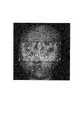
画像表示制御部12は、画像作成レンダリング部11で生成された色情報を有する3次元画像を表示制御するための表示レイアウト、表示条件、画像情報等の表示をコントロールする。表示部13は画像表示制御部12から出力される信号を表示する。図3は、血流量に応じて色付けされた脳のMPR画像を示している。図4は、血流量に応じて部分的に色付けされた表面画像を示している。ここでは、形態画像としての表面画像に、前記機能情報をもとに作成された機能情報を数値化し色分けして画像化したファンクショナルマップを貼り付けて表示される。なお、表示条件として、形態画像又は機能情報が所定の条件に合致する部分、例えば血流量が所定値未満の虚血が疑われる部分に限定してカラー表示するようにしてもよい。
The image
以上のように本実施形態によれば、ボリュームデータファイルを指定してそのファイルから形態画像を生成し表示させるに際して、同じ被検体であって同じ部位のダイナミックデータファイルが自動検索され、存在するのであればダイナミックデータファイルから血流量等の機能情報が計算され、その機能情報に応じて自動的に色付けして形態画像が表示される。操作者は所望のボリュームデータファイルを指定するだけで、余計な操作を不要にして、機能情報の提供を受けることができる。 As described above, according to the present embodiment, when a volume data file is specified and a morphological image is generated and displayed from the file, a dynamic data file of the same subject and the same part is automatically searched and exists. If there is, functional information such as blood flow is calculated from the dynamic data file, and a morphological image is displayed automatically colored according to the functional information. An operator can specify the desired volume data file, and can receive the provision of function information without unnecessary operations.
なお、本発明は上記実施形態そのままに限定されるものではなく、実施段階ではその要旨を逸脱しない範囲で構成要素を変形して具体化できる。また、上記実施形態に開示されている複数の構成要素の適宜な組み合わせにより、種々の発明を形成できる。例えば、実施形態に示される全構成要素から幾つかの構成要素を削除してもよい。さらに、異なる実施形態にわたる構成要素を適宜組み合わせてもよい。 Note that the present invention is not limited to the above-described embodiment as it is, and can be embodied by modifying the constituent elements without departing from the scope of the invention in the implementation stage. In addition, various inventions can be formed by appropriately combining a plurality of components disclosed in the embodiment. For example, some components may be deleted from all the components shown in the embodiment. Furthermore, constituent elements over different embodiments may be appropriately combined.
1…ボリュームデータファイル保管部、2…コントラスト画像自動検索部、3…ダイナミックデータ自動検索部、4…画像データロード部、5…画像同一位置参照部、6…機能情報数値化部、7…ファンクショナルマップ作成色変換部、8…画像レンダリングユニット、9…色情報作成部、10…形態情報処理部、11…画像作成レンダリング部、12…MPRユニット、13…3Dユニット、14…画像表示制御部、15…表示部。 DESCRIPTION OF SYMBOLS 1 ... Volume data file storage part, 2 ... Contrast image automatic search part, 3 ... Dynamic data automatic search part, 4 ... Image data load part, 5 ... Image same position reference part, 6 ... Functional information quantification part, 7 ... Funk 8: Image rendering unit, 9 ... Color information creation unit, 10 ... Form information processing unit, 11 ... Image creation / rendering unit, 12 ... MPR unit, 13 ... 3D unit, 14 ... Image display control unit , 15 ... display section.
Claims (9)
前記第1ボリュームデータファイルから血管に関する3次元の形態画像データを生成する3次元形態画像生成部と、
前記一連の第2ボリュームデータファイルから前記脳組織領域の血流動態指標に関する3次元のファンクショナルマップを生成する手段と、
前記3次元の形態画像の各画素に対してそれぞれ対応する前記3次元のファンクショナルマップ上の位置の血流動態指標の値に従って色情報を付与する手段と、
前記色情報を付与された3次元の形態画像からボリュームレンダリング処理により2次元の形態画像を生成する手段と、
前記形態画像を表示する手段とを具備する医用画像表示装置。 The first volume data file collected for the brain tissue area by the X-ray computed tomography apparatus and the time collected under the blood vessel enhancement for the brain tissue area by the dynamic scan of the X-ray computed tomography apparatus Storage means for storing a continuous series of second volume data files,
A three-dimensional morphological image generator for generating three-dimensional morphological image data related to blood vessels from the first volume data file;
Means for generating a three-dimensional functional map relating to a blood flow dynamic index of the brain tissue region from the series of second volume data files;
Means for giving color information according to a value of a blood flow dynamic index at a position on the three-dimensional functional map corresponding to each pixel of the three-dimensional morphological image;
Means for generating a two-dimensional morphological image by volume rendering from the three-dimensional morphological image to which the color information is assigned ;
A medical image display device comprising: means for displaying the morphological image .
前記第2のボリュームデータファイルから血管領域に対応する部分を抽出して動画表示する手段をさらに備えることを特徴とする請求項1記載の医用画像表示装置。 The function information is displayed only in the blood vessel region,
The medical image display apparatus according to claim 1, further comprising means for extracting a portion corresponding to a blood vessel region from the second volume data file and displaying a moving image.
Priority Applications (1)
| Application Number | Priority Date | Filing Date | Title |
|---|---|---|---|
| JP2004263472A JP4801892B2 (en) | 2004-09-10 | 2004-09-10 | Medical image display device |
Applications Claiming Priority (1)
| Application Number | Priority Date | Filing Date | Title |
|---|---|---|---|
| JP2004263472A JP4801892B2 (en) | 2004-09-10 | 2004-09-10 | Medical image display device |
Publications (3)
| Publication Number | Publication Date |
|---|---|
| JP2006075390A JP2006075390A (en) | 2006-03-23 |
| JP2006075390A5 JP2006075390A5 (en) | 2007-10-25 |
| JP4801892B2 true JP4801892B2 (en) | 2011-10-26 |
Family
ID=36155327
Family Applications (1)
| Application Number | Title | Priority Date | Filing Date |
|---|---|---|---|
| JP2004263472A Expired - Fee Related JP4801892B2 (en) | 2004-09-10 | 2004-09-10 | Medical image display device |
Country Status (1)
| Country | Link |
|---|---|
| JP (1) | JP4801892B2 (en) |
Families Citing this family (9)
| Publication number | Priority date | Publication date | Assignee | Title |
|---|---|---|---|---|
| CN101606182B (en) * | 2006-08-11 | 2012-11-28 | 皇家飞利浦电子股份有限公司 | Selection of datasets from 3D renderings for viewing |
| JP5591440B2 (en) * | 2007-01-17 | 2014-09-17 | 株式会社東芝 | Medical image display device |
| JP5524458B2 (en) * | 2008-07-18 | 2014-06-18 | 富士フイルムRiファーマ株式会社 | Organ surface image display apparatus and method |
| JP5355257B2 (en) * | 2009-07-01 | 2013-11-27 | 株式会社東芝 | Medical image display device and medical image display method |
| JP5380231B2 (en) | 2009-09-30 | 2014-01-08 | 富士フイルム株式会社 | Medical image display apparatus and method, and program |
| JP2011095796A (en) * | 2009-10-27 | 2011-05-12 | Fujifilm Corp | Image processing apparatus, method and program |
| JP5624350B2 (en) * | 2010-04-02 | 2014-11-12 | 株式会社東芝 | Medical image processing device |
| EP2788954A1 (en) * | 2011-12-07 | 2014-10-15 | Koninklijke Philips N.V. | Visualization of 3d medical perfusion images |
| JP6513365B2 (en) * | 2014-10-08 | 2019-05-15 | キヤノンメディカルシステムズ株式会社 | Medical image processing device |
Family Cites Families (10)
| Publication number | Priority date | Publication date | Assignee | Title |
|---|---|---|---|---|
| JP3512482B2 (en) * | 1994-09-06 | 2004-03-29 | 株式会社東芝 | Magnetic resonance imaging |
| JPH08126634A (en) * | 1994-10-30 | 1996-05-21 | Medeitetsukusu:Kk | Image display of three-dimensional (stereoscopic) or four-dimensional (three dimensions having time axis) data in stereoscopic imaging centering around blood vessel angiography utilizing x-rays |
| US5743266A (en) * | 1995-04-25 | 1998-04-28 | Molecular Biosystems, Inc. | Method for processing real-time contrast enhanced ultrasonic images |
| JP3690874B2 (en) * | 1996-05-30 | 2005-08-31 | 株式会社日立メディコ | Magnetic resonance imaging system |
| JP2000185036A (en) * | 1998-12-24 | 2000-07-04 | Toshiba Corp | Medical image display device |
| JP4421016B2 (en) * | 1999-07-01 | 2010-02-24 | 東芝医用システムエンジニアリング株式会社 | Medical image processing device |
| JP4350226B2 (en) * | 1999-09-13 | 2009-10-21 | 東芝医用システムエンジニアリング株式会社 | 3D image processing device |
| JP2001184491A (en) * | 1999-12-27 | 2001-07-06 | Hitachi Medical Corp | Three-dimensional image display device |
| DE10056457C2 (en) * | 2000-11-14 | 2002-11-07 | Siemens Ag | Method for operating a magnetic resonance device with functional imaging |
| JP4305720B2 (en) * | 2002-11-14 | 2009-07-29 | ジーイー・メディカル・システムズ・グローバル・テクノロジー・カンパニー・エルエルシー | X-ray CT system |
-
2004
- 2004-09-10 JP JP2004263472A patent/JP4801892B2/en not_active Expired - Fee Related
Also Published As
| Publication number | Publication date |
|---|---|
| JP2006075390A (en) | 2006-03-23 |
Similar Documents
| Publication | Publication Date | Title |
|---|---|---|
| JP5591440B2 (en) | Medical image display device | |
| CN101336844B (en) | Medical image processing apparatus and medical image diagnosis apparatus | |
| JP4421016B2 (en) | Medical image processing device | |
| CN105719324B (en) | Image processing apparatus and image processing method | |
| RU2627147C2 (en) | Real-time display of vasculature views for optimal device navigation | |
| JP4709177B2 (en) | Three-dimensional image processing apparatus and method, and program | |
| JP4468353B2 (en) | Method for three-dimensional modeling of tubular tissue | |
| JP6080248B2 (en) | Three-dimensional image display apparatus and method, and program | |
| CN104644202B (en) | Medical image data processing apparatus, medical image data processing method and medical image data processing routine | |
| JP5053606B2 (en) | Medical image diagnostic apparatus and medical image processing apparatus | |
| US20110054295A1 (en) | Medical image diagnostic apparatus and method using a liver function angiographic image, and computer readable recording medium on which is recorded a program therefor | |
| JP6214646B2 (en) | Temporal anatomical target tagging in angiograms | |
| JP4564387B2 (en) | Medical image generation device | |
| JP2008509773A (en) | Flexible 3D rotational angiography-computed tomography fusion method | |
| JP5090486B2 (en) | MEDICAL IMAGE DIAGNOSTIC APPARATUS, IMAGE DISPLAY METHOD, AND PROGRAM USING A LIVER CONTRAST | |
| JP4801892B2 (en) | Medical image display device | |
| JP2007222626A (en) | Automatic detecting method and device for unique part in medical image data | |
| US20140015836A1 (en) | System and method for generating and displaying a 2d projection from a 3d or 4d dataset | |
| JP2010154982A (en) | X-ray computer tomographic imaging apparatus and image processor | |
| JP2010000306A (en) | Medical image diagnostic apparatus, image processor and program | |
| JP5215444B2 (en) | Medical image diagnostic apparatus and medical image processing apparatus | |
| JPH0838433A (en) | Medical image diagnostic device | |
| JP5750381B2 (en) | Region extraction processing system | |
| JP2011067594A (en) | Medical image diagnostic apparatus and method using liver function angiographic image, and program | |
| JP2001087228A (en) | Image reading support device |
Legal Events
| Date | Code | Title | Description |
|---|---|---|---|
| A521 | Request for written amendment filed |
Free format text: JAPANESE INTERMEDIATE CODE: A523 Effective date: 20070910 |
|
| A621 | Written request for application examination |
Free format text: JAPANESE INTERMEDIATE CODE: A621 Effective date: 20070910 |
|
| A131 | Notification of reasons for refusal |
Free format text: JAPANESE INTERMEDIATE CODE: A131 Effective date: 20100713 |
|
| A521 | Request for written amendment filed |
Free format text: JAPANESE INTERMEDIATE CODE: A523 Effective date: 20100913 |
|
| A02 | Decision of refusal |
Free format text: JAPANESE INTERMEDIATE CODE: A02 Effective date: 20101102 |
|
| A521 | Request for written amendment filed |
Free format text: JAPANESE INTERMEDIATE CODE: A523 Effective date: 20110201 |
|
| A911 | Transfer to examiner for re-examination before appeal (zenchi) |
Free format text: JAPANESE INTERMEDIATE CODE: A911 Effective date: 20110204 |
|
| A131 | Notification of reasons for refusal |
Free format text: JAPANESE INTERMEDIATE CODE: A131 Effective date: 20110419 |
|
| A521 | Request for written amendment filed |
Free format text: JAPANESE INTERMEDIATE CODE: A523 Effective date: 20110526 |
|
| TRDD | Decision of grant or rejection written | ||
| A01 | Written decision to grant a patent or to grant a registration (utility model) |
Free format text: JAPANESE INTERMEDIATE CODE: A01 Effective date: 20110712 |
|
| A01 | Written decision to grant a patent or to grant a registration (utility model) |
Free format text: JAPANESE INTERMEDIATE CODE: A01 |
|
| A61 | First payment of annual fees (during grant procedure) |
Free format text: JAPANESE INTERMEDIATE CODE: A61 Effective date: 20110808 |
|
| FPAY | Renewal fee payment (event date is renewal date of database) |
Free format text: PAYMENT UNTIL: 20140812 Year of fee payment: 3 |
|
| R150 | Certificate of patent or registration of utility model |
Ref document number: 4801892 Country of ref document: JP Free format text: JAPANESE INTERMEDIATE CODE: R150 Free format text: JAPANESE INTERMEDIATE CODE: R150 |
|
| S111 | Request for change of ownership or part of ownership |
Free format text: JAPANESE INTERMEDIATE CODE: R313117 Free format text: JAPANESE INTERMEDIATE CODE: R313115 |
|
| R350 | Written notification of registration of transfer |
Free format text: JAPANESE INTERMEDIATE CODE: R350 |
|
| S533 | Written request for registration of change of name |
Free format text: JAPANESE INTERMEDIATE CODE: R313533 |
|
| R350 | Written notification of registration of transfer |
Free format text: JAPANESE INTERMEDIATE CODE: R350 |
|
| LAPS | Cancellation because of no payment of annual fees |