JP4650976B2 - Polyelectrolytes on biological templates - Google Patents
Polyelectrolytes on biological templates Download PDFInfo
- Publication number
- JP4650976B2 JP4650976B2 JP2000559929A JP2000559929A JP4650976B2 JP 4650976 B2 JP4650976 B2 JP 4650976B2 JP 2000559929 A JP2000559929 A JP 2000559929A JP 2000559929 A JP2000559929 A JP 2000559929A JP 4650976 B2 JP4650976 B2 JP 4650976B2
- Authority
- JP
- Japan
- Prior art keywords
- polyelectrolyte
- template
- layer
- capsule
- envelope
- Prior art date
- Legal status (The legal status is an assumption and is not a legal conclusion. Google has not performed a legal analysis and makes no representation as to the accuracy of the status listed.)
- Expired - Fee Related
Links
Images


Classifications
-
- B—PERFORMING OPERATIONS; TRANSPORTING
- B01—PHYSICAL OR CHEMICAL PROCESSES OR APPARATUS IN GENERAL
- B01J—CHEMICAL OR PHYSICAL PROCESSES, e.g. CATALYSIS OR COLLOID CHEMISTRY; THEIR RELEVANT APPARATUS
- B01J13/00—Colloid chemistry, e.g. the production of colloidal materials or their solutions, not otherwise provided for; Making microcapsules or microballoons
- B01J13/02—Making microcapsules or microballoons
-
- A—HUMAN NECESSITIES
- A61—MEDICAL OR VETERINARY SCIENCE; HYGIENE
- A61K—PREPARATIONS FOR MEDICAL, DENTAL OR TOILETRY PURPOSES
- A61K9/00—Medicinal preparations characterised by special physical form
- A61K9/48—Preparations in capsules, e.g. of gelatin, of chocolate
- A61K9/50—Microcapsules having a gas, liquid or semi-solid filling; Solid microparticles or pellets surrounded by a distinct coating layer, e.g. coated microspheres, coated drug crystals
- A61K9/5089—Processes
-
- B—PERFORMING OPERATIONS; TRANSPORTING
- B01—PHYSICAL OR CHEMICAL PROCESSES OR APPARATUS IN GENERAL
- B01J—CHEMICAL OR PHYSICAL PROCESSES, e.g. CATALYSIS OR COLLOID CHEMISTRY; THEIR RELEVANT APPARATUS
- B01J13/00—Colloid chemistry, e.g. the production of colloidal materials or their solutions, not otherwise provided for; Making microcapsules or microballoons
- B01J13/02—Making microcapsules or microballoons
- B01J13/20—After-treatment of capsule walls, e.g. hardening
- B01J13/22—Coating
Landscapes
- Chemical & Material Sciences (AREA)
- Organic Chemistry (AREA)
- Health & Medical Sciences (AREA)
- Dispersion Chemistry (AREA)
- Chemical Kinetics & Catalysis (AREA)
- Pharmacology & Pharmacy (AREA)
- General Health & Medical Sciences (AREA)
- Medicinal Chemistry (AREA)
- Engineering & Computer Science (AREA)
- Epidemiology (AREA)
- Life Sciences & Earth Sciences (AREA)
- Animal Behavior & Ethology (AREA)
- Bioinformatics & Cheminformatics (AREA)
- Public Health (AREA)
- Veterinary Medicine (AREA)
- Manufacturing Of Micro-Capsules (AREA)
- Medicinal Preparation (AREA)
- Immobilizing And Processing Of Enzymes And Microorganisms (AREA)
- General Preparation And Processing Of Foods (AREA)
Abstract
Description
【0001】
本発明は、高分子電解質外被を有するカプセルの製造方法ならびに該方法により得られるカプセルに関する。
【0002】
マイクロカプセルは種々の実施態様で公知であり、かつ特に医薬作用物質の制御された放出および標的に向けた輸送のため、ならびに敏感な作用物質、例えば酵素およびタンパク質を保護するために使用される。
【0003】
マイクロカプセルは機械的−物理的な方法により、例えば噴霧およびその後の被覆、化学的な方法、例えば界面重合もしくは界面縮合またはポリマー相分離により、あるいはリポソームへの作用物質のカプセル化により製造することができる。しかし従来公知の方法は一連の欠点を有している。
【0004】
ドイツ国特許出願公開第19812083.4号は、直径<10μmを有するマイクロカプセルの製造方法を記載しており、この場合、テンプレート粒子の水性分散液に反対に荷電した高分子電解質分子の複数の連続した層を施与する。この場合、テンプレート粒子として特に部分的に架橋したメラミンホルムアルデヒド粒子が記載されている。高分子電解質外被の形成後、メラミンホルムアルデヒド粒子を酸性のpH値の調整により、またはスルホン化により溶解させることができる。
【0005】
意外なことに高分子電解質カプセルは生物細胞、生物学的または/および両親媒性材料、例えば赤血球、細菌細胞もしくは脂質小胞の凝集体から選択されるテンプレートを使用しても形成できることが判明した。カプセルに入れたテンプレート粒子を引き続き可溶化もしくは分解により除去することができる。
【0006】
従って本発明は高分子電解質外被を有するカプセルの製造方法に関し、この場合、生物学的または/および両親媒性材料の凝集体から選択されるテンプレート上に反対に荷電した高分子電解質分子の複数の連続する層を施与し、かつ場合により引き続き該テンプレートを分解する。
【0007】
テンプレート材料として例えば細胞、例えば真核細胞、例えば哺乳類赤血球または植物細胞、単核細胞有機体、例えば酵母、細菌細胞、例えばE.コリ細胞、細胞凝集体、細胞レベル下の粒子、例えば細胞器官、花粉、膜調製物または細胞核、ウイルス粒子および生体分子の凝集体、例えばタンパク質凝集体、例えば免疫複合体、縮合した核酸、リガンド−受容体−複合体などを使用することができる。本発明による方法は生きている生物細胞および有機体のカプセル化のためにもまた適切である。テンプレートとして両親媒性材料、特に膜構造、例えば小胞、例えばリポソームもしくはミセル、ならびにその他の脂質凝集体が同様に適切である。
【0008】
これらのテンプレート上に反対に荷電した複数の高分子電解質層を析出させる。このためにテンプレート粒子を有利にはまず適切な溶剤、例えば水性媒体中に分散させる。次いで、特にテンプレート粒子が細胞またはその他の生物学的凝集体の場合、固定化試薬を充分な濃度で添加し、少なくとも部分的にテンプレート粒子を固定することができる。固定化試薬のための例はアルデヒド、例えばホルムアルデヒドまたはグルタルジアルデヒドであり、これを有利には最終濃度が0.1〜5%(w/w)となるように媒体に添加する。
【0009】
高分子電解質とは一般にポリマー鎖の成分または置換基であってもよい、イオン的に解離することができる基と理解される。通常、高分子電解質中のイオン的に解離可能なこれらの基の数は、解離した形(ポリイオンとも呼ばれる)でポリマーが水溶性であるような大きさである。ここでイオン性の基の濃度が水溶性にとって充分ではないが、しかし自己集合(Selbstassemblierung)に突入するために充分な電荷を有するイオノマーもまた高分子電解質という概念で理解される。有利には外被は「真の」高分子電解質を包含する。解離可能な基の種類に応じて高分子電解質を多価酸および多塩基に分類する。
【0010】
解離の際にプロトンの分離下で多価酸からポリアニオンが生じ、該ポリアニオンは無機ポリマーであっても有機ポリマーであってもよい。多価酸のための例は、ポリリン酸、ポリビニル硫酸、ポリビニルスルホン酸、ポリビニルホスホン酸およびポリアクリル酸である。ポリ塩とも呼ばれる相応する塩のための例はポリリン酸塩、ポリ硫酸塩、ポリスルホン酸塩、ポリホスホン酸塩およびポリアクリル酸塩である。
【0011】
多塩基は例えば酸との反応により塩を形成しながらプロトンを受け取ることができる基を有する。鎖−もしくは側鎖の解離可能な基を有する多塩基のための例はポリアリルアミン、ポリエチレンイミン、ポリビニルアミンおよびポリビニルピリジンである。多塩基はプロトンの受容によりポリカチオンを形成する。
【0012】
本発明により適切な高分子電解質はバイオポリマー、例えばアルギン酸、アラビアゴム、核酸、ペクチン、タンパク質およびその他のものであっても、ならびに化学的に修飾されたバイオポリマー、例えばカルボキシメチルセルロースおよびリグニンスルホネートであっても、ならびに合成ポリマー、例えばポリメタクリル酸、ポリビニルスルホン酸、ポリビニルホスホン酸およびポリエチレンイミンである。
【0013】
直鎖状もしくは分枝鎖状の高分子電解質を使用することができる。分枝鎖状の高分子電解質の使用は、高い度合いの壁孔隙率を有する緻密な高分子電解質の複数膜につながる。カプセル安定性を向上するために、高分子電解質分子を個々の層の内部または/およびその中間で、例えばアルデヒドを用いたアミノ基の架橋により架橋させてもよい。さらに両親媒性高分子電解質、例えば部分的に高分子電解質の特性を有している両親媒性ブロックコポリマーまたはランダムコポリマーを小さい極性分子に対する透過性を減少するために使用してもよい。このような両親媒性コポリマーは異なった官能価の単位、例えば一方では酸性もしくは塩基性の単位、および他方ではブロックとして、もしくはランダムに分散してポリマー全体に配置されていてもよい疎水性単位、例えばスチレン、ジエンまたはシロキサンなどからなる。外部の条件の相関関係としてその構造を変化するコポリマーを使用することによりカプセル壁をその透過性もしくはその他の特性に関して定義的に制御することができる。このために例えばポリ(N−イソプロピル−アクリルアミド)−割合を有するコポリマー、例えばポリ(N−イソプロピルアクリルアミド−アクリル酸)が考えられ、該コポリマーは水素架橋結合の平衡によりその水溶性を温度の相関関係として変化させ、このことは膨潤を伴う。
【0014】
特定の条件下で分解可能な、例えば光、酸もしくは塩基に不安定な高分子電解質を使用することにより、内包されている作用物質の放出をカプセル壁の溶解によって制御することができる。さらに特定の適用可能性のために導電性の高分子電解質または光学活性基を有する高分子電解質をカプセル成分として使用することができる。
【0015】
基本的に使用するべき高分子電解質もしくはイオノマーに関しては、使用される分子が充分に高い電荷を有し、または/およびその他の相互作用様式、例えば水素架橋結合および/または疎水性の相互作用を介してその下に存在する層と結合する限り、制限は生じない。
【0016】
従って適切な高分子電解質は分子量の小さい高分子電解質もしくはポリイオンであっても分子量の大きい高分子電解質、例えば生物由来の高分子電解質であってもよい。
【0017】
高分子電解質層をテンプレート上に施与するために有利にはまず水溶液中のテンプレート粒子の分散液を製造する。次いでこの分散液にテンプレート粒子の表面と同一または反対の電荷を有する高分子電解質種を添加する。場合により存在する過剰の高分子電解質分子の分離後に、第二の層の構成のための使用される、反対に荷電した高分子電解質種を添加する。引き続きさらに反対に荷電した高分子電解質の層を交互に施与し、その際、同一の電荷を有するそれぞれの層に関して同一または異なった高分子電解質種または高分子電解質種の混合物を選択することができる。層の数は基本的に任意に選択することができ、かつ例えば2〜40層、特に4〜20層の高分子電解質層となる。
【0018】
所望の数の層を施与した後で、所望の場合には、外被で覆われたテンプレート粒子を分解することができる。分解は溶解試薬の添加により行うことができる。この場合、生物学的材料、例えばタンパク質または/および脂質を溶解することができる溶解試薬が適切である。有利には溶解試薬は除蛋白剤、例えばペルオキソ化合物、例えばH2O2または/および次亜塩素酸化合物、例えば次亜塩素酸ナトリウムもしくは次亜塩素酸カリウムを含有している。意外なことにテンプレート粒子の分解は室温で短いインキュベーション時間、例えば1分〜1時間以内に行われる。テンプレート粒子の分解は充分に完全である、というのも残留している外被を電子顕微鏡により観察する場合でさえ粒子の残留物はもはや検出不可能であるからである。生物学的高分子電解質を外被へ組み込む際に空の層もまた高分子電解質外被中に存在していてもよい。
【0019】
本発明による方法により得られるカプセルは10nm〜50μm、有利には50nm〜10μmの範囲の直径で球形から逸脱してもよい、つまり異方性の形状で製造してもよい。壁厚は高分子電解質層の数により決定され、かつ例えば2〜100nmの範囲、特に5〜80nmの範囲である。該カプセルはその単分散性により優れている、つまり適切なテンプレートを選択すると、平均的な直径からの逸脱が50%を上回るカプセルの割合が10%未満、および特に有利には1%未満であるカプセル組成物が得られる。
【0020】
該カプセルは化学的、生物学的、有機的および熱的な負荷に対して極めて安定している。これらは凍結させるか、または凍結乾燥させ、引き続き再び適切な溶剤中に溶解させることができる。
【0021】
該カプセルはその内部に含有されているテンプレートのミクロ型(Mikroabdruecke)であり、かつテンプレートの除去後にもその形を保持するので、異方性の粒子を製造することができ、この場合、これは生物学的構造、例えば細胞、ウイルス粒子もしくは生体分子凝集体のミクロ型である。
【0022】
外被中の浸透性の修飾は、少なくとも1つの高分子電解質層中の孔の形成もしくは変化により達成することができる。このような孔は相応する高分子電解質を使用して自己形成することができる。さらにアニオン性または/およびカチオン性の基または/および界面活性物質、例えば表面活性剤、または/および透過性を修飾するための脂質およびその他の特性を有するナノ粒子を使用することができる。さらに浸透性は高分子電解質の析出の際に支配的な条件の変更により修飾することができる。従って例えば周囲媒体の高い塩濃度は高分子電解質外被の高い透過性につながる。
【0023】
高分子電解質外被の透過性の特に有利な修飾は、脂質層または/および両親媒性高分子電解質を、テンプレート粒子の分解後に高分子電解質外被上に析出させることにより達成することができる。この方法で小さく、かつ極性分子のための高分子電解質外被の浸透性を著しく低下させることができる。高分子電解質外被上に析出させることができる脂質の例は、少なくとも1つのイオン性もしくはイオン化可能な基を有する脂質、例えばリン脂質、例えばジパルミトイルホスファチジン酸または両性イオン性のリン脂質、例えばジパルミトイルホスファチジルコリンあるいはまた脂肪酸もしくは相応する長鎖のアルキルスルホン酸である。両性イオン性の脂質を使用する際に高分子電解質外被上に脂質の複数層を析出させることができる。脂質層上に引き続き別の高分子電解質層を析出させることができる。
【0024】
本方法により得られるカプセルは作用物質の封入のために使用することができる。これらの作用物質は無機物質であっても有機物質であってもよい。このような作用物質のための例は触媒、特に酵素、医薬作用物質、ポリマー、着色剤、例えば蛍光性化合物、センサー分子、つまり周囲の条件(温度、pH値)の変化に対して検出可能な反応をする分子、農薬および香料である。
【0025】
該カプセルを化学反応のためのマイクロ反応空間として、または沈殿−もしくは結晶化テンプレートとして使用することもできる。カプセル壁の浸透性は制御可能であるため、該壁は例えば分子量の小さい物質を通過させることができるが、しかし高分子を充分に抑止するという事実に基づいて、化学反応の際に生じる高分子の生成物の場合、例えば重合の際に生じるポリマーの場合に、容易な方法で合成中に内部空間に抑止することができる。外部媒体中で同時に合成される反応生成物を例えば遠心分離または/および濾過により後から、あるいはまたすでに反応の間に除去することができる。
【0026】
反応の間、反応基質の供給をカプセル壁による拡散により制御することができる。この場合、反応の進行に介入する新規の方法が生じる。例えば濾過により連続的に、もしくは例えば遠心分離により外部媒体を突発的に交換することもまた可能であるので、重合反応を基質除去により任意に停止させるかもしくはモノマーを交換することができる。従って新規の方法で、定義されたコポリマーもしくはマルチポリマーの製造を実施することが可能である。浸透により反応の進行はモノマー供給を介して制御可能であるので、カプセル中で新規の、およびその他の分子量分布を有する生成物、例えば高度に単分散性の生成物を製造することができる。カプセル内部で合成したポリマーを例えば蛍光染料を用いた滴定により分光分析を用いて、および共焦点の顕微鏡により検出することができる。個別粒子の光の散乱を用いて質量の増加ひいては反応速度論を追跡することができる。
【0027】
作用物質の封入のために、または合成もしくは沈殿法のための反応空間として異方性のカプセルを使用し、かつ場合により引き続きテンプレート外被を溶解する際に、粒子組成物を規定の形および形状を有する分散液として製造することができる。従って本発明は、高分子電解質外被中の作用物質のカプセル化により、例えば合成および沈殿および引き続き熱的もしくは化学的な処理によるテンプレートの除去により得られる異方性の粒子組成物に関する。有利にはこれらの異方性粒子はテンプレートとして使用されるバイオ構造の形を有している。
【0028】
さらに該カプセルを有機的な液体、例えばアルコールもしくは炭化水素、例えばヘキサノール、オクタノール、オクタンもしくはデカンの導入のため、または気体のカプセル化のために使用することができる。このような水と非混和性の有機液体で充填されたカプセルは化学反応、例えば重合反応のためにもまた使用することができる。例えばモノマーをその分布平衡によりカプセルの内部空間中で適切に富化させることができる。場合によりモノマー溶液はすでに合成の開始前にカプセルの内部空間に封入されている。
【0029】
しかしその大きさに基づいて高分子電解質外被中に浸透することができない作用物質もまたカプセルに封入することができる。このために封入するべき作用物質をテンプレート粒子に固定するか、または例えば生きている細胞の場合にはファゴサイトーシスもしくはエンドサイトーシスによりテンプレート粒子により封入する。テンプレート粒子の分解後に作用物質を高分子電解質外被の内部へ放出する。その際、有利には作用物質の不所望の分解が生じないようにテンプレート粒子の分解の際の条件を選択する。
【0030】
該カプセルは数多くの適用領域、例えばセンサー工学、表面分析化学で、エマルションキャリア(Emulsionstraeger)、例えば触媒法、重合法、沈殿法もしくは結晶化法のためのマイクロ反応空間として、製剤学および医学において、例えば作用物質のターゲッティングのため、または超音波造影剤として、食料品科学技術、化粧品、バイオテクノロジー、センサー工学、情報科学技術および印刷産業(着色剤のカプセル封入)において使用することができる。さらに該カプセルをミクロ複合材料もしくはナノ複合材料、つまり少なくとも2つの異なった材料からなり、かつミクロ−もしくはナノスコープなオーダーを有している作用物質の構成のために使用することができる。
【0031】
本発明のもう1つの実施態様は固定化された形のテンプレート粒子を有利には高分子電解質被覆の前に溶解試薬で処理することにより部分的に分解することである。溶解プロセスを適切な時間で中断すると、部分的に溶解した構造、例えば中心に穴の空いた環状構造が得られ、該構造を引き続き被覆することができる。引き続き該テンプレート粒子を完全に分解した後で、リング状のカプセルが最終生成物として得られる。これは例えば光学(マイクロ反響丸天井効果(Mikrofluesterbogeneffekt))におけような興味深い適用可能性を有する全く新規の位相品質である。
【0032】
本発明を以下の実施例および図面に基づいてさらに詳細に説明する。図面は以下のものを示している:
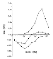
図1は、ζ電位の変化(A)および蛍光強度の上昇(B)を、グルタルジアルデヒドで固定したヒト赤血球上でのポリ(スチレンスルホン酸ナトリウム塩(PSS)およびポリ(塩酸アリルアミン)(PAH)の析出の層の数の相関関係として示す。
【0033】
図2は、10層のPSSおよびPAH層で被覆した円板状赤血球の走査型電子顕微鏡の画像を示す。
【0034】
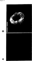
図3は、未被覆(A)および被覆した(B)円板状赤血球ならびに被覆した円板状赤血球の溶解後に得られる高分子電解質外被(C)の透過型電子顕微鏡の図を示す。
【0035】
図4は、円板状赤血球(A)およびエキノサイト(B)上に析出した高分子電解質外被の原子力顕微鏡による画像を示す。
【0036】
図5は、エキノサイト上に析出し、かつ11層のPSS/PAHからなる高分子電解質外被を共焦点顕微鏡で撮影した画像を示す。
【0037】
図6は、6−カルボキシフルオレセインで充填した高分子電解質外被を共焦点顕微鏡により撮影した画像を示す。
【0038】
図7は、付加的な被覆を有していない円板状赤血球上の高分子電解質外被の電子回転スペクトル(A)またはDPPA(B)もしくはDPPC(C)で被覆した円板状赤血球上の高分子電解質外被の電子回転スペクトルを示す。
【0039】
図8は、光学顕微鏡(A)および相応する走査型電子顕微鏡の画像(B)による円板状赤血球上に析出した高分子電解質外被の画像を示す。
【0040】
図9は、高分子電解質外被の外側および内部でのラジカル重合によるポリ(ジアリルメチルアンモニウムクロリド)の画像を示す。
【0041】
実施例
1.テンプレートとしてウシおよびヒトの赤血球を用いたポリマー外被の調製 新鮮なヒトもしくはウシの血液から血漿を遠心分離する。引き続き燐酸塩で緩衝した等張食塩溶液PBS(5.8mM燐酸塩緩衝液 pH7.4、KCl 5.6mM、NaCl 150mM)中で2回洗浄する。引き続き赤血球をグルタルジアルデヒドを用いて2%の濃度に固定する。ここに赤血球沈殿物1mlをPBS1mlと共に補充する。次いでこの溶液にグルタルジアルデヒド(グルタルジアルデヒド(25%水溶液)1部およびPBS9部)8mlを滴加する。20℃で60分の作用時間の後で該溶液を遠心分離し、かつ赤血球を2回蒸留水で4回洗浄する。引き続き固定した赤血球を、緩衝していない154mMのNaCl溶液で補充する。
【0042】
次の工程として反対に荷電した2種類の高分子電解質の連続的吸着を行う。固定された赤血球の出発電荷はマイナスであったので、有利にはまずプラスに荷電した、50〜60kDの分子量を有するポリ(アリルアミン)ヒドロクロリド(PAH)(Aldrich)を使用する。しかしまたマイナスに荷電した高分子電解質をまず第一の層として赤血球上に析出させてもよい。赤血球の被覆のために0.5g/dl PAHおよび0.5MのNaCl濃度を有する溶液4mlを約2.5(v/v)の赤血球濃度で添加する。20℃で10分間の作用時間の後で、赤血球を遠心分離し、かつ154mMのNaCl中で2回洗浄する。引き続き第二の層の吸着を行う。この目的のために70kDの分子量を有し、マイナスに荷電したポリ(スチレンスルホネート)−ナトリウム塩(PSS)を使用する。すでにPAHで被覆した赤血球上での第一のPSS層の施与のためにPSS0.5g/dlおよび0.5MのNaClの濃度および約2.5%(v/v)の赤血球濃度を有する溶液を作った。20℃で10分間の作用時間後に赤血球を遠心分離し、かつ154mMのNaCl溶液中で2回洗浄した。PAH層およびPSS層の施与は任意で数回繰り返してもよい。例えばそれぞれ5つのPAH層および5つのPSS層を施与することができる。
【0043】
テンプレートの溶解のために、固定された赤血球を1.2%のNaOCl溶液へピペットで移した。同様に市販の除蛋白剤(Produkt、メーカー)または排水清浄剤(例えばChlorix、メーカー)が適切である。作用時間は20℃で約20分であり、かつ溶液の濁りの消失により光学的に制御可能である。残留しているポリマー外被を引き続きNaCl溶液中で洗浄する。
【0044】
2.E.コリ細菌または酵母をテンプレートとして用いるポリマー外被の調製 まずE.コリ細胞をPBS等張溶液中で2回洗浄することにより培養液から分離する。引き続きグルタルジアルデヒドを用いて固定を行う。このためにコリ細菌の沈殿物をPBSで2mlになるまで補充する。この溶液にグルタルジアルデヒド溶液8mlを添加して最終濃度を2%とする。20℃で60分の作用時間の後、該溶液を遠心分離し、かつ固定したE.コリ細胞を2回蒸留水で4回洗浄する。
【0045】
引き続き、反対に荷電した2種類の高分子電解質の連続した吸着を例1に記載したとおりに行う。
【0046】
相応の方法で予備的な固定を行わずに酵母細胞もまた被覆した。
【0047】
3.高分子電解質外被上への脂質層の堆積
高分子電解質外被上に脂質層を析出させるために2つの異なった方法を使用した。
【0048】
3.1
高分子電解質外被の懸濁液200μlをメタノール中で繰り返し洗浄することにより再懸濁させた。3回目の洗浄の後、純粋なメタノールの代わりに、例えばメタノール中ジパルミトイルホスファチジン酸(DPPA)またはジパルミトイルホスファチジルコリン(DPPC)1mg/mlの脂質溶液500μlを沈殿物に添加する。外被をこのメタノール−脂質溶液中に再懸濁させ、かつ該懸濁液を90℃の温度で水浴中に保持する。蒸発させるべきメタノールを、その都度20μlの分量の水の滴加により交換する。メタノール700μlと水との交換は約30分を要する。
【0049】
蒸発終了後に外被懸濁液を水で3回洗浄し、かつ繰り返し遠心分離する。25000rpmで20分の遠心分離により脂質被覆した外被を沈殿させることができる。
【0050】
3.2
水中で脂質1mg/mlの濃度を有するDPPAまたはDPPC90%とDPPA10%との分散液を超音波処理により製造する。得られた脂質小胞の分散液500μlを濃縮した外被懸濁液200μlに添加する。30分後に試料を25000rpmで20分間遠心分離する。上澄みを除去し、かつ水を添加する。この手順を3回繰り返す。その際、脂質で被覆された外被の濃懸濁液が得られる。
【0051】
4.高分子電解質外被中への有機溶剤の封入
高分子電解質外被の水性懸濁液を3000rpmで5分間遠心分離する。上澄みを除去した後でメタノールを添加する。外被を再懸濁させ、かつ4000rpmで10分間遠心分離する。改めて上澄みを除去し、メタノールを添加し、かつ該試料を前記と同一の条件下で遠心分離する。この手順を3回繰り返す。メタノールを用いた最後の遠心分離の後で上澄みをヘキサノールと交換する。外被を再懸濁させ、かつ5000rpmで10分間遠心分離する。この手順を再度3回繰り返す。
【0052】
オクタノール、オクタンまたはデカンを外被に封入するために類似の手順を使用するが、その際、出発材料としてヘキサノール溶液中に存在する外被を使用する。遠心分離の速度はオクタノールおよびオクタンに関しては7000rpm(10分)に高め、かつデカンに関しては7500rpm(10分)に高める。
【0053】
最後に得られた沈殿物を水中に再懸濁させる。外被は水相中に残留し、その一方で外被同士の間の沈殿物中にまだ存在する痕跡量の溶剤は第二の有機相を形成する。有機相および水相のために蛍光標識を使用することにより共焦点顕微鏡を用いて外被が有機溶剤により充填されていることを示すことができる。
【0054】
記載の手順により、水中の無極性の液体の高度に安定したエマルジョンを製造することが可能になる。本来の外被の単分散の結果として、得られたエマルジョンは同様に単分散性である。もう1つの利点は、個々の小滴の形でさえ、使用されるテンプレートに依存して、制御することができることである。このことにより、球とは異なった表面:体積比を有するエマルジョンの製造が可能になる。
【0055】
5.高分子電解質外被の特徴付け
図1はゼータ電位における変化(A)および蛍光強度の上昇(B)を、グルタルジアルデヒドで予備処理したヒトの赤血球上へのポリ(スチレンスルホン酸ナトリウム塩)およびポリ(塩酸アリルアミン)を析出させる際の層の数の関数として示している。ζ電位を電気泳動移動度の測定(Elektrophor、Hasotec)により生理食塩溶液中で測定する。蛍光強度の分布は貫流血球計算(FACScan、Becton Dickinson)によりFITC−標識したPAHの使用下に3つの連続した層析出サイクルで記録する。
【0056】
図2にはPSSおよびPAHの10の層で覆われた円板状赤血球の走査型電子顕微鏡の画像を示す。乾燥工程は細胞の端部に沿った高分子電解質層の長軸方向のしわの発生につながる。
【0057】
図3は未被覆(A)および被覆後(B)の円板状赤血球の透過型電子顕微鏡の画像を示している。高分子電解質外被は明らかに認められる。細胞の可溶化の後に得られる高分子電解質外被(C)は、2つの意想外の特性を示す。倍率はAおよびBでは1:15000およびCでは1:17000である。第一にこれらは本来の細胞の形に類似しており、かつ第二にこれらは外被に亀裂もしくは比較的大きな孔が認識されずに完全に空であるように見える。
【0058】
図4には原子力顕微鏡(AFM)により得られた2つの画像(幅10μm)が示されており、これは円板状赤血球(A)およびエキノサイト(B)上に析出した、合計9つの層からなる高分子電解質外被を示している。楕円形の円板状赤血球上に析出した外被はわずかなしわを示すのみである一方で、星形のエキノサイト上の析出プロセスは、その上に本来のテンプレートの突出部さえも認識できるような良好に構造化された外被につながる。このことは図5に示された、エキノサイト上に析出したPSS/PAHの11の層からなる高分子電解質外被の共焦点顕微鏡の画像からよりいっそう明らかになる。最外層はFITCにより標識したPAHからなる。画像の幅は7μmである。スキャンを2つのレベルで1μmの間隔により別々に行った。スキャンAはスライドガラス上に施与した外被上の上部を通過して走行する。この画像上で外被の内部は、突出部の領域においてさえも空であることが認識される。
【0059】
図6は共焦点顕微鏡を用いて撮影した、円板状赤血球上に析出した、PSS/PAHの10の層からなる高分子電解質外被の画像を示している。外被を6−カルボキシフルオレセイン(6−CF)溶液100μMを用いて処理した。図6Aでは外被内にフルオレセインが認識される。このことは6−CF分子が外被の内部の侵入できることを示している。
【0060】
100nMの6−CFを用いた外被のインキュベーションの際に蛍光を発見することはできなかった。このことは溶解試薬を用いた処理により6−CFを結合する能力のあるアミノ基がPAHにより分解されるおよび/またはブロックされることを示している。6−CFのわずかな濃度に基づいて溶液の蛍光は背景として検出できるためには少なすぎる。
【0061】
気化による例3に記載の高分子電解質外被上へのジパルミトイルホスファチジン酸(DPPA)または両性イオン性の脂質、例えばジパルミトイルホスファチジルコリン(DPPC)の吸着により安定した脂質層で覆われた高分子電解質外被が得られる。脂質層は外被への6−CFの侵入を充分に阻止する(図6B)。図6Aおよび6B中に記載されている画像の幅は16もしくは15μmである。別の実験は脂質層が少なくとも4週間は安定しており、かつ脂質層上に別の高分子電解質層が、その下に存在する脂質層を破壊することなく析出できることを示している。
【0062】
エレクトロローテーション(Elektrorotation)の技術(Arnold et al., J. Phys. Chem. 91 (1987), 5093; Fuhr et al., In: Electromanipulation of Cells, U. Zimmermann und G. A. Neil, Hrgs., CRC Press, Boca Raton (1996), 259-328; Prueger et al., Biophys. J. 72 (1997), 1414)は多層構造の誘電性の区別を可能にする分光分析法である。この場合、KHz範囲〜MHz範囲で回転する電界を粒子懸濁液において印加する。誘導されたダイポールモーメントは、電界の回転速度が速すぎ、誘導するべきダイポールモーメントが電界に従うことができない場合に、印加された電界ベクターを有する角度を形成する。結果として該粒子は回転モーメントを得、これは再度認識可能な粒子自体の回転につながる。電子回転のスペクトルは粒子の回転速度の測定により外側の電界の回転数の関数として得られる。
【0063】
図7はコントロールとしての未被覆の高分子電解質外被(A)、DPPA被覆した高分子電解質外被(B)およびDPPCで被覆した高分子電解質外被(C)のための3つの典型的な電子回転スペクトルを示す。外被の外側の水相の導電率は2.000μS/cm、80μS/cmもしくは100μS/cmであった。絶縁性の脂質層の存在はKHz範囲におけるマイナスの回転方向につながる。強導電性の高分子電解質外被(約104μS/cm)は、コントロールにおいてMHz領域で認識可能なプラスのピークを生じる。弱導電性のDPPA層は周波数が高い場合にショートする。DPPC被覆の場合にプラスの回転が存在しないことは脂質の複数層が存在していることを示唆している。この結果は、透過性を制御するために高分子電解質の複数層を脂質で有利に被覆でき、かつ被覆した外被はイオン性の化合物、例えば塩に関してほとんど透過性ではないことを示している。
【0064】
空の高分子電解質外被は、有機または無機材料の制御された沈殿または結晶化のためにも使用することができる。このために円板状赤血球をテンプレートとした高分子電解質外被を30mMの6−CF溶液中pH7でインキュベーションする。引き続きpH値は急速に3.5の値に変化し、その際、6−CFは充分に不溶性になる。1〜12時間にわたるインキュベーションの後、明らかに完全に6−CFで充填され、かつ空の外被の混合物が得られる。図8Aは高分子電解質外被(10層)の光学顕微鏡による撮影を、および図8Bは相応する走査型電子顕微鏡による画像を示している。完全に暗い画像Aは結晶化した6−CFの顕著な吸着に起因する。SEM画像は、外被の内部で結晶化した6−CFが本来のテンプレートの形をとっていることを示している。画像Aの幅は8μmである。別の実験ではローダミンBをpH値の上昇により沈殿させることができることが示された。作用物質の沈殿はその他の手段、例えば溶剤の交換、塩沈殿などによってもまた誘発することができる。これらの結果は、高分子電解質外被を結晶化もしくは沈殿法のためのテンプレートとして使用できることを示しており、これにより反応により生じたコロイド状の粒子の大きさおよび形の制御が可能になる。
【0065】
図9はPAH/PSS外被中でのジアリルジメチルアンモニウムクロリド(DADMAC)のラジカル重合の結果を示している。このために2%のカプセル懸濁液中の3%モノマー溶液に重合開始剤ペルオキソ二硫酸ナトリウム(30mg/100ml)を添加し、かつ70℃で9.5時間重合した。遠心分離により界面層中で合成されるポリマーPDADMACを分離した。100mMの6−CFを用いた処理の後で、PDADMACのアミノ基への色素の結合は明らかに認識することができる。該ポリマーはマイナスのカプセル壁に吸着されるが、しかしカプセルの内部にもみられる。
【0066】
9つの層からなる高分子電解質外被([PSS/PAH]4PSS)をヒト赤血球上に析出させ、かつテンプレート粒子を除去した。引き続き別の層PAHを施与した。アクリル酸をポリアクリル酸へとラジカル重合するためにカプセルを使用した。このために2%のカプセル懸濁液中の3%のモノマー溶液に開始剤ペルオキソ二硫酸ナトリウム(30mg/100ml)を添加し、かつ70℃で9.5時間重合させた。遠心分離により界面層中で合成されるポリアクリル酸を除去した。100nMのローダミンB(選択的にアニオン基に結合する)の添加後にマイナスに荷電したカプセル壁に、あるいはまたカプセルの内部に吸着されたポリアクリル酸の存在を検出することができた。アクリル酸により媒介される架橋形成に基づいてカプセルの凝集化が行われた。アクリル酸のこの吸着は、外側のマイナスの荷電を有するカプセルの使用により防止することができる。
【図面の簡単な説明】
【図1】 ζ電位(A)の変化および蛍光強度(B)の上昇を示す図
【図2】 PSSおよびPAHの層で被覆した円板状赤血球の走査型電子顕微鏡の画像を示す図
【図3】 未被覆(A)および被覆した(B)円板状赤血球ならびに被覆した円板状赤血球の溶解後に得られる高分子電解質外被(C)の透過型電子顕微鏡による画像を示す図
【図4】 円板状赤血球(A)およびエキノサイト(B)上に析出した高分子電解質外被の原子力顕微鏡による画像を示す図
【図5】 エキノサイト上に析出し、かつPSS/PAHからなる高分子電解質外被の共焦点顕微鏡による画像を示す図
【図6】 6−カルボキシフルオレセインで充填した高分子電解質外被の共焦点顕微鏡による画像を示す図
【図7】 付加的な被覆を有していない円板状赤血球上の高分子電解質外被の電子回転スペクトル(A)またはDPPA(B)もしくはDPPC(C)で被覆した円板状赤血球上の高分子電解質外被の電子回転スペクトルを示す図
【図8】 光学顕微鏡(A)および相応する走査型電子顕微鏡の画像(B)による円板状赤血球上に析出した高分子電解質外被を示す図
【図9】 高分子電解質外被の外側および内部でのラジカル重合によるポリ(ジアリルメチルアンモニウムクロリド)の画像を示す図[0001]
The present invention relates to a method for producing a capsule having a polymer electrolyte jacket and a capsule obtained by the method.
[0002]
Microcapsules are known in various embodiments and are used in particular for the controlled release and delivery of pharmaceutical agents to targets and to protect sensitive agents such as enzymes and proteins.
[0003]
Microcapsules can be produced by mechanical-physical methods, for example by spraying and subsequent coating, chemical methods, for example by interfacial polymerization or interfacial condensation or polymer phase separation, or by encapsulating the agent in liposomes. it can. However, the known methods have a series of drawbacks.
[0004]
German Offenlegungsschrift DE 19 08 083.4 describes a process for the production of microcapsules having a diameter <10 μm, in which case a series of oppositely charged polyelectrolyte molecules in an aqueous dispersion of template particles. Apply the layer. In this case, particularly partially crosslinked melamine formaldehyde particles are described as template particles. After formation of the polyelectrolyte envelope, the melamine formaldehyde particles can be dissolved by adjusting the acidic pH value or by sulfonation.
[0005]
Surprisingly, it has been found that polyelectrolyte capsules can also be formed using templates selected from biological cells, biological or / and amphiphilic materials such as aggregates of red blood cells, bacterial cells or lipid vesicles. . The encapsulated template particles can then be removed by solubilization or degradation.
[0006]
The present invention therefore relates to a process for the production of a capsule having a polyelectrolyte envelope, in which case a plurality of oppositely charged polyelectrolyte molecules on a template selected from an aggregate of biological or / and amphiphilic materials. A continuous layer of is applied and optionally the template is subsequently decomposed.
[0007]
Examples of template materials include cells such as eukaryotic cells such as mammalian erythrocytes or plant cells, mononuclear cell organisms such as yeast, bacterial cells such as E. coli. Coli cells, cell aggregates, subcellular particles such as cell organs, pollen, membrane preparations or cell nuclei, virus particles and biomolecule aggregates such as protein aggregates such as immune complexes, condensed nucleic acids, ligands A receptor-complex or the like can be used. The method according to the invention is also suitable for the encapsulation of living biological cells and organisms. Amphiphilic materials, in particular membrane structures such as vesicles such as liposomes or micelles, and other lipid aggregates are likewise suitable as templates.
[0008]
A plurality of oppositely charged polymer electrolyte layers are deposited on these templates. For this purpose, the template particles are preferably first dispersed in a suitable solvent, for example an aqueous medium. Then, particularly when the template particles are cells or other biological aggregates, the immobilization reagent can be added at a sufficient concentration to at least partially immobilize the template particles. Examples for immobilization reagents are aldehydes, such as formaldehyde or glutardialdehyde, which are preferably added to the medium to a final concentration of 0.1-5% (w / w).
[0009]
A polyelectrolyte is generally understood to be an ionically dissociable group which may be a component or substituent of a polymer chain. Usually, the number of these ionically dissociable groups in the polyelectrolyte is such that the polymer is water soluble in a dissociated form (also called polyion). Here, ionomers whose concentration of ionic groups is not sufficient for water solubility, but ionomers that are sufficiently charged to enter self-assembly are also understood in the concept of polyelectrolytes. Advantageously, the jacket includes a “true” polyelectrolyte. Polyelectrolytes are classified into polyacids and polybases according to the type of dissociable group.
[0010]
In the dissociation, a polyanion is generated from the polyvalent acid under the separation of protons, and the polyanion may be an inorganic polymer or an organic polymer. Examples for polyvalent acids are polyphosphoric acid, polyvinyl sulfuric acid, polyvinyl sulfonic acid, polyvinyl phosphonic acid and polyacrylic acid. Examples for corresponding salts, also called polysalts, are polyphosphates, polysulfates, polysulfonates, polyphosphonates and polyacrylates.
[0011]
Polybases have groups that can accept protons while forming salts, for example by reaction with acids. Examples for polybases having chain- or side-chain dissociable groups are polyallylamine, polyethyleneimine, polyvinylamine and polyvinylpyridine. Polybases form polycations by accepting protons.
[0012]
Polyelectrolytes suitable according to the invention are biopolymers such as alginic acid, gum arabic, nucleic acids, pectin, proteins and others, as well as chemically modified biopolymers such as carboxymethylcellulose and lignin sulfonate. And synthetic polymers such as polymethacrylic acid, polyvinylsulfonic acid, polyvinylphosphonic acid and polyethyleneimine.
[0013]
A linear or branched polyelectrolyte can be used. The use of branched polyelectrolytes leads to dense polyelectrolyte membranes with a high degree of wall porosity. In order to improve the capsule stability, the polyelectrolyte molecules may be cross-linked inside or / and in the middle of the individual layers, for example by cross-linking amino groups with aldehydes. In addition, amphiphilic polyelectrolytes, such as amphiphilic block copolymers or random copolymers having partially polyelectrolyte properties may be used to reduce permeability to small polar molecules. Such amphiphilic copolymers are units of different functionality, such as acidic or basic units on the one hand, and hydrophobic units that may be arranged throughout the polymer as blocks or randomly dispersed on the other hand, For example, it consists of styrene, diene or siloxane. By using a copolymer that changes its structure as a function of external conditions, the capsule wall can be definitively controlled with respect to its permeability or other properties. For this purpose, for example, a copolymer having a poly (N-isopropyl-acrylamide) -ratio, such as poly (N-isopropylacrylamide-acrylic acid), is conceivable, the copolymer being water-soluble due to hydrogen cross-linking equilibrium and temperature correlation. This is accompanied by swelling.
[0014]
By using a polyelectrolyte that is degradable under certain conditions, such as light, acid or base labile, the release of the encapsulated active agent can be controlled by dissolution of the capsule wall. Further, for specific applicability, a conductive polymer electrolyte or a polymer electrolyte having an optically active group can be used as a capsule component.
[0015]
As regards the polyelectrolytes or ionomers to be used in principle, the molecules used have a sufficiently high charge and / or via other interaction modes, such as hydrogen crosslinking and / or hydrophobic interactions. As long as it is combined with the underlying layer, there is no restriction.
[0016]
Accordingly, a suitable polyelectrolyte may be a polyelectrolyte or polyion having a low molecular weight or a polyelectrolyte having a high molecular weight, for example, a bio-derived polymer electrolyte.
[0017]
In order to apply the polyelectrolyte layer on the template, a dispersion of template particles in an aqueous solution is preferably first prepared. Next, a polyelectrolyte species having the same or opposite charge as the surface of the template particles is added to the dispersion. After separation of any excess polyelectrolyte molecules present, the oppositely charged polyelectrolyte species used for the construction of the second layer is added. It is then possible to alternately apply layers of oppositely charged polyelectrolytes, with the choice of the same or different polyelectrolyte species or a mixture of polyelectrolyte species for each layer having the same charge. it can. The number of layers can basically be arbitrarily selected, and is 2 to 40 layers, particularly 4 to 20 polymer electrolyte layers.
[0018]
After applying the desired number of layers, the template particles covered with the jacket can be decomposed if desired. Degradation can be performed by the addition of a lysis reagent. In this case, a lysis reagent capable of dissolving biological materials such as proteins or / and lipids is suitable. Advantageously, the lysis reagent is a deproteinizing agent such as a peroxo compound such as H2O2Or / and containing a hypochlorous acid compound, such as sodium hypochlorite or potassium hypochlorite. Surprisingly, the degradation of the template particles takes place at room temperature within a short incubation time, for example within 1 minute to 1 hour. The decomposition of the template particles is sufficiently complete because the residue of the particles is no longer detectable even when the remaining envelope is observed with an electron microscope. An empty layer may also be present in the polyelectrolyte envelope when the biological polyelectrolyte is incorporated into the envelope.
[0019]
The capsules obtained by the process according to the invention may deviate from a sphere with a diameter in the range of 10 nm to 50 μm, preferably 50 nm to 10 μm, ie may be produced in an anisotropic shape. The wall thickness is determined by the number of polymer electrolyte layers and is, for example, in the range of 2 to 100 nm, in particular in the range of 5 to 80 nm. The capsules are superior due to their monodispersity, i.e., when a suitable template is selected, the proportion of capsules with an deviation from the average diameter of more than 50% is less than 10% and particularly preferably less than 1% A capsule composition is obtained.
[0020]
The capsule is extremely stable against chemical, biological, organic and thermal loads. They can be frozen or lyophilized and subsequently dissolved again in a suitable solvent.
[0021]
Since the capsule is a micro type of the template contained within it and retains its shape after removal of the template, anisotropic particles can be produced, in which case this is A biological structure, such as a micro-type of a cell, virus particle or biomolecular aggregate.
[0022]
Permeability modification in the jacket can be achieved by the formation or change of pores in at least one polyelectrolyte layer. Such pores can be self-formed using a corresponding polyelectrolyte. In addition, anionic or / and cationic groups or / and surfactants, such as surfactants, and / or nanoparticles for modifying permeability and other properties can be used. Furthermore, the permeability can be modified by changing the dominant conditions during the deposition of the polymer electrolyte. Thus, for example, a high salt concentration in the surrounding medium leads to a high permeability of the polymer electrolyte jacket.
[0023]
A particularly advantageous modification of the permeability of the polyelectrolyte envelope can be achieved by depositing the lipid layer or / and the amphiphilic polyelectrolyte on the polyelectrolyte envelope after decomposition of the template particles. This method can significantly reduce the permeability of the polymer electrolyte jacket for small and polar molecules. Examples of lipids that can be deposited on the polyelectrolyte envelope include lipids having at least one ionic or ionizable group, such as phospholipids such as dipalmitoylphosphatidic acid or zwitterionic phospholipids such as diacids. Palmitoyl phosphatidylcholine or also fatty acids or corresponding long-chain alkyl sulfonic acids. When using zwitterionic lipids, multiple layers of lipids can be deposited on the polyelectrolyte envelope. Another polyelectrolyte layer can subsequently be deposited on the lipid layer.
[0024]
The capsules obtained by this method can be used for encapsulating active substances. These active substances may be inorganic substances or organic substances. Examples for such agents are catalysts, especially enzymes, pharmaceutical agents, polymers, colorants such as fluorescent compounds, sensor molecules, ie detectable against changes in ambient conditions (temperature, pH value) Reacting molecules, pesticides and fragrances.
[0025]
The capsules can also be used as microreaction spaces for chemical reactions or as precipitation- or crystallization templates. Because the permeability of the capsule wall is controllable, the wall can pass, for example, low molecular weight substances, but based on the fact that it sufficiently inhibits the polymer, the polymer produced during the chemical reaction. For example, in the case of a polymer produced during polymerization, it can be suppressed in the internal space during synthesis by an easy method. The reaction products that are synthesized simultaneously in the external medium can be removed later, for example by centrifugation or / and filtration, or also already during the reaction.
[0026]
During the reaction, the supply of reaction substrate can be controlled by diffusion through the capsule wall. In this case, a new way of intervening in the progress of the reaction occurs. It is also possible to exchange the external medium continuously, for example by filtration, or suddenly, for example by centrifugation, so that the polymerization reaction can be stopped arbitrarily by removing the substrate or the monomers can be replaced. It is therefore possible to carry out the production of defined copolymers or multipolymers in a novel way. Since the progress of the reaction by osmosis can be controlled via monomer feed, new and other molecular weight distribution products, such as highly monodisperse products, can be produced in the capsule. The polymer synthesized inside the capsule can be detected using spectroscopic analysis, for example by titration with a fluorescent dye, and with a confocal microscope. The scattering of individual particles can be used to track the mass increase and thus the reaction kinetics.
[0027]
When using anisotropic capsules for encapsulating agents or as reaction spaces for synthesis or precipitation methods, and optionally subsequently dissolving the template envelope, the particle composition is of a defined shape and shape It can be produced as a dispersion having The invention therefore relates to an anisotropic particle composition obtained by encapsulation of the active substance in a polyelectrolyte envelope, for example by synthesis and precipitation and subsequent removal of the template by thermal or chemical treatment. Advantageously, these anisotropic particles have the form of a biostructure that is used as a template.
[0028]
Furthermore, the capsules can be used for the introduction of organic liquids such as alcohols or hydrocarbons such as hexanol, octanol, octane or decane, or for gas encapsulation. Capsules filled with such water-immiscible organic liquids can also be used for chemical reactions, for example polymerization reactions. For example, the monomer can be appropriately enriched in the internal space of the capsule by its distribution equilibrium. In some cases, the monomer solution is already enclosed in the interior space of the capsule before the start of the synthesis.
[0029]
However, agents that cannot penetrate into the polyelectrolyte envelope based on their size can also be encapsulated. For this purpose, the agent to be encapsulated is fixed to the template particle or encapsulated by the template particle, for example in the case of living cells, by phagocytosis or endocytosis. After decomposition of the template particles, the active substance is released into the polymer electrolyte envelope. In doing so, the conditions for the decomposition of the template particles are advantageously selected so that undesired decomposition of the active substance does not occur.
[0030]
The capsules have numerous application areas such as sensor engineering, surface analytical chemistry, emulsion carriers, e.g. as microreaction spaces for catalytic, polymerization, precipitation or crystallization methods, in pharmaceutics and medicine, For example, it can be used in the food science and technology, cosmetics, biotechnology, sensor engineering, information technology and the printing industry (colorant encapsulation) for the targeting of active substances or as ultrasound contrast agents. In addition, the capsules can be used for the construction of microcomposites or nanocomposites, ie agents composed of at least two different materials and having a micro- or nanoscopic order.
[0031]
Another embodiment of the present invention is to partially degrade the immobilized form of template particles, preferably by treatment with a lysis reagent prior to polyelectrolyte coating. If the dissolution process is interrupted at an appropriate time, a partially dissolved structure, for example an annular structure with a hole in the center, is obtained, which can subsequently be coated. After subsequent complete decomposition of the template particles, ring-shaped capsules are obtained as the final product. This is a completely new phase quality with interesting applicability, for example in optics (Mikrofluesterbogeneffekt).
[0032]
The present invention will be described in more detail with reference to the following examples and drawings. The drawing shows the following:
FIG. 1 shows changes in ζ potential (A) and increase in fluorescence intensity (B) with poly (styrenesulfonic acid sodium salt (PSS) and poly (allylamine hydrochloride) (PAH) on human erythrocytes fixed with glutardialdehyde. ) As a correlation of the number of deposited layers.
[0033]
FIG. 2 shows a scanning electron microscope image of discoid erythrocytes coated with 10 PSS and PAH layers.
[0034]
FIG. 3 shows a transmission electron microscope view of the uncoated (A) and coated (B) discoid erythrocytes and the polyelectrolyte envelope (C) obtained after lysis of the coated discoid erythrocytes.
[0035]
FIG. 4 shows an atomic force microscope image of the polymer electrolyte jacket deposited on the discoid red blood cells (A) and echinosite (B).
[0036]
FIG. 5 shows an image of a polyelectrolyte envelope deposited on echinosite and made of 11 layers of PSS / PAH, taken with a confocal microscope.
[0037]
FIG. 6 shows an image of a polyelectrolyte envelope filled with 6-carboxyfluorescein taken by a confocal microscope.
[0038]
FIG. 7 shows the electrorotation spectrum (A) of a polyelectrolyte envelope on a discoid erythrocyte without additional coating or on a discoid erythrocyte coated with DPPA (B) or DPPC (C). The electron rotation spectrum of a polymer electrolyte jacket is shown.
[0039]
FIG. 8 shows an image of the polyelectrolyte envelope deposited on the disc-shaped erythrocytes by an optical microscope (A) and a corresponding scanning electron microscope image (B).
[0040]
FIG. 9 shows images of poly (diallylmethylammonium chloride) by radical polymerization outside and inside the polyelectrolyte envelope.
[0041]
Example
1. Preparation of polymer envelope using bovine and human erythrocytes as a template Centrifuge plasma from fresh human or bovine blood. This is followed by two washes in phosphate buffered isotonic saline solution PBS (5.8 mM phosphate buffer pH 7.4, KCl 5.6 mM,
[0042]
As the next step, continuous adsorption of two oppositely charged polymer electrolytes is performed. Since the starting charge of the fixed erythrocytes was negative, it is preferable to first use positively charged poly (allylamine) hydrochloride (PAH) (Aldrich) with a molecular weight of 50-60 kD. However, a negatively charged polyelectrolyte may first be deposited on the erythrocytes as the first layer. For the red blood cell coating, 4 ml of a solution with 0.5 g / dl PAH and 0.5 M NaCl concentration is added at a red blood cell concentration of about 2.5 (v / v). After an action time of 10 minutes at 20 ° C., the red blood cells are centrifuged and washed twice in 154 mM NaCl. Subsequently, the second layer is adsorbed. For this purpose, a negatively charged poly (styrenesulfonate) -sodium salt (PSS) having a molecular weight of 70 kD is used. A solution having a concentration of 0.5 g / dl PSS and 0.5 M NaCl and a red blood cell concentration of about 2.5% (v / v) for application of the first PSS layer on red blood cells already coated with PAH made. Red blood cells were centrifuged after an action time of 10 minutes at 20 ° C. and washed twice in 154 mM NaCl solution. The application of the PAH layer and the PSS layer may optionally be repeated several times. For example, 5 PAH layers and 5 PSS layers can each be applied.
[0043]
For lysis of the template, the fixed erythrocytes were pipetted into a 1.2% NaOCl solution. Similarly, commercially available deproteinizers (Produkt, manufacturer) or waste water cleaners (eg Chlorix, manufacturer) are suitable. The action time is about 20 minutes at 20 ° C. and can be optically controlled by the disappearance of the turbidity of the solution. The remaining polymer jacket is subsequently washed in NaCl solution.
[0044]
2. E. Preparation of polymer jacket using colibacterium or yeast as template The coli cells are separated from the culture by washing twice in PBS isotonic solution. Subsequently, fixation is performed using glutardialdehyde. For this purpose, the coli bacteria precipitate is replenished with PBS to 2 ml. To this solution is added 8 ml of glutaraldehyde solution to a final concentration of 2%. After a working time of 60 minutes at 20 ° C., the solution was centrifuged and immobilized. Wash the
[0045]
Subsequently, successive adsorption of two oppositely charged polyelectrolytes is carried out as described in Example 1.
[0046]
Yeast cells were also coated without pre-fixation in a corresponding manner.
[0047]
3. Deposition of lipid layer on polyelectrolyte envelope
Two different methods were used to deposit the lipid layer on the polyelectrolyte envelope.
[0048]
3.1
A 200 μl suspension of the polyelectrolyte envelope was resuspended by repeated washing in methanol. After the third wash, instead of pure methanol, 500 μl of a lipid solution of 1 mg / ml of dipalmitoylphosphatidic acid (DPPA) or dipalmitoylphosphatidylcholine (DPPC) in methanol, for example, is added to the precipitate. The jacket is resuspended in this methanol-lipid solution and the suspension is kept in a water bath at a temperature of 90 ° C. The methanol to be evaporated is changed each time by the dropwise addition of 20 μl of water. Exchange of 700 μl of methanol with water takes about 30 minutes.
[0049]
After evaporation, the jacket suspension is washed 3 times with water and repeatedly centrifuged. The lipid-coated envelope can be precipitated by centrifugation at 25000 rpm for 20 minutes.
[0050]
3.2
A dispersion of DPPA or DPPC 90% and
[0051]
4). Encapsulation of organic solvent in polymer electrolyte jacket
The aqueous suspension of polyelectrolyte envelope is centrifuged at 3000 rpm for 5 minutes. Methanol is added after removing the supernatant. Resuspend the jacket and centrifuge at 4000 rpm for 10 minutes. The supernatant is again removed, methanol is added, and the sample is centrifuged under the same conditions as described above. Repeat this procedure three times. The supernatant is exchanged for hexanol after the final centrifugation with methanol. Resuspend the jacket and centrifuge at 5000 rpm for 10 minutes. Repeat this procedure three times again.
[0052]
A similar procedure is used to encapsulate octanol, octane or decane in the envelope, but using the envelope present in the hexanol solution as the starting material. The speed of centrifugation is increased to 7000 rpm (10 minutes) for octanol and octane and 7500 rpm (10 minutes) for decane.
[0053]
Finally, the resulting precipitate is resuspended in water. The jacket remains in the aqueous phase, while traces of solvent still present in the precipitate between the jackets form a second organic phase. By using fluorescent labels for the organic and aqueous phases, it can be shown using a confocal microscope that the envelope is filled with an organic solvent.
[0054]
The described procedure makes it possible to produce highly stable emulsions of nonpolar liquids in water. As a result of the monodispersity of the original jacket, the resulting emulsion is likewise monodisperse. Another advantage is that even the shape of individual droplets can be controlled depending on the template used. This allows the production of emulsions having a surface: volume ratio that is different from spheres.
[0055]
5. Characterization of polyelectrolyte envelope
FIG. 1 precipitates poly (styrenesulfonic acid sodium salt) and poly (allylamine hydrochloride) on human erythrocytes pretreated with glutardialdehyde upon changes in zeta potential (A) and increased fluorescence intensity (B). Shown as a function of the number of layers at the time. The zeta potential is measured in physiological saline solution by measuring electrophoretic mobility (Elektrophor, Hasotec). The fluorescence intensity distribution is recorded in three consecutive layer deposition cycles using FITC-labeled PAH by flow cytometry (FACScan, Becton Dickinson).
[0056]
FIG. 2 shows a scanning electron microscope image of a discoid red blood cell covered with 10 layers of PSS and PAH. The drying process leads to the generation of wrinkles in the long axis direction of the polymer electrolyte layer along the edge of the cell.
[0057]
FIG. 3 shows transmission electron microscope images of uncoated (A) and post-coated (B) discoid erythrocytes. The polyelectrolyte envelope is clearly visible. The polyelectrolyte envelope (C) obtained after cell solubilization exhibits two unexpected properties. The magnification is 1: 15000 for A and B and 1: 17000 for C. First they are similar to the original cell shape, and secondly they appear to be completely empty without cracks or relatively large pores being recognized in the envelope.
[0058]
FIG. 4 shows two images (10 μm wide) obtained by an atomic force microscope (AFM), which are deposited on a discoid red blood cell (A) and an echinosite (B), for a total of nine layers. 1 shows a polymer electrolyte jacket consisting of While the envelope deposited on the elliptical discoid erythrocytes shows only a slight wrinkle, the deposition process on the star-shaped echinosite can recognize even the original template protrusions on it. Leads to a well-structured jacket. This becomes even more apparent from the confocal microscope image of the polyelectrolyte envelope consisting of 11 layers of PSS / PAH deposited on echinosite, as shown in FIG. The outermost layer consists of PAH labeled with FITC. The width of the image is 7 μm. Scans were performed separately at two levels with 1 μm spacing. Scan A travels through the upper part of the outer cover applied on the slide glass. On this image, it is recognized that the inside of the jacket is empty even in the region of the protrusion.
[0059]
FIG. 6 shows an image of a polyelectrolyte envelope consisting of 10 layers of PSS / PAH deposited on disk-shaped red blood cells, taken using a confocal microscope. The jacket was treated with 100 μM 6-carboxyfluorescein (6-CF) solution. In FIG. 6A, fluorescein is recognized in the envelope. This indicates that 6-CF molecules can enter the inside of the jacket.
[0060]
Fluorescence could not be found during the incubation of the jacket with 100 nM 6-CF. This indicates that treatment with a lysis reagent causes amino groups capable of binding 6-CF to be degraded and / or blocked by PAH. Based on the small concentration of 6-CF, the fluorescence of the solution is too low to be detectable as a background.
[0061]
Polyelectrolyte covered with a lipid layer stabilized by adsorption of dipalmitoylphosphatidic acid (DPPA) or zwitterionic lipids such as dipalmitoylphosphatidylcholine (DPPC) onto the polyelectrolyte envelope according to Example 3 by vaporization A jacket is obtained. The lipid layer sufficiently prevents the invasion of 6-CF into the jacket (FIG. 6B). The width of the image described in FIGS. 6A and 6B is 16 or 15 μm. Another experiment shows that the lipid layer is stable for at least 4 weeks and that another polyelectrolyte layer can be deposited on the lipid layer without destroying the underlying lipid layer.
[0062]
Electrorotation technology (Arnold et al., J. Phys. Chem. 91 (1987), 5093; Fuhr et al., In: Electromanipulation of Cells, U. Zimmermann und GA Neil, Hrgs., CRC Press, Boca Raton (1996), 259-328; Prueger et al., Biophys. J. 72 (1997), 1414) is a spectroscopic method that allows the differentiation of dielectrics in multilayer structures. In this case, an electric field rotating in the KHz range to MHz range is applied to the particle suspension. The induced dipole moment forms an angle with the applied electric field vector when the rotation speed of the electric field is too fast and the dipole moment to be induced cannot follow the electric field. As a result, the particles get a rotational moment, which leads to a recognizable rotation of the particles themselves. The spectrum of electron rotation is obtained as a function of the rotational speed of the outer electric field by measuring the rotational speed of the particles.
[0063]
FIG. 7 shows three typical examples for an uncoated polyelectrolyte envelope (A), a DPPA-coated polyelectrolyte envelope (B), and a DPPC-coated polyelectrolyte envelope (C) as controls. An electron rotation spectrum is shown. The electrical conductivity of the water phase outside the jacket was 2.000 μS / cm, 80 μS / cm or 100 μS / cm. The presence of the insulating lipid layer leads to a negative rotation direction in the KHz range. Strongly conductive polymer electrolyte jacket (about 104μS / cm) produces a positive peak that can be recognized in the MHz region in the control. The weakly conductive DPPA layer shorts when the frequency is high. The absence of positive rotation in the case of DPPC coating suggests that there are multiple layers of lipids. This result indicates that multiple layers of polyelectrolyte can be advantageously coated with lipids to control permeability, and the coated jacket is hardly permeable with respect to ionic compounds such as salts.
[0064]
Empty polyelectrolyte envelopes can also be used for controlled precipitation or crystallization of organic or inorganic materials. For this purpose, a polyelectrolyte envelope using discoid erythrocytes as a template is incubated at pH 7 in a 30 mM 6-CF solution. Subsequently, the pH value rapidly changes to a value of 3.5, at which time 6-CF becomes sufficiently insoluble. After 1 to 12 hours of incubation, a mixture of clearly 6-CF and an empty envelope is obtained. FIG. 8A shows an image taken with an optical microscope of the polyelectrolyte envelope (10 layers), and FIG. 8B shows an image taken with a corresponding scanning electron microscope. The completely dark image A is due to significant adsorption of crystallized 6-CF. The SEM image shows that 6-CF crystallized inside the jacket takes the form of the original template. The width of the image A is 8 μm. Another experiment showed that rhodamine B can be precipitated by increasing the pH value. The precipitation of the active substance can also be triggered by other means such as solvent exchange, salt precipitation and the like. These results indicate that the polyelectrolyte envelope can be used as a template for crystallization or precipitation methods, which allows control of the size and shape of the colloidal particles produced by the reaction.
[0065]
FIG. 9 shows the results of radical polymerization of diallyldimethylammonium chloride (DADMAC) in a PAH / PSS jacket. For this purpose, a polymerization initiator sodium peroxodisulfate (30 mg / 100 ml) was added to a 3% monomer solution in a 2% capsule suspension and polymerized at 70 ° C. for 9.5 hours. The polymer PDADMAC synthesized in the interfacial layer was separated by centrifugation. After treatment with 100 mM 6-CF, the binding of the dye to the amino group of PDADMAC can clearly be recognized. The polymer is adsorbed on the negative capsule wall, but is also found inside the capsule.
[0066]
Nine-layer polymer electrolyte jacket ([PSS / PAH]4PSS) was deposited on human erythrocytes and template particles were removed. Subsequently another layer PAH was applied. Capsules were used to radically polymerize acrylic acid into polyacrylic acid. For this purpose, the initiator sodium peroxodisulfate (30 mg / 100 ml) was added to a 3% monomer solution in a 2% capsule suspension and polymerized at 70 ° C. for 9.5 hours. The polyacrylic acid synthesized in the interfacial layer was removed by centrifugation. It was possible to detect the presence of polyacrylic acid adsorbed on the negatively charged capsule wall after addition of 100 nM rhodamine B (which selectively binds to anionic groups) or also inside the capsule. Agglomeration of the capsules was performed based on crosslink formation mediated by acrylic acid. This adsorption of acrylic acid can be prevented by the use of an outer negatively charged capsule.
[Brief description of the drawings]
FIG. 1 is a graph showing a change in ζ potential (A) and an increase in fluorescence intensity (B).
FIG. 2 shows a scanning electron microscope image of a discoid erythrocyte coated with a layer of PSS and PAH.
FIG. 3 shows a transmission electron microscope image of uncoated (A) and coated (B) discoid erythrocytes and a polyelectrolyte envelope (C) obtained after lysis of the coated discoid erythrocytes.
FIG. 4 is a view showing an image of a polymer electrolyte jacket deposited on discoid red blood cells (A) and echinosite (B) by an atomic force microscope.
FIG. 5 shows a confocal microscope image of a polyelectrolyte envelope deposited on echinosite and made of PSS / PAH.
FIG. 6 shows a confocal microscope image of a polyelectrolyte envelope filled with 6-carboxyfluorescein.
FIG. 7 Electrorotation spectrum of polyelectrolyte envelope on discoid erythrocytes without additional coating (A) or on discoid erythrocytes coated with DPPA (B) or DPPC (C) Figure showing the electron rotation spectrum of the polymer electrolyte jacket
FIG. 8 is a diagram showing a polyelectrolyte envelope deposited on a disc-shaped erythrocyte by an optical microscope (A) and a corresponding scanning electron microscope image (B).
FIG. 9 is a diagram showing an image of poly (diallylmethylammonium chloride) by radical polymerization outside and inside the polymer electrolyte jacket.
Claims (22)
Applications Claiming Priority (5)
| Application Number | Priority Date | Filing Date | Title |
|---|---|---|---|
| EP98113181A EP0972563A1 (en) | 1998-07-15 | 1998-07-15 | Fabrication of multilayer-coated particles and hollow shells via electrostatic self-assembly of nanocomposite multilayers on decomposable colloidal templates |
| DE98113181.6 | 1999-02-22 | ||
| DE19907552.2 | 1999-02-22 | ||
| DE1999107552 DE19907552A1 (en) | 1999-02-22 | 1999-02-22 | New stable polyelectrolyte capsules of controllable permeability, prepared by coating template particles, useful e.g. for controlled drug release |
| PCT/EP1999/005063 WO2000003797A1 (en) | 1998-07-15 | 1999-07-15 | Polyelectrolyte coverings on biological templates |
Publications (2)
| Publication Number | Publication Date |
|---|---|
| JP2002520151A JP2002520151A (en) | 2002-07-09 |
| JP4650976B2 true JP4650976B2 (en) | 2011-03-16 |
Family
ID=26051986
Family Applications (1)
| Application Number | Title | Priority Date | Filing Date |
|---|---|---|---|
| JP2000559929A Expired - Fee Related JP4650976B2 (en) | 1998-07-15 | 1999-07-15 | Polyelectrolytes on biological templates |
Country Status (7)
| Country | Link |
|---|---|
| US (1) | US6699501B1 (en) |
| EP (1) | EP1098696B2 (en) |
| JP (1) | JP4650976B2 (en) |
| AT (1) | ATE377449T1 (en) |
| DE (1) | DE59914547D1 (en) |
| ES (1) | ES2292250T5 (en) |
| WO (1) | WO2000003797A1 (en) |
Families Citing this family (77)
| Publication number | Priority date | Publication date | Assignee | Title |
|---|---|---|---|---|
| EP1064087B1 (en) | 1998-03-19 | 2006-01-25 | Max-Planck-Gesellschaft zur Förderung der Wissenschaften e.V. | Production of nanocapsules and microcapsules by layer-wise polyelectrolyte self-assembly |
| EP1064088B1 (en) * | 1998-03-19 | 2002-12-04 | Max-Planck-Gesellschaft zur Förderung der Wissenschaften e.V. | Fabrication of multilayer-coated particles and hollow shells via electrostatic self-assembly of nanocomposite multilayers on decomposable colloidal templates |
| US7101575B2 (en) * | 1998-03-19 | 2006-09-05 | Max-Planck-Gesellschaft Zur Forderung Der Wissenschaften E.V. | Production of nanocapsules and microcapsules by layer-wise polyelectrolyte self-assembly |
| DE10001172A1 (en) | 2000-01-13 | 2001-07-26 | Max Planck Gesellschaft | Templating solid particles with polymer multilayers |
| DE10010264A1 (en) | 2000-03-02 | 2001-09-13 | Novosom Gmbh | Production of nano- or micro-capsules used in the production of liposomes coated with polyelectrolytes comprises electrically recharging template particles with polyelectrolytes |
| CA2420523C (en) * | 2000-08-28 | 2010-05-25 | Max-Planck-Gesellschaft Zur Forderung Der Wissenschaften E.V. | Controlled and sustained release properties of polyelectrolyte multilayer capsules |
| DE10132669B4 (en) * | 2001-07-05 | 2008-08-07 | Fraunhofer-Gesellschaft zur Förderung der angewandten Forschung e.V. | Pharmacological preparation of a nanoparticulate mesomorphic polyelectrolyte-lipid complex and at least one active ingredient |
| US20080026068A1 (en) * | 2001-08-16 | 2008-01-31 | Baxter Healthcare S.A. | Pulmonary delivery of spherical insulin microparticles |
| US7112361B2 (en) * | 2001-10-25 | 2006-09-26 | Massachusetts Institute Of Technology | Methods of making decomposable thin films of polyelectrolytes and uses thereof |
| US7504364B2 (en) * | 2002-03-01 | 2009-03-17 | Receptors Llc | Methods of making arrays and artificial receptors |
| WO2003090920A1 (en) * | 2002-04-25 | 2003-11-06 | Max-Planck-Gesellschaft Zur Förderung Der Wissenschaften | Core-assisted formation of microcapsules |
| US20050136483A1 (en) * | 2003-09-03 | 2005-06-23 | Receptors Llc | Nanodevices employing combinatorial artificial receptors |
| US7469076B2 (en) * | 2003-09-03 | 2008-12-23 | Receptors Llc | Sensors employing combinatorial artificial receptors |
| US20040137481A1 (en) * | 2002-09-16 | 2004-07-15 | Receptors Llc | Artificial receptor building blocks, components, and kits |
| US20050037381A1 (en) * | 2002-09-16 | 2005-02-17 | Receptors Llc | Artificial receptors, building blocks, and methods |
| WO2005003326A2 (en) * | 2003-03-28 | 2005-01-13 | Receptors Llc. | Artificial receptors including reversibly immobilized building blocks and methods |
| US20060057625A1 (en) * | 2002-09-16 | 2006-03-16 | Carlson Robert E | Scaffold-based artificial receptors and methods |
| US20050037429A1 (en) * | 2003-03-28 | 2005-02-17 | Receptors Llc | Artificial receptors including reversibly immobilized building blocks and methods |
| US7312040B2 (en) * | 2002-09-20 | 2007-12-25 | Agilent Technologies, Inc. | Microcapsule biosensors and methods of using the same |
| US8105652B2 (en) * | 2002-10-24 | 2012-01-31 | Massachusetts Institute Of Technology | Methods of making decomposable thin films of polyelectrolytes and uses thereof |
| US6780896B2 (en) | 2002-12-20 | 2004-08-24 | Kimberly-Clark Worldwide, Inc. | Stabilized photoinitiators and applications thereof |
| US8409618B2 (en) | 2002-12-20 | 2013-04-02 | Kimberly-Clark Worldwide, Inc. | Odor-reducing quinone compounds |
| US7666410B2 (en) | 2002-12-20 | 2010-02-23 | Kimberly-Clark Worldwide, Inc. | Delivery system for functional compounds |
| US7767219B2 (en) * | 2003-01-31 | 2010-08-03 | Boston Scientific Scimed, Inc. | Localized drug delivery using drug-loaded nanocapsules |
| DE10361100A1 (en) * | 2003-06-13 | 2005-01-05 | Henkel Kgaa | Storage-stable capsules based on peroxycarboxylic acids |
| DE10361084A1 (en) * | 2003-06-13 | 2005-01-05 | Henkel Kgaa | Storage stable bleaching compositions based on peroxycarboxylic acids |
| DE10361170A1 (en) † | 2003-06-13 | 2005-01-05 | Henkel Kgaa | Storage-stable polyelectrolyte capsule system based on peroxycarboxylic acids |
| US7364585B2 (en) * | 2003-08-11 | 2008-04-29 | Boston Scientific Scimed, Inc. | Medical devices comprising drug-loaded capsules for localized drug delivery |
| US7348399B2 (en) * | 2003-08-29 | 2008-03-25 | Louisiana Tech University Foundation, Inc. | Nanofabricated polypeptide multilayer films, coatings, and microcapsules |
| US7550557B2 (en) | 2003-08-29 | 2009-06-23 | Louisiana Tech University Foundation, Inc. | Multilayer films, coatings, and microcapsules comprising polypeptides |
| US7544770B2 (en) | 2003-08-29 | 2009-06-09 | Louisiana Tech Foundation, Inc. | Multilayer films, coatings, and microcapsules comprising polypeptides |
| US7413550B2 (en) | 2003-10-16 | 2008-08-19 | Kimberly-Clark Worldwide, Inc. | Visual indicating device for bad breath |
| US7754197B2 (en) | 2003-10-16 | 2010-07-13 | Kimberly-Clark Worldwide, Inc. | Method for reducing odor using coordinated polydentate compounds |
| US7678367B2 (en) | 2003-10-16 | 2010-03-16 | Kimberly-Clark Worldwide, Inc. | Method for reducing odor using metal-modified particles |
| US7488520B2 (en) | 2003-10-16 | 2009-02-10 | Kimberly-Clark Worldwide, Inc. | High surface area material blends for odor reduction, articles utilizing such blends and methods of using same |
| US7438875B2 (en) | 2003-10-16 | 2008-10-21 | Kimberly-Clark Worldwide, Inc. | Method for reducing odor using metal-modified silica particles |
| US8481017B2 (en) | 2004-02-23 | 2013-07-09 | Florida State University Research Foundation, Inc. | Thin films for controlled protein interaction |
| CA2559893A1 (en) * | 2004-03-11 | 2005-09-22 | University Of Massachusetts | Biopolymer encapsulation and stabilization of lipid systems and methods for utilization thereof |
| DE102004013637A1 (en) * | 2004-03-19 | 2005-10-13 | Capsulution Nanoscience Ag | Process for the preparation of CS particles and microcapsules using porous templates as well as CS particles and microcapsules |
| EP1586583A3 (en) * | 2004-04-16 | 2005-11-16 | Alligator Bioscience AB (publ) | Compounds that block C5a complement receptor and their use in therapy |
| EP1593374A1 (en) * | 2004-05-07 | 2005-11-09 | Max-Planck-Gesellschaft zur Förderung der Wissenschaften e.V. | Remote control release of encapsulated material |
| US8728525B2 (en) * | 2004-05-12 | 2014-05-20 | Baxter International Inc. | Protein microspheres retaining pharmacokinetic and pharmacodynamic properties |
| EP1771474B1 (en) | 2004-07-20 | 2010-01-27 | Genentech, Inc. | Inhibitors of angiopoietin-like 4 protein, combinations, and their use |
| US8119153B2 (en) | 2004-08-26 | 2012-02-21 | Boston Scientific Scimed, Inc. | Stents with drug eluting coatings |
| WO2006028930A2 (en) | 2004-09-03 | 2006-03-16 | Receptors Llc | Combinatorial artificial receptors including tether building blocks on scaffolds |
| US7985715B2 (en) * | 2004-09-11 | 2011-07-26 | Receptors Llc | Combinatorial artificial receptors including peptide building blocks |
| WO2006043571A1 (en) * | 2004-10-18 | 2006-04-27 | Seiko Epson Corporation | Encapsulation product, process for producing the same, and ink composition |
| BRPI0518582A2 (en) * | 2004-11-24 | 2008-11-25 | Therakine Corp | implant for intraocular drug release |
| DE102005014083A1 (en) * | 2005-03-22 | 2006-09-28 | Universität Leipzig | Colloidal nanocomposites of LbL particles, lipids and biological components used in medicine, molecular biology and diagnostics |
| MX2007013356A (en) * | 2005-04-27 | 2008-03-26 | Baxter Int | Surface-modified microparticles and methods of forming and using the same. |
| US20070048383A1 (en) * | 2005-08-25 | 2007-03-01 | Helmus Michael N | Self-assembled endovascular structures |
| JP2009510079A (en) * | 2005-09-28 | 2009-03-12 | ユニバーシティー オブ マサチューセッツ | Encapsulated emulsion and method for producing the same |
| EP1834994B1 (en) | 2006-03-15 | 2010-07-14 | Clariant Finance (BVI) Limited | Pigments encapsulated with polyelectrolytes |
| EP1849482A1 (en) * | 2006-04-25 | 2007-10-31 | Capsulution Nanoscience AG | Multimodally altered cells as a form for administering active substances and as diagnostic particles |
| US20070281031A1 (en) * | 2006-06-01 | 2007-12-06 | Guohan Yang | Microparticles and methods for production thereof |
| DE502007002356D1 (en) * | 2006-07-13 | 2010-01-28 | Basf Se | |
| AU2007281737B2 (en) | 2006-08-04 | 2013-09-19 | Baxter Healthcare S.A. | Microsphere-based composition for preventing and/or reversing new-onset autoimmune diabetes |
| EP2068845A2 (en) * | 2006-10-06 | 2009-06-17 | Baxter International Inc. | Microencapsules containing surface-modified microparticles and methods of forming and using the same |
| US9274106B2 (en) * | 2007-04-04 | 2016-03-01 | Particle Sciences, Inc. | Methods and devices for detecting binding events via zeta-potential and pharmacologically active compounds and delivery systems identified thereby |
| DE102007024642A1 (en) | 2007-05-24 | 2008-11-27 | Eyesense Ag | Hydrogel implant for sensor of metabolites on the eye |
| WO2008157372A2 (en) | 2007-06-14 | 2008-12-24 | Massachusetts Institute Of Technology | Self assembled films for protein and drug delivery applications |
| US9198875B2 (en) * | 2008-08-17 | 2015-12-01 | Massachusetts Institute Of Technology | Controlled delivery of bioactive agents from decomposable films |
| US8323615B2 (en) * | 2008-08-20 | 2012-12-04 | Baxter International Inc. | Methods of processing multi-phasic dispersions |
| US8323685B2 (en) | 2008-08-20 | 2012-12-04 | Baxter International Inc. | Methods of processing compositions containing microparticles |
| US8367427B2 (en) * | 2008-08-20 | 2013-02-05 | Baxter International Inc. | Methods of processing compositions containing microparticles |
| US20100047292A1 (en) * | 2008-08-20 | 2010-02-25 | Baxter International Inc. | Methods of processing microparticles and compositions produced thereby |
| JP5833311B2 (en) | 2008-09-01 | 2015-12-16 | 国立大学法人信州大学 | Production method of useful substances |
| FR2968993B1 (en) * | 2010-12-17 | 2012-12-28 | Flamel Tech Sa | NANOPARTICLES COMPRISING AT LEAST ONE ACTIVE AND AT LEAST TWO POLYELECTROLYTES |
| FR2968994B1 (en) * | 2010-12-17 | 2012-12-28 | Flamel Tech Sa | PROCESS FOR THE PREPARATION OF NANOPARTICLES |
| DE102011000264B4 (en) | 2011-01-21 | 2019-01-17 | Surflay Nanotec Gmbh | Microbubbles with PVA wall, production and use of such microbubbles |
| EP2841056A4 (en) | 2012-04-23 | 2015-09-16 | Massachusetts Inst Technology | Stable layer-by-layer coated particles |
| WO2014134029A1 (en) | 2013-02-26 | 2014-09-04 | Massachusetts Institute Of Technology | Nucleic acid particles, methods and use thereof |
| WO2014150074A1 (en) | 2013-03-15 | 2014-09-25 | Massachusetts Institute Of Technology | Compositions and methods for nucleic acid delivery |
| FR3029836B1 (en) * | 2014-12-11 | 2016-12-23 | Saint Gobain | SHEET OF A SHEET OF THICK POLYMERIC MATERIAL AND A THIN GLASS SHEET |
| US10307513B2 (en) | 2015-06-12 | 2019-06-04 | University of Pittsburgh—of the Commonwealth System of Higher Education | Biomimetic hydrogel scaffolds and related methods |
| US11419947B2 (en) | 2017-10-30 | 2022-08-23 | Massachusetts Institute Of Technology | Layer-by-layer nanoparticles for cytokine therapy in cancer treatment |
| GB2590721A (en) | 2019-12-31 | 2021-07-07 | Inst Jozef Stefan | Controlled aggregation of cells |
Citations (13)
| Publication number | Priority date | Publication date | Assignee | Title |
|---|---|---|---|---|
| JPS60190229A (en) * | 1984-02-13 | 1985-09-27 | デイモン・バイオテツク・インコーポレーテツド | Encapsulation method |
| JPS60224627A (en) * | 1984-04-23 | 1985-11-09 | Sumitomo Bakelite Co Ltd | Method for encapsulating animal free cell |
| JPS62213839A (en) * | 1986-03-14 | 1987-09-19 | Hoechst Gosei Kk | Preparation of composite particle coated uniformly |
| JPH03500721A (en) * | 1987-07-24 | 1991-02-21 | ザ、リージェンツ、オブ、ザ、ユニバーシティ、オブ、ミシガン | Encapsulation of biological materials within semipermeable membranes |
| JPH05138009A (en) * | 1991-11-22 | 1993-06-01 | Japan Synthetic Rubber Co Ltd | Production of spherical inorganic hollow particles |
| JPH05504573A (en) * | 1990-02-20 | 1993-07-15 | デルタ、バイオテクノロジー、リミテッド | diagnostic aids |
| WO1995026714A1 (en) * | 1994-04-01 | 1995-10-12 | The Johns Hopkins University | Living cells microencapsulated in a polymeric membrane |
| JPH08310115A (en) * | 1995-05-19 | 1996-11-26 | Canon Inc | Image receiving medium, production thereof and image forming method using image receiving medium |
| JPH09509612A (en) * | 1994-03-01 | 1997-09-30 | ニユコメド・イメージング・アクシエセルカペト | Gas-containing microcapsules useful as contrast agents for diagnostic imaging |
| JPH105577A (en) * | 1996-06-27 | 1998-01-13 | Kiteii:Kk | Production of calcium fine grain containing core material |
| JP2002506719A (en) * | 1998-03-19 | 2002-03-05 | マックス−プランク−ゲゼルシャフト・ツア・フェルデルング・デア・ヴィッセンシャフテン・エー・ファオ | Fabrication of nanocapsules and microcapsules by layered polyelectrolyte self-assembly |
| JP2002515932A (en) * | 1997-04-18 | 2002-05-28 | カリフォルニア インスティチュート オブ テクノロジー | Multifunctional polymeric tissue coating |
| JP2003522621A (en) * | 1998-03-19 | 2003-07-29 | マックス−プランク−ゲゼルシャフト・ツア・フェルデルング・デア・ヴィッセンシャフテン・エー・ファオ | Fabrication of multilayer coated particles and hollow shells by electrostatic self-assembly of nanocomposite multilayers on degradable colloid prototypes |
Family Cites Families (8)
| Publication number | Priority date | Publication date | Assignee | Title |
|---|---|---|---|---|
| DE4026978A1 (en) † | 1990-08-25 | 1992-02-27 | Bayer Ag | Coated substrates for electro=optical applications, etc. |
| US5529914A (en) † | 1990-10-15 | 1996-06-25 | The Board Of Regents The Univeristy Of Texas System | Gels for encapsulation of biological materials |
| DE4312970A1 (en) † | 1993-04-21 | 1994-10-27 | Juergen Dr Schrezenmeir | Microcapsule and process and apparatus for production thereof |
| US5700559A (en) | 1994-12-16 | 1997-12-23 | Advanced Surface Technology | Durable hydrophilic surface coatings |
| CZ286845B6 (en) | 1995-03-27 | 2000-07-12 | Ústav Makromolekulární Chemie Av Čr | Immobilization process of proteins on surface of solid objects |
| DE19519804A1 (en) † | 1995-05-31 | 1996-12-05 | Juergen Dr Schrezenmeir | Bioactive capsule has multi-layered sheath of variable porosity |
| WO1998014180A1 (en) * | 1996-10-03 | 1998-04-09 | Dmitri Kirpotin | Hydrophilic microparticles and methods to prepare same |
| DE19812083A1 (en) * | 1998-03-19 | 1999-09-30 | Max Planck Gesellschaft | Simple preparation of coated particles, used to prepare systems for slow and/or targeted release of actives including pharmaceuticals, contrast agents, herbicides, pesticides, catalysts and pigments |
-
1999
- 1999-07-15 ES ES99938268T patent/ES2292250T5/en not_active Expired - Lifetime
- 1999-07-15 AT AT99938268T patent/ATE377449T1/en not_active IP Right Cessation
- 1999-07-15 US US09/743,367 patent/US6699501B1/en not_active Expired - Fee Related
- 1999-07-15 DE DE59914547T patent/DE59914547D1/en not_active Expired - Lifetime
- 1999-07-15 EP EP99938268A patent/EP1098696B2/en not_active Expired - Lifetime
- 1999-07-15 WO PCT/EP1999/005063 patent/WO2000003797A1/en active IP Right Grant
- 1999-07-15 JP JP2000559929A patent/JP4650976B2/en not_active Expired - Fee Related
Patent Citations (13)
| Publication number | Priority date | Publication date | Assignee | Title |
|---|---|---|---|---|
| JPS60190229A (en) * | 1984-02-13 | 1985-09-27 | デイモン・バイオテツク・インコーポレーテツド | Encapsulation method |
| JPS60224627A (en) * | 1984-04-23 | 1985-11-09 | Sumitomo Bakelite Co Ltd | Method for encapsulating animal free cell |
| JPS62213839A (en) * | 1986-03-14 | 1987-09-19 | Hoechst Gosei Kk | Preparation of composite particle coated uniformly |
| JPH03500721A (en) * | 1987-07-24 | 1991-02-21 | ザ、リージェンツ、オブ、ザ、ユニバーシティ、オブ、ミシガン | Encapsulation of biological materials within semipermeable membranes |
| JPH05504573A (en) * | 1990-02-20 | 1993-07-15 | デルタ、バイオテクノロジー、リミテッド | diagnostic aids |
| JPH05138009A (en) * | 1991-11-22 | 1993-06-01 | Japan Synthetic Rubber Co Ltd | Production of spherical inorganic hollow particles |
| JPH09509612A (en) * | 1994-03-01 | 1997-09-30 | ニユコメド・イメージング・アクシエセルカペト | Gas-containing microcapsules useful as contrast agents for diagnostic imaging |
| WO1995026714A1 (en) * | 1994-04-01 | 1995-10-12 | The Johns Hopkins University | Living cells microencapsulated in a polymeric membrane |
| JPH08310115A (en) * | 1995-05-19 | 1996-11-26 | Canon Inc | Image receiving medium, production thereof and image forming method using image receiving medium |
| JPH105577A (en) * | 1996-06-27 | 1998-01-13 | Kiteii:Kk | Production of calcium fine grain containing core material |
| JP2002515932A (en) * | 1997-04-18 | 2002-05-28 | カリフォルニア インスティチュート オブ テクノロジー | Multifunctional polymeric tissue coating |
| JP2002506719A (en) * | 1998-03-19 | 2002-03-05 | マックス−プランク−ゲゼルシャフト・ツア・フェルデルング・デア・ヴィッセンシャフテン・エー・ファオ | Fabrication of nanocapsules and microcapsules by layered polyelectrolyte self-assembly |
| JP2003522621A (en) * | 1998-03-19 | 2003-07-29 | マックス−プランク−ゲゼルシャフト・ツア・フェルデルング・デア・ヴィッセンシャフテン・エー・ファオ | Fabrication of multilayer coated particles and hollow shells by electrostatic self-assembly of nanocomposite multilayers on degradable colloid prototypes |
Also Published As
| Publication number | Publication date |
|---|---|
| JP2002520151A (en) | 2002-07-09 |
| WO2000003797A1 (en) | 2000-01-27 |
| ATE377449T1 (en) | 2007-11-15 |
| ES2292250T3 (en) | 2008-03-01 |
| ES2292250T5 (en) | 2010-09-14 |
| EP1098696A1 (en) | 2001-05-16 |
| DE59914547D1 (en) | 2007-12-20 |
| EP1098696B1 (en) | 2007-11-07 |
| US6699501B1 (en) | 2004-03-02 |
| EP1098696B2 (en) | 2010-07-14 |
Similar Documents
| Publication | Publication Date | Title |
|---|---|---|
| JP4650976B2 (en) | Polyelectrolytes on biological templates | |
| US8092836B2 (en) | Production of nanocapsules and microcapsules by layer-wise polyelectrolyte self-assembly | |
| Sukhorukov et al. | Microencapsulation by means of step-wise adsorption of polyelectrolytes | |
| JP2002506719A (en) | Fabrication of nanocapsules and microcapsules by layered polyelectrolyte self-assembly | |
| EP1190123B1 (en) | Encapsulation of crystals via multilayer coatings | |
| Petrov et al. | Protein—calcium carbonate coprecipitation: a tool for protein encapsulation | |
| US6479146B1 (en) | Fabrication of multilayer-coated particles and hollow shells via electrostatic self-assembly of nanocomposite multilayers on decomposable colloidal templates | |
| CA2417792C (en) | Production of polyelectrolyte capsules by surface precipitation | |
| CA2420523C (en) | Controlled and sustained release properties of polyelectrolyte multilayer capsules | |
| Moya et al. | Lipid coating on polyelectrolyte surface modified colloidal particles and polyelectrolyte capsules | |
| JP2003519565A (en) | Template molding of solid particles by polymer multilayers | |
| Kazakov et al. | Liposome-nanogel structures for future pharmaceutical applications | |
| Nakache et al. | Biopolymer and polymer nanoparticles and their biomedical applications | |
| Xiong et al. | Structure and properties of hybrid biopolymer particles fabricated by co-precipitation cross-linking dissolution procedure | |
| Yaroslavov et al. | Biodegradable multi-liposomal containers | |
| Wang et al. | Construction of hollow DNA/PLL microcapsule as a dual carrier for controlled delivery of DNA and drug | |
| EP1951437A2 (en) | Polyelectrolyte multilayer films at liquid-liquid interfaces and methods for providing and using same | |
| RU2409668C1 (en) | Method for producing immobilised bilayer vesicles | |
| DE19907552A1 (en) | New stable polyelectrolyte capsules of controllable permeability, prepared by coating template particles, useful e.g. for controlled drug release | |
| Mao | Study of Solute Uptake in Polyelectrolyte Coacervate, Influence of Phase Behavior, and Chemical Crosslinking to Produce Stable Encapsulating Drops | |
| Ran | Encapsulation of Biologically Functional Nanoparticles—Virus Coating and Drug Nanoparticulation | |
| Trau | New strategies for the encapsulation of biomaterials and hydrophobic low molecular weight substances | |
| Ye | Nanocapsules via Layer-by-Layer Self-assembly Technique | |
| Kakade | DNA containing layer-by-layer thin films on colloidal microparticles for gene delivery | |
| BOURGEAT-LAMI | Hollow Particles |
Legal Events
| Date | Code | Title | Description |
|---|---|---|---|
| A621 | Written request for application examination |
Free format text: JAPANESE INTERMEDIATE CODE: A621 Effective date: 20060327 |
|
| A131 | Notification of reasons for refusal |
Free format text: JAPANESE INTERMEDIATE CODE: A131 Effective date: 20090416 |
|
| A521 | Request for written amendment filed |
Free format text: JAPANESE INTERMEDIATE CODE: A523 Effective date: 20090716 |
|
| A131 | Notification of reasons for refusal |
Free format text: JAPANESE INTERMEDIATE CODE: A131 Effective date: 20100721 |
|
| A521 | Request for written amendment filed |
Free format text: JAPANESE INTERMEDIATE CODE: A523 Effective date: 20101020 |
|
| TRDD | Decision of grant or rejection written | ||
| A01 | Written decision to grant a patent or to grant a registration (utility model) |
Free format text: JAPANESE INTERMEDIATE CODE: A01 Effective date: 20101112 |
|
| A01 | Written decision to grant a patent or to grant a registration (utility model) |
Free format text: JAPANESE INTERMEDIATE CODE: A01 |
|
| A61 | First payment of annual fees (during grant procedure) |
Free format text: JAPANESE INTERMEDIATE CODE: A61 Effective date: 20101210 |
|
| R150 | Certificate of patent or registration of utility model |
Free format text: JAPANESE INTERMEDIATE CODE: R150 |
|
| FPAY | Renewal fee payment (event date is renewal date of database) |
Free format text: PAYMENT UNTIL: 20131224 Year of fee payment: 3 |
|
| LAPS | Cancellation because of no payment of annual fees |