JP4579246B2 - How to diagnose breast cancer - Google Patents
How to diagnose breast cancer Download PDFInfo
- Publication number
- JP4579246B2 JP4579246B2 JP2006527766A JP2006527766A JP4579246B2 JP 4579246 B2 JP4579246 B2 JP 4579246B2 JP 2006527766 A JP2006527766 A JP 2006527766A JP 2006527766 A JP2006527766 A JP 2006527766A JP 4579246 B2 JP4579246 B2 JP 4579246B2
- Authority
- JP
- Japan
- Prior art keywords
- breast cancer
- cells
- protein
- brc
- gene
- Prior art date
- Legal status (The legal status is an assumption and is not a legal conclusion. Google has not performed a legal analysis and makes no representation as to the accuracy of the status listed.)
- Expired - Fee Related
Links
Images


Classifications
-
- C—CHEMISTRY; METALLURGY
- C12—BIOCHEMISTRY; BEER; SPIRITS; WINE; VINEGAR; MICROBIOLOGY; ENZYMOLOGY; MUTATION OR GENETIC ENGINEERING
- C12Q—MEASURING OR TESTING PROCESSES INVOLVING ENZYMES, NUCLEIC ACIDS OR MICROORGANISMS; COMPOSITIONS OR TEST PAPERS THEREFOR; PROCESSES OF PREPARING SUCH COMPOSITIONS; CONDITION-RESPONSIVE CONTROL IN MICROBIOLOGICAL OR ENZYMOLOGICAL PROCESSES
- C12Q1/00—Measuring or testing processes involving enzymes, nucleic acids or microorganisms; Compositions therefor; Processes of preparing such compositions
- C12Q1/68—Measuring or testing processes involving enzymes, nucleic acids or microorganisms; Compositions therefor; Processes of preparing such compositions involving nucleic acids
- C12Q1/6876—Nucleic acid products used in the analysis of nucleic acids, e.g. primers or probes
- C12Q1/6883—Nucleic acid products used in the analysis of nucleic acids, e.g. primers or probes for diseases caused by alterations of genetic material
- C12Q1/6886—Nucleic acid products used in the analysis of nucleic acids, e.g. primers or probes for diseases caused by alterations of genetic material for cancer
-
- A—HUMAN NECESSITIES
- A61—MEDICAL OR VETERINARY SCIENCE; HYGIENE
- A61P—SPECIFIC THERAPEUTIC ACTIVITY OF CHEMICAL COMPOUNDS OR MEDICINAL PREPARATIONS
- A61P35/00—Antineoplastic agents
-
- A—HUMAN NECESSITIES
- A61—MEDICAL OR VETERINARY SCIENCE; HYGIENE
- A61P—SPECIFIC THERAPEUTIC ACTIVITY OF CHEMICAL COMPOUNDS OR MEDICINAL PREPARATIONS
- A61P35/00—Antineoplastic agents
- A61P35/04—Antineoplastic agents specific for metastasis
-
- C—CHEMISTRY; METALLURGY
- C07—ORGANIC CHEMISTRY
- C07K—PEPTIDES
- C07K14/00—Peptides having more than 20 amino acids; Gastrins; Somatostatins; Melanotropins; Derivatives thereof
- C07K14/435—Peptides having more than 20 amino acids; Gastrins; Somatostatins; Melanotropins; Derivatives thereof from animals; from humans
- C07K14/46—Peptides having more than 20 amino acids; Gastrins; Somatostatins; Melanotropins; Derivatives thereof from animals; from humans from vertebrates
- C07K14/47—Peptides having more than 20 amino acids; Gastrins; Somatostatins; Melanotropins; Derivatives thereof from animals; from humans from vertebrates from mammals
- C07K14/4701—Peptides having more than 20 amino acids; Gastrins; Somatostatins; Melanotropins; Derivatives thereof from animals; from humans from vertebrates from mammals not used
- C07K14/4702—Regulators; Modulating activity
-
- G—PHYSICS
- G01—MEASURING; TESTING
- G01N—INVESTIGATING OR ANALYSING MATERIALS BY DETERMINING THEIR CHEMICAL OR PHYSICAL PROPERTIES
- G01N33/00—Investigating or analysing materials by specific methods not covered by groups G01N1/00 - G01N31/00
- G01N33/48—Biological material, e.g. blood, urine; Haemocytometers
- G01N33/50—Chemical analysis of biological material, e.g. blood, urine; Testing involving biospecific ligand binding methods; Immunological testing
- G01N33/53—Immunoassay; Biospecific binding assay; Materials therefor
- G01N33/574—Immunoassay; Biospecific binding assay; Materials therefor for cancer
- G01N33/57407—Specifically defined cancers
- G01N33/57415—Specifically defined cancers of breast
-
- C—CHEMISTRY; METALLURGY
- C12—BIOCHEMISTRY; BEER; SPIRITS; WINE; VINEGAR; MICROBIOLOGY; ENZYMOLOGY; MUTATION OR GENETIC ENGINEERING
- C12Q—MEASURING OR TESTING PROCESSES INVOLVING ENZYMES, NUCLEIC ACIDS OR MICROORGANISMS; COMPOSITIONS OR TEST PAPERS THEREFOR; PROCESSES OF PREPARING SUCH COMPOSITIONS; CONDITION-RESPONSIVE CONTROL IN MICROBIOLOGICAL OR ENZYMOLOGICAL PROCESSES
- C12Q2600/00—Oligonucleotides characterized by their use
- C12Q2600/112—Disease subtyping, staging or classification
-
- C—CHEMISTRY; METALLURGY
- C12—BIOCHEMISTRY; BEER; SPIRITS; WINE; VINEGAR; MICROBIOLOGY; ENZYMOLOGY; MUTATION OR GENETIC ENGINEERING
- C12Q—MEASURING OR TESTING PROCESSES INVOLVING ENZYMES, NUCLEIC ACIDS OR MICROORGANISMS; COMPOSITIONS OR TEST PAPERS THEREFOR; PROCESSES OF PREPARING SUCH COMPOSITIONS; CONDITION-RESPONSIVE CONTROL IN MICROBIOLOGICAL OR ENZYMOLOGICAL PROCESSES
- C12Q2600/00—Oligonucleotides characterized by their use
- C12Q2600/118—Prognosis of disease development
-
- C—CHEMISTRY; METALLURGY
- C12—BIOCHEMISTRY; BEER; SPIRITS; WINE; VINEGAR; MICROBIOLOGY; ENZYMOLOGY; MUTATION OR GENETIC ENGINEERING
- C12Q—MEASURING OR TESTING PROCESSES INVOLVING ENZYMES, NUCLEIC ACIDS OR MICROORGANISMS; COMPOSITIONS OR TEST PAPERS THEREFOR; PROCESSES OF PREPARING SUCH COMPOSITIONS; CONDITION-RESPONSIVE CONTROL IN MICROBIOLOGICAL OR ENZYMOLOGICAL PROCESSES
- C12Q2600/00—Oligonucleotides characterized by their use
- C12Q2600/136—Screening for pharmacological compounds
-
- C—CHEMISTRY; METALLURGY
- C12—BIOCHEMISTRY; BEER; SPIRITS; WINE; VINEGAR; MICROBIOLOGY; ENZYMOLOGY; MUTATION OR GENETIC ENGINEERING
- C12Q—MEASURING OR TESTING PROCESSES INVOLVING ENZYMES, NUCLEIC ACIDS OR MICROORGANISMS; COMPOSITIONS OR TEST PAPERS THEREFOR; PROCESSES OF PREPARING SUCH COMPOSITIONS; CONDITION-RESPONSIVE CONTROL IN MICROBIOLOGICAL OR ENZYMOLOGICAL PROCESSES
- C12Q2600/00—Oligonucleotides characterized by their use
- C12Q2600/156—Polymorphic or mutational markers
-
- C—CHEMISTRY; METALLURGY
- C12—BIOCHEMISTRY; BEER; SPIRITS; WINE; VINEGAR; MICROBIOLOGY; ENZYMOLOGY; MUTATION OR GENETIC ENGINEERING
- C12Q—MEASURING OR TESTING PROCESSES INVOLVING ENZYMES, NUCLEIC ACIDS OR MICROORGANISMS; COMPOSITIONS OR TEST PAPERS THEREFOR; PROCESSES OF PREPARING SUCH COMPOSITIONS; CONDITION-RESPONSIVE CONTROL IN MICROBIOLOGICAL OR ENZYMOLOGICAL PROCESSES
- C12Q2600/00—Oligonucleotides characterized by their use
- C12Q2600/158—Expression markers
-
- G—PHYSICS
- G01—MEASURING; TESTING
- G01N—INVESTIGATING OR ANALYSING MATERIALS BY DETERMINING THEIR CHEMICAL OR PHYSICAL PROPERTIES
- G01N2500/00—Screening for compounds of potential therapeutic value
- G01N2500/04—Screening involving studying the effect of compounds C directly on molecule A (e.g. C are potential ligands for a receptor A, or potential substrates for an enzyme A)
Landscapes
- Health & Medical Sciences (AREA)
- Chemical & Material Sciences (AREA)
- Life Sciences & Earth Sciences (AREA)
- Organic Chemistry (AREA)
- Immunology (AREA)
- Engineering & Computer Science (AREA)
- General Health & Medical Sciences (AREA)
- Proteomics, Peptides & Aminoacids (AREA)
- Molecular Biology (AREA)
- Medicinal Chemistry (AREA)
- Pathology (AREA)
- Biochemistry (AREA)
- Analytical Chemistry (AREA)
- Zoology (AREA)
- Genetics & Genomics (AREA)
- Oncology (AREA)
- Biophysics (AREA)
- Physics & Mathematics (AREA)
- Biotechnology (AREA)
- Wood Science & Technology (AREA)
- Hematology (AREA)
- Microbiology (AREA)
- Hospice & Palliative Care (AREA)
- Urology & Nephrology (AREA)
- Biomedical Technology (AREA)
- Pharmacology & Pharmacy (AREA)
- Cell Biology (AREA)
- Public Health (AREA)
- General Physics & Mathematics (AREA)
- Animal Behavior & Ethology (AREA)
- Toxicology (AREA)
- Food Science & Technology (AREA)
- Gastroenterology & Hepatology (AREA)
- Nuclear Medicine, Radiotherapy & Molecular Imaging (AREA)
- General Chemical & Material Sciences (AREA)
- Chemical Kinetics & Catalysis (AREA)
- Veterinary Medicine (AREA)
- Bioinformatics & Cheminformatics (AREA)
- General Engineering & Computer Science (AREA)
- Measuring Or Testing Involving Enzymes Or Micro-Organisms (AREA)
Abstract
Description
技術分野
本発明は、乳癌を診断する方法に関する。
TECHNICAL FIELD The present invention relates to a method for diagnosing breast cancer.
本出願は、2003年9月24日に提出された米国特許仮出願第60/505,571号の恩典を主張し、その内容はその全体が参照として本明細書に組み入れられる。 This application claims the benefit of US Provisional Application No. 60 / 505,571, filed Sep. 24, 2003, the contents of which are hereby incorporated by reference in their entirety.
発明の背景
乳癌は、多数の遺伝子における非常に多くの遺伝的および後成的な変化を特徴とする複合的な疾患である(Katherine N. N., Richard W. and Barbara L. W.「Breast Cancer genetics:What we know and what we need.」Nat Med, 7(5):552-556, 2001)。これらの異常が乳房腫瘍形成の原因であるのか否かはほとんど分かっていないが、これは異型乳管過形成、非浸潤性乳管癌(DCIS)および浸潤性乳管癌(IDC)の諸段階を含む、正常細胞の形質転換と大枠では同一視しうる多段階過程によって起こることが報告されている。乳癌発生の諸段階は他の組織におけるものと類似しているが、乳癌を引き起こす正確な分子的機序は依然として不明である。いずれにしても、原発性乳癌の発生、その進行およびその転移をもたらす分子的要因が、本疾患の早期診断、治療および予防のためのより良いツールの開発にとって有益な標的となることは容易に理解できる。
BACKGROUND OF THE INVENTION Breast cancer is a complex disease characterized by numerous genetic and epigenetic changes in many genes (Katherine NN, Richard W. and Barbara LW “Breast Cancer genetics: What we know Nat Med, 7 (5): 552-556, 2001). Little is known about whether these abnormalities are responsible for breast tumor formation, but this is the stage of atypical ductal hyperplasia, noninvasive ductal carcinoma (DCIS), and invasive ductal carcinoma (IDC) It is reported to occur by a multi-step process that can be roughly identified with transformation of normal cells, including Although the stages of breast cancer development are similar to those in other tissues, the exact molecular mechanism that causes breast cancer remains unclear. In any case, the molecular factors that lead to the development, progression and metastasis of primary breast cancer are easily a useful target for the development of better tools for early diagnosis, treatment and prevention of the disease. Understandable.
前癌病変のうち浸潤癌に進行するのは一部分に過ぎず、それ以外の病変は自然発生的に退縮するという証拠が得られている。原発性乳癌の発生、その進行およびその転移の形成をもたらす分子的関与に関するこの説明は、予防および治療を目標とする新たな戦略の主な焦点となる。 There is evidence that only a portion of precancerous lesions progress to invasive cancer, while other lesions regress spontaneously. This explanation of the molecular involvement leading to the development of primary breast cancer, its progression and the formation of its metastases is the main focus of new strategies aimed at prevention and treatment.
cDNAマイクロアレイ解析によって作成された遺伝子発現プロファイルは、個々の癌の性質に関して、従来の組織病理学的方法が与えうるよりもかなり多くの詳細を提供することができる。このような情報が有望とされている理由は、新生物性疾患の治療および新規薬剤の開発のための臨床開発戦略を進歩させる可能性があるという点にある(Petricoin, E. F., 3rd, Hackett, J. L., Lesko, L. J., Puri, R. K., Gutman, S. I., Chumakov, K., Woodcock, J., Feigal, D. W., Jr., Zoon, K. C. and Sistare, F. D.「Medical applications of microarray technologies:a regularory science perspective.」Nat Genet, 32 Suppl:474-479, 2002;Johannes B., Esther Z. and Axel U.「Molecular targets for breast cancer therapy and prevention.」Nat Med, 7(5):548-552, 2001)。この目標を念頭に置いて、本発明者らは、種々の組織からの1つまたは複数の腫瘍の発現プロファイルをcDNAマイクロアレイによって解析した(Okabe, H. et al.,「Genome-wide analysis of gene expression in human hepatocellular carcinomas using cDNA microarray:identification of genes involved in viral carcinogenesis and tumor progression.」Cancer Res, 61:2129-2137, 2001;Hasegawa, S. et al.,「Genome-wide analysis of gene expression in intestinal-type gastric cancers using a complementary DNA microarray representing 23,040 genes.」Cancer Res, 62:7012-7017, 2002;Kaneta, Y. et al., and Ohno, R.「Prediction of Sensitivity to STI571 among Chronic Myeloid Leukemia Patients by Genome-wide cDNA Microarray Analysis.」Jpn J Cancer Res, 93:849-856, 2002;Kaneta, Y. et al.,「Genome-wide analysis of gene-expression profiles in chronic myeloid leukemia cells using a cDNA microarray.」Int J Oncol, 23:681-691, 2003;Kitahara, O. et al.,「Alterations of gene expression during colorectal carcinogenesis revealed by cDNA microarrays after laser-capture microdissection of tumor tissues and normal epithelia.」Cancer Res, 61:3544-3549, 2001;Lin, Y. et al.「Molecular diagnosis of colorectal tumors by expression profiles of 50 genes expressed differentially in adenomas and carcinomas.」Oncogene, 21:4120-4128, 2002;Nagayama, S. et al.,「Genome-wide analysis of gene expression in synovial sarcomas using a cDNA microarray.」Cancer Res, 62:5859-5866, 2002;Okutsu, J. et al.,「Prediction of chemosensitivity for patients with acute myeloid leukemia, according to expression levels of 28 genes selected by genome-wide complementary DNA microarray analysis.」Mol Cancer Ther, 1:1035-1042, 2002;Kikuchi, T. et al.,「Expression profiles of non-small cell lung cancers on cDNA microarrays:identification of genes for prediction of lymph-node metastasis and sensitivity to anti-cancer drugs.」Oncogene, 22:2192-2205, 2003)。 Gene expression profiles generated by cDNA microarray analysis can provide much more detail regarding the nature of individual cancers than traditional histopathological methods can provide. This information is promising because it may advance clinical development strategies for the treatment of neoplastic diseases and the development of new drugs (Petricoin, EF, 3rd, Hackett, JL, Lesko, LJ, Puri, RK, Gutman, SI, Chumakov, K., Woodcock, J., Feigal, DW, Jr., Zoon, KC and Sistare, FD "Medical applications of microarray technologies: a regularory science perspective. Nat Genet, 32 Suppl: 474-479, 2002; Johannes B., Esther Z. and Axel U. “Molecular targets for breast cancer therapy and prevention.” Nat Med, 7 (5): 548-552, 2001). With this goal in mind, we analyzed the expression profile of one or more tumors from various tissues by cDNA microarray (Okabe, H. et al., “Genome-wide analysis of gene expression in human hepatocellular carcinomas using cDNA microarray: identification of genes involved in viral carcinogenesis and tumor progression. "Cancer Res, 61: 2129-2137, 2001; Hasegawa, S. et al.," Genome-wide analysis of gene expression in intestinal -type gastric cancers using a complementary DNA microarray representing 23,040 genes. "Cancer Res, 62: 7012-7017, 2002; Kaneta, Y. et al., and Ohno, R." Prediction of Sensitivity to STI571 among Chronic Myeloid Leukemia Patients by Genome-wide cDNA Microarray Analysis. ”Jpn J Cancer Res, 93: 849-856, 2002; Kaneta, Y. et al.,“ Genome-wide analysis of gene-expression profiles in chronic myeloid leukemia cells using a cDNA microarray. ” Int J Oncol, 23: 681-691, 2003; Kitahara, O. et a l., “Alterations of gene expression during colorectal carcinogenesis revealed by cDNA microarrays after laser-capture microdissection of tumor tissues and normal epithelia.” Cancer Res, 61: 3544-3549, 2001; Lin, Y. et al. “Molecular diagnosis of colorcotal tumors by expression profiles of 50 genes expressed differentially in adenomas and carcinomas. "Oncogene, 21: 4120-4128, 2002; Nagayama, S. et al.," Genome-wide analysis of gene expression in synovial sarcomas using a cDNA microarray. “Cancer Res, 62: 5859-5866, 2002; Okutsu, J. et al.,“ Prediction of chemosensitivity for patients with acute myeloid leukemia, according to expression levels of 28 genes selected by genome-wide complementary DNA microarray analysis. ”Mol Cancer Ther, 1: 1035-1042, 2002; Kikuchi, T. et al., “Expression profiles of non-small cell lung cancers on cDNA microarrays: identification of genes for prediction of lymph-node metastasis and sensitivity to anti-cancer drugs. . "Oncogene, 22: 2192-22 05, 2003).
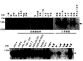
最近ではcDNAマイクロアレイを利用した数千種もの遺伝子の発現レベルが検討された結果、種々のタイプの乳癌における個別のパターンが発見されている(Sgroi, D. C. et al.,「In vivo gene expression profile analysis of human breast cancer progression.」Cancer Res, 59:5656-5661, 1999;Sorlie, T. et al.,「Gene expression patterns of breast carcinomas distinguish tumor subclasses with clinical implications.」Proc Natl Acad Sci U S A, 98:10869-10874, 2001;Kauraniemi, P. et al.,「New amplified and highly expressed genes discovered in the ERBB2 amplicon in breast cancer by cDNA microarrays.」Cancer Res, 61:8235-8240, 2001;Gruvberger, S. et al., S.「Estrogen receptor status in breast cancer is associated with remarkably distinct gene expression patterns.」Cancer Res, 61:5979-5984, 2001;Dressman, M. et al.,「Gene expression profiling detects gene amplification and differentiates tumor types in breast cancer.」Cancer Res, 63:2194-2199, 2003)。 Recently, the expression levels of thousands of genes using cDNA microarrays have been investigated, and individual patterns in various types of breast cancer have been discovered (Sgroi, DC et al., “In vivo gene expression profile analysis Cancer Res, 59: 5656-5661, 1999; Sorlie, T. et al., “Gene expression patterns of breast carcinomas distinguish tumor subclasses with clinical implications.” Proc Natl Acad Sci USA, 98: 10869 -10874, 2001; Kauraniemi, P. et al., “New amplified and highly expressed genes discovered in the ERBB2 amplicon in breast cancer by cDNA microarrays.” Cancer Res, 61: 8235-8240, 2001; Gruvberger, S. et al. ., S. “Estrogen receptor status in breast cancer is associated with remarkably distinct gene expression patterns.” Cancer Res, 61: 5979-5984, 2001; Dressman, M. et al., “Gene expression profiling detects gene amplification and differentiates tumor. types in breast cancer. "Cance r Res, 63: 2194-2199, 2003).
乳癌における遺伝子発現プロファイルに関する研究により、診断マーカーまたは予後判定プロファイルのための候補として役立つ可能性のある遺伝子が同定されている。しかし、乳癌細胞は高度の炎症反応を伴う固形腫瘤として存在し、種々な細胞成分を含むため、主として腫瘍塊から得られたこれらのデータは乳癌発生過程における発現変化を十分には反映できない。したがって、以前に公開されたマイクロアレイデータは混成の(heterogenous)プロファイルを反映している可能性が高い。 Studies on gene expression profiles in breast cancer have identified genes that may serve as candidates for diagnostic markers or prognostic profiles. However, since breast cancer cells exist as solid masses with a high degree of inflammatory reaction and contain various cellular components, these data obtained mainly from tumor masses cannot sufficiently reflect changes in expression during breast cancer development. Thus, previously published microarray data is likely to reflect a heterogenous profile.
発癌メカニズムを解明するように計画された研究によって、いくつかの抗腫瘍物質の分子標的の同定が既に促進されている。例えば、Rasに関連する増殖-シグナル伝達経路を阻害するように当初開発されたファルネシルトランスフェラーゼ阻害剤(FTI)(この活性は翻訳後のファルネシル化に依存する)は、動物モデルにおいてRas依存的腫瘍を治療するために有効であることが示されている(He et al., Cell 99:335-45(1999))。同様に、抗癌剤と、原癌遺伝子受容体HER2/neuに拮抗するための抗HER-2モノクローナル抗体であるトラスツズマブを併用したヒトに対する臨床試験が実施されており、乳癌患者の臨床応答および総生存率の改善が得られている(Lin et al., Cancer Res. 61:6345-9(2001))。bcr-abl融合タンパク質を選択的に不活化するチロシンキナーゼ阻害剤STI-571は、bcr-ablチロシンキナーゼの構成的活性化が白血球の形質転換において重要な役割を果たしている慢性骨髄性白血病を治療するためにようやく開発されてきている。これらの種類の物質は、特定の遺伝子産物の発癌活性を抑制するように設計されている(Fujita et al., Cancer Res. 61:7722-6(2001))。よって、癌性細胞において一般的に上方制御されている遺伝子産物が、新規抗癌剤を開発するための有力な標的として役立つ可能性があることは理解できる。 Studies designed to elucidate the mechanisms of carcinogenesis have already facilitated the identification of molecular targets for several antitumor agents. For example, a farnesyltransferase inhibitor (FTI), originally developed to inhibit the growth-signaling pathway associated with Ras, whose activity is dependent on post-translational farnesylation, has prevented Ras-dependent tumors in animal models. It has been shown to be effective for treatment (He et al., Cell 99: 335-45 (1999)). Similarly, clinical trials have been conducted in humans in combination with anticancer drugs and trastuzumab, an anti-HER-2 monoclonal antibody to antagonize the proto-oncogene receptor HER2 / neu, and clinical response and overall survival of breast cancer patients (Lin et al., Cancer Res. 61: 6345-9 (2001)). STI-571, a tyrosine kinase inhibitor that selectively inactivates bcr-abl fusion proteins, treats chronic myeloid leukemia where constitutive activation of bcr-abl tyrosine kinase plays an important role in leukocyte transformation It has finally been developed. These types of substances are designed to suppress the carcinogenic activity of certain gene products (Fujita et al., Cancer Res. 61: 7722-6 (2001)). Thus, it can be appreciated that gene products that are generally up-regulated in cancerous cells may serve as potential targets for developing new anticancer agents.
CD8+細胞障害性Tリンパ球(CTL)は、MHCクラスI分子上に提示された腫瘍関連抗原(TAA)に由来するエピトープペプチドを認識して、腫瘍細胞を溶解することがさらに証明されている。TAAの最初の例としてMAGEファミリーが発見されて以来、免疫学的アプローチを用いて他にも多くのTAAが発見されている(Boon, Int. J. Cancer 54:177-80(1993);Boon and van der Bruggen, J. Exp. Med. 183:725-9(1996);van der Bruggen et al., Science 254:1643-7(1991);Brichard et al., J. Exp. Med.178:489-95(1993);Kawakami et al., J. Exp. Med. 180:347-52(1994))。新しく発見されたTAAのいくつかにおいては、免疫治療の標的として現在臨床開発が行われている。これまで発見されたTAAには、MAGE(van der Bruggen et al., Science 254:1643-7(1991))、gp100(Kawakami et al., J. Exp. Med. 180:347-52(1994))、SART(Shichijo et al., J. Exp. Med. 187:277-88(1998)、およびNY-ESO-1(Chen et al., Proc. Natl. Acad. Sci. USA 94:1914-8(1997))が含まれる。一方、腫瘍細胞において特に過剰発現されることが示されている遺伝子産物は、細胞性免疫応答を誘導する標的として認識されることが示されている。そのような遺伝子産物には、p53(Umano et al., Brit. J. Cancer 84:1052-7(2001))、HER2/neu(Tanaka et al., Brit. J. Cancer 84:94-9(2001))、CEA(Nukaya et al., Int. J. Cancer 80:92-7(1999))等が含まれる。 CD8 + cytotoxic T lymphocytes (CTL) have further been demonstrated to recognize epitope peptides derived from tumor associated antigens (TAA) presented on MHC class I molecules and lyse tumor cells. Since the discovery of the MAGE family as the first example of TAA, many other TAAs have been discovered using immunological approaches (Boon, Int. J. Cancer 54: 177-80 (1993); and van der Bruggen, J. Exp. Med. 183: 725-9 (1996); van der Bruggen et al., Science 254: 1643-7 (1991); Brichard et al., J. Exp. Med. 178: 489-95 (1993); Kawakami et al., J. Exp. Med. 180: 347-52 (1994)). Several newly discovered TAAs are currently in clinical development as targets for immunotherapy. TAAs discovered so far include MAGE (van der Bruggen et al., Science 254: 1643-7 (1991)), gp100 (Kawakami et al., J. Exp. Med. 180: 347-52 (1994) ), SART (Shichijo et al., J. Exp. Med. 187: 277-88 (1998), and NY-ESO-1 (Chen et al., Proc. Natl. Acad. Sci. USA 94: 1914-8 (1997)), while gene products that have been shown to be specifically overexpressed in tumor cells have been shown to be recognized as targets that induce cellular immune responses. Gene products include p53 (Umano et al., Brit. J. Cancer 84: 1052-7 (2001)), HER2 / neu (Tanaka et al., Brit. J. Cancer 84: 94-9 (2001)) CEA (Nukaya et al., Int. J. Cancer 80: 92-7 (1999)) and the like.
TAAに関する基礎および臨床研究における著しい進歩にもかかわらず(Rosenberg et al., Nature Med. 4:321-7(1998);Mukherji et al., Proc. Natl. Acad. Sci. USA 92:8078-82(1995);Hu et al., Cancer Res. 56:2479-83(1996))、結腸癌を含む腺癌の治療に現在利用できるのは、ごく限られた数の候補TAAに過ぎない。癌細胞において豊富に発現されると共に未だその発現が癌細胞に限定されるTAAは、免疫療法の標的として有望な候補物質となるであろう。さらに、強力で特異的な抗腫瘍免疫応答を誘導する新規TAAが同定されれば、様々なタイプの癌におけるペプチドワクチン法の臨床応用を促進すると期待される(Boon and van der Bruggen, J. Exp. Med. 183:725-9(1996);van der Bruggen et al., Science 254:1643-7(1991);Brichard et al., J. Exp. Med.178:489-95(1993);Kawakami et al., J. Exp. Med. 180:347-52(1994);Shichijo et al., J. Exp. Med. 187:277-88(1998);Chen et al., Proc. Natl. Acad. Sci. USA 94:1914-8(1997);Harris CC., J. Natl. Cancer Inst. 88:1442-55(1996);Butterfield et al., Cancer Res. 59:3134-42(1999);Vissers et al., Cancer Res. 59:5554-9(1999);van der Burg et al., J. Immunol. 156:3308-14(1996);Tanaka et al., Cancer Res. 57:4465-8(1997);Fujie et al., Int. J. Cancer 80:169-72(1999);Kikuchi et al., Int. J. Cancer 81:459-66(1999);Oiso et al., Int. J. Cancer 81:387-94(1999))。 Despite significant advances in basic and clinical research on TAA (Rosenberg et al., Nature Med. 4: 321-7 (1998); Mukherji et al., Proc. Natl. Acad. Sci. USA 92: 8078-82 (1995); Hu et al., Cancer Res. 56: 2479-83 (1996)), only a limited number of candidate TAAs are currently available for the treatment of adenocarcinoma including colon cancer. TAA, which is abundantly expressed in cancer cells and whose expression is still limited to cancer cells, would be a promising candidate for immunotherapy targets. In addition, the identification of new TAAs that induce potent and specific anti-tumor immune responses is expected to facilitate the clinical application of peptide vaccine methods in various types of cancer (Boon and van der Bruggen, J. Exp Med. 183: 725-9 (1996); van der Bruggen et al., Science 254: 1643-7 (1991); Brichard et al., J. Exp. Med. 178: 489-95 (1993); et al., J. Exp. Med. 180: 347-52 (1994); Shichijo et al., J. Exp. Med. 187: 277-88 (1998); Chen et al., Proc. Natl. Acad. Sci. USA 94: 1914-8 (1997); Harris CC., J. Natl. Cancer Inst. 88: 1442-55 (1996); Butterfield et al., Cancer Res. 59: 3134-42 (1999); et al., Cancer Res. 59: 5554-9 (1999); van der Burg et al., J. Immunol. 156: 3308-14 (1996); Tanaka et al., Cancer Res. 57: 4465-8 ( 1997); Fujie et al., Int. J. Cancer 80: 169-72 (1999); Kikuchi et al., Int. J. Cancer 81: 459-66 (1999); Oiso et al., Int. Cancer 81: 387-94 (1999) .
ペプチド刺激された特定の健康なドナー由来の末梢血単核細胞(PBMC)は、ペプチド刺激に応答して著しいレベルのIFN-αを産生するが、51Cr-放出アッセイにおいてHLA-A24または-A0201拘束的に腫瘍細胞に対して細胞障害性を発揮することはまれであることは繰り返し報告されている(Kawano et al., Cancer Res. 60:3550-8(2000);Nishizaka et al., Cancer Res. 60:4830-7(2000);Tamura et al., Jpn. J. Cancer Res. 92:762-7(2001))。しかし、HLA-A24およびHLA-A0201はいずれも、白人集団のみならず、日本人集団における一般的なHLA対立遺伝子である(Date et al., Tissue Antigens 47:93-101(1996);Kondo et al., J. Immunol. 155:4307-12(1995);Kubo et al., J. Immunol. 152:3913-24(1994);Imanishi et al., Proceeding of the eleventh International Histocompatibility Workshop and Conference, Oxford University Press, Oxford, 1065(1992);Williams et al., Tissue Antigen 49:129(1997))。このように、これらのHLAによって提示される癌の抗原性ペプチドは、日本人および白人における癌の治療において特に有用となる可能性がある。さらに、インビトロでの低親和性CTLの誘導は、通常、高濃度のペプチドの使用によって、CTLを効果的に活性化する高レベルの特異的なペプチド/MHC複合体を抗原提示細胞(APCs)上に生成する結果であろうことは知られている(Alexander-Miller et al., Proc. Natl. Acad. Sci. USA 93:4102-7(1996))。 Peripheral blood mononuclear cells (PBMC) from certain healthy donors that have been peptide stimulated produce significant levels of IFN-α in response to peptide stimulation, but HLA-A24 or -A0201 in 51 Cr-release assays It has been repeatedly reported that it is rare to exert cytotoxicity on tumor cells in a restricted manner (Kawano et al., Cancer Res. 60: 3550-8 (2000); Nishizaka et al., Cancer Res. 60: 4830-7 (2000); Tamura et al., Jpn. J. Cancer Res. 92: 762-7 (2001)). However, both HLA-A24 and HLA-A0201 are common HLA alleles in the Japanese population as well as the Caucasian population (Date et al., Tissue Antigens 47: 93-101 (1996); Kondo et al. al., J. Immunol. 155: 4307-12 (1995); Kubo et al., J. Immunol. 152: 3913-24 (1994); Imanishi et al., Proceeding of the eleventh International Histocompatibility Workshop and Conference, Oxford University Press, Oxford, 1065 (1992); Williams et al., Tissue Antigen 49: 129 (1997)). Thus, the cancer antigenic peptides presented by these HLAs may be particularly useful in the treatment of cancer in Japanese and Caucasians. In addition, in vitro induction of low affinity CTLs usually results in high levels of specific peptide / MHC complexes on antigen-presenting cells (APCs) that effectively activate CTL by the use of high concentrations of peptides. (Alexander-Miller et al., Proc. Natl. Acad. Sci. USA 93: 4102-7 (1996)).
このため、癌に関係する発癌機構の解明、および新規抗癌剤を開発するための可能性のある標的の同定を目的とした取り組みとして、乳癌細胞の精製集団における遺伝子発現パターンの大規模解析を、23,040種の遺伝子を提示するcDNAマイクロアレイを用いて行った。より具体的には、cDNAマイクロアレイおよびレーザービームマイクロダイセクションの組み合わせを用いて、12例の非浸潤性乳管癌(DCIS)および69例の浸潤性乳管癌(IDC)を含む、81例の乳房腫瘍の正確なゲノム全域にわたる発現プロファイルを調べた。同定された上方制御される遺伝子のうち、乳癌細胞で有意に過剰発現されるという理由から以下の3つの遺伝子を選択した:A5657(以前はAF161499と呼ばれていた);B9769(以前はAA156269と呼ばれていた);およびC7965(以前はAW977394と呼ばれていた)。本明細書で詳細に考察する所見から、これらの遺伝子が腫瘍細胞の成長増殖に重要な役割を果たしており、このため抗癌薬の開発のための有望な標的であることが示唆される。 Therefore, a large-scale analysis of gene expression patterns in a purified population of breast cancer cells was undertaken in an effort to elucidate the mechanisms of cancer-related carcinogenesis and identify potential targets for the development of new anticancer drugs. This was done using a cDNA microarray displaying the genes of the species. More specifically, using a combination of cDNA microarray and laser beam microdissection, 81 cases, including 12 non-invasive ductal carcinoma (DCIS) and 69 invasive ductal carcinoma (IDC) The exact genome-wide expression profile of breast tumors was examined. Of the identified upregulated genes, the following three genes were selected because they were significantly overexpressed in breast cancer cells: A5657 (formerly called AF161499); B9769 (formerly AA156269 And C7965 (formerly called AW977394). The findings discussed in detail herein suggest that these genes play an important role in the growth and proliferation of tumor cells and are therefore promising targets for the development of anticancer drugs.
発明の概要
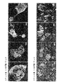
本発明は、アッセイした乳癌症例のそれぞれ49例中38例、73例中30例、および49例中28例で発現が有意に上方制御されていた以下の3つの遺伝子の発見に関する:A5657(SEQ ID NO:1)、これはユビキチン結合酵素に類似したHSPC150タンパク質(SEQ ID NO:2)をコードする;B9769(SEQ ID NO:3)、これは仮説上のタンパク質BC016861(SEQ ID NO:4)をコードする;およびC7965(SEQ ID NO:5)、これはLOC90557(SEQ ID NO:6)として指定される。その後の半定量的RT-PCRおよびノーザンブロット分析により、A5657、B9769、およびC7965が、臨床的な乳癌試料および乳癌細胞株では、乳管細胞および正常乳房を含む正常ヒト組織と比較して有意に過剰発現されることが確かめられた。特に、B9769はERα陽性乳癌細胞株において高発現された。免疫細胞化学染色により、乳癌細胞株T47D細胞において、外因性A5657、B9769、およびC7965はそれぞれ細胞質および/または核装置、細胞質、および細胞小器官のそれぞれに局在した。特に、外因性B9769は、COS7細胞およびT47D細胞において中間径フィラメント網に観察された。さらに、A5657タンパク質がユビキチンと相互作用することが免疫沈降結合アッセイによって示され、このことはA5657タンパク質がE2ユビキチン酵素活性を有する可能性を示唆する。低分子干渉RNA(siRNA)による乳癌細胞の処理は、A5657、B9769、およびC7965の発現を効果的に阻害し、乳癌の細胞/腫瘍成長をそれぞれ抑制した。加えて、この3種の遺伝子はいずれも、NIH3T3細胞で一過性に過剰発現させた場合にMTTアッセイにおける細胞増殖を劇的に促進することが見いだされ、このことからこれらが細胞の成長増殖に重要な役割を果たすことが示唆された。これらの所見は、A5657、B9769、およびC7965の過剰発現が乳房腫瘍形成に関与する可能性があり、かつ乳癌患者に対する特異的治療法のための有望な戦略となる可能性があることを示唆する。
The present invention relates to the discovery of the following three genes whose expression was significantly upregulated in 38 of 49, 30 of 73 and 28 of 49 breast cancer cases assayed, respectively: A5657 (SEQ ID NO: 1), which encodes an HSPC150 protein (SEQ ID NO: 2) similar to a ubiquitin-conjugating enzyme; B9769 (SEQ ID NO: 3), which is a hypothetical protein BC016861 (SEQ ID NO: 2) : 4); and C7965 (SEQ ID NO: 5), designated as LOC90557 (SEQ ID NO: 6). Subsequent semiquantitative RT-PCR and Northern blot analysis showed that A5657, B9769, and C7965 were significantly higher in clinical breast cancer samples and breast cancer cell lines compared to normal human tissues including ductal cells and normal breast It was confirmed that it was overexpressed. In particular, B9769 was highly expressed in ERα-positive breast cancer cell lines. By immunocytochemical staining, exogenous A5657, B9769, and C7965 were localized in the cytoplasm and / or nuclear apparatus, cytoplasm, and organelle, respectively, in breast cancer cell line T47D cells. In particular, exogenous B9769 was observed in the intermediate filament network in COS7 and T47D cells. Furthermore, immunoprecipitation binding assays show that A5657 protein interacts with ubiquitin, suggesting that A5657 protein may have E2 ubiquitin enzyme activity. Treatment of breast cancer cells with small interfering RNA (siRNA) effectively inhibited expression of A5657, B9769, and C7965 and suppressed breast cancer cell / tumor growth, respectively. In addition, all three genes were found to dramatically promote cell proliferation in the MTT assay when transiently overexpressed in NIH3T3 cells, which led to cell growth and proliferation. It was suggested to play an important role. These findings suggest that overexpression of A5657, B9769, and C7965 may be involved in breast tumorigenesis and may be a promising strategy for specific therapies for breast cancer patients .
したがって、本発明は、これらの3種の遺伝子の発現が乳癌(BRC)と有意に相関するという発見を含む。乳癌において差次的に発現されるこれらの遺伝子は、本明細書において「BRC核酸」または「BRCポリヌクレオチド」と総称され、コードされる対応するポリペプチドは「BRCポリペプチド」または「BRCタンパク質」と呼ばれる。 Thus, the present invention includes the discovery that the expression of these three genes is significantly correlated with breast cancer (BRC). These genes that are differentially expressed in breast cancer are collectively referred to herein as “BRC nucleic acids” or “BRC polynucleotides” and the corresponding polypeptides encoded are “BRC polypeptides” or “BRC proteins”. Called.
したがって、本発明は、患者由来の生物学的試料(組織試料など)におけるBRC関連遺伝子の発現レベルを決定することによる、対象における乳癌に対する素因を診断または判定する方法を提供する。「BRC関連遺伝子」という用語は、BRC細胞における発現レベルが正常細胞と比較して異なることを特徴とする遺伝子を指す。正常細胞は乳房組織から得られたものである。本発明の文脈において、BRC関連遺伝子とは、A5657(SEQ ID NO:1)、B9769(SEQ ID NO:3)およびC7965(SEQ ID NO:5)からなる群より選択される1つまたは複数の遺伝子のことである。BRC関連遺伝子の発現レベルが遺伝子の正常対照レベルと比較して変化していること、例えば上昇していることにより、対象がBRCに罹患している、またはBRCを発症するリスクを有することが示される。 Accordingly, the present invention provides a method of diagnosing or determining a predisposition to breast cancer in a subject by determining the level of expression of a BRC-related gene in a patient-derived biological sample (such as a tissue sample). The term “BRC-related gene” refers to a gene that is characterized by a different level of expression in BRC cells compared to normal cells. Normal cells are obtained from breast tissue. In the context of the present invention, a BRC-related gene is one or more selected from the group consisting of A5657 (SEQ ID NO: 1), B9769 (SEQ ID NO: 3) and C7965 (SEQ ID NO: 5). It is a gene. A change in the expression level of a BRC-related gene compared to the normal control level of the gene, for example an increase, indicates that the subject is suffering from or at risk of developing BRC. It is.
本発明の文脈において、「対照レベル」という語句は、対照試料で検出されるタンパク質発現レベルを指し、これには正常対照レベルおよび乳癌対照レベルの両方が含まれる。対照レベルは、単一の参照集団に由来する単一の発現パターンであっても、または複数の発現パターンに由来する単一の発現パターンであってもよい。例えば、対照レベルが、以前に試験を行った細胞からの発現パターンのデータベースであってもよい。「正常対照レベル」とは、正常で健康な個体において、または乳癌に罹患していないことが判明している個体の集団において検出される遺伝子の発現レベルを指す。正常個体とは、乳癌の臨床症状がない個体のことである。これに対して、「BRC対照レベル」とは、BRCに罹患した集団で認められるBRC関連遺伝子の発現プロファイルを指す。 In the context of the present invention, the phrase “control level” refers to the level of protein expression detected in a control sample, including both normal and breast cancer control levels. The control level can be a single expression pattern derived from a single reference population or a single expression pattern derived from multiple expression patterns. For example, the control level may be a database of expression patterns from previously tested cells. “Normal control level” refers to the level of gene expression detected in a normal healthy individual or in a population of individuals known to be free of breast cancer. A normal individual is an individual who has no clinical symptoms of breast cancer. In contrast, “BRC control level” refers to the expression profile of BRC-related genes found in populations affected with BRC.
被験試料中に検出されるA5657、B9769、およびC7965からなる群より選択される1つまたは複数のBRC関連遺伝子の発現レベルが正常対照レベルと比較して上昇していることにより、(試料を提供した)対象がBRCに罹患している、またはBRCを発症するリスクを有することが示される。 The expression level of one or more BRC-related genes selected from the group consisting of A5657, B9769, and C7965 detected in the test sample is increased compared to the normal control level (provided sample The subject is suffering from or at risk of developing BRC.
または、試料における2つまたはそれ以上のBRC関連遺伝子の一群の発現を、同じ遺伝子の一群のBRC対照レベルと比較することもできる。試料での発現とBRC対照での発現との間に類似性があれば、(試料を提供した)対象がBRCに罹患している、またはBRCを発症するリスクを有することが示される。 Alternatively, the expression of a group of two or more BRC-related genes in a sample can be compared to a group of BRC control levels of the same gene. A similarity between expression in the sample and expression in the BRC control indicates that the subject (who provided the sample) is suffering from or at risk of developing BRC.
本発明によれば、遺伝子発現は、対照レベルと比較して少なくとも10%、少なくとも25%、少なくとも50%またはそれ以上上昇している場合に「変化している」とみなされる。または、遺伝子発現は、対照レベルと比較して少なくとも1倍、少なくとも2倍、少なくとも5倍またはそれ以上上昇している場合に変化しているとみなしてもよい。遺伝子発現は、例えばアレイ上で、BRC関連遺伝子プローブと患者由来の組織試料の遺伝子転写物とのハイブリダイゼーションを検出することによって決定される。 According to the present invention, gene expression is considered “altered” if it is increased by at least 10%, at least 25%, at least 50% or more compared to the control level. Alternatively, gene expression may be considered altered if it is elevated at least 1 fold, at least 2 fold, at least 5 fold or more compared to a control level. Gene expression is determined, for example, by detecting hybridization of a BRC-related gene probe with a gene transcript of a patient-derived tissue sample on an array.
本発明の文脈において、患者由来の組織試料は、被験対象から、例えば、BRCを有することが判明しているかその疑いがある患者から得られる任意の試料であってもよい。例えば、組織には上皮細胞が含まれてもよい。より具体的には、組織は乳管癌からの上皮細胞でありうる。 In the context of the present invention, a patient-derived tissue sample may be any sample obtained from a subject, eg, a patient known or suspected of having BRC. For example, the tissue may include epithelial cells. More specifically, the tissue can be epithelial cells from breast cancer.
本発明はまた、A5657、B9769、およびC7965からなる群より選択されるBRC関連遺伝子の2つまたはそれ以上の遺伝子発現レベルを含む、BRC参照発現プロファイルも提供する。 The present invention also provides a BRC reference expression profile comprising two or more gene expression levels of a BRC-related gene selected from the group consisting of A5657, B9769, and C7965.
本発明はさらに、BRC関連遺伝子の、例えばA5657、B9769、およびC7965からなる群より選択されるBRC関連遺伝子の発現または活性を阻害する物質を同定する方法であって、BRC関連遺伝子を発現する被験細胞を被験化合物に接触させ、かつBRC関連遺伝子の発現レベルまたは活性を決定することによる方法も提供する。被験細胞は、乳癌から得られた上皮細胞などの上皮細胞であってもよい。BRC関連遺伝子またはその遺伝子産物の発現レベルまたは活性がその遺伝子または遺伝子産物の被験化合物の非存在下で検出される発現レベルまたは活性と比較して低下していることにより、被験物質がBRC関連遺伝子の阻害物質であることが示され、これはBRCの症状、例えばA5657、B9769、およびC7965からなる群より選択されるBRC関連遺伝子の発現を低下させるために用いられる可能性がある。 The present invention further provides a method for identifying a substance that inhibits the expression or activity of a BRC-related gene, for example, a BRC-related gene selected from the group consisting of A5657, B9769, and C7965, wherein the test expresses a BRC-related gene. Also provided are methods by contacting a cell with a test compound and determining the expression level or activity of a BRC-related gene. The test cell may be an epithelial cell such as an epithelial cell obtained from breast cancer. A test substance is a BRC-related gene because the expression level or activity of the BRC-related gene or its gene product is reduced compared to the expression level or activity detected in the absence of the test compound of that gene or gene product. Which may be used to reduce the expression of BRC-related genes selected from the group consisting of symptoms of BRC, eg, A5657, B9769, and C7965.
本発明はまた、1つまたは複数のBRC核酸またはBRCポリペプチドと結合する検出試薬を含むキットも提供する。1つまたは複数のBRC核酸と結合する核酸のアレイも提供される。 The present invention also provides a kit comprising a detection reagent that binds to one or more BRC nucleic acids or BRC polypeptides. An array of nucleic acids that binds to one or more BRC nucleic acids is also provided.
本発明の治療方法には、対象におけるBRCを治療または予防する方法であって、アンチセンス組成物を対象に投与する段階を含む方法が含まれる。本発明の文脈において、アンチセンス組成物は特異的な標的遺伝子の発現を低下させる。例えば、アンチセンス組成物は、BRC関連遺伝子A5657、B9769、およびC7965からなる群より選択されるBRC関連遺伝子配列に対して相補的なヌクレオチドを含みうる。または、本方法が、低分子干渉RNA(siRNA)組成物を対象に投与する段階を含んでもよい。本発明の文脈において、siRNA組成物は、BRC関連遺伝子A5657、B9769、およびC7965からなる群より選択されるBRC核酸の発現を低下させる。さらにもう1つの方法において、対象におけるBRCの治療または予防を、リボザイム組成物を対象に投与することによって実施することもできる。本発明の文脈において、核酸特異的なリボザイム組成物は、BRC関連遺伝子A5657、B9769、およびC7965からなる群より選択されるBRC核酸の発現を低下させる。 The therapeutic methods of the present invention include a method of treating or preventing BRC in a subject, comprising the step of administering an antisense composition to the subject. In the context of the present invention, an antisense composition reduces the expression of a specific target gene. For example, the antisense composition can comprise nucleotides complementary to a BRC-related gene sequence selected from the group consisting of BRC-related genes A5657, B9769, and C7965. Alternatively, the method may include administering a small interfering RNA (siRNA) composition to the subject. In the context of the present invention, the siRNA composition reduces the expression of a BRC nucleic acid selected from the group consisting of BRC-related genes A5657, B9769, and C7965. In yet another method, treatment or prevention of BRC in a subject can be performed by administering a ribozyme composition to the subject. In the context of the present invention, the nucleic acid specific ribozyme composition reduces the expression of a BRC nucleic acid selected from the group consisting of BRC-related genes A5657, B9769, and C7965.
本発明はまた、ワクチンおよびワクチン接種方法も含む。例えば、対象におけるBRCを治療または予防する方法が、BRC関連遺伝子A5657、B9769、およびC7965からなる群より選択される核酸によってコードされるポリペプチド、またはこのようなポリペプチドの免疫学的活性断片を含むワクチンを対象に投与する段階を含んでもよい。本発明の文脈において、免疫学的活性断片とは、完全長の天然型タンパク質よりも長さは短いものの、完全長タンパク質によって誘導される免疫応答に類似した免疫応答を誘導するポリペプチドのことである。例えば、免疫学的活性断片は、少なくとも8残基長であって、T細胞またはB細胞などの免疫細胞を賦活化しうるものであるべきである。免疫細胞の賦活化は、細胞増殖、サイトカイン(例えば、IL-2)の生成、細胞傷害性Tリンパ球の誘導、または抗体の産生を検出することによって測定可能である。 The invention also includes vaccines and vaccination methods. For example, a method of treating or preventing BRC in a subject comprises a polypeptide encoded by a nucleic acid selected from the group consisting of BRC-related genes A5657, B9769, and C7965, or an immunologically active fragment of such a polypeptide. The method may include administering a vaccine comprising the subject to the subject. In the context of the present invention, an immunologically active fragment is a polypeptide that induces an immune response similar to that induced by the full-length protein, although shorter in length than the full-length native protein. is there. For example, an immunologically active fragment should be at least 8 residues long and capable of stimulating immune cells such as T cells or B cells. Immune cell activation can be measured by detecting cell proliferation, production of cytokines (eg, IL-2), induction of cytotoxic T lymphocytes, or production of antibodies.
特に定義していなければ、本明細書において用いた科学技術用語は全て、本発明が属する当業者によって一般的に理解される意味と同じ意味を有する。本明細書に記述の方法および材料と類似または同等の方法および材料を、本発明の実践または試験において用いることができるが、適した方法および材料を下記に記述する。本明細書において言及した全ての刊行物、特許出願、特許、および他の参考文献はその全体が参照として本明細書に組み入れられる。矛盾する場合には、定義を含めて本明細書が優先する。さらに、材料、方法、および例は、説明するために限られ、制限することを意図しない。 Unless defined otherwise, all technical and scientific terms used herein have the same meaning as commonly understood by one of ordinary skill in the art to which this invention belongs. Although methods and materials similar or equivalent to those described herein can be used in the practice or testing of the present invention, suitable methods and materials are described below. All publications, patent applications, patents, and other references mentioned herein are hereby incorporated by reference in their entirety. In case of conflict, the present specification, including definitions, will control. In addition, the materials, methods, and examples are illustrative only and not intended to be limiting.
本明細書に記述の方法の一つの長所は、乳癌の明白な臨床症状を検出する前に疾患が同定されることである。本発明のその他の特徴および長所は、以下の詳細な説明および特許請求の範囲から明らかとなるであろう。 One advantage of the methods described herein is that the disease is identified prior to detecting overt clinical symptoms of breast cancer. Other features and advantages of the invention will be apparent from the following detailed description and from the claims.
詳細な説明
本明細書で用いる「1つの(a)」、「1つの(an)」および「その(the)」という用語は、別に特記する場合を除き、「少なくとも1つの」を意味する。
DETAILED DESCRIPTION As used herein, the terms “a”, “an” and “the” mean “at least one” unless otherwise specified.
一般に、乳癌細胞は、高度の炎症反応を有するとともに種々の細胞成分を含む固形腫瘤として存在する。このため、これまでに発表されたマイクロアレイデータは混成のプロファイルを反映している可能性が高い。 In general, breast cancer cells exist as a solid mass having a highly inflammatory response and containing various cellular components. For this reason, microarray data published so far is likely to reflect a hybrid profile.
これらの問題を考慮して、本発明者らは、乳癌細胞および正常乳房上皮乳管細胞の精製集団をレーザーマイクロビームマイクロダイセクション(laser-microbeam microdissection)(LMM)法によって調製した。12例のDCISおよび69例のIDCを含む81例のBRCからの癌細胞の遺伝子発現プロファイルを、23,040遺伝子を提示するcDNAマイクロアレイを用いて解析した。レーザーマイクロダイセクション法によって純粋なものとして選択された、BRCと診断された患者由来の癌細胞、および正常乳管上皮細胞の発現パターンを比較することにより、102種の遺伝子(データ非提示)がBRC細胞で通例的に上方制御される遺伝子として同定され、288種の遺伝子(データ非提示)がBRC細胞で通例的に下方制御される遺伝子として同定された。患者の血清中の癌関連タンパク質を検出する能力がある分子マーカー候補を選択したところ、ヒトBRCにおけるシグナル抑制戦略を開発するための標的となる可能性のあるものがいくつか見いだされた。特に、本発明は、3種の核酸、すなわちA5657、B9769、およびC7965の発現パターンが上皮細胞とBRC患者の癌との間で変化しているという発見を含む。A5657遺伝子(SEQ ID NO:1)は新規の配列を構成し、ユビキチン結合酵素(Genbankアクセッション番号NM_014176)に類似したHSPC150タンパク質(SEQ ID NO:2)をコードする。A5657はIDC細胞においてDCIS細胞および正常乳房上皮細胞と比較して上方制御される。B9769遺伝子(SEQ ID NO:3)は新規な配列を構成し、仮説上のタンパク質(SEQ ID NO:4)(Genbankアクセッション番号NM_138770)をコードする。B9769はIDC細胞において正常乳房上皮細胞と比較して上方制御される。C7965遺伝子(SEQ ID NO:5)およびそれによってコードされるタンパク質(SEQ ID NO:6)は、LOC90557と称される既知の配列を構成する。C7965はDCIS細胞およびIDC細胞の両方で正常乳房上皮細胞と比較して上方制御される。本明細書で同定された、差次的に発現されるこれらの遺伝子には、BRCマーカーおよびBRC遺伝子標的としての診断上の有用性がみられ、その発現は、BRC症状の治療または軽減を目的に変化されうる。または、本明細書で同定された、DCISとIDCとの間で差次的に発現されるA5657遺伝子には、IDCをDCISと区別するためのマーカーならびにBRC遺伝子標的としての診断上の有用性がみられ、IDC症状の治療または軽減を目的にその発現が変化されうる。 In view of these problems, we prepared purified populations of breast cancer cells and normal breast epithelial duct cells by the laser-microbeam microdissection (LMM) method. Gene expression profiles of cancer cells from 81 BRCs including 12 DCIS and 69 IDCs were analyzed using a cDNA microarray displaying 23,040 genes. By comparing the expression patterns of cancer cells from patients diagnosed with BRC and normal ductal epithelial cells selected as pure by the laser microdissection method, 102 genes (data not shown) were found. The genes that were typically upregulated in BRC cells were identified, and 288 genes (data not shown) were identified as genes that are typically downregulated in BRC cells. Selection of molecular marker candidates capable of detecting cancer-related proteins in patient sera found several potential targets for developing signal suppression strategies in human BRC. In particular, the present invention includes the discovery that the expression pattern of three nucleic acids, A5657, B9769, and C7965, is altered between epithelial cells and BRC patient cancer. The A5657 gene (SEQ ID NO: 1) constitutes a novel sequence and encodes an HSPC150 protein (SEQ ID NO: 2) similar to the ubiquitin conjugating enzyme (Genbank accession number NM_014176). A5657 is upregulated in IDC cells compared to DCIS cells and normal breast epithelial cells. The B9769 gene (SEQ ID NO: 3) constitutes a novel sequence and encodes a hypothetical protein (SEQ ID NO: 4) (Genbank accession number NM_138770). B9769 is upregulated in IDC cells compared to normal breast epithelial cells. The C7965 gene (SEQ ID NO: 5) and the protein encoded thereby (SEQ ID NO: 6) constitute a known sequence called LOC90557. C7965 is upregulated in both DCIS and IDC cells compared to normal breast epithelial cells. These differentially expressed genes identified herein have diagnostic utility as BRC markers and BRC gene targets whose expression is intended to treat or alleviate BRC symptoms Can be changed. Alternatively, the A5657 gene, identified here, that is differentially expressed between DCIS and IDC, has a diagnostic utility as a marker and BRC gene target to distinguish IDC from DCIS. And its expression can be altered to treat or reduce IDC symptoms.
BRC患者において発現レベルが変化している(すなわち、上昇している)これらの遺伝子は、本明細書において「BRC関連遺伝子」、「BRC核酸」、または「BRCポリヌクレオチド」と総称され、対応するコードされるポリペプチドは「BRCポリペプチド」または「BRCタンパク質」と呼ばれる。別に指示する場合を除き、「BRC」とは、本明細書中に開示した配列のうち任意のものを指す(例えば、A5657、B9769、およびC7965からなる群より選択されるBRC関連遺伝子)。 These genes with altered (ie elevated) expression levels in BRC patients are collectively referred to herein as “BRC-related genes”, “BRC nucleic acids”, or “BRC polynucleotides” and correspond The encoded polypeptide is referred to as a “BRC polypeptide” or “BRC protein”. Unless otherwise indicated, “BRC” refers to any of the sequences disclosed herein (eg, a BRC-related gene selected from the group consisting of A5657, B9769, and C7965).
細胞の試料における種々の遺伝子の発現を測定することにより、BRCを診断することができる。同様に、種々の物質に応答したこれらの遺伝子の発現を測定することにより、BRCを治療するための物質を同定することができる。 BRC can be diagnosed by measuring the expression of various genes in a sample of cells. Similarly, substances for treating BRC can be identified by measuring the expression of these genes in response to various substances.
本発明は、A5657、B9769、およびC7965からなる群より選択されるBRC関連遺伝子の少なくとも1つ、最大ですべての発現を決定すること(例えば、測定すること)を含む。既知の配列に関するGenBank(商標)データベースの記載事項によって得られる配列情報を使用し、当業者に周知の手法を用いてBRC関連遺伝子を検出および測定することができる。例えば、BRC関連遺伝子に対応する配列データベース記載事項中の配列を、例えばノーザンブロットハイブリダイゼーション分析において、BRC関連遺伝子に対応するRNA配列を検出するためのプローブを構築するために用いることができる。プローブは、典型的には参照配列のうちの少なくとも10ヌクレオチド、少なくとも20ヌクレオチド、少なくとも50ヌクレオチド、少なくとも100ヌクレオチド、または少なくとも200ヌクレオチドを含む。別の例としては、これらの配列は、例えば増幅に基づいた検出方法(逆転写に基づくポリメラーゼ連鎖反応など)におけるBRC核酸を特異的に増幅するためのプライマーを構築するのに用いることができる。 The present invention includes determining (eg, measuring) the expression of at least one, and at most, a BRC-related gene selected from the group consisting of A5657, B9769, and C7965. Using sequence information obtained from GenBank ™ database entries for known sequences, BRC-related genes can be detected and measured using techniques well known to those skilled in the art. For example, sequences in sequence database entries corresponding to BRC-related genes can be used to construct probes for detecting RNA sequences corresponding to BRC-related genes, eg, in Northern blot hybridization analysis. A probe typically comprises at least 10 nucleotides, at least 20 nucleotides, at least 50 nucleotides, at least 100 nucleotides, or at least 200 nucleotides of a reference sequence. As another example, these sequences can be used to construct primers for specifically amplifying BRC nucleic acids in, for example, amplification-based detection methods (such as reverse transcription-based polymerase chain reaction).
続いて、被験細胞集団、例えば患者由来の組織試料における、BRC関連遺伝子の1つまたは複数の発現レベルを、参照集団における同じ遺伝子の発現レベルと比較する。参照細胞集団は、比較されるパラメーターが既知である1つまたは複数の細胞、すなわち、乳管癌細胞(例えば、BRC細胞)または正常乳管上皮細胞(例えば、非BRC細胞)を含む。 Subsequently, the expression level of one or more BRC-related genes in a test cell population, eg, a patient-derived tissue sample, is compared to the expression level of the same gene in a reference population. The reference cell population includes one or more cells whose parameters to be compared are known, ie breast cancer cells (eg, BRC cells) or normal breast epithelial cells (eg, non-BRC cells).
被験細胞集団における遺伝子発現のパターンが、参照細胞集団と比較してBRCまたはそれに対する素因を示すか否かは、参照細胞集団の組成に依存する。例えば、参照細胞集団が非BRC細胞から構成される場合には、被験細胞集団と参照細胞集団との間で遺伝子発現パターンに類似性があることにより、被験細胞集団が非BRCであると示される。その反対に、参照細胞集団がBRC細胞から構成される場合には、被験細胞集団と参照細胞集団との間で遺伝子発現プロファイルに類似性があることにより、被験細胞集団がBRC細胞を含むと示される。 Whether the pattern of gene expression in the test cell population is predisposed to or relative to BRC as compared to the reference cell population depends on the composition of the reference cell population. For example, if the reference cell population is composed of non-BRC cells, a similarity in gene expression pattern between the test cell population and the reference cell population indicates that the test cell population is non-BRC . Conversely, if the reference cell population is composed of BRC cells, the similarity in gene expression profile between the test cell population and the reference cell population indicates that the test cell population contains BRC cells. It is.
被験細胞集団におけるBRCマーカー遺伝子の発現レベルは、それと参照細胞集団における対応するBRCマーカー遺伝子の発現レベルとの違いが1.0倍を上回る、1.5倍を上回る、2.0倍を上回る、5.0倍を上回る、10.0倍を上回る、またはそれ以上の倍数を上回るならば「変化している」とみなされる。 The expression level of the BRC marker gene in the test cell population differs from that of the corresponding BRC marker gene expression level in the reference cell population by more than 1.0 times, more than 1.5 times, more than 2.0 times, more than 5.0 times, 10.0 times It is considered “changed” if it is more than double or more than a multiple.
被験細胞集団と参照細胞集団との間での差次的な遺伝子発現は、対照核酸、例えばハウスキーピング遺伝子に対して標準化することができる。例えば、対照核酸は、細胞が癌状態にあるか非癌状態にあるかによっての差がないことが判明している核酸である。対照核酸の発現レベルは、被験集団および参照集団におけるシグナルレベルを標準化するのに用いることができる。対照遺伝子の例には、例えば、β-アクチン、グリセルアルデヒドβ-リン酸デヒドロゲナーゼ、およびリボソームタンパク質P1が非制限的に含まれる。 Differential gene expression between a test cell population and a reference cell population can be normalized to a control nucleic acid, such as a housekeeping gene. For example, a control nucleic acid is a nucleic acid that has been found not to differ depending on whether the cell is in a cancerous state or a non-cancerous state. The expression level of the control nucleic acid can be used to normalize the signal level in the test population and the reference population. Examples of control genes include, but are not limited to, for example, β-actin, glyceraldehyde β-phosphate dehydrogenase, and ribosomal protein P1.
被験細胞集団は複数の参照細胞集団と比較することができる。多数の参照集団のそれぞれは既知のパラメーターに関して差があってもよい。したがって、被験細胞集団を、例えばBRC細胞を含むことが判明している第1の参照細胞集団、ならびに例えば非BRC細胞(正常細胞)を含むことが判明している第2の参照集団と比較してもよい。被験細胞は、BRC細胞を含むことが判明しているかまたはBRC細胞を含むことが疑われる対象からの組織試料または細胞試料中に含まれてもよい。 The test cell population can be compared to multiple reference cell populations. Each of the multiple reference populations may differ with respect to a known parameter. Thus, a test cell population is compared to a first reference cell population that is known to contain, for example, BRC cells, and a second reference population that is known to contain, for example, non-BRC cells (normal cells). May be. The test cell may be included in a tissue sample or cell sample from a subject known to contain or suspected of containing BRC cells.
被験細胞は、体組織または体液、例えば、生体液(例えば、血液または痰など)から得られる。例えば、被験細胞を乳房組織から精製してもよい。好ましくは被験細胞集団は上皮細胞を含む。上皮細胞は、好ましくは乳管癌であるか乳管癌であることが疑われる組織からのものである。 The test cell is obtained from a body tissue or a body fluid, for example, a biological fluid (for example, blood or sputum). For example, the test cell may be purified from breast tissue. Preferably the test cell population comprises epithelial cells. The epithelial cells are preferably from tissue that is or is suspected to be breast cancer.
参照細胞集団中の細胞は、被験細胞の組織型と類似した組織型に由来すべきである。任意には、参照細胞集団は細胞株、例えば、BRC細胞株(すなわち、陽性対照)または正常非BRC細胞株(すなわち、陰性対照)である。または、対照細胞集団は、アッセイされるパラメーターまたは条件に関して既知である細胞の分子情報のデータベースに由来していてもよい。 Cells in the reference cell population should be derived from a tissue type similar to that of the test cell. Optionally, the reference cell population is a cell line, eg, a BRC cell line (ie, a positive control) or a normal non-BRC cell line (ie, a negative control). Alternatively, the control cell population may be derived from a database of cellular molecular information known with respect to the parameter or condition being assayed.
対象は、好ましくは哺乳動物である。哺乳動物の例には、例えば、ヒト、非ヒト霊長動物、マウス、ラット、イヌ、ネコ、ウマまたはウシが非制限的に含まれる。 The subject is preferably a mammal. Examples of mammals include, but are not limited to, for example, humans, non-human primates, mice, rats, dogs, cats, horses or cows.
本明細書に開示した遺伝子の発現は、当技術分野で公知の方法を用いて、タンパク質レベルまたは核酸レベルで決定することができる。例えば、遺伝子発現を決定するには、これらの核酸配列の1つまたは複数を特異的に認識するプローブによるノーザンハイブリダイゼーション分析を用いてもよい。または、遺伝子発現は、例えば、差次的に発現される遺伝子配列に対して特異的なプライマーを用いて逆転写に基づくPCRアッセイにより測定することもできる。発現はまた、タンパク質レベルで、すなわち、本明細書に記載の遺伝子によってコードされるポリペプチドのレベルまたはその生物学的活性を測定することによって決定することもできる。この種の方法は当技術分野で周知であり、例えば、遺伝子によってコードされるタンパク質に対する抗体を利用したイムノアッセイが非制限的に含まれる。遺伝子によってコードされるタンパク質の生物学的活性は一般に周知であるか、または慣行的に同定できるかのいずれかである。 Expression of the genes disclosed herein can be determined at the protein or nucleic acid level using methods known in the art. For example, Northern hybridization analysis with probes that specifically recognize one or more of these nucleic acid sequences may be used to determine gene expression. Alternatively, gene expression can be measured, for example, by a PCR assay based on reverse transcription using primers specific for differentially expressed gene sequences. Expression can also be determined at the protein level, ie, by measuring the level of a polypeptide encoded by a gene described herein or its biological activity. Such methods are well known in the art and include, but are not limited to, immunoassays utilizing antibodies to proteins encoded by genes. The biological activity of a protein encoded by a gene is generally well known or can be routinely identified.
新規なヌクレオチド、ポリペプチド、ベクター、および宿主細胞
本発明は、乳癌の大多数において、対応する非癌性乳房上皮と比較して発現が顕著に亢進している新規ヒト遺伝子A5657およびB9769を提供する。単離されたA5657遺伝子は、928ヌクレオチドを含むcDNA配列である、SEQ ID NO:1に記載されたポリヌクレオチド配列を含む。特に、A5657は7つのエクソンからなり、ゲノム中では染色体1q32.1上に約10.3kbの範囲にわたって存在する。このcDNA転写物は最終的には、ユビキチン結合酵素に類似したHSPC150タンパク質という197アミノ酸のポリペプチドをコードする。単離されたB9769遺伝子は、1472ヌクレオチドを含むcDNA配列である、SEQ ID NO:3に記載されたポリヌクレオチドを含む。特に、B9769は8つのエクソンからなり、ゲノム中では染色体2q21.2上に約5.7kbの範囲にわたって存在する。そのORFはエクソン1で始まり、エクソン8で終わる。最終的には、このcDNA転写物は378アミノ酸のポリペプチドをコードする。
Novel nucleotides, polypeptides, vectors, and host cells The present invention provides novel human genes A5657 and B9769 that are significantly upregulated in the majority of breast cancers compared to the corresponding non-cancerous breast epithelium. . The isolated A5657 gene comprises the polynucleotide sequence set forth in SEQ ID NO: 1, which is a cDNA sequence comprising 928 nucleotides. In particular, A5657 consists of 7 exons and exists in the genome over a range of about 10.3 kb on chromosome 1q32.1. This cDNA transcript ultimately encodes a 197 amino acid polypeptide named HSPC150 protein similar to ubiquitin conjugating enzymes. The isolated B9769 gene comprises the polynucleotide set forth in SEQ ID NO: 3, which is a cDNA sequence comprising 1472 nucleotides. In particular, B9769 consists of 8 exons and exists in the genome over a range of about 5.7 kb on chromosome 2q21.2. The ORF begins with
本発明は、SEQ ID NO:1および3に記載のポリヌクレオチド配列を含む新規ヒト遺伝子A5657およびB9769に加えて、それらの縮重物および変異体も、それらがSEQ ID NO:2および4に示されたアミノ酸配列を含むA5657またはB9769タンパク質、またはそれらの機能的等価物をコードする範囲で含む。A5657またはB9769と機能的に等価なポリペプチドの例には、例えば、ヒトA5657またはB9769タンパク質に対応する他の生物の相同タンパク質、ならびにこのようなヒトタンパク質の変異体が含まれる。 In addition to the novel human genes A5657 and B9769 comprising the polynucleotide sequences set forth in SEQ ID NOs: 1 and 3, the present invention also includes degenerates and variants thereof as shown in SEQ ID NOs: 2 and 4. In the range encoding the A5657 or B9769 protein, or a functional equivalent thereof, comprising the amino acid sequence determined. Examples of polypeptides functionally equivalent to A5657 or B9769 include, for example, homologous proteins of other organisms corresponding to the human A5657 or B9769 protein, as well as variants of such human proteins.
本発明はさらに、本発明者らによって同定されたポリペプチドと機能的に等価なポリペプチド、およびこのような機能的に等価なポリペプチドをコードするポリヌクレオチドも包含する。本発明の文脈において、「機能的に等価な」という用語は、対象ポリペプチドが、それぞれSEQ ID NO:2および4にアミノ酸配列を示したA5657またはB9769タンパク質に特徴的な生物学的に意味のある活性を保持していることを意味する。例えば、A5657遺伝子およびB9769遺伝子はいずれも、正常細胞と比較してBRC細胞で特異的に過剰発現される遺伝子として特徴づけられる。A5657遺伝子はさらに、IDC細胞においてDCIS細胞と比較して過剰発現される。さらに、それらの過剰発現が細胞増殖を促進することが本明細書において示される。したがって、本発明の文脈において、A5657またはB9769タンパク質の機能的等価物は、野生型タンパク質の細胞増殖活性に類似した細胞増殖活性を有するはずである。細胞増殖は、当技術分野における従来のアッセイおよび手法(例えば、以下の実施例で考察しているMTTアッセイ)を用いて測定できるパラメーターである。加えて、A5657タンパク質がE2ユビキチン酵素活性を有することおよびユビキチンと結合することも本明細書において示される。したがって、本発明の文脈において、A5657タンパク質の機能的等価物は、野生型タンパク質のものに類似したユビキチン酵素活性および/またはユビキチン結合活性を有するはずである。 The invention further encompasses polypeptides that are functionally equivalent to the polypeptides identified by the inventors, and polynucleotides that encode such functionally equivalent polypeptides. In the context of the present invention, the term “functionally equivalent” means that the subject polypeptide has a biological meaning characteristic of the A5657 or B9769 protein whose amino acid sequences are shown in SEQ ID NOs: 2 and 4, respectively. It means that it has a certain activity. For example, both the A5657 gene and the B9769 gene are characterized as genes that are specifically overexpressed in BRC cells compared to normal cells. The A5657 gene is further overexpressed in IDC cells compared to DCIS cells. Furthermore, it is shown herein that their overexpression promotes cell proliferation. Thus, in the context of the present invention, a functional equivalent of A5657 or B9769 protein should have cell proliferation activity similar to that of wild type protein. Cell proliferation is a parameter that can be measured using conventional assays and techniques in the art (eg, the MTT assay discussed in the Examples below). In addition, it is also shown herein that the A5657 protein has E2 ubiquitin enzyme activity and binds to ubiquitin. Thus, in the context of the present invention, the functional equivalent of the A5657 protein should have ubiquitin enzyme activity and / or ubiquitin binding activity similar to that of the wild type protein.
したがって、本発明は、開示した配列のある種の変異または変種を企図している。例えば、本発明は、1つまたは複数のアミノ酸が置換、欠失、挿入、および/または付加されたSEQ ID NO:2または4のタンパク質をコードするポリヌクレオチドを、その結果生じるタンパク質が野生型タンパク質の生物学的に意味のある活性を保持している限りは包含する。1つの好ましい態様において、本発明のポリヌクレオチドによってコードされる機能的に等価なタンパク質は、同様に、正常細胞と比較してBRC細胞で過剰発現される。本発明はまた、それぞれSEQ ID NO:1および3のヌクレオチド配列を含むA5657 DNAまたはB9769 DNAとストリンジェントな条件下でハイブリダイズするポリヌクレオチドも、その結果生じるポリヌクレオチドがA5657またはB9769タンパク質と機能的に等価なタンパク質をコードする限りは含む。生物学的に意味のある活性の決定は、本明細書に記載する方法(実施例の項を参照)を含む、当業者に周知の方法によって実施できる。 Accordingly, the present invention contemplates certain variations or variants of the disclosed sequences. For example, the invention provides a polynucleotide encoding a protein of SEQ ID NO: 2 or 4 with one or more amino acid substitutions, deletions, insertions, and / or additions, and the resulting protein is a wild-type protein As long as it retains biologically significant activity. In one preferred embodiment, the functionally equivalent protein encoded by the polynucleotide of the invention is also overexpressed in BRC cells compared to normal cells. The invention also relates to polynucleotides that hybridize under stringent conditions to A5657 DNA or B9769 DNA comprising the nucleotide sequences of SEQ ID NOs: 1 and 3, respectively, such that the resulting polynucleotide is functional with the A5657 or B9769 protein. As long as it encodes a protein equivalent to. The determination of biologically meaningful activity can be performed by methods well known to those skilled in the art, including the methods described herein (see the Examples section).
本発明は、本明細書に記載のポリペプチドまたはその断片をコードする、単離されたポリヌクレオチドを提供する。好ましくは、単離されたポリペプチドは、SEQ ID NO:1または3に示されたヌクレオチド配列に対して少なくとも約60%同一なヌクレオチド配列によってコードされる。より好ましくは、単離された核酸分子は、SEQ ID NO:1または3に示されたヌクレオチド配列に対して少なくとも約65%、70%、75%、80%、85%、90%、91%、92%、93%、94%、95%、96%、97%、98%、99%、またはそれ以上同一である。 The present invention provides isolated polynucleotides that encode the polypeptides described herein or fragments thereof. Preferably, the isolated polypeptide is encoded by a nucleotide sequence that is at least about 60% identical to the nucleotide sequence set forth in SEQ ID NO: 1 or 3. More preferably, the isolated nucleic acid molecule is at least about 65%, 70%, 75%, 80%, 85%, 90%, 91% relative to the nucleotide sequence set forth in SEQ ID NO: 1 or 3 92%, 93%, 94%, 95%, 96%, 97%, 98%, 99%, or more.
2つの核酸の同一性(%)を決定するためには、最適な比較がなされるように配列のアラインメントを行う。2つの配列間の同一性(%)は、配列によって共有される同一な位置の数の関数である(すなわち、同一性(%)=(同一な位置の数/位置(例えば、オーバーラップしている位置)の総数)×100)。2つの配列間の同一性(%)は、ギャップを許容するしないに関わらず、本明細書に記載したような従来の手法を用いて決定することができる。同一性の算出においては、厳密な一致を算定することが典型的である。 To determine the identity (%) of two nucleic acids, the sequences are aligned for optimal comparison. The percent identity between two sequences is a function of the number of identical positions shared by the sequences (ie,% identity) = (number of identical positions / positions (eg overlapping) The total number of positions)) x 100). The percent identity between the two sequences can be determined using conventional techniques, such as those described herein, with or without allowing gaps. In calculating identity, it is typical to calculate exact matches.
2つの配列間の同一性(%)の決定は、KarlinおよびAltschulによるBLASTアルゴリズム(S. Karlin and S.F. Altschul, Proc. Natl. Acad. Sci. USA. 1990, 87:2264-2268;S. Karlin and S.F. Altschul, Proc. Natl. Acad. Sci. USA. 1993, 90:5873-5877)などの任意の従来の数学的アルゴリズムを用いて行いうる。BLASTアルゴリズムは、AltschulらのBLASTNおよびBLASTXプログラム(S.F. Altschul et al., J. Mol. Biol. 1990, 215:403)に組み込まれている。ヌクレオチド配列をBLASTNによって解析する場合、適したパラメーターには、例えば、スコア=100およびワード長=12が含まれる。これに対して、BLASTXによるアミノ酸配列の解析に適したパラメーターには、例えば、スコア=50およびワード長=3が含まれる。比較用のギャップ付きアラインメントを得るためには、Altschul et al., (1997) Nucleic Acids Res. 25:3389に記載されているようにGapped BLASTを利用できる。または、PSI-Blastを用いて、分子間の遠隔的な関連を検出する反復検索を行ってもよい。BLAST、Gapped BLASTおよびPSI-Blastプログラムを用いる場合には、それぞれのプログラム(例えば、XBLASTおよびNBLAST)の初期設定のパラメーターを用いることが好ましい。しかし、当業者は、特定の目的に適合するようにパラメーターを容易に調整できる。 The percent identity between two sequences was determined by the BLAST algorithm by Karlin and Altschul (S. Karlin and SF Altschul, Proc. Natl. Acad. Sci. USA. 1990, 87: 2264-2268; S. Karlin and SF Altschul, Proc. Natl. Acad. Sci. USA. 1993, 90: 5873-5877). The BLAST algorithm is incorporated into the BLASTN and BLASTX programs of Altschul et al. (S.F. Altschul et al., J. Mol. Biol. 1990, 215: 403). When analyzing the nucleotide sequence by BLASTN, suitable parameters include, for example, score = 100 and word length = 12. In contrast, parameters suitable for amino acid sequence analysis by BLASTX include, for example, score = 50 and word length = 3. To obtain a comparative gapped alignment, Gapped BLAST can be used as described in Altschul et al., (1997) Nucleic Acids Res. 25: 3389. Alternatively, PSI-Blast may be used to perform an iterated search that detects distant associations between molecules. When using BLAST, Gapped BLAST, and PSI-Blast programs, it is preferable to use the default parameters of the respective programs (eg, XBLAST and NBLAST). However, one skilled in the art can easily adjust the parameters to suit a particular purpose.
配列の比較のために用いうる数学的アルゴリズムの別の例は、Myers and Miller (1988) CABIOS 4:11-17のアルゴリズムである。このようなアルゴリズムは、GCG配列アラインメントソフトウエアパッケージの一部であるALIGNプログラム(バージョン2.0)に組み込まれている。アミノ酸配列を比較するためにALIGNプログラムを利用する場合には、PAM120加重残基表(weight residue table)、ギャップ長ペナルティ12およびギャップペナルティ4を用いることができる。
Another example of a mathematical algorithm that can be used for sequence comparison is the algorithm of Myers and Miller (1988) CABIOS 4: 11-17. Such an algorithm is incorporated into the ALIGN program (version 2.0) which is part of the GCG sequence alignment software package. When utilizing the ALIGN program to compare amino acid sequences, the PAM120 weight residue table, gap length penalty 12 and
所定のタンパク質と機能的に等価なポリペプチドを調製するための方法は当業者に周知であり、これにはタンパク質に変異を導入する従来の方法が含まれる。例えば、当業者は、A5657またはB9769タンパク質と機能的に等価なポリペプチドを、これらのタンパク質のいずれかのアミノ酸配列に部位特異的変異誘発法によって適切な変異を導入することによって調製することができる(Hashimoto-Gotoh et al., Gene 152:271-5 (1995) ;Zoller and Smith, Methods Enzymol 100:468-500 (1983) ;Kramer et al., Nucleic Acids Res. 12:9441-9456 (1984) ;Kramer and Fritz, Methods Enzymol 154:350-67 (1987) ;Kunkel, Proc Natl Acad Sci USA 82:488-92 (1985) ;Kunkel, Methods Enzymol 85:2763-6 (1988))。特定のアミノ酸配列における1つまたは複数のアミノ酸残基の置換、欠失、挿入、および/または付加によって改変されたアミノ酸配列を有するタンパク質である変異タンパク質または改変タンパク質は、元の生物学的活性を保つことが知られている(Mark et al., Proc Natl Acad Sci USA 81:5662-6 (1984) ;Zoller and Smith, Nucleic Acids Res 10:6487-500 (1982) ;Dalbadie-McFarland et al., Proc Natl Acad Sci USA 79:6409-13 (1982))。アミノ酸変異は自然下でも起こりうる。したがって、結果的に生じる変異ポリペプチドが野生型A5657またはB9769タンパク質と機能的に等価であるという前提で、1つまたは複数のアミノ酸が変異したA5657またはB9769タンパク質のアミノ酸配列を有するタンパク質が本発明に含まれる。 Methods for preparing polypeptides functionally equivalent to a given protein are well known to those skilled in the art and include conventional methods for introducing mutations into proteins. For example, one skilled in the art can prepare polypeptides functionally equivalent to the A5657 or B9769 protein by introducing appropriate mutations into the amino acid sequence of any of these proteins by site-directed mutagenesis. (Hashimoto-Gotoh et al., Gene 152: 271-5 (1995); Zoller and Smith, Methods Enzymol 100: 468-500 (1983); Kramer et al., Nucleic Acids Res. 12: 9441-9456 (1984) Kramer and Fritz, Methods Enzymol 154: 350-67 (1987); Kunkel, Proc Natl Acad Sci USA 82: 488-92 (1985); Kunkel, Methods Enzymol 85: 2763-6 (1988)). A variant or modified protein that is a protein having an amino acid sequence modified by substitution, deletion, insertion, and / or addition of one or more amino acid residues in a particular amino acid sequence (Mark et al., Proc Natl Acad Sci USA 81: 5662-6 (1984); Zoller and Smith, Nucleic Acids Res 10: 6487-500 (1982); Dalbadie-McFarland et al., Proc Natl Acad Sci USA 79: 6409-13 (1982)). Amino acid mutations can occur naturally. Thus, a protein having the amino acid sequence of A5657 or B9769 protein in which one or more amino acids are mutated is provided in the present invention on the assumption that the resulting mutant polypeptide is functionally equivalent to wild-type A5657 or B9769 protein. included.
生物学的に意味のある活性が維持される限り、変異させうるアミノ酸の数は、特に限定されない。一般に、最大で約50個のアミノ酸を変異させることができ、好ましくは最大で約30個のアミノ酸、より好ましくは最大で約10個のアミノ酸、さらにより好ましくは最大で約3〜5個のアミノ酸を変異させることができる。同様に変異の部位も、変異が生物学的に意味のある活性を破壊する結果とならない限り、特に限定されない。 The number of amino acids that can be mutated is not particularly limited as long as biologically meaningful activity is maintained. Generally, up to about 50 amino acids can be mutated, preferably up to about 30 amino acids, more preferably up to about 10 amino acids, even more preferably up to about 3-5 amino acids Can be mutated. Similarly, the site of the mutation is not particularly limited as long as the mutation does not result in the destruction of biologically meaningful activity.
アミノ酸置換は、1つまたは複数の予測されるアミノ酸残基、好ましくは非必須アミノ酸残基において行うことができる。「非必須」アミノ酸残基とは、生物学的活性を変化させることなく、タンパク質の野生型配列(例えば、SEQ ID NO:2および4に示された配列)から変化させることが可能な残基のことであり、一方、「必須」アミノ酸残基は生物学的活性のために必要である。変異させるアミノ酸残基は、アミノ酸側鎖の特性を保存可能な別の残基によって置換されることが好ましい(保存的アミノ酸置換として知られる過程)。本明細書において、「保存的アミノ酸置換」という語句は、化学的に類似した側鎖を有するアミノ酸残基による、あるアミノ酸残基の置換を指す。類似した側鎖を有するアミノ酸残基の群は当技術分野で定義されている。アミノ酸の群の例には、以下を有するアミノ酸が含まれる:塩基性側鎖(例えば、リジン、アルギニン、ヒスチジン)、酸性側鎖(例えば、アスパラギン酸、グルタミン酸)、非荷電の極性側鎖(例えば、グリシン、アスパラギン、グルタミン、セリン、トレオニン、チロシン、システイン)、非極性側鎖(例えば、アラニン、バリン、ロイシン、イソロイシン、プロリン、フェニルアラニン、メチオニン、トリプトファン)、β分枝側鎖(例えば、トレオニン、バリン、イソロイシン)および芳香族側鎖(例えば、チロシン、フェニルアラニン、トリプトファン、ヒスチジン)。アミノ酸のグループ化のさらなる例には、以下の鎖特性、特徴および/または官能基もしくは機能的特徴を共通して有する側鎖が挙げられる:疎水性アミノ酸(A、I、L、M、F、P、W、Y、V)、親水性アミノ酸(R、D、N、C、E、Q、G、H、K、S、T)、脂肪族側鎖(G、A、V、L、I、P);水酸基含有側鎖(S、T、Y);イオウ原子含有側鎖(C、M);カルボン酸およびアミド含有側鎖(D、N、E、Q);塩基含有側鎖(R、K、H);および芳香族含有側鎖(H、F、Y、W)。括弧内の文字はアミノ酸の一文字略号を示すことに注意されたい。変異させうるアミノ酸の数は、生物学的に意味のある活性が維持される限り、特に限定されない。一般に、最大で約50個のアミノ酸を変異させることができ、好ましくは最大で約30個のアミノ酸、より好ましくは最大で約10個のアミノ酸、さらにより好ましくは最大で約3〜5個のアミノ酸を変異させることができる。同様に、変異の部位も、変異が生物学的に意味のある活性を破壊する結果とならない限り、特に限定されない。または、変異の数は典型的には、全アミノ酸の30%もしくはそれ未満、または20%もしくはそれ未満、または10%もしくはそれ未満に対応し、好ましくは全アミノ酸の5%もしくはそれ未満、または3%もしくはそれ未満、より好ましくは全アミノ酸の2%もしくはそれ未満、または1%またはそれ未満に対応する。 Amino acid substitutions can be made at one or more predicted amino acid residues, preferably non-essential amino acid residues. “Non-essential” amino acid residues are residues that can be altered from the wild-type sequence of the protein (eg, the sequences shown in SEQ ID NOs: 2 and 4) without altering biological activity. While “essential” amino acid residues are necessary for biological activity. The amino acid residue to be mutated is preferably replaced by another residue capable of conserving amino acid side chain properties (a process known as conservative amino acid substitution). As used herein, the phrase “conservative amino acid substitution” refers to the replacement of an amino acid residue by an amino acid residue having a chemically similar side chain. Groups of amino acid residues having similar side chains are defined in the art. Examples of amino acid groups include amino acids having the following: basic side chains (eg, lysine, arginine, histidine), acidic side chains (eg, aspartic acid, glutamic acid), uncharged polar side chains (eg, Glycine, asparagine, glutamine, serine, threonine, tyrosine, cysteine), non-polar side chains (eg, alanine, valine, leucine, isoleucine, proline, phenylalanine, methionine, tryptophan), β-branched side chains (eg, threonine, Valine, isoleucine) and aromatic side chains (eg tyrosine, phenylalanine, tryptophan, histidine). Further examples of amino acid groupings include side chains having in common the following chain properties, characteristics and / or functional groups or functional characteristics: hydrophobic amino acids (A, I, L, M, F, P, W, Y, V), hydrophilic amino acids (R, D, N, C, E, Q, G, H, K, S, T), aliphatic side chains (G, A, V, L, I) , P); hydroxyl group-containing side chains (S, T, Y); sulfur atom-containing side chains (C, M); carboxylic acid and amide-containing side chains (D, N, E, Q); base-containing side chains (R) , K, H); and aromatic-containing side chains (H, F, Y, W). Note that letters in parentheses indicate single letter abbreviations for amino acids. The number of amino acids that can be mutated is not particularly limited as long as biologically meaningful activity is maintained. Generally, up to about 50 amino acids can be mutated, preferably up to about 30 amino acids, more preferably up to about 10 amino acids, even more preferably up to about 3-5 amino acids Can be mutated. Similarly, the site of the mutation is not particularly limited as long as the mutation does not result in destroying biologically meaningful activity. Alternatively, the number of mutations typically corresponds to 30% or less of all amino acids, or 20% or less, or 10% or less, preferably 5% or less of all amino acids, or 3 % Or less, more preferably 2% or less of all amino acids, or 1% or less.
A5657またはB9769タンパク質のアミノ酸配列に1つまたは複数のアミノ酸残基が付加されたポリペプチドの一例は、A5657またはB9769タンパク質を含む融合タンパク質である。融合タンパク質とは、A5657またはB9769タンパク質と他のペプチドまたはタンパク質との融合物のことであり、これは本発明に含まれる。融合タンパク質は、本発明のA5657またはB9769タンパク質をコードするDNAを他のペプチドまたはタンパク質をコードするDNAとフレームが合致するように連結し、この融合DNAを発現ベクターに挿入して、それを宿主において発現させるといった当業者に周知の技法によって作製しうる。本発明のタンパク質と融合させるペプチドまたはタンパク質には制限はない。 An example of a polypeptide in which one or more amino acid residues are added to the amino acid sequence of the A5657 or B9769 protein is a fusion protein containing the A5657 or B9769 protein. A fusion protein is a fusion of an A5657 or B9769 protein with another peptide or protein, and is included in the present invention. A fusion protein is obtained by ligating DNA encoding the A5657 or B9769 protein of the present invention in frame with DNA encoding another peptide or protein, inserting the fusion DNA into an expression vector, and inserting it into a host. It can be produced by techniques well known to those skilled in the art, such as expression. There is no limitation on the peptide or protein to be fused with the protein of the present invention.
本発明のタンパク質と融合させるペプチドとして用いうる既知のペプチドには、例えば、FLAG(Hopp et al., Biotechnology 6:1204-10 (1988))、6個のHis(ヒスチジン)残基を含む6xHis、10xHis、インフルエンザ凝集素(HA)、ヒトc-myc断片、VSP-GP断片、p18HIV断片、T7タグ、HSVタグ、Eタグ、SV40T抗原断片、lckタグ、α-チューブリン断片、Bタグ、プロテインC断片などが含まれる。本発明のタンパク質と融合させうるタンパク質の例には、GST(グルタチオン-S-トランスフェラーゼ)、インフルエンザ凝集素(HA)、免疫グロブリン定常領域、β-ガラクトシダーゼ、MBP(マルトース結合タンパク質)などが含まれる。 Known peptides that can be used as peptides to be fused with the protein of the present invention include, for example, FLAG (Hopp et al., Biotechnology 6: 1204-10 (1988)), 6xHis containing 6 His (histidine) residues, 10xHis, influenza agglutinin (HA), human c-myc fragment, VSP-GP fragment, p18HIV fragment, T7 tag, HSV tag, E tag, SV40T antigen fragment, lck tag, α-tubulin fragment, B tag, protein C Includes fragments. Examples of proteins that can be fused with the protein of the present invention include GST (glutathione-S-transferase), influenza agglutinin (HA), immunoglobulin constant region, β-galactosidase, MBP (maltose binding protein) and the like.
融合タンパク質は、上に考察したように融合ペプチドまたは融合タンパク質をコードする市販のDNAと、本発明のポリペプチドをコードするDNAとを融合させ、作製された融合DNAを発現させることによって作製しうる。 As discussed above, the fusion protein can be prepared by fusing a commercially available DNA encoding a fusion peptide or fusion protein with a DNA encoding a polypeptide of the present invention and expressing the prepared fusion DNA. .
機能的に同等なポリペプチドを単離するための当技術分野で知られた代替的な方法には、従来のハイブリダイゼーション技術が挙げられる(Sambrook et al., 「Molecular Cloning」2nd ed., 9.47-9.58, Cold Spring Harbor Lab. Press (1989))。当業者は、A5657またはB9769タンパク質をコードするDNA配列(すなわち、SEQ ID NO:1または3)の全体または部分に対して高い相同性を有するDNAを容易に単離して、単離されたDNAからA5657またはB9769タンパク質と機能的に同等なポリペプチドを単離することができる。本発明のポリペプチドには、A5657またはB9769タンパク質をコードするDNA配列の全体または部分とハイブリダイズするDNAによってコードされ、そしてA5657またはB9769タンパク質と機能的に同等なポリペプチドが含まれる。これらのポリペプチドには、ヒト由来のタンパク質に対応する哺乳動物の相同体(例えば、サル、ラット、ウサギ、およびウシの遺伝子によってコードされるポリペプチド)が含まれる。 Alternative methods known in the art for isolating functionally equivalent polypeptides include conventional hybridization techniques (Sambrook et al., “Molecular Cloning” 2nd ed., 9.47 -9.58, Cold Spring Harbor Lab. Press (1989)). One skilled in the art can readily isolate DNA having high homology to all or part of the DNA sequence encoding A5657 or B9769 protein (ie, SEQ ID NO: 1 or 3) from the isolated DNA. A polypeptide functionally equivalent to the A5657 or B9769 protein can be isolated. Polypeptides of the invention include polypeptides that are encoded by DNA that hybridizes to all or part of the DNA sequence encoding the A5657 or B9769 protein and are functionally equivalent to the A5657 or B9769 protein. These polypeptides include mammalian homologues corresponding to human-derived proteins (eg, polypeptides encoded by monkey, rat, rabbit, and bovine genes).
A5657またはB9769タンパク質と機能的に同等なポリペプチドをコードするDNAを単離するためのハイブリダイゼーションの条件は、当業者によって慣行的に選択されうる。例えば、30分間またはそれ以上にわたる68℃でのプレハイブリダイゼーションを「Rapid-hyb緩衝液」(Amersham LIFE SCIENCE)を用いて行い、標識したプローブを添加した上で、1時間またはそれ以上にわたって68℃で加温することによって、ハイブリダイゼーションを行ってもよい。それに続く洗浄段階は、例えば、低ストリンジェント条件下で行いうる。低ストリンジェント条件とは、例えば、42℃、2X SSC、0.1%SDS、または好ましくは50℃、2X SSC、0.1%SDSのことである。より好ましくは、高ストリンジェント条件を用いる。高ストリンジェント条件とは、例えば、室温の2X SSC、0.01%SDS中での20分間の洗浄を3回行った後に、37℃の1x SSC、0.1%SDS中での20分間の洗浄を3回行い、50℃の1x SSC、0.1%SDS中での20分間の洗浄を2回行うことである。しかし、温度および塩濃度などのいくつかの要因はハイブリダイゼーションのストリンジェンシーに影響を及ぼすと考えられ、当業者は必要なストリンジェンシーを得るためにこれらの要因を適切に選択することができる。 Hybridization conditions for isolating DNA encoding a polypeptide functionally equivalent to the A5657 or B9769 protein can be routinely selected by those skilled in the art. For example, pre-hybridization at 68 ° C. for 30 minutes or longer is performed using “Rapid-hyb buffer” (Amersham LIFE SCIENCE), and a labeled probe is added, followed by 68 ° C. for 1 hour or longer. Hybridization may be carried out by heating with. Subsequent washing steps can be performed, for example, under low stringent conditions. Low stringency conditions are, for example, 42 ° C, 2X SSC, 0.1% SDS, or preferably 50 ° C, 2X SSC, 0.1% SDS. More preferably, highly stringent conditions are used. High stringency conditions include, for example, three 20-minute washes in 2X SSC and 0.01% SDS at room temperature, followed by three 20-minute washes in 1x SSC and 0.1% SDS at 37 ° C. Perform two 20 minute washes in 1x SSC, 0.1% SDS at 50 ° C. However, several factors such as temperature and salt concentration are thought to affect the stringency of hybridization and those skilled in the art can appropriately select these factors to obtain the required stringency.
ハイブリダイゼーションの代わりに、遺伝子増幅法、例えばポリメラーゼ連鎖反応(PCR)法を、A5657またはB9769タンパク質と機能的に同等なポリペプチドをコードするDNAを、タンパク質をコードするDNAの配列情報(SEQ ID NO:1または3)に基づいて合成したプライマーを用いて単離するために用いることもできる。 Instead of hybridization, gene amplification methods, such as polymerase chain reaction (PCR), can be used to obtain DNA encoding a polypeptide functionally equivalent to the A5657 or B9769 protein, and the sequence information of the DNA encoding the protein (SEQ ID NO : Can also be used for isolation using primers synthesized according to 1 or 3).
以上のハイブリダイゼーション法または遺伝子増幅法によって単離されたDNAによってコードされる、A5657またはB9769タンパク質と機能的に同等なポリペプチドは、通常、A5657またはB9769タンパク質のアミノ酸配列に対して高い相同性を有する。本明細書において「高い相同性」とは一般に、40%またはそれ以上、好ましくは60%またはそれ以上、より好ましくは80%またはそれ以上、さらにより好ましくは85%、90%、または95%、またはそれ以上の相同性を指す。ポリペプチドの相同性は、「Wilbur and Lipman, Proc Natl Acad Sci USA 80:726-30 (1983)」中のアルゴリズムに従って決定することができる。 A polypeptide functionally equivalent to the A5657 or B9769 protein encoded by DNA isolated by the above hybridization or gene amplification method usually has high homology to the amino acid sequence of the A5657 or B9769 protein. Have. As used herein, “high homology” generally refers to 40% or more, preferably 60% or more, more preferably 80% or more, even more preferably 85%, 90%, or 95%, Or more homology. Polypeptide homology can be determined according to the algorithm in “Wilbur and Lipman, Proc Natl Acad Sci USA 80: 726-30 (1983)”.
本発明のポリペプチドは、その生産のために用いられる細胞もしくは宿主、または利用する精製方法に応じて、アミノ酸配列、分子量、等電点、糖鎖の有無または形態にはばらつきがあってもよい。それにもかかわらず、本発明のA5657またはB9769タンパク質の機能と等価な機能を有する限り、本発明の範囲に含まれる。 The polypeptide of the present invention may vary in amino acid sequence, molecular weight, isoelectric point, presence or absence of sugar chain, or form depending on the cell or host used for production or the purification method used. . Nevertheless, so long as it has a function equivalent to the function of the A5657 or B9769 protein of the present invention, it is included in the scope of the present invention.
本発明のポリペプチドは、当業者に周知の方法により、組換えタンパク質として調製することもでき、または天然タンパク質として調製することもできる。組換えタンパク質は、本発明のポリペプチドをコードするDNA(例えば、SEQ ID NO:1または3のヌクレオチド配列を含むDNA)を適切な発現ベクターに挿入し、そのベクターを適切な宿主細胞に導入して抽出物を入手した上で、抽出物をクロマトグラフィー、例えばイオン交換クロマトグラフィー、逆相クロマトグラフィー、ゲル濾過、もしくは本発明のタンパク質に対する抗体を固定したカラムを用いるアフィニティークロマトグラフィーにかけることによって、または上述のカラムの複数を組み合わせることによってポリペプチドを精製することにより、調製することができる。同様に、本発明のポリペプチドを宿主細胞(例えば、動物細胞および大腸菌)の内部でグルタチオン-S-トランスフェラーゼタンパク質との融合タンパク質として、または多数のヒスチジンを追加した組換えタンパク質として発現させる場合には、発現した組換えタンパク質をグルタチオンカラムまたはニッケルカラムを用いて精製することができる。また、本発明のポリペプチドをc-myc、多数のヒスチジン、またはFLAGで標識したタンパク質として発現させる場合には、それぞれc-myc、His、またはFLAGに対する抗体を用いてそれを検出して精製することができる。 The polypeptide of the present invention can be prepared as a recombinant protein or a natural protein by methods well known to those skilled in the art. The recombinant protein can be obtained by inserting DNA encoding the polypeptide of the present invention (for example, DNA comprising the nucleotide sequence of SEQ ID NO: 1 or 3) into an appropriate expression vector and introducing the vector into an appropriate host cell. Obtaining the extract and subjecting the extract to chromatography, such as ion exchange chromatography, reverse phase chromatography, gel filtration, or affinity chromatography using a column immobilized with an antibody against the protein of the present invention, Alternatively, it can be prepared by purifying a polypeptide by combining a plurality of the above-mentioned columns. Similarly, when expressing the polypeptide of the present invention as a fusion protein with a glutathione-S-transferase protein in a host cell (eg, animal cell and E. coli) or as a recombinant protein supplemented with a large number of histidines. The expressed recombinant protein can be purified using a glutathione column or a nickel column. In addition, when the polypeptide of the present invention is expressed as a protein labeled with c-myc, many histidines, or FLAG, it is detected and purified using an antibody against c-myc, His, or FLAG, respectively. be able to.
融合タンパク質を精製した後に、必要に応じてトロンビンまたは第Xa因子で切断することにより、目的のポリペプチド以外の領域を除外することも可能である。天然のタンパク質は、当業者に知られた方法によって、例えば、下記のA5657またはB9769タンパク質と結合する抗体を結合させたアフィニティーカラムを、本発明のポリペプチドを発現する組織または細胞の抽出物と接触させることによって単離することができる。抗体はポリクローナル抗体でもモノクローナル抗体でもよい。 After purifying the fusion protein, it is possible to exclude regions other than the target polypeptide by cleaving with thrombin or factor Xa as necessary. The natural protein is obtained by contacting an affinity column to which an antibody that binds to the following A5657 or B9769 protein is bound with an extract of a tissue or cell that expresses the polypeptide of the present invention by a method known to those skilled in the art. Can be isolated. The antibody may be a polyclonal antibody or a monoclonal antibody.
本発明はまた、本発明のポリペプチドの部分(partial)ペプチドも含む。部分ペプチドは、本発明のポリペプチドに対して特異的なアミノ酸配列を有し、少なくとも7アミノ酸、好ましくは8アミノ酸またはそれ以上、より好ましくは9アミノ酸またはそれ以上からなる。部分ペプチドは、例えば、本発明のポリペプチドに対する抗体を作製するために、本発明のポリペプチドと結合する化合物をスクリーニングするために、および本発明のポリペプチドの促進物質または阻害物質をスクリーニングするために、用いることができる。 The present invention also includes partial peptides of the polypeptides of the present invention. The partial peptide has an amino acid sequence specific to the polypeptide of the present invention, and consists of at least 7 amino acids, preferably 8 amino acids or more, more preferably 9 amino acids or more. The partial peptide is used, for example, to generate an antibody against the polypeptide of the present invention, to screen for a compound that binds to the polypeptide of the present invention, and to screen for a promoter or inhibitor of the polypeptide of the present invention. Can be used.
本発明の部分ペプチドは、遺伝子操作により、既知のペプチド合成方法により、または本発明のポリペプチドを適切なペプチダーゼで消化することにより、作製可能である。ペプチド合成のためには、例えば、固相合成または液相合成を用いうる。 The partial peptide of the present invention can be produced by genetic manipulation, by a known peptide synthesis method, or by digesting the polypeptide of the present invention with an appropriate peptidase. For peptide synthesis, for example, solid phase synthesis or liquid phase synthesis can be used.
さらに、本発明は、本発明のポリペプチドをコードするポリヌクレオチドも提供する。本発明のポリペプチドの例として、SEQ ID NO:1または3のヌクレオチド配列を含むDNAが挙げられる。本発明のポリヌクレオチドは、上記のように本発明のポリペプチドのインビボもしくはインビトロでの産生のために用いることができ、または本発明のタンパク質をコードする遺伝子の遺伝的異常に起因する疾患に対する遺伝子治療のために用いることもできる。mRNA、RNA、cDNA、ゲノムDNA、化学合成されたポリヌクレオチドを含む、本発明のポリヌクレオチドのいかなる形態を、それが本発明のポリペプチドをコードする限りは、用いることができる。本発明のポリヌクレオチドには、所定のヌクレオチド配列を含むDNAのほかに、その縮重配列も、結果として生じるDNAが本発明のポリペプチドをコードする限りは含まれる。 Furthermore, the present invention also provides a polynucleotide encoding the polypeptide of the present invention. An example of a polypeptide of the invention includes DNA comprising the nucleotide sequence of SEQ ID NO: 1 or 3. The polynucleotide of the present invention can be used for in vivo or in vitro production of the polypeptide of the present invention as described above, or a gene for a disease caused by a genetic abnormality of a gene encoding the protein of the present invention. It can also be used for treatment. Any form of the polynucleotide of the present invention can be used, including mRNA, RNA, cDNA, genomic DNA, chemically synthesized polynucleotides, as long as it encodes a polypeptide of the present invention. In addition to DNA containing a predetermined nucleotide sequence, the polynucleotide of the present invention includes its degenerate sequence as long as the resulting DNA encodes the polypeptide of the present invention.
本発明のポリヌクレオチドは単離されていることが好ましい。本明細書で用いる場合、「単離されたポリヌクレオチド」とは、その本来の環境(例えば、天然に存在するならば天然の環境)から取り出され、このためその天然の状態から「人為的に」変化させられており、その構造が、どの天然の核酸の構造とも同一でないか、または、3つの遺伝子を上回る範囲にわたる天然のゲノム核酸のいずれの断片の構造とも同一でない、ポリヌクレオチドのことである。このため、この用語には、例えば(a)その断片に天然のゲノムDNA分子の部分配列を有するDNAであって、ゲノム中でその核酸に本来隣接しているコード配列(すなわち、その核酸の5'末端および3'末端に位置する配列)は除かれており、自然下で存在する生物のゲノム中でその分子の部分と隣接しているコード配列のいずれとも隣接していない、DNA;(b)結果として生じた分子がどの天然のベクターまたはゲノムDNAとも同一でないような様式で、ベクター中または原核生物もしくは真核生物のゲノムDNA中に組み入れられた核酸;(c)cDNA、ゲノム断片、ポリメラーゼ連鎖反応(PCR)によって生じた断片または制限断片などの個々の分子;および(d)ハイブリッド遺伝子の部分である組換えヌクレオチド配列、すなわち融合タンパク質をコードする遺伝子、が含まれる。この定義から明確に除外されるものには、cDNAライブラリーまたはゲノムDNAライブラリーなどのDNAライブラリー中に存在するような、種々のDNA分子、トランスフェクト細胞、または細胞クローンといった、特性が未決定の無作為混合物中に存在する核酸がある。 The polynucleotide of the present invention is preferably isolated. As used herein, an “isolated polynucleotide” is removed from its original environment (eg, the natural environment if it is naturally occurring), and thus “artificially” from its natural state. A polynucleotide that has been altered and whose structure is not identical to the structure of any natural nucleic acid or to any fragment of a natural genomic nucleic acid that spans more than three genes. is there. For this reason, the term includes, for example, (a) a DNA having a partial sequence of a native genomic DNA molecule in its fragment, and a coding sequence that is naturally adjacent to the nucleic acid in the genome (ie, 5 of the nucleic acid). (Sequences located at the 'terminal and 3' ends) are excluded, and DNA is not adjacent to any of the coding sequences adjacent to that part of the molecule in the genome of a naturally occurring organism; (b ) Nucleic acids incorporated into the vector or prokaryotic or eukaryotic genomic DNA in such a way that the resulting molecule is not identical to any natural vector or genomic DNA; (c) cDNA, genomic fragment, polymerase Individual molecules such as fragments or restriction fragments generated by chain reaction (PCR); and (d) encoding a recombinant nucleotide sequence that is part of a hybrid gene, ie a fusion protein Gene, is included. What is specifically excluded from this definition is the undetermined characterization of various DNA molecules, transfected cells, or cell clones, such as those present in DNA libraries such as cDNA libraries or genomic DNA libraries Of nucleic acids present in a random mixture of
本発明のポリヌクレオチドは、当業者に周知の方法によって単離できる。例示的な方法としては、上記のハイブリダイゼーション法(E.M. Southern, J. Mol. Biol. 1975, 98:503-517)のほか、ポリメラーゼ連鎖反応(PCR)法(R.K. Saiki et al., Science 1985, 230:1350-1354;R.K. Saiki et al., Science 1988, 239:487-491)が挙げられる。より具体的には、当業者は一般に、SEQ ID NO:1および3に示されたポリヌクレオチドまたはそれらの部分をプローブとして用いるか、またはSEQ ID NO:1および3に示されたポリヌクレオチドと特異的にハイブリダイズするオリゴヌクレオチドをプライマーとして用いて、SEQ ID NO:1および3に示されたポリヌクレオチドに対して高度に相同なポリヌクレオチドを他の動物(ヒトなど)から単離することができる。さらに、ハイブリダイゼーション法またはPCR法によって単離可能であって、SEQ ID NO:1および3に示されたポリヌクレオチドとハイブリダイズするポリヌクレオチドも、本発明のポリヌクレオチドに含まれる。 The polynucleotide of the present invention can be isolated by methods well known to those skilled in the art. Exemplary methods include the hybridization method described above (EM Southern, J. Mol. Biol. 1975, 98: 503-517), as well as the polymerase chain reaction (PCR) method (RK Saiki et al., Science 1985, 230: 1350-1354; RK Saiki et al., Science 1988, 239: 487-491). More specifically, those skilled in the art generally use the polynucleotides shown in SEQ ID NOs: 1 and 3 or portions thereof as probes, or are specific for the polynucleotides shown in SEQ ID NOs: 1 and 3. Polynucleotides that are highly homologous to the polynucleotides shown in SEQ ID NOs: 1 and 3 can be isolated from other animals (such as humans) using a chemically hybridizing oligonucleotide as a primer . Furthermore, polynucleotides that can be isolated by hybridization methods or PCR methods and hybridize to the polynucleotides shown in SEQ ID NOs: 1 and 3 are also included in the polynucleotides of the present invention.
上記のようなポリヌクレオチドを単離するためのハイブリダイゼーション反応は、ストリンジェントな条件下で行われることが好ましい。ハイブリダイゼーションは、ある程度のミスマッチを含む核酸配列間でのハイブリダイゼーション複合体の形成を許容する緩衝液を用いて行うことができる。高ストリンジェンシーでは、ハイブリダイゼーション複合体は、核酸分子がほぼ完全に相補的である場合にのみ安定に保たれると考えられる。cDNAのG+C含量、塩濃度、および温度を含む多くの要因が、ハイブリダイゼーションのストリンジェンシーを決定する。例えば、塩の濃度を低下させることにより、またはハイブリダイゼーション温度を高めることにより、ストリンジェンシーを高めることができる。ハイブリダイゼーションおよび洗浄に関する温度条件はストリンジェンシーに大きな影響を及ぼすが、これは融解温度(Tm)を用いて調節することができる。Tmは、ハイブリダイズさせる塩基対を構成するヌクレオチドの比、およびハイブリダイゼーション溶液の組成(塩、ホルムアミドおよびドデシル硫酸ナトリウムの濃度)に応じて変化する。膜に基づくある種のハイブリダイゼーションのために用いられる溶液では、ホルムアミドなどの有機溶媒の添加により、反応を低温で生じさせることが可能になる。したがって、関連パラメーターを考慮することで、当業者は、経験または実験に基づく適したストリンジェンシーを実現するための適切な条件を選択することができる。 The hybridization reaction for isolating the polynucleotide as described above is preferably performed under stringent conditions. Hybridization can be performed using a buffer that allows the formation of hybridization complexes between nucleic acid sequences containing some mismatch. At high stringency, the hybridization complex is believed to remain stable only if the nucleic acid molecules are almost completely complementary. Many factors determine the stringency of hybridization, including the G + C content of the cDNA, salt concentration, and temperature. For example, stringency can be increased by reducing the concentration of salt or by increasing the hybridization temperature. Temperature conditions for hybridization and washing have a significant impact on stringency, which can be adjusted using the melting temperature (Tm). Tm varies depending on the ratio of nucleotides constituting the base pair to be hybridized and the composition of the hybridization solution (concentration of salt, formamide and sodium dodecyl sulfate). In solutions used for certain membrane-based hybridization, the addition of an organic solvent such as formamide allows the reaction to occur at low temperatures. Thus, by considering the relevant parameters, one of ordinary skill in the art can select appropriate conditions to achieve a suitable stringency based on experience or experiment.
ストリンジェントなハイブリダイゼーション条件の例には、以下を含む条件が含まれる:6M尿素、0.4%SDS、および0.5×SSC、ならびにこれらの条件と等価なストリンジェンシーを有する条件。ハイブリダイゼーションをよりストリンジェンシーの高い条件下、例えば、6M尿素、0.4%SDS、および0.1×SSCの下で行った場合には、より高い相同性を有するポリヌクレオチドが単離されることが予想される。上記のようなストリンジェンシーのより高い条件下で単離されるポリヌクレオチドは、SEQ ID NO:2および4に示されたアミノ酸配列に対してアミノ酸レベルでより高度の相同性を有するポリペプチドをコードすると考えられる。上記のような「高度の相同性」とは、全アミノ酸配列における同一性が、少なくとも40%またはそれ以上、好ましくは60%またはそれ以上、より好ましくは80%またはそれ以上、さらにより好ましくは85%、90%、または95%またはそれ以上であることを指す。 Examples of stringent hybridization conditions include conditions including: 6M urea, 0.4% SDS, and 0.5 × SSC, and conditions having stringency equivalent to these conditions. It is expected that polynucleotides with higher homology will be isolated when hybridization is performed under more stringent conditions, for example, under 6M urea, 0.4% SDS, and 0.1 × SSC. . A polynucleotide isolated under conditions of higher stringency as described above encodes a polypeptide having a higher degree of homology at the amino acid level to the amino acid sequences shown in SEQ ID NOs: 2 and 4. Conceivable. “High degree of homology” as described above means that the identity in the entire amino acid sequence is at least 40% or more, preferably 60% or more, more preferably 80% or more, even more preferably 85 %, 90%, or 95% or more.
本発明はまた、SEQ ID NO:1および3に示されたポリヌクレオチドの変異型対立遺伝子も含む。対立遺伝子とは、特定の染色体または連鎖構造における同一の座位を占め、その座位の他の対立遺伝子とは1つまたは複数の変異部位の点で異なる、2つまたはそれ以上の代替的な遺伝子形態のうちの1つのことである。したがって、変異型対立遺伝子は、遺伝子の正常配列中に変化を含む。遺伝子の完全シークエンシングにより、ある所定の遺伝子に対して多数の(時には数百もの)変異型対立遺伝子がしばしば同定される。変異型対立遺伝子は、元のポリヌクレオチドと高い同一性(%)を有し、その違いは例えば、100塩基当たり約3塩基までである。変異型対立遺伝子は一般に、実質的に同一なタンパク質をコードし、結果として生じる表現型に通常は影響を及ぼさない。特定のポリヌクレオチドの変異型対立遺伝子は、例えば上述のハイブリダイゼーション法により、慣行的に入手可能である。 The invention also includes variant alleles of the polynucleotides shown in SEQ ID NOs: 1 and 3. Alleles are two or more alternative forms of a gene that occupy the same locus in a particular chromosome or linkage structure and differ from other alleles in that locus in one or more mutation sites One of them. Thus, mutant alleles contain changes in the normal sequence of the gene. Gene complete sequencing often identifies a large number (sometimes hundreds) of mutant alleles for a given gene. Mutant alleles have a high identity (%) with the original polynucleotide, the difference being, for example, up to about 3 bases per 100 bases. Mutant alleles generally encode substantially identical proteins and do not normally affect the resulting phenotype. Variant alleles of specific polynucleotides are routinely available, for example, by the hybridization methods described above.
変異型対立遺伝子の例には遺伝子多型が含まれる。多型は、個体間に存在するDNA配列に違いを含む。集団の1%を上回って存在する遺伝的差異は、遺伝子連鎖解析のために有用な多型とみなされる。ある種の多型(例えば、制限断片長多型(RFLP))は、制限酵素などの特異的酵素によって切断されるDNA断片サイズにおいて個体間での差異をもたらす。RFLPを生じる多型配列は、物理的地図および遺伝子連鎖地図のいずれの上でもマーカーとして有用である。普遍的な遺伝子多型の別の例は「一塩基多型(SNP)」である。SNPは、置換、挿入または欠失の結果としての単一塩基の変化を含む。その変化は保存的(プリンがプリンに)であることも非保存的(プリンがピリミジンに)であることもあり、コードされるアミノ酸の変化を引き起こすことも引き起こさないこともある。このような変化により、ある特定の疾患または状態に個体が罹患しやすくなる可能性がある。 Examples of mutant alleles include genetic polymorphisms. Polymorphisms include differences in DNA sequences that exist between individuals. Genetic differences present in over 1% of the population are considered polymorphisms useful for gene linkage analysis. Certain polymorphisms (eg, restriction fragment length polymorphism (RFLP)) result in differences between individuals in the size of DNA fragments that are cleaved by specific enzymes, such as restriction enzymes. Polymorphic sequences that give rise to RFLP are useful as markers on both physical maps and gene linkage maps. Another example of a universal gene polymorphism is a “single nucleotide polymorphism (SNP)”. SNPs contain single base changes as a result of substitutions, insertions or deletions. The change may be conservative (purine to purine) or non-conservative (purine to pyrimidine) and may or may not cause a change in the encoded amino acid. Such changes can make an individual more susceptible to certain diseases or conditions.
本発明のポリヌクレオチドは、当業者に知られた方法によって作製することができる。例えば、本発明のポリヌクレオチドは、本発明のポリペプチドを発現する細胞からcDNAライブラリーを作製し、本発明のDNA(例えば、SEQ ID NO:1または3)の部分配列をプローブとして用いてハイブリダイゼーションを行うことによって作製することができる。cDNAライブラリーは、例えば、Sambrookら、「Molecular Cloning」、Cold Spring Harbor Laboratory Press (1989)に記載された方法によって作製しうる;または、市販のcDNAライブラリーを用いてもよい。cDNAライブラリーは、本発明のポリペプチドを発現する細胞からRNAを抽出し、本発明のDNA(例えば、SEQ ID NO:1または3)の配列に基づいてオリゴDNAを合成し、オリゴDNAをプライマーとして用いてPCRを行い、そして本発明のタンパク質をコードするcDNAを増幅することによって作製することもできる。 The polynucleotide of the present invention can be prepared by methods known to those skilled in the art. For example, the polynucleotide of the present invention can be obtained by preparing a cDNA library from cells expressing the polypeptide of the present invention and using a partial sequence of the DNA of the present invention (eg, SEQ ID NO: 1 or 3) as a probe. It can be prepared by performing hybridization. A cDNA library can be prepared, for example, by the method described in Sambrook et al., “Molecular Cloning”, Cold Spring Harbor Laboratory Press (1989); or a commercially available cDNA library may be used. The cDNA library extracts RNA from cells expressing the polypeptide of the present invention, synthesizes oligo DNA based on the sequence of the DNA of the present invention (eg, SEQ ID NO: 1 or 3), and uses the oligo DNA as a primer. Can be used to perform PCR and amplify a cDNA encoding the protein of the present invention.
さらに、得られたcDNAのヌクレオチドのシークエンシングを行うことにより、cDNAによりコードされる翻訳領域を慣行的に決定し、本発明のポリペプチドのアミノ酸配列を容易に得ることができる。その上、得られたcDNAまたはその部分を用いてゲノムDNAライブラリーをスクリーニングすることにより、ゲノムDNAを単離することができる。 Furthermore, by sequencing the nucleotide of the obtained cDNA, the translation region encoded by the cDNA can be routinely determined, and the amino acid sequence of the polypeptide of the present invention can be easily obtained. Moreover, genomic DNA can be isolated by screening a genomic DNA library using the obtained cDNA or a portion thereof.
得られたmRNAは、逆転写酵素を用いてcDNAを合成するために用いることができる。cDNAは、AMV逆転写酵素第一鎖cDNA合成キット(生化学工業)などの市販のキットを用いて精製しうる。または、本明細書に記載のプライマー等、5'-Ampli FINDER RACEキット(Clontech)、およびポリメラーゼ連鎖反応(PCR)を用いる5'-RACE法(Frohman et al., Proc Natl Acad Sci USA 85:8998-9002 (1988);Belyavsky et al., Nucleic Acids Res 17:2919-32 (1989))に従って、cDNAの合成および増幅を行うこともできる。 The obtained mRNA can be used to synthesize cDNA using reverse transcriptase. The cDNA can be purified using a commercially available kit such as AMV reverse transcriptase first strand cDNA synthesis kit (Seikagaku Corporation). Alternatively, 5′-RACE method (Frohman et al., Proc Natl Acad Sci USA 85: 8998 using 5′-Ampli FINDER RACE kit (Clontech) and polymerase chain reaction (PCR) such as the primers described herein. -9002 (1988); Belyavsky et al., Nucleic Acids Res 17: 2919-32 (1989)) can also be used for the synthesis and amplification of cDNA.
所望のDNA断片をPCR産物から調製し、ベクターDNAと連結することができる。この組換えベクターを大腸菌などの形質転換に用い、選択したコロニーから所望の組換えベクターを調製する。所望のDNAのヌクレオチド配列を、ジデオキシヌクレオチド鎖終結法(dideoxynucleotide chain termination)などの従来の方法によって検証する。 Desired DNA fragments can be prepared from PCR products and ligated with vector DNA. This recombinant vector is used for transformation of E. coli and the like, and a desired recombinant vector is prepared from the selected colonies. The nucleotide sequence of the desired DNA is verified by conventional methods such as dideoxynucleotide chain termination.
本発明のポリヌクレオチドのヌクレオチド配列を、発現のために用いる宿主におけるコドン使用頻度を考慮に入れることにより、より効率的に発現されるように設計することもできる(Grantham et al., Nucleic Acids Res 9:43-74 (1981))。本発明のポリヌクレオチドの配列を、市販のキットまたは従来の方法によって改変することもできる。例えば、制限酵素による消化、合成オリゴヌクレオチドもしくは適切なポリヌクレオチド断片の挿入、リンカーの付加、または開始コドン(ATG)および/もしくは終止コドン(TAA、TGAまたはTAG)の挿入によって配列を改変することができる。 The nucleotide sequence of the polynucleotides of the invention can also be designed to be expressed more efficiently by taking into account the codon usage in the host used for expression (Grantham et al., Nucleic Acids Res 9: 43-74 (1981)). The sequence of the polynucleotide of the present invention can also be modified by a commercially available kit or a conventional method. For example, modifying the sequence by digestion with restriction enzymes, insertion of synthetic oligonucleotides or appropriate polynucleotide fragments, addition of linkers, or insertion of start codons (ATG) and / or stop codons (TAA, TGA or TAG) it can.
本発明はまた、本発明のポリヌクレオチドが内部に挿入されたベクターも提供する。本発明のベクターは、宿主細胞において本発明のポリヌクレオチド、特にDNAを保存し、本発明のポリペプチドを発現させる、または遺伝子治療を目的として本発明のポリヌクレオチドを投与するのに有用である。 The present invention also provides a vector having the polynucleotide of the present invention inserted therein. The vectors of the present invention are useful for storing the polynucleotides of the present invention, particularly DNA, in host cells, to express the polypeptides of the present invention, or to administer the polynucleotides of the present invention for gene therapy purposes.
大腸菌が宿主細胞であって、ベクターを大腸菌(例えば、JM109、DH5α、HB101、またはXL1Blue)の内部で大量に増幅および産生させる場合には、ベクターは、大腸菌内で増幅させるための「ori」、および、形質転換された大腸菌を選択するためのマーカー遺伝子(例えば、アンピシリン、テトラサイクリン、カナマイシン、クロラムフェニコールなどの薬剤によって選択される薬剤抵抗性遺伝子)を有する必要がある。例えば、M13シリーズのベクター、pUCシリーズのベクター、pBR322、pBluescript、pCR-Scriptなどを用いることができる。さらに、上記のベクター同様、pGEM-T、pDIRECT、およびpT7も、cDNAのサブクローニングおよび抽出のために用いることができる。本発明のタンパク質の産生のためにベクターを用いる場合には、発現ベクターが特に有用である。例えば、大腸菌内で発現させようとする発現ベクターは、大腸菌内で増幅させるための上記の特徴を有する必要がある。JM109、DH5α、HB101、またはXL1Blueなどの大腸菌を宿主細胞として用いる場合には、ベクターは、大腸菌内で所望の遺伝子を効率的に発現しうるプロモーター、例えば、lacZプロモーター(Ward et al., Nature 341:544-6 (1989);FASEB J 6:2422-7 (1992))、araBプロモーター(Better et al., Science 240:1041-3 (1988))、またはT7プロモーターなどを有する必要がある。その点に関しては、例えば、pGEX-5X-1(Pharmacia)、「QIAexpressシステム」(Qiagen)、pEGFPおよびpET(この場合、宿主はT7RNAポリメラーゼを発現するBL21であることが好ましい)を上記のベクターの代わりに用いてもよい。さらに、ベクターは、ポリペプチド分泌のためのシグナル配列をも含みうる。大腸菌の周辺質(periplasm)へのポリペプチド分泌を指令するシグナル配列の一例は、pelBシグナル配列(Lei et al., J Bacteriol 169:4379 (1987))である。ベクターを標的宿主細胞に導入するための手段には、例えば、塩化カルシウム法および電気穿孔法が含まれる。 If E. coli is the host cell and the vector is amplified and produced in large quantities within E. coli (eg, JM109, DH5α, HB101, or XL1Blue), the vector is “ori” for amplification in E. coli, In addition, it is necessary to have a marker gene (for example, a drug resistance gene selected by a drug such as ampicillin, tetracycline, kanamycin, chloramphenicol) for selecting transformed E. coli. For example, M13 series vectors, pUC series vectors, pBR322, pBluescript, pCR-Script, and the like can be used. Furthermore, like the above vectors, pGEM-T, pDIRECT, and pT7 can also be used for cDNA subcloning and extraction. Expression vectors are particularly useful when using vectors for the production of the proteins of the invention. For example, an expression vector to be expressed in E. coli must have the above characteristics for amplification in E. coli. When E. coli such as JM109, DH5α, HB101, or XL1Blue is used as a host cell, the vector may be a promoter that can efficiently express a desired gene in E. coli, such as the lacZ promoter (Ward et al., Nature 341). : 544-6 (1989); FASEB J 6: 2422-7 (1992)), araB promoter (Better et al., Science 240: 1041-3 (1988)), or T7 promoter. In this regard, for example, pGEX-5X-1 (Pharmacia), “QIAexpress system” (Qiagen), pEGFP and pET (in this case, preferably the host is BL21 expressing T7 RNA polymerase) of the above vector. It may be used instead. In addition, the vector may include a signal sequence for polypeptide secretion. An example of a signal sequence that directs polypeptide secretion into the periplasm of E. coli is the pelB signal sequence (Lei et al., J Bacteriol 169: 4379 (1987)). Means for introducing the vector into the target host cell include, for example, the calcium chloride method and the electroporation method.
大腸菌以外に、例えば、哺乳動物由来の発現ベクター(例えば、pcDNA3(Invitrogen)およびpEF-BOS(Nucleic Acids Res 18(17):5322 (1990))、pEF、pCDM8)、昆虫細胞由来の発現ベクター(例えば、「Bac-to-BACバキュロウイルス発現系」(ギブコBRL(GIBCO BRL))、pBacPAK8)、植物由来の発現ベクター(例えば、pMH1、pMH2)、動物ウイルス由来の発現ベクター(例えば、pHSV、pMV、pAdexLcw)、レトロウイルス由来の発現ベクター(例えば、pZIpneo)、酵母由来の発現ベクター(例えば、「Pichia発現キット」(Invitrogen)、pNV11、SP-Q01)、および枯草菌(Bacillus subtilis)由来の発現ベクター(例えば、pPL608、pKTH50)を、本発明のポリペプチドの産生のために用いることもできる。 In addition to E. coli, for example, mammalian-derived expression vectors (for example, pcDNA3 (Invitrogen) and pEF-BOS (Nucleic Acids Res 18 (17): 5322 (1990)), pEF, pCDM8), insect cell-derived expression vectors ( For example, “Bac-to-BAC baculovirus expression system” (Gibco BRL (GIBCO BRL)), pBacPAK8), plant-derived expression vectors (eg, pMH1, pMH2), animal virus-derived expression vectors (eg, pHSV, pMV) PAdexLcw), retrovirus-derived expression vectors (eg, pZIpneo), yeast-derived expression vectors (eg, “Pichia expression kit” (Invitrogen), pNV11, SP-Q01), and expression from Bacillus subtilis Vectors (eg, pPL608, pKTH50) can also be used for production of the polypeptides of the present invention.
ベクターをCHO細胞、COS細胞またはNIH3T3細胞などの動物細胞内で発現させるためには、ベクターは、この種の細胞における発現のために必要なプロモーター、例えば、SV40プロモーター(Mulligan et al., Nature 277:108 (1979))、MMLV-LTRプロモーター、EF1αプロモーター(Mizushima et al., Nucleic Acids Res 18:5322 (1990))、CMVプロモーターなどを有する必要があるほか、形質転換体を選択するためのマーカー遺伝子(例えば、薬剤(例えば、ネオマイシン、G418)によって選択される薬剤抵抗性遺伝子)を有することが好ましい。これらの特徴を備えた既知のベクターの例には、例えば、pMAM、pDR2、pBK-RSV、pBK-CMV、pOPRSV、およびpOP13が含まれる。 In order for a vector to be expressed in animal cells such as CHO cells, COS cells or NIH3T3 cells, the vector must be a promoter required for expression in such cells, eg the SV40 promoter (Mulligan et al., Nature 277 : 108 (1979)), MMLV-LTR promoter, EF1α promoter (Mizushima et al., Nucleic Acids Res 18: 5322 (1990)), CMV promoter, etc., and markers for selecting transformants It is preferable to have a gene (for example, a drug resistance gene selected by a drug (for example, neomycin, G418)). Examples of known vectors with these characteristics include, for example, pMAM, pDR2, pBK-RSV, pBK-CMV, pOPRSV, and pOP13.
さらに、本方法を、遺伝子を安定的に発現させるため、およびそれと同時に、細胞内の遺伝子のコピー数を増幅するために用いることもできる。例えば、相補的DHFR遺伝子を含むベクター(例えば、pCHO I)を、核酸合成経路が欠失したCHO細胞に導入した後に、メトトレキサート(MTX)によって増幅することができる。さらに、遺伝子の一過性発現の場合には、SV40の複製起点を含むベクター(pcDなど)を、SV40T抗原を発現する遺伝子を染色体上に含むCOS細胞に形質転換導入する方法を用いることができる。 Furthermore, the method can also be used to stably express the gene and at the same time to amplify the copy number of the gene in the cell. For example, a vector (eg, pCHO I) containing a complementary DHFR gene can be amplified by methotrexate (MTX) after introduction into CHO cells lacking the nucleic acid synthesis pathway. Furthermore, in the case of transient expression of a gene, a method in which a vector containing an SV40 replication origin (such as pcD) is transformed into a COS cell containing a gene expressing the SV40T antigen on the chromosome can be used. .
以上のようにして得られた本発明のポリペプチドは、宿主細胞の内部または外部(培地など)から単離して、実質的に純粋な均一なポリペプチドとして精製することができる。所定のポリペプチドに言及して本明細書で用いられる「実質的に純粋な」とは、そのポリペプチドが夾雑物、例えば他の生体高分子、培養液(組換えにより産生される場合)、または化学的前駆体(化学的に合成される場合)などから実質的に遊離していることを意味する。実質的に純粋なポリペプチドは、乾燥重量にして純度が少なくとも約75%、好ましくは少なくとも約80%、より好ましくは少なくとも約85%、90%、95%、または99%(例えば、少なくとも80%、85%、95%、または99%)である。純度は任意の適切な標準的方法により、例えばカラムクロマトグラフィー、ポリアクリルアミドゲル電気泳動、またはHPLC分析によって測定しうる。 The polypeptide of the present invention obtained as described above can be isolated from the inside or outside of a host cell (such as a medium) and purified as a substantially pure homogeneous polypeptide. As used herein with reference to a given polypeptide, “substantially pure” means that the polypeptide is contaminated, such as other biopolymers, culture fluid (when produced recombinantly), Or it means substantially free from chemical precursors (when chemically synthesized). A substantially pure polypeptide has a purity by dry weight of at least about 75%, preferably at least about 80%, more preferably at least about 85%, 90%, 95%, or 99% (eg, at least 80% 85%, 95%, or 99%). Purity can be measured by any appropriate standard method, for example, by column chromatography, polyacrylamide gel electrophoresis, or HPLC analysis.
例えば、カラムクロマトグラフィー、フィルター、限外濾過、塩析、溶媒沈殿、溶媒抽出、蒸留、免疫沈降、SDS-ポリアクリルアミドゲル電気泳動、等電点電気泳動、透析、および再結晶化を適切に選択し、組み合わせて、ポリペプチドの単離および精製を行ってもよい。 For example, appropriate selection of column chromatography, filter, ultrafiltration, salting out, solvent precipitation, solvent extraction, distillation, immunoprecipitation, SDS-polyacrylamide gel electrophoresis, isoelectric focusing, dialysis, and recrystallization In combination, the polypeptide may be isolated and purified.
クロマトグラフィーの例には、例えば、アフィニティークロマトグラフィー、イオン交換クロマトグラフィー、疎水性クロマトグラフィー、ゲル濾過、逆相クロマトグラフィー、吸着クロマトグラフィーなどが含まれる(「Strategies for Protein Purification and Characterization:A Laboratory Course Manual」Ed. Daniel R. Marshak et al., Cold Spring Harbor Laboratory Press (1996))。これらのクロマトグラフィーを、HPLCおよびFPLCなどの液体クロマトグラフィーによって行ってもよい。したがって、本発明は、以上の方法によって調製された高純度のポリペプチドを提供する。 Examples of chromatography include, for example, affinity chromatography, ion exchange chromatography, hydrophobic chromatography, gel filtration, reverse phase chromatography, adsorption chromatography, etc. ("Strategies for Protein Purification and Characterization: A Laboratory Course Manual "Ed. Daniel R. Marshak et al., Cold Spring Harbor Laboratory Press (1996)). These chromatographies may be performed by liquid chromatography such as HPLC and FPLC. Therefore, the present invention provides a high purity polypeptide prepared by the above method.
本発明のポリペプチドを、精製の前または後に適切なタンパク質修飾酵素でそれを処理することにより、随意に改変すること、または部分的に欠失させることも可能である。有用なタンパク質修飾酵素には、トリプシン、キモトリプシン、リシルエンドペプチダーゼ、タンパク質キナーゼ、グルコシダーゼなどが非制限的に含まれる。 The polypeptide of the invention can optionally be altered or partially deleted by treating it with an appropriate protein modifying enzyme before or after purification. Useful protein modifying enzymes include, but are not limited to, trypsin, chymotrypsin, lysyl endopeptidase, protein kinase, glucosidase and the like.
抗体
本発明は、本発明のポリペプチドと結合する抗体をさらに提供する。本発明の抗体は、モノクローナル抗体またはポリクローナル抗体などの任意の形態で用いることができ、これにはウサギなどの動物を本発明のポリペプチドで免疫することによって得られる抗血清、すべてのクラスのポリクローナル抗体およびモノクローナル抗体、ヒト抗体、ならびに遺伝子組換えによって作製されたヒト化抗体が含まれる。
Antibodies The present invention further provides antibodies that bind to the polypeptides of the present invention. The antibodies of the present invention can be used in any form such as monoclonal antibodies or polyclonal antibodies, including antisera obtained by immunizing animals such as rabbits with the polypeptides of the present invention, all classes of polyclonals. Antibodies and monoclonal antibodies, human antibodies, and humanized antibodies produced by genetic recombination are included.
抗体を得るための抗原として用いられる本発明のポリペプチドは、任意の動物種に由来するものでよいが、好ましくはヒト、マウス、またはラットなどの哺乳動物、より好ましくはヒトに由来する。ヒト由来のポリペプチドは、本明細書に開示するヌクレオチドまたはアミノ酸配列から入手しうる。 The polypeptide of the present invention used as an antigen for obtaining an antibody may be derived from any animal species, but is preferably derived from a mammal such as a human, mouse, or rat, more preferably from a human. Human-derived polypeptides can be obtained from the nucleotide or amino acid sequences disclosed herein.
本発明によれば、免疫化抗原として用いるポリペプチドは、完全長タンパク質でもよく、またはタンパク質の部分ペプチドでもよい。部分ペプチドは、例えば、本発明のポリペプチドのアミノ(N)末端またはカルボキシ(C)末端断片を含みうる。 According to the present invention, the polypeptide used as the immunizing antigen may be a full-length protein or a partial peptide of the protein. The partial peptide can include, for example, an amino (N) -terminal or carboxy (C) -terminal fragment of the polypeptide of the present invention.
いかなる哺乳動物も抗原で免疫することができるが、細胞融合に用いる親細胞との適合性を考慮に入れることが好ましい。一般に、齧歯目(Rodentia)、ウサギ目(Lagomorpha)、および霊長目(Primate)の動物が好ましくは用いられる。齧歯目の動物には、限定されないが、例えばマウス、ラット、およびハムスターが含まれる。ウサギ目の動物には、限定されないが、例えばウサギが含まれる。霊長目の動物には、限定されないが、例えば狭鼻猿下目(Catarrhini)(旧世界サル)のサル、例えばカニクイザル(Macaca fascicularis)、アカゲザル、マントヒヒ(sacred baboon)およびチンパンジーが含まれる。 Any mammal can be immunized with the antigen, but preferably the compatibility with the parental cell used for cell fusion is taken into account. In general, Rodentia, Lagomorpha, and Primate animals are preferably used. Rodent animals include, but are not limited to, for example, mice, rats, and hamsters. Rabbit animals include, but are not limited to, for example, rabbits. Primate animals include, but are not limited to, for example, the monkeys of the Catarrhini (Old World monkey), such as Macaca fascicularis, rhesus monkeys, sacred baboons and chimpanzees.
動物を抗原で免疫するための方法は当技術分野で周知である。抗原の腹腔内注射または皮下注射は、哺乳動物の免疫化のための標準的な方法である。より具体的に述べると、抗原を適切な量のリン酸緩衝食塩水(PBS)、生理食塩水などで希釈し、懸濁させる。必要に応じて、抗原懸濁液を、フロイント完全アジュバントなどの適切な量の標準的アジュバントと混合して乳濁液とした上で哺乳動物に対して投与してもよい。その後に、適切な量のフロイント不完全アジュバントと混合した抗原の投与を4〜21日毎に数回行うことが好ましい。適切な担体を免疫化のために用いてもよい。上記のような免疫化の後に、血清を、所望の抗体の量の増加に関して標準的方法によって検討する。 Methods for immunizing animals with antigens are well known in the art. Intraperitoneal or subcutaneous injection of antigen is a standard method for immunization of mammals. More specifically, the antigen is diluted with an appropriate amount of phosphate buffered saline (PBS), physiological saline or the like and suspended. If desired, the antigen suspension may be mixed with an appropriate amount of a standard adjuvant, such as Freund's complete adjuvant, to give an emulsion and administered to the mammal. Thereafter, administration of the antigen mixed with an appropriate amount of Freund's incomplete adjuvant is preferably performed several times every 4 to 21 days. A suitable carrier may be used for immunization. Following immunization as described above, sera are examined by standard methods for increasing amounts of the desired antibody.
本発明のポリペプチドに対するポリクローナル抗体を、血清中の所望の抗体の増加に関して検討した免疫後の哺乳動物から血液を採取し、従来の任意の方法によって血液から血清を分離することによって調製することもできる。ポリクローナル抗体にはポリクローナル抗体を含む血清が含まれ、ポリクローナル抗体を含む画分を血清から単離することもできる。免疫グロブリンGまたはMは、本発明のポリペプチドのみを認識する画分から、例えば、本発明のポリペプチドを結合させたアフィニティーカラムを用いた上で、この画分をプロテインAカラムまたはプロテインGカラムを用いてさらに精製して、調製することができる。 Polyclonal antibodies against the polypeptides of the present invention can also be prepared by collecting blood from immunized mammals examined for an increase in the desired antibody in the serum and separating the serum from the blood by any conventional method. it can. Polyclonal antibodies include serum containing polyclonal antibodies, and fractions containing polyclonal antibodies can also be isolated from the serum. Immunoglobulin G or M is obtained from a fraction that recognizes only the polypeptide of the present invention. And can be further purified and prepared.
モノクローナル抗体を調製するためには、上記の通りに血清中の所望の抗体のレベル上昇について確かめた上で、抗原で免疫した哺乳動物から免疫細胞を収集し、細胞融合に供する。細胞融合に用いる免疫細胞は脾臓から入手することが好ましい。上記の免疫細胞と融合させるためのその他の好ましい親細胞には、例えば、哺乳動物の骨髄腫細胞、より好ましくは、薬剤による融合細胞の選択のための獲得特性を有する骨髄腫細胞が含まれる。 In order to prepare a monoclonal antibody, after confirming an increase in the level of a desired antibody in serum as described above, immune cells are collected from a mammal immunized with an antigen and subjected to cell fusion. Immune cells used for cell fusion are preferably obtained from the spleen. Other preferred parent cells for fusing with the above immune cells include, for example, mammalian myeloma cells, more preferably myeloma cells with acquired properties for selection of fusion cells with drugs.
上記の免疫細胞および骨髄腫細胞は、既知の方法、例えば、Milsteinら(Galfre and Milstein, Methods Enzymol 73:3-46 (1981))の方法に従って融合させることができる。 The above immune cells and myeloma cells can be fused according to known methods, for example, the method of Milstein et al. (Galfre and Milstein, Methods Enzymol 73: 3-46 (1981)).
細胞融合によって結果として得られたハイブリドーマは、それらをHAT培地(ヒポキサンチン、アミノプテリン、およびチミジンを含む培地)などの標準的な選択培地中で培養することによって選択しうる。細胞培養は通常、HAT培地中で、所望のハイブリドーマを除く他のすべての細胞(非融合細胞)が死滅するのに十分な期間である、数日間から数週間にわたって続けられる。その後に、所望の抗体を産生するハイブリドーマ細胞のスクリーニングおよびクローニングのために標準的な限界希釈を行う。 The resulting hybridomas by cell fusion can be selected by culturing them in a standard selective medium such as HAT medium (medium containing hypoxanthine, aminopterin, and thymidine). Cell culture is usually continued in HAT medium for days to weeks, a period sufficient for all other cells (non-fused cells) except the desired hybridoma to die. This is followed by standard limiting dilution for screening and cloning of hybridoma cells producing the desired antibody.
ハイブリドーマ調製用に非ヒト動物を抗原で免疫する上記の方法に加えて、EBウイルスに感染したリンパ球などのヒトリンパ球を、ポリペプチド、ポリペプチド発現細胞、またはそれらの可溶化物によりインビトロで免疫することもできる。続いて、免疫後のリンパ球を、無限に分裂しうるU266などのヒト由来の骨髄腫細胞と融合させ、ポリペプチドと結合しうる所望のヒト抗体を産生するハイブリドーマを得ることができる(特開昭63-17688号)。 In addition to the methods described above for immunizing non-human animals with antigens for hybridoma preparation, human lymphocytes such as lymphocytes infected with EB virus are immunized in vitro with polypeptides, polypeptide-expressing cells, or lysates thereof. You can also Subsequently, lymphocytes after immunization can be fused with human-derived myeloma cells such as U266, which can divide indefinitely, to obtain hybridomas that produce the desired human antibody that can bind to the polypeptide (Japanese Patent Application Laid-Open (JP-A)). Sho 63-17688).
得られたハイブリドーマを続いてマウスの腹腔内に移植し、腹水を抽出する。得られたモノクローナル抗体は、例えば、硫酸アンモニウム沈殿、プロテインAもしくはプロテインGカラム、DEAEイオン交換クロマトグラフィー、または本発明のポリペプチドを結合させたアフィニティーカラムによって精製しうる。本発明の抗体は、本発明のポリペプチドの精製および検出のためだけでなく、本発明のポリペプチドのアンタゴニストの候補としても用いることができる。さらに、この抗体を、本発明のポリペプチドと関連のある疾患に対する抗体療法に適用することもできる。得られた抗体を人体に対して投与する場合には(抗体療法)、免疫原性を抑えるためにヒト抗体またはヒト化抗体が好ましい。 The obtained hybridoma is subsequently transplanted into the abdominal cavity of the mouse, and ascites is extracted. The obtained monoclonal antibody can be purified by, for example, ammonium sulfate precipitation, protein A or protein G column, DEAE ion exchange chromatography, or an affinity column to which the polypeptide of the present invention is bound. The antibody of the present invention can be used not only for purification and detection of the polypeptide of the present invention but also as a candidate for antagonist of the polypeptide of the present invention. Furthermore, this antibody can also be applied to antibody therapy for diseases associated with the polypeptide of the present invention. When the obtained antibody is administered to the human body (antibody therapy), a human antibody or a humanized antibody is preferable in order to suppress immunogenicity.
例えば、ヒト抗体遺伝子のレパートリーを有するトランスジェニック動物は、ポリペプチド、ポリペプチド発現細胞、またはそれらの可溶化物から選択される抗原で免疫することができる。続いて、抗体産生細胞を動物から採取し、骨髄腫細胞と融合させてハイブリドーマを得、そのハイブリドーマからポリペプチドに対するヒト抗体を調製することができる(国際公開公報第92-03918号、国際公開公報第94-02602号、国際公開公報第94-25585号、国際公開公報第96-33735号、および国際公開公報第96-34096号を参照のこと)。 For example, transgenic animals having a repertoire of human antibody genes can be immunized with an antigen selected from polypeptides, polypeptide-expressing cells, or lysates thereof. Subsequently, antibody-producing cells are collected from the animal, fused with myeloma cells to obtain a hybridoma, and a human antibody against the polypeptide can be prepared from the hybridoma (International Publication No. 92-03918, International Publication No. No. 94-02602, International Publication No. 94-25585, International Publication No. 96-33735, and International Publication No. 96-34096).
または、免疫したリンパ球のような、抗体を産生する免疫細胞を、癌遺伝子によって不死化させ、モノクローナル抗体の調製に用いることもできる。 Alternatively, immune cells that produce antibodies, such as immunized lymphocytes, can be immortalized by oncogenes and used to prepare monoclonal antibodies.
このようにして得られるモノクローナル抗体は、遺伝子操作技術を用いて組換えにより調製してもよい(例えば、Borrebaeck and Larrick、「Therapeutic Monoclonal Antibodies」、MacMillan Publishers LTD(英国)より刊行(1990)、を参照)。例えば、抗体をコードするDNAを、抗体を産生するハイブリドーマまたは免疫リンパ球などの免疫細胞からクローニングして適切なベクターに挿入した上で、宿主細胞に導入し、組換え抗体を調製することができる。本発明はまた、上記のようにして調製した組換え抗体も提供する。 Monoclonal antibodies thus obtained may be prepared recombinantly using genetic engineering techniques (eg Borrebaeck and Larrick, “Therapeutic Monoclonal Antibodies”, published by MacMillan Publishers LTD (UK) (1990)). reference). For example, DNA encoding an antibody can be cloned from an immune cell such as an antibody-producing hybridoma or immune lymphocyte, inserted into an appropriate vector, and introduced into a host cell to prepare a recombinant antibody. . The present invention also provides a recombinant antibody prepared as described above.
さらに、本発明の抗体は、本発明のポリペプチドの1つまたは複数と結合する限り、抗体の断片または修飾抗体であってもよい。例えば、抗体断片は、Fab、F(ab')2、Fv、またはH鎖およびL鎖由来のFv断片を適切なリンカーによって連結した一本鎖Fv(scFv)であってもよい(Huston et al., Proc Natl Acad Sci USA 85:5879-83 (1988))。より具体的に述べると、パパインまたはペプシンなどの酵素で抗体を処理することによって抗体断片を作製することもできる。または、抗体断片をコードする遺伝子を構築して発現ベクターに挿入した上で、適切な宿主細胞において発現させてもよい(例えば、Co et al., J Immunol 152:2968-76 (1994);Better and Horwitz, Methods Enzymol 178:476-96 (1989);Pluckthun and Skerra, Methods Enzymol 178:497-515 (1989);Lamoyi, Methods Enzymol 121:652-63 (1986);Rousseaux et al., Methods Enzymol 121:663-9 (1986);Bird and Walker, Trends Biotechnol 9:132-7 (1991)を参照されたい)。 Furthermore, the antibody of the present invention may be an antibody fragment or a modified antibody as long as it binds to one or more of the polypeptides of the present invention. For example, the antibody fragment may be Fab, F (ab ′) 2 , Fv, or a single chain Fv (scFv) in which Fv fragments derived from H and L chains are linked by an appropriate linker (Huston et al Proc Natl Acad Sci USA 85: 5879-83 (1988)). More specifically, antibody fragments can be prepared by treating an antibody with an enzyme such as papain or pepsin. Alternatively, a gene encoding an antibody fragment may be constructed and inserted into an expression vector and expressed in an appropriate host cell (eg, Co et al., J Immunol 152: 2968-76 (1994); Better and Horwitz, Methods Enzymol 178: 476-96 (1989); Pluckthun and Skerra, Methods Enzymol 178: 497-515 (1989); Lamoyi, Methods Enzymol 121: 652-63 (1986); Rousseaux et al., Methods Enzymol 121 : 663-9 (1986); see Bird and Walker, Trends Biotechnol 9: 132-7 (1991)).
抗体を、ポリエチレングリコール(PEG)などの種々の分子と結合させることによって修飾することもできる。本発明は、このような修飾抗体を提供する。修飾抗体は抗体を化学的に修飾することによって得ることができる。これらの修飾方法は当技術分野で慣例的である。 Antibodies can also be modified by conjugation with various molecules such as polyethylene glycol (PEG). The present invention provides such modified antibodies. The modified antibody can be obtained by chemically modifying the antibody. These modification methods are routine in the art.
または、本発明の抗体を、非ヒト抗体由来の可変領域とヒト抗体の定常領域とのキメラ抗体として、または、非ヒト抗体由来の相補性決定領域(CDR)、ヒト抗体由来のフレームワーク領域(FR)および定常領域を含むヒト化抗体として入手することもできる。このような抗体は、既知の技術を用いて調製可能である。 Alternatively, the antibody of the present invention can be used as a chimeric antibody of a variable region derived from a non-human antibody and a constant region of a human antibody, or a complementarity determining region (CDR) derived from a non-human antibody, a framework region derived from a human antibody ( FR) and constant regions can also be obtained as humanized antibodies. Such antibodies can be prepared using known techniques.
以上のようにして得られた抗体を均一になるまで精製してもよい。例えば、抗体を、一般的なタンパク質に対して用いられる分離法および精製法に従って分離および精製することができる。例えば、アフィニティークロマトグラフィーなどのカラムクロマトグラフィー、フィルター、限外濾過、塩析、透析、SDSポリアクリルアミドゲル電気泳動、等電点電気泳動など(しかし、これらには限定されない)を適切に選択・組み合わせることにより、抗体を分離および単離することができる(「A Laboratory Manual」Ed. Harlow and David Lane, Cold Spring Harbor Laboratory (1988))。プロテインAカラムおよびプロテインGカラムはアフィニティーカラムとして用いうる。用いられるプロテインAカラムの例には、例えば、ハイパーD、POROSおよびセファロースF.F.(Pharmacia)が含まれる。 The antibody obtained as described above may be purified to homogeneity. For example, antibodies can be separated and purified according to separation and purification methods used for common proteins. For example, column chromatography such as affinity chromatography, filter, ultrafiltration, salting out, dialysis, SDS polyacrylamide gel electrophoresis, isoelectric focusing etc. (but not limited to) are appropriately selected and combined. Antibodies can be separated and isolated ("A Laboratory Manual" Ed. Harlow and David Lane, Cold Spring Harbor Laboratory (1988)). Protein A columns and protein G columns can be used as affinity columns. Examples of protein A columns used include, for example, Hyper D, POROS and Sepharose F.F. (Pharmacia).
クロマトグラフィーの例には、アフィニティークロマトグラフィーを除いて、例えば、イオン交換クロマトグラフィー、疎水性クロマトグラフィー、ゲル濾過、逆相クロマトグラフィー、吸着クロマトグラフィーなどが含まれる(「Strategies for Protein Purification and Characterization:A Laboratory Course Manual」Ed. Daniel R. Marshak et al., Cold Spring Harbor Laboratory Press (1996))。クロマトグラフィーの手順を、HPLCおよびFPLCなどの液相クロマトグラフィーによって行うこともできる。 Examples of chromatography include, for example, ion exchange chromatography, hydrophobic chromatography, gel filtration, reverse phase chromatography, adsorption chromatography, etc., except for affinity chromatography ("Strategies for Protein Purification and Characterization: A Laboratory Course Manual, Ed. Daniel R. Marshak et al., Cold Spring Harbor Laboratory Press (1996)). Chromatographic procedures can also be performed by liquid phase chromatography such as HPLC and FPLC.
本発明の抗体の抗原結合活性を測定するには、例えば、吸光度、酵素結合免疫吸着アッセイ法(ELISA)、酵素免疫アッセイ法(EIA)、放射免疫アッセイ法(RIA)および/または免疫蛍光検査法を用いうる。ELISAの場合には、本発明の抗体をプレート上に固定化し、本発明のポリペプチドをプレートに対して添加した後に、抗体産生細胞の培養上清または精製抗体といった所望の抗体を含む試料を添加する。続いて、一次抗体を認識し、アルカリホスファターゼなどの酵素で標識された二次抗体を添加し、プレートをインキュベートする。次に、洗浄の後に、p-ニトロフェニルリン酸などの酵素基質をプレートに添加して、試料の抗原結合活性を評価するために吸光度を測定する。C末端断片またはN末端断片といったポリペプチドの断片を、抗体の結合活性を評価するための抗原として用いてもよい。BIAcore(Pharmacia)を、本発明による抗体の活性の評価に用いてもよい。 To measure the antigen binding activity of the antibody of the present invention, for example, absorbance, enzyme-linked immunosorbent assay (ELISA), enzyme immunoassay (EIA), radioimmunoassay (RIA) and / or immunofluorescence test Can be used. In the case of ELISA, the antibody of the present invention is immobilized on a plate, the polypeptide of the present invention is added to the plate, and then a sample containing a desired antibody such as a culture supernatant of antibody-producing cells or a purified antibody is added. To do. Subsequently, a secondary antibody that recognizes the primary antibody and is labeled with an enzyme such as alkaline phosphatase is added, and the plate is incubated. Next, after washing, an enzyme substrate such as p-nitrophenyl phosphate is added to the plate and the absorbance is measured to assess the antigen binding activity of the sample. A polypeptide fragment such as a C-terminal fragment or an N-terminal fragment may be used as an antigen for evaluating the binding activity of an antibody. BIAcore (Pharmacia) may be used to assess the activity of the antibody according to the invention.
乳癌の診断
本発明の文脈において、BRCは、被験細胞集団(すなわち、患者由来の生物学的試料)からの1つまたは複数のBRC核酸の発現レベルを測定することによって診断される。好ましくは被験細胞集団は、上皮細胞、例えば、乳房組織から得られた細胞を含む。遺伝子発現を、血液または他の体液(尿など)から測定することもできる。他の生物学的試料をタンパク質レベルの測定のために用いることもできる。例えば、診断しようとする対象から得た血液または血清中のタンパク質レベルを、イムノアッセイまたは従来の生物学的アッセイによって測定することができる。
Diagnosing Breast Cancer In the context of the present invention, BRC is diagnosed by measuring the expression level of one or more BRC nucleic acids from a test cell population (ie, a biological sample from a patient). Preferably, the test cell population includes epithelial cells, eg, cells obtained from breast tissue. Gene expression can also be measured from blood or other body fluids (such as urine). Other biological samples can also be used for protein level measurements. For example, protein levels in blood or serum obtained from the subject to be diagnosed can be measured by immunoassays or conventional biological assays.
1つまたは複数のBRC関連遺伝子、例えば、A5657、B9769、およびC7965の発現を、被験細胞試料または生物学的試料において決定し、アッセイした1つまたは複数のBRC関連遺伝子に付随する正常対照発現レベルと比較する。正常対照レベルとは、BRCに罹患していないことが判明している集団で一般に認められるBRC関連遺伝子の発現プロファイルのことである。患者由来の組織試料における1つまたは複数のBRC関連遺伝子の発現レベルに変化(例えば、上昇)があることにより、対象がBRCに罹患しているかBRCを発症するリスクを有することが示される。例えば、被験集団におけるBRC関連遺伝子A5657、B9769、およびC7965のうち1つまたは複数の発現が正常対照レベルと比較して上昇していることにより、その対象がBRCに罹患しているかBRCを発症するリスクを有することが示される。 The expression level of one or more BRC-related genes, e.g., A5657, B9769, and C7965, is determined in a test cell sample or biological sample, and the normal control expression level associated with the one or more BRC-related genes assayed Compare with A normal control level is the expression profile of a BRC-related gene commonly found in a population known to be free of BRC. A change (eg, an increase) in the expression level of one or more BRC-related genes in a patient-derived tissue sample indicates that the subject has or is at risk of developing BRC. For example, an expression of one or more of BRC-related genes A5657, B9769, and C7965 in the test population is elevated compared to normal control levels, thereby causing the subject to suffer from BRC or develop BRC Shown to be at risk.
被験集団におけるBRC関連遺伝子のうち1つまたは複数の発現レベルが正常対照レベルと比較して上昇していることにより、その対象がBRCに罹患しているかBRCを発症するリスクを有することが示される。例えば、一群のBRC関連遺伝子(例えば、A5657、B9769、およびC7965)のうち1つ、2つ、または3つが上昇していることにより、その対象がBRCに罹患しているかBRCを発症するリスクを有することが示される。 An increase in the expression level of one or more of the BRC-related genes in the test population compared to the normal control level indicates that the subject is suffering from or at risk of developing BRC . For example, an increase in one, two, or three of a group of BRC-related genes (eg, A5657, B9769, and C7965) increases the risk that the subject is suffering from or develops BRC. Is shown to have.
BRC関連遺伝子の発現を阻害する物質の同定
BRC関連遺伝子の発現またはその遺伝子産物の活性を阻害する物質は、BRCに関連して上方制御される遺伝子を発現する被験細胞集団を被験物質と接触させた後に、BRC関連遺伝子の発現レベルまたは活性を決定することによって同定することができる。物質の存在下、BRC関連遺伝子の発現レベルまたはその遺伝子産物の活性レベルが正常対照レベルと比較して(または被験物質の非存在下における発現レベルもしくは活性レベルと比較して)低下していることにより、その物質がBRC関連遺伝子の阻害物質であってBRCを阻害するのに有用であることが示される。
Identification of substances that inhibit the expression of BRC-related genes
A substance that inhibits the expression of a BRC-related gene or the activity of its gene product is obtained by contacting a test cell population expressing a gene that is up-regulated in relation to BRC with a test substance, and then expressing the expression level or activity of the BRC-related gene Can be identified. In the presence of the substance, the expression level of the BRC-related gene or the activity level of the gene product is reduced compared to the normal control level (or compared to the expression level or activity level in the absence of the test substance). Shows that the substance is an inhibitor of BRC-related genes and is useful for inhibiting BRC.
被験細胞集団は、BRC関連遺伝子を発現する任意の細胞であってもよい。例えば、被験細胞集団には、乳房組織由来の細胞などの上皮細胞が含まれうる。さらに、被験細胞は癌細胞由来の不死化細胞株であってもよい。または、被験細胞は、BRC関連遺伝子をトランスフェクトされた細胞、またはレポーター遺伝子と機能的に結合したBRC関連遺伝子由来の調節配列(例えば、プロモーター配列)をトランスフェクトされた細胞であってもよい。 The test cell population may be any cell that expresses a BRC-related gene. For example, the test cell population can include epithelial cells such as cells derived from breast tissue. Furthermore, the test cell may be an immortalized cell line derived from cancer cells. Alternatively, the test cell may be a cell transfected with a BRC-related gene, or a cell transfected with a regulatory sequence (eg, a promoter sequence) derived from a BRC-related gene operably linked to a reporter gene.
対象におけるBRCの治療有効性の評価
本明細書で同定された、差次的に発現されるBRC関連遺伝子は、BRCの治療の経過をモニターすることも可能にする。この方法では、BRCに対する治療を受けようとする対象から被験細胞集団を得る。必要に応じて、治療前、治療中、および/または治療後といった種々の時点で被験細胞集団を対象から入手する。続いて、細胞集団におけるBRC関連遺伝子の1つまたは複数の発現を決定し、BRCの状態が判明している細胞を含む参照細胞集団と比較する。本発明の文脈において、参照細胞は、関心対象の治療を受けていない必要がある。
Assessing the therapeutic efficacy of BRC in a subject The differentially expressed BRC-related genes identified herein also allow monitoring the course of treatment of BRC. In this method, a test cell population is obtained from a subject seeking treatment for BRC. As needed, test cell populations are obtained from the subject at various time points, such as before, during, and / or after treatment. Subsequently, the expression of one or more of the BRC-related genes in the cell population is determined and compared to a reference cell population comprising cells with known BRC status. In the context of the present invention, the reference cells need not have received the treatment of interest.
参照細胞集団がBRC細胞を含まない場合には、被験細胞集団および参照細胞集団におけるBRC関連遺伝子の発現に類似性があることにより、関心対象の治療が有効であることが示される。しかし、被験集団および正常対照参照細胞集団においてBRC関連遺伝子の発現に差があれば、臨床的な成果または予後がそれほど好ましくないことが示される。同様に、参照細胞集団がBRC細胞を含む場合には、被験細胞集団および参照細胞集団におけるBRC関連遺伝子の発現に差があることにより、関心対象の治療が有効であることが示されるが、被験集団および対照参照細胞集団におけるBRC関連遺伝子の発現に類似性があれば、臨床的な成果または予後がそれほど好ましくないことが示される。 If the reference cell population does not contain BRC cells, the similarity in the expression of BRC-related genes in the test cell population and the reference cell population indicates that the treatment of interest is effective. However, differences in the expression of BRC-related genes in the test population and the normal control reference cell population indicate that clinical outcome or prognosis is less favorable. Similarly, if the reference cell population includes BRC cells, a difference in the expression of BRC-related genes in the test cell population and the reference cell population indicates that the treatment of interest is effective. Similarities in the expression of BRC-related genes in the population and the control reference cell population indicate that clinical outcome or prognosis is less favorable.
さらに、治療後に入手した対象由来の生物学的試料において決定された1つまたは複数のBRC関連遺伝子の発現レベル(すなわち、治療後レベル)を、治療開始前に入手した対象由来の生物学的試料において決定された1つまたは複数のBRC関連遺伝子の発現レベル(すなわち、治療前レベル)と比較することもできる。治療後試料における発現レベルが低下していることによって関心対象の治療が有効であることが示され、一方、治療後試料における発現レベルが上昇しているまたは維持されていることにより、臨床的な成果または予後がそれほど好ましくないことが示される。 In addition, the expression level of one or more BRC-related genes determined in the biological sample from the subject obtained after treatment (ie, the post-treatment level) is obtained from the subject obtained prior to the start of treatment. It can also be compared to the expression level (ie pre-treatment level) of one or more BRC-related genes determined in. A decrease in expression level in the post-treatment sample indicates that the treatment of interest is effective, while an increase or maintenance of the expression level in the post-treatment sample indicates clinical The outcome or prognosis is shown to be less favorable.
本明細書で用いる場合、「有効な」という用語は、治療が、対象における、病理学的に上方制御される遺伝子の発現低下、病理学的に下方制御される遺伝子の発現増大、または乳管癌の大きさ、波及度もしくは転移能の低下をもたらすことを指す。関心対象の治療が予防的に適用される場合、「有効な」という用語は、治療が乳房腫瘍の形成を遅延もしくは防止する、または臨床的なBRCの症状を遅延、予防、もしくは軽減することを示す。乳房腫瘍の評価は、標準的な臨床プロトコールを用いて行える。 As used herein, the term “effective” refers to a decrease in the expression of a pathologically upregulated gene, an increase in the expression of a pathologically downregulated gene, or a duct in a subject. It refers to reducing the size, spread or metastatic potential of cancer. When the treatment of interest is applied prophylactically, the term `` effective '' means that the treatment delays or prevents the formation of breast tumors, or delays, prevents or reduces the symptoms of clinical BRC. Show. Breast tumor assessment can be done using standard clinical protocols.
さらに、有効性を、BRCの診断または治療のための任意の既知の方法に付随して判定することもできる。例えば、BRCを、症候性の異常、例えば、体重減少、腹痛、背部痛、食欲不振、悪心、嘔吐、および全身倦怠感、衰弱、および黄疸を特定することによって診断することができる。 Furthermore, efficacy can be determined in conjunction with any known method for diagnosis or treatment of BRC. For example, BRC can be diagnosed by identifying symptomatic abnormalities such as weight loss, abdominal pain, back pain, loss of appetite, nausea, vomiting, and general malaise, weakness, and jaundice.
特定の個体にとって適切なBRC治療用の治療物質の選択
個体における遺伝的構成の差によって、個体が様々な薬剤を代謝する相対的能力に差が起こりうる。対象において代謝されて抗BRC物質として作用する物質は、対象の細胞において、癌性状態に特徴的な遺伝子発現パターンから非癌性状態に特徴的な遺伝子発現パターンへの変化を誘導することによって顕在化しうる。したがって、物質が対象において適したBRC阻害物質であるか否かを決定するために、本明細書に開示される発現差のあるBRC関連遺伝子によって、選択された対象からの被験細胞集団において、治療効果があるまたは予防効果があると推定されるBRC阻害物質が適切かどうかを調べることができる。
Selection of therapeutic agents for BRC treatment appropriate for a particular individual Differences in the genetic makeup of an individual can cause differences in the relative ability of the individual to metabolize various drugs. Substances that are metabolized in the subject and act as anti-BRC substances are manifested in the cells of the subject by inducing a change from a gene expression pattern characteristic of a cancerous state to a gene expression pattern characteristic of a non-cancerous state It can be converted. Thus, in order to determine whether a substance is a suitable BRC inhibitor in a subject, treatment in a test cell population from a selected subject by the differentially expressed BRC-related gene disclosed herein. It is possible to examine whether a BRC inhibitor presumed to be effective or preventive is appropriate.
特定の対象にとって適当であるBRCの阻害物質を同定するために、対象からの被験細胞集団を治療剤に曝露して、BRC関連遺伝子A5657、B9769、およびC7965の一つまたは複数の発現を決定する。 To identify inhibitors of BRC that are appropriate for a particular subject, a test cell population from the subject is exposed to a therapeutic agent to determine the expression of one or more of the BRC-related genes A5657, B9769, and C7965 .
本発明の方法における文脈では、被験細胞集団は、BRC関連遺伝子を発現するBRC細胞を含む。好ましくは、被験細胞は上皮細胞である。例えば、被験細胞集団を候補物質の存在下でインキュベートし、被験試料の遺伝子発現パターンを測定して、一つまたは複数の参照プロファイル、例えばBRC参照発現プロファイルまたは非BRC参照発現プロファイルと比較することができる。 In the context of the methods of the invention, the test cell population comprises BRC cells that express a BRC-related gene. Preferably, the test cell is an epithelial cell. For example, incubating a test cell population in the presence of a candidate substance, measuring the gene expression pattern of the test sample and comparing it to one or more reference profiles, such as a BRC reference expression profile or a non-BRC reference expression profile it can.
BRCを含む参照細胞集団と比較して、BRC関連遺伝子A5657、B9769、およびC7965の一つもしくは複数の発現が減少すれば、物質に治療可能性があることを示している。 A decrease in the expression of one or more of the BRC-related genes A5657, B9769, and C7965 compared to a reference cell population containing BRC indicates that the substance is therapeutic.
本発明の文脈において、被験物質はいかなる化合物または組成物であってよい。被験物質の例には、限定されないが免疫調節剤が含まれる。 In the context of the present invention, the test substance can be any compound or composition. Examples of test substances include but are not limited to immunomodulators.
治療剤を同定するためのスクリーニングアッセイ
本明細書に開示した、差次的に発現されるBRC関連遺伝子を、BRCを治療するための候補治療剤を同定するのに用いることもできる。本発明の方法は、候補治療剤が、BRC状態に特徴的なBRC関連遺伝子A5657、B9769、およびC7965のうち1つまたは複数の発現プロファイルを、非BRC状態に特徴的な遺伝子発現パターンへと変換させうるか否かを判定することを目的に、候補治療剤をスクリーニングすることを含む。
Screening assays for identifying therapeutic agents The differentially expressed BRC-related genes disclosed herein can also be used to identify candidate therapeutic agents for treating BRC. The method of the present invention allows a candidate therapeutic to convert an expression profile of one or more of BRC-related genes A5657, B9769, and C7965 that are characteristic of a BRC condition into a gene expression pattern that is characteristic of a non-BRC condition Screening for candidate therapeutics for the purpose of determining whether or not they can be made.
本方法では、細胞を被験物質または複数の被験物質(逐次的にまたは組み合わせで)に対して曝露させ、細胞におけるBRC関連遺伝子A5657、B9769、およびC7965のうち1つまたは複数の発現を測定する。被験集団でアッセイしたBRC関連遺伝子の発現プロファイルを、被験物質に曝露されていない参照細胞集団における同じBRC関連遺伝子の発現レベルと比較する。 In this method, a cell is exposed to a test substance or a plurality of test substances (sequentially or in combination), and the expression of one or more of BRC-related genes A5657, B9769, and C7965 in the cell is measured. The expression profile of the BRC-related gene assayed in the test population is compared to the same BRC-related gene expression level in a reference cell population that has not been exposed to the test substance.
過剰発現または上方制御される遺伝子の発現を抑制しうる物質は、臨床的に有益な可能性がある。このような物質はさらに、動物または被験対象における乳管癌増殖を防止する能力に関して試験することができる。 Substances that can suppress the expression of over-expressed or up-regulated genes may be clinically beneficial. Such substances can be further tested for their ability to prevent ductal carcinoma growth in animals or test subjects.
さらなる態様において、本発明は、BRCの治療における標的となる可能性のある候補物質をスクリーニングするための方法を提供する。以上に詳述したように、マーカー遺伝子の発現レベルまたはそれらの遺伝子産物の活性を制御することにより、BRCの発症および進行を制御することができる。このため、BRCの治療における標的となる可能性のある候補物質を、このような発現レベルおよび活性を癌状態または非癌状態の指標として用いるスクリーニング方法によって同定することができる。本発明の文脈において、このようなスクリーニングは、例えば、以下の段階を含みうる:
a)被験化合物を、A5657、B9769、およびC7965によってコードされるポリペプチドに接触させる段階;
b)ポリペプチドと被験化合物との結合活性を検出する段階;および
c)ポリペプチドと結合する被験化合物を選択する段階。
In a further aspect, the present invention provides a method for screening candidate substances that may be targeted in the treatment of BRC. As detailed above, the onset and progression of BRC can be controlled by controlling the expression level of marker genes or the activity of their gene products. Therefore, candidate substances that may be targets in the treatment of BRC can be identified by screening methods using such expression levels and activities as indicators of cancerous or non-cancerous conditions. In the context of the present invention, such screening may include, for example, the following steps:
a) contacting a test compound with a polypeptide encoded by A5657, B9769, and C7965;
b) detecting the binding activity between the polypeptide and the test compound; and
c) selecting a test compound that binds to the polypeptide.
または、本発明のスクリーニング方法は、以下の段階を含みうる:
a)候補化合物を、A5657、B9769、およびC7965からなる群より選択される1つまたは複数のマーカー遺伝子を発現する細胞に接触させる段階;および
b)1つまたは複数のマーカー遺伝子の発現レベルを候補化合物の非存在下で検出される発現レベルと比較して低下させる候補化合物を選択する段階。
マーカー遺伝子を発現する細胞には、例えば、BRCから樹立された細胞株が含まれる;このような細胞は本発明の上記のスクリーニングに用いることができる。
Alternatively, the screening method of the present invention may comprise the following steps:
a) contacting the candidate compound with a cell expressing one or more marker genes selected from the group consisting of A5657, B9769, and C7965; and
b) selecting a candidate compound that reduces the expression level of one or more marker genes relative to the expression level detected in the absence of the candidate compound.
Cells that express the marker gene include, for example, cell lines established from BRC; such cells can be used in the above screening of the present invention.
または、本発明のスクリーニング方法は、以下の段階を含みうる:
a)被験化合物を、BRC関連遺伝子A5657、B9769、およびC7965からなる群より選択されるポリヌクレオチドによってコードされるポリペプチドに接触させる段階;
b)段階(a)のポリペプチドの生物学的活性を検出する段階;および
c)ポリペプチドの生物学的活性を被験化合物の非存在下で検出される生物学的活性と比較して抑制する化合物を選択する段階。
Alternatively, the screening method of the present invention may comprise the following steps:
a) contacting a test compound with a polypeptide encoded by a polynucleotide selected from the group consisting of BRC-related genes A5657, B9769, and C7965;
b) detecting the biological activity of the polypeptide of step (a); and
c) selecting a compound that inhibits the biological activity of the polypeptide relative to the biological activity detected in the absence of the test compound.
本発明のスクリーニング方法に用いるためのタンパク質は、マーカー遺伝子のヌクレオチド配列を用いて組換えタンパク質として得ることができる。マーカー遺伝子およびそれによってコードされるタンパク質に関する情報に基づき、当業者は、タンパク質の任意の生物学的活性をスクリーニングのための指標として選択し、選択した生物学的活性に関するアッセイのために適した任意の測定方法を選択することができる。 A protein for use in the screening method of the present invention can be obtained as a recombinant protein using the nucleotide sequence of the marker gene. Based on information about the marker gene and the protein encoded thereby, one of skill in the art will select any biological activity of the protein as an indicator for screening, and any suitable for assaying for the selected biological activity. The measurement method can be selected.
本発明の細胞増殖性疾患の治療を目的とする化合物のスクリーニングのための方法のもう1つの態様において、本方法は、本発明のポリペプチドの生物学的活性を指標として利用する。本発明のA5657、B9769、およびC7965タンパク質は細胞増殖を促進する活性を有するため、これらのタンパク質のうちの1つのこの活性を阻害する化合物を、この活性を指標として用いてスクリーニングすることができる。このスクリーニング方法は、以下の段階を含む:(a)被験化合物を、A5657、B9769、およびC7965からなる群より選択されるポリペプチドに接触させる段階;(b)段階(a)のポリペプチドの生物学的活性を検出する段階;および(c)ポリペプチドの生物学的活性を被験化合物の非存在下で検出される生物学的活性と比較して抑制する化合物を選択する段階。本方法において、好ましくは、ポリペプチドは生細胞において発現され、生物学的活性は指標としての細胞増殖によって検出される。さらに、好ましい生細胞は、A5657、B9769、およびC7965からなる群より選択されるポリヌクレオチドがトランスフェクションされた宿主細胞である。例えば、NIH3T3を宿主細胞として用いることができる。 In another embodiment of the method for screening a compound aimed at treating a cell proliferative disease of the present invention, the method utilizes the biological activity of the polypeptide of the present invention as an indicator. Since the A5657, B9769, and C7965 proteins of the present invention have the activity of promoting cell proliferation, compounds that inhibit this activity of one of these proteins can be screened using this activity as an index. The screening method includes the following steps: (a) contacting a test compound with a polypeptide selected from the group consisting of A5657, B9769, and C7965; (b) an organism of the polypeptide of step (a) Detecting a biological activity; and (c) selecting a compound that inhibits the biological activity of the polypeptide relative to the biological activity detected in the absence of the test compound. In the present method, preferably the polypeptide is expressed in living cells and the biological activity is detected by cell proliferation as an indicator. Further preferred living cells are host cells transfected with a polynucleotide selected from the group consisting of A5657, B9769, and C7965. For example, NIH3T3 can be used as a host cell.
任意のポリペプチドを、それらがA5657、B9769、およびC7965タンパク質の生物学的活性を含む限りにおいて、スクリーニング用に用いることができる。このような生物学的活性には、ヒトA5657、B9769、およびC7965タンパク質の細胞増殖活性が含まれる。例えば、ヒトA5657、B9769、およびC7965タンパク質を用いることができ、これらのタンパク質と機能的に等価なポリペプチドを用いることもできる。このようなポリペプチドは、細胞によって内因性に発現されても、または外因性に発現されてもよい。 Any polypeptide can be used for screening as long as they contain the biological activity of the A5657, B9769, and C7965 proteins. Such biological activities include cell proliferative activity of human A5657, B9769, and C7965 proteins. For example, human A5657, B9769, and C7965 proteins can be used, and polypeptides functionally equivalent to these proteins can also be used. Such a polypeptide may be endogenously expressed by the cell or exogenously expressed.
または、本発明のスクリーニング方法は、以下の段階を含んでもよい:
a)候補化合物を、BRC関連遺伝子A5657、B9769、およびC7965からなる群より選択される1つまたは複数のマーカー遺伝子の転写調節領域とその転写調節領域の制御下で発現されるレポーター遺伝子とを含むベクターが導入された細胞に接触させる段階;
b)該レポーター遺伝子の発現レベルまたは活性を測定する段階;ならびに
c)該レポーター遺伝子の発現レベルまたは活性を候補化合物の非存在下で検出される発現レベルまたは活性と比較して低下させる候補化合物を選択する段階。
Alternatively, the screening method of the present invention may comprise the following steps:
a) a candidate compound comprising a transcriptional regulatory region of one or more marker genes selected from the group consisting of BRC-related genes A5657, B9769, and C7965 and a reporter gene expressed under the control of the transcriptional regulatory region Contacting the cell into which the vector has been introduced;
b) measuring the expression level or activity of the reporter gene; and
c) selecting a candidate compound that reduces the expression level or activity of the reporter gene compared to the expression level or activity detected in the absence of the candidate compound.
適したレポーター遺伝子および宿主細胞は当技術分野で周知である。本発明のスクリーニング方法に適したレポーター構築物は、マーカー遺伝子の転写調節領域を用いることによって調製しうる。マーカー遺伝子の転写調節領域が当業者に既知である場合には、その既知の配列情報を用いることによってレポーター構築物を調製することができる。マーカー遺伝子の転写調節領域がまだ同定されていない場合には、転写調節領域を含むヌクレオチドセグメントを、マーカー遺伝子のヌクレオチド配列情報に基づいてゲノムライブラリーから単離することができる。 Suitable reporter genes and host cells are well known in the art. A reporter construct suitable for the screening method of the present invention can be prepared by using a transcriptional regulatory region of a marker gene. If the transcriptional regulatory region of the marker gene is known to those skilled in the art, a reporter construct can be prepared by using the known sequence information. If the transcriptional regulatory region of the marker gene has not yet been identified, the nucleotide segment comprising the transcriptional regulatory region can be isolated from the genomic library based on the nucleotide sequence information of the marker gene.
任意の被験化合物、例えば、細胞抽出物、細胞培養上清、発酵性微生物の産物、海洋生物の抽出物、植物抽出物、精製タンパク質または粗タンパク質、ペプチド、非ペプチド化合物、合成低分子化合物、天然化合物を用いることができる。 Any test compound such as cell extract, cell culture supernatant, fermentable microbial product, marine organism extract, plant extract, purified or crude protein, peptide, non-peptide compound, synthetic low molecular weight compound, natural Compounds can be used.
スクリーニングによって単離された化合物は、マーカー遺伝子にコードされるタンパク質の活性を阻害し、かつ乳癌の治療または予防に適用することができる薬物の開発において候補となる。 Compounds isolated by screening are candidates for the development of drugs that inhibit the activity of the protein encoded by the marker gene and can be applied to the treatment or prevention of breast cancer.
その上、マーカー遺伝子にコードされるタンパク質の活性を阻害する化合物の構造の一部が、付加、欠失、および/または置換によって変換されている化合物も同様に、本発明のスクリーニング法によって得ることができる化合物として含まれる。 In addition, a compound in which a part of the structure of the compound that inhibits the activity of the protein encoded by the marker gene is converted by addition, deletion, and / or substitution can be obtained by the screening method of the present invention. It is included as a compound that can
本発明の方法によって単離された化合物をヒト、ならびにマウス、ラット、モルモット、ウサギ、ネコ、イヌ、ヒツジ、ブタ、ウシ、サル、ヒヒ、およびチンパンジーのような他の哺乳類のための薬剤として投与する場合、単離された化合物を直接投与してもよく、または既知の薬学的調製法を用いて投与剤形に調製してもよい。例えば、必要に応じて、薬物は、糖衣錠、カプセル剤、エリキシル剤およびマイクロカプセルとして経口摂取されるか、または水もしくは他の任意の薬学的に許容される液体との滅菌溶液もしくは懸濁液の注射剤形で非経口摂取されうる。例えば、化合物は、薬学的に許容される担体または媒体、具体的には滅菌水、生理食塩液、植物油、乳化剤、懸濁剤、界面活性剤、安定化剤、着香料、賦形剤、溶剤、保存剤、結合剤などと共に、一般的に許容される投薬実施に必要な単位投与剤形で混合することができる。これらの調製物に含まれる活性成分の量によって、指示範囲内の適した用量を得ることができる。 Administration of compounds isolated by the methods of the invention as drugs for humans and other mammals such as mice, rats, guinea pigs, rabbits, cats, dogs, sheep, pigs, cows, monkeys, baboons, and chimpanzees If so, the isolated compound may be administered directly, or may be prepared in a dosage form using known pharmaceutical preparation methods. For example, if desired, the drug may be taken orally as dragees, capsules, elixirs, and microcapsules, or in a sterile solution or suspension with water or any other pharmaceutically acceptable liquid It can be taken parenterally in injectable form. For example, the compound may be a pharmaceutically acceptable carrier or vehicle, specifically sterile water, saline, vegetable oil, emulsifier, suspending agent, surfactant, stabilizer, flavoring agent, excipient, solvent. Can be mixed with preservatives, binders, etc., in unit dosage forms generally required for acceptable dosage practice. Depending on the amount of active ingredient contained in these preparations, a suitable dose within the indicated range may be obtained.
錠剤およびカプセル剤に混合することができる添加剤の例には、以下に限定されないが、ゼラチン、コーンスターチ、トラガカントゴム、およびアラビアゴムのような結合剤;結晶セルロースのような賦形剤;コーンスターチ、ゼラチンおよびアルギン酸のような膨張剤;ステアリン酸マグネシウムのような潤滑剤;ショ糖、乳糖、またはサッカリンのような甘味料;ならびにペパーミント、アカモノ油、およびチェリーのような着香料が含まれる。単位投与剤形がカプセル剤である場合、油のような液体担体を上記の成分にさらに含めることができる。注射用滅菌組成物は、注射用に適した蒸留水のような溶剤を用いて通常の投薬実施に従って調製することができる。 Examples of additives that can be mixed into tablets and capsules include, but are not limited to, binders such as gelatin, corn starch, gum tragacanth, and gum arabic; excipients such as crystalline cellulose; corn starch, gelatin And swelling agents such as alginic acid; lubricants such as magnesium stearate; sweeteners such as sucrose, lactose, or saccharin; and flavorings such as peppermint, red mono oil, and cherry. Where the unit dosage form is a capsule, a liquid carrier such as oil can be further included in the above ingredients. Sterile compositions for injection can be prepared according to normal dosing practices using a solvent such as distilled water suitable for injection.
生理食塩液、グルコース、ならびにD-ソルビトール、D-マンノース、D-マンニトール、および塩化ナトリウムのような補助剤を含む他の等張液は、注射用水溶液として用いることができる。これらは、例えば、エタノールのようなアルコール;プロピレングリコールおよびポリエチレングリコールのような多価アルコール;ポリソルベート80(商標)およびHCO-50のような非イオン性界面活性剤といった適した溶解剤と共に用いることができる。 Saline, glucose, and other isotonic solutions containing adjuvants such as D-sorbitol, D-mannose, D-mannitol, and sodium chloride can be used as an aqueous solution for injection. These may be used with suitable solubilizers such as alcohols such as ethanol; polyhydric alcohols such as propylene glycol and polyethylene glycol; nonionic surfactants such as polysorbate 80 ™ and HCO-50. it can.
ゴマ油または大豆油を油脂性液体として用いることができ、安息香酸ベンジルまたはベンジルアルコールを溶解剤として共に用いてもよく、リン酸緩衝液および酢酸ナトリウム緩衝液のような緩衝液;塩酸プロカインのような鎮痛剤;ベンジルアルコールおよびフェノールのような安定化剤;ならびに/または抗酸化剤と共に調製してもよい。調製された注射剤は適したアンプルに充填してもよい。 Sesame oil or soybean oil can be used as an oleaginous liquid, benzyl benzoate or benzyl alcohol may be used together as a solubilizer, buffers such as phosphate buffer and sodium acetate buffer; such as procaine hydrochloride It may be prepared with analgesics; stabilizers such as benzyl alcohol and phenol; and / or antioxidants. The prepared injection may be filled into a suitable ampoule.
当業者に周知の方法を用いて、本発明の薬学的組成物を患者に、例えば動脈内、静脈内、または経皮注射として投与してもよく、鼻腔内、気管支内、筋肉内、または経口投与として投与してもよい。投与の用量および方法は、患者の体重および年齢ならびに投与法に応じて変化する;しかし、当業者は、適した投与法を日常的に選択することができる。該化合物がDNAによってコードされうる場合、DNAを遺伝子治療のベクターに挿入して、治療を行うためにベクターを患者に投与することができる。投与の用量および方法は、患者の体重、年齢、および症状に応じて変化するが、当業者はそれらを適切に選択することができる。 Using methods well known to those skilled in the art, the pharmaceutical compositions of the invention may be administered to a patient, for example, as an intraarterial, intravenous, or transdermal injection, intranasal, intrabronchial, intramuscular, or oral. It may be administered as a dose. The dosage and method of administration will vary depending on the weight and age of the patient and the method of administration; however, one of ordinary skill in the art can routinely select a suitable method of administration. If the compound can be encoded by DNA, the DNA can be inserted into a gene therapy vector and the vector administered to the patient for treatment. The dosage and method of administration will vary depending on the patient's weight, age, and symptoms, but those skilled in the art can appropriately select them.
例えば、本発明のタンパク質に結合してその活性を調節する化合物の用量は、症状に依存するが、用量は、正常な成人(体重60 kg)に経口投与する場合、一般に約0.1 mg〜約100 mg/日、好ましくは約1.0 mg〜約50 mg/日、より好ましくは約1.0 mg〜約20 mg/日である。 For example, the dose of a compound that binds to the protein of the invention and modulates its activity depends on the symptoms, but the dose is generally about 0.1 mg to about 100 when administered orally to a normal adult (body weight 60 kg). mg / day, preferably about 1.0 mg to about 50 mg / day, more preferably about 1.0 mg to about 20 mg / day.
正常な成人(体重60 kg)に化合物を注射剤形で非経口投与する場合、患者、標的臓器、症状および投与法によって多少の差があるが、約0.01 mg〜約30 mg/日、好ましくは約0.1〜約20 mg/日、およびより好ましくは約0.1〜約10 mg/日を静脈内注射することが都合がよい。他の動物の場合においては、慣行的に体重60 kgに換算して適切な用量を計算してもよい。 When a compound is administered parenterally to normal adults (60 kg body weight) in an injection form, there are some differences depending on the patient, target organ, symptoms and administration method, but about 0.01 mg to about 30 mg / day, preferably It is convenient to inject about 0.1 to about 20 mg / day, and more preferably about 0.1 to about 10 mg / day intravenously. In the case of other animals, an appropriate dose may be calculated by routinely converting to a body weight of 60 kg.
乳癌を有する対象の予後の評価
本発明はまた、BRCを有する対象の予後を評価する方法であって、被験細胞集団における1つまたは複数のBRC関連遺伝子の発現を、ある範囲の疾患病期にわたる患者から得られた参照細胞集団におけるBRC関連遺伝子A5657、B9769、およびC7965の発現と比較する段階を含む方法も提供する。被験細胞集団および参照細胞集団における1つもしくは複数のBRC関連遺伝子の遺伝子発現を比較することにより、または対象から得られた被験細胞集団の経時的な遺伝子発現のパターンを比較することにより、対象の予後を評価することができる。
Assessing the prognosis of a subject with breast cancer The present invention is also a method of assessing the prognosis of a subject with BRC, wherein the expression of one or more BRC-related genes in a test cell population spans a range of disease stages. Also provided is a method comprising comparing to expression of BRC-related genes A5657, B9769, and C7965 in a reference cell population obtained from a patient. By comparing the gene expression of one or more BRC-related genes in the test cell population and the reference cell population, or by comparing the pattern of gene expression over time in the test cell population obtained from the subject, Prognosis can be assessed.
例えば、BRC関連遺伝子A5657、B9769、およびC7965のうちの1つまたは複数の発現が正常対照と比較して上昇していることは、予後がそれほど好ましくないことを意味する。その反対に、BRC関連遺伝子A5657、B9769、およびC7965のうちの1つまたは複数の発現が正常対照と比較して類似していることは、対象の予後が比較的好ましいことを意味する。好ましくは、対象の予後を、BRC関連遺伝子A5657、B9769、およびC7965からなる群より選択される遺伝子の発現プロファイルを比較することによって評価できる。発現プロファイルを比較するために分類スコア(CS)を用いることもできる。 For example, an increase in the expression of one or more of BRC-related genes A5657, B9769, and C7965 compared to a normal control means that the prognosis is less favorable. Conversely, the expression of one or more of BRC-related genes A5657, B9769, and C7965 being similar compared to normal controls means that the subject's prognosis is relatively favorable. Preferably, the prognosis of a subject can be assessed by comparing the expression profile of a gene selected from the group consisting of BRC-related genes A5657, B9769, and C7965. Classification scores (CS) can also be used to compare expression profiles.
キット
本発明にはまた、BRC検出試薬、例えばBRC核酸の一部と相補的なオリゴヌクレオチド配列のような、一つまたは複数のBRC核酸に特異的に結合する核酸か、BRC核酸にコードされる一つまたは複数のタンパク質に結合する抗体が含まれる。検出試薬は、キットの形で共に包装されていてもよい。例えば検出試薬、例えば核酸または抗体(固相マトリクスに結合させるか、またはそれらをマトリクスに結合させるための試薬とは別に包装される)、対照試薬(陽性および/または陰性)、ならびに/または検出標識は、異なる容器に包装されていてもよい。アッセイを行うための説明書(例えば、書面、テープ、VCR、CD-ROM等)がキットに含まれていてもよい。キットのアッセイフォーマットは、当技術分野でいずれも既知であるノーザンハイブリダイゼーションまたはサンドイッチELISAであってもよい。
Kits The present invention also includes a BRC detection reagent, eg, a nucleic acid that specifically binds to one or more BRC nucleic acids, such as an oligonucleotide sequence that is complementary to a portion of a BRC nucleic acid, or is encoded by a BRC nucleic acid. Antibodies that bind to one or more proteins are included. The detection reagent may be packaged together in the form of a kit. For example, detection reagents such as nucleic acids or antibodies (which are bound to the solid phase matrix or packaged separately from the reagents for binding them to the matrix), control reagents (positive and / or negative), and / or detection labels May be packaged in different containers. Instructions (eg, written, tape, VCR, CD-ROM, etc.) for performing the assay may be included in the kit. The assay format of the kit may be a Northern hybridization or a sandwich ELISA, all known in the art.
例えば、BRC検出試薬は、少なくとも一つのBRC検出部位を形成するために多孔性ストリップのような固相マトリクスに固定することができる。多孔性ストリップの測定または検出領域には、それぞれに核酸を含む多数の部位が含まれてもよい。試験ストリップはまた、陰性および/または陽性対照のための部位を含んでもよい。または、対照部位は、試験ストリップとは異なるストリップに存在していてもよい。任意で、異なる検出部位は、異なる量の固定された核酸を含んでもよく、すなわち第一の検出部位はより多い量を含み、それに続く部位ではより少ない量を含んでもよい。被験試料を加えると、検出可能なシグナルを示す部位の数が、試料に存在するBRCの量の定量的指標を提供する。検出部位は、任意の適した検出可能な形状で構成されてもよく、典型的に試験ストリップの幅に及ぶバーまたはドットの形状である。 For example, the BRC detection reagent can be immobilized on a solid phase matrix such as a porous strip to form at least one BRC detection site. The measurement or detection region of the porous strip may include a number of sites each containing a nucleic acid. The test strip may also include sites for negative and / or positive controls. Alternatively, the control site may be on a different strip than the test strip. Optionally, the different detection sites may contain different amounts of immobilized nucleic acid, i.e. the first detection site may contain a higher amount and subsequent sites may contain a lower amount. When a test sample is added, the number of sites exhibiting a detectable signal provides a quantitative indicator of the amount of BRC present in the sample. The detection site may be configured in any suitable detectable shape, typically in the shape of a bar or dot that spans the width of the test strip.
または、キットは、一つまたは複数の核酸を含む核酸基質アレイを含みうる。アレイ上の核酸は、BRC関連遺伝子A5657、B9769、およびC7965によって示される一つまたは複数の核酸配列を特異的に同定する。BRC関連遺伝子A5657、B9769、およびC7965によって表される核酸の2つまたはそれ以上の発現は、アレイ試験小片またはチップへの結合レベルによって同定されうる。基質アレイは、例えば固相基質上、例えば参照として本明細書にその内容全体が組み入れられる米国特許第5,744,305号に記載される「チップ」上に存在しうる。 Alternatively, the kit can include a nucleic acid substrate array comprising one or more nucleic acids. The nucleic acids on the array specifically identify one or more nucleic acid sequences represented by BRC-related genes A5657, B9769, and C7965. Expression of two or more of the nucleic acids represented by BRC-related genes A5657, B9769, and C7965 can be identified by the level of binding to the array test strip or chip. The substrate array can be present, for example, on a solid phase substrate, such as a “chip” described in US Pat. No. 5,744,305, the entire contents of which are incorporated herein by reference.
アレイと複数性
本発明にはまた、一つまたは複数の核酸を含む核酸基質アレイも含まれる。アレイ上の核酸は、BRC関連遺伝子A5657、B9769、およびC7965によって示される一つまたは複数の核酸配列に特異的に対応する。BRC関連遺伝子A5657、B9769、およびC7965によって表される核酸の2つまたはそれ以上の発現レベルは、アレイに結合する核酸を検出することによって同定されうる。
Arrays and pluralities The present invention also includes nucleic acid substrate arrays comprising one or more nucleic acids. The nucleic acids on the array specifically correspond to one or more nucleic acid sequences represented by BRC-related genes A5657, B9769, and C7965. Two or more expression levels of the nucleic acids represented by BRC-related genes A5657, B9769, and C7965 can be identified by detecting the nucleic acids that bind to the array.
本発明にはまた、単離された複数の核酸(すなわち、二つまたはそれ以上の核酸の混合物)が含まれる。核酸は、液相または固相に存在し、例えばニトロセルロースメンブレンのような固相支持体に固定されていてもよい。複数には、BRC関連遺伝子A5657、B9769、およびC7965によって示される核酸の一つまたは複数が含まれる。様々な態様において、複数には、BRC関連遺伝子A5657、B9769、およびC7965によって表される核酸の2つまたはそれ以上が含まれる。 The invention also includes a plurality of isolated nucleic acids (ie, a mixture of two or more nucleic acids). The nucleic acid is present in a liquid phase or a solid phase, and may be fixed to a solid support such as a nitrocellulose membrane. The plurality includes one or more of the nucleic acids represented by BRC-related genes A5657, B9769, and C7965. In various embodiments, the plurality includes two or more of the nucleic acids represented by BRC-related genes A5657, B9769, and C7965.
乳癌を抑制する方法
本発明はさらに、上方制御されるBRC関連遺伝子A5657、B9769、およびC7965のうちの1つまたは複数の発現(またはその遺伝子産物の活性)を低下させることにより、対象におけるBRCの症状を治療または軽減するための方法も提供する。適した治療用化合物を、BRCに罹患している対象またはBRC発症のリスク(もしくはそれに対する感受性)を有する対象に対して、予防的または治療的に投与することができる。このような対象は、標準的な臨床的方法を用いて、またはBRC関連遺伝子A5657、B9769、およびC7965のうちの1つまたは複数の異常な発現レベルもしくはその遺伝子産物の異常な活性を検出することにより、同定することができる。本発明の文脈において、適した治療薬には、例えば、細胞周期調節、細胞増殖、およびプロテインキナーゼ活性の阻害物質が含まれる。
Methods of inhibiting breast cancer The present invention further provides for the expression of BRC in a subject by reducing the expression (or activity of its gene product) of one or more of the upregulated BRC-related genes A5657, B9769, and C7965. Also provided are methods for treating or alleviating symptoms. A suitable therapeutic compound can be administered prophylactically or therapeutically to a subject suffering from or at risk for (or susceptible to) the development of BRC. Such subjects may detect abnormal expression levels of one or more of BRC-related genes A5657, B9769, and C7965 or abnormal activity of its gene product using standard clinical methods. Can be identified. In the context of the present invention, suitable therapeutic agents include, for example, inhibitors of cell cycle regulation, cell proliferation, and protein kinase activity.
本発明の治療方法は、乳房細胞において発現が異常に上昇している遺伝子(「上方制御される」または「過剰発現される」遺伝子)の1つまたは複数の遺伝子産物の発現、機能またはその両方を低下させる段階を含む。発現は、当技術分野で公知のいくつかのやり方のうち任意のものによって阻害させることができる。例えば、過剰発現される1つまたは複数の遺伝子の発現を阻害するまたはそれに拮抗する核酸を、例えば、過剰発現される1つまたは複数の遺伝子の発現を妨害するアンチセンスオリゴヌクレオチドまたは低分子干渉RNAを、対象に投与することによって発現を阻害することができる。 The therapeutic methods of the invention include the expression, function, or both of one or more gene products of a gene whose expression is abnormally elevated in breast cells (a gene that is “upregulated” or “overexpressed”) Including a step of lowering. Expression can be inhibited by any of several ways known in the art. For example, a nucleic acid that inhibits or antagonizes the expression of one or more genes that are overexpressed, eg, an antisense oligonucleotide or small interfering RNA that interferes with the expression of one or more genes that are overexpressed Can be inhibited by administering to a subject.
アンチセンス核酸
上記のように、BRC関連遺伝子A5657、B9769、およびC7965のヌクレオチド配列に対応するアンチセンス核酸は、遺伝子の発現レベルを低下させるために用いることができる。乳癌において上方制御されるBRC関連遺伝子A5657、B9769、およびC7965に対応するアンチセンス核酸は、乳癌の治療のために有用である。具体的には、本発明のアンチセンス核酸は、BRC関連遺伝子A5657、B9769、およびC7965またはそれらに対応するmRNAと結合することによって作用し、それによって遺伝子の転写または翻訳を阻害し、mRNAの分解を促進し、および/またはBRC関連遺伝子A5657、B9769、およびC7965によってコードされるタンパク質の発現を阻害し、それらによってタンパク質の機能を阻害することができる。本明細書で用いる「アンチセンス核酸」という用語には、標的配列に対して完全に相補的なヌクレオチドと、アンチセンス核酸が標的配列と特異的にハイブリダイズしうるという条件付きで、1つまたは複数のヌクレオチドにミスマッチがあるヌクレオチドとの両方が含まれる。例えば、本発明のアンチセンス核酸には、少なくとも15個の連続したヌクレオチドの範囲にわたって少なくとも70%またはそれ以上、好ましくは少なくとも80%またはそれ以上、より好ましくは少なくとも90%またはそれ以上、さらにより好ましくは少なくとも95%またはそれ以上の相同性を有するポリヌクレオチドが含まれる。相同性の決定には当技術分野で知られたアルゴリズムを用いうる。
Antisense nucleic acids As described above, antisense nucleic acids corresponding to the nucleotide sequences of BRC-related genes A5657, B9769, and C7965 can be used to reduce the expression level of the gene. Antisense nucleic acids corresponding to BRC-related genes A5657, B9769, and C7965 that are upregulated in breast cancer are useful for the treatment of breast cancer. Specifically, the antisense nucleic acids of the present invention act by binding to BRC-related genes A5657, B9769, and C7965 or their corresponding mRNAs, thereby inhibiting gene transcription or translation and mRNA degradation. And / or inhibit the expression of proteins encoded by BRC-related genes A5657, B9769, and C7965, thereby inhibiting the function of the protein. As used herein, the term “antisense nucleic acid” includes either a nucleotide that is completely complementary to a target sequence and, with the proviso that the antisense nucleic acid can specifically hybridize to the target sequence, Both nucleotides with mismatches in multiple nucleotides are included. For example, an antisense nucleic acid of the invention has at least 70% or more, preferably at least 80% or more, more preferably at least 90% or more, even more preferably over a range of at least 15 contiguous nucleotides. Includes polynucleotides having at least 95% or more homology. Algorithms known in the art can be used to determine homology.
本発明のアンチセンス核酸は、BRCに関連したマーカー遺伝子A5657、B9769、およびC7965によってコードされるタンパク質を産生する細胞に対して、これらのタンパク質をコードするDNAまたはmRNAと結合することによって作用し、その転写または翻訳を阻害して、mRNAの分解を促進し、タンパク質の発現を阻害して、その結果タンパク質の機能を阻害する。 The antisense nucleic acid of the present invention acts on cells producing proteins encoded by BRC-related marker genes A5657, B9769, and C7965 by binding to DNA or mRNA encoding these proteins, Inhibiting its transcription or translation promotes mRNA degradation and inhibits protein expression, thereby inhibiting protein function.
本発明のアンチセンス核酸は、核酸に対して不活性の適切な基剤と混合することにより、リニメント剤または湿布剤などの外用製剤の形にすることができる。 The antisense nucleic acid of the present invention can be formed into a preparation for external use such as a liniment or a poultice by mixing with an appropriate base inert to the nucleic acid.
同じく、必要に応じて、賦形剤、等張剤、溶解補助剤、安定剤、保存料、鎮痛薬などを添加することにより、本発明のアンチセンス核酸を、錠剤、粉剤、顆粒剤、カプセル剤、リポソームカプセル剤、注射剤、液剤、点鼻薬、および凍結乾燥製剤として製剤化することもできる。これらは既知の方法に従って調製可能である。 Similarly, by adding excipients, isotonic agents, solubilizers, stabilizers, preservatives, analgesics, etc. as necessary, the antisense nucleic acid of the present invention is converted into tablets, powders, granules, capsules. It can also be formulated as an agent, liposome capsule, injection, solution, nasal drop, and lyophilized preparation. These can be prepared according to known methods.
本発明のアンチセンス核酸は、罹患部位に直接適用することにより、またはそれが罹患部位に到達するように血管内に注入することにより、患者に与えることができる。持続性および膜透過性を高めるためにアンチセンス封入用媒質を用いることもできる。その例には、リポソーム、ポリ-L-リジン、脂質、コレステロール、リポフェクチンまたはこれらの誘導体が非制限的に含まれる。 An antisense nucleic acid of the invention can be given to a patient by applying it directly to the affected site or by injecting it into the blood vessel so that it reaches the affected site. Antisense encapsulation media can also be used to enhance durability and membrane permeability. Examples include, but are not limited to, liposomes, poly-L-lysine, lipids, cholesterol, lipofectin or derivatives thereof.
本発明のアンチセンス核酸の投与量は、患者の状態に従って適切に調整でき、所望の量を用いることができる。例えば、0.1〜100mg/kg、好ましくは0.1〜50mg/kgの範囲の用量を投与することができる。 The dosage of the antisense nucleic acid of the present invention can be appropriately adjusted according to the patient's condition, and a desired amount can be used. For example, a dose in the range of 0.1-100 mg / kg, preferably 0.1-50 mg / kg can be administered.
本発明のアンチセンス核酸は、本発明のタンパク質の発現を阻害し、そのため、本発明のタンパク質の生物学的活性を抑制するのに有用である。さらに、本発明のアンチセンス核酸を含む発現阻害物質も、本発明のタンパク質の生物学的活性を阻害しうるため、有用である。 The antisense nucleic acid of the present invention inhibits the expression of the protein of the present invention and is therefore useful for suppressing the biological activity of the protein of the present invention. Furthermore, an expression inhibitor containing the antisense nucleic acid of the present invention is also useful because it can inhibit the biological activity of the protein of the present invention.
本発明の方法は、細胞における、上方制御されるBRC関連遺伝子の発現、例えば、細胞の悪性転換に起因する上方制御を変化させるために用いることができる。標的細胞におけるsiRNAとBRC関連遺伝子A5657、B9769、またはC7965のいずれか1つに対応する転写物との結合は、細胞によるタンパク質産生の低下を引き起こす。オリゴヌクレオチドの長さは少なくとも10ヌクレオチドであり、天然の転写物程度の長さであってもよい。好ましくは、オリゴヌクレオチドは75、50、25ヌクレオチド長未満である。最も好ましくは、オリゴヌクレオチドは19〜25ヌクレオチド長である。 The methods of the invention can be used to alter upregulated BRC-related gene expression in cells, eg, upregulation due to malignant transformation of cells. Binding of the siRNA to the transcript corresponding to any one of the BRC-related genes A5657, B9769, or C7965 in the target cell causes a decrease in protein production by the cell. The length of the oligonucleotide is at least 10 nucleotides and may be as long as a natural transcript. Preferably, the oligonucleotide is less than 75, 50, 25 nucleotides in length. Most preferably, the oligonucleotide is 19-25 nucleotides in length.
本発明のアンチセンス核酸には、修飾オリゴヌクレオチドが含まれる。例えば、オリゴヌクレオチドにヌクレアーゼ抵抗性を付与するためにチオエート化オリゴヌクレオチドを用いることもできる。 The antisense nucleic acid of the present invention includes a modified oligonucleotide. For example, thioated oligonucleotides can be used to confer nuclease resistance to oligonucleotides.
また、マーカー遺伝子に対するsiRNAを、マーカー遺伝子の発現レベルを低下させるために用いることもできる。本明細書において「siRNA」という用語は、標的mRNAの翻訳を妨げる二本鎖RNA分子を指す。細胞中のDNAがRNAが転写されて生じる鋳型であるものを含め、siRNAを細胞に導入するための標準的な手法を用いることができる。本発明の文脈において、siRNAは、BRC関連遺伝子A5657、B9769、およびC7965等の上方制御されるマーカー遺伝子に対するセンス核酸配列およびアンチセンス核酸配列を含む。siRNAは、例えばヘアピンのように、単一の転写物が標的遺伝子由来のセンス配列および相補的アンチセンス配列の両方を有するように構築される。 In addition, siRNA against the marker gene can be used to reduce the expression level of the marker gene. As used herein, the term “siRNA” refers to a double-stranded RNA molecule that prevents translation of a target mRNA. Standard techniques for introducing siRNA into the cell can be used, including those in which DNA in the cell is a template resulting from transcription of RNA. In the context of the present invention, siRNA comprises sense and antisense nucleic acid sequences for upregulated marker genes such as BRC-related genes A5657, B9769, and C7965. siRNAs are constructed so that a single transcript has both a sense sequence and a complementary antisense sequence from the target gene, such as a hairpin.
標的mRNAとハイブリダイズするA5657、B9769、またはC7965のsiRNA は、正常な一本鎖mRNA転写物と会合し、それによって翻訳を妨げ、それゆえにタンパク質の発現を妨げることにより、それぞれA5657、B9769、およびC7965遺伝子によってコードされるA5657、B9769、およびC7965ポリペプチドの産生を低下または阻害させる。siRNAは好ましくは、500, 200、100、50、または25ヌクレオチド長未満である。より好ましくは、siRNAは19〜25ヌクレオチド長である。A5657、B9769、およびC7965 siRNAの作製のための核酸配列の例には、標的配列としてのSEQ ID NO:28、29、30、31、32、33、および34のヌクレオチドの配列が含まれる。さらに、siRNAの阻害活性を高める目的で、ヌクレオチド「u」を、標的配列のアンチセンス鎖の3'末端に付加することもできる。付加する「u」の数は、少なくとも2個、一般的には2〜10個、好ましくは2〜5個である。付加された「u」は、siRNAのアンチセンス鎖の3'末端に一本鎖を形成する。 The A5657, B9769, or C7965 siRNA that hybridizes to the target mRNA associates with the normal single-stranded mRNA transcript, thereby preventing translation and hence preventing protein expression, respectively, A5657, B9769, and Reduces or inhibits the production of A5657, B9769, and C7965 polypeptides encoded by the C7965 gene. The siRNA is preferably less than 500, 200, 100, 50, or 25 nucleotides in length. More preferably, the siRNA is 19-25 nucleotides in length. Examples of nucleic acid sequences for the production of A5657, B9769, and C7965 siRNA include the sequence of nucleotides of SEQ ID NO: 28, 29, 30, 31, 32, 33, and 34 as target sequences. Furthermore, nucleotide “u” can be added to the 3 ′ end of the antisense strand of the target sequence for the purpose of enhancing the inhibitory activity of siRNA. The number of “u” to be added is at least 2, generally 2 to 10, preferably 2 to 5. The added “u” forms a single strand at the 3 ′ end of the antisense strand of the siRNA.
A5657、B9769、またはC7965のsiRNAは、mRNA転写物と結合しうる形態で、細胞に直接導入することができる。または、A5657、B9769、またはC7965のsiRNAをコードするDNAを、ベクター中にある状態で運ぶこともできる。 A5657, B9769, or C7965 siRNA can be introduced directly into cells in a form that can bind to the mRNA transcript. Alternatively, DNA encoding A5657, B9769, or C7965 siRNA can be carried in a vector.
ベクターは、例えば、A5657、B9769、またはC7965標的配列を、A5657、B9769、またはC7965標的配列に隣接する機能的に結合した調節配列を有する発現ベクター中に、両方の鎖の発現が(DNA分子の転写によって)可能になるような様式でクローニングすることによって作製しうる(Lee, N.S., Dohjima, T., Bauer, G., Li, H., Li,M.-J., Ehsani, A., Salvaterra、P. and Rossi, J. (2002) 「Expression of small interfering RNAs targeted against HIV-1 rev transcripts in human cells.」Nature Biotechnology 20:500-505)。A5657、B9769、またはC7965mRNAに対するアンチセンスであるRNA分子を第1のプロモーター(例えば、クローニングされたDNAの3'側にあるプロモーター配列)によって転写させ、A5657、B9769、またはC7965mRNAに対するセンス鎖であるRNA分子を第2のプロモーター(例えば、クローニングされたDNAの5'側にあるプロモーター配列)によって転写させる。センス鎖およびアンチセンス鎖をインビボでハイブリダイズさせて、A5657、B9769、またはC7965遺伝子のサイレンシングのためのsiRNA構築物を生成させる。または、2つの構築物を利用して、siRNA構築物のセンス鎖およびアンチセンス鎖を作製することもできる。クローニングされたA5657、B9769、またはC7965は、単一の転写物が標的遺伝子由来のセンス配列および相補的アンチセンス配列の両方を有するような、ヘアピンなどの二次構造を有する構築物をコードすることができる。
Vectors, for example, have expression of both strands (of DNA molecules) in an expression vector having a functional sequence linked to the A5657, B9769, or C7965 target sequence and adjacent to the A5657, B9769, or C7965 target sequence. Can be made by cloning in a manner that allows (Lee, NS, Dohjima, T., Bauer, G., Li, H., Li, M.-J., Ehsani, A., Salvaterra, P. and Rossi, J. (2002) “Expression of small interfering RNAs targeted against HIV-1 rev transcripts in human cells.” Nature Biotechnology 20: 500-505). An RNA molecule that is antisense to A5657, B9769, or C7965 mRNA is transcribed by a first promoter (for example, a
ヘアピンループ構造を形成させる目的で、任意のヌクレオチド配列からなるループ配列をセンス配列とアンチセンス配列との間に配置することができる。したがって、本発明は、一般式5'-[A]-[B]-[A']-3'(式中、[A]はSEQ ID NO:28、29、30、31、32、33、および34のヌクレオチドからなる群より選択される配列に対応するリボヌクレオチド配列であり、[B]は3〜23ヌクレオチドからなるリボヌクレオチド配列であり、[A']は[A]の相補配列からなるリボヌクレオチド配列である)を有するsiRNAも提供する。領域[A]は[A']とハイブリダイズし、すると領域[B]からなるループが形成される。ループ配列は好ましくは3〜23ヌクレオチド長である。ループ配列は例えば、以下の配列からなる群より選択することができる(http://www.ambion.com/techlib/tb/tb_506.html)。さらに、23ヌクレオチドからなるループ配列によって活性のあるsiRNAを得ることもできる(Jacque, J.-M., Triques,K. and Stevenson,M. (2002) 「Modulation of HIV-1 replication by RNA interference.」Nature 418:435-438)。
CCC、CCACC、またはCCACACC:Jacque, J. M.Triques,K. and Stevenson, M (2002) 「Modulation of HIV-1 replication by RNA interference.」Nature, Vol. 418:435-438。
UUCG:Lee, N.S., Dohjima, T., Bauer, G., Li, H., Li, M.-J., Ehsani, A., Salvaterra, P. and Rossi, J. (2002) 「Expression of small interfering RNAs targeted against HIV-1 rev transcripts in human cells.」Nature Biotechnology 20:500-505。Fruscoloni, P., Zamboni, M. and Tocchini-Valentini, G. P. (2003) 「Exonucleolytic degradation of double-stranded RNA by an activity in Xenopus laevis germinal vesicles.」Proc. Natl. Acad. Sci. USA 100(4):1639-1644。
UUCAAGAGA:Dykxhoorn, D. M., Novina, C. D. and Sharp, P. A. (2002) 「Killing the messenger:Short RNAs that silence gene expression.」Nature Reviews Molecular Cell Biology 4:457-467。
For the purpose of forming a hairpin loop structure, a loop sequence consisting of an arbitrary nucleotide sequence can be arranged between the sense sequence and the antisense sequence. Therefore, the present invention relates to the
CCC, CCACC, or CCACACC: Jacque, JMTriques, K. and Stevenson, M (2002) “Modulation of HIV-1 replication by RNA interference.” Nature, Vol. 418: 435-438.
UUCG: Lee, NS, Dohjima, T., Bauer, G., Li, H., Li, M.-J., Ehsani, A., Salvaterra, P. and Rossi, J. (2002) "Expression of small interfering RNAs targeted against HIV-1 rev transcripts in human cells. "Nature Biotechnology 20: 500-505. Fruscoloni, P., Zamboni, M. and Tocchini-Valentini, GP (2003) “Exonucleolytic degradation of double-stranded RNA by an activity in Xenopus laevis germinal vesicles.” Proc. Natl. Acad. Sci. USA 100 (4): 1639-1644.
UUCAAGAGA: Dykxhoorn, DM, Novina, CD and Sharp, PA (2002) "Killing the messenger: Short RNAs that silence gene expression." Nature Reviews Molecular Cell Biology 4: 457-467.
例えば、本発明のヘアピンループ構造を有する好ましいsiRNAが以下に示されている。以下の構造では、ループ配列を、CCC、UUCG、CCACC、CCACACC、およびUUCAAGAGAからなる群より選択することができる。好ましいループ配列はUUCAAGAGA(DNAでは「ttcaagaga」)である。本発明の文脈における使用に適したヘアピンsiRNAの例には以下が含まれる。
A5657-siRNAに対して:
caucgcaacuguguugacc-[b]-ggucaacacaguugcgaug(SEQ ID NO:28の標的配列に対して);および
ugccagacaguggacagag-[b]-cucuguccacugucuggca(SEQ ID NO:29の標的配列に対して)。
B9769-siRNAに対して:
gccugcaguuccugcagca-[b]-ugcugcaggaacugcaggc(SEQ ID NO:30の標的配列に対して);
gcuuccagucugucaaguc-[b]-gacuugacagacuggaagc(SEQ ID NO:31の標的配列に対して);および
agcagaggccucuaaugca-[b]-ugcauuagaggccucugcu(SEQ ID NO:32の標的配列に対して)。
C7965-siRNAに対して:
acugcuccucucagcuucc-[b]-ggaagcugagaggagcagu(SEQ ID NO:33の標的配列に対して);および
guacgcuuacuggcaucaa-[b]-uugaugccaguaagcguac(SEQ ID NO:34の標的配列に対して)。
For example, preferred siRNAs having the hairpin loop structure of the present invention are shown below. In the following structure, the loop sequence can be selected from the group consisting of CCC, UUCG, CCACC, CCACACC, and UUCAAGAGA. A preferred loop sequence is UUCAAGAGA (“ttcaagaga” in DNA). Examples of hairpin siRNA suitable for use in the context of the present invention include:
For A5657-siRNA:
caucgcaacuguguugacc- [b] -ggucaacacaguugcgaug (for the target sequence of SEQ ID NO: 28); and
ugccagacaguggacagag- [b] -cucuguccacugucuggca (for the target sequence of SEQ ID NO: 29).
For B9769-siRNA:
gccugcaguuccugcagca- [b] -ugcugcaggaacugcaggc (to target sequence of SEQ ID NO: 30);
gcuuccagucugucaaguc- [b] -gacuugacagacuggaagc (to the target sequence of SEQ ID NO: 31); and
agcagaggccucuaaugca- [b] -ugcauuagaggccucugcu (for the target sequence of SEQ ID NO: 32).
For C7965-siRNA:
acugcuccucucagcuucc- [b] -ggaagcugagaggagcagu (to the target sequence of SEQ ID NO: 33); and
guacgcuuacuggcaucaa- [b] -uugaugccaguaagcguac (for the target sequence of SEQ ID NO: 34).
適切なsiRNAのヌクレオチド配列は、Ambionのウェブサイト(http://www.ambion.com/techlib/misc/siRNA_finder.html)から入手可能なsiRNA設計コンピュータープログラムを用いて設計することができる。コンピュータープログラムは、以下のプロトコールに基づいてsiRNA合成のためのヌクレオチド配列を選択する。 Suitable siRNA nucleotide sequences can be designed using the siRNA design computer program available from the Ambion website (http://www.ambion.com/techlib/misc/siRNA_finder.html). The computer program selects a nucleotide sequence for siRNA synthesis based on the following protocol.
siRNA標的部位の選択
1.対象となる転写物のAUG開始コドンから始めて、AAジヌクレオチド配列を求めて下流にスキャンする。潜在的なsiRNA標的部位として、各AAおよび3'隣接ヌクレオチド19個の出現を記録する。Tuschlらは、5'および3'非翻訳領域(UTR)および開始コドン近傍(75塩基以内)の領域が、調節タンパク質結合部位により富んでいる可能性があることから、これらに対してsiRNAを設計しないことを推奨している。UTR-結合タンパク質および/または翻訳開始複合体は、siRNAエンドヌクレアーゼ複合体の結合を妨害しうる。
2.潜在的な標的部位をヒトゲノムデータベースと比較して、他のコード配列と有意な相同性を有する如何なる標的配列も検討から除外する。相同性検索は、NCBIサーバー、www.ncbi.nlm.nih.gov/BLAST/において認められうるBLASTを用いて行うことができる。
3.合成のために適格な標的配列を選択する。アンビオンでは、好ましくは、評価すべき遺伝子の長さに沿っていくつかの標的配列を選択することができる。
siRNA target site selection
1. Starting from the AUG start codon of the transcript of interest, the AA dinucleotide sequence is scanned downstream. Record the occurrence of each AA and the 3 ′ adjacent 19 nucleotides as potential siRNA target sites. Tuschl et al. Designed siRNAs for the 5 'and 3' untranslated regions (UTRs) and the region near the start codon (within 75 bases) that might be richer in regulatory protein binding sites. It is recommended not to. UTR-binding proteins and / or translation initiation complexes can interfere with the binding of the siRNA endonuclease complex.
2. Potential target sites are compared to the human genome database, and any target sequences that have significant homology with other coding sequences are excluded from consideration. Homology searches can be performed using BLAST, which can be found on the NCBI server, www.ncbi.nlm.nih.gov/BLAST/.
3. Select eligible target sequences for synthesis. In Ambion, several target sequences can preferably be selected along the length of the gene to be evaluated.
A5657、B9769、またはC7965配列に隣接する調節配列は同一でもよく、またはそれらの発現を独立的に、または時間的もしくは空間的に調節しうるように異なっていてもよい。siRNAは、A5657、B9769、またはC7965遺伝子鋳型のそれぞれを、例えば核内低分子RNA(snRNA)U6由来のRNA pol III転写単位またはヒトH1 RNAプロモーターを含むベクター中にクローニングすることにより、細胞内で転写される。ベクターを細胞に導入するために、トランスフェクション促進物質を用いることができる。FuGENE(Rochediagnostices)、Lipofectamine 2000(Invitrogen)、Oligofectamine(Invitrogen)、およびNucleofector(和光純薬工業)は、トランスフェクション促進物質として有用である。 The regulatory sequences adjacent to the A5657, B9769, or C7965 sequences may be the same, or they may be different so that their expression can be regulated independently or temporally or spatially. siRNAs are produced in cells by cloning each of the A5657, B9769, or C7965 gene templates into a vector containing, for example, an RNA pol III transcription unit from the nuclear small RNA (snRNA) U6 or the human H1 RNA promoter. Transcribed. In order to introduce the vector into the cell, a transfection-enhancing substance can be used. FuGENE (Rochediagnostices), Lipofectamine 2000 (Invitrogen), Oligofectamine (Invitrogen), and Nucleofector (Wako Pure Chemical Industries) are useful as transfection promoting substances.
本発明のアンチセンスオリゴヌクレオチドまたはsiRNAは、本発明のポリペプチドの発現を阻害するので、本発明のポリペプチドの生物学的活性を抑制するのに有用である。同様に、本発明のアンチセンスオリゴヌクレオチドまたはsiRNAを含む発現阻害物質は、それらが本発明のポリペプチドの生物学的活性を阻害できるという点において有用である。したがって、本発明のアンチセンスオリゴヌクレオチドまたはsiRNAを含む組成物は、乳癌を治療するのに有用である。 Since the antisense oligonucleotide or siRNA of the present invention inhibits the expression of the polypeptide of the present invention, it is useful for suppressing the biological activity of the polypeptide of the present invention. Similarly, expression inhibitors comprising the antisense oligonucleotides or siRNAs of the invention are useful in that they can inhibit the biological activity of the polypeptides of the invention. Accordingly, compositions comprising the antisense oligonucleotides or siRNAs of the present invention are useful for treating breast cancer.
抗体
または、BRCにおいて過剰発現された遺伝子(例えば、A5657、B9769、またはC7965)の一つまたは複数の遺伝子産物の機能は、遺伝子産物に結合する化合物、さもなければ遺伝子産物の機能を阻害する化合物を投与することによって阻害されうる。例えば、化合物は、過剰発現された遺伝子産物または複数の遺伝子産物に結合する抗体であってもよい。
Antibody or the function of one or more gene products of a gene overexpressed in BRC (eg, A5657, B9769, or C7965) is a compound that binds to the gene product or otherwise inhibits the function of the gene product Can be inhibited. For example, the compound may be an antibody that binds to an overexpressed gene product or multiple gene products.
本発明では、抗体、特に上方制御されたマーカー遺伝子にコードされるタンパク質に対する抗体、またはそのような抗体の断片の使用を上述する。本明細書において用いられるように、「抗体」という用語は、抗体を合成するために用いられる抗原(すなわち、A5657、B9769、またはC7965のような上方制御されたマーカー遺伝子の遺伝子産物)またはそれに近縁の抗原のみと相互作用する(すなわち結合する)、特異的構造を有する免疫グロブリン分子を指す。上記のように、本発明の文脈において、抗体は、それがマーカー遺伝子にコードされるタンパク質の一つまたは複数に結合する限り、抗体断片または修飾抗体であってもよい。例えば、抗体断片は、Fab、F(ab')2、Fv、またはHおよびL鎖からのFv断片が適当なリンカーによって連結されている一本鎖Fv(scFv)であってもよい(Huston, J.S. et al., Proc. Natl. Acad. Sci. U.S.A. 85:5879-5883(1988))。より詳しく述べると、抗体断片は、抗体をパパインまたはペプシンのような酵素によって処理することによって産生してもよい。または、抗体断片をコードする遺伝子を構築して、発現ベクターに挿入し、適当な宿主細胞において発現させてもよい(例えば、Co M.S. et al., J. Immunol. 152:2968-2976(1994);Better M. and Horwitz A.H., Methods Enzymol. 178:476-496(1989);Pluckthun A. and Skerra A., Methods Enzymol. 178:497-515(1989);Lamoyi E., Methods Enzymol. 121:652-663(1986);Rousseaux J. et al., Methods Enzymol. 121:663-669(1986);Bird R.E. and Walker B.W., Trends Biotechnol. 9:132-137(1991)を参照されたい)。 In the present invention, the use of antibodies, particularly antibodies to proteins encoded by up-regulated marker genes, or fragments of such antibodies, is described above. As used herein, the term “antibody” refers to an antigen used to synthesize an antibody (ie, the gene product of an upregulated marker gene such as A5657, B9769, or C7965) or close thereto. Refers to an immunoglobulin molecule having a specific structure that interacts (ie binds) only with the border antigen. As mentioned above, in the context of the present invention, an antibody may be an antibody fragment or a modified antibody as long as it binds to one or more of the proteins encoded by the marker genes. For example, an antibody fragment may be Fab, F (ab ′) 2 , Fv, or a single chain Fv (scFv) in which Fv fragments from H and L chains are linked by a suitable linker (Huston, JS et al., Proc. Natl. Acad. Sci. USA 85: 5879-5883 (1988)). More specifically, antibody fragments may be produced by treating the antibody with an enzyme such as papain or pepsin. Alternatively, a gene encoding an antibody fragment may be constructed, inserted into an expression vector, and expressed in a suitable host cell (eg, Co MS et al., J. Immunol. 152: 2968-2976 (1994)). Better M. and Horwitz AH, Methods Enzymol. 178: 476-496 (1989); Pluckthun A. and Skerra A., Methods Enzymol. 178: 497-515 (1989); Lamoyi E., Methods Enzymol. 121: 652 -663 (1986); see Rousseaux J. et al., Methods Enzymol. 121: 663-669 (1986); Bird RE and Walker BW, Trends Biotechnol. 9: 132-137 (1991)).
抗体は、ポリエチレングリコール(PEG)のような多様な分子に結合させることによって修飾してもよい。本発明は、そのような修飾抗体を提供する。修飾抗体は、抗体を化学修飾することによって得ることができる。これらの修飾法は、当技術分野で慣例的である。 An antibody may be modified by conjugation to a variety of molecules, such as polyethylene glycol (PEG). The present invention provides such modified antibodies. The modified antibody can be obtained by chemically modifying the antibody. These modification methods are routine in the art.
または、抗体には、ヒト以外の抗体に由来する可変領域とヒト抗体に由来する定常領域とを有するキメラ抗体、またはヒト以外の抗体に由来する相補性決定領域(CDR)、ヒト抗体に由来するフレームワーク領域(FR)および定常領域を含むヒト化抗体が含まれうる。そのような抗体は、既知の技術を用いて調製することができる。 Alternatively, the antibody is derived from a chimeric antibody having a variable region derived from a non-human antibody and a constant region derived from a human antibody, a complementarity determining region (CDR) derived from a non-human antibody, or a human antibody. Humanized antibodies comprising a framework region (FR) and a constant region can be included. Such antibodies can be prepared using known techniques.
癌細胞において起こる特異的な分子変化に対する癌治療は、進行乳癌を治療するためのトラスツズマブ(ヘルセプチン)、慢性骨髄性白血病のためのイマチニブメチレート(グリーベック)、非小細胞肺癌(NSCLC)のためのゲフィチニブ(イレッサ)、ならびにB細胞リンパ腫およびマントル細胞リンパ腫のためのリツキシマブ(抗CD20 mAb)のような抗癌剤の臨床開発および規制認可によって確認されている(Ciardiello F, Tortora G.「A novel approach in the treatment of cancer:targeting the epidermal growth factor receptor.」Clin Cancer Res. 2001 Oct;7(10):2958-70. Review.;Slamon DJ, Leyland-Jones B, Shak S, Fuchs H, Paton V, Bajamonde A, Fleming T, Eiermann W, Wolter J, Pegram M, Baselga J, Norton L.「Use of chemotherapy plus a monoclonal antibody against HER2 for metastatic breast cancer that overexpresses HER2.」N Engl J Med. 2001 Mar 15;344(11):783-92.;Rehwald U, Schulz H, Reiser M, Sieber M, Staak JO, Morschhauser F, Driessen C, Rudiger T, Muller-Hermelink K, Diehl V, Engert A.「Treatment of relapsed CD20+ Hodgkin lymphoma with the monoclonal antibody rituximab is effective and well tolerated:results of a phase 2 trial of the German Hodgkin Lymphoma Study Group.」Blood. 2003 Jan 15;101(2):420-424.;Fang G, Kim CN, Perkins CL, Ramadevi N, Winton E, Wittmann S and Bhalla KN. (2000). Blood, 96, 2246-2253)。これらの薬物は、形質転換した細胞のみを標的とすることから、臨床的に有効であり、従来の抗癌剤より許容性が良好である。したがって、そのような薬物は、癌患者の生存および生活の質を改善するのみならず、分子標的癌治療の考え方が正当であることを証明している。さらに、標的薬物は、標準的な化学療法と併用して用いた場合に、その有効性を増強することができる(Gianni, L. (2002), Oncology 63 suppl 1, 47-56;Klejman A., Rushen L., Morrione A., Slupianek A and Skorski T. (2002), Oncogene 21:5868-5876)。したがって、将来の癌治療はおそらく、従来の薬物を血管新生および浸潤性のような腫瘍細胞の異なる特徴をねらった標的特異的薬物と併用することを含むであろう。
Cancer treatments for specific molecular changes that occur in cancer cells include trastuzumab (Herceptin) for treating advanced breast cancer, imatinib methylate (Gleevec) for chronic myeloid leukemia, and non-small cell lung cancer (NSCLC) Has been confirmed by clinical development and regulatory approval of anticancer drugs such as gefitinib (Iressa) and rituximab (anti-CD20 mAb) for B-cell and mantle cell lymphoma (Ciardiello F, Tortora G. “A novel approach in Clin Cancer Res. 2001 Oct; 7 (10): 2958-70. Review.; Slamon DJ, Leyland-Jones B, Shak S, Fuchs H, Paton V, Bajamonde A, Fleming T, Eiermann W, Wolter J, Pegram M, Baselga J, Norton L. `` Use of chemotherapy plus a monoclonal antibody against HER2 for metastatic breast cancer that overexpresses HER2. '' N Engl J Med. 2001 Mar 15 344 (11): 783-92; Rehwald U, Schulz H, Reiser M, Sieber M, Staak JO, Morschhauser F, Driessen C, Rudiger T, Muller-Hermelink K, Diehl V, Engert A. "Treatment of relapsed CD20 + Hodgkin lymphoma with the monoclonal antibody rituximab is effective and well tolerated: results of a
これらの調節方法は、エクスビボもしくはインビトロで行うこともでき(例えば、細胞を物質とともに培養することにより)、またはインビボで行うこともできる(例えば、物質を対象に投与することにより)。本方法は、タンパク質もしくはタンパク質の組み合わせ、または核酸分子もしくは核酸分子の組み合わせを、差次的に発現される遺伝子の異常な発現またはそれらの遺伝子産物の異常な活性を相殺する治療法として投与することを含む。 These regulatory methods can be performed ex vivo or in vitro (eg, by culturing cells with the substance) or in vivo (eg, by administering the substance to a subject). The method comprises administering a protein or a combination of proteins, or a nucleic acid molecule or combination of nucleic acid molecules as a treatment that counteracts the abnormal expression of differentially expressed genes or the abnormal activity of their gene products. including.
遺伝子および遺伝子産物のそれぞれ発現レベルまたは生物学的活性が(疾患または障害に罹患していない対象と比較して)上昇していることによって特徴づけられる疾患および障害は、過剰発現される1つまたは複数の遺伝子の活性に拮抗する(すなわち、それを低下させるまたは阻害する)治療薬によって治療される可能性がある。活性に拮抗する治療薬は、治療的または予防的に投与することができる。 Diseases and disorders characterized by increased expression levels or biological activity of each of the genes and gene products (compared to subjects not afflicted with the disease or disorder) are those that are overexpressed It may be treated by a therapeutic agent that antagonizes (ie, reduces or inhibits) the activity of multiple genes. Therapeutic agents that antagonize activity can be administered therapeutically or prophylactically.
したがって、本発明の文脈において利用しうる治療薬には、例えば以下のものが含まれる:(i)過剰発現される1つまたは複数の遺伝子のポリペプチド、またはその類似体、誘導体、断片もしくは相同体;(ii)過剰発現される遺伝子または遺伝子産物に対する抗体;(iii)過剰発現される1つまたは複数の遺伝子をコードする核酸;(iv)アンチセンス核酸、または「機能欠損」である核酸(すなわち、1つまたは複数の過剰発現される遺伝子の核酸内部への非相同的挿入のため);(v)低分子干渉RNA(siRNA);または(vi)調節物質(すなわち、阻害物質、過剰発現されるポリペプチドとその結合パートナーとの間の相互作用を変化させるアンタゴニスト)。機能欠損アンチセンス分子は、相同組換えによってポリペプチドの内因性機能を「ノックアウト」するために用いられる(例えば、Capecchi, Science 244:1288-1292 1989を参照)。 Thus, therapeutic agents that may be utilized in the context of the present invention include, for example: (i) a polypeptide of one or more genes that are overexpressed, or analogs, derivatives, fragments or homologues thereof. (Ii) an antibody against an overexpressed gene or gene product; (iii) a nucleic acid encoding one or more genes that are overexpressed; (iv) an antisense nucleic acid, or a nucleic acid that is “deficient in function” ( Ie, for non-homologous insertion of one or more overexpressed genes into the nucleic acid); (v) small interfering RNA (siRNA); or (vi) a modulator (ie, inhibitor, overexpression) An antagonist that alters the interaction between the polypeptide to be bound and its binding partner). Deficient antisense molecules are used to “knock out” the endogenous function of a polypeptide by homologous recombination (see, eg, Capecchi, Science 244: 1288-1292 1989).
レベルの上昇は、ペプチドおよび/またはRNAを定量することによって、患者の組織試料を得て(例えば、生検組織から)、これをRNAまたはペプチドレベル、発現されたペプチドの構造および/または活性(またはその発現が変化している遺伝子のmRNA)に関してインビトロでアッセイすることによって、容易に検出することができる。当技術分野において周知である方法には、イムノアッセイ(例えば、ウェスタンブロット解析、免疫沈降後のドデシル硫酸ナトリウム(SDS)ポリアクリルアミドゲル電気泳動、免疫細胞化学等)、および/またはmRNAの発現を検出するハイブリダイゼーションアッセイ(例えば、ノーザンアッセイ、ドットブロット、インサイチューハイブリダイゼーション等)が含まれるがこれらに限定されない。 Increased levels can be obtained by quantifying peptide and / or RNA to obtain a patient tissue sample (eg, from a biopsy tissue) that is expressed at the RNA or peptide level, expressed peptide structure and / or activity ( Alternatively, it can be easily detected by assaying in vitro for mRNA of a gene whose expression is altered. Methods well known in the art include detecting immunoassays (eg, Western blot analysis, sodium dodecyl sulfate (SDS) polyacrylamide gel electrophoresis, immunocytochemistry, etc. after immunoprecipitation), and / or mRNA expression. Hybridization assays such as, but not limited to, Northern assays, dot blots, in situ hybridization, etc.
予防的投与は、疾患もしくは障害が予防されるように、またはその進行が遅れるように、疾患の明白な臨床症状が発現する前に行われる。 Prophylactic administration occurs before the manifestation of overt clinical symptoms of the disease so that the disease or disorder is prevented or slowed down.
乳癌に対するワクチン接種
本発明はまた、BRC関連遺伝子A5657、B9769、およびC7965からなる群より選択される核酸にコードされるポリペプチド、該ポリペプチドの免疫学的活性断片、またはポリペプチドもしくはその断片をコードするポリヌクレオチドを含むワクチンを対象に投与する段階を含む、対象における乳癌を治療または予防する方法にも関する。ポリペプチドの投与は、対象において抗腫瘍免疫を誘導する。抗腫瘍免疫を誘導するために、BRC関連遺伝子A5657、B9769、およびC7965からなる群より選択される核酸にコードされるポリペプチド、該ポリペプチドの免疫学的活性断片、または該ポリペプチドもしくはその断片をコードするポリヌクレオチドを、治療または予防が必要な患者に投与する。ポリペプチドまたはその免疫学的活性断片はBRCに対するワクチンとして有用である。場合によっては、タンパク質またはその断片は、T細胞受容体(TCR)に結合した形で投与してもよく、またはマクロファージ、樹状細胞(DC)、もしくはB-細胞のような抗原提示細胞(APC)によって提示された形で投与してもよい。DCの強い抗原提示能のため、APCの中では、DCを用いることが最も好ましい。
The present invention also provides a polypeptide encoded by a nucleic acid selected from the group consisting of BRC-related genes A5657, B9769, and C7965, an immunologically active fragment of the polypeptide, or a polypeptide or a fragment thereof. It also relates to a method of treating or preventing breast cancer in a subject comprising administering to the subject a vaccine comprising the encoding polynucleotide. Administration of the polypeptide induces anti-tumor immunity in the subject. A polypeptide encoded by a nucleic acid selected from the group consisting of BRC-related genes A5657, B9769, and C7965, an immunologically active fragment of the polypeptide, or the polypeptide or a fragment thereof for inducing antitumor immunity Is administered to a patient in need of treatment or prevention. The polypeptide or immunologically active fragment thereof is useful as a vaccine against BRC. In some cases, the protein or fragment thereof may be administered in a form bound to a T cell receptor (TCR) or an antigen presenting cell (APC) such as a macrophage, dendritic cell (DC), or B-cell. ) May be administered as presented. Due to the strong antigen presenting ability of DC, it is most preferable to use DC in APC.
本発明において、BRCに対するワクチンとは、動物に接種すると抗腫瘍免疫を誘導する能力を有する物質を指す。本発明によると、BRC関連遺伝子A5657、B9769、またはC7965にコードされるポリペプチドまたはその断片は、BRC関連遺伝子A5657、B9769、またはC7965を発現するBRC細胞に対して強力かつ特異的な免疫応答を誘導する可能性があることが示唆された。このように、本発明はまた、ポリペプチドを用いて抗腫瘍免疫を誘導する方法も含む。一般的に、抗腫瘍免疫には、以下のような免疫応答が含まれる:
−腫瘍に対する細胞障害性リンパ球の誘導、
−腫瘍を認識する抗体の誘導、および
−抗腫瘍サイトカイン産生の誘導。
In the present invention, a vaccine against BRC refers to a substance having the ability to induce anti-tumor immunity when inoculated into animals. According to the present invention, a polypeptide encoded by BRC-related gene A5657, B9769, or C7965 or a fragment thereof has a strong and specific immune response against BRC cells expressing BRC-related gene A5657, B9769, or C7965. It was suggested that it might induce. Thus, the present invention also includes a method of inducing anti-tumor immunity using a polypeptide. In general, anti-tumor immunity includes immune responses such as:
-Induction of cytotoxic lymphocytes against the tumor,
-Induction of antibodies recognizing tumors, and-induction of anti-tumor cytokine production.
したがって、あるタンパク質が、動物への接種時にこれらの免疫応答のいずれか一つを誘導する場合、そのタンパク質は、抗腫瘍免疫誘導効果を有すると決定される。タンパク質による抗腫瘍免疫の誘導は、宿主におけるタンパク質に対する免疫系の反応をインビボまたはインビトロで観察することによって検出することができる。 Thus, if a protein induces any one of these immune responses upon inoculation to an animal, that protein is determined to have an anti-tumor immunity inducing effect. Induction of anti-tumor immunity by the protein can be detected by observing the immune system's response to the protein in the host in vivo or in vitro.
例えば、細胞障害性Tリンパ球の誘導を検出する方法は周知である。特に、生体内に入る外来物質は、抗原提示細胞(APC)の作用によってT細胞およびB細胞に提示される。APCによって提示された抗原に対して抗原特異的に応答するT細胞は、抗原による刺激によって細胞障害性T細胞(または細胞障害性Tリンパ球;CTL)に分化した後増殖する(これはT細胞の活性化と呼ばれる)。したがって、あるペプチドによるCTL誘導は、APCによるT細胞へのペプチドの提示およびCTLの誘導を検出することによって評価することができる。さらに、APCは、CD4+ T細胞、CD8+ T細胞、マクロファージ、好酸球、およびNK細胞を活性化する効果を有する。CD4+ T細胞およびCD8+ T細胞も同様に抗腫瘍免疫において重要であることから、ペプチドの抗腫瘍免疫誘導作用は、これらの細胞の活性化効果を指標として用いて評価することができる。 For example, a method for detecting the induction of cytotoxic T lymphocytes is well known. In particular, foreign substances entering the living body are presented to T cells and B cells by the action of antigen presenting cells (APC). T cells that respond antigen-specifically to the antigen presented by APC proliferate after differentiation into cytotoxic T cells (or cytotoxic T lymphocytes; CTLs) upon stimulation with antigen (this is T cells) Called activation). Therefore, CTL induction by a certain peptide can be evaluated by detecting the presentation of the peptide to T cells by APC and the induction of CTL. Furthermore, APC has the effect of activating CD4 + T cells, CD8 + T cells, macrophages, eosinophils, and NK cells. Since CD4 + T cells and CD8 + T cells are equally important in anti-tumor immunity, the anti-tumor immunity-inducing action of the peptides can be evaluated using the activation effect of these cells as an index.
APCとして樹状細胞(DC)を用いてCTLの誘導作用を評価する方法は、当技術分野で周知である。DCは、APCの中でも最も強力なCTL誘導作用を有する代表的なAPCである。この方法では、被験ポリペプチドをまずDCに接触させて、このDCをT細胞に接触させる。DCに接触させた後に、対象細胞に対して細胞障害作用を有するT細胞が検出されれば、被験ポリペプチドが細胞障害性T細胞の誘導活性を有することを示している。腫瘍に対するCTLの活性は、例えば51Cr標識腫瘍細胞の溶解を指標として用いて検出することができる。または、3H-チミジン取り込み活性またはLDH(乳糖デヒドロゲナーゼ)放出を指標として用いて腫瘍細胞の損傷の程度を評価する方法も同様に周知である。 A method for evaluating the inducing action of CTL using dendritic cells (DC) as APC is well known in the art. DC is a representative APC having the strongest CTL inducing action among APCs. In this method, the test polypeptide is first contacted with DC, which is then contacted with T cells. If a T cell having a cytotoxic effect on a target cell is detected after contact with DC, it indicates that the test polypeptide has an inducing activity of the cytotoxic T cell. The activity of CTL against tumor can be detected using, for example, lysis of 51 Cr-labeled tumor cells as an indicator. Alternatively, a method for evaluating the degree of tumor cell damage using 3 H-thymidine uptake activity or LDH (lactose dehydrogenase) release as an index is also well known.
DCとは別に、末梢血単核球(PBMC)も同様にAPCとして用いてもよい。CTLの誘導は、GM-CSFおよびIL-4の存在下でPBMCを培養することによって増強されることが報告されている。同様に、CTLは、キーホールリンペットヘモシアニン(KLH)およびIL-7の存在下でPBMCを培養することによって誘導されることが示されている。 Apart from DC, peripheral blood mononuclear cells (PBMC) may be used as APC as well. Induction of CTL has been reported to be enhanced by culturing PBMC in the presence of GM-CSF and IL-4. Similarly, CTL has been shown to be induced by culturing PBMC in the presence of keyhole limpet hemocyanin (KLH) and IL-7.
これらの方法によってCTL誘導活性を有することが確認された被験ポリペプチドは、DC活性化効果およびその後のCTL誘導活性を有するポリペプチドであると見なされる。したがって、腫瘍細胞に対してCTLを誘導するポリペプチドは、腫瘍に対するワクチンとして有用である。さらに、ポリペプチドに接触させることによって腫瘍に対するCTLの誘導能を獲得したAPCは、腫瘍に対するワクチンとして有用である。さらに、APCによるポリペプチド抗原の提示により細胞障害性を獲得したCTLも同様に、腫瘍に対するワクチンとして用いることができる。APCおよびCTLによる抗腫瘍免疫を用いるそのような腫瘍の治療法は、細胞免疫療法と呼ばれる。 A test polypeptide confirmed to have CTL-inducing activity by these methods is considered to be a polypeptide having a DC activation effect and a subsequent CTL-inducing activity. Therefore, polypeptides that induce CTL against tumor cells are useful as vaccines against tumors. Furthermore, APCs that have acquired the ability to induce CTLs against tumors by contacting with polypeptides are useful as vaccines against tumors. Furthermore, CTL that has acquired cytotoxicity due to presentation of polypeptide antigens by APC can also be used as a vaccine against tumors. The treatment of such tumors using anti-tumor immunity with APC and CTL is called cellular immunotherapy.
一般的に、細胞免疫療法のためにポリペプチドを用いる場合、CTL誘導効率は、異なる構造を有する複数のポリペプチドを組み合わせて、それらをDCに接触させることによって増加することが知られている。したがって、DCをタンパク質断片によって刺激する場合、複数のタイプの断片の混合物を用いることが有利である。 In general, when using polypeptides for cellular immunotherapy, it is known that the efficiency of CTL induction is increased by combining multiple polypeptides having different structures and contacting them with DC. Thus, when DCs are stimulated with protein fragments, it is advantageous to use a mixture of multiple types of fragments.
または、ポリペプチドによる抗腫瘍免疫の誘導は、腫瘍に対する抗体産生の誘導を観察することによって確認することができる。例えば、ポリペプチドに対する抗体が、そのポリペプチドで免疫した実験動物において誘導される場合、そして腫瘍細胞の増殖がそれらの抗体によって抑制される場合、ポリペプチドは、抗腫瘍免疫の誘導能を有すると見なすことができる。 Alternatively, the induction of anti-tumor immunity by the polypeptide can be confirmed by observing the induction of antibody production against the tumor. For example, if an antibody against a polypeptide is induced in a laboratory animal immunized with that polypeptide, and if tumor cell growth is suppressed by those antibodies, the polypeptide has the ability to induce anti-tumor immunity Can be considered.
抗腫瘍免疫は本発明のワクチンを投与することによって誘導され、抗腫瘍免疫の誘導によって、BRCを治療および予防することができる。癌の治療または癌の発症の予防には、癌性細胞の増殖の阻害、癌の退縮、および癌の発生抑制のような段階のいずれかが含まれる。癌を有する個体の死亡率および疾病率の低下、血液中の腫瘍マーカーのレベルの減少、癌に伴う検出可能な症状の軽減等も同様に、癌の治療または予防に含まれる。そのような治療および予防効果は好ましくは統計学的に有意である。例えば、細胞増殖疾患に対するワクチンの治療または予防効果を、ワクチン投与を行わない対照と比較する観察において、5%またはそれ未満の有意水準である。例えば、スチューデントのt-検定、マン-ホイットニーのU検定、またはANOVAを統計解析に用いてもよい。 Anti-tumor immunity is induced by administering the vaccine of the present invention, and BRC can be treated and prevented by induction of anti-tumor immunity. Treatment of cancer or prevention of cancer development includes any of stages such as inhibition of cancerous cell growth, cancer regression, and suppression of cancer development. Reducing the mortality and morbidity of individuals with cancer, reducing the level of tumor markers in the blood, reducing detectable symptoms associated with cancer, and the like are also included in the treatment or prevention of cancer. Such therapeutic and prophylactic effects are preferably statistically significant. For example, a significant level of 5% or less in observations comparing the therapeutic or prophylactic effect of vaccines against cell proliferative diseases with controls that do not receive vaccines. For example, Student's t-test, Mann-Whitney U test, or ANOVA may be used for statistical analysis.
免疫学的活性を有する上記のタンパク質またはそのタンパク質をコードするベクターをアジュバントと併用してもよい。アジュバントは、免疫学的活性を有するタンパク質と共に(または連続して)投与した場合にタンパク質に対する免疫応答を増強する化合物を指す。アジュバントの例には、コレラ毒素、サルモネラ毒素、ミョウバン等が含まれるがこれらに限定されない。さらに、本発明のワクチンは、薬学的に許容される担体と適当に組み合わせてもよい。そのような担体の例には、滅菌水、生理食塩液、リン酸緩衝液、培養液等が含まれる。さらに、ワクチンは必要に応じて、安定化剤、懸濁剤、保存剤、界面活性剤等を含んでもよい。ワクチンは、全身または局所投与されうる。ワクチン投与は、1回投与によって行ってもよく、または複数回投与によって追加刺激してもよい。 The above-mentioned protein having immunological activity or a vector encoding the protein may be used in combination with an adjuvant. An adjuvant refers to a compound that enhances the immune response to the protein when administered together (or sequentially) with the protein having immunological activity. Examples of adjuvants include, but are not limited to, cholera toxin, salmonella toxin, alum and the like. Furthermore, the vaccine of the present invention may be appropriately combined with a pharmaceutically acceptable carrier. Examples of such carriers include sterilized water, physiological saline, phosphate buffer, culture solution and the like. Furthermore, the vaccine may contain a stabilizer, a suspending agent, a preservative, a surfactant, and the like as required. The vaccine can be administered systemically or locally. Vaccine administration may be performed by a single dose or boosted by multiple doses.
本発明のワクチンとしてAPCまたはCTLを用いる場合、腫瘍を例えばエクスビボ法によって治療または予防することができる。より詳しく述べると、治療または予防を受ける対象のPBMCを採取して、細胞をエクスビボでポリペプチドに接触させて、APCまたはCTLの誘導後、細胞を対象に投与してもよい。APCはまた、ポリペプチドをコードするベクターをエクスビボでPBMCに導入することによって誘導することができる。インビトロで誘導されたAPCまたはCTLは、投与前にクローニングすることができる。標的細胞を障害する高い活性を有する細胞をクローニングして増殖させることによって、細胞免疫療法をより効率よく行うことができる。さらに、このようにして単離されたAPCおよびCTLを用いて、細胞が由来する個体に対してのみならず、他の個体からの類似のタイプの腫瘍に対する細胞免疫療法のために用いてもよい。 When using APC or CTL as the vaccine of the present invention, the tumor can be treated or prevented, for example, by the ex vivo method. More specifically, PBMCs of a subject to be treated or prevented can be collected, cells are contacted with the polypeptide ex vivo, and after induction of APC or CTL, the cells can be administered to the subject. APC can also be induced by introducing a vector encoding the polypeptide into PBMCs ex vivo. In vitro induced APCs or CTLs can be cloned prior to administration. Cell immunotherapy can be performed more efficiently by cloning and proliferating cells with high activity that damage target cells. In addition, APCs and CTLs isolated in this way may be used for cellular immunotherapy against similar types of tumors from other individuals as well as individuals from which the cells are derived. .
さらに、本発明のポリペプチドの薬学的有効量を含む、癌のような細胞増殖疾患を治療または予防するための薬学的組成物が提供される。薬学的組成物は、抗腫瘍免疫を惹起するために用いてもよい。 Furthermore, a pharmaceutical composition for treating or preventing a cell proliferative disease such as cancer, comprising a pharmaceutically effective amount of the polypeptide of the present invention is provided. The pharmaceutical composition may be used to elicit anti-tumor immunity.
BRCまたは悪性BRCを阻害するための薬学的組成物
本発明の文脈において、適切な薬学的製剤には、経口、直腸内、鼻腔内、局所(口腔内および舌下を含む)、膣内、もしくは非経口(筋肉内、皮下、および静脈内を含む)投与に適した製剤、または吸入もしくは吹入による投与に適した製剤が含まれる。好ましくは、投与は静脈内である。製剤は任意で用量単位ごとに個別に包装される。
Pharmaceutical compositions for inhibiting BRC or malignant BRC In the context of the present invention, suitable pharmaceutical formulations include oral, rectal, intranasal, topical (including buccal and sublingual), vaginal, or Formulations suitable for parenteral (including intramuscular, subcutaneous, and intravenous) administration, or formulations suitable for administration by inhalation or insufflation are included. Preferably administration is intravenous. The formulation is optionally packaged individually for each dosage unit.
経口投与に適した薬学的製剤には、それぞれが活性成分の規定量を含むカプセル剤、カシェ剤、または錠剤が含まれる。適切な製剤にはまた、粉剤、顆粒剤、溶液、懸濁液、および乳液が含まれる。活性成分は、任意でボーラス舐剤またはペーストとして投与される。経口投与用の錠剤およびカプセル剤は、結合剤、充填剤、潤滑剤、崩壊剤、および/または湿潤剤のような通常の賦形剤を含んでもよい。錠剤は、任意で一つまたは複数の製剤成分との圧縮または成形によって作製してもよい。圧縮錠は、粉剤または顆粒剤のような流動状の活性成分を、任意で結合剤、潤滑剤、不活性希釈剤、潤滑剤、表面活性剤、および/または分散剤と混合して、適した装置において圧縮することによって調製してもよい。成形錠剤は、不活性液体希釈剤によって湿らせた粉末化合物の混合物を適した機械において成形することによって作製してもよい。錠剤は、当技術分野で周知の方法に従ってコーティングしてもよい。経口液体調製物は、例えば、水性もしくは油性懸濁液、溶液、乳液、シロップ剤、もしくはエリキシル剤の形であってもよく、または使用前に水もしくは他の適した溶剤によって構成するための乾燥製品として提供してもよい。そのような液体調製物は、懸濁剤、乳化剤、非水性溶剤(食用油が含まれてもよい)、および/または保存剤のような通常の添加剤を含んでもよい。錠剤は任意で、活性成分の徐放または制御放出を提供するように調製してもよい。錠剤の包装は、毎月服用される錠剤1錠を含んでよい。 Pharmaceutical formulations suitable for oral administration include capsules, cachets or tablets each containing a defined amount of the active ingredient. Suitable formulations also include powders, granules, solutions, suspensions, and emulsions. The active ingredient is optionally administered as a bolus electuary or paste. Tablets and capsules for oral administration may contain conventional excipients such as binders, fillers, lubricants, disintegrants, and / or wetting agents. A tablet may be made by compression or molding, optionally with one or more pharmaceutical ingredients. Compressed tablets are suitable by mixing the fluid active ingredient, such as powders or granules, optionally with binders, lubricants, inert diluents, lubricants, surfactants, and / or dispersants You may prepare by compressing in an apparatus. Molded tablets may be made by molding in a suitable machine a mixture of the powdered compound moistened with an inert liquid diluent. The tablets may be coated according to methods well known in the art. Oral liquid preparations may be in the form of, for example, aqueous or oily suspensions, solutions, emulsions, syrups, or elixirs, or dried for constitution with water or other suitable solvent before use. It may be provided as a product. Such liquid preparations may contain conventional additives such as suspending agents, emulsifying agents, non-aqueous solvents (which may include edible oils), and / or preservatives. Tablets may optionally be prepared to provide sustained or controlled release of the active ingredient. Tablet packaging may include one tablet taken every month.
非経口投与用の適切な製剤には、抗酸化剤、緩衝剤、静菌剤および意図するレシピエントの血液と製剤を等張にする溶質を含んでいてもよい水性および非水性滅菌注射剤、ならびに懸濁剤および/または濃化剤を含む水性および非水性滅菌懸濁液が含まれる。製剤は、単位用量または複数回用量で容器、例えば密封アンプルおよびバイアルに入れてもよく、滅菌液体担体、例えば生理食塩液、注射用水を使用直前に加えるだけでよい凍結乾燥状態で保存してもよい。または、製剤は、連続注入用であってもよい。即時調合注射溶液および懸濁液は、既に記述した種類の滅菌粉末、顆粒、および錠剤から調製してもよい。 Suitable formulations for parenteral administration include aqueous and non-aqueous sterile injections, which may include antioxidants, buffers, bacteriostats and solutes that make the intended recipient's blood and formulation isotonic, And aqueous and non-aqueous sterile suspensions containing suspending agents and / or thickeners. Formulations may be placed in containers, such as sealed ampoules and vials, in unit or multiple doses, and stored in lyophilized form where a sterile liquid carrier, such as saline, water for injection may be added just prior to use. Good. Alternatively, the formulation may be for continuous infusion. Extemporaneous injection solutions and suspensions may be prepared from sterile powders, granules and tablets of the kind previously described.
直腸投与用の適切な製剤には、カカオバターまたはポリエチレングリコールのような標準的な担体を含む坐剤が含まれる。口内への、例えば口腔内または舌下への局所投与用の適切な製剤には、ショ糖およびアカシアまたはトラガカントのような着香基剤に活性成分を含むトローチ剤、ならびにゼラチンとグリセリンまたはショ糖とアカシアのような基剤に活性成分を含む香錠が含まれる。鼻腔内投与の場合、本発明の化合物を液体スプレー、もしくは分散性の粉末として、または点鼻剤の形態で用いてもよい。点鼻剤は、一つまたは複数の分散剤、溶解剤、および/または懸濁剤も含む水性または非水性基剤によって調製してもよい。 Suitable formulations for rectal administration include suppositories containing standard carriers such as cocoa butter or polyethylene glycol. Suitable formulations for topical administration in the mouth, for example in the oral cavity or sublingual, include sucrose and a lozenge containing the active ingredient in a flavoring base such as acacia or tragacanth, and gelatin and glycerin or sucrose And bases such as Acacia include pastilles containing the active ingredient. For intranasal administration, the compounds of the invention may be used as liquid sprays or dispersible powders or in the form of nasal drops. Nasal drops may be prepared with aqueous or non-aqueous bases which also contain one or more dispersing agents, solubilizing agents, and / or suspending agents.
吸入による投与の場合、吸入器、ネブライザー、加圧パックまたはエアロゾルスプレーを送達するための他の都合のよい手段によって化合物を都合よく送達することができる。加圧パックは、ジクロロジフルオロメタン、トリクロロフルオロメタン、ジクロロテトラフルオロエタン、二酸化炭素、または他の適したガスのような適した噴射剤を含んでもよい。加圧エアロゾルの場合、用量単位は、一定量を送達するための弁を提供することによって決定してもよい。 For administration by inhalation, the compounds can be conveniently delivered by an inhaler, nebulizer, pressurized pack or other convenient means of delivering an aerosol spray. The pressurized pack may contain a suitable propellant such as dichlorodifluoromethane, trichlorofluoromethane, dichlorotetrafluoroethane, carbon dioxide, or other suitable gas. In the case of a pressurized aerosol, the dosage unit may be determined by providing a valve to deliver a fixed amount.
または、吸入または吹入による投与の場合、化合物は、乾燥粉末組成物、例えば化合物と、乳糖またはデンプンのような適した粉末基剤との粉末混合物の形状をとってもよい。粉末組成物は、単位投与剤形、例えば、粉末が吸入器または吹入器を利用して投与されうるカプセル剤、カートリッジ、ゼラチンまたはブリスターパックの形としてもよい。 Alternatively, for administration by inhalation or insufflation, the compounds may take the form of a dry powder composition, for example a powder mixture of the compound and a suitable powder base such as lactose or starch. The powder composition may be in unit dosage form, eg, capsule, cartridge, gelatin or blister pack in which the powder can be administered using an inhaler or insufflator.
他の製剤には、治療剤を放出する埋め込み可能装置および接着パッチが含まれる。 Other formulations include implantable devices that release therapeutic agents and adhesive patches.
望ましければ、活性成分を持続的に放出するように適合された上記の製剤を用いてもよい。薬学的組成物はまた、抗菌剤、免疫抑制剤、および/または保存剤のような他の活性成分を含んでもよい。 If desired, the above formulations adapted to provide sustained release of the active ingredient may be used. The pharmaceutical composition may also include other active ingredients such as antibacterial agents, immunosuppressive agents, and / or preservatives.
上記で特に言及した成分の他に、本発明の製剤には、当該製剤のタイプに関して当技術分野において通常の他の物質が含まれていてもよいと理解すべきである。例えば経口投与に適した製剤は着香料を含んでいてもよい。 In addition to the ingredients specifically mentioned above, it should be understood that the formulations of the present invention may include other materials common in the art with respect to the type of formulation. For example, formulations suitable for oral administration may contain flavoring agents.
好ましい単位投与製剤は、下記に引用するように、活性成分またはその適当な分画の有効量を含む。 Preferred unit dosage formulations contain an effective amount of the active ingredient or an appropriate fraction thereof, as cited below.
上記の条件のそれぞれに関して、組成物、例えばポリペプチドおよび有機化合物は、約0.1〜約250 mg/kg/日の用量範囲で経口または注射によって投与されうる。成人ヒトの用量範囲は一般的に、約5 mg〜約17.5 g/日、好ましくは約5 mg〜約10 g/日、および最も好ましくは約100 mg〜約3 g/日である。錠剤または個別の単位で提供される他の単位投与剤形は、便宜上、同一単位量を複数回投与した量で有効性を示すような単位量、例えば約5 mg〜約500 mg、通常約100 mg〜約500 mgを含みうる。 For each of the above conditions, compositions, such as polypeptides and organic compounds, may be administered orally or by injection at a dose range of about 0.1 to about 250 mg / kg / day. The dose range for adult humans is generally about 5 mg to about 17.5 g / day, preferably about 5 mg to about 10 g / day, and most preferably about 100 mg to about 3 g / day. Other unit dosage forms provided in tablets or individual units are conveniently unit doses that show efficacy in multiple doses of the same unit dose, such as from about 5 mg to about 500 mg, usually about 100 mg to about 500 mg may be included.
用いられる用量は、対象の年齢および性別、治療しようとする詳細な障害およびその重症度を含む、さまざまな要因に依存すると考えられる。また、投与の経路も状態およびその重症度に依存して変化しうる。いずれにしても、適切で最適な投与量は、当業者により、上述した要因を考慮した上で慣行的に計算されうる。 The dose used will depend on a variety of factors, including the age and sex of the subject, the particular disorder being treated and its severity. The route of administration can also vary depending on the condition and its severity. In any case, an appropriate and optimal dose can be routinely calculated by one skilled in the art taking into account the factors discussed above.
本発明のいくつかの局面を以下の実施例に記載しているが、これらは、特許請求の範囲に記載された本発明の範囲を限定するものではない。以下の実施例は、BRC細胞において差次的に発現される遺伝子の同定および特性決定を例示的に説明したものである。 Several aspects of the invention are described in the following examples, which do not limit the scope of the invention described in the claims. The following examples illustrate the identification and characterization of genes that are differentially expressed in BRC cells.
実施例
疾病状態、例えば、BRCにおいて差次的に発現される遺伝子を同定するために、罹患組織(例えば、BRC由来の上皮細胞)および正常組織から入手した組織を評価した。アッセイは以下の通りに行った。
Examples To identify genes that are differentially expressed in disease states, eg, BRC, tissues obtained from diseased tissue (eg, BRC-derived epithelial cells) and normal tissue were evaluated. The assay was performed as follows.
患者および組織試料
癌研究会附属病院乳腺外科部門(Department of Breast Surgery、Cancer Institute Hospital、東京、日本)で治療を受けた81例の患者(非浸潤性乳管癌12例および浸潤性乳管癌69例、2cm〜5cm(T2)、年齢の中央値45歳、21〜68歳の範囲)から、インフォームドコンセントを伴って原発性乳癌を入手した(表2)このことに関しては全ての患者からインフォームドコンセントを得た。臨床情報を診療記録から入手し、かつそれぞれの腫瘍を病理学者が病理組織学的なサブタイプおよびグレードに従って診断した。腫瘍組織を用いて腫瘍の種類を評価した(世界保健機構(World Health Organization)の分類および日本癌学会の分類による)。臨床病期はJBCSのTNM分類に従って判断した。リンパ節陽性症例とリンパ節陰性症例との間に有意差は観察されなかった。血管侵入性増殖および広範囲にわたるリンパ球浸潤の存在は病理学者が判定した。エストロゲン受容体(ER)およびプロゲステロン受容体(PgR)の発現はEIAにより判定した(13fmol/mgタンパク質未満であればER陰性、BML)。閉経前乳癌患者15例または閉経後患者12例からの正常乳管細胞の混合物をそれぞれ正常対照として用いた。試料はすべて直ちに凍結し、-80℃で保存した。
Patients and tissue samples 81 patients (12 noninvasive ductal carcinomas and invasive ductal carcinomas) treated in the Department of Breast Surgery, Cancer Institute Hospital (Department of Breast Surgery, Cancer Institute Hospital, Tokyo, Japan) 69 cases, 2cm-5cm (T2), median age 45 years, range 21-68 years), primary breast cancer was obtained with informed consent (Table 2) in this regard from all patients Informed consent was obtained. Clinical information was obtained from medical records and each tumor was diagnosed by a pathologist according to histopathological subtypes and grades. Tumor tissue was used to assess tumor type (according to World Health Organization classification and Japanese Cancer Society classification). The clinical stage was judged according to the TNM classification of JBCS. No significant difference was observed between lymph node positive cases and lymph node negative cases. The presence of vascular invasive growth and extensive lymphocyte infiltration was determined by a pathologist. Expression of estrogen receptor (ER) and progesterone receptor (PgR) was determined by EIA (ER negative, BML if less than 13 fmol / mg protein). A mixture of normal ductal cells from 15 premenopausal breast cancer patients or 12 postmenopausal patients were used as normal controls, respectively. All samples were immediately frozen and stored at -80 ° C.
組織試料およびLMM
腫瘍に関する臨床情報および病理学的情報は表2に詳述されている。試料はTissueTek OCT媒質(Sakura)中に包埋した後に使用時まで-80℃で保存した。凍結標本をクリオスタットで連続的に切片化して8μmの切片とし、分析する領域を規定するためにヘマトキシリンおよびエオシンで染色した。癌細胞と非癌細胞の交差汚染を避けるために、EZ Cut LMM System(SL Microtest GmbH)を製造元のプロトコールにいくつかの修正を加えて用いることによって、これら2つの集団を調製した。貯蔵工程および組織採取の間の影響を最小化するために、癌組織を同じ手順によって慎重に取り扱った。RNAの質を調べるために、各症例の残りの組織から抽出した全RNAを変性アガロースゲル中で電気泳動し、リボソームRNAバンドの存在によってそれらの質を確認した。
Tissue samples and LMM
Clinical and pathological information about the tumor is detailed in Table 2. Samples were embedded at TissueTek OCT medium (Sakura) and stored at -80 ° C until use. Frozen specimens were serially sectioned with cryostat into 8 μm sections and stained with hematoxylin and eosin to define the area to be analyzed. To avoid cross-contamination of cancer and non-cancer cells, these two populations were prepared by using the EZ Cut LMM System (SL Microtest GmbH) with some modifications to the manufacturer's protocol. To minimize the impact between the storage process and tissue collection, cancer tissue was handled carefully by the same procedure. To examine RNA quality, total RNA extracted from the remaining tissues of each case was electrophoresed in a denaturing agarose gel and their quality was confirmed by the presence of ribosomal RNA bands.
細胞株
ヒト乳癌細胞株HBC4、HBC5、MDA-MB-231、BSY-1は、Yamori博士(財団法人癌研究会(The Japanese Foundation of Cancer Research)、東京)より寄贈いただき、MCF7、T47D、SKBR3、HCC1937、MDA-MB-435S、YMB1、HBL100、COS7、NIH3T3はATCCから入手した。細胞はすべて適切な培地中で培養した;すなわち、RPMI-1640(Sigma、St. Louis、MO)をHBC4、HBC5、SKBR3、T47D、YMB1、およびHCC1937(2mM L-グルタミンを添加)に対して用い;ダルベッコ変法イーグル培地(Invitrogen、Carlsbad、CA)をHBL100、COS7、NIH3T3に対して用い;0.1mM必須アミノ酸(Roche)、1mMピルビン酸ナトリウム(Roche)、0.01mg/mlインスリン(Sigma)を加えたEMEM(Sigma)をMCF7に対して用い;L-15(Roche)をMDA-MB-231およびMDA-MB-435Sに対して用いた。それぞれの培地には、10%ウシ胎仔血清(Cansera)および1%抗生物質/抗真菌物質溶液(Sigma)を補充した。MDA-MB-231細胞およびMDA-MB-435S細胞は、CO2を含まない加湿空気雰囲気中で37℃に保った。他の細胞株は、5% CO2を含む加湿空気雰囲気中で37℃に保った。
Cell lines Human breast cancer cell lines HBC4, HBC5, MDA-MB-231, BSY-1 were donated by Dr. Yamori (The Japanese Foundation of Cancer Research, Tokyo), MCF7, T47D, SKBR3, HCC1937, MDA-MB-435S, YMB1, HBL100, COS7, and NIH3T3 were obtained from ATCC. All cells were cultured in an appropriate medium; that is, RPMI-1640 (Sigma, St. Louis, MO) was used against HBC4, HBC5, SKBR3, T47D, YMB1, and HCC1937 (with 2 mM L-glutamine) Dulbecco's modified Eagle's medium (Invitrogen, Carlsbad, CA) used for HBL100, COS7, NIH3T3; 0.1 mM essential amino acid (Roche), 1 mM sodium pyruvate (Roche), 0.01 mg / ml insulin (Sigma) added EMEM (Sigma) was used for MCF7; L-15 (Roche) was used for MDA-MB-231 and MDA-MB-435S. Each medium was supplemented with 10% fetal calf serum (Cansera) and 1% antibiotic / antimycotic solution (Sigma). MDA-MB-231 cells and MDA-MB-435S cells were kept at 37 ° C. in a humidified air atmosphere not containing CO 2 . Other cell lines were kept at 37 ° C. in a humidified air atmosphere containing 5% CO 2 .
RNA抽出およびT7に基づくRNA増幅
レーザーで捕捉した細胞の各集団から、全RNAを350μlのRLT可溶化バッファー(QIAGEN)中に抽出した。抽出したRNAを、30単位のDNase I(QIAGEN)により室温で30分間処理した。70℃において10分間失活させた後に、製造元の推奨に従ってRNeasy Mini Kit(QIAGEN)を用い、RNAを精製した。DNase Iで処理したRNAすべてを、Ampliscribe T7転写キット(Epicentre Technologies)を用いた、T7に基づく増幅に供した。2回の増幅により、各試料に関して28.8〜329.4μgの増幅RNA(aRNA)が得られ、一方、15例の閉経前患者または12例の閉経後患者からの正常試料からのRNAを増幅した場合には、それぞれ合計2240.2μgおよび2023.8μgが得られた。それぞれの癌性乳管細胞および非癌性乳管細胞からのaRNAの2.5μgアリコートを、それぞれCy5-dCTPおよびCy3-dCTP(Amersham Biosciences)の存在下で逆転写させた。
RNA extraction and RNA amplification based on T7 From each population of cells captured with laser, total RNA was extracted into 350 μl of RLT solubilization buffer (QIAGEN). The extracted RNA was treated with 30 units of DNase I (QIAGEN) for 30 minutes at room temperature. After inactivation at 70 ° C. for 10 minutes, RNA was purified using RNeasy Mini Kit (QIAGEN) according to the manufacturer's recommendations. All RNAs treated with DNase I were subjected to T7-based amplification using the Ampliscribe T7 transcription kit (Epicentre Technologies). Two amplifications yielded 28.8-329.4 μg of amplified RNA (aRNA) for each sample, while amplifying RNA from normal samples from 15 premenopausal patients or 12 postmenopausal patients A total of 2240.2 μg and 2023.8 μg were obtained, respectively. 2.5 μg aliquots of aRNA from each cancerous and non-cancerous ductal cell were reverse transcribed in the presence of Cy5-dCTP and Cy3-dCTP (Amersham Biosciences), respectively.
cDNAマイクロアレイ
National Center for Biotechnology Information(NCBI)のUniGeneデータベース(ビルド番号131)から選択した23,040種のcDNAを含む「ゲノムワイド」cDNAマイクロアレイシステムを確立した。cDNAマイクロアレイスライドの製作については別稿に記載されている(Ono K, Tanaka T, Tsunoda T, Kitahara O, Kihara C, Okamoto A, Ochiai K, Katagiri T and Nakamura Y.「Identification by cDNA Microarray of Genes Involved in Ovarian Carcinogenesis.」Cancer Res., 60, 5007-11, 2000)。手短に述べると、種々のヒト臓器から単離したポリ(A)+RNAを鋳型として用いる逆転写PCRによってcDNAを増幅した;アンプリコンの長さは反復配列またはポリ(A)配列を含まずに200〜1100bpの範囲とした。Lucidea Array Spotter(Amersham Biosciences)を用いて、PCR産物を7型スライドガラス(Amersham Bioscience)上に2箇所ずつスポットした;4,608種または9,216種の遺伝子を単一のスライド上に2箇所ずつスポットした。それぞれ同じ52種類のハウスキーピング遺伝子および2種類の陰性対照遺伝子もスポットした、3種類の異なるスライドのセット(合計23,040種の遺伝子)を調製した。
cDNA microarray
A “genome-wide” cDNA microarray system was established that contained 23,040 cDNAs selected from the UniGene database (build number 131) of the National Center for Biotechnology Information (NCBI). The production of cDNA microarray slides is described elsewhere (Ono K, Tanaka T, Tsunoda T, Kitahara O, Kihara C, Okamoto A, Ochiai K, Katagiri T and Nakamura Y. “Identification by cDNA Microarray of Genes Involved in Ovarian Carcinogenesis. "Cancer Res., 60, 5007-11, 2000). Briefly, cDNA was amplified by reverse transcription PCR using poly (A) + RNA isolated from various human organs as a template; amplicon length does not include repeat or poly (A) sequences The range was 200 to 1100 bp. Using Lucidea Array Spotter (Amersham Biosciences), PCR products were spotted in two places on
ハイブリダイゼーションおよびデータの収集
ハイブリダイゼーションおよび洗浄は、すべての工程をAutomated Slide Processor(Amersham Biosciences)により行った点を除き、以前に記載されたプロトコールに従って行った(Giuliani, N. et al., V.「Human myeloma cells stimulate the receptor activator of nuclear factor-kappa B ligand (RANKL) in T lymphocytes:a potential role in multiple myeloma bone disease.」Blood, 100:4615-4621, 2002)。各ハイブリダイゼーションシグナルの強度を、ArrayVisionコンピュータプログラム(Amersham Biosciences)により測定光度を算出し、バックグラウンド強度を差し引いた。52種のハウスキーピング遺伝子からの平均化シグナルを用いて、意味のあるCy5/Cy3比が得られるように、各標的スポットに対するCy5(腫瘍)およびCy3(対照)の蛍光強度を調整した。低いシグナル強度に由来するデータは信頼性が低いため、各スライド上のカットオフ値を以前の記載のように決定し(Ono K. et al.,「Identification by cDNA microarray of genes involved in ovarian carcinogenesis.」Cancer Res., 60, 5007-11, 2000)、Cy3色素およびCy5色素の両方でカットオフ値よりも低いシグナル強度が得られた場合には、その遺伝子を以降の分析から除外した(Saito-Hisaminato, A., Katagiri, T., Kakiuchi, S., Nakamura, T., Tsunoda, T. and Nakamura, Y.「Genome-wide profiling of gene expression in 29 normal human tissues with a cDNA microarray.」DNA Res, 9:35-45, 2002)。他の遺伝子に関しては、各試料の生データを用いてCy5/Cy3比を算出した。
Hybridization and data collection Hybridization and washing were performed according to the protocol previously described (Giuliani, N. et al., V.) except that all steps were performed by an Automated Slide Processor (Amersham Biosciences). "Human myeloma cells stimulated the receptor activator of nuclear factor-kappa B ligand (RANKL) in T lymphocytes: a potential role in multiple myeloma bone disease." Blood, 100: 4615-4621, 2002). The intensity of each hybridization signal was measured by the ArrayVision computer program (Amersham Biosciences) and the background intensity was subtracted. The averaged signal from 52 housekeeping genes was used to adjust the fluorescence intensity of Cy5 (tumor) and Cy3 (control) for each target spot so that a meaningful Cy5 / Cy3 ratio was obtained. Since data from low signal intensity is unreliable, the cut-off value on each slide was determined as previously described (Ono K. et al., “Identification by cDNA microarray of genes involved in ovarian carcinogenesis. "Cancer Res., 60, 5007-11, 2000), if both Cy3 and Cy5 dyes gave a signal intensity lower than the cut-off value, the gene was excluded from further analysis (Saito- Hisaminato, A., Katagiri, T., Kakiuchi, S., Nakamura, T., Tsunoda, T. and Nakamura, Y. `` Genome-wide profiling of gene expression in 29 normal human tissues with a cDNA microarray. '' DNA Res 9: 35-45, 2002). For other genes, the Cy5 / Cy3 ratio was calculated using the raw data of each sample.
新規ヒト遺伝子A5657、B9769、およびC7965の同定および単離
全RNAの抽出および増幅を上記の通りに行った。乳癌細胞および正常乳管細胞から増幅されたRNAのアリコートを、それぞれCy5-dCTPおよびCy3-dCTP(Amersham Biosciences、Buckinghamshire、UK)を用いる逆転写によって標識した。ハイブリダイゼーション、洗浄、および検出は上記の通りに行った。乳癌において通例的に上方制御される遺伝子を検出するために、マイクロアレイ上の23,040種の遺伝子の全体的な発現パターンをスクリーニングし、i)81例の乳癌症例すべて、ii)69例の浸潤性乳管癌、iii)31例の高分化型病変、iv)14例の中程度分化型病変、またはv)24例の低分化型病変のそれぞれに50%を上回って存在する、発現比が3.0を上回る遺伝子を選択した。腫瘍細胞で上方制御されるように思われた合計102種の遺伝子のうち、以下の施設内識別番号、A5657、B9769、およびC7965を有する3つを、情報的価値のある乳癌症例の50%超において発現比が3.0を上回っていたことから、さらなる検討のために選択した。
Identification and Isolation of Novel Human Genes A5657, B9769, and C7965 Total RNA was extracted and amplified as described above. Aliquots of RNA amplified from breast cancer cells and normal duct cells were labeled by reverse transcription using Cy5-dCTP and Cy3-dCTP (Amersham Biosciences, Buckinghamshire, UK), respectively. Hybridization, washing, and detection were performed as described above. To detect genes that are typically up-regulated in breast cancer, we screened the overall expression pattern of 23,040 genes on the microarray, i) all 81 breast cancer cases, ii) 69 invasive milk Tube cancer, iii) 31 well-differentiated lesions, iv) 14 moderately differentiated lesions, or v) 24 poorly differentiated lesions, each present in excess of 50%, with an expression ratio of 3.0 The genes that exceeded were selected. Of a total of 102 genes that appeared to be up-regulated in tumor cells, 3 of the following institutional identification numbers, A5657, B9769, and C7965 were assigned to more than 50% of informative breast cancer cases Since the expression ratio exceeded 3.0 in, it was selected for further study.
半定量的RT-PCR
上述した上方制御される3つの遺伝子、A5657、B9769、およびC7965を選択し、半定量的RT-PCR実験を適用することによってそれらの発現レベルを試験した。詳細には、上記の通りに全RNAを抽出し、増幅した上で逆転写した。定量的内部対照としてグリセルアルデヒド-3-リン酸デヒドロゲナーゼ(GAPDH)をモニターすることにより、その後のPCR増幅のための各一本鎖cDNAの適切な希釈物を調製した。PCRプライマー配列は以下である。
GAPDHに対して
5'-CGACCACTTTGTCAAGCTCA-3'(SEQ ID NO:7)および
5'-GGTTGAGCACAGGGTACTTTATT-3'(SEQ ID NO:8);
A5657に対して
5'-CAAATATTAGGTGGAGCCAACAC-3'(SEQ ID NO:9)および
5'-TAGATCACCTTGGCAAAGAACAC-3'(SEQ ID NO:10);
B9769に対して
5'-ACCTCAAGTCCCTCCTGGAA-3'(SEQ ID NO:11)および
5'-TCAGTTTCAACAGGTAAGGCGAT-3'(SEQ ID NO:12);
C7965に対して
5'-AGAGCCATAGAAACTGCTCCTCT-3'(SEQ ID NO:13)および
5'-CATAACTGCATAGACAGCACGTC-3'(SEQ ID NO:14)。
Semi-quantitative RT-PCR
The three genes up-regulated above, A5657, B9769, and C7965 were selected and their expression levels were tested by applying semi-quantitative RT-PCR experiments. Specifically, total RNA was extracted as described above, amplified and reverse transcribed. Appropriate dilutions of each single-stranded cDNA for subsequent PCR amplification were prepared by monitoring glyceraldehyde-3-phosphate dehydrogenase (GAPDH) as a quantitative internal control. The PCR primer sequences are as follows.
Against GAPDH
5'-CGACCACTTTGTCAAGCTCA-3 '(SEQ ID NO: 7) and
5'-GGTTGAGCACAGGGTACTTTATT-3 '(SEQ ID NO: 8);
Against A5657
5'-CAAATATTAGGTGGAGCCAACAC-3 '(SEQ ID NO: 9) and
5'-TAGATCACCTTGGCAAAGAACAC-3 '(SEQ ID NO: 10);
Against B9769
5'-ACCTCAAGTCCCTCCTGGAA-3 '(SEQ ID NO: 11) and
5'-TCAGTTTCAACAGGTAAGGCGAT-3 '(SEQ ID NO: 12);
Against C7965
5'-AGAGCCATAGAAACTGCTCCTCT-3 '(SEQ ID NO: 13) and
5′-CATAACTGCATAGACAGCACGTC-3 ′ (SEQ ID NO: 14).
ノーザンブロット分析
すべての乳癌細胞株から、RNeasyキット(QIAGEN)を製造元の使用説明書に従って用いて全RNAを抽出した。DNase I(ニッポンジーン、大阪、日本)で処理した後に、mRNA精製キット(Amersham Biosciences)を製造元の使用説明書に従って用いてmRNAを単離した。各mRNAの1μgアリコートを、正常成人の乳房(Biochain)、肺、心臓、肝臓、腎臓、骨髄(BD, Clontech, Palo Alto, CA)から単離したポリA(+)RNAとともに1%変性アガロースゲル上で分離し、かつナイロン膜に転写した(乳癌ノーザンブロット)。乳癌ノーザンブロットおよびヒト多組織ノーザンブロット(Clontech, Palo Alto, CA)を、RT-PCRによって調製したA5657、B9769、およびC7965の[α32P]-dCTP標識PCR産物とハイブリダイズさせた(下記参照)。プレハイブリダイゼーション、ハイブリダイゼーション、および洗浄は供給元の推奨に従って行った。ブロットのオートラジオグラフィーは増感スクリーンを用いて-80℃で14日間行った。A5657(541bp)、B9769(499bp)、およびC7965(238bp)に対する特異的プローブは、半定量的RT-PCRの項に述べたプライマーセットを用いてPCRにより調製した。
A5657に対して
5'-CAAATATTAGGTGGAGCCAACAC-3'(SEQ ID NO:9)および
5'-TAGATCACCTTGGCAAAGAACAC-3'(SEQ ID NO:10);
B9769に対して
5'-ACCTCAAGTCCCTCCTGGAA-3'(SEQ ID NO:11)および
5'-TCAGTTTCAACAGGTAAGGCGAT-3'(SEQ ID NO:12);
C7965に対して
5'-GGGAAGAGAAGTCCCGAGTC-3'(SEQ ID NO:15)および
5'-TCCTTATTCTGAATTTCCAGAATC-3'(SEQ ID NO:16)。
Northern blot analysis Total RNA was extracted from all breast cancer cell lines using the RNeasy kit (QIAGEN) according to the manufacturer's instructions. After treatment with DNase I (Nippon Gene, Osaka, Japan), mRNA was isolated using an mRNA purification kit (Amersham Biosciences) according to the manufacturer's instructions. A 1 μg aliquot of each mRNA is 1% denatured agarose gel with poly A (+) RNA isolated from normal adult breast (Biochain), lung, heart, liver, kidney, bone marrow (BD, Clontech, Palo Alto, CA) Separated above and transferred to nylon membrane (breast cancer northern blot). Breast cancer northern blots and human multi-tissue northern blots (Clontech, Palo Alto, Calif.) Were hybridized with [α 32 P] -dCTP labeled PCR products of A5657, B9769, and C7965 prepared by RT-PCR (see below) ). Prehybridization, hybridization, and washing were performed according to the supplier's recommendations. Blot autoradiography was performed at −80 ° C. for 14 days using an intensifying screen. Specific probes for A5657 (541 bp), B9769 (499 bp), and C7965 (238 bp) were prepared by PCR using the primer sets described in the semi-quantitative RT-PCR section.
Against A5657
5'-CAAATATTAGGTGGAGCCAACAC-3 '(SEQ ID NO: 9) and
5'-TAGATCACCTTGGCAAAGAACAC-3 '(SEQ ID NO: 10);
Against B9769
5'-ACCTCAAGTCCCTCCTGGAA-3 '(SEQ ID NO: 11) and
5'-TCAGTTTCAACAGGTAAGGCGAT-3 '(SEQ ID NO: 12);
Against C7965
5'-GGGAAGAGAAGTCCCGAGTC-3 '(SEQ ID NO: 15) and
5′-TCCTTATTCTGAATTTCCAGAATC-3 ′ (SEQ ID NO: 16).
cDNA末端迅速増幅(RACE)PCR
C7965転写物の完全長を検出するために、Marathon cDNA増幅キット(BD, CLONTECH)を製造元の使用説明書に従って用いて5'RACEを行った。逆方向プライマー(5'-CAAGCAGTCCTACCAGGGTTCGGAAGCTGA-3')(SEQ ID NO:17)を用い、乳癌細胞株MCF7から調製したcDNAを鋳型として用いて1回目のPCRを行ったところ、多数の産物バンドが検出された。1回目の5'RACE-PCRは以下の条件下で行った:最初の変性を94℃で1分間;94℃で30秒間、68℃で2分間を35サイクル;その後に最終伸長を68℃で7分間。1回目のPCR産物を希釈した後に、ネステッドリバースプライマー(5'-CCAGGGTTCGGAAGCTGAGAGGAGCAGTTT-3')(SEQ ID NO:18)を用いたNested PCRにより、強い産物バンドが検出された。Nested-PCRは以下の条件下で行った:最初の変性を94℃で1分間;94℃で30秒間、72℃で2分間を2サイクル、および94℃で30秒間、70℃で2分間を2サイクル、94℃で30秒間、68℃で2分間を15サイクル;その後に最終伸長を68℃で7分間。配列は、Gel抽出キット(Qiagen)およびTOPO TAクローニングキット(Invitrogen)を製造元の使用説明書に従って用いて同定した。
cDNA end rapid amplification (RACE) PCR
To detect the full length of the C7965 transcript, 5'RACE was performed using the Marathon cDNA amplification kit (BD, CLONTECH) according to the manufacturer's instructions. Using the reverse primer (5'-CAAGCAGTCCTACCAGGGTTCGGAAGCTGA-3 ') (SEQ ID NO: 17) and the first PCR using the cDNA prepared from breast cancer cell line MCF7 as a template, many product bands were detected. It was done. The first 5'RACE-PCR was performed under the following conditions: initial denaturation at 94 ° C for 1 minute; 35 cycles of 94 ° C for 30 seconds and 68 ° C for 2 minutes; followed by final extension at 68 °
発現ベクターの構築
A5657 cDNA、B9769 cDNA、およびC7965 cDNAの全コード配列を、以下のプライマーとともにKOD-Plus DNAポリメラーゼ(東洋紡、大阪、日本)を用いてPCRにより増幅した。
A5657フォワードプライマー;5'-CCGGAATTCATGCAGAGAGCTTCACGTCTG-3'(SEQ ID NO:19)および
A5657リバースプライマー;5'-CCGCTCGAGAACATCAGGATGAAATTTCTTTTC-3'(SEQ ID NO:20)、
B9769フォワードプライマー;5'-CCGGAATTCATGAGCGGTGCGGGGGTGGCG-3'(SEQ ID NO:21)および
B9769リバースプライマー;5'-CCGCTCGAGAAGCACTGAGCGATGCAGGCG-3'(SEQ ID NO:22)、ならびに
C7965フォワードプライマー;5'-CCGGAATTCATGGACGCAGAGCTGGCAGAGGTGCG-3'(SEQ ID NO:23)および
C7965リバースプライマー;5'-CCGCTCGAGGTTGTTCTCCTCTGCACAAAG-3' (SEQ ID NO:24)。
これらのPCR産物を、それぞれpCAGGS-3xFLAGnおよびpCAGGSnHA、pcDNA3.1(+)-Myc/His(Invitrogen)発現ベクター中のEcoRI部位およびXhoI部位に挿入した。ユビキチン結合アッセイに関しては、HA-ユビキチン発現ベクター(pcdef3-HA-RPS27A)を、Kohei Miyazono博士(財団法人癌研究会癌研究所(The Cancer Institute of Japanese Foundation for Cancer Research))から寄贈いただいた。これらの構築物(pCAGGS-A5657-HA、pcDNA3.1-A5657-Myc/His、pCAGGS-3xFLAG-A5657、およびpcdef3-HA-RPS27A(Ub80a))をDNAシークエンシングによって確認した。細胞増殖促進アッセイに関して、変異型H-ras(G12K)の全コード配列はMotoko Unoki博士によって構築された。
Construction of expression vector
The entire coding sequences of A5657 cDNA, B9769 cDNA, and C7965 cDNA were amplified by PCR using KOD-Plus DNA polymerase (Toyobo, Osaka, Japan) together with the following primers.
A5657 forward primer; 5'-CCGGAATTCATGCAGAGAGCTTCACGTCTG-3 '(SEQ ID NO: 19) and
A5657 reverse primer; 5'-CCGCTCGAGAACATCAGGATGAAATTTCTTTTC-3 '(SEQ ID NO: 20),
B9769 forward primer; 5'-CCGGAATTCATGAGCGGTGCGGGGGTGGCG-3 '(SEQ ID NO: 21) and
B9769 reverse primer; 5'-CCGCTCGAGAAGCACTGAGCGATGCAGGCG-3 '(SEQ ID NO: 22), and
C7965 forward primer; 5'-CCGGAATTCATGGACGCAGAGCTGGCAGAGGTGCG-3 '(SEQ ID NO: 23) and
C7965 reverse primer; 5'-CCGCTCGAGGTTGTTCTCCTCTGCACAAAG-3 '(SEQ ID NO: 24).
These PCR products were inserted into EcoRI site and XhoI site in pCAGGS-3xFLAGn and pCAGGSnHA, pcDNA3.1 (+)-Myc / His (Invitrogen) expression vector, respectively. For the ubiquitin binding assay, the HA-ubiquitin expression vector (pcdef3-HA-RPS27A) was donated by Dr. Kohei Miyazono (The Cancer Institute of Japanese Foundation for Cancer Research). These constructs (pCAGGS-A5657-HA, pcDNA3.1-A5657-Myc / His, pCAGGS-3xFLAG-A5657, and pcdef3-HA-RPS27A (Ub80a)) were confirmed by DNA sequencing. For the cell growth promotion assay, the entire coding sequence of mutant H-ras (G12K) was constructed by Dr. Motoko Unoki.
免疫細胞化学染色
A5657、B9769、およびC7965タンパク質の細胞内局在を調べるために、A5657およびB9769に関してT47D細胞を5×10⁴個/ウェルで播くとともに、B9769およびC7965に関してCOS7細胞を1×104(低密度)および1×105(高密度)で播いた。24時間後に、FuGENE 6トランスフェクション試薬(Roche)を製造元の使用説明書に従って用いて、T47D細胞に対してそれぞれ1μgのpCAGGS-A5657-HAを細胞に一過性にトランスフェクトした。続いて、細胞を4%パラホルムアルデヒドを含むPBSにより15分間固定し、0.1%Triton X-100を含むPBSを4℃で2.5分間用いて透過化処理した。引き続いて、非特異的なハイブリダイゼーションをブロックするために、細胞を3%BSAを含むPBSにより4℃で12時間覆った。次に、A5657-HAをトランスフェクトしたT47D細胞を、1:1000希釈したマウス抗HA抗体(SANTA CRUZ)または1:1000希釈したマウス抗myc抗体(Sigma)のそれぞれとインキュベートした。PBSで洗浄した後に、両方のトランスフェクト細胞を、1:5000希釈したAlexa594結合抗マウス二次抗体(Molecular Probe)によって染色した。さらに、B9769タンパク質と他の細胞骨格タンパク質との間で細胞内局在を比較した。1μgのpcDNA3.1(+)-B9769-myc-hisによるトランスフェクション後に、細胞を、1:1000希釈したウサギ抗myc抗体(SANTACRUZ)または1:500希釈したマウス抗βチューブリン抗体(SIGMA)とともにインキュベートした。PBSで洗浄した後に、トランスフェクトしたT47D細胞を、1:5000希釈したAlexa488結合抗ウサギ二次抗体(Molecular Probe)、Alexa594結合抗マウス二次抗体(Molecular Probe)、1:50希釈したAlexa594結合ファロイジンによって染色した。さらに、低密度または高密度の細胞におけるB9769の細胞内局在を調べるために、COS7細胞に対して1μgのpCAGGSn3F-B9769-HAをトランスフェクトし、上記と同じ手順を用いて免疫細胞化学染色を行った。トランスフェクトした細胞を、1:1000希釈したマウス抗HA抗体(SANTACRUZ)とともにインキュベートした。PBSで洗浄した後に、COS7細胞を、1:5000希釈したAlexa594結合抗マウス二次抗体(Molecular Probe)によって染色した。核は4',6'-ジアミジン-2'-フェニルインドール二塩酸(DAPI)で対比染色した。さらに、低密度または高密度の細胞におけるC7965の細胞内局在を調べるために、COS7細胞に対して1μgのpcDNA3.1(+)-C7965-Myc/Hisをトランスフェクトし、上記と同じ手順を用いて免疫細胞化学染色を行った。トランスフェクトした細胞を、1:1000希釈したマウス抗myc抗体(SANTACRUZ)とともにインキュベートした。PBSで洗浄した後に、COS7細胞を、1:5000希釈したAlexa594結合抗マウス二次抗体(Molecular Probe)によって染色した。核は4',6'-ジアミジン-2'-フェニルインドール二塩酸(DAPI)で対比染色した。蛍光画像はTCS SP2AOBS顕微鏡(Leica、東京、日本)を用いて取得した。
Immunocytochemical staining
To examine the subcellular localization of A5657, B9769, and C7965 proteins, seed T47D cells at 5 × 10 4 cells / well for A5657 and B9769 and 1 × 10 4 (low density) COS7 cells for B9769 and C7965. And 1 × 10 5 (high density). After 24 hours, cells were transiently transfected with 1 μg each of pCAGGS-A5657-HA to T47D cells using FuGENE 6 transfection reagent (Roche) according to the manufacturer's instructions. Subsequently, the cells were fixed with PBS containing 4% paraformaldehyde for 15 minutes, and permeabilized with PBS containing 0.1% Triton X-100 at 4 ° C. for 2.5 minutes. Subsequently, the cells were covered with PBS containing 3% BSA for 12 hours at 4 ° C. to block non-specific hybridization. Next, T47D cells transfected with A5657-HA were incubated with mouse anti-HA antibody (SANTA CRUZ) diluted 1: 1000 or mouse anti-myc antibody (Sigma) diluted 1: 1000, respectively. After washing with PBS, both transfected cells were stained with Alexa594-conjugated anti-mouse secondary antibody (Molecular Probe) diluted 1: 5000. Furthermore, intracellular localization was compared between the B9769 protein and other cytoskeletal proteins. After transfection with 1 μg pcDNA3.1 (+)-B9769-myc-his, the cells are either 1: 1000 diluted rabbit anti-myc antibody (SANTACRUZ) or 1: 500 diluted mouse anti-β-tubulin antibody (SIGMA) Incubated. After washing with PBS, transfected T47D cells were diluted 1: 5000 with Alexa488-conjugated anti-rabbit secondary antibody (Molecular Probe), Alexa594-conjugated anti-mouse secondary antibody (Molecular Probe), 1: 50-diluted Alexa594-conjugated phalloidin. Stained by In addition, to investigate the subcellular localization of B9769 in low or high density cells, COS7 cells were transfected with 1 μg pCAGGSn3F-B9769-HA and immunocytochemical staining was performed using the same procedure described above. went. Transfected cells were incubated with mouse anti-HA antibody (SANTACRUZ) diluted 1: 1000. After washing with PBS, COS7 cells were stained with Alexa594-conjugated anti-mouse secondary antibody (Molecular Probe) diluted 1: 5000. The nuclei were counterstained with 4 ′, 6′-diamidine-2′-phenylindole dihydrochloride (DAPI). In addition, to investigate the subcellular localization of C7965 in low or high density cells, COS7 cells were transfected with 1 μg pcDNA3.1 (+)-C7965-Myc / His and the same procedure as above was performed. Were used for immunocytochemical staining. Transfected cells were incubated with 1: 1000 diluted mouse anti-myc antibody (SANTACRUZ). After washing with PBS, COS7 cells were stained with Alexa594-conjugated anti-mouse secondary antibody (Molecular Probe) diluted 1: 5000. The nuclei were counterstained with 4 ′, 6′-diamidine-2′-phenylindole dihydrochloride (DAPI). Fluorescence images were acquired using a TCS SP2AOBS microscope (Leica, Tokyo, Japan).
psiU6X3.0を用いた、A5657、B9769、およびC7965に特異的なsiRNA発現ベクターの構築
ベクターに基づくRNAiシステムを、文献に記載されているpsiU6BX siRNA発現ベクターを用いて確立した(Shimokawa T., Furukawa Y., Sakai M., Li M., Miwa N., Lin Y. M. Nakamura Y.「Involvement of the FGF18 Gene in Colorectal Carcinogenesis, as a Novel Downstream Target of the β-Catenin/T-Cell Factor Complex63.」Cancer Res., 63, 6116-20, 2003)。A5657(psiU6BX-A5657)、B9769(psiU6BX-B9769)およびC7965(psiU6BX-C7965)に対するsiRNA発現ベクターは、psiU6BXベクターのBbsI部位への二本鎖オリゴヌクレオチドのクローニングによって調製した。対照プラスミドpsiU6BX-EGFPは、二本鎖オリゴヌクレオチド、5'-CACCGAAGCAGCACGACTTCTTCTTCAAGAGAGAAGAAGTCGTGCTGCTTC-3'(SEQ ID NO:25)および5'-AAAAGAAGCAGCACGACTTCTTCTCTCTTGAAGAAGAAGTCGTGCTGCTTC-3'(SEQ ID NO:26)を、psiU6BX3.0ベクターのBbsI部位にクローニングすることによって調製した。
Construction of siRNA expression vectors specific for A5657, B9769, and C7965 using psiU6X3.0 A vector-based RNAi system was established using the psiU6BX siRNA expression vector described in the literature (Shimokawa T., Furukawa Y., Sakai M., Li M., Miwa N., Lin YM Nakamura Y. `` Involvement of the FGF18 Gene in Colorectal Carcinogenesis, as a Novel Downstream Target of the β-Catenin / T-Cell Factor Complex 63. '' Cancer Res ., 63, 6116-20, 2003). SiRNA expression vectors for A5657 (psiU6BX-A5657), B9769 (psiU6BX-B9769) and C7965 (psiU6BX-C7965) were prepared by cloning a double-stranded oligonucleotide into the BbsI site of the psiU6BX vector. The control plasmid psiU6BX-EGFP is a double-stranded oligonucleotide, 5'-CACCGAAGCAGCACGACTTCTTCTTCAAGAGAGAAGAAGTCGTGCTGCTTC-3 '(SEQ ID NO: 25) and 5'-AAAAGAAGCAGCACGACTTCTTCTCTCTTGAAGAAGAAGTCGTGCTGCTTC-3' (SEQ ID NO: 26), psi U Prepared by cloning into the site.
(表1)

A5657、B9769、およびC7965の遺伝子サイレンシング作用
ヒト乳癌細胞株T47Dを10cm培養皿(1×106個/枚)にプレーティングし、FuGENE6試薬を供給元の推奨(Roche)に従って用いて、psiU6BX-EGFP(陰性対照として)、psiU6BX-A5657、psiU6BX-B9769およびpsiU6BX-C7965をトランスフェクトした。トランスフェクションから7日目の時点で細胞から全RNAを抽出し、上記のようにA5657、B9769、およびC7965ならびにGAPDHに対して特異的なプライマーを用いて半定量的RT-PCRによりsiRNAのノックダウン作用を確認した。さらに、T47D細胞株を用いたsiRNAを発現するトランスフェクタントを、0.7mg/mlネオマイシンを含む選択培地中で28日間増殖させた。4%パラホルムアルデヒドで固定した後に、コロニー形成を評価するためにトランスフェクト細胞をギムザ液で染色した。細胞の生存度の定量のためにMTTアッセイを行った。ネオマイシンを含む培地中で7日間培養した後に、MTT溶液(臭化3-(4,5-ジメチルチアゾール-2-イル)-2,5-ジフェニルテトラゾリウム)(Sigma)を濃度0.5mg/mlで添加した。37℃で2.5時間インキュベートした後に、酸-SDS(0.01N HCl/10% SDS)を添加した;紺青色の結晶を溶解するためにこの懸濁液を激しく混合し、37℃で一晩インキュベートした。570nmでの吸光度をMicroplate Reader 550(BioRad)を用いて測定した。
Gene silencing effect of A5657, B9769, and C7965 Human breast cancer cell line T47D is plated on 10cm culture dishes (1 x 10 6 cells / plate) and FuGENE6 reagent is used according to the supplier's recommendations (Roche), psiU6BX- EGFP (as a negative control), psiU6BX-A5657, psiU6BX-B9769 and psiU6BX-C7965 were transfected. Extract total RNA from
細胞増殖アッセイ
NIH3T3細胞を150mm培養皿当たり3×10⁶個で播いた。24時間後に、16μgのpcDNA3.1(+)-Myc/His(Invitrogen)を陰性対照として、pcDNA3.1(+)-A5657-Myc/His、pcDNA3.1(+)-B9769-Myc/His、pcDNA3.1(+)-C7965-Myc/HisまたはpcDNA3.1(+)-H-Ras-変異型-Myc/Hisを陽性対照として、それぞれFuGENE 6(Roche)を用いてNIH3T3細胞に一過性にトランスフェクトした。次に、トランスフェクションから24時間後にNIH3T3細胞を1ウェル当たり2×10⁴個で再び播いた。MTTアッセイは再播種から1日、2日、4日、および6日後に以前の記載の通りに行った。
Cell proliferation assay
NIH3T3 cells were seeded at 3 × 10 6 cells per 150 mm culture dish. After 24 hours, 16 μg of pcDNA3.1 (+)-Myc / His (Invitrogen) was used as a negative control, pcDNA3.1 (+)-A5657-Myc / His, pcDNA3.1 (+)-B9769-Myc / His, pcDNA3.1 (+)-C7965-Myc / His or pcDNA3.1 (+)-H-Ras-mutant-Myc / His as a positive control, transiently to NIH3T3 cells using FuGENE 6 (Roche), respectively Transfected. Next, 24 hours after transfection, NIH3T3 cells were seeded again at 2 × 10 4 cells per well. The MTT assay was performed as previously described 1 day, 2 days, 4 days, and 6 days after reseeding.
ユビキチン結合分析
COS7細胞を1ウェル当たり5×10⁵個で播いた。24時間後に、1μgのFLAG標識A5657および0.1μgのHA標識ユビキチン発現ベクター(Ub80a-HA)による同時トランスフェクションを、FuGENE6(Roche)を用いてCOS7細胞に行った。HA標識ユビキチン発現ベクターは、ImamuraらおよびEbisawaらに従って構築した(Imamura et al., Nature 389/6651, 622-6, 1997 「Smad6 inhibits signalling by the TGF-beta super family.」;Ebisawa et al., J Biol Chem. 20:276/16, 12477-80, 2001 「Smurf1 interacts with transforming growth factor-beta type I receptor through Smad7 and induces receptor degradation.」)。トランスフェクションから24時間後に細胞に5μMプロテアソーム阻害剤MG132または5μM DMSOを添加し、その6時間後に細胞を1.0%NP40バッファー(1.0% NP40、50mM Tris-HCl(pH 8.0)、150mM NaClおよび完全プロテアーゼ阻害剤カクテル)1ml中で溶解させた。この細胞溶解液を、マウスモノクローナル抗FLAG-M2抗体またはマウス抗HA抗体を結合させたアガロースによって免疫沈降させた。可溶化バッファーによる洗浄を6回行った後に、結合性タンパク質をFlag-M2またはHAペプチド(SIGMA)によって溶出させた。免疫沈降物を、ウサギ抗HAポリクローナル抗体またはウサギ抗Flagポリクローナル抗体を用いてウエスタンブロットにて分析した。試料バッファー中の可溶性タンパク質を12%SDS-ポリアクリルアミドゲルにローディングし、ニトロセルロース膜(Hybond ECLO)に転写した。0.05% Tween 20を含むTris緩衝生理食塩水(TBST)にBlockAce粉末(雪印、東京、日本)を溶解させたものによって膜をブロックし、各抗体とともに室温で1時間インキュベートした。続いてブロットをHRP結合二次抗体(Amersharm Biosciences)とハイブリダイズさせ、ECL法(Amersham Biosciences)を用いて検出した。
Ubiquitin binding analysis
COS7 cells were seeded at 5 × 10 5 cells per well. After 24 hours, COS7 cells were co-transfected with 1 μg FLAG-labeled A5657 and 0.1 μg HA-labeled ubiquitin expression vector (Ub80a-HA) using FuGENE6 (Roche). HA-tagged ubiquitin expression vectors were constructed according to Imamura et al. And Ebisawa et al. (Imamura et al., Nature 389/6651, 622-6, 1997 “Smad6 inhibits signaling by the TGF-beta super family.”; Ebisawa et al., J Biol Chem. 20: 276/16, 12477-80, 2001 "Smurf1 interacts with transforming growth factor-beta type I receptor through Smad7 and induces receptor degradation."). 24 hours after transfection, 5 μM proteasome inhibitor MG132 or 5 μM DMSO was added to the cells, and 6 hours later, the cells were treated with 1.0% NP40 buffer (1.0% NP40, 50 mM Tris-HCl (pH 8.0), 150 mM NaCl and complete protease inhibition). Agent cocktail) dissolved in 1 ml. The cell lysate was immunoprecipitated with agarose conjugated with mouse monoclonal anti-FLAG-M2 antibody or mouse anti-HA antibody. After 6 washes with solubilization buffer, binding proteins were eluted with Flag-M2 or HA peptide (SIGMA). Immunoprecipitates were analyzed by Western blot using rabbit anti-HA polyclonal antibody or rabbit anti-Flag polyclonal antibody. Soluble protein in sample buffer was loaded onto a 12% SDS-polyacrylamide gel and transferred to a nitrocellulose membrane (Hybond ECLO). The membrane was blocked with a solution of BlockAce powder (Snow Brand, Tokyo, Japan) in Tris buffered saline (TBST) containing 0.05
結果
乳癌細胞で上方制御される遺伝子としてのA5657、B9769、およびC7965の同定
81例の乳癌患者からの癌細胞の遺伝子発現プロファイルを、23,040種のヒト遺伝子を提示したcDNAマイクロアレイを用いて解析したところ、乳癌細胞で通例的に上方制御されている102種の遺伝子が同定された。これらの上方制御される遺伝子の中から、ユビキチン結合酵素(Genbankアクセッション番号NM_014176)に類似したHSPC150タンパク質を指定する施設内コードA5657の遺伝子(SEQ ID NO:1)、仮説上のタンパク質BC016861(Genbankアクセッション番号NM_138770)を指定するB9769の遺伝子(SEQ ID NO:3)、および、ESTに対応するC7965の遺伝子(SEQ ID NO:5)を選択した。マイクロアレイ上での遺伝子A5657、B9769、およびC7965の発現は、正常乳管細胞と比較して、乳癌細胞のそれぞれ49例中38例、73例中30例、および49例中28例で有意に亢進していた。本発明者らが焦点としたこれらの上方制御される遺伝子の発現を確かめるために、乳癌細胞と正常ヒト組織(正常乳房細胞を含む)との間でのそれらの発現を比較するための半定量的RT-PCR分析を行った。第1に、A5657は、12例の臨床的な乳癌試料中9例(低分化型)で、正常乳管細胞および他のいくつかの正常組織と比較して発現の亢進が認められ、6種類の乳癌細胞株のすべてにおいても同様に過剰発現されることが見いだされた(図1a)。次に、B9769は、12例の臨床的な乳癌試料中6例(低分化型)で正常乳管細胞と比較して発現の亢進が認められ、20種類の乳癌細胞株中4種類で過剰発現されることが見いだされた(図1b)。最後に、C7965は、12例の臨床的な乳癌試料中7例(高分化型)で正常乳管細胞と比較して発現の亢進が認められ、20種類の乳癌細胞株中15種類で過剰発現されることが判明した(図1c)。
Results Identification of A5657, B9769, and C7965 as genes up-regulated in breast cancer cells
Analysis of gene expression profiles of cancer cells from 81 breast cancer patients using a cDNA microarray displaying 23,040 human genes identified 102 genes that are typically up-regulated in breast cancer cells. It was. Among these up-regulated genes, the institutional code A5657 gene (SEQ ID NO: 1) specifying the HSPC150 protein similar to the ubiquitin conjugating enzyme (Genbank accession number NM_014176), hypothetical protein BC016861 (Genbank The B9769 gene (SEQ ID NO: 3) designating the accession number NM_138770) and the C7965 gene (SEQ ID NO: 5) corresponding to EST were selected. Expression of genes A5657, B9769, and C7965 on the microarray was significantly increased in 38 of 49 breast cancer cells, 30 of 73 cases, and 28 of 49 cases of breast cancer cells, respectively, compared to normal breast duct cells Was. Semi-quantification to compare their expression between breast cancer cells and normal human tissues (including normal breast cells) to confirm the expression of these up-regulated genes that we focused on RT-PCR analysis was performed. First, A5657 is up-regulated in 9 of 12 clinical breast cancer samples (poorly differentiated) compared to normal ductal cells and some other normal tissues. It was found that all breast cancer cell lines were overexpressed as well (FIG. 1a). Second, B9769 is upregulated in 6 of 12 clinical breast cancer samples (low-differentiated) compared to normal breast duct cells and overexpressed in 4 of 20 breast cancer cell lines (Figure 1b). Finally, C7965 is up-regulated in 7 of 12 clinical breast cancer samples (highly differentiated) compared to normal ductal cells and overexpressed in 15 of 20 breast cancer cell lines (Figure 1c).
これらの上方制御される遺伝子の発現パターンをさらに調べるために、ヒト多組織および乳癌細胞株を用い、A5657、B9769、およびC7965のcDNA断片をプローブとして用い、ノーザンブロット分析を行った(上記参照)。その結果、A5657は肺、肝臓、膵臓、および末梢血白血球を除いて偏在性に発現されており(図2a;上図)、しかも、乳癌細胞株のすべてで、他の正常組織、特に図2aの上図に示されているように強いシグナルをそれぞれ示した心臓および骨髄と比較しても驚くほど過剰発現されることが見いだされた(図2a;下図)。B9769は精巣および前立腺に限局して発現され(図2b、上図)、いくつかの乳癌細胞株では、他の正常組織、特に正常ヒト乳房と比較して過剰発現されることが見いだされた(図2b、下図)。ノーザンブロット分析を、多組織および乳癌細胞株を用い、エクソン2および3の内部にあるC7965断片をプローブとして用いて行った場合には、約1.35kbの転写物が観察された。この1.35kb転写物は、乳房組織を含む正常組織と比較して乳癌細胞株で特異的に発現され(図2c、下図)、精巣、骨格筋、および小腸では弱発現された(図2c、上図)。
To further investigate the expression pattern of these up-regulated genes, Northern blot analysis was performed using human multi-tissue and breast cancer cell lines and A5657, B9769, and C7965 cDNA fragments as probes (see above). . As a result, A5657 is ubiquitously expressed with the exception of lung, liver, pancreas, and peripheral blood leukocytes (Fig. 2a; top), and in all breast cancer cell lines, other normal tissues, particularly Fig. 2a. It was found to be surprisingly overexpressed compared to the heart and bone marrow, which showed strong signals, respectively, as shown in the upper figure (FIG. 2a; lower figure). B9769 is expressed exclusively in the testis and prostate (Figure 2b, top panel) and was found to be overexpressed in some breast cancer cell lines compared to other normal tissues, particularly normal human breast ( Figure 2b, below). When Northern blot analysis was performed using multiple tissues and breast cancer cell lines and using the C7965 fragment within
A5657、B9769、およびC7965のゲノム構造
A5657、B9769、およびC7965の全cDNA配列を得るために、乳癌細胞株およびT47Dから調製したcDNAを鋳型として用い、RT-PCRを行った。A5657は、ユビキチン結合酵素に類似したHSPC150タンパク質を指定する7つのエクソンからなり、ゲノム中では染色体1q32.1上に約10.3kbの範囲にわたって存在する。A5657の完全長cDNA配列は928ヌクレオチドを含んでいた。オープンリーディングフレーム(ORF)はエクソン2で始まり、エクソン7で終わる。最終的に、この転写物は197アミノ酸をコードする。
Genomic structure of A5657, B9769, and C7965
To obtain the complete cDNA sequences of A5657, B9769, and C7965, RT-PCR was performed using cDNA prepared from breast cancer cell lines and T47D as templates. A5657 consists of seven exons that specify an HSPC150 protein similar to a ubiquitin-conjugating enzyme and exists in the genome over a range of approximately 10.3 kb on chromosome 1q32.1. The full length cDNA sequence of A5657 contained 928 nucleotides. An open reading frame (ORF) begins with
B9769は8つのエクソンからなり、ゲノム中では染色体2q21.2上に約5.7kbの範囲にわたって存在する。B9769の完全長cDNA配列は1472ヌクレオチドを含んでいた。このORFはエクソン1で始まり、エクソン8で終わる。最終的に、この転写物は378アミノ酸をコードする。
B9769 consists of 8 exons and exists in the genome over a range of about 5.7 kb on chromosome 2q21.2. The full length cDNA sequence of B9769 contained 1472 nucleotides. This ORF begins with
C7965転写物の5'末端をさらに単離するために、乳癌細胞株MCF7から調製したcDNAを鋳型として用いてcDNA末端迅速増幅(RACE)法を行った(材料および方法の項を参照)。8つのエクソンからなり、ノーザンブロットでは1.35kbに対応する1つの転写物が単離され(図2c、下図)、これはゲノム中では染色体9q上に約28.8kbの範囲にわたって存在していた。C7965の完全長cDNA配列は1315ヌクレオチドを含んでいた。C7965 cDNAのORFはエクソン1内部で始まり、エクソン8で終わる。最終的に、この転写物は288アミノ酸をコードする。
To further isolate the 5 'end of the C7965 transcript, a cDNA end rapid amplification (RACE) method was performed using cDNA prepared from the breast cancer cell line MCF7 as a template (see Materials and Methods section). A transcript consisting of 8 exons and corresponding to 1.35 kb was isolated in the Northern blot (FIG. 2c, bottom), which was present in the genome over a range of approximately 28.8 kb on chromosome 9q. The full length cDNA sequence of C7965 contained 1315 nucleotides. The ORF of C7965 cDNA begins within
A5657、B9769、およびC7965の細胞内局在
A5657、B9769、およびC7965の特性をさらに検討するために、これらの遺伝子産物の細胞内局在を哺乳動物細胞で調べた。まず、本発明者らが、A5657タンパク質を発現するプラスミド(pCAGGS-A5657-HA)をT47D細胞に一過性にトランスフェクトした場合には、免疫細胞化学染色により、トランスフェクトしたT47D細胞全体のうち約80%では外因性A5657が細胞質中に局在し、細胞の残りの約20%では核に局在することが判明した(図3a)。
Intracellular localization of A5657, B9769, and C7965
To further investigate the properties of A5657, B9769, and C7965, the subcellular localization of these gene products was examined in mammalian cells. First, when the present inventors transiently transfected a T47D cell with a plasmid expressing the A5657 protein (pCAGGS-A5657-HA), the entire T47D cell transfected was analyzed by immunocytochemical staining. In about 80%, exogenous A5657 was found to be localized in the cytoplasm and in the remaining 20% of the cells was localized in the nucleus (FIG. 3a).
次に、B9769タンパク質を発現するプラスミド(pCAGGS-Flag-B9769-HA)をCOS7細胞に一過性にトランスフェクトした場合には、免疫細胞化学染色により、B9769タンパク質は、少数の細胞中(低密度)では中間径フィラメントとして細胞小器官中に局在しているが、多数の細胞中(高密度)では細胞質に局在することが判明し(図3b)、このことからB9769が細胞間相互作用に重要な役割を果たしている可能性が示唆された。このタンパク質の局在性をさらに詳細に調べるために、他の細胞骨格タンパク質の局在性を免疫細胞化学染色によって比較した。その結果、B9769および他の細胞骨格タンパク質は、T47D細胞内の細胞小器官中にフィラメントとして共存していなかった(図3c)。 Next, when the B9769 protein-expressing plasmid (pCAGGS-Flag-B9769-HA) was transiently transfected into COS7 cells, immunocytochemical staining revealed that the B9769 protein was found in a small number of cells (low density). ) Was localized in organelles as intermediate filaments, but in many cells (high density), it was found to be localized in the cytoplasm (Fig. 3b). This suggests that it may play an important role. To investigate the localization of this protein in more detail, the localization of other cytoskeletal proteins was compared by immunocytochemical staining. As a result, B9769 and other cytoskeletal proteins did not coexist as filaments in the organelles in T47D cells (FIG. 3c).
最後に、C7965タンパク質を発現するプラスミド(pcDNA3.1(+)-C7965-Myc/His)をCOS7細胞に一過性にトランスフェクトした場合には、免疫細胞化学染色により、C7965タンパク質がCOS7細胞内の細胞小器官中に局在していることが判明した(図3d)。 Finally, when the C7965 protein-expressing plasmid (pcDNA3.1 (+)-C7965-Myc / His) was transiently transfected into COS7 cells, C7965 protein was expressed in COS7 cells by immunocytochemical staining. It was found to be localized in the organelles (Fig. 3d).
A5657、B9769、およびC7965の発現を低下させるように設計された低分子干渉RNA(siRNA)の増殖阻害作用
A5657、B9769、およびC7965の増殖促進上の役割を評価するために、A5657、B9769、およびC7965の過剰発現が示されている乳癌細胞株T47Dにおける内因性A5657、B9769、およびC7965の発現を、哺乳動物ベクターに基づくRNA干渉(RNAi)法(材料および方法の項を参照)によってノックダウンした(図4、5、および6)。A5657、B9769、およびC7965の発現レベルを半定量的RT-PCR実験によって調べた。図4、5、および6に示されているように、A5657(si2およびsi3)、B9769(si1およびsi2、si4)およびC7965(si1およびsi3)に対して特異的なsiRNAは、各遺伝子の発現を対照siRNA構築物(psiU6BX-EGFP)と比較して有意に抑制した。A5657、B9769、およびC7965に対して特異的なsiRNAによる細胞増殖阻害を確認するために、コロニー形成アッセイおよびMTTアッセイをそれぞれ行った。その結果、A5657(si2およびsi3)(図4b、c)、B9769(si1、およびsi2、si4)(図5b、c)およびC7965(si1およびsi3)(図6b、c)の構築物導入によってT47D細胞の増殖は抑制され、これは発現低下に関する上記の結果に一致した。それぞれの結果は3回の独立した実験によって確認された。このため、これらの所見は、A5657、B9769、およびC7965が乳癌の細胞増殖において重要な機能を有することを示唆している。
Growth inhibitory effects of small interfering RNA (siRNA) designed to reduce expression of A5657, B9769, and C7965
To assess the pro-proliferative role of A5657, B9769, and C7965, expression of endogenous A5657, B9769, and C7965 in breast cancer cell line T47D, which has been shown to overexpress A5657, B9769, and C7965, Knocked down by the RNA interference (RNAi) method based on animal vectors (see Materials and Methods section) (Figures 4, 5, and 6). The expression levels of A5657, B9769, and C7965 were examined by semi-quantitative RT-PCR experiments. As shown in FIGS. 4, 5, and 6, siRNAs specific for A5657 (si2 and si3), B9769 (si1 and si2, si4) and C7965 (si1 and si3) are expressed in each gene Was significantly suppressed compared to a control siRNA construct (psiU6BX-EGFP). To confirm cell growth inhibition by siRNAs specific for A5657, B9769, and C7965, colony formation assay and MTT assay were performed, respectively. As a result, T47D cells were introduced by construct introduction of A5657 (si2 and si3) (Figure 4b, c), B9769 (si1, and si2, si4) (Figure 5b, c) and C7965 (si1 and si3) (Figure 6b, c). Growth was suppressed, consistent with the above results for reduced expression. Each result was confirmed by three independent experiments. Thus, these findings suggest that A5657, B9769, and C7965 have important functions in breast cancer cell proliferation.
NIH3T3細胞におけるA5657、B9769、およびC7965の一過性過剰発現の増殖促進作用
細胞増殖調節におけるA5657、B9769、およびC7965の役割についてさらに検討するために、A5657、B9769、C7965、H-ras変異体(陽性対照として)または擬似物(Mock)(陰性対照として)を発現するプラスミドのそれぞれをNIH3T3細胞に一過性にトランスフェクトし、その後にMTTアッセイを行った(図7)。トランスフェクションから6日後に、A5657(図7a)、B9769(図7b)、およびC7965(図7c)の過剰発現ならびにH-ras変異体の過剰発現は、対照プラスミド(擬似ベクター)をトランスフェクトしたNIH3T3細胞と比較して細胞増殖の有意な増大(3倍を上回る)をもたらし、このことから、A5657、B9769、およびC7965が細胞増殖に役割を果たす可能性が示唆された。これらの結果は2回の独立した実験によって確認された。
Growth-promoting effect of transient overexpression of A5657, B9769, and C7965 in NIH3T3 cells To further investigate the role of A5657, B9769, and C7965 in regulating cell growth, we investigated A5657, B9769, C7965, H-ras mutants ( NIH3T3 cells were transiently transfected with each of the plasmids expressing as a positive control) or mimetic (Mock) (as a negative control) followed by an MTT assay (FIG. 7). Six days after transfection, overexpression of A5657 (FIG. 7a), B9769 (FIG. 7b), and C7965 (FIG. 7c) and overexpression of the H-ras mutant was observed in NIH3T3 transfected with control plasmid (pseudovector). It resulted in a significant increase (> 3 fold) in cell proliferation compared to cells, suggesting that A5657, B9769, and C7965 may play a role in cell proliferation. These results were confirmed by two independent experiments.
ユビキチンと結合するA5657タンパク質
A5657の機能をさらに詳細に探るために、A5657を発現するプラスミドをトランスフェクトしたCOS7細胞からの全細胞溶解液を用いるウエスタンブロット分析を行った。その結果、予想される分子量を示すバンドに加えて、より移動性の遅いバンドが1本のみ余剰に観察され(図8;左図、全細胞溶解液中)、翻訳後修飾によって生じた可能性が示唆された。SMARTプログラムにより、A5657によってコードされる197アミノ酸がUBCcドメイン(ユビキチン結合酵素E2、触媒ドメイン相同体)(5〜152残基)を含むことが予想され、このことからA5657がE2ユビキチン酵素活性を有する可能性が示唆された。この余剰なバンドがモノユビキチン化によって生じたものか否かを調べるために、FLAG標識A5657およびHA標識ユビキチンを発現するように設計されたプラスミドDNA(それぞれA5657-FLAGおよびUB80a-HA)を、COS7細胞に同時トランスフェクトした。A5657を抗FLAG抗体を用いて免疫沈降させたところ、沈降物は抗HA抗体で検出可能であった。余剰な移動性の遅いバンドが1本観察され、このことからA5657がユビキチンと結合することが示された(図8)。さらに、免疫沈降を抗HA抗体を用いて行い、その後に抗FLAGを用いてウエスタンブロット分析を行ったところ、移動性の遅い余剰なバンドがこの場合も観察された(図8)。プロテアソーム阻害剤MG132による処理はこの結合に影響を及ぼさなかった。この所見は、A5657タンパク質がモノユビキチン化を介したE2ユビキチン酵素活性を有することを強く示唆する。
A5657 protein that binds to ubiquitin
To explore the function of A5657 in more detail, Western blot analysis using whole cell lysates from COS7 cells transfected with a plasmid expressing A5657 was performed. As a result, in addition to the band indicating the expected molecular weight, only one band with slower mobility was observed in excess (Fig. 8; left figure, in whole cell lysate), possibly due to post-translational modification. Was suggested. The SMART program predicts that 197 amino acids encoded by A5657 contain a UBCc domain (ubiquitin conjugating enzyme E2, catalytic domain homolog) (
考察
本明細書では、ゲノムワイドcDNAマイクロアレイを利用した乳癌の正確な発現プロファイルにより、正常ヒト組織と比較して乳癌細胞で有意に過剰発現される新規遺伝子A5657、B9769、およびC7965を単離した。
DISCUSSION Here, we have isolated novel genes A5657, B9769, and C7965 that are significantly overexpressed in breast cancer cells compared to normal human tissues, with an accurate expression profile of breast cancer utilizing genome-wide cDNA microarrays.
これらのうち、A5657はUBCcドメイン(ユビキチン結合酵素E2、触媒ドメイン相同体)を含み、このため、ユビキチンと結合することができる。この所見は、A5657タンパク質がモノユビキチン化を介したE2ユビキチン酵素活性を有すること、およびそれゆえに乳癌発生に関与することを強く示唆する。 Of these, A5657 contains a UBCc domain (ubiquitin-conjugating enzyme E2, catalytic domain homolog) and can therefore bind to ubiquitin. This finding strongly suggests that the A5657 protein has E2 ubiquitin enzyme activity via monoubiquitination and is therefore involved in breast cancer development.
B9769タンパク質は、免疫化学染色により、細胞質中に中間径フィラメントとして局在することが観察された。B9769タンパク質はさらに、低密度細胞集団の条件下では細胞小器官中に中間径フィラメントとして局在するが、高密度細胞集団の条件下では細胞質中に局在することが観察され、このことからB9769が細胞間相互作用に重要な役割を果たしている可能性が示唆された。 The B9769 protein was observed to be localized as an intermediate filament in the cytoplasm by immunochemical staining. The B9769 protein was further observed to be localized as an intermediate filament in organelles under conditions of low density cell populations, but to be localized in the cytoplasm under conditions of high density cell populations. It is suggested that may play an important role in cell-cell interaction.
本明細書で示したように、乳癌細胞をsiRNAで処理すると、3種類の標的遺伝子A5657、B9769、およびC7965のすべての発現が効果的に阻害され、このために乳癌細胞腫瘍の増殖が有意に抑制された。さらに、これらの遺伝子は、NIH3T3細胞で一過性に過剰発現させると細胞増殖を劇的に促進することがMTTアッセイで示された。これらの所見は、A5657、B9769、およびC7965が腫瘍細胞の成長増殖に重要な役割を果たしている可能性とともに、抗癌薬の開発のための有望な標的である可能性を示唆する。 As demonstrated herein, treatment of breast cancer cells with siRNA effectively inhibited the expression of all three target genes A5657, B9769, and C7965, which significantly increased the growth of breast cancer cell tumors. Suppressed. Furthermore, MTT assays showed that these genes dramatically promote cell proliferation when transiently overexpressed in NIH3T3 cells. These findings suggest that A5657, B9769, and C7965 may play an important role in tumor cell growth and proliferation and may be promising targets for the development of anticancer drugs.
(表2)臨床組織学的情報
産業上の利用可能性
レーザービームを用いた切除法(laser-capture dissection)とゲノム全体のcDNAマイクロアレイとの組み合わせによって得られた、本明細書に記載の乳癌の遺伝子発現解析によって、癌の予防および治療の標的となる特異的遺伝子が同定された。これらの発現差のある遺伝子サブセットの発現に基づいて、本発明は、乳癌を同定または検出するための分子診断マーカーを提供する。
INDUSTRIAL APPLICABILITY Cancer expression and breast cancer gene expression analysis described herein, obtained by a combination of laser-capture dissection and genome-wide cDNA microarrays, prevents cancer and Specific genes targeted for treatment have been identified. Based on the expression of these differentially expressed gene subsets, the present invention provides molecular diagnostic markers for identifying or detecting breast cancer.
本明細書に記載の方法はまた、乳癌の予防、診断、および治療のさらなる分子標的の同定に有用である。本明細書において報告したデータは、乳癌の包括的な理解を増大させて、新規診断戦略の開発を促進し、治療薬および予防剤の分子標的を同定する手がかりを提供する。そのような情報は、乳房腫瘍形成のより深い理解に寄与し、乳癌を診断、治療、および究極的には予防するための新規戦略を開発するための指標を提供する。 The methods described herein are also useful for identifying additional molecular targets for the prevention, diagnosis, and treatment of breast cancer. The data reported herein increases the comprehensive understanding of breast cancer, facilitates the development of new diagnostic strategies, and provides clues to identify molecular targets for therapeutic and prophylactic agents. Such information contributes to a deeper understanding of breast tumorigenesis and provides an indicator for developing new strategies for diagnosing, treating, and ultimately preventing breast cancer.
本明細書において引用した全ての特許、特許出願、および刊行物は、その全体が参照として本明細書に組み入れられる。さらに、特定の態様を参照して本発明を詳細に説明してきたが、本発明の趣旨および範囲から逸脱することなく様々な変更および改変を本発明に加えることができることは、当業者に明らかであると考えられる。 All patents, patent applications, and publications cited herein are hereby incorporated by reference in their entirety. Furthermore, while the invention has been described in detail with reference to specific embodiments, it will be apparent to those skilled in the art that various changes and modifications can be made to the invention without departing from the spirit and scope of the invention. It is believed that there is.
さらに、本発明を、詳細に、その特定の態様に言及しながら説明してきたが、前記の記載は例示的および説明的な性質のものであって、本発明およびその好ましい態様を例示することを意図していることが理解されるであろう。当業者は、慣行的な実験により、発明の精神および範囲から逸脱することなく、さまざまな変更および修正をそれに加えうることを直ちに認識すると考えられる。したがって、本発明は以上の記載によって規定されるのではなく、添付の特許請求の範囲およびそれらの等価物によって規定されることが意図される。 Furthermore, while the invention has been described in detail with reference to specific embodiments thereof, it is to be understood that the foregoing description is of exemplary and illustrative nature and that the invention and preferred embodiments thereof are illustrated. It will be understood that it is intended. Those skilled in the art will readily recognize that various changes and modifications can be made thereto by routine experimentation without departing from the spirit and scope of the invention. Thus, the invention is intended to be defined not by the above description, but by the following claims and their equivalents.
Claims (32)
(a)HSPC150タンパク質をコードする遺伝子のmRNAの検出、
(b)HSPC150タンパク質の検出、および
(c)HSPC150タンパク質の生物学的活性の検出。2. The method of claim 1, wherein the gene expression level is determined by a method selected from the group consisting of:
(A) HSPC150 protein detection of the mRNA of the gene encoding,
(B) detection of HSPC150 protein , and (c) detection of biological activity of HSPC150 protein .
a)被験化合物を、HSPC150タンパク質に接触させる段階;
b)ポリペプチドと被験化合物との結合活性を検出する段階;および
c)ポリペプチドと結合する被験化合物を選択する段階。A method of screening for a compound for treating or preventing breast cancer comprising the following steps:
a) contacting the test compound with the HSPC150 protein ;
b) detecting the binding activity between the polypeptide and the test compound; and
c) selecting a test compound that binds to the polypeptide.
a)候補化合物を、HSPC150タンパク質をコードする遺伝子を発現する細胞に接触させる段階;および
b)該遺伝子の発現レベルを候補化合物の非存在下で検出される発現レベルと比較して低下させる候補化合物を選択する段階。A method of screening for a compound for treating or preventing breast cancer comprising the following steps:
a) contacting the candidate compound with a cell expressing a gene encoding the HSPC150 protein ; and
b) selecting a candidate compound that reduces in comparison with the expression level detected in the absence of the candidate compound the expression level of the gene.
a)被験化合物を、HSPC150タンパク質に接触させる段階;
b)段階(a)のHSPC150タンパク質の生物学的活性を検出する段階;および
c)該タンパク質の生物学的活性を被験化合物の非存在下で検出される該タンパク質の生物学的活性と比較して抑制する被験化合物を選択する段階。A method of screening for a compound for treating or preventing breast cancer comprising the following steps:
steps: a) test compound is contacted to HSPC150 protein;
b) detecting the biological activity of the HSPC150 protein of step (a); and
c) selecting the test compound that suppresses in comparison with the biological activity of the protein detected in the absence of said biologically active test compound of the proteins.
a)候補化合物を、HSPC150タンパク質をコードする遺伝子の転写調節領域とその転写調節領域の制御下で発現されるレポーター遺伝子とを含むベクターが導入された細胞に接触させる段階;
b)該レポーター遺伝子の発現レベルまたは活性を測定する段階;および
c)該レポーター遺伝子の発現レベルまたは活性を候補化合物の非存在下で検出される発現レベルまたは活性と比較して低下させる候補化合物を選択する段階。A method of screening for a compound for treating or preventing breast cancer comprising the following steps:
a) contacting a candidate compound with a cell into which a vector containing a transcription regulatory region of a gene encoding HSPC150 protein and a reporter gene expressed under the control of the transcription regulatory region is introduced;
b) measuring the expression level or activity of the reporter gene; and
c) selecting a candidate compound that reduces the expression level or activity of the reporter gene compared to the expression level or activity detected in the absence of the candidate compound.
a)被験化合物を、HSPC150タンパク質に接触させる段階;
b)HSPC150タンパク質と被験化合物との結合活性を検出する段階;および
c)HSPC150タンパク質と結合する被験化合物を選択する段階。13. The method of claim 12, wherein the breast cancer is IDC and comprises the following steps:
a) contacting the test compound with the HSPC150 protein ;
b) detecting the binding activity between the HSPC150 protein and the test compound; and
c) selecting a test compound that binds to the HSPC150 protein .
a)候補化合物を、HSPC150タンパク質をコードする遺伝子を発現する細胞に接触させる段階;および
b)該遺伝子の発現レベルを候補化合物の非存在下で検出される発現レベルと比較して低下させる候補化合物を選択する段階。14. The method of claim 13, wherein the breast cancer is IDC and comprises the following steps:
a) contacting the candidate compound with a cell expressing a gene encoding the HSPC150 protein ; and
b) selecting a candidate compound that reduces the expression level of the gene compared to the expression level detected in the absence of the candidate compound.
a)被験化合物を、HSPC150タンパク質に接触させる段階;
b)段階(a)のHSPC150タンパク質の生物学的活性を検出する段階;および
c)該タンパク質の生物学的活性を被験化合物の非存在下で検出される該タンパク質の生物学的活性と比較して抑制する被験化合物を選択する段階。16. The method of claim 15, wherein the breast cancer is IDC and comprises the following steps:
a) contacting the test compound with the HSPC150 protein ;
b) detecting the biological activity of the HSPC150 protein of step (a); and
c) selecting the test compound that suppresses in comparison with the biological activity of the protein detected in the absence of said biologically active test compound of the proteins.
a)候補化合物を、HSPC150タンパク質をコードする遺伝子の転写調節領域とその転写調節領域の制御下で発現されるレポーター遺伝子とを含むベクターが導入された細胞に接触させる段階;
b)該レポーター遺伝子の発現レベルまたは活性を測定する段階;および
c)該レポーター遺伝子の発現レベルまたは活性を候補化合物の非存在下で検出される発現レベルまたは活性と比較して低下させる候補化合物を選択する段階。17. The method of claim 16, wherein the breast cancer is IDC and comprises the following steps:
a) contacting a candidate compound with a cell into which a vector containing a transcription regulatory region of a gene encoding HSPC150 protein and a reporter gene expressed under the control of the transcription regulatory region is introduced;
b) measuring the expression level or activity of the reporter gene; and
c) selecting a candidate compound that reduces the expression level or activity of the reporter gene compared to the expression level or activity detected in the absence of the candidate compound.
Applications Claiming Priority (2)
| Application Number | Priority Date | Filing Date | Title |
|---|---|---|---|
| US50557103P | 2003-09-24 | 2003-09-24 | |
| PCT/JP2004/011741 WO2005029067A2 (en) | 2003-09-24 | 2004-08-10 | Method of diagnosing breast cancer |
Publications (2)
| Publication Number | Publication Date |
|---|---|
| JP2007506424A JP2007506424A (en) | 2007-03-22 |
| JP4579246B2 true JP4579246B2 (en) | 2010-11-10 |
Family
ID=34375580
Family Applications (3)
| Application Number | Title | Priority Date | Filing Date |
|---|---|---|---|
| JP2006527766A Expired - Fee Related JP4579246B2 (en) | 2003-09-24 | 2004-08-10 | How to diagnose breast cancer |
| JP2006527773A Expired - Fee Related JP4620670B2 (en) | 2003-09-24 | 2004-09-24 | How to diagnose breast cancer |
| JP2007237927A Withdrawn JP2008092949A (en) | 2003-09-24 | 2007-09-13 | Method of diagnosing breast cancer |
Family Applications After (2)
| Application Number | Title | Priority Date | Filing Date |
|---|---|---|---|
| JP2006527773A Expired - Fee Related JP4620670B2 (en) | 2003-09-24 | 2004-09-24 | How to diagnose breast cancer |
| JP2007237927A Withdrawn JP2008092949A (en) | 2003-09-24 | 2007-09-13 | Method of diagnosing breast cancer |
Country Status (9)
| Country | Link |
|---|---|
| US (3) | US7531300B2 (en) |
| EP (2) | EP1668354A2 (en) |
| JP (3) | JP4579246B2 (en) |
| CN (2) | CN1898563B (en) |
| AT (1) | ATE508202T1 (en) |
| DE (1) | DE602004032556D1 (en) |
| ES (1) | ES2365732T3 (en) |
| TW (2) | TW200512298A (en) |
| WO (2) | WO2005029067A2 (en) |
Families Citing this family (75)
| Publication number | Priority date | Publication date | Assignee | Title |
|---|---|---|---|---|
| AU2004294527A1 (en) * | 2003-12-05 | 2005-06-16 | Erasmus Mc | Predicting response and outcome of metastatic breast cancer anti-estrogen therapy |
| FR2875512B1 (en) * | 2004-09-17 | 2011-02-04 | Diagnogene S A | DIAGNOSTIC BIOPUCE OF METASTATIC OR LOCALIZED CHARACTER OF A MAMMARY TUMOR, METHOD AND KIT OF USE |
| EP1854473A1 (en) * | 2005-03-03 | 2007-11-14 | Takeda Pharmaceutical Company Limited | Preventive/therapeutic agent for cancer |
| JP4746048B2 (en) * | 2005-03-11 | 2011-08-10 | オンコセラピー・サイエンス株式会社 | Method for damaging cells using effector function of anti-GFRA1 antibody |
| JP2009505631A (en) * | 2005-07-28 | 2009-02-12 | オンコセラピー・サイエンス株式会社 | Cancer-related gene RASEGEF1A |
| JP5109063B2 (en) * | 2005-07-29 | 2012-12-26 | オンコセラピー・サイエンス株式会社 | Breast cancer-related genes and polypeptides |
| JP2007037421A (en) * | 2005-08-01 | 2007-02-15 | Osaka Univ | Gene set for predicting the presence or absence of colon cancer lymph node metastasis |
| WO2007141595A2 (en) | 2005-11-10 | 2007-12-13 | Aurelium Biopharma Inc. | Method of diagnosing breast cancer using protein markers |
| US20110087436A1 (en) * | 2005-11-17 | 2011-04-14 | Maria Klapa | Method and system for analysis of time-series molecular quantities |
| CA2641410A1 (en) * | 2006-02-03 | 2007-08-09 | Messengerscape Co., Ltd. | Genes for prognosis of cancer |
| CN101415440A (en) | 2006-02-10 | 2009-04-22 | 肿瘤疗法科学股份有限公司 | Method for treating lung cancer |
| US20080108065A1 (en) * | 2006-04-28 | 2008-05-08 | Indra Poola | Systems and diagnostic methods for breast cancer using mmp-1 markers |
| EP2057187B1 (en) | 2006-08-10 | 2016-12-28 | Oncotherapy Science, Inc. | Genes and polypeptides relating to breast cancers |
| EP3135773A1 (en) * | 2006-09-27 | 2017-03-01 | Sividon Diagnostics GmbH | Methods for breast cancer prognosis |
| EP2128621A4 (en) * | 2007-02-01 | 2011-05-18 | Oriental Yeast Co Ltd | Method of detecting mammary cancer cells |
| EP2118663B1 (en) * | 2007-02-15 | 2014-04-02 | Universita' Degli Studi di Torino | Regulation of expression of pi3kb protein in tumors |
| TW200908998A (en) * | 2007-06-27 | 2009-03-01 | Oncotherapy Science Inc | Compositions and methods of treating cancer |
| KR101543624B1 (en) | 2007-08-20 | 2015-08-11 | 온코세라피 사이언스 가부시키가이샤 | FOXM peptide and medicinal agent comprising the same |
| SG183698A1 (en) | 2007-08-20 | 2012-09-27 | Oncotherapy Science Inc | Cdca1 peptide and pharmaceutical agent comprising the same |
| JP2011501741A (en) * | 2007-10-04 | 2011-01-13 | エイジェンシー・フォー・サイエンス,テクノロジー・アンド・リサーチ(エイ・スター) | TAZ / WWTR1 for cancer diagnosis and treatment |
| WO2009064933A2 (en) * | 2007-11-13 | 2009-05-22 | Ikonisys, Inc. | Detection of circulating tumor cells in peripheral blood with an automated scanning fluorescence microscope |
| WO2009089548A2 (en) * | 2008-01-11 | 2009-07-16 | H. Lee Moffitt Cancer & Research Institute, Inc. | Malignancy-risk signature from histologically normal breast tissue |
| WO2009116860A1 (en) * | 2008-03-17 | 2009-09-24 | Vereniging Het Nederlands Kanker Instituut | Markers providing prognosis of metastasis among cancer patients |
| TWI526219B (en) | 2008-06-19 | 2016-03-21 | 腫瘤療法 科學股份有限公司 | Cdca1 epitope peptides and vaccines containing the same |
| TW201008574A (en) | 2008-08-19 | 2010-03-01 | Oncotherapy Science Inc | INHBB epitope peptides and vaccines containing the same |
| US20100055705A1 (en) * | 2008-08-29 | 2010-03-04 | Wilson Gerald M | Compositions and methods for diagnosing and treating cancer |
| TWI469791B (en) | 2009-02-18 | 2015-01-21 | Oncotherapy Science Inc | Foxm1 peptides and vaccines containing the same |
| KR101174369B1 (en) | 2009-05-01 | 2012-08-16 | 한국생명공학연구원 | Biomarker for diagnosing breast cancer and diagnostic agent for breast cancer |
| WO2010138826A1 (en) * | 2009-05-29 | 2010-12-02 | University Of Utah Research Foundation | Methods and compositions related to cellular assays |
| US20110217701A1 (en) * | 2010-03-03 | 2011-09-08 | Carter Scott L | Prognostic Marker for Endometrial Carcinoma |
| AU2011239707A1 (en) * | 2010-04-13 | 2012-11-08 | The Trustees Of Columbia University In The City Of New York | Biomarkers based on a multi-cancer invasion-associated mechanism |
| EP2426216A1 (en) * | 2010-09-01 | 2012-03-07 | Institut Gustave Roussy (IGR) | Prognostic and/or predictive biomarkers and biological applications thereof |
| CN103339265B (en) | 2010-10-06 | 2018-03-30 | 生物医学研究机构私人基金会 | Method for the diagnosis of Metastasis in Breast Cancer, prognosis and treatment |
| JP5866669B2 (en) | 2010-10-26 | 2016-02-17 | 国立大学法人山口大学 | Breast cancer susceptibility determination method |
| WO2012066093A1 (en) * | 2010-11-19 | 2012-05-24 | Santaris Pharma A/S | Compounds for the modulation of pdz-binding kinase (pbk) expression |
| CA2823334A1 (en) * | 2010-12-29 | 2012-07-05 | Expression Pathology, Inc. | Protein biomarkers of recurrent breast cancer |
| CN102312002A (en) * | 2011-09-20 | 2012-01-11 | 李艳 | Kit for rapid detection of mRNA expression level of BRCA1 gene |
| WO2013061328A2 (en) * | 2011-10-27 | 2013-05-02 | Yeda Research And Development Co. Ltd. | Method of treating cancer |
| RU2633503C2 (en) * | 2011-10-28 | 2017-10-12 | Онкотерапи Сайенс, Инк. | Topk peptides and their vaccines |
| RU2619739C2 (en) * | 2012-05-30 | 2017-05-17 | Федеральное государственное бюджетное учреждение "Российский научный центр рентгенорадиологии" Министерства здравоохранения российской федерации (ФГБУ "РНЦРР" Минздрава России) | Method for diagnosis and prediction in case of breast hyperproliferative diseases |
| WO2014010229A1 (en) | 2012-07-10 | 2014-01-16 | Oncotherapy Science, Inc. | Cdca1 epitope peptides for th1 cells and vaccines containing the same |
| CN107881158B (en) | 2012-09-11 | 2021-11-16 | 肿瘤疗法科学股份有限公司 | UBE2T peptides and vaccines containing the same |
| KR102279844B1 (en) * | 2012-12-26 | 2021-07-21 | 코닌클리케 필립스 엔.브이. | Assessment of cellular signaling pathway activity using linear combination(s) of target gene expressions |
| TWI658049B (en) | 2013-03-12 | 2019-05-01 | 腫瘤療法 科學股份有限公司 | Kntc2 peptides and vaccines containing the same |
| EP3524698A1 (en) * | 2013-10-09 | 2019-08-14 | Fundació Institut de Recerca Biomèdica IRB (Barcelona) | Method for the prognosis and treatment of cancer metastasis |
| EP3129034A4 (en) * | 2014-03-14 | 2017-11-22 | Brian J. Czerniecki | Methods for monitoring cd4+ t-helper type 1 response in cancer and immune restoration |
| CN107074908B (en) | 2014-08-04 | 2021-10-15 | 肿瘤疗法科学股份有限公司 | CDCA 1-derived peptides and vaccines containing the same |
| US10311681B2 (en) | 2015-08-19 | 2019-06-04 | Mark A. Litman | Electronic table game platform with secondary random event displays |
| US10380834B2 (en) | 2015-07-22 | 2019-08-13 | Mark A. Litman | Replaceable flexible electronic table top with display function for gaming tables |
| CN112940076A (en) | 2015-10-08 | 2021-06-11 | 肿瘤疗法科学股份有限公司 | FOXM 1-derived peptides and vaccines containing the same |
| US11609427B2 (en) | 2015-10-16 | 2023-03-21 | Ostendo Technologies, Inc. | Dual-mode augmented/virtual reality (AR/VR) near-eye wearable displays |
| US11106273B2 (en) * | 2015-10-30 | 2021-08-31 | Ostendo Technologies, Inc. | System and methods for on-body gestural interfaces and projection displays |
| KR102541256B1 (en) | 2015-11-30 | 2023-06-12 | (주)아모레퍼시픽 | A melanoma preventing or treating agent through LIPA inhibition, and screening method thereof |
| KR102475128B1 (en) | 2015-11-30 | 2022-12-07 | (주)아모레퍼시픽 | A melanoma preventing or treating agent through ENTPD4 inhibition, and screening method thereof |
| US10345594B2 (en) | 2015-12-18 | 2019-07-09 | Ostendo Technologies, Inc. | Systems and methods for augmented near-eye wearable displays |
| US10578882B2 (en) | 2015-12-28 | 2020-03-03 | Ostendo Technologies, Inc. | Non-telecentric emissive micro-pixel array light modulators and methods of fabrication thereof |
| US10353203B2 (en) | 2016-04-05 | 2019-07-16 | Ostendo Technologies, Inc. | Augmented/virtual reality near-eye displays with edge imaging lens comprising a plurality of display devices |
| US10453431B2 (en) | 2016-04-28 | 2019-10-22 | Ostendo Technologies, Inc. | Integrated near-far light field display systems |
| US10522106B2 (en) | 2016-05-05 | 2019-12-31 | Ostendo Technologies, Inc. | Methods and apparatus for active transparency modulation |
| WO2017203468A1 (en) | 2016-05-25 | 2017-11-30 | Inbiomotion S.L. | Therapeutic treatment of breast cancer based on c-maf status |
| PT3466978T (en) * | 2016-06-03 | 2021-05-07 | Univ Kyushu Nat Univ Corp | Fusion protein for improving protein expression from target mrna |
| WO2018102552A1 (en) | 2016-11-30 | 2018-06-07 | Case Western Reserve University | Combinations of 15-pgdh inhibitors with corcosteroids and/or tnf inhibitors and uses thereof |
| CN110573154A (en) | 2017-02-06 | 2019-12-13 | 卡斯西部储备大学 | Compositions and methods for modulating short-chain dehydrogenase activity |
| KR102091747B1 (en) * | 2017-11-15 | 2020-03-20 | 주식회사 프로탄바이오 | Biomarker for diagnosing breast cancer and use thereof |
| BR112020010192A2 (en) | 2017-11-22 | 2020-10-13 | Inbiomotion S.L. | therapeutic treatment of breast cancer based on c-maf status |
| CN108441559B (en) * | 2018-02-27 | 2021-01-05 | 海门善准生物科技有限公司 | Application of immune-related gene group as marker in preparation of product for evaluating distant metastasis risk of high-proliferative breast cancer |
| CN109234384A (en) * | 2018-10-31 | 2019-01-18 | 中国医学科学院阜外医院 | HMGCL as diagnosis marker and therapy target Non-compaction type cardiomyopathy application |
| EP3874062A4 (en) * | 2018-11-04 | 2022-08-24 | PFS Genomics, Inc. | Methods and genomic classifiers for prognosis of breast cancer and predicting benefit from adjuvant radiotherapy |
| CN109811059B (en) * | 2019-03-31 | 2021-10-19 | 青岛泱深生物医药有限公司 | Application of biomarker UGGT1 in cervical diseases |
| EP4185862A1 (en) * | 2020-09-14 | 2023-05-31 | Duke University | Compositions and methods for diagnosing and treating a dystonia |
| CN113025720B (en) * | 2021-04-28 | 2022-12-27 | 深圳市人民医院 | Marker for detecting diffuse large B cell lymphoma of primary breast as well as kit and application thereof |
| CN113466447B (en) * | 2021-05-28 | 2024-03-08 | 天津市肿瘤医院(天津医科大学肿瘤医院) | Tumor marker BZW1 for diagnosis of prognosis of pancreatic cancer and detection kit |
| CN114931665A (en) * | 2021-12-14 | 2022-08-23 | 广州医科大学 | Application of hexa-type collagen alpha 2 subunit in nerve repair product |
| CN114561358B (en) * | 2022-03-09 | 2023-10-03 | 广州源井生物科技有限公司 | Enhanced infection culture medium and method for improving infection rate of cell lentivirus |
| CN114561470B (en) * | 2022-03-19 | 2024-03-01 | 新乡医学院 | Triple negative breast cancer molecular marker and application thereof |
Family Cites Families (12)
| Publication number | Priority date | Publication date | Assignee | Title |
|---|---|---|---|---|
| US5744101A (en) | 1989-06-07 | 1998-04-28 | Affymax Technologies N.V. | Photolabile nucleoside protecting groups |
| AU728777B2 (en) * | 1996-01-11 | 2001-01-18 | Corixa Corporation | Compositions and methods for the treatment and diagnosis of breast cancer |
| US5968747A (en) * | 1997-12-12 | 1999-10-19 | Incyte Pharmaceuticals, Inc. | Ubiquitin-like conjugating protein |
| AR031250A1 (en) * | 2000-07-11 | 2003-09-17 | Corixa Corp | COMPOSITIONS AND METHODS FOR THE THERAPY AND DIAGNOSIS OF CANCER DE PULMON |
| US20050064402A1 (en) * | 2000-10-03 | 2005-03-24 | Goldsworthy Susan Mcdonald | Tumor marker and methods of use |
| US20030157544A1 (en) * | 2000-10-30 | 2003-08-21 | Eos Biotechnology, Inc. | Novel methods of diagnosing breast cancer, compositions, and methods of screening for breast cancer modulators |
| US20030104418A1 (en) * | 2001-04-27 | 2003-06-05 | Chao Zhang | Diagnostic markers for breast cancer |
| US7189507B2 (en) * | 2001-06-18 | 2007-03-13 | Pdl Biopharma, Inc. | Methods of diagnosis of ovarian cancer, compositions and methods of screening for modulators of ovarian cancer |
| CN1703522A (en) | 2002-09-30 | 2005-11-30 | 肿瘤疗法科学股份有限公司 | Method for diagnosing testicular seminomas |
| EP2295601A1 (en) | 2005-02-10 | 2011-03-16 | Oncotherapy Science, Inc. | Method of diagnosing bladder cancer |
| EP2057187B1 (en) | 2006-08-10 | 2016-12-28 | Oncotherapy Science, Inc. | Genes and polypeptides relating to breast cancers |
| TW200908998A (en) * | 2007-06-27 | 2009-03-01 | Oncotherapy Science Inc | Compositions and methods of treating cancer |
-
2004
- 2004-08-10 CN CN2004800347098A patent/CN1898563B/en not_active Expired - Fee Related
- 2004-08-10 JP JP2006527766A patent/JP4579246B2/en not_active Expired - Fee Related
- 2004-08-10 EP EP04771702A patent/EP1668354A2/en not_active Withdrawn
- 2004-08-10 WO PCT/JP2004/011741 patent/WO2005029067A2/en active Application Filing
- 2004-08-10 TW TW093123896A patent/TW200512298A/en unknown
- 2004-09-22 TW TW093128694A patent/TW200521243A/en unknown
- 2004-09-24 JP JP2006527773A patent/JP4620670B2/en not_active Expired - Fee Related
- 2004-09-24 EP EP04773526A patent/EP1668156B9/en not_active Not-in-force
- 2004-09-24 CN CN2004800347011A patent/CN1890381B/en not_active Expired - Fee Related
- 2004-09-24 US US10/573,297 patent/US7531300B2/en not_active Expired - Fee Related
- 2004-09-24 WO PCT/JP2004/014438 patent/WO2005028676A2/en active Application Filing
- 2004-09-24 DE DE602004032556T patent/DE602004032556D1/de active Active
- 2004-09-24 AT AT04773526T patent/ATE508202T1/en not_active IP Right Cessation
- 2004-09-24 ES ES04773526T patent/ES2365732T3/en active Active
-
2007
- 2007-09-13 JP JP2007237927A patent/JP2008092949A/en not_active Withdrawn
-
2009
- 2009-04-01 US US12/416,900 patent/US8044193B2/en not_active Expired - Fee Related
-
2011
- 2011-09-21 US US13/238,273 patent/US20120010090A1/en not_active Abandoned
Also Published As
| Publication number | Publication date |
|---|---|
| US8044193B2 (en) | 2011-10-25 |
| US20120010090A1 (en) | 2012-01-12 |
| DE602004032556D1 (en) | 2011-06-16 |
| ES2365732T3 (en) | 2011-10-10 |
| CN1898563A (en) | 2007-01-17 |
| JP2007506426A (en) | 2007-03-22 |
| WO2005028676A3 (en) | 2005-09-15 |
| TW200521243A (en) | 2005-07-01 |
| US20090286856A1 (en) | 2009-11-19 |
| EP1668156B1 (en) | 2011-05-04 |
| EP1668354A2 (en) | 2006-06-14 |
| EP1668156B9 (en) | 2012-02-22 |
| TW200512298A (en) | 2005-04-01 |
| WO2005029067A2 (en) | 2005-03-31 |
| CN1890381A (en) | 2007-01-03 |
| JP2007506424A (en) | 2007-03-22 |
| CN1898563B (en) | 2011-11-23 |
| WO2005028676A2 (en) | 2005-03-31 |
| CN1890381B (en) | 2011-06-22 |
| WO2005029067A3 (en) | 2005-08-18 |
| US7531300B2 (en) | 2009-05-12 |
| EP1668156A2 (en) | 2006-06-14 |
| ATE508202T1 (en) | 2011-05-15 |
| JP4620670B2 (en) | 2011-01-26 |
| US20070269432A1 (en) | 2007-11-22 |
| JP2008092949A (en) | 2008-04-24 |
Similar Documents
| Publication | Publication Date | Title |
|---|---|---|
| JP4579246B2 (en) | How to diagnose breast cancer | |
| JP5109131B2 (en) | Method for diagnosing bladder cancer | |
| JP2007506425A (en) | Methods for diagnosing hepatocellular carcinoma | |
| US8029981B2 (en) | Hypoxia-inducible protein 2 (HIG2), a diagnostic marker for clear cell renal cell carcinoma | |
| JP2006517783A (en) | Genes and polypeptides related to human myeloid leukemia | |
| JP2006500946A (en) | Diagnostic method of testicular seminoma | |
| JP2009502112A (en) | Methods for diagnosing and treating renal cell carcinoma | |
| EP1786925B1 (en) | Genes and polypeptides relating to breast cancers | |
| KR20050067409A (en) | Genes and polypeptides relating to prostate cancers | |
| US20070202109A1 (en) | Method Of Diagnosing Breast Cancer | |
| EP1907859B1 (en) | Genes and polypeptides relating to prostate cancers | |
| EP1910570B1 (en) | Gene and polypeptide relating to breast cancer | |
| JP2006500945A (en) | Methods for treating or preventing metastasis of colorectal cancer | |
| JP2009505631A (en) | Cancer-related gene RASEGEF1A | |
| JP2007522791A (en) | How to diagnose colorectal cancer | |
| EP1727831A1 (en) | Genes and polypeptides relating to prostate cancers |
Legal Events
| Date | Code | Title | Description |
|---|---|---|---|
| A621 | Written request for application examination |
Free format text: JAPANESE INTERMEDIATE CODE: A621 Effective date: 20070803 |
|
| A131 | Notification of reasons for refusal |
Free format text: JAPANESE INTERMEDIATE CODE: A131 Effective date: 20100517 |
|
| A521 | Request for written amendment filed |
Free format text: JAPANESE INTERMEDIATE CODE: A523 Effective date: 20100707 |
|
| TRDD | Decision of grant or rejection written | ||
| A01 | Written decision to grant a patent or to grant a registration (utility model) |
Free format text: JAPANESE INTERMEDIATE CODE: A01 Effective date: 20100802 |
|
| A01 | Written decision to grant a patent or to grant a registration (utility model) |
Free format text: JAPANESE INTERMEDIATE CODE: A01 |
|
| A61 | First payment of annual fees (during grant procedure) |
Free format text: JAPANESE INTERMEDIATE CODE: A61 Effective date: 20100825 |
|
| FPAY | Renewal fee payment (event date is renewal date of database) |
Free format text: PAYMENT UNTIL: 20130903 Year of fee payment: 3 |
|
| R150 | Certificate of patent or registration of utility model |
Free format text: JAPANESE INTERMEDIATE CODE: R150 |
|
| FPAY | Renewal fee payment (event date is renewal date of database) |
Free format text: PAYMENT UNTIL: 20130903 Year of fee payment: 3 |
|
| FPAY | Renewal fee payment (event date is renewal date of database) |
Free format text: PAYMENT UNTIL: 20130903 Year of fee payment: 3 |
|
| FPAY | Renewal fee payment (event date is renewal date of database) |
Free format text: PAYMENT UNTIL: 20130903 Year of fee payment: 3 |
|
| LAPS | Cancellation because of no payment of annual fees |