JP4521687B2 - Probiotics, Propionibacterium ienseni 702 - Google Patents
Probiotics, Propionibacterium ienseni 702 Download PDFInfo
- Publication number
- JP4521687B2 JP4521687B2 JP2004514431A JP2004514431A JP4521687B2 JP 4521687 B2 JP4521687 B2 JP 4521687B2 JP 2004514431 A JP2004514431 A JP 2004514431A JP 2004514431 A JP2004514431 A JP 2004514431A JP 4521687 B2 JP4521687 B2 JP 4521687B2
- Authority
- JP
- Japan
- Prior art keywords
- propionibacterium
- vitamin
- strains
- food
- vaccine
- Prior art date
- Legal status (The legal status is an assumption and is not a legal conclusion. Google has not performed a legal analysis and makes no representation as to the accuracy of the status listed.)
- Expired - Fee Related
Links
- 241000186429 Propionibacterium Species 0.000 title claims description 144
- 239000006041 probiotic Substances 0.000 title claims description 122
- 235000018291 probiotics Nutrition 0.000 title claims description 122
- 229960005486 vaccine Drugs 0.000 claims description 213
- 241000894006 Bacteria Species 0.000 claims description 189
- FDJOLVPMNUYSCM-UVKKECPRSA-L cobalt(3+);[(2r,3s,4r,5s)-5-(5,6-dimethylbenzimidazol-1-yl)-4-hydroxy-2-(hydroxymethyl)oxolan-3-yl] [(2r)-1-[3-[(2r,3r,4z,7s,9z,12s,13s,14z,17s,18s,19r)-2,13,18-tris(2-amino-2-oxoethyl)-7,12,17-tris(3-amino-3-oxopropyl)-3,5,8,8,13,15,18,19-octamethyl-2,7, Chemical compound [Co+3].N#[C-].C1([C@H](CC(N)=O)[C@@]2(C)CCC(=O)NC[C@@H](C)OP([O-])(=O)O[C@H]3[C@H]([C@H](O[C@@H]3CO)N3C4=CC(C)=C(C)C=C4N=C3)O)[N-]\C2=C(C)/C([C@H](C\2(C)C)CCC(N)=O)=N/C/2=C\C([C@H]([C@@]/2(CC(N)=O)C)CCC(N)=O)=N\C\2=C(C)/C2=N[C@]1(C)[C@@](C)(CC(N)=O)[C@@H]2CCC(N)=O FDJOLVPMNUYSCM-UVKKECPRSA-L 0.000 claims description 161
- 238000000034 method Methods 0.000 claims description 149
- 235000013305 food Nutrition 0.000 claims description 135
- 201000008827 tuberculosis Diseases 0.000 claims description 124
- 235000013336 milk Nutrition 0.000 claims description 121
- 239000008267 milk Substances 0.000 claims description 121
- 210000004080 milk Anatomy 0.000 claims description 121
- 108090000623 proteins and genes Proteins 0.000 claims description 117
- 102000004169 proteins and genes Human genes 0.000 claims description 116
- 239000000427 antigen Substances 0.000 claims description 106
- 108091007433 antigens Proteins 0.000 claims description 106
- 102000036639 antigens Human genes 0.000 claims description 106
- 230000028993 immune response Effects 0.000 claims description 84
- 230000012010 growth Effects 0.000 claims description 71
- 241001465754 Metazoa Species 0.000 claims description 70
- 230000000529 probiotic effect Effects 0.000 claims description 70
- 230000000694 effects Effects 0.000 claims description 69
- 230000002496 gastric effect Effects 0.000 claims description 56
- 238000004519 manufacturing process Methods 0.000 claims description 53
- 239000002671 adjuvant Substances 0.000 claims description 48
- 239000013589 supplement Substances 0.000 claims description 45
- 239000000047 product Substances 0.000 claims description 43
- 102000053187 Glucuronidase Human genes 0.000 claims description 42
- 108010060309 Glucuronidase Proteins 0.000 claims description 42
- 230000007812 deficiency Effects 0.000 claims description 39
- 244000005700 microbiome Species 0.000 claims description 39
- 239000002609 medium Substances 0.000 claims description 38
- 238000002360 preparation method Methods 0.000 claims description 35
- 241000187479 Mycobacterium tuberculosis Species 0.000 claims description 32
- 230000001413 cellular effect Effects 0.000 claims description 31
- 238000000855 fermentation Methods 0.000 claims description 30
- 230000004151 fermentation Effects 0.000 claims description 30
- 210000000987 immune system Anatomy 0.000 claims description 25
- 239000000203 mixture Substances 0.000 claims description 24
- 230000009885 systemic effect Effects 0.000 claims description 18
- 241000186000 Bifidobacterium Species 0.000 claims description 17
- 230000000638 stimulation Effects 0.000 claims description 15
- 230000009931 harmful effect Effects 0.000 claims description 14
- 238000003860 storage Methods 0.000 claims description 14
- 229940088594 vitamin Drugs 0.000 claims description 9
- 229930003231 vitamin Natural products 0.000 claims description 9
- 235000013343 vitamin Nutrition 0.000 claims description 9
- 239000011782 vitamin Substances 0.000 claims description 9
- 201000010538 Lactose Intolerance Diseases 0.000 claims description 7
- 230000006378 damage Effects 0.000 claims description 7
- 206010020751 Hypersensitivity Diseases 0.000 claims description 6
- 230000007815 allergy Effects 0.000 claims description 6
- 239000003085 diluting agent Substances 0.000 claims description 6
- 239000001963 growth medium Substances 0.000 claims description 6
- 230000037356 lipid metabolism Effects 0.000 claims description 6
- 150000003722 vitamin derivatives Chemical group 0.000 claims description 6
- 239000003814 drug Substances 0.000 claims description 5
- 239000007952 growth promoter Substances 0.000 claims description 5
- 229940124447 delivery agent Drugs 0.000 claims description 4
- 239000000546 pharmaceutical excipient Substances 0.000 claims description 4
- 239000002775 capsule Substances 0.000 claims description 3
- 239000008187 granular material Substances 0.000 claims description 3
- 208000026278 immune system disease Diseases 0.000 claims description 3
- 239000000843 powder Substances 0.000 claims description 3
- 239000003826 tablet Substances 0.000 claims description 3
- 235000015496 breakfast cereal Nutrition 0.000 claims description 2
- 235000014048 cultured milk product Nutrition 0.000 claims description 2
- 238000009472 formulation Methods 0.000 claims description 2
- 235000015220 hamburgers Nutrition 0.000 claims description 2
- 208000019622 heart disease Diseases 0.000 claims description 2
- 208000035473 Communicable disease Diseases 0.000 claims 1
- 235000015063 acidophilus milk Nutrition 0.000 claims 1
- ZTVZLYBCZNMWCF-UHFFFAOYSA-N homocystine Chemical compound [O-]C(=O)C([NH3+])CCSSCCC([NH3+])C([O-])=O ZTVZLYBCZNMWCF-UHFFFAOYSA-N 0.000 claims 1
- 239000006072 paste Substances 0.000 claims 1
- 230000001737 promoting effect Effects 0.000 claims 1
- 230000003362 replicative effect Effects 0.000 claims 1
- 239000007921 spray Substances 0.000 claims 1
- XBDQKXXYIPTUBI-UHFFFAOYSA-N dimethylselenoniopropionate Natural products CCC(O)=O XBDQKXXYIPTUBI-UHFFFAOYSA-N 0.000 description 230
- 210000004027 cell Anatomy 0.000 description 157
- 230000001580 bacterial effect Effects 0.000 description 133
- 235000019260 propionic acid Nutrition 0.000 description 115
- IUVKMZGDUIUOCP-BTNSXGMBSA-N quinbolone Chemical compound O([C@H]1CC[C@H]2[C@H]3[C@@H]([C@]4(C=CC(=O)C=C4CC3)C)CC[C@@]21C)C1=CCCC1 IUVKMZGDUIUOCP-BTNSXGMBSA-N 0.000 description 115
- 235000018102 proteins Nutrition 0.000 description 108
- 238000012360 testing method Methods 0.000 description 95
- 241000700159 Rattus Species 0.000 description 94
- 210000002966 serum Anatomy 0.000 description 76
- 239000002253 acid Substances 0.000 description 66
- 241000282414 Homo sapiens Species 0.000 description 56
- 230000002950 deficient Effects 0.000 description 55
- 241000894007 species Species 0.000 description 54
- 210000001035 gastrointestinal tract Anatomy 0.000 description 53
- 210000001519 tissue Anatomy 0.000 description 52
- HVYWMOMLDIMFJA-DPAQBDIFSA-N cholesterol Chemical compound C1C=C2C[C@@H](O)CC[C@]2(C)[C@@H]2[C@@H]1[C@@H]1CC[C@H]([C@H](C)CCCC(C)C)[C@@]1(C)CC2 HVYWMOMLDIMFJA-DPAQBDIFSA-N 0.000 description 49
- 239000000523 sample Substances 0.000 description 46
- XLYOFNOQVPJJNP-UHFFFAOYSA-N water Substances O XLYOFNOQVPJJNP-UHFFFAOYSA-N 0.000 description 45
- FFFHZYDWPBMWHY-VKHMYHEASA-N L-homocysteine Chemical compound OC(=O)[C@@H](N)CCS FFFHZYDWPBMWHY-VKHMYHEASA-N 0.000 description 42
- 239000000499 gel Substances 0.000 description 42
- 210000001744 T-lymphocyte Anatomy 0.000 description 40
- 210000000813 small intestine Anatomy 0.000 description 40
- 241000699670 Mus sp. Species 0.000 description 37
- 230000036039 immunity Effects 0.000 description 36
- 230000004044 response Effects 0.000 description 36
- 238000000338 in vitro Methods 0.000 description 35
- 210000003608 fece Anatomy 0.000 description 34
- 239000012530 fluid Substances 0.000 description 34
- JVTAAEKCZFNVCJ-UHFFFAOYSA-N lactic acid Chemical compound CC(O)C(O)=O JVTAAEKCZFNVCJ-UHFFFAOYSA-N 0.000 description 34
- 235000013351 cheese Nutrition 0.000 description 33
- 230000004083 survival effect Effects 0.000 description 33
- 239000000725 suspension Substances 0.000 description 32
- FAPWRFPIFSIZLT-UHFFFAOYSA-M Sodium chloride Chemical compound [Na+].[Cl-] FAPWRFPIFSIZLT-UHFFFAOYSA-M 0.000 description 31
- 230000002550 fecal effect Effects 0.000 description 31
- 238000011534 incubation Methods 0.000 description 30
- 210000004698 lymphocyte Anatomy 0.000 description 30
- 238000002474 experimental method Methods 0.000 description 29
- LFQSCWFLJHTTHZ-UHFFFAOYSA-N Ethanol Chemical compound CCO LFQSCWFLJHTTHZ-UHFFFAOYSA-N 0.000 description 25
- 230000006052 T cell proliferation Effects 0.000 description 25
- 238000000692 Student's t-test Methods 0.000 description 24
- 238000004458 analytical method Methods 0.000 description 24
- 210000000952 spleen Anatomy 0.000 description 24
- 108010074328 Interferon-gamma Proteins 0.000 description 23
- 230000001965 increasing effect Effects 0.000 description 23
- 238000011282 treatment Methods 0.000 description 23
- 102100037850 Interferon gamma Human genes 0.000 description 22
- 208000015181 infectious disease Diseases 0.000 description 22
- 230000004936 stimulating effect Effects 0.000 description 22
- 102000004127 Cytokines Human genes 0.000 description 21
- 108090000695 Cytokines Proteins 0.000 description 21
- 239000000872 buffer Substances 0.000 description 21
- 238000006243 chemical reaction Methods 0.000 description 21
- 235000012000 cholesterol Nutrition 0.000 description 21
- 230000035755 proliferation Effects 0.000 description 21
- 230000035899 viability Effects 0.000 description 21
- 229940126578 oral vaccine Drugs 0.000 description 20
- 210000001986 peyer's patch Anatomy 0.000 description 20
- 230000007423 decrease Effects 0.000 description 19
- 208000037265 diseases, disorders, signs and symptoms Diseases 0.000 description 19
- 230000000968 intestinal effect Effects 0.000 description 19
- 239000000243 solution Substances 0.000 description 19
- 102000009016 Cholera Toxin Human genes 0.000 description 18
- 108010049048 Cholera Toxin Proteins 0.000 description 18
- 210000000941 bile Anatomy 0.000 description 18
- 239000003833 bile salt Substances 0.000 description 18
- RMRCNWBMXRMIRW-BYFNXCQMSA-M cyanocobalamin Chemical compound N#C[Co+]N([C@]1([H])[C@H](CC(N)=O)[C@]\2(CCC(=O)NC[C@H](C)OP(O)(=O)OC3[C@H]([C@H](O[C@@H]3CO)N3C4=CC(C)=C(C)C=C4N=C3)O)C)C/2=C(C)\C([C@H](C/2(C)C)CCC(N)=O)=N\C\2=C\C([C@H]([C@@]/2(CC(N)=O)C)CCC(N)=O)=N\C\2=C(C)/C2=N[C@]1(C)[C@@](C)(CC(N)=O)[C@@H]2CCC(N)=O RMRCNWBMXRMIRW-BYFNXCQMSA-M 0.000 description 18
- 210000004051 gastric juice Anatomy 0.000 description 18
- 241001148471 unidentified anaerobic bacterium Species 0.000 description 18
- 230000009286 beneficial effect Effects 0.000 description 17
- 238000001727 in vivo Methods 0.000 description 17
- 239000004310 lactic acid Substances 0.000 description 17
- 235000014655 lactic acid Nutrition 0.000 description 17
- 210000001165 lymph node Anatomy 0.000 description 17
- 230000000813 microbial effect Effects 0.000 description 17
- 230000009467 reduction Effects 0.000 description 17
- 239000006228 supernatant Substances 0.000 description 17
- 102000000588 Interleukin-2 Human genes 0.000 description 16
- 108010002350 Interleukin-2 Proteins 0.000 description 16
- 102000004388 Interleukin-4 Human genes 0.000 description 16
- 108090000978 Interleukin-4 Proteins 0.000 description 16
- 230000037406 food intake Effects 0.000 description 16
- 229940028885 interleukin-4 Drugs 0.000 description 16
- 239000011780 sodium chloride Substances 0.000 description 16
- 201000010099 disease Diseases 0.000 description 15
- 244000304337 Cuminum cyminum Species 0.000 description 14
- 238000010171 animal model Methods 0.000 description 14
- 210000004369 blood Anatomy 0.000 description 14
- 239000008280 blood Substances 0.000 description 14
- 235000005911 diet Nutrition 0.000 description 14
- 238000005259 measurement Methods 0.000 description 14
- 239000008188 pellet Substances 0.000 description 14
- 230000002829 reductive effect Effects 0.000 description 14
- 241000699666 Mus <mouse, genus> Species 0.000 description 13
- 241000186359 Mycobacterium Species 0.000 description 13
- 230000016379 mucosal immune response Effects 0.000 description 13
- 210000002784 stomach Anatomy 0.000 description 13
- 238000002255 vaccination Methods 0.000 description 13
- 229920001817 Agar Polymers 0.000 description 12
- 230000005867 T cell response Effects 0.000 description 12
- 239000008272 agar Substances 0.000 description 12
- 230000037396 body weight Effects 0.000 description 12
- 150000001720 carbohydrates Chemical class 0.000 description 12
- 235000014633 carbohydrates Nutrition 0.000 description 12
- 230000037213 diet Effects 0.000 description 12
- 235000012631 food intake Nutrition 0.000 description 12
- 230000001681 protective effect Effects 0.000 description 12
- 102000016938 Catalase Human genes 0.000 description 11
- 108010053835 Catalase Proteins 0.000 description 11
- 102000004190 Enzymes Human genes 0.000 description 11
- 108090000790 Enzymes Proteins 0.000 description 11
- 239000006146 Roswell Park Memorial Institute medium Substances 0.000 description 11
- 241001148470 aerobic bacillus Species 0.000 description 11
- 238000001574 biopsy Methods 0.000 description 11
- 230000034994 death Effects 0.000 description 11
- 231100000517 death Toxicity 0.000 description 11
- 238000010790 dilution Methods 0.000 description 11
- 239000012895 dilution Substances 0.000 description 11
- 229940088598 enzyme Drugs 0.000 description 11
- 239000000706 filtrate Substances 0.000 description 11
- 230000036541 health Effects 0.000 description 11
- 230000009696 proliferative response Effects 0.000 description 11
- 235000013322 soy milk Nutrition 0.000 description 11
- 210000001550 testis Anatomy 0.000 description 11
- CURLTUGMZLYLDI-UHFFFAOYSA-N Carbon dioxide Chemical compound O=C=O CURLTUGMZLYLDI-UHFFFAOYSA-N 0.000 description 10
- 208000024172 Cardiovascular disease Diseases 0.000 description 10
- 241000282412 Homo Species 0.000 description 10
- 239000012980 RPMI-1640 medium Substances 0.000 description 10
- 238000003556 assay Methods 0.000 description 10
- 235000013339 cereals Nutrition 0.000 description 10
- 230000003247 decreasing effect Effects 0.000 description 10
- OVBPIULPVIDEAO-LBPRGKRZSA-N folic acid Chemical compound C=1N=C2NC(N)=NC(=O)C2=NC=1CNC1=CC=C(C(=O)N[C@@H](CCC(O)=O)C(O)=O)C=C1 OVBPIULPVIDEAO-LBPRGKRZSA-N 0.000 description 10
- 230000007246 mechanism Effects 0.000 description 10
- PEDCQBHIVMGVHV-UHFFFAOYSA-N Glycerine Chemical compound OCC(O)CO PEDCQBHIVMGVHV-UHFFFAOYSA-N 0.000 description 9
- 108060003951 Immunoglobulin Proteins 0.000 description 9
- 239000006285 cell suspension Substances 0.000 description 9
- 238000005119 centrifugation Methods 0.000 description 9
- 235000000639 cyanocobalamin Nutrition 0.000 description 9
- 239000011666 cyanocobalamin Substances 0.000 description 9
- 229960002104 cyanocobalamin Drugs 0.000 description 9
- 230000002354 daily effect Effects 0.000 description 9
- 239000003651 drinking water Substances 0.000 description 9
- 235000020188 drinking water Nutrition 0.000 description 9
- 238000001962 electrophoresis Methods 0.000 description 9
- 230000006870 function Effects 0.000 description 9
- 102000018358 immunoglobulin Human genes 0.000 description 9
- 244000052769 pathogen Species 0.000 description 9
- 108010062580 Concanavalin A Proteins 0.000 description 8
- 241000186660 Lactobacillus Species 0.000 description 8
- 230000003321 amplification Effects 0.000 description 8
- 229910002091 carbon monoxide Inorganic materials 0.000 description 8
- 229940039696 lactobacillus Drugs 0.000 description 8
- 210000002540 macrophage Anatomy 0.000 description 8
- 239000000463 material Substances 0.000 description 8
- 230000007170 pathology Effects 0.000 description 8
- 238000000926 separation method Methods 0.000 description 8
- 239000002356 single layer Substances 0.000 description 8
- 238000000527 sonication Methods 0.000 description 8
- 210000004988 splenocyte Anatomy 0.000 description 8
- 238000002965 ELISA Methods 0.000 description 7
- 108010010803 Gelatin Proteins 0.000 description 7
- 206010018910 Haemolysis Diseases 0.000 description 7
- 229930006000 Sucrose Natural products 0.000 description 7
- CZMRCDWAGMRECN-UGDNZRGBSA-N Sucrose Chemical compound O[C@H]1[C@H](O)[C@@H](CO)O[C@@]1(CO)O[C@@H]1[C@H](O)[C@@H](O)[C@H](O)[C@@H](CO)O1 CZMRCDWAGMRECN-UGDNZRGBSA-N 0.000 description 7
- 229940093761 bile salts Drugs 0.000 description 7
- 235000021152 breakfast Nutrition 0.000 description 7
- 235000013365 dairy product Nutrition 0.000 description 7
- 210000002919 epithelial cell Anatomy 0.000 description 7
- 238000000605 extraction Methods 0.000 description 7
- 239000008273 gelatin Substances 0.000 description 7
- 229920000159 gelatin Polymers 0.000 description 7
- 235000019322 gelatine Nutrition 0.000 description 7
- 235000011852 gelatine desserts Nutrition 0.000 description 7
- 230000003308 immunostimulating effect Effects 0.000 description 7
- 150000002632 lipids Chemical class 0.000 description 7
- 239000007788 liquid Substances 0.000 description 7
- 210000004072 lung Anatomy 0.000 description 7
- 238000003199 nucleic acid amplification method Methods 0.000 description 7
- 230000001717 pathogenic effect Effects 0.000 description 7
- NNFCIKHAZHQZJG-UHFFFAOYSA-N potassium cyanide Chemical compound [K+].N#[C-] NNFCIKHAZHQZJG-UHFFFAOYSA-N 0.000 description 7
- 235000020185 raw untreated milk Nutrition 0.000 description 7
- 238000011084 recovery Methods 0.000 description 7
- 239000005720 sucrose Substances 0.000 description 7
- 230000005945 translocation Effects 0.000 description 7
- OWEGMIWEEQEYGQ-UHFFFAOYSA-N 100676-05-9 Natural products OC1C(O)C(O)C(CO)OC1OCC1C(O)C(O)C(O)C(OC2C(OC(O)C(O)C2O)CO)O1 OWEGMIWEEQEYGQ-UHFFFAOYSA-N 0.000 description 6
- CYDQOEWLBCCFJZ-UHFFFAOYSA-N 4-(4-fluorophenyl)oxane-4-carboxylic acid Chemical compound C=1C=C(F)C=CC=1C1(C(=O)O)CCOCC1 CYDQOEWLBCCFJZ-UHFFFAOYSA-N 0.000 description 6
- QTBSBXVTEAMEQO-UHFFFAOYSA-N Acetic acid Chemical compound CC(O)=O QTBSBXVTEAMEQO-UHFFFAOYSA-N 0.000 description 6
- MHAJPDPJQMAIIY-UHFFFAOYSA-N Hydrogen peroxide Chemical compound OO MHAJPDPJQMAIIY-UHFFFAOYSA-N 0.000 description 6
- 240000001046 Lactobacillus acidophilus Species 0.000 description 6
- GUBGYTABKSRVRQ-PICCSMPSSA-N Maltose Natural products O[C@@H]1[C@@H](O)[C@H](O)[C@@H](CO)O[C@@H]1O[C@@H]1[C@@H](CO)OC(O)[C@H](O)[C@H]1O GUBGYTABKSRVRQ-PICCSMPSSA-N 0.000 description 6
- 229910002651 NO3 Inorganic materials 0.000 description 6
- NHNBFGGVMKEFGY-UHFFFAOYSA-N Nitrate Chemical compound [O-][N+]([O-])=O NHNBFGGVMKEFGY-UHFFFAOYSA-N 0.000 description 6
- WCUXLLCKKVVCTQ-UHFFFAOYSA-M Potassium chloride Chemical compound [Cl-].[K+] WCUXLLCKKVVCTQ-UHFFFAOYSA-M 0.000 description 6
- 238000010521 absorption reaction Methods 0.000 description 6
- 238000000246 agarose gel electrophoresis Methods 0.000 description 6
- GUBGYTABKSRVRQ-QUYVBRFLSA-N beta-maltose Chemical compound OC[C@H]1O[C@H](O[C@H]2[C@H](O)[C@@H](O)[C@H](O)O[C@@H]2CO)[C@H](O)[C@@H](O)[C@@H]1O GUBGYTABKSRVRQ-QUYVBRFLSA-N 0.000 description 6
- 230000027455 binding Effects 0.000 description 6
- 230000015572 biosynthetic process Effects 0.000 description 6
- 230000002939 deleterious effect Effects 0.000 description 6
- 230000001419 dependent effect Effects 0.000 description 6
- 238000011161 development Methods 0.000 description 6
- 230000018109 developmental process Effects 0.000 description 6
- 239000003925 fat Substances 0.000 description 6
- 235000019197 fats Nutrition 0.000 description 6
- 235000019152 folic acid Nutrition 0.000 description 6
- 239000011724 folic acid Substances 0.000 description 6
- 238000007710 freezing Methods 0.000 description 6
- 230000008014 freezing Effects 0.000 description 6
- 239000011521 glass Substances 0.000 description 6
- 238000006460 hydrolysis reaction Methods 0.000 description 6
- 210000003405 ileum Anatomy 0.000 description 6
- 239000003550 marker Substances 0.000 description 6
- 239000012528 membrane Substances 0.000 description 6
- 230000000877 morphologic effect Effects 0.000 description 6
- 239000013642 negative control Substances 0.000 description 6
- 230000036961 partial effect Effects 0.000 description 6
- 230000008569 process Effects 0.000 description 6
- 238000013207 serial dilution Methods 0.000 description 6
- 239000001540 sodium lactate Substances 0.000 description 6
- 235000011088 sodium lactate Nutrition 0.000 description 6
- 229940005581 sodium lactate Drugs 0.000 description 6
- 238000010186 staining Methods 0.000 description 6
- 239000007858 starting material Substances 0.000 description 6
- 230000001629 suppression Effects 0.000 description 6
- 208000024891 symptom Diseases 0.000 description 6
- 230000002381 testicular Effects 0.000 description 6
- 238000000108 ultra-filtration Methods 0.000 description 6
- 210000002438 upper gastrointestinal tract Anatomy 0.000 description 6
- 239000011534 wash buffer Substances 0.000 description 6
- 108010062877 Bacteriocins Proteins 0.000 description 5
- 235000013956 Lactobacillus acidophilus Nutrition 0.000 description 5
- 241001494479 Pecora Species 0.000 description 5
- 241000700157 Rattus norvegicus Species 0.000 description 5
- 241000193998 Streptococcus pneumoniae Species 0.000 description 5
- 238000002835 absorbance Methods 0.000 description 5
- 239000008186 active pharmaceutical agent Substances 0.000 description 5
- 231100000460 acute oral toxicity Toxicity 0.000 description 5
- 230000001464 adherent effect Effects 0.000 description 5
- 239000006161 blood agar Substances 0.000 description 5
- 230000000295 complement effect Effects 0.000 description 5
- 150000001875 compounds Chemical class 0.000 description 5
- 230000001472 cytotoxic effect Effects 0.000 description 5
- 239000012636 effector Substances 0.000 description 5
- 230000007062 hydrolysis Effects 0.000 description 5
- 230000002163 immunogen Effects 0.000 description 5
- 229960001438 immunostimulant agent Drugs 0.000 description 5
- 239000003022 immunostimulating agent Substances 0.000 description 5
- 230000002401 inhibitory effect Effects 0.000 description 5
- 230000003834 intracellular effect Effects 0.000 description 5
- 238000002955 isolation Methods 0.000 description 5
- 229940039695 lactobacillus acidophilus Drugs 0.000 description 5
- 210000004185 liver Anatomy 0.000 description 5
- 210000003563 lymphoid tissue Anatomy 0.000 description 5
- 238000012423 maintenance Methods 0.000 description 5
- 210000004379 membrane Anatomy 0.000 description 5
- 210000000056 organ Anatomy 0.000 description 5
- 239000008363 phosphate buffer Substances 0.000 description 5
- 230000007420 reactivation Effects 0.000 description 5
- 238000011160 research Methods 0.000 description 5
- 239000012723 sample buffer Substances 0.000 description 5
- 230000020192 tolerance induction in gut-associated lymphoid tissue Effects 0.000 description 5
- 150000003626 triacylglycerols Chemical class 0.000 description 5
- 230000004584 weight gain Effects 0.000 description 5
- 235000019786 weight gain Nutrition 0.000 description 5
- HRPVXLWXLXDGHG-UHFFFAOYSA-N Acrylamide Chemical compound NC(=O)C=C HRPVXLWXLXDGHG-UHFFFAOYSA-N 0.000 description 4
- 206010002091 Anaesthesia Diseases 0.000 description 4
- 241000901050 Bifidobacterium animalis subsp. lactis Species 0.000 description 4
- HEDRZPFGACZZDS-UHFFFAOYSA-N Chloroform Chemical compound ClC(Cl)Cl HEDRZPFGACZZDS-UHFFFAOYSA-N 0.000 description 4
- 102000013462 Interleukin-12 Human genes 0.000 description 4
- 108010065805 Interleukin-12 Proteins 0.000 description 4
- 244000183278 Nephelium litchi Species 0.000 description 4
- ISWSIDIOOBJBQZ-UHFFFAOYSA-N Phenol Chemical compound OC1=CC=CC=C1 ISWSIDIOOBJBQZ-UHFFFAOYSA-N 0.000 description 4
- 241000186428 Propionibacterium freudenreichii Species 0.000 description 4
- 230000024932 T cell mediated immunity Effects 0.000 description 4
- 102100040247 Tumor necrosis factor Human genes 0.000 description 4
- 230000000240 adjuvant effect Effects 0.000 description 4
- 230000002411 adverse Effects 0.000 description 4
- 239000011543 agarose gel Substances 0.000 description 4
- 230000037005 anaesthesia Effects 0.000 description 4
- 239000012298 atmosphere Substances 0.000 description 4
- 238000013475 authorization Methods 0.000 description 4
- 210000003719 b-lymphocyte Anatomy 0.000 description 4
- 235000008452 baby food Nutrition 0.000 description 4
- 230000008901 benefit Effects 0.000 description 4
- 230000000711 cancerogenic effect Effects 0.000 description 4
- 239000001569 carbon dioxide Substances 0.000 description 4
- 229910002092 carbon dioxide Inorganic materials 0.000 description 4
- 231100000315 carcinogenic Toxicity 0.000 description 4
- 230000008859 change Effects 0.000 description 4
- 238000007796 conventional method Methods 0.000 description 4
- 235000015140 cultured milk Nutrition 0.000 description 4
- 230000016396 cytokine production Effects 0.000 description 4
- 238000004925 denaturation Methods 0.000 description 4
- 230000036425 denaturation Effects 0.000 description 4
- 229940079593 drug Drugs 0.000 description 4
- 230000007613 environmental effect Effects 0.000 description 4
- 238000011156 evaluation Methods 0.000 description 4
- 230000003203 everyday effect Effects 0.000 description 4
- 235000021001 fermented dairy product Nutrition 0.000 description 4
- 238000001914 filtration Methods 0.000 description 4
- 230000002068 genetic effect Effects 0.000 description 4
- 230000003053 immunization Effects 0.000 description 4
- 238000002649 immunization Methods 0.000 description 4
- 230000006698 induction Effects 0.000 description 4
- 230000001939 inductive effect Effects 0.000 description 4
- 239000012678 infectious agent Substances 0.000 description 4
- 238000002347 injection Methods 0.000 description 4
- 239000007924 injection Substances 0.000 description 4
- PHTQWCKDNZKARW-UHFFFAOYSA-N isoamylol Chemical compound CC(C)CCO PHTQWCKDNZKARW-UHFFFAOYSA-N 0.000 description 4
- 235000012054 meals Nutrition 0.000 description 4
- 239000002207 metabolite Substances 0.000 description 4
- 230000002297 mitogenic effect Effects 0.000 description 4
- 238000002156 mixing Methods 0.000 description 4
- 210000004877 mucosa Anatomy 0.000 description 4
- 230000007935 neutral effect Effects 0.000 description 4
- 239000006916 nutrient agar Substances 0.000 description 4
- 235000016709 nutrition Nutrition 0.000 description 4
- 230000003287 optical effect Effects 0.000 description 4
- 235000021116 parmesan Nutrition 0.000 description 4
- 230000008092 positive effect Effects 0.000 description 4
- 238000012545 processing Methods 0.000 description 4
- 230000002062 proliferating effect Effects 0.000 description 4
- 238000002731 protein assay Methods 0.000 description 4
- 230000005855 radiation Effects 0.000 description 4
- 230000008929 regeneration Effects 0.000 description 4
- 238000011069 regeneration method Methods 0.000 description 4
- 230000028327 secretion Effects 0.000 description 4
- 210000004989 spleen cell Anatomy 0.000 description 4
- 239000012086 standard solution Substances 0.000 description 4
- 238000007619 statistical method Methods 0.000 description 4
- 238000003786 synthesis reaction Methods 0.000 description 4
- 230000002588 toxic effect Effects 0.000 description 4
- 235000013311 vegetables Nutrition 0.000 description 4
- 235000013618 yogurt Nutrition 0.000 description 4
- MZOFCQQQCNRIBI-VMXHOPILSA-N (3s)-4-[[(2s)-1-[[(2s)-1-[[(1s)-1-carboxy-2-hydroxyethyl]amino]-4-methyl-1-oxopentan-2-yl]amino]-5-(diaminomethylideneamino)-1-oxopentan-2-yl]amino]-3-[[2-[[(2s)-2,6-diaminohexanoyl]amino]acetyl]amino]-4-oxobutanoic acid Chemical compound OC[C@@H](C(O)=O)NC(=O)[C@H](CC(C)C)NC(=O)[C@H](CCCN=C(N)N)NC(=O)[C@H](CC(O)=O)NC(=O)CNC(=O)[C@@H](N)CCCCN MZOFCQQQCNRIBI-VMXHOPILSA-N 0.000 description 3
- BTJIUGUIPKRLHP-UHFFFAOYSA-N 4-nitrophenol Chemical compound OC1=CC=C([N+]([O-])=O)C=C1 BTJIUGUIPKRLHP-UHFFFAOYSA-N 0.000 description 3
- 208000002874 Acne Vulgaris Diseases 0.000 description 3
- 108091003079 Bovine Serum Albumin Proteins 0.000 description 3
- 229920000298 Cellophane Polymers 0.000 description 3
- 101000874334 Dalbergia nigrescens Isoflavonoid 7-O-beta-apiosyl-glucoside beta-glycosidase Proteins 0.000 description 3
- QSJXEFYPDANLFS-UHFFFAOYSA-N Diacetyl Chemical group CC(=O)C(C)=O QSJXEFYPDANLFS-UHFFFAOYSA-N 0.000 description 3
- IAZDPXIOMUYVGZ-UHFFFAOYSA-N Dimethylsulphoxide Chemical compound CS(C)=O IAZDPXIOMUYVGZ-UHFFFAOYSA-N 0.000 description 3
- KCXVZYZYPLLWCC-UHFFFAOYSA-N EDTA Chemical compound OC(=O)CN(CC(O)=O)CCN(CC(O)=O)CC(O)=O KCXVZYZYPLLWCC-UHFFFAOYSA-N 0.000 description 3
- 101000757733 Enterococcus faecalis (strain ATCC 700802 / V583) Autolysin Proteins 0.000 description 3
- 206010018691 Granuloma Diseases 0.000 description 3
- 102000007330 LDL Lipoproteins Human genes 0.000 description 3
- 108010007622 LDL Lipoproteins Proteins 0.000 description 3
- 206010062207 Mycobacterial infection Diseases 0.000 description 3
- 101000757734 Mycolicibacterium phlei 38 kDa autolysin Proteins 0.000 description 3
- OVBPIULPVIDEAO-UHFFFAOYSA-N N-Pteroyl-L-glutaminsaeure Natural products C=1N=C2NC(N)=NC(=O)C2=NC=1CNC1=CC=C(C(=O)NC(CCC(O)=O)C(O)=O)C=C1 OVBPIULPVIDEAO-UHFFFAOYSA-N 0.000 description 3
- 102100022365 NAD(P)H dehydrogenase [quinone] 1 Human genes 0.000 description 3
- 102000004459 Nitroreductase Human genes 0.000 description 3
- 108091005804 Peptidases Proteins 0.000 description 3
- 102000035195 Peptidases Human genes 0.000 description 3
- 240000004808 Saccharomyces cerevisiae Species 0.000 description 3
- DBMJMQXJHONAFJ-UHFFFAOYSA-M Sodium laurylsulphate Chemical compound [Na+].CCCCCCCCCCCCOS([O-])(=O)=O DBMJMQXJHONAFJ-UHFFFAOYSA-M 0.000 description 3
- 108090000631 Trypsin Proteins 0.000 description 3
- 102000004142 Trypsin Human genes 0.000 description 3
- 108060008682 Tumor Necrosis Factor Proteins 0.000 description 3
- 241000700605 Viruses Species 0.000 description 3
- 206010000496 acne Diseases 0.000 description 3
- 238000000540 analysis of variance Methods 0.000 description 3
- 239000003242 anti bacterial agent Substances 0.000 description 3
- 230000000844 anti-bacterial effect Effects 0.000 description 3
- 229940088710 antibiotic agent Drugs 0.000 description 3
- 108010066657 azoreductase Proteins 0.000 description 3
- 239000011324 bead Substances 0.000 description 3
- 230000002146 bilateral effect Effects 0.000 description 3
- 238000009835 boiling Methods 0.000 description 3
- 210000004556 brain Anatomy 0.000 description 3
- 229940041514 candida albicans extract Drugs 0.000 description 3
- 210000002421 cell wall Anatomy 0.000 description 3
- 239000003153 chemical reaction reagent Substances 0.000 description 3
- 238000002512 chemotherapy Methods 0.000 description 3
- ASARMUCNOOHMLO-WLORSUFZSA-L cobalt(2+);[(2r,3s,4r,5s)-5-(5,6-dimethylbenzimidazol-1-yl)-4-hydroxy-2-(hydroxymethyl)oxolan-3-yl] [(2s)-1-[3-[(1r,2r,3r,4z,7s,9z,12s,13s,14z,17s,18s,19r)-2,13,18-tris(2-amino-2-oxoethyl)-7,12,17-tris(3-amino-3-oxopropyl)-3,5,8,8,13,15,18,19-octamethyl-2 Chemical compound [Co+2].[N-]([C@@H]1[C@H](CC(N)=O)[C@@]2(C)CCC(=O)NC[C@H](C)OP([O-])(=O)O[C@H]3[C@H]([C@H](O[C@@H]3CO)N3C4=CC(C)=C(C)C=C4N=C3)O)\C2=C(C)/C([C@H](C\2(C)C)CCC(N)=O)=N/C/2=C\C([C@H]([C@@]/2(CC(N)=O)C)CCC(N)=O)=N\C\2=C(C)/C2=N[C@]1(C)[C@@](C)(CC(N)=O)[C@@H]2CCC(N)=O ASARMUCNOOHMLO-WLORSUFZSA-L 0.000 description 3
- 230000005757 colony formation Effects 0.000 description 3
- 230000002860 competitive effect Effects 0.000 description 3
- 230000001276 controlling effect Effects 0.000 description 3
- 230000002596 correlated effect Effects 0.000 description 3
- 231100000433 cytotoxic Toxicity 0.000 description 3
- 230000000254 damaging effect Effects 0.000 description 3
- GOMCKELMLXHYHH-UHFFFAOYSA-L dipotassium;phthalate Chemical compound [K+].[K+].[O-]C(=O)C1=CC=CC=C1C([O-])=O GOMCKELMLXHYHH-UHFFFAOYSA-L 0.000 description 3
- 208000035475 disorder Diseases 0.000 description 3
- 238000002224 dissection Methods 0.000 description 3
- 239000012153 distilled water Substances 0.000 description 3
- 238000010828 elution Methods 0.000 description 3
- 210000000981 epithelium Anatomy 0.000 description 3
- 229960000304 folic acid Drugs 0.000 description 3
- 235000011868 grain product Nutrition 0.000 description 3
- 230000008588 hemolysis Effects 0.000 description 3
- 230000028996 humoral immune response Effects 0.000 description 3
- 239000002955 immunomodulating agent Substances 0.000 description 3
- 229940121354 immunomodulator Drugs 0.000 description 3
- 230000001976 improved effect Effects 0.000 description 3
- 239000004615 ingredient Substances 0.000 description 3
- 210000004347 intestinal mucosa Anatomy 0.000 description 3
- 210000000936 intestine Anatomy 0.000 description 3
- 229940029329 intrinsic factor Drugs 0.000 description 3
- 239000004816 latex Substances 0.000 description 3
- 229920000126 latex Polymers 0.000 description 3
- 230000004060 metabolic process Effects 0.000 description 3
- 230000035772 mutation Effects 0.000 description 3
- 208000027531 mycobacterial infectious disease Diseases 0.000 description 3
- 108020001162 nitroreductase Proteins 0.000 description 3
- 231100000252 nontoxic Toxicity 0.000 description 3
- 230000003000 nontoxic effect Effects 0.000 description 3
- 208000015380 nutritional deficiency disease Diseases 0.000 description 3
- 239000012071 phase Substances 0.000 description 3
- 239000013641 positive control Substances 0.000 description 3
- 239000001103 potassium chloride Substances 0.000 description 3
- 235000011164 potassium chloride Nutrition 0.000 description 3
- 239000002994 raw material Substances 0.000 description 3
- 238000007894 restriction fragment length polymorphism technique Methods 0.000 description 3
- 238000001878 scanning electron micrograph Methods 0.000 description 3
- 238000012216 screening Methods 0.000 description 3
- 239000006152 selective media Substances 0.000 description 3
- 239000011734 sodium Substances 0.000 description 3
- 229910052708 sodium Inorganic materials 0.000 description 3
- 230000003393 splenic effect Effects 0.000 description 3
- 239000000126 substance Substances 0.000 description 3
- 230000008685 targeting Effects 0.000 description 3
- 231100000331 toxic Toxicity 0.000 description 3
- 230000001988 toxicity Effects 0.000 description 3
- 231100000419 toxicity Toxicity 0.000 description 3
- 238000012546 transfer Methods 0.000 description 3
- 230000032258 transport Effects 0.000 description 3
- 239000012588 trypsin Substances 0.000 description 3
- 239000013598 vector Substances 0.000 description 3
- 235000003563 vegetarian diet Nutrition 0.000 description 3
- 210000001835 viscera Anatomy 0.000 description 3
- 238000005406 washing Methods 0.000 description 3
- 239000002699 waste material Substances 0.000 description 3
- 239000012138 yeast extract Substances 0.000 description 3
- GMKMEZVLHJARHF-UHFFFAOYSA-N (2R,6R)-form-2.6-Diaminoheptanedioic acid Natural products OC(=O)C(N)CCCC(N)C(O)=O GMKMEZVLHJARHF-UHFFFAOYSA-N 0.000 description 2
- 108020004465 16S ribosomal RNA Proteins 0.000 description 2
- NHBKXEKEPDILRR-UHFFFAOYSA-N 2,3-bis(butanoylsulfanyl)propyl butanoate Chemical compound CCCC(=O)OCC(SC(=O)CCC)CSC(=O)CCC NHBKXEKEPDILRR-UHFFFAOYSA-N 0.000 description 2
- UAIUNKRWKOVEES-UHFFFAOYSA-N 3,3',5,5'-tetramethylbenzidine Chemical compound CC1=C(N)C(C)=CC(C=2C=C(C)C(N)=C(C)C=2)=C1 UAIUNKRWKOVEES-UHFFFAOYSA-N 0.000 description 2
- QFVHZQCOUORWEI-UHFFFAOYSA-N 4-[(4-anilino-5-sulfonaphthalen-1-yl)diazenyl]-5-hydroxynaphthalene-2,7-disulfonic acid Chemical compound C=12C(O)=CC(S(O)(=O)=O)=CC2=CC(S(O)(=O)=O)=CC=1N=NC(C1=CC=CC(=C11)S(O)(=O)=O)=CC=C1NC1=CC=CC=C1 QFVHZQCOUORWEI-UHFFFAOYSA-N 0.000 description 2
- QGZKDVFQNNGYKY-UHFFFAOYSA-N Ammonia Chemical compound N QGZKDVFQNNGYKY-UHFFFAOYSA-N 0.000 description 2
- 206010070545 Bacterial translocation Diseases 0.000 description 2
- 241001655328 Bifidobacteriales Species 0.000 description 2
- 241000283690 Bos taurus Species 0.000 description 2
- 102000019034 Chemokines Human genes 0.000 description 2
- 108010012236 Chemokines Proteins 0.000 description 2
- 241001112695 Clostridiales Species 0.000 description 2
- 206010011224 Cough Diseases 0.000 description 2
- SNRUBQQJIBEYMU-UHFFFAOYSA-N Dodecane Natural products CCCCCCCCCCCC SNRUBQQJIBEYMU-UHFFFAOYSA-N 0.000 description 2
- 229930186217 Glycolipid Natural products 0.000 description 2
- 238000003794 Gram staining Methods 0.000 description 2
- 206010053759 Growth retardation Diseases 0.000 description 2
- 102000015779 HDL Lipoproteins Human genes 0.000 description 2
- 108010010234 HDL Lipoproteins Proteins 0.000 description 2
- VEXZGXHMUGYJMC-UHFFFAOYSA-N Hydrochloric acid Chemical compound Cl VEXZGXHMUGYJMC-UHFFFAOYSA-N 0.000 description 2
- 238000012404 In vitro experiment Methods 0.000 description 2
- XEEYBQQBJWHFJM-UHFFFAOYSA-N Iron Chemical compound [Fe] XEEYBQQBJWHFJM-UHFFFAOYSA-N 0.000 description 2
- KFZMGEQAYNKOFK-UHFFFAOYSA-N Isopropanol Chemical compound CC(C)O KFZMGEQAYNKOFK-UHFFFAOYSA-N 0.000 description 2
- JVTAAEKCZFNVCJ-UHFFFAOYSA-M Lactate Chemical compound CC(O)C([O-])=O JVTAAEKCZFNVCJ-UHFFFAOYSA-M 0.000 description 2
- 208000002720 Malnutrition Diseases 0.000 description 2
- 240000002129 Malva sylvestris Species 0.000 description 2
- 235000006770 Malva sylvestris Nutrition 0.000 description 2
- 241000187488 Mycobacterium sp. Species 0.000 description 2
- 206010028470 Mycoplasma infections Diseases 0.000 description 2
- PVNIIMVLHYAWGP-UHFFFAOYSA-N Niacin Chemical compound OC(=O)C1=CC=CN=C1 PVNIIMVLHYAWGP-UHFFFAOYSA-N 0.000 description 2
- 108090000913 Nitrate Reductases Proteins 0.000 description 2
- GRYLNZFGIOXLOG-UHFFFAOYSA-N Nitric acid Chemical compound O[N+]([O-])=O GRYLNZFGIOXLOG-UHFFFAOYSA-N 0.000 description 2
- 108010019160 Pancreatin Proteins 0.000 description 2
- 102000057297 Pepsin A Human genes 0.000 description 2
- 108090000284 Pepsin A Proteins 0.000 description 2
- 208000031845 Pernicious anaemia Diseases 0.000 description 2
- 206010057249 Phagocytosis Diseases 0.000 description 2
- XBDQKXXYIPTUBI-UHFFFAOYSA-M Propionate Chemical compound CCC([O-])=O XBDQKXXYIPTUBI-UHFFFAOYSA-M 0.000 description 2
- 239000004365 Protease Substances 0.000 description 2
- 206010057190 Respiratory tract infections Diseases 0.000 description 2
- AUNGANRZJHBGPY-SCRDCRAPSA-N Riboflavin Chemical compound OC[C@@H](O)[C@@H](O)[C@@H](O)CN1C=2C=C(C)C(C)=CC=2N=C2C1=NC(=O)NC2=O AUNGANRZJHBGPY-SCRDCRAPSA-N 0.000 description 2
- 241000194017 Streptococcus Species 0.000 description 2
- 210000000662 T-lymphocyte subset Anatomy 0.000 description 2
- 239000008049 TAE buffer Substances 0.000 description 2
- 108010006785 Taq Polymerase Proteins 0.000 description 2
- 210000000447 Th1 cell Anatomy 0.000 description 2
- IQFYYKKMVGJFEH-XLPZGREQSA-N Thymidine Chemical compound O=C1NC(=O)C(C)=CN1[C@@H]1O[C@H](CO)[C@@H](O)C1 IQFYYKKMVGJFEH-XLPZGREQSA-N 0.000 description 2
- 206010053613 Type IV hypersensitivity reaction Diseases 0.000 description 2
- HGEVZDLYZYVYHD-UHFFFAOYSA-N acetic acid;2-amino-2-(hydroxymethyl)propane-1,3-diol;2-[2-[bis(carboxymethyl)amino]ethyl-(carboxymethyl)amino]acetic acid Chemical compound CC(O)=O.OCC(N)(CO)CO.OC(=O)CN(CC(O)=O)CCN(CC(O)=O)CC(O)=O HGEVZDLYZYVYHD-UHFFFAOYSA-N 0.000 description 2
- 230000009471 action Effects 0.000 description 2
- 230000004913 activation Effects 0.000 description 2
- 230000001154 acute effect Effects 0.000 description 2
- 208000026935 allergic disease Diseases 0.000 description 2
- 210000001132 alveolar macrophage Anatomy 0.000 description 2
- 150000001413 amino acids Chemical class 0.000 description 2
- 238000000137 annealing Methods 0.000 description 2
- 230000030741 antigen processing and presentation Effects 0.000 description 2
- 210000000612 antigen-presenting cell Anatomy 0.000 description 2
- 230000036528 appetite Effects 0.000 description 2
- 235000019789 appetite Nutrition 0.000 description 2
- QVGXLLKOCUKJST-UHFFFAOYSA-N atomic oxygen Chemical compound [O] QVGXLLKOCUKJST-UHFFFAOYSA-N 0.000 description 2
- 230000002238 attenuated effect Effects 0.000 description 2
- 230000007375 bacterial translocation Effects 0.000 description 2
- 230000004888 barrier function Effects 0.000 description 2
- 230000006399 behavior Effects 0.000 description 2
- 235000013361 beverage Nutrition 0.000 description 2
- 229940009289 bifidobacterium lactis Drugs 0.000 description 2
- 230000003115 biocidal effect Effects 0.000 description 2
- 230000033228 biological regulation Effects 0.000 description 2
- 235000011089 carbon dioxide Nutrition 0.000 description 2
- 238000012754 cardiac puncture Methods 0.000 description 2
- 238000004113 cell culture Methods 0.000 description 2
- 230000007969 cellular immunity Effects 0.000 description 2
- 239000003795 chemical substances by application Substances 0.000 description 2
- 210000001072 colon Anatomy 0.000 description 2
- 208000029742 colonic neoplasm Diseases 0.000 description 2
- 230000009137 competitive binding Effects 0.000 description 2
- 239000006059 cover glass Substances 0.000 description 2
- 210000001151 cytotoxic T lymphocyte Anatomy 0.000 description 2
- SUYVUBYJARFZHO-RRKCRQDMSA-N dATP Chemical compound C1=NC=2C(N)=NC=NC=2N1[C@H]1C[C@H](O)[C@@H](COP(O)(=O)OP(O)(=O)OP(O)(O)=O)O1 SUYVUBYJARFZHO-RRKCRQDMSA-N 0.000 description 2
- SUYVUBYJARFZHO-UHFFFAOYSA-N dATP Natural products C1=NC=2C(N)=NC=NC=2N1C1CC(O)C(COP(O)(=O)OP(O)(=O)OP(O)(O)=O)O1 SUYVUBYJARFZHO-UHFFFAOYSA-N 0.000 description 2
- RGWHQCVHVJXOKC-SHYZEUOFSA-J dCTP(4-) Chemical compound O=C1N=C(N)C=CN1[C@@H]1O[C@H](COP([O-])(=O)OP([O-])(=O)OP([O-])([O-])=O)[C@@H](O)C1 RGWHQCVHVJXOKC-SHYZEUOFSA-J 0.000 description 2
- HAAZLUGHYHWQIW-KVQBGUIXSA-N dGTP Chemical compound C1=NC=2C(=O)NC(N)=NC=2N1[C@H]1C[C@H](O)[C@@H](COP(O)(=O)OP(O)(=O)OP(O)(O)=O)O1 HAAZLUGHYHWQIW-KVQBGUIXSA-N 0.000 description 2
- NHVNXKFIZYSCEB-XLPZGREQSA-N dTTP Chemical compound O=C1NC(=O)C(C)=CN1[C@@H]1O[C@H](COP(O)(=O)OP(O)(=O)OP(O)(O)=O)[C@@H](O)C1 NHVNXKFIZYSCEB-XLPZGREQSA-N 0.000 description 2
- 235000021051 daily weight gain Nutrition 0.000 description 2
- 230000007123 defense Effects 0.000 description 2
- 210000004443 dendritic cell Anatomy 0.000 description 2
- 230000001627 detrimental effect Effects 0.000 description 2
- 238000003745 diagnosis Methods 0.000 description 2
- 230000000378 dietary effect Effects 0.000 description 2
- 235000015872 dietary supplement Nutrition 0.000 description 2
- 230000001079 digestive effect Effects 0.000 description 2
- 230000003828 downregulation Effects 0.000 description 2
- 230000002708 enhancing effect Effects 0.000 description 2
- 210000003743 erythrocyte Anatomy 0.000 description 2
- AEUTYOVWOVBAKS-UWVGGRQHSA-N ethambutol Chemical compound CC[C@@H](CO)NCCN[C@@H](CC)CO AEUTYOVWOVBAKS-UWVGGRQHSA-N 0.000 description 2
- 230000029142 excretion Effects 0.000 description 2
- 239000000284 extract Substances 0.000 description 2
- 235000019985 fermented beverage Nutrition 0.000 description 2
- 239000012894 fetal calf serum Substances 0.000 description 2
- 229940014144 folate Drugs 0.000 description 2
- 230000009246 food effect Effects 0.000 description 2
- 239000007789 gas Substances 0.000 description 2
- 210000003736 gastrointestinal content Anatomy 0.000 description 2
- 238000012252 genetic analysis Methods 0.000 description 2
- 229930182480 glucuronide Natural products 0.000 description 2
- 150000008134 glucuronides Chemical class 0.000 description 2
- 210000003714 granulocyte Anatomy 0.000 description 2
- 231100000001 growth retardation Toxicity 0.000 description 2
- BCQZXOMGPXTTIC-UHFFFAOYSA-N halothane Chemical compound FC(F)(F)C(Cl)Br BCQZXOMGPXTTIC-UHFFFAOYSA-N 0.000 description 2
- 229960003132 halothane Drugs 0.000 description 2
- 238000003306 harvesting Methods 0.000 description 2
- 238000004128 high performance liquid chromatography Methods 0.000 description 2
- YOZNUFWCRFCGIH-BYFNXCQMSA-L hydroxocobalamin Chemical compound O[Co+]N([C@]1([H])[C@H](CC(N)=O)[C@]\2(CCC(=O)NC[C@H](C)OP(O)(=O)OC3[C@H]([C@H](O[C@@H]3CO)N3C4=CC(C)=C(C)C=C4N=C3)O)C)C/2=C(C)\C([C@H](C/2(C)C)CCC(N)=O)=N\C\2=C\C([C@H]([C@@]/2(CC(N)=O)C)CCC(N)=O)=N\C\2=C(C)/C2=N[C@]1(C)[C@@](C)(CC(N)=O)[C@@H]2CCC(N)=O YOZNUFWCRFCGIH-BYFNXCQMSA-L 0.000 description 2
- 230000002519 immonomodulatory effect Effects 0.000 description 2
- 230000036737 immune function Effects 0.000 description 2
- 229940072221 immunoglobulins Drugs 0.000 description 2
- 230000002584 immunomodulator Effects 0.000 description 2
- 238000001802 infusion Methods 0.000 description 2
- 230000005764 inhibitory process Effects 0.000 description 2
- 230000003993 interaction Effects 0.000 description 2
- 235000015141 kefir Nutrition 0.000 description 2
- 229940001447 lactate Drugs 0.000 description 2
- 235000021056 liquid food Nutrition 0.000 description 2
- 210000005228 liver tissue Anatomy 0.000 description 2
- 230000007774 longterm Effects 0.000 description 2
- 210000003750 lower gastrointestinal tract Anatomy 0.000 description 2
- 238000007403 mPCR Methods 0.000 description 2
- 235000000824 malnutrition Nutrition 0.000 description 2
- 230000001071 malnutrition Effects 0.000 description 2
- GMKMEZVLHJARHF-SYDPRGILSA-N meso-2,6-diaminopimelic acid Chemical compound [O-]C(=O)[C@@H]([NH3+])CCC[C@@H]([NH3+])C([O-])=O GMKMEZVLHJARHF-SYDPRGILSA-N 0.000 description 2
- 230000002503 metabolic effect Effects 0.000 description 2
- 235000007672 methylcobalamin Nutrition 0.000 description 2
- 239000011585 methylcobalamin Substances 0.000 description 2
- JEWJRMKHSMTXPP-BYFNXCQMSA-M methylcobalamin Chemical compound C[Co+]N([C@]1([H])[C@H](CC(N)=O)[C@]\2(CCC(=O)NC[C@H](C)OP(O)(=O)OC3[C@H]([C@H](O[C@@H]3CO)N3C4=CC(C)=C(C)C=C4N=C3)O)C)C/2=C(C)\C([C@H](C/2(C)C)CCC(N)=O)=N\C\2=C\C([C@H]([C@@]/2(CC(N)=O)C)CCC(N)=O)=N\C\2=C(C)/C2=N[C@]1(C)[C@@](C)(CC(N)=O)[C@@H]2CCC(N)=O JEWJRMKHSMTXPP-BYFNXCQMSA-M 0.000 description 2
- ZIYVHBGGAOATLY-UHFFFAOYSA-N methylmalonic acid Chemical compound OC(=O)C(C)C(O)=O ZIYVHBGGAOATLY-UHFFFAOYSA-N 0.000 description 2
- 244000000010 microbial pathogen Species 0.000 description 2
- 238000012544 monitoring process Methods 0.000 description 2
- 230000009670 mycobacterial growth Effects 0.000 description 2
- 229910017604 nitric acid Inorganic materials 0.000 description 2
- 235000015097 nutrients Nutrition 0.000 description 2
- 238000001543 one-way ANOVA Methods 0.000 description 2
- 150000007524 organic acids Chemical class 0.000 description 2
- 235000005985 organic acids Nutrition 0.000 description 2
- 239000001301 oxygen Substances 0.000 description 2
- 229910052760 oxygen Inorganic materials 0.000 description 2
- 229940055695 pancreatin Drugs 0.000 description 2
- 230000037361 pathway Effects 0.000 description 2
- 229940111202 pepsin Drugs 0.000 description 2
- 230000002085 persistent effect Effects 0.000 description 2
- 230000008782 phagocytosis Effects 0.000 description 2
- YBYRMVIVWMBXKQ-UHFFFAOYSA-N phenylmethanesulfonyl fluoride Chemical compound FS(=O)(=O)CC1=CC=CC=C1 YBYRMVIVWMBXKQ-UHFFFAOYSA-N 0.000 description 2
- BASFCYQUMIYNBI-UHFFFAOYSA-N platinum Chemical compound [Pt] BASFCYQUMIYNBI-UHFFFAOYSA-N 0.000 description 2
- 238000002264 polyacrylamide gel electrophoresis Methods 0.000 description 2
- 230000003389 potentiating effect Effects 0.000 description 2
- 239000002243 precursor Substances 0.000 description 2
- 230000002265 prevention Effects 0.000 description 2
- 108090000765 processed proteins & peptides Proteins 0.000 description 2
- 230000002035 prolonged effect Effects 0.000 description 2
- 238000012514 protein characterization Methods 0.000 description 2
- 235000004252 protein component Nutrition 0.000 description 2
- 239000012474 protein marker Substances 0.000 description 2
- 239000000700 radioactive tracer Substances 0.000 description 2
- 239000011535 reaction buffer Substances 0.000 description 2
- 230000009257 reactivity Effects 0.000 description 2
- 230000010076 replication Effects 0.000 description 2
- 239000012146 running buffer Substances 0.000 description 2
- 238000005070 sampling Methods 0.000 description 2
- 210000003752 saphenous vein Anatomy 0.000 description 2
- 230000001953 sensory effect Effects 0.000 description 2
- 239000012064 sodium phosphate buffer Substances 0.000 description 2
- 230000028070 sporulation Effects 0.000 description 2
- 239000012128 staining reagent Substances 0.000 description 2
- 239000008223 sterile water Substances 0.000 description 2
- 150000003431 steroids Chemical class 0.000 description 2
- 239000012089 stop solution Substances 0.000 description 2
- UCSJYZPVAKXKNQ-HZYVHMACSA-N streptomycin Chemical compound CN[C@H]1[C@H](O)[C@@H](O)[C@H](CO)O[C@H]1O[C@@H]1[C@](C=O)(O)[C@H](C)O[C@H]1O[C@@H]1[C@@H](NC(N)=N)[C@H](O)[C@@H](NC(N)=N)[C@H](O)[C@H]1O UCSJYZPVAKXKNQ-HZYVHMACSA-N 0.000 description 2
- 235000000346 sugar Nutrition 0.000 description 2
- 230000008093 supporting effect Effects 0.000 description 2
- 238000012353 t test Methods 0.000 description 2
- 238000002560 therapeutic procedure Methods 0.000 description 2
- 230000005951 type IV hypersensitivity Effects 0.000 description 2
- 208000027930 type IV hypersensitivity disease Diseases 0.000 description 2
- 230000009278 visceral effect Effects 0.000 description 2
- 238000012800 visualization Methods 0.000 description 2
- RBCOYOYDYNXAFA-UHFFFAOYSA-L (5-hydroxy-4,6-dimethylpyridin-3-yl)methyl phosphate Chemical compound CC1=NC=C(COP([O-])([O-])=O)C(C)=C1O RBCOYOYDYNXAFA-UHFFFAOYSA-L 0.000 description 1
- MKNQNPYGAQGARI-UHFFFAOYSA-N 4-(bromomethyl)phenol Chemical compound OC1=CC=C(CBr)C=C1 MKNQNPYGAQGARI-UHFFFAOYSA-N 0.000 description 1
- SJPSPCODIPFASV-UHFFFAOYSA-N 5-ethyl-6-phenylphenanthridin-5-ium-3,8-diamine;iodide Chemical compound [I-].C12=CC(N)=CC=C2C2=CC=C(N)C=C2[N+](CC)=C1C1=CC=CC=C1 SJPSPCODIPFASV-UHFFFAOYSA-N 0.000 description 1
- 208000030507 AIDS Diseases 0.000 description 1
- 241000251468 Actinopterygii Species 0.000 description 1
- PLXMOAALOJOTIY-FPTXNFDTSA-N Aesculin Natural products OC[C@@H]1[C@@H](O)[C@H](O)[C@@H](O)[C@H](O)[C@H]1Oc2cc3C=CC(=O)Oc3cc2O PLXMOAALOJOTIY-FPTXNFDTSA-N 0.000 description 1
- 208000024827 Alzheimer disease Diseases 0.000 description 1
- 244000153158 Ammi visnaga Species 0.000 description 1
- 235000010585 Ammi visnaga Nutrition 0.000 description 1
- 206010002065 Anaemia megaloblastic Diseases 0.000 description 1
- 206010003694 Atrophy Diseases 0.000 description 1
- 208000023275 Autoimmune disease Diseases 0.000 description 1
- 241000193830 Bacillus <bacterium> Species 0.000 description 1
- 208000031729 Bacteremia Diseases 0.000 description 1
- DWRXFEITVBNRMK-UHFFFAOYSA-N Beta-D-1-Arabinofuranosylthymine Natural products O=C1NC(=O)C(C)=CN1C1C(O)C(O)C(CO)O1 DWRXFEITVBNRMK-UHFFFAOYSA-N 0.000 description 1
- 102100026189 Beta-galactosidase Human genes 0.000 description 1
- 241000186016 Bifidobacterium bifidum Species 0.000 description 1
- 206010006187 Breast cancer Diseases 0.000 description 1
- 208000026310 Breast neoplasm Diseases 0.000 description 1
- OYPRJOBELJOOCE-UHFFFAOYSA-N Calcium Chemical compound [Ca] OYPRJOBELJOOCE-UHFFFAOYSA-N 0.000 description 1
- OKTJSMMVPCPJKN-UHFFFAOYSA-N Carbon Chemical compound [C] OKTJSMMVPCPJKN-UHFFFAOYSA-N 0.000 description 1
- 102000014914 Carrier Proteins Human genes 0.000 description 1
- 108010076119 Caseins Proteins 0.000 description 1
- 102000011632 Caseins Human genes 0.000 description 1
- 206010008479 Chest Pain Diseases 0.000 description 1
- VEXZGXHMUGYJMC-UHFFFAOYSA-M Chloride anion Chemical compound [Cl-] VEXZGXHMUGYJMC-UHFFFAOYSA-M 0.000 description 1
- WNBCMONIPIJTSB-BGNCJLHMSA-N Cichoriin Natural products O([C@H]1[C@H](O)[C@@H](O)[C@@H](O)[C@@H](CO)O1)c1c(O)cc2c(OC(=O)C=C2)c1 WNBCMONIPIJTSB-BGNCJLHMSA-N 0.000 description 1
- YASYEJJMZJALEJ-UHFFFAOYSA-N Citric acid monohydrate Chemical compound O.OC(=O)CC(O)(C(O)=O)CC(O)=O YASYEJJMZJALEJ-UHFFFAOYSA-N 0.000 description 1
- 208000003322 Coinfection Diseases 0.000 description 1
- 206010009944 Colon cancer Diseases 0.000 description 1
- 206010010774 Constipation Diseases 0.000 description 1
- RYGMFSIKBFXOCR-UHFFFAOYSA-N Copper Chemical compound [Cu] RYGMFSIKBFXOCR-UHFFFAOYSA-N 0.000 description 1
- 229920000742 Cotton Polymers 0.000 description 1
- 241001464974 Cutibacterium avidum Species 0.000 description 1
- XFXPMWWXUTWYJX-UHFFFAOYSA-N Cyanide Chemical compound N#[C-] XFXPMWWXUTWYJX-UHFFFAOYSA-N 0.000 description 1
- AUNGANRZJHBGPY-UHFFFAOYSA-N D-Lyxoflavin Natural products OCC(O)C(O)C(O)CN1C=2C=C(C)C(C)=CC=2N=C2C1=NC(=O)NC2=O AUNGANRZJHBGPY-UHFFFAOYSA-N 0.000 description 1
- 206010012735 Diarrhoea Diseases 0.000 description 1
- 238000012286 ELISA Assay Methods 0.000 description 1
- 102100031780 Endonuclease Human genes 0.000 description 1
- 108010042407 Endonucleases Proteins 0.000 description 1
- 108010067770 Endopeptidase K Proteins 0.000 description 1
- 241000305071 Enterobacterales Species 0.000 description 1
- 101710198774 Envelope protein US9 Proteins 0.000 description 1
- 241000588724 Escherichia coli Species 0.000 description 1
- 206010015548 Euthanasia Diseases 0.000 description 1
- 206010016654 Fibrosis Diseases 0.000 description 1
- 241000192125 Firmicutes Species 0.000 description 1
- 208000004262 Food Hypersensitivity Diseases 0.000 description 1
- 206010061958 Food Intolerance Diseases 0.000 description 1
- 241000233866 Fungi Species 0.000 description 1
- IAJILQKETJEXLJ-UHFFFAOYSA-N Galacturonsaeure Natural products O=CC(O)C(O)C(O)C(O)C(O)=O IAJILQKETJEXLJ-UHFFFAOYSA-N 0.000 description 1
- 206010017943 Gastrointestinal conditions Diseases 0.000 description 1
- 208000018522 Gastrointestinal disease Diseases 0.000 description 1
- 208000000616 Hemoptysis Diseases 0.000 description 1
- 208000033892 Hyperhomocysteinemia Diseases 0.000 description 1
- DGAQECJNVWCQMB-PUAWFVPOSA-M Ilexoside XXIX Chemical compound C[C@@H]1CC[C@@]2(CC[C@@]3(C(=CC[C@H]4[C@]3(CC[C@@H]5[C@@]4(CC[C@@H](C5(C)C)OS(=O)(=O)[O-])C)C)[C@@H]2[C@]1(C)O)C)C(=O)O[C@H]6[C@@H]([C@H]([C@@H]([C@H](O6)CO)O)O)O.[Na+] DGAQECJNVWCQMB-PUAWFVPOSA-M 0.000 description 1
- 206010061598 Immunodeficiency Diseases 0.000 description 1
- 102000008070 Interferon-gamma Human genes 0.000 description 1
- 241001135786 Lactobacillus cerevisiae Species 0.000 description 1
- 241000917016 Lactobacillus rhamnosus Lc 705 Species 0.000 description 1
- GUBGYTABKSRVRQ-QKKXKWKRSA-N Lactose Natural products OC[C@H]1O[C@@H](O[C@H]2[C@H](O)[C@@H](O)C(O)O[C@@H]2CO)[C@H](O)[C@@H](O)[C@H]1O GUBGYTABKSRVRQ-QKKXKWKRSA-N 0.000 description 1
- 206010065048 Latent tuberculosis Diseases 0.000 description 1
- 108090001090 Lectins Proteins 0.000 description 1
- 102000004856 Lectins Human genes 0.000 description 1
- 108090001030 Lipoproteins Proteins 0.000 description 1
- 102000004895 Lipoproteins Human genes 0.000 description 1
- 241000406668 Loxodonta cyclotis Species 0.000 description 1
- 102000043131 MHC class II family Human genes 0.000 description 1
- 108091054438 MHC class II family Proteins 0.000 description 1
- 206010025476 Malabsorption Diseases 0.000 description 1
- 208000004155 Malabsorption Syndromes Diseases 0.000 description 1
- 241000064099 Massilioclostridium coli Species 0.000 description 1
- 241000940612 Medina Species 0.000 description 1
- 208000000682 Megaloblastic Anemia Diseases 0.000 description 1
- 241001467578 Microbacterium Species 0.000 description 1
- 108010011756 Milk Proteins Proteins 0.000 description 1
- 102000014171 Milk Proteins Human genes 0.000 description 1
- 241000204795 Muraena helena Species 0.000 description 1
- 102000016943 Muramidase Human genes 0.000 description 1
- 108010014251 Muramidase Proteins 0.000 description 1
- 241000186366 Mycobacterium bovis Species 0.000 description 1
- 241001646725 Mycobacterium tuberculosis H37Rv Species 0.000 description 1
- 108700035964 Mycobacterium tuberculosis HsaD Proteins 0.000 description 1
- 102000006386 Myelin Proteins Human genes 0.000 description 1
- 108010083674 Myelin Proteins Proteins 0.000 description 1
- 108010062010 N-Acetylmuramoyl-L-alanine Amidase Proteins 0.000 description 1
- JOCBASBOOFNAJA-UHFFFAOYSA-N N-tris(hydroxymethyl)methyl-2-aminoethanesulfonic acid Chemical compound OCC(CO)(CO)NCCS(O)(=O)=O JOCBASBOOFNAJA-UHFFFAOYSA-N 0.000 description 1
- 229920002274 Nalgene Polymers 0.000 description 1
- 206010067482 No adverse event Diseases 0.000 description 1
- 206010034133 Pathogen resistance Diseases 0.000 description 1
- 102000003992 Peroxidases Human genes 0.000 description 1
- 235000010627 Phaseolus vulgaris Nutrition 0.000 description 1
- 244000046052 Phaseolus vulgaris Species 0.000 description 1
- OAICVXFJPJFONN-UHFFFAOYSA-N Phosphorus Chemical compound [P] OAICVXFJPJFONN-UHFFFAOYSA-N 0.000 description 1
- 206010035664 Pneumonia Diseases 0.000 description 1
- 206010036790 Productive cough Diseases 0.000 description 1
- 241000982698 Prorodonopsis coli Species 0.000 description 1
- 229940124158 Protease/peptidase inhibitor Drugs 0.000 description 1
- 241000589774 Pseudomonas sp. Species 0.000 description 1
- 208000028017 Psychotic disease Diseases 0.000 description 1
- 206010037660 Pyrexia Diseases 0.000 description 1
- NGFMICBWJRZIBI-JZRPKSSGSA-N Salicin Natural products O([C@H]1[C@H](O)[C@@H](O)[C@@H](O)[C@H](CO)O1)c1c(CO)cccc1 NGFMICBWJRZIBI-JZRPKSSGSA-N 0.000 description 1
- 229940124639 Selective inhibitor Drugs 0.000 description 1
- 206010070834 Sensitisation Diseases 0.000 description 1
- 206010041925 Staphylococcal infections Diseases 0.000 description 1
- 208000006011 Stroke Diseases 0.000 description 1
- 230000029662 T-helper 1 type immune response Effects 0.000 description 1
- 239000007994 TES buffer Substances 0.000 description 1
- 239000007984 Tris EDTA buffer Substances 0.000 description 1
- GLNADSQYFUSGOU-GPTZEZBUSA-J Trypan blue Chemical compound [Na+].[Na+].[Na+].[Na+].C1=C(S([O-])(=O)=O)C=C2C=C(S([O-])(=O)=O)C(/N=N/C3=CC=C(C=C3C)C=3C=C(C(=CC=3)\N=N\C=3C(=CC4=CC(=CC(N)=C4C=3O)S([O-])(=O)=O)S([O-])(=O)=O)C)=C(O)C2=C1N GLNADSQYFUSGOU-GPTZEZBUSA-J 0.000 description 1
- MUPFEKGTMRGPLJ-UHFFFAOYSA-N UNPD196149 Natural products OC1C(O)C(CO)OC1(CO)OC1C(O)C(O)C(O)C(COC2C(C(O)C(O)C(CO)O2)O)O1 MUPFEKGTMRGPLJ-UHFFFAOYSA-N 0.000 description 1
- 206010046914 Vaginal infection Diseases 0.000 description 1
- 201000008100 Vaginitis Diseases 0.000 description 1
- 240000004922 Vigna radiata Species 0.000 description 1
- 229930003270 Vitamin B Natural products 0.000 description 1
- 108010046377 Whey Proteins Proteins 0.000 description 1
- 102000007544 Whey Proteins Human genes 0.000 description 1
- HCHKCACWOHOZIP-UHFFFAOYSA-N Zinc Chemical compound [Zn] HCHKCACWOHOZIP-UHFFFAOYSA-N 0.000 description 1
- AJEISQDONXEACD-RLOGYIKVSA-M [Co+3].[C-]#N.[C-]#N.C[C@@H](CNC(=O)CC[C@]1(C)[C@@H](CC(N)=O)[C@H]2[N-]\C1=C(C)/C1=N/C(=C\C3=N\C(=C(C)/C4=N[C@]2(C)[C@@](C)(CC(N)=O)[C@@H]4CCC(N)=O)\[C@@](C)(CC(N)=O)[C@@H]3CCC(N)=O)/C(C)(C)[C@@H]1CCC(N)=O)OP(O)(=O)O[C@@H]1[C@@H](CO)O[C@@H]([C@@H]1O)n1cnc2cc(C)c(C)cc12 Chemical compound [Co+3].[C-]#N.[C-]#N.C[C@@H](CNC(=O)CC[C@]1(C)[C@@H](CC(N)=O)[C@H]2[N-]\C1=C(C)/C1=N/C(=C\C3=N\C(=C(C)/C4=N[C@]2(C)[C@@](C)(CC(N)=O)[C@@H]4CCC(N)=O)\[C@@](C)(CC(N)=O)[C@@H]3CCC(N)=O)/C(C)(C)[C@@H]1CCC(N)=O)OP(O)(=O)O[C@@H]1[C@@H](CO)O[C@@H]([C@@H]1O)n1cnc2cc(C)c(C)cc12 AJEISQDONXEACD-RLOGYIKVSA-M 0.000 description 1
- 210000001015 abdomen Anatomy 0.000 description 1
- 230000003187 abdominal effect Effects 0.000 description 1
- 230000005856 abnormality Effects 0.000 description 1
- 239000008351 acetate buffer Substances 0.000 description 1
- 208000036981 active tuberculosis Diseases 0.000 description 1
- 231100000403 acute toxicity Toxicity 0.000 description 1
- 239000000654 additive Substances 0.000 description 1
- 239000000443 aerosol Substances 0.000 description 1
- 230000002776 aggregation Effects 0.000 description 1
- 238000004220 aggregation Methods 0.000 description 1
- 230000032683 aging Effects 0.000 description 1
- 238000007605 air drying Methods 0.000 description 1
- IAJILQKETJEXLJ-QTBDOELSSA-N aldehydo-D-glucuronic acid Chemical compound O=C[C@H](O)[C@@H](O)[C@H](O)[C@H](O)C(O)=O IAJILQKETJEXLJ-QTBDOELSSA-N 0.000 description 1
- NGFMICBWJRZIBI-UHFFFAOYSA-N alpha-salicin Natural products OC1C(O)C(O)C(CO)OC1OC1=CC=CC=C1CO NGFMICBWJRZIBI-UHFFFAOYSA-N 0.000 description 1
- 229910021529 ammonia Inorganic materials 0.000 description 1
- 230000009604 anaerobic growth Effects 0.000 description 1
- 230000003698 anagen phase Effects 0.000 description 1
- 210000003484 anatomy Anatomy 0.000 description 1
- 239000003674 animal food additive Substances 0.000 description 1
- 230000003042 antagnostic effect Effects 0.000 description 1
- 239000005557 antagonist Substances 0.000 description 1
- 230000003266 anti-allergic effect Effects 0.000 description 1
- 230000001355 anti-mycobacterial effect Effects 0.000 description 1
- 230000000259 anti-tumor effect Effects 0.000 description 1
- 230000005875 antibody response Effects 0.000 description 1
- 230000000890 antigenic effect Effects 0.000 description 1
- 239000003926 antimycobacterial agent Substances 0.000 description 1
- 239000002246 antineoplastic agent Substances 0.000 description 1
- 239000003963 antioxidant agent Substances 0.000 description 1
- 238000013459 approach Methods 0.000 description 1
- 239000008346 aqueous phase Substances 0.000 description 1
- 238000003149 assay kit Methods 0.000 description 1
- 230000037444 atrophy Effects 0.000 description 1
- 229960000190 bacillus calmette–guérin vaccine Drugs 0.000 description 1
- 230000010065 bacterial adhesion Effects 0.000 description 1
- 229960001212 bacterial vaccine Drugs 0.000 description 1
- 230000000721 bacterilogical effect Effects 0.000 description 1
- 210000004082 barrier epithelial cell Anatomy 0.000 description 1
- 238000013142 basic testing Methods 0.000 description 1
- 235000021028 berry Nutrition 0.000 description 1
- 108010005774 beta-Galactosidase Proteins 0.000 description 1
- IQFYYKKMVGJFEH-UHFFFAOYSA-N beta-L-thymidine Natural products O=C1NC(=O)C(C)=CN1C1OC(CO)C(O)C1 IQFYYKKMVGJFEH-UHFFFAOYSA-N 0.000 description 1
- 229940002008 bifidobacterium bifidum Drugs 0.000 description 1
- 239000003613 bile acid Substances 0.000 description 1
- 108091008324 binding proteins Proteins 0.000 description 1
- 238000010876 biochemical test Methods 0.000 description 1
- 230000001851 biosynthetic effect Effects 0.000 description 1
- 230000036772 blood pressure Effects 0.000 description 1
- 229940098773 bovine serum albumin Drugs 0.000 description 1
- 235000008429 bread Nutrition 0.000 description 1
- 238000009395 breeding Methods 0.000 description 1
- 230000001488 breeding effect Effects 0.000 description 1
- 239000007853 buffer solution Substances 0.000 description 1
- 235000014121 butter Nutrition 0.000 description 1
- 239000007978 cacodylate buffer Substances 0.000 description 1
- 229910052791 calcium Inorganic materials 0.000 description 1
- 239000011575 calcium Substances 0.000 description 1
- 159000000007 calcium salts Chemical class 0.000 description 1
- 229910052799 carbon Inorganic materials 0.000 description 1
- 231100000357 carcinogen Toxicity 0.000 description 1
- 239000003183 carcinogenic agent Substances 0.000 description 1
- 230000015556 catabolic process Effects 0.000 description 1
- 230000003915 cell function Effects 0.000 description 1
- 230000010261 cell growth Effects 0.000 description 1
- 210000000170 cell membrane Anatomy 0.000 description 1
- 230000004663 cell proliferation Effects 0.000 description 1
- 230000036755 cellular response Effects 0.000 description 1
- 210000003850 cellular structure Anatomy 0.000 description 1
- 229920002678 cellulose Polymers 0.000 description 1
- 239000007795 chemical reaction product Substances 0.000 description 1
- 238000009104 chemotherapy regimen Methods 0.000 description 1
- 230000007882 cirrhosis Effects 0.000 description 1
- 208000019425 cirrhosis of liver Diseases 0.000 description 1
- 238000004140 cleaning Methods 0.000 description 1
- 238000000576 coating method Methods 0.000 description 1
- 229910017052 cobalt Inorganic materials 0.000 description 1
- 239000010941 cobalt Substances 0.000 description 1
- GUTLYIVDDKVIGB-UHFFFAOYSA-N cobalt atom Chemical compound [Co] GUTLYIVDDKVIGB-UHFFFAOYSA-N 0.000 description 1
- 235000006279 cobamamide Nutrition 0.000 description 1
- 239000011789 cobamamide Substances 0.000 description 1
- ZIHHMGTYZOSFRC-UWWAPWIJSA-M cobamamide Chemical compound C1(/[C@](C)(CCC(=O)NC[C@H](C)OP(O)(=O)OC2[C@H]([C@H](O[C@@H]2CO)N2C3=CC(C)=C(C)C=C3N=C2)O)[C@@H](CC(N)=O)[C@]2(N1[Co+]C[C@@H]1[C@H]([C@@H](O)[C@@H](O1)N1C3=NC=NC(N)=C3N=C1)O)[H])=C(C)\C([C@H](C/1(C)C)CCC(N)=O)=N\C\1=C/C([C@H]([C@@]\1(CC(N)=O)C)CCC(N)=O)=N/C/1=C(C)\C1=N[C@]2(C)[C@@](C)(CC(N)=O)[C@@H]1CCC(N)=O ZIHHMGTYZOSFRC-UWWAPWIJSA-M 0.000 description 1
- 239000005515 coenzyme Substances 0.000 description 1
- 201000010897 colon adenocarcinoma Diseases 0.000 description 1
- 230000000112 colonic effect Effects 0.000 description 1
- 238000004590 computer program Methods 0.000 description 1
- 230000001143 conditioned effect Effects 0.000 description 1
- 210000002808 connective tissue Anatomy 0.000 description 1
- 238000001816 cooling Methods 0.000 description 1
- 229910052802 copper Inorganic materials 0.000 description 1
- 239000010949 copper Substances 0.000 description 1
- 230000000875 corresponding effect Effects 0.000 description 1
- 239000006071 cream Substances 0.000 description 1
- 239000013078 crystal Substances 0.000 description 1
- 239000012228 culture supernatant Substances 0.000 description 1
- 235000015142 cultured sour cream Nutrition 0.000 description 1
- 238000012258 culturing Methods 0.000 description 1
- 238000005520 cutting process Methods 0.000 description 1
- 230000009089 cytolysis Effects 0.000 description 1
- 235000021316 daily nutritional intake Nutrition 0.000 description 1
- 230000008260 defense mechanism Effects 0.000 description 1
- 239000008367 deionised water Substances 0.000 description 1
- 229910021641 deionized water Inorganic materials 0.000 description 1
- 230000003111 delayed effect Effects 0.000 description 1
- 238000012217 deletion Methods 0.000 description 1
- 230000037430 deletion Effects 0.000 description 1
- 238000001514 detection method Methods 0.000 description 1
- 235000014113 dietary fatty acids Nutrition 0.000 description 1
- 235000018823 dietary intake Nutrition 0.000 description 1
- 230000029087 digestion Effects 0.000 description 1
- 235000021186 dishes Nutrition 0.000 description 1
- BNIILDVGGAEEIG-UHFFFAOYSA-L disodium hydrogen phosphate Chemical compound [Na+].[Na+].OP([O-])([O-])=O BNIILDVGGAEEIG-UHFFFAOYSA-L 0.000 description 1
- 229910000397 disodium phosphate Inorganic materials 0.000 description 1
- 235000019800 disodium phosphate Nutrition 0.000 description 1
- 231100000673 dose–response relationship Toxicity 0.000 description 1
- 230000035622 drinking Effects 0.000 description 1
- 238000001035 drying Methods 0.000 description 1
- 239000000975 dye Substances 0.000 description 1
- 235000005686 eating Nutrition 0.000 description 1
- 235000013399 edible fruits Nutrition 0.000 description 1
- 230000001278 effect on cholesterol Effects 0.000 description 1
- 235000013601 eggs Nutrition 0.000 description 1
- 230000008030 elimination Effects 0.000 description 1
- 238000003379 elimination reaction Methods 0.000 description 1
- 238000005538 encapsulation Methods 0.000 description 1
- 230000012202 endocytosis Effects 0.000 description 1
- 230000037149 energy metabolism Effects 0.000 description 1
- 238000005516 engineering process Methods 0.000 description 1
- 239000003623 enhancer Substances 0.000 description 1
- 244000000021 enteric pathogen Species 0.000 description 1
- 230000029578 entry into host Effects 0.000 description 1
- 230000007071 enzymatic hydrolysis Effects 0.000 description 1
- 238000006047 enzymatic hydrolysis reaction Methods 0.000 description 1
- 230000004890 epithelial barrier function Effects 0.000 description 1
- XHCADAYNFIFUHF-TVKJYDDYSA-N esculin Chemical compound O[C@@H]1[C@@H](O)[C@H](O)[C@@H](CO)O[C@H]1OC(C(=C1)O)=CC2=C1OC(=O)C=C2 XHCADAYNFIFUHF-TVKJYDDYSA-N 0.000 description 1
- 229940093496 esculin Drugs 0.000 description 1
- AWRMZKLXZLNBBK-UHFFFAOYSA-N esculin Natural products OC1OC(COc2cc3C=CC(=O)Oc3cc2O)C(O)C(O)C1O AWRMZKLXZLNBBK-UHFFFAOYSA-N 0.000 description 1
- 229960000285 ethambutol Drugs 0.000 description 1
- ZMMJGEGLRURXTF-UHFFFAOYSA-N ethidium bromide Chemical compound [Br-].C12=CC(N)=CC=C2C2=CC=C(N)C=C2[N+](CC)=C1C1=CC=CC=C1 ZMMJGEGLRURXTF-UHFFFAOYSA-N 0.000 description 1
- 229960005542 ethidium bromide Drugs 0.000 description 1
- 230000007717 exclusion Effects 0.000 description 1
- 230000001747 exhibiting effect Effects 0.000 description 1
- 230000028023 exocytosis Effects 0.000 description 1
- 238000013401 experimental design Methods 0.000 description 1
- 210000000416 exudates and transudate Anatomy 0.000 description 1
- 235000013861 fat-free Nutrition 0.000 description 1
- 239000000194 fatty acid Substances 0.000 description 1
- 229930195729 fatty acid Natural products 0.000 description 1
- 230000004129 fatty acid metabolism Effects 0.000 description 1
- 150000004665 fatty acids Chemical class 0.000 description 1
- 235000021107 fermented food Nutrition 0.000 description 1
- 235000019688 fish Nutrition 0.000 description 1
- 239000000796 flavoring agent Substances 0.000 description 1
- 235000019634 flavors Nutrition 0.000 description 1
- 229940064302 folacin Drugs 0.000 description 1
- 235000013373 food additive Nutrition 0.000 description 1
- 239000002778 food additive Substances 0.000 description 1
- 235000012041 food component Nutrition 0.000 description 1
- 239000005417 food ingredient Substances 0.000 description 1
- 235000014106 fortified food Nutrition 0.000 description 1
- 239000013505 freshwater Substances 0.000 description 1
- 235000013611 frozen food Nutrition 0.000 description 1
- 235000015203 fruit juice Nutrition 0.000 description 1
- 235000013569 fruit product Nutrition 0.000 description 1
- 210000000232 gallbladder Anatomy 0.000 description 1
- 210000004211 gastric acid Anatomy 0.000 description 1
- 230000030136 gastric emptying Effects 0.000 description 1
- 238000001502 gel electrophoresis Methods 0.000 description 1
- 238000012224 gene deletion Methods 0.000 description 1
- 235000021472 generally recognized as safe Nutrition 0.000 description 1
- 239000003365 glass fiber Substances 0.000 description 1
- 229940097043 glucuronic acid Drugs 0.000 description 1
- 230000033687 granuloma formation Effects 0.000 description 1
- 244000005709 gut microbiome Species 0.000 description 1
- 230000007407 health benefit Effects 0.000 description 1
- 230000008821 health effect Effects 0.000 description 1
- 238000010438 heat treatment Methods 0.000 description 1
- 238000009957 hemming Methods 0.000 description 1
- 230000002440 hepatic effect Effects 0.000 description 1
- 231100000086 high toxicity Toxicity 0.000 description 1
- 230000036732 histological change Effects 0.000 description 1
- 230000005745 host immune response Effects 0.000 description 1
- 229910052739 hydrogen Inorganic materials 0.000 description 1
- 235000004867 hydroxocobalamin Nutrition 0.000 description 1
- 239000011704 hydroxocobalamin Substances 0.000 description 1
- 229960001103 hydroxocobalamin Drugs 0.000 description 1
- 230000003225 hyperhomocysteinemia Effects 0.000 description 1
- 230000009610 hypersensitivity Effects 0.000 description 1
- 210000002865 immune cell Anatomy 0.000 description 1
- 230000007124 immune defense Effects 0.000 description 1
- 230000008076 immune mechanism Effects 0.000 description 1
- 230000008105 immune reaction Effects 0.000 description 1
- 230000006058 immune tolerance Effects 0.000 description 1
- 238000003018 immunoassay Methods 0.000 description 1
- 230000006054 immunological memory Effects 0.000 description 1
- 230000007365 immunoregulation Effects 0.000 description 1
- 239000003547 immunosorbent Substances 0.000 description 1
- 230000006872 improvement Effects 0.000 description 1
- 238000000099 in vitro assay Methods 0.000 description 1
- 230000000415 inactivating effect Effects 0.000 description 1
- 230000002458 infectious effect Effects 0.000 description 1
- 230000036512 infertility Effects 0.000 description 1
- 230000002757 inflammatory effect Effects 0.000 description 1
- 239000003112 inhibitor Substances 0.000 description 1
- 238000011221 initial treatment Methods 0.000 description 1
- 230000000977 initiatory effect Effects 0.000 description 1
- 239000002054 inoculum Substances 0.000 description 1
- 238000007689 inspection Methods 0.000 description 1
- 229960003130 interferon gamma Drugs 0.000 description 1
- 208000028774 intestinal disease Diseases 0.000 description 1
- 210000002490 intestinal epithelial cell Anatomy 0.000 description 1
- 230000009545 invasion Effects 0.000 description 1
- 229910052742 iron Inorganic materials 0.000 description 1
- 230000001788 irregular Effects 0.000 description 1
- 229960003350 isoniazid Drugs 0.000 description 1
- QRXWMOHMRWLFEY-UHFFFAOYSA-N isoniazide Chemical compound NNC(=O)C1=CC=NC=C1 QRXWMOHMRWLFEY-UHFFFAOYSA-N 0.000 description 1
- 230000002147 killing effect Effects 0.000 description 1
- 239000008101 lactose Substances 0.000 description 1
- 208000033353 latent tuberculosis infection Diseases 0.000 description 1
- 239000010410 layer Substances 0.000 description 1
- 239000002523 lectin Substances 0.000 description 1
- 235000021374 legumes Nutrition 0.000 description 1
- 230000003902 lesion Effects 0.000 description 1
- 210000000265 leukocyte Anatomy 0.000 description 1
- 239000003446 ligand Substances 0.000 description 1
- 230000005923 long-lasting effect Effects 0.000 description 1
- 238000004020 luminiscence type Methods 0.000 description 1
- 210000002751 lymph Anatomy 0.000 description 1
- 230000001926 lymphatic effect Effects 0.000 description 1
- 210000004324 lymphatic system Anatomy 0.000 description 1
- 239000012139 lysis buffer Substances 0.000 description 1
- 239000004325 lysozyme Substances 0.000 description 1
- 235000010335 lysozyme Nutrition 0.000 description 1
- 229960000274 lysozyme Drugs 0.000 description 1
- WPBNNNQJVZRUHP-UHFFFAOYSA-L manganese(2+);methyl n-[[2-(methoxycarbonylcarbamothioylamino)phenyl]carbamothioyl]carbamate;n-[2-(sulfidocarbothioylamino)ethyl]carbamodithioate Chemical compound [Mn+2].[S-]C(=S)NCCNC([S-])=S.COC(=O)NC(=S)NC1=CC=CC=C1NC(=S)NC(=O)OC WPBNNNQJVZRUHP-UHFFFAOYSA-L 0.000 description 1
- 239000011159 matrix material Substances 0.000 description 1
- 235000013372 meat Nutrition 0.000 description 1
- 239000012577 media supplement Substances 0.000 description 1
- 230000001404 mediated effect Effects 0.000 description 1
- 230000005541 medical transmission Effects 0.000 description 1
- 231100001016 megaloblastic anemia Toxicity 0.000 description 1
- 230000015654 memory Effects 0.000 description 1
- 210000003071 memory t lymphocyte Anatomy 0.000 description 1
- 238000003808 methanol extraction Methods 0.000 description 1
- 125000002496 methyl group Chemical group [H]C([H])([H])* 0.000 description 1
- 238000002816 microbial assay Methods 0.000 description 1
- 230000002906 microbiologic effect Effects 0.000 description 1
- 238000000386 microscopy Methods 0.000 description 1
- 235000021239 milk protein Nutrition 0.000 description 1
- 239000002480 mineral oil Substances 0.000 description 1
- 235000010446 mineral oil Nutrition 0.000 description 1
- 239000003226 mitogen Substances 0.000 description 1
- 230000008747 mitogenic response Effects 0.000 description 1
- 230000004048 modification Effects 0.000 description 1
- 238000012986 modification Methods 0.000 description 1
- 210000001616 monocyte Anatomy 0.000 description 1
- 238000010172 mouse model Methods 0.000 description 1
- 230000004678 mucosal integrity Effects 0.000 description 1
- 201000006417 multiple sclerosis Diseases 0.000 description 1
- 239000003471 mutagenic agent Substances 0.000 description 1
- 231100000707 mutagenic chemical Toxicity 0.000 description 1
- 210000005012 myelin Anatomy 0.000 description 1
- 235000001968 nicotinic acid Nutrition 0.000 description 1
- 229960003512 nicotinic acid Drugs 0.000 description 1
- 239000011664 nicotinic acid Substances 0.000 description 1
- 206010029410 night sweats Diseases 0.000 description 1
- 230000036565 night sweats Effects 0.000 description 1
- 231100000956 nontoxicity Toxicity 0.000 description 1
- 235000021590 normal diet Nutrition 0.000 description 1
- 230000035764 nutrition Effects 0.000 description 1
- 235000003715 nutritional status Nutrition 0.000 description 1
- 235000014571 nuts Nutrition 0.000 description 1
- 229920001542 oligosaccharide Polymers 0.000 description 1
- 150000002482 oligosaccharides Chemical class 0.000 description 1
- 244000039328 opportunistic pathogen Species 0.000 description 1
- 239000000668 oral spray Substances 0.000 description 1
- 210000004798 organs belonging to the digestive system Anatomy 0.000 description 1
- 229960005030 other vaccine in atc Drugs 0.000 description 1
- 230000020477 pH reduction Effects 0.000 description 1
- 230000000242 pagocytic effect Effects 0.000 description 1
- 239000013618 particulate matter Substances 0.000 description 1
- 235000015927 pasta Nutrition 0.000 description 1
- 239000000137 peptide hydrolase inhibitor Substances 0.000 description 1
- 230000002093 peripheral effect Effects 0.000 description 1
- 210000003024 peritoneal macrophage Anatomy 0.000 description 1
- 108040007629 peroxidase activity proteins Proteins 0.000 description 1
- 239000011574 phosphorus Substances 0.000 description 1
- 229910052698 phosphorus Inorganic materials 0.000 description 1
- 230000001766 physiological effect Effects 0.000 description 1
- 230000019612 pigmentation Effects 0.000 description 1
- 239000000902 placebo Substances 0.000 description 1
- 229940068196 placebo Drugs 0.000 description 1
- 239000002985 plastic film Substances 0.000 description 1
- 229920006255 plastic film Polymers 0.000 description 1
- 229910052697 platinum Inorganic materials 0.000 description 1
- 238000003752 polymerase chain reaction Methods 0.000 description 1
- 230000007943 positive regulation of appetite Effects 0.000 description 1
- 230000027317 positive regulation of immune response Effects 0.000 description 1
- 229910052700 potassium Inorganic materials 0.000 description 1
- 238000001556 precipitation Methods 0.000 description 1
- 238000004321 preservation Methods 0.000 description 1
- 102000004196 processed proteins & peptides Human genes 0.000 description 1
- 238000003672 processing method Methods 0.000 description 1
- 230000007425 progressive decline Effects 0.000 description 1
- 229940055019 propionibacterium acne Drugs 0.000 description 1
- 229940055009 propionibacterium avidum Drugs 0.000 description 1
- FVSKHRXBFJPNKK-UHFFFAOYSA-N propionitrile Chemical compound CCC#N FVSKHRXBFJPNKK-UHFFFAOYSA-N 0.000 description 1
- 235000019419 proteases Nutrition 0.000 description 1
- 238000000159 protein binding assay Methods 0.000 description 1
- 238000000751 protein extraction Methods 0.000 description 1
- 229940024999 proteolytic enzymes for treatment of wounds and ulcers Drugs 0.000 description 1
- 108010009004 proteose-peptone Proteins 0.000 description 1
- 230000008132 psychomotor development Effects 0.000 description 1
- 235000021251 pulses Nutrition 0.000 description 1
- 238000000746 purification Methods 0.000 description 1
- 238000011002 quantification Methods 0.000 description 1
- 230000002285 radioactive effect Effects 0.000 description 1
- MUPFEKGTMRGPLJ-ZQSKZDJDSA-N raffinose Chemical compound O[C@H]1[C@H](O)[C@@H](CO)O[C@@]1(CO)O[C@@H]1[C@H](O)[C@@H](O)[C@H](O)[C@@H](CO[C@@H]2[C@@H]([C@@H](O)[C@@H](O)[C@@H](CO)O2)O)O1 MUPFEKGTMRGPLJ-ZQSKZDJDSA-N 0.000 description 1
- 238000011552 rat model Methods 0.000 description 1
- 239000011541 reaction mixture Substances 0.000 description 1
- 230000000384 rearing effect Effects 0.000 description 1
- 239000004627 regenerated cellulose Substances 0.000 description 1
- 210000002345 respiratory system Anatomy 0.000 description 1
- 230000004043 responsiveness Effects 0.000 description 1
- 108091008146 restriction endonucleases Proteins 0.000 description 1
- 230000000717 retained effect Effects 0.000 description 1
- 235000019192 riboflavin Nutrition 0.000 description 1
- 229960002477 riboflavin Drugs 0.000 description 1
- 239000002151 riboflavin Substances 0.000 description 1
- JQXXHWHPUNPDRT-WLSIYKJHSA-N rifampicin Chemical compound O([C@](C1=O)(C)O/C=C/[C@@H]([C@H]([C@@H](OC(C)=O)[C@H](C)[C@H](O)[C@H](C)[C@@H](O)[C@@H](C)\C=C\C=C(C)/C(=O)NC=2C(O)=C3C([O-])=C4C)C)OC)C4=C1C3=C(O)C=2\C=N\N1CC[NH+](C)CC1 JQXXHWHPUNPDRT-WLSIYKJHSA-N 0.000 description 1
- 229960001225 rifampicin Drugs 0.000 description 1
- NGFMICBWJRZIBI-UJPOAAIJSA-N salicin Chemical compound O[C@@H]1[C@@H](O)[C@H](O)[C@@H](CO)O[C@H]1OC1=CC=CC=C1CO NGFMICBWJRZIBI-UJPOAAIJSA-N 0.000 description 1
- 229940120668 salicin Drugs 0.000 description 1
- 238000004626 scanning electron microscopy Methods 0.000 description 1
- 230000003248 secreting effect Effects 0.000 description 1
- 230000008313 sensitization Effects 0.000 description 1
- 230000001235 sensitizing effect Effects 0.000 description 1
- 238000012154 short term therapy Methods 0.000 description 1
- 238000002741 site-directed mutagenesis Methods 0.000 description 1
- 206010041232 sneezing Diseases 0.000 description 1
- 238000002791 soaking Methods 0.000 description 1
- BHZOKUMUHVTPBX-UHFFFAOYSA-M sodium acetic acid acetate Chemical compound [Na+].CC(O)=O.CC([O-])=O BHZOKUMUHVTPBX-UHFFFAOYSA-M 0.000 description 1
- 229940080237 sodium caseinate Drugs 0.000 description 1
- HRZFUMHJMZEROT-UHFFFAOYSA-L sodium disulfite Chemical compound [Na+].[Na+].[O-]S(=O)S([O-])(=O)=O HRZFUMHJMZEROT-UHFFFAOYSA-L 0.000 description 1
- 229940001584 sodium metabisulfite Drugs 0.000 description 1
- 235000010262 sodium metabisulphite Nutrition 0.000 description 1
- VSJTUKOTCPFGMA-UHFFFAOYSA-M sodium;2-hydroxypropanoate;propane-1,2,3-triol Chemical compound [Na+].CC(O)C([O-])=O.OCC(O)CO VSJTUKOTCPFGMA-UHFFFAOYSA-M 0.000 description 1
- 239000002904 solvent Substances 0.000 description 1
- 238000000638 solvent extraction Methods 0.000 description 1
- 230000007480 spreading Effects 0.000 description 1
- 238000003892 spreading Methods 0.000 description 1
- 210000003802 sputum Anatomy 0.000 description 1
- 208000024794 sputum Diseases 0.000 description 1
- 238000010561 standard procedure Methods 0.000 description 1
- 230000001954 sterilising effect Effects 0.000 description 1
- 238000003756 stirring Methods 0.000 description 1
- 239000011550 stock solution Substances 0.000 description 1
- 229960005322 streptomycin Drugs 0.000 description 1
- 231100000456 subacute toxicity Toxicity 0.000 description 1
- 230000000153 supplemental effect Effects 0.000 description 1
- 230000009469 supplementation Effects 0.000 description 1
- 230000008961 swelling Effects 0.000 description 1
- 230000031068 symbiosis, encompassing mutualism through parasitism Effects 0.000 description 1
- 208000011580 syndromic disease Diseases 0.000 description 1
- 230000009897 systematic effect Effects 0.000 description 1
- 238000010257 thawing Methods 0.000 description 1
- 230000001225 therapeutic effect Effects 0.000 description 1
- 229940104230 thymidine Drugs 0.000 description 1
- 230000002110 toxicologic effect Effects 0.000 description 1
- 231100000027 toxicology Toxicity 0.000 description 1
- 239000003053 toxin Substances 0.000 description 1
- 231100000765 toxin Toxicity 0.000 description 1
- 108700012359 toxins Proteins 0.000 description 1
- 230000009466 transformation Effects 0.000 description 1
- 238000000844 transformation Methods 0.000 description 1
- UFTFJSFQGQCHQW-UHFFFAOYSA-N triformin Chemical compound O=COCC(OC=O)COC=O UFTFJSFQGQCHQW-UHFFFAOYSA-N 0.000 description 1
- 238000003211 trypan blue cell staining Methods 0.000 description 1
- 229960001005 tuberculin Drugs 0.000 description 1
- 229960002109 tuberculosis vaccine Drugs 0.000 description 1
- 239000000717 tumor promoter Substances 0.000 description 1
- 229910052721 tungsten Inorganic materials 0.000 description 1
- 238000011144 upstream manufacturing Methods 0.000 description 1
- 230000002485 urinary effect Effects 0.000 description 1
- 210000002700 urine Anatomy 0.000 description 1
- 239000012646 vaccine adjuvant Substances 0.000 description 1
- 229940124931 vaccine adjuvant Drugs 0.000 description 1
- 230000005924 vaccine-induced immune response Effects 0.000 description 1
- 239000006200 vaporizer Substances 0.000 description 1
- 238000012795 verification Methods 0.000 description 1
- 230000000007 visual effect Effects 0.000 description 1
- 235000019156 vitamin B Nutrition 0.000 description 1
- 239000011720 vitamin B Substances 0.000 description 1
- 238000003260 vortexing Methods 0.000 description 1
- 230000004580 weight loss Effects 0.000 description 1
- 208000016261 weight loss Diseases 0.000 description 1
- 235000021119 whey protein Nutrition 0.000 description 1
- 235000008924 yoghurt drink Nutrition 0.000 description 1
- 239000011701 zinc Substances 0.000 description 1
- 229910052725 zinc Inorganic materials 0.000 description 1
Images


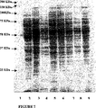


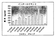

Classifications
-
- A—HUMAN NECESSITIES
- A23—FOODS OR FOODSTUFFS; TREATMENT THEREOF, NOT COVERED BY OTHER CLASSES
- A23K—FODDER
- A23K10/00—Animal feeding-stuffs
- A23K10/10—Animal feeding-stuffs obtained by microbiological or biochemical processes
- A23K10/16—Addition of microorganisms or extracts thereof, e.g. single-cell proteins, to feeding-stuff compositions
- A23K10/18—Addition of microorganisms or extracts thereof, e.g. single-cell proteins, to feeding-stuff compositions of live microorganisms
-
- A—HUMAN NECESSITIES
- A23—FOODS OR FOODSTUFFS; TREATMENT THEREOF, NOT COVERED BY OTHER CLASSES
- A23L—FOODS, FOODSTUFFS, OR NON-ALCOHOLIC BEVERAGES, NOT COVERED BY SUBCLASSES A21D OR A23B-A23J; THEIR PREPARATION OR TREATMENT, e.g. COOKING, MODIFICATION OF NUTRITIVE QUALITIES, PHYSICAL TREATMENT; PRESERVATION OF FOODS OR FOODSTUFFS, IN GENERAL
- A23L29/00—Foods or foodstuffs containing additives; Preparation or treatment thereof
- A23L29/065—Microorganisms
-
- A—HUMAN NECESSITIES
- A23—FOODS OR FOODSTUFFS; TREATMENT THEREOF, NOT COVERED BY OTHER CLASSES
- A23L—FOODS, FOODSTUFFS, OR NON-ALCOHOLIC BEVERAGES, NOT COVERED BY SUBCLASSES A21D OR A23B-A23J; THEIR PREPARATION OR TREATMENT, e.g. COOKING, MODIFICATION OF NUTRITIVE QUALITIES, PHYSICAL TREATMENT; PRESERVATION OF FOODS OR FOODSTUFFS, IN GENERAL
- A23L33/00—Modifying nutritive qualities of foods; Dietetic products; Preparation or treatment thereof
- A23L33/10—Modifying nutritive qualities of foods; Dietetic products; Preparation or treatment thereof using additives
- A23L33/105—Plant extracts, their artificial duplicates or their derivatives
-
- A—HUMAN NECESSITIES
- A23—FOODS OR FOODSTUFFS; TREATMENT THEREOF, NOT COVERED BY OTHER CLASSES
- A23L—FOODS, FOODSTUFFS, OR NON-ALCOHOLIC BEVERAGES, NOT COVERED BY SUBCLASSES A21D OR A23B-A23J; THEIR PREPARATION OR TREATMENT, e.g. COOKING, MODIFICATION OF NUTRITIVE QUALITIES, PHYSICAL TREATMENT; PRESERVATION OF FOODS OR FOODSTUFFS, IN GENERAL
- A23L33/00—Modifying nutritive qualities of foods; Dietetic products; Preparation or treatment thereof
- A23L33/10—Modifying nutritive qualities of foods; Dietetic products; Preparation or treatment thereof using additives
- A23L33/135—Bacteria or derivatives thereof, e.g. probiotics
-
- A—HUMAN NECESSITIES
- A61—MEDICAL OR VETERINARY SCIENCE; HYGIENE
- A61K—PREPARATIONS FOR MEDICAL, DENTAL OR TOILETRY PURPOSES
- A61K35/00—Medicinal preparations containing materials or reaction products thereof with undetermined constitution
- A61K35/66—Microorganisms or materials therefrom
- A61K35/74—Bacteria
- A61K35/741—Probiotics
-
- A—HUMAN NECESSITIES
- A61—MEDICAL OR VETERINARY SCIENCE; HYGIENE
- A61K—PREPARATIONS FOR MEDICAL, DENTAL OR TOILETRY PURPOSES
- A61K39/00—Medicinal preparations containing antigens or antibodies
- A61K39/02—Bacterial antigens
- A61K39/04—Mycobacterium, e.g. Mycobacterium tuberculosis
-
- A—HUMAN NECESSITIES
- A61—MEDICAL OR VETERINARY SCIENCE; HYGIENE
- A61P—SPECIFIC THERAPEUTIC ACTIVITY OF CHEMICAL COMPOUNDS OR MEDICINAL PREPARATIONS
- A61P1/00—Drugs for disorders of the alimentary tract or the digestive system
-
- A—HUMAN NECESSITIES
- A61—MEDICAL OR VETERINARY SCIENCE; HYGIENE
- A61P—SPECIFIC THERAPEUTIC ACTIVITY OF CHEMICAL COMPOUNDS OR MEDICINAL PREPARATIONS
- A61P1/00—Drugs for disorders of the alimentary tract or the digestive system
- A61P1/14—Prodigestives, e.g. acids, enzymes, appetite stimulants, antidyspeptics, tonics, antiflatulents
-
- A—HUMAN NECESSITIES
- A61—MEDICAL OR VETERINARY SCIENCE; HYGIENE
- A61P—SPECIFIC THERAPEUTIC ACTIVITY OF CHEMICAL COMPOUNDS OR MEDICINAL PREPARATIONS
- A61P3/00—Drugs for disorders of the metabolism
- A61P3/02—Nutrients, e.g. vitamins, minerals
-
- A—HUMAN NECESSITIES
- A61—MEDICAL OR VETERINARY SCIENCE; HYGIENE
- A61P—SPECIFIC THERAPEUTIC ACTIVITY OF CHEMICAL COMPOUNDS OR MEDICINAL PREPARATIONS
- A61P31/00—Antiinfectives, i.e. antibiotics, antiseptics, chemotherapeutics
-
- A—HUMAN NECESSITIES
- A61—MEDICAL OR VETERINARY SCIENCE; HYGIENE
- A61P—SPECIFIC THERAPEUTIC ACTIVITY OF CHEMICAL COMPOUNDS OR MEDICINAL PREPARATIONS
- A61P31/00—Antiinfectives, i.e. antibiotics, antiseptics, chemotherapeutics
- A61P31/04—Antibacterial agents
- A61P31/06—Antibacterial agents for tuberculosis
-
- A—HUMAN NECESSITIES
- A61—MEDICAL OR VETERINARY SCIENCE; HYGIENE
- A61P—SPECIFIC THERAPEUTIC ACTIVITY OF CHEMICAL COMPOUNDS OR MEDICINAL PREPARATIONS
- A61P37/00—Drugs for immunological or allergic disorders
- A61P37/02—Immunomodulators
-
- A—HUMAN NECESSITIES
- A61—MEDICAL OR VETERINARY SCIENCE; HYGIENE
- A61P—SPECIFIC THERAPEUTIC ACTIVITY OF CHEMICAL COMPOUNDS OR MEDICINAL PREPARATIONS
- A61P37/00—Drugs for immunological or allergic disorders
- A61P37/02—Immunomodulators
- A61P37/04—Immunostimulants
-
- A—HUMAN NECESSITIES
- A61—MEDICAL OR VETERINARY SCIENCE; HYGIENE
- A61P—SPECIFIC THERAPEUTIC ACTIVITY OF CHEMICAL COMPOUNDS OR MEDICINAL PREPARATIONS
- A61P37/00—Drugs for immunological or allergic disorders
- A61P37/08—Antiallergic agents
-
- A—HUMAN NECESSITIES
- A61—MEDICAL OR VETERINARY SCIENCE; HYGIENE
- A61P—SPECIFIC THERAPEUTIC ACTIVITY OF CHEMICAL COMPOUNDS OR MEDICINAL PREPARATIONS
- A61P39/00—General protective or antinoxious agents
-
- A—HUMAN NECESSITIES
- A61—MEDICAL OR VETERINARY SCIENCE; HYGIENE
- A61P—SPECIFIC THERAPEUTIC ACTIVITY OF CHEMICAL COMPOUNDS OR MEDICINAL PREPARATIONS
- A61P9/00—Drugs for disorders of the cardiovascular system
-
- C—CHEMISTRY; METALLURGY
- C07—ORGANIC CHEMISTRY
- C07K—PEPTIDES
- C07K14/00—Peptides having more than 20 amino acids; Gastrins; Somatostatins; Melanotropins; Derivatives thereof
- C07K14/195—Peptides having more than 20 amino acids; Gastrins; Somatostatins; Melanotropins; Derivatives thereof from bacteria
- C07K14/35—Peptides having more than 20 amino acids; Gastrins; Somatostatins; Melanotropins; Derivatives thereof from bacteria from Mycobacteriaceae (F)
-
- C—CHEMISTRY; METALLURGY
- C12—BIOCHEMISTRY; BEER; SPIRITS; WINE; VINEGAR; MICROBIOLOGY; ENZYMOLOGY; MUTATION OR GENETIC ENGINEERING
- C12N—MICROORGANISMS OR ENZYMES; COMPOSITIONS THEREOF; PROPAGATING, PRESERVING, OR MAINTAINING MICROORGANISMS; MUTATION OR GENETIC ENGINEERING; CULTURE MEDIA
- C12N1/00—Microorganisms, e.g. protozoa; Compositions thereof; Processes of propagating, maintaining or preserving microorganisms or compositions thereof; Processes of preparing or isolating a composition containing a microorganism; Culture media therefor
- C12N1/20—Bacteria; Culture media therefor
- C12N1/205—Bacterial isolates
-
- A—HUMAN NECESSITIES
- A23—FOODS OR FOODSTUFFS; TREATMENT THEREOF, NOT COVERED BY OTHER CLASSES
- A23V—INDEXING SCHEME RELATING TO FOODS, FOODSTUFFS OR NON-ALCOHOLIC BEVERAGES AND LACTIC OR PROPIONIC ACID BACTERIA USED IN FOODSTUFFS OR FOOD PREPARATION
- A23V2400/00—Lactic or propionic acid bacteria
- A23V2400/61—Propionibacterium
- A23V2400/619—Jensenii
-
- A—HUMAN NECESSITIES
- A61—MEDICAL OR VETERINARY SCIENCE; HYGIENE
- A61K—PREPARATIONS FOR MEDICAL, DENTAL OR TOILETRY PURPOSES
- A61K39/00—Medicinal preparations containing antigens or antibodies
- A61K2039/51—Medicinal preparations containing antigens or antibodies comprising whole cells, viruses or DNA/RNA
- A61K2039/52—Bacterial cells; Fungal cells; Protozoal cells
-
- A—HUMAN NECESSITIES
- A61—MEDICAL OR VETERINARY SCIENCE; HYGIENE
- A61K—PREPARATIONS FOR MEDICAL, DENTAL OR TOILETRY PURPOSES
- A61K39/00—Medicinal preparations containing antigens or antibodies
- A61K2039/555—Medicinal preparations containing antigens or antibodies characterised by a specific combination antigen/adjuvant
- A61K2039/55588—Adjuvants of undefined constitution
- A61K2039/55594—Adjuvants of undefined constitution from bacteria
-
- A—HUMAN NECESSITIES
- A61—MEDICAL OR VETERINARY SCIENCE; HYGIENE
- A61K—PREPARATIONS FOR MEDICAL, DENTAL OR TOILETRY PURPOSES
- A61K39/00—Medicinal preparations containing antigens or antibodies
-
- C—CHEMISTRY; METALLURGY
- C12—BIOCHEMISTRY; BEER; SPIRITS; WINE; VINEGAR; MICROBIOLOGY; ENZYMOLOGY; MUTATION OR GENETIC ENGINEERING
- C12R—INDEXING SCHEME ASSOCIATED WITH SUBCLASSES C12C - C12Q, RELATING TO MICROORGANISMS
- C12R2001/00—Microorganisms ; Processes using microorganisms
- C12R2001/01—Bacteria or Actinomycetales ; using bacteria or Actinomycetales
Landscapes
- Health & Medical Sciences (AREA)
- Life Sciences & Earth Sciences (AREA)
- Chemical & Material Sciences (AREA)
- Engineering & Computer Science (AREA)
- Organic Chemistry (AREA)
- Medicinal Chemistry (AREA)
- General Health & Medical Sciences (AREA)
- Microbiology (AREA)
- Bioinformatics & Cheminformatics (AREA)
- Veterinary Medicine (AREA)
- Pharmacology & Pharmacy (AREA)
- Animal Behavior & Ethology (AREA)
- Public Health (AREA)
- General Chemical & Material Sciences (AREA)
- Zoology (AREA)
- Chemical Kinetics & Catalysis (AREA)
- Nuclear Medicine, Radiotherapy & Molecular Imaging (AREA)
- Biotechnology (AREA)
- Mycology (AREA)
- Polymers & Plastics (AREA)
- Genetics & Genomics (AREA)
- Food Science & Technology (AREA)
- Wood Science & Technology (AREA)
- Nutrition Science (AREA)
- Biochemistry (AREA)
- Immunology (AREA)
- Molecular Biology (AREA)
- Biomedical Technology (AREA)
- General Engineering & Computer Science (AREA)
- Virology (AREA)
- Tropical Medicine & Parasitology (AREA)
- Communicable Diseases (AREA)
- Pulmonology (AREA)
- Epidemiology (AREA)
- Botany (AREA)
- Oncology (AREA)
- Animal Husbandry (AREA)
- Physiology (AREA)
- Gastroenterology & Hepatology (AREA)
- Biophysics (AREA)
Description
本発明は、プロバイオティクスのプロピオニバクテリウム菌株並びにプロバイオティクスのサプリメント及び食品の調製におけるそれらの使用に関する。本発明は、プロバイオティクス菌株によるビタミンB12、プロピオン酸、フォラシン及びバクテリオシンの提供、ビフィズス菌の増殖刺激、脂質代謝及び免疫刺激を介した宿主の免疫系における良好な効果の生成、又はアジュバントとしてのプロバイオティクス菌株の使用、ホモシステイン及びβ−グルクロニダーゼの低減及びこれらの活性の要求に関係する状態の予防、治療及び改善に関する。本発明のプロバイオティクス細菌はヒト又はそのほかの動物に使用することができる。少なくとも一部の適用では、細胞全体を使用してもよいというよりはむしろ、死んだ細菌又は細菌の一部を使用することができる。本発明はまた、感染症、特に結核から患者を保護するのに使用されるワクチンの調製にも関する。 The present invention relates to Probiotic Propionibacterium strains and their use in the preparation of probiotic supplements and foods. The present invention provides vitamin B 12 , propionic acid, foracin and bacteriocin by probiotic strains, production of good effects in the host immune system via bifidobacterial growth stimulation, lipid metabolism and immune stimulation, or adjuvant The use of probiotic strains as a base, the reduction of homocysteine and β-glucuronidase and the prevention, treatment and amelioration of conditions related to these activity requirements. The probiotic bacteria of the present invention can be used on humans or other animals. In at least some applications, rather than whole cells may be used, dead bacteria or parts of bacteria may be used. The present invention also relates to the preparation of vaccines used to protect patients from infections, particularly tuberculosis.
プロバイオティクス
消化管に導入されるプロバイオティクスは、消化管の腸内細菌叢に影響を及ぼすことができ、宿主における有益な役割を担うことができる。用語「プロバイオティクス」は、「生命のために」を意味するギリシア語に由来する。ある微生物により分泌される別の微生物の増殖を刺激する物質を記載するために最初に使用された(Lilly & Stillwell, 1965)。1992年、Havenaarは、動物又はヒトに適用した際、常在の腸内細菌叢の特性を改善することにより宿主に有益に作用する「生きた微生物の単一の、又は混在した培養物」としてプロバイオティクスを定義することを提案した(Havenaar et al., 1992)。Havenaarの定義が、用語、プロバイオティクスをヒトと動物の双方に適用した最初のものであった。プロバイオティクスの現在の適用及び判明した効果を考慮して、Salminenら(1999年)は、新しい定義:プロバイオティクスは、宿主の健康及び快適な暮らしに有益な効果を有する微生物細胞製剤又は微生物細胞の成分である、を提案した。この定義は、微生物細胞(生細胞又は非生細胞)及びプロバイオティクスとしての細胞の一部を包含するが、抗生物質のような代謝産物は包含しない。この定義はまた、プロバイオティクスの適用が食物における使用に限定されないことを示している。
Probiotics Probiotics introduced into the gastrointestinal tract can affect the intestinal flora of the gastrointestinal tract and can play a beneficial role in the host. The term “probiotic” comes from the Greek word meaning “for life”. It was first used to describe substances that stimulate the growth of another microorganism secreted by one microorganism (Lilly & Stillwell, 1965). In 1992, Havenaar is a “single or mixed culture of living microorganisms” that, when applied to animals or humans, acts beneficially on the host by improving the properties of the resident gut microbiota. It was proposed to define probiotics (Havenaar et al., 1992). The definition of Havenaar was the first to apply the term probiotics to both humans and animals. In view of the current application and proven effects of probiotics, Salminen et al. (1999) defined a new definition: probiotics are microbial cell preparations or microorganisms that have beneficial effects on the health and comfortable life of the host. Proposed to be a component of cells. This definition includes microbial cells (live or non-live cells) and parts of cells as probiotics, but not metabolites such as antibiotics. This definition also indicates that the application of probiotics is not limited to use in food.
最近、乳中のプロピオニバクテリウムの菌株が、宿主におけるプロピオン酸、フォラシン、バクテリオシン及びビタミンB12の産生、ビフィズス菌の増殖刺激、並びに脂質代謝及び免疫系における良好な効果を含む有益な効果を有することができるという幾つかの証拠が示されている。プロピオニバクテリウム菌株の使用によって、食物の栄養的及び治療的質が向上し、食品の貯蔵期間が延び、病原性及び有害な腸内細菌の増殖が抑制され、製品に対するさらに良好な感覚刺激的特性に寄与する。 Recently, Propionibacterium strains in milk, propionic acid in a host, folacin, production of bacteriocins and vitamin B 12, bifidobacteria growth stimulation, as well as beneficial effects, including good effect in lipid metabolism and immune system Some evidence is shown that it can have Use of Propionibacterium strains improves the nutritional and therapeutic quality of food, extends the shelf life of food, suppresses the growth of pathogenic and harmful intestinal bacteria, and provides better sensory stimulation to the product Contributes to properties.
乳中のプロピオニバクテリウムは、プロピオニバクテリウム属における4つの種、P.フレウデンレイチ(freudenreichii)、P.アシッドプロピオニシ(acidopropionici)、P.イエンセニー(jensenii)、及びP.ソエニ(thoenii)から成る。4種のプロピオニバクテリウムはすべて、野菜、果実、ナッツ、発芽した種子、豆、乳製品のような新鮮な、調理していない食物、及び特定の発酵品目を含む食物において生きたまま見い出される。 Propionibacterium in milk is the four species in the genus Propionibacterium, P. Freudenreich, P.M. Acid propionisi, P.I. Jensenii, and P.I. It consists of soeniii. All four Propionibacterium are found alive in fresh, uncooked foods such as vegetables, fruits, nuts, germinated seeds, beans, dairy products, and foods including certain fermented items .
プロピオニバクテリウムは、食品業界では、スターターカルチャーとして広く使用されている。プロピオニバクテリウム種は、伝統的には、「小穴」を作るスイスの又はスイス型のチーズ及びスイスチーズの典型的な甘味の維持に使用されている。プロピオニバクテリウム種はまた、食品添加物及び飼料添加物として野菜の発酵に、乳児用食品に、並びにプロピオン酸及びビタミンB12の生合成に使用されている。 Propionibacterium is widely used as a starter culture in the food industry. Propionibacterium species are traditionally used to maintain the typical sweetness of Swiss or Swiss-type cheeses and Swiss cheeses that make “small holes”. The Propionibacterium species, the fermentation of vegetables as food additives and feed additives, in infant food, as well as being used in the biosynthesis of propionic acid and vitamin B 12.
プロバイオティクス効果が生きている生物に依存する場合、プロバイオティクス菌株は、その集団が、腸管内容物で106〜108cfu/g前後の特定の最低レベルに達しなければ影響を与えることができない。経口により供給されるそのようなプロバイオティクス細菌は、上部消化管で生き残らなければならず、次いで下部消化管に達し、生き残って宿主に有益な効果を発揮する。 If the probiotic effect is dependent on the living organism, the probiotic strain will be affected if the population does not reach a specific minimum level of around 10 6 to 10 8 cfu / g in intestinal contents I can't. Such probiotic bacteria supplied orally must survive in the upper gastrointestinal tract, then reach the lower gastrointestinal tract and survive to exert a beneficial effect on the host.
胃の中の低いpH及びペプシンの抗菌作用は、細菌の腸管への侵入に対して有効な防壁となることが知られている。胃のpHは、2.5〜3.5の範囲にあるが、食物摂取の後では、低ければpH1.5に達し、高ければpH6にも達する。食物の種類が胃内容排出に影響を及ぼす。通常、食物は2〜4時間、胃に残るが、脂質の胃からの排出は約20分以内である。酸認容性の乳酸菌を含む胃の通過認容性を選択するために広範な試験管内の試験が使用されている(Charteris et al., 1998; Clark et al., 1993; Chou & Weimer, 1999)。しかしながら、プロピオニバクテリウムの試験管内の酸認容性に関しては限られた研究しかなく、これは、プロピオニバクテリウム・フレウデンレイチの2つの菌株によるものであった(Jan et al,m 2000; Mantere-Alhonen, 1983)。
The low pH in the stomach and the antibacterial action of pepsin are known to provide an effective barrier against bacterial intestinal entry. The pH of the stomach is in the range of 2.5 to 3.5, but after food intake it reaches pH 1.5 if low and reaches
プロピオン酸菌のもう1つの防壁は、小腸で生き残るこらねばならない。小腸の有害な条件には、胆汁塩及びパンクレアチンの存在が挙げられる。食物が小腸を通過する時間は、一般に1〜4時間の間である。胆汁塩の存在下で生き残る能力を調べ、種々のレベルの胆汁を伴った選抜培地でのその増殖を調べることによって胆汁塩耐性の乳酸菌を選択することができる(Gilliland et al., 1984; Ibarahimand Bezkorovainy, 1993; Clark & Martin, 1994; Chung et al., 1999)。ヒトでの使用で、プロバイオティクスを選択するには、0.15〜0.30%の濃度の胆汁塩が好適な濃度として報告されている。プロピオニバクテリウム菌株の小腸通過の認容性に関する報告は見られない。 Another barrier of propionic acid bacteria must survive in the small intestine. Harmful conditions of the small intestine include the presence of bile salts and pancreatin. The time for food to pass through the small intestine is generally between 1 and 4 hours. Bile salt-resistant lactic acid bacteria can be selected by examining their ability to survive in the presence of bile salts and examining their growth on selective media with varying levels of bile (Gilliland et al., 1984; Ibarahimand Bezkorovainy , 1993; Clark & Martin, 1994; Chung et al., 1999). For human use, bile salts at concentrations of 0.15 to 0.30% have been reported as preferred concentrations for selecting probiotics. There are no reports on the tolerance of the Propionibacterium strain through the small intestine.
上部消化管を通過して生き残った後、消化管でコロニーを形成し、持続するために、プロバイオティクス細菌は、消化管上皮に付着する必要がある。腸管粘膜及びその腸内細菌叢の複雑さによって、生体内で細菌の付着を検討するのが大変困難になっている。発酵したプロバイオティクス食品の調製の間においても、宿主の消化管においても生きているプロバイオティクス微生物は栄養的利益を提供することができる。発酵後、原料の質感及び風味を有意に改善することができ;食品における一部成分の有害効果、たとえば、一部のオリゴ糖及びタンパク質が原因になる食品の非認容性やアレルギーを軽減することができ;アミノ酸やビタミンのレベルを上げて食品の栄養価を改善することができ;及び、糖や食品のそのほかの損傷促進成分を除く結果、貯蔵期間を延ばし、食品の安全性を保証する。ミルクに比べて、発酵したヨーグルトにおいて、カルシウム、亜鉛、鉄、マンガン、銅及びリンの生物利用性が高められるという証拠が提示されている。検討の結果は、ヨーグルトにおけるリボフラビン及びニコチン酸の増加、チェダーチーズにおけるビタミンB6の増加、カッテージチーズにおけるビタミンB12の増加及びヨーグルト、カッテージチーズ、チェダーチーズ及びサワークリームを含む種々の製品における葉酸の増加を明らかにしている。プロバイオティクス微生物の酵素的加水分解もタンパク質及び脂肪の生物利用性を高めることが示されている。細菌のプロテアーゼは、特に宿主が内因性のプロテアーゼ欠損を有する場合、宿主の栄養状況に利益を与えることができる遊離のアミノ酸の産生を高めることができる。 After surviving through the upper gastrointestinal tract, probiotic bacteria need to adhere to the gastrointestinal epithelium in order to colonize and persist in the gastrointestinal tract. Due to the complexity of the intestinal mucosa and its intestinal bacterial flora, it is very difficult to study bacterial adhesion in vivo. Living probiotic microorganisms can provide nutritional benefits during the preparation of fermented probiotic foods and also in the digestive tract of the host. Can significantly improve the texture and flavor of raw materials after fermentation; reduce the harmful effects of some ingredients in food, such as food intolerance and allergies caused by some oligosaccharides and proteins Can increase the levels of amino acids and vitamins to improve the nutritional value of the food; and eliminates sugar and other damage-promoting ingredients of the food, thereby extending the shelf life and ensuring food safety. Evidence has been presented that the bioavailability of calcium, zinc, iron, manganese, copper and phosphorus is enhanced in fermented yogurt compared to milk. The results of the study were increased riboflavin and nicotinic acid in yogurt, increased vitamin B 6 in cheddar cheese, increased vitamin B 12 in cottage cheese and increased folic acid in various products including yogurt, cottage cheese, cheddar cheese and sour cream It is revealed. Enzymatic hydrolysis of probiotic microorganisms has also been shown to increase the bioavailability of proteins and fats. Bacterial proteases can enhance the production of free amino acids that can benefit the nutritional status of the host, especially if the host has an endogenous protease deficiency.
生きているプロバイオティクスを含有する食品の消費は、(1)病原性微生物、抗生物質又は化学療法による感染が原因の便秘及び下痢のような腸管障害の緩和;(2)免疫系の刺激及び調節;(3)β−グルクロニダーゼ、アゾレダクターゼ及びニトロレダクターゼのような腸内細菌酵素の低減により消化管における発癌性化合物を不活化する又は抑制することによる抗腫瘍効果;(4)アンモニア、フェノール及び肝硬変に影響を与えることが分かっているタンパク質の代謝産物のような毒性の最終産物の産生低下;(5)血清コレステロール及び血圧の低下;(6)粘膜整合性の維持;(7)乳糖不耐症の症状の緩和;(8)膣炎の予防、を含む健康上の利益を生じることが報告されている。 Consumption of food containing live probiotics is (1) alleviation of intestinal disorders such as constipation and diarrhea caused by infection with pathogenic microorganisms, antibiotics or chemotherapy; (2) stimulation of the immune system and (3) antitumor effects by inactivating or inhibiting carcinogenic compounds in the gastrointestinal tract by reducing enteric bacterial enzymes such as β-glucuronidase, azoreductase and nitroreductase; (4) ammonia, phenol and Reduced production of toxic end products such as protein metabolites known to affect cirrhosis; (5) reduced serum cholesterol and blood pressure; (6) maintained mucosal integrity; (7) lactose intolerance It has been reported to produce health benefits including relief of symptomatic symptoms; (8) prevention of vaginitis.
生きていないプロバイオティクス及び一部プロバイオティクス菌株の細胞壁成分も宿主に有益な効果を有するが、無処理の細胞よりも安全性の懸念は生じにくい。 Although non-living probiotics and some cell wall components of probiotic strains also have beneficial effects on the host, safety concerns are less likely than untreated cells.
プロピオニバクテリウムの菌株は、高濃度のビタミンB12を独特に含有しており(Hettinga & Reinbold, 1972a)、そのビタミンB12産生は最も効率的であるとみなされている(Hettinga & Reinbold, 1972b)。プロピオニバクテリウムの菌株は、発酵乳製品及び発酵飲料のビタミンB12補強に使用されることが多い。P.フレウデンレイチの亜種、シェルマニ(shermanii)を包含することにより、Kefir、ストレプトコッカスで発酵した発酵乳、Tvorogチーズ、Domiatiチーズ及びZabadyチーズでビタミンB12の増加を招く。P.フレウデンレイチの亜種、シェルマニ(shermanii)を包含することによりKefir、ストレプトコッカスで発酵した発酵乳を含む発酵乳製品及び発酵飲料で葉酸含量が増加する。 Propionibacterium strains, a high concentration of vitamin B 12 is contained uniquely (Hettinga & Reinbold, 1972a), the vitamin B 12 production is considered to be the most efficient (Hettinga & Reinbold, 1972b). Strain Propionibacterium are frequently used in vitamin B 12 reinforced fermented milk products and fermented beverages. P. Subspecies Fureudenreichi, by including the Sherumani (shermanii), leading Kefir, fermented milk fermented with Streptococcus, Tvorog cheese, an increase in vitamin B 12 in Domiati cheese and Zabady cheese. P. The inclusion of the Freudenreich subspecies, Shermanii, increases the folic acid content in fermented dairy products and fermented beverages, including fermented milk fermented with Kefir, Streptococcus.
プロピオニバクテリウムによるビタミンB12の産生は、細胞の収量に相関する。ビタミンB12の産生は、コバルトの存在、別の培地補助剤の包含、溶解した酸素濃度及び発酵ブイヨンのpHを含む多数の因子に影響される(Hettinga & Reinbold, 1972b; Quesada-Chanto et al., 1994b; Berry & Bullerman, 1966;Quesada-Chanto et al., 1998 )。 Production of vitamin B 12 by Propionibacterium correlates with cell yield. Vitamin B 12 production is affected by a number of factors including the presence of cobalt, the inclusion of additional media supplements, dissolved oxygen concentration and pH of the fermentation broth (Hettinga & Reinbold, 1972b; Quesada-Chanto et al. , 1994b; Berry & Bullerman, 1966; Quesada-Chanto et al., 1998).
ビタミンB12は人類の成長及び健康に必須である。それは、赤血球及び神経周囲のミエリンの産生に必要とされ、脂肪酸、炭水化物及びタンパク質の代謝において補酵素として作用する。ビタミンB12の天然合成は微生物においてのみ生じる。ビタミンB12の微生物による合成は、ヒトの結腸で生じるが、それは吸収されない。従って、人類は、ビタミンB12の食物摂取に全面的に依存し、適切な血清レベルや体内貯蔵を維持していると思われる。 Vitamin B 12 is essential to the growth and health of human beings. It is required for the production of red blood cells and perineural myelin and acts as a coenzyme in the metabolism of fatty acids, carbohydrates and proteins. Natural synthetic vitamin B 12 occurs only in microorganisms. Synthesis by microorganisms of vitamin B 12 is generated in the human colon, it is not absorbed. Thus, human beings, entirely dependent on the food intake of vitamin B 12, are believed to maintain proper serum levels and body stores.
ビタミンB12は栄養学の文献で用いられる用語であるが、ほとんどの生化学及び化学の教科書では、ビタミンB12ではなくコバラミンが用いられる。用語、ビタミンB12は今や、抗悪性貧血活性を呈するコバラミンの一般名である。 Although vitamin B 12 is a term used in the literature of nutrition, in most biochemistry and chemistry textbooks, cobalamin is used instead of vitamin B 12. The term, vitamin B 12 is now, it is the common name of cobalamin that exhibit anti-pernicious anemia activity.
ビタミンB12の欠乏は、心疾患、多発性硬化症、卒中、乳癌、アルツハイマー病、一部の精神病症候群及び加齢促進に対するリスク因子の1つであると報告されている(Choi, 1999; Wynn & Wynn, 1998; Gariballa, 2000; Delva, 1997)。ビタミンB12欠乏の有病性は、集団の年齢及び一般的な健康状態、正常な血清ビタミンB12の閾値、並びに診断に必要なそのほかの基準によって様々である。さらに優勢な、軽い及び前臨床的なビタミンB12欠乏が最近認識されている。前臨床的な欠乏は、不足を示す代謝的証拠は存在するが、巨赤芽球性貧血を含む臨床症状を有さない状態である(Carmel, 2000)。 Vitamin B 12 deficiency has been reported to be one of the risk factors for heart disease, multiple sclerosis, stroke, breast cancer, Alzheimer's disease, some psychotic syndromes and accelerated aging (Choi, 1999; Wynn & Wynn, 1998; Gariballa, 2000; Delva, 1997). Prevalence of vitamin B 12 deficiency, the population of the age and general health, normal threshold of serum vitamin B 12, as well as various by other criteria necessary for diagnosis. A more prevalent, mild and preclinical vitamin B 12 deficiency has recently been recognized. Preclinical deficiency is a condition with no clinical symptoms including megaloblastic anemia, although there is metabolic evidence to indicate deficiency (Carmel, 2000).
遺伝因子又は獲得因子のいずれによってもビタミンB12欠乏が生じてもよい。ほとんどのビタミンB12欠乏は、不適切な食物摂取に関係する。肉、魚、卵及び乳製品のような動物製品は人体に適切な量のビタミンB12を含有する。菜食主義者の食事は一般にRDLよりも低いレベルのビタミンB12を有する。菜食主義者の食事をするヒト及び彼らの乳児は、ビタミンB12の低レベル傾向にあり、ビタミンB12欠乏を発生する。研究によって、血清中の低レベルビタミンB12は、厳格な菜食主義者のみならず、乳菜食主義者及び乳卵菜食主義者にも見い出されている。ビタミンB12欠乏は、補完されていない又は補強されていない菜食主義者の食物において本当に障害である。多数の加工した菜食主義者の食事はビタミンB12が補強されているが、経済的理由から、これらの食物は、一部の菜食主義者には利用可能ではない。ビタミンB12の欠乏は、子供において成長及び精神運動の発達の遅滞を生じる。従って、菜食主義者の食事をする子供には特別の注意を払い、適切なビタミンB12の摂取を保証すべきである。 Vitamin B 12 deficiency may be caused by either genetic factors or acquired factors. Most vitamin B 12 deficiencies are associated with inadequate food intake. Meat, fish, eggs and animal products such as milk products containing vitamin B 12 in an appropriate amount to the human body. Of vegetarian diet generally have a low level of vitamin B 12 than RDL. Humans and their infant to eat vegetarian is in the low-level trend of vitamin B 12, to generate a vitamin B 12 deficiency. Studies low levels of vitamin B 12 in serum not only strict vegetarians, are also found in milk vegetarian and Chichitamago vegetarian. Vitamin B 12 deficiency is a real obstacle in uncomplemented or unreinforced vegetarian food. Although a number of processed vegetarian meal is reinforced vitamin B 12, from economic reasons, these foods are not available in some of the vegetarian. Vitamin B 12 deficiency results in delayed growth and psychomotor development in children. Therefore, special attention should be given to children eating vegetarian meals to ensure proper vitamin B 12 intake.
ビタミンB12欠乏のもう1つの原因は、食物中のビタミンB12の吸収不良である。 Another cause of vitamin B 12 deficiency is malabsorption of vitamin B 12 in food.
ビタミンB12の欠乏の効果を調べるのにラットが用いられてきた。ビタミンB12の欠乏は、ラットにおいて成長遅滞、エネルギー代謝の低下、尿中メチルマロン酸(MMA)の排泄の増加を引き起こす。ビタミンB12の欠乏はまた、ラットにおいて精巣組織にも影響を及ぼす。 Rats have been used to examine the effect of the deficiency of vitamin B 12. Vitamin B 12 deficiency causes growth retardation, decreased energy metabolism, and increased excretion of urinary methylmalonic acid (MMA) in rats. Vitamin B 12 deficiency also affects testicular tissue in rats.
ビタミンB12欠乏症に対する治療は、欠乏の原因によって様々である。ビタミンB12に対する最も頻度の高い2つの治療は、非経口的注射及び経口療法である。 Treatment for vitamin B 12 deficiency varies depending on the cause of the deficiency. The most frequent two treatments for vitamin B 12 is a parenteral injection and oral therapy.
ビタミンB12欠乏症の初期段階では、多数の患者が医学的忠告を求めていないことが分かっている。ビタミンB12食品補助剤及びビタミンB12を伴う一般の食物製品によって、患者の一部ではビタミンB12欠乏症の発症を防ぎ、欠乏の問題を軽減し、健康管理のコストを軽減すればよい。
ワクチン
あらゆるワクチンは、1つの目的、特定の疾病を引き起こす作用物資又は病原体が症状を起こすに十分なほど増殖する前に、それらを迅速に破壊するために、免疫系を感作すること、を有する(Langridge, 2000)。
In the early stages of vitamin B 12 deficiency, it has been found that a large number of patients do not seek medical advice. According to general food products with vitamin B 12 food supplements and vitamin B 12, preventing the onset of vitamin B 12 deficiency in some patients, to reduce the depletion of the problems may be reducing the cost of health care.
Vaccines Every vaccine has one purpose: to sensitize the immune system to quickly destroy agents or pathogens that cause a particular disease before they can grow sufficiently to cause symptoms. (Langridge, 2000).
単一の感染性作用物質に由来する世界最大の殺し屋は、結核(TB)である(WHO、1992年)。結核は、原発性に肺を冒す、細菌、マイコバクテリウム・ツベルクロシスにより引き起こされる疾病である。現在、20億の人々がこの細菌に感染している(Agger & Andersen, 2001)。これは世界人口の3分の1に匹敵する。これらの大半は、健康管理が最低限度である開発途上国において見られる。年間300万人がこの疾病で死亡し、治療しなければ死亡率は50%である(Collins & Kaufmann, 2001)。TBに現在利用可能なワクチンであるBCGは、様々な集団で0〜80%の範囲にある様々な有効率を有し(Guerin, 1997)、今日までこの疾病の拡散を防ぐ上では大きく非効果的である。50%の薬効を持つ新しいワクチンの開発によって2030年までに結核による900万人の死亡をふせぐことができると推定された(Murry & Salomon, 1998)。 The world's largest killer, derived from a single infectious agent, is tuberculosis (TB) (WHO, 1992). Tuberculosis is a disease caused by the bacterium Mycobacterium tuberculosis that primarily affects the lungs. Currently, 2 billion people are infected with this bacterium (Agger & Andersen, 2001). This is comparable to a third of the world population. Most of these are found in developing countries where health care is minimal. Three million people die annually from the disease, and if not treated, the mortality rate is 50% (Collins & Kaufmann, 2001). BCG, a vaccine currently available for TB, has various efficacy rates ranging from 0 to 80% in various populations (Guerin, 1997) and is largely ineffective at preventing the spread of this disease to date Is. It was estimated that the development of a new vaccine with 50% efficacy would prevent 9 million deaths from tuberculosis by 2030 (Murry & Salomon, 1998).
M.ツベルクロシスは、宿主の免疫反応をたくみにくぐりぬける多数のメカニズムを進化させた高度に発達した病原菌である(Flynn, 1999)。TBに対するヒトの免疫反応は、Tヘルパー1が関与する独特で複雑な細胞性応答である。免疫系の正しい態様を刺激する試みと組み合わせた細菌のチャレンジそのものが、BCGより有意に優れたワクチンを作製しようとする何年にもおよぶ試みを無駄なものにしてきた。
M.M. Tuberculosis is a highly developed pathogen that has evolved numerous mechanisms to evade host immune responses (Flynn, 1999). The human immune response to TB is a unique and complex cellular response involving
結核は、活動形態では、持続する咳、胸痛、発熱、体重減少、夜間の発汗のような症状を起こし、進行すれば、喀血を起こす(Miler & Schieffelbein, editorial 2001a)。結核は主として、咳をすること、くしゃみをすること、会話をすること、及び笑うことを介して、喀痰の小さな液滴で空気中に散らされることによって伝播する(Miler & Schieffelbein, 1998)。しかしながら、長期間にわたる反復した暴露が、暴露の強さと共に宿主における疾患の伝播及び成立に必要である(NIAID, 1999; Miler & Schieffelbein, 1998)。 Tuberculosis causes symptoms such as persistent cough, chest pain, fever, weight loss, night sweats, and, if progressed, hemoptysis (Miler & Schieffelbein, editorial 2001a). Tuberculosis is transmitted primarily through coughing, sneezing, talking, and laughing by being scattered in the air with small droplets of sputum (Miler & Schieffelbein, 1998). However, repeated exposure over time is required for disease transmission and establishment in the host along with the intensity of exposure (NIAID, 1999; Miler & Schieffelbein, 1998).
適切な抗生物質による治療によって90%の治癒率がある(NIAID, 1999; Collins & Kaufmann, 2001)。しかしながら、この治療方法には問題がある。治療を施すにはコンプライアンスを確保するための監督を必要とし、それは相対的に高価であり、多数の副作用を伴い、治癒には6ヵ月を超える治療を要する(Broker, 1999)。最近の研究により、多数の国で、TBの治療に使用される4種の主な薬剤、イソニアジド、リファンピン、エタンブトール及びストレプトマイシンに対する耐性が発生していることが示された(Mustafa, 2002)。このことは、多剤耐性結核菌(MDR−TB)菌株による感染は、利用可能な薬剤のいかなるものでも有効に治療することができないことを意味しており、MDR−TB患者の死亡率は40〜60%である(NIAID, 1999)。 There is a cure rate of 90% with appropriate antibiotic treatment (NIAID, 1999; Collins & Kaufmann, 2001). However, there are problems with this method of treatment. The treatment requires supervision to ensure compliance, which is relatively expensive, has many side effects, and cures require more than 6 months of treatment (Broker, 1999). Recent studies have shown that a number of countries have developed resistance to the four main drugs used to treat TB, isoniazid, rifampin, ethambutol and streptomycin (Mustafa, 2002). This means that infection with a multi-drug resistant Mycobacterium tuberculosis (MDR-TB) strain cannot be effectively treated with any of the available drugs, and the mortality rate of MDR-TB patients is 40 ~ 60% (NIAID, 1999).
マイクロバクテリウム・ツベルクロシスは、宿主に感染することができるが、活動型疾患の症状を呈することなく休止状態で存在することができる。これは、潜在的結核感染として知られる。潜在的感染を持つ危険性は、通常、宿主が弱った状態にある場合、特に、HIV/AIDSのような自己免疫疾患に共に感染した場合、また、高齢者に感染した場合、それらがいつでも再び活動期に入るということである(NIAID, 1996; Tsuyuguchi, 1996)。HIV/TBとの同時感染の50%を超える人々が活動期結核を発症している。免疫系が弱っている場合、細菌はマクロファージに侵入し、休止状態で存在し、血流に入って疾患を引き起こす。治療しないままであれば、活動期疾患の患者の50%は死亡する(NIAID, 1996)。結核の大半は、最低限の健康管理で、栄養不良やHIVが一般的な世界で最貧の国々で起きる(Kaufmann & Hess, 2000)。 Microbacterium tuberculosis can infect a host but can exist in a dormant state without presenting symptoms of active disease. This is known as latent tuberculosis infection. The risk of having a potential infection is usually reassured whenever the host is in a weak state, especially when co-infected with an autoimmune disease such as HIV / AIDS, or when the elderly are infected. It means entering the active period (NIAID, 1996; Tsuyuguchi, 1996). More than 50% of people with HIV / TB co-infection develop active tuberculosis. When the immune system is weak, bacteria enter macrophages, exist in a quiescent state, enter the bloodstream and cause disease. If left untreated, 50% of patients with active disease die (NIAID, 1996). The majority of tuberculosis occurs in the poorest countries in the world where malnutrition and HIV are common (Kaufmann & Hess, 2000) with minimal health care.
結核と闘うためのBCG以来最も好評なイニシアチブであり、TBの耐性菌株の拡散を裏付けているのは、WHOの直接的に観察する短期間療法(DOTS)である(Collins & Kaufmann, 2001)。これは、標準化された短期コースの化学療法の完全コースを達成するために患者のコンプライアンスを高めることを目的とする(Gleissberg, 1999)。これは言い換えれば、完全な投薬コースを確実に採用することによって、M.ツベルクロシスの抗生物質耐性菌株を除くという条件を排除することを目的とする。しかしながら、この戦略は、問題を制御するにはBCGと同じくらい有効でないと思われる。統計によって、そのような療法の成功率は、アフリカのある国々ではたった62%であることが示されている(Collins & Kaufmann, 2001)。その結果、TBは、化学療法投薬計画のみでは世界から根絶されないという一般的な合意がある(Hess & Kaufmann, 1999)。 The most popular initiative since BCG to combat tuberculosis, supporting the spread of resistant strains of TB is the WHO direct observation short term therapy (DOTS) (Collins & Kaufmann, 2001). This is aimed at increasing patient compliance to achieve a standardized short course full course of chemotherapy (Gleissberg, 1999). In other words, by ensuring that a complete dosing course is adopted, M.C. The objective is to eliminate the condition of removing antibiotic-resistant strains of tuberculosis. However, this strategy does not appear to be as effective as BCG in controlling the problem. Statistics show that the success rate of such therapy is only 62% in some African countries (Collins & Kaufmann, 2001). As a result, there is a general consensus that TB is not eradicated from the world with chemotherapy regimens alone (Hess & Kaufmann, 1999).
BCGは世界で最も広く使用されているワクチンである(Kaufmann & Hess, 2000)。今日までに、30億人を超える人々に投与された(Collins, 2000)。265,000人の被験者を含むインドにおける1968年に開始された対照のある臨床試験では、プラセボ群の参加者の方がBCGの投与を受けたものよりもわずかに良好なTBへの免疫性を有することが示された(Tripathy, 1987)。BCGは、前の世紀の初頭に創られ、ウシで結核を起こすマイコバクテリウム菌株である、マイコバクテリウム・ボビスの生きた減衰した菌株から成る。この菌株は、90%を超えるDNAの相同性を持って、M.ツベルクロシスと密接に関係するので、M.ツベルクロシスによる感染に対する優れた免疫反応を感作すると期待された(Andersen, 2001)。BCGワクチンが結核の解決法ではなかったにもかかわらず、それが創られた時は、そのように期待された(Ravn et al., 1997)。最近の研究によって、BCGワクチンは、実際、TBへの免疫反応で中心的役割を担うことが今や同定されている良好な抗原性を持つ生命に必要なタンパク質を欠いていることが示されている(Ravn et al., 1997)。 BCG is the most widely used vaccine in the world (Kaufmann & Hess, 2000). To date, it has been administered to over 3 billion people (Collins, 2000). In a controlled clinical trial initiated in 1968 in India involving 265,000 subjects, participants in the placebo group had slightly better immunity to TB than those who received BCG. (Tripathy, 1987). BCG consists of a live attenuated strain of Mycobacterium bovis, a Mycobacterium strain that was created early in the last century and causes tuberculosis in cattle. This strain has a DNA homology of over 90%, M. is closely related to tuberculosis. It was expected to sensitize an excellent immune response to infection with tuberculosis (Andersen, 2001). That was expected when it was created even though the BCG vaccine was not a solution for tuberculosis (Ravn et al., 1997). Recent studies have shown that BCG vaccines in fact lack vital antigenic proteins that have now been identified as playing a central role in the immune response to TB (Ravn et al., 1997).
BCGにおける特定の必須の抗原の欠如は、減衰過程の間に起きた遺伝子欠失によると考えられる(Ethay & Andersen, 1997)。BCGは、胆汁を含有する寒天プレートを段階的に通過させることを介して、M.ボビスを減衰することによって創られた(Kaufmann & Hess, 2000)。今や失われたこれらの有益なタンパク質は、結核患者で強く認識され、活発に研究された esat−6を包含する(Collins, 2000)。 The lack of certain essential antigens in BCG is thought to be due to gene deletions that occurred during the decay process (Ethay & Andersen, 1997). BCG is produced by passing it through agar plates containing bile in stages. Created by attenuating Bovis (Kaufmann & Hess, 2000). These beneficial proteins that are now lost include esat-6, which is strongly recognized and actively studied in tuberculosis patients (Collins, 2000).
最近の現場の試験では、BCGによる防御レベルは極めて低く、検出不能の場合もあることが示されている(Andersen, 2001)。 Recent field trials have shown that the level of protection by BCG is very low and may not be detectable (Andersen, 2001).
Brandtら(2002年)は、特定の環境のマイコバクテリアへの暴露は、BCGの増殖を制御する免疫反応を刺激し、従って、完全なワクチン誘発の免疫反応が発生するのを妨げるという長年の仮説を裏付けた(Brandt at el., 2002)。環境中の細菌への暴露は、免疫系を感作し、そのほかのマイコバクテリアの特定のレベルの防御を提供するが、動物モデルでは、防御は、M.ツベルクロシスの増殖を有意に減らすほど十分ではない(Brandt, et al., 2002)。 Brandt et al. (2002) found that long-term hypothesis that exposure to certain environmental mycobacteria stimulates an immune response that controls the growth of BCG, thus preventing a full vaccine-induced immune response from occurring. (Brandt at el., 2002). Exposure to bacteria in the environment sensitizes the immune system and provides a specific level of protection of other mycobacteria, but in animal models, protection is It is not enough to significantly reduce the growth of tuberculosis (Brandt, et al., 2002).
この研究は、生きたワクチンとしてのBCGは、M.アビウムとBCGとの間で共有する抗原に対して事前に存在する免疫反応の影響に特に感受性があり(Brandt, et al., 2002)、それによって、BCGワクチンの非有用性を一部説明することができると結論付けた。これに加えて、BCGは、主な診断ツールである、PPD皮内テストにおいて偽陽性を生じることによって、疾患の検出及び初期の治療に対して障害でもある(Fatkenheuer et al., 1999)。 This study shows that BCG as a live vaccine is Particularly sensitive to the effects of pre-existing immune responses to antigens shared between abium and BCG (Brandt, et al., 2002), thereby partially explaining the non-usefulness of BCG vaccines We conclude that we can. In addition, BCG is also an obstacle to disease detection and initial treatment by producing false positives in the main diagnostic tool, PPD intradermal test (Fatkenheuer et al., 1999).
BCGの明らかに明瞭な破綻及び不適切性にもかかわらず、新しい原型となるワクチンの合成は、未だに、BCGよりは有意に有効である、動物モデルにおける免疫反応を生じなければならない(Andersen, 1994)。BCGが世界中で投与され続ける主な理由は、ワクチンが安全であるという利点、安価で容易に製造され、注射1回当たり数セントで済むことである(Orme, 1999)。 Despite the apparent clear failure and inadequacy of BCG, the synthesis of a new prototype vaccine must still generate an immune response in animal models that is significantly more effective than BCG (Andersen, 1994 ). The main reason BCG continues to be administered worldwide is the advantage that the vaccine is safe, cheap and easy to manufacture, and costs only a few cents per injection (Orme, 1999).
背景技術に関する上記の考察は、本発明の背景を説明するのに包含される。引用された文書及びそのほかの資料は、本明細書のクレームのいずれか1項の優先日に、公知であり、知られており、又はオーストラリアにおける一般的知識の一部であったということは、譲歩としては解釈されない。 The above discussion regarding background art is included to illustrate the background of the invention. The cited documents and other materials were known, known or part of general knowledge in Australia on the priority date of any one of the claims herein, It is not interpreted as a concession.
本明細書の説明及びクレーム全体にわたって、用語「含む」及びその変異型「含むこと」及び「含む(3人称単数形)」は、そのほかの添加物、工程又は整数値を排除することを意図しない。 Throughout the description and claims herein, the term “comprising” and variants thereof “comprising” and “comprising (third person singular)” are not intended to exclude other additives, steps, or integer values. .
第1の態様において、本発明は、新規の単離されたプロピオニバクテリウム菌株、プロピオニバクテリウム・イエンセニー702を提供する。プロピオニバクテリウム・イエンセニー702は、ブダペスト条約の規定のもとで、オーストラリア政府の分析研究所(オーストラリア、ニューサウスウェールズ州、2073、ピンブル、スアキン通り1)にて、受入番号、NM02/29517のもと2002年6月12日、供託された。
In a first aspect, the present invention provides a novel isolated Propionibacterium strain,
第2の態様において、本発明は、培養培地、保存培地賦形剤、キャリア又は希釈剤と共にプロピオニバクテリウム・イエンセニー702からなる製剤を提供する。 In a second aspect, the present invention provides a formulation comprising Propionibacterium ienseni 702 together with a culture medium, storage medium excipient, carrier or diluent.
第3の態様において、本発明は、プロバイオティクス菌株としてのプロピオニバクテリウム・イエンセニー702の使用を提供する。該菌株は、ヒト又はそのほかの動物においてプロバイオティクスとして使用することができる。細菌は生きたまま又は死んだものを使用することができ、また細菌の一部を使用することができる。 In a third aspect, the present invention provides the use of Propionibacterium ienseni 702 as a probiotic strain. The strain can be used as probiotics in humans or other animals. Bacteria can be used alive or dead, and part of the bacteria can be used.
第4の態様において、当然のことながら本発明は、送達剤と共にプロピオニバクテリウム・イエンセニー702からなるビタミンB12サプリメントを提供する。サプリメントにおける使用に好適な送達剤は、薬学上許容可能な送達剤を包含する。 In a fourth aspect, it will be appreciated that the present invention provides a vitamin B 12 supplement consisting of Propionibacterium ienseni 702 with a delivery agent. Suitable delivery agents for use in supplements include pharmaceutically acceptable delivery agents.
サプリメントは、カプセル、錠剤、粉末(単体又はカプセルに入れて)、顆粒又はペーストの形態で提供してもよい。経口スプレーとしてサプリメントを調製することができる。もう1つの実施形態では、たとえば、朝食用シリアル、豆乳製品又は菜食主義者用のバーガーパティのようなビタミンB12を強化した食品の形態で提供される。 Supplements may be provided in the form of capsules, tablets, powders (alone or in capsules), granules or pastes. Supplements can be prepared as oral sprays. In another embodiment, it is provided in the form of a vitamin B 12 enriched food such as, for example, breakfast cereal, soy milk product or vegetarian burger patty.
第5の態様において、本発明は、方法が本発明のサプリメント又は食物を宿主に投与することからなる、宿主におけるビタミンB12欠乏の有害効果を防ぐ、治療する又は改善する方法を提供する。宿主はヒトであっても、そのほかの動物であってもよい。 In a fifth aspect, the present invention, a method comprises administering a supplement or food of the present invention to a host, prevent adverse effect of vitamin B 12 deficiency in a host, which provides a method of treating or ameliorating. The host may be a human or other animal.
第6の態様において、本発明は、好適なキャリア、賦形剤又は希釈剤と共にプロピオニバクテリウム・イエンセニー702からなる動物飼料のための成長促進剤を提供する。プロピオニバクテリウムにより動物を飼育することは、抗生物質又は化学療法による成長促進に関係する副作用無しで、成長、ビタミン供給及び有害腸内細菌の抑制を改善することができる。 In a sixth aspect, the present invention provides a growth promoter for animal feed consisting of Propionibacterium ienseni 702 with a suitable carrier, excipient or diluent. Rearing animals with Propionibacterium can improve growth, vitamin supply and suppression of harmful enteric bacteria without the side effects associated with growth promotion by antibiotics or chemotherapy.
第7の態様において、本発明は、宿主動物に第6の態様の成長促進剤を有効量で投与することからなる、宿主動物において成長及び/又はビタミンの供給及び/又は有害腸内細菌の抑制を改善する方法を提供する。当然のことながら、本発明の本態様は、宿主動物に第6の態様の成長促進剤を有効量で投与することを含む、宿主動物において成長及び/又はビタミンの供給及び/又は有害腸内細菌の抑制を改善する方法において使用するために第6の態様の成長促進剤を製造する方法も包含する。 In a seventh aspect, the present invention relates to growth and / or vitamin supply and / or suppression of harmful enteric bacteria in a host animal comprising administering to the host animal an effective amount of the growth promoter of the sixth aspect. Provide a way to improve. It will be appreciated that this aspect of the invention involves the growth and / or supply of vitamins and / or harmful enteric bacteria in the host animal comprising administering to the host animal an effective amount of the growth promoter of the sixth aspect. Also included is a method of producing the growth promoter of the sixth aspect for use in a method of improving inhibition of sucrose.
第8の態様において、本発明は、プロピオニバクテリウム・イエンセニー702の使用を含む発酵工程を含むことにより食品を調製することを含む食品の損傷を防ぎ、及び/又は食物製品の貯蔵期間を延ばす方法を提供する。 In an eighth aspect, the present invention prevents food damage including preparing food by including a fermentation process that includes the use of Propionibacterium ienseni 702 and / or extends the shelf life of food products. Provide a method.
発酵した食品は、長い消費歴を有し、元来の食品素材よりもさらに良好な品質を有することが多い。 Fermented foods have a long consumption history and often have better quality than the original food material.
乳酸菌及びプロピオン酸菌を含む乳スターターは、発酵生成物に阻害性の代謝産物を生成する能力によって、損傷性及び病原性の生物の増殖を制約することができる。阻害剤には、広域スペクトルの拮抗剤、有機酸、ジアセチル、及び過酸化水素が挙げられる。スターターの中には、バクテリオシン又は生産者の培養に通常密接に関係する種に対して活性のある殺菌性タンパク質を生じるものもある。プロピオン酸菌は、プロピオン酸、プロピオン酸塩、ジアセチル及びバクテリオシンを産生することができる。プロピオン酸は、酢酸及び乳酸よりも幅広い阻害を示す。プロピオン酸並びにそのナトリウム塩及びカルシウム塩は、抗ミオサイトで有効であり、多数の焼いた製品に添加してカビを抑制する。プロピオン酸塩はまた、グラム陰性菌を抑制するが、グラム陽性菌には有効ではない。ジアセチルは、酵母、乳酸菌、グラム陽性種及びグラム陰性培養物に抑制効果を有する。プロピオニバクテリウム・フレウデンレイチの亜種、シェルマニJSは、ラクトバチルス・ラムノサスLC705と共に、発酵乳及びパンに使用して、製品の貯蔵期間を改善することも報告されている。混合培養は、損傷性酵母、カビ及びバチルス種を抑制することが見い出された。 Milk starters, including lactic acid bacteria and propionic acid bacteria, can limit the growth of damaging and pathogenic organisms by their ability to produce inhibitory metabolites in the fermentation product. Inhibitors include broad spectrum antagonists, organic acids, diacetyl, and hydrogen peroxide. Some starters produce bactericidal proteins that are active against bacteriocin or species normally associated with the producer's culture. Propionic acid bacteria can produce propionic acid, propionate, diacetyl and bacteriocin. Propionic acid exhibits a broader inhibition than acetic acid and lactic acid. Propionic acid and its sodium and calcium salts are effective in anti-myocytes and are added to many baked products to control mold. Propionate also suppresses gram-negative bacteria but is not effective against gram-positive bacteria. Diacetyl has an inhibitory effect on yeast, lactic acid bacteria, gram positive species and gram negative cultures. Shellmani JS, a variant of Propionibacterium freudenreich, has also been reported to be used in fermented milk and bread with Lactobacillus rhamnosus LC705 to improve product shelf life. Mixed cultures were found to suppress damaging yeast, mold and Bacillus species.
第9の態様において、本発明は、もう1つの食品発酵微生物を伴った混合培養において、プロピオニバクテリウム・イエンセニー702の使用を含む発酵工程を含むことにより食品を調製することを含む食品の損傷を防ぎ、及び/又は食物製品の貯蔵期間を延ばす方法を提供する。 In a ninth aspect, the present invention relates to food damage comprising preparing a food product by including a fermentation step comprising the use of Propionibacterium ienseni 702 in a mixed culture with another food-fermenting microorganism. And / or a method for extending the shelf life of a food product.
第10の態様において、本発明は、プロピオニバクテリウム・イエンセニー702又はその一部を用いて調製された食品を提供する。 In a tenth aspect, the present invention provides a food prepared using Propionibacterium ienseni 702 or a portion thereof.
実施形態の1つでは、食品は、プロピオニアシッドフィルス乳のような発酵乳製品である。 In one embodiment, the food product is a fermented dairy product such as Propionia Cidfils milk.
第11の態様において、本発明は、調製においてプロピオニバクテリウム・イエンセニー702又はのあるその一部からなる食品を調製する方法を提供する。 In an eleventh aspect, the present invention provides a method of preparing a food product consisting of Propionibacterium ienseni 702 or some portion thereof in preparation.
プロバイオティクス食品には、「発酵した」プロバイオティクス食品及び「発酵しない」プロバイオティクス食品が挙げられる。「発酵した」プロバイオティクス食品は、食品の中で増殖し、発酵生成物を生成する生きたプロバイオティクス微生物を含有する。「発酵しない」プロバイオティクス食品は、食品の感覚刺激に反応する特性を顕著には変更しないで食品に添加されたプロバイオティクス微生物又はその構成成分を含有する。「発酵した」プロバイオティクス食品には、プロバイオティクス乳食品、チーズ、バター、クリーム、ヨーグルト及び飲用ヨーグルト及び一部の冷凍食品が挙げられる。食品は液体でもよい。「発酵した」プロバイオティクス食品に使用される原料には、ミルク、豆乳、穀物、豆類、果実生産物及び野菜生産物が挙げられる。 Probiotic foods include “fermented” probiotic foods and “non-fermented” probiotic foods. “Fermented” probiotic foods contain live probiotic microorganisms that grow in the food and produce fermentation products. A “non-fermented” probiotic food product contains a probiotic microorganism or component thereof added to the food product without significantly altering the food responsiveness to sensory stimuli. “Fermented” probiotic foods include probiotic dairy foods, cheese, butter, cream, yogurt and drinking yogurt and some frozen foods. The food may be a liquid. Ingredients used in “fermented” probiotic foods include milk, soy milk, cereals, legumes, fruit products and vegetable products.
プロバイオティクス食品は1種又は数種の微生物菌株を含有してもよい。すでに調製された食品に1又はそれより多くの微生物を添加することにより食品を調製してもよい。別の方法では、調製中に1又はそれより多くの微生物を食品に添加してもよい。これによって、食品内部での微生物の増殖を生じ、食品の特性に結果的に影響を与えてもよい。微生物は、調製において原料の発酵に加担してもよい。微生物は、調製において原料の部分的な又は完全な発酵を提供してもよい。本態様で使用される微生物は、発酵的機能のみを提供してもよく、発酵及びプロバイオティクス的機能を提供してもよい。 Probiotic foods may contain one or several microbial strains. The food product may be prepared by adding one or more microorganisms to the already prepared food product. Alternatively, one or more microorganisms may be added to the food during preparation. This may result in the growth of microorganisms within the food product and consequently affect the properties of the food product. Microorganisms may participate in the fermentation of the raw material in preparation. The microorganism may provide a partial or complete fermentation of the raw material in the preparation. Microorganisms used in this aspect may provide only fermentative functions or may provide fermentation and probiotic functions.
1又はそれより多くの微生物を用いて食品を調製し、次いでさらに微生物を添加させてもよい。 A food product may be prepared using one or more microorganisms and then further microorganisms may be added.
市販の食品製造のための乳酸菌及びプロバイオティクス培養、並びに発酵のためのスターター培養は、クリスチャン・ハンセンPty社(オーストラリア、ベイズウオーター)及びジスト−ブロケーズ・オーストラリアPty社(オーストラリア、モアーバンク)のような国際的な会社から購入することができる。CSIROスターター・カルチャー・コレクション(オーストラリア、ハイエット)及びオーストラリア・スターター・カルチャー・リサーチ・センター(オーストラリア・ウェリビー)のようなそのほかの研究機関は、良好な乳酸菌及び能力なプロバイオティクスコレクションを有するが、これらの生物は小規模でしか利用可能でない。 Lactic acid bacteria and probiotic cultures for commercial food production, as well as starter cultures for fermentation, such as Christian Hansen Pty (Baise Water, Australia) and Dyst-Brokers Australia Pty (Moerbank, Australia) Can be purchased from an international company. Other research institutions such as CSIRO Starter Culture Collection (Hayet, Australia) and Australia Starter Culture Research Center (Werribee, Australia) have good lactic acid bacteria and competent probiotics collections. Are only available on a small scale.
第12の態様において、本発明は、本発明の食品又はサプリメントを患者に投与することを含む、患者において消化器疾患及び/又は乳糖不耐症を治療する方法を提供する。実施形態の1つでは、患者は乳児である。患者はまた、非ヒトの動物であればよい。当然のことながら、本発明の本態様は本発明の食品又はサプリメントを患者に投与することを含む、患者において消化器疾患及び/又は乳糖不耐症を治療する方法において使用するための本発明の食品又はサプリメントを製造する方法も包含する。 In a twelfth aspect, the present invention provides a method for treating gastrointestinal diseases and / or lactose intolerance in a patient comprising administering to the patient a food or supplement of the present invention. In one embodiment, the patient is an infant. The patient may also be a non-human animal. It will be appreciated that this aspect of the invention includes the administration of a food or supplement of the invention to a patient for use in a method of treating digestive disorders and / or lactose intolerance in a patient. Also included are methods of producing food or supplements.
消化器疾患及び/又は乳糖不耐症の治療において乳児の食品にプロピオニバクテリウム菌株が使用される。ラクトバチルス・アシドフィルス及びP.フレウデンレイチの亜種、シェルマニを用いて調製されたプロピオニアシッドフィルス乳は、乳児の腸のスタフィロコッカス感染の治療において乳酸菌乳より有効であることが示された。種々の発酵乳製品は、乳糖不耐症の乳児の治療に有効であることが分かっている。乳糖の加水分解に必要な酵素、β−ガラクトシダーゼは、乳酸菌に認められるよりも高い濃度でプロピオニバクテリウムに存在することが分かっている。乳糖不耐症患者を治療するための発酵乳製品の製造にはプロピオン酸菌を使用することが推奨されている。 Propionibacterium strains are used in infant foods in the treatment of digestive disorders and / or lactose intolerance. Lactobacillus acidophilus and P. Propionia sidophilus milk prepared using the Freudenreich subspecies shellmani has been shown to be more effective than lactic acid bacteria milk in the treatment of intestinal staphylococcal infections in infants. A variety of fermented dairy products have been found to be effective in treating infants with lactose intolerance. It has been found that the enzyme required for lactose hydrolysis, β-galactosidase, is present in Propionibacterium at a higher concentration than found in lactic acid bacteria. It is recommended that propionic acid bacteria be used in the manufacture of fermented dairy products to treat lactose intolerant patients.
第13の態様において、本発明は、プロピオニバクテリウム・イエンセニー702又は本発明の食品もしくはサプリメントを宿主に投与することを含む、宿主の消化管におけるビフィズス菌の増殖を高める方法を提供する。宿主は、ヒトであっても、そのほかの動物であってもよい。当然のことながら、本発明の本態様はプロピオニバクテリウム・イエンセニー702又は本発明の食品もしくはサプリメントを患者に投与することからなる、宿主の消化管におけるビフィズス菌の増殖を高める方法において使用するための本発明の食品又はサプリメントを製造する方法も包含する。 In a thirteenth aspect, the present invention provides a method for increasing the growth of Bifidobacteria in the digestive tract of a host comprising administering to the host Propionibacterium ienseni 702 or a food or supplement of the present invention. The host may be a human or other animal. It will be appreciated that this aspect of the invention is for use in a method of increasing the growth of Bifidobacteria in the host gastrointestinal tract comprising administering to a patient Propionibacterium ienseni 702 or a food or supplement of the invention. The method for producing the food or supplement of the present invention is also included.
プロピオニバクテリウムは試験管内でビフィズス菌の増殖を高めることができることを示している。ビフィズス産生性の増殖刺激因子は、プロピオニバクテリウム・フレウデンレイチ7025の培養物の細胞なしのろ液及び細胞のメタノール抽出分画に存在することが見い出された(Kaneko et al., 1994)。この水溶性の刺激因子は、熱及びタンパク分解酵素に対して安定しており、ビタミンB12及び有機酸とは異なる。この刺激因子は、ビフィドバクテリウム・ビフィダム、B.ロングム、B.ブレイブ、及びB.アドレスセンツの菌株の増殖を増強した。プロピオニバクテリウムのビフィドバクテリウムにおける刺激効果はまた、健常なヒト対象にも処理され(Bougle et al., 1999)、結果は、プロピオニバクテリウム・フレウデンレイチのビフィズス産生特性を裏付けた。 Propionibacterium has been shown to increase the growth of bifidobacteria in vitro. A bifido-producing growth-stimulating factor was found to be present in the cell-free filtrate and cell methanol extraction fractions of the culture of Propionibacterium fredenreich 7025 (Kaneko et al., 1994). This water-soluble stimulating factor is stable to heat and proteolytic enzymes and is different from vitamin B 12 and organic acids. This stimulating factor is Bifidobacterium bifidum, B.I. Longum, B.B. Brave, and B.B. The growth of the address cents strain was enhanced. The stimulatory effect of Propionibacterium in Bifidobacterium was also processed in healthy human subjects (Bougle et al., 1999), and the results confirmed the bifido production characteristics of Propionibacterium froudenreich.
第14の態様において、本発明は、1又はそれより多くのプロバイオティクス細菌を伴ったプロピオニバクテリウム・イエンセニー702の混合培養を提供する。 In a fourteenth aspect, the present invention provides a mixed culture of Propionibacterium ienseni 702 with one or more probiotic bacteria.
プロピオニバクテリウムとそのほかのプロバイオティクス細菌を含有する混合培養は、結果として多数の有益な効果を生じる。プロピオニバクテリウムと乳酸菌の混合培養は、発酵で広く用いられている。野菜の発酵において乳酸菌と共にプロピオニバクテリウムを添加すると、フォラシン、ビタミンB12、プロピオン酸及び酢酸の含量が増加し、有害な病原性微生物が抑制され、製品の貯蔵期間が延びる。さらに、B.ロングムとP.フレウデンレイチの混合培養は、M.ルテウス、シュードモナス種、及びS.アウレウスに対するB.ロングム単独よりも良好な抗菌活性が得られる。 Mixed cultures containing Propionibacterium and other probiotic bacteria result in a number of beneficial effects. A mixed culture of Propionibacterium and lactic acid bacteria is widely used in fermentation. Addition of propionibacteria together with lactic acid bacteria in the fermentation of vegetables increases the content of foracin, vitamin B 12 , propionic acid and acetic acid, suppresses harmful pathogenic microorganisms and extends the shelf life of the product. Further, B.I. Longum and P.M. The mixed culture of Freudenreich Luteus, Pseudomonas sp. B. against Aureus Better antibacterial activity than longum alone.
さらに、本発明のプロバイオティクス細菌及びその製剤は、抗酸化剤及びプロバイオティクスに有益な効果を呈するそのほかの分子等と他の製剤とを組み合わせることができる。 Furthermore, the probiotic bacterium and the preparation thereof of the present invention can be combined with other preparations with other molecules exhibiting beneficial effects on antioxidants and probiotics.
第15の態様において、本発明は、プロピオニバクテリウム・イエンセニー702又は本発明の食品もしくはサプリメントを宿主に投与することを含む脂質代謝に影響を与える方法を提供する。宿主はヒトであっても動物であってもよい。当然のことながら、本発明の本態様がプロピオニバクテリウム・イエンセニー702又は本発明の食品もしくはサプリメントを宿主に投与することを含む脂質代謝に影響を与える方法において使用するための本発明の食品及びサプリメントを製造する方法も含む。 In a fifteenth aspect, the present invention provides a method of affecting lipid metabolism comprising administering Propionibacterium ienseni 702 or a food or supplement of the present invention to a host. The host may be a human or an animal. It will be appreciated that the food product of the present invention for use in a method of affecting lipid metabolism comprising the administration of Propionibacterium enscenii 702 or a food product or supplement of the present invention to a host, and Also included is a method of manufacturing the supplement.
プロピオン酸菌は、宿主の脂質代謝に影響を及ぼすことができる。プロピオン酸を含有する餌で飼育したマウスは血清脂質濃度の低下を示した。脂質低下効果は、プロピオン酸菌の存在による下部腸管での吸収又は高級脂質の異化によるものと考えられる。もう1つの研究では、コレステロール源としてウシ肺疫菌様微生物(PPLO)の血清分画を補完した脳・心臓浸出物培地におけるコレステロールの取り込みについてプロピオニバクテリウム・フレウデンレイチの種々の菌株を調べた。結果は、32℃において10〜14日間した後、プロピオニバクテリウム・フレウデンレイチは培地におけるコレステロール含量を50%低下させることを示した。さらに、プロピオン酸菌により培地から除かれたコレステロールの70%は、洗浄したプロピオン酸菌細胞からの溶媒抽出により回収された。プロピオン酸菌によるコレステロールの取り込みは、厳格な嫌気性条件を必要とせず、且つ胆汁酸の存在にも影響されなかった。 Propionic acid bacteria can affect the lipid metabolism of the host. Mice fed on food containing propionic acid showed a decrease in serum lipid concentration. The lipid lowering effect is thought to be due to absorption in the lower intestinal tract due to the presence of propionic acid bacteria or catabolism of higher lipids. In another study, various strains of Propionibacterium freudenreich were examined for cholesterol uptake in brain and heart exudate media supplemented with serum fractions of bovine pneumoniae-like microorganisms (PPLO) as a cholesterol source. The results showed that after 10-14 days at 32 ° C., Propionibacterium freuden litchi reduced the cholesterol content in the medium by 50%. Furthermore, 70% of the cholesterol removed from the medium by propionic acid bacteria was recovered by solvent extraction from the washed propionic acid bacteria cells. Cholesterol uptake by propionic acid bacteria did not require strict anaerobic conditions and was not affected by the presence of bile acids.
第16の態様において、本発明は、プロピオニバクテリウム・イエンセニー702又は本発明の食品もしくはサプリメントを宿主に投与することを含む、免疫系に作用し、関連する疾患症状を緩和する方法を提供する。免疫刺激、免疫調節によって、又はプロピオニバクテリウム・イエンセニー702又は本発明の食品もしくはサプリメントを介して、アジュバントとして宿主の免疫系に効果を与える。また、プロピオニバクテリウム・イエンセニー702又は本発明の食品もしくはサプリメントは、アレルギーを緩和する。さらに、死んだプロピオニバクテリウム・イエンセニー702又はその一部を使用して、宿主の免疫系に効果を与えることができる。宿主は、ヒト又はそのほかの動物であることができる。当然のことながら、発明の本態様がプロピオニバクテリウム・イエンセニー702又は本発明の食品もしくはサプリメントを宿主に投与することを含む、免疫系に作用する方法において使用するための本発明の食品もしくはサプリメントを製造する方法も包含する。 In a sixteenth aspect, the present invention provides a method of acting on the immune system and alleviating associated disease symptoms comprising administering to a host Propionibacterium ienseni 702 or a food or supplement of the present invention. . An effect is given to the host immune system as an adjuvant by immune stimulation, immunomodulation, or through Propionibacterium ienseni 702 or the food or supplement of the present invention. Also, Propionibacterium ienseni 702 or the food or supplement of the present invention alleviates allergies. In addition, dead Propionibacterium enscenii 702 or a portion thereof can be used to affect the host immune system. The host can be a human or other animal. Of course, the food or supplement of the present invention for use in a method of acting on the immune system, wherein the present aspect of the invention comprises administering to a host Propionibacterium ienseni 702 or the food or supplement of the present invention. The method of manufacturing is also included.
プロピオン酸菌は宿主の免疫系に作用することができる。プロピオニバクテリウム・アビダムKP−40のマウスへの注射によって、肝組織におけるリンパ肉種のコロニー形成が低下した(Pulverer at al., 1994)。プロピオニバクテリウム・フレウデンレイチの亜種、シェルマニJSは、B及びTリンパ球の増殖活性に影響を及ぼすことが分かっている。P.アシッドプロピオニシを食べさせたマウスにおいて、腹腔マクロファージの貪食活性及びカーボン・クリアランス活性の上昇が認められた。死んだプロピオニバクテリウムは、ウマにおいて免疫刺激剤として使用されている。 Propionic bacteria can affect the host immune system. Injection of mice with Propionibacterium avidum KP-40 reduced lymphoid colony formation in liver tissue (Pulverer at al., 1994). A subspecies of Propionibacterium freudenreich, Shellmani JS, has been shown to affect the proliferative activity of B and T lymphocytes. P. Increased phagocytic activity and carbon clearance activity of peritoneal macrophages were observed in mice fed with acid propionis. Dead Propionibacterium is used as an immunostimulant in horses.
第17の態様において、本発明は、大豆系又はシリアル系の製品を含む食品の存在下でプロバイオティクス微生物を投与することを含む、宿主の消化管を通過する間、プロバイオティクス微生物を保護する方法を提供する。宿主はヒトであっても、そのほかの動物であってもよい。実施形態の1つでは、大豆系製品は豆乳である。もう1つの実施形態では、シリアル系製品はシリアル飲料である。 In a seventeenth aspect, the present invention protects probiotic microorganisms during passage through the host gastrointestinal tract, comprising administering the probiotic microorganisms in the presence of a food product comprising a soy or cereal product. Provide a way to do it. The host may be a human or other animal. In one embodiment, the soy-based product is soy milk. In another embodiment, the cereal product is a cereal beverage.
第18の態様において、本発明は、本発明の食品又はサプリメントを宿主に投与することを含む、宿主における高ホモシステインの有害な影響を治療し、予防し又は改善する方法を提供する。宿主はヒトであっても、そのほかの動物であってもよい。当然のことながら、本発明の本態様が本発明の食品又はサプリメントを宿主に投与することを含む、宿主における高ホモシステインの有害な影響を治療する、予防する又は改善する方法において使用するための本発明の食品又はサプリメントを製造する方法も包含する。 In an eighteenth aspect, the present invention provides a method for treating, preventing or ameliorating the deleterious effects of high homocysteine in a host comprising administering to the host a food or supplement of the present invention. The host may be a human or other animal. It will be appreciated that this aspect of the invention for use in a method of treating, preventing or ameliorating the deleterious effects of high homocysteine in a host comprising administering to the host a food or supplement of the invention. Also included are methods of producing the food or supplement of the present invention.
第19の態様において、本発明は、本発明の食品又はサプリメントを宿主に投与することを含む、宿主におけるβ−グルクロニダーゼの有害な影響を治療する、予防する又は改善する方法を提供する。宿主はヒトであっても、そのほかの動物であってもよい。当然のことながら、本発明の本態様が、本発明の食品又はサプリメントを宿主に投与することを含む、宿主におけるβ−グルクロニダーゼの有害な影響を治療する、予防する又は改善する方法において使用するための本発明の食品又はサプリメントを製造する方法も包含する。 In a nineteenth aspect, the present invention provides a method for treating, preventing or ameliorating the harmful effects of β-glucuronidase in a host comprising administering to the host a food or supplement of the present invention. The host may be a human or other animal. It will be appreciated that this aspect of the invention is for use in a method of treating, preventing or ameliorating the deleterious effects of β-glucuronidase in a host comprising administering to the host a food or supplement of the invention. The method for producing the food or supplement of the present invention is also included.
ステロイド及び幾つかの発癌性化合物は、肝臓で代謝され、グルクロン酸と共役した後、胆汁を介して小腸に分泌される。細菌のβ−グルクロニダーゼは、明らかに有害な化合物を放出するグルクロニドを加水分解することができる(Gadelle et al., 1985; Fujisawa & Mori, 1996; Nanno et al., 1986)。ヒトの糞便から単離されたプロピオニバクテリウムの菌株は、試験管内でβ−グルクロニダーゼ陽性であることが判明している(Nanno et al., 1986)。興味深いことに、生体内の研究では、P.フレウデンレイチ及びP.アシッドプロピオニシの菌株を含む乳中プロピオン酸菌をマウスに与えた後、マウスの糞便中のグルクロニダーゼ活性は同等又は低下さえすると報告された(Perez-Chaia et al., 1999)。プロピオニバクテリウムの菌株は、それが試験管内の環境で増殖するか、生体内の環境で増殖するかのよって異なったグルクロニダーゼ活性を有することが示唆されている。 Steroids and some carcinogenic compounds are metabolized in the liver, coupled to glucuronic acid, and then secreted into the small intestine via bile. Bacterial β-glucuronidase can hydrolyze glucuronides, which release apparently harmful compounds (Gadelle et al., 1985; Fujisawa & Mori, 1996; Nanno et al., 1986). Propionibacterium strains isolated from human feces have been found to be β-glucuronidase positive in vitro (Nanno et al., 1986). Interestingly, in in vivo studies, P.P. Freuden Reach and P.I. After feeding mice with milk propionic acid bacteria containing the acid propionis strain, it was reported that glucuronidase activity in the feces of mice was equivalent or even reduced (Perez-Chaia et al., 1999). Propionibacterium strains have been suggested to have different glucuronidase activities depending on whether they grow in an in vitro or in vivo environment.
第20の態様において、本発明は、本発明の食品又はサプリメントを宿主に投与することを含む、宿主における循環器疾患のリスクを軽減する方法を提供する。宿主はヒトであっても、そのほかの動物であってもよい。当然のことながら、本発明の本態様が本発明の食品又はサプリメントを宿主に投与することを含む、宿主における循環器疾患のリスクを軽減する方法において使用するための本発明の食品又はサプリメントを製造する方法も包含する。 In a twentieth aspect, the present invention provides a method for reducing the risk of cardiovascular disease in a host, comprising administering the food or supplement of the present invention to the host. The host may be a human or other animal. It will be appreciated that the present aspects of the invention produce a food or supplement of the invention for use in a method for reducing the risk of cardiovascular disease in a host, comprising administering to the host the food or supplement of the invention. The method of doing is also included.
プロバイオティクスのプロピオニバクテリウムの種は、血清コレステロールに有益な効果を生じる可能性がある。Somkuti&Johnson(1990年)は、試験管内で、P.フレウデンレイチが、増殖培地からコレステロールを取り込むことを実証した。プロバイオティクス及び発酵乳のコレステロール低下効果に関する数多くの生体内の研究がある。異なった結果や誤りを有することが多い実験設計のために矛盾が存在する(Taylor & Williams, 1998)。循環器疾患(CVD)は、世界の主な死に至る病の1つであり、年間死亡の少なくとも3分の1に相当する(WHO、2001年)。高いコレステロールは、CVDの主なリスク因子の1つである。最近、高レベルのホモシステインもCVDのリスクを高めることが理解されている(Ozkan et al., 2002; van der Griend et al., 2000)。ビタミンB12のレベルとホモシステインとの間には逆の関係がある。P.イエンセニー702は、高レベルのビタミンB12を産生することができる。従って、P.イエンセニー702の有益な効果は、ビタミンB12のレベルを改善することを越えてホモシステインを低下させることによりCVDのリスクを軽減し、血清脂質における陽性効果を発揮するまで拡大すればよい。
Probiotic Propionibacterium species can have a beneficial effect on serum cholesterol. Somkuti & Johnson (1990), in a test tube, p. Freuden litchi demonstrated that it took up cholesterol from the growth medium. There are numerous in vivo studies on the cholesterol-lowering effect of probiotics and fermented milk. Inconsistencies exist for experimental designs that often have different results and errors (Taylor & Williams, 1998). Cardiovascular disease (CVD) is one of the leading deaths in the world and represents at least one third of annual deaths (WHO, 2001). High cholesterol is one of the main risk factors for CVD. Recently, it has been understood that high levels of homocysteine also increase the risk of CVD (Ozkan et al., 2002; van der Griend et al., 2000). Relationship inverse between the level and the homocysteine vitamin B 12. P. Ienseni 702 is capable of producing vitamin B 12 in a high level. Therefore, P.I. The beneficial effect of
第21の態様において、本発明は、P.イエンセニー702、本発明の食品又はサプリメントを宿主に投与することを含む、宿主における免疫不均衡の有害な影響を治療する、予防する又は改善する方法を提供する。宿主はヒトであっても、そのほかの動物であってもよい。当然のことながら、本発明の本態様が本発明の食品又はサプリメントを宿主に投与することを含む、宿主における免疫不均衡の有害な影響を治療し、予防し又は改善する方法において使用するための本発明の食品又はサプリメントを製造する方法も包含する。
In a twenty-first aspect, the present invention relates to P.I.
第22の態様において、本発明は、P.イエンセニー702、本発明の食品又はサプリメントを宿主に投与することを含む、宿主における免疫反応を調節する方法を提供する。宿主はヒトであっても、そのほかの動物であってもよい。当然のことながら、本発明の本態様が本発明の食品又はサプリメントを宿主に投与することを含む、宿主における免疫反応を強化する方法において使用するための本発明の食品又はサプリメントを製造する方法も包含する。
In a twenty-second aspect, the present invention relates to P.I.
免疫調節は、P.イエンセニー702、本発明の食品又はサプリメントがワクチンにおける特定の抗原又は抗体へのアジュバントとして機能する免疫刺激を含んでもよい。別の方法では、P.イエンセニー702、サプリメント又は食品が受入者の免疫系への全体的な刺激を提供する一般的な免疫刺激剤として作用してもよい。
Immunomodulation is described in P.A.
第23の態様において、本発明は、粘膜アジュバントと共に、マイコバクテリウム・ツベルクロシスの少なくとも1つの細胞性又は分泌型のタンパク質あるいは少なくとも1つのその免疫的活性部分を含む抗原からなる結核に対する粘膜ワクチンを提供する。 In a twenty-third aspect, the present invention provides a mucosal vaccine against tuberculosis comprising an antigen comprising at least one cellular or secreted protein of Mycobacterium tuberculosis or at least one immunologically active portion thereof together with a mucosal adjuvant. To do.
通常、ワクチンは経口ワクチンである。別の方法では、ワクチンは、もう1つの粘膜表面を介して投与されてもよい。一例は鼻内投与である。選択されるアジュバントは投与経路に適しており、たとえば、経口ワクチンには、経口アジュバントを使用する。 Usually the vaccine is an oral vaccine. Alternatively, the vaccine may be administered through another mucosal surface. An example is intranasal administration. The adjuvant chosen is appropriate for the route of administration, for example, oral adjuvants are used for oral vaccines.
実施形態の1つでは、経口アジュバントは細菌ベクターである。好ましい実施形態では、経口アジュバントは、P.イエンセニー702又はその一部である。
In one embodiment, the oral adjuvant is a bacterial vector. In a preferred embodiment, the oral adjuvant is P.I.
抗原は通常、M.ツベルクロシスの細胞全体、破壊したM.ツベルクロシス細胞又はその一部、及び可溶性M.ツベルクロシスタンパク質から成る群から選択され、それは、活発に増殖しているM.ツベルクロシスの培養から回収された分泌型又は細胞性のタンパク質、又は分泌型又は細胞性の水溶タンパク質の組み合わせである。短期間の培養物ろ液として抗原を提供することができる。抗原は、超音波処理した細胞全体と短期間の培養物ろ液との組み合わせであっても良い。可溶性タンパク質を含む抗原調製物を異なった大きさの範囲に分画することができる。たとえば、分画<20kDa、20〜30kDa、30〜35kDa、35〜65kDa及び>65kDaへの最初の分離を用いて、好適な反応を提供する分画(単数)又は分画(複数)を決定することができる。これらのサイズ分画をさらに精製することができる。分泌型又は細胞性のタンパク質の精選された分画の組み合わせを最適化された各分画の比率で利用して、所望の免疫反応を引き起こすことができる。 Antigens are usually M. Whole cells of tuberculosis, destroyed M. pneumoniae. Tuberculosis cells or parts thereof, and soluble M. pneumoniae cells. Selected from the group consisting of tuberculosis proteins, it is actively proliferating M. pneumoniae. A secreted or cellular protein recovered from a culture of tuberculosis, or a combination of secreted or cellular water-soluble proteins. The antigen can be provided as a short-term culture filtrate. The antigen may be a combination of whole sonicated cells and a short-term culture filtrate. Antigen preparations containing soluble proteins can be fractionated into different size ranges. For example, the initial separation into fractions <20 kDa, 20-30 kDa, 30-35 kDa, 35-65 kDa and> 65 kDa is used to determine the fraction (s) or fraction (s) that provide a suitable reaction. be able to. These size fractions can be further purified. A combination of selected fractions of secreted or cellular proteins can be used in optimized fraction ratios to elicit the desired immune response.
実施形態の1つでは、抗原は、有効な比率で存在する分泌型又は細胞性の有効量可溶性タンパク質の組み合わせを含む。 In one embodiment, the antigen comprises a combination of secreted or cellular effective amounts of soluble protein present in an effective ratio.
この分野の日常的な慣行に従って決定された有効用量にてワクチンを投与する。有効用量は、免疫を生じるのに十分であるが、寛容を誘導するほど多くない。 The vaccine is administered at an effective dose determined according to routine practice in the field. An effective dose is sufficient to generate immunity but not so much as to induce tolerance.
投与回数及び投与のタイミングは、この分野の日常的な慣行に従って決定される。 The frequency of administration and the timing of administration are determined according to routine practice in the art.
粘膜ワクチンの調製に関する標準法に従って、ワクチンを製剤化してもよい。たとえば、ワクチンをエアゾールにおける食品の一部として液剤、錠剤、粉末に製剤化してもよい。 The vaccine may be formulated according to standard methods for the preparation of mucosal vaccines. For example, the vaccine may be formulated into a liquid, tablet, or powder as part of a food product in an aerosol.
ワクチンは、宿主において、マイコバクテリウム・ツベルクロシスに対する全身性の、又は粘膜性の免疫反応、又はその両方を提供してもよい。 The vaccine may provide a systemic or mucosal immune response against Mycobacterium tuberculosis or both in the host.
第24の態様において、本発明は、粘膜表面を介して第23の態様のワクチンを患者に接種ことを含む、患者においてマイコバクテリウム・ツベルクロシスに対する免疫反応を産生する方法を提供する。 In a twenty-fourth aspect, the present invention provides a method for producing an immune response against Mycobacterium tuberculosis in a patient comprising inoculating the patient with the vaccine of the twenty-third aspect through a mucosal surface.
第25の態様において、本発明は、粘膜表面を介して第23の態様のワクチンを患者に接種することを含む、マイコバクテリウム・ツベルクロシスに対して患者にワクチン接種する方法を提供する。 In a twenty-fifth aspect, the present invention provides a method of vaccinating a patient against Mycobacterium tuberculosis comprising inoculating the patient with the vaccine of the twenty-third aspect through a mucosal surface.
第26の態様において、本発明は、粘膜表面を介して第23の態様のワクチンを患者に接種することを含む、マイコバクテリウム・ツベルクロシスの感染に対して患者を防御する方法を提供する。 In a twenty-sixth aspect, the present invention provides a method of protecting a patient against infection with Mycobacterium tuberculosis, comprising inoculating the patient with the vaccine of the twenty-third aspect through a mucosal surface.
第27の態様において、本発明は、粘膜表面を介して第23の態様のワクチンを患者に接種することを含む、マイコバクテリウム・ツベルクロシスの再活性化に対して患者を防御する方法を提供する。 In a twenty-seventh aspect, the present invention provides a method of protecting a patient against reactivation of Mycobacterium tuberculosis, comprising inoculating the patient with the vaccine of the twenty-third aspect through a mucosal surface. .
第28の態様において、本発明は、粘膜表面を介して第23の態様のワクチンを患者に接種することを含む、結核に対して患者を防御する方法を提供する。 In a twenty-eighth aspect, the present invention provides a method of protecting a patient against tuberculosis comprising inoculating the patient with the vaccine of the twenty-third aspect through a mucosal surface.
第29の態様において、本発明は、粘膜表面を介して第23の態様のワクチンを患者に接種することを含む、患者においてマイコバクテリウム・ツベルクロシスに対する免疫反応を産生する方法において使用するためのワクチンの製造におけるマイコバクテリウム・ツベルクロシスの少なくとも1つの細胞性又は分泌型のタンパク質あるいは少なくとも1つのその免疫的活性部分を含む抗原の使用を提供する。 In a twenty-ninth aspect, the present invention relates to a vaccine for use in a method for producing an immune response against Mycobacterium tuberculosis in a patient comprising inoculating the patient with the vaccine of the twenty-third aspect through a mucosal surface. The use of an antigen comprising at least one cellular or secreted protein of Mycobacterium tuberculosis or at least one immunologically active portion thereof in the manufacture of
第30の態様において、本発明は、粘膜表面を介して第23の態様のワクチンを患者に接種することを含む、マイコバクテリウム・ツベルクロシスに対して患者にワクチン接種する方法において使用するためのワクチンの製造におけるマイコバクテリウム・ツベルクロシスの少なくとも1つの細胞性又は分泌型のタンパク質あるいは少なくとも1つのその免疫的活性部分を含む抗原の使用を提供する。 In a thirtieth aspect, the invention relates to a vaccine for use in a method of vaccinating a patient against Mycobacterium tuberculosis comprising inoculating the patient with the vaccine of the twenty third aspect through a mucosal surface. The use of an antigen comprising at least one cellular or secreted protein of Mycobacterium tuberculosis or at least one immunologically active portion thereof in the manufacture of
第31の態様において、本発明は、粘膜表面を介して第23の態様のワクチンを患者に接種することを含む、マイコバクテリウム・ツベルクロシスの感染に対して患者を防御する方法において使用するためのワクチンの製造におけるマイコバクテリウム・ツベルクロシスの少なくとも1つの細胞性又は分泌型のタンパク質あるいは少なくとも1つのその免疫的活性部分を含む抗原の使用を提供する。 In a thirty-first aspect, the invention is for use in a method of protecting a patient against infection with Mycobacterium tuberculosis, comprising inoculating the patient with the vaccine of the twenty-third aspect through a mucosal surface. There is provided the use of an antigen comprising at least one cellular or secreted protein of Mycobacterium tuberculosis or at least one immunologically active portion thereof in the manufacture of a vaccine.
第32の態様において、本発明は、粘膜表面を介して第23の態様のワクチンを患者に接種することを含む、マイコバクテリウム・ツベルクロシスの再活性化に対して患者を防御する方法において使用するためのワクチンの製造におけるマイコバクテリウム・ツベルクロシスの少なくとも1つの細胞性又は分泌型のタンパク質あるいは少なくとも1つのその免疫的活性部分を含む抗原の使用を提供する。 In a thirty-second aspect, the invention is used in a method of protecting a patient against reactivation of Mycobacterium tuberculosis, comprising inoculating the patient with the vaccine of the twenty-third aspect through a mucosal surface. There is provided the use of an antigen comprising at least one cellular or secreted protein of Mycobacterium tuberculosis or at least one immunologically active portion thereof in the manufacture of a vaccine.
第33の態様において、本発明は、粘膜表面を介して第23の態様のワクチンを患者に接種することを含む、結核に対して患者を防御する方法において使用するためのワクチンの製造におけるマイコバクテリウム・ツベルクロシスの少なくとも1つの細胞性又は分泌型のタンパク質あるいは少なくとも1つのその免疫的活性部分を含む抗原の使用を提供する。 In a thirty-third aspect, the invention provides a mycobacteria in the manufacture of a vaccine for use in a method of protecting a patient against tuberculosis comprising inoculating the patient with the vaccine of the twenty-third aspect through a mucosal surface. There is provided the use of an antigen comprising at least one cellular or secreted protein of Um. Tuberculosis or at least one immunologically active portion thereof.
通常、粘膜表面は、口、鼻及び消化管である。 The mucosal surfaces are usually the mouth, nose and digestive tract.
ワクチンは、患者において、マイコバクテリウム・ツベルクロシスに対する全身性の、又は粘膜性の免疫反応、又はその両方を提供してもよい。免疫反応は、TB疾病が発症するのを防ぐことが出来る、及び/又はTBによる最初の感染が発生するのを防いでもよい。 The vaccine may provide a systemic or mucosal immune response to Mycobacterium tuberculosis or both in the patient. The immune response may prevent TB disease from developing and / or may prevent the first infection with TB from occurring.
本発明のワクチンは最初のワクチン接種として用いてもよいし、最初のワクチン接種の後、免疫反応を感作するのに用いてもよい。 The vaccine of the present invention may be used as an initial vaccination or may be used to sensitize an immune response after the initial vaccination.
本発明の第34の態様において、マイコバクテリウム・ツベルクロシスの少なくとも1つの細胞性又は分泌型のタンパク質あるいは少なくとも1つのその免疫的活性部分を含む抗原、粘膜アジュバント及び好適なキャリアを組み合わせることを含み、抗原がマイコバクテリウム・ツベルクロシスに対して免疫反応を提供するように選択される、本発明のワクチンの調製方法が提供される。 In a thirty-fourth aspect of the present invention, comprising combining an antigen comprising at least one cellular or secreted protein of Mycobacterium tuberculosis or at least one immunologically active portion thereof, a mucosal adjuvant and a suitable carrier, There is provided a method for preparing a vaccine of the present invention, wherein the antigen is selected to provide an immune response against Mycobacterium tuberculosis.
第35の態様において、マイコバクテリウム・ツベルクロシスの少なくとも1つの細胞性又は分泌型のタンパク質あるいは少なくとも1つのその免疫的活性部分を含む単離されたマイコバクテリウム・ツベルクロシス抗原が提供される。 In a thirty-fifth aspect, there is provided an isolated Mycobacterium tuberculosis antigen comprising at least one cellular or secreted protein of Mycobacterium tuberculosis or at least one immunologically active portion thereof.
抗原は通常、可溶性M.ツベルクロシスタンパク質より成る群から選択され、それは、活発に増殖しているM.ツベルクロシスの培養から回収された分泌型又は細胞性のタンパク質、又は分泌型又は細胞性の可溶タンパク質の組み合わせである。短期間の培養物ろ液として抗原を提供することができる。可溶性タンパク質を含む抗原調製物を異なった大きさの範囲に分画することができる。たとえば、分画<20kDa、20〜30kDa、30〜35kDa、35〜65kDa及び>65kDaへの最初の分離を用いて、好適な反応を提供する分画(単数)又は分画(複数)を決定することができる。これらのサイズ分画をさらに精製することができる。分泌型又は細胞性のタンパク質の精選された分画の組み合わせを最適化された各分画の比率で利用して、所望の免疫反応を引き出すことが出来る。 Antigens are usually soluble M. pylori. Selected from the group consisting of tuberculosis proteins, which are actively proliferating M. pneumoniae. Secreted or cellular proteins recovered from tuberculosis culture, or a combination of secreted or cellular soluble proteins. The antigen can be provided as a short-term culture filtrate. Antigen preparations containing soluble proteins can be fractionated into different size ranges. For example, the initial separation into fractions <20 kDa, 20-30 kDa, 30-35 kDa, 35-65 kDa and> 65 kDa is used to determine the fraction (s) or fraction (s) that provide a suitable reaction. be able to. These size fractions can be further purified. A combination of selected fractions of secreted or cellular proteins can be utilized in optimized fraction ratios to elicit the desired immune response.
実施形態の1つでは、抗原は、有効な比率で存在する分泌型及び細胞性の可溶性タンパク質の有効量の組み合わせを含む。 In one embodiment, the antigen comprises a combination of effective amounts of secreted and cellular soluble proteins present in an effective ratio.
当然のことながら、本発明は、結核に関してヒト以外の動物に適用できる。ワクチンに抗原を提供するマイコバクテリウムの種は、当該宿主にTBを生じるもの又は当該宿主においてTBを起こすマイコバクテリウム種に対する有効な防御を提供することができるものである。 Of course, the present invention is applicable to non-human animals with respect to tuberculosis. A mycobacterial species that provides an antigen to a vaccine is one that produces TB in the host or one that can provide effective protection against a mycobacterial species that causes TB in the host.
従って、本発明は、宿主において結核を引き起こすことが可能であるマイコバクテリウム種に対して動物宿主において免疫反応を産生する方法も提供するが、該方法は、粘膜表面を介して動物宿主に本発明のワクチンを接種することを含み、該ワクチンは、当該宿主にTBを生じるマイコバクテリウムの種又は当該宿主においてTBを起こすマイコバクテリウム種に対する有効な防御を提供することができるものに由来する抗原を含む。 Thus, the present invention also provides a method for producing an immune response in an animal host against a Mycobacterium species that is capable of causing tuberculosis in the host, which method is applied to the animal host via a mucosal surface. Vaccination of the invention, wherein the vaccine is derived from a mycobacterial species that produces TB in the host or one that can provide effective protection against a mycobacterial species that causes TB in the host Contains antigen.
本発明はさらに、宿主において結核を引き起こすことが可能であるマイコバクテリウム種に対して動物宿主にワクチン接種する方法を提供するが、該方法は、粘膜表面を介して動物宿主に本発明のワクチンを接種することを含み、該ワクチンは、当該宿主にTBを生じるマイコバクテリウムの種又は当該宿主においてTBを起こすマイコバクテリウム種に対する有効な防御を提供することができるものに由来する抗原を含む。 The present invention further provides a method of vaccinating an animal host against a Mycobacterium species that is capable of causing tuberculosis in the host, said method comprising vaccinating the vaccine of the present invention to the animal host via a mucosal surface. The vaccine comprises an antigen derived from a Mycobacterium species that produces TB in the host or that can provide effective protection against Mycobacterium species that cause TB in the host .
本発明は、宿主において結核を引き起こすことが可能であるマイコバクテリウム種に対して動物宿主を防御する方法を提供するが、該方法は、粘膜表面を介して動物宿主に本発明のワクチンを接種することを含み、該ワクチンは、当該宿主にTBを生じるマイコバクテリウムの種又は当該宿主においてTBを起こすマイコバクテリウム種に対する有効な防御を提供することができるものに由来する抗原を含む。 The present invention provides a method of protecting an animal host against Mycobacterium species that are capable of causing tuberculosis in the host, which method inoculates the animal host via the mucosal surface with the vaccine of the invention. The vaccine includes an antigen derived from a Mycobacterium species that produces TB in the host or one that can provide effective protection against a Mycobacterium species that causes TB in the host.
本発明は、結核の再活性化に対して動物宿主を防御する方法も提供するが、該方法は、粘膜表面を介して動物宿主に本発明のワクチンを接種することを含み、該ワクチンは、当該宿主にTBを生じるマイコバクテリウムの種又は当該宿主においてTBを引き起こすマイコバクテリウム種に対する有効な防御を提供することができるものに由来する抗原を含む。 The present invention also provides a method of protecting an animal host against tuberculosis reactivation, the method comprising inoculating the animal host with a vaccine of the present invention via a mucosal surface, the vaccine comprising: Antigens derived from mycobacterial species that produce TB in the host or those that can provide effective protection against the mycobacterial species causing TB in the host.
本発明は、宿主において結核を引き起こすことが可能であるマイコバクテリウム種に対して動物宿主を防御する方法も提供するが、該方法は、粘膜表面を介して動物宿主に本発明のワクチンを接種することを含み、該ワクチンは、当該宿主にTBを生じるマイコバクテリウムの種又は当該宿主においてTBを起こすマイコバクテリウム種に対する有効な防御を提供することができるものに由来する抗原を含む。 The present invention also provides a method of protecting an animal host against Mycobacterium species that can cause tuberculosis in the host, which method inoculates the animal host via the mucosal surface with the vaccine of the present invention. The vaccine includes an antigen derived from a Mycobacterium species that produces TB in the host or one that can provide effective protection against a Mycobacterium species that causes TB in the host.
本発明はまた、本発明の方法で使用するためのワクチンの製造における、マイコバクテリウム種の少なくとも1つの細胞性又は分泌型のタンパク質あるいは少なくとも1つのその免疫的活性部分を含む抗原の使用の提供する。 The invention also provides the use of an antigen comprising at least one cellular or secreted protein of Mycobacterium species or at least one immunologically active portion thereof in the manufacture of a vaccine for use in the methods of the invention. To do.
本発明は、マイコバクテリウム種の少なくとも1つの細胞性又は分泌型のタンパク質あるいは少なくとも1つのその免疫的活性部分を含む抗原、粘膜アジュバント及び好適なキャリアを組み合わせることを含み、抗原がマイコバクテリウム種に対して免疫反応を提供するように選択される、本発明のワクチンの調製方法を提供する。 The present invention comprises combining an antigen comprising at least one cellular or secreted protein of Mycobacterium sp. Or at least one immunologically active portion thereof, a mucosal adjuvant and a suitable carrier, wherein the antigen is Mycobacterium sp. A method of preparing a vaccine of the present invention, selected to provide an immune response against is provided.
本発明はさらに、マイコバクテリウム種の少なくとも1つの細胞性又は分泌型のタンパク質あるいは少なくとも1つのその免疫的活性部分を含むマイコバクテリウム抗原を提供する。 The present invention further provides a mycobacterial antigen comprising at least one cellular or secreted protein of mycobacterium species or at least one immunologically active portion thereof.
抗原は通常、可溶性のタンパク質より成る群から選択され、それは、活発に増殖している培養から回収された分泌型又は細胞性のタンパク質、又は分泌型又は細胞性の可溶性タンパク質の組み合わせである。短期間の培養物ろ液として抗原を提供することができる。可溶性タンパク質を含む抗原調製物を異なった大きさの範囲に分画することができる。たとえば、分画<20kDa、20〜30kDa、30〜35kDa、35〜65kDa及び>65kDaへの最初の分離を用いて、好適な反応を提供する分画(単数)又は分画(複数)を決定することができる。これらのサイズ分画をさらに精製することができる。分泌型又は細胞性のタンパク質の精選された分画の組み合わせを最適化された各分画の比率で利用して、所望の免疫反応を生じることができる。 The antigen is usually selected from the group consisting of soluble proteins, which are secreted or cellular proteins recovered from actively growing cultures, or a combination of secreted or cellular soluble proteins. The antigen can be provided as a short-term culture filtrate. Antigen preparations containing soluble proteins can be fractionated into different size ranges. For example, the initial separation into fractions <20 kDa, 20-30 kDa, 30-35 kDa, 35-65 kDa and> 65 kDa is used to determine the fraction (s) or fraction (s) that provide a suitable reaction. be able to. These size fractions can be further purified. A combination of selected fractions of secreted or cellular proteins can be utilized in optimized fraction ratios to produce the desired immune response.
WHO及びNIAIDは、TBに対する理想的なワクチンが所有すべき様々な特徴を概説している。これらの特徴には:ワクチン安全であるべきであり;動物モデルで防御を実証すべきであり;疾病のみならず感染も防御すべきであり;単回服用、必要に応じて少数回服用で防御可能であるべきで、好ましくは注射を必要としない;持続し、免疫記憶を付与し;疾病の診断のためのツベルクリン皮膚テストを妨害しない;製造コストが低く;熱安定性且つ貯蔵期間が長いこと;既存のワクチンによる防御を妨害しない;及び既存のワクチンプログラムに統合可能であるべきであることが挙げられる(NIAID、2000年)。しかしながら、これらの必要条件をすべて満たすわけではないワクチンが未だに有用なワクチンであるとみなされていることが受け入れられている。 WHO and NIAID outline various features that an ideal vaccine against TB should possess. These characteristics include: should be vaccine safe; should demonstrate protection in animal models; should protect against infection as well as disease; single dose, protected with few doses as needed Should be possible, preferably do not require injection; persistent, confer immune memory; do not interfere with tuberculin skin test for disease diagnosis; low manufacturing cost; thermal stability and long shelf life It should not interfere with protection by existing vaccines; and should be able to be integrated into existing vaccine programs (NIAID, 2000). However, it is accepted that vaccines that do not meet all these requirements are still considered useful vaccines.
本発明者(ら)は、マイコバクテリウム・ツベルクロシスの可溶性タンパク質の粘膜投与が結核感染に対する免疫を提供することができるという根底的な仮説の検証において、C57マウスに対し試しに結核に対する経口ワクチンを作製し、ワクチンに対するマウスの免疫反応を調べた。 In the verification of the underlying hypothesis that mucosal administration of a soluble protein of Mycobacterium tuberculosis can provide immunity against tuberculosis infection, the present inventors (e.g.) tried an oral vaccine against tuberculosis against C57 mice. Prepared and examined the immune response of mice to the vaccine.
この研究は、3つの段階から成り、それは、ワクチンの作製及び分析、試験動物におけるワクチン接種の試み、及び得られる免疫反応を示す免疫パラメータの測定値である。この研究は、免疫反応を調べる技術的手順を最適化し、次いで、これらの最適化レベルで免疫反応を調べることを目的とする。これに加えて、2つの異なった経口アジュバント、コレラ毒素及びP.イエンセニー702の有効性を調べ、どちらが最大に粘膜及び全身性の免疫反応を刺激するかを見た。免疫反応は、T細胞の増殖、サイトカインの分析、及び分泌型抗体IgAとIgGの存在を介して調べた。
This study consists of three stages: vaccine production and analysis, vaccination attempts in test animals, and measurements of immune parameters indicative of the resulting immune response. This study aims to optimize technical procedures for examining immune responses and then examine immune responses at these optimized levels. In addition to this, two different oral adjuvants, cholera toxin and P.I. The effectiveness of
現在、TBに対する複雑な免疫反応の一部が理解されている。TBに対する免疫反応は、主として細胞性であり、液性ではない(Ehlers, 1999)。このことは、免疫反応に関与する免疫細胞はT細胞であることを意味する。マイコバクテリウムの増殖の制御にはB細胞及び抗体は関連性がないか又は極めて小さな役割を果たしていると考えられている(Ehlers, 1999; Lowrie, Silva & Tascon, 1997)。 Currently, some of the complex immune responses to TB are understood. The immune response to TB is primarily cellular and not humoral (Ehlers, 1999). This means that the immune cells involved in the immune response are T cells. B cells and antibodies are thought to be unrelated or play a very minor role in the control of mycobacterial growth (Ehlers, 1999; Lowrie, Silva & Tascon, 1997).
TBに対する免疫反応は、3つの相互に関係する相に分類される:第1は、特異的免疫の開始及び発達であり;第2には、肺における防御免疫の発現;及び第3には、この防御免疫の維持である(Sauders & Cooper, 2000)。潜在的TB感染の再活性化を招くのはこの防御免疫の破壊である。 The immune response to TB is divided into three interrelated phases: the first is the initiation and development of specific immunity; the second is the development of protective immunity in the lungs; and the third is Maintenance of this protective immunity (Sauders & Cooper, 2000). It is this destruction of protective immunity that leads to reactivation of potential TB infection.
いったん吸入されると、M.ツベルクロシスの細菌が肺胞マクロファージによって取り込まれ、肺組織に感染巣を成立させる(Silva et al., 2001)。細菌の増殖を介して、これらの病巣は広がり、マクロファージ及びリンパ球を動員して、この疾病を定義する肉芽腫を形成する(Silva et al., 2001)。それらが、生きた細菌が肺全体にわたって転移するのを阻止する一方で、感染した肺胞マクロファージの溶解への耐性により細菌に安全を与え、生体防御のメカニズムをくぐりぬける(Stenger & Modlin, 1999)。 Once inhaled, M.P. Tuberculosis bacteria are taken up by alveolar macrophages and establishes infections in lung tissue (Silva et al., 2001). Through bacterial growth, these lesions spread and recruit macrophages and lymphocytes to form granulomas that define the disease (Silva et al., 2001). While they prevent live bacteria from spreading throughout the lung, they safeguard bacteria by resisting lysis of infected alveolar macrophages and bypass the defense mechanisms of the body (Stenger & Modlin, 1999) .
顆粒球は維持免疫で役割を担っている。防御免疫の維持相は、肉芽腫の整合性を維持し、細菌の複製を制御する強力で長期にわたるメモリーT細胞の反応を必要とする(Saundeers & Cooper, 2000)。 Granulocytes play a role in maintenance immunity. The maintenance phase of protective immunity requires strong and long-lasting memory T cell responses that maintain granulomas integrity and control bacterial replication (Saundeers & Cooper, 2000).
顆粒球は、細菌増殖の制御に決定的であるだけでなく、繊細な肺胞組織に損傷を与えないようにマイコバクテリウムを制御するのに必要な毒性環境を含有することによってTBの維持を助けている。 Granulocytes are not only critical in controlling bacterial growth, but also maintain TB by containing the toxic environment necessary to control mycobacteria so as not to damage sensitive alveolar tissue. I am helping.
マクロファージは、抗原のプロセッシング及び提示からエフェクター細胞機能に至るまでTB感染において種々の重要な役割を担っている(Silva et al., 2001)。適当に活性化されたマクロファージは、生きているM.ツベルクロシスを破壊する能力を有するが、これは、無菌化する免疫を提供し、持続するには一般に十分ではないので(Sato, Akaki & Tomioka, 1998)、宿主の中で細菌は慢性的に生存することができる(Bermudez & Sangari, 2001)。マイコバクテリウム感染に対してマクロファージは通常、2つの主な方法で活性化される。 Macrophages play a variety of important roles in TB infection, ranging from antigen processing and presentation to effector cell function (Silva et al., 2001). Appropriately activated macrophages are living M. pneumoniae. Has the ability to destroy tuberculosis, but this provides sterilizing immunity and is generally not sufficient to persist (Sato, Akaki & Tomioka, 1998), so bacteria live chronically in the host (Bermudez & Sangari, 2001). Macrophages are usually activated in two main ways against mycobacterial infection.
先ず、感染初期に、実際のマイコバクテリウムによって直接マクロファージを刺激することができ、又は感染後期にサイトカインによって間接的に刺激することができる(Tsuyuguchi, 1996)。 First, macrophages can be stimulated directly by actual mycobacteria early in infection or indirectly by cytokines late in infection (Tsuyuguchi, 1996).
M.ツベルクロシスに対する免疫反応におけるT細胞の主なエフェクター機能は、サイトカインの分泌、細胞傷害性のエフェクター機能及び細胞接触依存性のヘルパー機能である(Boom, 1999)。これらの多様なエフェクター機能の目的は、マイコバクテリウム増殖を含有することを手伝うこと及び記憶免疫を刺激することである。二重陰性のαβT細胞のような非古典的なリンパ球に加えて、主としてCD4+、CD8+及びgdT細胞が、抗原と反応し、感染肺で増殖することが明らかにされている(Feng, Bean & Hooi, 1999)。 M.M. The main effector functions of T cells in the immune response to tuberculosis are cytokine secretion, cytotoxic effector functions and cell contact-dependent helper functions (Boom, 1999). The purpose of these diverse effector functions is to help contain mycobacterial growth and stimulate memory immunity. In addition to non-classical lymphocytes such as double negative αβ T cells, it has been shown that mainly CD4 + , CD8 + and gdT cells react with antigens and proliferate in infected lungs (Feng, Bean & Hooi, 1999).
TBに対する免疫反応の知識の大半は、動物モデルから集められている(Elhay & Andersen, 1997)。多数の動物モデルが、TB免疫におけるCD4+T細胞の本質的な防御の役割を指し示し、それが、TBに対する免疫反応における単一の最も重要なT細胞サブセットであることを示している(Kaufmann & Andersen, 1998)。TBを活性化する防御的CD4+T細胞の応答は、Th1応答であり、感染と闘う典型的なTh1型サイトカインを産生する(Silva et al., 2001; Havir et al., 1991)。これには、IFN−γ、IL−12、IL−2及びTNF−αの分泌が挙げられる(Barnes, 1993)。 Much of the knowledge of immune responses to TB has been gathered from animal models (Elhay & Andersen, 1997). Numerous animal models point to the essential defense role of CD4 + T cells in TB immunity, indicating that it is the single most important T cell subset in the immune response to TB (Kaufmann & Andersen, 1998). The response of protective CD4 + T cells that activate TB is a Th1 response, producing typical Th1-type cytokines that fight infection (Silva et al., 2001; Havir et al., 1991). This includes secretion of IFN-γ, IL-12, IL-2 and TNF-α (Barnes, 1993).
CD4+T細胞はまた、細胞傷害活性を有することが同定されている。CD4+細胞傷害性T細胞(CTL)のマイコバクテリウム殺傷能は、Fas−Fasリガンド及び細胞傷害性の顆粒エクソサイトシス経路の双方を用いた接触依存性であり、細胞内のマイコバクテリウムを殺傷する(Stenger & Modlin, 1999)。しかしながら、CD4+T細胞の作用は、CD8+T細胞の作用によって鎮静化され、包括的な免疫反応を提供する。 CD4 + T cells have also been identified as having cytotoxic activity. The mycobacterial killing ability of CD4 + cytotoxic T cells (CTLs) is contact-dependent using both Fas-Fas ligand and the cytotoxic granule exocytosis pathway, allowing intracellular mycobacteria Kill (Stenger & Modlin, 1999). However, the action of CD4 + T cells is sedated by the action of CD8 + T cells, providing a comprehensive immune response.
証拠によって、結核に対する長期に有効な免疫反応を誘発するには、CD4+T細胞と同等に重要な役割をCD8+T細胞も担っていることが示唆されている。特に、粘膜免疫を介して感作されたCD8+T細胞の応答は、全身性の免疫と比べてさらに長い免疫を提供することが判明している(Feng & Briton, 2000)。CD8+T細胞はまた、サイトカイン、特にIFN−γの重要な供給源でもある(Elhay & Andersen, 1997)。 Evidence suggests that CD8 + T cells play an equally important role as CD4 + T cells in inducing long-term effective immune responses against tuberculosis. In particular, CD8 + T cell responses sensitized via mucosal immunity have been found to provide longer immunity compared to systemic immunity (Feng & Briton, 2000). CD8 + T cells are also an important source of cytokines, particularly IFN-γ (Elhay & Andersen, 1997).
CD8+T細胞は、サイトカインの産生及び直接的な細胞傷害活性のメカニズムを介して有効である(Stenger & Modlin, 1999)。CD8+T細胞は、急性の感染に対する防御においては小さな効果しか有さないが、マウスのモデルにおいて潜在的な疾病の再活性化に対する主なメディエータであることが判明している(Andersen, 2001)。 CD8 + T cells are effective through the mechanism of cytokine production and direct cytotoxic activity (Stenger & Modlin, 1999). CD8 + T cells have little effect in protecting against acute infections, but have been found to be the primary mediator for reactivation of potential diseases in a mouse model (Andersen, 2001). .
CD4+及びCD8+のT細胞は、双方ともにIFN−γを産生するが、動物試験では、TBに対する防御ではどちらのサブセットも他を補わないことが示された(Caruso et al., 1999; Tascon et al., 1998)。 Both CD4 + and CD8 + T cells produce IFN-γ, but animal studies have shown that protection against TB does not complement either subset (Caruso et al., 1999; Tascon et al., 1998).
γδT細胞もIFN−γを産生する。γδT細胞は、IFN−γのさらに効率的な産生細胞であり、強力な細胞傷害性エフェクター細胞でもある(Boom, 1999)。CD4+T細胞とともに、γδT細胞は、TNF−αも産生するが、合わせて、M.ツベルクロシス感染した単球により産生されるTNF−αも高める(Boom, 1999)。 γδT cells also produce IFN-γ. γδT cells are more efficient producers of IFN-γ and are also potent cytotoxic effector cells (Boom, 1999). Along with CD4 + T cells, γδ T cells also produce TNF-α. It also increases TNF-α produced by tuberculosis-infected monocytes (Boom, 1999).
CD4+T細胞は、最も優勢且つ決定的なT細胞サブセットのままであるが、γδT細胞は、主として成熟する肉芽腫の周りで発現される相補的な役割を担う。CD4+T細胞とγδT細胞の間の主な差異は、M.ツベルクロシスに感染したマクロファージにより交互に提示される異なった抗原を同定する2つのサブセットの能力にある。特に、γδT細胞は、非ペプチド分子に対する識別可能な反応性を有する(Balaji & Boom, 1999)。 CD4 + T cells remain the most dominant and critical T cell subset, whereas γδ T cells play a complementary role expressed primarily around mature granulomas. The main difference between CD4 + T cells and γδ T cells is the It is in the ability of the two subsets to identify different antigens that are alternately presented by macrophages infected with tuberculosis. In particular, γδ T cells have discernable reactivity to non-peptide molecules (Balaji & Boom, 1999).
DNαβT細胞は、CD1分子の背景でマイコバクテリウムの糖脂質と反応する小さなサブセットである。CD1分子はMHC経路と独立して抗原を提示する(Stenger & Modlin, 1999; Sieling et al., 1995)。DNαβT細胞により認識されることが判明したマイコバクテリウム細胞壁からの糖脂質には、ミコール酸及びリポアラビノマンノース(LAM)が挙げられる(Sieling et al., 1995)。γδT細胞と共にDNαβT細胞は、さらにメカニズムを提供し、それによって免疫系をマイコバクテリウム感染に感作すればよい。 DNαβ T cells are a small subset that react with mycobacterial glycolipids in the context of the CD1 molecule. CD1 molecules present antigens independently of the MHC pathway (Stenger & Modlin, 1999; Sieling et al., 1995). Glycolipids from mycobacterial cell walls that have been found to be recognized by DNαβ T cells include mycolic acid and lipoarabinomannose (LAM) (Sieling et al., 1995). Together with γδT cells, DNαβT cells provide a further mechanism, thereby sensitizing the immune system to mycobacterial infection.
TNFは、抗マイコバクテリウム免疫に決定的である(Schaible, Collins & Kaufmann)。それは、重要なサイトカインであり、ケモカイン反応の調節で重要な部分を担う。ケモカインは白血球の輸送や活性化の重要なメディエータなので、これは重要であり、従って、肉芽腫形成において肝要である(Sunders & Cooper, 2000)。 TNF is critical for antimycobacterial immunity (Schaible, Collins & Kaufmann). It is an important cytokine and plays an important part in the regulation of chemokine responses. This is important because chemokines are important mediators of leukocyte trafficking and activation, and are therefore critical in granuloma formation (Sunders & Cooper, 2000).
樹状細胞は、TBへの防御免疫の鍵となる要素として関与するサイトカインIL−12を産生する。IL−12は、CD4+T細胞にIFN−γを分泌させることに加えて、感作されていないナチュラルキラー(NK)細胞及びT細胞を活性化する(Cooper, Roberts & Rhoades, 1995)。これらのメカニズムを介して、IL−12は、TBのような細胞内細菌に対する生来の抵抗性と獲得免疫の間の架橋として作用する(Demangel & Britton, 2000)。 Dendritic cells produce the cytokine IL-12, which is involved as a key element of protective immunity to TB. In addition to secreting IFN-γ into CD4 + T cells, IL-12 activates unsensitized natural killer (NK) cells and T cells (Cooper, Roberts & Rhoades, 1995). Through these mechanisms, IL-12 acts as a bridge between innate resistance to intracellular bacteria such as TB and acquired immunity (Demangel & Britton, 2000).
病原体の体内への最初の侵入ポイントは、鼻及び口である。その結果、それらが遭遇する最初の境界線は、気道及び消化管(GIT)に並ぶ粘膜性の膜である(Laogridge, 2000)。腸管は人体で最も長い免疫器官なので、膜は、体内で最も長い病原体を阻止する表面をの一つを構成する(Pouwels et al., 1998)。毎日、体内で産生される抗体の60%がGITに分泌される(Pouwels et al., 1998)。免疫学的に、経口ワクチンは、粘膜免疫系に有益な刺激を提供する(Richter & Kipp, 1999)。 The first entry points for pathogens into the body are the nose and mouth. As a result, the first boundary they encounter is the mucosal membrane along the airway and gastrointestinal tract (GIT) (Laogridge, 2000). Since the intestinal tract is the longest immune organ in the human body, the membrane constitutes one of the surfaces that block the longest pathogen in the body (Pouwels et al., 1998). Every day, 60% of the antibodies produced in the body are secreted into GIT (Pouwels et al., 1998). Immunologically, oral vaccines provide beneficial stimuli for the mucosal immune system (Richter & Kipp, 1999).
経口ワクチンは、投与しやすく、安価であり、投与の非侵襲的方法のために患者の高いコンプライアンスが得られる粘膜ワクチンの一例である(Russell-Joses, 2000; Richter & Kipp, 1999)。経口投与は、多数に、特に途上国で投与する場合、好都合及び効率的である(Pouwels et al., 1998)。経口ワクチン接種には、人口の小さな部分の免疫後、共同社会の人員の間に免疫が広がる「集団免疫」が得られる可能性もある(Pouwels et al., 1998)。 An oral vaccine is an example of a mucosal vaccine that is easy to administer, inexpensive, and provides high patient compliance due to the non-invasive method of administration (Russell-Joses, 2000; Richter & Kipp, 1999). Oral administration is convenient and efficient when administered in large numbers, especially in developing countries (Pouwels et al., 1998). Oral vaccination can also result in “collective immunity” that immunizes among community members after immunization of a small part of the population (Pouwels et al., 1998).
経口ワクチン接種の限界性に焦点を当てられることが多いので、経口免疫反応を誘発するこの重要なメカニズムは軽視されることが多い。 This important mechanism that elicits an oral immune response is often neglected, since it often focuses on the limitations of oral vaccination.
非経口で投与されるワクチンは、全身性の免疫を刺激するのに有効であるが、これにはTB免疫に重要である粘膜表面は含まれていない(Russell-Joses, 2000)。経口投与は、局所粘膜及び全身性の免疫反応の双方を刺激する能力を有し、その結果、外来性の病原体のさらに有効な排除する(Pouwels et al., 1998)。特に、粘膜に会合したリンパ系組織(MALT)は、MALTの細胞が、主として粘膜系の内部で網状化しているので全身性のリンパ系と離した系とみなすことができる(Erickson & Hubbard, 2000)。このことは、MALTで創られる細胞は体中の粘膜表面に特異的に広がるので、抗原を経口投与する価値を持って防御免疫を誘導することを意味する。 Parenterally administered vaccines are effective in stimulating systemic immunity, but do not include mucosal surfaces that are important for TB immunity (Russell-Joses, 2000). Oral administration has the ability to stimulate both local mucosal and systemic immune responses, resulting in a more effective elimination of foreign pathogens (Pouwels et al., 1998). In particular, lymphoid tissue (MALT) associated with the mucosa can be regarded as a system separated from the systemic lymphatic system because MALT cells are reticulated mainly inside the mucosal system (Erickson & Hubbard, 2000). ). This means that the cells created by MALT spread specifically on the mucosal surface throughout the body, thus inducing protective immunity with the value of oral administration of antigen.
研究によって、ウイルス特異的なCD8+T細胞の応答は、全身性の免疫ではなく粘膜免疫による方がさらに効果的に生じることも明らかにされている(Feng & Britton, 2000)。CD8+T細胞はTBへの免疫反応に重要であることは確立しているので、ワクチンの経口投与は免疫系のこの態様へのアクセスを提供するはずである。 Studies have also shown that virus-specific CD8 + T cell responses are more effectively generated by mucosal immunity rather than systemic immunity (Feng & Britton, 2000). Since CD8 + T cells have been established to be important for the immune response to TB, oral administration of the vaccine should provide access to this aspect of the immune system.
粘膜の免疫系は一定に活性があり、異なった抗原すべての間で分化しようとしつつ、毎日、免疫不全を起こすことなく、食物を含む、環境の共生生物、生きた病原体、結合分子、毒素及び炎症性作用剤に出くわしている(Kenney et al., 1997)。粘膜の免疫系が、「体中の粘膜表面すべての間で細胞と分子の変換により統合される機能的な系」であることを示唆している(Famularo et al., 1997)証拠がある。このことは、経口ワクチンによってGALTで確立する免疫反応は、肺及び気道において粘膜免疫を付与し、そこで、マイコバクテリウムの感染が生じ、さらに効率的な免疫反応が起きることを意味する。 The mucosal immune system is constantly active, trying to differentiate between all of the different antigens, but without causing immune deficiency every day, including food, environmental symbiosis, living pathogens, binding molecules, toxins and An inflammatory agent has been encountered (Kenney et al., 1997). There is evidence to suggest that the mucosal immune system is a “functional system that is integrated by cellular and molecular transformations between all mucosal surfaces throughout the body” (Famularo et al., 1997). This means that the immune response established with GALT by an oral vaccine confers mucosal immunity in the lungs and respiratory tract, where mycobacterial infection occurs and a more efficient immune response occurs.
消化管は、消化管関連リンパ系組織(GALT)として知られる特殊化されたリンパ系組織を特徴とする。GALTにおけるリンパ系の凝集は、M(膜様)細胞として知られる特殊化された上皮細胞を特徴とする。M細胞は、小腸壁に並び(Langridge, 2000)、小腸に侵入する抗原の取り込みにおいて役割を担う(Toy & Mayer, 1996)。M細胞は、消化管内腔から物質を取り込み、それを上皮下領域に輸送する。一般にM細胞によって処理される粒子状物質には、たとえば、ワクチンのサブユニットに一般に見い出されるようなもののような細菌、ウイルス、原生動物及び可溶性タンパク質が挙げられる(Famularo et al., 1997)。 The gastrointestinal tract is characterized by a specialized lymphoid tissue known as gastrointestinal tract-related lymphoid tissue (GALT). Lymphatic aggregation in GALT is characterized by specialized epithelial cells known as M (membrane-like) cells. M cells line the small intestine wall (Langridge, 2000) and play a role in the uptake of antigens that enter the small intestine (Toy & Mayer, 1996). M cells take up material from the GI lumen and transport it to the subepithelial region. Particulate matter that is generally processed by M cells includes, for example, bacteria, viruses, protozoa and soluble proteins such as those commonly found in vaccine subunits (Famularo et al., 1997).
M細胞は、それらがワクチンベクターを粘膜のリンパ系区画に導く能力を有するので、ワクチンに関して極めて重要である(Famularo et al., 1997)。M細胞は、粒子状の抗原の結合及び輸送を促進するように設計され、エンドサイトシスに特殊化された表面を有し、抗原提示細胞(APC)、マクロファージ、樹状細胞及びリンパ球が豊富である(Famularo et al., 1997)。ここから、抗原がAPCによって処理され、MHCクラスIIと一緒にリンパ球に提示され、言い換えれば、差し迫った免疫反応を誘発する(Famularo et al., 1997)。 M cells are crucial for vaccines because they have the ability to direct vaccine vectors to the mucosal lymphoid compartment (Famularo et al., 1997). M cells are designed to facilitate the binding and transport of particulate antigens, have a surface specialized for endocytosis, and are rich in antigen presenting cells (APCs), macrophages, dendritic cells and lymphocytes (Famularo et al., 1997). From here, the antigen is processed by APC and presented to lymphocytes together with MHC class II, in other words, elicits an imminent immune response (Famularo et al., 1997).
粘膜免疫系は、粘膜表面を攻撃する無害な物質に対する局所及び末梢の過剰反応(過敏症)を回避するために下方調節のメカニズムを進化させている(Brandizaeg, 1998)。この進化的発達は経口免疫寛容として知られている。 The mucosal immune system has evolved a mechanism of down regulation to avoid local and peripheral overreaction (hypersensitivity) to harmless substances that attack the mucosal surface (Brandizaeg, 1998). This evolutionary development is known as oral tolerance.
用語「経口免疫寛容」は、「経口での抗原投与に続く免疫反応の抗原特異的抑制」を言う(Marth & Strober, 1997)。消化管を介したこの下方調節現象は、1つより多い免疫メカニズムが関与する(Brandtzaeg, 1998)。経口免疫寛容により、細胞性及び液性の免疫反応は双方とも抑制されるが、この免疫反応の停止は、サイトカイン産生性のサプレッサT細胞の生成に関係することが多い。特にワクチンに関連する細胞性免疫に関して、経口免疫寛容は、T細胞による増殖及びサイトカイン産生並びに遅延型過敏症(DTH)の抑制の形態で、細胞性免疫を抑制できることが判明している(Marth & Strober, 1997)。経口で投与された抗原に対するDHT反応の抑制が機能的に作用してもよい一方で、CMI全体の抑制は、経口抗原への免疫反応を受けるのに有害である。 The term “oral tolerance” refers to “antigen-specific suppression of immune response following oral challenge” (Marth & Strober, 1997). This downregulation phenomenon through the gastrointestinal tract involves more than one immune mechanism (Brandtzaeg, 1998). Although oral immune tolerance suppresses both cellular and humoral immune responses, the cessation of this immune response is often associated with the generation of cytokine-producing suppressor T cells. In particular with regard to cellular immunity associated with vaccines, oral tolerance has been shown to be able to suppress cellular immunity in the form of suppression of T cell proliferation and cytokine production and delayed type hypersensitivity (DTH) (Marth & Strober, 1997). While suppression of the DHT response to an orally administered antigen may function functionally, suppression of the overall CMI is detrimental to receiving an immune response to the oral antigen.
Brandtzaeg(1996年)は、経口免疫寛容の誘導を達成することができる多数の変数を同定した。これらには、遺伝、年齢、抗原用量、抗原投与のタイミング、抗原の構造、上皮バリアの整合性及び同時に生じる局所免疫の活性化の程度が挙げられる(Brandtzaeg, 1996)。その結果、送達される抗原用量が、経口免疫寛容を防ぐことに関連して経口ワクチンの成功に関連する。 Brandtzaeg (1996) has identified a number of variables that can achieve induction of oral tolerance. These include heredity, age, antigen dose, timing of antigen administration, antigen structure, epithelial barrier integrity and the extent of concurrent local immunity activation (Brandtzaeg, 1996). Consequently, the antigen dose delivered is related to the success of the oral vaccine in relation to preventing oral tolerance.
コレラ毒素(CT)は、抗原と同時に粘膜に投与した場合、最も強力な既知の粘膜アジュバントの1つである(Del Giudice, Podda & Rapuoni, 2002)。CTの使用はその高い毒性のために限られているが、部位特異的突然変異誘発によって、CTの無毒のBサブユニットが創られたが、それは粘膜アジュバントの特性を保持するが、毒性活性を欠く(Del Giudice, Podda & Rapuoni, 2002)ので、ヒトへの使用は安全になっている。 Cholera toxin (CT) is one of the most potent known mucosal adjuvants when administered to the mucosa simultaneously with the antigen (Del Giudice, Podda & Rapuoni, 2002). Although the use of CT is limited due to its high toxicity, site-directed mutagenesis has created the non-toxic B subunit of CT, which retains the properties of mucosal adjuvants, but has toxic activity. Because it lacks (Del Giudice, Podda & Rapuoni, 2002), it is safe for human use.
コレラ毒素サブユニットBは、M細胞に外来物質を招き入れるM細胞上の分子に結合することによって作用するので、抗原提示及びそれらの抗原に対する免疫反応の刺激を高める(Langridge, 2000)。CTBを含む研究は、通常のCTに存在する毒性をすべて固有に欠く、組換え的に作製されたCTBが依然として抗原特異的な免疫反応の誘導に対する強力なキャリア増強剤として作用することを示している(Sun, Holmgren & Czerkinsky, 1994)。 Cholera toxin subunit B acts by binding to molecules on M cells that invite foreign substances into M cells, thus enhancing antigen presentation and stimulation of immune responses to those antigens (Langridge, 2000). Studies involving CTB show that recombinantly produced CTB, still inherently lacking all the toxicities present in normal CT, still acts as a powerful carrier enhancer for the induction of antigen-specific immune responses. (Sun, Holmgren & Czerkinsky, 1994).
プロバイオティクスは、消化管関連リンパ系組織(GALT)を刺激することによってアジュバント効果を有するので(Fang et al., 2000)、粘膜の免疫反応を改善する。Fangら(2000年)は、プロバイオティクスを「宿主に有益な効果を有する微生物細胞調製物又は微生物細胞の構成成分」として定義している。以前の研究によって、実験動物において、プロバイオティクスうぃ産生する乳酸、IgAの分泌を促進し、貪食作用を高め、且つ、Th1応答とTh2応答のバランス及び関連するサイトカイン産生を変えることによって免疫反応を引き上げることが明らかにされている(Fang et al., 2000)。 Probiotics have an adjuvant effect by stimulating gastrointestinal tract-related lymphoid tissue (GALT) (Fang et al., 2000), thus improving mucosal immune responses. Fang et al. (2000) define probiotics as “a microbial cell preparation or component of a microbial cell that has a beneficial effect on the host”. Previous studies have shown that in laboratory animals, the secretion of lactic acid, IgA produced by probiotics, enhances phagocytosis, and improves immune response by altering the balance between Th1 and Th2 responses and related cytokine production. It has been shown to increase (Fang et al., 2000).
さらなる態様において、本発明は、宿主に本発明のサプリメント又は食品を投与することを含む宿主における感染症への抵抗性を高める方法を提供する。宿主はヒトであっても動物であってもよい。当然のことながら、本発明の本態様が宿主に本発明のサプリメント又は食品を投与することを含む感染症への抵抗性を高める方法において使用するための本発明の食品及びサプリメントを製造する方法も包含する。 In a further aspect, the present invention provides a method of increasing resistance to infection in a host comprising administering to the host a supplement or food of the present invention. The host may be a human or an animal. Of course, there is also a method for producing the food and supplement of the present invention for use in a method for increasing resistance to infections, wherein this aspect of the present invention comprises administering the supplement or food of the present invention to a host. Include.
プロバイオティクスの特徴
プロバイオティクス効果を発揮するのに生きている必要があるプロバイオティクスは、少なくとも以下の4つの特徴を保持する。
1)工業的規模で生きた製品として調製することが可能である。
2)貯蔵条件下及び現場の条件下で長期間、安定で且つ生存力を保つ。
3)腸管で生き残る(増殖する必要はない)能力を有する。
4)宿主動物の中で有益な効果を生じなければならない。
Probiotic Features Probiotics that need to be alive to exert probiotic effects retain at least the following four features.
1) It can be prepared as a live product on an industrial scale.
2) Stable and viable for long periods of time under storage and field conditions.
3) Ability to survive (no need to grow) in the intestinal tract.
4) It must produce beneficial effects in the host animal.
微生物はすべて、相対的に安全性という点で、3群:非病原性、日和見病原体及び病原性に分けることができる。プロバイオティクス的使用のための微生物菌株の選択の第1段階は、それが、「安全であるとして一般的に認識されている」(GRAS)微生物である微生物の代表でなければばらないことである(Havenaar et al., 1992)。酪農産業では、現在使用されているプロバイオティクスには、乳酸菌(LAB)、ビフィズス菌及び酵母が挙げられる。食品における乳酸菌の使用には長い歴史があり、ほとんどの菌株はGRAS微生物であるとみなされる。このことは、乳中プロピオン酸菌についても当てはまる。 All microorganisms can be divided into three groups: non-pathogenic, opportunistic pathogens and pathogenic in terms of relative safety. The first step in selecting a microbial strain for probiotic use is that it must be representative of a microorganism that is a “generally recognized as safe” (GRAS) microorganism. Yes (Havenaar et al., 1992). In the dairy industry, currently used probiotics include lactic acid bacteria (LAB), bifidobacteria and yeast. The use of lactic acid bacteria in food has a long history and most strains are considered to be GRAS microorganisms. This is also true for milk propionic acid bacteria.
安全性は、プロバイオティクスにとって重要な必要条件である。日々の食品から、離乳食、乳児食、果実ジュース系製品、シリアル系製品及び医薬に至るまでプロバイオティクス生物を含有する食品の範囲を広げる要求は高まっている。新たな且つさらに特異性の高いプロバイオティクスの菌株が選抜されつつある。しかしながら、これら新規のプロバイオティクス生物は、従来の菌株の安全性を共有していない可能性がある。食品に使用する前にそれらの安全性を慎重に評価すべきである。 Safety is an important requirement for probiotics. There is a growing demand to expand the range of foods containing probiotic organisms from everyday foods to baby food, baby food, fruit juice products, cereal products and medicines. New and more specific probiotic strains are being selected. However, these new probiotic organisms may not share the safety of conventional strains. Their safety should be carefully evaluated prior to use in food.
プロバイオティクス菌株の安全性を評価するのに3つの方法を使用することができる:菌株の固有の特性に関する検討;体内動態に関する検討;及び菌株と宿主との間の相互作用を検索することに関する検討である。プロバイオティクス菌株の安全性を調べるのに推奨されるモデル及び方法には、(1)プロバイオティクス的使用のために選択される細菌及び菌株の固有の特性、たとえば、付着因子、抗生物質耐性、プラスミド転移、酵素特性の判定;(2)細菌の代謝産物の影響の評価;(3)極端に大量の細菌を摂取した場合の急性及び亜急性の毒性の評価;(4)細胞株及びヒトの腸管粘膜分解物を用いたプロバイオティクスの試験管内での感染特性の推定;(5)動物モデル、たとえば、免疫不全にした動物又は致死的に放射線照射した動物における感染性の評価;(6)用量反応(必要とされる最少用量及び最大用量、結果として生じる健康効果)により測定される摂取したプロバイオティクスの有効性の判定、ヒト腸管の腸内細菌の組成における大量のプロバイオティクスの影響評価;(7)ヒト志願者の試験及び種々の疾病特異的状態の臨床試験における副作用の評価;(8)感染のために新しく導入されたプロバイオティクスを大量に摂取している人々の疫学的監視;(9)遺伝的に改変した菌株及び動物に由来する菌株への付加的な注意、が挙げられる。 Three methods can be used to assess the safety of probiotic strains: a study on the inherent properties of the strain; a study on pharmacokinetics; and a search for interactions between the strain and the host It is a study. Recommended models and methods for investigating the safety of probiotic strains include: (1) the inherent characteristics of bacteria and strains selected for probiotic use, such as attachment factors, antibiotic resistance (2) Evaluation of the effects of bacterial metabolites; (3) Evaluation of acute and subacute toxicity when an extremely large amount of bacteria is ingested; (4) Cell lines and humans Estimation of in vitro infectious properties of probiotics using intestinal mucosal digests of urine; (5) assessment of infectivity in animal models, eg, immunocompromised or lethalally irradiated animals; (6 ) Determining the effectiveness of ingested probiotics as measured by dose response (minimum and maximum doses required, resulting health effects), in the composition of intestinal bacteria in the human intestine Assessment of the effects of large amounts of probiotics; (7) Evaluation of side effects in human volunteer studies and clinical trials of various disease-specific conditions; (8) Ingestion of newly introduced probiotics for infection (9) Additional attention to genetically modified strains and strains derived from animals.
新規のプロバイオティクスの評価は、試験管内の方法、動物モデル及びヒト被験者によって行うことができる。試験管内では、試験は、試験生物の腸管細胞を侵襲し、腸管粘膜に損傷を与える可能性の間接的な測定値を提供する。乳酸菌の菌株はほとんど、ヒト消化管の上皮細胞株、Gaco−2に対して侵襲性の特性を示さない。プロピオン酸菌の腸管細胞を侵襲し、腸管粘膜を損傷する能力は報告されていないし、乳中プロピオン酸菌の大量摂取に関する毒性データはない。 Evaluation of new probiotics can be performed by in vitro methods, animal models and human subjects. In vitro, the test provides an indirect measure of the likelihood of invading the test organism's intestinal cells and damaging the intestinal mucosa. Most strains of lactic acid bacteria do not exhibit invasive properties against the human gastrointestinal epithelial cell line, Gaco-2. The ability of propionic acid bacteria to invade the intestinal cells and damage the intestinal mucosa has not been reported, and there are no toxicity data on high doses of propionic acid bacteria in milk.
毒性を改変する重要な因子は、消化管における微生物の代謝である。意味深い毒性学的及び発癌的な結果は、動物消化管における微生物の酵素活性の変化の結果生じる可能性がある。結腸の腸内細菌が、食事及び内因性に生じる前駆体から突然変異原、発癌物質及び腫瘍プロモータを生成できることは一般に知られている。これらの有害な過程に関与する細菌酵素には、アゾレダクターゼ、ニトロレダクターゼ、硝酸レダクターゼ、β−グルクロニダーゼ及びβ−グリコシダーゼが挙げられる。乳酸菌及びビフィズス菌のプロバイオティクスの一部は、ラットの下部消化管においてβ−グルクロニダーゼ及びβ−グリコシダーゼを低下させることが示されている。 An important factor that alters toxicity is the metabolism of microorganisms in the digestive tract. Significant toxicological and carcinogenic consequences can result from changes in microbial enzyme activity in the animal digestive tract. It is generally known that colonic enteric bacteria can produce mutagens, carcinogens and tumor promoters from diet and endogenously occurring precursors. Bacterial enzymes involved in these deleterious processes include azoreductase, nitroreductase, nitrate reductase, β-glucuronidase and β-glycosidase. Some of the probiotics of lactic acid bacteria and bifidobacteria have been shown to reduce β-glucuronidase and β-glycosidase in the lower gastrointestinal tract of rats.
ヒトの糞便から単離されたプロピオニバクテリウムの6菌株は、β−グルクロニダーゼ活性を有することが見い出されたが(Nannno, 1986)、別の研究ではP.フレウデンレイチの2菌株及びP.アシッドプロピオニシの3菌株は、マウスにおいてβ−グルクロニダーゼを低下させることが示されが、腸内細菌のアゾレダクターゼ、ニトロレダクターゼ、硝酸レダクターゼ、及びβ−グリコシダーゼの活性を誘導する効果は有さなかった(Perez-Chaia et al., 1999)。 Six strains of Propionibacterium isolated from human feces have been found to have β-glucuronidase activity (Nannno, 1986); Two strains of Freudenreich and P. aeruginosa Three acid propionis strains have been shown to reduce β-glucuronidase in mice but were not effective in inducing the activity of enterobacterial azoreductase, nitroreductase, nitrate reductase, and β-glycosidase (Perez-Chaia et al., 1999).
プロバイオティクスの開発中に、加工及び貯蔵の間における微生物の生存率を調べることは極めて重要である(Havenaar et al., 1992)。プロバイオティクスの加工方法及び貯蔵方法は、その適用方法及び投与方法によって決定される。 During the development of probiotics, it is extremely important to examine the viability of microorganisms during processing and storage (Havenaar et al., 1992). The probiotic processing method and storage method are determined by its application method and administration method.
プロバイオティクスの消化管通過認容性の試験では生体内の結果と試験管内の結果との間で高い相関が認められている(Havenaar et al., 1992)。従って、消化管認容性によるプロバイオティクス菌株の選択は、試験管内の実験に基づくことができる(Havenaar et al., 1992)。消化管認容性のプロバイオティクス菌株を選択するのに幾つかの試験管内の方法が開発されている。最も広く使用されている方法には、HClで酸性化した水、ブイヨン及び緩衝液の使用が挙げられる(Chou & Weimer, 1999; Chung et al., 1999; Clark et al., 1993; Clark & Martin, 1994; Wang et al., 1999)。 A high correlation between in vivo results and in vitro results has been found in probiotic gastrointestinal tolerability studies (Havenaar et al., 1992). Thus, the selection of probiotic strains by gut tolerance can be based on in vitro experiments (Havenaar et al., 1992). Several in vitro methods have been developed to select gut-acceptable probiotic strains. The most widely used methods include the use of HCl acidified water, broth and buffers (Chou & Weimer, 1999; Chung et al., 1999; Clark et al., 1993; Clark & Martin , 1994; Wang et al., 1999).
もう1つの試験管内の方法は、ヒトの上部消化管通過の状態を刺激するものであり、Charterisら(Chrrteris et al., 1998)により開発された。擬似胃液(pH2.0)はペプシン(0.3%w/v)及び塩化ナトリウム(0.5%w/v)を含有したが、擬似小腸液(pH8.0)はパンクレアチン(0.1%w/v)及び塩化ナトリウム(0.5%w/v)を含有した。 Another in vitro method, which stimulates the state of human upper gastrointestinal transit, was developed by Charteris et al. (Chrrteris et al., 1998). Simulated gastric juice (pH 2.0) contained pepsin (0.3% w / v) and sodium chloride (0.5% w / v), while simulated small intestinal fluid (pH 8.0) contained pancreatin (0.1% % W / v) and sodium chloride (0.5% w / v).
胆汁耐性の細菌の選択は、種々のレベルの胆汁と共に寒天選抜培地で培養することによって行うことができる(Gilliand et aal., 1984; Ibrahim & Bezkoropovainy, 1993; Clark & Martin, 1994; Chung et al., 1999)。オキシガル(オキソイド)が容易に入手でき、ヒトの腸内病原体の選抜培地に広く使用されているので、その使用は、胆汁認容性プロバイオティクス菌株の選択に広く適用されている(Clark & Martin, 1994)。 Bile-resistant bacteria can be selected by culturing in agar selective media with various levels of bile (Gilliand et aal., 1984; Ibrahim & Bezkoropovainy, 1993; Clark & Martin, 1994; Chung et al. 1999). Since oxygal (oxoid) is readily available and widely used in selective media for human enteric pathogens, its use has been widely applied in the selection of bile-tolerant probiotic strains (Clark & Martin, 1994).
腸管で生き残る及びコロニーを形成する能力は、種及び菌株の双方に依存する(Wang etal., 1999)。 The ability to survive and colonize the intestine is dependent on both species and strains (Wang etal., 1999).
腸管表面への付着は、コロニー形成及びプロバイオティクスへの免疫刺激の第1段階とみなされる(Havenaar et al., 1992)。生体内で細菌の付着を研究するのは極めて困難なので、消化管に付着する能力を持つ菌株の選択は、試験管内の試験に基づく(Tuomola et al., 1999; Sarem et al., 1996; Lehto & Salminen, 1997; Crociani et al., 1995; Maya-Makinen et al., 1983)。使用されるモデルの1つは、ヒト腸管上皮細胞株、Caco−2である。Caco−2細胞株は、元々、ヒト結腸腺癌から単離された(Pinto et al., 1983)。この細胞株は、標準の試験管内の培養下で、自然に分化し、分化した細胞は、成熟腸管細胞の特徴を発現する(Pinto et al., 1983)。 Adhesion to the intestinal surface is considered the first step of colony formation and immune stimulation to probiotics (Havenaar et al., 1992). Since it is extremely difficult to study bacterial attachment in vivo, the selection of strains with the ability to attach to the gastrointestinal tract is based on in vitro tests (Tuomola et al., 1999; Sarem et al., 1996; Lehto & Salminen, 1997; Crociani et al., 1995; Maya-Makinen et al., 1983). One model used is the human intestinal epithelial cell line, Caco-2. The Caco-2 cell line was originally isolated from human colon adenocarcinoma (Pinto et al., 1983). This cell line spontaneously differentiates in standard in vitro culture and the differentiated cells express the characteristics of mature intestinal cells (Pinto et al., 1983).
試験管内の方法は、プロバイオティクスの選択基準の一部の評価に好適である。にもかかわらず、試験管内の試験の結果にかかわらず、依然として生体内の実際の状態を予測するのは難しい(Havenaar, et al., 1992)。生体内での付着及びコロニー形成は、様々な動物種、微生物種の特異性及び消費される様々な食品及び餌に左右される(Havenaar, et al., 1992)。試験管内の付着は生体内での付着を保証しないが、それに続くコロニー形成は通常、生体内の有効性を予測できる。 The in vitro method is suitable for evaluating some of the selection criteria for probiotics. Nevertheless, despite the results of in vitro tests, it is still difficult to predict the actual state in vivo (Havenaar, et al., 1992). In vivo adherence and colonization depend on the different animal species, the specificity of the microbial species and the various foods and food consumed (Havenaar, et al., 1992). While adherence in a test tube does not guarantee adherence in vivo, subsequent colonization can usually predict in vivo effectiveness.
ここで報告される結果は、プロピオニバクテリウム・イエンセニー702が生体内で付着し、コロニーを形成する証拠を提供する。裏づけは、ラットの試験管内試験により提供される。Balb/cを用いた動物モデルは、試験管内で酸及び胆汁に耐性であることが示されたビフィズス菌菌株の消化管を通った生き残りを測定するに使用されている(Wang et al., 1999)。この試験では、ビフィズス菌をマウスに口から消化管を介して食べさせ、糞便中のこの菌株の生きた細胞数を測定した。糞便中におけるビフィズス菌株の回収率はたったの4.3%であることが判明した(Wang et al., 1999)。 The results reported here provide evidence that Propionibacterium ienseni 702 adheres and forms colonies in vivo. Support is provided by in vitro testing of rats. Animal models using Balb / c have been used to measure survival through the digestive tract of Bifidobacterial strains that have been shown to be resistant to acid and bile in vitro (Wang et al., 1999 ). In this test, Bifidobacteria were fed into the mouse through the digestive tract and the number of living cells of this strain in feces was measured. The recovery rate of bifidobacteria in feces was found to be only 4.3% (Wang et al., 1999).
幾つかのプロピオニバクテリウムの菌株の消化管における生き残り及びコロニー形成が、試験管内の試験、動物試験及びヒトでの試験を介して調べられている(Perez-Chaia et al., 1995; Mantere-Alhonen, 1983; Bougle et al., 1999)。P.フレウデンレイチの菌株は、試験管内でpH4.8の胃消化の間生き残ることができると報告され(Mantere-Alhonen, 1983)、一方、P.アシッドプロピオニシCRL1198は、BALB/cオスのアルビノマウスの消化管の中で生き残る能力が示された(Perez-Chaia et al., 1995)。ヒトの試験では、消化されたプロピオン酸菌の一部は、消化管通過で生き残れることも判明した(Bougle et al., 1999)。本発明の試験では、精選されたプロピオニバクテリウム菌株の消化管耐性が試験管内条件及び生体内条件双方のもとで調べられている。 Survival and colony formation in the gastrointestinal tract of several Propionibacterium strains has been investigated through in vitro, animal and human studies (Perez-Chaia et al., 1995; Mantere- Alhonen, 1983; Bougle et al., 1999). P. A strain of Freudenreich is reported to be able to survive in vitro in gastric digestion at pH 4.8 (Mantere-Alhonen, 1983), while P. Acid propionis CRL1198 has been shown to survive in the digestive tract of BALB / c male albino mice (Perez-Chaia et al., 1995). Human studies have also shown that some of the digested propionic acid bacteria survive in the gastrointestinal tract (Bougle et al., 1999). In the test of the present invention, the digestive tract resistance of selected Propionibacterium strains is examined under both in vitro conditions and in vivo conditions.
プロピオニバクテリウム菌株、P.フレウデンレイチの亜種、シェルマニJSは、試験管内でヒト消化管上皮Caco−2に付着できることが示されている(Lehto & Salminen, 1997)。結果はまた、P.フレウデンレイチの亜種、シェルマニJSの付着は、あらかじめ付着したラクトバチルス・ラーモサスLC705により有意に低下することを示した。このことは、細菌が付着部位について競合してもよいことを示してもよい(Lehto & Salminen, 1997)。本発明の試験では、Caco−2のサブクローンであるc2bbelを用いて、精選されたプロピオニバクテリウム菌株の付着能力を調べた。 Propionibacterium strain, P. It has been shown that the Freudenreich subspecies shellman JS can adhere to human gastrointestinal epithelium Caco-2 in vitro (Lehto & Salminen, 1997). The results are also shown in P.A. It was shown that the adhesion of the Freuden litchi subspecies, Shell Mani JS, was significantly reduced by pre-attached Lactobacillus ramosas LC705. This may indicate that bacteria may compete for the attachment site (Lehto & Salminen, 1997). In the test of the present invention, the adhesion ability of a selected Propionibacterium strain was examined using c2bbel, which is a subclone of Caco-2.
生体内の試験では、P.アシッドプロピオニシCRL1198は、BALB/cオスのアルビノマウスの消化管の中で定着する能力を示した(Perez-Chaia et al., 1995)。食事の停止から1週間後、マウスの消化管内容物及び消化管壁には与えられたP.アシッドプロピオニシCRL1198が多数安定した状態を保つ。それに対して、ヒトの試験では、プロピオン酸菌は消化管に付着することはできるが、コロニー形成できないことが判明した(Bougle et al., 1999)。糞便中のプロピオン酸の数は、補完停止の数日以内に補完前に観察されたレベルに戻った。 For in vivo testing, P.I. Acid propionis CRL 1198 has shown the ability to settle in the digestive tract of BALB / c male albino mice (Perez-Chaia et al., 1995). One week after the cessation of the meal, the mice were given P. a. A number of Acid Propioni CRL 1198s remain stable. In contrast, human studies have shown that propionic acid bacteria can adhere to the gastrointestinal tract but cannot colonize (Bougle et al., 1999). The number of propionic acid in the stool returned to the level observed before supplementation within a few days of supplemental cessation.
プロピオニバクテリウム菌株は乳酸塩を利用するので、乳酸ナトリウム、カゼインペプトン、酵母抽出物及び寒天を含有する酵母抽出物乳酸培地(YELA)を用いて食物及び環境中の試料から単離することができる(Harrigan, 1998a; Fessler et al., 1998; Briz & Riedel, 1994; Cummins & Johnson, 1986)。 Since Propionibacterium strains use lactate, they can be isolated from food and environmental samples using yeast extract lactate medium (YELA) containing sodium lactate, casein peptone, yeast extract and agar. Yes (Harrigan, 1998a; Fessler et al., 1998; Briz & Riedel, 1994; Cummins & Johnson, 1986).
単離された菌株のプロピオニバクテリウム属への主な同定は、形態学的な顕微鏡検査、染色特性、培養特性及び簡単な生化学試験に基づく。プロピオニバクテリウムは、グラム陽性染色、抗酸性染色陰性、内生胞子の非形成、カタラーゼ陽性、乳酸発酵陽性、及び不規則形状のロッドによって近縁の細菌種から分離される(Harrigan, 1998b)。 The main identification of the isolated strain to the genus Propionibacterium is based on morphological microscopy, staining characteristics, culture characteristics and simple biochemical tests. Propionibacterium is separated from related bacterial species by Gram-positive staining, acid-fast staining negative, endospore non-formation, catalase positive, lactic fermentation positive, and irregularly shaped rods (Harrigan, 1998b) .
種レベルにプロピオニバクテリウムを同定する従来の方法には、糖質発酵試験、硝酸塩還元試験、β−溶血試験、細胞及びコロニーの形態分析が挙げられる。プロピオニバクテリウム種の様々な選択された生化学特徴を表1に示す(Cummins & Johnson, 1986; Holt et al., 1997)。
表1.プロピオニバクテリウムの種を識別する特徴
Conventional methods for identifying Propionibacterium at the species level include carbohydrate fermentation tests, nitrate reduction tests, β-hemolysis tests, cell and colony morphological analysis. Various selected biochemical characteristics of Propionibacterium species are shown in Table 1 (Cummins & Johnson, 1986; Holt et al., 1997).
Table 1. Features distinguishing Propionibacterium species

記号:+:菌株の90%以上が陽性;(+):菌株の80〜89%が陽性;
d+:菌株の40〜90%が陽性;d:菌株の21〜79%が陽性;
d−:菌株の10〜40%が陽性;(−):菌株の11〜20%が陽性;
−:菌株の90%以上が陰性
従来の方法はほとんど表現型の特徴に基づいているが、種の区別は、特異的な表現型の特徴における変異のために必ずしも再現可能とは限らない(Cummins & Johnson, 1986; Britz & Riedel, 1994)。従って、属及び種をさらに正確に、さらに感度良く、且つさらに迅速に同定するには、タンパク質特性研究及び遺伝的解析のような代替の分類技術を用いる(Jones & Krieg, 1986; Jones, 1986)。
Symbol: +: 90% or more of the strain is positive; (+): 80 to 89% of the strain is positive;
d +: 40-90% of the strain is positive; d: 21-79% of the strain is positive;
d-: 10-40% of the strain is positive; (-): 11-20% of the strain is positive;
-: 90% or more of the strain is negative
Traditional methods are mostly based on phenotypic characteristics, but species distinction is not always reproducible due to mutations in specific phenotypic characteristics (Cummins & Johnson, 1986; Britz & Riedel, 1994). Thus, alternative classification techniques such as protein characterization and genetic analysis are used to identify genera and species more accurately, more sensitively and more quickly (Jones & Krieg, 1986; Jones, 1986). .
タンパク質特性法は、密接に関連する生物は類似の又は同一の種類の細胞性タンパク質を有するはずであるという原理に基づく(Jones & Krieg, 1986)。典型菌株とタンパク質特性に70%を超える同一性がある場合、ある菌株は、特定の種に属するとみなされる(Fessler, 1999)。ポリアクリルアミドゲル電気泳動(PAGE)によって、全細胞、細胞膜分画及び可溶性タンパク質分画を分離し、染色したゲルを用いて無関係の生物から関係するものを区別する(Jones & Krieg, 1986)。可溶性タンパク質のドデシル硫酸ナトリウム−PAGE(SDS−PAGE)を用いて、プロピオニバクテリウムの様々な種を区別している。細胞を含まない可溶性タンパク質のSDS−PAGE特性によって乳中プロピオニバクテリウムの4つの種を区別できることが報告されている(Fessler, 1999;Bear, 1987; Riedel & Britz, 1992)。これらの結果は、SDS−PAGEによるタンパク質特性の同定法と遺伝法との間に相関があることを示している(Fessler, 1999)。 Protein characterization methods are based on the principle that closely related organisms should have similar or identical types of cellular proteins (Jones & Krieg, 1986). A strain is considered to belong to a particular species if it has more than 70% identity in protein characteristics with a typical strain (Fessler, 1999). Polyacrylamide gel electrophoresis (PAGE) separates whole cells, cell membrane fractions and soluble protein fractions and uses stained gels to distinguish related ones from unrelated organisms (Jones & Krieg, 1986). The soluble protein sodium dodecyl sulfate-PAGE (SDS-PAGE) is used to distinguish different species of Propionibacterium. It has been reported that SDS-PAGE characteristics of cell-free soluble proteins can distinguish four species of Propionibacterium in milk (Fessler, 1999; Bear, 1987; Riedel & Britz, 1992). These results indicate that there is a correlation between the identification of protein properties by SDS-PAGE and the genetic method (Fessler, 1999).
乳中プロピオニバクテリウム種の同定に適用される、パルスフィールド電気泳動法によるDNAフィンガープリント及び種々のPCR法を含む幾つかの遺伝解析法がある(Gaulter et al., 1996; Melle et al., 1999; Fessler, 1999; Riedel et al., 1992)。プロピオニバクテリウムの同定に用いたPCR法及びプライマーの詳細をそれぞれ表2及び表3に要約する(Melle et al., 1999; Rossi et al., 1998;Fessler, 1999; Riedel et al., 1998; Riedel et al., 1994; Rossi et al., 1999)。
表2.プロピオニバクテリウムの同定に用いたPCR法
There are several genetic analysis methods applied to the identification of Propionibacterium species in milk, including DNA fingerprinting by pulse field electrophoresis and various PCR methods (Gaulter et al., 1996; Melle et al. 1999; Fessler, 1999; Riedel et al., 1992). Details of the PCR methods and primers used to identify Propionibacterium are summarized in Tables 2 and 3, respectively (Melle et al., 1999; Rossi et al., 1998; Fessler, 1999; Riedel et al., 1998). Riedel et al., 1994; Rossi et al., 1999).
Table 2. PCR method used to identify Propionibacterium
様々なPCR法が様々な能力を有する。2セットのプライマー、gdI−bakIIW及びgd1−bak4を用いたマルチプレックスPCR(MPCR)は、プロピオニバクテリウムを類縁属から区別することはできるが、プロピオニバクテリウムの種を区別することはできない(Meile et al., 1999)。プライマーOPL−05を用いるRAPD−PCR法は、乳中プロピオニバクテリウムの4種を区別することができるが、プライマーOPL−01、OPL−02、SK2、DF−4を用いたRAPD−PCR法は、異なった菌株を単一の種の範囲内で異なった群に分けるさらに良好な結果を生じる(Fessler et al., 1999; Rossi et al., 1998)。23sのrDNAを標的とするRFLP及びPCR(プライマーA及びB)及び制限エンドヌクレアーゼMspIの使用によって、類縁種から乳中プロピオニバクテリウムを区別することができる(Fessler et al., 1998)。4つの乳中プロピオニバクテリウムの種は、16sのrDNAを標的としたPCR及び制限断片長多型(RFLP)の組み合わせを用いても分離することができる。これらの方法は、エンドヌクレアーゼ、AluI、HaeI、HpaIIとの組み合わせで16sP1−16sP4.16sP3−16sP4のプライマー対を使用する(Riedel et al., 1998; Riedel et al., 1994)。最近、16sのrDNAをコードする遺伝子を標的とする属特異的且つ種特異的な迅速PCRが開発され、環境中の試料において乳中プロピオニバクテリウムを検出した(Rossi et al., 1999)。この方法では、乳中プロピオニバクテリウム、P.アクネ、P.フレウデンレイチ、P.イエンセニー、P.アシッドプロピオニシ、及びP.ソエニについてそれぞれ特異的に、プライマーPB1−PB2、PF−PB2、PJ−PB2、PA−PB2及びPT3−PB2が設計された。
Different PCR methods have different capabilities. Multiplex PCR (MPCR) using two sets of primers, gdI-bakIIW and gd1-bak4, can distinguish Propionibacterium from related species, but cannot distinguish Propionibacterium species (Meile et al., 1999). The RAPD-PCR method using the primer OPL-05 can distinguish four types of Propionibacterium in milk. The RAPD-PCR method using the primers OPL-01, OPL-02, SK2, and DF-4 Produces even better results, dividing different strains into different groups within a single species (Fessler et al., 1999; Rossi et al., 1998). The use of RFLP and PCR (primers A and B) and the restriction endonuclease MspI targeting 23s rDNA can distinguish milk propionibacterium from related species (Fessler et al., 1998). Four milk Propionibacterium species can also be isolated using a combination of PCR and restriction fragment length polymorphism (RFLP) targeting 16s rDNA. These methods use a primer pair of 16sP1-16sP4.16sP3-16sP4 in combination with endonuclease, AluI, HaeI, HpaII (Riedel et al., 1998; Riedel et al., 1994). Recently, a genus-specific and species-specific rapid PCR targeting a gene encoding 16s rDNA has been developed to detect milk Propionibacterium in environmental samples (Rossi et al., 1999). In this method, Propionibacterium in milk, P. Acne, P.A. Freuden Reach, P.A. Yenseny, P.M. Acid Propioni and P.I. Primers PB1-PB2, PF-PB2, PJ-PB2, PA-PB2, and PT3-PB2 were designed specifically for Soeni.
ヒトの口及び腸管は、多数の細菌属のための生息環境を提供している。プロピオン酸菌は、正常な口内細菌叢として見い出され(Sutter, 1984)、ヒトの結腸でも見い出されている(Allison et al., 1989; Macfaelane et al., 1986)。プロピオン酸菌は糞便中に104.3〜1012cfu/g存在することが認められる(Finegold et al., 1983; Macfarlane, 1986)。 The human mouth and intestinal tract provide habitats for numerous bacterial genera. Propionic acid bacteria are found as a normal oral flora (Sutter, 1984) and have also been found in the human colon (Allison et al., 1989; Macfaelane et al., 1986). Propionic acid bacteria are found to be present in feces from 10 4.3 to 10 12 cfu / g (Finegold et al., 1983; Macfarlane, 1986).
略語
ビタミンB12の分析
分光光度計、微生物アッセイ、タンパク質結合アッセイ、放射線アッセイ及び高速液体クロマトグラフィ(HPLC)を含む幾つかの分析方法が、別々の試料におけるビタミンB12のレベルを測定するのに利用可能である。ビタミンB12活性には国際単位は定義されておらず、アッセイ結果は、純粋なシアノコバラミンのミリグラム、マイクログラム又はナノグラムで表現される(Ball, 1998)。
Analysis of vitamin B 12 Several analytical methods are available to measure the level of vitamin B 12 in separate samples, including spectrophotometers, microbial assays, protein binding assays, radiation assays and high performance liquid chromatography (HPLC). It is. IU Vitamin B 12 activity is not defined, the assay results are milligrams of pure cyanocobalamin, expressed in micrograms or nanograms (Ball, 1998).
ビタミンB12の総含量を決定するには一般に抽出手順を必要とする(Ball, 1998)。ビタミンB12は動物の生組織及びほとんどの生合成的微生物において細胞内で生じるか、又は食物でタンパク質に結合している。抽出手順は、タンパク質に結合したコバラミンを遊離させ、それぞれ、シアン化物又はメタビスルフィドとの反応によって、単一且つ安定な形態、シアノコバラミン又はスルフィトコバラミンに変換する(Ball, 1998)。 To determine the total content of vitamin B 12 generally require extraction procedure (Ball, 1998). Vitamin B 12 is attached to the protein at occurring or food intracellularly in living tissues and most biosynthetic microorganism animals. The extraction procedure liberates cobalamin bound to the protein and converts it to a single and stable form, cyanocobalamin or sulfitocobalamin, by reaction with cyanide or metabisulfide, respectively (Ball, 1998).
食物又は微生物試料細胞からビタミンB12を抽出するのに幾つかの抽出方法が用いられている。AOAC[1990年までは、公式分析化学者協会で今や国際AOAC]の公式な方法では、1.3%のリン酸水素ナトリウム、1.2%のクエン酸水及び1.0gのメタビ亜硫酸ナトリウムを含有する抽出溶液における試料を121〜123℃にて10分間オートクレーブにかける(AOAC、1995年)。0.1%のKCN、又は0.01%のKCNを含有する0.1Mのリン酸緩衝液にて121℃で10分間オートクレーブにかけることによってP.アシッドプロピオニシ細胞のビタミンB12を抽出している(Chanto et al., 1994b; Quesada-Chanto et al., 1994a; Quesada-Chanto et al., 1998)。食物試料からビタミンB12を抽出するために記載されているもう1つの手順には、1mMのシアン化カリウムを含有する0.1Mの酢酸ナトリウム−酢酸緩衝液(pH4.5)と共に食物試料を超音波処理にてホモジネートし、次いで121℃にて10分間、混合物をオートクレーブにかけることが挙げられる(Muhammad et al., 1993c)。0.01%のシアン化カリウム(KCN)を含有する0.1Mのリン酸緩衝液(pH5.5)にてプロピオニバクテリウム・フレウデンレイチ細胞からビタミンB12を抽出するのに100℃にて20分間の煮沸も用いられる(Ye et al., 1996)。水の煮沸は結合したビタミンすべてを完全には抽出しない可能性があるので(Casey et al., 1982)、水を煮沸することによる加熱よりもオートクレーブの方がさらに良好な抽出を生じるので、本発明の研究では、0.01%のKCNを含有する0.05Mのリン酸緩衝液(pH5.5)にて121℃で10分間オートクレーブにかける方法を用いて、プロピオニバクテリウム細胞からビタミンB12を抽出した。 Several extraction methods to extract the vitamin B 12 from food or microbial sample cells have been used. The official method of AOAC [until 1990, now the International AOAC at the Association of Analytical Analyst Chemists] uses 1.3% sodium hydrogenphosphate, 1.2% citric acid water and 1.0 g sodium metabisulfite. Samples in the contained extraction solution are autoclaved at 121-123 ° C. for 10 minutes (AOAC, 1995). By autoclaving for 10 minutes at 121 ° C. in 0.1 M phosphate buffer containing 0.1% KCN or 0.01% KCN. It is extracted vitamin B 12 Acid propionitrile Western cells (Chanto et al, 1994b;. Quesada-Chanto et al, 1994a;.. Quesada-Chanto et al, 1998). Another procedure described for extracting vitamin B 12 from food samples is sonication of food samples with 0.1 M sodium acetate-acetate buffer (pH 4.5) containing 1 mM potassium cyanide. And autoclaving the mixture at 121 ° C. for 10 minutes (Muhammad et al., 1993c). Extraction of vitamin B 12 from Propionibacterium fludenreich cells with 0.1 M phosphate buffer (pH 5.5) containing 0.01% potassium cyanide (KCN) at 100 ° C. for 20 minutes Boiling is also used (Ye et al., 1996). Since boiling water may not completely extract all bound vitamins (Casey et al., 1982), autoclaving produces better extraction than heating by boiling water. In the study of the invention, vitamin B was obtained from Propionibacterium cells using a method of autoclaving with 0.05M phosphate buffer (pH 5.5) containing 0.01% KCN at 121 ° C. for 10 minutes. 12 were extracted.
放射定量法
臨床環境においてビタミンB12含量を測定するのに放射定量法が最初に用いられた(Lau et al., 1965)。今や、放射定量法の技術は、食物におけるビタミンB12のレベルを測定するように開発されている(Casey et al., 1982; Richardson et al., 1978)。
Radiation assay to measure the vitamin B 12 content of the radiation assay clinical environment is first used (Lau et al., 1965) . Radiometric techniques are now being developed to measure vitamin B 12 levels in food (Casey et al., 1982; Richardson et al., 1978).
放射定量法は、放射性ビタミンB12とタンパク質に結合するために試料中のビタミンB12の競合結合能に基づく。競合結合放射定量法では、較正標準としてシアノコバラミンが推奨されている(Muhammad et al., 1993b; Muhammad et al., 1993a)。様々なビタミンB12形態(メチルコバラミンを除く)をジシアノコバラミンに変換する調製手順が、正確な結果をもたらすように提案されている(Muhammad et al., 1993a)。これは、ヒドロキソコバラミン、アデノシルコバラミン及びスルフィトコバラミンによるブタの固有因子に対する結合親和性がシアノコバラミンのそれと統計的に異なることが判明しているためである。しかし、ジシアノコバラミン又はメチルコバラミンの結合親和性は、シアノコバラミンのそれとは有意に違わない(Muhammad et al., 1993a)。 Radiation quantification method is based on the competitive binding ability of vitamin B 12 in the sample to bind to the radioactive vitamin B 12 and protein. For competitive binding radiometric methods, cyanocobalamin is recommended as a calibration standard (Muhammad et al., 1993b; Muhammad et al., 1993a). Various vitamin B 12 forms preparation procedure that converts (excluding methylcobalamin) di cyanocobalamin has been proposed to provide accurate results (Muhammad et al., 1993a) . This is because it has been found that the binding affinity of hydroxocobalamin, adenosylcobalamin and sulfitocobalamin to the porcine intrinsic factor is statistically different from that of cyanocobalamin. However, the binding affinity of dicyanocobalamin or methylcobalamin is not significantly different from that of cyanocobalamin (Muhammad et al., 1993a).
ビタミンB12の測定には、ファルマシアB12テスト50ラジオアッセイ(Poston & Hemmings, 1979)及びバイオラッドB12/葉酸(本発明の研究で使用される)を含む幾つかの市販の放射定量キットが利用可能である(Richardson et al., 1978)。バイオラッドの市販の放射定量キットは、[57Co]シアノコバラミンをトレーサとして、ブタの固有因子を結合タンパク質として、及びシアノコバラミンを較正標準として利用する。この市販キットの検査範囲は、50pg/ml〜2000pg/mlである。 Vitamin B 12 measurements include several commercially available radiometric kits, including Pharmacia B 12 Test 50 radioassay (Poston & Hemmings, 1979) and Bio-Rad B 12 / folic acid (used in the present study). Available (Richardson et al., 1978). BioRad's commercial radiometric kit utilizes [ 57 Co] cyanocobalamin as a tracer, porcine intrinsic factor as a binding protein, and cyanocobalamin as a calibration standard. The inspection range of this commercial kit is 50 pg / ml to 2000 pg / ml.
実施例1
生乳、チーズ及びヒト小腸生検試料からの乳中プロピオン酸菌の単離及び同定
本研究の目的は、生乳、チーズ製品及びヒト小腸生検試料からの乳中プロピオン酸菌を単離し、さらに単離した菌株の種を同定することであった。この目的のために、従来の同定法、API50CH糖質発酵特性、PCR同定法及びSDS−PAGEによる全細胞の可溶性タンパク質の解析を含む様々な同定方法を使用した。
Example 1
Isolation and identification of propionic acid bacteria in milk from raw milk, cheese and human small intestine biopsy samples The purpose of this study was to isolate and further isolate propionic acid bacteria in milk from raw milk, cheese products and human small intestine biopsy samples. It was to identify the species of the separated strain. For this purpose, various identification methods were used including conventional identification methods, API50CH carbohydrate fermentation characteristics, PCR identification methods and analysis of soluble proteins in whole cells by SDS-PAGE.
材料及び方法
参照菌株
本発明の研究を通して使用した参照菌株を表4に列記する
表4.使用した参照菌株の詳細
Materials and Methods Reference strains Table 4. Table 4 lists the reference strains used throughout the study of the present invention. Details of the reference strain used
菌株の再生
菌株を液体培地で再生したが、プロピオニバクテリウムには、乳酸ナトリウムブイヨン(SLB)を、ラクトバチルスにはデマン・ロゴサアンドシャープ培地(MRS、オキソイド)を用いて30℃にて嫌気的に再生し、プロピオニバクテリウムでは乳酸ナトリウム寒天(SLA)、ラクトバチルスではMRS(オキソイド)寒天の寒天プレートに播き、純度を確立した。純粋なコロニーにグラム染色を施し、カタラーゼを調べた。染色はすべて、正しい細胞形態について顕微鏡で調べた。
Regeneration of the strain The strain was regenerated in a liquid medium using sodium lactate bouillon (SLB) for Propionibacterium and Deman Rogosa and Sharp medium (MRS, Oxoid) for Lactobacillus at 30 ° C. Regenerated anaerobically and seeded on agar plates of sodium lactate agar (SLA) for Propionibacterium and MRS (oxoid) agar for Lactobacillus to establish purity. Pure colonies were gram-stained and examined for catalase. All stainings were examined microscopically for the correct cell morphology.
菌株の保存
プロピオニバクテリウム菌株をSLAプレートから回収し、20%グリセロール(シグマ)を含むSLBである乳酸ナトリウムグリセロールブイヨンに再懸濁させた。細菌懸濁液1mLを2mLのコーニング凍結用バイアル(コーニンググラスワークス)に分注し、ドライアイスで凍結し、−70℃で保存した。
Strain Storage Propionibacterium strains were recovered from SLA plates and resuspended in sodium lactate glycerol broth, an SLB containing 20% glycerol (Sigma). 1 mL of bacterial suspension was dispensed into 2 mL Corning Freezing Vials (Corning Glass Works), frozen with dry ice and stored at -70 ° C.
L.アシッドフィルスMILA1をMRSプレートから回収し、20%グリセロールを含有するMRSブイヨンに再懸濁させた。細菌懸濁液1mLを2mLのコーニング凍結用バイアル(コーニンググラスワークス)に分注し、ドライアイスで凍結し、−70℃で保存した。 L. Acidophilus MILA1 was recovered from the MRS plate and resuspended in MRS broth containing 20% glycerol. 1 mL of bacterial suspension was dispensed into 2 mL Corning Freezing Vials (Corning Glass Works), frozen with dry ice and stored at -70 ° C.
乳中ピオニバクテリウム菌株の単離
倫理的認可
ヒト生検試料を研究するための倫理的認可は、オーストラリアのニューキャッスル大学の倫理委員会及びオーストラリア、ニューサウスウェールズ、シドニーのシドニーアドベンチスト病院の倫理委員会双方から得た。倫理証明番号は、H6700499である。
ヒト生検試料、牛乳及びチーズ試料の収集
Ethical approval for the isolation of pionibacterium strains in milk Obtained from both committees. The ethical proof number is H6700499.
Collection of human biopsy samples, milk and cheese samples
ヒトの消化管生検試料は、オーストラリア、ニューサウスウェールズ、シドニーのシドニーアドベンチスト病院で収集し、1mLのマキシマム・リカバリー・ディルエント(MRD、オキソイド)を含有する2mLのコーニング凍結用バイアルに保存し、常温にて研究室に運んだ。 Human gastrointestinal biopsy samples are collected at Sydney Adventist Hospital, Sydney, New South Wales, Australia, and stored in 2 mL Corning Freezing Vials containing 1 mL Maximum Recovery Diluent (MRD, Oxoid), Carried to the laboratory at room temperature.
生乳試料は、オーストラリア、ニューサウスウェールズ、クーランボングの酪農牧場から得た。 Raw milk samples were obtained from a dairy farm in Coolangbong, New South Wales, Australia.
パルメザンチーズ(ボンラック)、スイスチーズ(デイリーグッド)、ベストクオリティゴーダ(ニュージーランド)、グラナ・パパノ・チーズ(ベンゴジ)及びヤールスバーグチーズ(ノルウェイ)を含むチーズ試料は、オーストラリア、ニューサウスウェールズ、ゴスフォードのウールワーススーパーマーケットから購入した。 Cheese samples including Parmesan cheese (Bonrak), Swiss cheese (Daily Good), Best Quality Gouda (New Zealand), Grana Papano cheese (Bengogi) and Jarsburg Burg cheese (Norway) are available in Australia, New South Wales, Gosford From Woolworth Supermarket.
乳中プロピオニバクテリウム菌株の単離の手順
10gの各チーズを40mLのMRDと混合することにより、1:5に希釈したチーズ試料をストマッカー袋で調製し、次いで、ストマッカー(カルワース)にて1分間消化した。
Procedure for isolation of Propionibacterium strains in milk 10 g of each cheese is mixed with 40 mL of MRD to prepare a 1: 5 diluted cheese sample in a stomacher bag, then 1 in the stomacher (Kalworth) Digested for a minute.
ボルテックスミキサーを用い、最大に設定して、ヒト生検試料を含有するコーニング凍結用バイアルをボルテックス混合した。 Using a vortex mixer, set to maximum, vortex mixing Corning freezing vials containing human biopsy samples.
白金耳1杯の各チーズ懸濁液、ヒト生検懸濁液及び生乳試料を酵母抽出物乳酸寒天(YELA)プレートに播き、30℃にて嫌気的に10日間インキュベートした。10日間のインキュベートの後、YELAプレート上の様々な単一コロニーを取り、SLAプレートに播き、30℃にて嫌気的に7日間インキュベートした。次いで、純粋な単一コロニーをSLAスロープに播き、同定のために3ヵ月まで4℃で保存した。 Each cheese suspension, human biopsy suspension and raw milk sample in one platinum loop were plated on yeast extract lactic agar (YELA) plates and incubated anaerobically at 30 ° C. for 10 days. After 10 days of incubation, various single colonies on the YELA plate were picked and plated on SLA plates and incubated anaerobically at 30 ° C. for 7 days. Pure single colonies were then plated on SLA slopes and stored at 4 ° C. for up to 3 months for identification.
従来の同定方法
種の同定スキーム
単離した菌株を同定するために、迅速な従来の同定スキームを立案した。
1.グラム陽性、カタラーゼ陽性、胞子形成なし、不規則形状の短いロッド
・・・・2つのプロピオニバクテリウム属
2.ゼラチン加水分解陽性 ・・・・皮膚プロピオニバクテリウム
ゼラチン加水分解陰性 ・・・・3つの乳中プロピオニバクテリウム
3.マルトース及びスクロースの発酵陽性 ・・・4
マルトース及びスクロースの発酵陰性 ・・・・P.フレウデンレイチ
4.β溶血陽性 ・・・P.ソエニ
β溶血陰性 ・・・5
5.硝酸の還元陽性 ・・・P.アシッドプロピオニシ
硝酸の還元陰性 ・・・P.イエンセニー
Conventional Identification Method Species Identification Scheme To identify isolated strains, a rapid conventional identification scheme was developed.
1. Gram-positive, catalase-positive, no sporulation, irregularly shaped short rod
.... Two Propionibacterium genera Gelatin hydrolysis positive ... Skin Propionibacterium Gelatin hydrolysis negative ... 3 Propionibacterium in
Negative fermentation of maltose and
5). Nitric acid reduction positive ・ ・ ・ P. Acid propionis Nitric acid reduction negative ・ ・ ・ P. Jenseny
このスキームは、「グラム陽性細菌の同定のためのスキーム」(Harrigan, 1998b)及びバーギーの体系的細菌学における乳中プロピオニバクテリウムの区別特徴(Cummins & Johnson, 1986)に含有される情報から作製した。同定は、形態的特徴及び生化学的特徴(カタラーゼ試験、ゼラチンの加水分解、マルトース及びスクロースの発酵、β−溶血及び硝酸の還元)を含む表現型の特徴に基づく。 This scheme is derived from the information contained in the “Scheme for Gram-Positive Bacterial Identification” (Harrigan, 1998b) and the distinguishing features of Propionibacterium in milk (Cummins & Johnson, 1986) in Burgy's systematic bacteriology. Produced. Identification is based on phenotypic characteristics including morphological and biochemical characteristics (catalase test, hydrolysis of gelatin, fermentation of maltose and sucrose, β-hemolysis and reduction of nitrate).
グラム染色
各菌株の単一コロニーをSLAプレートから選択した。清潔な油気のないスライドに細菌の塗抹を調製した。塗抹を風乾した後、熱で固定し、クリスタルバイオレット染色(マイクロダイアグノスティクス)に30秒間漬け、水ですすぎ、染色を乾燥させ;次いで、塗抹をグラムイオジン染色(マイクロダイアグノスティクス)に30秒間漬け、水ですすぎ、染色を乾燥させ;95%エタノール(マイクロダイアグノスティクス)で5〜15秒間脱色し、サフラミン染色(マイクロダイアグノスティクス)で30秒間対比染色した。スライドを水ですすぎ、スライドの裏の水滴をろ紙で慎重に除いた。スライドの染色を乾燥させ、光学顕微鏡にて1000倍で調べた。
Gram staining Single colonies of each strain were selected from SLA plates. Bacterial smears were prepared on clean, oil-free slides. Air-dry the smear, fix with heat, soak in crystal violet stain (microdiagnostics) for 30 seconds, rinse with water to dry the stain; then soak the smear in gramiodin stain (microdiagnostics) for 30 seconds Rinse with water, dry the stain; decolorize with 95% ethanol (microdiagnostics) for 5-15 seconds and counterstain with saframin stain (microdiagnostics) for 30 seconds. The slide was rinsed with water, and water drops on the back of the slide were carefully removed with filter paper. The slides were dried and examined 1000 times with an optical microscope.
カタラーゼ試験
3%過酸化水素(ケム・サプライ)を毎日新しく調製し、使用まで4℃で保管した。各単離された細菌の単一コロニーをSLAプレートから取り、木製の爪楊枝を用いて、清潔な油気のないスライド上で3%の過酸化水素により乳化した。気体泡の産生は、試験培養におけるカタラーゼの存在を示した。
ゼラチンの加水分解
10x130mmのネジ蓋の付いた試験管にて12%のゼラチン(オキソイド)を含有する栄養ブイヨン(NB)(オキソイド)を調製した。まっすぐな細いワイヤにて、SLAプレート上の各単離された菌株の単一コロニーを培地に播種し、30℃にて30日間権気的にインキュベートした。培養物を含有する試験管をインキュベータから出し、4時間、又は陰性対照の試験管を設定するまで4℃の冷蔵庫に置いた。培地の液化は、陽性のゼラチン加水分解反応を示した。
Hydrolysis of gelatin Nutrient broth (NB) (Oxoid) containing 12% gelatin (Oxoid) was prepared in a test tube with a 10 x 130 mm screw cap. A single colony of each isolated strain on the SLA plate was inoculated into the medium with a straight thin wire and incubated aerobically at 30 ° C. for 30 days. Tubes containing the culture were removed from the incubator and placed in a 4 ° C. refrigerator for 4 hours or until a negative control tube was set up. Medium liquefaction showed a positive gelatin hydrolysis reaction.
β−溶血反応
SLAプレート上の各単離された菌株の単一コロニーをヒツジ血液の寒天プレート(HAPS)に播き、30℃にて7日間嫌気的にインキュベートした。次いで、溶血の徴候についてプレートをチェックした。ヒツジ血液寒天プレートは保存中又はインキュベート中に自然に溶血しうるので、播種していないヒツジ血液寒天プレートを陰性対照として用いて偽陽性の結果を防いだ。
β-Hemolysis Reaction Single colonies of each isolated strain on SLA plates were plated on sheep blood agar plates (HAPS) and incubated anaerobically at 30 ° C. for 7 days. The plates were then checked for signs of hemolysis. Since sheep blood agar plates can spontaneously hemolyze during storage or incubation, an unseeded sheep blood agar plate was used as a negative control to prevent false positive results.
糖質の発酵
糖質の発酵培地は、1%の各マルトース又はスクロースを含有する乳酸ナトリウムベースから成る。ネジ蓋の付いた20mLのビン(オキソイド)にて各糖質培地10mLを調製し、ヒツジ血液寒天上にて30℃で5日間嫌気的に増殖した各単離された菌株の単一コロニーを播種した。各菌株は2組で調べた。播種したビンを30℃にて7日間嫌気的にインキュベートした。ブイヨンの濁り又は沈殿が陽性の増殖を示した。ブイヨンの黄色の着色又は5.7より低いブイヨンのpHが陽性の発酵を示した。
乳中プロピオニバクテリウムの属及び種に特異的なPCR
Carbohydrate fermentation The sugar fermentation medium consists of a sodium lactate base containing 1% of each maltose or sucrose. Prepare 10 mL of each carbohydrate medium in a 20 mL bottle (oxoid) with a screw cap and seed a single colony of each isolated strain grown anaerobically at 30 ° C. on sheep blood agar for 5 days did. Each strain was examined in duplicate. The seeded bottles were incubated anaerobically at 30 ° C. for 7 days. Bouillon turbidity or precipitation showed positive growth. A yellow color of bouillon or a pH of bouillon lower than 5.7 indicated a positive fermentation.
PCR specific to the genus and species of Propionibacterium in milk
ゲノムDNAの調製
単離された6つの菌株及び参照用のプロピオニバクテリウム菌株のゲノムDNAの精製は、Rossi(1998年)により記載された方法に基づき、以下に簡単に概説する。
Genomic DNA Preparation The purification of genomic DNA from six isolated strains and reference Propionibacterium strains is briefly outlined below based on the method described by Rossi (1998).
SLBブイヨン中にて48〜72時間、嫌気的に菌株を増殖させた。各菌株1mLを1.5mLのエッペンドルフチューブ(サーステッド)に移し、2500xgにて5分間遠心した(ベックマン)。上清を捨てた。次いで細胞ペレットを900μLのTB緩衝液に再懸濁した。リゾチーム(100μL)(シグマ、100mg/mL)を細胞懸濁液に加え、チューブを30回反転することにより混合した。次いで37℃の温水浴にて混合物を2時間インキュベートした。インキュベートの後、試験細胞を遠心(2500xg、5分間)して回収した。上清を除き、各ペレットをTE緩衝液(370μL)及び20%(w/v)ドデシル硫酸ナトリウム(10μL、SDS)に再懸濁し、次いでチューブを10回反転することにより混合した。DNA分解酵素を含まないRNA分解酵素(20μL)(シグマ、2mg/mL)を懸濁液に加え、チューブを20回反転することにより混合し、37℃の温水浴にて30分間インキュベートした。プロテイナーゼK(8μL)(シグマ、1mg/mL)を加え、チューブを20回反転することにより混合し、37℃の温水浴にて1時間インキュベートした。過塩化ナトリウム(3M、215μL)(シグマ)を加え、チューブを30回反転することにより混合した。等量のフェノール:クロロホルム:イソアミルアルコール(シグマ)、次いで、間に遠心(3500xg、5分)を挟んで、クロロホルム(アジャックス):イソアミルアルコール(アジャックス)(24:1)にて水性相を抽出し、各抽出後、新しいエッペンドルフチューブに移した。チューブを穏やかに50回混合することにより、等量のイソプロパノール(ユニラボ)とともにDNAを沈殿させた。遠心(3500xg、1分)によりDNAをペレットにした。次いで、DNAペレットを70%の冷却エタノール(体積/体積)(ローヌプーラン)にて洗浄し、30分風乾し、4℃にて一晩、100μLのPCR等級の水に再懸濁させた。DNA溶液をボルテックスミキサー(ラテックス)により10秒間穏やかに混合し、20℃で保存した。
Strains were grown anaerobically in SLB broth for 48-72 hours. 1 mL of each strain was transferred to a 1.5 mL Eppendorf tube (Thursted) and centrifuged at 2500 × g for 5 minutes (Beckman). The supernatant was discarded. The cell pellet was then resuspended in 900 μL TB buffer. Lysozyme (100 μL) (Sigma, 100 mg / mL) was added to the cell suspension and mixed by inverting the
DNA濃度の測定
Towner(1999年)により記載されるようにDNA濃度を測定した。
Measurement of DNA concentration DNA concentration was measured as described by Towerer (1999).
簡単に言えば、5μLのDNA溶液を995μLの希釈水を含有するエッペンドルフチューブに加え、ボルテックスミキサー(ラテックス)により混合した。DNA溶液の吸収を260nm及び280nmで測定した。OD260/OD280の比がDNA試料の純度を示した。1.7を越える比を持つ試料を受け入れた。DNA(ng/μL)の濃度は、104*OD260に等しい。 Briefly, 5 μL of DNA solution was added to an Eppendorf tube containing 995 μL of diluted water and mixed with a vortex mixer (latex). The absorption of the DNA solution was measured at 260 nm and 280 nm. The ratio of OD 260 / OD 280 indicated the purity of the DNA sample. Samples with a ratio exceeding 1.7 were accepted. The concentration of DNA (ng / μL) is equal to 10 4 * OD 260 .
乳中プロピオニバクテリウムの属特異的な及び種に特異的なプライマー
Rossi(1999年)による記載に従って、表5のように、オーストラリア、WA、パースのジーンワークスによりプライマーが構成された。プライマー対は、PB1−PB2、PF−PB2、PJ−PB2、PA−PB2及びPT3−PB2である。
表5.プライマーの配列
Genus-specific and species-specific primers for Propionibacterium in milk Primers were constructed by Geneworks of Australia, WA, Perth as shown in Table 5, as described by Rossi (1999). The primer pairs are PB1-PB2, PF-PB2, PJ-PB2, PA-PB2, and PT3-PB2.
Table 5. Primer sequence
PCRの増幅手順
1x反応緩衝液(バイオテック)、1.5mMのMgCl2(バイオテック)、100μMの各dATP、dCTP、dTTP及びdGTP(バイオテック)、0.5μMの各プライマー、1UIのTaqDNAポリメラーゼ及び1μLの希釈した標的DNA溶液(20ng前後のDNA)を含有する20μLの反応容積でPCRを行った。サーマルサイクラー(コルベットリサーチ)にてPCRを行った。94℃にて4分間の最初の変性の後、使用したPCR条件は、94℃にて30秒間の変性、各異なった上流プライマーについて様々な温度での(PAとPJでは68℃、PFは69℃、及びPT3とPB1では70℃)15秒間のアニーリング及び72℃にて1分間の伸長の40サイクルからなる。最終サイクルの後、72℃で5分間温度を維持し、伸長を完成させた。
乳中プロピオニバクテリウムの属特異的なPCR産物の解析
5μLの混合物を1μLの6x試料緩衝液(バイオテック)と混合し、0.5xTAE緩衝液(シグマ)中に0.5μg/mLの臭化エチジウムを含有する1.5%のアガロースゲルを用い、100ボルトにて20分間のアガロースゲル電気泳動の後、観察することによって、増幅産物についてPCR混合物を解析した。電気泳動の後、Gel−Docコンピュータソフトウエア(バイオラッド)を用いて、アガロースゲルを調べ、写真撮影した。
Analysis of genus-specific PCR products of Propionibacterium in
乳中プロピオニバクテリウムを同定するためのプライマーOPL−05を用いたRAPD−PCR
ゲノムDNAの調製及びDNA濃度の測定は、前に記載通りに行った。
RAPD-PCR using primer OPL-05 to identify Propionibacterium in milk
Genomic DNA preparation and DNA concentration measurements were performed as previously described.
プライマーOPL−05
プライマーOPL−05(5’−ACGCAGGCAC−3’)は、オーストラリア、WA、パースのジーンワークスが構成した。
Primer OPL-05
Primer OPL-05 (5′-ACGCAGGCAC-3 ′) was constructed by Geneworks of Australia, WA, Perth.
RAPD−PCRの増幅手順
1x反応緩衝液(バイオテック)、1.5mMのMgCl2(バイオテック)、100μMの各dATP、dCTP、dTTP及びdGTP(バイオテック)、1μMのプライマーOPL−05、2.5UIのTaqDNAポリメラーゼ及び5μLの希釈した標的DNA溶液(25ng前後のDNA)を含有する25μLの反応容積でPCRを行った。サーマルサイクラー(コルベットリサーチ)にてRAPD−PCRを行った。94℃にて4分間の最初の変性の後、使用したPCR条件は、94℃にて1分間の変性、35℃にて1分間のアニーリング及び72℃にて2分間の伸長の45サイクルからなる。最終サイクルの後、72℃で5分間温度を維持し、伸長を完成させた。
Amplification procedure 1x reaction buffer RAPD-PCR (Biotech), MgCl 2 (Biotech) in 1.5 mM, each of dATP 100 [mu] M, dCTP, dTTP and dGTP (Biotech), primer OPL-05,2 of 1 [mu] M. PCR was performed in a 25 μL reaction volume containing 5 UI Taq DNA polymerase and 5 μL of diluted target DNA solution (around 25 ng of DNA). RAPD-PCR was performed with a thermal cycler (Corvette Research). After initial denaturation at 94 ° C for 4 minutes, the PCR conditions used consisted of 45 cycles of denaturation at 94 ° C for 1 minute, annealing at 35 ° C for 1 minute and extension at 72 ° C for 2 minutes. . After the final cycle, the temperature was maintained at 72 ° C. for 5 minutes to complete the extension.
RAPD−PCR産物の解析
5μLの混合物を1μLの6x試料緩衝液(バイオテック)と混合し、0.5xTAE緩衝液(シグマ)中に0.5μg/mLの臭化エチジウム(バイオラッド)を含有する1.5%のアガロースゲルを用い、80ボルトにて40分間のアガロースゲル電気泳動の後、観察することによって、増幅産物についてPCR混合物を解析した。電気泳動の後、Gel−Doc(バイオラッド)を用いて、アガロースゲルを調べ、写真撮影した。
Analysis of RAPD-
全細胞の可溶性タンパク質のSDS−PAGE
全細胞の可溶性タンパク質の調製
全細胞の可溶性タンパク質の抽出は、Fessler(1999年)により記載された方法に基づく。簡単に言えば、10mLのSLBにて各菌株を30℃で48時間、嫌気的に増殖させた。細胞を遠心分離(3500xg、10分間)し、ペレットを1mLの0.9%NaClで2回洗浄した。次いで、ペレットを200μLのTES緩衝液に再懸濁した。およそ0.5gのガラスビーズ(0.25mm〜0.5mm、オーストラリアンサイエンティフィック)を懸濁液に加え、最大速度にて1分間隔で5回、懸濁液をボルテックスした。ボルテックスとボルテックスの間で少なくとも1分間懸濁液を氷上で冷やした。懸濁液を遠心し(600xg、10分間)、SDS−PAGEによる解析のために、全細胞の可溶性タンパク質を含有する上清を回収した。
SDS-PAGE of whole cell soluble protein
Whole Cell Soluble Protein Preparation Whole cell soluble protein extraction is based on the method described by Fessler (1999). Briefly, each strain was grown anaerobically at 30 ° C. for 48 hours in 10 mL SLB. The cells were centrifuged (3500 × g, 10 minutes) and the pellet was washed twice with 1 mL 0.9% NaCl. The pellet was then resuspended in 200 μL TES buffer. Approximately 0.5 g of glass beads (0.25 mm to 0.5 mm, Australian Scientific) was added to the suspension and the suspension was vortexed 5 times at 1 minute intervals at maximum speed. The suspension was chilled on ice for at least 1 minute between vortexes. The suspension was centrifuged (600 × g, 10 minutes) and the supernatant containing the soluble protein of whole cells was collected for analysis by SDS-PAGE.
タンパク質濃度の測定
各試料のタンパク質濃度は、バイオラッドのプロテインアッセイ、マイクロタイター・プレート・プロトコールを用いて決定した。0.073、0.137、0.247及び0.336mg/mLのウシ血清アルブミン(バイオラッド)濃度を用いて、標準曲線を構成した。各標準液と試料(10μL)を96穴のマイクロタイタープレートに3連一組で入れた。タンパク質試料は必要に応じて希釈したので、読み取りは標準曲線の境界の範囲内に納まった。バイオラッドの染色試薬(100μL)を各ウエルに加え、穏やかに混合した。5分間〜1時間反応させた。バイオラッドのマイクロプレートリーダー(モデル550)を用いて各ウエルの吸収を読み取り、試験試料のタンパク質濃度を自動的に計算させた。
Determination of protein concentration The protein concentration of each sample was determined using the BioRad protein assay, microtiter plate protocol. Standard curves were constructed using bovine serum albumin (BioRad) concentrations of 0.073, 0.137, 0.247 and 0.336 mg / mL. Each standard solution and sample (10 μL) were placed in a 96-well microtiter plate in triplicate. Since the protein sample was diluted as necessary, the readings were within the boundaries of the standard curve. BioRad staining reagent (100 μL) was added to each well and mixed gently. The reaction was performed for 5 minutes to 1 hour. The absorbance of each well was read using a Bio-Rad microplate reader (model 550) and the protein concentration of the test sample was automatically calculated.
SDS−PAGE電気泳動
SDS−PAGE電気泳動の条件は、Baer(1987年)により記載されたものを改変したものである。簡単に言えば、バイオラッドのプロテアンIIxiセル冷却垂直スラブ式装置を用いて、厚さ0.75mmの垂直の12%アクリルアミドゲルを調製した。スタッキングゲルのために上4cmを残して、ガラス板の縁に沿って12%アクリルアミド分離ゲルミックスをゆっくりピペットで入れることによって各ゲルを調製した。無菌の水を上に重層し、アクリルアミドゲルは室温で固まるのに40〜45分要した。水の層を慎重に捨て、上0.5cmを残して、4〜4.5mLの4%アクリルアミドのスタッキングゲルを分離ゲルの上に加えた。次いで、15穴のコームをスタッキングゲルに挿入した。ゲルを静置し、30〜35分間で室温にて固化させた。最終タンパク質濃度10〜15μg/μLになるように試料緩衝液で試料を希釈し、3分間94℃に加熱した。試料(30μL)を各ウエルに加えた。二重ゲルを1x泳動緩衝液中に完全に浸し、定電流30mA室温にて5時間泳動した。
SDS-PAGE electrophoresis SDS-PAGE electrophoresis conditions are modified from those described by Baer (1987). Briefly, a 0.75 mm thick vertical 12% acrylamide gel was prepared using a Bio-Rad Protean IIxi cell cooled vertical slab apparatus. Each gel was prepared by slowly pipetting a 12% acrylamide separation gel mix along the edge of the glass plate, leaving 4 cm above for the stacking gel. Sterile water was overlaid and the acrylamide gel took 40-45 minutes to set at room temperature. The water layer was carefully discarded and 4-4.5 mL of 4% acrylamide stacking gel was added over the separating gel, leaving 0.5 cm above. A 15-hole comb was then inserted into the stacking gel. The gel was allowed to stand and solidified at room temperature for 30-35 minutes. Samples were diluted with sample buffer to a final protein concentration of 10-15 μg / μL and heated to 94 ° C. for 3 minutes. Sample (30 μL) was added to each well. The double gel was completely immersed in 1 × running buffer and run for 5 hours at a constant current of 30 mA at room temperature.
クマシーブルー染色のSDS−PAGEゲル
ガラス板から慎重にゲルを外し、1xクマシーブルー染色(バイオラッド)に浸し、回転インキュベータ(10rpm)上で一定に振盪しながら室温にて30分インキュベートした。
Coomassie blue stained SDS-PAGE gel The gel was carefully removed from the glass plate, immersed in 1x Coomassie blue stained (BioRad) and incubated at room temperature for 30 minutes with constant shaking on a rotating incubator (10 rpm).
次いで、一定に振盪し、ゲルの背景が透明になるまで脱色液を交換しながら、室温にて脱色液に90分間浸すことにより、ゲルを脱色した。 Next, the gel was decolored by immersing in the decoloring solution at room temperature for 90 minutes while shaking constantly and replacing the decoloring solution until the gel background became transparent.
SDS−PAGEゲルの解析
GelDocコンピュータソフトウエア(バイオラッド)を用いてゲルを写真撮影し、バンドの特性を解析した。
Analysis of SDS-PAGE gels Gels were photographed using GelDoc computer software (BioRad) and the characteristics of the bands were analyzed.
SDS−PAGEゲルの保存
セロファンで乾燥することによりゲルを保存した。簡単に言えば、ゲルを脱イオン水で3回洗浄し、一定で振盪しながら、ゲルドライ溶液(バイオラッド)に30分間浸し、次いで、バイオラッドのゲルエアドライシステムによってセロファンで乾燥した。
Storage of SDS-PAGE Gel The gel was stored by drying with cellophane. Briefly, the gel was washed three times with deionized water, immersed in a gel dry solution (BioRad) for 30 minutes with constant shaking, and then dried with cellophane by a BioRad gel air drying system.
SLを用いたAPI CH50の糖質の特性
API CH50試験紙を用いて糖質特性についてプロピオニバクテリウム菌株を評価した。製造元の指示書に従って、単離物の調製及び分析を行った。SLブイヨンは増殖培地として採用した。
API CH50 Carbohydrate Properties Using SL Propionibacterium strains were evaluated for carbohydrate properties using API CH50 test paper. Isolates were prepared and analyzed according to manufacturer's instructions. SL bouillon was employed as the growth medium.
簡単に言えば、ヒツジ血液寒天上で30℃にて5日間、嫌気的に各菌株を増殖させた。製造元の指示書に従って、SLブイヨンを用いて、各菌株の播種材料を調製した。API CH50のキットをインキュベートし、鉱油を各チューブに加えて嫌気的条件を提供した。キットを30℃にて嫌気的に4日間インキュベートした。インキュベートの24時間、48時間、72時間及び96時間後に、検査結果を読み取った。陽性の検査は、SLに含有されるブロモクレゾールの紫色の指標が黄色に変化することによって明らかにされる酸性化に相当した。エスクリン検査については、紫色から黒色への変化が陽性反応を示した。 Briefly, each strain was grown anaerobically on sheep blood agar at 30 ° C. for 5 days. According to the manufacturer's instructions, a seed material for each strain was prepared using SL broth. API CH50 kits were incubated and mineral oil was added to each tube to provide anaerobic conditions. The kit was incubated anaerobically for 4 days at 30 ° C. Test results were read after 24, 48, 72 and 96 hours of incubation. A positive test corresponded to the acidification revealed by the purple indicator of bromocresol contained in SL turning yellow. For the esculin test, a change from purple to black showed a positive reaction.
結果
以下に要約される方法によって単離された菌株は、乳中のプロピオニバクテリウム種に同定された。
乳中プロピオニバクテリウム菌株の存在
6種類の乳中プロピオニバクテリウム菌株が単離され、そのうち、4種類は牛乳からであり、2種類はスイスチーズの試料からであった(表6)。これら6種は、グラム陽性、カタラーゼ陽性、胞子形成なし、不規則な短いロッドであった。これら6種の単離物はゼラチンを加水分解しなかったので、乳中のプロピオン酸菌の種に包含される。
Presence of milk Propionibacterium strains Six milk Propionibacterium strains were isolated, of which 4 were from milk and 2 were from Swiss cheese samples (Table 6). These six were gram positive, catalase positive, no sporulation, irregular short rods. Since these 6 isolates did not hydrolyze gelatin, they are included in the species of propionic acid bacteria in milk.
乳中のプロピオン酸菌の菌株は、パルメザンチーズ、ベストクオリティゴーダ、グラナ・パパノ・チーズ及びヤールスバーグチーズの試料からは単離されなかった(結果は示さず)。乳中のプロピオン酸菌の菌株は87のヒト消化管生検試料からは単離されなかった(結果は示さず)。
表6.単離された菌株の名札及び起源
No strains of propionic acid bacteria in milk were isolated from samples of Parmesan cheese, Best Quality Gouda, Grana Papano cheese, and Jarlsburg cheese (results not shown). No strain of propionic acid bacteria in milk was isolated from 87 human gastrointestinal biopsy samples (results not shown).
Table 6. Nameplate and origin of the isolated strain
プロピオニバクテリウム属に特異的なPCR法を用いた属の同定
図1は、プロピオニバクテリウム属に特異的なプライマーPB1−PB2を用いた単離された6種及び参照菌株6つについて得られたPCR産物のアガロースゲル電気泳動の特性を示す。単離された菌株の同定は、乳中プロピオン酸菌の種の参照菌株と単離された菌株のPCR産物の電気泳動特性を視覚的に比較することによって行った。調べた菌株すべてについてたった1つのPCR産物が生成された。プライマー対PB1−PB2は、乳中プロピオン酸及びP.アクネに特異的であることが証明されている(Rossi, 1999)。P.アクネは皮膚のプロピオン酸菌に属する(Cummins & Johnson, 1986)。従って、6種の単離物は、プロピオニバクテリウム属の菌株として同定されうるが、PB1−PB2のプライマー対の属特異的なPCRを用いているので必ずしも乳中プロピオン酸菌の菌株として同定されえない。
Identification of Genus Using PCR Method Specific to Propionibacterium FIG. 1 is obtained for 6 isolated and 6 reference strains using primers PB1-PB2 specific for Propionibacterium The agarose gel electrophoresis characteristic of the obtained PCR product is shown. The identification of the isolated strain was performed by visually comparing the electrophoretic properties of the PCR product of the isolated strain and the reference strain of the propionic acid bacteria species in milk. Only one PCR product was generated for all the strains examined. Primer pair PB1-PB2 is composed of propionic acid in milk and P.I. It has been shown to be specific for acne (Rossi, 1999). P. Acne belongs to the skin propionic acid bacteria (Cummins & Johnson, 1986). Thus, the six isolates can be identified as Propionibacterium strains, but they are not necessarily identified as strains of propionic acid bacteria in milk because they use genus-specific PCR of PB1-PB2 primer pairs. I can't.
従来の方法を用いた単離された菌株の種の同定
単離したプロピオニバクテリウム菌株の形態的特徴及び調べた生化学的特徴を表7に列記する。比較を目的として、参照菌株の一部の形態的特徴及び調べた生化学的特徴も表7に列記する。P.フレウデンレイチCSCC2206を除いて、参照菌株はすべて公表されたデータ(Cummins and Johnson, 1986)に基づく予想された結果を示した。単離された菌株は、従来の迅速同定スキームによって種に同定された。P.フレウデンレイチCSCC2206は、P.フレウデンレイチ種のマルトース及びスクロース発酵(Cummins and Johnson, 1986)について期待された結果を示さなかった。
Identification of species of isolated strains using conventional methods The morphological characteristics and biochemical characteristics of the isolated Propionibacterium strains are listed in Table 7. For comparison purposes, some morphological characteristics of the reference strains and biochemical characteristics examined are also listed in Table 7. P. With the exception of Freudenreich CSCC 2206, all reference strains showed expected results based on published data (Cummins and Johnson, 1986). The isolated strain was identified to the species by a conventional rapid identification scheme. P. Freuden Reach CSCC 2206 It did not show the expected results for maltose and sucrose fermentation (Cummins and Johnson, 1986) of the Freudenreich species.
乳中プロピオン酸菌はほとんどがカタラーゼ陽性である(Cummins and Johnson, 1986)。菌株P.アシッドプロピオニシATCC25562は、カタラーゼ検査で陰性を示したが、P.アシッドプロピオニシ菌株のたった40〜90%がカタラーゼ陽性であると報告(Cummins and Johnson, 1986)されているので、これは偽陽性ではない。
P.フレウデンレイチ菌株の間で硝酸塩の還元にばらつきがある。このことは、9種の同定されたP.フレウデンレイチ菌株の間での菌株差を示している。このばらつきは、P.フレウデンレイチ菌株による硝酸塩還元における公開されたデータ、すなわち、菌株の11〜89%が硝酸塩を還元する(Cummins and Johnson, 1986)ということによって支持される。
表7.調べた菌株の異なった特徴の要約
Most propionic acid bacteria in milk are catalase positive (Cummins and Johnson, 1986). Strain P. Acid propionis ATCC 25562 showed a negative catalase test, but P. This is not a false positive since only 40-90% of acid propionis strains have been reported to be catalase positive (Cummins and Johnson, 1986).
P. There is a variation in nitrate reduction among the Freudenreich strains. This is due to the fact that nine identified P.P. The strain difference among the Freudenreich strains is shown. This variation is shown in P.I. Supported by published data on nitrate reduction by Freudenreich strains, ie, 11-89% of strains reduce nitrate (Cummins and Johnson, 1986).
Table 7. Summary of the different characteristics of the strains examined
プロピオニバクテリウムの種特異的PCRを用いた単離した菌株の種の同定
単離した菌株の種特異的PCR産物の電気泳動特性を参照菌株のそれと比較することによって単離した菌株の同定を行った。プライマー対、PF−PB2、PJ−PB2及びPT3−PB2(図2、図3、図4)を用いたPCR反応からPCR産物を検出した;しかしながら、プライマー対PA−PB2を用いたPCR反応からはPCR産物は検出されなかった(結果は示さず)。
Identification of species of isolated strains using species-specific PCR of Propionibacterium. Identification of the isolated strain by comparing the electrophoretic properties of the species-specific PCR product of the isolated strain with that of the reference strain. went. PCR products were detected from PCR reactions using primer pairs, PF-PB2, PJ-PB2 and PT3-PB2 (FIGS. 2, 3 and 4); however, PCR reactions using primer pair PA-PB2 No PCR product was detected (results not shown).
図2は、プライマー対PF−PB2により生じたPCR産物の電気泳動特性を示す。菌株201a1、201b、801、901、1001、P.フレウデンレイチCSCC2000、P.フレウデンレイチCSCC2001、P.フレウデンレイチCSCC2207、及びP.フレウデンレイチCSCC2216について、同一サイズのたった1つのPCR産物しか生じなかった。しかしながら、菌株702及びP.フレウデンレイチCSCC2206はPCR産物を生じなかった。プライマー対PF−PB2は、P.フレウデンレイチに特異的であることが判明している(Rossi at al., 1999)。従って、菌株201a1、201b、801、901及び1001はP.フレウデンレイチの菌株として同定され、菌株702及びP.フレウデンレイチCSCC2206ではない。この結果は、従来の同定法を用いた結果と相関がある(表7)。さらに、P.フレウデンレイチCSCC2206の同定状況は疑わしい。それはP.フレウデンレイチに属さないのかも知れない。
FIG. 2 shows the electrophoretic properties of the PCR product generated by primer pair PF-PB2. Strain 201a1, 201b, 801, 901, 1001, P. Freuden Reach CSCC2000, P.I. Freudenreich CSCC 2001, P.I. Freudenreich CSCC 2207, and P.I. For Freudenreich CSCC 2216, only one PCR product of the same size was produced. However, strains 702 and P.I. Freudenreich CSCC 2206 did not produce a PCR product. Primer pair PF-PB2 It has been found to be specific for Freuden Reach (Rossi at al., 1999). Therefore, strains 201a1, 201b, 801, 901, and 1001 are p. Identified as a strain of Freudenreich,
図3は、プライマー対PJ−PB2により得られた、菌株702及び参照菌株のアガロースゲル電気泳動の特性を示す。菌株702、P.イエンセニーNCFB571及びNCFB572により、同一サイズの1本のPCR産物が生じた。菌株P.フレウデンレイチCSCC2206、CSCC2207又はP.アシッドプロピオニシATCC25562ではPCR産物は生じなかった。プライマー対PJ−PB2は、P.イエンセニーに特異的であることが判明している(Rossi at al., 1999)。従って、702はP.イエンセニーの菌株として同定された。
FIG. 3 shows the characteristics of agarose gel electrophoresis of the
図4は、プライマー対PT3−PB2により得られた、菌株702及びP.ソエニACM365のアガロースゲル電気泳動の特性を示す。P.ソエニACM365は同一サイズの1本のPCR産物を生じた。菌株702ではPCR産物は生じなかった。プライマー対PT3−PB2はP.ソエニに特異的であることが判明している(Rossi at al., 1999)。従って、702はP.ソエニの菌株ではなかった。
FIG. 4 shows
RAPD−PCRを用いた単離された菌株の種の同定
図5は、OPL−05を用いたRAPD−PCRにより得られたアガロースゲル電気泳動の特性を示す。6種の単離された菌株の同定は、それらの電気泳動特性を参照菌株と目視比較よって行う。菌株、201a1、201b、801、901、1001及びP.フレウデンレイチCSCC2206は同一サイズのたった1本のPCR産物を生じたが、菌株、P.アシッドプロピオニシATCC25562及び241は、いくつかの共通バンドを持つ複数のPCR産物のパターンを有した。菌株702、P.イエンセニーNCFB572及び菌株P.フレウデンレイチCSCC2207は、OPL−05を用いた同一増幅プログラムのもとで調べると、いかなる産物も生じなかった。従って、菌株、201a1、201b、801、901、及び1001はP.フレウデンレイチの菌株として同定されうるが、菌株702は、このRAPD−PCR法を用いては、同定することができなかった。
Identification of isolated strain species using RAPD-PCR FIG. 5 shows the characteristics of agarose gel electrophoresis obtained by RAPD-PCR using OPL-05. The six isolated strains are identified by visual comparison of their electrophoretic properties with the reference strain. Strains 201a1, 201b, 801, 901, 1001 and P. Although Freudenreich CSCC 2206 produced only one PCR product of the same size, Acid Propioni ATCC 25562 and 241 had multiple PCR product patterns with several common bands.
単離した菌株の全細胞の可溶性タンパク質のSDS−PAGE特性
6種の単離した菌株及び参照菌株の全細胞の可溶性タンパク質をSDS−PAGEで解析した。各単離された菌株の電気泳動特性を参照菌株と比較した。バンドの濃さは、同一細菌についてもタンパク質試料の様々なバッチで変化する可能性があるので(Baer, 1987)、様々な菌株のSDS−PAGEのバンドパターンは、バンドの濃さではなく典型的なバンドの存在によって比較した。図6及び図7は、1つの単離物、菌株702及び乳中プロピオン酸菌4種の参照菌株のSDS−PAGEのバンドパターンを示す。菌株702、P.イエンセニーNCFB572及びP.ソエニACM365の複製試料のSDS−PAGEのバンドパターンは同一であった。このことは、SDS−PAGE同定法の高い再現性を示している。菌株702のバンドパターンは、P.イエンセニーNCFB572とほぼ同一であったが、P.ソエニACM365、P.フレウデンレイチCSCC2207、P.アシッドプロピオニシATCC25562及びL.アシッドフィルスMILA1とは異なっていた。従って、菌株702は、P.イエンセニーNCFB572に極めて近縁である。
SDS-PAGE characteristics of soluble proteins in whole cells of isolated strains Soluble proteins in whole cells of six isolated strains and reference strains were analyzed by SDS-PAGE. The electrophoretic properties of each isolated strain were compared to a reference strain. Since the intensity of the band may vary for different batches of protein samples for the same bacterium (Baer, 1987), the SDS-PAGE band pattern for different strains is typical rather than the intensity of the band. Comparison was made by the presence of various bands. FIGS. 6 and 7 show the SDS-PAGE band patterns of one isolate, strain 702 and four reference strains of propionic acid bacteria in milk.
図8は、6種の単離された菌株及びP.フレウデンレイチの5種の参照菌株のSDS−PAGEのバンドパターンを示す。単離された菌株702は、P.フレウデンレイチの5種の菌株とは異なったパターンを有したが、単離された菌株、201a1、201b、801、901、及び1001は、P.フレウデンレイチの5種の参照菌株に部分的に類似するバンドパターンを有した。
FIG. 8 shows six isolated strains and P. cerevisiae. The band pattern of SDS-PAGE of five reference strains of Freudenreich is shown. The
ダイス(さいころ)係数(クオリティワンソフトウエアパッケージ、バイオラッド)を用いてSDS−PAGEのタンパク質バンドのパターンの相同性を解析することによって表8において、P.フレウデンレイチの5種の参照菌株に対する単離された菌株の関連性をさらに明らかにする。相同性の百分率は、共有する遺伝的マーカー又はバンドの数を用いて算出される遺伝的関連性を示す。百分率が高ければ高いぼど菌株の類縁関係は密接になる。菌株、201a1、201b、801、901、及び1001のP.フレウデンレイチCSCC2207と一致する比率は、他の4種のP.フレウデンレイチ参照菌株のいずれよりも高かった。菌株702は、P.フレウデンレイチの5種の参照菌株との低い一致率を有した。菌株、201a1、201b、801、901、及び1001はP.フレウデンレイチCSCC2207と極めて類縁であるとみなされたが、菌株702は、P.フレウデンレイチの5種の参照菌株のいずれとも密接に関連しなかった。
表8.相関マトリクス:プロピオニバクテリウム菌株の全細胞の可溶性タンパク質のSDS−PAGEによる相同性
By analyzing the homology of the protein band pattern of SDS-PAGE using the dice coefficient (Quality One Software Package, Bio-Rad) The relationship of the isolated strain to the five reference strains of Freudenreich is further elucidated. The percentage homology indicates the genetic association calculated using the number of shared genetic markers or bands. The higher the percentage, the closer the affinity of the higher strains. Strains 201a1, 201b, 801, 901, and 1001. The ratios consistent with Freudenreich CSCC 2207 are the other four P.P. It was higher than any of the Freudenreich reference strains.
Table 8. Correlation matrix: homology by SDS-PAGE of soluble proteins of whole cells of Propionibacterium strain
表9.調べた菌株のAPI50CH
Table 9. API50CH of the examined strain
乳中プロピオン酸菌菌株のAPI50CHの特性
P.イエンセニー702のP.イエンセニーの参照菌株、P.イエンセニーNCFB571及びNCFB572との差異を見るためにAPI50CHの試験紙を用いた。反応対照として、API50CHの試験紙を用いて、他の3種の乳中プロピオン酸菌、P.フレウデンレイチCSCC2207、P.アシッドプロピオニシATCC25562及び P.ソエニACM365も調べた。表9は、API50CHの試験紙を用いたこれら6菌株の49の糖質発酵パターンを提示する。
Properties of API50CH of Propionic Acid Bacterial Strains in
調べた菌株すべてのAPI50CH特性(表9)を各種の公開された利用可能な糖質特性(Cummins & Johnson, 1986)と比較した。P.フレウデンレイチCSCC2207、P.アシッドプロピオニシATCC25562、P.イエンセニーNCFB572及び P.ソエニACM365のAPI50CH特性は、公開されたデータと相関していたが、P.イエンセニーNCFB571は1つの異なった反応を有した。すなわち、P.イエンセニーの90〜100%が陽性を示すと報告されている(Cummins & Johnson, 1986)、サリシンを発酵できなかった。単離された菌株702は、P.イエンセニーの90〜100%が陽性を示すと報告されている(Cummins & Johnson, 1986)、D−ラフィノースを発酵できないことを除いて、P.イエンセニーで報告されているもの(Cummins & Johnson, 1986)に類似の糖質特性を有した。
The API50CH properties (Table 9) of all strains examined were compared to various published available carbohydrate properties (Cummins & Johnson, 1986). P. Freudenreich CSCC 2207, P.I. Acid Propionis ATCC 25562, P.I. Yenseny NCFB572 and P.I. The API50CH characteristics of Soeni ACM365 correlated with published data, but Yenseny NCFB571 had one different reaction. That is, P.I. It has been reported that 90-100% of Jenseny is positive (Cummins & Johnson, 1986) and salicin could not be fermented. The
考察及び結論
YELAを用いて6種の乳中プロピオン酸菌の菌株を単離し、従来の方法、プロピオニバクテリウム属特異的PCR、プロピオニバクテリウム種特異的PCR、及び全細胞の可溶性タンパク質のSDS−PAGE解析によって種を同定した。精選した参照菌株に対する単離した菌株の関連性も全細胞の可溶性タンパク質のSDS−PAGE解析によって説明した。さらに、API50CH糖質発酵特性を用いて、5種の参照菌株と1種の単離菌株(702)について異なった糖質特性を比較した。
Discussion and Conclusions Six strains of propionic acid bacteria in milk were isolated using YELA and analyzed by conventional methods, Propionibacterium spp. Specific PCR, Propionibacterium spp. Specific PCR, and whole cell soluble proteins. Species were identified by SDS-PAGE analysis. The relevance of the isolated strain to the selected reference strain was also explained by SDS-PAGE analysis of soluble proteins in whole cells. In addition, API50CH carbohydrate fermentation characteristics were used to compare different carbohydrate characteristics for five reference strains and one isolated strain (702).
6種の乳中プロピオン酸菌は、スイスチーズ及び生乳で見い出されたが、パルメザンチーズ、ベストクオリティゴーダチーズ、グラナ・パパノ・チーズ及びヤールスバーグチーズでは見い出されなかった。しかしながら、ほかの研究では、プロピオニバクテリウムは、生乳試料と同様に、グラナ、パルメザン及びパスタ・フィラタのチーズ試料から単離されたと報告されている(Rossi et al., 1998; Fessler et al., 1999)。これらの結果は、主として乳製品、ヒトの皮膚及び貯蔵生牧草であるというプロピオニバクテリウムの既知の生息環境を裏付けている(Jones & Collins, 1986; Grant & Salminen, 1998)。 Six types of propionic acid bacteria in milk were found in Swiss cheese and raw milk, but not in Parmesan cheese, best quality Gouda cheese, Grana Papano cheese and Jarsburg cheese. However, other studies have reported that Propionibacterium was isolated from grana, parmesan and pasta filata cheese samples as well as raw milk samples (Rossi et al., 1998; Fessler et al. 1999). These results support the known habitat of Propionibacterium, which is primarily dairy products, human skin and stored pasture (Jones & Collins, 1986; Grant & Salminen, 1998).
本発明の研究では、87の生検試料からは乳中プロピオニバクテリウムは単離されなかった。生検試料は、シドニー・アドベンチスト病院で集められた。この病院は、シドニー・セブンスデー・アドベンチスト教会によって設立され、菜食主義を奨励することで良く知られている。その結果、これらの生検試料は、乳中プロピオン酸菌を含有する乳製品にさらされにくい菜食主義者集団から主として集められた可能性がある。生検試料の結果は、特定の菜食主義者における差別的なビタミンB12のレベルが、食事摂取というより特定の食物微生物への暴露の結末であることを示している。 In the study of the present invention, no Propionibacterium in milk was isolated from 87 biopsy samples. Biopsy samples were collected at Sydney Adventist Hospital. The hospital was founded by the Sydney 7th Day Adventist Church and is well known for encouraging vegetarianism. As a result, these biopsy samples may have been collected primarily from vegetarian populations that are not easily exposed to dairy products containing milk propionic acid bacteria. Biopsy results indicate that the differential vitamin B 12 levels in certain vegetarians are the result of exposure to certain food microorganisms rather than dietary intake.
6種の単離された菌株は、形態的特徴及び生化学的特徴に基づいた従来の同定法によってプロピオン酸菌の種であると同定された。本発明の研究では、スクロースとマルトースの発酵、硝酸塩の還元、β−溶血及び色素沈着の色を含むたった4つの特徴を適用して乳中プロピオン酸菌の種を区別した。4つの乳中プロピオン酸菌の種を区別する特徴の1つとして推奨されている、細胞壁におけるジアミノピメリン酸(DAP)の異性体は、本発明の研究では試験しなかった。 Six isolated strains were identified as propionic acid species by conventional identification methods based on morphological and biochemical characteristics. In the study of the present invention, only four characteristics were applied to distinguish the species of propionic acid bacteria in milk, including sucrose and maltose fermentation, nitrate reduction, β-hemolysis and pigmentation color. The isomer of diaminopimelic acid (DAP) in the cell wall, which is recommended as one of the distinguishing characteristics of four milk propionic acid bacteria species, was not tested in the study of the present invention.
従来の方法で同定された単離された菌株の種の状況は、プロピオニバクテリウムの属特異的な及び種特異的なPCRの特性によって確認された。プロピオニバクテリウムの参照菌株及び単離された6種の菌株はすべてPCR産物を発現し、それは、Rossiら(1999年)の知見を裏付けている。プライマーセットPJ−PB2及びPT3−PB2を用いて調べられたプロピオニバクテリウムは、Rossiら(1999年)により記載されたようにPCR産物(図3、図4)を生成した。P.フレウデンレイチの5つの参照菌株のうち4つは、プライマー対PF−PB2を用いてRossiら(1999年)により記載されたものと同一のPCR産物を生成したが(図2)、P.フレウデンレイチCSCC2206はPCR産物を生成しなかった(図2)。P.アシッドプロピオニシに特異的である(Rossi et al., 1999)PA−PB2を用いた場合、P.アシッドプロピオニシATCC25562及びP.アシッドプロピオニシ341(データは占めさず)の双方について、PCR産物は検出されなかった。P.フレウデンレイチCSCC2206、P.アシッドプロピオニシATCC25562及びP.アシッドプロピオニシ341はRossiら(1999年)による研究では使用されなかった。この矛盾は、ゲノムDNAの調製の差異又は実験室条件の差異による可能性がある。 The species status of isolated strains identified by conventional methods was confirmed by the properties of Propionibacterium genus-specific and species-specific PCR. The Propionibacterium reference strain and the six isolated strains all express PCR products, supporting the findings of Rossi et al. (1999). Propionibacterium examined using primer sets PJ-PB2 and PT3-PB2 produced PCR products (FIGS. 3, 4) as described by Rossi et al. (1999). P. Four of the five reference strains of Freudenreich produced PCR products identical to those described by Rossi et al. (1999) using primer pair PF-PB2 (FIG. 2). Freudenreich CSCC 2206 did not produce a PCR product (FIG. 2). P. Specific for Acid Propionis (Rossi et al., 1999) When PA-PB2 is used, P.P. Acid Propionis ATCC 25562 and P.I. No PCR product was detected for both Acid Propioni 341 (data not occupied). P. Freudenreich CSCC 2206, P.I. Acid Propionis ATCC 25562 and P.I. Acid propionis 341 was not used in studies by Rossi et al. (1999). This discrepancy may be due to differences in genomic DNA preparation or laboratory conditions.
種特異的PCRと同様に、プライマーOPL−05を用いたRAPD−PCRは、この技術が、P.フレウデンレイチの菌株の間の明瞭な類似性及びP.フレウデンレイチとP.アシッドプロピオニシの菌株の間の差異を示すことを実証している。これはRossi(1998年)の結果と相関した。P.フレウデンレイチCSCC2207、菌株702及びP.イエンセニーNCFB572により生成されるPCR産物はなかった。このことは、プロピオニバクテリウムすべての種に適用できないというRAPD−PCRの限界を示してもよい。
Similar to species-specific PCR, RAPD-PCR using primer OPL-05 is based on this technique. Clear similarity between strains of Freudenreich and P. Freuden Reach and P.A. It demonstrates that it shows differences between acid propionis strains. This correlated with the results of Rossi (1998). P. Freudenreich CSCC 2207,
SDS−PAGEによるタンパク質解析は、プロピオニバクテリウム菌株の種及び菌株を区別するための迅速な技術である(Baer, 1987; Fessler et al., 1999)。菌株702の複製試料(図6、図7)、P.ソエニACM365の複製試料(図6)及びP.イエンセニーNCFB572の複製試料(図7)のほとんど一致しているSDS−PAGEの特性は、細菌の可溶性タンパク質のSDS−PAGE解析が高い再現性を持つことを示している。細菌の可溶性タンパク質のSDS−PAGE解析再現性は、種の同定について、92〜98%(Costas et al., 1990; Costas, 1990)及び93〜97%(Tsakalidou et al., 1994)であると報告されている。 Protein analysis by SDS-PAGE is a rapid technique for distinguishing species and strains of Propionibacterium strains (Baer, 1987; Fessler et al., 1999). Replicate sample of strain 702 (FIGS. 6 and 7), P.I. Replicated sample of Soeni ACM365 (FIG. 6) and P.I. The almost consistent properties of SDS-PAGE from replicate samples of Yenseny NCFB572 (Figure 7) indicate that SDS-PAGE analysis of bacterial soluble proteins is highly reproducible. SDS-PAGE analysis reproducibility of bacterial soluble proteins is 92-98% (Costas et al., 1990; Costas, 1990) and 93-97% (Tsakalidou et al., 1994) for species identification It has been reported.
種の同定には、SDS−PAGE特性の70%相同性カットオフが推奨されている(Fessler et al., 1999)。これによって使用したP.フレウデンレイチの5つの参照菌株は4つの種に分離され、P.フレウデンレイチCSCC2201とCSCC2207のみが70%相同性を上回る(82.8%)(表8)。201a1と801がP.フレウデンレイチCSCC2207と同じ種であるとみなすならば、それらは、P.フレウデンレイチCSCC2201とも同じ種であるはずである。従って、さらに多くの参照菌株をスクリーニングし、バンドすべてのパターンの相同性を比較するのではなく、種の区別に共通するバンドを同定するには、さらなる研究が必要であってもよい。 For species identification, a 70% homology cutoff of SDS-PAGE properties is recommended (Fessler et al., 1999). The P.P. Five reference strains of Freudenreich were isolated into four species and Only Freudenreich CSCC2201 and CSCC2207 exceed 70% homology (82.8%) (Table 8). 201a1 and 801 are P.P. If considered to be the same species as Freuden Reach CSCC 2207, they are It should be the same species as the Freuden Reach CSCC2201. Thus, further studies may be necessary to screen more reference strains and identify bands that are common to species distinction, rather than comparing the homology of all band patterns.
乳中プロピオン酸菌の異なった種に対する明瞭に異なったAPI特性を用いて、種を分離することができる(表9)。P.イエンセニーの3つの菌株におけるAPI50CHの特性の変異は、菌株の差異を示す有用なツールであり、プロバイオティクスとして特定の菌株を選択するのに必須である。 Species can be separated using distinctly different API properties for different species of propionic acid bacteria in milk (Table 9). P. Variations in the properties of API50CH in the three strains of Yenseny are useful tools that show strain differences and are essential for selecting specific strains as probiotics.
実施例2
試験管内における乳中プロピオン酸菌の擬似上部消化管通過認容性及びC2BBel細胞への付着
本研究は、ビタミンB12の合成が可能である幾つかの乳中プロピオン酸菌の菌株の試験管内における上部消化管通過認容性及びヒト上皮細胞への付着を評価することを目的とする。この目的で、以下、(1)擬似胃液通過条件(pH2、pH3及びpH4の胃液)における乳中プロピオン酸菌菌株の生存率;(2)乳中プロピオン酸菌菌株pH2の胃液通過認容性における菜食主義者の食品の影響;(3)擬似小腸通過条件における乳中プロピオン酸菌の生存率;(4)0.3%胆汁塩を含有するSLB中での乳中プロピオン酸菌菌株の増殖;及び(5)Caco−2細胞株のクローンである細胞株C2BBelへの乳中プロピオン酸菌菌株の付着、を調べた。
Example 2
Pseudo-upper gastrointestinal tolerability of milk propionic acid bacteria and adherence to C2BBel cells in vitro This study was performed on the upper in vitro of several milk propionic acid bacteria strains capable of vitamin B 12 synthesis. The purpose is to evaluate gastrointestinal tolerance and adherence to human epithelial cells. For this purpose, hereinafter, (1) survival rate of propionic acid bacteria strain in milk under simulated gastric fluid passage conditions (
材料及び方法
細菌菌株
試験管内での消化管通過の認容性及び細胞株C2BBelへの付着について13の乳中プロピオン酸菌の菌株をスクリーニングした。これらの菌株は、発酵培地(FM)におけるビタミンB12の合成に基づいて選択した。これらの菌株の起源及びビタミンB12の産生レベルを表10に提示する。
Materials and Methods
オーストラリアのジスト・ブロケイズが、ラクトバチルス・アシッドフィルスMJLA1及びビフィドバクテリウム・ラクティスBDBB2を提供し、それぞれ、Caco−2への付着陽性対照及び陰性対照として記載した。C2BBel付着アッセイでそれらをそれぞれ陽性及び陰性の対照として用いた。 Australian dyst brocades provided Lactobacillus acidophilus MJLA1 and Bifidobacterium lactis BDBB2, described as positive and negative controls for adherence to Caco-2, respectively. They were used as positive and negative controls, respectively, in the C2BBel adhesion assay.
細菌菌株の再生及び保存
プロピオニバクテリウム及びL.アシッドフィルスMJLA1の再生及び保存は、実施例1に記載の通りに行った。B.ラクティスBDBB2は、強化クロストリジウム培地(RCM、オキソイド)で再生し、強化クロストリジウム寒天(RCA、オキソイド)上に播き、純度を確立した。グラム染色を行い、カタラーゼ試験を行い、正しい細胞形態について顕微鏡で調べた。
Regeneration and preservation of bacterial strains Propionibacterium and L. The regeneration and storage of Acidophilus MJLA1 was performed as described in Example 1. B. Lactis BDBB2 was regenerated in enhanced Clostridial medium (RCM, Oxoid) and seeded on enhanced Clostridial agar (RCA, Oxoid) to establish purity. Gram staining was performed, a catalase test was performed, and the correct cell morphology was examined under a microscope.
発酵培地における乳中プロピオン酸菌によるビタミンB12生成の測定
FMにおける乳中プロピオン酸菌菌株によるビタミンB12生成の測定には、1998年のQuesada−Chantoらの方法を適用した。
For the measurement of vitamin B 12 production by Propionibacterium strain in milk in the measuring FM vitamin B 12 production by milk propionibacteria in the fermentation medium, was applied Quesada-Chanto's method 1998.
簡単に言えば、乳中プロピオン酸菌をSLB中にて嫌気的に48時間増殖させた。各菌株の48時間培養物(20μL)をFMブイヨンに播種し、37℃にて4日間インキュベートした。次いで、10mLの無菌遠心管にFM(10mL)中の細菌細胞を遠心(3500xg、15分)により回収した。次いで、細菌のペレットを0.9%NaCl(10mL)にて2回洗浄した。次いで、細菌ペレットを0.1M、pH5.5のリン酸緩衝液(8mL)に再懸濁し、1%のKCN(0.1mL)(ケムサプライ)を加えた。121℃にて10分間、細菌懸濁液をオートクレーブにかけた。室温まで冷却した後、500xgにて20分間、懸濁液を遠心した。上清を回収し、ビタミンB12を調べるまで4℃で保存した。ビタミンB12のレベルは、オーストラリア、ニューサウスウェールズ、シドニーのシドニー・アドベンチスト病院にて、クアンタフェーズB12/葉酸アッセイキット(バイオラッド)により決定した。 Briefly, milk propionic acid bacteria were grown anaerobically in SLB for 48 hours. A 48 hour culture (20 μL) of each strain was inoculated into FM broth and incubated at 37 ° C. for 4 days. The bacterial cells in FM (10 mL) were then collected by centrifugation (3500 × g, 15 minutes) in a 10 mL sterile centrifuge tube. The bacterial pellet was then washed twice with 0.9% NaCl (10 mL). The bacterial pellet was then resuspended in 0.1 M pH 5.5 phosphate buffer (8 mL) and 1% KCN (0.1 mL) (Chem Supply) was added. The bacterial suspension was autoclaved at 121 ° C. for 10 minutes. After cooling to room temperature, the suspension was centrifuged at 500 × g for 20 minutes. The supernatant was collected and stored at 4 ° C. until vitamin B 12 was examined. Vitamin B 12 levels were determined by the Quantaphase B 12 / folate assay kit (BioRad) at Sydney Adventist Hospital, Sydney, New South Wales, Australia.
ヒトの消化管上皮細胞株C2BBel
細胞培養C2BBelは、オーストラリア、メルボルンのルドウィック研究所のマチアス・アーンスト博士から供与された。C2BBelは、ヒト消化管上皮細胞株である細胞培養Caco−2のクローンである。
Human gastrointestinal epithelial cell line C2BBel
Cell culture C2BBel was provided by Dr. Mathias Ernst of Ludwick Institute in Melbourne, Australia. C2BBel is a clone of cell culture Caco-2, a human gastrointestinal epithelial cell line.
C2BBel細胞の継代培養
C2BBelは、補完されたRPMI1640を10mL含有する25cm3の組織培養フラスコにて37℃、95%空気、5%CO2で7日間又は集密になるまで培養した。2日目毎に細胞に新しい補完したRPMI1640を与えた。
C2BBel cell subculture C2BBel was cultured in a 25 cm 3 tissue culture flask containing 10 mL of complemented RPMI 1640 at 37 ° C., 95% air, 5% CO 2 for 7 days or until confluence. Every second day, the cells were given new supplemented RPMI 1640.
C2BBel細胞の保存
25cm3の組織培養フラスコにて集密になるまでC2BBel細胞を増殖させた。増殖培地を捨て、0.25%トリプシン、0.2%EDTAの溶液(1mL)(シグマ)を培養フラスコに加え、細胞をすすいだ。次いで、さらに1.5mLの0.25%トリプシン、0.2%EDTAの溶液を加え、37℃、95%空気、5%CO2で10分間又は細胞が離れるまでフラスコを静置した。パスツールピペットを用いて、5%DMSO(シグマ)を含むRPMI1640ブイヨン(トレース・バイオサイエンシズ)10mLに分離した細胞を再懸濁した。1mLの細胞懸濁液を2mLのコーニング凍結用バイアルに分注し、−70℃にて保存した。
Storage of C2BBel cells C2BBel cells were grown to confluence in 25 cm 3 tissue culture flasks. The growth medium was discarded and a solution of 0.25% trypsin, 0.2% EDTA (1 mL) (Sigma) was added to the culture flask to rinse the cells. Then, another 1.5 mL of 0.25% trypsin, 0.2% EDTA solution was added and the flask was allowed to stand for 10 minutes at 37 ° C., 95% air, 5% CO 2 or until the cells left. Using a Pasteur pipette, the separated cells were resuspended in 10 mL of RPMI 1640 bouillon (Trace Biosciences) containing 5% DMSO (Sigma). 1 mL of the cell suspension was dispensed into 2 mL Corning freezing vials and stored at -70 ° C.
擬似上部消化管通過の認容性試験
試験管内での胃通過の認容性及び小腸通過の認容性に関する試験は、Charteris(1998年)により記載された方法に基づいた。
Tolerability test for simulated upper gastrointestinal tract Testing for intestinal tolerability for gastric and small intestine passage was based on the method described by Charteris (1998).
細菌懸濁液の調製
30℃にてSLBブイヨンで48時間、13の乳中プロピオン酸菌菌株すべてを嫌気的に増殖させた。1mLを1.5mLのエッペンドルフチューブに移し、遠心(2500xg、5分)により細菌細胞を回収し、洗浄と洗浄の間で遠心(2500xg、5分)して細胞をPBS緩衝液(pH7.0)で3回洗浄した。次いで、細菌細胞を1mLのPBS緩衝液1mLに再懸濁した。細菌懸濁液を希釈して適当な108cfu/mLの濃度を得、使用まで37℃に保存した。
Preparation of Bacterial Suspension All 13 milk propionic acid strains were grown anaerobically in SLB broth at 30 ° C. for 48 hours.
擬似胃通過認容性試験
300μLの0.5%NaCl又は液状の食物、200μLの細菌懸濁液及び1mLの擬似胃液を含有する1.5mLの反応容積にて擬似胃通過認容性試験をおこなった。試験混合物を最大に設定したボルテックスで10秒間撹拌し、次いで、37℃にて空気を強制したインキュベータで180分間インキュベートした。インキュベート時間の1分、60分、90分、180分で各試験混合物から等量(0.1mL)取り、混釈平板法にて生きた細胞数を測定した。
Simulated gastric tolerability test A simulated gastric tolerability test was performed in a 1.5 mL reaction volume containing 300 μL of 0.5% NaCl or liquid food, 200 μL of bacterial suspension and 1 mL of simulated gastric fluid. The test mixture was agitated with a vortex set at maximum for 10 seconds and then incubated at 37 ° C. in an air-forced incubator for 180 minutes. An equal amount (0.1 mL) was taken from each test mixture at 1 min, 60 min, 90 min, and 180 min of the incubation time, and the number of living cells was measured by the pour plate method.
擬似小腸通過認容性試験
300μLの0.5%NaCl又は液状の食物、200μLの細菌懸濁液及び1mLの0.3%の胆汁塩を含むまたは含まない擬似小腸液を含有する1.5mLの反応容積にて擬似小腸通過認容性試験を行った。試験混合物を最大に設定したボルテックスで10秒間撹拌し、次いで、37℃にて空気を強制したインキュベータで240分間インキュベートした。インキュベート時間の1分及び240分で各試験混合物から等量(0.1mL)取り、混釈平板法にて生きた細胞数を測定した。
Simulated small intestine tolerability test 1.5 mL reaction with simulated small intestinal fluid with or without 300 μL 0.5% NaCl or liquid food, 200 μL bacterial suspension and 1 mL 0.3% bile salt A pseudo small intestine passage tolerance test was performed by volume. The test mixture was agitated with vortex set at maximum for 10 seconds and then incubated at 37 ° C. in an incubator with forced air for 240 minutes. An equal amount (0.1 mL) was taken from each test mixture at 1 minute and 240 minutes of incubation time, and the number of living cells was measured by the pour plate method.
混釈平板法を用いた生細胞数の測定
混釈平板法を用いて生きた細胞数を計数した。簡単に言えば、MRDブイヨンにより試験混合物の0.1mLの連続希釈を作製した。選択した希釈の部分試料(1mL)を2枚のペトリ皿それぞれに移した。次いで、50℃の温水浴で保存したSLA寒天(15mL)をそれぞれのプレートに注いだ。前後に5回動かし、次いで5回時計回りで回し、次いで、最初の設定に対して右角で前後に5回動かし、次いで反時計回りに5回回すことによって培地と播種物を混合した。室温にて培地が固まるまでペトリ皿を静置した。培地が固まったら直ちに、ペトリ皿を反転し、30℃にて嫌気的にインキュベートした。6日間のインキュベートの後、コロニーカウンタ(スチュアートサイエンティフィック)を用いてコロニーを数えた。
Measurement of the number of living cells using the pour plate method The number of living cells was counted using the pour plate method. Briefly, a 0.1 mL serial dilution of the test mixture was made with MRD broth. A selected aliquot (1 mL) of the dilution was transferred to each of two petri dishes. SLA agar (15 mL) stored in a 50 ° C. hot water bath was then poured into each plate. The medium and the seed were mixed by turning 5 times back and forth, then turning 5 times clockwise, then turning 5 times back and forth in the right corner to the initial setting and then turning 5 times counterclockwise. The petri dish was allowed to stand until the medium solidified at room temperature. As soon as the medium solidified, the petri dish was inverted and incubated anaerobically at 30 ° C. After 6 days of incubation, colonies were counted using a colony counter (Stuart Scientific).
胆汁塩の存在下で乳中プロピオン酸菌菌株の増殖
胆汁認容性のラクトバチルス菌株及びビフィドバクテリウム菌株を選択するのに開発された公開された方法(Gilleland et al., 1984; Chou & Weimer, 1999; Ibrahim & Bezkirovainy, 1993)から方法を外挿した。標準化された胆汁抽出物であり、胆汁にだけ認容性の生物を増殖させる細菌培地において選択的阻害剤として広く使用されているので、本研究には、胆汁塩(オキソイド)を選択した。
Growth of Propionic Acid Bacterial Strains in Milk in the Presence of Bile Salts A published method developed to select bile-tolerant Lactobacillus and Bifidobacterium strains (Gilleland et al., 1984; Chou & Weimer 1999; Ibrahim & Bezkirovainy, 1993). A bile salt (oxoid) was chosen for this study because it is a standardized bile extract and is widely used as a selective inhibitor in bacterial media that grow organisms that are only tolerant to bile.
37℃にて0.3%の胆汁塩(オキソイド)を含有するSLBブイヨンで増殖する能力についてプロピオニバクテリウムの菌株をスクリーニングした。プロピオニバクテリウム菌株の48時間培養物の部分試料(100μL)を3連一組で、0.3%の胆汁塩の存在下又は非存在下、10mLのSLBに播種し、37℃で5日間インキュベートした。5日間のインキュベート期間の間10間隔にて650nmで光学密度を測定することにより増殖程度を確定した。マイクロソフトのエクセルソフトウエアを用いて各菌株の増殖曲線(A650対時間)を作成した。 Propionibacterium strains were screened for the ability to grow in SLB broth containing 0.3% bile salts (oxoids) at 37 ° C. Partial samples (100 μL) of a 48 hour culture of Propionibacterium strains were seeded in triplicate in 10 mL SLB in the presence or absence of 0.3% bile salt for 5 days at 37 ° C. Incubated. The extent of proliferation was determined by measuring the optical density at 650 nm at 10 intervals during the 5 day incubation period. Growth curves (A 650 vs. time) for each strain were generated using Microsoft Excel software.
ヒト消化管上皮細胞株C2BBelに対する付着
細菌懸濁液の調製
プロピオニバクテリウムではSLB、L.アシッドフィルスMJLA1ではMRS、及びB.ラクティスBDBB2ではRCMにて30℃で48時間、菌株を嫌気的に増殖させた。部分試料(1mL)を1.5mLのエッペンドルフチューブに移し、遠心(2500xg、5分)により細菌細胞を回収し、洗浄の間に遠心(2500xg、5分)を挟んで1mLのPBS緩衝液(pH7.0)にて3回洗浄した。次いでPBS緩衝液(pH7.0)に細菌細胞を再懸濁して適当な109cfu/mLの濃度を得て、付着試験まで最大1時間37℃で保存した。
Preparation of adherent bacterial suspensions against human gastrointestinal epithelial cell line C2BBel For Propionibacterium, SLB, L. Acid Fils MJLA1 uses MRS and B.I. In lactis BDBB2, the strain was grown anaerobically at 30 ° C. for 48 hours in RCM. Transfer a partial sample (1 mL) to a 1.5 mL Eppendorf tube, collect bacterial cells by centrifugation (2500 × g, 5 minutes), and
単層C2BBelの調製
C2BBelの保存培養物を37℃の温水浴で溶解し、25cm3の組織培養用フラスコにて、補完されたRPMI1640培地(7.5mL)と混合した。5%CO2/95%空気の雰囲気で37℃にて集密になるまでC2BBel細胞をインキュベートした。インキュベート中培地が黄色っぽい赤に変わったら、新鮮な補完されたRPMI1640培地を細胞に与えた。細胞が集密になったら、補完されたRPMI1640培地を取り除き、0.25%トリプシン、0.2%EDTAの溶液(1.5mL)をフラスコに加え、5%CO2/95%空気の雰囲気で37℃にて10分間又は細胞が離れるまでインキュベートした。パスツールピペットを用いて、離れた細胞を30mLの補完されたRPMI1640培地に再懸濁した。無菌のカバーグラス(13mm、サーステッド)を含有する組織培養用プレート(24穴、ベクトン・ディッキンソン)の各ウエルに細胞懸濁液の部分試料(1mL)を播いた。次いで、カバーグラス上で集密な増殖になるまで、5%CO2/95%空気の雰囲気で37℃にてプレートをインキュベートした(7〜8日前後)。インキュベート中培地が黄色っぽい赤に変わったら、新鮮な補完されたRPMI1640培地を細胞に与えた。
Preparation of monolayer C2BBel A stock culture of C2BBel was dissolved in a 37 ° C. warm water bath and mixed with supplemented RPMI 1640 medium (7.5 mL) in a 25 cm 3 tissue culture flask. C2BBel cells were incubated at 37 ° C. in an atmosphere of 5% CO 2 /95% air until confluent. When the medium turned yellowish red during the incubation, the cells were given fresh supplemented RPMI 1640 medium. Once the cells are confluent, remove the supplemented RPMI 1640 medium and add a solution of 0.25% trypsin, 0.2% EDTA (1.5 mL) to the flask in an atmosphere of 5% CO 2 /95% air. Incubated at 37 ° C. for 10 minutes or until cells detached. Using a Pasteur pipette, the detached cells were resuspended in 30 mL of supplemented RPMI 1640 medium. A partial sample (1 mL) of the cell suspension was seeded in each well of a tissue culture plate (24-well, Becton Dickinson) containing a sterile cover glass (13 mm, Thursted). The plates were then incubated at 37 ° C. in an atmosphere of 5% CO 2 /95% air (around 7-8 days) until confluent on the coverslips. When the medium turned yellowish red during the incubation, the cells were given fresh supplemented RPMI 1640 medium.
試験管内での付着アッセイ
C2BBel細胞への細菌の試験管内での付着アッセイは、Caco−2細胞への付着に関する試験管内アッセイ(Tuomolz & Salminen, 1998; Samen et al., 1996)に基づいた。
In vitro adherence assay The in vitro adherence assay of bacteria to C2BBel cells was based on the in vitro assay for adherence to Caco-2 cells (Tuomolz & Salminen, 1998; Samen et al., 1996).
マイクロタイタープレートの24穴におけるカバーグラス上の単層C2BBelを1mLのRPMI1640(トレース・バイオサイエンシズ)で洗浄し、950μLの新鮮なRPMI1640を補完した。37℃にて5%CO2/95%空気でプレートをインキュベートした。マイクロタイタープレートの各ウエルに2連一組で、各細菌懸濁液の部分試料(50μL)を加えた。培養物を5%CO2/95%空気の雰囲気で37℃にて2時間インキュベートした。次いでパスツールピペットを用いて、あらかじめ温めた(37℃)PBS緩衝液(pH7.0)1mLで4回単層を洗浄し、室温にて一晩500μLの固定緩衝液で固定し、走査電子顕微鏡観察に供した。 Monolayer C2BBel on the cover glass in 24 wells of the microtiter plate was washed with 1 mL of RPMI 1640 (Trace Biosciences) and supplemented with 950 μL of fresh RPMI 1640. Plates were incubated at 37 ° C. with 5% CO 2 /95% air. A partial sample (50 μL) of each bacterial suspension was added in duplicate to each well of the microtiter plate. The culture was incubated for 2 hours at 37 ° C. in an atmosphere of 5% CO 2 /95% air. The monolayer was then washed four times with 1 mL of pre-warmed (37 ° C.) PBS buffer (pH 7.0) using a Pasteur pipette, fixed with 500 μL of fixing buffer overnight at room temperature, and scanned with an electron microscope. It was used for observation.
走査電子顕微鏡
1mLの0.1Mカコジル酸緩衝液により、マイクロタイタープレートのウエルにおける組織の単層を3回洗浄した。次いで段階系列のエタノール(30%、50%、70%、90%及び100%)で各10分間、単層を脱水した。オーストラリア、ニューサウスウェールズのニューキャッスル大学の電子顕微鏡室に運ぶ間、単層を1mLの100%エタノールに保存した。次いで臨界点ドライヤーで単層を乾燥し、電子顕微鏡室のデシケータに保存した。
Scanning electron microscope The monolayer of tissue in the wells of the microtiter plate was washed 3 times with 1 mL of 0.1 M cacodylate buffer. The monolayer was then dehydrated with a graded series of ethanol (30%, 50%, 70%, 90% and 100%) for 10 minutes each. The monolayer was stored in 1 mL of 100% ethanol while transported to the electron microscope room at the University of Newcastle, New South Wales, Australia. The single layer was then dried with a critical point dryer and stored in a desiccator in an electron microscope room.
乾燥した単層は、ニューキャッスル大学の電子顕微鏡室のデビッド・フェラン氏が走査電子顕微鏡により観察した。 The dried monolayer was observed with a scanning electron microscope by David Ferran in the electron microscope room at the University of Newcastle.
結果
ビタミンB12の産生
ビタミンB12の産生を表10に示す。プロピオニバクテリウムの菌株はすべて、0.61ng/mL〜20.29ng/mLの範囲のビタミンB12を産生した。
Results The production of production vitamin B 12 Vitamin B 12 in Table 10. All strains of Propionibacterium produced a vitamin B 12 in the range of 0.61ng / mL~20.29ng / mL.
表10.発酵培地における乳中プロピオン酸菌のビタミンB12の産生 Table 10. Production of vitamin B 12 of propionic acid bacteria in milk in fermentation medium
表示結果は平均値(S.D.)である。n=2
The display result is an average value (SD). n = 2
乳中プロピオン酸菌菌株の生存率における擬似胃液(pH2、pH3、pH4)の影響
13の乳中プロピオン酸菌菌株の生存率における擬似胃液(pH2、pH3、pH4)の影響を表11に提示する。pH2、pH3及びpH4の胃液に対する擬似通過混合物の最終pHの平均は、2.33、3.80及び5.97であった。
The effect of simulated gastric fluid (
各菌株は、pH2の擬似胃液よりもpH3又はpH4の擬似胃液での方が高い生存率を示した(表11)。
Each strain showed a higher survival rate at
擬似胃液がpH2であった場合、180分間の擬似胃液通過の間、菌株はすべて生存率の進行的な低下を示した。たった1分間の胃液(pH2)とのインキュベートの後、菌株P.フレウデンレイチCSCC2206、P.アシッドプロピオニシ341及びP.アシッドプロピオニシATCC25562は、生存率が2〜3ログの低下した(P<0.05)(表11)。60分間のインキュベートでは、たった1ログの低下しか示さなかったP.フレウデンレイチ801及びP.フレウデンレイチ901を除いて、ほとんどの菌株の生存率は、実質的に低下した。擬似胃液(pH2)とのインキュベート90分〜180分後では、異なったレベルで菌株はすべて生存率の有意な低下を示し(表11)、特に、P.フレウデンレイチCSCC2200、P.フレウデンレイチCSCC2206及びP.フレウデンレイチCSCC2216は、180分の擬似胃液通過後、生存度を完全に喪失した(表11)。このことは、菌株はすべてpH2での擬似胃液に高感度であることを示している。
When the simulated gastric fluid was
擬似胃液がpH3であった場合、調べた菌株13のうち10は、180分までの擬似胃液通過の間同様の生存率を保持した(表11)。3つの菌株、P.フレウデンレイチCSCC2206、P.アシッドプロピオニシ341及びP.アシッドプロピオニシATCC25562 CSCC2206は、180分の擬似胃液通過の間、たった1ログサイクルの低下を示した(表11)。このことは、調べた菌株の大半は、pH3での擬似胃液に認容性であり、胃を通過して生き残ってもよいことを示している。
When the simulated gastric fluid was
擬似胃液がpH4であった場合、調べた菌株はすべて180分の擬似胃液通過の間、同一レベルの生存率を保持した(表11)。このことは、胃液がpH4であれば、調べた13のプロピオン酸菌菌株の細胞は胃を通過して生き残ってもよいことを示している。
When the simulated gastric fluid was
表11.乳中プロピオン酸菌菌株の生存率における擬似胃液(pH2、pH3、pH4)の影響 Table 11. Effects of simulated gastric juice (pH2, pH3, pH4) on the survival rate of propionic acid bacteria strains in milk
0分の時と様々なインキュベート時間での値を比較する。単一因子統計、ANOVAを使用
*P値<0.05、**P値<0.01、***P値<0.001
a,b,c,混合物のpHはそれぞれ、2.34、3.80、5.97であった。
Compare the values at 0 minutes and at different incubation times. Single factor statistics, using ANOVA * P value <0.05, ** P value <0.01, *** P value <0.001
The pH of a , b , c and the mixture was 2.34, 3.80 and 5.97, respectively.
擬似胃液通過認容の間の乳中プロピオン酸菌菌株の生存率に対する食物添加の影響(pH2の胃液)
調べた乳中プロピオン酸菌菌株はすべてpH2の擬似胃液(表11)に対し高感度だったので、pH2の胃液による180分の擬似胃液通過の認容の間における乳中プロピオン酸菌菌株の生存率に対する影響について、2種類の菜食主義者の食物である、ソーグッド豆乳(サニタリウム)及びアップ&ゴーシリアル朝食(サニタリウム)を調べた。擬似通過混合物の最終pHの平均は、対照、ソーグッド豆乳及びアップ&ゴーシリアル朝食についてそれぞれ、2.33、5.23及び5.34であった。
Effect of food addition on the viability of propionic acid bacteria strain in milk during simulated gastric fluid acceptance (gastric fluid at pH 2)
Since all of the milk propionic acid bacteria strains examined were highly sensitive to
一般に、擬似胃液がpH2であった場合(表12)、ソーグッド豆乳及びアップ&ゴーシリアル朝食の添加は、各菌株の生存率を有意に改善した(P<0.05)。 In general, when the simulated gastric juice was pH 2 (Table 12), the addition of Sogood Soymilk and Up & Go Cereal Breakfast significantly improved the viability of each strain (P <0.05).
食物添加がなければ、試験した13の乳中プロピオン酸菌菌株すべては、pH2の胃液による180分の擬似胃液通過の認容の間における生存率の3.5〜8ログの低下を示した(表12)。 In the absence of food addition, all 13 milk propionic acid strains tested showed a 3.5-8 log reduction in viability during a 180 minute simulated gastric fluid acceptance with gastric fluid at pH 2 (Table 1). 12).
ソーグッド豆乳の存在下で、試験した13のプロピオン酸菌菌株のうち12は、180分の擬似胃液通過の間、生存率の喪失を伴わずに、pH2の擬似胃液通過に対して完全な認容性を示した(表12)。菌株P.フレウデンレイチCSCC2200だけが0.3ログの生存率の低下を示した(表12)。興味深いことに、菌株P.フレウデンレイチCSCC2206は1分の擬似胃液通過で生存率にやや低下を示したが、180分の胃液通過の終了時、0分時と同じレベルの生存率に回復した。
In the presence of Sogood Soymilk, 12 of the 13 propionic acid bacteria strains tested were fully tolerated for passage of simulated gastric juice at
アップ&ゴーシリアル朝食の存在下では、試験した13のプロピオン酸菌菌株のうち11が、pH2の胃液による180分の擬似胃液通過を通して、同じ生存率を維持した(表12)。菌株、P.フレウデンレイチCSCC2200及び菌株、P.イエンセニー702はたった0.3ログの生存率の低下を示した。しかしながら、pH2の胃液による180分の擬似胃液通過の後、アップ&ゴーの添加なしでは、それぞれ2.6ログ及び<1ログのP.イエンセニー702及びP.フレウデンレイチCSCC2200に比べて、それらは、約8ログの値を持つ生存率を依然として有する。これらの結果は、胃液がpH2であったとしても、調べた菌株がソーグッド豆乳又はアップ&ゴーシリアル飲料と共に摂取されれば、試験した乳中プロピオン酸菌は、ヒトの胃を通って生き残る可能性を示している。
表12.胃液通過中(pH2)プロピオニバクテリウムの生存率における食物添加の影響
In the presence of up-and-go cereal breakfast, 11 of the 13 propionic acid bacteria strains tested maintained the same viability through 180 minutes of simulated gastric fluid passage through gastric fluid at pH 2 (Table 12). Strain, P. Freudenreich CSCC 2200 and strains
Table 12. Effect of food addition on the viability of Propionibacterium during gastric juice passage (pH 2)
0分の時と様々なインキュベート時間での値を比較する。単一因子統計、ANOVAを使用
*P値<0.05、**P値<0.01、***P値<0.001
a,b,c,混合物のpHはそれぞれ、2.34、5.23、5.35である。
Compare the values at 0 minutes and at different incubation times. Single factor statistics, using ANOVA * P value <0.05, ** P value <0.01, *** P value <0.001
The pH of a , b , c and the mixture is 2.34, 5.23, 5.35, respectively.
乳中プロピオン酸菌株の擬似小腸通過認容性
13の乳中プロピオン酸菌株の生存率に対する擬似小腸通過の影響を表13に提示する。
Acceptance of simulated small intestine passage of propionic acid strains in milk The effects of simulated small intestinal passage on the viability of 13 propionic acid strains in milk are presented in Table 13.
胆汁塩の非存在下で、調べた菌株はすべて、240分間の擬似小腸通過の間同じ生存率を保持した(表13)。それに対して、0.3%の胆汁塩の存在下では、調べた13の乳中プロピオン酸菌株のうち11は、240分間の擬似小腸通過の間同じ生存率を維持したが、他の2菌株、P.フレウデンレイチCSCC2207及びP.アシッドプロピオニシ341はそれぞれ、生きた菌の数の0.2ログ及び0.97ログの軽い低下を示した(表13)。
表13.乳中プロピオン酸菌株の生存率に対する擬似小腸通過の影響
In the absence of bile salts, all the strains examined maintained the same viability during the 240 minute simulated small intestine passage (Table 13). In contrast, in the presence of 0.3% bile salt, 11 of the 13 milk propionic acid strains examined maintained the same survival rate during the 240 minute simulated small intestine passage, while the other 2 strains. , P.M. Freudenreich CSCC 2207 and P.I. Acid propionis 341 showed a mild drop of 0.2 and 0.97 logs in the number of viable bacteria, respectively (Table 13).
Table 13. Effects of simulated small intestine passage on the survival rate of propionic acid strains in milk
0分の時と様々なインキュベート時間での値を比較する。単一因子統計、ANOVAを使用
*P値<0.05、**P値<0.01、***P値<0.001
Compare the values at 0 minutes and at different incubation times. Single factor statistics, using ANOVA * P value <0.05, ** P value <0.01, *** P value <0.001
0.3%の胆汁塩を含むSLBにおける乳中プロピオン酸菌菌株の増殖
本実験における13の乳中プロピオン酸菌菌株の増殖は、増殖ブイヨンの光学密度(A650)の上昇によって証明された。ブイヨンの光学密度が高ければ高いほど、ブイヨンにおける細菌細胞も多い。A650の値が0.5であった場合、乳中プロピオン酸菌菌株の生きた細胞の総数は108cfu/mL前後であることが判明した(表14)。プロバイオティクス細菌は、集団が腸管内容物の106〜108cfu/g前後の特定の最少レベルに達しないと環境に影響を及ぼすことができない。従って、A650=0.5は、本実験における乳中プロピオン酸菌菌株の有効な増殖の閾値であるとみなされる。
表14.0.5のA650における乳中プロピオン酸菌菌株の生きた細胞の総数
Growth of Propionic Acid Bacterial Strains in Milk in SLB with 0.3% Bile Salt The growth of thirteen milk propionic acid bacteria strains in this experiment was demonstrated by an increase in the optical density (A 650 ) of the growth broth. The higher the optical density of the bouillon, the more bacterial cells in the bouillon. If the value of A 650 was 0.5, the total number of living cells in milk propionic bacteria strain was found to be 10 8 cfu / mL before and after (Table 14). Probiotic bacteria cannot affect the environment unless the population reaches a certain minimum level around 10 6 to 10 8 cfu / g of intestinal contents. Therefore, A 650 = 0.5 is considered to be the effective growth threshold for the propionic acid bacteria strain in milk in this experiment.
The total number of live cells in milk propionic bacteria strains in A 650 Table 14.0.5
調べた13の乳中プロピオン酸菌菌株のうち9が、5日間のインキュベート期間の間に最大増殖(A650)に達した(図9、A、C、D、E、F、G、I、K、M)。P.フレウデンレイチCSCC2206、P.フレウデンレイチ801、P.フレウデンレイチ1001及びP.アシッドプロピオニシ341(図9、B、H、J、L)を含むそのほかの4菌株は、5日間のインキュベート期間の間に最大増殖(A650)に達しなかった。これは、5日後活発な増殖を示したにすぎなかったP.フレウデンレイチCSCC2206で特に顕著だった(図9B)。 Of the 13 milk propionic acid strains examined, 9 reached maximum growth (A 650 ) during the 5-day incubation period (FIGS. 9, A, C, D, E, F, G, I, K, M). P. Freudenreich CSCC 2206, P.I. Freuden Reach 801, P.I. Freuden Reach 1001 and P.I. The other four strains, including Acid Propioni 341 (FIG. 9, B, H, J, L), did not reach maximum growth (A 650 ) during the 5-day incubation period. This indicated that P. only had vigorous growth after 5 days. This was particularly noticeable with Freuden Reach CSCC 2206 (FIG. 9B).
調べた菌株13のうち9は、閾値の0.5より高いA650を有した。特に、P.イエンセニー702、P.アシッドプロピオニシATCC25562及びP.アシッドプロピオニシ341は、5日間のインキュベートで1.0を超えるA650を得た(表15)。それに対して、そのほかの4菌株、P.フレウデンレイチCSCC2206、CSCC2200、CSCC2201及びCSCC2216は増殖閾値(A650=0.5)を超えなかった(表15)。
Nine of the 13 strains examined had A 650 above the threshold of 0.5. In particular, P.I.
調べた菌株で24時間以内にA650=0.5に達したものはなかった(表15)。しかしながら、P.アシッドプロピオニシATCC25562、P.アシッドプロピオニシ341、P.フレウデンレイチ201a1及びP.フレウデンレイチCSCC2207を含む4菌株は、48時間のインキュベート後、0.5より高いA650を有した(表15)。
表15.プロピオニバクテリウム菌株の胆汁認容性の比較
None of the strains examined reached A 650 = 0.5 within 24 hours (Table 15). However, P.I. Acid Propionis ATCC 25562, P.I. Acid Propioni 341, P.I. Freudenreach 201a1 and P.I. Four strains, including Freudenreich CSCC 2207, had an A 650 higher than 0.5 after 48 hours of incubation (Table 15).
Table 15. Comparison of bile tolerance of Propionibacterium strains.
細胞株C2BBelに対する付着
細胞株C2BBelに対する付着について、6種の単離された菌株、P.フレウデンレイチCSCC2200、CSCC2201、CSCC2206、CSCC2207及びCSCC2216、P.アシッドプロピオニシATCC25562及びP.アシッドプロピオニシ341を含む13のプロピオニバクテリウム菌株を調べた。陽性対照、ラクトバチルス・アシッドフィルスMJLA1及び陰性対照、ビフィドバクテリウム・ラクティスBDBB2は、付着能について予想どおり反応した(図10A、B)。たった1菌株、プロピオニバクテリウム、P.イエンセニー702だけがC2BBelに付着性であることが判明した(図10、C、D)。調べたそのほかの12菌株は、C2BBelに対し付着性はないことが判明した(写真は示さず)。
Attachment to cell line C2BBel For attachment to the cell line C2BBel, six isolated strains, P. cerevisiae. Freudenreich CSCC2200, CSCC2201, CSCC2206, CSCC2207 and CSCC2216, P.I. Acid Propionis ATCC 25562 and P.I. Thirteen Propionibacterium strains including Acid Propioni 341 were examined. The positive control, Lactobacillus acidophilus MJLA1 and the negative control, Bifidobacterium lactis BDBB2, reacted as expected for adherence (FIGS. 10A, B). Only one strain, Propionibacterium, P. Only
考察
経口で導入されるプロバイオティクス微生物は、少なくとも、上部消化管、すなわち、胃及び小腸で生き残ることが求められる。試験管内での酸耐性、胆汁耐性、胃液耐性及び小腸液耐性は、生体内での微生物の消化管認容性の特徴に関連している(Havanaar et al., 1992; Conway et al., 1987; Charteris et al., 1998; Chou & Weimer, 1999)。しかしながら、微生物の上部消化管通過の認容性を調べるための標準的なスクリーニング法はない。
DISCUSSION Orally introduced probiotic microorganisms are required to survive at least in the upper gastrointestinal tract, ie, stomach and small intestine. In vitro acid resistance, bile resistance, gastric juice resistance and small intestinal fluid resistance are associated with in vivo microbial tolerance characteristics (Havanaar et al., 1992; Conway et al., 1987; Charteris et al., 1998; Chou & Weimer, 1999). However, there is no standard screening method for examining the acceptability of microorganisms for passage through the upper gastrointestinal tract.
本研究では、(1)乳中プロピオン酸菌菌株の生存率に対する擬似胃液(pH2、pH3及びpH4)の異なったpHの影響;(2)乳中プロピオン酸菌菌株の180分間の胃通過(pH2の胃液と共に)の認容性に対する2種類の菜食主義者の食品、ソーグッド豆乳及びアップ&ゴーシリアル朝食の影響;(3)0.3%の胆汁塩の存在下又は非存在下における擬似小腸液での乳中プロピオン酸菌菌株の生存率;(4)0.3%の胆汁塩を含有するSLB中の37℃での乳中プロピオン酸菌菌株の嫌気的増殖を含む、上部消化管通過の認容性について、13の乳中プロピオン酸菌菌株を試験管内でスクリーニングした。
In this study, (1) the effect of different pH of simulated gastric juice (
酸認容性とは、低pH条件で生き残る微生物の能力を言う。耐酸性スクリーンイングに対して協定規則はない。pH1〜pH5の範囲で、ラクトバチルス、ビフィドバクテリウム及び乳中プロピオニバクテリウムの菌株の酸認容性を試験管内でスクリーニングするのに使用した(Mantere-Alhonen, 1983; Charteris etal., 1998; Clark et al., 1993; Conway et al., 1987; Chou & Weimer, 1999; Chung et al., 1999)。微生物の生き残りに対する低pHの抑制効果は、本研究のみならず、他の幾つかの研究で認められている(Hood & Zottola, 1988; Clark et al., 1993; Chung et al., 1999; Jan et al., 2000)。本研究では、試験した13の乳中プロピオン酸菌菌株すべてで、3時間にわたるインキュベートで擬似胃液におけるpH2において生存率の大きな低下が示された(表11)。これは、3時間のインキュベート時間の後、生細胞が全く検出されなかったP.フレウデンレイチCSCC2200、P.フレウデンレイチCSCC2206及びP.フレウデンレイチCSCC2216で特に顕著だった。pH3では、10菌株の乳中プロピオン酸菌は、3時間のインキュベート後、一定の生存率を維持したが、それに対して、P.フレウデンレイチCSCC2206、P.アシッドプロピオニシATCC25563及びP.アシッドプロピオニシ341の細胞数は有意に低下した。pH4では、調べた菌株すべての細胞数は、3時間のインキュベート後一定を維持していた。Hood及びZottola(1988年)は、最初の細胞数107〜108cfu/mLから45分後、MRSブイヨン、塩化カリウム緩衝液及びフタル酸カリウム緩衝液中のpH2でL.アシッドフィルスBG2F04の細胞数が0まで急激に低下することを報告した。pH3では、L.アシッドフィルスBG2F04の細胞数は、MRSブイヨン及び塩化カリウム緩衝液中で2時間にわたって一定を維持したが、フタル酸カリウム緩衝液中では30分後細胞数はゼロまで急激に低下した。pH4では、L.アシッドフィルスBG2F04の細胞数は、MRSブイヨン、塩化カリウム緩衝液及びフタル酸カリウム緩衝液の中で2時間、同一を維持した。Clarkら(1993年)は、37%HCl溶液を用いて、pHレベル1、2及び3に対する4菌株のビフィドバクテリウムの反応を調べた。pH1では、ビフィドバクテリウム菌株の数は、急激に低下し、特に、B.ビフィズムは1時間後ゼロに低下した。pH2では、B.インファンティス、B.アドレスセンティス、及びB.ロングムの数の減少はなかったが、それに対して、B.ビフィズムの数は、1時間後当初の数の10%に減少し、2時間後、生細胞は検出されなかった。pH3では、ビフィドバクテリウムの4菌株の細胞はすべて、3時間のインキュベートの間生き延びた。Janら(2000年)は、pHレベル2、2.5、3及び4で酸性化されたチーズにおいてプロピオン酸菌の生き残りを調べた。1時間のインキュベート後、pH3又は4では、生存率に有意な損失は認められなかった。それに対して、最終pH2.5又はpH2に酸性化されたチーズでは、生きた細菌の数の急激な減少が検出された。
Acid tolerance refers to the ability of a microorganism to survive under low pH conditions. There are no agreement rules for acid-resistant screening. Used to screen in vitro the acid tolerance of strains of Lactobacillus, Bifidobacterium, and Propionibacterium in milk in the range of
この現在の研究の結果(表11)は、他の研究者のもの(Hood & Zottola, 1988; Clark et al., 1993; Chung et al., 1999; Jan et al., 2000)と同様に、胃の通過の間、微生物の生き残りに影響する鍵となる因子はpHであることを実証している。結果はまた、一部の菌株は、他の菌株よりも強い酸耐性を有することを示している。 The results of this current study (Table 11) are similar to those of other researchers (Hood & Zottola, 1988; Clark et al., 1993; Chung et al., 1999; Jan et al., 2000) During the passage through the stomach, it has been demonstrated that the key factor affecting microbial survival is pH. The results also show that some strains have stronger acid resistance than others.
胃の通過を介した微生物の生き残りは、pHに依存するだけでなく、食品及び食品成分の存在に影響される(Conway et al., 1987; Charteris et al., 1998; Wang et al., 1999)。Conwayら(1987年)は、10%の脱脂粉乳の添加により、胃液におけるラクトバチルス菌株の生き残りが有意に高められることを報告した。Chaterisらは、ラクトバチルス及びビフィドバクテリウムの生存率が乳タンパク質(カゼイン酸ナトリウム及び乳清タンパク質)の添加によって改善されることを見い出した。本研究では、ソーグッド豆乳及びアップ&ゴーシリアル朝食を添加することにより、pH2の擬似胃通過の間、試験した13の乳中プロピオン酸菌菌株の生存率は大きく改善された。ソーグッド豆乳及びアップ&ゴーシリアル朝食の天下により胃通過混合物のpHの上昇があった(表12)。従って、生存率の改善は部分的には、食品添加による擬似胃液のpHの上昇による可能性もある。しかしながら、別の物理的保護メカニズムがある可能性もある。胃通過を介した微生物の生存率の向上は、食品又は、たとえば、緩衝化された溶剤若しくはカプセル化システムのようなもう1つの好適な送達システムを介して、それらが多数小腸に送達される限り、低い酸認容性の菌株はかならずしもプロバイオティクス適用から除外されなくてもよいことを示している。
Microbial survival through gastric transit is not only dependent on pH but also affected by the presence of food and food ingredients (Conway et al., 1987; Charteris et al., 1998; Wang et al., 1999 ). Conway et al. (1987) reported that the addition of 10% nonfat dry milk significantly increased the survival of Lactobacillus strains in gastric juice. Chateris et al. Found that the survival rate of Lactobacillus and Bifidobacterium was improved by the addition of milk proteins (sodium caseinate and whey protein). In this study, the addition of Sogood Soymilk and Up & Go cereal breakfast greatly improved the viability of the 13 milk propionic acid strains tested during
小腸通過の認容性(胆汁認容性を含めて)は、プロバイオティクス菌株が小腸でコロニー形成するのに必須である。胆汁がなければ、調べた13の菌株はすべて4時間の擬似小腸通過の間、生存率の低下を示さなかった(表13)。胆汁が存在しても、それぞれ0.2ログ及び0.97ログ生存率が低下したP.フレウデンレイチCSCC2207及びP.アシッドプロピオニシ341を除いて、調べた菌株はほとんどすべて、4時間のインキュベートの後、同じ生存率を示した(表1)。このことは、試験した13の乳中プロピオン酸菌菌株が小腸通過の間、生き残ることができることを示している。 Tolerability of passage through the small intestine (including bile tolerance) is essential for the probiotic strain to colonize in the small intestine. In the absence of bile, all 13 strains examined showed no decrease in viability during the 4-hour simulated small intestine passage (Table 13). Even in the presence of bile, P. cerevisiae with reduced 0.2 log and 0.97 log survival rates, respectively. Freudenreich CSCC 2207 and P.I. With the exception of Acid Propioni 341, almost all strains examined showed the same viability after 4 hours of incubation (Table 1). This indicates that the 13 milk propionic acid strains tested can survive during passage through the small intestine.
微生物学的用語では、胆汁認容性は、胆汁の存在下で生き延びるのみならず、複製することも意味する。調べた13の乳中プロピオン酸菌菌株すべてが、0.3%の胆汁塩の存在下で増殖することができた(図9)。しかしながら、5日間のインキュベートの間、様々な菌株が様々な最大増殖(A650)を生じた。20%胆汁の存在下でP.アシッドプロピオニシは、乳中プロピオン酸菌菌株の間では最もよく増殖できることが分かっているので(Commins & Johnson, 1986)、P.アシッドプロピオニシの2つの菌株が、P.イエンセニー及びP.フレウデンレイチよりも高いA650を生じた(表15)ことは驚くことではない。注目すべきことに、P.イエンセニー702も0.3%の胆汁を含むSLB中で高い増殖(A650)を生じた。A650=0.5の閾値レベルによって、可能性のあるプロバイオティクス候補として、P.フレウデンレイチCSCC2200、P.フレウデンレイチCSCC2201、P.フレウデンレイチCSCC2206及びP.フレウデンレイチCSCC2216を除外してもよい。
In microbiological terms, bile tolerance means not only survival in the presence of bile, but also replication. All 13 milk propionic acid bacteria strains examined were able to grow in the presence of 0.3% bile salts (FIG. 9). However, during the 5 day incubation, different strains produced different maximum growths (A 650 ). In the presence of 20% bile. Acid propionis has been found to grow best among the propionic acid bacteria strains in milk (Commins & Johnson, 1986). Two strains of Acid Propioni Yenseny and P. It is not surprising that it produced A 650 higher than Freuden Reach (Table 15). Of note, P.M.
プロバイオティクス菌株が有効であるためには、消化管に付着力が必須である。付着性の菌株の選択は、試験管内における消化管細胞への付着に基づくことができる。細胞株C2BBelは、細菌の付着を研究するのに初めて用いられた。参照菌株、L.アシッドフィルスMJLA1及びB.ラクティスBDBB2の付着の結果は、C2BBelが細菌の付着をスクリーニングするのに好適であることを示している。本研究では、13の乳中プロピオン酸菌菌株のうちたった1菌株、P.イエンセニー702だけがC2BBelに付着性であることが見い出された。
In order for probiotic strains to be effective, adhesion to the digestive tract is essential. Selection of adherent strains can be based on adherence to gastrointestinal cells in vitro. The cell line C2BBel was used for the first time to study bacterial attachment. Reference strain L. Acid Fils MJLA1 and B.I. The results of lactis BDBB2 attachment indicate that C2BBel is suitable for screening bacterial attachment. In this study, only one of 13 milk propionic acid bacteria strains, Only
結論
可能性のあるプロバイオティクスとしての使用のために、本研究は、標準の試験管内の方法によって13の乳中プロピオン酸菌菌株をスクリーニングした。消化器認容性及び付着を比較した結果、たった1菌株、P.イエンセニー702だけがさらなる生体内の研究に対する好適な候補であることが見い出された。
CONCLUSION For possible probiotic use, the study screened 13 milk propionic acid strains by standard in vitro methods. As a result of comparing digestive organs tolerance and adhesion, only one strain, P. pylori, Only
本研究によって、相対的に高い胃の認容性を持つ3種の乳中プロピオン酸菌菌株を明らかにした。さらに、食品の存在が、胃通過中の乳中プロピオン酸菌菌株の生存率を明らかに向上させるので、酸認容性は必ずしも、可能性のあるプロバイオティクスの必須の選択基準ではないことが示唆されることがわかった。 This study revealed three milk propionic acid bacteria strains with relatively high stomach tolerance. In addition, the presence of food clearly improves the survival rate of propionic acid bacteria strains in milk during gastric passage, suggesting that acid tolerance is not necessarily an essential selection criterion for potential probiotics I found out that
実施例3
ウイスター系オスラットにおける経口投与された生きたプロピオニバクテリウム・イエンセニー702の生存率、安全性及びプロバイオティクス効果
新規のプロバイオティクス菌株は、食品に組み込まれる前に安全性だけでなく有効性についても評価される必要がある。本研究の目的は、オスのウイスター系ラットのモデルを用いてP.イエンセニー702の安全性及び可能性のあるプロバイオティクス効果を評価することであった。この目的のために、以下、(1)急性の経口毒性;(2)肝臓、腸間膜リンパ節及び脾臓組織への細菌細胞の転座;(3)糞便中の好気性及び嫌気性の細菌;(4)糞便中のβ−グルクロニダーゼのレベル;(5)ラットの精巣の組織学的変化;(6)P.イエンセニー702の小腸への付着;(7)P.イエンセニー702の小腸内容物中の数;(8)血清中のビタミンB12及びホモシステインのレベル;及び(9)総コレステロール及び中性脂肪、を調べた。
Example 3
Survival, safety and probiotic efficacy of live orally administered live Propionibacterium iencenii 702 in Wistar male rats The new probiotic strains are not only safe but also effective before being incorporated into food Also needs to be evaluated. The purpose of this study was to investigate the effect of P. cerevisiae using a male Wistar rat model. It was to evaluate the safety and potential probiotic effects of
材料及び方法
倫理的認可
動物の使用を含む本研究の倫理的認可は、オーストラリア、ニューサウスウェールズのニューキャッスル大学の倫理委員会から得た。倫理証明番号は、7180902である。
ラットの餌用の細菌懸濁液の調製
生乳(実施例1)から単離されたP.イエンセニー702を本研究では使用した。P.イエンセニー702の再生及び保存は以前、実施例1で記載通りに行った。
Material and Methods Ethical Authorization The ethical authorization for this study, including the use of animals, was obtained from the Ethics Committee of the University of Newcastle, New South Wales, Australia. The ethical proof number is 7180902.
Preparation of Bacterial Suspension for Rat Feed P. cerevisiae isolated from raw milk (Example 1)
餌用の細菌懸濁液は、以下のように調製した。それぞれ20mLのSLBブイヨンを含有する20のマッカートニーボトルにて30℃で3日間、菌株、P.イエンセニー702を嫌気的に増殖させた。次いで、培養物を2本の無菌の250mLの遠心管に移した。細菌細胞を遠心(3500xg、5分)により回収した。上清を捨て、各洗浄の間に遠心(3500xg、5分)を挟みながら、細胞ペレットを0.85%NaCl(シグマ)60mLで2回洗浄した。細菌細胞を30mLの0.85%NaClに再懸濁し、細菌懸濁液の部分試料(3.5mL)を5mLの無菌試験管に分注し、7日まで4℃で保存した。混釈平板法によって細菌懸濁液の生細胞数を調べた。この方法を用いて、細菌懸濁液の生細胞数は、1010cfu/mL前後(生データは示さず)であることが分かった。
A bait bacterial suspension was prepared as follows. In 20 McCartney bottles each containing 20 mL of SLB bouillon for 3 days at 30 °
動物及び給餌法
動物モデルは、Kawataら(1997年)により記載されたものから改変し、ウイスター系離乳ラットを用いた。Kawataの餌を用いた以前の実験(データは示さず)は、実験完了前に安楽死を必要とする重篤な栄養不良を招いた。Kawataら(1997年)により記載された方法とは異なって、本研究のラットは、正常で健康な母親から生まれ、離乳まで正常なラットの餌を与え、本研究の処理期間の間、全ラットに市販のペレット化した餌、ビタミンB12欠乏改変餌(ICN)を食べるよう馴化した。
Animals and feeding methods The animal model was modified from that described by Kawata et al. (1997) and Wistar weanling rats were used. Previous experiments with data from Kawata (data not shown) resulted in severe malnutrition requiring euthanasia prior to completion of the experiment. Unlike the method described by Kawata et al. (1997), rats in this study were born from normal healthy mothers and fed normal rat food until weaning, and all rats were treated during the treatment period of this study. Were conditioned to eat a commercial pelletized diet, vitamin B 12 deficient modified diet (ICN).
動物維持施設に到着した際、離乳ラットを無作為に3群、すなわち、欠乏群(VD)、細菌群(B)及び対照群(VC)に分け、各群は7匹のラットを含んだ。各群3ケージ、1ケージに2〜3匹を収容した。第1週では、50%正常餌及び50%ビタミンB12欠乏改変餌をラットに与えた。第1週の後、ラットすべてにビタミンB12欠乏改変餌を与え、試験期間中、この餌で維持した。過剰に無菌の水を飲料として与え、第2週から、飲水中で、0.85NaCl中の1μg/mLのシアノコバラミン(シグマ)(対照グループ)、0.85%NaCl(欠乏群)又は0.85NaCl中の1010cfu/mLのP.イエンセニー702(細菌群)を1%の補完物として各群に与えた。毎日、吸水ビンの水を変え、各群の補完物を添加した。 Upon arrival at the animal maintenance facility, weanling rats were randomly divided into three groups: deficient group (VD), bacterial group (B) and control group (VC), each group containing 7 rats. Each group contained 3 to 3 animals in 1 cage. In the first week, it was given 50% normal diet and 50% vitamin B 12 deficiency modified feed on rats. After the first week, given vitamin B 12 deficiency modified food to all rats during the test, and maintained at this bait. Excessively sterile water is given as a beverage, and from the second week, 1 μg / mL cyanocobalamin (Sigma), 0.85% NaCl (deficient group) or 0.85 NaCl in 0.85 NaCl in drinking water. Of 10 10 cfu / mL of P.I. Yenseny 702 (bacterial group) was given to each group as a 1% complement. Every day, the water in the water bottle was changed and supplements for each group were added.
ラットのモニタリング
実験を通して、餌及び水の摂取、動物の活動性、行動、及び一般的な健康状態を毎日モニターした。水及び餌は自由摂取としたので、個々の摂取の測定はできなかったが、ケージ毎の餌及び水の総摂取を測定し、ラット当たりの平均を決定した。個々のラットの体重は週1回測定した。
Rat monitoring Throughout the experiment, food and water intake, animal activity, behavior, and general health were monitored daily. Since water and food were ad libitum, individual intake could not be measured, but total food and water intake per cage was measured to determine the average per rat. Individual rat body weights were measured once a week.
ラット糞便中の生きた細菌数
月に1回、床にペーパータオルを敷いた個別ケージにラットを一晩入れ、細菌数及びグルクロニダーゼのレベルを調べるために、各ラットの24時間の糞便を回収した。糞便中の生きた細菌の計数方法は、Wangら(Wang etal., 1999)の方法に基づいた。
Live Bacteria Counts in Rat Feces Once a month, rats were placed in individual cages with a paper towel on the floor overnight and 24 hours of stool collected from each rat for examination of bacterial counts and glucuronidase levels. The method for counting live bacteria in feces was based on the method of Wang et al. (Wang etal., 1999).
ラット糞便の連続希釈の調製
各ラットの糞便を20mLの0.05Mリン酸ナトリウム緩衝液に回収し、検査前24時間まで4℃で保存した。ヘラを用いて撹拌することにより糞便懸濁液を調製し、次いで、ボルテックスミキサーにより最大に設定した10秒間撹拌した。糞便懸濁液が混ぜるには濃すぎる場合、5〜10mLの余分の0.05Mリン酸ナトリウム緩衝液を加え、混ぜ易くした。次いで1mLの注射器を用い、1mLの糞便懸濁液を9mLの半強度のウイルキンス−チャルグレンアナエローブブイヨン(WCAB)(オキソイド)に移すことによって、1/10の希釈液を調製した。9mLの半強度のWCABを用いて、さらなる連続希釈を行った。
Preparation of serial dilutions of rat feces Feces from each rat were collected in 20 mL 0.05 M sodium phosphate buffer and stored at 4 ° C. for up to 24 hours prior to testing. A fecal suspension was prepared by stirring with a spatula and then stirred for 10 seconds set to maximum with a vortex mixer. If the stool suspension was too thick to mix, 5-10 mL extra 0.05M sodium phosphate buffer was added to facilitate mixing. A 1/10 dilution was then prepared by using a 1 mL syringe and transferring 1 mL of stool suspension to 9 mL of semi-strength Wilkins-Charglenana elephant bouillon (WCAB) (Oxoid). Further serial dilutions were made using 9 mL of half-strength WCAB.
ラット糞便における細菌の生細胞の計数
プレート塗布法により、ラット糞便中の細菌生細胞の数を測定した。簡単に言えば、上述のように調製した選択した希釈液100μLを栄養寒天(NA)(オキソイド)、ウイルキンス−チャルグレンアナエローブ寒天(WCAA)(オキソイド)及びSLAのプレート上に移した。播種物(100μL)を曲がったスプレッダによって各プレート上で穏やかに且つ均一に広げた。15分間又は各プレート上の播種物が完全に吸収されるまで室温にて実験台でプレートを静置した。プレートを反転し、次いでNAプレートは37℃にて48時間、WCAAプレートは37℃にて嫌気的に5日間、及びSLAプレートは30℃にて嫌気的に5日間インキュベートした。NA及びWCAAにおけるコロニーをそれぞれ、好気性の生細胞の総数及び嫌気性の生細胞の総数について計数した。SLAプレートを調べ、乳中プロピオン酸菌の典型的なコロニーを乳中プロピオン酸菌について計数した。
Counting of viable bacterial cells in rat feces The number of viable bacterial cells in rat feces was measured by a plate coating method. Briefly, 100 μL of selected dilutions prepared as described above were transferred onto nutrient agar (NA) (Oxoid), Wilkins-Charglenanaelrobe agar (WCAA) (Oxoid) and SLA plates. The inoculum (100 μL) was gently and evenly spread on each plate with a curved spreader. The plates were left on the lab bench at room temperature for 15 minutes or until the seed on each plate was completely absorbed. The plates were inverted and then the NA plates were incubated for 48 hours at 37 ° C., the WCAA plates were anaerobically 5 days at 37 ° C., and the SLA plates were anaerobically 5 days incubated at 30 ° C. Colonies in NA and WCAA were counted for total aerobic and total anaerobic cells, respectively. SLA plates were examined and typical colonies of milk propionic acid bacteria were counted for milk propionic acid bacteria.
βーグルクロニダーゼの検査
Nanno(1986年)により記載された方法に従って、β−グルクロニダーゼの活性を測定した。糞便の連続希釈液を上述のように調製した。選択した糞便希釈液(0.1mL)又は細菌懸濁液(0.1mL)を、0.9mLの反応混合物を含有する5mLの試験管に加えた。試験管を37℃にて2時間インキュベートした。次いで、グリシン−NaOH−NaCl緩衝液(0.23M、pH10.4、2.5mL)を各試験管に加えて、反応を停止した。次いで、反応溶液を3500xgにて30分間遠心し、分光光度計(ファルマシアバイオテック)を用いて、400nmにて上清の吸収を測定した。
Testing for β-glucuronidase The activity of β-glucuronidase was measured according to the method described by Nanno (1986). A serial dilution of stool was prepared as described above. The selected stool dilution (0.1 mL) or bacterial suspension (0.1 mL) was added to a 5 mL test tube containing 0.9 mL of the reaction mixture. The test tube was incubated at 37 ° C. for 2 hours. Glycine-NaOH-NaCl buffer (0.23M, pH 10.4, 2.5 mL) was then added to each tube to stop the reaction. Next, the reaction solution was centrifuged at 3500 × g for 30 minutes, and the absorption of the supernatant was measured at 400 nm using a spectrophotometer (Pharmacia Biotech).
p−ニトロフェノール(シグマ)で標準液を作製した。0.1、0.2、0.5、1及び10mMのp−ニトロフェノールを用いて標準曲線を構成した。100万単位(1MU)を1時間で1mmolのp−ニトロフェノールを放出するのに必要な活性として定義した。 A standard solution was prepared with p-nitrophenol (Sigma). Standard curves were constructed using 0.1, 0.2, 0.5, 1, and 10 mM p-nitrophenol. One million units (1 MU) was defined as the activity required to release 1 mmol of p-nitrophenol in 1 hour.
ラット血液における血清ビタミンB12及びホモシステインのレベル
処理の開始時(0ヵ月)、lヵ月目の終わり、2ヵ月目の終わりに各ラットの血液試料を伏在静脈から採取した。3ヵ月目の終了時、麻酔下で心臓穿刺により血液を採取し、回復させないまま二酸化炭素ガスで安楽死させた。血液試料は最高2時間まで氷上に保存し、遠心(450xg、10分、4℃)により血清を細胞成分から分離した。血清試料をエッペンドルフチューブに移し、検査まで−70℃で保存した。細胞成分も−70℃で保存した。
At the beginning of the level process Serum vitamin B 12 and homocysteine in rat blood (0 months), the end of the l months, blood samples were taken for each rat saphenous vein at the end of 2 months. At the end of the third month, blood was collected by cardiac puncture under anesthesia and euthanized with carbon dioxide gas without recovery. Blood samples were stored on ice for up to 2 hours and serum was separated from cellular components by centrifugation (450 × g, 10 minutes, 4 ° C.). Serum samples were transferred to Eppendorf tubes and stored at -70 ° C until testing. Cell components were also stored at -70 ° C.
血清ビタミンB12のレベルは、オーストラリア、ニューサウスウェールズ、シドニーのシドニーアドベンチスト病院の病理ユニットにてクアンタフェーズB12/葉酸アッセイ(バイオラッド)によって測定した。 Serum vitamin B 12 levels were measured by the Quantaphase B 12 / folate assay (BioRad) at the Pathology Unit of Sydney Adventist Hospital, Sydney, New South Wales, Australia.
0ヵ月目、1ヵ月目及び2ヵ月目の試料の血清ホモシステインはアボットIMxにより、3ヵ月目の試料の血清ホモシステインは、DPCイムライト2000により、シドニーアドベンチスト病院の病理ユニットにて測定した。 Serum homocysteine in the 0th, 1st and 2nd month samples was measured by Abbott IMx, and the serum homocysteine in the 3rd month sample by DPC Imlite 2000 at the pathology unit of Sydney Adventist Hospital.
細菌菌株、P.イエンセニー702の給餌の転座試験
P.イエンセニー702の給餌の転座試験は、Zhouら(2000年)の方法に基づいて設計された。方法の詳細は以下である。
実験の終了時、ラットを安楽死させて解剖した。組織試料を切り出す前に、無菌の細菌学用綿棒(IDEXX獣医病理サービス)で臓器の表面をぬぐった。次いで、綿棒をブレインハートインフージョン寒天(BHA)(オキソイド)プレート及びSLAプレートの上を這わせ、臓器表面の微生物的無菌性を調べた。BHAプレートを37℃にて3日間嫌気的にインキュベートし、プレート上の微生物増殖をチェックした。次いで、SLAプレートを30℃にて6日間インキュベートして微生物の増殖及び乳中プロピオン酸菌の典型的なコロニーの存在をチェックした。
Bacterial strain, P. A translocation test of
At the end of the experiment, the rats were euthanized and dissected. Prior to excising the tissue sample, the surface of the organ was wiped with a sterile bacteriological swab (IDEX Veterinary Pathology Service). Next, a cotton swab was placed over a brain heart infusion agar (BHA) (oxoid) plate and an SLA plate to examine the microbial sterility of the organ surface. BHA plates were anaerobically incubated at 37 ° C. for 3 days to check for microbial growth on the plates. The SLA plates were then incubated for 6 days at 30 ° C. to check for microbial growth and the presence of typical colonies of propionic acid bacteria in the milk.
ぬぐい取りに続いて、内蔵臓器のサイズ及び外観を顕微鏡で調べた。次いで、腸間膜リンパ節(MLN)、脾臓及び肝臓組織の試料を無菌的に切り出した。組織試料をすでに重量を測定し、2mLのブレインハートインフージョンブイヨン(BHI)を含有する5mLの容器に回収し、再び重量を測定した。次いで、最後に測定した生体重(g)で割った実際の脾臓重量として脾臓重量指数(SWI)を決定した。次いで、腸間膜リンパ節、脾臓及び肝臓の一部を無菌的に小さく刻んだ。組織懸濁液及び心臓穿刺で得た血液試料を別々にSLA及びBHAのプレートに載せた(各プレートで0.1mL)。BHAプレートを37℃で3日間嫌気的にインキュベートして微生物の増殖をチェックした。次いで、SLAプレートを30℃にて6日間嫌気的にインキュベートして微生物の増殖及び乳中プロピオン酸菌の典型的なコロニーの存在をチェックした。 Following wiping, the internal organs were examined for size and appearance with a microscope. Samples of mesenteric lymph node (MLN), spleen and liver tissue were then excised aseptically. Tissue samples were already weighed and collected in a 5 mL container containing 2 mL brain heart infusion broth (BHI) and weighed again. The spleen weight index (SWI) was then determined as the actual spleen weight divided by the last measured weight (g). The mesenteric lymph node, spleen and part of the liver were then aseptically chopped. Blood samples obtained by tissue suspension and cardiac puncture were placed separately on SLA and BHA plates (0.1 mL on each plate). BHA plates were incubated anaerobically at 37 ° C. for 3 days to check for microbial growth. The SLA plates were then incubated anaerobically at 30 ° C. for 6 days to check for microbial growth and the presence of typical colonies of propionic acid bacteria in the milk.
小腸部分における乳中プロピオン酸菌の計数
体の解剖の間で、回腸部分を内容物と共に(10cm)無菌的に切り出し、すでに重量測定した5mLのBHIブイヨンを含有する30mLの容器に回収し、処理するまで氷上で保存した。
Count of propionic acid bacteria in milk in the small intestine part During dissection of the body, the ileum part was aseptically excised (10 cm) with the contents and collected in a 30 mL container containing 5 mL of BHI broth already weighed and processed Stored on ice until.
回収して24時間以内に、回腸部分を長手方向に切り開き、内容物から分離した。次いで、回腸部分の組織をもう1つの30mLの容器に入れ、5mLのPBS緩衝液(pH7.0)で2回洗浄した。小片(約1cm)を走査電子顕微鏡用の解剖トレイのワックスにピンで固定した。次いで回腸部分の組織の残りを秤量し、2mLのPBS緩衝液(pH7.0)中で細かく刻んだ。小腸組織の懸濁液を1分間激しくボルテックスした。9mLのMRD(オキソイド)を用いて、回腸部分の内容物及び組織懸濁液の連続希釈液を作製した。選択した希釈液をSLAプレートに載せた(各プレートで0.1mL)。次いで、SLAプレートを30℃にて6日間インキュベートして乳中プロピオン酸菌の典型的なコロニーの存在をチェックした。 Within 24 hours of collection, the ileum portion was cut open longitudinally and separated from the contents. The ileum tissue was then placed in another 30 mL container and washed twice with 5 mL PBS buffer (pH 7.0). A small piece (about 1 cm) was pinned to the dissecting tray wax for a scanning electron microscope. The rest of the ileal tissue was then weighed and minced in 2 mL of PBS buffer (pH 7.0). The small intestine tissue suspension was vortexed vigorously for 1 minute. Using 9 mL of MRD (Oxoid), serial dilutions of ileum contents and tissue suspension were made. Selected dilutions were placed on SLA plates (0.1 mL on each plate). The SLA plates were then incubated at 30 ° C. for 6 days to check for the presence of typical colonies of propionic acid bacteria in the milk.
小腸組織の試料の走査電子顕微鏡(SEM)
SEMのための試料調製は、BrookとFuller(1975年)及びSwiftとMarash(1968年)の方法に基づいた。簡単に言えば、あらかじめ解剖トレイのワックスにピンで止められた回腸部分の組織片を10%ホルマール生理食塩水(IDEXX−獣医病理サービス)中で室温にて2時間浸漬することによって固定した。固定後、組織をPBS緩衝液(pH7.0)で3回洗浄した。次いで、エタノールの段階系列(30%、50%、70%、90%、及び100%)を用い、各10分間で組織を脱水した。次いで、走査電子顕微鏡を用いて、デビッド・フェラン氏がさらに組織を処理して調べるためオーストラリア、ニューサウスウェールズのニューキャッスル大学の電子顕微鏡ユニットに輸送する間、組織を24穴マイクロプレートに移し、1mLの100%エタノール中に保存した。
Scanning electron microscope (SEM) of small intestine tissue samples
Sample preparation for SEM was based on the method of Brook and Fuller (1975) and Swift and Marash (1968). Briefly, ileal tissue pieces previously pinned to the dissection tray wax were fixed by soaking in 10% formal saline (IDEX-Veterinary Pathology Service) for 2 hours at room temperature. After fixation, the tissue was washed 3 times with PBS buffer (pH 7.0). The tissue was then dehydrated for 10 minutes each using a graded series of ethanol (30%, 50%, 70%, 90%, and 100%). The tissue was then transferred to a 24-well microplate using a scanning electron microscope while David Ferran was transported to an electron microscope unit at the University of Newcastle, New South Wales, Australia, for further processing and examination of the tissue. In 100% ethanol.
精巣の形態の検討
死亡後、直ちにラットの精巣を取り出し、重量を測定し、一本の長手切開を入れて、10%ホルマール生理食塩水(IDEXX−獣医病理サービス)中に保存した。最後に測定した生体重100gに対する精巣重量(g)として相対的精巣重量を求めた。オーストラリア、ブリスベンのIDEXX−獣医病理サービスによって精巣の組織病理が調べられた。
Examination of testicular morphology Immediately after death, the rat testis was removed, weighed, and a single longitudinal incision was made and stored in 10% formal saline (IDEX-Veterinary Pathology Service). The relative testis weight was determined as testis weight (g) with respect to 100 g of body weight measured last. Testicular histopathology was examined by the IDEXX-Veterinary Pathology Service in Brisbane, Australia.
統計学的解析
両側スチューデントのt検査又は一方向ANOVA(マイクロソフト、エクセルソフトウエア)のいずれかを用いて異なったラット群から得られたデータを統計的に解析した。本文及び表の中の統計的データは、平均値±標準偏差(SD)で表した。図におけるデータは平均値として表し、エラーバーは、平均値の標準誤差(SEM)を表した。指示がない限り、<0.05のP値を有意とした。
Statistical analysis Data from different groups of rats were statistically analyzed using either a two-sided student t test or one-way ANOVA (Microsoft, Excel software). Statistical data in the text and tables are expressed as mean ± standard deviation (SD). The data in the figure is represented as an average value, and the error bar represents the standard error (SEM) of the average value. Unless otherwise indicated, a P value of <0.05 was considered significant.
結果
一般的な健康状態
実験を通して、ラットは、健康で、探究心旺盛で且つ活動的であると思われた。病気や死亡はなかった。実験の最後の月、欠乏群で活動性が低下したと思われ、ラットは取り扱いに苛立っていた。動物の毛並みで観察可能な差異は群間に見られなかった。
Results General Health Conditions Throughout the experiments, rats appeared to be healthy, inquiring and active. There was no illness or death. During the last month of the experiment, the activity seemed to be reduced in the deficient group, and the rats were frustrated. There were no observable differences between the groups in the animal's fur.
餌の摂取及び生体重の増加
3つの試験群の間で体重増加に有意差はなかった(P>0.05)(表16、図11)。細菌群の餌摂取は、対照群及び欠乏群よりも有意に高かった。このことは、P.イエンセニー702を伴った給餌は、細菌群のラットの食欲を刺激したことを示している。
表16.81日間の処理の間のウイスター系オスラットの様々な群の体重、体重増加(g)及び餌摂取(g)の比較
There was no significant difference in weight gain among the three test groups (P> 0.05) (Table 16, FIG. 11). Bacterial group food intake was significantly higher than the control and deficient groups. This is because P.A. Feeding with
Table 16. Comparison of body weight, weight gain (g) and food intake (g) of various groups of Wistar male rats during 81 days of treatment
細菌群の水の摂取及び細菌用量
細菌群の各ケージの水の摂取を毎日測定し、ラット当たりの平均の水摂取を計算した。図12は、P.イエンセニー702の懸濁液を与えた細菌群における各ケージの毎日の平均水摂取を表す。
Bacterial group water intake and bacterial dose The water intake of each cage of the bacterial group was measured daily and the average water intake per rat was calculated. FIG. 1 represents the average daily water intake of each cage in a group of bacteria given a suspension of
水中で与えられるP.イエンセニー702の濃度を前述に記載の通りに混釈平板法で測定し、1日当たりラットの飲水で提供されるP.イエンセニー702の平均ログ値は、8.79±0.36(n=12)と算出された(生データは示さず)。このデータから1日当たりの平均細菌摂取量を算出した(表17)。ラットの成長につれて水摂取量が比例して上昇するという公差はなかった。
表17.細菌群(n=7)のウイスター系オスラットによる毎日の平均水摂取及びP.イエンセニー702の摂取
P. given in water. The concentration of
Table 17. Daily mean water intake and P. pylori by Wistar male rats of the bacterial group (n = 7). Ingestion of
3つの実験群のラットの内臓表面から取った綿棒からは細菌の増殖はなく、このことは、内臓表面が細菌で汚染されていないことを示している。
3つの実験群のいずれも菌血症は検出されなかった。血液、脾臓、腸間膜リンパ節及び肝臓試料のいかなる培養にもP.イエンセニー702の増殖はなかった。このことはP.イエンセニー702の血液及び組織への転座はないことを示し、それは、P.イエンセニー702の摂取が、宿主の組織細胞にいかなる侵襲も生じないことを示唆している。
No bacteremia was detected in any of the three experimental groups. Any culture of blood, spleen, mesenteric lymph nodes and liver samples may be P. cerevisiae. There was no growth of
内臓臓器の検査
顕微鏡検査は、各群間で、内臓臓器のサイズ及び外観に差異がないことを示した。肝腫脹及び脾腫脹も生じなかった。
Examination of visceral organs Microscopic examination showed no difference in visceral organ size and appearance between groups. Hepatic and splenic swelling also did not occur.
ラットの脾臓重量及び脾臓重量指数を表18に示す。対照群、欠乏群及び細菌群で脾臓の重量に有意差はなかった(P>0.05)。細菌群における脾臓重量指数は、欠乏群より有意に小さかったが(P<0.05)、細菌群又は欠乏群の脾臓重量指数は、対照群と有意差がない(P>0.05)。細菌群と欠乏群の脾臓重量指数の差異は、有意ではないが体重の差異による可能性がある。
表18.3ヵ月間ビタミンB12欠乏餌で飼育したウイスター系オスラットの様々な群の脾臓重量(g)、脾臓重量指数(mg/g)
The spleen weight and spleen weight index of rats are shown in Table 18. There was no significant difference in spleen weight between the control group, the deficient group and the bacterial group (P> 0.05). The spleen weight index in the bacterial group was significantly smaller than the deficient group (P <0.05), but the spleen weight index in the bacterial group or the deficient group was not significantly different from the control group (P> 0.05). Differences in the spleen weight index between the bacterial group and the deficient group may be due to differences in body weight, although not significantly.
Table 18. Spleen weight (g), spleen weight index (mg / g) of various groups of Wistar male rats fed on vitamin B 12 deficient diet for 3 months
糞便中の生きた細菌の数
糞便中の好気性菌の総数
81日間の処理期間の間のウイスター系オスラットの3つの実験群における糞便中の好気性菌の総数のレベルを表19に示す。
Number of live bacteria in stool
Total number of aerobic bacteria in feces The level of the total number of aerobic bacteria in feces in three experimental groups of Wistar male rats during the 81 day treatment period is shown in Table 19.
各実験群の中で、36日目、64日目及び81日目の糞便中の好気性菌の総数を0日のものと比較した。細菌群の36日目及び64日目の糞便中の好気性菌の総数は0日のものより有意に多かった(P<0.05)、が、81日目までに、細菌群の糞便中の好気性菌の総数は0日と同じレベルに減少した(P>0.05)(表19)。同じパターンが対照群及び欠乏群でも認められた(表19)。この結果は、P.イエンセニー702が糞便中の好気性菌集団に影響を及ぼさないことを示している。
Within each experimental group, the total number of aerobic bacteria in feces on
各検査時期で、異なった実験群の糞便中好気性菌の数を比較した(表19)。36日目では、欠乏群及び細菌群の糞便中好気性菌の総数は対照群よりも有意に少なかった(p<0.05)。しかしながら、64日目及び81日目では、対照群、欠乏群及び細菌群の糞便中好気性菌の数は有意に違わなかった(P>0.05)。
表19.処理中のウイスター雄ラットの糞便中好気性菌の総数(log cfu/g)
At each test period, the number of fecal aerobic bacteria in different experimental groups was compared (Table 19). On
Table 19. Total number of fecal aerobic bacteria in treated Wistar male rats (log cfu / g)
各群内で36日目、64日目及び81日目の値を0日と比べた、*p<0.05(両側スチューデントのt検査)
各検査時期で、異なった実験群の値を互いに比較した;同一の上付き文字を持つ所定の欄の中の値は有意に異ならない(P>0.05、両側スチューデントのt検査)
糞便中の嫌気性菌の総数
81日間の給餌期間の間のウイスター系オスラットにおける糞便中の嫌気性菌の総数のレベルを表20に示す。
Within each group, the values at
At each examination time, the values of different experimental groups were compared with each other; the values in a given column with the same superscript were not significantly different (P> 0.05, two-sided student t-test)
Total number of anaerobic bacteria in feces The level of the total number of anaerobic bacteria in feces in Wistar male rats during the 81 day feeding period is shown in Table 20.
各実験群の中で、36日目、64日目及び81日目の糞便中の嫌気性菌の総数を0日のものと比較した。対照群及び欠乏群の36日目64日目及び81日目の糞便中の嫌気性菌の総数は0日のものより有意に異なってはいなかった(P>0.05)(表20)。それに対して、細菌群では36日目と64日目の糞便中嫌気性菌の総数は、0日と有意に異なってはいなかったが(P>0.05)(表20)、細菌群での81日目の糞便中嫌気性菌の総数は、0日目よりも有意に少なかった(p<0.05)。細菌群における糞便中嫌気性菌の総数の減少は、P.イエンセニー702を含む給餌は、糞便中の嫌気性腸内細菌叢に影響を有することを示唆している。
Within each experimental group, the total number of anaerobic bacteria in feces on
各検査時期で、異なった実験群の糞便中嫌気性菌の数を比較した(表20)。81日の給餌期間の間、対照群、欠乏群及び細菌群の間で、糞便中嫌気性菌の数に有意差はなかった(p>0.05)。
表20.処理中のウイスター系オスラットにおける糞便中嫌気性菌の総数(log cfu/g)
At each examination period, the number of fecal anaerobic bacteria in different experimental groups was compared (Table 20). During the 81-day feeding period, there was no significant difference in the number of fecal anaerobes between the control, deficient and bacterial groups (p> 0.05).
Table 20. Total number of fecal anaerobes in treated male Wistar rats (log cfu / g)
各群内で36日目、64日目及び81日目の値を0日と比べた、*p<0.05(両側スチューデントのt検査)
各検査時期で、異なった実験群の値を互いに比較した;同一の上付き文字を持つ所定の欄の中の値は有意に異ならない(P>0.05、両側スチューデントのt検査)
Within each group, the values at
At each examination time, the values of different experimental groups were compared with each other; the values in a given column with the same superscript were not significantly different (P> 0.05, two-sided student t-test)
細菌群における糞便中の乳中プロピオン酸菌の数
飲水中でP.イエンセニー702を提供する前は、細菌群のラットでは乳中プロピオン酸菌は検出されなかった(図13)。第2の検査時(P,イエンセニー702で飼育して36日後)、細菌群における糞便中の乳中プロピオン酸菌のレベルは、8ログの値に上昇し、実験終了まで8〜9ログの値を維持した(図13)。81日の処理期間の間、対照群及び欠乏群の糞便では乳中プロピオン酸菌は検出されなかった。
Number of faecal propionic acid bacteria in feces in bacterial populations Prior to providing
糞便中のβ−グルクロニダーゼ活性
P.イエンセニー702懸濁液(1010cfu/mL)のβ−グルクロニダーゼ活性は、検査限界(IU)を下回った。
81日の処理期間の間、ウイスター系オスラットの様々な実験群の糞便中のβ−グルクロニダーゼ活性の平均を表21に示す。各検査時で各群内にてラットの間でβ−グルクロニダーゼ活性にばらつきがあった。各群内で、36日目、64日目及び81日目の糞便中β−グルクロニダーゼ活性を0日と比較した(表21)。36日目と64日目の対照群の平均の糞便中β−グルクロニダーゼ活性は、0日と同じレベルのままだったが、81日目では有意に上昇した(p<0.05)。欠乏群の平均の糞便中β−グルクロニダーゼ活性は、81日の給餌期間の間、同じレベルのままだった(p>0.05)。それに対して、細菌群の平均の糞便中β−グルクロニダーゼ活性は、36日目で0日より有意に高くなり、次いで64日目では低下し、処理期間の81日目の終わりまで、0日の同じレベルのままだった(p>0.05)。
Β-glucuronidase activity in feces The β-glucuronidase activity of
Table 21 shows the average β-glucuronidase activity in the stool of various experimental groups of Wistar male rats during the 81 day treatment period. There was a variation in β-glucuronidase activity among rats within each group at each test. Within each group, stool β-glucuronidase activity on
各検査時で、異なった群の平均の糞便中β−グルクロニダーゼ活性を比較した(表21)。0日、36日目及び64日目では、対照群、欠乏群及び細菌群の間で、平均の糞便中β−グルクロニダーゼ活性に有意差はなかった。それに対して、81日目では、細菌群及び欠乏群における平均の糞便中β−グルクロニダーゼ活性は、対照群より有意に低かった(p<0.05)(表21)。
表21.処理中のウイスター系オスラットにおける糞便中β−グルクロニダーゼ活性(MU/g)
At each test, the average fecal β-glucuronidase activity of different groups was compared (Table 21). On
Table 21. Fecal β-glucuronidase activity (MU / g) in Wistar male rats during treatment
各群内で36日目、64日目及び81日目の値を0日と比べた、*p<0.05(両側スチューデントのt検査)
各検査時期で、異なった実験群の値を互いに比較した;同一の上付き文字を持つ所定の欄の中の値は有意に異ならない(P>0.05、両側スチューデントのt検査)
Within each group, the values at
At each examination time, the values of different experimental groups were compared with each other; the values in a given column with the same superscript were not significantly different (P> 0.05, two-sided student t-test)
小腸における乳中プロピオン酸菌
細菌群のラットの回腸部分の内容物及び組織から乳中プロピオン酸菌を単離した。これらの試料から得られる乳中プロピオン酸菌の生きた数は極めて少ない(101〜102cfu/g)が、たった10cmの小腸が試料になっているので、これは小腸における総数を反映していない。欠乏群及び対照群のラットの回腸部分の内容物及び組織からは乳中プロピオン酸菌は単離されなかった。
Milk Propionic Acid Bacteria in the Small Intestine Milk propionic acid bacteria were isolated from the contents and tissues of the rat ileum part of the bacterial group. The number of live propionic acid bacteria in these samples obtained from these samples is very low (10 1 to 10 2 cfu / g), but only 10 cm of the small intestine is sampled, which reflects the total number in the small intestine. Not. No milk propionic acid bacteria were isolated from the contents and tissues of the ileum part of the deficient and control rats.
P.イエンセニー702に特異的に利用可能なモノクローナル抗体がないので、走査電子顕微鏡写真の観察は解釈するのが困難であった。小腸組織の試料において細菌に似た構造は、欠乏群及び対照群には認められなかった。細菌群の場合、予想される形状及びサイズの短いロッド形状の構造が存在した(図14)。これらのロッド形状の構造は他の2つの群では認められなかった。
P. Observation of scanning electron micrographs was difficult to interpret because there was no monoclonal antibody available specifically for
血清のビタミンB12及びホモシステインのレベル
3ヵ月の給餌期間の間のウイスター系オスラットの3つの実験群における血清ビタミンB12の平均値を表22に提示する。
Serum Vitamin B 12 and Homocysteine Levels Average values of serum vitamin B 12 in three experimental groups of Wistar male rats during the 3-month feeding period are presented in Table 22.
対照群の血清ビタミンB12のレベルは、給餌1ヵ月後有意に上昇し(p<0.05)、実験の残りの間、相対的に一定のレベルのままだった(表22)。対照群において1ヵ月目と3ヵ月目との間で血清ビタミンB12のレベルに有意差はなかった(p<0.05)(表22)。結果は、対照群のラットは、1ヵ月の間に飲水からビタミンB12を吸収し、血清ビタミンB12を構築し、次いで1ヵ月の処理後、このビタミンの吸収上限に達したことを示唆する。 Levels of serum vitamin B 12 in the control group, fed one month after significantly elevated (p <0.05), the remainder of the experiment, remained relatively constant level (table 22). There was no significant difference in levels of serum vitamin B 12 between 1 months and 3 months in the control group (p <0.05) (Table 22). Results, the control group rats suggests that absorb vitamin B 12 from drinking during 1 month, to build a serum vitamin B 12, then after the processing of one month, reaching the absorption limit of this vitamin .
欠乏群の血清ビタミンB12レベルは、1ヵ月後、444.4pmol/Lから131.3pmol/Lに有意に減少し(p<0.05)、3ヵ月の給餌期間の終了まで有意に低いレベルのままだった(p<0.05)(表22)。このことは、ビタミンB12欠乏餌がラットにおいて血清ビタミンB12の欠乏を生じることを示している。
Serum vitamin B 12 levels of
細菌群の血清ビタミンB12レベルは、最初の1ヵ月の給餌期間の後有意に低下した(p<0.05)(表22)。2ヵ月間の給餌の後、細菌群の血清ビタミンB12レベルは、1ヵ月目に比べて有意に上昇し(p<0.05)、0月と同じレベルだった(p>0.05)。細菌群の3ヵ月目の血清ビタミンB12のレベルは、0月よりも有意に低下したが(表22)、1ヵ月目よりは有意に高く、2ヵ月目とは有意差はなかった(p<0.05)。
表22.給餌期間中のウイスター系オスラットにおける血清ビタミンB12レベル(pmol/L)の比較
Serum vitamin B 12 levels of bacteria were significantly reduced after the feeding period of the first month (p <0.05) (Table 22). After two months feeding, serum vitamin B 12 levels of bacteria is significantly elevated compared to 1 month after (p <0.05), was the same level as in October (p> 0.05) . 3 months in the level of serum vitamin B 12 of the bacteria has been significantly lower than October (Table 22), significantly higher than 1 month after, there was no significant difference between 2 months (p <0.05).
Table 22. Comparison of serum vitamin B 12 levels (pmol / L) in Wistar male rats during the feeding period
各群内での1ヵ月目、2ヵ月目及び3ヵ月目の値を0月と比べた、*p<0.05(両側スチューデントのt検査)
各検査時期で、異なった実験群の値を互いに比較した;同一の上付き文字を持つ所定の欄の中の値は有意に異ならない(P>0.05、両側スチューデントのt検査)
* P <0.05 (t-test for two-sided students) comparing the values of the 1st, 2nd and 3rd months within each group with the 0th month
At each examination time, the values of different experimental groups were compared with each other; the values in a given column with the same superscript were not significantly different (P> 0.05, two-sided student t-test)
各検査時で異なった群の血清ビタミンB12レベルを比較した(表22)。給餌期間の開始時、対照群、欠乏群及び細菌群の間で血清ビタミンB12のレベルに有意差はなかった(p>0.05)。1ヵ月後、欠乏群と細菌群における血清ビタミンB12のレベルは対照群より有意に低かった(p<0.05)。これは、改変ビタミンB12欠乏餌のビタミンB12欠失効果に起因する。対照群で血清ビタミンB12のレベルが上昇したという事実は、飲水中の医薬形態のビタミンB12がビタミンB12の適当な食物源を提供したことを示している。実験の終了まで2ヵ月後、細菌群の血清ビタミンB12のレベルは欠乏群よりも有意に高かった。このことは、P.イエンセニー702細胞が細菌群のラットにビタミンB12を提供したことを裏付けている。細菌群が、試験の終了時対照群より有意に低い血清ビタミンB12を有したという事実は懸念には及ばず、後に続く考察で対処する。
Serum vitamin B 12 levels of different groups in a time each test were compared (Table 22). At the beginning of the feeding period, the control group, there was no significant difference in levels of serum vitamin B 12 between the deficient group and bacterial groups (p> 0.05). After one month, the level of serum vitamin B 12 in the deficient group and group of bacteria was significantly lower than the control group (p <0.05). This is due to the vitamin B 12 deletion effect of the modified vitamin B 12 deficient diet. The fact that serum vitamin B 12 levels were elevated in the control group indicates that the pharmaceutical form of vitamin B 12 in the drinking water provided a suitable food source of vitamin B 12 . After two months until the end of the experiment, the level of serum vitamin B 12 of the bacteria group was significantly higher than the deficient group. This is because P.A.
血清ホモシステインのレベル
3ヵ月の給餌期間の間のウイスター系オスラットの3つの実験群における血清ホモシステインのレベルの平均値を表23に提示する。対照群の血清ホモシステインのレベルの平均値は、3ヵ月の給餌期間の間、同じレベルのままだった(p<0.05)(表22)。欠乏群の血清ホモシステインのレベルは、1ヵ月後有意に上昇し(p<0.05)、3ヵ月の給餌期間の終了時まで0月よりも有意に高いままだった(p<0.05)。細菌群の血清ホモシステインのレベルは、当初、1ヵ月後、有意に上昇したが(p<0.05)、次いで、2ヵ月目及び3ヵ月目で有意に低下した(p<0.05)。3ヵ月目における細菌群の血清ホモシステインのレベルは、0月の最初の値と有意に違わなかった(p>0.05)。
Serum homocysteine levels Average values of serum homocysteine levels in the three experimental groups of Wistar male rats during the 3-month feeding period are presented in Table 23. Mean serum homocysteine levels in the control group remained the same (p <0.05) during the 3-month feeding period (Table 22). Serum homocysteine levels in the deficient group increased significantly after 1 month (p <0.05) and remained significantly higher than 0 months until the end of the 3 month feeding period (p <0.05). ). Serum homocysteine levels in the bacterial group initially increased significantly after 1 month (p <0.05), but then decreased significantly at
各検査時での異なった群における血清ホモシステインのレベルを比較した(表23)。給餌期間の開始時、対照群、欠乏群及び細菌群の間で血清ホモシステインのレベルに有意差はなかった(p>0.05)。1ヵ月目、欠乏群と細菌群の血清ホモシステインのレベルは対照群より有意に高かった。2ヵ月目と3ヵ月目、欠乏群の血清ホモシステインのレベルは依然として対照群より有意に高かった(p<0.05)。それに対して、細菌群の血清ホモシステインのレベルは、2ヵ月目で低下し、欠乏群及び対照群と有意差はなかったが;3ヵ月目、細菌群の血清ホモシステインのレベルはさらに低下し、欠乏群より有意に低くなり、対照群とは同じレベルだった(p>0.05)。細菌群における血清ホモシステインのレベルの緩やかな低下は、P.イエンセニー702を含む給餌が血清ホモシステインを低下させていることを示している。血清ホモシステインのレベルは、動物におけるビタミンB12の貯蔵を反映するので、対照群が達するホモシステインのレベルは、ラットで見られた低いホモシステインの結果値であるというのは起こり得る。
表23.3ヵ月の給餌期間中のウイスター系オスラットにおける血清ホモシステインレベル(pmol/L)の比較
Serum homocysteine levels in different groups at each test were compared (Table 23). At the beginning of the feeding period, there was no significant difference in serum homocysteine levels between the control, deficient and bacterial groups (p> 0.05). At
Table 23. Comparison of serum homocysteine levels (pmol / L) in Wistar male rats during the 3-month feeding period
各群内での1ヵ月目、2ヵ月目及び3ヵ月目の値を0月と比較した、*p<0.05(両側スチューデントのt検査)
各検査時期で、異なった実験群の値を互いに比較した;同一の上付き文字を持つ所定の欄の中の値は有意に異ならない(P>0.05、両側スチューデントのt検査)
* P <0.05 (both students' t test) comparing the values of the 1st, 2nd and 3rd months within each group with the 0th month
At each examination time, the values of different experimental groups were compared with each other; the values in a given column with the same superscript were not significantly different (P> 0.05, two-sided student t-test)
精巣の重量及び形態
対照群、欠乏群及び細菌群の間で精巣の湿重量に有意差はなかった(表24)。しかしながら、対照群の相対的な精巣重量(g/100g体重)は、欠乏群及び細菌群より有意に低かった(表24)。この結果について考えられる理由の1つは、対照群の1匹が小さな精巣を有するのに気づいたことであり(データは示さず))、健常なウイスター系ラットでは生じることがある(ニューキャッスル大学、動物倫理役員、マリー・バイト博士の私信)ということである。このことがおそらく結果を歪めた。
Testis Weight and Morphology There was no significant difference in testicular wet weight between the control, deficient and bacterial groups (Table 24). However, the relative testis weight (g / 100 g body weight) of the control group was significantly lower than the deficient and bacterial groups (Table 24). One possible reason for this result is that one of the control groups noticed having a small testis (data not shown) and may occur in healthy Wistar rats (Newcastle University) , Personal ethics of animal ethics officer, Dr. Marie Bight). This probably distorted the results.
オーストラリア、ブリスベンのIDEXX獣医病理サービスからの報告によれば(データは示さず)、検査した3群のすべての精巣の形態も正常であった。
表24.ビタミンB12欠乏餌で飼育したウイスター系オスラットにおける精巣重量(g)及び相対的精巣重量(g/100g体重)
According to a report from the IDEX veterinary pathology service in Brisbane, Australia (data not shown), the morphology of all three testes examined was also normal.
Table 24. Testis weight in Wistar male rats were housed in vitamin B 12 deficiency bait (g) and relative testis weight (g / 100 g body weight)
同一の上付き文字を持つ所定の欄の中の値は有意に異ならない(P>0.05、両側スチューデントのt検査)
Values in a given field with the same superscript are not significantly different (P> 0.05, two-sided student t test)
コレステロール及び中性脂肪
対照群、欠乏群及び細菌群について血清脂質濃度の平均値を表25に提示する。実験群の血清コレステロール濃度の平均値を3ヵ月目で比較した。細菌群は対照群より低いコレステロールレベルを有したが、有意ではなかった(p>0.05)。しかしながら、細菌群は、欠乏群よりは有意に低い総コレステロール濃度を有することが判った(p<0.05)。3つの実験群の間でリポタンパク質に関する有意差は認められなかった。
Cholesterol and triglycerides Mean values of serum lipid concentrations are presented in Table 25 for the control, deficiency and bacterial groups. The average serum cholesterol concentration of the experimental group was compared at the third month. The bacterial group had lower cholesterol levels than the control group, but was not significant (p> 0.05). However, the bacterial group was found to have a significantly lower total cholesterol concentration than the deficient group (p <0.05). There was no significant difference in lipoprotein among the three experimental groups.
実験群の血清中性脂肪の濃度を実験終了時に比較した(表25)。細菌群は、対照群及び欠乏群の両方よりも有意に低い中性脂肪を有した(p<0.05)。欠乏群におけるラットは、対照群よりも有意に低い中性脂肪を有したが、細菌群のラットよりは有意に高かった(p<0.05)。
表25.3ヵ月の給餌処理終了時におけるウイスター系オスラットの3つの実験群の血清脂質パラメータ(mmol/L)
The serum triglyceride concentrations in the experimental groups were compared at the end of the experiment (Table 25). The bacterial group had significantly lower neutral fat than both the control group and the deficient group (p <0.05). Rats in the deficient group had significantly lower triglycerides than the control group, but were significantly higher than the rats in the bacterial group (p <0.05).
Table 25. Serum lipid parameters (mmol / L) of three experimental groups of Wistar male rats at the end of the 3-month feeding treatment
値は平均値±SDである(n=7)
同一の上付き文字を持つ所定の欄の中の値は有意に異ならない(P>0.05、一方向ANOVA)
Values are mean ± SD (n = 7)
Values in a given field with the same superscript are not significantly different (P> 0.05, one-way ANOVA)
考察
新しいプロバイオティクス菌株の選択については安全性が最も重要な基準であり、安全性評価について一般的な指針又は特殊な政策がないのは残念なことである。本研究では、急性の経口毒性の測定、血液や臓器組織への細菌の転座及び有害な酵素の産生を含む、推奨される安全性試験を行った。
Discussion Safety is the most important criterion for the selection of new probiotic strains, and it is unfortunate that there is no general guidance or special policy for safety assessment. In this study, we conducted recommended safety studies, including measuring acute oral toxicity, translocation of bacteria to blood and organ tissues, and production of harmful enzymes.
急性経口毒性は、安全性を評価するための基礎的な試験として提唱されている(Donohue et al., 1998; Stine & Brown, 1996)。急性経口毒性は、以前、乳酸菌の安全性評価において適用された(Shou et al.,2000)。これらの評価では、食欲、活動性及び生体重の増加が、動物の健康状態に関する一般的且つ高感度のよい指標とみなされた。 Acute oral toxicity has been proposed as a basic test to assess safety (Donohue et al., 1998; Stine & Brown, 1996). Acute oral toxicity has previously been applied in the safety assessment of lactic acid bacteria (Shou et al., 2000). In these assessments, appetite, activity and weight gain were considered general and sensitive indicators of animal health.
平均において、細菌群のラットは、1日当たり非常に多数の生きた細菌(1010cfu/日)を消費した(表17)。この群のラットは全て、その活動性、餌の摂取量、毎日の体重増加、成長曲線及び一般的な外観から示されるように健全であり、P.イエンセニー702の有害効果は認められなかった。150〜500gの範囲にあるラットの体重に対して本研究で使用されたP.イエンセニー702の平均用量(1010cfu/日)は、70kgのヒトにとってのP.イエンセニー702の平均用量(1012cfu/日)に相当する。食品において一般に提案されているプロバイオティクスのレベルは、106cfu/mL(cfu/g)である。従って、本研究は、一般的なプロバイオティクス食品のレベルでのP.イエンセニー702の消費は、ヒトに有害効果を生じそうにないことを示唆している。
On average, rats of the bacterial group consumed a very large number of live bacteria (10 10 cfu / day) per day (Table 17). All rats in this group are healthy as shown by their activity, food intake, daily weight gain, growth curve and general appearance. No adverse effects of
急性経口毒性に加えて、細菌の転座は、プロバイオティクスの安全性評価でもう1つの高度に推奨される指標である(Marteau et al., 1997; Zhou et al., 2000)。細菌の転座とは、感染性の測定である。本研究では、P.イエンセニー702の消化管から、脾臓、肝臓、腸間膜リンパ節及び血液を含む組織への転座は認められなかった。
In addition to acute oral toxicity, bacterial translocation is another highly recommended indicator for probiotic safety assessment (Marteau et al., 1997; Zhou et al., 2000). Bacterial translocation is a measure of infectivity. In this study, No translocation from the gastrointestinal tract of
上記の証拠から、P.イエンセニー702がラットにおいて病原性ではないことは明らかである。この結果は、乳中プロピオン酸菌の菌株がいかなる有害な結果もなく、長年にわたって人々に消費されていることから、予測されたものだった。本研究の限界は、それが健全なラットについて行われたものなので、免疫抑制された集団による乳中プロピオン酸菌の大量消費の効果に対するデータを提供していないことである。さらに種間の病理は異なるので、単一の動物モデルは常に限界を有する。
From the above evidence, P.I. It is clear that
プロバイオティクスに関するもう1つの安全性における懸念は、潜在的に有害な酵素の産生であり、その1つがβ−グルクロニダーゼである。細菌性のβ−グルクロニダーゼは、食事中のグルクロニドを加水分解しステロイド類及び特定の発癌性化合物を放出することができる。前記の方法を用いて、試験管内での菌株、P.イエンセニー702のβ−グルクロニダーゼ活性は、試験限界(1U)を下回った。本研究では、同じ群内のラット間の大きなバラツキのために、ウイスター系オスラットの3つの実験群における糞便中のβ−グルクロニダーゼ活性を読み取るのは困難であった。しかしながら、81日目での細菌群の糞便中のβ−グルクロニダーゼ活性(表21)は、対照群より有意に低下していた。81日目の欠乏群における糞便中のβ−グルクロニダーゼのレベルも対照群より有意に低下していたことに気づくのは重要である。このことは、餌におけるビタミンB12の欠乏が消化管における微生物の種類を変えることを示している可能性がある。
Another safety concern with probiotics is the production of potentially harmful enzymes, one of which is β-glucuronidase. Bacterial β-glucuronidase can hydrolyze glucuronides in the diet to release steroids and certain carcinogenic compounds. Using the method described above, the in vitro strain, P. The β-glucuronidase activity of
糞便中のβ−グルクロニダーゼの低下はまた、乳中プロピオン酸菌菌株、P.アシッドプロピオニシCRL1198においても見い出されている(Perez-Chaia et al., 1999)。嫌気性菌及び大腸菌が糞便中のβ−グルクロニダーゼ活性に主として寄与することが示唆されており;糞便中のβ−グルクロニダーゼの低い活性は、P.アシッドプロピオニシCRL1198の包含後、糞便中の集団及びその代謝活性の変化から生じる(Perez-Chaia et al., 1999)。 The reduction of fecal β-glucuronidase is also observed in the milk propionic acid strain, P. It has also been found in Acid Propioni CRL 1198 (Perez-Chaia et al., 1999). It has been suggested that anaerobic bacteria and E. coli contribute mainly to stool β-glucuronidase activity; After inclusion of Acid Propioni CRL 1198, it results from changes in the fecal population and its metabolic activity (Perez-Chaia et al., 1999).
様々な細菌数は、検査時での対照群、欠乏群及び細菌群の間で、好気性菌及び嫌気性菌の総数に有意差がないことを実証した(表19、表20)。しかし、81日目での細菌群の糞便中の嫌気性菌の総数は、0日よりも有意に低下した(表20)。このことは、細菌群における低い糞便中β−グルクロニダーゼは、腸内嫌気性菌又はβ−グルクロニダーゼ活性の低下によってもよいし、それは、腸内細菌叢におけるP.イエンセニー702の包含から生じてもよいことを示唆している。さらに、それに続く嫌気性菌の有意な減少なしで、しかし、相対的に高い糞便中β−グルクロニダーゼ活性を伴って、乳中プロピオン酸菌の総数は、給餌後36日で108cfu/g糞便に達したので、消化管の嫌気性菌に対するP.イエンセニー702の影響は、それが小腸に付着し、コロニー化した後ずいぶん後に生じることを推測することができた。P.イエンセニー702のコロニー化はほかの嫌気性菌と競合的であってもよいし、P.イエンセニー702がその増殖相で抑制性化合物を分泌してもよい。このことは、給餌期間の最初の1ヵ月間、P.イエンセニー702の消化管の単なる通過が細菌群のラットに明らかな影響を発揮しなかったことを説明している。消化管におけるβ−グルクロニダーゼの存在が結腸癌のリスクを高めるので、P.イエンセニー702が抗癌剤として作用してもよいことが可能である。これを裏付けるためにはさらに詳細な研究が必要である。
Various bacterial counts demonstrated that there was no significant difference in the total number of aerobic and anaerobic bacteria between the control, deficient and bacterial groups at the time of the test (Table 19, Table 20). However, the total number of anaerobic bacteria in feces of the bacterial group on day 81 was significantly lower than day 0 (Table 20). This means that low fecal β-glucuronidase in the bacterial population may be due to a decrease in gut anaerobic bacteria or β-glucuronidase activity, which may be due to P. coli in the gut flora. Suggesting that it may result from the inclusion of
有効なプロバイオティクスは、たとえば、胃の酸及び酵素、腸管における胆汁塩及び酵素並びに消化管における拮抗的細菌の相互作用を含む、それらを冒す可能性がある多数の有害因子にもかかわらず、消化管でその生存率を維持することが可能でなければならない。 Effective probiotics, despite numerous adverse factors that may affect them, including, for example, gastric acids and enzymes, bile salts and enzymes in the intestine and antagonistic bacteria in the gastrointestinal tract It must be possible to maintain its survival rate in the gastrointestinal tract.
実施例2では、試験管内で、菌株P.イエンセニー702が胃及び腸管における酸及び酵素の条件に耐性であることを明らかにしている。本研究では、生体内でもP.イエンセニー702がラットの消化管の条件に耐性であることが判った。ラットがP.イエンセニー702を与える前、細菌群の糞便中では乳中プロピオン酸菌は検出されず、それは、欠乏群及び対照群から単離されなかった。細菌群の糞便における乳中プロピオン酸菌の不変の存在は、P.イエンセニー702が生体内で消化管の条件に認容性であることを示す試験管内の結果を裏付けている。
In Example 2, the
乳中プロピオン酸菌は、相対的に少数にもかかわらず、ウイスター系オスラットの小腸からも単離された。これは多分、細菌の数の実際の表れではなく、試料化した小腸の小さな部分を反映している可能性がある。 Propionic acid bacteria in milk were also isolated from the small intestine of Wistar male rats, although relatively few. This is probably not an actual indication of the number of bacteria, but may reflect a small portion of the sampled small intestine.
小腸組織を薄片に切り、走査電子顕微鏡を用いて組織試料を調べることにより、生体内でのP.イエンセニー702の小腸への付着を調べた。モノクローナル抗体なしでは、走査電子顕微鏡で観察された、細菌群の小腸に付着する細菌形状の構造が実際、P.イエンセニー702であることを確定するのは可能ではなかった(図14)。しかしながら、これらの構造は試験管内での実験で見られたものに似ており(図10C、D)、欠乏群及び対照群では見られなかった。これらがP.イエンセニー702であり、ラットの上皮への生体内の付着を実証するのはありそうなことである。
By cutting the small intestine tissue into slices and examining the tissue sample using a scanning electron microscope, P. The adhesion of
安全であり、且つ消化管で生き延びる細菌菌株が、プロバイオティクス菌株としてみなされるには、さらに宿主にプラスの効果をもたらさなければならない。本研究では、選択されたプロバイオティクス特性はビタミンB12の産生であった。 Bacterial strains that are safe and survive in the gastrointestinal tract must also have a positive effect on the host to be considered probiotic strains. In this study, probiotic characteristics selected was the production of vitamin B 12.
ビタミンB12欠乏ラットは、成長遅延を示し、それは食物摂取の低下により生じることが判っている(Kawata et al., 1995)。本研究では、我々もまた、欠乏群の毎日の食物摂取量は細菌群より有意に低いことが見い出された(表16)。しかしながら、対照群も細菌群に比べて、有意に低い食物摂取量を有した(表16)。対照群と欠乏群の間では食物摂取量に有意差はなかったが、それは、Kawataらの研究(Kawata et al., 1995)によって示されるビタミンB12の状態に食物摂取が反映しない可能性を示唆している。細菌群が高い食物摂取量を有する理由は本実験では説明できず、この食欲刺激の実際のメカニズムは何なのかを確定するには、さらなる、さらに詳しい研究が必要である。 Vitamin B 12 deficiency rats showed growth retardation, it is known to occur by a decrease in food intake (Kawata et al., 1995) . In this study, we also found that the daily food intake of the deficient group was significantly lower than the bacterial group (Table 16). However, the control group also had significantly lower food intake compared to the bacterial group (Table 16). Although there was no significant difference in food intake between the control and deficient groups, this suggests that food intake may not be reflected in the vitamin B 12 status shown by the Kawata et al. Study (Kawata et al., 1995). Suggests. The reason why the bacterial group has high food intake cannot be explained by this experiment, and further and more detailed studies are needed to determine what the actual mechanism of appetite stimulation is.
欠乏群の毎日の体重増加は、細菌群より少なかったが、有意ではなかった(p>0.05)(表6)。これには考えられる2つの理由がある。対照群と(p>0.05)と細菌群(p<0.05)の食物摂取量は欠乏群より高いが(表16)、この2群は高い生理的活動性も示した。さらに、本研究で使用したラットは、急激に成長する週令なので、特に、欠乏が瞬間的なものではなく、1ヵ月にわたって生じるとみなされる場合、この急激な成長が体重増加を曇らすのはありそうなことである。実験が延長されれば、体重増加に差異が認められることが期待される。 Daily weight gain in the deficient group was less than in the bacterial group, but not significant (p> 0.05) (Table 6). There are two possible reasons for this. The control group (p> 0.05) and bacterial group (p <0.05) had higher food intake than the deficient group (Table 16), but the two groups also showed high physiological activity. In addition, the rats used in this study are rapidly growing weeks of age, so this rapid growth cloudes weight gain, especially when deficiencies are not instantaneous and are considered to occur over a month. It is likely. If the experiment is extended, it is expected that there will be a difference in weight gain.
本研究では、ビタミンB12の欠乏の指標の1つとして血清ビタミンB12を測定した。全群での時間ゼロでのビタミンB12レベルの測定値は、統計的に同一であり、おそらく、健全なラットにおける正常なビタミンB12レベルの平均を反映する。ビタミンB12欠乏餌を給餌して1ヵ月後、欠乏群及び細菌群の双方は、対照群よりも低い血清ビタミンB12のレベルを有した(表22)。2ヵ月目では、細菌群のビタミンB12レベルは欠乏群よりも有意に高く(p<0.05)、実験の終了まで統計的に高いままだった(p<0.05)。このことは、P.イエンセニー702細胞の給餌が細菌群の餌にビタミンB12を補完していることを示唆している。1ヵ月での細菌群でのビタミンB12の最初の低下に対する理由は、プロバイオティクスは宿主に有益な効果を発揮する前に腸でコロニー化する時間を必要とするという事実によって説明される。いったんP.イエンセニー702が消化管に定着すると、次いで食事のビタミンB12源の活発な寄与体になり、細菌群の血清ビタミンB12のレベルは上昇し始める(表22)。このことは、嫌気性菌の総数及びβ−グルクロニダーゼ活性に関して前に考察したが、ビタミンB12レベルの結果は、この論点に相関する。研究が継続された場合、細菌群のビタミンB12レベルがどれくらい高くなるのかを推測することは不可能である。明らかに、最終的には、P.イエンセニー702の最大集団が小腸内に確立され、それが、結合部位の数、利用可能な栄養及びそのほかの生態的基準を反映する。一定のビタミンB12レベルに達するのは、コロニー化がこのレベルにあるときである。しかしながら、2ヵ月後、細菌群におけるビタミンB12のレベルは最初と同じレベルに戻るのは明らかである(表22)。
In this study, serum vitamin B 12 was measured as one of the indicators of vitamin B 12 deficiency. Measurements of vitamin B 12 levels at time zero in all groups are statistically identical and probably reflect the average of normal vitamin B 12 levels in healthy rats. One month after feeding the vitamin B 12 deficient diet, both the deficient and bacterial groups had lower levels of serum vitamin B 12 than the control group (Table 22). In 2 months, vitamin B 12 levels of bacterial groups were significantly higher than the deficient group (p <0.05), remained statistically higher until the end of the experiment (p <0.05). This is because P.A. Feeding
3ヵ月目で、細菌群で血清ビタミンB12レベルの低下が認められた。3ヵ月目での血清ビタミンB12のレベルは2ヵ月目とは有意に違わないが、0月の最初のレベルとは有意に異なる。これは、ビタミンB12の供給源としてのP.イエンセニー702から減らすのではない。細菌群は依然として欠乏群(105.3pmol/L)の2倍の血清ビタミンB12レベル(216.0omol/L)を有した。3ヵ月目の細菌群の血清ビタミンB12レベルは、ウイスター系オスラットの血清ビタミンB12の正常範囲内にあり、3ヵ月目の細菌群の正常なホモシステインレベルにより反映されると思われる(表23)。各実験群内での血清ビタミンB12レベルのバラツキは、さらに正確に研究するのはさらに大きな集団が必要とされることを示唆している。
In 3 months, decrease in serum vitamin B 12 level in the group of bacteria was observed. Levels of serum vitamin B 12 at 3 months is not significantly different from the 2 months, Significantly different first level of October. This, P. as a source of vitamin B 12 It's not a reduction from
対照群と細菌群のビタミンB12レベルの間での大きな差異は予想されなかった。過剰量のビタミンB12が対照群の飲水に与えられ、おそらくこのビタミンの吸収上限レベルにまで達した。このことは、ビタミンB12レベルが1ヵ月後で急速に上昇し、実験の残りについては相対的に一定のままだったという事実によって反映される(表22)。 No significant difference was expected between vitamin B 12 levels in the control and bacterial groups. Excess of vitamin B 12 is applied to the drinking water of the control group was probably reached the absorption limit level of this vitamin. This is reflected by the fact that vitamin B 12 levels rose rapidly after one month and remained relatively constant for the remainder of the experiment (Table 22).
ビタミンB12の欠乏は、ヒトにおいて高ホモシステイン血症を生じることが判っている(Bjorkergen & Svardsudd, 2001)。血清/血漿のホモシステインレベルは、血清/血漿のビタミンB12レベルと反対の関係を示す(Mann et al., 1999)。本研究では、欠乏群と細菌群におけるラットの血清ビタミンB12と血清ホモシステインのレベルの間でこの反対の関係が明らかに認められた(図15)。欠乏群では、低下した血清ビタミンB12レベルが上昇した血清ホモシステインレベルに相関していた(図15)。このことは、ビタミンB12の欠乏は結果としてホモシステインの上昇を招き、血清ホモシステインの測定は、ビタミンB12欠乏症に対する相補的な診断ツールであることを裏付けている(Bjorkergen & Svardsudd, 2001)。 Vitamin B 12 deficiency has been shown to cause hyperhomocysteinemia in humans (Bjorkergen & Svardsudd, 2001). Serum / plasma homocysteine levels are inversely related to serum / plasma vitamin B 12 levels (Mann et al., 1999). In this study, the opposite relationship between the level of the deficient group and rats in bacteria serum vitamin B 12 and serum homocysteine was clearly observed (Figure 15). In the deficient group, decreased serum vitamin B 12 levels correlated with increased serum homocysteine levels (FIG. 15). This is, deficiency of vitamin B 12 is as a result lead to elevated homocysteine, measurement of serum homocysteine, it confirms that it is a complementary diagnostic tool to vitamin B 12 deficiency (Bjorkergen & Svardsudd, 2001) .
高レベルのホモシステインは今や、循環器疾患のリスク因子として認識されている(Shaw et al., 1999; Wilcken & Wolcken, 1998)。0月では、ラット3群においてホモシステインレベルに統計的な差異はなかったが(表23)、その後、ホモシステインレベルにおける変化(表23)は逆にビタミンB12に反映した(図15)。P.イエンセニー702の摂取は、細菌群において血清ホモシステインレベルを低下させ(図15)、2ヵ月及び3ヵ月の終了時、細菌群及び対照群のホモシステインレベルに有意差はなかった(p>0.05)(表23)。この結果は、本研究の完了時での細菌群のビタミンB12レベルは、ラットの正常範囲内であることを示唆している。従って、プロバイオティクス細菌としてのP.イエンセニー702の適用は、循環器疾患のリスクの軽減に拡大することができる。
High levels of homocysteine are now recognized as a risk factor for cardiovascular disease (Shaw et al., 1999; Wilcken & Wolcken, 1998). In October, there was no statistical difference in the homocysteine level in the rat three groups (Table 23), then changes in the homocysteine levels (Table 23) was reflected in the vitamin B 12 in the opposite (Figure 15). P. Ingestion of
Kawataら(1997年)の研究では、ビタミンB12欠乏の測定の1つは精巣の異常であった。本研究では、餌による精巣の異常は示されなかった。Kawataら(1997年)が血清レベルではなく精巣組織を測定したので、2つの研究の間でビタミンB12のレベルを比較するのは不可能であり、これらは比較可能ではない。さらに、本研究で使用したラットは、Kawataら(1997年)のモデルとは異なって、本研究のラットは健全な母親から生まれ、離乳まで欠乏症ではなかったという点でKawataら(1997年)の改変であった。使用した餌も異なっていた。以前の実験では、Kawataら(1997年)の餌を調べ、ラットは栄養不良になり、矮小な成長に苦しみ、試験終了前に安楽死させた(データは示さず)。その餌はビタミンB12より多くのものを欠いているので、本研究では、市販のペレット製品、改変ビタミンB12欠乏餌を用いた。 In a study of Kawata et al. (1997), one of the measurement of vitamin B 12 deficiency was a problem with the testes. This study showed no testicular abnormalities due to food. Since Kawata et al (1997) measured the testicular tissue rather than serum levels, is to compare the level of vitamin B 12 between the two studies is not possible, they are not comparable. Furthermore, the rats used in this study differed from the model of Kawata et al. (1997), in that the rats in this study were born from healthy mothers and were not deficient until weaning. It was a modification. The baits used were also different. In previous experiments, the diet of Kawata et al. (1997) was examined and the rats became malnourished, suffered from dwarf growth and were euthanized before the end of the study (data not shown). Since the food lacks a lot of things than vitamin B 12, in the present study, commercially available pellet products, using a modified vitamin B 12 deficiency bait.
循環器疾患とコレステロールの間の関係は揺るぎないものである。多数の研究により、コレステロールを下げるためのプロバイオティクスの使用の可能性が同定されており、好ましい治療方法である食事を管理することによって、コレステロールを低下させるプロバイオティクスの潜在的な市場が重要である。本研究で使用したウイスター系ラットのモデルは主としてビタミンB12欠乏症のモデルとして選択されてので、コレステロールの研究には最適ではない。これにもかかわらず、群間においてコレステロールのシフトが観察され、欠乏群に比べて細菌群について有意に低いコレステロールが確認された(表25)。 The relationship between cardiovascular disease and cholesterol is unwavering. Numerous studies have identified the potential use of probiotics to lower cholesterol, and the potential market for probiotics to lower cholesterol by managing diets, the preferred treatment method, is important It is. Model Wistar rats were used in this study than is primarily selected as a vitamin B 12 deficiency model, not optimal for cholesterol study. Despite this, a shift in cholesterol was observed between groups, confirming significantly lower cholesterol for the bacterial group compared to the deficient group (Table 25).
以前の研究によって、すでに高い血清コレステロールレベルを持つ被験者においてプロバイオティクスは単にコレステロールだけを変化させることが示唆されている(de Rossa & Katan, 2000)。本実験では、ラットは高コレステロール餌では飼育されなかった。さらに、プロバイオティクスによるコレステロール低下のメカニズムの1つが胆汁の脱共役なので、本研究ではラットは胆嚢を欠くために、このメカニズムのいかなる測定可能な効果も確定できなかった。これらの理由で、我々は、この細菌をさらに好適なモデルに入れた場合、さらに有意な反応を期待する。 Previous studies have suggested that probiotics only change cholesterol in subjects who already have high serum cholesterol levels (de Rossa & Katan, 2000). In this experiment, rats were not bred on high cholesterol diet. Furthermore, since one of the mechanisms of cholesterol reduction by probiotics is uncoupling of bile, in this study, rats were devoid of gallbladder, and therefore no measurable effect of this mechanism could be determined. For these reasons, we expect a more significant response when this bacterium is put into a more suitable model.
3ヵ月の給餌期間の後、細菌群は、ビタミンB12補完群及び欠乏群の双方よりも有意に低い中性脂肪を有した(表25)。高い中性脂肪は、コレステロール代謝におけるその影響のために循環器疾患のリスク因子として同定されている。P.イエンセニー702の中性脂肪を下げる能力は、脂肪酸代謝に関係があると思われ、同様に、そのほかの腸内細菌及びプロバイオティクスで実証されている(Kankaapaa et al., 2002)。
After a 3-month feeding period, the bacterial group had significantly lower triglycerides than both the vitamin B 12 supplemented group and the deficient group (Table 25). High neutral fat has been identified as a risk factor for cardiovascular disease because of its effect on cholesterol metabolism. P. The ability to lower the neutral fat of
結論
新しいプロバイオティクスとして、P.イエンセニー702は顕著な可能性を有すると思われる。それは、ビタミンB12及びホモシステインに陽性の効果を明らかに発揮し、この領域におけるその適用は、純粋に食事を補完することを越えて、胃の萎縮症、R因子障害、及びビタミンB12欠乏症のそのほかの生理的原因を持つ患者の有効な治療へと拡大される。小腸で生じるビタミンB12の大量摂取(Herbert, 1988)とは無関係な固有因子の潜在性のために、P.イエンセニー702は、悪性貧血の治療に安価な代替法を提供してもよい。さらに、P.イエンセニー702がコレステロール及び中性脂肪に積極的な効果を有する可能性のある事実は、菜食主義者の食品業界で広く使用されてきたので、食品防御にとってさらに好都合である。最後に、天然の細菌菌株、P.イエンセニー702は、特に厳格な食事プログラムを維持するものにとって重要である消費者の容認可能性を有すると思われる。
Conclusion As a new probiotic, P.I.
実施例4
免疫刺激剤、免疫調節剤及びアジュバントとしてのプロバイオティクス、P.イエンセニー702の使用
免疫機能におけるプロバイオティクスの役割に関する数多くの報告があるが、これらは主として、ラクトバチルス種及びビフィドバクテリウム種に関するのもである。免疫反応の種類には、貪食作用の増強(Fooks et al., 1999)、免疫グロブリン(Ig)−A産生の刺激(Fooks et al., 1999)、細胞性免疫反応の刺激(Kaur et al., 2002)、経口ワクチンに対する免疫反応の増強(Salminen et al., 1998b)、免疫調節(Kitazawa et al., 1992)、及び有糸分裂促進的Bリンパ球刺激(Takeda et al., 1997)が挙げられる。不活化したプロピオニバクテリウム・アクネは、非特異的免疫刺激剤として作用することが知られている(Julia et al., 1998)。免疫機能を高めるのに使用される乳中プロピオニバクテリウムの報告はない。
Example 4
Probiotics as immunostimulants, immunomodulators and adjuvants, P.I. Use of
この実施例の目的は、プロピオニバクテリウム・イエンセニー702が、(1)液性免疫反応を高めるように作用し;(2)細胞媒介性の応答を高め、且つ調節するように作用し;及び(3)ワクチンに対する応答を高めるよう作用する(アジュバントとして作用する)ことを実証することである。本実施例では、生きたP.イエンセニー702を使用するが、不活化したP.イエンセニー702、殺したP.イエンセニー702又はP.イエンセニー702の精選した一部が同等の応答を生成することが推定できる。
The purpose of this example is that Propionibacterium ienseni 702 (1) acts to enhance the humoral immune response; (2) acts to enhance and regulate cell-mediated responses; and (3) To demonstrate that it acts to enhance the response to the vaccine (acts as an adjuvant). In this embodiment, live P.P.
さらに、本実施例は、結核に対する経口ワクチンを産生する能力の証拠を提供する。現在ヒトへの使用が認可された経口アジュバントが本実施例で示されるような免疫反応を刺激することができないという事実によって、以前はこの研究はこれを行うことができなかった。 Furthermore, this example provides evidence of the ability to produce an oral vaccine against tuberculosis. Previously this study could not do this due to the fact that oral adjuvants currently approved for human use are unable to stimulate the immune response as shown in this example.
材料及び方法
液性免疫反応の刺激及び調節
上記実施例3において、P.イエンセニー702をウイスター系オスラットに3ヵ月間食べさせた。この研究の終わりに血液を採取した。本実施例では、標準の酵素結合免疫吸収アッセイ(ELISA)を用いて、採血の一部を用いてIgA、IgG及びIgEを測定する。予測される結果は、細菌を食べた群では、他の2群よりも高いIgA及びIgG並びに低いIgEを有するということである。
ワクチンに対する免疫反応の刺激、調節及び増強(液性及び細胞性)
Materials and Methods Stimulation and modulation of humoral immune response.
Stimulation, regulation and enhancement of immune responses to vaccines (humoral and cellular)
ワクチン
本実施例では、生きていない細菌ワクチンを経口で与える。(a)ワクチンが生きている又は源弱していても、(b)ワクチンが生物全体又は生物の一部であっても、(c)ワクチンがウイルス、細菌又は真菌であっても、及び(d)ワクチンを経口で、非経口で、又は皮下で与えても、結果は同等であることを推定することができる。本実施例のワクチンは、2種類;(i)増殖中の細菌から抽出された可溶性タンパク質と組み合わせた細菌全体から抽出された可溶性タンパク質及び(ii)増殖中の細菌から抽出された可溶性タンパク質として与えられる。
Vaccine In this example, a non-live bacterial vaccine is given orally. (A) whether the vaccine is alive or weak, (b) whether the vaccine is an entire organism or part of an organism, (c) whether the vaccine is a virus, bacteria or fungus, and ( d) It can be assumed that the results are comparable if the vaccine is given orally, parenterally or subcutaneously. The vaccine of this example is given as two types; (i) soluble protein extracted from whole bacteria in combination with soluble protein extracted from growing bacteria and (ii) soluble protein extracted from growing bacteria It is done.
さらに本実施例は、結核に対して成功した経口ワクチンの最初の証拠を示す抗原としてM.ツベルクロシスを使用する。示された原理の適用は、動物の結核及びそのほかの経口ワクチンに外挿することができる。 In addition, this example provides M. as an antigen showing the first evidence of a successful oral vaccine against tuberculosis. Use tuberculosis. The application of the indicated principle can be extrapolated to animal tuberculosis and other oral vaccines.
最後に、本実施例で生じる免疫反応から、P.イエンセニー702が免疫調節剤として作用可能であることを推定することができ、すなわち、それは、(a)生きている形態又は減衰した状態あるいは(b)生物全体又は生物の一部のいずれかにおいてアレルギー型の免疫系を調整する免疫反応を改変する有効性を有する。
Finally, from the immune reaction generated in this example, P.I. It can be deduced that
一般装置
生きたM.ツベルクロシス生体を含むあらゆる作業は、クラスII生物安全性キャビネット(ゲルマンサイエンシズ)の中で行った。以下の実験に使用した試薬は、それぞれの業者から調合済みを得るか、又は上で列記したように調合した。特に指示がない限り、ピペッティングはすべて、ギルソンの自動ピペッター及びボネットエクイップメントのピペットチップを用いて行った。
生きたM.ツベルクロシスの増殖
General equipment All work involving tuberculosis organisms was performed in a Class II biosafety cabinet (German Sciences). The reagents used in the following experiments were either prepared from their respective vendors or were prepared as listed above. Unless otherwise indicated, all pipetting was performed using a Gilson automatic pipettor and a Bonnet equipment pipette tip.
Living M. Tuberculosis growth
ローウェンスタイン・ジャンセンスロープ
本研究ではたった1種のマイコバクテリウムの菌株、マイコバクテリウム・ツベルクロシスH37Rv菌株(ATCC27294)を使用し、ローウェンスタイン・ジャンセン(LJ)スロープ(マイクロ・ダイアグノスティクス)でそれを増殖させ、維持した。貯蔵培養の目的で、本研究の間中、LJスロープを維持した。播種されたLJスロープを37℃にて空気中で3週間培養し、次いで新しいスロープで継代培養し、細菌の継続した増殖及び生き残りを確保した。
Lowenstein Janssenlope In this study, only one mycobacterial strain, Mycobacterium tuberculosis H37Rv strain (ATCC27294) was used, and it was used on the Lowenstein Jansen (LJ) slope (Micro Diagnostics). Grown and maintained. For the purpose of storage culture, the LJ slope was maintained throughout the study. The inoculated LJ slope was cultured in air at 37 ° C. for 3 weeks and then subcultured with a new slope to ensure continued growth and survival of the bacteria.
改変ソートン培地
本実験では、M.ツベルクロシスの急激な増殖を支えるために改変ソートン培地(MSM)を用いた。先ず、ループを介してLJスロープからM.ツベルクロシスを0.45μmのセルロースエステル膜(アドバンティーMFS)に移すことによって、培養を改変ソートン培地で設定した。ピンセットを用いて、円盤状の膜を液状の改変ソートン培地の表面に浮かせた。次いで培養容器を37℃空気中にて(サーモライン)2週間培養した。この時間が終わった後、茶さじを用いて、細菌を膜から分離し、新しい改変ソートン培地に移した。
Modified Sorton Medium In this experiment, Modified sorton medium (MSM) was used to support the rapid growth of tuberculosis. First, the M.M. The culture was set up with modified sorton medium by transferring the tuberculosis to a 0.45 μm cellulose ester membrane (Advanty MFS). A disk-shaped film was floated on the surface of the liquid modified sorton medium using tweezers. The culture vessel was then cultured in air at 37 ° C. (thermoline) for 2 weeks. After this time, the bacteria were separated from the membrane using a teaspoon and transferred to a fresh modified sorton medium.
細菌の増殖程度に応じて4〜7日毎に継代培養を行った。 Subculture was performed every 4-7 days depending on the degree of bacterial growth.
MSMでの増殖から2倍のタンパク質を回収した。第1に培地の表面からM.ツベルクロシス全体を回収し、第2に、短期間培養物ろ液(STCF)として知られる分泌されたタンパク質の無菌ろ過を行った。これら2種の供給源をさらに処理してワクチンに使用するタンパク質を生成した。
抗原の作製
Two-fold protein was recovered from growth on MSM. First, M.M. The entire tuberculosis was recovered and second, aseptic filtration of the secreted protein known as short term culture filtrate (STCF) was performed. These two sources were further processed to produce proteins for use in vaccines.
Production of antigen
マイコバクテリウム・ツベルクロシス細胞全体の回収
4日間の増殖の後、MSMから細胞を回収した。培地表面から細胞をすくい取り、10mLの無菌遠心管(サーステッド)に回収した。合計3回、細胞を4000rpmで15分間遠心して細胞を洗浄した。各遠心の間で、3mLの使い捨てピペットを用い、上清を捨て、無菌蒸留水を10mLまで加えた。水をピペットで捨て、実験の次の段階まで細胞を−80℃に保存した。
Recovery of whole Mycobacterium tuberculosis cells Cells were recovered from MSM after 4 days of growth. Cells were scraped from the surface of the medium and collected in a 10 mL sterile centrifuge tube (Thursted). The cells were washed by centrifuging the cells three times at 4000 rpm for 15 minutes. Between each centrifugation, a 3 mL disposable pipette was used, the supernatant was discarded, and sterile distilled water was added to 10 mL. The water was pipetted and the cells were stored at −80 ° C. until the next stage of the experiment.
短期間培養物ろ液(STCF)(アンダーソンら(1991年)により記載された方法に基づく)
短期間培養物ろ液(STCF)は、4日間の指数的なM.ツベルクロシスの増殖から分泌されたタンパク質を含有する改変ソートン培地から成る。60mLの注射器及びミニスタート(0.22μm)の無菌フィルターヘッドを用いて、全細胞を除いた改変ソートン培地を密封容器に無菌ろ過し、使用に必要とされるまで−80℃で凍結した。
Short-term culture filtrate (STCF) (based on the method described by Anderson et al. (1991))
Short-term culture filtrate (STCF) is a 4 day exponential M.D. It consists of a modified sorton medium containing proteins secreted from the growth of tuberculosis. Using a 60 mL syringe and a mini-start (0.22 μm) sterile filter head, the modified Sorton medium, excluding whole cells, was sterile filtered into a sealed container and frozen at −80 ° C. until needed for use.
STCFタンパク質の濃縮
STCFの2工程限外ろ過
この工程は、MSM増殖培地から続けてろ過することによりSTCFを取り出すことを目的とした。具体的には、STCF及び培地をろ過するために、10kDaの分子量カットオフの再生セルロース限外ろ過膜(ミリポア)の使用が関与する。限外ろ過ユニットの廃棄ラインからの液体を無菌の70mLのポット(サーステッド)に回収した。約2mLを残して、試料をろ過した後、限外ろ過を止め、この濃縮された溶液を取り出し、無菌の5mLの試験管(サーステッド)に入れた。これが10kDaより大きなタンパク質すべてを含有し、限外ろ過の次の工程を行うまで冷蔵庫に入れた。次の工程には、10kDaのフィルター(ミリポア)を3kDaの分子量カットオフフィルターに交換し、10kDa未満のタンパク質を含有する廃棄ラインからの最初のろ液を再びろ過した。この試料が2mLにいったん減ると冷蔵庫の残りの試料を加えた。このろ過工程の間に廃棄ラインからの液体を捨てた。最終試料の総容積がいったん、2mL未満に減ったら、外して、凍結用試験管に入れ、−80℃で保存した。かなりの容積のSTCFがいったん回収されれば、それを洗浄して残留のMSMを除いた。これには、試料を解凍し、30kDaのフィルターで限外ろ過を行い、20mLの無菌蒸留水をそれに通すことを含む。この洗浄工程を3回繰り返した。
STCF Protein Concentration Two-Step Ultrafiltration of STCF This step was aimed at removing STCF by subsequent filtration from MSM growth medium. Specifically, the use of a regenerated cellulose ultrafiltration membrane (Millipore) with a molecular weight cut-off of 10 kDa is involved to filter STCF and media. The liquid from the ultrafiltration unit waste line was collected in a sterile 70 mL pot (Surstead). After filtering the sample, leaving about 2 mL, the ultrafiltration was stopped and the concentrated solution was removed and placed in a sterile 5 mL test tube (Sursted). This contained all proteins greater than 10 kDa and was placed in the refrigerator until the next step of ultrafiltration. In the next step, the 10 kDa filter (Millipore) was replaced with a 3 kDa molecular weight cut-off filter and the first filtrate from the waste line containing less than 10 kDa protein was filtered again. Once this sample was reduced to 2 mL, the remaining sample in the refrigerator was added. The liquid from the waste line was discarded during this filtration process. Once the total volume of the final sample was reduced to less than 2 mL, it was removed and placed in a freezing tube and stored at −80 ° C. Once a significant volume of STCF was recovered, it was washed to remove residual MSM. This includes thawing the sample, performing ultrafiltration with a 30 kDa filter, and passing 20 mL of sterile distilled water through it. This washing process was repeated three times.
M.ツベルクロシス細胞全体の超音波処理
改変ソートン培地上から離した細胞を、超音波処理に先立って、無菌PBSで4000rpmにて15分間で洗浄し、上清を除いた。本研究で行われた超音波処理はすべて、マイクロチップ先細1/8”ホーンのブランソン・デジタル・ソニファー250を利用した。
M.M. Sonication of whole tuberculosic cells Cells detached from the modified sorton medium were washed with sterile PBS at 4000 rpm for 15 minutes prior to sonication, and the supernatant was removed. All sonication performed in this study utilized a Branson Digital Sonifier 250 with a microchip tapered 1/8 "horn.
各回の超音波処理について、6mLのガラスビーズ及び7.5mLのPBSと共に、6mLのペレット化した細胞を10mLの丸底試験管(クラウンサイエンティフィック)に入れた。レトルトスタンドに取り付けられた留め具によって、この試験管をその中にある超音波処理器のチップとともに正しい位置に固定した。2つの留め具で45°の角度に固定して超音波処理器をレトルトスタンドに載せ、クラスII生物安全性キャビネットに収容した。超音波処理機のチップのそばで、試験管内に含められるのは試料の温度をモニターする温度プローブだった。温度限界を20℃に設定し、この温度に達したら、温度が下がるまで、超音波処理機はいったん止まった。試料の温度をさらに制御するために、氷と50mLの100%エタノールで満たしたビーカーを、試料を含有する試験管の底を包むようにもう1つの留め具で固定し、第1のレトルトスタンドに取り付けた。これは、超音波処理過程により創られる熱による肝要なタンパク質の損傷をさらに最小限にとどめることを目的とする。上述の20℃の温度限界で35%の振幅にて20分間超音波処理を行った。 For each sonication, 6 mL pelleted cells were placed in a 10 mL round bottom tube (Crown Scientific) with 6 mL glass beads and 7.5 mL PBS. The test tube was secured in place with the sonicator tip therein by fasteners attached to the retort stand. The sonicator was mounted on a retort stand with two fasteners fixed at a 45 ° angle and housed in a class II biosafety cabinet. Beside the sonicator tip, included in the test tube was a temperature probe that monitored the temperature of the sample. The temperature limit was set to 20 ° C., and once this temperature was reached, the sonicator stopped once until the temperature dropped. To further control the temperature of the sample, fix the beaker filled with ice and 50 mL of 100% ethanol with another fastener to wrap the bottom of the test tube containing the sample and attach it to the first retort stand It was. This is intended to further minimize vital protein damage due to heat created by the sonication process. Sonication was carried out for 20 minutes at an amplitude of 35% at the temperature limit of 20 ° C. described above.
超音波処理に続いて、移しピペットを介して、後にガラスビーズを残して、試料を試験管から超遠心管に移した。14000rpm、4℃にて試料を30分間遠心し(ベックマン、J2−MC遠心機)、いかなる不溶性タンパク質も除いた。次いで、0.22μmのザルトリウス・ミニスタートフィルターを用い、試料を無菌容器の中に無菌ろ過し、必要とされるまで−80℃で保存した。タンパク質アッセイを行うことにより、試料のタンパク質濃度を決定した。 Following sonication, the sample was transferred from the test tube to the ultracentrifuge tube via a transfer pipette, leaving behind glass beads. Samples were centrifuged for 30 minutes at 14000 rpm, 4 ° C. (Beckman, J2-MC centrifuge) to remove any insoluble protein. The samples were then sterile filtered into sterile containers using a 0.22 μm Sartorius ministart filter and stored at −80 ° C. until needed. The protein concentration of the sample was determined by performing a protein assay.
ワクチンのタンパク質濃度を決定するためのタンパク質アッセイ
試料が含有してもよい、タンパク質の量をミリリットル当たりミリグラムで測定するのに、バイオラッドのタンパク質アッセイキットを用いた。このアッセイの線形の範囲は0.05mg/mL〜0.5mL/mLの間であり、その結果、試料を原液で調べ、1:5、1:10及び1:20の希釈を調製した。次いで10mLのブランク(蒸留水)、標準又は試料をヌンクの96穴マイクロタイタープレートの指定されたウエルにピペットで入れた。100μLの希釈した染色試薬を各ウエルに加え、室温にてインキュベートするために最低5分間、最大1時間、プレートを放置した。
Protein Assay for Determining Vaccine Protein Concentration A Bio-Rad protein assay kit was used to measure the amount of protein a sample may contain, in milligrams per milliliter. The linear range of this assay was between 0.05 mg / mL and 0.5 mL / mL so that samples were examined in stock solution and 1: 5, 1:10 and 1:20 dilutions were prepared. A 10 mL blank (distilled water), standard or sample was then pipetted into the designated wells of a Nunc 96-well microtiter plate. 100 μL of diluted staining reagent was added to each well and the plate was left for a minimum of 5 minutes and a maximum of 1 hour to incubate at room temperature.
バイオラッドのマイクロプレートリーダー(モデル550)を用い、595nmの波長にてプレートを読み取った。バイオラッドのマイクロプレートマネージャーコンピュータプログラム(バージョン5.0.1)を介して、生データが自動的に処理され、標準曲線を作り、試料のタンパク質濃度を算出する。
プロバイオティクスアジュバントP.イエンセニー702の作製
本実験にアジュバンドとして用いたプロバイオティクスの菌株は、プロピオニバクテリウム・イエンセニー702である。本実施例では、それは「P.イエンセニー702」又は「プロバイオティクス」と呼ぶ。
Plates were read at a wavelength of 595 nm using a Bio-Rad microplate reader (Model 550). The raw data is automatically processed through BioRad's microplate manager computer program (version 5.0.1) to generate a standard curve and calculate the protein concentration of the sample.
Probiotic adjuvant P.I. Production of
飲水用のP.イエンセニー702の調製
100μLを20mLの乳酸ナトリウムブイヨン(SLB)に入れることにより凍結した培養物からプロピオニバクテリウムを生き返らせ、30℃で3日間嫌気的に増殖させた。3日後、20mLのSLBをそれぞれ含有する新しい20のビンに100μLを移した。これらも30℃で3日間嫌気的に増殖させた。2つの無菌の250mLの遠心ビン(ナルゲン)に移した1つを除いて、培養ビンはをすべてまとめることによって細菌細胞を回収した。ワクチン用アジュバントを調製するのに使用するために、余分なビン1本を取っておいた。これらを10,000rpm、4℃にて7分間遠心し、上清を捨てた。60mLの無菌0.85%のNaCl(シグマ)でさらに2回細胞を洗浄した。最後の洗浄の後、細胞を30mLの無菌0.85%のNaCl(シグマ)に再懸濁し、その部分試料0.5mLを5mLの無菌の容器(サーステッド)に小分けし、飲水での使用を必要とされるまで4℃で保存した。これにより、飲水に加えた場合、108cfuの濃度の細菌が作製された。各週で新しいロットの細菌を飲水中での使用のために製造してその生存率を確保した。
P. for drinking water. Preparation of
ワクチンにおけるアジュバントとしてのP.イエンセニー702の調製
SLBで増殖させたP.イエンセニー702の20mLビンの残りから10mLを取って、5000rpmで10分間別々に遠心した。上清を除き、0.85%のNaCl(シグマ)で5000rpmにて10分間、2回洗浄した。最後の洗浄の後、上清を捨て、細胞のペレットを、無菌容器中の200μLの0.85%NaCl(シグマ)に再懸濁した。これをワクチン接種の前日に調製し、使用まで4℃に保存した。細菌の濃度はおよそ109cfuであった。
P. as an adjuvant in vaccines. Preparation of
免疫
倫理的認可
ニューキャッスル大学動物倫理委員会から倫理的認可を得た。その結果、本研究は、倫理認可番号7120902のもとで行われた。
Immunoethical Authorization Ethical authorization was obtained from the University of Newcastle Animal Ethics Committee. As a result, this study was conducted under ethical approval number 7120902.
実験動物の取り扱い
本実験に用いた動物は、8〜12週令のC57系のメスマウスであった。それらが投与されるワクチンに従って、マウスを各群8匹で5群に分けた。各群の中で、さらに2群に分けてケージ当たり4匹収容し、合計10ケージに収容して合計40匹を本実験に用いた。マウスは毎日チェックし、新鮮な水と餌を提供した。ワクチンの中でP.イエンセニー702を与えた3群のマウスには飲水中にもプロバイオティクスを与えた。前もって調製した108の細菌0.5mLを50mLの飲水に加えた。新しい細菌を毎日加えた。動物維持施設に到着後、1週間休ませてマウスを新しい環境に馴染ませた。
Handling of Experimental Animals The animals used in this experiment were 8 to 12 week old C57 female mice. According to the vaccine to which they were administered, mice were divided into 5 groups with 8 mice in each group. Within each group, the animals were further divided into 2 groups and 4 animals were housed per cage, and a total of 40 animals were housed in 10 cages and used in this experiment. Mice were checked daily and provided with fresh water and food. Among vaccines, P.I. Three groups of mice given
経口ワクチンの投与
21ゲージの洗浄用針を用いてワクチンを経口的に投与した。マウスの胃に針を挿入する前に、5L/分の速度でハロタンBP(1mL/mL、レーザー・アニマル・ヘルス)及び4L/分の酸素を用い、麻酔チャンバーにて吸入(CIGTM41、麻酔装置、ノバメディカル・バポライザー)によって各マウスに麻酔をかけた。合計100μLのワクチン(表26)を各動物に投与した。ワクチンはPBSで作製した。各ワクチン接種の間に7日間置いて、動物に3回ワクチン接種した。
表26.本研究で調べた5種のワクチンとその内容
Administration of oral vaccine The vaccine was administered orally using a 21 gauge cleaning needle. Before inserting a needle into the stomach of a mouse, inhalation (CIGTM41, anesthesia device, halothane BP (1 mL / mL, Laser Animal Health) and 4 L / min oxygen at a rate of 5 L / min in an anesthesia chamber Each mouse was anesthetized with Nova Medical Vaporizer). A total of 100 μL of vaccine (Table 26) was administered to each animal. The vaccine was made with PBS. Animals were vaccinated 3 times with 7 days between each vaccination.
Table 26. Five vaccines examined in this study and their contents
血液試料
2回採血した。最初のワクチン接種と同じ日及び動物実験の最後の日、各マウスの伏在静脈から血液を採取した。次いで、血液を1.5mLのエッペンドルフチューブに入れ、5000rpmにて8分間遠心した。次いで、血清を新しいエッペンドルフチューブに移し、免疫グロブリン試験に必要とされるまで−80℃で保存した。
Blood samples were collected twice. Blood was collected from the saphenous vein of each mouse on the same day as the first vaccination and the last day of the animal experiment. The blood was then placed in a 1.5 mL Eppendorf tube and centrifuged at 5000 rpm for 8 minutes. The serum was then transferred to a new Eppendorf tube and stored at −80 ° C. until needed for immunoglobulin testing.
糞便の回収
2つの別々の検査のために2回糞便を回収しなければならなかった。0日目(ワクチン接種の最初の日)及び25日目(実験の最終日)の同じ時間に双方の糞便試料を回収した。2つの検査は、抗体IgAのための検査及び他の試料は、生きているP.イエンセニー702を調べるために用いた。
Fecal collection Feces had to be collected twice for two separate tests. Both stool samples were collected at the same time on day 0 (first day of vaccination) and day 25 (last day of experiment). Two tests are the test for antibody IgA and the other sample is live P.I. Used to investigate
IgA分析のための糞便ペレットの調製
各マウスから糞便ペレットを回収し、0.5mLのプロテアーゼ阻害剤を含有するエッペンドルフチューブに入れた。次いで室温にて試料を15分間ボルテックスした。いったんペレットが溶解したら、試料を4℃にて最大速度で15分間遠心(エッペンドルフ遠心機5415C)した。次いで、100μLのグリセロール(シグマ)を含有する新しいエッペンドルフチューブに各試料の上清400μLを移し、10μLのPMSF溶液を加えた。チューブを再び手短にボルテックスし、IgA検査に必要とされるまで−80℃で保存した。
Preparation of Fecal Pellet for IgA Analysis Fecal pellet was collected from each mouse and placed in an Eppendorf tube containing 0.5 mL of protease inhibitor. The sample was then vortexed for 15 minutes at room temperature. Once the pellet was dissolved, the sample was centrifuged (Eppendorf centrifuge 5415C) for 15 minutes at maximum speed at 4 ° C. Then, 400 μL of each sample supernatant was transferred to a new Eppendorf tube containing 100 μL glycerol (Sigma) and 10 μL PMSF solution was added. The tube was briefly vortexed again and stored at −80 ° C. until needed for IgA testing.
糞便プレートの計数
生きているプロピオニバクテリウム・イエンセニー702についても糞便を調べた。これは、細菌が消化管で生き延びたかどうかを見るために行った。この検査のために各動物から回収された糞便試料を2mLの糞便緩衝液に入れた。10-4、10-5及び10-6の希釈、希釈されたウイルキンス・チャルゲン・嫌気性ブイヨン(WCAB)(オキソイド)。試料をボルテックスし、1mLを希釈したWCABに入れ、各希釈の間でボルテックスしながら、連続希釈を作製した。乳酸ナトリウム寒天(SLA)を含有するプレート上に各希釈物100μLを載せた。ガラスのロッドでプレート上に広げ、乾かし、次いで反転して30℃にて6日間嫌気的にインキュベートした。
Fecal Plate Count Feces were also examined for live Propionibacterium ienseni 702. This was done to see if the bacteria survived in the digestive tract. A stool sample collected from each animal for this test was placed in 2 mL of stool buffer. Diluted 10 -4 , 10 -5 and 10 -6 , diluted Wilkins-Chargen anaerobic broth (WCAB) (oxoid). Samples were vortexed and placed in 1 mL of diluted WCAB and serial dilutions were made with vortexing between each dilution. 100 μL of each dilution was placed on a plate containing sodium lactate agar (SLA). Spread on a plate with a glass rod, let dry, then invert and incubate anaerobically at 30 ° C. for 6 days.
組織試料の取り出し
ハロタンで麻酔し、過剰の二酸化炭素を含有するタンクに沈めることによってマウスを安楽死させた。次いで腹部領域を70%エタノールで殺菌した。無菌の切開ハサミで腹部を切開し、脾臓、パイエル板及び腸間膜リンパ節を40mLの冷却無菌PBSに入れ、氷上に置いた。各ワクチン群の臓器をプールし、適当な数の細胞を確保した。
Removal of tissue samples Mice were euthanized by anesthesia with halothane and submerging in a tank containing excess carbon dioxide. The abdominal area was then sterilized with 70% ethanol. The abdomen was incised with sterile incision scissors and the spleen, Peyer's patch and mesenteric lymph nodes were placed in 40 mL of cold sterile PBS and placed on ice. The organs of each vaccine group were pooled to ensure an appropriate number of cells.
リンパ球の増殖及び培養の調製
使用前に一晩紫外線にさらすことによってクレムコ・ラミナ・フローフードで無菌環境を設定した。フードに入れるものはいかなるものも70%アルコールを噴霧して無菌環境を保持した。
Lymphocyte growth and culture preparation A sterile environment was set up in a Kremco lamina flow hood by exposure to UV light overnight prior to use. Anything in the hood was sprayed with 70% alcohol to maintain a sterile environment.
組織試料から細胞懸濁液を調製するためのふるい法
無菌の20mLの注射器の内筒の平坦な末端を用いて、組織を無菌のふるいにかけることによって単一細胞の懸濁液を創った。無菌PBSでふるいを介して細胞を洗浄し、組織残渣、脂肪及び結合組織はふるいの表面に残った。細胞懸濁液を50mLの遠心管(ファルコン、ベクトン・ディッキンソン)に回収した。
Sieve Method for Preparing Cell Suspension from Tissue Samples A single cell suspension was created by passing the tissue through a sterile sieve using the flat end of the inner cylinder of a sterile 20 mL syringe. Cells were washed through the sieve with sterile PBS and tissue debris, fat and connective tissue remained on the surface of the sieve. The cell suspension was collected in a 50 mL centrifuge tube (Falcon, Becton Dickinson).
リンパ球の単離
[脾細胞]
PBS中の細胞懸濁液を数分間氷上に置いた。大きな残渣を取り除き、4℃にて1200rpmで10分間、懸濁液を遠心した(ヘラカス・メガフージ1.0R)。上清を取り除き、次いで、脾細胞を5mLの赤血球溶解緩衝液に再懸濁し、5分間氷上に置いた。次いで、5mLのRPMI完全培地を加え、4℃にて1200rpmで10分間、遠心した。10mLのRPMI完全培地で脾細胞を3回洗浄し、最後に10mLのRPMI完全培地に再懸濁し、必要とされるまで氷上で保存した。
Isolation of lymphocytes [spleen cells]
The cell suspension in PBS was placed on ice for several minutes. Large residues were removed and the suspension was centrifuged at 1200 rpm for 10 minutes at 4 ° C. (Heracus Megafuji 1.0R). The supernatant was removed and the splenocytes were then resuspended in 5 mL of erythrocyte lysis buffer and placed on ice for 5 minutes. Next, 5 mL of RPMI complete medium was added and centrifuged at 1200 rpm for 10 minutes at 4 ° C. Splenocytes were washed 3 times with 10 mL RPMI complete medium and finally resuspended in 10 mL RPMI complete medium and stored on ice until needed.
[パイエル板及び腸間膜リンパ節]
PBS中の細胞懸濁液を4℃にて1200rpmで10分間、懸濁液を遠心した(ヘラカス・メガフージ1.0R)。上清を捨て、10mLのRPMI完全培地でペレットを3回洗浄し、最後に10mLのRPMI完全培地に再懸濁し、必要とされるまで氷上で保存した。
Peyer's patch and mesenteric lymph nodes
The cell suspension in PBS was centrifuged at 1200 rpm at 4 ° C. for 10 minutes (Heracus Megafuji 1.0R). The supernatant was discarded and the pellet was washed 3 times with 10 mL RPMI complete medium and finally resuspended in 10 mL RPMI complete medium and stored on ice until needed.
脾細胞、腸間膜リンパ節及びパイエル板の細胞の計数
トリパンブルー色素排除を用いて細胞の計数を行った。脾細胞は、トリパンブルー(シグマ)で1:5に希釈し、腸間膜リンパ節及びパイエル板は1:2に希釈した。計数チャンバー(WEBER)及びオリンパスの倒立顕微鏡を用いて細胞計数を得た。次いでmL当たりの細胞数を算出した。
Counts of splenocytes, mesenteric lymph nodes and Peyer's patch cells Cells were counted using trypan blue dye exclusion. Splenocytes were diluted 1: 5 with trypan blue (Sigma) and mesenteric lymph nodes and Peyer's patches were diluted 1: 2. Cell counts were obtained using a counting chamber (WEBER) and an Olympus inverted microscope. The number of cells per mL was then calculated.
リンパ球の増殖
ヌンクロンサーフェスの組織培養プレートでリンパ球の増殖を設定した。合計18枚のプレートを設定した。各回収日について幾つかの異なった増殖の組み合わせを収容するのに6枚のプレートを必要とした。最適な抗原濃度、最良の刺激抗原、WTB、STCF又はWTB/STCFの組み合わせ、及び最良のワクチンを調べるようにプレートを設定した。複数のプレートをセットし、3、4、及び5日目にプレートを回収してどれが最高のT細胞増殖をしているかを測定することによって、T細胞増殖の最適な日数を確定した。
Lymphocyte Proliferation Lymphocyte proliferation was set up on a tissue culture plate of Nunclon surface. A total of 18 plates were set. Six plates were required to accommodate several different growth combinations for each harvest date. Plates were set up to look for optimal antigen concentration, best stimulating antigen, WTB, STCF or WTB / STCF combination, and best vaccine. Optimal days of T cell proliferation were determined by setting multiple plates and collecting plates on
100μLの各抗原をプレートに加えた。様々な濃度で抗原をRPMI完全培地に組み入れた。調べた最終抗原濃度はそれぞれ、STCF、WTBまたはWTB/STCFの組み合わせについて2.5、5及び10μg/mLであった。4ウエルをブランクの対照とし、100μLのRPMI完全培地で満たした。 100 μL of each antigen was added to the plate. Antigen was incorporated into RPMI complete medium at various concentrations. The final antigen concentrations examined were 2.5, 5, and 10 μg / mL for STCF, WTB or WTB / STCF combinations, respectively. Four wells served as blank controls and were filled with 100 μL RPMI complete medium.
2x10⁶個/mLの濃度で、100μLの細胞懸濁液をプレートに加えた。組織試料はできれば4連一組で実験を行ったが、場合によっては、特にパイエル板の場合、得られた細胞数が少なく、2連一組又は1つのみしかできなかった場合もあった。5%二酸化炭素(CO2)37℃にて3〜5日間、プレートをインキュベートした。 100 μL of cell suspension was added to the plate at a concentration of 2 × 10 6 cells / mL. Experiments were performed with a set of four tissue samples if possible, but in some cases, especially in the case of Peyer's patches, the number of cells obtained was small, and only two sets or one could be made. Plates were incubated for 3-5 days at 37 ° C. with 5% carbon dioxide (CO 2 ).
コンカナバリンA対照プレート
100μLのレクチン、コンカナバリンA(ConA)(ベーリンガーマンハイム)を含有するプレートをセットした。ConAは2.5μg/mLとした。プレートのウエルに、RPMI完全培地中、2x106個/mLの濃度で100μLの組織細胞を加えた。次いで5%CO2,37℃にてプレートをインキュベートした。
Concanavalin A control plate A plate containing 100 μL of lectin, Concanavalin A (ConA) (Boehringer Mannheim) was set. ConA was 2.5 μg / mL. To the wells of the plate, 100 μL of tissue cells were added at a concentration of 2 × 10 6 cells / mL in RPMI complete medium. The plates were then incubated at 5% CO 2 and 37 ° C.
サイトカイン分析のためのリンパ球培養
サイトカイン、インターロイキン2(IL−2)、インターロイキン4(IL−4)及びインターフェロンγ(IFN−γ)の存在を調べるのに使用するために脾臓リンパ球の培養をセットした。脾細胞組織を用いて、マルチウエル組織培養プレート(ファルコン、ベクトン・ディッキンソン)にて2連一組で培養をセットした。RPMI完全培地中2x10⁶のリンパ球0.5mLを各ウエルに加えた。それぞれ以下の濃度、2.5μg、5μg、及び10μgで刺激抗原、WTB、STCF及びWTB/STCFの異なった濃度をRPMIで作製し、各抗原0.5mLをプレートに加えた。5%二酸化炭素、37℃にて培養物を72時間、インキュベートした。次いで、各ウエルからの上清を回収し、検査で必要とされるまで、−80℃で保存した。
Lymphocyte culture for cytokine analysis Culture of spleen lymphocytes for use in examining the presence of cytokines, interleukin 2 (IL-2), interleukin 4 (IL-4) and interferon gamma (IFN-γ) Set. Using the spleen cell tissue, cultures were set in duplicate on a multi-well tissue culture plate (Falcon, Becton Dickinson). 0.5 mL of 2 × 10 6 lymphocytes in RPMI complete medium was added to each well. Different concentrations of stimulating antigen, WTB, STCF and WTB / STCF were made with RPMI at the following concentrations, 2.5 μg, 5 μg and 10 μg, respectively, and 0.5 mL of each antigen was added to the plate. The cultures were incubated for 72 hours at 37 ° C. with 5% carbon dioxide. The supernatant from each well was then collected and stored at −80 ° C. until required for testing.
T細胞増殖の回収
増殖の3日目、最初に設定した6枚のプレートをインキュベータから取り出し、無菌のバイオ安全性キャビネットに入れ、完全RPMIで希釈した[メチル−3H]チミジンのトレーサ(アマシャム)10μLを各ウエルに加えた(0.5μCi/ウエル)。回収し、読み取る前にさらに6時間プレートをインキュベートした。次に続く2日間、4日目及び5日目もこの工程を繰り返した。
3 day recovery proliferation of T cell proliferation, the first six plates set to the removed from the incubator, placed in a sterile biosafety cabinet, and diluted with complete RPMI [methyl - 3 H] thymidine tracer (Amersham) 10 μL was added to each well (0.5 μCi / well). The plate was incubated for an additional 6 hours before harvesting and reading. This process was repeated for the next 2 days, 4 days and 5 days.
6時間のインキュベートの後、パッカードフィルターメート196でグラスファイバーフィルター(パッカード)上にリンパ球を回収し、60℃のインキュベータで乾かした。いったん乾いたら、各フィルターをプラスチックフィルム又はホットプレート上に置き、マルチレックス・シンチレータ用ワックス(ワラック)のシートを上に置き、フィルターを介して溶かした。いったフィルターが冷え、ワックスが固まったら、フィルターを切り取り、試料袋(ワラック)に入れ、ワラック1295−012熱シーラーで密封した。切り取り、密封した試料袋をカセットに入れ、液体シンチレーション発光カウンタ(TRILUX1450、マイクロベータ)で読み取った。 After 6 hours of incubation, lymphocytes were collected on a glass fiber filter (Packard) with Packard Filtermate 196 and dried in an incubator at 60 ° C. Once dry, each filter was placed on a plastic film or hot plate and a sheet of wax for multilex scintillator (wallack) was placed on top and melted through the filter. When the filter was cooled and the wax solidified, the filter was cut out, placed in a sample bag (wallac), and sealed with a wallac 1295-012 heat sealer. The cut and sealed sample bag was placed in a cassette and read with a liquid scintillation luminescence counter (TRILUX 1450, microbeta).
ELISAアッセイ
IL−2、IL−4及びIFN−γのためのELISA検査
サイトカインを調べるのにファーミンジェンのOptEIAキットを用いた。製造元の指示書に従って、ELISA及び必要な緩衝液及び試薬を調製した。
ELISA Assay ELISA Test for IL-2, IL-4 and IFN-γ Pharmingen's OptEIA kit was used to examine cytokines. ELISA and required buffers and reagents were prepared according to manufacturer's instructions.
100μLの希釈した捕捉抗体(ファーミンジェン)でヌンクの免疫吸収マルチウエルプレートをコーティングし、密封して4℃で一晩インキュベートした。次いで、洗浄緩衝液でウエルを5回洗浄した。次いでアッセイ希釈液(ファーミンジェン)でプレートをブロックし、室温で1時間インキュベートした。それに続いて、洗浄緩衝液でプレートを5回洗浄した。次いで100μLの標準液又は試料を各ウエルに加え、室温で2時間インキュベートした。再びプレートを洗浄緩衝液で5回洗浄し、次いで100μLのワーキングディテクタ(ファーミンジェン)を各ウエルに加え、室温にてさらに1時間、プレートをインキュベートした。 Nunc immunosorbent multiwell plates were coated with 100 μL of diluted capture antibody (Pharmingen), sealed and incubated overnight at 4 ° C. The wells were then washed 5 times with wash buffer. The plates were then blocked with assay diluent (Pharmingen) and incubated for 1 hour at room temperature. Subsequently, the plate was washed 5 times with wash buffer. 100 μL of standard solution or sample was then added to each well and incubated for 2 hours at room temperature. The plate was again washed 5 times with wash buffer, then 100 μL of working detector (Pharmingen) was added to each well and the plate was incubated for an additional hour at room temperature.
次いで、30秒間のすすぎでプレートを10回洗浄した。100μLのTMB基質溶液(ファーミンジェン)を各ウエルに加え、暗所にて室温で30分間インキュベートした。この時間が経過した後、50μLの停止液を各ウエルに加えた。直ちに、バイオラッドのマイクロプレートリーダー(モデル550)により450nm及び570nmの吸収で得られたプレートを読み取った。570nmの読み取りは、450nmの読み取りから差し引いて最終の吸収を得ることができるバックグランドの読み取りを提供した。マイクロプレートリーダーは、各プレートの標準液のデータに従って自動的に結果を調整し、最終的なサイトカインの濃度をピコグラム/mL(pg/mL)で提供した。 The plate was then washed 10 times with a 30 second rinse. 100 μL of TMB substrate solution (Pharmingen) was added to each well and incubated for 30 minutes at room temperature in the dark. After this time, 50 μL of stop solution was added to each well. Immediately, the plates obtained at 450 nm and 570 nm absorbance were read on a Bio-Rad microplate reader (model 550). The 570 nm reading provided a background reading that could be subtracted from the 450 nm reading to obtain the final absorbance. The microplate reader automatically adjusted the results according to the standard data on each plate and provided the final cytokine concentration in picograms / mL (pg / mL).
IgG及びIgAのための血清及び糞便の試料のELISA測定
重炭酸イオン緩衝液で希釈した25μg/mLの抗原、STCF又はSTCF/WTBの組み合わせ100μLでヌンク免疫吸収マルチウエルプレートの適当なウエルをコーティングした。プレートを4℃にて一晩インキュベートした。次いで、洗浄緩衝液でプレートを2回洗浄した。次いで、5%の熱非働化ウシ胎児血清(FCS)でプレートをブロックし、37℃で1時間インキュベートした。試料を希釈した。1:10〜1:10000で血清を10倍希釈した。糞便ではl:10〜l:80で倍々希釈した。対照の血清を1:100に希釈して対照として各プレートに加えた。2ウエルをブランクウエルとし、バックグランド染色の存在に対する対照とした。適当なウエルを試料で満たして、プレートを37℃にて1時間インキュベートした。次いで、洗浄緩衝液でプレートを3回洗浄した。
ELISA measurements of serum and stool samples for IgG and IgA Coated appropriate wells of Nunc immunoabsorbing multiwell plates with 100 μL of 25 μg / mL antigen, STCF or STCF / WTB combinations diluted in bicarbonate buffer. . Plates were incubated overnight at 4 ° C. The plate was then washed twice with wash buffer. The plates were then blocked with 5% heat-inactivated fetal calf serum (FCS) and incubated for 1 hour at 37 ° C. The sample was diluted. Serum was diluted 10-fold from 1:10 to 1: 10000. In stool, the dilution was 1: 2 to 1:80. Control serum was diluted 1: 100 and added to each plate as a control. Two wells were blank wells and served as a control for the presence of background staining. Appropriate wells were filled with sample and the plate was incubated at 37 ° C. for 1 hour. The plate was then washed 3 times with wash buffer.
次いで、標識したビオチン化抗マウスIgG(1.0mg/mL)を血清試料について調べ、又は糞便試料のためのビオチン化抗マウスIgA(0.5mg/mL)(サザン・バイオテクノロジー・アソシエーツ)を各プレートに加えた。次いで、それらを37℃のインキュベータに2時間戻した。この時間が経過した後、洗浄緩衝液でプレートを3回洗浄し、次いで100μLのストレプトアビジン−西洋ワサビペルオキシダーゼ(SA−HRP)を各プレートに加えた。プレートを37℃にてさらに1時間インキュベートした。 Labeled biotinylated anti-mouse IgG (1.0 mg / mL) was then examined for serum samples, or biotinylated anti-mouse IgA (0.5 mg / mL) (Southern Biotechnology Associates) for stool samples Added to plate. They were then returned to the 37 ° C. incubator for 2 hours. After this time, the plates were washed 3 times with wash buffer and then 100 μL streptavidin-horseradish peroxidase (SA-HRP) was added to each plate. Plates were incubated for an additional hour at 37 ° C.
次いで、100μLの発色試薬、TMB基質溶液(ファーミンジェン)を各ウエルに加え、色の変化について5分後反応をチェックした。5〜7分後、50μLの停止液を各ウエルに加えた。次いで、バイオラッドのマイクロプレートリーダー(モデル550)により450nmにてプレートを読み取った。 Next, 100 μL of color developing reagent, TMB substrate solution (Pharmingen) was added to each well and the reaction was checked after 5 minutes for color change. After 5-7 minutes, 50 μL of stop solution was added to each well. The plate was then read at 450 nm with a Bio-Rad microplate reader (Model 550).
統計的解析
結果の統計的解析は、マイクロソフトのエクセル2000を用いて行った。同等の変異を仮定する両側スチューデントt検査を用いた。0.05未満のP値をデータ間の統計的有意を示すものとみなした。平均値、標準偏差及びすべてのグラフもマイクロソフトのエクセル2000で作成した。
Statistical analysis Statistical analysis of the results was performed using Microsoft Excel 2000. A two-sided student's t test assuming equivalent mutations was used. P values less than 0.05 were considered to indicate statistical significance between the data. Average values, standard deviations and all graphs were also created with Microsoft Excel 2000.
結果
ワクチンの調製
本研究のためのワクチンの製造には、STCF及び超音波処理したWTBを製造し、それらを十分量に濃縮することが関与した。
Results Vaccine preparation The production of the vaccine for this study involved the production of STCF and sonicated WTB and enriching them in sufficient quantities.
ワクチンの投与
動物モデル:マウスのモニタリング
一般的な外観及び行動を調べることによりマウスを毎日モニターした。あらゆる処置の前にマウスの体重を測定した。処置に続いて、1〜2時間、マウスを定期的にモニターした。
表27.25日間の試験期間にわたるマウスの平均体重
Vaccine administered animal model: Monitoring of mice Mice were monitored daily by examining general appearance and behavior. Mice were weighed prior to any treatment. Following treatment, mice were monitored periodically for 1-2 hours.
Table 27. Average body weight of mice over a 25 day study period
試験群における体重は、試験期間と通して相対的に一定のままであった(表27)。各測定日におけるマウスの群間で体重に有意差はなかった。ワクチン群5のマウスの体重において4週間にわたって統計的な有意差が同定された。しかしながら、これは、ワクチンの試行に何の意味も持たないし、他の領域の結果にも影響を与えなかった。
Body weight in the test group remained relatively constant throughout the test period (Table 27). There was no significant difference in body weight between groups of mice on each measurement day. A statistically significant difference in the body weight of mice in
ワクチン試行の最後の週で、数匹の試験動物が病気になり、又は死亡し、又は安楽死させられた。病気の動物の解剖は、推定上のマイコプラズマ感染により生じた肺炎が死因であることが明らかになった。感染源は同定されなかった。死亡はワクチン群にも広がり、ワクチン群2で1匹が死亡し、ワクチン群4及び5では2匹が死亡した。40匹のマウスのうち全体で5匹の喪失は、各群におけるマウスの数の統計的正当性に影響を及ぼすことはなかった。興味深いことに、P.イエンセニー702を服用しているワクチン群の中でたった1匹が死亡したということは、動物の全体的な健康及び免疫系の機能に対するプロバイオティクスの防御効果の可能性を示唆している。
In the last week of the vaccine trial, several test animals became ill or died or euthanized. Anatomy of the sick animal revealed that the cause of death was pneumonia caused by putative mycoplasma infection. The source of infection was not identified. Death also spread to the vaccine group, with one death in
試験期間の間のマウスにおける消化管微生物の変化
嫌気性菌及びプロバイオティクスの計数をマウスの糞便で行った。
Changes in gastrointestinal microorganisms in mice during the test period. Anaerobic bacteria and probiotics were counted in the feces of the mice.
マウス糞便からのP.イエンセニー702の回収
試験動物からの糞便試料においてプレート計数を行って、P.イエンセニー702がマウスの消化管で生き延び、コロニー化していること、及びプロバイオティクスを服用しなかったマウスが消化管の中でP.イエンセニー702が混入していないこと又は自然にそれを含有していないことを確認した。
表28.試験期間中におけるマウス糞便中の生きているP.イエンセニー702の数
P. from mouse feces. Recovery of Yenseny 702 A plate count was performed on stool samples from test animals.
Table 28. Live P. pneumoniae in mouse feces during the test period. Number of
糞便試料は、0日目(P.イエンセニー702の投与前)、7日目及び25日目に回収した(表28)。細菌の投与前、どのマウスの糞便にもP.イエンセニー702は見い出されなかった。ワクチン群1及び2では、7日目までにマウスの糞便試料中にプロバイオティクスが存在し、25日目までにP.イエンセニー702を服用した全ワクチン群が多数のプロバイオティクスを有した。細菌を服用しなかったどのマウスからもP.イエンセニー702は単離されなかった。
Fecal samples were collected on day 0 (prior to administration of P. yensenii 702),
マウス糞便の嫌気性菌のプレート計数
プロバイオティクスの存在が消化管において嫌気性菌の減少を招くかどうかを検出するために、全体の嫌気性菌のプレート計数を行った(表29)。消化管におけるP.イエンセニー702のコロニー化の測定としてこれを用いた。
表29.25日間の試験中のマウスの糞便における嫌気性菌のプレート計数
Mouse Fecal Anaerobic Plate Counting To detect whether the presence of probiotics resulted in a decrease in anaerobic bacteria in the gastrointestinal tract, total anaerobic plate counts were performed (Table 29). P. in the digestive tract. This was used as a measure of the colonization of
Table 29. Plate counts of anaerobes in the feces of mice during the 25-day study
3回の試料採取時にわたってワクチン群1、2、3及び4の中では糞便中の嫌気性菌の数に有意差はなかった(表29)。ワクチン群5は、0日目と7日目と25日目との間でプレート計数に有意な低下を有した(表29)。0日目及び25日目では、ワクチン群の間での糞便中の嫌気性菌の数の間で統計的な差異はなかった。7日目では、ワクチン群1、2及び5の間で統計的な差異(p<0.05)があった。
There was no significant difference in the number of anaerobic bacteria in stool among
T細胞の増殖
コンカナバリンA
マイトジェン、ConAに対するT細胞の反応性及び増殖能力の一般的な指標として、コンカナバリンA(ConA)対照プレートを用いた。表30は、各ワクチン群の3種類のリンパ球についての増殖を1分当たりのカウント(CPM)で表す。ワクチン群1及び3では、すべての組織で高い増殖が見られたが、それらは一般的な増殖の指標にすぎず、1つのワクチンのもう1つに対する有効性を具体的に示すものではないので、これらの結果における統計的有意性は調べなかった。
表30.コンカナバリンAに対するT細胞の増殖
T cell proliferation concanavalin A
Concanavalin A (ConA) control plates were used as a general indicator of mitogen, T cell reactivity and proliferative capacity to ConA. Table 30 represents the proliferation for the three lymphocytes of each vaccine group in counts per minute (CPM). In
Table 30. Proliferation of T cells against concanavalin A
投与したワクチンに対するT細胞の増殖
各ワクチンについて、変数:抗原の濃度、刺激抗原の種類、インキュベートの時間、及びリンパ球の回収に用いた組織の種類を用いて、T細胞の増殖を測定した。
Proliferation of T cells relative to the administered vaccine For each vaccine, T cell proliferation was measured using the variables: concentration of antigen, type of stimulating antigen, incubation time, and type of tissue used for lymphocyte recovery.
表31は、3日目での平均のT細胞増殖の結果を示す。4日目及び5日目のデータは示さない。全体として高いT細胞増殖に基づいて、しかしまた、多量の統計的に高い増殖を有したので、T細胞増殖の最適日数として3日目を選択した。T細胞の応答が4日目に比べて、3日目で有意に高い場合、これを表31に示す。
表31.3日目のT細胞増殖の結果
Table 31 shows the results of average T cell proliferation on
Table 31.3 Day T Cell Proliferation Results
*は、4日目と5日目との間の増殖における有意差(p<0.05、両側スチューデントt検査)を示す。
* Indicates a significant difference in proliferation between
#は、3日目が4日目又は5日目に対して有意に差異はないが、最低の変異を示したのでこれに基づいて選択した場合を示す。
−は、増殖を行うには細胞が不十分であったことを示す。
# Indicates that the 3rd day is not significantly different from the 4th or 5th day but shows the lowest mutation, so that the selection is based on this.
-Indicates that the cells were insufficient for growth.
2.5μgの抗原を用いた3日目でのT細胞増殖の要約
各組織源のリンパ球に対して3種の異なった抗原の組み合わせを調べた。各場合において、刺激抗原は、それが元々のワクチンの成分である場合にのみ調べた。3日目で、刺激抗原の最適濃度は、それが全体的に最高の増殖の読み取りを生じることに基づいて、2.5μg/mLであった(表31)。残りの分析にこの濃度を選択した。
Summary of T cell proliferation on
刺激抗原として2.5μgのSTCFによる3日目のT細胞増殖
表32は、本研究で調べた全ワクチンに関する2.5μgのSTCFに対する3日目で得られた平均の増殖を示す。
表32.各組織に関する2.5μgのSTCFによる平均の増殖
Day 3 T Cell Proliferation with 2.5 μg STCF as Stimulating Antigen Table 32 shows the average proliferation obtained on
Table 32. Average growth with 2.5 μg STCF for each tissue
各組織群の範囲内でワクチン群の間でのみ有意性を検査した。異なった組織を用いたリンパ球の増殖は、免疫反応の種類を反映し、最良のワクチンを決定するのに使用されることはない。このことはさらに考察において取り上げる。脾臓に関する増殖の結果は、ワクチン1、2及び5は、ワクチン3、対照よりも有意に高いカウントを有することを示している。脾臓に関しては、ワクチン1が最高のカウントを有した。しかしながら、パイエル板では、2.5μgのSTCFタンパク質に対してワクチン5が最高の増殖を有し、対照よりも唯一有意に高い読み取りだった。腸間膜リンパ節では、増殖のカウントについてワクチン群間では統計的な有意性はなかった。
Significance was tested only between vaccine groups within each tissue group. The proliferation of lymphocytes using different tissues reflects the type of immune response and is not used to determine the best vaccine. This will be taken up further in the discussion. Proliferation results for the spleen indicate that
刺激抗原としての2.5μgのWTBによる3日目におけるT細胞の増殖
表33は、2.5μgのWTB抗原で刺激した場合の各T細胞源についての平均の増殖の値を示す。
表33.各組織についての2.5μgのWTBによる平均の増殖
T Cell Proliferation on
Table 33. Average growth with 2.5 μg WTB for each tissue
一方の成分が他方よりも高い増殖に関与しているかどうかを区別するという試みで、抗原STCFとWTBの別々の試験を行った。表33から、ワクチン5は再び、脾臓及びパイエル板の双方について、対照及びワクチン2よりも有意に高い増殖を示した。腸間膜リンパ節は対照により最高のシンチレーションカウントを有した。
刺激抗原としての2.5μgのSTCF−WTBによる3日目のT細胞の増殖
表34は、2.5μgのSTCF−WTB混合抗原により刺激した場合の各T細胞源についての平均の増殖を示す。
表34.2.5μgのSTCF−WTBによる各組織についての平均の増殖
Separate tests of the antigens STCF and WTB were performed in an attempt to distinguish whether one component is involved in higher growth than the other. From Table 33,
Day 3 T Cell Proliferation with 2.5 μg STCF-WTB as Stimulating Antigen Table 34 shows the average proliferation for each T cell source when stimulated with 2.5 μg STCF-WTB mixed antigen.
Table 34. Average growth for each tissue with 2.5 μg STCF-WTB
刺激抗原としてSTCF−WTBを用い、及び脾臓細胞に由来するリンパ球を用いて、ワクチン5は、対照に対して有意差があり、45,135cpmという非常に高い増殖を示した。ワクチン2も相対的に高い読み取りを示したが、対照に対して有意差はなかった。パイエル板のリンパ球は再び、対照に比べてワクチン5について有意に高い増殖(55,516cpm)を示した。腸間膜リンパ節についてのシンチレーションカウントはすべて低く、本当の読み取りではなく、バックグランドであると思われた。
Using STCF-WTB as the stimulating antigen and lymphocytes derived from spleen cells,
リンパ球の培養上清を用いたサイトカインの分析
サイトカインを用いて、免疫反応の種類を決定する。3種のサイトカイン;IL−2、IL−4及びIFN−γを測定した。サイトカインの産生のためには脾臓由来のリンパ球のみを培養した。最適な増殖が2.5μg/mLの刺激抗原濃度で決定されたので、5μg/mL及び10μg/mLでのサイトカインの結果は示さない。
Analysis of cytokine using lymphocyte culture supernatant Cytokine is used to determine the type of immune response. Three cytokines were measured: IL-2, IL-4 and IFN-γ. Only spleen-derived lymphocytes were cultured for cytokine production. Cytokine results at 5 μg / mL and 10 μg / mL are not shown as optimal growth was determined at a stimulating antigen concentration of 2.5 μg / mL.
インターロイキン2のレベル
IL−2は、T細胞応答の一般的な指標である。IL−2にとっては最適ではない3日目に細胞上清を回収したが、結果は明らかに、ワクチンはすべてIL−2応答を刺激したことを示している(図17)。
T細胞応答の種類の測定としてのIFN−γ及びIL−4
IFN−γ及びIL−4(図18)の比較により、T細胞応答の種類、すなわち、Th1応答又はTh2応答が生じているかどうかの指標を得られる。
IFN-γ and IL-4 as a measure of the type of T cell response
Comparison of IFN-γ and IL-4 (FIG. 18) provides an indication of the type of T cell response, ie whether a Th1 or Th2 response is occurring.
VI(STCF)を除いてすべての場合で、IFN−γの応答がIL−4よりも有意に高く、Th1の応答であることを示した。ワクチン1(STCF)は4.58pg/mLの平均IL−4レベルを生じた。IFN−γの読み取りの2連一組における大きな差異(160.5pg/mLと743.0pg/mL)は大きさ誤差を生じ、意味のある測定値を否定した。しかしながら、大きな値を除外してもIFN−γの値は、明らかにIL−4よりもはるかに高い。 In all cases except VI (STCF), the IFN-γ response was significantly higher than IL-4, indicating a Th1 response. Vaccine 1 (STCF) produced an average IL-4 level of 4.58 pg / mL. Large differences in duplicate sets of IFN-γ readings (160.5 pg / mL and 743.0 pg / mL) resulted in size errors, negating meaningful measurements. However, even if large values are excluded, the value of IFN-γ is clearly much higher than IL-4.
マウスにおける免疫グロブリンの測定
ELISAを行って免疫グロブリンを測定した。IgG及びIgAを測定し、比較した。表35は、450nmの波長でのELISAの読み取りの平均吸収を示す。IgGについては、血清を1:100の希釈で調べ、IgAについては、糞便上清を1:10に希釈した。それぞれが含有する抗原に対する抗体について各ワクチン群を調べた。そのどちらも含有せず、対照としての役割に必要なので、P.イエンセニー702対照群を双方の抗原、STCF及びSTCF−WTBの混合に対して調べた。
表35.ワクチン群についての各免疫グロブリンの平均吸収値
Measurement of immunoglobulins in mice ELISA was performed to measure immunoglobulins. IgG and IgA were measured and compared. Table 35 shows the average absorbance of ELISA readings at a wavelength of 450 nm. For IgG, the serum was examined at a 1: 100 dilution, and for IgA, the stool supernatant was diluted 1:10. Each vaccine group was examined for antibodies against the antigens contained in each. Neither of them is contained and is necessary for the role as a control. The
Table 35. Average absorption value of each immunoglobulin for the vaccine group
*は、0日目と25日目の間の各ワクチン群の中での免疫グロブリンのレベルの間での有意差(p<0.05、両側スチューデントt検査)を示す。
* Indicates a significant difference (p <0.05, two-sided student t test) between immunoglobulin levels within each vaccine group between
総IgGのレベルは、0日目から25日目で各ワクチンについて上昇したが、有意な上昇は、ワクチン1、2、3(STCF/WTB)及び5のみに見られた。
Total IgG levels increased for each vaccine from
ワクチン1、2及び3についてIgAのレベルに有意差はなかった。ワクチン4及び5は、IgAにおいて有意な低下を示した。糞便の調製がIgAの保存に不利な影響を有したかも知れず、考察でさらに対処する。
There was no significant difference in IgA levels for
考察
M.ツベルクロシスに対する4種の経口ワクチンの有効性を調べた。各ワクチンの最も免疫刺激する成分を見い出すことに加えて、プロバイオティクス、P.イエンセニー702を含有するアジュバントの有効性を、所定の抗原への免疫反応を刺激するその能力について、汎用される粘膜アジュバントコレラ毒素に対して調べた。結果から、ワクチンが試験管内で免疫反応を効果的に誘導するのは明らかである。
Discussion The effectiveness of four oral vaccines against tuberculosis was examined. In addition to finding the most immunostimulatory components of each vaccine, probiotics, P.I. The effectiveness of an
TB免疫に対する強いTh1のT細胞応答の重要性が認識されている(Kaufmann & Andersen, 1998)。M.ツベルクロシスの細胞内の性質によって、この細菌に対する免疫反応においてT細胞の役割が不可欠なものとなっている。その結果、可能性のあるワクチンの成功にとって、試験管内のT細胞の増殖は、良好な指標を提供する。TBワクチンにとって最適なアジュバントは細胞性免疫反応の発生に有利に働くはずであり、優先的に強いIFN−γの応答を刺激するはずである(Agger & Andersen, 2001)。 The importance of a strong Th1 T cell response to TB immunity has been recognized (Kaufmann & Andersen, 1998). M.M. The intracellular nature of tuberculosis makes the role of T cells essential in the immune response against this bacterium. Consequently, in vitro T cell proliferation provides a good indication for potential vaccine success. The optimal adjuvant for the TB vaccine should favor the generation of a cellular immune response and preferentially stimulate a strong IFN-γ response (Agger & Andersen, 2001).
アジュバントとしてのP.イエンセニー702の実証された有効性は、特に、今日ヒトのワクチン接種に認可されたアジュバントがないという事実を鑑みれば、M.ツベルクロシスに対する防御免疫の確立に対して不可欠である1型免疫反応の発生を促進する(Medina & Guzman, 2000)。
P. as an adjuvant. The demonstrated effectiveness of
可溶性ペプチド及びタンパク質に対して適当な免疫反応を生じることは周知のように難しいので、適当なアジュバントによる免疫は必須である(Maharias et al., 1995)。その結果、2つの検査されたアジュバントを含有する様々なワクチンに対するあらゆる検査を通して得られた結果は、アジュバントの有効性に関する有益な情報及び視点を提供する。 Immunization with an appropriate adjuvant is essential since it is difficult to generate an appropriate immune response against soluble peptides and proteins, as is well known (Maharias et al., 1995). As a result, the results obtained through any test for various vaccines containing the two tested adjuvants provide useful information and perspectives regarding the effectiveness of the adjuvant.
最適な抗原濃度及びT細胞増殖の日を決定するために本研究ではデータを集めた(表31)。T細胞の増殖に対して最適な抗原濃度を決定することは重要である。このことは、抗原が高すぎる濃度で存在すればリンパ球に毒性効果を発揮する可能性があり、低すぎる濃度は、応答を刺激するのに全く不十分である可能性があるという事実による。本研究では3種の抗原濃度、2.5μg、5μg及び10μg/mLを調べられた。統計的な解析から、2.5μg/mLの刺激抗原濃度による3日目がほとんどの場合で最高のT細胞増殖が得られることは判明した(表31)。 Data were collected in this study to determine the optimal antigen concentration and day of T cell proliferation (Table 31). It is important to determine the optimal antigen concentration for T cell proliferation. This is due to the fact that if the antigen is present at a concentration that is too high, it may exert a toxic effect on the lymphocytes, and a concentration that is too low may be quite insufficient to stimulate the response. In this study, three antigen concentrations were examined: 2.5 μg, 5 μg and 10 μg / mL. Statistical analysis revealed that the highest T cell proliferation was obtained in most cases on the third day with a stimulating antigen concentration of 2.5 μg / mL (Table 31).
免疫反応の異なった側面を表す能力について、T細胞の増殖を行うために選択された3種の組織試料が選択された。脾臓のT細胞の増殖は、全身性の免疫反応の指標であり、一方、パイエル板は、粘膜免疫反応の証拠について調べた(Bouvet, Decroix & Pamonsinlapatham, 2002)。粘膜関連リンパ系組織(MALT)の一部でもある腸間膜リンパ節も調べた(Roitt & Delves, 1997)。しかしながら、パイエル板は最初に抗原に接触し、刺激抗原感作リンパ球において役割を担っているので、粘膜の免疫反応の主な指標としてそれらを選択した。パイエル板からのこれらの刺激されたリンパ球はリンパ液に入り、腸間膜リンパ節を介して排出される(Roitt & Delves, 1997)。その結果、パイエル板で不十分な量しか得られなかった例では、粘膜の免疫反応の第2の指標として腸間膜リンパ節を使用した。パイエル板が十分なT細胞増殖を示したので、腸間膜リンパ節はさらに考察しないこととする。 Three tissue samples were selected that were selected for T cell expansion for their ability to represent different aspects of the immune response. Spleen T cell proliferation is an indicator of systemic immune response, while Peyer's patches were examined for evidence of mucosal immune response (Bouvet, Decroix & Pamonsinlapatham, 2002). Mesenteric lymph nodes that are also part of the mucosa-associated lymphoid tissue (MALT) were also examined (Roitt & Delves, 1997). However, Peyer's patches first contacted the antigen and played a role in stimulated antigen-sensitized lymphocytes, so they were selected as the primary indicator of mucosal immune response. These stimulated lymphocytes from the Peyer's patch enter the lymph and drain through the mesenteric lymph nodes (Roitt & Delves, 1997). As a result, mesenteric lymph nodes were used as a second index of mucosal immune response in cases where only insufficient amounts were obtained with Peyer's patches. Since Peyer's patches showed sufficient T cell proliferation, mesenteric lymph nodes will not be considered further.
一般に、パイエル板及び腸間膜リンパ節に比べて、脾細胞のさらに高い増殖によって示されるように、全身性の免疫反応は、粘膜よりも大きい。ワクチンを経口的に投与する好ましい目的は、粘膜の免疫反応を誘発することである。1箇所の粘膜表面での免疫は、他の粘膜表面での免疫の増強を招くことが、長い間仮説とされていた(Famularo, 1997)。Dohertyら(2002年)は、ブースターワクチンを経口で投与することによってあらかじめ感作した動物の肺で高いレベルの防御を生じ、粘膜表面にわたって免疫を付与する能力を示すことを見い出した。共生生物によるコロニー化を調節する能力、並びに上皮を介した病原体の侵入に対する防御の増強、有害レベルまでのその濃度の低下のために、粘膜表面での免疫反応の誘導は、主な関心である(Bouvet, Decroix & Pamonsinlapatham, 2002)。生体でM.ツベルクロシスの定着を妨げる能力は、極めて有益な功績であろう。 In general, the systemic immune response is greater than the mucosa, as shown by the higher proliferation of splenocytes compared to Peyer's patches and mesenteric lymph nodes. A preferred purpose of administering the vaccine orally is to elicit a mucosal immune response. It has long been hypothesized that immunity at one mucosal surface leads to enhanced immunity at other mucosal surfaces (Famularo, 1997). Doherty et al. (2002) found that oral administration of a booster vaccine produced a high level of protection in the lungs of previously sensitized animals and demonstrated the ability to immunize across mucosal surfaces. Induction of an immune response at the mucosal surface is of primary interest due to its ability to regulate symbiotic colonization, as well as increased protection against epithelial pathogen invasion and its concentration down to harmful levels (Bouvet, Decroix & Pamonsinlapatham, 2002). M. The ability to prevent tuberculosis colonization would be a very beneficial achievement.
免疫グロブリン試験の結果は、血清における総IgGレベルの上昇を介して示されるように、強い全身性の免疫反応が得られることをさらに強調している(表35)。低いIgAレベルが認められた(表35)。IgAの分泌は、粘膜表面での特異的な免疫防御に介在する抗体の優勢な形態であり、限定した呼吸器感染で役割を有することが判っている(Gleeson et al., 1999)。アジュバント、P.イエンセニー702を含有するワクチン1及び2に対してIgA応答が低いのは、所望の粘膜免疫反応を得るため細菌に対するさらに長い暴露時間の必要性による可能性がある。Bouvetら(2002年)は、免疫反応の程度が、ワクチン微生物のコロニー化の程度に関係することを見い出した。その後、このことは、P.イエンセニー702のコロニー化に対して十分な時間を与えることがワクチンのベクターとしてのその活性をさらに改善することを示唆している。
The results of the immunoglobulin test further emphasize that a strong systemic immune response is obtained, as shown through an increase in total IgG levels in the serum (Table 35). Low IgA levels were observed (Table 35). IgA secretion is the predominant form of antibody that mediates specific immune defense at the mucosal surface and has been shown to have a role in limited respiratory infections (Gleeson et al., 1999). Adjuvant, P.I. The low IgA response to
P.イエンセニー702に関する糞便プレート計数から、マウスの糞便から生きているP.イエンセニー702を得るには少なくとも7日間必要としたが、同時に、嫌気性菌の総数において相当する減少はなかったことがわかる(表29)。Huang(2002年)による以前の研究では、ウイスター系オスラットにおいて、P.イエンセニー702がコロニー化するのに1ヵ月かかり、それは、嫌気性菌の減少が明らかであるP.イエンセニー702の競合的コロニー化が生じるまでではなかった。本研究におけるさらなる測定、たとえば、ビタミンB12の産生及び血清コレステロールレベルの低下もまた、宿主に有益な効果を発揮することができる前、細菌が定着するのに1ヵ月かかることが明らかになった(Huang, 2002)。粘膜応答を生じることにおいてP.イエンセニー702が有効なアジュバントとして機能するには、暴露時間が決定的に重要な意味を持つ可能性がある。このコロニー化期間は種特異的であるとも思われる。
P. From stool plate counts for
パイエル板の結果は、コレラ毒素をアジュバントとして用いた場合(表32〜34)ワクチンが粘膜の免疫反応を刺激できることを示している。従って、ワクチン自体が、粘膜の及び全身性の免疫反応を刺激するのに経口ワクチンとして有効であることは疑いない。このことは、有効な粘膜の及び全身性の免疫反応を促進する経口のワクチン接種が今日まで多くの場合有効ではなかったので重要な所見である(Russell-Jones, 2000; Doherty et al., 2002)。 Peyer's patch results show that vaccines can stimulate mucosal immune responses when cholera toxin is used as an adjuvant (Tables 32-34). Thus, there is no doubt that the vaccine itself is effective as an oral vaccine to stimulate mucosal and systemic immune responses. This is an important finding since oral vaccination that promotes an effective mucosal and systemic immune response has not been effective to date (Russell-Jones, 2000; Doherty et al., 2002). ).
増殖反応の結果が、刺激する抗原の種類によって異なるということは価値有る指摘である。ワクチン2及び5は等量のSTCFとWTBを含有した。STCF単独で刺激した場合(表32)、脾臓に関する増殖反応の結果は、STCF−WTBの混合で刺激した場合よりもはるかに低かった(表34)。WTBで刺激した場合(表33)、ワクチン2は混合よりも低い結果を有したが、ワクチン5はさらに高い結果を有した。ワクチン5の場合、STCF単独に比べて、刺激抗原がSTCF−WTB又はWTBのいずれかである場合増殖反応に有意な増加があった。ワクチン2について同じ有意差が見られなかったと言う事実は、コレラ毒素をアジュバントとして用いた場合、WTBのマイトジェン効果がより顕著であることを示している。ワクチン2と5との間の有意差は刺激抗原がWTBであるときのみに見られ、それもこのマイトジェン効果に起因する可能性がある。防御免疫では、マイトジェン応答ではなく、特異的な免疫反応が望ましい。従って、免疫反応を誘導するには、STCF及びWTBを含有するワクチンが有効であるが、防御免疫では、P.イエンセニー702がさらに良好なアジュバントであってもよい。
It is worthwhile to point out that the result of the proliferative response depends on the type of antigen to be stimulated.
T細胞の増殖反応は、P.イエンセニー702のみを含有した対照、ワクチン3でも認められた。これは、M.ツベルクロシスと共有する免疫原性のタンパク質による可能性もあるし、以下でさらに考察する。このことにかかわりなく、脾細胞では、刺激抗原としてSTCFを用いて、ワクチン1、2及び5で、対照よりも有意に高い増殖結果が見い出された(表32)。脾臓リンパ球をWTB(表33)又はWTB/STCFの混合(表34)で刺激した場合、ワクチン5が対照よりも有意に高かった。抗原のいかなる組み合わせで刺激してもワクチン1とワクチン5との間に有意差はなかった(表32〜34)。従って、ワクチン1及びワクチン5の双方は、有意な全身性のT細胞応答を示している。WTBの提案されたマイトジェン効果を考慮すると、ワクチン1がさらに良好なワクチンであると結論付けられる。
The proliferative response of T cells is It was also observed in the control,
しかしながら、ワクチン1とワクチン2との間で増殖反応に有意差がなかったということは、アジュバントが同じである場合、このデータではどのタンパク質成分がさらに良好なワクチンを生じているのかを同定できないことを示している。非経口ワクチンに関する以前のデータは、免疫反応を誘導するには、全細胞のタンパク質よりも分泌されたM.ツベルクロシスのタンパク質がより重要であることを実証した(Weldingh & Andersen, 1999)。この研究は、M.ツベルクロシスの可溶性タンパク質が実際、この応答で役割を担っていることを示唆している。ワクチンにおけるこれらのタンパク質の比率が有効性において決定的な因子である可能性があるという仮説が立てられる。
However, there was no significant difference in growth response between
さらに、ワクチン1はワクチン4よりも有意に高い増殖反応を生じた。これら2つのワクチンは同じタンパク質を含有するので、このことは、ワクチンの成功にはアジュバントが決定的な指標であることを示している。ワクチン4は、対照、ワクチン3よりも高いT細胞応答を誘導できなかった(表32)。このことは、P.イエンセニー702が有効な経口アジュバントであることを実証している。粘膜経路を介して投与されたワクチンが細胞性の有効な免疫反応を刺激できることはほとんどない(Doherty et al., 2002)ので、本研究で得られた全身性の免疫を示す、脾細胞の有意な増殖反応は、独特で且つ重要である。
Furthermore,
パイエル板の場合、3種の抗原の組み合わせ、STCF、WTB及びSTCF−WTBで刺激された場合、ワクチン5のみがP.イエンセニー702対照に対して有意差を示した。パイエル板は粘膜免疫の測定なので、このことは、提案されたワクチンが有意な粘膜免疫を誘導することを示している。ワクチン5がこの反応を生じ、同じタンパク質成分を含有するワクチン2が生じない理由は、コレラ毒素がP.イエンセニー702よりもアジュバントとして迅速に作用するからであるというのはありそうなことである。しかしながら、コレラ毒素はその毒性ゆえにヒトでは使用することができず、コレラ毒素の非毒性サブユニットが利用可能ではあるが、それらも未だにヒトでの使用は許可されていない(Pizza et al., 2001)。P.イエンセニー702は安全な食品系の細菌である。Yangによる研究は、ヒトでの使用に好適であるという利点と共に、コレラ毒素に類似の又はそれより良好な活性を示すように、P.イエンセニー702のコロニー化期間を増やすことがアジュバント効果を高めると思われることを示唆している。
In the case of Peyer's patches, when stimulated with a combination of three antigens, STCF, WTB, and STCF-WTB, only
サイトカインは免疫反応の鍵となる指標なので、サイトカインの試験は本研究に極めて重要である(Kaufmann & Andersen, 1998)。正しくない免疫反応(Th2)は、TBの制御に有効ではなく、実際に病気を悪化させる効果を引き出すことが判っているので(Linblad et al., 1997)、ワクチンで誘導される免疫反応の種類はTB免疫にとって決定的である。Th1の応答は、IL−2及びIFN−γの産生を特徴とし(Kaufmann & Andersen, 1998)、TBの効率的な防御には、高いレベルのIFN−γと共に強力なTh1応答の誘導が必要とされる(Agger & Andersen, 2001)。Th1サイトカインの産生はIL−4により阻害され、Th2応答の生成は、IL−4の産生が有利に働き、IFN−γで阻害される(Marth & Strober, 1997)。IL−2は、Th1応答が誘導する活性化T及びB細胞の増殖の前駆体である(Roitt & Delves, 2001)。 Since cytokines are a key indicator of immune responses, cytokine testing is extremely important for this study (Kaufmann & Andersen, 1998). Incorrect immune responses (Th2) are not effective in controlling TB and are known to elicit effects that actually exacerbate the disease (Linblad et al., 1997), so the types of immune responses induced by vaccines Is critical to TB immunity. The Th1 response is characterized by the production of IL-2 and IFN-γ (Kaufmann & Andersen, 1998), and efficient protection of TB requires the induction of a strong Th1 response with high levels of IFN-γ. (Agger & Andersen, 2001). The production of Th1 cytokines is inhibited by IL-4 and the generation of Th2 responses is favored by the production of IL-4 and is inhibited by IFN-γ (Marth & Strober, 1997). IL-2 is a precursor of activated T and B cell proliferation induced by a Th1 response (Roitt & Delves, 2001).
3日目にサイトカインを測定したが、IFN−γ及びIL−4(図18)については、最適な存在に適していたが、IL−2(図17)については、IL−2は感染後24時間という早期にTBに反応して高い量で見いだされる早期応答性サイトカインなので、この時期は低下相を表す(Kaufmann & Andersen, 1998)。本研究で見い出された、延長して存在するIL−2は、ワクチンには望ましい、さらに延長した免疫反応が起きていることを示している。いかなるIL−2反応も十分である。明らかに最大の応答は、WTB(図17)で刺激されたワクチン5で見られ、再び、リンパ球とWTBとの相互作用が示唆される。
Cytokine was measured on
IFN−γとIL−4の結果の比較を用いて、T細胞応答のTh1性又はTh2性を確定した(Marth & Strober, 1997)。現在の知識では、結核のワクチンのための最適なアジュバントは、1型方向の応答に偏るはずであることが示されている(Elhay & Andersen, 1997)。ワクチン群すべてにわたるすべての結果は、IFN−γがIL−4レベルよりも有意に高いことを示した(図18)。以前の研究は、経口ワクチンは通常Th2応答を誘導し、このことは、経口ワクチンがワクチン投与の方法として見逃されることが多い理由であることを示している(Doherty et al., 2002)。本研究は、経口TBワクチンに対する強いTh1T細胞応答を初めて示したので独特である。
Comparison of IFN-γ and IL-4 results was used to determine the Th1 or Th2 nature of the T cell response (Marth & Strober, 1997). Current knowledge indicates that the optimal adjuvant for tuberculosis vaccines should be biased towards a
細胞性の免疫反応は防御的であるとみなされるが、抗体の応答が果たす役割は現在のところ再評価中である(Attanasio, Pehler & McClure, 2000)。総IgGの存在は、全身性の免疫反応が誘発されたことを示している(Roitt & Delves, 2001)。ワクチン1、2及び5によって明らかに抗体陽転が生じている(表11)。抗体陽転は、STCF−WTBに対して調べた場合、プロバイオティクス対照群についても観察された。TBに見い出される可溶性のタンパク質が保存され、他の細菌でも見い出される機会があり、これらの保存されたタンパク質が免疫原性でる可能性はある。これらのタンパク質がP.イエンセニー702に存在することは可能である。
Although the cellular immune response is considered protective, the role that antibody responses play is currently being reassessed (Attanasio, Pehler & McClure, 2000). The presence of total IgG indicates that a systemic immune response has been elicited (Roitt & Delves, 2001). Clearly antibody seroconversion occurred with
強いIgA応答は、TBのような細胞内の感染性病原体に応答する防御の最前線を提供する(Falero-Diaz, 2000)ことが予想された。強い粘膜免疫反応を誘発した後、ワクチン5は強いIgA応答を生じることが期待されたが、これは起きず、実際、ワクチン4及び5は、試験の終了時有意に低いIgAレベルを有した(表11)。低レベルのIgAは、IgAを維持するための糞便試料の調製に伴う問題、又はキャンパス間の試料の輸送で、温度の変動が免疫グロブリンのレベルに悪影響を及ぼす可能性があるという問題に起因することが可能である。これらの検体は、IgAには十分であるはずの1:10の希釈で調べられた。
A strong IgA response was expected to provide the forefront of protection in response to intracellular infectious agents such as TB (Falero-Diaz, 2000).
ワクチン1及び2によって、IgAレベルが上昇しないという事実が予想され(表35)が期待され、P.イエンセニー702への暴露時間が決定的であることは今や受入られている。本研究の最終的な観察には、ワクチンの試行中、5匹の実験動物の死亡に関して注目することができる興味深いパターンが関与する。解剖によって、マウスの死亡は推定的なマイコプラズマ感染により引き起こされたことが明らかとなった。死亡した5匹のマウスのうち4匹がプロバイオティクスではなく、コレラ毒素をアジュバントとして服用しているワクチン群のものだったことに注目するのは興味深い。P.イエンセニー702を服用したマウスが病気にならなかったという事実は、呼吸器感染に対するこのプロバイオティクスの防御効果のさらなる指標であってもよい。文献は、プロバイオティクスが既知の免疫刺激剤であることを示している。それらは、競合排除によって及びバクテリオシンの産生を介して潜在的病原体から宿主を守る(Matsuzaki & Chin, 2000)。そういうわけで、本研究の結果は、この観察と合わせて、プロバイオティクス、P.イエンセニー702が免疫刺激及びアジュバント効果を有することを示している。
Expected is the fact that
今年、サブユニットTBワクチンの経口投与を伴う似通った性質の研究がDohertyら(2002年)によって行われ、経口免疫は効果的に免疫反応を感作せず、経口免疫された実験動物のIFN−γレベルは無処置の動物と差異がなかったと結論付けた(Doherty et al., 2002)。経口ワクチン接種はM.ツベルクロシスに対して、全身的にも、又は粘膜表面でも、有意な免疫を誘発することができないことも明らかであり、達成できたのはせいぜいあらかじめ存在する免疫反応をブーストすることであった。ここに記載する結果は、これらの知見に反して、M.ツベルクロシスの可溶性の分泌型及び細胞内のタンパク質は強い全身性の及び粘膜の免疫反応を刺激し、それらは、それぞれ、脾臓リンパ球及びパイエル板におけるT細胞の高い増殖反応で示されると結論付けている。これに加えて、免疫反応は、高いIFN−γ及び低いIL−4レベルで示される所望のTh1型のものだった。 This year, a similar property study involving oral administration of subunit TB vaccine was conducted by Doherty et al. (2002), where oral immunization did not effectively sensitize the immune response and IFN- It was concluded that gamma levels were not different from untreated animals (Doherty et al., 2002). Oral vaccination is M.P. It was also clear that no significant immunity could be induced against tuberculosis systemically or at the mucosal surface, and what could be achieved was at best boosting pre-existing immune responses. Contrary to these findings, the results presented here are We conclude that the soluble secreted forms and intracellular proteins of tuberculosis stimulate strong systemic and mucosal immune responses, which are indicated by high proliferative responses of T cells in splenic lymphocytes and Peyer's patches, respectively. Yes. In addition to this, the immune response was of the desired Th1 type shown by high IFN-γ and low IL-4 levels.
結論
本研究から、M.ツベルクロシスの可溶性タンパク質の経口投与が免疫反応を誘導できることは明らかである。このことは、T細胞の増殖反応、サイトカインの分析及び免疫グロブリンのレベルから得られる結果によって示される。この一連の免疫試験技術から、ワクチン1、2及び5は、脾細胞の有意なT細胞増殖を誘導することが見い出された。ワクチン5だけがパイエル板において有意な増殖を生じた。これらの結果は、ワクチンが結核感染に対して防御する可能性を有することを示している。
Conclusion From this study, It is clear that oral administration of soluble tuberculosis protein can induce an immune response. This is shown by the results obtained from the T cell proliferative response, cytokine analysis and immunoglobulin levels. From this series of immunoassay techniques,
T細胞増殖の結果は、ワクチン製造におけるアジュバントの重要な役割を強調した。P.イエンセニー702なしでは、可溶性のM.ツベルクロシスのタンパク質を用いた経口ワクチン製造の同様の成功は疑わしいと結論付けられる。この結論は、ワクチン1とワクチン4の間で脾細胞の増殖を比較した場合、明らかである。双方のワクチンは同じタンパク質を含有するが、アジュバントとしてP.イエンセニー702を有するワクチン1だけが対照(ワクチン3)よりも高いT細胞の増殖反応を有した。
T cell proliferation results highlighted the important role of adjuvants in vaccine production. P. Without
粘膜の免疫反応を誘発する2つのアジュバントの能力に差異が認められた。ワクチン2はワクチン5と同じ抗原を含有するが、アジュバントとしてP.イエンセニー702を有した。双方は、脾臓では高い増殖反応を生じたが、ワクチン2はパイエル板から単離されたリンパ球で有意な増殖反応を生じることができなかった。この粘膜の免疫反応を誘導することができなかったことは、P.イエンセニー702が粘膜の免疫系に対してアジュバントとしての効果を発揮できる前にコロニー化することの必要性に起因することができる。低いIgAレベル及び試行の最初の2週間以内の最少コロニー化によって、短いワクチン試行期間内に適当にコロニー化できないP.イエンセニー702をさらに検証した。マウスの消化管におけるP.イエンセニー702の存在は、試験の7日目までに見られたが、嫌気性菌の総数の減少はなかった。このことは、さらに長いコロニー化時間が実際必要であり、消化管内に定着するのに将来、さらに長い時間を置けば、P.イエンセニー702は全く有効な粘膜のアジュバントになると思われる。
Differences were observed in the ability of the two adjuvants to elicit mucosal immune responses.
本研究で調べたワクチンすべてによって所望のTh1型免疫反応に成功した。今日まで、他のワクチンはTBに対するTh1応答を上手く誘導できなかったので、これは独特且つ有益な達成である。これに加えて、Th1免疫反応を誘導する能力によってP.イエンセニー702の経口アジュバントとしての能力が再び強調された。コレラ毒素はTh1応答を誘導することができるが、それは、非毒性のサブユニットと共にヒトでの使用が許可されていない。しかしながら、P.イエンセニー702は、食品系の生物/食品で使用される生物としてその安全性が立証されている。
免疫調節剤としてのP.イエンセニー702の実証
上述のように、ワクチン実験では、P.イエンセニー702は、経口アジュバントとして作用する。そのように行いつつ、それは、抗アレルギーである免疫反応を生じる。免疫系では2種類のT細胞が存在する:Th1とTh2である。一方の存在が他方の生成を防止する。アレルギーでは、Th2免疫系が優勢であるが、上で実証したように、P.イエンセニー702はTh1応答を生じ、そのように行いつつ、Th2を減らすので、アレルギーの免疫的原因を調整する。
All of the vaccines examined in this study succeeded in the desired Th1-type immune response. To date, this is a unique and beneficial achievement since other vaccines have not been able to successfully induce a Th1 response to TB. In addition to this, the ability to induce a Th1 immune response has increased The ability of
P. as an immunomodulator. Demonstration of
実施例5
プロピオニバクテリウム・イエンセニー702をアジュバント及びキャリアとして用いた経口ワクチンにおける適用のための可能性のあるタンパク質の選択
以下は、経口ワクチンで使用するためのタンパク質を同定するために使用されるべき技術の実施例である。技術は文献でうまく規定されている。ほとんどの感染剤は、上手く性状分析された免疫原性のタンパク質(抗原)であるが、場合によっては、送達方式、たとえば、経口対非経口によって、並びに使用されるアジュバントの種類に応じて、免疫原性の応答は変化する可能性もある。経口免疫の概念として、特に、結核のような適用は新規であり、どのように免疫原性のタンパク質を単離し、有効性に対してスクリーニングできるかの実施例を以下に説明する。単にアプローチを記載することを意味するのではなく、既知の抗原及び以前は有することができなかった心気の抗原の双方を用いた経口ワクチンを製造するのに使用することが出来る既知の技術が、既存のアジュバント技術を
この場合、実施例は結核に関するが、いかなる感染剤への適用も推定可能である。
Example 5
Selection of potential proteins for application in oral vaccines using Propionibacterium ienseni 702 as an adjuvant and carrier The following is a list of techniques to be used to identify proteins for use in oral vaccines This is an example. Technology is well defined in the literature. Most infectious agents are well-characterized immunogenic proteins (antigens), but in some cases, depending on the mode of delivery, eg, oral versus parenteral, and depending on the type of adjuvant used, immune The original response can change. As an oral immunization concept, applications such as tuberculosis, in particular, are novel and examples of how immunogenic proteins can be isolated and screened for efficacy are described below. Rather than simply describing the approach, there are known techniques that can be used to produce oral vaccines using both known antigens and heart antigens that previously could not have. In this case, the example relates to tuberculosis, but the application to any infectious agent can be deduced.
方法
免疫原性タンパク質の単離及び同定
超音波処理したM.ツベルクロシス全体又は短期間の培養物ろ液から分画したタンパク質の分離物を使用してワクチンに抗原を提供することができる。
超音波処理したM.ツベルクロシス全細胞のドデシル硫酸ナトリウム−ポリアクリルアミ
Methods Isolation and Identification of Immunogenic Proteins Protein isolates fractionated from whole tuberculosis or short-term culture filtrates can be used to provide antigens to vaccines.
Sonicated M.P. Tuberculosis whole cell sodium dodecyl sulfate-polyacrylami
ドゲル電気泳動(SDS−PAGE)による分離
SDS−PAGEの装置
本実験のSDS−PAGE解析に、バイオラッド・プロテアンIIXIゲル電気泳動ユニットを用いる。製造元の指示書に従って、4%のスタッキングゲルを重層した、12%の分離ゲルから成る16cmx0.75mmのゲルを調製する。15ウエルのコームでスタッキングゲルにウエルを作る。製造元の指示書に従って、ゲル及び泳動緩衝液を電気泳動槽に載せる。
Separation of SDS-PAGE by Dragel Electrophoresis (SDS-PAGE) A Bio-Rad Protean IIXI gel electrophoresis unit is used for SDS-PAGE analysis of this experiment. Prepare a 16 cm x 0.75 mm gel consisting of 12% separation gel, overlaid with 4% stacking gel, according to manufacturer's instructions. Make wells on the stacking gel with a 15-well comb. Place gel and running buffer in electrophoresis chamber according to manufacturer's instructions.
SDS−PAGEの分子量標準
分子量標準の選択は、ゲルが溶出用のものなのか、染色目的で使用されるのかに依存する。ゲルが溶出用であれば、事前に染色された低範囲のSDS−PAGE標準液(バイオラッド)を用いて分子量を示す。この標準液は、予備処理を必要とせず、25μLを指定されたウエルに直接加える。ゲルが染色されるべき又はタンパク質バンドの視覚化用であれば、未染色の低範囲SDS−PAGE分子量標準液(バイオラッド)を使用する。試料緩衝液でこの標準液を1:10に希釈し、95℃で4分間加熱して冷却する。調製されたSDS−PAGEゲルの指定されたウエルに処理した標準液10μLを加える。
SDS-PAGE Molecular Weight Standards The choice of molecular weight standard depends on whether the gel is for elution or used for staining purposes. If the gel is for elution, the molecular weight is indicated using a pre-stained low-range SDS-PAGE standard solution (BioRad). This standard does not require pretreatment and 25 μL is added directly to the designated well. If the gel is to be stained or for visualization of protein bands, an unstained low range SDS-PAGE molecular weight standard (BioRad) is used. Dilute this standard 1:10 with sample buffer and heat to cool at 95 ° C. for 4 minutes. Add 10 μL of the treated standard to the designated wells of the prepared SDS-PAGE gel.
事前に染色された標準液及び未染色の標準液は双方とも、およそ20〜110kDaのサイズ範囲にある6種の異なったタンパク質をそれぞれ含有する。 Both pre-stained and unstained standards each contain 6 different proteins in the size range of approximately 20-110 kDa.
SDS−PAGEによる分離のための超音波処理したM.ツベルクロシスの調製
試料を試料緩衝液で1:3に希釈し、95℃にて4分間加熱し、冷却する。電気泳動に先立って調製されたゲルの指定されたウエルに処理した試料60μL加える。
Sonicated M. for separation by SDS-PAGE. Preparation of tuberculosis Samples are diluted 1: 3 with sample buffer, heated at 95 ° C. for 4 minutes and cooled. Add 60 μL of treated sample to designated wells of gel prepared prior to electrophoresis.
超音波処理したM.ツベルクロシスの分離のための泳動条件
定電流25mAにておよそ8〜12時間、又は染料の先端がゲルの底から約1cmになるまで、SDS−PAGEを行う。ゲルを泳動した後、溶出して分離したタンパク質分画を回収してもよいし、タンパク質バンドの視覚化のためにそれを染色することができる。
Sonicated M.P. Electrophoretic conditions for tuberculosis separation SDS-PAGE is performed at a constant current of 25 mA for approximately 8-12 hours, or until the tip of the dye is approximately 1 cm from the bottom of the gel. After running the gel, the eluted and separated protein fraction can be collected or it can be stained for visualization of the protein band.
分離したタンパク質分画の電気的溶出(アンダーセン&ヘロン(1993年)の方法に基づく)
電気泳動に続いて、ラテックローケータ上で合計20分間、2mMのリン酸緩衝液pH6.8を3回代えて、ゲルを浸す。これにより、電気的溶出の前にゲルを膨潤させ、過剰のSDSを取り除くことができる。次いで、ゲルを緩衝液から取り出し、平面上に置き、形を整え、この工程で分子量マーカーの位置を記録する。次いで、ゲルをバイオラッド・ホールゲル・エルータに載せ、製造元の指示書に従って、組み立てる。40Vにて20分間ゲルで電気的溶出を行い、次いで、電流を15秒間逆流させ、セロファン膜に結合した可能性のあるタンパク質を取り外す。
Electroelution of separated protein fractions (based on Andersen & Heron (1993) method)
Following electrophoresis, the gel is soaked with 3 changes of 2 mM phosphate buffer pH 6.8 three times on a latex locator for a total of 20 minutes. This allows the gel to swell and remove excess SDS before electroelution. The gel is then removed from the buffer, placed on a flat surface, trimmed, and the molecular weight marker position is recorded in this process. The gel is then placed on a Biorad Whole Gel Eluter and assembled according to the manufacturer's instructions. Electroelution is performed on the gel for 20 minutes at 40 V, then the current is reversed for 15 seconds to remove proteins that may have bound to the cellophane membrane.
バイオラッド・ホールゲル・エルータからの分画の回収
溶出に続いて、回収箱をホールゲル・エルータ(バイオラッド)に固定し、30分画を別々の試験管に回収する。個々の分画を5つの指定されたサイズ群(表12)にまとめて、−80℃で保存する。
表36.超音波処理したM.ツベルクロシスの分画(kDa)
Collection of fractions from Biorad Whole Gel Eluter Following elution, the collection box is secured to Whole Gel Eluter (BioRad) and 30 fractions are collected in separate test tubes. Individual fractions are combined into 5 designated size groups (Table 12) and stored at -80 ° C.
Table 36. Sonicated M.P. Tuberculosis fraction (kDa)
HPLCを用いて分画を得て、さらに大きな収量を生じてもよい。 Fractions can be obtained using HPLC, resulting in higher yields.
免疫反応の測定
実施例4に記載の方法によって免疫反応を測定することができる。いったん、最良の免疫反応が確定したら、感作試験を行うだろう。
Measurement of immune response The immune response can be measured by the method described in Example 4. Once the best immune response is established, a sensitization test will be performed.
参考文献
Claims (19)
Applications Claiming Priority (4)
| Application Number | Priority Date | Filing Date | Title |
|---|---|---|---|
| AUPS3124A AUPS312402A0 (en) | 2002-06-21 | 2002-06-21 | Probiotic propionibacterium |
| AUPS3152A AUPS315202A0 (en) | 2002-06-24 | 2002-06-24 | Probiotic propionibacterium |
| AU2002952793A AU2002952793A0 (en) | 2002-11-18 | 2002-11-18 | Oral vaccination |
| PCT/AU2003/000775 WO2004001022A1 (en) | 2002-06-21 | 2003-06-20 | Probiotic propionibacterium jensenii 702 |
Publications (3)
| Publication Number | Publication Date |
|---|---|
| JP2005529960A JP2005529960A (en) | 2005-10-06 |
| JP2005529960A5 JP2005529960A5 (en) | 2006-08-24 |
| JP4521687B2 true JP4521687B2 (en) | 2010-08-11 |
Family
ID=30003395
Family Applications (1)
| Application Number | Title | Priority Date | Filing Date |
|---|---|---|---|
| JP2004514431A Expired - Fee Related JP4521687B2 (en) | 2002-06-21 | 2003-06-20 | Probiotics, Propionibacterium ienseni 702 |
Country Status (5)
| Country | Link |
|---|---|
| US (1) | US7427397B2 (en) |
| EP (1) | EP1527159A4 (en) |
| JP (1) | JP4521687B2 (en) |
| NZ (1) | NZ537242A (en) |
| WO (1) | WO2004001022A1 (en) |
Families Citing this family (40)
| Publication number | Priority date | Publication date | Assignee | Title |
|---|---|---|---|---|
| US20030175305A1 (en) * | 2002-01-08 | 2003-09-18 | Garner Bryan E. | Compositions and methods for inhibiting pathogenic growth |
| US7291326B2 (en) | 2003-01-06 | 2007-11-06 | Nutrition Physiology Corporation | Compositions and methods for reducing the pathogen content of meat and meat products |
| US20040028665A1 (en) | 2002-01-08 | 2004-02-12 | Garner Bryan E. | Compositions and methods for inhibiting pathogenic growth |
| EP1594560A4 (en) * | 2003-01-09 | 2007-06-06 | Univ Pennsylvania | Compositions, methods and kits for enhancing the immunogenicity of a bacterial vaccine vector |
| WO2007070518A2 (en) * | 2005-12-13 | 2007-06-21 | The Trustees Of The University Of Pennsylvania | Bacteria-derived blis for treatment of acne |
| WO2008028146A2 (en) * | 2006-09-01 | 2008-03-06 | Wisconsin Alumni Research Foundation | Novel protein compositions and systems for expression of desaturases |
| JP2008118908A (en) * | 2006-11-10 | 2008-05-29 | Canon Inc | Probe, probe set, probe-immobilized carrier and method for testing gene |
| DE502007004849D1 (en) * | 2007-02-02 | 2010-10-07 | May Amadeus Alexander | Product with live probiotic microorganisms |
| FR2921795B1 (en) * | 2007-10-03 | 2011-04-29 | Gervais Danone Sa | USE OF A BIFIDOBACTERIUM STRAIN FOR THE PREPARATION OF A COMPOSITION FOR THE PREVENTION AND / OR TREATMENT OF ALLERGIC-LIKE MANIFESTATIONS |
| JP5553434B2 (en) * | 2007-11-27 | 2014-07-16 | 株式会社明治 | Lactic acid bacteria survival improver, survival improvement method, and food composition |
| US20100322973A1 (en) * | 2008-02-29 | 2010-12-23 | Meiji Dairies Corpration | Anti-allergic agent |
| AU2015200625B2 (en) * | 2008-03-07 | 2017-03-16 | Megmilk Snow Brand Co., Ltd. | Agents for promoting secretion and/or suppressing decrease of adiponectin |
| AU2009220403B2 (en) * | 2008-03-07 | 2015-02-26 | Megmilk Snow Brand Co., Ltd. | Agents for promoting secretion and/or suppressing decrease of adiponectin |
| US8498729B2 (en) | 2008-08-29 | 2013-07-30 | Smp Logic Systems Llc | Manufacturing execution system for use in manufacturing baby formula |
| EP2245944A1 (en) * | 2009-04-30 | 2010-11-03 | Danisco A/S | Method for improving the digestibility and assimilability of cereals and/or fibres in a monogastric herbivorous animal |
| UY32802A (en) * | 2009-07-23 | 2011-01-31 | Provimi Holding B V | COMPOSITIONS TO REDUCE GASTROINTESTINAL METANOGENESIS IN RUMINANTS |
| US8524220B1 (en) | 2010-02-09 | 2013-09-03 | David Gordon Bermudes | Protease inhibitor: protease sensitivity expression system composition and methods improving the therapeutic activity and specificity of proteins delivered by bacteria |
| US8771669B1 (en) | 2010-02-09 | 2014-07-08 | David Gordon Bermudes | Immunization and/or treatment of parasites and infectious agents by live bacteria |
| US9597379B1 (en) | 2010-02-09 | 2017-03-21 | David Gordon Bermudes | Protease inhibitor combination with therapeutic proteins including antibodies |
| PL2648530T5 (en) * | 2010-12-07 | 2023-08-07 | Purac Biochem B.V. | Fruit ferments containing propionate and use thereof |
| JP5818309B2 (en) * | 2011-03-23 | 2015-11-18 | 株式会社明治 | Blood cholesterol reducing agent |
| WO2014001102A1 (en) * | 2012-06-26 | 2014-01-03 | Uniwersytet Przyrodniczy W Poznaniu | Propioni bacteria phylum propionibacterium freundenreichii kt 021 |
| JP5882177B2 (en) * | 2012-10-19 | 2016-03-09 | 株式会社日立製作所 | Biological material recovery method and biological material recovery device |
| WO2014098764A1 (en) * | 2012-12-21 | 2014-06-26 | Conifer Living Formulas Pte Ltd | Topical treatment delivery system |
| US11179427B2 (en) | 2013-01-21 | 2021-11-23 | Eth Zurich | Baby food composition comprising viable propionic acid-producing bacteria |
| US20150352162A1 (en) | 2013-01-21 | 2015-12-10 | Eth Zurich | Baby food composition comprising viable propionic acid-producing bacteria |
| US9737592B1 (en) | 2014-02-14 | 2017-08-22 | David Gordon Bermudes | Topical and orally administered protease inhibitors and bacterial vectors for the treatment of disorders and methods of treatment |
| ES2947458T3 (en) | 2015-03-05 | 2023-08-09 | Nestle Sa | Compositions for use in the prevention or treatment of gastrointestinal infections in infants or young children |
| WO2016168729A1 (en) * | 2015-04-17 | 2016-10-20 | Evolve Biosystems Inc. | Delivering non-pathogenic, intestinally beneficial bacteria to a patient having a feeding tube |
| US20180199611A1 (en) * | 2015-05-13 | 2018-07-19 | University Of Florida Research Foundation, Inc. | Propionibacterium fruedenreichii as a probiotic for infants |
| JP6262412B1 (en) * | 2016-04-14 | 2018-01-17 | 株式会社明治 | Method for shortening fermentation time in production of fermented milk, and method for suppressing increase in sourness in fermented milk |
| RU2631607C1 (en) * | 2016-11-22 | 2017-09-25 | Владислав Николаевич Ласкавый | Means for cattle leukemia prevention and method for its application |
| US11180535B1 (en) | 2016-12-07 | 2021-11-23 | David Gordon Bermudes | Saccharide binding, tumor penetration, and cytotoxic antitumor chimeric peptides from therapeutic bacteria |
| US11129906B1 (en) | 2016-12-07 | 2021-09-28 | David Gordon Bermudes | Chimeric protein toxins for expression by therapeutic bacteria |
| CN106587342A (en) * | 2016-12-29 | 2017-04-26 | 中国科学院城市环境研究所 | Bioremediation reagent and application thereof in removing 1,2-dichloroethane in composite polluted water body |
| KR20190129054A (en) * | 2017-03-27 | 2019-11-19 | 프라지 인더스트리스 리미티드 | How to prepare a unit animal powder probiotic formulation |
| WO2018217764A1 (en) * | 2017-05-22 | 2018-11-29 | Kansas State University Research Foundation | Microbiome transplantation |
| CA3183350A1 (en) * | 2020-06-23 | 2021-12-30 | Thomas M. Hitchcock | Probiotic skin formulations |
| CN114216982A (en) * | 2021-12-14 | 2022-03-22 | 黑龙江飞鹤乳业有限公司 | Vitamin B12Is detected by |
| CN114540235B (en) * | 2022-03-04 | 2023-10-31 | 中国海洋大学 | Method for inducing lactic acid bacteria to sleep and application thereof |
Family Cites Families (18)
| Publication number | Priority date | Publication date | Assignee | Title |
|---|---|---|---|---|
| JPS5937076B2 (en) | 1977-05-26 | 1984-09-07 | 日石三菱株式会社 | Fermentation method Vitamin B↓1↓2 manufacturing method |
| US4981705A (en) * | 1989-11-06 | 1991-01-01 | Pioneer Hi-Bred International, Inc. | Bacterial treatment to preserve silage |
| US5639659A (en) * | 1992-12-29 | 1997-06-17 | Clemson University | Process for inhibiting the growth of bacteria using bacteriocins produced by Propionibacterium jensenii strain ATCC 4872 |
| DE19630230A1 (en) | 1996-07-26 | 1998-01-29 | Bayer Ag | Production of immunostimulating agents based on propionibacteria |
| US6221847B1 (en) * | 1996-09-13 | 2001-04-24 | Clemson University | Composition and method for treating bacterial infection |
| US5989612A (en) * | 1996-10-11 | 1999-11-23 | Rodia Inc. | Foods including antimycotic agent |
| JPH11171783A (en) | 1997-08-15 | 1999-06-29 | Vetoquinol Sa | Oral administration of bacteria at concentration that causes cell-mediated immunity and body weight increase in specific animal |
| US6019985A (en) | 1998-02-27 | 2000-02-01 | Munova Corporation | Immunostimulation methods for providing disease protection in poultry |
| US6455063B1 (en) * | 1998-04-17 | 2002-09-24 | The Board Of Regents For Oklahoma State University | Propionibacterium P-63 for use in direct fed microbials for animal feeds |
| WO1999055326A1 (en) | 1998-04-30 | 1999-11-04 | Vit-Immune, L.C. | Method of treatment of glutathione deficient mammals |
| PL347029A1 (en) | 1998-10-01 | 2002-03-11 | Probi Ab | Reduction of oxidative stress factors |
| US6080401A (en) | 1998-11-19 | 2000-06-27 | Reddy; Malireddy S. | Herbal and pharmaceutical drugs enhanced with probiotics |
| AUPQ899700A0 (en) | 2000-07-25 | 2000-08-17 | Borody, Thomas Julius | Probiotic recolonisation therapy |
| US6951643B2 (en) * | 2001-07-24 | 2005-10-04 | Oklahoma State University | Direct-fed microbial |
| EP1308506A1 (en) * | 2001-11-06 | 2003-05-07 | Eidgenössische Technische Hochschule Zürich | Mixtures of Propionibacterium jensenii and Lactobacillus sp. with antimicrobial activities for use as a natural preservation system |
| EP1374878A1 (en) | 2002-06-20 | 2004-01-02 | N.V. Nutricia | Method and composition for preventing or alleviating symptoms of malabsorption from the gastrointestinal tract |
| CN1317385C (en) | 2003-05-30 | 2007-05-23 | 上海光明乳业股份有限公司 | Cheese lactobacillus Bd-II strain and use for decreasing blood fat |
| US7279320B1 (en) | 2003-06-20 | 2007-10-09 | United States As Represented By The Secretary Of The Air Force | Curlicue vaccine strain of Bacillus anthracis |
-
2003
- 2003-06-20 EP EP03729707A patent/EP1527159A4/en not_active Ceased
- 2003-06-20 NZ NZ537242A patent/NZ537242A/en not_active IP Right Cessation
- 2003-06-20 JP JP2004514431A patent/JP4521687B2/en not_active Expired - Fee Related
- 2003-06-20 WO PCT/AU2003/000775 patent/WO2004001022A1/en active Application Filing
-
2004
- 2004-12-21 US US11/018,911 patent/US7427397B2/en not_active Expired - Fee Related
Also Published As
| Publication number | Publication date |
|---|---|
| NZ537242A (en) | 2008-10-31 |
| JP2005529960A (en) | 2005-10-06 |
| US7427397B2 (en) | 2008-09-23 |
| EP1527159A1 (en) | 2005-05-04 |
| EP1527159A4 (en) | 2006-10-04 |
| US20050180963A1 (en) | 2005-08-18 |
| WO2004001022A1 (en) | 2003-12-31 |
Similar Documents
| Publication | Publication Date | Title |
|---|---|---|
| JP4521687B2 (en) | Probiotics, Propionibacterium ienseni 702 | |
| TWI359668B (en) | ||
| JP5709883B2 (en) | Novel Lactobacillus plantarum and composition containing the same | |
| WO2022218335A1 (en) | Lactobacillus reuteri, and use, composition, drug and food thereof | |
| JP4706016B2 (en) | Bifidobacterium in the treatment of inflammatory diseases | |
| TW587098B (en) | Immunity enhancing lactic acid bacteria | |
| EP2455450B1 (en) | Novel lactobacillus plantarum and composition containing same | |
| JP5273695B2 (en) | Allergy prevention and / or treatment agent containing bifidobacteria as an active ingredient | |
| Yang et al. | Oral administration of live Bifidobacterium substrains isolated from healthy centenarians enhanced immune function in BALB/c mice | |
| EA031426B1 (en) | Use of bifidobacterium lactis bl-04 for treatment of respiratory tract illnesses | |
| JP4712289B2 (en) | Immune promoting composition | |
| JP2022551680A (en) | Bifidobacterium breve 207-1 and uses thereof | |
| CN106795482B (en) | Lactobacillus composition allowing to promote the juvenile growth of humans and animals in case of malnutrition | |
| EP2220210B1 (en) | Strains of lactobacillus plantarum as probiotics with immunomodulatory specific effect | |
| CN111212575A (en) | Composition for muscle building | |
| KR20170105011A (en) | Immunomodulatory composition comprising bifidobacteria | |
| TW201902499A (en) | COMPOSITION FOR ACTIVATING Toll-LIKE RECEPTOR 2 | |
| Nishihira et al. | Lactobacillus gasseri potentiates immune response against influenza virus infection | |
| Kumar et al. | Bifidobacteria for life betterment | |
| WO2020067422A1 (en) | Composition for activating t cells | |
| MX2011000658A (en) | Bacteria and derived products to strengthen defences and reduce risk of illness. | |
| WO2011099875A1 (en) | Use of lactic acid bacteria to treat or prevent rhinitis | |
| Sherwani | Probiotics in processed dairy products and their role in gut microbiota health | |
| Sako et al. | The world’s oldest probiotic: perspectives for health claims | |
| EP2332557A1 (en) | Probiotic lactic acid bacteria |
Legal Events
| Date | Code | Title | Description |
|---|---|---|---|
| A621 | Written request for application examination |
Free format text: JAPANESE INTERMEDIATE CODE: A621 Effective date: 20060619 |
|
| A521 | Request for written amendment filed |
Free format text: JAPANESE INTERMEDIATE CODE: A523 Effective date: 20060621 |
|
| A521 | Request for written amendment filed |
Free format text: JAPANESE INTERMEDIATE CODE: A523 Effective date: 20060622 |
|
| A521 | Request for written amendment filed |
Free format text: JAPANESE INTERMEDIATE CODE: A523 Effective date: 20060707 |
|
| A131 | Notification of reasons for refusal |
Free format text: JAPANESE INTERMEDIATE CODE: A131 Effective date: 20090630 |
|
| A601 | Written request for extension of time |
Free format text: JAPANESE INTERMEDIATE CODE: A601 Effective date: 20090928 |
|
| A601 | Written request for extension of time |
Free format text: JAPANESE INTERMEDIATE CODE: A601 Effective date: 20090929 |
|
| A602 | Written permission of extension of time |
Free format text: JAPANESE INTERMEDIATE CODE: A602 Effective date: 20091005 |
|
| A602 | Written permission of extension of time |
Free format text: JAPANESE INTERMEDIATE CODE: A602 Effective date: 20091006 |
|
| A521 | Request for written amendment filed |
Free format text: JAPANESE INTERMEDIATE CODE: A523 Effective date: 20091210 |
|
| RD01 | Notification of change of attorney |
Free format text: JAPANESE INTERMEDIATE CODE: A7426 Effective date: 20091225 |
|
| A521 | Request for written amendment filed |
Free format text: JAPANESE INTERMEDIATE CODE: A821 Effective date: 20091225 |
|
| RD02 | Notification of acceptance of power of attorney |
Free format text: JAPANESE INTERMEDIATE CODE: A7422 Effective date: 20100128 |
|
| A131 | Notification of reasons for refusal |
Free format text: JAPANESE INTERMEDIATE CODE: A131 Effective date: 20100302 |
|
| A521 | Request for written amendment filed |
Free format text: JAPANESE INTERMEDIATE CODE: A523 Effective date: 20100324 |
|
| TRDD | Decision of grant or rejection written | ||
| A01 | Written decision to grant a patent or to grant a registration (utility model) |
Free format text: JAPANESE INTERMEDIATE CODE: A01 Effective date: 20100420 |
|
| A01 | Written decision to grant a patent or to grant a registration (utility model) |
Free format text: JAPANESE INTERMEDIATE CODE: A01 |
|
| A61 | First payment of annual fees (during grant procedure) |
Free format text: JAPANESE INTERMEDIATE CODE: A61 Effective date: 20100517 |
|
| R150 | Certificate of patent or registration of utility model |
Free format text: JAPANESE INTERMEDIATE CODE: R150 |
|
| FPAY | Renewal fee payment (event date is renewal date of database) |
Free format text: PAYMENT UNTIL: 20130604 Year of fee payment: 3 |
|
| LAPS | Cancellation because of no payment of annual fees |