JP4423450B2 - 新規の干渉計を用いた光学コヒーレンス断層撮影 - Google Patents
新規の干渉計を用いた光学コヒーレンス断層撮影 Download PDFInfo
- Publication number
- JP4423450B2 JP4423450B2 JP13367099A JP13367099A JP4423450B2 JP 4423450 B2 JP4423450 B2 JP 4423450B2 JP 13367099 A JP13367099 A JP 13367099A JP 13367099 A JP13367099 A JP 13367099A JP 4423450 B2 JP4423450 B2 JP 4423450B2
- Authority
- JP
- Japan
- Prior art keywords
- radiation
- coherence tomography
- optical coherence
- coupled
- coupler
- Prior art date
- Legal status (The legal status is an assumption and is not a legal conclusion. Google has not performed a legal analysis and makes no representation as to the accuracy of the status listed.)
- Expired - Fee Related
Links
Images


Classifications
-
- A—HUMAN NECESSITIES
- A61—MEDICAL OR VETERINARY SCIENCE; HYGIENE
- A61B—DIAGNOSIS; SURGERY; IDENTIFICATION
- A61B5/00—Measuring for diagnostic purposes; Identification of persons
- A61B5/0059—Measuring for diagnostic purposes; Identification of persons using light, e.g. diagnosis by transillumination, diascopy, fluorescence
- A61B5/0062—Arrangements for scanning
- A61B5/0066—Optical coherence imaging
-
- A—HUMAN NECESSITIES
- A61—MEDICAL OR VETERINARY SCIENCE; HYGIENE
- A61B—DIAGNOSIS; SURGERY; IDENTIFICATION
- A61B3/00—Apparatus for testing the eyes; Instruments for examining the eyes
- A61B3/10—Objective types, i.e. instruments for examining the eyes independent of the patients' perceptions or reactions
- A61B3/1005—Objective types, i.e. instruments for examining the eyes independent of the patients' perceptions or reactions for measuring distances inside the eye, e.g. thickness of the cornea
-
- A—HUMAN NECESSITIES
- A61—MEDICAL OR VETERINARY SCIENCE; HYGIENE
- A61B—DIAGNOSIS; SURGERY; IDENTIFICATION
- A61B3/00—Apparatus for testing the eyes; Instruments for examining the eyes
- A61B3/10—Objective types, i.e. instruments for examining the eyes independent of the patients' perceptions or reactions
- A61B3/102—Objective types, i.e. instruments for examining the eyes independent of the patients' perceptions or reactions for optical coherence tomography [OCT]
-
- A—HUMAN NECESSITIES
- A61—MEDICAL OR VETERINARY SCIENCE; HYGIENE
- A61B—DIAGNOSIS; SURGERY; IDENTIFICATION
- A61B5/00—Measuring for diagnostic purposes; Identification of persons
- A61B5/0059—Measuring for diagnostic purposes; Identification of persons using light, e.g. diagnosis by transillumination, diascopy, fluorescence
- A61B5/0073—Measuring for diagnostic purposes; Identification of persons using light, e.g. diagnosis by transillumination, diascopy, fluorescence by tomography, i.e. reconstruction of 3D images from 2D projections
-
- G—PHYSICS
- G01—MEASURING; TESTING
- G01B—MEASURING LENGTH, THICKNESS OR SIMILAR LINEAR DIMENSIONS; MEASURING ANGLES; MEASURING AREAS; MEASURING IRREGULARITIES OF SURFACES OR CONTOURS
- G01B9/00—Measuring instruments characterised by the use of optical techniques
- G01B9/02—Interferometers
- G01B9/02001—Interferometers characterised by controlling or generating intrinsic radiation properties
- G01B9/02002—Interferometers characterised by controlling or generating intrinsic radiation properties using two or more frequencies
-
- G—PHYSICS
- G01—MEASURING; TESTING
- G01B—MEASURING LENGTH, THICKNESS OR SIMILAR LINEAR DIMENSIONS; MEASURING ANGLES; MEASURING AREAS; MEASURING IRREGULARITIES OF SURFACES OR CONTOURS
- G01B9/00—Measuring instruments characterised by the use of optical techniques
- G01B9/02—Interferometers
- G01B9/02015—Interferometers characterised by the beam path configuration
- G01B9/02027—Two or more interferometric channels or interferometers
- G01B9/02028—Two or more reference or object arms in one interferometer
-
- G—PHYSICS
- G01—MEASURING; TESTING
- G01B—MEASURING LENGTH, THICKNESS OR SIMILAR LINEAR DIMENSIONS; MEASURING ANGLES; MEASURING AREAS; MEASURING IRREGULARITIES OF SURFACES OR CONTOURS
- G01B9/00—Measuring instruments characterised by the use of optical techniques
- G01B9/02—Interferometers
- G01B9/0209—Low-coherence interferometers
- G01B9/02091—Tomographic interferometers, e.g. based on optical coherence
-
- G—PHYSICS
- G01—MEASURING; TESTING
- G01B—MEASURING LENGTH, THICKNESS OR SIMILAR LINEAR DIMENSIONS; MEASURING ANGLES; MEASURING AREAS; MEASURING IRREGULARITIES OF SURFACES OR CONTOURS
- G01B2290/00—Aspects of interferometers not specifically covered by any group under G01B9/02
- G01B2290/15—Cat eye, i.e. reflection always parallel to incoming beam
-
- G—PHYSICS
- G01—MEASURING; TESTING
- G01B—MEASURING LENGTH, THICKNESS OR SIMILAR LINEAR DIMENSIONS; MEASURING ANGLES; MEASURING AREAS; MEASURING IRREGULARITIES OF SURFACES OR CONTOURS
- G01B2290/00—Aspects of interferometers not specifically covered by any group under G01B9/02
- G01B2290/35—Mechanical variable delay line
Landscapes
- Health & Medical Sciences (AREA)
- Life Sciences & Earth Sciences (AREA)
- Physics & Mathematics (AREA)
- General Health & Medical Sciences (AREA)
- Medical Informatics (AREA)
- Animal Behavior & Ethology (AREA)
- Veterinary Medicine (AREA)
- Engineering & Computer Science (AREA)
- Biomedical Technology (AREA)
- Heart & Thoracic Surgery (AREA)
- Radiology & Medical Imaging (AREA)
- Molecular Biology (AREA)
- Surgery (AREA)
- Biophysics (AREA)
- Nuclear Medicine, Radiotherapy & Molecular Imaging (AREA)
- Public Health (AREA)
- General Physics & Mathematics (AREA)
- Pathology (AREA)
- Ophthalmology & Optometry (AREA)
- Instruments For Measurement Of Length By Optical Means (AREA)
- Investigating Or Analysing Materials By Optical Means (AREA)
- Length Measuring Devices By Optical Means (AREA)
- Eye Examination Apparatus (AREA)
Description
【発明の属する技術分野】
本発明は、光学コヒーレンス断層撮影(「OCT」)に関し、詳細には、選択性測定範囲で高解像度測定が可能である干渉計を含むOCTの方法および装置に関する。
【0002】
【従来の技術】
散乱性媒体を研究するためのさまざまな光学コヒーレンス断層撮影(「OCT」)装置に低コヒーレンス光干渉計を組み込むことは従来からよく知られている。従来技術で見られる基本的な形態のOCT装置は、50/50ビームスプリッタを含む干渉計を備え、または干渉計が光ファイバを使用している場合に干渉計は3デシベル結合器を含む。光ファイバを用いた典型的な従来技術のOCT装置の実施形態では、低コヒーレンス放射源と光検出器が3デシベル結合器の2つの入力端に結合される。3デシベル結合器の2つの出力端から出射された放射ビームはそれぞれ、試験サンプル媒体と基準媒体に送られる。出力端から出射されたビームは、(a)サンプル媒体と基準媒体でそれぞれ反射され、(b)3デシベル結合器によって結合され、(c)光検出器に送られる。従来からよく知られているように、サンプル媒体で反射された放射の光路長と基準媒体で反射された放射の光路長の差が、低コヒーレンス放射源の可干渉距離よりも短いとき、これらの2本のビーム間に測定可能な干渉が生じる。基準媒体で反射される放射の光路長が既知の場合、光検出器が干渉信号を感知すると、サンプル媒体で反射された放射の光路長を放射源の可干渉距離までの精度で測定することができる。
【0003】
従来技術では、眼の検査にOCTの方法および装置を利用することも知られている。その際、基準媒体で反射された放射の光路長の測定を容易にするためにいくつかの種類の装置が使用されている。例えば、David Huang他の「Optical Coherence Tomography」,Science,Vol.254,pp.1178−1181,1991年11月22日に開示されているOCT装置は、基準ビームを光検出器に向けて反射するのにミラーを利用する。このOCT装置では、ステッパ・モータでミラーを動かすことによってサンプル媒体によって反射された放射の深度情報を段階的に得ている。このOCT装置を修正した装置もある。その例として、米国特許第5321501号を参照されたい。米国特許第5321501号には、(a)光学的アライメントの安定度を向上させるためにミラーの代わりにリトロレフレクターを使用すること、および(b)走査速度を高めるためにステッパ・モータの代わりにガルバノメータを使用することが開示されている。走査速度を高めることが重要なのは、それによって生体組織の断層撮影画像を得ることが可能となるからである。この点に関し、生体の人間の眼の網膜の断層撮影が、Eric Swanson他の「In vivo retinal imaging by optical coherence tomography」,Optics Letters,Vol.18,No.21,pp.1864−1866,1993年11月1日に示されている。このようなOCT装置の欠点は、生体の人間の眼の測定では、深度測定が約3mm〜5mmに限定されることである。
【0004】
V.M.Gelikonov他の「Coherent optical tomography of microscopic inhomogeneities in biological tissues」,JETP Lett.,Vol.61,No.2,pp.158−162,1995年1月25日には、干渉計の基準アームに固定ミラーを有する圧電ラジアル・アクチュエータを使用してOCT装置を製作することが開示されている。このOCT装置では、圧電アクチュエータに信号を与え、これによって光ファイバを縦方向に伸長することによって基準アームの光路長を調整する。この構成では、走査速度を高めることはできるが、走査深度は改善されない。さらに、光ファイバを伸長して走査深度を高めると、複屈折やヒステリシスなどのその他の問題が生じる。
【0005】
以上のことから、OCT装置で、簡単かつ経済的に効率的な走査を実施するための方法および装置が求められている。
【0006】
以上に加えて、この効率的な走査装置を眼の長さの測定に利用することが求められている。現在、眼の長さの測定は、網膜からの超音波のエコー・バックの遅れを測定することによって実施されている。眼内で超音波エネルギーが減衰するために、使用できるのは、低周波の超音波エネルギーに限られる。その結果、精度は一般に約200μmでしかない。この精度は、約1/2ジオプターの屈折の測定誤差に相当し、白内障手術などの臨床応用では十分に大きいと言える。さらに、この方法では眼に接触させる必要があるため、この測定手法には難点がある(接触は、患者にとって快適なものではない)。
【0007】
以上のことから、当技術分野では、正確に眼の長さを測定する方法および装置が求められている。この場合、測定が非接触で実施されることが好ましい。
【0008】
C.K.Hitzenbergerの「Optical Measurement of the Axial Eye Length by Laser Doppler Interferometry」,Investigative Ophthalmology & Visual Science,Vol.32,No.3,March 3,1991,pp.616−624には、二連ビーム・マイケルソン(Michelson)干渉測定法を用い、低コヒーレンス光源を使用して眼の長さを測定することが開示されている。この構成の欠点は、角膜および網膜上に別々にビームを収束させるために2焦点レンズが必要なことである。これを用いない場合には、信号強度が弱くなりすぎて網膜を映すことができない。本発明の実施形態は、基準ビームの走査長を物理的に長くすることなく、眼の長さを測定する代替の干渉計構成を提供する。さらにこのような実施形態は、良好な信号対雑音比で網膜画像を走査することができる。
【0009】
【発明が解決しようとする課題】
本発明の目的は、光学コヒーレンス断層撮影(「OCT」)装置で、簡単かつ経済的に効率的な走査を実施するための有利な方法および装置を提供することにある。
他の目的は、非接触で測定することができる、正確に眼の長さを測定する方法および装置を提供することである。
【0010】
【課題を解決するための手段】
具体的には本発明は、物体を調査するOCT装置であって、(a)短コヒーレンス放射の放射源と、(b)(i)放射の第1の部分を基準アームに結合し、(ii)放射の第2の部分をサンプル・アームに結合し、(iii)基準経路およびサンプル経路から送られた放射を結合し、(iv)結合された放射を解析器に結合する結合器とを備え、(c)基準アームが、放射の第1の部分を測定範囲変動装置に送り、測定範囲変動装置から出力された放射を結合器に戻し、(d)サンプル経路が、放射の第2の部分を物体に送り、物体によって散乱された放射を結合器に戻し、(e)測定範囲変動装置が、(i)入射する放射の一部分を透過させる透過器と、(ii)透過器によって透過された放射を反射する反射器と、(iii)反射器を走査するように結合された走査装置とを備えるOCT装置である。
【0011】
さらに、本発明の実施態様は、眼の長さを正確に、好ましくは非接触的な方法で測定するための方法および装置である。詳細には、この実施形態は、物体の第1の散乱性部分と第2の散乱性部分との間の距離を測定する前記OCT装置であって、(a)測定範囲変動装置がさらに、透過器を平行移動させるように結合されたトランスレータを備え、(b)解析器が、(i)結合器から結合された放射を検出する光検出器と、(ii)光検出器の出力に結合され、通過域の中心周波数が実質的に、反射器を走査することによって生成されるドップラー・シフト周波数に比例した周波数である第1のフィルタと、(iii)光検出器の出力に結合され、通過域の中心周波数が、前記周波数の倍数である第2のフィルタとを備えるOCT装置である。
【0012】
【発明の実施の形態】
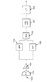
図1に、サンプル体の調査に使用する本発明の実施形態2000を示す。図1に示すように、本発明の実施形態2000は低コヒーレンス放射源1000を含む。低コヒーレンス放射源1000は、当業者に周知のいくつかの方法で実施することができる。本発明の好ましい実施形態では、短コヒーレンス放射源1000が、スーパールミネセント・ダイオード(SLD)である。低コヒーレンス放射源1000から出力された放射は、光ファイバ1010を通して、3デシベル放射結合器1030に入力される。3デシベル放射結合器1030は、低コヒーレンス放射源1000から結合器に入力された放射の50%をそれぞれ光ファイバ1040と1050に結合する。3デシベル放射結合器1030は、当業者に周知のいくつかの方法で実施することができる。本発明の好ましい実施形態では、3デシベル放射結合器1030が光ファイバ放射結合器である。光ファイバ1040および1050から出力された放射は、それぞれのコリメート・レンズ系1065および1067によってコリメートされ、実施形態2000の基準アーム経路1070およびサンプルアーム経路1140にそれぞれ送られる。
【0013】
図1にさらに示すように、基準アーム経路1070の放射は測定範囲変動装置1111に入射する。具体的には、基準アーム経路1070の放射は50%透過器(例えば50%ミラー)1080に入射し、これを通過する。図1に示すように、50%透過器1080は、直線ステージ移送器1090に装着され、エンコーダ1100が、直線ステージ移送器1090の位置を正確に測定する。直線ステージ移送器1090およびエンコーダ1100は、当業者に周知のいくつかの方法で実施することができる。図1に示すとおり、エンコーダ1100は直線ステージ移送器1090に取り付けられる。本発明によれば、直線ステージ移送器1090はモータ1120によって駆動され、50%透過器1080と反射器であるリトロレフレクター1130の間の距離を調整することができる。さらに、リトロレフレクター1130は、距離dの範囲内で一定の速度Vで前後に平行移動する。距離dを以後、走査範囲dと呼ぶ。リトロレフレクター1130の平行移動は、当業者に周知のいくつかの方法で実施することができる。本発明の好ましい実施形態では、所望の平行移動を得るために高速ガルバノメータ1135がリトロレフレクター1130に取り付けられる。
【0014】
サンプル・アーム経路1140の放射は横走査装置1160に入射し、通過する。横走査装置1160から出力された放射は、集束レンズ系1150によってサンプル1105上に集束される。当業者には周知のように、横走査装置1160は、サンプル・アーム経路1140の放射でサンプル1105を2次元横走査する。さらに横走査装置1160は、当業者に周知のいくつかの方法で実施することができる。
【0015】
50%透過器1080を通って基準アーム経路1070に戻された放射、および横走査装置1160を通ってサンプル・アーム経路1140に戻された放射は、それぞれのコリメート・レンズ系1065および1067によって、それぞれの光ファイバ1040および1050に入力される。光ファイバ1040および1050から出力された放射は、3デシベル放射結合器1030に入力される。3デシベル放射結合器1030は、基準アーム経路1070から結合器に結合された放射とサンプル・アーム経路1140から結合された放射を結合し、この結合された放射を光ファイバ1020に結合する。本発明を、光ファイバを利用する実施形態の文脈で記述したが、本発明がこれに限定されるものでないこと、および当業者が、個別の光学部品および統合された光学製品を使用することによって、その他の実施形態を容易に製作することができることを理解されたい。
【0016】
光ファイバ1020から出力された放射は解析器へ導かれ、まず光検出器1170に入力される。当業者には周知のとおり、3デシベル放射結合器1030で結合された、基準アーム経路1070からの放射とサンプル・アーム経路1140からの放射の間の光路長の差が、放射源1000の可干渉距離よりも短いと、有用で測定可能な干渉信号が光検出器1170から出力される。本発明の実施形態2000によれば、光検出器1170から出力された干渉信号は、トランスインピーダンス増幅器1180に入力される。トランスインピーダンス増幅器1180の出力は、同調可能な局部発振器1190によって生成された信号とともにミクサ1200に入力される。光検出器1170、トランスインピーダンス増幅器1180、同調可能局部発振器1190およびミクサ1200は、当業者に周知のいくつかの方法で実施することができる。
【0017】
当業者には周知のとおり、リトロレフレクター1130は一定の速度Vで前後に走査されるので、光検出器から出力された干渉信号、したがってトランスインピーダンス増幅器1180からミクサ1200に入力された信号は、ドップラー・シフト周波数fDで変調される。
fD=2V/λ (1)
V=d/t (2)
上式で、(i)λ=低コヒーレンス放射源1000から出力される放射の波長、(ii)Vは有効走査速度、(iii)dは、リトロレフレクター1130の走査範囲、(iv)tは走査時間である。
【0018】
当業者には周知のとおり、ミクサ1200および局部発振器1190は、AM/FM受信機で広く使われている周波数変換器のような機能を有し、中間周波数fiを有する信号を生成する。ただし、fi=fo−fD(foは、局部発振器1190の周波数)である。周知のとおり、ミクサ1200はさらに、いくつかの周波数、例えばfo+fDを生成する。周波数が高いほど信号対雑音比はよくなるが、増幅器の帯域幅はより制限される。
【0019】
本発明の実施形態2000によれば、ミクサ1200から出力された信号は、通過域の中心周波数がfiの帯域フィルタ1215に入力される。帯域フィルタ1215から出力された信号は、対数増幅器1220に入力される。この増幅器は、整流器としての働き、および入力信号の信号包絡線を対数スケールの信号に変換する対数増幅器としての働きをする。中間周波数fiの信号の帯域幅は、低コヒーレンス放射源1000から出力された信号のスペクトル帯域幅に比例する(Δfi=2πcΔλ/λ2。cは光速)。帯域フィルタ1215の帯域幅は、実質的に全ての信号構成要素を通過させることができ、かつ、ほとんどの雑音を排除することができるような幅とする。
【0020】
対数増幅器1220から出力された信号は低域フィルタ1230に入力され、低域フィルタ1230から出力された信号はA/D変換器1240に入力される。A/D変換器1240は入力信号をディジタル信号に変換し、A/D変換器1240から出力されたディジタル信号はコンピュータ1250に入力される。コンピュータ1250はこの未処理の信号を処理して、例えば眼の長さを測定し、その処理結果を、例えばカラー・マップを使用してディスプレイ・モニタ1260上に表示する。帯域フィルタ1215、対数増幅器1220、低域フィルタ1230、A/D変換器1240、コンピュータ1250およびディスプレイ・モニタ1260は、当業者に周知のいくつかの方法で実施することができる。
【0021】
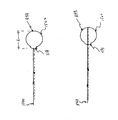
図2に、図1に示した本発明の実施形態2000の基準アームの経路1070を通る多くの可能な放射経路のうちの2つを示す。図2の経路1において、50%透過器1080とリトロレフレクター1130の間の光路長の最大と最小はそれぞれ、2(L±d/2)である(この式で、Lは、リトロレフレクター1130の平行移動範囲の中間点から50%透過器1080までの距離、dは、リトロレフレクター1130が平行移動する全体の距離、すなわちリトロレフレクター1130の走査範囲である)。経路1で、光検出器1170から出力される干渉信号のドップラー周波数をfD1とすると、fD1=fDである(fDは前記式(1)によって与えられる値である)。図2の経路2では、放射が、50%透過器1080とリトロレフレクター1130の間で前後に2度反射してから、50%透過器1080を通過してコリメート・レンズ系1065に戻る。したがって経路2では、50%透過器1080とリトロレフレクター1130の間の光路長の最大と最小はそれぞれ、4(L±d/2)となる。前記式(2)を用いると、リトロレフレクター1130の有効走査速度は2Vとなる。その結果、経路2では、光検出器1170から出力される干渉信号のドップラー周波数fD2は、fD2=2fDとなる。
【0022】
経路1によって生成された信号を通過させる帯域フィルタ1215の通過域の中心周波数がfi=fo−fDである場合、ミクサ1200から出力される経路2に対する信号の中間周波数fi2(=fo−2fD)は、帯域フィルタ1215を通過することができないことは容易に分かる。この場合、サンプル1105への縦方向の深度dの走査を観察することができることは、当業者なら容易に分かる。しかし、同調可能局部発振器1190の周波数がfo2(=fo+fD)に同調されている場合、中間周波数fi2(=fo+fD−2fD=fo−fD=fi)は、帯域フィルタ1215を通過し、サンプル1105への縦方向の深度2dの走査で観察することができる。
【0023】
以上を一般化すると、本発明によれば、図1に示した本発明の実施形態について、同調可能局部発振器1190の周波数がfON(=fo+(N−1)fD)に同調されている場合、中間周波数fiN(=fo+(N−1)fD−NfD=fo−fD=fi)は、帯域フィルタ1215を通過し、サンプル1105への縦方向の深度Ndの走査で観察することができる。
【0024】
しかし、基準アーム経路1070から3デシベル放射結合器に結合される放射の強度が0.5(0.5R)N倍に低減されるので、潜在的な走査深度は、50%透過器1080とリトロレフレクター1130の間の反射回数によって限定されうる。上式で、Rはリトロレフレクター1130の反射率である。全内部反射に基づき、表側が反射防止コーティングされている固体ガラスのリトロレフレクターでは、反射率の値Rを、0.9より高くすることができる。さらに留意すべきことは、ショット雑音が限定された系では、基準アーム経路1070から出力された放射がサンプル・アーム経路1140から出力された放射よりも大きく維持されることが好ましいことである。具体的には、本発明の実施形態の眼科的適用業務では、眼の反射率が、入射放射強度の10-4未満であるので、Nは、4〜5にもなる。
【0025】
図3は、本発明に基づいて製作され、眼の長さを測定するように適合されたOCT装置の信号解析セクションを示す図である。図3に示す信号解析セクションは、トランスインピーダンス増幅器1180とA/D1240の間の回路部品から成る図1に示した信号解析セクションを置き換えるものである。以下では、光検出器1170が、図2の経路1および経路2にそれぞれ対応する2つの信号を受け取ると仮定する。
【0026】
図3に示すとおり、これらの2つの信号は光検出器1170に入力される。光検出器1170の出力は、トランスインピーダンス増幅器1180に入力され、トランスインピーダンス増幅器1180の出力は帯域フィルタ1183および1187に入力される。帯域フィルタ1183および1187の通過域の中心周波数はそれぞれ、f1(=fD)およびf2(=2fD)である。帯域フィルタ1183および1187は図3に示すように、トランスインピーダンス増幅器1180とマルチプレクサ1193の間に並列に接続される。
【0027】
図4に、図1と図3の実施形態を組み合わせた本発明の実施形態が、眼からの反射を利用して、基準アーム経路1070上に生成された2つの放射信号と干渉する2つの放射信号をサンプル・アーム経路1140上に生成する方法を示す。この場合、これらの2組の放射信号間の干渉を同時に観察して、眼の長さを測定することができる。これを実施するために、50%透過器とリトロレフレクター1130の間の距離Lを平均的な人間の眼の長さに等しくする。次に図4を参照する。サンプル・アーム経路1140に生成される第1の信号は、眼1500の角膜1131で反射した放射に起因し、サンプル・アーム経路1140に生成される第2の信号は、眼1500の網膜1133で反射した放射の結果生じるものである。第1の干渉信号が、図2に示した経路1に起因する基準アーム経路1070の放射と、図4に示した角膜1131での反射に起因するサンプル・アーム経路1140の放射の間の干渉によって生成されたものであることは容易に分かる。同様に第2の干渉信号は、図2に示した経路2に起因する基準アーム経路1070の放射と、図4に示した網膜1133での反射に起因するサンプル・アーム経路1140の放射の間の干渉によって生成される。
【0028】
図5に、獲得され、表示装置1260に表示される2つの干渉信号を示す。図4の信号1600および1601に示すように、Leye(患者の眼の長さ)がL(平均的な人間の眼の長さ)とは異なり、かつLとLeyeの差が走査範囲dより短い場合、両方の干渉信号が時間遅れを伴って観察される。本発明によれば、例えば、コンピュータ1250との対話によって、図5に示すように信号1600と1601が同時に生じるよう、モータ1090で50%透過器1080を移動させることによって、眼の長さを測定することができる。容易に分かるように、この移動を、ジョイ・スティック、マウスなど当業者に周知のいくつかの装置(図示せず)の使用に由来するオペレータの入力によって実施することができる。代わりに、当業者に周知の方法によってこれらの2つの信号が重なり合うように、コンピュータ1250をプログラムしてもよい。50%透過器1080の位置が、エンコーダ1100からコンピュータ1250に中継され、コンピュータ1250はこの位置を使用して測定を実施する。本発明によれば、測定の精度が、低コヒーレンス放射源1000の可干渉距離となる利点が得られる。
【0029】
図6に、図1に示した実施形態2000に使用する基準アームの代替実施形態を示す。図6に示すように、50/50ビームスプリッタ4110が、基準アーム経路1070から入射する放射を分割し、それが2本の光路上を進む。第1の光路では放射が、(a)ビームスプリッタ4110を通過し、(b)リトロレフレクター1130で反射され、(c)再びビームスプリッタ4110を通過し、(d)放射4210としてコリメート・レンズ1060を通過する。この実施形態では、リトロレフレクター1130が速度Vで走査され、そのため、第1の光路のドップラー・シフト周波数はfDとなる。この実施形態ではまず、第1の光路の光路長が、眼の角膜で反射され、サンプル・アーム経路1140から現れる放射の光路長にセットされる。第2の光経路では放射が、(a)ビームスプリッタ4110を通過し、(b)リトロレフレクター1130で反射され、(c)ビームスプリッタ4110でミラー4120に向かって反射され、(d)ミラー4120で反射され、(e)ビームスプリッタ4110を通過し、(f)直線ステージ4140に装着されたミラー4130で反射され、(g)ビームスプリッタ4110で反射され、(h)放射4220としてコリメート・レンズ1060を通過する。ミラー4120および4130を、リトロレフレクター1130のようなリトロレフレクターとすることもできることを理解されたい。第2の光路を通る放射がリトロレフレクター1130で反射されるのは一度だけなので、第2の光経路のドップラー周波数シフタはfDとなる。本発明によれば、ミラー4120と4130の間の光学距離は、平均的な人間の眼の光路長にセットされる。その結果、放射4220がたどる光路長の合計は、網膜で反射されたサンプル・ビームがたどる光路長に等しくなる。さらに本発明によれば、モータ4150が、コンピュータ1250から送られた信号に応答して、角膜で反射された放射によって生成された干渉信号の位置と網膜で反射された放射によって生成された干渉信号の位置が先に説明したように一致するよう、直線ステージ移送器4140を移動させる。ミラー4130の位置が、エンコーダ4160によってコンピュータ1250に中継される。このようにして、ミラー4120と4130の間の光路長から眼の長さの情報が得られる。較正手順を使用することによって、長さの測定を正確に実施することができる。この代替実施形態の利点は、それぞれの基準アーム信号4210および4220に対して同じドップラー周波数が生成されるので、検出器の電子回路中に必要なのは、単一チャネル帯域フィルタだけであることである。
【0030】
本発明を、人間の眼の長さの測定に関して説明してきたが、本発明はこれに限定されるものではない。実際、当業者なら容易に理解できるように、物体の2つの部分の間の距離を測定するのに本発明の実施形態を使用することができる。さらに本発明の実施形態を、個別の光学構成部品、インテグレイテッド・オプティクス、光ファイバ、およびこれらの組合せを利用して製作することができる。さらに、これまでに説明した50%結合器および50%透過器とは異なる結合器および送信器を利用した他の実施形態も可能であり、以上の説明に照らせば、このような相違を考慮して、前述の実施形態をどのように変更すればよいかは、当業者にとって明白であろう。
【0031】
以上の記述は、例示および説明のために示したに過ぎないことを当業者は理解しよう。このように以上の記述は、本発明を網羅的に記載しようとしたものではなく、また、開示した正確な形態に本発明を限定しようとしたものでもない。
【図面の簡単な説明】
【図1】本発明に基づいて製作されたOCT装置の実施形態を示す図である。
【図2】図1に示した本発明の実施形態の基準アーム経路を通る多くの可能な放射経路のうちの2つを示す図である。
【図3】本発明に基づいて製作され、眼の長さを測定するように適合されたOCT装置の信号解析セクションを示す図である。
【図4】図1と図3の実施形態を組み合わせた本発明の実施形態が、眼からの反射を利用して、基準アーム経路で生成された2つの放射信号と干渉する2つの放射信号をサンプル・アーム経路に生成させる方法を示す図である。
【図5】眼の長さの測定のために、本発明に基づいて生成された2つの干渉信号を示す図である。
【図6】図1に示したOCT装置の基準アームの代替実施形態を示す図である。
【符号の説明】
1000 低コヒーレンス放射源
1030 3デシベル放射結合器
1070 基準アーム経路
1080 50%透過器
1090 直線ステージ移送器
1100 エンコーダ
1105 サンプル
1111 測定範囲変動装置
1130 リトロレフレクター
1140 サンプル・アーム経路
1160 横走査装置
1193 マルチプレクサ
1200 ミクサ
1215 帯域フィルタ
1220 対数増幅器
1230 低域フィルタ
1250 コンピュータ
1260 ディスプレイ・モニタ
Claims (4)
- 物体を調査する光学コヒーレンス断層撮影装置(OCT)において、
短コヒーレンス放射の放射源と、
(a)放射の第1の部分を基準アームに結合し、(b)放射の第2の部分をサンプル・アームに結合し、(c)前記基準アームおよび前記サンプルアームから送られた放射を結合し、(d)結合された放射を解析器に結合する結合器と
を備え、
前記基準アームが、放射の第1の部分を測定範囲変動装置に送り、その測定範囲変動装置から出力された放射を結合器に戻し、
前記サンプルアームが、放射の第2の部分を物体に送り、その物体によって散乱された放射を結合器に戻し、
前記測定範囲変動装置には、
入射する放射の一部分を透過させる透過器と、
前記透過器を透過した放射を反射する反射器とが備えられ、当該反射された放射の一部が前記透過器で反射され、
さらに、前記反射器に走査をさせるように結合された走査装置が備えられている、ことを特徴とする光学コヒーレンス断層撮影装置。 - 請求項1に記載の光学コヒーレンス断層撮影装置において、前記解析器には、
前記結合器から結合された放射を検出する光検出器と、
前記光検出器および発振器の出力に応答するミクサと、
前記ミクサの出力に結合されたフィルタと
が備えられている、ことを特徴とする光学コヒーレンス断層撮影装置。 - 請求項1に記載の光学コヒーレンス断層撮影装置であって、物体の第1の散乱性部分と第2の散乱性部分との間の距離を測定するものであり、
前記測定範囲変動装置には、さらに、前記透過器を平行移動させるように結合された移送器が備えられ、且つ、
前記解析器には、
前記結合器から結合された放射を検出する光検出器と、
前記光検出器の出力に結合され、通過域の中心周波数が、反射器が走査をすることによって生成されるドップラー・シフト周波数に比例した周波数である第1のフィルタと、
前記光検出器の出力に結合された第2のフィルタにして、その通過域の中心周波数が、前記ドップラー・シフト周波数に比例した周波数の倍数であって前記第1のフィルタの前記通過域の中心周波数とは異なっている、第2のフィルタと
が備えられている、
ことを特徴とする光学コヒーレンス断層撮影装置。 - 請求項3に記載の光学コヒーレンス断層撮影装置であって、
前記測定範囲変動装置には、前記透過器の位置を測定するエンコーダが備えられている、
ことを特徴とする光学コヒーレンス断層撮影装置。
Applications Claiming Priority (2)
| Application Number | Priority Date | Filing Date | Title |
|---|---|---|---|
| US09/079908 | 1998-05-15 | ||
| US09/079,908 US6053613A (en) | 1998-05-15 | 1998-05-15 | Optical coherence tomography with new interferometer |
Publications (3)
| Publication Number | Publication Date |
|---|---|
| JP2000002516A JP2000002516A (ja) | 2000-01-07 |
| JP2000002516A5 JP2000002516A5 (ja) | 2006-06-29 |
| JP4423450B2 true JP4423450B2 (ja) | 2010-03-03 |
Family
ID=22153581
Family Applications (1)
| Application Number | Title | Priority Date | Filing Date |
|---|---|---|---|
| JP13367099A Expired - Fee Related JP4423450B2 (ja) | 1998-05-15 | 1999-05-14 | 新規の干渉計を用いた光学コヒーレンス断層撮影 |
Country Status (4)
| Country | Link |
|---|---|
| US (1) | US6053613A (ja) |
| EP (1) | EP0956809B1 (ja) |
| JP (1) | JP4423450B2 (ja) |
| DE (1) | DE69940087D1 (ja) |
Families Citing this family (205)
| Publication number | Priority date | Publication date | Assignee | Title |
|---|---|---|---|---|
| US6615072B1 (en) * | 1999-02-04 | 2003-09-02 | Olympus Optical Co., Ltd. | Optical imaging device |
| DE10020559A1 (de) * | 2000-04-27 | 2001-10-31 | Hannover Laser Zentrum | Laser-Bearbeitung von Materialien |
| DE10042751A1 (de) * | 2000-08-31 | 2002-03-14 | Thomas Hellmuth | System zur berührungslosen Vermessung der optischen Abbildungsqualität eines Auges |
| AU2001285718B2 (en) | 2000-09-04 | 2006-11-02 | Danmarks Tekniske Universitet | Optical amplification in coherence reflectometry |
| JP4241038B2 (ja) * | 2000-10-30 | 2009-03-18 | ザ ジェネラル ホスピタル コーポレーション | 組織分析のための光学的な方法及びシステム |
| US9295391B1 (en) | 2000-11-10 | 2016-03-29 | The General Hospital Corporation | Spectrally encoded miniature endoscopic imaging probe |
| CA2448527C (en) * | 2001-03-20 | 2008-12-02 | Cornell Research Foundation, Inc. | Precision ultrasound measurement for intraocular lens placement |
| EP2333521B1 (en) * | 2001-04-30 | 2019-12-04 | The General Hospital Corporation | Method and apparatus for improving image clarity and sensitivity in optical coherence tomography using dynamic feedback to control focal properties and coherence gating |
| GB2408797B (en) | 2001-05-01 | 2006-09-20 | Gen Hospital Corp | Method and apparatus for determination of atherosclerotic plaque type by measurement of tissue optical properties |
| US6980299B1 (en) | 2001-10-16 | 2005-12-27 | General Hospital Corporation | Systems and methods for imaging a sample |
| DE10151216A1 (de) * | 2001-10-16 | 2003-04-24 | Zeiss Carl Jena Gmbh | Verfahren zur optischen Erfassung von charakteristischen Größen einer beleuchteten Probe |
| EP1468245B1 (en) * | 2002-01-11 | 2011-03-30 | The General Hospital Corporation | Apparatus for OCT imaging with axial line focus for improved resolution and depth of field |
| AU2003205144B2 (en) * | 2002-01-15 | 2008-09-04 | Board Of Regents, The University Of Texas System | Methods and compositions to reduce scattering of light during therapeutic and diagnostic imaging procedures |
| US7355716B2 (en) * | 2002-01-24 | 2008-04-08 | The General Hospital Corporation | Apparatus and method for ranging and noise reduction of low coherence interferometry LCI and optical coherence tomography OCT signals by parallel detection of spectral bands |
| US20110201924A1 (en) * | 2002-04-30 | 2011-08-18 | The General Hospital Corporation | Method and Apparatus for Improving Image Clarity and Sensitivity in Optical Tomography Using Dynamic Feedback to Control Focal Properties and Coherence Gating |
| EP2319404B1 (en) * | 2003-01-24 | 2015-03-11 | The General Hospital Corporation | System and method for identifying tissue low-coherence interferometry |
| US8054468B2 (en) | 2003-01-24 | 2011-11-08 | The General Hospital Corporation | Apparatus and method for ranging and noise reduction of low coherence interferometry LCI and optical coherence tomography OCT signals by parallel detection of spectral bands |
| US7567349B2 (en) * | 2003-03-31 | 2009-07-28 | The General Hospital Corporation | Speckle reduction in optical coherence tomography by path length encoded angular compounding |
| WO2004073501A2 (en) * | 2003-02-20 | 2004-09-02 | Gutin Mikhail | Optical coherence tomography with 3d coherence scanning |
| US6988801B2 (en) * | 2003-03-25 | 2006-01-24 | University Of Rochester | Compact portable wavefront sensor |
| RU2247938C1 (ru) * | 2003-05-27 | 2005-03-10 | Геликонов Валентин Михайлович | Оптическое устройство для исследования объекта |
| TWI223719B (en) * | 2003-05-30 | 2004-11-11 | Ind Tech Res Inst | Sub-micrometer-resolution optical coherent tomography |
| ES2310744T3 (es) | 2003-06-06 | 2009-01-16 | The General Hospital Corporation | Fuente de luz sintonizable en longitudes de onda. |
| US7876974B2 (en) * | 2003-08-29 | 2011-01-25 | Vladimir Brajovic | Method for improving digital images and an image sensor for sensing the same |
| EP2293031B8 (en) | 2003-10-27 | 2024-03-20 | The General Hospital Corporation | Method and apparatus for performing optical imaging using frequency-domain interferometry |
| WO2005054780A1 (en) * | 2003-11-28 | 2005-06-16 | The General Hospital Corporation | Method and apparatus for three-dimensional spectrally encoded imaging |
| AT501056B1 (de) * | 2004-02-06 | 2007-04-15 | Zeiss Carl Meditec Ag | Kurzkohärenz-interferometrische längenmessung am auge |
| WO2005077256A1 (en) * | 2004-02-06 | 2005-08-25 | Optovue, Inc. | Optical apparatus and methods for performing eye examinations |
| JP2007522456A (ja) * | 2004-02-10 | 2007-08-09 | オプトビュー,インコーポレーテッド | 高効率低コヒーレンス干渉法 |
| US7242480B2 (en) * | 2004-05-14 | 2007-07-10 | Medeikon Corporation | Low coherence interferometry for detecting and characterizing plaques |
| US7190464B2 (en) * | 2004-05-14 | 2007-03-13 | Medeikon Corporation | Low coherence interferometry for detecting and characterizing plaques |
| US7184148B2 (en) | 2004-05-14 | 2007-02-27 | Medeikon Corporation | Low coherence interferometry utilizing phase |
| US7327463B2 (en) | 2004-05-14 | 2008-02-05 | Medrikon Corporation | Low coherence interferometry utilizing magnitude |
| US20050254059A1 (en) * | 2004-05-14 | 2005-11-17 | Alphonse Gerard A | Low coherence interferometric system for optical metrology |
| US7474408B2 (en) * | 2004-05-14 | 2009-01-06 | Medeikon Corporation | Low coherence interferometry utilizing phase |
| EP1754016B1 (en) | 2004-05-29 | 2016-05-18 | The General Hospital Corporation | Process, system and software arrangement for a chromatic dispersion compensation using reflective layers in optical coherence tomography (oct) imaging |
| US7447408B2 (en) * | 2004-07-02 | 2008-11-04 | The General Hospital Corproation | Imaging system and related techniques |
| JP5053845B2 (ja) * | 2004-08-06 | 2012-10-24 | ザ ジェネラル ホスピタル コーポレイション | 光学コヒーレンス断層撮影法を使用して試料中の少なくとも1つの位置を決定するための方法、システムおよびソフトウェア装置 |
| EP1989997A1 (en) | 2004-08-24 | 2008-11-12 | The General Hospital Corporation | Process, System and Software Arrangement for Measuring a Mechanical Strain and Elastic Properties of a Sample |
| EP1793731B1 (en) | 2004-08-24 | 2013-12-25 | The General Hospital Corporation | Imaging apparatus comprising a fluid delivery arrangement and a pull-back arrangement |
| EP1787105A2 (en) * | 2004-09-10 | 2007-05-23 | The General Hospital Corporation | System and method for optical coherence imaging |
| EP1804638B1 (en) | 2004-09-29 | 2012-12-19 | The General Hospital Corporation | System and method for optical coherence imaging |
| JP5175101B2 (ja) * | 2004-10-29 | 2013-04-03 | ザ ジェネラル ホスピタル コーポレイション | 偏光感応性光コヒーレンストモグラフィを用いて偏光非解消の偏光パラメータを測定するジョーンズ行列に基づく解析を行うシステム及び方法 |
| WO2006050453A1 (en) * | 2004-11-02 | 2006-05-11 | The General Hospital Corporation | Fiber-optic rotational device, optical system and method for imaging a sample |
| EP2278266A3 (en) * | 2004-11-24 | 2011-06-29 | The General Hospital Corporation | Common-Path Interferometer for Endoscopic OCT |
| WO2006058346A1 (en) | 2004-11-29 | 2006-06-01 | The General Hospital Corporation | Arrangements, devices, endoscopes, catheters and methods for performing optical imaging by simultaneously illuminating and detecting multiple points on a sample |
| US8394084B2 (en) | 2005-01-10 | 2013-03-12 | Optimedica Corporation | Apparatus for patterned plasma-mediated laser trephination of the lens capsule and three dimensional phaco-segmentation |
| US7809171B2 (en) * | 2005-01-10 | 2010-10-05 | Battelle Memorial Institute | Facial feature evaluation based on eye location |
| WO2006078802A1 (en) * | 2005-01-21 | 2006-07-27 | Massachusetts Institute Of Technology | Methods and apparatus for optical coherence tomography scanning |
| ATE451669T1 (de) | 2005-04-28 | 2009-12-15 | Gen Hospital Corp | Bewertung von bildmerkmalen einer anatomischen struktur in optischen kohärenztomographiebildern |
| US7859679B2 (en) * | 2005-05-31 | 2010-12-28 | The General Hospital Corporation | System, method and arrangement which can use spectral encoding heterodyne interferometry techniques for imaging |
| EP1889037A2 (en) | 2005-06-01 | 2008-02-20 | The General Hospital Corporation | Apparatus, method and system for performing phase-resolved optical frequency domain imaging |
| US7426036B2 (en) * | 2005-07-08 | 2008-09-16 | Imalux Corporation | Common path frequency domain optical coherence reflectometer and common path frequency domain optical coherence tomography device |
| EP2267404B1 (en) | 2005-08-09 | 2016-10-05 | The General Hospital Corporation | Apparatus and method for performing polarization-based quadrature demodulation in optical coherence tomography |
| US7872759B2 (en) | 2005-09-29 | 2011-01-18 | The General Hospital Corporation | Arrangements and methods for providing multimodality microscopic imaging of one or more biological structures |
| US7400410B2 (en) | 2005-10-05 | 2008-07-15 | Carl Zeiss Meditec, Inc. | Optical coherence tomography for eye-length measurement |
| EP1945094B1 (en) * | 2005-10-14 | 2018-09-05 | The General Hospital Corporation | Spectral- and frequency- encoded fluorescence imaging |
| DE102005059923A1 (de) * | 2005-12-13 | 2007-06-14 | Oculus Optikgeräte GmbH | Verfahren und Vorrichtung zur Bestimmung des Abstandes zu einem Messpunkt auf einer Gewebefläche des Auges |
| DE102005062238A1 (de) * | 2005-12-22 | 2007-07-05 | Carl Zeiss Meditec Ag | Ophthalmologisches Messsystem und Verfahren zur Ermittlung der biometrischen Daten eines Auges |
| WO2007082228A1 (en) | 2006-01-10 | 2007-07-19 | The General Hospital Corporation | Systems and methods for generating data based on one or more spectrally-encoded endoscopy techniques |
| US20070223006A1 (en) * | 2006-01-19 | 2007-09-27 | The General Hospital Corporation | Systems and methods for performing rapid fluorescence lifetime, excitation and emission spectral measurements |
| EP2289397A3 (en) | 2006-01-19 | 2011-04-06 | The General Hospital Corporation | Methods and systems for optical imaging of epithelial luminal organs by beam scanning thereof |
| US8145018B2 (en) | 2006-01-19 | 2012-03-27 | The General Hospital Corporation | Apparatus for obtaining information for a structure using spectrally-encoded endoscopy techniques and methods for producing one or more optical arrangements |
| WO2007084959A1 (en) * | 2006-01-20 | 2007-07-26 | The General Hospital Corporation | Systems and methods for providing mirror tunnel microscopy |
| EP1986545A2 (en) * | 2006-02-01 | 2008-11-05 | The General Hospital Corporation | Apparatus for applying a plurality of electro-magnetic radiations to a sample |
| US10426548B2 (en) * | 2006-02-01 | 2019-10-01 | The General Hosppital Corporation | Methods and systems for providing electromagnetic radiation to at least one portion of a sample using conformal laser therapy procedures |
| US9777053B2 (en) | 2006-02-08 | 2017-10-03 | The General Hospital Corporation | Methods, arrangements and systems for obtaining information associated with an anatomical sample using optical microscopy |
| EP2306141A1 (en) | 2006-02-24 | 2011-04-06 | The General Hospital Corporation | Methods and systems for performing angle-resolved fourier-domain optical coherence tomography |
| WO2007103721A2 (en) * | 2006-03-01 | 2007-09-13 | The General Hospital Corporation | System and method for providing cell specific laser therapy of atherosclerotic plaques by targeting light absorbers in macrophages |
| WO2007109540A2 (en) * | 2006-03-17 | 2007-09-27 | The General Hospital Corporation | Arrangement, method and computer-accessible medium for identifying characteristics of at least a portion of a blood vessel contained within a tissue using spectral domain low coherence interferometry |
| US7742173B2 (en) * | 2006-04-05 | 2010-06-22 | The General Hospital Corporation | Methods, arrangements and systems for polarization-sensitive optical frequency domain imaging of a sample |
| US8175685B2 (en) * | 2006-05-10 | 2012-05-08 | The General Hospital Corporation | Process, arrangements and systems for providing frequency domain imaging of a sample |
| WO2007133964A2 (en) * | 2006-05-12 | 2007-11-22 | The General Hospital Corporation | Processes, arrangements and systems for providing a fiber layer thickness map based on optical coherence tomography images |
| JP4907227B2 (ja) * | 2006-05-29 | 2012-03-28 | 株式会社ニデック | 眼内寸法測定装置 |
| US7488930B2 (en) * | 2006-06-02 | 2009-02-10 | Medeikon Corporation | Multi-channel low coherence interferometer |
| WO2008024948A2 (en) | 2006-08-25 | 2008-02-28 | The General Hospital Corporation | Apparatus and methods for enhancing optical coherence tomography imaging using volumetric filtering techniques |
| US7452077B2 (en) * | 2006-08-29 | 2008-11-18 | Carl Zeiss Meditec, Inc. | Image adjustment derived from optical imaging measurement data |
| WO2008049118A2 (en) | 2006-10-19 | 2008-04-24 | The General Hospital Corporation | Apparatus and method for obtaining and providing imaging information associated with at least one portion of a sample and effecting such portion(s) |
| US20080206804A1 (en) * | 2007-01-19 | 2008-08-28 | The General Hospital Corporation | Arrangements and methods for multidimensional multiplexed luminescence imaging and diagnosis |
| EP2102583A2 (en) | 2007-01-19 | 2009-09-23 | The General Hospital Corporation | Apparatus and method for controlling ranging depth in optical frequency domain imaging |
| EP2662674A3 (en) | 2007-01-19 | 2014-06-25 | The General Hospital Corporation | Rotating disk reflection for fast wavelength scanning of dispersed broadbend light |
| EP2267403A3 (de) * | 2007-02-21 | 2011-04-20 | Agfa HealthCare N.V. | System und Verfahren zur optischen Kohärenztomographie |
| EP1962079B1 (de) | 2007-02-21 | 2016-06-01 | Agfa HealthCare N.V. | System und Verfahren zur optischen Kohärenztomographie |
| DE502007004384D1 (de) | 2007-02-21 | 2010-08-26 | Agfa Healthcare Nv | System und Verfahren zur optischen Kohärenztomographie |
| EP1962049B1 (de) | 2007-02-21 | 2015-12-23 | Agfa HealthCare N.V. | System und Verfahren zur optischen Kohärenztomographie |
| EP1962082A1 (de) | 2007-02-21 | 2008-08-27 | Agfa HealthCare N.V. | System und Verfahren zur optischen Kohärenztomographie |
| EP1962080B1 (de) | 2007-02-21 | 2011-06-01 | Agfa HealthCare N.V. | System zur optischen Kohärenztomographie |
| EP1962081B1 (de) | 2007-02-21 | 2016-09-14 | Agfa HealthCare N.V. | System zur optischen Kohärenztomographie |
| EP1962051A1 (de) * | 2007-02-21 | 2008-08-27 | Agfa HealthCare N.V. | System und Verfahren zur optischen Kohärenztomographie |
| EP2617398B1 (en) | 2007-03-13 | 2017-11-15 | Optimedica Corporation | Intraocular lens for improved placement |
| WO2008116010A1 (en) * | 2007-03-19 | 2008-09-25 | The General Hospital Corporation | System and method for providing noninvasive diagnosis of compartment syndrome exemplary laser speckle imaging procedure |
| US9176319B2 (en) | 2007-03-23 | 2015-11-03 | The General Hospital Corporation | Methods, arrangements and apparatus for utilizing a wavelength-swept laser using angular scanning and dispersion procedures |
| WO2008121844A1 (en) | 2007-03-30 | 2008-10-09 | The General Hospital Corporation | System and method providing intracoronary laser speckle imaging for the detection of vulnerable plaque |
| DE102007016444A1 (de) * | 2007-04-05 | 2008-10-16 | Precitec Optronik Gmbh | Bearbeitungseinrichtung |
| US8045177B2 (en) | 2007-04-17 | 2011-10-25 | The General Hospital Corporation | Apparatus and methods for measuring vibrations using spectrally-encoded endoscopy |
| WO2008137637A2 (en) | 2007-05-04 | 2008-11-13 | The General Hospital Corporation | Methods, arrangements and systems for obtaining information associated with a sample using brillouin microscopy |
| WO2009018456A2 (en) * | 2007-07-31 | 2009-02-05 | The General Hospital Corporation | Systems and methods for providing beam scan patterns for high speed doppler optical frequency domain imaging |
| US8040608B2 (en) | 2007-08-31 | 2011-10-18 | The General Hospital Corporation | System and method for self-interference fluorescence microscopy, and computer-accessible medium associated therewith |
| US8076624B1 (en) | 2007-09-19 | 2011-12-13 | Barchers Jeffrey D | Non-cooperative laser target enhancement system and method |
| US8787774B2 (en) * | 2007-10-10 | 2014-07-22 | Luxtera, Inc. | Method and system for a narrowband, non-linear optoelectronic receiver |
| WO2009059034A1 (en) * | 2007-10-30 | 2009-05-07 | The General Hospital Corporation | System and method for cladding mode detection |
| US7800759B2 (en) * | 2007-12-11 | 2010-09-21 | Bausch & Lomb Incorporated | Eye length measurement apparatus |
| EP2230990B1 (en) * | 2007-12-21 | 2016-12-14 | Bausch & Lomb Incorporated | Ophthalmic instrument alignment apparatus and method of using same |
| US8758252B2 (en) | 2008-01-02 | 2014-06-24 | Arcscan, Inc. | Innovative components for an ultrasonic arc scanning apparatus |
| US10531859B2 (en) | 2008-01-02 | 2020-01-14 | Arcscan, Inc. | Components for a precision ultrasonic scanning apparatus for body parts |
| US20090225324A1 (en) * | 2008-01-17 | 2009-09-10 | The General Hospital Corporation | Apparatus for providing endoscopic high-speed optical coherence tomography |
| US9332942B2 (en) * | 2008-01-28 | 2016-05-10 | The General Hospital Corporation | Systems, processes and computer-accessible medium for providing hybrid flourescence and optical coherence tomography imaging |
| US11123047B2 (en) | 2008-01-28 | 2021-09-21 | The General Hospital Corporation | Hybrid systems and methods for multi-modal acquisition of intravascular imaging data and counteracting the effects of signal absorption in blood |
| US10426348B2 (en) | 2008-03-05 | 2019-10-01 | Purdue Research Foundation | Using differential time-frequency tissue-response spectroscopy to evaluate living body response to a drug |
| US10080684B2 (en) | 2008-03-13 | 2018-09-25 | Optimedica Corporation | System and method for laser corneal incisions for keratoplasty procedures |
| WO2009124271A1 (en) | 2008-04-03 | 2009-10-08 | Arcscan, Inc. | Procedures for an ultrasonic arc scanning apparatus |
| ES2665748T3 (es) | 2008-04-23 | 2018-04-27 | Bioptigen, Inc. | Sistemas de generación de imágenes por tomografía de coherencia óptica (OCT) para uso en aplicaciones oftálmicas pediátricas y métodos y productos de programas informáticos relacionados |
| JP5607610B2 (ja) | 2008-05-07 | 2014-10-15 | ザ ジェネラル ホスピタル コーポレイション | 構造の特徴を決定する装置、装置の作動方法およびコンピュータアクセス可能な媒体 |
| US9039623B2 (en) * | 2008-05-29 | 2015-05-26 | Arcscan, Inc. | Compound scanning head for an ultrasonic scanning apparatus |
| JP5795531B2 (ja) | 2008-06-20 | 2015-10-14 | ザ ジェネラル ホスピタル コーポレイション | フューズドファイバオプティックカプラ構造、及びその使用方法 |
| EP2309923B1 (en) | 2008-07-14 | 2020-11-25 | The General Hospital Corporation | Apparatus and methods for color endoscopy |
| WO2010068764A2 (en) | 2008-12-10 | 2010-06-17 | The General Hospital Corporation | Systems, apparatus and methods for extending imaging depth range of optical coherence tomography through optical sub-sampling |
| US8317709B2 (en) | 2008-12-15 | 2012-11-27 | Arcscan, Inc. | Alignment and imaging of an eye with an ultrasonic scanner |
| US9149254B2 (en) | 2008-12-15 | 2015-10-06 | Arcscan, Inc. | Alignment and imaging of an eye with an ultrasonic scanner |
| US8294971B2 (en) * | 2008-12-18 | 2012-10-23 | Bausch • Lomb Incorporated | Apparatus comprising an optical path delay scanner |
| EP2382456A4 (en) | 2009-01-26 | 2012-07-25 | Gen Hospital Corp | SYSTEM, METHOD AND COMPUTER-ACCESSIBLE MEDIUM FOR PROVIDING BROAD FIELD SUPER-RESOLUTION MICROSCOPY |
| WO2010091190A2 (en) | 2009-02-04 | 2010-08-12 | The General Hospital Corporation | Apparatus and method for utilization of a high-speed optical wavelength tuning source |
| JP5249073B2 (ja) * | 2009-02-12 | 2013-07-31 | 株式会社ニデック | 光干渉式距離計測装置 |
| WO2010105197A2 (en) | 2009-03-12 | 2010-09-16 | The General Hospital Corporation | Non-contact optical system, computer-accessible medium and method for measuring at least one mechanical property of tissue using coherent speckle techniques(s) |
| JP5258052B2 (ja) * | 2009-04-25 | 2013-08-07 | 国立大学法人宇都宮大学 | 位相シフト法による形状測定方法及び形状測定装置、並びに複素振幅計測方法及び複素振幅計測装置 |
| JP5545618B2 (ja) * | 2009-07-06 | 2014-07-09 | 株式会社ニデック | 眼寸法測定装置 |
| JP5819823B2 (ja) | 2009-07-14 | 2015-11-24 | ザ ジェネラル ホスピタル コーポレイション | 血管の内部の流れおよび圧力を測定する装置および装置の作動方法 |
| US8510883B2 (en) * | 2009-10-30 | 2013-08-20 | Arcscan, Inc. | Method of positioning a patient for medical procedures |
| US20110184395A1 (en) * | 2009-12-23 | 2011-07-28 | Optimedica Corporation | Method for laser capsulotomy and lens conditioning |
| CN102811684B (zh) | 2010-01-22 | 2015-09-09 | 眼科医疗公司 | 用于自动放置扫描激光撕囊切口的装置 |
| US9278028B2 (en) | 2010-02-08 | 2016-03-08 | Optimedica Corporation | System and method for plasma-mediated modification of tissue |
| PT2542145T (pt) | 2010-03-05 | 2020-11-04 | Massachusetts Gen Hospital | Sistemas, métodos e meios acessíveis por computador que proporcionam imagens microscópicas de pelo menos uma estrutura anatómica numa resolução particular |
| US9069130B2 (en) | 2010-05-03 | 2015-06-30 | The General Hospital Corporation | Apparatus, method and system for generating optical radiation from biological gain media |
| EP2575598A2 (en) | 2010-05-25 | 2013-04-10 | The General Hospital Corporation | Apparatus, systems, methods and computer-accessible medium for spectral analysis of optical coherence tomography images |
| US9557154B2 (en) | 2010-05-25 | 2017-01-31 | The General Hospital Corporation | Systems, devices, methods, apparatus and computer-accessible media for providing optical imaging of structures and compositions |
| US10285568B2 (en) | 2010-06-03 | 2019-05-14 | The General Hospital Corporation | Apparatus and method for devices for imaging structures in or at one or more luminal organs |
| US10401793B2 (en) | 2010-06-17 | 2019-09-03 | Purdue Research Foundation | Digital holographic method of measuring cellular activity and measuring apparatus with improved stability |
| US9514271B2 (en) * | 2010-06-17 | 2016-12-06 | Purdue Research Foundation | Digital holographic method of measuring cellular activity and measuring apparatus with improved stability |
| WO2012058381A2 (en) | 2010-10-27 | 2012-05-03 | The General Hospital Corporation | Apparatus, systems and methods for measuring blood pressure within at least one vessel |
| US9046337B2 (en) | 2010-12-30 | 2015-06-02 | Volcano Corporation | Integrated OCT detector system with transimpedance amplifier |
| US8437007B2 (en) | 2010-12-30 | 2013-05-07 | Axsun Technologies, Inc. | Integrated optical coherence tomography system |
| KR101552290B1 (ko) * | 2011-02-15 | 2015-09-10 | 웨이브라이트 게엠베하 | 광간섭 단층촬영에 의한 물체의 내부 형상 측정 시스템 및 방법 |
| US8721077B2 (en) | 2011-04-29 | 2014-05-13 | The General Hospital Corporation | Systems, methods and computer-readable medium for determining depth-resolved physical and/or optical properties of scattering media by analyzing measured data over a range of depths |
| JP2014523536A (ja) | 2011-07-19 | 2014-09-11 | ザ ジェネラル ホスピタル コーポレイション | 光コヒーレンストモグラフィーにおいて偏波モード分散補償を提供するためのシステム、方法、装置およびコンピュータアクセス可能な媒体 |
| WO2013029047A1 (en) | 2011-08-25 | 2013-02-28 | The General Hospital Corporation | Methods, systems, arrangements and computer-accessible medium for providing micro-optical coherence tomography procedures |
| WO2013066631A1 (en) | 2011-10-18 | 2013-05-10 | The General Hospital Corporation | Apparatus and methods for producing and/or providing recirculating optical delay(s) |
| ITPI20120009A1 (it) * | 2012-01-24 | 2013-07-25 | Visia Imaging S R L | "un metodo per ridurre il tempo della misura a scansione della lunghezza assiale oculare e dispositivo per attuare tale metodo" |
| WO2013148306A1 (en) | 2012-03-30 | 2013-10-03 | The General Hospital Corporation | Imaging system, method and distal attachment for multidirectional field of view endoscopy |
| US9597059B2 (en) | 2012-05-17 | 2017-03-21 | Arcscan, Inc. | Correcting for unintended motion for ultrasonic eye scans |
| EP2852315A4 (en) | 2012-05-21 | 2016-06-08 | Gen Hospital Corp | DEVICE, APPARATUS AND METHOD FOR CAPSULE MICROSCOPY |
| US9320427B2 (en) | 2012-07-09 | 2016-04-26 | Arcscan, Inc. | Combination optical and ultrasonic imaging of an eye |
| EP2888616A4 (en) | 2012-08-22 | 2016-04-27 | Gen Hospital Corp | SYSTEM, METHOD AND COMPUTER-ACCESSIBLE MEDIA FOR MANUFACTURING MINIATURE ENDOSCOPES USING SOFT LITHOGRAPHY |
| US10702209B2 (en) | 2012-10-24 | 2020-07-07 | Amo Development, Llc | Graphical user interface for laser eye surgery system |
| US9445946B2 (en) | 2012-11-02 | 2016-09-20 | Optimedica Corporation | Laser eye surgery system |
| WO2014071221A2 (en) | 2012-11-02 | 2014-05-08 | Optimedica Corporation | Optical surface identification for laser surgery |
| US10278862B2 (en) | 2012-11-02 | 2019-05-07 | Optimedica Corporation | Low voltage communication between subsystems in a laser eye surgery system |
| US10292863B2 (en) | 2012-11-02 | 2019-05-21 | Optimedica Corporation | Interface force feedback in a laser eye surgery system |
| US10314746B2 (en) | 2012-11-02 | 2019-06-11 | Optimedica Corporation | Laser eye surgery system calibration |
| US10624786B2 (en) | 2012-11-02 | 2020-04-21 | Amo Development, Llc | Monitoring laser pulse energy in a laser eye surgery system |
| US10285860B2 (en) | 2012-11-02 | 2019-05-14 | Optimedica Corporation | Vacuum loss detection during laser eye surgery |
| US9987165B2 (en) | 2012-11-02 | 2018-06-05 | Optimedica Corporation | Liquid optical interface for laser eye surgery system |
| US9677869B2 (en) | 2012-12-05 | 2017-06-13 | Perimeter Medical Imaging, Inc. | System and method for generating a wide-field OCT image of a portion of a sample |
| US10893806B2 (en) | 2013-01-29 | 2021-01-19 | The General Hospital Corporation | Apparatus, systems and methods for providing information regarding the aortic valve |
| US11179028B2 (en) | 2013-02-01 | 2021-11-23 | The General Hospital Corporation | Objective lens arrangement for confocal endomicroscopy |
| JP6532854B2 (ja) | 2013-03-14 | 2019-06-19 | オプティメディカ・コーポレイションOptimedica Corporation | レーザ水晶体嚢硝子体切開術 |
| JP6378311B2 (ja) | 2013-03-15 | 2018-08-22 | ザ ジェネラル ホスピタル コーポレイション | 物体を特徴付ける方法とシステム |
| CA3144057A1 (en) | 2013-03-15 | 2014-09-25 | Optimedica Corporation | Microfemtotomy methods and systems |
| US10369053B2 (en) | 2013-04-17 | 2019-08-06 | Optimedica Corporation | Corneal topography measurements and fiducial mark incisions in laser surgical procedures |
| EP3505145B1 (en) | 2013-04-17 | 2020-08-19 | Optimedica Corporation | Laser fiducials for axis alignment in cataract surgery |
| CN105517514B (zh) | 2013-04-18 | 2018-09-21 | 光学医疗公司 | 角膜手术程序的角膜形貌测量和对准 |
| WO2014186353A1 (en) | 2013-05-13 | 2014-11-20 | The General Hospital Corporation | Detecting self-interefering fluorescence phase and amplitude |
| EP3021735A4 (en) | 2013-07-19 | 2017-04-19 | The General Hospital Corporation | Determining eye motion by imaging retina. with feedback |
| WO2015009932A1 (en) | 2013-07-19 | 2015-01-22 | The General Hospital Corporation | Imaging apparatus and method which utilizes multidirectional field of view endoscopy |
| CN105530853B (zh) | 2013-07-25 | 2018-12-04 | 光学医疗公司 | 对物质的折射率的原位确定 |
| EP3025173B1 (en) | 2013-07-26 | 2021-07-07 | The General Hospital Corporation | Apparatus with a laser arrangement utilizing optical dispersion for applications in fourier-domain optical coherence tomography |
| CA2916057A1 (en) | 2013-10-08 | 2015-04-16 | Optimedica Corporation | Laser eye surgery system calibration |
| US9733460B2 (en) | 2014-01-08 | 2017-08-15 | The General Hospital Corporation | Method and apparatus for microscopic imaging |
| WO2015116986A2 (en) | 2014-01-31 | 2015-08-06 | The General Hospital Corporation | System and method for facilitating manual and/or automatic volumetric imaging with real-time tension or force feedback using a tethered imaging device |
| EP3424405B1 (en) | 2014-02-04 | 2020-08-12 | AMO Development, LLC | System for laser corneal incisions for keratoplasty procedures |
| US10736605B2 (en) | 2014-02-24 | 2020-08-11 | Arcscan, Inc. | Disposable eyepiece system for an ultrasonic eye scanning apparatus |
| WO2015153982A1 (en) | 2014-04-04 | 2015-10-08 | The General Hospital Corporation | Apparatus and method for controlling propagation and/or transmission of electromagnetic radiation in flexible waveguide(s) |
| ES2907287T3 (es) | 2014-07-25 | 2022-04-22 | Massachusetts Gen Hospital | Aparato para imagenología y diagnóstico in vivo |
| AU2015320309B2 (en) | 2014-09-25 | 2020-07-23 | Amo Development, Llc | Methods and systems for corneal topography, blink detection and laser eye surgery |
| CA2964798A1 (en) | 2014-10-17 | 2016-04-21 | Optimedica Corporation | Automatic patient positioning within a laser eye surgery system |
| EP3206646B1 (en) | 2014-10-17 | 2021-01-06 | AMO Development, LLC | Vacuum loss detection during laser eye surgery |
| EP3270840B1 (en) | 2015-03-18 | 2019-06-05 | Optimedica Corporation | Vacuum loss detection during laser eye surgery |
| WO2017007504A1 (en) | 2015-07-08 | 2017-01-12 | Optimedica Corporation | Image processing method and system for edge detection and laser eye surgery system incorporating the same |
| US11426611B2 (en) | 2015-10-13 | 2022-08-30 | Arcscan, Inc. | Ultrasound therapeutic and scanning apparatus |
| US10888301B2 (en) | 2015-10-13 | 2021-01-12 | Arcscan, Inc. | Ultrasonic scanning apparatus |
| WO2017196306A1 (en) | 2016-05-10 | 2017-11-16 | Optimedica Corporation | Laser eye surgery systems and methods of treating vitreous and ocular floaters |
| DE102016110005A1 (de) * | 2016-05-31 | 2017-11-30 | Universität Zu Lübeck | Vorrichtung zur Brechkraftänderung der Cornea |
| EP3628282B1 (en) | 2016-09-14 | 2022-10-19 | AMO Development, LLC | Free floating patient interface for laser surgery system |
| AU2016423184B2 (en) | 2016-09-19 | 2022-05-26 | Amo Development, Llc | Systems for opthalmic measurements and laser surgery and systems for surgical planning based thereon |
| JP7019700B2 (ja) | 2016-12-21 | 2022-02-15 | アキュセラ インコーポレイテッド | 網膜の厚さを測定するための光干渉断層撮影(oct)システム |
| WO2019014767A1 (en) | 2017-07-18 | 2019-01-24 | Perimeter Medical Imaging, Inc. | SAMPLE CONTAINER FOR STABILIZING AND ALIGNING EXCISED ORGANIC TISSUE SAMPLES FOR EX VIVO ANALYSIS |
| EP3809948A4 (en) | 2018-06-20 | 2022-03-16 | Acucela Inc. | MINIATURIZED MOBILE, LOW COST OPTICAL COHERENCE TOMOGRAPHY SYSTEM FOR HOME OPHTHALMIC APPLICATIONS |
| US20200038241A1 (en) | 2018-08-02 | 2020-02-06 | Optimedica Corporation | Full depth laser ophthalmic surgical system, methods of calibrating the surgical system and treatment methods using the same |
| US11000413B2 (en) | 2019-02-15 | 2021-05-11 | Amo Development, Llc | Ophthalmic laser surgical system and method implementing simultaneous laser treatment and OCT measurement |
| WO2021134087A1 (en) | 2019-12-26 | 2021-07-01 | Acucela Inc. | Optical coherence tomography patient alignment system for home based ophthalmic applications |
| US10959613B1 (en) | 2020-08-04 | 2021-03-30 | Acucela Inc. | Scan pattern and signal processing for optical coherence tomography |
| CA3188255A1 (en) | 2020-08-14 | 2022-02-17 | Ryo Kubota | System and method for optical coherence tomography a-scan decurving |
| US11393094B2 (en) | 2020-09-11 | 2022-07-19 | Acucela Inc. | Artificial intelligence for evaluation of optical coherence tomography images |
| EP4221565A1 (en) | 2020-09-30 | 2023-08-09 | Acucela Inc. | Myopia prediction, diagnosis, planning, and monitoring device |
| US11497396B2 (en) | 2021-03-24 | 2022-11-15 | Acucela Inc. | Axial length measurement monitor |
| DE102021131831A1 (de) | 2021-12-02 | 2022-11-17 | Lessmüller Lasertechnik GmbH | Messvorrichtung für ein Bearbeitungssystem, Bearbeitungssystem und Verfahren zum Einstellen einer Messvorrichtung für ein Bearbeitungssystem |
Family Cites Families (6)
| Publication number | Priority date | Publication date | Assignee | Title |
|---|---|---|---|---|
| JP3479069B2 (ja) * | 1991-04-29 | 2003-12-15 | マサチューセッツ・インステチュート・オブ・テクノロジー | 光学的イメージ形成および測定の方法および装置 |
| US6134003A (en) * | 1991-04-29 | 2000-10-17 | Massachusetts Institute Of Technology | Method and apparatus for performing optical measurements using a fiber optic imaging guidewire, catheter or endoscope |
| US5491524A (en) * | 1994-10-05 | 1996-02-13 | Carl Zeiss, Inc. | Optical coherence tomography corneal mapping apparatus |
| US5644642A (en) * | 1995-04-03 | 1997-07-01 | Carl Zeiss, Inc. | Gaze tracking using optical coherence tomography |
| ATA107495A (de) * | 1995-06-23 | 1996-06-15 | Fercher Adolf Friedrich Dr | Kohärenz-biometrie und -tomographie mit dynamischem kohärentem fokus |
| US5892583A (en) * | 1997-08-21 | 1999-04-06 | Li; Ming-Chiang | High speed inspection of a sample using superbroad radiation coherent interferometer |
-
1998
- 1998-05-15 US US09/079,908 patent/US6053613A/en not_active Expired - Lifetime
-
1999
- 1999-03-25 EP EP99105995A patent/EP0956809B1/en not_active Expired - Lifetime
- 1999-03-25 DE DE69940087T patent/DE69940087D1/de not_active Expired - Lifetime
- 1999-05-14 JP JP13367099A patent/JP4423450B2/ja not_active Expired - Fee Related
Also Published As
| Publication number | Publication date |
|---|---|
| EP0956809A1 (en) | 1999-11-17 |
| DE69940087D1 (de) | 2009-01-29 |
| US6053613A (en) | 2000-04-25 |
| JP2000002516A (ja) | 2000-01-07 |
| EP0956809B1 (en) | 2008-12-17 |
Similar Documents
| Publication | Publication Date | Title |
|---|---|---|
| JP4423450B2 (ja) | 新規の干渉計を用いた光学コヒーレンス断層撮影 | |
| JP5591235B2 (ja) | 範囲が拡大されたイメージング | |
| US7023558B2 (en) | Acousto-optic monitoring and imaging in a depth sensitive manner | |
| US6853457B2 (en) | Optical amplification in coherence reflectometry | |
| US7995210B2 (en) | Devices and arrangements for performing coherence range imaging using a common path interferometer | |
| US7362444B2 (en) | Interferometers for optical coherence domain reflectometry | |
| US6900943B2 (en) | Optical amplification in coherent optical frequency modulated continuous wave reflectometry | |
| JP6196206B2 (ja) | マルチチャンネル光コヒーレンストモグラフィ | |
| JP6602671B2 (ja) | Oct光源および走査光学系を使用する2次元の共焦点撮像 | |
| US7508523B2 (en) | Interferometric system for complex image extraction | |
| US7511822B2 (en) | Optical tomographic imaging apparatus | |
| US10485422B2 (en) | System and method for imaging subsurface of specimen | |
| CN114869221A (zh) | 一种色散平衡的扫频oct眼底高分辨成像系统 | |
| US20160045106A1 (en) | Multi-Channel Optical Coherence Tomography | |
| JP6917663B2 (ja) | 光コヒーレンストモグラフィ装置用の光干渉ユニット | |
| US20160161244A1 (en) | Wavelength encoded multi-beam optical coherence tomography | |
| RU2184347C2 (ru) | Способ получения изображений внутренней структуры объектов | |
| WO2022044204A1 (ja) | 光干渉断層撮像装置 | |
| Podoleanu | Route to OCT from OFS at university of Kent | |
| KR20050059966A (ko) | 마흐 젠더 간섭계를 이용한 망막 진단 장치 및 방법 | |
| JPH09159607A (ja) | 光ヘテロダイン計測方法および装置 | |
| KR20230056764A (ko) | 광 간섭 단층 촬영 장치 및 광 간섭 단층 촬영법 | |
| Jiang et al. | Formulation of beam propagating through the organized tissues withpolarization-sensitive OCT | |
| Poupardin et al. | Study of coherent reflectometer for imaging internal structures of highly scattering media | |
| Jin et al. | High-speed optical coherence tomography based on line scanning |
Legal Events
| Date | Code | Title | Description |
|---|---|---|---|
| A521 | Written amendment |
Free format text: JAPANESE INTERMEDIATE CODE: A523 Effective date: 20060510 |
|
| A621 | Written request for application examination |
Free format text: JAPANESE INTERMEDIATE CODE: A621 Effective date: 20060510 |
|
| A131 | Notification of reasons for refusal |
Free format text: JAPANESE INTERMEDIATE CODE: A131 Effective date: 20090127 |
|
| A521 | Written amendment |
Free format text: JAPANESE INTERMEDIATE CODE: A523 Effective date: 20090423 |
|
| A131 | Notification of reasons for refusal |
Free format text: JAPANESE INTERMEDIATE CODE: A131 Effective date: 20090630 |
|
| A521 | Written amendment |
Free format text: JAPANESE INTERMEDIATE CODE: A523 Effective date: 20090929 |
|
| TRDD | Decision of grant or rejection written | ||
| A01 | Written decision to grant a patent or to grant a registration (utility model) |
Free format text: JAPANESE INTERMEDIATE CODE: A01 Effective date: 20091027 |
|
| A01 | Written decision to grant a patent or to grant a registration (utility model) |
Free format text: JAPANESE INTERMEDIATE CODE: A01 |
|
| A711 | Notification of change in applicant |
Free format text: JAPANESE INTERMEDIATE CODE: A711 Effective date: 20091118 |
|
| A61 | First payment of annual fees (during grant procedure) |
Free format text: JAPANESE INTERMEDIATE CODE: A61 Effective date: 20091118 |
|
| A521 | Written amendment |
Free format text: JAPANESE INTERMEDIATE CODE: A821 Effective date: 20091118 |
|
| R150 | Certificate of patent or registration of utility model |
Free format text: JAPANESE INTERMEDIATE CODE: R150 |
|
| FPAY | Renewal fee payment (event date is renewal date of database) |
Free format text: PAYMENT UNTIL: 20121218 Year of fee payment: 3 |
|
| FPAY | Renewal fee payment (event date is renewal date of database) |
Free format text: PAYMENT UNTIL: 20121218 Year of fee payment: 3 |
|
| FPAY | Renewal fee payment (event date is renewal date of database) |
Free format text: PAYMENT UNTIL: 20131218 Year of fee payment: 4 |
|
| R250 | Receipt of annual fees |
Free format text: JAPANESE INTERMEDIATE CODE: R250 |
|
| R250 | Receipt of annual fees |
Free format text: JAPANESE INTERMEDIATE CODE: R250 |
|
| LAPS | Cancellation because of no payment of annual fees |