JP4345959B2 - Method, system and computer product for cardiac interventional treatment planning - Google Patents
Method, system and computer product for cardiac interventional treatment planning Download PDFInfo
- Publication number
- JP4345959B2 JP4345959B2 JP2003069085A JP2003069085A JP4345959B2 JP 4345959 B2 JP4345959 B2 JP 4345959B2 JP 2003069085 A JP2003069085 A JP 2003069085A JP 2003069085 A JP2003069085 A JP 2003069085A JP 4345959 B2 JP4345959 B2 JP 4345959B2
- Authority
- JP
- Japan
- Prior art keywords
- dimensional model
- cardiac
- generating
- intervention
- data
- Prior art date
- Legal status (The legal status is an assumption and is not a legal conclusion. Google has not performed a legal analysis and makes no representation as to the accuracy of the status listed.)
- Expired - Lifetime
Links
Images


Classifications
-
- A—HUMAN NECESSITIES
- A61—MEDICAL OR VETERINARY SCIENCE; HYGIENE
- A61B—DIAGNOSIS; SURGERY; IDENTIFICATION
- A61B6/00—Apparatus for radiation diagnosis, e.g. combined with radiation therapy equipment
- A61B6/50—Clinical applications
- A61B6/503—Clinical applications involving diagnosis of heart
-
- A—HUMAN NECESSITIES
- A61—MEDICAL OR VETERINARY SCIENCE; HYGIENE
- A61B—DIAGNOSIS; SURGERY; IDENTIFICATION
- A61B6/00—Apparatus for radiation diagnosis, e.g. combined with radiation therapy equipment
- A61B6/54—Control of apparatus or devices for radiation diagnosis
- A61B6/541—Control of apparatus or devices for radiation diagnosis involving acquisition triggered by a physiological signal
-
- A—HUMAN NECESSITIES
- A61—MEDICAL OR VETERINARY SCIENCE; HYGIENE
- A61B—DIAGNOSIS; SURGERY; IDENTIFICATION
- A61B90/00—Instruments, implements or accessories specially adapted for surgery or diagnosis and not covered by any of the groups A61B1/00 - A61B50/00, e.g. for luxation treatment or for protecting wound edges
- A61B90/36—Image-producing devices or illumination devices not otherwise provided for
-
- G—PHYSICS
- G16—INFORMATION AND COMMUNICATION TECHNOLOGY [ICT] SPECIALLY ADAPTED FOR SPECIFIC APPLICATION FIELDS
- G16H—HEALTHCARE INFORMATICS, i.e. INFORMATION AND COMMUNICATION TECHNOLOGY [ICT] SPECIALLY ADAPTED FOR THE HANDLING OR PROCESSING OF MEDICAL OR HEALTHCARE DATA
- G16H20/00—ICT specially adapted for therapies or health-improving plans, e.g. for handling prescriptions, for steering therapy or for monitoring patient compliance
-
- G—PHYSICS
- G16—INFORMATION AND COMMUNICATION TECHNOLOGY [ICT] SPECIALLY ADAPTED FOR SPECIFIC APPLICATION FIELDS
- G16H—HEALTHCARE INFORMATICS, i.e. INFORMATION AND COMMUNICATION TECHNOLOGY [ICT] SPECIALLY ADAPTED FOR THE HANDLING OR PROCESSING OF MEDICAL OR HEALTHCARE DATA
- G16H50/00—ICT specially adapted for medical diagnosis, medical simulation or medical data mining; ICT specially adapted for detecting, monitoring or modelling epidemics or pandemics
- G16H50/50—ICT specially adapted for medical diagnosis, medical simulation or medical data mining; ICT specially adapted for detecting, monitoring or modelling epidemics or pandemics for simulation or modelling of medical disorders
-
- G—PHYSICS
- G16—INFORMATION AND COMMUNICATION TECHNOLOGY [ICT] SPECIALLY ADAPTED FOR SPECIFIC APPLICATION FIELDS
- G16H—HEALTHCARE INFORMATICS, i.e. INFORMATION AND COMMUNICATION TECHNOLOGY [ICT] SPECIALLY ADAPTED FOR THE HANDLING OR PROCESSING OF MEDICAL OR HEALTHCARE DATA
- G16H70/00—ICT specially adapted for the handling or processing of medical references
- G16H70/20—ICT specially adapted for the handling or processing of medical references relating to practices or guidelines
-
- A—HUMAN NECESSITIES
- A61—MEDICAL OR VETERINARY SCIENCE; HYGIENE
- A61B—DIAGNOSIS; SURGERY; IDENTIFICATION
- A61B17/00—Surgical instruments, devices or methods, e.g. tourniquets
- A61B17/00234—Surgical instruments, devices or methods, e.g. tourniquets for minimally invasive surgery
- A61B2017/00238—Type of minimally invasive operation
- A61B2017/00243—Type of minimally invasive operation cardiac
-
- A—HUMAN NECESSITIES
- A61—MEDICAL OR VETERINARY SCIENCE; HYGIENE
- A61B—DIAGNOSIS; SURGERY; IDENTIFICATION
- A61B34/00—Computer-aided surgery; Manipulators or robots specially adapted for use in surgery
- A61B34/10—Computer-aided planning, simulation or modelling of surgical operations
-
- A—HUMAN NECESSITIES
- A61—MEDICAL OR VETERINARY SCIENCE; HYGIENE
- A61B—DIAGNOSIS; SURGERY; IDENTIFICATION
- A61B5/00—Measuring for diagnostic purposes; Identification of persons
- A61B5/72—Signal processing specially adapted for physiological signals or for diagnostic purposes
- A61B5/7271—Specific aspects of physiological measurement analysis
- A61B5/7285—Specific aspects of physiological measurement analysis for synchronising or triggering a physiological measurement or image acquisition with a physiological event or waveform, e.g. an ECG signal
-
- A—HUMAN NECESSITIES
- A61—MEDICAL OR VETERINARY SCIENCE; HYGIENE
- A61B—DIAGNOSIS; SURGERY; IDENTIFICATION
- A61B6/00—Apparatus for radiation diagnosis, e.g. combined with radiation therapy equipment
- A61B6/02—Devices for diagnosis sequentially in different planes; Stereoscopic radiation diagnosis
- A61B6/027—Devices for diagnosis sequentially in different planes; Stereoscopic radiation diagnosis characterised by the use of a particular data acquisition trajectory, e.g. helical or spiral
-
- A—HUMAN NECESSITIES
- A61—MEDICAL OR VETERINARY SCIENCE; HYGIENE
- A61B—DIAGNOSIS; SURGERY; IDENTIFICATION
- A61B6/00—Apparatus for radiation diagnosis, e.g. combined with radiation therapy equipment
- A61B6/50—Clinical applications
- A61B6/504—Clinical applications involving diagnosis of blood vessels, e.g. by angiography
-
- G—PHYSICS
- G16—INFORMATION AND COMMUNICATION TECHNOLOGY [ICT] SPECIALLY ADAPTED FOR SPECIFIC APPLICATION FIELDS
- G16H—HEALTHCARE INFORMATICS, i.e. INFORMATION AND COMMUNICATION TECHNOLOGY [ICT] SPECIALLY ADAPTED FOR THE HANDLING OR PROCESSING OF MEDICAL OR HEALTHCARE DATA
- G16H30/00—ICT specially adapted for the handling or processing of medical images
- G16H30/20—ICT specially adapted for the handling or processing of medical images for handling medical images, e.g. DICOM, HL7 or PACS
-
- G—PHYSICS
- G16—INFORMATION AND COMMUNICATION TECHNOLOGY [ICT] SPECIALLY ADAPTED FOR SPECIFIC APPLICATION FIELDS
- G16H—HEALTHCARE INFORMATICS, i.e. INFORMATION AND COMMUNICATION TECHNOLOGY [ICT] SPECIALLY ADAPTED FOR THE HANDLING OR PROCESSING OF MEDICAL OR HEALTHCARE DATA
- G16H30/00—ICT specially adapted for the handling or processing of medical images
- G16H30/40—ICT specially adapted for the handling or processing of medical images for processing medical images, e.g. editing
-
- Y—GENERAL TAGGING OF NEW TECHNOLOGICAL DEVELOPMENTS; GENERAL TAGGING OF CROSS-SECTIONAL TECHNOLOGIES SPANNING OVER SEVERAL SECTIONS OF THE IPC; TECHNICAL SUBJECTS COVERED BY FORMER USPC CROSS-REFERENCE ART COLLECTIONS [XRACs] AND DIGESTS
- Y10—TECHNICAL SUBJECTS COVERED BY FORMER USPC
- Y10S—TECHNICAL SUBJECTS COVERED BY FORMER USPC CROSS-REFERENCE ART COLLECTIONS [XRACs] AND DIGESTS
- Y10S128/00—Surgery
- Y10S128/92—Computer assisted medical diagnostics
- Y10S128/922—Computer assisted medical diagnostics including image analysis
Description
【0001】
【発明の背景】
本開示は一般的に心臓インターベンション処置を計画するための方法に関し、特に、心臓インターベンション治療計画時に医療撮像システムによって生成されたデータを利用する方法に関する。
【0002】
医療診断撮像システムは医療施設では現在広く普及している。そのようなシステムは、身体の状態を明らかにしたり診断したり治療したりするための重要なツールを備えているため、外科的診断のためのインターベンションの必要性が大幅に削減される。多くの場合、主治医や放射線技師は、1つ以上の画像診断療法によって適切な部位や組織の詳細画像を用いて従来の検査を行ってから最終診断や治療に取り掛かる。
【0003】
現在、医療診断撮像システムには多くの療法がある。これには、断層撮像(CT)システムと(従来のデジタル/デジタル化撮像システムを含む)X線システムと磁気共鳴(MR)システムとポジトロン放射断層撮影(PET)システムと超音波システムと核医学システムが含まれる。多くの場合、そのような療法によって相互補完するものであって、特定の種類の組織や臓器や生理体系などを撮像するための様々な技法が医師に提供される。医療機関では、1箇所または複数の施設にそのような撮像システムを幾つか配置していることが多いので、特定の患者のニーズに応じて医師はその資源を利用することができる。
【0004】
一般に現在の医療診断システムには、画像データを獲得しそのデータを使用可能な形式に変換するための回路が含まれ、データ処理がなされて患者の注目部位の画像が再成される。撮像プロセスではある種の物理的もしくは電子的な走査が行われることが多いので、画像データ獲得処理回路は、療法に無関係に「スキャナ」と呼ばれることが多い。当然、システムの特定のコンポーネントとそれに関連する回路は物理特性とデータ処理要件が異なるので、療法毎に大幅に異なる。
【0005】
また、心房性細動(AF)インターベンションなどのインターベンション処置を行うことによって、医療診断や治療を行うことが可能である。米国では約220万もの人にAFの症状がある。AFは最も一般的な不整脈の一種であって、最も問題のある症状である。これは、米国では現在、卒中の第1の原因である。AFの発症率は年齢とともに高くなり、60歳を越えると急速に増える。左心房性細動の場合、左心房(LA)に繋がる4本の肺静脈(PV)のいずれかの回りの筋肉組織から、AFを引き起こす特殊な電気信号が時折発生する。この症状に対しては、左心房へ挿入される特殊なカテーテルを使って問題の電気信号の発生源付近で熱を与えることによって小さな損傷を生成するアブレーションが現在の臨床治療の1つである。アブレーション治療は1時間足らずの心臓切開手術中に決まって行われるが、健康な組織を冒さないカテーテル処置をタイムリーに行うことは非常に難しい。
【0006】
アブレーション治療の例としては、次の処置が一般的である。まず、X線透視によって誘導されたX線カテーテルがLAに挿入される。この作業はおよそ1時間かかる。次に、LAとPVの小孔(開口)の大雑把な3次元幾何表現が、特殊なカテーテルからの3次元位置情報を用いてLAの心房内を「スウィープスルー」してみることによって獲得される。通常、大雑把な3次元幾何表現を獲得するには約1時間を要する。次の工程は、以下の順で必要な回数だけ実施される。特殊なカテーテルを使って、1つ以上の心臓周期から電気情報を獲得し、この電気情報からインターベンションシステムソフトウェアを使って大雑把な3次元幾何表現マップを得る。次の工程では、アブレーションで治療すべき問題部位を特定するためにこのマップを視覚化する。次に、熱を与えて損傷を作り、ソフトウェアでその位置を追跡する。最後の工程で、損傷結果を確認するために電気的マップを再び得る。アブレーション治療を行う必要がある場合、このプロセスは、特殊なカテーテルを使用して電気的情報の獲得処理から始まる前工程を繰返しながら続けられる。アブレーション治療は時間と労力のかかる処置である。
【0007】
【発明の概要】
本発明の一態様では、心臓インターベンション処置計画のために利用される3次元モデルを生成する方法が提供される。獲得データは医療撮像システムから得られ、獲得データに応じて心臓画像データが生成される。心臓画像データに応じて3次元モデルが生成され、3点の解剖学的目印が3次元モデル上で特定される。3次元モデルはインターベンションシステムに送られるが、その3次元モデルはインターベンションシステムにインポートして記録できる形式である。
【0008】
本発明の別の一態様では、心臓インターベンション処置計画のために利用される3次元モデルを生成する方法が提供される。医療撮像システムから獲得データが受け取られる。獲得データに応じて心臓画像データが生成され、心臓画像データに応じて3次元モデルが生成される。3点の解剖学的目印が3次元モデルで特定される。3次元モデルは、3点の解剖学的目印に対応してインターベンションシステムで位置合せされ、3次元モデルはインターベンションシステムで視覚化される。
【0009】
本発明のその他の一態様では、心臓インターベンション処置計画のために利用される3次元モデルを生成するシステムが提供される。本システムには、医療撮像システムと、医療撮像システムと通信する獲得データベースと、画像データベースと、データ転送機構と、処理装置が含まれる。処理装置は、データ転送機構と獲得データベースと画像データベースと通信する。処理装置には、心臓インターベンション処置計画のために利用される3次元モデルを生成する機器が備わっている。その機器では、医療撮像システムから獲得データを得る手法が実施される。尚、獲得データは獲得データベースに格納されている。心臓画像データは獲得データに応じて生成される。尚、その心臓画像データは画像データベースに格納されている。心臓画像データに応じて3次元モデルが生成され、3点の解剖学的目印が3次元モデル上で特定される。3次元モデルは、インターベンションシステムに送られる。尚、その3次元モデルは、インターベンションシステムインポートして記録できる形式になっている。この送信は、データ転送機構を使って実施される。
【0010】
本発明の別の一態様によれば、心臓インターベンション処置計画のために利用される3次元モデルを生成するシステムが提供される。本システムには、医療撮像システムと、医療撮像システムと通信する獲得データベースと、画像データベースと、データ転送機構と、データ転送機構と通信するインターベンションシステムと、処理装置が含まれる。処理装置は、データ転送機構と獲得データベースと画像データベースと通信する。処理装置には、心臓インターベンション処置計画のために利用される3次元モデルを生成する機器が備わっている。その機器では、医療撮像システムから獲得データを得る手法が実施される。尚、その獲得データは、獲得データベースに格納されている。獲得データに応じて心臓画像データが生成される。尚、その心臓画像データは、画像データベースに格納されている。3次元モデルは心臓画像データに応じて生成され、3点の解剖学的目印が3次元モデル上で特定される。3次元モデルはインターベンションシステムに送られる。尚、3次元モデルは、インターベンションシステムに記録してインポートできる形式になっている。この送信は、データ転送機構を使って実施される。3次元モデルはインターベンションシステムで受け取られ、3点の解剖学的目印に応じて位置合せされる。そして3次元モデルはインターベンションシステムで視覚化される。
【0011】
本発明の別の一態様によれば、心臓インターベンション処置計画のために利用される3次元モデルを生成するコンピュータプログラム製品が提供される。本製品には、処理回路で読取り可能な記憶媒体が含まれ、処理回路によって実行されるインストラクションが記憶されている。実行用インストラクションには、医療撮像システムから獲得データを得て、獲得データに応じて心臓画像データを生成することが含まれる。3次元モデルは心臓画像データに応じて生成され、3点の解剖学的目印が3次元モデル上で特定される。3次元モデルはインターベンションシステムに送られる。尚、その3次元モデルは、インターベンションシステムにインポートして記録できる形式となっている。
【0012】
本発明のその他の態様が本願で開示される。本発明の上記その他の特徴と利点は、当業者であれば以下の詳細な説明と図面から正しく評価して理解することができる。
【0013】
模範的な図面を参照すると、幾つかの図面では同じ構成要素に対して同じ番号が付けられている。
【0014】
【発明の実施の形態】
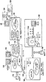
図1は、心臓撮像を支援する模範的な心臓用コンピュータ断層撮影(CT)システムの概観である。一例として心臓CTシステムを用いているが、当技術分野で周知のその他の撮像システムを本発明の一実施形態で使用してもよい。システム102のスキャナ部には、スキャナインタフェースボードを介してスキャナにR波のピーク事象を出力するEKGモニタが備わっている。スキャナインタフェースボートを使用することによって、EKGシステムをスキャナに接続することができる。スキャナインタフェースボードの一例には、ガントリインタフェースボードがある。心臓用CTサブシステム102には、心臓拡張相の動きの無い心臓を撮像するためのEKGゲーティング獲得、即ち、画像再生機能が備わっている。スキャナからのデータは、データ獲得/データ制御/画像生成ソフトウェアを含むサブシステム108に出力される。さらに、スキャナから出力される、R波ピークタイムスタンプを含むデータは、獲得データベース104に格納される。心臓、特に、左心房や右心房を撮像するための1つ以上の最適化獲得プロトコルに基づいて、データ獲得が実行される。画像生成は、左心房や右心房の内面のCT画像データセットの自動画像分割を行うための1つ以上の最適化3次元プロトコルを用いて実施される。
【0015】
図1を参照すると、画像データストリーム110はオペレータコンソール114に送られる。オペレータコンソール114の検査処方/視覚化ソフトウェアで使用されるデータは、画像データストリーム110からのデータと共に画像データベース112に格納される。ディスプレイ画面116は、検査処方/視覚化処理を行うオペレータのために用意されている。3次元後処理に含まれる分析や精査のために、画像データを保存したり、フィルムに記録したり、ネットワークを介してワークステーション120に送ったりすることが可能である。ワークステーション120内に描かれた後処理ソフトウェアによって、例えば、心房(もしくは心室)の液浸ビューが得られ、肺静脈を左心房の内側から見ることができる。これらの特別な画像は、心房3次元レンダリングファイル126に保存されるので、インターベンション技師はインターベンション処置中に見ることができる。また、後処理ソフトウェアによって、左心房や右心房の内面の詳細な3次元モデル124をエクスポートすることができる。左心房の場合は、4本の肺静脈が3次元モデル124で明確に定義される。3次元モデル124には、インターベンションシステムもしくは治療システムの座標系との3次元整合をとるために利用可能な解剖学上の目印が複数含まれている。3次元モデル124を、ワイヤメッシュ幾何モデルや、一連の輪郭や、2値画像の分割ボリュームや、放射線療法(RT)用DICOMオブジェクト規格のDICOMオフジェクトや、それと同様のオブジェクト形式のうちの1つにエクスポートすることが可能である。また、当技術分野で周知のその他の形式を用いて、3次元モデル124を記憶したりエクスポートしたりすることも可能である。さらに、オペレータは3次元モデル124をディスプレイ画面122で見ることができる。別の一実施形態のインターベンションシステムには、本発明の一実施形態に含まれる高性能3次元整合ソフトウェアや3次元視覚化ソフトウェアが含まれていてもよい。
【0016】
図2は、インターベンション計画システムで、心臓用CTで生成された画像データを使う模範的なプロセスフロー図である。本プロセスは、心臓CTシステムで左心房や右心房のための最適化プロトコルを用いて大量データを獲得する際のステップ202から始まる。利用可能なプロトコルの例としては、ゲーティング再生法を利用するヘリカルスキャン獲得技法を用いた心臓動脈撮像プロトコルがある。模範的な一実施形態では、心臓動脈撮像プロトコルで用いられるパラメータには、単一または複数セクタの心臓再生法を利用する場合、0.375ヘリカルピッチ係数で0.5秒のガントリ回転時間がある。また、パラメータとしては、マルチスライスCTスキャナでは、120キロボルト、250ミリアンプ、1.25ミリメータといった値を使うことが可能である。ステップ204では、左心房や右心房の内面を抽出するように設計された3次元プロトコルを含む後処理ソフトウェアによって、画像データセットを分割する。模範的な一実施形態の後処理ソフトウェアの機能には、先端血管定量評価と、シードの配置と、連結度の活用と、領域グロー法(region Growing techniques)の実施が含まれていてもよい。これらの機能は、購入したソフトウェアツール(例えば、先端血管定量評価(AVA))を使って実行することができる。模範的な一実施形態では、AVAなどのツールを画像データセットに適用した後に、しきい値処理やフローターフィルタ処理やスカルプリング(scalpling)やデータブリッジ処理やスカルプリング処理をさらに行うことが可能である。ステップ204では、3次元プロトコルを使って自動データ分割プロセスには、オペレータからの1つ以上の待ち行列が必要となる場合がある。模範的な一実施形態では、オペレータからの一つの待ち行列が必要な場合に、オペレータは本プロセスを経ることになる。3次元プロトコルには、ボリュームのデフォルトビューと、3次元分割とエクスポートを行うためにデータに対して実行可能な処理ステップが含まれる。
【0017】
次に、ステップ206では、3次元モデルを生成する。左心房と右心房は、液浸ビューを含む3次元表面レンダリングや3次元ボリュームレンダリングを利用して視覚化される。Volume Rendering(VR)とCardiac Image Quality(CARDIQ)を含む様々なボリュームレンダリングソフトウェアパッケージが入手可能である。ステップ208では、オペレータは、インターベンションシステムで位置合せするために利用される3点以上の特定の解剖学的目印を確認する。厳密な位置合わせを行うには、解剖学的目印が3点必要である。非厳密な位置合わせを行う場合には、解剖学的構造の目印が3点以上必要であった。左心房の場合には、静脈洞と2本の上肺静脈を用いてもよい。目印は、心室の内面とは異なる色系で視覚化することができる。別の方法では、明示的な幾何マーカを目印のボリュームに挿入し、不透明な幾何目印を用いて心室を半透明に視覚化することが可能である。ステップ206で述べたツールなどのボリュームレンダリングツールを利用して、本ステップを実行することができる。本発明の模範的な一実施形態では、オペレータは、視覚化と目印を特定して処理を進めることができる。
【0018】
ステップ210では、インターベンション計画処置時に視覚的に参照する必要のある特定の3次元レンダリングを保存する。3次元レンダリングを、DICOM画像を含む様々な形態でフィルム上に保存したり、マルチメディア形式で保存することが可能である。また、これらの表示を透視診断システムの投影画像と組合せることも可能である。X線画像をリアルタイムに得るために、透視診断システムには、患者の一方の側面にX線管を位置決めし、他方の側面に検出器を位置決めする処理が含まれていてもよい。透視診断システムは、処置中にカテーテルをガイドする手段の一例である。
【0019】
ステップ212では、左心房と右心房の3次元モデルは、選択された形式でエクスポートされる。可能な形式としては、ワイヤメッシュ幾何モデルと、一連の輪郭と、2値画像の分割ボリュームと、放射線療法DICOM規格で使用されるRT DICOMオブジェクトなどのDICOMオブジェクトがある。模範的な一実施形態では、2値画像内で無関係なデータは全て0に設定されるため、2値画像の分割ボリュームは0以外の情報だけを含んでいる。ボクセル値はCTの減衰量に対応し、ハーンズフィールド(Houndsfield)値で表現された組織の密度によって、2値画像の分割ボリュームが構成される。
【0020】
ステップ214では、エクスポートされた3次元モデルはインターベンションシステムに入力される。次にステップ216では、3次元モデルは、ステップ208で特定された目印と同一の目印を使って位置合せされる。厳密もしくは非厳密な位置合わせ法を用いて、インターベンションシステムの座標系で、3次元モデルを位置合せすることができる。さらにステップ218では、モデルはインターベンションシステム上で視覚化され、電気システムがそのモデルにマッピングされる。上述の模範的な実施形態では1つの3次元モデルについて言及したが、これを、心臓撮像システムからエクスポートされ、また、インターベンションシステムにインポートされる多数の3次元モデルに拡張することもできる。
【0021】
別の一実施形態では、図2に記載されたプロセスには、インターベンション処置完了後のステップがさらに含まれている。このステップには、インターベンションシステムによって計算される電気信号がDICOM形式で表示/保管される前と後で、心臓用撮像システムにインポートすることが含まれる。別の方法では、図2で説明されたプロセスは、心臓の心室(例えば、左心房や右心房や左心室や右心室)や血管(例えば、右冠動脈や上行大動脈)にも適用可能である。同様に、画像獲得システムによって生成されたレンダリングや3次元モデルを使って計画する必要があるアブレーションやその他の種類のインターベンション処置にも適用可能である。図2に示したプロセスは、心臓CTシステムに加えてその他の画像獲得システムにも適用可能である。例えば、心臓画像が磁気共鳴撮像(MRI)システムで獲得された場合、ステップ204には、磁気共鳴(MR)画像の後処理のために最適化心臓分割アルゴリズムを利用することが含まれていてもよい。
【0022】
図3は、インターベンション処置を実施するために改善された模範的なプロセスのフロー図である。改善されたアブレーション治療プロセスは、ステップ302でX線透視によってガイドされたカテーテルをLAに挿入することから始まる。通常、本プロセスのその部分の実施には約1時間がかかる。次にステップ304では、平面を定義するために、位置決めカテーテルと透視診断システムを用いて3点以上の解剖学的目印が心房内で特定される。ステップ306では、インターベンションシステムは、3次元モデルがインターベンションシステムの座標系に変換されるように、心臓CTシステムからエクスポートされた3次元モデルに対し3次元位置決め処理を行う。304と306は、背景の節で説明した最新のインターベンション処置の一部を置き換えるものである。3次元位置情報を利用しLAの心房をスイープスルーすることによって特殊なカテーテルから大雑把な3次元幾何表現を獲得することはもはや不要である。現在の方法に代わりにステップ304と306を用いることにより、インターベンション処置を短時間で行うことが可能になる。
【0023】
次に、ステップ308から314を含むループが開始される。ステップ308では、特殊なカテーテルを使って1つ以上の心臓周期から電気情報が獲得される。インターベンションシステムソフトウェアを利用することによって、電気情報は心房の詳細な幾何モデルにマッピングされる。次にステップ310では、そのマップが視覚化され、アブレーションで治療すべき問題部位が特定される。ステップ312では、熱を与えることによって損傷が生成され、その位置がソフトウェアによって追跡される。ステップ314では、損傷結果を確認するために電気マップが再び収集される。ステップ308から314を含む本ループは、インターベンション処置を完了させるために必要な回数だけ繰り返される。
【0024】
心房性細動の治療計画のための心臓CTシステムから、インターベンション処置計画に関する情報が提供されるので、インターベンション技師は背景の節で説明したように特殊なカテーテルを使ってLAの心房をスイープスルーしてLAやPVの小孔の大雑把な3次元幾何表現を獲得する必要がない。この結果、総インターベンション処置時間が短縮する。さらに、特殊なカテーテルによって獲得される表現を用いるのではなく、LAとPVの詳細な3次元幾何学的表現を用いた場合には、図3のステップ308から314までの繰り返し回数が少なくい治療を施せるようになる。幾何表現がより正確になると、インターベンション技師は問題のある電気信号の発生源を特定できるようになる。
【0025】
前述の複数の実施形態では医療用撮像について論じたが、本願に記載の画像獲得処理方法は、医療用途に限定されるものではなく、医療分野以外の分野にも適用可能であることを理解されたい。
【0026】
上述したように、本発明の実施形態は、コンピュータで実施されるプロセスやそれらのプロセスを実施する機器の形態で実現可能である。また、本発明の実施形態は、フロッピディスク(商標)やCD−ROMやハードドライブやその他のコンピュータ読取可能記憶媒体などの有形媒体で実現されるインストラクションを含むコンピュータプログラムコード形態でも実現可能であり、コンピュータプログラムコードがコンピュータへロードされコンピュータによって実行された時に、そのコンピュータは本発明を実施するための機器になる。また、本発明の一実施形態は、例えば、記憶媒体に記憶されるか、コンピュータへロードされてコンピュータによって実行されるか、電線やケーブルや光ファイバや電磁放射などの伝送媒体を介して伝送されるコンピュータプログラムコード形態でも実現可能であり、コンピュータプログラムコードがコンピュータへロードされコンピュータによって実行された時に、そのコンピュータは本発明を実施するための機器となる。汎用マイクロプロセッサで実現される場合には、コンピュータプログラムコードセグメントは、特定の論理回路を形成するようにマイクロプロセッサを構成するものである。
【0027】
本発明を模範的な実施形態に関して説明してきたが、当業者であれば、本発明の範囲から逸脱することなく様々な変更を加えたり、本発明の要素の代わりにそれと等価なものに置換可能なことを理解することができる。さらに、本発明の本質的な範囲から逸脱することなく特定の状況や構成要素を本発明の教示に適合させるために、多くの修正を加えることも可能である。従って、本発明は、本発明を実施するために考えられるベストモードとして開示された特定の実施形態に限定されることなく、添付の特許請求の範囲に含まれる実施形態を全て包含するものである。さらに、第1や第2などの用語を使用しているが、これらは何らかの順位や重要性を表わしているわけではなく、ある要素を別の要素と区別するために使用しているものである。
【図面の簡単な説明】
【図1】図1は、心臓の撮像を支援する心臓用コンピュータ断層撮影(CT)システムの概観である。
【図2】図2は、心臓用CTで生成された画像データがインターベンション計画システムで使用されるプロセスのフロー図である。
【図3】図3は、インターベンション処置を実施するために改善されたプロセスのフロー図である。[0001]
BACKGROUND OF THE INVENTION
The present disclosure relates generally to a method for planning a cardiac interventional procedure, and more particularly to a method for utilizing data generated by a medical imaging system when planning a cardiac interventional treatment.
[0002]
Medical diagnostic imaging systems are now widely used in medical facilities. Such systems provide important tools for revealing, diagnosing, and treating physical conditions, thus greatly reducing the need for intervention for surgical diagnosis. In many cases, the attending physician or radiologist performs a final examination or treatment after performing a conventional examination using detailed images of appropriate sites and tissues by one or more diagnostic imaging therapies.
[0003]
Currently, there are many therapies for medical diagnostic imaging systems. This includes tomography (CT) systems, X-ray systems (including conventional digital / digitized imaging systems), magnetic resonance (MR) systems, positron emission tomography (PET) systems, ultrasound systems and nuclear medicine systems. Is included. In many cases, such therapy complements each other, and doctors are provided with various techniques for imaging specific types of tissues, organs, physiological systems, and the like. In medical institutions, several such imaging systems are often located at one or more facilities, so that doctors can use the resources according to the needs of a particular patient.
[0004]
In general, current medical diagnostic systems include a circuit for acquiring image data and converting the data into a usable format, and data processing is performed to reproduce an image of a patient's attention site. Since the imaging process often involves some kind of physical or electronic scanning, the image data acquisition processing circuit is often referred to as a “scanner” regardless of therapy. Of course, the specific components of the system and the circuits associated with them vary greatly from therapy to therapy due to different physical characteristics and data processing requirements.
[0005]
In addition, medical diagnosis and treatment can be performed by performing interventional procedures such as atrial fibrillation (AF) intervention. About 2.2 million people in the United States have AF symptoms. AF is one of the most common arrhythmias and the most problematic symptom. This is currently the leading cause of stroke in the United States. The incidence of AF increases with age and increases rapidly after age 60. In the case of left atrial fibrillation, special electrical signals that cause AF are occasionally generated from muscle tissue around any of the four pulmonary veins (PV) that connect to the left atrium (LA). For this condition, one of the current clinical treatments is ablation, which uses a special catheter inserted into the left atrium to create a small lesion by applying heat near the source of the electrical signal in question. Ablation therapy is routinely performed during open-heart surgery for less than an hour, but it is very difficult to perform a timely catheter procedure that does not affect healthy tissue.
[0006]
As an example of ablation treatment, the following treatment is common. First, an X-ray catheter guided by fluoroscopy is inserted into LA. This operation takes about an hour. Next, a rough 3D geometric representation of LA and PV ostiums (openings) is obtained by “sweeping through” the LA atrium using 3D position information from a special catheter. . Usually, it takes about one hour to obtain a rough three-dimensional geometric representation. The next step is performed as many times as necessary in the following order. Using special catheters, electrical information is obtained from one or more cardiac cycles, and from this electrical information, a rough 3D geometric representation map is obtained using interventional system software. In the next step, this map is visualized to identify problem areas to be treated by ablation. Next, heat is applied to create damage and the software tracks its location. In the last step, an electrical map is obtained again to confirm the damage result. If ablation treatment needs to be performed, this process can be continued by repeating the previous steps starting with the electrical information acquisition process using a special catheter. Ablation therapy is a time consuming and laborious procedure.
[0007]
Summary of the Invention
In one aspect of the invention, a method is provided for generating a three-dimensional model utilized for cardiac interventional treatment planning. Acquired data is obtained from a medical imaging system, and cardiac image data is generated according to the acquired data. A three-dimensional model is generated according to the cardiac image data, and three anatomical landmarks are specified on the three-dimensional model. The 3D model is sent to the intervention system, and the 3D model is in a format that can be imported and recorded in the intervention system.
[0008]
In another aspect of the invention, a method is provided for generating a three-dimensional model utilized for cardiac interventional treatment planning. Acquired data is received from the medical imaging system. Heart image data is generated according to the acquired data, and a three-dimensional model is generated according to the heart image data. Three anatomical landmarks are identified in the three-dimensional model. The 3D model is registered with the intervention system corresponding to the three anatomical landmarks, and the 3D model is visualized with the intervention system.
[0009]
In another aspect of the present invention, a system for generating a three-dimensional model utilized for cardiac interventional treatment planning is provided. The system includes a medical imaging system, an acquisition database in communication with the medical imaging system, an image database, a data transfer mechanism, and a processing device. The processing device communicates with the data transfer mechanism, the acquisition database, and the image database. The processing device is equipped with equipment that generates a three-dimensional model used for cardiac interventional treatment planning. The device implements a technique for obtaining acquired data from a medical imaging system. Acquisition data is stored in an acquisition database. The cardiac image data is generated according to the acquired data. The heart image data is stored in an image database. A three-dimensional model is generated according to the cardiac image data, and three anatomical landmarks are specified on the three-dimensional model. The 3D model is sent to the intervention system. The three-dimensional model is in a format that can be recorded by importing the intervention system. This transmission is performed using a data transfer mechanism.
[0010]
In accordance with another aspect of the present invention, a system for generating a three-dimensional model utilized for cardiac interventional treatment planning is provided. The system includes a medical imaging system, an acquisition database in communication with the medical imaging system, an image database, a data transfer mechanism, an intervention system in communication with the data transfer mechanism, and a processing device. The processing device communicates with the data transfer mechanism, the acquisition database, and the image database. The processing device is equipped with equipment that generates a three-dimensional model used for cardiac interventional treatment planning. The device implements a technique for obtaining acquired data from a medical imaging system. The acquired data is stored in the acquisition database. Cardiac image data is generated according to the acquired data. The heart image data is stored in an image database. The three-dimensional model is generated according to the cardiac image data, and three anatomical landmarks are specified on the three-dimensional model. The 3D model is sent to the intervention system. The three-dimensional model is in a format that can be recorded and imported into the intervention system. This transmission is performed using a data transfer mechanism. The three-dimensional model is received by the intervention system and registered according to the three point anatomical landmarks. The 3D model is then visualized with an intervention system.
[0011]
In accordance with another aspect of the present invention, a computer program product is provided for generating a three-dimensional model utilized for cardiac interventional treatment planning. This product includes a storage medium readable by the processing circuit, and stores instructions executed by the processing circuit. Execution instructions include obtaining acquired data from the medical imaging system and generating cardiac image data in response to the acquired data. The three-dimensional model is generated according to the cardiac image data, and three anatomical landmarks are specified on the three-dimensional model. The 3D model is sent to the intervention system. The three-dimensional model is in a format that can be imported and recorded in the intervention system.
[0012]
Other aspects of the invention are disclosed herein. These and other features and advantages of the present invention can be understood and appreciated by those skilled in the art from the following detailed description and drawings.
[0013]
Referring to the exemplary drawings, the same components are numbered the same in several drawings.
[0014]
DETAILED DESCRIPTION OF THE INVENTION
FIG. 1 is an overview of an exemplary cardiac computed tomography (CT) system that supports cardiac imaging. Although a cardiac CT system is used as an example, other imaging systems known in the art may be used in an embodiment of the present invention. The scanner unit of the
[0015]
Referring to FIG. 1, the
[0016]
FIG. 2 is an exemplary process flow diagram for using image data generated by cardiac CT in an intervention planning system. The process begins at
[0017]
Next, in
[0018]
In
[0019]
In
[0020]
In
[0021]
In another embodiment, the process described in FIG. 2 further includes a step after completion of the interventional procedure. This step includes importing into the cardiac imaging system before and after the electrical signals calculated by the intervention system are displayed / stored in DICOM format. Alternatively, the process described in FIG. 2 is applicable to the heart ventricles (eg, left atrium, right atrium, left ventricle, right ventricle) and blood vessels (eg, right coronary artery, ascending aorta). Similarly, it is applicable to ablation and other types of interventional procedures that need to be planned using rendering and 3D models generated by the image acquisition system. The process shown in FIG. 2 is applicable to other image acquisition systems in addition to the cardiac CT system. For example, if a cardiac image was acquired with a magnetic resonance imaging (MRI) system,
[0022]
FIG. 3 is a flow diagram of an exemplary process that has been improved to perform an interventional procedure. The improved ablation treatment process begins with inserting a catheter guided by fluoroscopy into the LA at
[0023]
Next, a
[0024]
The cardiac CT system for treatment planning for atrial fibrillation provides information on interventional treatment plans, so interventionists can sweep LA atria using special catheters as described in the background section. There is no need to get through and get a rough 3D geometric representation of the small holes in LA and PV. As a result, the total intervention treatment time is shortened. Further, when the detailed three-dimensional geometric representation of LA and PV is used instead of the representation acquired by a special catheter, the treatment with a small number of iterations from
[0025]
In the above embodiments, medical imaging has been discussed. However, it is understood that the image acquisition processing method described in the present application is not limited to medical use and can be applied to fields other than the medical field. I want.
[0026]
As described above, the embodiment of the present invention can be realized in the form of a computer-implemented process or a device that performs the process. The embodiment of the present invention can also be realized in a computer program code form including instructions realized by a tangible medium such as a floppy disk (trademark), a CD-ROM, a hard drive, and other computer-readable storage media. When the computer program code is loaded into the computer and executed by the computer, the computer becomes a device for implementing the present invention. Also, an embodiment of the present invention may be stored in a storage medium, loaded into a computer and executed by the computer, or transmitted via a transmission medium such as an electric wire, cable, optical fiber, or electromagnetic radiation. The computer program code form can be realized, and when the computer program code is loaded into the computer and executed by the computer, the computer becomes a device for carrying out the present invention. When implemented on a general-purpose microprocessor, the computer program code segments configure the microprocessor to form specific logic circuits.
[0027]
Although the present invention has been described in terms of exemplary embodiments, those skilled in the art can make various changes without departing from the scope of the present invention or substitute equivalents instead of elements of the present invention. I can understand. In addition, many modifications may be made to adapt a particular situation or component to the teachings of the invention without departing from the essential scope thereof. Accordingly, the present invention is not limited to the specific embodiments disclosed as the best mode contemplated for carrying out the invention, but encompasses all embodiments that fall within the scope of the appended claims. . In addition, terms such as first and second are used, but these do not represent any order or importance, but are used to distinguish one element from another. .
[Brief description of the drawings]
FIG. 1 is an overview of a cardiac computed tomography (CT) system that supports cardiac imaging.
FIG. 2 is a flow diagram of a process in which image data generated by cardiac CT is used in an intervention planning system.
FIG. 3 is a flow diagram of an improved process for performing interventional procedures.
Claims (10)
該処理装置は、
前記獲得データベース(104)に格納された獲得データを前記医療撮像システム(102)から得る工程と、
心臓撮像プロトコルを使用して前記獲得データから前記画像データベース(112)に格納される心臓画像データ(110)を生成する工程と、
前記心臓画像データ(110)から心臓の3次元モデル(124)を生成する工程と、
前記3次元モデル(124)上に3点の解剖学的構造の目印を特定する工程と、
3点の幾何学的目印を前記3次元モデル(124)上の前記3点の解剖学的目印の位置に挿入する工程と、
前記3点の幾何学的目印を含む前記3次元モデル(124)を心臓インターベンション処置計画を行うインターベンションシステムにエクスポートする工程と、
前記3点の幾何学的目印を含む前記3次元モデル(124)を前記インターベンションシステムにインポートする工程と、
前記インターベンションシステムの座標系で前記3点の解剖学的目印を特定する工程と、
前記3点の解剖学的目印を使用して前記3点の幾何学的目印を含む前記3次元モデル(124)を前記インターベンションシステムの前記座標系に変換することにより、前記インターベンションシステムで前記3次元モデル(124)を位置合せする工程と、
を実施する、システム。A system for generating a three-dimensional model (124) utilized for cardiac interventional treatment planning, comprising: a medical imaging system (102); and an acquisition database (104) in communication with said medical imaging system (102); includes image database (112), and a data transfer mechanism, a processing device in communication with the data transfer unit,
The processor is
Obtaining acquisition data stored in the acquisition database (104) from the medical imaging system (102);
Generating a cardiac image data that is stored (110) in said image database (112) from the acquired data using cardiac imaging protocol,
Generating a three-dimensional model (124) of the heart from the cardiac image data (110);
Identifying landmarks of three anatomical structures on the three-dimensional model (124);
Inserting a three-point geometric landmark at the position of the three-point anatomical landmark on the three-dimensional model (124);
Exporting the three-dimensional model (124) including the three-point geometric landmarks to an intervention system for performing a cardiac interventional treatment plan ;
Importing the three-dimensional model (124) including the three-point geometric landmarks into the intervention system;
Identifying the three anatomical landmarks in the coordinate system of the intervention system;
Converting the three-dimensional model (124) including the three-point geometric landmarks into the coordinate system of the intervention system using the three-point anatomical landmarks; Aligning the three-dimensional model (124);
Implement the system.
該処理装置は、
獲得データを前記医療撮像システム(102)から得る工程であって、前記獲得データは前記獲得データベース(104)に格納される、当該工程と、
心臓撮像プロトコルを使用して前記獲得データから心臓画像データ(110)を生成する工程であって、前記心臓画像データは前記画像データベース(112)に格納される、当該工程と、
前記心臓画像データ(110)から心臓の3次元モデル(124)を生成する工程と、
前記3次元モデル(124)上で3点の解剖学的目印を特定する工程と、
3点の幾何学的目印を前記3次元モデル(124)上の前記3点の解剖学的目印の位置に挿入する工程と、
前記3点の幾何学的目印を含む前記3次元モデル(124)を心臓インターベンション処置計画を行うインターベンションシステムにエクスポートする工程と、
前記3点の幾何学的目印を含む前記3次元モデル(124)を前記インターベンションシステムにインポートする工程と、
前記インターベンションシステムの座標系で前記3点の解剖学的目印を特定する工程と、
前記3点の解剖学的目印を使用して前記3点の幾何学的目印を含む前記3次元モデル(124)を前記インターベンションシステムの前記座標系に変換することにより、前記インターベンションシステムで前記3次元モデル(124)を位置合せする工程と、
前記インターベンションシステムで前記3次元モデル(124)を視覚化する工程とを実施する、システム。A system for generating a three-dimensional model (124) utilized for cardiac interventional treatment planning, comprising: a medical imaging system (102); and an acquisition database (104) in communication with said medical imaging system (102); An image database (112), a data transfer mechanism, an intervention system in communication with the data transfer mechanism, a processing device in communication with the data transfer mechanism, the acquisition database (104), and the image database (112),
The processor is
Obtaining acquisition data from the medical imaging system (102), wherein the acquisition data is stored in the acquisition database (104);
Generating cardiac image data (110) from the acquired data using a cardiac imaging protocol , wherein the cardiac image data is stored in the image database (112);
Generating a three-dimensional model (124) of the heart from the cardiac image data (110);
Identifying three anatomical landmarks on the three-dimensional model (124);
Inserting a three-point geometric landmark at the position of the three-point anatomical landmark on the three-dimensional model (124);
Exporting the three-dimensional model (124) including the three-point geometric landmarks to an intervention system for performing a cardiac interventional treatment plan ;
Importing the three-dimensional model (124) including the three-point geometric landmarks into the intervention system;
Identifying the three anatomical landmarks in the coordinate system of the intervention system;
Converting the three-dimensional model (124) including the three-point geometric landmarks into the coordinate system of the intervention system using the three-point anatomical landmarks; Aligning the three-dimensional model (124);
Implementing a step of visualizing the three dimensional model (124) in the intervention system, the system.
前記3次元レンダリング(126)は、透視診断システムの投影画像と組合せられる、請求項1乃至3のいずれかに記載のシステム。The processing device includes generating a three-dimensional rendering (126) for viewing and referring to the cardiac image data (110), and sending the three-dimensional rendering (126) to the intervention system. Further ,
The three-dimensional rendering (126), Ru combined with the projected image of the fluoroscopy system, the system according to any one of claims 1 to 3.
前記医療撮像システム(102)は、EKGと、前記EKGからR波ピーク事象データを受け取るインタフェースボードと、前記インタフェースボードと通信するスキャナを備え、
前記データ転送機構はインターネットであり、
前記医療撮像システム(102)は心臓用コンピュータ断層撮影システム又は磁気共鳴撮像システムであり、
前記インターベンションシステムは心房性細動用のインターベンションシステムである、請求項1乃至4のいずれかに記載のシステム。The acquisition database (104) and said image database (112) is Ri relational der,
The medical imaging system (102) includes an EKG, an interface board that receives R wave peak event data from the EKG, and a scanner that communicates with the interface board ,
The data transfer mechanism is the Internet;
The medical imaging system (102) is also cardiac computer tomography system Ri magnetic resonance imaging system der,
The system according to any one of claims 1 to 4, wherein the intervention system is an intervention system for atrial fibrillation.
処理回路によって読取り可能であって、医療撮像システム(102)から獲得データを得る工程と、
心臓撮像プロトコルを使用して前記獲得データから心臓画像データを生成する工程と、
前記心臓画像データ(110)から心臓の3次元モデル(124)を生成する工程と、
前記3次元モデル(124)上に3点の解剖学的目印を特定する工程と、
3点の幾何学的目印を前記3次元モデル(124)上の前記3点の解剖学的目印の位置に挿入する工程と、
前記3点の幾何学的目印を含む前記3次元モデル(124)を心臓インターベンション処置計画を行うインターベンションシステムにエクスポートする工程と、
前記3点の幾何学的目印を含む前記3次元モデル(124)を前記インターベンションシステムにインポートする工程と、
前記インターベンションシステムの座標系で前記3点の解剖学的目印を特定する工程と、
前記3点の解剖学的目印を使用して前記3点の幾何学的目印を含む前記3次元モデル(124)を前記インターベンションシステムの前記座標系に変換することにより、前記インターベンションシステムで前記3次元モデル(124)を位置合せする工程のための、前記処理回路が実行するインストラクションを記憶する記憶媒体を備えるコンピュータプログラム製品。A computer program product for generating a three-dimensional model (124) utilized for cardiac interventional treatment planning comprising:
Obtaining acquired data from a medical imaging system (102) readable by a processing circuit;
Generating cardiac image data from the acquired data using a cardiac imaging protocol ;
Generating a three-dimensional model (124) of the heart from the cardiac image data (110);
Identifying three anatomical landmarks on the three-dimensional model (124);
Inserting a three-point geometric landmark at the position of the three-point anatomical landmark on the three-dimensional model (124);
Exporting the three-dimensional model (124) including the three-point geometric landmarks to an intervention system for performing a cardiac interventional treatment plan ;
Importing the three-dimensional model (124) including the three-point geometric landmarks into the intervention system;
Identifying the three anatomical landmarks in the coordinate system of the intervention system;
Converting the three-dimensional model (124) including the three-point geometric landmarks into the coordinate system of the intervention system using the three-point anatomical landmarks; A computer program product comprising a storage medium for storing instructions executed by the processing circuit for the step of aligning a three-dimensional model (124) .
Applications Claiming Priority (2)
| Application Number | Priority Date | Filing Date | Title |
|---|---|---|---|
| US10/063,064 US7286866B2 (en) | 2001-11-05 | 2002-03-15 | Method, system and computer product for cardiac interventional procedure planning |
| US10/063,064 | 2002-03-15 |
Publications (3)
| Publication Number | Publication Date |
|---|---|
| JP2003299673A JP2003299673A (en) | 2003-10-21 |
| JP2003299673A5 JP2003299673A5 (en) | 2008-07-24 |
| JP4345959B2 true JP4345959B2 (en) | 2009-10-14 |
Family
ID=28452179
Family Applications (1)
| Application Number | Title | Priority Date | Filing Date |
|---|---|---|---|
| JP2003069085A Expired - Lifetime JP4345959B2 (en) | 2002-03-15 | 2003-03-14 | Method, system and computer product for cardiac interventional treatment planning |
Country Status (3)
| Country | Link |
|---|---|
| US (1) | US7286866B2 (en) |
| JP (1) | JP4345959B2 (en) |
| DE (1) | DE10311319B4 (en) |
Families Citing this family (160)
| Publication number | Priority date | Publication date | Assignee | Title |
|---|---|---|---|---|
| US8175680B2 (en) * | 2001-11-09 | 2012-05-08 | Boston Scientific Scimed, Inc. | Systems and methods for guiding catheters using registered images |
| US7346381B2 (en) * | 2002-11-01 | 2008-03-18 | Ge Medical Systems Global Technology Company Llc | Method and apparatus for medical intervention procedure planning |
| US7499743B2 (en) | 2002-03-15 | 2009-03-03 | General Electric Company | Method and system for registration of 3D images within an interventional system |
| US20040003432A1 (en) * | 2002-05-06 | 2004-01-01 | Pharmacia Corporation | Production of hexosamines and uses thereof |
| US7778686B2 (en) * | 2002-06-04 | 2010-08-17 | General Electric Company | Method and apparatus for medical intervention procedure planning and location and navigation of an intervention tool |
| US20050277823A1 (en) * | 2002-06-10 | 2005-12-15 | Robert Sutherland | Angiogram display overlay technique for tracking vascular intervention sites |
| WO2004060157A1 (en) * | 2003-01-07 | 2004-07-22 | Philips Intellectual Property & Standards Gmbh | Method and arrangement for tracking a medical instrument |
| US7747047B2 (en) * | 2003-05-07 | 2010-06-29 | Ge Medical Systems Global Technology Company, Llc | Cardiac CT system and method for planning left atrial appendage isolation |
| US7565190B2 (en) | 2003-05-09 | 2009-07-21 | Ge Medical Systems Global Technology Company, Llc | Cardiac CT system and method for planning atrial fibrillation intervention |
| US7343196B2 (en) * | 2003-05-09 | 2008-03-11 | Ge Medical Systems Global Technology Company Llc | Cardiac CT system and method for planning and treatment of biventricular pacing using epicardial lead |
| EP1639546B1 (en) * | 2003-06-17 | 2011-05-04 | Brown University | Method and apparatus for model-based detection of structure in projection data |
| US7978887B2 (en) * | 2003-06-17 | 2011-07-12 | Brown University | Methods and apparatus for identifying subject matter in view data |
| US7813785B2 (en) | 2003-07-01 | 2010-10-12 | General Electric Company | Cardiac imaging system and method for planning minimally invasive direct coronary artery bypass surgery |
| US20050010105A1 (en) * | 2003-07-01 | 2005-01-13 | Sra Jasbir S. | Method and system for Coronary arterial intervention |
| US7344543B2 (en) * | 2003-07-01 | 2008-03-18 | Medtronic, Inc. | Method and apparatus for epicardial left atrial appendage isolation in patients with atrial fibrillation |
| US6888916B2 (en) * | 2003-08-18 | 2005-05-03 | Ge Medical Systems Global Technology Company, Llc | Preprocessing methods for robust tracking of coronary arteries in cardiac computed tomography images and systems therefor |
| DE10340546B4 (en) | 2003-09-01 | 2006-04-20 | Siemens Ag | Method and apparatus for visually assisting electrophysiology catheter application in the heart |
| DE10340544B4 (en) * | 2003-09-01 | 2006-08-03 | Siemens Ag | Device for visual support of electrophysiology catheter application in the heart |
| US20050054918A1 (en) * | 2003-09-04 | 2005-03-10 | Sra Jasbir S. | Method and system for treatment of atrial fibrillation and other cardiac arrhythmias |
| US7308299B2 (en) | 2003-10-22 | 2007-12-11 | General Electric Company | Method, apparatus and product for acquiring cardiac images |
| US7308297B2 (en) | 2003-11-05 | 2007-12-11 | Ge Medical Systems Global Technology Company, Llc | Cardiac imaging system and method for quantification of desynchrony of ventricles for biventricular pacing |
| DE10357205A1 (en) * | 2003-12-08 | 2005-07-14 | Siemens Ag | Method for generating result images of an examination object |
| US20050143777A1 (en) * | 2003-12-19 | 2005-06-30 | Sra Jasbir S. | Method and system of treatment of heart failure using 4D imaging |
| US20050137661A1 (en) * | 2003-12-19 | 2005-06-23 | Sra Jasbir S. | Method and system of treatment of cardiac arrhythmias using 4D imaging |
| US7454248B2 (en) | 2004-01-30 | 2008-11-18 | Ge Medical Systems Global Technology, Llc | Method, apparatus and product for acquiring cardiac images |
| DE102004020587B4 (en) * | 2004-04-27 | 2016-02-18 | Siemens Aktiengesellschaft | Method and apparatus for visually assisting a catheter electrophysiology application with 2D fluoroscopic images |
| US8010175B2 (en) | 2004-05-05 | 2011-08-30 | Siemens Medical Solutions Usa, Inc. | Patient-specific coronary territory mapping |
| US8515527B2 (en) * | 2004-10-13 | 2013-08-20 | General Electric Company | Method and apparatus for registering 3D models of anatomical regions of a heart and a tracking system with projection images of an interventional fluoroscopic system |
| US7327872B2 (en) * | 2004-10-13 | 2008-02-05 | General Electric Company | Method and system for registering 3D models of anatomical regions with projection images of the same |
| US20060184396A1 (en) * | 2005-01-28 | 2006-08-17 | Dennis Charles L | System and method for surgical navigation |
| US7352370B2 (en) * | 2005-06-02 | 2008-04-01 | Accuray Incorporated | Four-dimensional volume of interest |
| US7681579B2 (en) * | 2005-08-02 | 2010-03-23 | Biosense Webster, Inc. | Guided procedures for treating atrial fibrillation |
| US7877128B2 (en) * | 2005-08-02 | 2011-01-25 | Biosense Webster, Inc. | Simulation of invasive procedures |
| US8583220B2 (en) * | 2005-08-02 | 2013-11-12 | Biosense Webster, Inc. | Standardization of catheter-based treatment for atrial fibrillation |
| DE102005042329A1 (en) * | 2005-09-06 | 2007-03-08 | Siemens Ag | Electro-physiological catheter application assistance providing method, involves detecting contour of areas relevant for catheter application, and showing areas as simple line in representations of mapping and/or image data |
| US20070225749A1 (en) | 2006-02-03 | 2007-09-27 | Martin Brian B | Methods and devices for restoring blood flow within blocked vasculature |
| WO2007096804A1 (en) * | 2006-02-24 | 2007-08-30 | Koninklijke Philips Electronics N.V. | Automated robust learning of geometries for mr-examinations |
| EP2041721B1 (en) * | 2006-07-05 | 2016-09-07 | Philips Intellectual Property & Standards GmbH | Prediction of cardiac shape by a motion model |
| US7916919B2 (en) * | 2006-09-28 | 2011-03-29 | Siemens Medical Solutions Usa, Inc. | System and method for segmenting chambers of a heart in a three dimensional image |
| JP4559501B2 (en) * | 2007-03-14 | 2010-10-06 | 富士フイルム株式会社 | Cardiac function display device, cardiac function display method and program thereof |
| US8355550B2 (en) * | 2007-05-01 | 2013-01-15 | Siemens Aktiengesellschaft | Methods and apparatus for virtual coronary mapping |
| US8705819B2 (en) * | 2007-06-21 | 2014-04-22 | Koninklijke Philips N.V. | Adjusting acquisition protocols for dynamic medical imaging using dynamic models |
| US9076203B2 (en) * | 2007-11-26 | 2015-07-07 | The Invention Science Fund I, Llc | Image guided surgery with dynamic image reconstruction |
| WO2009156918A1 (en) * | 2008-06-25 | 2009-12-30 | Koninklijke Philips Electronics N.V. | Device and method for localizing an object of interest in a subject |
| US8200466B2 (en) | 2008-07-21 | 2012-06-12 | The Board Of Trustees Of The Leland Stanford Junior University | Method for tuning patient-specific cardiovascular simulations |
| WO2010054320A1 (en) | 2008-11-07 | 2010-05-14 | Cardioinsight Technologies, Inc. | Visualization of physiological data for virtual electrodes |
| EP2345024B1 (en) | 2008-11-10 | 2017-11-08 | Cardioinsight Technologies, Inc. | Visualization of electrophysiology data |
| US9795442B2 (en) | 2008-11-11 | 2017-10-24 | Shifamed Holdings, Llc | Ablation catheters |
| WO2010056771A1 (en) | 2008-11-11 | 2010-05-20 | Shifamed Llc | Low profile electrode assembly |
| WO2010102307A1 (en) | 2009-03-06 | 2010-09-10 | Lazarus Effect, Inc. | Retrieval systems and methods for use thereof |
| US9405886B2 (en) | 2009-03-17 | 2016-08-02 | The Board Of Trustees Of The Leland Stanford Junior University | Method for determining cardiovascular information |
| CN104548316B (en) | 2009-06-24 | 2018-01-26 | 施菲姆德控股有限责任公司 | Can steerable medical delivery devices and application method |
| JP2013519428A (en) | 2010-02-12 | 2013-05-30 | ブリガム・アンド・ウイミンズ・ホスピタル・インコーポレイテッド | System and method for automatic adjustment of regulatory parameters of cardiac resynchronization therapy |
| JP2013521995A (en) | 2010-03-24 | 2013-06-13 | シファメド・ホールディングス・エルエルシー | Endovascular tissue destruction |
| US9655677B2 (en) | 2010-05-12 | 2017-05-23 | Shifamed Holdings, Llc | Ablation catheters including a balloon and electrodes |
| WO2011143468A2 (en) | 2010-05-12 | 2011-11-17 | Shifamed, Llc | Low profile electrode assembly |
| US8157742B2 (en) | 2010-08-12 | 2012-04-17 | Heartflow, Inc. | Method and system for patient-specific modeling of blood flow |
| US8315812B2 (en) | 2010-08-12 | 2012-11-20 | Heartflow, Inc. | Method and system for patient-specific modeling of blood flow |
| US20120084064A1 (en) * | 2010-09-29 | 2012-04-05 | Nutech Ventures, Inc. | Model-based systems and methods for analyzing and predicting outcomes of vascular interventions and reconstructions |
| US9510763B2 (en) | 2011-05-03 | 2016-12-06 | Medtronic, Inc. | Assessing intra-cardiac activation patterns and electrical dyssynchrony |
| CA2833387A1 (en) | 2011-05-03 | 2012-11-08 | Shifamed Holdings, Llc | Steerable delivery sheaths |
| US8961550B2 (en) | 2012-04-17 | 2015-02-24 | Indian Wells Medical, Inc. | Steerable endoluminal punch |
| US8548778B1 (en) | 2012-05-14 | 2013-10-01 | Heartflow, Inc. | Method and system for providing information from a patient-specific model of blood flow |
| EP2875367B1 (en) * | 2012-07-18 | 2021-05-12 | Koninklijke Philips N.V. | Efficient cardiac mr workflows based on automated planning from mdixon surveys |
| US9091628B2 (en) | 2012-12-21 | 2015-07-28 | L-3 Communications Security And Detection Systems, Inc. | 3D mapping with two orthogonal imaging views |
| US9278219B2 (en) | 2013-03-15 | 2016-03-08 | Medtronic, Inc. | Closed loop optimization of control parameters during cardiac pacing |
| KR20150140760A (en) | 2013-04-08 | 2015-12-16 | 아파마 메디칼, 인크. | Cardiac ablation catheters and methods of use thereof |
| US10349824B2 (en) | 2013-04-08 | 2019-07-16 | Apama Medical, Inc. | Tissue mapping and visualization systems |
| US10098694B2 (en) | 2013-04-08 | 2018-10-16 | Apama Medical, Inc. | Tissue ablation and monitoring thereof |
| US10064567B2 (en) | 2013-04-30 | 2018-09-04 | Medtronic, Inc. | Systems, methods, and interfaces for identifying optimal electrical vectors |
| US9924884B2 (en) | 2013-04-30 | 2018-03-27 | Medtronic, Inc. | Systems, methods, and interfaces for identifying effective electrodes |
| US9877789B2 (en) | 2013-06-12 | 2018-01-30 | Medtronic, Inc. | Implantable electrode location selection |
| US10251555B2 (en) | 2013-06-12 | 2019-04-09 | Medtronic, Inc. | Implantable electrode location selection |
| US9486151B2 (en) | 2013-06-12 | 2016-11-08 | Medtronic, Inc. | Metrics of electrical dyssynchrony and electrical activation patterns from surface ECG electrodes |
| US9278220B2 (en) | 2013-07-23 | 2016-03-08 | Medtronic, Inc. | Identification of healthy versus unhealthy substrate for pacing from a multipolar lead |
| US9282907B2 (en) | 2013-07-23 | 2016-03-15 | Medtronic, Inc. | Identification of healthy versus unhealthy substrate for pacing from a multipolar lead |
| US9265955B2 (en) | 2013-07-26 | 2016-02-23 | Medtronic, Inc. | Method and system for improved estimation of time of left ventricular pacing with respect to intrinsic right ventricular activation in cardiac resynchronization therapy |
| US9265954B2 (en) | 2013-07-26 | 2016-02-23 | Medtronic, Inc. | Method and system for improved estimation of time of left ventricular pacing with respect to intrinsic right ventricular activation in cardiac resynchronization therapy |
| US9993172B2 (en) | 2013-12-09 | 2018-06-12 | Medtronic, Inc. | Noninvasive cardiac therapy evaluation |
| US9320446B2 (en) | 2013-12-09 | 2016-04-26 | Medtronic, Inc. | Bioelectric sensor device and methods |
| US9776009B2 (en) | 2014-03-20 | 2017-10-03 | Medtronic, Inc. | Non-invasive detection of phrenic nerve stimulation |
| US9591982B2 (en) | 2014-07-31 | 2017-03-14 | Medtronic, Inc. | Systems and methods for evaluating cardiac therapy |
| US9764143B2 (en) | 2014-08-15 | 2017-09-19 | Medtronic, Inc. | Systems and methods for configuration of interventricular interval |
| US9707400B2 (en) | 2014-08-15 | 2017-07-18 | Medtronic, Inc. | Systems, methods, and interfaces for configuring cardiac therapy |
| US9586050B2 (en) | 2014-08-15 | 2017-03-07 | Medtronic, Inc. | Systems and methods for configuration of atrioventricular interval |
| US9586052B2 (en) | 2014-08-15 | 2017-03-07 | Medtronic, Inc. | Systems and methods for evaluating cardiac therapy |
| US11253178B2 (en) | 2015-01-29 | 2022-02-22 | Medtronic, Inc. | Noninvasive assessment of cardiac resynchronization therapy |
| CN107405470A (en) | 2015-02-11 | 2017-11-28 | 柯惠有限合伙公司 | With expansible sophisticated medical treatment device and method |
| CN107708782A (en) | 2015-03-27 | 2018-02-16 | 施菲姆德控股有限责任公司 | Medical treatment device, system and application method can be turned to |
| US11052226B2 (en) | 2015-04-24 | 2021-07-06 | Kalila Medical, Inc. | Steerable medical devices, systems, and methods of use |
| CN108778131A (en) * | 2015-11-06 | 2018-11-09 | 圣路易斯华盛顿大学 | The atraumatic of cardiac arrhythmia is imaged and treatment system |
| CA3003045A1 (en) | 2015-11-09 | 2017-05-18 | Shifamed Holdings, Llc | Steering assemblies for medical devices, and methods of use |
| JP2018535739A (en) | 2015-11-16 | 2018-12-06 | アパマ・メディカル・インコーポレーテッド | Energy delivery device |
| US10780279B2 (en) | 2016-02-26 | 2020-09-22 | Medtronic, Inc. | Methods and systems of optimizing right ventricular only pacing for patients with respect to an atrial event and left ventricular event |
| US11219769B2 (en) | 2016-02-26 | 2022-01-11 | Medtronic, Inc. | Noninvasive methods and systems of determining the extent of tissue capture from cardiac pacing |
| US10497119B2 (en) * | 2016-05-20 | 2019-12-03 | Precision Image Analysis, Inc. | System and methods for post-cardiac MRI images |
| US10532213B2 (en) | 2017-03-03 | 2020-01-14 | Medtronic, Inc. | Criteria for determination of local tissue latency near pacing electrode |
| US10987517B2 (en) | 2017-03-15 | 2021-04-27 | Medtronic, Inc. | Detection of noise signals in cardiac signals |
| US10561380B2 (en) * | 2017-05-02 | 2020-02-18 | Apn Health, Llc | Determining and displaying the 3D location and orientation of a cardiac-ablation balloon |
| EP3658227B1 (en) | 2017-07-28 | 2021-05-12 | Medtronic, Inc. | Cardiac cycle selection |
| WO2019023472A1 (en) | 2017-07-28 | 2019-01-31 | Medtronic, Inc. | Generating activation times |
| US10492705B2 (en) | 2017-12-22 | 2019-12-03 | Regents Of The University Of Minnesota | Anterior and posterior electrode signals |
| US10786167B2 (en) | 2017-12-22 | 2020-09-29 | Medtronic, Inc. | Ectopic beat-compensated electrical heterogeneity information |
| US10433746B2 (en) | 2017-12-22 | 2019-10-08 | Regents Of The University Of Minnesota | Systems and methods for anterior and posterior electrode signal analysis |
| US10799703B2 (en) | 2017-12-22 | 2020-10-13 | Medtronic, Inc. | Evaluation of his bundle pacing therapy |
| US11419539B2 (en) | 2017-12-22 | 2022-08-23 | Regents Of The University Of Minnesota | QRS onset and offset times and cycle selection using anterior and posterior electrode signals |
| US10617318B2 (en) | 2018-02-27 | 2020-04-14 | Medtronic, Inc. | Mapping electrical activity on a model heart |
| US10668290B2 (en) | 2018-03-01 | 2020-06-02 | Medtronic, Inc. | Delivery of pacing therapy by a cardiac pacing device |
| US10918870B2 (en) | 2018-03-07 | 2021-02-16 | Medtronic, Inc. | Atrial lead placement for treatment of atrial dyssynchrony |
| US10780281B2 (en) | 2018-03-23 | 2020-09-22 | Medtronic, Inc. | Evaluation of ventricle from atrium pacing therapy |
| EP3768369A1 (en) | 2018-03-23 | 2021-01-27 | Medtronic, Inc. | Av synchronous vfa cardiac therapy |
| US11235159B2 (en) | 2018-03-23 | 2022-02-01 | Medtronic, Inc. | VFA cardiac resynchronization therapy |
| EP3768160B1 (en) | 2018-03-23 | 2023-06-07 | Medtronic, Inc. | Vfa cardiac therapy for tachycardia |
| US11285312B2 (en) | 2018-03-29 | 2022-03-29 | Medtronic, Inc. | Left ventricular assist device adjustment and evaluation |
| US11259871B2 (en) | 2018-04-26 | 2022-03-01 | Vektor Medical, Inc. | Identify ablation pattern for use in an ablation |
| US10860754B2 (en) | 2018-04-26 | 2020-12-08 | Vektor Medical, Inc. | Calibration of simulated cardiograms |
| US11622732B2 (en) | 2018-04-26 | 2023-04-11 | Vektor Medical, Inc. | Identifying an attribute of an electromagnetic source configuration by matching simulated and patient data |
| US11304641B2 (en) | 2018-06-01 | 2022-04-19 | Medtronic, Inc. | Systems, methods, and interfaces for use in cardiac evaluation |
| US10940321B2 (en) | 2018-06-01 | 2021-03-09 | Medtronic, Inc. | Systems, methods, and interfaces for use in cardiac evaluation |
| US20200069949A1 (en) | 2018-08-31 | 2020-03-05 | Medtronic, Inc. | Adaptive vfa cardiac therapy |
| WO2020065582A1 (en) | 2018-09-26 | 2020-04-02 | Medtronic, Inc. | Capture in ventricle-from-atrium cardiac therapy |
| WO2020101864A1 (en) | 2018-11-13 | 2020-05-22 | Vektor Medical, Inc. | Augmentation of images with source locations |
| US11951313B2 (en) | 2018-11-17 | 2024-04-09 | Medtronic, Inc. | VFA delivery systems and methods |
| US20200196892A1 (en) | 2018-12-20 | 2020-06-25 | Medtronic, Inc. | Propagation patterns method and related systems and devices |
| US20200197705A1 (en) | 2018-12-20 | 2020-06-25 | Medtronic, Inc. | Implantable medical device delivery for cardiac therapy |
| JP2022513779A (en) | 2018-12-21 | 2022-02-09 | メドトロニック,インコーポレイテッド | Delivery system and method for left ventricular pacing |
| US11679265B2 (en) | 2019-02-14 | 2023-06-20 | Medtronic, Inc. | Lead-in-lead systems and methods for cardiac therapy |
| US11701517B2 (en) | 2019-03-11 | 2023-07-18 | Medtronic, Inc. | Cardiac resynchronization therapy using accelerometer |
| US11547858B2 (en) | 2019-03-29 | 2023-01-10 | Medtronic, Inc. | Systems, methods, and devices for adaptive cardiac therapy |
| US11697025B2 (en) | 2019-03-29 | 2023-07-11 | Medtronic, Inc. | Cardiac conduction system capture |
| US11213676B2 (en) | 2019-04-01 | 2022-01-04 | Medtronic, Inc. | Delivery systems for VfA cardiac therapy |
| US11071500B2 (en) | 2019-05-02 | 2021-07-27 | Medtronic, Inc. | Identification of false asystole detection |
| US11712188B2 (en) | 2019-05-07 | 2023-08-01 | Medtronic, Inc. | Posterior left bundle branch engagement |
| US10595736B1 (en) | 2019-06-10 | 2020-03-24 | Vektor Medical, Inc. | Heart graphic display system |
| US10709347B1 (en) | 2019-06-10 | 2020-07-14 | Vektor Medical, Inc. | Heart graphic display system |
| US11633607B2 (en) | 2019-07-24 | 2023-04-25 | Medtronic, Inc. | AV synchronous septal pacing |
| US11305127B2 (en) | 2019-08-26 | 2022-04-19 | Medtronic Inc. | VfA delivery and implant region detection |
| US20210106227A1 (en) | 2019-10-09 | 2021-04-15 | Medtronic, Inc. | Systems, methods, and devices for determining cardiac condition |
| US20210106832A1 (en) | 2019-10-09 | 2021-04-15 | Medtronic, Inc. | Synchronizing external electrical activity |
| US11497431B2 (en) | 2019-10-09 | 2022-11-15 | Medtronic, Inc. | Systems and methods for configuring cardiac therapy |
| US11642533B2 (en) | 2019-11-04 | 2023-05-09 | Medtronic, Inc. | Systems and methods for evaluating cardiac therapy |
| US11944461B2 (en) | 2019-12-02 | 2024-04-02 | Medtronic, Inc. | Generating representative cardiac information |
| US11642032B2 (en) | 2019-12-31 | 2023-05-09 | Medtronic, Inc. | Model-based therapy parameters for heart failure |
| US11813466B2 (en) | 2020-01-27 | 2023-11-14 | Medtronic, Inc. | Atrioventricular nodal stimulation |
| US20210236038A1 (en) | 2020-01-30 | 2021-08-05 | Medtronic, Inc. | Disturbance detection and removal in cardiac signals |
| US20210298658A1 (en) | 2020-03-30 | 2021-09-30 | Medtronic, Inc. | Pacing efficacy determination using a representative morphology of external cardiac signals |
| US11911168B2 (en) | 2020-04-03 | 2024-02-27 | Medtronic, Inc. | Cardiac conduction system therapy benefit determination |
| US20210308458A1 (en) | 2020-04-03 | 2021-10-07 | Medtronic, Inc. | Cardiac conduction system engagement |
| US20230157544A1 (en) * | 2020-04-20 | 2023-05-25 | Quantaira, Inc. | System and Methods for Remotely Monitoring an ICU Environment |
| US20210361219A1 (en) | 2020-05-21 | 2021-11-25 | Medtronic, Inc. | Qrs detection and bracketing |
| US20220031221A1 (en) | 2020-07-30 | 2022-02-03 | Medtronic, Inc. | Patient screening and ecg belt for brady therapy tuning |
| US20220032069A1 (en) | 2020-07-30 | 2022-02-03 | Medtronic, Inc. | Ecg belt systems to interoperate with imds |
| US20220031222A1 (en) | 2020-07-31 | 2022-02-03 | Medtronic, Inc. | Stable cardiac signal identification |
| US11813464B2 (en) | 2020-07-31 | 2023-11-14 | Medtronic, Inc. | Cardiac conduction system evaluation |
| US11974853B2 (en) | 2020-10-30 | 2024-05-07 | Vektor Medical, Inc. | Heart graphic display system |
| US11338131B1 (en) | 2021-05-05 | 2022-05-24 | Vektor Medical, Inc. | Guiding implantation of an energy delivery component in a body |
| CA3228337A1 (en) | 2021-08-09 | 2023-02-16 | Vektor Medical, Inc. | Tissue state graphic display system |
| WO2023021367A1 (en) | 2021-08-19 | 2023-02-23 | Medtronic, Inc. | Pacing artifact mitigation |
| US11534224B1 (en) | 2021-12-02 | 2022-12-27 | Vektor Medical, Inc. | Interactive ablation workflow system |
| WO2023105316A1 (en) | 2021-12-07 | 2023-06-15 | Medtronic, Inc. | Determination of cardiac conduction system therapy benefit |
Family Cites Families (68)
| Publication number | Priority date | Publication date | Assignee | Title |
|---|---|---|---|---|
| US3954098A (en) | 1975-01-31 | 1976-05-04 | Dick Donald E | Synchronized multiple image tomographic cardiography |
| US4364397A (en) | 1980-01-23 | 1982-12-21 | Medtronic, Inc. | Apparatus for monitoring the rhythm of a patient's heartbeat |
| US4574807A (en) | 1984-03-02 | 1986-03-11 | Carl Hewson | Method and apparatus for pacing the heart employing external and internal electrodes |
| US5167228A (en) | 1987-06-26 | 1992-12-01 | Brigham And Women's Hospital | Assessment and modification of endogenous circadian phase and amplitude |
| CA2260688A1 (en) | 1989-11-21 | 1991-05-21 | I.S.G. Technologies, Inc. | Probe-correlated viewing of anatomical image data |
| US5431688A (en) | 1990-06-12 | 1995-07-11 | Zmd Corporation | Method and apparatus for transcutaneous electrical cardiac pacing |
| US5823958A (en) | 1990-11-26 | 1998-10-20 | Truppe; Michael | System and method for displaying a structural data image in real-time correlation with moveable body |
| US5348020A (en) | 1990-12-14 | 1994-09-20 | Hutson William H | Method and system for near real-time analysis and display of electrocardiographic signals |
| DE4127529C2 (en) | 1991-08-20 | 1995-06-08 | Siemens Ag | A method of operating a magnetic resonance imaging apparatus having a resonant circuit for generating gradient fields |
| US5274551A (en) | 1991-11-29 | 1993-12-28 | General Electric Company | Method and apparatus for real-time navigation assist in interventional radiological procedures |
| US5568384A (en) | 1992-10-13 | 1996-10-22 | Mayo Foundation For Medical Education And Research | Biomedical imaging and analysis |
| US5353795A (en) | 1992-12-10 | 1994-10-11 | General Electric Company | Tracking system to monitor the position of a device using multiplexed magnetic resonance detection |
| US6522905B2 (en) | 1993-03-11 | 2003-02-18 | Jawahar M. Desai | Apparatus and method for cardiac ablation |
| US5391199A (en) | 1993-07-20 | 1995-02-21 | Biosense, Inc. | Apparatus and method for treating cardiac arrhythmias |
| US5738096A (en) | 1993-07-20 | 1998-04-14 | Biosense, Inc. | Cardiac electromechanics |
| JP3045642B2 (en) | 1994-01-25 | 2000-05-29 | アロカ株式会社 | Ultrasound diagnostic equipment |
| US5839440A (en) | 1994-06-17 | 1998-11-24 | Siemens Corporate Research, Inc. | Three-dimensional image registration method for spiral CT angiography |
| US5765561A (en) | 1994-10-07 | 1998-06-16 | Medical Media Systems | Video-based surgical targeting system |
| US6246898B1 (en) | 1995-03-28 | 2001-06-12 | Sonometrics Corporation | Method for carrying out a medical procedure using a three-dimensional tracking and imaging system |
| US5953475A (en) * | 1995-09-01 | 1999-09-14 | Siemens Aktiengesellschaft | Fiber optic plug connector |
| US6314310B1 (en) | 1997-02-14 | 2001-11-06 | Biosense, Inc. | X-ray guided surgical location system with extended mapping volume |
| US6208347B1 (en) | 1997-06-23 | 2001-03-27 | Real-Time Geometry Corporation | System and method for computer modeling of 3D objects and 2D images by mesh constructions that incorporate non-spatial data such as color or texture |
| DE19740214A1 (en) | 1997-09-12 | 1999-04-01 | Siemens Ag | Computer tomography device with spiral scanning e.g. for examination of heart |
| US5951475A (en) | 1997-09-25 | 1999-09-14 | International Business Machines Corporation | Methods and apparatus for registering CT-scan data to multiple fluoroscopic images |
| US6058218A (en) | 1997-11-10 | 2000-05-02 | General Electric Company | Enhanced visualization of weak image sources in the vicinity of dominant sources |
| JPH11239165A (en) * | 1998-02-20 | 1999-08-31 | Fuji Photo Film Co Ltd | Medical network system |
| US6106460A (en) * | 1998-03-26 | 2000-08-22 | Scimed Life Systems, Inc. | Interface for controlling the display of images of diagnostic or therapeutic instruments in interior body regions and related data |
| US6226542B1 (en) | 1998-07-24 | 2001-05-01 | Biosense, Inc. | Three-dimensional reconstruction of intrabody organs |
| US6081577A (en) | 1998-07-24 | 2000-06-27 | Wake Forest University | Method and system for creating task-dependent three-dimensional images |
| US6950689B1 (en) | 1998-08-03 | 2005-09-27 | Boston Scientific Scimed, Inc. | Dynamically alterable three-dimensional graphical model of a body region |
| US6154516A (en) | 1998-09-04 | 2000-11-28 | Picker International, Inc. | Cardiac CT system |
| US6233478B1 (en) * | 1998-09-28 | 2001-05-15 | Advanced Research & Technology Institute | Apparatus and method for constructing computed tomography image slices of an object undergoing cyclic motion |
| US6468265B1 (en) * | 1998-11-20 | 2002-10-22 | Intuitive Surgical, Inc. | Performing cardiac surgery without cardioplegia |
| US6353445B1 (en) | 1998-11-25 | 2002-03-05 | Ge Medical Systems Global Technology Company, Llc | Medical imaging system with integrated service interface |
| US6233304B1 (en) | 1998-11-25 | 2001-05-15 | General Electric Company | Methods and apparatus for calcification scoring |
| US6421412B1 (en) | 1998-12-31 | 2002-07-16 | General Electric Company | Dual cardiac CT scanner |
| US6556695B1 (en) * | 1999-02-05 | 2003-04-29 | Mayo Foundation For Medical Education And Research | Method for producing high resolution real-time images, of structure and function during medical procedures |
| US6325797B1 (en) | 1999-04-05 | 2001-12-04 | Medtronic, Inc. | Ablation catheter and method for isolating a pulmonary vein |
| US20030028182A1 (en) | 1999-04-21 | 2003-02-06 | Cryocath Technologies Inc. | Cryoablation catheter handle |
| US6285907B1 (en) | 1999-05-21 | 2001-09-04 | Cardiac Pacemakers, Inc. | System providing ventricular pacing and biventricular coordination |
| FR2799031B1 (en) | 1999-09-24 | 2002-01-04 | Ge Medical Syst Sa | METHOD FOR RECONSTRUCTING A SECTION, FOR EXAMPLE CROSS-SECTION, OF AN ELEMENT OF INTEREST CONTAINED IN AN OBJECT, IN PARTICULAR A VESSEL OF THE HUMAN HEART |
| US6252924B1 (en) | 1999-09-30 | 2001-06-26 | General Electric Company | Method and apparatus for motion-free cardiac CT imaging |
| US6256368B1 (en) | 1999-10-15 | 2001-07-03 | General Electric Company | Methods and apparatus for scout-based cardiac calcification scoring |
| US6235038B1 (en) | 1999-10-28 | 2001-05-22 | Medtronic Surgical Navigation Technologies | System for translation of electromagnetic and optical localization systems |
| US6381485B1 (en) | 1999-10-28 | 2002-04-30 | Surgical Navigation Technologies, Inc. | Registration of human anatomy integrated for electromagnetic localization |
| US6249693B1 (en) | 1999-11-01 | 2001-06-19 | General Electric Company | Method and apparatus for cardiac analysis using four-dimensional connectivity and image dilation |
| US6584343B1 (en) | 2000-03-15 | 2003-06-24 | Resolution Medical, Inc. | Multi-electrode panel system for sensing electrical activity of the heart |
| US6490475B1 (en) | 2000-04-28 | 2002-12-03 | Ge Medical Systems Global Technology Company, Llc | Fluoroscopic tracking and visualization system |
| US6389104B1 (en) | 2000-06-30 | 2002-05-14 | Siemens Corporate Research, Inc. | Fluoroscopy based 3-D neural navigation based on 3-D angiography reconstruction data |
| EP1174076A3 (en) | 2000-07-18 | 2002-10-16 | BIOTRONIK Mess- und Therapiegeräte GmbH & Co Ingenieurbüro Berlin | Device for automatically performing diagnostic and/or therapeutic actions in body cavities |
| US6650927B1 (en) | 2000-08-18 | 2003-11-18 | Biosense, Inc. | Rendering of diagnostic imaging data on a three-dimensional map |
| US6714806B2 (en) | 2000-09-20 | 2004-03-30 | Medtronic, Inc. | System and method for determining tissue contact of an implantable medical device within a body |
| EP1324698A1 (en) | 2000-09-29 | 2003-07-09 | GE Medical Systems Global Technology Company LLC | Phase-driven multisector reconstruction for multislice helical ct imaging |
| US6348793B1 (en) | 2000-11-06 | 2002-02-19 | Ge Medical Systems Global Technology, Company, Llc | System architecture for medical imaging systems |
| US6490479B2 (en) | 2000-12-28 | 2002-12-03 | Ge Medical Systems Information Technologies, Inc. | Atrial fibrillation detection method and apparatus |
| US7010350B2 (en) | 2001-03-21 | 2006-03-07 | Kralik Michael R | Temporary biventricular pacing of heart after heart surgery |
| US20030018251A1 (en) | 2001-04-06 | 2003-01-23 | Stephen Solomon | Cardiological mapping and navigation system |
| WO2003007825A1 (en) | 2001-07-19 | 2003-01-30 | Atritech, Inc. | Individually customized device for covering the ostium of left atrial appendage |
| US6873718B2 (en) | 2001-10-12 | 2005-03-29 | Siemens Corporate Research, Inc. | System and method for 3D statistical shape model for the left ventricle of the heart |
| US7047060B1 (en) | 2001-11-26 | 2006-05-16 | Koninklijke Philips Electronics N.V. | Multiple preparatory excitations and readouts distributed over the cardiac cycle |
| US6978176B2 (en) | 2001-12-08 | 2005-12-20 | Lattouf Omar M | Treatment for patient with congestive heart failure |
| DE10162272A1 (en) | 2001-12-19 | 2003-07-10 | Philips Intellectual Property | Procedure to support orientation in the vascular system |
| US20030220557A1 (en) * | 2002-03-01 | 2003-11-27 | Kevin Cleary | Image guided liver interventions based on magnetic tracking of internal organ motion |
| US7346381B2 (en) | 2002-11-01 | 2008-03-18 | Ge Medical Systems Global Technology Company Llc | Method and apparatus for medical intervention procedure planning |
| WO2003107276A2 (en) | 2002-06-12 | 2003-12-24 | Spatial Integrated Systems, Inc. | Discrete linear space sampling method and apparatus for generating digital 3d models |
| US7747047B2 (en) | 2003-05-07 | 2010-06-29 | Ge Medical Systems Global Technology Company, Llc | Cardiac CT system and method for planning left atrial appendage isolation |
| US7565190B2 (en) | 2003-05-09 | 2009-07-21 | Ge Medical Systems Global Technology Company, Llc | Cardiac CT system and method for planning atrial fibrillation intervention |
| US7343196B2 (en) | 2003-05-09 | 2008-03-11 | Ge Medical Systems Global Technology Company Llc | Cardiac CT system and method for planning and treatment of biventricular pacing using epicardial lead |
-
2002
- 2002-03-15 US US10/063,064 patent/US7286866B2/en not_active Expired - Lifetime
-
2003
- 2003-03-14 DE DE10311319A patent/DE10311319B4/en not_active Expired - Fee Related
- 2003-03-14 JP JP2003069085A patent/JP4345959B2/en not_active Expired - Lifetime
Also Published As
| Publication number | Publication date |
|---|---|
| US7286866B2 (en) | 2007-10-23 |
| DE10311319A1 (en) | 2003-11-13 |
| DE10311319B4 (en) | 2013-02-21 |
| US20030187358A1 (en) | 2003-10-02 |
| JP2003299673A (en) | 2003-10-21 |
Similar Documents
| Publication | Publication Date | Title |
|---|---|---|
| JP4345959B2 (en) | Method, system and computer product for cardiac interventional treatment planning | |
| US7778686B2 (en) | Method and apparatus for medical intervention procedure planning and location and navigation of an intervention tool | |
| JP4374234B2 (en) | Method and apparatus for medical invasive treatment planning | |
| US7747047B2 (en) | Cardiac CT system and method for planning left atrial appendage isolation | |
| JP5039295B2 (en) | Imaging system for use in medical intervention procedures | |
| US7327872B2 (en) | Method and system for registering 3D models of anatomical regions with projection images of the same | |
| JP4524284B2 (en) | Cardiac imaging system and method for planning surgery | |
| JP6220310B2 (en) | Medical image information system, medical image information processing method, and program | |
| JP5497630B2 (en) | Risk indication for surgical procedures | |
| JP2010525914A (en) | Coronary artery selective calcium allocation using a low-dose calcium scoring scan | |
| CN102397104A (en) | Method for visualizing an atrium of the heart in a patient | |
| JP6734111B2 (en) | Finding information creation device and system | |
| CN114469153B (en) | Angiography device and equipment based on CT (computed tomography) image and computer readable medium | |
| US20050228252A1 (en) | Electrophysiology system and method | |
| US20050222509A1 (en) | Electrophysiology system and method | |
| Jackson et al. | Comparison of two‐dimensional imaging to three‐dimensional modeling of intrahepatic portosystemic shunts using computed tomography angiography |
Legal Events
| Date | Code | Title | Description |
|---|---|---|---|
| RD02 | Notification of acceptance of power of attorney |
Free format text: JAPANESE INTERMEDIATE CODE: A7422 Effective date: 20050615 |
|
| A521 | Request for written amendment filed |
Free format text: JAPANESE INTERMEDIATE CODE: A821 Effective date: 20050615 |
|
| A621 | Written request for application examination |
Free format text: JAPANESE INTERMEDIATE CODE: A621 Effective date: 20060309 |
|
| A521 | Request for written amendment filed |
Free format text: JAPANESE INTERMEDIATE CODE: A523 Effective date: 20080609 |
|
| A131 | Notification of reasons for refusal |
Free format text: JAPANESE INTERMEDIATE CODE: A131 Effective date: 20090407 |
|
| A521 | Request for written amendment filed |
Free format text: JAPANESE INTERMEDIATE CODE: A523 Effective date: 20090528 |
|
| TRDD | Decision of grant or rejection written | ||
| RD02 | Notification of acceptance of power of attorney |
Free format text: JAPANESE INTERMEDIATE CODE: A7422 Effective date: 20090618 |
|
| A01 | Written decision to grant a patent or to grant a registration (utility model) |
Free format text: JAPANESE INTERMEDIATE CODE: A01 Effective date: 20090623 |
|
| A01 | Written decision to grant a patent or to grant a registration (utility model) |
Free format text: JAPANESE INTERMEDIATE CODE: A01 |
|
| RD04 | Notification of resignation of power of attorney |
Free format text: JAPANESE INTERMEDIATE CODE: A7424 Effective date: 20090624 |
|
| A521 | Request for written amendment filed |
Free format text: JAPANESE INTERMEDIATE CODE: A821 Effective date: 20090618 |
|
| RD02 | Notification of acceptance of power of attorney |
Free format text: JAPANESE INTERMEDIATE CODE: A7422 Effective date: 20090709 |
|
| RD04 | Notification of resignation of power of attorney |
Free format text: JAPANESE INTERMEDIATE CODE: A7424 Effective date: 20090710 |
|
| A61 | First payment of annual fees (during grant procedure) |
Free format text: JAPANESE INTERMEDIATE CODE: A61 Effective date: 20090709 |
|
| R150 | Certificate of patent or registration of utility model |
Ref document number: 4345959 Country of ref document: JP Free format text: JAPANESE INTERMEDIATE CODE: R150 Free format text: JAPANESE INTERMEDIATE CODE: R150 |
|
| FPAY | Renewal fee payment (event date is renewal date of database) |
Free format text: PAYMENT UNTIL: 20120724 Year of fee payment: 3 |
|
| FPAY | Renewal fee payment (event date is renewal date of database) |
Free format text: PAYMENT UNTIL: 20120724 Year of fee payment: 3 |
|
| FPAY | Renewal fee payment (event date is renewal date of database) |
Free format text: PAYMENT UNTIL: 20130724 Year of fee payment: 4 |
|
| R250 | Receipt of annual fees |
Free format text: JAPANESE INTERMEDIATE CODE: R250 |
|
| R250 | Receipt of annual fees |
Free format text: JAPANESE INTERMEDIATE CODE: R250 |
|
| R250 | Receipt of annual fees |
Free format text: JAPANESE INTERMEDIATE CODE: R250 |
|
| R250 | Receipt of annual fees |
Free format text: JAPANESE INTERMEDIATE CODE: R250 |
|
| R250 | Receipt of annual fees |
Free format text: JAPANESE INTERMEDIATE CODE: R250 |
|
| R250 | Receipt of annual fees |
Free format text: JAPANESE INTERMEDIATE CODE: R250 |
|
| R250 | Receipt of annual fees |
Free format text: JAPANESE INTERMEDIATE CODE: R250 |
|
| R250 | Receipt of annual fees |
Free format text: JAPANESE INTERMEDIATE CODE: R250 |
|
| R250 | Receipt of annual fees |
Free format text: JAPANESE INTERMEDIATE CODE: R250 |
|
| R250 | Receipt of annual fees |
Free format text: JAPANESE INTERMEDIATE CODE: R250 |
|
| R250 | Receipt of annual fees |
Free format text: JAPANESE INTERMEDIATE CODE: R250 |
|
| EXPY | Cancellation because of completion of term |