JP4092196B2 - Absorbent fixture and application equipment - Google Patents
Absorbent fixture and application equipment Download PDFInfo
- Publication number
- JP4092196B2 JP4092196B2 JP2002537196A JP2002537196A JP4092196B2 JP 4092196 B2 JP4092196 B2 JP 4092196B2 JP 2002537196 A JP2002537196 A JP 2002537196A JP 2002537196 A JP2002537196 A JP 2002537196A JP 4092196 B2 JP4092196 B2 JP 4092196B2
- Authority
- JP
- Japan
- Prior art keywords
- surgical fastener
- surgical
- anchors
- pair
- anchor
- Prior art date
- Legal status (The legal status is an assumption and is not a legal conclusion. Google has not performed a legal analysis and makes no representation as to the accuracy of the status listed.)
- Expired - Fee Related
Links
Images


Classifications
-
- A—HUMAN NECESSITIES
- A61—MEDICAL OR VETERINARY SCIENCE; HYGIENE
- A61F—FILTERS IMPLANTABLE INTO BLOOD VESSELS; PROSTHESES; DEVICES PROVIDING PATENCY TO, OR PREVENTING COLLAPSING OF, TUBULAR STRUCTURES OF THE BODY, e.g. STENTS; ORTHOPAEDIC, NURSING OR CONTRACEPTIVE DEVICES; FOMENTATION; TREATMENT OR PROTECTION OF EYES OR EARS; BANDAGES, DRESSINGS OR ABSORBENT PADS; FIRST-AID KITS
- A61F2/00—Filters implantable into blood vessels; Prostheses, i.e. artificial substitutes or replacements for parts of the body; Appliances for connecting them with the body; Devices providing patency to, or preventing collapsing of, tubular structures of the body, e.g. stents
- A61F2/0063—Implantable repair or support meshes, e.g. hernia meshes
-
- A—HUMAN NECESSITIES
- A61—MEDICAL OR VETERINARY SCIENCE; HYGIENE
- A61B—DIAGNOSIS; SURGERY; IDENTIFICATION
- A61B17/00—Surgical instruments, devices or methods, e.g. tourniquets
- A61B17/064—Surgical staples, i.e. penetrating the tissue
-
- A—HUMAN NECESSITIES
- A61—MEDICAL OR VETERINARY SCIENCE; HYGIENE
- A61B—DIAGNOSIS; SURGERY; IDENTIFICATION
- A61B17/00—Surgical instruments, devices or methods, e.g. tourniquets
- A61B17/064—Surgical staples, i.e. penetrating the tissue
- A61B17/0642—Surgical staples, i.e. penetrating the tissue for bones, e.g. for osteosynthesis or connecting tendon to bone
-
- A—HUMAN NECESSITIES
- A61—MEDICAL OR VETERINARY SCIENCE; HYGIENE
- A61B—DIAGNOSIS; SURGERY; IDENTIFICATION
- A61B17/00—Surgical instruments, devices or methods, e.g. tourniquets
- A61B17/12—Surgical instruments, devices or methods, e.g. tourniquets for ligaturing or otherwise compressing tubular parts of the body, e.g. blood vessels, umbilical cord
- A61B17/122—Clamps or clips, e.g. for the umbilical cord
-
- A—HUMAN NECESSITIES
- A61—MEDICAL OR VETERINARY SCIENCE; HYGIENE
- A61B—DIAGNOSIS; SURGERY; IDENTIFICATION
- A61B17/00—Surgical instruments, devices or methods, e.g. tourniquets
- A61B17/00234—Surgical instruments, devices or methods, e.g. tourniquets for minimally invasive surgery
-
- A—HUMAN NECESSITIES
- A61—MEDICAL OR VETERINARY SCIENCE; HYGIENE
- A61B—DIAGNOSIS; SURGERY; IDENTIFICATION
- A61B17/00—Surgical instruments, devices or methods, e.g. tourniquets
- A61B17/04—Surgical instruments, devices or methods, e.g. tourniquets for suturing wounds; Holders or packages for needles or suture materials
- A61B17/0401—Suture anchors, buttons or pledgets, i.e. means for attaching sutures to bone, cartilage or soft tissue; Instruments for applying or removing suture anchors
-
- A—HUMAN NECESSITIES
- A61—MEDICAL OR VETERINARY SCIENCE; HYGIENE
- A61B—DIAGNOSIS; SURGERY; IDENTIFICATION
- A61B17/00—Surgical instruments, devices or methods, e.g. tourniquets
- A61B17/068—Surgical staplers, e.g. containing multiple staples or clamps
- A61B17/0682—Surgical staplers, e.g. containing multiple staples or clamps for applying U-shaped staples or clamps, e.g. without a forming anvil
-
- A—HUMAN NECESSITIES
- A61—MEDICAL OR VETERINARY SCIENCE; HYGIENE
- A61B—DIAGNOSIS; SURGERY; IDENTIFICATION
- A61B17/00—Surgical instruments, devices or methods, e.g. tourniquets
- A61B17/56—Surgical instruments or methods for treatment of bones or joints; Devices specially adapted therefor
- A61B17/58—Surgical instruments or methods for treatment of bones or joints; Devices specially adapted therefor for osteosynthesis, e.g. bone plates, screws, setting implements or the like
- A61B17/68—Internal fixation devices, including fasteners and spinal fixators, even if a part thereof projects from the skin
- A61B17/84—Fasteners therefor or fasteners being internal fixation devices
- A61B17/86—Pins or screws or threaded wires; nuts therefor
- A61B17/8625—Shanks, i.e. parts contacting bone tissue
-
- A—HUMAN NECESSITIES
- A61—MEDICAL OR VETERINARY SCIENCE; HYGIENE
- A61B—DIAGNOSIS; SURGERY; IDENTIFICATION
- A61B17/00—Surgical instruments, devices or methods, e.g. tourniquets
- A61B2017/00004—(bio)absorbable, (bio)resorbable, resorptive
-
- A—HUMAN NECESSITIES
- A61—MEDICAL OR VETERINARY SCIENCE; HYGIENE
- A61B—DIAGNOSIS; SURGERY; IDENTIFICATION
- A61B17/00—Surgical instruments, devices or methods, e.g. tourniquets
- A61B17/04—Surgical instruments, devices or methods, e.g. tourniquets for suturing wounds; Holders or packages for needles or suture materials
- A61B17/0401—Suture anchors, buttons or pledgets, i.e. means for attaching sutures to bone, cartilage or soft tissue; Instruments for applying or removing suture anchors
- A61B2017/0412—Suture anchors, buttons or pledgets, i.e. means for attaching sutures to bone, cartilage or soft tissue; Instruments for applying or removing suture anchors having anchoring barbs or pins extending outwardly from suture anchor body
-
- A—HUMAN NECESSITIES
- A61—MEDICAL OR VETERINARY SCIENCE; HYGIENE
- A61B—DIAGNOSIS; SURGERY; IDENTIFICATION
- A61B17/00—Surgical instruments, devices or methods, e.g. tourniquets
- A61B17/04—Surgical instruments, devices or methods, e.g. tourniquets for suturing wounds; Holders or packages for needles or suture materials
- A61B17/0401—Suture anchors, buttons or pledgets, i.e. means for attaching sutures to bone, cartilage or soft tissue; Instruments for applying or removing suture anchors
- A61B2017/0427—Suture anchors, buttons or pledgets, i.e. means for attaching sutures to bone, cartilage or soft tissue; Instruments for applying or removing suture anchors having anchoring barbs or pins extending outwardly from the anchor body
-
- A—HUMAN NECESSITIES
- A61—MEDICAL OR VETERINARY SCIENCE; HYGIENE
- A61B—DIAGNOSIS; SURGERY; IDENTIFICATION
- A61B17/00—Surgical instruments, devices or methods, e.g. tourniquets
- A61B17/04—Surgical instruments, devices or methods, e.g. tourniquets for suturing wounds; Holders or packages for needles or suture materials
- A61B17/0401—Suture anchors, buttons or pledgets, i.e. means for attaching sutures to bone, cartilage or soft tissue; Instruments for applying or removing suture anchors
- A61B2017/044—Suture anchors, buttons or pledgets, i.e. means for attaching sutures to bone, cartilage or soft tissue; Instruments for applying or removing suture anchors with a threaded shaft, e.g. screws
-
- A—HUMAN NECESSITIES
- A61—MEDICAL OR VETERINARY SCIENCE; HYGIENE
- A61B—DIAGNOSIS; SURGERY; IDENTIFICATION
- A61B17/00—Surgical instruments, devices or methods, e.g. tourniquets
- A61B17/04—Surgical instruments, devices or methods, e.g. tourniquets for suturing wounds; Holders or packages for needles or suture materials
- A61B17/0401—Suture anchors, buttons or pledgets, i.e. means for attaching sutures to bone, cartilage or soft tissue; Instruments for applying or removing suture anchors
- A61B2017/0464—Suture anchors, buttons or pledgets, i.e. means for attaching sutures to bone, cartilage or soft tissue; Instruments for applying or removing suture anchors for soft tissue
-
- A—HUMAN NECESSITIES
- A61—MEDICAL OR VETERINARY SCIENCE; HYGIENE
- A61B—DIAGNOSIS; SURGERY; IDENTIFICATION
- A61B17/00—Surgical instruments, devices or methods, e.g. tourniquets
- A61B17/064—Surgical staples, i.e. penetrating the tissue
- A61B2017/0646—Surgical staples, i.e. penetrating the tissue for insertion into cartillege, e.g. meniscus
-
- A—HUMAN NECESSITIES
- A61—MEDICAL OR VETERINARY SCIENCE; HYGIENE
- A61B—DIAGNOSIS; SURGERY; IDENTIFICATION
- A61B17/00—Surgical instruments, devices or methods, e.g. tourniquets
- A61B17/064—Surgical staples, i.e. penetrating the tissue
- A61B2017/0647—Surgical staples, i.e. penetrating the tissue having one single leg, e.g. tacks
- A61B2017/0648—Surgical staples, i.e. penetrating the tissue having one single leg, e.g. tacks threaded, e.g. tacks with a screw thread
Landscapes
- Health & Medical Sciences (AREA)
- Life Sciences & Earth Sciences (AREA)
- Surgery (AREA)
- General Health & Medical Sciences (AREA)
- Public Health (AREA)
- Biomedical Technology (AREA)
- Heart & Thoracic Surgery (AREA)
- Veterinary Medicine (AREA)
- Engineering & Computer Science (AREA)
- Animal Behavior & Ethology (AREA)
- Molecular Biology (AREA)
- Nuclear Medicine, Radiotherapy & Molecular Imaging (AREA)
- Medical Informatics (AREA)
- Vascular Medicine (AREA)
- Cardiology (AREA)
- Oral & Maxillofacial Surgery (AREA)
- Transplantation (AREA)
- Orthopedic Medicine & Surgery (AREA)
- Rheumatology (AREA)
- Reproductive Health (AREA)
- Prostheses (AREA)
- Materials For Medical Uses (AREA)
- Surgical Instruments (AREA)
Description
【0001】
(関連出願に対する相互参照)
本願は、2000年10月23日に出願された米国仮出願番号60/242,647(この内容全体は、本明細書中に参考として援用される)に対する優先権を主張する。
【0002】
(背景)
(1.技術分野)
本発明の開示は、外科用固定具および外科用固定具を適用するための装置ならびに身体組織に対象物を固定するための手順に関する。より詳細には、本発明の開示は、吸収性外科用固定具および吸収性固定具を適用するための装置に関する。さらに、本発明の開示は、組織に対象物を固定するための手順、および組織を一緒に固定することを必要とする手順に関する。
【0003】
(2.関連技術の背景)
対象物を身体組織に固定することは、一般に、多数の異なる外科適用において必要とされる課題である。このような適用の1つの例示的な例は、強化合成メッシュ材料が組織に取り付けられるヘルニア修復手順である。ヘルニアは、組織が通常収納される腔の壁を通る組織の突出をいう一般用語であり、また破裂とも呼ばれる。鼡径ヘルニアは、腸のループが鼡径管に侵入する状態(すなわち、腹壁の下層を通る管の通過)である。直接鼡径ヘルニアは、鼡径領域に膨隆を形成し、そして間接ヘルニアは、陰嚢に下降する。男性において、ヘルニアは、精索が腹部から陰嚢に通過する地点で発症し得る。鼡径ヘルニアは、男性の集団の約2%で生じる、男性における状態である。しばしば、鼡径ヘルニアは、腹腔に押し戻され得る。しかし、鼡径ヘルニアが、腹壁を通って押し戻すことができない場合、ヘルニアになった腸は、鼡径環に捕捉され得るかそして/または絞扼され得る。血流が制限される場合(絞扼性ヘルニア)、または腸がブロックされる(妨害される)場合、緊急の手術が必要である。処置なしで、絞扼された腸のループは、腸のループへの血液の欠如の結果として死亡する。
【0004】
鼡径ヘルニアを処置するために、腸のループを正常な位置に戻すため、そして腹における衰弱した筋肉を固定するための手術が、しばしば必要である。手順が合成メッシュの強化を用いる、ヘルニア修復のための2つの主に実施された開放外科手順が存在する。1つの手順は、Lichentstein腹側修復方法であり、そしてもう1つの手順は、Stoppa腹膜前修復方法である。ヘルニア欠損上の強化メッシュの配置を必要としないさらなる開放性外科手順と同様に、これらの手順の改変が存在する。
【0005】
Lichtenstein修復方法は、2つの重要な事実に基づいた「張力のない(tension−free)ヘルニア根治手術」である。これらの事実とは、すなわち、鼡径ヘルニアが、代謝障害によって引き起こされ、これは、鼡径の線維結合組織の進行性の破壊を生じ、組織をヘルニア修復における使用に不適切にすること、そして伝統的な組織修復が、縫合線における過度の張力を伴い、これは、より多くの手術後の疼痛、より長い回復時間、およびより高い速度を生じることである。
【0006】
Lichtenstein修復方法は、以下の工程を包含する。第1に、横行切開が、Langer線(これは、恥骨結節から開始する)中に作製される。外部斜方腱膜が開放され、そして精巣挙筋のカバーを有する精索、外部精管、および生殖神経は、鼡径床から遊離し、そしてペンローズドレーンを用いて共に持ち上げられる。次いで、精索は、恥骨結節を超えてメッシュを伸展させるための空間を空けるために、恥骨結節の内側の恥骨領域から切り離される。
【0007】
次に、外部斜方腱膜は、プロテーゼのための空間を空けるのに十分に高く、基礎を成す内部斜筋および腱膜から切り離される。次いで、嚢は、その頚部を超えて索から切り離され、そして結紮も切除もなしで腹膜前空間に逆にされる。近位端は閉鎖され、索構造から切り離され、そして腹膜前空間に逆にされる。次いで、メッシュの医療側面は、患者の解剖学的構造に成形される。メッシュの第1の固定縫合は、それが恥骨に挿入する場合に前腹直筋鞘にメッシュを固定する。メッシュの下部端は、連続的な様式で同じ縫合を使用して鼡径靭帯に縫合され、内部環の外側縁で終了する。次に、スリットは、メッシュの外側端で作製され、2つのテールを作製する。次いで、上のテールは、索の下を通過され、患者の頭の方に向って引っ張られ、2つのテール間に精索を配置する。次いで、上のテールは、下のテールと交差され、一対の止血鉗子を用いて維持される。テールは、後に一緒に縫合され、外部斜方腱膜下で縫い込まれる。
【0008】
ヘルニア修復のStoppa方法は、腹膜と恥骨筋(musculopectineal)開口部との間に単一のシートのプロテーゼ材料(すなわち、外科メッシュ)を配置する。次いで、外科メッシュは、非吸収性縫合を使用して、クーパー靭帯に固定される。Stoppaヘルニア修復方法は、さらに、付属書Aの添付論文に記載され、この内容全体は、本明細書中に参考として援用される。
【0009】
なお別のヘルニア修復方法(TransAbdominal PrePeritoneal(TAPP)Laparoscopic Hernia修復方法として公知である)は、一般的に以下の工程を包含する。気腹が、腹に作製され、内部腹圧が維持される。次いで修復が開始される。次いで、腹腔鏡が挿入され、そして罹患した鼡径腔に向って指し示される。腹膜欠損またはヘルニアが確認される。腹膜切開が作製され、この切開は、鼡径領域の外側の面から外側臍靭帯へ伸長する。次いで、クーパー靭帯は露出され、同様に、下位上腹管および精索も露出される。次いで、間接鼡径ヘルニア嚢は、精索から注意深く切り離される。次いで、外科メッシュは、腹腔内に挿入され、鼡径領域上に配置される。鼡径領域上にメッシュを配置および固定するための3つの方法が存在する。次いで、メッシュは、外科ステープラーを用いて所定の場所に固定される。これは、最初に、クーパー靭帯に固定され、続いて、靭帯に垂直にいくつかのステープルを配置し、クーパー靭帯により外側かつ並行の一列のステープルが続く。移植片はまた、下位の上腹部管の周りおよびこれらの外側に固定される。メッシュが、精索のまわりにラップされる場合、メッシュの両縁は、閉鎖して固定される。次いで、腹膜は、さらなるステープルを使用して閉鎖され、そしてホメオスタシスが調査される。
【0010】
なお別のヘルニア修復方法は、Total ExtraPeritoneal(TEP)Laparoscopic Hernia修復方法として公知である。この方法は、TAPP修復方法と同一であるが、これは、全体的に腹膜前空間中で生じる。TEP方法は、以下の工程を包含する。TAPP修復方法と異なり、気腹は、TEP修復方法において作製されない。その代わり、小さい切開が、臍下(正中)で作製され、そして正中が露出される。正中腱膜のわずかに外側に切開が作製され、前方腹直筋鞘および後方腹直筋鞘が露出される。解剖学的構造は、最初に、明らかに確認されなければならない。クーパー靭帯ならびに下位下腹部管は、最初に視覚化されなければならない。間接ヘルニア嚢は、精索および鼡径腔から露骨に引き出される。次いで、ヘルニア嚢は、できるだけ内側に切り離され、外科メッシュが、鼡径領域全体をカバーするのを可能にする。次いで、メッシュは、挿入され、TAPP修復方法におけるような場所に固定される。修復の完了の際に、小さい切開は閉鎖され得る。
【0011】
これらの方法の2つの最も一般的な方法は、Total ExtraPeritoneal(TEP)修復方法およびTransAbdominal PrePeritoneal(TAPP)修復方法である。上記のように、これらの方法の各々は、ヘルニア部位からのメッシュの初期の移動を妨げるために、組織に固定されなければならない強化合成メッシュを利用する。しかし、メッシュは、ヘルニア修復の視界からのその移動を妨げるために最初に所定の場所に固定されなければならない。たった7〜10日後、メッシュは、ヘルニア修復部位からのその移動を妨げるに十分な組織中増殖を有する。
【0012】
上に開示された手順の各々は、メッシュを所定の場所に保持するためにチタンステープルを利用する。これらのステープルは、体腔中に持続的に滞在性となる。持続的な金属ステープルの不利な点は、過剰な瘢痕組織(接着)の形成の可能性であり、これは、次に、さらなる患者の合併症を引き起こし、そして将来の外科手順を妨げる。さらに、これらの持続性ステープルは、ヘルニア修復手順の結果として、患者への増加した長期の不快感を伴い得る。
【0013】
従って、改善された外科用固定具および適用装置ならびに身体組織に対象物を固定する際(例えば、メッシュを所定の場所に維持するに十分な組織中増殖が生じるまで、ヘルニア修復部位に十分な時間メッシュ材料を取りつける際のような)の方法の必要性が存在する。
【0014】
(要旨)
吸収性の外科固定装置および方法を提供することが、本発明の開示の目的であり、この方法において、患者の身体中の外来材料の量が、減少され、それにより、接着形成を最小化し、そして患者に対する固定具に伴う長期の不快感を減少する。
【0015】
吸収性の外科用固定具および方法を提供することが、本発明の開示の別の目的であり、この方法は、開放手順における伝統的な縫合技術よりも、使用がより簡単かつ速い。さらに、本発明の開示の吸収性の固定具に適用され得る比較的高い発射力は、より堅い組織材料(例えば、クーパー靭帯のような)のより確実な貫入を容易にする。
【0016】
放射線透過性であり、かつ患者のためにより大きい心の平安を提供する吸収性の外科用固定具を提供することが、本発明の開示のなお別の目的である。
【0017】
固定バーブ間に配置された縫合テザーを有する吸収性の外科用固定具装置を提供することが、本発明の開示のなおさらなる目的である。このテザーは、所定の場所にゆるくメッシュを維持する利点を提供し、従って、周りの組織の張力を最小化し、そして固定具の引き抜きの発生を減少する。
【0018】
本発明の開示の別の目的は、外科用固定具装置を提供することであり、この装置は、患者の腹壁を貫通しないように寸法が取られ、そして標準的な固定具バーブよりも相対的に広い表面を有する一連のバーブを提供し、それによって、軟組織においてこの固定具をより維持する。
【0019】
本発明により開示される外科用固定具装置および方法は、本発明において実行されるように、体組織に対象物を固定するために以前に使用されたステープルの機能を提供する。例えば、メッシュをヘルニア修復を取り囲む特定の解剖学的目印に固定するか、またはメッシュを組織に取り付けるか、または組織を組織に取りつけるか、または組織を靭帯に取りつける。しかし、本発明により開示される吸収性固定具装置および方法は、この吸収性固定具が、メッシュ材料上に組織の成長が生じることを可能にするに十分な時間メッシュを組織に取り付けるために利用されるという、固有の有利な特徴を有する。この様式において、吸収性固定具は、初期のメッシュの移動を予防することを助け、次いで十分な組織成長の後に体内に吸収される。
【0020】
外科用固定具装置は、体組織に外科的メッシュ材料を固定するために提供され、ここで、この装置は、アンカーの対を含み、これらのアンカーの各々は、その外側表面に形成される維持手段、およびこのアンカーの対を互いに相互連絡する縫合テザーを有する。この固定具装置は、好ましくは、生体吸収性材料から作製され、そしてこの装置が移植後2〜3週間の期間にわたって、部分的にのみ吸収され、その後の任意の時点で体に完全に吸収されるように寸法が取られる。
【0021】
代替の実施形態において、この外科用固定具装置は、とがった遠位端および拡大した近位ヘッドを有する、円錐状の本体部分を備える。円錐状の本体部分は、遠位端またはその近傍で開始し、かつ拡大した近位ヘッドから好ましくは間隔をあけて終結する螺旋状ネジ山と共に提供される。
【0022】
さらに、本発明の開示に従う固定具を利用するヘルニア修復方法は、外科的メッシュを介して体組織中に外科用固定具を包埋することによって、外科的メッシュが、ヘルニア修復部位上に配置されて固定されるように提供される。
【0023】
(好ましい実施形態の詳細な説明)
本発明により開示される吸収性外科用固定具装置およびこれを適用する方法の好ましい実施形態は、ここで、図面を参照して詳細に説明される。図面において、同じ番号は、類似または同一の構造要素を同定する。
【0024】
本発明により開示される吸収性外科用固定具装置および方法は、開放および腹腔鼡径部、大腿および腹側のヘルニア修復と関連して、本明細書中に示され、そして記載される。本明細書中で詳細に記載されないが、吸収性固定具はまた、対象物が体組織に係合されることが必要である他の手順にも適用され得る。
【0025】
最初に、図1および図2を参照して、対象物を体組織に連結させるための吸収性外科用固定具装置、例えば、吸収性固定具10は、2連の固定具アンカー12および14を備え、これらは、これらの間に伸びる縫合テザー16によって互いに固定される。各アンカー12および14は、実質的に円筒形の本体部分18を有し、この本体部分は、円錐状に先細になった遠位端20および実質的に平面的な近位端表面22を有する。アンカー12および14は、好ましくは、アンカー12および14が硬組織(例えば、クーパー靭帯)をより容易に貫通するように、円錐状に先細になった遠位端20を備えて提供される。ほぼ円錐状の遠位端が開示されているが、他の形状の端部(例えば、ピラミッド型など)が提供され得ることが想定される。各アンカー12および14は、その長さに沿って長手方向に伸びる、向き合った平らな側方表面22の対を備えて提供される。
【0026】
アンカー12および14は、それぞれ、バーブ12a、12b、12cおよび14a、14b、14cを備えて提供され、これらは、硬組織または軟組織のいずれかにおける固定具の引き抜きの発生を阻害する。各バーブは、半環状であり、平面的な近位表面24および先細の底部表面26を有し、この平面的な近位表面24は、各アンカー12および14の中心長手軸「A」と直交する。好ましくは、バーブ12a〜12cおよび14a〜14cは、共通の中心軸「B」を共有するが、各それぞれのバーブが、アンカー上の他のバーブと比較して、異なる中心を有し得ることが想定される。好ましくは、中心軸「B」は、長手軸「A」から、距離「X」の間隔を開ける。この様式において、バーブ12a〜12cおよび14a〜14cの中心は、各アンカー12および14の本体部分18の強度を損なうことなく、体組織に対する予め決定されたアンカー特徴および縫合特徴を有するアンカーを提供するために、より多い量の平面的な近位表面24を露呈するように位置づけられ得る。例えば、より高い強度を維持することが所望の場合、中心軸「B」と長手軸「A」との間の距離「X」は、増大され、それによって、より広い平面的な近位表面24を有するバーブを形成する。ヘルニア修復の場合のように、固定具を軟組織に固定する場合、より大きい突出したバーブを有する固定具は、患者の軟組織中に固定具をより良好にアンカーするために所望される。
【0027】
各バーブ12a〜12cおよび14a〜14bは、それぞれ、各アンカー12および14の長手軸「A」と直交するが、各バーブが長手軸「A」に対して角度をなし得、その結果、バーブ12a、12bおよび12cが、各アンカー12および14の本体部分18のまわりに部分的なネジ山を形成することが想定される。この様式において、各アンカー12および14が体組織に押し込まれる場合、各アンカー12および14は、体組織に回転して入る。
【0028】
図1および図2の実施形態は、アンカー12および14の円周の周りに部分的に形成されたバーブを例示するが、複数のバーブ構成が利用され得、かつ本発明の開示の範囲内である。例えば、例示された構成に加えて、単一のバーブ、円周バーブ(すなわち、アンカー全体を取り囲むバーブ)、鋭いバーブ、鈍バーブ、複数のバーブおよび種々の幾何学的形状のバーブが使用され得る。
【0029】
平面的な近位端部20が示されているが、各アンカー12および14が、これらの近位端に形成されたノッチまたは移動止め(示さない)を備えて提供され得ることが想定される。この様式において、一連の吸収性固定具10が一緒に連結されると、先細の遠位端部18は、移動止めにおいて受けられ、それによって、吸収性固定具10ならびに長手軸方向に互いに整列されたアンカー12および14を維持する。
【0030】
吸収性固定具10は、好ましくは、医学等級の吸収性材料(例えば、ポリグリコール酸(PGA)およびポリ乳酸(PLA))から作製される。本発明により開示される吸収性固定具の臨床的特徴は、この吸収性固定具が、所望の期間にわたって配置されるメッシュ材料を維持するに十分な強度を提供することである。例えば、ヘルニア修復メッシュ材料を適用する場合、吸収性固定具10が、約2〜3週間にわたって配置されたままでメッシュ材料を維持し、そしてその後いつでも体組織に吸収されることが推奨される。
【0031】
この吸収性固定具10が、遠位端18の先端から近位端20まで、約3mmの長さであり、直径約1.5mmであることが好ましい。他の適切に構成されかつ寸法が取られた固定具はまた、特定の適用に依存して利用され得る。吸収性固定具10が、組織中に貫通し過ぎることを回避するように、構成されかつ寸法が取られることもまた好ましい。類似の固定具構造の例およびこのような固定具を適用するための器具は、Personらに対して発行された、表題Meniscle Fastner Applying Deviceの米国特許第5,997,552号(本明細書中以降、「‘552特許」という)(その全内容は、本明細書によって参考として援用される)に開示される。‘552特許の固定具とは異なり、本発明の開示に従う固定具10のバーブは、‘552特許においてアンカー本体がバーブを露呈するために切り取られたのとは対照的に、アンカーの長手軸「A」から距離「X」の間隔を開けた中心「B」を有する。この様式において、より高い高さを有するバーブは、本体部分の寸法を変更することなく達成され、このより高い高さは、‘552特許において開示された固定具と比較して、固定具10のアンカーを、体組織により堅固に固定する。
【0032】
以前に本明細書中で参考として援用された米国特許第5,997,552号に開示される吸収性固定具10を配置するためのアプリケータ器具とは異なり、本発明の開示に従うアプリケータは、好ましくは、開放手順および/または腹腔手順のいずれかにおいて使用するために、1器具当たり、20〜30個の固定具を発射するように適合される。このアプリケータ器具は、使用後に完全に使い捨てできるか、または固定具の交換可能なカートリッジを備えて提供され、このカートリッジは、再利用可能なアプリケータの末端に連結され得、かつ所定の手順内で交換され得る。一方で、このようなアプリケータのハンドルは、なお使い捨てできるが、一回の手順内で再利用可能である。代替のアプライヤが使用され得るが、‘552参考文献において開示される固定具アプライヤを使用して、本明細書中に開示される外科用固定具を適用し得る。
【0033】
例として、ヘルニア修復手順の間にメッシュ材料を適用する一般的な手順は、ヘルニア位置へのアクセス(すなわち、切開および解剖)を最初に作製し、それによってヘルニアを露出し;次いで、ヘルニア欠損上に人工のメッシュを配置し;次に、メッシュを介して組織中に、複数の吸収性固定具10を発射することによって周りの組織にメッシュを留め付け、それによってメッシュをその場所に固定し;最後にアクセス創傷を閉じることである。
【0034】
今度は、図3〜図5に転じて、本発明の開示に従う外科用固定具の代替の実施形態を、200として一般に示す。図3および図4を見ると、固定具200は、円形ヘッド部分204から伸びる実質的に円錐状の本体部分202を備える。円錐状本体部分202は、本体部分202の鋭い遠位端208で開始し、かつヘッド部分204から間隔を開けて終結する、螺旋状ネジ山206を備える。螺旋状ネジ山206が遠位端206からヘッド部分204に進むにつれ、本体部分202からの螺旋状ネジ山206の突出は、増加する。螺旋状ネジ山206は、遠位端208で開始するように開示されているが、螺旋状ネジ山206が、遠位端208から間隔を開けて開始し得ることが想定される。さらに、ヘッド部分204は、その近位表面の中心に形成される収納部またはノッチ(示さず)を備えて提供され得る。この収納部は、収納部の中に隣接固定具200の遠位端208を受けるように構成される。この様式において、一連の固定具200は、先端から尾部の様式で互いに整列され得、そして共通の軸を共有し得る。
【0035】
使用の際に、固定具200は、固定具200の主要部分全体202が外科用メッシュ「M」を通過し、そして身体組織「T」に埋め込まれるまで、外科用メッシュ「M」を通して身体組織「T」に押しつけられる。ヘッド部分204は、固定具200がメッシュ「M」を完全に通過しないことを確実し、それによってメッシュ「M」が、組織「T」と接触状態にあることを確実する。固定具200は、螺旋状ネジ山206の近位端から半径方向に外向きに突出した尖った歯210を備え、この尖った歯210は、この尖った歯210の屈曲が固定具200の回転し、身体組織「T」から固定具200が除去されるのを阻害するように方向付けられる。
【0036】
ここで、図5に目を向けると、本開示に従う外科用固定具装置が、300として一般的に示されている。固定具200と同様に、外科用固定具装置300は、各アンカー302の遠位端302から始まり、そして各アンカー302の近位端表面308で終了する螺旋状ネジ山304を有する一対の実質的に円錐形のアンカー302を備える。繰り返すと、アンカー302上の螺旋状ネジ山304の放射状の突起は、螺旋状ネジ山304がアンカー302の遠位端306から近位端へと前進すると共に増加する。螺旋状ネジ山304が遠位端306を開始することが開示されているが、螺旋状ネジ山304は、好適には遠位端306から間隔を空けて開始することが予想される。アンカー302は、その間に延びている縫合テザー310(suture tether)により互いに連結される。図5に示されるように、各アンカー302上の螺旋状ネジ山304の方向は、同一方向である。この様式で、アンカー302が身体組織に埋め込まれ、そして螺旋状ネジ山304の方向に回転を開始する場合、各アンカー302上の縫合テザー310は同一の方向に回転し、そして締め付けられない。各螺旋状ネジ山の近位端は、屈曲した歯312と共に提供され、この屈曲した歯312は、アンカー302が身体組織からアンカーを除去する方向に回転する場合、アンカー302が身体組織に完全に埋め込まれた後、歯312がこの身体組織に埋め込まれるように方向付けられる。固定具10と同様に、外科用固定具装置300は、その近位端表面に形成された爪または凹部314と共に提供され得る(図6を参照のこと)。この様式で、一連の固定具300は、固定具300が共通の軸を共有するように固定具アプライヤにおいて互いに先端から尾部への様式で整列され得る。
【0037】
図6に見られるように、螺旋状ネジ山304は、ひとつに連結されて鋭い端320を形成する遠位表面316および近位表面318からなる。さらに、縫合テザー310は、アンカー302内に固定して保持されるが、縫合テザー310は、近位表面308に回転可能に取りつけられ得る。
【0038】
固定具200および300はまた、好ましくは、医療等級の吸収性物質(例えば、ポリグリコール酸(PGA)およびポリ乳酸(PLA))からなる。現在開示されている吸収性固定具の重要な特徴は、吸収性固定具が、メッシュ材料を保持するのに十分な強度を所望の期間提供することである。例えば、ヘルニア修復メッシュ材料を適用する場合、吸収性固定具200または300が約2〜3週間メッシュ材料を保持した状態で維持し、そしてその後いつでも身体組織に吸収されることが推奨される。さらに、吸収性固定具200および固定具装置300の各々のアンカーが、遠位端の先端から近位端までおよそ3mm長さであることが好ましく、そしてここで近位端はおよそ直径1.5mmである。固定具200および固定具装置300が身体組織に貫通しすぎるのを回避するよう配置および寸法される。
【0039】
固定具200および300は、固定具を組織に押しつけ、そして固定具上のネジ山が固定具を組織へと自動的にねじることを可能にすることにより、固定具を回転するねじれアプライヤを提供し、それによって螺旋状ネジ山が身体組織に固定具を引き込むことにより、または押しつけおよびねじりの組み合わせにより身体組織に組込まれ得る。
【0040】
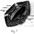
例示のみの目的で、そしていかなる方法によっても限定とみなされることなく、図7〜11を参照して、Lichtenstein修復方法が、本開示に従って任意の吸着性外科用固定具のいずれかを用いて実施されると記載されている。第1に、5cm〜6cmの横切開は、恥骨結節から始まるランガー線内でなされる。外腹斜腱膜が開かれる。図7に示すように、精巣挙筋に覆われている精索、精巣挙筋動脈、および生殖神経は、鼡径フロアに存在せず、そしてペンローズドレーンにより持ち上げられている。この精索は、恥骨結節を超えてプロテーゼメッシュが延びるための空間を確保するために、恥骨結節に対して約2cm中央の恥骨領域を含まずに解剖される。外腹斜腱膜は、下に存在する内腹斜筋および6cm〜7cmの高さのプロテーゼのための空間を確保するのに十分な高さの腱膜から解剖される。次いで、この嚢は、その首の向こうの索から解剖され、そして連結または切除することなく腹膜前空間に反転される。
【0041】
図8を参照すると、メッシュの医療側は、患者の解剖学的構造を形成する。第1の吸収性固定具10、200、または300は、メッシュに適用され、恥骨に挿入される前方直筋(anterior rectus)シースにメッシュを固定する。吸収性固定具は、この領域がメッシュにより覆われることを確認するために、恥骨結節に対して約2cm中央に配置される。さらなる吸収性固定具10、200、または300はその周囲に配置され、そして内部環の外側境界で終了する。次いで、メッシュの下方縁は、外科用メッシュの周囲の、そして内部環の外側境界で終了する同一の固定具10、200、または300を用いて鼡径靱帯に固着される。
【0042】
本開示の1つの方法に従って、吸収性の固定具は、外科用固定具の第1のアンカーが外科用メッシュおよび身体組織を貫通するように、および外科用固定具の第2のアンカーが身体組織に直接移植されるように、この固定具を身体組織に焼成することにより、固定して配置される。この様式で、固定具装置の縫合テザーは、部分的に外科用メッシュを横切って延び、部分的に身体組織を横切って延びている。代替の方法において、本開示に従う吸収性固定具は、両方のアンカーが身体組織に埋め込まれ、そして外科用メッシュを通過するように固定して配置され得る。この方法において固定具200が使用される場合、固定具200は、メッシュを通して単独で身体組織に固着される。
【0043】
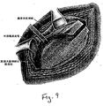
次いで、図9に示すように、スリットがメッシュの側方端に作製され、2つのテール−−2/3上方および1/3下方−−が作製される。次いで、上方テールは索の下を通過し、そして患者の頭部の方に引っぱられ、2つのテールの間に精索を配置する。次いで、図10に示されるように、上方テールは下方テールと交差し、そして1対の止血物質と共に保持される。このテールは後にひとつに縫合され、そして外腹斜腱膜の下に押し込まれ、内部環に対して側方に5cm〜6cmのメッシュを残す。
【0044】
メッシュの上方端が固定されて配置される場合、メッシュを少し弛緩させて維持することに注意が払われる。この弛緩は、メッシュにドーム様波紋を生成し、患者が手術の間に彼または彼女の横臥位置から立ち上がったときの腹内圧の増加を補う。メッシュが完全に平坦に維持される場合、患者が立ち上がったときに緊張に供される。メッシュおよび組織に対するこの引っ張り効果は、図11に示されるようにメッシュが平坦に維持される場合に説明される。本開示に従う外科用固定具の使用は、外科用メッシュの緊張を効果的に消滅させ、外科用固定具のアンカーは、互いに相対的に移動可能である。言い換えると、1つのアンカーは、患者の身体組織が移動するときに必要とされる場合、他のアンカーに対して移動し得る。その場合、メッシュに対する張力は減少される。なぜならば、個々のアンカーは互いに対して移動可能であるので、縫合テザーは、メッシュに移動するためのいくらかの空間を提供するからである。
【0045】
本開示に従う、固定具10または300を使用するTotal Extra peritoneal Laparoscopic Hernia修復がここに記載されている。第1に、皮膚切開がなされ、そして筋膜が切開される。次いで、バルーン型ディストラクターがそこに配置され、そして有効な腹膜腔外空間が生成される。次いで、臍帯トロカールおよび二次トロカールは、腹膜腔外空間に挿入され、この空間が探索される。次いで、中央および側方構造は切除され、そして精索(spermatic chord)が同定される。次いで、外科用メッシュは所望の大きさおよび形状に切断され、その結果、精索のいずれかの側でメッシュが包まれるように、スリットが提供される。次いで、メッシュは、胸横弓(tranversus arch)および腸恥靱帯にメッシュを固着させるために、外科用固定具10、200、または300を用いて側方に固定されて配置される。この工程は、反対側で繰り返される。次いで、メッシュは、さらなる固定具10、200、または300を用いて胸横弓およびクーパー靱帯に中央で固定されて配置されるか、あるいは中央の腸恥靱帯にメッシュを固定する。最後に、切開が閉じられる。
【0046】
固定具10、200、または300は、TAPP修復方法において使用され、側方および中央の両方でメッシュの縫合を置換し得ることが予想される。TAPP修復法において、固定具10、200、または300は、メッシュを貫通させ、そして身体組織に固着させることにより、メッシュを固定して配置するために使用される。
【0047】
上記の固定具の各々は、ヘルニアの修復外科と結びつけて使用されると記載されているが、類似の構造を有する固定具は、骨または軟骨にアイテムを固定するための外科手段において使用され得ることが予想される。このような外科手順において、外科用固定具は、外科等級のステンレス綱、チタン、または骨を貫通するのに十分な強度を有する任意の他の外科等級の材料から作製され得る。
【0048】
種々の改変が、本明細書で開示される外科用吸収性固定具装置および本明細書中で開示される方法の実施形態に対してなされることが理解される。従って、上記の説明は、限定として解釈されるべきではなく、好ましい実施形態の単なる例示であると解釈されるべきである。当業者は、本開示の範囲および意図の中での他の改変を予想し得る。
【0049】
(付属書A)
【0050】
【数1】

【数2】
【図面の簡単な説明】
本発明により開示される外科用固定具および方法の種々の実施形態は、添付の図面を参照して、本明細書中に記載される。
【図1】 図1は、本発明の開示に従って構築された吸収性外科用固定具装置の1実施形態の拡大透視図である。
【図2】 図2は、図1の吸収性外科用固定具装置のバーブ部分の側面図である。
【図3】 図3は、本発明の開示に従う吸収性外科用固定具装置の代替の実施形態の拡大透視図である。
【図4】 図4は、図3の外科用固定具の端面図である。
【図5】 図5は、図3に示される固定具の対を備える吸収性外科用固定具装置の拡大透視図である。
【図6】 図6は、長手軸方向に沿ってとった、図5に示される吸収性固定具の断面図である。
【図7】 図7は、本発明により開示された吸収性外科用固定具を使用して、体組織に対象物を係合させる1実施形態における、連続工程の例示である。
【図8】 図8は、図7の方法実施形態に従う、さらなる連続工程である。
【図9】 図9は、図7の方法のなおさらなる連続工程である。
【図10】 図10は、図7の方法の別の連続工程である。
【図11】 図11は、図7の方法実施形態の工程の別の例示である。[0001]
(Cross-reference to related applications)
This application claims priority to US Provisional Application No. 60 / 242,647, filed Oct. 23, 2000, the entire contents of which are incorporated herein by reference.
[0002]
(background)
(1. Technical field)
The present disclosure relates to surgical fasteners and devices for applying surgical fasteners and procedures for securing an object to body tissue. More particularly, the present disclosure relates to an absorbent surgical fixture and an apparatus for applying the absorbent fixture. Furthermore, the present disclosure relates to procedures for securing an object to tissue and procedures that require tissue to be secured together.
[0003]
(2. Background of related technology)
Fixing an object to body tissue is generally a challenge required in many different surgical applications. One illustrative example of such an application is a hernia repair procedure in which a reinforced synthetic mesh material is attached to tissue. Hernia is a general term that refers to the protrusion of tissue through the wall of the cavity in which it is normally stored, and is also called rupture. Inguinal hernia is a condition in which the intestinal loop enters the inguinal canal (ie, the passage of a tube through the lower layer of the abdominal wall). A direct inguinal hernia forms a bulge in the inguinal region, and an indirect hernia descends into the scrotum. In men, hernias can develop where the spermatic cord passes from the abdomen to the scrotum. Inguinal hernia is a condition in men that occurs in about 2% of the male population. Often, the inguinal hernia can be pushed back into the abdominal cavity. However, if the inguinal hernia cannot be pushed back through the abdominal wall, the hernia that has become hernia can be trapped and / or squeezed into the inguinal ring. If blood flow is restricted (strangulated hernia), or if the intestine is blocked (obstructed), urgent surgery is necessary. Without treatment, strangulated bowel loops die as a result of the lack of blood into the bowel loop.
[0004]
In order to treat inguinal hernias, surgery is often required to return the bowel loop to the normal position and to fix the weakened muscles in the abdomen. There are two main performed open surgical procedures for hernia repair, where the procedure uses synthetic mesh reinforcement. One procedure is the Richstein ventral repair method and the other is the Stoppa preperitoneal repair method. There are modifications to these procedures, as well as additional open surgical procedures that do not require the placement of a reinforcing mesh over the hernia defect.
[0005]
The Lichtenstein repair method is a “tension-free hernia radical surgery” based on two important facts. These facts mean that inguinal hernia is caused by metabolic disorders, which causes progressive destruction of inguinal fiber connective tissue, making it unsuitable for use in hernia repair, and traditionally Tissue repair involves excessive tension in the suture, which results in more post-operative pain, longer recovery time, and higher speed.
[0006]
The Richtensin repair method includes the following steps. First, a transverse incision is made in the Langer line (which starts at the pubic nodule). The external oblique aponeurosis is opened, and the spermatic cord, external vas deferens, and reproductive nerve with the cover of the levator levator are released from the inguinal floor and lifted together using a Penrose drain. The spermatic cord is then cut off from the pubic region inside the pubic nodule to make room for the mesh to extend beyond the pubic nodule.
[0007]
The external oblique aponeurosis is then high enough to make room for the prosthesis and is cut off from the underlying internal oblique and aponeurosis. The sac is then cut from the cord beyond its neck and inverted into the preperitoneal space without ligation or resection. The proximal end is closed, disconnected from the cord structure, and inverted into the preperitoneal space. The medical side of the mesh is then molded into the patient's anatomy. The first fixation suture of the mesh secures the mesh to the rectus abdominis muscle sheath when it is inserted into the pubic bone. The lower end of the mesh is sutured to the inguinal ligament using the same suture in a continuous manner and ends at the outer edge of the inner ring. Next, a slit is made at the outer edge of the mesh to make two tails. The upper tail is then passed under the cord and pulled toward the patient's head, placing the spermatic cord between the two tails. The upper tail is then crossed with the lower tail and maintained using a pair of hemostats. The tail is later sutured together and sewn under the external oblique aponeurosis.
[0008]
The Stoppa method of hernia repair places a single sheet of prosthetic material (ie, a surgical mesh) between the peritoneum and the musculopecteal opening. The surgical mesh is then secured to the Cooper ligament using a non-absorbable suture. The Stoppa hernia repair method is further described in the attached paper of Appendix A, the entire contents of which are hereby incorporated by reference.
[0009]
Yet another hernia repair method (known as TransAbdominal PrePeritoneal (TAPP) Laparoscopic Hernia repair method) generally includes the following steps. A pneumoperitoneum is created in the abdomen and internal abdominal pressure is maintained. Repair is then initiated. A laparoscope is then inserted and pointed towards the affected inguinal cavity. Peritoneal defect or hernia is confirmed. A peritoneal incision is made, which extends from the outer surface of the inguinal region to the outer umbilical ligament. The Cooper ligament is then exposed, as is the lower superior abdominal duct and spermatic cord. The indirect inguinal hernia sac is then carefully separated from the spermatic cord. The surgical mesh is then inserted into the abdominal cavity and placed over the inguinal region. There are three ways to place and secure the mesh over the inguinal region. The mesh is then secured in place using a surgical stapler. This is first secured to the Cooper ligament, followed by placing several staples perpendicular to the ligament, followed by a row of staples outside and parallel with the Cooper ligament. The implant is also secured around and outside the lower upper abdominal canal. When the mesh is wrapped around the spermatic cord, both edges of the mesh are closed and secured. The peritoneum is then closed using additional staples and homeostasis is investigated.
[0010]
Yet another hernia repair method is known as a Total ExtraPeritoneal (TEP) Laparoscopy Hernia repair method. This method is identical to the TAPP repair method, but it occurs entirely in the preperitoneal space. The TEP method includes the following steps. Unlike the TAPP repair method, the pneumoperitoneum is not created in the TEP repair method. Instead, a small incision is made under the navel (midline) and the midline is exposed. An incision is made slightly outside the midline aponeurosis, exposing the anterior rectus abdominis and posterior rectus abdominis sheaths. The anatomy must first be clearly identified. The Cooper ligament as well as the lower inferior abdominal canal must first be visualized. An indirect hernia sac is drawn out of the spermatic cord and inguinal cavity to the open bone. The hernia sac is then cut as inward as possible, allowing the surgical mesh to cover the entire inguinal region. The mesh is then inserted and secured in place as in the TAPP repair method. Upon completion of the repair, the small incision can be closed.
[0011]
The two most common of these methods are the Total ExtraPeritoneal (TEP) repair method and the TransAbdominal PrePeritoneal (TAPP) repair method. As noted above, each of these methods utilizes a reinforced synthetic mesh that must be secured to the tissue to prevent initial migration of the mesh from the hernia site. However, the mesh must first be locked in place to prevent its movement from the view of the hernia repair. After only 7-10 days, the mesh has sufficient tissue growth to prevent its migration from the hernia repair site.
[0012]
Each of the procedures disclosed above utilizes titanium staples to hold the mesh in place. These staples are persistent in the body cavity. The disadvantage of persistent metal staples is the possibility of excessive scar tissue (adhesion) formation, which in turn causes further patient complications and hinders future surgical procedures. Furthermore, these durable staples can be associated with increased long-term discomfort to the patient as a result of the hernia repair procedure.
[0013]
Thus, improved surgical fasteners and applicators as well as when securing an object to body tissue (eg, sufficient time at the hernia repair site until sufficient tissue growth occurs to maintain the mesh in place). There is a need for methods (such as when installing mesh material).
[0014]
(Summary)
It is an object of the present disclosure to provide an absorbable surgical fixation device and method in which the amount of extraneous material in the patient's body is reduced, thereby minimizing adhesion formation, And it reduces the long-term discomfort associated with the fixture for the patient.
[0015]
It is another object of the present disclosure to provide an absorbable surgical fastener and method that is easier and faster to use than traditional suturing techniques in an open procedure. Furthermore, the relatively high firing force that can be applied to the absorbent fixture of the present disclosure facilitates a more reliable penetration of stiffer tissue material (such as, for example, a Cooper ligament).
[0016]
It is yet another object of the present disclosure to provide an absorbent surgical fixture that is radiolucent and that provides greater peace of mind for the patient.
[0017]
It is still a further object of the present disclosure to provide an absorbent surgical fastener device having a suture tether disposed between fixation barbs. This tether provides the advantage of keeping the mesh loose in place, thus minimizing the tension in the surrounding tissue and reducing the occurrence of fixture pullout.
[0018]
Another object of the present disclosure is to provide a surgical fastener device that is dimensioned so as not to penetrate the patient's abdominal wall and is relative to standard fastener barbs. Provides a series of barbs having a wide surface, thereby more maintaining this fixture in soft tissue.
[0019]
The surgical fastener device and method disclosed by the present invention provides the functionality of staples previously used to secure objects to body tissue, as practiced in the present invention. For example, the mesh is secured to a specific anatomical landmark surrounding the hernia repair, or the mesh is attached to the tissue, the tissue is attached to the tissue, or the tissue is attached to the ligament. However, the absorbent fixture device and method disclosed by the present invention is utilized by the absorbent fixture to attach the mesh to the tissue for a time sufficient to allow tissue growth to occur on the mesh material. Has the unique advantageous feature of being In this manner, the absorbent fixator helps prevent initial mesh movement and is then absorbed into the body after sufficient tissue growth.
[0020]
A surgical fastener device is provided for securing a surgical mesh material to body tissue, wherein the device includes a pair of anchors, each of these anchors being formed on its outer surface. Means and a suture tether that interconnects the pair of anchors with each other. The fastener device is preferably made from a bioabsorbable material and the device is only partially absorbed over a period of 2 to 3 weeks after implantation and fully absorbed by the body at any time thereafter. Dimensions are taken so that
[0021]
In an alternative embodiment, the surgical fastener device comprises a conical body portion having a pointed distal end and an enlarged proximal head. A conical body portion is provided with a helical thread starting at or near the distal end and ending preferably spaced from the enlarged proximal head.
[0022]
Furthermore, a hernia repair method utilizing a fastener according to the present disclosure includes placing the surgical mesh on a hernia repair site by embedding the surgical fastener in body tissue via the surgical mesh. Provided to be fixed.
[0023]
Detailed Description of Preferred Embodiments
Preferred embodiments of the absorbable surgical fastener device and method of applying the same disclosed by the present invention will now be described in detail with reference to the drawings. In the drawings, the same number identifies similar or identical structural elements.
[0024]
The absorbable surgical fastener devices and methods disclosed by the present invention are shown and described herein in connection with open and abdominal groin, femoral and ventral hernia repairs. Although not described in detail herein, the absorbent fixture can also be applied to other procedures where the object needs to be engaged with body tissue.
[0025]
Initially, with reference to FIGS. 1 and 2, an absorbent surgical fastener device, eg,
[0026]
[0027]
Each barb 12a-12c and 14a-14b is orthogonal to the longitudinal axis "A" of each
[0028]
Although the embodiment of FIGS. 1 and 2 illustrates barbs partially formed around the circumference of
[0029]
Although a planar
[0030]
The
[0031]
The
[0032]
Unlike the applicator device for positioning the
[0033]
As an example, the general procedure for applying mesh material during a hernia repair procedure is to first create access to the hernia location (ie, incision and dissection), thereby exposing the hernia; An artificial mesh is then placed in the tissue; then, the mesh is secured to the surrounding tissue by firing a plurality of
[0034]
Turning now to FIGS. 3-5, an alternative embodiment of a surgical fastener in accordance with the present disclosure is shown generally as 200. FIG. 3 and 4, the
[0035]
In use, the
[0036]
Turning now to FIG. 5, a surgical fastener device according to the present disclosure is shown generally as 300. Similar to
[0037]
As seen in FIG. 6, the
[0038]
[0039]
[0040]
For illustrative purposes only and without being considered limiting in any way, and with reference to FIGS. 7-11, the Lichtenstein repair method is performed using any of the adsorptive surgical fasteners according to the present disclosure. It is stated that it will be. First, a 5-6 cm lateral incision is made within the Langer line starting from the pubic nodule. The external oblique aponeurosis is opened. As shown in FIG. 7, the spermatic cord, levator ani muscle, and reproductive nerve that are covered by the testicular muscle are not present on the inguinal floor and are lifted by the Penrose drain. The spermatic cord is dissected without the pubic region about 2 cm central to the pubic nodule to ensure space for the prosthetic mesh to extend beyond the pubic nodule. The external oblique aponeurosis is dissected from the aponeurosis which is high enough to ensure space for the underlying internal oblique and 6-7 cm high prosthesis. The sac is then dissected from the cord over its neck and inverted into the preperitoneal space without ligation or excision.
[0041]
Referring to FIG. 8, the medical side of the mesh forms the patient's anatomy. The first
[0042]
In accordance with one method of the present disclosure, the resorbable fastener is such that the first anchor of the surgical fastener penetrates the surgical mesh and body tissue and the second anchor of the surgical fastener is body tissue. The fixture is placed in place by firing into body tissue so that it can be implanted directly into the body. In this manner, the suture tether of the fastener device extends partially across the surgical mesh and partially extends across the body tissue. In an alternative method, the resorbable fastener according to the present disclosure can be placed securely so that both anchors are embedded in body tissue and pass through the surgical mesh. When the
[0043]
Then, as shown in FIG. 9, slits are made at the side edges of the mesh, creating two tails --2 / 3 above and 1/3 below. The upper tail then passes under the cord and is pulled towards the patient's head, placing the spermatic cord between the two tails. Then, as shown in FIG. 10, the upper tail intersects with the lower tail and is held with a pair of hemostatic substances. This tail is later sewn together and pushed under the external oblique aponeurosis, leaving a 5 cm to 6 cm mesh lateral to the internal annulus.
[0044]
Care is taken to keep the mesh slightly relaxed if the upper end of the mesh is placed fixed. This relaxation creates a dome-like ripple in the mesh that compensates for the increased abdominal pressure when the patient stands up from his or her recumbent position during surgery. If the mesh is kept perfectly flat, it is subjected to tension when the patient stands up. This pulling effect on the mesh and tissue is explained when the mesh is kept flat as shown in FIG. The use of a surgical fastener in accordance with the present disclosure effectively eliminates the strain on the surgical mesh and the anchors of the surgical fastener are movable relative to one another. In other words, one anchor can move relative to other anchors as needed when the patient's body tissue moves. In that case, the tension on the mesh is reduced. Because the individual anchors are movable relative to each other, the stitching tether provides some space for moving to the mesh.
[0045]
A Total Extra peripheral Laparoscopic Hernia repair using the
[0046]
It is anticipated that the
[0047]
Although each of the above fasteners has been described as being used in conjunction with hernia repair surgery, a fastener having a similar structure can be used in surgical means to secure an item to bone or cartilage. It is expected that. In such surgical procedures, surgical fasteners can be made from surgical grade stainless steel, titanium, or any other surgical grade material that is strong enough to penetrate bone.
[0048]
It will be understood that various modifications may be made to the surgical absorbable fastener device disclosed herein and the method embodiments disclosed herein. Therefore, the above description should not be construed as limiting, but merely as exemplifications of preferred embodiments. Those skilled in the art will envision other modifications within the scope and spirit of the present disclosure.
[0049]
(Appendix A)
[0050]
[Expression 1]
[Expression 2]
[Brief description of the drawings]
Various embodiments of the surgical fasteners and methods disclosed by the present invention are described herein with reference to the accompanying drawings.
FIG. 1 is an enlarged perspective view of one embodiment of an absorbent surgical fastener device constructed in accordance with the present disclosure.
2 is a side view of the barb portion of the absorbable surgical fastener device of FIG. 1. FIG.
FIG. 3 is an enlarged perspective view of an alternative embodiment of an absorbent surgical fastener device according to the present disclosure.
FIG. 4 is an end view of the surgical fastener of FIG.
FIG. 5 is an enlarged perspective view of an absorbent surgical fastener device comprising the fastener pair shown in FIG. 3;
6 is a cross-sectional view of the absorbent fixture shown in FIG. 5 taken along the longitudinal axis.
FIG. 7 is an illustration of a continuous process in one embodiment of engaging an object with body tissue using an absorbent surgical fastener disclosed in accordance with the present invention.
FIG. 8 is a further continuous process according to the method embodiment of FIG.
FIG. 9 is a still further sequence of the method of FIG.
FIG. 10 is another continuous process of the method of FIG.
FIG. 11 is another illustration of the steps of the method embodiment of FIG.
Claims (25)
一対の実質的に円柱形のアンカーであって、それぞれが長手方向中心軸を規定し、ここで、各アンカーが、円錐形のテーパー状の遠位端および実質的に平面の近位端を備え、そしてここで、各アンカーが、近位表面およびテーパー状の遠位端を有する一連の半円形の角度の付いた突出物をさらに備え、該突出物が、部分的なネジ山を規定するような様式で該長手方向軸に対して角度を有しており、該一連の突出物を通る共通の中心軸を規定しており、そしてここで、該一連の角度の付いた突出物の共通の中心軸が、各アンカーについて、それぞれのアンカーの長手方向中心軸から一定距離間隔をあけている、アンカー;および
該一対のアンカーの該近位端を相互接続して、互いに対して複数の方向で該アンカーの自由な運動を可能にする、可撓性テザー、
を備える、外科用固定具装置。A surgical fastener device for securing a surgical mesh material to body tissue comprising:
A pair of substantially cylindrical anchors, each defining a longitudinal central axis, wherein each anchor comprises a conical tapered distal end and a substantially planar proximal end and wherein, as each anchor further comprises a series of projections marked with a semi-circular angle with proximal surface and tapered distal end, the projecting distillate defines a partial thread Angled with respect to the longitudinal axis in a different manner, defining a common central axis through the series of protrusions, and wherein the common angle of the series of angled protrusions is A central axis , for each anchor, spaced a distance from the longitudinal central axis of the respective anchor; and interconnecting the proximal ends of the pair of anchors in a plurality of directions relative to each other Allow free movement of the anchor, possible Flexible tether,
A surgical fastener device comprising:
一対のアンカーであって、それぞれが長手方向中心軸を規定しており、それぞれが、その外側表面上に形成される角度の付いた突出部を有し、それぞれのアンカーについての角度の付いた突出部が、共通の長手方向軸を規定しており、該共通の長手方向軸が、それぞれのアンカーの長手方向中心軸から半径方向に一定距離間隔をあけており、該角度の付いた突出部が、部分的なネジ山を規定するような様式で該長手方向軸に対して角度を有している、アンカー;および
該一対のアンカーを互いに相互接続して、互いに対して複数の方向で該アンカーの自由な運動を可能にする、可撓性縫合テザー、
を備える、外科用固定具装置。A surgical fastener device for securing a surgical mesh material to body tissue comprising:
A pair of anchors, each project has defining a longitudinal center axis, respectively, have a protruding part with an angle formed on its outer surface, angled for each anchor Sections define a common longitudinal axis, the common longitudinal axis is spaced a certain distance in a radial direction from the longitudinal central axis of each anchor, and the angled protrusions are An anchor having an angle with respect to the longitudinal axis in a manner defining a partial thread ; and the pair of anchors interconnected to each other in a plurality of directions relative to each other Flexible suture tether, which allows free movement of
A surgical fastener device comprising:
Applications Claiming Priority (2)
| Application Number | Priority Date | Filing Date | Title |
|---|---|---|---|
| US24264700P | 2000-10-23 | 2000-10-23 | |
| PCT/US2001/050165 WO2002034140A2 (en) | 2000-10-23 | 2001-10-23 | Absorbable fastener and applying apparatus |
Related Child Applications (1)
| Application Number | Title | Priority Date | Filing Date |
|---|---|---|---|
| JP2008005035A Division JP4748813B2 (en) | 2000-10-23 | 2008-01-11 | Absorbent fixture and application equipment |
Publications (2)
| Publication Number | Publication Date |
|---|---|
| JP2004530450A JP2004530450A (en) | 2004-10-07 |
| JP4092196B2 true JP4092196B2 (en) | 2008-05-28 |
Family
ID=22915629
Family Applications (2)
| Application Number | Title | Priority Date | Filing Date |
|---|---|---|---|
| JP2002537196A Expired - Fee Related JP4092196B2 (en) | 2000-10-23 | 2001-10-23 | Absorbent fixture and application equipment |
| JP2008005035A Expired - Fee Related JP4748813B2 (en) | 2000-10-23 | 2008-01-11 | Absorbent fixture and application equipment |
Family Applications After (1)
| Application Number | Title | Priority Date | Filing Date |
|---|---|---|---|
| JP2008005035A Expired - Fee Related JP4748813B2 (en) | 2000-10-23 | 2008-01-11 | Absorbent fixture and application equipment |
Country Status (7)
| Country | Link |
|---|---|
| US (7) | US20040092937A1 (en) |
| EP (2) | EP1328196B1 (en) |
| JP (2) | JP4092196B2 (en) |
| AU (2) | AU2002232798B2 (en) |
| CA (2) | CA2426474C (en) |
| ES (1) | ES2365129T3 (en) |
| WO (1) | WO2002034140A2 (en) |
Families Citing this family (189)
| Publication number | Priority date | Publication date | Assignee | Title |
|---|---|---|---|---|
| DE69931018T2 (en) | 1998-12-30 | 2006-11-23 | Ethicon, Inc. | Thread belay device |
| US7153312B1 (en) | 1999-12-02 | 2006-12-26 | Smith & Nephew Inc. | Closure device and method for tissue repair |
| US7887551B2 (en) | 1999-12-02 | 2011-02-15 | Smith & Nephew, Inc. | Soft tissue attachment and repair |
| US7461767B2 (en) | 2005-06-03 | 2008-12-09 | Tyco Healthcare Group Lp | Battery powered surgical instrument |
| AU2002348033B2 (en) | 2001-10-23 | 2008-05-29 | Covidien Lp | Surgical fasteners |
| DE60336936D1 (en) | 2002-06-11 | 2011-06-09 | Tyco Healthcare | Network clips for breaks |
| US20040138683A1 (en) * | 2003-01-09 | 2004-07-15 | Walter Shelton | Suture arrow device and method of using |
| US20040138705A1 (en) * | 2003-01-09 | 2004-07-15 | Harri Heino | Surgical staple for tissue treatment |
| CA2513726C (en) * | 2003-01-27 | 2014-08-12 | Corassist Cardiovascular Ltd. | In vivo device for improving diastolic ventricular function |
| US9314235B2 (en) | 2003-02-05 | 2016-04-19 | Smith & Nephew, Inc. | Tissue anchor and insertion tool |
| JP2007500583A (en) | 2003-06-13 | 2007-01-18 | タイコ・ヘルスケア・グループ・リミテッド・パートナーシップ | Multi-member interconnect and absorbable screw fasteners for surgical instruments |
| US8926637B2 (en) | 2003-06-13 | 2015-01-06 | Covidien Lp | Multiple member interconnect for surgical instrument and absorbable screw fastener |
| US20060052825A1 (en) * | 2003-06-16 | 2006-03-09 | Ransick Mark H | Surgical implant alloy |
| US7905902B2 (en) * | 2003-06-16 | 2011-03-15 | Ethicon Endo-Surgery, Inc. | Surgical implant with preferential corrosion zone |
| US10478179B2 (en) | 2004-04-27 | 2019-11-19 | Covidien Lp | Absorbable fastener for hernia mesh fixation |
| ES1057646Y (en) * | 2004-05-19 | 2004-12-16 | Ayet Felix Checa | PERFECTED PROTESIS FOR THE SURGICAL TREATMENT OF HERNIAS DE LA INGLE. |
| US8128670B2 (en) * | 2005-04-15 | 2012-03-06 | Biodynamics Llc | Surgical expansion fasteners |
| US20060293709A1 (en) | 2005-06-24 | 2006-12-28 | Bojarski Raymond A | Tissue repair device |
| US20070162030A1 (en) * | 2006-01-06 | 2007-07-12 | Ernest Aranyi | Multi-pronged compressive absorbable tack |
| US7862573B2 (en) * | 2006-04-21 | 2011-01-04 | Darois Roger E | Method and apparatus for surgical fastening |
| US8454662B2 (en) * | 2006-12-08 | 2013-06-04 | Warsaw Orthopedic, Inc. | Tethers with strength limits for treating vertebral members |
| US7823760B2 (en) | 2007-05-01 | 2010-11-02 | Tyco Healthcare Group Lp | Powered surgical stapling device platform |
| US7931660B2 (en) | 2007-05-10 | 2011-04-26 | Tyco Healthcare Group Lp | Powered tacker instrument |
| US7959640B2 (en) | 2008-02-13 | 2011-06-14 | Apollo Endosurgery, Inc. | Method of performing transgastric ventral hernia repair and tissue anchors and deployment devices therefor |
| US8758373B2 (en) | 2008-02-18 | 2014-06-24 | Covidien Lp | Means and method for reversibly connecting a patch to a patch deployment device |
| US9393093B2 (en) | 2008-02-18 | 2016-07-19 | Covidien Lp | Clip for implant deployment device |
| US8317808B2 (en) | 2008-02-18 | 2012-11-27 | Covidien Lp | Device and method for rolling and inserting a prosthetic patch into a body cavity |
| US9301826B2 (en) | 2008-02-18 | 2016-04-05 | Covidien Lp | Lock bar spring and clip for implant deployment device |
| US9833240B2 (en) | 2008-02-18 | 2017-12-05 | Covidien Lp | Lock bar spring and clip for implant deployment device |
| US9034002B2 (en) | 2008-02-18 | 2015-05-19 | Covidien Lp | Lock bar spring and clip for implant deployment device |
| EP2247245B1 (en) | 2008-02-18 | 2017-06-28 | Covidien LP | A device for deploying and attaching a patch to a biological tissue |
| US9393002B2 (en) | 2008-02-18 | 2016-07-19 | Covidien Lp | Clip for implant deployment device |
| US8808314B2 (en) | 2008-02-18 | 2014-08-19 | Covidien Lp | Device and method for deploying and attaching an implant to a biological tissue |
| US9044235B2 (en) | 2008-02-18 | 2015-06-02 | Covidien Lp | Magnetic clip for implant deployment device |
| US9398944B2 (en) | 2008-02-18 | 2016-07-26 | Covidien Lp | Lock bar spring and clip for implant deployment device |
| US20090287045A1 (en) | 2008-05-15 | 2009-11-19 | Vladimir Mitelberg | Access Systems and Methods of Intra-Abdominal Surgery |
| US20100076487A1 (en) * | 2008-09-23 | 2010-03-25 | Ilahi Omer A | Kit Containing Combination Absorbable Staple and Non-absorbable Suture, And Method Of Using Same |
| WO2010046893A1 (en) | 2008-10-20 | 2010-04-29 | Polytouch Medical Ltd. | A device for attaching a patch to a biological tissue |
| US20100191332A1 (en) | 2009-01-08 | 2010-07-29 | Euteneuer Charles L | Implantable Tendon Protection Systems and Related Kits and Methods |
| US8287572B2 (en) | 2009-02-11 | 2012-10-16 | Howmedica Osteonics Corp. | Intervertebral implant with integrated fixation |
| US9179910B2 (en) | 2009-03-20 | 2015-11-10 | Rotation Medical, Inc. | Medical device delivery system and method |
| AU2010236836B2 (en) | 2009-03-31 | 2015-06-18 | Imds Llc | Double bundle ACL repair |
| US8535377B2 (en) * | 2009-03-31 | 2013-09-17 | Imds Corporation | Double bundle ACL repair system |
| US20100256675A1 (en) * | 2009-04-03 | 2010-10-07 | Romans Matthew L | Absorbable surgical staple |
| WO2010129641A1 (en) | 2009-05-07 | 2010-11-11 | Hammell Eugene J | Surgical patch cover and method of use |
| US8894669B2 (en) * | 2009-05-12 | 2014-11-25 | Ethicon, Inc. | Surgical fasteners, applicator instruments, and methods for deploying surgical fasteners |
| JP5567125B2 (en) | 2009-06-04 | 2014-08-06 | ローテーション メディカル インコーポレイテッド | A device having a bow chord shape for conveying staples to a target tissue |
| EP3308743A1 (en) | 2009-06-04 | 2018-04-18 | Rotation Medical, Inc. | Methods and apparatus for deploying sheet-like materials |
| EP3115026B1 (en) | 2009-08-10 | 2018-11-28 | Howmedica Osteonics Corp. | Surgical instrument |
| CA2769666C (en) | 2009-08-17 | 2018-02-13 | Arie Levy | Means and method for reversibly connecting an implant to a deployment device |
| US8906045B2 (en) | 2009-08-17 | 2014-12-09 | Covidien Lp | Articulating patch deployment device and method of use |
| US9033993B2 (en) | 2009-11-03 | 2015-05-19 | Howmedica Osteonics Corp. | Intervertebral implant with integrated fixation |
| US9480511B2 (en) | 2009-12-17 | 2016-11-01 | Engage Medical Holdings, Llc | Blade fixation for ankle fusion and arthroplasty |
| WO2014146000A2 (en) * | 2013-03-15 | 2014-09-18 | Micro Interventional Devices, Inc. | Tissue closure device and method |
| US9198750B2 (en) | 2010-03-11 | 2015-12-01 | Rotation Medical, Inc. | Tendon repair implant and method of arthroscopic implantation |
| EP2651341B1 (en) | 2010-12-16 | 2017-01-04 | Engage Medical Holdings, LLC | Arthroplasty systems and methods |
| US10952783B2 (en) | 2011-12-29 | 2021-03-23 | Rotation Medical, Inc. | Guidewire having a distal fixation member for delivering and positioning sheet-like materials in surgery |
| CA2825918C (en) | 2011-02-15 | 2018-08-07 | Rotation Medical, Inc. | Methods and apparatus for delivering and positioning sheet-like materials |
| WO2012145059A1 (en) | 2011-02-15 | 2012-10-26 | Rotation Medical, Inc. | Methods and apparatus for fixing sheet-like materials to a target tissue |
| US8777965B2 (en) * | 2011-04-15 | 2014-07-15 | Usgi Medical, Inc. | Devices and methods for laparoscopic hernia repair |
| US8968402B2 (en) | 2011-10-18 | 2015-03-03 | Arthrocare Corporation | ACL implants, instruments, and methods |
| US9254130B2 (en) | 2011-11-01 | 2016-02-09 | Hyun Bae | Blade anchor systems for bone fusion |
| US9615856B2 (en) | 2011-11-01 | 2017-04-11 | Imds Llc | Sacroiliac fusion cage |
| US8535339B2 (en) | 2011-12-18 | 2013-09-17 | Via Surgical Ltd. | Apparatus and method for suturing |
| EP2793712B1 (en) * | 2011-12-19 | 2018-03-28 | Rotation Medical, Inc. | Fasteners for affixing sheet -like materials to bone or tissue |
| US9107661B2 (en) | 2011-12-19 | 2015-08-18 | Rotation Medical, Inc. | Fasteners and fastener delivery devices for affixing sheet-like materials to bone or tissue |
| WO2013096224A1 (en) | 2011-12-19 | 2013-06-27 | Rotation Medical, Inc. | Fasteners for affixing sheet -like materials to bone or tissue |
| WO2013096219A1 (en) | 2011-12-19 | 2013-06-27 | Rotation Medical, Inc. | Apparatus and method for forming pilot holes in bone and delivering fasteners therein for retaining an implant |
| WO2013101638A1 (en) | 2011-12-29 | 2013-07-04 | Rotation Medical, Inc. | Methods and apparatus for delivering and positioning sheet -like materials in surgery |
| WO2013101641A2 (en) | 2011-12-29 | 2013-07-04 | Rotation Medical, Inc. | Anatomical location markers and methods of use in positioning sheet-like materials during surgery |
| US9204959B2 (en) | 2012-02-02 | 2015-12-08 | Smith & Nephew, Inc. | Implantable biologic holder |
| US10238382B2 (en) | 2012-03-26 | 2019-03-26 | Engage Medical Holdings, Llc | Blade anchor for foot and ankle |
| ES2879285T3 (en) | 2012-05-31 | 2021-11-22 | Via Surgical Ltd | Variable depth surgical fixation |
| US9888913B2 (en) | 2012-05-31 | 2018-02-13 | Via Surgical Ltd. | Variable depth surgical fixation |
| US20140046348A1 (en) * | 2012-08-07 | 2014-02-13 | University Hospitals Of Cleveland | Wound healing system |
| WO2014107503A1 (en) * | 2013-01-04 | 2014-07-10 | The Feinstein Institute For Medical Research | Fastener |
| US9351733B2 (en) | 2013-01-18 | 2016-05-31 | Covidien Lp | Surgical fastener applier |
| US9993245B2 (en) | 2013-03-11 | 2018-06-12 | Via Surgical Ltd. | Surgical tacker with quantity indicator |
| US9358010B2 (en) | 2013-03-12 | 2016-06-07 | Covidien Lp | Flex cable and spring-loaded tube for tacking device |
| US9867620B2 (en) | 2013-03-14 | 2018-01-16 | Covidien Lp | Articulation joint for apparatus for endoscopic procedures |
| US9655621B2 (en) | 2013-03-15 | 2017-05-23 | Covidien Lp | Surgical instrument for dispensing tacks and solution |
| US20140364872A1 (en) * | 2013-06-05 | 2014-12-11 | Lc Therapeutics, Inc. | Tissue anchor and deployment device for same |
| US9668730B2 (en) | 2013-06-28 | 2017-06-06 | Covidien Lp | Articulating apparatus for endoscopic procedures with timing system |
| US9358004B2 (en) | 2013-06-28 | 2016-06-07 | Covidien Lp | Articulating apparatus for endoscopic procedures |
| US10085746B2 (en) | 2013-06-28 | 2018-10-02 | Covidien Lp | Surgical instrument including rotating end effector and rotation-limiting structure |
| US9351728B2 (en) | 2013-06-28 | 2016-05-31 | Covidien Lp | Articulating apparatus for endoscopic procedures |
| US20150032130A1 (en) | 2013-07-24 | 2015-01-29 | Covidien Lp | Expanding absorbable tack |
| US9526498B2 (en) | 2013-09-17 | 2016-12-27 | Covidien Lp | Surgical device with a trigger lockout mechanism device |
| CN106455936B (en) | 2014-04-02 | 2019-03-08 | 柯惠有限合伙公司 | Surgical fastener bringing device, external member and method for endoscopic surgery |
| EP3139859B1 (en) | 2014-05-09 | 2021-06-23 | Rotation Medical, Inc. | Medical implant delivery system for sheet-like implant |
| CN104055550B (en) * | 2014-05-27 | 2016-06-08 | 苏州瑞华医院有限公司 | A kind of inclined-plane band wire holdfast |
| US10166022B2 (en) * | 2014-09-29 | 2019-01-01 | Biomet C.V. | Method and apparatus for bone fixation |
| US10123796B2 (en) | 2014-11-04 | 2018-11-13 | Rotation Medical, Inc. | Medical implant delivery system and related methods |
| EP3215025B1 (en) | 2014-11-04 | 2020-12-23 | Rotation Medical, Inc. | Medical implant delivery system |
| EP3215026B1 (en) | 2014-11-04 | 2023-10-25 | Rotation Medical, Inc. | Medical implant delivery system |
| US10568616B2 (en) | 2014-12-17 | 2020-02-25 | Howmedica Osteonics Corp. | Instruments and methods of soft tissue fixation |
| US10603182B2 (en) | 2015-01-14 | 2020-03-31 | Stryker European Holdings I, Llc | Spinal implant with fluid delivery capabilities |
| AU2016200179B2 (en) | 2015-01-14 | 2020-09-17 | Stryker European Operations Holdings Llc | Spinal implant with porous and solid surfaces |
| US11090097B2 (en) | 2015-03-17 | 2021-08-17 | Covidien Lp | Connecting end effectors to surgical devices |
| US9763800B2 (en) | 2015-03-18 | 2017-09-19 | Biomet C. V. | Implant configured for hammertoe and small bone fixation |
| US10117648B2 (en) | 2015-04-23 | 2018-11-06 | Via Surgical Ltd. | Surgical fastener delivery and locking mechanism |
| AU2016256857B2 (en) | 2015-05-06 | 2018-05-31 | Rotation Medical, Inc. | Medical implant delivery system and related methods |
| CA2930123A1 (en) | 2015-05-18 | 2016-11-18 | Stryker European Holdings I, Llc | Partially resorbable implants and methods |
| EP3307204B1 (en) | 2015-06-15 | 2021-11-24 | Rotation Medical, Inc. | Tendon repair implant |
| US10376367B2 (en) | 2015-07-02 | 2019-08-13 | First Ray, LLC | Orthopedic fasteners, instruments and methods |
| US10702290B2 (en) | 2015-11-02 | 2020-07-07 | First Ray, LLC | Orthopedic fastener, retainer, and guide |
| US10314689B2 (en) | 2015-12-31 | 2019-06-11 | Rotation Medical, Inc. | Medical implant delivery system and related methods |
| WO2017117437A1 (en) | 2015-12-31 | 2017-07-06 | Rotation Medical, Inc. | Fastener delivery system and related methods |
| US10390955B2 (en) | 2016-09-22 | 2019-08-27 | Engage Medical Holdings, Llc | Bone implants |
| US10743859B2 (en) | 2016-10-21 | 2020-08-18 | Covidien Lp | Surgical end effectors |
| US10617409B2 (en) | 2016-10-21 | 2020-04-14 | Covidien Lp | Surgical end effectors |
| US11298123B2 (en) | 2016-10-21 | 2022-04-12 | Covidien Lp | Surgical end effectors |
| US10888309B2 (en) | 2017-01-31 | 2021-01-12 | Covidien Lp | Surgical fastener devices with geometric tubes |
| US11540928B2 (en) | 2017-03-03 | 2023-01-03 | Engage Uni Llc | Unicompartmental knee arthroplasty |
| US10456272B2 (en) | 2017-03-03 | 2019-10-29 | Engage Uni Llc | Unicompartmental knee arthroplasty |
| US10835388B2 (en) | 2017-09-20 | 2020-11-17 | Stryker European Operations Holdings Llc | Spinal implants |
| US10842603B1 (en) * | 2017-10-16 | 2020-11-24 | David Lee Street | Sutureless ventral hernia meshing system and method of fixation |
| US10987104B2 (en) | 2017-10-30 | 2021-04-27 | Covidien Lp | Apparatus for endoscopic procedures |
| US11207066B2 (en) | 2017-10-30 | 2021-12-28 | Covidien Lp | Apparatus for endoscopic procedures |
| JP7317826B2 (en) | 2017-12-07 | 2023-07-31 | ローテーション メディカル インコーポレイテッド | Medical implant delivery system and related methods |
| CN108066041B (en) * | 2018-01-05 | 2024-04-16 | 北京博辉瑞进生物科技有限公司 | Absorbable nail and repair fixer |
| USD902405S1 (en) | 2018-02-22 | 2020-11-17 | Stryker Corporation | Self-punching bone anchor inserter |
| US11369371B2 (en) | 2018-03-02 | 2022-06-28 | Covidien Lp | Surgical stapling instrument |
| US11298126B2 (en) | 2018-05-02 | 2022-04-12 | Covidien Lp | Shipping wedge for end effector installation onto surgical devices |
| US11116500B2 (en) | 2018-06-28 | 2021-09-14 | Covidien Lp | Surgical fastener applying device, kits and methods for endoscopic procedures |
| US11497490B2 (en) | 2018-07-09 | 2022-11-15 | Covidien Lp | Powered surgical devices including predictive motor control |
| US11071547B2 (en) | 2018-09-12 | 2021-07-27 | Absolutions Med, Inc. | Abdominal closure method and device for ventral hernia |
| EP3836852A4 (en) | 2018-10-03 | 2022-05-04 | Absolutions Med, Inc. | Abdominal closure method and device variations |
| US11197734B2 (en) | 2018-10-30 | 2021-12-14 | Covidien Lp | Load sensing devices for use in surgical instruments |
| US11369372B2 (en) | 2018-11-28 | 2022-06-28 | Covidien Lp | Surgical stapler adapter with flexible cable assembly, flexible fingers, and contact clips |
| US11202635B2 (en) | 2019-02-04 | 2021-12-21 | Covidien Lp | Programmable distal tilt position of end effector for powered surgical devices |
| US11376006B2 (en) | 2019-02-06 | 2022-07-05 | Covidien Lp | End effector force measurement with digital drive circuit |
| US11219461B2 (en) | 2019-03-08 | 2022-01-11 | Covidien Lp | Strain gauge stabilization in a surgical device |
| CN114126506A (en) | 2019-04-10 | 2022-03-01 | 腹腔解决方案医疗公司 | Variations of abdominal closure methods and devices to close abdominal hernias and reduce recurrence |
| US11523817B2 (en) | 2019-06-27 | 2022-12-13 | Covidien Lp | Endoluminal pursestring device |
| US11707274B2 (en) | 2019-12-06 | 2023-07-25 | Covidien Lp | Articulating mechanism for surgical instrument |
| US11197675B2 (en) | 2019-12-19 | 2021-12-14 | Covidien Lp | Positioning guide for surgical instruments and surgical instrument systems |
| USD944985S1 (en) | 2019-12-19 | 2022-03-01 | Covidien Lp | Positioning guide cuff |
| USD944984S1 (en) | 2019-12-19 | 2022-03-01 | Covidien Lp | Tubular positioning guide |
| US11452524B2 (en) | 2020-01-31 | 2022-09-27 | Covidien Lp | Surgical stapling device with lockout |
| US11458244B2 (en) | 2020-02-07 | 2022-10-04 | Covidien Lp | Irrigating surgical apparatus with positive pressure fluid |
| US11553913B2 (en) | 2020-02-11 | 2023-01-17 | Covidien Lp | Electrically-determining tissue cut with surgical stapling apparatus |
| EP4103070A4 (en) | 2020-02-14 | 2023-11-08 | Covidien LP | Cartridge holder for surgical staples and having ridges in peripheral walls for gripping tissue |
| US11406383B2 (en) | 2020-03-17 | 2022-08-09 | Covidien Lp | Fire assisted powered EGIA handle |
| US11937794B2 (en) | 2020-05-11 | 2024-03-26 | Covidien Lp | Powered handle assembly for surgical devices |
| US11622768B2 (en) | 2020-07-13 | 2023-04-11 | Covidien Lp | Methods and structure for confirming proper assembly of powered surgical stapling systems |
| US11602342B2 (en) | 2020-08-27 | 2023-03-14 | Covidien Lp | Surgical stapling device with laser probe |
| US11678878B2 (en) | 2020-09-16 | 2023-06-20 | Covidien Lp | Articulation mechanism for surgical stapling device |
| US11660092B2 (en) | 2020-09-29 | 2023-05-30 | Covidien Lp | Adapter for securing loading units to handle assemblies of surgical stapling instruments |
| US11406384B2 (en) | 2020-10-05 | 2022-08-09 | Covidien Lp | Stapling device with drive assembly stop member |
| US11576674B2 (en) | 2020-10-06 | 2023-02-14 | Covidien Lp | Surgical stapling device with articulation lock assembly |
| US11890007B2 (en) | 2020-11-18 | 2024-02-06 | Covidien Lp | Stapling device with flex cable and tensioning mechanism |
| US11744580B2 (en) | 2020-11-24 | 2023-09-05 | Covidien Lp | Long stapler reloads with continuous cartridge |
| US11653919B2 (en) | 2020-11-24 | 2023-05-23 | Covidien Lp | Stapler line reinforcement continuity |
| US11737774B2 (en) | 2020-12-04 | 2023-08-29 | Covidien Lp | Surgical instrument with articulation assembly |
| US11819200B2 (en) | 2020-12-15 | 2023-11-21 | Covidien Lp | Surgical instrument with articulation assembly |
| US11553914B2 (en) | 2020-12-22 | 2023-01-17 | Covidien Lp | Surgical stapling device with parallel jaw closure |
| US11759206B2 (en) | 2021-01-05 | 2023-09-19 | Covidien Lp | Surgical stapling device with firing lockout mechanism |
| US11744582B2 (en) | 2021-01-05 | 2023-09-05 | Covidien Lp | Surgical stapling device with firing lockout mechanism |
| US11517313B2 (en) | 2021-01-27 | 2022-12-06 | Covidien Lp | Surgical stapling device with laminated drive member |
| US11759207B2 (en) | 2021-01-27 | 2023-09-19 | Covidien Lp | Surgical stapling apparatus with adjustable height clamping member |
| US11717300B2 (en) | 2021-03-11 | 2023-08-08 | Covidien Lp | Surgical stapling apparatus with integrated visualization |
| CN113069241A (en) * | 2021-03-15 | 2021-07-06 | 宁波迪创医疗科技有限公司 | Myocardial patch with microneedle |
| US11974750B2 (en) | 2021-03-26 | 2024-05-07 | Covidien Lp | Surgical staple cartridge |
| US11497495B2 (en) | 2021-03-31 | 2022-11-15 | Covidien Lp | Continuous stapler strip for use with a surgical stapling device |
| US11666330B2 (en) | 2021-04-05 | 2023-06-06 | Covidien Lp | Surgical stapling device with lockout mechanism |
| US20220347035A1 (en) * | 2021-04-29 | 2022-11-03 | Chris Coleman | Mandible Clamping Device |
| US11576670B2 (en) | 2021-05-06 | 2023-02-14 | Covidien Lp | Surgical stapling device with optimized drive assembly |
| US11812956B2 (en) | 2021-05-18 | 2023-11-14 | Covidien Lp | Dual firing radial stapling device |
| US11696755B2 (en) | 2021-05-19 | 2023-07-11 | Covidien Lp | Surgical stapling device with reload assembly removal lockout |
| US11510673B1 (en) | 2021-05-25 | 2022-11-29 | Covidien Lp | Powered stapling device with manual retraction |
| US11771423B2 (en) | 2021-05-25 | 2023-10-03 | Covidien Lp | Powered stapling device with manual retraction |
| US11701119B2 (en) | 2021-05-26 | 2023-07-18 | Covidien Lp | Powered stapling device with rack release |
| US11576675B2 (en) | 2021-06-07 | 2023-02-14 | Covidien Lp | Staple cartridge with knife |
| US11684362B2 (en) | 2021-06-07 | 2023-06-27 | Covidien Lp | Handheld electromechanical surgical system |
| EP4358862A1 (en) * | 2021-06-24 | 2024-05-01 | Tack Surgical, LLC | Flexible surgical stapler and staple insertion device |
| US11707275B2 (en) | 2021-06-29 | 2023-07-25 | Covidien Lp | Asymmetrical surgical stapling device |
| US11617579B2 (en) | 2021-06-29 | 2023-04-04 | Covidien Lp | Ultra low profile surgical stapling instrument for tissue resections |
| US11771432B2 (en) | 2021-06-29 | 2023-10-03 | Covidien Lp | Stapling and cutting to default values in the event of strain gauge data integrity loss |
| US11602344B2 (en) | 2021-06-30 | 2023-03-14 | Covidien Lp | Surgical stapling apparatus with firing lockout assembly |
| US11540831B1 (en) | 2021-08-12 | 2023-01-03 | Covidien Lp | Staple cartridge with actuation sled detection |
| US11779334B2 (en) | 2021-08-19 | 2023-10-10 | Covidien Lp | Surgical stapling device including a manual retraction assembly |
| US11707277B2 (en) | 2021-08-20 | 2023-07-25 | Covidien Lp | Articulating surgical stapling apparatus with pivotable knife bar guide assembly |
| US11576671B1 (en) | 2021-08-20 | 2023-02-14 | Covidien Lp | Small diameter linear surgical stapling apparatus |
| US11864761B2 (en) | 2021-09-14 | 2024-01-09 | Covidien Lp | Surgical instrument with illumination mechanism |
| US11653922B2 (en) | 2021-09-29 | 2023-05-23 | Covidien Lp | Surgical stapling device with firing lockout mechanism |
| US11660094B2 (en) | 2021-09-29 | 2023-05-30 | Covidien Lp | Surgical fastening instrument with two-part surgical fasteners |
| US11849949B2 (en) | 2021-09-30 | 2023-12-26 | Covidien Lp | Surgical stapling device with firing lockout member |
| US11832823B2 (en) | 2022-02-08 | 2023-12-05 | Covidien Lp | Determination of anvil release during anastomosis |
Family Cites Families (78)
| Publication number | Priority date | Publication date | Assignee | Title |
|---|---|---|---|---|
| US3123077A (en) * | 1964-03-03 | Surgical suture | ||
| US454875A (en) * | 1891-06-30 | Gridiron-valve | ||
| SU1034734A1 (en) | 1981-03-16 | 1983-08-15 | Центральный научно-исследовательский институт травматологии и ортопедии им.Н.Н.Приорова | Tooth implantat |
| US4403895A (en) * | 1981-06-01 | 1983-09-13 | General Motors Corporation | Vehicle floor mat retainer |
| AU548370B2 (en) * | 1981-10-08 | 1985-12-05 | United States Surgical Corporation | Surgical fastener |
| US4454875A (en) * | 1982-04-15 | 1984-06-19 | Techmedica, Inc. | Osteal medical staple |
| AT376021B (en) * | 1982-09-23 | 1984-10-10 | Rehau Plastiks In Austria Ges | CONNECTING ELEMENT FOR PACKING LAYERS |
| US4570623A (en) | 1983-06-02 | 1986-02-18 | Pfizer Hospital Products Group Inc. | Arched bridge staple |
| US4548202A (en) * | 1983-06-20 | 1985-10-22 | Ethicon, Inc. | Mesh tissue fasteners |
| US4889119A (en) | 1985-07-17 | 1989-12-26 | Ethicon, Inc. | Surgical fastener made from glycolide-rich polymer blends |
| SU1311721A1 (en) | 1985-10-29 | 1987-05-23 | Всесоюзный научно-исследовательский и испытательный институт медицинской техники | Staple for suturing bone tissue |
| US4884572A (en) | 1986-05-20 | 1989-12-05 | Concept, Inc. | Tack and applicator for treating torn bodily material in vivo |
| US4848328A (en) * | 1986-05-20 | 1989-07-18 | Laboureau Jacques P | Agraffe for osteosynthesis |
| US4723540A (en) * | 1986-07-15 | 1988-02-09 | Gilmer Jr Raymond E | Apparatus and method for exerting and maintaining a force between two bone members |
| FR2603794B1 (en) | 1986-09-12 | 1988-12-09 | Labourrau Jacques Philippe | SURGICAL STAPLE AND STAPLE HOLDER FOR ITS IMPLEMENTATION |
| US5261914A (en) * | 1987-09-02 | 1993-11-16 | Russell Warren | Surgical fastener |
| US4805617A (en) * | 1987-11-05 | 1989-02-21 | Ethicon, Inc. | Surgical fastening systems made from polymeric materials |
| DE3811345C1 (en) * | 1988-04-02 | 1989-09-07 | Aesculap Ag, 7200 Tuttlingen, De | |
| US5002562A (en) * | 1988-06-03 | 1991-03-26 | Oberlander Michael A | Surgical clip |
| FR2642641A1 (en) | 1989-02-09 | 1990-08-10 | France Implant | Surgical clip |
| US4932960A (en) * | 1989-09-01 | 1990-06-12 | United States Surgical Corporation | Absorbable surgical fastener |
| US5041129A (en) | 1990-07-02 | 1991-08-20 | Acufex Microsurgical, Inc. | Slotted suture anchor and method of anchoring a suture |
| US5258016A (en) | 1990-07-13 | 1993-11-02 | American Cyanamid Company | Suture anchor and driver assembly |
| US5593423A (en) * | 1990-10-22 | 1997-01-14 | United States Surgical Corporation | Skin fastener |
| US5108422A (en) * | 1990-10-22 | 1992-04-28 | United States Surgical Corporation | Skin fastener |
| US5492442A (en) * | 1990-11-27 | 1996-02-20 | National Medical Specialty, Inc. | Bone screw with improved threads |
| US5203864A (en) | 1991-04-05 | 1993-04-20 | Phillips Edward H | Surgical fastener system |
| FR2684927B1 (en) * | 1991-12-11 | 1994-02-04 | Briare Technologies Sa Emaux | IMPROVEMENTS TO THE PACKAGING OF TILE PLATES OR DECORATIVE PATTERNS. |
| FR2693899B1 (en) * | 1992-07-24 | 1994-09-23 | Laboureau Jacques | Osteosynthesis plate clip. |
| GB9217578D0 (en) * | 1992-08-19 | 1992-09-30 | Surgicarft Ltd | Surgical implants,etc |
| FR2695027B1 (en) * | 1992-09-02 | 1994-10-28 | Georges Comte | Surgical clip and apparatus for its impaction. |
| CA2100532C (en) * | 1992-09-21 | 2004-04-20 | David T. Green | Device for applying a meniscal staple |
| US5972000A (en) * | 1992-11-13 | 1999-10-26 | Influence Medical Technologies, Ltd. | Non-linear anchor inserter device and bone anchors |
| US5964768A (en) * | 1993-01-21 | 1999-10-12 | Acumed, Inc. | Tapered bone screw with continuously varying pitch |
| US6030162A (en) * | 1998-12-18 | 2000-02-29 | Acumed, Inc. | Axial tension screw |
| US5354292A (en) * | 1993-03-02 | 1994-10-11 | Braeuer Harry L | Surgical mesh introduce with bone screw applicator for the repair of an inguinal hernia |
| US5500000A (en) * | 1993-07-01 | 1996-03-19 | United States Surgical Corporation | Soft tissue repair system and method |
| AU1011595A (en) * | 1994-01-13 | 1995-07-20 | Ethicon Inc. | Spiral surgical tack |
| US5730744A (en) * | 1994-09-27 | 1998-03-24 | Justin; Daniel F. | Soft tissue screw, delivery device, and method |
| DE19504627A1 (en) * | 1995-02-13 | 1996-08-14 | Bayer Ag | Process and new intermediates for the production of triazolinones |
| JPH11511357A (en) * | 1995-08-25 | 1999-10-05 | グロッツ,アール・トーマス | Stabilizer for human joints |
| US5997552A (en) | 1995-10-20 | 1999-12-07 | United States Surgical Corporation | Meniscal fastener applying device |
| JPH09149906A (en) | 1995-11-29 | 1997-06-10 | Nagoya Rashi Seisakusho:Kk | Tool for curing bone disease |
| US5868749A (en) | 1996-04-05 | 1999-02-09 | Reed; Thomas M. | Fixation devices |
| US6319270B1 (en) * | 1996-08-05 | 2001-11-20 | Arthrex, Inc. | Headed bioabsorbable tissue anchor |
| DE69736601T2 (en) | 1996-09-20 | 2007-12-27 | United States Surgical Corporation, Norwalk | Surgical device for attaching spiral clips |
| US5947999A (en) * | 1996-12-03 | 1999-09-07 | Groiso; Jorge A. | Surgical clip and method |
| DE59710739D1 (en) * | 1997-02-28 | 2003-10-16 | Synthes Ag | Implantat für die osteosynthese |
| US5991146A (en) * | 1997-09-04 | 1999-11-23 | Bokhary; Tario I. | Method and device to reduce electrical insulator flashover |
| US5971985A (en) * | 1997-09-12 | 1999-10-26 | Ace Surgical Supply Co., Inc. | Bone attachment device for use with tissue grafts and membranes |
| US6264677B1 (en) * | 1997-10-15 | 2001-07-24 | Applied Biological Concepts, Inc. | Wedge screw suture anchor |
| US5891146A (en) * | 1997-10-15 | 1999-04-06 | Applied Biological Concepts, Inc. | Wedge orthopedic screw |
| US5961524A (en) * | 1998-03-11 | 1999-10-05 | Stryker Technologies Corporation | Screw and method of attachment to a substrate |
| US6398785B2 (en) * | 1998-04-14 | 2002-06-04 | Joseph Edward Carchidi | Apparatus for rigidly fixing craniomaxillofacial tissue grafts and bone plates |
| US6045573A (en) * | 1999-01-21 | 2000-04-04 | Ethicon, Inc. | Suture anchor having multiple sutures |
| US6387113B1 (en) * | 1999-02-02 | 2002-05-14 | Biomet, Inc. | Method and apparatus for repairing a torn meniscus |
| US6325805B1 (en) * | 1999-04-23 | 2001-12-04 | Sdgi Holdings, Inc. | Shape memory alloy staple |
| US6096060A (en) | 1999-05-20 | 2000-08-01 | Linvatec Corporation | Bioabsorbable threaded soft tissue anchor system |
| US6179840B1 (en) * | 1999-07-23 | 2001-01-30 | Ethicon, Inc. | Graft fixation device and method |
| US6554852B1 (en) * | 1999-08-25 | 2003-04-29 | Michael A. Oberlander | Multi-anchor suture |
| EP1110510B1 (en) * | 1999-12-23 | 2002-03-27 | Karl Storz GmbH & Co. KG | Screw driven non-centrally |
| US6290701B1 (en) * | 2000-01-11 | 2001-09-18 | Albert Enayati | Bioabsorbable rivet bone fastener |
| WO2001062136A2 (en) | 2000-02-24 | 2001-08-30 | Stryker Instruments | Bioabsorbable plates, fasteners, tools and method of using same |
| ES2271032T3 (en) * | 2000-06-14 | 2007-04-16 | Teppo Jarvinen | ANCHORAGE OF FIXATION. |
| EP1292231B1 (en) * | 2000-06-22 | 2013-10-16 | Arthrex, Inc. | Graft fixation using a screw or plug against suture or tissue |
| US6817508B1 (en) | 2000-10-13 | 2004-11-16 | Tyco Healthcare Group, Lp | Surgical stapling device |
| US20040267310A1 (en) * | 2000-10-20 | 2004-12-30 | Racenet David C | Directionally biased staple and anvil assembly for forming the staple |
| ITBO20010263A1 (en) * | 2001-05-02 | 2002-11-02 | Citieffe Srl | DEVICE FOR ANCHORING AN ELEMENT TENSILE AND FLEXIBLE ELEMENT FOR THE RECONSTRUCTION OF A TEARED LIGAMENT |
| US7122037B2 (en) * | 2001-05-17 | 2006-10-17 | Inion Ltd. | Bone fracture fastener and material for production thereof |
| JP4197158B2 (en) * | 2001-07-16 | 2008-12-17 | デピュイ・プロダクツ・インコーポレイテッド | Devices with naturally occurring biologically derived materials |
| US6645205B2 (en) * | 2001-08-15 | 2003-11-11 | Core Medical, Inc. | Apparatus and methods for reducing lung volume |
| US6974460B2 (en) | 2001-09-14 | 2005-12-13 | Stryker Spine | Biased angulation bone fixation assembly |
| AU2002348033B2 (en) * | 2001-10-23 | 2008-05-29 | Covidien Lp | Surgical fasteners |
| US6671185B2 (en) * | 2001-11-28 | 2003-12-30 | Landon Duval | Intelligent fasteners |
| US7011242B2 (en) | 2001-12-07 | 2006-03-14 | Acme Staple Company, Inc. | Coated staple and fastening tool for the same |
| US7905902B2 (en) * | 2003-06-16 | 2011-03-15 | Ethicon Endo-Surgery, Inc. | Surgical implant with preferential corrosion zone |
| CA2549224A1 (en) * | 2005-06-02 | 2006-12-02 | Tyco Healthcare Group Lp | Expandable backspan staple |
| US7717312B2 (en) * | 2005-06-03 | 2010-05-18 | Tyco Healthcare Group Lp | Surgical instruments employing sensors |
-
2001
- 2001-10-23 JP JP2002537196A patent/JP4092196B2/en not_active Expired - Fee Related
- 2001-10-23 AU AU2002232798A patent/AU2002232798B2/en not_active Ceased
- 2001-10-23 EP EP01988554A patent/EP1328196B1/en not_active Expired - Lifetime
- 2001-10-23 CA CA2426474A patent/CA2426474C/en not_active Expired - Fee Related
- 2001-10-23 CA CA2692564A patent/CA2692564C/en not_active Expired - Fee Related
- 2001-10-23 AU AU3279802A patent/AU3279802A/en active Pending
- 2001-10-23 US US10/399,486 patent/US20040092937A1/en not_active Abandoned
- 2001-10-23 ES ES01988554T patent/ES2365129T3/en not_active Expired - Lifetime
- 2001-10-23 WO PCT/US2001/050165 patent/WO2002034140A2/en active IP Right Grant
- 2001-10-23 EP EP10008788A patent/EP2298183B1/en not_active Expired - Lifetime
-
2008
- 2008-01-11 JP JP2008005035A patent/JP4748813B2/en not_active Expired - Fee Related
- 2008-03-20 US US12/052,077 patent/US8034076B2/en not_active Expired - Fee Related
- 2008-03-20 US US12/052,071 patent/US8114101B2/en not_active Expired - Fee Related
-
2011
- 2011-09-13 US US13/231,093 patent/US8597311B2/en not_active Expired - Fee Related
- 2011-11-11 US US13/294,442 patent/US20120059397A1/en not_active Abandoned
- 2011-12-06 US US13/312,031 patent/US8728102B2/en not_active Expired - Fee Related
-
2012
- 2012-01-09 US US13/345,786 patent/US20120109157A1/en not_active Abandoned
Also Published As
| Publication number | Publication date |
|---|---|
| AU2002232798B2 (en) | 2006-01-05 |
| ES2365129T3 (en) | 2011-09-22 |
| US20120078275A1 (en) | 2012-03-29 |
| CA2692564A1 (en) | 2002-05-02 |
| EP1328196A2 (en) | 2003-07-23 |
| US20120004674A1 (en) | 2012-01-05 |
| US20120109157A1 (en) | 2012-05-03 |
| AU3279802A (en) | 2002-05-06 |
| CA2426474A1 (en) | 2002-05-02 |
| US8114101B2 (en) | 2012-02-14 |
| US20080167668A1 (en) | 2008-07-10 |
| JP2008136876A (en) | 2008-06-19 |
| WO2002034140A2 (en) | 2002-05-02 |
| WO2002034140A3 (en) | 2002-08-08 |
| US20040092937A1 (en) | 2004-05-13 |
| EP1328196B1 (en) | 2011-06-29 |
| EP2298183B1 (en) | 2013-03-27 |
| EP2298183A1 (en) | 2011-03-23 |
| US8597311B2 (en) | 2013-12-03 |
| US8034076B2 (en) | 2011-10-11 |
| US20120059397A1 (en) | 2012-03-08 |
| US8728102B2 (en) | 2014-05-20 |
| US20080167667A1 (en) | 2008-07-10 |
| JP4748813B2 (en) | 2011-08-17 |
| CA2692564C (en) | 2013-08-27 |
| JP2004530450A (en) | 2004-10-07 |
| CA2426474C (en) | 2010-05-04 |
Similar Documents
| Publication | Publication Date | Title |
|---|---|---|
| JP4092196B2 (en) | Absorbent fixture and application equipment | |
| AU2002232798A1 (en) | Absorbable fastener and applying apparatus | |
| JP6749915B2 (en) | Curved surgical fasteners for securing a prosthesis to tissue | |
| JP4152549B2 (en) | Suture anchor having multiple suture lines | |
| US10448963B2 (en) | Devices, systems and methods for tissue repair | |
| US9439746B2 (en) | Methods and apparatus for treating ventral wall hernia | |
| US10368860B2 (en) | Absorbable surgical fasteners for securing prosthetic devices to tissue | |
| AU2015321668B2 (en) | Method and means to attach anchor sutures onto mesh implants | |
| US20060235446A1 (en) | Article, system, and method for securing medical device to tissue or organ | |
| WO2016100866A1 (en) | Port closure device and methods of use | |
| US9814555B2 (en) | Medical device for pelvic floor repair and method of delivering the medical device | |
| EP1773208A2 (en) | Devices, systems and methods for tissue repair | |
| BR202020005136U2 (en) | DEVICE FOR FIXING SURGICAL SCREENS IN LAPAROSCOPY | |
| WO2023250167A1 (en) | Mesh implant device, system, kit and methods of use |
Legal Events
| Date | Code | Title | Description |
|---|---|---|---|
| A131 | Notification of reasons for refusal |
Free format text: JAPANESE INTERMEDIATE CODE: A131 Effective date: 20071012 |
|
| A521 | Written amendment |
Free format text: JAPANESE INTERMEDIATE CODE: A523 Effective date: 20080111 |
|
| TRDD | Decision of grant or rejection written | ||
| A01 | Written decision to grant a patent or to grant a registration (utility model) |
Free format text: JAPANESE INTERMEDIATE CODE: A01 Effective date: 20080212 |
|
| A61 | First payment of annual fees (during grant procedure) |
Free format text: JAPANESE INTERMEDIATE CODE: A61 Effective date: 20080303 |
|
| R150 | Certificate of patent or registration of utility model |
Free format text: JAPANESE INTERMEDIATE CODE: R150 |
|
| FPAY | Renewal fee payment (event date is renewal date of database) |
Free format text: PAYMENT UNTIL: 20110307 Year of fee payment: 3 |
|
| FPAY | Renewal fee payment (event date is renewal date of database) |
Free format text: PAYMENT UNTIL: 20110307 Year of fee payment: 3 |
|
| FPAY | Renewal fee payment (event date is renewal date of database) |
Free format text: PAYMENT UNTIL: 20120307 Year of fee payment: 4 |
|
| FPAY | Renewal fee payment (event date is renewal date of database) |
Free format text: PAYMENT UNTIL: 20130307 Year of fee payment: 5 |
|
| FPAY | Renewal fee payment (event date is renewal date of database) |
Free format text: PAYMENT UNTIL: 20130307 Year of fee payment: 5 |
|
| FPAY | Renewal fee payment (event date is renewal date of database) |
Free format text: PAYMENT UNTIL: 20140307 Year of fee payment: 6 |
|
| R250 | Receipt of annual fees |
Free format text: JAPANESE INTERMEDIATE CODE: R250 |
|
| R250 | Receipt of annual fees |
Free format text: JAPANESE INTERMEDIATE CODE: R250 |
|
| LAPS | Cancellation because of no payment of annual fees |