本発明は、バイオセンサーと、酵素活性を検出することによって酵素の存在を検出することにおけるバイオセンサーの使用を伴う方法に関する。
免疫学的診断のアナライト(analytes)および疾患マーカーとして使用できる多くのタンパク質は、特にプロテアーゼ活性等の酵素活性の付加的な特性を有する。さらに、別のクラスのタンパク質はヌクレアーゼ活性を示す。
前立腺特異的抗原(Prostate Specific Antigen)(PSA)、すなわち前立腺ガンの診断マーカーは、プロテアーゼ活性を示すタンパク質の一例であり、セリンプロテアーゼとして知られるタンパク質のクラスに属する。重要な免疫学的診断マーカーである他のプロテアーゼの例には、血液凝固酵素、エラスターゼ、カテプシンBが含まれる。
また、ズブチリシン、パパインおよびα-アミラーゼ等の多くの工業的に重要な酵素もある。
重要なヌクレアーゼの例は、BamH1、HindIII等の制限酵素、T4 DNAポリメラーゼのようにある条件下ではヌクレアーゼとして作用するポリメラーゼ、RNアーゼHのようにある条件下ではRNアーゼとして作用する逆転写酵素、並びにS1ヌクレアーゼのようなエキソ-およびエンド-ヌクレアーゼである。
現行の診断試験は、PSAを検出するためのイムノアッセイを用いている(例えば、Abbott's AXsym、Boehringer Mannheim's ElecsysおよびCIBA-Corning's ACS-180等の多くの分析装置であり、いずれもELISAをベースとしたPSA試験を備える)。これらの試験は、PSA分子に対抗して生じた抗体を利用し、係る抗体は当該タンパク質分子内の特異的エピトープ部位を認識する。
これらの手法の変形は、国際特許出願No.PCT/AU95/00536に開示されている。この参考文献には、PSAによって特異的に切断された一連の基質が開示されている。また、この参考文献には、プロテアーゼの活性を利用したPSA等のプロテアーゼのアッセイ系も開示されている。このアッセイ系は、PSAを捕獲するリガンドの使用と、その後のPSAの基質の使用を含む。
本願発明者らは、タンパク質のプロテアーゼ活性を利用した酵素の検出装置並びに方法を開発した。これらの装置並びに方法は、膜をベースとしたバイオセンサーの使用を含む。これらのバイオセンサーに関する情報は、国際特許出願PCT/AU88/00273、PCT/AU89/00352、PCT/AU90/00025、PCT/AU92/00132,PCT/AU93/00509、PCT/AU93/00620、PCT/AU94/00202およびPCT/AU95/00763に見出すことができる。これらの出願の開示を参照としてここに含める。
本発明は、検出すべき酵素の基質を提供すること、並びに酵素による基質の分解を検知することを含む。これは多くの方法で達成されうるが、例えば基質の分解によりイオノフォアからある基が除去されてイオノフォアを放出することにより当該イオノフォアが膜内を横方向に拡散するか、あるいは単に“立体”障害の減少によってイオノフォアを経由するイオンの通過能力の増加を引き起こしてもよい。もしくは、基質が膜間成分(a membrane spanning component)に取り付けられた場合には、基質の分解によりイオノフォアの放出が引き起こされることによって、イオノフォアが膜内を横方向に拡散してもよい。明らかに、このことはイオノフォアと膜間成分の両方に取り付けられた基質の分解によって達成することもできる。
他の変更としては、基質の分解が、プローブを含むイオノフォアの放出を引き起こし、次いで膜内にそれ自身を挿入する。
従って、本発明の第一の態様は、サンプル中の酵素の存在を検出するのに使用するためのバイオセンサーであり、当該バイオセンサーは膜と当該膜のインピーダンスを検出するための手段を含み、当該膜はイオノフォアを備え、当該イオノフォアにリンカーが取り付けられており、当該リンカーは検出されるべき酵素によって切断されうるものであり、リンカーの切断によりイオノフォアを介したイオンの膜透過能力に変化が引き起こされる。
本発明の好ましい態様では、リンカーが膜に取り付けられることにより、イオノフォアが膜内を横方向に拡散することができない。リンカーは膜に設けられた膜間成分に取り付けられていることが好ましい。この取り付けは共有結合等の多くの方法で行うことができるが、現在のところ、リガンド結合対の一方をリンカーと膜間成分の両方に設けることによって取り付けることが好ましい。好ましいリガンド結合対はビオチン ストレプトアビジンである。他の好ましい変更では、膜間成分とリンカーの両方が、同一の分子に結合した部分を備えており、例えばビオチンが膜間成分とリンカーの両方に設けられ、ストレプトアビジンを介して架橋結合したものである。
膜間成分の当該部分は、リンカーを介して取り付けられてもよい。これは、イオノフォアに設けられたものと同じリンカーであっても異なるものであってもよい。
さらに好ましい態様では、両親媒性分子の密接に詰め込まれた列(a closely packed array)の第一層および第二層、膜において横方向に拡散できない複数のイオノフォアと複数の膜間脂質を含み、イオノフォアは第一半膜間モノマー及び第二半膜間モノマーを含み、第一半膜間モノマーは第一層に設けられ、第二半膜間モノマーは第二層に設けられている。第一半膜間モノマーは第一層において横方向に拡散することができず、第二半膜間モノマーはリンカーを介して膜間脂質に結合されている。酵素によるリンカーの切断後に、第二半膜間モノマーは、第一半膜間モノマーとは別の第二層内を横方向に拡散することができる。
本発明の第二の態様では、サンプル中の酵素の存在を検出することに使用するためのバイオセンサーであり、当該バイオセンサーは膜と当該膜のインピーダンスを測定するための手段を含み、この膜は複数のイオノフォアと複数の膜間成分を備え、当該膜間成分にはリンカー分子が取り付けられ、当該リンカー分子にイオノフォアが連結され、当該リンカー分子は検出されるべき酵素によって切断されうるものであり、リンカー分子の切断によりイオノフォアを介したイオンの膜透過能力に変化が引き起こされる。
好ましい実施態様では、前記膜は、両親媒性分子の密接に詰め込まれた列の第一層および第二層を含み、膜間成分は膜内において横方向に拡散することができない。好ましくは、イオノフォアが第一半膜間モノマー及び第二半膜間モノマーを含み、第一半膜間モノマーが第一層に設けられ、第二半膜間モノマーが第二層に設けられ、第一半膜間モノマーが第一層において横方向に拡散することができない。第二半膜間モノマーは、リンカー分子を介して膜間成分に連結されている。
これら両方の態様におけるイオノフォアは、好ましくはグラミシジン又はその類似体である。
一連の酵素は、本発明のバイオセンサーを用いて検出することができるが、このバイオセンサーは、プロテアーゼ、特にPSA、フィブリノーゲン等の臨床的に重要なプロテアーゼの検出に特に使用できる。
本発明の第三の態様は、第一領域および第二領域、並びに酵素を含むことが疑われるサンプルを第一領域に添加させる手段を備え、前記第一領域は酵素によって切断されうるリンカーを介してキャリアーに連結されたプローブと、第一領域から第二領域へ非結合プローブを通過させる手段とを含む、酵素の検出のためのバイオセンサーであり、前記第二領域は膜および当該膜のインピーダンスを測定する手段を含み、そのインピーダンスはプローブの存在又は不在に依存する。
本発明のこの態様における好ましい実施態様では、前記膜が、両親媒性分子の密接に詰め込まれた列の第一層および第二層、および第一半膜間モノマー及び第二半膜間モノマーを含む複数のイオノフォアを備え、前記第一半膜間モノマーは第一層に設けられ、第二半膜間モノマーは第二層に設けられ、第二半膜間モノマーは第一半膜間モノマーとは独立の第二層内を横方向に拡散することができ、第一半膜間モノマーは第一層において横方向に拡散することができず、リガンドが少なくとも第二半膜間モノマーに設けられており、前記リガンドはプローブ又はその一部と反応性を有し、プローブのリガンドへの結合が第一半膜間モノマーと第二半膜間モノマーとの間の関係に変化を引き起こすことによって、イオノフォアを介して膜を通過するイオンが流れるかまたは妨げられる。
好ましい実施態様では、プローブがストレプトアビジンを含み、リガンドがビオチンを含む。
さらに別の好ましい実施態様では、プローブがイオノフォアを含むことにより、プローブが膜と接触した際に、イオノフォアが自身を膜内に挿入し、膜のインピーダンスを変化させる。このような変更の例として、プローブは、膜内に自身を挿入するバリノマイシンを含んでもよい。
本発明の好ましい実施態様では、検出すべき酵素はプロテアーゼであり、特に前立腺特異的抗原である。この場合、リンカーもしくはリンカー分子は、配列Ala−Val−Tyrを含むことが好ましい。
当業者であれば理解できるように、使用される実際のリンカーは検出すべき酵素に依存する。ある酵素及び対応する基質の例は、WhittakerらによるAnalytical Biochemistry: 220, 238-243(1994)に記述されており、その開示を相互参照として含める。
さらに別の態様としては、本発明は、本発明の第一、第二もしくは第三の態様のバイオセンサーにサンプルを添加すること、および膜のインピーダンスの変化を測定することを含む、サンプル中の酵素の存在を検出する方法である。
明白なことではあるが、本発明のバイオセンサーと方法は、全体の酵素を検出するのではなく、活性酵素のみを検出する。多くの状況において、関心が向けられるのは、単に標準的なサンドウィッチELISAで測定されるような存在する酵素の全体量ではなく、存在する活性酵素の量であることから、これは重要なことである。
また、本発明のセンサーは広範囲の酵素を検出するのに使用されることも明らかである。これらの酵素は、ヌクレアーゼ、プロテアーゼ、アミラーゼ等を含む。このセンサーは、リンカーの作製を調節することによって、検出すべき特定の酵素に適合される。例えばプロテアーゼを検出するために、リンカーは酵素によって切断されるペプチド部位を一般的に含む。特異的プロテアーゼによって切断されるペプチド配列についての情報は、上記Whittakerらの参照文献に開示されている。検出すべき酵素がヌクレアーゼであれば、リンカーは一般的に核酸配列を含む。特異的酵素によって切断される特異的配列に関する情報は、“Current Protocols in Molecular Biology”Ausebelら(1987)John Wiley & Sons, NYに開示されている。
本発明のセンサーは、DNA−薬剤結合部位を決定するための薬剤開発において使用してもよい。このセンサーは、DNA−タンパク質結合部位を決定する際にも使用することもできる。また、このセンサーは、感染を診断する際に使用してもよい。例えば、病原体と特異的に関連する酵素活性を検出するために使用することができる。
産業上かつ臨床上適切なプロテアーゼと基質は、トロンビンとPSAを含むセリンプロテアーゼを含む。溶解酵素の一覧は、“タンパク質分解の特異性(Specificity of Proteolysis)”Borivoj Keil(1992)Springer Verlag NY pp.283-323に開示されている。有益なものはセリンプロテアーゼおよびシステインプロテアーゼである。“タンパク質分解酵素(Proteolytic Enzymes)”実用的な手法(a Practical Approach)R.J.Benyon & J.S.Bond(eds)1989 Oxford University Press NY p232, pp.241-249も参照。本発明の技術に適した商業上重要なプロテアーゼとプロテアーゼインヒビターは、セリン、システイン、アスパラギン酸並びにメタロ(metallo)タイプが利用できる。セリンプロテアーゼは、エンドプロテイナーゼ−Arg−C、−Glu−C、Lys−C、Xa因子(factor Xa)、プロテイナーゼK、ズブチリシンおよびトリプシンを含み、かつエキソペプチダーゼ アシルアミノ酸遊離酵素、カルボキシペプチダーゼP、およびカルボキシペプチダーゼYを含む。システインプロテアーゼは、エンドペプチダーゼ ブロメライン、カテプシンB、クロストリパイン、パパインおよびエキソペプチダーゼ カテプシンCおよびピログルタマートアミノペプチダーゼ(pyroglutamate aminopeptidase)を含む。アスパラギン酸プロテアーゼは、エンドペプチダーゼ カテプシンDおよびペプシンを含む。メタロプロテアーゼ(metallo proteases)は、エンドペプチダーゼ サーモリシンおよびエキソペプチダーゼ アミノペプチダーゼM、カルボキシペプチダーゼ−A、−Bおよびロイシンアミノペプチダーゼを含む。この一覧は排他的なものではなく、本願発明の広い実用性を示す。他の商業上使用できるプロテアーゼは、先に引用された文献に掲載されており、これらを参照としてここに含む。例えば、既知の種類のエンドペプチド エンドプロテイナーゼ−Asp−Nも含む。
本発明の性質をより明確に理解するために、以下の実施例および添付図面を参照しながら以下にその好ましい形態を記述する。
図1は、本発明の第三の態様の装置の実施態様の図式的描写を示す。この図から判るように、装置10は第一領域11と第二領域12とを含む。第一領域11には、ペプチドリンカー15を介してストレプトアビジン14(プローブ)に結合したポリマービーズ13(キャリアー)が設けられている。このペプチドリンカー15はプロテアーゼ16によって切断される。
この図に示すように、プロテアーゼ(またはヌクレアーゼ)16の添加により、ストレプトアビジン14が遊離され、第二領域12に流れる。第二領域12は、ストレプトアビジン14の存在を検出するバイオセンサー膜17を含む。バイオセンサー膜17に到達したストレプトアビジン14は、膜のインピーダンスに変化を引き起こす。
図2は、本発明の第一および/または第二態様の実施態様を示す。図2に示すようにバイオセンサー膜20は膜21と電極22とを含む。膜21は、両親媒性分子の列の第一層23と第二層24とを備える。膜内を横方向に拡散することができない第一半膜間モノマー25が層24に含まれている。層23は第二半膜間モノマー26を含む。この膜は、膜内を横方向に拡散することができない膜間脂質27も含む。第二半膜間モノマー26は、ペプチド28を介して膜間脂質27に連結されている。ペプチド28はプロテアーゼ29で切断される。プロテアーゼ29によるペプチド28の切断により、半膜間モノマー26は自由に膜内を横方向に拡散する。この結果、膜のインピーダンスに変化が生じる。
実施例
実施例1:
ストレプトアビジン−グラミシジン結合のプロテアーゼ切断
第一層:9.3nM リンカーグラミシジンB(Linker Gramicidin B)(図3)
1.1μM 膜間脂質D(Membrane Spanner Lipid D)(図4)
37μM MAAD(図5)
75μM リンカー脂質A(Linker Lipid A)(図6)
第二層:エタノール中に10mM(DPE−PC(図7):GDPE(図8)=7:3):ビオチニル化グラミシジンE(Biotinylated Gramicidin E)(図9)=66677:1
クロム付着層(ガラス顕微鏡スライド上に200Å)上に新たに蒸着された金(1000Å)を備えた電極を室温で1時間第一層成分のエタノール溶液中に浸し、エタノールで濯ぎ、インピーダンスの測定に使用するまでエタノール下、4℃で貯蔵した。このスライドを作用電極の範囲を約16mm2に限定したテフロン被覆ウェルを収容したブロック中に固定した。
5μLの第二層を作用電極に添加した後、150μLのリン酸緩衝生理食塩水(6.26mM NaCl、59.4mM NaH2PO4.2H2O、2.53mM Na2HPO4.12H2O、50mM EDTA pH7.4;PBS)を添加した。電極をPBSで4回洗浄し、30分間以上60℃まで温度を上げた。ストレプトアビジンをセンサーウェルに添加し(PBS中に0.01mg/mlを5μL)、インキュベートした。ストレプトアビジンのビオチニル化グラミシジンEへの結合により、最小位相におけるアドミタンスが減少した(図10)。15分後に過剰のストレプトアビジンをPBSで洗浄した。ストレプトアビジンを添加していないウェルを対照として用いた。
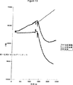
プロテイナーゼKを検出ウェルおよび対照ウェルに添加して、12.5mg/mlの最終的なウェル濃度とした(PBS中に調製されたBoehringer Mannheim D-68298)。対照ウェルへのプロテイナーゼKの添加では、膜アドミタンス特性に著しい変化を生じなかった。ストレプトアビジンが結合したセンサー膜は、最小位相におけるアドミタンスの増加を示した(図11)。最小位相におけるアドミタンスの増加の量および速度は、試験溶液中に存在するプロテイナーゼKの量に関係しており、それゆえ試験溶液中の酵素活性を調べるために用いることができる。
実施例2:
DNA結合チャネルのDNアーゼ1切断
第一層:9.3nM リンカーグラミシジンB(Linker Gramicidin B)
1.1μM 膜間脂質D(Membrane Spanner Lipid D)
27.5nM 膜間脂質C(Membrane Spanner Lipid C)(図12)
37μM MAAD
75μM リンカー脂質A(Linker Lipid A)
第二層:エタノール中に14mM(DPE−PC:GDPE=7:3):ビオチニル化グラミシジンE=50000:1
クロム付着層(ガラス顕微鏡スライド上に200Å)上に新たに蒸着された金(1000Å)を備えた電極を室温で1時間第一層成分のエタノール溶液中に浸し、エタノールで濯ぎ、インピーダンスの測定に使用するまでエタノール下、4℃で貯蔵した。このスライドを作用電極の範囲を約16mm2に限定したテフロン被覆ウェルを収容したブロック中に固定した。
5μLの第二層を作用電極に添加した後、180μLのリン酸緩衝生理食塩水(10mM NaH2PO4、1mM KH2PO4、137mM NaCl、2.7mM KCl;PBS)を添加した。電極をPBSで4回洗浄した。これらの工程は室温で行われた。以下の全ての工程は30℃で行われた。ストレプトアビジンを全てのウェルに添加し(PBS中に0.01mg/mlを5μL)、ビオチニル化グラミシジンEと10−15分間反応させ、過剰の非結合のストレプトアビジンをPBSで洗浄し、DNAプローブF(200nM):DNAプローブG(PBS中に200nM)の1:1混合物5μLをセンサーウェルに添加した。DNA非特異的結合プローブH(PBS中に400nMを5μL)を対照ウェルに添加した。結合プローブHは対象の標的DNAに非相補的であるため、標的DNAは結合しない。前記プローブを10−15分間ストレプトアビジンと反応させ、過剰の非結合プローブをPBSで洗い流した。PBS中の100μLのDNA標的I(10nM)を各ウェルに添加した。DNA標的Iのセンサーウェルに対する結合により、最小位相におけるアドミタンスが減少したが、対照ウェルにおける膜アドミタンスでは著しい変化はなかった(図13)。15分後に、非結合DNA標的IをDNアーゼ1活性化バッファーで洗浄した。DNアーゼ1活性化バッファーは、50nM Tris.HCl、pH7.6、50nM NaCl、10nM MgCl2、10nM MnCl2、0.2mg/mL BSAからなる。DNアーゼ1をセンサーウェルと対照ウェルに添加した(20mM Tris.HClの50% w/vグリセロール溶液、pH7.6、1mM MgCl2中に1mg/mLを2μL)。DNアーゼ1の添加により、センサーウェルの最小位相におけるアドミタンスは増大したが、対照ウェルには顕著な変化が見られなかった(図14)。最小位相におけるアドミタンスの増加の量および速度は、試験溶液中に存在するDNアーゼ1の量と関係しており、それゆえ試験溶液中の酵素活性を調べるために用いることができる。
DNAプローブF:
ビオチンとDNAとの間に31原子ホスホラミダイト(phosphoramidite)リンカー基を備えた5’ビオチニル化リステリア(listeria)プローブDNA
DNAプローブG:
ビオチンとDNAとの間に13原子ホスホラミダイトリンカー基を備えた5’ビオチニル化コレラ毒素プローブDNA
DNA非特異的結合プローブH:
ビオチンとDNAとの間に31原子ホスホラミダイトリンカー基を備えた5’ビオチニル化15マーオリゴヌクレオチド、標的DNA配列のあらゆる部分に対して非相補的である。
DNA標的I:
19塩基リステリア配列、10塩基“スペーサー”および23塩基コレラ毒素配列を含む52塩基DNA
広く記載された本発明の精神もしくは範囲から離れることなく、特定の実施態様に示したように、多数の変更および/または修正を本発明に加えてもよいことは、当業者に理解されるところである。しかして、上記の実施態様は、あらゆる点において例示的なものであって、限定的なものではないと考えるべきである。The present invention relates to biosensors and methods involving the use of biosensors in detecting the presence of an enzyme by detecting enzyme activity.
Many proteins that can be used as immunodiagnostic analytes and disease markers have additional properties of enzyme activity, particularly protease activity. In addition, another class of proteins exhibits nuclease activity.
Prostate specific antigen (PSA), a diagnostic marker for prostate cancer, is an example of a protein that exhibits protease activity and belongs to a class of proteins known as serine proteases. Examples of other proteases that are important immunological diagnostic markers include blood clotting enzymes, elastase, cathepsin B.
There are also many industrially important enzymes such as subtilisin, papain and α-amylase.
Examples of important nucleases include restriction enzymes such as BamH1, HindIII, polymerases that act as nucleases under certain conditions, such as T4 DNA polymerase, reverse transcriptases that act as RNases under certain conditions, such as RNase H, And exo- and endo-nucleases such as S1 nuclease.
Current diagnostic tests use immunoassays to detect PSA (eg, many analyzers such as Abbott's AXsym, Boehringer Mannheim's Elecsys and CIBA-Corning's ACS-180, all of which are ELISA-based PSAs). With a test). These tests utilize antibodies generated against the PSA molecule, which recognize specific epitope sites within the protein molecule.
Variations of these techniques are described in International Patent Application No. PCT / AU95 / 00536. This reference discloses a series of substrates specifically cleaved by PSA. This reference also discloses an assay system for a protease such as PSA utilizing the activity of the protease. This assay system involves the use of a ligand that captures PSA followed by the use of a substrate for PSA.
The inventors of the present application have developed an apparatus and a method for detecting an enzyme utilizing the protease activity of a protein. These devices and methods involve the use of membrane-based biosensors. Information regarding these biosensors can be found in international patent applications PCT / AU88 / 00273, PCT / AU89 / 00352, PCT / AU90 / 00025, PCT / AU92 / 00132, PCT / AU93 / 00509, PCT / AU93 / 00620, PCT / AU94. / 00202 and PCT / AU95 / 00763. The disclosures of these applications are hereby incorporated by reference.
The present invention includes providing a substrate for the enzyme to be detected, as well as detecting degradation of the substrate by the enzyme. This can be accomplished in a number of ways, for example, by decomposing a substrate to remove a group from the ionophore and releasing the ionophore, causing the ionophore to diffuse laterally within the membrane, or simply “steric” hindrance. The decrease may cause an increase in the ability of ions to pass through the ionophore. Alternatively, if the substrate is attached to a membrane spanning component, the ionophore may diffuse laterally through the membrane by causing the release of the ionophore by decomposition of the substrate. Obviously, this can also be achieved by degradation of the substrate attached to both the ionophore and the intermembrane component.
In another modification, degradation of the substrate causes the release of the ionophore containing the probe, which then inserts itself into the membrane.
Accordingly, a first aspect of the invention is a biosensor for use in detecting the presence of an enzyme in a sample, the biosensor comprising a means for detecting the membrane and the impedance of the membrane, The membrane includes an ionophore, and a linker is attached to the ionophore, and the linker can be cleaved by the enzyme to be detected, and the cleavage of the linker causes a change in the ability of ions to permeate through the ionophore. It is.
In a preferred embodiment of the invention, the linker is attached to the membrane so that the ionophore cannot diffuse laterally within the membrane. The linker is preferably attached to an intermembrane component provided on the membrane. This attachment can be done in many ways, such as covalent bonding, but it is currently preferred to attach by providing one of the ligand-binding pairs on both the linker and the transmembrane component. A preferred ligand binding pair is biotin streptavidin. In another preferred modification, both the transmembrane component and the linker have a moiety attached to the same molecule, for example biotin is provided on both the intermembrane component and the linker and is cross-linked via streptavidin. It is.
The part of the transmembrane component may be attached via a linker. This may be the same linker as provided for the ionophore or it may be different.
In a further preferred embodiment, the first and second layers of a closely packed array of amphiphilic molecules, comprising a plurality of ionophores and a plurality of intermembrane lipids that cannot diffuse laterally in the membrane, The ionophore includes a first intermembrane monomer and a second intermembrane monomer, wherein the first intermembrane monomer is provided in the first layer and the second intermembrane monomer is provided in the second layer. The first transmembrane monomer cannot diffuse laterally in the first layer and the second transmembrane monomer is bound to the transmembrane lipid via a linker. After enzymatic cleavage of the linker, the second inter-membrane monomer can diffuse laterally in a second layer that is separate from the first half-membrane monomer.
In a second aspect of the invention, a biosensor for use in detecting the presence of an enzyme in a sample, the biosensor comprising a membrane and means for measuring the impedance of the membrane, the membrane Comprises a plurality of ionophores and a plurality of intermembrane components, a linker molecule is attached to the intermembrane components, the ionophore is linked to the linker molecule, and the linker molecule can be cleaved by the enzyme to be detected. The cleavage of the linker molecule causes a change in the ability of ions to permeate through the ionophore.
In a preferred embodiment, the membrane comprises first and second layers of closely packed rows of amphiphilic molecules, and the intermembrane component cannot diffuse laterally within the membrane. Preferably, the ionophore comprises a first intermembrane monomer and a second intermembrane monomer, the first intermembrane monomer is provided in the first layer, the second intermembrane monomer is provided in the second layer, One-half membrane monomer cannot diffuse laterally in the first layer. The second transmembrane monomer is linked to the transmembrane component via a linker molecule.
The ionophore in both these embodiments is preferably gramicidin or an analogue thereof.
A series of enzymes can be detected using the biosensor of the present invention, but this biosensor can be used in particular for the detection of proteases, in particular clinically important proteases such as PSA, fibrinogen.
A third aspect of the present invention comprises a first region and a second region, and means for adding a sample suspected of containing an enzyme to the first region, wherein the first region is mediated by a linker that can be cleaved by the enzyme. A biosensor for the detection of an enzyme comprising a probe coupled to a carrier and a means for passing an unbound probe from the first region to the second region, wherein the second region comprises a membrane and the impedance of the membrane , And its impedance depends on the presence or absence of the probe.
In a preferred embodiment of this aspect of the invention, the membrane comprises a first and second layer of closely packed rows of amphiphilic molecules, and a first intermembrane monomer and a second intermembrane monomer. A plurality of ionophores, wherein the first intermembrane monomer is provided in the first layer, the second intermembrane monomer is provided in the second layer, and the second intermembrane monomer is the first intermembrane monomer. Can diffuse laterally in an independent second layer, the first interstitial monomer cannot diffuse laterally in the first layer, and a ligand is provided at least in the second interstitial monomer. The ligand is reactive with the probe or a portion thereof, and binding of the probe to the ligand causes a change in the relationship between the first and second half-membrane monomers, Pass the membrane through the ionophore Ion flows or impeded.
In a preferred embodiment, the probe comprises streptavidin and the ligand comprises biotin.
In yet another preferred embodiment, the probe includes an ionophore, such that when the probe contacts the membrane, the ionophore inserts itself into the membrane and changes the impedance of the membrane. As an example of such a modification, the probe may include valinomycin that inserts itself into the membrane.
In a preferred embodiment of the invention, the enzyme to be detected is a protease, in particular a prostate specific antigen. In this case, the linker or linker molecule preferably comprises the sequence Ala-Val-Tyr.
As will be appreciated by those skilled in the art, the actual linker used will depend on the enzyme to be detected. Examples of certain enzymes and corresponding substrates are described in Analytical Biochemistry: 220, 238-243 (1994) by Whittaker et al., The disclosure of which is included as a cross-reference.
In yet another aspect, the invention includes adding a sample to the biosensor of the first, second or third aspect of the invention and measuring a change in membrane impedance. A method for detecting the presence of an enzyme.
Obviously, the biosensor and method of the present invention detects only the active enzyme, not the entire enzyme. This is important because in many situations, the focus is on the amount of active enzyme present, not just the total amount of enzyme present as measured in a standard sandwich ELISA. is there.
It is also clear that the sensor of the present invention can be used to detect a wide range of enzymes. These enzymes include nucleases, proteases, amylases and the like. This sensor is adapted to the particular enzyme to be detected by regulating the production of the linker. For example, to detect a protease, the linker generally includes a peptide site that is cleaved by the enzyme. Information about peptide sequences cleaved by specific proteases is disclosed in the above-referenced Whittaker et al. If the enzyme to be detected is a nuclease, the linker generally comprises a nucleic acid sequence. Information on specific sequences cleaved by specific enzymes is disclosed in "Current Protocols in Molecular Biology" Ausebel et al. (1987) John Wiley & Sons, NY.
The sensor of the present invention may be used in drug development to determine DNA-drug binding sites. This sensor can also be used to determine DNA-protein binding sites. This sensor may also be used when diagnosing infection. For example, it can be used to detect enzyme activity specifically associated with a pathogen.
Industrially and clinically relevant proteases and substrates include serine proteases including thrombin and PSA. A list of lytic enzymes is disclosed in “Specificity of Proteolysis” Borivoj Keil (1992) Springer Verlag NY pp.283-323. Beneficial are serine proteases and cysteine proteases. See also "Proteolytic Enzymes" a Practical Approach RJBenyon & JSBond (eds) 1989 Oxford University Press NY p232, pp.241-249. Commercially important proteases and protease inhibitors suitable for the technology of the present invention are available in serine, cysteine, aspartic acid and metallo types. Serine proteases include endoproteinase-Arg-C, -Glu-C, Lys-C, factor Xa, proteinase K, subtilisin and trypsin, and exopeptidase acylamino acid releasing enzyme, carboxypeptidase P, and carboxy Contains peptidase Y. Cysteine proteases include endopeptidase bromelain, cathepsin B, clostripain, papain and exopeptidases cathepsin C and pyroglutamate aminopeptidase. Aspartic proteases include endopeptidases cathepsin D and pepsin. Metallo proteases include endopeptidase thermolysin and exopeptidase aminopeptidase M, carboxypeptidase-A, -B and leucine aminopeptidase. This list is not exclusive and shows the wide utility of the present invention. Other commercially available proteases are listed in the previously cited literature and are hereby incorporated by reference. For example, the known type of endopeptide endoproteinase-Asp-N is also included.
In order that the nature of the present invention may be more clearly understood, preferred forms thereof will now be described with reference to the following examples and the accompanying drawings.
FIG. 1 shows a schematic depiction of an embodiment of the apparatus of the third aspect of the present invention. As can be seen from this figure, the device 10 includes a first region 11 and a second region 12. The first region 11 is provided with polymer beads 13 (carriers) bonded to streptavidin 14 (probes) via peptide linkers 15. This peptide linker 15 is cleaved by protease 16.
As shown in this figure, the addition of protease (or nuclease) 16 releases streptavidin 14 and flows to the second region 12. The second region 12 includes a biosensor membrane 17 that detects the presence of streptavidin 14. The streptavidin 14 that has reached the biosensor membrane 17 causes a change in the impedance of the membrane.
FIG. 2 shows an embodiment of the first and / or second aspect of the present invention. As shown in FIG. 2, the biosensor film 20 includes a film 21 and an electrode 22. The membrane 21 comprises a first layer 23 and a second layer 24 of a row of amphiphilic molecules. The layer 24 includes a first inter-half membrane monomer 25 that cannot diffuse laterally within the membrane. Layer 23 includes a second intersemiconductor monomer 26. This membrane also includes intermembrane lipids 27 that cannot diffuse laterally within the membrane. The second transmembrane monomer 26 is linked to an intermembrane lipid 27 via a peptide 28. Peptide 28 is cleaved by protease 29. As the peptide 28 is cleaved by the protease 29, the interstitial monomer 26 freely diffuses laterally in the membrane. This results in a change in the membrane impedance.
Examples Example 1:
Protease cleavage first layer of streptavidin-gramicidin bond: 9.3 nM Linker Gramicidin B (FIG. 3)
1.1 μM Membrane Spanner Lipid D (Figure 4)
37 μM MAAD (Figure 5)
75 μM Linker Lipid A (Figure 6)
Second layer: 10 mM in ethanol (DPE-PC (FIG. 7): GDPE (FIG. 8) = 7: 3): Biotinylated Gramicidin E (FIG. 9) = 66677: 1
An electrode with gold (1000 mm) newly deposited on a chromium adhesion layer (200 mm on a glass microscope slide) is immersed in the ethanol solution of the first layer component for 1 hour at room temperature, rinsed with ethanol, and used for impedance measurement. Stored at 4 ° C. under ethanol until use. The slide was fixed in a block containing a Teflon-coated well with a working electrode range limited to about 16 mm 2 .
After adding 5 μL of the second layer to the working electrode, 150 μL of phosphate buffered saline (6.26 mM NaCl, 59.4 mM NaH 2 PO 4 .2H 2 O, 2.53 mM Na 2 HPO 4 .12H 2 O 50 mM EDTA pH 7.4; PBS). The electrode was washed 4 times with PBS, and the temperature was raised to 60 ° C. over 30 minutes. Streptavidin was added to the sensor wells (5 μL of 0.01 mg / ml in PBS) and incubated. Binding of streptavidin to biotinylated gramicidin E reduced admittance at the minimum phase (FIG. 10). After 15 minutes, excess streptavidin was washed with PBS. Wells without added streptavidin were used as controls.
Proteinase K was added to the detection and control wells to give a final well concentration of 12.5 mg / ml (Boehringer Mannheim D-68298 prepared in PBS). Addition of proteinase K to control wells did not cause significant changes in membrane admittance properties. The sensor membrane bound with streptavidin showed an increase in admittance at the minimum phase (FIG. 11). The amount and rate of admittance increase at the minimum phase is related to the amount of proteinase K present in the test solution and can therefore be used to examine enzyme activity in the test solution.
Example 2:
DNase 1 cleavage first layer of DNA binding channel: 9.3 nM Linker Gramicidin B
1.1μM Membrane Spanner Lipid D
27.5 nM Membrane Spanner Lipid C (Figure 12)
37μM MAAD
75μM Linker Lipid A
Second layer: 14 mM in ethanol (DPE-PC: GDPE = 7: 3): biotinylated gramicidin E = 50000: 1
An electrode with gold (1000 mm) newly deposited on a chromium adhesion layer (200 mm on a glass microscope slide) is immersed in the ethanol solution of the first layer component for 1 hour at room temperature, rinsed with ethanol, and used for impedance measurement. Stored at 4 ° C. under ethanol until use. The slide was fixed in a block containing a Teflon-coated well with a working electrode range limited to about 16 mm 2 .
5 μL of the second layer was added to the working electrode followed by 180 μL of phosphate buffered saline (10 mM NaH 2 PO 4 , 1 mM KH 2 PO 4 , 137 mM NaCl, 2.7 mM KCl; PBS). The electrode was washed 4 times with PBS. These steps were performed at room temperature. All the following steps were performed at 30 ° C. Streptavidin is added to all wells (0.01 μl / ml in PBS, 5 μL), reacted with biotinylated gramicidin E for 10-15 minutes, excess unbound streptavidin is washed with PBS, DNA probe F (200 nM): 5 μL of a 1: 1 mixture of DNA probe G (200 nM in PBS) was added to the sensor wells. Non-DNA specific binding probe H (5 μL of 400 nM in PBS) was added to control wells. Since the binding probe H is non-complementary to the target DNA of interest, the target DNA does not bind. The probe was reacted with streptavidin for 10-15 minutes and excess unbound probe was washed away with PBS. 100 μL of DNA target I (10 nM) in PBS was added to each well. Binding of DNA target I to the sensor well reduced admittance at the minimum phase, but there was no significant change in membrane admittance in the control well (FIG. 13). After 15 minutes, unbound DNA target I was washed with DNase 1 activation buffer. DNase 1 activation buffer is 50 nM Tris. It consists of HCl, pH 7.6, 50 nM NaCl, 10 nM MgCl 2 , 10 nM MnCl 2 , 0.2 mg / mL BSA. DNase 1 was added to the sensor and control wells ( 2 μL of 1 mg / mL in 20 mM Tris.HCl in 50% w / v glycerol, pH 7.6, 1 mM MgCl 2 ). Addition of DNase 1 increased the admittance at the minimum phase of the sensor well, but no significant change was seen in the control well (FIG. 14). The amount and rate of admittance increase at the minimum phase is related to the amount of DNase 1 present in the test solution and can therefore be used to examine enzyme activity in the test solution.
DNA probe F:
5 'biotinylated listeria probe DNA with a 31 atom phosphoramidite linker group between biotin and DNA
DNA probe G:
5 'biotinylated cholera toxin probe DNA with a 13 atom phosphoramidite linker group between biotin and DNA
DNA non-specific binding probe H:
5 'biotinylated 15-mer oligonucleotide with a 31 atom phosphoramidite linker group between biotin and DNA, non-complementary to any part of the target DNA sequence.
DNA target I:
52 base DNA containing 19 base listeria sequence, 10 base "spacer" and 23 base cholera toxin sequence
It will be appreciated by those skilled in the art that numerous changes and / or modifications may be made to the invention as set forth in the specific embodiments without departing from the spirit or scope of the invention as broadly described. is there. Thus, the above embodiments are to be considered in all respects only as illustrative and not restrictive.