JP2023055865A - Luterial and method for isolating and culturing same - Google Patents
Luterial and method for isolating and culturing same Download PDFInfo
- Publication number
- JP2023055865A JP2023055865A JP2023014621A JP2023014621A JP2023055865A JP 2023055865 A JP2023055865 A JP 2023055865A JP 2023014621 A JP2023014621 A JP 2023014621A JP 2023014621 A JP2023014621 A JP 2023014621A JP 2023055865 A JP2023055865 A JP 2023055865A
- Authority
- JP
- Japan
- Prior art keywords
- rutherial
- derived
- lutherial
- blood
- size
- Prior art date
- Legal status (The legal status is an assumption and is not a legal conclusion. Google has not performed a legal analysis and makes no representation as to the accuracy of the status listed.)
- Pending
Links
- 238000000034 method Methods 0.000 title claims abstract description 60
- 238000012258 culturing Methods 0.000 title claims description 17
- 210000004369 blood Anatomy 0.000 claims abstract description 59
- 239000008280 blood Substances 0.000 claims abstract description 59
- 108090000623 proteins and genes Proteins 0.000 claims abstract description 42
- 239000000126 substance Substances 0.000 claims abstract description 36
- 239000006228 supernatant Substances 0.000 claims abstract description 30
- 210000001124 body fluid Anatomy 0.000 claims abstract description 27
- 239000002245 particle Substances 0.000 claims abstract description 19
- 239000010839 body fluid Substances 0.000 claims abstract description 17
- 241000192128 Gammaproteobacteria Species 0.000 claims abstract description 15
- 241001135755 Betaproteobacteria Species 0.000 claims abstract description 14
- 238000006243 chemical reaction Methods 0.000 claims abstract description 13
- DPKHZNPWBDQZCN-UHFFFAOYSA-N acridine orange free base Chemical compound C1=CC(N(C)C)=CC2=NC3=CC(N(C)C)=CC=C3C=C21 DPKHZNPWBDQZCN-UHFFFAOYSA-N 0.000 claims abstract description 12
- DZBUGLKDJFMEHC-UHFFFAOYSA-N benzoquinolinylidene Natural products C1=CC=CC2=CC3=CC=CC=C3N=C21 DZBUGLKDJFMEHC-UHFFFAOYSA-N 0.000 claims abstract description 12
- 238000001914 filtration Methods 0.000 claims abstract description 12
- 241001493533 Streptophyta Species 0.000 claims abstract description 11
- 230000014509 gene expression Effects 0.000 claims abstract description 11
- TUFFYSFVSYUHPA-UHFFFAOYSA-M rhodamine 123 Chemical compound [Cl-].COC(=O)C1=CC=CC=C1C1=C(C=CC(N)=C2)C2=[O+]C2=C1C=CC(N)=C2 TUFFYSFVSYUHPA-UHFFFAOYSA-M 0.000 claims abstract description 11
- XXACTDWGHQXLGW-UHFFFAOYSA-M Janus Green B chloride Chemical compound [Cl-].C12=CC(N(CC)CC)=CC=C2N=C2C=CC(\N=N\C=3C=CC(=CC=3)N(C)C)=CC2=[N+]1C1=CC=CC=C1 XXACTDWGHQXLGW-UHFFFAOYSA-M 0.000 claims abstract description 10
- 238000010166 immunofluorescence Methods 0.000 claims abstract description 6
- 210000004027 cell Anatomy 0.000 claims description 43
- 238000000926 separation method Methods 0.000 claims description 29
- 238000005119 centrifugation Methods 0.000 claims description 28
- 210000001808 exosome Anatomy 0.000 claims description 25
- 230000004927 fusion Effects 0.000 claims description 23
- 210000003470 mitochondria Anatomy 0.000 claims description 19
- 210000000582 semen Anatomy 0.000 claims description 17
- 230000009172 bursting Effects 0.000 claims description 14
- 239000012530 fluid Substances 0.000 claims description 13
- 239000012528 membrane Substances 0.000 claims description 12
- 230000004992 fission Effects 0.000 claims description 11
- XLYOFNOQVPJJNP-UHFFFAOYSA-N water Substances O XLYOFNOQVPJJNP-UHFFFAOYSA-N 0.000 claims description 11
- 239000011148 porous material Substances 0.000 claims description 10
- 238000005406 washing Methods 0.000 claims description 10
- 239000000203 mixture Substances 0.000 claims description 8
- 241000124008 Mammalia Species 0.000 claims description 7
- 230000001678 irradiating effect Effects 0.000 claims description 7
- 230000001093 anti-cancer Effects 0.000 claims description 6
- 238000002955 isolation Methods 0.000 claims description 6
- 210000003296 saliva Anatomy 0.000 claims description 6
- 230000001464 adherent effect Effects 0.000 claims description 5
- 230000000968 intestinal effect Effects 0.000 claims description 5
- 230000003834 intracellular effect Effects 0.000 claims description 4
- 108700025694 p53 Genes Proteins 0.000 claims description 4
- 230000009711 regulatory function Effects 0.000 claims description 4
- 108091035539 telomere Proteins 0.000 claims description 4
- 102000055501 telomere Human genes 0.000 claims description 4
- 210000003411 telomere Anatomy 0.000 claims description 4
- FAPWRFPIFSIZLT-UHFFFAOYSA-M Sodium chloride Chemical compound [Na+].[Cl-] FAPWRFPIFSIZLT-UHFFFAOYSA-M 0.000 claims description 3
- 239000004480 active ingredient Substances 0.000 claims description 3
- 239000011780 sodium chloride Substances 0.000 claims description 3
- 239000011800 void material Substances 0.000 claims 1
- 201000010099 disease Diseases 0.000 abstract description 29
- 208000037265 diseases, disorders, signs and symptoms Diseases 0.000 abstract description 29
- 230000001747 exhibiting effect Effects 0.000 abstract description 4
- 230000001832 mitochondrialike Effects 0.000 abstract description 4
- 210000003527 eukaryotic cell Anatomy 0.000 abstract description 3
- 238000012360 testing method Methods 0.000 abstract description 3
- 239000002105 nanoparticle Substances 0.000 abstract 3
- 239000007788 liquid Substances 0.000 abstract 1
- 206010028980 Neoplasm Diseases 0.000 description 28
- 201000011510 cancer Diseases 0.000 description 24
- 108020004465 16S ribosomal RNA Proteins 0.000 description 21
- 108020004414 DNA Proteins 0.000 description 19
- 238000003745 diagnosis Methods 0.000 description 18
- 108091032973 (ribonucleotides)n+m Proteins 0.000 description 16
- 210000001772 blood platelet Anatomy 0.000 description 13
- 230000004899 motility Effects 0.000 description 12
- 230000005540 biological transmission Effects 0.000 description 11
- 239000000243 solution Substances 0.000 description 11
- 238000010186 staining Methods 0.000 description 10
- FWBHETKCLVMNFS-UHFFFAOYSA-N 4',6-Diamino-2-phenylindol Chemical compound C1=CC(C(=N)N)=CC=C1C1=CC2=CC=C(C(N)=N)C=C2N1 FWBHETKCLVMNFS-UHFFFAOYSA-N 0.000 description 9
- 241000894006 Bacteria Species 0.000 description 8
- 239000000872 buffer Substances 0.000 description 8
- 239000012634 fragment Substances 0.000 description 8
- 241000605059 Bacteroidetes Species 0.000 description 7
- 238000004458 analytical method Methods 0.000 description 7
- 210000003495 flagella Anatomy 0.000 description 7
- 241000192125 Firmicutes Species 0.000 description 6
- 238000012300 Sequence Analysis Methods 0.000 description 6
- 102100029338 Suppressor of SWI4 1 homolog Human genes 0.000 description 6
- 238000004393 prognosis Methods 0.000 description 6
- 210000001519 tissue Anatomy 0.000 description 6
- 230000002407 ATP formation Effects 0.000 description 5
- 241000206602 Eukaryota Species 0.000 description 5
- 108091092584 GDNA Proteins 0.000 description 5
- 241001465754 Metazoa Species 0.000 description 5
- 230000002159 abnormal effect Effects 0.000 description 5
- 238000001574 biopsy Methods 0.000 description 5
- 238000011161 development Methods 0.000 description 5
- 230000018109 developmental process Effects 0.000 description 5
- 230000008569 process Effects 0.000 description 5
- IAZDPXIOMUYVGZ-UHFFFAOYSA-N Dimethylsulphoxide Chemical compound CS(C)=O IAZDPXIOMUYVGZ-UHFFFAOYSA-N 0.000 description 4
- LFQSCWFLJHTTHZ-UHFFFAOYSA-N Ethanol Chemical compound CCO LFQSCWFLJHTTHZ-UHFFFAOYSA-N 0.000 description 4
- 239000002246 antineoplastic agent Substances 0.000 description 4
- 238000012757 fluorescence staining Methods 0.000 description 4
- 230000000984 immunochemical effect Effects 0.000 description 4
- IKEOZQLIVHGQLJ-UHFFFAOYSA-M mitoTracker Red Chemical compound [Cl-].C1=CC(CCl)=CC=C1C(C1=CC=2CCCN3CCCC(C=23)=C1O1)=C2C1=C(CCC1)C3=[N+]1CCCC3=C2 IKEOZQLIVHGQLJ-UHFFFAOYSA-M 0.000 description 4
- VMGAPWLDMVPYIA-HIDZBRGKSA-N n'-amino-n-iminomethanimidamide Chemical compound N\N=C\N=N VMGAPWLDMVPYIA-HIDZBRGKSA-N 0.000 description 4
- 108020004707 nucleic acids Proteins 0.000 description 4
- 102000039446 nucleic acids Human genes 0.000 description 4
- 150000007523 nucleic acids Chemical class 0.000 description 4
- BOLDJAUMGUJJKM-LSDHHAIUSA-N renifolin D Natural products CC(=C)[C@@H]1Cc2c(O)c(O)ccc2[C@H]1CC(=O)c3ccc(O)cc3O BOLDJAUMGUJJKM-LSDHHAIUSA-N 0.000 description 4
- 239000000523 sample Substances 0.000 description 4
- 241001156739 Actinobacteria <phylum> Species 0.000 description 3
- 239000005089 Luciferase Substances 0.000 description 3
- 238000005481 NMR spectroscopy Methods 0.000 description 3
- 108091028043 Nucleic acid sequence Proteins 0.000 description 3
- 206010033128 Ovarian cancer Diseases 0.000 description 3
- 206010061535 Ovarian neoplasm Diseases 0.000 description 3
- 238000012136 culture method Methods 0.000 description 3
- 230000006870 function Effects 0.000 description 3
- 230000009368 gene silencing by RNA Effects 0.000 description 3
- 230000012010 growth Effects 0.000 description 3
- 230000000877 morphologic effect Effects 0.000 description 3
- 230000035772 mutation Effects 0.000 description 3
- 239000002773 nucleotide Substances 0.000 description 3
- 125000003729 nucleotide group Chemical group 0.000 description 3
- IJGRMHOSHXDMSA-UHFFFAOYSA-N Atomic nitrogen Chemical compound N#N IJGRMHOSHXDMSA-UHFFFAOYSA-N 0.000 description 2
- 241000251556 Chordata Species 0.000 description 2
- 206010009944 Colon cancer Diseases 0.000 description 2
- 208000001333 Colorectal Neoplasms Diseases 0.000 description 2
- 208000031942 Late Onset disease Diseases 0.000 description 2
- 241000192142 Proteobacteria Species 0.000 description 2
- 238000012228 RNA interference-mediated gene silencing Methods 0.000 description 2
- VYPSYNLAJGMNEJ-UHFFFAOYSA-N Silicium dioxide Chemical compound O=[Si]=O VYPSYNLAJGMNEJ-UHFFFAOYSA-N 0.000 description 2
- 241000700605 Viruses Species 0.000 description 2
- 230000002378 acidificating effect Effects 0.000 description 2
- 230000002776 aggregation Effects 0.000 description 2
- 238000004220 aggregation Methods 0.000 description 2
- 229940041181 antineoplastic drug Drugs 0.000 description 2
- YZXBAPSDXZZRGB-DOFZRALJSA-N arachidonic acid Chemical compound CCCCC\C=C/C\C=C/C\C=C/C\C=C/CCCC(O)=O YZXBAPSDXZZRGB-DOFZRALJSA-N 0.000 description 2
- 230000001580 bacterial effect Effects 0.000 description 2
- 239000000090 biomarker Substances 0.000 description 2
- 230000005907 cancer growth Effects 0.000 description 2
- 230000024245 cell differentiation Effects 0.000 description 2
- 230000010261 cell growth Effects 0.000 description 2
- 230000008859 change Effects 0.000 description 2
- 239000003795 chemical substances by application Substances 0.000 description 2
- DQLATGHUWYMOKM-UHFFFAOYSA-L cisplatin Chemical compound N[Pt](N)(Cl)Cl DQLATGHUWYMOKM-UHFFFAOYSA-L 0.000 description 2
- 229960004316 cisplatin Drugs 0.000 description 2
- 150000001875 compounds Chemical class 0.000 description 2
- 230000003013 cytotoxicity Effects 0.000 description 2
- 231100000135 cytotoxicity Toxicity 0.000 description 2
- 238000001514 detection method Methods 0.000 description 2
- 238000010586 diagram Methods 0.000 description 2
- 229940079593 drug Drugs 0.000 description 2
- 239000003814 drug Substances 0.000 description 2
- 230000000694 effects Effects 0.000 description 2
- 230000037149 energy metabolism Effects 0.000 description 2
- 210000003743 erythrocyte Anatomy 0.000 description 2
- 230000005284 excitation Effects 0.000 description 2
- 238000002474 experimental method Methods 0.000 description 2
- XHEFDIBZLJXQHF-UHFFFAOYSA-N fisetin Chemical compound C=1C(O)=CC=C(C(C=2O)=O)C=1OC=2C1=CC=C(O)C(O)=C1 XHEFDIBZLJXQHF-UHFFFAOYSA-N 0.000 description 2
- 238000003384 imaging method Methods 0.000 description 2
- 230000005764 inhibitory process Effects 0.000 description 2
- 230000033001 locomotion Effects 0.000 description 2
- 238000004519 manufacturing process Methods 0.000 description 2
- 238000005259 measurement Methods 0.000 description 2
- 239000002609 medium Substances 0.000 description 2
- 230000031864 metaphase Effects 0.000 description 2
- 230000000394 mitotic effect Effects 0.000 description 2
- 239000013641 positive control Substances 0.000 description 2
- 239000000047 product Substances 0.000 description 2
- 210000001236 prokaryotic cell Anatomy 0.000 description 2
- 102000004169 proteins and genes Human genes 0.000 description 2
- 238000011002 quantification Methods 0.000 description 2
- 210000003705 ribosome Anatomy 0.000 description 2
- 230000004083 survival effect Effects 0.000 description 2
- 230000001225 therapeutic effect Effects 0.000 description 2
- 238000012546 transfer Methods 0.000 description 2
- AZKSAVLVSZKNRD-UHFFFAOYSA-M 3-(4,5-dimethylthiazol-2-yl)-2,5-diphenyltetrazolium bromide Chemical compound [Br-].S1C(C)=C(C)N=C1[N+]1=NC(C=2C=CC=CC=2)=NN1C1=CC=CC=C1 AZKSAVLVSZKNRD-UHFFFAOYSA-M 0.000 description 1
- 241000580482 Acidobacteria Species 0.000 description 1
- 241001185327 Acidobacteriales Species 0.000 description 1
- 241001135756 Alphaproteobacteria Species 0.000 description 1
- 235000003197 Byrsonima crassifolia Nutrition 0.000 description 1
- 240000001546 Byrsonima crassifolia Species 0.000 description 1
- OKTJSMMVPCPJKN-UHFFFAOYSA-N Carbon Chemical compound [C] OKTJSMMVPCPJKN-UHFFFAOYSA-N 0.000 description 1
- 102000001327 Chemokine CCL5 Human genes 0.000 description 1
- 108010055166 Chemokine CCL5 Proteins 0.000 description 1
- 241000192700 Cyanobacteria Species 0.000 description 1
- 230000004568 DNA-binding Effects 0.000 description 1
- 101710088194 Dehydrogenase Proteins 0.000 description 1
- 102000016911 Deoxyribonucleases Human genes 0.000 description 1
- 108010053770 Deoxyribonucleases Proteins 0.000 description 1
- 241000196324 Embryophyta Species 0.000 description 1
- WQZGKKKJIJFFOK-GASJEMHNSA-N Glucose Natural products OC[C@H]1OC(O)[C@H](O)[C@@H](O)[C@@H]1O WQZGKKKJIJFFOK-GASJEMHNSA-N 0.000 description 1
- 102100031181 Glyceraldehyde-3-phosphate dehydrogenase Human genes 0.000 description 1
- DGAQECJNVWCQMB-PUAWFVPOSA-M Ilexoside XXIX Chemical compound C[C@@H]1CC[C@@]2(CC[C@@]3(C(=CC[C@H]4[C@]3(CC[C@@H]5[C@@]4(CC[C@@H](C5(C)C)OS(=O)(=O)[O-])C)C)[C@@H]2[C@]1(C)O)C)C(=O)O[C@H]6[C@@H]([C@H]([C@@H]([C@H](O6)CO)O)O)O.[Na+] DGAQECJNVWCQMB-PUAWFVPOSA-M 0.000 description 1
- 108060001084 Luciferase Proteins 0.000 description 1
- 108010052285 Membrane Proteins Proteins 0.000 description 1
- 102000018697 Membrane Proteins Human genes 0.000 description 1
- 206010027476 Metastases Diseases 0.000 description 1
- -1 Mito-tracker Chemical compound 0.000 description 1
- 238000012408 PCR amplification Methods 0.000 description 1
- 229910019142 PO4 Inorganic materials 0.000 description 1
- 108091030071 RNAI Proteins 0.000 description 1
- 102000006382 Ribonucleases Human genes 0.000 description 1
- 108010083644 Ribonucleases Proteins 0.000 description 1
- 241001178521 Sarotherodon caroli Species 0.000 description 1
- 101710144764 Suppressor of SWI4 1 homolog Proteins 0.000 description 1
- 238000002835 absorbance Methods 0.000 description 1
- 239000008186 active pharmaceutical agent Substances 0.000 description 1
- 238000000246 agarose gel electrophoresis Methods 0.000 description 1
- 230000004931 aggregating effect Effects 0.000 description 1
- 239000000427 antigen Substances 0.000 description 1
- 102000036639 antigens Human genes 0.000 description 1
- 108091007433 antigens Proteins 0.000 description 1
- 229940114079 arachidonic acid Drugs 0.000 description 1
- 235000021342 arachidonic acid Nutrition 0.000 description 1
- QVGXLLKOCUKJST-UHFFFAOYSA-N atomic oxygen Chemical compound [O] QVGXLLKOCUKJST-UHFFFAOYSA-N 0.000 description 1
- 230000000975 bioactive effect Effects 0.000 description 1
- 230000004071 biological effect Effects 0.000 description 1
- 230000033228 biological regulation Effects 0.000 description 1
- 239000012472 biological sample Substances 0.000 description 1
- 230000015572 biosynthetic process Effects 0.000 description 1
- 210000001185 bone marrow Anatomy 0.000 description 1
- 210000004958 brain cell Anatomy 0.000 description 1
- 239000007975 buffered saline Substances 0.000 description 1
- 239000008366 buffered solution Substances 0.000 description 1
- 229910052799 carbon Inorganic materials 0.000 description 1
- 230000015556 catabolic process Effects 0.000 description 1
- 230000022131 cell cycle Effects 0.000 description 1
- 230000030833 cell death Effects 0.000 description 1
- 230000003915 cell function Effects 0.000 description 1
- 230000004663 cell proliferation Effects 0.000 description 1
- 230000003833 cell viability Effects 0.000 description 1
- 239000003153 chemical reaction reagent Substances 0.000 description 1
- 230000004186 co-expression Effects 0.000 description 1
- 230000015271 coagulation Effects 0.000 description 1
- 238000005345 coagulation Methods 0.000 description 1
- 210000003022 colostrum Anatomy 0.000 description 1
- 235000021277 colostrum Nutrition 0.000 description 1
- 239000002131 composite material Substances 0.000 description 1
- 238000004624 confocal microscopy Methods 0.000 description 1
- 239000002872 contrast media Substances 0.000 description 1
- 238000001816 cooling Methods 0.000 description 1
- 239000012531 culture fluid Substances 0.000 description 1
- 238000001446 dark-field microscopy Methods 0.000 description 1
- 238000006731 degradation reaction Methods 0.000 description 1
- 230000001419 dependent effect Effects 0.000 description 1
- 230000001079 digestive effect Effects 0.000 description 1
- 238000007865 diluting Methods 0.000 description 1
- 239000012153 distilled water Substances 0.000 description 1
- 238000004043 dyeing Methods 0.000 description 1
- 238000013399 early diagnosis Methods 0.000 description 1
- 238000000635 electron micrograph Methods 0.000 description 1
- 238000000605 extraction Methods 0.000 description 1
- 210000004700 fetal blood Anatomy 0.000 description 1
- 235000011990 fisetin Nutrition 0.000 description 1
- 239000007850 fluorescent dye Substances 0.000 description 1
- 235000013305 food Nutrition 0.000 description 1
- 235000011389 fruit/vegetable juice Nutrition 0.000 description 1
- 231100000089 gene mutation induction Toxicity 0.000 description 1
- 239000008103 glucose Substances 0.000 description 1
- 108020004445 glyceraldehyde-3-phosphate dehydrogenase Proteins 0.000 description 1
- 230000009422 growth inhibiting effect Effects 0.000 description 1
- 239000001963 growth medium Substances 0.000 description 1
- YQOKLYTXVFAUCW-UHFFFAOYSA-N guanidine;isothiocyanic acid Chemical compound N=C=S.NC(N)=N YQOKLYTXVFAUCW-UHFFFAOYSA-N 0.000 description 1
- 230000013632 homeostatic process Effects 0.000 description 1
- 238000000265 homogenisation Methods 0.000 description 1
- 235000020256 human milk Nutrition 0.000 description 1
- 210000004251 human milk Anatomy 0.000 description 1
- 210000000987 immune system Anatomy 0.000 description 1
- 238000001727 in vivo Methods 0.000 description 1
- 238000011534 incubation Methods 0.000 description 1
- 231100000405 induce cancer Toxicity 0.000 description 1
- 230000001788 irregular Effects 0.000 description 1
- 210000000265 leukocyte Anatomy 0.000 description 1
- 150000002632 lipids Chemical class 0.000 description 1
- 230000007774 longterm Effects 0.000 description 1
- 210000001365 lymphatic vessel Anatomy 0.000 description 1
- 239000003550 marker Substances 0.000 description 1
- 239000000463 material Substances 0.000 description 1
- 239000011159 matrix material Substances 0.000 description 1
- 108020004999 messenger RNA Proteins 0.000 description 1
- 230000002503 metabolic effect Effects 0.000 description 1
- 230000009401 metastasis Effects 0.000 description 1
- 230000000813 microbial effect Effects 0.000 description 1
- 238000001000 micrograph Methods 0.000 description 1
- 230000002438 mitochondrial effect Effects 0.000 description 1
- 230000004660 morphological change Effects 0.000 description 1
- 229910052757 nitrogen Inorganic materials 0.000 description 1
- 208000002154 non-small cell lung carcinoma Diseases 0.000 description 1
- 230000002611 ovarian Effects 0.000 description 1
- 229910052760 oxygen Inorganic materials 0.000 description 1
- 239000001301 oxygen Substances 0.000 description 1
- 239000008188 pellet Substances 0.000 description 1
- NBIIXXVUZAFLBC-UHFFFAOYSA-K phosphate Chemical compound [O-]P([O-])([O-])=O NBIIXXVUZAFLBC-UHFFFAOYSA-K 0.000 description 1
- 239000010452 phosphate Substances 0.000 description 1
- 239000003755 preservative agent Substances 0.000 description 1
- 230000000069 prophylactic effect Effects 0.000 description 1
- 230000006920 protein precipitation Effects 0.000 description 1
- 230000029058 respiratory gaseous exchange Effects 0.000 description 1
- 229920002477 rna polymer Polymers 0.000 description 1
- 238000002133 sample digestion Methods 0.000 description 1
- 238000006748 scratching Methods 0.000 description 1
- 230000002393 scratching effect Effects 0.000 description 1
- 238000012163 sequencing technique Methods 0.000 description 1
- 230000011664 signaling Effects 0.000 description 1
- 239000000377 silicon dioxide Substances 0.000 description 1
- 229910052708 sodium Inorganic materials 0.000 description 1
- 239000011734 sodium Substances 0.000 description 1
- 239000012064 sodium phosphate buffer Substances 0.000 description 1
- 210000000278 spinal cord Anatomy 0.000 description 1
- 230000002269 spontaneous effect Effects 0.000 description 1
- 238000007447 staining method Methods 0.000 description 1
- 238000007619 statistical method Methods 0.000 description 1
- 210000000130 stem cell Anatomy 0.000 description 1
- 238000003860 storage Methods 0.000 description 1
- 239000000758 substrate Substances 0.000 description 1
- 239000013076 target substance Substances 0.000 description 1
- 125000003831 tetrazolyl group Chemical group 0.000 description 1
- 239000000439 tumor marker Substances 0.000 description 1
- 208000029729 tumor suppressor gene on chromosome 11 Diseases 0.000 description 1
- DGVVWUTYPXICAM-UHFFFAOYSA-N β‐Mercaptoethanol Chemical compound OCCS DGVVWUTYPXICAM-UHFFFAOYSA-N 0.000 description 1
Images


Classifications
-
- A—HUMAN NECESSITIES
- A61—MEDICAL OR VETERINARY SCIENCE; HYGIENE
- A61K—PREPARATIONS FOR MEDICAL, DENTAL OR TOILETRY PURPOSES
- A61K35/00—Medicinal preparations containing materials or reaction products thereof with undetermined constitution
- A61K35/12—Materials from mammals; Compositions comprising non-specified tissues or cells; Compositions comprising non-embryonic stem cells; Genetically modified cells
- A61K35/14—Blood; Artificial blood
- A61K35/19—Platelets; Megacaryocytes
-
- A—HUMAN NECESSITIES
- A61—MEDICAL OR VETERINARY SCIENCE; HYGIENE
- A61K—PREPARATIONS FOR MEDICAL, DENTAL OR TOILETRY PURPOSES
- A61K35/00—Medicinal preparations containing materials or reaction products thereof with undetermined constitution
- A61K35/12—Materials from mammals; Compositions comprising non-specified tissues or cells; Compositions comprising non-embryonic stem cells; Genetically modified cells
-
- A—HUMAN NECESSITIES
- A61—MEDICAL OR VETERINARY SCIENCE; HYGIENE
- A61P—SPECIFIC THERAPEUTIC ACTIVITY OF CHEMICAL COMPOUNDS OR MEDICINAL PREPARATIONS
- A61P35/00—Antineoplastic agents
-
- C—CHEMISTRY; METALLURGY
- C12—BIOCHEMISTRY; BEER; SPIRITS; WINE; VINEGAR; MICROBIOLOGY; ENZYMOLOGY; MUTATION OR GENETIC ENGINEERING
- C12N—MICROORGANISMS OR ENZYMES; COMPOSITIONS THEREOF; PROPAGATING, PRESERVING, OR MAINTAINING MICROORGANISMS; MUTATION OR GENETIC ENGINEERING; CULTURE MEDIA
- C12N13/00—Treatment of microorganisms or enzymes with electrical or wave energy, e.g. magnetism, sonic waves
-
- C—CHEMISTRY; METALLURGY
- C12—BIOCHEMISTRY; BEER; SPIRITS; WINE; VINEGAR; MICROBIOLOGY; ENZYMOLOGY; MUTATION OR GENETIC ENGINEERING
- C12N—MICROORGANISMS OR ENZYMES; COMPOSITIONS THEREOF; PROPAGATING, PRESERVING, OR MAINTAINING MICROORGANISMS; MUTATION OR GENETIC ENGINEERING; CULTURE MEDIA
- C12N5/00—Undifferentiated human, animal or plant cells, e.g. cell lines; Tissues; Cultivation or maintenance thereof; Culture media therefor
- C12N5/06—Animal cells or tissues; Human cells or tissues
- C12N5/0602—Vertebrate cells
- C12N5/0634—Cells from the blood or the immune system
-
- C—CHEMISTRY; METALLURGY
- C12—BIOCHEMISTRY; BEER; SPIRITS; WINE; VINEGAR; MICROBIOLOGY; ENZYMOLOGY; MUTATION OR GENETIC ENGINEERING
- C12N—MICROORGANISMS OR ENZYMES; COMPOSITIONS THEREOF; PROPAGATING, PRESERVING, OR MAINTAINING MICROORGANISMS; MUTATION OR GENETIC ENGINEERING; CULTURE MEDIA
- C12N2509/00—Methods for the dissociation of cells, e.g. specific use of enzymes
-
- C—CHEMISTRY; METALLURGY
- C12—BIOCHEMISTRY; BEER; SPIRITS; WINE; VINEGAR; MICROBIOLOGY; ENZYMOLOGY; MUTATION OR GENETIC ENGINEERING
- C12N—MICROORGANISMS OR ENZYMES; COMPOSITIONS THEREOF; PROPAGATING, PRESERVING, OR MAINTAINING MICROORGANISMS; MUTATION OR GENETIC ENGINEERING; CULTURE MEDIA
- C12N2529/00—Culture process characterised by the use of electromagnetic stimulation
Landscapes
- Health & Medical Sciences (AREA)
- Life Sciences & Earth Sciences (AREA)
- Engineering & Computer Science (AREA)
- Biomedical Technology (AREA)
- Chemical & Material Sciences (AREA)
- Zoology (AREA)
- Biotechnology (AREA)
- Organic Chemistry (AREA)
- Wood Science & Technology (AREA)
- Genetics & Genomics (AREA)
- Bioinformatics & Cheminformatics (AREA)
- General Health & Medical Sciences (AREA)
- Cell Biology (AREA)
- Immunology (AREA)
- Hematology (AREA)
- General Engineering & Computer Science (AREA)
- Biochemistry (AREA)
- Microbiology (AREA)
- Veterinary Medicine (AREA)
- Public Health (AREA)
- Animal Behavior & Ethology (AREA)
- Pharmacology & Pharmacy (AREA)
- Medicinal Chemistry (AREA)
- Virology (AREA)
- Developmental Biology & Embryology (AREA)
- Epidemiology (AREA)
- Chemical Kinetics & Catalysis (AREA)
- General Chemical & Material Sciences (AREA)
- Nuclear Medicine, Radiotherapy & Molecular Imaging (AREA)
- Measuring Or Testing Involving Enzymes Or Micro-Organisms (AREA)
- Apparatus Associated With Microorganisms And Enzymes (AREA)
- Proteomics, Peptides & Aminoacids (AREA)
- Pathology (AREA)
- Physics & Mathematics (AREA)
- Analytical Chemistry (AREA)
- Biophysics (AREA)
- Molecular Biology (AREA)
- Micro-Organisms Or Cultivation Processes Thereof (AREA)
- Medicines Containing Material From Animals Or Micro-Organisms (AREA)
- Investigating Or Analysing Biological Materials (AREA)
Abstract
Description
本発明は、体液由来のミトコンドリア類似微細物質であるルテリアル、並びにその分離
および培養方法に関する。
The present invention relates to rutherial, which is mitochondria-like minute substances derived from body fluids, and methods for isolating and culturing the same.
血液中の微小胞などの微細物質は、かつては、特別な機能を持っていない物質として認
識されてきた。しかし、微小胞(microvesicle)も様々な生物活性を有する
ということが、種々の実験データにより明らかになっている。例えば、血小板由来の微小
胞は、小胞状の表面タンパク質を介して特定細胞を刺激する機能をするということが明ら
かになっており(CD154,RANTES and/or PF‐4;Thromb.
Haemost.(1999)82:794;J.Boil.Chem.(1999)2
74:7545)、血小板の微小胞における生理活性脂質(例えば、HTETまたはアラ
キドン酸)が特定の標的細胞に対して特定効果を示すことが報告されている(J.Bio
l.Chem.(2001)276;19672;Cardiovasc.Res.(2
001)49(5):88)。このように、生物学的試料中に存在する小嚢などの物質の
特徴(例えば、サイズ、表面抗原、起源細胞の決定、ペイロッド)は、疾病の診断、予後
または治療診断に関する情報を提供することができるため、疾患を探索して治療するのに
用いられることができる生物学的指標を確認することに対する必要性が求められている。
そこで、小嚢に関わるRNAおよび他の生物学的指標と小嚢の特徴を、診断、予後、また
は治療診断に提供しようとする試みもあった(WO2011/127219参照)。
Microscopic substances such as microvesicles in blood were once recognized as substances with no special function. However, various experimental data have revealed that microvesicles also have various biological activities. For example, platelet-derived microvesicles have been shown to function through vesicular surface proteins to stimulate specific cells (CD154, RANTES and/or PF-4; Thromb.
Haemost. (1999) 82:794; Boil. Chem. (1999) 2
74:7545), bioactive lipids (e.g., HTET or arachidonic acid) in platelet microvesicles have been reported to exhibit specific effects on specific target cells (J. Bio
l. Chem. (2001) 276; 19672; Cardiovasc. Res. (2
001) 49(5):88). Thus, the characteristics (e.g. size, surface antigens, determination of cell of origin, payload) of substances such as vesicles present in a biological sample can provide information regarding disease diagnosis, prognosis or therapeutic diagnosis. Therefore, there is a need for identifying biological markers that can be used to explore and treat disease.
Thus, there have been attempts to provide vesicle-associated RNA and other biological markers and vesicle characteristics for diagnosis, prognosis, or therapeutic diagnosis (see WO2011/127219).
一方、癌(cancer)は、細胞が無限に増殖して正常の細胞の機能を妨害する疾病
であり、肺癌、胃癌(gastric cancer、GC)、乳癌(breast c
ancer、BRC)、大腸癌(colorectal cancer、CRC)などが
代表的であるが、実質には何れの組織でも発生し得る。初期の癌の診断は、癌細胞の成長
による生体組織の外的変化に基づいて行われたが、近年、血液、糖鎖(glyco ch
ain)、DNAなどの生物の組織または細胞に存在する微量の生体分子を用いた診断お
よび検出が試されている。しかし、最も一般的に用いられる癌の診断方法は、生体組織検
査により得られた組織サンプルを用いるか、映像を用いた診断である。このうち生体組織
検査は、患者に大きい苦痛を与え、費用が高いだけでなく、診断まで長時間がかかるとい
う欠点がある。また、患者が実際に癌にかかった場合、生体組織検査の過程中に癌の転移
が誘発される恐れがあり、生体組織検査により組織サンプルが得られない部位の場合、疑
われる組織を外科的な手術により摘出しないと、疾病の診断が不可能であるという欠点が
ある。また、映像を用いた診断では、エックス線(X‐ray)映像、疾病標的物質が付
着された造影剤を用いて得た核磁気共鳴(nuclear magnetic reso
nance、NMR)映像などに基づいて癌を判定していた。しかし、かかる映像による
診断は、臨床医または読影医の熟練度によって誤診の可能性があり、映像を得る機器の精
度に大きく依存するという欠点がある。さらに、最も高精度の機器であるとしても、数m
m以下の腫瘍は検出が不可能であり、発病初期段階では検出が難しいという欠点がある。
また、映像を得るためには、患者または疾病保有可能者が、遺伝子の突然変異が誘発し得
る高エネルギーの電磁気波に露出されるため、さらに他の疾病を引き起こす恐れがあるだ
けでなく、映像を用いた診断の回数が制限されるという欠点がある。
On the other hand, cancer is a disease in which cells proliferate indefinitely to interfere with normal cell functions.
Ancer (BRC), colorectal cancer (CRC) and the like are typical examples, but it can occur virtually in any tissue. Early diagnosis of cancer was based on the external changes in living tissue caused by the growth of cancer cells.
ain), diagnostics and detection using trace amounts of biomolecules present in the tissues or cells of organisms, such as DNA. However, the most commonly used method of diagnosing cancer is diagnosis using tissue samples obtained by biopsy or using imaging. Of these, the biopsy has the disadvantages of causing great pain to the patient, being expensive, and taking a long time to make a diagnosis. Also, if a patient does have cancer, the biopsy process may induce cancer metastasis, and if the biopsy does not yield a tissue sample, the suspected tissue should be removed surgically. It has the disadvantage that it is impossible to diagnose the disease without surgically removing it. In addition, in diagnosis using images, X-ray images, nuclear magnetic resonance (nuclear magnetic resonance) obtained using a contrast medium to which a disease target substance is attached are used.
(nance, NMR) images and the like were used to determine cancer. However, diagnosis using such images has the drawback that there is a possibility of misdiagnosis depending on the skill of the clinician or radiologist, and that it is highly dependent on the accuracy of the equipment used to obtain the images. Furthermore, even the most accurate instruments require several meters
Tumors of m or less cannot be detected, and there is a drawback that detection is difficult in the early stages of disease.
In addition, in order to obtain images, the patient or disease carrier is exposed to high-energy electromagnetic waves that can induce genetic mutations, which may cause other diseases. has the disadvantage of limiting the number of diagnoses using
すなわち、癌の診断のための生体組織検査は、多くの時間、費用、不便さ、苦痛などが
伴われるため、不要な生体組織検査の対象者数を著しく減少させることができる方法、癌
を早期に診断できる方法が求められている。
That is, biopsy for diagnosis of cancer is accompanied by a lot of time, cost, inconvenience, and pain. There is a need for a method that can diagnose
このような背景下で、本発明者らは、患者から既に排出された体液中に存在する微細物
質の特性を観察することで、疾病を診断および予測することができることを見出し、その
内容を2013年7月12日付けで特許出願している(韓国特許出願第10‐2013‐
0082060号)。本発明者らは、上記の微細物質を「ルテリアル(luterial
)」と命名した。
Against this background, the present inventors found that it is possible to diagnose and predict diseases by observing the characteristics of fine substances present in bodily fluids that have already been excreted from patients. A patent application has been filed on July 12, 2013 (Korea Patent Application No. 10-2013-
0082060). The inventors of the present invention refer to the above microscopic substances as "luterial
)” was named.
しかし、上記の微細物質を臨床に適用することができるように、ルテリアルを効率的に
分離および培養する技術は未だに知られていない。
However, techniques for efficiently isolating and culturing rutherial so that the above microscopic substances can be clinically applied have not yet been known.
そこで、本発明者らは、患者または健常人から既に排出された体液中に存在する微細物
質であるルテリアルを効果的に分離することができる方法を開発し、その方法により分離
されたルテリアルの特性を調べることにより本発明を成すに至った。
Therefore, the present inventors have developed a method capable of effectively separating the lutherial, which is a fine substance present in body fluids already excreted from a patient or healthy person, and the properties of the lutherial separated by the method The present invention was accomplished by examining the.
本発明の目的は、患者または健常人から既に排出された体液中に存在するルテリアルの
分離および培養方法を提供することにある。
It is an object of the present invention to provide a method for isolating and culturing rutherial present in body fluids already excreted from patients or healthy individuals.
本発明の他の目的は、原核細胞(prokaryote)と真核細胞(Eukaryo
te)の中間段階の融合特性を有し、免疫蛍光(Immunofluorescence
)試験でヤーヌスグリーンB(Janus Green B)、ミトトラッカーレッド(
Mito‐tracker Red)、ローダミン123(Rhodamine123)
に陽性の免疫化学蛍光染色反応を示し、運動性を有するとともに、ATP生産などの特徴
を有するルテリアルを提供することにある。
Another object of the present invention is to provide prokaryote and eukaryote
te) intermediate-stage fusion properties, and immunofluorescence
) test Janus Green B, Mitotracker Red (
Mito-tracker Red), Rhodamine 123
It is an object of the present invention to provide a rutherial that shows a positive immunochemical fluorescence staining reaction to , has motility, and has characteristics such as ATP production.
上記の目的を達成するために、本発明は、血液から血小板および血小板以上のサイズを
有する血液由来物質を分離する第1の分離ステップと、前記血小板および血小板以上のサ
イズを有する血液由来物質を分離させた血液を遠心分離する第2の分離ステップと、前記
遠心分離により得られた上清からルテリアルを分離する第3の分離ステップと、前記分離
されたルテリアルを洗浄するステップと、を含む、ルテリアルの分離方法を提供する。
To achieve the above objects, the present invention provides a first separation step of separating platelets and blood-derived substances having a size equal to or larger than platelets from blood, and separating the platelets and blood-derived substances having a size equal to or larger than platelets. a second separation step of centrifuging the collected blood; a third separation step of separating the lutherial from the supernatant obtained by the centrifugation; and a step of washing the separated lutherial. provide a method for the separation of
本発明は、また、体液を1次遠心分離した後、上清を2~5μmのフィルタで濾過する
ステップと、前記濾過された溶液を2次遠心分離した後、上清を0.5~2μmのフィル
タで濾過するステップと、を含む、ルテリアルの分離方法を提供する。
The present invention also comprises a step of firstly centrifuging the body fluid and then filtering the supernatant through a 2-5 μm filter; and filtering with a filter of.
本発明は、また、次の特性の一つ以上を有する体液由来ルテリアルを提供する:
(a)免疫蛍光試験で、ヤーヌスグリーンB(Janus green B)、アクリジ
ンオレンジ(Acridine Orange)、およびローダミン123(Rhoda
mine123)に陽性の発色反応を示す;
(b)最適状態(pH7.2~7.4)では、β‐プロテオバクテリアとγ‐プロテオバ
クテリア由来遺伝子の発現特性を示し、30~800nmのサイズを有する;
(c)酸性化状態では、β‐プロテオバクテリアとγ‐プロテオバクテリア由来遺伝子だ
けでなく、真核細胞であるストレプトファイタ(Streptophyta)遺伝子を発
現し、400nm以上から2000nm以上までサイズが大きくなる;
(d)正常条件でATP生成に関与する;
(e)ミトコンドリアおよびエキソソームとは全く異なる細胞または細胞類似体である;
(f)定常状態では円形乃至楕円形であり、患者由来の場合は、定常状態に比べてサイズ
(長径800nm以上)が大きく、形態が均一ではない変異ルテリアルが発生する;
(g)二重膜構造を有しており、付着性がある;
(h)細胞内または外の両方に存在可能である;
(i)運動性を有し、フュージョン(fusion)および/またはフィッション(fi
ssion)の生態様式を示す;
(j)特定条件で変異ルテリアルはバースティング(Bursting)し、バースティ
ング(Bursting)後には幹細胞性(stemness)を有する;
(k)p53遺伝子およびテロメア(telomere)の調節機能を有する。
The present invention also provides body fluid-derived rutherials having one or more of the following properties:
(a) Immunofluorescence studies showed Janus green B, Acridine Orange, and Rhodamine 123 (Rhoda
shows a positive chromogenic reaction to mine123);
(b) under optimal conditions (pH 7.2-7.4), it exhibits the expression characteristics of genes from β-proteobacteria and γ-proteobacteria and has a size of 30-800 nm;
(c) under acidifying conditions, they express not only β- and γ-proteobacterial-derived genes, but also eukaryotic Streptophyta genes, increasing in size from >400 nm to >2000 nm;
(d) participate in ATP generation under normal conditions;
(e) cells or cell analogs that are distinct from mitochondria and exosomes;
(f) Mutant rutherials that are circular or elliptical in the steady state, and that are larger in size (longer axis than 800 nm) and non-uniform in shape when derived from a patient;
(g) having a double-membrane structure and being adherent;
(h) can be both intracellular or extracellular;
(i) have motility and are fusion and/or fission
ssion) ecology;
(j) under certain conditions the mutated lutherial bursts and has stemness after bursting;
(k) has a regulatory function of the p53 gene and telomeres;
本発明は、また、前記分離された体液由来ルテリアルに水分を添加し、IR光線照射下
、18~30℃で培養することを特徴とするルテリアルの培養方法を提供する。
The present invention also provides a method for culturing rutherial, which comprises adding water to the separated rutherial derived from bodily fluids and culturing at 18 to 30° C. under irradiation with IR light.
本発明は、また、ルテリアルを有効成分として含有する抗癌組成物を提供する。 The present invention also provides an anticancer composition containing Rutherial as an active ingredient.
他に定義されない限り、この明細書において用いられた全ての技術的および科学的用語
は、本発明が属する技術分野において熟練した当業者により通常的に理解されるものと同
一の意味を有する。通常、この明細書において用いられた命名法及び以下に記載の実験方
法は、本技術分野において公知の、通常的に用いられるものである。
Unless defined otherwise, all technical and scientific terms used herein have the same meaning as commonly understood by one of ordinary skill in the art to which this invention belongs. Generally, the nomenclature used herein and the experimental procedures described below are those known and commonly used in the art.
本発明で用いる用語「ルテリアル(luterial)」は、動物に存在する生命因子
(living organism)であって、ウイルスに近似な程度から約800nm
まで(正常フィッションステップ50~800nm/非正常フュージョンステップ800
nm以上)のサイズを有する微細物質を本発明者が命名したものである。ルテリアルは、
(1)原核細胞と真核細胞の中間段階の融合特性を有する細胞または細胞類似体であり;
(2)血液、精液、腸液、唾液、細胞液などの体液に存在し;(3)免疫蛍光試験で、ヤ
ーヌスグリーンB(Janus green B)、アクリジンオレンジ(Acridi
ne Orange)、およびローダミン123(Rhodamine123)に陽性の
発色反応を示し;(4)最適状態(pH7.2~7.4)では、β‐プロテオバクテリア
とγ‐プロテオバクテリア由来遺伝子の発現特性を示し、30~800nmのサイズを有
しており;(5)酸性化状態では、β‐プロテオバクテリアとγ‐プロテオバクテリア由
来遺伝子だけでなく、真核細胞由来遺伝子の発現特性を示すが、主にストレプトファイタ
(Streptophyta)遺伝子を発現し、400nm以上から2000nm以上ま
でサイズが大きくなり;(6)正常条件でATP生成に関与し;および(7)ミトコンド
リアとは異なって、エキソソームとは全く異なる細胞または細胞類似体である。ルテリア
ルは、ヒトを含む動物の場合、血液、唾液、リンパ管、精液、膣液、母乳(特に、初乳)
、臍帯血、脳細胞、脊髄、骨髓に存在する。その他に、角を有する動物の場合、角中にも
ルテリアルが存在する。
As used herein, the term "luterial" refers to living organisms present in animals that range from approximating viruses to about 800 nm
up to (normal fission step 50-800 nm/abnormal fusion step 800
This is what the inventor of the present invention named a microscopic substance having a size of nanometers or more. Lutherial is
(1) a cell or cell analogue with intermediate fusion properties of prokaryotic and eukaryotic cells;
(2) present in body fluids such as blood, semen, intestinal fluid, saliva, cell fluid;
(4) under optimal conditions (pH 7.2-7.4), the expression profile of genes derived from β-proteobacteria and γ-proteobacteria; (5) under acidified conditions, it exhibits expression characteristics of not only β- and γ-proteobacterial-derived genes, but also eukaryotic-derived genes, but mainly strep (6) involved in ATP generation under normal conditions; and (7) distinct from exosomes or cells that express the Streptophyta gene and increase in size from greater than 400 nm to greater than 2000 nm; are analogues. Lutherial is the blood, saliva, lymphatic vessels, semen, vaginal fluid, and breast milk (especially colostrum) of animals, including humans.
, umbilical cord blood, brain cells, spinal cord, and bone marrow. In addition, in animals with horns, Rutherial is also present in the horns.
正常のルテリアルのサイズは50~800nmであり、変異ルテリアルは融合して変異
体を形成し、数十マイクロメータのサイズを有する。ルテリアルは、mRNAとmiRN
Aだけでなく、DNAも含む未成熟ミトコンドリア段階のものを指すことができる。ルテ
リアルは、消化液に溶解せず、血液に流入される特徴を有する。
Normal lutherials have a size of 50-800 nm, and mutated lutherials fuse to form mutants, which have a size of several tens of micrometers. Rutherial is mRNA and miRN
It can refer to the immature mitochondrial stage containing not only A but also DNA. Lutherial has the characteristic that it does not dissolve in digestive juices and is flowed into the blood.
ルテリアルは、シグナリング、細胞分化、細胞死滅だけでなく、細胞サイクルおよび細
胞成長の調節にも関連すると予想されるが、本発明者は、中でもルテリアルが癌の診断に
密接に関連することを見出した。
Rutherial is expected to be involved not only in signaling, cell differentiation and cell death, but also in the regulation of the cell cycle and cell growth, among which the inventors have found that Rutherial is closely related to the diagnosis of cancer. .
正常のルテリアル(normal luterial)は、癌細胞の成長を抑え、細胞
を健康な免疫体系に戻す役割をすると予想されるが、その役割は、遺伝子を正常化させる
可能性を有するRNAi(RNA interference;RNA干渉)により行わ
れる。このように、健康な人や動物の血液中において、ルテリアルは、RNA中における
情報体系が正常軌道を外れて、異常疾患を誘発するタンパク質を生産するように指示する
場合、これを人為的に干渉することで癌などの疾病の発生を抑えるように作用し、サイズ
が200~500nm以上に成熟した時にはエネルギー代謝にも関与し、特定波長を照射
すると、反応発現として光エネルギー増幅機能をし、葉緑体のように反応することが確認
される。そのため、かかるルテリアルが正常の役割を行うことができない場合、恒常性お
よびATP生産において決定的障害を誘発し、呼吸およびエネルギー代謝の両方において
疾病を招く恐れがある。
Normal lutherial is expected to play a role in suppressing the growth of cancer cells and reverting cells to a healthy immune system, a role played by RNAi (RNA interference), which has the potential to normalize genes. RNA interference). Thus, in the blood of healthy humans and animals, lutherial artificially interferes with the information system in RNA when it deviates from its normal trajectory and directs it to produce proteins that induce abnormal disease. It acts to suppress the occurrence of diseases such as cancer, and when it matures to a size of 200 to 500 nm or more, it also participates in energy metabolism. It is confirmed that it reacts like a green body. As such, failure of such rutherials to perform their normal role can induce critical disturbances in homeostasis and ATP production, leading to disease in both respiration and energy metabolism.
このように、役割を正常に果たすことができない突然変異ルテリアルは、正常のルテリ
アルとは生態および特性が異なって、そのサイズや形態が多様である。具体的に、正常の
ルテリアルは、二重胞子(double‐spore)を形成した後にはそれ以上増殖し
ないが、癌患者や晩成疾病を有する患者の血液中で発見される突然変異ルテリアルは、幹
細胞と類似に無限に増殖する特性を有するため、600~800nm以上のサイズを有し
、200μm(200,000nm)以上のサイズを有するものもある。また、ウイルス
と類似に、赤血球、白血球、血小板などに侵入して生長したり、他のルテリアルと凝集し
たりする特性を示す。
Thus, mutant lutherials that cannot function normally differ in ecology and characteristics from normal lutherials, and are diverse in size and morphology. Specifically, normal rutherials do not proliferate any further after forming double-spores, whereas mutant rutherials found in the blood of cancer patients and patients with late-onset disease are associated with stem cells. They have similar properties of infinite growth, so they have sizes of 600-800 nm or more, and some have sizes of 200 μm (200,000 nm) or more. In addition, similar to viruses, it has the characteristic of invading and growing red blood cells, white blood cells, platelets, etc., and aggregating with other rutherials.
したがって、ルテリアルの形態学的特性または生化学的特性を観察することで、疾病の
診断や治療が可能であり、その用途が無限にあると予想される。しかし、ヒトを含む動物
から既に排出された体液から分離されたルテリアルは、生体外では短い時間内に溶解され
て消滅されたり、形態が変わったりする特性があるため観察自体が難しく、異常な環境下
で放置する場合には、24時間以内に正常のルテリアルも突然変異ルテリアルに変異され
、疾病の正確な診断や治療が困難であるという問題点がある。
Therefore, by observing the morphological or biochemical characteristics of rutherial, it is possible to diagnose and treat diseases, and it is expected that there will be limitless applications. However, Lutherial, which has been separated from bodily fluids that have already been excreted from animals, including humans, dissolves and disappears in a short period of time outside the body, or changes its shape. If left untreated, normal lutherial will mutate into mutant lutherial within 24 hours, making accurate diagnosis and treatment of the disease difficult.
本発明では、患者または健常人から既に排出された体液中に存在する微細物質であるル
テリアルを2つの方法により分離した。
In the present invention, rutherial, which is a fine substance present in body fluids already excreted from patients or healthy individuals, was separated by two methods.
したがって、一観点による本発明は、体液からルテリアルを分離する方法に関する。 Accordingly, the present invention in one aspect relates to a method of separating Rutherial from body fluids.
第1の方法は、血液からルテリアルを分離する方法であって、血液から血小板および血
小板以上のサイズを有する血液由来物質を分離する第1の分離ステップと、前記血小板お
よび血小板以上のサイズを有する血液由来物質を分離させた血液を遠心分離する第2の分
離ステップと、前記遠心分離により得られた上清からルテリアルを分離する第3の分離ス
テップと、前記分離されたルテリアルを洗浄するステップと、を含む。
The first method is a method for separating lutherial from blood, comprising a first separation step of separating platelets and blood-derived substances having a size greater than or equal to platelets from blood; a second separation step of centrifuging the blood from which the derived substance has been separated; a third separation step of separating the lutherial from the supernatant obtained by the centrifugation; a step of washing the separated lutherial; including.
前記第1の分離ステップは、血液を0.8~1.2μmの空隙を有するフィルタに通過
させ、濾過されていない物質は分離するステップを含むことができる。前記第2の分離ス
テップは、1200~5000rpmで5~10分間遠心分離を繰り返すことで、エキソ
ソーム(exosome)のような一般微小胞を除去して上清を得るステップを含むこと
ができる。前記第3の分離ステップは、遠心分離により得られた上清に可視光線を照射し
て、運動性を有して集まるルテリアル粒子をピペットを用いて分離するステップを含むこ
とができる。前記第1のステップで用いられた血液は、哺乳動物のうちヒト由来のもので
あることを特徴とする。ルテリアルは、自家蛍光および運動性の特性を有するため、上記
のように可視光線を照射すると、上清からルテリアル粒子を確認することができる。この
際、動くルテリアル粒子を暗視野顕微鏡または共焦点顕微鏡で確認しながら、ピペットを
用いて分離することができる。前記第3のステップで分離されたルテリアルを直径50n
mの空隙を有するフィルタに通過させ、濾過されていない部分のみをPBSで洗浄するこ
とでルテリアルを得ることができる。ルテリアルは、長径50nm以上のサイズを有する
ため、前記過程により、ルテリアル以外の血液由来微細物質は除去することができる。
The first separation step can include passing the blood through a filter having a pore space of 0.8-1.2 μm to separate unfiltered material. The second separation step can include repeated centrifugation at 1200-5000 rpm for 5-10 minutes to remove common microvesicles such as exosomes to obtain a supernatant. The third separation step can include a step of irradiating the supernatant obtained by centrifugation with visible light to separate the rutherial particles that are motile and aggregate using a pipette. The blood used in the first step is characterized by being derived from humans among mammals. Since rutherial has autofluorescence and motility properties, rutherial particles can be identified from the supernatant when irradiated with visible light as described above. At this time, the moving rutherial particles can be separated using a pipette while checking them with a dark field microscope or a confocal microscope. The Rutherial separated in the third step is
Lutherial can be obtained by passing it through a filter having a gap of m and washing only the unfiltered portion with PBS. Since the lutherial has a major axis of 50 nm or more, the above process can remove blood-derived minute substances other than the lutherial.
第2の方法は、血液や精液などの体液からルテリアルを分離する方法であって、体液を
1次遠心分離した後、上清を2~5μmのフィルタで濾過するステップと、前記濾過され
た溶液を2次遠心分離した後、上清を0.5~2μmのフィルタで濾過するステップと、
を含む。
The second method is a method of separating rutherial from body fluids such as blood and semen, comprising the step of first centrifuging the body fluids, filtering the supernatant with a 2-5 μm filter, and filtering the filtered solution. After secondary centrifugation, filtering the supernatant through a 0.5-2 μm filter;
including.
すなわち、体液を2000~4000rpmで5~30分間1次遠心分離した後、上清
を2~5μmのフィルタで濾過し、濾過された溶液を3000~7000rpmで5~2
0分間2次遠心分離した後、0.5~2μmのフィルタで濾過するステップを含むことが
できる。
That is, after first centrifuging the body fluid at 2000-4000 rpm for 5-30 minutes, the supernatant was filtered through a 2-5 μm filter, and the filtered solution was centrifuged at 3000-7000 rpm for 5-2 minutes.
A secondary centrifugation for 0 minutes followed by filtration through a 0.5-2 μm filter can be included.
濾過により得られた溶液に可視光線を照射して、運動性を有して集まるルテリアル粒子
をピペットを用いて分離するステップをさらに含むことができる。ルテリアルは、自家蛍
光および運動性の特性を有するため、上記のように可視光線を照射すると、上清からルテ
リアル粒子を確認することができる。この際、動くルテリアル粒子を暗視野顕微鏡または
共焦点顕微鏡で確認しながらピペットを用いて分離することができる。分離されたルテリ
アルを直径50nmの空隙を有するフィルタに通過させ、濾過されていない部分のみをP
BSで洗浄することでルテリアルを得ることができる。ルテリアルは、長径50nm以上
のサイズを有するため、前記過程によりルテリアル以外の血液由来微細物質を除去するこ
とができる。
The method may further include a step of irradiating the solution obtained by filtration with visible light to separate the rutherial particles that are motile and aggregate using a pipette. Since rutherial has autofluorescence and motility properties, rutherial particles can be identified from the supernatant when irradiated with visible light as described above. At this time, the moving rutherial particles can be separated using a pipette while confirming them with a dark field microscope or a confocal microscope. The separated rutherial was passed through a filter with a pore diameter of 50 nm, and only the unfiltered portion was separated into P
Rutherial can be obtained by washing with BS. Since the lutherial has a major axis of 50 nm or more, the above process can remove blood-derived minute substances other than the lutherial.
上記のような2つの方法により分離されたルテリアルは暗視野顕微鏡または共焦点顕微
鏡により観察可能であり、それぞれ200nm、400nm、600nm、800nm、
および1000nmのフィルタを順に用いて、サイズに応じて50~200nm(発生期
)/200~400nm(成熟期)/400~600nm(分裂期)/600~800n
m(過分裂期)に区別することができる。
Rutherials separated by the two methods described above can be observed by darkfield or confocal microscopy at 200 nm, 400 nm, 600 nm, 800 nm, respectively.
and 50-200 nm (nascent) / 200-400 nm (mature) / 400-600 nm (mitotic) / 600-800 nm depending on size using 1000 nm filters in sequence.
A distinction can be made between m (metaphase).
本発明では、前記分離されたルテリアルの特性を調べた。 In the present invention, the characteristics of the isolated rutherial were investigated.
(1)Morphology
正常のルテリアルのサイズは50~800nmであって(図2および図12)、フュー
ジョンがない場合には800nmまで成熟する。患者由来ルテリアルの場合、正常由来の
ものに比べてサイズ(長径800nm以上)が大きく、形態が均一ではない変異ルテリア
ルが発生して、フュージョンが発生する場合にはルテリアルのサイズが数千nmまで大き
くなることを確認した。
(1) Morphology
Normal rutherials are 50-800 nm in size (Figs. 2 and 12) and mature to 800 nm in the absence of fusion. In the case of patient-derived rutherials, mutant rutherials that are larger in size (longer axis 800 nm or more) than those derived from normal ones and have non-uniform morphology occur, and when fusion occurs, the size of the rutherials increases to several thousand nm. confirmed to be
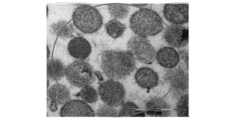
尚、ルテリアルは円形乃至楕円形であって、SEMまたはTEM電子顕微鏡写真でミト
コンドリアと類似した二重膜構造を示すが、内部クリステ(cristae)構造を有し
ていなかった(図1)。
Rutherials were round to oval and exhibited a double-membrane structure similar to mitochondria in SEM or TEM electron micrographs, but did not have internal cristae structures (Fig. 1).
(2)免疫化学蛍光染色
ミトコンドリアは、ヤーヌスグリーンB(Janus green B)、および蛍光
染色薬であるローダミン123(Rhodamine123)、ミトトラッカー(Mit
o‐tracker)、アクリジンオレンジ(Acridine Orange)および
DAPIにより発色されると知られているが、ルテリアルも、ミトコンドリアと同一の染
色薬による発色が確認された。前記ルテリアルがミトコンドリアと類似の免疫化学蛍光染
色反応を示したのに対し、エキソソーム(exsome)とは相反する反応を示し、自家
蛍光(autofluorescence)を示す特徴を蛍光写真から確認した(図2(
a)、図2(b)、図2(f)、図2(j)、図3~図6)。
(2) Immunochemical fluorescence staining Mitochondria were stained with Janus green B,
o-tracker), acridine orange, and DAPI, and it was confirmed that rutherial was also colored by the same staining agent as mitochondria. While the rutherial showed an immunochemical fluorescence staining reaction similar to that of mitochondria, it showed a reaction opposite to that of exosomes, and the characteristics of autofluorescence were confirmed from fluorescence photographs (Fig. 2 (
a), FIG. 2(b), FIG. 2(f), FIG. 2(j), FIGS. 3 to 6).
(3)生態様式
ルテリアルは、エキソソーム(exosome)および微小胞(microvesic
le)とは異なって、付着性および運動性を有し、フュージョン(fusion)または
フィッション(fission)の生態様式を示した。特定条件で変異ルテリアルはバー
スティング(Bursting)し、バースティング(Bursting)後には幹細胞
性(stemness)を有することを確認し、細胞内または細胞外に存在可能であるこ
とを確認した(図8、図9、図11)。
(3) Ecomorphism Rutherials are composed of exosomes and microvesicles.
le), it was sessile and motile and exhibited a fusion or fission ecology. Mutant Rutherial was bursting under specific conditions, confirmed to have stemness after bursting, and confirmed to exist intracellularly or extracellularly (Fig. 8, 9 and 11).
(4)ATP生産
200~400nmのサイズのルテリアルからATPが生産されることを、ルシフェリ
ン(luciferin)‐ルシフェラーゼ(luciferase)反応とルミノメー
タ(luminometer)を用いて立証した。ルテリアルが添加された群は、ルテリ
アルが添加されていない群に比べてATP濃度が増加した。このことから、ルテリアルが
ATP生産能力を有するという結論を導出することができる。SSHおよびSSFの添加
による差については、SSF添加群がSSH添加群に比べてATP濃度が高かった。この
ことから、ATP含量を効率的に増加させることができる培養液を確認した(図18)。
(4) ATP production Production of ATP from 200-400 nm size rutherial was demonstrated using a luciferin-luciferase reaction and a luminometer. The group to which rutherial was added had an increased ATP concentration compared to the group to which rutherial was not added. From this, the conclusion can be drawn that Rutherial has ATP production capacity. Regarding the difference due to the addition of SSH and SSF, the ATP concentration was higher in the SSF addition group than in the SSH addition group. From this, a culture medium capable of efficiently increasing the ATP content was confirmed (Fig. 18).
(5)核酸含有
DAPIおよびアクリジンオレンジ(AO)染色法により、ルテリアル中にRNAだけ
でなくDNAも含有されていることを確認した。具体的に、RNAは、アクリジンオレン
ジ染色薬により、励起460nm、放出650nmのレベル(level)でオレンジで
染色され、DNAは、励起502nm、放出525nmのレベルで緑色で染色されて、D
API染色法により、DNAが含有されていることを確認することができる。前記染色法
を用いて、本発明のルテリアル中にRNAとDNAが含有されていることを確認した(図
5および図6)。また、ルテリアルのRNAおよびDNAをキット(kit)で分離およ
び精製した後、アガロース(agarose)ゲル電気泳動によりバンドを確認し、qR
T‐PCR技法によりGAPDH発現程度を確認することで、ルテリアルのサイズによっ
て遺伝子発現において差があることを確認した(図2(h)、図16および図17)。
(5) Containing Nucleic Acid It was confirmed by DAPI and acridine orange (AO) staining that not only RNA but also DNA was contained in the rutherial. Specifically, RNA was stained orange with acridine orange stain at a level of excitation 460 nm, emission 650 nm, DNA was stained green with a level of excitation 502 nm, emission 525 nm, D
Containment of DNA can be confirmed by API staining. Using the staining method described above, it was confirmed that RNA and DNA were contained in the rutherial of the present invention (FIGS. 5 and 6). In addition, after separating and purifying rutherial RNA and DNA with a kit, bands were confirmed by agarose gel electrophoresis, and qR
By confirming the degree of GAPDH expression by T-PCR technique, it was confirmed that there was a difference in gene expression depending on the size of the rutherial (Fig. 2(h), Fig. 16 and Fig. 17).
(6)16S rRNA配列分析
FastDNA SPIN Kit(MP Biomedicals、Cat 656
0‐200)を用いてルテリアルのgDNAを抽出した後、表1および表2の特定プライ
マー(primer)を用いて16S rRNA遺伝子を増幅させた。
(6) 16S rRNA sequence analysis FastDNA SPIN Kit (MP Biomedicals, Cat 656
0-200) was used to extract rutherial gDNA, and then specific primers in Tables 1 and 2 were used to amplify the 16S rRNA gene.
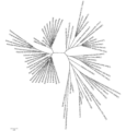
尚、前記増幅された1461個の断片遺伝子を対象に、GeneBank datab
ase(NCBI database)を用いて相同性を分析した結果、血液および精液
由来ルテリアルの16S rRNA塩基配列は、β-プロテオバクテリア(β‐Prot
eobacteria)、γ-プロテオバクテリア(γ‐Proteobacteria
)、アシドバクテリア(Acidobacteria)、シアノバクテリア(Cyano
bacteria)、アクチノバクテリア(Actinobacteria)、ファーミ
キューテス(Firmicutes)、および真核細胞(Eukaryote)由来遺伝
子と相同性を示し、原核細胞と真核細胞の中間段階の融合特性を有していた(図24~図
25)。
In addition, the GeneBank datab for the amplified 1461 fragment genes
As a result of homology analysis using NCBI database), the 16S rRNA nucleotide sequence of blood and semen-derived rutherial was identified as β-proteobacteria (β-Proteobacteria).
eobacteria), γ-Proteobacteria
), Acidobacteria, Cyanobacteria
It showed homology to genes from A.bacteria, Actinobacteria, Firmicutes, and Eukaryote, and possessed intermediate-stage fusion properties between prokaryotic and eukaryotic cells (Fig. 24 to 25).
最適状態(血液のpH:7.2~7.4)で、血液由来ルテリアルは、β-プロテオバ
クテリア(β‐Proteobacteria)、γ-プロテオバクテリア(γ‐Pro
teobacteria)、およびバクテロイデス(Bacteroidetes)由来
遺伝子と相同性を示し(図24)、50~800nmのサイズで観察される。
Under optimal conditions (blood pH: 7.2-7.4), blood-derived rutherials are composed of β-Proteobacteria, γ-Proteobacteria (γ-Proteobacteria),
teobacteria), and Bacteroidetes-derived genes (Fig. 24), observed at sizes of 50-800 nm.
定常状態で、精液由来ルテリアルは、β‐プロテオバクテリア、γ‐プロテオバクテリ
ア、バクテロイデス、および脊索動物(Chordata)由来遺伝子と相同性を示す。
At steady state, seminal fluid-derived rutherials show homology to genes from β-Proteobacteria, γ-Proteobacteria, Bacteroidetes, and Chordata.
これに対し、酸性化状態では、定常状態と同様にβ‐プロテオバクテリア(β‐pro
teobacteria)およびγ‐プロテオバクテリア(γ‐Proteobacte
ria)由来遺伝子だけでなく、バクテリア由来遺伝子がさらに多様に発現され、真核細
胞(Eukaryote)由来遺伝子も発現される。主にストレプトファイタ(Stre
ptophyta)とプランクトミ(planctomy)の16S rRNA特性を発
現し(図25)、サイズは400~2000nmまで成長する。
In contrast, in the acidified state, as in the steady state, β-proteobacteria (β-pro
teobacteria and γ-Proteobacteria
In addition to ria)-derived genes, bacteria-derived genes are more diversely expressed, and eukaryote-derived genes are also expressed. Mainly strepto fighters (Stre
ptophyta) and planctomy 16S rRNA characteristics (Fig. 25) and grows to 400-2000 nm in size.


(7)エキソソームおよびミトコンドリアとの違い
表3は、エキソソームおよびミトコンドリアとルテリアルとの違いを整理したものであ
る。
(7) Differences from exosomes and mitochondria Table 3 summarizes the differences between exosomes and mitochondria and lutherials.

ルテリアルの平均サイズは200~800nmであって、ミトコンドリア(400~1
,000nm)よりは小さく、エキソソーム(20~120nm)よりは大きい。また、
エキソソームは膜の区分が明確でなく、内部色が比較的薄いのに対し、ルテリアルは、膜
の区分が明確であるか、内部が満たされている形態である(図19)。尚、ルテリアルは
、エキソソームおよび微小胞(microvesicles)とはその形態が全く異なる
(図20)。
The average size of rutherials is 200-800 nm, while mitochondria (400-1
,000 nm) and larger than exosomes (20-120 nm). again,
Exosomes do not have distinct membrane divisions and have a relatively pale internal color, whereas rutherials have distinct membrane divisions or a morphology in which the interior is filled (FIG. 19). It should be noted that rutherials are quite different in their morphology from exosomes and microvesicles (Fig. 20).
免疫化学蛍光染色時に、ルテリアルはミトコンドリアと類似の反応を示すのに対し、エ
キソソームとは相反する反応を示す。細胞内外に存在するルテリアルと細胞外に存在する
エキソソームは、食品を摂取することで得られる物質でもあるのに対し、ミトコンドリア
は食品摂取によっては得られない細胞内にのみ存在するという違いがある。
Upon immunochemical fluorescence staining, rutherials show similar reactions to mitochondria, whereas they show opposite reactions to exosomes. Rutherials, which exist inside and outside the cells, and exosomes, which exist outside the cells, are also substances obtained by ingesting food.
そして、エキソソームおよびミトコンドリアと異なって、ルテリアルは運動性を有し、
自然成長が可能であって培養により成長を維持することができ、自家蛍光の特徴を示す。
また、ルテリアル、エキソソーム、およびミトコンドリアは何れもフュージョンの生態様
式を有するが、エキソソームではkiss‐and‐runフュージョンが起こらず、A
TP生産も起こらない。また、エキソソームは細胞外に存在し、ミトコンドリアは細胞内
に存在するのに対し、ルテリアルは細胞外または内の両方に存在可能である(図11)。
And unlike exosomes and mitochondria, rutherials are motile and
It is capable of spontaneous growth, can be maintained in culture, and exhibits autofluorescence characteristics.
Moreover, although rutherials, exosomes, and mitochondria all have a fusion ecology, kiss-and-run fusion does not occur in exosomes, and A
No TP production occurs. Also, exosomes exist extracellularly and mitochondria exist intracellularly, whereas rutherials can exist both extracellularly or intracellularly (Fig. 11).
また、16S rRNA塩基配列分析結果、ミトコンドリアは、α‐プロテオバクテリ
アと相同性を示すのに対し、ルテリアルは、γ‐プロテオバクテリア(γ‐Proteo
bacteria)、β‐プロテオバクテリア(β‐Proteobacteria)、
バクテロイデス(Bacteroidetes)、ファーミキューテス(Firmicu
tes)、および真核細胞(Eukaryote)と相同性を示す。
In addition, 16S rRNA base sequence analysis results show that mitochondria show homology with α-proteobacteria, whereas rutherials show homology with γ-proteobacteria (γ-Proteobacteria).
bacteria), β-Proteobacteria,
Bacteroidetes, Firmicu
tes), and shows homology with eukaryotes.
他の観点による本発明は、次の特性の一つ以上を有する体液由来ルテリアルに関する:
(a)免疫蛍光試験で、ヤーヌスグリーンB(Janus green B)、アクリジ
ンオレンジ(Acridine Orange)、およびローダミン123(Rhoda
mine123)に陽性の発色反応を示す;
(b)最適状態(pH7.2~7.4)では、β‐プロテオバクテリアとγ‐プロテオバ
クテリア由来遺伝子の発現特性を示し、30~800nmのサイズを有する;
(c)酸性化状態では、β‐プロテオバクテリアとγ‐プロテオバクテリア由来遺伝子だ
けでなく、真核細胞であるストレプトファイタ(Streptophyta)遺伝子を発
現し、400nm以上から2000nm以上までサイズが大きくなる;
(d)正常条件でATP生成に関与する;
(e)ミトコンドリアおよびエキソソームとは全く異なる細胞または細胞類似体である;
(f)定常状態では円形乃至楕円形であり、患者由来の場合、定常状態に比べてサイズ(
長径800nm以上)が大きく、形態が均一ではない変異ルテリアルが発生する;
(g)二重膜構造を有し、付着性がある;
(h)細胞内または外の両方に存在可能である;
(i)運動性を有し、フュージョン(fusion)および/またはフィッション(fi
ssion)の生態様式を示す;
(j)特定条件で変異ルテリアルがバースティング(Bursting)し、バースティ
ング(Bursting)後には幹細胞性(stemness)を有する;および
(k)p53遺伝子およびテロメアの調節機能を有する。
According to another aspect, the invention relates to body fluid-derived rutherials having one or more of the following properties:
(a) Immunofluorescence studies showed Janus green B, Acridine Orange, and Rhodamine 123 (Rhoda
shows a positive chromogenic reaction to mine123);
(b) under optimal conditions (pH 7.2-7.4), it exhibits the expression characteristics of genes from β-proteobacteria and γ-proteobacteria and has a size of 30-800 nm;
(c) under acidifying conditions, they express not only β- and γ-proteobacterial-derived genes, but also eukaryotic Streptophyta genes, increasing in size from >400 nm to >2000 nm;
(d) participate in ATP generation under normal conditions;
(e) cells or cell analogs that are distinct from mitochondria and exosomes;
(f) round to elliptical at steady state and size (
800 nm or more in major axis) and morphologically non-uniform mutant Rutherial occurs;
(g) having a double-membrane structure and being adherent;
(h) can be both intracellular or extracellular;
(i) have motility and are fusion and/or fission
ssion) ecology;
(j) mutated lutherial bursting under certain conditions and possessing stemness after bursting; and (k) regulatory function of the p53 gene and telomeres.
一方、ルテリアルは、個体の疾病有無に応じて、サイズ(直径)、面積、形態、および
ナノトラッキング速度が異なるため、前記特性の1つ以上を用いて、疾病の診断や予後を
予測することができる。これは、疾病のない健常人由来のルテリアルと、疾病のあるヒト
由来のルテリアルのサイズ、形態、ナノトラッキング速度などが異なることから分かる。
On the other hand, since lutherials differ in size (diameter), area, morphology, and nanotracking speed depending on the presence or absence of disease in an individual, one or more of the above characteristics can be used to predict disease diagnosis and prognosis. can. This can be seen from the difference in size, morphology, nanotracking speed, etc. between lutherial derived from healthy individuals without disease and lutherial derived from diseased humans.
健常人由来の正常のルテリアルは、単に二重胞子(double‐spore)を形成
(fission)するだけであるが、晩成疾病患者や癌患者由来のルテリアル(突然変
異ルテリアル)は、ルテリアル同士が融合(fussion)または凝集(coagul
ation)したり爆発(Bursting)して、赤血球や癌細胞などの細胞にも付着
し、その形態およびサイズが正常のルテリアルとは異なって異常に大きくなる特徴がある
(図8~図10)。突然変異ルテリアルは付着性が高いため、上記のようなサイクル(c
ycle)によって融合(fusion)がさらに加速化してそのサイズが約600~8
00nm以上に大きくなり、200μm(200,000nm)以上のサイズを有するも
のもある。本発明者は、癌の種類や進行程度によって、突然変異されたルテリアルの形態
に一貫性があることを確認し、これについての内容を特許出願している(韓国特許出願第
10‐2013‐0082060号)。
Normal rutherials derived from healthy subjects simply form double-spores (fission), but rutherials derived from late-onset disease patients and cancer patients (mutant rutherials) fuse with each other ( fussion or coagulation
) and bursting, adhere to cells such as erythrocytes and cancer cells, and differ from normal rutherials in their morphology and size to become abnormally large (Figs. 8 to 10). Mutant rutherials are highly adherent, so cycles such as those described above (c
The fusion is further accelerated by the ycle, and its size is about 600-8
00 nm or more, and some have sizes of 200 μm (200,000 nm) or more. The present inventor has confirmed that the morphology of mutated rutherial is consistent according to the type and progress of cancer, and has filed a patent application (Korean Patent Application No. 10-2013-0082060). issue).
したがって、ルテリアルの形態学的特性または生化学的特性を観察することで、疾病の
診断や予後を予測することができ、その用途が無限にある。
Therefore, by observing the morphological or biochemical characteristics of rutherial, it is possible to predict the diagnosis and prognosis of diseases, and the applications are limitless.
ルテリアルの形態は、正常形、鞭毛形、マス(Mass)形、ロッド(Rod)形、ま
たは複合形を示す。正常形とは、別の融合やバースティング(Bursting)などの
変形を起こすことなく、長径と短径との比が1:1~3:1の形態であり、円形に近い形
状を示すことができる。顕微鏡で観察時には小さい点として示される。
Rutherial morphology exhibits normal, flagellar, mass, rod, or complex morphology. The normal shape is a shape in which the ratio of the major axis to the minor axis is 1:1 to 3:1 without causing deformation such as another fusion or bursting, and can exhibit a shape close to a circle. can. Shown as small dots when observed under a microscope.
鞭毛形とは、ルテリアルが変形または融合を起こして外部に鞭毛が備えられた形態であ
ることができる。本発明者らは、末期癌に進むに従って鞭毛形が観察される割合が急激に
増加し、4期癌では99.1%と、略大部分の4期癌が診断された患者で鞭毛形のルテリ
アルが観察されることを確認した(韓国特許出願第2013‐0082060号)。ルテ
リアルの形態が鞭毛形の形態と80~100%一致する場合、末期腫瘍が疑われる腫瘍マ
ーカ(Tumor Marker)で表示することができる。前記末期腫瘍が診断された
患者の生存期間は略1~4ヶ月であり、特に、鞭毛形の場合、長期生存が不可能である。
A flagellum can be a form in which rutherials are deformed or fused to have flagella on the outside. The present inventors have found that the rate of flagella observed increases sharply as the cancer progresses to the terminal stage, reaching 99.1% in
マス形(M形)とは、ルテリアルがバースティング(Bursting)または融合を
起こしてサイズおよび形態が正常形から変形されたものであって、長径と短径の差が大き
くない不規則的な体積形態である。好ましくは、長径と短径との比が3:1~5:1であ
り、多様な形態のマス形が観察される。
The mass shape (M shape) is an irregular volume in which the size and shape are deformed from the normal shape due to bursting or fusion of Rutherial, and the difference between the major axis and the minor axis is not large. form. Preferably, the ratio of major axis to minor axis is 3:1 to 5:1, and various forms of mass are observed.
ロッド形(R形)は、ルテリアルがバースティング(Bursting)、変形、また
は融合を起こして棒(Rod)の形態を呈するものである。短径と長径との長さ差がマス
形より大きい。好ましくは、長径と短径との比が5:1~12:1であることができる。
ロッド形は、円形または楕円形の単一鎖からなるロッド1形、および単一鎖が2個以上結
合してなるロッド2形を含む。前記ロッド1形は、単一のルテリアルが棒の形態となった
ものであって、これは、バースティング(Bursting)および/または変形による
ものであることができる。前記ロッド2形は、2個以上のルテリアルが結合して棒の形態
となったものであって、これは、バースティング(Bursting)、変形、および融
合の1つ以上によるものであることができる。一方、鞭毛形は、その形状から、大きい範
疇でロッド形に含まれることができるが、鞭毛が伸びているという点で異なる点がある。
したがって、ロッド形であるかを先に判断した後、鞭毛形であるかを判断することができ
る。
A rod-shaped (R-shaped) rutherial undergoes bursting, deformation, or fusion to take the form of a rod. The length difference between the minor axis and the major axis is larger than that of the mass shape. Preferably, the ratio of major axis to minor axis can be from 5:1 to 12:1.
The rod type includes a
Therefore, it is possible to determine whether it is a rod shape first, and then whether it is a flagellum shape.
前記複合形は、ロッド形とマス形の融合形態であることができる。一体に形成された微
細物質の一部がロッド形であり、一部がマス形である形態を複合形と称えることができる
。
Said compound shape can be a fusion of a rod shape and a mass shape. A form in which a part of the integrally formed microscopic substance is rod-shaped and a part is mass-shaped can be called a composite form.
前記ロッド(Rod)形は、円形または楕円形の単一鎖からなるロッド1形、および単
一鎖が2個以上結合してなるロッド2形を含む群の一つであることができる。前記複合形
は、ロッド形とマス形の融合形態であることができる。
The rod type may be one of a group including a
上記のように、疾病の発病と進行によって、生体内のルテリアルの形態が変わるため、
ルテリアルの形態学的特性を観察することで疾病の診断や予後を予測することができる。
また、前記ルテリアルの形態変化は、ルテリアルが含有している核酸の量と配列変化にも
関連するため、ルテリアルの核酸発現パターン(16S rRNA)配列を分析すること
で疾病を診断することができる。
As mentioned above, due to the onset and progression of disease, the morphology of lutherial in vivo changes.
Observation of morphological characteristics of lutherial can predict disease diagnosis and prognosis.
In addition, since the morphological change of the rutherial is related to the amount and sequence change of the nucleic acid contained in the rutherial, the disease can be diagnosed by analyzing the nucleic acid expression pattern (16S rRNA) sequence of the rutherial.
例えば、正常のルテリアルの16S rRNA配列と患者のルテリアルの16S rR
NA配列を比較することで、疾病(特に、癌)を診断することができる。特に、ストレプ
トファイタ(Streptophyta)遺伝子発現と真核細胞(Eukaryote)
遺伝子同時発現は、癌の診断予測マーカとして活用可能である。
For example, the 16S rRNA sequence of normal rutherial and the 16S rR of patient rutherial
Diseases (particularly cancer) can be diagnosed by comparing NA sequences. In particular, Streptophyta gene expression and eukaryote
Gene co-expression can be used as a diagnostic predictive marker for cancer.
但し、患者または健常人から既に排出された体液から分離されたルテリアルは、生体外
では短い時間内に溶解されて消滅されたり、形態が変わったりする特性があるため観察自
体が難しく、異常な環境下で放置する場合には、24時間以内に正常のルテリアルも突然
変異ルテリアルに変異されて、疾病の正確な診断や治療が困難である。しかし、本発明の
培養方法によると、特定のサイズ(500nm)以上にならないようにルテリアルを培養
することができる。
However, rutherial separated from bodily fluids that have already been discharged from a patient or healthy person has the characteristic of being dissolved and eliminated in a short period of time outside the body, or changing its shape, making it difficult to observe in an abnormal environment. If left untreated, the normal lutherial will mutate into a mutant lutherial within 24 hours, making accurate diagnosis and treatment of the disease difficult. However, according to the culture method of the present invention, it is possible to culture lutherial so as not to exceed a specific size (500 nm).
したがって、他の観点による本発明は、ルテリアルに水分を添加し、IR光線照射下、
18~30℃(好ましくは20~25℃)で培養することを特徴とするルテリアルの培養
方法に関する。
Therefore, according to another aspect of the present invention, water is added to rutherial, and under irradiation with IR light,
It relates to a method for culturing rutherial characterized by culturing at 18 to 30°C (preferably 20 to 25°C).
前記培養時に添加される水分は、食塩水またはPBS溶液であることができるが、これ
に制限されない。培養前の体液由来ルテリアルは本発明の分離方法により得ることができ
、そのサイズが50~200nmのものを用いることができる。本発明の培養方法により
培養された血液由来ルテリアルの培養後のサイズは300~800nmであることができ
る。この際、顕微鏡で観察しながら、ルテリアルのサイズが500nmを超えないように
することができ、培養が終了すると、サイズ毎に分類して零下80℃に冷却して保存した
り、窒素を充填して保存または零度以上で保存することができ、保存時には保存剤を添加
することができる。
The water added during the culture may be saline or PBS solution, but is not limited thereto. A body fluid-derived rutherial before culture can be obtained by the separation method of the present invention, and one with a size of 50 to 200 nm can be used. The blood-derived rutherial cultured by the culture method of the present invention can have a size of 300 to 800 nm after culture. At this time, while observing with a microscope, the size of the rutherials can be prevented from exceeding 500 nm. After the culture is completed, the rutherials are sorted by size and stored by cooling to -80 ° C. or filled with nitrogen. It can be stored at or above zero degrees, and preservatives can be added during storage.
上記のように培養されたルテリアルは、その特性が変化することなく所定期間保存が可
能であり、ルテリアルを用いた疾病の診断および予後の予測に効果的に活用されることが
できる。本発明において「ルテリアルの特性が変化することなく」とは、ルテリアルの形
態(morphology)やサイズが培地で培養する前の状態と略類似に維持されるこ
とを意味する。また、ナノトラッキング速度のようなルテリアルの運動性などの活性が、
培養する前の状態と類似の値を維持することを意味する。
The lutherial cultured as described above can be stored for a predetermined period of time without changing its properties, and can be effectively utilized for diagnosis and prognosis prediction of diseases using lutherial. In the present invention, "without changing the properties of the rutherial" means that the morphology and size of the rutherial are maintained substantially similar to the state before culturing in the medium. Also, activities such as rutherial motility, like nanotracking velocity,
It means maintaining a value similar to the state before culturing.
具体的に、本発明の培養方法により培養されたルテリアルは、次の目的のために用いる
ことができる。変異された突然変異ルテリアルは、融合(fussion)または凝集し
て、その形態およびサイズが正常のルテリアルとは異なって異常に大きくなったものであ
り(図8~図10)、分離された突然変異ルテリアルの培養時に、ルテリアルの融合また
は凝集を抑えることができる候補物質や手段を処理して、ルテリアルの融合または凝集の
抑制有無を観察することで、ルテリアルの変異を抑制または予防することができる物質を
スクリーニングすることができる。
Specifically, the rutherial cultured by the culture method of the present invention can be used for the following purposes. The mutated mutated rutherials were fused or aggregated to become abnormally large in morphology and size different from normal rutherials (FIGS. 8-10). Substances capable of suppressing or preventing mutation of lutherial by treating candidate substances or means capable of suppressing fusion or aggregation of lutherial during cultivation of lutherial and observing the presence or absence of inhibition of fusion or aggregation of lutherial. can be screened.
また、分離されたルテリアルの培養時、フィッション(fission)を促進する候
補物質や手段を処理することで、突然変異ルテリアルのフィッション(fission)
を促進する物質をスクリーニングすることができる。突然変異ルテリアルは融合または凝
集する生態様式を有するが(図8、図9および図11)、フィッション(fission
)の生態様式に転換されたり、突然変異されたルテリアルがフィッション(fissio
n)されたりして正常のルテリアルのサイズを有するように、フィッション(fissi
on)を促進する候補物質を処理することで、ルテリアルが変異されることを抑制するか
、変異されたルテリアルが正常のルテリアルの生態様式を有するように転換する物質、窮
極的には、変異されたルテリアルにより誘発され得る疾病の予防物質をスクリーニングす
ることができる。
In addition, fission of mutant lutherials can be enhanced by treating candidate substances or means that promote fission during culture of the isolated lutherials.
can be screened for substances that promote Mutant lutherials have a fused or aggregated ecology (Figs. 8, 9 and 11), whereas fission
) or mutated lutherials become fissio
n) Fission the fissi to have a normal lutherial size.
On) by treating a candidate substance that promotes on), a substance that suppresses the mutation of the lutherial or converts the mutated lutherial to have a normal lutherial ecological pattern. It is possible to screen for prophylactic agents for diseases that can be induced by rutherial.
以下、実施例により本発明をさらに詳細に説明する。これら実施例は、本発明を例示す
るためのものにすぎず、本発明の範囲がこれら実施例によって制限されると解釈されない
ことは、当業界において通常の知識を有する者にとって自明である。
The present invention will be described in more detail below with reference to examples. It should be apparent to those of ordinary skill in the art that these examples are for illustrative purposes only and that the scope of the invention should not be construed as being limited by these examples.
実施例
実施例1:血液由来ルテリアルの分離
非小細胞性肺癌末期患者から血液を50cc採取して直径0.8μm以上の空隙を有す
るフィルタに通過させ、濾過されていない物質は分離した。濾過された血液を1200~
5000rpmで5~10分間繰り返して遠心分離することで、エキソソーム(exos
ome)のような一般微小胞を除去して上清を得た。前記上清に可視光線を照射して、運
動性を有して集まるルテリアル粒子をピペットを用いて分離した。ルテリアルは自家蛍光
および運動性の特性を有するため、上記のように可視光線を照射するとルテリアル粒子を
確認することができる。この際、動くルテリアル粒子を暗視野顕微鏡または共焦点顕微鏡
で確認しながらピペットを用いて分離した。分離されたルテリアルを直径50nmの空隙
を有するフィルタに通過させて、濾過されていない部分のみをPBSで洗浄することでル
テリアルを得た。前記過程により長径50~800nmのルテリアルが得られ、これは暗
視野顕微鏡または共焦点顕微鏡により観察確認が可能であった。前記得られたルテリアル
は、サイズに応じて、50~200nm(発生期)/200~400nm(成熟期)/4
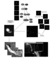
00~600nm(分裂期)/600~800nm(過分裂期)に区分した。類似の方法
により、図21のようなルテリアルのサイズ毎のライブラリを構築し、ルテリアルのサイ
ズ毎のmorphologyを図2に示した。
Examples Example 1: Separation of blood-derived rutherial 50 cc of blood was collected from a terminal patient of non-small cell lung cancer and passed through a filter having pores with a diameter of 0.8 µm or more to separate unfiltered substances. bottom. 1200~ of filtered blood
Exosomes (exos
The supernatant was obtained by removing common microvesicles such as ome). The supernatant was irradiated with visible light, and the rutherial particles that gathered with motility were separated using a pipette. Rutherial particles can be identified by irradiating visible light as described above, since the rutherial has autofluorescence and motility properties. At this time, the moving rutherial particles were separated using a pipette while confirming them with a dark field microscope or a confocal microscope. The separated rutherial was passed through a filter having pores with a diameter of 50 nm, and only the unfiltered portion was washed with PBS to obtain rutherial. A rutherial with a major diameter of 50 to 800 nm was obtained by the above process, which could be observed and confirmed by a dark field microscope or a confocal microscope. The resulting rutherial is 50-200 nm (nascent)/200-400 nm (mature)/4
It was divided into 00-600 nm (mitotic phase)/600-800 nm (metaphase). By a similar method, a library for each size of lutherial as shown in FIG. 21 was constructed, and the morphology for each size of lutherial is shown in FIG.
実施例2:精液由来ルテリアルの分離
精液を2000~4000rpmで5~30分間1次遠心分離した後、上清を2~5μ
mのフィルタで濾過し、濾過された溶液を3000~7000rpmで5~20分間2次
遠心分離した後、0.5~2μmのフィルタで濾過した。ルテリアルは自家蛍光および運
動性の特性を有するため、濾過した溶液に可視光線を照射するとルテリアル粒子を確認す
ることができる。この際、動くルテリアル粒子を暗視野顕微鏡または共焦点顕微鏡で確認
しながらピペットを用いて分離した。分離されたルテリアルを直径50nmの空隙を有す
るフィルタに通過させ、濾過されていない部分のみをPBSで洗浄することでルテリアル
を得た。これは暗視野顕微鏡または共焦点顕微鏡により観察確認が可能であった。
Example 2: Separation of semen-derived rutherial
The filtered solution was subjected to secondary centrifugation at 3000-7000 rpm for 5-20 minutes and then filtered through a 0.5-2 μm filter. Rutherial particles can be identified by illuminating the filtered solution with visible light due to their autofluorescence and motility properties. At this time, the moving rutherial particles were separated using a pipette while confirming them with a dark field microscope or a confocal microscope. The separated rutherial was passed through a filter having pores with a diameter of 50 nm, and only the unfiltered portion was washed with PBS to obtain rutherial. This could be observed and confirmed by a dark field microscope or a confocal microscope.
実施例3:ルテリアルの特性
(1)構造
実施例1で得られたルテリアルのうち約50~400nmのサイズを有するルテリアル
を、共焦点レーザー走査顕微鏡(Confocal Laser Scanning M
icroscope、Zeiss)、透過型電子顕微鏡(Transmission E
lectron Microscope)、走査型電子顕微鏡(Scanning El
ectron Microscope)、原子間力顕微鏡(Atomic Force
Microscope)、および共焦点スキャナ(Leica TCS‐SP8)で撮影
した結果、ルテリアルもミトコンドリアと類似に二重膜を有する膜構造であって、内部ク
リステ(cristae)構造が完成されていない状態の構造を有しており、ミトコンド
リアと同一のレーザー波長範囲で観察されることを確認した。また、その形態は円形乃至
楕円形であることを観察することができた(図1、図(e)、図2(h)、図13および
図14)。
Example 3 Properties of Rutherial (1) Structure Of the rutherials obtained in Example 1, the rutherials having a size of about 50 to 400 nm were observed with a confocal laser scanning microscope.
microscope, Zeiss), transmission electron microscope (Transmission E
Electron Microscope), Scanning Electron Microscope (Scanning El
electron microscope, atomic force microscope
As a result of imaging with a Microscope and a confocal scanner (Leica TCS-SP8), the rutherial is also a membrane structure having a double membrane similar to mitochondria, and the inner cristae structure is not completed. It was confirmed that it has the same laser wavelength range as mitochondria. It was also observed that the shape was circular or oval (FIGS. 1, (e), 2 (h), 13 and 14).
(2)染色特性
実施例1で得られたルテリアルのうち約50~800nmのサイズを有するルテリアル
を、ミトトラッカー(Mito‐tracker)、ローダミン123(Rhodami
ne123)、アクリジンオレンジ(Acridine Orange)、およびヤーヌ
スグリーンB(Janus green B)で染色した後、その発色有無を観察した。
その結果、植物由来ルテリアルも、ミトトラッカー、ローダミン123、アクリジンオレ
ンジ、およびヤーヌスグリーンBにより発色されることを確認した(図2(a)、図2(
b)、図2(f)、図2(j)、図3~図6)。
(2) Staining properties Of the rutherials obtained in Example 1, the rutherials having a size of about 50 to 800 nm were treated with Mito-tracker,
ne123), Acridine Orange, and Janus green B, and observed for color development.
As a result, it was confirmed that the plant-derived rutherial was also colored by Mitotracker,
b), FIG. 2(f), FIG. 2(j), FIGS. 3 to 6).
(3)自家蛍光
実施例1で得られたルテリアルのうち約50~800nmのサイズを有するルテリアル
が光反応を示すことを蛍光写真から確認した(図5)。
(3) Autofluorescence Among the rutherials obtained in Example 1, it was confirmed from fluorescence photographs that the rutherials having a size of about 50 to 800 nm showed photoreactivity (FIG. 5).
(4)運動性
実施例1で得られたルテリアルの運動性を、米国3i社のナノトラッキングにより測定
した。具体的に、ルテリアルを明視野顕微鏡で観察した後、ルテリアルの中心にトラッキ
ングを設定してナノトラッキングを作動すると、ルテリアルの移動にしたがってリアルタ
イム移動軌跡が表示され、その秒当りの速度を計算した(図7)。
(4) Motility The motility of the rutherial obtained in Example 1 was measured by nano-tracking by 3i, USA. Specifically, after observing the lutherial with a bright-field microscope, tracking was set at the center of the lutherial and nano-tracking was activated. A real-time movement trajectory was displayed according to the movement of the lutherial, and the velocity per second was calculated ( Figure 7).
その結果、本実施例によるルテリアルのナノトラッキング速度は、約13~25μm/
secと測定された。
As a result, the nanotracking velocity of Rutherial according to this example is about 13-25 μm/
Measured as sec.
(5)ルテリアルにおけるRNAおよびDNAの含有有無の分析
実施例1で分離された200~400nmのルテリアルを原子顕微鏡で撮影した結果、
図2(h)、図15、図16および図17に示されたように、ルテリアルにRNAやDN
Aのような核酸が含有されていると推正することができる。
(5) Analysis of the presence or absence of RNA and DNA in the rutherial As a result of photographing the 200-400 nm rutherial separated in Example 1 with an atomic microscope,
As shown in Fig. 2(h), Fig. 15, Fig. 16 and Fig. 17, rutherial RNA and DN
It can be inferred that a nucleic acid such as A is contained.
実施例1で分離された200~400nmのルテリアルから全RNAとDNAを分離す
るために、QIAGENキット(RNeasy Micro Kit:Cat 7400
4)を用いて分離した後、Experion RNA(DNA) StdSens(Bi
o‐Rad社)チップを用いて定量した。
QIAGEN kit (RNeasy Micro Kit: Cat 7400
4) followed by Experion RNA (DNA) StdSens (Bi
O-Rad) chip was used for quantification.
ルテリアルを遠心分離(8000g、1時間30分)して回収した後、キット内の分解
緩衝液RLT plus(Guanidine isothiocycanate、de
tergents)50μlにβメルカプトエタノール3.5μlを添加し、20ゲージ
の針付きの注射器を用いて5~10回通過させてルテリアルを溶解させた。サンプル分解
緩衝液をAllPrep DNA spinカラムに移した後、遠心分離(≧8000g
、15秒)して、カラムに詰っているDNAとカラムを通過したRNAが含まれたバッフ
ァからそれぞれ分離した。
After collecting the rutherial by centrifugation (8000 g, 1
3.5 μl of β-mercaptoethanol was added to 50 μl of tergents) and passed 5-10 times using a syringe with a 20 gauge needle to dissolve the rutherial. Transfer the sample digestion buffer to an AllPrep DNA spin column followed by centrifugation (≥8000 g
, 15 seconds) to separate the DNA stuck in the column from the buffer containing the RNA that has passed through the column.
先ず、カラムを通過したバッファに同一体積の350μlの70%エタノールを入れて
均一に混合した後、700μlの混合液をRNease MinElute spinカ
ラムに移して遠心分離(≧8000g、15秒)し、カラムを通過したバッファは除去し
た。それぞれの350μlのRW1,500μlのRPEバッファと500μlの80%
エタノールを用いて、カラム洗浄を段階的に行った。上記で用いられた全ての遠心分離(
≧8000g、15秒)は同一条件で行った。RNAを得るために、14μlのRNea
sy‐free溶液をカラムに入れた後、遠心分離(≧8000g、60秒)してルテリ
アルRNAを分離した。
First, the same volume of 350 μl of 70% ethanol was added to the buffer passed through the column and mixed uniformly, then 700 μl of the mixture was transferred to an RNase MinElute spin column and centrifuged (≧8000 g, 15 seconds) to remove the column. The run-through buffer was removed. 350 μl of each RW1, 500 μl of RPE buffer and 500 μl of 80%
Column washes were performed stepwise using ethanol. All centrifugation used above (
≧8000 g, 15 seconds) were performed under the same conditions. To obtain RNA, 14 μl RNea
After applying the sy-free solution to the column, the rutherial RNA was separated by centrifugation (≧8000 g, 60 seconds).
ゲノムDNAの分離のために、FastDNA SPIN Kit(MP Biome
dical)を用いた。チューブに分離されたルテリアルを入れた後、978μlのリン
酸ナトリウム(sodium phosphate)バッファ、122μlのMTバッフ
ァを添加した。40秒間均質化させてから、遠心分離(14,000g、10分)して上
清を得た後、250μlのPPS(Protein Precipitation So
lution)を入れて10分間混合した。遠心分離(14000g、5分)した後、1
5mlのチューブに上清を移す過程を2回繰り返し、DNA結合(DNA bindin
g)のためにロータ(rotor)に2分間載せた後、シリカマトリックス台に3分間入
れて置いた。上清を略600μl程度注意して回収してSPIN Filterに入れ、
遠心分離(14000g、1分)した後、上清を捨て、ペレット(pellet)にSE
WS‐Mを500μl入れて懸濁させた(suspending)。1分間遠心分離した
後、上清を捨て、如何なるバッファも残らないように遠心分離を繰り返した。50μlの
DES(DNase/Pyrogen‐Free Water)を入れて遠心分離(14
000g、1分)した後、ゲノムDNAを得た。
For isolation of genomic DNA, the FastDNA SPIN Kit (MP Biome
dical) was used. After putting the separated rutherial into the tube, 978 μl of sodium phosphate buffer and 122 μl of MT buffer were added. After homogenization for 40 seconds and centrifugation (14,000 g, 10 minutes) to obtain the supernatant, 250 μl of PPS (Protein Precipitation Sodium) was added.
lution) was added and mixed for 10 minutes. After centrifugation (14000 g, 5 minutes), 1
The process of transferring the supernatant to a 5 ml tube was repeated twice to allow DNA binding.
For g) it was placed on the rotor for 2 minutes and then placed on the silica matrix stage for 3 minutes. About 600 μl of the supernatant was carefully collected and placed in a SPIN filter,
After centrifugation (14000 g, 1 min), the supernatant was discarded and the pellet was SE.
500 μl of WS-M was added and suspended. After centrifugation for 1 minute, the supernatant was discarded and centrifugation was repeated so that no buffer remained. Add 50 μl of DES (DNase/Pyrogen-Free Water) and centrifuge (14
000 g, 1 min), genomic DNA was obtained.
Experion RNA(DNA)StdSens(Bio‐Rad社)チップを用
いて定量した結果、図16および図17に示されたように、ルテリアルにそれぞれRNA
およびDNAが含有されていることを確認することができた。
As a result of quantification using an Experion RNA (DNA) StdSens (Bio-Rad) chip, as shown in FIGS.
and contained DNA.
(6)16S rRNA配列分析
16S rRNA(ribosomal Ribonucleic acid)は、様
々なタンパク質と相互作用してリボソーム(Ribosome)を構成するRNAであっ
て、塩基配列変化率が殆どのゲノムにおける他の遺伝子の塩基配列より著しく小さいため
、16S rRNA塩基配列類似度の程度が生物間の系統学的距離を反映すると認識され
ている。
(6) 16S rRNA sequence analysis 16S rRNA (ribosomal ribonucleic acid) is an RNA that interacts with various proteins and constitutes ribosomes, and the base sequence change rate is almost the base of other genes in the genome. Being significantly smaller than sequences, it is recognized that the degree of 16S rRNA base sequence similarity reflects the phylogenetic distance between organisms.
[1]血液由来ルテリアル
実施例1で得られた血液由来ルテリアルからFastDNA SPIN Kit(MP
Biomedicals、Cat 6560‐200)を用いてgDNAを抽出した後
、PCR‐premix(iNtRON Biotechnology、Korea)配
列番号1~23のブライマー(primer)を用いてルテリアルの16S rRNAを
増幅させた。
[1] Blood-derived Rutherial Fast DNA SPIN Kit (MP
Biomedicals, Cat 6560-200) was used to extract gDNA, and PCR-premix (iNtRON Biotechnology, Korea) primers of SEQ ID NO: 1-23 were used to amplify rutherial 16S rRNA.
増幅されたPCR産物は、BigDye Terminator Cycle Seq
uencing Ready Reaction kit(applied Biosy
stems、USA)とautomated DNA analyzer system
(PRISM 3730XL DNA analyzer、Applied Biosy
stems)を用いて塩基配列を分析した。増幅されたPCR産物は総1461個の断片
であって、このうち1407個の断片はプロテオバクテリア(proteobacter
ia)由来遺伝子と相同性を示し、20個の断片はアシドバクテリア(Acidobac
teria)由来遺伝子と相同性を示し、11個の断片はアクチノバクテリア(Acti
nobacteria)由来遺伝子と相同性を示した(表4)。
The amplified PCR product was subjected to BigDye Terminator Cycle Seq
uencing Ready Reaction kit (applied Biosy
stems, USA) and an automated DNA analyzer system
(PRISM 3730XL DNA analyzer, Applied Biosy
stems) was used to analyze the nucleotide sequence. The amplified PCR products consisted of a total of 1461 fragments, of which 1407 fragments were proteobacterial.
ia) showing homology to the derived gene, 20 fragments are Acidobacterial
teria)-derived gene, and 11 fragments show homology with Actinobacteria (Acti
nobacteria)-derived gene (Table 4).
分析した塩基配列の断片をSeqMan software(DNASTAR)で組み
合わせて16S rRNAの塩基配列を得た。
The analyzed base sequence fragments were combined using SeqMan software (DNASTAR) to obtain the base sequence of 16S rRNA.
図24は、健常人(血液のpH:7.2~7.4)の血液由来ルテリアルの16S r
RNAを塩基配列分析して、相同性を示す類似バクテリアの構成を示したものであって、
ルテリアルのサイズ毎に分析した((a):100nm以下、(b):100~200n
m、(c)200~400nm、(d)400~800nm)。サイズ毎に大きい差はな
く、何れもプロテオバクテリア(Proteobacteria)、ファーミキューテス
(Firmicutes)、およびバクテロイデス(Bacteroidetes)由来
遺伝子と相同性を示した。
FIG. 24 shows 16S r of blood-derived rutherial from a healthy subject (blood pH: 7.2-7.4).
Sequence analysis of RNA showing the composition of similar bacteria exhibiting homology,
Analyzed for each size of Rutherial ((a): 100 nm or less, (b): 100 to 200 nm
m, (c) 200-400 nm, (d) 400-800 nm). There was no large difference between sizes, and all showed homology with genes derived from Proteobacteria, Firmicutes, and Bacteroidetes.
図25(c)は、疲労・疾病状態(血液のpH:7.0以下)の血液由来のルテリアル
(サイズ:200~400nm)の16S rRNAを塩基配列分析して、相同性を示す
類似バクテリアの構成を示したものである。定常状態とは異なって、ストレプトファイタ
(Streptophyta)由来遺伝子と相同性を示す遺伝子がさらに発現された。
FIG. 25(c) shows the results of base sequence analysis of 16S rRNA of lutherial (size: 200 to 400 nm) derived from blood in a fatigued/sick state (blood pH: 7.0 or less), and similar bacteria showing homology. The configuration is shown. Different from the steady state, genes showing homology to Streptophyta-derived genes were additionally expressed.
図26(a)、(b)および(c)は、血液由来ルテリアルの16S rRNA塩基配
列に基づいた系統発生図を示したものである。
Figures 26(a), (b) and (c) show phylogenetic diagrams based on the 16S rRNA nucleotide sequences of blood-derived rutherials.

血液由来ルテリアルの16S rRNA断片は、β‐プロテオバクテリア、γ‐プロテ
オバクテリア、バクテロイデス、ファーミキューテス、およびストレプトファイタなどの
様々なバクテリアと相同性を示すことが確認された。
The 16S rRNA fragment of blood-derived rutherial was confirmed to show homology to various bacteria such as β-Proteobacteria, γ-Proteobacteria, Bacteroidetes, Firmicutes, and Streptophiles.
一般に、微生物分類体系上、gDNA関連性(relatedness)が70%未満
であると、互いに独立した菌株として認められる。また、16S rRNA塩基配列の相
同性が97%未満であると、gDNA関連性が70%未満であるということが、統計学的
な分析により証明された。したがって、ルテリアルの16S rRNA断片と97.0%
以上の相同性を示す配列を有する細胞を分析した結果、表5~表7に示したように、血液
由来ルテリアルはγ‐プロテオバクテリアと100%の相同性を示し、ファーミキューテ
スとは97.53%、バクテロイデスとは97%以上の相同性を示した。
In general, strains with a gDNA relatedness of less than 70% are recognized as independent strains in the microbial classification system. Statistical analysis also demonstrated that the 16S rRNA sequence homology was less than 97% and the gDNA relatedness was less than 70%. Therefore, the 16S rRNA fragment of Rutherial and 97.0%
As a result of analyzing cells having sequences showing the above homology, as shown in Tables 5 to 7, blood-derived Rutherial showed 100% homology with γ-Proteobacteria, and 97% with Firmicutes. It showed 53% homology and 97% or more homology with Bacteroidetes.
一方、異常状態である酸性条件のルテリアルは、表8に示したように、ストレプトファ
イタ(Streptophyta)と99%以上の相同性を示した。
On the other hand, as shown in Table 8, Rutherial in an acidic condition, which is an abnormal state, showed 99% or more homology with Streptophyta.


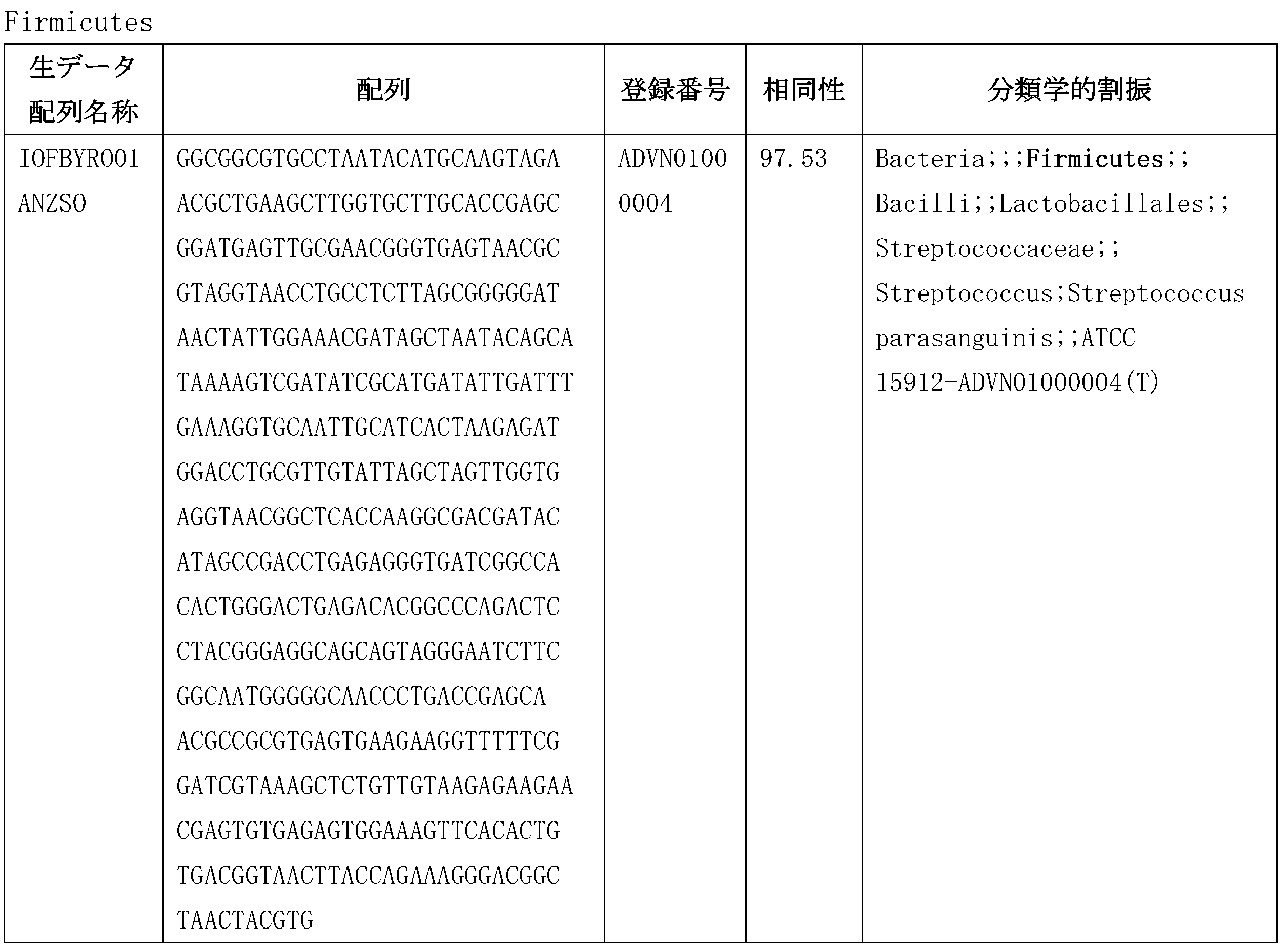


[2]精液由来ルテリアル
実施例2で得られた精液由来ルテリアルに対して、上記のような方法によりgDNA抽
出、PCR増幅、および塩基配列分析を行った。図25は、疲労・疾病状態(精液のpH
:7.0以下)の精液由来ルテリアルの16S rRNAを塩基配列分析して、相同性を
示す類似バクテリアの構成を示したものであって、ルテリアルのサイズ毎に分析した((
a):100nm以下、(b):100~200nm、(d)400~800nm)。
[2] Semen-derived rutherial The semen-derived rutherial obtained in Example 2 was subjected to gDNA extraction, PCR amplification, and nucleotide sequence analysis by the methods described above. FIG. 25 shows the fatigue/disease state (semen pH
The 16S rRNA of semen-derived rutherials from the semen of 7.0 or less) was analyzed to show the composition of similar bacteria showing homology, and was analyzed for each size of the rutherials ((
a): 100 nm or less, (b): 100 to 200 nm, (d) 400 to 800 nm).
定常状態の精液由来ルテリアルは、血液由来ルテリアルと同様に、プロテオバクテリア
(Proteobacteria)、ファーミキューテス(Firmicutes)、お
よびバクテロイデス(Bacteroidetes)由来遺伝子と相同性を示し、特異な
ことに、脊索動物(Chordata)由来遺伝子と相同性を示した。
Steady-state semen-derived rutherial, like blood-derived rutherial, shows homology to genes from Proteobacteria, Firmicutes, and Bacteroidetes, and, unusually, to chordates. ) showed homology with the derived gene.
尚、異常状態である酸性条件では、ストレプトファイタ(Streptophyta)
由来遺伝子と相同性を有する遺伝子が発現された。
In addition, under acidic conditions, which is an abnormal state, Streptophyta
Genes with homology to the derived gene were expressed.
(7)ATP含量測定
対照群、ルテリアル、ルテリアル(SSH 12h)、およびルテリアル(SSF 1
2h)で構成された4種の培養液10mLをそれぞれチューブに入れ、グルコース(Gl
ucose)(100mg/mL)およびADP(1mM)基質を入れて37℃の水浴で
培養した。培養開始後、30分間隔で試料100μlを採取してチューブに入れ、900
μlの蒸留水を入れて10倍希釈した後、10μlの試料を新しいチューブに移して、A
TP kitに含まれているルシフェラーゼ試薬を100μl添加し、直ちにルミノメー
タで5回繰り返して測定した。
(7) ATP content measurement control group, lutherial, lutherial (SSH 12h), and lutherial (
2h), 10 mL of each of the 4 types of culture fluids was put into a tube, and glucose (Gl
ucose) (100 mg/mL) and ADP (1 mM) substrates were added and cultured in a water bath at 37°C. After the start of culture, 100 μl samples were collected at 30 minute intervals and placed in tubes.
After diluting 10-fold with μl of distilled water, transfer 10 μl of the sample to a new tube,
100 μl of the luciferase reagent contained in the TP kit was added, and immediately measured with a luminometer five times.
その結果、図18に示されたように、ルテリアルが添加された群では、ルテリアルが添
加されていない群に比べてATP濃度が増加していた。この結果から、ルテリアルがAT
P生産能力を有するということを確認することができた。SSHおよびSSF添加による
差については、SSF添加群がSSH添加群に比べてATP濃度が高かった(図18)。
As a result, as shown in FIG. 18, the ATP concentration increased in the group to which rutherial was added compared to the group to which rutherial was not added. From this result, Lutherial is AT
It was confirmed that it has P production ability. Regarding the difference due to the addition of SSH and SSF, the ATP concentration was higher in the SSF addition group than in the SSH addition group (Fig. 18).
実施例4:ルテリアルの培養
(1)実施例1で得られたルテリアルのうちサイズが約50~200nmであるルテリ
アルにPBSを添加し、IR光線を照射した後、18~30℃で約3時間培養した。IR
光線を照射した直後から約1時間間隔でルテリアルのサイズを顕微鏡で確認した。約1~
6時間後、培養前のサイズが約200nmであったルテリアルが、約500nmに成長し
たことを確認することができた。これにより、血液由来ルテリアルに水分を添加し、IR
光線照射下で18~30℃で培養する場合、そのサイズを500nm程度まで成長させる
ことができた。勿論、追加培養すると数百μmまで培養されることが確認され、この状態
で追加培養するとバースティング(Bursting)されることも確認された(図22
)。
Example 4: Cultivation of Rutherial (1) Among the Rutherials obtained in Example 1, Rutherials having a size of about 50 to 200 nm were added with PBS, irradiated with IR light, and then kept at 18 to 30°C for about 3 hours. cultured. IR
The size of the rutherial was confirmed with a microscope at intervals of about one hour immediately after the light irradiation. About 1~
After 6 hours, it was confirmed that the lutherial, which had a size of about 200 nm before culture, grew to about 500 nm. As a result, water is added to the blood-derived rutherial, and IR
When cultured at 18 to 30° C. under light irradiation, the size could grow to about 500 nm. Of course, it was confirmed that the cells were cultured up to several hundred μm by additional culture, and it was also confirmed that bursting occurred when the cells were additionally cultured in this state (FIG. 22).
).
(2)実施例1で得られたルテリアルのうちサイズが約400~800nmであるルテ
リアルにPBSを添加し、IR光線を照射した後、18~30℃で約3時間培養した。I
R光線を照射した直後から約1時間間隔でルテリアルのサイズと状態を顕微鏡で確認した
。約1~6時間後、培養前のサイズが約400~800nmであったルテリアルが成長せ
ず、フィッション(fission)されることを確認した。
(2) Lutherial having a size of about 400-800 nm among those obtained in Example 1 was added with PBS, irradiated with IR light, and cultured at 18-30° C. for about 3 hours. I
The size and state of the rutherial were confirmed with a microscope at intervals of about one hour immediately after the R-ray irradiation. After about 1 to 6 hours, it was confirmed that the rutherial, which had a size of about 400 to 800 nm before culture, did not grow and was fissioned.
尚、800nm以上の変異ルテリアルを追加培養する場合、癌患者の血液で示された変
異ルテリアルの形態に変わることが観察された(図23)。
In addition, when the mutant lutherial of 800 nm or more was additionally cultured, it was observed that the morphology of the mutant lutherial changed to that shown in the blood of cancer patients (Fig. 23).
実施例5:ルテリアルの抗癌効果
卵巣癌細胞株SKOV3およびA2780の2種の増殖抑制効果を測定するために、黄
色テトラゾリウム MTT(3‐(4,5‐ジメチルチアゾリル‐2)‐2,5‐ジフェ
ニルテトラゾリウムブロミド)分析法を実施した。MTT分析法は、生きている細胞の生
育を測定する方法であって、生きている細胞のミトコンドリア中の脱水素酵素が黄色水溶
性物質であるMTTにより紫色ホルマザン(formazan)を生成する原理を利用す
る。紫色ホルマザン(formazan)の生産量は、代謝的活性を有する生きている細
胞数と略比例すると知られており、細胞の生育と分化を測定するにおいて非常に効果的に
用いられることができる。
Example 5: Anticancer Effect of Rutherial To determine the growth inhibitory effect of two ovarian cancer cell lines SKOV3 and A2780, yellow tetrazolium MTT (3-(4,5-dimethylthiazolyl-2)-2, 5-diphenyltetrazolium bromide) analytical method was performed. The MTT analysis method is a method for measuring the growth of living cells, and utilizes the principle that dehydrogenase in the mitochondria of living cells produces purple formazan from MTT, a yellow water-soluble substance. do. The amount of purple formazan produced is known to be roughly proportional to the number of living cells with metabolic activity and can be used very effectively in measuring cell growth and differentiation.
培養されたそれぞれの癌細胞を96ウェルプレートに5×10⁴個/mlになるように
ウェル当たりに100μlずつ添加し、37℃、炭素5%および酸素95%が供給される
湿潤インキュベータで24時間培養した後、サイズ100~800nmのルテリアルを濃
度毎に処理した。48時間培養した後、それぞれのウェルにリン酸塩緩衝溶液(phos
phate buffered saline、PBS)に溶解したMTT(5mg/m
l)溶液を15μlずつ添加して、さらに4時間培養した。ホルマザン(formaza
n)の形成を確認した後、培地を完全に除去し、ウェルの底に形成されたホルマザンを溶
解するために100μlのジメチルスルホキシド(DMSO)を添加した。その後、マイ
クロプレートリーダ(GEMINI、Stratec biomedical)を用いて
560nmで吸光度を測定し、対照群細胞を100%としたときの相対的な細胞増殖抑制
率を算出した。
100 μl of each cultured cancer cell was added to a 96-well plate at 5×10 4 cells/ml, and incubated at 37° C. for 24 hours in a humidified incubator supplied with 5% carbon and 95% oxygen. After culturing, each concentration of rutherial with a size of 100 to 800 nm was treated. After 48 hours of incubation, phosphate buffered solution (phos
MTT (5 mg/m2) dissolved in phate buffered saline, PBS)
l) The solution was added in 15 μl aliquots and incubated for an additional 4 hours. formazan
After confirming the formation of n), the medium was completely removed and 100 μl of dimethylsulfoxide (DMSO) was added to dissolve the formazan formed at the bottom of the well. After that, the absorbance was measured at 560 nm using a microplate reader (GEMINI, Stratec biomedical), and the relative cell proliferation inhibition rate was calculated when the control group cells were defined as 100%.
その結果、SKOV3およびA2780細胞株のルテリアルのIC50はそれぞれ30
μg/mlおよび60μg/mlであり、市販中の抗癌剤cisplatinのIC50
は100μMであった(図27)。ルテリアルは、卵巣癌細胞株2種に対して陽性対照薬
物群よりも強い細胞毒性を示し、卵巣正常細胞に対しては陽性対照薬物群と類似の細胞毒
性を示した。
As a result, the IC50 for Lutherial in SKOV3 and A2780 cell lines was 30
μg/ml and 60 μg/ml, the IC50 of the commercially available anticancer drug cisplatin
was 100 μM (FIG. 27). Lutherial showed stronger cytotoxicity against two ovarian cancer cell lines than the positive control drug group, and showed similar cytotoxicity against ovarian normal cells as the positive control drug group.
以上、本発明の内容を詳細に記述したが、当業界において通常の知識を有する者におい
て、このような具体的技術は好ましい実施形態にすぎず、これにより本発明の範囲が制限
されるものではないことは明らかである。したがって、本発明の実質的な範囲は、添付の
特許請求の範囲とそれらの等価物により定義されるといえる。
Although the content of the present invention has been described in detail above, those skilled in the art will recognize that such specific techniques are merely preferred embodiments, and do not limit the scope of the present invention. Clearly not. Accordingly, the substantial scope of the invention is to be defined by the appended claims and their equivalents.
本発明によると、患者または健常人の体液中に存在する微細物質であるルテリアルを効
果的に分離することができ、前記分離されたルテリアルを所定のサイズに成長するように
培養することができるため、疾病の診断および治療に有用である。特に、ルテリアルは癌
細胞株に対して強い抗癌効果を示すため抗癌剤として有用である。
According to the present invention, it is possible to effectively separate the rutherial, which is a fine substance present in the bodily fluids of a patient or a healthy person, and to culture the separated rutherial so that it grows to a predetermined size. , are useful in the diagnosis and treatment of diseases. In particular, rutherial is useful as an anticancer agent because it exhibits a strong anticancer effect on cancer cell lines.
本発明によると、患者または健常人の体液中に存在する微細物質であるルテリアルを効果的に分離することができ、前記分離されたルテリアルを所定のサイズに成長するように培養することができるため、疾病の診断および治療に有用である。特に、ルテリアルは癌細胞株に対して強い抗癌効果を示すため抗癌剤として有用である。
本発明は一態様において、下記を提供する。
[項目1]
次のステップを含むルテリアルの分離方法:
(a)血液から血小板および血小板以上のサイズを有する血液由来物質を分離する第1の分離ステップと、
(b)前記血小板および血小板以上のサイズを有する血液由来物質を分離させた血液を遠心分離する第2の分離ステップと、
(c)前記遠心分離により得られた上清からルテリアルを分離する第3の分離ステップと、
(d)前記分離されたルテリアルを洗浄するステップ。
[項目2]
前記血液は哺乳動物由来であることを特徴とする、項目1に記載のルテリアルの分離方法。
[項目3]
前記血液はヒト由来であることを特徴とする、項目2に記載のルテリアルの分離方法。
[項目4]
前記第1の分離ステップは0.8~1.2μmの空隙を有するフィルタに通過させ、濾過されていない物質は分離するステップをさらに含むことを特徴とする、項目1に記載のルテリアルの分離方法。
[項目5]
200nm、400nm、600nm、800nm、および1000nmのフィルタを順に用いて、各々50~200nm、200~400nm、400~600nm、600~800nm及び800~1000nmのサイズのルテリアルに分類することをさらに含むことを特徴とする、項目4に記載のルテリアルの分離方法。
[項目6]
前記第2の分離ステップは1200~5000rpmで5~10分間遠心分離を繰り返すことにより行うことを特徴とする、項目1に記載のルテリアルの分離方法。
[項目7]
前記第2の分離ステップにおいてエキソソーム(exosome)が除去されることを特徴とする項目1に記載のルテリアルの分離方法。
[項目8]
前記第3の分離ステップは遠心分離により得られた上清に可視光線を照射すること、及び可視光線が照射された位置に集まる運動性を有するルテリアル粒子をピペットを用いて分離して行うことを特徴とする項目1に記載のルテリアルの分離方法。
[項目9]
前記洗浄するステップは前記第3の分離ステップから得られたルテリアルを直径50nmの空隙を有するフィルタに通過させること、濾過されていない部分のみを洗浄すること、それによってルテリアルを得ることにより行われることを特徴とする項目1に記載のルテリアルの分離方法。
[項目10]
次の特性の一つ以上を有する体液由来ルテリアル:
(a)免疫蛍光試験で、ヤーヌスグリーンB(Janus green B)、アクリジンオレンジ(Acridine Orange)、およびローダミン123(Rhodamine123)に陽性の発色反応を示す;
(b)最適状態(pH7.2~7.4)において、β‐プロテオバクテリアとγ‐プロテオバクテリア由来遺伝子の発現特性を示し、30~800nmのサイズを有する;
(c)酸性化状態では、β‐プロテオバクテリアとγ‐プロテオバクテリア由来遺伝子だけでなく、真核細胞であるストレプトファイタ(Streptophyta)遺伝子を発現し、400nm~2000nmまたはそれ以上までサイズが大きくなる;
(d)正常条件でATP生成に関与する;
(e)ミトコンドリアおよびエキソソームとは全く異なる細胞または細胞類似構造体である;
(f)定常状態では円形または楕円形であり、患者由来の場合は、定常状態に比べてサイズ(長径800nm以上)が大きく、形態が均一ではない変異ルテリアルが発生する;
(g)二重膜構造を有しており、付着性がある;
(h)細胞内または外の両方に存在可能である;
(i)運動性を有し、融合(fusion)および/または分裂(fission)の生態様式を示す;
(j)特定条件で変異ルテリアルはバースティング(Bursting)し、バースティング(Bursting)後には幹細胞性(stemness)を有する;
(k)p53遺伝子およびテロメアの調節機能を有する。
[項目11]
前記体液は哺乳動物由来の血液、精液、腸液、唾液、または細胞液であることを特徴とする項目10に記載のルテリアル。
[項目12]
項目10に記載のルテリアルに水分を添加し、IR光線照射下、18~30℃で培養することを含むルテリアルの培養方法。
[項目13]
前記培養前及び後において、ルテリアルのサイズは、各々50~200nm及び300~800nmであることを特徴とする項目12に記載のルテリアルの培養方法。
[項目14]
前記水分は、食塩水またはPBS溶液であることを特徴とする項目13に記載のルテリアルの培養方法。
[項目15]
次のステップを含むルテリアルの分離方法:
(a)体液を遠心分離して上清を得て、この上清を2~5μmの孔のサイズのフィルタで濾過するステップ;と、
(b)前記濾過された溶液を2次遠心分離して上清を得て、この上清を0.5~2μmの孔のサイズのフィルタで濾過するステップ。
[項目16]
(c)前記(b)ステップにおいて濾過された溶液に可視光線を照射して、そして可視光線が照射された位置に集まる運動性を有するルテリアル粒子をピペットを用いて分離するステップをさらに含むことを特徴とする項目15に記載のルテリアルの分離方法。
[項目17]
前記体液は哺乳動物由来の血液、精液、腸液、唾液、または細胞液であることを特徴とする項目15に記載のルテリアルの分離方法。
[項目18]
前記1次遠心分離は2000~4000rpmで5~30分行うことを特徴とする項目15に記載のルテリアルの分離方法。
[項目19]
前記2次遠心分離は3000~7000rpmで5~20分行うことを特徴とする項目15に記載のルテリアルの分離方法。
[項目20]
項目10に記載のルテリアルを有効成分として含有する抗癌組成物。
According to the present invention, it is possible to effectively separate the rutherial, which is a fine substance present in the bodily fluids of a patient or a healthy person, and to culture the separated rutherial so that it grows to a predetermined size. , are useful in the diagnosis and treatment of diseases. In particular, rutherial is useful as an anticancer agent because it exhibits a strong anticancer effect on cancer cell lines.
In one aspect, the present invention provides the following.
[Item 1]
A Rutherial Isolation Method that includes the following steps:
(a) a first separation step of separating platelets and blood-derived substances having a size greater than or equal to platelets from blood;
(b) a second separation step of centrifuging the blood from which the platelets and blood-derived substances having a size equal to or larger than the platelets have been separated;
(c) a third separation step of separating Rutherial from the supernatant obtained by said centrifugation;
(d) washing the separated Rutherial;
[Item 2]
The method for separating lutherial according to
[Item 3]
The method for separating lutherial according to
[Item 4]
The method for separating rutherial according to
[Item 5]
further comprising sorting into rutherial sizes of 50-200 nm, 200-400 nm, 400-600 nm, 600-800 nm and 800-1000 nm using filters of 200 nm, 400 nm, 600 nm, 800 nm and 1000 nm in sequence, respectively. 5. A method for isolating a rutherial according to
[Item 6]
The method for separating lutherial according to
[Item 7]
2. The method of isolating lutherial according to
[Item 8]
The third separation step is performed by irradiating the supernatant obtained by centrifugation with visible light, and separating motile rutherial particles that gather at the position irradiated with visible light using a pipette. A method for separating lutherial according to
[Item 9]
The washing step is carried out by passing the lutherial obtained from the third separation step through a filter having pores of 50 nm diameter, washing only the unfiltered portion, thereby obtaining lutherial. The method for separating rutherial according to
[Item 10]
Fluid-derived rutherials with one or more of the following properties:
(a) Immunofluorescence test shows positive chromogenic reaction to Janus green B, Acridine Orange, and
(b) under optimal conditions (pH 7.2-7.4), it exhibits the expression profile of genes derived from β-proteobacteria and γ-proteobacteria and has a size of 30-800 nm;
(c) under acidifying conditions, it expresses not only β- and γ-proteobacterial-derived genes, but also eukaryotic Streptophyta genes, increasing in size from 400 nm to 2000 nm or more;
(d) participate in ATP generation under normal conditions;
(e) cells or cell-like structures distinct from mitochondria and exosomes;
(f) Mutant rutherials that are round or oval in the steady state and that are larger in size (longer axis than 800 nm) and non-uniform in shape when derived from a patient;
(g) having a double-membrane structure and being adherent;
(h) can be both intracellular or extracellular;
(i) is motile and exhibits a fusion and/or fission ecology;
(j) under certain conditions the mutated lutherial bursts and has stemness after bursting;
(k) has a regulatory function of the p53 gene and telomeres;
[Item 11]
11. Lutherial according to
[Item 12]
A method for culturing rutherial, comprising adding water to the rutherial according to
[Item 13]
13. The method for culturing the lutherial according to
[Item 14]
14. A method of cultivating rutherial according to item 13, wherein the water is saline or PBS solution.
[Item 15]
A Rutherial Isolation Method that includes the following steps:
(a) centrifuging the body fluid to obtain a supernatant, and filtering the supernatant through a 2-5 μm pore size filter;
(b) secondary centrifugation of the filtered solution to obtain a supernatant, and filtering the supernatant through a filter with a pore size of 0.5-2 μm;
[Item 16]
(c) further comprising the step of irradiating the solution filtered in step (b) with visible light, and using a pipette to separate motile rutherial particles that collect at the position where the visible light is irradiated. 16. A method for isolating a rutherial according to item 15, characterized by:
[Item 17]
16. The method for separating rutherial according to item 15, wherein the bodily fluid is mammal-derived blood, semen, intestinal fluid, saliva, or cell fluid.
[Item 18]
16. The method for separating lutherial according to item 15, wherein the primary centrifugation is performed at 2000-4000 rpm for 5-30 minutes.
[Item 19]
16. The method for separating lutherial according to item 15, wherein the secondary centrifugation is performed at 3000-7000 rpm for 5-20 minutes.
[Item 20]
An anticancer composition containing the rutherial according to
Claims (20)
(a)血液から血小板および血小板以上のサイズを有する血液由来物質を分離する第1の
分離ステップと、
(b)前記血小板および血小板以上のサイズを有する血液由来物質を分離させた血液を遠
心分離する第2の分離ステップと、
(c)前記遠心分離により得られた上清からルテリアルを分離する第3の分離ステップと
、
(d)前記分離されたルテリアルを洗浄するステップ。 A Rutherial Isolation Method that includes the following steps:
(a) a first separation step of separating platelets and blood-derived substances having a size greater than or equal to platelets from blood;
(b) a second separation step of centrifuging the blood from which the platelets and blood-derived substances having a size equal to or larger than the platelets have been separated;
(c) a third separation step of separating Rutherial from the supernatant obtained by said centrifugation;
(d) washing the separated Rutherial;
方法。 2. The method for separating rutherial according to claim 1, wherein said blood is derived from a mammal.
。 3. The method for separating lutherial according to claim 2, wherein the blood is of human origin.
過されていない物質は分離するステップをさらに含むことを特徴とする、請求項1に記載
のルテリアルの分離方法。 Separation of rutherial according to claim 1, wherein the first separation step further includes a step of passing through a filter having a gap of 0.8 to 1.2 μm to separate unfiltered substances. Method.
順に用いて、各々50~200nm、200~400nm、400~600nm、600
~800nm及び800~1000nmのサイズのルテリアルに分類することをさらに含
むことを特徴とする、請求項4に記載のルテリアルの分離方法。 50-200 nm, 200-400 nm, 400-600 nm, 600 nm, respectively, using 200 nm, 400 nm, 600 nm, 800 nm, and 1000 nm filters in sequence.
5. The method of isolating a lutherial according to claim 4, further comprising classifying the lutherial into sizes of ~800 nm and 800-1000 nm.
すことにより行うことを特徴とする、請求項1に記載のルテリアルの分離方法。 The method for separating rutherial according to claim 1, wherein the second separation step is performed by repeating centrifugation at 1200-5000 rpm for 5-10 minutes.
特徴とする請求項1に記載のルテリアルの分離方法。 2. The method of claim 1, wherein exosomes are removed in said second separation step.
び可視光線が照射された位置に集まる運動性を有するルテリアル粒子をピペットを用いて
分離して行うことを特徴とする請求項1に記載のルテリアルの分離方法。 The third separation step is performed by irradiating the supernatant obtained by centrifugation with visible light, and separating motile rutherial particles that gather at the position irradiated with visible light using a pipette. The method for separating lutherial according to claim 1.
mの空隙を有するフィルタに通過させること、濾過されていない部分のみを洗浄すること
、それによってルテリアルを得ることにより行われることを特徴とする請求項1に記載の
ルテリアルの分離方法。 Said washing step cuts the Rutherial obtained from said third separation step into a 50 nm diameter
2. The method for separating the Rutherial according to claim 1, characterized in that it is carried out by passing it through a filter having a void of m and washing only the unfiltered portion, thereby obtaining the Rutherial.
(a)免疫蛍光試験で、ヤーヌスグリーンB(Janus green B)、アクリジ
ンオレンジ(Acridine Orange)、およびローダミン123(Rhoda
mine123)に陽性の発色反応を示す;
(b)最適状態(pH7.2~7.4)において、β‐プロテオバクテリアとγ‐プロテ
オバクテリア由来遺伝子の発現特性を示し、30~800nmのサイズを有する;
(c)酸性化状態では、β‐プロテオバクテリアとγ‐プロテオバクテリア由来遺伝子だ
けでなく、真核細胞であるストレプトファイタ(Streptophyta)遺伝子を発
現し、400nm~2000nmまたはそれ以上までサイズが大きくなる;
(d)正常条件でATP生成に関与する;
(e)ミトコンドリアおよびエキソソームとは全く異なる細胞または細胞類似構造体であ
る;
(f)定常状態では円形または楕円形であり、患者由来の場合は、定常状態に比べてサイ
ズ(長径800nm以上)が大きく、形態が均一ではない変異ルテリアルが発生する;
(g)二重膜構造を有しており、付着性がある;
(h)細胞内または外の両方に存在可能である;
(i)運動性を有し、融合(fusion)および/または分裂(fission)の生
態様式を示す;
(j)特定条件で変異ルテリアルはバースティング(Bursting)し、バースティ
ング(Bursting)後には幹細胞性(stemness)を有する;
(k)p53遺伝子およびテロメアの調節機能を有する。 Fluid-derived rutherials with one or more of the following properties:
(a) Immunofluorescence studies showed Janus green B, Acridine Orange, and Rhodamine 123 (Rhoda
shows a positive chromogenic reaction to mine123);
(b) under optimal conditions (pH 7.2-7.4), it exhibits the expression profile of genes derived from β-proteobacteria and γ-proteobacteria and has a size of 30-800 nm;
(c) under acidifying conditions, it expresses not only β- and γ-proteobacterial-derived genes, but also eukaryotic Streptophyta genes, increasing in size from 400 nm to 2000 nm or more;
(d) participate in ATP generation under normal conditions;
(e) cells or cell-like structures distinct from mitochondria and exosomes;
(f) Mutant rutherials that are round or oval in the steady state and that are larger in size (longer axis than 800 nm) and non-uniform in shape when derived from a patient;
(g) having a double-membrane structure and being adherent;
(h) can be both intracellular or extracellular;
(i) is motile and exhibits a fusion and/or fission ecology;
(j) under certain conditions the mutated lutherial bursts and has stemness after bursting;
(k) has a regulatory function of the p53 gene and telomeres;
する請求項10に記載のルテリアル。 11. The rutherial according to claim 10, wherein said bodily fluid is blood, semen, intestinal fluid, saliva, or cell fluid derived from a mammal.
することを含むルテリアルの培養方法。 A method for cultivating rutherial, comprising adding water to the rutherial according to claim 10 and culturing at 18 to 30° C. under IR light irradiation.
~800nmであることを特徴とする請求項12に記載のルテリアルの培養方法。 Before and after the culture, the size of the rutherial was 50-200 nm and 300 nm, respectively.
13. The method for culturing lutherial according to claim 12, wherein the diameter is ~800 nm.
リアルの培養方法。 14. The method of cultivating rutherial according to claim 13, wherein the water is saline or PBS solution.
(a)体液を遠心分離して上清を得て、この上清を2~5μmの孔のサイズのフィルタで
濾過するステップ;と、
(b)前記濾過された溶液を2次遠心分離して上清を得て、この上清を0.5~2μmの
孔のサイズのフィルタで濾過するステップ。 A Rutherial Isolation Method that includes the following steps:
(a) centrifuging the body fluid to obtain a supernatant, and filtering the supernatant through a 2-5 μm pore size filter;
(b) secondary centrifugation of the filtered solution to obtain a supernatant, and filtering the supernatant through a filter with a pore size of 0.5-2 μm;
光線が照射された位置に集まる運動性を有するルテリアル粒子をピペットを用いて分離す
るステップをさらに含むことを特徴とする請求項15に記載のルテリアルの分離方法。 (c) further comprising the step of irradiating the solution filtered in step (b) with visible light, and using a pipette to separate motile rutherial particles that collect at the position where the visible light is irradiated. The method for separating lutherial according to claim 15.
する請求項15に記載のルテリアルの分離方法。 16. The method for separating rutherial according to claim 15, wherein the bodily fluid is mammal-derived blood, semen, intestinal fluid, saliva, or cell fluid.
項15に記載のルテリアルの分離方法。 The method for separating rutherial according to claim 15, wherein the primary centrifugation is performed at 2000-4000 rpm for 5-30 minutes.
項15に記載のルテリアルの分離方法。 16. The method for separating rutherial according to claim 15, wherein the secondary centrifugation is performed at 3000-7000 rpm for 5-20 minutes.
Applications Claiming Priority (3)
| Application Number | Priority Date | Filing Date | Title |
|---|---|---|---|
| KR10-2014-0004525 | 2014-01-14 | ||
| KR20140004525 | 2014-01-14 | ||
| JP2020179573A JP2021019628A (en) | 2014-01-14 | 2020-10-27 | Luterial and method for isolating and culturing the same |
Related Parent Applications (1)
| Application Number | Title | Priority Date | Filing Date |
|---|---|---|---|
| JP2020179573A Division JP2021019628A (en) | 2014-01-14 | 2020-10-27 | Luterial and method for isolating and culturing the same |
Publications (1)
| Publication Number | Publication Date |
|---|---|
| JP2023055865A true JP2023055865A (en) | 2023-04-18 |
Family
ID=53543111
Family Applications (4)
| Application Number | Title | Priority Date | Filing Date |
|---|---|---|---|
| JP2016546844A Active JP6862182B2 (en) | 2014-01-14 | 2014-05-09 | Luterial, and its separation and culture methods |
| JP2018167614A Pending JP2018201519A (en) | 2014-01-14 | 2018-09-07 | Luterial, and method for separating and culturing the same |
| JP2020179573A Pending JP2021019628A (en) | 2014-01-14 | 2020-10-27 | Luterial and method for isolating and culturing the same |
| JP2023014621A Pending JP2023055865A (en) | 2014-01-14 | 2023-02-02 | Luterial and method for isolating and culturing same |
Family Applications Before (3)
| Application Number | Title | Priority Date | Filing Date |
|---|---|---|---|
| JP2016546844A Active JP6862182B2 (en) | 2014-01-14 | 2014-05-09 | Luterial, and its separation and culture methods |
| JP2018167614A Pending JP2018201519A (en) | 2014-01-14 | 2018-09-07 | Luterial, and method for separating and culturing the same |
| JP2020179573A Pending JP2021019628A (en) | 2014-01-14 | 2020-10-27 | Luterial and method for isolating and culturing the same |
Country Status (7)
| Country | Link |
|---|---|
| US (1) | US10624926B2 (en) |
| EP (1) | EP3095875A4 (en) |
| JP (4) | JP6862182B2 (en) |
| KR (6) | KR20150084629A (en) |
| CN (1) | CN106536748A (en) |
| HK (1) | HK1231516A1 (en) |
| WO (1) | WO2015108246A1 (en) |
Families Citing this family (5)
| Publication number | Priority date | Publication date | Assignee | Title |
|---|---|---|---|---|
| JP2016526688A (en) | 2013-07-12 | 2016-09-05 | クォン ヨンアKWON, Young Ah | Disease diagnosis method using morphological characteristics of ruthereal |
| US10590384B2 (en) | 2014-01-14 | 2020-03-17 | Luterion Co., Ltd. | Luterial and method for isolating and culturing the same |
| CN106574927A (en) * | 2014-01-14 | 2017-04-19 | 崔元哲 | Method for screening cancer prevention agent or anticancer agent using morphological characteristics of luterial |
| KR101766373B1 (en) * | 2015-01-05 | 2017-08-09 | 권영아 | Method for Inhibiting Telomerase in Cancer Cell Using Luterion |
| CN107429226A (en) * | 2015-01-06 | 2017-12-01 | 株式会社露太利温 | Luterion and its separation and cultural method |
Family Cites Families (17)
| Publication number | Priority date | Publication date | Assignee | Title |
|---|---|---|---|---|
| JPS5933222A (en) | 1982-08-20 | 1984-02-23 | Mitsui Toatsu Chem Inc | Antitumor agent containing synthetic deoxyribonucleic acid as active component |
| JPS6183125A (en) | 1984-09-28 | 1986-04-26 | Kanto Ishi Pharma Co Ltd | Inhibitor against cancer metastasis |
| JP3065727B2 (en) * | 1991-07-26 | 2000-07-17 | テルモ株式会社 | Mononuclear cell separation tube and mononuclear cell separation method |
| JP3065728B2 (en) * | 1991-07-26 | 2000-07-17 | テルモ株式会社 | Mononuclear cell separation tube and mononuclear cell separation method |
| CA2572065A1 (en) | 2004-07-01 | 2006-01-19 | University Of Pittsburgh Of The Commonwealth System Of Higher Education | Immunosuppressive exosomes |
| KR100663888B1 (en) | 2005-02-24 | 2007-01-03 | 인제대학교 산학협력단 | The solution for separation of mitochondria and separation method of mitochondria using the solution |
| JP5034338B2 (en) | 2005-06-28 | 2012-09-26 | 株式会社Sumco | Container packaging equipment |
| JP5034339B2 (en) | 2006-06-27 | 2012-09-26 | Jfeスチール株式会社 | Coke extrusion equipment |
| ES2703363T3 (en) * | 2008-02-01 | 2019-03-08 | Massachusetts Gen Hospital | Use of microvesicles in the diagnosis and prognosis of brain tumors |
| WO2011109440A1 (en) | 2010-03-01 | 2011-09-09 | Caris Life Sciences Luxembourg Holdings | Biomarkers for theranostics |
| WO2011127219A1 (en) | 2010-04-06 | 2011-10-13 | Caris Life Sciences Luxembourg Holdings | Circulating biomarkers for disease |
| US20130203081A1 (en) | 2010-04-13 | 2013-08-08 | Janusz Rak | Tumor cell-derived microvesicles |
| JP5716680B2 (en) | 2012-01-10 | 2015-05-13 | 株式会社デンソー | Preceding vehicle selection device and inter-vehicle distance control device |
| CN104884611A (en) | 2012-02-13 | 2015-09-02 | 孔五一 | NPRCP, PFDNC and uses thereof |
| US9005888B2 (en) | 2012-06-14 | 2015-04-14 | System Biosciences, Llc | Methods for microvesicle isolation and selective removal |
| JP2016526688A (en) | 2013-07-12 | 2016-09-05 | クォン ヨンアKWON, Young Ah | Disease diagnosis method using morphological characteristics of ruthereal |
| CN106574927A (en) | 2014-01-14 | 2017-04-19 | 崔元哲 | Method for screening cancer prevention agent or anticancer agent using morphological characteristics of luterial |
-
2014
- 2014-05-09 EP EP14878548.8A patent/EP3095875A4/en not_active Withdrawn
- 2014-05-09 CN CN201480076991.XA patent/CN106536748A/en active Pending
- 2014-05-09 US US15/109,114 patent/US10624926B2/en active Active
- 2014-05-09 KR KR1020140055942A patent/KR20150084629A/en active Application Filing
- 2014-05-09 JP JP2016546844A patent/JP6862182B2/en active Active
- 2014-05-09 WO PCT/KR2014/004197 patent/WO2015108246A1/en active Application Filing
-
2016
- 2016-09-22 KR KR1020160121306A patent/KR20160114022A/en active Application Filing
-
2017
- 2017-05-22 HK HK17105188.2A patent/HK1231516A1/en unknown
-
2018
- 2018-09-07 JP JP2018167614A patent/JP2018201519A/en active Pending
-
2020
- 2020-04-21 KR KR1020200047984A patent/KR20200044764A/en not_active Application Discontinuation
- 2020-10-27 JP JP2020179573A patent/JP2021019628A/en active Pending
-
2021
- 2021-01-22 KR KR1020210009418A patent/KR20210011479A/en not_active Application Discontinuation
-
2022
- 2022-03-21 KR KR1020220034644A patent/KR20220050845A/en not_active Application Discontinuation
-
2023
- 2023-02-02 JP JP2023014621A patent/JP2023055865A/en active Pending
- 2023-08-14 KR KR1020230106287A patent/KR20230125137A/en active Search and Examination
Also Published As
| Publication number | Publication date |
|---|---|
| CN106536748A (en) | 2017-03-22 |
| EP3095875A1 (en) | 2016-11-23 |
| EP3095875A4 (en) | 2018-01-03 |
| JP6862182B2 (en) | 2021-04-21 |
| KR20150084629A (en) | 2015-07-22 |
| US20160324896A1 (en) | 2016-11-10 |
| KR20160114022A (en) | 2016-10-04 |
| WO2015108246A1 (en) | 2015-07-23 |
| US10624926B2 (en) | 2020-04-21 |
| HK1231516A1 (en) | 2017-12-22 |
| KR20220050845A (en) | 2022-04-25 |
| JP2021019628A (en) | 2021-02-18 |
| JP2017510248A (en) | 2017-04-13 |
| KR20210011479A (en) | 2021-02-01 |
| KR20200044764A (en) | 2020-04-29 |
| KR20230125137A (en) | 2023-08-29 |
| JP2018201519A (en) | 2018-12-27 |
Similar Documents
| Publication | Publication Date | Title |
|---|---|---|
| JP2023055865A (en) | Luterial and method for isolating and culturing same | |
| JP6430648B2 (en) | Identification method of causative bacteria of bacterial infectious diseases using bacteria-derived nanovesicles | |
| US20190128870A1 (en) | Diagnostic Methods For Patient Specific Therapeutic Decision Making In Cancer Care | |
| KR20240001089A (en) | Method for Diagnosing a Disease Using Morphology of Luterial | |
| JP2019023643A (en) | Screening method for cancer preventive or anticancer agent using morphological characteristic of luterial | |
| CN108883088A (en) | Using the Epithelial and stromal of chromone derivative conversion inhibitory activity as fibrosis prevention and the new application for the treatment of pharmaceutical composition | |
| Ansari et al. | Different approaches to detect “nanobacteria” in patients with kidney stones: an infectious cause or a subset of life? | |
| CN110004147B (en) | Aptamer of epithelial cell adhesion molecule EpCAM screened from human plasma and preparation method and application thereof | |
| CN108265055A (en) | Specifically bind DNA aptamers and its application of human breast cancer cell line Bcap-37 | |
| US10590384B2 (en) | Luterial and method for isolating and culturing the same | |
| CN110418848A (en) | By the macro genome analysis of bacterium come the method for diagnosis of prostate disease | |
| CN107540736B (en) | Biomacromolecule NHERF1 related to property compliance of cervical cancer and application thereof | |
| JP2016501261A (en) | Use of fourth generation polypropylene imine dendrimer PPI-G4-OS-Mal-III coated with maltotriose | |
| CN117448445B (en) | Application of PTEN, TSC1 and TSC2 genes in aristolochic acid sensitivity detection | |
| WO2020178685A1 (en) | A method for detecting dormant or cell wall deficient mycobacterium species and a method and medium for the growth promotion of dormant or cell wall deficient forms of mycobacterium speices | |
| JP2016501260A (en) | Use of fourth generation polypropylene imine dendrimer PPI-G4-DS-Mal-III coated with maltotriose | |
| KR20160084174A (en) | Method of Screening a Patient-specific Drug by Using Variation in Nucleic Acid Sequence of Luterial | |
| CN107674918A (en) | Applications of the 5p of has mir 195 as biomarker in pulmonary cancer diagnosis kit is prepared |
Legal Events
| Date | Code | Title | Description |
|---|---|---|---|
| A521 | Request for written amendment filed |
Free format text: JAPANESE INTERMEDIATE CODE: A523 Effective date: 20230202 |
|
| A621 | Written request for application examination |
Free format text: JAPANESE INTERMEDIATE CODE: A621 Effective date: 20230202 |
|
| A131 | Notification of reasons for refusal |
Free format text: JAPANESE INTERMEDIATE CODE: A131 Effective date: 20231212 |
|
| A601 | Written request for extension of time |
Free format text: JAPANESE INTERMEDIATE CODE: A601 Effective date: 20240308 |