JP2021530232A - Cells differentiated from immunomodulated pluripotent cells - Google Patents
Cells differentiated from immunomodulated pluripotent cells Download PDFInfo
- Publication number
- JP2021530232A JP2021530232A JP2021502525A JP2021502525A JP2021530232A JP 2021530232 A JP2021530232 A JP 2021530232A JP 2021502525 A JP2021502525 A JP 2021502525A JP 2021502525 A JP2021502525 A JP 2021502525A JP 2021530232 A JP2021530232 A JP 2021530232A
- Authority
- JP
- Japan
- Prior art keywords
- gene
- cells
- cell
- hypoimmune
- isolated
- Prior art date
- Legal status (The legal status is an assumption and is not a legal conclusion. Google has not performed a legal analysis and makes no representation as to the accuracy of the status listed.)
- Pending
Links
- 210000004027 cell Anatomy 0.000 claims abstract description 727
- 210000002216 heart Anatomy 0.000 claims abstract description 26
- 239000000790 retinal pigment Substances 0.000 claims abstract description 10
- 108090000623 proteins and genes Proteins 0.000 claims description 325
- 238000000034 method Methods 0.000 claims description 240
- 210000002889 endothelial cell Anatomy 0.000 claims description 124
- 241000282414 Homo sapiens Species 0.000 claims description 113
- 230000004069 differentiation Effects 0.000 claims description 112
- 230000014509 gene expression Effects 0.000 claims description 90
- NOESYZHRGYRDHS-UHFFFAOYSA-N insulin Chemical compound N1C(=O)C(NC(=O)C(CCC(N)=O)NC(=O)C(CCC(O)=O)NC(=O)C(C(C)C)NC(=O)C(NC(=O)CN)C(C)CC)CSSCC(C(NC(CO)C(=O)NC(CC(C)C)C(=O)NC(CC=2C=CC(O)=CC=2)C(=O)NC(CCC(N)=O)C(=O)NC(CC(C)C)C(=O)NC(CCC(O)=O)C(=O)NC(CC(N)=O)C(=O)NC(CC=2C=CC(O)=CC=2)C(=O)NC(CSSCC(NC(=O)C(C(C)C)NC(=O)C(CC(C)C)NC(=O)C(CC=2C=CC(O)=CC=2)NC(=O)C(CC(C)C)NC(=O)C(C)NC(=O)C(CCC(O)=O)NC(=O)C(C(C)C)NC(=O)C(CC(C)C)NC(=O)C(CC=2NC=NC=2)NC(=O)C(CO)NC(=O)CNC2=O)C(=O)NCC(=O)NC(CCC(O)=O)C(=O)NC(CCCNC(N)=N)C(=O)NCC(=O)NC(CC=3C=CC=CC=3)C(=O)NC(CC=3C=CC=CC=3)C(=O)NC(CC=3C=CC(O)=CC=3)C(=O)NC(C(C)O)C(=O)N3C(CCC3)C(=O)NC(CCCCN)C(=O)NC(C)C(O)=O)C(=O)NC(CC(N)=O)C(O)=O)=O)NC(=O)C(C(C)CC)NC(=O)C(CO)NC(=O)C(C(C)O)NC(=O)C1CSSCC2NC(=O)C(CC(C)C)NC(=O)C(NC(=O)C(CCC(N)=O)NC(=O)C(CC(N)=O)NC(=O)C(NC(=O)C(N)CC=1C=CC=CC=1)C(C)C)CC1=CN=CN1 NOESYZHRGYRDHS-UHFFFAOYSA-N 0.000 claims description 80
- 108700002010 MHC class II transactivator Proteins 0.000 claims description 78
- 239000003112 inhibitor Substances 0.000 claims description 78
- 230000001939 inductive effect Effects 0.000 claims description 75
- 230000000694 effects Effects 0.000 claims description 74
- 210000004153 islets of langerhan Anatomy 0.000 claims description 70
- 101000868279 Homo sapiens Leukocyte surface antigen CD47 Proteins 0.000 claims description 69
- 102100032913 Leukocyte surface antigen CD47 Human genes 0.000 claims description 67
- 102000004169 proteins and genes Human genes 0.000 claims description 67
- 210000002064 heart cell Anatomy 0.000 claims description 61
- 239000000126 substance Substances 0.000 claims description 61
- 101000937544 Homo sapiens Beta-2-microglobulin Proteins 0.000 claims description 60
- 210000004413 cardiac myocyte Anatomy 0.000 claims description 58
- 239000003795 chemical substances by application Substances 0.000 claims description 53
- 210000001778 pluripotent stem cell Anatomy 0.000 claims description 50
- 210000005064 dopaminergic neuron Anatomy 0.000 claims description 47
- 150000001875 compounds Chemical class 0.000 claims description 44
- -1 Nogin Proteins 0.000 claims description 42
- 108090001061 Insulin Proteins 0.000 claims description 41
- 102000004877 Insulin Human genes 0.000 claims description 40
- 210000004263 induced pluripotent stem cell Anatomy 0.000 claims description 40
- 229940125396 insulin Drugs 0.000 claims description 40
- 102100027314 Beta-2-microglobulin Human genes 0.000 claims description 39
- 210000000130 stem cell Anatomy 0.000 claims description 39
- 239000007924 injection Substances 0.000 claims description 38
- 238000002347 injection Methods 0.000 claims description 38
- XRECTZIEBJDKEO-UHFFFAOYSA-N flucytosine Chemical group NC1=NC(=O)NC=C1F XRECTZIEBJDKEO-UHFFFAOYSA-N 0.000 claims description 36
- 229960004413 flucytosine Drugs 0.000 claims description 36
- 101150076800 B2M gene Proteins 0.000 claims description 35
- 102000004039 Caspase-9 Human genes 0.000 claims description 35
- 108090000566 Caspase-9 Proteins 0.000 claims description 35
- 238000012258 culturing Methods 0.000 claims description 35
- 210000001519 tissue Anatomy 0.000 claims description 35
- 230000003291 dopaminomimetic effect Effects 0.000 claims description 33
- 238000004519 manufacturing process Methods 0.000 claims description 33
- 238000002054 transplantation Methods 0.000 claims description 33
- 101100382122 Homo sapiens CIITA gene Proteins 0.000 claims description 31
- 101100437231 Mus musculus B2m gene Proteins 0.000 claims description 29
- 239000000203 mixture Substances 0.000 claims description 29
- 208000037265 diseases, disorders, signs and symptoms Diseases 0.000 claims description 28
- 230000002829 reductive effect Effects 0.000 claims description 28
- 208000031229 Cardiomyopathies Diseases 0.000 claims description 27
- 108091033409 CRISPR Proteins 0.000 claims description 25
- 239000000539 dimer Substances 0.000 claims description 25
- IRSCQMHQWWYFCW-UHFFFAOYSA-N ganciclovir Chemical group O=C1NC(N)=NC2=C1N=CN2COC(CO)CO IRSCQMHQWWYFCW-UHFFFAOYSA-N 0.000 claims description 25
- 229960002963 ganciclovir Drugs 0.000 claims description 25
- 108010080611 Cytosine Deaminase Proteins 0.000 claims description 24
- 102000000311 Cytosine Deaminase Human genes 0.000 claims description 24
- 108020004440 Thymidine kinase Proteins 0.000 claims description 24
- AQGNHMOJWBZFQQ-UHFFFAOYSA-N CT 99021 Chemical group CC1=CNC(C=2C(=NC(NCCNC=3N=CC(=CC=3)C#N)=NC=2)C=2C(=CC(Cl)=CC=2)Cl)=N1 AQGNHMOJWBZFQQ-UHFFFAOYSA-N 0.000 claims description 23
- 101900095660 Escherichia coli Cytosine deaminase Proteins 0.000 claims description 23
- 102000006601 Thymidine Kinase Human genes 0.000 claims description 23
- 102000013814 Wnt Human genes 0.000 claims description 23
- 108050003627 Wnt Proteins 0.000 claims description 23
- 108010073929 Vascular Endothelial Growth Factor A Proteins 0.000 claims description 22
- 102000005789 Vascular Endothelial Growth Factors Human genes 0.000 claims description 22
- 108010019530 Vascular Endothelial Growth Factors Proteins 0.000 claims description 22
- 239000003102 growth factor Substances 0.000 claims description 21
- 108700028369 Alleles Proteins 0.000 claims description 20
- 229940122531 Anaplastic lymphoma kinase inhibitor Drugs 0.000 claims description 20
- 108010081355 beta 2-Microglobulin Proteins 0.000 claims description 20
- CIWBSHSKHKDKBQ-JLAZNSOCSA-N Ascorbic acid Chemical compound OC[C@H](O)[C@H]1OC(=O)C(O)=C1O CIWBSHSKHKDKBQ-JLAZNSOCSA-N 0.000 claims description 19
- 241001465754 Metazoa Species 0.000 claims description 19
- 102000015736 beta 2-Microglobulin Human genes 0.000 claims description 19
- 108090000379 Fibroblast growth factor 2 Proteins 0.000 claims description 18
- 230000000747 cardiac effect Effects 0.000 claims description 18
- 239000011435 rock Substances 0.000 claims description 18
- 201000010099 disease Diseases 0.000 claims description 17
- 239000000463 material Substances 0.000 claims description 17
- 238000006243 chemical reaction Methods 0.000 claims description 16
- 238000000338 in vitro Methods 0.000 claims description 16
- 239000002243 precursor Substances 0.000 claims description 16
- 241001529453 unidentified herpesvirus Species 0.000 claims description 16
- 208000010125 myocardial infarction Diseases 0.000 claims description 15
- 102000038624 GSKs Human genes 0.000 claims description 14
- 108091007911 GSKs Proteins 0.000 claims description 14
- 208000002780 macular degeneration Diseases 0.000 claims description 14
- 102000003693 Hedgehog Proteins Human genes 0.000 claims description 13
- 108090000031 Hedgehog Proteins Proteins 0.000 claims description 13
- 108090000723 Insulin-Like Growth Factor I Proteins 0.000 claims description 13
- 102000004887 Transforming Growth Factor beta Human genes 0.000 claims description 13
- 108090001012 Transforming Growth Factor beta Proteins 0.000 claims description 13
- 101150028326 CD gene Proteins 0.000 claims description 12
- 102000018233 Fibroblast Growth Factor Human genes 0.000 claims description 12
- 108050007372 Fibroblast Growth Factor Proteins 0.000 claims description 12
- 101100437218 Homo sapiens B2M gene Proteins 0.000 claims description 12
- 230000002107 myocardial effect Effects 0.000 claims description 12
- 102000003974 Fibroblast growth factor 2 Human genes 0.000 claims description 11
- 102000014429 Insulin-like growth factor Human genes 0.000 claims description 11
- 208000035475 disorder Diseases 0.000 claims description 11
- 229940126864 fibroblast growth factor Drugs 0.000 claims description 11
- 210000003494 hepatocyte Anatomy 0.000 claims description 11
- 230000002792 vascular Effects 0.000 claims description 11
- 206010019280 Heart failures Diseases 0.000 claims description 10
- 230000008030 elimination Effects 0.000 claims description 10
- 238000003379 elimination reaction Methods 0.000 claims description 10
- 230000000302 ischemic effect Effects 0.000 claims description 10
- 230000001537 neural effect Effects 0.000 claims description 10
- 102000019058 Glycogen Synthase Kinase 3 beta Human genes 0.000 claims description 9
- 108010051975 Glycogen Synthase Kinase 3 beta Proteins 0.000 claims description 9
- 102000040945 Transcription factor Human genes 0.000 claims description 9
- 108091023040 Transcription factor Proteins 0.000 claims description 9
- SHGAZHPCJJPHSC-YCNIQYBTSA-N all-trans-retinoic acid Chemical compound OC(=O)\C=C(/C)\C=C\C=C(/C)\C=C\C1=C(C)CCCC1(C)C SHGAZHPCJJPHSC-YCNIQYBTSA-N 0.000 claims description 9
- 229930002330 retinoic acid Natural products 0.000 claims description 9
- 210000002966 serum Anatomy 0.000 claims description 9
- 229960001727 tretinoin Drugs 0.000 claims description 9
- 241000700584 Simplexvirus Species 0.000 claims description 8
- 230000034994 death Effects 0.000 claims description 8
- 206010020772 Hypertension Diseases 0.000 claims description 7
- 101100059528 Mus musculus Cd47 gene Proteins 0.000 claims description 7
- 108010009583 Transforming Growth Factors Proteins 0.000 claims description 7
- 102000009618 Transforming Growth Factors Human genes 0.000 claims description 7
- 210000005003 heart tissue Anatomy 0.000 claims description 7
- 238000001802 infusion Methods 0.000 claims description 7
- 102100033793 ALK tyrosine kinase receptor Human genes 0.000 claims description 6
- 206010007559 Cardiac failure congestive Diseases 0.000 claims description 6
- 101100059527 Homo sapiens CD47 gene Proteins 0.000 claims description 6
- 108091081548 Palindromic sequence Proteins 0.000 claims description 6
- 230000008472 epithelial growth Effects 0.000 claims description 6
- 238000001361 intraarterial administration Methods 0.000 claims description 6
- 230000004770 neurodegeneration Effects 0.000 claims description 6
- 208000015122 neurodegenerative disease Diseases 0.000 claims description 6
- ZRKFYGHZFMAOKI-QMGMOQQFSA-N tgfbeta Chemical compound C([C@H](NC(=O)[C@H](C(C)C)NC(=O)CNC(=O)[C@H](CCC(O)=O)NC(=O)[C@H](CCCNC(N)=N)NC(=O)[C@H](CC(N)=O)NC(=O)[C@H](CC(C)C)NC(=O)[C@H]([C@@H](C)O)NC(=O)[C@H](CCC(O)=O)NC(=O)[C@H]([C@@H](C)O)NC(=O)[C@H](CC(C)C)NC(=O)CNC(=O)[C@H](C)NC(=O)[C@H](CO)NC(=O)[C@H](CCC(N)=O)NC(=O)[C@@H](NC(=O)[C@H](C)NC(=O)[C@H](C)NC(=O)[C@@H](NC(=O)[C@H](CC(C)C)NC(=O)[C@@H](N)CCSC)C(C)C)[C@@H](C)CC)C(=O)N[C@@H]([C@@H](C)O)C(=O)N[C@@H](C(C)C)C(=O)N[C@@H](CC=1C=CC=CC=1)C(=O)N[C@@H](C)C(=O)N1[C@@H](CCC1)C(=O)N[C@@H]([C@@H](C)O)C(=O)N[C@@H](CC(N)=O)C(=O)N[C@@H](CCC(O)=O)C(=O)N[C@@H](C)C(=O)N[C@@H](CC=1C=CC=CC=1)C(=O)N[C@@H](CCCNC(N)=N)C(=O)N[C@@H](C)C(=O)N[C@@H](CC(C)C)C(=O)N1[C@@H](CCC1)C(=O)N1[C@@H](CCC1)C(=O)N[C@@H](CCCNC(N)=N)C(=O)N[C@@H](CCC(O)=O)C(=O)N[C@@H](CCCNC(N)=N)C(=O)N[C@@H](CO)C(=O)N[C@@H](CCCNC(N)=N)C(=O)N[C@@H](CC(C)C)C(=O)N[C@@H](CC(C)C)C(O)=O)C1=CC=C(O)C=C1 ZRKFYGHZFMAOKI-QMGMOQQFSA-N 0.000 claims description 6
- 102100024505 Bone morphogenetic protein 4 Human genes 0.000 claims description 5
- 102100022544 Bone morphogenetic protein 7 Human genes 0.000 claims description 5
- 208000018737 Parkinson disease Diseases 0.000 claims description 5
- 208000027073 Stargardt disease Diseases 0.000 claims description 5
- 208000027653 severe early-childhood-onset retinal dystrophy Diseases 0.000 claims description 5
- 201000001320 Atherosclerosis Diseases 0.000 claims description 4
- 102000004219 Brain-derived neurotrophic factor Human genes 0.000 claims description 4
- 108090000715 Brain-derived neurotrophic factor Proteins 0.000 claims description 4
- 208000024172 Cardiovascular disease Diseases 0.000 claims description 4
- 102100037680 Fibroblast growth factor 8 Human genes 0.000 claims description 4
- 101001027382 Homo sapiens Fibroblast growth factor 8 Proteins 0.000 claims description 4
- 101000685824 Homo sapiens Probable RNA polymerase II nuclear localization protein SLC7A6OS Proteins 0.000 claims description 4
- 208000031481 Pathologic Constriction Diseases 0.000 claims description 4
- 102100023136 Probable RNA polymerase II nuclear localization protein SLC7A6OS Human genes 0.000 claims description 4
- 208000029078 coronary artery disease Diseases 0.000 claims description 4
- 238000010255 intramuscular injection Methods 0.000 claims description 4
- 239000007927 intramuscular injection Substances 0.000 claims description 4
- 239000007928 intraperitoneal injection Substances 0.000 claims description 4
- 238000010253 intravenous injection Methods 0.000 claims description 4
- 208000028867 ischemia Diseases 0.000 claims description 4
- ZGSXEXBYLJIOGF-BOPNQXPFSA-N iwr-1 Chemical compound C=1C=CC2=CC=CN=C2C=1NC(=O)C(C=C1)=CC=C1N1C(=O)[C@@H]2C(C=C3)CC3[C@@H]2C1=O ZGSXEXBYLJIOGF-BOPNQXPFSA-N 0.000 claims description 4
- 208000033808 peripheral neuropathy Diseases 0.000 claims description 4
- 230000010410 reperfusion Effects 0.000 claims description 4
- 208000037804 stenosis Diseases 0.000 claims description 4
- 206010002383 Angina Pectoris Diseases 0.000 claims description 3
- 108090000654 Bone morphogenetic protein 1 Proteins 0.000 claims description 3
- 102000004152 Bone morphogenetic protein 1 Human genes 0.000 claims description 3
- 206010007558 Cardiac failure chronic Diseases 0.000 claims description 3
- 206010010356 Congenital anomaly Diseases 0.000 claims description 3
- 206010056370 Congestive cardiomyopathy Diseases 0.000 claims description 3
- 201000010046 Dilated cardiomyopathy Diseases 0.000 claims description 3
- 108010054218 Factor VIII Proteins 0.000 claims description 3
- 102000001690 Factor VIII Human genes 0.000 claims description 3
- 101000899361 Homo sapiens Bone morphogenetic protein 7 Proteins 0.000 claims description 3
- 208000023105 Huntington disease Diseases 0.000 claims description 3
- 206010048858 Ischaemic cardiomyopathy Diseases 0.000 claims description 3
- 101710151715 Protein 7 Proteins 0.000 claims description 3
- 206010038848 Retinal detachment Diseases 0.000 claims description 3
- 208000024248 Vascular System injury Diseases 0.000 claims description 3
- 208000012339 Vascular injury Diseases 0.000 claims description 3
- 208000009982 Ventricular Dysfunction Diseases 0.000 claims description 3
- 102000052547 Wnt-1 Human genes 0.000 claims description 3
- 206010003230 arteritis Diseases 0.000 claims description 3
- 208000026106 cerebrovascular disease Diseases 0.000 claims description 3
- 230000001684 chronic effect Effects 0.000 claims description 3
- 206010012601 diabetes mellitus Diseases 0.000 claims description 3
- 206010020871 hypertrophic cardiomyopathy Diseases 0.000 claims description 3
- 208000033065 inborn errors of immunity Diseases 0.000 claims description 3
- 230000002757 inflammatory effect Effects 0.000 claims description 3
- 208000023589 ischemic disease Diseases 0.000 claims description 3
- 201000006417 multiple sclerosis Diseases 0.000 claims description 3
- 201000001119 neuropathy Diseases 0.000 claims description 3
- 230000007823 neuropathy Effects 0.000 claims description 3
- 230000009984 peri-natal effect Effects 0.000 claims description 3
- 208000028529 primary immunodeficiency disease Diseases 0.000 claims description 3
- 108091006082 receptor inhibitors Proteins 0.000 claims description 3
- 210000001525 retina Anatomy 0.000 claims description 3
- 230000004264 retinal detachment Effects 0.000 claims description 3
- 230000001568 sexual effect Effects 0.000 claims description 3
- 230000000451 tissue damage Effects 0.000 claims description 3
- 231100000827 tissue damage Toxicity 0.000 claims description 3
- 208000019553 vascular disease Diseases 0.000 claims description 3
- 230000006815 ventricular dysfunction Effects 0.000 claims description 3
- 201000004569 Blindness Diseases 0.000 claims description 2
- 102100024506 Bone morphogenetic protein 2 Human genes 0.000 claims description 2
- 101000762379 Homo sapiens Bone morphogenetic protein 4 Proteins 0.000 claims description 2
- 101000781955 Homo sapiens Proto-oncogene Wnt-1 Proteins 0.000 claims description 2
- 208000007014 Retinitis pigmentosa Diseases 0.000 claims description 2
- 229940124674 VEGF-R inhibitor Drugs 0.000 claims description 2
- 206010001902 amaurosis Diseases 0.000 claims description 2
- 239000000835 fiber Substances 0.000 claims description 2
- 208000037805 labour Diseases 0.000 claims description 2
- 125000003275 alpha amino acid group Chemical group 0.000 claims 15
- 238000010354 CRISPR gene editing Methods 0.000 claims 10
- 230000007717 exclusion Effects 0.000 claims 5
- 229940077737 brain-derived neurotrophic factor Drugs 0.000 claims 2
- 102100022545 Bone morphogenetic protein 8B Human genes 0.000 claims 1
- 102100030074 Dickkopf-related protein 1 Human genes 0.000 claims 1
- 101000762366 Homo sapiens Bone morphogenetic protein 2 Proteins 0.000 claims 1
- 101000899368 Homo sapiens Bone morphogenetic protein 8B Proteins 0.000 claims 1
- 101000864646 Homo sapiens Dickkopf-related protein 1 Proteins 0.000 claims 1
- 101000599951 Homo sapiens Insulin-like growth factor I Proteins 0.000 claims 1
- 102100037852 Insulin-like growth factor I Human genes 0.000 claims 1
- 101000899367 Mus musculus Bone morphogenetic protein 8A Proteins 0.000 claims 1
- 208000006011 Stroke Diseases 0.000 claims 1
- 230000002188 osteogenic effect Effects 0.000 claims 1
- 239000000427 antigen Substances 0.000 abstract description 11
- 108091007433 antigens Proteins 0.000 abstract description 11
- 102000036639 antigens Human genes 0.000 abstract description 11
- 230000028993 immune response Effects 0.000 abstract description 11
- 210000005036 nerve Anatomy 0.000 abstract description 7
- 230000003511 endothelial effect Effects 0.000 abstract description 4
- 230000012202 endocytosis Effects 0.000 abstract description 3
- 210000004694 pigment cell Anatomy 0.000 abstract description 3
- 230000000242 pagocytic effect Effects 0.000 abstract description 2
- 239000002609 medium Substances 0.000 description 157
- 241000699666 Mus <mouse, genus> Species 0.000 description 55
- 210000000822 natural killer cell Anatomy 0.000 description 47
- 108090000765 processed proteins & peptides Proteins 0.000 description 44
- 235000018102 proteins Nutrition 0.000 description 43
- 102000004196 processed proteins & peptides Human genes 0.000 description 42
- 229920001184 polypeptide Polymers 0.000 description 41
- 210000002540 macrophage Anatomy 0.000 description 37
- 238000003556 assay Methods 0.000 description 36
- 230000006870 function Effects 0.000 description 25
- 101100382123 Mus musculus Ciita gene Proteins 0.000 description 24
- 210000002569 neuron Anatomy 0.000 description 24
- 206010057249 Phagocytosis Diseases 0.000 description 22
- 230000008782 phagocytosis Effects 0.000 description 22
- 230000000735 allogeneic effect Effects 0.000 description 21
- 102000047279 human B2M Human genes 0.000 description 21
- 238000005516 engineering process Methods 0.000 description 20
- 150000007523 nucleic acids Chemical group 0.000 description 20
- 101000676246 Homo sapiens 60S ribosomal protein L29 Proteins 0.000 description 19
- 101001021500 Homo sapiens Hedgehog-interacting protein Proteins 0.000 description 19
- 102000046159 human RPL29 Human genes 0.000 description 19
- 238000011282 treatment Methods 0.000 description 18
- 239000011159 matrix material Substances 0.000 description 16
- 230000008672 reprogramming Effects 0.000 description 16
- 239000000243 solution Substances 0.000 description 16
- 239000000758 substrate Substances 0.000 description 16
- 239000013598 vector Substances 0.000 description 16
- 108010010803 Gelatin Proteins 0.000 description 15
- 101001021501 Mus musculus Hedgehog-interacting protein Proteins 0.000 description 15
- 102100035423 POU domain, class 5, transcription factor 1 Human genes 0.000 description 15
- 150000001413 amino acids Chemical group 0.000 description 15
- 229920000159 gelatin Polymers 0.000 description 15
- 239000008273 gelatin Substances 0.000 description 15
- 235000019322 gelatine Nutrition 0.000 description 15
- 235000011852 gelatine desserts Nutrition 0.000 description 15
- 210000000056 organ Anatomy 0.000 description 15
- 102000003964 Histone deacetylase Human genes 0.000 description 14
- 108090000353 Histone deacetylase Proteins 0.000 description 14
- 239000012190 activator Substances 0.000 description 14
- 101710126211 POU domain, class 5, transcription factor 1 Proteins 0.000 description 13
- 210000004369 blood Anatomy 0.000 description 13
- 239000008280 blood Substances 0.000 description 13
- 210000002950 fibroblast Anatomy 0.000 description 13
- 230000037361 pathway Effects 0.000 description 13
- 230000001105 regulatory effect Effects 0.000 description 13
- 101150084532 CD47 gene Proteins 0.000 description 12
- 108700018351 Major Histocompatibility Complex Proteins 0.000 description 12
- 241000699670 Mus sp. Species 0.000 description 12
- 239000000047 product Substances 0.000 description 12
- 230000020382 suppression by virus of host antigen processing and presentation of peptide antigen via MHC class I Effects 0.000 description 12
- FAPWRFPIFSIZLT-UHFFFAOYSA-M Sodium chloride Chemical compound [Na+].[Cl-] FAPWRFPIFSIZLT-UHFFFAOYSA-M 0.000 description 11
- 101150044878 US18 gene Proteins 0.000 description 11
- 210000000227 basophil cell of anterior lobe of hypophysis Anatomy 0.000 description 11
- 230000022534 cell killing Effects 0.000 description 11
- 239000010410 layer Substances 0.000 description 11
- 229920000642 polymer Polymers 0.000 description 11
- 230000011664 signaling Effects 0.000 description 11
- WQZGKKKJIJFFOK-GASJEMHNSA-N Glucose Natural products OC[C@H]1OC(O)[C@H](O)[C@@H](O)[C@@H]1O WQZGKKKJIJFFOK-GASJEMHNSA-N 0.000 description 10
- 238000004458 analytical method Methods 0.000 description 10
- 239000003153 chemical reaction reagent Substances 0.000 description 10
- VYFYYTLLBUKUHU-UHFFFAOYSA-N dopamine Chemical compound NCCC1=CC=C(O)C(O)=C1 VYFYYTLLBUKUHU-UHFFFAOYSA-N 0.000 description 10
- 239000008103 glucose Substances 0.000 description 10
- 102000039446 nucleic acids Human genes 0.000 description 10
- 108020004707 nucleic acids Proteins 0.000 description 10
- 108010007726 Bone Morphogenetic Proteins Proteins 0.000 description 9
- 102000007350 Bone Morphogenetic Proteins Human genes 0.000 description 9
- 108010035532 Collagen Proteins 0.000 description 9
- 102000008186 Collagen Human genes 0.000 description 9
- 102000010834 Extracellular Matrix Proteins Human genes 0.000 description 9
- 108010037362 Extracellular Matrix Proteins Proteins 0.000 description 9
- 108090000331 Firefly luciferases Proteins 0.000 description 9
- 108700019146 Transgenes Proteins 0.000 description 9
- 235000010323 ascorbic acid Nutrition 0.000 description 9
- 229960005070 ascorbic acid Drugs 0.000 description 9
- 239000011668 ascorbic acid Substances 0.000 description 9
- 229940112869 bone morphogenetic protein Drugs 0.000 description 9
- 239000000872 buffer Substances 0.000 description 9
- 230000036755 cellular response Effects 0.000 description 9
- 229920001436 collagen Polymers 0.000 description 9
- 210000002744 extracellular matrix Anatomy 0.000 description 9
- 239000011521 glass Substances 0.000 description 9
- 239000008188 pellet Substances 0.000 description 9
- 230000004044 response Effects 0.000 description 9
- 239000012583 B-27 Supplement Substances 0.000 description 8
- 108010076667 Caspases Proteins 0.000 description 8
- 102000011727 Caspases Human genes 0.000 description 8
- 241000766026 Coregonus nasus Species 0.000 description 8
- 102100038895 Myc proto-oncogene protein Human genes 0.000 description 8
- 101100247004 Rattus norvegicus Qsox1 gene Proteins 0.000 description 8
- 108090000631 Trypsin Proteins 0.000 description 8
- 102000004142 Trypsin Human genes 0.000 description 8
- 241000700605 Viruses Species 0.000 description 8
- 230000036982 action potential Effects 0.000 description 8
- 230000004913 activation Effects 0.000 description 8
- 206010064930 age-related macular degeneration Diseases 0.000 description 8
- 235000001014 amino acid Nutrition 0.000 description 8
- 210000004204 blood vessel Anatomy 0.000 description 8
- 229920001577 copolymer Polymers 0.000 description 8
- 108010082117 matrigel Proteins 0.000 description 8
- 230000004048 modification Effects 0.000 description 8
- 238000012986 modification Methods 0.000 description 8
- 229920002635 polyurethane Polymers 0.000 description 8
- 239000004814 polyurethane Substances 0.000 description 8
- 238000002360 preparation method Methods 0.000 description 8
- 231100000241 scar Toxicity 0.000 description 8
- 239000012588 trypsin Substances 0.000 description 8
- NIXOWILDQLNWCW-UHFFFAOYSA-M Acrylate Chemical compound [O-]C(=O)C=C NIXOWILDQLNWCW-UHFFFAOYSA-M 0.000 description 7
- 108020004414 DNA Proteins 0.000 description 7
- IAZDPXIOMUYVGZ-UHFFFAOYSA-N Dimethylsulphoxide Chemical compound CS(C)=O IAZDPXIOMUYVGZ-UHFFFAOYSA-N 0.000 description 7
- 239000006144 Dulbecco’s modified Eagle's medium Substances 0.000 description 7
- 241000588724 Escherichia coli Species 0.000 description 7
- 102100024785 Fibroblast growth factor 2 Human genes 0.000 description 7
- 101001139134 Homo sapiens Krueppel-like factor 4 Proteins 0.000 description 7
- 101000687905 Homo sapiens Transcription factor SOX-2 Proteins 0.000 description 7
- 102100020677 Krueppel-like factor 4 Human genes 0.000 description 7
- 108091028043 Nucleic acid sequence Proteins 0.000 description 7
- 102100024270 Transcription factor SOX-2 Human genes 0.000 description 7
- 239000002253 acid Substances 0.000 description 7
- 238000005415 bioluminescence Methods 0.000 description 7
- 230000029918 bioluminescence Effects 0.000 description 7
- 238000002659 cell therapy Methods 0.000 description 7
- 238000007667 floating Methods 0.000 description 7
- 239000000499 gel Substances 0.000 description 7
- 230000002068 genetic effect Effects 0.000 description 7
- 238000001727 in vivo Methods 0.000 description 7
- 210000005240 left ventricle Anatomy 0.000 description 7
- 239000003550 marker Substances 0.000 description 7
- 238000005259 measurement Methods 0.000 description 7
- 230000009467 reduction Effects 0.000 description 7
- 230000001172 regenerating effect Effects 0.000 description 7
- 230000002861 ventricular Effects 0.000 description 7
- 230000035899 viability Effects 0.000 description 7
- 241000283690 Bos taurus Species 0.000 description 6
- 108091026890 Coding region Proteins 0.000 description 6
- 102000004190 Enzymes Human genes 0.000 description 6
- 108090000790 Enzymes Proteins 0.000 description 6
- LYCAIKOWRPUZTN-UHFFFAOYSA-N Ethylene glycol Chemical compound OCCO LYCAIKOWRPUZTN-UHFFFAOYSA-N 0.000 description 6
- ZHNUHDYFZUAESO-UHFFFAOYSA-N Formamide Chemical compound NC=O ZHNUHDYFZUAESO-UHFFFAOYSA-N 0.000 description 6
- ZDXPYRJPNDTMRX-VKHMYHEASA-N L-glutamine Chemical compound OC(=O)[C@@H](N)CCC(N)=O ZDXPYRJPNDTMRX-VKHMYHEASA-N 0.000 description 6
- 101710135898 Myc proto-oncogene protein Proteins 0.000 description 6
- 101710163270 Nuclease Proteins 0.000 description 6
- 102100024616 Platelet endothelial cell adhesion molecule Human genes 0.000 description 6
- 238000012228 RNA interference-mediated gene silencing Methods 0.000 description 6
- 239000012980 RPMI-1640 medium Substances 0.000 description 6
- 238000010240 RT-PCR analysis Methods 0.000 description 6
- 241000700159 Rattus Species 0.000 description 6
- 239000006146 Roswell Park Memorial Institute medium Substances 0.000 description 6
- 210000001744 T-lymphocyte Anatomy 0.000 description 6
- 101710150448 Transcriptional regulator Myc Proteins 0.000 description 6
- 108010023082 activin A Proteins 0.000 description 6
- 230000015572 biosynthetic process Effects 0.000 description 6
- 230000000903 blocking effect Effects 0.000 description 6
- 210000004556 brain Anatomy 0.000 description 6
- 230000008859 change Effects 0.000 description 6
- 239000003814 drug Substances 0.000 description 6
- 210000001671 embryonic stem cell Anatomy 0.000 description 6
- 229940088598 enzyme Drugs 0.000 description 6
- 210000003414 extremity Anatomy 0.000 description 6
- 230000009368 gene silencing by RNA Effects 0.000 description 6
- 230000006698 induction Effects 0.000 description 6
- 239000000178 monomer Substances 0.000 description 6
- 239000002356 single layer Substances 0.000 description 6
- 239000011780 sodium chloride Substances 0.000 description 6
- ABKJCDILEUEJSH-MHWRWJLKSA-N 2-[(e)-(6-carboxyhexanoylhydrazinylidene)methyl]benzoic acid Chemical compound OC(=O)CCCCCC(=O)N\N=C\C1=CC=CC=C1C(O)=O ABKJCDILEUEJSH-MHWRWJLKSA-N 0.000 description 5
- 108091003079 Bovine Serum Albumin Proteins 0.000 description 5
- 230000004568 DNA-binding Effects 0.000 description 5
- 241000282412 Homo Species 0.000 description 5
- 108700021430 Kruppel-Like Factor 4 Proteins 0.000 description 5
- 108010085895 Laminin Proteins 0.000 description 5
- 108060001084 Luciferase Proteins 0.000 description 5
- 239000005089 Luciferase Substances 0.000 description 5
- 239000004743 Polypropylene Substances 0.000 description 5
- 101800004564 Transforming growth factor alpha Proteins 0.000 description 5
- 229920004890 Triton X-100 Polymers 0.000 description 5
- 239000013504 Triton X-100 Substances 0.000 description 5
- 230000033115 angiogenesis Effects 0.000 description 5
- 210000001185 bone marrow Anatomy 0.000 description 5
- 230000007423 decrease Effects 0.000 description 5
- 230000003247 decreasing effect Effects 0.000 description 5
- 238000012217 deletion Methods 0.000 description 5
- 230000037430 deletion Effects 0.000 description 5
- 229960003638 dopamine Drugs 0.000 description 5
- 210000001900 endoderm Anatomy 0.000 description 5
- 238000001943 fluorescence-activated cell sorting Methods 0.000 description 5
- 238000005755 formation reaction Methods 0.000 description 5
- 230000004217 heart function Effects 0.000 description 5
- 239000003276 histone deacetylase inhibitor Substances 0.000 description 5
- 238000011577 humanized mouse model Methods 0.000 description 5
- 238000003384 imaging method Methods 0.000 description 5
- 210000000987 immune system Anatomy 0.000 description 5
- 230000002163 immunogen Effects 0.000 description 5
- 210000004962 mammalian cell Anatomy 0.000 description 5
- 229920003023 plastic Polymers 0.000 description 5
- 239000004033 plastic Substances 0.000 description 5
- 229920000747 poly(lactic acid) Polymers 0.000 description 5
- 229920000052 poly(p-xylylene) Polymers 0.000 description 5
- 229920001610 polycaprolactone Polymers 0.000 description 5
- 239000004926 polymethyl methacrylate Substances 0.000 description 5
- 229920001155 polypropylene Polymers 0.000 description 5
- 230000003248 secreting effect Effects 0.000 description 5
- 230000028327 secretion Effects 0.000 description 5
- 230000000638 stimulation Effects 0.000 description 5
- 238000003860 storage Methods 0.000 description 5
- UCSJYZPVAKXKNQ-HZYVHMACSA-N streptomycin Chemical compound CN[C@H]1[C@H](O)[C@@H](O)[C@H](CO)O[C@H]1O[C@@H]1[C@](C=O)(O)[C@H](C)O[C@H]1O[C@@H]1[C@@H](NC(N)=N)[C@H](O)[C@@H](NC(N)=N)[C@H](O)[C@H]1O UCSJYZPVAKXKNQ-HZYVHMACSA-N 0.000 description 5
- 230000008093 supporting effect Effects 0.000 description 5
- 230000004083 survival effect Effects 0.000 description 5
- 238000012360 testing method Methods 0.000 description 5
- 210000001685 thyroid gland Anatomy 0.000 description 5
- 230000003612 virological effect Effects 0.000 description 5
- 238000001262 western blot Methods 0.000 description 5
- 239000011701 zinc Substances 0.000 description 5
- 108091032973 (ribonucleotides)n+m Proteins 0.000 description 4
- OHCQJHSOBUTRHG-KGGHGJDLSA-N FORSKOLIN Chemical compound O=C([C@@]12O)C[C@](C)(C=C)O[C@]1(C)[C@@H](OC(=O)C)[C@@H](O)[C@@H]1[C@]2(C)[C@@H](O)CCC1(C)C OHCQJHSOBUTRHG-KGGHGJDLSA-N 0.000 description 4
- 102100028412 Fibroblast growth factor 10 Human genes 0.000 description 4
- 238000012413 Fluorescence activated cell sorting analysis Methods 0.000 description 4
- 102100028972 HLA class I histocompatibility antigen, A alpha chain Human genes 0.000 description 4
- 108010075704 HLA-A Antigens Proteins 0.000 description 4
- 108010058597 HLA-DR Antigens Proteins 0.000 description 4
- 102000006354 HLA-DR Antigens Human genes 0.000 description 4
- 108010087745 Hepatocyte Nuclear Factor 3-beta Proteins 0.000 description 4
- 102100029284 Hepatocyte nuclear factor 3-beta Human genes 0.000 description 4
- 101000917237 Homo sapiens Fibroblast growth factor 10 Proteins 0.000 description 4
- 206010061216 Infarction Diseases 0.000 description 4
- 102100037850 Interferon gamma Human genes 0.000 description 4
- 108010074328 Interferon-gamma Proteins 0.000 description 4
- 230000006051 NK cell activation Effects 0.000 description 4
- 206010028980 Neoplasm Diseases 0.000 description 4
- 102000007354 PAX6 Transcription Factor Human genes 0.000 description 4
- 101150081664 PAX6 gene Proteins 0.000 description 4
- 101150086694 SLC22A3 gene Proteins 0.000 description 4
- 108020004459 Small interfering RNA Proteins 0.000 description 4
- DBMJMQXJHONAFJ-UHFFFAOYSA-M Sodium laurylsulphate Chemical compound [Na+].CCCCCCCCCCCCOS([O-])(=O)=O DBMJMQXJHONAFJ-UHFFFAOYSA-M 0.000 description 4
- 206010043276 Teratoma Diseases 0.000 description 4
- 230000010632 Transcription Factor Activity Effects 0.000 description 4
- 102400001320 Transforming growth factor alpha Human genes 0.000 description 4
- ZJCCRDAZUWHFQH-UHFFFAOYSA-N Trimethylolpropane Chemical compound CCC(CO)(CO)CO ZJCCRDAZUWHFQH-UHFFFAOYSA-N 0.000 description 4
- 108091000117 Tyrosine 3-Monooxygenase Proteins 0.000 description 4
- 102000048218 Tyrosine 3-monooxygenases Human genes 0.000 description 4
- 238000010009 beating Methods 0.000 description 4
- 230000008901 benefit Effects 0.000 description 4
- 230000027455 binding Effects 0.000 description 4
- 238000004113 cell culture Methods 0.000 description 4
- 230000001413 cellular effect Effects 0.000 description 4
- 210000000349 chromosome Anatomy 0.000 description 4
- 238000003776 cleavage reaction Methods 0.000 description 4
- 238000000576 coating method Methods 0.000 description 4
- 230000008602 contraction Effects 0.000 description 4
- 125000004386 diacrylate group Chemical group 0.000 description 4
- 239000003085 diluting agent Substances 0.000 description 4
- 239000000975 dye Substances 0.000 description 4
- 229940121647 egfr inhibitor Drugs 0.000 description 4
- 239000003623 enhancer Substances 0.000 description 4
- 239000013604 expression vector Substances 0.000 description 4
- 239000012091 fetal bovine serum Substances 0.000 description 4
- YFHXZQPUBCBNIP-UHFFFAOYSA-N fura-2 Chemical compound CC1=CC=C(N(CC(O)=O)CC(O)=O)C(OCCOC=2C(=CC=3OC(=CC=3C=2)C=2OC(=CN=2)C(O)=O)N(CC(O)=O)CC(O)=O)=C1 YFHXZQPUBCBNIP-UHFFFAOYSA-N 0.000 description 4
- ZDXPYRJPNDTMRX-UHFFFAOYSA-N glutamine Natural products OC(=O)C(N)CCC(N)=O ZDXPYRJPNDTMRX-UHFFFAOYSA-N 0.000 description 4
- 239000001963 growth medium Substances 0.000 description 4
- 229940121372 histone deacetylase inhibitor Drugs 0.000 description 4
- 230000006801 homologous recombination Effects 0.000 description 4
- 238000002744 homologous recombination Methods 0.000 description 4
- 238000010166 immunofluorescence Methods 0.000 description 4
- 230000006872 improvement Effects 0.000 description 4
- 230000007574 infarction Effects 0.000 description 4
- 230000002452 interceptive effect Effects 0.000 description 4
- 210000004185 liver Anatomy 0.000 description 4
- 238000012423 maintenance Methods 0.000 description 4
- 230000014759 maintenance of location Effects 0.000 description 4
- 239000003607 modifier Substances 0.000 description 4
- 210000003205 muscle Anatomy 0.000 description 4
- 210000004165 myocardium Anatomy 0.000 description 4
- 210000001178 neural stem cell Anatomy 0.000 description 4
- 239000002773 nucleotide Substances 0.000 description 4
- 125000003729 nucleotide group Chemical group 0.000 description 4
- 239000013612 plasmid Substances 0.000 description 4
- BASFCYQUMIYNBI-UHFFFAOYSA-N platinum Chemical compound [Pt] BASFCYQUMIYNBI-UHFFFAOYSA-N 0.000 description 4
- 229920003229 poly(methyl methacrylate) Polymers 0.000 description 4
- 229920001223 polyethylene glycol Polymers 0.000 description 4
- 230000008569 process Effects 0.000 description 4
- 230000035755 proliferation Effects 0.000 description 4
- 102000005962 receptors Human genes 0.000 description 4
- 108020003175 receptors Proteins 0.000 description 4
- 239000000523 sample Substances 0.000 description 4
- 230000007017 scission Effects 0.000 description 4
- 239000001509 sodium citrate Substances 0.000 description 4
- NLJMYIDDQXHKNR-UHFFFAOYSA-K sodium citrate Chemical compound O.O.[Na+].[Na+].[Na+].[O-]C(=O)CC(O)(CC([O-])=O)C([O-])=O NLJMYIDDQXHKNR-UHFFFAOYSA-K 0.000 description 4
- 241000894007 species Species 0.000 description 4
- 230000002269 spontaneous effect Effects 0.000 description 4
- UWHCKJMYHZGTIT-UHFFFAOYSA-N tetraethylene glycol Chemical compound OCCOCCOCCOCCO UWHCKJMYHZGTIT-UHFFFAOYSA-N 0.000 description 4
- 238000012546 transfer Methods 0.000 description 4
- 230000014621 translational initiation Effects 0.000 description 4
- FPIPGXGPPPQFEQ-UHFFFAOYSA-N 13-cis retinol Natural products OCC=C(C)C=CC=C(C)C=CC1=C(C)CCCC1(C)C FPIPGXGPPPQFEQ-UHFFFAOYSA-N 0.000 description 3
- 102100030988 Angiotensin-converting enzyme Human genes 0.000 description 3
- 108010049955 Bone Morphogenetic Protein 4 Proteins 0.000 description 3
- 101100342337 Caenorhabditis elegans klf-1 gene Proteins 0.000 description 3
- 101100257372 Caenorhabditis elegans sox-3 gene Proteins 0.000 description 3
- 241000282472 Canis lupus familiaris Species 0.000 description 3
- 208000032544 Cicatrix Diseases 0.000 description 3
- 241000699800 Cricetinae Species 0.000 description 3
- 102000004127 Cytokines Human genes 0.000 description 3
- 108090000695 Cytokines Proteins 0.000 description 3
- 108010044266 Dopamine Plasma Membrane Transport Proteins Proteins 0.000 description 3
- JOYRKODLDBILNP-UHFFFAOYSA-N Ethyl urethane Chemical compound CCOC(N)=O JOYRKODLDBILNP-UHFFFAOYSA-N 0.000 description 3
- 241000282326 Felis catus Species 0.000 description 3
- 102100028976 HLA class I histocompatibility antigen, B alpha chain Human genes 0.000 description 3
- 102100028971 HLA class I histocompatibility antigen, C alpha chain Human genes 0.000 description 3
- 108010058607 HLA-B Antigens Proteins 0.000 description 3
- 108010052199 HLA-C Antigens Proteins 0.000 description 3
- 108010010378 HLA-DP Antigens Proteins 0.000 description 3
- 102000015789 HLA-DP Antigens Human genes 0.000 description 3
- 108010062347 HLA-DQ Antigens Proteins 0.000 description 3
- 102100031573 Hematopoietic progenitor cell antigen CD34 Human genes 0.000 description 3
- 102100039996 Histone deacetylase 1 Human genes 0.000 description 3
- 101000777663 Homo sapiens Hematopoietic progenitor cell antigen CD34 Proteins 0.000 description 3
- 101001035024 Homo sapiens Histone deacetylase 1 Proteins 0.000 description 3
- 101001094700 Homo sapiens POU domain, class 5, transcription factor 1 Proteins 0.000 description 3
- DGAQECJNVWCQMB-PUAWFVPOSA-M Ilexoside XXIX Chemical compound C[C@@H]1CC[C@@]2(CC[C@@]3(C(=CC[C@H]4[C@]3(CC[C@@H]5[C@@]4(CC[C@@H](C5(C)C)OS(=O)(=O)[O-])C)C)[C@@H]2[C@]1(C)O)C)C(=O)O[C@H]6[C@@H]([C@H]([C@@H]([C@H](O6)CO)O)O)O.[Na+] DGAQECJNVWCQMB-PUAWFVPOSA-M 0.000 description 3
- 241000713666 Lentivirus Species 0.000 description 3
- 101100310657 Mus musculus Sox1 gene Proteins 0.000 description 3
- 101100310650 Mus musculus Sox18 gene Proteins 0.000 description 3
- 101100257376 Mus musculus Sox3 gene Proteins 0.000 description 3
- 239000012580 N-2 Supplement Substances 0.000 description 3
- DFPAKSUCGFBDDF-UHFFFAOYSA-N Nicotinamide Chemical compound NC(=O)C1=CC=CN=C1 DFPAKSUCGFBDDF-UHFFFAOYSA-N 0.000 description 3
- 229920000954 Polyglycolide Polymers 0.000 description 3
- 239000004793 Polystyrene Substances 0.000 description 3
- 102100033928 Sodium-dependent dopamine transporter Human genes 0.000 description 3
- 101150001847 Sox15 gene Proteins 0.000 description 3
- 102100026966 Thrombomodulin Human genes 0.000 description 3
- 102000056172 Transforming growth factor beta-3 Human genes 0.000 description 3
- 108090000097 Transforming growth factor beta-3 Proteins 0.000 description 3
- 102000004243 Tubulin Human genes 0.000 description 3
- 108090000704 Tubulin Proteins 0.000 description 3
- NIJJYAXOARWZEE-UHFFFAOYSA-N Valproic acid Chemical compound CCCC(C(O)=O)CCC NIJJYAXOARWZEE-UHFFFAOYSA-N 0.000 description 3
- 108010053099 Vascular Endothelial Growth Factor Receptor-2 Proteins 0.000 description 3
- KLGQSVMIPOVQAX-UHFFFAOYSA-N XAV939 Chemical compound N=1C=2CCSCC=2C(O)=NC=1C1=CC=C(C(F)(F)F)C=C1 KLGQSVMIPOVQAX-UHFFFAOYSA-N 0.000 description 3
- HCHKCACWOHOZIP-UHFFFAOYSA-N Zinc Chemical compound [Zn] HCHKCACWOHOZIP-UHFFFAOYSA-N 0.000 description 3
- 108010017070 Zinc Finger Nucleases Proteins 0.000 description 3
- 239000000556 agonist Substances 0.000 description 3
- FPIPGXGPPPQFEQ-OVSJKPMPSA-N all-trans-retinol Chemical compound OC\C=C(/C)\C=C\C=C(/C)\C=C\C1=C(C)CCCC1(C)C FPIPGXGPPPQFEQ-OVSJKPMPSA-N 0.000 description 3
- 230000002491 angiogenic effect Effects 0.000 description 3
- 238000010171 animal model Methods 0.000 description 3
- 210000000709 aorta Anatomy 0.000 description 3
- 210000002403 aortic endothelial cell Anatomy 0.000 description 3
- 238000013459 approach Methods 0.000 description 3
- 210000001367 artery Anatomy 0.000 description 3
- 239000011324 bead Substances 0.000 description 3
- 208000036815 beta tubulin Diseases 0.000 description 3
- 210000000988 bone and bone Anatomy 0.000 description 3
- GYKLFBYWXZYSOW-UHFFFAOYSA-N butanoyloxymethyl 2,2-dimethylpropanoate Chemical compound CCCC(=O)OCOC(=O)C(C)(C)C GYKLFBYWXZYSOW-UHFFFAOYSA-N 0.000 description 3
- 210000001043 capillary endothelial cell Anatomy 0.000 description 3
- 230000015556 catabolic process Effects 0.000 description 3
- 239000011248 coating agent Substances 0.000 description 3
- 239000006185 dispersion Substances 0.000 description 3
- 239000003937 drug carrier Substances 0.000 description 3
- 230000004064 dysfunction Effects 0.000 description 3
- 210000003038 endothelium Anatomy 0.000 description 3
- 230000000763 evoking effect Effects 0.000 description 3
- 231100000221 frame shift mutation induction Toxicity 0.000 description 3
- 230000037433 frameshift Effects 0.000 description 3
- 238000012239 gene modification Methods 0.000 description 3
- 210000001654 germ layer Anatomy 0.000 description 3
- 210000005260 human cell Anatomy 0.000 description 3
- 235000003642 hunger Nutrition 0.000 description 3
- 238000009396 hybridization Methods 0.000 description 3
- 239000007943 implant Substances 0.000 description 3
- 230000000415 inactivating effect Effects 0.000 description 3
- 238000003780 insertion Methods 0.000 description 3
- 230000037431 insertion Effects 0.000 description 3
- 230000000670 limiting effect Effects 0.000 description 3
- 230000033001 locomotion Effects 0.000 description 3
- 230000007774 longterm Effects 0.000 description 3
- 238000013507 mapping Methods 0.000 description 3
- 210000003716 mesoderm Anatomy 0.000 description 3
- 108020004999 messenger RNA Proteins 0.000 description 3
- 230000003287 optical effect Effects 0.000 description 3
- 210000002220 organoid Anatomy 0.000 description 3
- 239000008194 pharmaceutical composition Substances 0.000 description 3
- 239000000049 pigment Substances 0.000 description 3
- 229920001606 poly(lactic acid-co-glycolic acid) Polymers 0.000 description 3
- 239000005020 polyethylene terephthalate Substances 0.000 description 3
- 229920002223 polystyrene Polymers 0.000 description 3
- 229920001343 polytetrafluoroethylene Polymers 0.000 description 3
- 239000004810 polytetrafluoroethylene Substances 0.000 description 3
- 229920002451 polyvinyl alcohol Polymers 0.000 description 3
- 238000000746 purification Methods 0.000 description 3
- 238000011084 recovery Methods 0.000 description 3
- 230000008439 repair process Effects 0.000 description 3
- 230000002441 reversible effect Effects 0.000 description 3
- OHRURASPPZQGQM-GCCNXGTGSA-N romidepsin Chemical compound O1C(=O)[C@H](C(C)C)NC(=O)C(=C/C)/NC(=O)[C@H]2CSSCC\C=C\[C@@H]1CC(=O)N[C@H](C(C)C)C(=O)N2 OHRURASPPZQGQM-GCCNXGTGSA-N 0.000 description 3
- 230000037387 scars Effects 0.000 description 3
- 238000012216 screening Methods 0.000 description 3
- 229910052708 sodium Inorganic materials 0.000 description 3
- 239000011734 sodium Substances 0.000 description 3
- 210000001082 somatic cell Anatomy 0.000 description 3
- 238000010186 staining Methods 0.000 description 3
- 230000037351 starvation Effects 0.000 description 3
- 238000009168 stem cell therapy Methods 0.000 description 3
- 238000009580 stem-cell therapy Methods 0.000 description 3
- 238000007920 subcutaneous administration Methods 0.000 description 3
- 238000001356 surgical procedure Methods 0.000 description 3
- 208000024891 symptom Diseases 0.000 description 3
- 238000002560 therapeutic procedure Methods 0.000 description 3
- 238000013518 transcription Methods 0.000 description 3
- 230000035897 transcription Effects 0.000 description 3
- 238000010361 transduction Methods 0.000 description 3
- 230000026683 transduction Effects 0.000 description 3
- RTKIYFITIVXBLE-QEQCGCAPSA-N trichostatin A Chemical compound ONC(=O)/C=C/C(/C)=C/[C@@H](C)C(=O)C1=CC=C(N(C)C)C=C1 RTKIYFITIVXBLE-QEQCGCAPSA-N 0.000 description 3
- 210000003556 vascular endothelial cell Anatomy 0.000 description 3
- 229910052725 zinc Inorganic materials 0.000 description 3
- NMWKYTGJWUAZPZ-WWHBDHEGSA-N (4S)-4-[[(4R,7S,10S,16S,19S,25S,28S,31R)-31-[[(2S)-2-[[(1R,6R,9S,12S,18S,21S,24S,27S,30S,33S,36S,39S,42R,47R,53S,56S,59S,62S,65S,68S,71S,76S,79S,85S)-47-[[(2S)-2-[[(2S)-4-amino-2-[[(2S)-2-[[(2S)-2-[[(2S)-2-[[(2S)-2-[[(2S)-2-amino-3-methylbutanoyl]amino]-3-methylbutanoyl]amino]-3-hydroxypropanoyl]amino]-3-(1H-imidazol-4-yl)propanoyl]amino]-3-phenylpropanoyl]amino]-4-oxobutanoyl]amino]-3-carboxypropanoyl]amino]-18-(4-aminobutyl)-27,68-bis(3-amino-3-oxopropyl)-36,71,76-tribenzyl-39-(3-carbamimidamidopropyl)-24-(2-carboxyethyl)-21,56-bis(carboxymethyl)-65,85-bis[(1R)-1-hydroxyethyl]-59-(hydroxymethyl)-62,79-bis(1H-imidazol-4-ylmethyl)-9-methyl-33-(2-methylpropyl)-8,11,17,20,23,26,29,32,35,38,41,48,54,57,60,63,66,69,72,74,77,80,83,86-tetracosaoxo-30-propan-2-yl-3,4,44,45-tetrathia-7,10,16,19,22,25,28,31,34,37,40,49,55,58,61,64,67,70,73,75,78,81,84,87-tetracosazatetracyclo[40.31.14.012,16.049,53]heptaoctacontane-6-carbonyl]amino]-3-methylbutanoyl]amino]-7-(3-carbamimidamidopropyl)-25-(hydroxymethyl)-19-[(4-hydroxyphenyl)methyl]-28-(1H-imidazol-4-ylmethyl)-10-methyl-6,9,12,15,18,21,24,27,30-nonaoxo-16-propan-2-yl-1,2-dithia-5,8,11,14,17,20,23,26,29-nonazacyclodotriacontane-4-carbonyl]amino]-5-[[(2S)-1-[[(2S)-1-[[(2S)-3-carboxy-1-[[(2S)-1-[[(2S)-1-[[(1S)-1-carboxyethyl]amino]-4-methyl-1-oxopentan-2-yl]amino]-4-methyl-1-oxopentan-2-yl]amino]-1-oxopropan-2-yl]amino]-1-oxopropan-2-yl]amino]-3-(1H-imidazol-4-yl)-1-oxopropan-2-yl]amino]-5-oxopentanoic acid Chemical compound CC(C)C[C@H](NC(=O)[C@H](CC(C)C)NC(=O)[C@H](CC(O)=O)NC(=O)[C@H](C)NC(=O)[C@H](Cc1c[nH]cn1)NC(=O)[C@H](CCC(O)=O)NC(=O)[C@@H]1CSSC[C@H](NC(=O)[C@@H](NC(=O)[C@@H]2CSSC[C@@H]3NC(=O)[C@H](Cc4ccccc4)NC(=O)[C@H](CCC(N)=O)NC(=O)[C@@H](NC(=O)[C@H](Cc4c[nH]cn4)NC(=O)[C@H](CO)NC(=O)[C@H](CC(O)=O)NC(=O)[C@@H]4CCCN4C(=O)[C@H](CSSC[C@H](NC(=O)[C@@H](NC(=O)CNC(=O)[C@H](Cc4c[nH]cn4)NC(=O)[C@H](Cc4ccccc4)NC3=O)[C@@H](C)O)C(=O)N[C@@H](CCCNC(N)=N)C(=O)N[C@@H](Cc3ccccc3)C(=O)N[C@@H](CC(C)C)C(=O)N[C@@H](C(C)C)C(=O)N[C@@H](CCC(N)=O)C(=O)N[C@@H](CCC(O)=O)C(=O)N[C@@H](CC(O)=O)C(=O)N[C@@H](CCCCN)C(=O)N3CCC[C@H]3C(=O)N[C@@H](C)C(=O)N2)NC(=O)[C@H](CC(O)=O)NC(=O)[C@H](CC(N)=O)NC(=O)[C@H](Cc2ccccc2)NC(=O)[C@H](Cc2c[nH]cn2)NC(=O)[C@H](CO)NC(=O)[C@@H](NC(=O)[C@@H](N)C(C)C)C(C)C)[C@@H](C)O)C(C)C)C(=O)N[C@@H](Cc2c[nH]cn2)C(=O)N[C@@H](CO)C(=O)NCC(=O)N[C@@H](Cc2ccc(O)cc2)C(=O)N[C@@H](C(C)C)C(=O)NCC(=O)N[C@@H](C)C(=O)N[C@@H](CCCNC(N)=N)C(=O)N1)C(=O)N[C@@H](C)C(O)=O NMWKYTGJWUAZPZ-WWHBDHEGSA-N 0.000 description 2
- UUUHXMGGBIUAPW-UHFFFAOYSA-N 1-[1-[2-[[5-amino-2-[[1-[5-(diaminomethylideneamino)-2-[[1-[3-(1h-indol-3-yl)-2-[(5-oxopyrrolidine-2-carbonyl)amino]propanoyl]pyrrolidine-2-carbonyl]amino]pentanoyl]pyrrolidine-2-carbonyl]amino]-5-oxopentanoyl]amino]-3-methylpentanoyl]pyrrolidine-2-carbon Chemical compound C1CCC(C(=O)N2C(CCC2)C(O)=O)N1C(=O)C(C(C)CC)NC(=O)C(CCC(N)=O)NC(=O)C1CCCN1C(=O)C(CCCN=C(N)N)NC(=O)C1CCCN1C(=O)C(CC=1C2=CC=CC=C2NC=1)NC(=O)C1CCC(=O)N1 UUUHXMGGBIUAPW-UHFFFAOYSA-N 0.000 description 2
- CKTSBUTUHBMZGZ-SHYZEUOFSA-N 2'‐deoxycytidine Chemical compound O=C1N=C(N)C=CN1[C@@H]1O[C@H](CO)[C@@H](O)C1 CKTSBUTUHBMZGZ-SHYZEUOFSA-N 0.000 description 2
- RSROEZYGRKHVMN-UHFFFAOYSA-N 2-ethyl-2-(hydroxymethyl)propane-1,3-diol;oxirane Chemical compound C1CO1.CCC(CO)(CO)CO RSROEZYGRKHVMN-UHFFFAOYSA-N 0.000 description 2
- NEAQRZUHTPSBBM-UHFFFAOYSA-N 2-hydroxy-3,3-dimethyl-7-nitro-4h-isoquinolin-1-one Chemical class C1=C([N+]([O-])=O)C=C2C(=O)N(O)C(C)(C)CC2=C1 NEAQRZUHTPSBBM-UHFFFAOYSA-N 0.000 description 2
- HAPJROQJVSPKCJ-UHFFFAOYSA-N 3-[4-[2-[6-(dibutylamino)naphthalen-2-yl]ethenyl]pyridin-1-ium-1-yl]propane-1-sulfonate Chemical compound C1=CC2=CC(N(CCCC)CCCC)=CC=C2C=C1C=CC1=CC=[N+](CCCS([O-])(=O)=O)C=C1 HAPJROQJVSPKCJ-UHFFFAOYSA-N 0.000 description 2
- GBPSCCPAXYTNMB-UHFFFAOYSA-N 4-(1,3-dioxo-2-benzo[de]isoquinolinyl)-N-hydroxybutanamide Chemical compound C1=CC(C(N(CCCC(=O)NO)C2=O)=O)=C3C2=CC=CC3=C1 GBPSCCPAXYTNMB-UHFFFAOYSA-N 0.000 description 2
- XOJWAAUYNWGQAU-UHFFFAOYSA-N 4-(2-methylprop-2-enoyloxy)butyl 2-methylprop-2-enoate Chemical compound CC(=C)C(=O)OCCCCOC(=O)C(C)=C XOJWAAUYNWGQAU-UHFFFAOYSA-N 0.000 description 2
- FWMNVWWHGCHHJJ-SKKKGAJSSA-N 4-amino-1-[(2r)-6-amino-2-[[(2r)-2-[[(2r)-2-[[(2r)-2-amino-3-phenylpropanoyl]amino]-3-phenylpropanoyl]amino]-4-methylpentanoyl]amino]hexanoyl]piperidine-4-carboxylic acid Chemical compound C([C@H](C(=O)N[C@H](CC(C)C)C(=O)N[C@H](CCCCN)C(=O)N1CCC(N)(CC1)C(O)=O)NC(=O)[C@H](N)CC=1C=CC=CC=1)C1=CC=CC=C1 FWMNVWWHGCHHJJ-SKKKGAJSSA-N 0.000 description 2
- RZVAJINKPMORJF-UHFFFAOYSA-N Acetaminophen Chemical compound CC(=O)NC1=CC=C(O)C=C1 RZVAJINKPMORJF-UHFFFAOYSA-N 0.000 description 2
- 102000010825 Actinin Human genes 0.000 description 2
- 108010063503 Actinin Proteins 0.000 description 2
- 102000007469 Actins Human genes 0.000 description 2
- 108010085238 Actins Proteins 0.000 description 2
- 102100024439 Adhesion G protein-coupled receptor A2 Human genes 0.000 description 2
- 101710096458 Adhesion G protein-coupled receptor A2 Proteins 0.000 description 2
- HJCMDXDYPOUFDY-WHFBIAKZSA-N Ala-Gln Chemical compound C[C@H](N)C(=O)N[C@H](C(O)=O)CCC(N)=O HJCMDXDYPOUFDY-WHFBIAKZSA-N 0.000 description 2
- 102100027211 Albumin Human genes 0.000 description 2
- 108010088751 Albumins Proteins 0.000 description 2
- QGZKDVFQNNGYKY-UHFFFAOYSA-N Ammonia Chemical compound N QGZKDVFQNNGYKY-UHFFFAOYSA-N 0.000 description 2
- 102400000345 Angiotensin-2 Human genes 0.000 description 2
- 101800000733 Angiotensin-2 Proteins 0.000 description 2
- 101000642536 Apis mellifera Venom serine protease 34 Proteins 0.000 description 2
- IJGRMHOSHXDMSA-UHFFFAOYSA-N Atomic nitrogen Chemical compound N#N IJGRMHOSHXDMSA-UHFFFAOYSA-N 0.000 description 2
- KXDAEFPNCMNJSK-UHFFFAOYSA-N Benzamide Chemical compound NC(=O)C1=CC=CC=C1 KXDAEFPNCMNJSK-UHFFFAOYSA-N 0.000 description 2
- 102000012304 Bestrophin Human genes 0.000 description 2
- 108050002823 Bestrophin Proteins 0.000 description 2
- 108050003623 Bestrophin-1 Proteins 0.000 description 2
- 108010049870 Bone Morphogenetic Protein 7 Proteins 0.000 description 2
- 102000000905 Cadherin Human genes 0.000 description 2
- 108050007957 Cadherin Proteins 0.000 description 2
- 241000700198 Cavia Species 0.000 description 2
- 102100024646 Cell adhesion molecule 2 Human genes 0.000 description 2
- 101710197434 Cell adhesion molecule 2 Proteins 0.000 description 2
- 108010069514 Cyclic Peptides Proteins 0.000 description 2
- 102000001189 Cyclic Peptides Human genes 0.000 description 2
- 229920000089 Cyclic olefin copolymer Polymers 0.000 description 2
- 229930105110 Cyclosporin A Natural products 0.000 description 2
- PMATZTZNYRCHOR-CGLBZJNRSA-N Cyclosporin A Chemical compound CC[C@@H]1NC(=O)[C@H]([C@H](O)[C@H](C)C\C=C\C)N(C)C(=O)[C@H](C(C)C)N(C)C(=O)[C@H](CC(C)C)N(C)C(=O)[C@H](CC(C)C)N(C)C(=O)[C@@H](C)NC(=O)[C@H](C)NC(=O)[C@H](CC(C)C)N(C)C(=O)[C@H](C(C)C)NC(=O)[C@H](CC(C)C)N(C)C(=O)CN(C)C1=O PMATZTZNYRCHOR-CGLBZJNRSA-N 0.000 description 2
- 108010036949 Cyclosporine Proteins 0.000 description 2
- 241000701022 Cytomegalovirus Species 0.000 description 2
- SUZLHDUTVMZSEV-UHFFFAOYSA-N Deoxycoleonol Natural products C12C(=O)CC(C)(C=C)OC2(C)C(OC(=O)C)C(O)C2C1(C)C(O)CCC2(C)C SUZLHDUTVMZSEV-UHFFFAOYSA-N 0.000 description 2
- CKTSBUTUHBMZGZ-UHFFFAOYSA-N Deoxycytidine Natural products O=C1N=C(N)C=CN1C1OC(CO)C(O)C1 CKTSBUTUHBMZGZ-UHFFFAOYSA-N 0.000 description 2
- 102100023471 E-selectin Human genes 0.000 description 2
- 238000011510 Elispot assay Methods 0.000 description 2
- 102100037241 Endoglin Human genes 0.000 description 2
- 102100038083 Endosialin Human genes 0.000 description 2
- 101710144543 Endosialin Proteins 0.000 description 2
- 102100036448 Endothelial PAS domain-containing protein 1 Human genes 0.000 description 2
- 102100038591 Endothelial cell-selective adhesion molecule Human genes 0.000 description 2
- 101710129627 Endothelial cell-selective adhesion molecule Proteins 0.000 description 2
- 241000283086 Equidae Species 0.000 description 2
- 108010073385 Fibrin Proteins 0.000 description 2
- 102000009123 Fibrin Human genes 0.000 description 2
- BWGVNKXGVNDBDI-UHFFFAOYSA-N Fibrin monomer Chemical compound CNC(=O)CNC(=O)CN BWGVNKXGVNDBDI-UHFFFAOYSA-N 0.000 description 2
- 102100028072 Fibroblast growth factor 4 Human genes 0.000 description 2
- 102100028071 Fibroblast growth factor 7 Human genes 0.000 description 2
- 108010045438 Frizzled receptors Proteins 0.000 description 2
- 102000005698 Frizzled receptors Human genes 0.000 description 2
- 102000001267 GSK3 Human genes 0.000 description 2
- 108060006662 GSK3 Proteins 0.000 description 2
- 102000034615 Glial cell line-derived neurotrophic factor Human genes 0.000 description 2
- 108091010837 Glial cell line-derived neurotrophic factor Proteins 0.000 description 2
- 108010021582 Glucokinase Proteins 0.000 description 2
- 102000030595 Glucokinase Human genes 0.000 description 2
- 102100039999 Histone deacetylase 2 Human genes 0.000 description 2
- 102100021455 Histone deacetylase 3 Human genes 0.000 description 2
- 102100021454 Histone deacetylase 4 Human genes 0.000 description 2
- 102100021453 Histone deacetylase 5 Human genes 0.000 description 2
- 102100022537 Histone deacetylase 6 Human genes 0.000 description 2
- 102100038719 Histone deacetylase 7 Human genes 0.000 description 2
- 102100038715 Histone deacetylase 8 Human genes 0.000 description 2
- 102100038720 Histone deacetylase 9 Human genes 0.000 description 2
- 102000009331 Homeodomain Proteins Human genes 0.000 description 2
- 108010048671 Homeodomain Proteins Proteins 0.000 description 2
- 101000851937 Homo sapiens Endothelial PAS domain-containing protein 1 Proteins 0.000 description 2
- 101001060274 Homo sapiens Fibroblast growth factor 4 Proteins 0.000 description 2
- 101001060261 Homo sapiens Fibroblast growth factor 7 Proteins 0.000 description 2
- 101001035011 Homo sapiens Histone deacetylase 2 Proteins 0.000 description 2
- 101000899282 Homo sapiens Histone deacetylase 3 Proteins 0.000 description 2
- 101000899259 Homo sapiens Histone deacetylase 4 Proteins 0.000 description 2
- 101000899255 Homo sapiens Histone deacetylase 5 Proteins 0.000 description 2
- 101000899330 Homo sapiens Histone deacetylase 6 Proteins 0.000 description 2
- 101001032113 Homo sapiens Histone deacetylase 7 Proteins 0.000 description 2
- 101001032118 Homo sapiens Histone deacetylase 8 Proteins 0.000 description 2
- 101001032092 Homo sapiens Histone deacetylase 9 Proteins 0.000 description 2
- 101000984042 Homo sapiens Protein lin-28 homolog A Proteins 0.000 description 2
- 101000803709 Homo sapiens Vitronectin Proteins 0.000 description 2
- 241000701024 Human betaherpesvirus 5 Species 0.000 description 2
- CZGUSIXMZVURDU-JZXHSEFVSA-N Ile(5)-angiotensin II Chemical compound C([C@@H](C(=O)N[C@@H]([C@@H](C)CC)C(=O)N[C@@H](CC=1NC=NC=1)C(=O)N1[C@@H](CCC1)C(=O)N[C@@H](CC=1C=CC=CC=1)C([O-])=O)NC(=O)[C@@H](NC(=O)[C@H](CCCNC(N)=[NH2+])NC(=O)[C@@H]([NH3+])CC([O-])=O)C(C)C)C1=CC=C(O)C=C1 CZGUSIXMZVURDU-JZXHSEFVSA-N 0.000 description 2
- 108060003951 Immunoglobulin Proteins 0.000 description 2
- 102100037877 Intercellular adhesion molecule 1 Human genes 0.000 description 2
- 101150072501 Klf2 gene Proteins 0.000 description 2
- 229930182816 L-glutamine Natural products 0.000 description 2
- 101150040658 LHX2 gene Proteins 0.000 description 2
- 108010026585 LIM homeobox transcription factor 1 beta Proteins 0.000 description 2
- 102100025457 LIM homeobox transcription factor 1-beta Human genes 0.000 description 2
- 241000124008 Mammalia Species 0.000 description 2
- 102000002274 Matrix Metalloproteinases Human genes 0.000 description 2
- 108010000684 Matrix Metalloproteinases Proteins 0.000 description 2
- 102100022430 Melanocyte protein PMEL Human genes 0.000 description 2
- 101710130208 Melanocyte protein PMEL Proteins 0.000 description 2
- CERQOIWHTDAKMF-UHFFFAOYSA-M Methacrylate Chemical compound CC(=C)C([O-])=O CERQOIWHTDAKMF-UHFFFAOYSA-M 0.000 description 2
- 108010020004 Microtubule-Associated Proteins Proteins 0.000 description 2
- 102000009664 Microtubule-Associated Proteins Human genes 0.000 description 2
- 241000711408 Murine respirovirus Species 0.000 description 2
- 101000981253 Mus musculus GPI-linked NAD(P)(+)-arginine ADP-ribosyltransferase 1 Proteins 0.000 description 2
- 101100013973 Mus musculus Gata4 gene Proteins 0.000 description 2
- 101100310648 Mus musculus Sox17 gene Proteins 0.000 description 2
- 102100031790 Myelin expression factor 2 Human genes 0.000 description 2
- 101710107751 Myelin expression factor 2 Proteins 0.000 description 2
- WWGBHDIHIVGYLZ-UHFFFAOYSA-N N-[4-[3-[[[7-(hydroxyamino)-7-oxoheptyl]amino]-oxomethyl]-5-isoxazolyl]phenyl]carbamic acid tert-butyl ester Chemical compound C1=CC(NC(=O)OC(C)(C)C)=CC=C1C1=CC(C(=O)NCCCCCCC(=O)NO)=NO1 WWGBHDIHIVGYLZ-UHFFFAOYSA-N 0.000 description 2
- 101150026563 NR4A2 gene Proteins 0.000 description 2
- 102000008730 Nestin Human genes 0.000 description 2
- 108010088225 Nestin Proteins 0.000 description 2
- 101150114527 Nkx2-5 gene Proteins 0.000 description 2
- 241000283973 Oryctolagus cuniculus Species 0.000 description 2
- 102100035593 POU domain, class 2, transcription factor 1 Human genes 0.000 description 2
- 101710084414 POU domain, class 2, transcription factor 1 Proteins 0.000 description 2
- 102100035591 POU domain, class 2, transcription factor 2 Human genes 0.000 description 2
- 101710084411 POU domain, class 2, transcription factor 2 Proteins 0.000 description 2
- 239000002033 PVDF binder Substances 0.000 description 2
- 241001494479 Pecora Species 0.000 description 2
- 229930182555 Penicillin Natural products 0.000 description 2
- JGSARLDLIJGVTE-MBNYWOFBSA-N Penicillin G Chemical compound N([C@H]1[C@H]2SC([C@@H](N2C1=O)C(O)=O)(C)C)C(=O)CC1=CC=CC=C1 JGSARLDLIJGVTE-MBNYWOFBSA-N 0.000 description 2
- 108090000882 Peptidyl-Dipeptidase A Proteins 0.000 description 2
- ISWSIDIOOBJBQZ-UHFFFAOYSA-N Phenol Chemical compound OC1=CC=CC=C1 ISWSIDIOOBJBQZ-UHFFFAOYSA-N 0.000 description 2
- 102000012288 Phosphopyruvate Hydratase Human genes 0.000 description 2
- 108010022181 Phosphopyruvate Hydratase Proteins 0.000 description 2
- 102100037891 Plexin domain-containing protein 1 Human genes 0.000 description 2
- 108050009432 Plexin domain-containing protein 1 Proteins 0.000 description 2
- 239000004696 Poly ether ether ketone Substances 0.000 description 2
- 239000004698 Polyethylene Substances 0.000 description 2
- 229920002873 Polyethylenimine Polymers 0.000 description 2
- 239000004642 Polyimide Substances 0.000 description 2
- 229920001710 Polyorthoester Polymers 0.000 description 2
- 239000004721 Polyphenylene oxide Substances 0.000 description 2
- RJKFOVLPORLFTN-LEKSSAKUSA-N Progesterone Chemical compound C1CC2=CC(=O)CC[C@]2(C)[C@@H]2[C@@H]1[C@@H]1CC[C@H](C(=O)C)[C@@]1(C)CC2 RJKFOVLPORLFTN-LEKSSAKUSA-N 0.000 description 2
- 102100025460 Protein lin-28 homolog A Human genes 0.000 description 2
- 239000012979 RPMI medium Substances 0.000 description 2
- 238000011529 RT qPCR Methods 0.000 description 2
- 102000018120 Recombinases Human genes 0.000 description 2
- 108010091086 Recombinases Proteins 0.000 description 2
- 102100028001 Retinaldehyde-binding protein 1 Human genes 0.000 description 2
- 101710101931 Retinaldehyde-binding protein 1 Proteins 0.000 description 2
- 102100031176 Retinoid isomerohydrolase Human genes 0.000 description 2
- 208000025747 Rheumatic disease Diseases 0.000 description 2
- 101150106167 SOX9 gene Proteins 0.000 description 2
- 102100032491 Serine protease 1 Human genes 0.000 description 2
- 101710151387 Serine protease 1 Proteins 0.000 description 2
- VYPSYNLAJGMNEJ-UHFFFAOYSA-N Silicium dioxide Chemical compound O=[Si]=O VYPSYNLAJGMNEJ-UHFFFAOYSA-N 0.000 description 2
- BQCADISMDOOEFD-UHFFFAOYSA-N Silver Chemical compound [Ag] BQCADISMDOOEFD-UHFFFAOYSA-N 0.000 description 2
- 229920002334 Spandex Polymers 0.000 description 2
- 229940122924 Src inhibitor Drugs 0.000 description 2
- 229920000147 Styrene maleic anhydride Polymers 0.000 description 2
- 241000282887 Suidae Species 0.000 description 2
- 208000007536 Thrombosis Diseases 0.000 description 2
- IQFYYKKMVGJFEH-XLPZGREQSA-N Thymidine Chemical compound O=C1NC(=O)C(C)=CN1[C@@H]1O[C@H](CO)[C@@H](O)C1 IQFYYKKMVGJFEH-XLPZGREQSA-N 0.000 description 2
- RTAQQCXQSZGOHL-UHFFFAOYSA-N Titanium Chemical compound [Ti] RTAQQCXQSZGOHL-UHFFFAOYSA-N 0.000 description 2
- 208000000323 Tourette Syndrome Diseases 0.000 description 2
- 208000016620 Tourette disease Diseases 0.000 description 2
- 101800002279 Transforming growth factor beta-1 Proteins 0.000 description 2
- 102000013394 Troponin I Human genes 0.000 description 2
- 108010065729 Troponin I Proteins 0.000 description 2
- 102100036859 Troponin I, cardiac muscle Human genes 0.000 description 2
- 101710128251 Troponin I, cardiac muscle Proteins 0.000 description 2
- 102100034392 Trypsin-2 Human genes 0.000 description 2
- 102000008790 VE-cadherin Human genes 0.000 description 2
- 108010000134 Vascular Cell Adhesion Molecule-1 Proteins 0.000 description 2
- 102100023543 Vascular cell adhesion protein 1 Human genes 0.000 description 2
- 102100033177 Vascular endothelial growth factor receptor 2 Human genes 0.000 description 2
- 208000000208 Wet Macular Degeneration Diseases 0.000 description 2
- 101100460507 Xenopus laevis nkx-2.5 gene Proteins 0.000 description 2
- UKMBKKFLJMFCSA-UHFFFAOYSA-N [3-hydroxy-2-(2-methylprop-2-enoyloxy)propyl] 2-methylprop-2-enoate Chemical compound CC(=C)C(=O)OCC(CO)OC(=O)C(C)=C UKMBKKFLJMFCSA-UHFFFAOYSA-N 0.000 description 2
- SXEHKFHPFVVDIR-UHFFFAOYSA-N [4-(4-hydrazinylphenyl)phenyl]hydrazine Chemical compound C1=CC(NN)=CC=C1C1=CC=C(NN)C=C1 SXEHKFHPFVVDIR-UHFFFAOYSA-N 0.000 description 2
- 230000001594 aberrant effect Effects 0.000 description 2
- 230000002159 abnormal effect Effects 0.000 description 2
- 230000005856 abnormality Effects 0.000 description 2
- 108010023079 activin B Proteins 0.000 description 2
- 230000001154 acute effect Effects 0.000 description 2
- 239000000654 additive Substances 0.000 description 2
- 238000011316 allogeneic transplantation Methods 0.000 description 2
- 230000003321 amplification Effects 0.000 description 2
- 229950006323 angiotensin ii Drugs 0.000 description 2
- 150000008064 anhydrides Chemical class 0.000 description 2
- 239000005557 antagonist Substances 0.000 description 2
- 230000006793 arrhythmia Effects 0.000 description 2
- 206010003119 arrhythmia Diseases 0.000 description 2
- 210000003719 b-lymphocyte Anatomy 0.000 description 2
- 230000004888 barrier function Effects 0.000 description 2
- 210000002469 basement membrane Anatomy 0.000 description 2
- WPYMKLBDIGXBTP-UHFFFAOYSA-N benzoic acid Chemical compound OC(=O)C1=CC=CC=C1 WPYMKLBDIGXBTP-UHFFFAOYSA-N 0.000 description 2
- 230000003115 biocidal effect Effects 0.000 description 2
- 229920002988 biodegradable polymer Polymers 0.000 description 2
- 230000033228 biological regulation Effects 0.000 description 2
- 239000000090 biomarker Substances 0.000 description 2
- 230000036772 blood pressure Effects 0.000 description 2
- 108010018828 cadherin 5 Proteins 0.000 description 2
- 239000011575 calcium Substances 0.000 description 2
- 239000002371 cardiac agent Substances 0.000 description 2
- 230000030833 cell death Effects 0.000 description 2
- 230000010261 cell growth Effects 0.000 description 2
- 230000004663 cell proliferation Effects 0.000 description 2
- 229910010293 ceramic material Inorganic materials 0.000 description 2
- OSASVXMJTNOKOY-UHFFFAOYSA-N chlorobutanol Chemical compound CC(C)(O)C(Cl)(Cl)Cl OSASVXMJTNOKOY-UHFFFAOYSA-N 0.000 description 2
- 229960001265 ciclosporin Drugs 0.000 description 2
- OHCQJHSOBUTRHG-UHFFFAOYSA-N colforsin Natural products OC12C(=O)CC(C)(C=C)OC1(C)C(OC(=O)C)C(O)C1C2(C)C(O)CCC1(C)C OHCQJHSOBUTRHG-UHFFFAOYSA-N 0.000 description 2
- 239000000470 constituent Substances 0.000 description 2
- 125000004122 cyclic group Chemical group 0.000 description 2
- 230000006378 damage Effects 0.000 description 2
- 230000007123 defense Effects 0.000 description 2
- 238000006731 degradation reaction Methods 0.000 description 2
- 239000000412 dendrimer Substances 0.000 description 2
- 229920000736 dendritic polymer Polymers 0.000 description 2
- 238000011161 development Methods 0.000 description 2
- 230000018109 developmental process Effects 0.000 description 2
- 238000010586 diagram Methods 0.000 description 2
- 125000000118 dimethyl group Chemical group [H]C([H])([H])* 0.000 description 2
- 239000004205 dimethyl polysiloxane Substances 0.000 description 2
- 229960001760 dimethyl sulfoxide Drugs 0.000 description 2
- 210000001840 diploid cell Anatomy 0.000 description 2
- KPUWHANPEXNPJT-UHFFFAOYSA-N disiloxane Chemical class [SiH3]O[SiH3] KPUWHANPEXNPJT-UHFFFAOYSA-N 0.000 description 2
- 230000005782 double-strand break Effects 0.000 description 2
- 229940079593 drug Drugs 0.000 description 2
- 210000003981 ectoderm Anatomy 0.000 description 2
- 239000012636 effector Substances 0.000 description 2
- 238000002565 electrocardiography Methods 0.000 description 2
- 239000003792 electrolyte Substances 0.000 description 2
- 210000002257 embryonic structure Anatomy 0.000 description 2
- 238000003114 enzyme-linked immunosorbent spot assay Methods 0.000 description 2
- KRWWZDVIEFSIOT-UHFFFAOYSA-N ethenyl acetate;furan-2,5-dione Chemical compound CC(=O)OC=C.O=C1OC(=O)C=C1 KRWWZDVIEFSIOT-UHFFFAOYSA-N 0.000 description 2
- BFMKFCLXZSUVPI-UHFFFAOYSA-N ethyl but-3-enoate Chemical compound CCOC(=O)CC=C BFMKFCLXZSUVPI-UHFFFAOYSA-N 0.000 description 2
- 230000005284 excitation Effects 0.000 description 2
- 229960000301 factor viii Drugs 0.000 description 2
- 210000004700 fetal blood Anatomy 0.000 description 2
- 229950003499 fibrin Drugs 0.000 description 2
- 239000003527 fibrinolytic agent Substances 0.000 description 2
- 239000012530 fluid Substances 0.000 description 2
- 229920002313 fluoropolymer Polymers 0.000 description 2
- 239000012634 fragment Substances 0.000 description 2
- 230000008014 freezing Effects 0.000 description 2
- 238000007710 freezing Methods 0.000 description 2
- 238000002825 functional assay Methods 0.000 description 2
- 230000002496 gastric effect Effects 0.000 description 2
- 101150003286 gata4 gene Proteins 0.000 description 2
- 230000004077 genetic alteration Effects 0.000 description 2
- 231100000118 genetic alteration Toxicity 0.000 description 2
- 230000005017 genetic modification Effects 0.000 description 2
- 235000013617 genetically modified food Nutrition 0.000 description 2
- 238000010362 genome editing Methods 0.000 description 2
- 210000003709 heart valve Anatomy 0.000 description 2
- 210000003958 hematopoietic stem cell Anatomy 0.000 description 2
- XXMIOPMDWAUFGU-UHFFFAOYSA-N hexane-1,6-diol Chemical compound OCCCCCCO XXMIOPMDWAUFGU-UHFFFAOYSA-N 0.000 description 2
- 229920001519 homopolymer Polymers 0.000 description 2
- 229940088597 hormone Drugs 0.000 description 2
- 239000005556 hormone Substances 0.000 description 2
- 102000044459 human CD47 Human genes 0.000 description 2
- 230000001900 immune effect Effects 0.000 description 2
- 238000003018 immunoassay Methods 0.000 description 2
- 238000003119 immunoblot Methods 0.000 description 2
- 238000003365 immunocytochemistry Methods 0.000 description 2
- 102000018358 immunoglobulin Human genes 0.000 description 2
- 230000001506 immunosuppresive effect Effects 0.000 description 2
- 230000001976 improved effect Effects 0.000 description 2
- 230000000977 initiatory effect Effects 0.000 description 2
- 230000015788 innate immune response Effects 0.000 description 2
- 239000004041 inotropic agent Substances 0.000 description 2
- 230000010354 integration Effects 0.000 description 2
- 230000003834 intracellular effect Effects 0.000 description 2
- 238000007918 intramuscular administration Methods 0.000 description 2
- 238000007912 intraperitoneal administration Methods 0.000 description 2
- 238000001990 intravenous administration Methods 0.000 description 2
- 210000003734 kidney Anatomy 0.000 description 2
- 238000011031 large-scale manufacturing process Methods 0.000 description 2
- 208000032839 leukemia Diseases 0.000 description 2
- 239000003446 ligand Substances 0.000 description 2
- 210000003141 lower extremity Anatomy 0.000 description 2
- 210000004072 lung Anatomy 0.000 description 2
- 230000001926 lymphatic effect Effects 0.000 description 2
- 230000035800 maturation Effects 0.000 description 2
- 210000004379 membrane Anatomy 0.000 description 2
- 239000012528 membrane Substances 0.000 description 2
- 230000004060 metabolic process Effects 0.000 description 2
- YDKNBNOOCSNPNS-UHFFFAOYSA-N methyl 1,3-benzoxazole-2-carboxylate Chemical compound C1=CC=C2OC(C(=O)OC)=NC2=C1 YDKNBNOOCSNPNS-UHFFFAOYSA-N 0.000 description 2
- 125000002496 methyl group Chemical group [H]C([H])([H])* 0.000 description 2
- 239000011325 microbead Substances 0.000 description 2
- 230000000877 morphologic effect Effects 0.000 description 2
- 210000000663 muscle cell Anatomy 0.000 description 2
- 230000035772 mutation Effects 0.000 description 2
- NFVJNJQRWPQVOA-UHFFFAOYSA-N n-[2-chloro-5-(trifluoromethyl)phenyl]-2-[3-(4-ethyl-5-ethylsulfanyl-1,2,4-triazol-3-yl)piperidin-1-yl]acetamide Chemical compound CCN1C(SCC)=NN=C1C1CN(CC(=O)NC=2C(=CC=C(C=2)C(F)(F)F)Cl)CCC1 NFVJNJQRWPQVOA-UHFFFAOYSA-N 0.000 description 2
- AEMBWNDIEFEPTH-UHFFFAOYSA-N n-tert-butyl-n-ethylnitrous amide Chemical compound CCN(N=O)C(C)(C)C AEMBWNDIEFEPTH-UHFFFAOYSA-N 0.000 description 2
- SLCVBVWXLSEKPL-UHFFFAOYSA-N neopentyl glycol Chemical compound OCC(C)(C)CO SLCVBVWXLSEKPL-UHFFFAOYSA-N 0.000 description 2
- 210000005055 nestin Anatomy 0.000 description 2
- 238000003199 nucleic acid amplification method Methods 0.000 description 2
- 230000002018 overexpression Effects 0.000 description 2
- 239000002245 particle Substances 0.000 description 2
- 229940049954 penicillin Drugs 0.000 description 2
- 230000010412 perfusion Effects 0.000 description 2
- 230000002093 peripheral effect Effects 0.000 description 2
- SONNWYBIRXJNDC-VIFPVBQESA-N phenylephrine Chemical compound CNC[C@H](O)C1=CC=CC(O)=C1 SONNWYBIRXJNDC-VIFPVBQESA-N 0.000 description 2
- 229960001802 phenylephrine Drugs 0.000 description 2
- 125000005642 phosphothioate group Chemical group 0.000 description 2
- 229910052697 platinum Inorganic materials 0.000 description 2
- 229920000435 poly(dimethylsiloxane) Polymers 0.000 description 2
- 239000002745 poly(ortho ester) Substances 0.000 description 2
- 239000004632 polycaprolactone Substances 0.000 description 2
- 229920000570 polyether Polymers 0.000 description 2
- 229920002530 polyetherether ketone Polymers 0.000 description 2
- 229920000573 polyethylene Polymers 0.000 description 2
- 229920000139 polyethylene terephthalate Polymers 0.000 description 2
- 229920001721 polyimide Polymers 0.000 description 2
- 229920001296 polysiloxane Polymers 0.000 description 2
- 229920000915 polyvinyl chloride Polymers 0.000 description 2
- 239000004800 polyvinyl chloride Substances 0.000 description 2
- 229920002981 polyvinylidene fluoride Polymers 0.000 description 2
- 229920000036 polyvinylpyrrolidone Polymers 0.000 description 2
- 239000001267 polyvinylpyrrolidone Substances 0.000 description 2
- 235000013855 polyvinylpyrrolidone Nutrition 0.000 description 2
- 230000008092 positive effect Effects 0.000 description 2
- 230000003334 potential effect Effects 0.000 description 2
- 230000002265 prevention Effects 0.000 description 2
- 230000002062 proliferating effect Effects 0.000 description 2
- 235000011962 puddings Nutrition 0.000 description 2
- 239000005297 pyrex Substances 0.000 description 2
- 150000003230 pyrimidines Chemical class 0.000 description 2
- 238000004445 quantitative analysis Methods 0.000 description 2
- 230000008929 regeneration Effects 0.000 description 2
- 238000011069 regeneration method Methods 0.000 description 2
- 108091008146 restriction endonucleases Proteins 0.000 description 2
- 108010054126 retinoid isomerohydrolase Proteins 0.000 description 2
- 230000000552 rheumatic effect Effects 0.000 description 2
- 108010091666 romidepsin Proteins 0.000 description 2
- 229960003452 romidepsin Drugs 0.000 description 2
- 230000035945 sensitivity Effects 0.000 description 2
- 230000035939 shock Effects 0.000 description 2
- 239000010703 silicon Substances 0.000 description 2
- 229910052710 silicon Inorganic materials 0.000 description 2
- 229910052709 silver Inorganic materials 0.000 description 2
- 239000004332 silver Substances 0.000 description 2
- 210000003491 skin Anatomy 0.000 description 2
- 239000005361 soda-lime glass Substances 0.000 description 2
- MFBOGIVSZKQAPD-UHFFFAOYSA-M sodium butyrate Chemical compound [Na+].CCCC([O-])=O MFBOGIVSZKQAPD-UHFFFAOYSA-M 0.000 description 2
- 235000019333 sodium laurylsulphate Nutrition 0.000 description 2
- DAEPDZWVDSPTHF-UHFFFAOYSA-M sodium pyruvate Chemical compound [Na+].CC(=O)C([O-])=O DAEPDZWVDSPTHF-UHFFFAOYSA-M 0.000 description 2
- 238000003756 stirring Methods 0.000 description 2
- 229960005322 streptomycin Drugs 0.000 description 2
- 210000002330 subarachnoid space Anatomy 0.000 description 2
- 238000006467 substitution reaction Methods 0.000 description 2
- 239000006228 supernatant Substances 0.000 description 2
- 239000000725 suspension Substances 0.000 description 2
- 238000003786 synthesis reaction Methods 0.000 description 2
- 229920001059 synthetic polymer Polymers 0.000 description 2
- VAZAPHZUAVEOMC-UHFFFAOYSA-N tacedinaline Chemical compound C1=CC(NC(=O)C)=CC=C1C(=O)NC1=CC=CC=C1N VAZAPHZUAVEOMC-UHFFFAOYSA-N 0.000 description 2
- 230000008685 targeting Effects 0.000 description 2
- XOAAWQZATWQOTB-UHFFFAOYSA-N taurine Chemical compound NCCS(O)(=O)=O XOAAWQZATWQOTB-UHFFFAOYSA-N 0.000 description 2
- 238000010257 thawing Methods 0.000 description 2
- 230000001225 therapeutic effect Effects 0.000 description 2
- 229960000103 thrombolytic agent Drugs 0.000 description 2
- 239000010936 titanium Substances 0.000 description 2
- 230000002103 transcriptional effect Effects 0.000 description 2
- 230000001052 transient effect Effects 0.000 description 2
- 230000010474 transient expression Effects 0.000 description 2
- 238000013519 translation Methods 0.000 description 2
- ZFZDWMXUMXACHS-IACGZSPGSA-N tricyclo[5.2.1.02,6]decane-4,8-dimethanol Chemical compound C([C@H]1C2)C(CO)[C@H]2C2C1CC(CO)C2 ZFZDWMXUMXACHS-IACGZSPGSA-N 0.000 description 2
- 210000004881 tumor cell Anatomy 0.000 description 2
- 230000003827 upregulation Effects 0.000 description 2
- 210000005167 vascular cell Anatomy 0.000 description 2
- 125000000391 vinyl group Chemical group [H]C([*])=C([H])[H] 0.000 description 2
- 229920002554 vinyl polymer Polymers 0.000 description 2
- 238000011179 visual inspection Methods 0.000 description 2
- 238000005406 washing Methods 0.000 description 2
- JWZZKOKVBUJMES-UHFFFAOYSA-N (+-)-Isoprenaline Chemical compound CC(C)NCC(O)C1=CC=C(O)C(O)=C1 JWZZKOKVBUJMES-UHFFFAOYSA-N 0.000 description 1
- LOGFVTREOLYCPF-KXNHARMFSA-N (2s,3r)-2-[[(2r)-1-[(2s)-2,6-diaminohexanoyl]pyrrolidine-2-carbonyl]amino]-3-hydroxybutanoic acid Chemical compound C[C@@H](O)[C@@H](C(O)=O)NC(=O)[C@H]1CCCN1C(=O)[C@@H](N)CCCCN LOGFVTREOLYCPF-KXNHARMFSA-N 0.000 description 1
- LAQPKDLYOBZWBT-NYLDSJSYSA-N (2s,4s,5r,6r)-5-acetamido-2-{[(2s,3r,4s,5s,6r)-2-{[(2r,3r,4r,5r)-5-acetamido-1,2-dihydroxy-6-oxo-4-{[(2s,3s,4r,5s,6s)-3,4,5-trihydroxy-6-methyloxan-2-yl]oxy}hexan-3-yl]oxy}-3,5-dihydroxy-6-(hydroxymethyl)oxan-4-yl]oxy}-4-hydroxy-6-[(1r,2r)-1,2,3-trihydrox Chemical compound O[C@H]1[C@H](O)[C@H](O)[C@H](C)O[C@H]1O[C@H]([C@@H](NC(C)=O)C=O)[C@@H]([C@H](O)CO)O[C@H]1[C@H](O)[C@@H](O[C@]2(O[C@H]([C@H](NC(C)=O)[C@@H](O)C2)[C@H](O)[C@H](O)CO)C(O)=O)[C@@H](O)[C@@H](CO)O1 LAQPKDLYOBZWBT-NYLDSJSYSA-N 0.000 description 1
- GNYCTMYOHGBSBI-SVZOTFJBSA-N (3s,6r,9s,12r)-6,9-dimethyl-3-[6-[(2s)-oxiran-2-yl]-6-oxohexyl]-1,4,7,10-tetrazabicyclo[10.3.0]pentadecane-2,5,8,11-tetrone Chemical compound C([C@H]1C(=O)N2CCC[C@@H]2C(=O)N[C@H](C(N[C@H](C)C(=O)N1)=O)C)CCCCC(=O)[C@@H]1CO1 GNYCTMYOHGBSBI-SVZOTFJBSA-N 0.000 description 1
- LLOKIGWPNVSDGJ-AFBVCZJXSA-N (3s,6s,9s,12r)-3,6-dibenzyl-9-[6-[(2s)-oxiran-2-yl]-6-oxohexyl]-1,4,7,10-tetrazabicyclo[10.3.0]pentadecane-2,5,8,11-tetrone Chemical compound C([C@H]1C(=O)N2CCC[C@@H]2C(=O)N[C@H](C(N[C@@H](CC=2C=CC=CC=2)C(=O)N1)=O)CCCCCC(=O)[C@H]1OC1)C1=CC=CC=C1 LLOKIGWPNVSDGJ-AFBVCZJXSA-N 0.000 description 1
- BWDQBBCUWLSASG-MDZDMXLPSA-N (e)-n-hydroxy-3-[4-[[2-hydroxyethyl-[2-(1h-indol-3-yl)ethyl]amino]methyl]phenyl]prop-2-enamide Chemical compound C=1NC2=CC=CC=C2C=1CCN(CCO)CC1=CC=C(\C=C\C(=O)NO)C=C1 BWDQBBCUWLSASG-MDZDMXLPSA-N 0.000 description 1
- BQCIDUSAKPWEOX-UHFFFAOYSA-N 1,1-Difluoroethene Chemical compound FC(F)=C BQCIDUSAKPWEOX-UHFFFAOYSA-N 0.000 description 1
- BJHCYTJNPVGSBZ-YXSASFKJSA-N 1-[4-[6-amino-5-[(Z)-methoxyiminomethyl]pyrimidin-4-yl]oxy-2-chlorophenyl]-3-ethylurea Chemical compound CCNC(=O)Nc1ccc(Oc2ncnc(N)c2\C=N/OC)cc1Cl BJHCYTJNPVGSBZ-YXSASFKJSA-N 0.000 description 1
- FPIPGXGPPPQFEQ-HPNHMNAASA-N 11-cis-retinol Natural products OCC=C(C)C=C\C=C(/C)\C=C\C1=C(C)CCCC1(C)C FPIPGXGPPPQFEQ-HPNHMNAASA-N 0.000 description 1
- CTLOSZHDGZLOQE-UHFFFAOYSA-N 14-methoxy-9-[(4-methylpiperazin-1-yl)methyl]-9,19-diazapentacyclo[10.7.0.02,6.07,11.013,18]nonadeca-1(12),2(6),7(11),13(18),14,16-hexaene-8,10-dione Chemical compound O=C1C2=C3C=4C(OC)=CC=CC=4NC3=C3CCCC3=C2C(=O)N1CN1CCN(C)CC1 CTLOSZHDGZLOQE-UHFFFAOYSA-N 0.000 description 1
- NHBKXEKEPDILRR-UHFFFAOYSA-N 2,3-bis(butanoylsulfanyl)propyl butanoate Chemical compound CCCC(=O)OCC(SC(=O)CCC)CSC(=O)CCC NHBKXEKEPDILRR-UHFFFAOYSA-N 0.000 description 1
- SGTNSNPWRIOYBX-UHFFFAOYSA-N 2-(3,4-dimethoxyphenyl)-5-{[2-(3,4-dimethoxyphenyl)ethyl](methyl)amino}-2-(propan-2-yl)pentanenitrile Chemical compound C1=C(OC)C(OC)=CC=C1CCN(C)CCCC(C#N)(C(C)C)C1=CC=C(OC)C(OC)=C1 SGTNSNPWRIOYBX-UHFFFAOYSA-N 0.000 description 1
- UFFJZHVXSNLCEE-UHFFFAOYSA-N 2-(5-amino-4,6-dimethylpyrimidin-2-yl)oxy-n-(1-benzylpiperidin-4-yl)-n-methylacetamide Chemical compound N=1C(C)=C(N)C(C)=NC=1OCC(=O)N(C)C(CC1)CCN1CC1=CC=CC=C1 UFFJZHVXSNLCEE-UHFFFAOYSA-N 0.000 description 1
- RHUJMHOIQBDFQR-UHFFFAOYSA-N 2-[[3-(2-methoxyphenyl)-4-oxo-6,7-dihydrothieno[3,2-d]pyrimidin-2-yl]sulfanyl]-n-(6-methyl-1,3-benzothiazol-2-yl)acetamide Chemical compound COC1=CC=CC=C1N1C(=O)C(SCC2)=C2N=C1SCC(=O)NC1=NC2=CC=C(C)C=C2S1 RHUJMHOIQBDFQR-UHFFFAOYSA-N 0.000 description 1
- XVMHQSDMKWQNBK-UHFFFAOYSA-N 2-[[3-(4-fluorophenyl)-4-oxo-6,7-dihydrothieno[3,2-d]pyrimidin-2-yl]sulfanyl]-n-(6-methyl-1,3-benzothiazol-2-yl)acetamide Chemical compound S1C2=CC(C)=CC=C2N=C1NC(=O)CSC1=NC=2CCSC=2C(=O)N1C1=CC=C(F)C=C1 XVMHQSDMKWQNBK-UHFFFAOYSA-N 0.000 description 1
- VKUYLANQOAKALN-UHFFFAOYSA-N 2-[benzyl-(4-methoxyphenyl)sulfonylamino]-n-hydroxy-4-methylpentanamide Chemical compound C1=CC(OC)=CC=C1S(=O)(=O)N(C(CC(C)C)C(=O)NO)CC1=CC=CC=C1 VKUYLANQOAKALN-UHFFFAOYSA-N 0.000 description 1
- VRBFTYUMFJWSJY-UHFFFAOYSA-N 28804-46-8 Chemical compound ClC1CC(C=C2)=CC=C2C(Cl)CC2=CC=C1C=C2 VRBFTYUMFJWSJY-UHFFFAOYSA-N 0.000 description 1
- AJHPGXZOIAYYDW-UHFFFAOYSA-N 3-(2-cyanophenyl)-2-[(2-methylpropan-2-yl)oxycarbonylamino]propanoic acid Chemical compound CC(C)(C)OC(=O)NC(C(O)=O)CC1=CC=CC=C1C#N AJHPGXZOIAYYDW-UHFFFAOYSA-N 0.000 description 1
- GSCPDZHWVNUUFI-UHFFFAOYSA-N 3-aminobenzamide Chemical compound NC(=O)C1=CC=CC(N)=C1 GSCPDZHWVNUUFI-UHFFFAOYSA-N 0.000 description 1
- FWBHETKCLVMNFS-UHFFFAOYSA-N 4',6-Diamino-2-phenylindol Chemical compound C1=CC(C(=N)N)=CC=C1C1=CC2=CC=C(C(N)=N)C=C2N1 FWBHETKCLVMNFS-UHFFFAOYSA-N 0.000 description 1
- CDOVNWNANFFLFJ-UHFFFAOYSA-N 4-[6-[4-(1-piperazinyl)phenyl]-3-pyrazolo[1,5-a]pyrimidinyl]quinoline Chemical compound C1CNCCN1C1=CC=C(C2=CN3N=CC(=C3N=C2)C=2C3=CC=CC=C3N=CC=2)C=C1 CDOVNWNANFFLFJ-UHFFFAOYSA-N 0.000 description 1
- MDOJTZQKHMAPBK-UHFFFAOYSA-N 4-iodo-3-nitrobenzamide Chemical compound NC(=O)C1=CC=C(I)C([N+]([O-])=O)=C1 MDOJTZQKHMAPBK-UHFFFAOYSA-N 0.000 description 1
- OBKXEAXTFZPCHS-UHFFFAOYSA-N 4-phenylbutyric acid Chemical compound OC(=O)CCCC1=CC=CC=C1 OBKXEAXTFZPCHS-UHFFFAOYSA-N 0.000 description 1
- 101710169336 5'-deoxyadenosine deaminase Proteins 0.000 description 1
- 102100030310 5,6-dihydroxyindole-2-carboxylic acid oxidase Human genes 0.000 description 1
- JTDYUFSDZATMKU-UHFFFAOYSA-N 6-(1,3-dioxo-2-benzo[de]isoquinolinyl)-N-hydroxyhexanamide Chemical compound C1=CC(C(N(CCCCCC(=O)NO)C2=O)=O)=C3C2=CC=CC3=C1 JTDYUFSDZATMKU-UHFFFAOYSA-N 0.000 description 1
- ZLWYEPMDOUQDBW-UHFFFAOYSA-N 6-aminonicotinamide Chemical compound NC(=O)C1=CC=C(N)N=C1 ZLWYEPMDOUQDBW-UHFFFAOYSA-N 0.000 description 1
- 102100026802 72 kDa type IV collagenase Human genes 0.000 description 1
- 101710151806 72 kDa type IV collagenase Proteins 0.000 description 1
- YCFRRSVDAJUCFW-UHFFFAOYSA-N 8-(oxiran-2-yl)octanoic acid Chemical compound OC(=O)CCCCCCCC1CO1 YCFRRSVDAJUCFW-UHFFFAOYSA-N 0.000 description 1
- VRQMAABPASPXMW-HDICACEKSA-N AZD4547 Chemical compound COC1=CC(OC)=CC(CCC=2NN=C(NC(=O)C=3C=CC(=CC=3)N3C[C@@H](C)N[C@@H](C)C3)C=2)=C1 VRQMAABPASPXMW-HDICACEKSA-N 0.000 description 1
- 108010059616 Activins Proteins 0.000 description 1
- 102100036664 Adenosine deaminase Human genes 0.000 description 1
- 101710189683 Alkaline protease 1 Proteins 0.000 description 1
- 101710154562 Alkaline proteinase Proteins 0.000 description 1
- 241000320529 Allobates femoralis Species 0.000 description 1
- 101710170876 Antileukoproteinase Proteins 0.000 description 1
- 241001550224 Apha Species 0.000 description 1
- 101100296075 Arabidopsis thaliana PLL4 gene Proteins 0.000 description 1
- IYMAXBFPHPZYIK-BQBZGAKWSA-N Arg-Gly-Asp Chemical compound NC(N)=NCCC[C@H](N)C(=O)NCC(=O)N[C@@H](CC(O)=O)C(O)=O IYMAXBFPHPZYIK-BQBZGAKWSA-N 0.000 description 1
- 206010003210 Arteriosclerosis Diseases 0.000 description 1
- 208000006096 Attention Deficit Disorder with Hyperactivity Diseases 0.000 description 1
- 241000972773 Aulopiformes Species 0.000 description 1
- 210000002237 B-cell of pancreatic islet Anatomy 0.000 description 1
- RFLHBLWLFUFFDZ-UHFFFAOYSA-N BML-210 Chemical compound NC1=CC=CC=C1NC(=O)CCCCCCC(=O)NC1=CC=CC=C1 RFLHBLWLFUFFDZ-UHFFFAOYSA-N 0.000 description 1
- 241000894006 Bacteria Species 0.000 description 1
- 239000005711 Benzoic acid Substances 0.000 description 1
- DWRXFEITVBNRMK-UHFFFAOYSA-N Beta-D-1-Arabinofuranosylthymine Natural products O=C1NC(=O)C(C)=CN1C1C(O)C(O)C(CO)O1 DWRXFEITVBNRMK-UHFFFAOYSA-N 0.000 description 1
- 101800001382 Betacellulin Proteins 0.000 description 1
- 108010039209 Blood Coagulation Factors Proteins 0.000 description 1
- 102000015081 Blood Coagulation Factors Human genes 0.000 description 1
- 108010049931 Bone Morphogenetic Protein 2 Proteins 0.000 description 1
- 108010049976 Bone Morphogenetic Protein 5 Proteins 0.000 description 1
- 108010049974 Bone Morphogenetic Protein 6 Proteins 0.000 description 1
- 102100022526 Bone morphogenetic protein 5 Human genes 0.000 description 1
- 102100022525 Bone morphogenetic protein 6 Human genes 0.000 description 1
- 101001027327 Bos taurus Growth-regulated protein homolog alpha Proteins 0.000 description 1
- 241000701822 Bovine papillomavirus Species 0.000 description 1
- 206010006187 Breast cancer Diseases 0.000 description 1
- 208000026310 Breast neoplasm Diseases 0.000 description 1
- 101710112538 C-C motif chemokine 27 Proteins 0.000 description 1
- CNCGCBTUMIXMTR-UHFFFAOYSA-N C=CC1=CC=CC=C1.CC(C)CC=CC1=CC=CC=C1 Chemical compound C=CC1=CC=CC=C1.CC(C)CC=CC1=CC=CC=C1 CNCGCBTUMIXMTR-UHFFFAOYSA-N 0.000 description 1
- 210000001239 CD8-positive, alpha-beta cytotoxic T lymphocyte Anatomy 0.000 description 1
- 102100029761 Cadherin-5 Human genes 0.000 description 1
- 101100231513 Caenorhabditis elegans ceh-10 gene Proteins 0.000 description 1
- 101100510263 Caenorhabditis elegans klf-3 gene Proteins 0.000 description 1
- 101100257359 Caenorhabditis elegans sox-2 gene Proteins 0.000 description 1
- 229940127291 Calcium channel antagonist Drugs 0.000 description 1
- 102000004031 Carboxy-Lyases Human genes 0.000 description 1
- 108090000489 Carboxy-Lyases Proteins 0.000 description 1
- 102000014914 Carrier Proteins Human genes 0.000 description 1
- 108010078791 Carrier Proteins Proteins 0.000 description 1
- 241000282693 Cercopithecidae Species 0.000 description 1
- 241000122205 Chamaeleonidae Species 0.000 description 1
- 102000006440 Chemokine CCL26 Human genes 0.000 description 1
- 108010083698 Chemokine CCL26 Proteins 0.000 description 1
- 102000016950 Chemokine CXCL1 Human genes 0.000 description 1
- 102100021809 Chorionic somatomammotropin hormone 1 Human genes 0.000 description 1
- 101150071002 Ciita gene Proteins 0.000 description 1
- 108020004705 Codon Proteins 0.000 description 1
- 102000029816 Collagenase Human genes 0.000 description 1
- 108060005980 Collagenase Proteins 0.000 description 1
- 102100025877 Complement component C1q receptor Human genes 0.000 description 1
- 206010010144 Completed suicide Diseases 0.000 description 1
- 102000004420 Creatine Kinase Human genes 0.000 description 1
- 108010042126 Creatine kinase Proteins 0.000 description 1
- QASFUMOKHFSJGL-LAFRSMQTSA-N Cyclopamine Chemical compound C1C=C2C[C@@H](O)CC[C@]2(C)[C@@H](CC2=C3C)[C@@H]1[C@@H]2CC[C@@]13O[C@@H]2C[C@H](C)CN[C@H]2[C@H]1C QASFUMOKHFSJGL-LAFRSMQTSA-N 0.000 description 1
- IGXWBGJHJZYPQS-SSDOTTSWSA-N D-Luciferin Chemical compound OC(=O)[C@H]1CSC(C=2SC3=CC=C(O)C=C3N=2)=N1 IGXWBGJHJZYPQS-SSDOTTSWSA-N 0.000 description 1
- KDXKERNSBIXSRK-RXMQYKEDSA-N D-lysine Chemical compound NCCCC[C@@H](N)C(O)=O KDXKERNSBIXSRK-RXMQYKEDSA-N 0.000 description 1
- 102000053602 DNA Human genes 0.000 description 1
- 230000008265 DNA repair mechanism Effects 0.000 description 1
- 230000007018 DNA scission Effects 0.000 description 1
- 101150049660 DRD2 gene Proteins 0.000 description 1
- 229920004934 Dacron® Polymers 0.000 description 1
- 108010002156 Depsipeptides Proteins 0.000 description 1
- 208000032131 Diabetic Neuropathies Diseases 0.000 description 1
- 101710158332 Diuretic hormone Proteins 0.000 description 1
- 101710204261 Diuretic hormone class 2 Proteins 0.000 description 1
- 108010015720 Dopamine beta-Hydroxylase Proteins 0.000 description 1
- 102100033156 Dopamine beta-hydroxylase Human genes 0.000 description 1
- 241000255581 Drosophila <fruit fly, genus> Species 0.000 description 1
- 101100295848 Drosophila melanogaster Optix gene Proteins 0.000 description 1
- 101100118093 Drosophila melanogaster eEF1alpha2 gene Proteins 0.000 description 1
- 108010024212 E-Selectin Proteins 0.000 description 1
- KCXVZYZYPLLWCC-UHFFFAOYSA-N EDTA Chemical compound OC(=O)CN(CC(O)=O)CCN(CC(O)=O)CC(O)=O KCXVZYZYPLLWCC-UHFFFAOYSA-N 0.000 description 1
- 238000002965 ELISA Methods 0.000 description 1
- 238000012286 ELISA Assay Methods 0.000 description 1
- 108010036395 Endoglin Proteins 0.000 description 1
- 108010042407 Endonucleases Proteins 0.000 description 1
- 102000004533 Endonucleases Human genes 0.000 description 1
- 108010041308 Endothelial Growth Factors Proteins 0.000 description 1
- 102100021860 Endothelial cell-specific molecule 1 Human genes 0.000 description 1
- 101710153170 Endothelial cell-specific molecule 1 Proteins 0.000 description 1
- 102100031785 Endothelial transcription factor GATA-2 Human genes 0.000 description 1
- RYECOJGRJDOGPP-UHFFFAOYSA-N Ethylurea Chemical compound CCNC(N)=O RYECOJGRJDOGPP-UHFFFAOYSA-N 0.000 description 1
- 108010011459 Exenatide Proteins 0.000 description 1
- 101150000555 FOX3 gene Proteins 0.000 description 1
- 108010049003 Fibrinogen Proteins 0.000 description 1
- 102000008946 Fibrinogen Human genes 0.000 description 1
- 102000003971 Fibroblast Growth Factor 1 Human genes 0.000 description 1
- 108090000386 Fibroblast Growth Factor 1 Proteins 0.000 description 1
- 102100028043 Fibroblast growth factor 3 Human genes 0.000 description 1
- 102100028073 Fibroblast growth factor 5 Human genes 0.000 description 1
- 102100028075 Fibroblast growth factor 6 Human genes 0.000 description 1
- 102100037665 Fibroblast growth factor 9 Human genes 0.000 description 1
- 102100037362 Fibronectin Human genes 0.000 description 1
- 108010067306 Fibronectins Proteins 0.000 description 1
- 206010016654 Fibrosis Diseases 0.000 description 1
- 108010014612 Follistatin Proteins 0.000 description 1
- 102000016970 Follistatin Human genes 0.000 description 1
- 102100030334 Friend leukemia integration 1 transcription factor Human genes 0.000 description 1
- 241000233866 Fungi Species 0.000 description 1
- 102100021239 G protein-activated inward rectifier potassium channel 2 Human genes 0.000 description 1
- 108091006027 G proteins Proteins 0.000 description 1
- 102000030782 GTP binding Human genes 0.000 description 1
- 108091000058 GTP-Binding Proteins 0.000 description 1
- 102000006395 Globulins Human genes 0.000 description 1
- 108010044091 Globulins Proteins 0.000 description 1
- 229920002527 Glycogen Polymers 0.000 description 1
- 102000002254 Glycogen Synthase Kinase 3 Human genes 0.000 description 1
- 108010014905 Glycogen Synthase Kinase 3 Proteins 0.000 description 1
- 229920000544 Gore-Tex Polymers 0.000 description 1
- 108010051696 Growth Hormone Proteins 0.000 description 1
- 108010051041 HC toxin Proteins 0.000 description 1
- 108091005772 HDAC11 Proteins 0.000 description 1
- 102100031546 HLA class II histocompatibility antigen, DO beta chain Human genes 0.000 description 1
- 208000030836 Hashimoto thyroiditis Diseases 0.000 description 1
- 208000013875 Heart injury Diseases 0.000 description 1
- 101710164669 Hedgehog-interacting protein Proteins 0.000 description 1
- 102100035960 Hedgehog-interacting protein Human genes 0.000 description 1
- 102100035961 Hematopoietically-expressed homeobox protein HHEX Human genes 0.000 description 1
- HTTJABKRGRZYRN-UHFFFAOYSA-N Heparin Chemical compound OC1C(NC(=O)C)C(O)OC(COS(O)(=O)=O)C1OC1C(OS(O)(=O)=O)C(O)C(OC2C(C(OS(O)(=O)=O)C(OC3C(C(O)C(O)C(O3)C(O)=O)OS(O)(=O)=O)C(CO)O2)NS(O)(=O)=O)C(C(O)=O)O1 HTTJABKRGRZYRN-UHFFFAOYSA-N 0.000 description 1
- 206010019663 Hepatic failure Diseases 0.000 description 1
- 241000700721 Hepatitis B virus Species 0.000 description 1
- 102100029087 Hepatocyte nuclear factor 6 Human genes 0.000 description 1
- 102000008949 Histocompatibility Antigens Class I Human genes 0.000 description 1
- 108010088652 Histocompatibility Antigens Class I Proteins 0.000 description 1
- 108010027412 Histocompatibility Antigens Class II Proteins 0.000 description 1
- 102000018713 Histocompatibility Antigens Class II Human genes 0.000 description 1
- 102100039385 Histone deacetylase 11 Human genes 0.000 description 1
- 102000006947 Histones Human genes 0.000 description 1
- 108010033040 Histones Proteins 0.000 description 1
- 108700014808 Homeobox Protein Nkx-2.2 Proteins 0.000 description 1
- 101710109444 Homeobox protein 2 Proteins 0.000 description 1
- 102100028098 Homeobox protein Nkx-6.1 Human genes 0.000 description 1
- 102100027345 Homeobox protein SIX3 Human genes 0.000 description 1
- 101710189530 Homeobox protein SIX3 Proteins 0.000 description 1
- 241001272567 Hominoidea Species 0.000 description 1
- 101000773083 Homo sapiens 5,6-dihydroxyindole-2-carboxylic acid oxidase Proteins 0.000 description 1
- 101100220044 Homo sapiens CD34 gene Proteins 0.000 description 1
- 101000794587 Homo sapiens Cadherin-5 Proteins 0.000 description 1
- 101000983523 Homo sapiens Caspase-9 Proteins 0.000 description 1
- 101000895818 Homo sapiens Chorionic somatomammotropin hormone 1 Proteins 0.000 description 1
- 101000956228 Homo sapiens Chorionic somatomammotropin hormone 2 Proteins 0.000 description 1
- 101000933665 Homo sapiens Complement component C1q receptor Proteins 0.000 description 1
- 101000622123 Homo sapiens E-selectin Proteins 0.000 description 1
- 101001066265 Homo sapiens Endothelial transcription factor GATA-2 Proteins 0.000 description 1
- 101001060280 Homo sapiens Fibroblast growth factor 3 Proteins 0.000 description 1
- 101001060267 Homo sapiens Fibroblast growth factor 5 Proteins 0.000 description 1
- 101001060265 Homo sapiens Fibroblast growth factor 6 Proteins 0.000 description 1
- 101001027380 Homo sapiens Fibroblast growth factor 9 Proteins 0.000 description 1
- 101001027128 Homo sapiens Fibronectin Proteins 0.000 description 1
- 101001062996 Homo sapiens Friend leukemia integration 1 transcription factor Proteins 0.000 description 1
- 101000614714 Homo sapiens G protein-activated inward rectifier potassium channel 2 Proteins 0.000 description 1
- 101000866281 Homo sapiens HLA class II histocompatibility antigen, DO beta chain Proteins 0.000 description 1
- 101001021503 Homo sapiens Hematopoietically-expressed homeobox protein HHEX Proteins 0.000 description 1
- 101000988619 Homo sapiens Hepatocyte nuclear factor 6 Proteins 0.000 description 1
- 101000578254 Homo sapiens Homeobox protein Nkx-6.1 Proteins 0.000 description 1
- 101000599852 Homo sapiens Intercellular adhesion molecule 1 Proteins 0.000 description 1
- 101000796203 Homo sapiens L-dopachrome tautomerase Proteins 0.000 description 1
- 101001023379 Homo sapiens Lysosome-associated membrane glycoprotein 1 Proteins 0.000 description 1
- 101001012669 Homo sapiens Melanoma inhibitory activity protein 2 Proteins 0.000 description 1
- 101000720704 Homo sapiens Neuronal migration protein doublecortin Proteins 0.000 description 1
- 101001035694 Homo sapiens Polyamine deacetylase HDAC10 Proteins 0.000 description 1
- 101000738771 Homo sapiens Receptor-type tyrosine-protein phosphatase C Proteins 0.000 description 1
- 101000670189 Homo sapiens Ribulose-phosphate 3-epimerase Proteins 0.000 description 1
- 101000864743 Homo sapiens Secreted frizzled-related protein 1 Proteins 0.000 description 1
- 101000864786 Homo sapiens Secreted frizzled-related protein 2 Proteins 0.000 description 1
- 101000851696 Homo sapiens Steroid hormone receptor ERR2 Proteins 0.000 description 1
- 101000666775 Homo sapiens T-box transcription factor TBX3 Proteins 0.000 description 1
- 101000837401 Homo sapiens T-cell leukemia/lymphoma protein 1A Proteins 0.000 description 1
- 101000825079 Homo sapiens Transcription factor SOX-13 Proteins 0.000 description 1
- 101000848014 Homo sapiens Trypsin-2 Proteins 0.000 description 1
- XQFRJNBWHJMXHO-RRKCRQDMSA-N IDUR Chemical compound C1[C@H](O)[C@@H](CO)O[C@H]1N1C(=O)NC(=O)C(I)=C1 XQFRJNBWHJMXHO-RRKCRQDMSA-N 0.000 description 1
- 101150062179 II gene Proteins 0.000 description 1
- 206010062016 Immunosuppression Diseases 0.000 description 1
- 102100026818 Inhibin beta E chain Human genes 0.000 description 1
- 206010022489 Insulin Resistance Diseases 0.000 description 1
- 102100024392 Insulin gene enhancer protein ISL-1 Human genes 0.000 description 1
- 102000004218 Insulin-Like Growth Factor I Human genes 0.000 description 1
- 108090001117 Insulin-Like Growth Factor II Proteins 0.000 description 1
- 102000048143 Insulin-Like Growth Factor II Human genes 0.000 description 1
- 108010064593 Intercellular Adhesion Molecule-1 Proteins 0.000 description 1
- 102000000589 Interleukin-1 Human genes 0.000 description 1
- 108010002352 Interleukin-1 Proteins 0.000 description 1
- 102000003777 Interleukin-1 beta Human genes 0.000 description 1
- 108090000193 Interleukin-1 beta Proteins 0.000 description 1
- 108010002350 Interleukin-2 Proteins 0.000 description 1
- 108010002386 Interleukin-3 Proteins 0.000 description 1
- 108010063738 Interleukins Proteins 0.000 description 1
- 102000015696 Interleukins Human genes 0.000 description 1
- 101150070110 Isl1 gene Proteins 0.000 description 1
- 241000581650 Ivesia Species 0.000 description 1
- 101150092727 KLF10 gene Proteins 0.000 description 1
- 101150023743 KLF9 gene Proteins 0.000 description 1
- 101150061181 Klf6 gene Proteins 0.000 description 1
- 108010017123 Kruppel-Like Transcription Factors Proteins 0.000 description 1
- 102000004434 Kruppel-Like Transcription Factors Human genes 0.000 description 1
- 229920006370 Kynar Polymers 0.000 description 1
- 102100031413 L-dopachrome tautomerase Human genes 0.000 description 1
- 102100026849 Lymphatic vessel endothelial hyaluronic acid receptor 1 Human genes 0.000 description 1
- 101710178181 Lymphatic vessel endothelial hyaluronic acid receptor 1 Proteins 0.000 description 1
- 102100035133 Lysosome-associated membrane glycoprotein 1 Human genes 0.000 description 1
- 241000282553 Macaca Species 0.000 description 1
- 206010025412 Macular dystrophy congenital Diseases 0.000 description 1
- 102100029778 Melanoma inhibitory activity protein 2 Human genes 0.000 description 1
- 108010052285 Membrane Proteins Proteins 0.000 description 1
- 102000018697 Membrane Proteins Human genes 0.000 description 1
- 101100189458 Mesocricetus auratus INGAP gene Proteins 0.000 description 1
- VVQNEPGJFQJSBK-UHFFFAOYSA-N Methyl methacrylate Chemical compound COC(=O)C(C)=C VVQNEPGJFQJSBK-UHFFFAOYSA-N 0.000 description 1
- 108010050345 Microphthalmia-Associated Transcription Factor Proteins 0.000 description 1
- 102100030157 Microphthalmia-associated transcription factor Human genes 0.000 description 1
- 241000713333 Mouse mammary tumor virus Species 0.000 description 1
- 101100510267 Mus musculus Klf4 gene Proteins 0.000 description 1
- 101000969137 Mus musculus Metallothionein-1 Proteins 0.000 description 1
- 101100137157 Mus musculus Pou5f1 gene Proteins 0.000 description 1
- 101100366231 Mus musculus Sox12 gene Proteins 0.000 description 1
- 101100257363 Mus musculus Sox2 gene Proteins 0.000 description 1
- 101100043050 Mus musculus Sox4 gene Proteins 0.000 description 1
- 101100043062 Mus musculus Sox7 gene Proteins 0.000 description 1
- 101100043067 Mus musculus Sox8 gene Proteins 0.000 description 1
- 108010021466 Mutant Proteins Proteins 0.000 description 1
- 102000008300 Mutant Proteins Human genes 0.000 description 1
- 108091057508 Myc family Proteins 0.000 description 1
- 102100030856 Myoglobin Human genes 0.000 description 1
- 108010062374 Myoglobin Proteins 0.000 description 1
- HRNLUBSXIHFDHP-UHFFFAOYSA-N N-(2-aminophenyl)-4-[[[4-(3-pyridinyl)-2-pyrimidinyl]amino]methyl]benzamide Chemical compound NC1=CC=CC=C1NC(=O)C(C=C1)=CC=C1CNC1=NC=CC(C=2C=NC=CC=2)=N1 HRNLUBSXIHFDHP-UHFFFAOYSA-N 0.000 description 1
- WRKPZSMRWPJJDH-UHFFFAOYSA-N N-(6-methyl-1,3-benzothiazol-2-yl)-2-[(4-oxo-3-phenyl-6,7-dihydrothieno[3,2-d]pyrimidin-2-yl)thio]acetamide Chemical compound S1C2=CC(C)=CC=C2N=C1NC(=O)CSC1=NC=2CCSC=2C(=O)N1C1=CC=CC=C1 WRKPZSMRWPJJDH-UHFFFAOYSA-N 0.000 description 1
- 108700026495 N-Myc Proto-Oncogene Proteins 0.000 description 1
- BHUZLJOUHMBZQY-YXQOSMAKSA-N N-[4-[(2R,4R,6S)-4-[[(4,5-diphenyl-2-oxazolyl)thio]methyl]-6-[4-(hydroxymethyl)phenyl]-1,3-dioxan-2-yl]phenyl]-N'-hydroxyoctanediamide Chemical compound C1=CC(CO)=CC=C1[C@H]1O[C@@H](C=2C=CC(NC(=O)CCCCCCC(=O)NO)=CC=2)O[C@@H](CSC=2OC(=C(N=2)C=2C=CC=CC=2)C=2C=CC=CC=2)C1 BHUZLJOUHMBZQY-YXQOSMAKSA-N 0.000 description 1
- 125000003047 N-acetyl group Chemical group 0.000 description 1
- 108050000637 N-cadherin Proteins 0.000 description 1
- 102100030124 N-myc proto-oncogene protein Human genes 0.000 description 1
- 102100032063 Neurogenic differentiation factor 1 Human genes 0.000 description 1
- 108050000588 Neurogenic differentiation factor 1 Proteins 0.000 description 1
- 102100025929 Neuronal migration protein doublecortin Human genes 0.000 description 1
- 102100028762 Neuropilin-1 Human genes 0.000 description 1
- 102100037369 Nidogen-1 Human genes 0.000 description 1
- 102000014736 Notch Human genes 0.000 description 1
- 108010070047 Notch Receptors Proteins 0.000 description 1
- 102100021010 Nucleolin Human genes 0.000 description 1
- JWOGUUIOCYMBPV-UHFFFAOYSA-N OT-Key 11219 Natural products N1C(=O)C(CCCCCC(=O)CC)NC(=O)C2CCCCN2C(=O)C(C(C)CC)NC(=O)C1CC1=CN(OC)C2=CC=CC=C12 JWOGUUIOCYMBPV-UHFFFAOYSA-N 0.000 description 1
- 206010053159 Organ failure Diseases 0.000 description 1
- 208000001132 Osteoporosis Diseases 0.000 description 1
- 102000038030 PI3Ks Human genes 0.000 description 1
- 108091007960 PI3Ks Proteins 0.000 description 1
- 229910019142 PO4 Inorganic materials 0.000 description 1
- 102100026466 POU domain, class 2, transcription factor 3 Human genes 0.000 description 1
- 101710084413 POU domain, class 2, transcription factor 3 Proteins 0.000 description 1
- 102100026459 POU domain, class 3, transcription factor 2 Human genes 0.000 description 1
- 101710133394 POU domain, class 3, transcription factor 2 Proteins 0.000 description 1
- 102100026456 POU domain, class 3, transcription factor 3 Human genes 0.000 description 1
- 101710133393 POU domain, class 3, transcription factor 3 Proteins 0.000 description 1
- 102100026450 POU domain, class 3, transcription factor 4 Human genes 0.000 description 1
- 101710133389 POU domain, class 3, transcription factor 4 Proteins 0.000 description 1
- 101150054854 POU1F1 gene Proteins 0.000 description 1
- 241000282579 Pan Species 0.000 description 1
- URLKBWYHVLBVBO-UHFFFAOYSA-N Para-Xylene Chemical group CC1=CC=C(C)C=C1 URLKBWYHVLBVBO-UHFFFAOYSA-N 0.000 description 1
- 101150075928 Pax4 gene Proteins 0.000 description 1
- 102000010292 Peptide Elongation Factor 1 Human genes 0.000 description 1
- 108010077524 Peptide Elongation Factor 1 Proteins 0.000 description 1
- 102100027913 Peptidyl-prolyl cis-trans isomerase FKBP1A Human genes 0.000 description 1
- 208000005764 Peripheral Arterial Disease Diseases 0.000 description 1
- 208000030831 Peripheral arterial occlusive disease Diseases 0.000 description 1
- 208000018262 Peripheral vascular disease Diseases 0.000 description 1
- 229920005439 Perspex® Polymers 0.000 description 1
- 108091000080 Phosphotransferase Proteins 0.000 description 1
- 229920003171 Poly (ethylene oxide) Polymers 0.000 description 1
- 229940127397 Poly(ADP-Ribose) Polymerase Inhibitors Drugs 0.000 description 1
- 229920000776 Poly(Adenosine diphosphate-ribose) polymerase Polymers 0.000 description 1
- 229920001244 Poly(D,L-lactide) Polymers 0.000 description 1
- 229920012266 Poly(ether sulfone) PES Polymers 0.000 description 1
- 229930182556 Polyacetal Natural products 0.000 description 1
- 239000004952 Polyamide Substances 0.000 description 1
- 102100039388 Polyamine deacetylase HDAC10 Human genes 0.000 description 1
- 229920002614 Polyether block amide Polymers 0.000 description 1
- 239000002202 Polyethylene glycol Substances 0.000 description 1
- 241001505332 Polyomavirus sp. Species 0.000 description 1
- 229920002396 Polyurea Polymers 0.000 description 1
- 239000004372 Polyvinyl alcohol Substances 0.000 description 1
- 102000004257 Potassium Channel Human genes 0.000 description 1
- 102100029837 Probetacellulin Human genes 0.000 description 1
- 102100024819 Prolactin Human genes 0.000 description 1
- 108010057464 Prolactin Proteins 0.000 description 1
- 108010044159 Proprotein Convertases Proteins 0.000 description 1
- 102000006437 Proprotein Convertases Human genes 0.000 description 1
- 229940124158 Protease/peptidase inhibitor Drugs 0.000 description 1
- 101710150336 Protein Rex Proteins 0.000 description 1
- 108010076504 Protein Sorting Signals Proteins 0.000 description 1
- 102000016971 Proto-Oncogene Proteins c-kit Human genes 0.000 description 1
- 108010014608 Proto-Oncogene Proteins c-kit Proteins 0.000 description 1
- 101710183548 Pyridoxal 5'-phosphate synthase subunit PdxS Proteins 0.000 description 1
- 101001032756 Rattus norvegicus Granzyme-like protein 1 Proteins 0.000 description 1
- 102100037422 Receptor-type tyrosine-protein phosphatase C Human genes 0.000 description 1
- 108700005075 Regulator Genes Proteins 0.000 description 1
- 208000004531 Renal Artery Obstruction Diseases 0.000 description 1
- 208000001647 Renal Insufficiency Diseases 0.000 description 1
- 206010038378 Renal artery stenosis Diseases 0.000 description 1
- 201000007737 Retinal degeneration Diseases 0.000 description 1
- 108091006299 SLC2A2 Proteins 0.000 description 1
- 101150020367 SOX11 gene Proteins 0.000 description 1
- 101150073471 SOX14 gene Proteins 0.000 description 1
- 101000849522 Saccharomyces cerevisiae (strain ATCC 204508 / S288c) 40S ribosomal protein S13 Proteins 0.000 description 1
- 101100313649 Saccharomyces cerevisiae (strain ATCC 204508 / S288c) POT1 gene Proteins 0.000 description 1
- 206010039491 Sarcoma Diseases 0.000 description 1
- 102100030058 Secreted frizzled-related protein 1 Human genes 0.000 description 1
- 102100030054 Secreted frizzled-related protein 2 Human genes 0.000 description 1
- 102100030053 Secreted frizzled-related protein 3 Human genes 0.000 description 1
- 108091081021 Sense strand Proteins 0.000 description 1
- 229910021607 Silver chloride Inorganic materials 0.000 description 1
- 108700010572 Sine oculis homeobox homolog 3 Proteins 0.000 description 1
- 101150116689 Slc2a2 gene Proteins 0.000 description 1
- VMHLLURERBWHNL-UHFFFAOYSA-M Sodium acetate Chemical compound [Na+].CC([O-])=O VMHLLURERBWHNL-UHFFFAOYSA-M 0.000 description 1
- 102000013275 Somatomedins Human genes 0.000 description 1
- 102100038803 Somatotropin Human genes 0.000 description 1
- 101150117830 Sox5 gene Proteins 0.000 description 1
- 102100036831 Steroid hormone receptor ERR2 Human genes 0.000 description 1
- 102100030416 Stromelysin-1 Human genes 0.000 description 1
- 101710108790 Stromelysin-1 Proteins 0.000 description 1
- 238000000692 Student's t-test Methods 0.000 description 1
- 102100038409 T-box transcription factor TBX3 Human genes 0.000 description 1
- 102100028676 T-cell leukemia/lymphoma protein 1A Human genes 0.000 description 1
- 102000043168 TGF-beta family Human genes 0.000 description 1
- 108091085018 TGF-beta family Proteins 0.000 description 1
- 108010006877 Tacrolimus Binding Protein 1A Proteins 0.000 description 1
- 108010027179 Tacrolimus Binding Proteins Proteins 0.000 description 1
- 102000018679 Tacrolimus Binding Proteins Human genes 0.000 description 1
- 241000906446 Theraps Species 0.000 description 1
- 239000004433 Thermoplastic polyurethane Substances 0.000 description 1
- 108090000190 Thrombin Proteins 0.000 description 1
- 108010079274 Thrombomodulin Proteins 0.000 description 1
- AUYYCJSJGJYCDS-LBPRGKRZSA-N Thyrolar Chemical class IC1=CC(C[C@H](N)C(O)=O)=CC(I)=C1OC1=CC=C(O)C(I)=C1 AUYYCJSJGJYCDS-LBPRGKRZSA-N 0.000 description 1
- 102000003978 Tissue Plasminogen Activator Human genes 0.000 description 1
- 108090000373 Tissue Plasminogen Activator Proteins 0.000 description 1
- 108700009124 Transcription Initiation Site Proteins 0.000 description 1
- 102100022435 Transcription factor SOX-13 Human genes 0.000 description 1
- 102000006747 Transforming Growth Factor alpha Human genes 0.000 description 1
- 102000046299 Transforming Growth Factor beta1 Human genes 0.000 description 1
- 102400000716 Transforming growth factor beta-1 Human genes 0.000 description 1
- 208000032109 Transient ischaemic attack Diseases 0.000 description 1
- LLOKIGWPNVSDGJ-UHFFFAOYSA-N Trapoxin B Natural products C1OC1C(=O)CCCCCC(C(NC(CC=1C=CC=CC=1)C(=O)N1)=O)NC(=O)C2CCCN2C(=O)C1CC1=CC=CC=C1 LLOKIGWPNVSDGJ-UHFFFAOYSA-N 0.000 description 1
- 102000004903 Troponin Human genes 0.000 description 1
- 108090001027 Troponin Proteins 0.000 description 1
- 102000004987 Troponin T Human genes 0.000 description 1
- 108090001108 Troponin T Proteins 0.000 description 1
- 101710119665 Trypsin-1 Proteins 0.000 description 1
- 101710119666 Trypsin-2 Proteins 0.000 description 1
- 206010067584 Type 1 diabetes mellitus Diseases 0.000 description 1
- 102000003425 Tyrosinase Human genes 0.000 description 1
- 108060008724 Tyrosinase Proteins 0.000 description 1
- 108090000848 Ubiquitin Proteins 0.000 description 1
- 102000044159 Ubiquitin Human genes 0.000 description 1
- 108010053096 Vascular Endothelial Growth Factor Receptor-1 Proteins 0.000 description 1
- 102000016549 Vascular Endothelial Growth Factor Receptor-2 Human genes 0.000 description 1
- 102100033178 Vascular endothelial growth factor receptor 1 Human genes 0.000 description 1
- 102000009659 Vesicular Monoamine Transport Proteins Human genes 0.000 description 1
- 108010020033 Vesicular Monoamine Transport Proteins Proteins 0.000 description 1
- FPIPGXGPPPQFEQ-BOOMUCAASA-N Vitamin A Natural products OC/C=C(/C)\C=C\C=C(\C)/C=C/C1=C(C)CCCC1(C)C FPIPGXGPPPQFEQ-BOOMUCAASA-N 0.000 description 1
- 108010020277 WD repeat containing planar cell polarity effector Proteins 0.000 description 1
- 102100038258 Wnt inhibitory factor 1 Human genes 0.000 description 1
- 101710194167 Wnt inhibitory factor 1 Proteins 0.000 description 1
- 108700020987 Wnt-1 Proteins 0.000 description 1
- 101001062354 Xenopus tropicalis Forkhead box protein A2 Proteins 0.000 description 1
- 101710185494 Zinc finger protein Proteins 0.000 description 1
- 102100023597 Zinc finger protein 816 Human genes 0.000 description 1
- 125000002777 acetyl group Chemical group [H]C([H])([H])C(*)=O 0.000 description 1
- NIXOWILDQLNWCW-UHFFFAOYSA-N acrylic acid group Chemical group C(C=C)(=O)O NIXOWILDQLNWCW-UHFFFAOYSA-N 0.000 description 1
- 239000000488 activin Substances 0.000 description 1
- 230000006978 adaptation Effects 0.000 description 1
- 210000000577 adipose tissue Anatomy 0.000 description 1
- 239000002671 adjuvant Substances 0.000 description 1
- 210000004504 adult stem cell Anatomy 0.000 description 1
- 230000002411 adverse Effects 0.000 description 1
- 125000001931 aliphatic group Chemical group 0.000 description 1
- 235000019169 all-trans-retinol Nutrition 0.000 description 1
- 239000011717 all-trans-retinol Substances 0.000 description 1
- 229910045601 alloy Inorganic materials 0.000 description 1
- 239000000956 alloy Substances 0.000 description 1
- 108010050122 alpha 1-Antitrypsin Proteins 0.000 description 1
- 102000015395 alpha 1-Antitrypsin Human genes 0.000 description 1
- 229940024142 alpha 1-antitrypsin Drugs 0.000 description 1
- 108010026331 alpha-Fetoproteins Proteins 0.000 description 1
- 102000013529 alpha-Fetoproteins Human genes 0.000 description 1
- 125000000539 amino acid group Chemical group 0.000 description 1
- 229910021529 ammonia Inorganic materials 0.000 description 1
- 210000003663 amniotic stem cell Anatomy 0.000 description 1
- 210000003484 anatomy Anatomy 0.000 description 1
- 239000002333 angiotensin II receptor antagonist Substances 0.000 description 1
- 229940126317 angiotensin II receptor antagonist Drugs 0.000 description 1
- 229940044094 angiotensin-converting-enzyme inhibitor Drugs 0.000 description 1
- 150000001450 anions Chemical class 0.000 description 1
- 238000000137 annealing Methods 0.000 description 1
- 239000003242 anti bacterial agent Substances 0.000 description 1
- 239000002260 anti-inflammatory agent Substances 0.000 description 1
- 229940121363 anti-inflammatory agent Drugs 0.000 description 1
- 230000000692 anti-sense effect Effects 0.000 description 1
- 239000003416 antiarrhythmic agent Substances 0.000 description 1
- 230000005875 antibody response Effects 0.000 description 1
- 239000003146 anticoagulant agent Substances 0.000 description 1
- 229940127219 anticoagulant drug Drugs 0.000 description 1
- 229940121375 antifungal agent Drugs 0.000 description 1
- 239000003429 antifungal agent Substances 0.000 description 1
- 230000000890 antigenic effect Effects 0.000 description 1
- 239000002220 antihypertensive agent Substances 0.000 description 1
- 229940030600 antihypertensive agent Drugs 0.000 description 1
- 229940034982 antineoplastic agent Drugs 0.000 description 1
- 239000002246 antineoplastic agent Substances 0.000 description 1
- 239000003443 antiviral agent Substances 0.000 description 1
- 230000006907 apoptotic process Effects 0.000 description 1
- 239000012062 aqueous buffer Substances 0.000 description 1
- 239000003125 aqueous solvent Substances 0.000 description 1
- 238000003491 array Methods 0.000 description 1
- 210000004618 arterial endothelial cell Anatomy 0.000 description 1
- 208000011775 arteriosclerosis disease Diseases 0.000 description 1
- 239000002473 artificial blood Substances 0.000 description 1
- 125000003118 aryl group Chemical group 0.000 description 1
- 230000001174 ascending effect Effects 0.000 description 1
- 210000001130 astrocyte Anatomy 0.000 description 1
- 230000001746 atrial effect Effects 0.000 description 1
- 230000002238 attenuated effect Effects 0.000 description 1
- 230000003305 autocrine Effects 0.000 description 1
- 229940054066 benzamide antipsychotics Drugs 0.000 description 1
- 150000003936 benzamides Chemical class 0.000 description 1
- 235000010233 benzoic acid Nutrition 0.000 description 1
- 239000002876 beta blocker Substances 0.000 description 1
- 229940097320 beta blocking agent Drugs 0.000 description 1
- IQFYYKKMVGJFEH-UHFFFAOYSA-N beta-L-thymidine Natural products O=C1NC(=O)C(C)=CN1C1OC(CO)C(O)C1 IQFYYKKMVGJFEH-UHFFFAOYSA-N 0.000 description 1
- 102000014992 beta1-adrenergic receptor activity proteins Human genes 0.000 description 1
- 108040006808 beta1-adrenergic receptor activity proteins Proteins 0.000 description 1
- 108091008324 binding proteins Proteins 0.000 description 1
- 230000000975 bioactive effect Effects 0.000 description 1
- 229920000249 biocompatible polymer Polymers 0.000 description 1
- 239000004621 biodegradable polymer Substances 0.000 description 1
- 229930189065 blasticidin Natural products 0.000 description 1
- 229920001400 block copolymer Polymers 0.000 description 1
- 210000000601 blood cell Anatomy 0.000 description 1
- 239000003114 blood coagulation factor Substances 0.000 description 1
- 229940019700 blood coagulation factors Drugs 0.000 description 1
- 229940098773 bovine serum albumin Drugs 0.000 description 1
- OWBTYPJTUOEWEK-UHFFFAOYSA-N butane-2,3-diol Chemical compound CC(O)C(C)O OWBTYPJTUOEWEK-UHFFFAOYSA-N 0.000 description 1
- 239000000480 calcium channel blocker Substances 0.000 description 1
- 238000004364 calculation method Methods 0.000 description 1
- WNPMPFBJTYCQEL-UHFFFAOYSA-N carbonic acid;ethyl carbamate Chemical compound OC(O)=O.CCOC(N)=O WNPMPFBJTYCQEL-UHFFFAOYSA-N 0.000 description 1
- 150000007942 carboxylates Chemical class 0.000 description 1
- 230000003684 cardiac depression Effects 0.000 description 1
- 239000002368 cardiac glycoside Substances 0.000 description 1
- 229940097217 cardiac glycoside Drugs 0.000 description 1
- 239000000969 carrier Substances 0.000 description 1
- 210000001159 caudate nucleus Anatomy 0.000 description 1
- 239000006143 cell culture medium Substances 0.000 description 1
- 230000005779 cell damage Effects 0.000 description 1
- 230000024245 cell differentiation Effects 0.000 description 1
- 230000003915 cell function Effects 0.000 description 1
- 208000037887 cell injury Diseases 0.000 description 1
- 230000006037 cell lysis Effects 0.000 description 1
- 210000003855 cell nucleus Anatomy 0.000 description 1
- 230000036978 cell physiology Effects 0.000 description 1
- 239000006285 cell suspension Substances 0.000 description 1
- 230000010307 cell transformation Effects 0.000 description 1
- 210000003169 central nervous system Anatomy 0.000 description 1
- 239000000919 ceramic Substances 0.000 description 1
- JUFFVKRROAPVBI-PVOYSMBESA-N chembl1210015 Chemical compound C([C@@H](C(=O)N[C@@H]([C@@H](C)CC)C(=O)N[C@@H](CCC(O)=O)C(=O)N[C@@H](CC=1C2=CC=CC=C2NC=1)C(=O)N[C@@H](CC(C)C)C(=O)N[C@@H](CCCCN)C(=O)N[C@@H](CC(=O)N[C@H]1[C@@H]([C@@H](O)[C@H](O[C@H]2[C@@H]([C@@H](O)[C@@H](O)[C@@H](CO[C@]3(O[C@@H](C[C@H](O)[C@H](O)CO)[C@H](NC(C)=O)[C@@H](O)C3)C(O)=O)O2)O)[C@@H](CO)O1)NC(C)=O)C(=O)NCC(=O)NCC(=O)N1[C@@H](CCC1)C(=O)N[C@@H](CO)C(=O)N[C@@H](CO)C(=O)NCC(=O)N[C@@H](C)C(=O)N1[C@@H](CCC1)C(=O)N1[C@@H](CCC1)C(=O)N1[C@@H](CCC1)C(=O)N[C@@H](CO)C(N)=O)NC(=O)[C@H](CC(C)C)NC(=O)[C@H](CCCNC(N)=N)NC(=O)[C@@H](NC(=O)[C@H](C)NC(=O)[C@H](CCC(O)=O)NC(=O)[C@H](CCC(O)=O)NC(=O)[C@H](CCC(O)=O)NC(=O)[C@H](CCSC)NC(=O)[C@H](CCC(N)=O)NC(=O)[C@H](CCCCN)NC(=O)[C@H](CO)NC(=O)[C@H](CC(C)C)NC(=O)[C@H](CC(O)=O)NC(=O)[C@H](CO)NC(=O)[C@@H](NC(=O)[C@H](CC=1C=CC=CC=1)NC(=O)[C@@H](NC(=O)CNC(=O)[C@H](CCC(O)=O)NC(=O)CNC(=O)[C@@H](N)CC=1NC=NC=1)[C@@H](C)O)[C@@H](C)O)C(C)C)C1=CC=CC=C1 JUFFVKRROAPVBI-PVOYSMBESA-N 0.000 description 1
- HWGQMRYQVZSGDQ-HZPDHXFCSA-N chembl3137320 Chemical compound CN1N=CN=C1[C@H]([C@H](N1)C=2C=CC(F)=CC=2)C2=NNC(=O)C3=C2C1=CC(F)=C3 HWGQMRYQVZSGDQ-HZPDHXFCSA-N 0.000 description 1
- 229960004926 chlorobutanol Drugs 0.000 description 1
- 230000007882 cirrhosis Effects 0.000 description 1
- 208000019425 cirrhosis of liver Diseases 0.000 description 1
- 238000004140 cleaning Methods 0.000 description 1
- 230000035602 clotting Effects 0.000 description 1
- 230000001427 coherent effect Effects 0.000 description 1
- 229960002424 collagenase Drugs 0.000 description 1
- 238000002648 combination therapy Methods 0.000 description 1
- 230000000295 complement effect Effects 0.000 description 1
- 239000002131 composite material Substances 0.000 description 1
- 210000002808 connective tissue Anatomy 0.000 description 1
- 238000011109 contamination Methods 0.000 description 1
- 230000037020 contractile activity Effects 0.000 description 1
- 230000008828 contractile function Effects 0.000 description 1
- 210000004351 coronary vessel Anatomy 0.000 description 1
- 210000004748 cultured cell Anatomy 0.000 description 1
- QASFUMOKHFSJGL-UHFFFAOYSA-N cyclopamine Natural products C1C=C2CC(O)CCC2(C)C(CC2=C3C)C1C2CCC13OC2CC(C)CNC2C1C QASFUMOKHFSJGL-UHFFFAOYSA-N 0.000 description 1
- 235000018417 cysteine Nutrition 0.000 description 1
- XUJNEKJLAYXESH-UHFFFAOYSA-N cysteine Natural products SCC(N)C(O)=O XUJNEKJLAYXESH-UHFFFAOYSA-N 0.000 description 1
- 210000000805 cytoplasm Anatomy 0.000 description 1
- 210000001151 cytotoxic T lymphocyte Anatomy 0.000 description 1
- 230000002354 daily effect Effects 0.000 description 1
- 238000007405 data analysis Methods 0.000 description 1
- 230000032459 dedifferentiation Effects 0.000 description 1
- 230000007812 deficiency Effects 0.000 description 1
- 230000002950 deficient Effects 0.000 description 1
- 230000007850 degeneration Effects 0.000 description 1
- 230000003111 delayed effect Effects 0.000 description 1
- 230000001419 dependent effect Effects 0.000 description 1
- 230000002999 depolarising effect Effects 0.000 description 1
- UREBDLICKHMUKA-CXSFZGCWSA-N dexamethasone Chemical compound C1CC2=CC(=O)C=C[C@]2(C)[C@]2(F)[C@@H]1[C@@H]1C[C@@H](C)[C@@](C(=O)CO)(O)[C@@]1(C)C[C@@H]2O UREBDLICKHMUKA-CXSFZGCWSA-N 0.000 description 1
- 229960003957 dexamethasone Drugs 0.000 description 1
- 229960000633 dextran sulfate Drugs 0.000 description 1
- 230000003205 diastolic effect Effects 0.000 description 1
- 238000010790 dilution Methods 0.000 description 1
- 239000012895 dilution Substances 0.000 description 1
- 238000006471 dimerization reaction Methods 0.000 description 1
- 230000000447 dimerizing effect Effects 0.000 description 1
- 239000002612 dispersion medium Substances 0.000 description 1
- 239000002934 diuretic Substances 0.000 description 1
- 230000001882 diuretic effect Effects 0.000 description 1
- 230000011559 double-strand break repair via nonhomologous end joining Effects 0.000 description 1
- 230000002222 downregulating effect Effects 0.000 description 1
- 230000003828 downregulation Effects 0.000 description 1
- 230000009982 effect on human Effects 0.000 description 1
- 235000013601 eggs Nutrition 0.000 description 1
- 238000001493 electron microscopy Methods 0.000 description 1
- 230000007831 electrophysiology Effects 0.000 description 1
- 238000002001 electrophysiology Methods 0.000 description 1
- 238000001523 electrospinning Methods 0.000 description 1
- 230000003073 embolic effect Effects 0.000 description 1
- 230000010102 embolization Effects 0.000 description 1
- 238000005538 encapsulation Methods 0.000 description 1
- 210000001174 endocardium Anatomy 0.000 description 1
- 210000004039 endoderm cell Anatomy 0.000 description 1
- 230000009762 endothelial cell differentiation Effects 0.000 description 1
- 230000001973 epigenetic effect Effects 0.000 description 1
- 210000002919 epithelial cell Anatomy 0.000 description 1
- 210000000981 epithelium Anatomy 0.000 description 1
- CURLTUGMZLYLDI-UHFFFAOYSA-N epoxyketone group Chemical group O=C=O CURLTUGMZLYLDI-UHFFFAOYSA-N 0.000 description 1
- 239000003797 essential amino acid Substances 0.000 description 1
- 235000020776 essential amino acid Nutrition 0.000 description 1
- 230000003203 everyday effect Effects 0.000 description 1
- 229960001519 exenatide Drugs 0.000 description 1
- 230000001747 exhibiting effect Effects 0.000 description 1
- 229920000295 expanded polytetrafluoroethylene Polymers 0.000 description 1
- 238000002474 experimental method Methods 0.000 description 1
- 208000030533 eye disease Diseases 0.000 description 1
- LFVPBERIVUNMGV-UHFFFAOYSA-N fasudil hydrochloride Chemical compound Cl.C=1C=CC2=CN=CC=C2C=1S(=O)(=O)N1CCCNCC1 LFVPBERIVUNMGV-UHFFFAOYSA-N 0.000 description 1
- 230000001605 fetal effect Effects 0.000 description 1
- 210000000604 fetal stem cell Anatomy 0.000 description 1
- 229940012952 fibrinogen Drugs 0.000 description 1
- 229940029303 fibroblast growth factor-1 Drugs 0.000 description 1
- 239000007850 fluorescent dye Substances 0.000 description 1
- 230000003325 follicular Effects 0.000 description 1
- 210000003953 foreskin Anatomy 0.000 description 1
- 238000009472 formulation Methods 0.000 description 1
- 239000007789 gas Substances 0.000 description 1
- 108010043649 gastrin I Proteins 0.000 description 1
- 238000003633 gene expression assay Methods 0.000 description 1
- 238000003198 gene knock in Methods 0.000 description 1
- 238000003209 gene knockout Methods 0.000 description 1
- 230000030279 gene silencing Effects 0.000 description 1
- 238000001415 gene therapy Methods 0.000 description 1
- 238000007429 general method Methods 0.000 description 1
- 210000004602 germ cell Anatomy 0.000 description 1
- 239000006481 glucose medium Substances 0.000 description 1
- 230000004153 glucose metabolism Effects 0.000 description 1
- 150000004676 glycans Chemical class 0.000 description 1
- 229940096919 glycogen Drugs 0.000 description 1
- PCHJSUWPFVWCPO-UHFFFAOYSA-N gold Chemical compound [Au] PCHJSUWPFVWCPO-UHFFFAOYSA-N 0.000 description 1
- 229910052737 gold Inorganic materials 0.000 description 1
- 239000010931 gold Substances 0.000 description 1
- 230000012010 growth Effects 0.000 description 1
- 239000000122 growth hormone Substances 0.000 description 1
- RQFCJASXJCIDSX-UUOKFMHZSA-N guanosine 5'-monophosphate Chemical compound C1=2NC(N)=NC(=O)C=2N=CN1[C@@H]1O[C@H](COP(O)(O)=O)[C@@H](O)[C@H]1O RQFCJASXJCIDSX-UUOKFMHZSA-N 0.000 description 1
- 230000036541 health Effects 0.000 description 1
- GNYCTMYOHGBSBI-UHFFFAOYSA-N helminthsporium carbonum toxin Natural products N1C(=O)C(C)NC(=O)C(C)NC(=O)C2CCCN2C(=O)C1CCCCCC(=O)C1CO1 GNYCTMYOHGBSBI-UHFFFAOYSA-N 0.000 description 1
- 210000002443 helper t lymphocyte Anatomy 0.000 description 1
- 210000000777 hematopoietic system Anatomy 0.000 description 1
- 230000000004 hemodynamic effect Effects 0.000 description 1
- 229960002897 heparin Drugs 0.000 description 1
- 229920000669 heparin Polymers 0.000 description 1
- 108010038082 heparin proteoglycan Proteins 0.000 description 1
- 230000002440 hepatic effect Effects 0.000 description 1
- 230000005745 host immune response Effects 0.000 description 1
- 102000052983 human POU5F1 Human genes 0.000 description 1
- 150000001261 hydroxy acids Chemical class 0.000 description 1
- 210000002865 immune cell Anatomy 0.000 description 1
- 230000008073 immune recognition Effects 0.000 description 1
- 230000005847 immunogenicity Effects 0.000 description 1
- 229940072221 immunoglobulins Drugs 0.000 description 1
- 239000003018 immunosuppressive agent Substances 0.000 description 1
- 229940124589 immunosuppressive drug Drugs 0.000 description 1
- 238000002650 immunosuppressive therapy Methods 0.000 description 1
- 238000009169 immunotherapy Methods 0.000 description 1
- 230000001771 impaired effect Effects 0.000 description 1
- 238000002513 implantation Methods 0.000 description 1
- 238000000099 in vitro assay Methods 0.000 description 1
- 238000005462 in vivo assay Methods 0.000 description 1
- 230000006882 induction of apoptosis Effects 0.000 description 1
- 208000015181 infectious disease Diseases 0.000 description 1
- 230000002401 inhibitory effect Effects 0.000 description 1
- 230000003914 insulin secretion Effects 0.000 description 1
- 229940047122 interleukins Drugs 0.000 description 1
- 210000002570 interstitial cell Anatomy 0.000 description 1
- 230000000968 intestinal effect Effects 0.000 description 1
- 238000007913 intrathecal administration Methods 0.000 description 1
- 150000002500 ions Chemical class 0.000 description 1
- 230000007794 irritation Effects 0.000 description 1
- 238000002955 isolation Methods 0.000 description 1
- 229960001317 isoprenaline Drugs 0.000 description 1
- 201000006370 kidney failure Diseases 0.000 description 1
- 208000030175 lameness Diseases 0.000 description 1
- 108010057670 laminin 1 Proteins 0.000 description 1
- 108010038862 laminin 10 Proteins 0.000 description 1
- 210000000265 leukocyte Anatomy 0.000 description 1
- 239000002502 liposome Substances 0.000 description 1
- 239000007788 liquid Substances 0.000 description 1
- 231100000835 liver failure Toxicity 0.000 description 1
- 208000007903 liver failure Diseases 0.000 description 1
- 210000005228 liver tissue Anatomy 0.000 description 1
- 244000144972 livestock Species 0.000 description 1
- 238000003468 luciferase reporter gene assay Methods 0.000 description 1
- 238000004020 luminiscence type Methods 0.000 description 1
- 210000004698 lymphocyte Anatomy 0.000 description 1
- 230000036210 malignancy Effects 0.000 description 1
- 210000001161 mammalian embryo Anatomy 0.000 description 1
- 238000012083 mass cytometry Methods 0.000 description 1
- 230000001404 mediated effect Effects 0.000 description 1
- 238000002844 melting Methods 0.000 description 1
- 230000008018 melting Effects 0.000 description 1
- 239000002207 metabolite Substances 0.000 description 1
- 229910052751 metal Inorganic materials 0.000 description 1
- 239000002184 metal Substances 0.000 description 1
- 150000002739 metals Chemical class 0.000 description 1
- 108091070501 miRNA Proteins 0.000 description 1
- 239000002679 microRNA Substances 0.000 description 1
- 244000005700 microbiome Species 0.000 description 1
- 239000003094 microcapsule Substances 0.000 description 1
- 239000011859 microparticle Substances 0.000 description 1
- 230000000394 mitotic effect Effects 0.000 description 1
- 102000035118 modified proteins Human genes 0.000 description 1
- 108091005573 modified proteins Proteins 0.000 description 1
- 239000003147 molecular marker Substances 0.000 description 1
- 238000012544 monitoring process Methods 0.000 description 1
- 210000001616 monocyte Anatomy 0.000 description 1
- 238000010172 mouse model Methods 0.000 description 1
- 210000004877 mucosa Anatomy 0.000 description 1
- 210000003098 myoblast Anatomy 0.000 description 1
- 210000000651 myofibroblast Anatomy 0.000 description 1
- QOSWSNDWUATJBJ-UHFFFAOYSA-N n,n'-diphenyloctanediamide Chemical compound C=1C=CC=CC=1NC(=O)CCCCCCC(=O)NC1=CC=CC=C1 QOSWSNDWUATJBJ-UHFFFAOYSA-N 0.000 description 1
- OYKBQNOPCSXWBL-SNAWJCMRSA-N n-hydroxy-3-[(e)-3-(hydroxyamino)-3-oxoprop-1-enyl]benzamide Chemical compound ONC(=O)\C=C\C1=CC=CC(C(=O)NO)=C1 OYKBQNOPCSXWBL-SNAWJCMRSA-N 0.000 description 1
- 229910052754 neon Inorganic materials 0.000 description 1
- GKAOGPIIYCISHV-UHFFFAOYSA-N neon atom Chemical compound [Ne] GKAOGPIIYCISHV-UHFFFAOYSA-N 0.000 description 1
- 210000000653 nervous system Anatomy 0.000 description 1
- 210000000118 neural pathway Anatomy 0.000 description 1
- 230000010004 neural pathway Effects 0.000 description 1
- 210000005157 neural retina Anatomy 0.000 description 1
- 210000004498 neuroglial cell Anatomy 0.000 description 1
- 239000002858 neurotransmitter agent Substances 0.000 description 1
- 210000000440 neutrophil Anatomy 0.000 description 1
- HLXZNVUGXRDIFK-UHFFFAOYSA-N nickel titanium Chemical compound [Ti].[Ti].[Ti].[Ti].[Ti].[Ti].[Ti].[Ti].[Ti].[Ti].[Ti].[Ni].[Ni].[Ni].[Ni].[Ni].[Ni].[Ni].[Ni].[Ni].[Ni].[Ni].[Ni].[Ni].[Ni] HLXZNVUGXRDIFK-UHFFFAOYSA-N 0.000 description 1
- 229910001000 nickel titanium Inorganic materials 0.000 description 1
- 108010008217 nidogen Proteins 0.000 description 1
- PCHKPVIQAHNQLW-CQSZACIVSA-N niraparib Chemical compound N1=C2C(C(=O)N)=CC=CC2=CN1C(C=C1)=CC=C1[C@@H]1CCCNC1 PCHKPVIQAHNQLW-CQSZACIVSA-N 0.000 description 1
- 229910052757 nitrogen Inorganic materials 0.000 description 1
- 231100000062 no-observed-adverse-effect level Toxicity 0.000 description 1
- 108010044762 nucleolin Proteins 0.000 description 1
- 210000001009 nucleus accumben Anatomy 0.000 description 1
- 235000003170 nutritional factors Nutrition 0.000 description 1
- 230000000414 obstructive effect Effects 0.000 description 1
- FDLYAMZZIXQODN-UHFFFAOYSA-N olaparib Chemical compound FC1=CC=C(CC=2C3=CC=CC=C3C(=O)NN=2)C=C1C(=O)N(CC1)CCN1C(=O)C1CC1 FDLYAMZZIXQODN-UHFFFAOYSA-N 0.000 description 1
- 210000004248 oligodendroglia Anatomy 0.000 description 1
- 238000012014 optical coherence tomography Methods 0.000 description 1
- 210000000963 osteoblast Anatomy 0.000 description 1
- 210000002997 osteoclast Anatomy 0.000 description 1
- LPMXVESGRSUGHW-HBYQJFLCSA-N ouabain Chemical compound O[C@@H]1[C@H](O)[C@@H](O)[C@H](C)O[C@H]1O[C@@H]1C[C@@]2(O)CC[C@H]3[C@@]4(O)CC[C@H](C=5COC(=O)C=5)[C@@]4(C)C[C@@H](O)[C@@H]3[C@@]2(CO)[C@H](O)C1 LPMXVESGRSUGHW-HBYQJFLCSA-N 0.000 description 1
- 210000000496 pancreas Anatomy 0.000 description 1
- 210000004923 pancreatic tissue Anatomy 0.000 description 1
- 230000003071 parasitic effect Effects 0.000 description 1
- 230000007170 pathology Effects 0.000 description 1
- 239000000137 peptide hydrolase inhibitor Substances 0.000 description 1
- 230000000737 periodic effect Effects 0.000 description 1
- 210000005259 peripheral blood Anatomy 0.000 description 1
- 239000011886 peripheral blood Substances 0.000 description 1
- 239000000546 pharmaceutical excipient Substances 0.000 description 1
- 229960003742 phenol Drugs 0.000 description 1
- 229950009215 phenylbutanoic acid Drugs 0.000 description 1
- NBIIXXVUZAFLBC-UHFFFAOYSA-K phosphate Chemical compound [O-]P([O-])([O-])=O NBIIXXVUZAFLBC-UHFFFAOYSA-K 0.000 description 1
- 239000010452 phosphate Substances 0.000 description 1
- 102000020233 phosphotransferase Human genes 0.000 description 1
- 230000035479 physiological effects, processes and functions Effects 0.000 description 1
- 210000002826 placenta Anatomy 0.000 description 1
- 229920001983 poloxamer Polymers 0.000 description 1
- 229920001691 poly(ether urethane) Polymers 0.000 description 1
- 229920002647 polyamide Polymers 0.000 description 1
- 229920000767 polyaniline Polymers 0.000 description 1
- 239000004417 polycarbonate Substances 0.000 description 1
- 229920000515 polycarbonate Polymers 0.000 description 1
- 229920001690 polydopamine Polymers 0.000 description 1
- 229920000728 polyester Polymers 0.000 description 1
- 229920006267 polyester film Polymers 0.000 description 1
- 239000004626 polylactic acid Substances 0.000 description 1
- 229920000193 polymethacrylate Polymers 0.000 description 1
- 102000040430 polynucleotide Human genes 0.000 description 1
- 108091033319 polynucleotide Proteins 0.000 description 1
- 239000002157 polynucleotide Substances 0.000 description 1
- 229920006324 polyoxymethylene Polymers 0.000 description 1
- 229920000128 polypyrrole Polymers 0.000 description 1
- 229920001282 polysaccharide Polymers 0.000 description 1
- 239000005017 polysaccharide Substances 0.000 description 1
- 229920000123 polythiophene Polymers 0.000 description 1
- 229920002689 polyvinyl acetate Polymers 0.000 description 1
- 239000011118 polyvinyl acetate Substances 0.000 description 1
- 210000003240 portal vein Anatomy 0.000 description 1
- 230000030114 positive regulation of endothelial cell proliferation Effects 0.000 description 1
- 230000029279 positive regulation of transcription, DNA-dependent Effects 0.000 description 1
- 108020001213 potassium channel Proteins 0.000 description 1
- 244000144977 poultry Species 0.000 description 1
- 230000036316 preload Effects 0.000 description 1
- 239000003755 preservative agent Substances 0.000 description 1
- 230000002335 preservative effect Effects 0.000 description 1
- 230000037452 priming Effects 0.000 description 1
- DBABZHXKTCFAPX-UHFFFAOYSA-N probenecid Chemical compound CCCN(CCC)S(=O)(=O)C1=CC=C(C(O)=O)C=C1 DBABZHXKTCFAPX-UHFFFAOYSA-N 0.000 description 1
- 229960003081 probenecid Drugs 0.000 description 1
- 229960003387 progesterone Drugs 0.000 description 1
- 239000000186 progesterone Substances 0.000 description 1
- 229940097325 prolactin Drugs 0.000 description 1
- 230000001737 promoting effect Effects 0.000 description 1
- 230000001681 protective effect Effects 0.000 description 1
- 230000009993 protective function Effects 0.000 description 1
- 230000004952 protein activity Effects 0.000 description 1
- 208000020016 psychiatric disease Diseases 0.000 description 1
- 230000010349 pulsation Effects 0.000 description 1
- 238000005086 pumping Methods 0.000 description 1
- FYBHCRQFSFYWPY-UHFFFAOYSA-N purmorphamine Chemical compound C1CCCCC1N1C2=NC(OC=3C4=CC=CC=C4C=CC=3)=NC(NC=3C=CC(=CC=3)N3CCOCC3)=C2N=C1 FYBHCRQFSFYWPY-UHFFFAOYSA-N 0.000 description 1
- 210000002637 putamen Anatomy 0.000 description 1
- 238000003908 quality control method Methods 0.000 description 1
- 108091008598 receptor tyrosine kinases Proteins 0.000 description 1
- 102000027426 receptor tyrosine kinases Human genes 0.000 description 1
- 238000010188 recombinant method Methods 0.000 description 1
- 238000007634 remodeling Methods 0.000 description 1
- 210000005084 renal tissue Anatomy 0.000 description 1
- 230000010076 replication Effects 0.000 description 1
- 230000002336 repolarization Effects 0.000 description 1
- 230000000717 retained effect Effects 0.000 description 1
- 210000003660 reticulum Anatomy 0.000 description 1
- 235000020945 retinal Nutrition 0.000 description 1
- 239000011604 retinal Substances 0.000 description 1
- 230000002207 retinal effect Effects 0.000 description 1
- 210000003583 retinal pigment epithelium Anatomy 0.000 description 1
- 210000001164 retinal progenitor cell Anatomy 0.000 description 1
- 108010056030 retronectin Proteins 0.000 description 1
- 230000001177 retroviral effect Effects 0.000 description 1
- 206010039073 rheumatoid arthritis Diseases 0.000 description 1
- 230000033764 rhythmic process Effects 0.000 description 1
- 210000005245 right atrium Anatomy 0.000 description 1
- 102200015453 rs121912293 Human genes 0.000 description 1
- 235000019515 salmon Nutrition 0.000 description 1
- 150000003839 salts Chemical class 0.000 description 1
- 229910052594 sapphire Inorganic materials 0.000 description 1
- 239000010980 sapphire Substances 0.000 description 1
- 201000000980 schizophrenia Diseases 0.000 description 1
- 235000021391 short chain fatty acids Nutrition 0.000 description 1
- 150000004666 short chain fatty acids Chemical class 0.000 description 1
- 229920002379 silicone rubber Polymers 0.000 description 1
- HKZLPVFGJNLROG-UHFFFAOYSA-M silver monochloride Chemical compound [Cl-].[Ag+] HKZLPVFGJNLROG-UHFFFAOYSA-M 0.000 description 1
- 210000002027 skeletal muscle Anatomy 0.000 description 1
- 229940126586 small molecule drug Drugs 0.000 description 1
- 150000003384 small molecules Chemical class 0.000 description 1
- 230000029547 smooth muscle hypertrophy Effects 0.000 description 1
- 210000000329 smooth muscle myocyte Anatomy 0.000 description 1
- 230000008477 smooth muscle tissue growth Effects 0.000 description 1
- 239000001632 sodium acetate Substances 0.000 description 1
- 235000017281 sodium acetate Nutrition 0.000 description 1
- FQENQNTWSFEDLI-UHFFFAOYSA-J sodium diphosphate Chemical compound [Na+].[Na+].[Na+].[Na+].[O-]P([O-])(=O)OP([O-])([O-])=O FQENQNTWSFEDLI-UHFFFAOYSA-J 0.000 description 1
- 229940083575 sodium dodecyl sulfate Drugs 0.000 description 1
- 229960002232 sodium phenylbutyrate Drugs 0.000 description 1
- VPZRWNZGLKXFOE-UHFFFAOYSA-M sodium phenylbutyrate Chemical compound [Na+].[O-]C(=O)CCCC1=CC=CC=C1 VPZRWNZGLKXFOE-UHFFFAOYSA-M 0.000 description 1
- 239000001488 sodium phosphate Substances 0.000 description 1
- 229910000162 sodium phosphate Inorganic materials 0.000 description 1
- 239000012064 sodium phosphate buffer Substances 0.000 description 1
- 229940048086 sodium pyrophosphate Drugs 0.000 description 1
- 229940054269 sodium pyruvate Drugs 0.000 description 1
- 210000001988 somatic stem cell Anatomy 0.000 description 1
- 101150077014 sox10 gene Proteins 0.000 description 1
- 101150055666 sox6 gene Proteins 0.000 description 1
- 230000003595 spectral effect Effects 0.000 description 1
- 238000010561 standard procedure Methods 0.000 description 1
- 230000036262 stenosis Effects 0.000 description 1
- 238000011146 sterile filtration Methods 0.000 description 1
- 229930002534 steroid glycoside Natural products 0.000 description 1
- 150000003431 steroids Chemical class 0.000 description 1
- 125000002328 sterol group Chemical group 0.000 description 1
- 210000002784 stomach Anatomy 0.000 description 1
- 208000011117 substance-related disease Diseases 0.000 description 1
- 210000003523 substantia nigra Anatomy 0.000 description 1
- 210000004281 subthalamic nucleus Anatomy 0.000 description 1
- 239000013589 supplement Substances 0.000 description 1
- 230000001360 synchronised effect Effects 0.000 description 1
- 230000009885 systemic effect Effects 0.000 description 1
- 229960003080 taurine Drugs 0.000 description 1
- 235000019818 tetrasodium diphosphate Nutrition 0.000 description 1
- 239000001577 tetrasodium phosphonato phosphate Substances 0.000 description 1
- 229920001169 thermoplastic Polymers 0.000 description 1
- 229920002803 thermoplastic polyurethane Polymers 0.000 description 1
- 239000004416 thermosoftening plastic Substances 0.000 description 1
- RTKIYNMVFMVABJ-UHFFFAOYSA-L thimerosal Chemical compound [Na+].CC[Hg]SC1=CC=CC=C1C([O-])=O RTKIYNMVFMVABJ-UHFFFAOYSA-L 0.000 description 1
- 229940033663 thimerosal Drugs 0.000 description 1
- 229960004072 thrombin Drugs 0.000 description 1
- 229940104230 thymidine Drugs 0.000 description 1
- 239000005495 thyroid hormone Substances 0.000 description 1
- 229940036555 thyroid hormone Drugs 0.000 description 1
- 210000001578 tight junction Anatomy 0.000 description 1
- 230000036962 time dependent Effects 0.000 description 1
- 229960000187 tissue plasminogen activator Drugs 0.000 description 1
- 229910052719 titanium Inorganic materials 0.000 description 1
- 230000005030 transcription termination Effects 0.000 description 1
- 238000001890 transfection Methods 0.000 description 1
- 230000009261 transgenic effect Effects 0.000 description 1
- 238000011830 transgenic mouse model Methods 0.000 description 1
- 201000010875 transient cerebral ischemia Diseases 0.000 description 1
- 230000032258 transport Effects 0.000 description 1
- 108010060596 trapoxin B Proteins 0.000 description 1
- 238000011277 treatment modality Methods 0.000 description 1
- RYFMWSXOAZQYPI-UHFFFAOYSA-K trisodium phosphate Chemical compound [Na+].[Na+].[Na+].[O-]P([O-])([O-])=O RYFMWSXOAZQYPI-UHFFFAOYSA-K 0.000 description 1
- 208000001072 type 2 diabetes mellitus Diseases 0.000 description 1
- 241000701161 unidentified adenovirus Species 0.000 description 1
- 241001430294 unidentified retrovirus Species 0.000 description 1
- 229960000604 valproic acid Drugs 0.000 description 1
- 229940124549 vasodilator Drugs 0.000 description 1
- 239000003071 vasodilator agent Substances 0.000 description 1
- 239000003981 vehicle Substances 0.000 description 1
- JNAHVYVRKWKWKQ-CYBMUJFWSA-N veliparib Chemical compound N=1C2=CC=CC(C(N)=O)=C2NC=1[C@@]1(C)CCCN1 JNAHVYVRKWKWKQ-CYBMUJFWSA-N 0.000 description 1
- 229950011257 veliparib Drugs 0.000 description 1
- 210000004515 ventral tegmental area Anatomy 0.000 description 1
- 229960001722 verapamil Drugs 0.000 description 1
- 235000019155 vitamin A Nutrition 0.000 description 1
- 239000011719 vitamin A Substances 0.000 description 1
- NCYCYZXNIZJOKI-UHFFFAOYSA-N vitamin A aldehyde Natural products O=CC=C(C)C=CC=C(C)C=CC1=C(C)CCCC1(C)C NCYCYZXNIZJOKI-UHFFFAOYSA-N 0.000 description 1
- 229940045997 vitamin a Drugs 0.000 description 1
- 201000007790 vitelliform macular dystrophy Diseases 0.000 description 1
- QDLHCMPXEPAAMD-QAIWCSMKSA-N wortmannin Chemical compound C1([C@]2(C)C3=C(C4=O)OC=C3C(=O)O[C@@H]2COC)=C4[C@@H]2CCC(=O)[C@@]2(C)C[C@H]1OC(C)=O QDLHCMPXEPAAMD-QAIWCSMKSA-N 0.000 description 1
- DGVVWUTYPXICAM-UHFFFAOYSA-N β‐Mercaptoethanol Chemical compound OCCS DGVVWUTYPXICAM-UHFFFAOYSA-N 0.000 description 1
Images


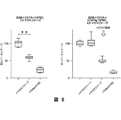

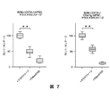

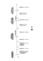


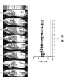


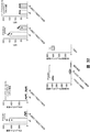
Classifications
-
- C—CHEMISTRY; METALLURGY
- C12—BIOCHEMISTRY; BEER; SPIRITS; WINE; VINEGAR; MICROBIOLOGY; ENZYMOLOGY; MUTATION OR GENETIC ENGINEERING
- C12N—MICROORGANISMS OR ENZYMES; COMPOSITIONS THEREOF; PROPAGATING, PRESERVING, OR MAINTAINING MICROORGANISMS; MUTATION OR GENETIC ENGINEERING; CULTURE MEDIA
- C12N5/00—Undifferentiated human, animal or plant cells, e.g. cell lines; Tissues; Cultivation or maintenance thereof; Culture media therefor
- C12N5/06—Animal cells or tissues; Human cells or tissues
- C12N5/0602—Vertebrate cells
- C12N5/0696—Artificially induced pluripotent stem cells, e.g. iPS
-
- A—HUMAN NECESSITIES
- A61—MEDICAL OR VETERINARY SCIENCE; HYGIENE
- A61K—PREPARATIONS FOR MEDICAL, DENTAL OR TOILETRY PURPOSES
- A61K35/00—Medicinal preparations containing materials or reaction products thereof with undetermined constitution
- A61K35/12—Materials from mammals; Compositions comprising non-specified tissues or cells; Compositions comprising non-embryonic stem cells; Genetically modified cells
- A61K35/14—Blood; Artificial blood
- A61K35/17—Lymphocytes; B-cells; T-cells; Natural killer cells; Interferon-activated or cytokine-activated lymphocytes
-
- A—HUMAN NECESSITIES
- A61—MEDICAL OR VETERINARY SCIENCE; HYGIENE
- A61K—PREPARATIONS FOR MEDICAL, DENTAL OR TOILETRY PURPOSES
- A61K35/00—Medicinal preparations containing materials or reaction products thereof with undetermined constitution
- A61K35/12—Materials from mammals; Compositions comprising non-specified tissues or cells; Compositions comprising non-embryonic stem cells; Genetically modified cells
- A61K35/30—Nerves; Brain; Eyes; Corneal cells; Cerebrospinal fluid; Neuronal stem cells; Neuronal precursor cells; Glial cells; Oligodendrocytes; Schwann cells; Astroglia; Astrocytes; Choroid plexus; Spinal cord tissue
-
- A—HUMAN NECESSITIES
- A61—MEDICAL OR VETERINARY SCIENCE; HYGIENE
- A61K—PREPARATIONS FOR MEDICAL, DENTAL OR TOILETRY PURPOSES
- A61K35/00—Medicinal preparations containing materials or reaction products thereof with undetermined constitution
- A61K35/12—Materials from mammals; Compositions comprising non-specified tissues or cells; Compositions comprising non-embryonic stem cells; Genetically modified cells
- A61K35/34—Muscles; Smooth muscle cells; Heart; Cardiac stem cells; Myoblasts; Myocytes; Cardiomyocytes
-
- A—HUMAN NECESSITIES
- A61—MEDICAL OR VETERINARY SCIENCE; HYGIENE
- A61K—PREPARATIONS FOR MEDICAL, DENTAL OR TOILETRY PURPOSES
- A61K35/00—Medicinal preparations containing materials or reaction products thereof with undetermined constitution
- A61K35/12—Materials from mammals; Compositions comprising non-specified tissues or cells; Compositions comprising non-embryonic stem cells; Genetically modified cells
- A61K35/36—Skin; Hair; Nails; Sebaceous glands; Cerumen; Epidermis; Epithelial cells; Keratinocytes; Langerhans cells; Ectodermal cells
-
- A—HUMAN NECESSITIES
- A61—MEDICAL OR VETERINARY SCIENCE; HYGIENE
- A61P—SPECIFIC THERAPEUTIC ACTIVITY OF CHEMICAL COMPOUNDS OR MEDICINAL PREPARATIONS
- A61P25/00—Drugs for disorders of the nervous system
- A61P25/28—Drugs for disorders of the nervous system for treating neurodegenerative disorders of the central nervous system, e.g. nootropic agents, cognition enhancers, drugs for treating Alzheimer's disease or other forms of dementia
-
- A—HUMAN NECESSITIES
- A61—MEDICAL OR VETERINARY SCIENCE; HYGIENE
- A61P—SPECIFIC THERAPEUTIC ACTIVITY OF CHEMICAL COMPOUNDS OR MEDICINAL PREPARATIONS
- A61P27/00—Drugs for disorders of the senses
- A61P27/02—Ophthalmic agents
- A61P27/04—Artificial tears; Irrigation solutions
-
- A—HUMAN NECESSITIES
- A61—MEDICAL OR VETERINARY SCIENCE; HYGIENE
- A61P—SPECIFIC THERAPEUTIC ACTIVITY OF CHEMICAL COMPOUNDS OR MEDICINAL PREPARATIONS
- A61P9/00—Drugs for disorders of the cardiovascular system
-
- C—CHEMISTRY; METALLURGY
- C12—BIOCHEMISTRY; BEER; SPIRITS; WINE; VINEGAR; MICROBIOLOGY; ENZYMOLOGY; MUTATION OR GENETIC ENGINEERING
- C12N—MICROORGANISMS OR ENZYMES; COMPOSITIONS THEREOF; PROPAGATING, PRESERVING, OR MAINTAINING MICROORGANISMS; MUTATION OR GENETIC ENGINEERING; CULTURE MEDIA
- C12N1/00—Microorganisms, e.g. protozoa; Compositions thereof; Processes of propagating, maintaining or preserving microorganisms or compositions thereof; Processes of preparing or isolating a composition containing a microorganism; Culture media therefor
- C12N1/04—Preserving or maintaining viable microorganisms
-
- C—CHEMISTRY; METALLURGY
- C12—BIOCHEMISTRY; BEER; SPIRITS; WINE; VINEGAR; MICROBIOLOGY; ENZYMOLOGY; MUTATION OR GENETIC ENGINEERING
- C12N—MICROORGANISMS OR ENZYMES; COMPOSITIONS THEREOF; PROPAGATING, PRESERVING, OR MAINTAINING MICROORGANISMS; MUTATION OR GENETIC ENGINEERING; CULTURE MEDIA
- C12N5/00—Undifferentiated human, animal or plant cells, e.g. cell lines; Tissues; Cultivation or maintenance thereof; Culture media therefor
- C12N5/06—Animal cells or tissues; Human cells or tissues
- C12N5/0602—Vertebrate cells
- C12N5/0618—Cells of the nervous system
- C12N5/0619—Neurons
-
- C—CHEMISTRY; METALLURGY
- C12—BIOCHEMISTRY; BEER; SPIRITS; WINE; VINEGAR; MICROBIOLOGY; ENZYMOLOGY; MUTATION OR GENETIC ENGINEERING
- C12N—MICROORGANISMS OR ENZYMES; COMPOSITIONS THEREOF; PROPAGATING, PRESERVING, OR MAINTAINING MICROORGANISMS; MUTATION OR GENETIC ENGINEERING; CULTURE MEDIA
- C12N5/00—Undifferentiated human, animal or plant cells, e.g. cell lines; Tissues; Cultivation or maintenance thereof; Culture media therefor
- C12N5/06—Animal cells or tissues; Human cells or tissues
- C12N5/0602—Vertebrate cells
- C12N5/0618—Cells of the nervous system
- C12N5/0621—Eye cells, e.g. cornea, iris pigmented cells
-
- C—CHEMISTRY; METALLURGY
- C12—BIOCHEMISTRY; BEER; SPIRITS; WINE; VINEGAR; MICROBIOLOGY; ENZYMOLOGY; MUTATION OR GENETIC ENGINEERING
- C12N—MICROORGANISMS OR ENZYMES; COMPOSITIONS THEREOF; PROPAGATING, PRESERVING, OR MAINTAINING MICROORGANISMS; MUTATION OR GENETIC ENGINEERING; CULTURE MEDIA
- C12N5/00—Undifferentiated human, animal or plant cells, e.g. cell lines; Tissues; Cultivation or maintenance thereof; Culture media therefor
- C12N5/06—Animal cells or tissues; Human cells or tissues
- C12N5/0602—Vertebrate cells
- C12N5/0634—Cells from the blood or the immune system
- C12N5/0639—Dendritic cells, e.g. Langherhans cells in the epidermis
-
- C—CHEMISTRY; METALLURGY
- C12—BIOCHEMISTRY; BEER; SPIRITS; WINE; VINEGAR; MICROBIOLOGY; ENZYMOLOGY; MUTATION OR GENETIC ENGINEERING
- C12N—MICROORGANISMS OR ENZYMES; COMPOSITIONS THEREOF; PROPAGATING, PRESERVING, OR MAINTAINING MICROORGANISMS; MUTATION OR GENETIC ENGINEERING; CULTURE MEDIA
- C12N5/00—Undifferentiated human, animal or plant cells, e.g. cell lines; Tissues; Cultivation or maintenance thereof; Culture media therefor
- C12N5/06—Animal cells or tissues; Human cells or tissues
- C12N5/0602—Vertebrate cells
- C12N5/0652—Cells of skeletal and connective tissues; Mesenchyme
- C12N5/0657—Cardiomyocytes; Heart cells
-
- C—CHEMISTRY; METALLURGY
- C12—BIOCHEMISTRY; BEER; SPIRITS; WINE; VINEGAR; MICROBIOLOGY; ENZYMOLOGY; MUTATION OR GENETIC ENGINEERING
- C12N—MICROORGANISMS OR ENZYMES; COMPOSITIONS THEREOF; PROPAGATING, PRESERVING, OR MAINTAINING MICROORGANISMS; MUTATION OR GENETIC ENGINEERING; CULTURE MEDIA
- C12N5/00—Undifferentiated human, animal or plant cells, e.g. cell lines; Tissues; Cultivation or maintenance thereof; Culture media therefor
- C12N5/06—Animal cells or tissues; Human cells or tissues
- C12N5/0602—Vertebrate cells
- C12N5/069—Vascular Endothelial cells
-
- C—CHEMISTRY; METALLURGY
- C12—BIOCHEMISTRY; BEER; SPIRITS; WINE; VINEGAR; MICROBIOLOGY; ENZYMOLOGY; MUTATION OR GENETIC ENGINEERING
- C12N—MICROORGANISMS OR ENZYMES; COMPOSITIONS THEREOF; PROPAGATING, PRESERVING, OR MAINTAINING MICROORGANISMS; MUTATION OR GENETIC ENGINEERING; CULTURE MEDIA
- C12N9/00—Enzymes; Proenzymes; Compositions thereof; Processes for preparing, activating, inhibiting, separating or purifying enzymes
- C12N9/10—Transferases (2.)
- C12N9/12—Transferases (2.) transferring phosphorus containing groups, e.g. kinases (2.7)
- C12N9/1205—Phosphotransferases with an alcohol group as acceptor (2.7.1), e.g. protein kinases
- C12N9/1211—Thymidine kinase (2.7.1.21)
-
- C—CHEMISTRY; METALLURGY
- C12—BIOCHEMISTRY; BEER; SPIRITS; WINE; VINEGAR; MICROBIOLOGY; ENZYMOLOGY; MUTATION OR GENETIC ENGINEERING
- C12N—MICROORGANISMS OR ENZYMES; COMPOSITIONS THEREOF; PROPAGATING, PRESERVING, OR MAINTAINING MICROORGANISMS; MUTATION OR GENETIC ENGINEERING; CULTURE MEDIA
- C12N9/00—Enzymes; Proenzymes; Compositions thereof; Processes for preparing, activating, inhibiting, separating or purifying enzymes
- C12N9/14—Hydrolases (3)
- C12N9/48—Hydrolases (3) acting on peptide bonds (3.4)
- C12N9/50—Proteinases, e.g. Endopeptidases (3.4.21-3.4.25)
- C12N9/64—Proteinases, e.g. Endopeptidases (3.4.21-3.4.25) derived from animal tissue
- C12N9/6421—Proteinases, e.g. Endopeptidases (3.4.21-3.4.25) derived from animal tissue from mammals
- C12N9/6472—Cysteine endopeptidases (3.4.22)
-
- C—CHEMISTRY; METALLURGY
- C12—BIOCHEMISTRY; BEER; SPIRITS; WINE; VINEGAR; MICROBIOLOGY; ENZYMOLOGY; MUTATION OR GENETIC ENGINEERING
- C12N—MICROORGANISMS OR ENZYMES; COMPOSITIONS THEREOF; PROPAGATING, PRESERVING, OR MAINTAINING MICROORGANISMS; MUTATION OR GENETIC ENGINEERING; CULTURE MEDIA
- C12N9/00—Enzymes; Proenzymes; Compositions thereof; Processes for preparing, activating, inhibiting, separating or purifying enzymes
- C12N9/14—Hydrolases (3)
- C12N9/78—Hydrolases (3) acting on carbon to nitrogen bonds other than peptide bonds (3.5)
-
- A—HUMAN NECESSITIES
- A01—AGRICULTURE; FORESTRY; ANIMAL HUSBANDRY; HUNTING; TRAPPING; FISHING
- A01K—ANIMAL HUSBANDRY; CARE OF BIRDS, FISHES, INSECTS; FISHING; REARING OR BREEDING ANIMALS, NOT OTHERWISE PROVIDED FOR; NEW BREEDS OF ANIMALS
- A01K2217/00—Genetically modified animals
- A01K2217/05—Animals comprising random inserted nucleic acids (transgenic)
-
- A—HUMAN NECESSITIES
- A01—AGRICULTURE; FORESTRY; ANIMAL HUSBANDRY; HUNTING; TRAPPING; FISHING
- A01K—ANIMAL HUSBANDRY; CARE OF BIRDS, FISHES, INSECTS; FISHING; REARING OR BREEDING ANIMALS, NOT OTHERWISE PROVIDED FOR; NEW BREEDS OF ANIMALS
- A01K2217/00—Genetically modified animals
- A01K2217/07—Animals genetically altered by homologous recombination
- A01K2217/075—Animals genetically altered by homologous recombination inducing loss of function, i.e. knock out
-
- A—HUMAN NECESSITIES
- A01—AGRICULTURE; FORESTRY; ANIMAL HUSBANDRY; HUNTING; TRAPPING; FISHING
- A01K—ANIMAL HUSBANDRY; CARE OF BIRDS, FISHES, INSECTS; FISHING; REARING OR BREEDING ANIMALS, NOT OTHERWISE PROVIDED FOR; NEW BREEDS OF ANIMALS
- A01K2217/00—Genetically modified animals
- A01K2217/15—Animals comprising multiple alterations of the genome, by transgenesis or homologous recombination, e.g. obtained by cross-breeding
-
- A—HUMAN NECESSITIES
- A01—AGRICULTURE; FORESTRY; ANIMAL HUSBANDRY; HUNTING; TRAPPING; FISHING
- A01K—ANIMAL HUSBANDRY; CARE OF BIRDS, FISHES, INSECTS; FISHING; REARING OR BREEDING ANIMALS, NOT OTHERWISE PROVIDED FOR; NEW BREEDS OF ANIMALS
- A01K2227/00—Animals characterised by species
- A01K2227/10—Mammal
- A01K2227/105—Murine
-
- C—CHEMISTRY; METALLURGY
- C12—BIOCHEMISTRY; BEER; SPIRITS; WINE; VINEGAR; MICROBIOLOGY; ENZYMOLOGY; MUTATION OR GENETIC ENGINEERING
- C12N—MICROORGANISMS OR ENZYMES; COMPOSITIONS THEREOF; PROPAGATING, PRESERVING, OR MAINTAINING MICROORGANISMS; MUTATION OR GENETIC ENGINEERING; CULTURE MEDIA
- C12N2310/00—Structure or type of the nucleic acid
- C12N2310/10—Type of nucleic acid
- C12N2310/20—Type of nucleic acid involving clustered regularly interspaced short palindromic repeats [CRISPRs]
-
- C—CHEMISTRY; METALLURGY
- C12—BIOCHEMISTRY; BEER; SPIRITS; WINE; VINEGAR; MICROBIOLOGY; ENZYMOLOGY; MUTATION OR GENETIC ENGINEERING
- C12N—MICROORGANISMS OR ENZYMES; COMPOSITIONS THEREOF; PROPAGATING, PRESERVING, OR MAINTAINING MICROORGANISMS; MUTATION OR GENETIC ENGINEERING; CULTURE MEDIA
- C12N2500/00—Specific components of cell culture medium
- C12N2500/30—Organic components
- C12N2500/38—Vitamins
-
- C—CHEMISTRY; METALLURGY
- C12—BIOCHEMISTRY; BEER; SPIRITS; WINE; VINEGAR; MICROBIOLOGY; ENZYMOLOGY; MUTATION OR GENETIC ENGINEERING
- C12N—MICROORGANISMS OR ENZYMES; COMPOSITIONS THEREOF; PROPAGATING, PRESERVING, OR MAINTAINING MICROORGANISMS; MUTATION OR GENETIC ENGINEERING; CULTURE MEDIA
- C12N2501/00—Active agents used in cell culture processes, e.g. differentation
- C12N2501/10—Growth factors
- C12N2501/115—Basic fibroblast growth factor (bFGF, FGF-2)
-
- C—CHEMISTRY; METALLURGY
- C12—BIOCHEMISTRY; BEER; SPIRITS; WINE; VINEGAR; MICROBIOLOGY; ENZYMOLOGY; MUTATION OR GENETIC ENGINEERING
- C12N—MICROORGANISMS OR ENZYMES; COMPOSITIONS THEREOF; PROPAGATING, PRESERVING, OR MAINTAINING MICROORGANISMS; MUTATION OR GENETIC ENGINEERING; CULTURE MEDIA
- C12N2501/00—Active agents used in cell culture processes, e.g. differentation
- C12N2501/10—Growth factors
- C12N2501/165—Vascular endothelial growth factor [VEGF]
-
- C—CHEMISTRY; METALLURGY
- C12—BIOCHEMISTRY; BEER; SPIRITS; WINE; VINEGAR; MICROBIOLOGY; ENZYMOLOGY; MUTATION OR GENETIC ENGINEERING
- C12N—MICROORGANISMS OR ENZYMES; COMPOSITIONS THEREOF; PROPAGATING, PRESERVING, OR MAINTAINING MICROORGANISMS; MUTATION OR GENETIC ENGINEERING; CULTURE MEDIA
- C12N2501/00—Active agents used in cell culture processes, e.g. differentation
- C12N2501/20—Cytokines; Chemokines
- C12N2501/23—Interleukins [IL]
- C12N2501/235—Leukemia inhibitory factor [LIF]
-
- C—CHEMISTRY; METALLURGY
- C12—BIOCHEMISTRY; BEER; SPIRITS; WINE; VINEGAR; MICROBIOLOGY; ENZYMOLOGY; MUTATION OR GENETIC ENGINEERING
- C12N—MICROORGANISMS OR ENZYMES; COMPOSITIONS THEREOF; PROPAGATING, PRESERVING, OR MAINTAINING MICROORGANISMS; MUTATION OR GENETIC ENGINEERING; CULTURE MEDIA
- C12N2501/00—Active agents used in cell culture processes, e.g. differentation
- C12N2501/30—Hormones
- C12N2501/33—Insulin
-
- C—CHEMISTRY; METALLURGY
- C12—BIOCHEMISTRY; BEER; SPIRITS; WINE; VINEGAR; MICROBIOLOGY; ENZYMOLOGY; MUTATION OR GENETIC ENGINEERING
- C12N—MICROORGANISMS OR ENZYMES; COMPOSITIONS THEREOF; PROPAGATING, PRESERVING, OR MAINTAINING MICROORGANISMS; MUTATION OR GENETIC ENGINEERING; CULTURE MEDIA
- C12N2501/00—Active agents used in cell culture processes, e.g. differentation
- C12N2501/30—Hormones
- C12N2501/38—Hormones with nuclear receptors
- C12N2501/385—Hormones with nuclear receptors of the family of the retinoic acid recptor, e.g. RAR, RXR; Peroxisome proliferator-activated receptor [PPAR]
-
- C—CHEMISTRY; METALLURGY
- C12—BIOCHEMISTRY; BEER; SPIRITS; WINE; VINEGAR; MICROBIOLOGY; ENZYMOLOGY; MUTATION OR GENETIC ENGINEERING
- C12N—MICROORGANISMS OR ENZYMES; COMPOSITIONS THEREOF; PROPAGATING, PRESERVING, OR MAINTAINING MICROORGANISMS; MUTATION OR GENETIC ENGINEERING; CULTURE MEDIA
- C12N2501/00—Active agents used in cell culture processes, e.g. differentation
- C12N2501/40—Regulators of development
- C12N2501/415—Wnt; Frizzeled
-
- C—CHEMISTRY; METALLURGY
- C12—BIOCHEMISTRY; BEER; SPIRITS; WINE; VINEGAR; MICROBIOLOGY; ENZYMOLOGY; MUTATION OR GENETIC ENGINEERING
- C12N—MICROORGANISMS OR ENZYMES; COMPOSITIONS THEREOF; PROPAGATING, PRESERVING, OR MAINTAINING MICROORGANISMS; MUTATION OR GENETIC ENGINEERING; CULTURE MEDIA
- C12N2501/00—Active agents used in cell culture processes, e.g. differentation
- C12N2501/60—Transcription factors
- C12N2501/602—Sox-2
-
- C—CHEMISTRY; METALLURGY
- C12—BIOCHEMISTRY; BEER; SPIRITS; WINE; VINEGAR; MICROBIOLOGY; ENZYMOLOGY; MUTATION OR GENETIC ENGINEERING
- C12N—MICROORGANISMS OR ENZYMES; COMPOSITIONS THEREOF; PROPAGATING, PRESERVING, OR MAINTAINING MICROORGANISMS; MUTATION OR GENETIC ENGINEERING; CULTURE MEDIA
- C12N2501/00—Active agents used in cell culture processes, e.g. differentation
- C12N2501/60—Transcription factors
- C12N2501/603—Oct-3/4
-
- C—CHEMISTRY; METALLURGY
- C12—BIOCHEMISTRY; BEER; SPIRITS; WINE; VINEGAR; MICROBIOLOGY; ENZYMOLOGY; MUTATION OR GENETIC ENGINEERING
- C12N—MICROORGANISMS OR ENZYMES; COMPOSITIONS THEREOF; PROPAGATING, PRESERVING, OR MAINTAINING MICROORGANISMS; MUTATION OR GENETIC ENGINEERING; CULTURE MEDIA
- C12N2501/00—Active agents used in cell culture processes, e.g. differentation
- C12N2501/60—Transcription factors
- C12N2501/604—Klf-4
-
- C—CHEMISTRY; METALLURGY
- C12—BIOCHEMISTRY; BEER; SPIRITS; WINE; VINEGAR; MICROBIOLOGY; ENZYMOLOGY; MUTATION OR GENETIC ENGINEERING
- C12N—MICROORGANISMS OR ENZYMES; COMPOSITIONS THEREOF; PROPAGATING, PRESERVING, OR MAINTAINING MICROORGANISMS; MUTATION OR GENETIC ENGINEERING; CULTURE MEDIA
- C12N2501/00—Active agents used in cell culture processes, e.g. differentation
- C12N2501/60—Transcription factors
- C12N2501/606—Transcription factors c-Myc
-
- C—CHEMISTRY; METALLURGY
- C12—BIOCHEMISTRY; BEER; SPIRITS; WINE; VINEGAR; MICROBIOLOGY; ENZYMOLOGY; MUTATION OR GENETIC ENGINEERING
- C12N—MICROORGANISMS OR ENZYMES; COMPOSITIONS THEREOF; PROPAGATING, PRESERVING, OR MAINTAINING MICROORGANISMS; MUTATION OR GENETIC ENGINEERING; CULTURE MEDIA
- C12N2501/00—Active agents used in cell culture processes, e.g. differentation
- C12N2501/60—Transcription factors
- C12N2501/608—Lin28
-
- C—CHEMISTRY; METALLURGY
- C12—BIOCHEMISTRY; BEER; SPIRITS; WINE; VINEGAR; MICROBIOLOGY; ENZYMOLOGY; MUTATION OR GENETIC ENGINEERING
- C12N—MICROORGANISMS OR ENZYMES; COMPOSITIONS THEREOF; PROPAGATING, PRESERVING, OR MAINTAINING MICROORGANISMS; MUTATION OR GENETIC ENGINEERING; CULTURE MEDIA
- C12N2506/00—Differentiation of animal cells from one lineage to another; Differentiation of pluripotent cells
- C12N2506/11—Differentiation of animal cells from one lineage to another; Differentiation of pluripotent cells from blood or immune system cells
-
- C—CHEMISTRY; METALLURGY
- C12—BIOCHEMISTRY; BEER; SPIRITS; WINE; VINEGAR; MICROBIOLOGY; ENZYMOLOGY; MUTATION OR GENETIC ENGINEERING
- C12N—MICROORGANISMS OR ENZYMES; COMPOSITIONS THEREOF; PROPAGATING, PRESERVING, OR MAINTAINING MICROORGANISMS; MUTATION OR GENETIC ENGINEERING; CULTURE MEDIA
- C12N2506/00—Differentiation of animal cells from one lineage to another; Differentiation of pluripotent cells
- C12N2506/13—Differentiation of animal cells from one lineage to another; Differentiation of pluripotent cells from connective tissue cells, from mesenchymal cells
- C12N2506/1307—Differentiation of animal cells from one lineage to another; Differentiation of pluripotent cells from connective tissue cells, from mesenchymal cells from adult fibroblasts
-
- C—CHEMISTRY; METALLURGY
- C12—BIOCHEMISTRY; BEER; SPIRITS; WINE; VINEGAR; MICROBIOLOGY; ENZYMOLOGY; MUTATION OR GENETIC ENGINEERING
- C12N—MICROORGANISMS OR ENZYMES; COMPOSITIONS THEREOF; PROPAGATING, PRESERVING, OR MAINTAINING MICROORGANISMS; MUTATION OR GENETIC ENGINEERING; CULTURE MEDIA
- C12N2506/00—Differentiation of animal cells from one lineage to another; Differentiation of pluripotent cells
- C12N2506/45—Differentiation of animal cells from one lineage to another; Differentiation of pluripotent cells from artificially induced pluripotent stem cells
-
- C—CHEMISTRY; METALLURGY
- C12—BIOCHEMISTRY; BEER; SPIRITS; WINE; VINEGAR; MICROBIOLOGY; ENZYMOLOGY; MUTATION OR GENETIC ENGINEERING
- C12N—MICROORGANISMS OR ENZYMES; COMPOSITIONS THEREOF; PROPAGATING, PRESERVING, OR MAINTAINING MICROORGANISMS; MUTATION OR GENETIC ENGINEERING; CULTURE MEDIA
- C12N2533/00—Supports or coatings for cell culture, characterised by material
- C12N2533/50—Proteins
- C12N2533/54—Collagen; Gelatin
-
- C—CHEMISTRY; METALLURGY
- C12—BIOCHEMISTRY; BEER; SPIRITS; WINE; VINEGAR; MICROBIOLOGY; ENZYMOLOGY; MUTATION OR GENETIC ENGINEERING
- C12N—MICROORGANISMS OR ENZYMES; COMPOSITIONS THEREOF; PROPAGATING, PRESERVING, OR MAINTAINING MICROORGANISMS; MUTATION OR GENETIC ENGINEERING; CULTURE MEDIA
- C12N2533/00—Supports or coatings for cell culture, characterised by material
- C12N2533/90—Substrates of biological origin, e.g. extracellular matrix, decellularised tissue
-
- Y—GENERAL TAGGING OF NEW TECHNOLOGICAL DEVELOPMENTS; GENERAL TAGGING OF CROSS-SECTIONAL TECHNOLOGIES SPANNING OVER SEVERAL SECTIONS OF THE IPC; TECHNICAL SUBJECTS COVERED BY FORMER USPC CROSS-REFERENCE ART COLLECTIONS [XRACs] AND DIGESTS
- Y02—TECHNOLOGIES OR APPLICATIONS FOR MITIGATION OR ADAPTATION AGAINST CLIMATE CHANGE
- Y02A—TECHNOLOGIES FOR ADAPTATION TO CLIMATE CHANGE
- Y02A50/00—TECHNOLOGIES FOR ADAPTATION TO CLIMATE CHANGE in human health protection, e.g. against extreme weather
- Y02A50/30—Against vector-borne diseases, e.g. mosquito-borne, fly-borne, tick-borne or waterborne diseases whose impact is exacerbated by climate change
Landscapes
- Health & Medical Sciences (AREA)
- Engineering & Computer Science (AREA)
- Life Sciences & Earth Sciences (AREA)
- Biomedical Technology (AREA)
- Chemical & Material Sciences (AREA)
- Bioinformatics & Cheminformatics (AREA)
- Zoology (AREA)
- Organic Chemistry (AREA)
- Biotechnology (AREA)
- Wood Science & Technology (AREA)
- Genetics & Genomics (AREA)
- General Health & Medical Sciences (AREA)
- Cell Biology (AREA)
- Microbiology (AREA)
- Biochemistry (AREA)
- General Engineering & Computer Science (AREA)
- Medicinal Chemistry (AREA)
- Developmental Biology & Embryology (AREA)
- Animal Behavior & Ethology (AREA)
- Veterinary Medicine (AREA)
- Public Health (AREA)
- Pharmacology & Pharmacy (AREA)
- Immunology (AREA)
- Cardiology (AREA)
- Neurology (AREA)
- Virology (AREA)
- Neurosurgery (AREA)
- Molecular Biology (AREA)
- Epidemiology (AREA)
- Rheumatology (AREA)
- Transplantation (AREA)
- Chemical Kinetics & Catalysis (AREA)
- Nuclear Medicine, Radiotherapy & Molecular Imaging (AREA)
- General Chemical & Material Sciences (AREA)
- Ophthalmology & Optometry (AREA)
- Vascular Medicine (AREA)
- Hematology (AREA)
- Hospice & Palliative Care (AREA)
- Psychiatry (AREA)
- Heart & Thoracic Surgery (AREA)
Abstract
本発明は、普遍的に許容可能な「容易に入手可能である」低免疫原性多能性細胞及びその分化した心臓、内皮、神経、膵島又は網膜色素細胞を提供する。このような低免疫細胞は、それを必要とする患者を処置するために使用される。本細胞は、免疫応答を惹起する主要免疫抗原を欠き、食作用性のエンドサイトーシスを回避するために改変されている。The present invention provides universally acceptable "easily available" hypoimmunogenic pluripotent cells and differentiated heart, endothelial, nerve, islet or retinal pigment cells thereof. Such hypoimmune cells are used to treat patients in need of it. The cells lack the major immune antigens that elicit an immune response and have been modified to avoid phagocytic endocytosis.
Description
関連出願の相互参照
本願は、全てその全体において本明細書中で参照により組み込まれる、2018年7月17日に提出の米国仮特許出願第62/698,965号明細書、2018年7月17日に提出の同第62/698,973号明細書、2018年7月17日に提出の同第62/698,978号明細書、2018年7月17日に提出の同第62/698,981号明細書及び2018年7月17日に提出の同第62/698,984号明細書の優先権を主張する。
Mutual Reference of Related Applications This application is incorporated herein by reference in its entirety, US Provisional Patent Application No. 62 / 698,965, submitted July 17, 2018, July 17, 2018. The same 62 / 698,973 submission on the same day, the 62 / 698,978 specification submitted on July 17, 2018, the 62/698, the same submitted on July 17, 2018, Claim the priority of Specification 981 and Specification 62/698,984 submitted on July 17, 2018.
再生細胞療法は、損傷臓器及び組織を再生させるための重要で有望な処置である。移植用の臓器は入手が困難であり、待機時間が長いことから、当然のことだが、容易に入手可能な細胞株を患者に移植することによって組織を再生可能であることは好ましい。再生細胞療法は、動物モデル(例えば心筋梗塞後)での移植後に障害組織を回復させるための有望な最初の結果を示した。しかし、移植レシピエントの免疫系が同種物質を拒絶する傾向があることにより、治療の潜在的効果が大きく低下し、このような処置の周囲の起こり得るプラス効果が低下する。 Regenerative cell therapy is an important and promising procedure for regenerating injured organs and tissues. Naturally, it is preferable that the tissue can be regenerated by transplanting an easily available cell line into a patient because the organ for transplantation is difficult to obtain and the waiting time is long. Regenerative cell therapy has shown promising first results for recovery of damaged tissue after transplantation in animal models (eg, after myocardial infarction). However, the tendency of the transplant recipient's immune system to reject allogeneic substances greatly reduces the potential effect of treatment and reduces the possible positive effects surrounding such treatment.
従って、本発明は、普遍的に許容可能な「容易に入手可能である」低免疫原性多能性細胞及びその分化した心臓、内皮、神経、膵島又は網膜色素細胞を提供する。このような低免疫細胞が、それを必要とする患者を処置するために使用される。これらの細胞は、免疫応答を惹起する主要免疫抗原を欠き、食作用性のエンドサイトーシスを回避するように改変される。 Accordingly, the present invention provides universally acceptable "easily available" hypoimmunogenic pluripotent cells and their differentiated heart, endothelium, nerve, islets or retinal pigment cells. Such hypoimmune cells are used to treat patients in need of it. These cells lack the major immune antigens that elicit an immune response and are modified to avoid phagocytic endocytosis.
再生細胞療法は、損傷臓器及び組織を再生させるための重要で有望な治療である。移植用の臓器は入手が困難であり、待機時間が長いことから、当然のことだが、容易に入手可能な細胞株を患者に移植することによって組織を再生可能であることは好ましい。再生細胞療法は、動物モデル(例えば心筋梗塞後)での移植後に障害組織を回復させるための有望な最初の結果を示した。しかし、移植レシピエントの免疫系が同種物質を拒絶する傾向があることにより、治療の潜在的効果が大きく低下し、このような処置の周囲の起こり得るプラス効果が低下する。 Regenerative cell therapy is an important and promising treatment for the regeneration of injured organs and tissues. Naturally, it is preferable that the tissue can be regenerated by transplanting an easily available cell line into a patient because the organ for transplantation is difficult to obtain and the waiting time is long. Regenerative cell therapy has shown promising first results for recovery of damaged tissue after transplantation in animal models (eg, after myocardial infarction). However, the tendency of the transplant recipient's immune system to reject allogeneic substances greatly reduces the potential effect of treatment and reduces the possible positive effects surrounding such treatment.
自己誘導多能性幹細胞(iPSC)は理論的に患者特異的な細胞に基づく臓器修復ストラテジーのための無限の細胞供給源を構成する。しかし、それらの作製は、技術及び製造の面で困難な点があり、何らかの急性処置モダリティーを概念的に妨げる冗長な工程である。同種iPSCに基づく治療法は、製造の観点からより容易であり、十分にスクリーニングされ、標準化された高品質の細胞生成物の作製を可能にする。しかし、それらが同種起源であるため、このような細胞生成物は拒絶される。細胞の抗原性の低下又は排除により、普遍的に許容可能な細胞生成物が作製され得る。多能性幹細胞は3つ胚葉のあらゆる細胞タイプに分化し得るので、幹細胞療法の適用の可能性は幅広い。移植部位の臓器環境で分化及び成熟を継続する祖先細胞を移植することによって、エクスビボ又はインビボで分化が行われ得る。エクスビボ分化によって、研究者又は医師が手順を密に監視することが可能になり、細胞の適正な集団が移植前に作製されることが確実になる。 Self-induced pluripotent stem cells (iPSCs) theoretically constitute an endless source of cells for patient-specific cell-based organ repair strategies. However, their production is technically and manufacturing difficult and is a redundant process that conceptually impedes some acute treatment modality. Allogeneic iPSC-based therapies are easier from a manufacturing standpoint and allow the production of well-screened, standardized, high-quality cell products. However, such cell products are rejected because they are of allogeneic origin. Decreased or eliminated the antigenicity of cells can produce universally acceptable cell products. Since pluripotent stem cells can differentiate into any of the three germ layer cell types, the potential for stem cell therapy is wide. Differentiation can occur in vivo or in vivo by transplanting ancestral cells that continue to differentiate and mature in the organ environment of the transplant site. Exvivo differentiation allows researchers or physicians to closely monitor the procedure and ensures that the proper population of cells is produced prior to transplantation.
しかし、殆どの場合、臨床移植療法において、未分化多能性幹細胞は、それらが奇形腫を形成する傾向ゆえに回避される。むしろ、このような治療法は、分化した細胞(例えば心不全に罹患している患者の心筋に移植する幹細胞由来心筋細胞)を使用する傾向がある。このような多能性細胞又は組織の臨床適用は、それらの移植後の細胞の増殖及び生存を制御する「安全機能」から利益を得る。 However, in most cases, in clinical transplantation therapy, undifferentiated pluripotent stem cells are avoided because they tend to form teratomas. Rather, such therapies tend to use differentiated cells (eg, stem cell-derived cardiomyocytes that are transplanted into the myocardium of patients suffering from heart failure). The clinical application of such pluripotent cells or tissues benefits from the "safety function" that controls the proliferation and survival of the cells after their transplantation.
本技術は、疾患又は欠損細胞を再生させるか又は置き換えるために使用される細胞を作製することが可能な幹細胞を追求する。迅速に増殖し、多くの可能な細胞タイプに分化するので、多能性幹細胞(PSC)が使用され得る。 The present technique seeks stem cells capable of producing cells used to regenerate or replace diseased or defective cells. Pluripotent stem cells (PSCs) can be used because they proliferate rapidly and differentiate into many possible cell types.
今まで、PSCに基づくアプローチの前臨床での成功は、免疫抑制又は免疫欠損モデルでのみ、又は細胞が封入され、宿主の免疫系から保護される場合にのみ、達成されている。しかし、同種臓器移植で使用されるような全身的な免疫抑制は、再生アプローチに正当なものではない。免疫抑制薬は、重い副作用があり、感染及び悪性腫瘍のリスクを顕著に上昇させる。 To date, preclinical success of PSC-based approaches has only been achieved in immunosuppressive or immunodeficient models, or when cells are encapsulated and protected from the host's immune system. However, systemic immunosuppression, such as that used in allogeneic organ transplantation, is not justified for a regenerative approach. Immunosuppressive drugs have serious side effects and significantly increase the risk of infection and malignancies.
当技術分野において、再生医学で使用され得る、低免疫原性多能性幹細胞から作製される細胞が求められている。 In the art, there is a need for cells made from hypoimmunogenic pluripotent stem cells that can be used in regenerative medicine.
いくつかの態様では、低免疫多能性幹細胞(HIP細胞)から分化した、単離された改変低免疫心臓、内皮、神経、膵島又は網膜色素細胞が本明細書中で提供される。HIP細胞は、例えば、内在性β−2ミクログロブリン(B2M)遺伝子活性が低下しているか又は排除されており、内在性クラスIIトランス活性化因子(CIITA)遺伝子活性が低下しているか又は排除されており、CD47発現が上昇している。 In some embodiments, isolated modified hypoimmune heart, endothelium, nerve, islets or retinal pigment cells differentiated from hypoimmune pluripotent stem cells (HIP cells) are provided herein. HIP cells have, for example, reduced or eliminated endogenous β-2 microglobulin (B2M) gene activity and reduced or eliminated endogenous Class II transactivator (CIITA) gene activity. CD47 expression is elevated.
一部の実施形態では、HIP細胞はヒト改変誘導多能性幹細胞(ヒト改変iPSC)であり、B2M遺伝子はヒトB2M遺伝子であり、CIITA遺伝子はヒトB2M遺伝子であり、CD47発現上昇は、プロモーターの制御下でヒトCD47遺伝子の少なくとも1コピーを細胞に導入した結果である。ある一定の実施形態では、HIPは、マウス改変誘導多能性幹細胞(マウス改変iPSC)であり、B2M遺伝子はマウスB2M遺伝子であり、CIITA遺伝子はマウスB2M遺伝子であり、CD47発現上昇は、プロモーターの制御下でマウスCD47遺伝子の少なくとも1コピーを細胞に導入した結果である。一部の例では、B2M遺伝子活性の排除は、B2M遺伝子の両アレルを破壊するクラスター化された規則的配置の短回文配列リピート(Clustered Regularly Interspaced Short Palindromic Repeats)(CRISPR)/Cas9反応の結果である。一部の例では、CIITA遺伝子活性の排除は、CIITA遺伝子の両アレルを破壊するCRISPR/Cas9反応の結果である。 In some embodiments, the HIP cell is a human modified induced pluripotent stem cell (human modified iPSC), the B2M gene is a human B2M gene, the CIITA gene is a human B2M gene, and elevated CD47 expression is a promoter. This is the result of introducing at least one copy of the human CD47 gene into cells under control. In certain embodiments, the HIP is a mouse-modified induced pluripotent stem cell (mouse-modified iPSC), the B2M gene is the mouse B2M gene, the CIITA gene is the mouse B2M gene, and elevated CD47 expression is the promoter. This is the result of introducing at least one copy of the mouse CD47 gene into cells under control. In some examples, elimination of B2M gene activity is the result of a clustered, regularly arranged short palindromic repeats (CRISPR) / Cas9 reaction that disrupts both alleles of the B2M gene. Is. In some examples, elimination of CIITA gene activity is the result of a CRISPR / Cas9 reaction that disrupts both alleles of the CIITA gene.
一部の実施形態では、本方法は、HIP細胞を死に誘導するトリガー物質により活性化される自殺遺伝子をさらに含む。一部の実施形態では、自殺遺伝子は単純ヘルペスウイルスチミジンキナーゼ(HSV−tk)遺伝子であり、トリガー物質はガンシクロビルである。一部の例では、HSV−tk遺伝子は、配列番号4に対して少なくとも90%の配列同一性を含むタンパク質をコードする。他の例では、HSV−tk遺伝子は、配列番号4のアミノ酸配列を含むタンパク質をコードする。 In some embodiments, the method further comprises a suicide gene activated by a triggering agent that induces death of HIP cells. In some embodiments, the suicide gene is the herpes simplex virus thymidine kinase (HSV-tk) gene and the triggering agent is ganciclovir. In some examples, the HSV-tk gene encodes a protein that contains at least 90% sequence identity to SEQ ID NO: 4. In another example, the HSV-tk gene encodes a protein comprising the amino acid sequence of SEQ ID NO: 4.
他の実施形態では、自殺遺伝子はエシェリキア・コリ(Escherichia coli)シトシンデアミナーゼ(CD)遺伝子であり、トリガー物質は5−フルオロシトシン(5−FC)である。一部の例では、CD遺伝子は、配列番号5に対して少なくとも90%の配列同一性を含むタンパク質をコードする。他の例では、CD遺伝子は、配列番号5のアミノ酸配列を含むタンパク質をコードする。 In other embodiments, the suicide gene is the Escherichia coli cytosine deaminase (CD) gene and the triggering agent is 5-fluorocytosine (5-FC). In some examples, the CD gene encodes a protein that contains at least 90% sequence identity to SEQ ID NO: 5. In another example, the CD gene encodes a protein comprising the amino acid sequence of SEQ ID NO: 5.
他の実施形態では、自殺遺伝子は誘導型カスパーゼ9タンパク質をコードし、トリガー物質は二量体誘導化合物(CID)である。一部の例では、誘導型カスパーゼ9タンパク質は、配列番号6に対して少なくとも90%の配列同一性を含む。他の例では、誘導型カスパーゼ9タンパク質は、配列番号6のアミノ酸配列を含む。一部の例では、CIDは化合物AP1903である。 In other embodiments, the suicide gene encodes an inducible caspase-9 protein and the triggering agent is a dimer-inducing compound (CID). In some examples, the inducible caspase-9 protein contains at least 90% sequence identity to SEQ ID NO: 6. In another example, the inducible caspase-9 protein comprises the amino acid sequence of SEQ ID NO: 6. In some examples, the CID is compound AP1903.
一部の実施形態では、単離低免疫心臓細胞は、心筋細胞、結節心筋細胞、伝導性心筋細胞、作業心筋細胞、心筋細胞前駆体、心筋細胞祖先細胞、心臓幹細胞及び心臓筋肉細胞からなる群から選択される。 In some embodiments, isolated low-immunity cardiomyocytes are a group consisting of cardiomyocytes, nodular cardiomyocytes, conductive cardiomyocytes, working cardiomyocytes, cardiomyocyte precursors, cardiomyocyte ancestral cells, cardiac stem cells and cardiac muscle cells. Is selected from.
一部の態様では、心臓状態又は疾患に罹患している患者を処置する方法が本明細書中で提供される。本方法は、治療的有効量の、本明細書中に記載の単離された改変低免疫心臓細胞の何れか1つの集団を含む組成物を投与することを含む。一部の実施形態では、本組成物は、治療的に有効な担体をさらに含む。 In some embodiments, methods of treating a patient suffering from a cardiac condition or disease are provided herein. The method comprises administering a therapeutically effective amount of a composition comprising any one population of isolated modified hypoimmune heart cells described herein. In some embodiments, the composition further comprises a therapeutically effective carrier.
一部の実施形態では、投与は、患者の心臓組織への移植、静脈内注射、動脈内注射、冠動脈内注射、筋肉内注射、腹腔内注射、心筋内注射、経心内膜注射、経心外膜注射又は点滴を含む。 In some embodiments, administration is transplantation into the patient's heart tissue, intravenous injection, intraarterial injection, intracoronary injection, intramuscular injection, intraperitoneal injection, intramyocardial injection, transcardiac injection, transcardiac. Includes outer membrane injection or instillation.
一部の実施形態では、心臓状態又は疾患は、小児心筋症、加齢性心筋症、拡張型心筋症、肥大型心筋症、拘束型心筋症、慢性虚血性心筋症、周産期心筋症、炎症性心筋症、他の心筋症、心筋炎、心筋虚血性再灌流障害、心室機能不全、心不全、うっ血性心不全、冠動脈疾患、末期心疾患、アテローム性動脈硬化症、虚血、高血圧、再狭窄、狭心症、リウマチ性心臓、動脈炎又は循環器疾患からなる群から選択される。 In some embodiments, the cardiac condition or disease is childhood cardiomyopathy, age-related cardiomyopathy, dilated cardiomyopathy, hypertrophic cardiomyopathy, restraint cardiomyopathy, chronic ischemic cardiomyopathy, perinatal cardiomyopathy, Inflammatory cardiomyopathy, other cardiomyopathy, cardiomyopathy, myocardial ischemic reperfusion disorder, ventricular dysfunction, heart failure, congestive heart failure, coronary artery disease, end-stage heart disease, atherosclerosis, ischemia, hypertension, re-stenosis , Cardiomyopathy, rheumatic heart, arteritis or cardiomyopathy.
一部の態様では、インビトロ分化により低免疫多能性細胞(HIP細胞)集団から低免疫心臓細胞集団を作製する方法が本明細書中で提供され、HIP細胞において、内在性β−2ミクログロブリン(B2M)遺伝子活性及び内在性クラスIIトランス活性化因子(CIITA)遺伝子活性が排除され、CD47発現が上昇している。本方法は、(a)GSK阻害剤を含む培地中でHIP細胞集団を培養し;(b)前駆心臓細胞集団を作製するために、WNTアンタゴニスを含む培地中でHIP細胞集団を培養し;(c)低免疫心臓細胞集団を作製するために、インスリンを含む培地中で前駆心臓細胞集団を培養することを含む。 In some embodiments, a method of producing a hypoimmune cardiac cell population from a hypoimmune pluripotent cell (HIP cell) population by in vitro differentiation is provided herein in HIP cells, an endogenous β-2 microglobulin. (B2M) gene activity and endogenous Class II trans-activator (CIITA) gene activity have been eliminated and CD47 expression has been elevated. The method comprises (a) culturing a HIP cell population in a medium containing a GSK inhibitor; (b) culturing a HIP cell population in a medium containing WNT antagonis to generate a prodromal heart cell population; c) In order to generate a low-immunity heart cell population, it involves culturing the precursor heart cell population in a medium containing insulin.
一部の実施形態では、GSK阻害剤は、CHIR−99021、その誘導体又はその変異体である。一部の例では、GSK阻害剤は、約2μM〜約10μMの範囲の濃度である。一部の実施形態では、WNTアンタゴニスは、IWR1、その誘導体又はその変異体である。一部の例では、WNTアンタゴニスは、約2μM〜約10μMの範囲の濃度である。 In some embodiments, the GSK inhibitor is CHIR-99021, a derivative thereof or a variant thereof. In some examples, the GSK inhibitor has a concentration in the range of about 2 μM to about 10 μM. In some embodiments, the WNT antagonis is IWR1, a derivative thereof or a variant thereof. In some examples, WNT antagonis has a concentration in the range of about 2 μM to about 10 μM.
一部の実施形態では、本方法は、HIP細胞が自殺遺伝子を含む場合、トリガー物質を含む培地中で段階(c)の前駆心臓細胞集団を培養することをさらに含み、自殺遺伝子が単純ヘルペスウイルスチミジンキナーゼ(HSV−tk)遺伝子である場合、トリガー物質はガンシクロビルであるか、自殺遺伝子がエシェリキア・コリ(Escherichia coli)シトシンデアミナーゼ(CD)遺伝子である場合、トリガー物質は5−フルオロシトシン(5−FC)であるか、又は自殺遺伝子が誘導型カスパーゼ9タンパク質をコードする場合、トリガー物質は二量体誘導化合物(CID)である。ある一定の実施形態では、本方法は、HIP細胞が自殺遺伝子を含む場合、トリガー物質を含む培地中で段階(d)の低免疫心臓細胞集団を培養することをさらに含み、自殺遺伝子が単純ヘルペスウイルスチミジンキナーゼ(HSV−tk)遺伝子である場合、トリガー物質はガンシクロビルであるか、自殺遺伝子がエシェリキア・コリ(Escherichia coli)シトシンデアミナーゼ(CD)遺伝子である場合、トリガー物質は5−フルオロシトシン(5−FC)であるか、又は自殺遺伝子が誘導型カスパーゼ9タンパク質をコードする場合、トリガー物質は二量体誘導化合物(CID)である。 In some embodiments, the method further comprises culturing the progenitor cardiac cell population of step (c) in a medium containing a triggering agent if the HIP cells contain the suicide gene, the suicide gene being a simple herpesvirus. If it is a thymidine kinase (HSV-tk) gene, the trigger substance is gancyclovir, or if the suicide gene is the Escherichia coli citosine deaminase (CD) gene, the trigger substance is 5-fluorocytosine (5-). If FC) or the suicide gene encodes an inducible caspase-9 protein, the triggering agent is a dimer-inducing compound (CID). In certain embodiments, the method further comprises culturing the low-immunity cardiac cell population of step (d) in a medium containing the triggering agent if the HIP cells contain the suicide gene, the suicide gene being simple herpes. If it is a viral thymidine kinase (HSV-tk) gene, the trigger substance is gancyclovir, or if the suicide gene is the Escherichia coli citosine deaminase (CD) gene, the trigger substance is 5-fluorocytosine (5). -FC) or if the suicide gene encodes an inducible caspase-9 protein, the triggering agent is a dimer-inducing compound (CID).
一部の実施形態では、本方法は、非心臓細胞から低免疫心臓細胞集団を単離することをさらに含む。他の実施形態では、本方法は、単離した低免疫心臓細胞集団を凍結保存することをさらに含む。 In some embodiments, the method further comprises isolating a low-immune cardiac cell population from non-cardiac cells. In other embodiments, the method further comprises cryopreserving an isolated hypoimmune cardiac cell population.
一部の実施形態では、単離された改変低免疫内皮細胞は、毛細血管内皮細胞、血管内皮細胞、大動脈内皮細胞、脳内皮細胞及び腎臓内皮細胞からなる群から選択される。 In some embodiments, the isolated modified hypoimmune endothelial cells are selected from the group consisting of capillary endothelial cells, vascular endothelial cells, aortic endothelial cells, brain endothelial cells and kidney endothelial cells.
一部の態様では、血管状態又は疾患に罹患している患者を処置する方法が本明細書中で提供される。一部の実施形態では、本方法は、単離された改変低免疫内皮細胞集団の治療的有効量を含む組成物を投与することを含む。 In some embodiments, methods of treating a patient suffering from a vascular condition or disease are provided herein. In some embodiments, the method comprises administering a composition comprising a therapeutically effective amount of an isolated modified hypoimmune endothelial cell population.
本方法は、本明細書中に記載の単離された改変低免疫内皮細胞のうち何れか1つの集団の治療的有効量を含む組成物を投与することを含む。一部の実施形態では、本組成物は、治療的に有効な担体をさらに含む。一部の実施形態では、投与は、患者の心臓組織への移植、静脈内注射、動脈内注射、冠動脈内注射、筋肉内注射、腹腔内注射、心筋内注射、経心内膜注射、経心外膜注射又は点滴を含む。 The method comprises administering a composition comprising a therapeutically effective amount of any one of the isolated modified hypoimmune endothelial cells described herein. In some embodiments, the composition further comprises a therapeutically effective carrier. In some embodiments, administration is transplantation into the patient's heart tissue, intravenous injection, intraarterial injection, intracoronary injection, intramuscular injection, intraperitoneal injection, intramyocardial injection, transcardiac injection, transcardiac. Includes outer membrane injection or instillation.
一部の実施形態では、血管状態又は疾患は、血管損傷、循環器疾患、血管疾患、虚血性疾患、心筋梗塞、うっ血性心不全、高血圧、虚血性組織損傷、虚血肢、脳卒中、神経障害及び脳血管疾患からなる群から選択される。 In some embodiments, the vascular condition or disease is vascular injury, cardiovascular disease, vascular disease, ischemic disease, myocardial infarction, congestive heart failure, hypertension, ischemic tissue damage, ischemic limbs, stroke, neuropathy and Selected from the group consisting of cerebrovascular disease.
一部の態様では、インビトロ分化によって低免疫原性多能性幹細胞(HIP細胞)の集団から低免疫内皮細胞集団を作製する方法が本明細書中で提供され、HIP細胞において、内在性β−2ミクログロブリン(B2M)遺伝子活性及び内在性クラスIIトランス活性化因子(CIITA)遺伝子活性が排除されており、CD47発現が上昇している。本方法は、(a)GSK阻害剤を含む第1の培地中でHIP細胞集団を培養し;(b)前駆内皮細胞集団を作製するために、VEGF及びbFGFを含む第2の培地中でHIP細胞集団を培養し;(c)低免疫内皮細胞集団を作製するために、ROCK阻害剤及びALK阻害剤を含む第3の培地中で前駆内皮細胞集団を培養することを含む。 In some embodiments, a method of producing a hypoimmune endothelial cell population from a population of hypoimmune pluripotent stem cells (HIP cells) by in vitro differentiation is provided herein and in HIP cells, endogenous β-. 2 Microglobulin (B2M) gene activity and endogenous class II transactivator (CIITA) gene activity have been eliminated and CD47 expression has been elevated. The method comprises (a) culturing a HIP cell population in a first medium containing a GSK inhibitor; (b) HIP in a second medium containing VEGF and bFGF to generate a precursor endothelial cell population. Culturing a cell population; (c) culturing a precursor endothelial cell population in a third medium containing a ROCK inhibitor and an ALK inhibitor to create a hypoimmune endothelial cell population.
一部の実施形態では、GSK阻害剤は、CHIR−99021、その誘導体又はその変異体である。一部の例では、GSK阻害剤は約1μM〜約10μMの範囲の濃度である。一部の実施形態では、ROCK阻害剤は、Y−27632、その誘導体又はその変異体である。一部の例では、ROCK阻害剤は約1μM〜約20μMの範囲の濃度である。一部の実施形態では、ALK阻害剤は、SB−431542、その誘導体又はその変異体である。一部の例では、ALK阻害剤は約0.5μM〜約10μMの濃度範囲である。 In some embodiments, the GSK inhibitor is CHIR-99021, a derivative thereof or a variant thereof. In some examples, the GSK inhibitor has a concentration in the range of about 1 μM to about 10 μM. In some embodiments, the ROCK inhibitor is Y-27632, a derivative thereof or a variant thereof. In some examples, the ROCK inhibitor has a concentration in the range of about 1 μM to about 20 μM. In some embodiments, the ALK inhibitor is SB-431542, a derivative thereof or a variant thereof. In some examples, ALK inhibitors range in concentration from about 0.5 μM to about 10 μM.
一部の実施形態では、第1の培地は2μM〜約10μMのCHIR−99021を含む。一部の実施形態では、第2の培地は、50ng/mL VEGF及び10ng/mL bFGFを含む。他の実施形態では、第2の培地はY−27632及びSB−431542をさらに含む。様々な実施形態では、第3の培地は10μM Y−27632及び1μM SB−431542を含む。ある一定の実施形態では、第3の培地はVEGF及びbFGFをさらに含む。特定の例では、第1の培地及び/又は第2の培地はインスリンを欠く。 In some embodiments, the first medium comprises 2 μM to about 10 μM CHIR-99021. In some embodiments, the second medium comprises 50 ng / mL VEGF and 10 ng / mL bFGF. In other embodiments, the second medium further comprises Y-27632 and SB-431542. In various embodiments, the third medium comprises 10 μM Y-27632 and 1 μM SB-431542. In certain embodiments, the third medium further comprises VEGF and bFGF. In certain examples, the first and / or second medium lacks insulin.
一部の実施形態では、本方法は、低免疫内皮細胞集団を非内皮細胞から単離することをさらに含む。一部の実施形態では、本方法は、単離した低免疫内皮細胞集団を凍結保存することをさらに含む。 In some embodiments, the method further comprises isolating a low immune endothelial cell population from non-endothelial cells. In some embodiments, the method further comprises cryopreserving an isolated hypoimmune endothelial cell population.
一部の実施形態では、単離低免疫ドーパミン作動性ニューロンは、神経幹細胞、神経祖先細胞、未熟ドーパミン作動性ニューロン及び成熟ドーパミン作動性ニューロンからなる群から選択される。 In some embodiments, isolated hypoimmune dopaminergic neurons are selected from the group consisting of neural stem cells, neural ancestral cells, immature dopaminergic neurons and mature dopaminergic neurons.
一部の態様では、神経変性疾患又は状態に罹患している患者を処置する方法が本明細書中で提供される。一部の実施形態では、本方法は、単離低免疫ドーパミン作動性ニューロンの何れか1つの集団の治療的有効量を含む組成物を投与することを含む。一部の実施形態では、本組成物は、治療的に有効な担体をさらに含む。一部の実施形態では、単離低免疫ドーパミン作動性ニューロンの集団は生体分解性足場上にある。投与は移植又は注入を含み得る。一部の実施形態では、神経変性疾患又は状態は、パーキンソン病、ハンチントン病及び多発性硬化症からなる群から選択される。 In some embodiments, methods of treating a patient suffering from a neurodegenerative disease or condition are provided herein. In some embodiments, the method comprises administering a composition comprising a therapeutically effective amount of any one population of isolated hypoimmune dopaminergic neurons. In some embodiments, the composition further comprises a therapeutically effective carrier. In some embodiments, a population of isolated hypoimmune dopaminergic neurons is on a biodegradable scaffold. Administration can include transplantation or injection. In some embodiments, the neurodegenerative disease or condition is selected from the group consisting of Parkinson's disease, Huntington's disease and multiple sclerosis.
一部の態様では、インビトロ分化によって低免疫誘導多能性幹細胞(HIP細胞)集団から低免疫ドーパミン作動性ニューロン集団を作製する方法が本明細書中で提供され、HIP細胞において、内在性β−2ミクログロブリン(B2M)遺伝子活性及び内在性クラスIIトランス活性化因子(CIITA)遺伝子活性が排除され、CD47発現が上昇している。一部の実施形態では、本方法は、(a)未熟ドーパミン作動性ニューロンの集団を作製するために、ソニックヘッジホッグ(SHH)、BDNF、EGF、bFGF、FGF8、WNT1、レチノイン酸、GSK3β阻害剤、ALK阻害剤及びROCK阻害剤からなる群から選択される1つ以上の因子を含む第1の培地中でHIP細胞集団を培養し;(b)ドーパミン作動性ニューロン集団を作製するために、第1の培地とは異なる第2の培地中で未熟ドーパミン作動性ニューロン集団を培養することを含む。 In some embodiments, a method of producing a hypoimmune dopaminergic neuronal population from a hypoimmune-induced pluripotent stem cell (HIP cell) population by in vitro differentiation is provided herein and in HIP cells, endogenous β-. 2 Microglobulin (B2M) gene activity and endogenous class II transactivator (CIITA) gene activity are eliminated and CD47 expression is elevated. In some embodiments, the method (a) sonic hedgehog (SHH), BDNF, EGF, bFGF, FGF8, WNT1, retinoic acid, GSK3β inhibitor to generate a population of immature dopaminergic neurons. The HIP cell population is cultured in a first medium containing one or more factors selected from the group consisting of ALK inhibitors and ROCK inhibitors; (b) to generate a dopaminergic neuron population. It involves culturing an immature dopaminergic neuron population in a second medium different from the first medium.
一部の実施形態では、GSKβ阻害剤は、CHIR−99021、その誘導体又はその変異体である。一部の例では、GSKβ阻害剤は、約2μM〜約10μMの範囲の濃度である。一部の実施形態では、ALK阻害剤は、SB−431542、その誘導体又はその変異体である。一部の例では、ALK阻害剤は、約1μM〜約10μMの範囲の濃度である。一部の実施形態では、第1の培地及び/又は第2の培地は動物血清を欠く。 In some embodiments, the GSKβ inhibitor is CHIR-99021, a derivative thereof or a variant thereof. In some examples, the GSKβ inhibitor has a concentration in the range of about 2 μM to about 10 μM. In some embodiments, the ALK inhibitor is SB-431542, a derivative thereof or a variant thereof. In some examples, the ALK inhibitor has a concentration in the range of about 1 μM to about 10 μM. In some embodiments, the first and / or second medium lacks animal serum.
一部の実施形態では、本方法は、HIP細胞が自殺遺伝子を含む場合、トリガー物質を含む培地中で段階(a)の未熟ドーパミン作動性ニューロン集団を培養することをさらに含み、自殺遺伝子が単純ヘルペスウイルスチミジンキナーゼ(HSV−tk)遺伝子である場合、トリガー物質はガンシクロビルであるか、自殺遺伝子がエシェリキア・コリ(Escherichia coli)シトシンデアミナーゼ(CD)遺伝子である場合、トリガー物質は5−フルオロシトシン(5−FC)であるか、又は自殺遺伝子が誘導型カスパーゼ9タンパク質をコードする場合、トリガー物質は二量体誘導化合物(CID)である。 In some embodiments, the method further comprises culturing the immature dopaminergic neuron population of step (a) in a medium containing the triggering agent if the HIP cells contain the suicide gene, the suicide gene being simple. If the herpesvirus thymidine kinase (HSV-tk) gene is the triggering agent, then the triggering agent is gancyclovir, or if the suicide gene is the Escherichia coli citosine deaminase (CD) gene, the triggering agent is 5-fluorocytosine (CD). If 5-FC) or the suicide gene encodes an inducible caspase-9 protein, the triggering agent is a dimer-inducing compound (CID).
一部の実施形態では、本方法は、非ドーパミン作動性ニューロンから低免疫ドーパミン作動性ニューロン集団を単離することも含む。 In some embodiments, the method also comprises isolating a population of hypoimmune dopaminergic neurons from non-dopaminergic neurons.
一部の実施形態では、単離低免疫膵島細胞は、膵島祖先細胞、未熟膵島細胞及び成熟膵島細胞からなる群から選択される。 In some embodiments, isolated low-immunity islet cells are selected from the group consisting of islet ancestral cells, immature islet cells and mature islet cells.
一部の態様では、糖尿病に罹患している患者を処置する方法が本明細書中で提供される。本方法は、本明細書中に記載の単離低免疫膵島細胞の何れか1つの集団の治療的有効量を含む組成物を投与することを含む。一部の実施形態では、本組成物は、治療的に有効な担体をさらに含む。一部の実施形態では、単離低免疫膵島細胞集団は生体分解性足場上にある。一部の例では、投与は移植又は注入を含む。 In some embodiments, methods of treating a patient suffering from diabetes are provided herein. The method comprises administering a composition comprising a therapeutically effective amount of any one population of isolated hypoimmune islet cells described herein. In some embodiments, the composition further comprises a therapeutically effective carrier. In some embodiments, the isolated hypoimmune islet cell population is on a biodegradable scaffold. In some cases, administration involves transplantation or infusion.
一部の態様では、インビトロ分化により低免疫多能性細胞(HIP細胞)の集団から低免疫膵島細胞集団を作製する方法が本明細書中で提供され、HIP細胞において、内在性β−2ミクログロブリン(B2M)遺伝子活性及び内在性クラスIIトランス活性化因子(CIITA)遺伝子活性が排除され、CD47発現が上昇している。本方法は、(a)未熟膵島細胞集団を作製するために、インスリン様増殖因子(IGF)、トランスフォーミング増殖因子(TGF)、線維芽細胞増殖因子(EGF)、上皮増殖因子(EGF)、肝細胞増殖因子(HGF)、ソニックヘッジホッグ(SHH)及び血管内皮胞増殖因子(VEGF)、トランスフォーミング増殖因子−β(TGFβ)スーパーファミリー、骨形成タンパク質−2(BMP2)、骨形成タンパク質−7(BMP7)、GSK3β阻害剤、ALK阻害剤、BMP1型受容体阻害剤及びレチノイン酸からなる群から選択される1つ以上の因子を含む第1の培地中でHIP細胞集団を培養し;(b)低免疫膵島細胞集団を作製するために、第1の培地とは異なる第2の培地中で未熟膵島細胞集団を培養することを含む。 In some embodiments, a method of producing a hypoimmune pancreatic islet cell population from a population of hypoimmune pluripotent cells (HIP cells) by in vitro differentiation is provided herein in HIP cells with endogenous β-2 micron. Globulin (B2M) gene activity and endogenous Class II trans-activator (CIITA) gene activity have been eliminated and CD47 expression has been elevated. The method (a) produces insulin-like growth factor (IGF), transforming growth factor (TGF), fibroblast growth factor (EGF), epithelial growth factor (EGF), liver to generate immature pancreatic islet cell populations. Cell Growth Factor (HGF), Sonic Hedgehog (SHH) and Vascular Endothelial Growth Factor (VEGF), Transforming Growth Factor-β (TGFβ) Superfamily, Bone-forming Protein-2 (BMP2), Bone-forming Protein-7 ( Cultivate the HIP cell population in a first medium containing one or more factors selected from the group consisting of BMP7), GSK3β inhibitors, ALK inhibitors, BMP1 type receptor inhibitors and retinoic acid; (b) In order to prepare a low-immunity pancreatic islet cell population, it involves culturing an immature pancreatic islet cell population in a second medium different from the first medium.
一部の実施形態では、GSK阻害剤は、CHIR−99021、その誘導体又はその変異体である。一部の例では、GSK阻害剤は、約2μM〜約10μMの範囲の濃度である。一部の実施形態では、ALK阻害剤は、SB−431542、その誘導体又はその変異体である。一部の例では、ALK阻害剤は、約1μM〜約10μMの範囲の濃度である。一部の実施形態では、第1の培地及び/又は第2の培地は動物血清を欠く。 In some embodiments, the GSK inhibitor is CHIR-99021, a derivative thereof or a variant thereof. In some examples, the GSK inhibitor has a concentration in the range of about 2 μM to about 10 μM. In some embodiments, the ALK inhibitor is SB-431542, a derivative thereof or a variant thereof. In some examples, the ALK inhibitor has a concentration in the range of about 1 μM to about 10 μM. In some embodiments, the first and / or second medium lacks animal serum.
一部の実施形態では、本方法は、HIP細胞が自殺遺伝子を含む場合、トリガー物質を含む培地中で段階(a)の未熟膵島細胞集団を培養することをさらに含み、自殺遺伝子が単純ヘルペスウイルスチミジンキナーゼ(HSV−tk)遺伝子である場合、トリガー物質はガンシクロビルであるか、自殺遺伝子がエシェリキア・コリ(Escherichia coli)シトシンデアミナーゼ(CD)遺伝子である場合、トリガー物質は5−フルオロシトシン(5−FC)であるか、又は自殺遺伝子が誘導型カスパーゼ9タンパク質をコードする場合、トリガー物質は二量体誘導化合物(CID)である。 In some embodiments, the method further comprises culturing the immature pancreatic islet cell population of step (a) in a medium containing the triggering agent if the HIP cells contain the suicide gene, the suicide gene being a simple herpesvirus. If it is a thymidine kinase (HSV-tk) gene, the trigger substance is gancyclovir, or if the suicide gene is the Escherichia coli citosine deaminase (CD) gene, the trigger substance is 5-fluorocytosine (5-). If FC) or the suicide gene encodes an inducible caspase-9 protein, the triggering agent is a dimer-inducing compound (CID).
特定の実施形態では、本方法は、HIP細胞が自殺遺伝子を含む場合、トリガー物質を含む培地中で段階(b)の膵島細胞集団を培養することをさらに含み、自殺遺伝子が単純ヘルペスウイルスチミジンキナーゼ(HSV−tk)遺伝子である場合、トリガー物質はガンシクロビルであるか、自殺遺伝子がエシェリキア・コリ(Escherichia coli)シトシンデアミナーゼ(CD)遺伝子である場合、トリガー物質は5−フルオロシトシン(5−FC)であるか、又は自殺遺伝子が誘導型カスパーゼ9タンパク質をコードする場合、トリガー物質は二量体誘導化合物(CID)である。 In certain embodiments, the method further comprises culturing the pancreatic islet cell population of step (b) in a medium containing the triggering agent if the HIP cells contain the suicide gene, where the suicide gene is a simple herpesvirus thymidine kinase. If it is the (HSV-tk) gene, the triggering agent is gancyclovir, or if the suicide gene is the Escherichia coli thytocin deaminase (CD) gene, the triggering agent is 5-fluorocytosine (5-FC). Or, if the suicide gene encodes an inducible caspase-9 protein, the triggering agent is a dimer-inducing compound (CID).
一部の実施形態では、本方法は、非膵島細胞から低免疫膵島細胞集団を単離することも含む。一部の実施形態では、本方法は、単離した低免疫膵島細胞集団を凍結保存することをさらに含む。 In some embodiments, the method also includes isolating a low-immune islet cell population from non-islet cells. In some embodiments, the method further comprises cryopreserving an isolated hypoimmune islet cell population.
一部の実施形態では、単離低免疫RPE細胞は、RPE祖先細胞、未熟RPE細胞、成熟RPE細胞及び機能的RPE細胞からなる群から選択される。 In some embodiments, isolated hypoimmune RPE cells are selected from the group consisting of RPE ancestral cells, immature RPE cells, mature RPE cells and functional RPE cells.
一部の態様では、眼科状態に罹患している患者を処置する方法が本明細書中で提供される。本方法は、本明細書中に記載の単離低免疫RPE細胞集団の何れか1つの集団の治療的有効量を含む組成物を投与することを含む。一部の実施形態では、本組成物は、治療的に有効な担体をさらに含む。一部の実施形態では、単離低免疫RPE細胞集団は生体分解性足場上にある。一部の実施形態では、投与は、患者の網膜への移植又は注入を含む。一部の実施形態では、眼科状態は、滲出型黄斑変性、萎縮型黄斑変性、若年性黄斑変性、レーバー先天黒内障、網膜色素変性症及び網膜剥離からなる群から選択される。 In some embodiments, methods of treating a patient suffering from an ophthalmic condition are provided herein. The method comprises administering a composition comprising a therapeutically effective amount of any one of the isolated hypoimmune RPE cell populations described herein. In some embodiments, the composition further comprises a therapeutically effective carrier. In some embodiments, the isolated hypoimmune RPE cell population is on a biodegradable scaffold. In some embodiments, administration comprises transplanting or injecting into the patient's retina. In some embodiments, the ophthalmic condition is selected from the group consisting of exudative macular degeneration, atrophic macular degeneration, juvenile macular degeneration, Labor congenital amaurosis, retinitis pigmentosa and retinal detachment.
一部の態様では、インビトロ分化により低免疫多能性細胞(HIP細胞)集団から低免疫網膜色素上皮(RPE)細胞集団を作製する方法が本明細書中で提供され、HIP細胞において、内在性β−2ミクログロブリン(B2M)遺伝子活性及び内在性クラスIIトランス活性化因子(CIITA)遺伝子活性が排除され、CD47発現が上昇している。本方法は、(a)前駆RPE細胞集団を作製するために、アクチビンA、bFGF、BMP4/7、DKK1、IGF1、ノギン、BMP阻害剤、ALK阻害剤、ROCK阻害剤及びVEGFR阻害剤からなる群から選択される因子の何れか1つを含む第1の培地中でHIP細胞集団を培養し;(b)低免疫RPE細胞集団を作製するために、第1の培地とは異なる第2の培地中で前駆RPE細胞集団を培養することを含む。 In some embodiments, a method of producing a hypoimmune retinal pigment epithelial (RPE) cell population from a hypoimmune pluripotent cell (HIP cell) population by in vitro differentiation is provided herein and is endogenous in HIP cells. β-2 microglobulin (B2M) gene activity and endogenous class II transactivator (CIITA) gene activity have been eliminated and CD47 expression has increased. The method comprises (a) activin A, bFGF, BMP4 / 7, DKK1, IGF1, nogin, BMP inhibitor, ALK inhibitor, ROCK inhibitor and VEGFR inhibitor to generate a precursor RPE cell population. Cultivate the HIP cell population in a first medium containing any one of the factors selected from; (b) a second medium different from the first medium to generate a low-immunity RPE cell population. Includes culturing a progenitor RPE cell population in.
一部の実施形態では、ALK阻害剤は、SB−431542、その誘導体又はその変異体である。一部の例では、ALK阻害剤は、約2μM〜約10μMの範囲の濃度である。一部の実施形態では、ROCK阻害剤は、Y−27632、その誘導体又はその変異体である。一部の例では、ROCK阻害剤は、約1μM〜約10μMの範囲の濃度である。 In some embodiments, the ALK inhibitor is SB-431542, a derivative thereof or a variant thereof. In some examples, ALK inhibitors are in concentrations in the range of about 2 μM to about 10 μM. In some embodiments, the ROCK inhibitor is Y-27632, a derivative thereof or a variant thereof. In some examples, the ROCK inhibitor has a concentration in the range of about 1 μM to about 10 μM.
一部の実施形態では、第1の培地及び/又は第2の培地は動物血清を欠く。 In some embodiments, the first and / or second medium lacks animal serum.
一部の実施形態では、本方法は、HIP細胞が自殺遺伝子を含む場合、トリガー物質を含む培地中で段階(a)の前駆RPE細胞集団を培養することをさらに含み、自殺遺伝子が単純ヘルペスウイルスチミジンキナーゼ(HSV−tk)遺伝子である場合、トリガー物質はガンシクロビルであるか、自殺遺伝子がエシェリキア・コリ(Escherichia coli)シトシンデアミナーゼ(CD)遺伝子である場合、トリガー物質は5−フルオロシトシン(5−FC)であるか、又は自殺遺伝子が誘導型カスパーゼ9タンパク質をコードする場合、トリガー物質は二量体誘導化合物(CID)である。 In some embodiments, the method further comprises culturing the precursor RPE cell population of step (a) in a medium containing the triggering agent if the HIP cells contain the suicide gene, the suicide gene being a simple herpesvirus. If it is a thymidine kinase (HSV-tk) gene, the trigger substance is gancyclovir, or if the suicide gene is the Escherichia coli citosine deaminase (CD) gene, the trigger substance is 5-fluorocytosine (5-). If FC) or the suicide gene encodes an inducible caspase-9 protein, the triggering agent is a dimer-inducing compound (CID).
一部の実施形態では、本方法は、HIP細胞が自殺遺伝子を含む場合、トリガー物質を含む培地中で段階(b)のPRE細胞集団を培養することをさらに含み、自殺遺伝子が単純ヘルペスウイルスチミジンキナーゼ(HSV−tk)遺伝子である場合、トリガー物質はガンシクロビルであるか、自殺遺伝子がエシェリキア・コリ(Escherichia coli)シトシンデアミナーゼ(CD)遺伝子である場合、トリガー物質は5−フルオロシトシン(5−FC)であるか、又は自殺遺伝子が誘導型カスパーゼ9タンパク質をコードする場合、トリガー物質は二量体誘導化合物(CID)である。 In some embodiments, the method further comprises culturing the PRE cell population of step (b) in a medium containing the triggering agent if the HIP cells contain the suicide gene, where the suicide gene is herpes simplex virus thymidine. If it is a kinase (HSV-tk) gene, the triggering substance is gancyclovir, or if the suicide gene is the Escherichia coli citosine deaminase (CD) gene, the triggering substance is 5-fluorocytosine (5-FC). ) Or if the suicide gene encodes an inducible caspase-9 protein, the triggering agent is a dimer-inducing compound (CID).
一部の実施形態では、本方法は、非RPE細胞から低免疫RPE細胞集団を単離することをさらに含む。一部の実施形態では、本方法は、単離した低免疫RPE細胞集団を凍結保存することをさらに含む。 In some embodiments, the method further comprises isolating a low-immune RPE cell population from non-RPE cells. In some embodiments, the method further comprises cryopreserving an isolated hypoimmune RPE cell population.
A.導入
本発明は、低免疫原性多能性(HIP)細胞由来の(から分化した)心臓細胞の作製及び最終的にはそれを必要とする患者への移植を提供する。
A. Introduction The present invention provides the production of (differentiated from) heart cells derived from hypoimmunogenic pluripotent (HIP) cells and ultimately transplantation into patients in need thereof.
PCT/US18/13688号明細書に記載のように、低免疫原性多能性(HIP)細胞は、免疫反応を惹起し得る主要免疫抗原を欠き、食作用を回避するように改変される。これにより、特異的な組織及び臓器を生成させるための「容易に入手可能な」細胞生成物の誘導が可能になる。ヒト患者においてヒト同種HIP細胞派生物を使用可能であることの利点により、一般に同種移植で見られる長期の補助免疫抑制療法及び薬物使用を回避することができるようになることを含め、顕著な有益性が得られる。細胞療法は、各患者に対する個々の処置を必要とせずに使用し得るので、コストも顕著に削減される。 As described in PCT / US18 / 13688, hypoimmunogenic pluripotent (HIP) cells lack a major immune antigen that can elicit an immune response and are modified to avoid phagocytosis. This allows the induction of "easily available" cell products for the production of specific tissues and organs. The benefits of being able to use human allogeneic HIP cell derivatives in human patients are of significant benefit, including avoiding the long-term adjuvant immunosuppressive therapy and substance use commonly found in allogeneic transplants. Sex is obtained. Cell therapy can also be used without the need for individual treatment for each patient, thus significantly reducing costs.
自己細胞供給源から作製される細胞生成物は、数個又は1個の単一の抗原性突然変異でさえも免疫拒絶に対する対象になり得ることが示されている。従って、自己細胞生成物は、本質的に非免疫原性ではない。また、細胞操作及び品質管理は非常に労力及びコストがかかるので、自己細胞は、急性処置の選択肢に対しては容易に利用可能となり得ない。従って、様々なヒト疾患を処置するために使用するための、普遍的に許容可能な多くのタイプの細胞に分化し得るHIP細胞などの普遍的な細胞供給源が当技術分野で必要とされる。 Cell products made from autologous cell sources have been shown to be subject to immune rejection even with a few or even a single antigenic mutation. Therefore, autologous cell products are not inherently non-immunogenic. Also, cell manipulation and quality control are so laborious and costly that autologous cells cannot be readily available for acute treatment options. Therefore, there is a need for universal cell sources in the art, such as HIP cells, which can differentiate into many universally acceptable types of cells for use in treating a variety of human diseases. ..
本願は、それらの全体において本開示が参照により本明細書中に組み込まれる2018年1月14日提出の国際出願第PCT/US18/13688号明細書及び2017年1月13日提出の米国仮特許出願第62/445,969号明細書に関連し、特に実施例、図面、図面の説明及び低免疫原性多能性幹細胞を作製すること及び他の細胞タイプへこのような細胞を分化させることについての説明に関する。 The present application is the international application No. PCT / US18 / 13688 filed January 14, 2018 and the US provisional patent filed January 13, 2017, of which the present disclosure is incorporated herein by reference in their entirety. In connection with Application No. 62 / 445,969, in particular the examples, drawings, description of the drawings and the production of hypoimmunogenic pluripotent stem cells and the differentiation of such cells into other cell types. Regarding the explanation about.
B.定義
「多能性細胞」という用語は、未分化状態に留まりながら自己更新及び増殖し得、適正な条件下で特別な細胞タイプに分化するように誘導され得る細胞を指す。「多能性細胞」という用語は、本明細書中で使用される場合、胚性幹細胞及び、胎児、羊膜又は体細胞の幹細胞を含め、他のタイプの幹細胞を包含する。代表的なヒト幹細胞株はH9ヒト胚性幹細胞株を含む。さらなる代表的な幹細胞株は、National Institutes of Health Human Embryonic Stem Cell Registry and the Howard Hughes Medical Institute HUES collection(その全体において本明細書中で参照により組み込まれるCowan,C.A.et.al,New England J.Med.350:13.(2004)に記載のとおり)を通じて利用可能にされるものを含む。
B. Definitions The term "pluripotent cell" refers to a cell that can self-renew and proliferate while remaining undifferentiated and can be induced to differentiate into a particular cell type under appropriate conditions. The term "pluripotent cell" as used herein includes embryonic stem cells and other types of stem cells, including fetal, amniotic or somatic stem cells. Representative human stem cell lines include H9 human embryonic stem cell lines. Further representative stem cell lines are incorporated herein by reference in the National Institutes of Health Human Embryonic Stem Cell Registry and the Howard Hughes Medical Institute HUES collection, in its entirety. Includes those made available through (as described in J. Med. 350: 13. (2004)).
「多能性幹細胞」は、本明細書中で使用される場合、3つの胚葉:内胚葉(例えば胃関連、消化管、肺など)、中胚葉(例えば筋肉、骨、血液、泌尿生殖器組織など)又は外胚葉(例えば表皮組織及び神経系組織)の何れかに分化する能力を有する。「多能性幹細胞」という用語は、本明細書中で使用される場合、「誘導多能性幹細胞」又は「iPSC」、非多能性細胞由来の多能性幹細胞のタイプも包含する。親細胞の例は、様々な手段により多能性の未分化表現型を誘導するように初期化されている体細胞を含む。このような「iPS」又は「iPSC」細胞は、ある特定の制御遺伝子の発現を誘導することにより、又はある特定のタンパク質の外因性適用により、作製され得る。iPS細胞の誘導のための方法は当技術分野で公知であり、以下でさらに記載する(例えば、Zhou et al.,Stem Cells 27(11):2667−74(2009);Huangfu et al.,Nature Biotechnol.26(7):795(2008);Woltjen et al.,Nature 458(7239):766−770(2009);及びZhou et al.,Cell Stem Cell 8:381−384(2009);これらのそれぞれはそれらの全体において本明細書中に参照により組み込まれる)。誘導多能性幹細胞(iPSC)の作製を以下で概説する。本明細書中で使用される場合、「hiPSC」は、ヒト誘導多能性幹細胞であり、「miPSC」はマウス誘導多能性幹細胞である。 As used herein, "pluripotent stem cells" are three germ layers: endoderm (eg, stomach-related, gastrointestinal, lung, etc.), mesoderm (eg, muscle, bone, blood, urogenital tissue, etc.). ) Or ectoderm (eg, epidermal tissue and nervous system tissue). The term "pluripotent stem cell", as used herein, also includes "induced pluripotent stem cell" or "iPSC", a type of pluripotent stem cell derived from a non-pluripotent cell. Examples of parental cells include somatic cells that have been reprogrammed to induce a pluripotent undifferentiated phenotype by various means. Such "iPS" or "iPSC" cells can be produced by inducing the expression of certain regulatory genes or by exogenous application of certain proteins. Methods for inducing iPS cells are known in the art and are described further below (eg, Zhou et al., Stem Cells 27 (11): 2667-74 (2009); Hungfu et al., Nature. Biotechnology. 26 (7): 795 (2008); Waltjen et al., Nature 458 (7239): 766-770 (2009); and Zhou et al., Cell Stem Cell 8: 381-384 (2009); Each of them is incorporated herein by reference in their entirety). The production of induced pluripotent stem cells (iPSCs) is outlined below. As used herein, "hiPSC" is a human-induced pluripotent stem cell and "miPSC" is a mouse-induced pluripotent stem cell.
「多能性幹細胞の特徴」は、他の細胞から多能性幹細胞を区別する細胞の特徴を指す。適切な条件下で、3つの胚葉(内胚葉、中胚葉及び外胚葉)の全てからの細胞系列と関連する特徴をまとめて示す細胞タイプへの分化が起き得る子孫を生じさせる能力は、多能性幹細胞の特徴である。分子マーカーのある特定の組み合わせの発現又は非発現も多能性幹細胞の特徴である。例えば、ヒト多能性幹細胞は、少なくとも数個及び一部の実施形態では、次の非限定リスト:SSEA−3、SSEA−4、TRA−1−60、TRA−1−81、TRA−2−49/6E、ALP、Sox2、E−カドヘリン、UTF−1、Oct4、Rex1及びNanogからのマーカーの全てを発現する。多能性幹細胞に関連する細胞形態学も多能性幹細胞の特徴である。本明細書中で記載のように、細胞は、内胚葉性の祖先細胞及び/又は肝細胞へと初期化されるという多能性を経る必要はない。 "Characteristics of pluripotent stem cells" refers to the characteristics of cells that distinguish pluripotent stem cells from other cells. Under appropriate conditions, the ability to produce progeny that can differentiate into cell types that collectively show cell lineage and related characteristics from all three germ layers (endoderm, mesoderm and ectoderm) is pluripotent. It is a characteristic of sexual stem cells. Expression or non-expression of certain combinations of molecular markers is also characteristic of pluripotent stem cells. For example, human pluripotent stem cells, in at least a few and in some embodiments, have the following non-limiting list: SSEA-3, SSEA-4, TRA-1-60, TRA-1-81, TRA-2- It expresses all of the markers from 49 / 6E, ALP, Sox2, E-cadherin, UTF-1, Oct4, Rex1 and Nanog. Cell morphology associated with pluripotent stem cells is also a feature of pluripotent stem cells. As described herein, cells need not undergo pluripotency to be reprogrammed into endoderm ancestral cells and / or hepatocytes.
本明細書中で使用される場合、「多能性」又は「多能性細胞」は、限定数の他の特定の細胞タイプを生じさせ得る細胞タイプを指す。例えば、誘導多能性細胞は、内胚葉性細胞を形成可能である。さらに、多能性血液幹細胞は、リンパ球、単球、好中球などを含め、血液細胞のいくつかのタイプにそれ自身分化し得る。 As used herein, "pluripotent" or "pluripotent cell" refers to a cell type that can give rise to a limited number of other specific cell types. For example, induced pluripotent cells are capable of forming endoderm cells. In addition, pluripotent blood stem cells can themselves differentiate into several types of blood cells, including lymphocytes, monocytes, neutrophils, and the like.
本明細書中で使用される場合、「少能性」という用語は、成体幹細胞が数種類の異なる細胞タイプにしか分化する能力がないことを指す。例えば、リンパ系又は骨髄幹細胞は、それぞれリンパ系又は骨髄系列の何れかの細胞を形成させることが可能である。 As used herein, the term "pluripotency" refers to the ability of adult stem cells to differentiate into only a few different cell types. For example, lymphatic or bone marrow stem cells can form cells of either lymphatic or bone marrow lineage, respectively.
本明細書中で使用される場合、「単能性」という用語は、1つの細胞タイプを形成する細胞の能力を意味する。例えば、精原幹細胞が形成可能であるのは精子細胞のみである。 As used herein, the term "universality" means the ability of a cell to form a cell type. For example, spermatogonial stem cells can only form spermatogonia.
本明細書中で使用される場合、「全能性」という用語は、細胞が生物全体を形成させる能力を意味する。例えば哺乳動物において、全能性であるのは、接合体及び第1の卵割段階の卵割球のみである。 As used herein, the term "totipotency" means the ability of a cell to form an entire organism. For example, in mammals, only the zygote and the cleavage sphere in the first cleavage stage are totipotent.
本明細書中で使用される場合、「非多能性細胞」は、多能性細胞ではない哺乳動物細胞を指す。このような細胞の例としては、分化した細胞並びに祖先細胞が挙げられる。分化した細胞の例としては、骨髄、皮膚、骨格筋肉、脂肪組織及び末梢血液から選択される組織からの細胞が挙げられるが限定されない。代表的な細胞タイプとしては、線維芽細胞、肝細胞、筋芽細胞、ニューロン、骨芽細胞、破骨細胞及びT細胞が挙げられるが限定されない。誘導多能性細胞、内胚葉性祖先細胞及び肝細胞を作製するために使用される出発細胞は非多能性細胞であり得る。 As used herein, "non-pluripotent cell" refers to a mammalian cell that is not a pluripotent cell. Examples of such cells include differentiated cells as well as ancestral cells. Examples of differentiated cells include, but are not limited to, cells from tissues selected from bone marrow, skin, skeletal muscle, adipose tissue and peripheral blood. Representative cell types include, but are not limited to, fibroblasts, hepatocytes, myoblasts, neurons, osteoblasts, osteoclasts and T cells. The starting cells used to make inducible pluripotent cells, endoderm ancestral cells and hepatocytes can be non-pluripotent cells.
分化した細胞としては、多能性細胞、少能性細胞、単能性細胞、祖先細胞及び最終分化した細胞が挙げられるが限定されない。特定の実施形態では、能力が低い細胞は、より強力な細胞に対して「分化している」とみなされる。 Differentiated cells include, but are not limited to, pluripotent cells, pluripotent cells, monopoly cells, ancestral cells and finally differentiated cells. In certain embodiments, cells with low capacity are considered to be "differentiated" into stronger cells.
「体細胞」は、生物の体を形成する細胞である。体細胞は、生物において、臓器、皮膚、血液、骨及び結合組織を構成する細胞を含むが、生殖細胞は含まない。 A "somatic cell" is a cell that forms the body of an organism. Somatic cells include cells that make up organs, skin, blood, bone and connective tissue in living organisms, but not germ cells.
細胞は、例えば、ヒト又は非ヒト哺乳動物由来であり得る。代表的な非ヒト哺乳動物としては、マウス、ラット、ネコ、イヌ、ウサギ、モルモット、ハムスター、ヒツジ、ブタ、ウマ、ウシ及び非ヒト霊長類が挙げられるが限定されない。一部の実施形態では、細胞は、成人ヒト又は非ヒト哺乳動物由来である。一部の実施形態では、細胞は新生児ヒト、成人又は非ヒト哺乳動物由来である。 The cells can be, for example, from human or non-human mammals. Representative non-human mammals include, but are not limited to, mice, rats, cats, dogs, rabbits, guinea pigs, hamsters, sheep, pigs, horses, cows and non-human primates. In some embodiments, the cells are of adult human or non-human mammal origin. In some embodiments, the cells are of neonatal human, adult or non-human mammal origin.
本明細書中で使用される場合、「対象」又は「患者」という用語は、何らかの動物、例えば家畜化された動物、動物園の動物又はヒトを指す。「対象」又は「患者」は、イヌ、ネコ、鳥、家畜又はヒトのような哺乳動物であり得る。「対象」及び「患者」の具体例としては、肝臓、心臓、肺、腎臓、膵臓、脳、神経組織、血液、骨、骨髄などに関連する疾患又は障害がある個体(特にヒト)が挙げられるが限定されない。 As used herein, the term "subject" or "patient" refers to any animal, such as a domesticated animal, a zoo animal or a human. The "subject" or "patient" can be a mammal such as a dog, cat, bird, livestock or human. Specific examples of the "subject" and "patient" include individuals (particularly humans) with diseases or disorders related to liver, heart, lung, kidney, pancreas, brain, nervous tissue, blood, bone, bone marrow, etc. Is not limited.
哺乳動物細胞は、ヒト又は非ヒト哺乳動物由来であり得る。代表的な非ヒト哺乳動物としては、マウス、ラット、ネコ、イヌ、ウサギ、モルモット、ハムスター、ヒツジ、ブタ、ウマ、ウシ及び非ヒト霊長類(例えばチンパンジー、マカク及び類人猿)が挙げられるが限定されない。 Mammalian cells can be of human or non-human mammalian origin. Representative non-human mammals include, but are not limited to, mice, rats, cats, dogs, rabbits, guinea pigs, hamsters, sheep, pigs, horses, cows and non-human primates (eg, chimpanzees, macaques and apes). ..
「低免疫原性多能性細胞」、「低免疫多能性細胞」又は「HIP細胞」は、本明細書中で、その多能性の特徴を保持し、また同種宿主に移植したときに免疫学的拒絶応答の低下が起こる多能性細胞を意味する。好ましい実施形態では、HIP細胞は免疫応答を生じさせない。従って、「低免疫原性」又は「低免疫」は、本明細書中で概説されるように免疫改変前の親(即ち「野生型」又は「wt」)細胞の免疫応答と比較した場合、免疫応答が顕著に低下しているか又は排除されていることを指す。多くのケースで、HIP細胞は、免疫学的にサイレントであり、また多能性の可能性を保持する。HIPの特徴に対するアッセイを以下で概説する。 A "hypoimmunogenic pluripotent cell", a "hypoimmunogenic pluripotent cell" or a "HIP cell" retains its pluripotent characteristics herein and when transplanted into an allogeneic host. It means pluripotent cells in which a decrease in immunological rejection response occurs. In a preferred embodiment, HIP cells do not give rise to an immune response. Thus, "hypoimmunogenicity" or "hypoimmunity" is when compared to the immune response of pre-immune-modified parental (ie, "wild-type" or "wt") cells as outlined herein. It refers to a markedly reduced or eliminated immune response. In many cases, HIP cells are immunologically silent and retain the potential for pluripotency. Assays for HIP features are outlined below.
「HLA」又は「ヒト白血球抗原」複合体は、ヒトにおいて主要組織適合遺伝子複合体(MHC)タンパク質をコードする遺伝子複合体である。HLA複合体を構成するこれらの細胞表面タンパク質は、抗原に対する免疫応答の制御に関与する。ヒトにおいて、2つのMHC、クラスI及びクラスII、「HLA−I」及び「HLA−II」がある。HLA−Iは、細胞内部からペプチドを提示する3つのタンパク質、HLA−A、HLA−B及びHLA−Cを含み、HLA−I複合体により提示される抗原は、キラーT細胞(CD8+T細胞又は細胞傷害性T細胞としても知られる)を引き付ける。HLA−Iタンパク質はβ−2ミクログロブリン(B2M)と会合する。HLA−IIは、5つのタンパク質、HLA−DP、HLA−DM、HLA−DOB、HLA−DQ及びHLA−DRを含み、これらはTリンパ球に対して細胞の外側から抗原を提示する。これはCD4+細胞(Tヘルパー細胞としても知られる)を刺激する。遺伝子がヒト(HLA)由来であるか又はマウス(MHC)由来であるかに依存するので、「MHC」又は「HLA」の何れかの使用は限定することを意味しないことを理解されたい。従って、これは哺乳動物細胞に関するので、これらの用語は本明細書中で交換可能に使用され得る。 The "HLA" or "human leukocyte antigen" complex is a gene complex that encodes a major histocompatibility complex (MHC) protein in humans. These cell surface proteins that make up the HLA complex are involved in the regulation of the immune response to antigens. In humans, there are two MHC, class I and class II, "HLA-I" and "HLA-II". HLA-I contains three proteins that present peptides from inside the cell, HLA-A, HLA-B and HLA-C, and the antigen presented by the HLA-I complex is a killer T cell (CD8 + T cell or cell). Attracts (also known as cytotoxic T cells). The HLA-I protein associates with β-2 microglobulin (B2M). HLA-II contains five proteins, HLA-DP, HLA-DM, HLA-DOB, HLA-DQ and HLA-DR, which present antigens to T lymphocytes from outside the cell. It stimulates CD4 + cells (also known as T helper cells). It should be understood that the use of either "MHC" or "HLA" does not mean limiting, as it depends on whether the gene is of human (HLA) or mouse (MHC) origin. Thus, as this relates to mammalian cells, these terms may be used interchangeably herein.
「遺伝子ノックアウト」は、本明細書中で、それが存在する宿主細胞において特定の遺伝子を不活性にして、その結果、関心のあるタンパク質が産生されないか又は不活性形態になるかの何れかになる工程を意味する。当業者により理解されるように、及び下でさらに記載されるように、これは、遺伝子から核酸配列を除去するか又はその配列を他の配列で妨害するか、読み枠を変更するか又は核酸の制御因子を変更することを含め、多くの様々な方法により遂行され得る。例えば関心のある遺伝子のコード領域の全て又は一部を除去し得るか、又は「ナンセンス」配列で置き換え得、プロモーターなどの制御配列の全て又は一部を除去し得るか又は置き換え得、翻訳開始配列を除去し得るか又は置き換え得るなどである。 "Gene knockout" is defined herein as either inactivating a particular gene in the host cell in which it resides, resulting in either no production of the protein of interest or an inactive form. Means the process of As understood by those skilled in the art, and as further described below, this removes the nucleic acid sequence from the gene or interferes with that sequence with another sequence, changes the reading frame, or the nucleic acid. It can be accomplished in many different ways, including modifying the control factors of. For example, all or part of the coding region of a gene of interest can be removed or replaced with a "nonsense" sequence, all or part of a control sequence such as a promoter can be removed or replaced, a translation initiation sequence. Can be removed or replaced, and so on.
「遺伝子ノックイン」は、本明細書中で、宿主細胞に遺伝子機能を追加する工程を意味する。これにより、コードされるタンパク質のレベル上昇が起こる。当業者により理解されるように、これは、遺伝子の1つ以上のさらなるコピーを宿主細胞に付加するか又はタンパク質発現を向上させる内在性遺伝子の制御因子を変化させることを含め、いくつかの方法で遂行され得る。これは、プロモーターを修飾するか、異なるプロモーターを付加するか、エンハンサーを付加するか又は他の遺伝子発現配列を修飾することにより遂行され得る。 "Gene knock-in" as used herein means the step of adding gene function to a host cell. This causes an increase in the level of the encoded protein. As will be appreciated by those of skill in the art, this may include adding one or more additional copies of the gene to the host cell or altering the regulator of the endogenous gene to improve protein expression. Can be carried out at. This can be accomplished by modifying the promoter, adding a different promoter, adding an enhancer, or modifying other gene expression sequences.
「β−2ミクログロブリン」又は「β2M」又は「B2M」タンパク質は、以下で示されるアミノ酸及び核酸配列を有するヒトβ2Mタンパク質を指し;このヒト遺伝子は受入番号NC_000015.10:44711487−44718159を有する。 The "β-2 microglobulin" or "β2M" or "B2M" protein refers to a human β2M protein having the amino acid and nucleic acid sequences shown below; this human gene has accession number NC_0000015.10: 44711487-44718159.
「CD47タンパク質」タンパク質は、下で示されるアミノ酸及び核酸配列を有するヒトCD47タンパク質を指し;このヒト遺伝子は受入番号NC_000003.12:108043094−108094200を有する。 The "CD47 protein" protein refers to a human CD47 protein having the amino acid and nucleic acid sequences shown below; this human gene has accession number NC_000003.12: 108043094-108094200.
「CIITAタンパク質」タンパク質は、下で示されるアミノ酸及び核酸配列を有するヒトCIITAタンパク質を指し;ヒト遺伝子は受入番号NC_000016.10:10866208−10941562を有する。 The "CIITA protein" protein refers to a human CIITA protein having the amino acid and nucleic acid sequences shown below; the human gene has accession number NC_0000016.10: 10866208-10941562.
細胞との関連での「野生型」は、天然で見出される細胞を意味する。しかし多能性幹細胞との関連で、本明細書中で使用される場合、これは、多能性を生じさせる核酸変化を含有し得るが、低免疫原性を達成するための本発明の遺伝子編集手順を経なかったiPSCも意味する。 "Wild type" in the context of cells means cells found in nature. However, in the context of pluripotent stem cells, as used herein, it may contain nucleic acid changes that give rise to pluripotency, but the genes of the invention for achieving low immunogenicity. It also means iPSC that has not undergone the editing procedure.
「同系」は、本明細書中で、宿主生物の遺伝学的類似性又は同一性及び免疫学的適合性がある細胞移植を指し;例えば免疫応答は生じない。 "Synonymous" as used herein refers to a cell transplant that has the genetic similarity or identity and immunological compatibility of the host organism; eg, no immune response occurs.
「同種」は、本明細書中で、免疫応答が生じる宿主生物及び細胞移植物の遺伝学的相違を指す。 "Homologous" as used herein refers to the genetic differences between host organisms and cell transplants in which an immune response occurs.
「B2M−/−」は、本明細書中で、二倍体細胞において両染色体でB2M遺伝子が不活性化されていることを意味する。本明細書中で記載のように、これは様々な方法で行われ得る。 By "B2M − / −", as used herein, it means that the B2M gene is inactivated on both chromosomes in diploid cells. As described herein, this can be done in a variety of ways.
「CIITA−/−」は、本明細書中で、二倍体細胞において両染色体でCIITA遺伝子が不活性化されていることを意味する。本明細書中で記載のように、これは様々な方法で行われ得る。 By "CIITA − / −", as used herein, it means that the CIITA gene is inactivated on both chromosomes in diploid cells. As described herein, this can be done in a variety of ways.
「CD47tg」(「導入遺伝子」を表す)又は「CD47+」)は本明細書中で、一部の例ではCD47遺伝子の少なくとも1個のさらなるコピーを有することにより、宿主細胞がCD47を発現することを意味する。 "CD47tg" (representing "transgene") or "CD47 +") herein means that the host cell expresses CD47 by having at least one additional copy of the CD47 gene in some examples. Means.
「Octポリペプチド」は、転写因子のオクタマーファミリーの天然のメンバー又は、最も近縁な天然ファミリーメンバーと比較して類似の(少なくとも50%、80%又は90%活性以内)転写因子活性を維持するそれらの変異体又は、天然のファミリーメンバーの少なくともDNA結合ドメインを含むポリペプチドの何れかを指し、転写活性化ドメインをさらに含み得る。代表的なOctポリペプチドとしては、Oct−1、Oct−2、Oct−3/4、Oct−6、Oct−7、Oct−8、Oct−9及びOct−11が挙げられる。Oct3/4(本明細書中で「Oct4」と呼ぶ)は、POUドメイン、Pit−1、Oct−1、Oct−2及びuric−86で保存される150アミノ酸配列、を含有する(その全体において参照により本明細書中に組み込まれる、Ryan,A.K.& Rosenfeld,M.G.,Gene Dev.11:1207−1225(1997)を参照)。一部の実施形態では、変異体は、天然のOctポリペプチドファミリーメンバーと、例えば上記で列挙されるものなど又はGenbank受入番号NP−002692.2(ヒトOct4)又はNP−038661.1(マウスOct4)で列挙されるものなどと比較して、それらの配列全体にわたり、少なくとも85%、90%又は95%のアミノ酸配列同一性を有する。Octポリペプチド(例えばOct3/4又はOct4)は、ヒト、マウス、ラット、ウシ、ブタ又は他の動物由来であり得る。一般に、操作される細胞の種と同じ種のタンパク質が使用される。Octポリペプチドは、非多能性細胞において多能性を誘導するのを助け得る多能性因子であり得る。 The "Oct polypeptide" maintains similar (at least 50%, 80% or 90% activity) transcription factor activity compared to a natural member of the octamer family of transcription factors or the most closely related natural family member. Refers to any of those variants or polypeptides containing at least the DNA binding domain of a native family member, which may further include a transcription activation domain. Representative Oct polypeptides include Oct-1, Oct-2, Oct-3 / 4, Oct-6, Oct-7, Oct-8, Oct-9 and Oct-11. Oct3 / 4 (referred to herein as "Oct4") contains 150 amino acid sequences conserved in the POU domains, Pit-1, Oct-1, Oct-2 and ric-86 (in its entirety). See Ryan, AK & Rosenfeld, MG, Gene Dev. 11: 1207-1225 (1997), incorporated herein by reference). In some embodiments, the mutant is a native Oct polypeptide family member, such as those listed above or Genbank Accession Number NP-002692.2 (Human Oct4) or NP-038661.1 (Mouse Oct4). ) Have at least 85%, 90% or 95% amino acid sequence identity across their sequences. The Oct polypeptide (eg Oct3 / 4 or Oct4) can be of human, mouse, rat, bovine, porcine or other animal origin. Generally, the same type of protein as the cell species to be manipulated is used. Oct polypeptides can be pluripotent factors that can help induce pluripotency in non-pluripotent cells.
「Klfポリペプチド」は、Kruppel様因子(Klf)のファミリーの天然のメンバー、ショウジョウバエ胚パターン調節因子Kruppelのものと同様のアミノ酸配列を含有するジンクフィンガータンパク質又は、最も近縁な天然のファミリーメンバーと比較して類似の(少なくとも50%、80%又は90%活性以内)転写因子活性を維持する天然メンバーの変異体、又は天然のファミリーメンバーの少なくともDNA結合ドメインを含むポリペプチドの何れかを指し、転写活性化ドメインをさらに含み得る。(その全体において本明細書中で参照により組み込まれる、Dang,D.T.,Pevsner,J.&Yang,V.W.,Cell Biol.32:1103−1121(2000)を参照)。代表的なKlfファミリーメンバーとしては、Klf1、Klf2、Klf3、Klf−4、Klf5、Klf6、Klf7、Klf8、Klf9、Klf10、Klf11、Klf12、Klf13、Klf14、Klf15、Klf16及びKlf17が挙げられる。Klf2及びKlf−4は、マウスにおいてiPS細胞を作製可能な因子であることが分かり、関連する遺伝子Klf1及びKlf5もそうであったが、効率は低かった(その全体において本明細書中で参照により組み込まれる、Nakagawa,et al.,Nature Biotechnology 26:101−106(2007)参照)。一部の実施形態では、変異体は、天然のKlfポリペプチドファミリーメンバー、例えば上で列挙されるものなど又はGenBank受入番号CAX16088(マウスKlf4)若しくはCAX14962(ヒトKlf4)で列挙されるものなどと比較して、それらの全配列にわたり少なくとも85%、90%又は95%のアミノ酸配列同一性を有する。Klfポリペプチド(例えばKlf1、Klf4及びKlf5)は、ヒト、マウス、ラット、ウシ、ブタ又は他の動物由来であり得る。一般に、操作される細胞の種と同じ種のタンパク質が使用される。Klfポリペプチドは、多能性因子であり得る。Klf4遺伝子又はポリペプチドの発現は、開始細胞又は開始細胞集団において多能性の誘導を助け得る。 A "Klf polypeptide" is a natural member of the Kruppel-like factor (Klf) family, a zinc finger protein containing an amino acid sequence similar to that of the Drosophila embryo pattern regulator Kruppel, or the most closely related natural family member. Refers to either a variant of a native member that maintains comparatively similar (within at least 50%, 80%, or 90% activity) transcription factor activity, or a polypeptide that contains at least the DNA binding domain of a native family member. It may further include a transcription activation domain. (See Dang, DT, Pevsner, J. & Yang, VW, Cell Biol. 32: 1103-1121 (2000), which is incorporated herein by reference in its entirety). Representative Klf family members include Klf1, Klf2, Klf3, Klf-4, Klf5, Klf6, Klf7, Klf8, Klf9, Klf10, Klf11, Klf12, Klf13, Klf14, Klf15, Klf16 and Klf17. Klf2 and Klf-4 were found to be factors capable of producing iPS cells in mice, as were the related genes Klf1 and Klf5, but were less efficient (as a whole, by reference herein). Incorporated, see Nakagawa, et al., Nature Biotechnology 26: 101-106 (2007)). In some embodiments, the mutant is compared to a native Klf polypeptide family member, such as those listed above or those listed by GenBank Accession Number CAX16088 (mouse Klf4) or CAX14962 (human Klf4). They have at least 85%, 90% or 95% amino acid sequence identity across their entire sequence. Klf polypeptides (eg, Klf1, Klf4 and Klf5) can be of human, mouse, rat, bovine, porcine or other animal origin. Generally, the same type of protein as the cell species to be manipulated is used. The Klf polypeptide can be a pluripotent factor. Expression of the Klf4 gene or polypeptide can aid in the induction of pluripotency in the starting cell or population of starting cells.
「Mycポリペプチド」は、Mycファミリーの天然メンバーの何れかを指す(例えばその全体において本明細書中で参照により組み込まれるAdhikary,S.&Eilers,M.,Nat.Rev.Mol.Cell Biol.6:635−645(2005)を参照)。これは、最も近縁な天然のファミリーメンバーと比較した場合に類似の(即ち少なくとも50%、80%又は90%活性以内)転写因子活性を維持する変異体も含む。これは、天然のファミリーメンバーの少なくともDNA結合ドメインを含むポリペプチドをさらに含み、転写活性化ドメインをさらに含み得る。代表的なMycポリペプチドは、例えばc−Myc、N−Myc及びL−Mycを含む。一部の実施形態では、変異体は、天然のMycポリペプチドファミリーメンバー、例えば上で列挙されるものなど、又はGenBank受入番号CAA25015(ヒトMyc)で列挙されるものなど、と比較して、それらの全配列にわたり、少なくとも85%、90%又は95%のアミノ酸配列同一性を有する。Mycポリペプチド(例えばc−Myc)は、ヒト、マウス、ラット、ウシ、ブタ又は他の動物由来であり得る。一般に、操作される細胞の種と同じ種のタンパク質が使用される。Mycポリペプチドは多能性因子であり得る。 "Myc polypeptide" refers to any of the natural members of the Myc family (eg, Achikari, S. & Eillers, M., Nat. Rev. Mol. Cell Biol. 6, which is incorporated herein by reference in its entirety. : See 635-645 (2005)). It also includes variants that maintain similar (ie, within at least 50%, 80% or 90% activity) transcription factor activity when compared to the most closely related native family members. It further comprises a polypeptide comprising at least the DNA binding domain of a native family member and may further include a transcription activation domain. Representative Myc polypeptides include, for example, c-Myc, N-Myc and L-Myc. In some embodiments, the variants are those compared to native Myc polypeptide family members, such as those listed above, or those listed by GenBank Accession Number CAA25015 (Human Myc). Has at least 85%, 90% or 95% amino acid sequence identity over the entire sequence of. The Myc polypeptide (eg, c-Myc) can be of human, mouse, rat, bovine, porcine or other animal origin. Generally, the same type of protein as the cell species to be manipulated is used. The Myc polypeptide can be a pluripotent factor.
「Soxポリペプチド」は、SRY関連HMG−box(Sox)転写因子の天然メンバーの何れかを指し、最も近縁な天然のファミリーメンバーと比較した場合に類似の(即ち少なくとも50%、80%又は90%活性以内)転写因子活性を維持する高移動度群(HMG)ドメイン又はそれらの変異体の存在を特徴とする。これは、天然のファミリーメンバーの少なくともDNA結合ドメインを含むポリペプチドも含み、転写活性化ドメインをさらに含み得る(例えばその全体において本明細書中で参照により組み込まれるDang,D.T.et al.,Int.J.Biochem.Cell Biol.32:1103−1121(2000)を参照)。代表的なSoxポリペプチドとしては、例えばSox1、Sox−2、Sox3、Sox4、Sox5、Sox6、Sox7、Sox8、Sox9、Sox10、Sox11、Sox12、Sox13、Sox14、Sox15、Sox17、Sox18、Sox−21及びSox30が挙げられる。Sox1は、Sox2と同様の効率でiPS細胞をもたらすことが示されており、遺伝子Sox3、Sox15及びSox18もiPS細胞を産生させることが示されているが、Sox2よりも幾分効率が低い(その全体において本明細書中で参照により組み込まれるNakagawa,et al.,Nature Biotechnology 26:101−106(2007)を参照)。一部の実施形態では、変異体は、天然のSoxポリペプチドファミリーメンバー、例えば上で列挙されるものなど又はGenBank受入番号CAA83435(ヒトSox2)に挙げられるものなどと比較して、それらの全配列にわたり少なくとも85%、90%又は95%のアミノ酸配列同一性を有する。Soxポリペプチド(例えばSox1、Sox2、Sox3、Sox15又はSox18)は、ヒト、マウス、ラット、ウシ、ブタ又は他の動物由来であり得る。一般に、操作される細胞の種と同じ種のタンパク質が使用される。Soxポリペプチドは多能性因子であり得る。本明細書中で論じるように、SOX2タンパク質はiPSCの作製において特定の用途がある。 "Sox polypeptide" refers to any of the natural members of the SRY-related HMG-box (Sox) transcription factor and is similar (ie, at least 50%, 80% or) when compared to the most closely related natural family members. It is characterized by the presence of high-mobility group (HMG) domains or variants thereof that maintain transcription factor activity (within 90% activity). It also includes polypeptides containing at least the DNA binding domain of a native family member and may further include a transcriptional activation domain (eg, Dang, DT et al., Which is incorporated herein by reference in its entirety. , Int. J. Biochem. Cell Biol. 32: 1103-1121 (2000)). Typical Sox polypeptides include, for example, Sox1, Sox-2, Sox3, Sox4, Sox5, Sox6, Sox7, Sox8, Sox9, Sox10, Sox11, Sox12, Sox13, Sox14, Sox15, Sox17, Sox18. Sox30 can be mentioned. Sox1 has been shown to yield iPS cells with similar efficiency to Sox2, and the genes Sox3, Sox15 and Sox18 have also been shown to produce iPS cells, but somewhat less efficiently than Sox2 (its). See Nakagawa, et al., Nature Biotechnology 26: 101-106 (2007), which is incorporated herein by reference in its entirety). In some embodiments, the variants are their entire sequence compared to native Sox polypeptide family members, such as those listed above or those listed in GenBank Accession Number CAA83435 (Human Sox2). Has at least 85%, 90% or 95% amino acid sequence identity across. The Sox polypeptide (eg, Sox1, Sox2, Sox3, Sox15 or Sox18) can be of human, mouse, rat, bovine, porcine or other animal origin. Generally, the same type of protein as the cell species to be manipulated is used. The Sox polypeptide can be a pluripotent factor. As discussed herein, SOX2 proteins have specific uses in the production of iPSCs.
「分化した低免疫原性多能性細胞」又は「分化したHIP細胞」又は「dHIP細胞」は、本明細書中で、低免疫原性を保持するように改変されているiPS細胞を意味し(例えばB2M及びCIITAのノックアウト及びCD47のノックイン)、次に対象への最終的な移植のための細胞タイプに分化させられる。従って、例えばHIP細胞は、肝細胞(「dHIP肝細胞」)、ベータ様膵臓細胞又は膵島オルガノイド(「dHIPベータ細胞」)、内皮細胞(「dHIP内皮細胞」)などへと、分化させられ得る。 "Differentiated hypoimmunogenic pluripotent cells" or "differentiated HIP cells" or "dHIP cells" are used herein to mean iPS cells that have been modified to retain hypoimmunogenicity. (Eg knockouts of B2M and CIITA and knockin of CD47), then differentiated into cell types for final transplantation into the subject. Thus, for example, HIP cells can be differentiated into hepatocytes (“dHIP hepatocytes”), beta-like pancreatic cells or pancreatic islet organoids (“dHIP beta cells”), endothelial cells (“dHIP endothelial cells”) and the like.
パーセント「同一性」という用語は、2つ以上の核酸又はポリペプチド配列という観点で、以下に記載の配列比較アルゴリズム(例えばBLASTP及びBLASTN又は当業者にとって利用可能な他のアルゴリズム)の1つを使用して、又は目視検査によって測定した場合、比較し、一致が最大となるように並べたとき、同じであるヌクレオチド又はアミノ酸残基が指定のパーセンテージとなる2つ以上の配列又はサブ配列を指す。適用に依存して、パーセント「同一性」は、比較される配列の領域にわたり、例えば機能的ドメインにわたり存在し得るか又は、或いは、比較しようとする2つの配列の全長にわたり存在し得る。配列比較の場合、一般的には1つの配列を、試験配列が比較される参照配列とする。配列比較アルゴリズムを使用する場合、試験及び参照配列をコンピューターに入力し、必要に応じてサブ配列座標を指定し、配列アルゴリズムプログラムパラメーターを指定する。次に、配列比較アルゴリズムが、指定されるプログラムパラメーターに基づき、参照配列に対する試験配列についてパーセント配列同一性を計算する。 Percentage The term "identity" uses one of the sequence comparison algorithms described below (eg, BLASTP and BLASTN or other algorithms available to those skilled in the art) in terms of two or more nucleic acid or polypeptide sequences. Refers to two or more sequences or subsequences in which the same nucleotide or amino acid residues are in the specified percentage when compared and arranged for maximum concordance when measured by visual inspection. Depending on the application, the percentage "identity" can exist over the region of the sequence being compared, eg, across the functional domain, or over the full length of the two sequences to be compared. In the case of sequence comparison, generally one sequence is used as a reference sequence to which the test sequences are compared. When using the sequence comparison algorithm, enter the test and reference sequences into the computer, specify subarray coordinates as needed, and specify the sequence algorithm program parameters. The sequence comparison algorithm then calculates the percent sequence identity for the test sequence against the reference sequence based on the program parameters specified.
比較のための配列の最適アライメントは、例えばSmith & Waterman,Adv.Appl.Math.2:482(1981)の局所相同性アルゴリズムによって、Needleman & Wunsch,J.Mol.Biol.48:443(1970)の相同性アライメントアルゴリズムによって、Pearson & Lipman,Proc.Nat’l.Acad.Sci.USA 85:2444(1988)の類似性方法(similarity method)に対する検索によって、これらのアルゴリズムのコンピュータライズドインプリメンテーション(computerized implementation)によって(Wisconsin Genetics Software Package,Genetics Computer Group,575 Science Dr.,Madison,Wis.の、GAP、BESTFIT、FASTA及びTFASTA)又は目視検査(一般にAusubel et al.,以下を参照)によって、遂行され得る。 Optimal alignment of sequences for comparison is described, for example, in Smith & Waterman, Adv. Apple. Math. By the local homology algorithm of 2: 482 (1981), Needleman & Wunsch, J. Mol. Mol. Biol. According to the homology alignment algorithm of 48: 443 (1970), Pearson & Lipman, Proc. Nat'l. Acad. Sci. By searching for the similarity method of USA 85: 2444 (1988), by computerized implementation of these algorithms (Wisconsin Genetics Software Software Package, Genetics Computer. , Wis., GAP, BESTFIT, FASTA and TFASTA) or visual inspection (generally Ausube et al., See below).
パーセント配列同一性及び配列類似性を決定するのに適切なアルゴリズムの一例はBLASTアルゴリズムであり、これはAltschul et al.,J.Mol.Biol.215:403−410(1990)に記載されている。BLAST分析を行うためのソフトウェアはNational Center for Biotechnology Information(www.ncbi.nlm.nih.gov/)を通じて公開されている。 An example of an algorithm suitable for determining percent sequence identity and sequence similarity is the BLAST algorithm, which is described in Altschul et al. , J. Mol. Biol. 215: 403-410 (1990). Software for performing BLAST analysis is published through the National Center for Biotechnology Information (www.ncbi.nlm.nih.gov/).
「阻害剤」、「活性化因子」及び「修飾因子」は、生物学的関連分子の機能又は発現に影響を及ぼす。「修飾因子」という用語は、阻害剤及び活性化因子の両方を含む。これらは、標的分子の発現又は活性に対するインビトロ及びインビボアッセイを用いて同定され得る。 "Inhibitors", "activators" and "modifiers" affect the function or expression of biologically related molecules. The term "modifier" includes both inhibitors and activators. These can be identified using in vitro and in vivo assays for the expression or activity of the target molecule.
「阻害剤」は、例えば発現を阻害するか又は標的分子又はタンパク質に結合する物質である。これらは、部分的又は全体的に刺激を遮断し得るか又はプロテアーゼ阻害剤活性を有し得る。これらは、記載される標的タンパク質の活性の不活性化、脱感作又は下方制御を含め、活性化を軽減、低下、防止又は遅延させ得る。修飾因子は、標的分子又はタンパク質のアンタゴニストであり得る。 An "inhibitor" is, for example, a substance that inhibits expression or binds to a target molecule or protein. They can block irritation partially or wholly or have protease inhibitor activity. These may reduce, reduce, prevent or delay activation, including inactivating, desensitizing or downregulating the activity of the described target proteins. The modifier can be an antagonist of the target molecule or protein.
「活性化因子」は、例えば標的分子又はタンパク質の機能又は発現を誘導又は活性化する物質である。これらは、結合して、標的分子活性を刺激し得るか、上昇させ得るか、開き得るか、活性化し得るか、又は促進し得る。活性化因子は標的分子又はタンパク質のアゴニストであり得る。 An "activator" is, for example, a substance that induces or activates the function or expression of a target molecule or protein. They can bind and stimulate, increase, open, activate, or promote target molecular activity. The activator can be an agonist of the target molecule or protein.
「相同体」は、ヌクレオチド配列、ペプチド配列、機能又は構造レベルで参照分子と同様である生理活性分子である。相同体は、参照配列とある一定のパーセント同一性を共有する配列誘導体を含み得る。従って、一実施形態では、相同又は誘導体配列は、少なくとも70パーセントの配列同一性を共有する。特定の実施形態では、相同又は誘導体配列は、少なくとも80又は85パーセントの配列同一性を共有する。特定の実施形態では、相同又は誘導体配列は、少なくとも90パーセントの配列同一性を共有する。特定の実施形態では、相同又は誘導体配列は、少なくとも95パーセントの配列同一性を共有する。より具体的な実施形態では、相同又は誘導体配列は、少なくとも50、55、60、65、70、75、85、86、87、88、89、90、91、92、93、94、95、96、97、98又は99パーセントの配列同一性を共有する。相同又は誘導体核酸配列はまた、高ストリンジェンシーハイブリッド形成条件下で参照核酸配列に結合し続けるそれらの能力によっても定められ得る。参照分子に対して構造又は機能的類似性を有する相同体は、参照分子の化学誘導体であり得る。構造及び機能的相同体並びに誘導体について検出し、作製し、スクリーニングする方法は当技術分野で公知である。 A "homology" is a bioactive molecule that is similar to a reference molecule at the nucleotide sequence, peptide sequence, functional or structural level. The homologue may include a sequence derivative that shares a certain percent identity with the reference sequence. Thus, in one embodiment, homologous or derivative sequences share at least 70 percent sequence identity. In certain embodiments, homologous or derivative sequences share at least 80 or 85 percent sequence identity. In certain embodiments, homologous or derivative sequences share at least 90 percent sequence identity. In certain embodiments, homologous or derivative sequences share at least 95 percent sequence identity. In a more specific embodiment, the homologous or derivative sequences are at least 50, 55, 60, 65, 70, 75, 85, 86, 87, 88, 89, 90, 91, 92, 93, 94, 95, 96. , 97, 98 or 99 percent of sequence identity. Homologous or derivative nucleic acid sequences can also be determined by their ability to remain bound to the reference nucleic acid sequence under high stringency hybridization conditions. Homologies that have structural or functional similarities to the reference molecule can be chemical derivatives of the reference molecule. Methods for detecting, producing and screening structural and functional homologues and derivatives are known in the art.
「ハイブリッド形成」は一般に、相補鎖がそれらの融解温度を下回る環境で存在するときに変性DNAが再アニールする能力に依存する。プローブとハイブリッド形成可能な配列との間の所望の相同性の度合いが高いほど、使用され得る相対温度は高くなる。結果として、より高い相対温度はよりストリンジェンシーの高い反応条件を作る傾向があり、一方でより低い温度はストリンジェンシーがより低くなるということになる。ハイブリッド形成反応のストリンジェンシーのさらなる詳細及び説明については、その全体において本明細書中で参照により組み込まれるAusubel et al.,Current Protocols in Molecular Biology,Wiley Interscience Publishers(1995)を参照。 "Hybridization" generally depends on the ability of denatured DNA to reanneal when complementary strands are present in an environment below their melting temperature. The higher the degree of desired homology between the probe and the hybridizable sequence, the higher the relative temperature that can be used. As a result, higher relative temperatures tend to create reaction conditions with higher stringency, while lower temperatures result in lower stringency. For further details and description of the stringency of the hybridization reaction, see Ausubel et al., Which is incorporated herein by reference in its entirety. , Current Protocols in Molecular Biology, Wiley Interscience Publicers (1995).
ハイブリッド形成反応の「ストリンジェンシー」は、当業者により容易に決定可能であり、一般にプローブの長さ、洗浄温度及び塩濃度に依存する経験的計算である。一般に、正確なアニーリングのためには、プローブが長いほど高い温度が必要となり、一方でプローブが短いほど必要な温度は低くなる。 The "stringency" of the hybrid formation reaction can be readily determined by one of ordinary skill in the art and is generally an empirical calculation that depends on probe length, wash temperature and salt concentration. In general, longer probes require higher temperatures, while shorter probes require lower temperatures for accurate annealing.
「ストリンジェントな条件」又は「高ストリンジェンシー条件」は、本明細書中で定義される場合、(1)洗浄のために低イオン強度及び高温、例えば50℃で0.015M塩化ナトリウム/0.0015Mクエン酸ナトリウム/0.1%ドデシル硫酸ナトリウムを使用するもの;(2)ハイブリッド形成中にホルムアミドなどの変性剤、例えば42℃で、0.1%ウシ血清アルブミン/0.1%Ficoll/0.1%ポリビニルピロリドン/Ph6.5の50Mmリン酸ナトリウム緩衝液、750Mm 塩化ナトリウム、75Mmクエン酸ナトリウム入りの50%(v/v)ホルムアミド、を使用するもの;又は(3)42℃で、50%ホルムアミド、5xSSC(0.75M NaCl、0.075Mクエン酸ナトリウム)、50Mmリン酸ナトリウム(Ph6.8)、0.1%ピロリン酸ナトリウム、5xデンハート溶液、超音波処理サケ精子DNA(50μl/mL)、0.1%SDS及び10%硫酸デキストランを使用し、0.2xSSC(塩化ナトリウム/クエン酸ナトリウム)中で42℃で10分間洗浄し、続いて55℃でEDTA含有0.1xSSCからなる10分間の高ストリンジェンシー洗浄を行う、溶液中での一晩のハイブリッド形成により同定され得る。 "Stringent conditions" or "high stringency conditions", as defined herein, are: (1) low ion intensity and high temperature for cleaning, eg 0.015 M sodium chloride / 0. at 50 ° C. Using 0015M sodium citrate / 0.1% sodium dodecylsulfate; (2) modifiers such as formamide during hybrid formation, eg 0.1% bovine serum albumin / 0.1% Foll / 0 at 42 ° C. .. Using 50 Mm sodium phosphate buffer of 1% polyvinylpyrrolidone / Ph6.5, 750 Mm sodium chloride, 50% (v / v) formamide with 75 Mm sodium citrate; or (3) 50 at 42 ° C. % Formamide, 5xSSC (0.75M NaCl, 0.075M sodium citrate), 50Mm sodium phosphate (Ph6.8), 0.1% sodium pyrophosphate, 5x Denhart solution, ultrasonically treated salmon sperm DNA (50 μl / mL) ), 0.1% SDS and 10% dextran sulfate, washed in 0.2xSSC (sodium chloride / sodium citrate) at 42 ° C. for 10 minutes, followed by 10 at 55 ° C. consisting of 0.1xSSC containing EDTA. It can be identified by overnight hybrid formation in solution with high stringency washing for minutes.
本願全体を通じて与えられる全ての最大の数値制限は、全てのより低い数値制限が本明細書中で明らかに書かれているかのように、全てのより低い数値制限を含むものとする。本願全体を通じて与えられる全ての最小の数値制限は、全てのより高い数値制限が本明細書中で明らかに書かれているかのように、全てのより高い数値制限を含むものとする。本願全体を通じて与えられる全ての数値範囲は、全てのより狭い数値範囲が全て本明細書中で明らかに書かれているかのように、このようなより広い数値範囲内に入る全てのより狭い数値範囲を含む。 All maximum numerical limits given throughout the present application shall include all lower numerical limits as if all lower numerical limits were explicitly stated herein. All minimum numerical limits given throughout this application shall include all higher numerical limits as if all higher numerical limits were explicitly stated herein. All numerical ranges given throughout the present application are all narrower numerical ranges that fall within such a wider numerical range, as if all narrower numerical ranges were all explicitly stated herein. including.
本明細書中で使用される場合、「修飾」という用語は、親分子から修飾された分子を物理的に区別する変化を指す。一実施形態では、本明細書中に記載の方法に従い調製されるCD47、HSVtk、EC−CD又はiCasp9変異体ポリペプチド中のアミノ酸変化は、野生型タンパク質、天然の突然変異体タンパク質又はこのような変異体ポリペプチドの修飾を含まない別の改変タンパク質など、本明細書中に記載の方法に従い修飾されていない対応する親とそれを区別する。別の実施形態では、変異体ポリペプチドは、未修飾ポリペプチドと変異体ポリペプチドの機能を区別する1つ以上の修飾を含む。例えば、変異体ポリペプチド中のアミノ酸変化は、その受容体結合プロファイルに影響を及ぼす。他の実施形態では、変異体ポリペプチドは、置換、欠失又は挿入修飾又はそれらの組み合わせを含む。別の実施形態では、変異体ポリペプチドは、未修飾ポリペプチドの親和性と比較して、受容体に対してその親和性を向上させる1つ以上の修飾を含む。 As used herein, the term "modification" refers to a change that physically distinguishes a modified molecule from its parent molecule. In one embodiment, amino acid changes in a CD47, HSVtk, EC-CD or iCasp9 mutant polypeptide prepared according to the methods described herein are wild-type proteins, native mutant proteins or such. Distinguish it from the corresponding parent that has not been modified according to the methods described herein, such as another modified protein that does not contain a modification of the mutant polypeptide. In another embodiment, the variant polypeptide comprises one or more modifications that distinguish the function of the unmodified polypeptide from the variant polypeptide. For example, amino acid changes in a mutant polypeptide affect its receptor binding profile. In other embodiments, the mutant polypeptide comprises a substitution, deletion or insertion modification or a combination thereof. In another embodiment, the mutant polypeptide comprises one or more modifications that improve its affinity for the receptor as compared to the affinity of the unmodified polypeptide.
一実施形態では、変異体ポリペプチドは、対応するネイティブ又は親配列と比較して、1つ以上の置換、挿入又は欠失を含む。ある一定の実施形態では、変異体ポリペプチドは、1、2、3、4、5、6、7、8、9、10、11、12、13、14、15、16、17、18、19、20、21、22、23、24、25、26、27、28、29、30、31〜40、41〜50又は51個以上の修飾を含む。 In one embodiment, the variant polypeptide comprises one or more substitutions, insertions or deletions as compared to the corresponding native or parental sequence. In certain embodiments, the mutant polypeptide is 1,2,3,4,5,6,7,8,9,10,11,12,13,14,15,16,17,18,19. , 20, 21, 22, 23, 24, 25, 26, 27, 28, 29, 30, 31-40, 41-50 or 51 or more modifications.
「エピソームベクター」は、本明細書中で、細胞の細胞質に存在し得、自律的に複製し得る遺伝子ベクターを意味し;例えば、これは宿主細胞のゲノムDNAに組み込まれない。多くのエピソームベクターが当技術分野で公知であり、下で記載する。 "Episome vector" as used herein means a genetic vector that can be present in the cytoplasm of a cell and can replicate autonomously; for example, it is not integrated into the genomic DNA of a host cell. Many episomal vectors are known in the art and are described below.
遺伝子との関連での「ノックアウト」は、ノックアウトを有する宿主細胞が遺伝子の機能的タンパク質産物を産生しないことを意味する。本明細書中で概説するように、ノックアウトは、様々な方法で、コード配列の全て又は一部を除去し、機能的タンパク質が産生されないように(短縮又はナンセンス配列の何れか)フレームシフト突然変異を導入し、遺伝子が転写されないように調節因子(例えばプロモーター)を除去又は改変し、mRNAなどへの結合を通じて翻訳を妨害することによる結果であり得る。一般に、ノックアウトは、細胞の子孫も永久的にノックアウトを保有するようにゲノムDNAレベルでもたらされる。 "Knockout" in the context of a gene means that the host cell with the knockout does not produce the functional protein product of the gene. As outlined herein, knockout removes all or part of the coding sequence in a variety of ways and frameshift mutations (either shortened or nonsense sequences) to prevent the production of functional proteins. Can be the result of introducing a regulator, removing or modifying a regulator (eg, a promoter) to prevent the gene from being transcribed, and interfering with translation through binding to mRNA or the like. In general, knockouts are brought about at the genomic DNA level so that the progeny of the cell also permanently retains the knockout.
遺伝子の文脈において、「ノックイン」は、ノックインを有する宿主細胞が細胞において活性のあるより機能的なタンパク質を有することを意味する。本明細書中で概説するように、ノックインは、様々な方法で、通常はタンパク質をコードする導入遺伝子(tg)の少なくとも1コピーの細胞への導入により、行われ得るが、これはまた、調節コンポーネントを置き換えることによっても、例えば内在性遺伝子に構成的プロモーターを付加することによって、行われ得る。一般に、ノックイン技術の結果、宿主細胞への導入遺伝子のさらなるコピーの組み込みが起こる。 In the context of a gene, "knock-in" means that the host cell carrying the knock-in has a more functional protein that is active in the cell. As outlined herein, knock-in can be done in a variety of ways, usually by introducing at least one copy of the transgene (tg) encoding the protein into the cell, but this is also regulatory. It can also be done by replacing the component, for example by adding a constitutive promoter to the endogenous gene. In general, knock-in techniques result in the integration of additional copies of the transgene into the host cell.
VII.低免疫原性多能性細胞
本発明は、マウス及びヒトHIP細胞を作製し、野生型細胞を用いて開始し、それらを多能性にし(例えば誘導多能性幹細胞又はiPSCを作製)、次いでiPSC集団からHIP細胞を作製するための、組成物及び方法を提供する。
VII. Hypoimmunogenic pluripotent cells The present invention creates mouse and human HIP cells, initiates with wild cells, makes them pluripotent (eg, creates induced pluripotent stem cells or iPSCs), and then Provided are compositions and methods for producing HIP cells from iPSC populations.
内在性B2M遺伝子の両アレルを不活性化する1つ以上の変化;内在性CIITA遺伝子の両アレルを不活性化する1つ以上の変化;及びヒトHIP幹細胞においてCD47遺伝子の発現上昇を引き超す1つ以上の変化を含む、低免疫原性多能性(HIP)幹細胞が本明細書中で提供され;ヒトHIP幹細胞は、前記B2M及びCIITAの変化を含むが前記CD47遺伝子発現上昇を含まない誘導多能性幹細胞(iPSC)により発揮される第2のNK細胞応答よりも低い第1のナチュラルキラー(NK)細胞応答をもたらし、第1及び第2のNK細胞応答は、B2M及びCIITAの変化を含むがCD47遺伝子発現上昇を含まないヒトHIP又はiPSCの何れかとともにインビトロで温置されるNK細胞からのIFN−γレベルを決定することによって測定される。HIP幹細胞はマウスHIP幹細胞であり得る。一部の実施形態では、HIP幹細胞はヒトHIP幹細胞である。
One or more changes that inactivate both alleles of the endogenous B2M gene; one or more changes that inactivate both alleles of the endogenous CIITA gene; and overwhelm upregulation of the CD47 gene in human
低免疫原性多能性細胞は、HLA−I機能低下、HLA−II機能低下及びNK細胞殺傷に対する感受性の低下の結果として、対象への移植時に、より拒絶されにくくなり得る。 Hypoimmunogenic pluripotent cells can be less likely to be rejected upon transplantation into a subject as a result of reduced HLA-I function, reduced HLA-II function, and reduced susceptibility to NK cell killing.
一部の実施形態では、低免疫原性多能性細胞は、β−2ミクログロブリンタンパク質発現が低下しているか又はそれを欠く。好ましい実施形態では、β−2ミクログロブリンタンパク質をコードする遺伝子が削除されるか又はノックアウトされる。より好ましい実施形態では、β−2ミクログロブリンタンパク質は、配列番号1に対して少なくとも90%(例えば91%、91%、92%、93%、94%、95%、96%、97%、98%、99%又は100%)の配列同一性を有する。より好ましい実施形態では、β−2ミクログロブリンタンパク質は配列番号1の配列を有する。 In some embodiments, hypoimmunogenic pluripotent cells have reduced or lack of β-2 microglobulin protein expression. In a preferred embodiment, the gene encoding the β-2 microglobulin protein is deleted or knocked out. In a more preferred embodiment, the β-2 microglobulin protein is at least 90% (eg, 91%, 91%, 92%, 93%, 94%, 95%, 96%, 97%, 98) relative to SEQ ID NO: 1. %, 99% or 100%) of sequence identity. In a more preferred embodiment, the β-2 microglobulin protein has the sequence of SEQ ID NO: 1.
一部の実施形態では、HLA−Aタンパク質発現の低下によりHLA−I機能が低下する。好ましい実施形態では、HLA−Aタンパク質をコードする遺伝子は削除又はノックアウトされる。一部の実施形態では、HLA−Bタンパク質発現低下によってHLA−I機能が低下する。好ましい実施形態では、HLA−Bタンパク質をコードする遺伝子が削除又はノックアウトされる。一部の実施形態では、HLA−Cタンパク質発現低下によってHLA−I機能を低下させる。好ましい実施形態では、HLA−Cタンパク質をコードする遺伝子が削除又はノックアウトされる。別の実施形態では、低免疫原性多能性細胞はHLA−I機能を含まない。 In some embodiments, reduced HLA-A protein expression reduces HLA-I function. In a preferred embodiment, the gene encoding the HLA-A protein is deleted or knocked out. In some embodiments, reduced HLA-B protein expression reduces HLA-I function. In a preferred embodiment, the gene encoding the HLA-B protein is deleted or knocked out. In some embodiments, reduced HLA-C protein expression reduces HLA-I function. In a preferred embodiment, the gene encoding the HLA-C protein is deleted or knocked out. In another embodiment, hypoimmunogenic pluripotent cells do not contain HLA-I function.
一部の実施形態では、低免疫原性多能性細胞は、CIITAタンパク質発現が低下しているか又はそれを欠く。好ましい実施形態では、CIITAタンパク質をコードする遺伝子が削除又はノックアウトされる。より好ましい実施形態では、CIITAタンパク質は、配列番号2に対して少なくとも90%(例えば91%、91%、92%、93%、94%、95%、96%、97%、98%、99%又は100%)の配列同一性を有する。より好ましい実施形態では、CIITAタンパク質は配列番号2の配列を有する。 In some embodiments, hypoimmunogenic pluripotent cells have reduced or lack of CIITA protein expression. In a preferred embodiment, the gene encoding the CIITA protein is deleted or knocked out. In a more preferred embodiment, the CIITA protein is at least 90% (eg, 91%, 91%, 92%, 93%, 94%, 95%, 96%, 97%, 98%, 99%) relative to SEQ ID NO: 2. Or 100%) sequence identity. In a more preferred embodiment, the CIITA protein has the sequence of SEQ ID NO: 2.
一部の実施形態では、HLA−DPタンパク質発現の減少によりHLA−II機能が低下する。好ましい実施形態では、HLA−DPタンパク質をコードする遺伝子が削除又はノックアウトされる。一部の実施形態では、HLA−DRタンパク質発現低下によりHLA−II機能が低下する。好ましい実施形態では、HLA−DRタンパク質をコードする遺伝子が削除又はノックアウトされる。一部の実施形態では、HLA−DQタンパク質発現低下によりHLA−II機能が低下する。好ましい実施形態では、HLA−DQタンパク質をコードする遺伝子が削除又はノックアウトされる。本発明は、HLA−II機能を含まない低免疫原性多能性細胞を提供する。 In some embodiments, reduced HLA-DP protein expression reduces HLA-II function. In a preferred embodiment, the gene encoding the HLA-DP protein is deleted or knocked out. In some embodiments, reduced HLA-DR protein expression reduces HLA-II function. In a preferred embodiment, the gene encoding the HLA-DR protein is deleted or knocked out. In some embodiments, decreased HLA-DQ protein expression reduces HLA-II function. In a preferred embodiment, the gene encoding the HLA-DQ protein is deleted or knocked out. The present invention provides hypoimmunogenic pluripotent cells that do not contain HLA-II function.
本発明は、マクロファージ食作用又はNK細胞殺傷に対する感受性が低下した低免疫原性多能性細胞を提供する。感受性の低下は、CD47タンパク質の発現上昇により起こる。一部の実施形態では、CD47発現上昇は、内在性CD47遺伝子遺伝子座に対する修飾の結果である。他の実施形態では、CD47発現上昇は、CD47導入遺伝子によるものである。好ましい実施形態では、CD47タンパク質は、配列番号3に対して少なくとも90%(例えば91%、91%、92%、93%、94%、95%、96%、97%、98%、99%又は100%)の配列同一性を有する。より好ましい実施形態では、CD47タンパク質は配列番号3の配列を有する。 The present invention provides hypoimmunogenic pluripotent cells with reduced susceptibility to macrophage phagocytosis or NK cell killing. Decreased sensitivity is caused by increased expression of the CD47 protein. In some embodiments, elevated CD47 expression is the result of modifications to the endogenous CD47 locus. In other embodiments, the increased expression of CD47 is due to the CD47 transgene. In a preferred embodiment, the CD47 protein is at least 90% (eg, 91%, 91%, 92%, 93%, 94%, 95%, 96%, 97%, 98%, 99% or more) relative to SEQ ID NO: 3. Has 100%) sequence identity. In a more preferred embodiment, the CD47 protein has the sequence of SEQ ID NO: 3.
本方法の別の実施形態では、マクロファージ食作用に対する多能性細胞の感受性を低下させるタンパク質の発現上昇は、内在性遺伝子遺伝子座に対する修飾の結果である。好ましい実施形態では、内在性遺伝子遺伝子座はCD47タンパク質をコードする。別の実施形態では、タンパク質発現上昇は、導入遺伝子の発現の結果である。好ましい実施形態では、導入遺伝子はCD47タンパク質をコードする。より好ましい実施形態では、CD47タンパク質は、配列番号3に対して少なくとも90%(例えば91%、91%、92%、93%、94%、95%、96%、97%、98%、99%又は100%)の配列同一性を有する。より好ましい実施形態では、CD47タンパク質は配列番号3の配列を有する。 In another embodiment of the method, increased expression of proteins that reduce the susceptibility of pluripotent cells to macrophage phagocytosis is the result of modifications to the endogenous locus. In a preferred embodiment, the endogenous locus encodes the CD47 protein. In another embodiment, elevated protein expression is the result of expression of the transgene. In a preferred embodiment, the transgene encodes a CD47 protein. In a more preferred embodiment, the CD47 protein is at least 90% (eg, 91%, 91%, 92%, 93%, 94%, 95%, 96%, 97%, 98%, 99%) relative to SEQ ID NO: 3. Or 100%) sequence identity. In a more preferred embodiment, the CD47 protein has the sequence of SEQ ID NO: 3.
一部の実施形態では、HIP細胞におけるCD47タンパク質のレベルは、対応する多能性幹細胞、例えば胚性幹細胞又は誘導多能性幹細胞でのレベルよりも高い。一部の例では、マウスHIP細胞でのマウスCD47タンパク質のレベルは、対応するマウス多能性幹細胞でのレベルよりも高い(例えば少なくとも0.5倍高い、少なくとも1.0倍高い、少なくとも1.5倍高い、少なくとも2倍高い、少なくとも3倍高い、少なくとも4倍高い、少なくとも5倍高い、少なくとも6倍高い、少なくとも7倍高い、少なくとも8倍高い、少なくとも8倍以上高い)。ある特定の例では、ヒトHIP細胞でのヒトCD47タンパク質のレベルは、対応するヒト多能性幹細胞のレベルよりも高い(例えば少なくとも0.5倍高い、少なくとも1.0倍高い、少なくとも1.5倍高い、少なくとも2倍高い、少なくとも3倍高い、少なくとも4倍高い、少なくとも5倍高い、少なくとも6倍高い、少なくとも7倍高い、少なくとも8倍高い、少なくとも8倍以上高い)。 In some embodiments, the level of the CD47 protein in HIP cells is higher than that in the corresponding pluripotent stem cells, such as embryonic stem cells or induced pluripotent stem cells. In some examples, the level of mouse CD47 protein in mouse HIP cells is higher than that in corresponding mouse pluripotent stem cells (eg, at least 0.5-fold higher, at least 1.0-fold higher, at least 1. 5x higher, at least 2x higher, at least 3x higher, at least 4x higher, at least 5x higher, at least 6x higher, at least 7x higher, at least 8x higher, at least 8x higher). In one particular example, the level of human CD47 protein in human HIP cells is higher than that of the corresponding human pluripotent stem cell (eg, at least 0.5-fold higher, at least 1.0-fold higher, at least 1.5-fold. Twice higher, at least 2 times higher, at least 3 times higher, at least 4 times higher, at least 5 times higher, at least 6 times higher, at least 7 times higher, at least 8 times higher, at least 8 times higher).
本方法の別の実施形態は、低免疫原性多能性又は分化した子孫細胞の死滅を引き起こすトリガーにより活性化される自殺遺伝子を発現させることをさらに含む。好ましい実施形態では、自殺遺伝子は、単純ヘルペスウイルスチミジンキナーゼ遺伝子(HSV−tk)であり、トリガーはガンシクロビルである。より好ましい実施形態では、HSV−tk遺伝子は、配列番号4に対して少なくとも90%(例えば91%、91%、92%、93%、94%、95%、96%、97%、98%、99%又は100%)の配列同一性を有するタンパク質をコードする。より好ましい実施形態では、HSV−tk遺伝子は、配列番号4の配列を有するタンパク質をコードする。 Another embodiment of the method further comprises expressing a suicide gene that is activated by a trigger that causes the death of hypoimmunogenic pluripotency or differentiated progeny cells. In a preferred embodiment, the suicide gene is the herpes simplex virus thymidine kinase gene (HSV-tk) and the trigger is ganciclovir. In a more preferred embodiment, the HSV-tk gene is at least 90% relative to SEQ ID NO: 4 (eg, 91%, 91%, 92%, 93%, 94%, 95%, 96%, 97%, 98%, It encodes a protein with 99% or 100%) sequence identity. In a more preferred embodiment, the HSV-tk gene encodes a protein having the sequence of SEQ ID NO: 4.
本方法の別の実施形態では、自殺遺伝子は、エシェリキア・コリ(Escherichia coli)シトシンデアミナーゼ遺伝子(EC−CD)であり、トリガーは5−フルオロシトシン(5−FC)である。好ましい実施形態では、EC−CD遺伝子は、配列番号5に対して少なくとも90%(例えば91%、91%、92%、93%、94%、95%、96%、97%、98%、99%又は100%)の配列同一性を有するタンパク質をコードする。より好ましい実施形態では、EC−CD遺伝子は、配列番号5の配列を有するタンパク質をコードする。 In another embodiment of the method, the suicide gene is the Escherichia coli cytosine deaminase gene (EC-CD) and the trigger is 5-fluorocytosine (5-FC). In a preferred embodiment, the EC-CD gene is at least 90% (eg, 91%, 91%, 92%, 93%, 94%, 95%, 96%, 97%, 98%, 99) relative to SEQ ID NO: 5. % Or 100%) encodes a protein with sequence identity. In a more preferred embodiment, the EC-CD gene encodes a protein having the sequence of SEQ ID NO: 5.
本方法の別の実施形態では、自殺遺伝子は誘導性カスパーゼタンパク質をコードし、トリガーは特異的な二量体誘導化合物(CID)である。本方法の好ましい実施形態では、本遺伝子は、配列番号6に対してなくとも90%(例えば91%、91%、92%、93%、94%、95%、96%、97%、98%、99%又は100%)の配列同一性を含む誘導性カスパーゼタンパク質をコードする。より好ましい実施形態では、本遺伝子は、配列番号6の配列を含む誘導性カスパーゼタンパク質をコードする。より好ましい実施形態では、CIDはAP1903である。 In another embodiment of the method, the suicide gene encodes an inducible caspase protein and the trigger is a specific dimer-inducing compound (CID). In a preferred embodiment of the method, the gene is at least 90% (eg, 91%, 91%, 92%, 93%, 94%, 95%, 96%, 97%, 98%) relative to SEQ ID NO: 6. , 99% or 100%), encoding an inducible caspase protein. In a more preferred embodiment, the gene encodes an inducible caspase protein comprising the sequence of SEQ ID NO: 6. In a more preferred embodiment, the CID is AP1903.
A.遺伝子変化のための方法
本方法は、多能性細胞及びHIP細胞の両方を作製するために細胞内又は無細胞条件下で核酸配列を修飾する方法を含む。代表的な技術としては、相同組み換え、ノックイン、ZFN(ジンクフィンガーヌクレアーゼ)、TALEN(転写活性化因子様エフェクターヌクレアーゼ)、CRISPR(クラスター化された規則的配置の短回文配列リピート(clustered regularly Interspaced short palindromic repeats))/Cas9及び他の部位特異的なヌクレアーゼ技術が挙げられる。これらの技術により、所望の遺伝子座部位での2本鎖DNA破壊が可能になる。これらの制御された2本鎖破壊は、特異的な遺伝子座部位で相同組み換えを促進する。この工程は、配列を認識し結合するエンドヌクレアーゼでの、染色体など、核酸分子の特異的な配列を標的とすることに焦点を当て、核酸分子での2本鎖破壊を誘導する。2本鎖破壊は、エラープローン非相同末端結合(NHEJ)又は相同組み換え(HR)の何れかにより修復される。
A. Methods for Genetic Alteration The method comprises modifying nucleic acid sequences under intracellular or cell-free conditions to generate both pluripotent cells and HIP cells. Typical techniques include homologous recombination, knock-in, ZFN (zinc finger nuclease), TALEN (transcriptional activator-like effector nuclease), and CRISPR (clustered, regularly arranged short palindromic sequence repeat). Palindromic repeats)) / Cas9 and other site-specific nuclease techniques can be mentioned. These techniques allow double-stranded DNA disruption at the desired locus site. These controlled double-strand disruptions promote homologous recombination at specific locus sites. This step focuses on targeting specific sequences of nucleic acid molecules, such as chromosomes, with endonucleases that recognize and bind to sequences, and induces double-strand breaks in the nucleic acid molecules. Double-strand breaks are repaired by either error prone non-homologous end binding (NHEJ) or homologous recombination (HR).
当業者により認められるように、本発明の多能性細胞を改変するために多くの様々な技術を使用し得、本明細書中で概説するように低免疫原性になるようにiPSCを改変する。 As will be appreciated by those skilled in the art, many different techniques can be used to modify the pluripotent cells of the invention and the iPSCs are modified to be hypoimmunogenic as outlined herein. do.
一般に、これらの技術は、個々に又は組み合わせて使用され得る。例えばHIP細胞の作製において、CD47の機能性をノックインするためのウイルス技術(例えばレンチウイルス)とともに、改変細胞での活性B2M及び/又はCIITAタンパク質の発現を低下させるためにCRISPRが使用され得る。また、当業者により認められるように、一実施形態は、CD47機能性をノックインするためのレンチウイルスの最終段階とともに、B2MをノックアウトするためのCRISPR段階、続いてCIITAをノックアウトするためのCRISPR段階を連続的に利用したが、これらの遺伝子は様々な技術を使用して様々な順序で操作され得る。 In general, these techniques can be used individually or in combination. For example, in the production of HIP cells, CRISPR can be used to reduce the expression of active B2M and / or CIITA protein in modified cells, along with viral techniques for knocking in the functionality of CD47 (eg, lentivirus). Also, as will be appreciated by those skilled in the art, one embodiment includes a final step of the lentivirus for knocking in CD47 functionality, followed by a CRISPR step for knocking out B2M, followed by a CRISPR step for knocking out CIITA. Although used continuously, these genes can be manipulated in different orders using different techniques.
下でより詳細に論じるように、初期化遺伝子の一過性発現は一般に多能性幹細胞を作製/誘導するために行われる。 As discussed in more detail below, transient expression of reprogramming genes is generally done to produce / induce pluripotent stem cells.
a.CRISPR技術
一実施形態では、当技術分野で公知のように、クラスター化された規則的配置の短回文配列リピート(Clustered regularly interspaced short palindromic repeats)/Cas(「CRISPR」)技術を使用して、細胞を操作する。開始iPSCを作製するため又はiPSCからHIP細胞を作製するために、CRISPRを使用し得る。CRISPRに基づく多数の技術があり、例えば参照により本明細書によって組み込まれるDoudna and Charpentier,Science doi:10.1126/science.1258096を参照。CRISPR技術及びキットは市販されている。
a. CRISPR Techniques In one embodiment, as is known in the art, clustered regularly arranged short palindromic repeats / Cas (“CRISPR”) techniques are used. Manipulate cells. CRISPR can be used to make the starting iPSC or to make HIP cells from the iPSC. There are a number of techniques based on CRISPR, eg, Doudna and Churchentier, Science doi: 10.1126 / science, which are incorporated herein by reference. See 1258096. CRISPR technologies and kits are commercially available.
b.TALEN技術
一部の実施形態では、本発明のHIP細胞は、転写活性化因子様エフェクターヌクレアーゼ(TALEN)法を使用して作製される。TALENは、実際に何らかの所望のDNA配列に結合し、切断するように改変され得るヌクレアーゼと組み合わせられた制限酵素である。TALENキットは市販されている。
b. TALEN Technology In some embodiments, the HIP cells of the invention are made using the transcriptional activator-like effector nuclease (TALEN) method. TALEN is a restriction enzyme combined with a nuclease that can actually bind to some desired DNA sequence and be modified to cleave. TALEN kits are commercially available.
c.ジンクフィンガー技術
一実施形態では、Znフィンガーヌクレアーゼ技術を使用して細胞を操作する。Znフィンガーヌクレアーゼは、ジンクフィンガーDNA結合ドメインをDNA切断ドメインに融合させることにより作製される人工的制限酵素である。ジンクフィンガードメインは特異的な所望のDNA配列を標的とするために改変され得、これにより、ジンクフィンガーヌクレアーゼが複雑なゲノム内でユニークな配列を標的とすることが可能になる。内在性DNA修復機構の長所により、CRISPR及びTALENと同様に、より高等な生物のゲノムを適正に改変するためにこれらの試薬を使用し得る。
c. Zinc finger technology In one embodiment, Zn finger nuclease technology is used to manipulate cells. Zn finger nuclease is an artificial restriction enzyme produced by fusing a zinc finger DNA binding domain to a DNA cleavage domain. Zinc finger domains can be modified to target specific desired DNA sequences, which allows zinc finger nucleases to target unique sequences within a complex genome. Due to the advantages of the endogenous DNA repair mechanism, these reagents can be used to properly modify the genomes of higher organisms, similar to CRISPR and TALEN.
d.ウイルスに基づく技術
レトロウイルスベクター、レンチウイルスベクター、アデノウイルスベクター及びセンダイウイルスベクターの使用を含むが限定されない、本発明のHIP細胞を作製するために(並びにiPSCのオリジナル作製(original generation)のため)使用し得る多岐にわたるウイルス技術がある。iPSCの作製において使用されるエピソームベクターを下で記載する。
d. Virus-Based Techniques To generate HIP cells of the invention, including but not limited to the use of retroviral vectors, lentiviral vectors, adenoviral vectors and Sendaivirus vectors (and for the original generation of iPSCs). There are a wide variety of viral technologies that can be used. The episomal vectors used in the production of iPSCs are described below.
e.干渉RNAを使用した遺伝子の下方制御
他の実施形態では、HLA分子で使用されるタンパク質をコードする遺伝子は、RNAi技術により下方制御される。RNA干渉(RNAi)は、しばしば特異的なmRNA分子の分解を引き起こすことにより、RNA分子が遺伝子発現を阻害する過程である。2つのタイプのRNA分子−マイクロRNA(miRNA)及び低分子干渉RNA(siRNA)−がRNA干渉の中心となる。これらは、標的mRNA分子に結合し、それらの活性を上昇又は低下させる。RNAiは、ウイルス及びトランスポゾン由来のものなどの寄生性の核酸に対する細胞防御を助ける。RNAiは発生にも影響を与える。
e. Downregulation of genes using interfering RNA In other embodiments, the gene encoding the protein used in the HLA molecule is downregulated by RNAi technology. RNA interference (RNAi) is the process by which an RNA molecule inhibits gene expression, often by causing the degradation of a specific mRNA molecule. Two types of RNA molecules-microRNAs (miRNAs) and small interfering RNAs (siRNAs) -are central to RNA interference. They bind to target mRNA molecules and increase or decrease their activity. RNAi aids in cell defense against parasitic nucleic acids such as those derived from viruses and transposons. RNAi also affects development.
sdRNA分子は、19〜21塩基のガイド(アンチセンス)鎖を含む非対称siRNAのクラスである。これらは、5’ホスフェート、2’Ome又は2’F修飾ピリミジン及び6個のホスホチオエートを3’位置に含有する。これらは、3’複合化ステロール部分、2個のホスホチオエートを3’位置に、及び2’Ome修飾ピリミジンを含有するセンス鎖も含有する。両鎖は、3を超えない長さの未修飾プリンの連続ストレッチとともに2’Omeプリンを含有する。sdRNAは、その全体において参照により本明細書中に組み込まれる米国特許第8,796,443号明細書で開示される。 An sdRNA molecule is a class of asymmetric siRNAs containing a guide (antisense) strand of 19-21 bases. They contain a 5'phosphate, a 2'Ome or 2'F modified pyrimidine and 6 phosphothioates at the 3'position. They also contain a 3'composite sterol moiety, two phosphothioates at the 3'position, and a sense strand containing a 2'Ome-modified pyrimidine. Both chains contain 2'Ome pudding with a continuous stretch of unmodified pudding of length not exceeding 3. The sdRNA is disclosed in US Pat. No. 8,769,443, which is incorporated herein by reference in its entirety.
これらの技術の全てに対して、本明細書中で概説するように組み換え核酸を作製するために周知の組み換え技術が使用される。ある一定の実施形態では、組み換え核酸(所望のポリペプチド、例えばCD47又は破壊配列の何れかをコードする)は、発現コンストラクト中で1つ以上の制御ヌクレオチド配列に操作可能に連結され得る。制御ヌクレオチド配列は、一般に処置しようとする宿主細胞及び対象に適切である。様々な宿主細胞に対して、多くのタイプの適切な発現ベクター及び適切な調節配列が当技術分野で公知である。一般的には、1つ以上の調節ヌクレオチド配列としては、プロモーター配列、リーダー又はシグナル配列、リボソーム結合部位、転写開始及び終結配列、翻訳開始及び終結配列及びエンハンサー又は活性化因子配列が挙げられ得るが限定されない。当技術分野で公知のような構成的又は誘導性プロモーターも企図される。プロモーターは、天然のプロモーター又は複数のプロモーターのエレメントを組み合わせるハイブリッドプロモーターの何れかであり得る。発現コンストラクトは、プラスミドなど、エピソーム上で細胞中に存在し得るか、又は発現コンストラクトは染色体に挿入され得る。特定の実施形態では、発現ベクターは、形質転換された宿主細胞の選択を可能にするために選択可能マーカー遺伝子を含む。ある一定の実施形態は、少なくとも1個の調節配列に操作可能に連結される変異体ポリペプチドをコードするヌクレオチド配列を含む発現ベクターを含む。本明細書中での使用のための調節配列は、プロモーター、エンハンサー及び他の発現制御エレメントを含む。ある一定の実施形態では、発現ベクターは、形質転換しようとする宿主細胞の選択、発現させようと所望する特定の変異体ポリペプチド、ベクターのコピー数、そのコピー数を制御するための能力又はベクターによりコードされる何らかの他のタンパク質、例えば抗生物質マーカーなど、の発現のために設計される。 For all of these techniques, well-known recombinant techniques are used to make recombinant nucleic acids as outlined herein. In certain embodiments, the recombinant nucleic acid (encoding either the desired polypeptide, eg, CD47 or disrupted sequence) can be operably linked to one or more regulatory nucleotide sequences in the expression construct. The regulatory nucleotide sequence is generally suitable for the host cell and subject to be treated. Many types of suitable expression vectors and suitable regulatory sequences are known in the art for various host cells. In general, one or more regulatory nucleotide sequences may include promoter sequences, leader or signal sequences, ribosome binding sites, transcription initiation and termination sequences, translation initiation and termination sequences and enhancer or activator sequences. Not limited. Constitutive or inducible promoters as known in the art are also contemplated. The promoter can be either a natural promoter or a hybrid promoter that combines elements of multiple promoters. The expression construct can be present in the cell on an episome, such as a plasmid, or the expression construct can be inserted into the chromosome. In certain embodiments, the expression vector comprises a selectable marker gene to allow selection of transformed host cells. Certain embodiments include an expression vector comprising a nucleotide sequence encoding a mutant polypeptide that is operably linked to at least one regulatory sequence. Regulatory sequences for use herein include promoters, enhancers and other expression control elements. In certain embodiments, the expression vector is a selection of host cells to be transformed, a particular variant polypeptide desired to be expressed, the number of copies of the vector, the ability or vector to control that number of copies. Designed for expression of some other protein encoded by, such as an antibiotic marker.
適切なプロモーターの例としては、例えば次の遺伝子からのプロモーター:ハムスターのユビキチン/S27aプロモーター(国際公開第97/15664号パンフレット)、サル空胞化ウイルス40(SV40)初期プロモーター、アデノウイルス主要後期プロモーター、マウスメタロチオネイン−Iプロモーター、ラウス肉腫ウイルス(RSV)の末端反復配列領域、マウス乳癌ウイルスプロモーター(MMTV)、モロニーマウス白血病ウイルス末端反復配列領域及びヒトサイトメガロウイルス(CMV)の初期プロモーターが挙げられる。他の異種哺乳動物プロモーターの例は、アクチン、免疫グロブリン又は熱ショックプロモーターである。一部の実施形態では、伸長因子1−アルファプロモーターが使用される。 Examples of suitable promoters include, for example, promoters from the following genes: hamster ubiquitin / S27a promoter (International Publication No. 97/15664 pamphlet), monkey vacuolar virus 40 (SV40) early promoter, adenovirus major late promoter, etc. Included are the mouse metallothionein-I promoter, the terminal repeat sequence region of Raus sarcoma virus (RSV), the mouse breast cancer virus promoter (MMTV), the Moloney mouse leukemia virus terminal repeat sequence region and the early promoter of human cytomegalovirus (CMV). Examples of other heterologous mammalian promoters are actin, immunoglobulins or heat shock promoters. In some embodiments, the elongation factor 1-alpha promoter is used.
さらなる実施形態では、哺乳動物宿主細胞での使用のためのプロモーターは、ポリオーマウイルス、鶏痘ウイルス(英国特許第2,211,504号明細書、1989年7月5日公開)、ウシパピローマウイルス、トリ肉腫ウイルス、サイトメガロウイルス、レトロウイルス、B型肝炎ウイルス及びサルウイルス40(SV40)などのウイルスのゲノムから得られ得る。さらなる実施形態では、異種哺乳動物プロモーターが使用される。例としては、アクチンプロモーター、免疫グロブリンプロモーター及び熱ショックプロモーターが挙げられる。SV40の初期及び後期プロモーターは、都合よく、SV40ウイルス複製起点も含有するSV40制限断片として得られる。Fiers et al.,Nature 273:113−120(1978)。ヒトサイトメガロウイルスの前初期プロモーターは、都合よく、HindIIIE制限断片として得られる。Greenaway,P.J.et al.,Gene 18:355−360(1982)。前述の参考文献はそれらの全体において参照により組み込まれる。 In a further embodiment, the promoters for use in mammalian host cells are polyomavirus, poultry virus (UK Pat. No. 2,211,504, published July 5, 1989), bovine papillomavirus. , Trisarcoma virus, cytomegalovirus, retrovirus, hepatitis B virus and salvirus 40 (SV40) and the like. In a further embodiment, a heterologous mammalian promoter is used. Examples include actin promoters, immunoglobulin promoters and heat shock promoters. The early and late promoters of SV40 are conveniently obtained as SV40 restriction fragments that also contain the SV40 virus origin of replication. Fiers et al. , Nature 273: 113-120 (1978). The pre-early promoter of human cytomegalovirus is conveniently obtained as a HindIIIE restriction fragment. Greenaway, P.M. J. et al. , Gene 18: 355-360 (1982). The references mentioned above are incorporated by reference in their entirety.
B.多能性細胞の作製
本発明は、多能性細胞から非免疫原性多能性細胞を作製する方法を提供する。従って、第1段階は多能性幹細胞を提供することである。
B. Preparation of Pluripotent Cells The present invention provides a method for producing non-immunogenic pluripotent cells from pluripotent cells. Therefore, the first step is to provide pluripotent stem cells.
マウス及びヒト多能性幹細胞(一般にiPSCと呼ばれ;マウス細胞の場合はmiPSC又はヒト細胞の場合はhiPSC)の作製は一般に当技術分野で公知である。当業者により認められるように、iPCSの作製のために様々な異なる方法がある。4つの転写因子、Oct3/4、Sox2、c−Myc及びKlf4のウイルス導入を使用して、マウス胚又は成体線維芽細胞から最初の誘導が行われた;その全体において、及び具体的には文献中で概説される技術に対して、参照により本明細書によって組み込まれるTakahashi and Yamanaka Cell 126:663−676(2006)を参照。そのときから、多くの方法が開発された;両者ともそれらの全体において、及び特にhiPSCを作製するための方法について、参照により本発明により明確に組み込まれる、概説に対するSeki et al.,World J.Stem Cells 7(1):116−125(2015)及びLakshmipathy and Vermuri,editors,Methods in Molecular Biology:Pluripotent Stem Cells,Methods and Protocols,Springer 2013を参照(例えば後者の参考文献の第3章を参照)。 The production of mouse and human pluripotent stem cells (generally referred to as iPSC; miPSC for mouse cells or hiPSC for human cells) is generally known in the art. As will be appreciated by those skilled in the art, there are a variety of different methods for making iPCS. Initial induction was performed from mouse embryos or adult fibroblasts using viral transfer of four transcription factors, Oct3 / 4, Sox2, c-Myc and Klf4; in its entirety and specifically in the literature. See Takahashi and Yamanaka Cell 126: 663-676 (2006), incorporated herein by reference, for the techniques outlined therein. Since that time, many methods have been developed; both in their entirety, and in particular for methods for making hiPSCs, Seki et al. , World J. Stem Cells 7 (1): 116-125 (2015) and Lakshmipathy and Vermuri, editors, Methods in Molecular Biology: See Springer Science, Stem Cells, Methoser ..
一般に、iPSCは、通常はエピソームベクターを使用して導入される、宿主細胞中での1つ以上の「初期化因子」の一過性発現により作製される。これらの条件下で少量の細胞がiPSCになるように誘導される(選択マーカーが使用されないので一般にこの段階の効率は低い)。細胞が「初期化」され、多能性になると、これらはエピソームベクターを失い、内在遺伝子を使用して因子を産生する。このエピソームベクターの喪失の結果、「ゼロフットプリント(zero footprint)」細胞と呼ばれる細胞が得られる。(特に宿主細胞のゲノムにおいて)遺伝子修飾が少ないほど良いのでこれは望ましい。従って、得られるhiPSCに永久的な遺伝子修飾がないことが好ましい。 In general, iPSCs are made by transient expression of one or more "reprogramming factors" in host cells, usually introduced using episomal vectors. Under these conditions, small amounts of cells are induced to become iPSCs (generally less efficient at this stage as no selectable marker is used). When cells are "reprogrammed" and become pluripotent, they lose episomal vectors and use endogenous genes to produce factors. The loss of this episomal vector results in cells called "zero footprint" cells. This is desirable as less genetic modification is better (especially in the host cell genome). Therefore, it is preferable that the resulting hiPSC has no permanent genetic modification.
また当業者により認識されるように、使用され得るか又は使用される多くの初期化因子が変動し得る。一般に、より少ない初期化因子が使用される場合、多能性状態への細胞の形質転換の効率が低下し、「多能性」も低下し、例えばより少ない初期化因子では、十分に多能性ではないが、より少ない細胞タイプにのみ分化可能であり得る細胞が得られ得る。 Also, as will be appreciated by those skilled in the art, many initialization factors that can or are used can vary. In general, when fewer reprogramming factors are used, the efficiency of cell transformation into a pluripotent state is reduced, and "pluripotency" is also reduced, for example, less reprogramming factors are sufficiently pluripotent. It is possible to obtain cells that are not sex but can only differentiate into fewer cell types.
一部の実施形態では、1個の初期化因子、OCT4が使用される。他の実施形態では、2つの初期化因子、OCT4及びKLF4が使用される。他の実施形態では、3つの初期化因子、OCT4、KLF4及びSOX2が使用される。他の実施形態では、4つの初期化因子、OCT4、KLF4、SOX2及びc−Mycが使用される。他の実施形態では、SOKMNLT;SOX2、OCT4(POU5F1)、KLF4、MYC、NANOG、LIN28及びSV40L T抗原から選択される、5、6又は7個の初期化因子が使用され得る。 In some embodiments, one reprogramming factor, OCT4, is used. In other embodiments, two reprogramming factors, OCT4 and KLF4, are used. In other embodiments, three reprogramming factors, OCT4, KLF4 and SOX2, are used. In other embodiments, four reprogramming factors, OCT4, KLF4, SOX2 and c-Myc are used. In other embodiments, 5, 6 or 7 reprogramming factors selected from SOKMNLT; SOX2, OCT4 (POU5F1), KLF4, MYC, NANOG, LIN28 and SV40LT antigens can be used.
一般に、当技術分野で公知のもの及び市販のものなど、エピソームベクターにおいてこれらの初期化因子遺伝子が提供される。例えば、ThermoFisher/Invitrogenは、hiPSCのゼロフットプリント(zero footprint)作製のためのセンダイウイルス初期化キットを販売しており、カタログ番号A34546を参照。ThermoFisherは、EBNAに基づく系も販売しており、カタログ番号A14703を参照。 Generally, these reprogramming factor genes are provided in episomal vectors, such as those known in the art and those commercially available. For example, Thermo Fisher / Invitrogen sells Sendai virus initialization kits for the production of zero footprints for hiPSCs, see Catalog No. A34546. Thermo Fisher also sells systems based on EBNA, see catalog number A14703.
さらに、利用可能な多くの市販hiPSC株がある;例えばゼロフットプリント(zero footprint)、ウイルス組み込みのないヒトiPSC細胞株であるGibco(登録商標)エピソームhiPSC株、K18945を参照(Burridge et al.,2011、上出も参照)。 In addition, there are many commercially available hiPSC strains available; see, for example, zero footprint, the Gibco® episome hiPSC strain, which is a virus-free human iPSC cell line, K18945 (Burridge et al., 2011, see also above).
一般に、当技術分野で公知のように、iPSCは、本明細書中で記載のように初期化因子を一過性に発現させることにより、CD34+臍帯血細胞、線維芽細胞など、非多能性細胞から作製される。 In general, as is known in the art, iPSCs are non-pluripotent cells such as CD34 + umbilical cord blood cells, fibroblasts, etc. by transiently expressing reprogramming factors as described herein. Made from.
例えば、成功したiPSCはまた、初期化効率が低下するものの、C−Mycを省略してOct3/4、Sox2及びKlf4のみを使用して作製された。 For example, successful iPSCs were also made using only Oct3 / 4, Sox2 and Klf4, omitting C-Myc, although the initialization efficiency was reduced.
一般に、iPSCは、KLF4、Nanog、OCT4、SOX2、ESRRB、TBX3、c−Myc及びTCL1を含むある一定の因子の発現を特徴とする。本発明の目的のためのこれらの因子の新しい発現又は発現上昇は、内在遺伝子座の誘導若しくは調整を介し得るか又は導入遺伝子からの発現からのものであり得る。 In general, iPSCs are characterized by the expression of certain factors, including KLF4, Nanog, Oct4, SOX2, ESRRB, TBX3, c-Myc and TCL1. New expression or upregulation of these factors for the purposes of the present invention may be mediated by induction or regulation of an endogenous locus or may be from expression from a transgene.
例えば、マウスiPSCは、その全体において、及び具体的にはmiPSCの作製のための方法及び試薬に対して、参照により本明細書によって組み込まれるDiecke et al.,Sci Rep.2015,Jan.28;5:8081(doi:10.1038/srep08081)の方法を使用して作製され得る。また、例えば全体において及び具体的には本明細書中で概説される方法に対して、参照により本明細書によって組み込まれるBurridge et al.,PLoS One,2011 6(4):18293も参照。 For example, mouse iPSCs are incorporated herein by reference in their entirety, and specifically for methods and reagents for the production of miPSCs. , Scientific Rep. 2015, Jan. It can be made using the method of 28; 5: 8081 (doi: 10.1038 / rep08081). Also, Burridge et al., Which is incorporated herein by reference, eg, in whole and specifically for the methods outlined herein. , PLos One, 2011 6 (4): 18293.
一部の例では、細胞の多能性は、一般にPCT/US18/13688号明細書で示されるように、又は本明細書中で概説されるように、及び実施例で分化反応を遂行することにより、例えば初期化因子についてアッセイすることにより、本明細書中で概説されるように測定されるか又は確認される。 In some examples, cell pluripotency is generally as indicated by PCT / US18 / 13688, or as outlined herein, and to carry out the differentiation reaction in the examples. Measured or confirmed as outlined herein, eg, by assaying for reprogramming factors.
C.低免疫原性多能性細胞の作製
本発明は、本明細書中で定められるように、患者への低免疫原性細胞の作製、操作、増殖及び移植を対象とする。多能性細胞からのHIP細胞の作製は3個という少ない遺伝子変化により行われ、その結果、細胞活性の破壊が最小限となるが、細胞に対して免疫サイレンシングを与える。
C. Preparation of Hypoimmunogenic Pluripotent Cells The present invention is directed to the production, manipulation, proliferation and transplantation of hypoimmunogenic cells into a patient, as defined herein. The production of HIP cells from pluripotent cells is carried out with as few genetic alterations as three, resulting in minimal disruption of cell activity but immune silencing to the cells.
本明細書中で論じられるように、一実施形態は、MHCI及びII(細胞がヒトである場合はHLAI及びII)のタンパク質活性の低下又は排除を利用する。これは、それらの構成成分をコードする遺伝子を変化させることにより行われ得る。一実施形態では、遺伝子のコード領域又は調節配列は、CRISPRを使用して破壊される。別の実施形態では、干渉RNA技術を使用して、遺伝子翻訳を減少させる。第3の変化は、CD47など、マクロファージ食作用に対する感受性を調節する遺伝子における変化であり、これは一般的にウイルス技術を使用した遺伝子の「ノックイン」である。 As discussed herein, one embodiment utilizes reduced or eliminated protein activity of MHCI and II (HLAI and II if the cell is human). This can be done by altering the genes encoding their constituents. In one embodiment, the coding region or regulatory sequence of a gene is disrupted using CRISPR. In another embodiment, interfering RNA techniques are used to reduce gene translation. The third change is in a gene that regulates susceptibility to macrophage phagocytosis, such as CD47, which is generally a "knock-in" of the gene using viral technology.
低免疫多能性細胞(HIP細胞)のさらなる説明は、2018年1月14日提出の国際出願第PCT/US18/13688号明細書及び2017年1月13日提出の米国仮特許出願第62/445,969号明細書で見出され得、その全体におけるこれらの開示は本明細書中で、参照により、特に実施例、図面、図面の説明及び低免疫原性多能性幹細胞の作製及び他の細胞タイプへのこのような細胞の分化の説明が組み込まれる。 Further description of hypoimmune pluripotent cells (HIP cells) is provided in International Application No. PCT / US18 / 13688 filed January 14, 2018 and US Provisional Patent Application No. 62 / filed January 13, 2017. These disclosures, which may be found in 445,969, are herein by reference, in particular examples, drawings, description of drawings and preparation of hypoimmunogenic pluripotent stem cells and others. A description of such cell differentiation into cell types is incorporated.
一部の例では、CRISPRは遺伝子修飾のために使用されており、細胞株の高効率編集を可能にするCas9コンストラクトを含有するhiPSC細胞が使用され得;例えばLife TechnologiesからのHuman Episomal Cas9 iPSC細胞株、A33124を参照。 In some examples, CRISPR has been used for gene modification and hiPSC cells containing Cas9 constructs that allow highly efficient editing of cell lines can be used; for example Human Episomal Cas9 iPSC cells from Life Technologies. See stock, A33124.
1.HLA−I低下
本発明のHIP細胞は、MHCI機能(細胞がヒト細胞由来である場合はHLA I)の低下を含む。
1. 1. HLA-I Decrease The HIP cells of the present invention include a decrease in MHCI function (HLA I if the cells are derived from human cells).
当業者により認められるように、機能の低下は、遺伝子から核酸配列を除去するか、配列を他の配列で妨害するか、又は核酸の調節コンポーネントを変化させることを含め、多くの方法において遂行され得る。例えば、関心のある遺伝子のコード領域の全て又は一部を除去し得るか、又は「ナンセンス」配列で置換し得、フレームシフト突然変異を作製し得、プロモーターなどの調節配列の全て又は一部を除去又は置換し得、翻訳開始配列を除去又は置換し得るなどである。 As will be appreciated by those skilled in the art, functional degradation is accomplished in many ways, including removing the nucleic acid sequence from a gene, interfering with the sequence with another sequence, or altering the regulatory component of the nucleic acid. obtain. For example, all or part of the coding region of a gene of interest can be removed or replaced with a "nonsense" sequence, frameshift mutations can be made, and all or part of regulatory sequences such as promoters can be removed. It can be removed or replaced, the translation initiation sequence can be removed or replaced, and so on.
当業者により認められるように、多能性細胞におけるMHCI機能(細胞がヒト細胞である場合はHLA I)の良好な低下は、当技術分野で公知の技術を使用して、及び下に記載のように;例えば、HLA複合体に結合する標識される抗体を使用したFACS技術;例えばヒト主要組織適合HLAクラスI抗原のアルファ鎖に結合する市販のHLA−A、B、C抗体を使用して、測定され得る。 As recognized by those skilled in the art, a good reduction in MHCI function (HLA I if the cell is a human cell) in pluripotent cells is described using techniques known in the art and described below. As; for example, FACS technology using a labeled antibody that binds to the HLA complex; for example, using a commercially available HLA-A, B, C antibody that binds to the alpha chain of a human major histocompatibility complex HLA class I antigen. , Can be measured.
B2M変化
一実施形態では、多能性幹細胞、本明細書中で開示されるヒト配列、におけるHLA−I活性の低下は、β−2ミクログロブリン遺伝子の発現を破壊することにより行われる。この変化は一般に本明細書中で、遺伝子「ノックアウト」と呼び、本発明のHIP細胞において、これは宿主細胞において両アレル上で行われる。一般に、両破壊を行うための技術は同じである。
B2M Changes In one embodiment, the reduction in HLA-I activity in pluripotent stem cells, the human sequences disclosed herein, is achieved by disrupting the expression of the β-2 microglobulin gene. This change is commonly referred to herein as the gene "knockout" and in the HIP cells of the invention this is done on both alleles in the host cell. In general, the techniques for performing both destructions are the same.
特に有用な実施形態は、遺伝子を破壊するためにCRISPR技術を使用する。一部の例では、遺伝子のコード領域に小さい欠失/挿入を導入するためにCRISPR技術が使用され、機能的タンパク質が産生されないようになり、しばしば、フレームシフト突然変異の結果、停止コドンが生じ、短縮された非機能的タンパク質が作製されるようになる。 A particularly useful embodiment uses CRISPR technology to disrupt genes. In some cases, CRISPR technology is used to introduce small deletions / insertions into the coding region of a gene, resulting in the production of functional proteins, often resulting in stop codons as a result of frameshift mutations. , Shortened non-functional proteins will be produced.
従って、有用な技術は、マウスでのB2M遺伝子又はヒトでのB2M遺伝子のコード配列を標的とするために設計されるCRISPR配列を使用することである。遺伝子編集後、遺伝子移入したiPSC培養物を単一細胞に解離させる。単一細胞をフルサイズのコロニーになるように増殖させ、CRISPR切断部位からの異常な配列の存在についてスクリーニングすることによりCRISPR編集について試験する。両アレルで欠失があるクローンを拾う。このようなクローンは、PCRにより示されるようにB2Mを発現せず、FACS分析により示されるようにHLA−Iを発現しなかった(例えば実施例1及び6参照)。 Therefore, a useful technique is to use CRISPR sequences designed to target the coding sequences of the B2M gene in mice or the B2M gene in humans. After gene editing, the transferred iPSC culture is dissociated into a single cell. Single cells are grown into full-sized colonies and tested for CRISPR editing by screening for the presence of aberrant sequences from the CRISPR cleavage site. Pick up clones with deletions in both alleles. Such clones did not express B2M as shown by PCR and did not express HLA-I as shown by FACS analysis (see, eg, Examples 1 and 6).
B2M遺伝子が不活性化されているか否かを試験するためのアッセイは公知であり、本明細書中に記載されている。一実施形態では、本アッセイは、B2Mタンパク質に対する抗体で探索する、細胞溶解物のウエスタンブロットである。別の実施形態では、リコンビナーゼポリメラーゼ増幅(RPA)又は逆転写酵素ポリメラーゼ連鎖反応(RT−PCR)は、不活性変化の存在を確認する。 Assays for testing whether the B2M gene is inactivated are known and described herein. In one embodiment, the assay is a Western blot of cytolysates looking for antibodies to the B2M protein. In another embodiment, recombinase polymerase amplification (RPA) or reverse transcriptase polymerase chain reaction (RT-PCR) confirms the presence of inactive changes.
さらに、HLA I複合体が細胞表面上で発現されないことを確認するために細胞を試験し得る。これは、上で論じるような1つ以上のHLA細胞表面構成成分に対する抗体を使用して、FACS分析によりアッセイされ得る。 In addition, cells can be tested to ensure that the HLA I complex is not expressed on the cell surface. This can be assayed by FACS analysis using antibodies against one or more HLA cell surface constituents as discussed above.
両アレルでB2M遺伝子を抑制しようとする場合、他のものでは不良な結果であったことは注目すべきことである。例えばGornalusse et al.,Nature Biotech.Doi/10.1038/nbt.3860)を参照。 It is noteworthy that attempts to suppress the B2M gene in both alleles had poor results in the other. For example, Gornaluse et al. , Nature Biotechnology. Doi / 10.1038 / nbt. See 3860).
2.HLA−II低下
HLA Iの低下に加えて、本発明のHIP細胞は、MHC II機能(細胞がヒト由来の場合はHLA II)も欠く。
2. Decreased HLA-II In addition to decreased HLA I, the HIP cells of the invention also lack MHC II function (HLA II if the cells are of human origin).
当業者により認められるように、機能の低下は、遺伝子からの核酸配列の除去、遺伝子への核酸配列の付加、読み枠の破壊、配列を他の配列で妨害すること又は核酸の調節コンポーネントの変化を含む、多くの方法で遂行され得る。一実施形態では、関心のある遺伝子のコード領域の全て又は一部を除去し得るか又は「ナンセンス」配列で置換し得る。別の実施形態では、プロモーターなどの調節配列を除去又は置換し得、翻訳開始配列を除去又は置換し得るなどである。 As recognized by those skilled in the art, reduced function is the removal of nucleic acid sequences from genes, the addition of nucleic acid sequences to genes, disruption of reading frames, disruption of sequences with other sequences, or changes in nucleic acid regulatory components. Can be accomplished in many ways, including. In one embodiment, all or part of the coding region of the gene of interest can be removed or replaced with a "nonsense" sequence. In another embodiment, regulatory sequences such as promoters can be removed or substituted, translation initiation sequences can be removed or substituted, and the like.
多能性細胞又はそれらの派生物におけるMHC II機能(細胞がヒト由来の場合はHLA II)の低下の成功は、タンパク質に対する抗体を使用してウエスタンブロッティング、FACS技術、RPA技術、RT−PCR技術などの当技術分野で公知の技術を使用して測定され得る。 Successful reduction of MHC II function (HLA II if the cells are of human origin) in pluripotent cells or their derivatives uses antibodies against proteins Western blotting, FACS technology, RPA technology, RT-PCR technology It can be measured using techniques known in the art such as.
CIITAの変化
一実施形態では、HLA−II活性の低下は、多能性幹細胞においてCIITA遺伝子の発現を破壊することにより行われ、そのヒト配列は本明細書中で示す。この変化は一般に、本明細書中で遺伝子「ノックアウト」と呼ばれ、本発明のHIP細胞において、宿主細胞中の両アレル上で行われる。
Changes in CIITA In one embodiment, the reduction in HLA-II activity is achieved by disrupting the expression of the CIITA gene in pluripotent stem cells, the human sequence of which is shown herein. This change is commonly referred to herein as the gene "knockout" and occurs in the HIP cells of the invention on both alleles in the host cell.
CIITA遺伝子が不活性化されているか否かを試験するためのアッセイは公知であり、本明細書中に記載される。一実施形態では、本アッセイは、CIITAタンパク質に対する抗体で探索する細胞溶解物のウエスタンブロットである。別の実施形態では、リコンビナーゼポリメラーゼ増幅(RPA)又は逆転写酵素ポリメラーゼ連鎖反応(RT−PCR)は、不活性化する変化の存在を確認する。 Assays for testing whether the CIITA gene is inactivated are known and described herein. In one embodiment, the assay is a Western blot of cytolysates searched for in antibodies to the CIITA protein. In another embodiment, recombinase polymerase amplification (RPA) or reverse transcriptase polymerase chain reaction (RT-PCR) confirms the presence of inactivating changes.
さらに、HLA II複合体が細胞表面上で発現されないことを確認するために、本細胞を試験し得る。再び、このアッセイは当技術分野で公知のように行われ(PCT/US18/13688号明細書、例えばPCT/US18/13688号明細書の図21参照)、一般に、下で概説されるようなヒトHLAクラスII HLA−DR、DP及び殆どのDQ抗原に結合する市販抗体に基づいて、ウエスタンブロット又はFACS分析の何れかを使用して行われる。 In addition, the cells can be tested to ensure that the HLA II complex is not expressed on the cell surface. Again, this assay is performed as known in the art (see PCT / US18 / 13688, eg, FIG. 21 of PCT / US18 / 13688) and is generally human as outlined below. HLA Class II Based on HLA-DR, DP and commercially available antibodies that bind to most DQ antigens, either Western blot or FACS analysis is used.
特に有用な実施形態は、CIITA遺伝子を破壊するためにCRISPR技術を使用する。全てのMHC II分子に対する必須の転写因子である、マウスにおけるCiita遺伝子又はヒトにおけるCIITA遺伝子のコード配列を標的とするためにCRISPRを設計した。遺伝子編集後、遺伝子移入したiPSC培養物を単一細胞に解離させた。これらをフルサイズのコロニーに増殖させ、CRISPR切断部位からの異常な配列の存在についてスクリーニングすることによりCRISPR編集の成功について試験した。欠失のあるクローンは、PCRにより判定した場合、CIITAを発現せず、FACS分析により判定した場合、MHC II/HLA−IIを発現しなかった。 A particularly useful embodiment uses CRISPR technology to disrupt the CIITA gene. CRISPR was designed to target the coding sequences of the Ciita gene in mice or the CIITA gene in humans, which are essential transcription factors for all MHC II molecules. After gene editing, the translocated iPSC culture was dissociated into single cells. These were grown into full-sized colonies and tested for successful CRISPR editing by screening for the presence of aberrant sequences from the CRISPR cleavage site. The clone with the deletion did not express CIITA when determined by PCR, and did not express MHC II / HLA-II when determined by FACS analysis.
3.マクロファージ食作用及び/又はNK細胞殺傷の低下
HLA I及びII(又はMHC I及びII)の減少に加えて、一般的にはB2M及びCIITAノックアウトを使用して、本発明のHIP細胞は、マクロファージ食作用及びNK細胞殺傷に対する感受性が低下している。得られたHIP細胞は、1つ以上のCD47トランス遺伝子により、免疫マクロファージ及び自然経路を「回避する」。
3. 3. Macrophage Phagocytosis and / or Reduction of NK Cell Killing In addition to reduction of HLA I and II (or MHC I and II), generally using B2M and CIITA knockouts, the HIP cells of the invention are macrophage phagocytoses. Reduced activity and susceptibility to NK cell killing. The resulting HIP cells "avoid" immune macrophages and natural pathways by one or more CD47 trans genes.
HIP細胞及びHIP細胞由来の細胞がNK細胞殺傷及び/又はマクロファージ食作用を逃れるか又は回避するための能力は、PCT/US18/13688号明細書の図14A〜14C及び34A〜34Cで示され、その内容、特に図面、図面の説明及び実施例が本明細書中で参照により組み込まれる。例えば、図14B〜14Cは、マウスHIP細胞(例えばB2m−/−Ciita−/−CD47トランスジェニックマウスiPSC)がNK細胞によるCD107a発現の誘導ができず、従ってNK細胞応答を惹起しなかったことを示す。さらに、このようなマウスHIP細胞がNK細胞の活性化又はIFNγの放出を誘導しなかったことが示された。NK細胞をHIP細胞由来の分化細胞(内皮細胞、平滑筋細胞及び心筋細胞など)と温置した場合、NK細胞応答は誘導されなかった(例えばPCT/US18/13688号明細書の図34A〜34C)を参照。 The ability of HIP cells and cells derived from HIP cells to escape or avoid NK cell killing and / or macrophage phagocytosis is shown in FIGS. 14A-14C and 34A-34C of PCT / US18 / 13688. Its contents, in particular the drawings, description of the drawings and examples, are incorporated herein by reference. For example, FIGS. 14B-14C show that mouse HIP cells (eg, B2m − / − Ciita − / − CD47 transgenic mouse iPSC) were unable to induce CD107a expression by NK cells and thus did not elicit an NK cell response. show. Furthermore, it was shown that such mouse HIP cells did not induce NK cell activation or IFNγ release. When NK cells were warmed with differentiated cells derived from HIP cells (endothelial cells, smooth muscle cells, myocardial cells, etc.), the NK cell response was not induced (eg, PCT / US18 / 13688, FIGS. 34A-34C. ).
CD47発現上昇
一部の実施形態では、マクロファージ食作用及びNK細胞殺傷感受性の低下は、HIP細胞表面上でのCD47上昇によるものである。これは、「ノックイン」又はトランスジェニック技術を使用して当業者により認められるように、いくつかの方法で行われる。一部の例では、CD47発現上昇は、1つ以上のCD47導入遺伝子によるものである。
Increased CD47 expression In some embodiments, the reduced macrophage phagocytosis and NK cell killing susceptibility is due to increased CD47 on the surface of HIP cells. This is done in several ways, as will be appreciated by those skilled in the art using "knock-in" or transgenic techniques. In some examples, elevated CD47 expression is due to one or more CD47 transgenes.
従って、一部の実施形態では、CD47遺伝子の1コピー以上をHIP細胞に誘導性又は構成的プロモーターの制御下で付加するが、後者が好ましい。一部の実施形態では、レンチウイルスコンストラクトが本明細書中で記載のように使用されるか、又は当技術分野で公知である。CD47遺伝子は、当技術分野で公知のような適切なプロモーターの制御下で、宿主細胞のゲノムに組み込み得る。 Therefore, in some embodiments, one or more copies of the CD47 gene are added to HIP cells under the control of an inducible or constitutive promoter, the latter being preferred. In some embodiments, the lentiviral construct is used as described herein or is known in the art. The CD47 gene can be integrated into the genome of a host cell under the control of a suitable promoter as known in the art.
B2M−/−CIITA−/−iPSCからHIP細胞株を作製した。ブラストサイジンマーカーを使用して、CD47を発現するレンチウイルスベクターを含有する細胞を選択した。CD47遺伝子配列を合成し、ブラストサイジン耐性があるプラスミドレンチウイルスpLenti6/V5にDNAをクローニングした(Thermo Fisher Scientific,Waltham,MA)。 A HIP cell line was prepared from B2M − / − CIITA − / − iPSC. Blasticidin markers were used to select cells containing a lentiviral vector expressing CD47. The CD47 gene sequence was synthesized and the DNA was cloned into the blastsaidin-resistant plasmid lenti6 / V5 (Thermo Fisher Scientific, Waltham, MA).
一部の実施形態では、内在性CD47遺伝子の調節配列を変化させることにより、例えば構成的プロモーターへと、又は異なる誘導性プロモーターへと、内在性プロモーターを交換することにより、CD47遺伝子の発現を上昇させ得る。これは一般にCRISPRなどの公知の技術を使用して行われ得る。 In some embodiments, the expression of the CD47 gene is increased by altering the regulatory sequence of the endogenous CD47 gene, eg, by exchanging the endogenous promoter for a constitutive promoter or for a different inducible promoter. I can let you. This can generally be done using known techniques such as CRISPR.
変化させたら、抗CD47抗体を使用した、ウエスタンブロット、ELISAアッセイ又はFACSアッセイなど、実施例に記載のものなどの公知の技術を使用して、十分なCD47発現の存在をアッセイし得る。一般に、「十分」は、この文脈において、NK細胞殺傷及び/又はマクロファージ食作用を抑制するHIP細胞表面上でのCD47の発現の上昇を意味する。それらのMHC Iが排除されると、細胞上の天然発現レベルが低すぎてNK細胞溶解からそれらを保護できない。 Once altered, the presence of sufficient CD47 expression can be assayed using known techniques such as those described in the Examples, such as Western blots, ELISA assays or FACS assays using anti-CD47 antibodies. In general, "sufficient" means, in this context, increased expression of CD47 on the surface of HIP cells that suppresses NK cell killing and / or macrophage phagocytosis. When their MHC I is eliminated, their naturally expressed levels on the cells are too low to protect them from NK cell lysis.
4.自殺遺伝子
一部の実施形態では、本発明は、「自殺遺伝子」又は「自殺スイッチ」を含む低免疫原性多能性細胞(HIP細胞)を提供する。これらは、望ましくない方式で増殖し、分裂する場合に低免疫原性多能性細胞の死を引き起こし得る「安全スイッチ」として機能するように組み込まれる。「自殺遺伝子」消失アプローチは、特異的な化合物により活性化された場合のみ細胞死滅を引き起こすタンパク質をコードする遺伝子移入ベクターにおいて自殺遺伝子を含む。自殺遺伝子は、選択的に無毒性化合物を強毒性代謝産物に変換する酵素をコードし得る。この結果は、酵素を発現する細胞を特異的に除去している。一部の実施形態では、自殺遺伝子はヘルペスウイルスチミジンキナーゼ(HSV−tk)遺伝子であり、トリガーはガンシクロビルである。他の実施形態では、自殺遺伝子はエシェリキア・コリ(Escherichia coli)シトシンデアミナーゼ(EC−CD)遺伝子であり、トリガーは5−フルオロシトシン(5−FC)である(両者ともそれらの全体において参照により本明細書中で組み込まれる、Barese et al.,Mol.Therap.20(10):1932−1943(2012),Xu et al.,Cell Res.8:73−8(1998))。
4. Suicide Gene In some embodiments, the present invention provides hypoimmunogenic pluripotent cells (HIP cells) comprising a "suicide gene" or a "suicide switch". They are incorporated to act as "safety switches" that can cause the death of hypoimmunogenic pluripotent cells when they proliferate and divide in an undesired manner. The "suicide gene" elimination approach involves a suicide gene in a gene transfer vector that encodes a protein that causes cell death only when activated by a specific compound. Suicide genes can encode enzymes that selectively convert NOAELs into highly virulent metabolites. This result specifically removes cells expressing the enzyme. In some embodiments, the suicide gene is the herpesvirus thymidine kinase (HSV-tk) gene and the trigger is ganciclovir. In other embodiments, the suicide gene is the Escherichia coli cytosine deaminase (EC-CD) gene and the trigger is 5-fluorocytosine (5-FC) (both by reference in their entirety). Barese et al., Mol. Therap. 20 (10): 1932-1943 (2012), Xu et al., Cell Res. 8: 73-8 (1998)), which are incorporated herein by reference.
他の実施形態では、自殺遺伝子は誘導性カスパーゼタンパク質である。誘導性カスパーゼタンパク質は、アポトーシスを誘導可能なカスパーゼタンパク質の少なくとも一部を含む。一実施形態では、カスパーゼタンパク質の一部は配列番号6で例示される。好ましい実施形態では、誘導性カスパーゼタンパク質はiCasp9である。これは、ヒトカスパーゼ9をコードする遺伝子に一連のアミノ酸を通じて連結される、F36V突然変異がある、ヒトFK506−結合タンパク質、FKBP12の配列を含む。FKBP12−F36Vは、低分子二量体化剤AP1903に高い親和性で結合する。従って本発明でのiCasp9の自殺機能は二量体誘導化合物(CID)の投与により惹起される。一部の実施形態では、CIDは低分子薬AP1903である。二量体化はアポトーシスの迅速な誘導を引き起こす。(それぞれがそれらの全体において本明細書中に参照により組み込まれる、国際公開第2011146862号パンフレット;Stasi et al.,N.Engl.J.Med 365;18(2011);Tey et al.,Biol.Blood Marrow Transplant.13:913−924(2007)参照)。 In other embodiments, the suicide gene is an inducible caspase protein. Inducible caspase proteins include at least some of the caspase proteins that can induce apoptosis. In one embodiment, some of the caspase proteins are exemplified by SEQ ID NO: 6. In a preferred embodiment, the inducible caspase protein is iCasp9. It contains the sequence of FKBP12, a human FK506-binding protein with an F36V mutation that is linked to the gene encoding human caspase-9 through a series of amino acids. FKBP12-F36V binds to the small molecule dimerizing agent AP1903 with high affinity. Therefore, the suicidal function of iCasp9 in the present invention is evoked by administration of a dimer-inducing compound (CID). In some embodiments, the CID is the small molecule drug AP1903. Dimerization causes rapid induction of apoptosis. (Pamphlet 2011146862, each of which is incorporated herein by reference in its entirety; Stasi et al., N. Engl. J. Med 365; 18 (2011); Tey et al., Biol. See Blood Marlow Transplant. 13: 913-924 (2007)).
5.HIP細胞表現型に対するアッセイ及び多能性の保持
HIP細胞が作製されたら、一般に本明細書中に及び実施例に記載のように、これらをそれらの低免疫原性及び/又は多能性保持についてアッセイし得る。
5. Assays for HIP cell phenotypes and retention of pluripotency Once HIP cells have been prepared, they are generally described herein and in the examples for their hypoimmunogenicity and / or retention of pluripotency. Can be assayed.
例えば、PCT/US18/13688号明細書の図13及び図15で例示されるような多くの技術を使用して、低免疫原性をアッセイし得る。これらの技術には、同種宿主への移植及び宿主免疫系を回避するHIP細胞増殖(例えば奇形腫)についての監視が含まれる。ルシフェラーゼを発現させるために、HIP派生物に対して形質導入を行い、次に生体発光イメージングを使用して追跡し得る。同様に、HIP細胞が宿主動物において免疫反応を引き起こさないことを確認するために、HIP細胞に対する宿主動物のT細胞及び/又はB細胞応答を試験する。Elispot、ELISA、FACS、PCR又はマスサイトメトリー(CYTOF)によってT細胞機能を評価する。FACS又はルミネックスを使用して、B細胞応答又は抗体応答を評価する。さらに又は或いは、一般にPCT/US18/13688号明細書の図14A〜14Cで示されるように、自然免疫応答、例えばNK細胞殺傷を回避するためのその能力について細胞をアッセイし得る。インビトロ又はインビボでNK細胞リトリティック(lytolytic)活性を評価する(図15で示されるように)。 For example, many techniques such as those exemplified in FIGS. 13 and 15 of PCT / US18 / 13688 can be used to assay hypoimmunogenicity. These techniques include porting to allogeneic hosts and monitoring for HIP cell proliferation (eg, teratomas) that evades the host immune system. To express luciferase, transduction of HIP derivatives can then be followed using bioluminescent imaging. Similarly, the host animal's T cell and / or B cell response to HIP cells is tested to ensure that HIP cells do not elicit an immune response in the host animal. T cell function is assessed by Elispot, ELISA, FACS, PCR or mass cytometry (CYTOF). B cell or antibody response is assessed using FACS or Luminex. Alternatively, or as shown in FIGS. 14A-14C of PCT / US18 / 13688, cells can be assayed for an innate immune response, eg, their ability to avoid NK cell killing. Assess NK cell lytric activity in vitro or in vivo (as shown in FIG. 15).
同様に、多くの方法で多能性の保持が試験される。一実施形態では、一般に本明細書中に記載され、PCT/US18/13688号明細書の図29で示されるように、ある種の多能性特異的な因子の発現によって、多能性をアッセイする。さらに又は或いは、多能性の指標としてHIP細胞を1つ以上の細胞タイプに分化させる。 Similarly, retention of pluripotency is tested in many ways. In one embodiment, the pluripotency is assayed by expression of certain pluripotency-specific factors, commonly described herein and as shown in FIG. 29 of PCT / US18 / 13688. do. Further or / or, as an indicator of pluripotency, HIP cells are differentiated into one or more cell types.
D.HIP細胞の好ましい実施形態
HIP細胞として又はHIP細胞の分化産物としての何れかでヒト患者などの同種宿主に移植された場合に多能性を示すが宿主免疫応答を生じさせない低免疫原性多能性幹細胞(「HIP細胞」)が本明細書中で提供される。
D. Preferred Embodiments of HIP Cells Hypoimmunogenic pluripotency that exhibits pluripotency when transplanted into allogeneic hosts such as human patients, either as HIP cells or as a differentiation product of HIP cells, but does not produce a host immune response Sexual stem cells (“HIP cells”) are provided herein.
一実施形態では、ヒト多能性幹細胞(hiPSC)は、a)各アレルでのB2M遺伝子の破壊(例えばB2M−/−)、b)各アレルでのCIITA遺伝子の破壊(例えばCIITA−/−)及びc)CD47遺伝子の過剰発現(CD47+、例えばCD47遺伝子の1つ以上のさらなるコピーを導入すること又はゲノム遺伝子を活性化することを通じて)により低免疫原性が付与される。これにより、hiPSC集団B2M−/−CIITA−/−CD47tgが付与される。好ましい実施形態では、細胞は非免疫原性である。別の実施形態では、HIP細胞に、上記のようであるが、必要なときにインビボで細胞を死滅させるために誘導される誘導性自殺遺伝子を含むことによりさらに修飾される非免疫原性B2M−/−CIITA−/−CD47tgが与えられる。 In one embodiment, human pluripotent stem cells (hiPSC) have a) disruption of the B2M gene in each allele (eg B2M − / −), b) disruption of the CIITA gene in each allele (eg CIITA − / −). And c) Hypoimmunogenicity is imparted by overexpression of the CD47 gene (through the introduction of one or more additional copies of the CD47 +, eg, the CD47 gene, or activation of the genomic gene). As a result, the hiPSC population B2M − / − CIITA − / − CD47tg is imparted. In a preferred embodiment, the cells are non-immunogenic. In another embodiment, the HIP cells are non-immunogenic B2M-, as described above, but further modified by containing an inducible suicide gene induced to kill the cells in vivo when needed. / -CIITA-/-CD47tg is given.
E.HIP細胞の維持
作製されたら、iPSCの維持のために知られているようにHIP細胞を未分化状態に維持し得る。例えば分化を防ぎ、多能性を維持する培地を使用してマトリゲル上でHIP細胞を培養する。
E. Maintenance of HIP cells Once created, HIP cells can be maintained in an undifferentiated state as is known for the maintenance of iPSCs. For example, HIP cells are cultured on Matrigel using a medium that prevents differentiation and maintains pluripotency.
一部の実施形態では、HIP細胞を凍結保存する。異なる細胞タイプへの分化前に細胞を凍結保存し得る。言い換えると、分化前に、本明細書中に記載のHIP細胞を凍結融解し、分化方法に供する前に培養する。他の実施形態では、HIP細胞は分化前に凍結保存しない。一部の実施形態では、分化したHIP細胞を患者への投与前に凍結保存する。他の実施形態では、分化したHIP細胞を患者への投与前に凍結保存しない。 In some embodiments, HIP cells are cryopreserved. Cells can be cryopreserved prior to differentiation into different cell types. In other words, prior to differentiation, the HIP cells described herein are frozen and thawed and cultured prior to subjecting to a differentiation method. In other embodiments, HIP cells are not cryopreserved prior to differentiation. In some embodiments, differentiated HIP cells are cryopreserved prior to administration to a patient. In other embodiments, the differentiated HIP cells are not cryopreserved prior to administration to the patient.
F.HIP細胞の分化
本発明は、対象への続く移植のために異なる細胞タイプに分化させるHIP細胞を提供する。当業者により認められるように、分化のための方法は、公知の技術を使用して所望の細胞タイプに依存する。懸濁液中で細胞を分化させ、次に細胞生存を促進するためにマトリゲル、ゼラチン又はフィブリン/トロンビン形態などのゲルマトリクス形態に入れる。当技術分野で公知のように、例えば、一般に細胞特異的マーカーの存在を評価することによって分化をアッセイする。
F. Differentiation of HIP cells The present invention provides HIP cells that differentiate into different cell types for subsequent transplantation into a subject. As will be appreciated by those skilled in the art, the method for differentiation will depend on the desired cell type using known techniques. Cells are differentiated in suspension and then placed in gel matrix forms such as matrigel, gelatin or fibrin / thrombin forms to promote cell survival. As is known in the art, differentiation is generally assayed, for example, by assessing the presence of cell-specific markers.
一部の実施形態では、肝細胞機能の喪失又は肝臓の肝硬変に対処するために、HIP細胞を肝細胞に分化させる。HIP細胞を肝細胞に分化させるために使用され得る多くの技術がある;例えばそれらの全体において、及び特に分化のための方法及び試薬に対して、参照により本明細書により明確に全て組み込まれる、Pettinato et al.,doi:10.1038/spre32888,Snykers et al.,Methods Mol Biol 698:305−314(2011),Si−Tayeb et al.,Hepatology 51:297−305(2010)及びAsgari et al.,Stem Cell Rev(:493−504(2013)を参照。一般に関連する肝細胞及び/又はアルブミン、アルファフェトプロテイン及びフィブリノーゲンを含むが限定されない特異的なマーカーの存在を評価することによって、当技術分野で公知のように分化をアッセイする。アンモニアの代謝、LDL貯蔵及び取り込み、ICG取り込み及び放出及びグリコーゲン貯蔵など、分化は機能面でも測定され得る。 In some embodiments, HIP cells are differentiated into hepatocytes to address loss of hepatocyte function or cirrhosis of the liver. There are many techniques that can be used to differentiate HIP cells into hepatocytes; eg, all of them, and especially for methods and reagents for differentiation, are all incorporated herein by reference. Pettinato et al. , Doi: 10.1038 / spre32888, Snykers et al. , Methods Mol Biol 698: 305-314 (2011), Si-Tayeb et al. , Hepatology 51: 297-305 (2010) and Asgari et al. , Stem Cell Rev (: 493-504 (2013)). In the art by assessing the presence of specific markers, including but not limited to, generally related hepatocytes and / or albumin, alpha-fetoprotein and fibrinogen. Differentiation is assayed as known. Differentiation can also be measured functionally, including albumin metabolism, LDL storage and uptake, ICG uptake and release and glycogen storage.
一部の実施形態では、I型糖尿病(T1DM)に対処するための移植用にHIP細胞をベータ様細胞又は膵島オルガノイドに分化させる。細胞系はT1DMに対処するための有望な方法であり、例えば参照により本明細書中で組み込まれるEllis et al.,doi/10.1038/nrgastro.2017.93を参照。さらにPagliuca et al.は、hiPSCからのβ細胞分化の成功について報告している(その全体において、及び特にヒト多能性幹細胞からの機能的ヒトβ細胞の大規模産生についてそこで概説される方法及び試薬に対して、参照により本明細書によって組み込まれるdoi/10.106/j.cell.2014.09.040を参照)。さらに、Vegas et al.は、ヒト多能性幹細胞からのヒトβ細胞の産生と、その後の宿主による免疫拒絶を回避するための封入を示す;(doi:10.1038/nm.4030、その全体において、特にヒト多能性幹細胞からの機能的ヒトβ細胞の大規模産生についてそこで概説される方法及び試薬に対して、参照により本明細書によって組み込まれる)。 In some embodiments, HIP cells are differentiated into beta-like cells or islet organoids for transplantation to combat type I diabetes (T1DM). Cellular lines are a promising method for dealing with T1DM, eg, Ellis et al., Which is incorporated herein by reference. , Doi / 10.1038 / nrgastro. See 2017.93. Further, Pagliuca et al. Reported the success of β-cell differentiation from hiPSCs (in its entirety, and especially for the methods and reagents outlined therein for large-scale production of functional human β-cells from human pluripotent stem cells). Doi / 10.106 / j.cell. 2014.09.040, incorporated herein by reference). In addition, Vegas et al. Shows the production of human β-cells from human pluripotent stem cells and encapsulation to avoid subsequent host immune rejection; (doi: 10.1038 / nm.4030, in its entirety, especially human pluripotency). The methods and reagents outlined therein for large-scale production of functional human β-cells from sex stem cells are incorporated herein by reference).
一般に、インスリンを含むが限定されないβ細胞関連の又は特異的なマーカーの存在を評価することによって、当技術分野で公知のように分化をアッセイする。グルコース代謝の測定など、分化はまた機能面でも測定され得、一般的にはその全体において、及び具体的にはそこで概説されるバイオマーカーに対して、参照により本明細書によって組み込まれるMuraro et al.,doi:10.1016/j.cels.2016.09.002を参照。 In general, differentiation is assayed as known in the art by assessing the presence of β-cell related or specific markers, including but not limited to insulin. Differentiation can also be measured functionally, such as the measurement of glucose metabolism, and is generally incorporated herein by reference to the biomarkers outlined herein in their entirety, and specifically to the biomarkers outlined therein. .. , Doi: 10.016 / j. cels. See 2016.09.002.
dHIPベータ細胞を作製したら、門脈/肝臓、網、胃腸粘膜、骨髄、筋肉又は皮下嚢にこれらを移植し得る(細懸濁液として、又は本明細書中で論じるようなゲルマトリクス内の何れかで)。 Once dHIP beta cells have been generated, they can be transplanted into the portal vein / liver, reticulum, gastrointestinal mucosa, bone marrow, muscle or subcutaneous sac (either as a fine suspension or in a gel matrix as discussed herein). Or).
一部の実施形態では、視力を脅かす眼疾患に対処するために、HIP細胞を網膜色素上皮(RPE)に分化させる。その全体において、及び特に分化技術及び試薬についてそこで概説される方法及び試薬に対して、参照により本明細書によって組み込まれるKamao et al.,Stem Cell Reports 2014:2:205−18で概説される技術を使用して、ヒト多能性幹細胞をRPE細胞に分化させた;RPE細胞のシート作製のための技術及び患者への移植に対してその全体においてまた組み込まれるMandai et al.,doi:10.1056/NEJMoa1608368も参照。 In some embodiments, HIP cells are differentiated into retinal pigment epithelium (RPE) to address vision-threatening eye diseases. Kamao et al., Which is incorporated herein by reference in its entirety, and in particular for the methods and reagents outlined therein for differentiation techniques and reagents. Human pluripotent stem cells were differentiated into RPE cells using the techniques outlined in Stem Cell Reports 2014: 2: 205-18; for techniques for sheet preparation of RPE cells and transplantation into patients. Mandai et al. , Doi: 10.1056 / NEJMoa1608368.
当技術分野で公知のように、一般的には、RPE関連及び/又は特異的なマーカーの存在を評価することにより、又は機能性を測定することにより、分化をアッセイし得る。例えばその全体において、及び具体的には結果セクションの第1段落で概説されるマーカーに対して、参照により本明細書によってまた組み込まれるKamao et al.,doi:10.1016/j.stemcr.2013.12.007も参照。 As is known in the art, differentiation can generally be assayed by assessing the presence of RPE-related and / or specific markers, or by measuring functionality. Kamao et al., Which is also incorporated herein by reference, eg, in its entirety, and specifically for the markers outlined in the first paragraph of the results section. , Doi: 10.016 / j. stemcr. See also 2013.12.007.
一部の実施形態では、心血管系疾患に対処するためにHIP細胞を心筋細胞に分化させる。カーディオミオクト(cardiomyocte)のhiPSCの分化について、技術は当技術分野で公知であり、実施例で論じる。当技術分野で公知のように、一般に心筋細胞関連の又は特異的なマーカーの存在を評価することによって、又は機能面を測定することによって分化をアッセイし得る;例えばその全体において、及び具体的に心筋細胞を含む幹細胞を分化させる方法に対して、参照により本明細書によって組み込まれるLoh et al.,doi:10.1016/j.Cell.2016.06.001を参照。 In some embodiments, HIP cells are differentiated into cardiomyocytes to address cardiovascular disease. The techniques for differentiating hiPSCs of cardiomyocte are known in the art and will be discussed in the Examples. As is known in the art, differentiation can be assayed generally by assessing the presence of cardiomyocyte-related or specific markers, or by measuring functional aspects; eg, in whole and specifically. Loh et al., Which is incorporated herein by reference, for methods of differentiating stem cells, including cardiomyocytes. , Doi: 10.016 / j. Cell. See 2016.06.001.
一部の実施形態では、新しい血管を形成させて末梢動脈性疾患に対処するために、内皮コロニー形成細胞(ECFC)へとHIP細胞を分化させる。内皮細胞を分化させるための技術は公知である。例えば、その全体において、及び具体的にはヒト多能性幹細胞からの内皮細胞の作製のため、及びまた移植技術のための方法及び試薬に対して、参照により組み込まれるPrasain et al.,doi:10.1038/nbt.3048を参照。一般に内皮細胞関連又は特異的マーカーの存在を評価することにより、又は機能面で測定することにより、当技術分野で公知のように分化をアッセイし得る。 In some embodiments, HIP cells are differentiated into endothelial colonizing cells (ECFCs) to form new blood vessels and address peripheral arterial disease. Techniques for differentiating endothelial cells are known. For example, Prasin et al., Which is incorporated by reference in its entirety, and specifically for methods and reagents for the production of endothelial cells from human pluripotent stem cells, and also for transplantation techniques. , Doi: 10.1038 / nbt. See 3048. Differentiation can be assayed as known in the art, generally by assessing the presence of endothelial cell-related or specific markers, or by measuring functionally.
一部の実施形態では、自己免疫甲状腺炎に対処するために、甲状腺祖先細胞及び甲状腺ホルモンを分泌し得る甲状腺濾胞オルガノイドへとHIP細胞を分化させる。甲状腺細胞を分化させるための技術は当技術分野で公知である。例えばその全体において、及び具体的にはヒト多能性幹細胞からの甲状腺細胞作製のための方法及び試薬に対して、及びまた移植技術に対して、参照により本明細書によって明確に組み込まれる、Kurmann et al.,doi:10.106/j.stem.2015.09.004を参照。一般的に、甲状腺細胞に関連するか又は特異的なマーカーの存在を評価することにより、又は機能面で測定することにより、当技術分野で公知のように分化をアッセイし得る。 In some embodiments, HIP cells are differentiated into thyroid ancestral cells and thyroid follicular organoids capable of secreting thyroid hormone to combat autoimmune thyroiditis. Techniques for differentiating thyroid cells are known in the art. Kurmann, which is specifically incorporated herein by reference, eg, in its entirety, and specifically for methods and reagents for the production of thyroid cells from human pluripotent stem cells, and also for transplantation techniques. et al. , Doi: 10.106 / j. sem. See 2015.09.004. In general, differentiation can be assayed as known in the art by assessing the presence of markers associated with or specific to thyroid cells, or by measuring functionally.
VIII.HIP細胞由来の低免疫心臓細胞
一部の実施形態では、心臓細胞は、本明細書中に記載のHIP細胞由来である。例えばヒト心臓細胞は、ヒトHIP細胞を分化させることによって作製され得る。同様に、マウス心臓細胞はマウスHIP細胞を分化させることによって作製され得る。このような心臓細胞は低免疫心臓細胞である。
VIII. HIP Cell-Derived Low Immune Heart Cells In some embodiments, the heart cells are derived from the HIP cells described herein. For example, human heart cells can be produced by differentiating human HIP cells. Similarly, mouse heart cells can be produced by differentiating mouse HIP cells. Such heart cells are hypoimmune heart cells.
誘導又は胚性多能性幹細胞を心臓細胞に分化させるための有用な方法が、例えば米国特許第20170152485号明細書、同第20170058263号明細書、同第20170002325号明細書、同第20160362661号明細書、同第20160068814号明細書、同第9062289号明細書、同第7897389号明細書及び同第7452718号明細書に記載される。 Useful methods for inducing or differentiating embryonic pluripotent stem cells into heart cells include, for example, US Pat. Nos. 201701752485, 20170058263, 20170002325, 20160632661. , 20160068814, 9062289, 7897389 and 7452718.
誘導又は胚性多能性幹細胞から心臓細胞を作製するためのさらなる方法は、例えばXu et al.,Stem Cells and Development,2006,15(5):631−9、Burridge et al.,Cell Stem Cell,2012,10:16−28及びChen et al.,Stem Cell Res,2015,15(2):365−375に記載される。 Further methods for producing heart cells from induced or embryonic pluripotent stem cells are described, for example, in Xu et al. , Stem Cells and Development, 2006, 15 (5): 631-9, Burridge et al. , Cell Stem Cell, 2012, 10: 16-28 and Chen et al. , Stem Cell Res, 2015, 15 (2): 365-375.
様々な実施形態において、HIP細胞(例えばマウスHIP細胞及びヒトHIP細胞)は、BMP経路阻害剤、WNTシグナル伝達活性化因子、WNTシグナル伝達阻害剤、WNTアゴニスト、WNTアンタゴニス、Src阻害剤、EGFR阻害剤、PCK活性化因子、サイトカイン、増殖因子、心臓作用物質、化合物などを含む培地中で培養され得る。 In various embodiments, HIP cells (eg, mouse HIP cells and human HIP cells) are BMP pathway inhibitors, WNT signaling activators, WNT signaling inhibitors, WNT agonists, WNT antagonis, Src inhibitors, EGFR inhibitors. It can be cultured in a medium containing agents, PCK activators, cytokines, growth factors, cardiac agents, compounds and the like.
WNTシグナル伝達活性化因子としてはCHIR99021が挙げられるが限定されない。PCK活性化因子としてはPMAが挙げられるが限定されない。WNTシグナル伝達阻害剤としては、KY02111、SO3031(KY01−I)、SO2031(KY02−I)及びSO3042(KY03−I)及びXAV939から選択される化合物が挙げられるが限定されない。Src阻害剤としてはA419259が挙げられるが限定されない。EGFR阻害剤としてはAG1478が挙げられるが限定されない。 Examples of the WNT signaling activator include, but are not limited to, CHIR99021. The PCK activator includes, but is not limited to, PMA. WNT signaling inhibitors include, but are not limited to, compounds selected from KY02111, SO3031 (KY01-I), SO2031 (KY02-I) and SO3042 (KY03-I) and XAV939. Examples of the Src inhibitor include, but are not limited to, A419259. EGFR inhibitors include, but are not limited to, AG1478.
iPSCから心臓細胞を作製するための物質の非限定例としては、アクチビンA、BMP−4、Wnt3a、VEGF、可溶性frizzledタンパク質、シクロスポリンA、アンジオテンシンII、フェニレフリン、アスコルビン酸、ジメチルスルホキシド、5−アザ−2’−デオキシシチジンなどが挙げられる。 Non-limiting examples of substances for producing heart cells from iPSC include activin A, BMP-4, Wnt3a, VEGF, soluble frizzled protein, cyclosporin A, angiotensin II, phenylephrine, ascorbic acid, dimethylsulfoxide, 5-aza-. 2'-deoxycytidine and the like can be mentioned.
本発明の細胞は、心臓細胞へのHIP細胞の分化を支持及び/又は促進するために、合成面などの面上で培養され得る。一部の実施形態では、この面は、選択される1つ以上のアクリラート単量体のホモポリマー又はコポリマーを含むが限定されないポリマー物質を含む。アクリラート単量体及びメタクリラート単量体の非限定例としては、テトラ(エチレングリコール)ジアクリラート、グリセロールジメタクリラート、1,4−ブタンジオールジメタクリラート、ポリ(エチレングリコール)ジアクリラート、ジ(エチレングリコール)ジメタクリラート、テトラ(エチレングリコール)ジメタクリラート、1,6−ヘキサンジオールプロポキシラートジアクリラート、ネオペンチルグリコールジアクリラート、トリメチロールプロパンベンゾアートジアクリラート、トリメチロールプロパンエトキシラート(1EO/OH)メチル、トリシクロ[5.2.1.02,6]デカン−ジメタノールジアクリラート、ネオペンチルグリコールエトキシラートジアクリラート及びトリメチロールプロパントリアクリラートが挙げられる。アクリラートは、当技術分野で公知のように合成されるか又はPolysciences,Inc.,Sigma Aldrich,Inc.及びSartomer,Incなどの市販業者から入手される。 The cells of the invention can be cultured on surfaces such as synthetic surfaces to support and / or promote the differentiation of HIP cells into heart cells. In some embodiments, the surface comprises a polymeric substance, including but not limited to homopolymers or copolymers of one or more selected acrylate monomers. Non-limiting examples of acrylate monomer and methacrylate monomer include tetra (ethylene glycol) diacetylate, glycerol dimethacrylate, 1,4-butanediol dimethacrylate, poly (ethylene glycol) diacrylate, and di (ethylene glycol). ) Dimethacrate, Tetra (ethylene glycol) dimethacrylate, 1,6-hexanediol propoxylate diacrylate, neopentyl glycol diacrylate, trimethylolpropane benzoatediacrilate, trimethylolpropane ethoxylate (1EO /) OH) methyl, tricyclo [5.2.1.02,6] decane-dimethanol diacryllate, neopentyl glycol ethoxylate diacryllate and trimethylolpropane triacryllate. Acrylate is synthesized as known in the art or Polysciences, Inc. , Sigma Aldrich, Inc. And obtained from commercial vendors such as Sartomer and Inc.
ポリマー性物質は、支持物質の表面上に分散され得る。細胞を培養するのに適切な有用な支持物質は、セラミック物質、ガラス、プラスチック、ポリマー又はコポリマー、それらの何らかの組み合わせ又は別の物質上のある物質のコーティングを含む。一部の例では、ガラスとしては、ソーダ石灰ガラス、パイレックスガラス、vycorガラス、石英ガラス、ケイ素又はこれらの誘導体などが挙げられる。 The polymeric material can be dispersed on the surface of the supporting material. Suitable supporting materials for culturing cells include coatings of certain materials on ceramic materials, glass, plastics, polymers or copolymers, any combination thereof or another material. In some examples, the glass includes soda-lime glass, Pyrex glass, vicor glass, quartz glass, silicon or derivatives thereof.
一部の例では、プラスチック又は樹状ポリマーを含むポリマーとしては、ポリ(塩化ビニル)、ポリ(ビニルアルコール)、ポリ(メチルメタクリラート)、ポリ(ビニル酢酸−マレイン酸無水物)、ポリ(ジメチルシロキサン)モノメタクリラート、環状オレフィンポリマー、フルオロカーボンポリマー、ポリスチレン、ポリプロピレン、ポリエチレンイミン又はこれらの誘導体などが挙げられる。一部の例では、コポリマーとしては、ポリ(ビニル酢酸−コ−マレイン酸無水物)、ポリ(スチレン−コ−マレイン酸無水物)、ポリ(エチレン−コ−アクリル酸)又はこれらの誘導体などが挙げられる。 In some examples, polymers including plastics or dendritic polymers include poly (vinyl chloride), poly (vinyl alcohol), poly (methylmethacrylate), poly (vinylacetate-maleic anhydride), poly (dimethyl). Siloxane) Monomethacrylate, cyclic olefin polymer, fluorocarbon polymer, polystyrene, polypropylene, polyethyleneimine or derivatives thereof and the like can be mentioned. In some examples, the copolymers include poly (vinyl acetate-co-maleic anhydride), poly (styrene-co-maleic anhydride), poly (ethylene-co-acrylic acid) or derivatives thereof. Can be mentioned.
本発明の改変心臓細胞としては、心筋細胞、結節心筋細胞、伝導性心筋細胞、作業心筋細胞、心筋細胞前駆体、心筋細胞祖先細胞、心臓幹細胞及び心臓筋肉細胞が挙げられるが限定されない。一部の実施形態では、心筋細胞前駆体は、成熟(最終段階)心筋細胞を含む子孫を生じさせることが可能である(脱分化又は初期化なし)細胞を指す。心筋細胞前駆細胞は、GATA−4、Nkx2.5及び転写因子のMEF−2ファミリーから選択される1つ以上のマーカーを使用して同定され得ることが多い。一部の例では、心筋細胞は、次のリストからの1つ以上のマーカー(時として少なくとも3又は5個のマーカー)を発現する未熟心筋細胞又は成熟心筋細胞を指す:心臓トロポニンI(cTnI)、心臓トロポニンT(cTnT)、サルコメアミオシン重鎖(MHC)、GATA−4、Nkx2.5、N−カドヘリン、β1−アドレノセプター(β1−AR)、ANF、転写因子のMEF−2ファミリー、クレアチンキナーゼMB(CK−MB)、ミオグロビン又は心房性ナトリウム利尿因子(ANF)。一部の実施形態では、改変心臓細胞は自発的な周期的収縮活性を示す。一部の例では、適切なCa2+濃度及び電解質バランスである適切な組織培養環境中で心臓細胞を培養する場合、培地に何らかさらなる構成成分を添加することなく、細胞が細胞の1つの軸を横断して周期的に収縮し、次いで収縮から解放されることが観察され得る。一部の実施形態では、心臓細胞は低免疫心臓細胞である。 Modified cardiomyocytes of the present invention include, but are not limited to, cardiomyocytes, nodular cardiomyocytes, conductive cardiomyocytes, working cardiomyocytes, cardiomyocyte precursors, cardiomyocyte ancestral cells, cardiac stem cells and cardiac muscle cells. In some embodiments, a cardiomyocyte precursor refers to a cell (without dedifferentiation or reprogramming) capable of giving rise to progeny, including mature (final stage) cardiomyocytes. Cardiomyocyte progenitor cells can often be identified using one or more markers selected from the MEF-2 family of GATA-4, Nkx2.5 and transcription factors. In some examples, myocardial cells refer to immature or mature myocardial cells expressing one or more markers (sometimes at least 3 or 5 markers) from the following list: cardiac troponin I (cTnI). , Cardiac troponin T (cTnT), Sarcomameamiosin heavy chain (MHC), GATA-4, Nkx2.5, N-cadherin, β1-adrenoceptor (β1-AR), ANF, MEF-2 family of transcription factors, creatine Kinase MB (CK-MB), myoglobin or atrial sodium diuretic factor (ANF). In some embodiments, the modified heart cells exhibit spontaneous periodic contractile activity. In some examples, when culturing heart cells in a proper tissue culture environment with the right Ca 2+ concentration and electrolyte balance, the cells are one axis of the cell without adding any additional components to the medium. It can be observed that it contracts periodically across the cell and then is released from the contraction. In some embodiments, the heart cells are hypoimmune heart cells.
処置なしで左心室壁組織の55%を瘢痕組織にする心臓クライオインジャリーに対する動物モデルにおいて、本明細書中で記載のように調製される心臓細胞の有効性を評価し得る(Li et al.,Ann.Thorac.Surg.62:654,1996;Sakai et al.,Ann.Thorac.Surg.8:2074,1999,Sakai et al.,J.Thorac.Cardiovasc.Surg.118:715,1999)。処置の成功によって、瘢痕の面積が減少し、瘢痕の拡大が制限され、収縮期、拡張期及び最大圧により判定した場合、心臓機能が改善し得る。心臓損傷はまた、左前下行枝動脈の遠位部分で塞栓形成コイルを使用してモデルを作製し得(Watanabe et al.,Cell Transplant.7:239,1998)、処置の有効性を組織学及び心臓機能により評価し得る。 In an animal model for cardiac cryoinjury that scars 55% of the left ventricular wall tissue without treatment, the effectiveness of cardiac cells prepared as described herein can be assessed (Li et al. , Ann. Heart. Surg. 62: 654, 1996; Sakai et al., Ann. Torac. Surg. 8: 2074, 1999, Sakai et al., J. Heart. Cardiovasc. Surg. 118: 715, 1999). Successful treatment can reduce scar area, limit scar enlargement, and improve cardiac function as judged by systole, diastole, and maximal pressure. Cardiac injury can also be modeled using an embolic forming coil in the distal portion of the left anterior descending artery artery (Watanabe et al., Cell Transunt. 7: 239, 1998), histology of treatment effectiveness and It can be evaluated by cardiac function.
一部の実施形態では、改変心臓細胞(例えば低免疫心臓細胞)を患者、例えばそれを必要とするヒト患者に投与する。小児心筋症、加齢性心筋症、拡張型心筋症、肥大型心筋症、拘束型心筋症、慢性虚血性心筋症、周産期心筋症、炎症性心筋症、他の心筋症、心筋炎、心筋虚血性再灌流障害、心室機能不全、心不全、うっ血性心不全、冠動脈疾患、末期心疾患、アテローム性動脈硬化症、虚血、高血圧、再狭窄、狭心症、リウマチ性心臓、動脈炎又は循環器疾患に罹患している患者に心臓細胞を投与し得る。一部の例では、患者は心筋梗塞になっている。特定例では、患者は冠動脈バイパス術を受けている。 In some embodiments, modified heart cells (eg, hypoimmune heart cells) are administered to a patient, eg, a human patient in need thereof. Pediatric cardiomyopathy, age-related cardiomyopathy, dilated cardiomyopathy, hypertrophic cardiomyopathy, restraint cardiomyopathy, chronic ischemic cardiomyopathy, perinatal cardiomyopathy, inflammatory cardiomyopathy, other cardiomyopathy, cardiomyopathy, Cardiomyopathy reperfusion disorder, ventricular dysfunction, heart failure, congestive heart failure, coronary artery disease, end-stage heart disease, atherosclerosis, ischemia, hypertension, re-stenosis, angina, rheumatic heart, arteritis or circulation Cardiomyopathy can be administered to patients suffering from cardiomyopathy. In some cases, the patient has a myocardial infarction. In certain cases, the patient is undergoing coronary artery bypass grafting.
組織及び/又は単離細胞を心臓に移植するために、周知の外科手術技術を使用して患者に改変心臓細胞を移植し得る。一部の実施形態では、注射(例えば心筋内注射、冠動脈内注射、経心内膜注射、経心外膜注射、経皮的注射)、点滴及び移植によって細胞を患者の心臓組織に導入する。 To transplant tissue and / or isolated cells into the heart, modified heart cells can be transplanted into the patient using well-known surgical techniques. In some embodiments, cells are introduced into the patient's heart tissue by injection (eg, intramyocardial injection, intracoronary injection, transendocardial injection, transecardial injection, percutaneous injection), instillation and transplantation.
改変心臓細胞の投与(送達)としては、皮下又は、静脈内、動脈内(例えば冠動脈内)、筋肉内、腹腔内、心筋内、経心内膜、経心外膜、鼻腔内投与並びにクモ膜下腔を含む非経口及び点滴技術が挙げられるが限定されない。 The administration (delivery) of the modified heart cells includes subcutaneous or intravenous, intraarterial (for example, intracoronary), intramuscular, intraperitoneal, intramyocardial, transendocardium, epicardium, intranasal administration, and subarachnoid space. Parenteral and infusion techniques involving the subarachnoid space include, but are not limited to.
一部の実施形態では、改変心臓細胞を投与された患者に心臓薬も投与する。併用療法での使用に適切な心臓薬の代表的な例としては、増殖因子、増殖因子をコードするポリヌクレオチド、血管新生剤、カルシウムチャネル遮断剤、降圧剤、有糸分裂阻害薬、イノトロピック剤(inotropic agent)、抗アテローム剤、抗凝固薬、ベータブロッカー、抗不整脈剤、抗炎症剤、血管拡張薬、血栓溶解剤、心臓グリコシド、抗生物質、抗ウイルス剤、抗真菌剤、原虫阻害物質、ナイトレート、アンジオテンシン変換酵素(ACE)阻害剤、アンジオテンシンII受容体アンタゴニスト、脳ナトリウム利尿ペプチド(BNP);抗悪性腫瘍薬、ステロイドなどが挙げられるが限定されない。 In some embodiments, a cardiac drug is also administered to a patient who has been administered modified heart cells. Typical examples of cardiac drugs suitable for use in combination therapy are proliferative factors, polynucleotides encoding proliferative factors, angiogenic agents, calcium channel blockers, antihypertensive agents, mitotic agents, inotropic agents. (Inotropic agent), anti-atherome agent, anticoagulant, beta blocker, antiarrhythmic agent, anti-inflammatory agent, vasodilator, thrombolytic agent, cardiac glycoside, antibiotic, antiviral agent, antifungal agent, protozoal inhibitor, Niterate, angiotensin converting enzyme (ACE) inhibitor, angiotensin II receptor antagonist, brain sodium diuretic peptide (BNP); antineoplastic agents, steroids and the like, but not limited to.
様々な方法において本発明の方法による治療の効果を監視し得る。例えば、処置の有効性を判定するために心電図(ECG)又はホルターモニターを利用し得る。ECGは、心臓リズム及び電気インパルスの目安であり、治療が対象の心臓で電気的状態の崩壊を改善又は維持したか、予防したか又は遅延させたか否かを判定するための非常に有効な非侵襲的方法である。心臓の異常、不整脈障害などを監視するために長時間にわたり装着され得るホルター心電図、携帯型ECGの使用も、治療の有効性を評価するための信頼性の高い方法である。心室機能の改善を判定するためにECG又は核研究を使用し得る。 The effects of treatment by the methods of the invention can be monitored in a variety of ways. For example, an electrocardiogram (ECG) or halter monitor may be used to determine the effectiveness of the procedure. The ECG is a measure of cardiac rhythm and electrical impulses and is a very effective non-effective measure of whether treatment has improved or maintained, prevented, or delayed the breakdown of electrical conditions in the subject's heart. It is an invasive method. The use of Holter electrocardiograms, portable ECGs, which can be worn over time to monitor cardiac abnormalities, arrhythmia disorders, etc., is also a reliable way to assess the effectiveness of treatment. ECG or nuclear studies can be used to determine improvement in ventricular function.
当業者により認められるように、細胞タイプ及びこれらの細胞の最終的な用途の両方に依存する当技術分野で公知の技術を使用して、分化させたHIP派生物を移植する。一般に、患者において静脈内で又は特定の位置での注射の何れかにより、本発明の分化したHIP細胞を移植する。特定の位置に移植した場合、それらを維持しながら分散を防ぐためにゲルマトリクス中で細胞を懸濁し得る。 As will be appreciated by those of skill in the art, differentiated HIP derivatives will be transplanted using techniques known in the art that depend on both the cell type and the end use of these cells. Generally, the differentiated HIP cells of the invention are transplanted in a patient either intravenously or by injection at a specific location. When transplanted to specific locations, cells can be suspended in a gel matrix to maintain them and prevent dispersion.
IX.HIP細胞由来の内皮細胞
一部の実施形態では、内皮細胞は本明細書中に記載のHIP細胞由来である。例えば、ヒトHIP細胞を分化させることによりヒト内皮細胞を作製し得る。同様に、マウスHIP細胞を分化させることによりマウス内皮細胞を作製し得る。このような内皮細胞は低免疫内皮細胞である。
IX. Endothelial cells derived from HIP cells In some embodiments, the endothelial cells are derived from the HIP cells described herein. For example, human endothelial cells can be produced by differentiating human HIP cells. Similarly, mouse endothelial cells can be produced by differentiating mouse HIP cells. Such endothelial cells are hypoimmune endothelial cells.
誘導又は胚性多能性幹細胞を内皮細胞に分化させるための有用な方法は、例えば米国特許出願公開第2004/0009589号明細書、国際公開第2011/090684号パンフレット及び同第2012/006440号パンフレットに記載される。 Useful methods for differentiating induced or embryonic pluripotent stem cells into endothelial cells are described, for example, in US Patent Application Publication No. 2004/0009589, WO 2011/090684 and 2012/006440. It is described in.
様々な実施形態で、GSK3阻害剤、ALK阻害剤、BMP経路阻害剤、ROCK阻害剤、WNTシグナル伝達活性化因子、WNTシグナル伝達阻害剤、WNTアゴニスト、WNTアンタゴニス、Src阻害剤、EGFR阻害剤、PCK活性化因子、サイトカイン、増殖因子、内皮細胞分化化合物、内皮細胞促進化合物などを含む培地中でHIP細胞(例えばマウスHIP細胞及びヒトHIP細胞)を培養し得る。 In various embodiments, GSK3 inhibitors, ALK inhibitors, BMP pathway inhibitors, ROCK inhibitors, WNT signaling activators, WNT signaling inhibitors, WNT agonists, WNT antagonis, Src inhibitors, EGFR inhibitors, HIP cells (eg, mouse HIP cells and human HIP cells) can be cultured in a medium containing PCK activators, cytokines, growth factors, endothelial cell differentiation compounds, endothelial cell promoting compounds and the like.
WNTシグナル伝達活性化因子(例えばGSK3阻害剤)としてはCHIR−99021が挙げられるが限定されない。PCK活性化因子としてはPMAが挙げられるが限定されない。WNTシグナル伝達阻害剤としては、KY02111、SO3031(KY01−I)、SO2031(KY02−I)及びSO3042(KY03−I)及びXAV939から選択される化合物が挙げられるが限定されない。Src阻害剤としてはA419259が挙げられるが限定されない。EGFR阻害剤としてはAG1478が挙げられるが限定されない。 Examples of WNT signaling activators (eg, GSK3 inhibitors) include, but are not limited to, CHIR-99021. The PCK activator includes, but is not limited to, PMA. WNT signaling inhibitors include, but are not limited to, compounds selected from KY02111, SO3031 (KY01-I), SO2031 (KY02-I) and SO3042 (KY03-I) and XAV939. Examples of the Src inhibitor include, but are not limited to, A419259. EGFR inhibitors include, but are not limited to, AG1478.
iPSCから内皮細胞を作製するための物質の非限定例としては、アクチビンA、BMP−4、Wnt3a、VEGF、可溶性frizzledタンパク質、シクロスポリンA、アンジオテンシンII、フェニレフリン、アスコルビン酸、ジメチルスルホキシド、5−アザ−2’−デオキシシチジンなどが挙げられる。 Non-limiting examples of substances for producing endothelial cells from iPSC include activin A, BMP-4, Wnt3a, VEGF, soluble frizzled protein, cyclosporin A, angiotensin II, phenylephrine, ascorbic acid, dimethylsulfoxide, 5-aza-. 2'-deoxycytidine and the like can be mentioned.
HIP細胞の低免疫内皮細胞への分化を支持及び/又は促進するための合成面など、面上で本発明の細胞を培養し得る。一部の実施形態では、この面は、選択される1つ以上のアクリラート単量体のホモポリマー又はコポリマーを含むが限定されないポリマー材料を含む。アクリラート単量体及びメタクリラート単量体の非限定例としては、テトラ(エチレングリコール)ジアクリラート、グリセロールジメタクリラート、1,4−ブタンジオールジメタクリラート、ポリ(エチレングリコール)ジアクリラート、ジ(エチレングリコール)ジメタクリラート、テトラ(エチレングリコール)ジメタクリラート、1,6−ヘキサンジオールプロポキシラートジアクリラート、ネオペンチルグリコールジアクリラート、トリメチロールプロパンベンゾアートジアクリラート、トリメチロールプロパンエトキシラート(1 EO/OH)メチル、トリシクロ[5.2.1.02,6]デカン−ジメタノールジアクリラート、ネオペンチルグリコールエトキシラートジアクリラート及びトリメチロールプロパントリアクリラートが挙げられる。アクリラートは当技術分野で公知のように合成されるか又はPolysciences,Inc.,Sigma Aldrich,Inc.及びSartomer,Inc.などの市販業者から得られる。 The cells of the invention can be cultured on surfaces such as synthetic surfaces to support and / or promote the differentiation of HIP cells into hypoimmune endothelial cells. In some embodiments, the surface comprises a polymeric material including, but not limited to, homopolymers or copolymers of one or more selected acrylate monomers. Non-limiting examples of acrylate monomer and methacrylate monomer include tetra (ethylene glycol) diacetylate, glycerol dimethacrylate, 1,4-butanediol dimethacrylate, poly (ethylene glycol) diacrylate, and di (ethylene glycol). ) Dimethacrate, Tetra (ethylene glycol) dimethacrylate, 1,6-hexanediol propoxylate diacrylate, neopentyl glycol diacrylate, trimethylolpropane benzoatediacrilate, trimethylolpropane ethoxylate (1 EO) / OH) Methyl, tricyclo [5.2.1.02,6] decane-dimethanol diacryllate, neopentyl glycol ethoxylate diacryllate and trimethylolpropane triacryllate. Acrylate is synthesized as known in the art or Polysciences, Inc. , Sigma Aldrich, Inc. And Sartomer, Inc. Obtained from a commercial vendor such as.
一部の実施形態では、ポリマーマトリクス上に内皮細胞を播種し得る。一部の例では、ポリマーマトリクスは生体分解性である。適切な生体分解性マトリクスは当技術分野で周知であり、コラーゲン−GAG、コラーゲン、フィブリン、PLA、PGA及びPLA/PGAコポリマーが挙げられる。さらなる生体分解性物質としては、ポリ(無水物)、ポリ(ヒドロキシ酸)、ポリ(オルトエステル)、ポリ(プロピルフメラート)、ポリ(カプロラクトン)、ポリアミド、ポリアミノ酸、ポリアセタール、生体分解性ポリシアノアクリラート、生体分解性ポリウレタン及び多糖類が挙げられる。 In some embodiments, endothelial cells can be seeded on a polymer matrix. In some examples, the polymer matrix is biodegradable. Suitable biodegradable matrices are well known in the art and include collagen-GAG, collagen, fibrin, PLA, PGA and PLA / PGA copolymers. Further biodegradable substances include poly (anhydride), poly (hydroxy acid), poly (orthoester), poly (propyl fumerate), poly (caprolactone), polyamide, polyamino acid, polyacetal, biodegradable polycyano. Acrylate, biodegradable polyurethane and polysaccharides can be mentioned.
非生体分解性ポリマーも使用され得る。他の非生体分解性、また生体適合性ポリマーとしては、ポリピロール、ポリアニリン、ポリチオフェン、ポリスチレン、ポリエステル、非生体分解性ポリウレタン、ポリ尿素、ポリ(エチレン酢酸ビニル)、ポリプロピレン、ポリメタクリラート、ポリエチレン、ポリカーボネート及びポリ(エチレンオキシド)が挙げられる。ポリマーマトリクスは、例えば粒子、スポンジ、チューブ、球、鎖、コイル状鎖、毛細管ネットワーク、フィルム、繊維、メッシュ又はシートとして、何らかの形状で形成され得る。天然又は合成細胞外マトリクス材料及び因子を含むようにポリマーマトリクスを修飾し得る。 Non-biodegradable polymers can also be used. Other non-biodegradable and biocompatible polymers include polypyrrole, polyaniline, polythiophene, polystyrene, polyester, non-biodegradable polyurethane, polyurea, poly (vinyl acetate), polypropylene, polymethacrylate, polyethylene, polycarbonate. And poly (ethylene oxide). The polymer matrix can be formed in any form, for example as particles, sponges, tubes, spheres, chains, coiled chains, capillary networks, films, fibers, meshes or sheets. Polymer matrices can be modified to include natural or synthetic extracellular matrix materials and factors.
支持材料の面上にポリマー性物質を分散させ得る。細胞培養に適切な有用な支持材料としては、セラミック物質、ガラス、プラスチック、ポリマー又はコポリマー、それらの何らかの組み合わせ又は別の物質上のある物質のコーティングが挙げられる。一部の例では、ガラスとしては、ソーダ石灰ガラス、パイレックスガラス、vycorガラス、石英ガラス、ケイ素又はこれらの誘導体などが挙げられる。 The polymeric material can be dispersed on the surface of the supporting material. Suitable supporting materials for cell culture include ceramic materials, glass, plastics, polymers or copolymers, some combination thereof or coating of certain materials on another material. In some examples, the glass includes soda-lime glass, Pyrex glass, vicor glass, quartz glass, silicon or derivatives thereof.
一部の例では、樹状ポリマーを含むプラスチック又はポリマーとしては、ポリ(塩化ビニル)、ポリ(ビニルアルコール)、ポリ(メチルメタクリラート)、ポリ(ビニル酢酸−マレイン酸無水物)、ポリ(ジメチルシロキサン)モノメタクリラート、環状オレフィンポリマー、フルオロカーボンポリマー、ポリスチレン、ポリプロピレン、ポリエチレンイミン又はこれらの誘導体などが挙げられる。一部の例では、コポリマーとしては、ポリ(ビニル酢酸−コ−マレイン酸無水物)、ポリ(スチレン−コ−マレイン酸無水物)、ポリ(エチレン−コ−アクリル酸)又はこれらの誘導体などが挙げられる。 In some examples, plastics or polymers containing dendritic polymers include poly (vinyl chloride), poly (vinyl alcohol), poly (methylmethacrylate), poly (vinylacetate-maleic anhydride), poly (dimethyl). Siloxane) Monomethacrylate, cyclic olefin polymer, fluorocarbon polymer, polystyrene, polypropylene, polyethyleneimine or derivatives thereof and the like can be mentioned. In some examples, the copolymers include poly (vinyl acetate-co-maleic anhydride), poly (styrene-co-maleic anhydride), poly (ethylene-co-acrylic acid) or derivatives thereof. Can be mentioned.
本発明の改変内皮細胞は1つ以上の内皮細胞マーカーを発現し得る。このようなマーカーの非限定例としては、VE−カドヘリン(CD144)、ACE(アンジオテンシン変換酵素)(CD143)、BNH9/BNF13、CD31、CD34、CD54(ICAM−1)、CD62E(E−セレクチン)、CD105(エンドグリン)、CD146、エンドカン(ESM−1)、Endoglyx−1、エンドムチン、エオタキシン−3.EPAS1(内皮PASドメインタンパク質1)、第VIII因子関連抗原、FLI−1、Flk−1(KDR、VEGFR−2)、FLT−1(VEGFR−1)、GATA2、GBP−1(グアニル酸−結合タンパク質−1)、GRO−アルファ、HEX、ICAM−2(細胞間接着分子2)、LM02、LYVE−1、MRB(マジックラウンドアバウト)、ヌクレオリン、PAL−E(パソロジスク・アナトミー・ライデン−エンドセリウム(pathologische anatomie Leiden−endothelium))、RTK、sVCAM−1、TALI、TEM1(腫瘍内皮マーカー1)、TEM5(腫瘍内皮マーカー5)、TEM7(腫瘍内皮マーカー7)、トロンボモジュリン(TM、CD141)、VCAM−1(血管細胞接着分子−1)(CD106)、VEGF(血管内皮胞増殖因子)、vWF(フォン・ヴィルブランド因子)、ZO−1、内皮細胞−選択的接着分子(ESAM)、CD102、CD93、CD184、CD304及びDLL4が挙げられる。 The modified endothelial cells of the present invention can express one or more endothelial cell markers. Non-limiting examples of such markers include VE-cadherin (CD144), ACE (angiotensin converting enzyme) (CD143), BNH9 / BNF13, CD31, CD34, CD54 (ICAM-1), CD62E (E-selectin), CD105 (Endoglin), CD146, Endocan (ESM-1), Endoglyx-1, Endomtin, Eotaxin-3. EPAS1 (endothelial PAS domain protein 1), factor VIII-related antigens, FLI-1, Flk-1 (KDR, VEGFR-2), FLT-1 (VEGFR-1), GATA2, GBP-1 (guanylate-binding protein) -1), GRO-alpha, HEX, ICAM-2 (cell adhesion molecule 2), LM02, LYVE-1, MRB (magic round about), nucleolin, PAL-E (patholium anatomy Leiden-endothelium) anatomie Leiden-endothelium)), RTK, sVCAM-1, TALI, TEM1 (tumor endothelial marker 1), TEM5 (tumor endothelial marker 5), TEM7 (tumor endothelial marker 7), thrombomodulin (TM, CD141), VCAM-1 (TM, CD141) Vascular cell adhesion molecule-1) (CD106), VEGF (vascular endothelial vesicle growth factor), vWF (von Wilbrand factor), ZO-1, endothelial cell-selective adhesion molecule (ESAM), CD102, CD93, CD184, Examples include CD304 and PLL4.
内皮細胞としては、内皮祖先細胞、毛細血管内皮細胞、動脈内皮細胞、静脈内皮細胞、リンパ血管内皮細胞、他の血管内皮細胞、大動脈内皮細胞、脳血管関門の内皮細胞、心臓内皮細胞、腎臓内皮細胞及び肝臓内皮細胞が挙げられるが限定されない。異なる内皮細胞のタイプ及び特徴は、Atkins et al.,Arteriosclerosis,Thrombosis,and Vascular Biology,2011,31:1476−1484及び米国特許第5,980,088号明細書に記載されている。一部の実施形態では、本発明の単離改変内皮細胞は、血管内皮細胞、脳内皮細胞、腎臓内皮細胞及び大動脈内皮細胞からなる群から選択される。好ましい実施形態では、内皮細胞は毛細血管内皮細胞である。 Endothelial cells include endothelial ancestral cells, capillary endothelial cells, arterial endothelial cells, venous endothelial cells, lymphovascular endothelial cells, other vascular endothelial cells, aortic endothelial cells, cerebrovascular barrier endothelial cells, cardiac endothelial cells, and renal endothelium. Cells and hepatic endothelial cells include, but are not limited to. Different endothelial cell types and characteristics are described in Atkins et al. , Arteriosclerosis, Thrombosis, and Vascalar Biology, 2011, 31: 1476-1484 and US Pat. No. 5,980,088. In some embodiments, the isolated and modified endothelial cells of the invention are selected from the group consisting of vascular endothelial cells, brain endothelial cells, renal endothelial cells and aortic endothelial cells. In a preferred embodiment, the endothelial cells are capillary endothelial cells.
一部の実施形態では、障害/状態を処置するか若しくは障害/状態の症状を回復させるのに有用な、酵素、ホルモン、受容体、リガンド又は薬物などであるが限定されない関心のあるタンパク質をコードする外因性遺伝子を発現させるために、改変内皮細胞が遺伝子修飾される。内皮細胞を遺伝子修飾するための標準的方法は例えば米国特許第5,674,722号明細書に記載されている。 In some embodiments, it encodes a protein of interest, such as, but not limited to, an enzyme, hormone, receptor, ligand or drug that is useful in treating the disorder / condition or relieving the symptoms of the disorder / condition. Modified endothelial cells are genetically modified to express the exogenous gene. Standard methods for genetically modifying endothelial cells are described, for example, in US Pat. No. 5,674,722.
このような内皮細胞は、疾患の予防又は処置に有用なポリペプチド又はタンパク質の構成的合成及び送達を提供するために使用され得る。このように、ポリペプチドは個体の血流又は身体の他の領域(例えば中枢神経系)に直接分泌される。一部の実施形態では、インスリン、血液凝固因子(例えば第VIII因子又はフォン・ヴィルブランド因子)、アルファ−1抗トリプシン、アデノシンデアミナーゼ、組織プラスミノーゲン活性化因子、インターロイキン(例えばIL−1、IL−2、IL−3)などを分泌させるために、内皮細胞が修飾され得る。 Such endothelial cells can be used to provide constitutive synthesis and delivery of polypeptides or proteins useful in the prevention or treatment of disease. Thus, the polypeptide is secreted directly into the bloodstream of an individual or other areas of the body (eg, the central nervous system). In some embodiments, insulin, blood coagulation factors (eg factor VIII or von Wilbrand factor), alpha-1 antitrypsin, adenosine deaminase, tissue plasminogen activator, interleukins (eg IL-1, etc., Endothelial cells can be modified to secrete IL-2, IL-3) and the like.
ある一定の実施形態では、移植される移植片との関連でそれらの性能を改善する方法で改変内皮細胞を修飾し得る。非限定的代表例としては、腔内血餅形成を防ぐための血小板溶解薬の分泌又は発現、平滑筋肥大による管腔狭窄を防ぐための平滑筋増殖の阻害剤の分泌及び内皮細胞増殖を刺激するための内皮細胞分裂促進因子又は自己分泌因子の発現及び/又は分泌及び移植片管腔の内皮細胞の裏打ちの広がり又は持続時間の改善が挙げられる。 In certain embodiments, the modified endothelial cells can be modified in a manner that improves their performance in the context of the graft to be transplanted. Non-limiting representative examples include secretion or expression of thrombolytic agents to prevent intraluminal clot formation, secretion of inhibitors of smooth muscle growth to prevent luminal stenosis due to smooth muscle hypertrophy, and stimulation of endothelial cell proliferation. Expression and / or secretion of endothelial cell division-promoting factors or autocrine factors for the purpose and improvement of the spread or duration of the endothelial cell lining of the transplanted piece lumen.
一部の実施形態では、特異的な臓器又は肢への分泌生成物の治療レベルの送達のために改変内皮細胞を利用する。例えば、インビトロで改変された(形質転換された)内皮細胞で裏打ちされた血管移植物を特異的な臓器又は肢に移植し得る。形質導入された内皮細胞の分泌生成物は、灌流される組織に対して高濃度で送達され、それにより標的とされる解剖学的位置に所望の効果を到達させる。 In some embodiments, modified endothelial cells are utilized for the delivery of therapeutic levels of secretory products to specific organs or limbs. For example, a vascular graft lined with in vitro modified (transformed) endothelial cells can be transplanted into a specific organ or limb. The secretory products of transduced endothelial cells are delivered in high concentrations to the perfused tissue, thereby reaching the targeted anatomical location with the desired effect.
他の実施形態では、血管形成する腫瘍において内皮細胞により発現されるときに血管形成を破壊又は阻害する遺伝子を含有させるために、改変内皮細胞を遺伝子修飾する。一部の例では、腫瘍処置の完了時に移植された内皮細胞の負の選択を可能にする、本明細書中に記載の選択可能な自殺遺伝子の何れか1つを発現させるためにも内皮細胞を遺伝子修飾し得る。 In another embodiment, the modified endothelial cells are genetically modified to contain a gene that disrupts or inhibits angiogenesis when expressed by the endothelial cells in an angiogenic tumor. In some examples, endothelial cells are also used to express any one of the selectable suicide genes described herein, which allows negative selection of the transplanted endothelial cells upon completion of tumor treatment. Can be genetically modified.
一部の実施形態では、改変内皮細胞を患者、例えばそれを必要とするヒト患者に投与する。循環器疾患、血管疾患、末梢血管疾患、虚血性疾患、心筋梗塞、うっ血性心不全、末梢血管閉塞性疾患、脳卒中、再灌流障害、虚血肢、神経障害(例えば末梢神経障害又は糖尿病性神経障害)、臓器不全(例えば肝不全、腎不全など)、糖尿病、関節リウマチ、骨粗しょう症、血管損傷、組織損傷、高血圧、冠動脈疾患による狭心症及び心筋梗塞、腎臓血管高血圧、腎臓動脈狭窄による腎不全、下肢の跛行などであるが限定されない疾患又は状態に罹患している患者に内皮細胞を投与し得る。ある一定の実施形態では、患者は、一部の例では脳血管疾患によるものであり得る一過性虚血性発作又は脳卒中に罹患したことがあるか又は罹患している。一部の実施形態では、例えばアテローム性動脈硬化症、心筋梗塞及び虚血肢で起こるような組織虚血を処置するために、及び損傷血管を修復するために、改変内皮細胞を投与する。一部の例では、移植片の生体工学で細胞を使用する。 In some embodiments, the modified endothelial cells are administered to a patient, eg, a human patient in need thereof. Cardiovascular disease, vascular disease, peripheral vascular disease, ischemic disease, myocardial infarction, congestive heart failure, peripheral vascular obstructive disease, stroke, reperfusion disorder, ischemic limb, neuropathy (eg peripheral neuropathy or diabetic neuropathy) ), Organ failure (eg liver failure, renal failure, etc.), diabetes, rheumatoid arthritis, osteoporosis, vascular injury, tissue damage, hypertension, angina due to coronary artery disease and myocardial infarction, renal vascular hypertension, renal due to renal artery stenosis Endocytosis can be administered to patients suffering from a disease or condition, such as, but not limited to, insufficiency, lameness of the lower extremities. In certain embodiments, the patient has or is suffering from a transient ischemic attack or stroke, which in some cases may be due to cerebrovascular disease. In some embodiments, modified endothelial cells are administered to treat tissue ischemia, such as those that occur in atherosclerosis, myocardial infarction and ischemic limbs, and to repair damaged blood vessels. In some examples, cells are used in the bioengineering of the implant.
例えば、虚血性組織の修復、血管及び心臓弁の形成、人工血管の改変、損傷のある血管の修復及び改変組織での血管形成の誘導(例えば移植前)のための細胞療法において改変内皮細胞を使用し得る。さらに、腫瘍を標的とし、処置するための薬剤を送達するために内皮細胞をさらに修飾し得る。 For example, modified endothelial cells in cell therapy for repairing ischemic tissue, forming blood vessels and heart valves, modifying artificial blood vessels, repairing damaged blood vessels and inducing angiogenesis in modified tissue (eg, before transplantation). Can be used. In addition, endothelial cells can be further modified to deliver agents for targeting and treating tumors.
具体的な実施形態では、血管細胞又は血管新生を必要とする組織に対する修復又は置換の方法が本明細書中で提供される。この方法は、このような組織での血管新生を促進するために、このような処置を必要とするヒト患者に単離内皮細胞を含有する組成物を投与することを含む。血管細胞又は血管新生を必要とする組織は、とりわけ、心臓組織、肝臓組織、膵臓組織、腎臓組織、筋肉組織、神経組織、骨組織であり得、これらは、損傷があり、過剰な細胞死を特徴とする組織、損傷のリスクがある組織又は人工的に改変された組織であり得る。 In specific embodiments, methods of repair or replacement for vascular cells or tissues requiring angiogenesis are provided herein. The method comprises administering a composition containing isolated endothelial cells to a human patient in need of such treatment in order to promote angiogenesis in such tissue. Tissues that require vascular cells or angiogenesis can be, among other things, heart tissue, liver tissue, pancreatic tissue, kidney tissue, muscle tissue, nerve tissue, bone tissue, which are damaged and cause excessive cell death. It can be a characteristic tissue, a tissue at risk of damage, or an artificially modified tissue.
ある一定の実施形態では、血管再建術で使用される人工移植物(例えばDacron及びGortexなどの合成物質から形成される血管)を改善するために改変内皮細胞を使用する。例えば、人工動脈移植物は、生体臓器又は肢を灌流する疾患のある動脈を置換するために使用されることが多い。他の実施形態では、人工心臓弁の表面をカバーして弁の表面の血栓形成性を低下させることにより塞栓形成のリスクを低下させるために改変内皮細胞を使用する。 In certain embodiments, modified endothelial cells are used to improve the artificial implants used in vascular reconstruction (eg, blood vessels formed from synthetic substances such as Dacron and Gore-Tex). For example, artificial artery implants are often used to replace diseased arteries that perfuse living organs or limbs. In other embodiments, modified endothelial cells are used to reduce the risk of embolization by covering the surface of the artificial heart valve and reducing the thrombus formation of the valve surface.
組織及び/又は単離細胞を血管に移植するための周知の外科的技術を使用して、改変内皮細胞を患者に移植し得る。一部の実施形態では、注射(例えば心筋内注射、冠動脈内注射、経心内膜注射、経心外膜注射、経皮的注射)、点滴、埋め込み及び移植によって、患者の心臓組織に細胞を導入する。 Modified endothelial cells can be transplanted into a patient using well-known surgical techniques for transplanting tissue and / or isolated cells into blood vessels. In some embodiments, cells are injected into the patient's heart tissue by injection (eg, intramyocardial injection, intracoronary injection, transendocardial injection, transecardial injection, percutaneous injection), instillation, implantation and transplantation. Introduce.
改変内皮細胞の投与(送達)としては、静脈内、動脈内(例えば冠動脈内)、筋肉内、腹腔内、心筋内、経心内膜、経心外膜、鼻腔内投与並びにくも膜下腔内及び点滴技術を含む皮下又は非経口が挙げられるが限定されない。 The administration (delivery) of the modified endothelial cells includes intravenous, intraarterial (for example, intracoronary), intramuscular, intraperitoneal, intramyocardial, transendocardial, epicardial, intranasal, and intrathecal. Subcutaneous or parenteral including, but not limited to, infusion techniques.
当業者により認められるように、細胞タイプ及びこれらの細胞の最終的な用途の両方に依存する当技術分野で公知の技術を使用して、分化したHIP派生物を移植する。一般に、静脈内で又は患者の特定の位置での注射によるかの何れかで本発明の分化したHIP細胞を移植する。特定の位置で移植する場合、それらが定着する一方で分散を防ぐためにゲルマトリクス中で細胞を懸濁し得る。 Differentiated HIP derivatives are transplanted using techniques known in the art that depend on both the cell type and the end use of these cells, as will be appreciated by those of skill in the art. Generally, the differentiated HIP cells of the invention are transplanted either intravenously or by injection at a specific location in the patient. When transplanted at specific locations, cells can be suspended in a gel matrix to prevent dispersion while they colonize.
X.HIP細胞由来の低免疫ドーパミン作動性ニューロン
一部の実施形態では、ドーパミン作動性(DA)ニューロンは本明細書中に記載のHIP細胞由来である。例えば、ヒトHIP細胞を分化させることによりヒトDAニューロンを作製し得る。同様に、マウスHIP細胞を分化させることによりマウスDAニューロンを作製し得る。このようなDAニューロンは低免疫DAニューロンである。
X. Hypoimmune Dopaminergic Neurons Derived from HIP Cells In some embodiments, dopaminergic (DA) neurons are derived from the HIP cells described herein. For example, human DA neurons can be produced by differentiating human HIP cells. Similarly, mouse DA neurons can be produced by differentiating mouse HIP cells. Such DA neurons are hypoimmune DA neurons.
多能性幹細胞をDAニューロンに分化させるための有用な方法は、例えば米国特許第9,968,637号明細書及び同第7,674,620号明細書に記載されており、この開示は、本願を含めそれらの全体において、本明細書中で参照により組み込まれる。ヒト多能性幹細胞からDA細胞を作製するためのさらなる方法は、例えばKim,J.−H.,et al.,Nature,2002,418,50−56;Bjorklund,L.M.,et al.,PNAS,2002,99(4),2344−2349;Grow,D.A.,et al.,Stem Cell Transl Med.2016,5(9):1133−44及びCho,M.S.,et al.,PNAS,2008,105:3392−3397で見出され得る。 Useful methods for differentiating pluripotent stem cells into DA neurons are described, for example, in US Pat. Nos. 9,968,637 and 7,674,620, the disclosure of which is described. All of them, including the present application, are incorporated by reference herein. Further methods for producing DA cells from human pluripotent stem cells are described, for example, in Kim, J. et al. -H. , Et al. , Nature, 2002, 418, 50-56; Bjorklund, L. et al. M. , Et al. , PNAS, 2002, 99 (4), 2344-2349; Grow, D. et al. A. , Et al. , Stem Cell Transfer Med. 2016, 5 (9): 1133-44 and Cho, M. et al. S. , Et al. , PNAS, 2008, 105: 3392-3397.
「ドーパミン作動性ニューロン」という用語は、チロシンヒドロキシラーゼ(TH)、ドーパミン合成に対する律速酵素を発現する神経細胞を指す。好ましくは、ドーパミン作動性ニューロンは神経伝達物質ドーパミンを分泌し、ドーパミン−β−ヒドロキシラーゼの発現は僅かであるか又はない。ドーパミン作動性ニューロンは、次のうち1つ以上を発現し得る:ニューロン特異的エノラーゼ(NSE)、1−芳香族アミノ酸デカルボキシラーゼ、小胞モノアミン輸送体2、ドーパミン輸送体、Nurr−1及びドーパミン−2受容体(D2受容体)。ドーパミン作動性ニューロンは、神経幹細胞、神経祖先細胞、未熟ドーパミン作動性ニューロン及び成熟ドーパミン作動性ニューロンを含む。
The term "dopaminergic neuron" refers to a nerve cell that expresses tyrosine hydroxylase (TH), a rate-determining enzyme for dopamine synthesis. Preferably, dopaminergic neurons secrete the neurotransmitter dopamine, with little or no expression of dopamine-β-hydroxylase. Dopaminergic neurons can express one or more of the following: neuron-specific enolase (NSE), 1-aromatic amino acid decarboxylase,
「神経幹細胞」という用語は、神経細胞経路に沿って部分的に分化し、例えばネスチンを含むいくつかの神経マーカーを発現する多能性細胞のサブセットを指す。神経幹細胞は、ニューロン又はグリア細胞(例えば星状細胞及び乏突起膠細胞)に分化し得る。「神経祖先細胞」という用語は、FOXA2及び低レベルのβ−チューブリンを発現するが、チロシンヒドロキシラーゼを発現しない(即ちFOXA2+/β−チューブリンLO/TH−表現型を有する)培養された細胞を指す。このような神経祖先細胞は、例えば本明細書中に記載のものなど、適切な因子の培養時に、様々な神経サブタイプ;特に様々なドーパミン作動性神経サブタイプを分化させる能力を有する。 The term "neural stem cell" refers to a subset of pluripotent cells that partially differentiate along the neural pathway and express several neural markers, including, for example, nestin. Neural stem cells can differentiate into neurons or glial cells (eg, astrocytes and oligodendrocytes). The term "nerve ancestral cells" refers to cultured cells that express FOXA2 and low levels of β-tubulin but do not express tyrosine hydroxylase (ie, have the FOXA2 + / β-tubulin LO / TH-phenotype). Points to. Such neural ancestral cells have the ability to differentiate various neural subtypes; especially various dopaminergic neural subtypes, when culturing suitable factors, such as those described herein.
増殖培地中でHIP細胞及びHIP細胞由来のDAニューロンを培養し得る。例示的な増殖培地としては、ヒト胚性幹細胞培地(hESC培地)、ダルベッコ改変イーグル培地哺乳動物細胞培地(DMEM)、ハムF12培地、Neurobasal(商標)(ThermoFisher)、Knockout Serum Replacer(KOSR)、最小必須培地イーグル−アルファ改変(アルファMEM)、Knockout DMEM(KO−DMEM)、N−2(ThermoFisher)、MS−5間質性細胞培地などが挙げられるが限定されない。 HIP cells and DA neurons derived from HIP cells can be cultured in growth medium. Exemplary growth media include human embryonic stem cell medium (hESC medium), Dalbeco modified Eagle's medium mammalian cell medium (DMEM), ham F12 medium, Neurobasal ™ (Thermo Fisher), Knockout Serum Replacer (KOSR), minimal. Essential medium Eagle-alpha modified (alpha MEM), Knockout DMEM (KO-DMEM), N-2 (Thermo Fisher), MS-5 interstitial cell medium and the like are included, but not limited to.
DAニューロンの分化、増殖、拡大、維持及び/又は成熟を促進する有用な添加物としては、Wnt1、線維芽細胞増殖因子2(FGF2)、FGF8、FGF8a、ソニックヘッジホッグ(SHH)、脳由来神経栄養因子(BDNF)、トランスフォーミング増殖因子α(TGF−α)、TGF−β3、インターロイキン1ベータ(IL1β)、グリア細胞株由来神経栄養因子(GDNF)、GSK−3阻害剤(例えばCHIR−99021)、TGF−β阻害剤(例えばSB−431542)、B−27サプリメント、ドルソモルフィン、プルモルファミン、ノギン、レチノイン酸、cAMP、アスコルビン酸、GlutaMax(商標)、ニュールツリン、Knockout Serum Replacement、N−アセチルシステイン、c−キットリガンド、その修飾形態、その模倣物、その類似体及びその変異体が挙げられるが限定されない。一部の実施形態では、WNT経路、NOTCH経路、SHH経路、BMP経路、FGF経路、TGFβ経路などを活性化又は阻害する1つ以上の因子の存在下でDAニューロンを分化させる。分化プロトコール及びその詳細な記載は、例えば米国特許第9,968,637号明細書及び同第7,674,620号明細書、Kim,J.−H.,et al.,Nature,2002,418,50−56;Bjorklund,L.M.,et al.,PNAS,2002,99(4),2344−2349;Grow,D.A.,et al.,Stem Cells Transl Med.2016,5(9):1133−44及びCho,M.S.,et al.,PNAS,2008,105:3392−3397で提供され、発明を実施するための形態、実施例、方法、オンライン方法及び結果を含むそれらの全体での開示が本明細書中で参照により組み込まれる。
Useful additives that promote the differentiation, proliferation, expansion, maintenance and / or maturation of DA neurons include Wnt1, fibroblast growth factor 2 (FGF2), FGF8, FGF8a, sonic hedgehog (SHH), and brain-derived nerves. Nutritional Factor (BDNF), Transforming Growth Factor α (TGF-α), TGF-β3,
DA分化の特徴を調べ、監視し、DA表現型を評価するために、分子及び遺伝子マーカーの何れかの数の発現が評価され得る。例えば遺伝子マーカーの存在は、当業者にとって公知の様々な方法により決定され得る。分子マーカー発現は、qPCRに基づくアッセイ、免疫アッセイ、免疫細胞化学アッセイ、免疫ブロッティングアッセイなどであるが限定されない定量法によって決定され得る。 Expression of any number of molecular and genetic markers can be assessed to characterize, monitor, and assess DA phenotype. For example, the presence of a genetic marker can be determined by a variety of methods known to those of skill in the art. Molecular marker expression can be determined by quantitative methods such as, but not limited to, qPCR-based assays, immunoassays, immunocytochemical assays, immunoblotting assays.
DAニューロンのための代表的なマーカーとしては、TH、β−チューブリン、ペアードボックスタンパク質(Pax6)、インスリン遺伝子エンハンサータンパク質(ISL1)、ネスチン、ジアミノベンジジン(DAB)、Gタンパク質活性化内向き整流カリウムチャネル2(GIRK2)、微小管結合タンパク質2(MAP−2)、核内受容体関連1タンパク質(NURR1)、ドーパミン輸送体(DAT)、フォークヘッドボックスタンパク質A2(FOXA2)、FOX3、ダブルコルチン及びLIMホメオボックス転写因子1−ベータ(LMX1B)などが挙げられる。 Representative markers for DA neurons include TH, β-tubulin, paired box protein (Pax6), insulin gene enhancer protein (ISL1), nestin, diaminobenzidine (DAB), G protein activation inward rectification. Potassium channel 2 (GIRK2), microtubule-associated protein 2 (MAP-2), nuclear receptor-related 1 protein (NURR1), dopamine transporter (DAT), forkheadbox protein A2 (FOXA2), FOX3, double cortin and LIM Homeobox transcription factor 1-beta (LMX1B) and the like can be mentioned.
DAニューロンは、細胞電気生理学的メーカー(maker)によっても評価され得る。細胞の電気生理学は、当業者にとって公知のアッセイを使用することによって評価され得る。例えば、全細胞及び穿孔処理パッチクランプ、細胞の電気生理学的活性を検出するためのアッセイ、細胞の活動電位の大きさ及び持続時間を測定するためのアッセイ及びDA細胞のドーパミン産生を検出するための機能アッセイである。 DA neurons can also be evaluated by a cell electrophysiological manufacturer (maker). Cellular electrophysiology can be assessed using assays known to those of skill in the art. For example, whole cell and perforation patch clamps, assays for detecting electrophysiological activity of cells, assays for measuring the magnitude and duration of cell activity potential and for detecting dopamine production in DA cells. It is a functional assay.
一部の実施形態では、DAニューロン分化は、自発的な律動的活動電位及び脱分極電流の注入時のスパイク周波数順応(spike frequency adaption)を伴う高周波活動電位を特徴とする。他の実施形態では、DA分化はドーパミン産生を特徴とする。産生されるドーパミンのレベルは、その最大振幅の半分に到達した点の活動電位の幅(スパイクの最大幅の半分)を測定することにより計算される。 In some embodiments, DA neuron differentiation is characterized by spontaneous action potentials and high frequency action potentials with spike frequency adaptation during infusion of depolarizing currents. In other embodiments, DA differentiation is characterized by dopamine production. The level of dopamine produced is calculated by measuring the width of the action potential (half the maximum width of the spike) at the point where it reaches half its maximum amplitude.
一部の実施形態では、神経変性疾患又は状態を処置するために、HIP細胞由来のDAニューロンを患者、例えばヒト患者に投与する。一部の例では、神経変性疾患又は状態はパーキンソン病、ハンチントン病及び多発性硬化症からなる群から選択される。他の実施形態では、注意欠陥多動性障害(ADHD)、トゥレット症候群(TS)、統合失調症、精神病及びうつなどの精神神経障害の1つ以上の症状を処置するか又は寛解させるためにDAニューロンを使用する。また他の実施形態では、DAニューロン障害がある患者を処置するためにDAニューロンを使用する。 In some embodiments, HIP cell-derived DA neurons are administered to a patient, eg, a human patient, to treat a neurodegenerative disease or condition. In some examples, the neurodegenerative disease or condition is selected from the group consisting of Parkinson's disease, Huntington's disease and multiple sclerosis. In other embodiments, DA to treat or ameliorate one or more symptoms of neuropsychiatric disorders such as attention deficit hyperactivity disorder (ADHD), Tourette's syndrome (TS), schizophrenia, psychiatric disorders and depression. Use neurons. In yet another embodiment, DA neurons are used to treat patients with DA neuron disorders.
患者において静脈内で又は特定の位置への注射よるかの何れかで、分化したDAニューロンを移植し得る。一部の実施形態では、変性によりパーキンソン病(PD)が起こるDAニューロンを置き換えるために、分化したDA細胞を脳の、黒質(特に緻密な領域に又はその隣)、腹側被蓋野の領域(VTA)、尾状核、被殻、側坐核、視床下核又は何らかのそれらの組み合わせに移植する。分化したDA細胞を細胞懸濁液として標的領域に注入し得る。或いは、このような送達装置に含有される場合、分化したDA細胞を支持マトリクス又は足場に埋め込み得る。一部の実施形態では、足場は生体分解性である。他の実施形態では、足場は生体分解性ではない。足場は天然又は合成(人工)材料を含み得る。 Differentiated DA neurons can be transplanted in a patient either intravenously or by injection at a specific location. In some embodiments, differentiated DA cells are placed in the substantia nigra (especially in or next to a dense area), ventral tegmental area of the brain to replace DA neurons that cause Parkinson's disease (PD) due to degeneration. Transplant into the region (VTA), caudate nucleus, putamen, nucleus accumbens, subthalamic nucleus or any combination thereof. Differentiated DA cells can be injected into the target region as a cell suspension. Alternatively, when contained in such a delivery device, differentiated DA cells can be implanted in a support matrix or scaffold. In some embodiments, the scaffold is biodegradable. In other embodiments, the scaffold is not biodegradable. Scaffolds can include natural or synthetic (artificial) materials.
細胞組成物の治療処方物の一般的な原理は、参照により本明細書中で具体的に組み込まれるCell Therapy:Stem Cell Transplantation,Gene Therapy,and Cellular Immunotherapy,G.Morstyn & W.Sheridan eds,Cambridge University Press,1996及びHematopoietic Stem Cell Therapy,E.Ball,J.Lister & P.Law,Churchill Livingstone,2000で見出される。一部の実施形態では、分化したDAニューロンは医薬組成物の形態で供給される。リポソーム、微粒子又はマイクロカプセルなどであるが限定されない適切なビヒクルを使用することにより、DAニューロンの送達を達成し得る。他の実施形態では、分化したDAニューロンは、等張賦形剤を含む医薬組成物中で投与される。本医薬組成物は、ヒト投与のために十分に無菌的な条件下で調製される。 The general principles of therapeutic formulations of cell compositions are specifically incorporated herein by reference in Cell Therapy: Stem Cell Therapy, Gene Therapy, and Cellular Immunotherapy, G. et al. Morstin & W. Sheridan eds, Cambridge University Press, 1996 and Hematopoietic Stem Cell Therapy, E. et al. Ball, J. et al. Lister & P. Found in Law, Churchill Livingstone, 2000. In some embodiments, the differentiated DA neurons are supplied in the form of pharmaceutical compositions. Delivery of DA neurons can be achieved by using suitable vehicles such as, but not limited to, liposomes, microparticles or microcapsules. In other embodiments, the differentiated DA neurons are administered in a pharmaceutical composition comprising an isotonic excipient. The pharmaceutical composition is prepared under sufficiently sterile conditions for human administration.
当業者により認められるように、分化したHIP派生物は、細胞タイプ及びこれらの細胞の最終的な用途の両方に依存する当技術分野で公知の技術を使用して移植されるか又は植え込まれる。一般に、患者の特定の位置に本発明の分化したHIP細胞を移植又は注入する。特定の位置に移植した場合、それらが定着する一方で分散を防ぐために細胞をゲルマトリクス中で懸濁し得る。 As will be appreciated by those of skill in the art, differentiated HIP derivatives will be transplanted or implanted using techniques known in the art that depend on both the cell type and the end use of these cells. .. Generally, the differentiated HIP cells of the present invention are transplanted or injected into a specific location of a patient. When transplanted to specific locations, cells can be suspended in a gel matrix to prevent dispersal while they colonize.
XI.HIP細胞由来の低免疫膵島細胞
一部の実施形態では、膵島細胞(膵臓ベータ細胞とも呼ばれる)は本明細書中に記載のHIP細胞由来である。例えば、ヒトHIP細胞を分化させることによりヒト膵島細胞を作製し得る。同様に,マウスHIP細胞分化させることによりマウス膵島細胞を作製し得る。このような膵島細胞は低免疫膵島細胞である。
XI. Hypoimmune islet cells derived from HIP cells In some embodiments, islet cells (also referred to as pancreatic beta cells) are derived from the HIP cells described herein. For example, human islet cells can be produced by differentiating human HIP cells. Similarly, mouse islet cells can be produced by differentiating mouse HIP cells. Such islet cells are hypoimmune islet cells.
一部の実施形態では、膵島細胞は、本明細書中に記載のHIP細胞由来である。膵島細胞への多能性幹細胞の分化に有用な方法は、例えば米国特許第9,683,215号明細書、同第9,157,062号明細書及び同第8,927,280号明細書に記載される。 In some embodiments, the islet cells are derived from the HIP cells described herein. Useful methods for the differentiation of pluripotent stem cells into islet cells are described, for example, in US Pat. Nos. 9,683,215, 9,157,062 and 8,927,280. It is described in.
一部の実施形態では、本明細書中で開示されるような方法により作製される膵島細胞はインスリンを分泌する。一部の実施形態では、膵島細胞は、例えば限定されないがグルコースに応答したインスリン分泌及びベータ細胞マーカーの発現など、内在性膵島細胞の少なくとも2つの特徴を示す。 In some embodiments, islet cells produced by methods as disclosed herein secrete insulin. In some embodiments, islet cells exhibit at least two characteristics of endogenous islet cells, such as, but not limited to, glucose-responsive insulin secretion and expression of beta cell markers.
代表的なベータ細胞マーカー又はベータ細胞前駆マーカーとしては、c−ペプチド、Pdx1、グルコース輸送担体2(Glut2)、HNF6、VEGF、グルコキナーゼ(GCK)、プロホルモンコンバターゼ(PC1/3)、Cdcp1、NeuroD、Ngn3、Nkx2.2、Nkx6.1、Nkx6.2、Pax4、Pax6、Ptfla、Isl1、Sox9、Sox17及びFoxA2が挙げられるが限定されない。 Representative beta cell markers or beta cell precursor markers include c-peptide, Pdx1, glucose transporter 2 (Glut2), HNF6, VEGF, glucokinase (GCK), prohormone convertase (PC1 / 3), Cdcp1, NeuroD. , Ngn3, Nkx2.2, Nkx6.1, Nkx6.2, Pax4, Pax6, Ptfla, Isl1, Sox9, Sox17 and FoxA2.
一部の実施形態では、単離膵島細胞は、グルコース上昇に応答してインスリンを産生する。様々な実施形態では、単離膵島細胞は、グルコース上昇に応答してインスリンを分泌する。一部の実施形態では、細胞は、玉石細胞形態及び/又は約17μm〜約25μmの直径などの個別の形態を有する。 In some embodiments, isolated islet cells produce insulin in response to elevated glucose. In various embodiments, isolated islet cells secrete insulin in response to elevated glucose. In some embodiments, the cells have a cobblestone cell morphology and / or individual morphology such as a diameter of about 17 μm to about 25 μm.
一部の実施形態では、低免疫膵島細胞にHIP細胞を分化させる方法は、FGF10を含む培地中でHIP幹細胞を培養することを含む。一部の例では、培地は、角化細胞増殖因子(KGF)、上皮増殖因子(EGF);トランスフォーミング増殖因子−α(TGFα)、トランスフォーミング増殖因子−β(TGFβ)、肝細胞増殖因子(HGF)、Wnt3a、アクチビンA、Nodal、KAAD−CYC、(塩基性線維芽細胞増殖因子(bFGF)、ニコチンアミド、インドラタム(indolatam)V、HDAC阻害剤、IDE1及びIDE2からなる群から選択される1つ以上の分化因子を含む。一部の実施形態では、次の:インスリン様増殖因子(IGF)、トランスフォーミング増殖因子(TGF)、線維芽細胞増殖因子(EGF)、上皮増殖因子(EGF)、肝細胞増殖因子(HGF)、ソニックヘッジホッグ(SHH)及び血管内皮胞増殖因子(VEGF)、トランスフォーミング増殖因子−β(TGFβ)スーパーファミリー、骨形成タンパク質−2(BMP2)、骨形成タンパク質−7(BMP7)、GSK3β阻害剤、ALK阻害剤、BMP1型受容体阻害剤、レチノイン酸及び何らかのそれらの組み合わせのうち1つ以上を含む培地中で細胞を培養することにより、HIP細胞を膵島細胞に分化させ得る。 In some embodiments, the method of differentiating HIP cells into hypoimmune islet cells comprises culturing HIP stem cells in a medium containing FGF10. In some examples, the medium is keratinized growth factor (KGF), epithelial growth factor (EGF); transforming growth factor-α (TGFα), transforming growth factor-β (TGFβ), hepatocellular growth factor (TGFβ). HGF), Wnt3a, Actibin A, Nodal, KAAD-CYC, (basic fibroblast growth factor (bFGF), nicotine amide, indolatam V, HDAC inhibitor, IDE1 and IDE2) 1 Includes one or more differentiation factors. In some embodiments: insulin-like growth factor (IGF), transforming growth factor (TGF), fibroblast growth factor (EGF), epithelial growth factor (EGF), Hepatic cell growth factor (HGF), Sonic Hedgehog (SHH) and vascular endothelial growth factor (VEGF), transforming growth factor-β (TGFβ) superfamily, bone-forming protein-2 (BMP2), bone-forming protein-7 Differentiate HIP cells into pancreatic islet cells by culturing cells in a medium containing (BMP7), GSK3β inhibitor, ALK inhibitor, BMP1 type receptor inhibitor, retinoic acid and any combination thereof. I can let you.
一部の実施形態では、単独で本明細書中で記載のように、低免疫多能性幹細胞(HIP細胞)集団を式(I)の化合物の1つ以上、例えばIDE1又はIDE2に接触させ得るか又は曝露し得、他の実施形態では、多能性幹細胞集団は、米国特許第8,927,280号明細書に開示されるような式(I)の化合物と多能性細胞を接触させるのと同時に(例えばそれと組み合わせて)、それに続いて又はその前の何れかで、少なくとも1個のさらなる物質と接触させ得る。 In some embodiments, the hypoimmogenic pluripotent stem cell (HIP cell) population can be contacted alone with one or more of the compounds of formula (I), such as IDE1 or IDE2, as described herein. Or can be exposed, in other embodiments, the pluripotent stem cell population contacts the pluripotent cells with a compound of formula (I) as disclosed in US Pat. No. 8,927,280. At the same time (eg, in combination with it), either subsequently or earlier, it may be contacted with at least one additional substance.
一部の実施形態では、米国特許第8,927,280号明細書で開示されるような式(I)の化合物と組み合わせた使用のためのさらなる化合物としては、トランスフォーミング増殖因子−β(TGFβ)ファミリーメンバー(例えばNodal又はアクチビンA)、線維芽細胞増殖因子(FGF)ファミリーメンバー(例えばFGF10)、Wnt増殖因子ファミリーメンバー(例えばWnt3a)、骨形成タンパク質(BMP)及び/又はAKT/PI3K経路のメンバーの物質が挙げられ得るが限定されない。TGF−ベータ3/BMP経路の定義及び詳細は、当技術分野、例えばKawabata M.及びMiyazono K.,J.Biochem.(Tokyo),125,9−16(1999);Wrana J.L.,Miner.Electrolyte Metab.,24,120−130(1998);及びMarkowitz S.D.及びRoberts A.B.,Cytokine Growth Factor Rev.,7,93−102(1996)で開示される。一部の実施形態では、多能性幹細胞は、シクロパミン、TGFファミリーメンバー(TGF−アルファ、アクチビンA、アクチビンB、TGF−β−1、TGF−ベータ−3)、エキセンディン4、ニコチンアミド、n−ブチラート、DMSO、全トランス型レチノイン酸、GLP−I、骨形成タンパク質(BMP−2、BMP−5、BMP−6、BMP−7)、インスリン様増殖因子(IGF−I、IGF−II)、線維芽細胞増殖因子(FGF7、FGF10、bFGF、FGF4)、他の増殖因子(EGF、ベータセルリン、成長ホルモン、HGF)、他のホルモン(プロラクチン、コレシトキニン(cholecytokinin)、ガストリンI、胎盤性ラクトゲン)、TGF−βファミリーアンタゴニスト(ノギン、フォリスタチン、コーディン)、IBMX、ワートマニン、デキサメタゾン、Reg、INGAP、cAMP又はcAMP活性化因子(フォルスコリン)及び/又は細胞外マトリクス構成成分(ラミニン、フィブロネクチン)を含むが限定されない、少なくとも1つのさらなる化合物又は因子と組み合わせて式(I)の化合物、例えばIDE1及び/又はIDE2、に曝露され得る。
In some embodiments, as a further compound for use in combination with a compound of formula (I) as disclosed in US Pat. No. 8,927,280, transforming growth factor-β (TGFβ) ) Family members (eg Nodal or Activin A), fibroblast growth factor (FGF) family members (eg FGF10), Wnt growth factor family members (eg Wnt3a), bone morphogenetic proteins (BMP) and / or AKT / PI3K pathways Member substances can be mentioned, but not limited. Definitions and details of the TGF-beta3 / BMP pathway can be found in the art, such as Kawabata M. et al. And Miyazono K. , J. Biochem. (Tokyo), 125, 9-16 (1999); Wrana J. et al. L. , Miner. Electrolyte Metab. , 24, 120-130 (1998); and Markowitz S.A. D. And Roberts A. B. , Cytokine Growth Factor Rev. , 7, 93-102 (1996). In some embodiments, pluripotent stem cells are cyclopamine, TGF family members (TGF-alpha, activin A, activin B, TGF-β-1, TGF-beta-3),
一部の実施形態では、細胞を膵島細胞に分化させるために、HIP細胞を少なくとも1つのヒストンデアセチラーゼ(HDAC)阻害剤(例えばクラスI/II HDAC阻害剤)と接触させる。ヒストンデアセチラーゼ(HDAC)は、アセチル基をヒストンにおいてe−N−アセチルリジンアミノ酸から除去する酵素のクラスである。代表的なHDACとしては、クラスI HDAC:HDAC1、HDAC2、HDAC3、HDAC8;及びクラスII HDAC:HDAC4、HDAC5、HDAC6、HDAC7A、HDAC9、HDAC10が挙げられる。I型哺乳動物HDACとしては、HDAC1、HDAC2、HDAC3、HDAC8及びHDAC11が挙げられる。II型哺乳動物HDACとしては、HDAC4、HDAC5、HDAC6、HDAC7、HDAC9及びHDAC1が挙げられる。 In some embodiments, HIP cells are contacted with at least one histone deacetylase (HDAC) inhibitor (eg, a class I / II HDAC inhibitor) in order to differentiate the cells into islet cells. Histone deacetylase (HDAC) is a class of enzymes that remove acetyl groups from e-N-acetyllysine amino acids in histones. Typical HDACs include Class I HDACs: HDAC1, HDAC2, HDAC3, HDAC8; and Class II HDACs: HDAC4, HDAC5, HDAC6, HDAC7A, HDAC9, HDAC10. Examples of type I mammalian HDACs include HDAC1, HDAC2, HDAC3, HDAC8 and HDAC11. Examples of type II mammalian HDACs include HDAC4, HDAC5, HDAC6, HDAC7, HDAC9 and HDAC1.
多くのHDACの負の制御因子の構造クラス(例えばHDAC阻害剤)が開発され、例えば低分子量カルボキシラート(例えば約250amu未満)、ヒドロキサミン酸、ベンズアミド、エポキシケトン、環状ペプチド及びハイブリッド分子である(例えばその全体において参照により本明細書によって組み込まれるDrummond D C,Noble C O,Kirpotin D B,Guo Z,Scott G K,et al.(2005)Clinical development of histone deacetylase inhibitors as anticancer agents.Annu Rev Pharmacol Toxicol 45:495−528,(本明細書中の具体例を含む)を参照)。I/II型HDACの負の制御因子の非限定例としては、ヒドロキサム酸スベロイルアニリド(SAHA(例えばMK0683、ボリノスタット)及び他のヒドロキサム酸)、BML−210、デプデシン(例えば,(−)−デプデシン)、HCトキシン、Nullscript(4−(1,3−ジオキソ−1H,3H−ベンゾ[de]イソキノリン−2−イル)−N−ヒドロキシブタンアミド)、フェニルブチラート(例えばフェニル酪酸ナトリウム)及びバルプロ酸((VPA)及び他の短鎖脂肪酸)、スクリプタイド、スラミンナトリウム、トリコスタチンA(TSA)、APHA化合物8、アピシジン、酪酸ナトリウム、酪酸ピバロイルオキシメチル(Pivanex、AN−9)、トラポキシンB、クラミドシン、デプシペプチド(FR901228又はFK228としても知られる)、ベンズアミド(例えばCI−994(即ちN−アセチルジナリン)及びMS−27−275)、MGCD0103、NVP−LAQ−824、CBHA(m−カルボキシケイ皮酸ビスヒドロキサミン酸)、JNJ16241199、ツバシン、A−161906、プロキサミド、オキサムフラチン、3−Cl−UCHA(即ち6−(3−クロロフェニルウレイド)カプロンヒドロキサム酸)、AOE(2−アミノ−8−オキソ−9,10−エポキシデカン酸)、CHAP31、CHAP50、IDE1及びIDE2が挙げられる。他の阻害剤としては、例えばHDACのドミナントネガティブ形態(例えば触媒的に不活性な形態)、HDACのsiRNA阻害剤及びHDACに特異的に結合する抗体が挙げられる。阻害剤は、例えばBIOMOL International、Fukasawa、Merck Biosciences、Novartis、Gloucester Pharmaceuticals、Aton Pharma、Titan Pharmaceuticals、Schering AG、Pharmion、MethylGene及びSigma Aldrichから市販されている。一部の実施形態では、IDE1又はIDE2は好ましいヒストンデアセチラーゼ阻害剤である。
Structural classes of negative regulators of many HDACs have been developed, such as low molecular weight carboxylates (eg less than about 250 amu), hydroxamic acids, benzamides, epoxy ketones, cyclic peptides and hybrid molecules (eg HDAC inhibitors). Drummond DC, Noble CO, Kirpotin DB, Guo Z, Scott GG, et al. (2005) Cyclic peptide lopment of histone deacetylase inhibitor, which is incorporated herein by reference in its entirety. 45: 495-528, (including specific examples herein). Non-limiting examples of negative regulators of type I / II HDAC include hydroxamic acid suberoylanilide (SAHA (eg MK0683, borinostat) and other hydroxamic acids), BML-210, depdecine (eg (-)-deptesin). ), HC toxin, Nullscript (4- (1,3-dioxo-1H, 3H-benzo [de] isoquinolin-2-yl) -N-hydroxybutaneamide), phenylbutyrate (eg sodium phenylbutyrate) and valproic acid. ((VPA) and other short chain fatty acids), scriptide, sodium slamin, tricostatin A (TSA),
例えば細胞外マトリクス(ECM)の1つ又は複数の構成成分とHIP細胞の単層を重ねて接触させることにより、HIP細胞の分化を達成し得る。一部の実施形態では、ラミニン、例えばラミニン1;コラーゲン、例えばコラーゲンIV;エンタクチン;ヘパリン硫酸プロテオグリカン;ナイドジェンの1つ以上である細胞外マトリクス構成成分とHIP細胞の層を接触させる。細胞外マトリクス構成成分は、基底膜由来物質、例えば、細胞、例えば腫瘍細胞、例えばEngelbreth−Holm−Swarm(EHS)腫瘍細胞、が沈着した基底膜であり得る。一部の実施形態では、細胞外マトリクス構成成分は、Becton−Dickensonから市販されているマトリゲル(商標)である。細胞外構成成分は、1つ以上の増殖因子、1つ以上のマトリクスメタロプロテイナーゼ(MMP)、例えばMMP−2、MMP−3及びそれらの組み合わせをさらに含み得る。
Differentiation of HIP cells can be achieved, for example, by overlapping and contacting one or more components of the extracellular matrix (ECM) with a single layer of HIP cells. In some embodiments, a layer of HIP cells is contacted with one or more extracellular matrix components of laminin, such as
少なくとも1、2、3、5、7、10、12、14、16、18、21、25、28、30、35、40、42、48、50日又はそれを超える期間にわたり、細胞外マトリクス又は細胞外マトリクスの1つ又は複数の構成成分の存在下でHIP細胞を培養し得る。 Extracellular matrix or for a period of at least 1, 2, 3, 5, 7, 10, 12, 14, 16, 18, 21, 25, 28, 30, 35, 40, 42, 48, 50 days or more. HIP cells can be cultured in the presence of one or more components of the extracellular matrix.
一部の実施形態では、HIP由来膵島細胞は、患者、例えばそれを必要とするヒト患者に投与され得る。一部の例では、患者は、このような細胞を使用して処置され得る疾患、障害又は状態を有する。言い換えると、HIP由来膵島細胞の投与は、当技術分野で周知のように、インスリン代謝に付随する少なくとも1つの有害作用又は症状を軽減又は緩和し得る。一部の実施形態では、患者は、インスリン機能異常のためにグルコース利用が異常である疾患を含み得る不十分なインスリン活性を特徴とする疾患を有する。インスリン機能異常は、インスリン産生(例えば発現及び/又は細胞小器官を通じた輸送、例えばβ細胞喪失の結果起こるインスリン欠乏など);分泌(例えばインスリン分泌応答の障害);インスリン分子そのものの形態(例えば一次、二次又は三次構造);標的細胞におけるインスリンの効果(例えば末梢組織を含む体の組織でのインスリン抵抗性);及びインスリンに対する標的細胞の応答における何らかの異常性又は機能障害を含み得る。 In some embodiments, HIP-derived islet cells can be administered to a patient, eg, a human patient in need thereof. In some examples, the patient has a disease, disorder or condition that can be treated using such cells. In other words, administration of HIP-derived islet cells can alleviate or alleviate at least one adverse effect or symptom associated with insulin metabolism, as is well known in the art. In some embodiments, the patient has a disease characterized by inadequate insulin activity, which may include diseases in which glucose utilization is abnormal due to insulin dysfunction. Insulin dysfunction is insulin production (eg, expression and / or transport through cell organs, such as insulin deficiency resulting from β-cell loss); secretion (eg, impaired insulin secretory response); morphology of the insulin molecule itself (eg, primary). , Secondary or tertiary structure); the effect of insulin on target cells (eg, insulin resistance in body tissues, including peripheral tissues); and any abnormality or dysfunction in the target cell's response to insulin.
対象、特にヒト対象に膵島細胞を投与する一般的方法が本明細書中に記載されている。例えば、対象における標的部位への細胞の注入又は移植によって膵島細胞を対象に投与し得る。さらに、対象において細胞の注入又は移植により導入を促進する送達装置に細胞を挿入し得る。このような送達装置としては、チューブ、例えばレシピエント対象の身体に細胞及び液体を注入するためのカテーテルが挙げられる。好ましい実施形態では、チューブはさらに、対象に所望の位置で本発明の細胞が導入され得る針、例えばシリンジを有する。異なる形態で、このような送達装置、例えばシリンジに膵島細胞を挿入し得る。例えば、このような送達装置中に含有される場合、溶液中で細胞を懸濁し得るか又は支持マトリクス中に埋め込まれ得る。 General methods of administering islet cells to subjects, especially human subjects, are described herein. For example, islet cells can be administered to a subject by injecting or transplanting the cells into the target site in the subject. In addition, cells can be inserted into a delivery device that facilitates introduction by injecting or transplanting cells in a subject. Such delivery devices include tubes, such as catheters for injecting cells and fluids into the body of a recipient subject. In a preferred embodiment, the tube further has a needle, such as a syringe, into which the cells of the invention can be introduced into the subject at the desired location. In different forms, islet cells can be inserted into such a delivery device, such as a syringe. For example, when contained in such a delivery device, the cells can be suspended in solution or embedded in a support matrix.
本明細書中で使用される場合、「溶液」という用語は、本発明の細胞が生存可能なままである薬学的に許容可能な担体又は希釈剤を含む。薬学的に許容可能な担体及び希釈剤としては、生理食塩水、水性緩衝液、溶媒及び/又は分散媒体が挙げられる。このような担体及び希釈剤の使用は当技術分野で周知である。溶液は好ましくは、無菌性であり、細胞及び溶液をシリンジに通し得る程度に流動性である。一部の実施形態では、溶液は、製造及び保管の条件下で安定であり、例えばパラベン、クロロブタノール、フェノール、アスコルビン酸、チメロサールなどの使用を通じて細菌及び真菌などの微生物の混入作用から防御される。このような溶液は、薬学的に許容可能な担体又は希釈剤中で本明細書中に記載の膵島細胞を組み込み、続いて滅菌ろ過することにより調製され得る。 As used herein, the term "solution" includes a pharmaceutically acceptable carrier or diluent in which the cells of the invention remain viable. Pharmaceutically acceptable carriers and diluents include saline, aqueous buffers, solvents and / or dispersion media. The use of such carriers and diluents is well known in the art. The solution is preferably sterile and fluid enough to allow the cells and solution to pass through a syringe. In some embodiments, the solution is stable under manufacturing and storage conditions and is protected from contamination by microorganisms such as bacteria and fungi through the use of, for example, parabens, chlorobutanol, phenol, ascorbic acid, thimerosal and the like. .. Such solutions can be prepared by incorporating the islet cells described herein in a pharmaceutically acceptable carrier or diluent followed by sterile filtration.
膵島細胞が組み込まれ得るか又は埋め込まれ得る支持マトリクスとしては、レシピエント適合性であり、レシピエントにとって有害ではない生成物に分解するマトリクスが挙げられる。天然及び/又は合成生体分解性マトリクスは、このようなマトリクスの例である。天然の生体分解性マトリクスとしては、例えば哺乳動物由来の凝固血漿及びコラーゲンマトリクスが挙げられる。合成の生体分解性マトリクスとしては、ポリ酸無水物、ポリオルトエステル及びポリ乳酸などの合成ポリマーが挙げられる。合成ポリマーの他の例及び細胞をマトリクスに組み込むか又は埋め込む方法は当技術分野で公知である。例えば米国特許第4,298,002号明細書及び同第5,308,701号明細書を参照。マトリクスは、インビボで脆弱な膵臓細胞に対する支持及び/又は保護を提供する。 Supporting matrices in which islet cells can be integrated or embedded include matrices that break down into products that are recipient compatible and not harmful to the recipient. Natural and / or synthetic biodegradable matrices are examples of such matrices. Natural biodegradable matrices include, for example, mammalian-derived coagulated plasma and collagen matrices. Synthetic biodegradable matrices include synthetic polymers such as polyacid anhydride, polyorthoester and polylactic acid. Other examples of synthetic polymers and methods of incorporating or embedding cells into a matrix are known in the art. See, for example, U.S. Pat. Nos. 4,298,002 and 5,308,701. The matrix provides support and / or protection for vulnerable pancreatic cells in vivo.
当業者により認められるように、細胞タイプ及びこれらの細胞の最終的用途の両方に依存する当技術分野で公知の技術を使用して、分化したHIP派生物を移植するか又は植え込む。一般に患者の特定の位置に本発明の分化したHIP細胞を移植又は注入する。特定の位置に移植した場合、それらが定着する一方で分散を防ぐためにゲルマトリクス中で細胞を懸濁し得る。 As will be appreciated by those of skill in the art, differentiated HIP derivatives will be transplanted or implanted using techniques known in the art that depend on both the cell type and the end use of these cells. Generally, the differentiated HIP cells of the present invention are transplanted or injected into a specific position of a patient. When transplanted to specific locations, cells can be suspended in a gel matrix to prevent dispersion while they colonize.
XII.HIP細胞由来の低免疫網膜色素上皮(RPE)細胞
一部の実施形態では、網膜色素上皮(RPE)細胞は、本明細書中に記載のHIP細胞由来である。例えばヒトRPE細胞は、ヒトHIP細胞を分化させることにより作製し得る。同様にマウスRPE細胞はマウスHIP細胞を分化させることにより作製し得る。このようなRPE細胞は低免疫RPE細胞である。
XII. Retinal Pigment Epithelial (RPE) Cells Derived from HIP Cells In some embodiments, the retinal pigment epithelial (RPE) cells are derived from the HIP cells described herein. For example, human RPE cells can be produced by differentiating human HIP cells. Similarly, mouse RPE cells can be produced by differentiating mouse HIP cells. Such RPE cells are hypoimmune RPE cells.
本明細書中に記載のHIP細胞は、RPE祖先細胞、未熟RPE細胞、成熟RPE細胞及び機能的RPE細胞を含む網膜色素上皮(RPE)細胞に分化し得る。 The HIP cells described herein can differentiate into retinal pigment epithelial (RPE) cells, including RPE ancestral cells, immature RPE cells, mature RPE cells and functional RPE cells.
胚性多能性幹細胞をRPE細胞に分化させるための有用な方法は、例えば米国特許第9,458,428号明細書及び同第9,850,463号明細書に記載され、それらの全体における開示は、明細書を含め、参照により本明細書中に組み込まれる。ヒト胚又は誘導多能性幹細胞からRPE細胞を作製するためのさらなる方法は、例えばLamba et al.,PNAS,2006,103(34):12769−12774;Mellough et al.,Stem Cells,2012,30(4):673−686;Idelson et al.,Cell Stem Cell,2009,5(4):396−408;Rowland et al.,Journal of Cellular Physiology,2012,227(2):457−466,Buchholz et al.,Stem Cells Trans Med,2013,2(5):384−393及びda Cruz et al.,Nat Biotech,2018,36:328−337で見出され得る。 Useful methods for differentiating embryonic pluripotent stem cells into RPE cells are described, for example, in US Pat. Nos. 9,458,428 and 9,850,463, all of which are described. The disclosure, including the specification, is incorporated herein by reference. Further methods for producing RPE cells from human embryos or induced pluripotent stem cells are described, for example, in Lamba et al. , PNAS, 2006, 103 (34): 12769-12774; Mellow et al. , Stem Cells, 2012, 30 (4): 673-686; Idelson et al. , Cell Stem Cell, 2009, 5 (4): 396-408; Rowland et al. , Journal of Cellular Physiology, 2012, 227 (2): 457-466, Buchholz et al. , Stem Cells Trans Med, 2013, 2 (5): 384-393 and da Cruz et al. , Nat Biotech, 2018, 36: 328-337.
「RPE」細胞という用語は、ネイティブRPE細胞と同様又は実質的に同様である遺伝子発現プロファイルを有する色素網膜上皮細胞を指す。多能性幹細胞由来のこのようなRPE細胞は、平面基質上で密集するまで増殖させる場合、ネイティブRPE細胞の多角形の平面シート形態を保持し得る。 The term "RPE" cell refers to a pigment retinal epithelial cell having a gene expression profile that is similar or substantially similar to a native RPE cell. Such RPE cells derived from pluripotent stem cells can retain the polygonal planar sheet morphology of native RPE cells when grown to cluster on a planar substrate.
増殖培地中でHIP細胞及びHIP細胞由来のRPE細胞を培養し得る。例示的な増殖培地としては、X−VIVO 10(商標)(Lonza Biosciences)、X−VIVO 15(商標)(Lonza Biosciences)、MTESR2(商標)(幹細胞技術)、NUTRISTEM(商標)(StemGent)及びHESCGRO(商標)(Millipore)が挙げられるが限定されない。5〜40%ゼノフリーKnockout Serum Replacement(XF−KOSR(商標)、Invitrogen)を補給したLonza X−VIVO 10(商標)、MX−302(B−27サプリメント入りのイスコフ改変ダルベッコ培地(IMDM))、Essential 8(商標)培地、ダルベッコ改変イーグル培地哺乳動物細胞培地(DMEM)、ハムF12培地、イスコフ改変ダルベッコ培地(IMDM)、最少必須培地イーグル(MEM)、Roswell Park Memorial Institute培地1640(RPMI−1640)、MCDB培地など。
HIP cells and RPE cells derived from HIP cells can be cultured in growth medium. Exemplary growth media include X-VIVO 10 ™ (Lonza Biosciences), X-VIVO 15 ™ (Lonza Biosciences), MTESR2 ™ (Stem Cell Technology), NUTRISTEM ™ (Stemgent) and HESCGRO. (Trademark) (Millipore), but is not limited.
RPE細胞の分化、増殖、拡大、維持及び/又は成熟を促進する有用な添加物としては、BMPシグナル伝達の阻害剤(例えばLDN−193189、ドルソモルフィン、コーディン、セルブルス(cerburus)及びノギン)、WNTシグナル伝達の阻害剤(例えばDickkopf関連タンパク質(DKK1)、IWP−2、IWP−3、IWP−4、XAV939、デクリーテッド(decreted)frizzled関連タンパク質(SFRP1及びSFRP2)及びWnt阻害因子1(WIF−1))、FGFシグナル伝達の阻害剤(例えばSU5402、AZD4547及びPD173074)、インスリン様増殖因子(IGF1)、ニコチンアミド、安息香酸、3−アミノ安息香酸、6−アミノニコチンアミド、ポリ(ADP−リボース)ポリメラーゼ(PARP)の阻害剤(例えば3−アミノベンズアミド、イニパリブ(BSI 201)、オラパリブ(AZD−2281)及びルカパリブ(AG014699、PF−01367338)、ベリパリブ(ABT−888)、CEP9722、MK4827及びBMN−673)、ROCK阻害剤(例えばY−27632、チアゾビビン、GSK429286A及びファスジル)、塩基性線維芽細胞増殖因子1(bFGF1)、FGF1、FGF2、FGF3、FGF4、FGF5、FGF6、FGF7、FGF8、FGF9、FGF10、SUN13837、F2A4−K−NS、トリコスタチンA、アクチビンA、アクチビンAB、アクチビンB、BMP−4、BMP−7、TGF−β1、血管作動性腸ペプチド(VIP)、フォルスコリン、ロリプラム、B−27サプリメント、Knockout Serum Replacement、N2サプリメント、タウリン、プロゲステロン、ビタミンA、ブレビスタチン、その修飾形態、その模倣物、その類似体及びその変異体が挙げられるが限定されない。分化プロトコール及びその詳細な説明は、例えばそれらの全体におけるその開示が発明を実施するための形態、実施例、方法、オンライン法及び結果を含め、本明細書中に参照により組み込まれる、米国特許第9,458,428号明細書及び同第9,850,463号明細書及びda Cruz et al.,Nat Biotech,2018,36:328−337で提供される。 Useful additives that promote the differentiation, proliferation, expansion, maintenance and / or maturation of RPE cells include inhibitors of BMP signaling (eg LDN-193189, dolsomorphin, cordin, cerburus and nogin), WNT. Inhibitors of signaling (eg, Dickkopf-related proteins (DKK1), IWP-2, IWP-3, IWP-4, XAV939, decreted frizzled-related proteins (SFRP1 and SFRP2) and Wnt inhibitor 1 (WIF-1)). ), Inhibitors of FGF signaling (eg SU5402, AZD4547 and PD173704), insulin-like growth factor (IGF1), nicotine amide, benzoic acid, 3-aminobenzoic acid, 6-aminonicotinamide, poly (ADP-ribose) polymerase Inhibitors of (PARP) (eg 3-aminobenzamide, iniparib (BSI 201), olaparib (AZD-2281) and lucaparib (AG01469, PF-01376338), veriparib (ABT-888), CEP9722, MK4827 and BMN-673). , ROCK inhibitors (eg Y-27632, thiazobibin, GSK492286A and fasdil), basic fibroblast growth factor 1 (bFGF1), FGF1, FGF2, FGF3, FGF4, FGF5, FGF6, FGF7, FGF8, FGF9, FGF10, SUN13837 , F2A4-K-NS, Tricostatin A, Activin A, Activin AB, Activin B, BMP-4, BMP-7, TGF-β1, Vascular Intestinal Peptide (VIP), Forskolin, Loriplum, B-27 Supplement , Knockout Protein Replacement, N2 supplement, taurine, progesterone, vitamin A, brevistatin, modified forms thereof, mimics thereof, analogs thereof and variants thereof. The differentiation protocol and its detailed description are incorporated herein by reference, including, for example, its disclosure in its entirety, including embodiments, examples, methods, online methods and results for carrying out the invention. 9,458,428 and 9,850,463 and da Cruz et al. , Nat Biotech, 2018, 36: 328-337.
一部の実施形態では、低免疫RPE細胞を分化させるか、増殖させるか又は細胞培養基質上に固定化する。代表的な細胞培養基質は、次のもの、マトリゲル(商標)(Corning Life Sciences)などの一般的に使用される基質、マウス胚性線維芽細胞フィード細胞層、ヒト胚性線維芽細胞、ヒト卵管上皮又はヒト包皮線維芽細胞フィーダー層である。Synthemax(商標)(Corning Life Sciences)、CELLstart(商標)(Invitrogen)、GELstart(商標)(Invitrogen)及びStemAdhere(商標)(Primorigen)などであるが限定されない、ゼノフリー基質も使用され得る。さらなる細胞培養基質は、精製ヒトビトロネクチン、組み換えヒトビトロネクチン、組み換えヒトフィブロネクチン(例えばRetroNectin(登録商標);Takara Bio)、精製ヒトラミニン、組み換えラミニン、組み換えラミニン511、組み換えラミニン521、ポリ−D−リジンなどを含むものの1つ以上を含み得る。 In some embodiments, hypoimmune RPE cells are differentiated, proliferated, or immobilized on a cell culture medium. Representative cell culture substrates are: the following, commonly used substrates such as Matrigel ™ (Corning Life Sciences), mouse embryonic fibroblast feed cell layer, human embryonic fibroblasts, human eggs. Tube epithelium or human foreskin fibroblast feeder layer. Xenofree substrates may also be used, including, but not limited to, Synthemax ™ (Corning Life Sciences), CELLstart ™ (Invitrogen), GELstart ™ (Invitrogen) and StemAdhere ™ (Primorigen). Further cell culture substrates include purified human vitronectin, recombinant human vitronectin, recombinant human fibronectin (eg, RetroNectin®; Takara Bio), purified human laminin, recombinant laminin, recombinant laminin 511, recombinant laminin 521, poly-D-lysine and the like. It may include one or more of those that contain.
ある一定の実施形態では、RPE細胞を合成基質などの生体適合性基質上に分化、増殖又は固定させる。非限定基質としては、ポリマー性基質、ポリエステル膜、ポリエチレンテレフタラート(PET)膜、ポリ(DL−乳酸−コ−グリコール酸)(PLGA)膜、延伸ポリテトラフルオロエチレン(ePTFE)膜、ポリカプロラクトン膜、メチルメタクリラート及びポリ(エチレングリコール)から作製されるエレクトロスピニング人工足場などが挙げられる。代表的な基質は、例えばその全体における開示が本明細書中で参照により組み込まれる米国特許第8,808,687号明細書に記載される。基質のための適切な材料の代表例としては、パリレンポリプロピレン、ポリイミド、ガラス、ニチノール、ポリビニルアルコール、ポリビニルピロリドン、コラーゲン、化学的に処理したコラーゲン、ポリエーテルスルホン(PES)、ポリ(グリセロール−セバシン酸)PGS、ポリ(スチレン−イソブチル−スチレン)、ポリウレタン、エチル酢酸ビニル(EVA)、ポリエーテルエーテルケトン(PEEK)、Kynar(ポリビニリデンフルオリド;PVDF)、ポリテトラフルオロエチレン(PTFE)、ポリメチルメタクリラート(PMMA)、Pebax、アクリリック、ポリオレフィン、ポリジメチルシロキサン(PDMS)及び他のシリコーンエラストマー、ポリプロピレン、ヒドロキシアペタイト(hydroxyapetite)、チタン、金、銀、白金、他の金属及び合金、セラミック、プラスチック及びそれらの混合物又は組み合わせが挙げられるが限定されない。基質を構築するために使用されるさらなる適切な材料としては、ポリ−パラ−キシレン(例えばパリレンA、パリレンAM、パリレンC、アンモニア処理パリレン、ポリドーパミンで処理したパリレンCを含むが限定されないパリレン)、ポリ(乳酸)(PLA)、ポリエチレン−酢酸ビニル、ポリ(乳酸−コ−グリコール酸)(PLGA)、ポリ(D,L−ラクチド)、ポリ(D,L−ラクチド−コ−トリメチレンカーボネート)、コラーゲン、ヘパリン処理コラーゲン、変性コラーゲン、修飾コラーゲド(collaged)(例えばゼラチンを伴うシリコーン)、他の細胞増殖マトリクス(SYNTHEMAX(商標)など)、ポリ(カプロラクトン)、ポリ(グリコール酸)及び/又は他のポリマー、コポリマー又はブロックコポリマー、環状アルギニン−グリシン−アスパラギンを含有するポリ(カプロラクトン)、環状又は直鎖状アルギニン−グリシン−アスパラギン酸、ポリカプロラクトン及びポリエチレングリコールのブレンド物(PCL−PEG)、熱可塑性ポリウレタン、シリコーン修飾ポリエーテルウレタン、ポリ(カーボネートウレタン)又はポリイミドが挙げられるが限定されない。代表的な熱可塑性ポリウレタンは、脂肪族ポリウレタン、芳香族ポリウレタン、ポリウレタンハイドロゲル形成物質、親水性ポリウレタン又はそれらの組み合わせを含み得るポリマー又はコポリマーである。非限定例としては、エラスタン(ポリ(エーテルウレタン))、例えばエラスタン(商標)80A、Lubrizol、Tecophilic.(商標)、Pellethane(商標)、Carbothane(商標)、Tecothane(商標)、Tecoplast(商標)及びEstane(商標)などが挙げられる。シリコーン修飾ポリエーテルウレタンとしては、Carbosil(商標)20又はPursil(商標)20 80Aなどが挙げられ得る。ポリ(カーボネートウレタン)としては、Bionate(商標)80A又は同様のポリマーが挙げられ得る。
In certain embodiments, RPE cells are differentiated, proliferated or immobilized on a biocompatible substrate such as a synthetic substrate. Non-limiting substrates include polymeric substrates, polyester films, polyethylene terephthalate (PET) films, poly (DL-lactic acid-co-glycolic acid) (PLGA) films, stretched polytetrafluoroethylene (ePTFE) films, and polycaprolactone films. , Electrospinning artificial scaffolds made from methylmethacrylate and poly (ethylene glycol) and the like. Representative substrates are described, for example, in US Pat. No. 8,808,687, the entire disclosure of which is incorporated by reference herein. Representative examples of suitable materials for substrates are parylene polypropylene, polyimide, glass, nitinol, polyvinyl alcohol, polyvinylpyrrolidone, collagen, chemically treated collagen, polyethersulfone (PES), poly (glycerol-sevacinic acid). ) PGS, poly (styrene-isobutyl-styrene), polyurethane, ethyl vinyl acetate (EVA), polyetheretherketone (PEEK), Kynar (polyvinylidene fluoride; PVDF), polytetrafluoroethylene (PTFE), polymethylmethacrylate Lat (PMMA), Pebax, Acrylic, Polyethylene, Polydimethylsiloxane (PDMS) and other silicone elastomers, polypropylene, hydroxyapetite, titanium, gold, silver, platinum, other metals and alloys, ceramics, plastics and theirs. Mixtures or combinations of, but are not limited. Further suitable materials used to construct the substrate include poly-para-xylene (eg, parylene A, parylene AM, parylene C, ammonia-treated parylene, polylene C treated with polydopamine, but not limited to parylene). , Poly (lactic acid) (PLA), polyethylene-vinyl acetate, poly (lactic acid-co-glycolic acid) (PLGA), poly (D, L-lactide), poly (D, L-lactide-co-trimethylene carbonate) , Collagen, heparin treated collagen, denatured collagen, modified collaged (eg silicone with gelatin), other cell proliferation matrix (such as SYNTHEMAX ™), poly (caprolactone), poly (glycolic acid) and / or others Polymers, copolymers or block copolymers, poly (caprolactone) containing cyclic arginine-glycine-asparagin, cyclic or linear arginine-glycine-aspartic acid, polycaprolactone and polyethylene glycol blends (PCL-PEG), thermoplastics Examples include, but are not limited to, polyurethane, silicone modified polyether urethane, poly (carbonated urethane) or polyimide. Representative thermoplastic polyurethanes are polymers or copolymers that may include aliphatic polyurethanes, aromatic polyurethanes, polyurethane hydrogel-forming materials, hydrophilic polyurethanes or combinations thereof. Non-limiting examples include elastane (poly (ether urethane)), eg elastane ™ 80A, Lubrizol, Tecophilic. (Trademark), Pellethane ™, Carbotane ™, Tecothane ™, Tecoplast ™, Estane ™ and the like. Examples of the silicone-modified polyether urethane include
一部の実施形態では、基質は生体分解性である。他の実施形態では、基質は非生体分解性である。特定の実施形態では、基質は、1つ以上の生体分解性構成成分及び1つ以上の非生体分解性構成成分を含む。 In some embodiments, the substrate is biodegradable. In other embodiments, the substrate is non-biodegradable. In certain embodiments, the substrate comprises one or more biodegradable components and one or more non-biodegradable components.
RPE分化の特徴を調べ、監視し、RPE表現型を評価するために、分子及び遺伝子マーカーのいくつかの数の発現を評価し得る。例えば、当業者にとって公知の様々な方法により、遺伝子マーカーの存在を判定し得る。qPCRに基づくアッセイ、免疫アッセイ、免疫細胞化学アッセイ、免疫ブロッティングアッセイなどであるが限定されない定量法により分子マーカーの発現を判定し得る。 The expression of several numbers of molecular and genetic markers can be assessed to examine and monitor the characteristics of RPE differentiation and assess the RPE phenotype. For example, the presence of a genetic marker can be determined by various methods known to those skilled in the art. Expression of molecular markers can be determined by quantitative methods such as, but not limited to, qPCR-based assays, immunoassays, immunocytochemical assays, immunoblotting assays.
RPE細胞に対する代表的なマーカーとしては、ペアードボックスタンパク質(Pax6)、Raxホメオボックスタンパク質(Rax)、LIM/ホメオボックスタンパク質2(Lhx2)、ホメオボックスタンパク質SIX3、チロシナーゼ酵素(TYR)、小眼球症関連転写因子(MITF)、細胞性レチンアルデヒド結合タンパク質(CRALBP)、トリプシン−1(陽イオン性トリプシノーゲン、TYRP1)、トリプシン−2(陰イオン性トリプシノーゲン、TYRP2)、プレメラノソームタンパク質(PMEL17)、シルバー遺伝子座タンパク質相同体(SILV)、ceh−10ホメオドメイン含有相同体(Chx10)、ベストロフィン−1(BEST)及び網膜色素上皮特異的な65kDaタンパク質(RPE65)が挙げられる。 Typical markers for RPE cells include paired box protein (Pax6), Lux homeobox protein (Rax), LIM / homeobox protein 2 (Lhx2), homeobox protein SIX3, tyrosinase enzyme (TYR), and microophthalmopathy. Related transcription factors (MITF), cellular retinaldehyde-binding protein (CRALBP), trypsin-1 (cationic trypsinogen, TYRP1), trypsin-2 (anionic trypsinogen, TYRP2), premelanosome protein (PMEL17), silver gene Included are locus protein homologues (SILV), ceh-10 homeodomain-containing homologues (Chx10), bestrophin-1 (BEST) and retinal pigment epithelium-specific 65 kDa protein (RPE65).
RPE細胞はまた、細胞生理学的マーカー及び形態学的マーカーによっても評価し得る。細胞の形態を調べるために、免疫細胞化学及び電子顕微鏡を使用し得る。当業者にとって公知の機能アッセイを使用してRPE細胞を評価し得る。例えば、HIP細胞由来のRPE細胞の特徴を調べるために、色素上皮由来因子(PEDF)分泌プロファイリング、杆体外節(ROS)食作用アッセイ、11−シスレチノールへのトランスレチノール変換に対するアッセイ、増殖因子の極性分泌を調べるためのアッセイ及び電子障壁を作るタイトジャンクションを検出するためのアッセイを使用し得る。 RPE cells can also be evaluated by cell physiology and morphological markers. Immunocytochemistry and electron microscopy can be used to examine cell morphology. RPE cells can be evaluated using functional assays known to those of skill in the art. For example, to characterize RPE cells derived from HIP cells, pigment epithelial-derived factor (PEDF) secretory profiling, external segment (ROS) feeding assay, assay for transretinol conversion to 11-cis retinol, growth factor Assays for examining polar secretions and assays for detecting tight junctions that create electronic barriers can be used.
分化の一部の実施形態では、多能性幹細胞は神経誘導を受け、1つ以上の網膜前駆細胞マーカー、Pax6、Rax、Lhx2、Six3又は何らかの他の分子、神経誘導の生理学的又は形態学的マーカーを発現する。他の実施形態では、分化している細胞は、RPE特異化を受け、及び/又はロゼット構造を形成する。細胞が分化し続けているので、ロゼット構造は平坦になり未熟RPE細胞の層又はシートになり得る。未熟RPE細胞の層は、多角形及び/又は六角形の形状の平面状の細胞を含み得る。 In some embodiments of differentiation, pluripotent stem cells undergo nerve induction, one or more retinal progenitor cell markers, Pax6, Lux, Lhx2, Six3 or any other molecule, physiological or morphological of nerve induction. Express the marker. In other embodiments, the differentiated cells undergo RPE-specification and / or form a rosette structure. As the cells continue to differentiate, the rosette structure can flatten into layers or sheets of immature RPE cells. Layers of immature RPE cells can include planar cells in the shape of polygons and / or hexagons.
一部の実施形態では、低免疫RPEを、必要とする患者に移植する。黄斑変性罹患患者又はRPE細胞損傷患者にRPE細胞を移植し得る。一部の実施形態では、患者は、加齢性黄斑変性(AMD)、早期AMD、中期AMD、後期AMD、非血管新生加齢性黄斑変性、萎縮型黄斑変性(萎縮型加齢性黄斑変性)、滲出型黄斑変性(滲出型加齢性黄斑変性)、若年性黄斑変性(JMD)(例えばシュタルガルト病、ベスト病及び若年性網膜分離症)、レーバー先天黒内障又は網膜色素変性症を有する。他の実施形態では、患者は網膜剥離に罹患している。 In some embodiments, the hypoimmune RPE is transplanted into a patient in need. RPE cells can be transplanted into patients with macular degeneration or patients with RPE cell damage. In some embodiments, the patient has age-related macular degeneration (AMD), early AMD, mid-term AMD, late AMD, non-angiogenic age-related macular degeneration, atrophic macular degeneration (atrophic age-related macular degeneration). , With wet macular degeneration (wet age-related macular degeneration), juvenile macular degeneration (JMD) (eg Stargard's disease, Best's disease and juvenile macular degeneration), Labor congenital macular degeneration or retinal pigment degeneration. In other embodiments, the patient suffers from retinal detachment.
必要とする患者に移植され得るRPEパッチを作製するために、本明細書中に記載の基質の何れかにRPE細胞を固定化し得る。RPE細胞の1つ以上の層を含むパッチを眼の組織に外科手術により投与するか又は送達し得る。一部の例では、神経網膜又は網膜下の空間にパッチを送達する。ある一定の実施形態では、内視鏡により、カテーテルに基づく方法を介して、血管内に、筋肉内に、又は特定の眼組織のための当技術分野で公知の他の手段により、パッチを送達する。パッチの配置は、立体生物顕微鏡、眼底写真、スペクトルドメイン光干渉断層撮影(SD−OCT)及び当業者により認められる他の方法を使用して決定され得る。 RPE cells can be immobilized on any of the substrates described herein to make an RPE patch that can be transplanted into a patient in need. A patch containing one or more layers of RPE cells can be surgically administered or delivered to the tissue of the eye. In some cases, the patch is delivered to the neural retina or the space below the retina. In certain embodiments, the patch is delivered endoscopically, via a catheter-based method, intravascularly, intramuscularly, or by other means known in the art for a particular ocular tissue. do. Patch placement can be determined using a stereobiological microscope, fundus photography, spectral domain optical coherence tomography (SD-OCT) and other methods recognized by those of skill in the art.
当業者により認められるように、細胞タイプ及びこれらの細胞の最終的用途の両方に依存する当技術分野で公知の技術を使用して、分化したHIP派生物を移植するか又は埋め込む。一般に、患者の特定の位置に本発明の分化したHIP細胞を移植又は注入する。特定の位置に移植する場合、それらが定着する一方で分散することを防ぐために、ゲルマトリクス中で細胞を懸濁し得る。 As will be appreciated by those skilled in the art, differentiated HIP derivatives will be transplanted or embedded using techniques known in the art that depend on both the cell type and the end use of these cells. Generally, the differentiated HIP cells of the present invention are transplanted or injected into a specific location of a patient. When transplanted to specific locations, cells can be suspended in a gel matrix to prevent them from dispersing while colonizing.
本明細書中に記載の本発明がより詳細に理解され得るように、次の実施例を説明する。これらの実施例は単なる例示目的であり、何ら本発明を限定するものとして解釈されるものではないことを理解されたい。 The following examples will be described so that the invention described herein can be understood in more detail. It should be understood that these examples are for illustrative purposes only and are not to be construed as limiting the invention in any way.
XIII.実施例
実施例1:マウス誘導多能性幹細胞の作製
本明細書中に記載の方法の出典はDiecke et al.,Sci Rep,2015,8081である。
XIII. Example Example 1: Preparation of mouse-induced pluripotent stem cells The sources of the methods described herein are described in Dieke et al. , Scientific Rep, 2015,8081.
マウスのマウス尾部先端の線維芽細胞を分離し、IV型コラゲナーゼ(Life Technologies,Grand Island,NY,USA)で単離し、10%胎児ウシ血清(FBS)、L−グルタミン、4.5g/Lグルコース、100U/mLペニシリン及び100μg/mLストレプトマイシンを含有するダルベッコ改変イーグル培地(DMEM)とともに、加湿インキュベーター中、37℃、20%O2及び5%CO2で維持した。 Fibroblasts at the tip of the mouse tail of mice were isolated and isolated with type IV collagenase (Life Technologies, Grand Island, NY, USA), 10% fetal bovine serum (FBS), L-glutamine, 4.5 g / L glucose. , 100 U / mL penicillin and Dulbecco's Modified Eagle's Medium (DMEM) containing 100 μg / mL streptomycin, maintained at 37 ° C., 20% O2 and 5% CO2 in a humidified incubator.
次にNeon Transfection系を使用し、4個の初期化因子、Oct4、KLF4、Sox2及びc−Mycを発現する新規コドン最適化ミニイントロン(mini−intronic)プラスミド(co−MIP)(DNA10〜12μm)を使用して、1x10⁶個のマウス線維芽細胞を初期化した。遺伝子移入後、マウス胚性線維芽細胞(MEF)フィーダー層上に線維芽細胞を播種し、酪酸ナトリウム(0.2mM)及び50μg/mLアスコルビン酸の添加により線維芽細胞培地中で維持した。 Next, using the Neon Transfection system, a novel codon-optimized mini-intron plasmid (co-MIP) (DNA 10-12 μm) expressing four reprogramming factors, Oct4, KLF4, Sox2 and c-Myc. Was used to reprogram 1x10 6 mouse fibroblasts. After gene transfer, fibroblasts were seeded on a mouse embryonic fibroblast (MEF) feeder layer and maintained in fibroblast medium by the addition of sodium butyrate (0.2 mM) and 50 μg / mL ascorbic acid.
ESC様コロニーが出現したとき、DMEM、20%FBS、L−グルタミン、非必須アミノ酸(NEAA)、β−メルカプトエタノール及び10ng/mL白血病阻害因子(LIF)を含有するマウスiPSC培地に培地を変えた。2回の継代後、マウスiPSCを0.2%ゼラチンコーティングプレートに移し、さらに増殖させた。全ての継代で、磁気活性化細胞選別(MACS)を使用して、マウス多能性マーカーSSEA−1についてiPSCを選別した。 When ESC-like colonies appeared, the medium was changed to mouse iPSC medium containing DMEM, 20% FBS, L-glutamine, non-essential amino acids (NEAA), β-mercaptoethanol and 10 ng / mL leukemia inhibitor (LIF). .. After two passages, mouse iPSCs were transferred to 0.2% gelatin coated plates and further grown. At all passages, magnetically activated cell sorting (MACS) was used to screen iPSCs for the mouse pluripotency marker SSEA-1.
上記方法によりマウス低免疫原性iPSCを作製するために単離マウスiPSCを使用し得る。 An isolated mouse iPSC can be used to generate a mouse hypoimmunogenic iPSC by the above method.
実施例2:ヒト誘導多能性幹細胞の作製
Gibco(商標)ヒトエピソームiPSC株(カタログ番号A18945、ThermoFisher)は、3プラスミド、7因子(SOKMNLT;SOX2、OCT4(POU5F1)、KLF4、MYC、NANOG、LIN28及びSV40L T抗原)EBNAに基づくエピソーム系を使用して、CD34+臍帯血から得た。このiPSC株は、初期化事象からのゲノムへの組み込みがなかったので、ゼロフットプリント(zero foot−print)であるとみなされる。全ての初期化遺伝子不含であることが示された。ヒトiPSCを凍結融解し、培養し、継代するためのプロトコールは製品マニュアルで提供される。
Example 2: Preparation of human-induced pluripotent stem cells Gibco ™ human episome iPSC strain (catalog number A18945, ThermoFisher) has 3 plasmids, 7 factors (SOKMNLT; SOX2, Oct4 (POU5F1), KLF4, MYC, NANOG, LIN28 and SV40LT antigens) EBNA-based episomal systems were used to obtain from CD34 + cord blood. This iPSC strain is considered to have a zero footprint because there was no integration into the genome from the reprogramming event. It was shown to be free of all reprogramming genes. Protocols for freezing, thawing, culturing and subculturing human iPSCs are provided in the product manual.
インビボ奇形腫アッセイ及びインビトロ多能性遺伝子発現アッセイ(例えばPCR及びアレイ)によって又は多能性マーカーに対する蛍光染色によってヒトiPSCの多能性を判定し得る。 The pluripotency of human iPSCs can be determined by in vivo teratoma assays and in vitro pluripotency gene expression assays (eg, PCR and arrays) or by fluorescent staining for pluripotency markers.
Gibco(商標)ヒトエピソームiPSC株は、正常な核型及び、OCT4、SOX2及びNANOG(RT−PCRにより示されるように)及びOCT4、SSEA4、TRA−1−60及びTRA−1−81(ICCにより示されるように)のような多能性マーカーの内在性発現を有する。全ゲノム発現及びエピジェネティックプロファイリング分析から、このエピソームhiPSC株がヒト胚性幹細胞株とは分子的に区別できないことが示される(Quintanilla et al.,PloS One,2014,9(1):e85419)。定方向分化及び奇形腫分析で、これらのhiPSCは外胚葉性、内胚葉性及び中胚葉性の系列に対するそれらの分化能を保持した(Burridge et al.,PLoS One,2011,6(4):e18293)。さらに血管、造血系、神経及び心臓系列が安定的な効率で導かれた(Burridge et al.,上出)。 Gibco ™ human episome iPSC strains are normal karyotypes and OCT4, SOX2 and NANOG (as indicated by RT-PCR) and OCT4, SSEA4, TRA-1-60 and TRA-1-81 (by ICC). Has an endogenous expression of pluripotent markers such as). Whole-genome expression and epigenetic profiling analysis show that this episome hiPSC strain is molecularly indistinguishable from human embryonic stem cell strains (Quintanilla et al., PloS One, 2014, 9 (1): e85419). In directional differentiation and teratoma analysis, these hiPSCs retained their ability to differentiate into ectodermal, endoderm and mesoderm lines (Burridge et al., PLoS One, 2011, 6 (4): e18293). In addition, blood vessels, hematopoietic system, neural and cardiac lineages were derived with stable efficiency (Burridge et al., Supra).

上記方法によりヒト低免疫原性iPSC(HIP細胞)を作製するために単離ヒトiPSCを使用し得る。 An isolated human iPSC can be used to generate human hypoimmunogenic iPSCs (HIP cells) by the above method.
実施例3:低免疫原性多能性細胞は、NK細胞殺傷及びマクロファージ食作用に対してより感受性が低かった。
低免疫原性多能性細胞(例えばマウスb2m−/−ciita−/−CD47tg iPSC及びヒトB2M−/−CIITA−/−CD47tg iPSC)の能力及び免疫自然応答経路を逃れるための能力を評価するために、実施例を行った。
Example 3: Hypoimmunogenic pluripotent cells were less sensitive to NK cell killing and macrophage phagocytosis.
To evaluate the ability of hypoimmunogenic pluripotent cells (eg, mouse b2m-/-ciita-/-CD47tg iPSC and human B2M-/-CIITA-/-CD47tg iPSC) and the ability to escape the immune natural response pathway. An example was given.
特に、酵素結合免疫スポット(Elispot)アッセイを行った。マウスHIP細胞又はヒトHIP細胞(マウスB2m−/−Ciita−/−CD47tg iPSC又はヒトB2M−/−CIITA−/−CD47tg iPSC)とNK細胞を同時培養し、IFNγ放出を測定した(例えばElispotプレートリーダーを使用して自然IFNγスポット頻度を測定した)。一部の例において、抗CD47抗体を使用することによってCD47を遮断した。 In particular, an enzyme-bound immune spot (Elispot) assay was performed. Mouse HIP cells or human HIP cells (mouse B2m − / − Ciita − / − CD47tg iPSC or human B2M − / − CIITA − / − CD47tg iPSC) and NK cells were co-cultured and IFNγ release was measured (eg, Elispot plate reader). The natural IFNγ spot frequency was measured using. In some cases, CD47 was blocked by using an anti-CD47 antibody.
およそ95%NK細胞及び5%マクロファージなどのマウスNK細胞と同時培養したマウスB2m−/−Ciita−/−CD47tg iPSCは、NK細胞活性化を刺激できなかった(図1)。ElispotアッセイにおいてマウスB2m−/−Ciita−/−iPSCは、NK細胞によるIFNγ放出を惹起し、一方でマウスB2m−/−Ciita−/−CD47tg iPSCは惹起しなかった。CD47の遮断(例えば抗CD47抗体の使用)は、マウスB2m−/−Ciita−/−iPSCにおいて効果がなかった。しかし、CD47遮断は、B2m−/−Ciita−/−CD47tg iPSCが有した防御を完全に消失させた。NK細胞を活性化し、従ってIFNγを放出させることが知られているYAC−1細胞を対照とした。 Mouse B2m − / − Ciita − / − CD47tg iPSC co-cultured with mouse NK cells such as approximately 95% NK cells and 5% macrophages could not stimulate NK cell activation (Fig. 1). In the Elispot assay, mouse B2m − / − Ciita − / − iPSC evoked IFNγ release by NK cells, while mouse B2m − / − Ciita − / − CD47tg iPSC did not. Blocking CD47 (eg, the use of anti-CD47 antibody) had no effect on mouse B2m − / − Ciita − / − iPSC. However, CD47 blockade completely eliminated the defenses of the B2m-/-Cita-/-CD47tg iPSC. YAC-1 cells, which are known to activate NK cells and thus release IFNγ, were used as controls.
ヒトNK細胞と同時培養したヒトB2M−/−CIITA−/−CD47tg iPSCもNK細胞活性化を刺激できなかった。図2は、ElispotアッセイにおいてヒトB2M−/−CIITA−/−iPSCがNK細胞によるIFNγ放出を惹起し、一方でヒトB2M−/−CIITA−/−CD47tg iPSCは惹起しなかったことを示す。CD47の遮断は、ヒトB2M−/−CIITA−/−iPSCにおいて効果がなかったが、これはヒトB2M−/−CIITA−/−CD47tg iPSCが有した保護を消失させた。NK細胞を活性化し、従ってIFNγを放出することが知られているK562細胞を対照とした。 Human B2M − / − CIITA − / − CD47tg iPSC co-cultured with human NK cells also failed to stimulate NK cell activation. FIG. 2 shows that human B2M − / − CIITA − / − iPSC evoked IFNγ release by NK cells in the Elispot assay, while human B2M − / − CIITA − / − CD47tg iPSC did not. Blocking of CD47 had no effect on human B2M − / − CIITA − / − iPSC, which abolished the protection that human B2M − / − CIITA − / − CD47tg iPSC had. K562 cells known to activate NK cells and thus release IFNγ were used as controls.
図3は、ヒトNK細胞(およそ95%NK細胞及び5%マクロファージ)とともに温置したマウスB2m−/−Ciita−/−CD47tg iPSCのElispotの結果を示す。マウスB2m−/−Ciita−/−iPSC及びマウスB2m−/−Ciita−/−CD47tg iPSCは、ヒトNK細胞によるIFNγ放出を惹起した。CD47の遮断は、NK細胞応答に対して効果がなかった。YAC−1細胞は、ヒトNK細胞による強いIFNγ放出を誘発し、これを対照とした。 FIG. 3 shows the results of Elispot of mouse B2m − / − Ciita − / − CD47tg iPSC cultured with human NK cells (approximately 95% NK cells and 5% macrophages). Mouse B2m − / − Ciita − / − iPSC and mouse B2 m − / − Ciita − / − CD47tg iPSC induced IFNγ release by human NK cells. Blocking CD47 had no effect on the NK cell response. YAC-1 cells induced strong IFNγ release by human NK cells, which was used as a control.
図4は、マウスNK細胞(およそ95%NK細胞及び5%マクロファージ)とともに温置したヒトB2M−/−CIITA−/−CD47tg iPSCのElispotの結果を示す。ヒトB2M−/−CIITA−/−iPSC及びヒトB2M−/−CIITA−/−CD47tg iPSCは、マウスNK細胞によるIFNγ放出を惹起した。CD47の遮断はNK細胞応答に対して効果がなかった。ヒトK562細胞は、マウスNK細胞による強いIFNγ放出を誘導し、これを対照とした。 FIG. 4 shows the results of Elispot of human B2M − / − CIITA − / − CD47tg iPSC stored with mouse NK cells (approximately 95% NK cells and 5% macrophages). Human B2M − / − CIITA − / − iPSC and human B2M − / − CIITA − / − CD47tg iPSC induced IFNγ release by mouse NK cells. Blocking CD47 had no effect on the NK cell response. Human K562 cells induced strong IFNγ release by mouse NK cells, which was used as a control.
本発明のHIP細胞がマクロファージ食作用に対して感受性であるか否かを判定するために、マクロファージ食作用アッセイも行った。簡潔に述べると、本明細書中に記載のHIP細胞を蛍ルシフェラーゼで標識し、マクロファージと同時培養した。ルシフェラーゼレポーターアッセイにより、HIP細胞の生存能又は存在を分析した。 A macrophage phagocytosis assay was also performed to determine if the HIP cells of the invention were sensitive to macrophage phagocytosis. Briefly, the HIP cells described herein were labeled with firefly luciferase and co-cultured with macrophages. The viability or presence of HIP cells was analyzed by the luciferase reporter assay.
図5は、ヒトマクロファージと同時培養した蛍ルシフェラーゼで標識されるヒトB2M−/−CIITA−/−CD47tg iPSCの食作用アッセイの結果を示す。マクロファージと温置した場合、ヒトB2M−/−CIITA−/−iPSCの生存能シグナルは顕著に減弱した。一方で、ヒトB2M−/−CIITA−/−CD47tg iPSCの生存能シグナルはヒトマクロファージの存在下で変化しなかった。全てのHIP細胞を死滅させたTritonX−100を対照として使用した。CD47の遮断によって、ヒトB2M−/−CIITA−/−CD47tg iPSCの防御特性が排除され、それらがマクロファージ食作用又は排除に対して感受性になった。 FIG. 5 shows the results of a phagocytosis assay of human B2M − / − CIITA − / − CD47tg iPSC labeled with firefly luciferase co-cultured with human macrophages. When placed with macrophages, the viability signal of human B2M − / − CIITA − / − PSC was significantly attenuated. On the other hand, the viability signal of human B2M − / − CIITA − / − CD47tg iPSC did not change in the presence of human macrophages. Triton X-100, which killed all HIP cells, was used as a control. Blocking of CD47 eliminated the protective properties of human B2M − / − CIITA − / − CD47tg iPSC, making them sensitive to macrophage phagocytosis or elimination.
図6は、マウスマクロファージと同時培養した蛍ルシフェラーゼ標識マウスB2m−/−Ciita−/−CD47tg iPSCの食作用アッセイの結果を示す。 FIG. 6 shows the results of phagocytosis assay of firefly luciferase-labeled mouse B2m − / − Ciita − / − CD47tg iPSC co-cultured with mouse macrophages.
マクロファージと温置した場合、マウスB2m−/−Ciita−/−iPSCの生存能シグナルが顕著に低下した。一方で、マウスマクロファージの存在下でマウスB2m−/−Ciita−/−CD47tg iPSCの生存能シグナルは変化しなかった。全てのHIP細胞を死滅させたTritonX−100を対照として使用した。CD47の遮断によって、マウスB2M−/−CIITA−/−CD47tg iPSCの保護機能が排除され、それらがマクロファージ食作用又は排除に対して感受性になった。全てのHIP細胞を死滅させたTritonX−100を対照として使用した。 When placed with macrophages, the viability signal of mouse B2m − / − Ciita − / − PSC was significantly reduced. On the other hand, the viability signal of mouse B2m − / − Ciita − / − CD47tg iPSC was not changed in the presence of mouse macrophages. Triton X-100, which killed all HIP cells, was used as a control. Blocking of CD47 eliminated the protective function of mouse B2M − / − CIITA − / − CD47tg iPSC and made them sensitive to macrophage phagocytosis or elimination. Triton X-100, which killed all HIP cells, was used as a control.
図7は、マウスマクロファージと同時培養した蛍ルシフェラーゼ標識ヒトB2M−/−CIITA−/−CD47tg iPSCの食作用アッセイの結果を示す。ヒトB2M−/−CIITA−/−iPSC及びヒトB2M−/−CIITA−/−CD47tg iPSCの両方の生存能シグナルは、マウスマクロファージと同時培養した場合、顕著に低下した。全てのHIP細胞を死滅させたTritonX−100を対照として使用した。 FIG. 7 shows the results of a phagocytosis assay of firefly luciferase-labeled human B2M − / − CIITA − / − CD47tg iPSC co-cultured with mouse macrophages. The viability signals of both human B2M − / − CIITA − / − iPSC and human B2M − / − CIITA − / − CD47tg iPSC were significantly reduced when co-cultured with mouse macrophages. Triton X-100, which killed all HIP cells, was used as a control.
図8は、ヒトマクロファージと同時培養した蛍ルシフェラーゼ標識マウスB2m−/−Ciita−/−CD47tg iPSCの食作用アッセイの結果を示す。ヒトマクロファージと同時培養した場合、マウスB2m−/−Ciita−/−iPSC及びマウスB2m−/−Ciita−/−CD47tg iPSCの両方の生存能シグナルが顕著に低下した。全てのHIP細胞を死滅させたTritonX−100を対照として使用した。 FIG. 8 shows the results of phagocytosis assay of firefly luciferase-labeled mouse B2m − / − Ciita − / − CD47tg iPSC co-cultured with human macrophages. When co-cultured with human macrophages, the viability signals of both mouse B2m − / − Ciita − / − iPSC and mouse B2 m − / − Ciita − / − CD47tg iPSC were significantly reduced. Triton X-100, which killed all HIP cells, was used as a control.
本明細書中で提供される結果から、マウスB2m−/−Ciita−/−CD47tg iPSC及びヒトB2M−/−CIITA−/−CD47tg iPSCは、NK細胞活性化及びマクロファージ食作用など、自然免疫応答を逃れることが可能であったことが示される。 From the results provided herein, mouse B2m − / − Ciita − / − CD47tg iPSC and human B2M − / − CIITA − / − CD47tg iPSC provide innate immune responses such as NK cell activation and macrophage phagocytosis. It is shown that it was possible to escape.
実施例4:ヒト誘導多能性幹細胞由来の心筋細胞の作製
ヒトHIP細胞由来心筋細胞(CM)を作製するための方法を本明細書中で提供する。代表的な実施形態では、ヒトHIP細胞を心筋細胞のインビトロ単層に分化させた。本明細書中に記載の代表的な手順の出典は、その全体において、及び具体的には細胞を分化させるための技術に対して、参照により本明細書によって組み込まれる、Sharma et al.,J Vis Exp,2015,97,doi:10.3791/52628であった。
Example 4: Preparation of human-induced pluripotent stem cell-derived cardiomyocytes A method for producing human HIP cell-derived cardiomyocytes (CM) is provided herein. In a typical embodiment, human HIP cells were differentiated into in vitro monolayers of cardiomyocytes. Sources of representative procedures described herein are incorporated herein by reference in their entirety, and specifically for techniques for differentiating cells. , J Vis Exp, 2015, 97, doi: 10.3791 / 52628.
6ウェルプレート中で希釈マトリゲル(356231,Corning)上にヒトHIP細胞を播種し、Essential 8Flex培地(Thermo Fisher)中で維持した。培地を24時間ごとに交換した。
Human HIP cells were seeded on diluted Matrigel (356231, Corning) in a 6-well plate and maintained in
細胞が90%〜100%培養密度になった後、分化を開始させ(図10)、第0日のCM分化培地から、2%B−27マイナスインスリン(Gibco、カタログ番号A1895601)及び2μM〜8μM CHIR−99021(Selleck Chem、カタログ番号S1263)を含有する5mLのRPMI1640(Gibco、カタログ番号61870)に交換した。一部の例では、CHIR−99021の濃度は6μMであった。分化第0日〜第2日には培地交換を行わなかった。
After the cells reached 90% to 100% culture density, differentiation was initiated (FIG. 10) and from
第2日(図11)に、CHIR−99021なしで2%B−27マイナスインスリンを含有するRPMI1640へとCM分化培地を交換した。細胞を撹拌しないように注意した。 On day 2 (FIG. 11), CM differentiation medium was exchanged for RPMI1640 containing 2% B-27 minus insulin without CHIR-99021. Care was taken not to stir the cells.
第3日(図12)に、CM分化培地から、2%B−27マイナスインスリン及び5μL IWR1(WNT阻害剤、Selleck Chem、カタログ番号S7086)を含有するRPMI1640へと交換した。細胞を撹拌しないように注意した。培地交換した後、37℃インキュベーター中で48時間、細胞を乱さないようにした。分化第3日〜第5日には培地交換を行わなかった。 On day 3 (FIG. 12), CM differentiation medium was replaced with RPMI1640 containing 2% B-27 minus insulin and 5 μL IWR1 (WNT inhibitor, Selleck Chem, Catalog No. S7086). Care was taken not to stir the cells. After the medium was changed, the cells were not disturbed for 48 hours in a 37 ° C. incubator. No medium exchange was performed on the 3rd to 5th days of differentiation.
第5日(図13)に、培地交換してCM分化培地から2%B−27マイナスインスリンを含有するRPMI1640培地に戻し、48時間温置した。第5日のCM分化培地は、第2日の培地と同じであった。培地交換後、37℃インキュベーター中で48時間、細胞を乱さないようにした。 On the 5th day (FIG. 13), the medium was exchanged and returned from the CM differentiation medium to RPMI1640 medium containing 2% B-27 minus insulin, and left to stand for 48 hours. The CM differentiation medium on the 5th day was the same as the medium on the 2nd day. After the medium was changed, the cells were not disturbed for 48 hours in a 37 ° C. incubator.
第7日(図14)に、CM分化培地から、B27プラスインスリン(Gibco、カタログ番号17504044)を含有するRPMI1640へと交換し、その後1日ごとに同じ培地で培地交換した。およそ第8日〜第10日で最初に心筋細胞の自発的拍動が見られた。 On the 7th day (FIG. 14), the CM differentiation medium was replaced with RPMI1640 containing B27 plus insulin (Gibco, Catalog No. 17504044), and then the medium was replaced with the same medium every day. Spontaneous pulsation of cardiomyocytes was first observed on the 8th to 10th days.
分化の10日後(図15)、細胞面積の50%超が拍動した。枝様構造は非常に明白であった。一部の例では、細胞は最終分化し、もはや増殖しなかった。細胞が拍動してから、第7日のCM分化培地中で心筋細胞の培養を維持した。培地を1日おきに交換した。
Ten days after differentiation (FIG. 15), more than 50% of the cell area beat. The branch-like structure was very obvious. In some cases, the cells were terminally differentiated and no longer proliferated. After the cells beat, cardiomyocyte culture was maintained in CM differentiation medium on
一部の例では、分化した細胞が拍動しなかった場合又は小さな拍動領域があった場合、細胞をグルコース飢餓に供した。グルコース飢餓によって非心筋細胞が培養プレートから剥がれた。分化第10日に、B−27プラスインスリン(Gibco、カタログ番号17504044)を含有するグルコースなしのRPMI1640(Gibco、カタログ番号11879)を含む精製培地へと培地交換した。精製培地中で細胞を3日間にわたり維持した(分化第13日まで)。
In some cases, cells were subjected to glucose starvation if the differentiated cells did not beat or had small beating regions. Glucose starvation caused non-cardiomyocytes to detach from the culture plate. On the 10th day of differentiation, medium was changed to purified medium containing glucose-free RPMI1640 (Gibco, Catalog No. 11879) containing B-27 plus insulin (Gibco, Catalog No. 17504044). Cells were maintained in purified medium for 3 days (until
第13日に、培養中で心筋細胞を維持するために第7日のCM分化培地へと培地交換した。一部の例では、第14日に精製手順(グルコース飢餓)を反復し、第17日に培地をCM分化培地に交換するようにした。残りの細胞は高度に精製された心筋細胞であった。
On the 13th day, the medium was changed to the CM differentiation medium on the 7th day in order to maintain the cardiomyocytes in the culture. In some cases, the purification procedure (glucose starvation) was repeated on
第7日の第7日CM分化培地中で単離及び精製心筋細胞を維持した。培地を1日おきに交換した。一部の実施形態では、1枚の6ウェルプレートにおいて約1x10⁶個の心筋細胞を播種した。
Isolated and purified cardiomyocytes were maintained in
凍結培地中で拍動する心筋細胞を凍結し、液体窒素中で保管した。一部の例では、凍結培地は、90%熱不活性化FCS及び10%DMCO又はゼノフリー同等物を含んだ。凍結融解後、心筋細胞は拍動し続けた。 Cardiomyocytes beating in frozen medium were frozen and stored in liquid nitrogen. In some examples, the frozen medium contained 90% heat-inactivated FCS and 10% DMCO or Xenofree equivalent. After freezing and thawing, cardiomyocytes continued to beat.
図9は分化方法のダイアグラムを提供する。図10は、分化開始直前のマトリゲル(商標)上で培養したヒトiPSCを示す(100x拡大率)。図11は、培地交換前の分化第2日の細胞を示す(100x拡大率)。単層に細胞が残り、およそ10%が浮遊細胞であった。培地交換前に分化培地が黄色味を帯びた。図12は、培地交換前の分化第3日の細胞を示す(100x拡大率)。単層に細胞が残り、およそ20%が浮遊細胞であった。分化第2日と比較して培地の黄色味は少なかった。一部の小胞様構造が単層で見られた。図13は、培地交換前、分化第5日の細胞を示す(100x拡大率)。単層に細胞が残り、およそ15%が浮遊細胞であった。心筋細胞は他の細胞クラスターの上に出現した。図14は、培地交換前、分化第7日の細胞を示す(100x拡大率)。単層に細胞が残り、およそ15%が浮遊細胞であった。枝様構造が見えた。図15は、培地交換前、分化第9日の細胞を示す(100x拡大率)。拍動する心筋細胞が見え、写真でより暗く見えた。 FIG. 9 provides a diagram of the differentiation method. FIG. 10 shows human iPSCs cultured on Matrigel ™ immediately before the start of differentiation (100 x magnification). FIG. 11 shows cells on the second day of differentiation before medium exchange (100 x magnification). Cells remained in the monolayer, and approximately 10% were floating cells. The differentiation medium became yellowish before the medium exchange. FIG. 12 shows cells on the third day of differentiation before medium exchange (100 x magnification). Cells remained in the monolayer, and approximately 20% were floating cells. The yellowness of the medium was less than that on the second day of differentiation. Some vesicle-like structures were seen in a single layer. FIG. 13 shows cells on the 5th day of differentiation before medium exchange (100x magnification). Cells remained in the monolayer, approximately 15% were floating cells. Cardiomyocytes emerged on top of other cell clusters. FIG. 14 shows cells on the 7th day of differentiation before medium exchange (100x magnification). Cells remained in the monolayer, approximately 15% were floating cells. A branch-like structure was visible. FIG. 15 shows cells on day 9 of differentiation before media exchange (100 x magnification). Beating cardiomyocytes were visible and appeared darker in the photograph.
実施例5:誘導心筋細胞に分化させたマウスHIP細胞
mHIP細胞をマウス誘導心筋細胞(hmiCM)に分化させた。分化前に、フィーダー細胞を除去するために、ゼラチンコーティングディッシュ上でmHIP細胞及びmiPSCを2回継代した。第0日に、コーティングのない10cmプレートで2日間、IMEM/ハムF12(3:1、両方ともCorning)+0.5%N2−サプリメント、1%B27レチノイン酸、0.05%BSA、1%pen−strep、1%グルタミン(Gibco)、5mg/mLアスコルビン酸及び40nL/mL MTG(両方ともSigma−Aldrich)中で細胞80,000個/mLを用いて分化を開始させた。第2日に、コーティングのない10cmプレートにおいて2日間、0.5%N2−サプリメント、1%B27レチノイン酸、0.05%BSA、1%pen−strep、1%グルタミン(全てGibco)、5mg/mLアスコルビン酸及び40nL/mL MTG(Sigma−Aldrich)入りのIMEM/ハムF12(3:1、両方ともCorning)中に細胞を移した。第4日に、1%グルタミン、50μg/mLアスコルビン酸、5ng/mL VEGF、500μg/mL hFGFb及び25ng/mL hFGF10(R&D Systems)を含有するSP34培地中でゼラチンコーティング6ウェルプレートに細胞を播種した。第7日に、1%グルタミン及び50μg/mLアスコルビン酸を含有するSP34培地へと培地交換し、1日おきに交換した。第11〜14日前後に細胞拍動が開始し、それらの機能を示した。
Example 5: Mouse HIP cells differentiated into induced cardiomyocytes mHIP cells were differentiated into mouse induced cardiomyocytes (hmiCM). Prior to differentiation, mHIP cells and miPSC were passaged twice on gelatin-coated dishes to remove feeder cells.
陰性選択のために抗CD15 mAbコーティング磁気マイクロビーズ(Miltenyi)を使用して、製造者のプロトコールに従いMACSを使用した分離によって細胞を濃縮した。hiCMs及びmiCMを含有するフロースルーを再び播種し、異なるアッセイに使用した。Gata4及びMhy6に対するrtPCTによって分化を確認した(図16)。Gata4フォワードプライマー(配列番号7は、
5’−CTGTCATCTCACTATGGGCA−3’
リバースプライマー(配列番号8は、
5’−CCAAGTCCGAGCAGGAATTT−3’
Mhy6フォワードプライマー(配列番号9)は、
5’−ATCATTCCCAACGAGCGAAAG−3’
リバースプライマー(配列番号10)は、
5’−AAGTCCCCATAGAGAATGCGG−3’であった。
Cells were enriched by isolation using MACS according to the manufacturer's protocol using anti-CD15 mAb coated magnetic microbeads (Miltenyi) for negative selection. Flow-throughs containing hiCMs and miCM were reseeded and used in different assays. Differentiation was confirmed by rtPCT for Gata4 and My6 (Fig. 16). Gata4 forward primer (SEQ ID NO: 7 is
5'-CTGTCATCTCATCATTGGGCA-3'
Reverse primer (SEQ ID NO: 8 is
5'-CCAAGTCCGAGCAGGAATTT-3'
My6 forward primer (SEQ ID NO: 9)
5'-ATCATTCCAACGAGCGAAAG-3'
The reverse primer (SEQ ID NO: 10) is
It was 5'-AAGTCCCCATAGAGAATGCGG-3'.
分化は免疫蛍光(IF)によっても確認した。一次抗体は、α−サルコメアアクチニン(EA−53、Abcam)又はトロポニンI(ab47003、Abcam)に対するものであり、続いて対応する二次抗体はAF488又はAF555(Invitrogen)と複合化したものであった(データは示さない)。 Differentiation was also confirmed by immunofluorescence (IF). The primary antibody was against α-sarcomere actinin (EA-53, Abcam) or troponin I (ab47003, Abcam), followed by the corresponding secondary antibody complexed with AF488 or AF555 (Invitrogen). (Data not shown).
実施例6:移植した低mCMは同種レシピエントにおいて長期生存する。
同種レシピエントマウス(BALB/c)の梗塞境界域にhiCM及びmiCMを移植した。細胞株は、ルシフェラーゼ(+)であり、luc(+)CMを生じさせ、インビボで生体発光(BLI)により追跡した。
Example 6: Transplanted low mCM survives long term in allogeneic recipients.
HiCM and miCM were transplanted into the infarct boundary region of allogeneic recipient mice (BALB / c). The cell line was luciferase (+), which gave rise to luc (+) CM and was followed in vivo by bioluminescence (BLI).
ルシフェラーゼを発現させるためのHIP細胞及びiPSC形質導入。Flucを発現させるために細胞に対して形質導入を行った。100,000個のmHIP細胞又はmiPSCをゼラチンコーティング6ウェルプレートに播種し、37℃及び5%CO2で一晩温置した。翌日、培地を交換し、再改変EF1aプロモーター(GenTarget,San Diego,CA)下でルシフェラーゼII遺伝子を発現する1バイアルのFlucレンチウイルス粒子を1.5mL培地に添加した。36時間後、1mLの培地を添加した。さらに24時間後、完全培地交換を行った。2日後、D−ルシフェリン(Promega,Madison,WI)を添加することにより、ルシフェラーゼ発現を確認した。IVIS 200(Perkin Elmer Waltham,MA)により最大光子s−1cm−2/ステラジアンでシグナルを定量した。 HIP cell and iPSC transduction to express luciferase. Transduction was performed on cells to express Fluc. 100,000 mHIP cells or miPSCs were seeded on gelatin coated 6-well plates and warmed overnight at 37 ° C. and 5% CO 2. The next day, the medium was changed and 1 vial of Fluc lentivirus particles expressing the luciferase II gene under the remodified EF1a promoter (GenTaget, San Diego, CA) was added to 1.5 mL medium. After 36 hours, 1 mL of medium was added. After an additional 24 hours, complete medium replacement was performed. Two days later, luciferase expression was confirmed by adding D-luciferin (Promega, Madison, WI). Signals were quantified with maximal photons s -1 cm -2 / steradian by IVIS 200 (Perkin Elmer Waltham, MA).
hiCMは拒絶されず、移植後28日で他の臓器に移動しなかった(図17)。hiCMは、同種移植後、免疫認識を逃れ、長期生存を示した。注入は心臓筋肉へのものだったので、移植細胞を光学的にマッピングした。hiCMの結果、「スーパー細胞」生着が起こったが、miCMではなかった(データを示さない)。 HiCM was not rejected and did not migrate to other organs 28 days after transplantation (Fig. 17). After allogeneic transplantation, hiCM escaped immune recognition and showed long-term survival. Since the injection was to the heart muscle, the transplanted cells were optically mapped. As a result of hiCM, "super cell" engraftment occurred, but not miCM (data not shown).
光学的マッピング:
全心臓ランゲンドルフ標本。手術から4〜5週間後、マウスを頸椎脱臼により安楽死させた。ヒアス(hears)を素早く切除し、組成(mmol/L)93NaCl、20NaHCO3、1Na2HPO4、1MgSO4、5KCl、1.8CaCl2、20酢酸Na、20グルコースの氷冷改変タイロード液に入れた。カニューレに大動脈を介して心臓を組み込み、95%O2/5%CO2ガス混合物を通気させることによりpHを7.4に維持しながら37℃にて同じタイロード液を使用して9mL/minで逆行性に灌流した。次に、収縮を阻害し、動きの人為現象を最小限にするために10mmol/L 2,3−ブタンジオンモノオキシム(BDM)及び10μmol/Lブレビスタチン(Enzo Life Sciences、Exeter,United Kingdom)を含有するタイロード液へと灌流液を切り替えた。左心室(LV)のイメージングを可能にするために特注のPerspexチャンバーに心臓を水平に置いた。心臓の近くに置いた2Ag/AgClディスク電極を使用して、実験の間中、偽性心電図(ECG)を監視し、参照電極は灌流浴槽中に置いた。200ms(5Hz)のサイクル長で生理学的心内膜から心外膜の伝導経路を介して心臓のペーシングを可能にするために、単独の刺激装置に接続される2本の白金電極を右心房(RA)付属器上に置いた。
Optical mapping:
Whole heart Langendorff specimen. Mice were euthanized by cervical dislocation 4-5 weeks after surgery. The hears were quickly excised and placed in an ice-cooled modified tie load solution of composition (mmol / L) 93 NaCl, 20NaHCO3, 1Na2HPO4, 100724, 5KCl, 1.8CaCl2, 20 sodium acetate, 20 glucose. 9 mL / min using the same tie load solution at 37 ° C. while maintaining pH at 7.4 by incorporating the heart into the cannula via the aorta and aerating a 95% O 2 /5% CO 2 gas mixture. It was perfused retrogradely. Next, 10 mmol /
以前に詳述されるように光学的測定を行った(その全体において参照により本明細書中に組み込まれるKelly et al.,Circ.Arrhythmia Electrophysiol 6:809−17(2013))。簡潔に述べると、10分間にわたり2mM電圧感受性色素(ジ−4−ANEPPS)の50μLボーラスを心臓に負荷した。590nmのロングパス発光フィルターとともにCardioCMOS−SM128カメラ(Redshirt Imaging,Decatur,GA)を使用して、LVからの広視野の単一光子落射蛍光記録を作製した。470nmのLED光により励起がもたらされた。画像解像度は128x128ピクセル/フレームであり、2.5kHzのフレーム率で記録を行った。Ti:Sapphire 690〜1080nm波長可変レーザー(Chameleon Ultra II,Coherent,Santa Clara,CA)を備えたZeiss LSM 510 NLO正立顕微鏡(Carl Zeiss,Jena,Germany)を使用して2光子(2P)レーザー走査顕微鏡(2PLSM)を遂行した。これらの2P測定は、高度の深さ分解能をもたらし、電気的活性を示す別々の組織層の同定を可能にする(Rubart,2004)。ジ−4−ANEPPSを920nmで励起し、それぞれ510〜560nm及び590〜650nmの2つのバイアルカリPMT検出器によって発光を回収し、比率計測による測定を行うことが可能になる。心外膜表面で観察される細胞配向の方向で、短いスキャンの場合は0.39msのスキャン時間及び長いスキャンの場合は1.93msのスキャン時間でラインスキャンを行った。トリガー心拍の到着後にラインスキャンを開始し、心臓のペースを調整するために使用される電気刺激心拍により同調させた。 Optical measurements have been made as previously detailed (Kelly et al., Circ. Arrhythmia Electrophysiol 6: 809-17 (2013), which is incorporated herein by reference in its entirety). Briefly, a 50 μL bolus of 2 mM voltage-sensitive dye (Di-4-ANEPPS) was loaded on the heart for 10 minutes. A wide-field, single-photon epifluorescence recording from LV was made using a Cardio CMOS-SM128 camera (Redshirt Imaging, Decatur, GA) with a 590 nm long pass emission filter. Excitation was brought about by LED light at 470 nm. The image resolution was 128 x 128 pixels / frame, and recording was performed at a frame rate of 2.5 kHz. Ti: Two-photon (P) using a Zeiss LSM 510 NLO upright microscope (Carl Zeiss, Jena, Germany) equipped with a Sapphire 690-1080 nm tunable laser (Chameleon Ultra II, Coherent, Santa Clara, CA). A microscope (2PLSM) was performed. These 2P measurements provide a high degree of depth resolution and allow the identification of separate tissue layers exhibiting electrical activity (Rubart, 2004). Di-4-ANEPPS is excited at 920 nm, luminescence is recovered by two bialkali PMT detectors at 510-560 nm and 590-650 nm, respectively, and measurement by ratio measurement becomes possible. In the direction of cell orientation observed on the epicardial surface, line scans were performed with a scan time of 0.39 ms for short scans and 1.93 ms for long scans. A line scan was initiated after the arrival of the trigger heartbeat and synchronized with the electrically stimulated heartbeat used to regulate the pace of the heart.
NZで梗塞瘢痕から離れた心筋において、可視的な瘢痕の端部のBZで、NZに隣接して、及び瘢痕の中心に向かってIZ内で、連続的広視野及び2P電圧記録を作成した。BZ及びIZ内でレムナント電気活性の領域を同定するために10x/0.3NA対物レンズ(Carl Zeiss,Jena,Germany)を使用した広視野の電気的マッピングを最初に使用した。次に、電気活性領域をさらに局在化させるために、40x/0.8NA対物(Carl Zeiss,Jena,Germany)を用いて電気シグナルを示す領域で、広視野の電気的マッピングを反復した。最後に、フレームスキャンモードで2P励起を使用して光軸面の構造をイメージングし、次に、心外膜表面の下方で個別の深さでラインスキャンモードを使用して電気シグナルを記録した。面の下方で50μmで開始し、シグナル対ノイズ比(S/N)が低すぎて明らかな活動電位(AP)シグナルを区別できなくなるまで、深度を深めて(50〜100μm段階)一連の記録を行った。明確な構造を可視化する能力は深い層ほど低下し;識別可能な画像が得られ得る最大深度はゾーンに依存し得るが、ラインスキャン記録からの電気シグナルは常に、構造が画像化され得る層を超えて記録され得る。従って、より深い層での組織構造の画像から直接電気活性の源を同定することは可能ではなかった。1対の高感度GaAsP PMT検出器を利用し、FluoVolt及びRhod2AMの組み合わせを使用し、840nmで励起して、改変正立2Pレーザー走査装置(Intelligent Imaging Innovations;Denver,CO)を使用して、分析の一部を行った。より高い感度設定で知見を検証するためにこれらを使用した。 In the myocardium away from the infarcted scar at NZ, continuous wide field and 2P voltage recordings were made at the BZ at the end of the visible scar, adjacent to the NZ and within the IZ towards the center of the scar. Wide-field electrical mapping using a 10x / 0.3NA objective (Carl Zeiss, Jena, Germany) was first used to identify regions of remnant electrical activity within BZ and IZ. Next, wide-field electrical mapping was repeated in regions showing electrical signals using a 40x / 0.8 NA objective (Carl Zeiss, Jena, Germany) to further localize the electrically active region. Finally, the structure of the optical axis plane was imaged using 2P excitation in frame scan mode, and then electrical signals were recorded using line scan mode at individual depths below the epicardial surface. Start at 50 μm below the plane and deepen the depth (50-100 μm steps) until the signal-to-noise ratio (S / N) is too low to distinguish the apparent action potential (AP) signal. went. The ability to visualize clear structures diminishes with deeper layers; the maximum depth at which an identifiable image can be obtained can depend on the zone, but electrical signals from line scan recordings always have layers where structures can be imaged. Can be recorded beyond. Therefore, it was not possible to identify the source of electrical activity directly from images of tissue structure in deeper layers. Analysis using a pair of sensitive GaAsP PMT detectors, using a combination of FluoVolt and Rhod2AM, excited at 840 nm, and using a modified erecting 2P laser scanning device (Intelligent Imaging Innovations; Denver, CO). Made a part of. These were used to validate the findings with higher sensitivity settings.
瘢痕における電気活性源の決定。瘢痕における電気活性の細胞源を決定するために、Fura−2/AMを心筋に予め負荷することにより電圧シグナルが測定された領域で細胞内Ca2+の測定を行った。シグナルが残留筋細胞から生じた場合、電気刺激に応答してCa2+過渡応答(CaTs)が予想される。しかし、電圧シグナルが異常な筋細胞又は他の細胞実体(即ち線維芽細胞又は筋線維芽細胞)から生じていた場合、CaTsは、電気刺激に応答して生じ得ない(Chilton et al.,2007)。 Determining the source of electrical activity in scars. In order to determine the cell source of electrical activity in scars, intracellular Ca 2+ was measured in the region where the voltage signal was measured by preloading Fura-2 / AM on the myocardium. When the signal originates from residual muscle cells, Ca 2+ transient responses (CaTs) are expected in response to electrical stimulation. However, if the voltage signal originates from abnormal muscle cells or other cell entities (ie, fibroblasts or myofibroblasts), CaTs cannot arise in response to electrical stimulation (Chilton et al., 2007). ).
Fura−2を排出する陰イオン輸送体を遮断し、従って細胞における色素保持を向上させるために1mmol/Lプロベネシドとともにこれらの実験用のタイロード液を補給した(その全体において本明細書中で参照により組み込まれるDi Virgilio et al.,Biochem.J.256:959−63(1988))。DMSO−プルロニック酸F−127(25%w/v)中の1mmol/L保存液としてFura−2/AMを調製した。色素の100μLボーラスを灌流ライン中のバブルトラップに注入して、色素の希釈及び心臓への緩徐な添加を可能にした。蛍光シグナルが低いか又は時間依存的に喪失するがゆえに必要とされる場合、色素のさらなるボーラスを注入した。Fura−2/AMは760nmで2P励起し、ショートパス650nmダイクロイックミラーを通じて蛍光発光を誘導し、510〜560nmで回収した。同じ焦点面で同じスキャンライン及び1.93msスキャン時間を使用して何らかの電圧シグナルが検出された直後にCa2+測定を行った。 These experimental tie load solutions were supplemented with 1 mmol / L probenecid to block the anion transporter excreting Fura-2 and thus improve pigment retention in cells (see herein in its entirety). Di Virgilio et al., Biochem. J. 256: 959-63 (1988), incorporated by. Fura-2 / AM was prepared as a 1 mmol / L preservative in DMSO-pluronic acid F-127 (25% w / v). A 100 μL bolus of dye was injected into a bubble trap in the perfusion line to allow dilution of the dye and slow addition to the heart. Additional bolus of dye was injected if required due to low or time-dependent loss of fluorescent signal. Fura-2 / AM was excited by 2P at 760 nm, induced fluorescence emission through a short-pass 650 nm dichroic mirror, and recovered at 510-560 nm. Ca 2+ measurements were taken immediately after any voltage signal was detected using the same scan line and 1.93 ms scan time at the same focal plane.
データ分析及び解釈。3x3ピクセルアレイから広視野の電圧シグナルを平均した。正確なサイクル長の時間及びラインスキャン率からの情報を利用して、25回の連続刺激(記録の5s)からの平均電圧又はCa2+トレースを並べて生成させる特注ソフトウェアを使用して、2P電圧及びCa2+シグナルを処理した。平均したトレースからAPシグナルの特徴を分析した。これらの測定は、10〜90%上昇過程の立ち上がり時間(TRise)及び50%活性化から50、75及び90%再分極のAP持続時間を含んだ(それぞれAPD50、APD75及びAPD90)。刺激時間に対するLV壁におけるその時点でのAPの到達時間として活性化時間を決定した。 Data analysis and interpretation. Wide-field voltage signals were averaged from a 3x3 pixel array. Using information from accurate cycle length times and line scan rates, 2P voltage and 2P voltage and using custom software to generate side-by-side average voltage or Ca 2+ traces from 25 consecutive stimuli (5s of recording). The Ca 2+ signal was processed. AP signal characteristics were analyzed from the averaged traces. These measurements included the rise time (Trise) of the 10-90% ascending process and the AP duration of 50, 75 and 90% repolarization from 50% activation (APD50, APD75 and APD90, respectively). The activation time was determined as the time to reach AP at that time in the LV wall relative to the stimulation time.
単回刺激心拍(シグナル)後のトレース全体のピーク振幅を拡張期中のトレースのピーク振幅(ノイズ)により除したものとしてS/Nを計算した(補足図面S1)。1のS/N値は、ベースラインのノイズを超え、上回るシグナルは示さなかった。個々のトレース及び対応するS/N値の観察に基づいて、S/N>1.4を有意義な一過性シグナル(電圧又はCa2+)とみなした。動き又はノイズスパイクにより生じる人為的シグナルを除外するためにS/N>1.4の全てのトレースをさらに精査した。全データを平均±標準誤差として表した。スチューデントのt検定を使用してデータ群を比較した。 The S / N was calculated assuming that the peak amplitude of the entire trace after the single stimulation heartbeat (signal) was divided by the peak amplitude (noise) of the trace during the expansion period (supplementary drawing S1). The S / N value of 1 exceeded the baseline noise and showed no signal exceeding. Based on individual traces and observations of the corresponding S / N values, S / N> 1.4 was considered a meaningful transient signal (voltage or Ca2 +). All traces with S / N> 1.4 were further scrutinized to rule out anthropogenic signals caused by motion or noise spikes. All data are expressed as mean ± standard error. Data groups were compared using Student's t-test.
心筋梗塞後28日でのレシピエント心臓の組織病理及びトリクローム染色から、hiCMの同種レシピエントにおける梗塞サイズも左心室サイズも顕著に縮小したことが明らかになった。各150μmのギャップがある25枚のスライドから梗塞の長さを測定した(10番目のスライドごと、各スライドにおいて3枚の切片で5μm;スライド1〜250)。発明者らは、hiCM細胞を受容したレシピエントにおいてアルファ−サルコメア陽性ドナー心筋細胞及び未熟血管構造を同定した。一方、同種miCMでは細胞は検出されなかった(図18)。 Tissue pathology and trichrome staining of the recipient's heart 28 days after myocardial infarction revealed a marked reduction in infarct size and left ventricular size in allogeneic recipients of hiCM. Infarct length was measured from 25 slides with a gap of 150 μm each (5 μm for each 10th slide, 3 sections on each slide; slides 1-250). The inventors identified alpha-sarcomere-positive donor cardiomyocytes and immature vascular structures in recipients that received hiCM cells. On the other hand, no cells were detected in allogeneic miCM (Fig. 18).
詳細なPV−loop分析から、左心室パラメーターの顕著な改善が明らかになった。ITから、同種細胞の生存だけでなく、それらが生着し、心筋梗塞後の心臓を機能面で回復させたことが示された(図19A)。miCM(緑)又はhiCM「スーパー細胞」(赤)注入の何れかを受けた梗塞心臓のPV−loop分析。 Detailed PV-loop analysis revealed a significant improvement in left ventricular parameters. IT showed that not only the survival of allogeneic cells, but also their engraftment and functional recovery of the heart after myocardial infarction (Fig. 19A). PV-loop analysis of infarcted hearts that received either miCM (green) or hiCM "supercell" (red) injections.
次のパラメーターは、hiCMが心臓機能を回復させたことを示し:駆出率(EF)は、心拍あたりの心室から駆出される血液体積の、拡張末期の心室の血液体積に対する比である。1回の拍出量(SV)は、1回の収縮で心室により駆出される血液体積である。これは、拡張末期の体積と収縮末期の体積との間の差である(図19B)。心室の1回仕事係数(SW)は、大動脈への1回拍出量を駆出するために左心室により行われる作業として定義される。心臓出力(CO)は、単位時間に心室により拍出される血液量として定義される(図19C)。収縮末期の血圧体積関係(ESPVR)は、任意の心腔体積で心室により発現され得る最大血圧を示す。ESPVRは、前負荷、後負荷及び心拍の変化に対して比較的感受性が低い。これによって、駆出率、心臓出力及び1回拍出量のような他の血行動態パラメーターを超える収縮機能の指標改善が起こる。 The following parameters indicate that hiCM restored cardiac function: Ejection fraction (EF) is the ratio of the volume of blood ejected from the ventricles per heartbeat to the blood volume of the ventricles at the end of diastole. A single pump volume (SV) is the volume of blood expelled by the ventricles in a single contraction. This is the difference between the end diastolic volume and the end systolic volume (Fig. 19B). The ventricular stroke coefficient (SW) is defined as the work performed by the left ventricle to expel a stroke volume into the aorta. Cardiac output (CO) is defined as the amount of blood pumped by the ventricles per unit time (Fig. 19C). The end-systolic blood pressure volume relationship (ESPVR) indicates the maximum blood pressure that can be expressed by the ventricles at any chamber volume. ESPVR is relatively insensitive to preload, afterload and heart rate changes. This results in improved indicators of contractile function that exceed other hemodynamic parameters such as ejection fraction, cardiac output and stroke volume.
心筋梗塞の結果、P−V−ループ分析で体積が増加し、圧力が低下し、心臓のポンプ機能が低下する。心筋梗塞後にHIP−心筋細胞を注入した場合、圧力及び体積の変化が妨げられ、心臓再生及びリモデリング予防が示される。 As a result of myocardial infarction, PV-loop analysis increases volume, reduces pressure, and reduces the pumping function of the heart. Infusion of HIP-cardiomyocytes after myocardial infarction prevents changes in pressure and volume, indicating cardiac regeneration and prevention of remodeling.
実施例7:hiCMに分化させたヒトHIP細胞
ヒトHIP細胞を低免疫心筋細胞に分化させた。6ウェルプレート中の希釈マトリゲル(356231、Corning)上にhHIP細胞を播種し、Essential 8 Flex培地(Thermo Fisher)中で維持した。90%培養密度で分化を開始させ、2%B−27マイナスインスリン(Gibco)及び6μM CHIR−99021(Selleckchem)を含有する5mLのRPMI−1640へと培地を交換した。2日後、CHIR不含の2%B−27マイナスインスリンを含有するRPMI−1640へと培地交換した。第3日に、さらに2日間、5μLのIWR1を培地に添加した。第5日に、培地を交換して2%B−27マイナスインスリン培地を含有するRPMI−1640に戻し、48時間置いた。第7日に、B27プラスインスリンを含有するRPMI−1640(Gibco)へと培地交換し、その後3日ごとに同じ培地で培地交換を行った。
Example 7: Human HIP cells differentiated into hiCM Human HIP cells were differentiated into hypoimmune cardiomyocytes. HHIP cells were seeded on diluted Matrigel (356231, Corning) in a 6-well plate and maintained in
分化後第10日に心筋細胞の精製を行った。簡潔に述べると、低グルコース培地へと培地交換し、3日間維持した。第13日に、培地を交換して、B27プラスインスリンを含有するRPMI−1640に戻した。第14日に再びこの手順を反復した。 Cardiomyocytes were purified on the 10th day after differentiation. Briefly, the medium was changed to low glucose medium and maintained for 3 days. On thirteenth day, the medium was changed back to RPMI-1640 containing B27 plus insulin. This procedure was repeated again on the 14th day.
トロポニン(cTNT、データは示さない)に対してrtPCRにより分化表現型を確認した。フォワードプライマー(配列番号11)は:
5’−GAGGCACCAAGTTGGGCATGAACG A−3’、
リバースプライマー(配列番号12)は:
5’−GGCAGCGGAAGAGGATGCTGAA’であった。
Differentiation phenotype was confirmed by rtPCR for troponin (cTNT, data not shown). The forward primer (SEQ ID NO: 11) is:
5'-GAGGCACCAAGTTGGGCATGAACG A-3',
The reverse primer (SEQ ID NO: 12) is:
It was 5'-GGCAGCGGAAGAGGATGCTGAA'.
免疫蛍光(IF)染色でも分化表現型を確認した。一次抗体はα−サルコメアアクチニン(EA−53、Abcam)及びトロポニンI(ab47003、Abcam)に対するものであり、続いて対応する二次抗体はAF488又はAF555(Invitrogen)と複合化されたものであった。細胞核をDAPIで染色した。Leica SP5レーザー共焦点顕微鏡(Leica)を使用してイメージングを行った(データは示さない)。 The differentiation phenotype was also confirmed by immunofluorescence (IF) staining. The primary antibody was against α-sarcomere actinin (EA-53, Abcam) and troponin I (ab47003, Abcam), followed by the corresponding secondary antibody complexed with AF488 or AF555 (Invitrogen). .. Cell nuclei were stained with DAPI. Imaging was performed using a Leica SP5 laser confocal microscope (Leica) (data not shown).
心筋細胞の自発的拍動は、最初に分化第10日前後で見られた。心筋細胞はヒト心臓組織と同様の拍動数を示し、心臓刺激(イソプレナリンによる)及び心臓抑制(cardiac blocking)(ベラパミルによる)に感受性がある。 Spontaneous beating of cardiomyocytes was first seen around the 10th day of differentiation. Cardiomyocytes exhibit a heartbeat rate similar to that of human heart tissue and are sensitive to cardiac stimulation (due to isoprenaline) and cardiac depression (due to verapamil).
実施例8:ヒト化マウスにおけるhiCM細胞生存同種間移植
Jackson Laboratories(Sacramento,CA)から購入した同種ヒト化マウス(ヒト化NSG−SGM3マウス(18〜30週)への移植後に低免疫心筋細胞が生存した。ヒトCD34+造血系幹細胞生着NSG−SGM3マウスは、多系統ヒト免疫細胞を発現させ、宿主に対してドナー細胞免疫反応性がないT細胞依存性免疫応答を示す機能的ヒト免疫系を示す。動物を無作為に実験群に割り当てた。全ての動物でヒトCD45+細胞集団間のCD3+細胞のパーセンテージを評価し、CD3パーセンテージはWTとB2M−/−CIITA−/−CD47tg群との間で有意差はなかった(その全体において本明細書中で参照により組み込まれるDeuse et al.,Nat Biotech 37(3):252−258(2019))。
Example 8: HiCM cell survival allogeneic transplantation in humanized mice Hypoimmunized myocardial cells after transplantation into allogeneic humanized mice (humanized NSG-SGM3 mice (18-30 weeks)) purchased from Jackson Laboratories (Sacramento, CA). Survived. Human CD34 + hematopoietic stem cell engraftment NSG-SGM3 mice express multilineage human immune cells and provide a functional human immune system that exhibits a T-cell-dependent immune response that is not donor cell immune responsive to the host. Animals were randomly assigned to the experimental group. All animals were evaluated for the percentage of CD3 + cells between human CD45 + cell populations, and the CD3 percentage was between the WT and B2M − / − CIITA − / − CD47tg groups. There was no significant difference (Deuse et al., Nat Biotech 37 (3): 252-258 (2019), which is incorporated herein by reference in its entirety).
同種ヒト化マウスの筋肉内に移植した野生型又はB2M−/−CIITA−/−CD47tg hiCMは、さらなるヒト化マウスモデルでマウスデータを確認した。生体発光(BLI)により心筋細胞を長軸方向に追跡した。時間経過とともに全wt hiCM移植物が拒絶された(動物5匹)。5つのB2M−/−CIITA−/−CD47tg hiCM移植片全てが永久的に生存した(図20)。心筋梗塞後の移植の後にも低免疫hiCM細胞が生存した(図21)。 Wild-type or B2M − / − CIITA − / − CD47tg hiCM transplanted into the muscle of allogeneic humanized mice confirmed mouse data in a further humanized mouse model. Cardiomyocytes were traced in the longitudinal direction by bioluminescence (BLI). Over time, all wt hiCM grafts were rejected (5 animals). All five B2M − / − CIITA − / − CD47tg hiCM grafts survived permanently (FIG. 20). Hypoimmune hiCM cells survived after transplantation after myocardial infarction (Fig. 21).
実施例9:マウス誘導多能性幹細胞由来の内皮細胞の作製
ヒトiPSC由来内皮細胞(EC)を作製するための方法が本明細書中で提供される。代表的な実施形態では、マウスiPSC(マウスHIP細胞など)を内皮細胞のインビトロ単層に分化させ得る。
Example 9: Preparation of Mouse-Induced Pluripotent Stem Cell-Derived Endothelial Cells A method for producing human iPSC-derived endothelial cells (EC) is provided herein. In a typical embodiment, mouse iPSCs (such as mouse HIP cells) can be differentiated into in vitro monolayers of endothelial cells.
このプロトコールは、6ウェル方式プレート又は10cmディッシュを使用することを含む。標準的トリプシンで細胞をトリプシン処理した。しかし、トリプシン処理した細胞を遠心分離しなかった。むしろ、使用したトリプシンの体積の3倍を超える体積の培地中で、トリプシン処理したコロニーを再懸濁した。 This protocol involves using a 6-well plate or a 10 cm dish. Cells were trypsinized with standard trypsin. However, trypsin-treated cells were not centrifuged. Rather, the trypsin-treated colonies were resuspended in a medium with a volume greater than three times the volume of trypsin used.
分化(およそ12日)前、マウス胚性線維芽細胞(MEF)を凍結融解した−MEF培地を使用してゼラチンコーティングプレート上で1x10⁶個のMEF/ウェル(6ウェルプレート)。1バイアルのiPSCを凍結融解し、MEF上、3枚の6ウェルに移した。ESC培地を毎日交換した。MEF上でコロニーをESC培地中で1:3〜1:6に分割した(凍結融解からおよそ8日後)。分割前にMEF上でNT−ESCコロニーを観察した(100x拡大率)。ESC培地中で1:3〜1:6でゼラチン上にMEFからコロニーを分割した(前分化からおよそ4日後;第−2日とも呼ぶ)。 Before differentiation (approximately 12 days), mouse embryonic fibroblasts (MEFs) were thawed-1x10 6 MEFs / wells (6 well plates) on gelatin coated plates using MEF medium. One vial of iPSC was frozen and thawed and transferred onto three 6-wells on the MEF. The ESC medium was changed daily. Colonies were split 1: 3 to 1: 6 in ESC medium on MEF (approximately 8 days after freeze-thaw). NT-ESC colonies were observed on the MEF prior to splitting (100x magnification). Colonies were split from MEF on gelatin 1: 3 to 1: 6 in ESC medium (approximately 4 days after predifferentiation; also referred to as day-2).
第0日〜第2日に、分化プロトコールを開始するために細胞を70〜85%培養密度にした。EC分化培地#1は、RPMI及びB−27サプリメントマイナスインスリン(Life Technologies)及び5μM CHIR−99021(Selleck Chemicals,Houston,TX,USA)を2日間、含んだ。約2.5mLの培地/6ウェルプレート又は10cmプレートを使用した。第0〜2日は培地を交換しなかった。細胞を動かさずにインキュベーター中に置いておいた。
On days 0-2, cells were brought to 70-85% culture density to initiate the differentiation protocol. EC
第2日〜第4日に、細胞培養物の形態を観察した。RPMI及びB−27サプリメントマイナスインスリンを含有し、2μM CHIR−99021を含むEC分化培地#2へと培地を交換した。細胞のかき混ぜを回避するように注意した。
The morphology of the cell culture was observed on the 2nd to 4th days. Medium was exchanged for EC
第4日〜第7日に、RPMI培地マイナスインスリンを含む内皮細胞(EC)分化培地#3を使用した。第4日及び第6日に培地交換を実施した。未分化細胞クラスターは浮遊したままであり、最初のECコロニーが出現した。
Endothelial cell (EC)
第7日〜第17日に、ECコロニーはプレートで密集状態になるまで増殖し続けた。内皮細胞(EC)分化培地#3は、VEGF、FGF、ROCK阻害剤、SB431542及びY−27632を補給したEC培地(Lonza,Benicia,California)を含む。EC分化中、およそ1日おきに培地を交換した(第7日、第9日、第11日及び第13日など)。
From
材料及び試薬
トリプシン:Gibco、カタログ番号12605−010
ゼラチンコーティング:EmbryoMax(登録商標)0.1%ゼラチン溶液、カタログ番号ES−006−B。プレート表面を溶液で被覆し、コーティングしたプレートを使用まで37℃で保管した。
Materials and Reagents Trypsin: Gibco, Catalog No. 12605-010
Gelatin coating: EmbryoMax® 0.1% gelatin solution, Catalog No. ES-006-B. The surface of the plate was coated with a solution and the coated plate was stored at 37 ° C. until use.
MEF培地:DMEM+glutamax+ピルビン酸ナトリウム、4.5g/Lグルコース(Gibco)、15%FBS、NEAA及び1%ペニシリン/ストレプトマイシン(P/S)入り。 MEF medium: DMEM + glutamax + sodium pyruvate, 4.5 g / L glucose (Gibco), 15% FBS, NEAA and 1% penicillin / streptomycin (P / S).
第0日〜第2日用のEC培地:RPMI及びB−27サプリメントマイナスインスリン(Life Technologies、カタログ番号A1895601、5μM CHIR−99021(Selleck Chemicals,Houston,TX,USA.カタログ番号S2924)及び1%P/S。 EC media for days 0-2: RPMI and B-27 supplement minus insulin (Life Technologies, Catalog No. A1895601, 5 μM CHIR-99021 (Selleck Chemicals, Houston, TX, USA. Catalog No. S2924) and 1% P. / S.
第2日〜第4日用のEC培地:RPMI及びB−27サプリメントマイナスインスリン(Life Technologies、カタログ番号A1895601、2μM CHIR−99021(Selleck Chemicals,Houston,TX,USA.カタログ番S2924)及び1%P/S。 EC media for days 2-4: RPMI and B-27 supplement minus insulin (Life Technologies, Catalog No. A1895601, 2 μM CHIR-99021 (Selleck Chemicals, Houston, TX, USA. Catalog No. S2924) and 1% P. / S.
第0日〜第2日培地用に5μM(1:2000)にCHIR保存溶液(10mM)を培地中で希釈した。第2日〜第4日の培地用に2μM(1:5000)になるように培地中でCHIR保存溶液を希釈した。 CHIR storage solution (10 mM) was diluted in medium to 5 μM (1: 2000) for day 0-2 medium. The CHIR storage solution was diluted in medium to 2 μM (1: 5000) for day 2-4 media.
第4日〜第7日用のEC培地(RPMI培地マイナスインスリン):RPMI及びB−27サプリメントマイナスインスリン、50ng/mL血管内皮胞増殖因子(VEGF;R&D Systems,Minneapolis,MN,USA)、10ng/mL線維芽細胞増殖因子塩基性(FGFb;R&D Systems)、10μM Y−27632(ROCK阻害剤)(Sigma−Aldrich,Saint Louis,MO,USA)及び1μM SB431542(Sigma−Aldrich)入り、3日間。 EC medium for days 4-7 (RPMI medium minus insulin): RPMI and B-27 supplement minus insulin, 50 ng / mL vascular endothelial growth factor (VEGF; R & D Systems, Minneapolis, MN, USA), 10 ng / mL fibroblast growth factor basic (FGFb; R & D Systems), 10 μM Y-27632 (ROCK inhibitor) (Sigma-Aldrich, Saint Louis, MO, USA) and 1 μM SB431542 (Sigma-Aldrich) for 3 days.
第7日〜第14日用のEC培地(LonzaからのEC培地):EGM−2 SingleQuots培地(又はCC−3162 EGMTM−2 BulletKitTM,EBMTM−2プラスGrowth SupplementのSingleQuotsTM、500mL)、10μM Y−27632(Sigma−Aldrich,Saint Louis,MO,USA)、1μM SB431542(Sigma−Aldrich)、25ng/mL VEGF及び2ng/mL FGF。 EC medium for days 7-14 (EC medium from Lonza): EGM-2 SingleQuots medium (or CC-3162 EGMTM-2 BulletKitTM, EBMTM-2 Plus Growth Factor SingleQuotsTM, 500 mL), 10 μM Y-27632 (Sigma-Aldrich, St. Louis, MO, USA), 1 μM SB431542 (Sigma-Aldrich), 25 ng / mL VEGF and 2 ng / mL FGF.
CD31+細胞を選別し、多能性幹細胞由来のマウスECから選択した。MACSを含む磁気ビーズに基づく選別方法を実施した。例えばCD31+細胞の陽性選択のためにCD31マイクロビーズ(Miltenyi、カタログ番号130−097−418)を使用した。第12日〜第14日の後及びMACS選別前の細胞(100x拡大率)を観察した。ECコロニーは80%〜90%培養密度であり、未分化細胞クラスターが存在した。紡錘形態で長い細胞は間葉系の分化細胞に相当した。 CD31 + cells were screened and selected from pluripotent stem cell-derived mouse ECs. A sorting method based on magnetic beads containing MACS was carried out. For example, CD31 microbeads (Miltenyi, Catalog No. 130-097-418) were used for positive selection of CD31 + cells. Cells (100x magnification) were observed after days 12-14 and before MACS sorting. EC colonies had a culture density of 80% to 90% and undifferentiated cell clusters were present. Spindle-shaped, long cells corresponded to mesenchymal differentiated cells.
CD31+細胞のMACS選択のための代表的プロトコールは次のとおりである。
1.細胞をPBSで1回洗浄。
2.トリプシン:0.5mL/ウェル;37℃で5分間温置。
3.培地(第7日〜14日培地)、1mL/ウェルでトリプシン処理を停止。
4.1000μL−エッペンドルフピペットを使用:凝集塊を分解させるためにウェルを再懸濁。
5.凝集塊を除去するために50mLファルコンチューブにおいて黄色の100μmメッシュフィルターを使用し、50mLチューブ中で細胞を回収。
6.50mLチューブを300g、5分間、4℃でスピン。
7.50mLチューブ中でペレットが見られる。大きなペレット=app.90Mio 細胞。
8.10個のMio細胞あたりを計算:90μL MACS緩衝液+10μLビーズ。
9.乾燥した細胞ペレットを得るために上清を除去。
10.MACS緩衝液中で1000μLエッペンドルフピペットを使用してペレットを再懸濁。
11.黄色の200μLエッペンドルフピペットでビーズを10に添加し再懸濁。
12.ボルテックス処理(非常に短時間)。
13.4℃(冷蔵庫)で30分間温置。
14.50mLチューブに緩衝液を添加することにより1〜2mL MACS緩衝液/10個のMio細胞で洗浄。
15.300gで5分間、4℃にて遠心分離。
16.MidiMACSカラムを磁石に置く。カラムに3mL MACS緩衝液を添加(「プライミング」として)。カラム下:50mLファルコンチューブ。
17.細胞ペレットから上清を除去(ペレットは乾燥させる必要あり)。
18.ペレット上で500μL MACS緩衝液を添加(ペレットサイズにかかわらず常に500μLを使用)。
19.カラムに500μL(細胞+MACS緩衝液)を添加し、カラムを通過するまで待機。
20.3mLの純粋なMACS緩衝液をカラムに添加し、カラムを通過するまで待機:3回実施(洗浄段階)。
21.磁石からカラムを除去し、15mLファルコンチューブに置く。
22.5mL MACS緩衝液をカラムに添加し、「スタンプ」又は「プランジャー」を使用:15mLファルコンチューブに5mLを押し入れてCD31+細胞を溶出。
23.300gで5分間4℃にて細胞をスピン。
24.ペレット化(大きい場合およそ5〜10Mio):1x6ウェルプレートに播種(ゼラチンコーティング)。
25.培地:第7日〜第14日用のEC分化培地。MACS後に第3日前にプレートを動かさなかった;その前にインキュベーターからプレートを出さない。
26.第5日まで第7日〜14日にわたり分化培地を維持。
27.MACS選別後、第5日に培地を交換:Lonza EGM−2 SingleQuots培地(CC−3162 EGMTM−2 BulletKitTM、EBMTM−2プラスGrowth SupplementsのSingleQuotsTM、500mL)+10μM Y−27632。
28.1日おきに培地交換。ゼラチンコーティングプレート上で細胞を維持。
29.必要な場合、トリプシンを用いて細胞を1:3に分割した。
30.ECの特徴を調べるために、当業者にとって公知の方法により、免疫細胞化学、LDLアッセイ及び/又はチューブ形成アッセイを実施した。
Typical protocols for MACS selection of CD31 + cells are as follows.
1. 1. Wash cells once with PBS.
2. Trypsin: 0.5 mL / well; incubate at 37 ° C for 5 minutes.
3. 3. Medium (Days 7-14 medium), stop trypsin treatment at 1 mL / well.
4. Using a 1000 μL-Eppendorf pipette: Resuspend the wells to break down the agglomerates.
5. Cells were collected in a 50 mL falcon tube using a yellow 100 μm mesh filter to remove agglomerates.
6. Spin a 50 mL tube at 300 g for 5 minutes at 4 ° C.
Pellets can be seen in 7.50 mL tubes. Large pellet = application. 90 Mio cells.
8. Calculated per 10 Mio cells: 90 μL MACS buffer + 10 μL beads.
9. Remove the supernatant to obtain dried cell pellets.
10. Resuspend the pellet in MACS buffer using a 1000 μL Eppendorf pipette.
11. Add beads to 10 with a yellow 200 μL Eppendorf pipette and resuspend.
12. Vortex treatment (very short time).
Incubate at 13.4 ° C (refrigerator) for 30 minutes.
Wash with 1-2 mL MACS buffer / 10 Mio cells by adding buffer to 14.50 mL tubes.
Centrifuge at 4 ° C for 5 minutes at 15.300 g.
16. Place the MidiMACS column on the magnet. Add 3 mL MACS buffer to the column (as "priming"). Below column: 50 mL Falcon tube.
17. Remove the supernatant from the cell pellet (pellet needs to be dried).
18. Add 500 μL MACS buffer on pellets (always use 500 μL regardless of pellet size).
19. Add 500 μL (cells + MACS buffer) to the column and wait until it passes through the column.
Add 20.3 mL of pure MACS buffer to the column and wait for it to pass through the column: 3 times (washing step).
21. Remove the column from the magnet and place in a 15 mL falcon tube.
Add 22.5 mL MACS buffer to the column and use a "stamp" or "plunger": Push 5 mL into a 15 mL falcon tube to elute CD31 + cells.
Spin cells at 4 ° C for 5 minutes at 23.300 g.
24. Pellet (approximately 5-10 Mio at large): Seed on 1x6 well plates (gelatin coating).
25. Medium: EC differentiation medium for days 7-14. Did not move the
26. The differentiation medium was maintained for 7 to 14 days until the 5th day.
27. After MACS sorting, medium was changed on day 5: Lonza EGM-2 SingleQuots medium (CC-3162 EGMTM-2 BulletKitTM, EBMTM-2 plus Growth Supplements SingleQuotsTM, 500 mL) + 10 μM Y-27632.
28.1 Medium change every other day. Maintain cells on gelatin coated plates.
29. If necessary, trypsin was used to divide the cells 1: 3.
30. To characterize EC, immunocytochemistry, LDL assays and / or tube formation assays were performed by methods known to those of skill in the art.
図22は、分割前のMEFのNT−ESCコロニーを示す(100x拡大率)。図23は、分化開始直前のゼラチン上のNT−ESCを示す(100x拡大率)。図24は、5μM CHIRから2μM CHIRに分化培地を交換する前の分化第2日の細胞を示す(100x拡大率)。CHIRは中胚葉性分化を引き起こす。非中胚葉性細胞は凝集塊を形成し、浮遊して見え得る。図25は、培地交換前のEC分化第4日の細胞を示す(100x拡大率)。ECコロニーは、浮遊する未分化細胞凝集塊の下に出現する。図26は、分化第7日のEC細胞を示す(100x拡大率)。ECコロニーが見え、より密集している。数個の未分化細胞クラスターのみが見えた。図27は、第12日〜第14日後及びMACS選別前の細胞を示す(100x拡大率)。 FIG. 22 shows the NT-ESC colonies of MEF before division (100 x magnification). FIG. 23 shows NT-ESC on gelatin immediately before the start of differentiation (100x magnification). FIG. 24 shows cells on the second day of differentiation before changing the differentiation medium from 5 μM CHIR to 2 μM CHIR (100 x magnification). CHIR causes mesodermal differentiation. Non-mesoderm cells form aggregates and can appear floating. FIG. 25 shows cells on the 4th day of EC differentiation before medium exchange (100 x magnification). EC colonies appear beneath floating undifferentiated cell aggregates. FIG. 26 shows EC cells on the 7th day of differentiation (100x magnification). EC colonies are visible and more dense. Only a few undifferentiated cell clusters were visible. FIG. 27 shows cells after days 12-14 and before MACS sorting (100x magnification).
実施例10−マウス内皮細胞(miEC)に分化したHIP細胞
HIP iPSC及びmiPSCを内皮細胞(miEC)に分化させたが、HIP細胞由来miECのみが同種宿主において長期間生存した。6ウェルプレート中のゼラチン上にHIP及びmiPSCを播種し、マウスiPSC培地中で維持した。細胞が60%培養密度に到達した後、分化を開始させ、2%B−27マイナスインスリン(両者ともGibco)及び5μM CHIR−99021(Selleckchem,Munich,Germany)を含有するRPMI−1640へと培地を交換した。第2日に、削減培地(reduced media):2%B−27マイナスインスリン(両者ともGibco)及び2μM CHIR−99021(Selleckchem)を含有するRPMI−1640へと培地を交換した。第4日〜第7日に、RPMI−1640EC培地、2%B−27マイナスインスリンプラス50ng/mLマウス血管内皮胞増殖因子(mVEGF;R&D Systems,Minneapolis,MN)、10ng/mLマウス線維芽細胞増殖因子塩基性(mFGFb;R&D Systems)、10μM Y−27632(Sigma−Aldrich,Saint Louis,MO)及び1μM SB431542(Sigma−Aldrich)を含有するRPMI−1640に細胞を曝露した。第7日から内皮細胞クラスターが見られ、EGM−2 SingleQuots培地(Lonza)プラス10%FCS hi(Gibco)、25ng/mL mVEGF、2ng/mL mFGFb、10μM Y−27632(Sigma−Aldrich)及び1μM SB431542中で細胞を維持した。21日後に分化工程を完了し、分化工程中に未分化細胞が剥離した。精製のために、抗CD15 mAbコーティング磁気マイクロビーズ(Miltenyi,Auburn,CA)を使用して製造者のプロトコールに従い、細胞をMACS精製に通した。
Example 10-HIP cells differentiated into mouse endothelial cells (miEC) HIP iPSC and miPSC were differentiated into endothelial cells (miEC), but only HIP cell-derived miEC survived for a long time in the allogeneic host. HIP and miPSC were seeded on gelatin in a 6-well plate and maintained in mouse iPSC medium. After the cells reach a culture density of 60%, differentiation is initiated and medium is medium to RPMI-1640 containing 2% B-27 minus insulin (both Gibco) and 5 μM CHIR-99021 (Selleckchem, Munich, Germany). I exchanged it. On
rtPCR)及び免疫蛍光により内皮細胞分化を確認した。EGM−2 SingleQuots培地プラスサプリメント及び10%FCS hi中でフロースルーからの高度精製HIP及びmiECを培養した。3〜4日ごとに細胞を1:3に継代するためにTrypLEを使用した。上記のようにPCRを行った。次のプライマーを使用した:VE−カドヘリンフォワードプライマー(配列番号13):5’−GGATGCAGAGGCTCACAGAG−3’及びリバースプライマー(配列番号14):5’−CTGGCGGTTCACGTTGGACT−3’。miPSC及びHIP細胞の両方からのEC細胞は、VE−カドヘリン発現を含め、分化した遺伝子発現プロファイルを示し、親細胞はそれを示さなかった(図28)。 Endothelial cell differentiation was confirmed by rtPCR) and immunofluorescence. Highly purified HIP and miEC from flow-through were cultured in EGM-2 SingleQuots Medium Plus supplement and 10% FCS hi. TrypLE was used to passage cells 1: 3 every 3-4 days. PCR was performed as described above. The following primers were used: VE-cadherin forward primer (SEQ ID NO: 13): 5'-GGATGCAGAGGCTCACAGAG-3'and reverse primer (SEQ ID NO: 14): 5'-CTGGCGGTTCACGTTGGACT-3'. EC cells from both miPSC and HIP cells showed a differentiated gene expression profile, including VE-cadherin expression, and parent cells did not (FIG. 28).
CD31(ab28364,Abcam)及びVE−カドヘリン(sc−6458,Santa Cruz Biotechnology,Santa Cruz,CA)に対する免疫蛍光(IF)によってもそれらの表現型を確認した。簡潔に述べると、PBS中の4%パラホルムアルデヒドで細胞を15分間固定した。細胞膜を透過処理液(ASB−0102,Applied StemCell)で透過処理し、続いてブロッキング溶液(ASB−0103,Applied StemCell)を使用し、一次抗体と温置した。可視化のために、AF488又はAF555(Invitrogen)と複合化した二次抗体と細胞を温置した。DAPIでの核染色後、画像を得て、Leica SP5レーザー共焦点顕微鏡(Leica)で分析した。miPSC及びHIP細胞の両方からのEC細胞がCD31及びVE−カドヘリンに対して両方陽性に染色された(データは示さない)。 Their phenotypes were also confirmed by immunofluorescence (IF) to CD31 (ab28364, Abcam) and VE-cadherin (sc-6458, Santa Cruz Biotechnology, Santa Cruz, CA). Briefly, cells were fixed with 4% paraformaldehyde in PBS for 15 minutes. The cell membrane was permeated with a permeation treatment solution (ASB-0102, Applied StemCell), followed by a blocking solution (ASB-0103, Applied StemCell), which was warmed with the primary antibody. For visualization, cells were warmed with a secondary antibody complexed with AF488 or AF555 (Invitrogen). After nuclear staining with DAPI, images were obtained and analyzed with a Leica SP5 laser confocal microscope (Leica). EC cells from both miPSC and HIP cells were stained positive for both CD31 and VE-cadherin (data not shown).
免疫蛍光アッセイによってチューブ形成も確認した。2.5x10⁵個のmiECを5μM CFSE及び0.1μg/mL Hoechst(両方ともThermo Fisher)で室温にて10分間染色し、24ウェルプレートにおいて10mg/mL未希釈マトリゲル(356231,Corning,Corning,NY)上に播種した。48時間後、IFによりチューブ形成を可視化した(データは示さない)。
Tube formation was also confirmed by immunofluorescence assay. The 2.5 × 10 5 pieces of miEC stained for 10 min at room temperature 5 [mu] M CFSE and 0.1 [mu] g / mL Hoechst (both Thermo Fisher), in 24-
実施例6−mHIP細胞由来内皮細胞は同種移植で生存する
同種低mECは、後肢虚血モデルにおいてインビボで生存した。A.フェモラリス(大腿輪、A.femoralis)の除去後、Fluc+wt又はB2M−/−CIITA−/−CD47tg mECの移植物を同種マウスに移植し、BLIにより長軸方向に追跡した。群あたり15匹の動物を使用した。
Example 6-mHIP cell-derived endothelial cells survive allogeneic transplantation Allogeneic low mEC survived in vivo in a hindlimb ischemia model. A. After removal of femoralis (femoralis), Fluc + wt or B2M − / − CIITA − / − CD47tg mEC grafts were transplanted into allogeneic mice and followed longitudinally by BLI. Fifteen animals were used per group.
生体発光イメージング(BLI)の場合、D−ルシフェリン蛍カリウム塩(375mg/kg;Biosynth AG,Staad,Switzerland)をPBS(pH7.4)(Gibco,Invitrogen)中で溶解させ、麻酔下のマウスの腹腔内に注射した(250μL/マウス)。IVIS 200 system(Xenogen)を使用して動物を画像化した。最大光子/秒/cm2/ステラジアン(p/s/cm2/sr)の単位で関心のある領域(ROI)の生体発光を定量した。Living Imageソフトウェア(Media Cybernetics)を使用してROIからの最大シグナルを測定した。第0日、第1日及び第30日まで1日おき、及びその後10日ごとにマウスを監視した。
For bioluminescence imaging (BLI), D-luciferin firefly potassium salt (375 mg / kg; Biosynth AG, Staad, Switzerland) is dissolved in PBS (pH 7.4) (Gibco, Invitrogen) and peritoneally in anesthetized mice. Injected into (250 μL / mouse). Animals were imaged using the
全動物のBLI値をプロットした。免疫原性が非常に高いwt mECは15日以内に拒絶され、経時的にBLIシグナル低下を示し、一方でB2M−/−CIITA−/−CD47tg移植物は全て生存した(図29)。 BLI values for all animals were plotted. The highly immunogenic wt mEC was rejected within 15 days and showed a decrease in BLI signal over time, while all B2M-/-CIITA-/-CD47tg grafts survived (Fig. 29).
実施例11−mHIP細胞由来EC細胞はインビトロで免疫応答を逃れる
HIP細胞由来内皮細胞は、インビトロでIFN−γ又はナチュラルキラー応答を誘発しなかった。
Example 11-m HIP cell-derived EC cells escape immune response in vitro HIP cell-derived endothelial cells did not elicit an IFN-γ or natural killer response in vitro.
Elispotアッセイ。一方向性の酵素結合免疫スポット(Elispot)アッセイの場合、細胞注入から5日後にレシピエント脾臓細胞を脾臓から単離し、応答細胞として使用した。ドナー細胞をマイトマイシンで処理し(50μg/mLで30分)、刺激細胞として使用した。100,000個の刺激細胞を1x106個のレシピエント応答脾臓細胞と24時間温置し、Elispotプレートリーダーを使用してIFN−γ及びIL−4スポットの頻度を数え上げた。
Elispot assay. For the one-way enzyme-bound immune spot (Elispot) assay, recipient spleen cells were isolated from the
ドナー特異的抗体。56℃に30分間加熱することによって、レシピエントマウスからの血清を非働化した。等量の血清及び細胞懸濁液(5x10⁶/mL)を4℃で45分間温置した。FITC複合化ヤギ抗マウスIgM(Sigma−Aldrich)で細胞を標識し、フローサイトメトリー(BD Bioscience)により分析した。 Donor-specific antibody. Serum from recipient mice was deactivated by heating to 56 ° C. for 30 minutes. Equal volumes of serum and cell suspension (5x10⁶ / mL) were warmed at 4 ° C. for 45 minutes. Cells were labeled with FITC-complexed goat anti-mouse IgM (Sigma-Aldrich) and analyzed by flow cytometry (BD Bioscience).
インビトロでのマウスNK細胞Elispotアッセイ。ポリI:C注射(200μL滅菌生理食塩水中の150ngポリI:C、腹腔内(i.p.)、Sigma−Aldrich)後に、NK細胞を新鮮なBALB/c脾臓から単離した。赤血球溶解後、抗CD49b mAbコーティング磁気ビーズ選別により細胞を精製し、応答細胞として使用した。この細胞集団は>99%CD3−であり、NK細胞(>90%)及び、骨髄細胞を含む他の細胞(<10%)を含有した。Elispot原理を使用して、IL−2(1ng/mL、Peprotech,Rocky Hill,NJ)の存在下でNK細胞をB2m−/−Ciita−/−又はB2m−/−Ciita−/−Cd47tg miPSCと同時培養し、それらのIFN−γ放出を測定した。YAC−1細胞(Sigma−Aldrich)を陽性対照とした。マイトマイシン処理(50μg/mLで30分間)刺激細胞をNK細胞とともに(1:1)24時間温置し、Elispotプレートリーダーを使用してIFN−γスポット頻度を数え上げた。 In vitro mouse NK cell Elispot assay. NK cells were isolated from fresh BALB / c spleens after poly I: C injection (150 ng poly I: C in 200 μL sterile physiological saline, intraperitoneal (ip), Sigma-Aldrich). After erythrocyte lysis, cells were purified by anti-CD49b mAb-coated magnetic bead sorting and used as responsive cells. This cell population was> 99% CD3-, containing NK cells (> 90%) and other cells, including bone marrow cells (<10%). Simultaneously with B2m − / − Ciita − / − or B2 m − / − Ciita − / − Cd47tg miPSC in the presence of IL-2 (1 ng / mL, Pelotech, Rocky Hill, NJ) using the Elispot principle. They were cultured and their IFN-γ release was measured. YAC-1 cells (Sigma-Aldrich) were used as positive controls. Mitomycin-treated (50 μg / mL for 30 minutes) stimulated cells were warmed with NK cells (1: 1) for 24 hours and IFN-γ spot frequencies were counted using an Elispot plate reader.
wt miPSC由来miECのC57BL/6又はBALB/cレシピエントへの注入から5日後、IFN−γ Elispotアッセイのために脾臓細胞を回収した(箱は中央値とともに25th〜75thパーセンタイル、ひげは最小−最大、群あたり動物6匹、両側スチューデントt検定)。IFN−γ応答は、全ての同種レシピエントにおいて非常により強かった。5日後にレシピエント血清と温置したwt miPSC由来miECに対するIgM結合の平均蛍光(MFI)(箱は中央値とともに25th〜75thパーセンタイル、ひげは最小−最大、群あたり動物6匹、両側スチューデントt検定)。全ての同種レシピエントにおいて顕著により強いIgM応答があった(図30)。 Five days after injection of wt miPSC-derived miEC into C57BL / 6 or BALB / c recipients, spleen cells were harvested for IFN-γ Elispot assay (box 25th-75th percentile with median, whiskers minimal-maximum). , 6 animals per group, bilateral student's t-test). The IFN-γ response was much stronger in all homologous recipients. Mean fluorescence (MFI) of IgM binding to wt miPSC-derived miEC warmed with recipient serum after 5 days (box is 25th-75th percentile with median, whiskers minimum-maximum, 6 animals per group, bilateral student's t-test ). There was a significantly stronger IgM response in all homologous recipients (Fig. 30).
同様に、C57BL/6又はBALB/cレシピエントにB2m−/−Ciita−/−CD47tg miPSC由来miECを注入し、5日後にIFN−γ Elispotを実施した(箱は中央値とともに25th〜75thパーセンタイル、ひげは最小−最大、群あたり動物6匹、両側スチューデントt検定)。B2m−/−Ciita−/−Cd47tg miPSC由来miECへのIgM結合の平均蛍光(MFI))、5日後にレシピエント血清と温置した(箱は中央値とともに25th〜75thパーセンタイル、ひげは最小−最大、群あたり6匹、両側スチューデントt検定)。同種レシピエントにおいて測定可能なIFN−γ応答又はIgM応答はなかった。NK細胞殺傷に対するCd47過剰発現の阻害効果を評価するために、B2m−/−Ciita−/−miPSC又はB2m−/−Ciita−/−CD47tg miPSC由来のmiECを用いてNK細胞でのIFN−γ Elispotを実施した(箱は中央値とともに25th〜75thパーセンタイル、ひげは最小−最大、独立実験6回、ボンフェローニの事後検定とのANOVA)。B2m−/−Ciita−/−miPSCからの派生物のみがNK細胞殺傷に感受性であった(図30)。 Similarly, B2m − / − Ciita − / − CD47tg miPSC-derived miEC was injected into C57BL / 6 or BALB / c recipients, and IFN-γ Elispot was performed 5 days later (boxes 25th to 75th percentile with median, Beards minimal-maximum, 6 animals per group, bilateral student's t-test). B2m − / − Ciita − / − Cd47tg miPSC-derived IgM binding average fluorescence to miEC (MFI), 5 days later warmed with recipient serum (box 25th-75th percentile with median, whiskers minimal-maximum , 6 per group, bilateral student's t-test). There were no measurable IFN-γ or IgM responses in homologous recipients. IFN-γ Erispot in NK cells using B2m − / − Ciita − / − miPSC or B2m − / − Ciita − / − CD47tg miPSC-derived miEC to evaluate the inhibitory effect of Cd47 overexpression on NK cell killing. (Box is 25th-75th percentile with median, whiskers are minimum-maximum, 6 independent experiments, ANOVA with Bonferroni post-test). Only derivatives from B2m − / − Ciita − / − miPSC were susceptible to NK cell killing (Fig. 30).
実施例12−HIP細胞由来内皮細胞形態学
HIP細胞由来内皮細胞は典型的なEC形態を示した。マトリゲル中のB2m−/−Ciita−/−Cd47tg miEC移植片を同種BALB/cマウスの皮下に移植した。これらの低免疫原性派生物は、同種レシピエントにおいて経時的に、インビボでさらに成熟したか又はそれらの形態を変化させた。
Example 12-HIP cell-derived endothelial cell morphology HIP cell-derived endothelial cells exhibited a typical EC morphology. B2m − / − Ciita − / − Cd47tg miEC graft in Matrigel was subcutaneously transplanted into allogeneic BALB / c mice. These hypoimmunogenic derivatives have matured or altered their morphology in vivo over time in homologous recipients.
1:1希釈マトリゲル(Corning)中の800,000個のB2m−/−Ciita−/−Cd47tg miECを同種BALB/cマウスに注入した。56日まで複数の時間点でマトリゲルプラグを回収し、1%グルタルアルデヒド入りのPBS中の4%パラホルムアルデヒド中で24時間固定した。試料を脱水し、パラフィン中で包埋し、5μMの厚さの切片に切った。組織病理に対して、切片をヘマトキシリン及びエオシンで染色し(Carl Roth)、倒立光学顕微鏡で画像を撮影した。細胞の源が免疫蛍光染色で示された。切片を再水和し、抗原回収及びブロッキングを行った。ルシフェラーゼ(ab21176)、VE−カドヘリン(SC−6458)に対する抗体及びAF488又はAF555(Invitrogen)と複合化した対応する二次抗体とともに試料を温置した。細胞核をDAPIで対比染色し、Leica SP5レーザー共焦点顕微鏡(Leica,Wetzlar,Germany)で画像を撮影した。 800,000 B2m − / − Ciita − / − Cd47tg miEC in 1: 1 diluted Corning were injected into allogeneic BALB / c mice. Matrigel plugs were harvested at multiple time points up to 56 days and fixed in 4% paraformaldehyde in PBS with 1% glutaraldehyde for 24 hours. The sample was dehydrated, embedded in paraffin and cut into 5 μM thick sections. For histological pathology, sections were stained with hematoxylin and eosin (Carl Roth) and images were taken with an inverted light microscope. The source of the cells was shown by immunofluorescent staining. The sections were rehydrated for antigen recovery and blocking. Samples were warmed with antibodies to luciferase (ab2176), VE-cadherin (SC-6458) and corresponding secondary antibodies complexed with AF488 or AF555 (Invitrogen). Cell nuclei were counterstained with DAPI and images were taken with a Leica SP5 laser confocal microscope (Leica, Wetzlar, Germany).
miEC及び免疫細胞の同時染色実験に対して、VE−カドヘリン(SC−6458、Sigma)及びCD3(ab16669、Abcam)に対する一次抗体を使用し、続いてAF488又はAF555(Invitrogen)と複合化された対応する二次抗体を使用した。未熟な血管形成が観察された。データは示さない. Correspondence to simultaneous staining of miEC and immune cells using primary antibodies against VE-cadherin (SC-6458, Sigma) and CD3 (ab16669, Abcam) followed by composite with AF488 or AF555 (Invitrogen). Secondary antibody was used. Immature angiogenesis was observed. No data is shown.
移植したmiECが、第14日前後に環状構造において組織化を開始し、第3週前後で赤血球を含有した原始的血管を形成した(データは示さない)。 The transplanted miEC started organizing in a circular structure around the 14th day and formed primordial blood vessels containing erythrocytes around the 3rd week (data not shown).
実施例6からの動物から採取した細胞の灌流ドップラー(Periscan PIM II”(PERIMED Ltd.,Italy)から新しい血管形成が明らかになり、低EC群で肢をレスキューした(データは示さない)。 Perfusion Doppler (PERIMED Ltd., Italy) of cells taken from animals from Example 6 revealed new angiogenesis and rescued limbs in the low EC group (data not shown).
実施例13−内皮細胞に分化したヒトHIP細胞
ヒトHIP細胞をhiEC細胞に分化させた。野生型hiPSC及びヒトHIP細胞を6ウェルプレート中の希釈マトリゲル(356231、Corning)上に播種し、Essential 8 Flex培地(Thermo Fisher)中で維持した。60%培養密度で分化を開始させ、2%B−27マイナスインスリン(両方ともGibco)及び5μM CHIR−99021(Selleckchem)を含有するRPMI−1640へと培地を交換した。第2日に、削減培地(reduced media):2%B−27マイナスインスリン(Gibco)及び2μM CHIR−99021(Selleckchem)を含有するRPMI−1640へと培地を交換した。第4日〜第7日に、RPMI−1640EC培地、2%B−27マイナスインスリン+50ng/mLヒト血管内皮胞増殖因子(VEGF;R&D Systems)、10ng/mLヒト線維芽細胞増殖因子塩基性(FGFb;R&D Systems)、10μM Y−27632(Sigma−Aldrich)及び1μM SB431542(Sigma−Aldrich)を含有するRPMI−1640に細胞を曝露した。第7日から内皮細胞クラスターが見え、EGM−2 SingleQuots培地(Lonza)+10%FCS hi(Gibco)、25ng/mL VEGF、2ng/mL FGFb、10μM Y−27632(Sigma−Aldrich)及び1μM SB431542(Sigma−Aldrich)中で細胞を維持した。14日後に分化工程を完了させ、未分化細胞が分化工程中に剥離した。精製のために、20μM PluriSln−1(StemCell Technologies,Vancouver,BC,Canada)で48時間、細胞を処理した。EGM−2 SingleQuots培地(Lonza)+サプリメント及び10%FCS hi(Gibco)中で高度精製ECを培養した。3〜4日ごとに細胞を1:3で継代するために、TrypLE Expressを使用した。
Example 13-Human HIP cells differentiated into endothelial cells Human HIP cells were differentiated into hiEC cells. Wild-type hiPSC and human HIP cells were seeded on diluted Matrigel (356231, Corning) in a 6-well plate and maintained in
それらの表現型を確認するために上記のようにIF染色を実施した。CD31(ab28364、Abcam)及びVE−カドヘリン(sc−6458、Santa Cruz Biotechnology)に対して一次抗体を使用し、続いてAF488又はAF555(Invitrogen)と複合化された対応する二次抗体を使用した。細胞核をDAPIで染色した。Leica SP5レーザー共焦点顕微鏡(Leica)を使用してイメージングを行った。VE−カドヘリン(フォワード(配列番号15):5’−AAGATGCAGAGGCTCATG−3’及びリバースプライマー(配列番号16):5’−CATGAGCCTCTGCATCTT−3’)に対するPCRを上記のように行った。 IF staining was performed as described above to confirm their phenotype. A primary antibody was used against CD31 (ab28364, Abcam) and VE-cadherin (sc-6458, Santa Cruz Biotechnology), followed by the corresponding secondary antibody complexed with AF488 or AF555 (Invitrogen). Cell nuclei were stained with DAPI. Imaging was performed using a Leica SP5 laser confocal microscope (Leica). PCR was performed on VE-cadherin (forward (SEQ ID NO: 15): 5'-AAGATGCAGAGGCTCATG-3'and reverse primer (SEQ ID NO: 16): 5'-CATGAGCCTTGCATCT-3') as described above.
wt hiPSC(a)及びB2M−/−CIITA−/−CD47tg hiPSC(b)の、対応するhiEC派生物への分化に成功した。hiPSC及びHIP細胞の両方からのEC細胞は、CDH5発現を含む分化した遺伝子発現プロファイルを示し、親細胞は示さなかった。共焦点免疫蛍光により、miECはCD31及びVE−カドヘリンに対して陽性であった。全ての派生物はそれらの多能性遺伝子の発現を喪失した(2つの独立した実験の代表的写真)(図31及びデータは示さない)。 We succeeded in differentiating wt hiPSC (a) and B2M − / − CIITA − / − CD47tg hiPSC (b) into corresponding hiEC derivatives. EC cells from both hiPSC and HIP cells showed a differentiated gene expression profile containing CDH5 expression, not parental cells. By confocal immunofluorescence, miEC was positive for CD31 and VE-cadherin. All derivatives lost expression of their pluripotent genes (representative photographs of two independent experiments) (Figure 31 and data not shown).
実施例14−hiEC細胞はヒト化マウス中で生存した
ヒトHIP細胞は、ヒト化免疫系を有するマウスに移植され、生存した。ヒト化NSG−SGM3マウス(18〜30週)はJackson Laboratories(カタログ番号SMG3−CD34,Sacramento,CA)から購入した。ヒトCD34+造血系幹細胞(HSE)−生着NSG−SGM3マウスは多系統ヒト免疫細胞を発現した。これらは、宿主に対するドナー細胞免疫反応性がないT細胞依存性免疫反応を示す機能的ヒト免疫系を示した。動物を無作為に実験群に割り当てた。全動物においてヒトCD45+細胞集団中のCD3+細胞のパーセンテージを評価し、CD3パーセンテージは野生型とB2M−/−CIITA−/−CD47tg群との間で有意差はなかった。全てのヒト化NSG−SGM3マウスはHLA−A型であり、細胞移植物に対するミスマッチの数を計算した。hiEC群でのElispotアッセイにおいて、常に2個のミスマッチがあった。
Example 14-hiEC cells survived in humanized mice Human HIP cells were transplanted into mice having a humanized immune system and survived. Humanized NSG-SGM3 mice (18-30 weeks) were purchased from Jackson Laboratories (Cat. No. SMG3-CD34, Sacramento, CA). Human CD34 + hematopoietic stem cell (HSE) -engrafted NSG-SGM3 mice expressed multilineage human immune cells. They showed a functional human immune system that exhibited a T cell-dependent immune response with no donor cell immune response to the host. Animals were randomly assigned to the experimental group. The percentage of CD3 + cells in the human CD45 + cell population was assessed in all animals and the CD3 percentage was not significantly different between the wild-type and the B2M − / − CIITA − / − CD47tg group. All humanized NSG-SGM3 mice were HLA-A type and the number of mismatches to cell transplants was calculated. There were always two mismatches in the Elispot assay in the hiEC group.
同種ヒト化NSG−SGM3マウスに野生型又はB2M−/−CIITA−/−CD47tg hiEC移植物を注入した。5日後にIFN−γ Elispotを実施した(平均±s.d.、動物3匹/群、両側スチューデントt検定)。ナイーブマウスでのバックグラウンドスポット頻度が示される(平均±s.d.、4匹/群、両側スチューデントt検定)。5日後に、何れかのhiECに対するIgM結合のl,MFIをレシピエント血清と温置した(平均±s.d.、3匹/群、両側スチューデントt検定)。ナイーブマウスにおけるバックグラウンド蛍光を示す(平均±s.d.、3匹/群、スチューデントt検定)。B2M−/−CIITA−/−hiEC又はB2M−/−CIITA−/−CD47tg hiECを用いてヒトNK細胞でのIFN−γ Elispotを実施した(箱は中央値とともに25th〜75thパーセンタイル、ひげは最小−最大、群あたり6匹、ボンフェローニ事後検定とともにANOVA)(図32)。 Allogeneic humanized NSG-SGM3 mice were injected with wild-type or B2M − / − CIITA − / − CD47tg hiEC transplants. IFN-γ Elispot was performed 5 days later (mean ± sd., 3 animals / group, bilateral student's t-test). Background spot frequency in naive mice is shown (mean ± s. D., 4 animals / group, bilateral student's t-test). After 5 days, l, MFI of IgM binding to any hiEC was warmed with recipient serum (mean ± s. D., 3 animals / group, bilateral student's t-test). Shows background fluorescence in naive mice (mean ± s. D., 3 animals / group, Student's t-test). IFN-γ Erispot in human NK cells was performed using B2M − / − CIITA − / − hiEC or B2M − / − CIITA − / − CD47tg hiEC (box is 25th to 75th percentile with median, whiskers are minimal − Maximum, 6 per group, ANOVA with Bonferroni post-test) (Fig. 32).
本明細書中で開示又は参照する全ての刊行物及び特許文書は、それらの全体において参照により組み込まれる。前述の記載は、例示及び説明の目的のためにのみ与えられている。この記載は、開示される正確な形態に本発明を限定するものではない。 All publications and patent documents disclosed or referenced herein are incorporated by reference in their entirety. The above description is given for purposes of illustration and illustration only. This description is not limited to the exact form disclosed.
本発明の範囲は、本明細書に添付される特許請求の範囲により定められるものとする。 The scope of the present invention shall be defined by the scope of claims attached to the present specification.
非公式配列リスト
配列番号1−ヒトβ−2−ミクログロブリンタンパク質
配列番号2−ヒトCIITAタンパク質、160アミノ酸N−末端
配列番号3−ヒトCD47タンパク質
配列番号4−単純ヘルペスウイルスチミジンキナーゼ(HSV−tk)タンパク質
配列番号5−エシェリキア・コリ(Escherichia coli)シトシンデアミナーゼ(CD)タンパク質
配列番号6−短縮ヒトカスパーゼ9タンパク質
SEQ ID NO: 2-Human CIITA protein, 160 amino acids N-terminus
SEQ ID NO: 3-Human CD47 protein
SEQ ID NO: 4-Herpes simplex virus thymidine kinase (HSV-tk) protein
SEQ ID NO: 5-Escherichia coli cytosine deaminase (CD) protein
SEQ ID NO: 6-shortened human caspase-9 protein
Claims (164)
内在性β−2ミクログロブリン(B2M)遺伝子活性及び内在性クラスIIトランス活性化因子(CIITA)遺伝子活性が排除され、CD47発現が上昇している、単離低免疫心臓細胞。 An isolated hypoimmune heart cell differentiated from hypoimmune-induced pluripotent stem cells (HIP cells).
An isolated hypoimmune cardiac cell in which endogenous β-2 microglobulin (B2M) gene activity and endogenous class II transactivator (CIITA) gene activity are eliminated and CD47 expression is elevated.
前記方法が、
(a)GSK阻害剤を含む培地中でHIP細胞集団を培養し;
(b)前駆心臓細胞集団を作製するために、WNTアンタゴニスを含む培地中で前記HIP細胞集団を培養し;
(c)心臓細胞集団を作製するために、インスリンを含む培地中で前記前駆心臓細胞集団を培養すること
を含む、方法。 A method of producing a hypoimmune cardiac cell population from a hypoimmune pluripotent cell (HIP cell) population by in vitro differentiation, wherein in the HIP cells, endogenous β-2 microglobulin (B2M) gene activity and endogenous class II. Trans-activator (CIITA) gene activity has been eliminated and CD47 expression has increased.
The above method
(A) Cultivate the HIP cell population in medium containing a GSK inhibitor;
(B) The HIP cell population is cultured in a medium containing WNT antagonis to prepare a precursor cardiac cell population;
(C) A method comprising culturing the precursor cardiac cell population in a medium containing insulin to prepare a cardiac cell population.
前記自殺遺伝子が単純ヘルペスウイルスチミジンキナーゼ(HSV−tk)遺伝子である場合、前記トリガー物質がガンシクロビルであるか、前記自殺遺伝子がエシェリキア・コリ(Escherichia coli)シトシンデアミナーゼ(CD)遺伝子である場合、前記トリガー物質が5−フルオロシトシン(5−FC)であるか、又は前記自殺遺伝子が誘導型カスパーゼ9タンパク質をコードする場合、前記トリガー物質が二量体誘導化合物(CID)である、請求項22〜27の何れか1項に記載の方法。 If the HIP cells contain a suicide gene, further comprising culturing the progenitor cardiac cell population of step (c) in a medium containing a trigger substance.
When the suicide gene is a simple herpesvirus thymidine kinase (HSV-tk) gene, the trigger substance is ganciclovir, or when the suicide gene is an Escherichia coli cytosine deaminase (CD) gene, the above. 22 to 22 to claim that if the triggering agent is 5-fluorothythin (5-FC) or if the suicide gene encodes an inducible caspase-9 protein, the triggering agent is a dimer-inducing compound (CID). The method according to any one of 27.
前記自殺遺伝子が単純ヘルペスウイルスチミジンキナーゼ(HSV−tk)遺伝子である場合、前記トリガー物質がガンシクロビルであるか、前記自殺遺伝子がエシェリキア・コリ(Escherichia coli)シトシンデアミナーゼ(CD)遺伝子である場合、前記トリガー物質が5−フルオロシトシン(5−FC)であるか、又は前記自殺遺伝子が誘導型カスパーゼ9タンパク質をコードする場合、前記トリガー物質が二量体誘導化合物(CID)である、請求項22〜28の何れか1項に記載の方法。 If the HIP cells contain a suicide gene, further comprising culturing the cardiac cell population of step (d) in a medium containing a trigger substance.
When the suicide gene is a simple herpesvirus thymidine kinase (HSV-tk) gene, the trigger substance is ganciclovir, or when the suicide gene is an Escherichia coli cytosine deaminase (CD) gene, the above. 22 to 22 to claim that if the triggering agent is 5-fluorothythin (5-FC) or if the suicide gene encodes an inducible caspase-9 protein, the triggering agent is a dimer-inducing compound (CID). The method according to any one of 28.
前記方法が、
(a)GSK阻害剤を含む第1の培地中でHIP細胞集団を培養し;
(b)前駆内皮細胞集団を作製するためにVEGF及びbFGFを含む第2の培地中で前記HIP細胞集団を培養し;
(c)低免疫内皮細胞集団を作製するためにROCK阻害剤及びALK阻害剤を含む第3の培地中で前記前駆内皮細胞集団を培養すること
を含む、方法。 A method for producing a hypoimmune endothelial cell population from a hypoimmune pluripotent stem cell (HIP cell) population by in vitro differentiation, wherein the endogenous β-2 microglobulin (B2M) gene activity and endogenous class II in the HIP cells. Trans-activator (CIITA) gene activity has been eliminated and CD47 expression has increased.
The above method
(A) Cultivate the HIP cell population in a first medium containing a GSK inhibitor;
(B) The HIP cell population is cultured in a second medium containing VEGF and bFGF to prepare a precursor endothelial cell population;
(C) A method comprising culturing the precursor endothelial cell population in a third medium containing a ROCK inhibitor and an ALK inhibitor to generate a hypoimmune endothelial cell population.
前記自殺遺伝子が単純ヘルペスウイルスチミジンキナーゼ(HSV−tk)遺伝子である場合、前記トリガー物質がガンシクロビルであるか、前記自殺遺伝子がエシェリキア・コリ(Escherichia coli)シトシンデアミナーゼ(CD)遺伝子である場合、前記トリガー物質が5−フルオロシトシン(5−FC)であるか、又は前記自殺遺伝子が誘導型カスパーゼ9タンパク質をコードする場合、前記トリガー物質が二量体誘導化合物(CID)である、請求項53〜65の何れか1項に記載の方法。 If the HIP cell contains a suicide gene, the second medium further comprises a triggering agent.
When the suicide gene is a simple herpesvirus thymidine kinase (HSV-tk) gene, the trigger substance is ganciclovir, or when the suicide gene is an Escherichia coli cytosine deaminase (CD) gene, the above. Claims 53-53, wherein the triggering agent is 5-fluorothythymidine (5-FC), or if the suicide gene encodes an inducible caspase-9 protein, the triggering agent is a dimer-inducing compound (CID). The method according to any one of 65.
前記自殺遺伝子が単純ヘルペスウイルスチミジンキナーゼ(HSV−tk)遺伝子である場合、前記トリガー物質がガンシクロビルであるか、前記自殺遺伝子がエシェリキア・コリ(Escherichia coli)シトシンデアミナーゼ(CD)遺伝子である場合、前記トリガー物質が5−フルオロシトシン(5−FC)であるか、又は前記自殺遺伝子が誘導型カスパーゼ9タンパク質をコードする場合、前記トリガー物質が二量体誘導化合物(CID)である、請求項53〜66の何れか1項に記載の方法。 If the HIP cells contain a suicide gene, the third medium further contains a triggering agent.
When the suicide gene is a simple herpesvirus thymidine kinase (HSV-tk) gene, the trigger substance is ganciclovir, or when the suicide gene is an Escherichia coli cytosine deaminase (CD) gene, the above. Claims 53-53, wherein the triggering agent is 5-fluorothythymidine (5-FC), or if the suicide gene encodes an inducible caspase-9 protein, the triggering agent is a dimer-inducing compound (CID). The method according to any one of 66.
内在性β−2ミクログロブリン(B2M)遺伝子活性及び内在性クラスIIトランス活性化因子(CIITA)遺伝子活性が排除され、CD47発現が上昇している、単離低免疫ドーパミン作動性ニューロン(DN)。 An isolated hypoimmune dopaminergic neuron (DN) differentiated from hypoimmune pluripotent stem cells (HIP cells).
An isolated hypoimmune dopaminergic neuron (DN) in which endogenous β-2 microglobulin (B2M) gene activity and endogenous class II transactivator (CIITA) gene activity are eliminated and CD47 expression is elevated.
前記方法が、
(a)未熟ドーパミン作動性ニューロン集団を作製するために、ソニックヘッジホッグ(SHH)、脳由来神経栄養因子(BDNF)、上皮増殖因子(EGF)、塩基性線維芽細胞増殖因子(bFGF)、線維芽細胞増殖因子8(FGF8)、WNT1、レチノイン酸、GSK3β阻害剤、ALK阻害剤及びROCK阻害剤からなる群から選択される1つ以上の因子を含む第1の培地中で前記HIP細胞集団を培養し;
(b)低免疫ドーパミン作動性ニューロン集団を作製するために、前記第1の培地とは異なる第2の培地中で前記未熟ドーパミン作動性ニューロン集団を培養すること、
を含む、方法。 A method for producing a hypoimmune dopaminergic neuron population from a hypoimmune pluripotent cell (HIP cell) population by in vitro differentiation, wherein the endogenous β-2 microglobulin (B2M) gene activity and endogenous in the HIP cells. Sex class II trans-activator (CIITA) gene activity has been eliminated and CD47 expression has been elevated.
The above method
(A) Sonic hedgehog (SHH), brain-derived neurotrophic factor (BDNF), epithelial growth factor (EGF), basic fibroblast growth factor (bFGF), fibers to generate immature dopaminergic neuronal populations. The HIP cell population was subjected to a first medium containing one or more factors selected from the group consisting of blast growth factor 8 (FGF8), WNT1, retinoic acid, GSK3β inhibitor, ALK inhibitor and ROCK inhibitor. Cultured;
(B) Incubating the immature dopaminergic neuron population in a second medium different from the first medium in order to prepare a hypoimmune dopaminergic neuron population.
Including methods.
前記自殺遺伝子が単純ヘルペスウイルスチミジンキナーゼ(HSV−tk)遺伝子である場合、前記トリガー物質がガンシクロビルであるか、前記自殺遺伝子がエシェリキア・コリ(Escherichia coli)シトシンデアミナーゼ(CD)遺伝子である場合、前記トリガー物質が5−フルオロシトシン(5−FC)であるか、又は前記自殺遺伝子が誘導型カスパーゼ9タンパク質をコードする場合、前記トリガー物質が二量体誘導化合物(CID)である、請求項92〜95の何れか1項に記載の方法。 If the HIP cells contain a suicide gene, further comprising culturing the immature dopaminergic neuron population of step (a) in a medium containing a trigger substance.
When the suicide gene is a simple herpesvirus thymidine kinase (HSV-tk) gene, the trigger substance is ganciclovir, or when the suicide gene is an Escherichia coli cytosine deaminase (CD) gene, the above. If the triggering agent is 5-fluorothythin (5-FC) or the suicide gene encodes an inducible caspase-9 protein, the triggering agent is a dimer-inducing compound (CID), claims 92-92. The method according to any one of 95.
前記自殺遺伝子が単純ヘルペスウイルスチミジンキナーゼ(HSV−tk)遺伝子である場合、前記トリガー物質がガンシクロビルであるか、前記自殺遺伝子がエシェリキア・コリ(Escherichia coli)シトシンデアミナーゼ(CD)遺伝子である場合、前記トリガー物質が5−フルオロシトシン(5−FC)であるか、又は前記自殺遺伝子が誘導型カスパーゼ9タンパク質をコードする場合、前記トリガー物質が二量体誘導化合物(CID)である、請求項92〜98の何れか1項に記載の方法。 If the HIP cells contain a suicide gene, further comprising culturing the dopaminergic neuronal population of step (b) in a medium containing a trigger substance.
When the suicide gene is a simple herpesvirus thymidine kinase (HSV-tk) gene, the trigger substance is ganciclovir, or when the suicide gene is an Escherichia coli cytosine deaminase (CD) gene, the above. If the triggering agent is 5-fluorothythin (5-FC) or the suicide gene encodes an inducible caspase-9 protein, the triggering agent is a dimer-inducing compound (CID), claims 92-92. The method according to any one of 98.
前記方法が、
(a)未熟膵島細胞集団を作製するために、インスリン様増殖因子(IGF)、トランスフォーミング増殖因子(TGF)、線維芽細胞増殖因子(EGF)、上皮増殖因子(EGF)、肝細胞増殖因子(HGF)、ソニックヘッジホッグ(SHH)及び血管内皮胞増殖因子(VEGF)、トランスフォーミング増殖因子−β(TGFβ)スーパーファミリー、骨形成タンパク質−2(BMP2)、骨形成タンパク質−7(BMP7)、GSK3β阻害剤、ALK阻害剤、BMP1型受容体阻害剤及びレチノイン酸からなる群から選択される1つ以上の因子を含む第1の培地中で前記HIP細胞集団を培養し;
(b)膵島細胞集団を作製するために、前記第1の培地とは異なる第2の培地中で前記未熟膵島細胞集団を培養すること
を含む方法。 A method for producing a hypoimmune pancreatic islet cell population from a hypoimmunogenic pluripotent cell (HIP cell) population by in vitro differentiation, in which endogenous β-2 microglobulin (B2M) gene activity and in hypoimmunogenic iPSC Endogenous Class II TransActivator (CIITA) gene activity has been eliminated and CD47 expression has been elevated.
The above method
(A) Insulin-like growth factor (IGF), transforming growth factor (TGF), fibroblast growth factor (EGF), epithelial growth factor (EGF), hepatocellular growth factor (IGF), hepatocytes growth factor (IGF), epithelial growth factor (EGF), to prepare immature pancreatic islet cell population. HGF), Sonic Hedgehog (SHH) and vascular endothelial growth factor (VEGF), transforming growth factor-β (TGFβ) superfamily, osteogenic protein-2 (BMP2), osteogenic protein-7 (BMP7), GSK3β The HIP cell population is cultured in a first medium containing one or more factors selected from the group consisting of inhibitors, ALK inhibitors, BMP1 type receptor inhibitors and retinoic acid;
(B) A method comprising culturing the immature islet cell population in a second medium different from the first medium in order to prepare the islet cell population.
前記自殺遺伝子が単純ヘルペスウイルスチミジンキナーゼ(HSV−tk)遺伝子である場合、前記トリガー物質がガンシクロビルであるか、前記自殺遺伝子がエシェリキア・コリ(Escherichia coli)シトシンデアミナーゼ(CD)遺伝子である場合、前記トリガー物質が5−フルオロシトシン(5−FC)であるか、又は前記自殺遺伝子が誘導型カスパーゼ9タンパク質をコードする場合、前記トリガー物質が二量体誘導化合物(CID)である、請求項123〜128の何れか1項に記載の方法。 When the HIP cells contain a suicide gene, further comprising culturing the immature islet cell population of step (a) in a medium containing a trigger substance.
When the suicide gene is a simple herpesvirus thymidine kinase (HSV-tk) gene, the trigger substance is ganciclovir, or when the suicide gene is an Escherichia coli cytosine deaminase (CD) gene, the above. If the triggering agent is 5-fluorothythin (5-FC) or the suicide gene encodes an inducible caspase-9 protein, the triggering agent is a dimer-inducing compound (CID), claims 123- The method according to any one of 128.
前記自殺遺伝子が単純ヘルペスウイルスチミジンキナーゼ(HSV−tk)遺伝子である場合、前記トリガー物質がガンシクロビルであるか、前記自殺遺伝子がエシェリキア・コリ(Escherichia coli)シトシンデアミナーゼ(CD)遺伝子である場合、前記トリガー物質が5−フルオロシトシン(5−FC)であるか、又は前記自殺遺伝子が誘導型カスパーゼ9タンパク質をコードする場合、前記トリガー物質が二量体誘導化合物(CID)である、請求項123〜129の何れか1項に記載の方法。 When the HIP cells contain a suicide gene, further comprising culturing the islet cell population of step (b) in a medium containing a trigger substance.
When the suicide gene is a simple herpesvirus thymidine kinase (HSV-tk) gene, the trigger substance is ganciclovir, or when the suicide gene is an Escherichia coli cytosine deaminase (CD) gene, the above. If the triggering agent is 5-fluorothythin (5-FC) or the suicide gene encodes an inducible caspase-9 protein, the triggering agent is a dimer-inducing compound (CID), claims 123- The method according to any one of 129.
内在性β−2ミクログロブリン(B2M)遺伝子活性及び内在性クラスIIトランス活性化因子(CIITA)遺伝子活性が排除され、CD47発現が上昇している、単離低免疫網膜色素上皮(RPE)細胞。 An isolated hypoimmune retinal pigment epithelial (RPE) cell differentiated from hypoimmune-induced pluripotent stem cells (HIP cells).
Isolated hypoimmune retinal pigment epithelial (RPE) cells in which endogenous β-2 microglobulin (B2M) gene activity and endogenous class II transactivator (CIITA) gene activity are eliminated and CD47 expression is elevated.
前記方法が、
(a)前駆RPE細胞集団を作製するために、アクチビンA、bFGF、BMP4/7、DKK1、IGF1、ノギン、BMP阻害剤、ALK阻害剤、ROCK阻害剤及びVEGFR阻害剤からなる群から選択される因子の何れか1つを含む第1の培地中で前記HIP細胞集団を培養し;
(b)低免疫RPE細胞集団を作製するために、前記第1の培地とは異なる第2の培地中で前記前駆RPE細胞集団を培養すること
を含む、方法。 A method for producing a hypoimmune retinal pigment epithelial (RPE) cell population from a hypoimmune pluripotent cell (HIP cell) population by in vitro differentiation, wherein the endogenous β-2 microglobulin (B2M) gene activity in the HIP cells. And endogenous Class II trans-activator (CIITA) gene activity was eliminated and CD47 expression was elevated.
The above method
(A) Selected from the group consisting of Actibin A, bFGF, BMP4 / 7, DKK1, IGF1, Nogin, BMP inhibitor, ALK inhibitor, ROCK inhibitor and VEGFR inhibitor to generate prodromal RPE cell population. The HIP cell population is cultured in a first medium containing any one of the factors;
(B) A method comprising culturing the precursor RPE cell population in a second medium different from the first medium in order to prepare a hypoimmune RPE cell population.
前記自殺遺伝子が単純ヘルペスウイルスチミジンキナーゼ(HSV−tk)遺伝子である場合、前記トリガー物質がガンシクロビルであるか、前記自殺遺伝子がエシェリキア・コリ(Escherichia coli)シトシンデアミナーゼ(CD)遺伝子である場合、前記トリガー物質が5−フルオロシトシン(5−FC)であるか、又は前記自殺遺伝子が誘導型カスパーゼ9タンパク質をコードする場合、前記トリガー物質が二量体誘導化合物(CID)である、請求項155〜160の何れか1項に記載の方法。 If the HIP cells contain a suicide gene, further comprising culturing the precursor RPE cell population of step (a) in a medium containing the triggering material.
When the suicide gene is a simple herpesvirus thymidine kinase (HSV-tk) gene, the trigger substance is ganciclovir, or when the suicide gene is an Escherichia coli cytosine deaminase (CD) gene, the above. Claim 155-if the triggering agent is 5-fluorothythin (5-FC) or if the suicide gene encodes an inducible caspase-9 protein, the triggering agent is a dimer-inducing compound (CID). The method according to any one of 160.
前記自殺遺伝子が単純ヘルペスウイルスチミジンキナーゼ(HSV−tk)遺伝子である場合、前記トリガー物質がガンシクロビルであるか、前記自殺遺伝子がエシェリキア・コリ(Escherichia coli)シトシンデアミナーゼ(CD)遺伝子である場合、前記トリガー物質が5−フルオロシトシン(5−FC)であるか、又は前記自殺遺伝子が誘導型カスパーゼ9タンパク質をコードする場合、前記トリガー物質が二量体誘導化合物(CID)である、請求項155〜161の何れか1項に記載の方法。 If the HIP cells contain a suicide gene, further comprising culturing the PRE cell population of step (b) in a medium containing a trigger substance.
When the suicide gene is a simple herpesvirus thymidine kinase (HSV-tk) gene, the trigger substance is ganciclovir, or when the suicide gene is an Escherichia coli cytosine deaminase (CD) gene, the above. Claim 155-if the triggering agent is 5-fluorothythin (5-FC) or if the suicide gene encodes an inducible caspase-9 protein, the triggering agent is a dimer-inducing compound (CID). The method according to any one of 161.
Priority Applications (1)
| Application Number | Priority Date | Filing Date | Title |
|---|---|---|---|
| JP2024001947A JP2024050597A (en) | 2018-07-17 | 2024-01-10 | Cells differentiated from immune-modified pluripotent cells |
Applications Claiming Priority (11)
| Application Number | Priority Date | Filing Date | Title |
|---|---|---|---|
| US201862698978P | 2018-07-17 | 2018-07-17 | |
| US201862698984P | 2018-07-17 | 2018-07-17 | |
| US201862698973P | 2018-07-17 | 2018-07-17 | |
| US201862698981P | 2018-07-17 | 2018-07-17 | |
| US201862698965P | 2018-07-17 | 2018-07-17 | |
| US62/698,965 | 2018-07-17 | ||
| US62/698,973 | 2018-07-17 | ||
| US62/698,984 | 2018-07-17 | ||
| US62/698,981 | 2018-07-17 | ||
| US62/698,978 | 2018-07-17 | ||
| PCT/US2019/042117 WO2020018615A2 (en) | 2018-07-17 | 2019-07-17 | Cells differentiated from immunoengineered pluripotent cells |
Related Child Applications (1)
| Application Number | Title | Priority Date | Filing Date |
|---|---|---|---|
| JP2024001947A Division JP2024050597A (en) | 2018-07-17 | 2024-01-10 | Cells differentiated from immune-modified pluripotent cells |
Publications (2)
| Publication Number | Publication Date |
|---|---|
| JP2021530232A true JP2021530232A (en) | 2021-11-11 |
| JPWO2020018615A5 JPWO2020018615A5 (en) | 2022-08-09 |
Family
ID=69165113
Family Applications (2)
| Application Number | Title | Priority Date | Filing Date |
|---|---|---|---|
| JP2021502525A Pending JP2021530232A (en) | 2018-07-17 | 2019-07-17 | Cells differentiated from immunomodulated pluripotent cells |
| JP2024001947A Pending JP2024050597A (en) | 2018-07-17 | 2024-01-10 | Cells differentiated from immune-modified pluripotent cells |
Family Applications After (1)
| Application Number | Title | Priority Date | Filing Date |
|---|---|---|---|
| JP2024001947A Pending JP2024050597A (en) | 2018-07-17 | 2024-01-10 | Cells differentiated from immune-modified pluripotent cells |
Country Status (12)
| Country | Link |
|---|---|
| US (1) | US20210292715A1 (en) |
| EP (1) | EP3824074A4 (en) |
| JP (2) | JP2021530232A (en) |
| KR (1) | KR20210032454A (en) |
| CN (1) | CN112639079A (en) |
| AU (1) | AU2019305585A1 (en) |
| BR (1) | BR112021000637A2 (en) |
| CA (1) | CA3109078A1 (en) |
| IL (1) | IL279871A (en) |
| MX (1) | MX2021000614A (en) |
| SG (1) | SG11202100157YA (en) |
| WO (1) | WO2020018615A2 (en) |
Families Citing this family (31)
| Publication number | Priority date | Publication date | Assignee | Title |
|---|---|---|---|---|
| US11162079B2 (en) | 2019-05-10 | 2021-11-02 | The Regents Of The University Of California | Blood type O Rh-hypo-immunogenic pluripotent cells |
| GB2584664B (en) * | 2019-06-10 | 2023-05-24 | Newcells Biotech Ltd | Improved retinal organoids and methods of making the same |
| AU2020336302A1 (en) | 2019-08-23 | 2022-03-03 | Sana Biotechnology, Inc. | CD24 expressing cells and uses thereof |
| US20230293593A1 (en) | 2020-03-25 | 2023-09-21 | Sana Biotechnology, Inc. | Hypoimmunogenic neural cells for the treatment of neurological disorders and conditions |
| WO2021222285A2 (en) * | 2020-04-27 | 2021-11-04 | Sana Biotechnology, Inc. | Repeat dosing of hypoimmunogenic cells |
| EP4149525A1 (en) * | 2020-05-15 | 2023-03-22 | Rxcell Inc. | Hypoimmunogenic cells and uses thereof in immune responses |
| KR20230074718A (en) | 2020-08-13 | 2023-05-31 | 사나 바이오테크놀로지, 인크. | Methods of treating patients sensitized with hypoimmunogenic cells and related methods and compositions |
| US11459372B2 (en) | 2020-11-30 | 2022-10-04 | Crispr Therapeutics Ag | Gene-edited natural killer cells |
| CA3200509A1 (en) | 2020-12-31 | 2022-07-07 | Sonja SCHREPFER | Methods and compositions for modulating car-t activity |
| EP4301380A1 (en) | 2021-03-03 | 2024-01-10 | Sana Biotechnology, Inc. | Immunosuppressive therapies for use with cardiomyocyte cell therapies, and associated methods and compositions |
| AU2022283291A1 (en) | 2021-05-27 | 2023-11-02 | Sana Biotechnology, Inc. | Hypoimmunogenic cells comprising engineered hla-e or hla-g |
| WO2023287827A2 (en) | 2021-07-14 | 2023-01-19 | Sana Biotechnology, Inc. | Altered expression of y chromosome-linked antigens in hypoimmunogenic cells |
| WO2023019226A1 (en) | 2021-08-11 | 2023-02-16 | Sana Biotechnology, Inc. | Genetically modified cells for allogeneic cell therapy |
| AU2022325955A1 (en) | 2021-08-11 | 2024-02-08 | Sana Biotechnology, Inc. | Genetically modified cells for allogeneic cell therapy to reduce instant blood mediated inflammatory reactions |
| WO2023019229A1 (en) | 2021-08-11 | 2023-02-16 | Sana Biotechnology, Inc. | Genetically modified primary cells for allogeneic cell therapy |
| WO2023019227A1 (en) | 2021-08-11 | 2023-02-16 | Sana Biotechnology, Inc. | Genetically modified cells for allogeneic cell therapy to reduce complement-mediated inflammatory reactions |
| CA3227613A1 (en) | 2021-08-11 | 2023-02-16 | William Dowdle | Inducible systems for altering gene expression in hypoimmunogenic cells |
| US20230078230A1 (en) * | 2021-09-13 | 2023-03-16 | FUJIFILM Cellular Dynamics, Inc. | Methods for the production of committed cardiac progenitor cells |
| WO2023047433A1 (en) * | 2021-09-23 | 2023-03-30 | The University Of Jordan | Dental pluripotent stem cells |
| CN115873797A (en) * | 2021-09-29 | 2023-03-31 | 北京干细胞与再生医学研究院 | Amplification culture medium and culture method for retinal pigment epithelial cells |
| WO2023069480A1 (en) * | 2021-10-20 | 2023-04-27 | Vascugen, Inc. | Hypoimmune vasculogenic cells |
| WO2023122337A1 (en) | 2021-12-23 | 2023-06-29 | Sana Biotechnology, Inc. | Chimeric antigen receptor (car) t cells for treating autoimmune disease and associated methods |
| WO2023133568A2 (en) * | 2022-01-10 | 2023-07-13 | Sana Biotechnology, Inc. | Hypoimmune beta cells differentiated from pluripotent stem cells and related uses and methods |
| WO2023154578A1 (en) | 2022-02-14 | 2023-08-17 | Sana Biotechnology, Inc. | Methods of treating patients exhibiting a prior failed therapy with hypoimmunogenic cells |
| WO2023158836A1 (en) | 2022-02-17 | 2023-08-24 | Sana Biotechnology, Inc. | Engineered cd47 proteins and uses thereof |
| WO2023173123A1 (en) | 2022-03-11 | 2023-09-14 | Sana Biotechnology, Inc. | Genetically modified cells and compositions and uses thereof |
| WO2023183313A1 (en) | 2022-03-22 | 2023-09-28 | Sana Biotechnology, Inc. | Engineering cells with a transgene in b2m or ciita locus and associated compositions and methods |
| WO2023196980A1 (en) * | 2022-04-07 | 2023-10-12 | Fate Therapeutics, Inc. | Stealth strategy engaging immune recognition pathways for use in allogeneic cell therapies |
| CN114958768B (en) * | 2022-06-02 | 2023-03-24 | 健颐生物科技发展(山东)有限公司 | Preparation method of FGF10 paracrine general human fibroblast preparation |
| WO2024003349A1 (en) | 2022-07-01 | 2024-01-04 | Novo Nordisk A/S | Enhancing neuronal differentiation of ventral midbrain neural progenitor cells |
| WO2024030959A1 (en) | 2022-08-02 | 2024-02-08 | Sana Biotechnology, Inc. | Methods for differentiating cardiomyocytes |
Citations (3)
| Publication number | Priority date | Publication date | Assignee | Title |
|---|---|---|---|---|
| US20160230143A1 (en) * | 2013-09-19 | 2016-08-11 | The U.S.A., As Represented By The Secretary, Department Of Health & Human Services | Chemically defined culture medium for stem cell maintenance and differentiation |
| WO2017079673A1 (en) * | 2015-11-04 | 2017-05-11 | Fate Therapeutics, Inc. | Genomic engineering of pluripotent cells |
| JP2018515139A (en) * | 2015-05-08 | 2018-06-14 | プレジデント アンド フェローズ オブ ハーバード カレッジ | Universal donor stem cells and related methods |
Family Cites Families (5)
| Publication number | Priority date | Publication date | Assignee | Title |
|---|---|---|---|---|
| PL2477648T3 (en) * | 2009-09-15 | 2022-11-07 | The Board Of Trustees Of The Leland Stanford Junior University | Synergistic anti-cd47 therapy for hematologic cancers |
| US9234176B2 (en) * | 2012-11-13 | 2016-01-12 | The Board Of Trustees Of The Leland Stanford Junior University | Chemically defined production of cardiomyocytes from pluripotent stem cells |
| WO2014165663A1 (en) * | 2013-04-03 | 2014-10-09 | Cellular Dynamics International, Inc. | Methods and compositions for culturing endoderm progenitor cells in suspension |
| EP3964565A1 (en) * | 2015-09-01 | 2022-03-09 | Ncardia B.V. | An in vitro method of differentiating a human pluripotent stem cell population into a cardiomyocyte cell population |
| CN108048484B (en) * | 2017-11-14 | 2021-07-06 | 海南一龄医疗产业发展有限公司 | Induced pluripotent stem cell and preparation method and application thereof |
-
2019
- 2019-07-17 CN CN201980056481.9A patent/CN112639079A/en active Pending
- 2019-07-17 EP EP19837257.5A patent/EP3824074A4/en active Pending
- 2019-07-17 WO PCT/US2019/042117 patent/WO2020018615A2/en active Application Filing
- 2019-07-17 MX MX2021000614A patent/MX2021000614A/en unknown
- 2019-07-17 CA CA3109078A patent/CA3109078A1/en active Pending
- 2019-07-17 AU AU2019305585A patent/AU2019305585A1/en active Pending
- 2019-07-17 SG SG11202100157YA patent/SG11202100157YA/en unknown
- 2019-07-17 US US17/260,224 patent/US20210292715A1/en active Pending
- 2019-07-17 JP JP2021502525A patent/JP2021530232A/en active Pending
- 2019-07-17 BR BR112021000637-0A patent/BR112021000637A2/en unknown
- 2019-07-17 KR KR1020217004402A patent/KR20210032454A/en unknown
-
2020
- 2020-12-30 IL IL279871A patent/IL279871A/en unknown
-
2024
- 2024-01-10 JP JP2024001947A patent/JP2024050597A/en active Pending
Patent Citations (3)
| Publication number | Priority date | Publication date | Assignee | Title |
|---|---|---|---|---|
| US20160230143A1 (en) * | 2013-09-19 | 2016-08-11 | The U.S.A., As Represented By The Secretary, Department Of Health & Human Services | Chemically defined culture medium for stem cell maintenance and differentiation |
| JP2018515139A (en) * | 2015-05-08 | 2018-06-14 | プレジデント アンド フェローズ オブ ハーバード カレッジ | Universal donor stem cells and related methods |
| WO2017079673A1 (en) * | 2015-11-04 | 2017-05-11 | Fate Therapeutics, Inc. | Genomic engineering of pluripotent cells |
Non-Patent Citations (1)
| Title |
|---|
| ISLETS, vol. 4:3, JPN7023002605, 2012, pages 249 - 252, ISSN: 0005104683 * |
Also Published As
| Publication number | Publication date |
|---|---|
| BR112021000637A2 (en) | 2021-04-13 |
| SG11202100157YA (en) | 2021-02-25 |
| US20210292715A1 (en) | 2021-09-23 |
| AU2019305585A1 (en) | 2021-01-28 |
| WO2020018615A3 (en) | 2020-02-27 |
| WO2020018615A2 (en) | 2020-01-23 |
| EP3824074A2 (en) | 2021-05-26 |
| CA3109078A1 (en) | 2020-01-23 |
| EP3824074A4 (en) | 2022-04-20 |
| CN112639079A (en) | 2021-04-09 |
| JP2024050597A (en) | 2024-04-10 |
| IL279871A (en) | 2021-03-01 |
| MX2021000614A (en) | 2021-07-02 |
| KR20210032454A (en) | 2021-03-24 |
Similar Documents
| Publication | Publication Date | Title |
|---|---|---|
| JP2021530232A (en) | Cells differentiated from immunomodulated pluripotent cells | |
| US10526581B2 (en) | Modulation of cardiac stem-progenitor cell differentiation, assays and uses thereof | |
| US11401508B2 (en) | Methods for isolating human cardiac ventricular progenitor cells | |
| CN111133099B (en) | Use of neuropilin-1 (NRP 1) as a cell surface marker for isolation of human cardiac ventricular progenitor cells | |
| US20030031651A1 (en) | Methods and reagents for cell transplantation | |
| AU2011299327B2 (en) | Tissue-specific differentiation matrices and uses thereof | |
| US20080145860A1 (en) | Encapsulated cell indicator system | |
| JP7333271B2 (en) | Methods and compositions for enhancing cardiomyocyte maturation and engraftment | |
| US20160108363A1 (en) | Use of lifr or fgfr3 as a cell surface marker for isolating human cardiac ventricular progenitor cells | |
| JP2017525393A (en) | Use of Jagged 1 / Frizzled4 as a cell surface marker for isolating human ventricular progenitor cells | |
| WO2019107535A1 (en) | Formation of three-dimensional organ from pluripotent stem cells | |
| US20200231933A1 (en) | Human pluripotent stem cell derived endocardial endothelium | |
| WO2006017567A2 (en) | Customizing stem cell-derived cardiomyocytes for transplantation | |
| McDonald et al. | Could cells from your nose fix your heart? Transplantation of olfactory stem cells in a rat model of cardiac infarction | |
| WO2019093047A1 (en) | Method for producing functional exocrine gland in vitro, and exocrine gland produced thereby | |
| WO2023004345A1 (en) | Compositions and methods for enhancing cardiomyocyte transplant engraftment | |
| TW202340454A (en) | A method of differentiating an induced pluripotent stem cell into a retinal pigment epithelial cell, a retinal pigment epithelial cell and methods of using the retinal pigment epithelial cell | |
| Iglesias-García | Analysis of the regenerative potential of the induced pluripotent stem cells in a model of acute myocardial infarction in mice |
Legal Events
| Date | Code | Title | Description |
|---|---|---|---|
| A521 | Request for written amendment filed |
Free format text: JAPANESE INTERMEDIATE CODE: A523 Effective date: 20220715 |
|
| A621 | Written request for application examination |
Free format text: JAPANESE INTERMEDIATE CODE: A621 Effective date: 20220715 |
|
| A521 | Request for written amendment filed |
Free format text: JAPANESE INTERMEDIATE CODE: A523 Effective date: 20220801 |
|
| A131 | Notification of reasons for refusal |
Free format text: JAPANESE INTERMEDIATE CODE: A131 Effective date: 20230711 |
|
| A601 | Written request for extension of time |
Free format text: JAPANESE INTERMEDIATE CODE: A601 Effective date: 20231010 |
|
| A601 | Written request for extension of time |
Free format text: JAPANESE INTERMEDIATE CODE: A601 Effective date: 20231206 |
|
| A02 | Decision of refusal |
Free format text: JAPANESE INTERMEDIATE CODE: A02 Effective date: 20240305 |