JP2021519621A - A method for estimating blood pressure and arterial wall sclerosis based on photoplethysmographic (PPG) signals - Google Patents
A method for estimating blood pressure and arterial wall sclerosis based on photoplethysmographic (PPG) signals Download PDFInfo
- Publication number
- JP2021519621A JP2021519621A JP2020547035A JP2020547035A JP2021519621A JP 2021519621 A JP2021519621 A JP 2021519621A JP 2020547035 A JP2020547035 A JP 2020547035A JP 2020547035 A JP2020547035 A JP 2020547035A JP 2021519621 A JP2021519621 A JP 2021519621A
- Authority
- JP
- Japan
- Prior art keywords
- ppg
- heart rate
- age
- pulse wave
- parameters
- Prior art date
- Legal status (The legal status is an assumption and is not a legal conclusion. Google has not performed a legal analysis and makes no representation as to the accuracy of the status listed.)
- Withdrawn
Links
- 238000000034 method Methods 0.000 title claims abstract description 90
- 230000036772 blood pressure Effects 0.000 title claims abstract description 71
- 208000034189 Sclerosis Diseases 0.000 title abstract description 18
- 230000002526 effect on cardiovascular system Effects 0.000 claims abstract description 73
- 230000002792 vascular Effects 0.000 claims description 42
- 238000012417 linear regression Methods 0.000 claims description 37
- 230000003205 diastolic effect Effects 0.000 claims description 26
- 210000000707 wrist Anatomy 0.000 claims description 22
- 230000003416 augmentation Effects 0.000 claims description 11
- 210000004204 blood vessel Anatomy 0.000 claims description 10
- 230000036541 health Effects 0.000 claims description 10
- 239000008280 blood Substances 0.000 claims description 9
- 238000005070 sampling Methods 0.000 claims description 8
- 238000012806 monitoring device Methods 0.000 claims description 5
- 238000012545 processing Methods 0.000 claims description 5
- 230000000007 visual effect Effects 0.000 claims description 4
- 241001103870 Adia Species 0.000 claims 1
- 230000037396 body weight Effects 0.000 claims 1
- 238000004422 calculation algorithm Methods 0.000 abstract description 20
- 238000010586 diagram Methods 0.000 abstract description 3
- 238000005259 measurement Methods 0.000 description 45
- 238000004458 analytical method Methods 0.000 description 18
- 210000003491 skin Anatomy 0.000 description 11
- 206010020772 Hypertension Diseases 0.000 description 10
- PCHJSUWPFVWCPO-UHFFFAOYSA-N gold Chemical compound [Au] PCHJSUWPFVWCPO-UHFFFAOYSA-N 0.000 description 10
- 238000005516 engineering process Methods 0.000 description 8
- 238000012544 monitoring process Methods 0.000 description 8
- 210000001367 artery Anatomy 0.000 description 7
- 210000004369 blood Anatomy 0.000 description 7
- 230000036996 cardiovascular health Effects 0.000 description 7
- 230000000694 effects Effects 0.000 description 6
- 238000011156 evaluation Methods 0.000 description 6
- 230000000737 periodic effect Effects 0.000 description 6
- 230000008901 benefit Effects 0.000 description 5
- 230000000747 cardiac effect Effects 0.000 description 5
- 230000035487 diastolic blood pressure Effects 0.000 description 5
- 239000000523 sample Substances 0.000 description 5
- 210000000709 aorta Anatomy 0.000 description 4
- 230000017531 blood circulation Effects 0.000 description 4
- 230000006870 function Effects 0.000 description 4
- 238000007781 pre-processing Methods 0.000 description 4
- 238000012552 review Methods 0.000 description 4
- 230000035488 systolic blood pressure Effects 0.000 description 4
- 238000013459 approach Methods 0.000 description 3
- 210000002565 arteriole Anatomy 0.000 description 3
- 210000001715 carotid artery Anatomy 0.000 description 3
- 201000010099 disease Diseases 0.000 description 3
- 208000037265 diseases, disorders, signs and symptoms Diseases 0.000 description 3
- 210000000624 ear auricle Anatomy 0.000 description 3
- 238000001727 in vivo Methods 0.000 description 3
- 230000000877 morphologic effect Effects 0.000 description 3
- 230000003287 optical effect Effects 0.000 description 3
- 238000011160 research Methods 0.000 description 3
- 230000001360 synchronised effect Effects 0.000 description 3
- 230000002123 temporal effect Effects 0.000 description 3
- 210000001519 tissue Anatomy 0.000 description 3
- 208000024172 Cardiovascular disease Diseases 0.000 description 2
- 238000010521 absorption reaction Methods 0.000 description 2
- 230000004872 arterial blood pressure Effects 0.000 description 2
- 208000037849 arterial hypertension Diseases 0.000 description 2
- 230000001174 ascending effect Effects 0.000 description 2
- QVGXLLKOCUKJST-UHFFFAOYSA-N atomic oxygen Chemical compound [O] QVGXLLKOCUKJST-UHFFFAOYSA-N 0.000 description 2
- 238000010009 beating Methods 0.000 description 2
- 238000004364 calculation method Methods 0.000 description 2
- 238000001514 detection method Methods 0.000 description 2
- 239000003814 drug Substances 0.000 description 2
- 238000002565 electrocardiography Methods 0.000 description 2
- 238000000605 extraction Methods 0.000 description 2
- 230000031700 light absorption Effects 0.000 description 2
- 230000001404 mediated effect Effects 0.000 description 2
- 239000000203 mixture Substances 0.000 description 2
- 238000012986 modification Methods 0.000 description 2
- 230000004048 modification Effects 0.000 description 2
- 229910052760 oxygen Inorganic materials 0.000 description 2
- 239000001301 oxygen Substances 0.000 description 2
- 230000002093 peripheral effect Effects 0.000 description 2
- 238000013186 photoplethysmography Methods 0.000 description 2
- 230000008569 process Effects 0.000 description 2
- 230000000541 pulsatile effect Effects 0.000 description 2
- 210000002321 radial artery Anatomy 0.000 description 2
- 230000029058 respiratory gaseous exchange Effects 0.000 description 2
- 238000012882 sequential analysis Methods 0.000 description 2
- 230000003068 static effect Effects 0.000 description 2
- 230000007704 transition Effects 0.000 description 2
- 206010002329 Aneurysm Diseases 0.000 description 1
- 238000012935 Averaging Methods 0.000 description 1
- 102000008186 Collagen Human genes 0.000 description 1
- 108010035532 Collagen Proteins 0.000 description 1
- 102000016942 Elastin Human genes 0.000 description 1
- 108010014258 Elastin Proteins 0.000 description 1
- 208000034826 Genetic Predisposition to Disease Diseases 0.000 description 1
- 238000003646 Spearman's rank correlation coefficient Methods 0.000 description 1
- 230000002159 abnormal effect Effects 0.000 description 1
- 230000001133 acceleration Effects 0.000 description 1
- 230000004075 alteration Effects 0.000 description 1
- 210000001765 aortic valve Anatomy 0.000 description 1
- 230000003542 behavioural effect Effects 0.000 description 1
- 238000009530 blood pressure measurement Methods 0.000 description 1
- 210000000988 bone and bone Anatomy 0.000 description 1
- 230000009084 cardiovascular function Effects 0.000 description 1
- 210000000748 cardiovascular system Anatomy 0.000 description 1
- 230000008859 change Effects 0.000 description 1
- 208000020832 chronic kidney disease Diseases 0.000 description 1
- 229920001436 collagen Polymers 0.000 description 1
- 230000003247 decreasing effect Effects 0.000 description 1
- 230000001934 delay Effects 0.000 description 1
- 230000001419 dependent effect Effects 0.000 description 1
- 238000009795 derivation Methods 0.000 description 1
- 230000001627 detrimental effect Effects 0.000 description 1
- 206010012601 diabetes mellitus Diseases 0.000 description 1
- 238000003745 diagnosis Methods 0.000 description 1
- 229940079593 drug Drugs 0.000 description 1
- 229920002549 elastin Polymers 0.000 description 1
- 208000028208 end stage renal disease Diseases 0.000 description 1
- 201000000523 end stage renal failure Diseases 0.000 description 1
- 210000002615 epidermis Anatomy 0.000 description 1
- 238000002474 experimental method Methods 0.000 description 1
- 239000000284 extract Substances 0.000 description 1
- 210000001105 femoral artery Anatomy 0.000 description 1
- 238000001914 filtration Methods 0.000 description 1
- 210000002683 foot Anatomy 0.000 description 1
- 210000002758 humerus Anatomy 0.000 description 1
- 238000005286 illumination Methods 0.000 description 1
- 230000000977 initiatory effect Effects 0.000 description 1
- 238000009434 installation Methods 0.000 description 1
- 238000001990 intravenous administration Methods 0.000 description 1
- 238000012886 linear function Methods 0.000 description 1
- 230000007774 longterm Effects 0.000 description 1
- 210000004072 lung Anatomy 0.000 description 1
- 238000004519 manufacturing process Methods 0.000 description 1
- 238000000691 measurement method Methods 0.000 description 1
- 210000003205 muscle Anatomy 0.000 description 1
- 238000010606 normalization Methods 0.000 description 1
- 210000000056 organ Anatomy 0.000 description 1
- 210000005259 peripheral blood Anatomy 0.000 description 1
- 239000011886 peripheral blood Substances 0.000 description 1
- 230000036581 peripheral resistance Effects 0.000 description 1
- 238000007626 photothermal therapy Methods 0.000 description 1
- 238000000053 physical method Methods 0.000 description 1
- 229920001451 polypropylene glycol Polymers 0.000 description 1
- 229920002215 polytrimethylene terephthalate Polymers 0.000 description 1
- 230000000644 propagated effect Effects 0.000 description 1
- 230000005180 public health Effects 0.000 description 1
- 238000002106 pulse oximetry Methods 0.000 description 1
- 230000035485 pulse pressure Effects 0.000 description 1
- 230000011514 reflex Effects 0.000 description 1
- 230000034225 regulation of ventricular cardiomyocyte membrane depolarization Effects 0.000 description 1
- 238000012216 screening Methods 0.000 description 1
- 230000011218 segmentation Effects 0.000 description 1
- 238000000926 separation method Methods 0.000 description 1
- 230000011664 signaling Effects 0.000 description 1
- 230000008326 skin blood flow Effects 0.000 description 1
- 208000023516 stroke disease Diseases 0.000 description 1
- 230000000153 supplemental effect Effects 0.000 description 1
- 238000004441 surface measurement Methods 0.000 description 1
- 238000001356 surgical procedure Methods 0.000 description 1
- 210000001994 temporal artery Anatomy 0.000 description 1
- 210000003371 toe Anatomy 0.000 description 1
- 210000003462 vein Anatomy 0.000 description 1
- 238000012795 verification Methods 0.000 description 1
Images


Classifications
-
- A—HUMAN NECESSITIES
- A61—MEDICAL OR VETERINARY SCIENCE; HYGIENE
- A61B—DIAGNOSIS; SURGERY; IDENTIFICATION
- A61B5/00—Measuring for diagnostic purposes; Identification of persons
- A61B5/02—Detecting, measuring or recording pulse, heart rate, blood pressure or blood flow; Combined pulse/heart-rate/blood pressure determination; Evaluating a cardiovascular condition not otherwise provided for, e.g. using combinations of techniques provided for in this group with electrocardiography or electroauscultation; Heart catheters for measuring blood pressure
- A61B5/02007—Evaluating blood vessel condition, e.g. elasticity, compliance
-
- A—HUMAN NECESSITIES
- A61—MEDICAL OR VETERINARY SCIENCE; HYGIENE
- A61B—DIAGNOSIS; SURGERY; IDENTIFICATION
- A61B5/00—Measuring for diagnostic purposes; Identification of persons
- A61B5/72—Signal processing specially adapted for physiological signals or for diagnostic purposes
- A61B5/7271—Specific aspects of physiological measurement analysis
- A61B5/7278—Artificial waveform generation or derivation, e.g. synthesising signals from measured signals
-
- A—HUMAN NECESSITIES
- A61—MEDICAL OR VETERINARY SCIENCE; HYGIENE
- A61B—DIAGNOSIS; SURGERY; IDENTIFICATION
- A61B5/00—Measuring for diagnostic purposes; Identification of persons
- A61B5/02—Detecting, measuring or recording pulse, heart rate, blood pressure or blood flow; Combined pulse/heart-rate/blood pressure determination; Evaluating a cardiovascular condition not otherwise provided for, e.g. using combinations of techniques provided for in this group with electrocardiography or electroauscultation; Heart catheters for measuring blood pressure
- A61B5/021—Measuring pressure in heart or blood vessels
- A61B5/02108—Measuring pressure in heart or blood vessels from analysis of pulse wave characteristics
- A61B5/02116—Measuring pressure in heart or blood vessels from analysis of pulse wave characteristics of pulse wave amplitude
-
- A—HUMAN NECESSITIES
- A61—MEDICAL OR VETERINARY SCIENCE; HYGIENE
- A61B—DIAGNOSIS; SURGERY; IDENTIFICATION
- A61B5/00—Measuring for diagnostic purposes; Identification of persons
- A61B5/02—Detecting, measuring or recording pulse, heart rate, blood pressure or blood flow; Combined pulse/heart-rate/blood pressure determination; Evaluating a cardiovascular condition not otherwise provided for, e.g. using combinations of techniques provided for in this group with electrocardiography or electroauscultation; Heart catheters for measuring blood pressure
- A61B5/021—Measuring pressure in heart or blood vessels
- A61B5/02108—Measuring pressure in heart or blood vessels from analysis of pulse wave characteristics
- A61B5/02125—Measuring pressure in heart or blood vessels from analysis of pulse wave characteristics of pulse wave propagation time
-
- A—HUMAN NECESSITIES
- A61—MEDICAL OR VETERINARY SCIENCE; HYGIENE
- A61B—DIAGNOSIS; SURGERY; IDENTIFICATION
- A61B5/00—Measuring for diagnostic purposes; Identification of persons
- A61B5/02—Detecting, measuring or recording pulse, heart rate, blood pressure or blood flow; Combined pulse/heart-rate/blood pressure determination; Evaluating a cardiovascular condition not otherwise provided for, e.g. using combinations of techniques provided for in this group with electrocardiography or electroauscultation; Heart catheters for measuring blood pressure
- A61B5/024—Detecting, measuring or recording pulse rate or heart rate
- A61B5/02405—Determining heart rate variability
-
- A—HUMAN NECESSITIES
- A61—MEDICAL OR VETERINARY SCIENCE; HYGIENE
- A61B—DIAGNOSIS; SURGERY; IDENTIFICATION
- A61B5/00—Measuring for diagnostic purposes; Identification of persons
- A61B5/02—Detecting, measuring or recording pulse, heart rate, blood pressure or blood flow; Combined pulse/heart-rate/blood pressure determination; Evaluating a cardiovascular condition not otherwise provided for, e.g. using combinations of techniques provided for in this group with electrocardiography or electroauscultation; Heart catheters for measuring blood pressure
- A61B5/024—Detecting, measuring or recording pulse rate or heart rate
- A61B5/02416—Detecting, measuring or recording pulse rate or heart rate using photoplethysmograph signals, e.g. generated by infrared radiation
-
- A—HUMAN NECESSITIES
- A61—MEDICAL OR VETERINARY SCIENCE; HYGIENE
- A61B—DIAGNOSIS; SURGERY; IDENTIFICATION
- A61B5/00—Measuring for diagnostic purposes; Identification of persons
- A61B5/02—Detecting, measuring or recording pulse, heart rate, blood pressure or blood flow; Combined pulse/heart-rate/blood pressure determination; Evaluating a cardiovascular condition not otherwise provided for, e.g. using combinations of techniques provided for in this group with electrocardiography or electroauscultation; Heart catheters for measuring blood pressure
- A61B5/026—Measuring blood flow
- A61B5/0285—Measuring or recording phase velocity of blood waves
-
- A—HUMAN NECESSITIES
- A61—MEDICAL OR VETERINARY SCIENCE; HYGIENE
- A61B—DIAGNOSIS; SURGERY; IDENTIFICATION
- A61B5/00—Measuring for diagnostic purposes; Identification of persons
- A61B5/72—Signal processing specially adapted for physiological signals or for diagnostic purposes
- A61B5/7235—Details of waveform analysis
- A61B5/7239—Details of waveform analysis using differentiation including higher order derivatives
-
- A—HUMAN NECESSITIES
- A61—MEDICAL OR VETERINARY SCIENCE; HYGIENE
- A61B—DIAGNOSIS; SURGERY; IDENTIFICATION
- A61B5/00—Measuring for diagnostic purposes; Identification of persons
- A61B5/72—Signal processing specially adapted for physiological signals or for diagnostic purposes
- A61B5/7271—Specific aspects of physiological measurement analysis
- A61B5/7275—Determining trends in physiological measurement data; Predicting development of a medical condition based on physiological measurements, e.g. determining a risk factor
-
- A—HUMAN NECESSITIES
- A61—MEDICAL OR VETERINARY SCIENCE; HYGIENE
- A61B—DIAGNOSIS; SURGERY; IDENTIFICATION
- A61B5/00—Measuring for diagnostic purposes; Identification of persons
- A61B5/74—Details of notification to user or communication with user or patient ; user input means
- A61B5/7405—Details of notification to user or communication with user or patient ; user input means using sound
-
- A—HUMAN NECESSITIES
- A61—MEDICAL OR VETERINARY SCIENCE; HYGIENE
- A61B—DIAGNOSIS; SURGERY; IDENTIFICATION
- A61B5/00—Measuring for diagnostic purposes; Identification of persons
- A61B5/74—Details of notification to user or communication with user or patient ; user input means
- A61B5/742—Details of notification to user or communication with user or patient ; user input means using visual displays
-
- A—HUMAN NECESSITIES
- A61—MEDICAL OR VETERINARY SCIENCE; HYGIENE
- A61B—DIAGNOSIS; SURGERY; IDENTIFICATION
- A61B5/00—Measuring for diagnostic purposes; Identification of persons
- A61B5/74—Details of notification to user or communication with user or patient ; user input means
- A61B5/746—Alarms related to a physiological condition, e.g. details of setting alarm thresholds or avoiding false alarms
-
- G—PHYSICS
- G16—INFORMATION AND COMMUNICATION TECHNOLOGY [ICT] SPECIALLY ADAPTED FOR SPECIFIC APPLICATION FIELDS
- G16H—HEALTHCARE INFORMATICS, i.e. INFORMATION AND COMMUNICATION TECHNOLOGY [ICT] SPECIALLY ADAPTED FOR THE HANDLING OR PROCESSING OF MEDICAL OR HEALTHCARE DATA
- G16H10/00—ICT specially adapted for the handling or processing of patient-related medical or healthcare data
- G16H10/60—ICT specially adapted for the handling or processing of patient-related medical or healthcare data for patient-specific data, e.g. for electronic patient records
-
- G—PHYSICS
- G16—INFORMATION AND COMMUNICATION TECHNOLOGY [ICT] SPECIALLY ADAPTED FOR SPECIFIC APPLICATION FIELDS
- G16H—HEALTHCARE INFORMATICS, i.e. INFORMATION AND COMMUNICATION TECHNOLOGY [ICT] SPECIALLY ADAPTED FOR THE HANDLING OR PROCESSING OF MEDICAL OR HEALTHCARE DATA
- G16H50/00—ICT specially adapted for medical diagnosis, medical simulation or medical data mining; ICT specially adapted for detecting, monitoring or modelling epidemics or pandemics
- G16H50/30—ICT specially adapted for medical diagnosis, medical simulation or medical data mining; ICT specially adapted for detecting, monitoring or modelling epidemics or pandemics for calculating health indices; for individual health risk assessment
-
- A—HUMAN NECESSITIES
- A61—MEDICAL OR VETERINARY SCIENCE; HYGIENE
- A61B—DIAGNOSIS; SURGERY; IDENTIFICATION
- A61B5/00—Measuring for diagnostic purposes; Identification of persons
- A61B5/103—Detecting, measuring or recording devices for testing the shape, pattern, colour, size or movement of the body or parts thereof, for diagnostic purposes
- A61B5/107—Measuring physical dimensions, e.g. size of the entire body or parts thereof
- A61B5/1072—Measuring physical dimensions, e.g. size of the entire body or parts thereof measuring distances on the body, e.g. measuring length, height or thickness
-
- A—HUMAN NECESSITIES
- A61—MEDICAL OR VETERINARY SCIENCE; HYGIENE
- A61B—DIAGNOSIS; SURGERY; IDENTIFICATION
- A61B5/00—Measuring for diagnostic purposes; Identification of persons
- A61B5/68—Arrangements of detecting, measuring or recording means, e.g. sensors, in relation to patient
- A61B5/6801—Arrangements of detecting, measuring or recording means, e.g. sensors, in relation to patient specially adapted to be attached to or worn on the body surface
- A61B5/6802—Sensor mounted on worn items
- A61B5/681—Wristwatch-type devices
-
- A—HUMAN NECESSITIES
- A61—MEDICAL OR VETERINARY SCIENCE; HYGIENE
- A61B—DIAGNOSIS; SURGERY; IDENTIFICATION
- A61B5/00—Measuring for diagnostic purposes; Identification of persons
- A61B5/72—Signal processing specially adapted for physiological signals or for diagnostic purposes
- A61B5/7203—Signal processing specially adapted for physiological signals or for diagnostic purposes for noise prevention, reduction or removal
Landscapes
- Health & Medical Sciences (AREA)
- Life Sciences & Earth Sciences (AREA)
- Engineering & Computer Science (AREA)
- Cardiology (AREA)
- Public Health (AREA)
- Medical Informatics (AREA)
- General Health & Medical Sciences (AREA)
- Pathology (AREA)
- Biomedical Technology (AREA)
- Molecular Biology (AREA)
- Veterinary Medicine (AREA)
- Physics & Mathematics (AREA)
- Heart & Thoracic Surgery (AREA)
- Surgery (AREA)
- Animal Behavior & Ethology (AREA)
- Biophysics (AREA)
- Physiology (AREA)
- Vascular Medicine (AREA)
- Signal Processing (AREA)
- Artificial Intelligence (AREA)
- Computer Vision & Pattern Recognition (AREA)
- Psychiatry (AREA)
- Epidemiology (AREA)
- Primary Health Care (AREA)
- Hematology (AREA)
- Data Mining & Analysis (AREA)
- Databases & Information Systems (AREA)
- Measuring Pulse, Heart Rate, Blood Pressure Or Blood Flow (AREA)
- Dentistry (AREA)
- Oral & Maxillofacial Surgery (AREA)
Abstract
本発明は、フォトプレチスモグラフィック(PPG)シグナルに基づいて血圧および動脈壁の硬化を推定する方法に関する。心臓血管パラメータを推定することによって人の心臓血管状態を分析するために、新規のアルゴリズムを開発し、PPGシグナルに基づいて検証した。本発明により、PPGに基づいて対象者において1つまたは複数の心臓血管パラメータを測定する方法が提供される。【選択図】図1The present invention relates to a method of estimating blood pressure and arterial wall sclerosis based on photoplethysmographic (PPG) signals. To analyze human cardiovascular status by estimating cardiovascular parameters, a new algorithm was developed and validated based on PPG signals. The present invention provides a method of measuring one or more cardiovascular parameters in a subject based on PPG. [Selection diagram] Fig. 1
Description
本発明は、フォトプレチスモグラフィック(photoplethysmographic:PPG)シグナルに基づいて血圧および動脈壁の硬化を推定する方法に関する。心臓血管パラメータを推定することによって人の心臓血管状態を分析するために、新規のアルゴリズムを開発し、PPGシグナルに基づいて検証した。本発明により、PPGに基づいて対象者において1つまたは複数の心臓血管パラメータを測定する方法が提供される。 The present invention relates to a method of estimating blood pressure and arterial wall sclerosis based on photoplethysmographic (PPG) signals. To analyze human cardiovascular status by estimating cardiovascular parameters, a new algorithm was developed and validated based on PPG signals. The present invention provides a method of measuring one or more cardiovascular parameters in a subject based on PPG.
フォトプレチスモグラフィック(PPG)センサは、多くの異なるデバイスにおいて見出すことができる。それらは、消費者向けの製品、例えば、手首式フィットネストラッカなど、に組み込まれるだけでなく、医療専門家によって使用されるデバイスにも組み込まれる。当該センサは、大抵、脈拍数または血液中の酸素飽和度を推定するために使用される。 Photoplethysmographic (PPG) sensors can be found in many different devices. They are not only incorporated into consumer products, such as wrist fitness trackers, but also into devices used by medical professionals. The sensor is often used to estimate pulse rate or oxygen saturation in the blood.
プレチスモグラフ(plethysmograph)は、臓器の体積における変化を測定する機器であり、基本的に、光学センサである。フォトプレチスモグラフィ(photoplethysmography)なる用語は、通常、血流による動脈および細動脈の体積変化の測定を意味する。様々な種類のPPGセンサが存在する。あるものは指先に取り付けられ、あるものは手首に取り付けられ、他の部位、例えば、耳たぶなど、も可能である。センサ自体は、皮膚に向けて光を放つ発光ダイオード(LED)とフォトダイオードとからなる。このダイオードは、通常、LEDの隣に位置され、反射された光を検出する(タイプB)。指センサの場合、フォトダイオードは、指の反対端に位置することもでき、指を通過する光を測定する(タイプA)。図1.1は、異なるタイプを示している。 A plethysmograph is a device that measures changes in the volume of an organ and is basically an optical sensor. The term photoplethysmography usually refers to the measurement of changes in arterial and arteriole volume due to blood flow. There are various types of PPG sensors. Some are attached to the fingertips, some are attached to the wrist, and other parts, such as the earlobe, are also possible. The sensor itself consists of a light emitting diode (LED) and a photodiode that emit light toward the skin. This diode is usually located next to the LED and detects the reflected light (Type B). In the case of a finger sensor, the photodiode can also be located at the opposite end of the finger and measures the light passing through the finger (type A). Figure 1.1 shows different types.
PPGセンサ配置は、シグナル品質およびモーションアーチファクトに対する頑健性に影響を及ぼし得る。光波長、構成、および逐次分析は、測定部位に依存する(Castaneda et al., International journal of biosensor & bioelectronics, vol. 4, n. 4, pp. 195−202, 2018)。光波長は、関連プロジェクト問題である(これは、光検出器システムにも影響を及ぼす)。一般的に、PPGデバイスは、赤色波長または近赤外波長において作動する。その光学的特徴のおかげで、この種類の光源は、優れた深部組織(例えば、筋肉中)血流測定を提供する。近年、益々多くの市販のセンサが、緑色光源を備え、それは、表面測定(例えば、細動脈)にとって好適であり、より大きな信号変調を提供し(Tamura et al., Electronics, vol. 3, pp. 282−302, 2014)、IR源よりもより良い信号雑音比を有する(Jing et al., 38th Annual International Conference of the IEEE Engineering in medicine and biology society, 2016)。 PPG sensor placement can affect signal quality and robustness to motion artifacts. Light wavelength, composition, and sequential analysis depend on the measurement site (Castaneda et al., International journal of biosensors & bioelectronics, vol. 4, n. 4, pp. 195-202, 2018). Light wavelength is a related project issue (which also affects photodetector systems). In general, PPG devices operate at red or near infrared wavelengths. Thanks to its optical characteristics, this type of light source provides excellent deep tissue (eg, in muscle) blood flow measurements. In recent years, more and more commercially available sensors are equipped with a green light source, which is suitable for surface measurements (eg, arterioles) and provides greater signal modulation (Tamura et al., Electronics, vol. 3, pp). 282-302, 2014) and has a better signal-to-noise ratio than the IR source (Jing et al., 38th Annual International Conference of the IEEE Electronics and biology 16).
PPG波形
光が伝播される異なる層に基づいて、PPG波形は2つの部分を含む:拍動性(AC)の生理的波形、これは、各心拍による(血管中の)血液量における心臓同期変化に起因し、ゆっくりと変化する(DC)成分に重畳される。DCまたは静的シグナルは、身体組織の静的な要素、例えば、表皮、骨、および非拍動性血液など、によって決定される。
PPG Waveform Based on the different layers through which light is propagated, the PPG waveform contains two parts: a pulsatile (AC) physiological waveform, which is a cardiac synchronous change in blood volume (in blood vessels) with each heartbeat. Due to this, it is superimposed on the slowly changing (DC) component. DC or static signals are determined by static elements of body tissue, such as the epidermis, bone, and non-pulsatile blood.
心臓周期内のフォトプレチスモグラフィシグナルは、常同性波形を有する。2つのフェーズ、すなわち、昇脚フェーズ(anacrotic phase)および降脚フェーズ(catacrotic phase)、を検出することができる。前者は、主に、心臓周期の収縮期事象に起因し、後者は、部分的には拡張期事象によって、および末梢血管による圧力波の反映によって引き起こされる。 The photoplethysmography signal within the cardiac cycle has a stereotyped waveform. Two phases, namely the ascending phase (anacrotic phase) and the descending phase (catacrotic phase), can be detected. The former is primarily due to systolic events of the cardiac cycle, and the latter is caused in part by diastolic events and by the reflection of pressure waves by peripheral blood vessels.
図1.2に示されるように、PPG波形内にランドマーク点を検出することができる。収縮期の裾部(systolic foot)は、心臓周期の間のPPG波形の最小値として定義される。収縮期ピークは極大点である。両方のポイントは、昇脚フェーズに存する。拡張期ピークは、二番目の極大である。重拍性ノッチ(dicrotic notch)は、収縮期ピークと拡張期ピークの間のわずかな下向きの湾曲であり、このノッチが存在するか否かは、いくつかの要因(例えば、年齢または測定部位など)に依存する。重拍性ノッチおよび拡張期ピークの両方は、降脚フェーズに存する。 As shown in FIG. 1.2, landmark points can be detected in the PPG waveform. The systolic foot is defined as the minimum value of the PPG waveform during the cardiac cycle. The systolic peak is the extremum. Both points are in the ascending phase. The diastolic peak is the second maximum. A digital notch is a slight downward curvature between systolic and diastolic peaks, and the presence or absence of this notch depends on several factors (eg, age or measurement site, etc.). ) Depends on. Both the double-beating notch and the diastolic peak are in the descending phase.
センサ配置
PPGセンサ配置は、シグナルの品質およびモーションアーチファクトに対する頑健性に影響を及ぼし得る。光波長、構成、および逐次解析は、測定部位に依存する。最も一般的な測定部位は、指先であり、それは、酸素飽和度に関する情報を得るために集中治療室において使用される(一般的に、それは、「パルス酸素濃度計」と呼ばれる)。他の測定部位と比較して大きなシグナル振幅を達成することができるおかげで、この測定は、PPGシグナルにとってのゴールドスタンダードであると考えることができる。しかしながら、この部位の最大の欠点は、この種類のセンサが、日周活動を妨げるという点であり、そのため、それは、広く使用される計測にとって好適ではない。
Sensor placement PPG sensor placement can affect signal quality and robustness to motion artifacts. Light wavelength, composition, and sequential analysis depend on the measurement site. The most common measurement site is the fingertip, which is used in the intensive care unit to obtain information about oxygen saturation (commonly referred to as a "pulse oximeter"). This measurement can be considered the gold standard for PPG signals, thanks to the ability to achieve large signal amplitudes compared to other measurement sites. However, the biggest drawback of this site is that this type of sensor interferes with diurnal activity, which makes it unsuitable for widely used measurements.
近年、多くの研究グループが、手首式PPG測定に注目している。残念ながら、モーションアーチファクトの理由からこの部位において高い性能を得ることができず、依然として、高い信頼性を達成することができない。様々な測定部位、例えば、指先、手首、耳たぶ、前頭、およびつま先など、を比較した、PPGシグナルにおける違いに関するいくつかの研究が存在する。最近のある研究において(Rajala et al., Physiological measurement, vol. 39, p. 13 pp, 2018)、手首と指先から記録されたPPGシグナルが比較された。結果は、手首のPPG波形は、形状および振幅において指先のPPG波形と異なっていることを示している。それにもかかわらず、著者らは、手首のPPGシグナルは、血圧に関する有用な情報を提供することができるいくつかの心臓血管パラメータ推定のために使用することができることを確信している。別の最近の論文(Han and Shin, World Congress on Medical Physics and Biomedical Engineering, 2018)では、指先をゴールドスタンダードとみなして、PPGシグナルを記録するために手首における最適な測定位置および最適波長を評価する研究について開示されている。結果として、彼らは、最適な測定位置および波長として背側の橈骨動脈および緑色光源を見出した。 In recent years, many research groups have focused on wrist-based PPG measurements. Unfortunately, due to motion artifacts, high performance cannot be obtained in this area and still high reliability cannot be achieved. There are several studies on differences in PPG signals comparing different measurement sites, such as fingertips, wrists, ear lobes, frontal region, and toes. In a recent study (Rajala et al., Physiological measurement, vol. 39, p. 13 pp, 2018), PPG signals recorded from the wrist and fingertips were compared. The results show that the wrist PPG waveform differs from the fingertip PPG waveform in shape and amplitude. Nevertheless, the authors are convinced that the wrist PPG signal can be used for some cardiovascular parameter estimation that can provide useful information about blood pressure. Another recent paper (Hand Shin, World Congress on Medical Physical Engineering, 2018) considers the fingertips as the gold standard and evaluates the optimal measurement position and wavelength on the wrist to record the PPG signal. The study is disclosed. As a result, they found the dorsal radial artery and green light source as optimal measurement positions and wavelengths.
フォトプレチスモグラフィック測定は、いくつかのパラメータおよびインジケータを提供することができ、そのおかげで、心臓血管系に関する情報を得ることが可能である。新しいパラメータに対する継続的な調査は、フォトプレチスモグラフィックシステムの高い携帯性によって推進され、多くの場合、大きな機器を伴う古典的な測定技術は、取り付けが簡単で継続的なモニタリングも可能にするこの種類の機器で置き換えることができるであろう。 Photoplethysmographic measurements can provide several parameters and indicators, which make it possible to obtain information about the cardiovascular system. Continuous research into new parameters is driven by the high portability of photoplethysmographic systems, and classic measurement techniques, often with large equipment, are of this type that are easy to install and also allow for continuous monitoring. Could be replaced with the equipment of.
心臓血管パラメータと動脈壁の硬化との間の関係
年齢の増加に伴って、血管は、通常、若い人に比べて硬くなる。この現象は、主に、血管壁のエラスチンが変質し、より柔軟ではないコラーゲンによって置き換えらことによって生じる。硬さが増すことにより、血液は血管中をより速く移動することになり、したがって、動脈壁の硬化は、脈波伝播速速度(pulse wave velocity)PWVに強く相関する。その人の動脈壁の硬化がその年齢の標準値より高い場合、これは、高血圧症、すなわち、収縮期および拡張期の血圧の増加、の決定要因である。上記において言及したように、高血圧症は、益々増大していく問題であり、したがって、動脈壁の硬化も、関心対象である。動脈壁の硬化の増加は、高血圧症が生じる前に検出することができるため、これは、早期に治療または行動変化を開始することを可能にし、おそらく高血圧症を避けることを可能にする。動脈硬化プラークおよび動脈瘤は、血管壁特性における変化を伴い、結果として血管壁の硬さにおける変化を伴うことも知られている(M. McGarry et al., "In vivo repeatability of the pulse wave inverse problem in human carotid arteries", J. of biomechanics, vol. 64, pp. 136−144, 2017)。この場合も、正確な動脈壁の硬化の測定、特にその変動は、関連する疾患の診断およびモニタリングを向上させるであろう。様々な心臓血管パラメータを分析することによって、人の心臓血管の健康に関する情報を得ることができる。
Relationship between cardiovascular parameters and hardening of the arterial wall With increasing age, blood vessels usually become stiffer than in younger people. This phenomenon is mainly caused by the alteration of elastin in the vessel wall, which is replaced by less flexible collagen. The increased stiffness causes blood to move faster in the blood vessels, and thus hardening of the arterial wall strongly correlates with pulse wave velocity PWV. If a person's arterial wall sclerosis is higher than the norm for that age, this is a determinant of hypertension, ie, increased systolic and diastolic blood pressure. As mentioned above, hypertension is an ever-increasing problem, and therefore hardening of the arterial wall is also of interest. This allows early initiation of treatment or behavioral changes and possibly avoidance of hypertension, as increased hardening of the arterial wall can be detected before hypertension occurs. Arteriosclerotic plaques and aneurysms are also known to be associated with changes in vessel wall properties and, as a result, changes in vessel wall hardness (M. McGarry et al., "In vivo repeatability of the pulse wave inverse". plaque in human carotid arteries ", J. of biomechanics, vol. 64, pp. 136-144, 2017). Again, accurate measurement of arterial wall sclerosis, especially its variability, will improve the diagnosis and monitoring of related diseases. By analyzing various cardiovascular parameters, information on human cardiovascular health can be obtained.
脈波増大係数(Augmentation index:AIx)は、圧脈波(pressure pulse wave)から通常得られる心臓血管パラメータであり、膨張式カフを使用するデバイスによって大血管において測定することができる。対照的に、PPGセンサは、圧力を測定することができず、非常に小さい動脈および細動脈における体積変化のみを測定する。それは、動脈壁の硬化の間接的な指標を提供し、さらに、末梢循環器系による圧力波反射に関する情報を提供する。PPG波形を分析する動脈壁の硬化に関する情報を得ることができると仮定して、脈波増大係数なる指標を、血圧脈波分析(Blood Pressure Pulse Wave Analysis)からPPGシグナルへと転換させた。まさしく動脈壁の硬化のように、脈波増大係数は、年齢と共に増加し、将来に心臓血管疾患を患うリスクを推定するために使用することができる。 The Pulse wave increase coefficient (AIx) is a cardiovascular parameter usually obtained from a pressure pulse wave and can be measured in a large vessel by a device using an inflatable cuff. In contrast, PPG sensors cannot measure pressure and only measure volume changes in very small arteries and arterioles. It provides an indirect indicator of arterial wall sclerosis and also provides information on pressure wave reflexes by the peripheral circulatory system. Assuming that information on arterial wall sclerosis for analyzing PPG waveforms could be obtained, the index of pulse wave increase factor was converted from blood pressure pulse wave analysis (Blood Pressure Pulse Wave Analysis) to PPG signal. Just like arterial wall sclerosis, the pulse wave velocity factor increases with age and can be used to estimate the risk of developing cardiovascular disease in the future.
血管年齢指数(vascular age index:AgIx)は、健康な人の集団に対するなんらかの正常な閾値と比較して、動脈の年齢状態に関する情報を与える心臓血管パラメータである。それは、膨張式カフを使用するデバイスによって特定することができる。文献によれば、AgIxは、PPGパルス波形の二次導関数から得られる。血管年齢は、主に、遺伝的素因および生活様式によって影響を受ける。このパラメータの推定は、血管樹を通る圧力波伝播速度に基づいている。健康な対象者において、それは、暦年齢より低いはずである。高血圧の対象者において、それは、暦年齢よりかなり高い(Lozinsky, Arterial Hypertension, vol. 19, n. 4, pp. 174−178, 2015)。 The vascular age index (AgIx) is a cardiovascular parameter that provides information about the age status of arteries as compared to some normal threshold for a population of healthy individuals. It can be identified by a device that uses an inflatable cuff. According to the literature, AgIx is obtained from the quadratic derivative of the PPG pulse waveform. Vascular age is mainly influenced by genetic predisposition and lifestyle. Estimates of this parameter are based on the velocity of pressure wave propagation through the vascular tree. In healthy subjects, it should be younger than the calendar age. In subjects with hypertension, it is significantly older than the calendar age (Lozinsky, Arterial Hypertension, vol. 19, n. 4, pp. 174-178, 2015).
脈波伝播速度(PWV)は、人の動脈を通って流れる血液の速度を表現するものであり、動脈壁の硬化の指標として使用される。PWVは、心血管樹(cardiovascular tree)を通って圧力波が伝播する速度として定義される。PWV評価は、動脈系の弾性特性に関する情報を提供する。PWVを測定するための最も正確なデバイスは、頸動脈−大腿測定を実施する。この測定の場合、1つの血圧計が、首にある頸動脈に位置され、第2の血圧計が、上脚の大腿動脈に位置される。これらの血圧計は、動脈の圧脈波を測定する。シグナルの間の時間差および血圧計の間の距離から、PWVを計算することができる。PWVを推定するためのより簡便な方法は、既知の距離における2つのPPGセンサかまたは1つのPPGセンサと心電図(ECG)とを使用して当該シグナルの間の時間差からPWVを計算する方法である。評価するのはより困難であるが、脈波伝播時間(pulse transit time:PTT)は、モニタリングのためのより良い指標を提供する。このパラメータは、大動脈PWVの推定を可能にする(当該大動脈は、文献においてPWVを測定するための基準点(reference point)である)。PWVは、1つの血圧測定用カフだけでも測定することができる。この技術は、実験装置において基準デバイスとして使用されてきた、I.E.M. GmbHの臨床デバイスである「Mobil−OGraph PWA」によって使用される。 The pulse wave velocity (PWV) expresses the velocity of blood flowing through a human artery and is used as an index of hardening of the arterial wall. PWV is defined as the velocity at which a pressure wave propagates through a cardiovascular tree. The PWV assessment provides information on the elastic properties of the arterial system. The most accurate device for measuring PWV is to perform a carotid-femoral measurement. For this measurement, one sphygmomanometer is located in the carotid artery at the neck and a second sphygmomanometer is located in the femoral artery of the upper leg. These sphygmomanometers measure arterial pressure pulse waves. The PWV can be calculated from the time difference between the signals and the distance between the sphygmomanometers. A simpler method for estimating PWV is to calculate PWV from the time difference between the signals using two PPG sensors or one PPG sensor and an electrocardiogram (ECG) at a known distance. .. Although more difficult to assess, pulse wave velocity (PTT) provides a better indicator for monitoring. This parameter allows an estimate of the aortic PWV (the aorta is the reference point for measuring PWV in the literature). PWV can be measured with only one blood pressure measuring cuff. This technique has been used as a reference device in experimental equipment, I.I. E. M. Used by the GmbH clinical device "Mobile-OGraf PWA".
血圧(BP)は、大動脈を通って流れる血液がその壁に加える圧力を意味する。高血圧症は、複数の疾患、例えば、卒中および末期腎疾患など、および全死亡率に対する主要な危険因子である。2025年には、全世界における高血圧症の人数は15億6000万人に増加することが予想される。当該状態が早期に検出され、適切に治療される場合、疾患のリスクは、著しく減少させることができる。したがって、異常な変化を検出するために定期的にBPを測定することは重要である。この他、生活様式の変化は、多くの場合、BPを低下させ、その傾向が早期に検出される場合、高血圧症を防ぐことができる。現在、BPを測定するためのいくつかの異なるアプローチが存在する。最も一般的なデバイスは、患者の腕に位置されて腕動脈に圧力を加える膨張式カフである。これは、正確な測定を可能にするが、患者によっては不便として感知され、ならびに医師への訪問またはデバイスの購入を必要とする。他のアプローチは、例えば、動脈内に位置される静脈内カニューレなど、侵襲的である。これらは、例えば、手術中など、臨床状況においてのみ使用される。PPGシグナルは、快適に、継続的に、低コストにおいて得ることができる。BPに関する情報の抽出は、重要な目的に役立ち得、すなわち、自宅においてたやすく得ることができるため、人に早期に警告することができ、彼らに、医師のアドバイスを求めるようにアドバイスすることができる。 Blood pressure (BP) means the pressure exerted on the wall by blood flowing through the aorta. Hypertension is a major risk factor for multiple diseases, such as stroke and end-stage renal disease, and overall mortality. By 2025, the number of people with hypertension worldwide is expected to increase to 1.56 billion. If the condition is detected early and treated appropriately, the risk of the disease can be significantly reduced. Therefore, it is important to measure BP on a regular basis to detect abnormal changes. In addition, lifestyle changes can often reduce BP and prevent hypertension if the tendency is detected early. Currently, there are several different approaches for measuring BP. The most common device is an inflatable cuff that is located on the patient's arm and applies pressure to the brachiocephalic artery. This allows accurate measurements, but is perceived as inconvenient for some patients and requires a visit to a doctor or purchase of a device. Other approaches are invasive, for example, an intravenous cannula located within an artery. They are only used in clinical situations, for example during surgery. PPG signals can be obtained comfortably, continuously and at low cost. Extracting information about BP can serve an important purpose, i.e. it can be easily obtained at home, so it can warn people early and advise them to seek the advice of a doctor. can.
心拍数変動性(heart rate variability:HRV)は、心拍の間の時間間隔における変動を説明するものであり、ECGからのRR間隔を必要とするため、通常、ECGから計算される。しかし、HRV分析の場合、原則として、心拍を正確に識別することを可能にする任意のシグナルを使用することができる。この理由から、PPG技術は、HRV分析を行うための有効な代替手段であると考えられる(Pinheiro et al., IEEE Explore Digital Library, 2016)。通常、HRVは、収縮期の裾部の位置の特定に基づいてPPGから特定することができる。 Heart rate variability (HRV) describes variability in the time interval between heartbeats and requires an RR interval from the ECG and is therefore usually calculated from the ECG. However, in the case of HRV analysis, in principle, any signal can be used that allows the heartbeat to be accurately identified. For this reason, PPG technology is considered to be an effective alternative for performing HRV analysis (Pinhero et al., IEEE Xplore Digital Library, 2016). Usually, the HRV can be identified from the PPG based on the location of the systolic hem.
他のPPGパラメータ
前述のパラメータに加えて、PPGシグナルの様々な形態的特徴およびその導関数も研究されている。
Other PPG Parameters In addition to the parameters described above, various morphological features of PPG signals and their derivatives have also been studied.
パルス領域(Pulse Area)は、PPG曲線の下の領域として定義される。最近の研究では(Usman et al., Acta Scientiarum Technology, vol. 36, n. 1, pp. 123−128, 2013)、このパラメータにおける有意な差が、2つの異なるレベルの糖尿病との関連において見出された。結論において、当該著者は、それが、動脈壁の硬化の特定において有用なパラメータとして使用することができることを確信した。Wangらの論文において(Annual International Conferente of the IEEE Engineering in Medicine and Biology Society, 2009)、当該領域は、重拍性ノッチ(dicrotic notch)における2つの部分領域A1およびA2に分割される。これらの2つの指標に基づいて、変曲点比(Inflection Point Ratio)は、2つの領域の間の比として定義され、この比が全末梢抵抗のインジケータとして使用することができることを実証した。 The pulse region (Pulse Area) is defined as the region below the PPG curve. In a recent study (Usman et al., Acta Scientificarum Technology, vol. 36, n. 1, pp. 123-128, 2013), a significant difference in this parameter was found in the association of two different levels of diabetes. It was issued. In conclusion, the authors were convinced that it could be used as a useful parameter in identifying arterial wall sclerosis. In a paper by Wang et al. (Annual International Conference of the IEEE Engineering in Biology Society, 2009), the region is divided into a double-beating notch (dicrotic notch). Based on these two indicators, the Inflection Point Ratio was defined as the ratio between the two regions, demonstrating that this ratio can be used as an indicator of total peripheral resistance.
収縮期ピークと拡張期ピークの間の時間ΔTは、血管弾性に関連しているように思われる。Millasseauら(Clinical Science, vol. 103, n. 4, pp. 371−377, 2002)は、対象者の身長と、収縮期ピークと拡張期ピークとの間の時間間隔との間の比として定義される新しい指数である大血管硬さ指数(Large Artery Stiffness Index)(Stiffness Index:SI)を得るために、この時間間隔を使用し、それが年齢と共に減少することを見出した。 The time ΔT between systolic and diastolic peaks appears to be related to vascular elasticity. Millasseu et al. (Clinical Science, vol. 103, n. 4, pp. 371-377, 2002) defined the subject's height as the ratio between the systolic peak and the diastolic peak. We used this time interval to obtain a new index, the Large Artery Stiffness Index (SI), and found that it decreased with age.
PPGシグナルの時間的傾向の別の指標は、立ち上がり時間(Crest Time:CT)である。測定するのが容易なことに、CTは、PPG波における収縮期の裾部と収縮期ピークとの間の時間経過である。それは、一般的臨床実施における使用のための、安価で有効な心臓血管疾患(Cardiovascular Disease:CVD)スクリーニング技術のための適正なパラメータとして(PPGシグナルに由来する他の測定値と一緒に)評価されてきた(Alty et al., IEEE Transactions on biomedical engineering, vol. 54, n. 12, pp. 2268−2275, 2007)。 Another indicator of the temporal tendency of PPG signals is the rise time (Crest Time: CT). To be easy to measure, CT is the passage of time between the systolic hem and the systolic peak in the PPG wave. It is evaluated (along with other measurements derived from PPG signals) as an appropriate parameter for inexpensive and effective cardiovascular disease (CVD) screening techniques for use in general clinical practice. (Alty et al., IEEE Transitions on biomedical engineering, vol. 54, n. 12, pp. 2268-2275, 2007).
CTおよびSIは、相対ゼロクロスの間の時間間隔を測定する、伝播速度フォトプレチスモグラフ(Velocity Photoplethysmograph:VPG)としても知られる、PPGシグナルの一次導関数を使用するより信頼性の高い方法において推定することができる(図1.3を参照されたい)。 CT and SI are estimated in a more reliable way using the first derivative of the PPG signal, also known as the Velocity Photoplethysmograph (VPG), which measures the time interval between relative zero crosses. (See Figure 1.3).
図1.4は、PGGシグナルの研究から得ることができる、上記において説明したパラメータの図表による要約を表している。 FIG. 1.4 represents a graphical summary of the parameters described above, which can be obtained from the study of PGG signals.
膨張式カフに対する代替手段としての血圧を測定するための様々なシステムが、例えば、血圧を測定およびモニタリングするためのシステムおよび方法が提供されている国際公開第2015/066445(A1)号などに記載されている。当該システムは、ウェアラブルデバイスおよび当該ウェアラブルデバイスに接続された血圧測定デバイスを含む。当該血圧測定デバイスは、使用者の浅側頭動脈(STA)を加圧するように構成される。センサパッドは、当該血圧測定デバイスに隣接する当該ウェアラブルデバイスに取り付けられる。血圧センサは、連続的な目立たない血圧モニタリングのために、当該センサパッド内に統合される。 Various systems for measuring blood pressure as an alternative to inflatable cuffs are described, for example, in WO 2015/06645 (A1), where systems and methods for measuring and monitoring blood pressure are provided. Has been done. The system includes a wearable device and a blood pressure measuring device connected to the wearable device. The blood pressure measuring device is configured to pressurize the user's superficial temporal artery (STA). The sensor pad is attached to the wearable device adjacent to the blood pressure measuring device. Blood pressure sensors are integrated within the sensor pad for continuous, unobtrusive blood pressure monitoring.
国際公開第2015/193917(A2)号では、対象者のカフなし血圧(BP)測定のための方法およびシステムが開示されている。当該方法は、対象者の局所的脈波伝播速度(PWV)および/または動脈壁の血液脈波波形(blood pulse waveform)を、1つまたは複数のセンサによって測定することを含む。さらに、当該方法は、超音波トランスデューサによって、対象者の動脈壁の心臓周期における動脈寸法の変化を測定することを含む。当該動脈寸法は、動脈の拡張直径および拡張終期直径を含む。その上、当該方法は、局所的PWVおよび動脈寸法の変化に基づいて、対象者のBPを制御装置ユニットによって測定することを含む。 WO 2015/193917 (A2) discloses methods and systems for measuring subject's cuffless blood pressure (BP). The method comprises measuring a subject's local pulse wave velocity (PWV) and / or blood pulse wave waveform of the arterial wall with one or more sensors. In addition, the method comprises measuring changes in arterial dimensions during the cardiac cycle of the subject's arterial wall with an ultrasonic transducer. The arterial dimensions include the dilated diameter and end diastolic diameter of the artery. Moreover, the method comprises measuring a subject's BP by a controller unit based on local PWV and changes in arterial dimensions.
さらに、1つまたは複数の心臓血管パラメータを測定するための様々なアプローチが提案されている。米国特許出願公開第201600089081(A1)号では、脈波伝播時間および脈波伝播速度を含む人の心臓血管のバイタルサインを測定するための非侵入的な方法を概して提供する、ウェアラブルセンシングバンドについて説明されている。当該バンドは、使用者の身体の第1の部分に接触している1つまたは複数の一次心電図記録(ECG)電極と、1つまたは複数の二次ECG電極と、1つまたは複数の圧脈波到達(pulse pressure wave arrival:PPWA)センサとを伴うストラップを含む。当該一次および二次ECG電極は、二次ECG電極が使用者の身体の第2の部分と電気に接触するときにはいつでもECGシグナルを検出し、当該PPWAセンサは、使用者の心臓から使用者の身体の第1の部分への圧脈波の到達を感知する。当該ECGシグナルおよびPPWAセンサ読み取り値は、使用者の脈波伝播時間(PTT)および脈波伝播速度(PWV)のうちの少なくとも一方を計算するために使用される。 In addition, various approaches have been proposed for measuring one or more cardiovascular parameters. U.S. Patent Application Publication No. 201600089081 (A1) describes a wearable sensing band that generally provides a non-invasive method for measuring human cardiovascular vital signs, including pulse wave velocity and pulse wave velocity. Has been done. The band comprises one or more primary electrocardiography (ECG) electrodes in contact with a first part of the user's body, one or more secondary ECG electrodes, and one or more pressure veins. Includes a strap with a pulse pressure wave rough (PPWA) sensor. The primary and secondary ECG electrodes detect ECG signals whenever the secondary ECG electrodes come into electrical contact with a second part of the user's body, and the PPWA sensor from the user's heart to the user's body. The arrival of the pressure pulse wave to the first part of the The ECG signal and PPWA sensor readings are used to calculate at least one of the user's pulse wave velocity (PTT) and pulse wave velocity (PWV).
心臓血管パラメータを分析するためのPPTの使用は、例えば、フォトプレチスモグラフィック測定機器、フォトプレチスモグラフィック測定方法、および生体信号を測定するための機器を提案する米国特許出願公開第2015/0148663(A1)号など、現状技術水準において説明されている。当該フォトプレチスモグラフィック測定機器は、プローブと、非電気的光源を含み当該プローブの片端に配置された発光素子であって、測定部分を照らすように構成された発光素子と、当該プローブの別の片端に配置され、当該照らされた測定部分によって反射されるかまたは透過された光を検出するように構成された受光器とを含む。 The use of PPT for analyzing cardiovascular parameters is described, for example, in US Patent Application Publication No. 2015/0148633 (A1), which proposes photoplethysmographic measuring instruments, photoplethysmographic measuring methods, and instruments for measuring biological signals. It is explained at the current technical level such as the issue. The photoplethysmographic measuring device is a light emitting element including a probe and a non-electric light source and arranged at one end of the probe, and is configured to illuminate the measurement portion, and another one end of the probe. Includes a receiver arranged in and configured to detect light reflected or transmitted by the illuminated measurement portion.
国際公開第2014/022906(A1)号では、侵襲的技術または進行中の大規模外部スキャニング手段を必要とすることなく、光学(PPG)源に同期された心電図記録(ECG)源を使用して心臓血管の健康を連続的にモニターするシステムが提供されている。当該システムは、情報の第1のセットを発生させる、皮膚に接触する電極を伴うECGシグナル源と、情報の第2のセットを発生させる、PPGシグナル源として機能するカメラを有するモバイルデバイスとを含む。心拍肺圧力波の時間差を計算するために使用することができる、情報の第1および第2のセットを受け取って処理するように構成された、モバイルデバイスのプロセッサと一緒に、心臓血管の健康のマーカ、例えば、動脈壁の硬化など、に関連する連続データを特定することができる。当該ECG源の変更例は、胸ストラップと、モバイルデバイスのためのプラグインアダプタまたはモバイルデバイスに内蔵された電極とを含み得る。 WO 2014/022906 (A1) uses an electrocardiography (ECG) source synchronized to an optical (PPG) source without the need for invasive techniques or ongoing large-scale external scanning means. Systems are provided that continuously monitor cardiovascular health. The system includes an ECG signal source with electrodes that come into contact with the skin, which produces a first set of information, and a mobile device, which has a camera that functions as a PPG signal source, that produces a second set of information. .. Cardiovascular health, along with a mobile device processor configured to receive and process a first and second set of information that can be used to calculate the time difference between heartbeat and lung pressure waves. Continuous data related to markers, such as hardening of the arterial wall, can be identified. Modifications of the ECG source may include a chest strap and a plug-in adapter for the mobile device or electrodes built into the mobile device.
米国特許出願公開第2013/324859(A1)号では、PPGを使用して非侵襲的に動脈壁の硬化を診断するための情報を提供する方法が開示されている。動脈壁の硬化を評価するための当該発明の方法は、使用者情報入力ステップ、特徴ポイント抽出ステップ、および動脈壁の硬化の評価ステップを含む。特に、動脈壁の硬化の評価ステップは、baPWV(上腕−足首脈波伝播速度)値を使用した多重線形回帰分析を実施した結果を含む。PPG二次導関数の助けによってPPGセグメント化が実施され、ならびにPPGパルスは、壊乱されたPPGパルスを除去するために分類されることを必要とする。さらなる心臓血管特徴、例えば、脈波増大係数および血管年齢指数など、は、当該二次導関数波形の特徴点から直接的に推定される。その上、当該二次導関数は、いくつかの要所のPPGシグナルにおける位置を見出すために使用される。 U.S. Patent Application Publication No. 2013/324859 (A1) discloses a method of using PPG to provide information for diagnosing arterial wall sclerosis non-invasively. The method of the invention for assessing arterial wall sclerosis includes a user information input step, a feature point extraction step, and an arterial wall sclerosis evaluation step. In particular, the arterial wall sclerosis assessment step includes the results of performing multiple linear regression analysis using baPWV (brachial-ankle pulse wave velocity) values. PPG segmentation is performed with the help of PPG quadratic derivatives, and PPG pulses need to be classified to eliminate disrupted PPG pulses. Further cardiovascular features, such as pulse wave augmentation coefficient and vascular age index, are estimated directly from the feature points of the secondary derivative waveform. Moreover, the quadratic derivative is used to find the position of some key points in the PPG signal.
米国特許出願公開第2017/0238818(A1)号には、電子デバイスに含まれる1つのPPGセンサによって使用者の皮膚を照明すること、当該皮膚による照光吸収に基づいてPPGシグナルを測定することを含む、血圧を測定する方法が記載されている。さらに、当該方法は、PPGシグナルから複数のパラメータを抽出することも含み、この場合、当該パラメータは、PPGの特徴、心拍数変動性(HRV)の特徴、および非線形の特徴を含み得る。 U.S. Patent Application Publication No. 2017/02388818 (A1) includes illuminating the user's skin with a single PPG sensor included in the electronic device and measuring the PPG signal based on the light absorption by the skin. , How to measure blood pressure is described. Further, the method also includes extracting a plurality of parameters from the PPG signal, in which case the parameters may include PPG features, heart rate variability (HRV) features, and non-linear features.
Elgendi(Current Cardiology Reviews, 2012, 8, 14−25)は、赤外光を使用して皮膚血流を推定するためのPPGの使用について記載している。最近の研究は、PPG波形シグナルに埋め込まれた潜在的情報を重要視しており、それは、パルスオキシメトリおよび心拍数計算を超えた適用可能性に対してさらなる注目を受けるに値する。特に、PPG波形の特徴およびその導関数は、血管の硬さおよび年齢指数を評価するための基礎として機能し得る。 Elgendi (Cardiology Reviews, 2012, 8, 14-25) describes the use of PPGs to estimate skin blood flow using infrared light. Recent studies have emphasized the potential information embedded in the PPG waveform signal, which deserves further attention for its applicability beyond pulse oximetry and heart rate calculations. In particular, the characteristics of the PPG waveform and its derivatives can serve as the basis for assessing vascular stiffness and age index.
欧州特許出願公開第3061392(A1)号では、身長、年齢、および性別を有するヒト対象者の心拍を表す脈波データを提供する手段を含む、血圧を特定する方法が開示されている。当該対象者の血圧は、同じPPGパルスにおける2つのピークの間の時間差、身長、年齢、および性別に基づいて特定される。 European Patent Application Publication No. 3061392 (A1) discloses methods for identifying blood pressure, including means for providing pulse wave data representing the heart rate of a human subject of height, age, and gender. The subject's blood pressure is determined based on the time difference, height, age, and gender between the two peaks in the same PPG pulse.
しかしながら、これらすべての解決策は、様々なセンサを必要とし、コンパクトな手首式デバイスにおいて実践するのに適していない。その上、これらすべての方法は、測定される対象者の個別の生理学的パラメータを含まず、測定された値に依存するのみである。 However, all these solutions require a variety of sensors and are not suitable for practice in compact wrist devices. Moreover, all these methods do not include the individual physiological parameters of the subject being measured and only depend on the measured values.
したがって、先行技術から進んで、PPGシグナルに基づいて血圧および動脈壁の硬化を推定し、関心対象の個々の生理学的パラメータ、例えば、身長、年齢、および他の推定されたパラメータ、例えば、心拍数など、に基づいた様々な心臓血管パラメータの計算のための最適なアルゴリズムを提供する方法が必要とされている。できるだけ多くのパラメータを組み入れた多機能な解決策を提供することは望ましい。提案される解決策は、様々な心臓血管パラメータのモニタリングに関連する追加機能を含ませることができるコンパクトなシステム、例えば、リストバンドまたはスマートウォッチなど、に組み入れられるべきである。 Therefore, proceeding from the prior art, blood pressure and arterial wall hardening are estimated based on PPG signals, and individual physiological parameters of interest, such as height, age, and other estimated parameters, such as heart rate. There is a need for a method to provide optimal algorithms for the calculation of various cardiovascular parameters based on such. It is desirable to provide a multifunctional solution that incorporates as many parameters as possible. The proposed solution should be incorporated into a compact system that can include additional functions related to monitoring various cardiovascular parameters, such as a wristband or smartwatch.
当該問題は、対象者における1つまたは複数の心臓血管パラメータを推定することによって、当該対象者における1つまたは複数の心臓血管パラメータを測定する方法であって、当該対象者が年齢および体重を有し、
・対象者の年齢(page)および体重(pheight)を特定し、
・当該対象者の2つの異なる部位において少なくとも2つのPPGセンサによって、少なくとも2つのフォトプレチスモグラフィック(PPG)シグナルを測定し、
・当該PPGシグナルをPPGパルスへと分離し、それにより、当該パルスの開始点および終了点は、当該PPGシグナルの収縮期の裾部に対応し、
・当該対象者の心拍数(pHR)を特定し、心拍数中央値を計算し、
・当該収縮期ピーク振幅Asysおよび拡張期ピーク振幅Adiaならびにそれらの時間tsおよびtdを特定し、
・当該PPGパルスの二次導関数を計算して、当該PPGパルスの当該二次導関数から特徴点a、b、c、d、およびeを特定し、
ここで、aおよびeは、それぞれ、二次導関数における第1および第2の最も際立った極大であり、
cは、特徴点aとeとの間における最も際立ったピークであり、
bは、当該二次導関数における最も際立った極小であり、
dは、特徴点cとeとの間における最も際立った極小であり、
・以下を特定する、
a)特徴点a、b、c、d、およびe、対象者の年齢(page)、身長(pheight)、および心拍数中央値に基づいて線形回帰を使用することにより、血管年齢指数AgIx、
b)当該対象者の2つのPPGパルスの間の時間差(PTT)、年齢(page)、身長(pheight)、および心拍数中央値の推定値に基づいて線形回帰を使用することにより、脈波伝播速度PWV、
c)2つのPPGパルスの間の時間差(PTT)および心拍数中央値に基づいて線形回帰を使用することにより、血圧BPdiaおよびBPsys、
d)任意で、75心拍数(AIx@75)に対して正規化された収縮期ピーク振幅Asysおよび拡張期ピーク振幅Adiaに基づいて、および当該正規化された脈波増大係数AIxに基づいて線形回帰を使用することにより、脈波増大係数AIx、
ステップを含む方法を提供することによって解決される。
好ましい構成において、当該方法は、PPGシグナルの立ち上がり時間(CT)、硬さ指数(SI)、およびパルス領域(PA)を特定することをさらに含み、心臓血管パラメータは、以下の方程式によって推定され、

式中、pageは対象者の年齢であり、pheightは対象者の身長であり、中央値(HR)は心拍数中央値であり、PTTはPPGパルスの間の時間差であり、AsysおよびAdiaは、それぞれ、収縮期ピークおよび拡張期ピークの大きさであり、CTは立ち上がり時間であり、STは硬さ指数であり、PAはPPGシグナルのパルス領域であり、d0からd4、g0からg4、l0dからlkd、k0sからk2s、およびb0からb1は、それぞれ線形回帰方程式の係数を表す。
The problem is a method of measuring one or more cardiovascular parameters in a subject by estimating one or more cardiovascular parameters in the subject, the subject having age and weight. death,
-Identify the subject's age (page) and weight (weight),
-Measure at least two photoplethysmographic (PPG) signals with at least two PPG sensors at two different sites of the subject.
• The PPG signal is separated into PPG pulses so that the start and end points of the pulse correspond to the systolic hem of the PPG signal.
-Identify the subject's heart rate (pHR), calculate the median heart rate, and
-Identify the systolic peak amplitude Asys and diastolic peak amplitude Asia and their time ts and td.
-Calculate the secondary derivative of the PPG pulse, identify feature points a, b, c, d, and e from the secondary derivative of the PPG pulse.
Here, a and e are the first and second most prominent maximums in the quadratic derivative, respectively.
c is the most prominent peak between feature points a and e.
b is the most prominent minimum in the quadratic derivative.
d is the most prominent minimum between feature points c and e.
・ Specify the following,
a) Vascular age index AgIx, by using linear regression based on feature points a, b, c, d, and e, subject's age, height, and median heart rate.
b) Pulse wave velocity by using linear regression based on the time difference (PTT), age (page), height (velocity), and median heart rate estimates between the subject's two PPG pulses. Speed PWV,
c) Blood pressure BPdia and BPsys, by using linear regression based on the time difference (PTT) between the two PPG pulses and the median heart rate.
d) Optionally linear based on systolic peak amplitude Asys and diastolic peak amplitude AIX and based on the normalized pulse wave augmentation factor AIX for 75 heart rate (AIx @ 75). By using regression, the pulse wave increase factor AIX,
It is solved by providing a method that includes steps.
In a preferred configuration, the method further comprises identifying the rise time (CT), hardness index (SI), and pulse region (PA) of the PPG signal, and the cardiovascular parameters are estimated by the following equations.
Wherein, p age is the age of the subject, p height is the height of the subject, the median (HR) is the heart rate median, PTT is the time difference between the PPG pulse, A sys and A dia is the magnitude of the systolic and diastolic peaks, CT is the rise time, ST is the hardness index, PA is the pulse region of the PPG signal, d 0 to d 4 , g 0 to g 4 , l 0d to l kd , k 0s to k 2s , and b 0 to b 1 represent the coefficients of the linear regression equation, respectively.
好ましい構成において、当該心臓血管パラメータは、少なくとも60PPGパルス、好ましくは少なくとも100PPGパルス、より好ましくは少なくとも120PPGパルスに基づいて推定される。60パルスの推定は、およそ1分間の測定時間に対応する(1分間に60パルス)。したがって、当該好ましい構成は、少なくとも1分間(60PPGパルス)、好ましくは少なくとも1.7分間(100PPGパルス)、より好ましくは少なくとも2分間(120PPGパルス)の測定時間を意味する。測定された時間において媒介されるすべてのPPGパルスによって得られる結果を組み合わせることにより、これは、より信頼性の高い推定を可能にする。この方法では、壊乱されたPPGパルスが存在する場合、その効果は、当該シグナルが当該測定された時間にわたって媒介される場合、平滑化することができる。定義された時間にわたってのPPGパルスの測定は、単一のPPGパルスが、現状技術水準において(例えば、米国特許出願公開第2013/324859(A1)号などにおいて)必要であるとして分類されることを必要としないという利点を有し、このことは、より効率的なアルゴリズムを提供する。 In a preferred configuration, the cardiovascular parameters are estimated based on at least 60 PPG pulses, preferably at least 100 PPG pulses, more preferably at least 120 PPG pulses. The 60 pulse estimate corresponds to a measurement time of approximately 1 minute (60 pulses per minute). Therefore, the preferred configuration means a measurement time of at least 1 minute (60 PPG pulses), preferably at least 1.7 minutes (100 PPG pulses), more preferably at least 2 minutes (120 PPG pulses). By combining the results obtained by all PPG pulses mediated at the measured time, this allows for a more reliable estimate. In this method, in the presence of disrupted PPG pulses, the effect can be smoothed if the signal is mediated over the measured time. Measurements of PPG pulses over a defined time indicate that a single PPG pulse is classified as required at current state of the art (eg, in US Patent Application Publication No. 2013/324859 (A1)). It has the advantage of not requiring it, which provides a more efficient algorithm.
本発明による方法は、PPGシグナルに基づく血圧および動脈壁の硬化の推定を可能にする。本発明により、PPGシグナルおよびその時間導関数における推定にとって必要な特徴点(特徴)を見出すための新規の方法が提案される。今日まで、これを達成する利用可能なアルゴリズムは存在しなかった。特徴点を見出すために、PPG波形に対するモデルも提案される。特徴の抽出後に、当該抽出された特徴を関心対象の生理学的パラメータに関連付ける、新規の方法が提供される。文献における既存の方法とは異なり、本発明によって提案されるモデルは、身長、年齢、および他の推定されたパラメータ、例えば、心拍数など、のパラメータを組み入れること可能にする。まとめると、特定の解剖学的データを含む先進のアルゴリズムに基づいて、いくつかの心臓血管パラメータの評価が達成される。血流、血圧、動脈壁の硬化、血管弾性、血管年齢などの補足的パラメータの評価は、包括的な全般的健康評価を可能にする。この個々の心臓血管健康評価は、誤った解釈のリスクを減少させ、より正確な健康評価につながる。PPGセンサ技術を使用した新規のパラメータの測定は、フィットネストラッカまたはスマートウォッチなどのモバイルデバイスによる新規の健康器具の生産を可能にする。 The method according to the invention allows estimation of blood pressure and arterial wall sclerosis based on PPG signals. INDUSTRIAL APPLICABILITY The present invention proposes a novel method for finding a feature point (feature) necessary for estimation in a PPG signal and its time derivative. To date, no algorithm has been available to achieve this. A model for the PPG waveform is also proposed to find the feature points. After extraction of features, a novel method is provided for associating the extracted features with the physiological parameters of interest. Unlike existing methods in the literature, the model proposed by the present invention makes it possible to incorporate parameters such as height, age, and other estimated parameters, such as heart rate. In summary, assessment of several cardiovascular parameters is achieved based on advanced algorithms containing specific anatomical data. Assessment of supplemental parameters such as blood flow, blood pressure, arterial wall sclerosis, vascular elasticity, and vascular age allows for a comprehensive overall health assessment. This individual cardiovascular health assessment reduces the risk of misinterpretation and leads to a more accurate health assessment. Measurement of new parameters using PPG sensor technology will enable the production of new health appliances by mobile devices such as fitness trackers or smart watches.
心臓血管パラメータである脈波伝播速度および血圧の特定のために、対象者における2つの異なる位置において2つ以上のPPGセンサを使用することは、本発明にとって重要である。第2のPPGセンサの導入は、先行技術において説明される方法との比較において、脈波伝播時間(PTT)を(推定する代わりに)測定することができるという利点を有し、それは、心臓血管パラメータに対する推定を向上させる。少なくとも2つのPPGセンサの使用は、心臓血管パラメータのより信頼性の高い測定を可能にする。 It is important for the present invention to use two or more PPG sensors at two different positions in the subject to identify the cardiovascular parameters pulse wave velocity and blood pressure. The introduction of a second PPG sensor has the advantage that pulse wave velocity (PTT) can be measured (instead of estimating) in comparison to the methods described in the prior art, which is cardiovascular. Improve estimation for parameters. The use of at least two PPG sensors allows for more reliable measurements of cardiovascular parameters.
代替の一実施形態において、1つのPPGセンサが、対象者の手首に位置され、別のPPGセンサが、当該対象者の指先に位置される(これは、携帯電話などのモバイルデバイスに内蔵することができる)。別の代替の実施形態において、1つのPPGセンサが、対象者の手首に位置され、別のPPGセンサが、当該対象者の手首に、第1のPPGセンサに対して決められた距離において位置される。それは、2つのPPGセンサが、当該2つのPPGセンサの間が5cm以下、好ましくは当該2つのPPGセンサの間が4cm以下の距離において、対象者の手首に位置される場合に、特に好ましい。これは、対象者の手首に装着することができる1つのデバイス内に両方のPPGセンサを内蔵させることを可能にする。 In one alternative embodiment, one PPG sensor is located on the subject's wrist and another PPG sensor is located on the subject's fingertips (which is built into a mobile device such as a mobile phone). Can be done). In another alternative embodiment, one PPG sensor is located on the subject's wrist and another PPG sensor is located on the subject's wrist at a distance determined with respect to the first PPG sensor. NS. It is particularly preferred when the two PPG sensors are located on the subject's wrist at a distance of 5 cm or less, preferably 4 cm or less between the two PPG sensors. This allows both PPG sensors to be integrated within a single device that can be worn on the subject's wrist.
心臓血管パラメータの特定のためのアルゴリズムの評価
PPGシグナルの前処理
PPGからの正しいパラメータ推定にとって、前処理フェーズは、重要な課題である。それは、その要所のより容易な検出を得るために、PPG波の輪郭を強調することを可能にする。
Evaluation of Algorithms for Identifying Cardiovascular Parameters Pretreatment of PPG Signals The pretreatment phase is an important issue for correct parameter estimation from PPG. It makes it possible to emphasize the contours of the PPG wave for easier detection of its key points.
したがって、本発明における有利な構成において、PPGセンサからの未処理のPPGシグナルは、以下のうちの1つまたは複数によって処理される:
・当該シグナルを正規化すること、
・呼吸に起因してPPGシグナルに常に存在するドリフトを除去するために、移動平均フィルタにかけること、
・零相およびカットオフ周波数=20HzのIVオーダーチェビシェフローパスフィルタ。
Therefore, in an advantageous configuration in the present invention, the unprocessed PPG signal from the PPG sensor is processed by one or more of the following:
-Normalizing the signal,
• Applying a moving average filter to remove drifts that are always present in the PPG signal due to respiration,
-IV order Chebyche flow path filter with zero phase and cutoff frequency = 20Hz.
パルスへのPPGシグナルの分離
当該PPGシグナルにおけるそれぞれ個々のPPG波形を分析するため、およびモーションアーチファクトの効果を減少させるために、当該PPGシグナルは、全体として検証するのではなく、部分において検証される。本発明により、当該シグナルは、当該PPGシグナルから抽出されるすべての特徴を1つの脈波から導き出すことができるように、個々のパルスへと分割される。収縮期の裾部は、PPGパルスの最も際立った特徴であり、したがって、PPGシグナルにおいて最も信頼性高く見出すことができる。したがって、当該PPGシグナルにおける極小を見出すことによって、この収縮期の裾部において当該PPGシグナルをPPGパルスへと分割した。この方策は、各パルスを個別に分析することを可能にする。最終的なパラメータ値は、すべての個々のパルスの結果の中央値によって計算されるため、いくつかのパルスが、正しく認識されない場合でも、それは、測定の最終的な結果を誤らせる効果を有さない。
Separation of PPG Signals into Pulses In order to analyze the individual PPG waveforms in the PPG signal and to reduce the effect of motion artifacts, the PPG signal is verified in parts rather than as a whole. .. According to the present invention, the signal is divided into individual pulses so that all the features extracted from the PPG signal can be derived from one pulse wave. The systolic hem is the most prominent feature of the PPG pulse and can therefore be found most reliably in the PPG signal. Therefore, by finding the minimum in the PPG signal, the PPG signal was split into PPG pulses at the hem of this systole. This strategy allows each pulse to be analyzed individually. The final parameter value is calculated by the median of the results of every individual pulse, so even if some pulses are not recognized correctly, it has no effect of misleading the final result of the measurement. ..
様々な心臓血管パラメータを特定するために、当該PPG波形を分析する必要があり、当該PPG波形から様々な特徴が抽出される。 In order to identify various cardiovascular parameters, the PPG waveform needs to be analyzed and various features are extracted from the PPG waveform.
パラメータ推定
1.脈波増大係数(AIxPPG):
動脈壁の硬化の間接的指標は、脈波脈波増大係数(AIx)によって提供することができる。それは、末梢循環器系による圧力波反射に関する情報を提供する。PPG波形を分析する動脈壁の硬化に関する情報を得ることができると仮定して、脈波増大係数なる指標を、血圧脈波分析(Blood Pressure Pulse Wave Analysis)からPPGシグナルへと転換させた。
An indirect indicator of arterial wall sclerosis can be provided by the pulse wave velocity increase factor (AIx). It provides information about pressure wave reflections by the peripheral circulatory system. Assuming that information on arterial wall sclerosis for analyzing PPG waveforms could be obtained, the index of pulse wave increase factor was converted from blood pressure pulse wave analysis (Blood Pressure Pulse Wave Analysis) to PPG signal.
PPGパルスは、圧力脈波ではない。したがって、上記において説明される脈波増大係数は、PPGシグナルから直接得られる。概して、PPGの形態学的特性のおかげで、当該脈波増大係数を推定することができる。文献によれば、当該脈波増大係数は、以下の式により計算される:
AIxは、収縮期ピークから拡張期ピークへのPPGシグナルの増大を説明する。 AIX explains the increase in PPG signal from systolic to diastolic peaks.
PPGパルスから、収縮期ピーク振幅Asysおよび拡張期ピーク振幅Adia(それぞれ、式1.2におけるxおよびyに対応する)、ならびにそれらの時間tsおよびtdが推定される。反射された波が非常に小さく、波形に目立った拡張期ピークが存在しない場合、PPG波形におけるAdiaの特定は、非常に難しくあり得る(図1.2を参照されたい)。それでも両方のピーク位置を推定することができるように、2つの波の形態をモデル化する2つの異なる方法を開発した。 From the PPG pulse, the systolic peak amplitude A sys and the diastolic peak amplitude A dia (corresponding to x and y in Equation 1.2, respectively), and their times t s and t d are estimated. Identifying the dia in the PPG waveform can be very difficult if the reflected wave is very small and there are no noticeable diastolic peaks in the waveform (see Figure 1.2). We have developed two different methods of modeling the morphology of the two waves so that both peak positions can still be estimated.
第1の方法において、当該PPG波形は、指数関数により2つの脈波の合計としてモデル化される。
非線形回帰が適用され、当該PPG波形に対して当該モデルをフィッティングし、tsおよびtdの推定値を受け取ってそれぞれAsysおよびAdiaを求める。 Nonlinear regression is applied, fitting the model to the PPG waveform, respectively receives an estimate of t s and t d Request A sys and A dia.
第2の方法は、当該PPG波形における最大が収縮期ピークであるという事実を利用する。収縮期ピークにおける既知の位置によって第1の波のみをモデル化することにより、その指数モデルは当該PPGシグナルから減算され、残った反射波をもたらす。
より信頼性が高いと思われるパラメータは、75心拍に対して正規化された脈波増大係数(AIx@75)である。実際に、このパラメータは、心拍に依存すると思われる。それは、Wilkinsonらの論文(American Journal of Hypertension, vol. 15, pp. 24−30, 2002)においてはじめて導入された。血圧波(Blood Pressure wave)から推定されるAIxは、PPG波から推定される同じパラメータと比較して異なる値を有するということが見いだされた。したがって、AIxおよびAIx@75を、参考値を用いた線形回帰において使用した。同じ方法を、AIxおよびAIx@75の両方を計算するために適用した。 A parameter that appears to be more reliable is the pulse wave augmentation factor (AIx @ 75) normalized to 75 heartbeats. In fact, this parameter appears to be heart rate dependent. It was first introduced in a paper by Wilkinson et al. (American Journal of Hypertension, vol. 15, pp. 24-30, 2002). It was found that the AIX estimated from the Blood Pressure wave had different values compared to the same parameters estimated from the PPG wave. Therefore, AIX and AIX @ 75 were used in linear regression with reference values. The same method was applied to calculate both AIX and AIX @ 75.
正規化された指数値AIx@75が得られ、線形回帰モデルにおいて使用した:
![]()
![]()
シグナルの導関数からの特徴の抽出
他の特徴は、隣接する試料の間の差によって計算される、シグナルの導関数から得られる。導関数を取ることによって導入された高周波数ノイズを除去するために、移動平均フィルタを適用した。aからeの特徴点を信頼性高く見出すために、2つの際立った極大を見出すためのアルゴリズムを開発し、それらをaおよびeとしてマークした。ポイントcは、特徴点aからeの間における最も際立ったピークである。その上、特徴点bは、当該二次導関数における最も際立った最小であり、特徴点dは、ポイントcからeの間の最も際立った極小である(図1.6を参照されたい)。
Extracting Features from Signal Derivatives Other features are derived from signal derivatives, which are calculated by the difference between adjacent samples. A moving average filter was applied to remove the high frequency noise introduced by taking the derivative. In order to find the feature points from a to e with high reliability, we developed an algorithm to find two outstanding maximums and marked them as a and e. Point c is the most prominent peak between feature points a and e. Moreover, feature point b is the most prominent minimum in the quadratic derivative, and feature point d is the most prominent minimum between points c and e (see Figure 1.6).
したがって、本発明の好ましい実施形態において、特徴点a、b、c、d、およびeは、当該PPGパルスの二次導関数から自動的に導かれ、この場合、
aおよびeは、それぞれ、二次導関数における第1および第2の最も際立った極大であり、 cは、特徴点aとeとの間における最も際立ったピークであり、 bは、当該二次導関数における最も際立った極小であり、ならびに、 dは、特徴点cとeとの間における最も際立った極小である。
Therefore, in a preferred embodiment of the invention, feature points a, b, c, d, and e are automatically derived from the quadratic derivative of the PPG pulse, in this case.
a and e are the first and second most prominent maximums in the quadratic derivative, respectively, c is the most prominent peak between feature points a and e, and b is the quadratic. It is the most prominent minimum in the derivative, and d is the most prominent minimum between feature points c and e.
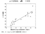
2.血管年齢指数(AgIxPPG):
PPG波形に関して、血管年齢指数の推定値は、加速度フォトプレチスモグラフィ(Acceleration Photoplethysmography:APG)としても知られる、PPGシグナルの二次導関数の分析によって得ることができる。それは、PPG波のように、いくつかのランドマーク点によって特徴づけられ、これらの点の推定は、血管年齢指数を含む、心臓血管機能に関する情報を与えるインジケータを得るために使用される。現状技術水準の文献では、以下の式によって特徴点の比率が計算される:
![]()
For PPG waveforms, an estimate of the vascular age index can be obtained by analysis of the second derivative of the PPG signal, also known as Acceleration Photography symmetry (APG). It is characterized by several landmark points, such as PPG waves, and estimates of these points are used to obtain informative indicators of cardiovascular function, including the vascular age index. In the current state of the art literature, the ratio of feature points is calculated by the following formula:
![]()
最も使用されるAPGからのパラメータは血管年齢指数であるが、APG波の推定値に始まり、例えば、いくつかの研究において(Elgendi, Current Cardiology Reviews, vol. 8, pp. 14−25, 2012)、b波、c波、d波、またはe波とa波との間の比など、他の指標が調査されている。これらの比率は、対象者の年齢によって変わることが分かっている。血管年齢指数の代替手段として、c波およびd波がはっきりしない場合、別の研究(Baek et al., 6th International Special Topic Conference on Information Technology Applications in Biomedicine, 2007)において提案されるように、(b−e)/a比を使用することができる。 The most used parameter from APG is the vascular age index, which begins with estimates of APG waves and, for example, in some studies (Elgendi, Current Cardiology Reviews, vol. 8, pp. 14-25, 2012). , B-wave, c-wave, d-wave, or the ratio between e-wave and a-wave, and other indicators are being investigated. These ratios have been found to vary with the age of the subject. As an alternative to the vascular age index, if the c-wave and d-wave are unclear, another study (Baek et al., 6th International Special Foundation on Information Technology Technology Application 7), proposed in Biomedicine, 200, Biomedicine. -E) / a ratio can be used.
血管年齢指数に加えて、以下の指数も推定した。
脈波伝播速度(PWV):
PWVは、圧力波が伝播する同じ線上の2つの異なる測定部位の間の距離と、対応する波の点の間の時間間隔との間の比として、実験により測定される。
Pulse wave velocity (PWV):
PWV is measured experimentally as the ratio between the distance between two different measurement sites on the same line through which the pressure wave propagates and the time interval between the corresponding wave points.
脈波伝播速度は、PPGシグナルによっても推定することができる。この場合、当該PWVは、2つの異なる機器セットアップによって得ることができる:
・ECG+PPGセンサ:ECGのRピークとPPGランドマーク点(収縮期の裾部、最大傾斜、または収縮期ピーク)との間の時間間隔として、脈波到達時間(Pulse Arrival Time:PAT)を評価しなければならない;
・2つのPPGセンサ:それらは、一方が他方の下流に位置され、この場合、2つの測定部位の間の時間間隔として脈波伝播時間(PTT)を評価しなければならない[21]。
The pulse wave velocity can also be estimated from the PPG signal. In this case, the PWV can be obtained by two different device setups:
ECG + PPG sensor: Evaluates the pulse wave arrival time (Pulse Arrival Time: PAT) as the time interval between the ECG R peak and the PPG landmark point (systolic hem, maximum slope, or systolic peak). There must be;
Two PPG sensors: they are located downstream of the other, in which case the pulse wave velocity (PTT) must be evaluated as the time interval between the two measurement sites [21].
測定された時間間隔を区別して指定することが必要であり、すなわち、当該PATは、PTTと前駆出時間(Pre−Ejection Period:PEP)との合計に等しく、これは、心室脱分極の開始と大動脈弁が開く瞬間との間の時間間隔である。PEPは、測定または予測するのが困難であり、圧力の一次関数ではないため、PATは、PTTほど正確ではないインジケータであることが判明する。PTTは、評価するのがより困難であるが、モニタリングに対してより良い指標を提供する。このパラメータは、大動脈PWVを推定することを可能にする(当該大動脈は、当該文献におけるPWVを測定するための基準点である)。近代的血圧測定システムも、間接的な方法によって大動脈PWVを計算する。 It is necessary to specify the measured time intervals separately, that is, the PAT is equal to the sum of the PTT and the Pre-Ejection Period (PEP), which is the onset of ventricular depolarization. The time interval between the moment the aortic valve opens. Since PEP is difficult to measure or predict and is not a linear function of pressure, PAT turns out to be an indicator that is not as accurate as PTT. PTTs are more difficult to assess, but provide better indicators for monitoring. This parameter makes it possible to estimate the aortic PWV (the aorta is the reference point for measuring PWV in the literature). Modern blood pressure measurement systems also calculate aortic PWV by an indirect method.
PWV推定値を得るために、2つの異なる測定システムからのPPGシグナルの収縮期の裾部が識別される。収縮期の裾部が記録される時刻の間の差のおかげで、機器に応じて(第1の事例ではECGおよびPPG、第2の事例では2つのPPGシグナル)、脈波到達時間および脈波伝播時間を知ることが可能である。この測定は、PATまたはPTTと、ゴールドスタンダード機器(これは、中央PWV、すなわち、大動脈において、を意味する)から測定された脈波伝播速度との間の相関関係を評価するために使用されるであろう。この理由から、脈波伝播時間の値、年齢、身長、心拍数の中央値、およびPPGシグナルにおける3つの典型的なパラメータ、すなわち、立ち上がり時間、硬さ指数、およびパルス領域、を使用して、線形回帰を作製した。 To obtain PWV estimates, systolic tails of PPG signals from two different measurement systems are identified. Depending on the device (ECG and PPG in the first case, two PPG signals in the second case), pulse wave arrival time and pulse wave, thanks to the difference between the times when the systolic hem is recorded. It is possible to know the propagation time. This measurement is used to assess the correlation between PAT or PTT and pulse wave velocity measured from a gold standard instrument, which means in the central PWV, i.e., in the aorta. Will. For this reason, using three typical parameters in pulse wave propagation time, age, height, median heart rate, and PPG signal: rise time, hardness index, and pulse region, A linear regression was made.
当該PWVは、離れて位置された2つのPPGセンサにおいて測定された2つのPPGシグナルのパルスの間の時間差(ここでは、PTT)によって推定される。したがって、当該シグナルの収縮期の裾部の間の時間差を調べる。PWVを推定するために、線形回帰モデルに当該時間差の中央値が使用される。さらに、追加の生理的および個人的データが、当該線形回帰モデルに含められた:
2つのPPGシグナルが測定され、当該2つの対応するPPGパルスの間の時間差が考慮されることは好ましい。一実施形態において、1つのPPGセンサは、使用者の手首に位置決めすることができ、第2のセンサは、使用者の指に位置決めすることができる。しかしながら、有利な構成において、2つのPPGセンサは、両方のセンサの間のある特定の距離において、使用者の手首に位置決めすることができる。これは、手首装着式デバイス、例えば、スマートウォッチまたはフィットネストラッカなど、における実践を可能にする。 It is preferred that two PPG signals are measured and the time difference between the two corresponding PPG pulses be taken into account. In one embodiment, one PPG sensor can be positioned on the user's wrist and the second sensor can be positioned on the user's finger. However, in an advantageous configuration, the two PPG sensors can be positioned on the user's wrist at a particular distance between both sensors. This enables practice on wrist-worn devices, such as smart watches or fitness trackers.
血圧(BP):
PPGシグナルからの血圧の推定は、そのような些細なタスクではない。前の研究は、PPGパルスにおける抽出された収縮期および拡張期の時間を使用するシンプルな線形回帰モデルによってBPを推定することを提案している:
Estimating blood pressure from PPG signals is not such a trivial task. Previous studies have proposed estimating BP by a simple linear regression model using extracted systolic and diastolic times in PPG pulses:
本発明のために、動脈血圧(収縮期および拡張期)を推定するための方策を開発し、脈波伝播時間に取り組み、ゴールドスタンダードの機器によって得られた血圧の推定値を用いてこれらの値の線形回帰を評価した。その上、心拍数の中央値、立ち上がり時間、硬さ指数、およびパルス領域、ならびに生理学的パラメータ、例えば、年齢および身長など、のように、線形回帰推定において他のパラメータを使用した。
心拍数変動性(HRV):
心拍数変動性(HRV)は、心拍の間の時間間隔における変動を説明するものである。各心拍に対する心拍間隔(interbeat interval:IBI)値は、2つの連続するPPG波における2つの対応するランドマーク点(収縮期の裾部、最大傾斜、または収縮期ピーク)の間の時間間隔として推定される。例えば、図1.7において、IBIは、2つの連続する収縮期裾部の間の時間間隔として測定される。
Heart rate variability (HRV):
Heart rate variability (HRV) describes variability in the time interval between heartbeats. The heart rate interval (IBI) value for each heartbeat is estimated as the time interval between two corresponding landmark points (systolic hem, maximal slope, or systolic peak) in two consecutive PPG waves. Will be done. For example, in FIG. 1.7, IBI is measured as the time interval between two consecutive systolic hem.
IBIが測定されると、HRVパラメータを推定することが可能である。慣習的に、HRV分析は、時間ドメインおよび周波数ドメインにおいて実施される。さらに、これらのパラメータのいくつかは、十分に長い持続時間を記録が有する場合にのみ、推定することができる。短い記録の場合(すなわち、少なくとも2分)、以下は、得ることが可能な可能性のある指数のいくつかである(Shaffer and Ginsberg, Frontiers in Public Health, vol. 5, n. 258, p. 17 pp, 2017):
1.正常な洞性拍動のIBIの標準偏差(SDNN)
2.50msを超えて互いに異なる隣接する間隔の数(NN50およびpNN50)
3.正常な心拍の間の逐次差分の二乗平均平方根(Root Mean Square of Successive Difference:RMSSD)、最初に心拍の間のそれぞれの連続した時間差を計算し、次いで、当該値のそれぞれを二乗し、結果を平均した後に、全ての平方根を取ることによって得られる、
4.LF/HF比、すなわち、低周波数出力(0.04〜0.15Hz)と高周波数出力(0.15〜0.4Hz)の間の比、
5.ポアンカレプロット、それは、前の間隔に対してすべてのIBI間隔をプロットして、散布図を作成することによって得られる;ポアンカレプロットは、当該プットされた点に楕円をフィッティングさせることによっても分析することができる。フィッティングフェーズの後、以下の2つの非線形測定を得ることができる;
5.a.SD1:x軸からの各点の距離の標準偏差、楕円の幅を特定する;それは、短期的HRVを反映する。
5.b.SD2:y=x+平均(IBI間隔)からの各点の標準偏差、それは、楕円の長さを特定する;それは、短期的および長期的HRVを測定する。
6.試料エントロピー、時系列の規則性および複雑さを測定する。
Once the IBI is measured, it is possible to estimate the HRV parameters. By convention, HRV analysis is performed in the time domain and frequency domain. Moreover, some of these parameters can only be estimated if the record has a sufficiently long duration. For short records (ie, at least 2 minutes), the following are some of the indices that may be available (Shaffer and Ginsberg, Frontiers in Public Health, vol. 5, n. 258, p. 17 pp, 2017):
1. 1. Standard deviation of IBI of normal sinus beat (SDNN)
Number of adjacent intervals that differ from each other beyond 2.50 ms (NN50 and pNN50)
3. 3. Root Mean Square of Successive Difference (RMSSD) of successive differences between normal heartbeats, first calculate each consecutive time difference between heartbeats, then square each of those values and obtain the result Obtained by taking all square roots after averaging,
4. The LF / HF ratio, i.e., the ratio between the low frequency output (0.04 to 0.15 Hz) and the high frequency output (0.15 to 0.4 Hz),
5. Poincare plot, which is obtained by plotting all IBI intervals against the previous interval and creating a scatter plot; Poincare plots can also be analyzed by fitting an ellipse to the put point. Can be done. After the fitting phase, the following two non-linear measurements can be obtained;
5. a. SD1: Specify the standard deviation of the distance of each point from the x-axis, the width of the ellipse; it reflects the short-term HRV.
5. b. SD2: Standard deviation of each point from y = x + mean (IBI interval), which identifies the length of the ellipse; it measures short-term and long-term HRV.
6. Measure sample entropy, time series regularity and complexity.
益々増加するウェアラブルデバイスは、PPG技術を使用して正確で、経済的で、容易に測定できるHRV指数を提供すると主張している。いくつかの研究は、ECGシグナルによって与えられる、ゴールドスタンダードと比較して、PPG測定によって報告されるHRV指数の信頼性に注目してきた。特に、最近のレビュー(Georgiou et al., Folia Medica, vol. 60, n. 1, pp. 7−20, 2018)において、報告される結果は、PPG技術がHRV測定に対して有効な代替手段となり得るということであるが、それは、依然として、非定常条件下においてより徹底的な研究を行うことを必要としている。 An increasing number of wearable devices claim to use PPG technology to provide an accurate, economical and easily measurable HRV index. Several studies have focused on the reliability of the HRV index reported by PPG measurements compared to the gold standard given by the ECG signal. In particular, in a recent review (Georgio et al., Folia Medica, vol. 60, n. 1, pp. 7-20, 2018), the reported results show that PPG technology is an effective alternative to HRV measurement. Although it can be, it still requires more thorough research under unsteady conditions.
本発明により、2つ以上のPPGセンサを用いて2つ以上のPPGシグナルを測定し、血管年齢指数AgIx、血圧BPdiaおよびBPsys、脈波伝播速度PWV、および脈波増大係数AIxを特定するために先進的なアルゴリズムを使用することによって、1つまたは複数の心臓血管パラメータが計算される。 The present invention, two or more PPG signal using two or more PPG sensor measures, vascular age index AgIx, identifying blood pressure BP dia and BP sys, pulse-wave propagation velocity PWV, and a pulse wave enhancement factor AIx By using advanced algorithms for this, one or more cardiovascular parameters are calculated.
一構成において、1つだけの心臓血管パラメータが測定され、脈波増大係数AIxが特定されるか、または血管年齢指数AgIxのみまたは血圧のみが特定されるか、または脈波伝播速度PWVのみが特定される。 In one configuration, only one cardiovascular parameter is measured and the pulse wave increase factor AIx is specified, or only the vascular age index AgIx or blood pressure is specified, or only the pulse wave velocity PWV is specified. Will be done.
さらなる構成において、2つの心臓血管パラメータが測定され、脈波増大係数AIxおよび血管年齢指数AgIxが特定される。さらなる代替手段において、さらに、血圧が特定されるか、または脈波伝播速度PWVが特定されるか、またはその両方が特定される。 In a further configuration, two cardiovascular parameters are measured to identify the pulse wave augmentation factor AIX and the vascular age index AgIx. In a further alternative, the blood pressure is further identified, the pulse wave velocity PWV is identified, or both.
さらなる構成において、脈波増大係数AIxおよび血圧が特定される。さらなる代替手段において、さらに、血管年齢指数AgIxが特定されるか、または脈波伝播速度PWVが特定されるか、またはその両方が特定される。 In a further configuration, the pulse wave velocity factor AIX and blood pressure are specified. In a further alternative, the vascular age index AgIx, the pulse wave velocity PWV, or both are further identified.
さらなる構成において、脈波増大係数AIxおよび脈波伝播速度PWVが特定される。さらなる代替手段において、さらに、血管年齢指数AgIxが特定されるか、または血圧が特定されるか、またはその両方が特定される。 In a further configuration, the pulse wave increase factor AIX and the pulse wave velocity PWV are specified. In a further alternative, the vascular age index AgIx is further identified, blood pressure is identified, or both are identified.
さらなる構成において、血管年齢指数AgIxおよび血圧が特定される。さらなる代替手段において、さらに、血管年齢指数AgIxが特定されるか、または脈波増大係数AIxが特定されるか、またはその両方が特定される。 In a further configuration, the vascular age index AgIx and blood pressure are identified. In a further alternative, the vascular age index AgIx, the pulse wave augmentation factor AIX, or both are further identified.
さらなる構成において、血管年齢指数AgIxおよび脈波伝播速度PWVが特定される。さらなる代替手段において、さらに、血圧が特定されるか、または脈波増大係数AIxが特定されるか、またはその両方が特定される。 In a further configuration, the vascular age index AgIx and the pulse wave velocity PWV are identified. In a further alternative, the blood pressure is further identified, the pulse wave velocity increase factor AIX is identified, or both.
さらなる構成において、血圧および脈波伝播速度PWVが特定される。さらなる代替手段において、さらに、脈波増大係数Aixが特定されるか、または血管年齢指数AgIxが特定されるか、またはその両方が特定される。 In a further configuration, blood pressure and pulse wave velocity PWV are identified. In a further alternative, the pulse wave augmentation factor AIX, the vascular age index AgIx, or both are further identified.
好ましい構成において、心臓血管パラメータである脈波増大係数AIx、血管年齢指数AgIx、血圧、および脈波伝播速度PWVが特定される。 In a preferred configuration, the cardiovascular parameters pulse wave increase factor AIx, vascular age index AgIx, blood pressure, and pulse wave velocity PWV are identified.
特に好ましい構成において、心臓血管パラメータである脈波増大係数AIx、血管年齢指数AgIx、血圧、および脈波伝播速度PWVが特定される。 In a particularly preferred configuration, cardiovascular parameters are identified: pulse wave augmentation factor AIX, vascular age index AgIx, blood pressure, and pulse wave velocity PWV.
代替の構成において、1つ、2つ、3つ、または4つの心臓血管パラメータに加えて、以下のうちの1つまたは複数を計算することによって、心拍数変動性HRVが特定される:
・最小および最大心拍間隔(IBI)
・IBIの中央値および平均IBI
・最小および最大心拍数
・心拍数の中央値および平均心拍数
・正常な洞性拍動のIBIの標準偏差(SDNN)
・50msを超えて互いに異なる隣接する間隔の数(NN50およびNN50)
・正常な心拍の間の逐次差分の二乗平均平方根(RMSSD)、
・LF/HF比、すなわち、低周波数出力(0.04〜0.15Hz)と高周波数出力(0.15〜0.4Hz)の間の比
・SD1:全てのIBI間隔を前の間隔に対してプロットすることによって得られるポアンカレプロットにおけるx軸からの各点の距離の標準偏差
・SD2:全てのIBI間隔を前の間隔に対してプロットすることによって得られるポアンカレプロットにおけるy=x+平均(IBI間隔)からの各点の距離の標準偏差、
・試料のエントロピー
In an alternative configuration, heart rate variability HRV is identified by calculating one or more of the following in addition to one, two, three, or four cardiovascular parameters:
• Minimum and maximum heart rate intervals (IBI)
Median and mean IBI
-Minimum and maximum heart rate-Median and average heart rate-Standard deviation of IBI of normal sinus beat (SDNN)
· 50 ms Beyond different number of adjacent intervals (NN50 and N N50)
Root mean square (RMSSD), the root mean square of successive differences between normal heartbeats,
-LF / HF ratio, that is, the ratio between low frequency output (0.04 to 0.15 Hz) and high frequency output (0.15 to 0.4 Hz) -SD1: All IBI intervals relative to the previous interval Standard deviation of the distance of each point from the x-axis in the Poincare plot obtained by plotting ・ SD2: y = x + mean (IBI) in the Poincare plot obtained by plotting all IBI intervals with respect to the previous interval. Standard deviation of the distance of each point from (interval),
・ Sample entropy
本発明は、多くの様々な身体健康モニタリングデバイス、例えば、手首タイプのフィットネストラッカ、スマートウォッチ、または医療専門家によって使用される特殊なデバイスなど、に内蔵されたPGGセンサを使用することにより適用することができる。本発明による方法は、いくつかの心臓血管パラメータを分析することによって、シンプルな手首装着式デバイスの助けにより、人の心臓血管状態の詳細な分析を可能にする。 The present invention is applied by using PGG sensors built into many different physical health monitoring devices, such as wrist-type fitness trackers, smartwatches, or specialized devices used by medical professionals. be able to. The method according to the invention allows detailed analysis of a person's cardiovascular status with the help of a simple wrist-worn device by analyzing several cardiovascular parameters.
したがって、本発明における有利な構成において、1つまたは複数の計算されたパラメータが、少なくとも1つのPPGセンサを含む身体健康モニタリングデバイス上に表示される。 Therefore, in an advantageous configuration of the present invention, one or more calculated parameters are displayed on a physical health monitoring device that includes at least one PPG sensor.
代替の構成において、少なくとも2つのPPGセンサを含み、結果として、2つのPPGシグナルの間の時間差を分析することによって1つまたは複数の心臓血管パラメータの評価を可能にする、身体健康モニタリングデバイス上に、1つまたは複数の計算されたパラメータが表示される。 In an alternative configuration, on a physical health monitoring device that includes at least two PPG sensors and, as a result, allows assessment of one or more cardiovascular parameters by analyzing the time difference between the two PPG signals. One or more calculated parameters are displayed.
本発明の別の好ましい実施形態において、音響シグナルまたは視覚シグナルが当該計算されたパラメータと一緒に出力される。 In another preferred embodiment of the invention, an acoustic or visual signal is output along with the calculated parameters.
本発明の別の代替の実施形態において、1つまたは複数の計算されたパラメータが、少なくとも2つのPPGセンサを含む身体健康モニタリングデバイス上に表示される。 In another alternative embodiment of the invention, one or more calculated parameters are displayed on a physical health monitoring device that includes at least two PPG sensors.
本発明の代替の実施形態において、当該計算された心臓血管パラメータは、予め保存されている心臓血管指数パラメータと比較され、当該計算された心臓血管パラメータが、当該予め保存されている心臓血管指数パラメータからX%を超えて異なる場合、音響シグナルまたは視覚シグナルが出力され、その一方で、Xは、以下の値:5、10、20、30、40、50、60、70、80、90、100から選択される。 In an alternative embodiment of the invention, the calculated cardiovascular parameters are compared with pre-stored cardiovascular index parameters, and the calculated cardiovascular parameters are the pre-stored cardiovascular index parameters. If it differs by more than X% from, an acoustic or visual signal is output, while X has the following values: 5, 10, 20, 30, 40, 50, 60, 70, 80, 90, 100. Is selected from.
本発明の別の態様は、以下のパラメータ:
・血管年齢指数AgIx、
・脈波伝播速度PWV、
・血圧BPdiaおよびBPsys、
・脈波増大係数AIx、
の1つまたは複数を特定するための手首装着式デバイスに関し、
この場合、当該デバイスは、
・腕の背側部分に面する、5cm以下の距離の2つのPPGセンサ、
を含み、
・この場合、当該PPGセンサは、少なくとも1つの緑色光源を含み、ならびに好ましくは512Hzのサンプリング周波数を含む。
Another aspect of the invention is the following parameters:
・ Blood vessel age index AgIx,
・ Pulse wave velocity PWV,
-Blood pressure BPdia and BPsys,
-Pulse wave increase coefficient AIX,
With respect to a wrist-worn device for identifying one or more of
In this case, the device
-Two PPG sensors with a distance of 5 cm or less facing the dorsal part of the arm,
Including
-In this case, the PPG sensor comprises at least one green light source, and preferably includes a sampling frequency of 512 Hz.
好ましい実施形態において、当該デバイスは、
・特徴点a、b、c、d、およびe、対象者の年齢(page)、身長(pheight)、および心拍数中央値に基づいて線形回帰を使用して、血管年齢指数AgIx、
・当該対象者の2つのPPGパルスの間の時間差(PTT)、年齢(page)、身長(pheight)、および心拍数中央値の推定値に基づいて線形回帰を使用して、脈波伝播速度PWV、
・2つのPPGパルスの間の時間差(PTT)および心拍数中央値に基づいて線形回帰を使用して、血圧BPdiaおよびBPsys、
・任意で、75心拍数(AIx@75)に対して正規化された収縮期ピーク振幅Asysおよび拡張期ピーク振幅Adiaに基づいて、および当該正規化された脈波増大係数AIxに基づいて線形回帰を使用して、脈波増大係数AIx、
の1つまたは複数を計算するのに適合されたシグナル処理手段をさらに含む。
当該手首装着式デバイスは、フィットネストラッカまたはスマートウォッチであり得る。
In a preferred embodiment, the device is
Vascular age index AgIx, using linear regression based on feature points a, b, c, d, and e, subject's age, height, and median heart rate.
-Pulse wave velocity PWV using linear regression based on estimates of time difference (PTT), age (page), height (height), and median heart rate between the subject's two PPG pulses. ,
Blood pressure BPdia and BPsys, using linear regression based on the time difference (PTT) between the two PPG pulses and the median heart rate.
-Optionally, linear regression based on systolic peak amplitude Asys and diastolic peak amplitude AIX and based on the normalized pulse wave augmentation factor AIX for 75 heart rate (AIx @ 75). Using the pulse wave increasing factor AIX,
Further includes signal processing means adapted to calculate one or more of the above.
The wrist-worn device can be a fitness tracker or smartwatch.
実験セットアップ
本発明の実験のために、2つの異なる機器セットアップを使用した。
Experimental Setup Two different instrument setups were used for the experiments of the present invention.
GTECセットアップ
TEC medical engineering GmbH(オーストリア)製の携帯用生体信号収集および分析システムであるg.MOBIlabを含むポリグラフセットアップを使用した(「gMOBIlab取扱説明書」[オンライン]、http://www.gtec.at/Download/Product−Manuals−Handbooks/g.MOBIlab/gMOBIlabInstructionsForUseから利用可能)。
GTEC Setup A portable biological signal collection and analysis system manufactured by TEC medical engineering GmbH (Austria). A polygraph setup including MOBIlab was used ("gMOBIlab Instruction Manual" [online], http: //www.gtec.at/Download/Product-Manuals-Handbooks/g.MOBIlab/gMOBIlabInstractsF.
当該GTECセットアップにおいて、2つのPPGセンサを、Connector Boxを介してg.MOBIlabに接続する。ECGでは、代わりに電極をg.MOBIlabに直接接続する。g.MOBIlabは、ブルートゥース接続によってシグナルを記録し表示するソフトウェアと通信することができる。シグナルが記録されると、それらは、使用者が指定したフォルダに保存される。 In the GTEC setup, the two PPG sensors were g. Connect to MOBIlab. In ECG, instead, the electrode is g. Connect directly to MOBIlab. g. MOBIlab can communicate with software that records and displays signals via Bluetooth connectivity. When the signals are recorded, they are saved in a folder specified by the user.
この機器セットアップによって取得される関連情報は、以下の通りである:
oECGシグナル[mV]
o指センサからのPPGシグナル[a.u.]
o耳たぶセンサからのPPGシグナル[a.u.]
oUnix時間1において記録する初期時間[ms]
他の関連情報は、以下の通りである:
oサンプリング周波数:256Hz(カスタマイズ不可能)
oPPGセンサ
・IR光源
・1つの光電検出器
The relevant information obtained by this equipment setup is as follows:
oECG signal [MV]
o PPG signal from the finger sensor [a. u. ]
o PPG signal from the earlobe sensor [a. u. ]
Initial time to record at oUnix time 1 [ms]
Other relevant information is as follows:
o Sampling frequency: 256Hz (non-customizable)
oPPG sensor ・ IR light source ・ One photoelectric detector
この機器セットアップのおかげで、2つのPPGセンサからのシグナルが、心電図シグナルと共に、メインステーション(g.MOBIlab)によって同時に記録される。これは、2つのPPGセンサの間およびPPGセンサとECGシグナルとの間の両方において、完全な同期を可能にする。したがって、この特徴は、PPGシグナルの時間的特性に関連する心臓血管パラメータを正確に導出することを可能にするであろう。しかしながら、以下の2つの問題が生じる:
i.当該2つのセンサは、赤外光源を備えている。前の章において報告したように、このような光源は、手首での取得にとって最適ではなく、そのため、記録されたシグナルの品質は低く、それにもかかわらず、依然として、収縮期の裾部を識別することが可能である。
ii.以前に報告されているように(O'Rourke et al., American Journal of Hypertension, vol. 15, pp. 426−444, 2002)、上腕と橈骨動脈との間の脈波伝播速度は、約800cm/秒から1160cm/秒まで及ぶ。いずれにせよ、これらの基準値が40歳の対象者のものであるということを考慮する必要があり、そのため、下限は、800cm/秒より低いかもしれない。サンプリング周波数は256Hzに等しく、したがって、サンプリング周期は0.0039秒であるため、当該脈波伝播速度が、1050cm/秒より高い場合、当該GTECシステムは、それを検出することができないであろう。実際に、当該センサが互いに4cm離間されて位置される場合、この方法では、パルスは4cm移動するのに0.0039秒かかるため、検出することができる最大伝播速度は、1050cm/秒である。1050cm/秒を超える伝播速度のシグナルを記録するためには、より短いサンプリング周期が必要であろう。
Thanks to this equipment setup, signals from the two PPG sensors are recorded simultaneously by the main station (g. MOBIlab) along with the electrocardiogram signals. This allows perfect synchronization both between the two PPG sensors and between the PPG sensor and the ECG signal. Therefore, this feature will allow accurate derivation of cardiovascular parameters related to the temporal properties of PPG signals. However, the following two problems arise:
i. The two sensors include an infrared light source. As reported in the previous chapter, such light sources are not optimal for wrist acquisition and therefore the quality of the recorded signal is poor and nevertheless still identifies the systolic hem. It is possible.
ii. As previously reported (O'Rourke et al., American Journal of Hypertension, vol. 15, pp. 426-444, 2002), the pulse wave velocity between the humerus and the radial artery is approximately 800 cm. It ranges from / sec to 1160 cm / sec. In any case, it should be taken into account that these reference values are for a 40 year old subject, so the lower limit may be lower than 800 cm / sec. Since the sampling frequency is equal to 256 Hz and therefore the sampling period is 0.0039 seconds, if the pulse wave velocity is higher than 1050 cm / sec, the GTEC system will not be able to detect it. In fact, if the sensors are located 4 cm apart from each other, the maximum propagation velocity that can be detected is 1050 cm / sec because the pulse takes 0.0039 seconds to move 4 cm in this method. Shorter sampling cycles may be required to record signals with propagation velocities greater than 1050 cm / sec.
E4セットアップ
Empatica E4リストバンド(Empatica Inc.、合衆国)を使用した(「E4リストバンドユーザマニュアル」[オンライン]。https://empatica.app.box.com/v/E4−User−Manualから利用可能)。
E4 Setup Using the Empatica E4 wristband (Empatica Inc., United States) ("E4 Wristband User Manual" [online]. ).
シグナルモードを取得する際、ストリーミングモードを選択した。ストリーミングモードでは、E4リストバンドが手首に装着されると、「Bluegiga Bluetooth Smart Dongle」モジュールを介して、E4によってサポートされる唯一の接続タイプであるブルートゥース接続が確立される。次いで、データがE4ストリーミングサーバに送信され、当該サーバは、今度は、TCP接続によって当該データストリーミングを送信する。次いで、データは、指定フォルダにセーブされる。この機器セットアップによって取得される関連情報は、PPGシグナル[a.u.]およびUnix時間での各値の記録時間である。 When getting the signal mode, I chose the streaming mode. In streaming mode, when the E4 wristband is worn on the wrist, a Bluetooth connection, the only connection type supported by the E4, is established via the "Blueguiga Bluetooth Smart Dongle" module. The data is then transmitted to the E4 streaming server, which in turn transmits the data streaming over a TCP connection. Then, the data is saved in the designated folder. The relevant information obtained by this device setup is the PPG signal [a. u. ] And the recording time of each value in Unix time.
追加の関連情報は、以下の通りである:
oサンプリング周波数:64Hz(カスタマイズ不可能)
oPPGセンサ
・4つの光源、2つは緑色で2つは赤色
・2つの光電検出器
・モーションアーチファクトを除去するための内部アルゴリズム
Additional relevant information is as follows:
o Sampling frequency: 64Hz (non-customizable)
oPPG sensor ・ 4 light sources ・ 2 green and 2 red ・ 2 photoelectric detectors ・ Internal algorithm for removing motion artifacts
ストリーミングモードにおいて、当該2つのセンサの間において完全に同期させることができないという、技術的限界が検出された。複数の接続(すなわち、ブルートゥースおよびTCP)によって導入される遅延は、決定論的ではなく、したがって、シグナル分析フェーズに達した後、検出されるオフセットとして処理することができない。一方で、E4リストバンドによって導入される利点は、結果として、以下のいくつかの要因のおかげで達することができる高いシグナル品質をもたらす:
i.4つの光源および2つの光電検出器:当該E4システムは、異なる波長(緑色および赤色)において当該2つの光電検出器によって取得されたシグナルを組み合わせ、アンビエント照明にあまり影響されないシグナルを得る。
ii.内部アルゴリズム:各E4リストバンドは、Empaticaが所有する内部アルゴリズムを有し、それは、シグナルの初期の前処理を実施する。一方において、これは、この技術における有利な特徴を表し得るが、その反面、それは、定量化するのが困難な遅延のさらなる原因となり得る。
In streaming mode, a technical limitation has been detected in which the two sensors cannot be perfectly synchronized. The delay introduced by multiple connections (ie Bluetooth and TCP) is not deterministic and therefore cannot be treated as an offset detected after reaching the signal analysis phase. On the other hand, the benefits introduced by the E4 wristband result in high signal quality that can be achieved thanks to several factors:
i. Four light sources and two photoelectric detectors: The E4 system combines the signals acquired by the two photoelectric detectors at different wavelengths (green and red) to obtain a signal that is less sensitive to ambient illumination.
ii. Internal Algorithm: Each E4 wristband has an internal algorithm owned by Empatica, which performs the initial preprocessing of the signal. On the one hand, this can represent an advantageous feature in the technique, but on the other hand, it can cause additional delays that are difficult to quantify.
本研究では、当該2つのシステム(GTECおよびE4)を組み合わせた。これは、得られるパラメータのうちの3つ(すなわち、脈波増大係数、血管年齢指数、および心拍数変動性)は、PPG波の形態的特徴に依存し、残りの2つのパラメータ(すなわち、脈波伝播速度および血圧)は、異なる2つのセンサからの2つのPPG波の間の時間的距離に依存するため、可能である。したがって、同時に両方のシステムを使用することを決定し、それらのそれぞれから最良な特性:すなわち、E4リストバンドの高い品質、およびGTECシステムからの同期を活用した。 In this study, the two systems (GTEC and E4) were combined. This is because three of the resulting parameters (ie, pulse wave velocity factor, vascular age index, and heart rate variability) depend on the morphological characteristics of the PPG wave, and the remaining two parameters (ie, pulse). Wave velocity and blood pressure) are possible because they depend on the temporal distance between the two PPG waves from two different sensors. Therefore, we decided to use both systems at the same time and took advantage of the best characteristics from each of them: the high quality of the E4 wristband, and the synchronization from the GTEC system.
インビボ研究
PPGシグナルを用いた心臓血管パラメータ推定値の評価を実施するために、前の章において説明した機器セットアップを用いて、実験研究を行った。当該評価は、年齢、身長、および体重に関するデータを表1に報告する20人の健康な対象者に対して実施した。
In vivo Studies Experimental studies were performed using the instrument setup described in the previous chapter to perform evaluation of cardiovascular parameter estimates using PPG signals. The assessment was performed on 20 healthy subjects reporting data on age, height, and weight in Table 1.
実験セットアップは、以下の通りである:
o2つのEmpatica E4リストバンド
o以下を備えるGTECポリグラフ
og.MOBIlab
oIR光源を有する2つのPPGセンサ
oECGケーブル
この機器セットアップによって得られた測定値を検証するために、ゴールドスタンダード機器である、I.E.M. GmbH製の臨床用デバイスであるMobil−O−Graph PWA 24hを使用した(「Mobil−O−Graph 24h PWA user's manual」[オンライン]。www.iem.de/_attic/website/UserManual_NG_HMS−CS_24h−PWA_EN.pdfから入手可能)。このデバイスは、血圧用の標準的測定デバイスと同様に機能し、対象者の上腕へカフを適用する。それは、圧力波に対して脈波分析(Pulse Wave Analysis)を実施し、各指数に対して正確な値を得ることを可能にする。
The experimental setup is as follows:
o GTEC polygraph og with two Empatica E4 wristbands o and below. MOBIlab
Two PPG sensors with oIR light source oECG cable
To verify the measurements obtained by this device setup, the Gold Standard device, I.I. E. M. A clinical device made by GmbH, Mobil-O-Graph PWA 24h, was used ("Mobile-O-Graph 24h PWA user's manual" [online]. Www.iem.de/_attic/website/UserManual_N Available from PWA_EN.pdf). This device functions like a standard measuring device for blood pressure and applies a cuff to the subject's upper arm. It makes it possible to perform pulse wave analysis on pressure waves and obtain accurate values for each index.
当該実験プロトコルを20人の対象者に適用した。それは、以下のステップからなる:
1.5分間の安静
2.センサ装着
a. 手首の背側部に、互いに4cm離間した2つのE4リストバンド
b. 当該E4ストラップと腕の腹側部との間に、互いに4cm離間した2つのGTEC PPGセンサ
3.E4リストバンドのブルートゥース接続
4.GTECシステムのブルートゥース接続
5.2分30秒間の取得
6.センサの取り外し
7.Mobil−O−Graph血圧カフの取り付け
8.Mobil−O−Graphのブルートゥース接続
9.「Triple Pulse Wave Analysis」モードによる血圧シグナルの取得
The experimental protocol was applied to 20 subjects. It consists of the following steps:
Rest for 1.5
PPGシグナルの前処理
PPGからの正しいパラメータ推定にとって、前処理フェーズは、重要な課題である。それは、その要所のより容易な検出を得るために、PPG波の輪郭を強調することを可能にする。図2.1に示されるように、変更を行わずに測定されたPPGシグナルは、通常、50Hzの周波数での可視的電力ライン干渉を含む。この干渉を除去するために、50Hzのノッチフィルタを使用する。電力ライン干渉および高周波数ノイズを除去したPPGシグナルを図2.2に示す。
Pretreatment of PPG signals The pretreatment phase is an important issue for correct parameter estimation from PPG. It makes it possible to emphasize the contours of the PPG wave for easier detection of its key points. As shown in Figure 2.1, the PPG signal measured without modification typically includes visible power line interference at a frequency of 50 Hz. A 50 Hz notch filter is used to eliminate this interference. Figure 2.2 shows the PPG signal with power line interference and high frequency noise removed.
以下の3つのステップからなる、組み合わせた前処理アルゴリズムを選択した[34][35]:
i.シグナルの正規化
![]()
iii.・零相およびカットオフ周波数=20HzのIVオーダーチェビシェフローパスフィルタ
20Hzにおいてフィルタリングを行うステップ、当該フィルタは、さらなる情報を提供しないシグナルのイレギュラーを除去することを可能にし、これらの速い変動は、収縮期ピークおよび拡張期ピークの正確な位置の推定にとって有害となる危険がある(図2.3に示される)。
A combined preprocessing algorithm consisting of the following three steps was selected [34] [35]:
i. Signal normalization
![]()
評価基準
提案されるアルゴリズムの正確さおよび信頼性を、臨床的に推奨される基準デバイスの測定値によってこれらのアルゴリズムの推定値を比較することにより、検証した。I.E.M. GmbH製のMobil−O−Graphによって得られるこれらの測定値は、基準値として役立ち、ならびに当該デバイスは、固有の測定誤差も有し、したがって、その測定値における変動も有するため、当該測定値自体、真の値とは異なり得る。基準デバイスの固有の測定誤差の影響を減じるために、当該基準デバイスによって3回の連続した測定を行い、各心臓血管パラメータに対してそれら3つの値の中央値を計算した。
Criteria The accuracy and reliability of the proposed algorithms were verified by comparing the estimates of these algorithms with the measurements of clinically recommended reference devices. I. E. M. These measurements obtained by the GmbH Mobile-O-Graph serve as reference values, and because the device also has inherent measurement errors and therefore variations in those measurements, the measurements themselves. , Can be different from the true value. To reduce the effect of the inherent measurement error of the reference device, three consecutive measurements were made with the reference device and the median of those three values was calculated for each cardiovascular parameter.
当該シグナルを、Matlabにおいて処理した。性能評価のためのアルゴリズムは、3つの主要な段階を含む:
1.PPGシグナルからのパラメータの推定
2.当該推定されたパラメータの線形回帰分析
3.当該推定されたパラメータとゴールドスタンダード指標との間の一致の評価(以下の表)
The signal was processed in MATLAB. The algorithm for performance evaluation involves three main stages:
1. 1. Parameter estimation from
PPGシグナルからのパラメータ推定の性能を、5つのインジケータに基づいて評価した。検証のため、5つの異なる基準である平均誤差、標準偏差(STD)、平均絶対誤差(MAE)、平均二乗誤差(MSE)、および二乗平均平方根誤差(RMSE)を計算したが、ここで、yest(i)は、全ての協力者に対する測定の総数に等しい長さN=242における、関心対象である当該推定された心臓血管パラメータであり、yref(i)は、その心臓血管パラメータの基準値である。
当該方法および最良のセンサ位置が決定されると、ノンパラメトリックのスピアマンの順位相関係数ρが、そのp値を用いて評価される。 Once the method and the best sensor position are determined, the nonparametric Spearman's rank correlation coefficient ρ is evaluated using its p-value.
心臓血管パラメータを推定するための線形回帰係数の見積もり
1. 脈波増大係数AIx
以下に説明される方法を使用して、脈波増大係数の推定値を得た:
o方法1.1:2つの指数の合計によるAIx=y/x
o方法1.2:2つの指数の合計によるAIx=(x−y)/x
o方法2.1:単一の指数をモデル化することによるAIx=y/x
o方法2.2:単一の指数をモデル化することによるAIx=(x−y)/x
Estimating the linear regression coefficient for estimating
An estimate of the pulse wave augmentation factor was obtained using the method described below:
o Method 1.1: AIX = y / x by sum of two indices
o Method 1.2: AIX = (xy) / x by sum of two indices
o Method 2.1: AIX = y / x by modeling a single exponent
o Method 2.2: AIX = (xy) / x by modeling a single exponent
平均誤差は低いが(基本的に、それはゼロである)、標準偏差は比較的高く、そのため、この種類の指標は、信頼性の高いパラメータとして定義することができないであろう。この理由のため、正規化された指数AIx@75の性能を評価することを決めた。表2.1および2.2は、それぞれ近位センサおよび遠位センサによって記録されたPPGシグナルから得られた結果を示している。 Although the mean error is low (basically it is zero), the standard deviation is relatively high, so this kind of indicator would not be able to be defined as a reliable parameter. For this reason, we decided to evaluate the performance of the normalized index AIX @ 75. Tables 2.1 and 2.2 show the results obtained from the PPG signals recorded by the proximal and distal sensors, respectively.
当該インジケータは全て、AIxに対して得られたものより著しく低い。当該最良の結果は、遠位センサからのPPGシグナルを使用した方法1.2によって達成される。図3.1は、基準値、スピアマン相関係数、およびそのp値に関連してこの方法によって得られた値を示している。 All of the indicators are significantly lower than those obtained for AIX. The best result is achieved by method 1.2 using the PPG signal from the distal sensor. FIG. 3.1 shows the reference values, the Spearman correlation coefficient, and the values obtained by this method in relation to their p-values.
2.血管年齢指数AgIxPPG:
血管年齢指数を推定するための3つの異なる方法を評価した:
・方法1:最大および最小ピークを見出すことによってAPG振幅波を得る。次いで、式(1.6)を使用して血管年齢指数を推定する;
・方法2:文献[16][36]に見出される追加の方法が実践されている。この場合も、式(1.6)を使用した;
・方法3:方法2と同様にAPGランドマーク点を検出したが、ゴールドスタンダード機器からの血管年齢指数を、式(1.7)によって得られた振幅比と比較した。
2. Blood vessel age index AgIx PPG :
Three different methods for estimating the vascular age index were evaluated:
Method 1: Obtain the APG amplitude wave by finding the maximum and minimum peaks. The vascular age index is then estimated using equation (1.6);
Method 2: Additional methods found in Ref. [16] [36] are practiced. Again, equation (1.6) was used;
-Method 3: APG landmark points were detected in the same manner as in
表3.1および3.2は、近位センサおよび遠位センサにおいて得られた値を示している。当該血管年齢指数は、年(y)の単位であり、推定誤差もそうである。 Tables 3.1 and 3.2 show the values obtained for the proximal and distal sensors. The blood vessel age index is a unit of year (y), and so is the estimation error.
当該2つのPPG記録部位の間において、特定の差はなかった。当該APG振幅波から推定される血管年齢指数は、あまり正確な結果を提供するようには思えない。この理由から、線形回帰において、他の回帰係数:年齢、身長、および心拍数の中央値、を導入することを決定したが、それについては式(1.8)を参照されたい。結果を表3.3および3.4に表す。 There was no particular difference between the two PPG recording sites. The vascular age index estimated from the APG amplitude wave does not seem to provide very accurate results. For this reason, we decided to introduce other regression coefficients: median age, height, and heart rate in linear regression, see equation (1.8) for that. The results are shown in Tables 3.3 and 3.4.
性能は、対象者の追加情報を当該線形回帰推定に組み入れることによってかなり向上する。標準偏差値は許容可能である。最良の方法は、近位のPPGセンサのシグナルに使用した場合の方法1である。図3.2は、基準値と比較した、この方法によって得られた値を示しており、スピアマン相関係数およびそのp値も示されている。
Performance is significantly improved by incorporating the subject's additional information into the linear regression estimation. The standard deviation value is acceptable. The best method is
3.脈波伝播速度PWV
脈波伝播速度の推定値は、GTECシステムを使用することによってのみ得られた。3つの異なるセットアップに基づく結果を評価した:
・近位のGTECセンサからのECGピークと連続するPPG収縮期の裾部との間での脈波到達時間
・遠位のGTECセンサからのECGピークと連続するPPG収縮期の裾部との間での脈波到達時間
・それぞれ近位のGTECセンサおよび遠位のGTECセンサによって記録された2つのPPG波の収縮期の裾部の間での脈波伝播時間。
その上、線形回帰が使用された。発明者らは、6つのモデルの性能を試験した:
・モデル1:PATまたはPTTのみ
・モデル2:PATまたはPTT+年齢
・モデル3:PATまたはPTT+年齢+身長
・モデル4:PATまたはPTT+年齢+身長+中央値(HR)、式(1.9)
・モデル5:PTT+立ち上がり時間+硬さ指数+パルス領域(PPG近位センサから)
・モデル6:PTT+立ち上がり時間+硬さ指数+パルス領域(PPG遠位センサから)
次いで、PATおよびPTTから得られたPWVの結果を比較した。
3. 3. Pulse wave velocity PWV
Estimates of pulse wave velocity were obtained only by using the GTEC system. Evaluated results based on three different setups:
-Pulse wave arrival time between the ECG peak from the proximal GTEC sensor and the continuous PPG systolic hem-between the ECG peak from the distal GTEC sensor and the continuous PPG systolic hem Pulse wave arrival time at-Pulse wave propagation time between the systolic hem of two PPG waves recorded by the proximal GTEC sensor and the distal GTEC sensor, respectively.
Moreover, linear regression was used. The inventors tested the performance of six models:
-Model 1: PAT or PTT only-Model 2: PAT or PTT + age-Model 3: PAT or PTT + age + height-Model 4: PAT or PTT + age + height + median (HR), equation (1.9)
Model 5: PTT + rise time + hardness index + pulse region (from PPG proximal sensor)
Model 6: PTT + rise time + hardness index + pulse region (from PPG distal sensor)
The PWV results obtained from PAT and PTT were then compared.
表4.1、4.2、および4.3は、3つの異なる機器セットアップによる当該推定されたPWV値を示している。 Tables 4.1, 4.2, and 4.3 show the estimated PWV values for three different equipment setups.
最初の注目に値する結果は、PTTから得られたPWV推定値であり(表4.3、モデル1からモデル4)、それは、PATから得られたものより低い(表4.1および4.2)。
The first notable result is the PWV estimate obtained from PTT (Table 4.3,
第二に、評価下の対象者の他の個人的なデータが、当該線形回帰モデルに含まれる場合、より良い結果を達成することができる。年齢、身長、および心拍数の中央値を含むモデルは、実際、最良の結果を示している。 Second, better results can be achieved if the subject's other personal data under evaluation is included in the linear regression model. Models that include median age, height, and heart rate actually show the best results.
PPGシグナルモルホロジのパラメータを使用することは、顕著な結果をもたらさない。見かけ上、当該PWV推定値は、これらPPGの特徴によって影響されない。 The use of PPG signal morphology parameters does not produce significant results. Apparently, the PWV estimate is not affected by these PPG features.
したがって、最良の方法は、方法4、(1.9)に示される式である。図4.3は、ゴールドスタンダードの指標、スピアマン相関係数、およびそのp値と比較したPWV推定値を示している。 Therefore, the best method is the formula shown in Method 4, (1.9). FIG. 4.3 shows the gold standard index, the Spearman correlation coefficient, and the PWV estimate compared to its p-value.
4.血圧
血圧推定値を、以下の線形回帰によって得た:
・方法1:PTT
・方法2:PTT+年齢+身長+中央値(HR)、式(1.12)および(1.13)
・方法3:PTT+立ち上がり時間+硬さ指数+近位センサからのパルス領域、式(1.14)および(1.15)
・方法4:PTT+立ち上がり時間+硬さ指数+遠位センサからのパルス領域
収縮期血圧および拡張期血圧に対する結果が、それぞれ、表5.1および5.2に別々に表される。血圧は、mmHgの単位であり、その推定誤差もそうである。
4. Blood pressure Estimates of blood pressure were obtained by the following linear regression:
-Method 1: PTT
Method 2: PTT + age + height + median (HR), equations (1.12) and (1.13)
Method 3: PTT + rise time + hardness index + pulse region from proximal sensor, equations (1.14) and (1.15).
Method 4: PTT + rise time + hardness index + pulse region from distal sensor Results for systolic and diastolic blood pressure are shown separately in Tables 5.1 and 5.2, respectively. Blood pressure is a unit of mmHg, and so is its estimation error.
方法2から良好な推定値を得られたが、これは、誤差の標準偏差が、収縮期および拡張期の両方の血圧推定値に対して他の方法より低いためである。
Good estimates were obtained from
当該誤差をさらに減じるために、CT、SI、およびPAを用いた線形回帰モデルも試験した。この場合、収縮期BPの誤差の標準偏差は、他の方法によって得られたものより大きく、その一方で、拡張期の血圧推定値は、他の方法よりわずかに低い誤差の標準偏差を示している(表4.14および4.15)。 To further reduce this error, linear regression models using CT, SI, and PA were also tested. In this case, the standard deviation of the systolic BP error is greater than that obtained by the other methods, while the diastolic blood pressure estimates show a slightly lower standard deviation of error than the other methods. (Tables 4.14 and 4.15).
収縮期BPに関して、当該最良の方法は、方法2であるように思われる。拡張期BPの場合、方法3が好ましいかもしれない。図3.4および3.5には、スピアマン相関係数およびそのp値と共に、推定された収縮期BPおよび拡張期BPも表されている。
For systolic BP, the best method seems to be
5.心拍数変動性HRV
表5および6は、ゴールドスタンダードと比較した、近位および遠位PPGシグナルにより得られた心拍数変動性(HRV)分析の結果を示している。表6.1および6.2に示されるように、HRVを推定するために、16のパラメータを考慮した:
5. Heart rate variability HRV
Tables 5 and 6 show the results of heart rate variability (HRV) analysis obtained by proximal and distal PPG signals compared to the gold standard. 16 parameters were considered to estimate the HRV, as shown in Tables 6.1 and 6.2:
表6.2:遠位E4センサからのHRVの結果
Table 6.2: HRV results from the distal E4 sensor
全体的に、近位PPGセンサのからの推定値は、遠位センサから得られたものより正確である。これは、リストバンドのより正しい位置決めと当該手首に対するより強い把持に起因し得る。 Overall, the estimates from the proximal PPG sensor are more accurate than those obtained from the distal sensor. This may be due to the more correct positioning of the wristband and the stronger grip on the wrist.
その誤差の標準偏差は比較的大きいが、RMSSDは、PPGセンサを装着した対象者の心臓血管の健康に対処する将来のアルゴリズムのための貴重な選択肢とみなすことができる。 Although the standard deviation of the error is relatively large, RMSSD can be regarded as a valuable option for future algorithms to address the cardiovascular health of subjects wearing PPG sensors.
心臓血管の推定の分析は、レファレンスから妥当な偏差によって推定することができる複数の心臓血管パラメータが存在することを示した。結論すると、シンプルで低コストのPPGシグナルは、現在最も一般的な抽出される特徴である脈拍数をはるかに超えて、人の心臓血管の健康に関する有用な情報を含む。本発明による新規のアルゴリズムは、手首に位置された2つのPPGセンサの場合でさえ基準値からのわずかな偏差のみによって心臓血管パラメータを推定することができる。これは、対象者の心臓血管状態の詳細な分析を提供するために1つの手首装着式デバイス内の2つのPPGシグナルを含ませる可能性を初めて提供するものである。当該2つのPPGセンサは、それらの心臓血管パラメータの永久的なモニタリングのために、フィットネストラッカまたはスマートウォッチに内蔵することができる。 Analysis of cardiovascular estimates has shown that there are multiple cardiovascular parameters that can be estimated from the reference with reasonable deviations. In conclusion, a simple, low-cost PPG signal goes far beyond the currently most commonly extracted feature, pulse rate, and contains useful information about human cardiovascular health. The novel algorithm according to the invention can estimate cardiovascular parameters with only a small deviation from the reference value, even with two PPG sensors located on the wrist. This offers for the first time the possibility of including two PPG signals in one wrist-worn device to provide a detailed analysis of the subject's cardiovascular status. The two PPG sensors can be integrated into a fitness tracker or smartwatch for permanent monitoring of their cardiovascular parameters.
図4は、心臓血管パラメータ、すなわち、血管年齢指数AgIx、血圧BPdiaおよびBPsys、脈波伝播速度PWV、脈波拡大指数AIx、および心拍数変動性HRV、を特定するためのシステム100を例示的に示している。システム100は、手首装着式デバイス、例えば、フィットネストラッカまたはスマートウォッチなど、において実践することができ、ならびに2つのPPGセンサ101、プロセッサ102、メモリ103、予め保存されているデータとの比較104、およびユーザインタフェース105を含む。データベース103は、全ての心臓血管パラメータに対する基準データを含み、ならびに、異なる組織データベースから得られた生理学的データおよびシステム100の測定されたデータから得られた生理学的データから導かれ得る。別の実施形態において、データベースは、有線または無線接続によって当該システムに外部的に接続することができる。
FIG. 4 illustrates a
当該PPGセンサ101は、使用者の皮膚に光を照射し、皮膚による照射光吸収に基づいて2つのPPGシグナルを測定するように構成される。当該PPGセンサ101は、例えば、少なくとも1つの周期的光源(例えば、発光ダイオード(LED)、またはそれに関連付けられた任意の他の周期的光源)と、当該少なくとも1つの周期的光源によって放たれて使用者の皮膚によって反射された当該周期的光を受光するように構成された光検出器とを含み得る。好ましい実施形態において、当該PPGセンサは、少なくとも1つの緑色光源を含み、ならびに好ましくは512Hzのサンプリング周波数を含む。
The
当該2つのPPGセンサ101は、プロセッサ102に接続することができる。別の実施形態において、当該PPGセンサ101は、プロセッサ102と他の回路/ハードウェア要素と共にハウジングに収納してもよい。両方のPPGセンサ101は、ハウジングに収納され、5cm以下の距離において位置決めされ、腕の背側部に面していることが好ましい。
The two
PPGセンサ101から周期的な光を受光して当該受光した周期的な光を処理するようにプロセッサ102(例えば、ハードウェアユニット、機器、中央演算処理装置(CPU)、グラフィック処理ユニット(GPU))を構成することができる。当該処理は、前に説明したような第一段階でのデータの前処理および本発明によるアルゴリズムの助けによる心臓血管パラメータの推定を含む。次いで、当該推定された心臓血管パラメータが、前に保存されたデータ104と比較され、使用者に対して表示されるユーザインタフェース105へと処理される。使用者は、さらに、当該推定されたパラメータに対してフィードバックを提供することができる。
Processor 102 (for example, hardware unit, equipment, central processing unit (CPU), graphic processing unit (GPU)) so as to receive periodic light from the
図5は、2つの離されたPPGセンサからの2つのPPGシグナルに基づく例示的実施形態に従って、対象者において1つまたは複数の心臓血管パラメータを推定するための方法を示すフローダイアグラムである。図5を参照すると、作動中、当該電子デバイスは、使用者の皮膚に光を照射し、皮膚による照射光吸収に基づいて2つのPPGセンサからのPPGシグナルを測定する。例えば、当該電子デバイスにおいて、図4に示されるように、2つのPPGセンサ101は、使用者の皮膚に光を照射し、当該皮膚による照射光吸収に基づいてPPGシグナルを測定するように構成される。
FIG. 5 is a flow diagram showing a method for estimating one or more cardiovascular parameters in a subject according to an exemplary embodiment based on two PPG signals from two detached PPG sensors. Referring to FIG. 5, during operation, the electronic device irradiates the user's skin with light and measures the PPG signals from the two PPG sensors based on the absorption of the irradiated light by the skin. For example, in the electronic device, as shown in FIG. 4, the two
作動中、当該電子デバイス100は、シグナルの前処理後、PPGの特徴、HRVの特徴、APGの特徴、および脈波伝播時間(PTT)を含む複数のパラメータを両方のPPGシグナルから抽出する。当該2つのPPGシグナル分析に基づいて、上記において説明したように当該心臓血管パラメータを推定することができる。当該電子デバイス100は、当該抽出された複数のパラメータに基づいて、心臓血管パラメータ、この場合はPWVおよびBP、を推定する。当該推定されたパラメータは、予め保存されている心臓血管パラメータ104と比較される。結果は、ユーザにフィードバックを与えるユーザインタフェース105内に表示される。
During operation, after preprocessing the signal, the
1つまたは複数の心臓血管パラメータを推定するための当該システムおよび当該方法のおかげで、使用者は、生理学的パラメータ、例えば、心臓血管パラメータなど、を継続的にモニターし、評価することができる。特定の解剖学的データを含む先進的アルゴリズムに基づいて、いくつかの心臓血管パラメータの評価が達成される。補助的パラメータ、例えば、血流、血圧、動脈壁の硬化、血管弾性、血管年齢など、の評価は、包括的な全般的健康評価を可能にする。この個々の心臓血管健康評価は、誤った解釈のリスクを減らし、使用者のより正確な健康評価につながる。 Thanks to the system and the method for estimating one or more cardiovascular parameters, the user can continuously monitor and evaluate physiological parameters such as cardiovascular parameters. Evaluation of several cardiovascular parameters is achieved based on advanced algorithms containing specific anatomical data. Assessment of ancillary parameters such as blood flow, blood pressure, arterial wall sclerosis, vascular elasticity, vascular age, etc. allows for a comprehensive overall health assessment. This individual cardiovascular health assessment reduces the risk of misinterpretation and leads to a more accurate health assessment of the user.
Claims (12)
・前記対象者の前記年齢(page)および前記体重(pheight)を特定し、
・前記対象者の2つの異なる部位において少なくとも2つのPPGセンサによって、少なくとも2つのフォトプレチスモグラフィック(photoplethysmographic:PPG)シグナルを測定し、
・前記PPGシグナルをPPGパルスへと分離し、それにより、前記パルスの開始点および終了点は、前記PPGシグナルの収縮期の裾部(systolic foot)に対応し、
・前記対象者の心拍数(PHR)を特定し、心拍数中央値を計算し、
・収縮期ピーク振幅Asysおよび拡張期ピーク振幅Adiaならびにそれらの時間tsおよびtdを特定し、
・前記PPGパルスの二次導関数を計算して、前記PPGパルスの前記二次導関数から特徴点a、b、c、d、およびeを特定し、
ここで、aおよびeは、それぞれ、前記二次導関数における第1および第2の最も際立った極大であり、
cは、前記特徴点aとeとの間における最も際立ったピークであり、
bは、前記二次導関数における最も際立った極小であり、
dは、特徴点cとeとの間における最も際立った極小であり、
・以下を特定し、
a)前記特徴点a、b、c、d、およびe、前記対象者の年齢(page)、身長(pheight)、および心拍数中央値に基づいて線形回帰を使用することにより、血管年齢指数(vascular age index)AgIx、
b)前記対象者の2つの前記PPGパルスの間の時間差(PTT)、年齢(page)、身長(pheight)、および心拍数中央値の推定値に基づいて線形回帰を使用することにより、脈波伝播速度(pulse wave velocity)PWV、
c)2つのPPGパルスの間の時間差(PTT)および心拍数中央値に基づいて線形回帰を使用することにより、血圧BPdiaおよびBPsys、
d)任意で、75心拍数(AIx@75)に対して正規化された収縮期ピーク振幅Asysおよび拡張期ピーク振幅Adiaに基づいて、および前記正規化された脈波増大係数(augmentation index)AIxに基づいて線形回帰を使用することにより、脈波増大係数AIx、
・前記計算されたパラメータを出力する、
ステップを含む方法。 A method of estimating one or more cardiovascular parameters in a subject, wherein the subject has age and weight.
- the age (p age) of the subject and the identify the body weight (p height),
At least two photoplethysmographic (PPG) signals were measured by at least two PPG sensors at two different sites of the subject.
The PPG signal is separated into PPG pulses, whereby the start and end points of the pulse correspond to the systolic foot of the PPG signal.
-Identify the subject's heart rate ( PHR ), calculate the median heart rate, and
-Identify systolic peak amplitude A sys and diastolic peak amplitude A dia and their time t s and t d .
-The secondary derivative of the PPG pulse is calculated, and the feature points a, b, c, d, and e are identified from the secondary derivative of the PPG pulse.
Here, a and e are the first and second most prominent maximums in the quadratic derivative, respectively.
c is the most prominent peak between the feature points a and e.
b is the most prominent minimum in the quadratic derivative.
d is the most prominent minimum between feature points c and e.
・ Identify the following
a) the feature point a, b, c, d, and e, age of the subject (p age), by using a linear regression based on the height (p height), and heart rate median vascular age Index (vascular age index) AgIx,
b) By using linear regression based on the time difference (PTT), age ( page ), height ( height ), and median heart rate estimates between the subject's two said PPG pulses. Pulse wave velocity PWV,
c) Blood pressure BP dia and BP systems , by using linear regression based on the time difference (PTT) between the two PPG pulses and the median heart rate.
d) Optionally, based on systolic peak amplitude Asys and diastolic peak amplitude Adia normalized for 75 heart rate (AIx @ 75), and said normalized pulse wave increase factor AIx. By using linear regression based on, the pulse wave velocity factor AIx,
-Output the calculated parameters
How to include steps.
前記心臓血管パラメータは、以下の方程式によって推定され、
式中、pageは前記対象者の前記年齢であり、pheightは前記対象者の前記身長であり、中央値(HR)は前記心拍数中央値であり、PTTは前記PPGパルスの間の時間差であり、AsysおよびAdiaは、それぞれ、前記収縮期ピークおよび前記拡張期ピークの大きさであり、CTは前記立ち上がり時間であり、STは前記硬さ指数であり、PAは前記PPGシグナルのパルス領域であり、xは前記拡張期ピーク振幅であり、yは前記収縮期ピーク振幅であり、d0からd4,g0からg4,l0dからl5d、k0sからk4s、およびb0からb1は、それぞれの前記線形回帰方程式の係数を表す、
請求項1に記載の方法。 Further comprising identifying the rise time (Crest Time: CT), hardness index (SI), and pulse region (PA) of the PPG signal.
The cardiovascular parameters are estimated by the following equations
In the formula, page is the subject's age, pitch is the subject's height, median (HR) is the heart rate median, and PTT is the time difference between the PPG pulses. Asys and Media are the magnitudes of the systolic peak and the diastolic peak, respectively, CT is the rise time, ST is the hardness index, and PA is the PPG signal. a pulse area, x is the diastolic peak amplitude, y is the systolic peak amplitude, from d 0 d 4, g 0 from g 4, from l 0d l 5d, k 0s from k 4s, and b 0 to b 1 represent the coefficients of the respective linear regression equations.
The method according to claim 1.
・最小および最大心拍間隔(interbeat interval:IBI)
・IBIの中央値および平均IBI
・最小および最大心拍数
・心拍数の中央値および平均心拍数
・正常な洞性拍動のIBIの標準偏差(SDNN)
・50msを超えて互いに異なる隣接する間隔の数(NN50およびpNN50)
・正常な心拍の間の逐次差分の二乗平均平方根(RMSSD)、
・LF/HF比、すなわち、低周波数出力(0.04〜0.15Hz)と高周波数出力(0.15〜0.4Hz)の間の比
・SD1:全てのIBI間隔を前の間隔に対してプロットすることによって得られるポアンカレプロットにおけるx軸からの各点の距離の標準偏差、
・SD2:全てのIBI間隔を前の間隔に対してプロットすることによって得られるポアンカレプロットにおけるy=x+平均(IBI間隔)からの各点の距離の標準偏差、
・試料のエントロピー
の1つまたは複数を計算することによってさらに特定される、請求項1〜4のいずれか一項に記載の方法。 Heart rate variability HRV,
-Minimum and maximum heart rate intervals (interbeat interval: IBI)
Median and mean IBI
-Minimum and maximum heart rate-Median and average heart rate-Standard deviation of IBI of normal sinus beat (SDNN)
Number of adjacent intervals that differ from each other over 50 ms (NN50 and pNN50)
Root mean square (RMSSD), the root mean square of successive differences between normal heartbeats,
-LF / HF ratio, that is, the ratio between low frequency output (0.04 to 0.15 Hz) and high frequency output (0.15 to 0.4 Hz) -SD1: All IBI intervals relative to the previous interval The standard deviation of the distance of each point from the x-axis in the Poincare plot obtained by plotting
SD2: Standard deviation of the distance of each point from the y = x + mean (IBI interval) in the Poincare plot obtained by plotting all IBI intervals against the previous interval.
The method according to any one of claims 1 to 4, further specified by calculating one or more of the entropy of the sample.
aおよびeは、それぞれ、前記二次導関数における前記第1および第2の最も際立った極大であり、
cは、前記特徴点aとeとの間における最も際立ったピークであり、
bは、前記二次導関数における最も際立った極小であり、
dは、特徴点cとeとの間における最も際立った極小である、
請求項1〜5のいずれか一項に記載の方法。 The feature points a, b, c, d, and e are automatically derived from the quadratic derivative of the PPG pulse, where.
a and e are the first and second most prominent maximums of the quadratic derivative, respectively.
c is the most prominent peak between the feature points a and e.
b is the most prominent minimum in the quadratic derivative.
d is the most prominent minimum between feature points c and e,
The method according to any one of claims 1 to 5.
・指数関数により2つの脈波の合計として前記PPG波形をモデル化し、非線形回帰を適用して前記PPG波形に対して前記モデルをフィッティングし、tsおよびtdの推定値を受け取ってそれぞれAsysおよびAdiaを求めること、あるいは
前記収縮期ピークAsysにおける既知の位置によって第1の波をモデル化し、前記PPGシグナルからその指数モデルを差し引き、それにより、残った反射波を得ること、
の1つによって特定される、請求項1〜6のいずれか一項に記載の方法。 The systolic peak amplitude A sys, the diastolic peak amplitude A dia, and their times t s and t d are described by the following methods:
· The exponential model the PPG waveform as the sum of the two pulse waves, by applying a non-linear regression fitting the model to the PPG waveform, t s and each receives an estimate of t d A sys And to determine the dia , or to model the first wave by a known position in the systolic peak Asys and subtract its exponential model from the PPG signal, thereby obtaining the remaining reflected wave.
The method according to any one of claims 1 to 6, which is specified by one of the above.
・血管年齢指数AgIx、
・脈波伝播速度PWV、
・血圧BPdiaおよびBPsys、
・脈波増大係数AIx、
の1つまたは複数を特定するための手首装着式デバイスであって、
・腕の背側部分に面する、5cm以下の距離の2つのPPGセンサを備え、
・前記PPGセンサは、少なくとも1つの緑色光源を含み、ならびに好ましくは512Hzのサンプリング周波数を含む、
手首装着式デバイス。 The following parameters:
・ Blood vessel age index AgIx,
・ Pulse wave velocity PWV,
-Blood pressure BP dia and BP systems ,
-Pulse wave increase coefficient AIX,
A wrist-worn device for identifying one or more of
-Equipped with two PPG sensors with a distance of 5 cm or less facing the dorsal part of the arm
The PPG sensor comprises at least one green light source, and preferably includes a sampling frequency of 512 Hz.
Wrist-worn device.
・前記対象者の2つのPPGパルスの間の時間差(PTT)、年齢(page)、身長(pheight)、および心拍数中央値の推定値に基づいて線形回帰を使用して、脈波伝播速度PWV、
・前記2つのPPGパルスの間の時間差(PTT)および心拍数中央値に基づいて線形回帰を使用して、血圧BPdiaおよびBPsys、
・任意で、75心拍数(AIx@75)に対して正規化された収縮期ピーク振幅Asysおよび拡張期ピーク振幅Adiaに基づいて、および正規化された脈波増大係数AIxに基づいて線形回帰を使用して、脈波増大係数AIx、
の1つまたは複数を計算するのに適合されたシグナル処理手段をさらに含む、請求項11に記載の手首装着式デバイス。 - feature points a, b, c, d, and e, age of the subject (p age), height (p height), and using linear regression based on the heart rate median vascular age index AgIx,
-Pulse wave velocity using linear regression based on the time difference (PTT), age ( page ), height ( height), and median heart rate estimates between the subject's two PPG pulses. Speed PWV,
Blood pressure BP dia and BP systems , using linear regression based on the time difference (PTT) and median heart rate between the two PPG pulses.
- optionally, 75 heart rate (AIx @ 75) linearly on the basis of normalized systolic based on the peak amplitude A sys and diastolic peak amplitude A dia, and normalized pulse wave enhancement factor AIx against Using regression, pulse wave augmentation factor AIX,
The wrist-worn device of claim 11, further comprising a signal processing means adapted to calculate one or more of the above.
Applications Claiming Priority (3)
| Application Number | Priority Date | Filing Date | Title |
|---|---|---|---|
| EP18168758.3 | 2018-04-23 | ||
| EP18168758 | 2018-04-23 | ||
| PCT/EP2019/060146 WO2019206818A1 (en) | 2018-04-23 | 2019-04-18 | Methods to estimate the blood pressure and the arterial stiffness based on photoplethysmographic (ppg) signals |
Publications (1)
| Publication Number | Publication Date |
|---|---|
| JP2021519621A true JP2021519621A (en) | 2021-08-12 |
Family
ID=62046748
Family Applications (1)
| Application Number | Title | Priority Date | Filing Date |
|---|---|---|---|
| JP2020547035A Withdrawn JP2021519621A (en) | 2018-04-23 | 2019-04-18 | A method for estimating blood pressure and arterial wall sclerosis based on photoplethysmographic (PPG) signals |
Country Status (11)
| Country | Link |
|---|---|
| US (2) | US20210244302A1 (en) |
| EP (2) | EP3784121A1 (en) |
| JP (1) | JP2021519621A (en) |
| KR (1) | KR20210005644A (en) |
| CN (1) | CN112040846A (en) |
| AU (1) | AU2019260099A1 (en) |
| BR (1) | BR112020021760A2 (en) |
| CA (1) | CA3097663A1 (en) |
| MX (1) | MX2020011160A (en) |
| RU (1) | RU2020134568A (en) |
| WO (2) | WO2019206818A1 (en) |
Families Citing this family (20)
| Publication number | Priority date | Publication date | Assignee | Title |
|---|---|---|---|---|
| EP3770921A1 (en) * | 2019-07-22 | 2021-01-27 | Tata Consultancy Services Limited | Method and system for pressure autoregulation based synthesizing of photoplethysmogram signal |
| WO2021249850A1 (en) * | 2020-06-09 | 2021-12-16 | Evonik Operations Gmbh | Wearable device |
| CN114424933B (en) * | 2020-10-29 | 2024-04-23 | 华为技术有限公司 | PWV detection method and device based on portable electronic equipment |
| CN114431847A (en) * | 2020-11-06 | 2022-05-06 | 爱奥乐医疗器械(深圳)有限公司 | Arteriosclerosis detection method, device, system and computer program |
| EP4014837A1 (en) * | 2020-12-15 | 2022-06-22 | Koninklijke Philips N.V. | Method, apparatus and computer program product for analysing a pulse wave signal |
| CN112842311B (en) * | 2021-01-29 | 2022-12-02 | 清华大学深圳国际研究生院 | Wearable heart rate real-time detection system |
| CN112957022A (en) * | 2021-03-24 | 2021-06-15 | 南京邮电大学 | Fast self-adaptive acquisition device and acquisition method for PPG (photoplethysmography) signals |
| CN113040738B (en) * | 2021-03-29 | 2023-03-24 | 南京邮电大学 | Blood pressure detecting device |
| CN114027810B (en) * | 2021-03-31 | 2024-03-26 | 北京超思电子技术有限责任公司 | Blood pressure calculation model generation method containing arteriosclerosis classification and blood pressure measurement system |
| US20220361761A1 (en) * | 2021-04-26 | 2022-11-17 | Stichting Imec Nederland | Method, a device, and a system for estimating a measure of cardiovascular health of a subject |
| US20220400959A1 (en) * | 2021-06-22 | 2022-12-22 | Apple Inc. | Non-invasive blood pressure measurement techniques based on wave shape change during an external pressure cycle |
| CN113520350B (en) * | 2021-07-27 | 2024-05-17 | 香港心脑血管健康工程研究中心有限公司 | Processing method and device for acquiring relevant characteristic parameters and index information of blood pressure chart signals |
| CN113712524B (en) * | 2021-09-14 | 2023-08-01 | 北京大学人民医院 | Data processing device, system and kit for auxiliary assessment of cardiovascular risk |
| CN113925472B (en) * | 2021-12-17 | 2022-04-12 | 北京麦邦光电仪器有限公司 | Method and device for acquiring quantitative index of arterial pressure wave conduction velocity |
| CN113951846B (en) * | 2021-12-17 | 2022-04-12 | 北京麦邦光电仪器有限公司 | Pulse wave signal processing method and device and readable storage medium |
| CN114176532B (en) * | 2021-12-31 | 2023-06-23 | 北京大学人民医院 | Clinical verification method for determining cfPWV parameters and application system thereof |
| CN114145725B (en) * | 2022-02-08 | 2022-05-06 | 广东工业大学 | PPG sampling rate estimation method based on noninvasive continuous blood pressure measurement |
| US20240041340A1 (en) * | 2022-08-08 | 2024-02-08 | Oura Health Oy | Cardiovascular health metric determination from wearable-based physiological data |
| CN115969342A (en) * | 2022-12-23 | 2023-04-18 | 安徽中科医疗器械有限公司 | Sphygmomanometer with arterial elasticity and microcirculation evaluation function |
| CN117137465B (en) * | 2023-11-01 | 2024-04-16 | 深圳市奋达智能技术有限公司 | Blood flow dynamic parameter measurement method and related equipment thereof |
Family Cites Families (13)
| Publication number | Priority date | Publication date | Assignee | Title |
|---|---|---|---|---|
| WO2005069740A2 (en) * | 2004-01-27 | 2005-08-04 | Cardiometer Ltd. | Method and system for cardiovascular system diagnosis |
| KR101298838B1 (en) * | 2010-11-29 | 2013-08-23 | (주)더힘스 | Information providing method for arterial stiffness diagnosis |
| EP2674773A1 (en) | 2012-06-12 | 2013-12-18 | Koninklijke Philips N.V. | Oscillation applicator for MR rheology |
| WO2014022906A1 (en) | 2012-08-10 | 2014-02-13 | Cnv Systems Ltd. | Mobile device system for measurement of cardiovascular health |
| US20150245777A1 (en) * | 2012-10-19 | 2015-09-03 | Basis Science, Inc. | Detection of emotional states |
| WO2015066445A1 (en) | 2013-10-31 | 2015-05-07 | The General Hospital Corporation | System for measuring and monitoring blood pressure |
| WO2015177594A2 (en) * | 2014-05-22 | 2015-11-26 | Samsung Electronics Co., Ltd. | Electrocardiogram watch clasp |
| US10709424B2 (en) | 2014-06-20 | 2020-07-14 | Healthcare Technology Innovation Centre | Method and system for cuff-less blood pressure (BP) measurement of a subject |
| US9848825B2 (en) | 2014-09-29 | 2017-12-26 | Microsoft Technology Licensing, Llc | Wearable sensing band |
| PL3061391T3 (en) | 2015-02-27 | 2021-02-08 | Preventicus Gmbh | Apparatus and method for determining blood pressure |
| US20170238819A1 (en) * | 2016-02-18 | 2017-08-24 | Garmin Switzerland Gmbh | System and method to determine blood pressure |
| WO2017142240A1 (en) * | 2016-02-18 | 2017-08-24 | Samsung Electronics Co., Ltd. | Method and electronic device for cuff-less blood pressure(bp) measurement |
| CN109480805B (en) * | 2017-09-13 | 2023-08-15 | 三星电子株式会社 | Biological information measuring apparatus and biological information measuring method |
-
2019
- 2019-04-18 JP JP2020547035A patent/JP2021519621A/en not_active Withdrawn
- 2019-04-18 BR BR112020021760-3A patent/BR112020021760A2/en not_active Application Discontinuation
- 2019-04-18 CN CN201980027701.5A patent/CN112040846A/en active Pending
- 2019-04-18 CA CA3097663A patent/CA3097663A1/en active Pending
- 2019-04-18 US US17/045,715 patent/US20210244302A1/en not_active Abandoned
- 2019-04-18 KR KR1020207033166A patent/KR20210005644A/en unknown
- 2019-04-18 EP EP19717937.7A patent/EP3784121A1/en not_active Withdrawn
- 2019-04-18 WO PCT/EP2019/060146 patent/WO2019206818A1/en unknown
- 2019-04-18 AU AU2019260099A patent/AU2019260099A1/en not_active Abandoned
- 2019-04-18 MX MX2020011160A patent/MX2020011160A/en unknown
- 2019-04-18 RU RU2020134568A patent/RU2020134568A/en unknown
- 2019-04-18 US US17/045,910 patent/US20210030372A1/en not_active Abandoned
- 2019-04-18 EP EP19718374.2A patent/EP3784122A1/en not_active Withdrawn
- 2019-04-18 WO PCT/EP2019/060121 patent/WO2019206813A1/en unknown
Also Published As
| Publication number | Publication date |
|---|---|
| CA3097663A1 (en) | 2019-10-31 |
| WO2019206813A1 (en) | 2019-10-31 |
| RU2020134568A (en) | 2022-04-21 |
| WO2019206818A1 (en) | 2019-10-31 |
| EP3784122A1 (en) | 2021-03-03 |
| MX2020011160A (en) | 2020-11-12 |
| BR112020021760A2 (en) | 2021-01-26 |
| KR20210005644A (en) | 2021-01-14 |
| EP3784121A1 (en) | 2021-03-03 |
| US20210030372A1 (en) | 2021-02-04 |
| CN112040846A (en) | 2020-12-04 |
| US20210244302A1 (en) | 2021-08-12 |
| AU2019260099A1 (en) | 2020-11-19 |
Similar Documents
| Publication | Publication Date | Title |
|---|---|---|
| JP2021519621A (en) | A method for estimating blood pressure and arterial wall sclerosis based on photoplethysmographic (PPG) signals | |
| Charlton et al. | Breathing rate estimation from the electrocardiogram and photoplethysmogram: A review | |
| Castaneda et al. | A review on wearable photoplethysmography sensors and their potential future applications in health care | |
| CN106618537B (en) | Continuous dynamic blood pressure monitoring device and method based on pulse wave conduction | |
| JP5955341B2 (en) | System and method for observing the circulatory system | |
| US6616613B1 (en) | Physiological signal monitoring system | |
| US20060074322A1 (en) | Measuring systolic blood pressure by photoplethysmography | |
| Barvik et al. | Noninvasive continuous blood pressure estimation from pulse transit time: A review of the calibration models | |
| Liu et al. | PCA-based multi-wavelength photoplethysmography algorithm for cuffless blood pressure measurement on elderly subjects | |
| Ibrahim et al. | Continuous blood pressure monitoring using wrist-worn bio-impedance sensors with wet electrodes | |
| Taha et al. | A review on non-invasive hypertension monitoring system by using photoplethysmography method | |
| Vardoulis et al. | In vivo evaluation of a novel, wrist-mounted arterial pressure sensing device versus the traditional hand-held tonometer | |
| Yoon et al. | Nonconstrained blood pressure measurement by photoplethysmography | |
| Scardulla et al. | Photoplethysmograhic sensors, potential and limitations: Is it time for regulation? A comprehensive review | |
| Osman et al. | Blood pressure estimation using a single channel bio-impedance ring sensor | |
| Rashid et al. | Monitoring the Cardiovascular Parameters (HR, RR, PBP) Under Pressure Situation | |
| Zaki et al. | Cuff-less continuous blood pressure monitoring system using pulse transit time techniques | |
| Alqudah et al. | Multiple time and spectral analysis techniques for comparing the PhotoPlethysmography to PiezoelectricPlethysmography with electrocardiography | |
| Alam et al. | Portable photoplethysmogram acquisition and analysis instrument | |
| WO2021249850A1 (en) | Wearable device | |
| Li et al. | The establishment of a non-invasive continuous blood pressure measure system based on pulse transit time | |
| JP2007252767A (en) | Blood oxygen concentration analyzer, and method and apparatus for measuring blood pressure value by electrocardiograph | |
| CN112040851A (en) | Vital sign measuring device | |
| JP2000225097A (en) | Portable blood pressure gauge | |
| Ma | Developing a low-cost cardiovascular mobile screening kit |
Legal Events
| Date | Code | Title | Description |
|---|---|---|---|
| A621 | Written request for application examination |
Free format text: JAPANESE INTERMEDIATE CODE: A621 Effective date: 20220318 |
|
| A761 | Written withdrawal of application |
Free format text: JAPANESE INTERMEDIATE CODE: A761 Effective date: 20220415 |