JP2020517715A - Biomarkers for cancer therapy - Google Patents
Biomarkers for cancer therapy Download PDFInfo
- Publication number
- JP2020517715A JP2020517715A JP2019558526A JP2019558526A JP2020517715A JP 2020517715 A JP2020517715 A JP 2020517715A JP 2019558526 A JP2019558526 A JP 2019558526A JP 2019558526 A JP2019558526 A JP 2019558526A JP 2020517715 A JP2020517715 A JP 2020517715A
- Authority
- JP
- Japan
- Prior art keywords
- cells
- less
- cancer
- pembrolizumab
- tumor
- Prior art date
- Legal status (The legal status is an assumption and is not a legal conclusion. Google has not performed a legal analysis and makes no representation as to the accuracy of the status listed.)
- Pending
Links
- 239000000090 biomarker Substances 0.000 title abstract description 27
- 238000011275 oncology therapy Methods 0.000 title description 4
- 206010028980 Neoplasm Diseases 0.000 claims abstract description 393
- 229960002621 pembrolizumab Drugs 0.000 claims abstract description 343
- 108010074708 B7-H1 Antigen Proteins 0.000 claims abstract description 164
- 239000000427 antigen Substances 0.000 claims abstract description 159
- 108091007433 antigens Proteins 0.000 claims abstract description 159
- 102000036639 antigens Human genes 0.000 claims abstract description 159
- 230000027455 binding Effects 0.000 claims abstract description 156
- 239000012634 fragment Substances 0.000 claims abstract description 143
- 238000000034 method Methods 0.000 claims abstract description 118
- 201000011510 cancer Diseases 0.000 claims abstract description 94
- 238000011282 treatment Methods 0.000 claims abstract description 76
- 108010074328 Interferon-gamma Proteins 0.000 claims abstract description 53
- 102000008070 Interferon-gamma Human genes 0.000 claims abstract description 29
- 229960003130 interferon gamma Drugs 0.000 claims abstract description 27
- 238000009097 single-agent therapy Methods 0.000 claims abstract description 22
- 102000008096 B7-H1 Antigen Human genes 0.000 claims abstract description 12
- 210000004027 cell Anatomy 0.000 claims description 520
- 108090000623 proteins and genes Proteins 0.000 claims description 118
- 210000001744 T-lymphocyte Anatomy 0.000 claims description 89
- 201000001441 melanoma Diseases 0.000 claims description 74
- 230000008595 infiltration Effects 0.000 claims description 50
- 238000001764 infiltration Methods 0.000 claims description 50
- 206010006187 Breast cancer Diseases 0.000 claims description 47
- 208000026310 Breast neoplasm Diseases 0.000 claims description 47
- 201000010536 head and neck cancer Diseases 0.000 claims description 46
- 208000014829 head and neck neoplasm Diseases 0.000 claims description 46
- 206010005003 Bladder cancer Diseases 0.000 claims description 33
- 208000007097 Urinary Bladder Neoplasms Diseases 0.000 claims description 33
- 201000005112 urinary bladder cancer Diseases 0.000 claims description 33
- 206010009944 Colon cancer Diseases 0.000 claims description 32
- 208000002154 non-small cell lung carcinoma Diseases 0.000 claims description 32
- 208000029729 tumor suppressor gene on chromosome 11 Diseases 0.000 claims description 32
- 230000000306 recurrent effect Effects 0.000 claims description 31
- 206010033128 Ovarian cancer Diseases 0.000 claims description 30
- 206010061535 Ovarian neoplasm Diseases 0.000 claims description 30
- 208000006265 Renal cell carcinoma Diseases 0.000 claims description 30
- 208000000461 Esophageal Neoplasms Diseases 0.000 claims description 29
- 206010063569 Metastatic squamous cell carcinoma Diseases 0.000 claims description 29
- 206010030155 Oesophageal carcinoma Diseases 0.000 claims description 29
- 201000004101 esophageal cancer Diseases 0.000 claims description 29
- 206010039491 Sarcoma Diseases 0.000 claims description 28
- 201000009036 biliary tract cancer Diseases 0.000 claims description 28
- 208000020790 biliary tract neoplasm Diseases 0.000 claims description 28
- 206010060862 Prostate cancer Diseases 0.000 claims description 27
- 208000000236 Prostatic Neoplasms Diseases 0.000 claims description 27
- 206010044412 transitional cell carcinoma Diseases 0.000 claims description 27
- 102100036011 T-cell surface glycoprotein CD4 Human genes 0.000 claims description 26
- 210000001266 CD8-positive T-lymphocyte Anatomy 0.000 claims description 24
- 230000001965 increasing effect Effects 0.000 claims description 24
- 102000001398 Granzyme Human genes 0.000 claims description 22
- 108060005986 Granzyme Proteins 0.000 claims description 22
- 206010061902 Pancreatic neoplasm Diseases 0.000 claims description 21
- 208000005718 Stomach Neoplasms Diseases 0.000 claims description 21
- 206010017758 gastric cancer Diseases 0.000 claims description 21
- 201000011549 stomach cancer Diseases 0.000 claims description 21
- 208000029742 colonic neoplasm Diseases 0.000 claims description 20
- 208000015486 malignant pancreatic neoplasm Diseases 0.000 claims description 20
- 201000002528 pancreatic cancer Diseases 0.000 claims description 20
- 208000008443 pancreatic carcinoma Diseases 0.000 claims description 20
- 102100025248 C-X-C motif chemokine 10 Human genes 0.000 claims description 18
- 102100025279 C-X-C motif chemokine 11 Human genes 0.000 claims description 18
- 108010008978 Chemokine CXCL10 Proteins 0.000 claims description 18
- 101000858060 Homo sapiens C-X-C motif chemokine 11 Proteins 0.000 claims description 18
- 102100036170 C-X-C motif chemokine 9 Human genes 0.000 claims description 17
- 108010014231 Chemokine CXCL9 Proteins 0.000 claims description 17
- 102000004503 Perforin Human genes 0.000 claims description 17
- 108010056995 Perforin Proteins 0.000 claims description 17
- 230000002601 intratumoral effect Effects 0.000 claims description 17
- 102100040061 Indoleamine 2,3-dioxygenase 1 Human genes 0.000 claims description 16
- 101710120843 Indoleamine 2,3-dioxygenase 1 Proteins 0.000 claims description 16
- 101150023944 CXCR5 gene Proteins 0.000 claims description 15
- 102100040505 HLA class II histocompatibility antigen, DR alpha chain Human genes 0.000 claims description 15
- 108010067802 HLA-DR alpha-Chains Proteins 0.000 claims description 15
- 208000015347 renal cell adenocarcinoma Diseases 0.000 claims description 15
- 208000030381 cutaneous melanoma Diseases 0.000 claims description 13
- 201000003708 skin melanoma Diseases 0.000 claims description 13
- 101001012157 Homo sapiens Receptor tyrosine-protein kinase erbB-2 Proteins 0.000 claims description 12
- 102100030086 Receptor tyrosine-protein kinase erbB-2 Human genes 0.000 claims description 12
- 208000003721 Triple Negative Breast Neoplasms Diseases 0.000 claims description 12
- 208000022679 triple-negative breast carcinoma Diseases 0.000 claims description 12
- 206010027480 Metastatic malignant melanoma Diseases 0.000 claims description 11
- 208000021039 metastatic melanoma Diseases 0.000 claims description 11
- 201000005969 Uveal melanoma Diseases 0.000 claims description 10
- 102100022005 B-lymphocyte antigen CD20 Human genes 0.000 claims description 6
- 101000897405 Homo sapiens B-lymphocyte antigen CD20 Proteins 0.000 claims description 6
- 101000581981 Homo sapiens Neural cell adhesion molecule 1 Proteins 0.000 claims description 6
- 102100027347 Neural cell adhesion molecule 1 Human genes 0.000 claims description 6
- 208000021712 Soft tissue sarcoma Diseases 0.000 claims description 3
- 210000003719 b-lymphocyte Anatomy 0.000 claims description 3
- 102000018779 Replication Protein C Human genes 0.000 claims description 2
- 201000008815 extraosseous osteosarcoma Diseases 0.000 claims description 2
- 210000003071 memory t lymphocyte Anatomy 0.000 claims description 2
- 210000003289 regulatory T cell Anatomy 0.000 claims description 2
- 230000001235 sensitizing effect Effects 0.000 claims description 2
- 108091006106 transcriptional activators Proteins 0.000 claims description 2
- 210000002255 anal canal Anatomy 0.000 claims 6
- 108091008927 CC chemokine receptors Proteins 0.000 claims 1
- 102000005674 CCR Receptors Human genes 0.000 claims 1
- 230000019491 signal transduction Effects 0.000 claims 1
- 238000002648 combination therapy Methods 0.000 abstract description 48
- 239000000523 sample Substances 0.000 description 290
- 102100024216 Programmed cell death 1 ligand 1 Human genes 0.000 description 155
- 230000004044 response Effects 0.000 description 143
- 101000946843 Homo sapiens T-cell surface glycoprotein CD8 alpha chain Proteins 0.000 description 109
- 102100034922 T-cell surface glycoprotein CD8 alpha chain Human genes 0.000 description 109
- 230000014509 gene expression Effects 0.000 description 99
- 230000004547 gene signature Effects 0.000 description 80
- 210000001519 tissue Anatomy 0.000 description 78
- 238000002560 therapeutic procedure Methods 0.000 description 58
- 238000012360 testing method Methods 0.000 description 57
- 230000003902 lesion Effects 0.000 description 51
- 239000003814 drug Substances 0.000 description 47
- 238000003556 assay Methods 0.000 description 43
- 238000001574 biopsy Methods 0.000 description 43
- 238000003364 immunohistochemistry Methods 0.000 description 36
- 235000001014 amino acid Nutrition 0.000 description 26
- 101000716102 Homo sapiens T-cell surface glycoprotein CD4 Proteins 0.000 description 25
- 102100037850 Interferon gamma Human genes 0.000 description 24
- 238000000684 flow cytometry Methods 0.000 description 24
- 102000004169 proteins and genes Human genes 0.000 description 24
- 206010061424 Anal cancer Diseases 0.000 description 23
- 101100519207 Mus musculus Pdcd1 gene Proteins 0.000 description 23
- 229940076838 Immune checkpoint inhibitor Drugs 0.000 description 22
- 230000008859 change Effects 0.000 description 22
- 239000012274 immune-checkpoint protein inhibitor Substances 0.000 description 22
- 239000007924 injection Substances 0.000 description 22
- 238000002347 injection Methods 0.000 description 22
- 108091032973 (ribonucleotides)n+m Proteins 0.000 description 21
- 239000000203 mixture Substances 0.000 description 20
- 235000018102 proteins Nutrition 0.000 description 20
- 210000003491 skin Anatomy 0.000 description 20
- 229940124597 therapeutic agent Drugs 0.000 description 19
- 230000001394 metastastic effect Effects 0.000 description 18
- 206010061289 metastatic neoplasm Diseases 0.000 description 18
- 108010047041 Complementarity Determining Regions Proteins 0.000 description 17
- 125000003275 alpha amino acid group Chemical group 0.000 description 17
- 230000036961 partial effect Effects 0.000 description 17
- -1 EGFR inhibitors Substances 0.000 description 16
- WSFSSNUMVMOOMR-UHFFFAOYSA-N Formaldehyde Chemical compound O=C WSFSSNUMVMOOMR-UHFFFAOYSA-N 0.000 description 16
- 238000002405 diagnostic procedure Methods 0.000 description 16
- 150000007523 nucleic acids Chemical class 0.000 description 16
- 201000007696 anal canal cancer Diseases 0.000 description 15
- 238000004458 analytical method Methods 0.000 description 15
- 239000003795 chemical substances by application Substances 0.000 description 15
- 231100000371 dose-limiting toxicity Toxicity 0.000 description 15
- 238000009396 hybridization Methods 0.000 description 15
- 238000001990 intravenous administration Methods 0.000 description 15
- 102000039446 nucleic acids Human genes 0.000 description 15
- 108020004707 nucleic acids Proteins 0.000 description 15
- 238000006467 substitution reaction Methods 0.000 description 15
- 210000004881 tumor cell Anatomy 0.000 description 15
- 229940024606 amino acid Drugs 0.000 description 14
- 150000001413 amino acids Chemical class 0.000 description 14
- 238000001514 detection method Methods 0.000 description 14
- 108020004999 messenger RNA Proteins 0.000 description 14
- 238000002360 preparation method Methods 0.000 description 14
- 108090000765 processed proteins & peptides Proteins 0.000 description 14
- 102000004190 Enzymes Human genes 0.000 description 13
- 108090000790 Enzymes Proteins 0.000 description 13
- 230000000694 effects Effects 0.000 description 13
- 238000005516 engineering process Methods 0.000 description 13
- 229940088598 enzyme Drugs 0.000 description 13
- 239000012071 phase Substances 0.000 description 13
- 102000004196 processed proteins & peptides Human genes 0.000 description 13
- 208000037821 progressive disease Diseases 0.000 description 13
- 102000017420 CD3 protein, epsilon/gamma/delta subunit Human genes 0.000 description 12
- 108050005493 CD3 protein, epsilon/gamma/delta subunit Proteins 0.000 description 12
- 230000037396 body weight Effects 0.000 description 12
- 150000001875 compounds Chemical class 0.000 description 12
- 229920001184 polypeptide Polymers 0.000 description 12
- 239000000758 substrate Substances 0.000 description 12
- 210000003171 tumor-infiltrating lymphocyte Anatomy 0.000 description 12
- 101001117317 Homo sapiens Programmed cell death 1 ligand 1 Proteins 0.000 description 11
- FAPWRFPIFSIZLT-UHFFFAOYSA-M Sodium chloride Chemical compound [Na+].[Cl-] FAPWRFPIFSIZLT-UHFFFAOYSA-M 0.000 description 11
- 239000000243 solution Substances 0.000 description 11
- 208000001333 Colorectal Neoplasms Diseases 0.000 description 10
- 102000004457 Granulocyte-Macrophage Colony-Stimulating Factor Human genes 0.000 description 10
- 108010017213 Granulocyte-Macrophage Colony-Stimulating Factor Proteins 0.000 description 10
- 208000017604 Hodgkin disease Diseases 0.000 description 10
- 208000010747 Hodgkins lymphoma Diseases 0.000 description 10
- 206010025323 Lymphomas Diseases 0.000 description 10
- 229940127089 cytotoxic agent Drugs 0.000 description 10
- 238000013461 design Methods 0.000 description 10
- 229940079593 drug Drugs 0.000 description 10
- 238000003384 imaging method Methods 0.000 description 10
- 239000003550 marker Substances 0.000 description 10
- 230000001225 therapeutic effect Effects 0.000 description 10
- 208000021519 Hodgkin lymphoma Diseases 0.000 description 9
- 206010058467 Lung neoplasm malignant Diseases 0.000 description 9
- 108091034117 Oligonucleotide Proteins 0.000 description 9
- 102100024952 Protein CBFA2T1 Human genes 0.000 description 9
- 239000002246 antineoplastic agent Substances 0.000 description 9
- 210000000988 bone and bone Anatomy 0.000 description 9
- 239000003153 chemical reaction reagent Substances 0.000 description 9
- 238000010586 diagram Methods 0.000 description 9
- 102000048776 human CD274 Human genes 0.000 description 9
- 230000028993 immune response Effects 0.000 description 9
- 201000005202 lung cancer Diseases 0.000 description 9
- 208000020816 lung neoplasm Diseases 0.000 description 9
- 210000004698 lymphocyte Anatomy 0.000 description 9
- 239000012188 paraffin wax Substances 0.000 description 9
- 230000002829 reductive effect Effects 0.000 description 9
- 210000004872 soft tissue Anatomy 0.000 description 9
- 238000010186 staining Methods 0.000 description 9
- 230000004083 survival effect Effects 0.000 description 9
- YBJHBAHKTGYVGT-ZKWXMUAHSA-N (+)-Biotin Chemical compound N1C(=O)N[C@@H]2[C@H](CCCCC(=O)O)SC[C@@H]21 YBJHBAHKTGYVGT-ZKWXMUAHSA-N 0.000 description 8
- 208000007860 Anus Neoplasms Diseases 0.000 description 8
- 206010008342 Cervix carcinoma Diseases 0.000 description 8
- 206010017993 Gastrointestinal neoplasms Diseases 0.000 description 8
- 206010027406 Mesothelioma Diseases 0.000 description 8
- 208000034578 Multiple myelomas Diseases 0.000 description 8
- 208000001894 Nasopharyngeal Neoplasms Diseases 0.000 description 8
- 206010061306 Nasopharyngeal cancer Diseases 0.000 description 8
- 206010035226 Plasma cell myeloma Diseases 0.000 description 8
- 206010036711 Primary mediastinal large B-cell lymphomas Diseases 0.000 description 8
- 208000004337 Salivary Gland Neoplasms Diseases 0.000 description 8
- 206010061934 Salivary gland cancer Diseases 0.000 description 8
- 206010041067 Small cell lung cancer Diseases 0.000 description 8
- 210000000662 T-lymphocyte subset Anatomy 0.000 description 8
- 208000024770 Thyroid neoplasm Diseases 0.000 description 8
- 208000006105 Uterine Cervical Neoplasms Diseases 0.000 description 8
- 241000700605 Viruses Species 0.000 description 8
- 201000011165 anus cancer Diseases 0.000 description 8
- 210000004369 blood Anatomy 0.000 description 8
- 239000008280 blood Substances 0.000 description 8
- 239000000872 buffer Substances 0.000 description 8
- 201000010881 cervical cancer Diseases 0.000 description 8
- 201000010099 disease Diseases 0.000 description 8
- 208000037265 diseases, disorders, signs and symptoms Diseases 0.000 description 8
- 206010073071 hepatocellular carcinoma Diseases 0.000 description 8
- 231100000844 hepatocellular carcinoma Toxicity 0.000 description 8
- 238000002372 labelling Methods 0.000 description 8
- 239000000047 product Substances 0.000 description 8
- 208000000587 small cell lung carcinoma Diseases 0.000 description 8
- 201000002510 thyroid cancer Diseases 0.000 description 8
- 102000004127 Cytokines Human genes 0.000 description 7
- 108090000695 Cytokines Proteins 0.000 description 7
- 108060003951 Immunoglobulin Proteins 0.000 description 7
- 230000004913 activation Effects 0.000 description 7
- 238000003491 array Methods 0.000 description 7
- 239000011324 bead Substances 0.000 description 7
- 150000001720 carbohydrates Chemical group 0.000 description 7
- 210000004970 cd4 cell Anatomy 0.000 description 7
- 239000007850 fluorescent dye Substances 0.000 description 7
- 102000018358 immunoglobulin Human genes 0.000 description 7
- 238000001802 infusion Methods 0.000 description 7
- 238000004519 manufacturing process Methods 0.000 description 7
- 239000008194 pharmaceutical composition Substances 0.000 description 7
- 238000007920 subcutaneous administration Methods 0.000 description 7
- XLYOFNOQVPJJNP-UHFFFAOYSA-N water Substances O XLYOFNOQVPJJNP-UHFFFAOYSA-N 0.000 description 7
- NFGXHKASABOEEW-UHFFFAOYSA-N 1-methylethyl 11-methoxy-3,7,11-trimethyl-2,4-dodecadienoate Chemical compound COC(C)(C)CCCC(C)CC=CC(C)=CC(=O)OC(C)C NFGXHKASABOEEW-UHFFFAOYSA-N 0.000 description 6
- CIWBSHSKHKDKBQ-JLAZNSOCSA-N Ascorbic acid Chemical compound OC[C@H](O)[C@H]1OC(=O)C(O)=C1O CIWBSHSKHKDKBQ-JLAZNSOCSA-N 0.000 description 6
- WVDDGKGOMKODPV-UHFFFAOYSA-N Benzyl alcohol Chemical compound OCC1=CC=CC=C1 WVDDGKGOMKODPV-UHFFFAOYSA-N 0.000 description 6
- 108010001336 Horseradish Peroxidase Proteins 0.000 description 6
- 241000700588 Human alphaherpesvirus 1 Species 0.000 description 6
- 241000699666 Mus <mouse, genus> Species 0.000 description 6
- DNIAPMSPPWPWGF-UHFFFAOYSA-N Propylene glycol Chemical compound CC(O)CO DNIAPMSPPWPWGF-UHFFFAOYSA-N 0.000 description 6
- 239000003242 anti bacterial agent Substances 0.000 description 6
- 230000000259 anti-tumor effect Effects 0.000 description 6
- 235000014633 carbohydrates Nutrition 0.000 description 6
- 230000000295 complement effect Effects 0.000 description 6
- 238000002591 computed tomography Methods 0.000 description 6
- 238000011156 evaluation Methods 0.000 description 6
- 230000000670 limiting effect Effects 0.000 description 6
- 239000007788 liquid Substances 0.000 description 6
- 210000004976 peripheral blood cell Anatomy 0.000 description 6
- 239000000546 pharmaceutical excipient Substances 0.000 description 6
- 239000011780 sodium chloride Substances 0.000 description 6
- 230000002459 sustained effect Effects 0.000 description 6
- 102000002260 Alkaline Phosphatase Human genes 0.000 description 5
- 108020004774 Alkaline Phosphatase Proteins 0.000 description 5
- 108020004414 DNA Proteins 0.000 description 5
- PEDCQBHIVMGVHV-UHFFFAOYSA-N Glycerine Chemical compound OCC(O)CO PEDCQBHIVMGVHV-UHFFFAOYSA-N 0.000 description 5
- 102000037982 Immune checkpoint proteins Human genes 0.000 description 5
- 108091008036 Immune checkpoint proteins Proteins 0.000 description 5
- 206010027476 Metastases Diseases 0.000 description 5
- 101710163270 Nuclease Proteins 0.000 description 5
- 239000002253 acid Substances 0.000 description 5
- 230000002411 adverse Effects 0.000 description 5
- 230000004071 biological effect Effects 0.000 description 5
- 229960000106 biosimilars Drugs 0.000 description 5
- 230000001472 cytotoxic effect Effects 0.000 description 5
- 239000003085 diluting agent Substances 0.000 description 5
- 239000003937 drug carrier Substances 0.000 description 5
- 238000009472 formulation Methods 0.000 description 5
- 239000011521 glass Substances 0.000 description 5
- 230000012010 growth Effects 0.000 description 5
- 230000002757 inflammatory effect Effects 0.000 description 5
- 239000004615 ingredient Substances 0.000 description 5
- 239000003446 ligand Substances 0.000 description 5
- 239000000463 material Substances 0.000 description 5
- 238000005259 measurement Methods 0.000 description 5
- 238000002493 microarray Methods 0.000 description 5
- 238000010606 normalization Methods 0.000 description 5
- 244000309459 oncolytic virus Species 0.000 description 5
- 238000012552 review Methods 0.000 description 5
- 150000003839 salts Chemical class 0.000 description 5
- 230000001988 toxicity Effects 0.000 description 5
- 231100000419 toxicity Toxicity 0.000 description 5
- 238000002604 ultrasonography Methods 0.000 description 5
- 101150104383 ALOX5AP gene Proteins 0.000 description 4
- 108090001008 Avidin Proteins 0.000 description 4
- 238000000018 DNA microarray Methods 0.000 description 4
- KCXVZYZYPLLWCC-UHFFFAOYSA-N EDTA Chemical compound OC(=O)CN(CC(O)=O)CCN(CC(O)=O)CC(O)=O KCXVZYZYPLLWCC-UHFFFAOYSA-N 0.000 description 4
- DHMQDGOQFOQNFH-UHFFFAOYSA-N Glycine Chemical compound NCC(O)=O DHMQDGOQFOQNFH-UHFFFAOYSA-N 0.000 description 4
- 108010021625 Immunoglobulin Fragments Proteins 0.000 description 4
- 102000008394 Immunoglobulin Fragments Human genes 0.000 description 4
- 102100034343 Integrase Human genes 0.000 description 4
- XUMBMVFBXHLACL-UHFFFAOYSA-N Melanin Chemical compound O=C1C(=O)C(C2=CNC3=C(C(C(=O)C4=C32)=O)C)=C2C4=CNC2=C1C XUMBMVFBXHLACL-UHFFFAOYSA-N 0.000 description 4
- 101100236114 Mus musculus Lrrfip1 gene Proteins 0.000 description 4
- 108020005187 Oligonucleotide Probes Proteins 0.000 description 4
- CZMRCDWAGMRECN-UGDNZRGBSA-N Sucrose Chemical compound O[C@H]1[C@H](O)[C@@H](CO)O[C@@]1(CO)O[C@@H]1[C@H](O)[C@@H](O)[C@H](O)[C@@H](CO)O1 CZMRCDWAGMRECN-UGDNZRGBSA-N 0.000 description 4
- 229930006000 Sucrose Natural products 0.000 description 4
- JLCPHMBAVCMARE-UHFFFAOYSA-N [3-[[3-[[3-[[3-[[3-[[3-[[3-[[3-[[3-[[3-[[3-[[5-(2-amino-6-oxo-1H-purin-9-yl)-3-[[3-[[3-[[3-[[3-[[3-[[5-(2-amino-6-oxo-1H-purin-9-yl)-3-[[5-(2-amino-6-oxo-1H-purin-9-yl)-3-hydroxyoxolan-2-yl]methoxy-hydroxyphosphoryl]oxyoxolan-2-yl]methoxy-hydroxyphosphoryl]oxy-5-(5-methyl-2,4-dioxopyrimidin-1-yl)oxolan-2-yl]methoxy-hydroxyphosphoryl]oxy-5-(6-aminopurin-9-yl)oxolan-2-yl]methoxy-hydroxyphosphoryl]oxy-5-(6-aminopurin-9-yl)oxolan-2-yl]methoxy-hydroxyphosphoryl]oxy-5-(6-aminopurin-9-yl)oxolan-2-yl]methoxy-hydroxyphosphoryl]oxy-5-(6-aminopurin-9-yl)oxolan-2-yl]methoxy-hydroxyphosphoryl]oxyoxolan-2-yl]methoxy-hydroxyphosphoryl]oxy-5-(5-methyl-2,4-dioxopyrimidin-1-yl)oxolan-2-yl]methoxy-hydroxyphosphoryl]oxy-5-(4-amino-2-oxopyrimidin-1-yl)oxolan-2-yl]methoxy-hydroxyphosphoryl]oxy-5-(5-methyl-2,4-dioxopyrimidin-1-yl)oxolan-2-yl]methoxy-hydroxyphosphoryl]oxy-5-(5-methyl-2,4-dioxopyrimidin-1-yl)oxolan-2-yl]methoxy-hydroxyphosphoryl]oxy-5-(6-aminopurin-9-yl)oxolan-2-yl]methoxy-hydroxyphosphoryl]oxy-5-(6-aminopurin-9-yl)oxolan-2-yl]methoxy-hydroxyphosphoryl]oxy-5-(4-amino-2-oxopyrimidin-1-yl)oxolan-2-yl]methoxy-hydroxyphosphoryl]oxy-5-(4-amino-2-oxopyrimidin-1-yl)oxolan-2-yl]methoxy-hydroxyphosphoryl]oxy-5-(4-amino-2-oxopyrimidin-1-yl)oxolan-2-yl]methoxy-hydroxyphosphoryl]oxy-5-(6-aminopurin-9-yl)oxolan-2-yl]methoxy-hydroxyphosphoryl]oxy-5-(4-amino-2-oxopyrimidin-1-yl)oxolan-2-yl]methyl [5-(6-aminopurin-9-yl)-2-(hydroxymethyl)oxolan-3-yl] hydrogen phosphate Polymers Cc1cn(C2CC(OP(O)(=O)OCC3OC(CC3OP(O)(=O)OCC3OC(CC3O)n3cnc4c3nc(N)[nH]c4=O)n3cnc4c3nc(N)[nH]c4=O)C(COP(O)(=O)OC3CC(OC3COP(O)(=O)OC3CC(OC3COP(O)(=O)OC3CC(OC3COP(O)(=O)OC3CC(OC3COP(O)(=O)OC3CC(OC3COP(O)(=O)OC3CC(OC3COP(O)(=O)OC3CC(OC3COP(O)(=O)OC3CC(OC3COP(O)(=O)OC3CC(OC3COP(O)(=O)OC3CC(OC3COP(O)(=O)OC3CC(OC3COP(O)(=O)OC3CC(OC3COP(O)(=O)OC3CC(OC3COP(O)(=O)OC3CC(OC3COP(O)(=O)OC3CC(OC3COP(O)(=O)OC3CC(OC3COP(O)(=O)OC3CC(OC3CO)n3cnc4c(N)ncnc34)n3ccc(N)nc3=O)n3cnc4c(N)ncnc34)n3ccc(N)nc3=O)n3ccc(N)nc3=O)n3ccc(N)nc3=O)n3cnc4c(N)ncnc34)n3cnc4c(N)ncnc34)n3cc(C)c(=O)[nH]c3=O)n3cc(C)c(=O)[nH]c3=O)n3ccc(N)nc3=O)n3cc(C)c(=O)[nH]c3=O)n3cnc4c3nc(N)[nH]c4=O)n3cnc4c(N)ncnc34)n3cnc4c(N)ncnc34)n3cnc4c(N)ncnc34)n3cnc4c(N)ncnc34)O2)c(=O)[nH]c1=O JLCPHMBAVCMARE-UHFFFAOYSA-N 0.000 description 4
- 230000003044 adaptive effect Effects 0.000 description 4
- 230000006023 anti-tumor response Effects 0.000 description 4
- 238000013459 approach Methods 0.000 description 4
- 229960002685 biotin Drugs 0.000 description 4
- 235000020958 biotin Nutrition 0.000 description 4
- 239000011616 biotin Substances 0.000 description 4
- 230000000903 blocking effect Effects 0.000 description 4
- 238000006243 chemical reaction Methods 0.000 description 4
- 208000037966 cold tumor Diseases 0.000 description 4
- 238000012790 confirmation Methods 0.000 description 4
- 231100000433 cytotoxic Toxicity 0.000 description 4
- 210000001151 cytotoxic T lymphocyte Anatomy 0.000 description 4
- 239000006185 dispersion Substances 0.000 description 4
- 230000009977 dual effect Effects 0.000 description 4
- 239000000975 dye Substances 0.000 description 4
- 239000000834 fixative Substances 0.000 description 4
- 239000012530 fluid Substances 0.000 description 4
- RAXXELZNTBOGNW-UHFFFAOYSA-N imidazole Substances C1=CNC=N1 RAXXELZNTBOGNW-UHFFFAOYSA-N 0.000 description 4
- 210000000987 immune system Anatomy 0.000 description 4
- 230000003993 interaction Effects 0.000 description 4
- 210000001616 monocyte Anatomy 0.000 description 4
- 239000002751 oligonucleotide probe Substances 0.000 description 4
- 230000000174 oncolytic effect Effects 0.000 description 4
- 210000000056 organ Anatomy 0.000 description 4
- 230000007170 pathology Effects 0.000 description 4
- 239000000843 powder Substances 0.000 description 4
- 230000004224 protection Effects 0.000 description 4
- 230000010076 replication Effects 0.000 description 4
- 229940095743 selective estrogen receptor modulator Drugs 0.000 description 4
- 239000000333 selective estrogen receptor modulator Substances 0.000 description 4
- 239000007787 solid Substances 0.000 description 4
- 239000002904 solvent Substances 0.000 description 4
- 239000005720 sucrose Substances 0.000 description 4
- 230000009885 systemic effect Effects 0.000 description 4
- 230000003612 virological effect Effects 0.000 description 4
- WYWHKKSPHMUBEB-UHFFFAOYSA-N 6-Mercaptoguanine Natural products N1C(N)=NC(=S)C2=C1N=CN2 WYWHKKSPHMUBEB-UHFFFAOYSA-N 0.000 description 3
- IGXWBGJHJZYPQS-SSDOTTSWSA-N D-Luciferin Chemical compound OC(=O)[C@H]1CSC(C=2SC3=CC=C(O)C=C3N=2)=N1 IGXWBGJHJZYPQS-SSDOTTSWSA-N 0.000 description 3
- FBPFZTCFMRRESA-KVTDHHQDSA-N D-Mannitol Chemical compound OC[C@@H](O)[C@@H](O)[C@H](O)[C@H](O)CO FBPFZTCFMRRESA-KVTDHHQDSA-N 0.000 description 3
- CYCGRDQQIOGCKX-UHFFFAOYSA-N Dehydro-luciferin Natural products OC(=O)C1=CSC(C=2SC3=CC(O)=CC=C3N=2)=N1 CYCGRDQQIOGCKX-UHFFFAOYSA-N 0.000 description 3
- 206010061818 Disease progression Diseases 0.000 description 3
- LFQSCWFLJHTTHZ-UHFFFAOYSA-N Ethanol Chemical compound CCO LFQSCWFLJHTTHZ-UHFFFAOYSA-N 0.000 description 3
- BJGNCJDXODQBOB-UHFFFAOYSA-N Fivefly Luciferin Natural products OC(=O)C1CSC(C=2SC3=CC(O)=CC=C3N=2)=N1 BJGNCJDXODQBOB-UHFFFAOYSA-N 0.000 description 3
- 101000746373 Homo sapiens Granulocyte-macrophage colony-stimulating factor Proteins 0.000 description 3
- 101000934372 Homo sapiens Macrosialin Proteins 0.000 description 3
- 101100508081 Human herpesvirus 1 (strain 17) ICP34.5 gene Proteins 0.000 description 3
- HNDVDQJCIGZPNO-YFKPBYRVSA-N L-histidine Chemical compound OC(=O)[C@@H](N)CC1=CN=CN1 HNDVDQJCIGZPNO-YFKPBYRVSA-N 0.000 description 3
- FBOZXECLQNJBKD-ZDUSSCGKSA-N L-methotrexate Chemical compound C=1N=C2N=C(N)N=C(N)C2=NC=1CN(C)C1=CC=C(C(=O)N[C@@H](CCC(O)=O)C(O)=O)C=C1 FBOZXECLQNJBKD-ZDUSSCGKSA-N 0.000 description 3
- DDWFXDSYGUXRAY-UHFFFAOYSA-N Luciferin Natural products CCc1c(C)c(CC2NC(=O)C(=C2C=C)C)[nH]c1Cc3[nH]c4C(=C5/NC(CC(=O)O)C(C)C5CC(=O)O)CC(=O)c4c3C DDWFXDSYGUXRAY-UHFFFAOYSA-N 0.000 description 3
- 108091054437 MHC class I family Proteins 0.000 description 3
- 102000043129 MHC class I family Human genes 0.000 description 3
- 102100025136 Macrosialin Human genes 0.000 description 3
- 229930195725 Mannitol Natural products 0.000 description 3
- 241001465754 Metazoa Species 0.000 description 3
- 241000699670 Mus sp. Species 0.000 description 3
- NWIBSHFKIJFRCO-WUDYKRTCSA-N Mytomycin Chemical compound C1N2C(C(C(C)=C(N)C3=O)=O)=C3[C@@H](COC(N)=O)[C@@]2(OC)[C@@H]2[C@H]1N2 NWIBSHFKIJFRCO-WUDYKRTCSA-N 0.000 description 3
- 206010035664 Pneumonia Diseases 0.000 description 3
- 102100024213 Programmed cell death 1 ligand 2 Human genes 0.000 description 3
- 102100040678 Programmed cell death protein 1 Human genes 0.000 description 3
- 101150027249 RL1 gene Proteins 0.000 description 3
- 241000700159 Rattus Species 0.000 description 3
- 241000283984 Rodentia Species 0.000 description 3
- HEMHJVSKTPXQMS-UHFFFAOYSA-M Sodium hydroxide Chemical compound [OH-].[Na+] HEMHJVSKTPXQMS-UHFFFAOYSA-M 0.000 description 3
- 208000000102 Squamous Cell Carcinoma of Head and Neck Diseases 0.000 description 3
- 230000005867 T cell response Effects 0.000 description 3
- FOCVUCIESVLUNU-UHFFFAOYSA-N Thiotepa Chemical compound C1CN1P(N1CC1)(=S)N1CC1 FOCVUCIESVLUNU-UHFFFAOYSA-N 0.000 description 3
- 150000007513 acids Chemical class 0.000 description 3
- 230000003321 amplification Effects 0.000 description 3
- 239000005557 antagonist Substances 0.000 description 3
- 229940088710 antibiotic agent Drugs 0.000 description 3
- 230000030741 antigen processing and presentation Effects 0.000 description 3
- 235000010323 ascorbic acid Nutrition 0.000 description 3
- 239000011668 ascorbic acid Substances 0.000 description 3
- 229960005070 ascorbic acid Drugs 0.000 description 3
- 230000008901 benefit Effects 0.000 description 3
- 108010005774 beta-Galactosidase Proteins 0.000 description 3
- 238000004422 calculation algorithm Methods 0.000 description 3
- HXCHCVDVKSCDHU-LULTVBGHSA-N calicheamicin Chemical compound C1[C@H](OC)[C@@H](NCC)CO[C@H]1O[C@H]1[C@H](O[C@@H]2C\3=C(NC(=O)OC)C(=O)C[C@](C/3=C/CSSSC)(O)C#C\C=C/C#C2)O[C@H](C)[C@@H](NO[C@@H]2O[C@H](C)[C@@H](SC(=O)C=3C(=C(OC)C(O[C@H]4[C@@H]([C@H](OC)[C@@H](O)[C@H](C)O4)O)=C(I)C=3C)OC)[C@@H](O)C2)[C@@H]1O HXCHCVDVKSCDHU-LULTVBGHSA-N 0.000 description 3
- 229930195731 calicheamicin Natural products 0.000 description 3
- 239000000969 carrier Substances 0.000 description 3
- 238000004113 cell culture Methods 0.000 description 3
- 238000004891 communication Methods 0.000 description 3
- 230000006854 communication Effects 0.000 description 3
- 238000004590 computer program Methods 0.000 description 3
- 230000006378 damage Effects 0.000 description 3
- 238000012217 deletion Methods 0.000 description 3
- 230000037430 deletion Effects 0.000 description 3
- 210000004443 dendritic cell Anatomy 0.000 description 3
- 230000005750 disease progression Effects 0.000 description 3
- 239000002612 dispersion medium Substances 0.000 description 3
- 238000009826 distribution Methods 0.000 description 3
- 230000006870 function Effects 0.000 description 3
- 201000000459 head and neck squamous cell carcinoma Diseases 0.000 description 3
- 230000036541 health Effects 0.000 description 3
- 208000037967 hot tumor Diseases 0.000 description 3
- 102000046157 human CSF2 Human genes 0.000 description 3
- 210000002865 immune cell Anatomy 0.000 description 3
- 230000001900 immune effect Effects 0.000 description 3
- 238000001727 in vivo Methods 0.000 description 3
- 230000002401 inhibitory effect Effects 0.000 description 3
- 230000005764 inhibitory process Effects 0.000 description 3
- 230000000977 initiatory effect Effects 0.000 description 3
- 230000009545 invasion Effects 0.000 description 3
- 238000002955 isolation Methods 0.000 description 3
- 238000002595 magnetic resonance imaging Methods 0.000 description 3
- 230000003211 malignant effect Effects 0.000 description 3
- 239000000594 mannitol Substances 0.000 description 3
- 235000010355 mannitol Nutrition 0.000 description 3
- 230000001404 mediated effect Effects 0.000 description 3
- GLVAUDGFNGKCSF-UHFFFAOYSA-N mercaptopurine Chemical compound S=C1NC=NC2=C1NC=N2 GLVAUDGFNGKCSF-UHFFFAOYSA-N 0.000 description 3
- MYWUZJCMWCOHBA-VIFPVBQESA-N methamphetamine Chemical compound CN[C@@H](C)CC1=CC=CC=C1 MYWUZJCMWCOHBA-VIFPVBQESA-N 0.000 description 3
- 229960000485 methotrexate Drugs 0.000 description 3
- 230000004048 modification Effects 0.000 description 3
- 238000012986 modification Methods 0.000 description 3
- 230000007935 neutral effect Effects 0.000 description 3
- 230000009279 non-visceral effect Effects 0.000 description 3
- 238000003199 nucleic acid amplification method Methods 0.000 description 3
- 238000007911 parenteral administration Methods 0.000 description 3
- 239000002245 particle Substances 0.000 description 3
- 230000001575 pathological effect Effects 0.000 description 3
- 102000013415 peroxidase activity proteins Human genes 0.000 description 3
- 108040007629 peroxidase activity proteins Proteins 0.000 description 3
- 239000004033 plastic Substances 0.000 description 3
- 229920003023 plastic Polymers 0.000 description 3
- 229920001223 polyethylene glycol Polymers 0.000 description 3
- 229920000642 polymer Polymers 0.000 description 3
- 230000008569 process Effects 0.000 description 3
- 230000005180 public health Effects 0.000 description 3
- 230000009467 reduction Effects 0.000 description 3
- 230000011218 segmentation Effects 0.000 description 3
- 230000035945 sensitivity Effects 0.000 description 3
- 241000894007 species Species 0.000 description 3
- 238000007619 statistical method Methods 0.000 description 3
- 239000000126 substance Substances 0.000 description 3
- 230000008685 targeting Effects 0.000 description 3
- 238000004448 titration Methods 0.000 description 3
- 230000004614 tumor growth Effects 0.000 description 3
- 230000001755 vocal effect Effects 0.000 description 3
- HDTRYLNUVZCQOY-UHFFFAOYSA-N α-D-glucopyranosyl-α-D-glucopyranoside Natural products OC1C(O)C(O)C(CO)OC1OC1C(O)C(O)C(O)C(CO)O1 HDTRYLNUVZCQOY-UHFFFAOYSA-N 0.000 description 2
- GEYOCULIXLDCMW-UHFFFAOYSA-N 1,2-phenylenediamine Chemical compound NC1=CC=CC=C1N GEYOCULIXLDCMW-UHFFFAOYSA-N 0.000 description 2
- KGLPWQKSKUVKMJ-UHFFFAOYSA-N 2,3-dihydrophthalazine-1,4-dione Chemical compound C1=CC=C2C(=O)NNC(=O)C2=C1 KGLPWQKSKUVKMJ-UHFFFAOYSA-N 0.000 description 2
- QRXMUCSWCMTJGU-UHFFFAOYSA-N 5-bromo-4-chloro-3-indolyl phosphate Chemical compound C1=C(Br)C(Cl)=C2C(OP(O)(=O)O)=CNC2=C1 QRXMUCSWCMTJGU-UHFFFAOYSA-N 0.000 description 2
- STQGQHZAVUOBTE-UHFFFAOYSA-N 7-Cyan-hept-2t-en-4,6-diinsaeure Natural products C1=2C(O)=C3C(=O)C=4C(OC)=CC=CC=4C(=O)C3=C(O)C=2CC(O)(C(C)=O)CC1OC1CC(N)C(O)C(C)O1 STQGQHZAVUOBTE-UHFFFAOYSA-N 0.000 description 2
- CJIJXIFQYOPWTF-UHFFFAOYSA-N 7-hydroxycoumarin Natural products O1C(=O)C=CC2=CC(O)=CC=C21 CJIJXIFQYOPWTF-UHFFFAOYSA-N 0.000 description 2
- OXEUETBFKVCRNP-UHFFFAOYSA-N 9-ethyl-3-carbazolamine Chemical compound NC1=CC=C2N(CC)C3=CC=CC=C3C2=C1 OXEUETBFKVCRNP-UHFFFAOYSA-N 0.000 description 2
- 108010000239 Aequorin Proteins 0.000 description 2
- GUBGYTABKSRVRQ-XLOQQCSPSA-N Alpha-Lactose Chemical compound O[C@@H]1[C@@H](O)[C@@H](O)[C@@H](CO)O[C@H]1O[C@@H]1[C@@H](CO)O[C@H](O)[C@H](O)[C@H]1O GUBGYTABKSRVRQ-XLOQQCSPSA-N 0.000 description 2
- 208000006740 Aseptic Meningitis Diseases 0.000 description 2
- IJGRMHOSHXDMSA-UHFFFAOYSA-N Atomic nitrogen Chemical compound N#N IJGRMHOSHXDMSA-UHFFFAOYSA-N 0.000 description 2
- NOWKCMXCCJGMRR-UHFFFAOYSA-N Aziridine Chemical compound C1CN1 NOWKCMXCCJGMRR-UHFFFAOYSA-N 0.000 description 2
- 102100029822 B- and T-lymphocyte attenuator Human genes 0.000 description 2
- 102100026189 Beta-galactosidase Human genes 0.000 description 2
- 241000282472 Canis lupus familiaris Species 0.000 description 2
- GAGWJHPBXLXJQN-UORFTKCHSA-N Capecitabine Chemical compound C1=C(F)C(NC(=O)OCCCCC)=NC(=O)N1[C@H]1[C@H](O)[C@H](O)[C@@H](C)O1 GAGWJHPBXLXJQN-UORFTKCHSA-N 0.000 description 2
- CMSMOCZEIVJLDB-UHFFFAOYSA-N Cyclophosphamide Chemical compound ClCCN(CCCl)P1(=O)NCCCO1 CMSMOCZEIVJLDB-UHFFFAOYSA-N 0.000 description 2
- UHDGCWIWMRVCDJ-CCXZUQQUSA-N Cytarabine Chemical compound O=C1N=C(N)C=CN1[C@H]1[C@@H](O)[C@H](O)[C@@H](CO)O1 UHDGCWIWMRVCDJ-CCXZUQQUSA-N 0.000 description 2
- 102000053602 DNA Human genes 0.000 description 2
- 108010092160 Dactinomycin Proteins 0.000 description 2
- AOJJSUZBOXZQNB-TZSSRYMLSA-N Doxorubicin Chemical compound O([C@H]1C[C@@](O)(CC=2C(O)=C3C(=O)C=4C=CC=C(C=4C(=O)C3=C(O)C=21)OC)C(=O)CO)[C@H]1C[C@H](N)[C@H](O)[C@H](C)O1 AOJJSUZBOXZQNB-TZSSRYMLSA-N 0.000 description 2
- 241000588724 Escherichia coli Species 0.000 description 2
- 108700039887 Essential Genes Proteins 0.000 description 2
- GHASVSINZRGABV-UHFFFAOYSA-N Fluorouracil Chemical compound FC1=CNC(=O)NC1=O GHASVSINZRGABV-UHFFFAOYSA-N 0.000 description 2
- WQZGKKKJIJFFOK-GASJEMHNSA-N Glucose Natural products OC[C@H]1OC(O)[C@H](O)[C@@H](O)[C@@H]1O WQZGKKKJIJFFOK-GASJEMHNSA-N 0.000 description 2
- 239000004366 Glucose oxidase Substances 0.000 description 2
- 108010015776 Glucose oxidase Proteins 0.000 description 2
- SXRSQZLOMIGNAQ-UHFFFAOYSA-N Glutaraldehyde Chemical compound O=CCCCC=O SXRSQZLOMIGNAQ-UHFFFAOYSA-N 0.000 description 2
- 239000004471 Glycine Substances 0.000 description 2
- 102100034458 Hepatitis A virus cellular receptor 2 Human genes 0.000 description 2
- 101000864344 Homo sapiens B- and T-lymphocyte attenuator Proteins 0.000 description 2
- 101001117312 Homo sapiens Programmed cell death 1 ligand 2 Proteins 0.000 description 2
- 101000611936 Homo sapiens Programmed cell death protein 1 Proteins 0.000 description 2
- 102000008100 Human Serum Albumin Human genes 0.000 description 2
- 108091006905 Human Serum Albumin Proteins 0.000 description 2
- VEXZGXHMUGYJMC-UHFFFAOYSA-N Hydrochloric acid Chemical compound Cl VEXZGXHMUGYJMC-UHFFFAOYSA-N 0.000 description 2
- 108010067060 Immunoglobulin Variable Region Proteins 0.000 description 2
- 102000017727 Immunoglobulin Variable Region Human genes 0.000 description 2
- 206010061218 Inflammation Diseases 0.000 description 2
- 101710203526 Integrase Proteins 0.000 description 2
- 102000014150 Interferons Human genes 0.000 description 2
- 108010050904 Interferons Proteins 0.000 description 2
- 238000010824 Kaplan-Meier survival analysis Methods 0.000 description 2
- 238000012313 Kruskal-Wallis test Methods 0.000 description 2
- QNAYBMKLOCPYGJ-REOHCLBHSA-N L-alanine Chemical compound C[C@H](N)C(O)=O QNAYBMKLOCPYGJ-REOHCLBHSA-N 0.000 description 2
- GUBGYTABKSRVRQ-QKKXKWKRSA-N Lactose Natural products OC[C@H]1O[C@@H](O[C@H]2[C@H](O)[C@@H](O)C(O)O[C@@H]2CO)[C@H](O)[C@@H](O)[C@H]1O GUBGYTABKSRVRQ-QKKXKWKRSA-N 0.000 description 2
- 108060001084 Luciferase Proteins 0.000 description 2
- 239000005089 Luciferase Substances 0.000 description 2
- 229920002774 Maltodextrin Polymers 0.000 description 2
- 206010027201 Meningitis aseptic Diseases 0.000 description 2
- 108091007491 NSP3 Papain-like protease domains Proteins 0.000 description 2
- 102000004316 Oxidoreductases Human genes 0.000 description 2
- 108090000854 Oxidoreductases Proteins 0.000 description 2
- 229940124060 PD-1 antagonist Drugs 0.000 description 2
- 229930040373 Paraformaldehyde Natural products 0.000 description 2
- ISWSIDIOOBJBQZ-UHFFFAOYSA-N Phenol Chemical compound OC1=CC=CC=C1 ISWSIDIOOBJBQZ-UHFFFAOYSA-N 0.000 description 2
- 108010053210 Phycocyanin Proteins 0.000 description 2
- 101710089372 Programmed cell death protein 1 Proteins 0.000 description 2
- 206010037660 Pyrexia Diseases 0.000 description 2
- 108010092799 RNA-directed DNA polymerase Proteins 0.000 description 2
- MUPFEKGTMRGPLJ-RMMQSMQOSA-N Raffinose Natural products O(C[C@H]1[C@@H](O)[C@H](O)[C@@H](O)[C@@H](O[C@@]2(CO)[C@H](O)[C@@H](O)[C@@H](CO)O2)O1)[C@@H]1[C@H](O)[C@@H](O)[C@@H](O)[C@@H](CO)O1 MUPFEKGTMRGPLJ-RMMQSMQOSA-N 0.000 description 2
- 208000007660 Residual Neoplasm Diseases 0.000 description 2
- VYPSYNLAJGMNEJ-UHFFFAOYSA-N Silicium dioxide Chemical compound O=[Si]=O VYPSYNLAJGMNEJ-UHFFFAOYSA-N 0.000 description 2
- 241000700584 Simplexvirus Species 0.000 description 2
- 108010003723 Single-Domain Antibodies Proteins 0.000 description 2
- 108010090804 Streptavidin Proteins 0.000 description 2
- 230000006044 T cell activation Effects 0.000 description 2
- NKANXQFJJICGDU-QPLCGJKRSA-N Tamoxifen Chemical compound C=1C=CC=CC=1C(/CC)=C(C=1C=CC(OCCN(C)C)=CC=1)/C1=CC=CC=C1 NKANXQFJJICGDU-QPLCGJKRSA-N 0.000 description 2
- 102000006601 Thymidine Kinase Human genes 0.000 description 2
- 108020004440 Thymidine kinase Proteins 0.000 description 2
- HDTRYLNUVZCQOY-WSWWMNSNSA-N Trehalose Natural products O[C@@H]1[C@@H](O)[C@@H](O)[C@@H](CO)O[C@@H]1O[C@@H]1[C@H](O)[C@@H](O)[C@@H](O)[C@@H](CO)O1 HDTRYLNUVZCQOY-WSWWMNSNSA-N 0.000 description 2
- MUPFEKGTMRGPLJ-UHFFFAOYSA-N UNPD196149 Natural products OC1C(O)C(CO)OC1(CO)OC1C(O)C(O)C(O)C(COC2C(C(O)C(O)C(CO)O2)O)O1 MUPFEKGTMRGPLJ-UHFFFAOYSA-N 0.000 description 2
- ISAKRJDGNUQOIC-UHFFFAOYSA-N Uracil Chemical compound O=C1C=CNC(=O)N1 ISAKRJDGNUQOIC-UHFFFAOYSA-N 0.000 description 2
- 108700005077 Viral Genes Proteins 0.000 description 2
- XZSRRNFBEIOBDA-CFNBKWCHSA-N [2-[(2s,4s)-4-[(2r,4s,5s,6s)-4-amino-5-hydroxy-6-methyloxan-2-yl]oxy-2,5,12-trihydroxy-7-methoxy-6,11-dioxo-3,4-dihydro-1h-tetracen-2-yl]-2-oxoethyl] 2,2-diethoxyacetate Chemical compound O([C@H]1C[C@](CC2=C(O)C=3C(=O)C4=CC=CC(OC)=C4C(=O)C=3C(O)=C21)(O)C(=O)COC(=O)C(OCC)OCC)[C@H]1C[C@H](N)[C@H](O)[C@H](C)O1 XZSRRNFBEIOBDA-CFNBKWCHSA-N 0.000 description 2
- SXEHKFHPFVVDIR-UHFFFAOYSA-N [4-(4-hydrazinylphenyl)phenyl]hydrazine Chemical compound C1=CC(NN)=CC=C1C1=CC=C(NN)C=C1 SXEHKFHPFVVDIR-UHFFFAOYSA-N 0.000 description 2
- 230000002159 abnormal effect Effects 0.000 description 2
- 238000010521 absorption reaction Methods 0.000 description 2
- 229960004150 aciclovir Drugs 0.000 description 2
- MKUXAQIIEYXACX-UHFFFAOYSA-N aciclovir Chemical compound N1C(N)=NC(=O)C2=C1N(COCCO)C=N2 MKUXAQIIEYXACX-UHFFFAOYSA-N 0.000 description 2
- RJURFGZVJUQBHK-UHFFFAOYSA-N actinomycin D Natural products CC1OC(=O)C(C(C)C)N(C)C(=O)CN(C)C(=O)C2CCCN2C(=O)C(C(C)C)NC(=O)C1NC(=O)C1=C(N)C(=O)C(C)=C2OC(C(C)=CC=C3C(=O)NC4C(=O)NC(C(N5CCCC5C(=O)N(C)CC(=O)N(C)C(C(C)C)C(=O)OC4C)=O)C(C)C)=C3N=C21 RJURFGZVJUQBHK-UHFFFAOYSA-N 0.000 description 2
- 230000009471 action Effects 0.000 description 2
- 230000003213 activating effect Effects 0.000 description 2
- 230000006978 adaptation Effects 0.000 description 2
- 235000004279 alanine Nutrition 0.000 description 2
- 150000001298 alcohols Chemical class 0.000 description 2
- 229940100198 alkylating agent Drugs 0.000 description 2
- 239000002168 alkylating agent Substances 0.000 description 2
- HDTRYLNUVZCQOY-LIZSDCNHSA-N alpha,alpha-trehalose Chemical compound O[C@@H]1[C@@H](O)[C@H](O)[C@@H](CO)O[C@@H]1O[C@@H]1[C@H](O)[C@@H](O)[C@H](O)[C@@H](CO)O1 HDTRYLNUVZCQOY-LIZSDCNHSA-N 0.000 description 2
- 125000000539 amino acid group Chemical group 0.000 description 2
- 229960003437 aminoglutethimide Drugs 0.000 description 2
- ROBVIMPUHSLWNV-UHFFFAOYSA-N aminoglutethimide Chemical compound C=1C=C(N)C=CC=1C1(CC)CCC(=O)NC1=O ROBVIMPUHSLWNV-UHFFFAOYSA-N 0.000 description 2
- 230000002280 anti-androgenic effect Effects 0.000 description 2
- 230000000844 anti-bacterial effect Effects 0.000 description 2
- 230000000340 anti-metabolite Effects 0.000 description 2
- 239000000051 antiandrogen Substances 0.000 description 2
- 229940121375 antifungal agent Drugs 0.000 description 2
- 239000003429 antifungal agent Substances 0.000 description 2
- 210000000612 antigen-presenting cell Anatomy 0.000 description 2
- 229940100197 antimetabolite Drugs 0.000 description 2
- 239000002256 antimetabolite Substances 0.000 description 2
- 239000003963 antioxidant agent Substances 0.000 description 2
- 235000006708 antioxidants Nutrition 0.000 description 2
- 230000005975 antitumor immune response Effects 0.000 description 2
- 239000007864 aqueous solution Substances 0.000 description 2
- 239000003886 aromatase inhibitor Substances 0.000 description 2
- 230000001174 ascending effect Effects 0.000 description 2
- 238000002820 assay format Methods 0.000 description 2
- 230000009286 beneficial effect Effects 0.000 description 2
- 235000019445 benzyl alcohol Nutrition 0.000 description 2
- WQZGKKKJIJFFOK-VFUOTHLCSA-N beta-D-glucose Chemical compound OC[C@H]1O[C@@H](O)[C@H](O)[C@@H](O)[C@@H]1O WQZGKKKJIJFFOK-VFUOTHLCSA-N 0.000 description 2
- 239000011230 binding agent Substances 0.000 description 2
- 239000013060 biological fluid Substances 0.000 description 2
- 238000007469 bone scintigraphy Methods 0.000 description 2
- 239000004067 bulking agent Substances 0.000 description 2
- 238000004364 calculation method Methods 0.000 description 2
- 230000004709 cell invasion Effects 0.000 description 2
- 210000001175 cerebrospinal fluid Anatomy 0.000 description 2
- 239000002738 chelating agent Substances 0.000 description 2
- 239000007795 chemical reaction product Substances 0.000 description 2
- OSASVXMJTNOKOY-UHFFFAOYSA-N chlorobutanol Chemical compound CC(C)(O)C(Cl)(Cl)Cl OSASVXMJTNOKOY-UHFFFAOYSA-N 0.000 description 2
- 239000003593 chromogenic compound Substances 0.000 description 2
- 238000000576 coating method Methods 0.000 description 2
- 238000011284 combination treatment Methods 0.000 description 2
- 230000001268 conjugating effect Effects 0.000 description 2
- 230000021615 conjugation Effects 0.000 description 2
- 238000007796 conventional method Methods 0.000 description 2
- 229960004397 cyclophosphamide Drugs 0.000 description 2
- 125000001295 dansyl group Chemical group [H]C1=C([H])C(N(C([H])([H])[H])C([H])([H])[H])=C2C([H])=C([H])C([H])=C(C2=C1[H])S(*)(=O)=O 0.000 description 2
- STQGQHZAVUOBTE-VGBVRHCVSA-N daunorubicin Chemical compound O([C@H]1C[C@@](O)(CC=2C(O)=C3C(=O)C=4C=CC=C(C=4C(=O)C3=C(O)C=21)OC)C(C)=O)[C@H]1C[C@H](N)[C@H](O)[C@H](C)O1 STQGQHZAVUOBTE-VGBVRHCVSA-N 0.000 description 2
- 230000034994 death Effects 0.000 description 2
- 231100000517 death Toxicity 0.000 description 2
- 230000001934 delay Effects 0.000 description 2
- 229950003913 detorubicin Drugs 0.000 description 2
- 239000000032 diagnostic agent Substances 0.000 description 2
- 229940039227 diagnostic agent Drugs 0.000 description 2
- LOKCTEFSRHRXRJ-UHFFFAOYSA-I dipotassium trisodium dihydrogen phosphate hydrogen phosphate dichloride Chemical compound P(=O)(O)(O)[O-].[K+].P(=O)(O)([O-])[O-].[Na+].[Na+].[Cl-].[K+].[Cl-].[Na+] LOKCTEFSRHRXRJ-UHFFFAOYSA-I 0.000 description 2
- 230000008034 disappearance Effects 0.000 description 2
- 239000003534 dna topoisomerase inhibitor Substances 0.000 description 2
- VLCYCQAOQCDTCN-UHFFFAOYSA-N eflornithine Chemical compound NCCCC(N)(C(F)F)C(O)=O VLCYCQAOQCDTCN-UHFFFAOYSA-N 0.000 description 2
- 230000008030 elimination Effects 0.000 description 2
- 238000003379 elimination reaction Methods 0.000 description 2
- 229940011871 estrogen Drugs 0.000 description 2
- 230000001747 exhibiting effect Effects 0.000 description 2
- 239000012467 final product Substances 0.000 description 2
- 238000002866 fluorescence resonance energy transfer Methods 0.000 description 2
- 229960002949 fluorouracil Drugs 0.000 description 2
- OVBPIULPVIDEAO-LBPRGKRZSA-N folic acid Chemical compound C=1N=C2NC(N)=NC(=O)C2=NC=1CNC1=CC=C(C(=O)N[C@@H](CCC(O)=O)C(O)=O)C=C1 OVBPIULPVIDEAO-LBPRGKRZSA-N 0.000 description 2
- CHPZKNULDCNCBW-UHFFFAOYSA-N gallium nitrate Chemical compound [Ga+3].[O-][N+]([O-])=O.[O-][N+]([O-])=O.[O-][N+]([O-])=O CHPZKNULDCNCBW-UHFFFAOYSA-N 0.000 description 2
- 229940116332 glucose oxidase Drugs 0.000 description 2
- 235000019420 glucose oxidase Nutrition 0.000 description 2
- RWSXRVCMGQZWBV-WDSKDSINSA-N glutathione Chemical compound OC(=O)[C@@H](N)CCC(=O)N[C@@H](CS)C(=O)NCC(O)=O RWSXRVCMGQZWBV-WDSKDSINSA-N 0.000 description 2
- 235000011187 glycerol Nutrition 0.000 description 2
- PCHJSUWPFVWCPO-UHFFFAOYSA-N gold Chemical compound [Au] PCHJSUWPFVWCPO-UHFFFAOYSA-N 0.000 description 2
- 239000008187 granular material Substances 0.000 description 2
- 231100000226 haematotoxicity Toxicity 0.000 description 2
- 230000002008 hemorrhagic effect Effects 0.000 description 2
- 229960002885 histidine Drugs 0.000 description 2
- 210000004408 hybridoma Anatomy 0.000 description 2
- 239000001257 hydrogen Substances 0.000 description 2
- 229910052739 hydrogen Inorganic materials 0.000 description 2
- 125000004435 hydrogen atom Chemical class [H]* 0.000 description 2
- 229960001101 ifosfamide Drugs 0.000 description 2
- HOMGKSMUEGBAAB-UHFFFAOYSA-N ifosfamide Chemical compound ClCCNP1(=O)OCCCN1CCCl HOMGKSMUEGBAAB-UHFFFAOYSA-N 0.000 description 2
- 229940091204 imlygic Drugs 0.000 description 2
- 238000011493 immune profiling Methods 0.000 description 2
- 230000036039 immunity Effects 0.000 description 2
- 238000010166 immunofluorescence Methods 0.000 description 2
- 238000003125 immunofluorescent labeling Methods 0.000 description 2
- 230000003308 immunostimulating effect Effects 0.000 description 2
- 238000000338 in vitro Methods 0.000 description 2
- 230000006698 induction Effects 0.000 description 2
- 230000001939 inductive effect Effects 0.000 description 2
- 230000004054 inflammatory process Effects 0.000 description 2
- 239000003112 inhibitor Substances 0.000 description 2
- 229940079322 interferon Drugs 0.000 description 2
- 238000007912 intraperitoneal administration Methods 0.000 description 2
- 238000007914 intraventricular administration Methods 0.000 description 2
- JORABGDXCIBAFL-UHFFFAOYSA-M iodonitrotetrazolium chloride Chemical compound [Cl-].C1=CC([N+](=O)[O-])=CC=C1N1[N+](C=2C=CC(I)=CC=2)=NC(C=2C=CC=CC=2)=N1 JORABGDXCIBAFL-UHFFFAOYSA-M 0.000 description 2
- 239000007951 isotonicity adjuster Substances 0.000 description 2
- 239000008101 lactose Substances 0.000 description 2
- 239000004816 latex Substances 0.000 description 2
- 229920000126 latex Polymers 0.000 description 2
- 208000032839 leukemia Diseases 0.000 description 2
- 210000000265 leukocyte Anatomy 0.000 description 2
- 238000007477 logistic regression Methods 0.000 description 2
- 238000011866 long-term treatment Methods 0.000 description 2
- 230000007774 longterm Effects 0.000 description 2
- 210000000207 lymphocyte subset Anatomy 0.000 description 2
- 230000002101 lytic effect Effects 0.000 description 2
- 210000002540 macrophage Anatomy 0.000 description 2
- 230000005291 magnetic effect Effects 0.000 description 2
- 238000012423 maintenance Methods 0.000 description 2
- 230000000873 masking effect Effects 0.000 description 2
- 230000007246 mechanism Effects 0.000 description 2
- 239000002609 medium Substances 0.000 description 2
- 229960001428 mercaptopurine Drugs 0.000 description 2
- LXCFILQKKLGQFO-UHFFFAOYSA-N methylparaben Chemical compound COC(=O)C1=CC=C(O)C=C1 LXCFILQKKLGQFO-UHFFFAOYSA-N 0.000 description 2
- 244000005700 microbiome Species 0.000 description 2
- 229960004857 mitomycin Drugs 0.000 description 2
- KKZJGLLVHKMTCM-UHFFFAOYSA-N mitoxantrone Chemical compound O=C1C2=C(O)C=CC(O)=C2C(=O)C2=C1C(NCCNCCO)=CC=C2NCCNCCO KKZJGLLVHKMTCM-UHFFFAOYSA-N 0.000 description 2
- 229940126619 mouse monoclonal antibody Drugs 0.000 description 2
- 238000007837 multiplex assay Methods 0.000 description 2
- 210000000822 natural killer cell Anatomy 0.000 description 2
- 230000003472 neutralizing effect Effects 0.000 description 2
- JPXMTWWFLBLUCD-UHFFFAOYSA-N nitro blue tetrazolium(2+) Chemical compound COC1=CC(C=2C=C(OC)C(=CC=2)[N+]=2N(N=C(N=2)C=2C=CC=CC=2)C=2C=CC(=CC=2)[N+]([O-])=O)=CC=C1[N+]1=NC(C=2C=CC=CC=2)=NN1C1=CC=C([N+]([O-])=O)C=C1 JPXMTWWFLBLUCD-UHFFFAOYSA-N 0.000 description 2
- 239000002674 ointment Substances 0.000 description 2
- 229920002866 paraformaldehyde Polymers 0.000 description 2
- 230000001717 pathogenic effect Effects 0.000 description 2
- 230000037361 pathway Effects 0.000 description 2
- 230000002093 peripheral effect Effects 0.000 description 2
- 239000002953 phosphate buffered saline Substances 0.000 description 2
- BASFCYQUMIYNBI-UHFFFAOYSA-N platinum Chemical compound [Pt] BASFCYQUMIYNBI-UHFFFAOYSA-N 0.000 description 2
- 239000000244 polyoxyethylene sorbitan monooleate Substances 0.000 description 2
- 235000010482 polyoxyethylene sorbitan monooleate Nutrition 0.000 description 2
- 229940068968 polysorbate 80 Drugs 0.000 description 2
- 229920000053 polysorbate 80 Polymers 0.000 description 2
- RXWNCPJZOCPEPQ-NVWDDTSBSA-N puromycin Chemical compound C1=CC(OC)=CC=C1C[C@H](N)C(=O)N[C@H]1[C@@H](O)[C@H](N2C3=NC=NC(=C3N=C2)N(C)C)O[C@@H]1CO RXWNCPJZOCPEPQ-NVWDDTSBSA-N 0.000 description 2
- MUPFEKGTMRGPLJ-ZQSKZDJDSA-N raffinose Chemical compound O[C@H]1[C@H](O)[C@@H](CO)O[C@@]1(CO)O[C@@H]1[C@H](O)[C@@H](O)[C@H](O)[C@@H](CO[C@@H]2[C@@H]([C@@H](O)[C@@H](O)[C@@H](CO)O2)O)O1 MUPFEKGTMRGPLJ-ZQSKZDJDSA-N 0.000 description 2
- 229960004622 raloxifene Drugs 0.000 description 2
- GZUITABIAKMVPG-UHFFFAOYSA-N raloxifene Chemical compound C1=CC(O)=CC=C1C1=C(C(=O)C=2C=CC(OCCN3CCCCC3)=CC=2)C2=CC=C(O)C=C2S1 GZUITABIAKMVPG-UHFFFAOYSA-N 0.000 description 2
- 238000011160 research Methods 0.000 description 2
- 238000003345 scintillation counting Methods 0.000 description 2
- 230000011664 signaling Effects 0.000 description 2
- 239000001509 sodium citrate Substances 0.000 description 2
- NLJMYIDDQXHKNR-UHFFFAOYSA-K sodium citrate Chemical compound O.O.[Na+].[Na+].[Na+].[O-]C(=O)CC(O)(CC([O-])=O)C([O-])=O NLJMYIDDQXHKNR-UHFFFAOYSA-K 0.000 description 2
- 238000001228 spectrum Methods 0.000 description 2
- 239000003381 stabilizer Substances 0.000 description 2
- 235000000346 sugar Nutrition 0.000 description 2
- 150000005846 sugar alcohols Polymers 0.000 description 2
- 238000001356 surgical procedure Methods 0.000 description 2
- 239000000725 suspension Substances 0.000 description 2
- 238000007910 systemic administration Methods 0.000 description 2
- 229950008461 talimogene laherparepvec Drugs 0.000 description 2
- 229960001196 thiotepa Drugs 0.000 description 2
- 229960003087 tioguanine Drugs 0.000 description 2
- 230000000699 topical effect Effects 0.000 description 2
- 229940044693 topoisomerase inhibitor Drugs 0.000 description 2
- 230000009466 transformation Effects 0.000 description 2
- ORHBXUUXSCNDEV-UHFFFAOYSA-N umbelliferone Chemical compound C1=CC(=O)OC2=CC(O)=CC=C21 ORHBXUUXSCNDEV-UHFFFAOYSA-N 0.000 description 2
- HFTAFOQKODTIJY-UHFFFAOYSA-N umbelliferone Natural products Cc1cc2C=CC(=O)Oc2cc1OCC=CC(C)(C)O HFTAFOQKODTIJY-UHFFFAOYSA-N 0.000 description 2
- 241001529453 unidentified herpesvirus Species 0.000 description 2
- 210000003741 urothelium Anatomy 0.000 description 2
- 239000003981 vehicle Substances 0.000 description 2
- 230000009278 visceral effect Effects 0.000 description 2
- 239000008215 water for injection Substances 0.000 description 2
- FLWWDYNPWOSLEO-HQVZTVAUSA-N (2s)-2-[[4-[1-(2-amino-4-oxo-1h-pteridin-6-yl)ethyl-methylamino]benzoyl]amino]pentanedioic acid Chemical compound C=1N=C2NC(N)=NC(=O)C2=NC=1C(C)N(C)C1=CC=C(C(=O)N[C@@H](CCC(O)=O)C(O)=O)C=C1 FLWWDYNPWOSLEO-HQVZTVAUSA-N 0.000 description 1
- TVIRNGFXQVMMGB-OFWIHYRESA-N (3s,6r,10r,13e,16s)-16-[(2r,3r,4s)-4-chloro-3-hydroxy-4-phenylbutan-2-yl]-10-[(3-chloro-4-methoxyphenyl)methyl]-6-methyl-3-(2-methylpropyl)-1,4-dioxa-8,11-diazacyclohexadec-13-ene-2,5,9,12-tetrone Chemical compound C1=C(Cl)C(OC)=CC=C1C[C@@H]1C(=O)NC[C@@H](C)C(=O)O[C@@H](CC(C)C)C(=O)O[C@H]([C@H](C)[C@@H](O)[C@@H](Cl)C=2C=CC=CC=2)C/C=C/C(=O)N1 TVIRNGFXQVMMGB-OFWIHYRESA-N 0.000 description 1
- XRBSKUSTLXISAB-XVVDYKMHSA-N (5r,6r,7r,8r)-8-hydroxy-7-(hydroxymethyl)-5-(3,4,5-trimethoxyphenyl)-5,6,7,8-tetrahydrobenzo[f][1,3]benzodioxole-6-carboxylic acid Chemical compound COC1=C(OC)C(OC)=CC([C@@H]2C3=CC=4OCOC=4C=C3[C@H](O)[C@@H](CO)[C@@H]2C(O)=O)=C1 XRBSKUSTLXISAB-XVVDYKMHSA-N 0.000 description 1
- XRBSKUSTLXISAB-UHFFFAOYSA-N (7R,7'R,8R,8'R)-form-Podophyllic acid Natural products COC1=C(OC)C(OC)=CC(C2C3=CC=4OCOC=4C=C3C(O)C(CO)C2C(O)=O)=C1 XRBSKUSTLXISAB-UHFFFAOYSA-N 0.000 description 1
- JXVAMODRWBNUSF-KZQKBALLSA-N (7s,9r,10r)-7-[(2r,4s,5s,6s)-5-[[(2s,4as,5as,7s,9s,9ar,10ar)-2,9-dimethyl-3-oxo-4,4a,5a,6,7,9,9a,10a-octahydrodipyrano[4,2-a:4',3'-e][1,4]dioxin-7-yl]oxy]-4-(dimethylamino)-6-methyloxan-2-yl]oxy-10-[(2s,4s,5s,6s)-4-(dimethylamino)-5-hydroxy-6-methyloxan-2 Chemical compound O([C@@H]1C2=C(O)C=3C(=O)C4=CC=CC(O)=C4C(=O)C=3C(O)=C2[C@@H](O[C@@H]2O[C@@H](C)[C@@H](O[C@@H]3O[C@@H](C)[C@H]4O[C@@H]5O[C@@H](C)C(=O)C[C@@H]5O[C@H]4C3)[C@H](C2)N(C)C)C[C@]1(O)CC)[C@H]1C[C@H](N(C)C)[C@H](O)[C@H](C)O1 JXVAMODRWBNUSF-KZQKBALLSA-N 0.000 description 1
- INAUWOVKEZHHDM-PEDBPRJASA-N (7s,9s)-6,9,11-trihydroxy-9-(2-hydroxyacetyl)-7-[(2r,4s,5s,6s)-5-hydroxy-6-methyl-4-morpholin-4-yloxan-2-yl]oxy-4-methoxy-8,10-dihydro-7h-tetracene-5,12-dione;hydrochloride Chemical compound Cl.N1([C@H]2C[C@@H](O[C@@H](C)[C@H]2O)O[C@H]2C[C@@](O)(CC=3C(O)=C4C(=O)C=5C=CC=C(C=5C(=O)C4=C(O)C=32)OC)C(=O)CO)CCOCC1 INAUWOVKEZHHDM-PEDBPRJASA-N 0.000 description 1
- RCFNNLSZHVHCEK-IMHLAKCZSA-N (7s,9s)-7-(4-amino-6-methyloxan-2-yl)oxy-6,9,11-trihydroxy-9-(2-hydroxyacetyl)-4-methoxy-8,10-dihydro-7h-tetracene-5,12-dione;hydrochloride Chemical compound [Cl-].O([C@H]1C[C@@](O)(CC=2C(O)=C3C(=O)C=4C=CC=C(C=4C(=O)C3=C(O)C=21)OC)C(=O)CO)C1CC([NH3+])CC(C)O1 RCFNNLSZHVHCEK-IMHLAKCZSA-N 0.000 description 1
- NOPNWHSMQOXAEI-PUCKCBAPSA-N (7s,9s)-7-[(2r,4s,5s,6s)-4-(2,3-dihydropyrrol-1-yl)-5-hydroxy-6-methyloxan-2-yl]oxy-6,9,11-trihydroxy-9-(2-hydroxyacetyl)-4-methoxy-8,10-dihydro-7h-tetracene-5,12-dione Chemical compound N1([C@H]2C[C@@H](O[C@@H](C)[C@H]2O)O[C@H]2C[C@@](O)(CC=3C(O)=C4C(=O)C=5C=CC=C(C=5C(=O)C4=C(O)C=32)OC)C(=O)CO)CCC=C1 NOPNWHSMQOXAEI-PUCKCBAPSA-N 0.000 description 1
- FPVKHBSQESCIEP-UHFFFAOYSA-N (8S)-3-(2-deoxy-beta-D-erythro-pentofuranosyl)-3,6,7,8-tetrahydroimidazo[4,5-d][1,3]diazepin-8-ol Natural products C1C(O)C(CO)OC1N1C(NC=NCC2O)=C2N=C1 FPVKHBSQESCIEP-UHFFFAOYSA-N 0.000 description 1
- IEXUMDBQLIVNHZ-YOUGDJEHSA-N (8s,11r,13r,14s,17s)-11-[4-(dimethylamino)phenyl]-17-hydroxy-17-(3-hydroxypropyl)-13-methyl-1,2,6,7,8,11,12,14,15,16-decahydrocyclopenta[a]phenanthren-3-one Chemical compound C1=CC(N(C)C)=CC=C1[C@@H]1C2=C3CCC(=O)C=C3CC[C@H]2[C@H](CC[C@]2(O)CCCO)[C@@]2(C)C1 IEXUMDBQLIVNHZ-YOUGDJEHSA-N 0.000 description 1
- FDKXTQMXEQVLRF-ZHACJKMWSA-N (E)-dacarbazine Chemical compound CN(C)\N=N\c1[nH]cnc1C(N)=O FDKXTQMXEQVLRF-ZHACJKMWSA-N 0.000 description 1
- LKJPYSCBVHEWIU-KRWDZBQOSA-N (R)-bicalutamide Chemical compound C([C@@](O)(C)C(=O)NC=1C=C(C(C#N)=CC=1)C(F)(F)F)S(=O)(=O)C1=CC=C(F)C=C1 LKJPYSCBVHEWIU-KRWDZBQOSA-N 0.000 description 1
- AGNGYMCLFWQVGX-AGFFZDDWSA-N (e)-1-[(2s)-2-amino-2-carboxyethoxy]-2-diazonioethenolate Chemical compound OC(=O)[C@@H](N)CO\C([O-])=C\[N+]#N AGNGYMCLFWQVGX-AGFFZDDWSA-N 0.000 description 1
- IIZPXYDJLKNOIY-JXPKJXOSSA-N 1-palmitoyl-2-arachidonoyl-sn-glycero-3-phosphocholine Chemical compound CCCCCCCCCCCCCCCC(=O)OC[C@H](COP([O-])(=O)OCC[N+](C)(C)C)OC(=O)CCC\C=C/C\C=C/C\C=C/C\C=C/CCCCC IIZPXYDJLKNOIY-JXPKJXOSSA-N 0.000 description 1
- BTOTXLJHDSNXMW-POYBYMJQSA-N 2,3-dideoxyuridine Chemical compound O1[C@H](CO)CC[C@@H]1N1C(=O)NC(=O)C=C1 BTOTXLJHDSNXMW-POYBYMJQSA-N 0.000 description 1
- BOMZMNZEXMAQQW-UHFFFAOYSA-N 2,5,11-trimethyl-6h-pyrido[4,3-b]carbazol-2-ium-9-ol;acetate Chemical compound CC([O-])=O.C[N+]1=CC=C2C(C)=C(NC=3C4=CC(O)=CC=3)C4=C(C)C2=C1 BOMZMNZEXMAQQW-UHFFFAOYSA-N 0.000 description 1
- QCXJFISCRQIYID-IAEPZHFASA-N 2-amino-1-n-[(3s,6s,7r,10s,16s)-3-[(2s)-butan-2-yl]-7,11,14-trimethyl-2,5,9,12,15-pentaoxo-10-propan-2-yl-8-oxa-1,4,11,14-tetrazabicyclo[14.3.0]nonadecan-6-yl]-4,6-dimethyl-3-oxo-9-n-[(3s,6s,7r,10s,16s)-7,11,14-trimethyl-2,5,9,12,15-pentaoxo-3,10-di(propa Chemical compound C[C@H]1OC(=O)[C@H](C(C)C)N(C)C(=O)CN(C)C(=O)[C@@H]2CCCN2C(=O)[C@H](C(C)C)NC(=O)[C@H]1NC(=O)C1=C(N=C2C(C(=O)N[C@@H]3C(=O)N[C@H](C(N4CCC[C@H]4C(=O)N(C)CC(=O)N(C)[C@@H](C(C)C)C(=O)O[C@@H]3C)=O)[C@@H](C)CC)=C(N)C(=O)C(C)=C2O2)C2=C(C)C=C1 QCXJFISCRQIYID-IAEPZHFASA-N 0.000 description 1
- FDAYLTPAFBGXAB-UHFFFAOYSA-N 2-chloro-n,n-bis(2-chloroethyl)ethanamine Chemical compound ClCCN(CCCl)CCCl FDAYLTPAFBGXAB-UHFFFAOYSA-N 0.000 description 1
- VNBAOSVONFJBKP-UHFFFAOYSA-N 2-chloro-n,n-bis(2-chloroethyl)propan-1-amine;hydrochloride Chemical compound Cl.CC(Cl)CN(CCCl)CCCl VNBAOSVONFJBKP-UHFFFAOYSA-N 0.000 description 1
- YIMDLWDNDGKDTJ-QLKYHASDSA-N 3'-deamino-3'-(3-cyanomorpholin-4-yl)doxorubicin Chemical compound N1([C@H]2C[C@@H](O[C@@H](C)[C@H]2O)O[C@H]2C[C@@](O)(CC=3C(O)=C4C(=O)C=5C=CC=C(C=5C(=O)C4=C(O)C=32)OC)C(=O)CO)CCOCC1C#N YIMDLWDNDGKDTJ-QLKYHASDSA-N 0.000 description 1
- NDMPLJNOPCLANR-UHFFFAOYSA-N 3,4-dihydroxy-15-(4-hydroxy-18-methoxycarbonyl-5,18-seco-ibogamin-18-yl)-16-methoxy-1-methyl-6,7-didehydro-aspidospermidine-3-carboxylic acid methyl ester Natural products C1C(CC)(O)CC(CC2(C(=O)OC)C=3C(=CC4=C(C56C(C(C(O)C7(CC)C=CCN(C67)CC5)(O)C(=O)OC)N4C)C=3)OC)CN1CCC1=C2NC2=CC=CC=C12 NDMPLJNOPCLANR-UHFFFAOYSA-N 0.000 description 1
- PWMYMKOUNYTVQN-UHFFFAOYSA-N 3-(8,8-diethyl-2-aza-8-germaspiro[4.5]decan-2-yl)-n,n-dimethylpropan-1-amine Chemical compound C1C[Ge](CC)(CC)CCC11CN(CCCN(C)C)CC1 PWMYMKOUNYTVQN-UHFFFAOYSA-N 0.000 description 1
- AOJJSUZBOXZQNB-VTZDEGQISA-N 4'-epidoxorubicin Chemical compound O([C@H]1C[C@@](O)(CC=2C(O)=C3C(=O)C=4C=CC=C(C=4C(=O)C3=C(O)C=21)OC)C(=O)CO)[C@H]1C[C@H](N)[C@@H](O)[C@H](C)O1 AOJJSUZBOXZQNB-VTZDEGQISA-N 0.000 description 1
- CLPFFLWZZBQMAO-UHFFFAOYSA-N 4-(5,6,7,8-tetrahydroimidazo[1,5-a]pyridin-5-yl)benzonitrile Chemical compound C1=CC(C#N)=CC=C1C1N2C=NC=C2CCC1 CLPFFLWZZBQMAO-UHFFFAOYSA-N 0.000 description 1
- CXNVOWPRHWWCQR-UHFFFAOYSA-N 4-Chloro-ortho-toluidine Chemical compound CC1=CC(Cl)=CC=C1N CXNVOWPRHWWCQR-UHFFFAOYSA-N 0.000 description 1
- XZKIHKMTEMTJQX-UHFFFAOYSA-N 4-Nitrophenyl Phosphate Chemical compound OP(O)(=O)OC1=CC=C([N+]([O-])=O)C=C1 XZKIHKMTEMTJQX-UHFFFAOYSA-N 0.000 description 1
- DODQJNMQWMSYGS-QPLCGJKRSA-N 4-[(z)-1-[4-[2-(dimethylamino)ethoxy]phenyl]-1-phenylbut-1-en-2-yl]phenol Chemical compound C=1C=C(O)C=CC=1C(/CC)=C(C=1C=CC(OCCN(C)C)=CC=1)/C1=CC=CC=C1 DODQJNMQWMSYGS-QPLCGJKRSA-N 0.000 description 1
- TVZGACDUOSZQKY-LBPRGKRZSA-N 4-aminofolic acid Chemical compound C1=NC2=NC(N)=NC(N)=C2N=C1CNC1=CC=C(C(=O)N[C@@H](CCC(O)=O)C(O)=O)C=C1 TVZGACDUOSZQKY-LBPRGKRZSA-N 0.000 description 1
- FJKROLUGYXJWQN-UHFFFAOYSA-N 4-hydroxybenzoic acid Chemical compound OC(=O)C1=CC=C(O)C=C1 FJKROLUGYXJWQN-UHFFFAOYSA-N 0.000 description 1
- IJJWOSAXNHWBPR-HUBLWGQQSA-N 5-[(3as,4s,6ar)-2-oxo-1,3,3a,4,6,6a-hexahydrothieno[3,4-d]imidazol-4-yl]-n-(6-hydrazinyl-6-oxohexyl)pentanamide Chemical compound N1C(=O)N[C@@H]2[C@H](CCCCC(=O)NCCCCCC(=O)NN)SC[C@@H]21 IJJWOSAXNHWBPR-HUBLWGQQSA-N 0.000 description 1
- IDPUKCWIGUEADI-UHFFFAOYSA-N 5-[bis(2-chloroethyl)amino]uracil Chemical compound ClCCN(CCCl)C1=CNC(=O)NC1=O IDPUKCWIGUEADI-UHFFFAOYSA-N 0.000 description 1
- NMUSYJAQQFHJEW-KVTDHHQDSA-N 5-azacytidine Chemical compound O=C1N=C(N)N=CN1[C@H]1[C@H](O)[C@H](O)[C@@H](CO)O1 NMUSYJAQQFHJEW-KVTDHHQDSA-N 0.000 description 1
- WYXSYVWAUAUWLD-SHUUEZRQSA-N 6-azauridine Chemical compound O[C@@H]1[C@H](O)[C@@H](CO)O[C@H]1N1C(=O)NC(=O)C=N1 WYXSYVWAUAUWLD-SHUUEZRQSA-N 0.000 description 1
- 102100031126 6-phosphogluconolactonase Human genes 0.000 description 1
- 108010029731 6-phosphogluconolactonase Proteins 0.000 description 1
- ZGXJTSGNIOSYLO-UHFFFAOYSA-N 88755TAZ87 Chemical compound NCC(=O)CCC(O)=O ZGXJTSGNIOSYLO-UHFFFAOYSA-N 0.000 description 1
- 108010032595 Antibody Binding Sites Proteins 0.000 description 1
- 108020000948 Antisense Oligonucleotides Proteins 0.000 description 1
- BFYIZQONLCFLEV-DAELLWKTSA-N Aromasine Chemical compound O=C1C=C[C@]2(C)[C@H]3CC[C@](C)(C(CC4)=O)[C@@H]4[C@@H]3CC(=C)C2=C1 BFYIZQONLCFLEV-DAELLWKTSA-N 0.000 description 1
- 108010078554 Aromatase Proteins 0.000 description 1
- 229940122815 Aromatase inhibitor Drugs 0.000 description 1
- 208000006820 Arthralgia Diseases 0.000 description 1
- 108010011485 Aspartame Proteins 0.000 description 1
- 208000023275 Autoimmune disease Diseases 0.000 description 1
- 206010003827 Autoimmune hepatitis Diseases 0.000 description 1
- 241000894006 Bacteria Species 0.000 description 1
- 108090000363 Bacterial Luciferases Proteins 0.000 description 1
- VGGGPCQERPFHOB-MCIONIFRSA-N Bestatin Chemical compound CC(C)C[C@H](C(O)=O)NC(=O)[C@@H](O)[C@H](N)CC1=CC=CC=C1 VGGGPCQERPFHOB-MCIONIFRSA-N 0.000 description 1
- 229940122361 Bisphosphonate Drugs 0.000 description 1
- 108010006654 Bleomycin Proteins 0.000 description 1
- 241000283690 Bos taurus Species 0.000 description 1
- 208000003174 Brain Neoplasms Diseases 0.000 description 1
- COVZYZSDYWQREU-UHFFFAOYSA-N Busulfan Chemical compound CS(=O)(=O)OCCCCOS(C)(=O)=O COVZYZSDYWQREU-UHFFFAOYSA-N 0.000 description 1
- 102100035875 C-C chemokine receptor type 5 Human genes 0.000 description 1
- 101710149870 C-C chemokine receptor type 5 Proteins 0.000 description 1
- 102100036301 C-C chemokine receptor type 7 Human genes 0.000 description 1
- 108010041397 CD4 Antigens Proteins 0.000 description 1
- KLWPJMFMVPTNCC-UHFFFAOYSA-N Camptothecin Natural products CCC1(O)C(=O)OCC2=C1C=C3C4Nc5ccccc5C=C4CN3C2=O KLWPJMFMVPTNCC-UHFFFAOYSA-N 0.000 description 1
- GAGWJHPBXLXJQN-UHFFFAOYSA-N Capecitabine Natural products C1=C(F)C(NC(=O)OCCCCC)=NC(=O)N1C1C(O)C(O)C(C)O1 GAGWJHPBXLXJQN-UHFFFAOYSA-N 0.000 description 1
- 201000009030 Carcinoma Diseases 0.000 description 1
- AOCCBINRVIKJHY-UHFFFAOYSA-N Carmofur Chemical compound CCCCCCNC(=O)N1C=C(F)C(=O)NC1=O AOCCBINRVIKJHY-UHFFFAOYSA-N 0.000 description 1
- DLGOEMSEDOSKAD-UHFFFAOYSA-N Carmustine Chemical compound ClCCNC(=O)N(N=O)CCCl DLGOEMSEDOSKAD-UHFFFAOYSA-N 0.000 description 1
- 102000011727 Caspases Human genes 0.000 description 1
- 108010076667 Caspases Proteins 0.000 description 1
- JWBOIMRXGHLCPP-UHFFFAOYSA-N Chloditan Chemical compound C=1C=CC=C(Cl)C=1C(C(Cl)Cl)C1=CC=C(Cl)C=C1 JWBOIMRXGHLCPP-UHFFFAOYSA-N 0.000 description 1
- MKQWTWSXVILIKJ-LXGUWJNJSA-N Chlorozotocin Chemical compound OC[C@@H](O)[C@@H](O)[C@H](O)[C@H](C=O)NC(=O)N(N=O)CCCl MKQWTWSXVILIKJ-LXGUWJNJSA-N 0.000 description 1
- 108091026890 Coding region Proteins 0.000 description 1
- 102000008186 Collagen Human genes 0.000 description 1
- 108010035532 Collagen Proteins 0.000 description 1
- 108020004635 Complementary DNA Proteins 0.000 description 1
- 108020004394 Complementary RNA Proteins 0.000 description 1
- 108091035707 Consensus sequence Proteins 0.000 description 1
- 241000557626 Corvus corax Species 0.000 description 1
- 229930188224 Cryptophycin Natural products 0.000 description 1
- 229920000858 Cyclodextrin Polymers 0.000 description 1
- 102100037579 D-3-phosphoglycerate dehydrogenase Human genes 0.000 description 1
- FBPFZTCFMRRESA-FSIIMWSLSA-N D-Glucitol Natural products OC[C@H](O)[C@H](O)[C@@H](O)[C@H](O)CO FBPFZTCFMRRESA-FSIIMWSLSA-N 0.000 description 1
- FBPFZTCFMRRESA-JGWLITMVSA-N D-glucitol Chemical compound OC[C@H](O)[C@@H](O)[C@H](O)[C@H](O)CO FBPFZTCFMRRESA-JGWLITMVSA-N 0.000 description 1
- WQZGKKKJIJFFOK-QTVWNMPRSA-N D-mannopyranose Chemical compound OC[C@H]1OC(O)[C@@H](O)[C@@H](O)[C@@H]1O WQZGKKKJIJFFOK-QTVWNMPRSA-N 0.000 description 1
- WEAHRLBPCANXCN-UHFFFAOYSA-N Daunomycin Natural products CCC1(O)CC(OC2CC(N)C(O)C(C)O2)c3cc4C(=O)c5c(OC)cccc5C(=O)c4c(O)c3C1 WEAHRLBPCANXCN-UHFFFAOYSA-N 0.000 description 1
- 229920002307 Dextran Polymers 0.000 description 1
- 239000004375 Dextrin Substances 0.000 description 1
- 229920001353 Dextrin Polymers 0.000 description 1
- AUGQEEXBDZWUJY-ZLJUKNTDSA-N Diacetoxyscirpenol Chemical compound C([C@]12[C@]3(C)[C@H](OC(C)=O)[C@@H](O)[C@H]1O[C@@H]1C=C(C)CC[C@@]13COC(=O)C)O2 AUGQEEXBDZWUJY-ZLJUKNTDSA-N 0.000 description 1
- AUGQEEXBDZWUJY-UHFFFAOYSA-N Diacetoxyscirpenol Natural products CC(=O)OCC12CCC(C)=CC1OC1C(O)C(OC(C)=O)C2(C)C11CO1 AUGQEEXBDZWUJY-UHFFFAOYSA-N 0.000 description 1
- ZQZFYGIXNQKOAV-OCEACIFDSA-N Droloxifene Chemical compound C=1C=CC=CC=1C(/CC)=C(C=1C=C(O)C=CC=1)\C1=CC=C(OCCN(C)C)C=C1 ZQZFYGIXNQKOAV-OCEACIFDSA-N 0.000 description 1
- 241000196324 Embryophyta Species 0.000 description 1
- SAMRUMKYXPVKPA-VFKOLLTISA-N Enocitabine Chemical compound O=C1N=C(NC(=O)CCCCCCCCCCCCCCCCCCCCC)C=CN1[C@H]1[C@@H](O)[C@H](O)[C@@H](CO)O1 SAMRUMKYXPVKPA-VFKOLLTISA-N 0.000 description 1
- HTIJFSOGRVMCQR-UHFFFAOYSA-N Epirubicin Natural products COc1cccc2C(=O)c3c(O)c4CC(O)(CC(OC5CC(N)C(=O)C(C)O5)c4c(O)c3C(=O)c12)C(=O)CO HTIJFSOGRVMCQR-UHFFFAOYSA-N 0.000 description 1
- OBMLHUPNRURLOK-XGRAFVIBSA-N Epitiostanol Chemical compound C1[C@@H]2S[C@@H]2C[C@]2(C)[C@H]3CC[C@](C)([C@H](CC4)O)[C@@H]4[C@@H]3CC[C@H]21 OBMLHUPNRURLOK-XGRAFVIBSA-N 0.000 description 1
- 241000283086 Equidae Species 0.000 description 1
- 229930189413 Esperamicin Natural products 0.000 description 1
- 229940102550 Estrogen receptor antagonist Drugs 0.000 description 1
- JOYRKODLDBILNP-UHFFFAOYSA-N Ethyl urethane Chemical compound CCOC(N)=O JOYRKODLDBILNP-UHFFFAOYSA-N 0.000 description 1
- 229910052693 Europium Inorganic materials 0.000 description 1
- 208000010201 Exanthema Diseases 0.000 description 1
- 208000002633 Febrile Neutropenia Diseases 0.000 description 1
- 241000282326 Felis catus Species 0.000 description 1
- 108090000331 Firefly luciferases Proteins 0.000 description 1
- 102000004150 Flap endonucleases Human genes 0.000 description 1
- 108090000652 Flap endonucleases Proteins 0.000 description 1
- 241000233866 Fungi Species 0.000 description 1
- 108010015133 Galactose oxidase Proteins 0.000 description 1
- 108010010803 Gelatin Proteins 0.000 description 1
- 108010073178 Glucan 1,4-alpha-Glucosidase Proteins 0.000 description 1
- 102100022624 Glucoamylase Human genes 0.000 description 1
- 108010018962 Glucosephosphate Dehydrogenase Proteins 0.000 description 1
- 108010024636 Glutathione Proteins 0.000 description 1
- BLCLNMBMMGCOAS-URPVMXJPSA-N Goserelin Chemical compound C([C@@H](C(=O)N[C@H](COC(C)(C)C)C(=O)N[C@@H](CC(C)C)C(=O)N[C@@H](CCCN=C(N)N)C(=O)N1[C@@H](CCC1)C(=O)NNC(N)=O)NC(=O)[C@H](CO)NC(=O)[C@H](CC=1C2=CC=CC=C2NC=1)NC(=O)[C@H](CC=1NC=NC=1)NC(=O)[C@H]1NC(=O)CC1)C1=CC=C(O)C=C1 BLCLNMBMMGCOAS-URPVMXJPSA-N 0.000 description 1
- 108010069236 Goserelin Proteins 0.000 description 1
- 108010058597 HLA-DR Antigens Proteins 0.000 description 1
- 102000006354 HLA-DR Antigens Human genes 0.000 description 1
- 101150046249 Havcr2 gene Proteins 0.000 description 1
- 208000008051 Hereditary Nonpolyposis Colorectal Neoplasms Diseases 0.000 description 1
- 206010051922 Hereditary non-polyposis colorectal cancer syndrome Diseases 0.000 description 1
- 208000009889 Herpes Simplex Diseases 0.000 description 1
- 206010019973 Herpes virus infection Diseases 0.000 description 1
- 241000282412 Homo Species 0.000 description 1
- 101000716065 Homo sapiens C-C chemokine receptor type 7 Proteins 0.000 description 1
- 101001068133 Homo sapiens Hepatitis A virus cellular receptor 2 Proteins 0.000 description 1
- 101000946889 Homo sapiens Monocyte differentiation antigen CD14 Proteins 0.000 description 1
- 101000738771 Homo sapiens Receptor-type tyrosine-protein phosphatase C Proteins 0.000 description 1
- 101000914484 Homo sapiens T-lymphocyte activation antigen CD80 Proteins 0.000 description 1
- 101000801234 Homo sapiens Tumor necrosis factor receptor superfamily member 18 Proteins 0.000 description 1
- 244000025221 Humulus lupulus Species 0.000 description 1
- 235000008694 Humulus lupulus Nutrition 0.000 description 1
- VSNHCAURESNICA-UHFFFAOYSA-N Hydroxyurea Chemical compound NC(=O)NO VSNHCAURESNICA-UHFFFAOYSA-N 0.000 description 1
- 206010020751 Hypersensitivity Diseases 0.000 description 1
- 101150076998 ICP34.5 gene Proteins 0.000 description 1
- XQFRJNBWHJMXHO-RRKCRQDMSA-N IDUR Chemical compound C1[C@H](O)[C@@H](CO)O[C@H]1N1C(=O)NC(=O)C(I)=C1 XQFRJNBWHJMXHO-RRKCRQDMSA-N 0.000 description 1
- MPBVHIBUJCELCL-UHFFFAOYSA-N Ibandronate Chemical compound CCCCCN(C)CCC(O)(P(O)(O)=O)P(O)(O)=O MPBVHIBUJCELCL-UHFFFAOYSA-N 0.000 description 1
- XDXDZDZNSLXDNA-TZNDIEGXSA-N Idarubicin Chemical compound C1[C@H](N)[C@H](O)[C@H](C)O[C@H]1O[C@@H]1C2=C(O)C(C(=O)C3=CC=CC=C3C3=O)=C3C(O)=C2C[C@@](O)(C(C)=O)C1 XDXDZDZNSLXDNA-TZNDIEGXSA-N 0.000 description 1
- XDXDZDZNSLXDNA-UHFFFAOYSA-N Idarubicin Natural products C1C(N)C(O)C(C)OC1OC1C2=C(O)C(C(=O)C3=CC=CC=C3C3=O)=C3C(O)=C2CC(O)(C(C)=O)C1 XDXDZDZNSLXDNA-UHFFFAOYSA-N 0.000 description 1
- 102000009786 Immunoglobulin Constant Regions Human genes 0.000 description 1
- 108010009817 Immunoglobulin Constant Regions Proteins 0.000 description 1
- 108010002350 Interleukin-2 Proteins 0.000 description 1
- 208000008839 Kidney Neoplasms Diseases 0.000 description 1
- LKDRXBCSQODPBY-AMVSKUEXSA-N L-(-)-Sorbose Chemical compound OCC1(O)OC[C@H](O)[C@@H](O)[C@@H]1O LKDRXBCSQODPBY-AMVSKUEXSA-N 0.000 description 1
- 102100038609 Lactoperoxidase Human genes 0.000 description 1
- 108010023244 Lactoperoxidase Proteins 0.000 description 1
- 229920001491 Lentinan Polymers 0.000 description 1
- 108010000817 Leuprolide Proteins 0.000 description 1
- 201000005027 Lynch syndrome Diseases 0.000 description 1
- 102000043131 MHC class II family Human genes 0.000 description 1
- 108091054438 MHC class II family Proteins 0.000 description 1
- 108010026217 Malate Dehydrogenase Proteins 0.000 description 1
- 239000005913 Maltodextrin Substances 0.000 description 1
- 241000124008 Mammalia Species 0.000 description 1
- 238000000585 Mann–Whitney U test Methods 0.000 description 1
- 229930126263 Maytansine Natural products 0.000 description 1
- 208000002030 Merkel cell carcinoma Diseases 0.000 description 1
- 206010059282 Metastases to central nervous system Diseases 0.000 description 1
- VFKZTMPDYBFSTM-KVTDHHQDSA-N Mitobronitol Chemical compound BrC[C@@H](O)[C@@H](O)[C@H](O)[C@H](O)CBr VFKZTMPDYBFSTM-KVTDHHQDSA-N 0.000 description 1
- 229930192392 Mitomycin Natural products 0.000 description 1
- 102100035877 Monocyte differentiation antigen CD14 Human genes 0.000 description 1
- 102000016943 Muramidase Human genes 0.000 description 1
- 108010014251 Muramidase Proteins 0.000 description 1
- 241001529936 Murinae Species 0.000 description 1
- 101100407308 Mus musculus Pdcd1lg2 gene Proteins 0.000 description 1
- 108010062010 N-Acetylmuramoyl-L-alanine Amidase Proteins 0.000 description 1
- OVBPIULPVIDEAO-UHFFFAOYSA-N N-Pteroyl-L-glutaminsaeure Natural products C=1N=C2NC(N)=NC(=O)C2=NC=1CNC1=CC=C(C(=O)NC(CCC(O)=O)C(O)=O)C=C1 OVBPIULPVIDEAO-UHFFFAOYSA-N 0.000 description 1
- 238000011495 NanoString analysis Methods 0.000 description 1
- 206010061309 Neoplasm progression Diseases 0.000 description 1
- 206010029266 Neuroendocrine carcinoma of the skin Diseases 0.000 description 1
- SYNHCENRCUAUNM-UHFFFAOYSA-N Nitrogen mustard N-oxide hydrochloride Chemical compound Cl.ClCC[N+]([O-])(C)CCCl SYNHCENRCUAUNM-UHFFFAOYSA-N 0.000 description 1
- 206010029488 Nodular melanoma Diseases 0.000 description 1
- 238000000636 Northern blotting Methods 0.000 description 1
- 108020004711 Nucleic Acid Probes Proteins 0.000 description 1
- 108091028043 Nucleic acid sequence Proteins 0.000 description 1
- 108700020796 Oncogene Proteins 0.000 description 1
- 241000283973 Oryctolagus cuniculus Species 0.000 description 1
- 229910019142 PO4 Inorganic materials 0.000 description 1
- 229930012538 Paclitaxel Natural products 0.000 description 1
- VREZDOWOLGNDPW-ALTGWBOUSA-N Pancratistatin Chemical compound C1=C2[C@H]3[C@@H](O)[C@H](O)[C@@H](O)[C@@H](O)[C@@H]3NC(=O)C2=C(O)C2=C1OCO2 VREZDOWOLGNDPW-ALTGWBOUSA-N 0.000 description 1
- VREZDOWOLGNDPW-MYVCAWNPSA-N Pancratistatin Natural products O=C1N[C@H]2[C@H](O)[C@H](O)[C@H](O)[C@H](O)[C@@H]2c2c1c(O)c1OCOc1c2 VREZDOWOLGNDPW-MYVCAWNPSA-N 0.000 description 1
- 108010057150 Peplomycin Proteins 0.000 description 1
- 102000035195 Peptidases Human genes 0.000 description 1
- 108091005804 Peptidases Proteins 0.000 description 1
- 241000577979 Peromyscus spicilegus Species 0.000 description 1
- 108700020962 Peroxidase Proteins 0.000 description 1
- 102000003992 Peroxidases Human genes 0.000 description 1
- 108010004729 Phycoerythrin Proteins 0.000 description 1
- 240000007643 Phytolacca americana Species 0.000 description 1
- 235000009074 Phytolacca americana Nutrition 0.000 description 1
- KMSKQZKKOZQFFG-HSUXVGOQSA-N Pirarubicin Chemical compound O([C@H]1[C@@H](N)C[C@@H](O[C@H]1C)O[C@H]1C[C@@](O)(CC=2C(O)=C3C(=O)C=4C=CC=C(C=4C(=O)C3=C(O)C=21)OC)C(=O)CO)[C@H]1CCCCO1 KMSKQZKKOZQFFG-HSUXVGOQSA-N 0.000 description 1
- 229920002732 Polyanhydride Polymers 0.000 description 1
- 239000002202 Polyethylene glycol Substances 0.000 description 1
- 229920000954 Polyglycolide Polymers 0.000 description 1
- 229920001710 Polyorthoester Polymers 0.000 description 1
- 108700030875 Programmed Cell Death 1 Ligand 2 Proteins 0.000 description 1
- 101710094000 Programmed cell death 1 ligand 1 Proteins 0.000 description 1
- 208000025844 Prostatic disease Diseases 0.000 description 1
- 239000004365 Protease Substances 0.000 description 1
- 108010076504 Protein Sorting Signals Proteins 0.000 description 1
- 241000508269 Psidium Species 0.000 description 1
- 238000002123 RNA extraction Methods 0.000 description 1
- 238000010802 RNA extraction kit Methods 0.000 description 1
- 238000011529 RT qPCR Methods 0.000 description 1
- AHHFEZNOXOZZQA-ZEBDFXRSSA-N Ranimustine Chemical compound CO[C@H]1O[C@H](CNC(=O)N(CCCl)N=O)[C@@H](O)[C@H](O)[C@H]1O AHHFEZNOXOZZQA-ZEBDFXRSSA-N 0.000 description 1
- 238000001604 Rao's score test Methods 0.000 description 1
- 102100037422 Receptor-type tyrosine-protein phosphatase C Human genes 0.000 description 1
- 108020004511 Recombinant DNA Proteins 0.000 description 1
- 206010038389 Renal cancer Diseases 0.000 description 1
- 241000220317 Rosa Species 0.000 description 1
- 238000012300 Sequence Analysis Methods 0.000 description 1
- 241000872198 Serjania polyphylla Species 0.000 description 1
- 102000007562 Serum Albumin Human genes 0.000 description 1
- 108010071390 Serum Albumin Proteins 0.000 description 1
- 208000000453 Skin Neoplasms Diseases 0.000 description 1
- DWAQJAXMDSEUJJ-UHFFFAOYSA-M Sodium bisulfite Chemical compound [Na+].OS([O-])=O DWAQJAXMDSEUJJ-UHFFFAOYSA-M 0.000 description 1
- 208000034331 Squamous cell carcinoma of the stomach Diseases 0.000 description 1
- 238000000692 Student's t-test Methods 0.000 description 1
- 230000006052 T cell proliferation Effects 0.000 description 1
- 108091008874 T cell receptors Proteins 0.000 description 1
- BXFOFFBJRFZBQZ-QYWOHJEZSA-N T-2 toxin Chemical compound C([C@@]12[C@]3(C)[C@H](OC(C)=O)[C@@H](O)[C@H]1O[C@H]1[C@]3(COC(C)=O)C[C@@H](C(=C1)C)OC(=O)CC(C)C)O2 BXFOFFBJRFZBQZ-QYWOHJEZSA-N 0.000 description 1
- 102000016266 T-Cell Antigen Receptors Human genes 0.000 description 1
- 102100027222 T-lymphocyte activation antigen CD80 Human genes 0.000 description 1
- 101710137500 T7 RNA polymerase Proteins 0.000 description 1
- IWEQQRMGNVVKQW-OQKDUQJOSA-N Toremifene citrate Chemical compound OC(=O)CC(O)(C(O)=O)CC(O)=O.C1=CC(OCCN(C)C)=CC=C1C(\C=1C=CC=CC=1)=C(\CCCl)C1=CC=CC=C1 IWEQQRMGNVVKQW-OQKDUQJOSA-N 0.000 description 1
- FYAMXEPQQLNQDM-UHFFFAOYSA-N Tris(1-aziridinyl)phosphine oxide Chemical compound C1CN1P(N1CC1)(=O)N1CC1 FYAMXEPQQLNQDM-UHFFFAOYSA-N 0.000 description 1
- 102100033728 Tumor necrosis factor receptor superfamily member 18 Human genes 0.000 description 1
- 102100022153 Tumor necrosis factor receptor superfamily member 4 Human genes 0.000 description 1
- 101710165473 Tumor necrosis factor receptor superfamily member 4 Proteins 0.000 description 1
- 206010064390 Tumour invasion Diseases 0.000 description 1
- 101150085955 US11 gene Proteins 0.000 description 1
- 108010092464 Urate Oxidase Proteins 0.000 description 1
- 108010046334 Urease Proteins 0.000 description 1
- JXLYSJRDGCGARV-WWYNWVTFSA-N Vinblastine Natural products O=C(O[C@H]1[C@](O)(C(=O)OC)[C@@H]2N(C)c3c(cc(c(OC)c3)[C@]3(C(=O)OC)c4[nH]c5c(c4CCN4C[C@](O)(CC)C[C@H](C3)C4)cccc5)[C@@]32[C@H]2[C@@]1(CC)C=CCN2CC3)C JXLYSJRDGCGARV-WWYNWVTFSA-N 0.000 description 1
- 238000001793 Wilcoxon signed-rank test Methods 0.000 description 1
- 102100033220 Xanthine oxidase Human genes 0.000 description 1
- 108010093894 Xanthine oxidase Proteins 0.000 description 1
- TVXBFESIOXBWNM-UHFFFAOYSA-N Xylitol Natural products OCCC(O)C(O)C(O)CCO TVXBFESIOXBWNM-UHFFFAOYSA-N 0.000 description 1
- PVNFMCBFDPTNQI-UIBOPQHZSA-N [(1S,2R,5S,6S,16E,18E,20R,21S)-11-chloro-21-hydroxy-12,20-dimethoxy-2,5,9,16-tetramethyl-8,23-dioxo-4,24-dioxa-9,22-diazatetracyclo[19.3.1.110,14.03,5]hexacosa-10,12,14(26),16,18-pentaen-6-yl] acetate [(1S,2R,5S,6S,16E,18E,20R,21S)-11-chloro-21-hydroxy-12,20-dimethoxy-2,5,9,16-tetramethyl-8,23-dioxo-4,24-dioxa-9,22-diazatetracyclo[19.3.1.110,14.03,5]hexacosa-10,12,14(26),16,18-pentaen-6-yl] 3-methylbutanoate [(1S,2R,5S,6S,16E,18E,20R,21S)-11-chloro-21-hydroxy-12,20-dimethoxy-2,5,9,16-tetramethyl-8,23-dioxo-4,24-dioxa-9,22-diazatetracyclo[19.3.1.110,14.03,5]hexacosa-10,12,14(26),16,18-pentaen-6-yl] 2-methylpropanoate [(1S,2R,5S,6S,16E,18E,20R,21S)-11-chloro-21-hydroxy-12,20-dimethoxy-2,5,9,16-tetramethyl-8,23-dioxo-4,24-dioxa-9,22-diazatetracyclo[19.3.1.110,14.03,5]hexacosa-10,12,14(26),16,18-pentaen-6-yl] propanoate Chemical compound CO[C@@H]1\C=C\C=C(C)\Cc2cc(OC)c(Cl)c(c2)N(C)C(=O)C[C@H](OC(C)=O)[C@]2(C)OC2[C@H](C)[C@@H]2C[C@@]1(O)NC(=O)O2.CCC(=O)O[C@H]1CC(=O)N(C)c2cc(C\C(C)=C\C=C\[C@@H](OC)[C@@]3(O)C[C@H](OC(=O)N3)[C@@H](C)C3O[C@@]13C)cc(OC)c2Cl.CO[C@@H]1\C=C\C=C(C)\Cc2cc(OC)c(Cl)c(c2)N(C)C(=O)C[C@H](OC(=O)C(C)C)[C@]2(C)OC2[C@H](C)[C@@H]2C[C@@]1(O)NC(=O)O2.CO[C@@H]1\C=C\C=C(C)\Cc2cc(OC)c(Cl)c(c2)N(C)C(=O)C[C@H](OC(=O)CC(C)C)[C@]2(C)OC2[C@H](C)[C@@H]2C[C@@]1(O)NC(=O)O2 PVNFMCBFDPTNQI-UIBOPQHZSA-N 0.000 description 1
- IRRNLILPWYYQNT-YFKPBYRVSA-N [(1s)-1-carboxy-4-oxopentyl]-diazonioazanide Chemical compound CC(=O)CC[C@@H](C(O)=O)N=[N+]=[N-] IRRNLILPWYYQNT-YFKPBYRVSA-N 0.000 description 1
- SPJCRMJCFSJKDE-ZWBUGVOYSA-N [(3s,8s,9s,10r,13r,14s,17r)-10,13-dimethyl-17-[(2r)-6-methylheptan-2-yl]-2,3,4,7,8,9,11,12,14,15,16,17-dodecahydro-1h-cyclopenta[a]phenanthren-3-yl] 2-[4-[bis(2-chloroethyl)amino]phenyl]acetate Chemical compound O([C@@H]1CC2=CC[C@H]3[C@@H]4CC[C@@H]([C@]4(CC[C@@H]3[C@@]2(C)CC1)C)[C@H](C)CCCC(C)C)C(=O)CC1=CC=C(N(CCCl)CCCl)C=C1 SPJCRMJCFSJKDE-ZWBUGVOYSA-N 0.000 description 1
- PUDCZUQFOPHIGU-UHFFFAOYSA-N [2-methyl-4-[(2-methylphenyl)diazenyl]phenyl]azanium;chloride Chemical compound Cl.C1=C(N)C(C)=CC(N=NC=2C(=CC=CC=2)C)=C1 PUDCZUQFOPHIGU-UHFFFAOYSA-N 0.000 description 1
- IOMLBTHPCVDRHM-UHFFFAOYSA-N [3-[(2,4-dimethylphenyl)carbamoyl]naphthalen-2-yl] dihydrogen phosphate Chemical compound CC1=CC(C)=CC=C1NC(=O)C1=CC2=CC=CC=C2C=C1OP(O)(O)=O IOMLBTHPCVDRHM-UHFFFAOYSA-N 0.000 description 1
- SHTFMUPUBYXPCD-UHFFFAOYSA-N [3-[(4-chloro-2-methylphenyl)carbamoyl]naphthalen-2-yl] dihydrogen phosphate Chemical compound CC1=CC(Cl)=CC=C1NC(=O)C1=CC2=CC=CC=C2C=C1OP(O)(O)=O SHTFMUPUBYXPCD-UHFFFAOYSA-N 0.000 description 1
- HUXIAXQSTATULQ-UHFFFAOYSA-N [6-bromo-3-[(2-methoxyphenyl)carbamoyl]naphthalen-2-yl] dihydrogen phosphate Chemical compound COC1=CC=CC=C1NC(=O)C1=CC2=CC(Br)=CC=C2C=C1OP(O)(O)=O HUXIAXQSTATULQ-UHFFFAOYSA-N 0.000 description 1
- 239000003070 absorption delaying agent Substances 0.000 description 1
- 150000001242 acetic acid derivatives Chemical class 0.000 description 1
- 230000002378 acidificating effect Effects 0.000 description 1
- DZBUGLKDJFMEHC-UHFFFAOYSA-N acridine Chemical class C1=CC=CC2=CC3=CC=CC=C3N=C21 DZBUGLKDJFMEHC-UHFFFAOYSA-N 0.000 description 1
- 229930183665 actinomycin Natural products 0.000 description 1
- RJURFGZVJUQBHK-IIXSONLDSA-N actinomycin D Chemical compound C[C@H]1OC(=O)[C@H](C(C)C)N(C)C(=O)CN(C)C(=O)[C@@H]2CCCN2C(=O)[C@@H](C(C)C)NC(=O)[C@H]1NC(=O)C1=C(N)C(=O)C(C)=C2OC(C(C)=CC=C3C(=O)N[C@@H]4C(=O)N[C@@H](C(N5CCC[C@H]5C(=O)N(C)CC(=O)N(C)[C@@H](C(C)C)C(=O)O[C@@H]4C)=O)C(C)C)=C3N=C21 RJURFGZVJUQBHK-IIXSONLDSA-N 0.000 description 1
- 239000004480 active ingredient Substances 0.000 description 1
- 239000013543 active substance Substances 0.000 description 1
- 239000000654 additive Substances 0.000 description 1
- 210000004100 adrenal gland Anatomy 0.000 description 1
- 208000037844 advanced solid tumor Diseases 0.000 description 1
- 239000000556 agonist Substances 0.000 description 1
- 238000005904 alkaline hydrolysis reaction Methods 0.000 description 1
- 229930013930 alkaloid Natural products 0.000 description 1
- 229940045714 alkyl sulfonate alkylating agent Drugs 0.000 description 1
- 150000008052 alkyl sulfonates Chemical class 0.000 description 1
- 208000026935 allergic disease Diseases 0.000 description 1
- 230000007815 allergy Effects 0.000 description 1
- 108010004469 allophycocyanin Proteins 0.000 description 1
- WQZGKKKJIJFFOK-PHYPRBDBSA-N alpha-D-galactose Chemical compound OC[C@H]1O[C@H](O)[C@H](O)[C@@H](O)[C@H]1O WQZGKKKJIJFFOK-PHYPRBDBSA-N 0.000 description 1
- VREFGVBLTWBCJP-UHFFFAOYSA-N alprazolam Chemical compound C12=CC(Cl)=CC=C2N2C(C)=NN=C2CN=C1C1=CC=CC=C1 VREFGVBLTWBCJP-UHFFFAOYSA-N 0.000 description 1
- 230000004075 alteration Effects 0.000 description 1
- 229960000473 altretamine Drugs 0.000 description 1
- WLDHEUZGFKACJH-UHFFFAOYSA-K amaranth Chemical compound [Na+].[Na+].[Na+].C12=CC=C(S([O-])(=O)=O)C=C2C=C(S([O-])(=O)=O)C(O)=C1N=NC1=CC=C(S([O-])(=O)=O)C2=CC=CC=C12 WLDHEUZGFKACJH-UHFFFAOYSA-K 0.000 description 1
- 229960002749 aminolevulinic acid Drugs 0.000 description 1
- 229960003896 aminopterin Drugs 0.000 description 1
- 229960001220 amsacrine Drugs 0.000 description 1
- XCPGHVQEEXUHNC-UHFFFAOYSA-N amsacrine Chemical compound COC1=CC(NS(C)(=O)=O)=CC=C1NC1=C(C=CC=C2)C2=NC2=CC=CC=C12 XCPGHVQEEXUHNC-UHFFFAOYSA-N 0.000 description 1
- 229960002932 anastrozole Drugs 0.000 description 1
- YBBLVLTVTVSKRW-UHFFFAOYSA-N anastrozole Chemical compound N#CC(C)(C)C1=CC(C(C)(C#N)C)=CC(CN2N=CN=C2)=C1 YBBLVLTVTVSKRW-UHFFFAOYSA-N 0.000 description 1
- BBDAGFIXKZCXAH-CCXZUQQUSA-N ancitabine Chemical compound N=C1C=CN2[C@@H]3O[C@H](CO)[C@@H](O)[C@@H]3OC2=N1 BBDAGFIXKZCXAH-CCXZUQQUSA-N 0.000 description 1
- 229950000242 ancitabine Drugs 0.000 description 1
- 239000003098 androgen Substances 0.000 description 1
- 229940030486 androgens Drugs 0.000 description 1
- 210000004102 animal cell Anatomy 0.000 description 1
- 230000001093 anti-cancer Effects 0.000 description 1
- 229940046836 anti-estrogen Drugs 0.000 description 1
- 230000001833 anti-estrogenic effect Effects 0.000 description 1
- 230000003602 anti-herpes Effects 0.000 description 1
- 229940030495 antiandrogen sex hormone and modulator of the genital system Drugs 0.000 description 1
- 230000009833 antibody interaction Effects 0.000 description 1
- 238000009175 antibody therapy Methods 0.000 description 1
- 238000011319 anticancer therapy Methods 0.000 description 1
- 239000013059 antihormonal agent Substances 0.000 description 1
- 229940045687 antimetabolites folic acid analogs Drugs 0.000 description 1
- 230000003078 antioxidant effect Effects 0.000 description 1
- 239000003418 antiprogestin Substances 0.000 description 1
- 239000000074 antisense oligonucleotide Substances 0.000 description 1
- 238000012230 antisense oligonucleotides Methods 0.000 description 1
- 239000003443 antiviral agent Substances 0.000 description 1
- 150000008209 arabinosides Chemical class 0.000 description 1
- 238000000149 argon plasma sintering Methods 0.000 description 1
- 229940046844 aromatase inhibitors Drugs 0.000 description 1
- 206010003246 arthritis Diseases 0.000 description 1
- 125000003118 aryl group Chemical group 0.000 description 1
- 239000000605 aspartame Substances 0.000 description 1
- 235000010357 aspartame Nutrition 0.000 description 1
- IAOZJIPTCAWIRG-QWRGUYRKSA-N aspartame Chemical compound OC(=O)C[C@H](N)C(=O)N[C@H](C(=O)OC)CC1=CC=CC=C1 IAOZJIPTCAWIRG-QWRGUYRKSA-N 0.000 description 1
- 229960003438 aspartame Drugs 0.000 description 1
- 238000011948 assay development Methods 0.000 description 1
- 230000002238 attenuated effect Effects 0.000 description 1
- 230000001363 autoimmune Effects 0.000 description 1
- 238000000376 autoradiography Methods 0.000 description 1
- 229960003005 axitinib Drugs 0.000 description 1
- RITAVMQDGBJQJZ-FMIVXFBMSA-N axitinib Chemical compound CNC(=O)C1=CC=CC=C1SC1=CC=C(C(\C=C\C=2N=CC=CC=2)=NN2)C2=C1 RITAVMQDGBJQJZ-FMIVXFBMSA-N 0.000 description 1
- 229960002756 azacitidine Drugs 0.000 description 1
- VSRXQHXAPYXROS-UHFFFAOYSA-N azanide;cyclobutane-1,1-dicarboxylic acid;platinum(2+) Chemical compound [NH2-].[NH2-].[Pt+2].OC(=O)C1(C(O)=O)CCC1 VSRXQHXAPYXROS-UHFFFAOYSA-N 0.000 description 1
- 229950011321 azaserine Drugs 0.000 description 1
- 230000003385 bacteriostatic effect Effects 0.000 description 1
- 230000004888 barrier function Effects 0.000 description 1
- IYXMNTLBLQNMLM-UHFFFAOYSA-N benzene-1,4-diamine;hydron;dichloride Chemical compound Cl.Cl.NC1=CC=C(N)C=C1 IYXMNTLBLQNMLM-UHFFFAOYSA-N 0.000 description 1
- WQZGKKKJIJFFOK-FPRJBGLDSA-N beta-D-galactose Chemical compound OC[C@H]1O[C@@H](O)[C@H](O)[C@@H](O)[C@H]1O WQZGKKKJIJFFOK-FPRJBGLDSA-N 0.000 description 1
- 102000005936 beta-Galactosidase Human genes 0.000 description 1
- 229960000997 bicalutamide Drugs 0.000 description 1
- 230000001588 bifunctional effect Effects 0.000 description 1
- 239000003833 bile salt Substances 0.000 description 1
- 229940093761 bile salts Drugs 0.000 description 1
- 210000003445 biliary tract Anatomy 0.000 description 1
- 230000003115 biocidal effect Effects 0.000 description 1
- 229920000249 biocompatible polymer Polymers 0.000 description 1
- 239000012620 biological material Substances 0.000 description 1
- 230000008236 biological pathway Effects 0.000 description 1
- 230000033228 biological regulation Effects 0.000 description 1
- 230000015572 biosynthetic process Effects 0.000 description 1
- 150000004663 bisphosphonates Chemical class 0.000 description 1
- 201000000053 blastoma Diseases 0.000 description 1
- 229960001561 bleomycin Drugs 0.000 description 1
- OYVAGSVQBOHSSS-UAPAGMARSA-O bleomycin A2 Chemical compound N([C@H](C(=O)N[C@H](C)[C@@H](O)[C@H](C)C(=O)N[C@@H]([C@H](O)C)C(=O)NCCC=1SC=C(N=1)C=1SC=C(N=1)C(=O)NCCC[S+](C)C)[C@@H](O[C@H]1[C@H]([C@@H](O)[C@H](O)[C@H](CO)O1)O[C@@H]1[C@H]([C@@H](OC(N)=O)[C@H](O)[C@@H](CO)O1)O)C=1N=CNC=1)C(=O)C1=NC([C@H](CC(N)=O)NC[C@H](N)C(N)=O)=NC(N)=C1C OYVAGSVQBOHSSS-UAPAGMARSA-O 0.000 description 1
- 238000004820 blood count Methods 0.000 description 1
- 229960005520 bryostatin Drugs 0.000 description 1
- MJQUEDHRCUIRLF-TVIXENOKSA-N bryostatin 1 Chemical compound C([C@@H]1CC(/[C@@H]([C@@](C(C)(C)/C=C/2)(O)O1)OC(=O)/C=C/C=C/CCC)=C\C(=O)OC)[C@H]([C@@H](C)O)OC(=O)C[C@H](O)C[C@@H](O1)C[C@H](OC(C)=O)C(C)(C)[C@]1(O)C[C@@H]1C\C(=C\C(=O)OC)C[C@H]\2O1 MJQUEDHRCUIRLF-TVIXENOKSA-N 0.000 description 1
- MUIWQCKLQMOUAT-AKUNNTHJSA-N bryostatin 20 Natural products COC(=O)C=C1C[C@@]2(C)C[C@]3(O)O[C@](C)(C[C@@H](O)CC(=O)O[C@](C)(C[C@@]4(C)O[C@](O)(CC5=CC(=O)O[C@]45C)C(C)(C)C=C[C@@](C)(C1)O2)[C@@H](C)O)C[C@H](OC(=O)C(C)(C)C)C3(C)C MUIWQCKLQMOUAT-AKUNNTHJSA-N 0.000 description 1
- 239000007975 buffered saline Substances 0.000 description 1
- 239000006172 buffering agent Substances 0.000 description 1
- 229960002092 busulfan Drugs 0.000 description 1
- DQXBYHZEEUGOBF-UHFFFAOYSA-N but-3-enoic acid;ethene Chemical compound C=C.OC(=O)CC=C DQXBYHZEEUGOBF-UHFFFAOYSA-N 0.000 description 1
- 238000010804 cDNA synthesis Methods 0.000 description 1
- 108700002839 cactinomycin Proteins 0.000 description 1
- 229950009908 cactinomycin Drugs 0.000 description 1
- 238000007707 calorimetry Methods 0.000 description 1
- VSJKWCGYPAHWDS-FQEVSTJZSA-N camptothecin Chemical compound C1=CC=C2C=C(CN3C4=CC5=C(C3=O)COC(=O)[C@]5(O)CC)C4=NC2=C1 VSJKWCGYPAHWDS-FQEVSTJZSA-N 0.000 description 1
- 229940127093 camptothecin Drugs 0.000 description 1
- 230000009400 cancer invasion Effects 0.000 description 1
- 229960004117 capecitabine Drugs 0.000 description 1
- 239000002775 capsule Substances 0.000 description 1
- 229960004562 carboplatin Drugs 0.000 description 1
- 229960003261 carmofur Drugs 0.000 description 1
- 229960005243 carmustine Drugs 0.000 description 1
- 230000003915 cell function Effects 0.000 description 1
- 230000010261 cell growth Effects 0.000 description 1
- 230000004663 cell proliferation Effects 0.000 description 1
- 230000008614 cellular interaction Effects 0.000 description 1
- 239000000919 ceramic Substances 0.000 description 1
- 238000012512 characterization method Methods 0.000 description 1
- 238000000546 chi-square test Methods 0.000 description 1
- 210000004978 chinese hamster ovary cell Anatomy 0.000 description 1
- 229960004630 chlorambucil Drugs 0.000 description 1
- JCKYGMPEJWAADB-UHFFFAOYSA-N chlorambucil Chemical compound OC(=O)CCCC1=CC=C(N(CCCl)CCCl)C=C1 JCKYGMPEJWAADB-UHFFFAOYSA-N 0.000 description 1
- 229960004926 chlorobutanol Drugs 0.000 description 1
- 229960001480 chlorozotocin Drugs 0.000 description 1
- ZYVSOIYQKUDENJ-WKSBCEQHSA-N chromomycin A3 Chemical compound O([C@@H]1C[C@@H](O[C@H](C)[C@@H]1OC(C)=O)OC=1C=C2C=C3C[C@H]([C@@H](C(=O)C3=C(O)C2=C(O)C=1C)O[C@@H]1O[C@H](C)[C@@H](O)[C@H](O[C@@H]2O[C@H](C)[C@@H](O)[C@H](O[C@@H]3O[C@@H](C)[C@H](OC(C)=O)[C@@](C)(O)C3)C2)C1)[C@H](OC)C(=O)[C@@H](O)[C@@H](C)O)[C@@H]1C[C@@H](O)[C@@H](OC)[C@@H](C)O1 ZYVSOIYQKUDENJ-WKSBCEQHSA-N 0.000 description 1
- 230000001684 chronic effect Effects 0.000 description 1
- DQLATGHUWYMOKM-UHFFFAOYSA-L cisplatin Chemical compound N[Pt](N)(Cl)Cl DQLATGHUWYMOKM-UHFFFAOYSA-L 0.000 description 1
- 229960004316 cisplatin Drugs 0.000 description 1
- 150000001860 citric acid derivatives Chemical class 0.000 description 1
- 230000007012 clinical effect Effects 0.000 description 1
- ACSIXWWBWUQEHA-UHFFFAOYSA-N clodronic acid Chemical compound OP(O)(=O)C(Cl)(Cl)P(O)(O)=O ACSIXWWBWUQEHA-UHFFFAOYSA-N 0.000 description 1
- 229960002286 clodronic acid Drugs 0.000 description 1
- 230000015271 coagulation Effects 0.000 description 1
- 238000005345 coagulation Methods 0.000 description 1
- 239000011248 coating agent Substances 0.000 description 1
- 229920001436 collagen Polymers 0.000 description 1
- 239000003086 colorant Substances 0.000 description 1
- 231100000026 common toxicity Toxicity 0.000 description 1
- 230000000052 comparative effect Effects 0.000 description 1
- 239000002299 complementary DNA Substances 0.000 description 1
- 239000003184 complementary RNA Substances 0.000 description 1
- 239000002131 composite material Substances 0.000 description 1
- 238000013270 controlled release Methods 0.000 description 1
- 238000010411 cooking Methods 0.000 description 1
- 230000000139 costimulatory effect Effects 0.000 description 1
- 239000006071 cream Substances 0.000 description 1
- 238000004132 cross linking Methods 0.000 description 1
- 108010006226 cryptophycin Proteins 0.000 description 1
- PSNOPSMXOBPNNV-VVCTWANISA-N cryptophycin 1 Chemical compound C1=C(Cl)C(OC)=CC=C1C[C@@H]1C(=O)NC[C@@H](C)C(=O)O[C@@H](CC(C)C)C(=O)O[C@H]([C@H](C)[C@@H]2[C@H](O2)C=2C=CC=CC=2)C/C=C/C(=O)N1 PSNOPSMXOBPNNV-VVCTWANISA-N 0.000 description 1
- 108010090203 cryptophycin 8 Proteins 0.000 description 1
- PSNOPSMXOBPNNV-UHFFFAOYSA-N cryptophycin-327 Natural products C1=C(Cl)C(OC)=CC=C1CC1C(=O)NCC(C)C(=O)OC(CC(C)C)C(=O)OC(C(C)C2C(O2)C=2C=CC=CC=2)CC=CC(=O)N1 PSNOPSMXOBPNNV-UHFFFAOYSA-N 0.000 description 1
- WKPZKRWOIZKCDN-WAQYZQTGSA-N ctp-11 Chemical compound Cl.C=1C=C2C(CC)=C3CN(C(C4=C([C@@](C(=O)OC4)(O)CC)C=4)=O)C=4C3=NC2=CC=1OC(=O)N(CC1)CCC1N1CCCCC1 WKPZKRWOIZKCDN-WAQYZQTGSA-N 0.000 description 1
- 208000017763 cutaneous neuroendocrine carcinoma Diseases 0.000 description 1
- 125000004122 cyclic group Chemical group 0.000 description 1
- 229940097362 cyclodextrins Drugs 0.000 description 1
- 208000031513 cyst Diseases 0.000 description 1
- 229960000684 cytarabine Drugs 0.000 description 1
- 230000016396 cytokine production Effects 0.000 description 1
- 206010052015 cytokine release syndrome Diseases 0.000 description 1
- 230000009089 cytolysis Effects 0.000 description 1
- 239000000824 cytostatic agent Substances 0.000 description 1
- 230000001085 cytostatic effect Effects 0.000 description 1
- 239000002254 cytotoxic agent Substances 0.000 description 1
- 231100000599 cytotoxic agent Toxicity 0.000 description 1
- 230000003013 cytotoxicity Effects 0.000 description 1
- 231100000135 cytotoxicity Toxicity 0.000 description 1
- 229960002465 dabrafenib Drugs 0.000 description 1
- BFSMGDJOXZAERB-UHFFFAOYSA-N dabrafenib Chemical compound S1C(C(C)(C)C)=NC(C=2C(=C(NS(=O)(=O)C=3C(=CC=CC=3F)F)C=CC=2)F)=C1C1=CC=NC(N)=N1 BFSMGDJOXZAERB-UHFFFAOYSA-N 0.000 description 1
- 229960003901 dacarbazine Drugs 0.000 description 1
- 229960000640 dactinomycin Drugs 0.000 description 1
- 238000013480 data collection Methods 0.000 description 1
- 229960000975 daunorubicin Drugs 0.000 description 1
- 230000002950 deficient Effects 0.000 description 1
- 230000003111 delayed effect Effects 0.000 description 1
- 230000001419 dependent effect Effects 0.000 description 1
- 239000003599 detergent Substances 0.000 description 1
- 238000011161 development Methods 0.000 description 1
- 230000018109 developmental process Effects 0.000 description 1
- 235000019425 dextrin Nutrition 0.000 description 1
- 239000008121 dextrose Substances 0.000 description 1
- 230000004069 differentiation Effects 0.000 description 1
- UGMCXQCYOVCMTB-UHFFFAOYSA-K dihydroxy(stearato)aluminium Chemical compound CCCCCCCCCCCCCCCCCC(=O)O[Al](O)O UGMCXQCYOVCMTB-UHFFFAOYSA-K 0.000 description 1
- 238000010790 dilution Methods 0.000 description 1
- 239000012895 dilution Substances 0.000 description 1
- 150000002016 disaccharides Chemical class 0.000 description 1
- BFMYDTVEBKDAKJ-UHFFFAOYSA-L disodium;(2',7'-dibromo-3',6'-dioxido-3-oxospiro[2-benzofuran-1,9'-xanthene]-4'-yl)mercury;hydrate Chemical compound O.[Na+].[Na+].O1C(=O)C2=CC=CC=C2C21C1=CC(Br)=C([O-])C([Hg])=C1OC1=C2C=C(Br)C([O-])=C1 BFMYDTVEBKDAKJ-UHFFFAOYSA-L 0.000 description 1
- 238000010494 dissociation reaction Methods 0.000 description 1
- 230000005593 dissociations Effects 0.000 description 1
- VSJKWCGYPAHWDS-UHFFFAOYSA-N dl-camptothecin Natural products C1=CC=C2C=C(CN3C4=CC5=C(C3=O)COC(=O)C5(O)CC)C4=NC2=C1 VSJKWCGYPAHWDS-UHFFFAOYSA-N 0.000 description 1
- AMRJKAQTDDKMCE-UHFFFAOYSA-N dolastatin Chemical compound CC(C)C(N(C)C)C(=O)NC(C(C)C)C(=O)N(C)C(C(C)C)C(OC)CC(=O)N1CCCC1C(OC)C(C)C(=O)NC(C=1SC=CN=1)CC1=CC=CC=C1 AMRJKAQTDDKMCE-UHFFFAOYSA-N 0.000 description 1
- 229930188854 dolastatin Natural products 0.000 description 1
- 239000002552 dosage form Substances 0.000 description 1
- 229960004679 doxorubicin Drugs 0.000 description 1
- 229950004203 droloxifene Drugs 0.000 description 1
- NOTIQUSPUUHHEH-UXOVVSIBSA-N dromostanolone propionate Chemical compound C([C@@H]1CC2)C(=O)[C@H](C)C[C@]1(C)[C@@H]1[C@@H]2[C@@H]2CC[C@H](OC(=O)CC)[C@@]2(C)CC1 NOTIQUSPUUHHEH-UXOVVSIBSA-N 0.000 description 1
- 229950004683 drostanolone propionate Drugs 0.000 description 1
- 238000009509 drug development Methods 0.000 description 1
- 238000002651 drug therapy Methods 0.000 description 1
- 238000001035 drying Methods 0.000 description 1
- 229960005501 duocarmycin Drugs 0.000 description 1
- VQNATVDKACXKTF-XELLLNAOSA-N duocarmycin Chemical compound COC1=C(OC)C(OC)=C2NC(C(=O)N3C4=CC(=O)C5=C([C@@]64C[C@@H]6C3)C=C(N5)C(=O)OC)=CC2=C1 VQNATVDKACXKTF-XELLLNAOSA-N 0.000 description 1
- 229930184221 duocarmycin Natural products 0.000 description 1
- FSIRXIHZBIXHKT-MHTVFEQDSA-N edatrexate Chemical compound C=1N=C2N=C(N)N=C(N)C2=NC=1CC(CC)C1=CC=C(C(=O)N[C@@H](CCC(O)=O)C(O)=O)C=C1 FSIRXIHZBIXHKT-MHTVFEQDSA-N 0.000 description 1
- 229950006700 edatrexate Drugs 0.000 description 1
- 230000002900 effect on cell Effects 0.000 description 1
- 239000012636 effector Substances 0.000 description 1
- 229940121647 egfr inhibitor Drugs 0.000 description 1
- 229950000549 elliptinium acetate Drugs 0.000 description 1
- 201000008184 embryoma Diseases 0.000 description 1
- 210000002889 endothelial cell Anatomy 0.000 description 1
- 230000002708 enhancing effect Effects 0.000 description 1
- 229950011487 enocitabine Drugs 0.000 description 1
- 229960001904 epirubicin Drugs 0.000 description 1
- 229950002973 epitiostanol Drugs 0.000 description 1
- ITSGNOIFAJAQHJ-BMFNZSJVSA-N esorubicin Chemical compound O([C@H]1C[C@@](O)(CC=2C(O)=C3C(=O)C=4C=CC=C(C=4C(=O)C3=C(O)C=21)OC)C(=O)CO)[C@H]1C[C@H](N)C[C@H](C)O1 ITSGNOIFAJAQHJ-BMFNZSJVSA-N 0.000 description 1
- 229950002017 esorubicin Drugs 0.000 description 1
- LJQQFQHBKUKHIS-WJHRIEJJSA-N esperamicin Chemical compound O1CC(NC(C)C)C(OC)CC1OC1C(O)C(NOC2OC(C)C(SC)C(O)C2)C(C)OC1OC1C(\C2=C/CSSSC)=C(NC(=O)OC)C(=O)C(OC3OC(C)C(O)C(OC(=O)C=4C(=CC(OC)=C(OC)C=4)NC(=O)C(=C)OC)C3)C2(O)C#C\C=C/C#C1 LJQQFQHBKUKHIS-WJHRIEJJSA-N 0.000 description 1
- 229960001842 estramustine Drugs 0.000 description 1
- FRPJXPJMRWBBIH-RBRWEJTLSA-N estramustine Chemical compound ClCCN(CCCl)C(=O)OC1=CC=C2[C@H]3CC[C@](C)([C@H](CC4)O)[C@@H]4[C@@H]3CCC2=C1 FRPJXPJMRWBBIH-RBRWEJTLSA-N 0.000 description 1
- 239000000262 estrogen Substances 0.000 description 1
- 239000000328 estrogen antagonist Substances 0.000 description 1
- 239000002834 estrogen receptor modulator Substances 0.000 description 1
- 102000015694 estrogen receptors Human genes 0.000 description 1
- 108010038795 estrogen receptors Proteins 0.000 description 1
- QSRLNKCNOLVZIR-KRWDZBQOSA-N ethyl (2s)-2-[[2-[4-[bis(2-chloroethyl)amino]phenyl]acetyl]amino]-4-methylsulfanylbutanoate Chemical compound CCOC(=O)[C@H](CCSC)NC(=O)CC1=CC=C(N(CCCl)CCCl)C=C1 QSRLNKCNOLVZIR-KRWDZBQOSA-N 0.000 description 1
- 239000005038 ethylene vinyl acetate Substances 0.000 description 1
- VJJPUSNTGOMMGY-MRVIYFEKSA-N etoposide Chemical compound COC1=C(O)C(OC)=CC([C@@H]2C3=CC=4OCOC=4C=C3[C@@H](O[C@H]3[C@@H]([C@@H](O)[C@@H]4O[C@H](C)OC[C@H]4O3)O)[C@@H]3[C@@H]2C(OC3)=O)=C1 VJJPUSNTGOMMGY-MRVIYFEKSA-N 0.000 description 1
- OGPBJKLSAFTDLK-UHFFFAOYSA-N europium atom Chemical compound [Eu] OGPBJKLSAFTDLK-UHFFFAOYSA-N 0.000 description 1
- 201000005884 exanthem Diseases 0.000 description 1
- 229960000255 exemestane Drugs 0.000 description 1
- 238000002474 experimental method Methods 0.000 description 1
- 229950011548 fadrozole Drugs 0.000 description 1
- 229940043168 fareston Drugs 0.000 description 1
- 210000002950 fibroblast Anatomy 0.000 description 1
- 229960000961 floxuridine Drugs 0.000 description 1
- ODKNJVUHOIMIIZ-RRKCRQDMSA-N floxuridine Chemical compound C1[C@H](O)[C@@H](CO)O[C@H]1N1C(=O)NC(=O)C(F)=C1 ODKNJVUHOIMIIZ-RRKCRQDMSA-N 0.000 description 1
- 229960000390 fludarabine Drugs 0.000 description 1
- GIUYCYHIANZCFB-FJFJXFQQSA-N fludarabine phosphate Chemical compound C1=NC=2C(N)=NC(F)=NC=2N1[C@@H]1O[C@H](COP(O)(O)=O)[C@@H](O)[C@@H]1O GIUYCYHIANZCFB-FJFJXFQQSA-N 0.000 description 1
- ZFKJVJIDPQDDFY-UHFFFAOYSA-N fluorescamine Chemical compound C12=CC=CC=C2C(=O)OC1(C1=O)OC=C1C1=CC=CC=C1 ZFKJVJIDPQDDFY-UHFFFAOYSA-N 0.000 description 1
- GNBHRKFJIUUOQI-UHFFFAOYSA-N fluorescein Chemical compound O1C(=O)C2=CC=CC=C2C21C1=CC=C(O)C=C1OC1=CC(O)=CC=C21 GNBHRKFJIUUOQI-UHFFFAOYSA-N 0.000 description 1
- 238000000799 fluorescence microscopy Methods 0.000 description 1
- MKXKFYHWDHIYRV-UHFFFAOYSA-N flutamide Chemical compound CC(C)C(=O)NC1=CC=C([N+]([O-])=O)C(C(F)(F)F)=C1 MKXKFYHWDHIYRV-UHFFFAOYSA-N 0.000 description 1
- 229960002074 flutamide Drugs 0.000 description 1
- 229960000304 folic acid Drugs 0.000 description 1
- 235000019152 folic acid Nutrition 0.000 description 1
- 239000011724 folic acid Substances 0.000 description 1
- 150000002224 folic acids Chemical class 0.000 description 1
- 229960004421 formestane Drugs 0.000 description 1
- OSVMTWJCGUFAOD-KZQROQTASA-N formestane Chemical compound O=C1CC[C@]2(C)[C@H]3CC[C@](C)(C(CC4)=O)[C@@H]4[C@@H]3CCC2=C1O OSVMTWJCGUFAOD-KZQROQTASA-N 0.000 description 1
- IECPWNUMDGFDKC-MZJAQBGESA-N fusidic acid Chemical class O[C@@H]([C@@H]12)C[C@H]3\C(=C(/CCC=C(C)C)C(O)=O)[C@@H](OC(C)=O)C[C@]3(C)[C@@]2(C)CC[C@@H]2[C@]1(C)CC[C@@H](O)[C@H]2C IECPWNUMDGFDKC-MZJAQBGESA-N 0.000 description 1
- 229930182830 galactose Natural products 0.000 description 1
- 229940044658 gallium nitrate Drugs 0.000 description 1
- 201000000496 gastric squamous cell carcinoma Diseases 0.000 description 1
- 239000000499 gel Substances 0.000 description 1
- 229920000159 gelatin Polymers 0.000 description 1
- 239000008273 gelatin Substances 0.000 description 1
- 235000019322 gelatine Nutrition 0.000 description 1
- 235000011852 gelatine desserts Nutrition 0.000 description 1
- 229960005277 gemcitabine Drugs 0.000 description 1
- SDUQYLNIPVEERB-QPPQHZFASA-N gemcitabine Chemical compound O=C1N=C(N)C=CN1[C@H]1C(F)(F)[C@H](O)[C@@H](CO)O1 SDUQYLNIPVEERB-QPPQHZFASA-N 0.000 description 1
- 238000003500 gene array Methods 0.000 description 1
- 238000011223 gene expression profiling Methods 0.000 description 1
- 238000001415 gene therapy Methods 0.000 description 1
- 238000007429 general method Methods 0.000 description 1
- 230000002068 genetic effect Effects 0.000 description 1
- 239000008103 glucose Substances 0.000 description 1
- 229960003180 glutathione Drugs 0.000 description 1
- 150000004676 glycans Chemical class 0.000 description 1
- 229930182470 glycoside Natural products 0.000 description 1
- 229960002913 goserelin Drugs 0.000 description 1
- 239000001963 growth medium Substances 0.000 description 1
- ZJYYHGLJYGJLLN-UHFFFAOYSA-N guanidinium thiocyanate Chemical compound SC#N.NC(N)=N ZJYYHGLJYGJLLN-UHFFFAOYSA-N 0.000 description 1
- 229940093915 gynecological organic acid Drugs 0.000 description 1
- 201000003911 head and neck carcinoma Diseases 0.000 description 1
- 238000010438 heat treatment Methods 0.000 description 1
- 201000005787 hematologic cancer Diseases 0.000 description 1
- 208000024200 hematopoietic and lymphoid system neoplasm Diseases 0.000 description 1
- 238000007490 hematoxylin and eosin (H&E) staining Methods 0.000 description 1
- 125000000623 heterocyclic group Chemical group 0.000 description 1
- 239000000833 heterodimer Substances 0.000 description 1
- UUVWYPNAQBNQJQ-UHFFFAOYSA-N hexamethylmelamine Chemical compound CN(C)C1=NC(N(C)C)=NC(N(C)C)=N1 UUVWYPNAQBNQJQ-UHFFFAOYSA-N 0.000 description 1
- HNDVDQJCIGZPNO-UHFFFAOYSA-N histidine Natural products OC(=O)C(N)CC1=CN=CN1 HNDVDQJCIGZPNO-UHFFFAOYSA-N 0.000 description 1
- 230000001744 histochemical effect Effects 0.000 description 1
- 238000000265 homogenisation Methods 0.000 description 1
- 230000003054 hormonal effect Effects 0.000 description 1
- 230000005745 host immune response Effects 0.000 description 1
- 102000048362 human PDCD1 Human genes 0.000 description 1
- 230000002209 hydrophobic effect Effects 0.000 description 1
- 229960001330 hydroxycarbamide Drugs 0.000 description 1
- 229940015872 ibandronate Drugs 0.000 description 1
- 229960000908 idarubicin Drugs 0.000 description 1
- 229940127121 immunoconjugate Drugs 0.000 description 1
- 230000002163 immunogen Effects 0.000 description 1
- 230000005847 immunogenicity Effects 0.000 description 1
- 238000013115 immunohistochemical detection Methods 0.000 description 1
- 238000013388 immunohistochemistry analysis Methods 0.000 description 1
- 238000009169 immunotherapy Methods 0.000 description 1
- 239000007943 implant Substances 0.000 description 1
- DBIGHPPNXATHOF-UHFFFAOYSA-N improsulfan Chemical compound CS(=O)(=O)OCCCNCCCOS(C)(=O)=O DBIGHPPNXATHOF-UHFFFAOYSA-N 0.000 description 1
- 229950008097 improsulfan Drugs 0.000 description 1
- 230000001976 improved effect Effects 0.000 description 1
- 230000006872 improvement Effects 0.000 description 1
- 238000007901 in situ hybridization Methods 0.000 description 1
- 230000002779 inactivation Effects 0.000 description 1
- 239000005414 inactive ingredient Substances 0.000 description 1
- 230000028709 inflammatory response Effects 0.000 description 1
- 230000004941 influx Effects 0.000 description 1
- 239000007972 injectable composition Substances 0.000 description 1
- 238000003780 insertion Methods 0.000 description 1
- 230000037431 insertion Effects 0.000 description 1
- 238000000185 intracerebroventricular administration Methods 0.000 description 1
- 238000007918 intramuscular administration Methods 0.000 description 1
- 238000010255 intramuscular injection Methods 0.000 description 1
- 239000007927 intramuscular injection Substances 0.000 description 1
- 239000007928 intraperitoneal injection Substances 0.000 description 1
- 238000007913 intrathecal administration Methods 0.000 description 1
- 150000002500 ions Chemical class 0.000 description 1
- 229960005386 ipilimumab Drugs 0.000 description 1
- 150000002540 isothiocyanates Chemical class 0.000 description 1
- 210000003734 kidney Anatomy 0.000 description 1
- 201000010982 kidney cancer Diseases 0.000 description 1
- 229940043355 kinase inhibitor Drugs 0.000 description 1
- 238000011005 laboratory method Methods 0.000 description 1
- 150000002596 lactones Chemical class 0.000 description 1
- 229940057428 lactoperoxidase Drugs 0.000 description 1
- 239000000787 lecithin Substances 0.000 description 1
- 229940067606 lecithin Drugs 0.000 description 1
- 235000010445 lecithin Nutrition 0.000 description 1
- 229940115286 lentinan Drugs 0.000 description 1
- GFIJNRVAKGFPGQ-LIJARHBVSA-N leuprolide Chemical compound CCNC(=O)[C@@H]1CCCN1C(=O)[C@H](CCCNC(N)=N)NC(=O)[C@H](CC(C)C)NC(=O)[C@@H](CC(C)C)NC(=O)[C@@H](NC(=O)[C@H](CO)NC(=O)[C@H](CC=1C2=CC=CC=C2NC=1)NC(=O)[C@H](CC=1N=CNC=1)NC(=O)[C@H]1NC(=O)CC1)CC1=CC=C(O)C=C1 GFIJNRVAKGFPGQ-LIJARHBVSA-N 0.000 description 1
- 229960004338 leuprorelin Drugs 0.000 description 1
- 150000002632 lipids Chemical class 0.000 description 1
- 239000002502 liposome Substances 0.000 description 1
- 238000011528 liquid biopsy Methods 0.000 description 1
- 239000012669 liquid formulation Substances 0.000 description 1
- 210000004185 liver Anatomy 0.000 description 1
- 230000004807 localization Effects 0.000 description 1
- 229960003538 lonidamine Drugs 0.000 description 1
- WDRYRZXSPDWGEB-UHFFFAOYSA-N lonidamine Chemical compound C12=CC=CC=C2C(C(=O)O)=NN1CC1=CC=C(Cl)C=C1Cl WDRYRZXSPDWGEB-UHFFFAOYSA-N 0.000 description 1
- 238000004020 luminiscence type Methods 0.000 description 1
- 210000001165 lymph node Anatomy 0.000 description 1
- 230000001926 lymphatic effect Effects 0.000 description 1
- 239000004325 lysozyme Substances 0.000 description 1
- 229960000274 lysozyme Drugs 0.000 description 1
- 235000010335 lysozyme Nutrition 0.000 description 1
- ADKOXSOCTOWDOP-UHFFFAOYSA-L magnesium;aluminum;dihydroxide;trihydrate Chemical compound O.O.O.[OH-].[OH-].[Mg+2].[Al] ADKOXSOCTOWDOP-UHFFFAOYSA-L 0.000 description 1
- 229940035034 maltodextrin Drugs 0.000 description 1
- 238000007726 management method Methods 0.000 description 1
- MQXVYODZCMMZEM-ZYUZMQFOSA-N mannomustine Chemical compound ClCCNC[C@@H](O)[C@@H](O)[C@H](O)[C@H](O)CNCCCl MQXVYODZCMMZEM-ZYUZMQFOSA-N 0.000 description 1
- 229950008612 mannomustine Drugs 0.000 description 1
- WKPWGQKGSOKKOO-RSFHAFMBSA-N maytansine Chemical compound CO[C@@H]([C@@]1(O)C[C@](OC(=O)N1)([C@H]([C@@H]1O[C@@]1(C)[C@@H](OC(=O)[C@H](C)N(C)C(C)=O)CC(=O)N1C)C)[H])\C=C\C=C(C)\CC2=CC(OC)=C(Cl)C1=C2 WKPWGQKGSOKKOO-RSFHAFMBSA-N 0.000 description 1
- 229960004961 mechlorethamine Drugs 0.000 description 1
- HAWPXGHAZFHHAD-UHFFFAOYSA-N mechlorethamine Chemical compound ClCCN(C)CCCl HAWPXGHAZFHHAD-UHFFFAOYSA-N 0.000 description 1
- 238000002483 medication Methods 0.000 description 1
- RQZAXGRLVPAYTJ-GQFGMJRRSA-N megestrol acetate Chemical compound C1=C(C)C2=CC(=O)CC[C@]2(C)[C@@H]2[C@@H]1[C@@H]1CC[C@@](C(C)=O)(OC(=O)C)[C@@]1(C)CC2 RQZAXGRLVPAYTJ-GQFGMJRRSA-N 0.000 description 1
- 229960004296 megestrol acetate Drugs 0.000 description 1
- 229960001924 melphalan Drugs 0.000 description 1
- SGDBTWWWUNNDEQ-LBPRGKRZSA-N melphalan Chemical compound OC(=O)[C@@H](N)CC1=CC=C(N(CCCl)CCCl)C=C1 SGDBTWWWUNNDEQ-LBPRGKRZSA-N 0.000 description 1
- 238000002844 melting Methods 0.000 description 1
- 230000008018 melting Effects 0.000 description 1
- HEBKCHPVOIAQTA-UHFFFAOYSA-N meso ribitol Natural products OCC(O)C(O)C(O)CO HEBKCHPVOIAQTA-UHFFFAOYSA-N 0.000 description 1
- 229910052751 metal Inorganic materials 0.000 description 1
- 239000002184 metal Substances 0.000 description 1
- 229910052752 metalloid Inorganic materials 0.000 description 1
- 150000002738 metalloids Chemical class 0.000 description 1
- 150000002739 metals Chemical class 0.000 description 1
- 239000004292 methyl p-hydroxybenzoate Substances 0.000 description 1
- 235000010270 methyl p-hydroxybenzoate Nutrition 0.000 description 1
- 229960002216 methylparaben Drugs 0.000 description 1
- HPNSFSBZBAHARI-UHFFFAOYSA-N micophenolic acid Natural products OC1=C(CC=C(C)CCC(O)=O)C(OC)=C(C)C2=C1C(=O)OC2 HPNSFSBZBAHARI-UHFFFAOYSA-N 0.000 description 1
- 108010029942 microperoxidase Proteins 0.000 description 1
- 238000000386 microscopy Methods 0.000 description 1
- 230000033607 mismatch repair Effects 0.000 description 1
- 229960005485 mitobronitol Drugs 0.000 description 1
- 229960003539 mitoguazone Drugs 0.000 description 1
- MXWHMTNPTTVWDM-NXOFHUPFSA-N mitoguazone Chemical compound NC(N)=N\N=C(/C)\C=N\N=C(N)N MXWHMTNPTTVWDM-NXOFHUPFSA-N 0.000 description 1
- 229960000350 mitotane Drugs 0.000 description 1
- 229960001156 mitoxantrone Drugs 0.000 description 1
- 239000003607 modifier Substances 0.000 description 1
- 239000000178 monomer Substances 0.000 description 1
- 210000005087 mononuclear cell Anatomy 0.000 description 1
- 150000002772 monosaccharides Chemical class 0.000 description 1
- FOYWNSCCNCUEPU-UHFFFAOYSA-N mopidamol Chemical compound C12=NC(N(CCO)CCO)=NC=C2N=C(N(CCO)CCO)N=C1N1CCCCC1 FOYWNSCCNCUEPU-UHFFFAOYSA-N 0.000 description 1
- 229950010718 mopidamol Drugs 0.000 description 1
- 201000003731 mucosal melanoma Diseases 0.000 description 1
- 230000035772 mutation Effects 0.000 description 1
- 229960000951 mycophenolic acid Drugs 0.000 description 1
- HPNSFSBZBAHARI-RUDMXATFSA-N mycophenolic acid Chemical compound OC1=C(C\C=C(/C)CCC(O)=O)C(OC)=C(C)C2=C1C(=O)OC2 HPNSFSBZBAHARI-RUDMXATFSA-N 0.000 description 1
- CNXZLZNEIYFZGU-UHFFFAOYSA-N n-(4-amino-2,5-diethoxyphenyl)benzamide Chemical compound C1=C(N)C(OCC)=CC(NC(=O)C=2C=CC=CC=2)=C1OCC CNXZLZNEIYFZGU-UHFFFAOYSA-N 0.000 description 1
- UPSFMJHZUCSEHU-JYGUBCOQSA-N n-[(2s,3r,4r,5s,6r)-2-[(2r,3s,4r,5r,6s)-5-acetamido-4-hydroxy-2-(hydroxymethyl)-6-(4-methyl-2-oxochromen-7-yl)oxyoxan-3-yl]oxy-4,5-dihydroxy-6-(hydroxymethyl)oxan-3-yl]acetamide Chemical compound CC(=O)N[C@@H]1[C@@H](O)[C@H](O)[C@@H](CO)O[C@H]1O[C@H]1[C@H](O)[C@@H](NC(C)=O)[C@H](OC=2C=C3OC(=O)C=C(C)C3=CC=2)O[C@@H]1CO UPSFMJHZUCSEHU-JYGUBCOQSA-N 0.000 description 1
- 239000007922 nasal spray Substances 0.000 description 1
- 239000006218 nasal suppository Substances 0.000 description 1
- 239000013642 negative control Substances 0.000 description 1
- QZGIWPZCWHMVQL-UIYAJPBUSA-N neocarzinostatin chromophore Chemical compound O1[C@H](C)[C@H](O)[C@H](O)[C@@H](NC)[C@H]1O[C@@H]1C/2=C/C#C[C@H]3O[C@@]3([C@@H]3OC(=O)OC3)C#CC\2=C[C@H]1OC(=O)C1=C(O)C=CC2=C(C)C=C(OC)C=C12 QZGIWPZCWHMVQL-UIYAJPBUSA-N 0.000 description 1
- XWXYUMMDTVBTOU-UHFFFAOYSA-N nilutamide Chemical compound O=C1C(C)(C)NC(=O)N1C1=CC=C([N+]([O-])=O)C(C(F)(F)F)=C1 XWXYUMMDTVBTOU-UHFFFAOYSA-N 0.000 description 1
- 229960002653 nilutamide Drugs 0.000 description 1
- 229960001420 nimustine Drugs 0.000 description 1
- VFEDRRNHLBGPNN-UHFFFAOYSA-N nimustine Chemical compound CC1=NC=C(CNC(=O)N(CCCl)N=O)C(N)=N1 VFEDRRNHLBGPNN-UHFFFAOYSA-N 0.000 description 1
- YMVWGSQGCWCDGW-UHFFFAOYSA-N nitracrine Chemical compound C1=CC([N+]([O-])=O)=C2C(NCCCN(C)C)=C(C=CC=C3)C3=NC2=C1 YMVWGSQGCWCDGW-UHFFFAOYSA-N 0.000 description 1
- 229950008607 nitracrine Drugs 0.000 description 1
- 229910052757 nitrogen Inorganic materials 0.000 description 1
- 201000000032 nodular malignant melanoma Diseases 0.000 description 1
- 231100000252 nontoxic Toxicity 0.000 description 1
- 230000003000 nontoxic effect Effects 0.000 description 1
- 239000000346 nonvolatile oil Substances 0.000 description 1
- 238000007899 nucleic acid hybridization Methods 0.000 description 1
- 239000002853 nucleic acid probe Substances 0.000 description 1
- 229940054441 o-phthalaldehyde Drugs 0.000 description 1
- 229950011093 onapristone Drugs 0.000 description 1
- 150000007524 organic acids Chemical class 0.000 description 1
- 235000005985 organic acids Nutrition 0.000 description 1
- 150000007530 organic bases Chemical class 0.000 description 1
- 230000003204 osmotic effect Effects 0.000 description 1
- 201000008968 osteosarcoma Diseases 0.000 description 1
- 150000003901 oxalic acid esters Chemical class 0.000 description 1
- 230000003647 oxidation Effects 0.000 description 1
- 238000007254 oxidation reaction Methods 0.000 description 1
- 101710135378 pH 6 antigen Proteins 0.000 description 1
- 239000003002 pH adjusting agent Substances 0.000 description 1
- 229960001592 paclitaxel Drugs 0.000 description 1
- 238000002559 palpation Methods 0.000 description 1
- VREZDOWOLGNDPW-UHFFFAOYSA-N pancratistatine Natural products C1=C2C3C(O)C(O)C(O)C(O)C3NC(=O)C2=C(O)C2=C1OCO2 VREZDOWOLGNDPW-UHFFFAOYSA-N 0.000 description 1
- 210000000496 pancreas Anatomy 0.000 description 1
- 230000005298 paramagnetic effect Effects 0.000 description 1
- 230000000149 penetrating effect Effects 0.000 description 1
- 229960002340 pentostatin Drugs 0.000 description 1
- FPVKHBSQESCIEP-JQCXWYLXSA-N pentostatin Chemical compound C1[C@H](O)[C@@H](CO)O[C@H]1N1C(N=CNC[C@H]2O)=C2N=C1 FPVKHBSQESCIEP-JQCXWYLXSA-N 0.000 description 1
- QIMGFXOHTOXMQP-GFAGFCTOSA-N peplomycin Chemical compound N([C@H](C(=O)N[C@H](C)[C@@H](O)[C@H](C)C(=O)N[C@@H]([C@H](O)C)C(=O)NCCC=1SC=C(N=1)C=1SC=C(N=1)C(=O)NCCCN[C@@H](C)C=1C=CC=CC=1)[C@@H](O[C@H]1[C@H]([C@@H](O)[C@H](O)[C@H](CO)O1)O[C@@H]1[C@H]([C@@H](OC(N)=O)[C@H](O)[C@@H](CO)O1)O)C=1NC=NC=1)C(=O)C1=NC([C@H](CC(N)=O)NC[C@H](N)C(N)=O)=NC(N)=C1C QIMGFXOHTOXMQP-GFAGFCTOSA-N 0.000 description 1
- 229950003180 peplomycin Drugs 0.000 description 1
- 230000002688 persistence Effects 0.000 description 1
- 230000002085 persistent effect Effects 0.000 description 1
- 229940124531 pharmaceutical excipient Drugs 0.000 description 1
- 229960003742 phenol Drugs 0.000 description 1
- 238000002205 phenol-chloroform extraction Methods 0.000 description 1
- RXNXLAHQOVLMIE-UHFFFAOYSA-N phenyl 10-methylacridin-10-ium-9-carboxylate Chemical compound C12=CC=CC=C2[N+](C)=C2C=CC=CC2=C1C(=O)OC1=CC=CC=C1 RXNXLAHQOVLMIE-UHFFFAOYSA-N 0.000 description 1
- 235000021317 phosphate Nutrition 0.000 description 1
- 150000003013 phosphoric acid derivatives Chemical class 0.000 description 1
- 239000003757 phosphotransferase inhibitor Substances 0.000 description 1
- 230000003711 photoprotective effect Effects 0.000 description 1
- 239000003504 photosensitizing agent Substances 0.000 description 1
- ZWLUXSQADUDCSB-UHFFFAOYSA-N phthalaldehyde Chemical compound O=CC1=CC=CC=C1C=O ZWLUXSQADUDCSB-UHFFFAOYSA-N 0.000 description 1
- 230000004962 physiological condition Effects 0.000 description 1
- 229960000952 pipobroman Drugs 0.000 description 1
- NJBFOOCLYDNZJN-UHFFFAOYSA-N pipobroman Chemical compound BrCCC(=O)N1CCN(C(=O)CCBr)CC1 NJBFOOCLYDNZJN-UHFFFAOYSA-N 0.000 description 1
- 229960001221 pirarubicin Drugs 0.000 description 1
- 229940068196 placebo Drugs 0.000 description 1
- 239000000902 placebo Substances 0.000 description 1
- 150000003057 platinum Chemical class 0.000 description 1
- 229910052697 platinum Inorganic materials 0.000 description 1
- 230000004983 pleiotropic effect Effects 0.000 description 1
- 229920001200 poly(ethylene-vinyl acetate) Polymers 0.000 description 1
- 229920000747 poly(lactic acid) Polymers 0.000 description 1
- 239000008389 polyethoxylated castor oil Substances 0.000 description 1
- 239000004633 polyglycolic acid Substances 0.000 description 1
- 239000004626 polylactic acid Substances 0.000 description 1
- 229920005862 polyol Polymers 0.000 description 1
- 150000003077 polyols Chemical class 0.000 description 1
- 229920001282 polysaccharide Polymers 0.000 description 1
- 239000005017 polysaccharide Substances 0.000 description 1
- 238000011176 pooling Methods 0.000 description 1
- 239000013641 positive control Substances 0.000 description 1
- 238000002600 positron emission tomography Methods 0.000 description 1
- 239000002244 precipitate Substances 0.000 description 1
- 239000002243 precursor Substances 0.000 description 1
- 239000003755 preservative agent Substances 0.000 description 1
- 238000012913 prioritisation Methods 0.000 description 1
- CPTBDICYNRMXFX-UHFFFAOYSA-N procarbazine Chemical compound CNNCC1=CC=C(C(=O)NC(C)C)C=C1 CPTBDICYNRMXFX-UHFFFAOYSA-N 0.000 description 1
- 229960000624 procarbazine Drugs 0.000 description 1
- 230000003623 progesteronic effect Effects 0.000 description 1
- 239000000092 prognostic biomarker Substances 0.000 description 1
- 230000000750 progressive effect Effects 0.000 description 1
- 230000002035 prolonged effect Effects 0.000 description 1
- 230000001737 promoting effect Effects 0.000 description 1
- 230000000069 prophylactic effect Effects 0.000 description 1
- 210000002307 prostate Anatomy 0.000 description 1
- 230000001681 protective effect Effects 0.000 description 1
- WOLQREOUPKZMEX-UHFFFAOYSA-N pteroyltriglutamic acid Chemical compound C=1N=C2NC(N)=NC(=O)C2=NC=1CNC1=CC=C(C(=O)NC(CCC(=O)NC(CCC(=O)NC(CCC(O)=O)C(O)=O)C(O)=O)C(O)=O)C=C1 WOLQREOUPKZMEX-UHFFFAOYSA-N 0.000 description 1
- 238000007388 punch biopsy Methods 0.000 description 1
- 238000000746 purification Methods 0.000 description 1
- 229950010131 puromycin Drugs 0.000 description 1
- 150000003230 pyrimidines Chemical class 0.000 description 1
- 238000011002 quantification Methods 0.000 description 1
- 238000004445 quantitative analysis Methods 0.000 description 1
- 230000002285 radioactive effect Effects 0.000 description 1
- 238000001959 radiotherapy Methods 0.000 description 1
- 229960002185 ranimustine Drugs 0.000 description 1
- 229910052761 rare earth metal Inorganic materials 0.000 description 1
- 150000002910 rare earth metals Chemical class 0.000 description 1
- 206010037844 rash Diseases 0.000 description 1
- BMKDZUISNHGIBY-UHFFFAOYSA-N razoxane Chemical compound C1C(=O)NC(=O)CN1C(C)CN1CC(=O)NC(=O)C1 BMKDZUISNHGIBY-UHFFFAOYSA-N 0.000 description 1
- 229960000460 razoxane Drugs 0.000 description 1
- 239000011541 reaction mixture Substances 0.000 description 1
- 238000003753 real-time PCR Methods 0.000 description 1
- 102000005962 receptors Human genes 0.000 description 1
- 108020003175 receptors Proteins 0.000 description 1
- 230000006798 recombination Effects 0.000 description 1
- 238000005215 recombination Methods 0.000 description 1
- 238000011084 recovery Methods 0.000 description 1
- 208000016691 refractory malignant neoplasm Diseases 0.000 description 1
- 239000003488 releasing hormone Substances 0.000 description 1
- 230000008261 resistance mechanism Effects 0.000 description 1
- 230000002441 reversible effect Effects 0.000 description 1
- PYWVYCXTNDRMGF-UHFFFAOYSA-N rhodamine B Chemical compound [Cl-].C=12C=CC(=[N+](CC)CC)C=C2OC2=CC(N(CC)CC)=CC=C2C=1C1=CC=CC=C1C(O)=O PYWVYCXTNDRMGF-UHFFFAOYSA-N 0.000 description 1
- 229950004892 rodorubicin Drugs 0.000 description 1
- VHXNKPBCCMUMSW-FQEVSTJZSA-N rubitecan Chemical compound C1=CC([N+]([O-])=O)=C2C=C(CN3C4=CC5=C(C3=O)COC(=O)[C@]5(O)CC)C4=NC2=C1 VHXNKPBCCMUMSW-FQEVSTJZSA-N 0.000 description 1
- 231100000279 safety data Toxicity 0.000 description 1
- 238000005070 sampling Methods 0.000 description 1
- 238000012216 screening Methods 0.000 description 1
- 208000011581 secondary neoplasm Diseases 0.000 description 1
- 210000002966 serum Anatomy 0.000 description 1
- 239000000377 silicon dioxide Substances 0.000 description 1
- 238000005549 size reduction Methods 0.000 description 1
- 201000000849 skin cancer Diseases 0.000 description 1
- 150000003384 small molecules Chemical class 0.000 description 1
- 239000011734 sodium Substances 0.000 description 1
- 235000010378 sodium ascorbate Nutrition 0.000 description 1
- PPASLZSBLFJQEF-RKJRWTFHSA-M sodium ascorbate Substances [Na+].OC[C@@H](O)[C@H]1OC(=O)C(O)=C1[O-] PPASLZSBLFJQEF-RKJRWTFHSA-M 0.000 description 1
- 229960005055 sodium ascorbate Drugs 0.000 description 1
- 235000010267 sodium hydrogen sulphite Nutrition 0.000 description 1
- PPASLZSBLFJQEF-RXSVEWSESA-M sodium-L-ascorbate Chemical compound [Na+].OC[C@H](O)[C@H]1OC(=O)C(O)=C1[O-] PPASLZSBLFJQEF-RXSVEWSESA-M 0.000 description 1
- 239000007790 solid phase Substances 0.000 description 1
- 239000000600 sorbitol Substances 0.000 description 1
- 230000009870 specific binding Effects 0.000 description 1
- 238000012421 spiking Methods 0.000 description 1
- 229950006315 spirogermanium Drugs 0.000 description 1
- ICXJVZHDZFXYQC-UHFFFAOYSA-N spongistatin 1 Natural products OC1C(O2)(O)CC(O)C(C)C2CCCC=CC(O2)CC(O)CC2(O2)CC(OC)CC2CC(=O)C(C)C(OC(C)=O)C(C)C(=C)CC(O2)CC(C)(O)CC2(O2)CC(OC(C)=O)CC2CC(=O)OC2C(O)C(CC(=C)CC(O)C=CC(Cl)=C)OC1C2C ICXJVZHDZFXYQC-UHFFFAOYSA-N 0.000 description 1
- 206010041823 squamous cell carcinoma Diseases 0.000 description 1
- 208000017572 squamous cell neoplasm Diseases 0.000 description 1
- 238000011272 standard treatment Methods 0.000 description 1
- 238000000528 statistical test Methods 0.000 description 1
- 239000008174 sterile solution Substances 0.000 description 1
- 239000008227 sterile water for injection Substances 0.000 description 1
- 230000001954 sterilising effect Effects 0.000 description 1
- 238000004659 sterilization and disinfection Methods 0.000 description 1
- 230000004936 stimulating effect Effects 0.000 description 1
- 210000002784 stomach Anatomy 0.000 description 1
- 238000003860 storage Methods 0.000 description 1
- 239000007929 subcutaneous injection Substances 0.000 description 1
- 150000008163 sugars Chemical class 0.000 description 1
- 239000013589 supplement Substances 0.000 description 1
- 230000003319 supportive effect Effects 0.000 description 1
- 230000001629 suppression Effects 0.000 description 1
- 239000004094 surface-active agent Substances 0.000 description 1
- 208000024891 symptom Diseases 0.000 description 1
- 238000009121 systemic therapy Methods 0.000 description 1
- 239000003826 tablet Substances 0.000 description 1
- 229960001603 tamoxifen Drugs 0.000 description 1
- RCINICONZNJXQF-MZXODVADSA-N taxol Chemical compound O([C@@H]1[C@@]2(C[C@@H](C(C)=C(C2(C)C)[C@H](C([C@]2(C)[C@@H](O)C[C@H]3OC[C@]3([C@H]21)OC(C)=O)=O)OC(=O)C)OC(=O)[C@H](O)[C@@H](NC(=O)C=1C=CC=CC=1)C=1C=CC=CC=1)O)C(=O)C1=CC=CC=C1 RCINICONZNJXQF-MZXODVADSA-N 0.000 description 1
- NRUKOCRGYNPUPR-QBPJDGROSA-N teniposide Chemical compound COC1=C(O)C(OC)=CC([C@@H]2C3=CC=4OCOC=4C=C3[C@@H](O[C@H]3[C@@H]([C@@H](O)[C@@H]4O[C@@H](OC[C@H]4O3)C=3SC=CC=3)O)[C@@H]3[C@@H]2C(OC3)=O)=C1 NRUKOCRGYNPUPR-QBPJDGROSA-N 0.000 description 1
- 229960001278 teniposide Drugs 0.000 description 1
- MPLHNVLQVRSVEE-UHFFFAOYSA-N texas red Chemical compound [O-]S(=O)(=O)C1=CC(S(Cl)(=O)=O)=CC=C1C(C1=CC=2CCCN3CCCC(C=23)=C1O1)=C2C1=C(CCC1)C3=[N+]1CCCC3=C2 MPLHNVLQVRSVEE-UHFFFAOYSA-N 0.000 description 1
- 238000011287 therapeutic dose Methods 0.000 description 1
- RTKIYNMVFMVABJ-UHFFFAOYSA-L thimerosal Chemical compound [Na+].CC[Hg]SC1=CC=CC=C1C([O-])=O RTKIYNMVFMVABJ-UHFFFAOYSA-L 0.000 description 1
- 229940033663 thimerosal Drugs 0.000 description 1
- 210000001541 thymus gland Anatomy 0.000 description 1
- 230000008427 tissue turnover Effects 0.000 description 1
- UCFGDBYHRUNTLO-QHCPKHFHSA-N topotecan Chemical compound C1=C(O)C(CN(C)C)=C2C=C(CN3C4=CC5=C(C3=O)COC(=O)[C@]5(O)CC)C4=NC2=C1 UCFGDBYHRUNTLO-QHCPKHFHSA-N 0.000 description 1
- 229960000303 topotecan Drugs 0.000 description 1
- 229960005026 toremifene Drugs 0.000 description 1
- XFCLJVABOIYOMF-QPLCGJKRSA-N toremifene Chemical compound C1=CC(OCCN(C)C)=CC=C1C(\C=1C=CC=CC=1)=C(\CCCl)C1=CC=CC=C1 XFCLJVABOIYOMF-QPLCGJKRSA-N 0.000 description 1
- 229960004066 trametinib Drugs 0.000 description 1
- LIRYPHYGHXZJBZ-UHFFFAOYSA-N trametinib Chemical compound CC(=O)NC1=CC=CC(N2C(N(C3CC3)C(=O)C3=C(NC=4C(=CC(I)=CC=4)F)N(C)C(=O)C(C)=C32)=O)=C1 LIRYPHYGHXZJBZ-UHFFFAOYSA-N 0.000 description 1
- 238000000844 transformation Methods 0.000 description 1
- 230000001052 transient effect Effects 0.000 description 1
- ODLHGICHYURWBS-LKONHMLTSA-N trappsol cyclo Chemical compound CC(O)COC[C@H]([C@H]([C@@H]([C@H]1O)O)O[C@H]2O[C@@H]([C@@H](O[C@H]3O[C@H](COCC(C)O)[C@H]([C@@H]([C@H]3O)O)O[C@H]3O[C@H](COCC(C)O)[C@H]([C@@H]([C@H]3O)O)O[C@H]3O[C@H](COCC(C)O)[C@H]([C@@H]([C@H]3O)O)O[C@H]3O[C@H](COCC(C)O)[C@H]([C@@H]([C@H]3O)O)O3)[C@H](O)[C@H]2O)COCC(O)C)O[C@@H]1O[C@H]1[C@H](O)[C@@H](O)[C@@H]3O[C@@H]1COCC(C)O ODLHGICHYURWBS-LKONHMLTSA-N 0.000 description 1
- IUCJMVBFZDHPDX-UHFFFAOYSA-N tretamine Chemical compound C1CN1C1=NC(N2CC2)=NC(N2CC2)=N1 IUCJMVBFZDHPDX-UHFFFAOYSA-N 0.000 description 1
- 229950001353 tretamine Drugs 0.000 description 1
- LZAJKCZTKKKZNT-PMNGPLLRSA-N trichothecene Chemical compound C12([C@@]3(CC[C@H]2OC2C=C(CCC23C)C)C)CO1 LZAJKCZTKKKZNT-PMNGPLLRSA-N 0.000 description 1
- 229930013292 trichothecene Natural products 0.000 description 1
- 229960001670 trilostane Drugs 0.000 description 1
- KVJXBPDAXMEYOA-CXANFOAXSA-N trilostane Chemical compound OC1=C(C#N)C[C@]2(C)[C@H]3CC[C@](C)([C@H](CC4)O)[C@@H]4[C@@H]3CC[C@@]32O[C@@H]31 KVJXBPDAXMEYOA-CXANFOAXSA-N 0.000 description 1
- NOYPYLRCIDNJJB-UHFFFAOYSA-N trimetrexate Chemical compound COC1=C(OC)C(OC)=CC(NCC=2C(=C3C(N)=NC(N)=NC3=CC=2)C)=C1 NOYPYLRCIDNJJB-UHFFFAOYSA-N 0.000 description 1
- 229960001099 trimetrexate Drugs 0.000 description 1
- 229960000875 trofosfamide Drugs 0.000 description 1
- UMKFEPPTGMDVMI-UHFFFAOYSA-N trofosfamide Chemical compound ClCCN(CCCl)P1(=O)OCCCN1CCCl UMKFEPPTGMDVMI-UHFFFAOYSA-N 0.000 description 1
- 230000005751 tumor progression Effects 0.000 description 1
- 229950009811 ubenimex Drugs 0.000 description 1
- 229940035893 uracil Drugs 0.000 description 1
- 229960001055 uracil mustard Drugs 0.000 description 1
- 210000001635 urinary tract Anatomy 0.000 description 1
- 238000002255 vaccination Methods 0.000 description 1
- 238000001291 vacuum drying Methods 0.000 description 1
- 238000009777 vacuum freeze-drying Methods 0.000 description 1
- 239000002525 vasculotropin inhibitor Substances 0.000 description 1
- 229940099259 vaseline Drugs 0.000 description 1
- 229960003048 vinblastine Drugs 0.000 description 1
- JXLYSJRDGCGARV-XQKSVPLYSA-N vincaleukoblastine Chemical compound C([C@@H](C[C@]1(C(=O)OC)C=2C(=CC3=C([C@]45[C@H]([C@@]([C@H](OC(C)=O)[C@]6(CC)C=CCN([C@H]56)CC4)(O)C(=O)OC)N3C)C=2)OC)C[C@@](C2)(O)CC)N2CCC2=C1NC1=CC=CC=C21 JXLYSJRDGCGARV-XQKSVPLYSA-N 0.000 description 1
- OGWKCGZFUXNPDA-XQKSVPLYSA-N vincristine Chemical compound C([N@]1C[C@@H](C[C@]2(C(=O)OC)C=3C(=CC4=C([C@]56[C@H]([C@@]([C@H](OC(C)=O)[C@]7(CC)C=CCN([C@H]67)CC5)(O)C(=O)OC)N4C=O)C=3)OC)C[C@@](C1)(O)CC)CC1=C2NC2=CC=CC=C12 OGWKCGZFUXNPDA-XQKSVPLYSA-N 0.000 description 1
- 229960004528 vincristine Drugs 0.000 description 1
- OGWKCGZFUXNPDA-UHFFFAOYSA-N vincristine Natural products C1C(CC)(O)CC(CC2(C(=O)OC)C=3C(=CC4=C(C56C(C(C(OC(C)=O)C7(CC)C=CCN(C67)CC5)(O)C(=O)OC)N4C=O)C=3)OC)CN1CCC1=C2NC2=CC=CC=C12 OGWKCGZFUXNPDA-UHFFFAOYSA-N 0.000 description 1
- 229960004355 vindesine Drugs 0.000 description 1
- UGGWPQSBPIFKDZ-KOTLKJBCSA-N vindesine Chemical compound C([C@@H](C[C@]1(C(=O)OC)C=2C(=CC3=C([C@]45[C@H]([C@@]([C@H](O)[C@]6(CC)C=CCN([C@H]56)CC4)(O)C(N)=O)N3C)C=2)OC)C[C@@](C2)(O)CC)N2CCC2=C1N=C1[C]2C=CC=C1 UGGWPQSBPIFKDZ-KOTLKJBCSA-N 0.000 description 1
- GBABOYUKABKIAF-GHYRFKGUSA-N vinorelbine Chemical compound C1N(CC=2C3=CC=CC=C3NC=22)CC(CC)=C[C@H]1C[C@]2(C(=O)OC)C1=CC([C@]23[C@H]([C@]([C@H](OC(C)=O)[C@]4(CC)C=CCN([C@H]34)CC2)(O)C(=O)OC)N2C)=C2C=C1OC GBABOYUKABKIAF-GHYRFKGUSA-N 0.000 description 1
- 229960002066 vinorelbine Drugs 0.000 description 1
- 230000029812 viral genome replication Effects 0.000 description 1
- 239000013603 viral vector Substances 0.000 description 1
- 239000000304 virulence factor Substances 0.000 description 1
- 230000007923 virulence factor Effects 0.000 description 1
- 238000012800 visualization Methods 0.000 description 1
- 238000005406 washing Methods 0.000 description 1
- 230000003442 weekly effect Effects 0.000 description 1
- 239000012130 whole-cell lysate Substances 0.000 description 1
- 229940053867 xeloda Drugs 0.000 description 1
- 239000000811 xylitol Substances 0.000 description 1
- HEBKCHPVOIAQTA-SCDXWVJYSA-N xylitol Chemical compound OC[C@H](O)[C@@H](O)[C@H](O)CO HEBKCHPVOIAQTA-SCDXWVJYSA-N 0.000 description 1
- 235000010447 xylitol Nutrition 0.000 description 1
- 229960002675 xylitol Drugs 0.000 description 1
- 229960000641 zorubicin Drugs 0.000 description 1
- FBTUMDXHSRTGRV-ALTNURHMSA-N zorubicin Chemical compound O([C@H]1C[C@@](O)(CC=2C(O)=C3C(=O)C=4C=CC=C(C=4C(=O)C3=C(O)C=21)OC)C(\C)=N\NC(=O)C=1C=CC=CC=1)[C@H]1C[C@H](N)[C@H](O)[C@H](C)O1 FBTUMDXHSRTGRV-ALTNURHMSA-N 0.000 description 1
Images


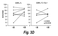


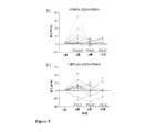


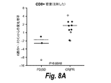


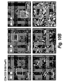


Classifications
-
- A—HUMAN NECESSITIES
- A61—MEDICAL OR VETERINARY SCIENCE; HYGIENE
- A61K—PREPARATIONS FOR MEDICAL, DENTAL OR TOILETRY PURPOSES
- A61K35/00—Medicinal preparations containing materials or reaction products thereof with undetermined constitution
- A61K35/66—Microorganisms or materials therefrom
- A61K35/76—Viruses; Subviral particles; Bacteriophages
- A61K35/763—Herpes virus
-
- A—HUMAN NECESSITIES
- A61—MEDICAL OR VETERINARY SCIENCE; HYGIENE
- A61K—PREPARATIONS FOR MEDICAL, DENTAL OR TOILETRY PURPOSES
- A61K39/00—Medicinal preparations containing antigens or antibodies
- A61K39/395—Antibodies; Immunoglobulins; Immune serum, e.g. antilymphocytic serum
- A61K39/39533—Antibodies; Immunoglobulins; Immune serum, e.g. antilymphocytic serum against materials from animals
- A61K39/3955—Antibodies; Immunoglobulins; Immune serum, e.g. antilymphocytic serum against materials from animals against proteinaceous materials, e.g. enzymes, hormones, lymphokines
-
- A—HUMAN NECESSITIES
- A61—MEDICAL OR VETERINARY SCIENCE; HYGIENE
- A61P—SPECIFIC THERAPEUTIC ACTIVITY OF CHEMICAL COMPOUNDS OR MEDICINAL PREPARATIONS
- A61P35/00—Antineoplastic agents
-
- A—HUMAN NECESSITIES
- A61—MEDICAL OR VETERINARY SCIENCE; HYGIENE
- A61P—SPECIFIC THERAPEUTIC ACTIVITY OF CHEMICAL COMPOUNDS OR MEDICINAL PREPARATIONS
- A61P35/00—Antineoplastic agents
- A61P35/04—Antineoplastic agents specific for metastasis
-
- C—CHEMISTRY; METALLURGY
- C07—ORGANIC CHEMISTRY
- C07K—PEPTIDES
- C07K14/00—Peptides having more than 20 amino acids; Gastrins; Somatostatins; Melanotropins; Derivatives thereof
- C07K14/435—Peptides having more than 20 amino acids; Gastrins; Somatostatins; Melanotropins; Derivatives thereof from animals; from humans
- C07K14/705—Receptors; Cell surface antigens; Cell surface determinants
- C07K14/70596—Molecules with a "CD"-designation not provided for elsewhere
-
- C—CHEMISTRY; METALLURGY
- C07—ORGANIC CHEMISTRY
- C07K—PEPTIDES
- C07K16/00—Immunoglobulins [IGs], e.g. monoclonal or polyclonal antibodies
- C07K16/18—Immunoglobulins [IGs], e.g. monoclonal or polyclonal antibodies against material from animals or humans
- C07K16/28—Immunoglobulins [IGs], e.g. monoclonal or polyclonal antibodies against material from animals or humans against receptors, cell surface antigens or cell surface determinants
- C07K16/2803—Immunoglobulins [IGs], e.g. monoclonal or polyclonal antibodies against material from animals or humans against receptors, cell surface antigens or cell surface determinants against the immunoglobulin superfamily
- C07K16/2818—Immunoglobulins [IGs], e.g. monoclonal or polyclonal antibodies against material from animals or humans against receptors, cell surface antigens or cell surface determinants against the immunoglobulin superfamily against CD28 or CD152
-
- G—PHYSICS
- G01—MEASURING; TESTING
- G01N—INVESTIGATING OR ANALYSING MATERIALS BY DETERMINING THEIR CHEMICAL OR PHYSICAL PROPERTIES
- G01N33/00—Investigating or analysing materials by specific methods not covered by groups G01N1/00 - G01N31/00
- G01N33/48—Biological material, e.g. blood, urine; Haemocytometers
- G01N33/50—Chemical analysis of biological material, e.g. blood, urine; Testing involving biospecific ligand binding methods; Immunological testing
- G01N33/5005—Chemical analysis of biological material, e.g. blood, urine; Testing involving biospecific ligand binding methods; Immunological testing involving human or animal cells
- G01N33/5091—Chemical analysis of biological material, e.g. blood, urine; Testing involving biospecific ligand binding methods; Immunological testing involving human or animal cells for testing the pathological state of an organism
-
- G—PHYSICS
- G01—MEASURING; TESTING
- G01N—INVESTIGATING OR ANALYSING MATERIALS BY DETERMINING THEIR CHEMICAL OR PHYSICAL PROPERTIES
- G01N33/00—Investigating or analysing materials by specific methods not covered by groups G01N1/00 - G01N31/00
- G01N33/48—Biological material, e.g. blood, urine; Haemocytometers
- G01N33/50—Chemical analysis of biological material, e.g. blood, urine; Testing involving biospecific ligand binding methods; Immunological testing
- G01N33/53—Immunoassay; Biospecific binding assay; Materials therefor
- G01N33/574—Immunoassay; Biospecific binding assay; Materials therefor for cancer
- G01N33/57407—Specifically defined cancers
- G01N33/5743—Specifically defined cancers of skin, e.g. melanoma
-
- G—PHYSICS
- G01—MEASURING; TESTING
- G01N—INVESTIGATING OR ANALYSING MATERIALS BY DETERMINING THEIR CHEMICAL OR PHYSICAL PROPERTIES
- G01N33/00—Investigating or analysing materials by specific methods not covered by groups G01N1/00 - G01N31/00
- G01N33/48—Biological material, e.g. blood, urine; Haemocytometers
- G01N33/50—Chemical analysis of biological material, e.g. blood, urine; Testing involving biospecific ligand binding methods; Immunological testing
- G01N33/68—Chemical analysis of biological material, e.g. blood, urine; Testing involving biospecific ligand binding methods; Immunological testing involving proteins, peptides or amino acids
- G01N33/6863—Cytokines, i.e. immune system proteins modifying a biological response such as cell growth proliferation or differentiation, e.g. TNF, CNF, GM-CSF, lymphotoxin, MIF or their receptors
-
- A—HUMAN NECESSITIES
- A61—MEDICAL OR VETERINARY SCIENCE; HYGIENE
- A61K—PREPARATIONS FOR MEDICAL, DENTAL OR TOILETRY PURPOSES
- A61K39/00—Medicinal preparations containing antigens or antibodies
- A61K2039/505—Medicinal preparations containing antigens or antibodies comprising antibodies
-
- A—HUMAN NECESSITIES
- A61—MEDICAL OR VETERINARY SCIENCE; HYGIENE
- A61K—PREPARATIONS FOR MEDICAL, DENTAL OR TOILETRY PURPOSES
- A61K2300/00—Mixtures or combinations of active ingredients, wherein at least one active ingredient is fully defined in groups A61K31/00 - A61K41/00
-
- A—HUMAN NECESSITIES
- A61—MEDICAL OR VETERINARY SCIENCE; HYGIENE
- A61K—PREPARATIONS FOR MEDICAL, DENTAL OR TOILETRY PURPOSES
- A61K39/00—Medicinal preparations containing antigens or antibodies
- A61K39/395—Antibodies; Immunoglobulins; Immune serum, e.g. antilymphocytic serum
- A61K39/39533—Antibodies; Immunoglobulins; Immune serum, e.g. antilymphocytic serum against materials from animals
- A61K39/39541—Antibodies; Immunoglobulins; Immune serum, e.g. antilymphocytic serum against materials from animals against normal tissues, cells
-
- G—PHYSICS
- G01—MEASURING; TESTING
- G01N—INVESTIGATING OR ANALYSING MATERIALS BY DETERMINING THEIR CHEMICAL OR PHYSICAL PROPERTIES
- G01N2800/00—Detection or diagnosis of diseases
- G01N2800/52—Predicting or monitoring the response to treatment, e.g. for selection of therapy based on assay results in personalised medicine; Prognosis
Landscapes
- Health & Medical Sciences (AREA)
- Life Sciences & Earth Sciences (AREA)
- Chemical & Material Sciences (AREA)
- Immunology (AREA)
- Engineering & Computer Science (AREA)
- Medicinal Chemistry (AREA)
- Molecular Biology (AREA)
- General Health & Medical Sciences (AREA)
- Urology & Nephrology (AREA)
- Hematology (AREA)
- Biomedical Technology (AREA)
- Microbiology (AREA)
- Cell Biology (AREA)
- Biochemistry (AREA)
- Animal Behavior & Ethology (AREA)
- Public Health (AREA)
- Pharmacology & Pharmacy (AREA)
- Veterinary Medicine (AREA)
- Physics & Mathematics (AREA)
- General Physics & Mathematics (AREA)
- Pathology (AREA)
- Analytical Chemistry (AREA)
- Food Science & Technology (AREA)
- Biotechnology (AREA)
- Organic Chemistry (AREA)
- Oncology (AREA)
- Virology (AREA)
- Mycology (AREA)
- Epidemiology (AREA)
- General Chemical & Material Sciences (AREA)
- Chemical Kinetics & Catalysis (AREA)
- Proteomics, Peptides & Aminoacids (AREA)
- Nuclear Medicine, Radiotherapy & Molecular Imaging (AREA)
- Hospice & Palliative Care (AREA)
- Tropical Medicine & Parasitology (AREA)
- Physiology (AREA)
- Biophysics (AREA)
- Endocrinology (AREA)
- Bioinformatics & Cheminformatics (AREA)
- Genetics & Genomics (AREA)
Abstract
ペンブロリズマブ、ペンブロリズマブバリアント又はこれらの抗原結合断片及びタリモジーン・ラハーパレプベックを含む併用療法による治療に応答する様々ながんを同定するのに有用なバイオマーカーが提供される。ペンブロリズマブ、ペンブロリズマブバリアント又はこれらの抗原結合断片による単剤療法に対して耐性を有するがんを治療する方法が提供される。低いCD8+密度、低い若しくは陰性のインターフェロンガンマシグネチャー及び/又は低い若しくは陰性のPD−L1状態を有する腫瘍を有する対象におけるがんを治療する方法もまた提供される。Provided are biomarkers useful in identifying various cancers that respond to treatment with a combination therapy including pembrolizumab, pembrolizumab variant or antigen-binding fragments thereof and Talimogene Laherparepbeck. Methods of treating cancers that are resistant to monotherapy with pembrolizumab, pembrolizumab variants or antigen-binding fragments thereof are provided. Also provided are methods of treating cancer in a subject having a tumor with low CD8+ density, low or negative interferon gamma signature, and/or low or negative PD-L1 status.
Description
関連出願
本出願は、2017年4月28日に出願された米国仮出願第62/491,746号の優先権の利益を主張する。前述の出願の内容は、全ての目的のためにその全体が参照により本明細書に組み込まれる。
Related Applications This application claims the benefit of priority of US Provisional Application No. 62/491,746, filed April 28, 2017. The contents of the aforementioned application are incorporated herein by reference in their entirety for all purposes.
発明の分野
本発明は、がん療法の分野に関する。特に、本発明は、ペンブロリズマブ、ペンブロリズマブバリアント及び/又はこれらの抗原結合断片並びにタリモジーン・ラハーパレプベックを含む併用療法により治療され得る様々ながんを同定するのに有用なバイオマーカーに関する。
FIELD OF THE INVENTION The present invention relates to the field of cancer therapy. In particular, the present invention provides biomarkers useful for identifying various cancers that may be treated by combination therapy including pembrolizumab, pembrolizumab variants and/or antigen-binding fragments thereof and Tarimogene laherparepbeck. Regarding
抗PD−1又は抗PD−L1抗体による治療は、様々ながんを有する患者において長期にわたる抗腫瘍応答をもたらし、転移性黒色腫、頭頸部癌、肺癌、腎癌及び膀胱癌並びにメルケル細胞癌及びホジキン病を有する患者にとっての標準的な治療方法となってきている(Sharma,P.及びAllison,J.P.(2015年)、The future of immune checkpoint therapy、Science 348、56〜61頁)。しかしながら、これらの適応症の全てにおいて、患者の一部のみが療法に対して応答し、患者の大部分は主にPD−1遮断に対して耐性を有する。したがって、PD−1遮断耐性がんを標的とする新たながん治療が必要とされている。 Treatment with anti-PD-1 or anti-PD-L1 antibodies results in a long-term anti-tumor response in patients with various cancers, including metastatic melanoma, head and neck cancer, lung cancer, renal and bladder cancer and Merkel cell carcinoma. And has become a standard treatment method for patients with Hodgkin's disease (Sharma, P. and Allison, JP (2015), The future of immune checkpoint therapy, Science 348, 56-61). .. However, in all of these indications, only some of the patients respond to therapy, and the majority of patients are predominantly resistant to PD-1 blockade. Therefore, new cancer therapies targeting PD-1 blockade resistant cancers are needed.
(発明の要旨)
本開示は、ペンブロリズマブ、ペンブロリズマブバリアント又はこれらの抗原結合断片及びタリモジーン・ラハーパレプベックによる併用療法に応答する腫瘍を同定するために使用され得るバイオマーカー(例えば、腫瘍内バイオマーカー)の発見に基づく。本発明は、単剤療法により(すなわち、チェックポイント阻害剤により(例えば、抗PD−L1療法又は抗PD−1療法(例えば、ペンブロリズマブ、ペンブロリズマブバリアント又はこれらの抗原結合断片)により))以前に治療不可能であったか又は十分に治療できなかった対象における腫瘍を治療するのに、特に有用である。腫瘍溶解性ウイルスであるタリモジーン・ラハーパレプベック(腫瘍内で優先的に複製し、顆粒球マクロファージコロニー刺激因子(GM−CSF)を産生するように設計された単純ヘルペスウイルス1型)の腫瘍内投与は、ヒト患者において細胞傷害性T細胞によって腫瘍内浸潤を増加させ、これによって、低いCD8+T細胞密度、低い若しくは陰性のインターフェロンガンマシグネチャー及び/又は低い若しくは陰性のPD−L1状態を示す腫瘍を有する対象において、並びに/又は以前のチェックポイント阻害剤療法(例えば、抗PD−L1療法若しくは抗PD−1療法(例えばペンブロリズマブ)による)に対して応答しなかったか若しくはほとんど応答しなかった対象において、又は例えば低いPD−L1状態に起因してチェックポイント阻害剤による(例えば、抗PD−L1療法又は抗PD−1療法(例えば、ペンブロリズマブ、ペンブロリズマブバリアント又はこれらの抗原結合断片)による)単剤療法に対して応答する可能性が低い対象において、併用療法として使用した場合、抗PD−1抗体ペンブロリズマブの抗腫瘍活性を改善することが決定された。
(Summary of invention)
The present disclosure provides biomarkers (eg, intratumoral biomarkers) that can be used to identify tumors that respond to combination therapy with pembrolizumab, pembrolizumab variants or antigen-binding fragments thereof and Talimogene Laherparepbeck. Based on the discovery of. The invention can be administered by monotherapy (ie, by a checkpoint inhibitor (eg, anti-PD-L1 or anti-PD-1 therapy (eg, pembrolizumab, pembrolizumab variant or antigen-binding fragment thereof)). It is particularly useful for treating tumors in subjects that were previously untreatable or under-treated. Tumor of the oncolytic virus Talimogene laherparepbeck (herpes
一態様では、約1500細胞/mm2未満、約1400細胞/mm2未満、約1300細胞/mm2未満、約1200細胞/mm2未満、約1100細胞/mm2未満、約1000細胞/mm2未満、約900細胞/mm2未満、約800細胞/mm2未満、約700細胞/mm2未満、約600細胞/mm2未満又は約500細胞/mm2未満のCD8+T細胞浸潤密度を含む腫瘍を有する対象を選択するステップ、タリモジーン・ラハーパレプベックを対象に投与するステップ、及びペンブロリズマブ、ペンブロリズマブバリアント又はこれらの抗原結合断片を対象に投与するステップを含む、対象における腫瘍を治療する方法が提供される。 In one aspect, less than about 1500 cells/mm 2, less than about 1400 cells/mm 2, less than about 1300 cells/mm 2, less than about 1200 cells/mm 2, less than about 1100 cells/mm 2 , about 1000 cells/mm 2. A tumor comprising a CD8+ T cell infiltration density of less than, less than about 900 cells/mm 2, less than about 800 cells/mm 2, less than about 700 cells/mm 2, less than about 600 cells/mm 2 or less than about 500 cells/mm 2. Treating a tumor in a subject, comprising selecting a subject having, administering Tarimogene laharparepbeck to the subject, and administering pembrolizumab, pembrolizumab variant, or an antigen-binding fragment thereof to the subject. A method is provided.
ある特定の実施形態では、腫瘍は、IFNγ、シグナル伝達兼転写活性化因子1(STAT1)、C−Cケモカイン受容体5型(CCR5)、ケモカイン(C−X−Cモチーフ)リガンド9(CXCL9)、パーフォリン1(PRF1)、HLA−DRA、ケモカイン(C−X−Cモチーフ)リガンド10(CXCL10)、ケモカイン(C−X−Cモチーフ)リガンド11(CXCL11)、インドールアミン2,3−ジオキシゲナーゼ1(IDO1)及びグランザイムA(GZMA)からなる群から選択されるシグネチャー遺伝子の対照パネルの所定の閾値と比較して、投与する前に、より低いレベルの2、3、4又は5種のインターフェロンガンマ(IFNγ)シグネチャー遺伝子を発現する。ある特定の実施形態では、腫瘍は、タリモジーン・ラハーパレプベック及びペンブロリズマブ、ペンブロリズマブバリアント又はこれらの抗原結合断片を投与する前にIFNγシグネチャー遺伝子を発現しない。 In certain embodiments, the tumor is IFNγ, signaling and transcriptional activator 1 (STAT1), C—C chemokine receptor type 5 (CCR5), chemokine (C—X—C motif) ligand 9 (CXCL9). , Perforin 1 (PRF1), HLA-DRA, chemokine (C-X-C motif) ligand 10 (CXCL10), chemokine (C-X-C motif) ligand 11 (CXCL11), indoleamine 2,3-dioxygenase 1 (IDO1) and granzyme A (GZMA) a lower level of 2, 3, 4 or 5 interferon gamma prior to administration, as compared to a predetermined threshold of a control panel of signature genes selected from the group consisting of: It expresses the (IFNγ) signature gene. In certain embodiments, the tumor does not express the IFNγ signature gene prior to administration of Talimogene laherparepbeck and pembrolizumab, pembrolizumab variant or antigen-binding fragment thereof.
ある特定の実施形態では、腫瘍は、タリモジーン・ラハーパレプベック及びペンブロリズマブ、ペンブロリズマブバリアント又はこれらの抗原結合断片を投与する前に約50%未満のプログラム死リガンド1(PD−L1)状態を有する。ある特定の実施形態では、腫瘍は、タリモジーン・ラハーパレプベック及びペンブロリズマブ、ペンブロリズマブバリアント又はこれらの抗原結合断片を投与する前に約1%未満のPD−L1状態を有する。 In certain embodiments, the tumor is less than about 50% programmed death ligand 1 (PD-L1) prior to administration of Talimogene laherparepbeck and pembrolizumab, pembrolizumab variant or antigen-binding fragment thereof. Have a state. In certain embodiments, the tumor has a PD-L1 status of less than about 1% prior to administration of Talimogene laherparepbeck and pembrolizumab, pembrolizumab variant or antigen-binding fragment thereof.
ある特定の実施形態では、タリモジーン・ラハーパレプベックは対象に腫瘍内で投与される及び/又はペンブロリズマブ若しくはこの抗原結合断片は対象に全身的に投与される。 In certain embodiments, Tarimogene laharparepbeck is administered intratumorally to the subject and/or pembrolizumab or an antigen binding fragment thereof is administered systemically to the subject.
ある特定の実施形態では、タリモジーン・ラハーパレプベックは、ペンブロリズマブ又はこの抗原結合断片の投与前に対象に投与される。 In certain embodiments, Tarimogene laharparepbeck is administered to the subject prior to the administration of pembrolizumab or an antigen-binding fragment thereof.
ある特定の実施形態では、注射された腫瘍のサイズの減少及び/又は注射されていない腫瘍のサイズの減少は、タリモジーン・ラハーパレプベック及びペンブロリズマブ、ペンブロリズマブバリアント又はこれらの抗原結合断片を投与した後に生じる。 In certain embodiments, the reduced size of the injected tumor and/or the reduced size of the non-injected tumor is caused by talimogene laherparepbeck and pembrolizumab, pembrolizumab variant or antigen-binding fragment thereof. Occurs after administration of.
ある特定の実施形態では、CD8+T細胞浸潤密度は、タリモジーン・ラハーパレプベックを投与した後の腫瘍において増加する。ある特定の実施形態では、対象において循環している分裂CD8+T細胞は、タリモジーン・ラハーパレプベック及びペンブロリズマブ、ペンブロリズマブバリアント又はこれらの抗原結合断片を投与した後に増加する。 In certain embodiments, the CD8+ T cell infiltration density is increased in the tumor following administration of Talimogene Laharparepbeck. In certain embodiments, the circulating dividing CD8 + T cells in the subject are increased following administration of Talimogene laherparepbeck and pembrolizumab, pembrolizumab variant or antigen-binding fragment thereof.
ある特定の実施形態では、対象は、黒色腫、非小細胞肺がん、頭頸部がん、大腸がん、乳がん、卵巣がん、膀胱がん、前立腺がん、肉腫、腎細胞がん、胃がん、食道がん、肛門管がん、胆道がん及び膵がんからなる群から選択されるがんを有する。ある特定の実施形態では、黒色腫は、皮膚黒色腫、転移性黒色腫若しくはぶどう膜黒色腫であり、乳がんは、HER2+乳がん、HER2−HR+乳がん若しくはトリプルネガティブ乳がんであり、前立腺がんは、去勢抵抗性前立腺がんであり、膀胱がんは、移行上皮がん若しくは尿路上皮がんであり、頭頸部がんは、頭頸部の再発性若しくは転移性扁平上皮癌であり、及び/又は肉腫は、軟組織肉腫若しくは骨肉腫である。 In certain embodiments, the subject is melanoma, non-small cell lung cancer, head and neck cancer, colorectal cancer, breast cancer, ovarian cancer, bladder cancer, prostate cancer, sarcoma, renal cell cancer, gastric cancer, Having a cancer selected from the group consisting of esophageal cancer, anal canal cancer, biliary tract cancer, and pancreatic cancer. In certain embodiments, the melanoma is cutaneous melanoma, metastatic melanoma or uveal melanoma, the breast cancer is HER2+ breast cancer, HER2-HR+ breast cancer or triple negative breast cancer, and the prostate cancer is castration. Resistant prostate cancer, bladder cancer is transitional cell carcinoma or urothelial cancer, head and neck cancer is recurrent or metastatic squamous cell carcinoma of the head and neck, and/or sarcoma is This is soft tissue sarcoma or osteosarcoma.
ある特定の実施形態では、腫瘍は、約1000細胞/mm2未満のCD8+T細胞浸潤密度を含む。 In certain embodiments, the tumor comprises a CD8 + T cell infiltration density of less than about 1000 cells/mm 2 .
一態様では、チェックポイント阻害剤による(例えば、抗PD−L1療法又は抗PD−1療法(例えば、ペンブロリズマブ、ペンブロリズマブバリアント又はこれらの抗原結合断片)による)単剤療法に対してほとんど応答しない又は応答しない対象における腫瘍を治療する方法であって、タリモジーン・ラハーパレプベックを対象に投与するステップ、及びペンブロリズマブ、ペンブロリズマブバリアント又はこれらの抗原結合断片を対象に投与するステップを含む、方法が提供される。 In one aspect, most response to monotherapy with a checkpoint inhibitor (eg, with anti-PD-L1 therapy or anti-PD-1 therapy (eg, pembrolizumab, pembrolizumab variant or antigen-binding fragment thereof)). A method of treating a tumor in a subject that does not or does not respond, comprising administering to the subject talimogene laherparepbeck, and administering pembrolizumab, a pembrolizumab variant or an antigen-binding fragment thereof to the subject. A method is provided that includes.
ある特定の実施形態では、腫瘍は、投与する前に、約1500細胞/mm2未満、約1400細胞/mm2未満、約1300細胞/mm2未満、約1200細胞/mm2未満、約1100細胞/mm2未満、約1000細胞/mm2未満、約900細胞/mm2未満、約800細胞/mm2未満、約700細胞/mm2未満、約600細胞/mm2未満若しくは約500細胞/mm2未満のCD8+T細胞浸潤密度を含み、腫瘍は、投与する前に、IFNγ、STAT1、CCR5、CXCL9、PRF1、HLA−DRA、CXCL10、CXCL11、IDO1及びGZMAからなる群から選択されるシグネチャー遺伝子の対照パネルの所定の閾値より低いレベルの、2、3、4若しくは5種のインターフェロンガンマ(IFNγ)シグネチャー遺伝子を発現し、並びに/又は腫瘍は、タリモジーン・ラハーパレプベック及びペンブロリズマブ、ペンブロリズマブバリアント又はこれらの抗原結合断片を投与する前に、約50%未満のPD−L1状態を有する。
In certain embodiments, the tumor is less than about 1500 cells/mm 2, less than about 1400 cells/mm 2, less than about 1300 cells/mm 2, less than about 1200 cells/mm 2 , about 1100 cells prior to administration. /Mm 2, less than about 1000 cells/mm 2, less than about 900 cells/mm 2, less than about 800 cells/mm 2, less than about 700 cells/mm 2, less than about 600 cells/mm 2 or about 500 cells/
一態様では、チェックポイント阻害剤による(例えば、抗PD−L1療法又は抗PD−1療法(例えば、ペンブロリズマブ、ペンブロリズマブバリアント又はこれらの抗原結合断片)による)単剤療法の間に進行した対象における腫瘍を治療する方法であって、タリモジーン・ラハーパレプベックを対象に投与するステップ、及びペンブロリズマブ、ペンブロリズマブバリアント又はこれらの抗原結合断片を対象に投与するステップを含む、方法が提供される。 In one aspect, progression during monotherapy with a checkpoint inhibitor (eg, with anti-PD-L1 therapy or anti-PD-1 therapy (eg, pembrolizumab, pembrolizumab variant or antigen-binding fragment thereof)). A method of treating a tumor in a subject, the method comprising administering to the subject talimogene laherparepbeck, and pembrolizumab, pembrolizumab variant or an antigen-binding fragment thereof to the subject. Provided.
ある特定の実施形態では、腫瘍は、投与する前に、約1500細胞/mm2未満、約1400細胞/mm2未満、約1300細胞/mm2未満、約1200細胞/mm2未満、約1100細胞/mm2未満、約1000細胞/mm2未満、約900細胞/mm2未満、約800細胞/mm2未満、約700細胞/mm2未満、約600細胞/mm2未満若しくは約500細胞/mm2未満のCD8+T細胞浸潤密度を含み、腫瘍は、投与する前に、IFNγ、STAT1、CCR5、CXCL9、PRF1、HLA−DRA、CXCL10、CXCL11、IDO1及びGZMAからなる群から選択されるシグネチャー遺伝子の対照パネルの、所定の閾値より低いレベルの2、3、4若しくは5種のIFNγシグネチャー遺伝子を発現し、並びに/又は腫瘍は、タリモジーン・ラハーパレプベック及びペンブロリズマブ、ペンブロリズマブバリアント又はこれらの抗原結合断片を投与する前に約50%未満のPD−L1状態を有する。
In certain embodiments, the tumor is less than about 1500 cells/mm 2, less than about 1400 cells/mm 2, less than about 1300 cells/mm 2, less than about 1200 cells/mm 2 , about 1100 cells prior to administration. /Mm 2, less than about 1000 cells/mm 2, less than about 900 cells/mm 2, less than about 800 cells/mm 2, less than about 700 cells/mm 2, less than about 600 cells/mm 2 or about 500 cells/
ある特定の実施形態では、タリモジーン・ラハーパレプベックは、初回用量、続いて1回以上の二次用量として連続して投与される。ある特定の実施形態では、ペンブロリズマブ、ペンブロリズマブバリアント又はこれらの抗原結合断片は、初回用量、続いて1回以上の二次用量として連続して投与される。ある特定の実施形態では、タリモジーン・ラハーパレプベックは、初回用量、続いて1回以上の二次用量として連続して投与され、ペンブロリズマブ、ペンブロリズマブバリアント又はこれらの抗原結合断片は、タリモジーン・ラハーパレプベックの1回以上の二次用量と連続してかつ同時に投与される。 In certain embodiments, Tarimogene laharparepbeck is administered sequentially as an initial dose followed by one or more secondary doses. In certain embodiments, pembrolizumab, pembrolizumab variants or antigen-binding fragments thereof are administered sequentially as an initial dose, followed by one or more secondary doses. In certain embodiments, Tarimogene laharparepbeck is administered sequentially as an initial dose followed by one or more secondary doses, wherein pembrolizumab, pembrolizumab variant or an antigen-binding fragment thereof is Administered sequentially and simultaneously with one or more secondary doses of Talimogene Lahar Palapbeck.
ある特定の実施形態では、タリモジーン・ラハーパレプベックは腫瘍内で投与され、ペンブロリズマブ、ペンブロリズマブバリアント又はこれらの抗原結合断片は全身的に投与される。ある特定の実施形態では、タリモジーン・ラハーパレプベック及びペンブロリズマブ、ペンブロリズマブバリアント又はこれらの抗原結合断片は腫瘍内で投与される。 In certain embodiments, Tarimogene laharparepbeck is administered intratumorally and pembrolizumab, pembrolizumab variant or antigen-binding fragment thereof is administered systemically. In certain embodiments, Talimogene laherparepbeck and pembrolizumab, pembrolizumab variant or antigen-binding fragments thereof are administered intratumorally.
一態様では、約1500細胞/mm2未満、約1400細胞/mm2未満、約1300細胞/mm2未満、約1200細胞/mm2未満、約1100細胞/mm2未満、約1000細胞/mm2未満、約900細胞/mm2未満、約800細胞/mm2未満、約700細胞/mm2未満、約600細胞/mm2未満又は約500細胞/mm2未満のCD8+T細胞浸潤密度を有する腫瘍を治療する方法であって、タリモジーン・ラハーパレプベック及びペンブロリズマブ、ペンブロリズマブバリアント又はこれらの抗原結合断片と腫瘍を接触させるステップを含む、方法が提供される。 In one aspect, less than about 1500 cells/mm 2, less than about 1400 cells/mm 2, less than about 1300 cells/mm 2, less than about 1200 cells/mm 2, less than about 1100 cells/mm 2 , about 1000 cells/mm 2. Having a CD8 + T cell infiltration density of less than about 900 cells/mm 2, less than about 800 cells/mm 2, less than about 700 cells/mm 2, less than about 600 cells/mm 2 or less than about 500 cells/mm 2. A method of treating a tumor is provided comprising the step of contacting the tumor with Talimogene laherparepbeck and pembrolizumab, pembrolizumab variant or an antigen binding fragment thereof.
ある特定の実施形態では、腫瘍は、黒色腫、非小細胞肺がん、頭頸部がん、大腸がん、乳がん、卵巣がん、膀胱がん、前立腺がん、肉腫、腎細胞がん、胃がん、食道がん、肛門管がん、胆道がん及び膵がんからなる群から選択されるがんを有する対象由来である。ある特定の実施形態では、がんは皮膚黒色腫である。ある特定の実施形態では、がんは頭頸部の再発性若しくは転移性扁平上皮癌である。 In certain embodiments, the tumor is melanoma, non-small cell lung cancer, head and neck cancer, colorectal cancer, breast cancer, ovarian cancer, bladder cancer, prostate cancer, sarcoma, renal cell cancer, gastric cancer, It is derived from a subject having a cancer selected from the group consisting of esophageal cancer, anal canal cancer, biliary tract cancer, and pancreatic cancer. In certain embodiments, the cancer is cutaneous melanoma. In certain embodiments, the cancer is recurrent or metastatic squamous cell carcinoma of the head and neck.
ある特定の実施形態では、腫瘍は、接触させる前に、IFNγ、STAT1、CCR5、CXCL9、PRF1、HLA−DRA、CXCL10、CXCL11、IDO1及びGZMAからなる群から選択されるシグネチャー遺伝子の対照パネルの、所定の閾値より低いレベルの2、3、4若しくは5種のIFNγシグネチャー遺伝子を発現する。 In certain embodiments, the tumor is prior to contacting with a control panel of signature genes selected from the group consisting of IFNγ, STAT1, CCR5, CXCL9, PRF1, HLA-DRA, CXCL10, CXCL11, IDO1 and GZMA. It expresses levels of 2, 3, 4 or 5 IFNγ signature genes below a given threshold.
ある特定の実施形態では、腫瘍は、タリモジーン・ラハーパレプベック及びペンブロリズマブ、ペンブロリズマブバリアント又はこれらの抗原結合断片と接触させる前に約50%未満のPD−L1を有する。 In certain embodiments, the tumor has less than about 50% PD-L1 prior to contacting with Tarimogene laharparepbeck and pembrolizumab, pembrolizumab variant or antigen-binding fragment thereof.
一態様では、治療前に、IFNγ、STAT1、CCR5、CXCL9、PRF1、HLA−DRA、CXCL10、CXCL11、IDO1及びGZMAからなる群から選択されるシグネチャー遺伝子の対照パネルの、所定の閾値より低いレベルの2、3、4又は5種のIFNγシグネチャー遺伝子を発現する腫瘍を治療する方法であって、腫瘍を、タリモジーン・ラハーパレプベック及びペンブロリズマブ、ペンブロリズマブバリアント又はこれらの抗原結合断片と接触させるステップを含む、方法が提供される。 In one aspect, prior to treatment, a control panel of a signature gene selected from the group consisting of IFNγ, STAT1, CCR5, CXCL9, PRF1, HLA-DRA, CXCL10, CXCL11, IDO1 and GZMA has a level below a predetermined threshold. A method of treating a tumor expressing 2, 3, 4 or 5 IFNγ signature genes, the method comprising contacting the tumor with tarimogene laherparepbeck and pembrolizumab, pembrolizumab variant or an antigen-binding fragment thereof. A method is provided that includes the steps of:
ある特定の実施形態では、腫瘍は、黒色腫、非小細胞肺がん、頭頸部がん、大腸がん、乳がん、卵巣がん、膀胱がん、前立腺がん、肉腫、腎細胞がん、胃がん、食道がん、肛門管がん、胆道がん及び膵がんからなる群から選択されるがんを有する対象由来である。ある特定の実施形態では、がんは黒色腫(例えば、皮膚黒色腫)である。ある特定の実施形態では、がんは頭頸部がん(例えば、頭頸部の再発性若しくは転移性扁平上皮癌)である。 In certain embodiments, the tumor is melanoma, non-small cell lung cancer, head and neck cancer, colorectal cancer, breast cancer, ovarian cancer, bladder cancer, prostate cancer, sarcoma, renal cell cancer, gastric cancer, It is derived from a subject having a cancer selected from the group consisting of esophageal cancer, anal canal cancer, biliary tract cancer, and pancreatic cancer. In certain embodiments, the cancer is melanoma (eg, cutaneous melanoma). In certain embodiments, the cancer is head and neck cancer (eg, recurrent or metastatic squamous cell carcinoma of the head and neck).
ある特定の実施形態では、腫瘍は、接触させる前に、約1500細胞/mm2未満、約1400細胞/mm2未満、約1300細胞/mm2未満、約1200細胞/mm2未満、約1100細胞/mm2未満、約1000細胞/mm2未満、約900細胞/mm2未満、約800細胞/mm2未満、約700細胞/mm2未満、約600細胞/mm2未満又は約500細胞/mm2未満のCD8+T細胞浸潤密度を有する。
In certain embodiments, the tumor is less than about 1500 cells/mm 2, less than about 1400 cells/mm 2, less than about 1300 cells/mm 2, less than about 1200 cells/mm 2 , about 1100 cells prior to contacting. /Mm 2, less than about 1000 cells/mm 2, less than about 900 cells/mm 2, less than about 800 cells/mm 2, less than about 700 cells/mm 2, less than about 600 cells/mm 2 or about 500 cells/
ある特定の実施形態では、腫瘍は、タリモジーン・ラハーパレプベック及びペンブロリズマブ、ペンブロリズマブバリアント又はこれらの抗原結合断片と接触させる前に約50%未満のPD−L1状態を有する。 In certain embodiments, the tumor has less than about 50% PD-L1 status prior to contacting with Talimogene laherparepbeck and pembrolizumab, pembrolizumab variant or antigen-binding fragment thereof.
一態様では、治療前に約50%未満のPD−L1状態を有する腫瘍を治療する方法であって、腫瘍を、タリモジーン・ラハーパレプベック及びペンブロリズマブ、ペンブロリズマブバリアント又はこれらの抗原結合断片と接触させるステップを含む、方法が提供される。 In one aspect, a method of treating a tumor having a PD-L1 status of less than about 50% prior to treatment, wherein the tumor comprises tarimogene laherparepbeck and pembrolizumab, pembrolizumab variant or antigen-binding thereof. A method is provided that includes contacting a fragment.
ある特定の実施形態では、腫瘍は、黒色腫、非小細胞肺がん、頭頸部がん、大腸がん、乳がん、卵巣がん、膀胱がん、前立腺がん、肉腫、腎細胞がん、胃がん、食道がん、肛門管がん、胆道がん及び膵がんからなる群から選択されるがんを有する対象由来である。ある特定の実施形態では、がんは黒色腫(例えば、皮膚黒色腫)である。ある特定の実施形態では、がんは頭頸部がん(例えば、頭頸部の再発性若しくは転移性扁平上皮癌)である。 In certain embodiments, the tumor is melanoma, non-small cell lung cancer, head and neck cancer, colorectal cancer, breast cancer, ovarian cancer, bladder cancer, prostate cancer, sarcoma, renal cell cancer, gastric cancer, It is derived from a subject having a cancer selected from the group consisting of esophageal cancer, anal canal cancer, biliary tract cancer, and pancreatic cancer. In certain embodiments, the cancer is melanoma (eg, cutaneous melanoma). In certain embodiments, the cancer is head and neck cancer (eg, recurrent or metastatic squamous cell carcinoma of the head and neck).
ある特定の実施形態では、腫瘍は、接触させる前に、約1500細胞/mm2未満、約1400細胞/mm2未満、約1300細胞/mm2未満、約1200細胞/mm2未満、約1100細胞/mm2未満、約1000細胞/mm2未満、約900細胞/mm2未満、約800細胞/mm2未満、約700細胞/mm2未満、約600細胞/mm2未満又は約500細胞/mm2未満のCD8+T細胞浸潤密度を有する。
In certain embodiments, the tumor is less than about 1500 cells/mm 2, less than about 1400 cells/mm 2, less than about 1300 cells/mm 2, less than about 1200 cells/mm 2 , about 1100 cells prior to contacting. /Mm 2, less than about 1000 cells/mm 2, less than about 900 cells/mm 2, less than about 800 cells/mm 2, less than about 700 cells/mm 2, less than about 600 cells/mm 2 or about 500 cells/
ある特定の実施形態では、腫瘍は、接触させる前に、IFNγ、STAT1、CCR5、CXCL9、PRF1、HLA−DRA、CXCL10、CXCL11、IDO1及びGZMAからなる群から選択されるシグネチャー遺伝子の対照パネルの所定の閾値より低いレベルの2、3、4若しくは5種のIFNγシグネチャー遺伝子を発現する。 In certain embodiments, the tumor is prior to being contacted with a control panel of a signature gene selected from the group consisting of IFNγ, STAT1, CCR5, CXCL9, PRF1, HLA-DRA, CXCL10, CXCL11, IDO1 and GZMA. Express levels of 2, 3, 4 or 5 IFNγ signature genes below the threshold of
一態様では、約1500細胞/mm2未満、約1400細胞/mm2未満、約1300細胞/mm2未満、約1200細胞/mm2未満、約1100細胞/mm2未満、約1000細胞/mm2未満、約900細胞/mm2未満、約800細胞/mm2未満、約700細胞/mm2未満、約600細胞/mm2未満又は約500細胞/mm2未満のCD8+T細胞浸潤密度を有し、治療前に、IFNγ、STAT1、CCR5、CXCL9、PRF1、HLA−DRA、CXCL10、CXCL11、IDO1及びGZMAからなる群から選択されるシグネチャー遺伝子の対照パネルの所定の閾値より低いレベルの5種以下のIFNγシグネチャー遺伝子を発現する腫瘍を治療する方法であって、腫瘍を、タリモジーン・ラハーパレプベック及びペンブロリズマブ、ペンブロリズマブバリアント又はこれらの抗原結合断片と接触させるステップを含む、方法が提供される。 In one aspect, less than about 1500 cells/mm 2, less than about 1400 cells/mm 2, less than about 1300 cells/mm 2, less than about 1200 cells/mm 2, less than about 1100 cells/mm 2 , about 1000 cells/mm 2. Having a CD8 + T cell infiltration density of less than about 900 cells/mm 2, less than about 800 cells/mm 2, less than about 700 cells/mm 2, less than about 600 cells/mm 2 or less than about 500 cells/mm 2. However, before treatment, 5 or less levels of a signature gene selected from the group consisting of IFNγ, STAT1, CCR5, CXCL9, PRF1, HLA-DRA, CXCL10, CXCL11, IDO1 and GZMA, which are lower than a predetermined threshold of a control panel. A method of treating a tumor that expresses the IFNγ signature gene of Escherichia coli, comprising contacting the tumor with tarimogene laherparepbeck and pembrolizumab, pembrolizumab variant or an antigen-binding fragment thereof. To be done.
ある特定の実施形態では、腫瘍は、黒色腫、非小細胞肺がん、頭頸部がん、大腸がん、乳がん、卵巣がん、膀胱がん、前立腺がん、肉腫、腎細胞がん、胃がん、食道がん、肛門管がん、胆道がん及び膵がんからなる群から選択されるがんを有する対象由来である。ある特定の実施形態では、がんは黒色腫(例えば、皮膚黒色腫)である。ある特定の実施形態では、がんは頭頸部がん(例えば、頭頸部の再発性若しくは転移性扁平上皮癌)である。 In certain embodiments, the tumor is melanoma, non-small cell lung cancer, head and neck cancer, colorectal cancer, breast cancer, ovarian cancer, bladder cancer, prostate cancer, sarcoma, renal cell cancer, gastric cancer, It is derived from a subject having a cancer selected from the group consisting of esophageal cancer, anal canal cancer, biliary tract cancer, and pancreatic cancer. In certain embodiments, the cancer is melanoma (eg, cutaneous melanoma). In certain embodiments, the cancer is head and neck cancer (eg, recurrent or metastatic squamous cell carcinoma of the head and neck).
ある特定の実施形態では、腫瘍は、タリモジーン・ラハーパレプベック及びペンブロリズマブ、ペンブロリズマブバリアント又はこれらの抗原結合断片と接触させる前に約50%未満のPD−L1状態を有する。 In certain embodiments, the tumor has less than about 50% PD-L1 status prior to contacting with Talimogene laherparepbeck and pembrolizumab, pembrolizumab variant or antigen-binding fragment thereof.
一態様では、後でタリモジーン・ラハーパレプベックに曝露される対象における、以前にペンブロリズマブ、ペンブロリズマブバリアント又はこれらの抗原結合断片に耐性を有した腫瘍を治療する方法であって、ペンブロリズマブ、ペンブロリズマブバリアント又はこれらの抗原結合断片を対象に投与するステップを含む、方法が提供される。 In one aspect, a method of treating a tumor previously resistant to pembrolizumab, a pembrolizumab variant or an antigen-binding fragment thereof in a subject that is subsequently exposed to Talimogene laharparepbeck, comprising pembrolizumab. , Pembrolizumab variant or an antigen-binding fragment thereof is administered to a subject.
ある特定の実施形態では、対象は、黒色腫、非小細胞肺がん、頭頸部がん、大腸がん、乳がん、卵巣がん、膀胱がん、前立腺がん、肉腫、腎細胞がん、胃がん、食道がん、肛門管がん、胆道がん及び膵がんからなる群から選択されるがんを有する。ある特定の実施形態では、がんは黒色腫(例えば、皮膚黒色腫)である。ある特定の実施形態では、がんは頭頸部がん(例えば、頭頸部の再発性若しくは転移性扁平上皮癌)である。 In certain embodiments, the subject is melanoma, non-small cell lung cancer, head and neck cancer, colorectal cancer, breast cancer, ovarian cancer, bladder cancer, prostate cancer, sarcoma, renal cell cancer, gastric cancer, Having a cancer selected from the group consisting of esophageal cancer, anal canal cancer, biliary tract cancer, and pancreatic cancer. In certain embodiments, the cancer is melanoma (eg, cutaneous melanoma). In certain embodiments, the cancer is head and neck cancer (eg, recurrent or metastatic squamous cell carcinoma of the head and neck).
一態様では、対象におけるチェックポイント阻害剤による(例えば、抗PD−L1療法又は抗PD−1療法(例えば、ペンブロリズマブ、ペンブロリズマブバリアント又はこれらの抗原結合断片)による)単剤療法に対して耐性を有する腫瘍を、ペンブロリズマブ、ペンブロリズマブバリアント又はこれらの抗原結合断片に対して感受性にする方法であって、腫瘍をタリモジーン・ラハーパレプベックと接触させるステップを含む、方法が提供される。 In one aspect, against monotherapy with a checkpoint inhibitor (eg, with anti-PD-L1 or anti-PD-1 therapy (eg, pembrolizumab, pembrolizumab variant or antigen-binding fragment thereof)) in a subject. A method of sensitizing a resistant tumor to pembrolizumab, a pembrolizumab variant, or an antigen-binding fragment thereof, comprising the step of contacting the tumor with Tarimogene laharparepbeck. It
ある特定の実施形態では、腫瘍をタリモジーン・ラハーパレプベックと接触させた後に対象から採取した腫瘍の試料は、腫瘍をタリモジーン・ラハーパレプベックと接触させる前に採取した腫瘍の試料に対して増加したレベルのCD8+T細胞、CD4+T細胞、IFNγ、CD20+B細胞、記憶T細胞、制御性T細胞及びCD56+細胞のうちの1つ又はいずれかの組合せを有する。 In certain embodiments, the tumor sample taken from the subject after contacting the tumor with Talimogene Laherparepbeck is compared to the tumor sample taken before contacting the tumor with Talimogene Laharparepbeck. Have increased levels of one or any combination of CD8+ T cells, CD4+ T cells, IFNγ, CD20+ B cells, memory T cells, regulatory T cells and CD56+ cells.
ある特定の実施形態では、腫瘍は、タリモジーン・ラハーパレプベックと接触させた後、1000細胞/mm2より多いCD8+T細胞浸潤密度を有する。 In certain embodiments, the tumor has a CD8 + T cell infiltration density of greater than 1000 cells/mm 2 after contacting with Talimogene Laharparepbeck.
ある特定の実施形態では、腫瘍をタリモジーン・ラハーパレプベックと接触させた後に対象から採取した血液試料は、接触させる前に対象から採取した血液試料と比較して増加したレベルのCD8+T細胞及び/又はCD4+T細胞を有し、任意選択的にCD8+T細胞は分裂CD8+T細胞である。 In certain embodiments, a blood sample taken from the subject after contacting the tumor with Tarimogene laharparepbeck has increased levels of CD8+ T cells and increased compared to a blood sample taken from the subject prior to contacting. /Or have CD4+ T cells, optionally the CD8+ T cells are dividing CD8+ T cells.
一態様では、対象における腫瘍を治療する方法であって、約1500細胞/mm2未満、約1400細胞/mm2未満、約1300細胞/mm2未満、約1200細胞/mm2未満、約1100細胞/mm2未満、約1000細胞/mm2未満、約900細胞/mm2未満、約800細胞/mm2未満、約700細胞/mm2未満、約600細胞/mm2未満又は約500細胞/mm2未満のCD8+T細胞浸潤密度を含む腫瘍を有する対象を選択するステップ、初回用量後、1回以上の二次用量として、タリモジーン・ラハーパレプベックを対象に腫瘍内で投与するステップ、及びペンブロリズマブ、ペンブロリズマブバリアント又はこれらの抗原結合断片を、初回用量後、1回以上の二次用量として対象に全身的に投与するステップを含む、方法が提供される。
In one aspect, a method of treating a tumor in a subject comprising less than about 1500 cells/mm 2, less than about 1400 cells/mm 2, less than about 1300 cells/mm 2, less than about 1200 cells/mm 2 , about 1100 cells. /Mm 2, less than about 1000 cells/mm 2, less than about 900 cells/mm 2, less than about 800 cells/mm 2, less than about 700 cells/mm 2, less than about 600 cells/mm 2 or about 500 cells/
ある特定の実施形態では、二次用量は2週間に1回(Q2W)投与される。ある特定の実施形態では、タリモジーン・ラハーパレプベックの初回用量は1週目の1日目に投与され、タリモジーン・ラハーパレプベックの二次用量は、4週目の1日目、6週目の1日目に投与され、その後Q2Wで投与される。ある特定の実施形態では、ペンブロリズマブ、ペンブロリズマブバリアント又はこれらの抗原結合断片の初回用量は、6週目の1日目に投与され、ペンブロリズマブ、ペンブロリズマブバリアント又はこれらの抗原結合断片の二次用量は、8週目の1日目に投与され、その後Q2Wで投与される。
In certain embodiments, the secondary dose is administered once every two weeks (Q2W). In certain embodiments, the first dose of Talimogene Lahar Palapbeck is administered on the first day of the first week and the second dose of Talimogene Lahar Palabeck is on the first day of the fourth week. It will be administered on
ある特定の実施形態では、タリモジーン・ラハーパレプベックの初回用量は、106プラーク形成単位(pfu)/mLの用量にて投与され、タリモジーン・ラハーパレプベックの二次用量は、108pfu/mLの用量にて投与される。 In certain embodiments, the initial dose of Talimogene Laherparepbeck is administered at a dose of 10 6 plaque forming units (pfu)/mL and the secondary dose of Talimogene Lahaparepbeck is 10. It is administered at a dose of 8 pfu/mL.
ある特定の実施形態では、ペンブロリズマブ、ペンブロリズマブバリアント又はこれらの抗原結合断片の初回用量は200mgの用量にて投与され、ペンブロリズマブ、ペンブロリズマブバリアント又はこれらの抗原結合断片の二次用量は200mgの用量にて投与される。 In certain embodiments, the initial dose of pembrolizumab, pembrolizumab variant or antigen-binding fragment thereof is administered at a dose of 200 mg, and the secondary dose of pembrolizumab, pembrolizumab variant or antigen-binding fragment thereof is It is administered at a dose of 200 mg.
上記の開示の概要は非限定的であり、開示されたバイオマーカー及び方法の他の特徴及び利点は、以下の図面、本開示の詳細な説明及び特許請求の範囲から明らかになる。 The summary of the above disclosure is non-limiting, and other features and advantages of the disclosed biomarkers and methods will be apparent from the following drawings, detailed description of the disclosure and claims.
腫瘍内の高レベルのCD8+T細胞浸潤、高レベルのPD−L1及び/又は陽性IFNγ遺伝子シグネチャーが、PD−1/PD−L1アンタゴニストを使用した腫瘍の首尾よい治療に必要とされることは確立されている。実際、プログラム死−1(PD−1)受容体を標的とする現在のがん療法は、治療的PD−1遮断後の腫瘍退縮を含む、種々のがん型を有する患者において前例のない持続的臨床応答率を示す。このような結果には、高レベルの既存のCD8+T細胞が必須である(Tumehら、(2014年)Nature 515:568〜571頁)。抗PD−L1抗体は、高レベルのPD−L1を発現する腫瘍を有する患者における複数のがん型を治療する際に有効であることが決定された(Herbstら、(2014年)Nature 515:563〜7頁)。さらに、腫瘍内の高いインターフェロンガンマ(IFNγ)遺伝子シグネチャーの存在はPD−1アンタゴニストを用いて腫瘍を治療する能力と相関する(WO2015/094992;出願人、Merck Sharp & Dohme Corporation)。 It has been established that high levels of CD8+ T cell infiltration within tumors, high levels of PD-L1 and/or positive IFNγ gene signatures are required for successful treatment of tumors with PD-1/PD-L1 antagonists. ing. Indeed, current cancer therapies targeting programmed death-1 (PD-1) receptors have an unprecedented persistence in patients with various cancer types, including tumor regression after therapeutic PD-1 blockade. The clinical clinical response rate is shown. High levels of existing CD8+ T cells are essential for such results (Tumeh et al., (2014) Nature 515:568-571). Anti-PD-L1 antibodies have been determined to be effective in treating multiple cancer types in patients with tumors that express high levels of PD-L1 (Herbst et al. (2014) Nature 515: Pp. 563-7). Furthermore, the presence of a high interferon gamma (IFNγ) gene signature in tumors correlates with the ability to treat tumors with PD-1 antagonists (WO2015/094992; Applicant, Merck Sharp & Dohme Corporation).
驚くべきことに、そして、PD−1アンタゴニストを用いて腫瘍を首尾よく治療するために、高レベルのCD8+T細胞浸潤、高いPD−L1レベル及び/又は陽性IFNγ遺伝子シグネチャーが必要であるという、認められた定説とは相容れないが、低レベルのCD8+T細胞浸潤、低い若しくは陰性のPD−L1状態及び/又は低い若しくは陰性のIFNγ遺伝子シグネチャープロファイルを有する腫瘍が、タリモジーン・ラハーパレプベックとの併用療法アプローチを使用して、PD−1アンタゴニストペンブロリズマブにより、効果的に治療され得ることが発見された。この発見は、タリモジーン・ラハーパレプベックが、PD−1アンタゴニストペンブロリズマブによる腫瘍の効率的な標的化を可能にするのに十分な、インビボでの腫瘍における免疫応答を誘発し得ることの、最初の臨床的実証を表す。 Surprisingly, and it is recognized that successful treatment of tumors with PD-1 antagonists requires high levels of CD8+ T cell infiltration, high PD-L1 levels and/or a positive IFNγ gene signature. Contrary to the established theory, tumors with low levels of CD8+ T cell infiltration, low or negative PD-L1 status and/or low or negative IFNγ gene signature profile were treated with a combination therapy approach with Tarimogene laharparepbeck. It has been discovered that can be effectively treated with the PD-1 antagonist pembrolizumab. This finding indicates that Talimogene laharparepbeck can elicit an immune response in tumors in vivo sufficient to allow efficient targeting of tumors by the PD-1 antagonist pembrolizumab Represents the first clinical demonstration of
さらに、タリモジーン・ラハーパレプベックの腫瘍内投与は、例えば、CD8+T細胞浸潤を増加させ、PD−L1発現を増加させ、及び/又はより陽性のIFNγ遺伝子シグネチャーを産生し、これによって腫瘍細胞を抗PD−1療法に対してより感受性にすることによって、注射した病変の腫瘍微小環境を好都合に変化させることが発見された。PD−L1の発現はタリモジーン・ラハーパレプベックによる治療後に増加したが、その後のペンブロリズマブによるPD−1遮断によって押しとどめられた。タリモジーン・ラハーパレプベックの病変への注射後、腫瘍抗原特異的CD8+T細胞は局所病変及び遠隔転移性病変の両方に輸送され、浸潤した。ペンブロリズマブ及びタリモジーン・ラハーパレプベックの併用療法を使用すると、抗PD−1遮断は免疫応答のチェックポイントタンパク質に媒介される阻害に対抗するように作用した。 Furthermore, intratumoral administration of Tarimogene laharparepbeck increases CD8+ T cell infiltration, increases PD-L1 expression, and/or produces a more positive IFNγ gene signature, thereby producing tumor cells, for example. It has been discovered that by making it more sensitive to anti-PD-1 therapy, it advantageously modifies the tumor microenvironment of the injected lesion. PD-L1 expression was increased after treatment with Talimogene laherparepbeck but was suppressed by subsequent PD-1 blockade with pembrolizumab. After injection into the lesions of Tarimogene laharparepbeck, tumor antigen-specific CD8+ T cells were transported and infiltrated into both local and distant metastatic lesions. Using the combination therapy of pembrolizumab and tarimogene laherparepbeck, anti-PD-1 blockade acted to counter checkpoint protein-mediated inhibition of the immune response.
したがって、腫瘍溶解性ウイルスの投与は、「冷たい(cold)」腫瘍(すなわち、低レベルの免疫浸潤を示す腫瘍(例えば、CD8+T細胞による)、陰性IFNγ遺伝子シグネチャー及び/又は低いPD−L1状態)を、「熱い(hot)」腫瘍(すなわち、高レベルの免疫浸潤を示す腫瘍(例えば、CD8+T細胞による)、陽性IFNγ遺伝子シグネチャー及び/又は高いPD−L1状態)に変換することによって、ペンブロリズマブ遮断療法に対してより感受性にした。したがって、以前はチェックポイント阻害剤による(例えば、抗PD−L1療法又は抗PD−1療法(例えば、ペンブロリズマブ又はこのバリアント若しくは抗原結合断片)による)単剤療法に対してほとんど反応しなかったか又は全く反応しなかった腫瘍を、ペンブロリズマブ又はこのバリアント若しくは抗原結合断片による療法に対して感受性にすることができた。 Thus, administration of oncolytic virus results in “cold” tumors (ie, tumors exhibiting low levels of immune invasion (eg, by CD8+ T cells), negative IFNγ gene signature and/or low PD-L1 status). , "Hot" tumors (ie, tumors showing high levels of immune invasion (eg, by CD8+ T cells), positive IFNγ gene signature and/or high PD-L1 status) to pembrolizumab blockade therapy. Made it more sensitive. Thus, previously there was little or no response to monotherapy with checkpoint inhibitors (eg, anti-PD-L1 therapy or anti-PD-1 therapy (eg, pembrolizumab or variants or antigen-binding fragments thereof)). Tumors that did not respond could be sensitized to therapy with pembrolizumab or a variant or antigen-binding fragment thereof.
したがって、一実施形態では、ペンブロリズマブ、ペンブロリズマブバリアント又はこれらの抗原結合断片及びタリモジーン・ラハーパレプベックの組合せは、患者におけるペンブロリズマブ、ペンブロリズマブバリアント又はこれらの抗原結合断片単独と比較して腫瘍特異的T細胞応答の大きさ増加させるために使用される。この効果は特に、以前に治療されておらず、切除不可能で、安定なIIIb−IV黒色腫で観察され得る。ペンブロリズマブ、ペンブロリズマブバリアント又はこれらの抗原結合断片及びタリモジーン・ラハーパレプベックの組合せは、腫瘍におけるタリモジーン・ラハーパレプベックの溶解複製後の腫瘍抗原に対する全身性抗腫瘍応答を増強することを意図する。したがって、併用療法は、全体の腫瘍応答率及び応答の持続時間を改善させるために、注射した腫瘍並びに微小転移性疾患を含む、注射していない/遠隔腫瘍の破壊の増強をもたらし得る。全体として、これらの効果は、特にチェックポイント阻害剤による(例えば、抗PD−L1療法又は抗PD−1療法(例えば、ペンブロリズマブ、ペンブロリズマブバリアント又はこれらの抗原結合断片)による)単剤療法と比較して全生存率の改善に寄与することができる。ペンブロリズマブ、ペンブロリズマブバリアント又はこれらの抗原結合断片と組み合わせたタリモジーン・ラハーパレプベックの使用は、異なる機構を介してT細胞活性化を増強し、GM−CSFの局所発現を介して増強された溶解性ウイルス複製による腫瘍抗原の放出後にそれぞれ樹状細胞媒介性腫瘍抗原提示を増大させ(Kaufmanら、Ann Surg Oncol.、17(3):718〜730頁、2010年)、免疫チェックポイント阻害剤によって媒介される阻害シグナルを遮断することによって免疫耐性に拮抗すること(Kapadia及びFong、J Clin Oncol.、23:8926〜8928頁、2005年)を意図する。 Thus, in one embodiment, the combination of pembrolizumab, pembrolizumab variant or antigen-binding fragment thereof and Talimogene Laharparepbeck is compared to pembrolizumab, pembrolizumab variant or antigen-binding fragment thereof in a patient alone. And used to increase the magnitude of tumor-specific T cell responses. This effect may be observed especially in previously untreated, unresectable, stable IIIb-IV melanoma. Combination of pembrolizumab, pembrolizumab variant or antigen-binding fragment thereof and Talimogene Laharparepbeck enhances systemic anti-tumor response to tumor antigen after lytic replication of Talimogene Lahaparepbeck in tumor Intended to. Thus, combination therapy may result in enhanced destruction of non-injected/distant tumors, including injected tumors as well as micrometastatic disease to improve overall tumor response rate and duration of response. Overall, these effects are particularly due to monotherapy with checkpoint inhibitors (eg, anti-PD-L1 or anti-PD-1 therapies (eg, pembrolizumab, pembrolizumab variants or antigen-binding fragments thereof)). Can contribute to an improvement in overall survival compared to. Use of Tarimogene laharparepbeck in combination with pembrolizumab, pembrolizumab variant or antigen-binding fragments thereof enhances T cell activation through different mechanisms and enhances via local expression of GM-CSF. Dendritic cell-mediated tumor antigen presentation after release of the tumor antigen by lytic virus replication, respectively (Kaufman et al., Ann Surg Oncol., 17(3):718-730, 2010), immune checkpoint It is intended to antagonize immune resistance by blocking the inhibitory signal mediated by inhibitors (Kapadia and Fong, J Clin Oncol., 23: 8926-8928, 2005).
ある特定の実施形態では、1mm2試料当たり(又は1mL試料当たり)、約1500細胞未満、約1400細胞未満、約1300細胞未満、約1200細胞未満、約1100細胞未満、約1000細胞未満、約900細胞未満、約800細胞未満、約700細胞未満、約600細胞未満若しくは約500細胞未満のCD8+T細胞浸潤密度、低い若しくは陰性のPD−L1状態及び/又は陰性のIFNγ遺伝子シグネチャーを有する冷たい腫瘍に関連するがんは、ペンブロリズマブ、ペンブロリズマブバリアント又はこれらの抗原結合断片及びタリモジーン・ラハーパレプベックの組合せにより治療される。このような腫瘍には、限定されないが、黒色腫(例えば、皮膚、転移性、ぶどう膜)、非小細胞肺がん、頭頸部がん(例えば、頭頸部の再発性若しくは転移性扁平上皮癌)、大腸がん、乳がん(例えば、HER2+、HER2−(HR+)、トリプルネガティブ)、卵巣がん、膀胱がん(例えば、移行上皮がん、尿路上皮がん)、前立腺がん(例えば、去勢抵抗性)、肉腫(例えば、軟組織、骨)、腎細胞がん、胃がん、食道がん、肛門管がん、胆道がん及び膵がんが含まれる。 In certain embodiments, less than about 1500 cells, less than about 1400 cells, less than about 1300 cells, less than about 1200 cells, less than about 1100 cells, less than about 1000 cells per mm 2 sample (or per mL sample), about 900. Associated with cold tumors having less than about 800 cells, less than about 800 cells, less than about 700 cells, less than about 600 cells or less than about 500 cells CD8+ T cell infiltration density, low or negative PD-L1 status and/or negative IFNγ gene signature. Cancers that are treated are treated with a combination of pembrolizumab, pembrolizumab variant or an antigen-binding fragment thereof and Tarimogene laherparepbeck. Such tumors include, but are not limited to, melanoma (eg, skin, metastatic, uveal), non-small cell lung cancer, head and neck cancer (eg, recurrent or metastatic squamous cell carcinoma of the head and neck), Colorectal cancer, breast cancer (eg HER2+, HER2-(HR+), triple negative), ovarian cancer, bladder cancer (eg transitional cell carcinoma, urothelial cancer), prostate cancer (eg castration resistance) Sex), sarcoma (eg, soft tissue, bone), renal cell carcinoma, gastric cancer, esophageal cancer, anal canal cancer, biliary tract cancer, and pancreatic cancer.
他の実施形態では、チェックポイント阻害剤による(例えば、抗PD−L1療法又は抗PD−1療法(例えば、ペンブロリズマブ、ペンブロリズマブバリアント又はこれらの抗原結合断片)による)単剤療法に応答する熱い腫瘍(又は熱い腫瘍に関連するがん)(すなわち、1mm2試料当たり(又は1mL試料当たり)、約1500細胞未満、約1400細胞未満、約1300細胞未満、約1200細胞未満、約1100細胞未満、約1000細胞未満、約900細胞未満、約800細胞未満、約700細胞未満、約600細胞未満若しくは約500細胞未満のCD8+T細胞浸潤密度、低い若しくは陰性のPD−L1状態及び/又は低レベルのIFNγ遺伝子シグネチャー発現のうちの1つ以上に関連しないがん)は、ペンブロリズマブ、ペンブロリズマブバリアント又はこれらの抗原結合断片及びタリモジーン・ラハーパレプベックの組合せにより治療される。このようながんには、限定されないが、黒色腫(例えば、皮膚、転移性、ぶどう膜)、肺がん(例えば、非小細胞肺がん、小細胞肺がん)、頭頸部がん(例えば、頭頸部の再発性若しくは転移性扁平上皮癌)、上咽頭がん、甲状腺がん、唾液腺がん、食道がん)、乳がん(例えば、ER+/HER2−乳がん、トリプルネガティブ乳がん)、卵巣がん、子宮頸がん、膀胱がん(例えば、尿路上皮がん)、腎細胞がん、消化管がん(例えば、肝細胞がん、大腸がん、肛門がん)、胆道がん、多発性骨髄腫、リンパ腫(例えば、縦隔大細胞型B細胞リンパ腫、ホジキンリンパ腫)及び中皮腫が含まれる。 In other embodiments, responds to monotherapy with a checkpoint inhibitor (eg, anti-PD-L1 therapy or anti-PD-1 therapy (eg, pembrolizumab, pembrolizumab variant or antigen-binding fragment thereof)). Hot tumor (or cancer associated with hot tumor) (ie, per mm 2 sample (or per mL sample), less than about 1500 cells, less than about 1400 cells, less than about 1300 cells, less than about 1200 cells, less than about 1100 cells). , Less than about 1000 cells, less than about 900 cells, less than about 800 cells, less than about 700 cells, less than about 600 cells or less than about 500 cells, low or negative PD-L1 status and/or low levels of CD8+ T cell infiltration density. Cancers not associated with one or more of IFNγ gene signature expression) are treated with a combination of pembrolizumab, pembrolizumab variant or antigen-binding fragments thereof and Talimogene Lahapale Pbeck. Such cancers include, but are not limited to, melanoma (eg, skin, metastatic, uveal), lung cancer (eg, non-small cell lung cancer, small cell lung cancer), head and neck cancer (eg, head and neck cancer). Recurrent or metastatic squamous cell carcinoma), nasopharyngeal cancer, thyroid cancer, salivary gland cancer, esophageal cancer), breast cancer (eg ER+/HER2- breast cancer, triple negative breast cancer), ovarian cancer, cervical cancer Cancer, bladder cancer (eg, urothelial cancer), renal cell carcinoma, gastrointestinal cancer (eg, hepatocellular carcinoma, colon cancer, anal cancer), biliary tract cancer, multiple myeloma, Lymphomas (eg, mediastinal large B-cell lymphoma, Hodgkin lymphoma) and mesothelioma are included.
定義
本発明がより容易に理解され得るように、ある特定の技術的及び科学的用語を以下に具体的に定義する。本文書の他の箇所で特に定義されない限り、本明細書に使用されている全ての他の技術的及び科学的用語は、本発明が属する技術分野の当業者によって一般に理解されている意味を有する。
Definitions Certain technical and scientific terms are specifically defined below so that the invention may be more readily understood. Unless defined otherwise elsewhere in this document, all other technical and scientific terms used herein have the meaning commonly understood by one of skill in the art to which this invention belongs. .
添付の特許請求の範囲を含む、本明細書に使用されている場合、「一つの(a)」、「一つの(an)」及び「この(the)」のような単数形の単語は、文脈が明確に別様を指示しない限り、これらの対応する複数の参照を含む。 As used herein, including the appended claims, singular words such as "a", "an" and "the" are: Include these corresponding references unless the context clearly dictates otherwise.
数値的に定義されたパラメーター(例えば、本明細書に説明されている遺伝子シグネチャーについての遺伝子シグネチャースコア又はペンブロリズマブ、ペンブロリズマブバリアント及び/若しくはこれらの抗原結合断片若しくはタリモジーン・ラハーパレプベックの投薬量又はペンブロリズマブ、ペンブロリズマブバリアント及び/若しくはこれらの抗原結合断片若しくはタリモジーン・ラハーパレプベックによる治療期間の長さ)を修飾するために使用されている場合、「約」は、パラメーターが、このパラメーターについて述べられた数値よりも10%多く又は少なく変化し得ることを意味する。例えば、約10個の遺伝子からなる遺伝子シグネチャーは9〜11個の遺伝子を有し得る。 Numerically defined parameters (eg, gene signature scores for the gene signatures described herein or pembrolizumab, pembrolizumab variants and/or their antigen binding fragments or Tarimogene laherpale pbeck When used to modify the dosage or pembrolizumab, pembrolizumab variant and/or antigen-binding fragment thereof or the treatment period with Talimogene Laharparepbeck) "about" means the parameter Means that it can vary by 10% more or less than the number stated for this parameter. For example, a gene signature of about 10 genes can have 9-11 genes.
「投与」及び「治療」は、これが動物、ヒト、実験対象、細胞、組織、器官又は生体液に適用される場合、外因性薬剤、治療剤、診断剤又は組成物の、動物、ヒト、対象、細胞、組織、器官又は生体液との接触を指す。細胞の治療は、試薬の細胞との接触及び流体が細胞と接触する場合、試薬と流体との接触を包含する。「投与」及び「治療」はまた、例えば、試薬、診断剤、結合化合物による、又は別の細胞による、細胞のインビトロ及びエキソビボ治療も意味する。 "Administration" and "treatment", when applied to an animal, human, experimental subject, cell, tissue, organ or biological fluid, of an exogenous drug, therapeutic agent, diagnostic agent or composition, animal, human, subject , Cells, tissues, organs or contact with biological fluids. Treatment of cells includes contacting the reagent with the cells and contacting the reagent with the fluid when the fluid contacts the cells. “Administration” and “treatment” also mean in vitro and ex vivo treatment of cells, eg, with a reagent, diagnostic agent, binding compound, or with another cell.
本明細書に使用されている場合、「抗体」という用語は、所望の生物学的活性又は結合活性を示す抗体の任意の形態を指す。したがって、これは最も広い意味で使用され、特に、限定されないが、モノクローナル抗体(全長モノクローナル抗体を含む)、ポリクローナル抗体、多重特異性抗体(例えば、二重特異性抗体)、ヒト化抗体、完全ヒト抗体、キメラ抗体及びラクダ化単一ドメイン抗体を包含する。「親抗体」は、ヒト治療剤として使用するための抗体のヒト化のような、意図される使用のための抗体の改変の前に免疫系の抗原への曝露によって得られる抗体である。 As used herein, the term "antibody" refers to any form of antibody that exhibits the desired biological or binding activity. Thus, it is used in its broadest sense and is not particularly limited to monoclonal antibodies (including full length monoclonal antibodies), polyclonal antibodies, multispecific antibodies (eg bispecific antibodies), humanized antibodies, fully human. Antibodies, chimeric antibodies and camelized single domain antibodies are included. A "parental antibody" is an antibody obtained by exposure of the immune system to an antigen prior to modification of the antibody for its intended use, such as humanization of the antibody for use as a human therapeutic.
一般に、基本抗体構造単位は、四量体を含む。各四量体は、2つの同一対のポリペプチド鎖を含み、各対は1つの「軽」鎖(約25kDa)及び1つの「重」鎖(約50〜70kDa)を有する。各鎖のアミノ末端部分は、主に抗原認識に関与する約100〜110又はこれ以上のアミノ酸の可変領域を含む。重鎖のカルボキシ末端部分は、主にエフェクター機能に関与する定常領域を規定し得る。典型的に、ヒト軽鎖はカッパ及びラムダ軽鎖として分類される。さらに、ヒト重鎖は典型的に、ミュー、デルタ、ガンマ、アルファ又はイプシロンとして分類され、抗体のアイソタイプをそれぞれIgM、IgD、IgG、IgA及びIgEと規定する。軽鎖及び重鎖内で、可変領域及び定常領域は、約12又はこれ以上のアミノ酸の「J」領域によって連結され、重鎖はまた、約10より多いアミノ酸の「D」領域を含む。概して、Fundamental Immunology Ch.7(Paul,W.、ed.、第2版、Raven Press、N.Y.(1989年))を参照のこと。 Generally, the basic antibody structural unit comprises a tetramer. Each tetramer contains two identical pairs of polypeptide chains, each pair having one "light" chain (about 25 kDa) and one "heavy" chain (about 50-70 kDa). The amino-terminal portion of each chain contains a variable region of about 100-110 or more amino acids primarily responsible for antigen recognition. The carboxy-terminal portion of the heavy chain may define a constant region primarily responsible for effector function. Human light chains are typically classified as kappa and lambda light chains. Furthermore, human heavy chains are typically classified as mu, delta, gamma, alpha, or epsilon, defining the antibody's isotypes as IgM, IgD, IgG, IgA, and IgE, respectively. Within the light and heavy chains, the variable and constant regions are linked by a "J" region of about 12 or more amino acids, and the heavy chain also contains a "D" region of more than about 10 amino acids. Generally, Fundamental Immunology Ch. 7 (Paul, W., ed., 2nd edition, Raven Press, NY (1989)).
各軽鎖/重鎖対の可変領域は、抗体結合部位を形成する。したがって、一般に無傷(intact)抗体は2つの結合部位を有する。二機能性又は二重特異性抗体を除いて、2つの結合部位は一般に同じである。 The variable regions of each light/heavy chain pair form the antibody binding site. Therefore, an intact antibody generally has two binding sites. Except for bifunctional or bispecific antibodies, the two binding sites are generally the same.
典型的に、重鎖及び軽鎖の両方の可変ドメインは、相対的に保存されたフレームワーク領域(FR)内に位置する、相補性決定領域(CDR)とも呼ばれる3つの超可変領域を含む。CDRは、通常、フレームワーク領域によって整列され、特異的エピトープとの結合を可能にする。一般に、N末端からC末端まで、軽鎖可変ドメイン及び重鎖可変ドメインの両方は、FR1、CDR1、FR2、CDR2、FR3、CDR3及びFR4を含む。各ドメインに対するアミノ酸の割り当ては、一般に、Sequences of Proteins of Immunological Interest、Kabatら;National Institutes of Health、Bethesda、Md.;第5版;NIH Publ.No.91〜3242(1991年);Kabat(1978年)Adv.Prot.Chem.32:1〜75頁;Kabatら、(1977年)J.Biol.Chem.252:6609〜6616頁;Chothiaら、(1987年)J Mol.Biol.196:901〜917頁又はChothiaら、(1989年)Nature 342:878〜883頁の定義に従う。 Typically, the variable domains of both heavy and light chains contain three hypervariable regions, also called complementarity determining regions (CDRs), located within the relatively conserved framework regions (FR). The CDRs are usually aligned by the framework regions, allowing binding to a specific epitope. Generally, from the N-terminus to the C-terminus, both light and heavy chain variable domains comprise FRl, CDRl, FR2, CDR2, FR3, CDR3 and FR4. The assignment of amino acids to each domain is generally described in Sequences of Proteins of Immunological Interest, Kabat et al.; National Institutes of Health, Bethesda, Md. Fifth edition; NIH Publ. No. 91-324 (1991); Kabat (1978) Adv. Prot. Chem. 32:1-75; Kabat et al., (1977) J. Am. Biol. Chem. 252: 6609-6616; Chothia et al., (1987) J Mol. Biol. 196:901-917 or Chothia et al. (1989) Nature 342:878-883.
本明細書に使用されている場合、「超可変領域」という用語は、抗原結合に関与する抗体のアミノ酸残基を指す。超可変領域は、「相補性決定領域」又は「CDR」(すなわち、軽鎖可変ドメイン内のLCDR1、LCDR2及びLCDR3並びに重鎖可変ドメイン内のHCDR1、HCDR2及びHCDR3)由来のアミノ酸残基を含む。Kabatら、(1991年)Sequences of Proteins of Immunological Interest、第5版、Public Health Service、National Institutes of Health、Bethesda、Md.(defining the CDR regions of an antibody by sequence)を参照のこと。Chothia及びLesk(1987年)J.Mol.Biol.196:901〜917頁(defining the CDR regions of an antibody by structure)も参照のこと。本明細書に使用されている場合、「フレームワーク」又は「FR」残基という用語は、CDR残基として本明細書に定義されている超可変領域残基以外のこれらの可変ドメイン残基を指す。 As used herein, the term "hypervariable region" refers to the amino acid residues of an antibody that participate in antigen binding. Hypervariable regions include amino acid residues from the “complementarity determining regions” or “CDRs” (ie, LCDR1, LCDR2 and LCDR3 in the light chain variable domain and HCDR1, HCDR2 and HCDR3 in the heavy chain variable domain). Kabat et al. (1991) Sequences of Proteins of Immunological Interest, 5th Edition, Public Health Service, National Institutes of Health, Bethesda, Md. See (defining the CDR regions of an antibody by sequence). Chothia and Lesk (1987) J. Am. Mol. Biol. See 196:901-917 (defining the CDR regions of an antibody by structure). As used herein, the term "framework" or "FR" residues refers to those variable domain residues other than the hypervariable region residues defined herein as CDR residues. Point to.
本明細書に使用されている場合、他に示されていない限り、「抗体断片」又は「抗原結合断片」とは、抗体の抗原結合断片、すなわち、全長抗体が結合した抗原に特異的に結合する能力を保持する抗体断片、例えば1つ以上のCDR領域を保持する断片を指す。抗体結合断片の例には、限定されないが、Fab、Fab’、F(ab’)2及びFv断片;ダイアボディ;線形抗体;一本鎖抗体分子、例えばsc−Fv;ナノボディ及び抗体断片から形成された多重特異性抗体が含まれる。 As used herein, unless otherwise indicated, “antibody fragment” or “antigen-binding fragment” refers to an antigen-binding fragment of an antibody, ie, a full-length antibody that specifically binds to the bound antigen. Antibody fragment that retains the ability to function, eg, a fragment that retains one or more CDR regions. Examples of antibody binding fragments include, but are not limited to, Fab, Fab', F(ab')2 and Fv fragments; diabodies; linear antibodies; single chain antibody molecules, such as sc-Fv; nanobodies and antibody fragments. Included multispecific antibodies.
特定の標的タンパク質「に特異的に結合する」抗体は、他のタンパク質と比較してその標的への優先的な結合を示す抗体であるが、この特異性は絶対的な結合特異性である必要はない。抗体は、この結合が、例えば、偽陽性のような望ましくない結果を生じることなく、試料中の標的タンパク質の存在を決定する場合、その意図される標的に対して「特異的」であると考えられる。本発明において有用な抗体又はこの結合断片は、非標的タンパク質との親和性より少なくとも2倍大きい、好ましくは少なくとも10倍大きい、より好ましくは少なくとも20倍大きい、最も好ましくは少なくとも100倍大きい親和性で標的タンパク質に結合する。本明細書に使用されている場合、抗体は、所定のアミノ酸配列、例えば、成熟ヒトPD−1又はヒトPD−L1分子のアミノ酸配列を含むポリペプチドに結合するが、この配列を欠くタンパク質に結合しない場合、この配列を含むポリペプチドに特異的に結合すると言われる。 An antibody that "specifically binds" to a particular target protein is one that exhibits preferential binding to its target compared to other proteins, but this specificity must be an absolute binding specificity. There is no. An antibody is considered to be "specific" for its intended target if this binding determines the presence of the target protein in a sample without producing undesired results such as false positives. Be done. Antibodies or binding fragments thereof useful in the invention have an affinity that is at least 2-fold greater than the affinity for the non-target protein, preferably at least 10-fold greater, more preferably at least 20-fold greater, and most preferably at least 100-fold greater affinity. It binds to the target protein. As used herein, an antibody binds to a polypeptide comprising a predetermined amino acid sequence, eg, the amino acid sequence of a mature human PD-1 or human PD-L1 molecule, but binds a protein lacking this sequence. If not, it is said to specifically bind to the polypeptide comprising this sequence.
「キメラ抗体」とは、重鎖及び/又は軽鎖の一部が、特定の種(例えばヒト)に由来するか又は特定の抗体クラス若しくはサブクラスに属する抗体における対応する配列と、同一であるか又は相同であり、一方、鎖の残りは、別の種(例えばマウス)に由来するか又は別の抗体クラス若しくはサブクラスに属する抗体における対応する配列と、同一であるか又は相同である抗体、並びに所望の生物学的活性を示す限り、このような抗体の断片を指す。 "Chimeric antibody" means that some of the heavy and/or light chains are identical to the corresponding sequence in an antibody that is derived from a particular species (eg, human) or that belongs to a particular antibody class or subclass. Or homologous, while the rest of the chain is identical or homologous to the corresponding sequence in an antibody derived from another species (e.g. mouse) or belonging to another antibody class or subclass, and Fragments of such antibodies are referred to as long as they exhibit the desired biological activity.
「ヒト抗体」とは、ヒト免疫グロブリンタンパク質配列のみを含む抗体を指す。ヒト抗体は、マウス、マウス細胞又はマウス細胞に由来するハイブリドーマにおいて産生される場合、マウス炭水化物鎖を含有し得る。同様に、「マウス抗体」又は「ラット抗体」とは、それぞれマウス又はラット免疫グロブリン配列のみを含む抗体を指す。 "Human antibody" refers to an antibody that comprises human immunoglobulin protein sequences only. Human antibodies may contain mouse carbohydrate chains when produced in mice, mouse cells or hybridomas derived from mouse cells. Similarly, "mouse antibody" or "rat antibody" refers to an antibody that comprises only mouse or rat immunoglobulin sequences, respectively.
「ヒト化抗体」とは、非ヒト(例えば、マウス)抗体由来の配列を含有する抗体及びヒト抗体の形態を指す。このような抗体は、非ヒト免疫グロブリンに由来する最小配列を含有する。一般に、ヒト化抗体は、少なくとも1つ、典型的には2つの可変ドメインの実質的に全てを含み、超可変ループの全て又は実質的に全ては非ヒト免疫グロブリンのものに対応し、FR領域の全て又は実質的に全てはヒト免疫グロブリン配列のものである。ヒト化抗体はまた任意選択的に、免疫グロブリン定常領域(Fc)、典型的にはヒト免疫グロブリンのものの少なくとも一部も含む。接頭辞「hum」、「hu」又は「h」は、親げっ歯類動物抗体からヒト化抗体を区別するために必要な場合、抗体クローン名称に付加される。げっ歯類動物抗体のヒト化形態は一般に親げっ歯類動物抗体の同じCDR配列を含むが、ある特定のアミノ酸置換が、親和性を増加させるために含まれてもよく、ヒト化抗体の安定性を増加させるために含まれてもよく、又は他の理由のために含まれてもよい。 "Humanized antibody" refers to forms of antibodies and human antibodies that contain sequences from non-human (eg, murine) antibodies. Such antibodies contain minimal sequence derived from non-human immunoglobulin. Generally, a humanized antibody comprises substantially all of at least one, typically two, variable domains, wherein all or substantially all of the hypervariable loops correspond to those of a non-human immunoglobulin and the FR regions. All or substantially all are of human immunoglobulin sequences. Humanized antibodies also optionally include at least a portion of an immunoglobulin constant region (Fc), typically that of a human immunoglobulin. The prefix "hum", "hu" or "h" is added to the antibody clone name when necessary to distinguish humanized antibodies from rodent antibodies. Humanized forms of rodent antibodies generally contain the same CDR sequences of the parental rodent antibody, but certain amino acid substitutions may be included to increase affinity and stabilize the humanized antibody. May be included to increase sex, or for other reasons.
「生物療法剤(biotherapeutic agent)」とは、腫瘍維持及び/若しくは成長を支持する又は抗腫瘍免疫応答を抑制する任意の生物学的経路において、リガンド/受容体シグナル伝達を遮断する並びに/又は腫瘍へのCD8+T細胞浸潤を動員する抗体のような生体分子を意味する。 A "biotherapeutic agent" refers to blocking ligand/receptor signaling and/or tumors in any biological pathway that supports tumor maintenance and/or growth or suppresses anti-tumor immune responses. By biomolecules such as antibodies that recruit CD8+ T cell infiltration into
「がん」、「がん性」又は「悪性」という用語は、典型的に、調節されていない細胞成長を特徴とする哺乳動物における生理学的状態を指す又は表す。1mm2試料当たり(又は1mL試料当たり)、約1500細胞未満、約1400細胞未満、約1300細胞未満、約1200細胞未満、約1100細胞未満、約1000細胞未満、約900細胞未満、約800細胞未満、約700細胞未満、約600細胞未満若しくは約500細胞未満のCD8+T細胞浸潤密度、低い若しくは陰性のPD−L1状態及び/又は低レベルのIFNγ遺伝子シグネチャー発現のうちの1つ以上に関連するがんの例には、限定されないが、癌、リンパ腫、白血病、芽細胞腫及び肉腫が含まれる。 The terms "cancer", "cancerous" or "malignant" refer to or describe the physiological condition in mammals that is typically characterized by unregulated cell growth. Less than about 1500 cells, less than about 1400 cells, less than about 1300 cells, less than about 1200 cells, less than about 1100 cells, less than about 1000 cells, less than about 900 cells, less than about 800 cells per mm 2 sample (or per 1 mL sample). , Less than about 700 cells, less than about 600 cells or less than about 500 cells, a cancer associated with one or more of a CD8+ T cell infiltration density, a low or negative PD-L1 status and/or a low level of IFNγ gene signature expression. Examples include, but are not limited to, cancer, lymphoma, leukemia, blastoma and sarcoma.
1mm2試料当たり(又は1mL試料当たり)、約1500細胞未満、約1400細胞未満、約1300細胞未満、約1200細胞未満、約1100細胞未満、約1000細胞未満、約900細胞未満、約800細胞未満、約700細胞未満、約600細胞未満若しくは約500細胞未満のCD8+T細胞浸潤密度、低い若しくは陰性のPD−L1状態及び/又は低レベルのIFNγ遺伝子シグネチャー発現のうちの1つ以上に関連する特定のがんの例には、限定されないが、黒色腫(例えば、皮膚、転移性、ぶどう膜)、非小細胞肺がん、頭頸部がん(例えば、頭頸部の再発性若しくは転移性扁平上皮癌)、大腸がん、乳がん(例えば、HER2+、HER2−(HR+)、トリプルネガティブ)、卵巣がん、膀胱がん(例えば、移行上皮がん、尿路上皮がん)、前立腺がん(例えば、去勢抵抗性)、肉腫(例えば、軟組織、骨)、腎細胞がん、胃がん、食道がん、肛門管がん、胆道がん又は膵がんが含まれる。 Less than about 1500 cells, less than about 1400 cells, less than about 1300 cells, less than about 1200 cells, less than about 1100 cells, less than about 1000 cells, less than about 900 cells, less than about 800 cells per mm 2 sample (or per 1 mL sample). , Less than about 700 cells, less than about 600 cells or less than about 500 cells, a low or negative PD-L1 status and/or a low level of IFNγ gene signature expression associated with one or more of the following: Examples of cancer include, but are not limited to, melanoma (eg, skin, metastatic, uveal), non-small cell lung cancer, head and neck cancer (eg, recurrent or metastatic squamous cell carcinoma of the head and neck), Colorectal cancer, breast cancer (eg HER2+, HER2-(HR+), triple negative), ovarian cancer, bladder cancer (eg transitional cell carcinoma, urothelial cancer), prostate cancer (eg castration resistance) Sex), sarcoma (eg, soft tissue, bone), renal cell carcinoma, gastric cancer, esophageal cancer, anal canal cancer, biliary tract cancer, or pancreatic cancer.
1mm2試料当たり(又は1mL試料当たり)、約1500細胞未満、約1400細胞未満、約1300細胞未満、約1200細胞未満、約1100細胞未満、約1000細胞未満、約900細胞未満、約800細胞未満、約700細胞未満、約600細胞未満若しくは約500細胞未満のCD8+T細胞浸潤密度、低い若しくは陰性のPD−L1状態及び/又は陰性のIFNγ遺伝子シグネチャーのうちの1つ以上に関連し得る又は関連し得ない、チェックポイント阻害剤による(例えば、抗PD−L1療法又は抗PD−1療法(例えば、ペンブロリズマブ、ペンブロリズマブバリアント又はこれらの抗原結合断片)による)単剤療法に応答する特定のがんの例には、限定されないが、黒色腫(例えば、皮膚、転移性、ぶどう膜)、肺がん(例えば、非小細胞肺がん、小細胞肺がん)、頭頸部がん(例えば、頭頸部の再発性若しくは転移性扁平上皮癌、上咽頭がん、甲状腺がん、唾液腺がん、食道がん)、乳がん(例えば、ER+/HER2−乳がん、トリプルネガティブ乳がん)、卵巣がん、子宮頸がん、膀胱がん(例えば、尿路上皮がん)、腎細胞がん、消化管がん(例えば、肝細胞がん、大腸がん、肛門がん)、胆道がん、多発性骨髄腫、リンパ腫(例えば、縦隔大細胞型B細胞リンパ腫、ホジキンリンパ腫)又は中皮腫が含まれる。ある特定の実施形態では、特定のがん型は、熱い(すなわち、1mm2試料当たり(又は1mL試料当たり)約1500細胞未満、約1400細胞未満、約1300細胞未満、約1200細胞未満、約1100細胞未満、約1000細胞未満、約900細胞未満、約800細胞未満、約700細胞未満、約600細胞未満若しくは約500細胞未満のCD8+T細胞浸潤密度、低い若しくは陰性のPD−L1状態及び/又は陰性のIFNγ遺伝子シグネチャーのうちの1つ以上に関連しない)1つ以上のサブセット、及び冷たい(すなわち、1mm2試料当たり(又は1mL試料当たり)1000細胞未満のCD8+T細胞浸潤密度、低い若しくは陰性のPD−L1状態及び/又は陰性のIFNγ遺伝子シグネチャーのうちの1つ以上に関連する)1つ以上のサブセットを有し得る。 Less than about 1500 cells, less than about 1400 cells, less than about 1300 cells, less than about 1200 cells, less than about 1100 cells, less than about 1000 cells, less than about 900 cells, less than about 800 cells per mm 2 sample (or per 1 mL sample). , Less than about 700 cells, less than about 600 cells or less than about 500 cells, a low or negative PD-L1 status and/or one or more of a negative IFNγ gene signature. Certain not responding to monotherapy with checkpoint inhibitors (eg, with anti-PD-L1 therapy or anti-PD-1 therapy (eg, pembrolizumab, pembrolizumab variant or antigen-binding fragment thereof)). Examples of cancer include, but are not limited to, melanoma (eg skin, metastatic, uveal), lung cancer (eg non-small cell lung cancer, small cell lung cancer), head and neck cancer (eg recurrent head and neck). Or metastatic squamous cell carcinoma, nasopharyngeal cancer, thyroid cancer, salivary gland cancer, esophageal cancer), breast cancer (eg ER+/HER2- breast cancer, triple negative breast cancer), ovarian cancer, cervical cancer, bladder Cancer (eg, urothelial cancer), renal cell carcinoma, gastrointestinal cancer (eg, hepatocellular carcinoma, colon cancer, anal cancer), biliary tract cancer, multiple myeloma, lymphoma (eg, , Mediastinal large B-cell lymphoma, Hodgkin lymphoma) or mesothelioma. In certain embodiments, the particular cancer type is hot (ie, less than about 1500 cells per mm 2 sample (or per mL sample), less than about 1400 cells, less than about 1300 cells, less than about 1200 cells, about 1100). <8 cells, <1000 cells, <900 cells, <800 cells, <700 cells, <600 cells or <500 cells, CD8+ T cell infiltration density, low or negative PD-L1 status and/or negative One or more subsets that are not associated with one or more of the IFNγ gene signatures of, and cold (ie, less than 1000 cells per mm 2 sample (or per mL sample) CD8+ T cell infiltration density, low or negative PD − One or more subsets (associated with one or more of the L1 status and/or a negative IFNγ gene signature).
本発明に従って治療され得る特に好ましいがんには、低いCD8+T細胞密度、低い若しくは陰性のインターフェロンガンマシグネチャー及び低い若しくは陰性のPD−L1状態のうちの1つ又はいずれかの組合せを特徴とするものが含まれる。ある特定の実施形態では、がんは「冷たい」。これは、例えば、典型的に、このような腫瘍を「熱い」腫瘍に変換することによって抗PD−1遮断療法に対して感受性ではないCD8+T細胞による低レベルの免疫浸潤を示すがんを指す。ある特定の実施形態では、冷たい腫瘍は、1mm2又は1mL(すなわち、1cm3)試料当たり、約3000細胞以下、例えば、約3000細胞未満、約2900細胞未満、約2800細胞未満、約2700細胞未満、約2600細胞未満、約2500細胞未満、約2400細胞未満、約2300細胞未満、約2200細胞未満、約2100細胞未満、約2000細胞未満、約1900細胞未満、約1800細胞未満、約1700細胞未満、約1600細胞未満、約1500細胞未満、約1400細胞未満、約1300細胞未満、約1200細胞未満、約1100細胞未満、約1000細胞未満、約900細胞未満、約800細胞未満、約700細胞未満、約600細胞未満又は約500細胞未満のCD8+T細胞密度を有する。 Particularly preferred cancers that can be treated according to the present invention are those characterized by low CD8+ T cell density, low or negative interferon gamma signature and one or any combination of low or negative PD-L1 status. included. In certain embodiments, the cancer is "cold." This refers, for example, to cancers that typically exhibit low levels of immune infiltration by CD8+ T cells that are not sensitive to anti-PD-1 blockade therapy by converting such tumors into "hot" tumors. In certain embodiments, the cold tumor is less than or equal to about 3000 cells per mm 2 or 1 mL (ie, 1 cm 3 ) sample, eg, less than about 3000 cells, less than about 2900 cells, less than about 2800 cells, less than about 2700 cells. Less than about 2600 cells, less than about 2500 cells, less than about 2400 cells, less than about 2300 cells, less than about 2200 cells, less than about 2100 cells, less than about 2000 cells, less than about 1900 cells, less than about 1800 cells, less than about 1700 cells. , Less than about 1600 cells, less than about 1500 cells, less than about 1400 cells, less than about 1300 cells, less than about 1200 cells, less than about 1100 cells, less than about 1000 cells, less than about 900 cells, less than about 800 cells, less than about 700 cells. , Having a CD8+ T cell density of less than about 600 cells or less than about 500 cells.
本明細書に使用されている場合、「熱い」腫瘍は、冷たい腫瘍よりも、例えばCD8+T細胞による高いレベルの免疫浸潤を示す腫瘍である。ある特定の実施形態では、熱い腫瘍は、1mm2試料当たり、約3000細胞より多い、例えば約4000細胞より多い又は約5000細胞より多いCD8+T細胞密度を有する。 As used herein, "hot" tumors are tumors that exhibit a higher level of immune infiltration, for example by CD8+ T cells, than cold tumors. In certain embodiments, the hot tumor has a CD8+ T cell density of greater than about 3000 cells, such as greater than about 4000 cells or greater than about 5000 cells per mm 2 sample.
CD8+(細胞傷害性)T細胞は胸腺内で生成され、T細胞受容体を発現する。CD8+T細胞は、通常、1つのCD8α及び1つのCD8β鎖から構成される二量体共受容体である、CD8を発現する。CD8+T細胞は、全ての有核細胞に見出されるMHCクラスI分子によって提示されるペプチドを認識する。CD8ヘテロ二量体は、T細胞/抗原提示細胞相互作用の間、MHCクラスIの保存された部分(α3領域)に結合する。CD8+T細胞が抗原を認識して活性化されると、これはサイトカインを分泌し、細胞傷害性顆粒を産生して放出し、悪性細胞を殺傷するためにカスパーゼカスケードを活性化することができる。 CD8+ (cytotoxic) T cells are generated within the thymus and express T cell receptors. CD8+ T cells normally express CD8, a dimeric co-receptor composed of one CD8α and one CD8β chain. CD8+ T cells recognize peptides presented by MHC class I molecules found on all nucleated cells. The CD8 heterodimer binds to a conserved portion of MHC class I (α3 region) during T cell/antigen presenting cell interactions. When CD8+ T cells recognize and activate antigens, they can secrete cytokines, produce and release cytotoxic granules, and activate the caspase cascade to kill malignant cells.
高密度の腫瘍浸潤CD8+T細胞は、当該技術分野において非常に広範囲のがんにおける有益な臨床転帰と高い頻度で関連し、進行中の宿主免疫応答を示し、臨床転帰に対する高密度の腫瘍浸潤リンパ球の予後値が、様々ながん実体において評価されている(Zhangら、(2003年)NEJM 348:203〜13頁;Galonら、(2006年)Science 313:1960〜64頁;Gaoら、(2007年)J Clin Oncol.25:2586〜93頁;Goodenら、(2011年)Br J Cancer 105:93〜103頁)。 High density of tumor infiltrating CD8+ T cells are frequently associated in the art with beneficial clinical outcomes in a very wide range of cancers, exhibiting an ongoing host immune response, and high density of tumor infiltrating lymphocytes to clinical outcome. Have been evaluated in various cancer entities (Zhang et al. (2003) NEJM 348:203-13; Galon et al. (2006) Science 313: 1960-64; Gao et al. ( 2007) J Clin Oncol. 25:2586-93; Gooden et al. (2011) Br J Cancer 105:93-103).
「CD8密度」、「CD8+密度」又は「CD8+T細胞密度」という用語は、試料、例えば腫瘍試料に存在するCD8+T細胞の数を指す。例示的な実施形態では、CD8+T細胞密度は、試料、例えば、対象由来の腫瘍の1mm2試料(例えばパンチ生検)又は1mL(すなわち1cm3)試料(例えば液体生検)に存在する細胞の数である。ある特定の例示的な実施形態では、低いCD8+T細胞密度(これは「冷たい」腫瘍に関連する)は、1mm2試料当たり若しくは1mL試料当たり約3000細胞未満、1mm2試料当たり若しくは1mL試料当たり約2900細胞未満、1mm2試料当たり若しくは1mL試料当たり約2800細胞未満、1mm2試料当たり若しくは1mL試料当たり約2700細胞未満、1mm2試料当たり若しくは1mL試料当たり約2600細胞未満、1mm2試料当たり若しくは1mL試料当たり約2500細胞未満、1mm2試料当たり若しくは1mL試料当たり約2400細胞未満、1mm2試料当たり若しくは1mL試料当たり約2300細胞未満、1mm2試料当たり若しくは1mL試料当たり約2200細胞未満、1mm2試料当たり若しくは1mL試料当たり約2100細胞未満、1mm2試料当たり約2000細胞未満、1mm2試料当たり約1900細胞未満、1mm2試料当たり若しくは1mL試料当たり約1800細胞未満、1mm2試料当たり若しくは1mL試料当たり約1700細胞未満、1mm2試料当たり若しくは1mL試料当たり約1600細胞未満、1mm2試料当たり若しくは1mL試料当たり約1500細胞未満、1mm2試料当たり若しくは1mL試料当たり約1400細胞未満、1mm2試料当たり若しくは1mL試料当たり約1300細胞未満、1mm2試料当たり若しくは1mL試料当たり約1200細胞未満、1mm2試料当たり若しくは1mL試料当たり約1100細胞未満、1mm2試料当たり若しくは1mL試料当たり約1000細胞未満、1mm2試料当たり若しくは1mL試料当たり約900細胞未満、1mm2試料当たり若しくは1mL試料当たり約800細胞未満、1mm2試料当たり若しくは1mL試料当たり約700細胞未満、1mm2試料当たり若しくは1mL試料当たり約600細胞未満、1mm2試料当たり若しくは1mL試料当たり約500細胞未満、1mm2試料当たり若しくは1mL試料当たり約400細胞未満、1mm2試料当たり若しくは1mL試料当たり約300細胞未満、1mm2試料当たり若しくは1mL試料当たり約200細胞未満又は1mm2試料当たり若しくは1mL試料当たり約100細胞未満である。ある特定の例示的な実施形態では、低いCD8+T細胞密度は、1mm2試料当たり若しくは1mL試料当たり約3000〜500細胞、1mm2試料当たり若しくは1mL試料当たり約2900〜500細胞、1mm2試料当たり若しくは1mL試料当たり約2800〜500細胞、1mm2試料当たり若しくは1mL試料当たり約2700〜500細胞、1mm2試料当たり若しくは1mL試料当たり約2600〜500細胞、1mm2試料当たり若しくは1mL試料当たり約2500〜500細胞、1mm2試料当たり若しくは1mL試料当たり約2400〜500細胞、1mm2試料当たり若しくは1mL試料当たり約2300〜500細胞、1mm2試料当たり若しくは1mL試料当たり約2200〜500細胞、1mm2試料当たり若しくは1mL試料当たり約2100〜500細胞、1mm2試料当たり若しくは1mL試料当たり約2000〜500細胞、1mm2試料当たり若しくは1mL試料当たり約1900〜500細胞、1mm2試料当たり若しくは1mL試料当たり約1800〜500細胞、1mm2試料当たり若しくは1mL試料当たり約1700〜500細胞、1mm2試料当たり若しくは1mL試料当たり約1600〜500細胞、1mm2試料当たり若しくは1mL試料当たり約1500〜500細胞、1mm2試料当たり若しくは1mL試料当たり約1400〜600細胞、1mm2試料当たり若しくは1mL試料当たり約1300〜700細胞、1mm2試料当たり若しくは1mL試料当たり約1200〜800細胞、1mm2試料当たり若しくは1mL試料当たり約1100〜900細胞又は1mm2試料当たり若しくは1mL試料当たり約1050〜950細胞である。ある特定の例示的な実施形態では、低いCD8+T細胞密度は、1mm2試料当たり若しくは1mL試料当たり約10〜1000細胞、1mm2試料当たり若しくは1mL試料当たり約20〜900細胞、1mm2試料当たり若しくは1mL試料当たり約30〜800細胞、1mm2試料当たり若しくは1mL試料当たり約40〜700細胞、1mm2試料当たり若しくは1mL試料当たり約50〜600細胞、1mm2試料当たり若しくは1mL試料当たり約60〜500細胞、1mm2試料当たり若しくは1mL試料当たり約70〜400細胞、1mm2試料当たり若しくは1mL試料当たり約80〜300細胞又は1mm2試料当たり若しくは1mL試料当たり約90〜100細胞である。ある特定の例示的な実施形態では、試料は検出可能なCD8+T細胞を含有しない。
The terms "CD8 density", "CD8+ density" or "CD8+ T cell density" refer to the number of CD8+ T cells present in a sample, eg a tumor sample. In an exemplary embodiment, the CD8+ T cell density is the number of cells present in a sample, eg, a 1 mm 2 sample (eg punch biopsy) or 1 mL (
本明細書に使用されている場合、「CDR」又は「CDRs」は、他に示されていない限り、Kabat番号付けシステムを使用して定義されている、(1つ又は複数の)免疫グロブリン可変領域における相補性決定領域を意味する。 As used herein, “CDRs” or “CDRs” are immunoglobulin variable(s) defined using the Kabat numbering system, unless otherwise indicated. It means the complementarity determining region in the region.
「チェックポイント阻害剤」とは、1つ以上のチェックポイントタンパク質を完全に又は部分的に低減させるか、阻害するか、妨害するか、又はモジュレートする分子を指す。チェックポイントタンパク質はT細胞の活性化又は機能を調節する。CTLA−4並びにこのリガンドCD80及びCD86;並びにPD1及びこのリガンドPDL1及びPDL2のような多数のチェックポイントタンパク質が知られている(Pardoll(2012年)Nature Reviews Cancer 12:252〜264頁)。これらのタンパク質はT細胞応答の共刺激又は阻害相互作用に関与している。チェックポイントタンパク質は、自己寛容並びに生理学的免疫応答の持続時間及び振幅を調節し、維持する。ある特定の実施形態では、チェックポイント阻害剤は抗体を含む又は抗体に由来し得る。 "Checkpoint inhibitor" refers to a molecule that completely or partially reduces, inhibits, interferes with, or modulates one or more checkpoint proteins. Checkpoint proteins regulate T cell activation or function. Many checkpoint proteins are known, such as CTLA-4 and its ligands CD80 and CD86; and PD1 and its ligands PDL1 and PDL2 (Pardoll (2012) Nature Reviews Cancer 12:252-264). These proteins are involved in costimulatory or inhibitory interactions of T cell responses. Checkpoint proteins regulate and maintain self-tolerance and the duration and amplitude of physiological immune responses. In certain embodiments, the checkpoint inhibitor comprises or may be derived from an antibody.
「化学療法剤」は、がんの治療に有用な化合物である。化学療法剤のクラスには、限定されないが、アルキル化剤、代謝拮抗剤、キナーゼ阻害剤、紡錘体毒植物性アルカロイド、細胞傷害性/抗腫瘍抗生物質、トポイソメラーゼ阻害剤、光増感剤、抗エストロゲン及び選択的エストロゲン受容体モジュレーター(SERM)、抗プロゲステロン、エストロゲン受容体ダウンレギュレーター(ERD)、エストロゲン受容体アンタゴニスト、黄体形成ホルモン放出ホルモンアゴニスト(leutinizing hormone−releasing hormone agonist)、抗アンドロゲン、アロマターゼ阻害剤、EGFR阻害剤、VEGF阻害剤、異常な細胞増殖又は腫瘍成長に関与する遺伝子の発現を阻害するアンチセンスオリゴヌクレオチドが含まれる。本発明の治療方法に有用な化学療法剤には、細胞増殖抑制剤及び/又は細胞傷害剤が含まれる。 A "chemotherapeutic agent" is a compound useful in the treatment of cancer. Chemotherapeutic agents include, but are not limited to, alkylating agents, antimetabolites, kinase inhibitors, spindle-toxic plant alkaloids, cytotoxic/antitumor antibiotics, topoisomerase inhibitors, photosensitizers, Estrogen and Selective Estrogen Receptor Modulator (SERM), Antiprogesterone, Estrogen Receptor Down Regulator (ERD), Estrogen Receptor Antagonist, Leutinizing Hormone-Releasing Hormone Agonist, Antiandrogen, Aromatase Inhibitor , EGFR inhibitors, VEGF inhibitors, antisense oligonucleotides that inhibit the expression of genes involved in abnormal cell proliferation or tumor growth. Chemotherapeutic agents useful in the therapeutic methods of the present invention include cytostatics and/or cytotoxic agents.
本明細書に使用されている場合、「Clothia」は、Al−Lazikaniら、JMB 273:927〜948頁(1997年)に記載されている抗体番号付けシステムを意味する。 As used herein, "Clothia" means the antibody numbering system described in Al-Lazikani et al., JMB 273:927-948 (1997).
「保存的に改変されたバリアント」又は「保存的置換」とは、タンパク質の生物学的活性又は抗原親和性及び/若しくは特異性のような他の所望の特性を変化させる(又は実質的に変化させる)ことなく変化が頻繁に行われ得るような、類似の特徴(例えば、電荷、側鎖サイズ、疎水性/親水性、骨格コンホメーション及び剛性など)を有する他のアミノ酸によるタンパク質におけるアミノ酸の置換を指す。当業者は一般に、ポリペプチドの非必須領域における単一アミノ酸置換が生物学的活性を実質的に変化させないことを認識する(例えば、Watsonら、(1987年)Molecular Biology of the Gene、The Benjamin/Cummings Pub.Co.、224頁(第4版)を参照のこと)。さらに、構造的又は機能的に類似したアミノ酸の置換は生物学的活性を破壊する可能性が低い。 A "conservatively modified variant" or "conservative substitution" refers to (or substantially alters) the biological activity of a protein or other desired property, such as antigen affinity and/or specificity. Of amino acids in a protein by other amino acids with similar characteristics (eg, charge, side chain size, hydrophobic/hydrophilicity, backbone conformation and rigidity, etc.) such that changes can be frequently made without changes). Refers to substitution. Those skilled in the art will generally recognize that single amino acid substitutions in non-essential regions of the polypeptide do not substantially alter biological activity (eg, Watson et al. (1987) Molecular Biology of the Gene, The Benjamin/ Cummings Pub. Co., page 224 (4th edition)). Furthermore, substitution of structurally or functionally similar amino acids is less likely to destroy biological activity.
「含む(comprising)」又は「含む(comprise)」、「含む(comprises)」若しくは「から構成される(comprised of)」のような変形は、包括的な意味で、すなわち、述べられた特徴の存在を特定するためであるが、明示的な言語又は必要な含意のために文脈が別様を必要としない限り、本発明の実施形態のいずれかの操作又は有用性を実質的に高めることができるさらなる特徴の存在又は追加を除外しないように本明細書及び特許請求の範囲全体にわたって使用されている。 Variations such as “comprising” or “comprise”, “comprises” or “comprised of” are in the inclusive sense, that is to say of the stated features. For the purpose of identifying existence, but unless the context requires otherwise, either because of explicit language or necessary implications, it may substantially enhance the operation or usefulness of any of the embodiments of the invention. It is used throughout the specification and claims so as not to exclude the presence or addition of possible additional features.
本明細書及び特許請求の範囲全体にわたって使用されている場合、「から本質的になる(consists essentially of)」及び「から本質的になる(consist essentially of)」又は「から本質的になっている(consisting essentially of)」のような変形は、任意の列挙された要素又は要素の群の包含、及び指定された投薬レジメン、方法又は組成物の基本的特性又は新規特性を実質的に変化させない、他の要素、列挙された要素と類似又は異なる性質の任意選択的な包含を示す。非限定的な例として、遺伝子シグネチャースコアが遺伝子の指定されたリストからなる遺伝子の組についての複合RNA発現スコアとして定義される場合、当業者は、この遺伝子シグネチャースコアが、このような包含が予測力に実質的に影響を及ぼさないならば、1つ以上のさらなる遺伝子、好ましくは3つ以下のさらなる遺伝子について決定されたRNA発現レベルを含み得ることを理解する。 As used throughout the specification and claims, "consisting essentially of" and "consisting essentially of" or "consisting essentially of". Such as "consisting essentially of" does not substantially change the inclusion of any listed element or group of elements and the basic or novel characteristics of the specified dosing regimen, method or composition, Other elements, optional inclusions of similar or different nature to the listed elements are indicated. As a non-limiting example, if a gene signature score is defined as a composite RNA expression score for a set of genes consisting of a specified list of genes, one of ordinary skill in the art will recognize that the gene signature score predicts such inclusion. It is understood that it may include RNA expression levels determined for one or more additional genes, preferably not more than 3 additional genes, provided that the force is not substantially affected.
本明細書に使用されている場合、「フレームワーク領域」又は「FR」は、CDR領域を除く免疫グロブリン可変領域を意味する。 As used herein, “framework region” or “FR” means immunoglobulin variable region excluding CDR regions.
「相同性」とは、2つのポリペプチド配列が最適に整列される場合のこれらの間の配列類似性を指す。2つの比較される配列の両方における位置が同じアミノ酸モノマーサブユニットによって占められる場合、例えば、2つの異なるAbの軽鎖CDRにおける位置がアラニンによって占められる場合、2つのAbはこの位置において相同である。相同性のパーセントは、比較される位置の総数で割った、2つの配列によって共有される相同位置の数×100である。例えば、配列が最適に整列される場合、2つの配列中の10個の位置のうちの8個が一致又は相同である場合、2つの配列は80%相同である。一般に、比較は、2つの配列が最大パーセント相同性を与えるように整列される場合に行われる。例えば、比較はBLASTアルゴリズムによって実施されてもよく、アルゴリズムのパラメーターは、それぞれの参照配列の全長にわたるそれぞれの配列間の最大の一致を与えるように選択される。 “Homology” refers to sequence similarity between two polypeptide sequences when they are optimally aligned. Two positions are homologous at this position if a position in both of the two compared sequences is occupied by the same amino acid monomer subunit, eg, a position in the light chain CDR of two different Abs is occupied by alanine. .. The percent homology is the number of homologous positions shared by the two sequences divided by the total number of positions compared x 100. For example, two sequences are 80% homologous if the sequences are optimally aligned and if 8 out of 10 positions in the 2 sequences are identical or homologous. Generally, comparisons are made when the two sequences are aligned to give the maximum percent homology. For example, the comparison may be performed by the BLAST algorithm, the parameters of the algorithm being selected to give the largest match between each sequence over the entire length of each reference sequence.
以下の参考文献は、しばしば配列分析のために使用されるBLASTアルゴリズムに関する:BLAST ALGORITHMS:Altschul,S.F.ら、(1990年)J.Mol.Biol.215:403〜410頁;Gish,W.ら、(1993年)Nature Genet.3:266〜272頁;Madden,T.L.ら、(1996年)Meth.Enzymol.266:131〜141頁;Altschul,S.F.ら、(1997年)Nucleic Acids Res.25:3389〜3402頁;Zhang,J.ら、(1997年)Genome Res.7:649〜656頁;Wootton,J.C.ら、(1993年)Comput.Chem.17:149〜163頁;Hancock,J.M.ら、(1994年)Comput.Appl.Biosci.10:67〜70頁;ALIGNMENT SCORING SYSTEMS:Dayhoff,M.O.ら、「A model of evolutionary change in proteins.」、Atlas of Protein Sequence and Structure、(1978年)vol.5、suppl.3.M.O.Dayhoff(ed.)、345〜352頁、Natl.Biomed.Res.Found.、Washington、D.C.;Schwartz,R.M.ら、「Matrices for detecting distant relationships.」、Atlas of Protein Sequence and Structure、(1978年)vol.5、suppl.3.M.O.Dayhoff(ed.)、353〜358頁、Natl.Biomed.Res.Found.、Washington、D.C.;Altschul,S.F.、(1991年)J.Mol.Biol.219:555〜565頁;States,D.J.ら、(1991年)Methods 3:66〜70頁;Henikoff,S.ら、(1992年)Proc.Natl.Acad.Sci.USA 89:10915〜10919頁;Altschul,S.F.ら、(1993年)J.Mol.Evol.36:290〜300頁;ALIGNMENT STATISTICS:Karlin,S.ら、(1990年)Proc.Natl.Acad.Sci.USA87:2264〜2268頁;Karlin,S.ら、(1993年)Proc.Natl.Acad.Sci.USA 90:5873〜5877頁;Dembo,A.ら、(1994年)Ann.Prob.22:2022〜2039頁;及びAltschul,S.F.、「Evaluating the statistical significance of multiple distinct local alignments.」、Theoretical and Computational Methods in Genome Research(S.Suhai、ed.)、(1997年)、1〜14頁、Plenum、N.Y。 The following references relate to the BLAST algorithm, which is often used for sequence analysis: BLAST ALGORITHMS: Altschul, S. et al. F. Et al. (1990) J. Mol. Biol. 215:403-410; Gish, W.; Et al. (1993) Nature Genet. 3:266-272; Madden, T.; L. Et al. (1996) Meth. Enzymol. 266:131-141; Altschul, S.; F. Et al. (1997) Nucleic Acids Res. 25:3389-3402; Zhang, J.; Et al. (1997) Genome Res. 7:649-656; Wooton, J.; C. Et al. (1993) Comput. Chem. 17:149-163; Hancock, J.; M. Et al. (1994) Comput. Appl. Biosci. 10:67-70; ALIGNMENT SCORING SYSTEMS: Dayhoff, M.; O. Et al., "A model of evolutionary changes in proteins.", Atlas of Protein Sequence and Structure, (1978), vol. 5, suppl. 3. M. O. Dayhoff (ed.), pp. 345-352, Natl. Biomed. Res. Found. Washington, D.; C. Schwartz, R.; M. Et al., "Matrixes for detecting distinct relations.", Atlas of Protein Sequence and Structure, (1978) vol. 5, suppl. 3. M. O. Dayhoff (ed.), pp. 353-358, Natl. Biomed. Res. Found. Washington, D.; C. Altschul, S.; F. , (1991) J. Mol. Biol. 219:555-565; States, D.; J. (1991) Methods 3: 66-70; Henikoff, S.; Et al. (1992) Proc. Natl. Acad. Sci. USA 89:10915-10919; Altschul, S.; F. Et al. (1993) J. Mol. Evol. 36:290-300; ALIGNMENT STATISTICS: Karlin, S.; Et al. (1990) Proc. Natl. Acad. Sci. USA 87:2264-2268; Karlin, S.; Et al. (1993) Proc. Natl. Acad. Sci. USA 90:5873-5877; Dembo, A.; Et al. (1994) Ann. Prob. 22:2022-2039; and Altschul, S.; F. , "Evaluating the statistical signature of multiple local Alignment. Y.
「単離された抗体」及び「単離された抗体断片」とは、精製状態を指し、このような文脈において、名を挙げられた分子は、核酸、タンパク質、脂質、炭水化物のような他の生物学的分子又は細胞残屑及び成長培地のような他の物質を実質的に含まないことを意味する。一般に、「単離された」という用語は、本明細書に記載されているような結合化合物の実験的又は治療的使用を実質的に妨害する量で存在しない限り、このような物質の完全な不在又は水、緩衝剤若しくは塩の不在を指すことを意図しない。 "Isolated antibody" and "isolated antibody fragment" refer to the purified state, in which context the named molecule may include other molecules such as nucleic acids, proteins, lipids, carbohydrates. It is meant to be substantially free of biological molecules or other debris such as cell debris and growth media. In general, the term “isolated” refers to the integrity of such material, unless present in an amount that substantially interferes with the experimental or therapeutic use of the binding compounds as described herein. It is not intended to refer to the absence or the absence of water, buffers or salts.
本明細書に使用されている場合、「Kabat」は、Elvin A.Kabat((1991年)Sequences of Proteins of Immunological Interest、第5版、Public Health Service、National Institutes of Health、Bethesda、Md)によって開発された免疫グロブリンアラインメント及び番号付けシステムを意味する。 As used herein, "Kabat" refers to Elvin A.; Kabat ((1991) Sequences of Proteins of Immunological Interest, Fifth Edition, Public Health Service, National Institutes of Health, Bethesda, Md line developed by Immunology.
本明細書に使用されている場合、「モノクローナル抗体」又は「mAb」又は「Mab」は、実質的に均一な抗体の集団を指し、すなわち、集団を含む抗体分子は、少量で存在し得る天然に存在し得る突然変異を除いて、アミノ酸配列において同一である。対照的に、従来の(ポリクローナル)抗体調製物は、典型的に、しばしば異なるエピトープに特異的である、これらの可変ドメイン、特にこれらのCDRに異なるアミノ酸配列を有する多数の異なる抗体を含む。「モノクローナル」という修飾語は、実質的に均一な抗体の集団から得られているという抗体の特徴を示し、任意の特定の方法による抗体の産生を必要とするとは解釈されない。例えば、本発明に従って使用されているモノクローナル抗体は、Kohlerら、(1975年)Nature 256:495頁によって最初に記載されたハイブリドーマ法によって作製されてもよく、又は組換えDNA法によって作製されてもよい(例えば、U.S.Pat.No.4,816,567を参照のこと)。「モノクローナル抗体」はまた、例えば、Clacksonら、(1991年)Nature 352:624〜628及びMarksら、(1991年)J.Mol.Biol.222:581〜597頁に記載されている技術を使用してファージ抗体ライブラリーから単離されてもよい。Presta(2005年)J.Allergy Clin.Immunol.116:731頁も参照のこと。 As used herein, “monoclonal antibody” or “mAb” or “Mab” refers to a substantially homogeneous population of antibodies, ie, antibody molecules that comprise the population are naturally occurring in small amounts. Are identical in amino acid sequence except for mutations that may occur in In contrast, conventional (polyclonal) antibody preparations typically include a large number of different antibodies with different amino acid sequences in their variable domains, especially in their CDRs, which are often specific for different epitopes. The modifier "monoclonal" indicates the character of the antibody as being obtained from a population of substantially homogeneous antibodies, and is not construed as requiring production of the antibody by any particular method. For example, the monoclonal antibodies used in accordance with the present invention may be made by the hybridoma method first described by Kohler et al. (1975) Nature 256:495 or by recombinant DNA methods. Good (see, eg, US Pat. No. 4, 816, 567). “Monoclonal antibodies” are also described, for example, by Clackson et al. (1991) Nature 352:624-628 and Marks et al. Mol. Biol. 222:581-597 may be used to isolate from a phage antibody library. Presta (2005)J. Allergy Clin. Immunol. See also 116:731.
「インターフェロンガンマ」及び「IFNγ」(免疫又はII型インターフェロンとも呼ばれる)とは、T細胞、B細胞、マクロファージ、NK細胞並びに内皮細胞及び線維芽細胞のような他の細胞型の活性化、成長及び分化を含む、免疫及び炎症応答のほぼ全ての段階の調節に関与する多面的サイトカインを指す。IFNγは、抗原提示細胞におけるMHC発現を増強し、また、抗腫瘍効果を増強するためにリンパ球を活性化する際に重要な役割も果たす。 "Interferon gamma" and "IFNγ" (also called immune or type II interferon) refer to activation, growth and growth of T cells, B cells, macrophages, NK cells and other cell types such as endothelial cells and fibroblasts. Refers to pleiotropic cytokines involved in the regulation of almost all stages of the immune and inflammatory response, including differentiation. IFNγ enhances MHC expression in antigen-presenting cells and also plays an important role in activating lymphocytes to enhance antitumor effects.
IFNγは、腫瘍特異的T細胞に対する腫瘍抗原提示を増加させ、NK細胞障害性に対する感受性を増加させることによって腫瘍進行及び成長の抑制に寄与し得る。腫瘍に対する免疫応答を促進することに加えて、IFN−γはまた、腫瘍抑制因子の発現を誘導することもできる。 IFNγ may contribute to suppression of tumor progression and growth by increasing tumor antigen presentation to tumor-specific T cells and increasing sensitivity to NK cytotoxicity. In addition to promoting an immune response against tumors, IFN-γ can also induce the expression of tumor suppressors.
本明細書に使用されている場合、「遺伝子改変された腫瘍溶解性ウイルス」とは、典型的には1つ以上の遺伝子を除去する及び/又は挿入するように、ウイルスの野生型バージョンと比較して改変されている、腫瘍溶解性ウイルスを指す。本発明の好ましい遺伝子改変された腫瘍溶解性ウイルスは、Amgen Inc.(Thousand Oaks、CA)から市販されている遺伝子操作されたヘルペスウイルスである、IMLYGIC(登録商標)(INN=タリモジーン・ラハーパレプベック)としても知られている、タリモジーン・ラハーパレプベックである。タリモジーン・ラハーパレプベックは、例えば、その全体が全ての目的のために参照により本明細書に組み込まれているWO2014036412に記載されている。 As used herein, "genetically modified oncolytic virus" refers to a wild-type version of a virus that typically removes and/or inserts one or more genes. Modified oncolytic virus. A preferred genetically modified oncolytic virus of the invention is Amgen Inc. Talymogene laharparepbeck, also known as IMLYGIC® (INN=Talimogene Lahapa Repbeck), a genetically engineered herpesvirus commercially available from (Thousand Oaks, CA). Is. Talimogene Lahar Palabbek is described, for example, in WO2014403412, which is incorporated herein by reference in its entirety for all purposes.
タリモジーン・ラハーパレプベック、HSV−1(JS1株)ICP34.5−/ICP47−/hGM−CSF(OncoVexGM−CSFとして以前に知られている)は、固形腫瘍において選択的に複製する免疫増強HSV−1を含む、腫瘍内送達腫瘍溶解性免疫療法である(Luiら、Gene Therapy、10:292〜303頁、2003年;US Patent No.7,223,593及びUS Patent No.7,537,924)。HSV−1は、受託番号01010209として細胞培養物の欧州コレクション(European collection of cell cultures)(ECAAC)に寄託されたJS1株に由来している。タリモジーン・ラハーパレプベックにおいて、ICP34.5をコードするHSV−1ウイルス遺伝子は機能的に欠失している。HSV感染の間に毒性因子として作用するICP34.5の機能的欠失は、非分裂細胞における複製を制限し、ウイルスを非病原性にする。さらに、タリモジーン・ラハーパレプベックにおいて、ICP47をコードするHSV−1ウイルス遺伝子(主要組織適合遺伝子複合体クラスI及びII分子に対するウイルス抗原提示を遮断する)は、機能的に欠失している。ICP47の機能的欠失はまた、腫瘍選択性を減少させることなく腫瘍細胞におけるウイルス成長を促進する遺伝子である、US11のより早期の発現を導く。最後に、免疫応答の刺激に関与するサイトカインである、ヒトGM−CSFについてのコード配列が、タリモジーン・ラハーパレプベックのウイルスゲノムに挿入されている。ヒトGM−CSFをコードする遺伝子の挿入は、これがICP34.5遺伝子のほぼ全てを置き換えるようなものであり、タリモジーン・ラハーパレプベックと野生型ウイルスとの間の任意の潜在的な組換え事象が、機能しない非病原性ウイルスのみを生じ得、ヒトGM−CSFについての遺伝子を保有する野生型ウイルスの生成をもたらすことがないことを確実にする。HSVチミジンキナーゼ(TK)遺伝子は、タリモジーン・ラハーパレプベックにおいて無傷なままであり、これは、ウイルスをアシクロビルのような抗ウイルス剤に対して感受性にする。したがって、アシクロビルは、必要であれば、タリモジーン・ラハーパレプベック複製を遮断するために使用され得る。 Tarimogene laharparepbeck, HSV-1 (JS1 strain) ICP34.5-/ICP47-/hGM-CSF (previously known as OncoVex GM-CSF ) is an immune system that selectively replicates in solid tumors. Intratumoral delivery oncolytic immunotherapy, including enhanced HSV-1 (Lui et al., Gene Therapy 10:292-303, 2003; US Patent No. 7,223,593 and US Patent No. 7,). 537, 924). HSV-1 is derived from the JS1 strain deposited at the European collection of cell cultures (ECAAC) under accession number 01010209. The HSV-1 viral gene encoding ICP34.5 is functionally deleted in Talimogene Lahar Palabeck. Functional loss of ICP34.5, which acts as a virulence factor during HSV infection, limits replication in non-dividing cells and renders the virus non-pathogenic. Furthermore, in Tarimogene laharparepbeck, the HSV-1 viral gene encoding ICP47, which blocks viral antigen presentation to major histocompatibility complex class I and II molecules, is functionally deleted. . Functional deletion of ICP47 also leads to earlier expression of US11, a gene that promotes viral growth in tumor cells without reducing tumor selectivity. Finally, the coding sequence for human GM-CSF, a cytokine involved in stimulating the immune response, has been inserted into the viral genome of Tarimogene laharparepbeck. The insertion of the gene encoding human GM-CSF is such that it replaces almost all of the ICP34.5 gene, and any potential recombination between Talimogene laharparepbeck and the wild-type virus. It ensures that the event can only give rise to a non-functioning non-pathogenic virus, resulting in the production of a wild-type virus carrying the gene for human GM-CSF. The HSV thymidine kinase (TK) gene remains intact in Talimogene laharparepbeck, which sensitizes the virus to antiviral agents such as acyclovir. Therefore, acyclovir can be used to block the Talimogene Laherparepbeck replication, if necessary.
以前の第3相臨床試験では、タリモジーン・ラハーパレプベックの黒色腫転移への腫瘍内注射は、進行性黒色腫を有する患者における皮下GM−CSFと比較して持続的奏効率を改善した(Andtbackaら、(2015年)、Talimogene Laherparepvec Improves Durable Response Rate in Patients With Advanced Melanoma.J Clin Oncol 33、2780〜2788頁)。また、タリモジーン・ラハーパレプベックが、細胞障害性T細胞関連抗原4(CTLA−4)を遮断するチェックポイント阻害剤であるイピリムマブと一緒に与えられた場合、有望な抗腫瘍活性が実証された(Chesney,J.、Collichio,F.、Andtbacka,R.H.、Puzanov,I.、Glaspy,J.A.、Milhem,M.、Hamid,O.、Cranmer,L.、Saenger,Y.、Ross,M.ら、(2016年)、Interim safety and efficacy of a randomized(1:1),open−label phase 2 study of talimogene laherparepvec(T) and ipilimumab(I) vs I alone in unresected,stage IIIB−IV melanoma、Ann Oncol 27(6)、379〜400頁;Puzanov,I.、Milhem,M.M.、Minor,D.、Hamid,O.、Li,A.、Chen,L.、Chastain,M.、Gorski,K.S.、Anderson,A.、Chou,J.ら、(2016年)、Talimogene Laherparepvec in Combination With Ipilimumab in Previously Untreated,Unresectable Stage IIIB−IV Melanoma、J Clin Oncol 34、2619〜2626頁)。
In a
タリモジーン・ラハーパレプベック(IMLYGIC(登録商標))は、2015年に米国、欧州連合及びオーストラリアにおいて転移性黒色腫についての単剤療法治療として承認された。外科的に取り除くことができない転移性黒色腫を有する患者を登録している多施設第3相臨床試験であるOPTiMでは、タリモジーン・ラハーパレプベックを与えられた患者は、比較療法であるGM−CSFを与えられた患者と比較して、持続的奏効を経験する可能性が有意に高かった(Andtbacka RHIら、J.Clin Oncol.、33:2780〜2788頁(2015年))。
Talimogene Lahar Palabbek (IMLYGIC®) was approved in 2015 in the US, European Union and Australia as a monotherapy treatment for metastatic melanoma. In OPTiM, a
さらに、ICP34.5が機能的に欠失したHSVの安全性は複数の臨床研究において示されている(MacKieら、Lancet 357:525〜526頁、2001年;Markertら、Gene Ther 7:867〜874頁、2000年;Ramplingら、Gene Ther 7:859〜866頁、2000年;Sundaresanら、J.Virol 74:3822〜3841頁、2000年;Hunterら、J Virol Aug;73(8):6319〜6326頁、1999年)。 Furthermore, the safety of HSV with a functional deletion of ICP34.5 has been demonstrated in several clinical studies (MacKie et al. Lancet 357:525-526, 2001; Markert et al. Gene Ther 7:867-. 874, 2000; Rampling et al., Gene Ther 7:859-866, 2000; Sundaresan et al., J. Virol 74:3822-3841, 2000; Hunter et al., J Virol Aug;73(8):6319. ~ 6326, 1999).
タリモジーン・ラハーパレプベックは、腫瘍におけるウイルスの複製、及びGM−CSFの局所発現によって増強された抗腫瘍免疫応答の誘導によって直接的な腫瘍溶解性効果を生じる。意図される臨床効果には、限定されないが、注射された腫瘍の破壊;局所、局所−領域及び遠隔の注射されていない腫瘍の破壊;新たな転移の発生の減少;全体的な進行の速度の減少;及び延長された全生存が含まれる。 Tarimogene laharparepbeck produces a direct oncolytic effect by replication of the virus in tumors and induction of an anti-tumor immune response enhanced by local expression of GM-CSF. Intended clinical effects include, but are not limited to, destruction of injected tumors; destruction of local, local-regional and distant non-injected tumors; reduction of the incidence of new metastases; overall rate of progression. Reduction; and extended overall survival.
タリモジーン・ラハーパレプベックは、様々なインビトロ(細胞株)及びインビボマウス腫瘍モデルにおいて有効性について試験されており、臨床研究において使用される用量に匹敵する用量にて腫瘍を根絶するか、又は腫瘍の成長を実質的に阻害することが示されている。また、非臨床評価により、GM−CSFが、生じた免疫応答を増強し、注射された腫瘍応答及び注射されていない腫瘍応答の両方を増強すること、並びにICP47の欠失に起因してMHCクラスI分子の表面レベルを増加させることが確認された。タリモジーン・ラハーパレプベックは、この安全性を評価するために正常マウス及び腫瘍を有するマウスに注射された。概して、ウイルスは良好な耐容性を示し、1×10⁸PFU/用量までの投与は、いかなる安全性の懸念の兆候も与えていない(例えば、Liuら、Gene Ther 10:292〜303頁、2003年を参照のこと)。 Tarimogene laharparepbeck has been tested for efficacy in various in vitro (cell line) and in vivo mouse tumor models and either eradicates tumors at doses comparable to those used in clinical studies, or It has been shown to substantially inhibit tumor growth. Also, by non-clinical evaluation, GM-CSF enhances the immune response generated and enhances both injected and uninjected tumor responses, and due to the deletion of ICP47, MHC class. It was confirmed to increase the surface level of the I molecule. Tarimogene laharparepbeck was injected into normal and tumor-bearing mice to assess this safety. In general, the virus is well tolerated and administration up to 1×10 8 PFU/dose does not give any indication of any safety concern (eg Liu et al., Gene Ther 10:292-303, 2003. See year).
臨床研究は、いくつかの進行性腫瘍型(進行性固形腫瘍、黒色腫、頭頸部の扁平上皮がん及び膵がん)において行われてきたか、又は行われており、400を超える対象がタリモジーン・ラハーパレプベックにより治療されている(例えば、Huら、Clin Can Res 12:6737〜6747頁、2006年;Harringtonら、J Clin Oncol.27(15a):要約書6018頁、2009年;Kaufmanら、Ann Surgic Oncol.17:718〜730頁、2010年;Kaufman及びBines、Future Oncol.6(6):941〜949頁、2010年を参照のこと)。 Clinical studies have been or have been conducted in several advanced tumor types (advanced solid tumors, melanomas, squamous cell and pancreatic cancers of the head and neck), with more than 400 subjects having tarimogene. Treated by Lahar Palapbeck (eg Hu et al., Clin Can Res 12:6737-6747, 2006; Harrington et al., J Clin Oncol. 27(15a): Abstract 6018, 2009; See Kaufman et al., Ann Surgic Oncol. 17:718-730, 2010; Kaufman and Bines, Future Oncol. 6(6):941-949, 2010).
「オリゴヌクレオチド」とは、通常、長さが5〜100の連続塩基であり、最も頻繁には、長さが10〜50、10〜40、10〜30、10〜25、10〜20、15〜50、15〜40、15〜30、15〜25、15〜20、20〜50、20〜40、20〜30又は20〜25の連続塩基である核酸を指す。 An "oligonucleotide" is usually a continuous base of length 5-100, most often 10-50, 10-40, 10-30, 10-25, 10-20, 15 of length. -50, 15-40, 15-30, 15-25, 15-20, 20-50, 20-40, 20-30 or 20-25 consecutive bases.
「患者」又は「対象」とは、療法が所望される又は臨床試験、疫学研究に参加している若しくは対照として使用されている任意の単一の対象を指し、ヒト、非ヒト霊長類、ウシ、ウマ、イヌ、ネコなどのような哺乳動物獣医学的患者並びに非ヒト霊長類、ラット、マウス、イヌ、ウサギなどのような研究動物が含まれる。 “Patient” or “subject” refers to any single subject for whom therapy is desired or is participating in a clinical trial, epidemiological study, or used as a control, including humans, non-human primates, cattle. , Mammalian veterinary patients such as horses, dogs, cats, and research animals such as non-human primates, rats, mice, dogs, rabbits, and the like.
ペンブロリズマブは、PD−1に結合し、これを遮断するヒト化モノクローナル抗体である。ペンブロリズマブは、PD−1と、このリガンドであるPD−L1及びPD−L2との間の相互作用を遮断し、これによって腫瘍細胞及び健康な細胞の両方に影響を及ぼし得るTリンパ球を活性化させることによって、腫瘍細胞を検出し、これに対抗するのに役立つ身体の免疫系の能力を増加させることによって作用する。 Pembrolizumab is a humanized monoclonal antibody that binds to and blocks PD-1. Pembrolizumab blocks the interaction between PD-1 and its ligands PD-L1 and PD-L2, thereby activating T lymphocytes that can affect both tumor cells and healthy cells. By acting, it acts by increasing the ability of the body's immune system to detect and counteract tumor cells.
ペンブロリズマブ単剤療法は、非応答個体において見出されるレベルよりも高い、ベースラインCD8+T細胞浸潤の密度、IFNγ遺伝子シグネチャー及びPD−L1発現を有する罹患した個体において黒色腫、非小細胞肺がん及び頭頸部の扁平上皮癌を治療することが知られている。このことにより、低いCD8+T細胞密度、陰性のIFNγ遺伝子シグネチャー及び/又は低い若しくは陰性のPD−L1状態を有する個体におけるペンブロリズマブの有用性を制限することが当業者によって理解された。 Pembrolizumab monotherapy has higher levels of baseline CD8+ T cell infiltration, IFNγ gene signature and PD-L1 expression in afflicted individuals with melanoma, non-small cell lung cancer and head and neck than those found in non-responders. It is known to treat squamous cell carcinoma. It was understood by those skilled in the art that this limits the usefulness of pembrolizumab in individuals with low CD8+ T cell density, negative IFNγ gene signature and/or low or negative PD-L1 status.
本明細書に使用されている場合、「ペンブロリズマブ」とは、全ての目的のために全体が参照により本明細書に組み込まれる、WO2016196173並びにU.S.Pat.Nos.8,354,509及び8,900,587に記載されている、商標名KEYTRUDA(登録商標)(Merck Sharp & Dohme Corp.、Whitehouse Station、NJ)として市販されているモノクローナル抗体並びにこれらのバリアント及び抗原結合断片を指す。ペンブロリズマブは、以下に記載されている、重鎖ドメイン、軽鎖ドメイン、重鎖可変ドメイン、軽鎖可変ドメイン、重鎖相補性決定配列及び軽鎖相補性決定配列のうちの1つ又はいずれかの組合せによって特徴付けられ得る。 As used herein, "pembrolizumab" refers to WO2016196173 as well as U.S. Pat. S. Pat. Nos. Monoclonal antibodies and their variants and antigens, which are commercially available under the trade name KEYTRUDA® (Merck Sharp & Dohme Corp., Whitehouse Station, NJ), described in 8,354,509 and 8,900,587. Refers to a binding fragment. Pembrolizumab is one or any of the heavy chain domain, light chain domain, heavy chain variable domain, light chain variable domain, heavy chain complementarity determining sequence and light chain complementarity determining sequence described below. It can be characterized by a combination.
ペンブロリズマブは、 Pembrolizumab is

ペンブロリズマブは、 Pembrolizumab is
ペンブロリズマブは、以下の重鎖相補性決定領域(HCDR):NYYMY(HCDR1、配列番号5);GINPSNGGTNFN(HCDR2、配列番号6);及びRDYRFDMGFDY(HCDR3、配列番号7)を含み得る。 Pembrolizumab may include the following heavy chain complementarity determining regions (HCDR): NYYMY (HCDR1, SEQ ID NO:5); GINPSNGGTNFN (HCDR2, SEQ ID NO:6); and RDYRFDMGFDY (HCDR3, SEQ ID NO:7).
ペンブロリズマブは、以下の軽鎖相補性決定領域(LCDR):RASKGVSTSGYSYLH(LCDR1、配列番号8);LASYLES(LCDR2、配列番号9);及びQHSRDLPLT(LCDR3、配列番号10)を含み得る。 Pembrolizumab may include the following light chain complementarity determining regions (LCDR): RASKGVSTSGYSYLH (LCDR1, SEQ ID NO:8); LASYLES (LCDR2, SEQ ID NO:9); and QHSRDLPLT (LCDR3, SEQ ID NO:10).
ある特定の実施形態では、配列番号5、6及び7の重鎖CDR並びに配列番号8、9及び10の軽鎖CDRを含む、ペンブロリズマブ、ペンブロリズマブバリアント又はこれらの抗原結合断片が提供される。 In certain embodiments, provided is pembrolizumab, a pembrolizumab variant, or an antigen-binding fragment thereof, comprising the heavy chain CDRs of SEQ ID NOs: 5, 6 and 7 and the light chain CDRs of SEQ ID NOs: 8, 9 and 10. ..
他の実施形態では、配列番号3及び配列番号4のVH/VL配列対由来の重鎖及び軽鎖CDR配列を含む、ペンブロリズマブ、ペンブロリズマブバリアント又はこれらの抗原結合断片が提供される。 In other embodiments, pembrolizumab, pembrolizumab variants, or antigen-binding fragments thereof, comprising heavy and light chain CDR sequences from the VH/VL sequence pairs of SEQ ID NO:3 and SEQ ID NO:4 are provided.
さらに他の好ましい実施形態では、配列番号3を含む重鎖可変領域若しくはこのバリアント及び/又は配列番号4を含む軽鎖可変領域若しくはこのバリアントを含む、ペンブロリズマブ、ペンブロリズマブバリアント又はこれらの抗原結合断片が提供される。他の実施形態では、ペンブロリズマブバリアント又はこの抗原結合断片は、配列番号3に対して少なくとも80%の配列相同性若しくは同一性(例えば、80%、85%、90%、95%、98%又は99%)を有する配列を含む重鎖可変領域及び/又は配列番号4に対して少なくとも80%の配列相同性又は同一性(identify)(例えば、80%、85%、90%、95%、98%又は99%)を有する配列を含む軽鎖可変領域を含む。 In yet another preferred embodiment, pembrolizumab, a pembrolizumab variant or an antigen-binding thereof comprising a heavy chain variable region comprising SEQ ID NO:3 or variant thereof and/or a light chain variable region comprising SEQ ID NO:4 or variant thereof. Fragments are provided. In other embodiments, the pembrolizumab variant or antigen-binding fragment thereof has at least 80% sequence homology or identity to SEQ ID NO:3 (eg, 80%, 85%, 90%, 95%, 98). % Or 99%) and/or at least 80% sequence homology or identity to the heavy chain variable region and/or SEQ ID NO:4 (eg 80%, 85%, 90%, 95%). , 98% or 99%).
本明細書に使用されている場合、「重鎖可変領域配列のバリアント」は、フレームワーク領域(すなわち、CDRの外側)において17個までの保存的アミノ酸置換を有すること、好ましくはフレームワーク領域において10個未満、9個未満、8個未満、7個未満、6個未満又は5個未満の保存的アミノ酸置換を有することを除いて、参照配列と同一である配列である。本明細書に使用されている場合、「軽鎖可変領域配列のバリアント」は、フレームワーク領域(すなわち、CDRの外側)において5個までの保存的アミノ酸置換を有すること、好ましくはフレームワーク領域において4個未満、3個未満又は2個未満の保存的アミノ酸置換を有することを除いて、参照配列と同一である配列である。 As used herein, a “heavy chain variable region sequence variant” has up to 17 conservative amino acid substitutions in the framework regions (ie, outside the CDRs), preferably in the framework regions. A sequence that is identical to a reference sequence, except that it has less than 10, less than 9, less than 8, less than 7, less than 6, or less than 5 conservative amino acid substitutions. As used herein, a “light chain variable region sequence variant” has up to 5 conservative amino acid substitutions in the framework regions (ie, outside the CDRs), preferably in the framework regions. A sequence that is identical to a reference sequence, except that it has less than 4, less than 3, or less than 2 conservative amino acid substitutions.
さらに他の実施形態では、配列番号1を含む重鎖若しくはこのバリアント及び/又は配列番号2を含む軽鎖若しくはこのバリアントを含む、ペンブロリズマブ、ペンブロリズマブバリアント又はこれらの抗原結合断片が提供される。他の実施形態では、ペンブロリズマブバリアント又はこの抗原結合断片は、配列番号1に対して少なくとも80%の配列相同性若しくは同一性(例えば、80%、85%、90%、95%、98%又は99%)を有する配列を含む重鎖及び/又は配列番号2に対して少なくとも80%の配列相同性若しくは同一性(例えば、80%、85%、90%、95%、98%又は99%)を有する配列を含む軽鎖を含む。 In yet another embodiment, pembrolizumab, a pembrolizumab variant, or an antigen-binding fragment thereof, comprising a heavy chain comprising SEQ ID NO:1 or a variant thereof and/or a light chain comprising SEQ ID NO:2 or a variant thereof is provided. .. In another embodiment, the pembrolizumab variant or antigen-binding fragment thereof has at least 80% sequence homology or identity to SEQ ID NO:1 (eg, 80%, 85%, 90%, 95%, 98. % Or 99%) and/or at least 80% sequence homology or identity to the heavy chain and/or SEQ ID NO:2 (eg 80%, 85%, 90%, 95%, 98% or 99). %).
本明細書に使用されている場合、「ペンブロリズマブバリアント」とは、フレームワーク領域(すなわち、CDRの外側)において5個までの保存的アミノ酸置換を有すること、好ましくはフレームワーク領域において4個未満、3個未満又は2個未満の保存的アミノ酸置換を有すること、及びフレームワーク領域(すなわち、CDRの外側)において17個までの保存的アミノ酸置換を有すること、好ましくはフレームワーク領域において10個未満、9個未満、8個未満、7個未満、6個未満又は5個未満の保存的アミノ酸置換を有すること、好ましくはフレームワーク領域において4個未満、3個未満又は2個未満の保存的アミノ酸置換を有することを除いて、ペンブロリズマブの配列と同一である重鎖及び軽鎖配列を含む、モノクローナル抗体を指す。つまり、ペンブロリズマブ及びペンブロリズマブバリアントは同一のCDR配列を含むが、これらの全長軽鎖及び重鎖配列において、それぞれ3個以下又は6個以下の他の位置に保存的アミノ酸置換を有することに起因して互いに異なる。ペンブロリズマブバリアントは、以下の特性:PD−1に対する結合親和性及びインビボでの中和作用に関して、ペンブロリズマブと実質的に同じである又はペンブロリズマブよりも良好である。 As used herein, a “pembrolizumab variant” has up to 5 conservative amino acid substitutions in the framework regions (ie, outside the CDRs), preferably 4 in the framework regions. Less than 3, less than 3, or less than 2 conservative amino acid substitutions, and up to 17 conservative amino acid substitutions in the framework regions (ie, outside the CDRs), preferably 10 in the framework regions. Less than 1, less than 9, less than 8, less than 7, less than 6, or less than 5 conservative amino acid substitutions, preferably less than 4, less than 3, or less than 2 conserved in the framework regions Refers to a monoclonal antibody that comprises heavy and light chain sequences that are identical to the sequence of pembrolizumab except that it has a specific amino acid substitution. That is, pembrolizumab and pembrolizumab variants contain identical CDR sequences, but have conservative amino acid substitutions at 3 or less or 6 or less other positions in their full-length light and heavy chain sequences, respectively. Due to different from each other. Pembrolizumab variants are substantially the same as or better than pembrolizumab with respect to the following properties: binding affinity for PD-1 and neutralizing effect in vivo.
ある特定の実施形態では、ペンブロリズマブのバイオシミラーが提供される。本明細書に使用されている場合、「バイオシミラー」という用語は、バイオシミラー製品を(臨床的に不活性な成分におけるわずかな相違にもかかわらず)参照製品と「非常に類似している」製品であると定義している、米国食品医薬品局によって公布された実用的定義と一致するように使用されている。実際には、安全性、純度及び効力に関して、参照製品とバイオシミラー製品との間に臨床的に有意義な相違は存在し得ない(公衆衛生(PHS)法§262)。ある特定の実施形態では、二重盲検の単回用量比較薬物動態(PK)交差研究が、ペンブロリズマブを候補バイオシミラー抗体と比較して同等のバイオアベイラビリティを決定するために実施される。 In certain embodiments, a pembrolizumab biosimilar is provided. As used herein, the term "biosimilar" refers to a biosimilar product as "very similar to a reference product (despite slight differences in clinically inactive ingredients). Used to be consistent with the working definition promulgated by the US Food and Drug Administration, which defines it as a "good" product. In fact, there can be no clinically meaningful differences between the reference and biosimilar products in terms of safety, purity and potency (Public Health (PHS) Act §262). In certain embodiments, a double-blind, single dose comparative pharmacokinetic (PK) crossover study is performed to compare pembrolizumab to candidate biosimilar antibodies to determine comparable bioavailability.
本明細書に使用されている場合、「参照製品」という用語は、市販されているペンブロリズマブを指すために使用される。 As used herein, the term "reference product" is used to refer to the commercially available pembrolizumab.
PD−1受容体(CD279としても知られている)は活性化T細胞の表面において発現される。このリガンドであるPD−L1(B7−H1;CD274)及びPD−L2(B7−DC;CD273)は、通常、樹状細胞又はマクロファージの表面において発現される。PD1及びPD−L1/PD−L2は、T細胞応答の発生を停止し得る又は制限し得る共阻害因子として作用する免疫チェックポイントタンパク質のファミリーに属する。PD1/PD−L1相互作用により、免疫系が、慢性自己免疫炎症の可能性を最小限にするために、適切な時間にのみ活性化されることが確実にされる。PD−L1がPD−1に結合すると、阻害シグナルがT細胞に伝達され、これにより、サイトカイン産生が低減し、T細胞増殖が抑制される。腫瘍細胞は、この免疫チェックポイント経路を、検出を回避し、免疫応答を阻害する機構として利用する。 The PD-1 receptor (also known as CD279) is expressed on the surface of activated T cells. The ligands PD-L1 (B7-H1; CD274) and PD-L2 (B7-DC; CD273) are normally expressed on the surface of dendritic cells or macrophages. PD1 and PD-L1/PD-L2 belong to a family of immune checkpoint proteins that act as co-inhibitors that can stop or limit the development of T cell responses. The PD1/PD-L1 interaction ensures that the immune system is activated only at appropriate times to minimize the likelihood of chronic autoimmune inflammation. When PD-L1 binds to PD-1, an inhibitory signal is transmitted to T cells, which reduces cytokine production and suppresses T cell proliferation. Tumor cells utilize this immune checkpoint pathway as a mechanism to evade detection and inhibit the immune response.
PD−L1は一般に、腫瘍微小環境において、腫瘍細胞上で過剰発現されるか、又は非形質転換細胞上で過剰発現される。腫瘍細胞上で発現されたPD−L1は、活性化T細胞上のPD−1受容体に結合し、これにより、細胞傷害性T細胞の阻害がもたらされる。これらの不活性化されたT細胞は、腫瘍微小環境において阻害されたままである。PD1/PD−L1経路は、内因性抗腫瘍活性に応答して腫瘍細胞によって発揮される適応免疫耐性機構を表す。 PD-L1 is generally overexpressed on tumor cells or on non-transformed cells in the tumor microenvironment. PD-L1 expressed on tumor cells binds to the PD-1 receptor on activated T cells, resulting in inhibition of cytotoxic T cells. These inactivated T cells remain inhibited in the tumor microenvironment. The PD1/PD-L1 pathway represents an adaptive immune resistance mechanism exerted by tumor cells in response to endogenous antitumor activity.
本明細書に使用されている場合、「PD−L1状態」又は「PD−L1発現状態」とは、試料、例えば、腫瘍試料に存在するPD−L1のレベルを指す。ある特定の実施形態では、PD−L1状態は、「腫瘍比率スコア」又は「TPS」として表され、これは、検出可能なレベルのPD−L1を発現する試料中の腫瘍細胞のパーセンテージを指す(Garonら、(2015年)NEJM 372:2018〜28頁を参照のこと)。PD−L1が試料中の腫瘍細胞の約1%〜約49%で発現される場合、すなわち、PD−L1状態が約1%〜約49%である場合、PD−L1状態は、「過小発現された」又は「低減した」又は「低い」。PD−L1が試料中の腫瘍細胞の約1%未満で発現される場合、すなわち、PD−L1状態が約1%未満である場合、PD−L1状態は「陰性」である。PD−L1が、試料中の腫瘍細胞の約50%以上で発現される場合、すなわち、PD−L1状態が約50%以上である場合、PD−L1状態は「高い」。 As used herein, "PD-L1 status" or "PD-L1 expression status" refers to the level of PD-L1 present in a sample, eg, a tumor sample. In certain embodiments, PD-L1 status is expressed as a “tumor ratio score” or “TPS”, which refers to the percentage of tumor cells in a sample that express detectable levels of PD-L1 ( Garon et al. (2015) NEJM 372:2018-28). If PD-L1 is expressed in about 1% to about 49% of the tumor cells in the sample, ie, the PD-L1 status is about 1% to about 49%, the PD-L1 status is “underexpressed. “Reduced” or “Reduced” or “Low”. A PD-L1 status is “negative” if PD-L1 is expressed in less than about 1% of the tumor cells in the sample, ie, if the PD-L1 status is less than about 1%. A PD-L1 status is “high” if PD-L1 is expressed in about 50% or more of the tumor cells in the sample, ie, the PD-L1 status is about 50% or more.
一部の好ましい実施形態では、過少発現された、低減した又は低いPD−L1状態は、約50%未満である、又は約49%、約48%、約47%、約46%、約45%、約44%、約43%、約42%、約41%、約40%、約39%、約38%、約37%、約36%、約35%、約34%、約33%、約32%、約31%、約30%、約29%、約28%、約27%、約26%、約25%、約24%、約23%、約22%、約21%、約20%、約19%、約18%、約17%、約16%、約15%、約14%、約13%、約12%、約11%、約10%、約9%、約8%、約7%、約6%、約5%、約4%、約3%、約2%若しくは約1%である。特に好ましい実施形態では、過少発現された、低減した又は低いPD−L1状態は、約10%、約9%、約8%、約7%、約6%、約5%、約4%、約3%、約2%又は約1%である。一部の好ましい実施形態では、過少発現された、低減した又は低いPD−L1状態は、約1%〜約10%、約1%〜約9%、約1%〜約8%、約1%〜約7%、約1%〜約6%、約1%〜約5%、約1%〜約4%、約1%〜約3%、約1%〜約2%、約2%〜約6%、約3%〜約7%、約4%〜約8%又は約5%〜約9%である。 In some preferred embodiments, the underexpressed, reduced or low PD-L1 status is less than about 50%, or about 49%, about 48%, about 47%, about 46%, about 45%. , About 44%, about 43%, about 42%, about 41%, about 40%, about 39%, about 38%, about 37%, about 36%, about 35%, about 34%, about 33%, about 32%, about 31%, about 30%, about 29%, about 28%, about 27%, about 26%, about 25%, about 24%, about 23%, about 22%, about 21%, about 20% , About 19%, about 18%, about 17%, about 16%, about 15%, about 14%, about 13%, about 12%, about 11%, about 10%, about 9%, about 8%, about 7%, about 6%, about 5%, about 4%, about 3%, about 2% or about 1%. In particularly preferred embodiments, the underexpressed, reduced or low PD-L1 status is about 10%, about 9%, about 8%, about 7%, about 6%, about 5%, about 4%, about 4%. 3%, about 2% or about 1%. In some preferred embodiments, the underexpressed, reduced or low PD-L1 status is from about 1% to about 10%, about 1% to about 9%, about 1% to about 8%, about 1%. To about 7%, about 1% to about 6%, about 1% to about 5%, about 1% to about 4%, about 1% to about 3%, about 1% to about 2%, about 2% to about. 6%, about 3% to about 7%, about 4% to about 8% or about 5% to about 9%.
一部の好ましい実施形態では、陰性のPD−L1状態は、約1%未満である、又は約0.9%、約0.8%、約0.7%、約0.6%、約0.5%、約0.4%、約0.3%、約0.2%、約0.1%、約0.05%、約0.01%、約0.005%、約0.001%若しくは約0%(すなわち、いずれの腫瘍細胞も検出可能なレベルのPD−L1を有さない)である。 In some preferred embodiments, a negative PD-L1 status is less than about 1%, or about 0.9%, about 0.8%, about 0.7%, about 0.6%, about 0. .5%, about 0.4%, about 0.3%, about 0.2%, about 0.1%, about 0.05%, about 0.01%, about 0.005%, about 0.001 % Or about 0% (ie, no tumor cells have detectable levels of PD-L1).
一部の好ましい実施形態では、高いPD−L1状態は約50%以上であり、例えば、約55%、約60%、約65%、約70%、約75%、約80%、約85%、約90%、約95%、約96%、約97%、約98%、約99%若しくは約100%(すなわち、腫瘍細胞の全てが検出可能なレベルのPD−L1を有する)である。 In some preferred embodiments, the elevated PD-L1 status is about 50% or higher, eg, about 55%, about 60%, about 65%, about 70%, about 75%, about 80%, about 85%. , About 90%, about 95%, about 96%, about 97%, about 98%, about 99% or about 100% (ie, all of the tumor cells have detectable levels of PD-L1).
ある特定の好ましい実施形態では、PD−L1発現は、IHCアッセイにおいて、対象から除去された腫瘍試料のFFPE又は凍結組織切片に対する診断用抗ヒトPD−L1抗体又はこの抗原結合断片を使用して検出される。ある特定の実施形態では、PD−L1発現は、PD−L1 IHC 22C3 pharmDx免疫組織化学アッセイ(Dako North America、Carpinteria、CA)を使用して試料(例えば、腫瘍試料)中で評価され得る(Daud,A.I.、Wolchok,J.D.、Robert,C.、Hwu,W.J.、Weber,J.S.、Ribas,A.、Hodi,F.S.、Joshua,A.M.、Kefford,R.、Hersey,P.ら、(2016年)、Programmed Death−Ligand 1 Expression and Response to the Anti−Programmed Death 1 Antibody Pembrolizumab in Melanoma、J Clin Oncol 34、4102〜4109頁を参照のこと)。典型的に、対象の医師は、ペンブロリズマブ、ペンブロリズマブバリアント及び/若しくはこれらの抗原結合断片並びに/又はタリモジーン・ラハーパレプベックによる治療の開始前に、患者から除去された腫瘍組織試料中のPD−L1発現を決定するための診断試験を指示するが、医師は、例えば、治療サイクルの完了後のような治療の開始後の任意の時点にて最初又は後続の診断試験を指示することができることが想定される。
In certain preferred embodiments, PD-L1 expression is detected in an IHC assay using a diagnostic anti-human PD-L1 antibody or antigen-binding fragment thereof against FFPE or frozen tissue sections of tumor samples removed from the subject. To be done. In certain embodiments, PD-L1 expression can be assessed in a sample (eg, tumor sample) using the PD-L1 IHC 22C3 pharmDx immunohistochemistry assay (Dako North America, Carpinteria, Calif.) (Daud). , AI, Wolchok, JD, Robert, C., Hwu, WJ, Weber, JS, Ribas, A., Hodi, F.S., Joshua, AM. , Keford, R., Hersey, P. et al., (2016), Programmed Death-
本明細書に使用されている場合、「一次ペンブロリズマブ抗体」及び「一次ペンブロリズマブバリアント抗体」とは、組織切片中のPD−L1に特異的に結合する抗体を指し、一般に、腫瘍試料中のPD−L1発現のIHCアッセイに使用される最初の抗体である。一実施形態では、一次抗体はIHCアッセイに使用される唯一の抗体である。 As used herein, "primary pembrolizumab antibody" and "primary pembrolizumab variant antibody" refer to an antibody that specifically binds to PD-L1 in tissue sections, generally in tumor samples. Is the first antibody used in the IHC assay for PD-L1 expression in E. coli. In one embodiment, the primary antibody is the only antibody used in the IHC assay.
本明細書に使用されている場合、「二次抗体」とは、一次ペンブロリズマブ抗体又は一次ペンブロリズマブバリアント抗体に特異的に結合し、これによって一次抗体と、もしあれば、後続の検出試薬との間に架橋を形成する抗体を指す。二次抗体は一般に、腫瘍試料中のPD−L1発現のIHCアッセイに使用される二次抗体である。 As used herein, a "secondary antibody" refers to a primary pembrolizumab antibody or primary pembrolizumab variant antibody that specifically binds, whereby the primary antibody and any subsequent detection reagents are present. It refers to an antibody that forms a bridge between and. The secondary antibody is generally the secondary antibody used in the IHC assay for PD-L1 expression in tumor samples.
本明細書に使用されている場合、「患者」とは、ペンブロリズマブ、ペンブロリズマブバリアント又はこれらの抗原結合断片及びタリモジーン・ラハーパレプベックの組合せによって治療されるがんを有する対象を指す。 As used herein, a "patient" refers to a subject having a cancer that is treated by the combination of pembrolizumab, pembrolizumab variant or antigen-binding fragment thereof and Talimogene Laharparepbeck. .
ある特定の実施形態では、患者は、1mm2試料当たり(又は1mL試料当たり)、約1500細胞未満、約1400細胞未満、約1300細胞未満、約1200細胞未満、約1100細胞未満、約1000細胞未満、約900細胞未満、約800細胞未満、約700細胞未満、約600細胞未満若しくは約500細胞未満のCD8+T細胞浸潤密度、低い又は陰性のPD−L1状態及び低レベルのIFNγ遺伝子シグネチャーのうちの1つ以上に関連し、限定されないが、黒色腫(例えば、皮膚、転移性、ぶどう膜)、非小細胞肺がん、頭頸部がん(例えば、頭頸部の再発性若しくは転移性扁平上皮癌)、大腸がん、乳がん(例えば、HER2+、HER2−(HR+)、トリプルネガティブ)、卵巣がん、膀胱がん(例えば、移行上皮がん、尿路上皮がん)、前立腺がん(例えば、去勢抵抗性)、肉腫(例えば、軟組織、骨)、腎細胞がん、胃がん、食道がん、肛門管がん、胆道がん又は膵がんを含む、がんを有する。 In certain embodiments, the patient has less than about 1500 cells, less than about 1400 cells, less than about 1300 cells, less than about 1200 cells, less than about 1100 cells, less than about 1000 cells per mm 2 sample (or per mL sample). , Less than about 900 cells, less than about 800 cells, less than about 700 cells, less than about 600 cells or less than about 500 cells, a low or negative PD-L1 status and low levels of IFNγ gene signature. Melanoma (eg, skin, metastatic, uveal), non-small cell lung cancer, head and neck cancer (eg, recurrent or metastatic squamous cell carcinoma of the head and neck), colon Cancer, breast cancer (eg HER2+, HER2-(HR+), triple negative), ovarian cancer, bladder cancer (eg transitional cell carcinoma, urothelial cancer), prostate cancer (eg castration resistance) ), sarcoma (eg, soft tissue, bone), renal cell carcinoma, gastric cancer, esophageal cancer, anal canal cancer, biliary tract cancer, or pancreatic cancer.
他の実施形態では、患者は、チェックポイント阻害剤による(例えば、抗PD−L1療法又は抗PD−1療法(例えば、ペンブロリズマブ、ペンブロリズマブバリアント又はこれらの抗原結合断片)による)単剤療法に応答するがんを有し、このがんは、任意選択的に、1mm2試料当たり(又は1mL試料当たり)、約1500細胞未満、約1400細胞未満、約1300細胞未満、約1200細胞未満、約1100細胞未満、約1000細胞未満、約900細胞未満、約800細胞未満、約700細胞未満、約600細胞未満若しくは約500細胞未満のCD8+T細胞浸潤密度、低い又は陰性のPD−L1状態及び低レベルのIFNγ遺伝子シグネチャーのうちの1つ以上に関連し得、限定されないが、黒色腫(例えば、皮膚、転移性、ぶどう膜)、肺がん(例えば、非小細胞肺がん、小細胞肺がん)、頭頸部がん(例えば、頭頸部の再発性若しくは転移性扁平上皮癌、上咽頭がん、甲状腺がん、唾液腺がん、食道がん)、乳がん(例えば、ER2+/HER2−乳がん、トリプルネガティブ乳がん)、卵巣がん、子宮頸がん、膀胱がん(例えば、尿路上皮がん)、腎細胞がん、消化管がん(例えば、肝細胞がん、大腸がん、肛門がん)、胆道がん、多発性骨髄腫、リンパ腫(例えば、縦隔大細胞型B細胞リンパ腫、ホジキンリンパ腫)又は中皮腫を含む。 In other embodiments, the patient is monotherapy with a checkpoint inhibitor (eg, anti-PD-L1 therapy or anti-PD-1 therapy (eg, pembrolizumab, pembrolizumab variant or antigen-binding fragment thereof)). Optionally responding to, wherein the cancer is optionally less than about 1500 cells per mm 2 sample (or per mL sample), less than about 1400 cells, less than about 1300 cells, less than about 1200 cells, Less than about 1100 cells, less than about 1000 cells, less than about 900 cells, less than about 800 cells, less than about 700 cells, less than about 600 cells or less than about 500 cells, low or negative PD-L1 status and low PD-L1 status. Levels may be associated with one or more of the IFNγ gene signatures, including but not limited to melanoma (eg, skin, metastatic, uveal), lung cancer (eg, non-small cell lung cancer, small cell lung cancer), head and neck Cancer (eg, recurrent or metastatic squamous cell carcinoma of the head and neck, nasopharyngeal cancer, thyroid cancer, salivary gland cancer, esophageal cancer), breast cancer (eg ER2+/HER2-breast cancer, triple negative breast cancer), Ovarian cancer, cervical cancer, bladder cancer (eg urothelial cancer), renal cell cancer, gastrointestinal cancer (eg hepatocellular carcinoma, colon cancer, anal cancer), biliary tract Cancer, multiple myeloma, lymphoma (eg, mediastinal large B-cell lymphoma, Hodgkin lymphoma) or mesothelioma.
本明細書に使用されている場合、「プローブ」は、ストリンジェントなハイブリダイゼーション条件下で、目的の遺伝子によって発現される転写産物に特異的にハイブリダイズすることができ、一部の好ましい実施形態では、ストリンジェントなハイブリダイゼーション条件下で、目的の遺伝子についての特定の転写産物に特異的にハイブリダイズするオリゴヌクレオチドを意味する。 As used herein, a "probe" is capable of specifically hybridizing to a transcript expressed by a gene of interest under stringent hybridization conditions, some preferred embodiments. Means an oligonucleotide that specifically hybridizes to a particular transcript for a gene of interest under stringent hybridization conditions.
本明細書に使用されている場合、「RECIST 1.1応答基準」は、必要に応じて、応答が測定される状況に基づいて標的病変又は非標的病変について、Eisenhauerら、E.A.ら、Eur.J Cancer 45:228〜247頁(2009年)に記載されている定義を意味する。 As used herein, the “RECIST 1.1 response criteria” refers to target lesions or non-target lesions, as appropriate, based on the circumstances under which the response is measured, by Eisenhauer et al. A. Et al., Eur. J Cancer 45:228-247 (2009).
本明細書に使用されている場合、「参照IFN−γ遺伝子シグネチャースコア」は、試験対象と同じ腫瘍型を有し、ペンブロリズマブ、ペンブロリズマブバリアント及び/又はこれらの抗原結合断片並びにタリモジーン・ラハーパレプベックの併用療法により治療されている対象の参照集団における非応答者の少なくとも大部分から応答者の少なくとも大部分を分けることが決定されているIFN−γ遺伝子シグネチャーについてのスコアを意味する。好ましくは、参照集団における応答者の60%、70%、80%又は90%のうちの少なくともいずれかは、選択された参照スコア(例えば、対照パネルの規定された閾値)未満であるIFN−γ遺伝子シグネチャースコア(すなわち、陰性のIFNγ遺伝子シグネチャー)を有するが、参照集団における非応答者の60%、70%、80%、90%又は95%のうちの少なくともいずれかについてのIFN−γ遺伝子シグネチャーは、選択された参照スコアよりも大きい(すなわち、陽性のIFNγ遺伝子シグネチャー)。一部の好ましい実施形態では、参照集団における応答者は、RECIST 1.1基準によって測定して部分奏効(PR)又は完全奏効(CR)を達成した対象として定義され、非応答者は、RECIST 1.1臨床応答を達成しなかったものとして定義される。
As used herein, a “reference IFN-γ gene signature score” has the same tumor type as the test subject and is pembrolizumab, pembrolizumab variant and/or antigen-binding fragments thereof and talimogen la. Means a score for an IFN-γ gene signature that has been determined to separate at least a majority of responders from at least a majority of non-responders in a reference population of a subject being treated with a Harparepbeck combination therapy . Preferably, 60%, 70%, 80% or 90% of the responders in the reference population have IFN-γ that is below a selected reference score (eg, a defined threshold of a control panel). An IFN-γ gene signature having a gene signature score (ie, a negative IFNγ gene signature) but for at least 60%, 70%, 80%, 90% or 95% of non-responders in a reference population. Is greater than the selected reference score (ie, positive IFNγ gene signature). In some preferred embodiments, responders in the reference population are defined as those who achieve a partial response (PR) or complete response (CR) as measured by the RECIST 1.1 criteria, and non-responders are
本明細書に記載されている併用療法による治療に対する特異的抗腫瘍応答に言及する場合、「応答患者」は、患者が抗腫瘍応答を示したことを意味する。 When referring to a specific anti-tumor response to treatment with the combination therapies described herein, "responding patient" means that the patient has shown an anti-tumor response.
本明細書に参照される腫瘍又は任意の他の生物学的物質に言及する場合、「試料」は、対象から除去された試料を意味する。 "Sample" when referring to a tumor or any other biological material referred to herein, means a sample that has been removed from a subject.
「持続的応答」は、治療剤又は本明細書に記載されている併用療法による治療の停止後の持続的治療効果を意味する。一部の実施形態では、持続的応答は、治療期間と少なくとも同じである又は治療期間より少なくとも1.5、2.0、2.5若しくは3倍長い持続時間を有する。 By "sustained response" is meant a sustained therapeutic effect following cessation of treatment by the therapeutic agents or the combination therapies described herein. In some embodiments, the sustained response has a duration that is at least the same as the treatment period or at least 1.5, 2.0, 2.5 or 3 times longer than the treatment period.
「組織切片」とは、組織試料の単一の部分又は片、例えば、正常組織又は腫瘍の試料から切断された組織の薄い切片を指す。 “Tissue section” refers to a single piece or piece of a tissue sample, eg, a thin section of tissue cut from a sample of normal tissue or tumor.
本明細書に使用されている場合、がんを「治療する」又は「治療すること」は、例えば、がん細胞の数の低減、腫瘍サイズの低減、末梢器官へのがん細胞浸潤の速度の低減又は腫瘍転移若しくは腫瘍成長の速度の低減のような少なくとも1つの陽性の治療効果を達成するために、ペンブロリズマブ、ペンブロリズマブバリアント若しくはこれらの抗原結合断片及びタリモジーン・ラハーパレプベック並びに/又は別の治療剤を、がんを有する又はがんと診断された対象に投与することを意味する。がんにおける陽性の治療効果は多くの手段において測定され得る(W.A.Weber,J.Null.Med.50:1S−10S(2009年);Eisenhauerら、前出を参照のこと)。一部の好ましい実施形態では、ペンブロリズマブ、ペンブロリズマブバリアント及び/若しくはこれらの抗原結合断片並びに/又はタリモジーン・ラハーパレプベックに対する応答は、RECIST 1.1基準を使用して評価される。一部の実施形態では、治療的に有効な量によって達成される治療は、部分奏効(PR)、完全奏効(CR)、無増悪生存率(PFS)、無病生存率(DFS)、客観的奏効(OR)又は全生存率(OS)のうちのいずれかである。一部の好ましい実施形態では、本発明のCD8+細胞密度、遺伝子シグネチャーバイオマーカー及び/又は低い若しくは陰性のPD−L1状態は、固形腫瘍を有する対象がPR又はCRを達成する可能性が高いかどうかを予測する。がん患者を治療するのに有効である本明細書に記載されている療法の投薬レジメンは、患者の疾患状態、年齢及び体重並びに対象において抗がん応答を誘発する療法の能力のような要因に応じて変化し得る。本発明の治療方法、医薬及び使用の実施形態は、あらゆる対象において陽性の治療効果を達成するのに有効ではないかもしれないが、スチューデントのt検定、カイ二乗検定、マン・ホイットニーによるU検定、クラスカル・ウォリス検定(H検定)、ヨンクヒール・タプストラ検定及びウィルコクソン検定のような当該技術分野において公知の任意の統計的検定によって決定して統計学的に有意な数の対象において有効でなければならない。 As used herein, "treating" or "treating" a cancer refers to, for example, reducing the number of cancer cells, reducing tumor size, the rate of cancer cell infiltration into peripheral organs. Pembrolizumab, pembrolizumab variant or antigen-binding fragments thereof and Tarimogene laherpalepbeck in order to achieve at least one positive therapeutic effect such as It means that/or another therapeutic agent is administered to a subject having or diagnosed with cancer. Positive therapeutic effects in cancer can be measured in a number of ways (WA Weber, J. Null. Med. 50:1S-10S (2009); Eisenhauer et al., supra). In some preferred embodiments, the response to pembrolizumab, pembrolizumab variant and/or antigen binding fragments thereof and/or Talimogene laharparepbeck is assessed using the RECIST 1.1 criteria. In some embodiments, the treatment achieved by the therapeutically effective amount is partial response (PR), complete response (CR), progression free survival (PFS), disease free survival (DFS), objective response (OR) or overall survival (OS). In some preferred embodiments, the CD8+ cell density, gene signature biomarkers, and/or low or negative PD-L1 status of the present invention indicates whether a subject with solid tumors is likely to achieve PR or CR. Predict. The dosing regimen of the therapy described herein that is effective in treating a cancer patient will depend on factors such as the patient's disease state, age and weight, and the ability of the therapy to elicit an anti-cancer response in the subject. Can vary depending on. Although the methods of treatment, medicaments and uses embodiments of the present invention may not be effective in achieving a positive therapeutic effect in any subject, Student's t-test, chi-square test, Mann-Whitney U-test, It must be valid in a statistically significant number of subjects as determined by any statistical test known in the art, such as the Kruskal-Wallis test (H test), the Jonckheal-Tapstra test and the Wilcoxon test.
「腫瘍」とは、がんと診断された対象又はがんを有することが疑われる対象に適用される場合、いずれかの大きさの悪性又は潜在的に悪性の新生物又は組織塊を指し、原発腫瘍及び続発性新生物を含む。固形腫瘍は、通常、嚢胞又は液体領域を含有しない組織の異常な成長又は塊である。異なる型の固形腫瘍は、これらを形成する細胞の型に由来する。固形腫瘍の例は、肉腫、癌及びリンパ腫である。白血病(血液がん)は一般に固形腫瘍を形成しない(National Cancer Institute、Dictionary of Cancer Terms)。 "Tumor" refers to a malignant or potentially malignant neoplasm or mass of tissue of any size when applied to a subject diagnosed with cancer or suspected of having cancer, Includes primary tumors and secondary neoplasms. A solid tumor is usually an abnormal growth or mass of tissue that does not contain cysts or fluid areas. Different types of solid tumors derive from the type of cells that form them. Examples of solid tumors are sarcomas, cancers and lymphomas. Leukemia (blood cancer) generally does not form solid tumors (National Cancer Institute, Dictionary of Cancer Terms).
「腫瘍細胞量」とも呼ばれる「腫瘍負荷」とは、全身に分布する腫瘍物質の総量を指す。腫瘍負荷とは、リンパ節及び骨髄(bone narrow)を含む、全身のがん細胞の総数又は腫瘍の全体のサイズを指す。腫瘍負荷は、例えば、キャリパーを使用して、例えば、対象から除去したときの腫瘍の寸法を測定すること、又はイメージング技術、例えば、超音波、骨スキャン、コンピュータ断層撮影(CT)若しくは磁気共鳴イメージング(MRI)スキャンを使用して体内にある間に腫瘍の寸法を測定することなどの、当該技術分野において公知の様々な方法によって決定され得る。 "Tumor burden," also called "tumor cell mass," refers to the total amount of tumor material distributed throughout the body. Tumor burden refers to the total number of cancer cells in the whole body or the total size of the tumor, including the lymph nodes and bone narrow. Tumor burden can be measured, for example, by using a caliper to measure the size of the tumor as it is removed from the subject, or imaging techniques such as ultrasound, bone scan, computed tomography (CT) or magnetic resonance imaging. It can be determined by various methods known in the art, such as measuring the size of a tumor while in the body using an (MRI) scan.
「腫瘍サイズ」という用語は、腫瘍の長さ及び幅として測定され得る腫瘍の全体のサイズを指す。腫瘍サイズは、例えば、キャリパーを使用して、例えば、対象から除去したときの腫瘍の寸法を測定すること、又はイメージング技術、例えば、骨スキャン、超音波、CT若しくはMRIスキャンを使用して体内にある間に腫瘍の寸法を腫瘍の寸法を測定することのような、当該技術分野において公知の様々な方法によって決定され得る。 The term "tumor size" refers to the overall size of a tumor, which can be measured as the length and width of the tumor. Tumor size can be measured, for example, using a caliper to measure the size of the tumor when removed from the subject, or into the body using imaging techniques such as bone scan, ultrasound, CT or MRI scan. Tumor size can be determined by various methods known in the art, such as measuring tumor size over time.
本明細書に使用されている場合、「可変領域」又は「V領域」は、異なる抗体間で配列が可変であるIgG鎖のセグメントを意味する。これは軽鎖のKabat残基109及び重鎖のKabat残基113にまで及ぶ。 As used herein, “variable region” or “V region” means a segment of an IgG chain that is variable in sequence between different antibodies. This extends to Kabat residue 109 of the light chain and Kabat residue 113 of the heavy chain.
本明細書に開示されている本発明の治療方法、医薬及び使用の一部の実施形態では、個体はヒトであり、がんは、低い若しくは陰性のPD−L1状態及び/又は低い若しくは陰性のCD8+T細胞密度と相関し、限定されないが、黒色腫(一般に高いT細胞浸潤である高い%のPD−L1+;サブセットPD−L1/TIL低、Tumehら、Nature 2014年;Daud、JCO 2016年);非小細胞肺がん(一部の高い%のPD−L1+;サブセットPD−L1/TIL低、Topalian NEJM 2012年;Garonら、NEJM 2015年;Ameratunga PlosOne 2016年);頭頸部の再発性若しくは転移性扁平上皮癌;HPV陽性頭頸部がん又はHPV陰性頭頸部がん(一般に高いT細胞浸潤である高い%のPD−L1+;サブセットPD−L1/TIL低はHPV+及びHPV−頭頸部がんの両方に当てはまるようである、Lyford−Pyke CanRes 2013年;Malm Head Neck 2015年;Mandalら、JCI 2016年);大腸がん(ミスマッチ修復欠損CRC/リンチ症候群の高いPD−L1+TIL浸潤である;一般にMSI安定性CRCの冷たい腫瘍、Leら、NEJM 2015年;Maby CanRes 2015年);HER2+乳がん(CD8+高(61%)Foxp3 Treg高;リンパ球優位型乳がん(16%)、Stanton JAMA(2016年));HER2−(HR+)乳がん(CD8+中(43%)、低Foxp3 Treg;リンパ球優位型乳がん(6%)、Stanton JAMA(2016年));トリプルネガティブ乳がん(CD8+高(60%)Foxp3 Treg高;リンパ球優位型乳がん(20%)、Stanton JAMA(2016年));卵巣がん(黒色腫に類似したTIL周囲の予後データ;Hamanishi JCOにおいて奏効率15〜40%、Hamanishi JCO、2015年;Mandai IJCO 2016年);膀胱がん(PD−L1と相関;チェックポイント阻害剤に対する可能性のあるTIL膀胱がん相関、Powles Nature 2014年(Atezo);Kim ICU 2016年);ぶどう膜黒色腫(bmxデータが提示されたPiperno−Neumann ASCO 2016年、ぶどう膜黒色腫);去勢抵抗性前立腺がん(前立腺がんにおける少量のPD−L1発現:先天性及び適応性免疫耐性、Martin Prostate Cancer and Prostatic Diseases 2015年、Kim ICU 2016年);軟組織又は骨肉腫(わずかな奏効率;TIL対チェックポイント阻害剤応答−に関する臨床データを見つけるために必要とされる利用可能な又は(ore)調査される予後PD−L1/TILデータ、L.Paoluzzi Clinical Sarcoma Research 2016年;D’Angelo SP Human Pathol 2015年);及び腎細胞がん(主にRCCにおいてIpi、アキシチニブ、抗VEGFなどと組み合わせて使用したペンブロリズマブ、一般に高いT細胞浸潤である高い%のPD−L1+;サブセットPD−L1/TIL低、Taube Clin Can Res 2014年)を含む。 In some embodiments of the therapeutic methods, medicaments and uses of the invention disclosed herein, the individual is a human and the cancer is low or negative PD-L1 status and/or low or negative PD-L1 status. Melanoma correlates with, but is not limited to, CD8+ T cell density (high% PD-L1+, which is generally high T cell invasion; subset PD-L1/TIL low, Tumeh et al., Nature 2014; Daud, JCO 2016); Non-small cell lung cancer (some high% PD-L1+; subset PD-L1/TIL low, Topalian NEJM 2012; Garon et al., NEJM 2015; Ameratunga PlosOne 2016); recurrent or metastatic squamous head and neck. Epithelial cancer; HPV positive head and neck cancer or HPV negative head and neck cancer (generally high% PD-L1+ with high T cell invasion; subset PD-L1/TIL low in both HPV+ and HPV-head and neck cancer) Seems to be true, Lyford-Pyke CanRes 2013; Malm Head Neck 2015; Mandal et al., JCI 2016; colorectal cancer (mismatch repair deficient CRC/high PD-L1+TIL infiltration of Lynch syndrome; generally MSI stability. Cold Tumors of CRC, Le et al., NEJM 2015; Maby CanRes 2015); HER2+ Breast Cancer (CD8+ High (61%) Foxp3 Treg High; Lymphocyte Dominant Breast Cancer (16%), Stanton JAMA (2016)); HER2 -(HR+) breast cancer (CD8+ medium (43%), low Foxp3 Treg; lymphocyte-dominant breast cancer (6%), Stanton JAMA (2016)); triple negative breast cancer (CD8+ high (60%) Foxp3 Treg high; lymphatic) Sphere-dominant breast cancer (20%), Stanton JAMA (2016)); ovarian cancer (prognostic data around TIL similar to melanoma; response rate 15-40% in Hamanishi JCO, Hamanishi JCO, 2015; Mandai IJCO 2016); bladder cancer (correlation with PD-L1; possible TIL bladder cancer correlation to checkpoint inhibitors, Powers Nature 2014 (Atezo); Kim ICU 2016); uveal melanoma (bmx data). Was presented iperno-Neumann ASCO 2016, uveal melanoma; Castration resistant prostate cancer (low PD-L1 expression in prostate cancer: innate and adaptive immune resistance, Martin Prostate Cancer and Prostatic Diseases 2015, Kim ICU). 2016); Available or ore investigated prognostic PD-L1/TIL data needed to find clinical data on soft tissue or osteosarcoma (slight response rate; TIL vs. checkpoint inhibitor response). L.; Paoluzzi Clinical Sarcoma Research 2016; D'Angelo SP Human Pathol 2015); and renal cell carcinoma (mainly high% T cell infiltration with pembrolizumab used in combination with Ipi, axitinib, anti-VEGF, etc. in RCC). PD-L1+; subset PD-L1/TIL low, Taube Clin Can Res 2014).
本明細書に開示されている本発明の治療方法、医薬及び使用の他の実施形態では、個体はヒトであり、がんは、低い又は陰性のIFNγ mRNAシグネチャーと相関し、限定されないが、黒色腫(IFNγシグネチャーとのORR/PFS相関、Ayersら、JITC 2015年);頭頸部がん(「炎症性表現型」シグネチャーは、PD−L1+であると既に考えられている患者の群の中でもHNSCCについての抗PD−1治療による臨床的有用性の強力な予測因子である;臨床試験情報:NCT01848834、Seiwert ASCOら、JITC);胃がん(IFNγシグネチャーとのPFS相関、Ayersら、JITC 2015年);食道がん(全体として、より高いシグネチャースコアを有するものは、ペンブロリズマブに対してより強い応答及び進行の実質的な遅延を経験した;非炎症性の低スコア群において、ORRは、より高いシグネチャースコアを有するものについての43%と比較して11%であった、Doiら、GCS 2016年;5つのがんからのこのRNAプロファイリングデータセットは、活性化T細胞による腫瘍浸潤が、PD−1チェックポイント遮断に対する応答の必要条件であるという証拠の増加につながっており、T細胞炎症性遺伝子発現シグネチャーが、抗PD−1治療による臨床的有用性のpanがん予測因子であることを実証する、Piha−Paulら、ASCO 2016年);肛門管がん(Piha−Paulら、ASCO 2016年の通り);胆道がん(Piha−Paulら、ASCO 2016年の通り);大腸がん(Piha−Paulら、ASCO 2016年の通り);卵巣がん(Piha−Paulら、ASCO 2016年の通り);及び移行上皮がん(IFNγシグネチャーの予測値に関して黒色腫、SCCHN及び胃と類似)を含む。 In other embodiments of the therapeutic methods, medicaments and uses of the invention disclosed herein, the individual is a human and the cancer is associated with a low or negative IFNγ mRNA signature, including but not limited to black. Tumors (ORR/PFS correlation with IFNγ signature, Ayers et al., JITC 2015); head and neck cancer (“inflammatory phenotype” signature is HNSCC among the group of patients already considered to be PD-L1+). Is a strong predictor of clinical utility with anti-PD-1 treatment; clinical trial information: NCT01848834, Seiwert ASCO et al., JITC; gastric cancer (PFS correlation with IFNγ signature, Ayers et al., JITC 2015); Esophageal cancer (Overall, those with higher signature scores experienced a stronger response to pembrolizumab and a substantial delay in progression; in the non-inflammatory low score group, ORR showed higher signature scores. 11% compared to 43% for those with Doi et al., GCS 2016; this RNA profiling dataset from 5 cancers showed that tumor invasion by activated T cells was PD-1 checked. Leading to increasing evidence that it is a prerequisite for response to point blockade, demonstrating that the T cell inflammatory gene expression signature is a pan cancer predictor of clinical benefit from anti-PD-1 treatment, Piha-Paul et al., ASCO 2016); anal canal cancer (as Piha-Paul et al., ASCO 2016); biliary tract cancer (as Piha-Paul et al. ASCO 2016); colon cancer (Piha-Paul). Et al., ASCO 2016); ovarian cancer (Piha-Paul et al., ASCO 2016); and transitional cell carcinoma (similar to melanoma, SCCHN and stomach for predictive value of IFNγ signature).
上記の治療方法、医薬及び使用の他の実施形態では、個体はヒトであり、がんは、低い又は陰性のPD−L1 mRNA発現と相関し、限定されないが、膵がん(4%PD−L1+、Ayersら、AACR 2015年);前立腺がん(14%PD−L1+、Id.);トリプルネガティブ乳がん(29%PD−L1+、Id.);黒色腫(41%PD−L1+、Id.);非小細胞肺がん(42%PD−L1+、Id.);尿路上皮がん(42%PD−L1+、Id.);及び頭頸部がん(59%PD−L1+、Id.)(例えば、頭頸部の再発性又は転移性扁平上皮癌)を含む。 In other embodiments of the methods of treatment, medicaments, and uses described above, the individual is a human and the cancer is associated with low or negative PD-L1 mRNA expression, including but not limited to pancreatic cancer (4% PD- L1+, Ayers et al., AACR 2015); prostate cancer (14% PD-L1+, Id.); triple negative breast cancer (29% PD-L1+, Id.); melanoma (41% PD-L1+, Id.). Non-small cell lung cancer (42% PD-L1+, Id.); urothelial cancer (42% PD-L1+, Id.); and head and neck cancer (59% PD-L1+, Id.) (for example, Recurrent or metastatic squamous cell carcinoma of the head and neck).
方法、使用及び医薬
一態様では、本発明は、個体におけるがんを治療する方法であって、ペンブロリズマブ、ペンブロリズマブバリアント又はこれらの抗原結合断片及びタリモジーン・ラハーパレプベックを含む併用療法を個体に投与するステップを含む、方法に関する。
Methods, Uses and Medicaments In one aspect, the invention provides a method of treating cancer in an individual comprising a combination therapy comprising pembrolizumab, a pembrolizumab variant or an antigen-binding fragment thereof and Tarimogene laherparepbeck. Is administered to an individual.
併用療法はまた、1種以上のさらなる治療剤も含んでもよい。さらなる治療剤は、例えば、化学療法剤、生物療法剤、免疫原性物質(例えば、弱毒化がん細胞、腫瘍抗原、腫瘍由来抗原又は核酸によりパルス化された樹状細胞のような抗原提示細胞、免疫刺激サイトカイン(例えば、IL−2、IFNα2、GM−CSF)及び限定されないが、GM−CSFのような免疫刺激サイトカインをコードする遺伝子をトランスフェクトされた細胞)であってもよい。さらなる治療剤の特定の投薬量及び投薬スケジュールは、さらに変化させてもよく、最適な用量、投与スケジュール及び投与経路は、使用される特定の治療剤に基づいて決定される。 The combination therapy may also include one or more additional therapeutic agents. Further therapeutic agents are, for example, chemotherapeutic agents, biotherapeutic agents, immunogenic substances (eg attenuated cancer cells, tumor antigens, antigens presenting cells such as tumor-derived antigens or dendritic cells pulsed with nucleic acids). , Immunostimulatory cytokines (eg, IL-2, IFNα2, GM-CSF) and cells that have been transfected with genes encoding immunostimulatory cytokines such as, but not limited to, GM-CSF. The specific dosages and dosing schedules of the additional therapeutic agents may vary further, and the optimum dose, dosing schedule and route of administration will be determined based on the particular therapeutic agent used.
化学療法剤の例には、チオテパ及びシクロホスファミド(cyclosphosphamide)のようなアルキル化剤;ブスルファン、インプロスルファン及びピポスルファンのようなアルキルスルホネート;ベンゾドパ(benzodopa)、カルボコン、メツレドパ(meturedopa)及びウレドパ(uredopa)のようなアジリジン;アルトレタミン、トリエチレンメラミン、トリエチレンホスホルアミド、トリエチレンチオホスホルアミド及びトリメチロロメラミン(trimethylolomelamine)を含むエチレンイミン及びメチラメラミン(methylamelamine);アセトゲニン(特にブラタシン及びブラタシノン);カンプトテシン(合成類似体のトポテカンを含む);ブリオスタチン;カリスタチン(callystatin);CC−1065(このアドゼレシン、カルゼレシン及びビゼレシン合成類似体を含む);クリプトフィシン(特にクリプトフィシン1及びクリプトフィシン8);ドラスタチン;デュオカルマイシン(合成類似体であるKW−2189及びCBI−TMIを含む);エリュテロビン;パンクラチスタチン(pancratistatin);サルコジクチイン;スポンジスタチン(spongistatin);クロラムブチル、クロルナファジン、コロホスファミド(cholophosphamide)、エストラムスチン、イホスファミド、メクロレタミン、メクロレタミンオキシドヒドロクロリド、メルファラン、ノベムビチン(novembichin)、フェネステリン、プレドニムスチン、トロフォスファミド、ウラシルマスタードのようなナイトロジェンマスタード;カルムスチン、クロロゾトシン、フォテムスチン、ロムスチン、ニムスチン、ラニムスチンのようなニトロソウレア(nitrosurea);エンジイン抗生物質のような抗生物質(例えば、カリケアマイシン、特にカリケアマイシンガンマll(gammall)及びカリケアマイシンファイll(phill)、例えば、Agnew、Chem.Intl.Ed.Engl.、33:183〜186頁(1994年)を参照のこと;ジネマイシンAを含むジネマイシン;クロドロネートのようなビスホスホネート;エスペラミシン;並びにネオカルジノスタチン発色団及び関連色素タンパク質エンジイン抗生物質発色団)、アクラシノマイシン、アクチノマイシン、オースラマイシン(authramycin)、アザセリン、ブレオマイシン、カクチノマイシン、カラビシン(carabicin)、カミノマイシン(caminomycin)、カルジノフィリン、クロモマイシン、ダクチノマイシン、ダウノルビシン、デトルビシン(detorubicin)、6−ジアゾ−5−オキソ−L−ノルロイシン、ドキソルビシン(モルホリノ−ドキソルビシン、シアノモルホリノ−ドキソルビシン、2−ピロリノ(pyrrolino)−ドキソルビシン及びデオキシドキソルビシンを含む)、エピルビシン、エソルビシン(esorubicin)、イダルビシン、マルセロマイシン(marcellomycin)、マイトマイシンCのようなマイトマイシン、ミコフェノール酸、ノガラマイシン、オリボマイシン、ペプロマイシン、ポトフィロマイシン(potfiromycin)、ピューロマイシン、ケラマイシン(quelamycin)、ロドルビシン(rodorubicin)、ストレプトニグリン、ストレプトゾシン、ツベルシジン(tubercidin)、ウベニメクス、ジノスタチン、ゾルビシン;メトトレキサート及び5−フルオロウラシル(5−FU)のような抗代謝産物;デノプテリン、メトトレキサート、プテロプテリン、トリメトレキサートのような葉酸類似体;フルダラビン、6−メルカプトプリン、チアミプリン、チオグアニンのようなプリン類似体;アンシタビン、アザシチジン、6−アザウリジン、カルモフール、シタラビン、ジデオキシウリジン、ドキシフルリジン、エノシタビン、フロクスウリジンのようなピリミジン類似体;カルステロン、プロピオン酸ドロモスタノロン、エピチオスタノール、メピチオスタン、テストラクトンのようなアンドロゲン;アミノグルテチミド、ミトタン、トリロスタンのような抗副腎剤(anti−adrenal);フロリン酸(frolinic acid)のような葉酸補充剤;アセグラトン;アルドホスファミドグリコシド;アミノレブリン酸;エニルウラシル;アムサクリン;ベストラブシル;ビサントレン;エダトラキセート(edatraxate);デフォファミン(defofamine);デメコルシン;ジアジコン;エルフォルニチン(elfornithine);酢酸エリプチニウム(elliptinium acetate);エポチロン(epothilone);エトグルシド;硝酸ガリウム;ヒドロキシウレア;レンチナン;ロニダミン;メイタンシン及びアンサマイトシンのようなメイタンシノイド;ミトグアゾン;ミトキサントロン;モピダモール(mopidamol);ニトラクリン;ペントスタチン;フェナメット(phenamet);ピラルビシン;ロソキサントロン;ポドフィリン酸(podophyllinic acid);2−エチルヒドラジド;プロカルバジン;ラゾキサン;リゾキシン;シゾフィラン(sizofuran);スピロゲルマニウム;テヌアゾン酸;トリアジコン;2,2’,2’’−トリクロロトリエチルアミン;トリコテセン(特にT−2トキシン、ベラクリン(verracurin)A、ロリジンA及びアングイジン);ウレタン;ビンデシン;ダカルバジン;マンノムスチン;ミトブロニトール;ミトラクトール;ピポブロマン;ガシトシン(gacytosine);アラビノシド(「Ara−C」);シクロホスファミド;チオテパ;タキソイド、例えばパクリタキセル及びドキセタキセル(doxetaxel);クロラムブシル;ゲムシタビン;6−チオグアニン;メルカプトプリン;メトトレキサート;シスプラチン及びカルボプラチンのようなプラチナ類似体;ビンブラスチン;プラチナ;エトポシド(VP−16);イホスファミド;ミトキサントロン;ビンクリスチン;ビノレルビン;ノバントロン(novantrone);テニポシド;エダトレキサート;ダウノマイシン;アミノプテリン;ゼローダ;イバンドロネート;CTP−11;トポイソメラーゼ阻害剤RFS2000;ジフルオロメチルオルニチン(difluoromethylormthine)(DMFO);レチノイン酸のようなレチノイド;カペシタビン;並びに上記のいずれかの薬学的に許容される塩、酸又は誘導体が含まれる。また、例えば、タモキシフェン、ラロキシフェン、ドロロキシフェン、4−ヒドロキシタモキシフェン、トリオキシフェン、ケオキシフェン(keoxifene)、LYI 17018、オナプリストン及びトレミフェン(Fareston)を含む、抗エストロゲン及び選択的エストロゲン受容体モジュレーター(SERM)のような腫瘍に対するホルモン作用を調節する又は阻害するように作用する抗ホルモン剤;例えば、4(5)−イミダゾール、アミノグルテチミド、酢酸メゲストロール、エキセメスタン、ホルメスタン、ファドロゾール、ボロゾール、レトロゾール及びアナストロゾールのような、副腎におけるエストロゲン産生を調節する、酵素アロマターゼを阻害するアロマターゼ阻害剤;フルタミド、ニルタミド、ビカルタミド、ロイプロリド及びゴセレリンのような抗アンドロゲン;並びに上記のいずれかの薬学的に許容される塩、酸又は誘導体も含まれる。 Examples of chemotherapeutic agents are alkylating agents such as thiotepa and cyclophosphamide; alkyl sulfonates such as busulfan, improsulfan and piposulfane; benzodopa, carbocon, metredopa and uredopa. Aziridine such as (uredopa); altretamine, triethylenemelamine, triethylenephosphoramide, ethyleneimine, including triethylenethiophosphoramide and trimethylollomelamine, and methylamelamine; acetogenins (especially bratacin and bratacinone). Camptothecin (including the synthetic analogs topotecan); bryostatin; callystatin; CC-1065 (including its adzeresin, calcelesin and biselesin synthetic analogs); cryptophycin (especially cryptophysin 1 and cryptophycin 8); Dolastatin; Duocarmycin (including the synthetic analogs KW-2189 and CBI-TMI); Elutobine; Pancratistatin; Sarcodictine; Spongistatin; Chlorambutyl, chlornafazine, cholophosphamide. ), estramustine, ifosfamide, mechlorethamine, mechlorethamine oxide hydrochloride, melphalan, novembichin, phenesterine, predonimustine, nitrogen mustards such as trofosfamide, uracil mustard, carmustine, chlorozotocin, fotemus. , Nitrosureas such as nimustine, ranimustine; antibiotics such as enediyne antibiotics (eg calicheamicin, especially calicheamicin gammall and calicheamicin phil, eg Agnew). Chem. Intl. Ed. Engl., 33:183-186 (1994); Dinemycin with Dynemycin A; Bisphosphonates such as clodronate; Esperamicin; and Neocarzinostatin chromophore and related chromoproteins. Endiyne antibiotic chromophore), acracinomycin , Actinomycin, authramicin, azaserine, bleomycin, cactinomycin, carabicin, caminomycin, carminophylline, chromomycin, dactinomycin, daunorubicin, detorubicin, detorubicin. Diazo-5-oxo-L-norleucine, doxorubicin (including morpholino-doxorubicin, cyanomorpholino-doxorubicin, 2-pyrrolino-doxorubicin and deoxydoxorubicin), epirubicin, esorubicin, idarubicin, marcmycin, marcemicin, marsemycin, marcemicin, marcemicin, marcemicin, marcemicin, marcemicin, marcemicin, marcemicin, marcemicin, marsemycin. , Mitomycin such as mitomycin C, mycophenolic acid, nogaramycin, olibomycin, peplomycin, potfiromycin, puromycin, queramycin, rodorubicin, streptigrin, streptozin, tubercicin, tubercizcin, tubercicin, tubercicin, tubercicin, tubercicin, tubercicin, tubercicin, tubercicin, tubercicin, tubercicin, tubercicin, tubercicin, tubercicin, tubercicin, tubercicin, tubercicin, tubercicin, tubercicin, tubercicin, tubercicin, tubercin. , Ubenimex, Dinostatin, Zorubicin; Antimetabolites such as Methotrexate and 5-Fluorouracil (5-FU); Folic acid analogues such as Denopterin, Methotrexate, Pteropterin, Trimetrexate; Fludarabine, 6-Mercaptopurine, Thiamipurine, Thioguanine Purine analogs such as; ancitabine, azacitidine, 6-azauridine, carmofur, cytarabine, dideoxyuridine, pyrimidine analogs such as doxyfluridine, enocitabine, and floxuridine; carsterone, dromostanolone propionate, epithiostanol, mepithiostane, test lactone. Such as androgens; anti-adrenals such as aminoglutethimide, mitotane, trilostane; folic acid supplements such as florinic acid; acegraton; aldophosphamide glycosides; aminolevulinic acid; enyl Uracil; Amsacrine; Bestlabcil; Bisanthren; Edatraxate; Defofamine; Demecorsin; Diazicon; Elfornitine; Elliptinium acetate; Epoti Gallium nitrate; hydroxyurea; lentinan; lonidamine; maytansinoids such as maytansine and ansamitocin; mitoguazone; mitoxantrone; mopidamol; nitracrine; pentostatin; phenamet; Pirarubicin; Rosoxantrone; Podophyllinic acid; 2-Ethylhydrazide; Procarbazine; Razoxane; Rhizoxine; Sizofuran; Spirogermanium; Tenuazoic acid; Triadicon; 2,2',2''-Trichlorotriethylamine (especially trichothecene) T-2 toxin, verracurin A, loridin A and anguidine); urethane; vindesine; dacarbazine; mannomustine; mitobronitol; mithractol; pipobroman; gacytosine; arabinoside (“Ara-C”); cyclophosphamide; Thiotepa; taxoids such as paclitaxel and doxetaxel; chlorambucil; gemcitabine; 6-thioguanine; mercaptopurine; methotrexate; platinum analogs such as cisplatin and carboplatin; vinblastine; platinum; etoposide (VP-16); ifosfamide; Tron; Vincristine; Vinorelbine; Novantrone; Teniposide; Edatrexate; Daunomycin; Aminopterin; Xeloda; Ibandronate; CTP-11; Topoisomerase inhibitor RFS2000; Difluoromethylornithine (DMF); Retinoids; capecitabine; and pharmaceutically acceptable salts, acids or derivatives of any of the above. Also, for example, anti-estrogen and selective estrogen receptor modulators (SERMs) including tamoxifen, raloxifene, droloxifene, 4-hydroxy tamoxifen, trioxyphene, keoxifene, LYI 17018, onapristone and toremifene (Fareston). Antihormonal agents that act to regulate or inhibit hormonal action on tumors such as); for example, 4(5)-imidazole, aminoglutethimide, megestrol acetate, exemestane, formestane, fadrozole, borozole, retro. Aromatase inhibitors that inhibit the enzyme aromatase, which regulates estrogen production in the adrenal glands, such as zole and anastrozole; antiandrogens such as flutamide, nilutamide, bicalutamide, leuprolide and goserelin; and any of the above pharmaceutically Also included are acceptable salts, acids or derivatives.
本発明の併用療法における各々の治療剤は、標準的な薬務に従って、単独で投与され得る又は治療剤並びに1種以上の薬学的に許容される担体、賦形剤及び希釈剤を含む医薬(本明細書において医薬組成物とも称される)中で投与され得る。 Each therapeutic agent in the combination therapies of the invention may be administered alone or according to standard pharmaceutical practice, or a medicament containing one or more pharmaceutically acceptable carriers, excipients and diluents ( (Also referred to herein as pharmaceutical composition).
本発明の併用療法における各々の治療剤は、同時に(すなわち、同じ医薬中で)投与され得る、併用して(すなわち、任意の順序で一方の直後に他方が投与される別々の医薬中で)投与され得る、又は任意の順序で連続して投与され得る。連続投与は、併用療法における治療剤が、異なる投薬形態(1つの薬剤は錠剤又はカプセルであり、別の薬剤は滅菌液である)であり、及び/又は異なる投薬スケジュールで投与され、例えば、化学療法剤が少なくとも毎日投与され、生物療法剤が、1週間に1回、2週間に1回若しくは3週間に1回のような少ない頻度で投与され、及び/又は身体の異なる部分に投与され、例えば、1種の治療剤が腫瘍内に投与され、1種の治療剤が全身に投与される場合、特に有用である。 Each therapeutic agent in the combination therapies of the invention may be administered simultaneously (ie, in the same medication), in combination (ie, in separate medications, one immediately following the other in any order). It can be administered or can be administered sequentially in any order. Sequential administration means that the therapeutic agents in the combination therapy are in different dosage forms (one drug is a tablet or capsule, another is a sterile solution) and/or are administered on different dosing schedules, eg The therapeutic agent is administered at least daily, the biotherapeutic agent is administered less frequently, such as once a week, once every two weeks or once every three weeks, and/or is administered to different parts of the body, For example, it is particularly useful when one therapeutic agent is administered intratumorally and one therapeutic agent is administered systemically.
特に好ましい実施形態では、タリモジーン・ラハーパレプベックは、ペンブロリズマブ、ペンブロリズマブバリアント又はこれらの抗原結合断片の投与前に投与される。他の実施形態では、タリモジーン・ラハーパレプベックは、ペンブロリズマブ、ペンブロリズマブバリアント又はこれらの抗原結合断片の投与後に投与される。別の実施形態では、タリモジーン・ラハーパレプベックは、ペンブロリズマブ、ペンブロリズマブバリアント又はこれらの抗原結合断片と併用して投与される。 In a particularly preferred embodiment, Tarimogene laharparepbeck is administered prior to the administration of pembrolizumab, pembrolizumab variant or antigen-binding fragment thereof. In another embodiment, Tarimogene laharparepbeck is administered after administration of pembrolizumab, pembrolizumab variant or antigen-binding fragment thereof. In another embodiment, Tarimogene laharparepbeck is administered in combination with pembrolizumab, pembrolizumab variant or antigen-binding fragment thereof.
一部の実施形態では、併用療法における治療剤のうちの少なくとも1つは、同じがんを治療するための単剤療法として薬剤が使用される場合に典型的に利用される同じ投薬レジメン(用量、頻度及び治療期間)を使用して投与される。他の実施形態では、患者は、薬剤が単剤療法として使用される場合より、併用療法における治療剤の少なくとも1種の少ない総量、例えば、より少ない用量、より少ない頻度の用量及び/又はより短い治療期間を受ける。 In some embodiments, at least one of the therapeutic agents in the combination therapy is the same dosing regimen (dosage) typically utilized when the agents are used as monotherapy to treat the same cancer. , Frequency and duration of treatment). In other embodiments, the patient has a lower total amount of at least one of the therapeutic agents in the combination therapy, eg, lower dose, less frequent dose and/or shorter than when the agents are used as monotherapy. Receive a treatment period.
ある特定の実施形態では、タリモジーン・ラハーパレプベックは腫瘍内に投与される。ある特定の実施形態では、ペンブロリズマブ、ペンブロリズマブバリアント又はこれらの抗原結合断片は非経口的に投与される。 In certain embodiments, Tarimogene laharparepbeck is administered intratumorally. In certain embodiments, pembrolizumab, pembrolizumab variant or antigen-binding fragments thereof are administered parenterally.
本発明の併用療法は、腫瘍を除去するための手術の前又は後に使用されてもよく、放射線療法の前、間又は後に使用されてもよい。 The combination therapies of the invention may be used before or after surgery to remove the tumor, and may be used before, during or after radiation therapy.
一部の実施形態では、本発明の併用療法は、以前に生物療法剤又は化学療法剤により治療されていなかった患者、すなわち、がん治療未経験である患者に投与される。他の実施形態では、併用療法は、以前の療法の後(例えば、チェックポイント阻害剤による(例えば、抗PD−L1療法又は抗PD−1療法(例えば、ペンブロリズマブ、ペンブロリズマブバリアント又はこれらの抗原結合断片)による)又は化学療法剤による不成功又は無効な療法の後に持続的応答を達成できなかった患者、すなわち、がん治療経験のある患者に投与される。 In some embodiments, the combination therapies of the invention are administered to patients who have not previously been treated with a biotherapeutic or chemotherapeutic agent, ie, patients who are naive to cancer treatment. In other embodiments, the combination therapy is after previous therapy (eg, with a checkpoint inhibitor (eg, anti-PD-L1 therapy or anti-PD-1 therapy (eg, pembrolizumab, pembrolizumab variant or a combination thereof). Antigen-binding fragment)) or a chemotherapeutic agent, or a patient who has failed to achieve a sustained response after ineffective therapy, that is, a patient who has experienced cancer treatment.
本発明の併用療法は典型的に、触診によって見出される、又はMRI、超音波若しくはCATスキャンのような当該技術分野において周知のイメージング技術によって見出されるのに十分な大きさである腫瘍を治療するために使用される。 The combination therapies of the invention are typically for treating tumors that are large enough to be found by palpation or by imaging techniques well known in the art such as MRI, ultrasound or CAT scan. Used for.
本発明の併用療法は好ましくは、低いCD8+T細胞密度、陰性のIFNγ遺伝子シグネチャーを有するがん及び/又は低い若しくは陰性のPD−L1状態を有するがんを有するヒト患者に投与される。 The combination therapies of the invention are preferably administered to human patients with low CD8+ T cell densities, cancers with a negative IFNγ gene signature and/or cancers with low or negative PD-L1 status.
本発明の併用療法のための投薬レジメン(本明細書において投与レジメンとも称される)の選択は、実体(entity)の血清又は組織代謝回転速度、症状のレベル、実体の免疫原性及び治療される個体における標的細胞、組織又は器官の接近可能性を含む、いくつかの要因に依存する。好ましくは、投薬レジメンは、許容可能なレベルの副作用と調和して患者に送達される各治療剤の量を最大にする。したがって、組合せにおける各々の生物療法剤及び化学療法剤の用量及び投薬頻度は、特定の治療剤、治療されるがんの重症度及び患者の特徴に部分的に依存する。抗体、サイトカイン及び低分子の適切な用量を選択する際の指針は入手可能である。例えば、Wawrzynczak(1996年)Antibody Therapy、Bios Scientific Pub.Ltd、Oxfordshire、UK;Kresina(ed.)(1991年)Monoclonal Antibodies、Cytokines and Arthritis、Marcel Dekker、New York、NY;Bach(ed.)(1993年)Monoclonal Antibodies and Peptide Therapy in Autoimmune Diseases、Marcel Dekker、New York、NY;Baertら、(2003年)New Engl.J.Med.348:601〜608頁;Milgromら、(1999年)New Engl.J.Med.341:1966〜1973頁;Slamonら、(2001年)New Engl.J.Med.344:783〜792頁;Beniaminovitzら、(2000年)New Engl.J.Med.342:613〜619頁;Ghoshら、(2003年)New Engl.J.Med.348:24〜32頁;Lipskyら、(2000年)New Engl.J.Med.343:1594〜1602頁;Physicians’ Desk Reference 2003年(Physicians’ Desk Reference、第57版);Medical Economics Company;ISBN:1563634457;第57版(2002年11月)を参照のこと。適切な投薬レジメンの決定は、例えば、治療に影響を及ぼすか又は治療に影響を及ぼすと予測される、当該技術分野において公知である又は疑われるパラメーター又は要因を使用して臨床医によってなされ得、例えば、患者の病歴(例えば、以前の療法)、治療されるがんの種類及びステージ並びに併用療法における治療剤のうちの1種以上に対する応答のバイオマーカーに依存する。タリモジーン・ラハーパレプベックと併用したペンブロリズマブについての最適な用量は、これらの薬剤の一方又は両方の用量漸増又は用量漸減によって同定され得る。 The selection of dosing regimens (also referred to herein as dosing regimens) for the combination therapies of the invention will include serum or tissue turnover rates of the entity, levels of symptoms, immunogenicity of the entity and the treatment. Depends on several factors, including the accessibility of target cells, tissues or organs in an individual. Preferably, the dosing regimen maximizes the amount of each therapeutic agent delivered to the patient in concert with acceptable levels of side effects. Thus, the dose and dosing frequency of each biotherapeutic and chemotherapeutic agent in the combination depends in part on the particular therapeutic agent, the severity of the cancer being treated and the characteristics of the patient. Guidance is available in selecting appropriate doses of antibodies, cytokines and small molecules. For example, Wawrzynzzak (1996) Antibody Therapy, Bios Scientific Pub. Ltd, Oxfordshire, UK; (. Ed) Kresina (1991 years) Monoclonal Antibodies, Cytokines and Arthritis, Marcel Dekker, New York, NY; (. Ed) Bach (1993 years) Monoclonal Antibodies and Peptide Therapy in Autoimmune Diseases, Marcel Dekker , New York, NY; Baert et al. (2003) New Engl. J. Med. 348:601-608; Milgrom et al., (1999) New Engl. J. Med. 341: 1966-1973; Slamon et al. (2001) New Engl. J. Med. 344:783-792; Beniaminovitz et al., (2000) New Engl. J. Med. 342:613-619; Ghosh et al., (2003) New Engl. J. Med. 348: 24-32; Lipsky et al. (2000) New Engl. J. Med. 343: 1594-1602; Physicians' Desk Reference 2003 (Physicians' Desk Reference, 57th Edition); Medical Economies Company; ISBN: 156634744; 57th Edition (November 2002). Determination of the proper dosing regimen can be made by the clinician using, for example, parameters or factors known in the art or suspected of affecting or predicted to affect treatment, For example, it depends on the patient's medical history (eg, previous therapy), the type and stage of the cancer being treated, and a biomarker of response to one or more of the therapeutic agents in the combination therapy. Optimal doses for pembrolizumab in combination with Talimogene Lahar Palapbeck can be identified by titration or titration of one or both of these agents.
本発明はまた、上記のペンブロリズマブ、ペンブロリズマブバリアント及び/又はこれらの抗原結合断片並びに薬学的に許容される賦形剤を含む医薬を提供する。ペンブロリズマブ、ペンブロリズマブバリアント及び/又はこれらの抗原結合断片は、従来の細胞培養及び回収/精製技術を使用してCHO細胞中で産生され得る。 The present invention also provides a medicament comprising the above-mentioned pembrolizumab, pembrolizumab variant and/or antigen-binding fragment thereof and a pharmaceutically acceptable excipient. Pembrolizumab, pembrolizumab variants and/or antigen-binding fragments thereof can be produced in CHO cells using conventional cell culture and recovery/purification techniques.
一部の実施形態では、ペンブロリズマブ、ペンブロリズマブバリアント及び/又はこれらの抗原結合断片を含む医薬は、液体製剤として提供されてもよく、又は使用前に注射用の滅菌水を用いて凍結乾燥粉末を再構成することによって調製されてもよい。WO2012/135408は、本発明における使用に適したペンブロリズマブを含む液体及び凍結乾燥医薬の調製を記載している。一部の実施形態では、ペンブロリズマブを含む医薬は、4mlの溶液中に約100mgのペンブロリズマブを含有するガラスバイアル中に提供される。各1mLの溶液は25mgのペンブロリズマブを含有し、L−ヒスチジン(1.55mg)、ポリソルベート80(0.2mg)、スクロース(70mg)及び注射用水、USP中で製剤化される。溶液はIV輸注のために希釈を必要とする。 In some embodiments, the medicament comprising pembrolizumab, pembrolizumab variant and/or antigen-binding fragment thereof may be provided as a liquid formulation, or lyophilized prior to use with sterile water for injection. It may be prepared by reconstituting the powder. WO 2012/135408 describes the preparation of liquid and lyophilized medicaments containing pembrolizumab suitable for use in the present invention. In some embodiments, the medicament comprising pembrolizumab is provided in a glass vial containing about 100 mg pembrolizumab in 4 ml of solution. Each 1 mL solution contains 25 mg pembrolizumab and is formulated in L-histidine (1.55 mg), polysorbate 80 (0.2 mg), sucrose (70 mg) and water for injection, USP. The solution requires dilution for IV infusion.
本発明の併用療法における生物療法剤は、持続輸注によって投与され得る、又は例えば、毎日、1日おき、1週間に3回若しくは1週間に1回、2週間に1回、3週間に1回、1ヶ月に1回、2ヶ月に1回などの間隔での用量によって投与され得る。1週間の総用量は一般に、少なくとも0.05μg/kg体重、0.2μg/kg体重、0.5μg/kg体重、1μg/kg体重、10μg/kg体重、100μg/kg体重、0.2mg/kg体重、1.0mg/kg体重、2.0mg/kg体重、10mg/kg体重、25mg/kg体重、50mg/kg体重又はこれ以上である。例えば、Yangら、(2003年)New Engl.J.Med.349:427〜434頁;Heroldら、(2002年)New Engl.J.Med.346:1692〜1698頁;Liuら、(1999年)J.Neurol.Neurosurg.Psych.67:451〜456頁;Portieljiら、(20003)Cancer Immunol.Immunother.52:133〜144頁を参照のこと。 The biotherapeutic agents in the combination therapies of the invention may be administered by continuous infusion or, for example, daily, every other day, three times a week or once a week, once every two weeks, once every three weeks. It may be administered in doses at intervals such as once a month, once every two months and so on. The total weekly dose is generally at least 0.05 μg/kg body weight, 0.2 μg/kg body weight, 0.5 μg/kg body weight, 1 μg/kg body weight, 10 μg/kg body weight, 100 μg/kg body weight, 0.2 mg/kg. Body weight, 1.0 mg/kg body weight, 2.0 mg/kg body weight, 10 mg/kg body weight, 25 mg/kg body weight, 50 mg/kg body weight or more. See, eg, Yang et al. (2003) New Engl. J. Med. 349:427-434; Herold et al., (2002) New Engl. J. Med. 346:1692-1698; Liu et al., (1999) J. Am. Neurol. Neurosurg. Psych. 67:451-456; Portielji et al., (20003) Cancer Immunol. Immunother. 52:133-144.
ペンブロリズマブ、ペンブロリズマブバリアント及び/又はこれらの抗原結合断片を利用するある特定の実施形態では、投薬レジメンは、治療の過程全体を通して、約14日(±2日)又は約21日(±2日)又は約30日(±2日)の間隔で、1、2、3、5又は10mg/kgの用量にて、ペンブロリズマブ、ペンブロリズマブバリアント及び/又はこれらの抗原結合断片を投与することを含む。好ましい実施形態では、ペンブロリズマブ、ペンブロリズマブバリアント又はこれらの抗原結合断片は、(例えば、黒色腫について)3週間ごとに2mg/kgの用量にて使用される。別の好ましい実施形態では、ペンブロリズマブ、ペンブロリズマブバリアント又はこれらの抗原結合断片は、(例えば、非小細胞肺がん、頭頸部扁平上皮癌及び/又はホジキンリンパ腫について)3週間ごとに200mg(固定)の用量にて使用される。 In certain embodiments utilizing pembrolizumab, pembrolizumab variants and/or antigen binding fragments thereof, the dosing regimen is about 14 days (±2 days) or about 21 days (±2 days) throughout the course of treatment. Administration of pembrolizumab, pembrolizumab variant and/or antigen-binding fragment thereof at a dose of 1, 2, 3, 5 or 10 mg/kg at intervals of 1 day) or about 30 days (±2 days). including. In a preferred embodiment, pembrolizumab, pembrolizumab variants or antigen-binding fragments thereof are used every 3 weeks (eg for melanoma) at a dose of 2 mg/kg. In another preferred embodiment, pembrolizumab, pembrolizumab variant or an antigen-binding fragment thereof is administered at 200 mg (fixed) every 3 weeks (eg, for non-small cell lung cancer, head and neck squamous cell carcinoma and/or Hodgkin lymphoma). Used in a dose of.
併用療法においてペンブロリズマブ、ペンブロリズマブバリアント及び/又はこれらの抗原結合断片を利用する他の実施形態では、投薬レジメンは、患者内での用量漸増を伴って約0.005mg/kg〜約10mg/kgの用量にて、ペンブロリズマブ、ペンブロリズマブバリアント及び/又はこれらの抗原結合断片を投与することを含む。他の漸増用量の実施形態では、用量間の間隔は漸進的に短縮され、例えば、第1の用量と第2の用量との間で約30日(±2日)、第2の用量と第3の用量との間で約14日(±2日)である。ある特定の実施形態では、投薬間隔は、第2の用量の後の用量について、約14日(±2日)である。 In other embodiments utilizing pembrolizumab, pembrolizumab variants and/or antigen binding fragments thereof in combination therapy, the dosing regimen is from about 0.005 mg/kg to about 10 mg/kg with dose escalation within the patient. Administration of pembrolizumab, pembrolizumab variant and/or antigen binding fragments thereof at a dose of kg. In other escalating dose embodiments, the interval between doses is progressively shortened, eg, about 30 days (±2 days) between the first and second doses, the second dose and the second dose. Approximately 14 days (±2 days) between the 3 doses. In certain embodiments, the dosing interval is about 14 days (±2 days) for doses after the second dose.
ある特定の実施形態では、対象は、ペンブロリズマブ、ペンブロリズマブバリアント及び/又はこれらの抗原結合断片のいずれかを含む医薬の非経口投薬、例えば静脈内(IV)輸注により投与される。 In certain embodiments, the subject is administered by parenteral administration of a medicament comprising any of pembrolizumab, pembrolizumab variants and/or antigen binding fragments thereof, eg, intravenous (IV) infusion.
本発明の好ましい実施形態では、ペンブロリズマブ、ペンブロリズマブバリアント及び/又はこれらの抗原結合断片は、2週間ごとに1mg/kg(Q2W)、2mg/kg Q2W、3mg/kg Q2W、5mg/kg Q2W、10mg/kg Q2W、3週間ごとに1mg/kg(Q3W)、2mg/kg Q3W、3mg/kg Q3W、5mg/kg Q3W、10mg Q3W及びこれらの用量のいずれかの等価な一定用量(flat−dose equivalent)、すなわち、例えば200mg Q3Wからなる群から選択される用量にて液体医薬中で投与される。一部の実施形態では、ペンブロリズマブ、ペンブロリズマブバリアント及び/又はこれらの抗原結合断片は、25mg/mlのペンブロリズマブ、7%(w/v)のスクロース、0.02%(w/v)のポリソルベート80を10mMのヒスチジン緩衝剤(pH5.5)中に含む液体医薬として提供される。
In a preferred embodiment of the invention, pembrolizumab, pembrolizumab variant and/or antigen-binding fragment thereof is 1 mg/kg (Q2W), 2 mg/kg Q2W, 3 mg/kg Q2W, 5 mg/kg Q2W every 2 weeks. 10 mg/kg Q2W, 1 mg/kg (Q3W) every 3 weeks, 2 mg/kg Q3W, 3 mg/kg Q3W, 5 mg/kg Q3W, 10 mg Q3W and an equivalent flat-dose of any of these doses. equivalent), ie, for example, in a liquid medication at a dose selected from the group consisting of 200 mg Q3W. In some embodiments, the pembrolizumab, pembrolizumab variant and/or antigen-binding fragment thereof is 25 mg/ml pembrolizumab, 7% (w/v) sucrose, 0.02% (w/v). It is provided as a liquid
一部の実施形態では、選択された用量のペンブロリズマブ、ペンブロリズマブバリアント及び/又はこれらの抗原結合断片は、IV輸注によって投与される。一実施形態では、選択された用量のペンブロリズマブ、ペンブロリズマブバリアント及び/又はこれらの抗原結合断片は、25〜40分又は約30分の時間にわたってIV輸注によって投与される。 In some embodiments, the selected dose of pembrolizumab, pembrolizumab variant and/or antigen-binding fragment thereof is administered by IV infusion. In one embodiment, the selected dose of pembrolizumab, pembrolizumab variant and/or antigen binding fragment thereof is administered by IV infusion over a time period of 25-40 minutes or about 30 minutes.
本発明はまた、タリモジーン・ラハーパレプベック及び薬学的に許容される賦形剤を含む医薬を提供する。タリモジーン・ラハーパレプベックは腫瘍内注射のために生理学的緩衝剤中に懸濁され得る。 The present invention also provides a medicament comprising Talimogene laherparepbeck and a pharmaceutically acceptable excipient. Tarimogene laharparepbeck can be suspended in physiological buffer for intratumoral injection.
ある特定の実施形態では、タリモジーン・ラハーパレプベックは、1週目の1日目に最大で4.0mlまでの106プラーク形成単位/mL(PFU/mL)の用量にて注射可能な腫瘍への腫瘍内注射によって投与され、続いて、4週目及び6週目の1日目、その後、2週(±3日)ごとに最大で4.0mlまでの108PFU/mLの用量にて投与される。腫瘍内に注射されるタリモジーン・ラハーパレプベックの推奨される体積は、腫瘍のサイズに依存し、表1の注射体積ガイドラインに従って決定されるべきである。
In certain embodiments, Tarimogene laharparepbeck is injectable at a dose of 10 6 plaque forming units/mL (PFU/mL) up to 4.0 ml on
全ての合理的に注射可能な病変(超音波誘導を用いて又は用いずに注射され得る皮膚、皮下及び結節性疾患)は、個々の投薬時に利用可能な最大投薬体積を注射されるべきである。各治療日に、注射の優先順位付けは、以下のように推奨される:最後の注射から現れたいずれかの新たな注射可能な腫瘍;最大の腫瘍から開始して腫瘍サイズによる;現在注射可能であるいずれかの以前に注射不可能な腫瘍。組成物は、緩衝剤、アスコルビン酸のような酸化防止剤、(10未満のアミノ酸を有するもののような)低分子量ポリペプチド、タンパク質、アミノ酸、グルコース、スクロース又はデキストリンのような炭水化物、EDTAのようなキレート剤、グルタチオン、安定剤及び賦形剤からなる群から選択される1種以上の物質を含んでもよい。特定の血清アルブミンと混合された中性緩衝生理食塩水又は生理食塩水が、適切な希釈剤の例である。適切な業界標準に従って、ベンジルアルコールのような防腐剤も添加されてもよい。組成物は、希釈剤として適切な賦形剤溶液(例えば、スクロース)を使用して凍結乾燥物として製剤化され得る。適切な成分は、利用される投薬量及び濃度にて受容者に対して無毒である。 All reasonably injectable lesions (skin, subcutaneous and nodular disease that can be injected with or without ultrasound guidance) should be injected with the maximum dose volume available at each individual dose . On each treatment day, injection prioritization is recommended as follows: any new injectable tumor that has emerged since the last injection; starting from largest tumor by tumor size; currently injectable Any previously uninjectable tumor. The composition may include a buffer, an antioxidant such as ascorbic acid, a low molecular weight polypeptide (such as those having less than 10 amino acids), a protein, an amino acid, a carbohydrate such as glucose, sucrose or dextrin, such as EDTA. It may include one or more substances selected from the group consisting of chelating agents, glutathione, stabilizers and excipients. Neutral buffered saline or saline mixed with specific serum albumin are examples of suitable diluents. Preservatives such as benzyl alcohol may also be added according to appropriate industry standards. The composition may be formulated as a lyophilizate using appropriate excipient solutions such as sucrose as diluents. The appropriate ingredient is nontoxic to recipients at the dosages and concentrations employed.
一部の実施形態では、患者が、(1)1mm2試料当たり(又は1mL試料当たり)、約1500細胞未満、約1400細胞未満、約1300細胞未満、約1200細胞未満、約1100細胞未満、約1000細胞未満、約900細胞未満、約800細胞未満、約700細胞未満、約600細胞未満若しくは約500細胞未満のCD8+T細胞浸潤密度、低い若しくは陰性のPD−L1状態及び/又は陰性のIFNγ遺伝子シグネチャーを有し、(2)黒色腫(例えば、皮膚、転移性、ぶどう膜)、非小細胞肺がん、頭頸部がん(例えば、頭頸部の再発性若しくは転移性扁平上皮癌)、大腸がん、乳がん(例えば、HER2+、HER2−(HR+)、トリプルネガティブ)、卵巣がん、膀胱がん(例えば、移行上皮がん、尿路上皮がん)、前立腺がん(例えば、去勢抵抗性)、肉腫(例えば、軟組織、骨)、腎細胞がん、胃がん、食道がん、肛門管がん、胆道がん又は膵がんと診断されている場合、この患者は本発明の併用療法による治療のために選択される。 In some embodiments, the patient has (1) less than about 1500 cells, less than about 1400 cells, less than about 1300 cells, less than about 1200 cells, less than about 1100 cells per mm 2 sample (or per mL sample), CD8+ T cell infiltration density of less than 1000 cells, less than about 900 cells, less than about 800 cells, less than about 700 cells, less than about 600 cells or less than about 500 cells, low or negative PD-L1 status and/or negative IFNγ gene signature. (2) melanoma (eg skin, metastatic, uveal), non-small cell lung cancer, head and neck cancer (eg recurrent or metastatic squamous cell carcinoma of the head and neck), colon cancer, Breast cancer (eg HER2+, HER2-(HR+), triple negative), ovarian cancer, bladder cancer (eg transitional cell carcinoma, urothelial cancer), prostate cancer (eg castration resistance), sarcoma (For example, soft tissue, bone), renal cell carcinoma, gastric cancer, esophageal cancer, anal canal cancer, biliary tract cancer, or pancreatic cancer, this patient is for treatment with the combination therapy of the present invention. To be selected.
他の実施形態では、患者が、(1)チェックポイント阻害剤による(例えば、抗PD−L1療法又は抗PD−1療法(例えば、ペンブロリズマブ、ペンブロリズマブバリアント又はこれらの抗原結合断片)による)単剤療法に応答するがんを有し、(2)1mm2試料当たり(又は1mL試料当たり)、約1500細胞未満、約1400細胞未満、約1300細胞未満、約1200細胞未満、約1100細胞未満、約1000細胞未満、約900細胞未満、約800細胞未満、約700細胞未満、約600細胞未満若しくは約500細胞未満のCD8+T細胞浸潤密度、低い若しくは陰性のPD−L1状態及び陰性のIFNγ遺伝子シグネチャーのうちの1つ以上に任意選択的に関連し得、(3)黒色腫(例えば、皮膚、転移性、ぶどう膜)、肺がん(例えば、非小細胞肺がん、小細胞肺がん)、頭頸部がん(例えば、頭頸部の再発性若しくは転移性扁平上皮癌、上咽頭がん、甲状腺がん、唾液腺がん、食道がん)、乳がん(例えば、ER2+/HER2−乳がん、炎症性のトリプルネガティブ乳がん)、卵巣がん、子宮頸がん、膀胱がん(例えば、尿路上皮がん)、腎細胞がん、消化管がん(例えば、肝細胞がん、大腸がん、肛門がん)、胆道がん、多発性骨髄腫、リンパ腫(例えば、縦隔大細胞型B細胞リンパ腫、ホジキンリンパ腫)又は中皮腫と診断されている場合、この患者は本発明の併用療法による治療のために選択される。 In other embodiments, the patient is (1) with a checkpoint inhibitor (eg, with anti-PD-L1 or anti-PD-1 therapy (eg, pembrolizumab, pembrolizumab variant or antigen-binding fragment thereof)). (2) less than about 1500 cells, less than about 1400 cells, less than about 1300 cells, less than about 1200 cells, less than about 1100 cells per mm 2 sample (or per mL sample), having cancer that responds to monotherapy. , Less than about 1000 cells, less than about 900 cells, less than about 800 cells, less than about 700 cells, less than about 600 cells or less than about 500 cells CD8+ T cell infiltration density, low or negative PD-L1 status and negative IFNγ gene signature. Optionally associated with one or more of (3) melanoma (eg, skin, metastatic, uveal), lung cancer (eg, non-small cell lung cancer, small cell lung cancer), head and neck cancer (For example, recurrent or metastatic squamous cell carcinoma of the head and neck, nasopharyngeal cancer, thyroid cancer, salivary gland cancer, esophageal cancer), breast cancer (eg, ER2+/HER2- breast cancer, inflammatory triple-negative breast cancer) , Ovarian cancer, cervical cancer, bladder cancer (eg urothelial cancer), renal cell carcinoma, gastrointestinal cancer (eg hepatocellular carcinoma, colon cancer, anal cancer), biliary tract If diagnosed with cancer, multiple myeloma, lymphoma (eg, mediastinal large B-cell lymphoma, Hodgkin lymphoma) or mesothelioma, this patient is selected for treatment with the combination therapy of the present invention. It
本明細書に記載されている医薬は、第1の容器及び第2の容器並びに添付文書を含むキットとして提供され得る。第1の容器は、ペンブロリズマブ、ペンブロリズマブバリアント及び/又はこれらの抗原結合断片を含む少なくとも1回の用量の医薬を含有し、第2の容器は、少なくとも1回の用量のタリモジーン・ラハーパレプベックを含有する。キットは、医薬を使用してがんについて患者を治療するための説明書を含む、添付文書又はラベルを任意選択的に含んでもよい。第1の容器及び第2の容器は、同じ又は異なる形状(例えば、バイアル、注射器及びボトル)及び/又は材料(例えば、プラスチック又はガラス)から構成されてもよい。キットは、希釈剤、フィルター、IVバッグ及びライン、針及び注射器のような、医薬を投与するのに有用であり得る他の材料をさらに含んでもよい。キットの一部の好ましい実施形態では、説明書は、医薬が、1,000細胞/mm2未満のCD8+T細胞密度、陰性のIFNγ遺伝子シグネチャー及び/又は低い若しくは陰性のPD−L1状態を有するがんを有する患者を治療する際の使用を意図することを記載している。 The medicament described herein can be provided as a kit including a first container and a second container and a package insert. The first container contains at least one dose of medicament comprising pembrolizumab, pembrolizumab variant and/or antigen-binding fragment thereof, and the second container contains at least one dose of Tarimogene laher. Contains Palapbeck. The kit may optionally include a package insert or label containing instructions for using the medicament to treat a patient for cancer. The first container and the second container may be constructed of the same or different shapes (eg vials, syringes and bottles) and/or materials (eg plastic or glass). The kit may further include other materials that may be useful in administering the medicament, such as diluents, filters, IV bags and lines, needles and syringes. In some preferred embodiments of the kit, the instructions are that the medicament is a cancer with a CD8+ T cell density of less than 1,000 cells/mm 2 , a negative IFNγ gene signature and/or a low or negative PD-L1 status. It is intended for use in treating a patient having
医薬組成物
本発明は、下記に記載されている予防的治療及び/又は治療的治療のための上記の薬剤の使用に関する。したがって、本発明のペンブロリズマブ、ペンブロリズマブバリアント及び/若しくはこれらの抗原結合断片並びに/又はタリモジーン・ラハーパレプベックは、投与に適した医薬組成物に組み込まれ得る。このような組成物は典型的に、ペンブロリズマブ、ペンブロリズマブバリアント及び/若しくはこれらの抗原結合断片又はタリモジーン・ラハーパレプベック並びに薬学的に許容される担体を含む。本明細書に使用されている場合、「薬学的に許容される担体」という言語は、薬学的投与に適合する、あらゆる全ての溶媒、分散媒体、コーティング、抗菌剤及び抗真菌剤、等張剤及び吸収遅延剤などを含むことが意図される。薬学的に活性な物質のためのこのような媒体及び薬剤の使用は、当該技術分野において周知である。任意の従来の媒体又は薬剤が活性化合物と不適合であることを除いて、組成物におけるこれらの使用が意図される。補助活性化合物も組成物に組み込まれてもよい。
Pharmaceutical Compositions The present invention relates to the use of the agents described above for the prophylactic and/or therapeutic treatments described below. Thus, the pembrolizumab, pembrolizumab variant of the invention and/or antigen binding fragments thereof and/or Talimogene laherparepbeck may be incorporated into a pharmaceutical composition suitable for administration. Such compositions typically comprise pembrolizumab, pembrolizumab variants and/or antigen-binding fragments thereof or Talimogene laherparepbeck and a pharmaceutically acceptable carrier. As used herein, the term "pharmaceutically acceptable carrier" refers to any and all solvents, dispersion media, coatings, antibacterial and antifungal agents, isotonic agents compatible with pharmaceutical administration. And absorption delaying agents and the like. The use of such media and agents for pharmaceutically active substances is well known in the art. Their use in compositions is intended, except that any conventional vehicle or agent is incompatible with the active compound. Supplementary active compounds can also be incorporated into the compositions.
本発明の医薬組成物は、この意図される投与経路と適合するように製剤化される。投与経路の例には、非経口、例えば、静脈内、皮内、皮下、腹腔内、筋肉内、経皮(局所)及び経粘膜投与が含まれる。非経口、皮内又は皮下適用のために使用される溶液又は懸濁液は、以下の成分:注射用水、生理食塩水溶液、不揮発性油、ポリエチレングリコール、グリセリン、プロピレングリコール又は他の合成溶媒のような滅菌希釈剤;ベンジルアルコール又はメチルパラベンのような抗菌剤;アスコルビン酸又は亜硫酸水素ナトリウムのような酸化防止剤;エチレンジアミン四酢酸のようなキレート剤;酢酸塩、クエン酸塩又はリン酸塩のような緩衝剤及び塩化ナトリウム又はデキストロースのような等張性を調節するための薬剤を含んでもよい。pHは、塩酸又は水酸化ナトリウムのような酸又は塩基により調整され得る。非経口製剤は、ガラス又はプラスチックから作製されたアンプル、使い捨て注射器又は複数回用量バイアル中に封入され得る。 The pharmaceutical composition of the invention is formulated to be compatible with its intended route of administration. Examples of routes of administration include parenteral, eg, intravenous, intradermal, subcutaneous, intraperitoneal, intramuscular, transdermal (topical) and transmucosal administration. Solutions or suspensions used for parenteral, intradermal or subcutaneous application may contain the following ingredients: water for injection, saline solution, fixed oils, polyethylene glycols, glycerine, propylene glycol or other synthetic solvents. Antibacterial agents such as benzyl alcohol or methylparaben; Antioxidants such as ascorbic acid or sodium bisulfite; Chelating agents such as ethylenediaminetetraacetic acid; Acetates, citrates or phosphates Buffers and agents to adjust isotonicity, such as sodium chloride or dextrose, may be included. pH can be adjusted with acids or bases, such as hydrochloric acid or sodium hydroxide. The parenteral preparation can be enclosed in ampoules, disposable syringes or multiple dose vials made of glass or plastic.
注射用途に適した医薬組成物には、滅菌水溶液(水溶性の場合)又は分散液及び滅菌注射用溶液又は分散液の即時調製のための滅菌粉末が含まれる。静脈内、IS、ICV及び/又はIT投与に関して、適切な担体には、生理食塩水、静菌水、Cremophor ELTM(BASF、Parsippany、N.J.)又はリン酸緩衝生理食塩水(PBS)が含まれる。全ての場合、組成物は無菌でなければならず、容易な注射可能性(syringability)が存在する程度まで流動性でなければならない。これは製造及び保存の条件下で安定であるべきであり、細菌及び真菌のような微生物の汚染作用に対して保存されなければならない。担体は、例えば、水、エタノール、ポリオール(例えば、グリセロール、プロピレングリコール及び液体ポリエチレングリコールなど)及びこれらの適切な混合物を含有する溶媒又は分散媒であってもよい。適切な流動性は、例えば、レシチンのようなコーティングの使用によって維持され得、分散液の場合に必要な粒径のメンテナンスによって維持され得、界面活性剤の使用によって維持され得る。微生物の作用の阻止は、様々な抗菌剤及び抗真菌剤、例えば、パラベン、クロロブタノール、フェノール、アスコルビン酸、チメロサールなどによって達成され得る。多くの場合、等張剤、例えば、糖、マンニトール、ソルビトールのような多価アルコール、塩化ナトリウムを組成物中に含むことが好ましい。注射用組成物の持続的吸収は、吸収を遅延させる薬剤、例えば、モノステアリン酸アルミニウム及びゼラチンを組成物中に含めることによってもたらされ得る。 Pharmaceutical compositions suitable for injectable use include sterile aqueous solutions (where water soluble) or dispersions and sterile powders for the extemporaneous preparation of sterile injectable solutions or dispersion. For intravenous, IS, ICV and/or IT administration, suitable carriers include saline, bacteriostatic water, Cremophor ELTM (BASF, Parsippany, NJ) or phosphate buffered saline (PBS). included. In all cases, the composition must be sterile and should be fluid to the extent that easy syringability exists. It should be stable under the conditions of manufacture and storage and must be preserved against the contaminating action of microorganisms such as bacteria and fungi. The carrier can be a solvent or dispersion medium containing, for example, water, ethanol, polyol (such as glycerol, propylene glycol and liquid polyethylene glycol) and suitable mixtures thereof. Proper fluidity can be maintained, for example, by the use of a coating such as lecithin, by the maintenance of the required particle size in the case of dispersion and by the use of surfactants. Inhibition of the action of microorganisms can be achieved by various antibacterial and antifungal agents such as paraben, chlorobutanol, phenol, ascorbic acid, thimerosal and the like. In many cases, it will be preferable to include isotonic agents, for example, sugars, polyalcohols such as mannitol, sorbitol, sodium chloride in the composition. Prolonged absorption of the injectable compositions can be brought about by including in the composition an agent that delays absorption, for example, aluminum monostearate and gelatin.
滅菌注射用溶液は、必要な量の活性化合物を、必要に応じて、上記に列挙した成分の1つ又は組合せを有する適切な溶媒中に組み込み、続いて濾過滅菌することによって調製され得る。一般に、分散液は、活性化合物を、基礎分散媒体及び上記に列挙したものからの必要な他の成分を含有する滅菌ビヒクルに組み込むことによって調製される。滅菌注射用溶液を調製するための滅菌粉末の場合、好ましい調製方法は真空乾燥及び凍結乾燥であり、これらにより、活性成分に加えて、予め濾過滅菌されたこれらの溶液から任意のさらなる所望の成分の粉末が得られる。 Sterile injectable solutions can be prepared by incorporating the active compound in the required amount in the appropriate solvent with one or a combination of ingredients enumerated above, as required, followed by filtered sterilization. Generally, dispersions are prepared by incorporating the active compound into a sterile vehicle that contains a basic dispersion medium and the required other ingredients from those enumerated above. In the case of sterile powders for the preparation of sterile injectable solutions, the preferred methods of preparation are vacuum drying and freeze-drying, whereby the active ingredient as well as any further desired ingredients from those solutions which have been previously filter-sterilized A powder of is obtained.
全身投与は、経粘膜手段によってもよく、又は経皮手段によってもよい。経粘膜投与又は経皮投与に関して、障壁を浸透するのに適した浸透剤が製剤に使用される。このような浸透剤は一般に当該技術分野において公知であり、例えば、経粘膜投与に関して、洗浄剤、胆汁酸塩及びフシジン酸誘導体を含む。経粘膜投与は、鼻腔用スプレー又は坐剤の使用によって達成され得る。経皮投与に関して、活性化合物は、当該技術分野において一般に公知である、軟膏、膏薬、ゲル又はクリームに製剤化される。 Systemic administration may be by transmucosal or transdermal means. For transmucosal or transdermal administration, penetrants suitable for penetrating the barrier are used in the formulation. Such penetrants are generally known in the art, and include, for example, for transmucosal administration, detergents, bile salts, and fusidic acid derivatives. Transmucosal administration can be accomplished by the use of nasal sprays or suppositories. For transdermal administration, the active compounds are formulated into ointments, salves, gels or creams as is generally known in the art.
一実施形態では、ペンブロリズマブ、ペンブロリズマブバリアント及び/若しくはこれらの抗原結合断片又はタリモジーン・ラハーパレプベックは、インプラント及びマイクロカプセル化送達系を含む、制御放出製剤のような、身体からの迅速な排出から化合物を保護する担体を用いて調製される。エチレンビニルアセテート、ポリ無水物、ポリグリコール酸、コラーゲン、ポリオルトエステル及びポリ乳酸のような、生分解性、生体適合性ポリマーが使用され得る。このような製剤の調製方法は当業者にとって明らかである。材料はまた、Alza Corporation及びNova Pharmaceuticals,Incから商業的に得られ得る。リポソーム懸濁液(ウイルス抗原に対するモノクローナル抗体を用いて、感染細胞を標的リポソームを含む)もまた、薬学的に許容される担体として使用され得る。これらは、例えば、U.S.Pat.No.4,522,811に記載されているように、当業者に公知の方法に従って調製され得る。 In one embodiment, pembrolizumab, pembrolizumab variant and/or antigen-binding fragments thereof or Tarimogene laharparepbeck are administered from the body, such as controlled release formulations, including implants and microencapsulated delivery systems. It is prepared with a carrier that protects the compound from rapid elimination. Biodegradable, biocompatible polymers can be used, such as ethylene vinyl acetate, polyanhydrides, polyglycolic acid, collagen, polyorthoesters and polylactic acid. It will be clear to those skilled in the art how to prepare such formulations. Materials can also be obtained commercially from Alza Corporation and Nova Pharmaceuticals, Inc. Liposomal suspensions, including liposomes that target infected cells with monoclonal antibodies against viral antigens, can also be used as pharmaceutically acceptable carriers. These are, for example, U.S.P. S. Pat. No. It can be prepared according to methods known to those skilled in the art, as described in US Pat.
投与の容易性及び投薬の均一性のために、投薬単位形態において非経口組成物を製剤化することが、特に有益である。本明細書に使用されている場合、投薬単位形態とは、治療される対象のための単位投薬として適した物理的に別個の単位を指し、各単位は、必要とされる医薬担体と関連して所望の治療効果を生じるように計算された所定量の活性化合物を含有する。本発明の投薬単位形態についての仕様は、活性化合物の特有の特徴及び達成されるべき特定の治療効果並びに個体の治療のためにこのような活性化合物を配合する技術に固有の制限によって決定され、これらに直接依存する。 It is especially beneficial to formulate parenteral compositions in dosage unit form for ease of administration and uniformity of dosage. Dosage unit form, as used herein, refers to physically discrete units suitable as unitary dosages for the subject to be treated, each unit being associated with a required pharmaceutical carrier. And a predetermined amount of active compound calculated to produce the desired therapeutic effect. The specifications for the dosage unit forms of the present invention are determined by the unique characteristics of the active compounds and the particular therapeutic effect to be achieved as well as the limitations inherent in the technology of formulating such active compounds for the treatment of individuals. It depends directly on these.
医薬組成物は、投与のための任意選択の説明書と一緒に、容器、パック又はディスペンサーに含まれ得る。 The pharmaceutical composition may be included in a container, pack or dispenser with optional instructions for administration.
本発明の医薬組成物は、局所的治療又は全身的治療が望まれるかどうかに依存して、及び治療される領域に依存して、多くの手段において投与され得る。投与は、腫瘍内であってもよく、又は非経口であってもよい。非経口投与には、静脈内点滴、皮下、腹腔内又は筋肉内注射、髄腔内又は脳室内投与が含まれる。 The pharmaceutical composition of the invention may be administered in a number of ways depending on whether local or systemic treatment is desired, and on the area to be treated. Administration can be intratumoral or parenteral. Parenteral administration includes intravenous infusion, subcutaneous, intraperitoneal or intramuscular injection, intrathecal or intracerebroventricular administration.
非経口投与のための製剤には、緩衝剤、希釈剤及び他の適切な添加剤も含有し得る滅菌水溶液が含まれ得る。脳室内注射は、例えば、リザーバに取り付けられた脳室内カテーテルによって容易にされ得る。静脈内使用のために、溶質の総濃度は、製剤を等張にするように制御されるべきである。 Formulations for parenteral administration may include sterile aqueous solutions which may also contain buffers, diluents and other suitable additives. Intraventricular injection can be facilitated, for example, by an intraventricular catheter attached to the reservoir. For intravenous use, the total concentration of solute should be controlled to render the formulation isotonic.
担体として有用である医薬賦形剤の種類には、ヒト血清アルブミン(HSA)のような安定剤、炭水化物、アミノ酸及びポリペプチドのような増量剤、pH調整剤又は緩衝剤、塩化ナトリウムのような塩などが含まれる。これらの担体は、結晶形態であってもよく、若しくは非晶質形態であってもよく、又はこれらの2つの混合物であってもよい。 Types of pharmaceutical excipients useful as carriers include stabilizers such as human serum albumin (HSA), bulking agents such as carbohydrates, amino acids and polypeptides, pH adjusting or buffering agents, sodium chloride such as sodium chloride. Salt and the like are included. These carriers may be in crystalline form or in amorphous form, or a mixture of the two.
特に有益である増量剤には、適合性炭水化物、ポリペプチド、アミノ酸又はこれらの組合せが含まれる。適切な炭水化物には、ガラクトース、D−マンノース、ソルボースなどのような単糖類;ラクトース、トレハロースなどのような二糖類;2−ヒドロキシプロピル−ベータ−シクロデキストリンのようなシクロデキストリン;及びラフィノース、マルトデキストリン、デキストランなどのような多糖類;マンニトール、キシリトールなどのようなアルジトールが含まれる。炭水化物の好ましい群には、ラクトース、トレハロース、ラフィノースマルトデキストリン及びマンニトールが含まれる。適切なポリペプチドには、アスパルテームが含まれる。アミノ酸には、アラニン及びグリシンが含まれ、グリシンが好ましい。 Bulking agents that are of particular interest include compatible carbohydrates, polypeptides, amino acids or combinations thereof. Suitable carbohydrates include monosaccharides such as galactose, D-mannose, sorbose, etc.; disaccharides such as lactose, trehalose, etc.; cyclodextrins such as 2-hydroxypropyl-beta-cyclodextrin; and raffinose, maltodextrins. , Polysaccharides such as dextran; alditols such as mannitol, xylitol and the like. A preferred group of carbohydrates includes lactose, trehalose, raffinose maltodextrin and mannitol. Suitable polypeptides include aspartame. Amino acids include alanine and glycine, with glycine being preferred.
適切なpH調整剤又は緩衝剤には、クエン酸ナトリウム、アスコルビン酸ナトリウムなどのような有機酸及び塩基から調製される有機塩が含まれ、クエン酸ナトリウムが好ましい。 Suitable pH adjusters or buffers include organic salts prepared from organic acids and bases such as sodium citrate, sodium ascorbate and the like, with sodium citrate being preferred.
一実施形態では、ペンブロリズマブ、ペンブロリズマブバリアント及び/若しくはこれらの抗原結合断片又はタリモジーン・ラハーパレプベックを含む組成物の単位用量又は測定用量は、埋め込みデバイスによって分配される。デバイスは、対象内のパラメーターをモニタするセンサを含んでもよい。例えば、デバイスは、浸透圧ポンプのようなポンプ及び任意選択的に関連する電子機器を含んでもよい。 In one embodiment, unit or measured doses of compositions comprising pembrolizumab, pembrolizumab variants and/or antigen binding fragments thereof or Talimogene Laherparepbeck are dispensed by an implantable device. The device may include sensors that monitor parameters within the subject. For example, the device may include a pump, such as an osmotic pump, and optionally associated electronics.
バイオマーカー試験
バイオマーカー発現スコア(例えば、IFN−γ遺伝子シグネチャースコア及び/又はPD−L1状態)は、対象から除去された腫瘍組織の試料において決定され得る。腫瘍は、原発性であってもよく、若しくは再発性であってもよく、任意の種類(上記のように)であってもよく、任意のステージ(例えば、ステージI、II、III若しくはIV又は同等の他の病期分類システム)であってもよく、及び/又は任意の組織構造であってもよい。対象は、いずれの年齢、性別、治療歴並びに/又は寛解の程度及び期間であってもよい。
Biomarker Testing Biomarker expression scores (eg, IFN-γ gene signature score and/or PD-L1 status) can be determined in samples of tumor tissue removed from a subject. The tumor may be primary or recurrent, may be of any type (as described above), at any stage (eg, stage I, II, III or IV or Equivalent staging system) and/or any tissue structure. The subject may be of any age, sex, history of treatment and/or degree and duration of remission.
腫瘍試料は、限定されないが、外科的切除、吸引又は生検を含む、様々な手順によって得られ得る。組織試料は、切片化されてもよく、新鮮な被検査物としてアッセイされてもよい。あるいは、組織試料は、さらなる切片化のために凍結されてもよい。一部の好ましい実施形態では、組織試料は、パラフィンなどに固定し、包埋することによって保存される。 Tumor samples can be obtained by a variety of procedures including, but not limited to, surgical excision, aspiration or biopsy. The tissue sample may be sectioned and assayed as a fresh specimen. Alternatively, the tissue sample may be frozen for further sectioning. In some preferred embodiments, the tissue sample is preserved by fixing in paraffin and embedding.
腫瘍組織試料は従来の方法によって固定されてもよく、固定の長さは組織試料のサイズ及び使用される固定剤に依存する。中性緩衝ホルマリン、グルタルアルデヒド、ブアン及びパラホルムアルデヒドは、固定剤の非限定的な例である。好ましい実施形態では、組織試料はホルマリンにより固定される。一部の実施形態では、固定組織試料はまた、ホルマリン固定パラフィン包埋(FFPE)組織試料を調製するためにパラフィンに包埋される。 Tumor tissue samples may be fixed by conventional methods, the length of fixation depending on the size of the tissue sample and the fixative used. Neutral buffered formalin, glutaraldehyde, bouin and paraformaldehyde are non-limiting examples of fixatives. In a preferred embodiment, the tissue sample is fixed with formalin. In some embodiments, fixed tissue samples are also embedded in paraffin to prepare formalin fixed paraffin embedded (FFPE) tissue samples.
典型的に、組織試料は固定され、上昇系列のアルコールによって脱水され、パラフィン又は他の切片化媒体により浸潤され、包埋され、これにより組織試料は切片化され得る。あるいは、最初に腫瘍組織試料が切片化され、次いで個々の切片が固定される。 Typically, the tissue sample is fixed, dehydrated with ascending series of alcohols, infiltrated with paraffin or other sectioning media, and embedded, whereby the tissue sample can be sectioned. Alternatively, the tumor tissue sample is first sectioned and then individual sections are fixed.
一部の好ましい実施形態では、腫瘍についてのバイオマーカー発現スコアは、顕微鏡スライド上にマウントされ、乾燥される、約3〜5マイクロメートル、好ましくは4又は5マイクロメートルのFFPE組織切片を使用して決定される。 In some preferred embodiments, the biomarker expression score for the tumor is mounted using FFPE tissue sections of about 3-5 micrometers, preferably 4 or 5 micrometers, mounted on a microscope slide and dried. It is determined.
IFN−γ遺伝子シグネチャースコア及び/又はPD−L1状態についての診断試験
一実施形態では、試験される腫瘍試料は、低い若しくは陰性のPD−L1状態及び/又は陰性のIFNγ遺伝子シグネチャー並びに任意選択的に1mm2試料当たり(又は1mL試料当たり)、約1500細胞未満、約1400細胞未満、約1300細胞未満、約1200細胞未満、約1100細胞未満、約1000細胞未満、約900細胞未満、約800細胞未満、約700細胞未満、約600細胞未満又は約500細胞未満のCD8+T細胞浸潤密度に関連し、限定されないが、黒色腫(例えば、皮膚、転移性、ぶどう膜)、非小細胞肺がん、頭頸部がん(例えば、頭頸部の再発性若しくは転移性扁平上皮癌)、大腸がん、乳がん(例えば、HER2+、HER2−(HR+)、トリプルネガティブ)、卵巣がん、膀胱がん(例えば、移行上皮がん、尿路上皮がん)、前立腺がん(例えば、去勢抵抗性)、肉腫(例えば、軟組織、骨)、腎細胞がん、胃がん、食道がん、肛門管がん、胆道がん又は膵がんを含む、がん由来である。
IFN-γ Gene Signature Score and/or Diagnostic Test for PD-L1 Status In one embodiment, the tumor sample tested has a low or negative PD-L1 status and/or a negative IFNγ gene signature and optionally Less than about 1500 cells, less than about 1400 cells, less than about 1300 cells, less than about 1200 cells, less than about 1100 cells, less than about 1000 cells, less than about 900 cells, less than about 800 cells per mm 2 sample (or per 1 mL sample). Associated with a CD8+ T cell infiltration density of less than about 700 cells, less than about 600 cells or less than about 500 cells, including but not limited to melanoma (eg, skin, metastatic, uveal), non-small cell lung cancer, head and neck Cancer (for example, recurrent or metastatic squamous cell carcinoma of the head and neck), colon cancer, breast cancer (for example, HER2+, HER2-(HR+), triple negative), ovarian cancer, bladder cancer (for example, transitional epithelium Cancer, urothelial cancer), prostate cancer (eg castration resistance), sarcoma (eg soft tissue, bone), renal cell cancer, gastric cancer, esophageal cancer, anal canal cancer, biliary tract cancer or pancreas. It is derived from cancer, including cancer.
別の実施形態では、試験される腫瘍試料は、チェックポイント阻害剤による(例えば、抗PD−L1療法又は抗PD−1療法(例えば、ペンブロリズマブ、ペンブロリズマブバリアント又はこれらの抗原結合断片)による)単剤療法に応答し、任意選択的に1mm2試料当たり(又は1mL試料当たり)、約1500細胞未満、約1400細胞未満、約1300細胞未満、約1200細胞未満、約1100細胞未満、約1000細胞未満、約900細胞未満、約800細胞未満、約700細胞未満、約600細胞未満又は約500細胞未満のCD8+T細胞浸潤密度、低い若しくは陰性のPD−L1状態及び陰性のIFNγ遺伝子シグネチャーのうちの1つ以上に関連し得、限定されないが、黒色腫(例えば、皮膚、転移性、ぶどう膜)、肺がん(例えば、非小細胞肺がん、小細胞肺がん)、頭頸部がん(例えば、頭頸部の再発性若しくは転移性扁平上皮癌、上咽頭がん、甲状腺がん、唾液腺がん、食道がん)、乳がん(例えば、ER2+/HER2−乳がん、トリプルネガティブ乳がん)、卵巣がん、子宮頸がん、膀胱がん(例えば、尿路上皮がん)、腎細胞がん、消化管がん(例えば、肝細胞がん、大腸がん、肛門がん)、胆道がん、多発性骨髄腫、リンパ腫(例えば、縦隔大細胞型B細胞リンパ腫、ホジキンリンパ腫)又は中皮腫を含む、がん由来である。 In another embodiment, the tumor sample tested is by a checkpoint inhibitor (eg, anti-PD-L1 or anti-PD-1 therapy (eg, pembrolizumab, pembrolizumab variant or antigen-binding fragment thereof). ) Responding to monotherapy, optionally less than about 1500 cells, less than about 1400 cells, less than about 1300 cells, less than about 1200 cells, less than about 1100 cells, about 1000 per mm 2 sample (or per mL sample). Of less than cells, less than about 900 cells, less than about 800 cells, less than about 700 cells, less than about 600 cells or less than about 500 cells, low or negative PD-L1 status and negative IFNγ gene signature. Melanoma (eg, skin, metastatic, uveal), lung cancer (eg, non-small cell lung cancer, small cell lung cancer), head and neck cancer (eg, head and neck) Recurrent or metastatic squamous cell carcinoma, nasopharyngeal cancer, thyroid cancer, salivary gland cancer, esophageal cancer), breast cancer (eg ER2+/HER2- breast cancer, triple negative breast cancer), ovarian cancer, cervical cancer , Bladder cancer (eg urothelial cancer), renal cell carcinoma, gastrointestinal cancer (eg hepatocellular carcinoma, colon cancer, anal cancer), biliary tract cancer, multiple myeloma, lymphoma (For example, mediastinal large B-cell lymphoma, Hodgkin lymphoma) or mesothelioma is derived from cancer.
腫瘍組織の適切な試料が得られると、これは、スコア化される特定のIFN−γ遺伝子シグネチャーを含む遺伝子、例えば、IFNγ、STAT1、CCR5、CXCL9、PRF1、HLA−DRA、CXCL10、CXCL11、ID01及びGZMA又はPD−L1のmRNAレベルのうちの1つ又はいずれかの組合せの各々の発現レベルを定量するために分析され得る。本明細書に使用されている場合、「遺伝子の発現レベルを決定する」という語句は、この遺伝子から転写されたRNA又はこのようなRNAから翻訳されたタンパク質を検出し、定量することを指す。「RNA転写産物」という用語は、遺伝子から転写されたmRNA及び/若しくはこの特異的スプライシングバリアント並びに/又はこのようなmRNA及びスプライシングバリアントの断片を含む。好ましい実施形態では、発現が測定されるRNA転写産物は、図11における転写産物及び/又はPD−L1転写産物である。 Once a suitable sample of tumor tissue is obtained, it will be the gene containing the particular IFN-γ gene signature to be scored, eg IFNγ, STAT1, CCR5, CXCL9, PRF1, HLA-DRA, CXCL10, CXCL11, ID01. And GZMA or PD-L1 mRNA levels can be analyzed to quantify the expression level of each one or any combination. As used herein, the phrase "determining the level of expression of a gene" refers to detecting and quantifying RNA transcribed from this gene or proteins translated from such RNA. The term "RNA transcript" includes mRNA transcribed from a gene and/or its specific splicing variants and/or fragments of such mRNAs and splicing variants. In a preferred embodiment, the RNA transcript whose expression is measured is the transcript in Figure 11 and/or the PD-L1 transcript.
当業者は、多くの方法が、分析のために組織試料からRNAを単離するために使用され得ることを理解する。例えば、RNAは、グアニジニウムイソチオシアネート及び酸性フェノール−クロロホルム抽出におけるホモジナイゼーションによって凍結組織試料から単離され得る。市販のキットが、FFPE試料からRNAを単離するために利用可能である。 Those skilled in the art will appreciate that many methods can be used to isolate RNA from tissue samples for analysis. For example, RNA can be isolated from frozen tissue samples by homogenization in guanidinium isothiocyanate and acidic phenol-chloroform extractions. Commercially available kits are available to isolate RNA from FFPE samples.
当業者はまた、単離されたRNA又は全細胞溶解物内のRNA転写産物のレベルを検出し、定量するために有用ないくつかの方法を知っている。定量的検出方法には、限定されないが、アレイ(すなわち、マイクロアレイ)、定量的リアルタイムPCR(RT−PCR)、マルチプレックスアッセイ、ヌクレアーゼ保護アッセイ及びノーザンブロット分析が含まれる。一般に、このような方法は、検出される各転写産物の一部に相補的(complimentary)である標識されたプローブを利用する。これらの方法に使用するためのプローブは、遺伝子の既知の配列及びこれによって発現される転写産物に基づいて容易に設計され得る。一部の好ましい実施形態では、プローブは、図11において同定された遺伝子シグネチャー転写産物の各々とハイブリダイズするように設計される。プローブに適した標識は周知であり、例えば、蛍光標識、化学発光標識及び放射性標識が含まれる。 Those of skill in the art are also aware of several methods useful for detecting and quantifying RNA transcript levels in isolated RNA or whole cell lysates. Quantitative detection methods include, but are not limited to, arrays (ie, microarrays), quantitative real-time PCR (RT-PCR), multiplex assays, nuclease protection assays and Northern blot analysis. Generally, such methods utilize a labeled probe that is complementary to the portion of each transcript that is detected. Probes for use in these methods can be readily designed based on the known sequence of the gene and the transcript expressed thereby. In some preferred embodiments, the probes are designed to hybridize with each of the gene signature transcripts identified in FIG. Labels suitable for probes are well known and include, for example, fluorescent labels, chemiluminescent labels and radioactive labels.
一部の実施形態では、本発明の遺伝子シグネチャーに関する腫瘍試料のアッセイは、分子ビーコン検出分子と組み合わされた核酸配列ベース増幅(NASBA)を使用するリアルタイムでのRNAレベルの検出及び定量を利用する。NASBAは、例えば、Compton J.、Nature 350(6313):91〜92頁(1991年)に記載されている。NASBAは、一ステップ等温RNA特異的増幅法である。一般に、この方法は以下のステップを含む:RNA鋳型が反応混合物に提供され、ここで第1のプライマーは鋳型の3’末端におけるこの相補的部位に結合し;逆転写酵素は逆の相補的DNA鎖を合成し;RNase HはRNA鋳型を破壊し(RNase HはRNA−DNAハイブリッドにおけるRNAのみを破壊するが、一本鎖RNAを破壊しない);第2のプライマーはDNA鎖の3’末端に結合し、逆転写酵素はDNAの第2の鎖を合成し;T7 RNAポリメラーゼは二本鎖DNAに結合し、相補的RNA鎖を産生し、これはステップ1において再び使用され得るので、この反応は循環的である。
In some embodiments, assaying tumor samples for gene signatures of the invention utilizes detection and quantification of RNA levels in real time using nucleic acid sequence-based amplification (NASBA) in combination with a molecular beacon detection molecule. NASBA is described in, for example, Compton J. , Nature 350 (6313): 91-92 (1991). NASBA is a one-step isothermal RNA-specific amplification method. In general, the method comprises the following steps: An RNA template is provided to the reaction mixture, where the first primer binds to this complementary site at the 3'end of the template; the reverse transcriptase binds the reverse complementary DNA. Synthesize the strand; RNase H destroys the RNA template (RNase H only destroys RNA in RNA-DNA hybrids but not single-stranded RNA); second primer is at the 3'end of the DNA strand Upon binding, reverse transcriptase synthesizes a second strand of DNA; T7 RNA polymerase binds to the double-stranded DNA and produces a complementary RNA strand, which can be used again in
他の実施形態では、アッセイフォーマットは、インベーダー(Invader)(商標)アッセイ(Third Wave Technologies)のようなフラップエンドヌクレアーゼベースのフォーマットである。インベーダー法を使用する場合、標的部位の3’領域に特異的な配列を含有するインベーダープローブ及び鋳型の標的部位の5’領域に特異的な配列を含有する一次プローブ及び無関係なフラップ配列が調製される。次いでクリベースを、これらのプローブ、標的分子並びにフラップ配列に相補的な配列及び蛍光色素とクエンチャーの両方により標識される自己相補的配列を含有するFRETプローブの存在下で作用させる。一次プローブが鋳型とハイブリダイズすると、インベーダープローブの3’末端が標的部位へと突き進み、この構造はクリベースにより切断されてフラップの解離をもたらす。フラップはFRETプローブに結合し、蛍光色素部分がクリベースにより切断されて、蛍光の発光が生じる。 In other embodiments, the assay format is a flap endonuclease-based format such as the Invader™ Assay (Third Wave Technologies). When using the invader method, an invader probe containing a sequence specific to the 3'region of the target site and a primary probe containing a sequence specific to the 5'region of the target site of the template and an irrelevant flap sequence are prepared. It The Clybase is then allowed to act in the presence of these probes, the target molecule and a FRET probe containing a sequence complementary to the flap sequence and a self-complementary sequence labeled with both a fluorochrome and a quencher. When the primary probe hybridizes to the template, the 3'end of the invader probe pokes into the target site and this structure is cleaved by Clybase resulting in dissociation of the flap. The flap binds to the FRET probe and the fluorescent dye moiety is cleaved by the cryobase, resulting in fluorescence emission.
さらに他の実施形態では、アッセイフォーマットは、分岐DNA(QuantiGene(商標)、Panomics)又はHybrid Capture(商標)(Digene)を用いる直接mRNA捕捉を利用する。 In still other embodiments, the assay format utilizes direct mRNA capture using branched DNA (QuantiGene™, Panomics) or Hybrid Capture™ (Digene).
IFN−γ遺伝子シグネチャーにおける遺伝子の発現を測定する際又はPD−L1の発現を測定する際の使用に適したアレイ技術の一例は、HTG Molecular、Tucson AZによって販売されているArrayPlate(商標)アッセイ技術であり、Martel,R.R.ら、Assay and Drug Development Technologies 1(1):61〜71頁、2002年に記載されている。簡潔に説明すると、この技術は、ヌクレアーゼ保護アッセイをアレイ検出と組み合わせたものである。マイクロプレートウェル内の細胞はヌクレアーゼ保護アッセイに供される。細胞は、標的化されたmRNA種に結合するプローブの存在下で溶解する。S1ヌクレアーゼを加えると、過剰のプローブ及び未ハイブリダイズmRNAが分解して、mRNA:プローブ二本鎖のみが残る。アルカリ加水分解は二本鎖のmRNA成分を破壊して、プローブを無傷のまま残す。中和溶液の添加後、処理された細胞培養プレートの内容物を、プログラム化ArrayPlate(商標)と呼ばれる別のArrayPlate(商標)に移す。ArrayPlates(商標)は各ウェルの底に16素子アレイを含有する。各アレイ素子は、1つのアッセイから次のアッセイになっても同じ状態のままである位置特異的アンカーオリゴヌクレオチドを含む。16個のアンカーの各々の結合特異性は、プログラミングリンカーオリゴヌクレオチドと呼ばれるオリゴヌクレオチドにより改変され、これは一方の末端においてアンカーに対して相補的であり、他方の末端においてヌクレアーゼ保護プローブに対して相補的である。ハイブリダイゼーション反応の間、培養プレートから移されたプローブは、固定化プログラミングリンカーにより捕捉される。捕捉されたプローブは、検出リンカーオリゴヌクレオチドとのハイブリダイゼーションにより標識され、次にこの検出リンカーオリゴヌクレオチドは、ペルオキシダーゼを取り込む検出コンジュゲートにより標識される。酵素は化学発光基質と共に供給され、酵素により生じた光がデジタルイメージにおいて捕捉される。アレイ素子における光強度は、元の細胞に存在する対応する標的mRNAの量の尺度である。 An example of an array technology suitable for use in measuring gene expression in the IFN-γ gene signature or in measuring PD-L1 expression is the ArrayPlate™ assay technology sold by HTG Molecular, Tucson AZ. Martel, R.; R. Et al., Assay and Drug Development Technologies 1(1): 61-71, 2002. Briefly, this technique combines a nuclease protection assay with array detection. The cells in the microplate wells are subjected to a nuclease protection assay. The cells lyse in the presence of the probe that binds to the targeted mRNA species. Addition of S1 nuclease degrades excess probe and unhybridized mRNA, leaving only the mRNA:probe duplex. Alkaline hydrolysis destroys the double-stranded mRNA component, leaving the probe intact. After addition of the neutralizing solution, the contents of the treated cell culture plate are transferred to another ArrayPlate™ called Programmable ArrayPlate™. ArrayPlates™ contain a 16-element array at the bottom of each well. Each array element contains position-specific anchor oligonucleotides that remain the same from one assay to the next. The binding specificity of each of the 16 anchors is modified by an oligonucleotide called a programming linker oligonucleotide, which is complementary to the anchor at one end and complementary to a nuclease protection probe at the other end. Target. During the hybridization reaction, the probe transferred from the culture plate is captured by the immobilized programming linker. The captured probe is labeled by hybridization with a detection linker oligonucleotide, which is then labeled with a detection conjugate that incorporates peroxidase. The enzyme is provided with a chemiluminescent substrate and the light generated by the enzyme is captured in the digital image. The light intensity at the array element is a measure of the amount of the corresponding target mRNA present in the original cell.
さらなる例としては、DNAマイクロアレイが遺伝子発現を測定するために使用され得る。簡潔に説明すると、DNAチップとも称されるDNAマイクロアレイは、固体支持体上に一定のパターンにおいて配置された合成オリゴヌクレオチドのようなDNA断片の顕微鏡的アレイであり、この場合、これらは標準的なハイブリダイゼーション法による分析に適している(Schena、BioEssays 18:427頁(1996年)を参照のこと)。例示的なマイクロアレイ及びこれらの製造及び使用のための方法は、T.R.Hughesら、Nature Biotechnology 9:342〜347頁(2001年)に記載されている。多くの異なるマイクロアレイ構造及びこれらの製造方法は当業者に公知であり、U.S.Pat.Nos.5,242,974;5,384,261;5,405,783;5,412,087;5,424,186;5,429,807;5,436,327;5,445,934;5,556,752;5,405,783;5,412,087;5,424,186;5,429,807;5,436,327;5,472,672;5,527,681;5,529,756;5,545,531;5,554,501;5,561,071;5,571,639;5,593,839;5,624,711;5,700,637;5,744,305;5,770,456;5,770,722;5,837,832;5,856,101;5,874,219;5,885,837;5,919,523;6,022,963;6,077,674;及びU.S.Pat.No.6,156,501;Shenaら、Tibtech 6:301〜306頁、1998年;Dugganら、Nat.Genet.2:10〜14頁、1999年;Bowtellら、Nat.Genet.21:25〜32頁、1999年;Lipshutzら、Nat.Genet.21:20〜24頁、1999年;Blanchardら、Biosensors and Bioelectronics 77:687〜90頁、1996年;Maskosら、Nucleic Acids Res.2:4663〜69頁、1993年;並びにHughesら、Nat.Biotechnol.79:342〜347頁、2001年に開示されている。種々の適用においてアレイを使用する方法を記載している特許には、U.S.Pat.Nos.5,143,854;5,288,644;5,324,633;5,432,049;5,470,710;5,492,806;5,503,980;5,510,270;5,525,464;5,547,839;5,580,732;5,661,028;5,848,659;及び5,874,219が含まれ、これらの開示は全ての目的のためにそれらの全体が参照により本明細書に組み込まれる。 As a further example, DNA microarrays can be used to measure gene expression. Briefly, DNA microarrays, also called DNA chips, are microscopic arrays of DNA fragments, such as synthetic oligonucleotides, arranged in a pattern on a solid support, in which case they are standard. Suitable for analysis by the hybridization method (see Schena, BioEssays 18:427 (1996)). Exemplary microarrays and methods for their manufacture and use are described in T.W. R. Hughes et al., Nature Biotechnology 9:342-347 (2001). Many different microarray structures and their methods of manufacture are known to those skilled in the art and are described in US Pat. S. Pat. Nos. 5,242,974; 5,384,261; 5,405,783; 5,412,087; 5,424,186; 5,429,807; 5,436,327; 5,445,934; 5, 556,752; 5,405,783; 5,412,087; 5,424,186; 5,429,807; 5,436,327; 5,472,672; 5,527,681; 5,529, 756;5,545,531;5,554,501;5,561,071;5,571,639;5,593,839;5,624,711;5,700,637;5,744,305; 5,770,456; 5,770,722; 5,837,832; 5,856,101; 5,874,219; 5,885,837; 5,919,523; 6,022,963;6. 077,674; and U.S.P. S. Pat. No. 6,156,501; Shena et al., Tibtech 6:301-306, 1998; Duggan et al., Nat. Genet. 2:10-14, 1999; Bowtel et al., Nat. Genet. 21:25-32, 1999; Lipshutz et al., Nat. Genet. 21:20-24, 1999; Blanchard et al., Biosensors and Bioelectronics 77:687-90, 1996; Maskos et al., Nucleic Acids Res. 2:4663-69, 1993; and Hughes et al., Nat. Biotechnol. 79:342-347, 2001. Patents describing methods of using arrays in various applications include US Pat. S. Pat. Nos. 5,143,854; 5,288,644; 5,324,633; 5,432,049; 5,470,710; 5,492,806; 5,503,980; 5,510,270; 5, 525,464; 5,547,839; 5,580,732; 5,661,028; 5,848,659; and 5,874,219, the disclosures of which are incorporated by reference for all purposes. The entire description is incorporated herein by reference.
一実施形態では、オリゴヌクレオチドのアレイは、固体支持体上で合成され得る。例示的な固体支持体には、ガラス、プラスチック、ポリマー、金属、メタロイド、セラミックス、有機物などが含まれる。チップマスキング技術及び光保護化学を使用して、核酸プローブの秩序だったアレイを作製することが可能である。これらのアレイは、例えば、「DNAチップ」又は非常に大規模の固定化ポリマーアレイ(「VLSIPS(登録商標)アレイ」)として公知であり、約1cm2〜数cm2の面積を有する基体上に数百万個の一定のプローブ領域を含み、これにより数個〜数百万個のプローブを組み込むことができる(例えば、U.S.Pat.No.5,631,734を参照のこと)。 In one embodiment, the array of oligonucleotides can be synthesized on a solid support. Exemplary solid supports include glass, plastics, polymers, metals, metalloids, ceramics, organics, and the like. It is possible to create ordered arrays of nucleic acid probes using chip masking techniques and photoprotection chemistry. These arrays are known, for example, as “DNA chips” or very large scale immobilized polymer arrays (“VLSIPS® arrays”), on a substrate having an area of about 1 cm 2 to several cm 2. It contains millions of constant probe regions, which can incorporate several to millions of probes (see, eg, US Pat. No. 5,631,734).
発現レベルを比較するために、標識化された核酸を、アレイ上の標的核酸とプローブとの間の結合に十分な条件下でアレイと接触させることができる。一実施形態では、ハイブリダイゼーション条件は、所望のレベルのハイブリダイゼーション特異性が得られるように選択され得る(すなわち、マイクロアレイ上の標識化された核酸とプローブとの間にハイブリダイゼーションが生じるのに十分な条件)。 To compare expression levels, labeled nucleic acid can be contacted with the array under conditions sufficient for binding between the target nucleic acid on the array and the probe. In one embodiment, the hybridization conditions can be selected to provide the desired level of hybridization specificity (ie, sufficient for hybridization to occur between the labeled nucleic acid on the microarray and the probe). Conditions).
ハイブリダイゼーションは、本質的に特異的なハイブリダイゼーションを可能にする条件において実施され得る。核酸の長さ及びGC含量は熱融解温度を決定し、したがって標的核酸に対するプローブの特異的ハイブリダイゼーションを得るのに必要なハイブリダイゼーション条件を決定する。これらの要因は当業者に周知であり、アッセイにおいても試験され得る。核酸ハイブリダイゼーションの詳細な指針は、Tijssenら、(Laboratory Techniques in Biochemistry and Molecular Biology、Vol.24:Hybridization With Nucleic Acid Probes、P.Tijssen、ed.;Elsevier、N.Y.(1993年))に見出され得る。上記の方法は、アレイ表面上の標識化された標的核酸のハイブリダイゼーションパターンの産生をもたらす。標識化された核酸の得られたハイブリダイゼーションパターンは、様々な方法において可視化され得、又は検出され得、特定の検出方式は標的核酸の特定の標識に基づいて選択される。代表的な検出手段には、シンチレーション計数、オートラジオグラフィー、蛍光測定、熱量測定、発光測定、光散乱などが含まれる。 Hybridization can be carried out in conditions that allow essentially specific hybridization. The length of the nucleic acid and the GC content determine the thermal melting temperature and thus the hybridization conditions necessary to obtain the specific hybridization of the probe to the target nucleic acid. These factors are well known to those of skill in the art and can also be tested in the assay. Detailed guidelines for nucleic acid hybridization can be found in Tijssen et al., (Laboratory Techniques in Biochemistry and Molecular Biology, Vol. Can be found. The above method results in the production of a hybridization pattern of labeled target nucleic acid on the array surface. The resulting hybridization pattern of labeled nucleic acid can be visualized or detected in a variety of ways, with the particular mode of detection being selected based on the particular label of the target nucleic acid. Typical detection means include scintillation counting, autoradiography, fluorescence measurement, calorimetry, luminescence measurement, light scattering and the like.
1つのこのような検出方法は、市販されているアレイスキャナー(Affymetrix、Santa Clara、CA)、例えば、417(登録商標)Arrayer、418(登録商標)アレイスキャナー又はAgilent Gene Array(登録商標)スキャナーを利用する。このスキャナーは、インターフェース及び使用し易いソフトウェアツールを伴うシステムコンピューターから制御される。出力は様々なソフトウェアアプリケーション内に直接取り込まれ得る、又は様々なソフトウェアアプリケーションによって直接読み取られ得る。例示的なスキャニング装置は、例えば、U.S.Pat.Nos.5,143,854及び5,424,186に記載されている。 One such detection method is with commercially available array scanners (Affymetrix, Santa Clara, CA), such as the 417® Arrayer, 418® Array Scanner or the Agilent Gene Array® Scanner. To use. The scanner is controlled from a system computer with an interface and easy-to-use software tools. The output can be captured directly in or read by various software applications. Exemplary scanning devices are described, for example, in US Pat. S. Pat. Nos. 5,143,854 and 5,424,186.
バイオマーカー転写産物の存在量を測定するための好ましいアッセイ法は、NanoString(登録商標)Technologies(Seattle、Wash.USA)によって販売されているnCounter(登録商標)Analysis Systemを使用することを含む。Geissら、Nature Biotechnol.2(3):317〜325頁(2008年)に記載されているこのシステムは、プローブの対、すなわち、捕捉プローブ及びレポータープローブを利用し、各々は、検出される転写産物に相補的な35〜50塩基配列を含む。捕捉プローブは、固定化タグ、例えば、データ収集のために複合体が固定化されることを可能にするアフィニティタグに結合した短い共通配列をさらに含む。レポータープローブは検出可能なシグナル又は標識をさらに含み、例えば、色分けされたタグに結合している。ハイブリダイゼーション後、過剰なプローブは試料から除去され、ハイブリダイズしたプローブ/標的複合体が整列され、カートリッジにおいてアフィニティタグ又は他のタグを介して固定化される。次いで試料は、例えば、この目的に適合したデジタル分析装置又は他のプロセッサを使用して分析される。一般に、各転写産物上の色分けされたタグは計数され、試料中の各転写産物の発現レベルを得るように各標的転写産物に関して集計される。このシステムは、Nano−Stringによって設計された捕捉プローブ及びレポータープローブを使用する単一のマルチプレックスアッセイにおいて数百個の固有の遺伝子転写産物の発現を測定することを可能にする。 A preferred assay method for measuring the abundance of biomarker transcripts involves using the nCounter® Analysis System sold by NanoString® Technologies (Seattle, Wash. USA). Geiss et al., Nature Biotechnol. 2(3):317-325 (2008), this system utilizes a pair of probes, a capture probe and a reporter probe, each 35 complementary to the transcript detected. .About.50 base sequences. The capture probe further comprises a short consensus sequence attached to an immobilization tag, eg, an affinity tag that allows the complex to be immobilized for data collection. The reporter probe further comprises a detectable signal or label, eg, attached to a color-coded tag. After hybridization, excess probe is removed from the sample and the hybridized probe/target complex is aligned and immobilized in the cartridge via an affinity tag or other tag. The sample is then analyzed, for example, using a digital analyzer or other processor adapted for this purpose. Generally, the color-coded tags on each transcript are counted and tabulated for each target transcript to obtain the expression level of each transcript in the sample. This system makes it possible to measure the expression of hundreds of unique gene transcripts in a single multiplex assay using capture and reporter probes designed by Nano-String.
IFN−γ遺伝子シグネチャーにおける遺伝子の発現を測定する際に又は本明細書に記載されているPD−L1状態を決定するために、腫瘍試料中の遺伝子の各々の絶対発現が対照と比較される。例えば、対照は、それぞれ、対象のプールにおける遺伝子の各々の発現の平均レベルであり得る。しかしながら、比較の感度を増加させるために、発現レベル値は好ましくは、多くの手段において変換される。 The absolute expression of each of the genes in the tumor samples is compared to a control when measuring the expression of the genes in the IFN-γ gene signature or to determine the PD-L1 status described herein. For example, the control can be the average level of expression of each of the genes in the pool of interest, respectively. However, in order to increase the sensitivity of the comparison, expression level values are preferably converted in many ways.
例えば、遺伝子シグネチャーにおける各遺伝子の発現レベルは、遺伝子の全ての平均発現レベル若しくは発現レベルが決定される全ての遺伝子の総発現レベルによって正規化され得る、又は対照遺伝子のセットの平均発現レベルによって正規化され得る。したがって、一実施形態では、遺伝子はプローブのセットによって表され、遺伝子の各々の発現レベルは、目的の遺伝子シグネチャーの部分ではない任意の遺伝子を含む、表された遺伝子の全てにわたる平均又は中央値発現レベルによって正規化される。特定の実施形態では、正規化は、マイクロアレイ上の遺伝子の全ての発現の中央値又は平均レベルを割ることによって実施される。別の実施形態では、シグネチャー遺伝子の発現レベルは、対照遺伝子のセットの発現の平均又は中央値レベルによって正規化される。特定の実施形態では、対照遺伝子はハウスキーピング遺伝子を含む。別の特定の実施形態では、正規化は、対照遺伝子の中央値又は平均発現レベルで割ることによって達成される。 For example, the expression level of each gene in a gene signature can be normalized by the average expression level of all of the genes or the total expression level of all genes for which the expression level is determined, or by the average expression level of a set of control genes. Can be converted. Thus, in one embodiment, the genes are represented by a set of probes and the expression level of each of the genes is a mean or median expression across all of the represented genes, including any gene that is not part of the gene signature of interest. Normalized by level. In certain embodiments, normalization is performed by dividing the median or average level of expression of all of the genes on the microarray. In another embodiment, the expression level of the signature gene is normalized by the mean or median level of expression of the set of control genes. In a particular embodiment, the control gene comprises a housekeeping gene. In another particular embodiment, normalization is achieved by dividing by the median or mean expression level of the control gene.
遺伝子シグネチャースコアの感度はまた、遺伝子シグネチャーにおける個々の遺伝子の発現レベルを腫瘍試料のプールにおける同じ遺伝子の発現と比較した場合、増加する。好ましくは、比較は、試料のプールにおける各シグネチャー遺伝子の平均又は中央値発現レベルに対するものである。このような比較は、例えば、目的の対象試料における遺伝子の各々の発現レベルからの遺伝子の各々についてのプールの平均又は中央値発現レベルで割ることによって達成され得る。これは、試料における遺伝子とプール全体における遺伝子との間の発現における相対的相違を強調する効果を有し、比較をより高感度にし、有意義な結果をもたらす可能性が絶対的発現レベルのみの使用より高い。発現レベルのデータは任意の簡便な手段において変換され得る。好ましくは、全てについての発現レベルのデータは、平均又は中央値を取る前に対数変換される。 The sensitivity of the gene signature score is also increased when the expression level of individual genes in the gene signature is compared to the expression of the same gene in a pool of tumor samples. Preferably, the comparison is to the mean or median expression level of each signature gene in the pool of samples. Such a comparison can be accomplished, for example, by dividing by the mean or median expression level of the pool for each of the genes from the expression level of each of the genes in the subject sample of interest. This has the effect of highlighting the relative differences in expression between genes in the sample and genes in the pool as a whole, making the comparisons more sensitive and potentially meaningful results using only absolute expression levels. taller than. Expression level data can be converted by any convenient means. Preferably, all expression level data are log transformed before taking the mean or median.
プールに対する比較を実施する際に、2つのアプローチが使用され得る。第1に、試料におけるシグネチャー遺伝子の発現レベルは、プールにおけるこれらの遺伝子の発現レベルと比較され得、試料に由来する核酸及びプールに由来する核酸は単一実験の経過中にハイブリダイズされる。このようなアプローチは、核酸の新たなプールが各比較又は限られた数の比較のために生成されることを必要とし、したがって利用可能な核酸の量によって制限される。あるいは及び好ましくは、プールにおける発現レベルは、正規化されているか及び/又は変換されているか否かにかかわらず、試料からの個々の発現レベルのデータ(すなわち、単一チャネルデータ)との比較に使用されるように、コンピュータ上に保存され、又はコンピュータ可読媒体上に保存される。 Two approaches may be used in performing the comparison against the pool. First, the expression levels of the signature genes in the sample can be compared to the expression levels of these genes in the pool and the nucleic acids from the sample and from the pool are hybridized during the course of a single experiment. Such an approach requires that a new pool of nucleic acids be generated for each comparison or a limited number of comparisons and is therefore limited by the amount of nucleic acid available. Alternatively and preferably, the expression levels in the pool, whether normalized and/or transformed, are compared to individual expression level data from the sample (ie, single channel data). It is stored on the computer or stored on a computer-readable medium for use.
対象の腫瘍試料を標準又は対照と比較する場合、試料における特定の遺伝子の発現値は、標準又は対照における遺伝子の発現値と比較される。本発明の遺伝子シグネチャーにおける各遺伝子に関して、標準又は対照に対する個々の試料における発現値に関するlog10比が得られる。IFN−γ遺伝子シグネチャー又はPD−L1発現に関するスコアは、シグネチャーにおける遺伝子の平均log(10)比を決定することによって算出される。試験試料に関する遺伝子シグネチャースコアが、この遺伝子シグネチャーに関する所定の閾値を超える場合、試料は、IFN−γ遺伝子シグネチャーバイオマーカーに関して陽性とみなされる。本発明の一実施形態では、所定の閾値は、2.17〜2.69のいずれかの数(すなわち、2.18、2.19、2.20...2.66、2.67、2.68)に設定される。所定の閾値はまた、標準又は対照として使用される試料の収集物又はプールされた試料における遺伝子シグネチャーに関するスコアの平均、中央値又はパーセンタイルであってもよい。 When comparing a tumor sample of interest to a standard or control, the expression value of the particular gene in the sample is compared to the expression value of the gene in the standard or control. For each gene in the gene signature of the invention, a log 10 ratio is obtained for the expression value in the individual sample relative to the standard or control. The score for the IFN-γ gene signature or PD-L1 expression is calculated by determining the mean log(10) ratio of the genes in the signature. A sample is considered positive for an IFN-γ gene signature biomarker if the gene signature score for the test sample exceeds a predetermined threshold for this gene signature. In one embodiment of the invention, the predetermined threshold is any number from 2.17 to 2.69 (ie 2.18, 2.19, 2.20...2.66, 2.67, 2.67, 2.68) is set. The predetermined threshold may also be the mean, median or percentile of the scores for the gene signature in a collection of samples or pooled samples used as a standard or control.
値が遺伝子の転写産物の存在量の客観的測定値を表す限り、シグネチャースコアを算出するためには、log(10)比以外の他の差次的発現値が使用され得ることは当業者によって認識される。例には、限定されないが、xdev、誤差重み付け(error−weighted)log(比)及び平均差分(mean subtracted)log(強度)が含まれる。 It will be appreciated by those skilled in the art that other differential expression values other than the log(10) ratio can be used to calculate the signature score, as long as the value represents an objective measure of gene transcript abundance. Be recognized. Examples include, but are not limited to, xdev, error-weighted log (ratio), and mean subtracted log (strength).
1つの好ましい実施形態では、生の発現値は、参照分布に対する分位正規化及びその後のlog10変換を実施することによって正規化される。遺伝子発現が、NanoString(登録商標)、NanoString Technologiesによって販売されているnCounter(登録商標)Analysis Systemを使用して検出される場合、技術的(陽性対照及び陰性対照の両方)プローブについての値を除外した後で、陰性(バックグラウンド調整)又は陽性(既知の滴定によりスパイクされた合成配列)に依存する中間の正規化を実施せずに、試験試料及び1つ以上の対照試料(好ましくは少なくとも2つの試料、より好ましくは少なくとも4、8又は16個の試料のいずれか)について報告された(すなわち生の)計数をプールすることによって、参照分布が作成される。次いでIFN−γシグネチャースコアは、遺伝子シグネチャーにおける遺伝子の各々、例えば、STAT1、CCR5、CXCL9、PRF1及びHLA−DRAのうちの1つ若しくはいずれかの組合せ又はIFNγ、STAT1、CCR5、CXCL9、PRF1、HLA−DRA、CXCL10、CXCL11、ID01及びGZMA若しくはPD−L1の各々についての正規化された値の算術平均として算出される。 In one preferred embodiment, the raw expression values are normalized by performing quantile normalization and subsequent log10 transformation on the reference distribution. Excludes values for technical (both positive and negative controls) probes when gene expression is detected using NanoString®, an nCounter® Analysis System sold by NanoString Technologies. Test sample and one or more control samples (preferably at least 2) without intermediate normalization depending on negative (background adjustment) or positive (synthetic sequences spiked by known titrations). A reference distribution is created by pooling the reported (ie, raw) counts for one sample, and more preferably for at least 4, 8, or 16 samples. The IFN-[gamma] signature score is then calculated from each of the genes in the gene signature, e.g. Calculated as the arithmetic mean of the normalized values for each of -DRA, CXCL10, CXCL11, ID01 and GZMA or PD-L1.
一部の好ましい実施形態では、参照分布は、図11に列挙されている400個の遺伝子のセット又はこれらのサブセットにおける遺伝子の各々から本質的になる、遺伝子の正規化セットについての生の発現計数から作成される。サブセットは、25、50、75、100、125、150、175、200、225、250、275、300、325、350、375個又は25〜400個の任意の整数のうちの少なくともいずれかからなり得る。 In some preferred embodiments, the reference distribution is the raw expression counts for a normalized set of genes consisting essentially of each of the 400 genes listed in FIG. 11 or each of these subsets. Created from The subset consists of 25, 50, 75, 100, 125, 150, 175, 200, 225, 250, 275, 300, 325, 350, 375 or any integer between 25 and 400. obtain.
組織試料を得るステップ、遺伝子シグネチャーバイオマーカーアッセイのためにこれから1つ以上の組織切片を調製するステップ、アッセイを実施するステップ、及び結果をスコア化するステップの各々は、別の場所において別の個体/実体によって実施され得る。例えば、外科医は、生検によってがん患者の腫瘍から組織試料を得ることができ、次いで組織試料を病理試験施設へ送ることができ、組織試料を固定することができ、次いで単一組織切片を各々有する、アッセイのための1つ以上のスライドを調製することができる。スライドは、調製直後にアッセイされてもよく、又は将来のアッセイのために保存されてもよい。組織切片を調製した試験施設は、アッセイを行うことができ、又はアッセイを行うためにスライドを異なる試験施設に送ることができる。IFN−γ遺伝子シグネチャーに関してスライドをスコア化する病理学者又は訓練を受けた専門家は、診断試験施設の勤務者であってもよく、又は独立した契約者であってもよい。あるいは、単一の診断試験施設が、対象の医師又は外科医から組織試料を得、次いで組織切片の調製、スライドのアッセイ及び組織切片に関する遺伝子シグネチャースコアの算出に関わるステップの全てを実施する。 Obtaining a tissue sample, preparing one or more tissue sections therefrom for a gene signature biomarker assay, performing the assay, and scoring the results, each at a different location and in a different individual. /Can be performed by an entity. For example, a surgeon can obtain a tissue sample from a cancer patient's tumor by biopsy, then send the tissue sample to a pathology laboratory, fix the tissue sample, and then take a single tissue section. One or more slides can be prepared for the assay, each having. Slides may be assayed immediately after preparation or may be stored for future assays. The test facility that prepared the tissue sections can perform the assay, or the slides can be sent to a different test facility to perform the assay. The pathologist or trained specialist who scores the slides for the IFN-γ gene signature may be a worker at a diagnostic testing facility or may be an independent contractor. Alternatively, a single diagnostic test facility obtains a tissue sample from the subject physician or surgeon and then performs all the steps involved in tissue section preparation, slide assay and calculation of a gene signature score for the tissue section.
一部の実施形態では、遺伝子シグネチャーバイオマーカーに関する組織切片の調製及びアッセイに関わる者は、試料が試験される対象の正体を知らない。すなわち、試験施設が受け取る試料は、試験施設に送られる前に何らかの形で匿名化されている。例えば、試料は番号又は何らかの他の記号(「試料ID」)のみによって特定され得、アッセイの結果は試料IDを使用して試験発注者に報告される。好ましい実施形態では、対象の正体と対象の組織試料との間の関連性は、担当者又は担当医師しか知らない。 In some embodiments, those involved in the preparation and assay of tissue sections for genetic signature biomarkers are unaware of the identity of the subject whose sample is being tested. That is, the samples that the testing facility receives are somehow anonymized before being sent to the testing facility. For example, a sample may be identified only by a number or some other symbol (“Sample ID”), and the results of the assay are reported to the test orderer using the sample ID. In a preferred embodiment, the association between the subject's identity and the subject's tissue sample is known only to the attendant or physician.
一部の実施形態では、試験結果が得られた後、診断試験施設は試験報告を作成し、この試験報告は以下の結果のうちのいずれか1つ又は両方を含み得る:組織試料がバイオマーカー陽性であったこと又は陰性であったこと、腫瘍試料に関する遺伝子シグネチャースコア及びこの遺伝子シグネチャーに関する参照スコア。試験報告は、発現がアッセイにおいて分析された遺伝子のリストも含み得る。 In some embodiments, after the test results are obtained, the diagnostic test facility produces a test report, which test report may include any one or both of the following results: The tissue sample is a biomarker. Was positive or negative, the gene signature score for the tumor sample and the reference score for this gene signature. The test report may also include a list of genes whose expression was analyzed in the assay.
他の実施形態では、試験報告は、対象が、ペンブロリズマブ、ペンブロリズマブバリアント及び/又はこれらの抗原結合断片/タリモジーン・ラハーパレプベックの併用療法に応答する可能性があるかどうかを予測するための結果をどのように解釈するかに関する指針も含み得る。例えば、一実施形態では、試験される腫瘍試料は、限定されないが、黒色腫(例えば、皮膚、転移性、ぶどう膜)、非小細胞肺がん、頭頸部がん(例えば、頭頸部の再発性若しくは転移性扁平上皮癌)、大腸がん、乳がん(例えば、HER2+、HER2−(HR+)、トリプルネガティブ)、卵巣がん、膀胱がん(例えば、移行上皮がん、尿路上皮がん)、前立腺がん(例えば、去勢抵抗性)、肉腫(例えば、軟組織、骨)、腎細胞がん、胃がん、食道がん、肛門管がん、胆道がん又は膵がんを含み、陰性のIFN−γ遺伝子シグネチャースコア及び/又は低い若しくは陰性のPD−L1状態スコアを有するがん由来であり、試験報告は、対象が、ペンブロリズマブ、ペンブロリズマブバリアント及び/又はこれらの抗原結合断片/タリモジーン・ラハーパレプベックの併用療法による治療に対する応答又はより良好な応答に関連するスコアを有することを示すことが可能であり、一方、CD8+T細胞密度スコア及び/若しくはIFN−γ遺伝子シグネチャースコアが閾値を超える場合、並びに/又はPD−L1状態スコアが高い場合、試験報告は、患者が、ペンブロリズマブ、ペンブロリズマブバリアント及び/又はこれらの抗原結合断片/タリモジーン・ラハーパレプベックの併用療法による治療に対する無応答又は不良な応答に関連するスコアを有することを示す。 In other embodiments, the test report predicts whether the subject is likely to respond to a combination therapy of pembrolizumab, pembrolizumab variant and/or antigen-binding fragment thereof/talimogene laharparepbeck. It may also include guidance on how to interpret the results to do so. For example, in one embodiment, the tumor sample tested includes, but is not limited to, melanoma (eg, skin, metastatic, uveal), non-small cell lung cancer, head and neck cancer (eg, recurrent head and neck or Metastatic squamous cell carcinoma), colon cancer, breast cancer (eg HER2+, HER2-(HR+), triple negative), ovarian cancer, bladder cancer (eg transitional cell carcinoma, urothelial cancer), prostate Negative IFN-γ, including cancer (eg, castration resistance), sarcoma (eg, soft tissue, bone), renal cell carcinoma, gastric cancer, esophageal cancer, anal canal cancer, biliary tract cancer, or pancreatic cancer From a cancer that has a gene signature score and/or a PD-L1 status score that is low or negative, the test report indicates that the subject is pembrolizumab, a pembrolizumab variant and/or an antigen-binding fragment thereof/talimogene laher. It can be shown to have a score associated with response to, or a better response to, treatment with the combination therapy of Palepbeck, while the CD8+ T cell density score and/or the IFN-γ gene signature score exceeds a threshold. , And/or a high PD-L1 status score, the study reports that patients were not treated for treatment with a combination therapy of pembrolizumab, pembrolizumab variant and/or antigen-binding fragment thereof/talimogene laherparepbeck. It is shown to have a score associated with a response or a poor response.
別の実施形態では、試験される腫瘍試料は、チェックポイント阻害剤による(例えば、抗PD−L1療法又は抗PD−1療法(例えば、ペンブロリズマブ、ペンブロリズマブバリアント又はこれらの抗原結合断片)による)単剤療法に応答し、任意選択的に、1mm2試料当たり(又は1mL試料当たり)、約1500細胞未満、約1400細胞未満、約1300細胞未満、約1200細胞未満、約1100細胞未満、約1000細胞未満、約900細胞未満、約800細胞未満、約700細胞未満、約600細胞未満若しくは約500細胞未満のCD8+T細胞浸潤密度、低い若しくは陰性のPD−L1状態及び陰性のIFNγ遺伝子シグネチャーのうちの1つ以上に関連し得、限定されないが、黒色腫(例えば、皮膚、転移性、ぶどう膜)、肺がん(例えば、非小細胞肺がん、小細胞肺がん)、頭頸部がん(例えば、頭頸部の再発性若しくは転移性扁平上皮癌、上咽頭がん、甲状腺がん、唾液腺がん、食道がん)、乳がん(例えば、ER+/HER2−乳がん、トリプルネガティブ乳がん)、卵巣がん、子宮頸がん、膀胱がん(例えば、尿路上皮がん)、腎細胞がん、消化管がん(例えば、肝細胞がん、大腸がん、肛門がん)、胆道がん、多発性骨髄腫、リンパ腫(例えば、縦隔大細胞型B細胞リンパ腫、ホジキンリンパ腫)又は中皮腫を含む、がん由来である。 In another embodiment, the tumor sample tested is by a checkpoint inhibitor (eg, anti-PD-L1 or anti-PD-1 therapy (eg, pembrolizumab, pembrolizumab variant or antigen-binding fragment thereof). ) Responding to monotherapy, optionally less than about 1500 cells, less than about 1400 cells, less than about 1300 cells, less than about 1200 cells, less than about 1100 cells per mm 2 sample (or per mL sample), Of less than 1000 cells, less than about 900 cells, less than about 800 cells, less than about 700 cells, less than about 600 cells or less than about 500 cells CD8+ T cell infiltration density, low or negative PD-L1 status and negative IFNγ gene signature. One or more of, but not limited to, melanoma (eg, skin, metastatic, uveal), lung cancer (eg, non-small cell lung cancer, small cell lung cancer), head and neck cancer (eg, head and neck). Recurrent or metastatic squamous cell carcinoma, nasopharyngeal cancer, thyroid cancer, salivary gland cancer, esophageal cancer), breast cancer (eg, ER+/HER2- breast cancer, triple negative breast cancer), ovarian cancer, cervical cancer Cancer, bladder cancer (eg, urothelial cancer), renal cell carcinoma, gastrointestinal cancer (eg, hepatocellular carcinoma, colon cancer, anal cancer), biliary tract cancer, multiple myeloma, It is of cancer origin, including lymphoma (eg, mediastinal large B-cell lymphoma, Hodgkin lymphoma) or mesothelioma.
一部の実施形態では、試験報告は、診断試験施設によって作成された文書であり、ハードコピーとして又は電子メールにより患者又は患者の医師に送られる。他の実施形態では、試験報告はコンピュータプログラムによって作成され、診療所内のビデオモニター上に表示される。試験報告は、直接的に患者又は患者の医師又は診療所における許可された従業員への試験結果の口頭伝達も含み得る。同様に、試験報告は、医師が患者のファイルにおいて作成する試験結果の記録を含み得る。 In some embodiments, the test report is a document produced by a diagnostic testing facility and sent to the patient or the patient's doctor as a hard copy or by email. In another embodiment, the test report is generated by a computer program and displayed on a video monitor in the clinic. The test report may also include a verbal communication of the test results directly to the patient or an authorized employee of the patient's doctor or clinic. Similarly, the test report may include a record of the test results that the physician creates in the patient's file.
本発明のマーカーの存在又は不在の検出は、この目的のために特別に設計されたキットを使用して実施され得る。一実施形態では、キットは、遺伝子シグネチャーにおける標的転写産物とハイブリダイズすることができるオリゴヌクレオチドプローブのセットを含む。キットは、対照遺伝子又は正規化目的のために使用される遺伝子のような他の遺伝子の転写産物を検出することができるオリゴヌクレオチドプローブをさらに含み得る。オリゴヌクレオチドプローブのセットは、マイクロチップ、シリカビーズ(例えば、Illumina、San Diego、Calif.製のBead Array technology)又はガラススライド(例えば、WO98/20020及びWO98/20019を参照のこと)のような固体表面上のオリゴヌクレオチドの秩序だったアレイを含み得る。一部の実施形態では、オリゴヌクレオチドプローブは液体又は乾燥形態の1種以上の組成物中で提供される。 Detection of the presence or absence of the markers of the present invention can be performed using a kit specifically designed for this purpose. In one embodiment, the kit comprises a set of oligonucleotide probes capable of hybridizing with the target transcript in the gene signature. The kit may further comprise an oligonucleotide probe capable of detecting transcripts of other genes such as control genes or genes used for normalization purposes. The set of oligonucleotide probes can be a solid such as a microchip, silica beads (eg, Bead Array technology from Illumina, San Diego, Calif.) or glass slides (see, for example, WO98/22020 and WO98/20019). It may include an ordered array of oligonucleotides on the surface. In some embodiments, the oligonucleotide probe is provided in one or more compositions in liquid or dry form.
免疫組織化学(IHC)
IHCアッセイは典型的に、抗原賦活化により開始し、これは試薬及び方法に関して変化させてもよい。抗原賦活化プロセスは、ホルマリン架橋又は他の固定によって隠された抗原をマスクしないという標準的な目的で、加圧加熱処理(pressure cooking)、プロテアーゼ処理、マイクロ波処理又は適切な緩衝剤の浴中での組織切片の加熱を含み得る(例えば、Leongら、Appl.Immnunohistochem.4(3):201頁(1996年)を参照のこと)。
Immunohistochemistry (IHC)
IHC assays typically begin with antigen retrieval, which may vary with respect to reagents and methods. The antigen retrieval process has the standard purpose of not masking antigens hidden by formalin crosslinking or other immobilization, with pressure cooking, protease treatment, microwave treatment or in a bath of suitable buffer. Heating of the tissue section may be included (see, eg, Leong et al., Appl. Immunohistochem. 4(3): 201 (1996)).
IHCの2つの一般的な方法:直接アッセイ及び間接アッセイが使用され得る。直接IHCアッセイでは、標的抗原への抗体の結合は直接決定される。この直接アッセイは、さらなる抗体相互作用を必要とせずに可視化され得る、蛍光タグ又は酵素標識化一次抗体のような標識化された試薬を使用する。典型的な間接アッセイでは、コンジュゲートしていない一次抗体は抗原に結合し、次いで標識化された二次抗体は一次抗体に結合する。二次抗体が酵素標識にコンジュゲートする場合、発色性(chromagenic)基質又は蛍光性基質が抗原の可視化を提供するために添加される。シグナル増幅は、いくつかの二次抗体が一次抗体上の異なるエピトープと反応し得るために生じる。 Two general methods of IHC can be used: direct and indirect. In the direct IHC assay, antibody binding to the target antigen is determined directly. This direct assay uses a labeled reagent such as a fluorescent tag or an enzyme-labeled primary antibody that can be visualized without the need for further antibody interaction. In a typical indirect assay, the unconjugated primary antibody binds to the antigen, and then the labeled secondary antibody binds to the primary antibody. When the secondary antibody is conjugated to an enzyme label, a chromogenic or fluorescent substrate is added to provide visualization of the antigen. Signal amplification occurs because some secondary antibodies may react with different epitopes on the primary antibody.
免疫組織化学のために使用される一次抗体及び/又は二次抗体は典型的に、検出可能な部分により標識される。一般に以下のカテゴリーに分類され得る多数の標識が利用可能である:(a)35S、14C、131I、3H及び123Iのような放射性同位体(抗体は、例えば、Current Protocols in Immunology、Volumes 1及び2、Coligenら、Ed.Wiley−Interscience、New York、N.Y.、Pubs.(1991年)に記載されている技術を使用して放射性同位体により標識され得、放射能はシンチレーション計数を使用して測定され得る。他の放射性核種には、99Tc、90Y、32P、13N、18F、51Cr、57To、225Ra、60Co、59Fe、57Se、152Eu、67Cu、217Ci、211At、212Pb、47Sc、109Pd、234Th、40K、157Gd、55Mn、52Tr、82Rb、201Th、92Sr、67Ga、192Ir、166Ho、10B、99mTc、42K、186Re、188Re、75Se、24Na、11C、13N、15O、57Co、67Ga、177Lu及び56Feが含まれる。);コロイド金粒子;並びに限定されないが、希土類キレート(ユーロピウムキレート)、フルオレセイン及びこの誘導体、ローダミン及びこの誘導体、イソチオシアネート、フィコエリトリン、フィコシアニン、アロフィコシアニン、o−フタルアルデヒド(o−phthaladehyde)、フルオレスカミン、ダンシル、ウンベリフェロン、ルシフェリン、ルミナール(luminal)標識、イソルミナール(isoluminal)標識、芳香族アクリジニウムエステル標識、イミダゾール標識、アクリジニウム塩(acridimium salt)標識、シュウ酸エステル標識、エクオリン標識、2,3−ジヒドロフタラジンジオン、Texas Red、ダンシル、Lissamine、ウンベリフェロン、フィコクリセリン(phycocrytherin)、フィコシアニン若しくはSPECTRUM ORANGE(登録商標)及びSPECTRUM GREEN(登録商標)のような市販のフルオロフォア及び/又は上記のいずれか1つ以上の誘導体を含む、蛍光標識又は化学発光標識。蛍光標識は、例えば、Current Protocols in Immunology(前出)に開示されている技術を使用して抗体にコンジュゲートされ得る。蛍光は蛍光光度計を使用して定量され得る。
The primary and/or secondary antibodies used for immunohistochemistry are typically labeled with a detectable moiety. Numerous labels are generally available which can be classified into the following categories: (a) Radioisotopes such as 35 S, 14 C, 131 I, 3 H and 123 I (antibodies are, for example, Current Protocols in Immunology). ,
種々の酵素−基質標識が利用可能である(U.S.Pat.No.4,275,149を参照のこと)。酵素は一般に、種々の技術を使用して測定され得る発色性基質の化学変化を触媒する。例えば、酵素は、分光光度的に測定され得る、基質の色変化を触媒し得る。あるいは、酵素は基質の蛍光又は化学発光を変化させ得る。蛍光の変化を定量するための技術は上記されている。化学発光基質は、化学反応によって電子的に励起され、次いで(例えば、化学発光計(chemiluminometer)を使用して)測定され得る、又は蛍光アクセプターにエネルギーを供与する光を放出し得る。 A variety of enzyme-substrate labels are available (see US Pat. No. 4,275,149). Enzymes generally catalyze chemical changes in chromogenic substrates that can be measured using various techniques. For example, the enzyme can catalyze a color change in a substrate that can be measured spectrophotometrically. Alternatively, the enzyme may alter the fluorescence or chemiluminescence of the substrate. Techniques for quantifying changes in fluorescence have been described above. A chemiluminescent substrate can be electronically excited by a chemical reaction and then measured (eg, using a chemiluminometer) or emit light that donates energy to a fluorescent acceptor.
酵素標識の例には、ルシフェラーゼ(例えば、ホタルルシフェラーゼ及び細菌ルシフェラーゼ;U.S.Pat.No.4,737,456)、ルシフェリン、2,3−ジヒドロフタラジンジオン、リンゴ酸脱水素酵素、ウレアーゼ、西洋ワサビペルオキシダーゼ(HRPO)のようなペルオキシダーゼ、アルカリホスファターゼ、β−ガラクトシダーゼ、グルコアミラーゼ、リゾチーム、糖類酸化酵素(例えば、グルコース酸化酵素、ガラクトース酸化酵素及びグルコース−6−ホスフェートデヒドロゲナーゼ)、複素環酸化酵素(例えば、ウリカーゼ及びキサンチン酸化酵素)、ラクトペルオキシダーゼ、マイクロペルオキシダーゼなどが含まれる。酵素を抗体にコンジュゲートするための技術は、O’Sullivanら、Methods for the Preparation of Enzyme− Antibody Conjugates for use in Enzyme Immunoassay, in Methods in Enzym.(ed J.Langbne & H.Van Vunakis)、Academic press、New York、73:147〜166頁(1981年)に記載されている。 Examples of enzyme labels include luciferase (eg, firefly luciferase and bacterial luciferase; US Pat. No. 4,737,456), luciferin, 2,3-dihydrophthalazinedione, malate dehydrogenase, urease. Peroxidases such as horseradish peroxidase (HRPO), alkaline phosphatase, β-galactosidase, glucoamylase, lysozyme, sugar oxidases (eg glucose oxidase, galactose oxidase and glucose-6-phosphate dehydrogenase), heterocyclic oxidases. (Eg uricase and xanthine oxidase), lactoperoxidase, microperoxidase and the like. Techniques for conjugating enzymes to antibodies are described by O'Sullivan et al., Methods for the Preparation of Enzyme-Antibody Conjugates for use in Enzyme Immunos in, Meth in. (Ed J. Langbne & H. Van Vunakis), Academic press, New York, 73:147-166 (1981).
多数の酵素−基質の組合せが当業者に利用可能である。これらの一般的な概説に関しては、U.S.Pat.Nos.4,275,149及び4,318,980を参照のこと。酵素−基質の組合せの例は以下である:(i)茶色の最終生成物を生成する、西洋ワサビペルオキシダーゼ(HRP)と、基質として水素ペルオキシダーゼ(ここで、水素ペルオキシダーゼは、例えば、3,3’ジアミノベンジジン(DAB)のような色素前駆体を酸化する);酸化すると、ローズレッドの最終生成物を形成する3−アミノ−9−エチルカルバゾール(AEC);青色最終生成物として沈殿する4−クロロ−1−ナフトール(napthol)(CN);及び濃い藍色の生成物を生成するp−フェニレンジアミンジヒドロクロリド/ピロカテコール(pyrocatecol);オルトフェニレンジアミン(OPD)及び3,3’,5,5’−テトラメチルベンジジンヒドロクロリド(TMB);(ii)アルカリホスファターゼ(AP)及びパラ−ニトロフェニルホスフェート、ナフトールAS−MXホスフェート、Fast Red TR及びFast Blue BB、ナフトールAS−BIホスフェート、ナフトールAS−TRホスフェート、5−ブロモ−4−クロロ−3−インドキシルホスフェート(BCIP)、Fast Red LB、Fast Garnet GBC、ニトロブルーテトラゾリウム(NBT)及びヨードニトロテトラゾリウムバイオレット(INT);並びに(iii)β−D−ガラクトシダーゼ(β−D−Gal)と、発色性基質(例えば、p−ニトロフェニル−P−D−ガラクトシダーゼ)又は蛍光発生基質(例えば、4−メチルウンベリフェリル−P−D−ガラクトシダーゼ)。 Many enzyme-substrate combinations are available to those of skill in the art. For a general review of these, see U.S.P. S. Pat. Nos. See 4,275,149 and 4,318,980. Examples of enzyme-substrate combinations are: (i) horseradish peroxidase (HRP), which produces a brown end product, and hydrogen peroxidase as the substrate (where hydrogen peroxidase is, for example, 3,3′). Oxidizes dye precursors such as diaminobenzidine (DAB)); 3-amino-9-ethylcarbazole (AEC) which upon oxidation forms the final product of rose red; 4-chloro which precipitates as a blue final product -1-Naphthol (CN); and p-phenylenediamine dihydrochloride/pyrocatecol which produces a deep cyan product; orthophenylenediamine (OPD) and 3,3',5,5'. -Tetramethylbenzidine hydrochloride (TMB); (ii) Alkaline phosphatase (AP) and para-nitrophenyl phosphate, Naphthol AS-MX phosphate, Fast Red TR and Fast Blue BB, Naphthol AS-BI phosphate, Naphthol AS-TR phosphate. , 5-bromo-4-chloro-3-indoxyl phosphate (BCIP), Fast Red LB, Fast Garnet GBC, nitroblue tetrazolium (NBT) and iodonitrotetrazolium violet (INT); and (iii) β-D-galactosidase. (Β-D-Gal) and a chromogenic substrate (eg, p-nitrophenyl-PD-galactosidase) or a fluorogenic substrate (eg, 4-methylumbelliferyl-PD-galactosidase).
Hunterら、(1962年)Nature 144:945頁;Davidら、(1974年)Biochemistry 13:1014頁;Painら、(1981年)J.Immunol.Meth.40:219頁;及びNygren,J.、(1982年)Histochem. and Cytochem.30:407頁に記載されているこれらの方法を含む、抗体分子を種々の部分にコンジュゲートするための当該技術分野において公知の任意の方法が利用され得る。 Hunter et al. (1962) Nature 144:945; David et al. (1974) Biochemistry 13:1014; Pain et al. (1981) J. Am. Immunol. Meth. 40:219; and Nygren, J.; , (1982) Histochem. and Cytochem. Any method known in the art for conjugating antibody molecules to various moieties can be utilized, including those described at 30:407.
一部の実施形態では、標識は抗体と間接的にコンジュゲートされる。当業者は、これを達成するための種々の技術を知っている。例えば、抗体はビオチンとコンジュゲートされ得、上述の4つの広範なカテゴリーの標識のいずれかはアビジンとコンジュゲートされ得、又はこの逆も同様である。ビオチンはアビジンに選択的に結合し、したがって標識はこの間接的な様式で抗体とコンジュゲートされ得る。あるいは、標識と抗体との間接的なコンジュゲーションを達成するために、抗体は小さなハプテンとコンジュゲートされ、上述の異なる種類の標識のうちの1つは抗ハプテン抗体とコンジュゲートされる。したがって、標識と抗体との間接的なコンジュゲーションが達成され得る。 In some embodiments, the label is indirectly conjugated to the antibody. Those skilled in the art are aware of various techniques to achieve this. For example, the antibody can be conjugated with biotin, any of the four broad categories of labels mentioned above can be conjugated with avidin, or vice versa. Biotin selectively binds to avidin, so the label can be conjugated to the antibody in this indirect manner. Alternatively, the antibody is conjugated to a small hapten and one of the different types of labels described above is conjugated to an anti-hapten antibody to achieve indirect conjugation of the label with the antibody. Thus, indirect conjugation of label and antibody can be achieved.
抗原賦活化及び任意選択的な遮断ステップの後、組織切片は、一次抗体が組織切片中のPD−L1タンパク質に結合することを可能にするのに十分な時間及び適切な条件下で、一次抗体としてペンブロリズマブ、ペンブロリズマブバリアント及び/又はこれらの抗原結合断片に曝露される。これを達成するための適切な条件は、慣用の実験によって決定され得る。次いでスライドが、未結合及び過剰量の一次抗体を除去するために洗浄される。 After the antigen retrieval and optional blocking step, the tissue section is treated with the primary antibody for a time and under appropriate conditions to allow the primary antibody to bind to PD-L1 protein in the tissue section. Exposed to pembrolizumab, pembrolizumab variant and/or antigen-binding fragment thereof. Suitable conditions for achieving this can be determined by routine experimentation. The slides are then washed to remove unbound and excess primary antibody.
一部の実施形態では、一次抗体は、常磁性イオン、放射性同位体、蛍光色素などのような検出可能な標識に連結され、スライドは、適切なイメージング装置を使用してPD−L1染色について評価される。 In some embodiments, the primary antibody is linked to a detectable label such as paramagnetic ions, radioisotopes, fluorescent dyes, etc. and slides are evaluated for PD-L1 staining using a suitable imaging device. To be done.
他の実施形態では、PD−L1と一次抗体との間の免疫複合体は、検出可能な標識に連結している第2の結合剤を使用して検出され得る。第2の結合剤は好ましくは二次抗体であり、これは、二次免疫複合体の形成を可能にするのに十分な濃度及び時間にてスライドに適用される。次いでスライドは典型的に、あらゆる非特異的に結合している二次抗体を除去するために洗浄され、二次免疫複合体における標識が検出される。 In other embodiments, immune complexes between PD-L1 and the primary antibody can be detected using a second binding agent linked to a detectable label. The second binding agent is preferably a secondary antibody, which is applied to the slide at a concentration and for a time sufficient to allow the formation of secondary immune complexes. The slide is then typically washed to remove any non-specifically bound secondary antibody and the label in the secondary immune complex is detected.
二次抗体は、アビジン、ストレプトアビジン又はビオチンを使用して標識化され得、これは、蛍光色素(染料)、発光色素又は非蛍光色素のような検出可能な部分により独立して標識化される。原則として、(例えば、コンジュゲートしたアビジン、ストレプトアビジン、ビオチンを介して)二次抗体にコンジュゲートされ得る、又は二次抗体に間接的に結合し得る任意の酵素が使用され得る。利用される酵素は、例えば、アルカリホスファターゼ(AP)、西洋ワサビペルオキシダーゼ(HRP)、ベータ−ガラクトシダーゼ及び/又はグルコースオキシダーゼであり得る。酵素はまた、発光生成物である、限定されないが、ルシフェリン及びATP又はセレンテレジン及びCa2+のような第2の基質の第2の反応を発光することができる又は誘導することができる実質的に不溶性の反応生成物である、限定されないが、ルシフェラーゼ及びエクオリンのような基質の発光反応を触媒するように誘導され得る。最後に、免疫複合体の局在化を可視化するために色素原又は蛍光タグ化分子を含む検出試薬が適用される。 The secondary antibody can be labeled using avidin, streptavidin or biotin, which is independently labeled with a detectable moiety such as a fluorescent dye (dye), a luminescent dye or a non-fluorescent dye. .. In principle, any enzyme that can be conjugated to a secondary antibody (eg, via conjugated avidin, streptavidin, biotin) or indirectly bound to a secondary antibody can be used. The enzyme utilized can be, for example, alkaline phosphatase (AP), horseradish peroxidase (HRP), beta-galactosidase and/or glucose oxidase. The enzyme is also a luminescent product, capable of emitting or inducing a second reaction of a second substrate such as, but not limited to, luciferin and ATP or coelenterezin and Ca 2+. Insoluble reaction products, which can be derived to catalyze the luminescent reaction of substrates such as, but not limited to, luciferase and aequorin. Finally, a detection reagent containing a chromogen or fluorescently tagged molecule is applied to visualize the localization of the immune complex.
IHCアッセイは、Rojaら、Review of imaging solutions for integrated,quantitative immunohistochemistry in the Pathology daily practice、Folia Histochemica et Cytobiologica、Vol.47、No.3、349〜354頁、2009年に記載されているように、自動染色(従来の染色、組織化学的技術、免疫染色剤);自動インサイチュハイブリダイゼーションシステム;自動スライド準備(カバースリップ、スライド乾燥)並びに一体化されたスライド及びカセット標識化を含み得る、自動病理学システムを使用して実施され得る。 The IHC assay is performed by Roja et al., Review of imaging solutions for integrated, quantitative immunohistochemistry chemistry, pt. 47, No. 3, 349-354, 2009, automated staining (conventional staining, histochemical techniques, immunostains); automated in situ hybridization system; automated slide preparation (coverslip, slide drying). And an automated pathology system, which may include integrated slide and cassette labeling.
ある特定の例示的な実施形態では、IHCアッセイは、Dako Autostainer機器(Dako、an Agilent Technologies Company、Glostrup、Denmark)と一緒に使用することが意図される、市販のDako EnVision(商標)FLEX検出システムを利用する。これらの試薬は、他のオートステイナーのために、又は手動で実施する染色(オートステイナーでは実施されない)のために在庫品で使用され得る。 In certain exemplary embodiments, the IHC assay is a commercially available Dako EnVision(TM) FLEX™ system, intended for use with a Dako Autostainer instrument (Dako, an Agilent Technologies Company, Glostrup, Denmark). To use. These reagents may be used in stock for other autostainers or for manually performed staining (not performed by autostainers).
組織切片の試料採取及び調製
PD−L1発現をスコア化するための染色組織切片を調製するために使用される腫瘍組織試料は、1種以上の治療剤、例えば、ペンブロリズマブ、ペンブロリズマブバリアント及び/若しくはこれらの抗原結合断片、タリモジーン・ラハーパレプベック並びに/又は化学療法剤に対象を曝露する前及び/又は後に、対象から採取され得る。したがって、腫瘍試料は、ある期間にわたって対象から採取され得る。腫瘍試料は、限定されないが、外科的切除、吸引又は生検を含む、様々な手順によって得られ得る。組織試料は、切片化され得、新鮮な被検査物としてPD−L1について検査され得る。他の実施形態では、組織試料は、さらなる切片化のために凍結される。他の実施形態では、組織試料は、パラフィンなどに固定し、包埋することによって保存される。
Sampling and Preparation of Tissue Sections Tumor tissue samples used to prepare stained tissue sections for scoring PD-L1 expression include one or more therapeutic agents such as pembrolizumab, pembrolizumab variant and /Or antigen-binding fragments thereof, Tarimogene laherparepbeck and/or may be harvested from the subject before and/or after exposure of the subject to the chemotherapeutic agent. Thus, a tumor sample can be taken from a subject over a period of time. Tumor samples can be obtained by a variety of procedures, including but not limited to surgical excision, aspiration or biopsy. Tissue samples can be sectioned and examined for PD-L1 as fresh specimens. In other embodiments, the tissue sample is frozen for further sectioning. In other embodiments, the tissue sample is preserved by fixation in paraffin or the like and embedding.
組織試料は従来の方法によって固定され得、固定の長さは組織試料のサイズ及び使用される固定剤に依存する。中性緩衝ホルマリン、グルタルアルデヒド、ブアン又はパラホルムアルデヒドが、固定剤の非限定的な例である。好ましい実施形態では、組織試料はホルマリンにより固定される。一部の実施形態では、固定組織試料はまた、ホルマリン固定及びパラフィン包埋(FFPE)組織試料を調製するためにパラフィンに包埋される。パラフィンの例には、限定されないが、Paraplast、Broloid及びTissuemayが含まれる。 The tissue sample can be fixed by conventional methods, the length of the fixation depending on the size of the tissue sample and the fixative used. Neutral buffered formalin, glutaraldehyde, bouin or paraformaldehyde are non-limiting examples of fixatives. In a preferred embodiment, the tissue sample is fixed with formalin. In some embodiments, fixed tissue samples are also embedded in paraffin to prepare formalin fixed and paraffin embedded (FFPE) tissue samples. Examples of paraffins include, but are not limited to, Paraplast, Broloid and Tissuemay.
典型的に、組織試料は固定され、上昇系列のアルコールによって脱水され、パラフィン又は他の切片化媒体により浸潤され、包埋され、これにより組織試料は切片化され得る。あるいは、最初に腫瘍組織試料は切片化され、次いで個々の切片が固定される。 Typically, the tissue sample is fixed, dehydrated with ascending series of alcohols, infiltrated with paraffin or other sectioning media, and embedded, whereby the tissue sample can be sectioned. Alternatively, the tumor tissue sample is first sectioned and then individual sections are fixed.
一部の実施形態では、本発明のスコア化プロセスは、顕微鏡スライド上にマウントされ、乾燥される、約3〜4ミリメートル、好ましくは4マイクロメートルのFFPE組織切片において実施される。 In some embodiments, the scoring process of the present invention is performed on FFPE tissue sections of about 3-4 millimeters, preferably 4 micrometers, mounted on microscope slides and dried.
ペンブロリズマブ及びペンブロリズマブバリアント抗体
本明細書に記載されているIHCアッセイのための一次抗体は、ペンブロリズマブ、ペンブロリズマブバリアント及び/又はこれらの抗原結合断片であり、これは、ある特定の哺乳動物細胞の表面上で発現されるPD−L1の成熟形態(前分泌リーダー配列を欠き、リーダーペプチドとも称されている)に結合する。「PD−L1」及び「成熟PD−L1」という用語は、本明細書において交換可能に使用され、他に示されていない限り又は文脈から容易に明らかでない限り、同じ分子を意味することが理解されるものとする。本明細書に使用されている場合、抗ヒトPD−L1抗体又は抗hPD−L1抗体とは、成熟ヒトPD−L1に特異的に結合する抗体、例えば、ペンブロリズマブ、ペンブロリズマブバリアント及び/又はこれらの抗原結合断片を指す。成熟ヒトPD−L1分子は、以下の配列のアミノ酸19〜290からなる:
Pembrolizumab and Pembrolizumab Variant Antibodies The primary antibody for the IHC assays described herein is pembrolizumab, pembrolizumab variants and/or antigen-binding fragments thereof, which are specific mammalian antibodies. It binds to the mature form of PD-L1 expressed on the surface of animal cells, which lacks the presecretory leader sequence and is also called the leader peptide. The terms "PD-L1" and "mature PD-L1" are used interchangeably herein and are understood to mean the same molecule unless otherwise indicated or readily apparent from the context. Shall be done. As used herein, anti-human PD-L1 antibody or anti-hPD-L1 antibody refers to an antibody that specifically binds to mature human PD-L1, such as pembrolizumab, pembrolizumab variant and/or Refers to these antigen-binding fragments. The mature human PD-L1 molecule consists of amino acids 19-290 of the following sequence:
「ヒトPD−L1に特異的に結合する」抗体又は「ヒトPD−L1のアミノ酸配列を含むポリペプチドに特異的に結合する」抗体は、他の抗原と比較してヒトPD−L1との優先的な結合を示す抗体であるが、この特異性は絶対的な結合特異性を必要としない。抗hPD−L1抗体は、例えば、IHC診断アッセイにおいて偽陽性のような望ましくない結果を生じることなく、この結合が試料中のヒトPD−L1の存在を決定する場合、ヒトPD−L1に対して「特異的」であるとみなされる。本発明のプロセス及び方法において一次抗体として有用な抗体又はこの結合断片は、任意の非PD−L1タンパク質との親和性より少なくとも2倍大きい、好ましくは少なくとも10倍大きい、より好ましくは少なくとも20倍大きい、最も好ましくは少なくとも100倍大きい親和性でヒトPD−L1に結合する。本明細書に使用されている場合、抗体は、この抗体が所定の配列を含むポリペプチドに結合するが、所定の配列を欠くタンパク質に結合しない場合、その所定の配列、例えば、成熟ヒトPD−L1を含むポリペプチドに特異的に結合すると言われる。ヒト対象由来の腫瘍試料の組織切片は、ペンブロリズマブ、ペンブロリズマブバリアント及び/又はこれらの抗原結合断片を使用してPD−L1発現についてスコア化され得る。 An antibody that "binds specifically to human PD-L1" or an antibody that "binds specifically to a polypeptide comprising the amino acid sequence of human PD-L1" is preferential to human PD-L1 compared to other antigens. Although specific binding is an antibody, this specificity does not require absolute binding specificity. An anti-hPD-L1 antibody is directed against human PD-L1 if this binding determines the presence of human PD-L1 in a sample without producing undesirable results such as false positives in IHC diagnostic assays. Considered to be "specific". Antibodies or binding fragments thereof useful as primary antibodies in the processes and methods of the present invention are at least 2-fold greater than their affinity with any non-PD-L1 protein, preferably at least 10-fold greater, more preferably at least 20-fold greater. Most preferably binds human PD-L1 with at least 100-fold greater affinity. As used herein, an antibody is one that binds to a polypeptide comprising a given sequence, but not to a protein lacking the given sequence, such as mature human PD-. It is said to specifically bind to a polypeptide containing L1. Tissue sections of tumor samples from human subjects can be scored for PD-L1 expression using pembrolizumab, pembrolizumab variants and/or antigen binding fragments thereof.
PD−L1発現についての診断試験
一実施形態では、試験される腫瘍試料は、低い若しくは陰性のPD−L1状態及び任意選択的に、1mm2試料当たり(又は1mL試料当たり)、約1500細胞未満、約1400細胞未満、約1300細胞未満、約1200細胞未満、約1100細胞未満、約1000細胞未満、約900細胞未満、約800細胞未満、約700細胞未満、約600細胞未満若しくは約500細胞未満のCD8+T細胞浸潤密度及び/又は低い若しくは陰性のPD−L1状態に関連するがん由来であり、黒色腫(例えば、皮膚、転移性、ぶどう膜)、非小細胞肺がん、頭頸部がん(例えば、頭頸部の再発性若しくは転移性扁平上皮癌)、大腸がん、乳がん(例えば、HER2+、HER2−(HR+)、トリプルネガティブ)、卵巣がん、膀胱がん(例えば、移行上皮がん、尿路上皮がん)、前立腺がん(例えば、去勢抵抗性)、肉腫(例えば、軟組織、骨)、腎細胞がん、胃がん、食道がん、肛門管がん、胆道がん又は膵がん由来である。
Diagnostic Test for PD-L1 Expression In one embodiment, the tumor sample tested is a low or negative PD-L1 status and optionally less than about 1500 cells per mm 2 sample (or per mL sample), Less than about 1400 cells, less than about 1300 cells, less than about 1200 cells, less than about 1100 cells, less than about 1000 cells, less than about 900 cells, less than about 800 cells, less than about 700 cells, less than about 600 cells or less than about 500 cells. Originating from cancers associated with CD8+ T cell infiltration density and/or low or negative PD-L1 status, including melanoma (eg skin, metastatic, uveal), non-small cell lung cancer, head and neck cancer (eg, Recurrent or metastatic squamous cell carcinoma of the head and neck, colon cancer, breast cancer (eg HER2+, HER2-(HR+), triple negative), ovarian cancer, bladder cancer (eg transitional cell carcinoma, urinary tract) Derived from skin cancer), prostate cancer (eg castration resistance), sarcoma (eg soft tissue, bone), renal cell cancer, gastric cancer, esophageal cancer, anal canal cancer, biliary tract cancer or pancreatic cancer is there.
別の実施形態では、試験される腫瘍試料は、チェックポイント阻害剤による(例えば、抗PD−L1療法又は抗PD−1療法(例えば、ペンブロリズマブ、ペンブロリズマブバリアント又はこれらの抗原結合断片)による)単剤療法に応答し、任意選択的に、1mm2試料当たり(又は1mL試料当たり)、約1500細胞未満、約1400細胞未満、約1300細胞未満、約1200細胞未満、約1100細胞未満、約1000細胞未満、約900細胞未満、約800細胞未満、約700細胞未満、約600細胞未満若しくは約500細胞未満のCD8+T細胞浸潤密度、低い又は陰性のPD−L1状態及び陰性のIFNγ遺伝子シグネチャーのうちの1つ以上に関連し得、限定されないが、黒色腫(例えば、皮膚、転移性、ぶどう膜)、肺がん(例えば、非小細胞肺がん、小細胞肺がん)、頭頸部がん(例えば、頭頸部の再発性若しくは転移性扁平上皮癌、上咽頭がん、甲状腺がん、唾液腺がん、食道がん)、乳がん(例えば、ER+/HER2−乳がん、トリプルネガティブ乳がん)、卵巣がん、子宮頸がん、膀胱がん(例えば、尿路上皮がん)、腎細胞がん、消化管がん(例えば、肝細胞がん、大腸がん、肛門がん)、胆道がん、多発性骨髄腫、リンパ腫(例えば、縦隔大細胞型B細胞リンパ腫、ホジキンリンパ腫)又は中皮腫を含む。 In another embodiment, the tumor sample tested is by a checkpoint inhibitor (eg, anti-PD-L1 or anti-PD-1 therapy (eg, pembrolizumab, pembrolizumab variant or antigen-binding fragment thereof). ) Responding to monotherapy, optionally less than about 1500 cells, less than about 1400 cells, less than about 1300 cells, less than about 1200 cells, less than about 1100 cells per mm 2 sample (or per mL sample), Of less than 1000 cells, less than about 900 cells, less than about 800 cells, less than about 700 cells, less than about 600 cells or less than about 500 cells CD8+ T cell infiltration density, low or negative PD-L1 status and negative IFNγ gene signature. One or more of, but not limited to, melanoma (eg, skin, metastatic, uveal), lung cancer (eg, non-small cell lung cancer, small cell lung cancer), head and neck cancer (eg, head and neck). Recurrent or metastatic squamous cell carcinoma, nasopharyngeal cancer, thyroid cancer, salivary gland cancer, esophageal cancer), breast cancer (eg, ER+/HER2- breast cancer, triple negative breast cancer), ovarian cancer, cervical cancer Cancer, bladder cancer (eg urothelial cancer), renal cell carcinoma, gastrointestinal cancer (eg hepatocellular carcinoma, colon cancer, anal cancer), biliary tract cancer, multiple myeloma, Includes lymphoma (eg, mediastinal large B-cell lymphoma, Hodgkin lymphoma) or mesothelioma.
組織試料を得るステップ、IHCアッセイのためにこれから1つ以上の組織切片を調製するステップ、IHC染色を実施するステップ、及び結果をスコア化するステップの各々は、同じ又は別の場所において別の個体/実体によって実施され得る。例えば、外科医は、生検によってがん患者の腫瘍から組織試料を得ることができ、次いで組織試料を病理試験施設へ送ることができ、組織試料を固定することができ、次いで単一組織切片を各々有する、IHCアッセイのための1つ以上のスライドを調製することができる。スライドは、調製直後にIHCによって分析されてもよく、又は将来のアッセイのために保存されてもよい。IHCアッセイのための組織切片を調製した試験施設は、アッセイを行うことができ、又はアッセイを行うためにスライドを異なる試験施設に送ることができる。PD−L1染色に関して染色したスライドをスコア化する病理学者又は訓練を受けた専門家は、診断試験施設の勤務者であってもよく、又は独立した契約者であってもよい。あるいは、単一の診断試験施設が、対象の医師若しくは外科医から組織試料を得、次いで組織切片を調製すること、スライドを染色すること及び染色した組織切片におけるPD−L1発現をスコア化すること又はPD−L1スコア化について訓練を受けた専門家に染色したスライドを送ることに関わるステップの全てを実施する。 Each of the steps of obtaining a tissue sample, preparing one or more tissue sections therefrom for an IHC assay, performing an IHC stain, and scoring the results are performed in different individuals at the same or different locations. /Can be performed by an entity. For example, a surgeon can obtain a tissue sample from a cancer patient's tumor by biopsy, then send the tissue sample to a pathology laboratory, fix the tissue sample, and then take a single tissue section. One or more slides, each having an IHC assay, can be prepared. Slides may be analyzed by IHC immediately after preparation or stored for future assays. The test facility that prepared the tissue section for the IHC assay can perform the assay, or the slide can be sent to a different test facility to perform the assay. The pathologist or trained professional who scores the slides stained for PD-L1 staining may be a worker at a diagnostic testing facility or may be an independent contractor. Alternatively, a single diagnostic laboratory obtains a tissue sample from the physician or surgeon of interest and then prepares a tissue section, stains the slide and scores PD-L1 expression in the stained tissue section, or All steps involved in sending stained slides to professionals trained for PD-L1 scoring are performed.
一部の実施形態では、IHCアッセイによって組織切片を調製し、分析することに関わる者は、試料が試験される対象の正体を知らない。すなわち、試験施設が受け取る試料は、試験施設に送られる前に何らかの形で匿名化されている。例えば、試料は番号又は何らかの他の記号(「試料ID」)のみによって特定され得、IHCアッセイの結果は試料IDを使用して試験発注者に報告される。好ましい実施形態では、対象の正体と対象の組織試料との間の関連性は、担当者又は担当医師しか知らない。 In some embodiments, those involved in preparing and analyzing tissue sections by the IHC assay are unaware of the identity of the subject whose sample is being tested. That is, the samples that the testing facility receives are somehow anonymized before being sent to the testing facility. For example, a sample may be identified only by a number or some other symbol (“Sample ID”), and the IHC assay results are reported to the test orderer using the sample ID. In a preferred embodiment, the association between the subject's identity and the subject's tissue sample is known only to the attendant or physician.
一部の実施形態では、試験結果が得られた後、診断試験施設は試験報告を作成し、この試験報告は、組織試料がPD−L1発現について陽性であったこと又は陰性であったことの結果を含み得る。試験報告は、対象が、ペンブロリズマブ、ペンブロリズマブバリアント及び/又はこれらの抗原結合断片/タリモジーン・ラハーパレプベックの併用療法に応答する可能性があるかどうかを予測するための結果をどのように解釈するかに関する指針も含み得る。例えば、一実施形態では、患者の腫瘍が所定の閾値未満である場合、試験報告は、患者が、ペンブロリズマブ、ペンブロリズマブバリアント及び/又はこれらの抗原結合断片/タリモジーン・ラハーパレプベックの併用療法による治療に対する応答又はより良好な応答と相関するPD−L1発現スコアを有することを示すことができ、一方、PD−L1発現スコアが所定の閾値以上である場合、PD−L1発現スコアは、ペンブロリズマブ、ペンブロリズマブバリアント及び/又はこれらの抗原結合断片/タリモジーン・ラハーパレプベックの併用療法による治療に対する無応答又は不良な応答と相関する。 In some embodiments, after the test results are obtained, the diagnostic test facility produces a test report that the tissue sample was positive or negative for PD-L1 expression. May include results. What are the results of the study to determine whether the subject is likely to respond to the combination therapy of pembrolizumab, pembrolizumab variant and/or antigen-binding fragment/tarimogene laherparepbeck of them? May also include guidance on how to interpret. For example, in one embodiment, if the patient's tumor is below a predetermined threshold, the test report indicates that the patient is of pembrolizumab, pembrolizumab variant and/or antigen-binding fragment thereof/Tarimogene laherparepbeck. It can be shown to have a PD-L1 expression score that correlates with a response to treatment with a combination therapy or a better response, while a PD-L1 expression score is above a predetermined threshold, the PD-L1 expression score is , Pembrolizumab, pembrolizumab variant and/or antigen-binding fragment thereof/talimogene laherparepbeck correlate with no response or poor response to treatment.
一部の実施形態では、試験報告は、診断試験施設によって作成された文書であり、ハードコピーとして又は電子メールにより患者又は患者の医師に送られる。他の実施形態では、試験報告はコンピュータプログラムによって作成され、診療所内のビデオモニター上に表示される。試験報告は、直接的に患者又は患者の医師又は診療所における許可された従業員への試験結果の口頭伝達も含み得る。同様に、試験報告は、医師が患者のファイルにおいて作成する試験結果の記録を含み得る。 In some embodiments, the test report is a document produced by a diagnostic testing facility and sent to the patient or the patient's doctor as a hard copy or by email. In another embodiment, the test report is generated by a computer program and displayed on a video monitor in the clinic. The test report may also include a verbal communication of the test results directly to the patient or an authorized employee of the patient's doctor or clinic. Similarly, the test report may include a record of the test results that the physician creates in the patient's file.
リンパ球を定量する方法
フローサイトメトリー
リンパ球サブセットは典型的に、特異的モノクローナル抗体にコンジュゲートした蛍光色素により細胞を免疫蛍光標識し、フローサイトメトリーによって特異的に標識された細胞の割合を定量することによって測定される。フローサイトメトリーに対する手動の代替法もCD4細胞を定量するために利用可能である。これらは、細胞計数を必要とするだけの簡単な光学顕微鏡法又は蛍光顕微鏡法である。
Methods for Quantifying Lymphocytes Flow Cytometry Lymphocyte subsets typically immunofluorescently label cells with a fluorescent dye conjugated to a specific monoclonal antibody and quantify the proportion of cells specifically labeled by flow cytometry. It is measured by A manual alternative to flow cytometry is also available to quantify CD4 cells. These are simple light or fluorescence microscopy methods that only require cell counting.
フローサイトメトリーにおいて、細胞に存在する特異的CD抗原に対して作製された特異的モノクローナル抗体は蛍光色素により標識される。標識されたモノクローナル抗体は、単核細胞(リンパ球及び単球)と反応させられ、反応した細胞は、どの抗体が結合したかに依存して、フローサイトメーターによって亜集団に分類され得る。フローサイトメトリーは一般に、CD4+又はCD8+細胞のパーセンテージを与える。絶対細胞数を得るために、デュアル及びシングルプラットフォーム技術が使用される。デュアルプラットフォーム技術は、フローサイトメーター及び血液分析器を利用する。デュアルプラットフォームアプローチを使用するCD4絶対数は以下の3つの測定の積である:白血球数、リンパ球である白血球数のパーセンテージ(示差)及びCD4細胞であるリンパ球のパーセンテージ(フローサイトメトリーによって決定される)。単一のプラットフォーム技術が使用される場合、リンパ球サブセットの絶対数は単一の機器によって単一のチューブ内で測定される。通常、これは、既知の数の蛍光ビーズ(ビーズベースのシステム)により一定の体積の試料をスパイクすることによって達成される、又は分析される試料の体積を正確に記録することによって達成される。最近の勧告は、単一のプラットフォーム技術がCD4絶対数のための至適基準であるべきであることを示唆している。 In flow cytometry, the specific monoclonal antibody raised against the specific CD antigen present on the cells is labeled with a fluorescent dye. Labeled monoclonal antibodies are reacted with mononuclear cells (lymphocytes and monocytes) and the reacted cells can be sorted into subpopulations by a flow cytometer, depending on which antibody is bound. Flow cytometry generally gives the percentage of CD4+ or CD8+ cells. Dual and single platform techniques are used to obtain absolute cell numbers. The dual platform technology utilizes a flow cytometer and hematology analyzer. The absolute CD4 count using the dual platform approach is the product of three measurements: white blood cell count, percentage of white blood cells that are lymphocytes (differential) and percentage of lymphocytes that are CD4 cells (determined by flow cytometry. ). If a single platform technique is used, the absolute number of lymphocyte subsets is measured in a single tube by a single instrument. Typically, this is accomplished by spiking a constant volume of sample with a known number of fluorescent beads (bead-based system), or by accurately recording the volume of sample to be analyzed. Recent recommendations suggest that a single platform technology should be the gold standard for absolute CD4 numbers.
いくつかの種類のフローサイトメーターが利用可能であり、FACSCalibur(Becton Dickinson)及びEPICS XL(Beckman Coulter)が最も一般的である。これらの機器は、高い試料スループット、自動化によるワークフロー管理及び簡単なソフトウェアアプリケーションを提供する。両方の機器は4つの色を検出することができ、相対的な細胞サイズ及び細胞の複雑性を測定することができる。このシステムは、液体EDTA中に採取された全血を使用するように設計されている。従来のフローサイトメーター(デュアル又はシングルプラットフォーム技術を利用することができるオープンプラットフォーム)を使用することに加えて、簡略化された専用プラットフォームが、CD4+T細胞計数のために開発されている。市販の専用プラットフォームには、FACScount(Becton Dickinson)、CyFLow Counter(Partec)及びGuava Auto CD4/CD8%(Millipore/Merck)が含まれる。専用プラットフォームは、技術的複雑性を低減させてCD4+T細胞計数を可能にし、外部コンピュータを必要とせずに、絶対CD4数及びCD4/CD8比をもたらすことができる。このシステムは全血を使用し、溶解及び洗浄ステップの必要性を排除し、目的のリンパ球集団を自動的に同定する特有のソフトウェアアルゴリズムを有する。 Several types of flow cytometers are available, with FACSCalibur (Becton Dickinson) and EPICS XL (Beckman Coulter) being the most common. These instruments offer high sample throughput, automated workflow management and simple software applications. Both instruments are able to detect four colors and measure relative cell size and cell complexity. This system is designed to use whole blood drawn in liquid EDTA. In addition to using a conventional flow cytometer (an open platform that can utilize dual or single platform technology), a simplified dedicated platform has been developed for CD4+ T cell enumeration. Commercially available dedicated platforms include FACScount (Becton Dickinson), CyFLow Counter (Partec) and Guava Auto CD4/CD8% (Millipore/Merck). A dedicated platform reduces technical complexity and enables CD4+ T cell enumeration, and can provide absolute CD4 numbers and CD4/CD8 ratios without the need for an external computer. This system uses whole blood, eliminates the need for lysis and wash steps, and has a unique software algorithm that automatically identifies the lymphocyte population of interest.
リンパ球を定量する手動による方法
市販されているフローサイトメトリーの手動の代替法は、Cyto−Spheres(Coulter Corporation、USA)及びDynabeads(Dynal AS、Norway)である。Dynal T4キット(Dynabeads)は、顕微鏡下で細胞計数チャンバにおいてCD4細胞を手動で計数するために使用される。この方法はCD4絶対数を測定し、リンパ球パーセンテージは決定され得ない。これは、光学顕微鏡、血球計、ボルテックス、チューブロッカー、タイマー及び磁石のみで実施され得るが、落射蛍光顕微鏡(推奨)を必要とする。磁気ビーズは、全血からCD4及びCD8細胞を単離するために固相としてモノクローナル抗体によりコーティングされ、一方、CD4陽性単球はCD14磁気ビーズを使用して予め枯渇される。CD4細胞の単離後、細胞を溶解し、染色し、計数する。血液試料は新鮮でなければならず、好ましくは24時間を経過してはならない。Coulter Manual CD4 Count Kit(Cyto−Spheres法のため)は、光学顕微鏡、タイマー及び血球計を必要とし、EDTAチューブに採取された全血からCD4絶対数(パーセンテージではない)を測定する。抗体によりコーティングされたラテックス粒子がCD4細胞に結合するように使用され、各CD4細胞の周りにラテックスビーズの「ロゼット」を生じ、このロゼットは光学顕微鏡によって容易に認識される。単球遮断試薬は、これらがCD4細胞計数の間に認識され得るので、CD4抗原を含有する単球からの干渉を最小限にする。
Manual Methods for Quantifying Lymphocytes Commercially available manual alternatives to flow cytometry are Cyto-Spheres (Coulter Corporation, USA) and Dynabeads (Dynal AS, Norway). The Dynal T4 kit (Dynabeads) is used to manually count CD4 cells in a cell counting chamber under a microscope. This method measures absolute CD4 counts and lymphocyte percentages cannot be determined. This can be done only with a light microscope, hemacytometer, vortex, tube rocker, timer and magnet, but requires an epifluorescence microscope (recommended). Magnetic beads are coated with a monoclonal antibody as a solid phase to isolate CD4 and CD8 cells from whole blood, while CD4 positive monocytes are pre-depleted using CD14 magnetic beads. After isolation of CD4 cells, cells are lysed, stained and counted. Blood samples must be fresh and preferably not exceed 24 hours. The Coulter Manual CD4 Count Kit (for the Cyto-Spheres method) requires a light microscope, a timer and a hemacytometer to measure absolute CD4 counts (not percentages) from whole blood collected in EDTA tubes. Latex particles coated with antibody are used to bind to CD4 cells, resulting in a "rosette" of latex beads around each CD4 cell, which rosette is easily recognized by light microscopy. Monocyte blocking reagents minimize interference from monocytes containing the CD4 antigen, as they can be recognized during CD4 cell counting.
本明細書に開示されている実施形態の範囲から逸脱することなく、適切な等価物を使用して、本明細書に記載されている方法の他の適切な改変及び適応が行われ得ることは当業者に容易に理解される。ここである特定の実施形態を詳細に記載したが、例示の目的のみのために含まれ、限定することを意図していない以下の実施例を参照することによって、これらはより明確に理解される。 It is understood that other suitable modifications and adaptations of the methods described herein can be made using appropriate equivalents without departing from the scope of the embodiments disclosed herein. It will be easily understood by those skilled in the art. While the particular embodiments herein have been described in detail, these are more clearly understood by reference to the following examples, which are included for illustrative purposes only and are not intended to be limiting. .
CD8+発現のための診断試験
一実施形態では、試験される腫瘍試料は、1mm2試料当たり(又は1mL試料当たり)、約1500細胞未満、約1400細胞未満、約1300細胞未満、約1200細胞未満、約1100細胞未満、約1000細胞未満、約900細胞未満、約800細胞未満、約700細胞未満、約600細胞未満若しくは約500細胞未満のCD8+T細胞浸潤密度に関連し、任意選択的に、低い若しくは陰性のPD−L1状態及び/又は陰性のIFNγ遺伝子シグネチャーを有し、限定されないが、黒色腫(例えば、皮膚、転移性、ぶどう膜)、非小細胞肺がん、頭頸部がん(例えば、頭頸部の再発性若しくは転移性扁平上皮癌)、大腸がん、乳がん(例えば、HER2+、HER2−(HR+)、トリプルネガティブ)、卵巣がん、膀胱がん(例えば、移行上皮がん、尿路上皮がん)、前立腺がん(例えば、去勢抵抗性)、肉腫(例えば、軟組織、骨)、腎細胞がん、胃がん、食道がん、肛門管がん、胆道がん又は膵がんを含む、がん由来である。
Diagnostic Test for CD8+ Expression In one embodiment, the tumor sample tested is less than about 1500 cells, less than about 1400 cells, less than about 1300 cells, less than about 1200 cells per mm 2 sample (or per mL sample). Associated with a CD8+ T cell infiltration density of less than about 1100 cells, less than about 1000 cells, less than about 900 cells, less than about 800 cells, less than about 700 cells, less than about 600 cells or less than about 500 cells, optionally low or Having a negative PD-L1 status and/or a negative IFNγ gene signature, including but not limited to melanoma (eg skin, metastatic, uveal), non-small cell lung cancer, head and neck cancer (eg head and neck). Recurrent or metastatic squamous cell carcinoma), colorectal cancer, breast cancer (eg HER2+, HER2-(HR+), triple negative), ovarian cancer, bladder cancer (eg transitional cell carcinoma, urothelium) Cancer), prostate cancer (eg, castration resistance), sarcoma (eg, soft tissue, bone), renal cell carcinoma, gastric cancer, esophageal cancer, anal canal cancer, biliary tract cancer, or pancreatic cancer. It comes from
一実施形態では、試験される腫瘍試料は、チェックポイント阻害剤による(例えば、抗PD−L1療法又は抗PD−1療法(例えば、ペンブロリズマブ、ペンブロリズマブバリアント又はこれらの抗原結合断片)による)単剤療法に応答し、任意選択的に、1mm2試料当たり(又は1mL試料当たり)、約1500細胞未満、約1400細胞未満、約1300細胞未満、約1200細胞未満、約1100細胞未満、約1000細胞未満、約900細胞未満、約800細胞未満、約700細胞未満、約600細胞未満若しくは約500細胞未満のCD8+T細胞浸潤密度、低い又は陰性のPD−L1状態及び陰性のIFNγ遺伝子シグネチャーのうちの1つ以上に関連し得、限定されないが、黒色腫(例えば、皮膚、転移性、ぶどう膜)、肺がん(例えば、非小細胞肺がん、小細胞肺がん)、頭頸部がん(例えば、頭頸部の再発性若しくは転移性扁平上皮癌、上咽頭がん、甲状腺がん、唾液腺がん、食道がん)、乳がん(例えば、ER+/HER2−乳がん、トリプルネガティブ乳がん)、卵巣がん、子宮頸がん、膀胱がん(例えば、尿路上皮がん)、腎細胞がん、消化管がん(例えば、肝細胞がん、大腸がん、肛門がん)、胆道がん、多発性骨髄腫、リンパ腫(例えば、縦隔大細胞型B細胞リンパ腫、ホジキンリンパ腫)又は中皮腫を含む、がん由来である。 In one embodiment, the tumor sample tested is with a checkpoint inhibitor (eg, with anti-PD-L1 or anti-PD-1 therapy (eg, pembrolizumab, pembrolizumab variant or antigen-binding fragment thereof)). Responding to monotherapy, optionally less than about 1500 cells, less than about 1400 cells, less than about 1300 cells, less than about 1200 cells, less than about 1100 cells, about 1000 per mm 2 sample (or per mL sample). A CD8+ T cell infiltration density of less than cells, less than about 900 cells, less than about 800 cells, less than about 700 cells, less than about 600 cells or less than about 500 cells, a low or negative PD-L1 status and a negative IFNγ gene signature. Melanoma (eg, skin, metastatic, uveal), lung cancer (eg, non-small cell lung cancer, small cell lung cancer), head and neck cancer (eg, head and neck) Recurrent or metastatic squamous cell carcinoma, nasopharyngeal cancer, thyroid cancer, salivary gland cancer, esophageal cancer), breast cancer (eg ER+/HER2- breast cancer, triple negative breast cancer), ovarian cancer, cervical cancer , Bladder cancer (eg urothelial cancer), renal cell carcinoma, gastrointestinal cancer (eg hepatocellular carcinoma, colon cancer, anal cancer), biliary tract cancer, multiple myeloma, lymphoma (For example, mediastinal large B-cell lymphoma, Hodgkin lymphoma) or mesothelioma is derived from cancer.
試料を得るステップ、フローサイトメトリーのために試料を調製するステップ、フローサイトメトリーを実施するステップ、及び結果をスコア化するステップの各々は、同じ又は別の場所において別の個体/実体によって実施され得る。例えば、外科医は、生検によってがん患者から試料を得ることができ、次いで試料を病理試験施設へ送ることができ、フローサイトメトリーのために試料を調製することができる。スライドは、調製直後にIHCによって分析され得、又は将来のアッセイのために保存され得る。フローサイトメトリーのための試料を調製した試験施設は、アッセイを行うことができ、又はアッセイを行うためにスライドを異なる試験施設に送ることができる。フローサイトメトリーを実施する病理学者又は訓練を受けた専門家は、診断試験施設の勤務者であってもよく、又は独立した契約者であってもよい。あるいは、単一の診断試験施設が、対象の医師若しくは外科医から試料を得、次いで試料を調製すること、及び試料中のCD8+をスコア化すること、又はCD8+スコア化について訓練を受けた専門家に試料を送ることに関わるステップの全てを実施する。 Each of the steps of obtaining a sample, preparing a sample for flow cytometry, performing flow cytometry, and scoring the results is performed by another individual/entity at the same or another location. obtain. For example, a surgeon can obtain a sample from a cancer patient by biopsy, then send the sample to a pathology laboratory and prepare the sample for flow cytometry. The slides can be analyzed by IHC immediately after preparation or stored for future assays. The test facility that prepared the sample for flow cytometry can perform the assay, or the slides can be sent to different test facilities to perform the assay. The pathologist or trained specialist performing the flow cytometry may be an employee of the diagnostic testing facility or may be an independent contractor. Alternatively, a single diagnostic testing facility may obtain a sample from a physician or surgeon of interest and then prepare the sample and score CD8+ in the sample, or have a trained expert on CD8+ scoring performed. Perform all the steps involved in sending the sample.
一部の実施形態では、フローサイトメトリーによって試料を調製し、分析することに関わる者は、試料が試験される対象の正体を知らない。すなわち、試験施設が受け取る試料は、試験施設に送られる前に何らかの形で匿名化されている。例えば、試料は番号又は何らかの他の記号(「試料ID」)のみによって特定され得、フローサイトメトリーアッセイの結果は試料IDを使用して試験発注者に報告される。好ましい実施形態では、対象の正体と対象の組織試料との間の関連性は、担当者又は担当医師しか知らない。 In some embodiments, those involved in preparing and analyzing the sample by flow cytometry are unaware of the identity of the subject for which the sample is being tested. That is, the samples that the testing facility receives are somehow anonymized before being sent to the testing facility. For example, a sample can be identified only by a number or some other symbol (“Sample ID”), and the results of the flow cytometry assay are reported to the test orderer using the sample ID. In a preferred embodiment, the association between the subject's identity and the subject's tissue sample is known only to the attendant or physician.
一部の実施形態では、試験結果が得られた後、診断試験施設は試験報告を作成し、この試験報告は、対象が、ペンブロリズマブ、ペンブロリズマブバリアント及び/又はこれらの抗原結合断片/タリモジーン・ラハーパレプベックの併用療法に応答する可能性があるかどうかを予測するための結果をどのように解釈するかに関する指針を含み得る。例えば、一実施形態では、患者の腫瘍が所定の閾値未満である場合、試験報告は、患者が、ペンブロリズマブ、ペンブロリズマブバリアント及び/又はこれらの抗原結合断片/タリモジーン・ラハーパレプベックの併用療法による治療に対する応答又はより良好な応答と相関するCD8+発現スコアを有することを示すことができ、一方、CD8+発現スコアが所定の閾値以上である場合、CD8+発現スコアは、ペンブロリズマブ、ペンブロリズマブバリアント及び/又はこれらの抗原結合断片/タリモジーン・ラハーパレプベックの併用療法による治療に対する無応答又は不良な応答と相関する。 In some embodiments, after the test results are obtained, the diagnostic test facility produces a test report that the subject is pembrolizumab, pembrolizumab variant and/or antigen-binding fragment/talimogene thereof. • It may include guidance on how to interpret the results to predict whether they may respond to the Lahar Palapbeck combination therapy. For example, in one embodiment, if the patient's tumor is below a predetermined threshold, the test report indicates that the patient has pembrolizumab, a pembrolizumab variant and/or an antigen-binding fragment/tarimogene laherpale pbeck of It can be shown to have a CD8+ expression score that correlates with response to or better response to the combination therapy, while the CD8+ expression score is greater than or equal to a predetermined threshold, the CD8+ expression score is pembrolizumab, pembroliz. Correlates with no response or poor response to treatment with the mab variant and/or antigen-binding fragment thereof/Talimogene laherparepbeck combination therapy.
一部の実施形態では、試験報告は、診断試験施設によって作成された文書であり、ハードコピーとして又は電子メールにより患者又は患者の医師に送られる。他の実施形態では、試験報告はコンピュータプログラムによって作成され、診療所内のビデオモニター上に表示される。試験報告は、直接的に患者又は患者の医師又は診療所における許可された従業員への試験結果の口頭伝達も含み得る。同様に、試験報告は、医師が患者のファイルにおいて作成する試験結果の記録を含み得る。 In some embodiments, the test report is a document produced by a diagnostic testing facility and sent to the patient or the patient's doctor as a hard copy or by email. In another embodiment, the test report is generated by a computer program and displayed on a video monitor in the clinic. The test report may also include a verbal communication of the test results directly to the patient or an authorized employee of the patient's doctor or clinic. Similarly, the test report may include a record of the test results that the physician creates in the patient's file.
本明細書に開示されている実施形態の範囲から逸脱することなく、適切な等価物を使用して、本明細書に記載されている方法の他の適切な改変及び適応が行われ得ることは当業者に容易に理解される。ここである特定の実施形態を詳細に記載したが、例示の目的のみのために含まれ、限定することを意図していない以下の実施例を参照することによって、これらはより明確に理解される。本明細書に記載されている全ての特許、特許出願及び参考文献は、全ての目的のためにこれらの全体が参照により組み込まれる。 It is understood that other suitable modifications and adaptations of the methods described herein can be made using appropriate equivalents without departing from the scope of the embodiments disclosed herein. It will be easily understood by those skilled in the art. While the particular embodiments herein have been described in detail, these are more clearly understood by reference to the following examples, which are included for illustrative purposes only and are not intended to be limiting. . All patents, patent applications and references mentioned herein are incorporated by reference in their entirety for all purposes.
[実施例1] タリモジーン・ラハーパレプベックとペンブロリズマブとを併用した第1b相臨床試験
第1b相試験(MASTERKEY−265;ClinicalTrials.gov識別番号:NCT02263508)を、進行性黒色腫を有する患者において、ベースライン及び反復療法中生検を用いて、この併用の安全性を試験し、腫瘍の炎症状態を上昇させるこの能力を調査することを主目的として、タリモジーン・ラハーパレプベックの腫瘍内注射と、抗PD−1抗体ペンブロリズマブの全身投与とを併用する設計をした。
[Example 1] Phase 1b clinical study in which talimogen laherparepbeck and pembrolizumab were used in combination Phase 1b study (MASTERKEY-265; ClinicalTrials.gov identification number: NCT02263508) in patients with advanced melanoma Intratumoral of Tarimogene Laharpa Repbeck with the primary goal of testing the safety of this combination using baseline and repeat biopsy and investigating its ability to increase the inflammatory status of the tumor. It was designed to combine injection and systemic administration of the anti-PD-1 antibody pembrolizumab.
適格患者(18歳以上)は組織学的に確認され、外科的に切除不可能であり、ステージIIIB−IVの皮膚黒色腫、測定可能な疾患(10mm以上の最長径を有する1つ以上の黒色腫病変)及び単独で又は全体として、最長径が10mm以上であった1つ以上の注射可能な皮膚、皮下又は結節黒色腫病変を有し、これらの患者に対して手術は推奨されていなかった。患者は、適正な一般状態並びに血液機能、肝機能、腎機能及び凝固機能を有することが要求された。患者は、ぶどう膜/粘膜黒色腫を有していた場合;進行性黒色腫に対して全身療法により以前に治療されていた場合、タリモジーン・ラハーパレプベックを以前に与えられていた場合、若しくは切除不可能なステージIIIB−IV黒色腫に対する何らかの以前の全身抗がん治療を受けていた場合;米国東海岸がん臨床試験グループ(Eastern Cooperative Oncology Group)(ECOG)一般状態が2以上である場合;活動性脳転移である場合;活動性ヘルペス性皮膚病変である場合;ヘルペス性感染症による以前の合併症である場合;又は間欠的局所使用以外の全身性抗ヘルペス治療を必要とした場合は除外した。全ての患者は書面によるインフォームドコンセントを提供した。研究手順は各場所における施設倫理委員会によって承認された。 Eligible patients (18 years and older) are histologically confirmed, surgically unresectable, stage IIIB-IV cutaneous melanoma, measurable disease (one or more black with a longest diameter of 10 mm or more). Tumor lesions) and, alone or in total, had one or more injectable skin, subcutaneous or nodular melanoma lesions with a longest diameter of 10 mm or more and surgery was not recommended for these patients .. Patients were required to have proper general condition as well as blood, liver, kidney and coagulation functions. Patients had uveal/mucosal melanoma; previously treated with systemic therapy for advanced melanoma, previously given Talimogene Laharparepbeck, Or had any previous systemic anticancer therapy for unresectable stage IIIB-IV melanoma; US East Coast Cancer Oncology Group (ECOG) general status ≥ 2 If; active brain metastases; active herpes cutaneous lesions; previous complication of herpes infection; or if systemic anti-herpes treatment other than intermittent topical use is required Excluded. All patients provided written informed consent. Study procedures were approved by the institutional review board at each site.
研究設計
MASTERKEY−265研究の第1b相部分は、静脈内ペンブロリズマブと併用した病変内タリモジーン・ラハーパレプベックの安全性を主に評価した非盲検多施設単独群研究であった(図6)。
Study Design The Phase 1b portion of the MASTERKEY-265 study was an open-label, multi-center, single-group study that primarily evaluated the safety of intralesional Tarimogene laharparepbeck in combination with intravenous pembrolizumab (Figure 6). ).
簡潔に説明すると、単純ヘルペスウイルス陰性患者を血清転換するために、病変内タリモジーン・ラハーパレプベック106pfu/mLを研究1週目の1日目に投与した。続いて、タリモジーン・ラハーパレプベック108pfu/mLの用量を、4週目及び6週目の1日目に投与し、この後2週間ごとに投与した。最大で4mL(全体積)までのタリモジーン・ラハーパレプベックを、各治療来院時に病変内注射によって投与することができた。各注射された病変に送達された体積は病変の直径に依存した(Hoffner,B.、Iodice,G.M.及びGasal,E.(2016年).Administration and Handling of Talimogene Laherparepvec:An Intralesional Oncolytic Immunotherapy for Melanoma.Oncol Nurs Forum 43、219〜226頁)。病変当たりの注射した体積は、最長径が0.5cm以下の病変について0.1mLから5cm超の病変について4.0mLの範囲であった。タリモジーン・ラハーパレプベック投与は、注射可能な病変の消失、完全奏効(CR)、改変された免疫関連応答基準(irRC)による疾患進行(PD)の確認、(Wolchokら、2009年)治療不寛容、ペンブロリズマブの最初の用量から24ヶ月又は研究の終了のいずれか最初に起こった日まで継続した。毒性が発生した場合、タリモジーン・ラハーパレプベックの投与を最大で4週間まで遅延させることができ、4週間を超える遅延は永続的な中止をもたらした。
Briefly,
ペンブロリズマブ(200mg)を、6週目の1日目(すなわち、タリモジーン・ラハーパレプベックの3回目の投与時)に開始して2週間ごとに静脈内投与した。ペンブロリズマブ治療は、irRCによるPDの確認、治療不寛容、ペンブロリズマブの最初の用量から24ヶ月又は研究の終了のいずれかが最初に起こった日まで継続した。ペンブロリズマブは、米国処方情報と一致したプロトコルにより指定された規則に従って抑制され得る又は中止され得る(Kaufman,H.L.、Kim,D.W.、DeRaffele,G.、Mitcham,J.、Coffin,R.S.及びKim−Schulze,S.(2010年).Local and distant immunity induced by intralesional vaccination with an oncolytic herpes virus encoding GM−CSF in patients with stage IIIc and IV melanoma.Ann Surg Oncol 17、718〜730頁)。ペンブロリズマブが12週を超えて抑制された場合、ペンブロリズマブ治療は永続的に中止された。
Pembrolizumab (200 mg) was administered intravenously every 2 weeks starting on
主要評価項目は、両方の薬剤を併用して与えたときから開始する用量制限毒性(DLT)の発生率であった。最初の6人のDLT評価可能患者におけるDLTの発生率及び全ての患者からのさらなる安全性データを用量レベル審査チームによって評価した。DLTの発生率が、DLT評価期間中に33%未満であった場合、併用は耐容性があると宣言される。副次的評価項目には、irRCに従って治験責任医師によって評価された客観的奏効率(ORR;CR率+部分奏効(PR)率)の確認、(Wolchokら、2009年)最良の全奏効及びAEの発生率が含まれた。 The primary endpoint was the incidence of dose limiting toxicity (DLT) starting when both drugs were given in combination. The incidence of DLT in the first 6 DLT evaluable patients and additional safety data from all patients were evaluated by a dose level review team. The combination is declared tolerated if the incidence of DLT was less than 33% during the DLT evaluation period. Secondary endpoints included confirmation of objective response rate (ORR; CR rate + partial response (PR) rate) assessed by investigators according to irRC, (Wolchok et al., 2009) best overall response and AE Incidence rate was included.
DLTは、ペンブロリズマブ治療の開始から6週間の間に発生した以下の治療関連毒性のうちのいずれかとして定義された:グレード4の非血液学的毒性;グレード3/4の肺炎;最適な支持療法にもかかわらずグレード3の非血液学的毒性が3日を超えて持続する(グレード3の疲労を除く);医学的介入/入院を必要とする又は1週間を超えて持続するグレード3/4の非血液学的検査値;グレード3/4の発熱性好中球減少症;生命を脅かす出血性イベント又は血小板注入を必要とする出血性イベントに関連する場合、25×109/L未満の血小板減少症;あらゆるグレード5の毒性;又はタリモジーン・ラハーパレプベック若しくはペンブロリズマブの永続的中止を必要とするあらゆる毒性。
DLT was defined as one of the following treatment-related toxicities that occurred during the first 6 weeks of pembrolizumab treatment:
研究臨床評価
研究治療の最後の投与から1週間〜30日後に発生した有害事象を記録し、有害事象共通用語規準(Common Terminology Criteria for Adverse Events)バージョン4.0を使用して等級付けした。
Study Clinical Evaluation Adverse events that occurred 1 week to 30 days after the last dose of study treatment were recorded and graded using the Common Terminology Criteria for Advertise Events version 4.0.
腫瘍応答を、改変されたirRC(Wolchok,J.D.、Hoos,A.、O’Day,S.、Weber,J.S.、Hamid,O.、Lebbe,C.、Maio,M.、Binder,M.、Bohnsack,O.、Nichol,G.ら、(2009年).Guidelines for the evaluation of immune therapy activity in solid tumors:immune−related response criteria.Clin Cancer Res 15、7412〜7420頁)に従って治験責任医師によって評価した。完全奏効を全ての病変の消失として定義した。PRをベースラインに対する腫瘍面積の50%以上の減少として定義し、PDを最下点に対する腫瘍面積の25%以上の増加として定義し、SDを登録後77日以上経過した応答又はPDについての基準を満たさないあらゆる転帰として定義した。応答は、応答の最初の証拠書類の提出日から4週間以内に確認された。腫瘍評価は、スクリーニング時、6週(ペンブロリズマブの開始前)、18週及びこの後12週間ごとに実施した。病変の評価のための放射線イメージングは、コンピュータ断層撮影、陽電子放出断層撮影、磁気共鳴イメージング又は超音波を使用して実施した。皮膚、皮下及び触診可能な結節腫瘍病変の臨床測定を、キャリパーを用いて行った。PDの最初の測定は、測定可能/測定不可能な新たな病変並びに4週間以上後の指標病変の評価によって確認した。臨床的に安定であれば、患者はPDの確認を待ちながら治療を継続した。
Tumor response was analyzed by modified irRC (Wolchok, JD, Hoos, A., O'Day, S., Weber, JS, Hamid, O., Lebbe, C., Maio, M.,. Binder, M., Bohnsack, O., Nichol, G., et al., (2009). Assessed by the investigator. A complete response was defined as the disappearance of all lesions. PR is defined as a 50% or greater reduction in tumor area relative to baseline, PD is defined as a 25% or greater increase in tumor area relative to nadir, and SD is the response or
バイオマーカー分析
フローサイトメトリー
T細胞サブセットを、以下のマーカーCD45、CD3、CD4、CD8及びBD TruCOUNTを用いてフローサイトメトリーによって分析した。さらに、評価したチェックポイントマーカーには、CD3、CD4、CD8、CCR7及びCD45RAの発現によって定義されるT細胞サブセットにおけるHLA−DR、PD−1、Tim3、BTLA、ICOS、OX40、41BB及びGITRが含まれた。
Biomarker Analysis Flow Cytometry T cell subsets were analyzed by flow cytometry using the following markers CD45, CD3, CD4, CD8 and BD TruCOUNT. In addition, the checkpoint markers evaluated included HLA-DR, PD-1, Tim3, BTLA, ICOS, OX40, 41BB and GITR in T cell subsets defined by expression of CD3, CD4, CD8, CCR7 and CD45RA. It was
RNAプロファイリング及びIFNγ遺伝子シグネチャー
全RNAを、正に荷電したスライド上に固定した5μm厚のホルマリン固定パラフィン包埋(FFPE)切片から単離した。腫瘍面積のパーセンテージを最初に評価し、全ての組織を単離のために擦り取った、又は50%未満の腫瘍面積が存在する場合、腫瘍組織を単離のためにマクロ的に解離した(macro−dissected)。RNA単離は、Roche Diagnostics(Indianapolis、IN)製のHigh Pure FFPET RNA単離キットを使用して実施した。NanoString遺伝子発現プロファイリングは、製造業者の説明書に従ってNanoString Technologies(Seattle、WA)製のnCounter PanCancer Immune Profiling Panelにおいて作動する50ngのRNAを使用して行った。NanoStringアッセイからの正規化遺伝子発現値は、ハウスキーピング遺伝子のlog10算出平均から各遺伝子についてのlog10形質転換の生の計数を差し引くことによって算出した。インターフェロンガンマ遺伝子シグネチャースコアは、正規化値をシグネチャー内の各遺伝子についての所定の重み付けされたスコアと比較する計算を使用して得た。
RNA profiling and IFNγ gene signature Total RNA was isolated from 5 μm thick formalin fixed paraffin embedded (FFPE) sections fixed on positively charged slides. The percentage of tumor area was assessed first and all tissues were scraped for isolation or tumor tissue was macroscopically dissociated for isolation if less than 50% tumor area was present. -Dissected). RNA isolation was performed using the High Pure FFPET RNA isolation kit from Roche Diagnostics (Indianapolis, IN). NanoString gene expression profiling was performed using 50 ng of RNA operating on the nCounter PanCancer Immune Profiling Panel from NanoString Technologies (Seattle, WA) according to the manufacturer's instructions. Normalized gene expression values from the NanoString assay were calculated by subtracting the raw count of log10 transformations for each gene from the log10 calculated average of housekeeping genes. The interferon gamma gene signature score was obtained using a calculation that compares the normalized value to a predetermined weighted score for each gene in the signature.
免疫組織化学
腫瘍におけるPD−L1発現は、Dako PD−L1 22C3アッセイ(Carpinteria、CA)の治験用バージョンを使用して以前に記載されているようにIHCを使用して評価した(Daud,A.I.、Wolchok,J.D.、Robert,C.、Hwu,W.J.、Weber,J.S.、Ribas,A.、Hodi,F.S.、Joshua,A.M.、Kefford,R.、Hersey,P.ら、(2016年).Programmed Death−Ligand 1 Expression and Response to the Anti−Programmed Death 1 Antibody Pembrolizumab in Melanoma.J Clin Oncol 34、4102〜4109頁)。
Immunohistochemistry PD-L1 expression in tumors was assessed using IHC as previously described using the clinical version of the Dako PD-L1 22C3 assay (Carpinteria, CA) (Daud, A. et al. I., Wolchok, JD, Robert, C., Hwu, WJ, Weber, JS, Ribas, A., Hodi, F.S., Joshua, AM, Keford,. R., Hersey, P., et al., (2016).
CD8及びグランザイムB IHC分析のために、ヘマトキシリン及びエオシン染色を実施し、病理学者によって審査して、黒色腫の存在を検証し、分析のための目的の領域として腫瘍面積を定義した。抗CD8マウスモノクローナル抗体クローンC8/144BをCD8 IHCのために使用した。抗グランザイムBマウスモノクローナル抗体クローンGrB−7(Dako)をグランザイムB IHCのために使用した。免疫組織化学的検出を、ポリマーベースの検出方法及び赤色色素原を用いて実施した。スライドを、ScanScope CS又はAT Turboシステム(Aperio、Vista、CA、USA)を使用して走査し、腫瘍領域を丸で囲み、陽性細胞の密度(例えば、CD8陽性細胞/mm2)を自動画像解析によって評価した。 For CD8 and Granzyme B IHC analysis, hematoxylin and eosin staining was performed and examined by a pathologist to verify the presence of melanoma and define tumor area as the area of interest for analysis. The anti-CD8 mouse monoclonal antibody clone C8/144B was used for CD8 IHC. The anti-Granzyme B mouse monoclonal antibody clone GrB-7 (Dako) was used for Granzyme B IHC. Immunohistochemical detection was performed using a polymer-based detection method and a red chromogen. Slides were scanned using the ScanScope CS or AT Turbo system (Aperio, Vista, CA, USA), the tumor area was circled and the density of positive cells (eg CD8 positive cells/mm 2 ) was automatically imaged. Evaluated by
免疫蛍光
利用可能な対になった、治療前及び治療後の生検を、単一のスライドを使用して12個のバイオマーカーを染色するためにMultiOmyx(商標)技術を使用して評価した。Cy3又はCy5のいずれかに直接コンジュゲートした一対の抗体を使用した染色、その後の画像化及び色素不活性化の反復サイクルを、公開された方法(Auら、2016年;Gerdesら、2013年)に従って実施した。
Immunofluorescence Available paired pre- and post-treatment biopsies were evaluated using the MultiOmyx™ technique to stain 12 biomarkers using a single slide. Repeated cycles of staining using a pair of antibodies directly conjugated to either Cy3 or Cy5, followed by imaging and dye inactivation, were published method (Au et al., 2016; Gerdes et al., 2013). Was carried out according to.
定量及び統計的分析
6+3試験設計を使用して、6〜9人のDLT評価可能な患者は、11%〜33%の真のDLT発生率を仮定して、ペンブロリズマブと併用したタリモジーン・ラハーパレプベックのDLTプロファイルを評価することが必要であった。さらなる患者を、バイオマーカーと応答との関連を評価するために登録した。
Quantitative and Statistical Analysis Using the 6+3 study design, 6-9 DLT evaluable patients were presumed to have a true DLT incidence of 11%-33% and Tarimogene laherpale in combination with pembrolizumab. It was necessary to evaluate the Pvek DLT profile. Additional patients were enrolled to assess the association between biomarkers and response.
DLT分析対象集団は、ペンブロリズマブの初回投与から6週間以上治療を受ける機会があり、2回以上の用量のタリモジーン・ラハーパレプベック及び2回用量のペンブロリズマブを併用して受けた、又は併用療法開始から6週間以内にDLTを経験した、第1b相に登録された全てのDLT評価可能な患者を含んだ。安全性分析対象集団は、1回以上の用量のタリモジーン・ラハーパレプベック又はペンブロリズマブを受けた全ての患者を含んだ。予測バイオマーカー分析は、ベースラインバイオマーカー結果を有する全ての患者を含み、バイオマーカー変化の分析は、ベースラインバイオマーカー結果及び1つ以上の後続のバイオマーカー結果を有する全ての患者を含んだ。 The DLT analysis population had an opportunity to receive treatment for more than 6 weeks from the first dose of pembrolizumab, received two or more doses of Talimogene Laharparepbeck and two doses of pembrolizumab, or a combination therapy. All DLT evaluable patients enrolled in Phase Ib who experienced DLT within 6 weeks of initiation were included. The safety analysis set included all patients who received one or more doses of Talimogene Laherparepbeck or Pembrolizumab. Predictive biomarker analysis included all patients with baseline biomarker results and analysis of biomarker changes included all patients with baseline biomarker results and one or more subsequent biomarker results.
対応する正確な95%信頼区間(95%CI)を、ORR及び疾患制御率について計算した。PFS(登録から改変されたirRCに従った疾患進行又は死亡までの時間)及びOS(登録から死亡までの時間)を、Kaplan−Meier法を使用して推定した。 Corresponding exact 95% confidence intervals (95% CI) were calculated for ORR and disease control rate. PFS (time to disease progression or death according to modified irRC from enrollment) and OS (time to enrollment to death) were estimated using the Kaplan-Meier method.
IHCからの細胞密度又はH−スコアの結果に関して、ベースラインからの変化を、注射した病変及び注射していない病変におけるベースライン(1週)に対するベースライン後のlog2比の符号検定を用いて別々に評価した。フローサイトメトリーの結果に関して、ベースラインからの変化を、ベースラインをlog10比(絶対数又はMESF)についての共変量とした線形混合効果モデルを用いて評価した又はベースラインまでの/ベースラインからの差異(%)を用いて評価した。免疫蛍光ベースのマルチパラメーターイメージングに関して、細胞密度に対する効果を、来院及び注射状態の因子を用いた密度の立方根についての線形混合効果モデルを用いて評価した(Ribas,A.(2015年).Adaptive Immune Resistance:How Cancer Protects from Immune Attack.Cancer Discov 5、915〜919頁)。
For cell density or H-score results from IHC, changes from baseline were separated using signed test of post-baseline log2 ratio to baseline (1 week) in injected and uninjected lesions. Evaluated to. For flow cytometry results, changes from baseline were assessed using a linear mixed-effects model with baseline as a covariate for log10 ratio (absolute number or MESF) or to/from baseline. Evaluation was made using the difference (%). For immunofluorescence-based multi-parameter imaging, the effect on cell density was evaluated using a linear mixed-effects model for the cube root of density with visiting and injected factors (Ribas, A. (2015). Adaptive Immune. Resistance: How Cancer Protects from Immun Attack.
2016年8月現在の治験責任医師による未確認の最良な応答との関連性を、ベースライン又は所定の来院時のベースラインからの変化のいずれかにおける応答(CR又はPR)対連続したバイオマーカーの結果のロジスティック回帰を用いて評価した。変換された結果を分析のために使用した。注射した病変及び注射していない病変を別々に分析した。Kruskal−Wallis検定もまた、試料サイズが小さい場合に評価した。 The association with the unidentified best response by the Investigator as of August 2016 was assessed as either response (CR or PR) at baseline or change from baseline at a given visit versus continuous biomarkers. The results were evaluated using logistic regression. The converted results were used for analysis. Injected and non-injected lesions were analyzed separately. The Kruskal-Wallis test was also evaluated when the sample size was small.
Benjamini−Hochberg手順及びフローサイトメトリー分析により5%に制御したFDRを、優先順位の高い一連のエンドポイント及び報告測定基準(Abs、MESF、%)によって階層化した。 FDR controlled at 5% by Benjamini-Hochberg procedure and flow cytometric analysis was stratified by a set of high priority endpoints and reporting metrics (Abs, MESF, %).
データ及びソフトウェアの利用可能性
統計的分析を、SASバージョン9.2(SAS Institute、Cary、NC)を使用して行った。バイオマーカー統計的分析を、Matlab R2015a(The Mathworks Inc.、Natick、MA)を使用して行った。
Data and Software Availability Statistical analysis was performed using SAS version 9.2 (SAS Institute, Cary, NC). Biomarker statistical analysis was performed using Matlab R2015a (The Mathworks Inc., Natick, MA).
[実施例2] タリモジーン・ラハーパレプベックとペンブロリズマブとの併用の安全性及び耐容性
第1b相臨床試験において、腫瘍内タリモジーン・ラハーパレプベック及び静脈内ペンブロリズマブを、注射を行いやすい皮膚病変を有した進行性黒色腫を有する21人の患者に投与した。この併用は概して良好な耐容性を示し、疲労、発熱及び悪寒が最も一般的な有害事象であった。用量制限毒性は認められなかった。1人の患者は、抗PD−1療法の既知の毒性である肺炎に罹った。確認された客観的奏効率は62%であり、完全奏効率は33%であった。タリモジーン・ラハーパレプベック及びペンブロリズマブの併用に応答した患者は、タリモジーン・ラハーパレプベック後の腫瘍においてCD8+T細胞を増加させた。タリモジーン・ラハーパレプベック治療後、PD−L1は、インターフェロンガンマ遺伝子発現と共に腫瘍における複数の免疫細胞サブセットにおいて増加した。併用療法に対する応答は、CD8+T細胞又はインターフェロンガンマシグネチャーによるベースライン浸潤とは無関係であった。これらの知見は、タリモジーン・ラハーパレプベックの腫瘍内注射が、T細胞を腫瘍に誘引することによって腫瘍微小環境を有利に変化させることができ、これによってPD−1遮断療法の臨床活性を促進することを示した。
[Example 2] Safety and tolerability of the combination of talimogene laherparepbeck and pembrolizumab In a phase 1b clinical trial, intratumoral talimogene laherparepbeck and intravenous pembrolizumab are easily injected into the skin. It was administered to 21 patients with advanced melanoma with lesions. This combination was generally well tolerated with fatigue, fever and chills being the most common adverse events. No dose limiting toxicity was observed. One patient suffered from pneumonia, a known toxicity of anti-PD-1 therapy. The confirmed objective response rate was 62% and the complete response rate was 33%. Patients who responded to the combination of Talimogene Laherparepbeck and pembrolizumab had increased CD8+ T cells in their tumors after Talimogene Lahaparepbeck. After treatment with Tarimogene-Lahaparepbeck, PD-L1 was increased in multiple immune cell subsets in tumors with interferon-gamma gene expression. Responses to combination therapy were independent of baseline infiltration by CD8+ T cells or interferon gamma signature. These findings demonstrate that intratumoral injection of Talimogene laharparepbeck can beneficially alter the tumor microenvironment by attracting T cells to the tumor, thereby enhancing the clinical activity of PD-1 blockade therapy. It has been shown to promote.
第1b相試験は、ベースライン生検を含み、その後、腫瘍内タリモジーン・ラハーパレプベック注射を開始し、セロコンバージョン及び腫瘍溶解性ウイルスベクターに対する防御免疫応答を誘導することを目的として1mL当たり最大で4mL×106プラーク形成単位(pfu)までの初回注射をし、続いて3週間後に2週間ごとに最大で4mL×108pfu/mLまでのタリモジーン・ラハーパレプベックの全用量を反復して注射した(図1A)。2回目の腫瘍生検を、タリモジーン・ラハーパレプベックの2回目の全用量の投与の前、及びタリモジーン・ラハーパレプベックの次の用量と同時に2週間ごとに200mgにてペンブロリズマブを静脈内に与えることを開始する前に実施した。単剤のタリモジーン・ラハーパレプベック投与の導入期間を、タリモジーン・ラハーパレプベックの腫瘍内注射が併用療法を開始する前にどのように腫瘍微小環境を変化させるかを分析するために設計した。3回目の腫瘍生検を、研究の併用療法部分の間に計画した(図1A及び6)。臨床試験には、2014年12月〜2015年3月の間に腫瘍内注射を行いやすい進行性黒色腫及び末梢病変を有する21人の患者が登録された(完全な患者の特徴については表2を参照のこと)。患者を、報告時点において平均18.8ヶ月間(17.7〜20.7の範囲)追跡した。 Phase 1b trials included a baseline biopsy, followed by the start of intratumoral Talimogene Laharparepbeck injection, per mL for the purpose of inducing a protective immune response against seroconversion and oncolytic viral vectors. An initial injection of up to 4 mL x 10 6 plaque forming units (pfu) followed by a total dose of Tarimogene laherparepbeck up to 4 mL x 10 8 pfu/mL every 2 weeks after 3 weeks. Repeated injections (FIG. 1A). A second tumor biopsy was taken with pembrolizumab intravenously at 200 mg every two weeks prior to the administration of the second full dose of Talimogene Laharparepbeck and at the same time as the next dose of Talimogene Laharparepbeck. Carried out before starting to give in. To analyze the induction period of single-dose Tarimogene Laharparepbeck administration to see how intratumoral injection of Talimogene Laharparepbeck alters the tumor microenvironment before starting combination therapy. Designed A third tumor biopsy was planned during the combination therapy part of the study (FIGS. 1A and 6). The clinical trial enrolled 21 patients with progressive melanoma and peripheral lesions prone to intratumoral injection between December 2014 and March 2015 (Table 2 for complete patient characteristics. checking). Patients were followed for a mean of 18.8 months (range 17.7-20.7) at the time of reporting.
併用療法では、21人の患者のいずれにおいても新たな毒性又は用量制限毒性は存在しなかった。最も一般的な毒性は、疲労(62%)、悪寒(48%)及び発熱(43%)であり、これらはタリモジーン・ラハーパレプベックの腫瘍内注射により予想された(Andtbackaら、2015年)。一般的なペンブロリズマブ関連有害事象は、疲労(62%)、発疹(33%)及び関節痛(33%)であり、これらはこの薬剤により予想された(Ribasら、2016年)。治験責任医師による併用に起因する可能性のある唯一の重篤な有害事象は、グレード1のサイトカイン放出症候群(1人の患者)であった。ペンブロリズマブに起因する重篤な有害事象には、グレード3の自己免疫性肝炎、グレード3の無菌性髄膜炎及びグレード4の肺炎が含まれた(各々1人の患者)。治療関連無菌性髄膜炎を有する患者において、単純ヘルペスウイルスは脳脊髄液中に検出されず、患者は、1ヶ月前にタリモジーン・ラハーパレプベック及びペンブロリズマブによる療法を停止し、この有害事象の初回公表時に既にダブラフェニブ及びトラメチニブに対する療法に切り替えていた。
With the combination therapy, there were no new or dose limiting toxicities in any of the 21 patients. The most common toxicities are fatigue (62%), chills (48%) and fever (43%), which were predicted by intratumoral injection of Talimogene Lahar Palabeck (Andtbacka et al., 2015). ). Common pembrolizumab-related adverse events were fatigue (62%), rash (33%) and arthralgia (33%), which were predicted by this drug (Ribas et al., 2016). The only serious adverse event that could be attributed to the investigator combination was
[実施例3] タリモジーン・ラハーパレプベックとペンブロリズマブとの併用による抗腫瘍活性
免疫関連応答基準(irRC)(Wolchokら、2009年)に従って確認された客観的奏効率は61.9%(95%CI、38.4%〜81.9%)であり、確認された完全奏効率は33.3%(95%CI、14.6%〜57.0%)であった(表3)。応答は黒色腫の全てのサブステージにわたって生じた(図1B及びC)。9人の患者は、タリモジーン・ラハーパレプベックの投与の間、特に最初の治療量以下の投与後、及びペンブロリズマブと一緒にタリモジーン・ラハーパレプベックの全用量を受ける前に、全腫瘍サイズの一過性の増加を示したが、これらの病変は併用療法による後に応答した(図1D)。無増悪生存率(PFS)及び全生存率(OS)の中央値は、最後の追跡時には達成されなかった(図1E及びF)。併用治療は、注射した非内臓病変の82%、注射していない非内臓病変の43.5%、及び注射していない内臓病変の33.4%において50%を超えるサイズの減少をもたらした(図6)。
Example 3 Antitumor Activity by Combination of Talimogene Laherparepbeck and Pembrolizumab The objective response rate confirmed by the immune-related response criteria (irRC) (Wolchok et al., 2009) is 61.9% (95). % CI, 38.4% to 81.9%), and the confirmed complete response rate was 33.3% (95% CI, 14.6% to 57.0%) (Table 3). Responses occurred across all substages of melanoma (FIGS. 1B and C). Nine patients received all tumors during the administration of Talimogene Laherparepbeck, especially after the first sub-therapeutic dose and before receiving the full dose of Talimogene Lahaparepbeck with pembrolizumab. Although showing a transient increase in size, these lesions responded after combination therapy (FIG. 1D). Median progression-free survival (PFS) and overall survival (OS) were not achieved at the last follow-up (FIGS. 1E and F). The combination treatment resulted in a size reduction of over 50% in 82% of the injected non-visceral lesions, 43.5% of the non-injected non-visceral lesions and 33.4% of the uninjected visceral lesions ( (Figure 6).
[実施例4] ベースラインCD8+T細胞浸潤、PD−L1状態及びインターフェロンシグネチャーとは無関係な腫瘍応答
PD−L1は、腫瘍浸潤抗原特異的T細胞によって産生されるインターフェロンガンマによって誘導され、適応免疫耐性と呼ばれているものにおいて、がん細胞がT細胞の細胞傷害活性を回避することを可能にする(Pardoll、2012年;Ribas、2015年)。次いで、これらのT細胞はPD−1:PD−L1相互作用によって遮断される。単剤のPD−1遮断療法に応答する患者は、より高い密度のベースラインCD8+T細胞浸潤、インターフェロンガンマ遺伝子発現シグネチャー及びPD−L1発現を有する(Herbstら、2014年;Ribasら、2015年;Tumehら、2014年)。本明細書に記載されている第1b相臨床試験における患者のワセリン生検を、CD8+T細胞密度、PD−L1陽性及びインターフェロンガンマ遺伝子シグネチャーについて分析した。単剤のペンブロリズマブ療法による以前の経験(Ribasら、2015年;Tumehら、2014年)とは対照的に、ベースライン生検が非常に低いCD8+T細胞浸潤を有した、又は陰性のインターフェロンガンマ遺伝子シグネチャーを有した、又は陰性のPD−L1状態を有する患者では、対象臨床試験における応答が明らかであった。1,000細胞/mm2未満のCD8+密度を有する13の生検のうち、9人の患者が療法に応答し続け、4人の患者では疾患が進行した(図2A)。低いインターフェロンガンマシグネチャーを有する5つのベースライン生検の中で、3人の患者は完全奏効であり続け、2人では疾患が進行した(図2B)。PD−L1陰性であるとスコア付けされたベースライン生検は1つのみであったが、その患者は併用療法に対して完全奏効であり続けた(図2C)。
Example 4 Tumor Response Independent of Baseline CD8+ T Cell Infiltration, PD-L1 Status and Interferon Signature PD-L1 is induced by interferon gamma produced by tumor infiltrating antigen-specific T cells and is associated with adaptive immune resistance. In what is called allows cancer cells to evade the cytotoxic activity of T cells (Pardoll, 2012; Ribas, 2015). These T cells are then blocked by the PD-1:PD-L1 interaction. Patients who respond to single agent PD-1 blockade therapy have a higher density of baseline CD8+ T cell infiltration, interferon gamma gene expression signature and PD-L1 expression (Herbst et al., 2014; Ribas et al., 2015; Tumeh). Et al., 2014). Vaseline biopsies of patients in the Phase 1b clinical trials described herein were analyzed for CD8+ T cell density, PD-L1 positive and interferon gamma gene signature. In contrast to previous experience with single agent pembrolizumab therapy (Ribas et al., 2015; Tumeh et al., 2014), baseline biopsies had very low CD8+ T cell infiltration, or negative interferon gamma gene signature. Responses in controlled clinical trials were evident in patients with PD-L1 status or with negative. Of the 13 biopsies with CD8+ densities less than 1,000 cells/mm 2 , 9 patients continued to respond to therapy and 4 patients developed disease (FIG. 2A). Among the 5 baseline biopsies with a low interferon gamma signature, 3 patients remained in complete response and 2 developed disease (FIG. 2B). Although only one baseline biopsy was scored PD-L1 negative, the patient continued to respond completely to the combination therapy (FIG. 2C).
[実施例5] タリモジーン・ラハーパレプベック腫瘍内注射は併用療法に応答する患者においてCD8+T細胞浸潤を増加させる
ベースライン生検が比較的低いCD8+細胞密度を有し、インターフェロンガンマ遺伝子シグネチャーについて陽性ではなかった一部の患者は、客観的奏効であり続けたため、タリモジーン・ラハーパレプベックが、療法に応答した患者においてT細胞を転移性黒色腫病変にもたらすことによって腫瘍微小環境を変化させたかどうかを決定するために単剤のタリモジーン・ラハーパレプベックによる導入期間を実施した。実際に、ベースライン生検を、タリモジーン・ラハーパレプベック単独後に実施した生検と比較する免疫組織化学(IHC)分析により、分析に利用可能な12個の注射した病変のうち8個において浸潤CD8+T細胞の密度の増加が示され、これは併用療法の時点で得られた生検のいくつかにおいてさらに増加した(図3A及びB)。療法に応答した3人の患者において、CD8+密度は療法中の生検で低下し、さらに1人の患者ではCD8+密度は変化しなかった。応答しない3人の患者は全て、療法中の生検においてCD8+密度が減少した。全体として、CD8+密度の増加は、療法に応答し続けた患者の注射した病変において最も明白であり(図3B)、ロジスティック回帰によって支持される関係であった(p=0.0048、図8A)。CD8+浸潤密度の変化は、後に療法に応答した患者においてさえ、6週における注射していない病変において変わりやすく、解釈のために利用可能なこのような生検は3つのみであったという注意を伴った(図3B)。一部の生検は、治療後に低い残存腫瘍含量を有することが見出され(図3B及びCにおいて白抜きの記号によって示される)、科学的理論によって束縛されることを意図しないが、このことは、その部位での完全な病理学的奏効又は黒色腫沈着を失った生検のいずれかに起因している可能性がある。これらの腫瘍枯渇試料を用いた5個の応答生検のうち4個は、このセットに含まれる進行している単一の患者の生検と比較して、6週において比較的高いCD8+T細胞密度を示した。また、IHCを、細胞傷害性顆粒成分グランザイムB(CD8T細胞及びNK細胞のサブセットに関連する)についても実施し、これはPD−1遮断後に腫瘍を増加させることが示されている(Tumehら、2014年)。タリモジーン・ラハーパレプベック及び併用治療後の腫瘍におけるグランザイムBの増加を示唆する傾向も、特に、残存腫瘍含量が低い生検について観察された(図3C)。さらに、腫瘍遺伝子発現データの分析において、CD8+アルファ及びインターフェロンガンマmRNAが治療後に上昇し、インターフェロンガンマ産生細胞傷害性T細胞の数を増加させる腫瘍微小環境における治療関連変化についての証拠のさらなる支持を提供することが決定された(図3D、3E)。CD8アルファは、ベースラインと比較して6週における注射した病変において1.7倍増加し(p=0.01)、注射していない病変において1.44倍増加した(p=0.0012)。同様に、注射した病変及び注射していない病変についてのインターフェロンガンマの増加は、それぞれ1.63(p=0.0004)及び1.41(p=0.17)であった。
Example 5 Intratumoral Injection of Talimogene Laharparepbeck Increases CD8+ T Cell Infiltration in Patients Responding to Combination Therapy Baseline Biopsy Has Lower CD8+ Cell Density and is Positive for Interferon Gamma Gene Signature Some patients who were not, continued to respond objectively, so that Talimogene Laharparepbeck altered the tumor microenvironment by bringing T cells to metastatic melanoma lesions in patients who responded to therapy. A single agent Talimogen Lahapa Repbeck lead-in period was performed to determine whether or not. In fact, an immunohistochemistry (IHC) analysis comparing a baseline biopsy with a biopsy performed after Talimogene-Lahaparepbeck alone, in 8 out of 12 injected lesions available for analysis. An increase in the density of infiltrating CD8+ T cells was shown, which was further increased in some of the biopsies obtained at the time of combination therapy (FIGS. 3A and B). In 3 patients who responded to therapy, CD8+ density was reduced by biopsy during therapy, and in one patient CD8+ density was unchanged. All three non-responders had reduced CD8+ density on biopsy during therapy. Overall, the increase in CD8+ density was most pronounced in injected lesions of patients who continued to respond to therapy (Figure 3B), a relationship favored by logistic regression (p=0.0048, Figure 8A). .. Note that changes in CD8+ infiltrate density were variable in uninjected lesions at 6 weeks, even in patients who subsequently responded to therapy, with only three such biopsies available for interpretation. Accompanied (FIG. 3B). Some biopsies were found to have low residual tumor content after treatment (indicated by the open symbols in Figures 3B and C) and are not intended to be bound by scientific theory, but this May result from either a complete pathological response at the site or a biopsy that has lost melanoma deposits. Four out of five response biopsies with these tumor-depleted samples showed a relatively high CD8+ T cell density at 6 weeks compared to the biopsy of an advanced single patient included in this set. showed that. IHC was also performed on the cytotoxic granule component granzyme B, which is associated with a subset of CD8 T cells and NK cells, which has been shown to increase tumors after PD-1 blockade (Tumeh et al. 2014). A trend suggesting an increase in Granzyme B in tumors following Tarimogene laherparepbeck and combination treatment was also observed, especially for biopsies with low residual tumor content (Fig. 3C). Furthermore, analysis of oncogene expression data provides further support for evidence for treatment-related changes in the tumor microenvironment that increase CD8+alpha and interferon-gamma mRNA post-treatment and increase the number of interferon-gamma producing cytotoxic T cells. It was decided to do so (FIGS. 3D, 3E). CD8alpha was increased 1.7-fold in injected lesions at 6 weeks (p=0.01) and 1.44-fold in uninjected lesions (p=0.0012) compared to baseline. .. Similarly, the increase in interferon gamma for injected and uninjected lesions was 1.63 (p=0.0004) and 1.41 (p=0.17), respectively.
[実施例6] タリモジーン・ラハーパレプベックを注射した病変及び注射していない病変における免疫細胞浸潤の変化の特徴付け
腫瘍の変化をさらに特徴付けるために、異なる時点において13人の患者からの対になった生検のサブセットの多重免疫蛍光染色を実施した。10個の注射した腫瘍のうちの8個において、及び4個の注射していない腫瘍のうちの2個において免疫細胞による浸潤の増加及びPD−L1を発現する細胞の明らかな増加を含む、腫瘍炎症の広範な変化を6週におけるタリモジーン・ラハーパレプベック治療後に観察した(図4A)。免疫浸潤は、一部の患者からの治療中の生検において、大部分のCD4+及びCD8+T細胞、多くの共発現PD−1並びにCD56発現細胞及びCD20陽性B細胞の流入を含んだ。記憶T細胞マーカーCD45ROを発現する細胞の密度及び制御性T細胞(Treg)マーカーFoxp3を発現する細胞においても増加が観察された(図4A)。しかしながら、エフェクターT細胞(Teff)の増加の大きさは、Tregと比較してはるかに大きく、以前の報告(Kaufmanら、2010年)と一致して、タリモジーン・ラハーパレプベック後の腫瘍においてTreg対Teff比の全体的な減少をもたらした(図9)。生検における免疫蛍光分析の全セットを表4に報告し、この表4は、CD68染色に基づいたマクロファージの密度の明らかな増加がなかったことをさらに示す。ベースラインと比較した6週及び30週におけるCD8+及びPD−L1密度の増加(免疫蛍光による)の例を図4Bに示す。6週及び30週において、S100(青色)及びPD−L1染色(赤色)についての腫瘍細胞の共染色は、PD−L1の共発現を示すCD8T細胞(緑色)と共に明らかである。応答患者の併用療法の間に採取した生検では、CD8+T細胞がほぼ完全に浸潤していた。さらなる代表的な画像を図10に示す。
Example 6 Characterization of Altered Immune Cell Infiltration in Lesions Injected and Uninjected with Tarimogene laherparepbeck Pairings from 13 patients at different time points to further characterize tumor alterations. Multiple immunofluorescence stainings of a subset of biopsied specimens were performed. Tumors with increased infiltration by immune cells and a clear increase in cells expressing PD-L1 in 8 out of 10 injected tumors and in 2 out of 4 uninjected tumors. Extensive changes in inflammation were observed after 6 weeks of treatment with Talimogene Lahaparepbeck (Fig. 4A). Immune infiltration included an influx of most CD4+ and CD8+ T cells, many co-expressed PD-1 and CD56 expressing cells and CD20 positive B cells in biopsies during treatment from some patients. An increase was also observed in the density of cells expressing the memory T cell marker CD45RO and in cells expressing the regulatory T cell (Treg) marker Foxp3 (Fig. 4A). However, the magnitude of the increase in effector T cells (Teff) is much greater compared to Treg, consistent with previous reports (Kaufman et al., 2010) in tumors after Tarimogene laharparepbeck. It resulted in an overall decrease in the Treg to Teff ratio (Figure 9). The entire set of immunofluorescence analyzes in biopsies is reported in Table 4, which further shows that there was no apparent increase in macrophage density based on CD68 staining. An example of increased CD8+ and PD-L1 density (by immunofluorescence) at 6 and 30 weeks compared to baseline is shown in Figure 4B. At 6 and 30 weeks co-staining of tumor cells for S100 (blue) and PD-L1 staining (red) is evident with CD8 T cells (green) showing co-expression of PD-L1. Biopsies taken during combination therapy of responding patients showed near complete infiltration of CD8+ T cells. A further representative image is shown in FIG.
[実施例7] 併用療法による循環T細胞の機能的表現型の変化
免疫細胞における変化もまた、単剤及び併用療法の潜在的な薬力学的効果として末梢血において分析した。タリモジーン・ラハーパレプベック単剤療法の後、大部分の患者は、末梢血において循環CD8+及びCD4+T細胞の数を増加させたが、ペンブロリズマブを添加した場合にはそれ以上増加させなかった(図5A及びB)。しかしながら、ペンブロリズマブの添加は、Ki67+CD3+CD8+T細胞の増加によって示されるように、循環中の分裂CD8T細胞の数を増加させる傾向があった(図5C)。循環CD3+CD8+T細胞における異なる免疫チェックポイント受容体の発現の分析により、単剤タリモジーン・ラハーパレプベック療法によるPD−1及びTIM−3の増加が明らかになったが(図5D及びE)、BTLAに変化はなかった(図5F)。応答とベースライン細胞レベル又は経時変化との関連性により、本発明者らの誤った発見対照は見逃されなかった。
Example 7 Change in functional phenotype of circulating T cells by combination therapy Changes in immune cells were also analyzed in peripheral blood as potential pharmacodynamic effects of monotherapy and combination therapy. After Tarimogene-Lahaparepbeck monotherapy, most patients increased the number of circulating CD8+ and CD4+ T cells in peripheral blood, but no further when pembrolizumab was added (Fig. 5A and B). However, the addition of pembrolizumab tended to increase the number of dividing CD8 T cells in circulation, as shown by the increase in Ki67+CD3+CD8+ T cells (FIG. 5C). Analysis of the expression of different immune checkpoint receptors on circulating CD3+CD8+ T cells revealed an increase in PD-1 and TIM-3 by single agent Talimogene Laharpa Repbeck therapy (FIGS. 5D and E), but BTLA. Did not change (Fig. 5F). The association of response with baseline cell levels or time course did not miss our false discovery control.
[実施例8] 考察
このヒトにおける最初の併用免疫療法臨床試験は、進行性黒色腫を有する患者において高い全奏効率及び完全奏効率を実証し(腫瘍生検の変化によって仲介される)、このことは、腫瘍溶解性ウイルスであるタリモジーン・ラハーパレプベックの注射が、その後のペンブロリズマブによるPD−1の遮断後に遠隔転移において全身応答を誘導し得るT細胞を誘引することによって腫瘍微小環境を変化させるという仮説と、機構的に相関する。実際に、単剤タリモジーン・ラハーパレプベック腫瘍内投与による研究の導入期間の間に、循環CD4及びCD8T細胞の全身的増加並びに腫瘍へのCD8T細胞浸潤の増加の証拠が存在した。これらのT細胞はPD−1を発現し、腫瘍細胞はPD−L1を発現し、おそらく単剤タリモジーン・ラハーパレプベックの抗腫瘍活性を制限し、PD−1を遮断することから利益を得、いずれかの療法単独で予想されるものを超える臨床活性の増加をもたらした。増加した応答の利点は低い毒性率により達成され、このほとんどは、単剤タリモジーン・ラハーパレプベック又はペンブロリズマブの使用により予想される毒性であった(Andtbacka,R.H.、Kaufman,H.L.、Collichio,F.、Amatruda,T.、Senzer,N.、Chesney,J.、Delman,K.A.、Spitler,L.E.、Puzanov,I.、Agarwala,S.S.ら、(2015年)、Talimogene Laherparepvec Improves Durable Response Rate in Patients With Advanced Melanoma、J Clin Oncol 33、2780〜2788頁;Ribas,A.、Hamid,O.、Daud,A.、Hodi,F.S.、Wolchok,J.D.、Kefford,R.、Joshua,A.M.、Patnaik,A.、Hwu,W.J.、Weber,J.S.ら、(2016年)、Association of Pembrolizumab With Tumor Response and Survival Among Patients With Advanced Melanoma、JAMA 315、1600〜1609頁)。
Example 8 Discussion This first combination immunotherapy clinical trial in humans demonstrated high overall and complete response rates in patients with advanced melanoma (mediated by changes in tumor biopsy). That is, injection of the oncolytic virus Talimogene laherparepbeck elicits the tumor microenvironment by attracting T cells that can induce systemic responses in distant metastases after subsequent blockade of PD-1 by pembrolizumab. Mechanistically correlates with the hypothesis of change. Indeed, there was evidence of a systemic increase in circulating CD4 and CD8 T cells as well as an increase in CD8 T cell infiltration into the tumor during the induction period of the study with single agent Tarimogene Laharparepbeck intratumoral administration. These T cells express PD-1 and the tumor cells express PD-L1, which probably limits the antitumor activity of the single agent Tarimogene laharparepbeck and benefits from blocking PD-1. Thus, either therapy alone resulted in an increase in clinical activity over that expected. The benefit of the increased response was achieved by a low toxicity rate, most of which was the toxicity expected with the use of single agent Tarimogene Laharparepbeck or pembrolizumab (Andtbacka, RH, Kaufman, H.; L., Collichio, F., Amatruda, T., Senzer, N., Chesney, J., Delman, K.A., Spitler, LE, Puzanov, I., Agarwala, S.S., et al. (2015), Talimogene Laherparepvec Improves Durable Response Rate in Patients With Advanced Melanoma, J Clin Onco, 33, 2780-2788, A. H., A. H. , JD, Keford, R., Joshua, AM, Patnaik, A., Hwu, WJ, Weber, JS, et al. Survival Amon Patients With Advanced Melanoma, JAMA 315, pp. 1600-1609).
ペンブロリズマブ又はニボルマブによるPD−1遮断療法は、以前の療法を受けていない転移性黒色腫を有する治療未経験患者について約35〜40%の客観的奏効をもたらす(Ribasら、2016年;Robert,C.、Long,G.V.、Brady,B.、Dutriaux,C.、Maio,M.、Mortier,L.、Hassel,J.C.、Rutkowski,P.、McNeil,C.、Kalinka−Warzocha,E.ら、(2015a)、Nivolumab in previously untreated melanoma without BRAF mutation、N Engl J Med 372、320〜330頁;Robert,C.、Schachter,J.、Long,G.V.、Arance,A.、Grob,J.J.、Mortier,L.、Daud,A.、Carlino,M.S.、McNeil,C.、Lotem,M.ら、(2015b)、Pembrolizumab versus Ipilimumab in Advanced Melanoma、N Engl J Med 372、2521〜2532頁)。科学的理論によって束縛されることを意図しないが、注射可能な病変を有する患者を選択する必要性が、この研究において患者の集団を偏らせる可能性があることを考慮しても、62%の全奏効率及び33%の完全奏効率は、抗PD−1療法のみを投与した結果である可能性は低い。ペンブロリズマブにより治療した655人の患者の研究において、皮膚及びリンパ節転移(ステージM1a)のみを有した34人の患者が存在し、このグループの患者の全奏効率は38%であった(Ribasら、2016年)。併用が単剤のペンブロリズマブ又はタリモジーン・ラハーパレプベックのいずれかよりも有効であることをさらに実証するためにランダム化した第3相臨床試験が現在進行中であり、ペンブロリズマブの全身投与とタリモジーン・ラハーパレプベック又はプラセボの病変内注射とを比較している(NCT02263508)。 PD-1 blockade therapy with pembrolizumab or nivolumab results in an objective response of approximately 35-40% for naive patients with metastatic melanoma who have not received previous therapy (Ribas et al., 2016; Robert, C. et al. , Long, GV, Brady, B., Dutriaux, C., Mio, M., Mortier, L., Hassel, JC, Rutkowski, P., McNeil, C., Kalinka-Warzocha, E. Et al., (2015a), Nivolumab in previouslyly untouched melanoma without BRAF mutation, N Engl J Med 372, 320-330; Robert, C., Schachter, J., V.L. , JJ, Mortier, L., Daud, A., Carlino, MS, McNeil, C., Lotem, M., et al. , 2521-2532.). Although not intended to be bound by scientific theory, 62% of the patients were considered, considering that the need to select patients with injectable lesions could bias the patient population in this study. The overall response rate and complete response rate of 33% are unlikely to be the result of administration of anti-PD-1 therapy alone. In a study of 655 patients treated with pembrolizumab, there were 34 patients with only skin and lymph node metastases (stage M1a), with an overall response rate of 38% for this group of patients (Ribas et al. , 2016). A randomized phase III clinical trial is currently underway to further demonstrate that the combination is more effective than either pembrolizumab alone or tarimogene laherparepbeck, systemic pembrolizumab versus talimogen. -Compare with intralesional injections of Lahar Parepbeck or placebo (NCT02263508).
CD8+T細胞の密度が低く、有意なインターフェロンガンマ発現を欠き、PD−L1発現が低いベースライン生検の患者は、単剤の抗PD−1療法に応答する可能性は低い(Ribas,A.、Robert,C.、Hodi,F.S.、Wolchok,J.D.、Joshua,A.M.、Hwu,W.J.、Weber,J.S.、Zarour,H.M.、Kefford,R.、Loboda,A.ら、(2015年)、Association of response to programmed death receptor 1(PD1)blockade with pembrolizumab(MK−3475)with an interferon−inflammatory immune gene signature、J Clin Oncol 33、abstr 3001頁;Tumeh,P.C.、Harview,C.L.、Yearley,J.H.、Shintaku,I.P.、Taylor,E.J.、Robert,L.、Chmielowski,B.、Spasic,M.、Henry,G.、Ciobanu,V.ら、(2014年)、PD−1 blockade induces responses by inhibiting adaptive immune resistance、Nature 515、568〜571頁)。したがって、本明細書に記載されている併用療法は、PD−1遮断療法に対する一次耐性を逆転させ得る腫瘍特異性を有する十分なT細胞を誘引し得るCD8+T細胞による腫瘍内浸潤を増加させるはずである(Chen,P.L.、Roh,W.、Reuben,A.、Cooper,Z.A.、Spencer,C.N.、Prieto,P.A.、Miller,J.P.、Bassett,R.L.、Gopalakrishnan,V.、Wani,K.ら、(2016年)、Analysis of Immune Signatures in Longitudinal Tumor Samples Yields Insight into Biomarkers of Response and Mechanisms of Resistance to Immune Checkpoint Blockade、Cancer Discov 6、827〜837頁;Ribas,A.、(2015年)、Adaptive Immune Resistance:How Cancer Protects from Immune Attack、Cancer Discov 5、915〜919頁)。本明細書に提示されているデータは、タリモジーン・ラハーパレプベックがこの組合せ効果を提供し得ることを実証する。本明細書に提示されているシリーズにおいて、腫瘍が低いベースラインCD8+密度及び低いインターフェロンガンマシグネチャーを有し、併用療法に対して依然として客観的奏効であった患者の数は、単剤ペンブロリズマブによる以前の経験と比較して明らかに高かった(Ribasら、2015年;Tumeh,P.C.、Harview,C.L.、Yearley,J.H.、Shintaku,I.P.、Taylor,E.J.、Robert,L.、Chmielowski,B.、Spasic,M.、Henry,G.、Ciobanu,V.ら、(2014年)、PD−1 blockade induces responses by inhibiting adaptive immune resistance、Nature 515、568〜571頁)。
Baseline biopsy patients with low density of CD8+ T cells, lacking significant interferon-gamma expression, and low PD-L1 expression are less likely to respond to single agent anti-PD-1 therapy (Ribas, A., Robert, C., Hodi, F.S., Wolchok, J.D., Joshua, A.M., Hwu, W.J., Weber, J.S., Zarrour, H.M., Keford, R. , Loboda, A., et al. (2015), Association of response to programmed death receptor page 1 (PD1) blockade with pestro vinel vinegarin urn terns, in terns, and unferon urin terns, in terns, and terns. Tumeh, P.C., Harview, C.L., Yearley, J.H., Shintaku, I.P., Taylor, EJ, Robert, L., Chmilowski, B., Spasic, M.,. Henry, G., Ciobanu, V. et al. (2014), PD-1 blockade induces responses by initiating adaptive immunity, Nature 515, 568-571). Thus, the combination therapies described herein should increase intratumoral infiltration by CD8+ T cells that can attract sufficient T cells with tumor specificity that can reverse primary resistance to PD-1 blockade therapy. Yes (Chen, PL, Roh, W., Reuben, A., Cooper, ZA, Spencer, CN, Prieto, PA, Miller, JP, Bassett, R.). .L., Gopalakrishnan, V., Wani, K., et al., (2016), Analysis of Immune Signatures in Longitudinal Tumor Samples Yields Insight into Biomarkers of Response and Mechanisms of Resistance to Immune Checkpoint Blockade, Cancer Discov 6,827~837 Ribas, A., (2015), Adaptive Immunity Resistance: How Cancer Protects from Immun Attack,
タリモジーン・ラハーパレプベックの局所投与が全身性抗腫瘍効果に寄与している証拠は、ペンブロリズマブの導入前にタリモジーン・ラハーパレプベックを注射されなかった腫瘍において観察された炎症の増加によって提供された。おおまかには、4週間の6つの注射されなかった病変のうちの2つは増加したCD8+密度及びPD−L1(免疫蛍光による)を示し、5つのうちの3つは増加したインターフェロンガンマmRNAを示した。 Evidence that topical administration of Talimogene Laharparepbeck contributes to the systemic antitumor effect is due to the increased inflammation observed in tumors not injected with Talimogene Laharparepbeck prior to the introduction of pembrolizumab. offered. Broadly, 2 out of 6 uninjected lesions over 4 weeks showed increased CD8+ density and PD-L1 (by immunofluorescence) and 3 out of 5 showed increased interferon gamma mRNA. It was
ベースライン腫瘍浸潤についての必要性の欠如が、現在655人の患者が集まっており、半数が併用を受け、半数が対照群において腫瘍内プラセボとペンブロリズマブを受けている、タリモジーン・ラハーパレプベック+ペンブロリズマブの併用の進行中の第3相研究(ClinicalTrials.gov、NCT02263508)においてさらに評価される。また、タリモジーン・ラハーパレプベックの全身効果をさらに評価するために、別個のバイオマーカー研究が、100人を超える患者からのベースライン及びタリモジーン・ラハーパレプベック後の注射されなかった腫瘍を評価するために進行中である(NCT02366195)。このことは、対象シリーズにおいてタリモジーン・ラハーパレプベックを注射されなかった腫瘍生検の少しのセットからの知見についての追跡を可能にし、これらの多くは腫瘍炎症の増加を示した。
The lack of need for baseline tumor invasion currently entails 655 patients, half receiving concomitant therapy and half receiving intratumoral placebo and pembrolizumab in the control group, Tarimogene Lahar Palabbek. Further evaluation in an
結論として、この第1相臨床試験における高い奏効率及び患者の生検において実証された機構の変化は、タリモジーン・ラハーパレプベック及びペンブロリズマブの併用が、ペンブロリズマブ又はタリモジーン・ラハーパレプベック単剤療法の限界のうちのいくらかを克服することができ、タリモジーン・ラハーパレプベック又はペンブロリズマブのいずれかの単独投与で予想されるものを超える応答を患者に与えることができることを示している。
In conclusion, the high response rate in this
[実施例9] タリモジーン・ラハーパレプベックとペンブロリズマブとを併用した第3相臨床試験
無増悪生存率(PFS)(改変された固形腫瘍における応答評価基準1.1(RECIST)を使用した盲検下独立中央評価による応答評価)及び全生存率(OS)によって分析して、タリモジーン・ラハーパレプベックとペンブロリズマブ対プラセボとペンブロリズマブの有効性を評価するために、第3相試験を、以前に治療されなかった切除不可能なステージIIIBからIVM1cの黒色腫を有する患者において行った(図12)。対象を1:1にランダム化して以下を与える:(1)群1:タリモジーン・ラハーパレプベック+ペンブロリズマブ;及び(2)群2:プラセボ+ペンブロリズマブ。
[Example 9]
第3相試験の第2の目的は、(1)ステージIVM1cを除いた対象における完全奏効率、OS、全奏効率、最良の全奏効、永続的な奏効率及び疾患制御率によって分析した、タリモジーン・ラハーパレプベックとペンブロリズマブ対プラセボとペンブロリズマブの有効性の評価;(2)タリモジーン・ラハーパレプベックとペンブロリズマブ対プラセボとペンブロリズマブの安全性の評価;並びに(3)European Organization for Research and Treatment of Cancer(EORTC)Quality of Life Questionnaire Core 30(QLQ−C30)Global Health Status/Quality of Life(GHS/QoL)サブスケールによって分析した、第3相における患者報告アウトカム(PRO)の評価を含む。
The second purpose of the
ランダム化は、疾患のステージ:進行度の低いステージ(IIIB、IIIC及びIVM1a)対進行度の高いステージ(IVM1b及びIVM1c)によって階層化され、以前のBRAF阻害剤療法:以前のBRAF阻害剤なし対MEK阻害剤を伴う又は伴わないBRAF阻害剤によって階層化される。 Randomization is stratified by stage of disease: less advanced stages (IIIB, IIIC and IVM1a) versus more advanced stages (IVM1b and IVM1c), previous BRAF inhibitor therapy: previous no BRAF inhibitor vs. Stratified by BRAF inhibitors with or without MEK inhibitors.
主要な選択基準には、組織学的に確認された黒色腫と診断され、手術が推奨されていないステージIIIB〜IVM1cの18歳以上の男性又は女性が含まれる。対象は、測定可能な疾患を有し、皮膚、皮下又は結節病変への病変内療法投与についての候補となるべきである。対象は、0又は1の米国東海岸がん臨床試験グループ(ECOG)一般状態を有し、適切な血液機能、肝機能、腎機能及び凝固機能を有するべきである。対象は、非アジュバント環境において与えられる治療未経験であるべきである(化学療法、免疫療法又は標的化療法からなるいずれかの以前の全身性抗がん治療を受けたことがあってはならない)。BRAF突然変異腫瘍を有する対象は、この対象の唯一の以前の全身療法として、単独で又はMEK阻害剤との組合せのいずれかでBRAF阻害剤による治療を受けていてもよい。 Key inclusion criteria include men or women aged 18 years or older with stage IIIB-IVM1c diagnosed histologically confirmed melanoma and no surgery recommended. The subject has a measurable disease and should be a candidate for intralesional therapeutic administration to cutaneous, subcutaneous or nodular lesions. Subjects should have a US East Coast Cancer Clinical Trials Group (ECOG) general status of 0 or 1 and have appropriate blood, liver, renal and coagulation functions. Subjects should be naive to treatment given in a non-adjuvant setting (no previous systemic anti-cancer treatment consisting of chemotherapy, immunotherapy or targeted therapy). A subject with a BRAF mutant tumor may have been treated with a BRAF inhibitor, either alone or in combination with a MEK inhibitor, as the subject's only previous systemic therapy.
重要な除外基準には以下が含まれる:活動性脳転移及び又は癌性髄膜炎がなく、対象はぶどう膜又は粘膜黒色腫を有するべきではない。対象は免疫不全状態の病歴を有するべきではない。対象は、タリモジーン・ラハーパレプベック、任意の他の腫瘍溶解性ウイルス、ペンブロリズマブ又はPD−1、PD−L1若しくはプログラム細胞死リガンド2(PD−L2)のいずれかの他の阻害剤による以前の治療を受けていてはならない。対象は症候性自己免疫疾患の病歴の証拠を有するべきではない。対象は、活動性のヘルペス皮膚病変又はヘルペス感染の以前の合併症を有するべきではなく、抗ヘルペス薬による間欠的又は長期間の治療を必要とするべきではない。 Important exclusion criteria include: No active brain metastases and/or cancerous meningitis, and the subject should not have uveal or mucosal melanoma. The subject should not have a history of immunodeficiency. The subject was previously treated with Talimogene laherparepbeck, any other oncolytic virus, pembrolizumab or other inhibitors of PD-1, PD-L1 or any of the programmed cell death ligands 2 (PD-L2). Should not be treated. Subjects should have no evidence of a history of symptomatic autoimmune disease. Subjects should not have active herpes skin lesions or previous complications of herpes infections and should not require intermittent or long-term treatment with anti-herpes drugs.
タリモジーン・ラハーパレプベック/プラセボ治療
タリモジーン・ラハーパレプベック又はプラセボの最初のサイクルは21(+3)日である。この後のサイクルは、9週までは2週間(±3)ごとに、その後は3週間(±3)ごとに与えられるべきである。サイクル1の1日目に、タリモジーン・ラハーパレプベックの最初の用量は、最大で4.0mLまでの106PFU/mL又はプラセボである。最大で4.0mLまでの108PFU/mLの2回目の注射が、最初の注射から21(+3)日後に投与されるべきである(すなわち、22日より早くなってはならないが、21日の時点から3日を超えて遅延させてはならない。)。タリモジーン・ラハーパレプベック/プラセボは、注射可能な病変の消失、完全奏効、iRC−RECISTによる進行性疾患の文書による確認、研究治療の不寛容、最初のタリモジーン・ラハーパレプベック/プラセボの日から24ヶ月又は研究の終了のいずれかが最初に起こる日まで投与されるべきである。治療サイクルの間隔は毒性に起因して増加させてもよい。タリモジーン・ラハーパレプベック又はプラセボ注射及びペンブロリズマブを同日に投与する場合、可能であればタリモジーン・ラハーパレプベック又はプラセボを最初に投与するべきである。
Talimogene Lahar Palabbek/Placebo Treatment The first cycle of Talimogene Lahar Palabbek or placebo is 21 (+3) days. Subsequent cycles should be given every 2 weeks (±3) until
ペンブロリズマブ治療
200mgの用量にてペンブロリズマブを3週間(±3日)ごとに静脈内投与する。ペンブロリズマブの第2の用量を、最初の用量の21(+3)日後に投与する。治療サイクルの間隔は毒性に起因して増加させてもよい。タリモジーン・ラハーパレプベック及びペンブロリズマブを同日に投与する場合、可能であればタリモジーン・ラハーパレプベックを最初に投与するべきである。ペンブロリズマブの投薬は、irRC−RECISTによる進行性疾患(PD)の確認、治療に対する不寛容、ペンブロリズマブの最初の投与の日から24ヶ月又は研究の終了のいずれかが最初に起こる日まで継続される。ペンブロリズマブにより少なくとも24週間治療を受けており、最初のCRが宣言された日を超えてペンブロリズマブによる少なくとも2回の治療を受けた、確認された完全奏効(CR)に達した対象には、治療の中止を考慮してもよい。
Pembrolizumab treatment Pembrolizumab is administered intravenously every 3 weeks (±3 days) at a dose of 200 mg. A second dose of pembrolizumab is administered 21 (+3) days after the first dose. Intervals between treatment cycles may be increased due to toxicity. If Tarimogene Lahar Palapbeck and Pembrolizumab are administered on the same day, Tarimogene Lahar Palapbeck should be given first, if possible. Pembrolizumab dosing will continue until the first of either 24 months or the end of the study from confirmation of progressive disease (PD) by irRC-RECIST, intolerance to treatment, first administration of pembrolizumab. Subjects who had a confirmed complete response (CR) who had been treated with pembrolizumab for at least 24 weeks and had received at least two treatments with pembrolizumab beyond the date of the first CR declaration were treated with You may consider discontinuation.
[実施例10] 頭頸部の再発性又は転移性扁平上皮癌を有する患者におけるタリモジーン・ラハーパレプベック及びペンブロリズマブを併用した第1b相臨床試験
第1b相試験(MASTERKEY232;ClinicalTrials.gov識別番号:NCT02626000)を、頭頸部の再発性又は転移性扁平上皮癌(SCCHN)を有する患者において、タリモジーン・ラハーパレプベックの病変内注射とペンブロリズマブの全身投与との併用療法を受けるように設計した。第一の目的は、この組合せの安全性を評価することであり、第二の目的は、免疫関連腫瘍応答を含んだ。
[Example 10] Phase 1b clinical trial using talimogene laherparepbeck and pembrolizumab in patients with recurrent or metastatic squamous cell carcinoma of the head and neck Phase 1b trial (MASTERKEY232; ClinicalTrials.gov identification number: NCT 02626000) was designed to receive a combination therapy of intralesional injection of talimogen laharparepbeck and systemic pembrolizumab in patients with recurrent or metastatic squamous cell carcinoma of the head and neck (SCCHN). The first purpose was to assess the safety of this combination and the second purpose involved immune-related tumor responses.
18歳以上の適格患者では、口腔、中咽頭、下咽頭又は喉頭の転移性又は再発性のSCCHNが組織学的に確認され、この疾患は切除不可能で根治的放射線療法に適していないと考えられ、病変内注射に適した測定可能な疾患(単独又は全体としてのいずれかで、最大直径が1mmより大きく10mm未満の皮膚、皮下又は結節SCCHN腫瘍)であった。適格患者では、白金含有療法による治療後に疾患が進行した又は再発した。患者は、適正な一般状態並びに血液機能、腎機能、肝機能及び凝固機能を有することが要求された。患者は、活動性CNS転移及び/若しくは癌性髄膜炎、原発性上咽頭癌を有した場合又は腫瘍の注射後の膨張/炎症に起因して易感染性気道のリスクがあった場合、以前にタリモジーン・ラハーパレプベック、ペンブロリズマブ若しくは他の抗PD−1療法を受けた場合は除外された。全ての患者は書面によるインフォームドコンセントを提供した。研究手順は各場所における施設倫理委員会によって承認された。
Metastatic or recurrent SCCHN of the oral, oropharyngeal, hypopharyngeal or laryngeal histology was confirmed histologically in
研究設計
この第1b相研究は、静脈内ペンブロリズマブと併用した病変内タリモジーン・ラハーパレプベックの安全性を主に評価した非盲検多施設単一群研究であった。36人の患者がタリモジーン・ラハーパレプベック及びペンブロリズマブを受け、このうち最初の16人の患者はDLT分析の一部であった。
Study Design This Phase 1b study was an open-label, multicenter, single-group study that primarily evaluated the safety of intralesional Tarimogene laharparepbeck in combination with intravenous pembrolizumab. Thirty-six patients underwent talimogen laherparepbeck and pembrolizumab, of which the first 16 were part of the DLT analysis.
最大で8.0mLまでの106PFU/mLの病変内タリモジーン・ラハーパレプベックの最初の用量を1週目の1日目に投与した。投与後、最大で8.0mLまでの108PFU/mLを3週間ごとに与えた。最大で4mL(総体積)までのタリモジーン・ラハーパレプベックを、各々の治療来院時に病変内注射によって投与することができた。各々の注射病変に送達された体積は病変の直径に依存した。病変ごとに注射された体積は、最大直径が0.5cm以下の病変について0.1mLから5cm超の病変について4.0mLの範囲であった。タリモジーン・ラハーパレプベック投与は、注射可能な病変の消失、完全奏効(CR)、固形腫瘍における免疫関連応答評価基準(iRECIST)による疾患進行(PD)の確認、治療不寛容、最初の用量のペンブロリズマブから24ケ月又は研究の終了のいずれかが最初に起こる日まで継続した。ペンブロリズマブ(200mg)を、1週目の1日目に開始して3週間ごとに静脈内投与した(同じ日のタリモジーン・ラハーパレプベックの初回用量の後)。ペンブロリズマブ治療は、iCRの達成、iPDの確認、治療に対する不寛容、最初の用量のペンブロリズマブの日から24ケ月又は研究の終了のいずれかが最初に起こる日まで継続した。主要評価項目は、両方の薬剤を併用して与えたときから開始する用量制限毒性(DLT)の患者発生率であった。最初の16人の評価可能な患者におけるDLTの発生率及びさらなる安全性データを、用量レベル審査チーム(DLRT)によって審査した。DLT評価期間は、両方の研究治療の最初の投薬から6週間であった。DLT評価において考慮するために、患者は、2回用量のタリモジーン・ラハーパレプベック及び2回用量のペンブロリズマブを併用して受ける必要があった、又は少なくとも1回用量のタリモジーン・ラハーパレプベック及びペンブロリズマブの併用後にDLTを有した。副次的評価項目は、irRECISTを使用した治験責任医師による以下の免疫関連腫瘍応答:客観的奏効率(iORR;完全奏効+部分奏効)、完全奏効率(iCRR)、最良の全奏効(iBOR)、応答期間(iDOR)、疾患制御率(iDCR)及び無増悪生存率(iPFS)を含んだ。
A first dose of 10 6 PFU/mL intralesional Tarimogene laharparepbeck up to 8.0 mL was administered on
第1b相臨床試験についての主要分析は、登録された最後の対象が9週間の応答評価を完了する機会を得たときに完了した腫瘍応答評価であった。
ベースラインの層及び特徴
第1b相臨床試験において、頭頸部の再発性又は転移性扁平上皮癌を有する36人の患者に、腫瘍内タリモジーン・ラハーパレプベック及び静脈内ペンブロリズマブを投与した。ベースラインの層及び臨床的特徴を表5に記載する。
The primary analysis for the Phase 1b clinical trial was tumor response assessment completed when the last subject enrolled had the opportunity to complete the 9-week response assessment.
Baseline Layers and Features In a Phase 1b clinical trial, 36 patients with recurrent or metastatic squamous cell carcinoma of the head and neck were treated with intratumoral Tarimogene laherparepbeck and intravenous pembrolizumab. Baseline layers and clinical features are listed in Table 5.
タリモジーン・ラハーパレプベックとペンブロリズマブとの併用の用量制限毒性、安全性及び耐容性
16人の患者を1b試験の用量制限毒性相に含んだ。これらの16人の患者のうち、1人(6.3%)のDLTが観察され、グレード5の(致死的な)動脈性出血であった。6.3%のこのDLT率により、試験の第1b相部分にさらに20人の患者を登録することが支持された。
Dose-Limiting Toxicity, Safety, and Tolerability of the Combination of Tarimogene Laharparepbeck and
第1b相試験における36人の患者は、少なくとも1回用量の腫瘍内タリモジーン・ラハーパレプベック及び少なくとも1回用量の静脈内ペンブロリズマブを併用して受けた。予期せぬ安全性の知見は存在せず、有害事象は、単一のタリモジーン・ラハーパレプベック及びペンブロリズマブの単剤療法により観察されたものと一致した。グレード3以上の治療中に発生した有害事象は、タリモジーン・ラハーパレプベックに関連すると考えられた5人(13.9%)の患者及びペンブロリズマブに関連した3人(8.3%)の患者において報告された。これらのグレード3以上の治療中に発生した有害事象のうち、2人(5.6%)はタリモジーン・ラハーパレプベックの中止に至り、1人(2.8%)はペンブロリズマブの中止に至った。タリモジーン・ラハーパレプベックに関連があると考えられたDLT患者(動脈性出血)を含む7人の死亡が研究中に報告され、ペンブロリズマブに関連があると考えられた死亡はゼロであった。最も多くみられた治療中に発生した有害事象は、発熱(36.1%)、呼吸困難(33.3%)及び疲労(25.0%)であった。
Thirty-six patients in the Phase 1b trial received at least one dose of intratumoral Talimogene Laharparepbeck and at least one dose of intravenous pembrolizumab in combination. There were no unanticipated safety findings, and adverse events were consistent with those observed with single talimogene laharparepbeck and pembrolizumab monotherapy. Adverse events occurring during
タリモジーン・ラハーパレプベック及びペンブロリズマブの併用による抗腫瘍活性
第1b相試験の一次分析を、登録された最後の対象が9週間の応答評価を完了する機会を得たときに開始した。主要有効性評価項目は、客観的奏効率(iORR)、完全奏効率(iCRR)、最良の全奏効(iBOR)、応答期間(iDOR)及び疾患制御率(iDCR)(固形腫瘍における免疫関連応答評価基準を使用した治験責任医師による応答評価[irRECIST])であった。irRECISTに従って、iCR、iPR及びiPDの観察は、応答分析において確認されたとみなすために応答の最初の観察から28日以上の確認の精査を必要とする。
Antitumor activity with the combination of Talimogene Laharparepbeck and Pembrolizumab A primary analysis of the Phase 1b trial was initiated when the last subject enrolled had the opportunity to complete a 9-week response assessment. The main efficacy endpoints are objective response rate (iORR), complete response rate (iCRR), best overall response (iBOR), response duration (iDOR) and disease control rate (iDCR) (immune-related response assessment in solid tumors). Response assessment by investigators using criteria [irRECIST]). According to irRECIST, observations of iCR, iPR and iPD require 28 or more days of confirmation scrutiny from the first observation of response to be considered confirmed in response analysis.
36人の登録された患者のうち、28人(77.8%)が確認されたPD−L1陽性腫瘍を有し、5人(13.9%)がHPV陽性であり、13人(36.1%)がHPV陰性であり、18人(50%)が未知であった(表6)。iRECISTによる未確認の客観的奏効率(完全奏効及び部分奏効)は16.7%(95%CI、6.4%〜32.8%)であり、確認されたiORRは11.1%(95%CI、3.1%〜26.1%)であった。iRECISTによる未確認及び確認された疾患制御率(完全、部分及び安定性疾患)は38.9%(95%CI、23.1%〜56.5%)であった。長期追跡は一次分析の時点で依然として進行中であった。 Of the 36 enrolled patients, 28 (77.8%) had confirmed PD-L1 positive tumors, 5 (13.9%) were HPV positive and 13 (36. 1%) were HPV negative and 18 (50%) were unknown (Table 6). Unconfirmed objective response rate (complete response and partial response) by iRECIST was 16.7% (95% CI, 6.4% to 32.8%), and confirmed iORR was 11.1% (95%). CI was 3.1% to 26.1%). Unconfirmed and confirmed disease control rates (complete, partial and stable disease) by iRECIST were 38.9% (95% CI, 23.1% to 56.5%). Long-term follow-up was still ongoing at the time of primary analysis.
Claims (39)
タリモジーン・ラハーパレプベックを対象に投与するステップと、
ペンブロリズマブ、ペンブロリズマブバリアント又はこれらの抗原結合断片を対象に投与するステップと
を含む、対象における腫瘍を治療する方法。 Less than about 1500 cells/mm 2, less than about 1400 cells/mm 2, less than about 1300 cells/mm 2, less than about 1200 cells/mm 2, less than about 1100 cells/mm 2, less than about 1000 cells/mm 2 , about 900 A subject having a tumor having a CD8 + T cell infiltration density of less than cells/mm 2, less than about 800 cells/mm 2, less than about 700 cells/mm 2, less than about 600 cells/mm 2 or less than about 500 cells/mm 2. The step of selecting
Administering Tarimogene Lahar Palapbeck to the subject,
Administering pembrolizumab, a pembrolizumab variant or an antigen-binding fragment thereof to the subject, the method of treating a tumor in the subject.
乳がんが、HER2+乳がん、HER2−HR+乳がん若しくはトリプルネガティブ乳がんであり、
前立腺がんが、去勢抵抗性前立腺がんであり、
膀胱がんが、移行上皮がん若しくは尿路上皮がんであり、
頭頸部がんが、頭頸部の再発性若しくは転移性扁平上皮癌であり、及び/又は
肉腫が、軟組織肉腫若しくは骨肉腫である、請求項7に記載の方法。 Melanoma is cutaneous melanoma, metastatic melanoma or uveal melanoma,
The breast cancer is HER2+ breast cancer, HER2-HR+ breast cancer or triple negative breast cancer,
Prostate cancer is castration-resistant prostate cancer,
Bladder cancer is transitional cell carcinoma or urothelial cancer,
The method according to claim 7, wherein the head and neck cancer is recurrent or metastatic squamous cell carcinoma of the head and neck, and/or the sarcoma is soft tissue sarcoma or osteosarcoma.
タリモジーン・ラハーパレプベックを対象に投与するステップと、
ペンブロリズマブ、ペンブロリズマブバリアント又はこれらの抗原結合断片を対象に投与するステップと
を含む、方法。 A method of treating a tumor in a subject that responds poorly or not to monotherapy with pembrolizumab, a pembrolizumab variant or an antigen-binding fragment thereof, comprising:
Administering Tarimogene Lahar Palapbeck to the subject,
Administering pembrolizumab, a pembrolizumab variant or an antigen-binding fragment thereof to a subject.
投与する前に、腫瘍が、インターフェロンガンマ(IFNγ)、STAT1、CCR5、CXCL9、PRF1、HLA−DRA、CXCL10、CXCL11、IDO1及びGZMAからなる群から選択されるシグネチャー遺伝子の対照パネルの所定の閾値より低いレベルの2、3、4若しくは5種のIFNγシグネチャー遺伝子を発現し、並びに/又は
タリモジーン・ラハーパレプベック、及びペンブロリズマブ、ペンブロリズマブバリアント又はこれらの抗原結合断片を投与する前に、腫瘍が約50%未満のPD−L1状態を有する、請求項10に記載の方法。 The tumor is less than about 1500 cells/mm 2, less than about 1400 cells/mm 2, less than about 1300 cells/mm 2, less than about 1200 cells/mm 2, less than about 1100 cells/mm 2 , about 1000 prior to administration. cells / mm less than 2, less than about 900 cells / mm 2, less than about 800 cells / mm 2, less than about 700 cells / mm 2, about 600 cells / mm 2 or less than about 500 cells / mm 2 less than CD8 + T cells Including infiltration density,
Prior to administration, the tumor is above a predetermined threshold of a control panel of signature genes selected from the group consisting of interferon gamma (IFNγ), STAT1, CCR5, CXCL9, PRF1, HLA-DRA, CXCL10, CXCL11, IDO1 and GZMA. Expressing low levels of 2, 3, 4 or 5 IFNγ signature genes and/or prior to administration of Talimogene laherparepbeck and pembrolizumab, pembrolizumab variant or antigen-binding fragment thereof. 11. The method of claim 10, wherein the tumor has a PD-L1 status of less than about 50%.
タリモジーン・ラハーパレプベックを対象に投与するステップと、
ペンブロリズマブ、ペンブロリズマブバリアント又はこれらの抗原結合断片を対象に投与するステップと
を含む、方法。 A method of treating a tumor in a subject who has progressed during monotherapy with pembrolizumab, a pembrolizumab variant or an antigen-binding fragment thereof, comprising:
Administering Tarimogene Lahar Palapbeck to the subject,
Administering pembrolizumab, a pembrolizumab variant or an antigen-binding fragment thereof to a subject.
投与する前に、腫瘍が、インターフェロンガンマ(IFNγ)、STAT1、CCR5、CXCL9、PRF1、HLA−DRA、CXCL10、CXCL11、IDO1及びGZMAからなる群から選択されるシグネチャー遺伝子の対照パネルの所定の閾値より低いレベルの2、3、4若しくは5種のIFNγシグネチャー遺伝子を発現し、並びに/又は
タリモジーン・ラハーパレプベック、及びペンブロリズマブ、ペンブロリズマブバリアント又はこれらの抗原結合断片を投与する前に、腫瘍が約50%未満のPD−L1状態を有する、請求項12に記載の方法。 Prior to administration, the tumor is less than about 1500 cells/mm 2, less than about 1400 cells/mm 2, less than about 1300 cells/mm 2, less than about 1200 cells/mm 2, less than about 1100 cells/mm 2 , about 1000. cells / mm less than 2, less than about 900 cells / mm 2, less than about 800 cells / mm 2, less than about 700 cells / mm 2, about 600 cells / mm 2 or less than about 500 cells / mm 2 less than CD8 + T cells Including infiltration density,
Prior to administration, the tumor is above a predetermined threshold of a control panel of signature genes selected from the group consisting of interferon gamma (IFNγ), STAT1, CCR5, CXCL9, PRF1, HLA-DRA, CXCL10, CXCL11, IDO1 and GZMA. Expressing low levels of 2, 3, 4 or 5 IFNγ signature genes and/or prior to administration of Talimogene laherparepbeck and pembrolizumab, pembrolizumab variant or antigen-binding fragment thereof. 13. The method of claim 12, wherein the tumor has a PD-L1 status of less than about 50%.
頭頸部がんが、頭頸部の再発性若しくは転移性扁平上皮癌である、
請求項15に記載の方法。 The melanoma is cutaneous melanoma, metastatic melanoma or uveal melanoma, and/or the head and neck cancer is recurrent or metastatic squamous cell carcinoma of the head and neck,
The method according to claim 15.
頭頸部がんが、頭頸部の再発性若しくは転移性扁平上皮癌である、
請求項20に記載の方法。 The melanoma is cutaneous melanoma, metastatic melanoma or uveal melanoma, and/or the head and neck cancer is recurrent or metastatic squamous cell carcinoma of the head and neck,
21. The method of claim 20.
頭頸部がんが、頭頸部の再発性若しくは転移性扁平上皮癌である、
請求項25に記載の方法。 The melanoma is cutaneous melanoma, metastatic melanoma or uveal melanoma, and/or the head and neck cancer is recurrent or metastatic squamous cell carcinoma of the head and neck,
The method of claim 25.
頭頸部がんが、頭頸部の再発性若しくは転移性扁平上皮癌である、
請求項30に記載の方法。 The melanoma is cutaneous melanoma, metastatic melanoma or uveal melanoma, and/or the head and neck cancer is recurrent or metastatic squamous cell carcinoma of the head and neck,
31. The method of claim 30.
頭頸部がんが、頭頸部の再発性若しくは転移性扁平上皮癌である、
請求項34に記載の方法。 The melanoma is cutaneous melanoma, metastatic melanoma or uveal melanoma, and/or the head and neck cancer is recurrent or metastatic squamous cell carcinoma of the head and neck,
The method of claim 34.
初回用量、続いて1回以上の二次用量としてタリモジーン・ラハーパレプベックを対象に腫瘍内で投与するステップ、及び
初回用量、続いて1回以上の二次用量としてペンブロリズマブ、ペンブロリズマブバリアント又はこれらの抗原結合断片を対象に全身的に投与するステップ
を含む、対象における腫瘍を治療する方法。 Less than about 1500 cells/mm 2, less than about 1400 cells/mm 2, less than about 1300 cells/mm 2, less than about 1200 cells/mm 2, less than about 1100 cells/mm 2, less than about 1000 cells/mm 2 , about 900 A subject having a tumor that has a CD8 + T cell infiltration density of less than cells/mm 2, less than about 800 cells/mm 2, less than about 700 cells/mm 2, less than about 600 cells/mm 2 or less than about 500 cells/mm 2. The step of selecting,
Intratumoral administration of Talimogene Lahaparepbeck as an initial dose followed by one or more secondary doses, and an initial dose followed by pembrolizumab, pembrolizumab as one or more secondary doses A method of treating a tumor in a subject, comprising the step of systemically administering the variant or an antigen-binding fragment thereof to the subject.
Applications Claiming Priority (3)
| Application Number | Priority Date | Filing Date | Title |
|---|---|---|---|
| US201762491746P | 2017-04-28 | 2017-04-28 | |
| US62/491,746 | 2017-04-28 | ||
| PCT/US2018/029915 WO2018201028A1 (en) | 2017-04-28 | 2018-04-27 | Biomarkers for cancer therapeutics |
Publications (2)
| Publication Number | Publication Date |
|---|---|
| JP2020517715A true JP2020517715A (en) | 2020-06-18 |
| JP2020517715A5 JP2020517715A5 (en) | 2021-06-10 |
Family
ID=62244544
Family Applications (1)
| Application Number | Title | Priority Date | Filing Date |
|---|---|---|---|
| JP2019558526A Pending JP2020517715A (en) | 2017-04-28 | 2018-04-27 | Biomarkers for cancer therapy |
Country Status (15)
| Country | Link |
|---|---|
| US (1) | US20210008135A1 (en) |
| EP (2) | EP3615934A1 (en) |
| JP (1) | JP2020517715A (en) |
| KR (1) | KR20190139225A (en) |
| CN (1) | CN110678750A (en) |
| AR (1) | AR111432A1 (en) |
| AU (1) | AU2018260777A1 (en) |
| BR (1) | BR112019022488A2 (en) |
| CA (1) | CA3057157A1 (en) |
| CL (1) | CL2019002904A1 (en) |
| EA (1) | EA201992575A1 (en) |
| MX (1) | MX2019012741A (en) |
| SG (2) | SG11201908296VA (en) |
| TW (1) | TW201900193A (en) |
| WO (1) | WO2018201028A1 (en) |
Families Citing this family (8)
| Publication number | Priority date | Publication date | Assignee | Title |
|---|---|---|---|---|
| EP3222634A1 (en) | 2007-06-18 | 2017-09-27 | Merck Sharp & Dohme B.V. | Antibodies to human programmed death receptor pd-1 |
| EP3700544A1 (en) * | 2017-10-27 | 2020-09-02 | Merck Sharp & Dohme Corp. | Compositions and methods for treating liver cancer |
| TW202102543A (en) * | 2019-03-29 | 2021-01-16 | 美商安進公司 | Use of oncolytic viruses in the neoadjuvant therapy of cancer |
| US20220018844A1 (en) * | 2020-07-20 | 2022-01-20 | University Of Baltimore, Maryland | Method for Treating a Cancer Based on Inflammatory Subtype Thereof |
| CN111766125A (en) * | 2020-07-29 | 2020-10-13 | 广州金域医学检验中心有限公司 | Staining method using fluorescence quenching time difference, automatic staining apparatus, device, and medium |
| AU2021334361A1 (en) * | 2020-08-31 | 2023-05-11 | Bristol-Myers Squibb Company | Cell localization signature and immunotherapy |
| KR102524548B1 (en) * | 2020-11-06 | 2023-04-20 | 사회복지법인 삼성생명공익재단 | Biomarker predictive of responsiveness to an anticancer agent and use thereof |
| EP4358984A1 (en) * | 2021-06-24 | 2024-05-01 | Folkan, Gro | Combination of an oncolytic virus, an anti-pd-1 inhibitor and an activator of the innate immune system for use in the treatment of prostate cancer |
Family Cites Families (49)
| Publication number | Priority date | Publication date | Assignee | Title |
|---|---|---|---|---|
| US4275149A (en) | 1978-11-24 | 1981-06-23 | Syva Company | Macromolecular environment control in specific receptor assays |
| US4318980A (en) | 1978-04-10 | 1982-03-09 | Miles Laboratories, Inc. | Heterogenous specific binding assay employing a cycling reactant as label |
| US4522811A (en) | 1982-07-08 | 1985-06-11 | Syntex (U.S.A.) Inc. | Serial injection of muramyldipeptides and liposomes enhances the anti-infective activity of muramyldipeptides |
| US4816567A (en) | 1983-04-08 | 1989-03-28 | Genentech, Inc. | Recombinant immunoglobin preparations |
| US4737456A (en) | 1985-05-09 | 1988-04-12 | Syntex (U.S.A.) Inc. | Reducing interference in ligand-receptor binding assays |
| US5525464A (en) | 1987-04-01 | 1996-06-11 | Hyseq, Inc. | Method of sequencing by hybridization of oligonucleotide probes |
| US5202231A (en) | 1987-04-01 | 1993-04-13 | Drmanac Radoje T | Method of sequencing of genomes by hybridization of oligonucleotide probes |
| US5700637A (en) | 1988-05-03 | 1997-12-23 | Isis Innovation Limited | Apparatus and method for analyzing polynucleotide sequences and method of generating oligonucleotide arrays |
| GB8822228D0 (en) | 1988-09-21 | 1988-10-26 | Southern E M | Support-bound oligonucleotides |
| US5143854A (en) | 1989-06-07 | 1992-09-01 | Affymax Technologies N.V. | Large scale photolithographic solid phase synthesis of polypeptides and receptor binding screening thereof |
| US5242974A (en) | 1991-11-22 | 1993-09-07 | Affymax Technologies N.V. | Polymer reversal on solid surfaces |
| US5744101A (en) | 1989-06-07 | 1998-04-28 | Affymax Technologies N.V. | Photolabile nucleoside protecting groups |
| US5527681A (en) | 1989-06-07 | 1996-06-18 | Affymax Technologies N.V. | Immobilized molecular synthesis of systematically substituted compounds |
| US5547839A (en) | 1989-06-07 | 1996-08-20 | Affymax Technologies N.V. | Sequencing of surface immobilized polymers utilizing microflourescence detection |
| US5424186A (en) | 1989-06-07 | 1995-06-13 | Affymax Technologies N.V. | Very large scale immobilized polymer synthesis |
| DE3924454A1 (en) | 1989-07-24 | 1991-02-07 | Cornelis P Prof Dr Hollenberg | THE APPLICATION OF DNA AND DNA TECHNOLOGY FOR THE CONSTRUCTION OF NETWORKS FOR USE IN CHIP CONSTRUCTION AND CHIP PRODUCTION (DNA CHIPS) |
| EP0430881A3 (en) | 1989-11-29 | 1991-10-23 | Ciba-Geigy Ag | Photochromic compounds, process for their preparation and their use |
| US5288644A (en) | 1990-04-04 | 1994-02-22 | The Rockefeller University | Instrument and method for the sequencing of genome |
| US5384261A (en) | 1991-11-22 | 1995-01-24 | Affymax Technologies N.V. | Very large scale immobilized polymer synthesis using mechanically directed flow paths |
| US5324633A (en) | 1991-11-22 | 1994-06-28 | Affymax Technologies N.V. | Method and apparatus for measuring binding affinity |
| US5412087A (en) | 1992-04-24 | 1995-05-02 | Affymax Technologies N.V. | Spatially-addressable immobilization of oligonucleotides and other biological polymers on surfaces |
| ATE173767T1 (en) | 1992-04-03 | 1998-12-15 | Perkin Elmer Corp | SAMPLES COMPOSITION AND METHODS |
| US5554501A (en) | 1992-10-29 | 1996-09-10 | Beckman Instruments, Inc. | Biopolymer synthesis using surface activated biaxially oriented polypropylene |
| US5503980A (en) | 1992-11-06 | 1996-04-02 | Trustees Of Boston University | Positional sequencing by hybridization |
| DE4311230C2 (en) | 1993-04-02 | 1996-12-19 | Mannesmann Ag | Non-track-bound vehicle with an electric motor |
| US5837832A (en) | 1993-06-25 | 1998-11-17 | Affymetrix, Inc. | Arrays of nucleic acid probes on biological chips |
| US5472672A (en) | 1993-10-22 | 1995-12-05 | The Board Of Trustees Of The Leland Stanford Junior University | Apparatus and method for polymer synthesis using arrays |
| US5470710A (en) | 1993-10-22 | 1995-11-28 | University Of Utah | Automated hybridization/imaging device for fluorescent multiplex DNA sequencing |
| US6156501A (en) | 1993-10-26 | 2000-12-05 | Affymetrix, Inc. | Arrays of modified nucleic acid probes and methods of use |
| US5429807A (en) | 1993-10-28 | 1995-07-04 | Beckman Instruments, Inc. | Method and apparatus for creating biopolymer arrays on a solid support surface |
| US5631734A (en) | 1994-02-10 | 1997-05-20 | Affymetrix, Inc. | Method and apparatus for detection of fluorescently labeled materials |
| US5571639A (en) | 1994-05-24 | 1996-11-05 | Affymax Technologies N.V. | Computer-aided engineering system for design of sequence arrays and lithographic masks |
| US5556752A (en) | 1994-10-24 | 1996-09-17 | Affymetrix, Inc. | Surface-bound, unimolecular, double-stranded DNA |
| US5624711A (en) | 1995-04-27 | 1997-04-29 | Affymax Technologies, N.V. | Derivatization of solid supports and methods for oligomer synthesis |
| US5545531A (en) | 1995-06-07 | 1996-08-13 | Affymax Technologies N.V. | Methods for making a device for concurrently processing multiple biological chip assays |
| US5661028A (en) | 1995-09-29 | 1997-08-26 | Lockheed Martin Energy Systems, Inc. | Large scale DNA microsequencing device |
| US6022963A (en) | 1995-12-15 | 2000-02-08 | Affymetrix, Inc. | Synthesis of oligonucleotide arrays using photocleavable protecting groups |
| CA2268740C (en) | 1996-11-06 | 2010-07-20 | Sequenom, Inc. | High density immobilization of nucleic acids |
| EP0937097B1 (en) | 1996-11-06 | 2001-08-16 | Sequenom, Inc. | Compositions and methods for immobilizing nucleic acids to solid supports |
| US6077674A (en) | 1999-10-27 | 2000-06-20 | Agilent Technologies Inc. | Method of producing oligonucleotide arrays with features of high purity |
| EP1252322B2 (en) | 2000-01-21 | 2009-03-04 | BioVex Limited | Herpes virus strains for gene therapy |
| EP3222634A1 (en) | 2007-06-18 | 2017-09-27 | Merck Sharp & Dohme B.V. | Antibodies to human programmed death receptor pd-1 |
| EP2691112B1 (en) | 2011-03-31 | 2018-05-23 | Merck Sharp & Dohme Corp. | Stable formulations of antibodies to human programmed death receptor pd-1 and related treatments |
| CN114984062A (en) | 2012-08-30 | 2022-09-02 | 安姆根有限公司 | Methods of treating melanoma using herpes simplex virus and immune checkpoint inhibitors |
| US10350275B2 (en) * | 2013-09-21 | 2019-07-16 | Advantagene, Inc. | Methods of cytotoxic gene therapy to treat tumors |
| EP3084003A4 (en) | 2013-12-17 | 2017-07-19 | Merck Sharp & Dohme Corp. | Ifn-gamma gene signature biomarkers of tumor response to pd-1 antagonists |
| US20160339030A1 (en) * | 2015-05-19 | 2016-11-24 | University Of Maryland, Baltimore | Treatment agents for inhibiting hiv and cancer in hiv infected patients |
| WO2016196173A1 (en) | 2015-05-29 | 2016-12-08 | Merck Sharp & Dohme Corp. | Combination of a pd-1 antagonist and cpg-c type oligonucleotide for treating cancer |
| EP3700544A1 (en) * | 2017-10-27 | 2020-09-02 | Merck Sharp & Dohme Corp. | Compositions and methods for treating liver cancer |
-
2018
- 2018-04-27 EP EP18727090.5A patent/EP3615934A1/en not_active Ceased
- 2018-04-27 TW TW107114398A patent/TW201900193A/en unknown
- 2018-04-27 BR BR112019022488-2A patent/BR112019022488A2/en unknown
- 2018-04-27 US US16/499,095 patent/US20210008135A1/en active Pending
- 2018-04-27 JP JP2019558526A patent/JP2020517715A/en active Pending
- 2018-04-27 AU AU2018260777A patent/AU2018260777A1/en active Pending
- 2018-04-27 AR ARP180101096A patent/AR111432A1/en unknown
- 2018-04-27 EP EP22163106.2A patent/EP4063859A1/en active Pending
- 2018-04-27 KR KR1020197030511A patent/KR20190139225A/en not_active Application Discontinuation
- 2018-04-27 CA CA3057157A patent/CA3057157A1/en active Pending
- 2018-04-27 CN CN201880026945.7A patent/CN110678750A/en active Pending
- 2018-04-27 EA EA201992575A patent/EA201992575A1/en unknown
- 2018-04-27 WO PCT/US2018/029915 patent/WO2018201028A1/en active Application Filing
- 2018-04-27 SG SG11201908296V patent/SG11201908296VA/en unknown
- 2018-04-27 SG SG10202110526WA patent/SG10202110526WA/en unknown
- 2018-04-27 MX MX2019012741A patent/MX2019012741A/en unknown
-
2019
- 2019-10-11 CL CL2019002904A patent/CL2019002904A1/en unknown
Non-Patent Citations (7)
| Title |
|---|
| ANNL ONCOL., 2016, VOL.27, P.1805-1808, JPN6022018597, ISSN: 0004977211 * |
| CANCER IMMUNOL IMMUNOTHER, 2017 FEB, VOL.66 NO.5, P.551-564, JPN6022018595, ISSN: 0004977205 * |
| CANCER IMMUNOL IMMUNOTHER., 2017 FEB, VOL.66, P.683-695, JPN6022018592, ISSN: 0004977208 * |
| EUR J CANCER, 2017 MAR, VOL.76, P.152-166, JPN6022018599, ISSN: 0004977210 * |
| J IMMUNO THERAPY OF CANCER, 2016, VOL.4, ARTICLE.73, JPN6022018591, ISSN: 0004977209 * |
| MEETING ABSTRACT 2016 ASCO ANNUAL MEETING I, 2016, NO.9568, JPN6022018593, ISSN: 0004977206 * |
| NATURE, 2014, VOL.515 NO.7528, P.568-571, JPN6023002937, ISSN: 0004977207 * |
Also Published As
| Publication number | Publication date |
|---|---|
| SG11201908296VA (en) | 2019-10-30 |
| US20210008135A1 (en) | 2021-01-14 |
| KR20190139225A (en) | 2019-12-17 |
| EP3615934A1 (en) | 2020-03-04 |
| WO2018201028A1 (en) | 2018-11-01 |
| BR112019022488A2 (en) | 2020-06-16 |
| CN110678750A (en) | 2020-01-10 |
| TW201900193A (en) | 2019-01-01 |
| AR111432A1 (en) | 2019-07-10 |
| CA3057157A1 (en) | 2018-11-01 |
| SG10202110526WA (en) | 2021-11-29 |
| EA201992575A1 (en) | 2020-04-13 |
| CL2019002904A1 (en) | 2020-01-17 |
| MX2019012741A (en) | 2020-01-21 |
| EP4063859A1 (en) | 2022-09-28 |
| AU2018260777A1 (en) | 2019-10-17 |
Similar Documents
| Publication | Publication Date | Title |
|---|---|---|
| US20190219585A1 (en) | Immunohistochemical proximity assay for pd-1 positive cells and pd-ligand positive cells in tumor tissue | |
| US11377693B2 (en) | System and methods for deriving gene signature biomarkers of response to PD-1 antagonists | |
| JP2020517715A (en) | Biomarkers for cancer therapy | |
| ES2844799T3 (en) | Blood biomarkers of tumor sensitivity to PD-1 antagonists | |
| US20160304969A1 (en) | Ifn-gamma gene signature biomarkers of tumor response to pd-1 antagonists | |
| US20160312295A1 (en) | Gene signature biomarkers of tumor response to pd-1 antagonists | |
| US20160312297A1 (en) | Pd-l1 gene signature biomarkers of tumor response to pd-1 antagonists | |
| CN109844536B (en) | Predicting response to PD-1 axis inhibitors | |
| JP5606537B2 (en) | Methods and compositions for diagnostic use in cancer patients | |
| US20220112564A1 (en) | Gene expression based biomarker of tumor response to pd-1 antagonists | |
| US20220380854A1 (en) | ANGIOGENESIS AND mMDSC GENE EXPRESSION BASED BIOMARKER OF TUMOR RESPONSE TO PD-1 ANTAGONISTS | |
| US20230184771A1 (en) | Methods for treating bladder cancer | |
| JP2023510847A (en) | How to treat cancer |
Legal Events
| Date | Code | Title | Description |
|---|---|---|---|
| A521 | Request for written amendment filed |
Free format text: JAPANESE INTERMEDIATE CODE: A523 Effective date: 20210426 |
|
| A621 | Written request for application examination |
Free format text: JAPANESE INTERMEDIATE CODE: A621 Effective date: 20210426 |
|
| A131 | Notification of reasons for refusal |
Free format text: JAPANESE INTERMEDIATE CODE: A131 Effective date: 20220517 |
|
| A601 | Written request for extension of time |
Free format text: JAPANESE INTERMEDIATE CODE: A601 Effective date: 20220817 |
|
| A521 | Request for written amendment filed |
Free format text: JAPANESE INTERMEDIATE CODE: A523 Effective date: 20221017 |
|
| A131 | Notification of reasons for refusal |
Free format text: JAPANESE INTERMEDIATE CODE: A131 Effective date: 20230131 |
|
| A601 | Written request for extension of time |
Free format text: JAPANESE INTERMEDIATE CODE: A601 Effective date: 20230424 |
|
| A521 | Request for written amendment filed |
Free format text: JAPANESE INTERMEDIATE CODE: A523 Effective date: 20230731 |
|
| A521 | Request for written amendment filed |
Free format text: JAPANESE INTERMEDIATE CODE: A523 Effective date: 20230831 |
|
| A711 | Notification of change in applicant |
Free format text: JAPANESE INTERMEDIATE CODE: A712 Effective date: 20230831 |
|
| A521 | Request for written amendment filed |
Free format text: JAPANESE INTERMEDIATE CODE: A821 Effective date: 20230831 |
|
| A02 | Decision of refusal |
Free format text: JAPANESE INTERMEDIATE CODE: A02 Effective date: 20231031 |