JP2019531730A - Bacterial community relationship determination and other high-throughput microbiology applied high resolution systems, kits, devices, and methods - Google Patents
Bacterial community relationship determination and other high-throughput microbiology applied high resolution systems, kits, devices, and methods Download PDFInfo
- Publication number
- JP2019531730A JP2019531730A JP2019516965A JP2019516965A JP2019531730A JP 2019531730 A JP2019531730 A JP 2019531730A JP 2019516965 A JP2019516965 A JP 2019516965A JP 2019516965 A JP2019516965 A JP 2019516965A JP 2019531730 A JP2019531730 A JP 2019531730A
- Authority
- JP
- Japan
- Prior art keywords
- microwells
- microwell
- sample
- cells
- microfabricated device
- Prior art date
- Legal status (The legal status is an assumption and is not a legal conclusion. Google has not performed a legal analysis and makes no representation as to the accuracy of the status listed.)
- Pending
Links
Images


Classifications
-
- C—CHEMISTRY; METALLURGY
- C12—BIOCHEMISTRY; BEER; SPIRITS; WINE; VINEGAR; MICROBIOLOGY; ENZYMOLOGY; MUTATION OR GENETIC ENGINEERING
- C12Q—MEASURING OR TESTING PROCESSES INVOLVING ENZYMES, NUCLEIC ACIDS OR MICROORGANISMS; COMPOSITIONS OR TEST PAPERS THEREFOR; PROCESSES OF PREPARING SUCH COMPOSITIONS; CONDITION-RESPONSIVE CONTROL IN MICROBIOLOGICAL OR ENZYMOLOGICAL PROCESSES
- C12Q1/00—Measuring or testing processes involving enzymes, nucleic acids or microorganisms; Compositions therefor; Processes of preparing such compositions
- C12Q1/68—Measuring or testing processes involving enzymes, nucleic acids or microorganisms; Compositions therefor; Processes of preparing such compositions involving nucleic acids
- C12Q1/6876—Nucleic acid products used in the analysis of nucleic acids, e.g. primers or probes
- C12Q1/6888—Nucleic acid products used in the analysis of nucleic acids, e.g. primers or probes for detection or identification of organisms
- C12Q1/689—Nucleic acid products used in the analysis of nucleic acids, e.g. primers or probes for detection or identification of organisms for bacteria
-
- B—PERFORMING OPERATIONS; TRANSPORTING
- B01—PHYSICAL OR CHEMICAL PROCESSES OR APPARATUS IN GENERAL
- B01L—CHEMICAL OR PHYSICAL LABORATORY APPARATUS FOR GENERAL USE
- B01L3/00—Containers or dishes for laboratory use, e.g. laboratory glassware; Droppers
- B01L3/02—Burettes; Pipettes
- B01L3/0241—Drop counters; Drop formers
- B01L3/0244—Drop counters; Drop formers using pins
-
- B—PERFORMING OPERATIONS; TRANSPORTING
- B01—PHYSICAL OR CHEMICAL PROCESSES OR APPARATUS IN GENERAL
- B01L—CHEMICAL OR PHYSICAL LABORATORY APPARATUS FOR GENERAL USE
- B01L3/00—Containers or dishes for laboratory use, e.g. laboratory glassware; Droppers
- B01L3/50—Containers for the purpose of retaining a material to be analysed, e.g. test tubes
- B01L3/508—Containers for the purpose of retaining a material to be analysed, e.g. test tubes rigid containers not provided for above
- B01L3/5085—Containers for the purpose of retaining a material to be analysed, e.g. test tubes rigid containers not provided for above for multiple samples, e.g. microtitration plates
-
- B—PERFORMING OPERATIONS; TRANSPORTING
- B01—PHYSICAL OR CHEMICAL PROCESSES OR APPARATUS IN GENERAL
- B01L—CHEMICAL OR PHYSICAL LABORATORY APPARATUS FOR GENERAL USE
- B01L3/00—Containers or dishes for laboratory use, e.g. laboratory glassware; Droppers
- B01L3/54—Labware with identification means
- B01L3/545—Labware with identification means for laboratory containers
-
- C—CHEMISTRY; METALLURGY
- C12—BIOCHEMISTRY; BEER; SPIRITS; WINE; VINEGAR; MICROBIOLOGY; ENZYMOLOGY; MUTATION OR GENETIC ENGINEERING
- C12Q—MEASURING OR TESTING PROCESSES INVOLVING ENZYMES, NUCLEIC ACIDS OR MICROORGANISMS; COMPOSITIONS OR TEST PAPERS THEREFOR; PROCESSES OF PREPARING SUCH COMPOSITIONS; CONDITION-RESPONSIVE CONTROL IN MICROBIOLOGICAL OR ENZYMOLOGICAL PROCESSES
- C12Q1/00—Measuring or testing processes involving enzymes, nucleic acids or microorganisms; Compositions therefor; Processes of preparing such compositions
- C12Q1/68—Measuring or testing processes involving enzymes, nucleic acids or microorganisms; Compositions therefor; Processes of preparing such compositions involving nucleic acids
- C12Q1/6813—Hybridisation assays
- C12Q1/6834—Enzymatic or biochemical coupling of nucleic acids to a solid phase
- C12Q1/6837—Enzymatic or biochemical coupling of nucleic acids to a solid phase using probe arrays or probe chips
-
- C—CHEMISTRY; METALLURGY
- C12—BIOCHEMISTRY; BEER; SPIRITS; WINE; VINEGAR; MICROBIOLOGY; ENZYMOLOGY; MUTATION OR GENETIC ENGINEERING
- C12Q—MEASURING OR TESTING PROCESSES INVOLVING ENZYMES, NUCLEIC ACIDS OR MICROORGANISMS; COMPOSITIONS OR TEST PAPERS THEREFOR; PROCESSES OF PREPARING SUCH COMPOSITIONS; CONDITION-RESPONSIVE CONTROL IN MICROBIOLOGICAL OR ENZYMOLOGICAL PROCESSES
- C12Q1/00—Measuring or testing processes involving enzymes, nucleic acids or microorganisms; Compositions therefor; Processes of preparing such compositions
- C12Q1/68—Measuring or testing processes involving enzymes, nucleic acids or microorganisms; Compositions therefor; Processes of preparing such compositions involving nucleic acids
- C12Q1/6869—Methods for sequencing
-
- B—PERFORMING OPERATIONS; TRANSPORTING
- B01—PHYSICAL OR CHEMICAL PROCESSES OR APPARATUS IN GENERAL
- B01J—CHEMICAL OR PHYSICAL PROCESSES, e.g. CATALYSIS OR COLLOID CHEMISTRY; THEIR RELEVANT APPARATUS
- B01J2219/00—Chemical, physical or physico-chemical processes in general; Their relevant apparatus
- B01J2219/00274—Sequential or parallel reactions; Apparatus and devices for combinatorial chemistry or for making arrays; Chemical library technology
- B01J2219/00277—Apparatus
- B01J2219/00279—Features relating to reactor vessels
- B01J2219/00306—Reactor vessels in a multiple arrangement
- B01J2219/00313—Reactor vessels in a multiple arrangement the reactor vessels being formed by arrays of wells in blocks
- B01J2219/00315—Microtiter plates
- B01J2219/00317—Microwell devices, i.e. having large numbers of wells
-
- B—PERFORMING OPERATIONS; TRANSPORTING
- B01—PHYSICAL OR CHEMICAL PROCESSES OR APPARATUS IN GENERAL
- B01J—CHEMICAL OR PHYSICAL PROCESSES, e.g. CATALYSIS OR COLLOID CHEMISTRY; THEIR RELEVANT APPARATUS
- B01J2219/00—Chemical, physical or physico-chemical processes in general; Their relevant apparatus
- B01J2219/00274—Sequential or parallel reactions; Apparatus and devices for combinatorial chemistry or for making arrays; Chemical library technology
- B01J2219/00718—Type of compounds synthesised
- B01J2219/0072—Organic compounds
- B01J2219/0074—Biological products
- B01J2219/00743—Cells
-
- B—PERFORMING OPERATIONS; TRANSPORTING
- B01—PHYSICAL OR CHEMICAL PROCESSES OR APPARATUS IN GENERAL
- B01L—CHEMICAL OR PHYSICAL LABORATORY APPARATUS FOR GENERAL USE
- B01L2200/00—Solutions for specific problems relating to chemical or physical laboratory apparatus
- B01L2200/02—Adapting objects or devices to another
- B01L2200/025—Align devices or objects to ensure defined positions relative to each other
-
- B—PERFORMING OPERATIONS; TRANSPORTING
- B01—PHYSICAL OR CHEMICAL PROCESSES OR APPARATUS IN GENERAL
- B01L—CHEMICAL OR PHYSICAL LABORATORY APPARATUS FOR GENERAL USE
- B01L2200/00—Solutions for specific problems relating to chemical or physical laboratory apparatus
- B01L2200/06—Fluid handling related problems
- B01L2200/0689—Sealing
-
- B—PERFORMING OPERATIONS; TRANSPORTING
- B01—PHYSICAL OR CHEMICAL PROCESSES OR APPARATUS IN GENERAL
- B01L—CHEMICAL OR PHYSICAL LABORATORY APPARATUS FOR GENERAL USE
- B01L2200/00—Solutions for specific problems relating to chemical or physical laboratory apparatus
- B01L2200/12—Specific details about manufacturing devices
-
- B—PERFORMING OPERATIONS; TRANSPORTING
- B01—PHYSICAL OR CHEMICAL PROCESSES OR APPARATUS IN GENERAL
- B01L—CHEMICAL OR PHYSICAL LABORATORY APPARATUS FOR GENERAL USE
- B01L2200/00—Solutions for specific problems relating to chemical or physical laboratory apparatus
- B01L2200/14—Process control and prevention of errors
- B01L2200/141—Preventing contamination, tampering
-
- B—PERFORMING OPERATIONS; TRANSPORTING
- B01—PHYSICAL OR CHEMICAL PROCESSES OR APPARATUS IN GENERAL
- B01L—CHEMICAL OR PHYSICAL LABORATORY APPARATUS FOR GENERAL USE
- B01L2300/00—Additional constructional details
- B01L2300/02—Identification, exchange or storage of information
- B01L2300/021—Identification, e.g. bar codes
-
- B—PERFORMING OPERATIONS; TRANSPORTING
- B01—PHYSICAL OR CHEMICAL PROCESSES OR APPARATUS IN GENERAL
- B01L—CHEMICAL OR PHYSICAL LABORATORY APPARATUS FOR GENERAL USE
- B01L2300/00—Additional constructional details
- B01L2300/06—Auxiliary integrated devices, integrated components
- B01L2300/0627—Sensor or part of a sensor is integrated
- B01L2300/0636—Integrated biosensor, microarrays
-
- B—PERFORMING OPERATIONS; TRANSPORTING
- B01—PHYSICAL OR CHEMICAL PROCESSES OR APPARATUS IN GENERAL
- B01L—CHEMICAL OR PHYSICAL LABORATORY APPARATUS FOR GENERAL USE
- B01L2300/00—Additional constructional details
- B01L2300/06—Auxiliary integrated devices, integrated components
- B01L2300/0681—Filter
-
- B—PERFORMING OPERATIONS; TRANSPORTING
- B01—PHYSICAL OR CHEMICAL PROCESSES OR APPARATUS IN GENERAL
- B01L—CHEMICAL OR PHYSICAL LABORATORY APPARATUS FOR GENERAL USE
- B01L2300/00—Additional constructional details
- B01L2300/08—Geometry, shape and general structure
- B01L2300/0809—Geometry, shape and general structure rectangular shaped
- B01L2300/0829—Multi-well plates; Microtitration plates
-
- B—PERFORMING OPERATIONS; TRANSPORTING
- B01—PHYSICAL OR CHEMICAL PROCESSES OR APPARATUS IN GENERAL
- B01L—CHEMICAL OR PHYSICAL LABORATORY APPARATUS FOR GENERAL USE
- B01L2300/00—Additional constructional details
- B01L2300/08—Geometry, shape and general structure
- B01L2300/0893—Geometry, shape and general structure having a very large number of wells, microfabricated wells
-
- B—PERFORMING OPERATIONS; TRANSPORTING
- B01—PHYSICAL OR CHEMICAL PROCESSES OR APPARATUS IN GENERAL
- B01L—CHEMICAL OR PHYSICAL LABORATORY APPARATUS FOR GENERAL USE
- B01L2300/00—Additional constructional details
- B01L2300/16—Surface properties and coatings
- B01L2300/161—Control and use of surface tension forces, e.g. hydrophobic, hydrophilic
- B01L2300/165—Specific details about hydrophobic, oleophobic surfaces
-
- B—PERFORMING OPERATIONS; TRANSPORTING
- B01—PHYSICAL OR CHEMICAL PROCESSES OR APPARATUS IN GENERAL
- B01L—CHEMICAL OR PHYSICAL LABORATORY APPARATUS FOR GENERAL USE
- B01L7/00—Heating or cooling apparatus; Heat insulating devices
- B01L7/52—Heating or cooling apparatus; Heat insulating devices with provision for submitting samples to a predetermined sequence of different temperatures, e.g. for treating nucleic acid samples
-
- C—CHEMISTRY; METALLURGY
- C40—COMBINATORIAL TECHNOLOGY
- C40B—COMBINATORIAL CHEMISTRY; LIBRARIES, e.g. CHEMICAL LIBRARIES
- C40B20/00—Methods specially adapted for identifying library members
- C40B20/02—Identifying library members by their fixed physical location on a support or substrate
Abstract
少なくとも1つの微細加工デバイスを使用して生物学的実体の集団を含むサンプルを分析するための方法が提供される。微細加工デバイス上の複数のマイクロウェルはそれぞれ一意的にインデクス付けされ、少なくともいくつかのマイクロウェルがそれぞれ生物学的実体の2つ以上の細胞を含むようにサンプルが充填される。微細加工デバイスを所定の条件でインキュベートし、インキュベーションから得られた生物学的実体の細胞の選択された遺伝物質を増幅してアンプリコンを得る。単位複製配列の集合体を配列決定し、配列決定データを取得し、それに基づいて、およびマイクロウェルのインデクス付けに基づいて、複数のマイクロウェルのそれぞれに存在する生物学的実体の識別を取得する。そのような同定は、サンプル中の異なる種類の生物学的実体の間の関係を決定するために使用しても良い。A method is provided for analyzing a sample comprising a population of biological entities using at least one microfabricated device. Each of the plurality of microwells on the microfabricated device is uniquely indexed and the sample is filled such that at least some of the microwells each contain two or more cells of the biological entity. The microfabricated device is incubated under predetermined conditions, and the selected genetic material of the biological entity cells obtained from the incubation is amplified to obtain an amplicon. Sequence amplicon collections, obtain sequencing data, and based on that and based on microwell indexing, obtain identification of biological entities present in each of the plurality of microwells . Such identification may be used to determine relationships between different types of biological entities in the sample.
Description
本出願は、2016年4月21日に提出した第15/135,377号米国通常特許出願の一部継続出願及び2016年9月28日に提出した第62/400,841号米国仮出願の請求の範囲に基づき優先権を主張し、これら出願の各々の全文を参照文献としてここに組み込む。 This application is within the scope of the claims of 15 / 135,377 US continuation application filed on April 21, 2016 and 62 / 400,841 US provisional application filed September 28, 2016. Claims the priority of each of these applications, the entire text of each of which is incorporated herein by reference.
[配列表]
本出願は、2017年9月27日に作成された「GALT_007_PCT_ST25.txt」という名称のEFS−Webを介してASCIIフォーマットで提出されている3KBのサイズの配列リストを含む。組み込まれる配列リストの内容の全てを参照文献としてここに組み込む。
[Sequence Listing]
This application includes a 3 KB size sequence list submitted in ASCII format via EFS-Web created on September 27, 2017 and named “GALT — 007_PCT_ST25.txt”. The entire contents of the sequence list to be incorporated are incorporated herein by reference.
本発明は一般的には、微生物学、微細加工、化学、光学、ロボット工学および情報技術における革新に関する。もっと具体的に言えば、本発明は生物学的実体および/または栄養素の高処理量の培養、選別、単離、試料採取および/または識別用システム、装置、キットおよび方法に関する。 The present invention relates generally to innovations in microbiology, microfabrication, chemistry, optics, robotics and information technology. More specifically, the present invention relates to high throughput culture, sorting, isolation, sampling and / or identification systems, devices, kits and methods of biological entities and / or nutrients.
自然環境およびその他の試料から生物学的実体を培養するための従来技術や手段は多くの場合、遅速で多くの労力と時間を要し且つ高価である。これら従来技術および手段を持ってしてさえ、しばしば細胞や他の生物学的実体は尚、培養しようとするあらゆる試みを拒み、情報および/または生成物を得る機会を逸することになる。同様に、特有の代謝産物、酵素、タンパク質、核酸、表現型、突然変異、代謝経路、遺伝子、適応性、性能および/または治療恩恵について一群の生物学的実体を選別することは難易度が高く、複雑で効果的な方法を必要とする。例えば、微生物は非常に危険度の高い環境に生息している。生き残るために微生物は、新規な酵素、特有代謝産物、革新的遺伝子経路、並びに環境および隣接微生物への操作戦略を含む驚くべき幾組かの生化学的手段、即ち新しい識見並びに生命救助抗生物質から食料の生産および安全性を改善する肥料にまでおよぶ生成物に導き得た強力な解決策、を創り出してきた。 Prior art techniques and means for culturing biological entities from the natural environment and other samples are often slow, labor intensive, time consuming and expensive. Even with these conventional techniques and means, cells and other biological entities often still refuse any attempt to culture and miss the opportunity to obtain information and / or products. Similarly, screening a group of biological entities for specific metabolites, enzymes, proteins, nucleic acids, phenotypes, mutations, metabolic pathways, genes, adaptability, performance and / or therapeutic benefits can be challenging. Need a complex and effective method. For example, microorganisms live in highly dangerous environments. To survive, microorganisms come from a surprising set of biochemical means, including new enzymes, unique metabolites, innovative genetic pathways, and manipulation strategies to the environment and neighboring microorganisms: new insights and life-saving antibiotics. It has created a powerful solution that could lead to products up to fertilizers that improve food production and safety.
本発明は、複数の実施形態に従って培養作業の流れを能率化し、高スループット選別を助け、および/または新しい病識並びに生成物を創り出す微生物学システム、装置、キットおよび方法を提供する。 The present invention provides microbiology systems, devices, kits and methods that streamline the culture workflow, assist in high-throughput sorting, and / or create new pathology and products according to embodiments.
複数の実施形態において、生物学的実体の個体群を含む標本の分析方法を提供する。この方法は、マイクロウェルのアレイが定められた(または含まれた)上面を有する少なくとも一つの微細加工されたデバイスを用いることができ、マイクロウェルのアレイの複数のマイクロウェルのアレイには、固有のタグ核酸分子であり、(1)マイクロウェルの中に存在する一つまたはそれ以上の生物学的実体の標的となく核酸フラグメントにアニーリングするための標的特異性核酸配列(a target-specific nucleic sequence)および(2)マイクロウェルの位置を同定するための位置特異性ヌクレオチド配列を含む。この方法は、複数のマイクロウェルのうちの少なくともいくつかのマイクロウェルがそれぞれ1つ以上の細胞の生物学的実体およびある量の栄養素をそれぞれ含むように、微細加工デバイス上にサンプルを装填することを含み、微細加工された装置を所定の条件でインキュベートし、個々のマイクロウェルの中で、複数のマイクロウェル中でのインキュベーションから得られた生物学的実体の細胞の選択された遺伝物質(例えば、細胞のゲノムDNA)を増幅し、それにより複数のマイクロウェルの少なくともサブセット中の第一アンプリコンを得て、複数のマイクロウェルのサブセットから収集された第1のアンプリコンの集合体を配列決定して配列決定データを得て、配列決定データおよび複数のマイクロウェルのそれぞれに含まれる固有のタグ核酸分子に基づいて、少なくとも1つのマイクロ加工デバイスの複数のマイクロウェルのサブセットのそれぞれに存在する生物学的実体の識別を得る。 In embodiments, a method for analyzing a sample comprising a population of biological entities is provided. This method can use at least one microfabricated device having a defined (or included) top surface of the microwell array, and the microwell array is unique to a plurality of microwell arrays. (1) a target-specific nucleic acid sequence for annealing a nucleic acid fragment without the target of one or more biological entities present in the microwell ) And (2) contains a position-specific nucleotide sequence for identifying the position of the microwell. The method includes loading a sample onto a microfabricated device such that at least some of the plurality of microwells each contain one or more cellular biological entities and a certain amount of nutrients, respectively. And incubating the microfabricated device under predetermined conditions, and within individual microwells, selected genetic material of cells of a biological entity obtained from incubation in a plurality of microwells (eg, The first amplicon in at least a subset of the plurality of microwells, and thereby sequencing the first amplicon collection collected from the subset of the plurality of microwells Sequencing data to obtain the sequencing data and the fixed data contained in each of the plurality of microwells. Of based on the tag nucleic acid molecule, to obtain an identification of biological entities present in each of the subset of the plurality of micro-wells of the at least one micromachined device.
複数の実施形態において示す方法は、複数のマイクロウェルのサブセットのそれぞれの中に存在する生物学的実体の識別に基づいて、少なくとも2つの種類の生物学的実体間のサンプルの関係の有無を決定する。関係は、従属関係(dependent)、共生関係(symbiotic)または破壊的(destructive)関係のいずれであっても良い。複数の実施形態において、関係の有無はインキュベーション後の同じマイクロウェルの中にどの種類の生物学的実体が存在するかしないかを複数のマイクロウェルにわたって比較することによって決定されても良い。 The methods shown in embodiments determine the presence or absence of a sample relationship between at least two types of biological entities based on the identification of biological entities present in each of the plurality of subsets of microwells. To do. The relationship may be any of a dependent relationship, a symbiotic relationship, or a destructive relationship. In embodiments, the presence or absence of a relationship may be determined by comparing across multiple microwells what type of biological entity is not present in the same microwell after incubation.
複数の実施形態において示す方法は、微細加工デバイスへのサンプルの充填は複数のマイクロウェルのそれぞれのマイクロウェルに平均してある生物学実体のN個の細胞(N個の細胞は、同じ種類または異なる種類であっても良い)を含み、Nは、2またはそれ以上の数である。例えば、Nは2から100の間、2から50の間、2から10の間の数を取り得る。 In some embodiments, the method includes: N samples of a biological entity averaged in each microwell of a plurality of microwells (N cells are of the same type or N may be two or more. For example, N can take a number between 2 and 100, between 2 and 50, and between 2 and 10.
複数の実施形態において示す方法は、サンプル中の生物学的実体は細菌を含む。細菌は、異なる株、異なる種または異なる属の細菌を含んでも良い。サンプル中の生物学的実体の集団は、特定の環境に自然に存在する微生物を含んでも良い。例えば、生物学的実体は、ヒトの便、ヒトの腸、ヒトの皮膚、ヒトの鼻腔、膣、土壌、根圏、水などから得られる微生物の集合であっても良い。他の実施形態においては、サンプル中の生物学的実体は、ウイルス、真菌、または真核細胞を含んでも良い。 In the methods shown in embodiments, the biological entity in the sample comprises bacteria. The bacteria may include bacteria of different strains, different species or different genera. The population of biological entities in the sample may include microorganisms that are naturally present in a particular environment. For example, the biological entity may be a collection of microorganisms obtained from human stool, human intestine, human skin, human nasal cavity, vagina, soil, rhizosphere, water, and the like. In other embodiments, the biological entity in the sample may include viruses, fungi, or eukaryotic cells.
複数の実施形態において示す方法は、インキュベーションの前に、微細加工デバイスの上面に膜を適用して、複数のマイクロウェルを横断して充填された生物学的実体を保持しても良い。栄養素は、マイクロウェルに予め充填され、膜によって保持されても良い。栄養素は、少なくとも1つの微細加工デバイスのマイクロウェルを横断して充填された複数の異なる栄養素を含んでも良い。そのような場合、少なくとも2種類の生物学的実体の間の関係の有無を決定することは、異なる栄養素に依存してそのような関係を決定することを含んでも良い。複数の実施形態において、栄養素は膜上およびマイクロウェルの外側に設けられたリザーバ内に含めても良い。そのような場合、膜は栄養素に対して透過性であっても良く、栄養素がリザーバから膜を通してマイクロウェルに移動可能としても良い。 The methods shown in embodiments may apply a membrane to the top surface of a microfabricated device to retain a biological entity filled across multiple microwells prior to incubation. Nutrients may be pre-filled in microwells and retained by a membrane. The nutrient may include a plurality of different nutrients filled across the microwells of at least one microfabricated device. In such a case, determining the presence or absence of a relationship between at least two types of biological entities may include determining such a relationship depending on different nutrients. In embodiments, nutrients may be included in a reservoir provided on the membrane and outside the microwell. In such cases, the membrane may be permeable to nutrients and the nutrients may be movable from the reservoir through the membrane to the microwell.
複数の実施形態において示す方法は、インキュベーションから得られた生物学的実体の細胞の少なくともいくつかを標的位置にさらに移しても良い。そのような場合、増幅および配列決定は標的位置に移されていない細胞または標的位置に移された細胞の何に対して行っても良い。
いくつかの実施形態では、複数のマイクロウェルのそれぞれにおける固有のタグ核酸分子は、増幅に使用されるPCRプライマーの一部を構成する。
The methods shown in embodiments may further transfer at least some of the cells of the biological entity obtained from the incubation to the target location. In such cases, amplification and sequencing may be performed on any cells that have not been transferred to the target location or cells that have been transferred to the target location.
In some embodiments, the unique tag nucleic acid molecule in each of the plurality of microwells forms part of the PCR primer used for amplification.
いくつかの実施形態では、少なくとも1つの微細加工デバイスのマイクロウェルのアレイの表面密度は、cm2あたり少なくとも150マイクロウェル、cm2あたり少なくとも250マイクロウェル、cm2あたり少なくとも400マイクロウェル、cm2あたり少なくとも500マイクロウェル、cm2あたり少なくとも750マイクロウェル、cm2あたり少なくとも1,000マイクロウェル、cm2あたり少なくとも2,500マイクロウェル、cm2あたり少なくとも7,500マイクロウェル、cm2あたり少なくとも10,000マイクロウェル、cm2あたり少なくとも50,000マイクロウェル、cm2あたり100,000マイクロウェル、またはcm2あたり少なくとも160,000マイクロウェルの表面密度を有しても良い。いくつかの実施形態では、少なくとも1つの微細加工デバイスのマイクロウェルのそれぞれのマイクロウェルは、約5μmから約500μm、約10μmから約300μm、または約20μmから約200μmの直径を有しても良い。 In some embodiments, the surface density of at least one microwell array of microfabricated devices is at least 150 per micro well cm 2, at least 250 microwells per cm 2, cm 2 per at least 400 microwells per cm 2 At least 500 microwells cm 2 per at least 750 microwells cm 2 per at least 1,000 microwells cm 2 per least 2,500 microwells cm 2 per least 7,500 microwells at least 10,000 per micro well cm 2, cm 2 per at least 50,000 microwells cm 2 per 100,000 microwells, or cm 2 per may have a surface density of at least 160,000 microwells. In some embodiments, each microwell of at least one microfabricated device microwell may have a diameter of about 5 μm to about 500 μm, about 10 μm to about 300 μm, or about 20 μm to about 200 μm.
複数の実施形態において示す方法は、マイクロウェルのアレイを画定する上面を有する少なくとも1つの微細加工デバイスを使用して生物学的実体の集団を含むサンプルを分析する方法が提供される。当該方法は、マイクロウェルアレイの複数のマイクロウェルのそれぞれに:(1)1つまたは複数の生物学的実体の標的核酸フラグメントにアニーリングするための標的特異的ヌクレオチド配列、および、(2)マイクロウェルの位置を同定するための位置特異的ヌクレオチド配列;複数のマイクロウェルのうち少なくともいくつかのマイクロウェルがそれぞれ生物学的実体の1つ以上の細胞およびある量の栄養素を含むように微細加工されたデバイスの上へのサンプルの充填;微細加工されたデバイスの所定の条件でのインキュベート; 少なくとも1つの微細加工デバイスの複数のマイクロウェルの少なくともサブセットにおけるインキュベーションから得られた生物学的実体の細胞の選択された遺伝物質の増幅、その結果得られる複数のマイクロウェル中の第1の単位複製配列;複数のマイクロウェルのサブセットから収集された第1のアンプリコンの集合体を配列決定して配列決定データの獲得;および、配列決定データおよび複数のマイクロウェルのそれぞれに含まれる固有のタグ核酸分子に基づいて、少なくとも1つのマイクロ加工デバイスの複数のマイクロウェルのサブセットのそれぞれに存在する生物学的実体の識別の獲得を含む固有のタグ核酸分子を充填することを含む。 The methods shown in embodiments provide a method for analyzing a sample comprising a population of biological entities using at least one microfabricated device having a top surface defining an array of microwells. The method comprises: (1) a target-specific nucleotide sequence for annealing to a target nucleic acid fragment of one or more biological entities, and (2) a microwell in each of a plurality of microwells of a microwell array A position-specific nucleotide sequence for identifying the location of at least one of the plurality of microwells, each of which is microfabricated to contain one or more cells of a biological entity and a certain amount of nutrients Loading of sample onto device; Incubation of microfabricated device at predetermined conditions; Selection of cells of biological entity obtained from incubation in at least a subset of a plurality of microwells of at least one microfabricated device Amplification of the generated genetic material, resulting in multiple A first amplicon in the clowell; sequencing a first amplicon collection collected from a subset of the plurality of microwells to obtain sequencing data; and sequencing data and a plurality of microwells Filling with a unique tag nucleic acid molecule comprising obtaining an identification of a biological entity present in each of a plurality of microwell subsets of at least one microfabricated device based on the unique tag nucleic acid molecule contained in each including.
複数の実施形態において特定の環境から収集された微生物のミクロバイオームを分析する方法が提供される。当該方法は、マイクロウェルのアレイを画定する上面を有する少なくとも1つの微細加工デバイスを使用し、マイクロウェルのアレイの複数のマイクロウェルはそれぞれ、(1)アニーリングのための標的特異的ヌクレオチド配列マイクロウェル中に存在し得る1つ以上の目的の生物学的実体の標的核酸フラグメント、および(2)微細加工デバイス上のマイクロウェルの位置を同定するための位置特異的ヌクレオチド配列を含む固有のタグ核酸分子を含む。この方法は、マイクロバイオームから調製されたサンプルを少なくとも1つの微細加工デバイス上に充填して、複数のマイクロウェルがそれぞれ平均して2〜20細胞のマイクロバイオームの任意の微生物およびある量の栄養素を含み;微細加工されたデバイスの所定の条件でのインキュベート;インキュベーションから得られた微生物の選択された遺伝物質を少なくとも1つの微細加工デバイス中の複数のマイクロウェルの少なくともサブセット中で増幅、その結果として複数のマイクロウェル中の第1のアンプリコンの獲得;配列決定データを得るために、複数のマイクロウェルのサブセットから収集された第1のアンプリコンの凝集体の配列決定;および、配列決定データおよび複数のマイクロウェルのそれぞれに含まれる固有のタグ核酸分子に基づいて、少なくとも1つの微細加工デバイスの複数のマイクロウェルのサブセットのそれぞれに存在する微生物の識別の獲得;および、複数のマイクロウェル中に存在する微生物の同定に基づいて、マイクロバイオーム中の少なくとも2つの異なる種類の微生物間の関係の有無の決定を含む。 In embodiments, a method for analyzing a microbiome of microorganisms collected from a particular environment is provided. The method uses at least one microfabricated device having a top surface defining an array of microwells, each of the plurality of microwells of the array of microwells being (1) a target specific nucleotide sequence for annealing microwells A unique tag nucleic acid molecule comprising a target nucleic acid fragment of one or more biological entities of interest that may be present in, and (2) a position-specific nucleotide sequence for identifying the location of a microwell on a microfabricated device including. In this method, a sample prepared from a microbiome is loaded onto at least one microfabricated device so that each microwell has an average of 2-20 cells of microbiome and an amount of nutrients on average. Including; incubating the microfabricated device under predetermined conditions; amplifying selected genetic material of the microorganism resulting from the incubation in at least a subset of the plurality of microwells in the at least one microfabricated device, as a result Obtaining a first amplicon in a plurality of microwells; sequencing a first amplicon aggregate collected from a subset of the plurality of microwells to obtain sequencing data; and sequencing data and Unique tag in each of multiple microwells Based on acid molecules, obtaining identification of microorganisms present in each of the subsets of the plurality of microwells of the at least one microfabricated device; and in the microbiome based on identification of microorganisms present in the plurality of microwells Determining the presence or absence of a relationship between at least two different types of microorganisms.
複数の実施形態において特定の環境から収集されたマイクロバイオームの微生物間の関係を分析する方法が提供される。当該方法は:それぞれ平均して2〜20個の細胞を含むマイクロバイオームの微生物を複数の部分に分離;微生物の複数の部分の各部分を栄養素の存在下で所定の条件で別々の区画でのインキュベート;インキュベーション後、単離された区画の少なくともサブセットのそれぞれに存在する微生物の同定;および、識別された微生物の種類が同じマイクロウェルに存在するか存在しないかの区画のサブセットにわたる比較に基づいて、マイクロバイオームの中の少なくとも2つの異なる種類の微生物間の関係の有無の決定を含む。
In embodiments, a method is provided for analyzing relationships between microbiome microorganisms collected from a particular environment. The method consists of: separating microbiome microorganisms each containing on
上記の構想および以下でより詳細に述べる構想(それらの構想が互いに矛盾しない限り)の全組み合わせが、此処に開示した集約的主題の一部として検討されることは正当に評価されるべきである。特に、この開示の最後に表示されるクレームに記載された主題の全組み合わせは、此処で開示の集約的主題の一部として検討されている。ここにおいて明確に用いられ参照として組み込まれたどの開示にも表示されている用語は、ここに開示の特定構想と最も整合性のある意味を与えられるべきであることもまた正当に評価される筈である。 It should be appreciated that all combinations of the above concepts and those described in more detail below (as long as they do not contradict each other) are considered as part of the aggregate subject matter disclosed herein. . In particular, all combinations of subject matter recited in the claims presented at the end of this disclosure are contemplated herein as part of the aggregate subject matter of the disclosure. It is also reasonably appreciated that the terms used in any disclosure expressly used herein and incorporated by reference should be given the meaning most consistent with the particular concept of the disclosure. It is.
他のシステム、製法および機能は、当技術分野の熟練技能者には以下の図面や詳細説明の検討によって明らかとなるであろう。その様な追加のシステム、製法および機能の全てが、本発明の明細書に包含され、本発明の範囲内にあり且つ添付の請求項によって保護されることを意味している。 Other systems, processes and functions will become apparent to those skilled in the art upon review of the following drawings and detailed description. All such additional systems, processes and functions are intended to be encompassed within the specification of the invention, fall within the scope of the invention, and be protected by the accompanying claims.
熟練技術者であれば、図面が主として説明の目的であってここに記述の発明主題の範囲を限定しようとするものではないことを理解されたい。図面は必ずしも一定の縮尺ではなく、複数の例では、異なる機構の理解を容易にするために此処に開示した発明主題の様々な態様を図面において誇張あるいは拡大して示している場合がある。図面において、同じ参照記号は概ね同じ機構(例えば、機能的に類似および/または構造的に類似の構成部品)を意味する。 It should be understood by those skilled in the art that the drawings are primarily for illustrative purposes and are not intended to limit the scope of the inventive subject matter described herein. The drawings are not necessarily to scale, and in some instances, various aspects of the inventive subject matter disclosed herein may be exaggerated or enlarged in the drawings to facilitate understanding of different mechanisms. In the drawings, like reference numbers generally indicate identical features (eg, functionally similar and / or structurally similar components).
図1は、複数の実施形態に従う微細加工デバイスあるいはチップを図示する透視図である。 FIG. 1 is a perspective view illustrating a microfabricated device or chip according to embodiments.
図2A〜図2Cはそれぞれ、複数の実施形態に従う微細加工デバイスあるいはチップの大きさを図示する上面図、側面図および端面図である。 2A-2C are top, side, and end views, respectively, illustrating the size of a microfabricated device or chip according to multiple embodiments.
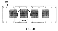
図3Aおよび図3Bはそれぞれ、複数の実施形態に従う微細加工デバイスあるいはチップを図示する分解組立図および上面図である。 FIGS. 3A and 3B are an exploded view and a top view, respectively, illustrating a microfabricated device or chip according to embodiments.
図4Aおよび図4Bは、複数の実施形態に従う膜の説明図である。図4Cは、複数の実施形態に従ってウェルのアレイとの接触から形成された印象を有する膜表面の画像である。 4A and 4B are illustrations of membranes according to multiple embodiments. FIG. 4C is an image of a membrane surface with an impression formed from contact with an array of wells according to embodiments.
図5Aは、複数の実施形態に従って試料から幾つもの細胞を単離する方法を説明する作業ステップ図である。図5Bは、複数の実施形態に従って土壌試料から幾つもの細胞を単離する方法の説明図である。 FIG. 5A is an operational step diagram illustrating a method for isolating a number of cells from a sample according to embodiments. FIG. 5B is an illustration of a method of isolating a number of cells from a soil sample according to embodiments.
図6は、複数の実施形態に従って試料から幾つもの細胞を単離し且つ培養する方法を説明する作業ステップ図である。 FIG. 6 is an operational step diagram illustrating a method for isolating and culturing a number of cells from a sample according to embodiments.
図7は、複数の実施形態に従って複合試料から幾つもの細胞を単離し且つ培養する方法の説明図である。パネル716は、単離された培養細胞株(配列番号2〜6)の出力を示す。 FIG. 7 is an illustration of a method for isolating and culturing a number of cells from a composite sample according to embodiments. Panel 716 shows the output of an isolated cultured cell line (SEQ ID NO: 2-6).
図8A〜図8Cは、複数の実施形態に従って行う1つのピンあるいは多数のピンによる採取の説明図である。 FIG. 8A to FIG. 8C are explanatory diagrams of sampling by one pin or multiple pins performed according to a plurality of embodiments.
図9A〜図9Dは、複数の実施形態に従うある1つのウェルの選抜を示す画像である。 9A-9D are images showing selection of one well according to embodiments.
図10A〜図10Dは、複数の実施形態に従ってある1つのチップから採取するための道具の説明図である。 10A-10D are illustrations of a tool for harvesting from one chip according to embodiments.
図11は、寒天の薄層を突き抜かれた1つのウェルの画像であって、複数の実施形態に従って膜または封止層を突きぬくことを図で示している。 FIG. 11 is an image of a well punched through a thin layer of agar, illustrating the penetration of a membrane or sealing layer according to embodiments.
図12は、複数の実施形態に従うチップ1200の断面を表す図である。 FIG. 12 is a diagram illustrating a cross section of a chip 1200 according to a plurality of embodiments.
図13は、複数の実施形態に従う選別方法を説明する作業ステップ図である。 FIG. 13 is a work step diagram illustrating a screening method according to a plurality of embodiments.
図14は、複数の実施形態に従う選別方法を図解する一覧図である。 FIG. 14 is a list diagram illustrating a screening method according to a plurality of embodiments.
図15は、複数の実施形態に従う1つの選別例を図示する一連の画像である。 FIG. 15 is a series of images illustrating one screening example according to embodiments.
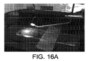
図16A〜図16Cは、複数の実施形態に従う1つの遮蔽物からの回収を説明する画像である。 FIGS. 16A-16C are images that illustrate collection from a single shield according to embodiments.
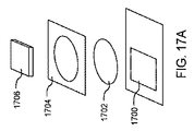
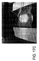
図17Aは、複数の実施形態に従う選別用チップを説明する分解組立図である。図17Bは、複数の実施形態に従う選別に続くチップの蛍光画像である。図17Cは、複数の実施形態に従って選別に続いてチップから試料を採取する1ステップを示す画像である。 FIG. 17A is an exploded view illustrating a sorting chip according to a plurality of embodiments. FIG. 17B is a fluorescence image of the chip following sorting according to embodiments. FIG. 17C is an image showing one step of collecting a sample from a chip following sorting according to embodiments.
図18は、複数の実施形態に従う計数方法を説明する作業ステップ図である。 FIG. 18 is a work step diagram illustrating a counting method according to a plurality of embodiments.
図19は、複数の実施形態に従う計数方法を図解する一覧図である。パネル1916は、培養細胞の配列および相対的存在量(配列番号2〜6)の出力を示す。
FIG. 19 is a list diagram illustrating a counting method according to a plurality of embodiments.
図20は、複数の実施形態に従う索引付け方法を図解する一覧図である。 FIG. 20 is a list diagram illustrating an indexing method according to embodiments.
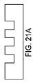
図21A〜図21Eは、複数の実施形態に従うウェル特異性化学作用物質を有するチップを表す一覧図である。 FIGS. 21A-21E are lists illustrating chips having well-specific chemical agents according to embodiments.
図22は、複数の実施形態による微細加工デバイスのマイクロウェル中の最近の属の数の概要を示す。
詳細な説明
FIG. 22 outlines the number of recent genera in the microwells of microfabricated devices according to embodiments.
Detailed description
本発明は、生物学的実体および/または栄養素の単離、培養、適応、試料採取および/または選別のためのシステム、キット、装置および方法に概ね関する。少なくとも1つの生物学的実体を含む試料を収容するための微細加工デバイス(または“チップ”)を開示する。“生物学的実体”という用語は、微生物、細胞、細胞成分、細胞生成物、ウイルスおよびその他を含んでよく、そして“種”という用語は、分類の単位を記述するために使われ、操作上の分類単位(OTU)、遺伝子型、系統型、表現型、生態型、来歴、挙動または相互作用、生成物、変種、進化上有意種およびその他を含み得る。 The present invention relates generally to systems, kits, devices and methods for the isolation, culture, adaptation, sampling and / or sorting of biological entities and / or nutrients. Disclosed is a microfabricated device (or “chip”) for containing a sample containing at least one biological entity. The term “biological entity” may include microorganisms, cells, cellular components, cell products, viruses and others, and the term “species” is used to describe a unit of classification and is Taxonomic units (OTU), genotypes, phylogenetics, phenotypes, ecotypes, provenances, behaviors or interactions, products, varieties, evolutionary significant species and others.
細胞は、古細菌、細菌、真核生物(例えば真菌類)あるいは哺乳動物細胞などであり得る。例えば、細胞は好気性、嫌気性あるいは条件的好気性微生物のような微生物であり得る。ウイルスはバクテリオファージであり得る。他の細胞成分/生成物は、タンパク質、アミノ酸、酵素、糖類、アデノシン三燐酸(ATP)、脂質、核酸(例えばDNAおよびRNA)、ヌクレオシド、ヌクレオチド、細胞膜/壁、鞭毛、線毛、小器官、代謝産物、ビタミン、ホルモン、神経伝達物質、抗体およびその他を含み得る。 The cells can be archaea, bacteria, eukaryotes (eg fungi) or mammalian cells. For example, the cell can be a microorganism such as an aerobic, anaerobic or a conditional aerobic microorganism. The virus can be a bacteriophage. Other cellular components / products include proteins, amino acids, enzymes, sugars, adenosine triphosphate (ATP), lipids, nucleic acids (eg DNA and RNA), nucleosides, nucleotides, cell membranes / walls, flagella, pili, organelles, It can include metabolites, vitamins, hormones, neurotransmitters, antibodies and others.
栄養素は、規定(例えば、化学的規定あるいは合成の培地)あるいは未規定(例えば、基礎あるいは複合の培地)であってもよい。栄養素は、実験室配合および/または市販用に生産された培地(例えば、二種以上の化学薬品の混合物)を含んでもあるいはその1成分であってもよい。栄養素は、例えばマリンブロス、溶原性ブロス(例えば、ルリアブロス)等のような、液体栄養培地(即ち、栄養スープ)であっても或いはその1成分であってもよい。栄養素は、寒天と混合されて固体培地を形成する液体培地および/または血液寒天培地のような市販製造の寒天板であっても或いはその1成分であってもよい。 Nutrients may be defined (eg, chemically defined or synthetic media) or undefined (eg, basal or complex media). Nutrients may include or be a component of laboratory formulated and / or commercially produced media (eg, a mixture of two or more chemicals). The nutrient may be a liquid nutrient medium (ie, nutrient soup) or a component thereof, such as marine broth, lysogenic broth (eg, luria broth), and the like. The nutrient may be a commercially available agar plate such as a liquid medium and / or a blood agar medium that is mixed with agar to form a solid medium, or may be a component thereof.
栄養素は、選択培地を含んでもあるいはそれらの1成分であってもよい。例えば、選択培地は、或る特定の生物学的実体だけあるいは特定の性質(例えば、抗生物質耐性あるいは特定代謝物合成)を有する生物学的実体だけの増殖用に使うことができる。栄養素は、特異性指示薬(例えば、ニュートラルレッド、フェノールレッド、エオシンYあるいはメチレンブルー)の存在下で生物学的特性を利用して1つのタイプの生物学的実体をいま1つのタイプの生物学的実体あるいは他の複数の生物学的実体から区別する識別培地を含んでもあるいはその1成分であってもよい。 The nutrient may include a selective medium or one component thereof. For example, the selective medium can be used for the growth of only certain biological entities or only biological entities that have certain properties (eg, antibiotic resistance or specific metabolite synthesis). Nutrients take advantage of biological properties in the presence of a specific indicator (eg, neutral red, phenol red, eosin Y, or methylene blue) to replace one type of biological entity with another type of biological entity. Alternatively, it may contain or be a component of an identification medium that distinguishes it from other biological entities.
栄養素は、自然環境の抽出物あるいは自然環境から由来した培地を含んでもあるいはその1成分であっても良い。例えば、栄養素は特定タイプの生物学的実体にとって自然な環境、別の環境あるいは複数の環境から導きだされ得る。その環境は、1種以上の生物組織(例えば、結合組織、筋肉、神経、上皮、植物表皮、維管束、土壌等)、生物液あるいは他の生物産物(例えば、羊水、胆汁、血液、脳脊髄液、耳垢、浸出物、糞便、胃液、間質液、細胞内液、リンパ液、乳汁、鼻汁、ルーメン内容物、唾液、皮脂、精液、汗、尿、膣分泌物、吐瀉物等)、微生物懸濁液、空気(例えばいろいろな気体含有物を伴っている)、超臨界二酸化炭素、土壌(例えば、ミネラル、有機物、気体、液体、微生物等を伴っている)、堆積物(例えば、農業、海洋堆積物等)、生きている有機物(例えば、植物、昆虫、その他の小生物および微生物)、死んだ有機物、馬草(例えば、牧草、豆科植物、貯蔵牧草、作物残留物等)、ミネラル、油あるいは油製品(例えば、動物、植物、石油化学に由来するもの)、水(例えば、天然の真水、飲料水、海水等)および/または汚水(例えば、衛生、商業、工業および/または農業廃水、および表面流出物)、更にその他を含み得る。 The nutrient may include a natural environment extract or a medium derived from the natural environment, or may be a component thereof. For example, nutrients can be derived from a natural environment, a separate environment, or multiple environments for a particular type of biological entity. The environment can be one or more biological tissues (eg, connective tissue, muscles, nerves, epithelium, plant epidermis, vascular bundles, soil, etc.), biological fluids or other biological products (eg, amniotic fluid, bile, blood, cerebrospinal cord) Fluid, earwax, exudate, feces, stomach fluid, interstitial fluid, intracellular fluid, lymph fluid, milk, nasal discharge, rumen contents, saliva, sebum, semen, sweat, urine, vaginal discharge, vomiting, etc.), microorganism suspension Suspension, air (eg, with various gas contents), supercritical carbon dioxide, soil (eg, with minerals, organics, gases, liquids, microorganisms, etc.), sediment (eg, agriculture, oceans) Sediment, etc.), living organic matter (eg, plants, insects, other small organisms and microorganisms), dead organic matter, horse grass (eg, pasture, legumes, storage grass, crop residues, etc.), minerals, Oil or oil product (eg animal, plant, petrochemical) Scientific origin), water (eg, natural fresh water, drinking water, sea water, etc.) and / or sewage (eg, sanitary, commercial, industrial and / or agricultural wastewater, and surface effluent), and others .
微細加工デバイスは、少なくとも1つの生物学的実体の培養用マイクロウェルの高密度アレイを画定し得る。“高密度”という語は、システムまたは方法が一定領域内に多くの実験を割り振る能力を意味し得る。例えば、“高密度”の実験ユニットを備える微細加工デバイスは、ここで更に述べるように、cm2当たり約150マイクロウェルからcm2当たり約160,000以上のアイクロウェルを組み込み得る。追加の例を表1に示す。

微細加工デバイスは、一連の機能層を備えた基材を組み込み得る。その一連の機能層は、実験ユニットの第1アレイ(例えばウェル)を画定する第1機能層、および先行する機能層の各実験ユニット内で実験ユニットの次のアレイ(例えば、マイクロウェル)を画定する少なくとも1つの次の機能層を含み得る。実験ユニットの各々は、生物学的実体および/または栄養素を収容且つ培養し、および/または選別するように構成され得る。特に、ここに述べるシステム、キット、装置および方法は、細胞の高密度マトリックスに対する種々の条件の自動および/またはハイスループット選別用に使用され得る。例えば、ここに述べる、システム、キット、装置および方法は、1つ以上の異なる栄養素の微生物増殖への影響を調べて比較するおよび/または代謝物、酵素活性、突然変異あるいは他の細胞機能について検査するために使われ得る。 Microfabricated devices can incorporate a substrate with a series of functional layers. The series of functional layers defines a first functional layer defining a first array of experimental units (eg, wells) and a next array of experimental units (eg, microwells) within each experimental unit of the preceding functional layer. At least one subsequent functional layer. Each of the experimental units can be configured to contain and culture and / or sort biological entities and / or nutrients. In particular, the systems, kits, devices and methods described herein can be used for automated and / or high-throughput sorting of various conditions on a dense matrix of cells. For example, the systems, kits, devices and methods described herein can examine and compare the effects of one or more different nutrients on microbial growth and / or test for metabolites, enzyme activity, mutations or other cellular functions. Can be used to
図1は、複数の実施形態に従う微細加工デバイスあるいはチップを説明する透視図である。チップ100は、上部表面102に射出成形機構を備えた顕微鏡スライドグラス形式に形づくられた基材を組み込んでいる。それら機構は、突き出しタグ106に加えて4つの分離したマイクロウェルアレイ(あるいはマイクロアレイ)等である。各マイクロアレイ内のマイクロウェルは、チップ100の縁回りおよびマイクロアレイ104間のウェルの無い縁辺を備えた格子模様に配置されている。
FIG. 1 is a perspective view illustrating a microfabricated device or chip according to a plurality of embodiments. The
図2A〜図2Cはそれぞれ、複数の実施形態に従うチップ100の大きさを示す上面、側面および端面図である。図2Aにおいて、チップ100の上面はおおよそ25.5mm×75.5mmである。図2Bにおいて、チップ100の端面はおおよそ25.5mm×0.8mmである。図2Cにおいて、チップ100の側面はおおよそ75.5mm×0.8mmである。
2A-2C are top, side, and end views, respectively, illustrating the size of the
試料を微細加工デバイスに装填した後、デバイスの少なくとも1部分に膜を施し得る。図3Aは、複数の実施形態に従う微細加工デバイス300を図3Bの上面から見て表示した分解組立図である。デバイス300は、例えば土壌微生物を収容するウェルアレイ302を備えたチップを組み込んでいる。膜304が、ウェルアレイ302の上面に置かれる。ガスケット306が膜304の上面に置かれる。充填孔310を備えたポリカーボネートカバー308が、ガスケット306の上面に置かれる。最後に、封止テープ312がカバー308に貼られる。
After loading the sample into the microfabricated device, a film may be applied to at least a portion of the device. FIG. 3A is an exploded view showing a
1つ以上の実験ユニット、ウェルまたはマイクロウェルを組み込む微細加工デバイスの少なくとも1部分を膜で覆うことができる。例えば、微細加工デバイスに試料を装填した後、少なくとも1枚の膜をマイクロウェル高密度アレイの少なくとも1つのマイクロウェルに貼り付け得る。複数枚の膜を微細加工デバイスの複数の部分に貼り付け得る。例えば、別々の膜をマイクロウェル高密度アレイの複数の異なる小区画に貼り付け得る。 At least a portion of a microfabricated device that incorporates one or more experimental units, wells or microwells can be covered with a membrane. For example, after loading a sample into a microfabricated device, at least one film can be applied to at least one microwell of a microwell high density array. Multiple films can be attached to multiple portions of a microfabricated device. For example, separate films can be affixed to a plurality of different compartments of a microwell high density array.
膜は、マイクロウェル高密度アレイの少なくとも1つのマイクロウェルに少なくとも1つの生物学的実体を保持するために、微細加工デバイスに結びつけられ、取り付けられ、部分的に取り付けられ、貼られ、封止されおよび/または部分的に封止され得る。例えば、膜は、微細加工デバイスへラミネート加工により可逆的に貼られてもよい。マイクロウェル高密度アレイの少なくとも1つのマイクロウェルへ少なくとも1つの生物学的実体を入れるために、膜は打ち抜かれ、引き剥がされ、脱着され、部分的に脱着され、除去されおよびまたは部分的に除去され得る。 The membrane is tied to, attached, partially attached, affixed, and sealed to a microfabricated device to retain at least one biological entity in at least one microwell of a microwell high density array. And / or may be partially sealed. For example, the film may be reversibly applied to a microfabricated device by lamination. To place at least one biological entity into at least one microwell of a microwell high density array, the membrane is stamped, peeled, detached, partially detached, removed and / or partially removed Can be done.
少なくとも1つの実験ユニット、ウェルあるいはマイクロウェル内の細胞個体群の1部分は(例えば吸着等の経由で)膜に付着し得る。付着する場合、少なくとも1つの実験ユニット、ウェルあるいはマイクロウェル内の細胞個体群は、少なくとも1つの実験ユニット、ウェルあるいはマイクロウェル内の細胞固体群の1部分が膜に付着したままでその膜を引き剥がすことによって試料採取され得る。 A portion of the cell population within at least one experimental unit, well or microwell may be attached to the membrane (eg, via adsorption). When attached, the cell population in at least one experimental unit, well or microwell pulls the membrane while a portion of the cell solid group in at least one experimental unit, well or microwell remains attached to the membrane. It can be sampled by peeling off.
図4Aおよび図4Bは、複数の実施形態に従う膜の説明図である。図4Aは、内容物で満たされたウェルアレイを画定するチップ400およびそのウェルアレイを覆ってチップ400を封止する膜402の側面図を示しており、従ってチップ400と接触した膜402の表面は、チップ400から剥がされた時に、図4Bに示されるように、そこに付着した(例えば、貼り付いた)ウェル内容物試料の付いたウェルの各々の刻印を有している。図4Cは、複数の実施形態に従うウェルアレイとの接触によって形成された刻印付き膜表面の画像である。 4A and 4B are illustrations of membranes according to multiple embodiments. FIG. 4A shows a side view of a chip 400 that defines a well array filled with contents and a membrane 402 that covers the well array and encapsulates the chip 400, and thus the surface of the membrane 402 in contact with the chip 400. Has an imprint of each of the wells with well content samples attached (eg, attached) thereto when peeled from the chip 400, as shown in FIG. 4B. FIG. 4C is an image of a stamped membrane surface formed by contact with a well array according to embodiments.
膜は、不透過性、半透過性、選択的透過性、差分透過性、および/またはマイクロウェル高密度アレイの少なくとも1つのマイクロウェルへ少なくとも1つの栄養素が拡散するのを可能にする部分的透過性であり得る。例えば、膜は天然物質および/または合成物質で構成されてよい。膜は、ヒドロゲル層および/または濾紙を組み込み得る。複数の実施形態では、マイクロウェル内の細胞の少なくとも幾つかあるいは全てを保持できる程に十分小さいサイズの孔を有する膜が選択される。哺乳動物細胞に関しては、孔のサイズは数ミクロンであってよく、それでも細胞を保持できる。しかしながら、複数の実施形態においては、孔のサイズは、例えば0.1μmのような、約0.2μm以下の場合がある。膜直径と孔のサイズは材料次第である。例えば、親水性ポリカーボネート膜を利用でき、そのための直径は約10mmから約3000mmの範囲に、そして孔のサイズは約0.01μmから約30.0μmの範囲にあってよい。不透過性の膜は、ゼロにほぼ等しい孔のサイズを有する。不透過膜を使う実施形態では、その膜での封止に先立ってあらゆる栄養素がマイクロウェルに供給されなければならない。気体透過性であるが液体透過性でない膜は、マイクロウェルへの酸素およびマイクロウェルからの二酸化炭素を受け入れ得る。その膜は、限定された孔のサイズを有してよいあるいは有してはならない複雑な構造を持ち得る。しかしながら、それらの孔はナノメートル程度であり得る。膜を選ぶに際しての他の要素としては、コスト、密封性能および/または殺菌性能などが含まれ得る。 The membrane is impermeable, semi-permeable, selectively permeable, differentially permeable, and / or partially permeable allowing at least one nutrient to diffuse into at least one microwell of the microwell high density array. Can be sex. For example, the membrane may be composed of natural and / or synthetic materials. The membrane may incorporate a hydrogel layer and / or filter paper. In embodiments, a membrane is selected that has pores that are small enough to hold at least some or all of the cells in the microwell. For mammalian cells, the pore size can be a few microns and still retain the cells. However, in embodiments, the pore size may be about 0.2 μm or less, such as 0.1 μm. The membrane diameter and pore size depend on the material. For example, a hydrophilic polycarbonate membrane can be utilized, for which the diameter can range from about 10 mm to about 3000 mm, and the pore size can range from about 0.01 μm to about 30.0 μm. An impermeable membrane has a pore size approximately equal to zero. In an embodiment using an impermeable membrane, all nutrients must be supplied to the microwells prior to sealing with the membrane. A membrane that is gas permeable but not liquid permeable can accept oxygen into the microwell and carbon dioxide from the microwell. The membrane may have a complex structure that may or may not have a limited pore size. However, the pores can be on the order of nanometers. Other factors in selecting a membrane may include cost, sealing performance and / or sterilization performance.
基材は、第1表面からその第1表面に向き合う第2表面へと伸ばされたマイクロウェルアレイを画定し得る。マイクロチャネルは、第1表面に第1開口を、第2表面に第2開口を有し得る。第1膜は、少なくとも1つのマイクロチャネル内の細胞個体群の少なくとも幾つかが第1表面に付着するように第1表面の少なくとも1部分に貼り付けられ得る。第2着脱可能膜は、少なくとも1つのマイクロチャネル内の細胞個体群の少なくとも幾つかが第2表面に付着するように第2表面の少なくとも1部分に貼り付けられ得る。少なくとも1つのマイクロチャネル内の細胞固体群は、少なくとも1つのマイクロチャネル内の細胞固体群の少なくとも幾つかが第1膜に付着したままで第1膜をおよび/または少なくとも1つのマイクロチャネル内の細胞固体群の少なくとも幾つかが第2膜に付着したままで第2膜を引き剥がすことによって試料採取され得る。 The substrate can define a microwell array that is extended from a first surface to a second surface facing the first surface. The microchannel may have a first opening on the first surface and a second opening on the second surface. The first membrane can be applied to at least a portion of the first surface such that at least some of the cell population in the at least one microchannel is attached to the first surface. The second removable membrane can be affixed to at least a portion of the second surface such that at least some of the cell population within the at least one microchannel is attached to the second surface. The cell solid groups in the at least one microchannel may include cells in the first membrane and / or cells in the at least one microchannel while at least some of the cell solid groups in the at least one microchannel remain attached to the first membrane. Samples can be sampled by tearing off the second membrane while at least some of the solid groups remain attached to the second membrane.
“ハイスループット”という語は、システムあるいは方法が有する非常に大きな数の実験を平行あるいは連続して迅速に行うことを可能にする能力を意味し得る。“ハイスループット”システムの一例は、此処で述べるように、1つ以上の生物学的実体を、少なくとも1つの代謝生成物、酵素、タンパク質、核酸、表現型、突然変異、代謝経路、遺伝子、適応型および能力について検査するために、調製、培養および/または多数の化学、遺伝、薬学、光学および/または画像分析を行う細胞生物学技法を備えた自動装置を含み得る。複数の実施形態によれば、“ハイスループット”は、少なくとも約96実験から少なくとも約10,000,000実験に及ぶ規模の実験を同時あるいはほぼ同時に行うことを意味し得る。 The term “high throughput” can mean the ability to allow a very large number of experiments that a system or method has to be performed quickly in parallel or sequentially. An example of a “high-throughput” system is that, as described herein, one or more biological entities, at least one metabolite, enzyme, protein, nucleic acid, phenotype, mutation, metabolic pathway, gene, adaptation Automated devices equipped with cell biology techniques for preparation, culture and / or numerous chemical, genetic, pharmaceutical, optical and / or image analysis can be included to test for type and ability. According to embodiments, “high throughput” can mean performing experiments on a scale ranging from at least about 96 experiments to at least about 10,000,000 experiments simultaneously or nearly simultaneously.
ここに開示のシステム、装置、キットおよび方法は、生物学的実体(例えば細胞)のマトリックスに対して種々の条件のハイスループット選別に使うことができる。“ウェル内ウェル”概念は、多重レベルの機能層を有するように基材あるいはチップを製造する(例えば、微細加工する)ことによって要求あるいは望まれる如何なるレベル(即ち、ウェル内ウェル内ウェル内ウェル等)でも実現され得る。第1機能層は、実験ユニット(例えば、ウェル)アレイを画定できる。実験ユニットの各々は、次の実験ユニット(例えば、マイクロウェル)アレイを画定するステップによってもう一つの機能層を提供する。このことによって、多重実験あるいは試験を単一チップで同時に行えることから、ハイスループット操作が可能となる。 The systems, devices, kits and methods disclosed herein can be used for high-throughput sorting of various conditions against a matrix of biological entities (eg, cells). The “in-well well” concept refers to any level required or desired by manufacturing (eg, microfabricating) a substrate or chip to have multiple levels of functional layers (ie, wells in wells, wells in wells, etc. ) Can also be realized. The first functional layer can define an experimental unit (eg, well) array. Each experimental unit provides another functional layer by defining the next experimental unit (eg, microwell) array. As a result, multiple experiments or tests can be performed simultaneously on a single chip, thereby enabling high-throughput operation.
例えば、図3Aおよび図3Bにおいて、ガスケット306が膜304の上面に置かれ、その膜は複数の実施形態に従う微細加工デバイス300のウェルアレイ302に施されている。ガスケット306は1つの開口のみを有する。しかしながら、更なる実施形態では、1つのより小さな開口を有する多重のより小型なガスケットあるいは複数のより小さな開口を有する単一ガスケットをデバイスの上面(膜を備えていてもいなくても)に置き、それによって機能層、あるいは次の機能層を備えたより大きな実験ユニットのアレイあるいはそこに位置する次の実験ユニットアレイ(例えば、ウェル302)を形成するステップが出来る。
多重レベルの機能層があれば、例えば、複数の栄養素あるいは栄養素調合物を、同時にあるいはほぼ同時に試験できる。同じ方式が、例えば、代謝産物あるいは細胞の特異機能について検査するあるいは自然環境派生栄養素から微生物を引き離して別の栄養素にするために使用され得る。
For example, in FIGS. 3A and 3B, a
With multiple levels of functional layers, for example, multiple nutrients or nutrient formulations can be tested simultaneously or nearly simultaneously. The same scheme can be used, for example, to test for metabolite or cell specific functions or to separate a microorganism from a natural environment derived nutrient into another nutrient.
実験ユニットは、微細加工デバイス表面の所定部位である。例えば、チップの表面を、所定部位の第1アレイに細胞を固定化するように、設計することができる。これらの所定部位は、ウェル、マイクロウェル、マイクロチャネルおよび/または指定固定化部位であり得る。例えば、表面がマイクロウェルアレイを画定するように微細加工されてよい。そのアレイは、基材に壁あるいは追加壁を画定するステップによって数区画に分けることができる。例えば、その表面がウェルの第1アレイを最初に画定するように微細加工されてよく、そこでは各ウェルの内部表面が、マイクロウェル、マイクロチャネルあるいは固定化部位の第2アレイを画定するように、順に加工される。もう1つの例においては、その表面を、マイクロウェルアレイを画定するように、加工でき、そしてもう1つの基材(例えば、寒天、プラスチックあるいはその他の物質)を、その表面およびそれによって画定されるマイクロウェルを仕切るように、その表面に塗ることができる。各ウェル、マイクロウェル、マイクロチャネルおよび/または固定化部位を、少なくとも1つの細胞を収容して増殖させるように構築し得るが、しかし使用において、どの所定ウェル、マイクロウェル、マイクロチャネルあるいは固定化部位も実際のところ1つ以上の細胞を収容および/または増殖させ得るあるいはさせ得ない。実験ユニットの型は置き換え可能であり得る。例えば、マイクロウェルを特に記述するこの文書中の実施形態は、それらのマイクロウェルがすくなくとも部分的にマイクロチャネル、固定化部位および/または他の型の実験ユニットと置き換えられる実施形態を開示することもまた意図している。 The experimental unit is a predetermined part on the surface of the microfabricated device. For example, the surface of the chip can be designed to immobilize cells in a first array of predetermined sites. These predetermined sites can be wells, microwells, microchannels and / or designated immobilization sites. For example, the surface may be microfabricated so as to define a microwell array. The array can be divided into several sections by the step of defining walls or additional walls in the substrate. For example, the surface may be microfabricated such that it first defines a first array of wells, such that the internal surface of each well defines a second array of microwells, microchannels or immobilization sites. Are processed in order. In another example, the surface can be processed to define a microwell array, and another substrate (eg, agar, plastic or other material) is defined by the surface and thereby It can be applied to the surface of the microwell to partition it. Each well, microwell, microchannel and / or immobilization site can be constructed to contain and grow at least one cell, but in use, whichever well, microwell, microchannel or immobilization site In fact, one or more cells may or may not be accommodated and / or grown. The experimental unit type may be interchangeable. For example, embodiments in this document that specifically describe microwells also disclose embodiments in which those microwells are at least partially replaced with microchannels, immobilization sites, and / or other types of experimental units. Also intended.
微細加工デバイスの1か所以上の部分は、選択され、処理され、および/または特別な表面化学性を有する様に表面化学性改質剤で塗布され得る。例えば、基材表面の少なくとも1部分は、細胞を寄せ付けないおよび/または細胞の表面へ貼りつく傾向を減じる第1の表面特性あるいは細胞を引き寄せおよび/または細胞の表面へ貼りつく傾向を高める第2の表面特性付きで設計され得る。標的細胞の種類によるが、物質および/または塗膜は疎水性および/または親水性であり得る。基材の上部表面の少なくとも1部分は、標的細胞を寄せ付けないおよび/または標的細胞のその表面に貼りつく傾向を減じる第1の表面特性を有する様に、処理されてよい。その一方で、各実験ユニット、ウェルあるいはマイクロウェルの内部表面の少なくとも1部分は、標的細胞を引き寄せて標的細胞が実験ユニット、ウェルあるいはマイクロウェルを占める傾向を高める第2の特性を有する様に、処理されてよい。基材の1つの表面は異なった表面特性を備えた複数の部分を有してよい。 One or more portions of the microfabricated device can be selected, treated, and / or applied with a surface chemistry modifier to have a special surface chemistry. For example, at least a portion of the substrate surface has a first surface property that does not attract cells and / or reduces the tendency to stick to the surface of cells or a second tendency to increase the tendency to attract and / or stick cells to the surface of cells. Can be designed with various surface properties. Depending on the type of target cell, the substance and / or coating can be hydrophobic and / or hydrophilic. At least a portion of the top surface of the substrate may be treated to have a first surface property that does not attract the target cells and / or reduces the tendency of the target cells to stick to that surface. On the other hand, at least a portion of the internal surface of each experimental unit, well or microwell has a second property that attracts the target cells and increases the tendency of the target cells to occupy the experimental units, wells or microwells, May be processed. One surface of the substrate may have multiple portions with different surface characteristics.
表面化学性改質剤は、化学蒸着法、電気穿孔法、プラズマ処理および/または電解析出法を使って施すことができる。表面化学性改質剤は、表面電位、ルンド電位、ゼータ電位、表面形態、疎水性および/または親水性を制御できる。表面化学性改質剤は、シラン、高分子電解質、金属、高分子、抗体および/または血漿などを含み得る。例えば、表面化学性改質剤はオクタデシルトリクロロシラン等であってよい。表面化学性改質剤は、例えばモノラウリン酸ポリオキシエチレン(20)ソルビタンおよび/またはポリエチレングリコールp−(1、1、3、3−テトラメチルブチル)−フェニルエーテルの様な、動的共重合体を含み得る。表面化学性改質剤は、例えばポロクサマー407、ポリ(L−リジン)および/またはポリ(エチレングリコール)−ポリ(l−リジン)ブロック共重合体の様な、静的共重合体を含み得る。 The surface chemical modifier can be applied using chemical vapor deposition, electroporation, plasma treatment and / or electrolytic deposition. The surface chemical modifier can control surface potential, lund potential, zeta potential, surface morphology, hydrophobicity and / or hydrophilicity. The surface chemical modifier may include silane, polyelectrolyte, metal, polymer, antibody and / or plasma. For example, the surface chemical modifier may be octadecyltrichlorosilane or the like. The surface chemical modifier is a dynamic copolymer such as polyoxyethylene (20) sorbitan monolaurate and / or polyethylene glycol p- (1,1,3,3-tetramethylbutyl) -phenyl ether. Can be included. The surface chemical modifier may comprise a static copolymer such as, for example, poloxamer 407, poly (L-lysine) and / or poly (ethylene glycol) -poly (1-lysine) block copolymer.
細胞のマトリックスに対して種々の条件を選別するための装置は、マイクロウェルアレイを画定する表面を有する基材を含み得る。マイクロウェルアレイの数区画は、(例えば、より大きなウェルあるいは壁によって)サブアレイに分割され得る。基材は微細加工で作られ得る。各マイクロウェルは、生物学的実体(例えば、細胞)を収容し且つ増殖させ得る。結果として得た生物学的実体(例えば、細胞)のマトリックスは、生物学的実体の高密度マトリックスであり得る。第1アレイおよび/または第2アレイは、平面、概ね平面、および/または多重平面(例えば、ロール上で)であり得る。 An apparatus for sorting various conditions against a matrix of cells can include a substrate having a surface that defines a microwell array. Several sections of the microwell array can be divided into subarrays (eg, by larger wells or walls). The substrate can be made by microfabrication. Each microwell can contain and grow a biological entity (eg, a cell). The resulting matrix of biological entities (eg, cells) can be a dense matrix of biological entities. The first array and / or the second array can be planar, generally planar, and / or multi-planar (eg, on a roll).
“高分解能”という用語は、数多くの利用可能実験を区別するシステムあるいは方法の能力を意味し得る。例えば、“高分解能”システムあるいは方法は、約1nmから約800μmの直径を有する1つの実験ユニットを高密度実験ユニット含有の微細加工デバイスから選択することができる。微細加工デバイスあるいはチップの基材は、約10,000,000あるいはそれ以上のマイクロウェルを組み込むことができる。例えば、マイクロウェルアレイは、少なくとも96箇所、少なくとも1,000箇所、少なくとも5,000箇所、少なくとも10,000箇所、少なくとも100,000箇所、少なくとも500,000箇所、少なくとも1,000,000箇所、少なくとも5,000,000箇所、あるいは10,000,000箇所の格納場所を含み得る。 The term “high resolution” can mean the ability of a system or method to distinguish between a number of available experiments. For example, a “high resolution” system or method can select one experimental unit having a diameter of about 1 nm to about 800 μm from a microfabricated device containing a high density experimental unit. The substrate of the microfabricated device or chip can incorporate about 10,000,000 or more microwells. For example, a microwell array can include at least 96, at least 1,000, at least 5,000, at least 10,000, at least 100,000, at least 500,000, at least 1,000,000, at least 5,000,000, or 10,000,000 storage locations.
マイクロウェルの表面密度は、cm2当たり約500マイクロウェルから約160,000マイクロウェル、あるいはそれ以上であり得る。微細加工デバイスあるいはチップの基材は、cm2当たり少なくとも150マイクロウェル、cm2当たり少なくとも250マイクロウェル、cm2当たり少なくとも400マイクロウェル、cm2当たり少なくとも500マイクロウェル、cm2当たり少なくとも750マイクロウェル、cm2当たり少なくとも1,000マイクロウェル、cm2当たり少なくとも2,500マイクロウェル、cm2当たり少なくとも5,000マイクロウェル、cm2当たり少なくとも7,500マイクロウェル、cm2当たり少なくとも10,000マイクロウェル、cm2当たり少なくとも50,000マイクロウェル、cm2当たり少なくとも100,000マイクロウェル、あるいはcm2当たり少なくとも160,000マイクロウェルのマイクロウェル表面密度を有し得る。 The surface density of the microwells can be from about 500 microwells to about 160,000 microwells per cm 2 or more. The substrate microfabricated device or chip, cm 2 per at least 150 microwells cm 2 per at least 250 microwells cm 2 per at least 400 microwells cm 2 per at least 500 microwells cm 2 per at least 750 micro-wells, per cm 2 of at least 1,000 microwells per cm 2 of at least 2,500 microwells per cm 2 of at least 5,000 microwells least 7,500 microwells per cm @ 2, per cm 2 of at least 10,000 micro-wells per cm 2 of at least 50,000 micro-wells per cm 2 It may have a microwell surface density of at least 100,000 microwells or per cm 2 of at least 160,000 microwells.
マイクロウェルの大きさは、ナノスケール(例えば、約1から約100ナノメートルの直径)からミクロスケール以上までの幅があり得る。例えば、各マイクロウェルは、約1μmから約800μmの直径、約25μmから約500μmの直径、あるいは約30μmから約100μmの直径を有し得る。マイクロウェルの有し得る直径は、約1μmまたは1μm未満、約5μmまたは5μm未満、約10μmまたは10μm未満、約25μmまたは25μm未満、約50μmまたは50μm未満、約100μmまたは100μm未満、約200μmまたは200μm未満、約300μmまたは300μm未満、約400μmまたは400μm未満、約500μmまたは500μm未満、約600μmまたは600μm未満、約700μmまたは700μm未満、あるいは約800μmまたは800μm未満であり得る。 The microwell size can range from nanoscale (eg, a diameter of about 1 to about 100 nanometers) to microscale or more. For example, each microwell can have a diameter of about 1 μm to about 800 μm, a diameter of about 25 μm to about 500 μm, or a diameter of about 30 μm to about 100 μm. The diameter of the microwell can be about 1 μm or less than 1 μm, about 5 μm or less than 5 μm, about 10 μm or less than 10 μm, about 25 μm or less than 25 μm, about 50 μm or less than 50 μm, about 100 μm or less than 100 μm, about 200 μm or less than 200 μm , Less than about 300 μm or less than 300 μm, less than about 400 μm or less than 400 μm, less than about 500 μm or less than 500 μm, less than about 600 μm or less than 600 μm, less than about 700 μm or less than 700 μm, or less than about 800 μm or less than 800 μm.
マイクロウェルは、約500μmから約5000μmの深さ、約1μmから約500μmの深さ、あるいは約25μmから約100μmの深さを有し得る。マイクロウェルの有し得る深さは、約1μm、約5μm、約10μm、約25μm、約50μm、約100μm、約200μm、約300μm、約400μm、約500μm、約600μm、約700μm、約800μm、約1,000μm、約1,500μm、約2,000μm、約3,000μm、あるいは約5,000μmであり得る。 The microwell can have a depth of about 500 μm to about 5000 μm, a depth of about 1 μm to about 500 μm, or a depth of about 25 μm to about 100 μm. The depth that the microwell can have is about 1 μm, about 5 μm, about 10 μm, about 25 μm, about 50 μm, about 100 μm, about 200 μm, about 300 μm, about 400 μm, about 500 μm, about 600 μm, about 700 μm, about 800 μm, about It can be 1,000 μm, about 1,500 μm, about 2,000 μm, about 3,000 μm, or about 5,000 μm.
各マイクロウェルは、円形、六角形あるいは正方形の開口を有し得る。各マイクロウェルは側壁を伴い得る。それらの側壁は、垂直の、傾斜したあるいは湾曲した断面形状を有し得る。少なくとも1つの特有の位置特異性タグが、以下で更に述べるように、高密度マクロウェルアレイの少なくとも1つのマイクロウェルに割り付けられて種の識別およびマイクロウェルの高密度アレイの特異的なマイクロウェルへの種の相関を容易に成し得る。その少なくとも1つの特異タグは、その1つのマイクロウェルの底および/または少なくとも1つの側面に割り付けおよび/または配置され得る。その少なくとも1つの特異タグは、少なくとも1つの生物学的実体の標的核酸断片へアニールするための標的特異性ヌクレオチド配列およびマイクロウェルの高密度アレイの少なくとも1つのマイクロウェルを識別するための位置特異性ヌクレオチド配列を備えた核酸分子を組み込み得る。 Each microwell may have a circular, hexagonal or square opening. Each microwell can have a sidewall. Their side walls may have a vertical, inclined or curved cross-sectional shape. At least one unique position-specific tag is assigned to at least one microwell of the high density macrowell array to further identify species and to specific microwells of the high density array of microwells, as further described below. The species correlation can be easily achieved. The at least one unique tag can be assigned and / or placed on the bottom and / or at least one side of the one microwell. The at least one specific tag is a target specific nucleotide sequence for annealing to a target nucleic acid fragment of at least one biological entity and a position specificity for identifying at least one microwell of a high density array of microwells Nucleic acid molecules with nucleotide sequences can be incorporated.
例えば、微細加工デバイスあるいはチップの基材は、約4インチ×4インチの大きさの表面を有し得る。その表面は、おおよそ1億個のマイクロウェルのアレイを画定し得る。マイクロウェルアレイを壁によって約100の小区画に分割できおよび/または基材は約100ウェルのアレイを画定できる、すなわち各小区画あるいはウェル内で画定された約100万のマイクロウェルは合計で約1億のマイクロウェルとなる。異なった栄養素を試験する使用例に関しては、自然環境試料からの微生物は、個々の微生物あるいは微生物クラスターがチップ上のマイクロウェルに分配されるように、チップに装填でき、そこでは各マイクロウェルはより大きなウェルの下部に設置されている。より大きなウェルの各々は、異なる100の栄養素を同一のチップ上で並行してあるいは連続して試験できるように、実験ユニットを組み込むことができる、即ち各ウェルは100万までの試験例をまかなうことができる。 For example, the substrate of a microfabricated device or chip can have a surface that is approximately 4 inches by 4 inches. The surface can define an array of approximately 100 million microwells. The microwell array can be divided into about 100 small compartments by walls and / or the substrate can define an array of about 100 wells, i.e. about 1 million microwells defined within each subcompartment or well total about There will be 100 million microwells. For use cases where different nutrients are tested, microorganisms from natural environmental samples can be loaded onto the chip such that individual microorganisms or microbial clusters are distributed to the microwells on the chip, where each microwell is more Located at the bottom of a large well. Each larger well can incorporate an experimental unit so that 100 different nutrients can be tested in parallel or sequentially on the same chip, ie each well can serve up to 1 million test examples Can do.
標的細胞は、古細菌、細菌あるいは真核生物(例えば、菌類、植物あるいは動物)であり得る。例えば、標的細胞は、好気性、嫌気性および/または条件的好気性微生物のような微生物であってよい。例えば細胞個体数、細胞成分および/または細胞生成物への増殖あるいは他の効果を分析および比較するために、異なる栄養素が標的細胞の組成物を使い並行してあるいは連続して試験され得る。標的細胞の組成物は、例えば一種以上のウイルス(例えばバクテリオファージ)、細胞表面(例えば、細胞膜、細胞壁)、代謝産物、ビタミン、ホルモン、神経伝達物質、抗体、アミノ酸、酵素、タンパク質、糖類、ATP、脂質、ヌクレオシド、ヌクレオチド、核酸(例えばDNAあるいはRNA)、表現型、突然変異体、代謝経路、遺伝子および適応性のような、細胞の成分、生成物、および/または機能について検査され得る。 Target cells can be archaea, bacteria or eukaryotes (eg fungi, plants or animals). For example, the target cell may be a microorganism such as an aerobic, anaerobic and / or conditional aerobic microorganism. Different nutrients can be tested in parallel or sequentially using the composition of the target cells, for example to analyze and compare growth or other effects on cell populations, cell components and / or cell products. The composition of the target cell is, for example, one or more viruses (eg bacteriophage), cell surface (eg cell membrane, cell wall), metabolites, vitamins, hormones, neurotransmitters, antibodies, amino acids, enzymes, proteins, sugars, ATP , Lipids, nucleosides, nucleotides, nucleic acids (eg DNA or RNA), phenotypes, mutants, metabolic pathways, genes and adaptability can be examined for cellular components, products, and / or functions.
細胞の組成物は、自然環境試料抽出物および/または希釈物を含み得る。その自然環境試料抽出物および/または希釈物は、1種以上の生物組織(例えば、結合組織、筋肉、神経、上皮、植物表皮、維管束、土壌等)、生物液あるいは他の生物産物(例えば、羊水、胆汁、血液、脳脊髄液、耳垢、浸出物、糞便、胃液、間質液、細胞内液、リンパ液、乳汁、鼻汁、ルーメン内容物、唾液、皮脂、精液、汗、尿、膣分泌物、吐瀉物等)、微生物懸濁液、(例えば種々の気体成分を伴う)空気、超臨界二酸化炭素、(例えば、ミネラル、有機物、気体、液体、微生物等を伴う)土壌、(例えば、農業、海洋等の)堆積物、生きている有機物(例えば、植物、昆虫、その他の小生物および微生物)、死んだ有機物、馬草(例えば、牧草、豆科植物、貯蔵牧草、作物残留物等)、ミネラル、(例えば、動物、植物、石油化学由来の)油あるいは油製品、アルコール、緩衝物、有機溶媒、水(例えば、天然の真水、飲料水、海水等)および/または汚水(例えば、衛生、商業、工業および/または農業廃水、および表面流出物)およびその他を含み得る。 The composition of the cells can include natural environment sample extracts and / or dilutions. The natural environment sample extract and / or dilution may contain one or more biological tissues (eg, connective tissue, muscle, nerve, epithelium, plant epidermis, vascular bundle, soil, etc.), biological fluids or other biological products (eg, , Amniotic fluid, bile, blood, cerebrospinal fluid, earwax, exudate, feces, gastric fluid, interstitial fluid, intracellular fluid, lymph fluid, milk, nasal discharge, rumen contents, saliva, sebum, semen, sweat, urine, vaginal secretion , Suspensions, etc.), microbial suspensions, air (eg, with various gas components), supercritical carbon dioxide, soil (eg, with minerals, organics, gases, liquids, microorganisms, etc.), (eg, agriculture) Sediment, living organic matter (eg, plants, insects, other small organisms and microorganisms), dead organic matter, horse grass (eg, pasture, legumes, storage pasture, crop residues, etc.) , Minerals (eg animal, plant, petrochemical Oils or oil products, alcohol, buffers, organic solvents, water (eg natural fresh water, drinking water, sea water, etc.) and / or sewage (eg sanitary, commercial, industrial and / or agricultural wastewater, and surface runoff) Material) and others.
1つの方法は、細胞を含む組成物を微細加工デバイスへの適用(例えば、装填)に先立ち、細胞と自然環境試料抽出物および/または希釈物とを組み合わせて組成物を調製するステップを含み得る。その方法は更に、自然環境試料抽出物および/または希釈物を液化するステップ含む場合がある。組成物内の細胞濃度は、実験ユニット、ウェルあるいはマイクロウェル1個当たりの1細胞の標的配分に合わせ得る。 One method may include combining the cells with a natural environment sample extract and / or dilution prior to applying (eg, loading) the composition comprising the cells to the microfabricated device. . The method may further comprise liquefying the natural environment sample extract and / or dilution. The cell concentration within the composition can be tailored to the target distribution of one cell per experimental unit, well or microwell.
試料が複数個の細胞あるいはウイルスを含む場合、試料内のそれら細胞は、微細加工デバイスに適用された後で溶解されて、核酸分子を放出し得る。細胞は、例えばアルカリ性曝露のような化学処理、洗剤、音波処理、タンパク質分解酵素Kあるいはリゾチーム曝露を使って細胞を溶解し得る。細胞は、加熱によっても溶解され得る。 If the sample contains multiple cells or viruses, the cells in the sample can be lysed after application to the microfabricated device to release the nucleic acid molecules. Cells can be lysed using chemical treatments such as alkaline exposure, detergent, sonication, proteolytic enzyme K or lysozyme exposure. Cells can also be lysed by heating.
図5Aは、複数の実施形態に従って1つの試料から細胞を単離するための1つの方法を説明する作業ステップ図である。ステップ500において、試料を得る。ステップ502においては、物理的技法(例えば、混合および/または音波処理)および化学的技法(例えば、キレート剤、洗剤および/または酵素)の少なくとも1つを使って、試料を均一化および/または分散化する。ステップ504においては、均一化およびまたは分散化された試料内の細胞は、例えばNycodenz(登録商標)非粒子状媒体(プロゲンビオテクニクGmbH、ハイデルベルグ、ドイツ、から入手可能)を使う密度遠心法によって分離される。
FIG. 5A is an operational step diagram illustrating one method for isolating cells from a sample according to embodiments. In
図5Bは、複数の実施形態に従って1つの土壌試料から細胞を単離する方法を説明する一覧図である。パネル506は、土壌試料を表示している。パネル508は、試験管内で均質化されおよびまたは分散された試料を表示している。パネル510は、遠心分離後に可溶堆積物512、細胞514、不溶堆積物516およびNycodenz(登録商標)518に分離した試料を表示している。
FIG. 5B is a list diagram illustrating a method for isolating cells from a single soil sample according to embodiments.
図6は、複数の実施形態に従って1つの試料から細胞を単離しそして培養する方法を説明する作業ステップ図である。ステップ600において、試料を得る。ステップ602において、その得られた試料から少なくとも1細胞を抽出する。ステップ604において、微細加工デバイスあるいはチップの少なくとも1つの高密度マイクロウェルアレイにその少なくとも1つの抽出された細胞を装填する。ステップ604には、その少なくとも1つの抽出細胞を伴う細胞濃度を用意、少なくとも1つの栄養素/培地を選択、および/または少なくとも1つの膜の選択を組み込み得る。ステップ606には、マイクロウェルアレイの少なくとも1部分が少なくとも1つの選択された膜によって密閉されてそれらのマイクロウェルに関する細胞濃度を保持する。ステップ608において、チップが培養目的で温められる。ステップ608には、温度選択、(例えば、好気性かあるいは嫌気性かの)雰囲気の決定、および/または培養時間の決定を組み込み得る。ステップ610において、チップは分割されおよび/または(例えば、ピッカーを使って)実質的に複製がつくられ、結果としてここに述べた方法に従って培養された細胞を2部もたらすことになる。例えば、少なくとも1枚の膜が培養細胞の1部分を付着した儘で剥がされる、あるいは培養細胞を試料として取り出すために剥がされるかまたは突き破られる場合がある。省略可のステップ612において、培養細胞の1部が識別のために犠牲にされる。ステップ612には、PCR、配列決定、および/または様々なデータ解析を組み込み得る。ステップ614において、対象物の菌株が識別される。更なる培養、試験、および/または識別を、例えば対象物の菌株およびまたは培養細胞の残部を使って行い得る。
FIG. 6 is an operational step diagram illustrating a method for isolating and culturing cells from a sample according to embodiments. In
図7は、複数の実施形態に従って複合試料を単離して培養する方法を図解する一覧図である。パネル700には、複合試料、特に微生物叢試料702および土壌試料704、の例が示されている。パネル706では、例えば図5Aおよび図5Bに図解されたプロトコルを使って、試料から少なくとも1つの細胞が抽出されている。パネル708では、少なくとも1つの抽出細胞(および何らかの自然環境抽出物および希釈物)が、少なくとも1つの高密度マイクロウェルアレイ710を有する微細加工デバイスあるいはチップに装填されている。チップ710および試薬カートリッジ712が培養器714内に挿入される場合がある。試薬は、増殖のための栄養需要および/または種々の選抜目的を維持するために、液体を加えるのに役立得る。パネル716には、産出物、即ち培養細胞の単離菌株、が示されている。
FIG. 7 is a list diagram illustrating a method of isolating and culturing a composite sample according to multiple embodiments.
1つのマイクロウェルで増殖する細胞あるいは微生物の種あるいは分類系統を識別するためには、DNA塩基配列決定、核酸混成、質量分析、赤外分光測定、DNA増幅、遺伝因子あるいは他の種識別子を識別するための抗体結合およびその他を含む技法が必要とされる。多くの識別方法および処理ステップは微生物を消滅させるので、更なる培養および対象物の微生物の研究を妨げている。以後の培養、研究および特定の対象物のクローンの更なる育成を可能にしながら細胞あるいは微生物の識別の両方を可能にするために、幾つもの実験ユニット、ウェルあるいはマイクロウェル全般にわたって位置保全および微生物個体群の分離を維持しながら1つの基材あるいはチップ全般にわたって各実験ユニット、ウェルあるいはマイクロウェルから試料を取って調べるために更なる実施形態が設計される。 Identify DNA sequencing, nucleic acid hybridization, mass spectrometry, infrared spectroscopy, DNA amplification, genetic factors or other species identifiers to identify the species or taxonomy of cells or microorganisms growing in one microwell Techniques including antibody binding and others to do so are needed. Many identification methods and processing steps kill microorganisms, hindering further culture and study of the subject microorganisms. In order to enable both subsequent cell cultures, research, and further growth of clones of a particular object while allowing cell or microbe identification, location conservation and microbial individuals across several experimental units, wells or microwells in general. Further embodiments are designed to take samples from each experimental unit, well or microwell over a single substrate or chip while maintaining group separation.
上述のように基材は、更に幾つものシステム、キット、装置や方法を使って細胞個体群から試料を取って調べることを可能にし得る。例えば、採取装置が基材の第1表面に適用され得る。その装置は、第1表面と向き合う少なくとも1個の突起物を組み込み得る。その少なくとも1個の突起物は、各マイクロウェル、ウェルあるいは実験ユニットの開口直径よりも小さな直径を有している。その少なくとも1個の突起物は、少なくとも1つのマイクロウェル、ウェルあるいは実験ユニット内の細胞の1個体群の1部分が少なくとも1個の突起物に付着および/または結合するように、その細胞個体群を保持する少なくとも1つのマイクロウェル、ウェルあるいは実験ユニットに挿入され得る。少なくとも1つのマイクロウェル、ウェルあるいは実験ユニット内の細胞個体群の試料は、少なくとも1つのマイクロウェル、ウェルあるいは実験ユニット内の細胞個体群の1部分が少なくとも1個の突起物に付着および/または結合したままで残るように、採取装置を基材の第1表面から取り除くことによって回収され得る。各突起物は、1本のピンまたは複数のピンまたはピン集合体であり得る。 As described above, the substrate may allow a sample to be examined from a cell population using a number of systems, kits, devices and methods. For example, a harvesting device can be applied to the first surface of the substrate. The device may incorporate at least one protrusion that faces the first surface. The at least one protrusion has a diameter smaller than the opening diameter of each microwell, well or experimental unit. The at least one protrusion is a cell population such that a portion of a population of cells in at least one microwell, well or experimental unit attaches and / or binds to at least one protrusion. Can be inserted into at least one microwell, well or experimental unit. A sample of a cell population in at least one microwell, well, or experimental unit has a portion of the cell population in at least one microwell, well, or experimental unit attached and / or bound to at least one protrusion. Can be recovered by removing the harvesting device from the first surface of the substrate. Each protrusion may be a single pin or multiple pins or pin assemblies.
図8A〜図8Cは、複数の実施形態に従っての1本のピンあるは多数のピンによる採取の説明図である。チップ800は、顕微鏡802による検査および採取制御装置804による採取に提供される。図8Aにおいて、採取制御装置804は1本だけのピン806を有するアームを備えている。図8Bでは、多数のピン808を有するアームが示されている。図8Cは、採取ステップ進行中のチップの透視図である。
8A-8C are illustrations of sampling with a single pin or multiple pins in accordance with embodiments.
図9A〜図9Dは、複数の実施形態に従って1つのウェルの選抜を実際に行っている画像である。図9Aでは、そのウェルは満杯である。図9Bにおいて、ピンが所定位置へと動かされている。図9Cにおいて、そのウェルが選抜されている。図9Dにおいて、試料がそのウェルから取り出されている。 9A to 9D are images actually selecting one well according to a plurality of embodiments. In FIG. 9A, the well is full. In FIG. 9B, the pin has been moved into place. In FIG. 9C, the well is selected. In FIG. 9D, a sample has been removed from the well.
図10A〜図10Dは、複数の実施形態に従ってチップからの採取用器具を図解する一覧図である。図10Aにおいて、複数のピンを備えた器具は、複数のウェルを有するチップに位置合わせされている。図10Bにおいて、ピンがウェルに浸かるように器具が下げられている。図10Cにおいて、付着した試料を備えたピンが示され、それら試料は新しいチップへと移される。一方、図10Dでは、試料が器具自身に保持されるように器具を上下反転させている。 FIGS. 10A-10D are listings illustrating a tool for collecting from a chip according to embodiments. In FIG. 10A, an instrument with multiple pins is aligned with a chip having multiple wells. In FIG. 10B, the instrument is lowered so that the pin is immersed in the well. In FIG. 10C, pins with attached samples are shown and the samples are transferred to a new chip. On the other hand, in FIG. 10D, the instrument is turned upside down so that the sample is held by the instrument itself.
図11は、寒天薄層を突き通されたウェルの画像であり、複数の実施形態に従う膜あるいは密封層突き抜けを図示している。 FIG. 11 is an image of a well penetrated through a thin agar layer, illustrating a membrane or sealing layer penetration according to embodiments.
一方、少なくとも1つの突起物が少なくとも1つのマイクロウェル、ウェルあるいは実験ユニットに浸される時、その少なくとも1つのマイクロウェル、ウェルあるいは実験ユニット内の細胞固体群の1部分は、その少なくとも1つの突起物の上方およびまわりへ移動した量であって、その移動量部分の少なくとも幾分かは基材の第1表面およびまたはその少なくとも1つのマイクロウェル、ウェルあるいは実験ユニットの内部表面上方にあるものとする。その方法はまた、細胞固体群の移動量部分の少なくとも幾分かを集めることによってその少なくとも1つのマイクロウェル内の細胞固体群を試料として採取するステップを含んでいる。 On the other hand, when at least one protrusion is immersed in at least one microwell, well, or experimental unit, a portion of the cell solid group within the at least one microwell, well, or experimental unit has its at least one protrusion The amount moved above and around the object, at least some of which is above the first surface of the substrate and / or the inner surface of the at least one microwell, well or experimental unit To do. The method also includes sampling the cell solid mass within the at least one microwell as a sample by collecting at least some of the transferred portion of the cell solid mass.
同様の採取装置が基材の第1表面に向き合う第2表面へ適用され得る。その装置は、第2表面に向き合う少なくとも1つの突起物を組み込み得る。その少なくとも1つの突起物は、少なくとも1つのマイクロウェル、ウェルあるいは実験ユニットの直径に略等しいまたはより小さい直径を有する。その少なくとも1つの突起物は、細胞の固体群を保持する少なくとも1つのマイクロウェル、ウェルあるいは実験ユニットに対応する位置で第2表面へ押され、そして/あるいは細胞の固体群を保持するその少なくとも1つのマイクロウェル、ウェルあるいは実験ユニットの中へ挿入されて、その少なくとも1つのマイクロウェル、ウェルあるいは実験ユニット内のその細胞個体群の1部分を基材の第1表面および/またはその少なくとも1つのマイクロウェル、ウェルあるいは実験ユニットの内部表面の上方へ移動させることとする。細胞個体群の移動部分は、その後で収集され得る。その細胞個体群は、少なくとも1つの実験ユニット、ウェルあるいはマイクロウェル内のプラグ(例えば、ヒドロゲルあるいは寒天のような他の柔らかい物質)上に置かれ、少なくとも1つの突起物が第2表面へ押されそして少なくとも1つのマイクロウェルに挿入された少なくとも1つである時、そのプラグは移動させられ、それによって細胞個体群の1部を移動させるものとする。 A similar harvesting device can be applied to the second surface facing the first surface of the substrate. The device may incorporate at least one protrusion that faces the second surface. The at least one protrusion has a diameter approximately equal to or smaller than the diameter of the at least one microwell, well or experimental unit. The at least one protrusion is pushed to the second surface at a location corresponding to at least one microwell, well or experimental unit holding a solid group of cells and / or at least one of holding the solid group of cells. Inserted into one microwell, well, or experimental unit, and a portion of the cell population in the at least one microwell, well, or experimental unit is transferred to the first surface of the substrate and / or the at least one micro It is assumed to move above the internal surface of the well, well or experimental unit. The moving part of the cell population can then be collected. The cell population is placed on a plug in at least one experimental unit, well or microwell (eg, other soft material such as hydrogel or agar) and at least one protrusion is pushed to the second surface. And when it is at least one inserted into at least one microwell, the plug is moved, thereby moving part of the cell population.
少なくとも1つの実験ユニット、ウェルあるいはマイクロウェルからの細胞個体群の試料は、別の位置に保管され得る。更なる試料採取に先立ち、少なくとも1つの突起物を洗浄および/または殺菌し得る。その少なくとも1つの突起物の少なくとも1部分が、細胞の付着に有利な表面特性のための表面化学性改質剤で処理および/または塗布された物質で構成され得る。その少なくとも1つの突起物は、突起物配列であり得る。基材の第1表面に装置を適用するに際して、突起物配列は、実験ユニット、ウェルあるいはマイクロウェルの対応アレイに挿入され得る。突起物配列の突起物数は、第1アレイ内の実験ユニット数、マイクロウェルの1つの第2アレイ内のマイクロウェル数、あるいは基材内のマイクロウェル総数に相当し得る。 A sample of the cell population from at least one experimental unit, well or microwell can be stored in another location. Prior to further sampling, at least one protrusion may be washed and / or sterilized. At least a portion of the at least one protrusion can be composed of a material that has been treated and / or applied with a surface chemical modifier for surface properties that favor cell attachment. The at least one protrusion may be a protrusion array. In applying the device to the first surface of the substrate, the protrusion array can be inserted into a corresponding array of experimental units, wells or microwells. The number of protrusions in the protrusion array can correspond to the number of experimental units in the first array, the number of microwells in one second array of microwells, or the total number of microwells in the substrate.
基材内の細胞固体群を試料採取するための別の装置は、第1表面と向き合う少なくとも1つの注射針および/またはナノピペットを組み込んでいる。その少なくとも1つの注射針および/またはナノピペットは、各マイクロウェルの開口直径より小さい外径と標的細胞直径に対応し得る内径を有している。その少なくとも1つの注射針および/またはナノピペットは、細胞の個体群を保持する少なくとも1つの実験ユニット、ウェルあるいはマイクロウェルへ挿入される。その少なくとも1つの実験ユニット、ウェルあるいはマイクロウェル内細胞個体群の試料は、その少なくとも1つの実験ユニット、ウェルあるいはマイクロウェルからその細胞個体群を引っ張り出すための圧力を使ってデバイスへ回収される。 Another device for sampling cellular solids within a substrate incorporates at least one needle and / or nanopipette that faces the first surface. The at least one needle and / or nanopipette has an outer diameter that is smaller than the opening diameter of each microwell and an inner diameter that can correspond to the target cell diameter. The at least one needle and / or nanopipette is inserted into at least one experimental unit, well or microwell holding a population of cells. The sample of the cell population in the at least one experimental unit, well or microwell is collected into the device using pressure to pull the cell population out of the at least one experimental unit, well or microwell.
その少なくとも1つの実験ユニット、ウェルあるいはマイクロウェルからの細胞個体群の試料は、別の位置に保管され得る。更なる試料採取に先立ち、その少なくとも1つの注射針およびまたはナノピペットを洗浄および/または殺菌し得る。その少なくとも1つの注射針および/またはナノピペットは、注射針アレイおよび/またはナノピペットアレイであり得る。微細加工基材の第1表面にその装置を適用するに際して、その注射針および/またはナノピペットアレイは実験ユニット、ウェルあるいはマイクロウェルの対応アレイへ挿入され得る。注射針および/またはナノピペットアレイ内の注射針および/またはナノピペットの数は、第1アレイ内実験ユニット数、マイクロウェルの別の1アレイ内マイクロウェル数あるいは基材内マイクロウェル総数に相当し得る。 A sample of the cell population from the at least one experimental unit, well or microwell can be stored in another location. Prior to further sampling, the at least one needle and / or nanopipette may be washed and / or sterilized. The at least one needle and / or nanopipette can be a needle array and / or a nanopipette array. In applying the device to the first surface of the microfabricated substrate, the needle and / or nanopipette array can be inserted into a corresponding array of experimental units, wells or microwells. The number of needles and / or nanopipettes in the needle and / or nanopipette array corresponds to the number of experimental units in the first array, the number of microwells in another one of the microwells or the total number of microwells in the substrate. obtain.
基材内の細胞個体群を試料採取する別の方法は、流動体内に細胞個体群を保持している少なくとも1つの実験ユニット、ウェルあるいはマイクロウェルへ集束させた音響エネルギーを加えることを含んでいる。集束させた音響エネルギーは、少なくとも1つのマイクロウェルから1小液滴を射出するのに効果的なやり方、例えば音波による小液滴の射出(ADE)、で加える場合がある(例えば、Sackmann 等、"Acoustical Micro- and Nanofluidics: Synthesis, Assembly and Other Applications," Proceedings of the 4th European Conference on Microfluidics (December 2014) を参照)。その小液滴は、少なくとも1つの実験ユニット、ウェルあるいはマイクロウェル内の細胞個体群の試料を含んでいる。その小液滴は、別の容器あるいは表面あるいは基材へ向けて送り出され得る。 Another method of sampling a cell population within a substrate includes applying focused acoustic energy to at least one experimental unit, well or microwell holding the cell population within a fluid. . Focused acoustic energy may be applied in an effective way to eject a single droplet from at least one microwell, such as ejection of a small droplet by acoustic waves (ADE) (eg, Sackmann et al., "Acoustical Micro- and Nanofluidics: Synthesis, Assembly and Other Applications," Proceedings of the 4th European Conference on Microfluidics (December 2014)). The small droplet contains a sample of the cell population in at least one experimental unit, well or microwell. The small droplets can be delivered towards another container or surface or substrate.
基材は、第1表面の少なくとも1部分を含む第1片と第2表面の少なくとも1部分を含む第2片とを少なくとも含んでいる。それら第1片と第2片は、第1表面および第2表面に平行な平面の少なくとも1部分に沿って脱着可能に結び付けられている。その平面は、幾つもの実験ユニット、ウェルあるいはマイクロウェルを分割する。少なくとも1つの実験ユニット、ウェルあるいはマイクロウェル内の細胞個体群は、例えば少なくとも1つの実験ユニット、ウェルあるいはマイクロウェル内の細胞個体群の第1部分が第1片に付着したままで且つその少なくとも1つの実験ユニット、ウェルあるいはマイクロウェル内の細胞個体群の第2部分が第2片に付着したままで、第1片と第2片を切り離すことによって試料採取される。 The substrate includes at least a first piece that includes at least a portion of the first surface and a second piece that includes at least a portion of the second surface. The first piece and the second piece are detachably connected along at least a portion of a plane parallel to the first surface and the second surface. The plane divides several experimental units, wells or microwells. The cell population in the at least one experimental unit, well or microwell is, for example, a first portion of the cell population in at least one experimental unit, well or microwell attached to the first piece and at least one thereof. Samples are taken by separating the first piece and the second piece while the second part of the cell population in one experimental unit, well or microwell remains attached to the second piece.
図12は、複数の実施形態に従うチップ1200の1断面を示す図形である。チップ1200は、内容物で満たされたウェルアレイ1202を画定する基材を含んでいる。その基材は、第1片1206および第2片1208で構成されている。それら第1片1206と第2片1208は、ウェルアレイ1202に平行で且つそれを二分する平面に沿って脱着可能に結合されている。第1片1206と第2片1208が切り離される時、複数のウェル1202とそれらの内容物1204は分割されて、チップ1200の内容物1204の隔離と位置を維持する内容物1204の写し2部をもたらす。 FIG. 12 is a diagram showing a cross section of a chip 1200 according to a plurality of embodiments. Chip 1200 includes a substrate that defines a well array 1202 filled with contents. The base material is composed of a first piece 1206 and a second piece 1208. The first piece 1206 and the second piece 1208 are detachably coupled along a plane parallel to the well array 1202 and bisecting it. When the first piece 1206 and the second piece 1208 are separated, the plurality of wells 1202 and their contents 1204 are split to provide two copies of the contents 1204 that maintain the isolation and position of the contents 1204 of the chip 1200. Bring.
各マイクロウェル、実験ユニットあるいはマイクロチャネルは、細胞個体群が第1部分と下部部分の両方で増殖できるようなそんな第1部分と下部部分にそのマイクロウェル、実験ユニットあるいはマイクロチャネルを部分的に分離する部分障壁を含み得る。細胞個体群を試料採取するに先立って、上述の方法は細胞個体群内の塊を分散および/または減少させることを含み得る。細胞個体群内細胞塊分散および/または減少には、音波処理、振盪、小粒子による分注等が含まれるが、これらに限定されるものではない。 Each microwell, experimental unit or microchannel is partly separated into a first part and a lower part such that the cell population can grow in both the first part and the lower part. Partial barriers can be included. Prior to sampling the cell population, the method described above may include dispersing and / or reducing the mass within the cell population. Cell mass dispersion and / or reduction within a cell population includes, but is not limited to, sonication, shaking, dispensing with small particles, and the like.
上記の方法は、少なくとも1つの実験ユニット、ウェルあるいはマイクロウェルからの細胞個体群試料を別の場所へ付着させることを更に含み得る。そのべつの場所は、対応する実験ユニット、ウェルあるいはマイクロウェルのアレイであり得る。その別の場所は1つの単独容器であってもよい。少なくとも1つの実験ユニット、ウェルあるいはマイクロウェルからの細胞個体群の試料は、次の培養用に維持され得る。一方、その細胞個体群の中でその少なくとも1つの実験ユニット、ウェルあるいはマイクロウェルに残った細胞も次の培養用に維持され得る。 The above method may further comprise attaching a cell population sample from at least one experimental unit, well or microwell to another location. The other location can be a corresponding experimental unit, well or an array of microwells. The other location may be a single container. A sample of the cell population from at least one experimental unit, well or microwell can be maintained for subsequent culture. On the other hand, cells remaining in the at least one experimental unit, well or microwell in the cell population can also be maintained for the next culture.
上記の方法は、細胞固体群の試料および/または細胞個体群の残留細胞からの少なくとも1つの細胞を識別するステップを更に含み得る。このことは、DNA,cDNAおよび/またはRNA増幅、DNAおよび/またはRNA塩基配列決定、核酸分子混成物形成、質量分光法、およびまたは抗体結合を行うことを含み得る。一方、あるいはそれに加えて、それは少なくとも1つの細胞が由来した実験ユニット、ウェルあるいはマイクロウェルを識別するステップをも含み得る。各実験ユニット、ウェルあるいはマイクロウェルは、位置特異性ヌクレオチド配列を組み込んだ特異タグでしるし付けをされ得る。その実験ユニット、ウェルあるいはマイクロウェルを識別するために、位置特異性ヌクレオチド配列が、配列決定および又は増幅反応において識別され、そしてその位置特異性ヌクレオチド配列は、その少なくとも1つの細胞が由来したその少なくとも1つの実験ユニット、ウェルあるいはマイクロウェルと相間関係にあり得る。 The method may further comprise identifying at least one cell from a sample of the cell solid group and / or a residual cell of the cell population. This can include performing DNA, cDNA and / or RNA amplification, DNA and / or RNA sequencing, nucleic acid molecule hybridization, mass spectroscopy, and / or antibody binding. Alternatively, or in addition, it can also include identifying the experimental unit, well or microwell from which at least one cell was derived. Each experimental unit, well or microwell can be marked with a specific tag that incorporates a position-specific nucleotide sequence. To identify the experimental unit, well or microwell, a position-specific nucleotide sequence is identified in a sequencing and / or amplification reaction, and the position-specific nucleotide sequence is at least the one from which the at least one cell was derived. It can be interrelated with one experimental unit, well or microwell.
上述のような微細加工デバイスは、更なるシステム、キット、装置および方法を使って自然環境由来の試料内細胞の培養を可能にし得る。例えば、細胞の少なくとも1つが少なくとも1つのマイクロウェル、ウェルあるいは実験ユニットを占めるように、試料を基材の第1表面に塗布し得る。第1表面の少なくとも1部分(例えば、実験ユニットあるいはウェルの内部表面の少なくとも1部分)に、栄養素がその少なくとも1つのマイクロウェル、ウェルあるいは実験ユニットへ拡散できるように、半透膜を貼りつける。その間、その少なくとも1つのマイクロウェル、ウェルあるいは実験ユニットから占拠細胞が漏れ出すことが防がれおよび/または軽減される。半透膜は、例えば、ヒドロゲル層であり得る。半透膜は、例えばラミネート加工によって、基材に可逆的にあるいは不可逆的に結びつけられるか、あるいは貼り付けられる。このようにして、占拠細胞は、少なくとも1つの栄養素を備えた少なくとも1つのマイクロウェル、ウェルあるいは実験ユニット内で培養され得る。それらの細胞は、ある期間にわたって進行性部分交換を使って少なくとも1つの栄養素から少なくとも1つの他の栄養素あるいは栄養素配合物へと徐々に移動させ得て、それによって飼養化あるいは改造を受ける。 Microfabricated devices such as those described above may allow culturing cells in a sample from the natural environment using additional systems, kits, apparatus and methods. For example, the sample can be applied to the first surface of the substrate such that at least one of the cells occupies at least one microwell, well or experimental unit. A semipermeable membrane is applied to at least a portion of the first surface (eg, at least a portion of the internal surface of the experimental unit or well) so that nutrients can diffuse into the at least one microwell, well or experimental unit. Meanwhile, occupying cells are prevented and / or reduced from leaking out of the at least one microwell, well or experimental unit. The semipermeable membrane can be, for example, a hydrogel layer. The semipermeable membrane is reversibly or irreversibly bound to or pasted on the substrate, for example, by laminating. In this way, occupying cells can be cultured in at least one microwell, well or experimental unit with at least one nutrient. The cells can be gradually transferred from at least one nutrient to at least one other nutrient or nutrient composition using progressive partial exchange over a period of time, thereby undergoing domestication or remodeling.
自然環境に由来する第1の栄養素は、少なくとも1つの第1実験ユニット、ウェルあるいはマイクロウェルを占拠する細胞を培養するのに使うことができ、そしてその環境に由来する第2の栄養素は、少なくとも1つの第2実験ユニット、ウェルあるいはマイクロウェルを占拠する細胞を培養するのに使うことができる。上記の方法は、少なくとも1つの第1実験ユニット、ウェルあるいはマイクロウェルを占拠する細胞と少なくとも1つの第2実験ユニット、ウェルあるいはマイクロウェルを占拠する細胞を比較して第1栄養素と第2栄養素を分析するステップを含み得る。 The first nutrient derived from the natural environment can be used to culture cells occupying at least one first experimental unit, well or microwell, and the second nutrient derived from the environment is at least It can be used to culture cells occupying one second experimental unit, a well or a microwell. The above method compares the cells occupying at least one first experimental unit, a well or a microwell with the cells occupying at least one second experimental unit, a well or a microwell, and compares the first nutrient and the second nutrient. The step of analyzing may be included.
例えば、1つの方法は、以下に記すステップ中の1つ以上を含み得る:
・多くのより大きなウェルあるいはフローセル内に千から1千万個のマイクロウェルを画定するチップを取得すること、そこではマイクロウェルの各々は約1μm〜約800μmの直径、約1μm〜約800μmの深さを有し、チップは、標的微生物のマイクロウェルへの移動を容易にするように構成された1種以上の表面化学作用物質を有するものとする;
・自然環境試料あるいは自然環境試料からの派生物を、どの標的微生物もマイクロウェル内に配置される状態になる様にチップへ塗布するステップ;
・栄養素がマイクロウェル内へ拡散するのを可能にするがマイクロウェルからの微生物の漏れ出しを防ぐおよび/または軽減する障壁を創り出す様にチップ上に1つ以上の半透過性のフィルター、ヒドロゲル層あるいは他の障壁を設置するステップ;
・少なくとも1つの栄養素(例えば、自然環境に由来)を備えたチップを培養目的で温めること;
・少なくとも1つの別の栄養素(例えば、配合物)を使って進行性部分交換により栄養素源を徐々に変化させること;および
・マイクロウェル内微生物の如何なる増殖をも検出するステップ。
For example, one method may include one or more of the following steps:
Obtain chips that define 10 to 10 million microwells in many larger wells or flow cells, where each of the microwells has a diameter of about 1 μm to about 800 μm and a depth of about 1 μm to about 800 μm And the chip has one or more surface chemical agents configured to facilitate migration of target microorganisms to the microwells;
Applying a natural environmental sample or a derivative from the natural environmental sample to the chip so that any target microorganism is placed in the microwell;
One or more semi-permeable filters, hydrogel layers on the chip to create a barrier that allows nutrients to diffuse into the microwell but prevent and / or reduce microbial leakage from the microwell Or installing other barriers;
Warming the chip with at least one nutrient (eg from the natural environment) for culture purposes;
Gradually changing the nutrient source by progressive partial exchange using at least one other nutrient (eg, a formulation); and detecting any growth of microorganisms in the microwell.
標的細胞は、古細菌、細菌あるいは真核生物であり得る。標的ウイルスはバクテリオファージであり得る。ウイルスが標的とされる場合、チップのマイクロウェルは、ウイルスが増殖し得る宿主細胞をも含み得る。占拠細胞あるいはウイルスの増殖検出は、バイオマス(例えば、DNA/RNA/タンパク質/脂質)の変化、代謝産物の有無、pH、栄養素消費、および/または気体消費の検出を伴い得る。占拠細胞あるいはウイルスの増殖検出は、実時間順次画像化、顕微鏡分析、光学濃度測定、蛍光顕微鏡分析、質量分析、電気化学、増幅(DNA、cDNAおよび/またはRNA)、塩基配列決定(DNAおよび/またはRNA)、核酸分子混成物形成、および/または抗体結合を行うことを伴い得る。 Target cells can be archaea, bacteria or eukaryotes. The target virus can be a bacteriophage. If virus is targeted, the microwells of the chip can also contain host cells in which the virus can grow. Occupying cell or virus growth detection may involve detection of changes in biomass (eg, DNA / RNA / protein / lipid), presence of metabolites, pH, nutrient consumption, and / or gas consumption. Occupant cell or virus growth detection includes real-time sequential imaging, microscopic analysis, optical densitometry, fluorescence microscopic analysis, mass spectrometry, electrochemistry, amplification (DNA, cDNA and / or RNA), sequencing (DNA and / or Or RNA), nucleic acid molecule hybridization, and / or antibody binding.
図13は、複数の実施形態に従って選抜を行うための方法を説明する作業ステップ図である。ステップ1300において、試料を取得する。ステップ1302において、取得した試料から少なくとも1つの細胞を抽出する。ステップ1304において、微細加工デバイスあるいはチップの少なくとも1つの高密度マイクロウェルアレイに、その少なくとも1つの抽出細胞が装填される。ステップ1304は、その少なくとも1つの抽出細胞を有する細胞濃度を用意、少なくとも1つの栄養素/培地を選択、および/または少なくとも1つの膜の選択を含み得る。ステップ1306において、マイクロウェルアレイの少なくとも1部分が少なくとも1つの選択された膜によって密閉されてそれらのマイクロウェルに関する細胞濃度を保持する。ステップ1308において、チップが培養目的で温められる。ステップ1308には、温度選択、(例えば、好気性かあるいは嫌気性かの)雰囲気の決定、および/または培養時間の決定が含まれ得る。遺伝子検査および/または機能検査が実施される場合がある。ステップ1310において、遺伝子検査がチップにたいして施される。ステップ1312において、チップが分割されおよび/または(例えば、ピッカーを使って)実質的に複製されて、ここに記述した方法に従って培養した細胞を2部もたらすことになる。例えば、少なくとも1つの膜は、培養細胞を試料採取するために培養細胞の一部分が付着して残るように剥がされるか、あるいは剥がされかあるいは穴をあけられる場合がある。省略可のステップ1314において、培養細胞の1部分が識別のために犠牲にされる。ステップ1314は、PCR、配列決定、および/または様々なデータ解析を組み込み得る。ステップ1316において、対象物の菌株が識別される。更なる培養、試験、および/または識別を、例えば対象物の菌株およびまたは培養細胞の残部を使って行い得る。一方、ステップ1318において機能検査がチップに施される。ステップ1320において、1つ以上の変量を観察し、そしてステップ1316に於けるように、対象物の菌株を識別する。
FIG. 13 is a work step diagram illustrating a method for performing selection according to multiple embodiments. In
図14は、複数の実施形態に従う選別方法を図解する一覧図である。パネル1400は、複合試料の例、具体的には微生物群系試料1402および土壌試料1404、を示している。パネル1406では、例えば図5Aおよび図5Bに示されたプロトコルを使って試料から少なくとも1つの細胞が抽出されている。パネル1408では、少なくとも1つの抽出細胞(および何らかの環境抽出物および/または希釈物)が、少なくとも1つの高密度マイクロウェルアレイ1410を備えた微細加工デバイスあるいはチップに装填されている。チップ1410および試薬カートリッジ1412は、培養器1414内へ挿入され得る。試薬は、増殖および/または種々の選別目的で栄養上の必要条件を維持するための液体を加えるのに有用であり得る。パネル1416は、出力、即ち選別結果および培養細胞の単離菌株、を示している。
FIG. 14 is a list diagram illustrating a screening method according to a plurality of embodiments.
図15は、複数の実施形態に従う選別例を示す一連の画像である。それらの画像は、低いpHについて検査するために施された膜および酸感受性層を備えたチップの複数の部分を示している。画像1500には、1800を超える50μmマイクロウェルが9箇所の明確なヒット1502を有する状態で表示されている。画像1504は、囲い枠1504の拡大画面であり、そして画像1506はヒット1502を有するマイクロウェルのなかの1つを拡大した画面である。
FIG. 15 is a series of images showing a selection example according to a plurality of embodiments. The images show multiple portions of the chip with a membrane and acid sensitive layer applied to test for low pH. In the
図16A〜図16Cは、複数の実施形態に従う遮蔽物からの回収を説明する画像である。図16Aでは、少なくとも1つのウェルが顕微鏡と少なくとも1つのピンを有する採集器を使って穿られている。図16Bでは、ピンが取り除かれ、培地で培養されている。図16Cでは、増殖が見た目に明らかである。 FIGS. 16A-16C are images illustrating collection from an obstruction according to multiple embodiments. In FIG. 16A, at least one well has been drilled using a collector with a microscope and at least one pin. In FIG. 16B, the pins are removed and cultured in medium. In FIG. 16C, the proliferation is apparent.
図17Aは、複数の実施形態に従う選別用チップを説明する分解組立図である。図17Aにおいて、チップ1700は、例えば土壌微生物を内部に有するマイクロウェルの高密度アレイを組み込んでいる。膜1702をチップ1700に貼りつける。ガスケット1704を膜1702越しにチップ1700に貼りつける。蛍光大腸菌を付けた寒天1706をガスケット1704および膜1702越しにチップ1700に貼り付ける。図17Bおよび図17Cは、複数の実施形態に従う選別例を説明する画像である。この例において、遮蔽物は、排除領域用である。図17Bは、図17Aにおけるチップ1700のように加工され、遮蔽物を同伴するチップの蛍光画像である。図17Cは、寒天を貫いてこのチップから試料を採集するステップを示す画像である。
FIG. 17A is an exploded view illustrating a sorting chip according to a plurality of embodiments. In FIG. 17A,
複数の実施形態において、試料の1部分(あるいはその部分の一部)が装置から取り除かれた後、その装置上の位置はその位置に存在する試料のその1部分と相関関係にあり得る。装置はマイクロアレイであっても、あるいはマイクロアレイを組み込んでいてもよい。マイクロアレイは試料を施す複数の位置を有し、そこでは試料の1部分がそのマイクロアレイから取り除かれた後、各位置は試料のその1部分が由来した位置を識別するのに使用できる特異タグで印づけされる。 In embodiments, after a portion of a sample (or a portion of that portion) has been removed from the device, the location on the device can be correlated with that portion of the sample present at that location. The device may be a microarray or may incorporate a microarray. The microarray has multiple locations where the sample is applied, where after a portion of the sample is removed from the microarray, each location is marked with a unique tag that can be used to identify the location from which that portion of the sample originated. It is attached.
本発明は、試料の1部分がマイクロアレイから取り除かれた後、少なくとも1つの核酸分子を含む試料のその部がマイクロアレイのどの位置に由来したかを識別する方法に関しており、その方法は、(a)試料の1部分またはそれ以上の部分をマイクロアレイ上の複数位置の中の1つ以上へ塗るステップ、そこでは各位置は、(i)位置特異性ヌクレオチド配列および(ii)第1の標的特異性ヌクレオチド配列を含む核酸分子を有する特異タグで印づけが成されていることとする、(b)試料の少なくとも1部分に見出される標的核酸分子を位置の印づけタグにアニールさせるステップ、(c)アニールされた核酸分子個体群に対してプライマー伸張、逆転写、1本鎖連結反応あるいは2本鎖連結反応を行い、それによって位置特異性ヌクレオチド配列をプライマー伸張、逆転写、1本鎖連結反応あるいは2本鎖連結反応によって生成された各核酸分子へ組み込むステップ、(d)ステップ(c)で生成された核酸分子個体群を組み合わせるステップ、(e)組み合わせた核酸分子個体群の配列決定を行い、それによって1つ以上の位置特異性ヌクレオチド配列の中のその配列を得るステップ、および(f)組み合わせた核酸分子個体群から得られた少なくとも1つの位置特異性ヌクレオチド配列のその配列をその位置特異性ヌクレオチド配列を含むタグで印づけられたマイクロアレイ上の位置と関連付け、それによって少なくとも1つの核酸分子を含む試料の1部分が由来したマイクロアレイ上の位置を識別するステップ、を含んでいる。複数の実施形態において、試料は少なくとも1つの細胞を含むことができ、その細胞から1つ以上の核酸分子がステップ (a) の後で且つステップ (b) の前に放出される。試料は少なくとも1つの細胞を含むことができ、そしてその少なくとも1つの細胞が、ステップ (a) の後で且つステップ (b) の前に自己複製あるいは分裂する。その試料の1部分の1部がステップ (b) の前に少なくとも1つの位置から取り除くことができて、その試料の1部分のその1部がマイクロアレイ上のその試料の1部分の元の位置と相関関係にある入れ物に保管され得る。位置を相互関連付けるあるいは識別する方法は、ステップ (c) で生成された核酸分子あるいはステップ (d) で生成された組み合わせ核酸分子の個体群を増幅するステップを更に含み得る。その増幅ステップは、ポリメラーゼ連鎖反応増幅、多重ポリメラーゼ連鎖反応増幅、入れ子ポリメラーゼ連鎖反応増副、リガーゼ連鎖反応増幅、リガーゼ検出反応増幅、鎖置換増幅、転写をベースとした増幅、核酸配列をベースとした増幅、ローリングサークル増幅、あるいは超分岐ローリングサークル増幅を含み得る。追加のプライマーを増幅反応中に添加し得る。例えば、5’および3’の両プライマーがPCR反応に必要とされ得る。増幅反応中に使われるプライマーの1つは、試料中のヌクレオチド配列に相補的であり得る。 The present invention relates to a method for identifying where in a microarray a part of a sample containing at least one nucleic acid molecule has been derived after a part of the sample has been removed from the microarray, the method comprising (a) Applying one or more portions of the sample to one or more of a plurality of positions on the microarray, wherein each position comprises (i) a position-specific nucleotide sequence and (ii) a first target-specific nucleotide (B) annealing the target nucleic acid molecule found in at least a portion of the sample to the position marking tag, wherein the marking is marked with a specific tag having a nucleic acid molecule comprising a sequence; The target nucleic acid molecule population is subjected to primer extension, reverse transcription, single-stranded ligation reaction or double-stranded ligation reaction, thereby position-specific nucleotide sequence Incorporating into each nucleic acid molecule generated by primer extension, reverse transcription, single-stranded ligation or double-stranded ligation, (d) combining the nucleic acid molecule populations produced in step (c), (e) Sequencing the combined nucleic acid molecule population, thereby obtaining that sequence among one or more position-specific nucleotide sequences, and (f) at least one position obtained from the combined nucleic acid molecule population Associating that sequence of specific nucleotide sequences with a location on a microarray marked with a tag comprising the location specific nucleotide sequence, thereby identifying the location on the microarray from which a portion of the sample containing at least one nucleic acid molecule was derived. Identifying. In embodiments, the sample can include at least one cell from which one or more nucleic acid molecules are released after step (a) and before step (b). The sample can contain at least one cell, and the at least one cell self-replicates or divides after step (a) and before step (b). A portion of the portion of the sample can be removed from at least one location prior to step (b) so that the portion of the portion of the sample is the original location of the portion of the sample on the microarray. Can be stored in correlated containers. The method of correlating or identifying positions may further comprise amplifying the population of nucleic acid molecules produced in step (c) or the combined nucleic acid molecules produced in step (d). The amplification steps include polymerase chain reaction amplification, multiplex polymerase chain reaction amplification, nested polymerase chain reaction amplification, ligase chain reaction amplification, ligase detection reaction amplification, strand displacement amplification, transcription-based amplification, and nucleic acid sequence-based amplification. It may include amplification, rolling circle amplification, or hyperbranched rolling circle amplification. Additional primers can be added during the amplification reaction. For example, both 5 'and 3' primers may be required for PCR reactions. One of the primers used during the amplification reaction can be complementary to the nucleotide sequence in the sample.
複数の実施形態においては、細胞および/またはウイルスを含む組成物を、後続のステップにおいて汚染核酸分子が増幅しないようにその組成物が微細加工デバイスに塗られる前に、核酸分解酵素を使って処理し得る。 In embodiments, a composition comprising cells and / or viruses is treated with a nucleolytic enzyme before the composition is applied to a microfabricated device so that contaminating nucleic acid molecules are not amplified in subsequent steps. Can do.
本発明の方法および装置で使われる配列決定は、混成物形成および配列特異性蛋白(例えば、酵素)の使用を含む配列情報を得る如何なるプロセスであってもよい。その配列決定は、サンガー配列決定、混成物形成による配列決定、連結反応による配列決定、定量増分蛍光ヌクレオチド付加配列決定(QIFNAS)、段階的連結および切断、蛍光共鳴エネルギー移動、分子標識、TaqManレポータプローブ消化、パイロシークエンシング、蛍光インサイチュー配列決定(FISSEQ)、ゆらぎ配列決定、多重配列決定、重合コロニー(POLONY)配列決定(例えば米国特許出願公報No. 2012/0270740を参照。この公報はここにおいての参照によりその全文が本発明の明細書に組み入れられる)、ナノグリッドローリングサークル(ROLONY)配列決定(例えば米国特許出願公報No. 2009/0018024を参照。この公報はここにおいての参照によりその全文が本発明の明細書に組み入れられる)、対立特異性オリゴ連結結合分析配列決定、あるいは次世代配列決定(NGS)プラットホームでの配列決定を含み得る。NGSプラットホームの非限定例には、Illumina(登録商標)(サンディエゴ、カリフォルニア)(例えば、MiSeqTM、NextSeqTM、HiSeqTMおよびHiSeqXTM)、Life Technologies(カールスバド、カリフォルニア)(例えばIon TorrentTM)およびPacific Biosciences(メンロパーク、カリフォルニア)(例えばPacBio(登録商標) RS II)のシステムが含まれる。 Sequencing used in the methods and apparatus of the present invention may be any process that obtains sequence information including hybrid formation and the use of sequence specific proteins (eg, enzymes). Its sequencing includes Sanger sequencing, sequencing by hybrid formation, sequencing by ligation, quantitative incremental fluorescent nucleotide addition sequencing (QIFNAS), stepwise ligation and cleavage, fluorescence resonance energy transfer, molecular labeling, TaqMan reporter probe Digestion, pyrosequencing, fluorescence in situ sequencing (FISSEQ), fluctuation sequencing, multiple sequencing, polymerized colony (POLONY) sequencing (see, eg, US Patent Application Publication No. 2012/0270740, which is herein incorporated by reference) Incorporated herein by reference in its entirety), nanogrid rolling circle (ROLONY) sequencing (see, eg, US Patent Application Publication No. 2009/0018024, which is incorporated herein by reference in its entirety) Incorporated in the description of the invention), allele-specific oligo-ligation analysis or sequencing, or Cash Sequencing (NGS) may include sequencing by the platform. Non-limiting examples of NGS platforms include Illumina® (San Diego, California) (eg, MiSeq ™ , NextSeq ™ , HiSeq ™ and HiSeqX ™ ), Life Technologies (Carlsbad, California) (eg, Ion Torrent ™ ) and Pacific. A system of Biosciences (Menlo Park, Calif.) (Eg, PacBio® RS II) is included.
有機体あるいは生物種は、その有機体から得られた核酸配列と数々の有機体の配列を収録している様々なデータベースと比較することによって識別され得る。例えば、リボソームRNA配列データは、SIL VA rRNAデータベースプロジェクト(マックスプランク海洋微生物学研究所、ブレーメン、ドイツ(www.arb-silva.de))から入手可能で、例えば Quast 等、 "The SIL VA Ribosomal RNA Gene Database Project: Improved Data Processing and Web-Based Tools," 41 Nucl. Acids Res. D590-D596 (2013), および Pruesse 等、 "SINA: Accurate High-Throughput Multiple Sequence Alignment of Ribosomal RNA Genes," 28 Bioinformatics 1823-1829 (2012) を参照したが、両論文はここにおいての参照によりそれらの全文が本発明の明細書に含まれる。他のリボソームRNA配列データベースは、リボソームデータベースプロジェクト(ミシガン州立大学、イーストランシング、ミシガン(www.rdp.cme.msu.edu): 例えば、Cole 等、"Ribosomal Database Project: Data and Tools for High Throughput rRNA Analysis" 42 Nucl. Acids Res. D633-D642 (2014) を参照する。この論文はここにおいての参照によりその全文が本発明の明細書に組み入れられる)および Greengenes (ローレンス・バークレイ国立研究所、 バークレイ、 カリフォルニア(www.greengenes.lbl.gov) : 例えば、DeSantis 等、"Greengenes, a Chimera-Checked 16S rRNA Gene Database and Workbench Compatible with ARB," 72 Appl. Environ. Microbial. 5069-72 (2006) を参照する。この論文はここにおいての参照によりその全文が本発明の明細書に組み入れられる) を含む。GenBank(登録商標)遺伝子配列データベースは、ほぼ260,000の正式に記述された種の公的に入手可能なヌクレオチド配列(国立衛生研究所, ベセスダ, メリーランド (www.ncbi.nlm.nih.gov); 例えば、Benson 等、 "GenBank," 41 Nucl. Acids Res. D36-42 (2013) を参照)を収録している。 An organism or species can be identified by comparing the nucleic acid sequence obtained from that organism with various databases that contain sequences of numerous organisms. For example, ribosomal RNA sequence data is available from the SIL VA rRNA database project (Max Planck Institute for Marine Microbiology, Bremen, Germany (www.arb-silva.de)), eg Quast et al., “The SIL VA Ribosomal RNA Gene Database Project: Improved Data Processing and Web-Based Tools, "41 Nucl. Acids Res. D590-D596 (2013), and Pruesse et al.," SINA: Accurate High-Throughput Multiple Sequence Alignment of Ribosomal RNA Genes, "28 Bioinformatics 1823 -1829 (2012), both of which are incorporated herein by reference in their entirety. Other ribosomal RNA sequence databases are available from the Ribosome Database Project (Michigan State University, East Lansing, Michigan (www.rdp.cme.msu.edu): eg Cole et al., “Ribosomal Database Project: Data and Tools for High Throughput rRNA Analysis 42 Nucl. Acids Res. D633-D642 (2014). This article is hereby incorporated by reference in its entirety) and Greengenes (Lawrence Berkeley National Laboratory, Berkeley, California). (www.greengenes.lbl.gov): See, for example, DeSantis et al., “Greengenes, a Chimera-Checked 16S rRNA Gene Database and Workbench Compatible with ARB,” 72 Appl. Environ. Microbial. 5069-72 (2006). This article includes the entire text of the present specification, which is incorporated herein by reference. The GenBank® gene sequence database is a publicly available nucleotide sequence of nearly 260,000 officially described species (National Institutes of Health, Bethesda, Maryland (www.ncbi.nlm.nih.gov); For example, Benson et al., “GenBank,” 41 Nucl. Acids Res. D36-42 (2013)).
照合および識別用配列は、16Sリボソーム領域、18Sリボソーム領域あるいは識別情報を提供する何か他の領域を含み得る。所望の変種は、遺伝子型(例えば、単独ヌクレオチド多型(SNP)あるいは他のタイプの変種)あるいは特異遺伝子配列(例えば、酵素あるいはタンパク質の遺伝子暗号を指定する配列)を含む種であり得る。有機体あるいは生物種は、その配列を国内のカスタム配列データベースと照合することによってもまた識別し得る。複数の場合において、もしある位置で試料の一部分から得られた配列がある生物種あるいは微生物から得られる既知のDNA、cDNAあるいはRNA配列に対して少なくとも所定割合の同一性(例えば、少なくとも90%、少なくとも91%、少なくとも92%、少なくとも93%、少なくとも94%、少なくとも95%、少なくとも96%、少なくとも97%、少なくとも98%、少なくとも99%、あるいは100%の同一性)を有するならば、その生物種あるいは有機体はマイクロアレイ上のその位置に見出されると結論できる。 The verification and identification sequence may include a 16S ribosomal region, an 18S ribosomal region, or some other region that provides identification information. The desired variant can be a species that includes a genotype (eg, a single nucleotide polymorphism (SNP) or other type of variant) or a specific gene sequence (eg, a sequence that specifies the genetic code of an enzyme or protein). An organism or species can also be identified by checking its sequence against a national custom sequence database. In some cases, if the sequence obtained from a portion of the sample at a certain position is at least a predetermined percentage of identity to a known DNA, cDNA or RNA sequence obtained from a species or microorganism (eg, at least 90%, At least 91%, at least 92%, at least 93%, at least 94%, at least 95%, at least 96%, at least 97%, at least 98%, at least 99%, or 100% identity) It can be concluded that the species or organism is found at that location on the microarray.
本発明はさらに、試料を塗布する為の複数の位置を有するマイクロアレイを製造する方法に関しており、そこでは少なくとも1つの位置が特有のタグで印づけされているものとする。その方法は、(a) 各々が (i) 位置特異性ヌクレオチド配列および (ii) 標的特異性ヌクレオチド配列を含む核酸分子を含んでいる複数のタグを合成するステップおよび (b) マイクロアレイ上の複数の位置の中の少なくとも1つの位置にタグを配置するステップを含んでいる。別の実施形態においては、本発明は試料を塗布するための複数の位置、その中の少なくとも1つの位置が特有のタグで印づけられている、を有するマイクロアレイを製造する方法に関しているが、その方法は (a) 各々が標的特異性ヌクレオチド配列を含み且つ位置特異性ヌクレオチド配列を含まない核酸分子を含んでいる複数のタグを合成するステップ、および (b) マイクロアレイ上の複数の位置の中の少なくとも1つの位置にタグを配置するステップを有している。標的特異性配列は、マイクロアレイのどの位置でも同じであり得る。上記の実施形態の何れにおいても、ステップ (a) はステップ (b) の前に行われて良い。配置ステップ (b) は、液体処理手続き(例えば、ピペット操作、固形ピンを使うスポッティング、中空ピンを使うスポッティングあるいはインクジェット装置による沈着)によって各位置にタグを配置するステップを含む。少なくとも1つのタグは、核酸分子あるいは合成前の核酸分子の1部分を含み得る。ステップ (a) はステップ (b) と同時に行われても良い。或る複数の実施形態では、少なくとも1つのタグは、インサイチュー合成によって各位置で合成される核酸分子を含んでいる。合成ステップ (a) は、インクジェット印刷合成あるいはフォトリソグラフィ合成を含み得る。 The invention further relates to a method of manufacturing a microarray having a plurality of positions for applying a sample, wherein at least one position is marked with a unique tag. The method comprises the steps of: (a) synthesizing a plurality of tags each comprising (i) a position-specific nucleotide sequence and (ii) a nucleic acid molecule comprising a target-specific nucleotide sequence; and (b) a plurality of tags on the microarray. Placing the tag at at least one of the positions. In another embodiment, the invention relates to a method of manufacturing a microarray having a plurality of locations for applying a sample, at least one of which is marked with a unique tag, The method comprises the steps of: (a) synthesizing a plurality of tags each comprising a nucleic acid molecule comprising a target specific nucleotide sequence and no position specific nucleotide sequence; and (b) in a plurality of positions on the microarray. Placing the tag in at least one position. The target specific sequence can be the same at any location in the microarray. In any of the above embodiments, step (a) may be performed before step (b). Placement step (b) includes placing the tag at each position by a liquid handling procedure (eg, pipetting, spotting using a solid pin, spotting using a hollow pin or deposition by an inkjet device). The at least one tag can comprise a nucleic acid molecule or a portion of a nucleic acid molecule prior to synthesis. Step (a) may be performed simultaneously with step (b). In some embodiments, the at least one tag includes a nucleic acid molecule that is synthesized at each position by in situ synthesis. The synthesis step (a) can include ink jet printing synthesis or photolithography synthesis.
マイクロアレイの各位置は、試料の一部分を受け取るように構成され得る。位置には、少なくとも1つの (i) 位置特異性ヌクレオチド配列(例えばバーコード)および (ii) 標的特異性ヌクレオチド配列を含む核酸分子(例えば、オリゴヌクレオチド)を使ってタグあるいはラベル付けをすることが出来る。標的特異性ヌクレオチド配列は、試料に見出されるヌクレオチド配列を補完あるいはおおむね補完し得る。その核酸分子の5’末端から3’末端へのヌクレオチド配列の順序は、(1) 位置特異性ヌクレオチド配列、(2) 標的特異性ヌクレオチド配列、であってよい。一方、その核酸分子の5’末端から3’末端へのヌクレオチド配列の順序は、(2) その次に (1) であってもよい。核酸分子は、その5’末端でマイクロアレイに付着し得る。装置(例えばマイクロアレイ)の1つ以上の位置は、タグ無しあるいはラベル無しでもよい。 Each location of the microarray can be configured to receive a portion of the sample. The position may be tagged or labeled with at least one (i) a position-specific nucleotide sequence (eg, a barcode) and (ii) a nucleic acid molecule (eg, an oligonucleotide) containing a target-specific nucleotide sequence. I can do it. The target specific nucleotide sequence can complement or generally complement the nucleotide sequence found in the sample. The order of the nucleotide sequence from the 5 'end to the 3' end of the nucleic acid molecule may be (1) a position-specific nucleotide sequence, (2) a target-specific nucleotide sequence. On the other hand, the order of the nucleotide sequence from the 5 'end to the 3' end of the nucleic acid molecule may be (2) then (1). The nucleic acid molecule can be attached to the microarray at its 5 'end. One or more locations of the device (eg, microarray) may be untagged or unlabeled.
「補完的」あるいは「おおむね補完的」という語は、例えば2本鎖DNA分子の2本鎖間あるいはオリゴヌクレオチドプライマーと1本鎖核酸上のプライマー結合部位の間のような、ヌクレオチド間あるいは核酸間での混成物形成、塩基対合あるいはデュプレックス形成を意味し得る。補完的ヌクレオチドは、一般的には、AとT/UあるいはCとGである。最適に並べられ、比較され且つ適切なヌクレオチド挿入または削除を伴っている1本鎖のヌクレオチドが、別の鎖のヌクレオチドの少なくとも約80%、通常は少なくとも約90%から95%、より好ましくは約98%から100%と対合する時、2つの1本鎖RNAあるいはDNA分子が実質的に補完的であると言われる。一方、RNAあるいはDNA鎖が選択的混成物形成条件のもとでその補完物に混成する場合、実質的補完性が存在する。典型的には、少なくとも14から25のヌクレオチドのひと続き区間に亘って少なくとも約65%、少なくとも約75%あるいは少なくとも約90%補完性がある場合、選択的混成物形成が起こる。 The term “complementary” or “generally complementary” means between nucleotides or between nucleic acids, eg, between the two strands of a double-stranded DNA molecule or between an oligonucleotide primer and a primer binding site on a single-stranded nucleic acid. Can mean hybrid formation, base pairing or duplex formation. Complementary nucleotides are generally A and T / U or C and G. A single stranded nucleotide that is optimally aligned, compared and with appropriate nucleotide insertions or deletions is at least about 80%, usually at least about 90% to 95%, more preferably about When paired with 98% to 100%, two single stranded RNA or DNA molecules are said to be substantially complementary. On the other hand, substantial complementarity exists when an RNA or DNA strand is hybridized to its complement under selective hybrid formation conditions. Typically, selective hybrid formation occurs when there is at least about 65%, at least about 75%, or at least about 90% complementarity over a stretch of at least 14 to 25 nucleotides.
「選択的に混成物の形成をする」あるいは「選択的混成物形成」という語は、検出可能に且つはっきり限定して結合することを意味し得る。ポリヌクレオチド、オリゴヌクレオチドおよびそれらの断片は、非特異性核酸へのかなりの量の検出可能結合を最少化する混成物形成および洗浄条件の下で核酸鎖に選択的に混成する。「高度に厳格な」あるいは「高度に厳しい」条件は、当技術分野で既知且つここにおいて述べる選択的混成物形成条件を達成するために用いることができる。「高度に厳格な」あるいは「高度に厳しい」条件の一例は、あるポリヌクレオチドを別のポリヌクレオチドを使って培養する方法であって、そこでは一方のポリヌクレオチドを、6xSSPEまたはSSC、50%ホルムアルデヒド、5xデンハルド試薬、0.5%SDS、100μg/ml 変性断片化鮭精子DNAの混成物形成バッファー内で、42℃の混成物形成温度で12〜16時間の間、膜のような固体表面に付着させ、次いで1xSSC、0.5%SDSの洗浄バッファーを使って55℃で2回洗浄をしている。 The terms “selectively form hybrids” or “selective hybrids” can mean to detectably and specifically bind. Polynucleotides, oligonucleotides and fragments thereof are selectively hybridized to nucleic acid strands under hybridization and washing conditions that minimize a significant amount of detectable binding to non-specific nucleic acids. “Highly stringent” or “highly stringent” conditions can be used to achieve selective hybrid formation conditions known in the art and described herein. An example of “highly stringent” or “highly stringent” conditions is a method of culturing one polynucleotide with another polynucleotide, in which one polynucleotide is 6 × SSPE or SSC, 50% formaldehyde. 5x Denhardt's reagent, 0.5% SDS, 100 μg / ml denatured fragmented sperm DNA in a hybrid formation buffer and attached to a membrane-like solid surface for 12-16 hours at a hybrid formation temperature of 42 ° C. Then, it is washed twice at 55 ° C. using a washing buffer of 1 × SSC and 0.5% SDS.
位置タグの一部である核酸分子は、少なくとも1つのデオキシリボヌクレオチドあるいは少なくとも1つのリボヌクレオチドを含み得る。その核酸分子は、1本鎖でも2本鎖でもよい。核酸分子は、一本鎖張り出しを有する2本鎖であってもよい。 The nucleic acid molecule that is part of the position tag can comprise at least one deoxyribonucleotide or at least one ribonucleotide. The nucleic acid molecule may be single-stranded or double-stranded. The nucleic acid molecule may be double stranded with a single stranded overhang.
複数の実施形態において、位置タグは、アニールする核酸分子を増幅するために用いられ得る。従って、位置タグは、増幅プライマー結合部位を更に有する核酸配列を含み得る。増幅プライマー結合部位は、少なくとも16、少なくとも17、少なくとも18、少なくとも19、少なくとも20、少なくとも21、少なくとも22、少なくとも23、少なくとも24、少なくとも25、少なくとも26、少なくとも27、少なくとも28、少なくとも29、あるいは少なくとも30のヌクレオチドの長さであってよい。核酸分子の5’末端から3’末端までのヌクレオチド配列の順序は、例えば、(1)増幅プライマー結合部位、(2)位置特異性ヌクレオチド配列、および(3)標的特異性ヌクレオチド配列、であり得る。 In embodiments, position tags can be used to amplify nucleic acid molecules that anneal. Thus, the position tag can comprise a nucleic acid sequence further having an amplification primer binding site. The amplification primer binding site is at least 16, at least 17, at least 18, at least 19, at least 20, at least 21, at least 22, at least 23, at least 24, at least 25, at least 26, at least 27, at least 28, at least 29, or at least It may be 30 nucleotides in length. The order of the nucleotide sequence from the 5 ′ end to the 3 ′ end of the nucleic acid molecule can be, for example, (1) an amplification primer binding site, (2) a position-specific nucleotide sequence, and (3) a target-specific nucleotide sequence. .
複数の実施形態において、核酸分子は位置特異性ヌクレオチド配列を含むことなく標的特異性ヌクレオチド配列を含み得る。ある特定の実施形態では、核酸分子は位置特異性ヌクレオチド配列あるいは増幅結合部位配列のいずれかを含むことなく標的特異性ヌクレオチド配列を含み得る。更なる実施形態では、核酸分子は標的特異性ヌクレオチド配列のみを含み得る。なお更なる実施形態では、核酸分子は標的特異性ヌクレオチド配列のみで構成され得る。増幅プライマー結合部位は、ポリメラーゼ連鎖反応プライマー、多重ポリメラーゼ連鎖反応プライマー、入れ子ポリメラーゼ連鎖反応プライマー、リガーゼ連鎖反応プライマー、リガーゼ検出反応プライマー、鎖置換プライマー、転写をベースとしたプライマー、核酸配列をベースとしたプライマー、ローリングサークルプライマー、あるいは超分岐ローリングサークルプライマーに結合可能であってよい。追加のプライマーが増幅反応中にマイクロアレイに添加され得る。例えば、5’と3’プライマーの両方がPCR反応に必要とされ得る。標的特異性ヌクレオチド配列は、標的核酸分子の収容位置で増幅され、例えば qPCR、エンドポイントPCRおよび/または染料によって検出され得て増幅核酸分子が検出される。 In embodiments, the nucleic acid molecule can comprise a target-specific nucleotide sequence without comprising a position-specific nucleotide sequence. In certain embodiments, the nucleic acid molecule can comprise a target-specific nucleotide sequence without comprising either a position-specific nucleotide sequence or an amplified binding site sequence. In further embodiments, the nucleic acid molecule may comprise only target-specific nucleotide sequences. In still further embodiments, the nucleic acid molecule can be composed solely of target-specific nucleotide sequences. Amplification primer binding sites are based on polymerase chain reaction primer, multiple polymerase chain reaction primer, nested polymerase chain reaction primer, ligase chain reaction primer, ligase detection reaction primer, strand displacement primer, transcription-based primer, nucleic acid sequence It may be capable of binding to a primer, rolling circle primer, or hyperbranched rolling circle primer. Additional primers can be added to the microarray during the amplification reaction. For example, both 5 'and 3' primers may be required for PCR reactions. The target-specific nucleotide sequence is amplified at the location of the target nucleic acid molecule and can be detected by, for example, qPCR, endpoint PCR and / or dye to detect the amplified nucleic acid molecule.
位置タグあるいは核酸分子に基づいて増幅された生成物にアニールするそのような核酸分子の配列を読み取ることが望ましい場合がある。位置タグは、アダプターヌクレオチド配列を更に含む核酸配列を含み得る。ある特定の実施形態においては、アダプターヌクレオチド配列が、位置タグ内に見出されないかもしれないが、副次的PCR反応においてあるいは連結反応によって試料核酸分子に加えられる。アダプターヌクレオチド配列は、汎用アダプターあるいは特異配列決定プラットホーム(例えば、Illumina(登録商標)あるいはIon TorrentTM)用アダプターであり得る。アダプターヌクレオチド配列は、配列決定プライマー結合部位を含み得る。配列決定プライマー結合部位は、サンガー配列決定、混成物形成による配列決定、連結反応による配列決定、定量増分蛍光ヌクレオチド付加配列決定(QIFNAS)、段階的連結および切断、蛍光共鳴エネルギー移動、分子標識、TaqManレポータプローブ消化、パイロシークエンシング、蛍光インサイチュー配列決定(FISSEQ)、ゆらぎ配列決定、多重配列決定、重合コロニー(POLONY)配列決定(例えば米国特許No. 2012/0270740を参照)、ナノグリッドローリングサークル(ROLONY)配列決定(例えば米国特許No. 2009/0018024を参照)、対立特異性オリゴ連結結合分析配列決定、NGSプラットホームでの配列決定あるいは任意の適切な配列決定手続きの為のプライマーを結合可能であり得る。NGSプラットホームの非限定例には、Illumina(登録商標)(サンディエゴ、カリフォルニア)(例えば、MiSeqTM、NextSeqTM、HiSeqTM および HiSeqXTM)、Life Technologies(カールスバド、カリフォルニア)(例えば Ion TorrentTM)および Pacific Biosciences(メンロパーク、カリフォルニア)(例えばPacBio(登録商標) RS II)からのシステムが含まれる。 It may be desirable to read the sequence of such nucleic acid molecules that anneal to the amplified product based on the position tag or nucleic acid molecule. The position tag can include a nucleic acid sequence that further includes an adapter nucleotide sequence. In certain embodiments, the adapter nucleotide sequence may not be found within the position tag, but is added to the sample nucleic acid molecule in a secondary PCR reaction or by a ligation reaction. The adapter nucleotide sequence can be a universal adapter or an adapter for a specific sequencing platform (eg, Illumina® or Ion Torrent ™ ). The adapter nucleotide sequence can include a sequencing primer binding site. Sequencing primer binding sites include Sanger sequencing, sequencing by hybrid formation, sequencing by ligation, quantitative incremental fluorescent nucleotide addition sequencing (QIFNAS), stepwise ligation and cleavage, fluorescence resonance energy transfer, molecular labeling, TaqMan Reporter probe digestion, pyrosequencing, fluorescence in situ sequencing (FISSEQ), fluctuation sequencing, multiple sequencing, polymerized colony (POLONY) sequencing (see, eg, US Patent No. 2012/0270740), nanogrid rolling circle ( ROLONY) can bind primers for sequencing (see, eg, US Patent No. 2009/0018024), allele-specific oligo-ligation analysis, sequencing on the NGS platform, or any suitable sequencing procedure obtain. Non-limiting examples of NGS platforms include Illumina® (San Diego, California) (eg, MiSeq ™ , NextSeq ™ , HiSeq ™ and HiSeqX ™ ), Life Technologies (Carlsbad, California) (eg, Ion Torrent ™) and Pacific Biosciences Systems from (Menlo Park, California) (eg PacBio® RS II) are included.
位置特異性ヌクレオチド配列(例えば、バーコード)は、少なくとも2、少なくとも3、少なくとも4、少なくとも5、少なくとも6、少なくとも7、少なくとも8、少なくとも9、少なくとも10、少なくとも11、少なくとも12、少なくとも13、少なくとも14、少なくとも15、少なくとも16、少なくとも17、少なくとも18、少なくとも19、少なくとも20、少なくとも21、少なくとも22、少なくとも23、少なくとも24、少なくとも25、少なくとも26、少なくとも27、少なくとも28、少なくとも29、あるいは少なくとも30のヌクレオチドの長さであってよい。 The position-specific nucleotide sequence (eg, barcode) is at least 2, at least 3, at least 4, at least 5, at least 6, at least 7, at least 8, at least 9, at least 10, at least 11, at least 12, at least 13, at least 14, at least 15, at least 16, at least 17, at least 18, at least 19, at least 20, at least 21, at least 22, at least 23, at least 24, at least 25, at least 26, at least 27, at least 28, at least 29, or at least 30 The nucleotide length may be
標的特異性ヌクレオチド配列は、少なくとも10、少なくとも11、少なくとも12、少なくとも13、少なくとも14、少なくとも15、少なくとも16、少なくとも17、少なくとも18、少なくとも19、少なくとも20、少なくとも21、少なくとも22、少なくとも23、少なくとも24、少なくとも25、少なくとも26、少なくとも27、少なくとも28、少なくとも29、少なくとも30、少なくとも40、少なくとも50、少なくとも75、あるいは少なくとも100のヌクレオチドの長さであってよい。 The target specific nucleotide sequence is at least 10, at least 11, at least 12, at least 13, at least 14, at least 15, at least 16, at least 17, at least 18, at least 19, at least 20, at least 21, at least 22, at least 23, at least It may be 24, at least 25, at least 26, at least 27, at least 28, at least 29, at least 30, at least 40, at least 50, at least 75, or at least 100 nucleotides in length.
マイクロアレイ上の少なくとも1つの位置は、特有の分子識別子タグで更に印づけされ得る。特有の分子識別子タグは、増殖(例えば、微生物コロニーの増殖あるいはその位置での細胞複製)を定量化するために用い得る。特有の分子識別子は、無秩序ヌクレオチド配列であり得る。特有の分子識別子使用方法および特有の分子識別子例は当技術分野において記述されてきており、例えばWO 2013/173394を参照するが、ここにおいての参照によりその全文が本発明の明細書に組み入れられる。例えば、特有の分子識別子タグは、ヌクレオチド配列 NNNANNNCNNNTNNNGNNNANNNCNNN (SEQ ID NO: 1) を有することが出来るが、そこでは増幅された各分子が特有の (特異的)DNA 配列バーコード(4^Nバーコード、あるいはこの例において4^21〜4兆)を獲得するように、幾つものN (ACGTの同じ無秩序混合) が大きな符号化空間を創り出している。この配列を、増幅バイアスあるいは他の問題からの妨害を受けることなく、数え上げることができる。SED ID NO: 1 の固定塩基(A、C、G、T)がバーコードの正確な読み取り、例えば挿入欠失の取扱い、を助けている。 At least one location on the microarray can be further marked with a unique molecular identifier tag. The unique molecular identifier tag can be used to quantify growth (eg, microbial colony growth or cell replication at that location). The unique molecular identifier can be a disordered nucleotide sequence. Specific molecular identifier usage methods and specific molecular identifier examples have been described in the art, for example see WO 2013/173394, which is hereby incorporated in its entirety by reference. For example, a unique molecular identifier tag can have the nucleotide sequence NNNANNNCNNNTNNNGNNNANNNCNNN (SEQ ID NO: 1), where each amplified molecule has a unique (specific) DNA sequence barcode (4 ^ N barcode) Or N in this example (4 ^ 21-4 trillion), so many Ns (the same random mixture of ACGTs) have created a large coding space. This array can be enumerated without interference from amplification bias or other problems. The fixed bases (A, C, G, T) of SED ID NO: 1 help correct reading of the barcode, for example, handling of insertion deletions.
本発明は、試料内に複数の標的特異性ヌクレオチド配列(例えば多重化)の存在あるいは量を観察・記録するための位置特異性タグを使うことを抱合する。マイクロアレイ上の少なくとも1つの位置は、例えば (i) 増幅プライマー結合部位、(ii) 位置特異性ヌクレオチド配列、および (iii) 標的特異性ヌクレオチド配列を含む核酸分子を有する第2の特異タグで更に印づけられ得る。 The present invention contemplates the use of position specific tags for observing and recording the presence or amount of multiple target specific nucleotide sequences (eg, multiplexing) within a sample. At least one position on the microarray is further marked, for example, with a second specific tag having a nucleic acid molecule comprising (i) an amplification primer binding site, (ii) a position specific nucleotide sequence, and (iii) a target specific nucleotide sequence. Can be attached.
複数の実施形態において、核酸分子は位置特異性ヌクレオチド配列を含むことなく標的特異性ヌクレオチド配列を含み得る。或る特定の実施形態では、核酸分子は位置特異性ヌクレオチド配列あるいは増幅結合部位配列のいずれかをふくむことなく標的特異性ヌクレオチド配列を含み得る。更なる実施形態では、核酸分子は標的特異性ヌクレオチド配列のみを含み得る。なお更なる実施形態では、核酸分子は標的特異性ヌクレオチド配列のみで構成され得る。そのような標的特異性ヌクレオチド配列は、少なくとも10、少なくとも11、少なくとも12、少なくとも13、少なくとも14、少なくとも15、少なくとも16、少なくとも17、少なくとも18、少なくとも19、少なくとも20、少なくとも21、少なくとも22、少なくとも23、少なくとも24、少なくとも25、少なくとも26、少なくとも27、少なくとも28、少なくとも29、少なくとも30、少なくとも40、少なくとも50、少なくとも75、あるいは少なくとも100のヌクレオチドの長さであってよい。或る特定の実施形態では、標的特異性配列は、マイクロアレイのどの位置でもみな同じであり得る。追加の標的特異性ヌクレオチド配列が観察・記録され得る。例えば、1つ以上の位置が、少なくとも10、少なくとも25、少なくとも50、少なくとも75あるいは少なくとも100の特異タグで印づけられ得るが、そこでは各タグは、その位置でのタグの中のその他の標的特異性ヌクレオチド配列とは異なる1つの標的特異性ヌクレオチド配列を含んでいる。 In embodiments, the nucleic acid molecule can comprise a target-specific nucleotide sequence without comprising a position-specific nucleotide sequence. In certain embodiments, a nucleic acid molecule can comprise a target-specific nucleotide sequence without including either a position-specific nucleotide sequence or an amplified binding site sequence. In further embodiments, the nucleic acid molecule may comprise only target-specific nucleotide sequences. In still further embodiments, the nucleic acid molecule can be composed solely of target-specific nucleotide sequences. Such target specific nucleotide sequences are at least 10, at least 11, at least 12, at least 13, at least 14, at least 15, at least 16, at least 17, at least 18, at least 19, at least 20, at least 21, at least 22, at least It may be 23, at least 24, at least 25, at least 26, at least 27, at least 28, at least 29, at least 30, at least 40, at least 50, at least 75, or at least 100 nucleotides in length. In certain embodiments, the target-specific sequence can be the same everywhere in the microarray. Additional target specific nucleotide sequences can be observed and recorded. For example, one or more positions can be marked with at least 10, at least 25, at least 50, at least 75, or at least 100 unique tags, where each tag is another target in the tag at that position. It contains one target-specific nucleotide sequence that is different from the specific nucleotide sequence.
対象物のどの遺伝子座も、標的特異性ヌクレオチド配列を与え得る。例えば、バクテリア16SリボソームRNA (rRNA)、18SリボソームRNA、ポリ(A) RNA、RNAポリメラーゼ遺伝子、DNAポリメラーゼ遺伝子、RecA遺伝子、トランスポザーゼ遺伝子の配列類、リボソーム内部転写スペーサ―領域 (ITS) 配列、酵素符号化遺伝子、制御領域DNA配列類、結合部位DNA配列類、あるいはこれらの配列類のなかのいずれのものの一部分でも、標的特異性ヌクレオチド配列として役立ち得る。開示のシステム、キット、装置あるいは方法は、Sundquist 等、"Bacterial Flora-Typing with Targeted, Chip-Based Pyrosequencing," 7:108 BMC Microbiology (2007)およびWang 等、"Conservative Fragments in Bacterial l6S rRNA Genes and Primer Design for l6S Ribosomal DNA Amplicons in Metagenomic Studies," 4:10PLoS ONE e7401(2009) に記述されたバクテリア16S rRNAプライマーの中の1つ以上を使い得る。尚これらの文献の各々は、ここにおいての参照によりその全文が本発明の明細書に組み入れられる。 Any locus of interest can provide a target-specific nucleotide sequence. For example, bacterial 16S ribosomal RNA (rRNA), 18S ribosomal RNA, poly (A) RNA, RNA polymerase gene, DNA polymerase gene, RecA gene, transposase gene sequence, ribosome internal transcription spacer region (ITS) sequence, enzyme code Any part of the gene, regulatory region DNA sequences, binding site DNA sequences, or any of these sequences can serve as a target-specific nucleotide sequence. The disclosed system, kit, apparatus or method is described in Sundquist et al., “Bacterial Flora-Typing with Targeted, Chip-Based Pyrosequencing,” 7: 108 BMC Microbiology (2007) and Wang et al., “Conservative Fragments in Bacterial l6S rRNA Genes and Primer. One or more of the bacterial 16S rRNA primers described in Design for l6S Ribosomal DNA Amplicons in Metagenomic Studies, "4: 10PLoS ONE e7401 (2009) may be used. Each of these documents is hereby incorporated in its entirety by reference herein.
開示した装置および方法に使われる試料は、複数の核酸分子を含み得る。試料は少なくとも1つのDNA分子あるいは少なくとも1つのRNA分子を含み得る。試料は、制限酵素消化作用によって形成された少なくとも1つの核酸分子を含み得る。試料は、少なくとも1つの細胞(例えば、古細菌細胞、真正細菌細胞、真菌細胞、植物細胞、および/または動物細胞)を含み得る。試料は、少なくとも1つの微生物を含み得る。試料は、宿主細胞の供給が必要であり得る1種以上のウイルス(例えば、バクテリオファージ)を含み得る。マイクロアレイの1つの位置にある試料の1部分は、単独細胞あるいは単独細胞が増殖したコロニーであってもよい。例えば、微生物あるいは細胞を個別にマイクロウェルに配置することができ、それら個々の微生物あるいは細胞は、各微生物あるいは細胞が置かれた各マイクロウェル内部でコロニーが生長するように、分割あるいは複製を可能にされ得る。こうして、マイクロアレイの1つの位置は、単独微生物種あるいは互いの生長を助け合う微生物品種の混合共同体を収容し得る。試料は、あらゆる適切な希釈物を含み得る。試料が非限定例に含み得るものは、土壌、下水、糞便、体腔内容物、生物液、生きている有機物、死んだ有機物、微生物懸濁液、天然の真水、飲料水、海水、廃水、空気、超臨界二酸化炭素、ミネラル、気体、緩衝剤、アルコール、有機溶媒、および/または油である。複数の実施形態では、(a) (i) 1つの位置特異性ヌクレオチド配列および (ii) 1つ以上の標的特異性ヌクレオチド配列、あるいは (b) 1つ以上の標的特異性ヌクレオチド配列(即ち、位置特異性ヌクレオチド配列を含まない)が、マイクロアレイの少なくとも1つの位置に、その位置に試料の1部分が置かれる前に、置かれる。その他の実施形態では、(a) (i) 位置特異性ヌクレオチド配列および (ii) 1つ以上の標的特異性ヌクレオチド配列、あるいは (b) 1つ以上の標的特異性ヌクレオチド配列(即ち、位置特異性ヌクレオチド配列を含まない)が、マイクロアレイの少なくとも1つの位置に、その位置に試料の1部分が置かれた後で、置かれる。1つの例では、試料あるいは試料の1部分が1つのマイクロアレイに、そのマイクロアレイに (i) 1つの位置特異性ヌクレオチド配列と (ii) 1つ以上の標的特異性ヌクレオチド配列の中の少なくとも1つを含む1つの核酸分子が配置される前に、配置されそして培養され得る。複数の実施形態では、試料のその部分の1部が、そのマイクロアレイの少なくとも1つの位置から取り除かれて別の入れ物に保管されても、あるいはそのマイクロアレイの少なくとも1つの位置にそれらの核酸分子が置かれる前あるいは後のいずれかでそのマイクロアレイが分割されてもよい。 Samples used in the disclosed apparatus and methods can include a plurality of nucleic acid molecules. The sample can contain at least one DNA molecule or at least one RNA molecule. The sample can include at least one nucleic acid molecule formed by restriction enzyme digestion. The sample can include at least one cell (eg, archaeal cell, eubacteria cell, fungal cell, plant cell, and / or animal cell). The sample can include at least one microorganism. The sample may contain one or more viruses (eg, bacteriophages) that may need to be supplied with host cells. The portion of the sample at one location of the microarray may be a single cell or a colony in which a single cell has grown. For example, microorganisms or cells can be individually placed in a microwell, and each individual microorganism or cell can be divided or replicated so that colonies grow inside each microwell in which each microorganism or cell is placed. Can be. Thus, one location of the microarray can accommodate a single microbial species or a mixed community of microbial varieties that help each other grow. The sample can contain any suitable dilution. Samples may include, but are not limited to, soil, sewage, feces, body cavity contents, biological fluid, living organic matter, dead organic matter, microbial suspension, natural fresh water, drinking water, seawater, wastewater, air , Supercritical carbon dioxide, minerals, gases, buffers, alcohols, organic solvents, and / or oils. In embodiments, (a) (i) one position-specific nucleotide sequence and (ii) one or more target-specific nucleotide sequences, or (b) one or more target-specific nucleotide sequences (ie, positions Specific nucleotide sequence) is placed in at least one location of the microarray before a portion of the sample is placed in that location. In other embodiments, (a) (i) a position specific nucleotide sequence and (ii) one or more target specific nucleotide sequences, or (b) one or more target specific nucleotide sequences (ie, position specific Is not placed in at least one position of the microarray after a portion of the sample has been placed in that position. In one example, a sample or a portion of a sample is placed on a microarray with (i) at least one of one position-specific nucleotide sequence and (ii) one or more target-specific nucleotide sequences. It can be placed and cultured before the containing nucleic acid molecule is placed. In embodiments, a portion of the portion of the sample is removed from at least one location of the microarray and stored in another container, or the nucleic acid molecules are placed in at least one location of the microarray. The microarray may be divided either before or after being placed.
少なくとも1つの位置特異性タグは、あらかじめ合成されそして液体処理手続きによってその位置に配置される核酸分子あるいはその1部分を含み得る。例えば、液体処理手続きは、ピペット操作、固形ピンを使うスポッティング、中空ピンを使うスポッティングあるいはインクジェット装置による沈着であり得る。タグは、別に予め合成された多数の核酸分子を使ってその位置で生成され得る。少なくとも1つのタグは、インサイチュー合成(例えば、インクジェット印刷あるいはフォトリトグラフィー)によってその位置で合成される核酸分子を含み得る。 The at least one position-specific tag can comprise a nucleic acid molecule or a portion thereof that has been pre-synthesized and placed at that position by a liquid processing procedure. For example, the liquid processing procedure can be pipetting, spotting using a solid pin, spotting using a hollow pin, or deposition by an inkjet device. The tag can be generated at that location using a number of previously synthesized nucleic acid molecules. The at least one tag can include a nucleic acid molecule that is synthesized at that location by in situ synthesis (eg, ink jet printing or photolithography).
(生物種のデジタル列挙)
高密度マイクロウェルを有する表面を備えた高密度チップデバイスを此処に述べる。微生物叢試料の微生物が希釈されて、それらのウェルが1占有ウェル当たりほぼ1つの微生物を収容するように、そのデバイスへ塗布される。次に、そのチップは、微生物がウェル内で複製されるように、培養目的で温められてよい。更に、各ウェルにどの様な生物種が存在するかを決定するために、DNAによる位置索引作成システムをここに述べる。この索引作成システムは、ウェルを識別するアドレス指定バーコード類を含む各ウェルへ予め装填された幾つものPCRプライマー並びに種の情報を提供するあるいは所望の遺伝子配列を標的とする微生物ゲノム内の特有遺伝子要素(例えば、16S)を対象にしたプライマー配列を有することを伴い得る。培養後、微生物DNA が放出され、それらのPCRプライマーが標的バクテリアDNA領域を増幅し、そしてチップ上の種々のウェルからの増幅産物が貯留され、更に次世代配列決定法で解読され得る。
(Digital enumeration of species)
High density chip devices with surfaces having high density microwells are described herein. Microorganisms in the microbiota sample are diluted and applied to the device so that the wells contain approximately one microorganism per occupied well. The chip may then be warmed for culture purposes so that the microorganisms are replicated in the wells. In addition, a DNA location indexing system is described here to determine what species are present in each well. This indexing system provides a number of PCR primers pre-loaded into each well, including addressing barcodes that identify the wells as well as specific genes within the microbial genome that provide species information or target the desired gene sequence It may involve having a primer sequence directed to the element (eg 16S). After incubation, microbial DNA is released, their PCR primers amplify the target bacterial DNA region, and amplification products from various wells on the chip are pooled and can be further decoded with next generation sequencing.
ここに述べたシステム、キット、装置および方法論は、1つの試料内の各微生物種あるいは変種の数の絶対集計を行うのに利用され得る。各ウェルは、元の希釈試料内での単独微生物の存在を表すデジタル事象を意味し得る。位置索引作成システムは、どんなバクテリア種がウェル内にいるのかをユーザが決定することを可能にし得る。測定のある1つの構成単位は、「ある1つのバクテリア種がある1つのウェル内にいる」であってよく、そのウェルにいるバクテリアの数とは無関係であってよい。 The systems, kits, devices and methodologies described herein can be used to perform an absolute count of the number of each microbial species or variant within a sample. Each well may represent a digital event that represents the presence of a single microorganism in the original diluted sample. The location indexing system may allow the user to determine what bacterial species are in the well. One building block of measurement may be “a certain bacterial species is in one well” and may be independent of the number of bacteria in that well.
一例では、微生物の混合試料は、生物種1を50%、生物種2を30%および生物種3を20%含んでいる。その試料は希釈され、次に各占有ウェルが、大部分、1つの微生物を有する様にチップに塗られる。その微生物が複製される。尚、生物種が異なると複製率は異なり得る。次に、ウェル内微生物からのDNAが放出され、そして16Sまたはある他の標的配列が増幅される様に、チップが処理される。各ウェルからのDNA増幅生成物が貯留され、次世代配列決定法を使って配列決定され得る。次世代配列決定データが解析されて、どの生物種が各占有ウェル内にあるかを、各ウェルに対して決定し得る。多くのウェルが、全く占有されていない場合がある。各生物種の存在度は、各生物種で占められたウェルの総数を占有ウェルの総数で割ることによって決定され得る。絶対存在度決定は、ステップからの各生物種の%存在率に元の試料内の微生物の総数を掛けることによって、成され得る。配列決定データは、公的に入手できる配列決定データベースと比べられて、どんな生物種が各占有ウェル内にあるかが決定され得る。例えば、リボソームRNA配列は、上記したSIL VA rRNAデータベースプロジェクトで入手され得る。その他のリボソームRNA配列データベースとしては、上記したRibosomal Database Project、Greengenes、および GenBank(登録商標)遺伝子配列データベース等がある。
In one example, a mixed sample of microorganisms includes 50
試料内微生物種存在度推定の現行方法は、顕微鏡、染色法、選択培地、代謝/生理遮蔽物およびペトリ皿を使う培養のような伝統的技法の利用を伴っている。これらの方法は、特異性の不足(顕微鏡、染色法、代謝/生理遮蔽物)あるいは試料内の全ての種を明らかにする能力の不足(選択的培地、培養)のために往々にして不正確であり、それによって伝統的手法では多くの種がよく増殖せずあるいは全く増殖を起こさない。
微生物叢試料内の微生物種の相対存在度を決定する現行の分子方法は、試料から微生物DNAを抽出し、種または他の情報を提供する16Sまたは他のDNA領域のPCR増幅を行い、次に結果として得たPCR生成物について次世代配列決定(NGS)を行うことを伴っている。元の試料内の各生物種の相対存在度は、NGSデータ内の種特異性DNA配列の相対的頻度から推測される。このタイプの解析の文献は数多くあり、そしてこの方法が多くの微生物叢の研究を支えている。
Current methods of estimating microbial species abundance in a sample involve the use of traditional techniques such as microscopy, staining, selective media, metabolic / physiological shields and culture using Petri dishes. These methods are often inaccurate due to lack of specificity (microscopy, staining, metabolic / physiological shield) or lack of ability to reveal all species in the sample (selective media, culture) So many species do not grow well or grow at all in traditional methods.
Current molecular methods to determine the relative abundance of microbial species within a microbiota sample extract microbial DNA from the sample, perform PCR amplification of 16S or other DNA regions that provide species or other information, and then The resulting PCR product involves performing next generation sequencing (NGS). The relative abundance of each species in the original sample is inferred from the relative frequency of species-specific DNA sequences in the NGS data. There are numerous references to this type of analysis, and this method supports the study of many microbiota.
現行の方法論の問題は、異なった微生物に存在し得る異なった数の16S遺伝子、異なった微生物種からの配列が異なった率で増幅されるPCRバイアス、および異なった微生物種からの配列が異なった率で配列され得る配列決定バイアスに対して調整を行っていないことにある。その結果は、現行の方法論を使って導き出した相対存在度データの正確さに関して多くの不確実性があるということになる。 The problem with current methodologies is that the different numbers of 16S genes that can exist in different microorganisms, PCR biases where sequences from different microbial species are amplified at different rates, and sequences from different microbial species differed There is no adjustment to the sequencing bias that can be arranged at a rate. The result is that there are many uncertainties regarding the accuracy of the relative abundance data derived using current methodologies.
異なる生物種の集計は、単独ウェルにおける生物種の存在に基づき得る。これは、装填中にそのウェルへ分割する元の試料からの単独微生物と直接関係がある。PCR/NGSのみが、各ウェルにどんな微生物種が存在するかを識別するのに使用し得る。識別された配列の数は、計算の一部を形成していない。それ故、方法にPCR、NGSあるいは標的配列複写数変動あるいは偏りがあるかどうかは重要ではない。 Aggregation of different species can be based on the presence of species in a single well. This is directly related to a single microorganism from the original sample that divides into its wells during loading. Only PCR / NGS can be used to identify what microbial species are present in each well. The number of sequences identified does not form part of the calculation. It is therefore not important whether the method has PCR, NGS or target sequence copy number variation or bias.
複数の実施形態は、微生物叢研究、微生物産物発見および開発、医療診断、および試料内の微生物種の正確な集計が求められる他の如何なる分野にも応用され得る。
従って、複数の実施形態は、生物叢試料内各生物種の相対存在度の更に一層の正確な測定、およびこの相対存在度測定を(希釈を明らかにするおよび/または元の試料内の微生物の総数の測定と組み合わせることによって)元の試料内各生物種の絶対存在度あるいは直接集計に変換する能力を提供し得る。複数の実施形態は、微細加工高密度チップの新しい応用を(微生物の培養および選別の外に)提供し得る。
Embodiments can be applied to microbiota research, microbial product discovery and development, medical diagnostics, and any other field where accurate counting of microbial species within a sample is desired.
Thus, embodiments provide an even more accurate measurement of the relative abundance of each species in the flora sample, and this relative abundance measurement (to account for dilution and / or microbial activity in the original sample). It may provide the ability to convert to absolute abundance or direct aggregation of each species in the original sample (in combination with total count measurements). Embodiments may provide new applications of microfabricated high density chips (outside of microbial culture and selection).
図18は、複数の実施形態に従う集計方法を説明する作業ステップ図である。ステップ1800において、試料を取得する。ステップ1802において、少なくとも1つの細胞を取得した試料から抽出する。ステップ1804において、微細加工デバイスあるいはチップの少なくとも1つの高密度マイクロウェルアレイに少なくとも1つの抽出細胞を装填する。ステップ1804は、その少なくとも1つの抽出細胞を伴う細胞濃度を用意し、少なくとも1つの栄養素/培地を選択し、および/または少なくとも1枚の膜を選択することを含んでいる。ステップ1806において、マイクロアレイの少なくとも1部分を少なくとも1枚の選択された膜で密閉してマイクロウェルに関して細胞濃度を保持する。ステップ1808において、チップを培養目的で温める。ステップ1808は、温度を選択、(例えば、好気性かあるいは嫌気性か)雰囲気の決定、および/または培養の時間調整を含んでいる。ステップ1810において、培養細胞を識別のために犠牲にする場合がある。ステップ1810は、PCR、配列決定、および/または種々のデータ解析を含んでいる。ステップ1812において、試料についての情報(例えば、微生物共同体構造)を評価および/または判定し得る。
FIG. 18 is a work step diagram illustrating a tabulation method according to a plurality of embodiments. In
図19は、複数の実施形態に従う集計方法を図解する一覧図である。パネル1900は、複合試料の例、具体的には微生物叢試料1902と土壌試料1904、を示している。パネル1906では、少なくとも1つの細胞が、例えば図5A および図5Bに示したプロトコルを使って複合試料から抽出されている。パネル1908では、その少なくとも1つの抽出細胞(および何らかの環境抽出物および/または希釈物)が、少なくとも1つの高密度マイクロウェルアレイ1910を備えた微細加工デバイスあるいはチップに装填されている。チップ1910と試薬カートリッジ1912が、培養器1914へ挿入され得る。試薬は、増殖に必要な栄養を維持するための液体補給および/または種々の選別目的に有用であり得る。パネル1916は出力、即ち配列および培養細胞の相対存在度、を示している。
FIG. 19 is a list diagram illustrating a tabulation method according to a plurality of embodiments.
(小液滴を用いたプラットホーム)
孤立性の小液滴を用いたプラットホームは、チップが用いられるのと殆ど同じ方法で分離、培養、およびまたは選別のために使用し得る。小液滴は、ナノあるいはピコリットル容器として機能するマイクロウェルの類似物である。小液滴生成方法は、特にチップ上細胞分類器型の計測装置と組み合わされる場合、複合環境試料から微生物を分離するために利用され得る。小液滴添加は、微生物を供給するのに利用され得る。小液滴分割は、生物試料を後に残しながらも、配列決定あるいは何か他の分解試験用に利用され得る。配列決定に必要なすべての予備作業は、小液滴方式でも同様に成され得る。
複数の実施形態は、複合環境から微生物を取り出して小液滴に入れるために利用され得る。例えば、細胞懸濁液を含む小液滴生成用モジュール式システムは、1つあるいは少数の細胞を含み得る。水性小液滴は、互いに近づかないように又いかなる表面にも触れたり汚したりしないように非混和性液体に懸濁した状態にあり得る。小液滴は各マイクロウェルにおいて、例えば30Hzで、これは言い換えると毎日数百万ということになるが、生成され得る。
(Platform using small droplets)
A platform with isolated small droplets can be used for separation, culture, and / or sorting in much the same way that chips are used. Small droplets are analogs of microwells that function as nano or picoliter containers. The small droplet generation method can be used to separate microorganisms from complex environmental samples, especially when combined with an on-chip cell classifier type instrumentation. Small droplet addition can be utilized to supply microorganisms. Small droplet splitting can be utilized for sequencing or some other degradation test while leaving behind a biological sample. All the preliminary work required for sequencing can be done in the same way in a small droplet system.
Embodiments can be utilized to remove microorganisms from a complex environment into small droplets. For example, a modular system for generating droplets that includes a cell suspension can include one or a few cells. Aqueous droplets can be suspended in an immiscible liquid so that they are not close to each other and do not touch or soil any surface. Small droplets can be produced in each microwell, for example at 30 Hz, which in other words would be millions every day.
液滴をベースとしたミクロ流体システムは、小液滴(例えば、約30 pL)をカプセル化、操作、および/または培養を行い得る。細胞生存および繁殖は、大容量溶液の制御実験に類似していることが注目される。小液滴は、数百Hzで生成され、これは数百万の液滴が数時間で生成され得ることを意味している。単独チップをベースとしたデバイスは、小液滴を生成する為に用いることが出来、そしてそれらの小液滴は単独細胞を含む様に設計され得る。 A droplet-based microfluidic system can encapsulate, manipulate, and / or culture small droplets (eg, about 30 pL). It is noted that cell survival and propagation is similar to large volume solution control experiments. Small droplets are generated at hundreds of Hz, which means that millions of droplets can be generated in hours. Single chip based devices can be used to generate small droplets, and the small droplets can be designed to contain single cells.
複数の実施形態は、小液滴内の細胞を選別するのに利用され得る。小液滴培養後の蛍光選別は、例えば1秒当たり500液滴の速度で、チップ上で行われ得る。小液滴は、多くの異なった測定用に構成された落射蛍光顕微鏡の焦点で1つのチャンネルを通して流され得る。これは、非常に小さい液滴に閉じ込められたために局所濃度が相当高いので、代謝物を対象にした選別を行う特に効果的な方法であり得る。 Several embodiments can be utilized to sort cells within small droplets. Fluorescence sorting after small droplet culture can be performed on the chip, for example, at a rate of 500 droplets per second. Small droplets can be flowed through one channel at the focal point of an epifluorescence microscope configured for many different measurements. This can be a particularly effective method of sorting for metabolites because the local concentration is quite high because it is trapped in very small droplets.
複数の実施形態は、小液滴を分類するために利用され得る。一旦細胞が単離され、増殖されおよび/または選別されたら、それらは、有用な試料が取り出されるように、分類され得る。小液滴は、通常用いられるFACS機構と類似のやり方で分類され得る。 Several embodiments can be utilized to classify small droplets. Once the cells are isolated, grown and / or sorted, they can be sorted so that a useful sample is removed. Small droplets can be classified in a manner similar to commonly used FACS mechanisms.
複数の実施形態は、小液滴を分割する為に利用され得る。複数の実施形態は、試料を取得して、1部分は配列決定(破壊ステップ)へ送りそして生育可能な培地であるもう1つの部分は保持するために、その試料を分割する能力を必要とし得る。小液滴を分割するやり方はいろいろ数多くあり、例えば液滴が流れ続けながら分裂することになる注意深く計算された大きさを有するT型接合を構築すること、あるいはエレクトロウェッティング(高すぎる電圧を加えて細胞溶解を起こさないように注意)等を含むが、これに限定されるものではない。 Embodiments can be utilized to break up small droplets. Embodiments may require the ability to divide the sample in order to obtain the sample and send one part to sequencing (disruption step) and retain another part that is viable medium . There are many ways to break up small droplets, for example building a T-junction with a carefully calculated size that will break as the droplet continues to flow, or electrowetting (applying too high a voltage) However, it is not limited to this.
複数の実施形態は、小液滴同士を合体させるおよび/または小液滴に試薬を添加するために利用され得る。例えば、細胞の長期(例えば。数週間)培養は、増殖に必要な栄養の維持のために液を加える機能を必要とする。種々の選別目的用に試薬を添加できることもまた有効であり得る。小液滴選別は、化合物コードを含む小液滴と単独細胞を含む小液滴を合体させ得ることに依拠している。そして次に小液滴は、培養されおよび/または分析チップへ戻されてそれらのコードを介して化合物を識別し得る。これは、必要に応じて小液滴同士を正確に合体させる能力を必要とし得る。 Embodiments can be utilized to coalesce droplets and / or add reagents to the droplets. For example, long-term (eg, several weeks) culture of cells requires the ability to add fluid to maintain the nutrients necessary for growth. It may also be advantageous to be able to add reagents for various sorting purposes. Small droplet sorting relies on the ability to combine small droplets containing compound codes with small droplets containing single cells. The small droplets can then be cultured and / or returned to the analysis chip to identify compounds via their codes. This may require the ability to accurately coalesce small droplets as needed.
複数の実施形態は、小液滴内でPCRを行うために利用され得る。PCRは、微生物を識別する為に特異性遺伝子要素(例えば、16S領域)を最終的に配列決定する為に用いられ得る。このことは、各ウェル内でどんなタイプの微生物が増殖しているかを決定するのに使われ得る。小液滴を用いたシステムにおいて、この手法は、適正なプライマー配列が遺伝子の正しい領域を増幅するように設計されている限り各小液滴内にどんな微生物が存在するかを決定するのに用いられ得る。 Multiple embodiments can be utilized to perform PCR in small droplets. PCR can be used to ultimately sequence specific genetic elements (eg, the 16S region) to identify microorganisms. This can be used to determine what type of microorganism is growing in each well. In systems using small droplets, this technique is used to determine what microorganisms are present in each droplet as long as the appropriate primer sequences are designed to amplify the correct region of the gene. Can be.
複数の実施形態は、小液滴からの(例えば、PCRステップで発生した)DNAの配列決定および/またはDNAライブラリーを作成するのに利用され得る。 Embodiments can be utilized to sequence DNA from small droplets (eg, generated in a PCR step) and / or create a DNA library.
(高密度チップ用位置特異性タグ)
高密度マイクロウェルを備えた表面を有する高密度チップデバイスが使用され得る。微生物叢試料からの微生物は、1占有ウェル当たりほぼ1つの微生物をウェルが収容する様に希釈されて上記デバイスに塗布され得る。そのチップは、微生物がウェル内で複製され、そして結果として生じた固体群が単一の種を示す様に、培養目的で温められ得る。DNAによる位置索引作成システムは、各ウェルにどんな生物種が存在するかを決定するのに使用し得る。この索引作成システムは、ウェルを識別するアドレス指定バーコードを含む各ウェルに予め装填されたPCRプライマー、および生物種の情報を提供する特異性遺伝子要素(例えば、微生物遺伝子の16S)を対象とするプライマー配列を有することを必要とし得る。培養後に微生物DNAが放出され得て、PCRプライマーが標的バクテリアDNA領域を増幅でき、そしてチップ上の様々なウェルからの増幅産物が貯留され得て、次に次世代配列決定法によって解読される。
(Position specific tag for high-density chip)
A high density chip device having a surface with high density microwells can be used. Microorganisms from the microbiota sample can be diluted and applied to the device such that the well contains approximately one microorganism per occupied well. The chip can be warmed for culturing purposes so that the microorganisms replicate in the wells and the resulting solid population represents a single species. A DNA location indexing system can be used to determine what species are present in each well. This indexing system is targeted to pre-loaded PCR primers that contain addressing barcodes that identify the wells and specific genetic elements that provide species information (eg 16S of microbial genes) It may be necessary to have a primer sequence. Microbial DNA can be released after incubation, PCR primers can amplify the target bacterial DNA region, and amplification products from various wells on the chip can be pooled and then decoded by next generation sequencing.
上記の位置索引作成システムは、マイクロチップ上の各ウェルに対して異なった位置コードを組み込むことを伴い得る、言い換えると所定数のウェルをコード化するのに必要なコードの総数が減じられる様に各ウェルに複合位置コードを組み込み得る。例えば、ある1つのチップに100のウェルがあるとすると、1ウェルに付き1つのコードである場合、100のコードが必要とされる。各ウェルから2つのコードが解読されたとすると、同じチップは20コードだけでコード化(即ち、グリッドのx軸に対して10のコード化、y軸に対して10のコード化)ができることになる。 The above position indexing system may involve incorporating a different position code for each well on the microchip, in other words, so that the total number of codes required to encode a given number of wells is reduced. A composite location code can be incorporated into each well. For example, if there are 100 wells on one chip, 100 codes are required if there is one code per well. If two codes were decoded from each well, the same chip could be coded with only 20 codes (ie, 10 codes for the grid x-axis and 10 codes for the y-axis). .
1ウェル当たり2コードを組み込むPCR戦略の1例を表2に示す。
1ウェル当たり3コードを組み込むPCR戦略の1例を表3に示す。
3つのオリゴプライマーが、単独PCR生成物を作るのに用いられる。分子の1つの末端に多くの部分からなるバーコードを装着するために2つのオリゴを使うこのシステムの利点は、例えば必要とされるオリゴの最大長さを減じることあるいは格別長いバーコードを作ること、を含み得る。 Three oligo primers are used to make a single PCR product. The advantage of this system using two oligos to attach a multi-part barcode to one end of the molecule is, for example, to reduce the maximum length of oligos required or to create exceptionally long barcodes , May be included.
この手法は、1反応に付きn個のバーコード組込みに一般化できる。手法はまた、標的配列領域の同じ側に幾つものバーコードがあるようないろいろな実施状況を含むこともできる。NGS配列決定アダプターが加えられて、バーコード化されたPCR生成物の個体群に対する全配列が、次世代配列決定法によって解読され得る。 This approach can be generalized to incorporate n barcodes per reaction. The approach can also include various implementations where there are several barcodes on the same side of the target sequence region. With the addition of NGS sequencing adapters, the entire sequence for a population of barcoded PCR products can be decoded by next generation sequencing.
別の実施状況において、固定コードが試料番号あるいはプレート番号を識別する為に加えることができ、そして表4に示されるような2つのバーコードに向けての1行程において複合試料/プレートの貯留を可能にし得る。
さらなる実施状況において、固定コードが試料番号あるいはプレート番号を識別する為に加えることができ、そして表5に示されるような3つのバーコードに向けての1行程において複合試料/プレートの貯留を可能にし得る。
単独コードのコード化システムを使って幾つものオリゴの作成をすることおよび幾つものチップを印刷することは高コストである。例えば、10,000ウェルチップは、チップ上のウェル中にバーコードを配置するのに10,000の単独バーコードと独立した印刷を10,000回繰り返すことを必要とする。2コードシステムを用いる場合、それらチップを製造するのに印刷をただ200回繰り返すとすると僅か200のバーコードが場合によっては必要とされるにすぎない。このことは、オリゴ費用、印刷時間および印刷資本投資の大幅な節約を意味する。 Making a number of oligos and printing a number of chips using a single code encoding system is expensive. For example, a 10,000 well chip requires 10,000 independent barcodes and 10,000 independent prints to place the barcode in the well on the chip. When using a two-code system, only 200 bar codes are required in some cases if printing is repeated 200 times to produce the chips. This means significant savings in oligo costs, printing time and printing capital investment.
デュアルバーコード化PCRプライマーの使用の後で増幅と配列決定解析を行って、微細加工チップ上へ無作為に分割された部分を含有するDNAあるいはRNAに関する位置データを得ることは、高い有用性と比較的低いコストを持ち得る。 It is highly useful to perform amplification and sequencing analysis after the use of dual barcoded PCR primers to obtain positional data for DNA or RNA containing randomly divided portions on a microfabricated chip. Can have a relatively low cost.
図20は、複数の実施形態に従う索引作成システムを説明する一覧図である。マイクロウェル2000は、N個の横列とM個の縦列を有し、それによってN×M個の特有指標を生成する。チップ2000内マイクロウェルの位置は、座標(N、M)を有すると考え得る。各縦列は共通の逆方向プライマー配列(例えば、R1、R2、R3、...RM)を有し、並びに各横列は共通の順方向プライマー配列(例えば、F1、F2、F3、...FN)を有する。例えば16Sリボソームリボ核酸 (rRNA)内のある特定の遺伝子要素を対象とした特異タグは、順方向プライマー配列F515並びに逆方向プライマー配列R806を含み得る。チップ2000のPCRに続き、標的遺伝子要素の存在が、順方向プライマー配列と逆方向プライマー配列の存在に基づく元の特有マイクロウェルにマッピングし戻すことができる。例えば、F515とR806の存在が、チップ2000内で座標(515、806)を有するマイクロウェルへ ユーザを導く。 FIG. 20 is a list diagram illustrating an index creation system according to a plurality of embodiments. Microwell 2000 has N rows and M columns, thereby generating N × M unique indicators. The position of the microwell in the chip 2000 can be considered to have coordinates (N, M). Each column has a common reverse primer sequence (eg, R1, R2, R3,... RM), and each row has a common forward primer sequence (eg, F1, F2, F3,... FN) ). For example, a specific tag directed to a particular genetic element within 16S ribosomal ribonucleic acid (rRNA) can include a forward primer sequence F515 as well as a reverse primer sequence R806. Following chip 2000 PCR, the presence of the target gene element can be mapped back to the original unique microwell based on the presence of the forward and reverse primer sequences. For example, the presence of F515 and R806 guides the user to a microwell having coordinates (515, 806) within chip 2000.
(バクテリア含有マイクロウェル全体にわたるPCR増幅生成物に対する変動性低減)
DNAによる位置索引作成システムは、各ウェルにどんな生物種が存在するかを決定するのに使用され得る。この索引作成システムは、ウェルを識別するアドレス指定バーコードを含む各ウェルに予め装填されたPCRプライマー、および生物種の情報を提供する微生物遺伝子内の特異性遺伝子要素(例えば、16S)を標的とするプライマー配列を有することを要し得る。培養後に微生物DNAが放出されて、PCRプライマーが標的バクテリアDNA領域を増幅し、そしてチップ上の様々なウェルからの増幅産物が貯留され、そして次世代配列決定法によって解読され得る。
(Reduced variability for PCR amplification products across bacteria-containing microwells)
A DNA location indexing system can be used to determine what species are present in each well. This indexing system targets pre-loaded PCR primers that contain addressing barcodes that identify the wells and specific genetic elements (eg, 16S) within the microbial gene that provide species information. It may be necessary to have a primer sequence that Microbial DNA is released after incubation, PCR primers amplify the target bacterial DNA region, and amplification products from various wells on the chip are pooled and can be decoded by next generation sequencing.
ウェル全体にわたるPCR生成物の量の変動を制限するための複数の実施形態は、予想される試料DNA濃度の大部分に対してPCRプライマーの量がDNA増幅反応において制限的であるように、チップの製造中にウェル内PCRプライマー量の制限を組み込むことができ、従ってPCR生成物の生成量がウェル全体にわたってそれほど変わり易くは無いことになる。 Embodiments to limit the variation in the amount of PCR product across the well include a chip so that the amount of PCR primer is limiting in the DNA amplification reaction for the majority of the expected sample DNA concentration. Limitations of the amount of PCR primer in the well can be incorporated during the production of the PCR, and thus the amount of PCR product produced will not be as variable throughout the well.
ウェル全体にわたるPCR生成物の量の変動を制限するための複数の実施形態は、PCR生成物の生成量がウェル全体にわたって、全サイクルPCR増幅プロトコルと対比して、変動がより少ないように、チップ上のPCRサイクル数を3サイクル未満、あるいは5サイクル未満、あるいは10サイクル未満、あるいは15サイクル未満、あるいは20サイクル未満、あるいは25サイクル未満、あるいは30サイクル未満に制限することを含み得る。 Embodiments for limiting variation in the amount of PCR product across a well include a chip so that the amount of PCR product produced across the well is less variable compared to a full cycle PCR amplification protocol. Limiting the number of PCR cycles above to less than 3, alternatively less than 5, alternatively less than 10 cycles, alternatively less than 15 cycles, alternatively less than 20 cycles, alternatively less than 25 cycles, alternatively less than 30 cycles.
ウェル全体にわたるPCR生成物の量の変動を制限するための複数の実施形態は、PCR増幅産物の生成数が元の試料内のDNA量よりもヌクレオチド量により関連するように、反応混合物中のヌクレオチド量を制限することを含み得る。大量の標的DNAを有するマイクロウェルは、サイクリングステップの初期にヌクレオチドを使い尽くすのに対して、少量の標的DNAを有するマイクロウェルはサイクリングステップの後の方でヌクレオチドを使い尽くすが、ほぼ同じ量の増幅生成物を生成する。 Embodiments for limiting the variation in the amount of PCR product across the well include a number of nucleotides in the reaction mixture such that the number of PCR amplification products produced is more related to the amount of nucleotides than to the amount of DNA in the original sample. It may include limiting the amount. Microwells with a large amount of target DNA run out of nucleotides early in the cycling step, whereas microwells with a small amount of target DNA run out of nucleotides later in the cycling step, but with approximately the same amount An amplification product is produced.
ウェル全体にわたるPCR生成物の量の変動を制限するための複数の実施形態は、培地が使い尽くされて複製を停止するまで細胞が自己複製をするように、ウェル内で増殖する微生物に利用可能な栄養素の量を制限することを含み得る。 Multiple embodiments to limit variation in the amount of PCR product across the well are available for microorganisms growing in the well so that the cells self-replicate until the medium is exhausted and stops replicating Limiting the amount of nourishment nutrients.
ウェル全体にわたるPCR生成物の量の変動を制限するための複数の実施形態は、PCR生成物がより多く生成されるにつれて信号がより輝くようにPCR生成物を識別する染料を各ウェルに配置することを含み得る。各PCRサイクル中に染料輝度を計測管理して、一旦望みの信号強度を観測したらそのウェルから試料を取り出すことができる。 Multiple embodiments for limiting variation in the amount of PCR product across the well place a dye in each well that identifies the PCR product so that the signal shines more as more PCR product is produced Can include. During each PCR cycle, dye brightness is measured and managed, and once the desired signal intensity is observed, the sample can be removed from the well.
ウェル全体にわたるPCR生成物の量の変動を制限するための複数の実施形態は、各ウェルからの増幅DNAを選択的に混成化する為に各ウェルに特異的なバーコードに補完的なオリゴで覆った混成物作成ビーズの混合物を用いることを含み得る。いったんビーズが飽和したなら、未結合DNAを洗い流し、結合DNAをビーズから解放する。次に、各ウェルからのDNAの量をビーズの飽和限界で正規化する。 Multiple embodiments for limiting variation in the amount of PCR product across wells can be achieved with oligos complementary to barcodes specific to each well to selectively hybridize amplified DNA from each well. It may include using a mixture of covered hybrid-producing beads. Once the beads are saturated, unbound DNA is washed away and bound DNA is released from the beads. The amount of DNA from each well is then normalized with the bead saturation limit.
ウェル全体にわたるPCR生成物の量の変動を制限するための複数の実施形態は、速く増殖する微生物は急速にウェルを満たして複製を止め、より遅く増殖する微生物は徐々にウェルを満たし、もしほぼ同じ数の細胞がウェル内にあるようになれば増殖を止めるように長期間にわたってチップを培養目的で温めることを含み得る。 Embodiments for limiting variation in the amount of PCR product across a well include that fast-growing microorganisms rapidly fill the well and stop replicating, slower-growing microorganisms gradually fill the well, and It may include warming the chip for culture for an extended period of time to stop growth once the same number of cells are in the well.
複数の実施形態において、チップ形式と方法の状況内でのバーコード化プライマーおよび次世代配列決定法(NGS)の使用は、どの生物種が高密度マイクロチップ上のどのウェル内で増殖しているかを識別するのに利用され得る。ほぼ同数のバクテリアがチップ内の各マイクロウェルを占める時、NGSデータの各ウェルからの信号は殆ど同じであり得る。 In embodiments, the use of barcoded primers and next generation sequencing (NGS) within the context of the chip format and method allows which species are grown in which wells on the high density microchip. Can be used to identify When approximately the same number of bacteria occupy each microwell in the chip, the signal from each well of the NGS data can be almost the same.
例えば、1千2百万の配列読み取りを生じる典型的NGSの実行において、各々が10,000のマイクロウェルを有する24チップがその実行において配列決定され、1ウェル当たり同数のバクテリアがいるとすると、1ウェル当たり平均して50読み取りがあることになる。 For example, in a typical NGS run that yields 12 million sequence reads, 24 chips each having 10,000 microwells are sequenced in the run, and if there are the same number of bacteria per well, 1 well On average there will be 50 readings.
しかしながら、異なるバクテリアが異なる速度で増殖すると、複数のウェルは少数のバクテリアを有し、複数のウェルは多数のバクテリアを有することになる。これは、少数のバクテリアを有するウェルがNGS分析では検出されないほどにNGSの実行をともすれば歪めてしまう。 However, when different bacteria grow at different rates, multiple wells will have a small number of bacteria and multiple wells will have a large number of bacteria. This is distorted when NGS runs so that wells with a small number of bacteria are not detected by NGS analysis.
こういう訳で、1千2百万の配列読み取りを生じる典型的NGSの実行例において、各チップが10,000のマイクロウェルを有し、それらのマイクロウェルの半分は他の半分の100倍多いバクテリアをその中に有する24のチップが1実行に付き配列決定されるとする。ゆっくりと増殖するウェル内のバクテリアが検出される確率は著しく低下する。この場合、
(10,000x24)/2x100 = 12,000,000 (1)
(10,000x24)/2 = 120,000 (2)
This is why in a typical NGS implementation that yields 12 million sequence reads, each chip has 10,000 microwells, half of those microwells containing 100 times more bacteria than the other half. Suppose the 24 chips in it are sequenced per run. The probability of detecting bacteria in wells that grow slowly is significantly reduced. in this case,
(10,000x24) / 2x100 = 12,000,000 (1)
(10,000x24) / 2 = 120,000 (2)
低頻度ウェルは、全体の1%に相当している。こうして、NGSの1実行においての12,000,000の読み取りの中で、120,000が低頻度ウェルから、即ち1ウェル当たり平均1読み取り、である。
この現象の衝撃を最小化するために、NGS実行において全てのウェルが検出されるようにウェル全体に亘ってPCR生成物量を均一化するのを助ける新しい方法が開発される必要がある。
Low frequency wells represent 1% of the total. Thus, of the 12,000,000 readings in one NGS run, 120,000 are from low frequency wells, ie, an average of 1 reading per well.
In order to minimize the impact of this phenomenon, new methods need to be developed to help homogenize the amount of PCR product across the wells so that all wells are detected in the NGS run.
(微生物の単離、増殖、選別および解析用シリコンをベースとしたマイクロウェルチップ)
微細加工デバイスあるいはチップは、ウェル毎の電気的測定を考慮して、プラスチック、ガラスおよび/またはポリマーの代わりまたはそれらに加えて、少なくとも一部がシリコンで構成され得る。例えば、各ウェルの壁面は、マイクロコンデンサを創り出すために絶縁される。別の例では、各ウェル内のFETが、ゲート表面がウェル内容物に曝されるように絶縁され得る。純粋にシリコンだけを用いるチップの代わりに、メッキ、蒸着、および/またはアーク/火炎噴霧によって既存のチップの最上面に金属薄層が生成されてもよい。これにより、より多くの機能性の追加、より安価および/またはよりクリーンな代替製造方法の利用、および/または手持ち式および/または携帯型のデバイスの小型軽量化を見込み得る。
(Microwell chip based on silicon for isolation, growth, selection and analysis of microorganisms)
The microfabricated device or chip can be at least partially composed of silicon instead of or in addition to plastic, glass and / or polymer, taking into account electrical measurements per well. For example, the walls of each well are insulated to create a microcapacitor. In another example, the FET in each well can be isolated such that the gate surface is exposed to the well contents. Instead of a tip that uses pure silicon only, a thin metal layer may be produced on the top surface of an existing tip by plating, vapor deposition, and / or arc / flame spraying. This can be expected to add more functionality, use cheaper and / or cleaner alternative manufacturing methods, and / or reduce the size and weight of handheld and / or portable devices.
複数の実施形態は、電気的測定によって増殖を計測管理する場合を考慮し得る。インピーダンスの定期的測定は、微生物(例えば、バクテリア)増殖の測定に適用され得る。例えば、大腸菌(E. coli)を収容する管の両端のインピーダンスは、Ur等、“Impedance Monitoring of Bacterial Activity”、8:1 J. Med. Microbiology 19-28 (1975) においてその管の細胞数にたとえられている。そしてこの論文はここにおいての参照によりその全文が本発明の明細書に組み込まれる。この効果が一般的であることを証明するために、ジュードモナス、クレブシエラおよび連鎖球菌を含む他のタイプのバクテリアに関して測定を行ってよい。異なった配合物全体に亘って増殖条件をテストする為に、ウェルが異なった培地で満たされてもよい。 Embodiments may consider the case of measuring growth by electrical measurement. Periodic measurement of impedance can be applied to the measurement of microbial (eg, bacterial) growth. For example, the impedance at both ends of a tube containing E. coli is the cell number of that tube in Ur et al., “Impedance Monitoring of Bacterial Activity”, 8: 1 J. Med. Microbiology 19-28 (1975). It is compared. This article is incorporated herein by reference in its entirety. In order to prove that this effect is common, measurements may be made on other types of bacteria including Judemonas, Klebsiella and Streptococcus. To test growth conditions across different formulations, the wells may be filled with different media.
複数の実施形態は、電気的測定による選別を見込み得る。電気的測定は、選別を見込むウェル毎になされ得る。1例は、pHであった。ISFETやpHメータ等を含むウェル内デバイスのゲートからpH依存性の応答を得るための多数の様々なやり方がある。埋め込みpHセンサを備えたウェルアレイは、酸性あるいは塩基性の代謝物を生成する微生物をどのウェルが含有するかを電気的に決定しうる。単純な例は、ラクトースからの乳酸生成を対象とした検査である。バクテリアは希釈されてウェル中へ分配され、増殖され、そして次にラクトースを供給される。pHの低下を記録するウェルは、ラクトースを乳酸に分解できる微生物を含んでいる。 Embodiments can allow for sorting by electrical measurements. Electrical measurements can be made for each well that allows for sorting. One example was pH. There are a number of different ways to obtain a pH dependent response from the gates of in-well devices including ISFETs, pH meters, and the like. Well arrays with embedded pH sensors can electrically determine which wells contain microorganisms that produce acidic or basic metabolites. A simple example is a test targeting lactic acid production from lactose. The bacteria are diluted and distributed into the wells, grown and then fed with lactose. Wells that record a drop in pH contain microorganisms that can break down lactose into lactic acid.
複数の実施形態は、酸化還元プローブの電気測定を見込み得る。電気測定に影響を及ぼすもう一つの方法は、ウェル内のバクテリアが如何にして既知の酸化還元プローブに影響するかを見ることにある。基本的には、定義が明確な応答を有するシステムは、バクテリアの存在下で測定出来、予想された挙動からの逸脱はバクテリア試料に帰せられ得る。典型的な酸化還元プローブは、フェリシアン化物、[Fe(CN)6]3-/4-、に似た何かである。フェリシアン化物のフェロシアン化物への還元は、特に電極からの電子移動周辺の小さな挙動変化が見出し可能なものとして非常によく特徴づけられる。このシステムは、バクテリア自身を直接変更する必要なく検出できるので、「ラベル無し」である。 Embodiments can envision electrical measurements of redox probes. Another way to influence electrical measurements is to see how bacteria in the well affect known redox probes. Basically, systems with well-defined responses can be measured in the presence of bacteria, and deviations from expected behavior can be attributed to bacterial samples. A typical redox probe is something similar to ferricyanide, [Fe (CN) 6] 3- / 4- . The reduction of ferricyanide to ferrocyanide is very well characterized, especially as a small behavioral change around the electron transfer from the electrode can be found. This system is “unlabeled” because it can detect bacteria themselves without having to modify them directly.
微生物(例えば、大腸菌)の認識が可能な抗体を、ITO電極に固定化し得る。電極からフェリシアン化物含有溶液への電子移動抵抗を測定し得る。電極表面に結合している大腸菌は、表面上の大腸菌濃度に比例して上記抵抗を増加させる。このことは、酸化還元プローブを使って特異型の有機体あるいは代謝産物を検出せしめる得る一群の測定の一例である。 An antibody capable of recognizing a microorganism (for example, E. coli) can be immobilized on the ITO electrode. Electron transfer resistance from the electrode to the ferricyanide-containing solution can be measured. E. coli bound to the electrode surface increases the resistance in proportion to the E. coli concentration on the surface. This is an example of a group of measurements that can detect a specific type of organism or metabolite using a redox probe.
シリコン(あるいは少なくとも金属または金属被覆プラスチック)に備わる働きは、(例えば、既存技術への便乗による)より廉価なチップ生産、小型および/または携帯版を可能にする総合検出能力、他の材料(例えば、LCR、CV等)には無い更なる測定能力、新しく見出されたチップによる検出様式の既存デバイスへの統合、電気測定と配列決定の組み合わせ等を含むいろいろな長所を提供する。これらの長所は、干渉計による検出法を用いるあらゆる顧客に恩恵をもたらすことになる。 The work provided by silicon (or at least metal or metal-coated plastic) is the ability to produce cheaper chips (eg, by piggybacking on existing technology), comprehensive detection capabilities that enable smaller and / or portable versions, and other materials (eg, , LCR, CV, etc.) provide various advantages, including integration of detection modes with newly discovered chips into existing devices, a combination of electrical measurements and sequencing. These advantages will benefit any customer using an interferometric detection method.
(チップ上のウェル特異的化学作用物質を保護する剥離可能障壁)
図21A〜図21Eは、複数の実施形態に従うウェル特異的化学作用物質を有するチップを示す一覧図である。図21Aには、微細加工で作られ、複数のマイクロウェルを有するデバイスあるいはチップが示されている。図21Bでは、マイクロウェル特異的化学作用物質が、チップ上の各マイクロウェルに割り振られている。図21Cでは、チップの各マイクロウェル内のマイクロウェル特異的化学作用物質上を封止剤が覆っており、それによってウェルへの更なる添加物とのそれら化学作用物質の相互作用を阻止している。図21Dでは、試料が充填され、そしてマクロウェル内の試料に対して実験が行われる。図21Eでは、トリガー(例えば、熱)により、マイクロウェル特異的化学作用物質がウェル内試料との相互作用のために解放される。
(Peelable barrier protecting well-specific chemicals on the chip)
21A-21E are lists illustrating chips having well-specific chemical agents according to embodiments. FIG. 21A shows a device or chip made by microfabrication and having a plurality of microwells. In FIG. 21B, microwell-specific chemical agents are assigned to each microwell on the chip. In FIG. 21C, the sealant covers the microwell-specific chemicals within each microwell of the chip, thereby preventing their interaction with further additives to the wells. Yes. In FIG. 21D, the sample is filled and the experiment is performed on the sample in the macrowell. In FIG. 21E, a trigger (eg, heat) releases the microwell-specific chemical agent for interaction with the sample in the well.
マイクロウェルチップが製造され、洗浄されおよび/またはそれらの表面が処理される。特異的化学作用物質は別途に調製され、次に、例えば1組の特異的化学物質が特定の1つのウェル(あるいは複数のウェル)への誘導を可能にする方法および/または機器を使って、ウェル内に入れられる。その後すぐに、種々の化学作用物質を環境からおよび/または何らかの限定的外部トリガーによる除去、放出および/また妨害から守るために封止剤を施し得る。 Microwell chips are manufactured, cleaned and / or their surfaces are treated. Specific chemical agents are prepared separately and then, for example, using methods and / or instruments that allow a set of specific chemicals to be directed to a specific well (or multiple wells), Put in the well. Immediately thereafter, sealants may be applied to protect the various chemical agents from removal and release and / or interference from the environment and / or by some limited external trigger.
微細加工デバイスあるいはチップは、特定のデザインで製造され、例えば洗浄されおよび/または濡れを高めるために表面処理が施され得る。PCRプライマーを特定ウェルへ印刷するあるいはピンで点在させ得る。チップを乾燥させてよく、その後ワックス層をエタノール溶液の蒸発によって沈積させ得る。最適濃度は約1% v/vである。溶融ワックスが直接塗布されても、あるいはワックスの水溶液あるいはアルコール溶液が噴霧されてもよい。別法として、スピンコーティングあるいは蒸着法が使われてもよい。種々のワックスが使用可能であり、それらの例としてポリエチレングリコールを含むあるいは含まないステアリン酸グリセリル、セテアリルアルコール、1−ヘキサデカノール、ステアリン酸グリセリルエステル、セテアレス−20(CAS登録No. 68439-49-6)、およびLotionproTM 165 (Lotioncrafter(登録商標)、イーストサウンド、ワシントン、から入手可能) および PolawaxTM (Croda, Inc., エジソン、ニュージャージー、から入手可能) 等の複数の市販製品等が挙げられる。下に横たわる化学作用物質は、例えばワックスが溶ける迄に加熱されることによって、後で放出され得る。これらの組成物に対して、加熱は50℃と70℃の間である。加熱温度は、どの化学成分にも損傷を与えずあるいは我々の水性溶液を沸騰させないために十分低いことが重要である。 Microfabricated devices or chips can be manufactured with a specific design, for example, cleaned and / or surface treated to enhance wetting. PCR primers can be printed into specific wells or dotted with pins. The chips can be dried, after which the wax layer can be deposited by evaporation of the ethanol solution. The optimal concentration is about 1% v / v. The molten wax may be applied directly, or an aqueous wax solution or an alcohol solution may be sprayed. Alternatively, spin coating or vapor deposition may be used. Various waxes can be used, examples of which include glyceryl stearate, cetearyl alcohol, 1-hexadecanol, glyceryl stearate, ceteares-20 (CAS Registry No. 68439-49) with or without polyethylene glycol. -6), and multiple commercial products such as Lotionpro ™ 165 (available from Lotioncrafter®, Eastsound, Washington) and Polawax ™ (available from Croda, Inc., Edison, NJ) It is done. The underlying chemical agent can be released later, for example by heating until the wax melts. For these compositions, the heating is between 50 ° C and 70 ° C. It is important that the heating temperature be low enough not to damage any chemical components or to boil our aqueous solution.
重要な概念には、実験者がトリガーを解除するまでチップと仕切られているウェル特異的化学作用物質がある。この方法は、ウェル内でのバーコード化に用いられ得るが、より広く別の問題の全般にわたっても適用され得る。チップ上での封止に役立つ種々の化学作用物質は、抗生物質、蛍光物質、染料、PCRプライマー、溶解促進剤、抗体、色々の代謝生成物用試薬等を含むが、これらに限定されるものではない。ワックスは加熱によって後で放出され得る物を封止するのに良い手段ではあるが、露光、音波処理および/または何か他のトリガーに対しての封止および解除に他の物質が使われてもよい。チップに試薬を単に添加することに比べてこの方が優位である点は、ウェル毎に制御することにある。同様な効果が、微生物試験を行った後でウェルに化学作用物質を印刷することによっても達成されるかもしれないのだが、これは時間の問題(印刷ステップが1日掛りであり得る)および、もし微生物がチップ上に存在した後で各ウェルが個々に塞がれるとするならば、全てのウェルを同じ時間量で作用を受けさせることは不可能であるという事実問題を持ち込むことになる。解除機構をもってすれば、どのウェルもみな同時に作用を受けることができる。1つの例では、ワックスを溶媒流し込みによってチップ上へ付着させ得る。 An important concept is a well-specific chemical agent that is separated from the chip until the experimenter releases the trigger. This method can be used for bar-coding within a well, but can also be applied across a wider range of different problems. Various chemical agents useful for sealing on the chip include, but are not limited to, antibiotics, fluorescent materials, dyes, PCR primers, dissolution promoters, antibodies, reagents for various metabolites, etc. is not. Wax is a good means to seal things that can later be released by heating, but other materials are used to seal and release against exposure, sonication and / or some other trigger. Also good. This is superior to simply adding a reagent to the chip because it controls each well. A similar effect may be achieved by printing chemical agents into the wells after performing a microbial test, but this is a matter of time (printing steps can take a day) and If each well is clogged individually after the microorganisms are present on the chip, this introduces the fact that it is impossible to have all wells acted on in the same amount of time. With the release mechanism, all wells can be affected simultaneously. In one example, the wax may be deposited on the chip by solvent casting.
(簡単および/または相対存在度制御用隔離マイクロウェル)
高密度チップデバイスは、高密度マイクロウェルを有する表面を備え得る。微生物叢試料からの微生物、あるいは他の細胞種は、1占有ウェル当たり略1つの微生物あるいは細胞をウェルが収容するように希釈されてデバイスに塗布され得る。これらのマイクロウェルは、栄養素のマイクロウェルへの拡散を可能にするが微生物あるいは細胞の全てあるいは少なくとも幾つかがマイクロウェルから外へ移動するのを防ぐ半透過膜で封じられ得る。
(Isolated microwells for easy and / or relative abundance control)
A high density chip device may comprise a surface having high density microwells. Microorganisms, or other cell types, from the microbiota sample can be diluted and applied to the device such that the well contains approximately one microorganism or cell per occupied well. These microwells can be sealed with a semi-permeable membrane that allows diffusion of nutrients into the microwells but prevents all or at least some of the microorganisms or cells from moving out of the microwells.
微生物あるいは細胞の試料は調製され、次に不透過性あるいは気体のみを透過する膜を使ってチップ中へ封じ込まれ得る。如何なる液体貯留層もチップ上部あるいは膜上に置かれないので、封止時のウェル内栄養素のみがマイクロウェルに於ける微生物あるいは細胞の増殖を助けるために利用可能である。この特徴についての2つの根拠として、(1)構造およびデバイスとしての作業流れの単純さが半透過性膜あるいは貯留層を有することを必要とせず、栄養素を添加しなければならないことは無く、且つ汚染の恐れが少ないこと、並びに(2)栄養素の入手を制限することによって早成生物の相対存在度を抑えることが挙げられる。複数の早成生物と複数の晩成生物を含む試料については、早成生物はそれらのマイクロウェル内で速やかに供給源限界になり生長が止まるかあるいは遅くなるが、一方晩成生物は増殖し続ける。この事が、早成生物の栄養素量に制限がない場合に比べて、チップ全般にわたる微生物の個体群の中でより高い相対存在度で晩成生物が示されるようにしている。これは、チップ上の全ての配列決定時の下流処理にとって重要になる。それは又、希少種の増殖がそれらの検出用システムの性能を制限する点にまで早成種によってより速められることがないので、希少種に対しより良い検出限界を提供する。 Microbial or cellular samples can be prepared and then encapsulated in a chip using a membrane that is impermeable or gas permeable only. Since no liquid reservoir is placed on top of the chip or on the membrane, only the nutrients in the wells at the time of sealing can be used to help the growth of microorganisms or cells in the microwells. Two grounds for this feature are: (1) the simplicity of the structure and device workflow does not require a semi-permeable membrane or reservoir, no nutrients have to be added, and There are less fears of contamination, and (2) reducing the relative abundance of premature organisms by limiting the availability of nutrients. For samples containing multiple premature organisms and multiple late organisms, premature organisms quickly become a source limit in their microwells and stop growing or slow, while the adult organisms continue to grow. This ensures that the adults are shown with a higher relative abundance in the population of microorganisms across the chip than in the case where there is no limit on the nutrient content of the adults. This is important for downstream processing during all sequencing on the chip. It also provides a better detection limit for rare species because the propagation of rare species is not accelerated by premature species to the point of limiting the performance of their detection system.
現行の方法は、本来早成種あるいは晩成種であるかに関わらず全ての生物種を増殖せしめるよう試みている。このことは、早成種が共同体を席巻し、時間と共に相対存在度を高めるだけという必然的結果を生む。配列決定あるいは蛍光選別のような川下解析の多くのタイプは、所定固体郡の中の或る限定相対存在度を上回る生物種以外のすべての生物種を分析ができない。目標が多様性を保持して希少種を検出することであれば、その時には何らかの方法で早成種が制限される必要がある。 Current methods attempt to propagate all species regardless of whether they are native or late. This has the inevitable consequence that the early adult species swept the community and only increased relative abundance over time. Many types of downstream analysis, such as sequencing or fluorescence sorting, cannot analyze all species except those above a certain limited relative abundance within a given solid county. If the goal is to detect rare species while retaining diversity, then it is necessary to limit premature species in some way.
この考えを論証する1例として、2つの生物種を含む試料の単純な実例、即ち表6に示す様に、1つの生物種は毎日倍増し、もう一つの生物種は1週間毎に倍増するものとする、を考察する。晩成種が初めは相対存在度が5%と希少であるとすると、両生物種が増殖するにつれてそれは直ぐに非常に希少となる。
早成種が栄養素を奪い合うことおよび/または増殖用の物理的空間によって制限を受けるとすると、その時には表7に示す様に、ある時間が経過した後で晩成種の相対存在度が増加し始めている。
(バイオバンク細胞用高密度微細加工アレイ)
バイオバンクは、例えば疾患関連の生物標識発見のような或るタイプの研究を推進する為に大きな個体群から多数の試料を研究者が入手する道を開くように設計されている。ビイオバンキングにおける技術の現状は、管あるいは、例えば96-ウェルまたは384-ウェルプレートのような、低密度プレート形態に保管すべき試料に備えている。これは、仕込まれるべき試料の数が比較的小さく且つ試料自体が離散、孤立性の固体群である時に機能する。バイオバンキングへの既存の取り組み方では、微生物試料のような試料を保管する時に複雑で非能率的となり、そこでは試料の数が多い場合があり、各試料内の異なる種または変種の数が1試料当たり数百から数千あるいは数百万に及ぶ場合もある。既存の方法を使うとすると、所望の種あるいは変種を入手するために保管ステップに先立つあるいは後に続くかのいずれかで個々の種あるいは変種を分離するために骨の折れる隔離プロトコルが実施されなければならない。
(High-density microfabricated array for biobank cells)
Biobanks are designed to pave the way for researchers to obtain large numbers of samples from large populations to drive certain types of research, such as the discovery of disease-related biomarkers. The state of the art in biobanking provides for samples to be stored in tubes or low density plate forms, such as 96-well or 384-well plates. This works when the number of samples to be charged is relatively small and the samples themselves are discrete, isolated solid groups. Existing approaches to biobanking can be complex and inefficient when storing samples such as microbial samples, where the number of samples can be large and the number of different species or variants within each sample is one. It can range from hundreds to thousands or even millions per sample. Using existing methods, a laborious isolation protocol must be implemented to separate individual species or variants either prior to or following the storage step to obtain the desired species or variant. Don't be.
ここに記述するシステム、キット、装置および方法論は、バイオバンク細胞、微生物、ウイルスおよび他の生物学的存在に適用し得る。高密度マイクロウェルを有する表面を備える高密度チップデバイスは、1チップ当たり数千、数十万あるいは数百万のマイクロウェルを有し得る。例えば、微生物叢試料からの微生物(または異なったタイプの細胞あるいはウイルスのような他の生物学的存在)が希釈されて、ウェルが微生物を1占有ウェル当たり略1つ収容するようにデバイスへ塗布される。そして次に、微生物がウェル内で複製されて結果として生じた個体群が単一の種であるように、チップが温められる。各ウェルにどんな種が存在するかを決定するために、核酸による位置指標付けシステムを利用し得る。この指標付けシステムは、各ウェルが予め装填するPCRプライマーを有することを必要とし得る。それらのPCRプライマーは、ウェルを識別する番地付けバーコードおよび種情報を提供あるいは所望の遺伝子配列を標的にする微生物遺伝子の特異性遺伝要素(例えば、16S)を対象にしたプライマー配列を含有し得る。培養後に、微生物DNAが放出され、そしてPCRプライマーが標的バクテリアDNA領域を増幅する。チップ上の種々のウェルからの増幅産物が貯留されて、その後に次世代配列決定法によって解読され得る。 The systems, kits, devices, and methodologies described herein may be applied to biobank cells, microorganisms, viruses, and other biological entities. A high density chip device comprising a surface with high density microwells can have thousands, hundreds of thousands or millions of microwells per chip. For example, microorganisms (or other biological entities such as different types of cells or viruses) from the microbiota sample are diluted and applied to the device so that the wells contain approximately one microorganism per occupied well. Is done. The chip is then warmed so that the microorganism is replicated in the well and the resulting population is a single species. A nucleic acid location indexing system can be used to determine what species is present in each well. This indexing system may require that each well has pre-loaded PCR primers. These PCR primers may contain a primer sequence directed to a specific genetic element (eg, 16S) of a microbial gene that provides an addressing barcode that identifies the well and species information or targets the desired gene sequence . After incubation, microbial DNA is released and PCR primers amplify the target bacterial DNA region. Amplification products from various wells on the chip can be pooled and subsequently decoded by next generation sequencing.
バイオバンキング用高密度微細加工チップの使用は、保管前あるいは保管後のいずれかで骨の折れる隔離プロトコルを実行することを必要とせずに別々の個体群として保管される各試料内の多数の種あるいは変種に備えている。DNAによる位置指標付けシステムあるいは特定用途向け試験法の使用は、各マイクロウェルの内容物の遺伝標識あるいは特性の識別へのより平易な一般的取り組みを可能にして、例えば種の情報のような、情報を与える。その上、チップデバイスは、細胞隔離集団を保管する極めて空間効率の大きい方法に備えている。例えば、100,000のウェルを備えた単式顕微鏡スライド寸法のチップは、対応する従来の保管形態よりも大幅に小さな空間を占める。チップ形式はまた、単一対象物からの多くの異なった試料を1つのチップに収容させることによって、試料を適切に記録保存して管理するおよび/または被験者(例えば、患者)情報のデータベースを取り扱うために有用であり得る。 The use of high-density microfabricated chips for biobanking allows multiple species within each sample to be stored as separate populations without the need to perform laborious isolation protocols either before or after storage. Or prepare for a variant. The use of DNA location indexing systems or application specific testing methods allows a simpler general approach to identifying the genetic markers or characteristics of the contents of each microwell, such as species information, Give information. In addition, chip devices provide for a very space efficient way of storing cell segregation populations. For example, a single microscope slide size tip with 100,000 wells occupies much less space than a corresponding conventional storage configuration. The chip format also handles the database of subject (e.g. patient) information by properly recording and managing samples and / or by accommodating many different samples from a single object on a single chip. Can be useful for.
この装置を使って細胞を列状に並べるために、細胞を装置のマイクロウェルに配置および/または位置付けし得る。マイクロウェル内の細胞を、保管の影響を改善するように処理してから適切な保管条件に置き得る。例えば、細胞をグリセロールのような薬剤で処理して凍結の影響を改善し得る。次にチップを、冷凍庫のような適切な保管条件下に置き得る。細胞を脱水、凍結乾燥および/または真空凍結乾燥してよく、且つチップを乾燥細胞防護のための適切な条件下に置き得る。保管デバイスとしての有用性を更に高めるために追加の構造物がチップに加えられてよい。例えば、膜あるいは他の構造部品をチップの上面に配置して保管に先立ってウェルの少なくとも幾つかを封じてよい。 In order to line up cells using this device, the cells can be placed and / or positioned in microwells of the device. Cells in the microwells can be treated to improve the storage effect and then placed in appropriate storage conditions. For example, cells can be treated with agents such as glycerol to improve the effects of freezing. The chips can then be placed under appropriate storage conditions such as a freezer. The cells may be dehydrated, lyophilized and / or vacuum lyophilized, and the chip may be placed under appropriate conditions for dry cell protection. Additional structures may be added to the chip to further enhance its usefulness as a storage device. For example, a membrane or other structural component may be placed on the top surface of the chip to seal at least some of the wells prior to storage.
チップは、チップ上のマイクロウェルの1部分の各々が略1つの細胞で占められる様に、細胞を装填され得る。チップは、細胞の複製を見越して温められる。チップおよびその内容物は実質的な複製がつくられ、元のチップあるいは複製チップのいずれかがチップの各ウェルに存在する細胞あるいは生物種あるいは遺伝子標識を(例えば、上記した位置索引付けシステムを使って)識別するために用いられる。そのチップを、適切な条件で処理および/または保管し得る。複製ステップおよび/または識別ステップは、保管前よりは保管後に行われてよい。 The chip can be loaded with cells such that each portion of the microwell on the chip is occupied by approximately one cell. The chip is warmed in anticipation of cell replication. The chip and its contents are substantially replicated, and either the original chip or the replicated chip uses the cell or species or genetic marker present in each well of the chip (eg, using the position indexing system described above). Used to identify). The chip can be processed and / or stored at appropriate conditions. The duplication step and / or identification step may be performed after storage rather than before storage.
予め存在する単離菌株ひと組の各菌株からの1つ以上の細胞が、チップ上の別々のマイクロウェルに配置され得る。各菌株あるいは細胞タイプのものが配置されているマイクロウェルの位置が記録され、そしてそのチップは適切な条件で処理およびまたは保管され得る。 One or more cells from each of a set of pre-existing isolated strains can be placed in separate microwells on the chip. The location of the microwell where each strain or cell type is located is recorded, and the chip can be processed and / or stored under appropriate conditions.
各ウェル内のワックス障壁の真下に保存の化学作用が隔離されている1種のチップを創り得る。単離物はチップ内部に封印されて増殖でき、次にバンキング前に保存剤の熱誘発放出によって後日から保護され得る。
その様な装置は、土壌、人の腸、海水、口腔、皮膚等からの微生物群系試料の様な混合微生物群系試料を保管/バイオバンクするために使用しうる。チップは、菌類、古細菌、人の細胞(生殖細胞を含む)、動物細胞およびウイルスの様な別タイプの生物学的存在を保管するために使用し得る。
One chip can be created in which the storage chemistry is sequestered directly under the wax barrier in each well. The isolate can be sealed inside the chip and allowed to grow and then protected from later days by heat-induced release of preservatives prior to banking.
Such devices can be used to store / biobank mixed microbial community samples such as microbial community samples from soil, human intestines, seawater, oral cavity, skin, and the like. The chip can be used to store other types of biological entities such as fungi, archaea, human cells (including germ cells), animal cells and viruses.
DNA位置標識付けシステムは、各ウェルの内容物に関する情報をあらゆる種類の生物学的存在にわたって作成するために使用できる。チップ全体を、抗体あるいは基材による分析のように特化された分析法を使って所望の活性について検査し得る。例えば、T細胞のバンク個体群を備えるチップを獲得免疫活性について検査し得る。 The DNA position labeling system can be used to create information about the contents of each well across all types of biological entities. The entire chip can be tested for the desired activity using specialized analytical methods such as antibody or substrate analysis. For example, a chip with a bank population of T cells can be tested for acquired immune activity.
説明に役立つ1つの例では、人の糞便微生物叢試料を研究参加者から集める場合がある。個々人の糞便微生物が、後日に標的微生物の供給源として使用するための微生物叢の試料としてだけでなくその個人の微生物叢の記録を維持するためにチップ上にバイオバンクされ得る。別の例では、臨床あるいは調査研究中にあるいは治療のワークフロー(例えば、生体外で処理した細胞を用いる治療)の一部としてT細胞あるいは他の獲得免疫細胞の混合個体群のサンプルを個々人から取って調べる場合がある。更に別の例では、土壌生物群系が種子バンキングと協力して保管される場合がある。 In one illustrative example, human fecal microbiota samples may be collected from study participants. Individual faecal microorganisms can be biobanked on the chip to maintain a record of the individual's microflora as well as as a sample of the microflora for later use as a source of target microorganisms. In another example, a sample of a mixed population of T cells or other acquired immune cells is taken from an individual during clinical or research studies or as part of a treatment workflow (eg, treatment using cells treated in vitro). May be examined. In yet another example, soil biomes may be stored in cooperation with seed banking.
(高分解能採取)
高度に正確/精密な採取装置あるいはシステムを、上述した微細加工チップ上のマイクロウェルからのおよび/またはマイクロウェルに対しての色々な採取機能を実行するように設計し得る。標的基材あるいはチップは、1つの表面に射出成形された機構を有する顕微鏡スライド型(約25mmx75mmx1mm)であり得る。マイクロウェルを、チップの辺の周りに約4〜8mmのウェル無し縁端部を有する格子模様に配列し得る。ウェルの大きさと間隔は、採取器の性能に基づいて決めることが出来る。マイクロウェルは、各辺に沿った寸法が約25μmから約200μmの正方形であってよく、ウェル端とウェル端の間に約25μmから約100μmの間隔を設け得る。マイクロウェルは代わりに円形であっても、六角形であってもよい。ウェルの深さは、約25μmから約100μmであってよい。例えば、7mmの縁端部、ウェル端間に100ミクロンの間隔を有する100μm角のウェルを有する75mmx25mmのスライドは、約16,775のマイクロウェルを有することになる。
(High resolution sampling)
Highly accurate / precise sampling devices or systems can be designed to perform various sampling functions from and / or to the microwells on the microfabricated chip described above. The target substrate or tip can be a microscope slide type (about 25 mm × 75 mm × 1 mm) with a mechanism injection molded on one surface. The microwells can be arranged in a grid pattern with a well-less edge of about 4-8 mm around the sides of the chip. Well size and spacing can be determined based on collector performance. The microwells can be square with a dimension along each side of about 25 μm to about 200 μm, with a spacing of about 25 μm to about 100 μm between the well ends. The microwells may alternatively be circular or hexagonal. The depth of the well can be from about 25 μm to about 100 μm. For example, a 75 mm × 25 mm slide with 7 mm edge, 100 μm square wells with 100 micron spacing between well ends would have about 16,775 microwells.
高度に正確/精密な採取システムを、上述したような微細加工チップ上のマイクロウェルに対応する膜の一部からのおよび/またはその膜の一部に対しての色々な採取機能を実行するように設計し得る。膜は、増殖バクテリアをマイクロウェルへ封入するために既に使っていた薄いシートであってもよい。チップから剥がされる時、膜はチップからの分離後にその表面にバクテリア試料ばかりでなくマイクロウェルアレイの圧痕を保持し得る。こうして、剥離膜はチップで増殖したバクテリアの複製として機能し得る。 Highly accurate / precise sampling system to perform various sampling functions from and / or for part of the membrane corresponding to the microwell on the microfabricated chip as described above Can be designed to. The membrane may be a thin sheet that has already been used to encapsulate the growing bacteria into the microwells. When peeled from the chip, the membrane can retain indentations of the microwell array as well as bacterial samples on its surface after separation from the chip. Thus, the release membrane can function as a replica of bacteria grown on the chip.
高分解能採取器は、ユーザからのデータ入力を受け取ることができる。その入力は、少なくとも1対のチップマイクロウェル座標を含んでおり、従って採取器は少なくとも1つの入力対の座標からおよび/または少なくとも1つの入力対の座標に対して採取し得る。機械的な殺菌作業がサイクルとサイクルの間に行われ得る。高分解能採取器は、嫌気性室内で操作可能であってよい。 The high resolution sampler can receive data input from the user. The input includes at least one pair of chip microwell coordinates, so that the collector can sample from and / or to at least one input pair coordinate. Mechanical sterilization operations can be performed between cycles. The high resolution sampler may be operable in an anaerobic chamber.
高分解能採取器システムはチップを受け取り、例えば基準標識および/または参照ウェルを使って、採取器をチップに合わせることができる。採取器は、例えばチップ上のマイクロウェルで増殖した細胞を、例えば培養培地を含有する96−ウェルあるいは384−ウェルのプレート中へ採取し得る。採取器は、単一採取ピンあるいは複数の採取ピンを含み得る。 The high resolution collector system receives the chip and can be adapted to the chip using, for example, a reference label and / or a reference well. The collector can collect, for example, cells grown in microwells on the chip into 96-well or 384-well plates containing, for example, culture medium. The collector may include a single collection pin or multiple collection pins.
高分解能採取器システムは膜を受け取り、例えば膜上の参照ウェル標識を使って、採取器を膜に合わせ得る。採取器は、例えば膜からの細胞を、例えば培養培地を含有する96−ウェルあるいは384−ウェルのプレート中へ採取し得る。採取ピンは、いろいろな形(例えば、キノコ形)および/または膜からの採取用表面(例えば、肌理)を有し得る。システムはまた、膜を保持および/または平らにするための1つ以上の機構(例えば、浮ピンおよび/または真空)を含み得る。 The high resolution collector system receives the membrane and can use the reference well label on the membrane, for example, to align the collector to the membrane. The harvester can, for example, harvest cells from the membrane into 96-well or 384-well plates containing, for example, culture medium. The collection pin can have a variety of shapes (eg, mushroom shape) and / or a collection surface (eg, texture) from the membrane. The system may also include one or more mechanisms (eg, floating pins and / or vacuum) for holding and / or flattening the membrane.
高分解能採取器システムは、チップ間移動を介してチップの複製を可能にし得る。採取器は、基準標識および/または参照ウェルに基づいて第1のチップを受け取って位置合わせをし得る。採取器はまた、基準標識および/または参照ウェルに基づいて第2のチップを受け取って位置合わせをし得る。採取器は、例えば第1チップ上のマイクロウェルで増殖した細胞を第2チップ上のマイクロウェルへ移し得る。 A high resolution sampler system may allow chip replication via chip-to-chip movement. The collector may receive and align the first chip based on the reference label and / or the reference well. The collector may also receive and align a second tip based on the reference label and / or the reference well. The harvester can, for example, transfer cells grown in the microwells on the first chip to the microwells on the second chip.
微細加工デバイスで培養された少なくとも1つの生物学的実体の複数生物種の中の標的種を自動的に採取するためのハイスループットシステムは、その微細加工デバイスを受け入れるポートを含み得る。微細加工デバイスは、マイクロウェルの高密度アレイを画定する。マイクロウェルの高密度アレイの各マイクロウェルは、少なくとも1つの生物学的実体の少なくとも1つの生物種を単離し培養するように構成され且つ複数の特異タグの中の少なくとも1つを含んでいる。複数の特異タグの各タグは核酸分子を含み、その核酸分子はその少なくとも1つの生物学的実体にアニールするための標的特異性ヌクレオチド配列並びにマイクロウェルの高密度アレイの少なくとも1つのマイクロウェルと相関関係にある位置特異性ヌクレオチド配列を含むものとする。当システムはまた、マイクロウェルの高密度アレイの少なくとも1つのマイクロウェルから少なくとも1つの生物学的実体を採取するための少なくとも1つの突起物を備えた高分解能採取装置を組み込んでいる。当システムは更に、少なくとも1つの標的特異性ヌクレオチド配列の表示を受け取るための入力デバイス並びに入力デバイスおよび高分解能採取装置と通信可能に接続する少なくとも1つの処理装置を組み込んでいる。その少なくとも1つの処理装置は、入力デバイスから少なくとも1つの標的特異性ヌクレオチド配列を取得し、その少なくとも1つの標的特異性ヌクレオチド配列を複数の特異タグと比較し、その比較に基づいて高密度マクロウェルアレイの中の標的種を含む少なくとも1つのマイクロウェルを決定し、そしてマクロウェルの高密度アレイの中の少なくとも1つの決定されたマイクロウェルから少なくとも1つの生物学的実体を採取するように高分解能採取装置を制御する。 A high-throughput system for automatically harvesting a target species among multiple species of at least one biological entity cultured on a microfabricated device may include a port that accepts the microfabricated device. Microfabricated devices define a high density array of microwells. Each microwell of the high density array of microwells is configured to isolate and culture at least one species of at least one biological entity and includes at least one of a plurality of specific tags. Each tag of the plurality of specific tags includes a nucleic acid molecule that correlates with a target-specific nucleotide sequence for annealing to the at least one biological entity as well as at least one microwell of a high density array of microwells. It shall include the relevant position-specific nucleotide sequence. The system also incorporates a high resolution collection device with at least one projection for collecting at least one biological entity from at least one microwell of a high density array of microwells. The system further incorporates an input device for receiving an indication of at least one target-specific nucleotide sequence and at least one processing unit communicatively connected to the input device and the high resolution collection device. The at least one processing device obtains at least one target-specific nucleotide sequence from the input device, compares the at least one target-specific nucleotide sequence to a plurality of specific tags, and based on the comparison, the high density macrowell High resolution to determine at least one microwell containing a target species in the array and to collect at least one biological entity from at least one determined microwell in the high density array of macrowells Control the collection device.
ハイスループットシステムは、微細加工デバイスで培養された少なくとも1つの生物学的実体の複数の生物種の中の標的種を自動的に採取するために開示されている。その微細加工デバイスは、マイクロウェルの高密度アレイを画定し、そのマイクロウェル高密度アレイの各マイクロウェルは、複数の特有プライマーの中の少なくとも1つの特有プライマーと対応付けられている。当システムは、微細加工デバイスから取り除かれた膜を受け入れるポートを含んでおり、その膜はマイクロウェルの高密度アレイ内に少なくとも1つの生物学的実体を保持するためにマイクロウェルの高密度アレイの各マイクロウェルを密封していたので、マイクロウェルの高密度アレイに対応する少なくとも1つの生物学的実体の一部が膜除去後の膜に付着して残っている。当システムはまた、マイクロウェルの高密度アレイの少なくとも1つのマイクロウェルに対応する少なくとも1つの膜位置から少なくとも1つの生物学的実体を採取するための少なくとも1つの突起物を含む高分解能採取装置、標的種と対応付けられる少なくとも1つの標的特異性ヌクレオチド配列の表示を受け取る入力デバイス、および入力デバイスおよび高分解能採取装置と通信可能に接続する少なくとも1つの処理装置を組み込んでいる。少なくとも1つの処理装置は、入力デバイスから少なくとも1つの標的特異性ヌクレオチド配列表示を獲 得し、少なくとも1つの標的特異性ヌクレオチド配列を複数の特異タグと比較し、その比較にもとづいて標的種を有するマイクロウェルの高密度アレイの少なくとも1つのマイクロウェルに対応する少なくとも1つの膜位置を決定し、そしてその少なくとも1つの決定した膜位置から少なくとも1つの生物学的実体の一部を採取するように高分解能採取装置を制御する。 A high-throughput system is disclosed for automatically harvesting a target species among a plurality of species of at least one biological entity cultured on a microfabricated device. The microfabricated device defines a high density array of microwells, and each microwell of the microwell high density array is associated with at least one unique primer of the plurality of unique primers. The system includes a port that receives a membrane that has been removed from a microfabricated device, the membrane of the microwell high density array to retain at least one biological entity within the microwell high density array. Since each microwell was sealed, a portion of at least one biological entity corresponding to the high density array of microwells remains attached to the membrane after removal of the membrane. The system also includes a high resolution collection device including at least one protrusion for collecting at least one biological entity from at least one membrane location corresponding to at least one microwell of a high density array of microwells; It incorporates an input device that receives an indication of at least one target-specific nucleotide sequence that is associated with the target species, and at least one processing unit that is communicatively connected to the input device and the high-resolution collection device. At least one processing unit obtains at least one target-specific nucleotide sequence representation from the input device, compares the at least one target-specific nucleotide sequence to a plurality of specific tags, and has a target species based on the comparison Determining at least one membrane location corresponding to at least one microwell of the high density array of microwells and taking a portion of the at least one biological entity from the at least one determined membrane location; Control the resolution sampling device.
上記の実施形態は、非常に多くの方法のいずれにおいても実現することができる。例えば、実施形態をハードウエア、ソフトウェアあるいはそれらの組み合わせを用いて実現しうる。ソフトウェアでの実現の場合、ソフトウェアコードが、単独のコンピュータに設けられようと多数のコンピュータに分配されようと、全ての適切な処理装置あるいは処理装置の集合体上で実行され得る。 The above embodiments can be implemented in any of a great many ways. For example, the embodiments may be realized using hardware, software, or a combination thereof. When implemented in software, the software code may be executed on any suitable processor or collection of processors, whether provided on a single computer or distributed to multiple computers.
その上、コンピュータは、パーソナルデジタルアシスタント(PDA)、スマートフォンあるいは適切な他のすべての携帯あるいは固定の電子機器を含む一般にはコンピュータと見做されないが適切な処理能力を有するデバイスで具現化され得る。 Moreover, the computer may be embodied in a device that is not generally considered a computer, including a personal digital assistant (PDA), a smartphone, or any other suitable portable or fixed electronic device, but that has appropriate processing capabilities.
更にまた、コンピュータは1つ以上の入力および出力デバイスを有し得る。これらのデバイスは、とりわけユーザインターフェイスを提供するために用いることが出来る。ユーザインターフェイスを提供する為に用い得る出力デバイスの例は、出力の視覚提示用のプリンターあるいは表示スクリーンおよび出力の可聴提示用のスピーカあるいは他の音声発生デバイスを含む。ユーザインターフェイス用に使うことの出来る入力デバイスの例は、キイボードおよび例えばマウス、タッチパッドやデジタル化タブレットのような位置決めデバイスを含む。もう1つの例として、コンピュータは会話認識あるいは他の聴音方式を介して入力情報を受け取り得る。 Furthermore, the computer may have one or more input and output devices. These devices can be used, among other things, to provide a user interface. Examples of output devices that can be used to provide a user interface include a printer or display screen for visual presentation of output and a speaker or other audio generating device for audible presentation of output. Examples of input devices that can be used for user interfaces include keyboards and positioning devices such as mice, touchpads and digitizing tablets. As another example, a computer may receive input information via speech recognition or other listening methods.
そのようなコンピュータ類は、例えば企業通信網およびインテリジェントネットワーク(IN)またはインターネットのような、域内情報通信網あるいは広域情報通信網を含むあらゆる適切な形の1つ以上の情報通信網によって相互接続され得る。そのような情報通信網は、適切な如何なる科学技術にも基づかせることが出来、好適な如何なるプロトコルにも従って稼働させることができ、且つ無線通信網、有線通信網あるいは光ファイバー通信網を含み得る。 Such computers are interconnected by one or more information communication networks of any suitable form including regional or wide area information communication networks, such as, for example, corporate communication networks and intelligent networks (IN) or the Internet. obtain. Such an information communication network can be based on any suitable science and technology, can operate according to any suitable protocol, and can include a wireless communication network, a wired communication network, or an optical fiber communication network.
ここに概略を記した種々の方法またはステップは、様々な操作システムあるいはプラットホームのどれか1つを用いる1つ以上の処理装置上で実行可能なソフトウェアとして符号化できる。それに加えて、そのようなソフトウェアは、数多くの好適なプログラム作成言語および/またはプログラム作成あるいはスクリプト作成手段を使って書き込みが出来、且つ枠組みあるいは仮想機械上で実行される実行可能な機械言語コードあるいは中間コードとして変換もされ得る。 The various methods or steps outlined herein can be encoded as software executable on one or more processing devices using any one of a variety of operating systems or platforms. In addition, such software can be written using a number of suitable programming languages and / or programming or scripting means, and executable machine language code or code executed on a framework or virtual machine. It can also be converted as an intermediate code.
(細菌群の関係の決定)
複数の実施形態において、上記のシステム、きっと、装置および方法は、細菌群の関係を決定するために使用することができる。本明細書記載の位置インデクスシステムを有する微細加工デバイス(または、チップ)上のマイクロウェルアレイ(例えば、それらの特定のマイクロウェルを識別するコード情報を含むそれぞれのマイクロウェル・オリゴヌクレオチド、並びに核酸配列を標的とするプライマー配列を配置することによって)は、ミクロバイオームサンプル中の同所性または他の種類の変異体間または種間関係を超並列的に分析するために使用することができる。
(Determination of bacterial group relationship)
In embodiments, the systems, surely apparatus and methods described above can be used to determine bacterial group relationships. Microwell arrays on a microfabricated device (or chip) having the position index system described herein (eg, each microwell oligonucleotide containing code information that identifies those particular microwells, as well as nucleic acid sequences) Can be used to analyze sympatric or other types of mutants or species relationships in microbiome samples in a massively parallel manner.
本発明の複数の実施形態によれば、生物学的実体の集団を含むサンプルを分析する方法が提供される。当該方法は、マイクロウェルアレイを確定する上面を有する少なくとも1つの微細加工デバイスを利用し、マイクロウェルのアレイの複数のマイクロウェルはそれぞれ、(1)マイクロウェル中に存在し得る1つ以上の目的の生物学的実体の標的核酸フラグメントにアニーリングするための標的特異的ヌクレオチド配列、および、(2)微細加工デバイス上のマイクロウェルの位置を特定するための位置特異的ヌクレオチド配列を固有のタグ核酸分子として含む。ある量の栄養素も複数のマイクロウェルのそれぞれに充填される。これは、別の工程として行うこと、あるいは栄養素および生物的実体を同時に一緒に入れても良い。充填されたマイクロウェルを有する微細加工デバイスは、所定の条件でインキュベートされる。ついで、例えば生物学的実体の細胞の選択された遺伝物質(例えば、細菌、真菌、または真核細胞のゲノムDNA)に対してPCR増幅を行うことによって、複数のマイクロウェルのうちの個々の各マイクロウェルにおいてオンチップで増幅が行われる。増幅が行われる際に、それぞれのマイクロウェルにおいて増幅が成功しない可能性がある。第1のアンプリコンは、複数のマイクロウェルから収集/プールされ、そして配列決定データを得るために配列決定される。配列決定データおよび複数のマイクロウェルのそれぞれに含まれる固有のタグ核酸分子に基づいて、複数のマイクロウェルの少なくともサブセットに存在する生物学的実体が同定される。いくつかの実施形態では、複数のマイクロウェルのそれぞれに存在する生物学的実体の識別に基づいて、サンプル中に含まれる生物学的実体の集団中の少なくとも2つの異なる種類の生物学的実体間の関係の有無が決定される。関係は、従属関係、共生関係、または破壊的関係のいずれかである。 関係の有無は、インキュベーション後に同じマイクロウェル中にどの種類の生物学的実体が存在するか存在しないかを複数のマイクロウェルにわたって比較することによって決定することができる。 In accordance with embodiments of the present invention, a method for analyzing a sample containing a population of biological entities is provided. The method utilizes at least one microfabricated device having a top surface defining a microwell array, each of the plurality of microwells of the array of microwells being (1) one or more objects that may be present in the microwell A tag-specific nucleic acid sequence with a target-specific nucleotide sequence for annealing to a target nucleic acid fragment of a biological entity, and (2) a position-specific nucleotide sequence for locating a microwell on a microfabricated device Include as. A certain amount of nutrient is also filled in each of the plurality of microwells. This may be done as a separate step, or the nutrient and biological entity may be put together at the same time. A microfabricated device having a filled microwell is incubated under predetermined conditions. Then, for example, by performing PCR amplification on selected genetic material (eg, bacterial, fungal, or eukaryotic genomic DNA) of cells of the biological entity, each individual of the plurality of microwells. Amplification is performed on-chip in the microwell. When amplification is performed, amplification may not be successful in each microwell. The first amplicon is collected / pooled from a plurality of microwells and sequenced to obtain sequencing data. Based on the sequencing data and the unique tag nucleic acid molecule contained in each of the plurality of microwells, biological entities present in at least a subset of the plurality of microwells are identified. In some embodiments, between at least two different types of biological entities in the population of biological entities included in the sample based on the identification of biological entities present in each of the plurality of microwells. The presence or absence of the relationship is determined. A relationship is either a subordinate relationship, a symbiotic relationship, or a destructive relationship. The presence or absence of a relationship can be determined by comparing across multiple microwells what type of biological entity is present or absent in the same microwell after incubation.
例えば、複雑なミクロバイオームサンプルから単離された細胞(すなわち、1つまたは複数の種または変異体を含む)は、適切な希釈およびマイクロウェルあたり平均2つ以上の細胞をもたらす分配方法で微細加工デバイスのマイクロウェルに分配することができる。分配方法は、例えば、微生物がマイクロウェル中にランダムに分配されるように、微生物を含む溶液をアレイ上に適用することによるものであっても良い。溶液中の微生物の濃度は、マイクロウェルあたりの細菌の所望の平均数または分布を標的にするように調整することができる。あるいは、セルソーターのような装置を用いて微生物を直接マイクロウェルに入れても良い。 For example, cells isolated from a complex microbiome sample (ie, containing one or more species or variants) are microfabricated with appropriate dilution and distribution methods that yield an average of two or more cells per microwell Can be dispensed into microwells of the device. The dispensing method may be, for example, by applying a solution containing the microorganisms on the array so that the microorganisms are randomly distributed in the microwells. The concentration of microorganisms in solution can be adjusted to target the desired average number or distribution of bacteria per microwell. Alternatively, the microorganism may be directly placed in the microwell using an apparatus such as a cell sorter.
マイクロウェルあたりの細胞の目標平均数は、サンプル中の微生物の数および評価されている依存性の複雑さに応じて変えることができる。例えば、それは、1マイクロウェルあたり2セル、1マイクロウェルあたり5セル、1マイクロウェルあたり10セル、1マイクロウェルあたり50セル、1マイクロウェルあたり100セル、または1マイクロウェルあたり他のセル数であり得る。いくつかの実施形態において、マイクロウェルあたりの平均細胞数は、2から20の間である。他の実施形態において、マイクロウェルあたりの平均細胞数は、5から50の間、10から100の間などであり得る。細胞が入っていないマイクロウェル、1つの細胞が入っているマイクロウェル、および必要な数を超える細胞が入っているマイクロウェルがある。 The target average number of cells per microwell can vary depending on the number of microorganisms in the sample and the dependency complexity being assessed. For example, it is 2 cells per microwell, 5 cells per microwell, 10 cells per microwell, 50 cells per microwell, 100 cells per microwell, or other number of cells per microwell obtain. In some embodiments, the average number of cells per microwell is between 2 and 20. In other embodiments, the average number of cells per microwell can be between 5 and 50, between 10 and 100, and the like. There are microwells that contain no cells, microwells that contain one cell, and microwells that contain more than the required number of cells.
方法の実施形態では、サンプル中の生物学的実体の集団は細菌を含む。細菌は、異なる株、異なる種、または異なる属の細菌を含み得る。生物学的実体の集団は、特定の環境に自然に存在する微生物の集まりを含み得る。例えば、生物学的実体は、ヒトの便、ヒトの腸、ヒトの皮膚、ヒトの鼻腔、膣、土壌、根圏、水などから得られる微生物の集まりであり得る。いくつかの他の実施形態において、生物学的実体は真核細胞を含み得る。 In an embodiment of the method, the population of biological entities in the sample comprises bacteria. Bacteria can include bacteria of different strains, different species, or different genera. A population of biological entities can include a collection of microorganisms that are naturally present in a particular environment. For example, a biological entity can be a collection of microorganisms obtained from human stool, human intestine, human skin, human nasal cavity, vagina, soil, rhizosphere, water, and the like. In some other embodiments, the biological entity may comprise a eukaryotic cell.
微細加工デバイスに載せた後、微細加工デバイスを適切な条件でインキュベートすることによって細胞を培養することができる。インキュベーションは様々な方法で実施することができる。例えば、インキュベーションの前に、マイクロウェルに装填された細胞を保持するために、微細加工デバイスの上面に膜を適用することができる。栄養素は、マイクロウェルに予め充填され、そして膜によって保持され得る。いくつかの実施形態では、栄養素は、膜上およびマイクロウェルの外側に設けられたリザーバに充填することができる。膜は栄養分に対して透過性であり、栄養分がリザーバからマイクロウェルに移動することを可能にする。 After being placed on the microfabricated device, the cells can be cultured by incubating the microfabricated device under appropriate conditions. Incubation can be carried out in various ways. For example, a membrane can be applied to the top surface of the microfabricated device to hold the cells loaded in the microwell prior to incubation. Nutrients can be pre-filled into microwells and retained by the membrane. In some embodiments, nutrients can be filled into reservoirs provided on the membrane and outside the microwell. The membrane is permeable to nutrients and allows nutrients to move from the reservoir to the microwells.
インキュベーション後、微細加工デバイスを前述のように複製または分割して、各マイクロウェルから少量の試料を取り出すことによってマイクロウェルの内容物の複製を作成しても良い。ついで、オンチップPCRまたは他の核酸増幅手段を実行して、分析用のマイクロウェルそれぞれの細胞内容物(属、種、株、変異体など)のリストを生成できるように適切な索引付けを有する増幅産物またはアンプリコンのライブラリーを生成できる。 After incubation, the microfabricated device may be replicated or divided as described above to make a replica of the contents of the microwell by removing a small amount of sample from each microwell. Then with appropriate indexing so that on-chip PCR or other nucleic acid amplification means can be performed to generate a list of cell contents (genus, species, strain, variant, etc.) for each microwell for analysis Amplification products or amplicon libraries can be generated.
微細加工デバイスのマイクロウェルのアレイを横切って同じマイクロウェル内でどの属、種、または他の種類の微生物が互いに増殖するかを観察することによって、そのマイクロバイオーム内に存在する潜在的な依存関係または関係を決定することができる。例えば、関係は従属関係、共生関係、抑制関係、または独立性であり得る。説明のために、2つの種の間の依存関係は、生き残る、繁栄する、または増殖するために一方または両方の種が他方に依存するというものである。両方の種が生き残る、繁栄する、または増殖するために互いに依存している場合、それらは相互依存していると見なされる。第一の種が第二の種に依存している場合、それはほとんどの場合第二の種の存在下で増殖が検出されるだけであろう。第一種と第二種が互いに依存している場合には、どちらの種も他の種の不在下で成長しているのが見いだされないであろう。2つの種の間の共生関係は、第1の種が第2の種の不在下で生存、繁栄または増殖することができるが、第2の種の存在下でより有利に成長または増殖するということであり、2つの種の間の抑制または破壊的な関係は、1つの種が2番目の種の成長を阻害するか殺すというものあり、第一の種が第二の種によって阻害される場合、第一の種は検出されないか、または第二の種が存在するときはいつでも低レベルでのみ検出され、2つの種の間の独立性(または関係の欠如、関係の欠如)は、第1種と第2種の両方が一緒にそして別々にうまく成長することができる状況を意味する。第一種と第二種の両方が一緒にそして別々に成長しているのが検出されるであろう。 Potential dependencies that exist within a microbiome by observing which genus, species, or other types of microorganisms grow together in the same microwell across an array of microfabricated microwells Or a relationship can be determined. For example, the relationship can be a subordinate relationship, a symbiotic relationship, a restraint relationship, or an independence. For illustration purposes, a dependency between two species is that one or both species depend on the other to survive, thrive, or multiply. If both species are dependent on each other to survive, thrive, or multiply, they are considered to be interdependent. If the first species is dependent on the second species, it will most likely only detect growth in the presence of the second species. If the first and second species depend on each other, neither species will be found growing in the absence of the other species. A symbiotic relationship between two species means that the first species can survive, thrive or proliferate in the absence of the second species, but grow or proliferate more advantageously in the presence of the second species. That is, the repressive or destructive relationship between two species is that one species inhibits or kills the growth of the second species and the first species is inhibited by the second species If the first species is not detected, or is detected only at a low level whenever the second species is present, the independence (or lack of relationship, lack of relationship) between the two species It means a situation where both the first and second species can grow well together and separately. It will be detected that both the first and second species are growing together and separately.
2つの種の間の潜在的な関係を決定するためにマイクロウェルからの結果を分析するために様々な存在量閾値を使用することができる。例えば、1%未満の第一種はゼロに等しいまたは5%未満の第一の種は阻害を意味すると考えても良い。カットオフの正確な性質およびレベルは、異なる生物学的システムおよび組成の間で変わるだろう。この方法は、数百または数千の異なる成分/種/変異体を含むミクロバイオーム全体にわたる種間関係を分析するために使用することができる。 Various abundance thresholds can be used to analyze the results from the microwells to determine the potential relationship between the two species. For example, a first species of less than 1% may be considered equal to zero or a first species of less than 5% means inhibition. The exact nature and level of the cut-off will vary between different biological systems and compositions. This method can be used to analyze interspecies relationships across microbiomes containing hundreds or thousands of different components / species / variants.
AとBという2つの要素を持つ群を考える。それらはチップに充填され、成長し、そしてインデクス付けされそして配列される。どの種が他のどの種と一緒に成長するかを決定するために配列データを研究することができ、それによってそれらの関係および依存性への洞察を提供する。AとBが共存できる場合、期待される結果は、AをBなしで含むマイクロウェル、BをAなしで含むもの、およびAとBの両方を含むマイクロウェルである。AがBを破壊すると、Aの複数のインスタンス、Bの複数のインスタンス、およびA + Bの最小または全くないインスタンスが存在する。AとBが真の相互共生関係にあり、A + Bの複数の例があり、AまたはBの例が最小限であるか全くない場合がある。 Consider a group with two elements A and B. They are filled into chips, grown, indexed and arranged. Sequence data can be studied to determine which species grow with which other species, thereby providing insight into their relationships and dependencies. If A and B can coexist, the expected results are microwells containing A without B, those containing B without A, and microwells containing both A and B. When A destroys B, there are multiple instances of A, multiple instances of B, and minimal or no instances of A + B. A and B are in a true symbiotic relationship, there are multiple examples of A + B, and there may be minimal or no examples of A or B.
より詳細には、微生物叢サンプル中の2つの関心対象の生物学的実体、例えば微生物Aと別の微生物(微生物B、微生物Cなど)の細胞間の関係を決定するために、以下の手順例を使用することができる。この手順は例示的なものにすぎず、本発明を限定するものではない。 More specifically, to determine the relationship between two biological entities of interest in a microbiota sample, for example, the cells of microorganism A and another microorganism (microorganism B, microorganism C, etc.), the following example procedure Can be used. This procedure is exemplary only and does not limit the invention.
1.土などのサンプルを取る。
2.土壌中の微生物細胞を残りの土壌から分離して、微生物細胞の懸濁液を製造する。これは、濾過およびナイコデンツなどの媒体を用いた遠心分離によって行うことができる。
3.顕微鏡下またはフローサイトメーターを用いて細胞計数スライド(血球計など)中の細胞を計数するなどの技術を用いて懸濁液中の微生物細胞を計数する。
4.本明細書に記載の微細加工デバイスを取り、各マイクロウェルに、i)個々のマイクロウェルに特異的なバーコードヌクレオチド配列(例えば、マイクロウェルの行および列番号、または他の相対位置)を組み込んだPCRプライマーをプレロードする。 (ii)細菌の16S rRNA遺伝子領域を標的とするヌクレオチド配列。 (微生物を含むミクロビオームの分析のために、ヌクレオチド配列はITS遺伝子領域を標的とし得る)プライマーはワックスコーティングにより保護され得る。プライマーとワックスの両方を、高解像度印刷装置を用いてマイクロウェルに印刷することができる。
5.細胞懸濁液を希釈して、本発明の微細加工デバイスに充填したときに、占有されるマイクロウェルあたりの予想される(または平均の)細胞数が5になるようにする。 細胞を適切な増殖培地に懸濁することができる。
6.全てのマイクロウェルが液体で覆われるように、微細加工デバイスの表面に希釈細胞懸濁液をピペッティングしてマイクロウェルを充填する。細菌懸濁液の一部はマイクロウェルに入る。
7.(a)マイクロウェルの上に不透過性またはガス透過性の膜を置くか、または(b)マイクロウェルの上に透過性の膜を置き、次に栄養素を含む栄養素貯蔵器を置く。
8.試験する種に適した期間および適切な条件(温度、湿度、雰囲気など)で微細加工デバイスをインキュベートする。
9.本明細書に記載の方法を用いて微細加工デバイスを複製する。 一方または両方の複製にメンブレンを追加し、必要に応じてさらにいくつかインキュベートする。
10.PCRプライマーを含むチップの複製を取り、液体窒素に浸し、次いで液体窒素から取り出し、ついで膜を取り出し、PCR混合物をマイクロウェルに加え、マイクロウェルを油で覆い、そしてフラットベッドPCRサーモサイクラー中の細菌のゲノムDNAを増幅することによりオンチップPCRを行う。
11.チップ上のマイクロウェルからPCR産物またはアンプリコンをプールし、2回目のPCR反応のためにシークエンシングアダプターとサンプル固有のバーコードを追加する。
12.Illumina Miseqのような次世代シークエンシングシステムにおいてアンプリコンをシークエンシングする。これは、マイクロウェル上の大部分の位置特異的バーコードの読み取りを与えることができ、そして16S配列は、どの微生物がどのマイクロウェル中で増殖しているかが示されるであろう。16S分析の性質を考えると、この情報は一般に属または種のレベルであろう。
13.結果を分析して、各マイクロウェルでどの種または属が増殖したかを判断する。
14.微生物Aを含むウェルを特定する。微生物Aを含む各マイクロウェルについて、同じマイクロウェル内で他の種が増殖しているものを特定する。各ウェルには限られた数の種があるので、どの追加種が微生物Aの増殖を支持し得るかを同定することが可能である。例えば、結果は、微生物Aは微生物B、微生物Cまたは微生物Dの存在下でのみ成長する。
15.ウェルあたりわずか2つの微生物を標的にするために、より高い希釈レベルを使用して実験を繰り返すことができる。これは確認のための情報を提供することができる。
上記の方法は、i)常にどの種が他の種と一緒に成長するか(依存関係を含む(相互依存関係を含む))、ii)特定の他の種と決して成長しない種(破壊的または抑制関係を示す)とを含む。 )、iii)どの種が他の種から独立して成長することができるかについて、マイクロビオーム全体のマップを作成しても良い。このデータを使用して、マイクロバイオーム全体にわたる関係のマップを作成し、相互依存関係を共有する微生物のクラスターを識別しても良い。この情報は、例えば、ミクロバイオームサンプル中のどの微生物が培養が容易であり得るか(そしてそれらは独立して増殖するため)、そしてどの微生物が重要な増殖因子について他の種に依存するので独立して培養するのが困難であり得るかを予測するために使用しても良い。
1. Take a sample of soil.
2. Microbial cells in the soil are separated from the rest of the soil to produce a suspension of microbial cells. This can be done by filtration and centrifugation using a medium such as Nycodenz.
3. Count the microbial cells in suspension using techniques such as counting cells in a cell counting slide (such as a hemocytometer) under a microscope or using a flow cytometer.
4). Take the microfabricated device described herein and incorporate in each microwell i) a barcode nucleotide sequence (eg, microwell row and column number, or other relative position) specific to the individual microwell Preload with PCR primers. (Ii) Nucleotide sequences targeting the bacterial 16S rRNA gene region. (For analysis of microbiomes, including microorganisms, nucleotide sequences can target the ITS gene region) Primers can be protected by a wax coating. Both primer and wax can be printed on the microwells using a high resolution printing device.
5). The cell suspension is diluted so that the expected (or average) number of cells per occupied microwell is 5 when filled into the microfabricated device of the present invention. The cells can be suspended in a suitable growth medium.
6). Fill the microwells by pipetting the diluted cell suspension onto the surface of the microfabricated device so that all the microwells are covered with liquid. Part of the bacterial suspension enters the microwell.
7). (A) Place an impermeable or gas permeable membrane over the microwell, or (b) place a permeable membrane over the microwell, and then place a nutrient reservoir containing nutrients.
8). Incubate the microfabricated device for a period suitable for the species to be tested and for appropriate conditions (temperature, humidity, atmosphere, etc.).
9. The microfabricated device is replicated using the methods described herein. Add membrane to one or both replicates and incubate several more as needed.
10. Take a duplicate of the chip containing the PCR primer, soak in liquid nitrogen, then remove from the liquid nitrogen, then remove the membrane, add the PCR mixture to the microwell, cover the microwell with oil, and bacteria in the flatbed PCR thermocycler On-chip PCR is performed by amplifying the genomic DNA.
11. Pool PCR products or amplicons from microwells on the chip and add sequencing adapters and sample-specific barcodes for the second PCR reaction.
12 Sequence amplicons in next-generation sequencing systems such as Illumina Miseq. This can give the reading of most position-specific barcodes on the microwell, and the 16S sequence will indicate which microorganism is growing in which microwell. Given the nature of the 16S analysis, this information will generally be at the genus or species level.
13. The results are analyzed to determine which species or genus has grown in each microwell.
14 Identify wells containing microbe A. For each microwell containing microorganism A, identify those in which other species are growing in the same microwell. Since each well has a limited number of species, it is possible to identify which additional species can support the growth of microorganism A. For example, the result is that microorganism A grows only in the presence of microorganism B, microorganism C or microorganism D.
15. The experiment can be repeated using higher dilution levels to target as few as two microorganisms per well. This can provide information for confirmation.
The above methods are: i) which species always grow with other species (including dependencies (including interdependencies)), ii) species that never grow with certain other species (destructive or Indicating a suppression relationship). ), Iii) A map of the entire microbiome may be created for which species can grow independently of other species. This data may be used to create a relationship map across the microbiome to identify clusters of microorganisms that share interdependencies. This information can be independent of, for example, which microorganisms in a microbiome sample can be easily cultured (and because they grow independently) and which microorganisms depend on other species for important growth factors. And may be used to predict whether it may be difficult to culture.
上記の方法はまた、新規抗生物質の発見ツールとしても使用しても良い。例えば、微細加工されたチップは、例えば所定の濃度に希釈することによって、または溶液中の微生物をアレイ上のマイクロウェルに適用するためのセルソーターを使用することによって、1種のみを含むマイクロウェルのいずれも数学的にあり得ないように充填される。 いずれかのマイクロウェルが1つの種のみを含む場合、その種は抗生物質を産生する可能性が高い(すなわち、以前にマイクロウェル内にあった他の種が生存種によって分泌される抗生物質によって殺されたと考えられる)。推定抗生物質生産者の生存可能なサンプルを増幅し、そしてチップの分割を介してさらに試験することもできる。 The above method may also be used as a discovery tool for new antibiotics. For example, microfabricated chips can be produced in microwells containing only one species, for example by diluting to a predetermined concentration or by using a cell sorter to apply microorganisms in solution to the microwells on the array. Both are filled so that they are not mathematically possible. If any microwell contains only one species, that species is likely to produce antibiotics (ie, other species that were previously in the microwells are due to antibiotics secreted by the living species) It seems to have been killed). A viable sample of putative antibiotic producers can be amplified and further tested through chip splitting.
異なる微生物増殖培地をアレイ上で使用して、細菌変異体または種の間の関係に対する培地処方物(または栄養素)の効果、例えば相互依存関係の促進または遅延、およびそのような効果の基礎となる化学を理解することができる。例えば、異なる培地配合物または栄養素を含有する複数の微細加工デバイスに、微生物叢サンプル由来の微生物を充填し、そして上に概説したように処理することができる。アレイ間で観察された増殖依存性マップの違いは、各培地が異なる細菌変異体または種の間の同所性の関係または他の相互作用にどのように影響を及ぼすかを示すであろう。培地Xは、遊離アミノ酸を含まないように処方され得る。培地Yは、培地Xと同じであるが遊離アミノ酸を含有するように処方することができる。培地Xを用いた場合には種Aは種Bと同じマイクロウェル内でのみ増殖するが、培地Yを用いた場合には種Aは種Bとは無関係に増殖できることがわかったと仮定する。培地の複数の処方を使用し、すべてのマイクロウェルにわたって分析すると、共生関係のマップおよびこれらの関係の根本的な性質の指標をマイクロバイオームについて開発することができる。 Different microbial growth media are used on the array to underlie the effects of media formulations (or nutrients) on the relationship between bacterial variants or species, eg, promoting or delaying interdependencies, and such effects Can understand chemistry. For example, multiple microfabricated devices containing different media formulations or nutrients can be loaded with microorganisms from a microflora sample and processed as outlined above. Differences in growth-dependent maps observed between arrays will show how each medium affects sympathetic relationships or other interactions between different bacterial variants or species. Medium X can be formulated to be free of free amino acids. Medium Y is the same as medium X but can be formulated to contain free amino acids. It is assumed that species A grows only in the same microwells as species B when medium X is used, while species A can be grown independently of species B when medium Y is used. Using multiple formulations of media and analyzing across all microwells, a map of symbiotic relationships and indicators of the underlying nature of these relationships can be developed for the microbiome.
あるいは、単一の微細加工デバイスの異なるサブエリアに配置され、研究中の特定の微生物種またはマイクロバイオームを接種したマイクロウェルに、異なる培地製剤を添加しても良い。異なるメディアによる観察された成長依存性の違いは、関係の根本的な性質への洞察を提供する。 Alternatively, different media formulations may be added to microwells placed in different subareas of a single microfabricated device and inoculated with the particular microbial species or microbiome under study. The observed growth dependence differences from different media provide insight into the underlying nature of the relationship.
さらに、細菌関係ならびに個々の細菌増殖に対する異なる培地処方の影響を決定しても良い。例えば、目的の微生物叢内の関係は、本明細書に記載の方法に従って研究しても良く、微生物Gは、所与の増殖培地を用いて微生物Kの存在下でのみ増殖すると決定される。ミクロバイオームは、異なる微細加工チップ上のいくつかの異なる培地を用いて再分析しても良い。特定の培地処方について、微生物Gは微生物Kの不在下で増殖することができると決定される。ついで、この特定の培地処方を分解し、そして種々の成分をさらなる実験において使用して、どの成分が微生物Kの不在下で微生物Gの増殖を促進するかを決定する。 In addition, the effect of different media formulations on bacterial relationships as well as individual bacterial growth may be determined. For example, relationships within the desired microflora may be studied according to the methods described herein, and microorganism G is determined to grow only in the presence of microorganism K using a given growth medium. The microbiome may be reanalyzed using several different media on different microfabricated chips. For a particular medium formulation, it is determined that microorganism G can grow in the absence of microorganism K. This particular media formulation is then broken down and various components are used in further experiments to determine which components promote the growth of microorganism G in the absence of microorganism K.
あるいは、上記は以下のようにして達成することができる:関心のあるミクロバイオーム内の関係が研究され、そして微生物Gは所与の培地を用いて微生物Kの存在下でのみ増殖すると決定される。次に、異なる培地配合物がアレイ上の異なるマイクロウェルに配置されるようにアレイを作製する。これは、例えば、インクジェット印刷機のような非常に正確な印刷装置を使用して、異なる媒体調合物を異なるウェルに供給することによって実行することができる。次にアレイに微生物Gと微生物Kの混合物を装填する。 Alternatively, the above can be accomplished as follows: relationships within the microbiome of interest are studied, and microorganism G is determined to grow only in the presence of microorganism K using a given medium. . The array is then made so that different media formulations are placed in different microwells on the array. This can be done, for example, by feeding different media formulations to different wells using a very accurate printing device such as an ink jet printer. The array is then loaded with a mixture of microorganisms G and K.
次いで、上記の位置インデックスシステムを用いて、i)Gのみが成長する、ii)Kのみが成長する、iii)GもKも成長しない、またはiv)GおよびKの両方が成長するかを決定することができる。そのような成長情報は培地処方と相関しており、成長関係においてどの成分が重要であるかを推論するために使用される。 Then, using the above position index system, determine whether i) only G grows, ii) only K grows, iii) neither G nor K grow, or iv) both G and K grow can do. Such growth information correlates with the media recipe and is used to infer which components are important in the growth relationship.
理論的には微生物の相互関係に関する上記の研究は伝統的なプラットホーム(例えばペトリ皿)上で行うことができるが、本発明のマイクロウェルのアレイを有する微細加工チップを使用することによりそのような研究を大規模並列様式および高度多重形式で行うことができるが、それはそれぞれの種がどのようにふるまうかについての先験的な知識を必要としない。単一の実験または少数の実験によって、複雑なミクロビオームにおける多数の関係依存性の同定が可能になり得る。関係マップを構築するための配列データを得る前に(マイクロウェルの内容を複製することによって)各ウェルをサンプリングする能力と相まって、関心のある関係が発見された場合、そのような関係が発見されたサンプルは既に複製されている。微生物の生存可能なサンプルをさらなる実験のために確保することを可能にする。 Theoretically, the above studies on microbial interactions can be performed on traditional platforms (eg, Petri dishes), but by using a microfabricated chip with an array of microwells of the present invention, Although studies can be conducted in massively parallel and highly multiplexed formats, it does not require a priori knowledge of how each species behaves. A single experiment or a small number of experiments may allow identification of multiple relationship dependencies in complex microbiomes. If a relationship of interest is found, coupled with the ability to sample each well (by duplicating the contents of the microwells) before obtaining sequence data to build the relationship map, such a relationship is discovered. The sample has already been duplicated. It makes it possible to reserve a viable sample of microorganisms for further experiments.
以下の実施例において、微細加工チップの2500(50×50)マイクロウェル(それぞれ100μm×100μmおよび深さ100μmのエッジを有する正方形である)からなるパネル上の各マイクロウェルに、PCR増幅プライマーとして機能する独自のタグオリゴを16S細菌遺伝子領域に充填する。各タグオリゴは、タグオリゴが充填されているマイクロウェルの特定の位置を特定するためのバーコード配列を含む。マイクロウェルは格子状に配置されているので、本明細書のバーコード配列は、マイクロウェルの列を識別するための部分列と行を識別するための別の部分列とを含む。
使用される方法は以下に示す。
In the examples below, each microwell on a panel of 2500 (50 x 50) microwells of microfabricated chips (each square with 100 μm x 100 μm and 100 μm deep edges) functions as a PCR amplification primer Fill the 16S bacterial gene region with your own tag oligo. Each tag oligo includes a barcode sequence to identify a specific location in the microwell filled with the tag oligo. Since the microwells are arranged in a grid, the bar code array herein includes a subcolumn for identifying the microwell columns and another subcolumn for identifying the rows.
The method used is shown below.
チップPCRプロトコル
1.TSB培地1:10で細菌を希釈。
2.チップの各パネルに3μLの細菌を入れ、PCRシーラーで密封。
3.チップをバクテリアとともに30℃のインキュベーターで一晩インキュベート。
4.溶解を容易にするために液体窒素にチップを浸す。
5.シールを剥がす。
6.3μLのチップPCR緩衝液を充填し、チップをPCRシーラーで密封。
7.Life Tech QuantStudio 3DデジタルPCRシステムにチップを充填し、チップPCRを実行。
PCR増幅プロトコール:96℃12分;
60℃2分、98℃40秒を39サイクル:
60℃2分;
10℃を維持。
8.NGSライブラリー構築:各チップサンプルについてサンプルインデクスおよびNGSアダプターを加えるために、第2のPCR増幅を実行してNGSライブラリーを調製。ゲル電気泳動で正しい生成物の存在を確認した後、アンプリコンをKapa精製ビーズで精製し、そして25μLのアンプリコン生成物の最終アリコートをNGSのために収集。
Chip PCR protocol Dilute bacteria with TSB medium 1:10.
2.
3. Incubate the chip with bacteria overnight in a 30 ° C incubator.
4). Immerse the tip in liquid nitrogen to facilitate dissolution.
5). Remove the seal.
6. Fill with 3 μL of chip PCR buffer and seal the chip with PCR sealer.
7). Fill the Life Tech QuantStudio 3D digital PCR system with chip and perform chip PCR.
PCR amplification protocol: 96 ° C for 12 minutes;
39 cycles of 60 ° C for 2 minutes and 98 ° C for 40 seconds:
60 ° C for 2 minutes;
Maintain 10 ° C.
8). NGS library construction: Prepare a NGS library by performing a second PCR amplification to add a sample index and NGS adapter for each chip sample. After confirming the presence of the correct product by gel electrophoresis, the amplicon is purified with Kapa purification beads and a final aliquot of 25 μL amplicon product is collected for NGS.
イルミナMiSeqとバイオインフォマティクス
1.MiSeqに10 pM DNAライブラリーをロードし、NGSを実行した。
2.固有のタグ情報を使用してデータを逆多重化し、微細加工デバイスの各マイクロウェルに対して読み取られた各NGSの場所を特定。NGS読み取りデータは、ウェルあたり5000読み取りに閾値処理。
Illumina MiSeq and bioinformatics MiSeq was loaded with 10 pM DNA library and NGS was performed.
2. Decode data using unique tag information to identify the location of each NGS read for each microwell of the microfabricated device. NGS reading data is thresholded to 5000 readings per well.
マイクロウェルのパネルに、生細菌の8種のモック群を充填し、一晩インキュベート。モック群は、アシネトバクター・カルコアセチカス、セレウス菌、枯草菌、大腸菌、緑膿菌、サルモネラ・エンテリカ、セラチア・マルセンスおよびスタフィロコッカス・ワーネリから成る。上記のようにチップPCRおよびNGSを実行後、NGSデータは、約7,514,030の総NGS読み取りがあったことを示す。細菌は1503個のマイクロウェル(パネル内の全マイクロウェルの約60%)中に検出された。これらのマイクロウェルの92%において、複数の細菌属が検出された。108のマイクロウェルで単一の細菌属が検出され、その大部分は大腸菌であった。それぞれが大腸菌である複数の種を含むマイクロウェルもあった。これらのマイクロウェルの中で最も多い種であった。説明の1つは、大腸菌が急速に増殖し、ウェルの大部分を占めることである。各マイクロウェルで増殖した細菌属の数の分布を理解するために、データをまとめて図22に示す。単一属細菌が検出された108個のウェル、2つの細菌属が検出された426個のウェル、3個の属が検出された334ウェル、4つ以上の属が検出された635ウェルを検出した。 Fill a microwell panel with 8 mock groups of live bacteria and incubate overnight. The mock group consists of Acinetobacter calcoaceticus, Bacillus cereus, Bacillus subtilis, Escherichia coli, Pseudomonas aeruginosa, Salmonella enterica, Serratia marcens and Staphylococcus warneri. After performing chip PCR and NGS as described above, the NGS data indicates that there were approximately 7,514,030 total NGS readings. Bacteria were detected in 1503 microwells (about 60% of all microwells in the panel). In 92% of these microwells, multiple bacterial genera were detected. A single bacterial genus was detected in 108 microwells, most of which were E. coli. Some microwells contained multiple species, each of which was E. coli. It was the most common species among these microwells. One explanation is that E. coli grows rapidly and occupies most of the well. To understand the distribution of the number of bacterial genera grown in each microwell, the data are summarized in FIG. 108 wells with single genus bacteria detected, 426 wells with 2 bacterial genus detected, 334 wells with 3 genus detected, 635 wells with 4 or more genus detected did.
本発明の種々の実施形態をこの明細書中に述べ、且つ実証してきたが、当業者であれば、機能を実行して上述した結果および/または1つ以上の有利点を得る多様なその他の手段および構造体を容易に想像するはずであり、その様な変形および/または改修の各々はここに述べた本発明の実施形態の範囲内であると考える。より一般的には、当業者であれば、ここに述べた全てのパラメータ、寸法、材料および構成は代表的なものであることを意味しており、実際のパラメータ、寸法および/または構成は、本発明の教示が利用される特定の1つあるいは複数の用途に左右されることは容易に分かる筈である。当業者であれば、慣例実験だけを用いて、ここで述べた本発明の特定実施形態と等価な多くの物を認めあるいは確かめ得る筈である。それ故、上記の実施形態は例としてのみ提示されており、最後に添える特許請求の範囲およびその等価物の範囲内で、具体的に述べられ且つ特許請求されたままよりは違ったやり方で発明の実施形態が実行され得ることは、理解されるべきである。本発明の実施形態は、ここに述べた個々の機能、システム、物品、材料、キットおよび/または方法に向けられている。さらに、その様な機能、システム、物品、材料、キットおよび/または方法が互いに矛盾していないならば、それらの機能、システム、物品、材料、キットおよび/または方法の中の2つ以上の組み合わせは本発明の範囲内に含まれる。 While various embodiments of the invention have been described and demonstrated herein, those skilled in the art will recognize that various other implementations may be performed to perform the functions and obtain the above results and / or one or more advantages. Means and structures should readily be envisioned, and each such modification and / or modification is considered to be within the scope of the embodiments of the invention described herein. More generally, one skilled in the art means that all parameters, dimensions, materials and configurations described herein are representative, and actual parameters, dimensions and / or configurations are It should be readily apparent that the teachings of the present invention depend on the particular application or applications in which they are utilized. Those skilled in the art will recognize, or be able to ascertain using no more than routine experimentation, many equivalents to the specific embodiments of the invention described herein. Therefore, the foregoing embodiments have been presented by way of example only and, within the scope of the appended claims and their equivalents, inventions in different ways than those specifically described and claimed. It should be understood that the embodiments may be implemented. Embodiments of the present invention are directed to the individual functions, systems, articles, materials, kits and / or methods described herein. Further, if such functions, systems, articles, materials, kits and / or methods are not in conflict with each other, combinations of two or more of those functions, systems, articles, materials, kits and / or methods Are included within the scope of the present invention.
しかも、種々の発明概念は、1つ以上の方法として具現化されており、その実施例が提供されている。その方法の1環として行われる動作は、任意の好適なやり方で順序づけられ得る。従って、動作が例示されたものとは異なる順序で行われる実施形態が構成されてもよく、例示の実施形態においてたとえ順次動作として示されていたとしても、複数の動作が同時に行われることを含んでよい。 Moreover, the various inventive concepts are embodied as one or more methods, examples of which are provided. The operations performed as one ring of the method can be ordered in any suitable manner. Thus, embodiments may be configured in which operations are performed in a different order than illustrated, including multiple operations performed at the same time, even if shown as sequential operations in the illustrated embodiments. It's okay.
この中で言及した全ての著作、特許出願、特許、および他の参考文献の各々の全文を参照文献としてここに組み込む。 The full text of each of all works, patent applications, patents, and other references mentioned herein is hereby incorporated by reference.
全ての定義は、この文書中で明確化されそして使用されたように、辞書定義、参照文献として組み込まれた文献での定義および/または定義された用語の通常の意味を統制すると理解されるべきである。 All definitions should be understood to govern the dictionary definitions, definitions in the literature incorporated as references and / or the ordinary meaning of the defined terms, as clarified and used in this document. It is.
本願の明細書および請求の範囲において使われている不定冠詞「a」および「an」を、「ある1つの」とする場合又は明確に数を指定しない場合、「少なくとも1つの」を意味すると理解されるべきである。 The indefinite articles "a" and "an" used in the specification and claims of this application are understood to mean "at least one" if they are "one" or if no number is explicitly specified. It should be.
本願の明細書および請求の範囲において使われている語句「および/または」は、そのように結び付けられた要素、即ち或る場合には共役的に他の場合には分離的に存在する要素、の「いずれか1方あるいは両方」を意味すると理解されるべきである。「および/または」を伴って列挙されている数多くの要素も同様に、即ち要素の1つ以上がそのように結び付けられていると、解釈されるべきである。「および/または」という語句によって具体的に特定された要素以外に他の要素も、それら特定された要素と関連が在ろうと無かろうと、オプションとして存在し得る。従って、非制限例として、「Aおよび/またはB」への参照は、「含んでいる」のような無限定の語句と一緒に使われる場合、ある実施形態では(B以外の要素をオプションとして含むが)Aのみを参照でき、別の実施形態では(A以外の要素をオプションとして含むが)Bのみを参照でき、更に別の実施形態では(他の要素をオプションとして含むが)AとBの両方を参照できる等である。 As used in the specification and claims of this application, the phrase “and / or” includes the elements so conjoined, ie, elements that are conjugate in some cases and separated in others. Should be understood to mean “one or both”. Numerous elements listed with “and / or” should be construed as well, ie, one or more of the elements are so associated. In addition to the elements specifically identified by the phrase “and / or”, other elements may optionally be present whether or not related to the identified elements. Thus, as a non-limiting example, a reference to “A and / or B” may be used in conjunction with an unrestricted phrase such as “includes” in certain embodiments (elements other than B are optional). Only A can be referenced, in other embodiments only B can be referenced (although elements other than A are optionally included), and in other embodiments A and B (although other elements are optionally included) Both of them can be referred to.
本願の明細書および特許請求の範囲に用いられているような「あるいは」は、上記で定義された「および/または」と同じ意味を有すると理解されるべきである。例えば、リストに於いて種目を分ける場合、「あるいは」あるいは「および/または」でも一切を含んでいる、即ち数多くのあるいは一覧の要素の中の少なくとも1つを包含するばかりでなく1つ以上を含み且つオプションとして追加のリストに載っていない項目をも含む、と解釈するものとする。反対に、例えば「1つだけの」あるいは「ちょうど1つの」あるいは特許請求の範囲で用いられる「・・・から成る」のように、明確に指定された種目のみが、数多くのあるいは一覧の種目の中の正に1つの要素の包含に当てはまることになる。一般的に、この文書中で用いるような用語「あるいは」は、例えば「どちらか1方の」、「1つの」、「1つだけの」あるいは「ちょうど1つの」のような排他性用語が先行する場合にのみ、代わりになるものの排除(即ち、一方あるいは他方のものであって両方ではない)を指示すると解釈されるものとする。特許請求の範囲で用いられる場合、「から実質的に成る」は、特許法の分野で用いられるようなその通常の意味を有するものとする。 "Or" as used in the specification and claims of this application should be understood to have the same meaning as "and / or" as defined above. For example, when separating an item in a list, “or” or “and / or” also includes everything, that is, not only include at least one of the many or list elements, but also include one or more. It shall be construed to include items that are included and optionally not included in the additional list. On the contrary, only clearly specified events, such as “only one” or “exactly one” or “consisting of” used in the claims, are numerous or list events. This is true for the inclusion of exactly one element in. In general, the term “or” as used in this document is preceded by an exclusive term such as “one of”, “one”, “only one” or “just one”. Only if it is to be construed to indicate the exclusion of an alternative (ie, one or the other, not both). As used in the claims, “consisting essentially of” shall have its ordinary meaning as used in the field of patent law.
本願の明細書および特許請求の範囲で用いられているような1つ以上の要素のリストに関連しての語句「少なくとも1つ」は、要素リストの中のいずれか1つ以上から選ばれた少なくとも1つを意味すると理解されるべきで、必ずしも要素リストの中に具体的に挙げられたあらゆる要素の中の少なくとも1つを含むとは限らず、要素リスト中の要素の如何なる組み合わせも排除しないことを意味していると理解されるべきである。この定義はまた、語句「少なくとも1つ」が関連する要素のリスト内で具体的に特定された要素以外の要素が、それら特定要素に関連が在ろうと無かろうと、オプションとして存在することを可能にしている。従って、非限定例として、「AおよびBの少なくとも1つ」(言い換えると、AあるいはBの少なくとも1つ、言い換えるとAおよび/またはBの少なくとも1つ)は、1つの実施形態では、Bが存在していない(B以外の要素をオプションとして含んでいる)状態での少なくとも1つの、オプションとして1つ以上を含む、A;別の実施形態では、Aが存在していない(A以外の要素をオプションとして含んでいる)状態での少なくとも1つの、オプションとして1つ以上を含む、B;更に別の実施形態では、少なくとも1つの、オプションとして1つ以上を含む、Aおよび少なくとも1つの、オプションとして1つ以上を含む、B(およびオプションとして他の要素を含んでいる);等を意味し得る。 The phrase “at least one” in relation to a list of one or more elements as used in the specification and claims of this application was chosen from any one or more of the element lists It should be understood to mean at least one, and does not necessarily include at least one of every element specifically listed in the element list, and does not exclude any combination of elements in the element list Should be understood to mean. This definition also allows elements other than those specifically identified in the list of elements to which the phrase “at least one” is associated, optionally present, whether or not they are associated with those particular elements. I have to. Thus, as a non-limiting example, “at least one of A and B” (in other words, at least one of A or B, in other words at least one of A and / or B), in one embodiment, B is At least one in the state of non-existing (including an element other than B as an option), including one or more as an option, A; in another embodiment, A is not present (an element other than A At least one, optionally including one or more, B; in yet another embodiment, at least one, optionally including one or more, A and at least one option Can include one or more, B (and optionally include other elements);
特許請求の範囲においても、上記の明細書に於けると同様に、例えば「含む」、「などがある」、「携える」、「有する」、「含有する」、「伴う」、「保持する」、「構成されている」等の全ての移行句は、開放型、即ち「およびその他も含む」、を意味すると理解されるべきである。移行句「から成る」および「から実質的に成る」だけが、米国特許庁マニュアルの特許審査手続き、第2111.03項、に明示されているように、それぞれ閉鎖型あるいは半閉鎖型の移行句である。 Also in the claims, as in the above specification, for example, “include”, “has,” etc., “carry”, “have”, “include”, “accompany”, “hold” All transitional phrases such as “consisting of” are to be understood to mean open, ie “and including”. The transition phrases “consisting of” and “consisting essentially of” are the closed or semi-closed transition phrases, respectively, as specified in the US Patent Office Manual Patent Examination Procedure, Section 2111.03. .
Claims (23)
(1)前記マイクロウェル中に存在し得る1つ以上の目的の生物学的実体の標的核酸フラグメントにアニーリングするための標的特異的ヌクレオチド配列、および(2)前記微細加工デバイス上の前記マイクロウェルの位置を同定するための位置特異的ヌクレオチド配列である固有のタグ核酸分子を含み、
前記複数のマイクロウェルのうちの少なくともいくつかのマイクロウェルが、生物学的実体の1つより多くの細胞およびある量の栄養素をそれぞれ含むように、前記微細加工デバイス上にサンプルを充填するステップ;
前記微細加工された装置を所定の条件でインキュベートするステップ;
個々のマイクロウェルにおいて、前記複数のマイクロウェル中での前記インキュベーションから得られた生物学的実体の細胞の選択された遺伝物質を増幅し、それにより前記複数のマイクロウェルの少なくともサブセット中の第一アンプリコンを獲得するステップ;
前記複数のマイクロウェルのサブセットから収集された前記第1のアンプリコンの集合体の配列を決定して配列決定データを獲得するステップ;および、
前記配列決定データおよび複数のマイクロウェルのそれぞれに含まれる前記固有のタグ核酸分子に基づいて、前記少なくとも1つの微細加工デバイスの前記複数のマイクロウェルのサブセットのそれぞれに存在する生物学的実体の識別を獲得するステップからなる方法。 A method of analyzing a sample comprising a population of biological entities using at least one microfabricated device having an upper surface defining an array of microwells, wherein the plurality of microwells of the microwell array include:
(1) a target-specific nucleotide sequence for annealing to a target nucleic acid fragment of one or more biological entities of interest that may be present in the microwell, and (2) of the microwell on the microfabricated device A unique tag nucleic acid molecule that is a position-specific nucleotide sequence for identifying a position;
Loading the sample onto the microfabricated device such that at least some of the plurality of microwells each contain more than one cell of the biological entity and an amount of nutrients;
Incubating the microfabricated device under predetermined conditions;
In an individual microwell, amplifying selected genetic material of cells of the biological entity obtained from the incubation in the plurality of microwells, whereby a first in at least a subset of the plurality of microwells Acquiring amplicons;
Sequencing the collection of first amplicons collected from the subset of the plurality of microwells to obtain sequencing data; and
Identification of biological entities present in each of the plurality of microwell subsets of the at least one microfabricated device based on the sequencing data and the unique tag nucleic acid molecules contained in each of the plurality of microwells. A method consisting of the steps of earning.
前記マイクロウェルの前記アレイの複数のマイクロウェルのそれぞれには、(1)前記マイクロウェルに存在する1つ以上の目的の生物学的実体の標的核酸フラグメントにアニーリングするための標的特異的ヌクレオチド配列、および、(2)前記少なくとも微細加工デバイス上の前記マイクロウェルの位置を識別するための位置特異的ヌクレオチド配列である固有のタグ核酸分子が充填され、
前記複数のマイクロウェルのうちの少なくともいくつかのマイクロウェルがそれぞれ、生物学的実体の1つより多い細胞およびある量の栄養素を含むように、前記微細加工されたデバイス上にサンプルを装填するステップ;
前記微細加工された装置を所定の条件でインキュベートするステップ;
前記少なくとも1つの微細加工デバイスの前記複数のマイクロウェルの少なくともサブセットにおけるインキュベーションから得られた生物学的実体の細胞の選択された遺伝物質を増幅し、それによって複数のマイクロウェル中の第1の単位複製配列を獲得するステップ;
前記複数のマイクロウェルのサブセットから収集された前記第1のアンプリコンの集合体を配列決定して配列決定データを取得するステップ、および、
配列決定データおよび前記複数のマイクロウェルのそれぞれに含まれる固有のタグ核酸分子に基づいて、前記少なくとも1つのマイクロ加工デバイスの複数のマイクロウェルのサブセットのそれぞれに存在する生物学的実体の識別を獲得するステップからなる方法。 A method for analyzing a sample comprising a population of biological entities using at least one microfabricated device having an upper surface defining an array of microwells, comprising:
Each of the plurality of microwells of the array of microwells includes (1) a target specific nucleotide sequence for annealing to a target nucleic acid fragment of one or more biological entities of interest present in the microwell; And (2) packed with a unique tag nucleic acid molecule that is a position-specific nucleotide sequence for identifying the position of the microwell on the at least microfabricated device;
Loading the sample onto the microfabricated device such that at least some of the plurality of microwells each contain more than one cell of the biological entity and an amount of nutrients. ;
Incubating the microfabricated device under predetermined conditions;
Amplifying selected genetic material of cells of a biological entity resulting from incubation in at least a subset of the plurality of microwells of the at least one microfabricated device, thereby a first unit in the plurality of microwells Obtaining a replication sequence;
Sequencing the first amplicon population collected from the subset of the plurality of microwells to obtain sequencing data; and
Based on sequencing data and unique tag nucleic acid molecules contained in each of the plurality of microwells, obtain identification of biological entities present in each of the plurality of microwell subsets of the at least one microfabricated device A method consisting of steps to do.
(1)前記マイクロウェル中に存在し得る1つ以上の目的の生物学的実体の標的核酸フラグメントにアニーリングするための標的特異的ヌクレオチド配列、および(2)前記マイクロウェルの位置を同定するための位置特異的ヌクレオチド配列を含み、
前記マイクロバイオームから調製されたサンプルを前記少なくとも1つの微細加工デバイス上に充填して、前記複数のマイクロウェルがそれぞれ平均して2〜20個のマイクロバイオームの任意の微生物の細胞ならびにある量の栄養素を含むようにするステップ;
前記微細加工された装置を所定の条件でインキュベートするステップ;
前記インキュベーションから得られた微生物の選択された遺伝物質を前記少なくとも1つの微細加工デバイス中の前記複数のマイクロウェルの少なくともサブセット中で増幅し、それによって複数のマイクロウェル中の第1のアンプリコンを獲得するステップ;
配列決定データを得るために、前記複数のマイクロウェルの前記サブセットから収集された第1のアンプリコンの凝集体を配列決定するステップ、および、
前記配列決定データおよび前記複数のマイクロウェルのそれぞれに含まれる固有のタグ核酸分子に基づいて、前記少なくとも1つのマイクロ加工デバイスの複数のマイクロウェルのサブセットのそれぞれに存在する微生物の識別を取得し、
前記複数のマイクロウェル中に存在する微生物の同定に基づいて、マイクロバイオーム中の少なくとも2つの異なる種類の微生物間の関係の有無を決定するステップからなる方法。 A method for analyzing a microbiome of microorganisms collected from a particular environment using at least one microfabricated device having an upper surface defining an array of microwells, comprising:
(1) a target-specific nucleotide sequence for annealing to a target nucleic acid fragment of one or more biological entities of interest that may be present in the microwell, and (2) for identifying the location of the microwell Comprising a position-specific nucleotide sequence;
A sample prepared from the microbiome is loaded onto the at least one microfabricated device such that the plurality of microwells each averages 2 to 20 microbiome cells of any microorganism and a certain amount of nutrients Including:
Incubating the microfabricated device under predetermined conditions;
Amplifying selected genetic material of the microorganism resulting from the incubation in at least a subset of the plurality of microwells in the at least one microfabricated device, thereby providing a first amplicon in the plurality of microwells Step to acquire;
Sequencing aggregates of a first amplicon collected from the subset of the plurality of microwells to obtain sequencing data; and
Based on the sequencing data and a unique tag nucleic acid molecule contained in each of the plurality of microwells, obtaining an identification of microorganisms present in each of the plurality of microwell subsets of the at least one microfabricated device;
Determining the presence or absence of a relationship between at least two different types of microorganisms in the microbiome based on the identification of microorganisms present in the plurality of microwells.
前記マイクロバイオームの微生物を複数の部分に分けるステップにおいて、それぞれ平均して2〜20細胞のマイクロバイオームの微生物が含まれ;
前記微生物の複数の部分の各部分を、前記栄養素の存在下で、所定の条件で別々の区画でインキュベートするステップ;
インキュベーション後、前記単離された区画の少なくともサブセットのそれぞれに存在する微生物を同定するステップ;および、
前記識別された微生物の種類が前記同じマイクロウェルに存在するか存在しないかの区画のサブセットにわたる比較に基づいて、前記マイクロバイオーム中の少なくとも2つの異なる種類の微生物間の関係の有無を決定するステップからなる方法。 A method for analyzing the relationship between microbiome microorganisms collected from a particular environment, comprising:
In the step of dividing the microbiome microorganisms into a plurality of portions, each comprising an average of 2-20 cells of microbiome microorganisms;
Incubating each portion of the plurality of portions of the microorganism in separate compartments in the presence of the nutrients under predetermined conditions;
After incubation, identifying microorganisms present in each of at least a subset of the isolated compartments; and
Determining the presence or absence of a relationship between at least two different types of microorganisms in the microbiome based on a comparison across a subset of compartments whether the identified microorganism types are present or absent in the same microwell A method consisting of:
Applications Claiming Priority (3)
| Application Number | Priority Date | Filing Date | Title |
|---|---|---|---|
| US201662400841P | 2016-09-28 | 2016-09-28 | |
| US62/400,841 | 2016-09-28 | ||
| PCT/US2017/054108 WO2018064385A1 (en) | 2016-09-28 | 2017-09-28 | High resolution systems, kits, apparatus, and methods for bacterial community relationship determination and other high throughput microbiology applications |
Publications (2)
| Publication Number | Publication Date |
|---|---|
| JP2019531730A true JP2019531730A (en) | 2019-11-07 |
| JP2019531730A5 JP2019531730A5 (en) | 2020-11-12 |
Family
ID=61760120
Family Applications (1)
| Application Number | Title | Priority Date | Filing Date |
|---|---|---|---|
| JP2019516965A Pending JP2019531730A (en) | 2016-09-28 | 2017-09-28 | Bacterial community relationship determination and other high-throughput microbiology applied high resolution systems, kits, devices, and methods |
Country Status (7)
| Country | Link |
|---|---|
| EP (1) | EP3519096A4 (en) |
| JP (1) | JP2019531730A (en) |
| CN (1) | CN109843435A (en) |
| AU (1) | AU2017335897A1 (en) |
| CA (1) | CA3037631A1 (en) |
| IL (1) | IL265540A (en) |
| WO (1) | WO2018064385A1 (en) |
Cited By (1)
| Publication number | Priority date | Publication date | Assignee | Title |
|---|---|---|---|---|
| WO2021241721A1 (en) * | 2020-05-29 | 2021-12-02 | 国立研究開発法人理化学研究所 | Method for treating cell population and method for analyzing genes included in cell population |
Families Citing this family (2)
| Publication number | Priority date | Publication date | Assignee | Title |
|---|---|---|---|---|
| CN111483096B (en) * | 2019-12-10 | 2022-04-08 | 中国科学院亚热带农业生态研究所 | Rhizosphere channel micro-fluidic chip, colloidal block thereof and mold for manufacturing colloidal block |
| EP3939699A1 (en) * | 2020-07-17 | 2022-01-19 | Biomillenia SAS | Growth modulation |
Citations (7)
| Publication number | Priority date | Publication date | Assignee | Title |
|---|---|---|---|---|
| JP2001522242A (en) * | 1997-04-09 | 2001-11-13 | ミネソタ・マイニング・アンド・マニュファクチャリング・カンパニー | Method and apparatus for dividing a biological sample liquid into minute volumes |
| US20050014155A1 (en) * | 2001-11-16 | 2005-01-20 | Xu Wilson C | High-density cell microarrays for parallel functional determinations |
| WO2005090549A1 (en) * | 2004-03-19 | 2005-09-29 | Genomidea Inc. | Cell culture microwell |
| WO2015031691A1 (en) * | 2013-08-28 | 2015-03-05 | Cellular Research, Inc. | Massively parallel single cell analysis |
| US20150211055A1 (en) * | 2014-01-25 | 2015-07-30 | uBiome, Inc. | Method and system for microbiome analysis |
| WO2016079731A2 (en) * | 2014-11-17 | 2016-05-26 | Yeda Research And Development Co. Ltd. | Method of analyzing microbiome |
| JP2016525355A (en) * | 2013-07-21 | 2016-08-25 | フール・バイオーム,インコーポレーテッド | Methods and systems for microbiome characterization, monitoring, and treatment |
Family Cites Families (10)
| Publication number | Priority date | Publication date | Assignee | Title |
|---|---|---|---|---|
| US20050070005A1 (en) * | 1997-06-16 | 2005-03-31 | Martin Keller | High throughput or capillary-based screening for a bioactivity or biomolecule |
| CN1313622C (en) * | 2002-08-02 | 2007-05-02 | 赵翀 | High-flux cell biological chip testing technology and reagent case |
| CN1444043A (en) * | 2002-12-25 | 2003-09-24 | 帕弗瑞生物技术(北京)有限公司 | Individuation specific immunocytofunction determination method |
| CN1920559A (en) * | 2005-08-24 | 2007-02-28 | 赵翀 | Cellular biological technique, reagent kits and preparation device |
| HUE031322T2 (en) * | 2006-03-31 | 2017-07-28 | Mobidiag Oy | Method and microarray for detecting herpesviruses |
| US9850482B2 (en) * | 2013-03-07 | 2017-12-26 | Seoul National University R&Db Foundation | Heterologous DNA barcoding method |
| EP2805769A1 (en) * | 2013-05-24 | 2014-11-26 | European Molecular Biology Laboratory | Methods for nano-scale single cell analysis |
| KR102366116B1 (en) * | 2013-06-27 | 2022-02-23 | 10엑스 제노믹스, 인크. | Compositions and methods for sample processing |
| ES2836802T3 (en) * | 2015-02-27 | 2021-06-28 | Becton Dickinson Co | Spatially addressable molecular barcodes |
| JP6837473B2 (en) * | 2015-04-21 | 2021-03-03 | ジェネラル オートメーション ラボ テクノロジーズ インコーポレイテッド | High-throughput microbiology application High-resolution systems, kits, equipment, and methods |
-
2017
- 2017-09-28 CA CA3037631A patent/CA3037631A1/en not_active Abandoned
- 2017-09-28 AU AU2017335897A patent/AU2017335897A1/en not_active Abandoned
- 2017-09-28 EP EP17857449.7A patent/EP3519096A4/en not_active Withdrawn
- 2017-09-28 WO PCT/US2017/054108 patent/WO2018064385A1/en unknown
- 2017-09-28 JP JP2019516965A patent/JP2019531730A/en active Pending
- 2017-09-28 CN CN201780059848.3A patent/CN109843435A/en active Pending
-
2019
- 2019-03-21 IL IL265540A patent/IL265540A/en unknown
Patent Citations (7)
| Publication number | Priority date | Publication date | Assignee | Title |
|---|---|---|---|---|
| JP2001522242A (en) * | 1997-04-09 | 2001-11-13 | ミネソタ・マイニング・アンド・マニュファクチャリング・カンパニー | Method and apparatus for dividing a biological sample liquid into minute volumes |
| US20050014155A1 (en) * | 2001-11-16 | 2005-01-20 | Xu Wilson C | High-density cell microarrays for parallel functional determinations |
| WO2005090549A1 (en) * | 2004-03-19 | 2005-09-29 | Genomidea Inc. | Cell culture microwell |
| JP2016525355A (en) * | 2013-07-21 | 2016-08-25 | フール・バイオーム,インコーポレーテッド | Methods and systems for microbiome characterization, monitoring, and treatment |
| WO2015031691A1 (en) * | 2013-08-28 | 2015-03-05 | Cellular Research, Inc. | Massively parallel single cell analysis |
| US20150211055A1 (en) * | 2014-01-25 | 2015-07-30 | uBiome, Inc. | Method and system for microbiome analysis |
| WO2016079731A2 (en) * | 2014-11-17 | 2016-05-26 | Yeda Research And Development Co. Ltd. | Method of analyzing microbiome |
Cited By (1)
| Publication number | Priority date | Publication date | Assignee | Title |
|---|---|---|---|---|
| WO2021241721A1 (en) * | 2020-05-29 | 2021-12-02 | 国立研究開発法人理化学研究所 | Method for treating cell population and method for analyzing genes included in cell population |
Also Published As
| Publication number | Publication date |
|---|---|
| AU2017335897A1 (en) | 2019-04-04 |
| WO2018064385A1 (en) | 2018-04-05 |
| EP3519096A1 (en) | 2019-08-07 |
| CA3037631A1 (en) | 2018-04-05 |
| EP3519096A4 (en) | 2020-06-17 |
| IL265540A (en) | 2019-05-30 |
| CN109843435A (en) | 2019-06-04 |
Similar Documents
| Publication | Publication Date | Title |
|---|---|---|
| JP6837473B2 (en) | High-throughput microbiology application High-resolution systems, kits, equipment, and methods | |
| US10677793B2 (en) | High resolution systems, kits, apparatus, and methods using lateral flow for high throughput microbiology applications | |
| US20180023045A1 (en) | High resolution systems, kits, apparatus, and methods using combinatorial media strategies for high throughput microbiology applications | |
| US10788452B2 (en) | High resolution systems, kits, apparatus, and methods for bacterial community relationship determination and other high throughput microbiology applications | |
| JP2019531730A (en) | Bacterial community relationship determination and other high-throughput microbiology applied high resolution systems, kits, devices, and methods | |
| US20200263227A1 (en) | High resolution systems, kits, apparatus, and methods using magnetic beads for high throughput microbiology applications | |
| JP2019535245A (en) | High-throughput microbiology-applied high-resolution system, kit, device, and microorganism and other screening methods | |
| CN110049817B (en) | Method of transferring material from a first microfabricated device to a second microfabricated device, and kit therefor | |
| JP2019520826A (en) | High Throughput Microbiology Applications High Resolution Systems, Kits, Devices, and Methods Using Combined Media Strategies | |
| EP3478650B1 (en) | Methods using lateral flow for high throughput microbiology applications |
Legal Events
| Date | Code | Title | Description |
|---|---|---|---|
| A521 | Request for written amendment filed |
Free format text: JAPANESE INTERMEDIATE CODE: A523 Effective date: 20190409 |
|
| A521 | Request for written amendment filed |
Free format text: JAPANESE INTERMEDIATE CODE: A523 Effective date: 20200925 |
|
| A621 | Written request for application examination |
Free format text: JAPANESE INTERMEDIATE CODE: A621 Effective date: 20200925 |
|
| A977 | Report on retrieval |
Free format text: JAPANESE INTERMEDIATE CODE: A971007 Effective date: 20210727 |
|
| A131 | Notification of reasons for refusal |
Free format text: JAPANESE INTERMEDIATE CODE: A131 Effective date: 20210810 |
|
| A601 | Written request for extension of time |
Free format text: JAPANESE INTERMEDIATE CODE: A601 Effective date: 20211105 |
|
| A601 | Written request for extension of time |
Free format text: JAPANESE INTERMEDIATE CODE: A601 Effective date: 20211130 |
|
| A02 | Decision of refusal |
Free format text: JAPANESE INTERMEDIATE CODE: A02 Effective date: 20220414 |