JP2015505966A - Apparatus, system and method for identifying circulating tumor cells - Google Patents
Apparatus, system and method for identifying circulating tumor cells Download PDFInfo
- Publication number
- JP2015505966A JP2015505966A JP2014546147A JP2014546147A JP2015505966A JP 2015505966 A JP2015505966 A JP 2015505966A JP 2014546147 A JP2014546147 A JP 2014546147A JP 2014546147 A JP2014546147 A JP 2014546147A JP 2015505966 A JP2015505966 A JP 2015505966A
- Authority
- JP
- Japan
- Prior art keywords
- well
- cell
- cells
- ctc
- sample
- Prior art date
- Legal status (The legal status is an assumption and is not a legal conclusion. Google has not performed a legal analysis and makes no representation as to the accuracy of the status listed.)
- Pending
Links
Images


Classifications
-
- G—PHYSICS
- G01—MEASURING; TESTING
- G01N—INVESTIGATING OR ANALYSING MATERIALS BY DETERMINING THEIR CHEMICAL OR PHYSICAL PROPERTIES
- G01N33/00—Investigating or analysing materials by specific methods not covered by groups G01N1/00 - G01N31/00
- G01N33/48—Biological material, e.g. blood, urine; Haemocytometers
- G01N33/50—Chemical analysis of biological material, e.g. blood, urine; Testing involving biospecific ligand binding methods; Immunological testing
- G01N33/53—Immunoassay; Biospecific binding assay; Materials therefor
- G01N33/574—Immunoassay; Biospecific binding assay; Materials therefor for cancer
- G01N33/57484—Immunoassay; Biospecific binding assay; Materials therefor for cancer involving compounds serving as markers for tumor, cancer, neoplasia, e.g. cellular determinants, receptors, heat shock/stress proteins, A-protein, oligosaccharides, metabolites
- G01N33/57492—Immunoassay; Biospecific binding assay; Materials therefor for cancer involving compounds serving as markers for tumor, cancer, neoplasia, e.g. cellular determinants, receptors, heat shock/stress proteins, A-protein, oligosaccharides, metabolites involving compounds localized on the membrane of tumor or cancer cells
-
- G—PHYSICS
- G01—MEASURING; TESTING
- G01N—INVESTIGATING OR ANALYSING MATERIALS BY DETERMINING THEIR CHEMICAL OR PHYSICAL PROPERTIES
- G01N33/00—Investigating or analysing materials by specific methods not covered by groups G01N1/00 - G01N31/00
- G01N33/48—Biological material, e.g. blood, urine; Haemocytometers
- G01N33/50—Chemical analysis of biological material, e.g. blood, urine; Testing involving biospecific ligand binding methods; Immunological testing
- G01N33/5005—Chemical analysis of biological material, e.g. blood, urine; Testing involving biospecific ligand binding methods; Immunological testing involving human or animal cells
- G01N33/5091—Chemical analysis of biological material, e.g. blood, urine; Testing involving biospecific ligand binding methods; Immunological testing involving human or animal cells for testing the pathological state of an organism
-
- G—PHYSICS
- G02—OPTICS
- G02B—OPTICAL ELEMENTS, SYSTEMS OR APPARATUS
- G02B21/00—Microscopes
- G02B21/34—Microscope slides, e.g. mounting specimens on microscope slides
-
- B—PERFORMING OPERATIONS; TRANSPORTING
- B01—PHYSICAL OR CHEMICAL PROCESSES OR APPARATUS IN GENERAL
- B01L—CHEMICAL OR PHYSICAL LABORATORY APPARATUS FOR GENERAL USE
- B01L3/00—Containers or dishes for laboratory use, e.g. laboratory glassware; Droppers
- B01L3/50—Containers for the purpose of retaining a material to be analysed, e.g. test tubes
- B01L3/508—Containers for the purpose of retaining a material to be analysed, e.g. test tubes rigid containers not provided for above
- B01L3/5088—Containers for the purpose of retaining a material to be analysed, e.g. test tubes rigid containers not provided for above confining liquids at a location by surface tension, e.g. virtual wells on plates, wires
-
- G—PHYSICS
- G01—MEASURING; TESTING
- G01N—INVESTIGATING OR ANALYSING MATERIALS BY DETERMINING THEIR CHEMICAL OR PHYSICAL PROPERTIES
- G01N33/00—Investigating or analysing materials by specific methods not covered by groups G01N1/00 - G01N31/00
- G01N33/48—Biological material, e.g. blood, urine; Haemocytometers
- G01N33/483—Physical analysis of biological material
- G01N33/4833—Physical analysis of biological material of solid biological material, e.g. tissue samples, cell cultures
-
- G—PHYSICS
- G01—MEASURING; TESTING
- G01N—INVESTIGATING OR ANALYSING MATERIALS BY DETERMINING THEIR CHEMICAL OR PHYSICAL PROPERTIES
- G01N33/00—Investigating or analysing materials by specific methods not covered by groups G01N1/00 - G01N31/00
- G01N33/48—Biological material, e.g. blood, urine; Haemocytometers
- G01N33/50—Chemical analysis of biological material, e.g. blood, urine; Testing involving biospecific ligand binding methods; Immunological testing
- G01N33/5005—Chemical analysis of biological material, e.g. blood, urine; Testing involving biospecific ligand binding methods; Immunological testing involving human or animal cells
- G01N33/5008—Chemical analysis of biological material, e.g. blood, urine; Testing involving biospecific ligand binding methods; Immunological testing involving human or animal cells for testing or evaluating the effect of chemical or biological compounds, e.g. drugs, cosmetics
- G01N33/5011—Chemical analysis of biological material, e.g. blood, urine; Testing involving biospecific ligand binding methods; Immunological testing involving human or animal cells for testing or evaluating the effect of chemical or biological compounds, e.g. drugs, cosmetics for testing antineoplastic activity
-
- G—PHYSICS
- G01—MEASURING; TESTING
- G01N—INVESTIGATING OR ANALYSING MATERIALS BY DETERMINING THEIR CHEMICAL OR PHYSICAL PROPERTIES
- G01N33/00—Investigating or analysing materials by specific methods not covered by groups G01N1/00 - G01N31/00
- G01N33/48—Biological material, e.g. blood, urine; Haemocytometers
- G01N33/50—Chemical analysis of biological material, e.g. blood, urine; Testing involving biospecific ligand binding methods; Immunological testing
- G01N33/53—Immunoassay; Biospecific binding assay; Materials therefor
- G01N33/574—Immunoassay; Biospecific binding assay; Materials therefor for cancer
-
- G—PHYSICS
- G02—OPTICS
- G02B—OPTICAL ELEMENTS, SYSTEMS OR APPARATUS
- G02B21/00—Microscopes
- G02B21/36—Microscopes arranged for photographic purposes or projection purposes or digital imaging or video purposes including associated control and data processing arrangements
- G02B21/365—Control or image processing arrangements for digital or video microscopes
-
- B—PERFORMING OPERATIONS; TRANSPORTING
- B01—PHYSICAL OR CHEMICAL PROCESSES OR APPARATUS IN GENERAL
- B01L—CHEMICAL OR PHYSICAL LABORATORY APPARATUS FOR GENERAL USE
- B01L2300/00—Additional constructional details
- B01L2300/08—Geometry, shape and general structure
- B01L2300/0809—Geometry, shape and general structure rectangular shaped
- B01L2300/0822—Slides
-
- B—PERFORMING OPERATIONS; TRANSPORTING
- B01—PHYSICAL OR CHEMICAL PROCESSES OR APPARATUS IN GENERAL
- B01L—CHEMICAL OR PHYSICAL LABORATORY APPARATUS FOR GENERAL USE
- B01L2300/00—Additional constructional details
- B01L2300/16—Surface properties and coatings
- B01L2300/161—Control and use of surface tension forces, e.g. hydrophobic, hydrophilic
Landscapes
- Health & Medical Sciences (AREA)
- Chemical & Material Sciences (AREA)
- Engineering & Computer Science (AREA)
- Life Sciences & Earth Sciences (AREA)
- Physics & Mathematics (AREA)
- Analytical Chemistry (AREA)
- Immunology (AREA)
- Hematology (AREA)
- Biomedical Technology (AREA)
- General Physics & Mathematics (AREA)
- Urology & Nephrology (AREA)
- Molecular Biology (AREA)
- General Health & Medical Sciences (AREA)
- Optics & Photonics (AREA)
- Cell Biology (AREA)
- Multimedia (AREA)
- Pathology (AREA)
- Medicinal Chemistry (AREA)
- Food Science & Technology (AREA)
- Biochemistry (AREA)
- Biotechnology (AREA)
- Microbiology (AREA)
- Clinical Laboratory Science (AREA)
- Chemical Kinetics & Catalysis (AREA)
- Oncology (AREA)
- Computer Vision & Pattern Recognition (AREA)
- Hospice & Palliative Care (AREA)
- Tropical Medicine & Parasitology (AREA)
- Biophysics (AREA)
- Bioinformatics & Cheminformatics (AREA)
- Toxicology (AREA)
- Physiology (AREA)
- Investigating Or Analysing Biological Materials (AREA)
- Measuring Or Testing Involving Enzymes Or Micro-Organisms (AREA)
- Apparatus Associated With Microorganisms And Enzymes (AREA)
- Peptides Or Proteins (AREA)
Abstract
様々な目的物、特に循環腫瘍細胞の特定のための装置、システム、及び方法を提供する。一態様では、該システムは、限定はされないが、走査システム、画像保管システム、及び分析システムを含む。分析システムは、様々な基準(これには、細胞核の面積又は体積、CD-45陰性状態、及びサイトケラチン陽性状態が含まれ得る)に基づいて、完全な細胞などの所望の目的物を特定することが好ましい。好ましくは、含まれるのは、画像化ステップ中に細胞を含有するためのスライド、平底面を含むウェル、ウェルの辺を決定するウェルの外縁の境界(該境界は、ウェルの底部表面と隣接し、かつ、その間に流体封鎖を提供する)である。ここでは、本発明は、実質的に単層の目的物、例えば細胞を提供する、単一の画像化ウェルを提供する。【選択図】 図3Devices, systems, and methods are provided for the identification of various objects, particularly circulating tumor cells. In one aspect, the system includes, but is not limited to, a scanning system, an image storage system, and an analysis system. The analysis system identifies the desired object, such as a complete cell, based on various criteria, which may include cell nucleus area or volume, CD-45 negative status, and cytokeratin positive status It is preferable. Preferably, included are a slide for containing cells during the imaging step, a well including a flat bottom, a boundary of the outer edge of the well that determines the side of the well (the boundary is adjacent to the bottom surface of the well). And providing a fluid seal between them). Here, the present invention provides a single imaging well that provides a substantially monolayer object, eg, a cell. [Selection] Figure 3
Description
(発明の背景)
(発明の分野)
本発明は概して、医療診断に、より詳細には、循環腫瘍細胞(CTC)を特定及び分類することに関する。
(Background of the invention)
(Field of Invention)
The present invention relates generally to medical diagnosis, and more particularly to identifying and classifying circulating tumor cells (CTC).
(背景情報)
循環腫瘍細胞研究の分野は、上皮癌(癌腫)を有する患者における長期的な疾患モニタリングの、未だ対処されていない重要な医学的必要性に応えて、急速に発展している。治療法への応答及び疾患進行を予測及びモニタリングすることは、特に重要である。患者の癌の経過を通して、疾患の治療法-応答性が変化するからである。白血病などの液性腫瘍では、悪性細胞は、疾患の期間内の多くの時点で、血流から容易に採取することができ、適切な治療法調節を適用することができる。しかし、癌腫などの固形腫瘍は、組織生検が、公知のリスクを伴う侵襲手法であることから、一般に、初期診断の時点でしか採取されない。時折、遠隔転移が最初に明らかになった時点で、繰り返して腫瘍採取物を集めて、遠い病変部が、実際に患者の既知の原発腫瘍からの転移に相当することが確認される。
(Background information)
The field of circulating tumor cell research is developing rapidly in response to the important unmet medical need for long-term disease monitoring in patients with epithelial cancer. It is particularly important to predict and monitor response to therapy and disease progression. This is because disease treatment-responsiveness changes throughout the course of the patient's cancer. In humoral tumors such as leukemia, malignant cells can be easily harvested from the bloodstream at many time points within the duration of the disease, and appropriate therapy modulation can be applied. However, solid tumors such as carcinomas are generally collected only at the time of initial diagnosis because tissue biopsy is an invasive procedure with known risks. Occasionally, when distant metastases are first revealed, tumor collections are repeatedly collected to confirm that distant lesions actually correspond to metastases from the patient's known primary tumor.
癌挙動の現在の認識では、固形組織形状の原発性癌腫及び転移性癌腫の、それぞれの微小環境における理解は進歩しているが、血流を占め、血流を通して広がる、流体相中の癌腫挙動の理解においては、かなりの隔たりが存在する。主に固形として存在する癌については、捕らえにくいわずかな量の循環成分が、その中に、将来の遠隔転移を引き起こす細胞を含有するので、これは、本発明の魅力的な標的となる。 Current perceptions of cancer behavior are progressing in understanding the primary and metastatic carcinomas of solid tissue shapes in their respective microenvironments, but occupying and spreading through the bloodstream. There is a considerable gap in understanding. For cancers that exist primarily as solids, this is an attractive target for the present invention because a small amount of circulating components that are difficult to catch contain therein cells that cause future distant metastasis.
固形腫瘍のこの流動相の臨床的重要性を十分に特徴付けるための研究は、循環腫瘍細胞(CTC)の特定ための容易に利用可能かつ信頼性が高い実験ツールが存在しないことによって、妨げられてきた。血液中のCTCの特徴が解明されていないことや頻度が低いことは、癌細胞を正常な上皮細胞と区別することの難しさと合わせて、流動相がどのようにして臨床的に重要であり得るのかに関する研究をかなり遅らせている。理想的な流動相生検は、大抵の上皮癌患者において、かなりの数のCTCを検出するであろう。また、診断を行う病理医及び/又は研究者に対して、計数だけでなく、さらなる分子的、形態的、及び/又は表現型的分析も可能にする形式で、CTCを保存及び提供するであろう。さらに、理想的な流動相生検は、さらなる分析のために、サンプル内のすべての他のCTC様細胞集団を保存するべきである。 Research to fully characterize the clinical importance of this fluid phase of solid tumors has been hampered by the lack of readily available and reliable experimental tools for the identification of circulating tumor cells (CTC). It was. The uncharacterized and infrequent characteristics of CTCs in the blood, along with the difficulty of distinguishing cancer cells from normal epithelial cells, and how the fluid phase can be clinically important The research on whether or not is considerably delayed. An ideal fluid phase biopsy will detect a significant number of CTCs in most patients with epithelial cancer. It will also store and provide CTCs to diagnostic pathologists and / or researchers in a format that allows not only counting but also further molecular, morphological, and / or phenotypic analysis. Let's go. Furthermore, an ideal fluid phase biopsy should preserve all other CTC-like cell populations in the sample for further analysis.
現在、CTC検出のためのFDA承認技術だけが、免疫磁気濃縮(immunomagnetic enrichment)に基づいている。この現在の「ゴールドスタンダード」試験は、CellSearch(登録商標)と呼ばれ、上皮細胞接着分子(EpCAM)を発現する細胞を単離するための免疫磁気濃縮ステップを用いる[1]。さらに、CTCとして特定されるためには、細胞は、細胞質サイトケラチンを発現し、かつ5ミクロンよりも大きい直径を有する、核を含有しなければならない。この技術は、転移性の、乳癌、前立腺癌、及び直腸結腸癌を有する患者におけるCTC数の計数及びモニタリングの予後的有用性を明らかにしている;しかし、このシステムの感度は低く、大抵の患者において、ほとんど又は全くCTCを検出しない[2、3]。最新のCTC技術は、より高感度であると報告されており、濃縮戦略の変形を追究している;しかし、これらの手法は、検出可能な事象を、最初の濃縮ステップのために選択されたタンパク質の十分な発現を示す事象に直接的に偏らせる[4〜8]。 Currently, only FDA approved technology for CTC detection is based on immunomagnetic enrichment. This current “gold standard” test is called CellSearch® and uses an immunomagnetic enrichment step to isolate cells that express epithelial cell adhesion molecule (EpCAM) [1]. Furthermore, to be identified as CTC, the cell must contain a nucleus that expresses cytoplasmic cytokeratin and has a diameter greater than 5 microns. This technique demonstrates the prognostic utility of CTC count counting and monitoring in patients with metastatic breast cancer, prostate cancer, and colorectal cancer; however, the sensitivity of this system is low and most patients , Little or no CTC is detected [2, 3]. The latest CTC technology has been reported to be more sensitive and is pursuing a variation of the enrichment strategy; however, these approaches selected a detectable event for the initial enrichment step. Directly bias to events that show sufficient expression of the protein [4-8].
CTC生物学の分野には、落とし穴がたくさん存在する。最も難しい問題としては、感度及び特異性が挙げられる。癌患者における真の生物学的陽性の割合は、未知であり、健康な人又は非悪性腫瘍性疾患を有する人における循環良性上皮細胞の割合は、同様に、確実には知られていない。感度-疾患が存在する場合の陽性試験(この場合はCTCの生物学的存在)-については、確実な知識の代わりに一般的に使用される戦略は、細胞株細胞を全血に入れる添加(spiking)実験、又は大きく変わり得る技術を使用した他の研究者の公表データである。どちらの手法にも問題点があり、この分野においては議論が残っている。特異性-疾患が存在しない場合の陰性試験-は、癌に対する本発明者らの現在の理解によれば循環癌細胞について陰性であるはずである患者サンプルを評価することによって、少なくともある程度、対処することができる。 There are many pitfalls in the field of CTC biology. The most difficult problems include sensitivity and specificity. The proportion of true biological positives in cancer patients is unknown, and the proportion of circulating benign epithelial cells in people who are healthy or who have non-malignant neoplastic disease is likewise certainly not known. For sensitivity-a positive test in the presence of disease (in this case the biological presence of CTC)-a commonly used strategy instead of solid knowledge is the addition of cell line cells into whole blood ( spiking) published data from experiments or other researchers using techniques that can vary greatly. Both methods have problems and controversy remains in this area. Specificity-a negative test in the absence of disease-addresses at least in part by evaluating patient samples that should be negative for circulating cancer cells according to our current understanding of cancer be able to.
この目的のために、健康なドナーの血液が、陰性対照として一般に使用され、発表される数は変動するものの、臨床的に健康な人では一般に、少数のみのCTCしか検出されない。本明細書に論じる結果については、健康なドナー集団は、癌を有しないと思われているが不顕性癌についての広範な医学的評価を受けていない、様々な年齢の非実験室メンバーのボランティアで構成される。すべての癌は当然、初期には無症候性であるので、特異性決定について本当に陰性の集団を見つけることは、費用がかかる長期間の試みであろう。何故なら、明らかに健康な対象に対して侵襲的な医学検査を実施する必要がある、又は、対象がその後数年間にわたって、ある型の癌腫を顕在化しないことを確認するのに十分な時間、待つ必要があるからである。 For this purpose, healthy donor blood is commonly used as a negative control and, although the published numbers vary, generally only a small number of CTCs are detected in clinically healthy people. For the results discussed herein, healthy donor populations of non-laboratory members of various ages who do not appear to have cancer but have not received extensive medical assessments for occult cancer. Consists of volunteers. Since all cancers are naturally asymptomatic in the early stages, finding a population that is truly negative for specificity determination would be a costly long-term attempt. Because it is necessary to perform an invasive medical test on a clearly healthy subject, or enough time to confirm that the subject does not manifest some type of carcinoma over the next few years, It is necessary to wait.
従来の技術では、アッセイ用の患者サンプルを提供するために、様々な形式が使用されてきた。これらの形式としては、フローサイトメトリーに基づくシステムなどの流体システム、及び静的システムが挙げられる。図1は、従来技術の静的画像化システムにおいて使用される3ウェルプレートの平面図を示す。スライドは、3つの等しいサイズのウェル領域を含む。一例では、各ウェル領域10は、1.45cm平方である。その結果得られるスライドの総ウェル面積は、6.3cm2である。このウェルの外縁は、17.4cmである。3つのウェルが占める、スライド全体に対する割合は、23.6%である。スライドあたりの推定される細胞数は、125万から150万細胞の範囲内である。
In the prior art, various formats have been used to provide patient samples for assays. These formats include fluid systems, such as systems based on flow cytometry, and static systems. FIG. 1 shows a top view of a 3-well plate used in a prior art static imaging system. The slide contains three equally sized well regions. In one example, each
図2は、従来技術に使用される12ウェルプレートの平面図を示す。スライドは、12個の等しいサイズのウェル領域を含む。一例では、各ウェル領域は、直径0.5cmの円である。その結果得られるスライドの総ウェル面積は、2.4cm2である。このウェルの外縁は、19cmである。最後に、3つのウェルが占める、スライド全体に対する割合は、12.8%である。スライドあたりの推定される細胞数は、50万から60万細胞の範囲内である。 FIG. 2 shows a plan view of a 12-well plate used in the prior art. The slide contains 12 equally sized well regions. In one example, each well region is a circle with a diameter of 0.5 cm. The resulting total well area of the slide is 2.4 cm 2 . The outer edge of this well is 19 cm. Finally, the percentage of the whole slide occupied by the three wells is 12.8%. The estimated number of cells per slide is in the range of 500,000 to 600,000 cells.
徹底的な努力にもかかわらず、CTCの検出はこれまで、難しいままであった。必要とされるのは、効率的に、迅速に、かつ安価にCTCを検出することができる、高感度の特異的なシステムである。 Despite exhaustive efforts, detection of CTC has remained difficult so far. What is needed is a sensitive and specific system that can detect CTC efficiently, quickly, and inexpensively.
(発明の概要)
様々な目的物、特にCTCの特定のための装置、システム、及び方法を提供する。
(Summary of Invention)
An apparatus, system, and method are provided for identification of various objects, particularly CTC.
したがって、一態様では、本発明は、細胞を分析するためのシステムを提供する。このシステムは、本発明のウェル、照明システム、画像化システム、細胞選別基準を分析するための機能を有する分析モジュール、及び使用者のための出力部を含む。実施態様では、システムは、限定はされないが、走査システム、画像保管システム、及び分析システムを含むことができる。分析システムは、様々な基準(これには、細胞核の面積又は体積、CD-45陰性状態、及びサイトケラチン陽性状態が含まれ得る)に基づいて、完全な細胞などの所望の目的物を特定することが好ましい。好ましくは、含まれるのは、画像化ステップ中に細胞を含有するためのウェルを有するスライド、平底面を含むウェル、ウェルの辺を決定するウェルの外縁の境界(該境界は、ウェルの底部表面と隣接し、かつ、その間に流体封鎖を提供する)である。 Accordingly, in one aspect, the present invention provides a system for analyzing cells. The system includes the well of the present invention, an illumination system, an imaging system, an analysis module having the function to analyze cell sorting criteria, and an output for the user. In embodiments, the system can include, but is not limited to, a scanning system, an image storage system, and an analysis system. The analysis system identifies the desired object, such as a complete cell, based on various criteria, which may include cell nucleus area or volume, CD-45 negative status, and cytokeratin positive status It is preferable. Preferably, included are a slide having a well for containing cells during the imaging step, a well including a flat bottom, a boundary of the outer edge of the well that determines the sides of the well (the boundary is the bottom surface of the well) And provide a fluid seal between them).
別の態様では、本発明は、基板の表面に配置された細胞を分析するためのウェルを提供する。ウェルは、平底面、及びウェルの外縁を成す境界(該境界は、底部表面と隣接し、かつ、その間に流体封鎖を提供する)を含む。本発明の実施態様は、実質的に単層の目的物、例えば細胞を提供する、単一の画像化ウェルを提供する。ウェルの面積は、好ましくは7.5cm2超、より好ましくは10cm2超、最も好ましくは実質的に11.7cm2である。ウェルの周長は、それに対応して、好ましくは実質的に12.5cm、より好ましくは実質的に14.5cm、最も好ましくは実質的に15.7cmである。ウェルによって被覆されるスライドの上部表面の割合は、好ましくは実質的に40%、より好ましくは53%、最も好ましくは実質的に62%である。ウェルは、それぞれ、好ましくは160万から190万個の細胞、より好ましくは210万から260万個の細胞、最も好ましくは、250万から300万個の細胞の単層の画像化が可能であるようなサイズである。好ましい画像化ウェルは、合計4つの辺を有する。(例えば3ウェルスライドが12の辺を有する従来の技術と比較して)辺の数及びその周長を減らすことによって、側壁境界線に関連する端部効果が最小限になる。 In another aspect, the present invention provides a well for analyzing cells disposed on the surface of a substrate. The well includes a flat bottom surface and a border that forms the outer edge of the well, which borders the bottom surface and provides a fluid seal therebetween. Embodiments of the present invention provide a single imaging well that provides a substantially monolayer object, eg, a cell. Area of the well is preferably 7.5 cm 2, more preferably above 10 cm 2 greater, and most preferably substantially 11.7 cm 2. Correspondingly, the perimeter of the well is preferably substantially 12.5 cm, more preferably substantially 14.5 cm, and most preferably substantially 15.7 cm. The percentage of the top surface of the slide covered by the well is preferably substantially 40%, more preferably 53%, and most preferably substantially 62%. Each well is preferably capable of imaging a monolayer of 1.6 to 1.9 million cells, more preferably 2.1 to 2.6 million cells, most preferably 2.5 to 3 million cells It is a size like this. A preferred imaging well has a total of four sides. By reducing the number of sides and their perimeter (eg, as compared to the prior art where a 3-well slide has 12 sides), the edge effects associated with the sidewall boundaries are minimized.
ある手段では、高解像度でCTCを特定するために本明細書で使用される手法(HD-CTC)は、任意の単一のタンパク質濃縮戦略に頼らないという点で特徴的である。代わりに、すべての有核細胞が保持され、かつ、サイトケラチン(CK)(すなわち上皮細胞のみに見られる中間径フィラメント)を標的にするモノクローナル抗体、CD45を標的にする全白血球(pan leukocyte)特異的な抗体、及び核染色、すなわちDAPIを用いて免疫蛍光的に染色される。有核血液細胞は、核の輪郭及び細胞質分布の細かい細胞学的詳細を保持する高品質かつ高解像度のデジタル画像をもたらすために、複数の蛍光チャネルで画像化される。この濃縮無しの戦略は、不均一であることが知られているCTC集団の詳細な形態的特徴付けを可能にするための高解像度の細胞形態を与えつつ、高感度かつ高特異性をもたらす。この手法の利点は、対象となる特定の集団を特定する及び該集団の特徴付けを行うために、複数の分析パラメータを追跡することができることである。 In one way, the technique used herein to identify CTC with high resolution (HD-CTC) is distinct in that it does not rely on any single protein enrichment strategy. Instead, all nucleated cells are retained and specific for pan leukocyte, a monoclonal antibody that targets cytokeratin (CK) (ie, intermediate filament found only in epithelial cells), CD45 Antibody and nuclear staining, ie, immunofluorescent staining using DAPI. Nucleated blood cells are imaged with multiple fluorescent channels to provide a high quality, high resolution digital image that retains the fine cytological details of the nuclear contour and cytoplasm distribution. This non-enriched strategy provides high sensitivity and specificity while providing high resolution cell morphology to allow detailed morphological characterization of CTC populations known to be heterogeneous. The advantage of this approach is that multiple analysis parameters can be tracked to identify and characterize a particular population of interest.
本発明の実施態様は、転移性癌患者において「HD-CTC」を使用してサンプルを分析するために使用されている。このアッセイの重要な革新的側面は、血液検体に対する最小限の処理を伴うその単純性、及び、診断病理学/細胞病理学品質の画像を用いる専門的な形態的解析を可能にするその能力である。 Embodiments of the invention have been used to analyze samples using “HD-CTC” in patients with metastatic cancer. An important innovative aspect of this assay is its simplicity with minimal processing on blood samples and its ability to allow professional morphological analysis using diagnostic pathology / cytopathology quality images. is there.
さらに別の態様では、本発明は、細胞アッセイを実施するための方法を提供する。この方法は、ある細胞集団を有するサンプルを本発明のウェルと接触させることと、本発明のシステムによってその細胞集団を分析し、それによって細胞アッセイを前もって決めることを含む。実施態様では、この分析は、CTCなどの細胞集団内の細胞型の特徴付けを含む。 In yet another aspect, the present invention provides a method for performing a cellular assay. The method includes contacting a sample having a cell population with a well of the invention and analyzing the cell population by the system of the invention, thereby predetermining a cell assay. In an embodiment, this analysis includes characterization of cell types within a cell population such as CTC.
さらに別の態様では、本発明は、あるサンプル中のCTCを検出する方法を提供する。この方法は、本発明のウェルを該サンプルと接触させることと、本発明のシステムによってその細胞集団を分析することと;その分析に基づいてCTCを検出し、それによって該サンプル中のCTCを検出することとを含む。実施態様では、1mlのサンプルにつき、2、5、7、10、15、20、又は50個を超える循環腫瘍細胞が検出される。 In yet another aspect, the present invention provides a method for detecting CTC in a sample. The method comprises contacting a well of the invention with the sample and analyzing the cell population by the system of the invention; detecting CTC based on the analysis and thereby detecting CTC in the sample Including. In embodiments, more than 2, 5, 7, 10, 15, 20, or 50 circulating tumor cells are detected per 1 ml sample.
さらに別の態様では、本発明は、対象における癌を診断する又は癌の予後を提供するための方法を提供する。この方法は、本発明のウェルを、該対象由来の細胞集団を含むサンプルと接触させることと、本発明のシステムによって、該細胞集団を分析することと、該細胞集団中のCTCを検出することと、該CTCの特徴付けを行うことと、この特徴付けに基づいて診断又は予後を判定し、それによって、該対象における癌の予後を診断する又は提供することとを含む。 In yet another aspect, the invention provides a method for diagnosing cancer in a subject or providing a prognosis for cancer. The method comprises contacting a well of the invention with a sample comprising a cell population from the subject, analyzing the cell population with the system of the invention, and detecting CTC in the cell population. Characterization of the CTC and determining a diagnosis or prognosis based on the characterization, thereby diagnosing or providing a prognosis of cancer in the subject.
さらに別の態様では、本発明は、化学療法レジメンに対する対象の応答性を判定するための方法を提供する。この方法は、本発明のウェルを、対象由来の細胞集団を含むサンプルと接触させることと、本発明のシステムによって、該細胞集団を分析することと、その分析によって、CTCを検出することと、該CTCの特徴付けを行って、化学療法薬の投与の有効性を判定し、それによって、治療レジメンに対する対象の応答性を判定することとを含む。 In yet another aspect, the present invention provides a method for determining a subject's responsiveness to a chemotherapy regimen. The method comprises contacting a well of the invention with a sample comprising a cell population derived from a subject, analyzing the cell population with the system of the invention, and detecting CTC by the analysis; Characterizing the CTC to determine the effectiveness of administration of the chemotherapeutic agent, thereby determining the subject's responsiveness to the treatment regimen.
さらに別の実施態様では、本発明は、キットを提供する。このキットは、本発明の少なくとも1つのウェル、細胞におけるサイトケラチン又はCD45の存在を免疫学的に判定するための試薬、及びサンプル中のCTCを検出するためのキットを利用するための説明書を含む。 In yet another embodiment, the present invention provides a kit. This kit contains at least one well of the present invention, reagents for immunologically determining the presence of cytokeratin or CD45 in a cell, and instructions for using the kit for detecting CTC in a sample. Including.
本明細書に記載する装置、システム、及び方法によって、アッセイの信頼性及び確実性に対処する、制御された前向きプロトコルにおけるHD-CTCアッセイの第1の使用が提示され、Cellsearch(登録商標)アッセイとの分割サンプル比較における感度が比較され、転移性の乳癌、前立腺癌、及び膵臓癌を有する患者、並びに正常な対照におけるHD-CTC及びHD-CTCクラスターの出現率が確認される。重要なことに、「HD-CTC」の決定は、好ましくは、細胞(1又は複数)が、完全な核を有する、サイトケラチンを発現するがCD45を発現しない、周囲の良性白血球(WBC)とは形態的に異なる、及びその後の分析に適した完全な形態的に異常な上皮細胞と一致する細胞学的特徴を有するという、1以上の要件を必要とする。 The devices, systems, and methods described herein present the first use of the HD-CTC assay in a controlled prospective protocol that addresses the reliability and certainty of the assay, and the Cellsearch® assay Sensitivity in the split sample comparison is confirmed, confirming the incidence of HD-CTC and HD-CTC clusters in patients with metastatic breast cancer, prostate cancer, and pancreatic cancer, and in normal controls. Importantly, the determination of “HD-CTC” is preferably determined by determining whether the cell (s) have surrounding nuclei leukocytes (WBCs) that have a complete nucleus, express cytokeratin but do not express CD45. Requires one or more requirements that are morphologically different and have cytological characteristics consistent with fully morphologically abnormal epithelial cells suitable for subsequent analysis.
(詳細な説明)
本発明の組成物及び方法を記載する前に、本発明が、記載した特定の組成物、方法、及び実験条件に限定されないことを理解されたい。こうした組成物、方法、及び条件は、変わり得るからである。本明細書で使用される用語法が、特定の実施態様を説明することのみを目的としており、限定的な意図はないことも理解されたい。何故なら、本発明の範囲は、添付の特許請求の範囲内でのみ限定されることとなるからである。
(Detailed explanation)
Before describing the compositions and methods of the present invention, it is to be understood that the present invention is not limited to the specific compositions, methods, and experimental conditions described. This is because such compositions, methods, and conditions can vary. It should also be understood that the terminology used herein is for the purpose of describing particular embodiments only and is not intended to be limiting. This is because the scope of the present invention is limited only within the scope of the appended claims.
本明細書及び添付の特許請求の範囲で使用される場合、文脈によってそうではないと明らかに決定付けられない限り、単数形「a」、「an」、及び「the」には、複数の指示物が含まれる。したがって、例えば、「方法(単数形)」に対する指示物には、当業者が本開示等を読むと明らかとなるであろう、本明細書に記載する種類の1以上の方法及び/又はステップが含まれる。 As used in this specification and the appended claims, the singular forms “a”, “an”, and “the” include plural referents unless the context clearly dictates otherwise. Things are included. Thus, for example, reference to “method (singular)” includes one or more methods and / or steps of the type described herein that would be apparent to one of ordinary skill in the art upon reading this disclosure and the like. included.
別段の定義のない限り、本明細書で使用されるすべての科学技術用語は、本発明が属する分野の技術者によって普通に理解されるのと同じ意味を有する。本明細書に記載するのと類似の又は同等の任意の方法及び材料を、本発明の実施又は試験において使用することができるが、好ましい方法及び材料を、今から記載する。 Unless defined otherwise, all technical and scientific terms used herein have the same meaning as commonly understood by one of ordinary skill in the art to which this invention belongs. Although any methods and materials similar or equivalent to those described herein can be used in the practice or testing of the present invention, the preferred methods and materials are now described.
一般に、「循環腫瘍細胞(単数形)」に対する指示物は、単一の細胞を指すことが意図されるのに対し、「循環腫瘍細胞(複数形)」又は「循環腫瘍細胞(複数形)のクラスター」に対する指示物は、2以上の細胞を指すことが意図される。しかし、当分野の技術者は、「循環腫瘍細胞」に対する指示物には、1以上の循環腫瘍細胞を含めた循環腫瘍細胞の集団が含まれることが意図されることを理解するであろう。 In general, an indication for "circulating tumor cell (singular)" is intended to refer to a single cell, whereas "circulating tumor cell (plural)" or "circulating tumor cell (plural)" Reference to “cluster” is intended to refer to two or more cells. However, those skilled in the art will understand that an indication to “circulating tumor cells” is intended to include a population of circulating tumor cells, including one or more circulating tumor cells.
用語「循環腫瘍細胞」(CTC)又はCTC「クラスター」は、対象のサンプル中に検出される、あらゆる癌細胞又は癌細胞のクラスターを意味することが意図される。一般的に、CTCは、固形腫瘍から剥離したものである。したがって、CTCは、進行癌を有する患者の血液循環内に非常に低い濃度で検出される、固形腫瘍から脱落した上皮細胞であることが多い。CTCはまた、肉腫、又は黒色腫からのメラノサイト由来の、中皮性である可能性もある。CTCはまた、原発性、二次性、又は三次性腫瘍に由来する細胞である可能性もある。CTCはまた、循環性の癌幹細胞である可能性もある。用語「循環腫瘍細胞」(CTC)又はCTC「クラスター」には、癌細胞が含まれるが、これには、血液循環内には通常は検出されない非腫瘍細胞、例えば、循環性の上皮又は内皮細胞も含まれることが意図される。したがって、腫瘍細胞及び非腫瘍上皮細胞は、CTCの定義に包含される。 The term “circulating tumor cell” (CTC) or CTC “cluster” is intended to mean any cancer cell or cluster of cancer cells detected in a sample of a subject. Generally, CTC is exfoliated from a solid tumor. Therefore, CTCs are often epithelial cells that have fallen from solid tumors that are detected at very low concentrations in the blood circulation of patients with advanced cancer. CTCs can also be mesothelioma derived from sarcomas or melanocytes from melanoma. CTCs can also be cells derived from primary, secondary, or tertiary tumors. CTCs can also be circulating cancer stem cells. The term “circulating tumor cells” (CTC) or CTC “cluster” includes cancer cells, which include non-tumor cells that are not normally detected in the blood circulation, such as circulating epithelial or endothelial cells. Is also intended to be included. Thus, tumor cells and non-tumor epithelial cells are included in the definition of CTC.
用語「癌」には、本明細書で使用される場合、限定はされないが、異形成、過形成、固形腫瘍、及び造血器癌を含めた、当技術分野で周知の様々な癌型が含まれる。多くの型の癌は、転移する、及び循環腫瘍細胞を脱落させる、又は転移性である、例えば、二次性の癌が転移した原発性の癌に起因することが知られている。さらなる癌としては、限定はされないが、以下の器官又は系を含めることができる:脳、心臓系、肺、消化器系、泌尿生殖路、肝臓、骨、神経系、婦人科系、血液系、皮膚、乳房、及び副腎。さらなる型の癌細胞としては、神経膠腫(シュワン細胞腫、神経膠芽腫、星状細胞腫)、神経芽細胞腫、褐色細胞腫、傍神経節腫、髄膜腫、副腎皮質癌腫、髄芽腫、横紋筋肉腫、腎臓癌、様々な型の血管性の癌、骨芽球性骨肉腫、前立腺癌、卵巣癌、子宮筋腫、唾液腺癌、脈絡叢癌、乳癌、膵臓癌、大腸癌、及び巨核芽球性白血病;並びに皮膚癌(悪性黒色腫、基底細胞癌、扁平上皮癌、カポジ肉腫、異形成母斑、脂肪腫、血管腫、皮膚線維腫、蟹足腫、線維肉腫又は血管肉腫などの肉腫、及び黒色腫を含めて)が含まれる。 The term “cancer” as used herein includes various cancer types well known in the art, including but not limited to dysplasia, hyperplasia, solid tumor, and hematopoietic cancer. It is. Many types of cancer are known to result from a primary cancer that metastasizes and sheds circulating tumor cells or is metastatic, eg, a secondary cancer has metastasized. Additional cancers can include, but are not limited to, the following organs or systems: brain, heart system, lung, digestive system, urogenital tract, liver, bone, nervous system, gynecological system, blood system, Skin, breast, and adrenal glands. Additional types of cancer cells include glioma (Schwannoma, glioblastoma, astrocytoma), neuroblastoma, pheochromocytoma, paraganglioma, meningioma, adrenocortical carcinoma, medulla Blastoma, rhabdomyosarcoma, kidney cancer, various types of vascular cancer, osteoblastic osteosarcoma, prostate cancer, ovarian cancer, uterine fibroid, salivary gland cancer, choroid plexus cancer, breast cancer, pancreatic cancer, colon cancer , And megakaryoblastic leukemia; and skin cancer (malignant melanoma, basal cell carcinoma, squamous cell carcinoma, Kaposi's sarcoma, dysplastic nevi, lipoma, hemangioma, dermal fibroma, footpad, fibrosarcoma or blood vessel Sarcomas such as sarcomas, and melanomas).
本明細書に記載する装置及び方法を使用して、任意の適切なサンプル型から、CTCを検出及び特徴付けすることができる。本明細書で使用される場合、用語「サンプル」とは、本発明によって提供される方法に適したあらゆるサンプルをいう。サンプルは、検出に適した希少細胞を含む、任意のサンプルであり得る。サンプルの起源としては、全血、骨髄、胸膜液、腹膜液、脳脊髄液、尿、唾液、及び気管支洗浄液が挙げられる。一態様では、サンプルは、例えば、全血、又はその任意の分画若しくは構成成分を含めた、血液サンプルである。本発明と共に使用するのに適した血液サンプルは、血液細胞又はその構成成分(静脈血、動脈血、末梢血、組織、血管など)を含むことが知られている任意の起源からの抽出物であり得る。例えば、サンプルは、周知かつ慣例的な臨床的方法(例えば、全血を採取及び処理する手順)を使用して、獲得及び処理することができる。一実施態様では、例示的なサンプルは、癌を有する対象から採取された末梢血であり得る。 The devices and methods described herein can be used to detect and characterize CTC from any suitable sample type. As used herein, the term “sample” refers to any sample suitable for the method provided by the present invention. The sample can be any sample containing rare cells suitable for detection. Sample sources include whole blood, bone marrow, pleural fluid, peritoneal fluid, cerebrospinal fluid, urine, saliva, and bronchial lavage fluid. In one aspect, the sample is a blood sample, including, for example, whole blood, or any fraction or component thereof. A blood sample suitable for use with the present invention is an extract from any source known to contain blood cells or components thereof (such as venous blood, arterial blood, peripheral blood, tissue, blood vessels). obtain. For example, samples can be obtained and processed using well-known and routine clinical methods (eg, procedures for collecting and processing whole blood). In one embodiment, an exemplary sample can be peripheral blood collected from a subject with cancer.
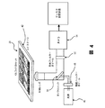
図3は、システム全体の機能ブロック図である。1以上のスライド20が、分析のために調製される。サンプルの採取、調製、及び処理の説明については、以下の詳細な説明を参照されたい。スキャナー22は、1以上のスライド20を画像化する。スキャナー22は、4色スキャナーなどのマルチチャネルスキャナーであることが好ましい。スキャナー22からのデータは、画像記憶部24に送られる。画像記憶部24は、当分野の技術者に公知である通りの記憶装置、好ましくはRAIDシステムなどの大容量記憶装置から構成され得る。画像記憶部からの走査データは、検討のために、例えば専門家などによる専門的検討のために、技術分析モジュール26、技術分析報告モジュール32、及び又は出力部34のうちの1つ以上に提供される。技術分析モジュール26は、少なくとも、画像記憶部24からのデータを以下に詳細に記載する様式で分析する働きをする。この分析には、限定はされないが、細胞を核の面積又はサイズについて(例えば青色DAPIの強度について分析することによって)分析すること、CD-45の非存在について(例えばCD-45抗体と結合させた二次抗体の強度について走査することによって)分析すること、及び/又は、サイトケラチンと結合させた抗体の強度について分析することが含まれ、好ましくは、該技術分析報告は、データを伴うHTMLファイル(該ファイル内には、細胞又は目的物の画像が含まれる)を含む。自動分析を、医療専門家による分析で補うことができる。
FIG. 3 is a functional block diagram of the entire system. One or
技術分析モジュール26の出力は、メタデータ・データベース28に提供されることが好ましい。メタデータ・データベースは、様々な形態のデータ分析のすべてによってもたらされた情報を含む。リターンループ制御経路30は、その後のスライド20(1又は複数)の再画像化を制御するための、スキャンデータの使用及び分析を可能にする。細胞などの目的物のさらなる分析が必要とされる場合には、このシステムは、その目的物を再画像化することができる。スライドに対する目的物の接着又は付着の程度は、少なくとも再画像化の間、スライド上の目的物の位置を維持するのに十分であるべきである。さらに長い期間では、スライドに対する目的物の接着又は付着の程度は、例えば、以下に記載する、目的物のその後のさらなる処理のための、例えば、遺伝子型同定又は他のその後の分析のための、分析によって特定された特定の目的物のその後の特定を可能にするのに十分であるべきである。スライドの保管、及び細胞の位置が固定されたままであることが所望される時間の長さは、数時間から数ヶ月の範囲であり得る。好ましくは、このシステムは、例えばデータ及び報告のための、総合記憶部40を含む。
The output of the
メタデータ・データベース28からの情報は、データインベントリ管理システム38に提供されることが好ましい。このシステムは、システム全体のための管理システムを含む。このシステム38は、データの中で特に、患者識別とスライドとの相関を管理する。
Information from the
図4は、走査システムの、ある可能な手段の模式図である。ステージ42、例えばx-yステージは、1以上のスライド20を支持する。本明細書に記載するウェル面積のサイズについては、4つのスライドによって、1人の患者の典型的なサイズサンプルである約1000万細胞の走査が可能になる。光学経路は、本明細書に記載する実施態様と矛盾しない任意の形をとることができる。照明構成部分は、光源46、及び任意選択の励起フィルターホイール48を含むことができる。光源は、好ましくは広範囲照明器である。ダイクロイックミラー50は、照明をスライドまで通過させ、かつ、照明がカメラ54に戻るのを可能にする働きをする。ミラー50とカメラ54との間に、任意選択の発光フィルター(emission filter)ホイール52を設置することができる。次いで、出力を、先に記載した通りに保管する。
FIG. 4 is a schematic diagram of some possible means of the scanning system. A
図5は、スライド20によって支持される目的物の走査のための様々な選択肢を示す。複数次元での、好ましくは3次元の枠組みでの走査及び画像化が可能である。目的物は、効率的な目的物の走査及び画像化を可能にするのに十分に平らな単層で、スライド上に配置される、好ましくは単一焦点平面内に配置されるのが好ましい。平面の又は平らなスライドによって、画像平面の偏差が最小限になるため、均一な方式での単層の画像化が可能になる。平らな表面上での画像化はまた、画像の、より容易なZ-スタッキングも可能にする。図5に示す通り、非平面関係にある可能性がある様々な方向での画像化平面が可能である。図5に示す通り、CTC候補細胞の検出は一般的に、スライド画像から測定されるいくつかのパラメータを頼りにする。例えば、次元1〜3は、核面積、サイトケラチン強度、及びCD-45強度であり得る。図5における平面は、CTC候補を定義する測定されるパラメータのそれぞれについての区切り境界を表す。或いは、又はさらに、デジタルカメラが、単一焦点平面を超越して光線を捕らえ、様々な画像平面からの画像のソフトウェア構築を可能にする光センサーを含む、ライトフィールドカメラシステムを利用することができる。デジタル光センサーと関連して、マイクロレンズなどのレンズを使用することができる。
FIG. 5 shows various options for scanning the object supported by the
図6及び7は、スライド20の様々な特徴を明らかにする。このスライドは、本明細書に記載する発明の実施態様と矛盾しない任意のサイズ又は形状のものであり得る。ある手段では、スライド20の形状は一般に、長さL、幅W、及び厚さtを伴う長方形である。代表的な寸法は、実質的に7.5cmのL、実質的に2.5cmのW、及び7mmの厚さtである。スライド20は、上部表面72、平行な底部表面74、側面後退部66、側面68、及び端面70を含むことが好ましい。例えばバーコードによって、スライド識別部60を提供することができる。こうしたスライドは、Marienfeld Laboratory Glassware社を含めた様々な供給業者から入手できる。
6 and 7 reveal various features of the
ウェル62は、画像化されることとなる材料を含有かつ維持するように提供される。本明細書に詳細に記載する形式では、ウェル62は、長方形であり、長さLと幅Wを有する。ウェル62の代表的な内部寸法は、一例としては、実質的に5.85cmのL及び実質的に2.5cmのWであり得る。ウェル62の外縁又は周長は、(特定の構造的境界であるか、他の材料による、例えば疎水性材料による境界であるかにかかわらず)境界64によって定めることができる。境界は、境界線と呼ぶことができる。境界又は境界線は、該プロセスにおいて使用される、細胞懸濁液、及び他のすべての試薬、溶液、緩衝液、又は他の液体を受け入れる及び含有するように適合される。境界又は境界線は、スライドの上部側面と共に、ウェルを形成する。これらの寸法を伴うウェル62の面積は、実質的に11.7cm2であり、ウェル62の周長は、実質的に15.7cmである。スライド20の端部からのウェル62の後退の程度は、走査システムの特徴などの、システムの他の状況に基づいて設定することができる。一実施態様は、画像化されることとなる実質的に単層の目的物、例えば細胞を提供する、好ましくは7.5cm2超、より好ましくは10cm2超、最も好ましくは実質的に11.7cm2の面積を有する単一の画像化ウェル62を提供する。このウェル62の周長は、それに対応して、実質的に12.5cm、より好ましくは実質的に14.5cm、最も好ましくは実質的に15.7cmであることが好ましい。ウェルによって被覆されるスライドの上部表面の割合は、好ましくは実質的に40%、より好ましくは53%、最も好ましくは実質的に62%である。
Well 62 is provided to contain and maintain the material to be imaged. In the format described in detail herein, the well 62 is rectangular and has a length L and a width W. Typical internal dimensions of the well 62 may be, for example, substantially 5.85 cm L and substantially 2.5 cm W. The outer edge or perimeter of the well 62 can be defined by the boundary 64 (whether it is a specific structural boundary or a boundary with other materials, such as a hydrophobic material). The boundary can be referred to as a boundary line. The boundary or boundary line is adapted to receive and contain the cell suspension and all other reagents, solutions, buffers, or other liquids used in the process. The boundary or boundary line forms a well with the upper side of the slide. The area of the well 62 with these dimensions is substantially 11.7 cm 2 and the circumference of the well 62 is substantially 15.7 cm. The degree of retraction of the well 62 from the end of the
好ましい画像化ウェル62は、合計4つの辺を有する。(例えば3ウェルスライドが12の辺を有する従来の技術(図1参照)と比較して)辺の数及びその周長を減らすことによって、側壁境界線に関連する端部効果が最小限になる。ウェルは、それぞれ、好ましくは160万から190万個の細胞、より好ましくは210万から260万個の細胞、最も好ましくは、250万から300万個の細胞の単層の画像化が可能であるようなサイズである。 A preferred imaging well 62 has a total of four sides. By reducing the number of edges and their perimeter (e.g., compared to the prior art where a 3-well slide has 12 sides (see Figure 1)), the edge effects associated with sidewall boundaries are minimized. . Each well is preferably capable of imaging a monolayer of 1.6 to 1.9 million cells, more preferably 2.1 to 2.6 million cells, most preferably 2.5 to 3 million cells It is a size like this.
まとめると、以下のパラメータによって、本明細書に記載するウェルにおいて実質的に単層の細胞を画像化する場合の、システム、装置、及び方法の様々な尺度が定義される:

スライドは、任意に、基準マークを備えることができる。細胞の位置が十分に固定された場合、スライドに指標を付けるために、他の構造を使用することができる。一例としては、境界を利用することができる。より具体的には、基準のために、ウェルの隅に90度の角を使用することができる。 The slide can optionally be provided with a fiducial mark. Other structures can be used to index the slide if the cell position is well fixed. As an example, a boundary can be used. More specifically, a 90 degree corner can be used for the corner of the well for reference.
本明細書に記載する装置及びシステムは、最大限かつ最適な細胞密度と、最小限の端部効果を提供する。好ましい実施態様では、少なくとも150万個の細胞、任意にさらに多くの、例えば300万個の細胞を保持するために、単一の細胞ウェルが利用される。好ましい実施態様では、4つの側壁が、その細胞集団を含有する働きをする。それに対して、単一領域スライドではなく3領域スライドの使用では、2から3倍の数のスライドの使用、かつ、たった4つの端部ではなく各スライド上の12(3×4)の端部への対処が必要とされるであろう。いかなる流体分布も、端部がどんなに疎水性であろうと、端部効果を受ける。細胞分布によると、端部での細胞密度が、有意に低下していることが示される。標準のサイズの顕微鏡スライドを使用することができる限りは、これは、それほど制限されない。本明細書に記載する実施態様の目的と一致する、より大きいスライドガラスを利用することができる。しかし、通常のサイズのスライド(これらのスライドについては、例えばほんの数例を挙げると、自動制御装置、既存の顕微鏡システム、及び記憶システムなどの、大規模ベースのインストール済みの機械が存在する)を使用することによって、標準のサイズのままでいることによるプロセス利益がもたらされる。 The devices and systems described herein provide maximum and optimal cell density and minimal end effects. In a preferred embodiment, a single cell well is utilized to hold at least 1.5 million cells, optionally even more, for example 3 million cells. In a preferred embodiment, the four sidewalls serve to contain the cell population. In contrast, using a three-region slide rather than a single-region slide uses two to three times the number of slides, and 12 (3x4) ends on each slide instead of just four ends It will be necessary to deal with. Any fluid distribution is subject to end effects, no matter how hydrophobic the ends are. Cell distribution indicates that the cell density at the edge is significantly reduced. This is not so limited as long as standard size microscope slides can be used. Larger glass slides may be utilized that are consistent with the objectives of the embodiments described herein. However, normal-sized slides (for these slides, there are large-scale installed machines such as automated controllers, existing microscope systems, and storage systems, to name just a few) The use provides process benefits by staying at a standard size.
該システムは、1スライドにつき単一のカバースライドをさらに含むことが好ましい。該システムは、十分な質をもたらしながら、速度を最適化する働きをする。平らでない表面、細胞の重なり、流体の厚さの変化、及び又は各スライドに対する複数のカバースライドの使用を好ましくは避けることによって、画像化準備及びデータ収集の速度と質の両方が増大する。単一かつ非常に平らな均質な単層が好ましい。単一のカバースライドを使用するさらなる利点は、画像化のために均一な表面が提供されることである。標準の3ウェルスライドにおいて必要となるであろう3つのカバーガラスではなく単一のカバーガラスを使用することで、はるかに平らな封入媒体分布が提供される。 The system preferably further includes a single cover slide per slide. The system serves to optimize speed while providing sufficient quality. By preferably avoiding uneven surfaces, cell overlap, fluid thickness changes, and / or the use of multiple cover slides for each slide, both the speed and quality of imaging preparation and data collection are increased. A single and very flat homogeneous monolayer is preferred. A further advantage of using a single cover slide is that a uniform surface is provided for imaging. The use of a single cover slip rather than the three cover slips that would be required in a standard 3 well slide provides a much flatter encapsulating media distribution.
実施態様では、本明細書に記載する通りに処理されるサンプルは、約1、2、5、7、10、15、20、30、40、50、100、200、300、400、500、600、700、800、900、さらには1000を超える希少細胞又はCTCを含む。 In embodiments, samples processed as described herein are about 1, 2, 5, 7, 10, 15, 20, 30, 40, 50, 100, 200, 300, 400, 500, 600. 700, 800, 900 and even more than 1000 rare cells or CTC.
本発明に記載した方法は、全体を通して論じた通り、CTCを検出する際に有用であるが、本発明はまた、CTCの特徴付けにおいても有用である。具体的には、検出可能なマーカーと、細胞の画像化及び分析を実施するための計算方法との様々な組み合わせの使用によって、癌の予後を評価する際に、また、疾患再発を引き起こす可能性がある治療不成功の早期検出のための治療有効性をモニタリングする際に有用な、有意義な特徴付けが可能になる。さらに、本発明によるCTC分析は、一連の治療法が完了している、発症前の患者における早期再発の検出を可能にする。これが可能であるのは、CTCの存在が、腫瘍の進行及び広がり、治療法に対する応答不良、疾患の再発、及び/又は長期にわたる生存率の低下と関連している及び/又は相関性があるからである。したがって、CTCの計数及び特徴付けは、治療法に対する応答に基づく初期リスク及び続発リスクを予測する基本特性について患者を階層化するための方法を提供する。 While the methods described in the present invention are useful in detecting CTC as discussed throughout, the present invention is also useful in characterizing CTC. Specifically, the use of various combinations of detectable markers and computational methods to perform cell imaging and analysis may also be used in assessing cancer prognosis and causing disease recurrence. It enables meaningful characterization useful in monitoring the effectiveness of treatment for the early detection of certain treatment failures. Furthermore, CTC analysis according to the present invention allows for the detection of early recurrence in pre-onset patients who have completed a series of treatments. This is possible because the presence of CTC is associated with and / or correlated with tumor progression and spread, poor response to therapy, disease recurrence, and / or reduced long-term survival. It is. Thus, CTC counting and characterization provides a way to stratify patients for basic characteristics that predict initial and secondary risk based on response to therapy.
用語「対象」とは、本明細書で使用される場合、主題の方法が実施される、あらゆる個人又は患者をいう。一般に、対象はヒトであるが、当分野の技術者には分かるように、対象は、動物である可能性もある。したがって、哺乳類、例えばげっ歯類(マウス、ラット、ハムスター、及びモルモットを含めて)、ネコ、イヌ、ウサギ、家畜(ウシ、ウマ、ヤギ、ヒツジ、ブタなどを含めて)、及び霊長類(サル、チンパンジー、オランウータン、及びゴリラを含めて)などを含めた他の動物も、対象の定義内に含まれる。 The term “subject” as used herein refers to any individual or patient on whom the subject method is performed. Generally, the subject is a human, but the subject may be an animal, as will be appreciated by those skilled in the art. Thus, mammals such as rodents (including mice, rats, hamsters, and guinea pigs), cats, dogs, rabbits, livestock (including cattle, horses, goats, sheep, pigs, etc.), and primates (monkeys). Other animals are also included within the definition of a subject, including, including, chimpanzees, orangutans, and gorillas.
したがって、別の実施態様では、本発明は、対象における癌を診断又は予測するための方法を提供する。この方法は、本明細書に記載する通りにCTCを検出することを含む。次いで、CTCを分析して、該対象における癌を診断又は予測することができる。したがって、本発明の方法を使用して、例えば、癌患者及び癌のリスクがある患者を評価することができる。本明細書に記載する診断又は予測の方法のいずれかにおいては、診断又は予測をもたらすために、癌の1以上の指標(癌細胞など)の、又は他の任意の障害の指標の存在又は非存在を使用することができる。 Accordingly, in another embodiment, the present invention provides a method for diagnosing or predicting cancer in a subject. The method includes detecting CTC as described herein. CTC can then be analyzed to diagnose or predict cancer in the subject. Thus, the methods of the invention can be used, for example, to assess cancer patients and patients at risk for cancer. In any of the diagnostic or prognostic methods described herein, the presence or absence of one or more indicators of cancer (such as cancer cells) or of any other disorder to produce a diagnosis or prediction. The presence can be used.
一態様では、血液サンプルは、患者から採取され、本明細書に記載する通りにCTCを検出するために処理される。本発明の方法を使用して、血液サンプル中のCTCの数が求められ、検出可能なマーカー及び細胞の画像化から集められた他のデータの分析によって、そのCTCが特徴付けられる。例えば、分析を実施して、サンプル中のCTCの数及び特徴付けを決定することができ、この測定から、最初の血液サンプル中に存在するCTCの数を求めることができる。 In one aspect, a blood sample is taken from a patient and processed to detect CTC as described herein. Using the method of the present invention, the number of CTCs in a blood sample is determined, and the CTCs are characterized by analysis of detectable markers and other data collected from cell imaging. For example, an analysis can be performed to determine the number and characterization of CTCs in the sample, and from this measurement, the number of CTCs present in the initial blood sample can be determined.
種々の態様では、対象のCTCの数及び特徴付けの分析を、特定の時間経過にわたって、様々な間隔で行って、対象の進行及び病状を評価することができる。例えば、分析は、時間の関数としての循環上皮細胞のレベル及び特徴付けを追跡するために、1日、2日、3日、1週、2週、1ヶ月、2ヶ月、3ヶ月、6ヶ月、又は1年などの一定の間隔で実施することができる。癌が存在する患者の場合、これによって、疾患の進行の有用な指標が提供され、医師が、患者の血流中のCTCの存在などの循環上皮細胞の増大、低下、又は無変化に基づいて適切な治療選択を行う手助けとなる。時間に伴うCTCの数の、例えば2倍、5倍、10倍、又はそれ以上といった、いかなる増大であっても、患者の予後を低下させ、また、これは、患者が治療法を変えるべきであるという早期指標である。同様に、例えば2倍、5倍、10倍、又はそれ以上といった、いかなる増大も、患者が、予後及び治療法に対する応答をさらに評価するための、画像化などのさらなる試験を受けるべきであることを示す。時間に伴うCTCの数の、例えば2倍、5倍、10倍、又はそれ以上といった、いかなる低下も、疾患安定化、及び治療法に対する患者の応答を示し、また、これは、治療法を変更しないという指標である。癌のリスクがある患者については、検出されるCTCの数の突然の増加は、患者が腫瘍を発症しているという早期の警告を提供する、したがって、早期診断を提供することができる。一実施態様では、顕示されたCTCの検出は、癌の病期分類を向上させる。 In various embodiments, analysis of a subject's number and characterization of CTCs can be performed at various intervals over a particular time course to assess subject progression and pathology. For example, analysis can be used to track the level and characterization of circulating epithelial cells as a function of time, 1 day, 2 days, 3 days, 1 week, 2 weeks, 1 month, 2 months, 3 months, 6 months Or at regular intervals such as one year. For patients with cancer, this provides a useful indicator of disease progression, based on the increase, decrease, or no change in circulating epithelial cells, such as the presence of CTC in the patient's bloodstream. Helps make appropriate treatment choices. Any increase in the number of CTCs over time, e.g. 2x, 5x, 10x or more, reduces the patient's prognosis and this should change the treatment It is an early indicator that there is. Similarly, any increase, such as 2x, 5x, 10x, or more, should the patient undergo additional testing, such as imaging, to further assess prognosis and response to therapy. Indicates. Any decrease in the number of CTCs over time, e.g. 2x, 5x, 10x or more, indicates disease stabilization and the patient's response to therapy, which also changes therapy It is an indicator that no. For patients at risk for cancer, a sudden increase in the number of CTCs detected provides an early warning that the patient has developed a tumor and can therefore provide an early diagnosis. In one embodiment, the detection of manifested CTC improves cancer staging.
本明細書に提供される方法のうちのいずれかでは、追加的臨床評価を提供するために、追加的分析を実施して、CTCの特徴付けを行うこともできる。例えば、画像分析に加えて、遺伝子チップ分析、及び特定の癌マーカーに特異的なプライマーを用いる多重化などの、遺伝子発現分析及びPCR技術を用いて、CTCが由来する腫瘍の型、転移状態、及び悪性の程度などの情報を得ることができる。さらに、患者の癌の特徴付けに関する追加的情報を評価する手段として、細胞サイズ、DNA又はRNA分析、プロテオーム解析、又はメタボローム解析を実施することができる。種々の態様では、分析には、以下のマーカーのうちの1つ以上に誘導される抗体、又は、以下のマーカーのうちの1つ以上に特異的なプライマーを使用するPCR多重化が含まれる:EGFR、HER2、ERCC1、CXCR4、EpCAM、E-カドヘリン、ムチン-1、サイトケラチン、PSA、PSMA、RRM1、アンドロゲン受容体、エストロゲン受容体、プロゲステロン受容体、IGF1、cMET、EML4、又は白血球関連受容体(LAR)。 In any of the methods provided herein, additional analysis can be performed to characterize CTC in order to provide additional clinical evaluation. For example, in addition to image analysis, using gene expression analysis and PCR techniques, such as gene chip analysis, and multiplexing with primers specific to specific cancer markers, the type of tumor from which CTC is derived, metastasis status, In addition, information such as the degree of malignancy can be obtained. In addition, cell size, DNA or RNA analysis, proteomic analysis, or metabolomic analysis can be performed as a means of evaluating additional information regarding the characterization of the patient's cancer. In various embodiments, the analysis includes PCR multiplexing using antibodies directed to one or more of the following markers, or primers specific for one or more of the following markers: EGFR, HER2, ERCC1, CXCR4, EpCAM, E-cadherin, mucin-1, cytokeratin, PSA, PSMA, RRM1, androgen receptor, estrogen receptor, progesterone receptor, IGF1, cMET, EML4, or leukocyte related receptor (LAR).
例えば、追加的分析は、特定の治療レジメンに対する対象の応答性の判定を行うのに十分な、又は、癌の治療における候補薬剤の有効性を判定するのに十分なデータを提供することができる。したがって、本発明は、本明細書に記載する通りに対象のCTCを検出し、検出されたCTCを分析することによって、特定の治療レジメンに対する対象の応答性を判定する、又は、癌の治療における候補薬剤の有効性を判定する方法を提供する。例えば、薬物治療が患者に施された場合、本発明の方法を使用して、その薬物治療の有効性を判定することが可能である。例えば、薬物治療前の患者から採取されたサンプルと、薬物治療と同時に又は薬物治療後の患者から採取された1以上の細胞サンプルを、本発明の方法を使用して処理することができる。処理されたそれぞれのサンプルの分析の結果を比較することによって、薬物治療の有効性又は薬剤に対する患者の応答性を判定することができる。このようにして、不成功の化合物の早期特定を行うことができる、又は、有望な化合物の早期確認を行うことができる。 For example, additional analysis can provide sufficient data to make a determination of a subject's responsiveness to a particular treatment regimen or to determine the effectiveness of a candidate agent in treating cancer. . Thus, the present invention detects a subject's CTC as described herein and determines the subject's responsiveness to a particular treatment regimen by analyzing the detected CTC, or in the treatment of cancer. A method for determining the effectiveness of a candidate agent is provided. For example, if a medication is given to a patient, the method of the invention can be used to determine the effectiveness of that medication. For example, a sample taken from a patient prior to drug treatment and one or more cell samples taken from a patient at the same time or after drug treatment can be processed using the methods of the invention. By comparing the results of the analysis of each processed sample, the effectiveness of the drug treatment or the patient's responsiveness to the drug can be determined. In this way, early identification of unsuccessful compounds can be performed, or early confirmation of promising compounds can be performed.
候補化合物の臨床活性を見抜く4つの重要な指標として、HER2、EGFR、CXCR4、及びEphB4 RTKが挙げられる。HER2は、mRNA安定性、及びHER2転写物の細胞内局在を判定することによる、細胞の悪性度の指標を提供する。獲得変異に対するEGFRの抵抗性、及び/又は獲得された変異は、候補化合物、それに加えて、候補化合物と組み合わせて使用することができる可能な代替化合物の活性の重要な指標を提供する。白金を用いて誘発されるDNA修復妨害のレベルの評価は、CXCR4マーカーの状態及び転移状態に関する洞察を提供する。さらに、EphB4受容体チロシンキナーゼの状態の評価は、細胞の転移の潜在的可能性に関する洞察を提供する。したがって、本発明の方法を使用して、こうした候補薬物を摂取する患者を、血液の高頻度のサンプルを採取し、各サンプル中の循環上皮細胞、例えばCTCの数を時間の関数として求めることによってモニタリングすることができる。Her2、EGFR、CXCR4、及びEphB4 RTK指標のさらなる分析は、癌の病状及び候補薬物の有効性に関する情報を提供する。同様に、ERRC1、サイトケラチン、PSA、PSMA、RRM1、アンドロゲン受容体、エストロゲン受容体、プロゲステロン受容体、IGF1、cMET、EML4などは、候補化合物の臨床活性への洞察を提供する。臨床活性のこれらの指標の分析は、本明細書に論じる通りの検出可能なマーカーの分析(例えば、免疫組織化学及び蛍光インサイチューハイブリダイゼーション(FISH))、又は配列決定、遺伝子型同定、遺伝子発現、又は他の分子分析技術などの技術によるさらなる分析を介するものであり得る。 Four important indicators for identifying the clinical activity of candidate compounds include HER2, EGFR, CXCR4, and EphB4 RTK. HER2 provides an indication of cell malignancy by determining mRNA stability and subcellular localization of the HER2 transcript. EGFR resistance to acquired mutations and / or acquired mutations provide an important indicator of the activity of a candidate compound, as well as possible alternative compounds that can be used in combination with the candidate compound. Assessment of the level of DNA repair interference induced with platinum provides insight into the status of the CXCR4 marker and the metastatic status. Furthermore, assessment of the status of the EphB4 receptor tyrosine kinase provides insight into the potential for cell metastasis. Thus, using the method of the present invention, patients taking such candidate drugs can be obtained by taking a high frequency sample of blood and determining the number of circulating epithelial cells, eg, CTC, in each sample as a function of time. Can be monitored. Further analysis of the Her2, EGFR, CXCR4, and EphB4 RTK indicators provides information regarding cancer pathology and candidate drug efficacy. Similarly, ERRC1, cytokeratin, PSA, PSMA, RRM1, androgen receptor, estrogen receptor, progesterone receptor, IGF1, cMET, EML4, etc. provide insight into the clinical activity of candidate compounds. Analysis of these indicators of clinical activity includes analysis of detectable markers as discussed herein (e.g., immunohistochemistry and fluorescence in situ hybridization (FISH)), or sequencing, genotyping, gene expression, Or it may be through further analysis by techniques such as other molecular analysis techniques.
CTCの分析は、特定の臨床試験のための候補対象を決定する方法を提供する。例えば、臨床試験の特定の治療レジメンが、潜在的に成功する可能性があるかどうかを決定するために、ある候補の検出されたCTCを分析して、特定のマーカーが存在するかどうかを決定することができる。したがって、別の実施態様では、本発明は、臨床試験のための候補対象を決定するための方法を提供する。この方法は、本明細書に記載する通りに対象のCTCを検出することを含む。次いで、CTCを分析して、その候補対象が特定の臨床試験に適しているかどうかを決定することができる。 Analysis of CTC provides a way to determine candidate subjects for a particular clinical trial. For example, to determine whether a particular treatment regimen in a clinical trial is potentially successful, analyze a candidate's detected CTC to determine whether a particular marker is present can do. Thus, in another embodiment, the present invention provides a method for determining candidate subjects for clinical trials. The method includes detecting the CTC of the subject as described herein. The CTC can then be analyzed to determine whether the candidate subject is suitable for a particular clinical trial.
臨床試験中のCTCの分析は、患者が実験的薬物に応答するか応答しないかに関する情報を提供することとなる。ここでは、顕示されたCTCの実質的な変化又は低下がないことは、応答を示し、顕示されたCTCの増大は、応答不良を示す。増大又は低下は、2倍、10倍、又はそれ以上であり得る。この情報は、薬物の有効性の早期指標であり、研究者によって、臨床試験における副次的評価項目として使用され得る。 Analysis of CTC during clinical trials will provide information on whether patients respond to or do not respond to experimental drugs. Here, the absence of a substantial change or reduction in the manifested CTC indicates a response, and an apparent increase in CTC indicates a poor response. The increase or decrease can be 2-fold, 10-fold, or more. This information is an early indicator of drug efficacy and can be used by researchers as a secondary endpoint in clinical trials.
以下の実施例は、例示を目的とするものであり、本発明を限定しない。 The following examples are for purposes of illustration and do not limit the invention.
(実施例1)
(CTCの検出及び特徴付け)
(実験結果)
(Example 1)
(Detection and characterization of CTC)
(Experimental result)
(CTCからHD-CTC:定義の精緻化) (CTC to HD-CTC: Refinement of definition)
該システムは、分析において利用されることとなる細胞に関する1以上の尺度を定義することが好ましい。同じ患者における固形の同じ腫瘍由来の細胞との直接的比較を用いる、上皮癌を有する患者における多くの候補事象の調査に基づいて、様々な手法によって、完全なCTCが定義される[9〜12]。これらの定義に基づいて、かつ基準を精緻化するためにHD-CTCアッセイを利用して、HD-CTCの定義が確立された。この定義が発展して、HD-CTCが、患者の体内の癌腫の固体析出物に由来する完全な細胞である可能性が最も高い細胞であることが確実になってきた。こうした要件を部分的に満たすが、以下に論じる厳密な包含基準には達しない、すべての他の集団が、この分析において追跡される。何故なら、これらのうちの多くは、生物学的意義を有する可能性がある断片化された又はアポトーシス性の腫瘍細胞に相当する可能性があるが[13]、これらは、さらなるその後の方法論による評価への適合性が予測不可能であるという理由で、HD-CTC計数において排除されるからである。本出願の目的のために、これらのうちの一部又はすべてを満たす、様々な基準を使用することができる。 The system preferably defines one or more measures for the cells that will be utilized in the analysis. Based on a survey of many candidate events in patients with epithelial cancer using a direct comparison with cells from the same solid tumor in the same patient, various approaches define complete CTC [9-12 ]. Based on these definitions and using the HD-CTC assay to refine the criteria, the definition of HD-CTC was established. This definition has evolved to ensure that HD-CTC is most likely a complete cell derived from a solid deposit of carcinoma in the patient's body. All other populations that partially meet these requirements but do not meet the strict inclusion criteria discussed below are tracked in this analysis. Because many of these may represent fragmented or apoptotic tumor cells that may have biological significance [13], these are due to further subsequent methodologies This is because the fitness for evaluation is excluded in HD-CTC counting because it is unpredictable. Various criteria that meet some or all of these can be used for the purposes of this application.
乳癌、直腸結腸癌、及び肺癌患者の症例研究において、CTCを、形態的に特徴付ける、また、その原発腫瘍に関して信用を与えることができる。高分化型の肺腺癌を有する患者におけるCTCの形態的検査を通して、循環細胞を、例えば細胞質に対する核の比が比較的低い細胞を含めたこの型の腫瘍と一致する形態的特徴と同一であるとした。血液循環中で特定される腫瘍細胞の形態は、その患者の原発腫瘍の微細針吸引生検において見られる形態と類似していた[9]。より大きなコホートの乳癌及び直腸結腸癌患者において評価されると、CTCは、原発性及び転移性腫瘍において通常見られる細胞の形態的不均一性と一致する、細胞学的外観における高度の患者間及び患者内不均一性を示した[10、11]。 In case studies of breast, colorectal and lung cancer patients, CTC can be morphologically characterized and credited for its primary tumor. Through morphological examination of CTC in patients with well-differentiated lung adenocarcinoma, circulating cells are identical to morphological features consistent with this type of tumor, including cells with a relatively low nuclear to cytoplasmic ratio, for example It was. The morphology of tumor cells identified in the blood circulation was similar to that seen in fine needle aspiration biopsy of the patient's primary tumor [9]. When assessed in patients with breast and colorectal cancers in larger cohorts, CTCs are highly inter-patient in cytological appearance and consistent with the morphological heterogeneity of cells normally seen in primary and metastatic tumors. Intra-patient heterogeneity was demonstrated [10, 11].
このプラットフォームは、目的物のサイズ及び蛍光強度に関する補助的な半定量的データを用いる細胞ごとのアノテーションを用いて増強される、蛍光画像と個々のチャネル画像との同時的な細胞形態レビューを可能にする。HD-CTCは、DAPI画像化によって非アポトーシス性に見える完全な核を伴う、a)サイトケラチン陽性である細胞;b)CD45陰性である細胞に分類される。CKについての陽性は、蛍光シグナルが、周囲の細胞のシグナルを有意に上回ることと定義される。陰性は、周囲の細胞のシグナルと同じレベル又はそれ未満であることと定義される。CD45についての陰性は、全細胞のうちの99%が包括的に検出可能である境界状態に達しない、目視検出未満の強度を有することと定義される。癌患者に見られる代表的なHD-CTCの集合は示さないが、HD-CTCは、サイトケラチン陽性、CD45陰性であり、かつDAPI核を含有し、かつ周囲の白血球と形態的に異なる。 This platform enables simultaneous cell morphology review of fluorescence and individual channel images, augmented with cell-by-cell annotations using supplemental semi-quantitative data on target size and fluorescence intensity To do. HD-CTCs are classified as a) cytokeratin positive cells; b) CD45 negative cells with complete nuclei that appear non-apoptotic by DAPI imaging. A positive for CK is defined as a fluorescent signal that significantly exceeds the signal of the surrounding cells. Negative is defined as being at or below the level of the surrounding cell signal. Negative for CD45 is defined as having less than visual detection, 99% of all cells do not reach a boundary state that is comprehensively detectable. Although the representative collection of HD-CTC found in cancer patients is not shown, HD-CTC is cytokeratin positive, CD45 negative, contains DAPI nuclei, and is morphologically different from surrounding leukocytes.
核がアポトーシス性であるように見えない限りは、サイトケラチンチャネルにおいて可視化される細胞質小疱形成などの、細胞質の穏やかなアポトーシス性変化は許容される。これらの半定量的特性に加えて、HD-CTCは、正常なWBCと形態的に異ならなければならず、かつ、標準の診断的細胞病理学において使用される基準(主にサイズの拡大として具体化されるが、核及び細胞質の構造的組成、細胞質形状、及び核形状などの細胞形態的特徴も包含する)による悪性細胞と適合する形態を有しなければならない。平均WBC核サイズの1.3倍という、より低い核サイズの切り捨て値を設定することができる。最新の合意は確立されておらず、生物学的事実も今のところ未知であるので、いささか自由裁量ではあるが、この境界は、サイトケラチンでの誤った非特異的染色(すなわちCD45陽性かつサイトケラチン陽性)を示す、健康なドナーにおいてWBCとして特定された細胞の最も大きい核サイズの評価に基づいて設定される。癌診断の臨床範囲では、慣例的に固定及び染色されたヒト組織においては、実質的にすべての生存可能な上皮細胞は、実質的にすべての白血球よりも大きいので、この手法は、保守的な仮定であると感じられる。この範囲の対極では、HD-CTCの通常の形態的特徴は、周囲のWBC核の平均サイズの5倍までの、より大きい核である。通常観察される他の特徴としては、周囲のWBC核のものとは異なる核輪郭(例えば伸長)、核に対して偏心的な分布の細胞質を伴う大きな細胞質ドメイン、多辺形の又は引き伸ばされたような細胞質形状、並びに高頻度のダブレット及び3個以上のHD-CTCのクラスターが挙げられる。 As long as the nucleus does not appear to be apoptotic, mild apoptotic changes in the cytoplasm are permissible, such as cytoplasmic blebbing visualized in cytokeratin channels. In addition to these semi-quantitative properties, HD-CTC must be morphologically different from normal WBC and is a standard used in standard diagnostic cytopathology (specifically as an increase in size). But must also have a form that is compatible with malignant cells by the structural composition of the nucleus and cytoplasm, including cytomorphic features such as cytoplasmic shape and nuclear shape). A lower nucleus size truncation value of 1.3 times the average WBC nucleus size can be set. Although the latest consensus has not been established and the biological facts are unknown at the moment, this border is somewhat discretionary, but this boundary is a false non-specific staining with cytokeratins (i.e. CD45 positive and site Set based on an assessment of the largest nuclear size of cells identified as WBC in healthy donors, showing keratin positive). In the clinical scope of cancer diagnosis, in routinely fixed and stained human tissue, virtually all viable epithelial cells are larger than substantially all leukocytes, so this approach is conservative. I feel it is an assumption. At the opposite end of this range, the normal morphological features of HD-CTC are larger nuclei, up to 5 times the average size of surrounding WBC nuclei. Other features commonly observed include a nuclear profile that differs from that of the surrounding WBC nucleus (e.g., elongation), a large cytoplasmic domain with a cytoplasm that is eccentric to the nucleus, polygonal or stretched Cytoplasmic shapes, as well as frequent doublets and clusters of 3 or more HD-CTCs.
表1:転移性の前立腺癌、乳癌、及び膵臓癌患者、並びに正常な対照から得られた1mLの血液あたりにHD-CTCを有する患者の割合。
(転移性癌を有する患者におけるHD-CTCの出現率) (Appearance rate of HD-CTC in patients with metastatic cancer)
30人の転移性乳癌患者、20人の転移性前立腺癌患者、18人の転移性膵臓癌患者、及び15人の正常な対照のコホートにおいて、HD-CTCを計数した。調査した3つの型の癌におけるHD-CTCの出現率を、表1に示す。この手法を使用すると、≧5 HD-CTC/mLは、前立腺癌患者の80%に(平均=92.2)、乳癌患者の70%に(平均=56.8)、膵臓癌患者の50%に(平均=15.8)、正常な対照の0%に(平均=0.6)見られた。 HD-CTC was counted in a cohort of 30 metastatic breast cancer patients, 20 metastatic prostate cancer patients, 18 metastatic pancreatic cancer patients, and 15 normal controls. Table 1 shows the incidence of HD-CTC in the three types of cancer investigated. Using this approach, ≧ 5 HD-CTC / mL was found in 80% of prostate cancer patients (mean = 92.2), 70% of breast cancer patients (mean = 56.8), and 50% of pancreatic cancer patients (mean = 15.8), found in 0% of normal controls (mean = 0.6).
図8は、期待されるSKBR3に対してプロットされた、観察された平均SKBR3を示す。正常な対照の血液の4つの一定分量に、様々な数のSKBR2細胞を添加して、スライドあたり約10、30、100、及び300個の癌細胞を含む、4つのスライドをもたらした。各4つ組の平均を、標準偏差を表すエラーバーと共に示す。 FIG. 8 shows the observed average SKBR3 plotted against the expected SKBR3. Various numbers of SKBR2 cells were added to four aliquots of normal control blood, resulting in four slides containing approximately 10, 30, 100, and 300 cancer cells per slide. Each quadruplicate average is shown with an error bar representing the standard deviation.
(添加実験を使用する、アッセイの線形性及び感度) (Assay linearity and sensitivity using loading experiments)
アッセイの線形性及び感度を試験するために、様々な数の乳癌細胞株SKBR3を、正常な対照血液に添加し(4つ組)、HD-CTCアッセイに従って処理した。図8に示す通り、観察された平均SKBR3を、期待されるSKBR3に対してプロットし、0.9997という相関係数(R2)が示される。 To test the linearity and sensitivity of the assay, various numbers of breast cancer cell lines SKBR3 were added to normal control blood (in quadruplicate) and processed according to the HD-CTC assay. As shown in FIG. 8, the observed average SKBR3 is plotted against the expected SKBR3, indicating a correlation coefficient (R 2 ) of 0.9997.
(癌腫を有する患者におけるHD-CTC数のアッセイ確実性) (Assay certainty of HD-CTC count in patients with carcinoma)
HD-CTCアッセイのアッセイ確実性を、複数の処理装置及び分割サンプルに対して試験した。2つの別の処理装置によって、9個の異なる患者サンプルに対して、2連の試験を実施した。図9は、0.979という相関係数(R2)と共に、この比較についてのY=1.163x-4.3325という回帰方程式を与える、分割サンプルを使用する2つの処理装置間のHD-CTC数/mLの比較を示す。すべてのデータは、実験内容を知らされていない1人の操作者によって解析された。線の傾きは、1を超えており、これは、2つの処理装置間のわずかな系統的線形変動を示唆している。 The assay certainty of the HD-CTC assay was tested against multiple processors and aliquots. Duplicate tests were performed on nine different patient samples with two separate treatment devices. Figure 9 shows a comparison of HD-CTC / mL between two processors using split samples, giving a regression equation of Y = 1.163x-4.3325 for this comparison, with a correlation coefficient (R 2 ) of 0.979 Indicates. All data was analyzed by a single operator who was not informed of the experiment. The slope of the line is over 1, suggesting a slight systematic linear variation between the two processors.
図9は、9個の異なる癌患者サンプルに対する、2つの別の処理装置間のCTC数の比較である。CTC数/mLは、0から203の範囲であった。 FIG. 9 is a comparison of the number of CTCs between two different treatment devices for nine different cancer patient samples. CTC count / mL ranged from 0 to 203.
(正常な対照由来のサンプルにおけるアッセイ特異性) (Assay specificity in samples from normal controls)
組織の健康なドナープールからの15人の健康なドナーを、年齢が24歳から62歳の範囲である8人の女性と7人の男性で構成される対照集団として評価した。健康な対照1人を除くすべての人において、こうした事象の数は、体積について補正すると、1 HD-CTC/ml以下であった。除外者は、4/mlというHD-CTC数を有する健康な女性ドナーであった。彼女の細胞のそれぞれに、系統立てられた再検査を行うと、そのうちの約3分の1は、すべての包含基準を容易に満たしたのに対し、残りの3分の2は、すべての基準を満たしたが、1以上の基準による包含については、下限の近くであった。4人の他の健康なドナーは、それぞれ1 HD-CTC/mlを有し、ゼロではない部類に分類された。これらの細胞の明示された再検査によって、約3分の1はすべての包含基準を確実に満たしたのに対し、残りの3分の2の細胞はすべての基準を満たしたが、1以上の基準による包含については下限の近くであったという点で、類似のパターンが示された。後者の型の事象の例としては、周囲のWBCよりも30%大きいが、形態的評価によれば有意に大きくは見えない細胞や、わずかに焦点がずれており、目によって検出できないアポトーシス性の核変化を有する可能性がある細胞や、最後に、切り捨て値を超える客観的なサイトケラチン強度測定値を有するが、主観的には、単一チャネル蛍光検査によれば周囲のWBCよりも有意に明るくは見えない偶発的な細胞が挙げられる。 Fifteen healthy donors from the tissue's healthy donor pool were evaluated as a control population consisting of 8 women and 7 men ranging in age from 24 to 62 years. In all but one healthy control, the number of such events, when adjusted for volume, was less than 1 HD-CTC / ml. Excluded were healthy female donors with a HD-CTC count of 4 / ml. A systematic reexamination of each of her cells revealed that about one-third of them easily met all inclusion criteria, while the remaining two-thirds met all criteria. However, the inclusion by one or more criteria was near the lower limit. Four other healthy donors, each with 1 HD-CTC / ml, were classified as non-zero. An explicit reexamination of these cells ensures that about one-third met all inclusion criteria, while the remaining two-thirds met all criteria, but more than one A similar pattern was shown in that the inclusion by criteria was near the lower limit. Examples of the latter type of event are cells that are 30% larger than the surrounding WBC, but are not significantly larger by morphological evaluation, or are slightly out of focus and cannot be detected by the eyes. Cells that may have nuclear changes and, finally, have an objective cytokeratin intensity measurement that exceeds the truncation value, but subjectively, it is significantly more than the surrounding WBC according to single channel fluorescence studies. There are incidental cells that are not visible brightly.
(HD-CTCアッセイとCellSearch(登録商標)との比較) (Comparison between HD-CTC assay and CellSearch (registered trademark))
合計15人の患者(5人の転移性乳癌及び10人の転移性前立腺癌)を、CellSearch(登録商標)とHD-CTCアッセイとの両方を用いて、CTCについて評価した。各患者から、2本のチューブの血液を採取した。一方のチューブの7.5mLの血液は、CellSaveチューブ(Veridex社、ニュージャージー州Raritan)内に採取し、CellSearch(登録商標)アッセイを使用するCTCの計数のためにQuest Diagnostics(カリフォルニア州San Juan Capistrano)に送った。第2のチューブの血液は、各患者から採取し、サンプルがQuest Diagnosticsで処理されるタイミングを模倣するために血液採取の24時間後に、標準のHD-CTCプロセスと一致するHD-CTCプロトコルに従って処理した。表2は、この対照比較の結果を示す。CellSearch(登録商標)アッセイは、試験された5/15患者において、7.5mLの血液あたり2以上のCTCを検出した。それに対して、HD-CTCアッセイは、有意に多い患者において(HD-CTCは、試験された14/15患者において特定された)、有意に多い数のCTCを検出した。
A total of 15 patients (5 metastatic breast cancer and 10 metastatic prostate cancer) were evaluated for CTC using both CellSearch® and HD-CTC assay. Two tubes of blood were collected from each patient. 7.5 mL of blood from one tube is collected in a CellSave tube (Veridex, Raritan, NJ) and sent to Quest Diagnostics (San Juan Capistrano, Calif.) For CTC counting using the CellSearch® assay. sent. A second tube of blood is drawn from each patient and processed according to an HD-CTC protocol consistent with the standard HD-
表2:5人の転移性乳癌患者と10人の転移性前立腺癌患者からのHD-CTCアッセイとCellSearch(登録商標)との比較。CellSearch(登録商標)の値は、血液1mLあたりのCTCの数に補外されている。
(HD-CTCの形態的範囲) (Morphological range of HD-CTC)
患者内で、及び患者を超えて、HD-CTCの形態的に不均一な集団を検出した。HD-CTCは、様々な形状、サイズ、及びサイトケラチン強度を有しており、ある場合には、大きなサイズ又は多辺形細胞質形状などの特有の細胞学的特徴は、患者のサンプル内でかなり特徴的かつ単調であった。他の場合には、単一のサンプル内のHD-CTC間に、細胞形態的多様性が存在していた。細胞サイズも様々であり;多くの患者サンプルは、隣接するWBC核の一様に3又は4倍のサイズの核を有するHD-CTCを有していたのに対して、他の患者は、隣接するWBC核の一様にわずか1.3倍のサイズの核を有する細胞を有していた。ある患者は、幅広いサイズを有していた。サイトケラチンでの誤った非特異的染色(すなわちCD45陽性かつサイトケラチン陽性)を示す、WBCとして特定された細胞の最も大きい核サイズの評価に基づいて、平均WBC核の1.3倍のHD-CTC核サイズの下限基準を選択した。 A morphologically heterogeneous population of HD-CTC was detected within and across patients. HD-CTC has a variety of shapes, sizes, and cytokeratin strengths, and in some cases unique cytological features such as large size or polygonal cytoplasmic shape can be quite significant in patient samples. It was characteristic and monotonous. In other cases, there was cell morphological diversity between HD-CTCs within a single sample. Cell size also varies; many patient samples had HD-CTC with nuclei uniformly 3 or 4 times the size of adjacent WBC nuclei, whereas other patients It had cells with nuclei that were only 1.3 times the size of WBC nuclei uniformly. Some patients had a wide range of sizes. HD-CTC nuclei 1.3 times the average WBC nuclei, based on an assessment of the largest nuclear size of cells identified as WBC, showing false non-specific staining with cytokeratin (i.e. CD45 positive and cytokeratin positive) The lower size criterion was selected.
このプラットフォームを使用することによって、詳細な形態的評価が可能になり、このコホート内の癌患者の大多数(88%)において、HD-CTCダブレット、及び2 HD-CTCから30 HD-CTC超までの範囲のクラスターが特定された(データは示していない)。 Using this platform allows for detailed morphological assessments, with the majority of cancer patients in this cohort (88%) HD-CTC doublets and from 2 HD-CTC to over 30 HD-CTC A range of clusters was identified (data not shown).
クラスターは、癌を有する大抵の患者において検出された。クラスターは、2個から30個を超えるHD-CTCの範囲であった。各HD-CTCを、サイトケラチン陽性、CD45陰性であり、かつDAPI核を含有し、かつ周囲の有核細胞と形態的に異なると決定した。 Clusters were detected in most patients with cancer. Clusters ranged from 2 to over 30 HD-CTCs. Each HD-CTC was determined to be cytokeratin positive, CD45 negative, contained DAPI nuclei, and morphologically different from surrounding nucleated cells.
(「他の」細胞型の形態) ("Other" cell type morphology)
サイトケラチン陽性、CD45陰性であり、核を含有するが、包含基準を満たさない、他の細胞様目的物は、HD-CTCとはみなされないが、アッセイによって追跡される。この手法の目的は、こうした基準を部分的にしか満たさないが、臨床的に意味がある可能性がある目的物(例えば、アポトーシス性腫瘍細胞、腫瘍細胞断片、又は上皮から間葉への移行を受けている細胞など)への接近を保持しながら、CTCの具体的な完全な表現型についての厳密な包含/除外基準を設けることである[13]。 Other cell-like objects that are cytokeratin positive, CD45 negative, contain nuclei but do not meet inclusion criteria are not considered HD-CTC, but are tracked by the assay. The objective of this approach is to meet these criteria only partially, but to a clinically meaningful target (e.g. apoptotic tumor cells, tumor cell fragments, or epithelial to mesenchymal transition). Strict inclusion / exclusion criteria for the specific complete phenotype of CTC, while maintaining access to the recipient cells etc. [13].
したがって、HD-CTCを追跡することに加えて、このコホートの患者において、アポトーシスを示す核を有していた細胞、外周的なサイトケラチンを有しなかった細胞、周囲のWBCと同じサイズ又は周囲のWBCよりも小さかった細胞、及びサイトケラチンが弱い又は陰性であった細胞を含めた、いくつかの異なる部類のサイトケラチン陽性細胞を目録化した(データは示していない)。最後に、多くの患者は、様々な型の高輝度のサイトケラチン陽性細胞に加えて、周囲のWBCと形態的に異なる、かつ、そのサンプル内のHD-CTCの核と似ている、かつ、CD45陰性であるが、やはりサイトケラチンが弱い又は陰性である核を有する、かなりの数の細胞を有していた(データは示していない)。 Thus, in addition to following HD-CTC, cells in this cohort had cells with apoptotic nuclei, cells without peripheral cytokeratin, the same size or surroundings as the surrounding WBC Several different classes of cytokeratin positive cells were cataloged (data not shown), including cells that were smaller than their WBC and cells that were weak or negative for cytokeratin. Finally, many patients, in addition to various types of high-intensity cytokeratin-positive cells, are morphologically different from the surrounding WBC and resemble the nucleus of HD-CTC in the sample, and It had a significant number of cells that were CD45 negative but also had nuclei that were weak or negative for cytokeratin (data not shown).
いくつかの候補HD-CTCは、様々な形態的又は形態計測的包含基準を欠いているため、除外された。例えば、観察されたサイトケラチン強度が弱過ぎであった;核サイズが小さ過ぎであった;サイトケラチンが外周不十分である(核の2/3未満を囲む)と認められた;クラスターが複数の大きい細胞のものであるように見えるにもかかわらず、観察されたサイトケラチン強度が弱過ぎであった;核がアポトーシス性の崩壊変化を示した;核が小さ過ぎであり、かつ細胞質が外周不十分であった;後期アポトーシスの細胞であるように見られた;核が小さ過ぎであった(周囲のWBC核と同じサイズ);サイトケラチンが存在するが、外周的ではなかった;細胞質が外周不十分であり、かつ核が小さ過ぎであった。 Some candidate HD-CTCs were excluded because they lacked various morphological or morphometric inclusion criteria. For example, the observed cytokeratin intensity was too weak; the nucleus size was too small; cytokeratin was found to be poorly perimeter (less than 2/3 of the nucleus); multiple clusters The observed cytokeratin strength was too weak despite appearing to be of large cells; the nucleus showed apoptotic disruption changes; the nucleus was too small and the cytoplasm was perimeter Insufficient; appeared to be late apoptotic cell; nucleus too small (same size as surrounding WBC nucleus); cytokeratin was present but not peripheral; cytoplasm was The circumference was insufficient and the core was too small.
1人の前立腺癌患者においては、様々な型の疑似CTCも見られた。例えば、いくつかは、サイトケラチン及びCD45について陰性であったが、大きく、かつこの患者において見られる他のHD-CTCと似ている核を有していた。典型的なHD-CTC(これらの細胞は、サイトケラチン陽性、CD45陰性、DAPI核を有する)も観察された。複数の細胞、例えば4細胞のHD-CTCのクラスターも観察された。 Various types of pseudo-CTC were also seen in one prostate cancer patient. For example, some were negative for cytokeratin and CD45, but had large and similar nuclei to other HD-CTCs found in this patient. Typical HD-CTCs (these cells are cytokeratin positive, CD45 negative, have DAPI nuclei) were also observed. A cluster of HD-CTCs of multiple cells, such as 4 cells, was also observed.
上皮から間葉への移行を受けている癌腫細胞の存在の可能性についての広範な現在の議論を考慮すると、これらの細胞の出現及びタンパク質発現パターンは、これらを、こうした細胞型の潜在的候補と特定する。或いは、これらの細胞は、細胞質の大部分を剥ぎ取られている、老いた腫瘍細胞である可能性がある。 Given the extensive current discussion about the potential for the presence of carcinoma cells undergoing transition from epithelium to mesenchyme, the appearance and protein expression patterns of these cells make them a potential candidate for these cell types. Is identified. Alternatively, these cells may be old tumor cells that have had most of their cytoplasm stripped off.
流体生検分析は、作用中の転移を明らかにする見込みを秘めている。この中では、捕らえにくい細胞集団が、血流を通して広がり、かつ最終的に遠隔転移をもたらす、癌のタネである。しかし、これらを調べてその発見を臨床的に適用するためには、この細胞は、大多数の癌患者において、確実に回収可能でなければならない。本明細書に記載する装置、システム、及び方法は、多数の癌患者における高解像度で高品質の細胞をもたらす、最大限に包括的な、最小限に破壊的な、その上細胞学的に選択的なプラットフォームをもたらす。前立腺癌患者では稀な存在であると当初は知られていた[13]、これらの細胞のクラスターを、転移性癌を有する患者の大多数(88%)において特定するのは初めてである。 Fluid biopsy analysis has the potential to reveal metastases during action. Among these, a cell population that is difficult to catch is a seed of cancer that spreads through the bloodstream and eventually leads to distant metastasis. However, in order to examine them and apply the findings clinically, the cells must be reliably recoverable in the majority of cancer patients. The devices, systems, and methods described herein provide the most comprehensive, minimally destructive, and cytological selection that results in high resolution, high quality cells in a large number of cancer patients. A dynamic platform. Initially known to be rare in prostate cancer patients [13], these clusters of cells are the first to be identified in the majority (88%) of patients with metastatic cancer.
該アッセイを使用するCTCの出現率は、多くの技術で報告されるよりもかなり高く、CTCチップによって報告されるのと同じ範囲内である[6]。さらに、CellSearch(登録商標)との直接的比較によって、このHD-CTCアッセイによって、より高い割合の患者において、有意に多いCTCが検出されることが示された。より高い感度に加えて、このアッセイはまた、細胞株と患者サンプルとの両方における確実な性能も実証する。該アッセイの再現性及び確実性は、各事象の半定量的特徴付けと共に、細胞のその後の分析にとって重要である。 The incidence of CTC using the assay is significantly higher than reported by many techniques and is in the same range as reported by CTC chips [6]. Furthermore, a direct comparison with CellSearch® showed that this HD-CTC assay detected significantly more CTC in a higher proportion of patients. In addition to higher sensitivity, this assay also demonstrates reliable performance in both cell lines and patient samples. The reproducibility and certainty of the assay is important for subsequent analysis of the cells, as well as semi-quantitative characterization of each event.
患者内で、及び患者を超えて、CTCの形態的に不均一な集団を検出した。CTCは、様々な形状、サイズ、及びサイトケラチン強度を有していた。ある場合には、大きなサイズ又は多辺形細胞質形状などの特有の細胞学的特徴は、患者のサンプル内でかなり特徴的かつ単調であった。他の場合には、単一のサンプル内のHD-CTC間に、細胞形態的多様性が存在していた。細胞サイズも一貫性がなく様々であり;多くの患者サンプルは、隣接するWBC核の一様に3又は4倍のサイズの核を有するHD-CTCを有しており、他の患者は、隣接するWBC核の一様にこちらもわずか3分の1のサイズの核を有する細胞を有しており、他の患者は、患者内の高度なサイズ多様性を有していた。 A morphologically heterogeneous population of CTC was detected within and across patients. CTCs had various shapes, sizes, and cytokeratin strengths. In some cases, unique cytological features, such as large size or polygonal cytoplasmic shape, were fairly characteristic and monotonous within patient samples. In other cases, there was cell morphological diversity between HD-CTCs within a single sample. Cell size is also inconsistent and varied; many patient samples have HD-CTC with nuclei uniformly 3 or 4 times the size of adjacent WBC nuclei, and other patients Uniformly WBC nuclei also have cells with nuclei that are only one third the size, and other patients had a high degree of size diversity within the patient.
驚いたことに、患者のコホートは、大抵の患者において、HD-CTCのクラスターの共通の存在を示した。このコホート研究において評価された転移性癌患者の88%は、2〜30 HD-CTCのサイズの範囲のクラスターを示した。こうしたクラスターの存在については、こうした大きな凝集体の循環系の通過のレオロジー、並びに、こうしたクラスターが、移動するにつれて該クラスターと共にそれ自体の微小環境基質を輸送し、それによって、最も多い又は唯一の、真に転移可能な循環腫瘍細胞であり得る、「多発小腫瘍」に相当するかどうかについての生物学的疑問を含めて、複数の疑問が生じる。このコホートの患者におけるクラスターのさらなる特徴付けを行うための、現在の調査は進行中である。 Surprisingly, the patient cohort showed a common presence of HD-CTC clusters in most patients. 88% of metastatic cancer patients evaluated in this cohort study showed clusters ranging in size from 2-30 HD-CTC. Regarding the presence of such clusters, the rheology of the passage of such large aggregates through the circulatory system, as well as the transport of its own microenvironment substrate with the cluster as it moves, thereby the most or only, Multiple questions arise, including biological questions about whether it represents a “multiple small tumor”, which can be truly metastatic circulating tumor cells. Current research is underway to further characterize the clusters in patients in this cohort.
HD-CTCを計数することに加えて、アポトーシスを示す核を有していた細胞、外周的なサイトケラチンを欠いていた細胞、周囲のWBCと同じサイズ又は周囲のWBCよりも小さかった細胞、及びサイトケラチンが弱い又は陰性であったCD45-陰性細胞を含めた、様々な他の部類のCTC様細胞を、独立に追跡した(データは示していない)。これらの事象の多くは、実際に、生物学的アノイキスの様々なステージ、又はこのプラットフォームにおいて利用される最小限の処理に続発する力学的破壊における循環悪性上皮細胞に相当する可能性があるが、様々な型の誤った陽性に相当する可能性があるものもある。最初の目的は、二次的な方法論によるその後の分析に適した、すべての潜在的に転移する可能性がある上皮細胞を含む可能性が非常に高い細胞集団を特定することである。断片化した、破壊された、核濃縮した、又は損傷した癌腫細胞は、標準の診断病理学における二次的な分析について信頼性が高いとはみなされないので、これらは、この流動相生検プラットフォームにおいて「生存可能な循環腫瘍細胞を計数する」目的では除外される方がよい。これらの実施態様のシステム、装置、及び方法は、これらを探索、計数、及び追跡する。その存在が、腫瘍量を全体的に反映することによって、又は、腫瘍量及び腫瘍血管分布、並びに血管内の免疫監視機構の効率を含む、何らかの今のところよく理解されていない複雑な方程式を反映することによって、患者における腫瘍生物学と全体的に相関する可能性があることが認識されているからである。 In addition to counting HD-CTC, cells that had apoptotic nuclei, cells that lacked peripheral cytokeratin, cells that were the same size as the surrounding WBC or smaller than the surrounding WBC, and Various other classes of CTC-like cells were tracked independently, including CD45-negative cells that were weak or negative for cytokeratin (data not shown). Many of these events may actually correspond to circulating malignant epithelial cells in various stages of biological anoikis, or mechanical destruction secondary to the minimal processing utilized in this platform, Some may represent different types of false positives. The first objective is to identify cell populations that are very likely to contain all potentially metastatic epithelial cells suitable for subsequent analysis by secondary methodologies. Since fragmented, disrupted, nuclear enriched, or damaged carcinoma cells are not considered reliable for secondary analysis in standard diagnostic pathology, they are For the purposes of “counting viable circulating tumor cells” it should be excluded. The systems, devices, and methods of these embodiments search, count, and track them. Its presence reflects either the tumor volume as a whole, or some complex equation that is not well understood at present, including the tumor volume and tumor vascular distribution, and the efficiency of immune surveillance in the blood vessels This is because it has been recognized that this may correlate overall with tumor biology in the patient.
スライド上の他の場所にHD-CTCを有する患者においてしばしば見られる、興味深い非HD-CTC部類の1つは、一般的にサイズ基準によって周囲のWBCと形態的に異なる、かつ、CD45陰性であるが、やはりサイトケラチンが弱い又は陰性である核を有する細胞からなる。HD-CTCアッセイの最も有意義な利点のうちの1つは、細胞の並行的な一定分量が凍結され、特定の高産生患者サンプルにおける遡及的マーカー選択が可能になることであるので、こうした細胞のさらなる特徴付けを行うための継続研究が進行中である。可能性としては、細胞質が変性した又は剥ぎ取られた上皮細胞、その生体起源に典型的なタンパク質を異常に発現している又は異常に欠いている細胞、又はもしかすると、上皮から間葉への移行などの異形成プロセスを受けている細胞が挙げられる。アッセイ及び画像化プラットフォームは現在、固定された細胞の分析に限られる;しかし、生細胞の計数及び画像化に対するこの手法の実用可能性を確立する取り組みが進行中である。 One of the interesting non-HD-CTC classes often seen in patients with HD-CTC elsewhere on the slide is generally morphologically different from the surrounding WBC by size criteria and is CD45 negative However, it consists of cells with nuclei that are also weak or negative for cytokeratin. One of the most significant advantages of the HD-CTC assay is that parallel aliquots of cells are frozen, allowing retrospective marker selection in specific high-producing patient samples, Ongoing research is underway to further characterize. Potentially, epithelial cells with denatured or stripped cytoplasm, cells that are abnormally expressing or abnormally lacking proteins typical of their biological origin, or possibly from epithelium to mesenchyme Examples include cells undergoing dysplastic processes such as migration. Assays and imaging platforms are currently limited to the analysis of fixed cells; however, efforts are underway to establish the feasibility of this technique for live cell counting and imaging.
それぞれの患者コホートのサンプルサイズが、小さすぎていかなる確固たる結論も出すことができない一方で、この手法を使用する異なる腫瘍型の間のCTCの検出頻度及び相対濃度(前立腺>乳房>膵臓)が、CellSearch(登録商標)などの他の方法を使用して観察された知見と一致することは注目に値するものである。以前の研究者は、腫瘍血管新生の生物学的又は解剖学的差異、転移の解剖学的部位、及び腫瘍細胞が門脈循環を介して除去されるかどうかが、これらの違いのいくつかの原因となり得ることを示唆している[1]。 While the sample size of each patient cohort is too small to make any firm conclusions, the frequency and relative concentration of CTC between different tumor types using this technique (prostate> breast> pancreas) It is noteworthy that it is consistent with observations observed using other methods such as CellSearch®. Previous investigators have shown that some of these differences are biological or anatomical differences in tumor angiogenesis, anatomical sites of metastasis, and whether tumor cells are removed via portal circulation. It suggests that it can be a cause [1].
図10は、本明細書に記載するシステム、装置、及び方法と、CellSearch(登録商標)製品との比較試験データを示す。一番左の列は、5例の乳癌腫瘍と10例の前立腺癌腫瘍を明らかにする。第2列は、mL/試験を記述する。第3列は、記載した実施態様のシステム、装置、及び方法を使用して観察されたCTCを示す。第4列は、算出されたCTC/mLを提供する。2つの一番右の列は、(第4列における1mLあたりに対して)7.5mlあたりとして報告されたCellSearch(登録商標)製品に対する比較データを提供する。
FIG. 10 shows comparative test data between the system, apparatus, and method described herein and CellSearch® products. The leftmost column reveals 5 breast cancer tumors and 10 prostate cancer tumors. The second column describes the mL / test. The third column shows the CTC observed using the system, apparatus and method of the described embodiment.
図11は、前立腺、膵臓、乳房腫瘍の様々なサンプルについてのCTCの量、及び健康な集団との比較をグラフ化した試験結果を示す。左から右へ、前立腺癌、膵臓癌、乳癌についての、また、推定上の健康な集団についてのデータである。これらの結果は、本明細書に記載する実施態様のシステム、装置、及び方法を使用して観察されたCTCサンプルによるCTC数/mlを提供する。 FIG. 11 shows the test results that graphed the amount of CTC for various samples of prostate, pancreas, and breast tumors, and comparison to a healthy population. From left to right, data for prostate cancer, pancreatic cancer, breast cancer and for a putative healthy population. These results provide the number of CTC / ml by CTC sample observed using the systems, devices, and methods of the embodiments described herein.
図12は、乳癌と比較した様々な患者サンプルについてのCTCの量を示す。一番左のグラフは、HER2-(非ハーセプチン上)及びHER2+(ハーセプチン)を示す。中央のグラフは、非ハーセプチン上(左)とハーセプチン上(右)との比較を示す。一番右の図は、HER2陰性(左)とHER2陽性(右)を示す。 FIG. 12 shows the amount of CTC for various patient samples compared to breast cancer. The leftmost graph shows HER2- (on non-Herceptin) and HER2 + (Herceptin). The middle graph shows a comparison between non-Herceptin (left) and Herceptin (right). The rightmost figure shows HER2 negative (left) and HER2 positive (right).
図13は、白血球(WBC)及びCTCの核面積に対する正規化した核面積(基準線領域の拡大表示を含む)を示す。基となるグラフにおける左軸は、0から700,000である。拡大表示は、0から400である。複数パラメータ分析の使用の利点は、図16及び17によって裏付けられる。図13に示す通り、核面積などの単一パラメータは、スライド上の候補の数を、扱いやすい量まで減らすことができない。核サイズに対する下限の使用が、ノイズの大部分(WBC)を取り除くように見えるかもしれないが、拡大表示によれば、非CTC候補の数が、実際のHD-CTCに比べてまだまだ大きいことが示される。CK強度及びCD-45強度などの、より多いパラメータの使用は、非HD CTC事象を有効に除外する働きをする。 FIG. 13 shows the normalized nuclear area (including enlarged display of the baseline area) relative to the white area of white blood cells (WBC) and CTC. The left axis in the underlying graph is from 0 to 700,000. The enlarged display is from 0 to 400. The advantages of using multiple parameter analysis are supported by FIGS. As shown in FIG. 13, a single parameter such as the nucleus area cannot reduce the number of candidates on the slide to a manageable amount. The use of a lower limit on the nucleus size may seem to remove most of the noise (WBC), but according to the magnified view, the number of non-CTC candidates is still large compared to the actual HD-CTC. Indicated. The use of more parameters, such as CK intensity and CD-45 intensity, serves to effectively exclude non-HD CTC events.
まとめると、HD-CTCアッセイは、(i)転移性癌を有する大抵の患者におけるかなりの数のCTCを検出する、(ii)Cellsearch(登録商標)システムよりも向上した感度を有する、(iii)その後の特徴付けのための理想的な形式でHD-CTCを提供する、(iii)長期間、凍結保管し、その後、新規のアッセイ又はマーカーが利用可能になった時に遡及的に分析することが可能なサンプルの前向き採取を可能にする。 In summary, the HD-CTC assay (i) detects a significant number of CTCs in most patients with metastatic cancer, (ii) has improved sensitivity over the Cellsearch® system, (iii) Provide HD-CTC in an ideal format for subsequent characterization; (iii) be stored frozen for extended periods of time and then retrospectively analyzed as new assays or markers become available Allows prospective collection of possible samples.
(実験方法) (experimental method)
(患者及び血液サンプル採取) (Patient and blood sample collection)
Scripps Clinic、University of California, San Diego、Billings Clinic、及びUniversity of California, San Franciscoで、治験審査委員会(Institutional Review Board)(IRB)認可プロトコルで、転移性癌患者からサンプルを抗凝血血液チューブに採取した。地元でない場所(UCSF、Billings Clinic)からのサンプルを、前の晩に輸送し、このサンプルを、24時間以内に受け取りかつ処理した。地元(Scripps Clinic及びUCSD)からのサンプルは、地元でない場所から来るサンプルを模倣するために、16〜24時間、室温で保持した。The Scripps Research Institute(「TSRI」)Normal Blood Donor Serviceから、正常な対照からの血液検体も採取した。 Samples from patients with metastatic cancer in anticoagulated blood tubes with the Institutional Review Board (IRB) approved protocol at Scripps Clinic, University of California, San Diego, Billings Clinic, and University of California, San Francisco It was collected. Samples from a non-local location (UCSF, Billings Clinic) were shipped the previous night and received and processed within 24 hours. Samples from local (Scripps Clinic and UCSD) were kept at room temperature for 16-24 hours to mimic samples coming from non-local locations. Blood samples from normal controls were also collected from The Scripps Research Institute (“TSRI”) Normal Blood Donor Service.
(HD-CTC検出のための血液サンプル処理) (Blood sample processing for HD-CTC detection)
血液検体を5分間振動させ、その後、Hemocue白血球システム(HemoCue社、スウェーデン)を使用して、白血球(WBC)数を測定した。WBC数に基づいて、ある体積の血液を、赤血球溶血(塩化アンモニウム溶液)にかけた。遠心分離後、有核細胞をリン酸緩衝生理食塩水(PBS)に再懸濁し、オーダーメイドのスライドガラス上に単層として付着させた。このスライドガラスは、標準の顕微鏡観察スライドと同じサイズであるが、生細胞の最大限の維持を可能にするコーティングを有する。(ある型の接着スライドは、少なくともMarienfeld Laboratory Glassware社(ドイツ)から得ることができる。)各スライドは、約300万個の有核細胞を保持することができるので、スライドあたりの蒔かれた細胞数は、患者のWBC数に依存していた。 The blood sample was shaken for 5 minutes, after which the white blood cell (WBC) count was measured using the Hemocue leukocyte system (HemoCue, Sweden). Based on the WBC count, a volume of blood was subjected to erythrocyte hemolysis (ammonium chloride solution). After centrifugation, nucleated cells were resuspended in phosphate buffered saline (PBS) and allowed to adhere as a monolayer on a custom made glass slide. This glass slide is the same size as a standard microscope slide, but has a coating that allows maximum maintenance of live cells. (Some types of adherent slides can be obtained at least from Marienfeld Laboratory Glassware (Germany).) Each slide can hold approximately 3 million nucleated cells, so that the cells sown per slide The number was dependent on the patient's WBC count.
この研究のための癌患者におけるHD-CTC検出のために、4枚のスライドを試験として使用した。各患者について作製した残りのスライドは、将来的な実験のために-80℃で保管した。各患者からの4枚のスライドを解凍した。細胞を2%パラホルムアルデヒドで固定し、冷メタノールで透過処理し、ヤギ血清を用いて非特異的結合部位をブロックした。その後、スライドを、モノクローナル抗panサイトケラチン抗体(Sigma社)及びCD45-Alexa 647(Serotec社)と共に、37℃で40分間インキュベートした。PBS洗浄後、スライドを、Alexa Fluor 555ヤギ抗マウス抗体(Invitrogen社)と共に、37℃で20分間インキュベートした。細胞を、DAPIで10分間、対比染色し、水性の封入媒体を用いて封入した。 Four slides were used as a test for HD-CTC detection in cancer patients for this study. The remaining slides made for each patient were stored at -80 ° C for future experiments. Four slides from each patient were thawed. Cells were fixed with 2% paraformaldehyde, permeabilized with cold methanol, and nonspecific binding sites were blocked with goat serum. The slides were then incubated with monoclonal anti-pan cytokeratin antibody (Sigma) and CD45-Alexa 647 (Serotec) for 40 minutes at 37 ° C. After washing with PBS, the slides were incubated with Alexa Fluor 555 goat anti-mouse antibody (Invitrogen) for 20 minutes at 37 ° C. Cells were counterstained with DAPI for 10 minutes and encapsulated using an aqueous encapsulation medium.
(画像化及び技術分析) (Imaging and technical analysis)
各患者からのすべての4枚のスライドを、高速の信頼性が高い走査のために開発及び最適化されているオーダーメイドの蛍光走査顕微鏡を使用して走査した。各スライドを、10×対物レンズを使用して、3色で完全に走査し、6900を超える画像をもたらした。得られた画像を、核及び細胞質形状及びサイズだけでなくサイトケラチン強度、CD45強度を含めた多数の尺度に基づいて、見込みのある候補HD-CTCを特定する、分析アルゴリズムに送った。次いで、技術分析者が、アルゴリズムによってもたらされた見込みのある候補を調べ、色素凝集物などの明らかに細胞ではないヒットを排除する。 All four slides from each patient were scanned using a custom-made fluorescence scanning microscope that was developed and optimized for fast and reliable scanning. Each slide was fully scanned in 3 colors using a 10 × objective lens, resulting in over 6900 images. The resulting images were sent to an analysis algorithm that identified promising candidate HD-CTCs based on numerous scales including cytokeratin intensity, CD45 intensity as well as nuclear and cytoplasmic shape and size. A technical analyst then examines the probable candidates brought by the algorithm and eliminates hits that are clearly not cells, such as dye aggregates.
(専門的な分析及び解析) (Professional analysis and analysis)
見込みのあるすべての候補CTCを、病理学者がHD-CTCとしての各候補細胞を包含又は除外することが可能であるウェブベースの報告を通じた分析及び解析のために、血液病理学者に提供する。細胞は、サイトケラチン陽性、CD45陰性であり、かつ確認可能なアポトーシス性変化(小疱形成、変性した外観)又は破壊された外観のない完全なDAPI核を含有し、かつ周囲の白血球と形態的に異なる(通常は形状に基づく特徴であるが、時折、純粋にサイズに基づく特徴である)場合に、HD-CTCと分類される。これらは、明らかに外周的であり、かつ、その内部に完全な核が含有される細胞質を有しなければならない。細胞質は、アポトーシス性変化(小疱形成、及び周囲の細胞質境界線での不規則な密度又は穏やかな破壊など)を示してもよいが、核との関連が問題になるほど破壊されていてはならない。画像は、それぞれの蛍光チャネル表示能力を用いるデジタル画像、さらには合成画像として提供する。各細胞画像には、相対的な核サイズ、蛍光強度、及び相対的な蛍光強度に関する補助的な統計データで注釈を付ける。各HD-CTC候補を、問題とする細胞の細胞形態的特徴と、背景の白血球との状況的な比較を可能にするために、十分な周囲のWBCが存在する視野に入れる。 All prospective candidate CTCs are provided to the hematopathologist for analysis and analysis through a web-based report that allows the pathologist to include or exclude each candidate cell as an HD-CTC. The cells are cytokeratin positive, CD45 negative, and contain complete DAPI nuclei with no appreciable apoptotic changes (blebbing, degenerated appearance) or destroyed appearance, and morphologically with surrounding white blood cells Categorized as HD-CTC if they are different (usually features based on shape, but sometimes features based purely on size). They must have a clear cytoplasm and a cytoplasm containing a complete nucleus inside. The cytoplasm may show apoptotic changes (such as blebbing and irregular density or gentle destruction at the surrounding cytoplasmic border), but it must not be destroyed so much that its association with the nucleus becomes a problem . The images are provided as digital images using the respective fluorescence channel display capabilities, as well as composite images. Each cell image is annotated with ancillary statistical data regarding relative nucleus size, fluorescence intensity, and relative fluorescence intensity. Each HD-CTC candidate is placed in a field where there is sufficient surrounding WBC to allow a contextual comparison of the cell morphological characteristics of the cell in question with the background leukocytes.
HD-CTCアッセイは、早期の技術革新及び将来の自動化を念頭に置いて、また、これらの必要性のために、臨床環境と共に特別に開発された。すべての実験室プロセスは、最適化され、試験され、確認されている、厳しい標準の操作手順に従う。データ収集及び候補特定は、病理医の決定と、この決定のその後の追跡との両方を可能にする、特定のインターフェースを使用して自動化されている。完全自動化と、慣例的な設定に対する適合との両方についての詳細は、この早期研究骨格に起因することとなる。 The HD-CTC assay was specially developed with clinical settings in mind, with early innovation and future automation in mind. All laboratory processes follow strict standard operating procedures that are optimized, tested and validated. Data collection and candidate identification are automated using a specific interface that allows both the pathologist's decision and the subsequent tracking of this decision. Details about both full automation and adaptation to conventional settings will be attributed to this early research framework.
(細胞株実験) (Cell line experiment)
ドナーからの4つの一定分量(各2ml)に、様々な数のSKBR-3細胞を添加して、スライドあたり約300、100、30、及び10個の癌細胞を含む、4つのスライドをもたらした。次いで、16枚のスライドを、HD-CTCサンプル調製プロトコルに従って、1人の操作者によって処理及び分析した。単一の機器を使用して、16枚のスライドすべてを画像化した。 To 4 aliquots (2 ml each) from the donor, various numbers of SKBR-3 cells were added, resulting in 4 slides containing approximately 300, 100, 30, and 10 cancer cells per slide. . The 16 slides were then processed and analyzed by one operator according to the HD-CTC sample preparation protocol. All 16 slides were imaged using a single instrument.
参考文献
本明細書に引用したすべての刊行物及び特許を、あたかも、それぞれ個々の刊行物又は特許出願が、具体的かつ個々に参照によって組み込まれることが示されるかのように、参照によって本明細書に組み込む。本開示の先述の実施態様を、明確性及び理解の目的のための実例又は実施例として、多少詳しく記載してきたが、本発明の教示に照らして、本明細書に記載する本発明の趣旨及び範囲を逸脱せずに、これにある種の変更及び改変を施すことができることは、当分野の技術者には容易に明らかであろう。したがって、本発明は、以下の特許請求の範囲によってのみ限定される。 All publications and patents cited herein are hereby incorporated by reference as if each individual publication or patent application was specifically and individually indicated to be incorporated by reference. Include. Although the foregoing embodiments of the present disclosure have been described in some detail as examples or examples for purposes of clarity and understanding, in light of the teachings of the invention, the spirit of the invention described herein and It will be readily apparent to those skilled in the art that certain changes and modifications can be made thereto without departing from the scope. Accordingly, the invention is limited only by the following claims.
Claims (51)
a)平底面、及び
b)該ウェルの外縁を成す境界(該境界は、底部表面と隣接し、かつ底部表面との間に流体封鎖を提供する)
を含み、該境界内に単層の少なくとも150万個の細胞を受け入れるように構成され、かつ該平底面の面積が少なくとも7.0cm2である、前記ウェル。 A well for analyzing cells placed on the surface of a substrate,
a) a flat bottom surface, and
b) the boundary that forms the outer edge of the well (the boundary is adjacent to the bottom surface and provides a fluid seal between the bottom surface)
And wherein the well is configured to receive at least 1.5 million cells in a monolayer within the boundary and has an area of the flat bottom of at least 7.0 cm 2 .
a)請求項1から20のいずれか1項記載のウェル;
b)照明システム;
c)画像化システム;
d)細胞選別基準を分析するための機能を含む分析モジュール;及び
e)使用者のための出力部。 A system for analyzing cells, including:
a) the well of any one of claims 1 to 20;
b) lighting system;
c) Imaging system;
d) an analysis module including functions for analyzing cell sorting criteria; and
e) Output section for users.
a)細胞集団を含むサンプルを、請求項1から20のいずれか1項記載のウェルと接触させることと;
b)請求項21から28のいずれか1項記載のシステムによって、該細胞集団を分析し、それによって細胞アッセイを前もって決めることと
を含む前記方法。 A method for performing a cellular assay comprising:
a) contacting a sample comprising a cell population with the well of any one of claims 1 to 20;
b) analyzing the cell population by the system of any one of claims 21 to 28, thereby pre-determining a cell assay.
a)請求項1から20のいずれか1項記載のウェルを、該サンプルと接触させることと;
b)請求項21から28のいずれか1項記載のシステムによって、該細胞集団を分析することと;
c)(b)の分析によって、循環腫瘍細胞を検出し、それによって、該サンプル中の循環腫瘍細胞を検出することと
を含む前記方法。 A method for detecting circulating tumor cells in a sample comprising a cell population comprising:
a) contacting the well of any one of claims 1 to 20 with the sample;
b) analyzing the cell population by the system of any one of claims 21 to 28;
c) detecting the circulating tumor cells by the analysis of (b), thereby detecting the circulating tumor cells in the sample.
a)請求項1から20のいずれか1項記載のウェルを、該対象由来の細胞集団を含むサンプルと接触させることと;
b)請求項21から28のいずれか1項記載のシステムによって、該細胞集団を分析することと;
c)(b)の分析によって、循環腫瘍細胞を検出することと;
d)該循環腫瘍細胞の特徴付けを行うことと;
e)(d)の特徴付けによって診断又は予後を判定し、それによって、該対象における癌を診断する又は癌の予後を提供することと
を含む前記方法。 A method for diagnosing cancer or providing a prognosis of cancer in a subject comprising:
a) contacting the well of any one of claims 1 to 20 with a sample comprising a cell population from the subject;
b) analyzing the cell population by the system of any one of claims 21 to 28;
c) detecting circulating tumor cells by analysis of (b);
d) characterizing the circulating tumor cells;
e) determining diagnosis or prognosis by characterization of (d), thereby diagnosing cancer in the subject or providing a prognosis of cancer.
a)請求項1から20のいずれか1項記載のウェルを、該対象由来の細胞集団を含むサンプルと接触させることと;
b)請求項21から28のいずれか1項記載のシステムによって、該細胞集団を分析することと;
c)(b)の分析によって、循環腫瘍細胞を検出することと;
d)該循環腫瘍細胞の特徴付けを行って、化学療法薬の投与の有効性を判定し、それによって、治療レジメンに対する対象の応答性を判定することと
を含む前記方法。 A method for determining a subject's responsiveness to a chemotherapy regimen comprising:
a) contacting the well of any one of claims 1 to 20 with a sample comprising a cell population from the subject;
b) analyzing the cell population by the system of any one of claims 21 to 28;
c) detecting circulating tumor cells by analysis of (b);
d) characterization of the circulating tumor cells to determine the effectiveness of administration of a chemotherapeutic agent, thereby determining the subject's responsiveness to a treatment regimen.
a)請求項1から20のいずれか1項記載のウェル;
b)細胞におけるサイトケラチン又はCD45の存在を免疫学的に判定するための試薬;及び
c)サンプル中の循環腫瘍細胞を検出するためのキットを利用するための説明書。 Kit containing:
a) the well of any one of claims 1 to 20;
b) a reagent for immunologically determining the presence of cytokeratin or CD45 in a cell; and
c) Instructions for using the kit to detect circulating tumor cells in the sample.
Applications Claiming Priority (3)
| Application Number | Priority Date | Filing Date | Title |
|---|---|---|---|
| US201161568793P | 2011-12-09 | 2011-12-09 | |
| US61/568,793 | 2011-12-09 | ||
| PCT/US2012/068586 WO2013086428A1 (en) | 2011-12-09 | 2012-12-07 | Apparatus, system and method for identifying circulating tumor cells |
Publications (2)
| Publication Number | Publication Date |
|---|---|
| JP2015505966A true JP2015505966A (en) | 2015-02-26 |
| JP2015505966A5 JP2015505966A5 (en) | 2016-03-31 |
Family
ID=48574948
Family Applications (1)
| Application Number | Title | Priority Date | Filing Date |
|---|---|---|---|
| JP2014546147A Pending JP2015505966A (en) | 2011-12-09 | 2012-12-07 | Apparatus, system and method for identifying circulating tumor cells |
Country Status (14)
| Country | Link |
|---|---|
| US (1) | US20140329917A1 (en) |
| EP (1) | EP2788773A4 (en) |
| JP (1) | JP2015505966A (en) |
| KR (1) | KR20150008842A (en) |
| CN (1) | CN104428677A (en) |
| AU (1) | AU2012347526A1 (en) |
| BR (1) | BR112014013956A2 (en) |
| CA (1) | CA2858689A1 (en) |
| EA (1) | EA201491140A1 (en) |
| HK (1) | HK1201916A1 (en) |
| IL (1) | IL233007A0 (en) |
| MX (1) | MX2014006884A (en) |
| SG (1) | SG11201403041TA (en) |
| WO (1) | WO2013086428A1 (en) |
Cited By (4)
| Publication number | Priority date | Publication date | Assignee | Title |
|---|---|---|---|---|
| KR20180019607A (en) * | 2016-07-25 | 2018-02-26 | 가부시끼가이샤 옵토니쿠스 세이미쯔 | A slide glass specimen production apparatus for cells, a slide glass specimen production method for cells, and a DNA or RNA extraction method |
| WO2018116465A1 (en) * | 2016-12-22 | 2018-06-28 | 日立化成株式会社 | Method for detecting her2-positive cancer cells |
| JP2018136336A (en) * | 2018-04-19 | 2018-08-30 | 日立化成株式会社 | Standard cell solution |
| JP2021073437A (en) * | 2015-04-21 | 2021-05-13 | ジェネンテック, インコーポレイテッド | Compositions and Methods for Prostate Cancer Analysis |
Families Citing this family (27)
| Publication number | Priority date | Publication date | Assignee | Title |
|---|---|---|---|---|
| US10966421B2 (en) | 2002-10-16 | 2021-04-06 | Streck, Inc. | Method and device for collecting and preserving cells for analysis |
| US11634747B2 (en) | 2009-01-21 | 2023-04-25 | Streck Llc | Preservation of fetal nucleic acids in maternal plasma |
| EP3290530B1 (en) | 2009-02-18 | 2020-09-02 | Streck Inc. | Preservation of cell-free nucleic acids |
| US20120276555A1 (en) | 2009-10-21 | 2012-11-01 | Peter Kuhn | Method of Using Non-Rare Cells to Detect Rare Cells |
| WO2012151391A2 (en) | 2011-05-04 | 2012-11-08 | Streck, Inc. | Inactivated virus compositions and methods of preparing such compositions |
| US9396532B2 (en) * | 2012-11-15 | 2016-07-19 | Siemens Healthcare Diagnostics, Inc. | Cell feature-based automatic circulating tumor cell detection |
| CA2917912C (en) * | 2013-07-24 | 2019-09-17 | Streck, Inc. | Compositions and methods for stabilizing circulating tumor cells |
| US9683938B2 (en) | 2013-07-31 | 2017-06-20 | The Regents Of The University Of California | Fluorescent imaging using a flatbed scanner |
| CA2928779A1 (en) | 2013-10-21 | 2015-04-30 | The General Hospital Corporation | Methods relating to circulating tumor cell clusters and the treatment of cancer |
| EA201691505A1 (en) * | 2014-01-27 | 2016-12-30 | Эпик Сайенсиз, Инк. | DETECTION OF EXPRESSION OF PROSTATIC SPECIFIC MEMBRANE ANTIGEN (PSMA) ON CIRCULATING TUMOR CELLS (CTC) |
| WO2015112955A1 (en) | 2014-01-27 | 2015-07-30 | Epic Sciences, Inc. | Circulating tumor cell diagnostics for prostate cancer biomarkers |
| EA201691499A1 (en) * | 2014-01-30 | 2017-02-28 | Эпик Сайенсиз, Инк. | DIAGNOSTICS WITH THE HELP OF CIRCULATING TUMOR CELLS OF BIOMARKERS TO ENABLE PREDICTION OF RESISTANCE WITH RESPECT TO AIMED ON ANDROGEN RECEPTOR AR). |
| US10545151B2 (en) | 2014-02-21 | 2020-01-28 | Epic Sciences, Inc. | Methods for analyzing rare circulating cells |
| CN104062428B (en) * | 2014-07-09 | 2016-08-17 | 郑勤 | A kind of test kit detecting circulating tumor cell |
| WO2016094698A1 (en) * | 2014-12-10 | 2016-06-16 | Board Of Regents, The University Of Texas System | Circulating tumor and tumor stem cell detection using genomic specific probes |
| US20160178630A1 (en) * | 2014-12-18 | 2016-06-23 | Creatv Microtech, Inc. | Methods for predicting overall survival of cancer patients based on numbers of circulating tumor cells |
| US20180017568A1 (en) * | 2015-01-29 | 2018-01-18 | Konica Minolta, Inc. | Method for simultaneously analyzing blood cells having interactive molecules |
| US11168351B2 (en) | 2015-03-05 | 2021-11-09 | Streck, Inc. | Stabilization of nucleic acids in urine |
| KR102080107B1 (en) * | 2015-05-14 | 2020-02-21 | 더 보오드 오브 트러스티스 오브 더 유니버시티 오브 일리노이즈 | Method for monitoring efficacy of a cancer therapy using circulating tumor cells as a biomarker |
| CN107614674B (en) * | 2015-06-24 | 2022-05-17 | 株式会社日立制作所 | Inspection system, inspection device, and inspection method |
| US20170145475A1 (en) | 2015-11-20 | 2017-05-25 | Streck, Inc. | Single spin process for blood plasma separation and plasma composition including preservative |
| WO2018022991A1 (en) | 2016-07-29 | 2018-02-01 | Streck, Inc. | Suspension composition for hematology analysis control |
| US9738937B1 (en) * | 2017-03-31 | 2017-08-22 | Cellmax, Ltd. | Identifying candidate cells using image analysis |
| IT201700085439A1 (en) * | 2017-07-26 | 2019-01-26 | Tethis S P A | METHOD OF IMMOBILIZATION OF BIOLOGICAL SAMPLES FOR ANALYTICAL AND DIAGNOSTIC PURPOSE |
| CN109887542A (en) * | 2019-02-12 | 2019-06-14 | 嘉兴海云惠智医疗科技有限公司 | A kind of tumour individuation genetic test intelligence solution read apparatus based on cloud computing |
| CN110146704A (en) * | 2019-03-28 | 2019-08-20 | 苏州举健生物科技有限公司 | A kind of medication and pathological examination data system based on circulating tumor cell |
| CN111323362A (en) * | 2020-03-17 | 2020-06-23 | 宁波江丰生物信息技术有限公司 | Circulating tumor cell detection system and method based on fluorescence section |
Citations (5)
| Publication number | Priority date | Publication date | Assignee | Title |
|---|---|---|---|---|
| JP2002526785A (en) * | 1998-08-07 | 2002-08-20 | バイオジェネックス ラボラトリーズ | Slide with reaction zone defined by hydrophobic barrier |
| JP2009500675A (en) * | 2005-07-08 | 2009-01-08 | イコニシス インコーポレーテッド | Slide deposition chamber |
| JP2009525468A (en) * | 2006-01-30 | 2009-07-09 | ザ・スクリプス・リサーチ・インステイチユート | Method for detecting circulating tumor cells and method for diagnosing cancer in a mammalian subject |
| JP2010512508A (en) * | 2006-12-20 | 2010-04-22 | ベンタナ・メデイカル・システムズ・インコーポレーテツド | Analysis of quantitative multi-spectral images of tissue samples stained with quantum dots |
| WO2011093927A1 (en) * | 2010-01-27 | 2011-08-04 | Duke University | Biomarkers for circulating tumor cells |
Family Cites Families (9)
| Publication number | Priority date | Publication date | Assignee | Title |
|---|---|---|---|---|
| DE3830721A1 (en) * | 1988-09-09 | 1990-03-22 | Michael Dr Menton | Slide for use in laboratories, especially in medical laboratories |
| CN2265982Y (en) * | 1996-05-20 | 1997-10-29 | 中国农业大学 | Flat-bottom square trough type cell micrographic operation glass slide |
| US8192994B2 (en) * | 1998-02-10 | 2012-06-05 | Angros Lee H | Method of applying a biological specimen to an analytic plate |
| CN2377532Y (en) * | 1999-06-10 | 2000-05-10 | 中国人民解放军第二军医大学 | Microfluorecyte counting plate |
| AU2001297830A1 (en) * | 2000-10-25 | 2002-12-09 | Exiqon A/S | Closed substrate platforms suitable for analysis of biomolecules |
| US7943093B2 (en) * | 2001-12-12 | 2011-05-17 | Erie Scientific Company | Cover slip |
| US20100184629A1 (en) * | 2007-06-05 | 2010-07-22 | Nanopoint, Inc. | Cell tray systems and methods |
| CN201184866Y (en) * | 2008-02-26 | 2009-01-21 | 强实贸易(上海)有限公司 | Quantitative cell counting system apparatus |
| US8860938B2 (en) * | 2008-10-21 | 2014-10-14 | Chemometec A/S | Apparatus and methods for analysing fluorescent particles |
-
2012
- 2012-12-07 BR BR112014013956A patent/BR112014013956A2/en not_active Application Discontinuation
- 2012-12-07 JP JP2014546147A patent/JP2015505966A/en active Pending
- 2012-12-07 KR KR20147018924A patent/KR20150008842A/en not_active Application Discontinuation
- 2012-12-07 EA EA201491140A patent/EA201491140A1/en unknown
- 2012-12-07 CA CA2858689A patent/CA2858689A1/en not_active Abandoned
- 2012-12-07 AU AU2012347526A patent/AU2012347526A1/en not_active Abandoned
- 2012-12-07 CN CN201280069331.XA patent/CN104428677A/en active Pending
- 2012-12-07 MX MX2014006884A patent/MX2014006884A/en unknown
- 2012-12-07 SG SG11201403041TA patent/SG11201403041TA/en unknown
- 2012-12-07 WO PCT/US2012/068586 patent/WO2013086428A1/en active Application Filing
- 2012-12-07 EP EP12855719.6A patent/EP2788773A4/en not_active Withdrawn
- 2012-12-07 US US14/363,290 patent/US20140329917A1/en not_active Abandoned
-
2014
- 2014-06-08 IL IL233007A patent/IL233007A0/en unknown
-
2015
- 2015-03-05 HK HK15102251.3A patent/HK1201916A1/en unknown
Patent Citations (5)
| Publication number | Priority date | Publication date | Assignee | Title |
|---|---|---|---|---|
| JP2002526785A (en) * | 1998-08-07 | 2002-08-20 | バイオジェネックス ラボラトリーズ | Slide with reaction zone defined by hydrophobic barrier |
| JP2009500675A (en) * | 2005-07-08 | 2009-01-08 | イコニシス インコーポレーテッド | Slide deposition chamber |
| JP2009525468A (en) * | 2006-01-30 | 2009-07-09 | ザ・スクリプス・リサーチ・インステイチユート | Method for detecting circulating tumor cells and method for diagnosing cancer in a mammalian subject |
| JP2010512508A (en) * | 2006-12-20 | 2010-04-22 | ベンタナ・メデイカル・システムズ・インコーポレーテツド | Analysis of quantitative multi-spectral images of tissue samples stained with quantum dots |
| WO2011093927A1 (en) * | 2010-01-27 | 2011-08-04 | Duke University | Biomarkers for circulating tumor cells |
Cited By (6)
| Publication number | Priority date | Publication date | Assignee | Title |
|---|---|---|---|---|
| JP2021073437A (en) * | 2015-04-21 | 2021-05-13 | ジェネンテック, インコーポレイテッド | Compositions and Methods for Prostate Cancer Analysis |
| JP7022191B2 (en) | 2015-04-21 | 2022-02-17 | ジェネンテック, インコーポレイテッド | Compositions and Methods for Prostate Cancer Analysis |
| KR20180019607A (en) * | 2016-07-25 | 2018-02-26 | 가부시끼가이샤 옵토니쿠스 세이미쯔 | A slide glass specimen production apparatus for cells, a slide glass specimen production method for cells, and a DNA or RNA extraction method |
| KR102064049B1 (en) | 2016-07-25 | 2020-01-08 | 가부시끼가이샤 옵토니쿠스 세이미쯔 | Method of making slide glass specimens of cells |
| WO2018116465A1 (en) * | 2016-12-22 | 2018-06-28 | 日立化成株式会社 | Method for detecting her2-positive cancer cells |
| JP2018136336A (en) * | 2018-04-19 | 2018-08-30 | 日立化成株式会社 | Standard cell solution |
Also Published As
| Publication number | Publication date |
|---|---|
| MX2014006884A (en) | 2015-06-02 |
| BR112014013956A2 (en) | 2017-06-13 |
| CA2858689A1 (en) | 2013-06-13 |
| IL233007A0 (en) | 2014-07-31 |
| EA201491140A1 (en) | 2014-11-28 |
| CN104428677A (en) | 2015-03-18 |
| US20140329917A1 (en) | 2014-11-06 |
| HK1201916A1 (en) | 2015-09-11 |
| EP2788773A4 (en) | 2015-09-09 |
| SG11201403041TA (en) | 2014-08-28 |
| WO2013086428A1 (en) | 2013-06-13 |
| AU2012347526A1 (en) | 2014-07-10 |
| EP2788773A1 (en) | 2014-10-15 |
| KR20150008842A (en) | 2015-01-23 |
Similar Documents
| Publication | Publication Date | Title |
|---|---|---|
| JP2015505966A (en) | Apparatus, system and method for identifying circulating tumor cells | |
| US10613089B2 (en) | Method of using non-rare cells to detect rare cells | |
| US20230085158A1 (en) | Method of using non-rare cells to detect rare cells | |
| JP6109464B2 (en) | A sensitive multi-parameter method for the analysis of rare events in biological samples | |
| US10254286B2 (en) | Methods for detecting 5T4-positive circulating tumor cells and methods of diagnosis of 5T4-positive cancer in a mammalian subject | |
| WO2000026666A2 (en) | Multiple marker characterization of single cells |
Legal Events
| Date | Code | Title | Description |
|---|---|---|---|
| A521 | Request for written amendment filed |
Free format text: JAPANESE INTERMEDIATE CODE: A523 Effective date: 20151204 |
|
| A621 | Written request for application examination |
Free format text: JAPANESE INTERMEDIATE CODE: A621 Effective date: 20151204 |
|
| A521 | Request for written amendment filed |
Free format text: JAPANESE INTERMEDIATE CODE: A523 Effective date: 20160202 |
|
| A977 | Report on retrieval |
Free format text: JAPANESE INTERMEDIATE CODE: A971007 Effective date: 20161005 |
|
| A131 | Notification of reasons for refusal |
Free format text: JAPANESE INTERMEDIATE CODE: A131 Effective date: 20161101 |
|
| A601 | Written request for extension of time |
Free format text: JAPANESE INTERMEDIATE CODE: A601 Effective date: 20170127 |
|
| A02 | Decision of refusal |
Free format text: JAPANESE INTERMEDIATE CODE: A02 Effective date: 20170627 |