JP2014530360A - OxMIF as a diagnostic marker - Google Patents
OxMIF as a diagnostic marker Download PDFInfo
- Publication number
- JP2014530360A JP2014530360A JP2014533883A JP2014533883A JP2014530360A JP 2014530360 A JP2014530360 A JP 2014530360A JP 2014533883 A JP2014533883 A JP 2014533883A JP 2014533883 A JP2014533883 A JP 2014533883A JP 2014530360 A JP2014530360 A JP 2014530360A
- Authority
- JP
- Japan
- Prior art keywords
- mif
- oxmif
- antibody
- disease
- amino acid
- Prior art date
- Legal status (The legal status is an assumption and is not a legal conclusion. Google has not performed a legal analysis and makes no representation as to the accuracy of the status listed.)
- Pending
Links
- 239000003550 marker Substances 0.000 title claims description 39
- 208000037265 diseases, disorders, signs and symptoms Diseases 0.000 claims abstract description 124
- 201000010099 disease Diseases 0.000 claims abstract description 121
- 238000003556 assay Methods 0.000 claims abstract description 25
- 206010061818 Disease progression Diseases 0.000 claims abstract description 16
- 230000005750 disease progression Effects 0.000 claims abstract description 16
- 238000012544 monitoring process Methods 0.000 claims abstract description 13
- 238000009007 Diagnostic Kit Methods 0.000 claims abstract description 6
- 102000028677 Rab9 Human genes 0.000 claims description 46
- 108050007276 Rab9 Proteins 0.000 claims description 46
- 230000027455 binding Effects 0.000 claims description 45
- 239000013612 plasmid Substances 0.000 claims description 38
- 238000011282 treatment Methods 0.000 claims description 34
- 238000001514 detection method Methods 0.000 claims description 33
- 102000028589 Rab4 Human genes 0.000 claims description 30
- 108010044923 rab4 GTP-Binding Proteins Proteins 0.000 claims description 30
- 206010033128 Ovarian cancer Diseases 0.000 claims description 29
- 206010061535 Ovarian neoplasm Diseases 0.000 claims description 29
- 238000003745 diagnosis Methods 0.000 claims description 28
- 206010060862 Prostate cancer Diseases 0.000 claims description 24
- 208000000236 Prostatic Neoplasms Diseases 0.000 claims description 24
- 239000000872 buffer Substances 0.000 claims description 20
- 206010009900 Colitis ulcerative Diseases 0.000 claims description 19
- 201000006704 Ulcerative Colitis Diseases 0.000 claims description 19
- 208000011231 Crohn disease Diseases 0.000 claims description 18
- 150000001875 compounds Chemical class 0.000 claims description 17
- 210000001124 body fluid Anatomy 0.000 claims description 14
- 208000005777 Lupus Nephritis Diseases 0.000 claims description 12
- 206010025323 Lymphomas Diseases 0.000 claims description 12
- 206010012689 Diabetic retinopathy Diseases 0.000 claims description 11
- 201000004681 Psoriasis Diseases 0.000 claims description 11
- 239000010839 body fluid Substances 0.000 claims description 11
- 238000000338 in vitro Methods 0.000 claims description 11
- 101100328887 Caenorhabditis elegans col-34 gene Proteins 0.000 claims description 10
- 206010061902 Pancreatic neoplasm Diseases 0.000 claims description 10
- 206010040047 Sepsis Diseases 0.000 claims description 10
- 208000015486 malignant pancreatic neoplasm Diseases 0.000 claims description 10
- 201000006417 multiple sclerosis Diseases 0.000 claims description 10
- 201000002528 pancreatic cancer Diseases 0.000 claims description 10
- 208000008443 pancreatic carcinoma Diseases 0.000 claims description 10
- 206010018364 Glomerulonephritis Diseases 0.000 claims description 7
- 102100034485 Ras-related protein Rab-2A Human genes 0.000 claims description 6
- 108010067765 rab2 GTP Binding protein Proteins 0.000 claims description 6
- 206010001052 Acute respiratory distress syndrome Diseases 0.000 claims description 4
- 201000001320 Atherosclerosis Diseases 0.000 claims description 4
- 206010009944 Colon cancer Diseases 0.000 claims description 4
- 206010064930 age-related macular degeneration Diseases 0.000 claims description 4
- 208000006673 asthma Diseases 0.000 claims description 4
- 208000029742 colonic neoplasm Diseases 0.000 claims description 4
- 208000027866 inflammatory disease Diseases 0.000 claims description 4
- 208000002780 macular degeneration Diseases 0.000 claims description 4
- 201000001441 melanoma Diseases 0.000 claims description 4
- 206010039073 rheumatoid arthritis Diseases 0.000 claims description 4
- FWMNVWWHGCHHJJ-SKKKGAJSSA-N 4-amino-1-[(2r)-6-amino-2-[[(2r)-2-[[(2r)-2-[[(2r)-2-amino-3-phenylpropanoyl]amino]-3-phenylpropanoyl]amino]-4-methylpentanoyl]amino]hexanoyl]piperidine-4-carboxylic acid Chemical compound C([C@H](C(=O)N[C@H](CC(C)C)C(=O)N[C@H](CCCCN)C(=O)N1CCC(N)(CC1)C(O)=O)NC(=O)[C@H](N)CC=1C=CC=CC=1)C1=CC=CC=C1 FWMNVWWHGCHHJJ-SKKKGAJSSA-N 0.000 claims description 3
- 206010005003 Bladder cancer Diseases 0.000 claims description 3
- 206010012438 Dermatitis atopic Diseases 0.000 claims description 3
- 208000010159 IgA glomerulonephritis Diseases 0.000 claims description 3
- 206010021263 IgA nephropathy Diseases 0.000 claims description 3
- 208000009525 Myocarditis Diseases 0.000 claims description 3
- 206010033645 Pancreatitis Diseases 0.000 claims description 3
- 208000007097 Urinary Bladder Neoplasms Diseases 0.000 claims description 3
- 206010069351 acute lung injury Diseases 0.000 claims description 3
- 201000008937 atopic dermatitis Diseases 0.000 claims description 3
- 206010073071 hepatocellular carcinoma Diseases 0.000 claims description 3
- 231100000844 hepatocellular carcinoma Toxicity 0.000 claims description 3
- 208000004296 neuralgia Diseases 0.000 claims description 3
- 208000021722 neuropathic pain Diseases 0.000 claims description 3
- 201000005112 urinary bladder cancer Diseases 0.000 claims description 3
- 208000024827 Alzheimer disease Diseases 0.000 claims description 2
- 206010003210 Arteriosclerosis Diseases 0.000 claims description 2
- 208000037260 Atherosclerotic Plaque Diseases 0.000 claims description 2
- 201000009273 Endometriosis Diseases 0.000 claims description 2
- 208000035895 Guillain-Barré syndrome Diseases 0.000 claims description 2
- 206010049567 Miller Fisher syndrome Diseases 0.000 claims description 2
- 208000007107 Stomach Ulcer Diseases 0.000 claims description 2
- 206010067584 Type 1 diabetes mellitus Diseases 0.000 claims description 2
- 206010046851 Uveitis Diseases 0.000 claims description 2
- 201000000028 adult respiratory distress syndrome Diseases 0.000 claims description 2
- 238000002399 angioplasty Methods 0.000 claims description 2
- 239000011230 binding agent Substances 0.000 claims description 2
- 230000000747 cardiac effect Effects 0.000 claims description 2
- 230000004064 dysfunction Effects 0.000 claims description 2
- 201000005917 gastric ulcer Diseases 0.000 claims description 2
- 208000006454 hepatitis Diseases 0.000 claims description 2
- 231100000283 hepatitis Toxicity 0.000 claims description 2
- 230000001613 neoplastic effect Effects 0.000 claims description 2
- 125000003275 alpha amino acid group Chemical group 0.000 claims 14
- 210000004027 cell Anatomy 0.000 description 141
- 210000002381 plasma Anatomy 0.000 description 106
- 241000282414 Homo sapiens Species 0.000 description 75
- 238000000034 method Methods 0.000 description 51
- 241000283973 Oryctolagus cuniculus Species 0.000 description 40
- 108090000623 proteins and genes Proteins 0.000 description 39
- 150000001413 amino acids Chemical class 0.000 description 37
- 241000699670 Mus sp. Species 0.000 description 36
- 239000000523 sample Substances 0.000 description 35
- 102000004169 proteins and genes Human genes 0.000 description 29
- 235000018102 proteins Nutrition 0.000 description 28
- 210000002700 urine Anatomy 0.000 description 26
- 241000588724 Escherichia coli Species 0.000 description 21
- 101001011645 Homo sapiens Muellerian-inhibiting factor Proteins 0.000 description 21
- 102000057097 human MIF Human genes 0.000 description 21
- 210000002966 serum Anatomy 0.000 description 21
- 238000012353 t test Methods 0.000 description 21
- 101000950847 Homo sapiens Macrophage migration inhibitory factor Proteins 0.000 description 20
- 238000000692 Student's t-test Methods 0.000 description 20
- 239000008280 blood Substances 0.000 description 20
- 210000004369 blood Anatomy 0.000 description 19
- 239000000427 antigen Substances 0.000 description 18
- 102000036639 antigens Human genes 0.000 description 18
- 108091007433 antigens Proteins 0.000 description 18
- 241000251468 Actinopterygii Species 0.000 description 17
- 241000283707 Capra Species 0.000 description 17
- 108010010803 Gelatin Proteins 0.000 description 17
- 206010028980 Neoplasm Diseases 0.000 description 17
- 229920000159 gelatin Polymers 0.000 description 17
- 239000008273 gelatin Substances 0.000 description 17
- 235000019322 gelatine Nutrition 0.000 description 17
- 235000011852 gelatine desserts Nutrition 0.000 description 17
- 239000013598 vector Substances 0.000 description 16
- 241001465754 Metazoa Species 0.000 description 14
- 239000003593 chromogenic compound Substances 0.000 description 13
- 239000013642 negative control Substances 0.000 description 13
- 230000002829 reductive effect Effects 0.000 description 13
- DHMQDGOQFOQNFH-UHFFFAOYSA-N Glycine Chemical compound NCC(O)=O DHMQDGOQFOQNFH-UHFFFAOYSA-N 0.000 description 12
- 210000001175 cerebrospinal fluid Anatomy 0.000 description 12
- 239000000463 material Substances 0.000 description 12
- 238000010186 staining Methods 0.000 description 11
- 206010006187 Breast cancer Diseases 0.000 description 10
- 208000026310 Breast neoplasm Diseases 0.000 description 10
- 238000002474 experimental method Methods 0.000 description 10
- 239000013604 expression vector Substances 0.000 description 10
- 230000014509 gene expression Effects 0.000 description 10
- 238000011534 incubation Methods 0.000 description 10
- 210000000265 leukocyte Anatomy 0.000 description 10
- 238000004519 manufacturing process Methods 0.000 description 10
- 238000003118 sandwich ELISA Methods 0.000 description 10
- 238000012360 testing method Methods 0.000 description 10
- KCXVZYZYPLLWCC-UHFFFAOYSA-N EDTA Chemical compound OC(=O)CN(CC(O)=O)CCN(CC(O)=O)CC(O)=O KCXVZYZYPLLWCC-UHFFFAOYSA-N 0.000 description 9
- 235000001014 amino acid Nutrition 0.000 description 9
- 238000004458 analytical method Methods 0.000 description 9
- 238000006243 chemical reaction Methods 0.000 description 9
- 238000000684 flow cytometry Methods 0.000 description 9
- 239000012530 fluid Substances 0.000 description 9
- 230000001225 therapeutic effect Effects 0.000 description 9
- 241000699666 Mus <mouse, genus> Species 0.000 description 8
- 239000007983 Tris buffer Substances 0.000 description 8
- 238000011088 calibration curve Methods 0.000 description 8
- 239000012634 fragment Substances 0.000 description 8
- 230000001965 increasing effect Effects 0.000 description 8
- 230000006698 induction Effects 0.000 description 8
- 238000002360 preparation method Methods 0.000 description 8
- 230000028327 secretion Effects 0.000 description 8
- LENZDBCJOHFCAS-UHFFFAOYSA-N tris Chemical compound OCC(N)(CO)CO LENZDBCJOHFCAS-UHFFFAOYSA-N 0.000 description 8
- 238000005406 washing Methods 0.000 description 8
- 208000031729 Bacteremia Diseases 0.000 description 7
- 101710119980 Macrophage migration inhibitory factor Proteins 0.000 description 7
- 102100037791 Macrophage migration inhibitory factor Human genes 0.000 description 7
- 102000001708 Protein Isoforms Human genes 0.000 description 7
- 108010029485 Protein Isoforms Proteins 0.000 description 7
- 230000001580 bacterial effect Effects 0.000 description 7
- 201000011510 cancer Diseases 0.000 description 7
- 238000005119 centrifugation Methods 0.000 description 7
- 238000001943 fluorescence-activated cell sorting Methods 0.000 description 7
- 210000003714 granulocyte Anatomy 0.000 description 7
- 238000005259 measurement Methods 0.000 description 7
- 210000001616 monocyte Anatomy 0.000 description 7
- 230000003647 oxidation Effects 0.000 description 7
- 238000007254 oxidation reaction Methods 0.000 description 7
- 208000004548 serous cystadenocarcinoma Diseases 0.000 description 7
- 239000004471 Glycine Substances 0.000 description 6
- 241000700159 Rattus Species 0.000 description 6
- FAPWRFPIFSIZLT-UHFFFAOYSA-M Sodium chloride Chemical compound [Na+].[Cl-] FAPWRFPIFSIZLT-UHFFFAOYSA-M 0.000 description 6
- HEMHJVSKTPXQMS-UHFFFAOYSA-M Sodium hydroxide Chemical compound [OH-].[Na+] HEMHJVSKTPXQMS-UHFFFAOYSA-M 0.000 description 6
- 239000006180 TBST buffer Substances 0.000 description 6
- 210000003719 b-lymphocyte Anatomy 0.000 description 6
- 238000004113 cell culture Methods 0.000 description 6
- 230000002596 correlated effect Effects 0.000 description 6
- 230000000694 effects Effects 0.000 description 6
- 210000002540 macrophage Anatomy 0.000 description 6
- 239000002609 medium Substances 0.000 description 6
- 239000006228 supernatant Substances 0.000 description 6
- 102100024222 B-lymphocyte antigen CD19 Human genes 0.000 description 5
- 101000980825 Homo sapiens B-lymphocyte antigen CD19 Proteins 0.000 description 5
- 239000012472 biological sample Substances 0.000 description 5
- 208000009060 clear cell adenocarcinoma Diseases 0.000 description 5
- 238000011156 evaluation Methods 0.000 description 5
- 230000003053 immunization Effects 0.000 description 5
- 238000002649 immunization Methods 0.000 description 5
- 230000008595 infiltration Effects 0.000 description 5
- 238000001764 infiltration Methods 0.000 description 5
- 208000024641 papillary serous cystadenocarcinoma Diseases 0.000 description 5
- 206010034674 peritonitis Diseases 0.000 description 5
- 108090000765 processed proteins & peptides Proteins 0.000 description 5
- 239000000243 solution Substances 0.000 description 5
- 208000024891 symptom Diseases 0.000 description 5
- 230000002485 urinary effect Effects 0.000 description 5
- 108020004414 DNA Proteins 0.000 description 4
- XEEYBQQBJWHFJM-UHFFFAOYSA-N Iron Chemical compound [Fe] XEEYBQQBJWHFJM-UHFFFAOYSA-N 0.000 description 4
- 206010040070 Septic Shock Diseases 0.000 description 4
- 238000001042 affinity chromatography Methods 0.000 description 4
- 210000001742 aqueous humor Anatomy 0.000 description 4
- 230000009286 beneficial effect Effects 0.000 description 4
- 239000000090 biomarker Substances 0.000 description 4
- 230000015572 biosynthetic process Effects 0.000 description 4
- 210000000601 blood cell Anatomy 0.000 description 4
- 210000004978 chinese hamster ovary cell Anatomy 0.000 description 4
- 238000011161 development Methods 0.000 description 4
- 230000018109 developmental process Effects 0.000 description 4
- 230000036541 health Effects 0.000 description 4
- 208000015181 infectious disease Diseases 0.000 description 4
- 208000017169 kidney disease Diseases 0.000 description 4
- 239000000203 mixture Substances 0.000 description 4
- 239000007800 oxidant agent Substances 0.000 description 4
- 201000008171 proliferative glomerulonephritis Diseases 0.000 description 4
- 238000003259 recombinant expression Methods 0.000 description 4
- 230000009467 reduction Effects 0.000 description 4
- 239000011734 sodium Substances 0.000 description 4
- 238000002198 surface plasmon resonance spectroscopy Methods 0.000 description 4
- 230000009885 systemic effect Effects 0.000 description 4
- 230000004614 tumor growth Effects 0.000 description 4
- 208000011691 Burkitt lymphomas Diseases 0.000 description 3
- 208000002177 Cataract Diseases 0.000 description 3
- 102000004127 Cytokines Human genes 0.000 description 3
- 108090000695 Cytokines Proteins 0.000 description 3
- 208000022559 Inflammatory bowel disease Diseases 0.000 description 3
- 102000004889 Interleukin-6 Human genes 0.000 description 3
- 108090001005 Interleukin-6 Proteins 0.000 description 3
- 102000009073 Macrophage Migration-Inhibitory Factors Human genes 0.000 description 3
- 108010048043 Macrophage Migration-Inhibitory Factors Proteins 0.000 description 3
- 108020004511 Recombinant DNA Proteins 0.000 description 3
- 210000001744 T-lymphocyte Anatomy 0.000 description 3
- 230000001154 acute effect Effects 0.000 description 3
- 239000002671 adjuvant Substances 0.000 description 3
- 238000010171 animal model Methods 0.000 description 3
- 239000003795 chemical substances by application Substances 0.000 description 3
- 230000004186 co-expression Effects 0.000 description 3
- 239000011248 coating agent Substances 0.000 description 3
- 238000000576 coating method Methods 0.000 description 3
- 239000012228 culture supernatant Substances 0.000 description 3
- UQLDLKMNUJERMK-UHFFFAOYSA-L di(octadecanoyloxy)lead Chemical compound [Pb+2].CCCCCCCCCCCCCCCCCC([O-])=O.CCCCCCCCCCCCCCCCCC([O-])=O UQLDLKMNUJERMK-UHFFFAOYSA-L 0.000 description 3
- 208000035475 disorder Diseases 0.000 description 3
- 239000002158 endotoxin Substances 0.000 description 3
- 210000003743 erythrocyte Anatomy 0.000 description 3
- 239000003862 glucocorticoid Substances 0.000 description 3
- 238000001727 in vivo Methods 0.000 description 3
- 230000001404 mediated effect Effects 0.000 description 3
- 229930182817 methionine Natural products 0.000 description 3
- 239000000178 monomer Substances 0.000 description 3
- 201000008383 nephritis Diseases 0.000 description 3
- 102000039446 nucleic acids Human genes 0.000 description 3
- 108020004707 nucleic acids Proteins 0.000 description 3
- 150000007523 nucleic acids Chemical class 0.000 description 3
- 230000001590 oxidative effect Effects 0.000 description 3
- 230000036961 partial effect Effects 0.000 description 3
- 235000010482 polyoxyethylene sorbitan monooleate Nutrition 0.000 description 3
- 229920000053 polysorbate 80 Polymers 0.000 description 3
- 230000035755 proliferation Effects 0.000 description 3
- 239000012146 running buffer Substances 0.000 description 3
- 230000036303 septic shock Effects 0.000 description 3
- 239000011780 sodium chloride Substances 0.000 description 3
- 239000000126 substance Substances 0.000 description 3
- 229940037128 systemic glucocorticoids Drugs 0.000 description 3
- 238000002560 therapeutic procedure Methods 0.000 description 3
- JLHMJWHSBYZWJJ-UHFFFAOYSA-N 1,2-thiazole 1-oxide Chemical compound O=S1C=CC=N1 JLHMJWHSBYZWJJ-UHFFFAOYSA-N 0.000 description 2
- NFGXHKASABOEEW-UHFFFAOYSA-N 1-methylethyl 11-methoxy-3,7,11-trimethyl-2,4-dodecadienoate Chemical compound COC(C)(C)CCCC(C)CC=CC(C)=CC(=O)OC(C)C NFGXHKASABOEEW-UHFFFAOYSA-N 0.000 description 2
- UAIUNKRWKOVEES-UHFFFAOYSA-N 3,3',5,5'-tetramethylbenzidine Chemical compound CC1=C(N)C(C)=CC(C=2C=C(C)C(N)=C(C)C=2)=C1 UAIUNKRWKOVEES-UHFFFAOYSA-N 0.000 description 2
- 241000894006 Bacteria Species 0.000 description 2
- 108010074051 C-Reactive Protein Proteins 0.000 description 2
- 102100032752 C-reactive protein Human genes 0.000 description 2
- 241000282836 Camelus dromedarius Species 0.000 description 2
- 108010047041 Complementarity Determining Regions Proteins 0.000 description 2
- 102100024746 Dihydrofolate reductase Human genes 0.000 description 2
- 108010053070 Glutathione Disulfide Proteins 0.000 description 2
- 229940121672 Glycosylation inhibitor Drugs 0.000 description 2
- 206010018910 Haemolysis Diseases 0.000 description 2
- 101000946889 Homo sapiens Monocyte differentiation antigen CD14 Proteins 0.000 description 2
- 108010054477 Immunoglobulin Fab Fragments Proteins 0.000 description 2
- 102000001706 Immunoglobulin Fab Fragments Human genes 0.000 description 2
- 108010021625 Immunoglobulin Fragments Proteins 0.000 description 2
- 102000008394 Immunoglobulin Fragments Human genes 0.000 description 2
- 206010061218 Inflammation Diseases 0.000 description 2
- LEVWYRKDKASIDU-IMJSIDKUSA-N L-cystine Chemical compound [O-]C(=O)[C@@H]([NH3+])CSSC[C@H]([NH3+])C([O-])=O LEVWYRKDKASIDU-IMJSIDKUSA-N 0.000 description 2
- 239000012515 MabSelect SuRe Substances 0.000 description 2
- 102100035877 Monocyte differentiation antigen CD14 Human genes 0.000 description 2
- 241000699660 Mus musculus Species 0.000 description 2
- MTCFGRXMJLQNBG-UHFFFAOYSA-N Serine Natural products OCC(N)C(O)=O MTCFGRXMJLQNBG-UHFFFAOYSA-N 0.000 description 2
- 238000001261 affinity purification Methods 0.000 description 2
- 230000003321 amplification Effects 0.000 description 2
- 230000002682 anti-psoriatic effect Effects 0.000 description 2
- 230000001363 autoimmune Effects 0.000 description 2
- 238000013357 binding ELISA Methods 0.000 description 2
- 230000004071 biological effect Effects 0.000 description 2
- 210000004899 c-terminal region Anatomy 0.000 description 2
- 238000012754 cardiac puncture Methods 0.000 description 2
- 230000008859 change Effects 0.000 description 2
- 238000007385 chemical modification Methods 0.000 description 2
- 238000003776 cleavage reaction Methods 0.000 description 2
- 206010009887 colitis Diseases 0.000 description 2
- 238000007796 conventional method Methods 0.000 description 2
- 238000012258 culturing Methods 0.000 description 2
- 235000018417 cysteine Nutrition 0.000 description 2
- 125000000151 cysteine group Chemical group N[C@@H](CS)C(=O)* 0.000 description 2
- 229960003067 cystine Drugs 0.000 description 2
- 230000009089 cytolysis Effects 0.000 description 2
- 230000003247 decreasing effect Effects 0.000 description 2
- 238000002405 diagnostic procedure Methods 0.000 description 2
- 238000010494 dissociation reaction Methods 0.000 description 2
- 230000005593 dissociations Effects 0.000 description 2
- 231100000673 dose–response relationship Toxicity 0.000 description 2
- 239000003814 drug Substances 0.000 description 2
- 206010014599 encephalitis Diseases 0.000 description 2
- 238000001914 filtration Methods 0.000 description 2
- YPZRWBKMTBYPTK-BJDJZHNGSA-N glutathione disulfide Chemical compound OC(=O)[C@@H](N)CCC(=O)N[C@H](C(=O)NCC(O)=O)CSSC[C@@H](C(=O)NCC(O)=O)NC(=O)CC[C@H](N)C(O)=O YPZRWBKMTBYPTK-BJDJZHNGSA-N 0.000 description 2
- 230000013595 glycosylation Effects 0.000 description 2
- 238000006206 glycosylation reaction Methods 0.000 description 2
- 230000012010 growth Effects 0.000 description 2
- 239000001963 growth medium Substances 0.000 description 2
- 150000003278 haem Chemical class 0.000 description 2
- 230000008588 hemolysis Effects 0.000 description 2
- 210000005260 human cell Anatomy 0.000 description 2
- 239000012535 impurity Substances 0.000 description 2
- 230000004054 inflammatory process Effects 0.000 description 2
- 230000002401 inhibitory effect Effects 0.000 description 2
- 229910052742 iron Inorganic materials 0.000 description 2
- 238000002955 isolation Methods 0.000 description 2
- 238000011068 loading method Methods 0.000 description 2
- 210000004698 lymphocyte Anatomy 0.000 description 2
- 210000004962 mammalian cell Anatomy 0.000 description 2
- 108010082117 matrigel Proteins 0.000 description 2
- 239000011159 matrix material Substances 0.000 description 2
- 125000001360 methionine group Chemical group N[C@@H](CCSC)C(=O)* 0.000 description 2
- 230000035772 mutation Effects 0.000 description 2
- 208000010125 myocardial infarction Diseases 0.000 description 2
- 210000000822 natural killer cell Anatomy 0.000 description 2
- 238000003199 nucleic acid amplification method Methods 0.000 description 2
- 238000011580 nude mouse model Methods 0.000 description 2
- 201000002094 pancreatic adenocarcinoma Diseases 0.000 description 2
- 239000013610 patient sample Substances 0.000 description 2
- 239000008188 pellet Substances 0.000 description 2
- 238000003359 percent control normalization Methods 0.000 description 2
- 229920001184 polypeptide Polymers 0.000 description 2
- 102000004196 processed proteins & peptides Human genes 0.000 description 2
- 230000000069 prophylactic effect Effects 0.000 description 2
- 210000002307 prostate Anatomy 0.000 description 2
- 201000005825 prostate adenocarcinoma Diseases 0.000 description 2
- 201000001474 proteinuria Diseases 0.000 description 2
- 238000000746 purification Methods 0.000 description 2
- 230000010076 replication Effects 0.000 description 2
- 210000003296 saliva Anatomy 0.000 description 2
- 230000007017 scission Effects 0.000 description 2
- 238000012216 screening Methods 0.000 description 2
- 150000003384 small molecules Chemical class 0.000 description 2
- 239000001488 sodium phosphate Substances 0.000 description 2
- 229910000162 sodium phosphate Inorganic materials 0.000 description 2
- 230000009870 specific binding Effects 0.000 description 2
- 239000000758 substrate Substances 0.000 description 2
- 230000004083 survival effect Effects 0.000 description 2
- 210000001138 tear Anatomy 0.000 description 2
- 210000001519 tissue Anatomy 0.000 description 2
- 238000003151 transfection method Methods 0.000 description 2
- RYFMWSXOAZQYPI-UHFFFAOYSA-K trisodium phosphate Chemical compound [Na+].[Na+].[Na+].[O-]P([O-])([O-])=O RYFMWSXOAZQYPI-UHFFFAOYSA-K 0.000 description 2
- 208000027930 type IV hypersensitivity disease Diseases 0.000 description 2
- 238000000108 ultra-filtration Methods 0.000 description 2
- WLKSPGHQGFFKGE-UHFFFAOYSA-N 1-chloropropan-2-yl n-(3-chlorophenyl)carbamate Chemical compound ClCC(C)OC(=O)NC1=CC=CC(Cl)=C1 WLKSPGHQGFFKGE-UHFFFAOYSA-N 0.000 description 1
- BFSVOASYOCHEOV-UHFFFAOYSA-N 2-diethylaminoethanol Chemical compound CCN(CC)CCO BFSVOASYOCHEOV-UHFFFAOYSA-N 0.000 description 1
- 238000010600 3H thymidine incorporation assay Methods 0.000 description 1
- QYYMDNHUJFIDDQ-UHFFFAOYSA-N 5-chloro-2-methyl-1,2-thiazol-3-one;2-methyl-1,2-thiazol-3-one Chemical compound CN1SC=CC1=O.CN1SC(Cl)=CC1=O QYYMDNHUJFIDDQ-UHFFFAOYSA-N 0.000 description 1
- 206010052747 Adenocarcinoma pancreas Diseases 0.000 description 1
- 208000004881 Amebiasis Diseases 0.000 description 1
- 206010001980 Amoebiasis Diseases 0.000 description 1
- 208000031873 Animal Disease Models Diseases 0.000 description 1
- 206010003445 Ascites Diseases 0.000 description 1
- CURLTUGMZLYLDI-UHFFFAOYSA-N Carbon dioxide Chemical compound O=C=O CURLTUGMZLYLDI-UHFFFAOYSA-N 0.000 description 1
- 241000700198 Cavia Species 0.000 description 1
- 241000282693 Cercopithecidae Species 0.000 description 1
- KRKNYBCHXYNGOX-UHFFFAOYSA-K Citrate Chemical compound [O-]C(=O)CC(O)(CC([O-])=O)C([O-])=O KRKNYBCHXYNGOX-UHFFFAOYSA-K 0.000 description 1
- 208000035473 Communicable disease Diseases 0.000 description 1
- 206010010356 Congenital anomaly Diseases 0.000 description 1
- 241000557626 Corvus corax Species 0.000 description 1
- 241000699802 Cricetulus griseus Species 0.000 description 1
- 239000004971 Cross linker Substances 0.000 description 1
- 102000053602 DNA Human genes 0.000 description 1
- 208000001490 Dengue Diseases 0.000 description 1
- 206010012310 Dengue fever Diseases 0.000 description 1
- 201000004624 Dermatitis Diseases 0.000 description 1
- 208000007342 Diabetic Nephropathies Diseases 0.000 description 1
- 101001091269 Escherichia coli Hygromycin-B 4-O-kinase Proteins 0.000 description 1
- 208000009386 Experimental Arthritis Diseases 0.000 description 1
- 102000009109 Fc receptors Human genes 0.000 description 1
- 108010087819 Fc receptors Proteins 0.000 description 1
- 201000006353 Filariasis Diseases 0.000 description 1
- 208000007882 Gastritis Diseases 0.000 description 1
- 208000009329 Graft vs Host Disease Diseases 0.000 description 1
- 208000013875 Heart injury Diseases 0.000 description 1
- 241000238631 Hexapoda Species 0.000 description 1
- 101000746373 Homo sapiens Granulocyte-macrophage colony-stimulating factor Proteins 0.000 description 1
- 101100023378 Homo sapiens MIF gene Proteins 0.000 description 1
- 101001074035 Homo sapiens Zinc finger protein GLI2 Proteins 0.000 description 1
- 206010055171 Hypertensive nephropathy Diseases 0.000 description 1
- 102000014150 Interferons Human genes 0.000 description 1
- 108010050904 Interferons Proteins 0.000 description 1
- 102000015696 Interleukins Human genes 0.000 description 1
- 108010063738 Interleukins Proteins 0.000 description 1
- 208000003456 Juvenile Arthritis Diseases 0.000 description 1
- 206010059176 Juvenile idiopathic arthritis Diseases 0.000 description 1
- 125000000415 L-cysteinyl group Chemical group O=C([*])[C@@](N([H])[H])([H])C([H])([H])S[H] 0.000 description 1
- 235000019393 L-cystine Nutrition 0.000 description 1
- 239000004158 L-cystine Substances 0.000 description 1
- FFEARJCKVFRZRR-BYPYZUCNSA-N L-methionine Chemical compound CSCC[C@H](N)C(O)=O FFEARJCKVFRZRR-BYPYZUCNSA-N 0.000 description 1
- 208000031671 Large B-Cell Diffuse Lymphoma Diseases 0.000 description 1
- 208000004554 Leishmaniasis Diseases 0.000 description 1
- 239000006391 Luria-Bertani Medium Substances 0.000 description 1
- 101150058224 MIF gene Proteins 0.000 description 1
- 101001018878 Mus musculus Macrophage migration inhibitory factor Proteins 0.000 description 1
- 206010065673 Nephritic syndrome Diseases 0.000 description 1
- 108091028043 Nucleic acid sequence Proteins 0.000 description 1
- 108700020796 Oncogene Proteins 0.000 description 1
- 206010035226 Plasma cell myeloma Diseases 0.000 description 1
- 208000002151 Pleural effusion Diseases 0.000 description 1
- 206010035664 Pneumonia Diseases 0.000 description 1
- 206010036790 Productive cough Diseases 0.000 description 1
- ONIBWKKTOPOVIA-UHFFFAOYSA-N Proline Natural products OC(=O)C1CCCN1 ONIBWKKTOPOVIA-UHFFFAOYSA-N 0.000 description 1
- 108010076504 Protein Sorting Signals Proteins 0.000 description 1
- 102000007056 Recombinant Fusion Proteins Human genes 0.000 description 1
- 108010008281 Recombinant Fusion Proteins Proteins 0.000 description 1
- 206010061481 Renal injury Diseases 0.000 description 1
- 208000013616 Respiratory Distress Syndrome Diseases 0.000 description 1
- 208000036071 Rhinorrhea Diseases 0.000 description 1
- 206010039101 Rhinorrhoea Diseases 0.000 description 1
- 240000004808 Saccharomyces cerevisiae Species 0.000 description 1
- 206010039491 Sarcoma Diseases 0.000 description 1
- 206010039710 Scleroderma Diseases 0.000 description 1
- 101001091268 Streptomyces hygroscopicus Hygromycin-B 7''-O-kinase Proteins 0.000 description 1
- 108010022394 Threonine synthase Proteins 0.000 description 1
- IQFYYKKMVGJFEH-XLPZGREQSA-N Thymidine Chemical compound O=C1NC(=O)C(C)=CN1[C@@H]1O[C@H](CO)[C@@H](O)C1 IQFYYKKMVGJFEH-XLPZGREQSA-N 0.000 description 1
- 102000006601 Thymidine Kinase Human genes 0.000 description 1
- 108020004440 Thymidine kinase Proteins 0.000 description 1
- 206010044248 Toxic shock syndrome Diseases 0.000 description 1
- 231100000650 Toxic shock syndrome Toxicity 0.000 description 1
- 201000005485 Toxoplasmosis Diseases 0.000 description 1
- 108060008682 Tumor Necrosis Factor Proteins 0.000 description 1
- 102000000852 Tumor Necrosis Factor-alpha Human genes 0.000 description 1
- 206010053613 Type IV hypersensitivity reaction Diseases 0.000 description 1
- 208000025865 Ulcer Diseases 0.000 description 1
- 206010046337 Urate nephropathy Diseases 0.000 description 1
- 208000004608 Ureteral Obstruction Diseases 0.000 description 1
- 241000251539 Vertebrata <Metazoa> Species 0.000 description 1
- 208000036142 Viral infection Diseases 0.000 description 1
- 102100035558 Zinc finger protein GLI2 Human genes 0.000 description 1
- 230000033289 adaptive immune response Effects 0.000 description 1
- 235000004279 alanine Nutrition 0.000 description 1
- 125000003295 alanine group Chemical group N[C@@H](C)C(=O)* 0.000 description 1
- 230000000172 allergic effect Effects 0.000 description 1
- 210000004381 amniotic fluid Anatomy 0.000 description 1
- AVKUERGKIZMTKX-NJBDSQKTSA-N ampicillin Chemical compound C1([C@@H](N)C(=O)N[C@H]2[C@H]3SC([C@@H](N3C2=O)C(O)=O)(C)C)=CC=CC=C1 AVKUERGKIZMTKX-NJBDSQKTSA-N 0.000 description 1
- 229960000723 ampicillin Drugs 0.000 description 1
- 238000011558 animal model by disease Methods 0.000 description 1
- 238000005571 anion exchange chromatography Methods 0.000 description 1
- 230000002424 anti-apoptotic effect Effects 0.000 description 1
- 238000011091 antibody purification Methods 0.000 description 1
- 206010003246 arthritis Diseases 0.000 description 1
- 230000036523 atherogenesis Effects 0.000 description 1
- 208000010668 atopic eczema Diseases 0.000 description 1
- 230000005784 autoimmunity Effects 0.000 description 1
- 239000011324 bead Substances 0.000 description 1
- 230000008901 benefit Effects 0.000 description 1
- 230000003115 biocidal effect Effects 0.000 description 1
- 239000003139 biocide Substances 0.000 description 1
- 239000013060 biological fluid Substances 0.000 description 1
- 230000033228 biological regulation Effects 0.000 description 1
- 238000009835 boiling Methods 0.000 description 1
- 210000004556 brain Anatomy 0.000 description 1
- 150000001720 carbohydrates Chemical group 0.000 description 1
- 235000011089 carbon dioxide Nutrition 0.000 description 1
- 238000005277 cation exchange chromatography Methods 0.000 description 1
- 239000006143 cell culture medium Substances 0.000 description 1
- 230000024245 cell differentiation Effects 0.000 description 1
- 239000002771 cell marker Substances 0.000 description 1
- 230000004663 cell proliferation Effects 0.000 description 1
- 239000006285 cell suspension Substances 0.000 description 1
- 230000005754 cellular signaling Effects 0.000 description 1
- 239000003638 chemical reducing agent Substances 0.000 description 1
- 210000000349 chromosome Anatomy 0.000 description 1
- 230000001684 chronic effect Effects 0.000 description 1
- 238000010367 cloning Methods 0.000 description 1
- 230000001332 colony forming effect Effects 0.000 description 1
- 230000000295 complement effect Effects 0.000 description 1
- 239000002299 complementary DNA Substances 0.000 description 1
- 238000011109 contamination Methods 0.000 description 1
- 239000013068 control sample Substances 0.000 description 1
- 230000008878 coupling Effects 0.000 description 1
- 238000010168 coupling process Methods 0.000 description 1
- 238000005859 coupling reaction Methods 0.000 description 1
- 239000003431 cross linking reagent Substances 0.000 description 1
- 238000005138 cryopreservation Methods 0.000 description 1
- XUJNEKJLAYXESH-UHFFFAOYSA-N cysteine Natural products SCC(N)C(O)=O XUJNEKJLAYXESH-UHFFFAOYSA-N 0.000 description 1
- 230000016396 cytokine production Effects 0.000 description 1
- 229940127089 cytotoxic agent Drugs 0.000 description 1
- 239000002254 cytotoxic agent Substances 0.000 description 1
- 231100000599 cytotoxic agent Toxicity 0.000 description 1
- 230000002950 deficient Effects 0.000 description 1
- 208000025729 dengue disease Diseases 0.000 description 1
- 230000001419 dependent effect Effects 0.000 description 1
- 229960003957 dexamethasone Drugs 0.000 description 1
- UREBDLICKHMUKA-CXSFZGCWSA-N dexamethasone Chemical compound C1CC2=CC(=O)C=C[C@]2(C)[C@]2(F)[C@@H]1[C@@H]1C[C@@H](C)[C@@](C(=O)CO)(O)[C@@]1(C)C[C@@H]2O UREBDLICKHMUKA-CXSFZGCWSA-N 0.000 description 1
- 229960000633 dextran sulfate Drugs 0.000 description 1
- 206010012601 diabetes mellitus Diseases 0.000 description 1
- 208000033679 diabetic kidney disease Diseases 0.000 description 1
- 239000000032 diagnostic agent Substances 0.000 description 1
- 229940039227 diagnostic agent Drugs 0.000 description 1
- 238000012631 diagnostic technique Methods 0.000 description 1
- 238000010790 dilution Methods 0.000 description 1
- 239000012895 dilution Substances 0.000 description 1
- 239000013024 dilution buffer Substances 0.000 description 1
- 229940079593 drug Drugs 0.000 description 1
- 238000004520 electroporation Methods 0.000 description 1
- 238000010828 elution Methods 0.000 description 1
- 201000002491 encephalomyelitis Diseases 0.000 description 1
- 239000003623 enhancer Substances 0.000 description 1
- 230000007613 environmental effect Effects 0.000 description 1
- 230000002255 enzymatic effect Effects 0.000 description 1
- 239000006167 equilibration buffer Substances 0.000 description 1
- 210000003527 eukaryotic cell Anatomy 0.000 description 1
- 239000013613 expression plasmid Substances 0.000 description 1
- 210000003722 extracellular fluid Anatomy 0.000 description 1
- 210000000416 exudates and transudate Anatomy 0.000 description 1
- 238000000855 fermentation Methods 0.000 description 1
- 230000004151 fermentation Effects 0.000 description 1
- 210000002950 fibroblast Anatomy 0.000 description 1
- 239000012737 fresh medium Substances 0.000 description 1
- 102000037865 fusion proteins Human genes 0.000 description 1
- 108020001507 fusion proteins Proteins 0.000 description 1
- 230000002496 gastric effect Effects 0.000 description 1
- 238000010353 genetic engineering Methods 0.000 description 1
- 210000004907 gland Anatomy 0.000 description 1
- 239000011521 glass Substances 0.000 description 1
- 210000000585 glomerular basement membrane Anatomy 0.000 description 1
- 150000002337 glycosamines Chemical class 0.000 description 1
- PCHJSUWPFVWCPO-UHFFFAOYSA-N gold Chemical compound [Au] PCHJSUWPFVWCPO-UHFFFAOYSA-N 0.000 description 1
- 239000010931 gold Substances 0.000 description 1
- 229910052737 gold Inorganic materials 0.000 description 1
- 208000024908 graft versus host disease Diseases 0.000 description 1
- 239000003102 growth factor Substances 0.000 description 1
- 244000000013 helminth Species 0.000 description 1
- 208000002672 hepatitis B Diseases 0.000 description 1
- 235000020256 human milk Nutrition 0.000 description 1
- 210000004251 human milk Anatomy 0.000 description 1
- 210000004408 hybridoma Anatomy 0.000 description 1
- 210000002865 immune cell Anatomy 0.000 description 1
- 230000001900 immune effect Effects 0.000 description 1
- 230000028993 immune response Effects 0.000 description 1
- 208000026278 immune system disease Diseases 0.000 description 1
- 238000010166 immunofluorescence Methods 0.000 description 1
- 230000005847 immunogenicity Effects 0.000 description 1
- 238000003364 immunohistochemistry Methods 0.000 description 1
- 239000002955 immunomodulating agent Substances 0.000 description 1
- 229940121354 immunomodulator Drugs 0.000 description 1
- 239000003018 immunosuppressive agent Substances 0.000 description 1
- 229940124589 immunosuppressive drug Drugs 0.000 description 1
- 230000001771 impaired effect Effects 0.000 description 1
- 230000001939 inductive effect Effects 0.000 description 1
- 239000012678 infectious agent Substances 0.000 description 1
- 230000004968 inflammatory condition Effects 0.000 description 1
- 230000028709 inflammatory response Effects 0.000 description 1
- 230000005764 inhibitory process Effects 0.000 description 1
- 230000015788 innate immune response Effects 0.000 description 1
- 230000003993 interaction Effects 0.000 description 1
- 230000000968 intestinal effect Effects 0.000 description 1
- 230000003834 intracellular effect Effects 0.000 description 1
- 238000010253 intravenous injection Methods 0.000 description 1
- 238000004255 ion exchange chromatography Methods 0.000 description 1
- 230000000302 ischemic effect Effects 0.000 description 1
- BPHPUYQFMNQIOC-NXRLNHOXSA-N isopropyl beta-D-thiogalactopyranoside Chemical compound CC(C)S[C@@H]1O[C@H](CO)[C@H](O)[C@H](O)[C@H]1O BPHPUYQFMNQIOC-NXRLNHOXSA-N 0.000 description 1
- 210000003734 kidney Anatomy 0.000 description 1
- 208000037806 kidney injury Diseases 0.000 description 1
- 208000032839 leukemia Diseases 0.000 description 1
- 230000000670 limiting effect Effects 0.000 description 1
- 238000001638 lipofection Methods 0.000 description 1
- 239000000314 lubricant Substances 0.000 description 1
- 210000004880 lymph fluid Anatomy 0.000 description 1
- 201000004792 malaria Diseases 0.000 description 1
- 230000003211 malignant effect Effects 0.000 description 1
- 210000004379 membrane Anatomy 0.000 description 1
- 239000012528 membrane Substances 0.000 description 1
- 230000005499 meniscus Effects 0.000 description 1
- 230000002175 menstrual effect Effects 0.000 description 1
- 230000002503 metabolic effect Effects 0.000 description 1
- 230000000813 microbial effect Effects 0.000 description 1
- 238000000520 microinjection Methods 0.000 description 1
- 238000013508 migration Methods 0.000 description 1
- 230000005012 migration Effects 0.000 description 1
- 238000002156 mixing Methods 0.000 description 1
- 230000004048 modification Effects 0.000 description 1
- 238000012986 modification Methods 0.000 description 1
- 238000010369 molecular cloning Methods 0.000 description 1
- 210000003097 mucus Anatomy 0.000 description 1
- 201000000050 myeloid neoplasm Diseases 0.000 description 1
- 230000003589 nefrotoxic effect Effects 0.000 description 1
- 230000035407 negative regulation of cell proliferation Effects 0.000 description 1
- 230000009826 neoplastic cell growth Effects 0.000 description 1
- 231100000381 nephrotoxic Toxicity 0.000 description 1
- 238000011587 new zealand white rabbit Methods 0.000 description 1
- 210000000056 organ Anatomy 0.000 description 1
- 239000003960 organic solvent Substances 0.000 description 1
- YPZRWBKMTBYPTK-UHFFFAOYSA-N oxidized gamma-L-glutamyl-L-cysteinylglycine Natural products OC(=O)C(N)CCC(=O)NC(C(=O)NCC(O)=O)CSSCC(C(=O)NCC(O)=O)NC(=O)CCC(N)C(O)=O YPZRWBKMTBYPTK-UHFFFAOYSA-N 0.000 description 1
- VYNDHICBIRRPFP-UHFFFAOYSA-N pacific blue Chemical compound FC1=C(O)C(F)=C2OC(=O)C(C(=O)O)=CC2=C1 VYNDHICBIRRPFP-UHFFFAOYSA-N 0.000 description 1
- 201000005163 papillary serous adenocarcinoma Diseases 0.000 description 1
- 244000045947 parasite Species 0.000 description 1
- 239000002245 particle Substances 0.000 description 1
- 230000001717 pathogenic effect Effects 0.000 description 1
- 230000001575 pathological effect Effects 0.000 description 1
- 230000008447 perception Effects 0.000 description 1
- 238000002823 phage display Methods 0.000 description 1
- 239000008177 pharmaceutical agent Substances 0.000 description 1
- 239000004033 plastic Substances 0.000 description 1
- 230000004983 pleiotropic effect Effects 0.000 description 1
- 230000004481 post-translational protein modification Effects 0.000 description 1
- 210000004909 pre-ejaculatory fluid Anatomy 0.000 description 1
- 230000001023 pro-angiogenic effect Effects 0.000 description 1
- 230000003651 pro-proliferative effect Effects 0.000 description 1
- 230000008569 process Effects 0.000 description 1
- 239000000047 product Substances 0.000 description 1
- 230000000770 proinflammatory effect Effects 0.000 description 1
- 230000002062 proliferating effect Effects 0.000 description 1
- 238000011321 prophylaxis Methods 0.000 description 1
- 238000001742 protein purification Methods 0.000 description 1
- 238000011552 rat model Methods 0.000 description 1
- 230000001105 regulatory effect Effects 0.000 description 1
- 201000006845 reticulosarcoma Diseases 0.000 description 1
- 208000029922 reticulum cell sarcoma Diseases 0.000 description 1
- 238000010839 reverse transcription Methods 0.000 description 1
- 150000003839 salts Chemical class 0.000 description 1
- 239000012488 sample solution Substances 0.000 description 1
- 201000000306 sarcoidosis Diseases 0.000 description 1
- 201000004409 schistosomiasis Diseases 0.000 description 1
- 238000007423 screening assay Methods 0.000 description 1
- 210000002374 sebum Anatomy 0.000 description 1
- 239000013049 sediment Substances 0.000 description 1
- 210000000582 semen Anatomy 0.000 description 1
- 230000035945 sensitivity Effects 0.000 description 1
- 208000013223 septicemia Diseases 0.000 description 1
- 208000019694 serous adenocarcinoma Diseases 0.000 description 1
- 230000035939 shock Effects 0.000 description 1
- 238000001542 size-exclusion chromatography Methods 0.000 description 1
- 238000003307 slaughter Methods 0.000 description 1
- 239000012475 sodium chloride buffer Substances 0.000 description 1
- 239000002904 solvent Substances 0.000 description 1
- 241000894007 species Species 0.000 description 1
- 238000001228 spectrum Methods 0.000 description 1
- 210000003802 sputum Anatomy 0.000 description 1
- 208000024794 sputum Diseases 0.000 description 1
- 238000010561 standard procedure Methods 0.000 description 1
- 238000007619 statistical method Methods 0.000 description 1
- 239000000725 suspension Substances 0.000 description 1
- 238000004114 suspension culture Methods 0.000 description 1
- 210000001179 synovial fluid Anatomy 0.000 description 1
- 229940124597 therapeutic agent Drugs 0.000 description 1
- 238000001890 transfection Methods 0.000 description 1
- 230000032258 transport Effects 0.000 description 1
- 208000003982 trichinellosis Diseases 0.000 description 1
- 239000013638 trimer Substances 0.000 description 1
- 201000002311 trypanosomiasis Diseases 0.000 description 1
- 229960001005 tuberculin Drugs 0.000 description 1
- 201000008827 tuberculosis Diseases 0.000 description 1
- 230000004565 tumor cell growth Effects 0.000 description 1
- 230000005951 type IV hypersensitivity Effects 0.000 description 1
- 238000011144 upstream manufacturing Methods 0.000 description 1
- 230000035899 viability Effects 0.000 description 1
- 230000009385 viral infection Effects 0.000 description 1
- 239000013603 viral vector Substances 0.000 description 1
- 230000003612 virological effect Effects 0.000 description 1
- 210000004127 vitreous body Anatomy 0.000 description 1
- 230000029663 wound healing Effects 0.000 description 1
- 238000012447 xenograft mouse model Methods 0.000 description 1
Images


Classifications
-
- G—PHYSICS
- G01—MEASURING; TESTING
- G01N—INVESTIGATING OR ANALYSING MATERIALS BY DETERMINING THEIR CHEMICAL OR PHYSICAL PROPERTIES
- G01N33/00—Investigating or analysing materials by specific methods not covered by groups G01N1/00 - G01N31/00
- G01N33/48—Biological material, e.g. blood, urine; Haemocytometers
- G01N33/50—Chemical analysis of biological material, e.g. blood, urine; Testing involving biospecific ligand binding methods; Immunological testing
- G01N33/68—Chemical analysis of biological material, e.g. blood, urine; Testing involving biospecific ligand binding methods; Immunological testing involving proteins, peptides or amino acids
- G01N33/6863—Cytokines, i.e. immune system proteins modifying a biological response such as cell growth proliferation or differentiation, e.g. TNF, CNF, GM-CSF, lymphotoxin, MIF or their receptors
-
- C—CHEMISTRY; METALLURGY
- C07—ORGANIC CHEMISTRY
- C07K—PEPTIDES
- C07K16/00—Immunoglobulins [IGs], e.g. monoclonal or polyclonal antibodies
- C07K16/18—Immunoglobulins [IGs], e.g. monoclonal or polyclonal antibodies against material from animals or humans
- C07K16/24—Immunoglobulins [IGs], e.g. monoclonal or polyclonal antibodies against material from animals or humans against cytokines, lymphokines or interferons
-
- G—PHYSICS
- G01—MEASURING; TESTING
- G01N—INVESTIGATING OR ANALYSING MATERIALS BY DETERMINING THEIR CHEMICAL OR PHYSICAL PROPERTIES
- G01N2800/00—Detection or diagnosis of diseases
- G01N2800/70—Mechanisms involved in disease identification
Landscapes
- Life Sciences & Earth Sciences (AREA)
- Health & Medical Sciences (AREA)
- Engineering & Computer Science (AREA)
- Immunology (AREA)
- Molecular Biology (AREA)
- Chemical & Material Sciences (AREA)
- Urology & Nephrology (AREA)
- Biomedical Technology (AREA)
- Cell Biology (AREA)
- Hematology (AREA)
- Medicinal Chemistry (AREA)
- Biochemistry (AREA)
- Biotechnology (AREA)
- Food Science & Technology (AREA)
- Proteomics, Peptides & Aminoacids (AREA)
- Physics & Mathematics (AREA)
- Analytical Chemistry (AREA)
- Microbiology (AREA)
- General Health & Medical Sciences (AREA)
- General Physics & Mathematics (AREA)
- Pathology (AREA)
- Peptides Or Proteins (AREA)
- Medicines Containing Antibodies Or Antigens For Use As Internal Diagnostic Agents (AREA)
- Measurement Of The Respiration, Hearing Ability, Form, And Blood Characteristics Of Living Organisms (AREA)
Abstract
本発明は、MIFの特別なoxMIF型が、(MIF関連)疾患において診断マーカーとして、特に例えば疾患進行をモニタリングするための診断マーカーとして、有用であるという認識に関係する。本発明は、診断キットのそれぞれの使用およびそれぞれの診断アッセイにも関係し、有利なそれぞれの抗体にも関係する。The present invention relates to the recognition that special oxMIF forms of MIF are useful as diagnostic markers in (MIF-related) diseases, particularly as diagnostic markers for monitoring disease progression, for example. The invention also relates to the respective use of the diagnostic kit and the respective diagnostic assay and also to the respective advantageous antibodies.
Description
本発明は、特に体液または細胞もしくは細胞表面において、特定のMIF型が、MIF関連疾患における診断マーカーとして、特に例えば疾患進行をモニタリングするための診断マーカーとして、(MIF関連)疾患状態の(二次)マーカーとして、または処置判定を支援するツールとして、有用であるという認識に関係する。本発明は、診断キットのそれぞれの使用およびそれぞれの診断アッセイにも関係している。 The present invention relates to a (secondary) (secondary) (MIF-related) disease state of a particular MIF type as a diagnostic marker in MIF-related diseases, in particular as a diagnostic marker for monitoring disease progression, particularly in body fluids or cells or cell surfaces. ) Related to the perception that it is useful as a marker or as a tool to aid in treatment decisions. The invention also relates to each use of the diagnostic kit and each diagnostic assay.
マクロファージ遊走阻止因子(MIF)は、最初は、ツベルクリン過敏性モルモットからの腹腔滲出細胞(マクロファージを含有する)のインビトロでのランダムな遊走を阻害するその能力に基づいて単離されたサイトカインである(Bloomら, Science 1966, 153, 80−2;Davidら, PNAS 1966, 56, 72−7)。今日、MIFは、多面発現的な活性スペクトルを発揮する自然免疫応答および後天性免疫応答の決定的な上流制御因子として知られている。 Macrophage migration inhibitory factor (MIF) is a cytokine that was originally isolated based on its ability to inhibit in vitro random migration of peritoneal exudate cells (containing macrophages) from tuberculin-sensitive guinea pigs ( Bloom et al., Science 1966, 153, 80-2; David et al., PNAS 1966, 56, 72-7). Today, MIF is known as a critical upstream regulator of innate and acquired immune responses that exert a pleiotropic spectrum of activity.
ヒトMIF cDNAは1989年にクローニングされ(Weiserら, PNAS 1989, 86, 7522−6)、そのゲノム位置は22番染色体にマッピングされている。ヒトMIF遺伝子の産物は、114アミノ酸(N末端メチオニンの切断後)で見掛けの分子量が約12.5kDaのタンパク質である。MIFは他のどのタンパク質とも有意な配列相同性を有さない。このタンパク質は、同一サブユニットの三量体として結晶化する。各単量体は、4本鎖βシートに対してパッキングされた2本の逆平行αヘリックスを含有している。モノマーは、隣接するサブユニットのβシートと相互作用して単量体間の界面を形成するさらに2本のβストランドを有している。3つのサブユニットは、溶媒露出チャネルを含むバレルを形成するように配置され、そのチャネルは分子の3回回転軸に沿ってタンパク質の中心を貫通している(Sunら, PNAS 1996, 93, 5191−5196)。
Human MIF cDNA was cloned in 1989 (Weiser et al., PNAS 1989, 86, 7522-6) and its genomic location has been mapped to
マクロファージからのMIF分泌は極めて低濃度のグルココルチコイドで誘発されると報告されている(Calandraら, Nature 1995, 377, 68−71)。しかしまた、MIFはグルココルチコイドの効果を対抗的に制御し、他のサイトカイン、例えば腫瘍壊死因子TNF−αおよびインターロイキンIL−1βなどの分泌を刺激する(Baughら, Crit Care Med 2002, 30, S27−35)。MIFは、例えば血管新生促進性、増殖促進性および抗アポトーシス性を示し、よって腫瘍細胞増殖を促進することも示されている(Mitchell, R.A., Cellular Signalling, 2004. 16(1):p.13−19;Lue, H.ら, Oncogene 2007. 26(35):p.5046−59)。また、例えばリンパ腫、黒色腫、および大腸がんの成長とも、直接的に関連付けられている(Nishihiraら, J Interferon Cytokine Res. 2000, 20:751−62)。 It has been reported that MIF secretion from macrophages is induced by very low concentrations of glucocorticoids (Calandra et al., Nature 1995, 377, 68-71). However, MIF counteracts the effects of glucocorticoids and stimulates secretion of other cytokines such as tumor necrosis factor TNF-α and interleukin IL-1β (Baugh et al., Crit Care Med 2002, 30, S27-35). MIF has also been shown to exhibit, for example, pro-angiogenic, pro-proliferative and anti-apoptotic properties and thus promote tumor cell growth (Mitchell, RA, Cellular Signaling, 2004. 16 (1): p.13-19; Lue, H. et al., Oncogene 2007. 26 (35): p.5046-59). It is also directly associated with, for example, the growth of lymphoma, melanoma, and colon cancer (Nishihira et al., J Interferon Cytokine Res. 2000, 20: 751-62).
MIFは、多くの病理学的状態のメディエーターであり、したがってさまざまな疾患、例えば限定するわけではないが、とりわけ、炎症性腸疾患(IBD)、関節リウマチ(RA)、急性呼吸窮迫症候群(ARDS)、喘息、糸球体腎炎、IgAネフロパシー、心筋梗塞(MI)、敗血症およびがんなどと関連付けられている。 MIF is a mediator of many pathological conditions and thus various diseases such as, but not limited to, inflammatory bowel disease (IBD), rheumatoid arthritis (RA), acute respiratory distress syndrome (ARDS) , Asthma, glomerulonephritis, IgA nephropathy, myocardial infarction (MI), sepsis and cancer.
組換えヒトMIFに対してポリクローナルおよびモノクローナル抗MIF抗体が開発されている(Shimizuら, FEBS Lett. 1996;381, 199−202;Kawaguchiら, Leukoc. Biol. 1986, 39, 223−232、およびWeiserら, Cell. Immunol. 1985, 90, 167-78)。 Polyclonal and monoclonal anti-MIF antibodies have been developed against recombinant human MIF (Shimizu et al., FEBS Lett. 1996; 381, 199-202; Kawaguchi et al., Leukoc. Biol. 1986, 39, 223-232, and Weiser Et al., Cell. Immunol. 1985, 90, 167-78).
抗MIF抗体はその治療的使用が提案されている。Calandraら(J. Inflamm. (1995);47, 39−51)は、実験的に誘発されるグラム陰性菌およびグラム陽性菌敗血症性ショックから動物を防御するために、抗MIF抗体を使用したと報告している。抗MIF抗体は、敗血症性ショックおよび他の炎症性疾患状態におけるサイトカイン生産を調節するための治療手段として提案された。 Anti-MIF antibodies have been proposed for their therapeutic use. Calandra et al. (J. Inflamm. (1995); 47, 39-51) used anti-MIF antibodies to protect animals from experimentally induced gram-negative and gram-positive septic shock. Reporting. Anti-MIF antibodies have been proposed as therapeutic tools for modulating cytokine production in septic shock and other inflammatory disease states.
US6,645,493には、MIFの生物学的活性を中和する、ハイブリドーマ細胞由来のモノクローナル抗MIF抗体が開示されている。これらのマウス由来抗MIF抗体がエンドトキシン誘発性ショックの処置において有益な効果を有することは、動物モデルで示すことができた。 US 6,645,493 discloses monoclonal anti-MIF antibodies derived from hybridoma cells that neutralize the biological activity of MIF. It was possible to show in animal models that these mouse-derived anti-MIF antibodies have a beneficial effect in the treatment of endotoxin-induced shock.
US200310235584には、MIF遺伝子がホモ接合性にノックアウトされている動物において、MIFに対する高アフィニティ抗体を調製する方法が開示されている。 US200331035584 discloses a method for preparing high affinity antibodies against MIF in animals in which the MIF gene is knocked out homozygously.
グリコシル化阻害因子(GIF)は、Galatらによって記載されたタンパク質である(Eur. J. Biochem, 1994, 224, 417−21)。現在ではMIFとGIFとは同じものであると認識されている。Wataraiら(PNAS 2000, 97, 13251−6)は、Ts細胞におけるGIFの翻訳後修飾の生化学的性質を同定するために、異なるGIFエピトープに結合するポリクローナル抗体を記載した。Wataraiら(前掲書)は、GIFがインビトロでは異なるコンフォメーションアイソフォームで存在すると報告している。アイソマーの一タイプは、単一システイン残基の化学修飾によって生じる。この化学修飾がGIFタンパク質内でのコンフォメーション変化につながる。 Glycosylation inhibitor (GIF) is a protein described by Galat et al. (Eur. J. Biochem, 1994, 224, 417-21). Currently, MIF and GIF are recognized to be the same. Watarai et al. (PNAS 2000, 97, 13251-6) described polyclonal antibodies that bind to different GIF epitopes to identify the biochemical nature of post-translational modification of GIF in Ts cells. Watarai et al. (Supra) report that GIF exists in different conformational isoforms in vitro. One type of isomer results from chemical modification of a single cysteine residue. This chemical modification leads to a conformational change within the GIF protein.
MIFが多数の異なる相互作用に関与する分子であることは過去数十年間にわたって示されてきたので、これは、MIF関連疾患における疾患状態に関して、適切なマーカーになるかもしれない。MIFが関連する疾患のうち、いくつかについては、診断マーカーおよび診断方法が存在するが、通常、ある所与の疾患の診断のために2つ以上の方法およびマーカーが存在することが有益であり、さらに重要なことには、実際の疾患状態と相関するマーカーが存在することが有益である。MIFは人体において多量に検出されうる遍在性タンパク質であり、それゆえに、一般に、MIFの出現と(MIF関連)疾患とを明確に結びつけて考えることはできなかった。それゆえに、当技術分野では、対象における(MIF関連)疾患の発生および/または存在を検出するための適切な診断マーカーが必要とされている;特に(MIF関連)疾患において、疾患進行のモニタリング、疾患状態の決定、および処置の効力のモニタリングを、特に、試料として体液を使用することによって、または試料として細胞を使用することによって、可能にするであろう信頼できるマーカーが必要とされている。 Since MIF has been shown over the past decades to be a molecule involved in many different interactions, this may be a suitable marker for disease states in MIF-related diseases. For some of the diseases associated with MIF, diagnostic markers and methods exist, but it is usually beneficial to have more than one method and marker for the diagnosis of a given disease. More importantly, it is beneficial to have markers that correlate with actual disease states. MIF is a ubiquitous protein that can be detected in large amounts in the human body, and therefore, in general, the appearance of MIF and (MIF-related) diseases could not be clearly linked. Therefore, there is a need in the art for appropriate diagnostic markers to detect the occurrence and / or presence of (MIF-related) disease in a subject; monitoring disease progression, particularly in (MIF-related) disease, There is a need for reliable markers that will enable the determination of disease state and monitoring of the efficacy of treatment, particularly by using body fluids as samples or by using cells as samples.
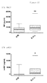
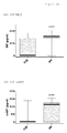
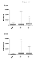
上記の目的は本発明によって解決された。特に本発明者らは、(MIF関連)疾患の発生後は、例えば体液試料中に、または細胞もしくは細胞表面上に、oxMIF(すなわち酸化型MIF)を検出できること、およびoxMIFは疾患状態および/または疾患進行と相関することを示すことができた。ここに提供される知識/技法によれば、oxMIFは健常ドナーからの体液試料、例えば血液、血清および尿などには存在せず、健常ドナーからの細胞試料にも存在しない。oxMIFは疾患状態下で増加する。この増加は、全MIFの増加より著しい(より特異的である)(実施例も参照されたい)。 The above objects have been solved by the present invention. In particular, we can detect oxMIF (ie oxidized MIF) after the occurrence of a (MIF-related) disease, for example in a body fluid sample or on a cell or cell surface, and oxMIF is a disease state and / or It could be shown to correlate with disease progression. According to the knowledge / technique provided herein, oxMIF is not present in body fluid samples from healthy donors such as blood, serum and urine, and is not present in cell samples from healthy donors. oxMIF increases under disease conditions. This increase is more significant (more specific) than the increase in total MIF (see also the examples).
この文脈において「存在しない」とは、oxMIFが、後述の抗体RAB0で行った場合に、実施例3.4で「材料および方法」の見出しの下に示すELISA技法で検出することが可能な量では、体液中に存在しないことを意味するものとする。 “Non-existing” in this context is the amount that oxMIF can detect with the ELISA technique shown in Example 3.4 under the heading “Materials and Methods” when performed with the antibody RAB0 described below. Then, it shall mean that it does not exist in a bodily fluid.
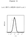
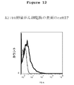
例えば血球などの細胞試料に関連して「存在しない」とは、細胞試料において細胞への抗体RAB9またはRAB0またはRAB4の適用が、実施例3.9に述べるフローサイトメトリー実験で、対照抗体「対照1(Control 1)」による染色と比較して、それより高いシグナルを与えないことを意味する。 For example, “absent” in the context of a cell sample such as blood cells means that application of antibody RAB9 or RAB0 or RAB4 to the cell in the cell sample is a control antibody “control” in the flow cytometry experiment described in Example 3.9. Compared to staining with “1” (Control 1), it means that no higher signal is given.
それゆえにoxMIFはこれらの疾患に関するマーカーとして適切であり、ここで、本発明に関して、「(MIF関連)疾患の診断におけるマーカー」という術語は、特に、MIFがこの(MIF関連)疾患に関与する因子であるかどうかの評価の可能性を意味するものとする。この点において、マーカーとしてのoxMIFは、疾患状態、その進行に関する情報を供給し、所与の処置の有効性を決定するためのマーカーとして役立つ;加えて、試料、例えば体液試料または細胞試料におけるoxMIF検出は、好ましい抗MIF治療に関する指標として役立ちうる。こうして、oxMIFの検出は、所与の疾患または障害における既知の診断技法を改良するのに役立つ。これは、医師が、所与の疾患または障害の処置方法を判定するのを支援し、診断の特異度を改良するのに役立つ。このように、oxMIFは、特異的で適切な二次マーカーである。したがってその検出は、MIF関連疾患に冒された患者の管理において、補助試験として役立ちうる。該当する疾患は、好ましい実施形態では、MIF関連であることが知られているか、MIF関連であると疑われている疾患(以下に詳述する疾患を参照されたい)であるが、今までにMIF関連であると疑われたことがない疾患であることもできる。 OxMIF is therefore suitable as a marker for these diseases, where in the context of the present invention the term “marker in the diagnosis of (MIF-related) diseases” is in particular the factor that MIF is involved in this (MIF-related) diseases. It means the possibility of evaluation of whether or not. In this respect, oxMIF as a marker serves as a marker for providing information about the disease state, its progression, and determining the effectiveness of a given treatment; in addition, oxMIF in a sample, eg, a body fluid sample or a cell sample Detection can serve as an indicator for a preferred anti-MIF treatment. Thus, detection of oxMIF serves to improve known diagnostic techniques in a given disease or disorder. This assists the physician in determining how to treat a given disease or disorder and helps improve the specificity of the diagnosis. Thus, oxMIF is a specific and appropriate secondary marker. Therefore, its detection can serve as an adjunct test in the management of patients affected by MIF-related diseases. The disease of interest, in a preferred embodiment, is a disease known or suspected to be MIF related (see diseases detailed below), but to date It can also be a disease that has never been suspected of being MIF related.
好ましい実施形態において、試料中のoxMIF存在の検出は、医師に、その試料の採取源となった対象が、MIFに対抗する治療法の恩恵を受けるかもしれないことを示すであろう。そのような治療法は、抗MIF分子、例えば抗(ox)MIF抗体、または(ox)MIFに対抗する小分子から選択されうる。 In a preferred embodiment, detection of the presence of oxMIF in a sample will indicate to the physician that the subject from which the sample was collected may benefit from a therapy that combats MIF. Such therapies can be selected from anti-MIF molecules, such as anti- (ox) MIF antibodies, or small molecules that oppose (ox) MIF.
さまざまな疾患の発生後、とりわけ、がんの発生後は、MIFレベルの上昇、すなわちMIF全般のレベルの上昇が検出される。しかし、MIFは健常対象でも循環しているので、明確な区別は困難である。これに対して、oxMIFは健常対象には存在せず、それゆえに、MIF関連疾患に関するはるかに強力な診断マーカーである。実施例において示すように、oxMIFは疾患状態では増加し、例えば血液、血清および尿のような患者の試料に検出することができる。 After the development of various diseases, in particular after the development of cancer, an increase in MIF levels, i.e. an increase in overall levels of MIF, is detected. However, since MIF circulates even in healthy subjects, clear distinction is difficult. In contrast, oxMIF is not present in healthy subjects and is therefore a much more powerful diagnostic marker for MIF-related diseases. As shown in the Examples, oxMIF increases in disease states and can be detected in patient samples such as blood, serum and urine.
ここに提示する発明は、とりわけ、バクスター(Baxter)抗体RAB9、RAB4およびRAB0がoxMIFに特異的に結合する(かつredMIFに結合する能力はない)という発見に基づいている。 The invention presented here is based, inter alia, on the discovery that Baxter antibodies RAB9, RAB4 and RAB0 specifically bind to oxMIF (and not capable of binding to redMIF).
本発明者らが行った初期の実験では、システイン媒介酸化、GSSG(酸化型グルタチオン)媒介酸化のような酸化的手順、またはMIFとプロクリン(Proclin)300もしくはタンパク質架橋剤(例えばBMOE)とのインキュベーションが、上述の抗体への結合を引き起こすことを示すことができた。 Early experiments conducted by the inventors included oxidative procedures such as cysteine mediated oxidation, GSSG (oxidized glutathione) mediated oxidation, or incubation of MIF with Proclin 300 or a protein crosslinker (eg, BMOE). Could be shown to cause binding to the antibodies described above.
本発明者らが到達した驚くべき結論は、次のとおりである:
・組換えMIF(ヒト、マウス、ラット、CHO、サル)の酸化還元調節(Cys/Gluを介した温和な酸化)またはプロクリン300またはタンパク質架橋剤による組換えMIFの処理は、バクスターの抗MIF抗体RAB9、RAB4およびRAB0の結合につながる;
・oxMIFの還元はAb結合の喪失につながる;
・oxMIF−アイソフォームに対する特異性は、生物学的Ab効力と相関する(インビトロ/インビボ);
・oxMIFレベルは疾患状態と相関させることができる。
The surprising conclusions reached by the inventors are as follows:
-Redox regulation of recombinant MIF (human, mouse, rat, CHO, monkey) (mild oxidation via Cys / Glu) or treatment of recombinant MIF with procrine 300 or protein cross-linking agent is Baxter's anti-MIF antibody Leading to binding of RAB9, RAB4 and RAB0;
• Reduction of oxMIF leads to loss of Ab binding;
Specificity for oxMIF-isoform correlates with biological Ab potency (in vitro / in vivo);
OxMIF levels can be correlated with disease states.
したがって本発明は、好ましくは、次のように定義される。
1.抗体RAB9、RAB0および/またはRAB4に弁別的に結合するMIFであるoxMIFの、(MIF関連)疾患のインビトロ診断におけるマーカーとしての使用。
Therefore, the present invention is preferably defined as follows.
1. Use of oxMIF, a MIF that differentially binds to antibodies RAB9, RAB0 and / or RAB4, as a marker in in vitro diagnosis of (MIF-related) diseases.
2.該(MIF関連疾患)の診断が、項目1において定義したoxMIFである診断マーカーに弁別的に結合する化合物の使用をさらに伴う、項目1に記載の使用。
2. The use according to
3.化合物がoxMIFに弁別的に結合する抗体である、項目2に記載の使用。
3. The use according to
4.抗体がoxMIFに結合するが、redMIFには結合しない、項目3に記載の使用。
4). 4. Use according to
5.弁別的結合が、100nM未満、好ましくは50nM未満、さらに好ましくは10nM未満のKD値で起こるoxMIFへの結合、および400nMを上回るKDを特徴とするredMIFへの非結合である、項目4に記載の使用。
5. Distinctive bond is less than 100 nM, preferably non-binding to redMIF, wherein K D greater than binding, and a 400nM to 50nM less, more oxMIF preferably occurring in the K D value of less than 10 nM,
6.MIF関連疾患が、炎症性疾患および新生物疾患(良性、前悪性および/または悪性)を含む群から選択される、項目1〜5のいずれか1つ以上に記載の使用。
6). 6. Use according to any one or more of
7.MIF関連疾患が、大腸がん、前立腺がん、膀胱がん、膵がん、卵巣がん、黒色腫、リンパ腫、肝細胞癌、喘息、ARDS、関節リウマチ、敗血症、IgAネフロパシー、糸球体腎炎、ループス腎炎(LN)、肝炎、膵炎(+/−急性肺傷害)、クローン病、潰瘍性大腸炎、胃潰瘍、アルツハイマー病、多発性硬化症、ギラン・バレー症候群、心機能障害、血管形成術、アテローム性動脈硬化、心筋炎、1型糖尿病、糖尿病性網膜症、加齢性黄斑変性症(AMD)、アトピー性皮膚炎、乾癬、子宮内膜症、神経障害性痛および/またはぶどう膜炎からなる群より選択される、項目6に記載の使用。
7). MIF-related diseases are colon cancer, prostate cancer, bladder cancer, pancreatic cancer, ovarian cancer, melanoma, lymphoma, hepatocellular carcinoma, asthma, ARDS, rheumatoid arthritis, sepsis, IgA nephropathy, glomerulonephritis, Lupus nephritis (LN), hepatitis, pancreatitis (+/- acute lung injury), Crohn's disease, ulcerative colitis, gastric ulcer, Alzheimer's disease, multiple sclerosis, Guillain-Barre syndrome, cardiac dysfunction, angioplasty, atheroma Atherosclerosis, myocarditis,
8.抗体がoxMIF結合剤(例えば抗体RAB9、RAB4および/またはRAB0など)からなる群より選択される、項目2〜7のいずれか1つ以上に記載の使用。 8). 8. Use according to any one or more of items 2-7, wherein the antibody is selected from the group consisting of oxMIF binding agents (such as antibodies RAB9, RAB4 and / or RAB0).
9.診断が、(MIF関連疾患)の存在の診断、(MIF関連疾患)の進行の診断、疾患の状態の診断、および/または処置の有効性のモニタリングである、項目1〜8のいずれか1つに記載の使用。 9. Any one of items 1-8, wherein the diagnosis is a diagnosis of the presence of (MIF-related disease), a diagnosis of progression of (MIF-related disease), a diagnosis of the state of the disease, and / or monitoring of the effectiveness of the treatment Use as described in.
10.診断が対象の体液試料に対して行われる、項目1〜9のいずれか1つに記載の使用。
10. 10. Use according to any one of
11.診断が対象の細胞試料に対して行われる、項目1〜9のいずれか1つに記載の使用。
11. 10. Use according to any one of
12.対象の体液試料または細胞試料における項目1において定義したoxMIFの検出による(MIF関連)疾患のインビトロ診断のための診断アッセイであって、該試料中のoxMIFへの化合物の結合をインビトロで決定するステップを含む、診断アッセイ。
12 A diagnostic assay for in vitro diagnosis of a disease (MIF related) by detection of oxMIF as defined in
13.oxMIFに結合する化合物および(MIF関連)疾患が、項目2〜9のいずれか1つ以上において定義したとおりである、項目12に記載の診断アッセイ。
13. 13. The diagnostic assay of
14.アッセイが、(MIF関連)疾患の進行、寛解および/または処置中に1回または数回繰り返される、項目12または13に記載の診断アッセイ。
14 14. A diagnostic assay according to
15.診断キットがoxMIFに結合する化合物を含む、項目12〜14のいずれか1つ以上に記載のアッセイにおける診断キットの使用。 15. 15. Use of a diagnostic kit in an assay according to any one or more of items 12-14, wherein the diagnostic kit comprises a compound that binds to oxMIF.
16.キットが、緩衝液、対照(例えば組換え(ox)MIF)、ポリクローナル抗MIF抗体、および/またはコンジュゲート検出抗体をさらに含む、項目15に記載の使用。
16. The use according to
17.以下の群から選択される抗MIF抗体:
a)プラスミド寄託として寄託された受託番号DSM25110の軽鎖配列とプラスミド寄託として寄託された受託番号DSM25112の重鎖配列とを特徴とする、RAB4抗体、
b)プラスミド寄託として寄託された受託番号DSM25111の軽鎖配列とプラスミド寄託として寄託された受託番号DSM25113の重鎖配列とを特徴とする、RAB9抗体、
c)プラスミド寄託として寄託された受託番号DSM25114の軽鎖配列とプラスミド寄託として寄託された受託番号DSM25115の重鎖配列とを特徴とする、RAB0抗体
d)プラスミド寄託として寄託された受託番号DSM25861の軽鎖配列とプラスミド寄託として寄託された受託番号DSM25862の重鎖配列とを特徴とする、RAM4抗体、
e)プラスミド寄託として寄託された受託番号DSM25859の軽鎖配列とプラスミド寄託として寄託された受託番号DSM25860の重鎖配列とを特徴とする、RAM9抗体、および/または
f)プラスミド寄託として寄託された受託番号DSM25863の軽鎖配列とプラスミド寄託として寄託された受託番号DSM25864の重鎖配列とを特徴とする、RAM0抗体。
17. An anti-MIF antibody selected from the following group:
a) a RAB4 antibody characterized by the light chain sequence of deposit number DSM25110 deposited as a plasmid deposit and the heavy chain sequence of deposit number DSM25112 deposited as a plasmid deposit;
b) an RAB9 antibody characterized by the light chain sequence of deposit number DSM25111 deposited as a plasmid deposit and the heavy chain sequence of deposit number DSM25113 deposited as a plasmid deposit;
c) RAB0 antibody, characterized by the light chain sequence of accession number DSM25114 deposited as a plasmid deposit and the heavy chain sequence of accession number DSM25115 deposited as a plasmid deposit, d) light of accession number DSM25861 deposited as a plasmid deposit A RAM4 antibody, characterized by the chain sequence and the heavy chain sequence of deposit number DSM25862 deposited as a plasmid deposit;
e) RAM9 antibody, characterized by the light chain sequence of deposit number DSM25859 deposited as plasmid deposit and the heavy chain sequence of deposit number DSM25860 deposited as plasmid deposit, and / or f) deposit deposited as plasmid deposit RAM0 antibody, characterized by the light chain sequence number DSM25863 and the heavy chain sequence number deposit number DSM25864 deposited as a plasmid deposit.
18.以下の群から選択される抗MIF抗体:
a)配列番号2の軽鎖アミノ酸配列と配列番号6の重鎖アミノ酸配列とを特徴とする、RAB4抗体、
b)配列番号1の軽鎖アミノ酸配列と配列番号5の重鎖アミノ酸配列とを特徴とする、RAB9抗体、
c)配列番号3の軽鎖アミノ酸配列と配列番号7の重鎖アミノ酸配列とを特徴とする、RAB0抗体、
d)配列番号4の軽鎖アミノ酸配列と配列番号8の重鎖アミノ酸配列とを特徴とする、RAB2抗体、
e)配列番号14の軽鎖アミノ酸配列と配列番号13の重鎖アミノ酸配列とを特徴とする、RAM4抗体、
f)配列番号12の軽鎖アミノ酸配列と配列番号11の重鎖アミノ酸配列とを特徴とする、RAM9抗体、および/もしくは
g)配列番号10の軽鎖アミノ酸配列と配列番号9の重鎖アミノ酸配列とを特徴とする、RAM0抗体、
h)または、上記抗体a)〜g)のいずれか1つと同じエピトープへの結合を特徴とする、それらの機能的等価物。
18. An anti-MIF antibody selected from the following group:
a) a RAB4 antibody characterized by the light chain amino acid sequence of SEQ ID NO: 2 and the heavy chain amino acid sequence of SEQ ID NO: 6,
b) an RAB9 antibody characterized by the light chain amino acid sequence of SEQ ID NO: 1 and the heavy chain amino acid sequence of SEQ ID NO: 5;
c) a RAB0 antibody characterized by the light chain amino acid sequence of SEQ ID NO: 3 and the heavy chain amino acid sequence of SEQ ID NO: 7,
d) a RAB2 antibody characterized by the light chain amino acid sequence of SEQ ID NO: 4 and the heavy chain amino acid sequence of SEQ ID NO: 8,
e) a RAM4 antibody characterized by the light chain amino acid sequence of SEQ ID NO: 14 and the heavy chain amino acid sequence of SEQ ID NO: 13,
f) a RAM9 antibody characterized by the light chain amino acid sequence of SEQ ID NO: 12 and the heavy chain amino acid sequence of SEQ ID NO: 11, and / or g) the light chain amino acid sequence of SEQ ID NO: 10 and the heavy chain amino acid sequence of SEQ ID NO: 9 RAM0 antibody, characterized by
h) or a functional equivalent thereof characterized by binding to the same epitope as any one of antibodies a) to g) above.
19.(MIF関連)疾患の診断における、上記抗体(特に項目17または18において定義したもの)のいずれか1つの使用。 19. (MIF-related) Use of any one of the above antibodies (particularly those defined in items 17 or 18) in the diagnosis of a disease.
上述の項目ならびに本願に添付のクレームは全て、以下の好ましい抗体に、等しく関係している:
RAM9
RAM4
RAM0。
All of the above items, as well as the claims appended hereto, relate equally to the following preferred antibodies:
RAM9
RAM4
RAM0.
これらの抗体は、上記の項目のリストに挙げた抗体と同じ特異性を有し(下記も参照されたい)、これらの抗体を使って類似する結果を達成することができる。 These antibodies have the same specificity as the antibodies listed in the list above (see also below) and these antibodies can be used to achieve similar results.
特に本発明では、例えば診断マーカーとして特に好適で有利な、好ましい本発明の抗体が提供される。 In particular, the present invention provides preferred antibodies of the present invention that are particularly suitable and advantageous as, for example, diagnostic markers.
これら上述の抗体は、その配列によって特徴づけられ、裏付けられると共に、上述の抗体RAB0、RAB4およびRAB9ならびにRAM0、RAM4およびRAM9のそれぞれの軽鎖または重鎖のいずれかを、それぞれ含む、大腸菌(E. coli)(TG1株)中のプラスミドとしての寄託物によっても特徴づけられ、裏付けられる。 These above-mentioned antibodies are characterized and supported by their sequences and contain either the above-described antibodies RAB0, RAB4 and RAB9 and each light chain or heavy chain of RAM0, RAM4 and RAM9, respectively, E. coli ( E E. coli ) (TG1 strain) is also characterized and supported by the deposit as a plasmid.
プラスミドは、ドイツ微生物細胞培養コレクション(DSMZ)(ドイツ国ブラウンシュワイク、マッシャーオーダーヴェーク1b)へのブダペスト条約に基づく寄託時に与えられる公式番号である、そのDSM番号によって特徴づけられる。プラスミドはそれぞれ大腸菌株に入れて寄託された。 The plasmid is characterized by its DSM number, which is the official number given at the time of deposit under the Budapest Treaty to the German Microbial Cell Culture Collection (DSMZ) (Brownschweig, Germany, Mascher Orderweg 1b). Each plasmid was deposited in an E. coli strain.
番号DSM25110のプラスミドは抗MIF抗体RAB4の軽鎖配列を含む。 The plasmid numbered DSM25110 contains the light chain sequence of anti-MIF antibody RAB4.
番号DSM25112のプラスミドは抗MIF抗体RAB4の重鎖(IgG4)配列を含む。 The plasmid with the number DSM25112 contains the heavy chain (IgG4) sequence of the anti-MIF antibody RAB4.
適切な宿主細胞におけるプラスミドDSM25110とDSM25112との同時発現は、好ましい抗MIF抗体RAB4の生産をもたらす。 Co-expression of plasmids DSM25110 and DSM25112 in a suitable host cell results in production of the preferred anti-MIF antibody RAB4.
番号DSM25111のプラスミドは抗MIF抗体RAB9の軽鎖配列を含む。 The plasmid with the number DSM25111 contains the light chain sequence of the anti-MIF antibody RAB9.
番号DSM25113のプラスミドは抗MIF抗体RAB9の重鎖(IgG4)配列を含む。 The plasmid numbered DSM25113 contains the heavy chain (IgG4) sequence of anti-MIF antibody RAB9.
適切な宿主細胞におけるプラスミドDSM25111とDSM25113との同時発現は、好ましい抗MIF抗体RAB9の生産をもたらす。 Co-expression of plasmids DSM25111 and DSM25113 in a suitable host cell results in production of the preferred anti-MIF antibody RAB9.
番号DSM25114のプラスミドは抗MIF抗体RAB0の軽鎖配列を含む。 The plasmid numbered DSM25114 contains the light chain sequence of anti-MIF antibody RAB0.
番号DSM25115のプラスミドは抗MIF抗体RAB0の重鎖(IgG4)配列を含む。 The plasmid numbered DSM25115 contains the heavy chain (IgG4) sequence of anti-MIF antibody RAB0.
適切な宿主細胞におけるプラスミドDSM25114とDSM25115との同時発現は、好ましい抗MIF抗体RAB0の生産をもたらす。 Co-expression of plasmids DSM25114 and DSM25115 in a suitable host cell results in production of the preferred anti-MIF antibody RAB0.
抗体RAM0、RAM9およびRAM4も寄託されている;いずれも、ブダペスト条約に従い、2012年4月12日に、以下の名称で、DSZM、ブラウンシュワイク、ドイツ国に寄託された:
RAM9−重鎖:大腸菌GA.662−01.pRAM9hc−DMS25860.
RAM4−軽鎖:大腸菌GA.906−04.pRAM4lc−DMS25861.
RAM9−軽鎖:大腸菌GA.661−01.pRAM9lc−DMS25859.
RAM4−重鎖:大腸菌GA.657−02.pRAM4hc−DMS25862.
RAM0−軽鎖:大腸菌GA.906−01.pRAM0lc−DMS25863.
RAM0−重鎖:大腸菌GA.784−01.pRAM0hc−DMS25864.
The antibodies RAM0, RAM9 and RAM4 have also been deposited; all were deposited in DSZM, Braunschweig, Germany under the following names on April 12, 2012 in accordance with the Budapest Treaty:
RAM9-heavy chain: E. coli GA. 662-01. pRAM9hc-DMS25860.
RAM4-light chain: E. coli GA. 906-04. pRAM4lc-DMS25861.
RAM9-light chain: E. coli GA. 661-01. pRAM9lc-DMS25859.
RAM4-heavy chain: E. coli GA. 657-02. pRAM4hc-DMS25862.
RAM0-light chain: E. coli GA. 906-01. pRAM0lc-DMS25863.
RAM0-heavy chain: E. coli GA. 784-01. pRAM0hc-DMS25864.
したがって本発明は、診断方法において使用するための、抗oxMIF抗体またはその抗原結合性フラグメントを含む診断アッセイであって、これらの抗体またはその抗原結合性フラグメントが弁別的結合を有する、すなわちoxMIFには結合するが、redMIFには結合しない、診断アッセイも包含する。現在の知識/技法によれば、健常ドナーからの試料にoxMIFを検出することはできない。一実施形態では、ヒト対象からの生物学的試料中のヒトoxMIFを検出するために、上記抗oxMIF抗体またはその抗原結合性部分を使用することができる。 Accordingly, the present invention is a diagnostic assay comprising anti-oxMIF antibodies or antigen-binding fragments thereof for use in diagnostic methods, wherein these antibodies or antigen-binding fragments thereof have a discriminative binding, ie oxMIF Also included are diagnostic assays that bind but do not bind to redMIF. According to current knowledge / techniques, oxMIF cannot be detected in samples from healthy donors. In one embodiment, the anti-oxMIF antibody or antigen-binding portion thereof can be used to detect human oxMIF in a biological sample from a human subject.
本願に関して、生物学的試料は、好ましくは、診断が下される対象の体液試料である。体液試料は、当業者に知られている体液の任意の試料である。例えば限定するわけではないが、そのような試料として、血液、血漿、血清、唾液、尿、鼻汁(nasal fluid)、腹水、眼液(ocular fluid)、羊水、房水、硝子体液、涙液、カウパー液、精液、間質液、リンパ液、母乳、粘液(鼻水および痰を含む)、胸水、膿、月経分泌物、膣潤滑液、皮脂、脳脊髄液および滑液を挙げることができる。本願に関し、さらなる生物学的試料として、(中空)体器官の洗浄液(洗い出し液)(例えば気管支肺胞洗浄液、胃洗浄液および腸洗浄液)を挙げることもできる。 In the context of the present application, the biological sample is preferably a body fluid sample of the subject to be diagnosed. A bodily fluid sample is any sample of bodily fluid known to those skilled in the art. For example, but not limited to, such samples include blood, plasma, serum, saliva, urine, nasal fluid, ascites, ocular fluid, amniotic fluid, aqueous humor, vitreous humor, tear fluid, Mention may be made of Cowper's fluid, semen, interstitial fluid, lymph fluid, breast milk, mucus (including runny nose and sputum), pleural effusion, pus, menstrual secretions, vaginal lubricant, sebum, cerebrospinal fluid and synovial fluid. In the context of the present application, further biological samples may also include (hollow) body organ lavage fluids (e.g., bronchoalveolar lavage fluid, gastric lavage fluid and intestinal lavage fluid).
代替的一態様では、本願に関して、生物学的試料は、診断が下される対象の細胞試料、最も好ましくは循環または患部組織からの細胞試料、より好ましくは単細胞懸濁液試料としての細胞試料である。 In an alternative aspect, for the present application, the biological sample is a cell sample to be diagnosed, most preferably a cell sample from circulating or diseased tissue, more preferably a cell sample as a single cell suspension sample. is there.
特に、上記診断アッセイは、所与の疾患に(ox)MIFが関与しているかどうかを決定するために使用することができる。 In particular, the diagnostic assay can be used to determine whether (ox) MIF is involved in a given disease.
したがって本発明は、疾患の進行を評価するための方法にも関係する;この文脈において、「疾患の状態」という用語は、「疾患の重症度」という用語と同義であると理解すべきであり、疾患または状態の重篤度、程度または状態(すなわちステージ)を指す。例えば疾患は、軽度、中等度または重度と特徴づけることができる。重症度または程度、すなわち疾患の状態の決定または評価は、当業者にはよく知られている。この評価のために行われる実際の方法は、もちろん、問題の疾患または状態に依存する。例えば、ある疾患を有する対象の生存の可能性または長さを、同じ疾患を有する他の対象における生存の可能性または長さと比較することによって、疾患の状態を決定することができる。 Thus, the present invention also relates to a method for assessing disease progression; in this context, the term “disease state” should be understood to be synonymous with the term “disease severity” Refers to the severity, degree or condition (ie stage) of the disease or condition. For example, the disease can be characterized as mild, moderate or severe. The determination or evaluation of severity or extent, ie disease state, is well known to those skilled in the art. The actual method performed for this evaluation will, of course, depend on the disease or condition in question. For example, a disease state can be determined by comparing the likelihood or length of survival of a subject having a disease to the likelihood or length of survival in another subject having the same disease.
別の実施形態では、ある疾患を有する対象における疾患の症状を、同じ疾患を有する他の対象における症状と比較することによって、疾患の状態を決定することができる。さらにもう一つの実施形態において、疾患の状態およびその進行は、ある期間にわたる同一患者内での症状の変化に反映される。 In another embodiment, the state of the disease can be determined by comparing the symptoms of the disease in a subject having a disease with the symptoms in other subjects having the same disease. In yet another embodiment, the disease state and its progression are reflected in changes in symptoms within the same patient over a period of time.
さらにもう一つの好ましい態様において、本発明は、抗(ox)MIF化合物による処置に適格な対象を選択する方法であって、対象は(MIF関連)疾患を有するか、(MIF関連)疾患を進展させるリスクがあり、該対象におけるoxMIFの存在および/またはレベルおよび/またはレベルの変化を検出することを含む方法に向けることもできる。oxMIFのレベルが上昇している対象を、上に定義した抗(ox)MIF化合物による予防的または治療的処置のために選択することができる。 In yet another preferred embodiment, the invention is a method of selecting a subject eligible for treatment with an anti- (ox) MIF compound, wherein the subject has (MIF-related) disease or develops (MIF-related) disease. Can be directed to methods that include detecting the presence and / or level and / or level change of oxMIF in the subject. Subjects with elevated levels of oxMIF can be selected for prophylactic or therapeutic treatment with anti- (ox) MIF compounds as defined above.
「予防的」または「治療的」処置という用語は、当技術分野において認識されている用語であり、患者への薬物の投与を指す。望ましくない状態(例えば、ヒトまたは動物などの宿主の疾患または他の望ましくない状態)の臨床症状発現の前に投与される場合、その処置は予防的処置であり、すなわちそれは望ましくない状態の進展から宿主を防御し、他方、投与が望ましくない状態の症状発現後に投与される場合、その処置は治療的処置である(すなわち、既存の望ましくない状態もしくはその副作用を減じ、改善し、または既存の望ましくない状態もしくはその副作用を維持する)。 The term “prophylactic” or “therapeutic” treatment is an art-recognized term and refers to the administration of a drug to a patient. When administered prior to the onset of clinical symptoms of an undesirable condition (eg, a host disease such as a human or animal or other undesirable condition), the treatment is a prophylactic treatment, ie it is from the development of an undesirable condition When the host is protected, while administration is administered after manifestation of an undesirable condition, the treatment is a therapeutic treatment (i.e., reduces, improves or eliminates an existing undesirable condition or its side effects) No state or its side effects).
本明細書にいう抗(ox)MIF化合物とは、(ox)MIFの生物学的活性を減弱し、阻害し、対抗し、打ち消し、または減少させる任意の薬剤を指す。抗(ox)MIF化合物は、(ox)MIF活性を阻害または中和する薬剤、例えば抗体、特に好ましくは本明細書に記載する抗体、さらに好ましくは抗体RAB9、RAB4および/またはRAB0であることができる。 As used herein, an anti- (ox) MIF compound refers to any agent that attenuates, inhibits, counters, counteracts, or reduces the biological activity of (ox) MIF. The anti- (ox) MIF compound may be an agent that inhibits or neutralizes (ox) MIF activity, such as an antibody, particularly preferably an antibody described herein, more preferably the antibodies RAB9, RAB4 and / or RAB0. it can.
診断アッセイは、例えば患者の体液試料または細胞試料などにおけるoxMIFの存在またはレベルを決定するために使用することができる。oxMIFの存在または非存在は、疾患がMIF関連であるかどうかの識別またはoxMIF処置が合理的であるかどうかの判定に適している。oxMIFレベルは疾患進行または処置効力を示す。 The diagnostic assay can be used to determine the presence or level of oxMIF, such as in a patient fluid sample or cell sample. The presence or absence of oxMIF is suitable for identifying whether the disease is MIF-related or determining whether oxMIF treatment is reasonable. oxMIF levels indicate disease progression or treatment efficacy.
本発明はさらに、本発明の抗oxMIF抗体またはその抗原結合性部分を含むキットに関する。キットは、抗体に加えて、さらなる診断剤または治療剤、およびその使用を含みうる。キットは、診断方法または治療方法における使用に関する説明書も含むことができる。 The present invention further relates to a kit comprising the anti-oxMIF antibody of the present invention or an antigen-binding portion thereof. In addition to the antibody, the kit can include additional diagnostic or therapeutic agents, and uses thereof. The kit can also include instructions for use in a diagnostic or therapeutic method.
添付の図面に本発明をさらに詳しく説明する。 The invention is described in more detail in the accompanying drawings.
定義および一般技法
本明細書に別段の定義がある場合を除き、本発明に関連して使用される科学用語および技術用語は、当業者に一般に理解されている意味を有するものとする。一般に、本明細書に記載する細胞および組織培養、分子生物学、免疫学、微生物学、遺伝学ならびにタンパク質および核酸化学に関連して使用される術語は、当技術分野において周知のよく使用されるものである。本発明の方法および技法は、別段の表示がある場合を除き、一般に、当技術分野において周知の従来法に従って、本明細書の全体を通して引用し議論するさまざまな一般文献と、より専門的な文献とに記述されているように行われる。例えばSambrookら「Molecular Cloning: A Laboratory Manual」第2版、Cold Spring Harbor Laboratory Press、ニューヨーク州コールドスプリングハーバー(1989)、Ausubelら「Current Protocols in Molecular Biology」Greene Publishing Associates (1992)、ならびにHarlowおよびLane「Antibodies: A Laboratory Manual」Cold Spring Harbor Laboratory Press、ニューヨーク州コールドスプリングハーバー(1990)を参照されたい(これらは引用により本明細書に組み込まれる)。
Definitions and General Techniques Unless otherwise defined herein, scientific and technical terms used in connection with the present invention shall have the meanings that are commonly understood by those of ordinary skill in the art. In general, the terms used in connection with cell and tissue culture, molecular biology, immunology, microbiology, genetics and protein and nucleic acid chemistry described herein are commonly used in the art. Is. The methods and techniques of the present invention, unless otherwise indicated, generally vary according to conventional methods well known in the art, as well as various general and more specialized documents cited and discussed throughout this specification. Done as described in. For example, Sambrook et al. “Molecular Cloning: A Laboratory Manual” 2nd edition, Cold Spring Harbor Laboratory Press, Cold Spring Harbor, New York (1989), Ausubel et al. “Current Protocols in MolesheGolh See "Antibodies: A Laboratory Manual" Cold Spring Harbor Laboratory Press, Cold Spring Harbor, NY (1990), which are incorporated herein by reference.
「MIF」または「マクロファージ遊走阻止因子」とは、免疫応答および炎症応答における決定的メディエーターとして知られ、またグルココルチコイドの対抗制御因子として知られているタンパク質を指す。MIFには、哺乳動物MIF、具体的にはヒトMIF(Swiss−Protプライマリアクセッション番号:P14174)が含まれ、その単量体型は、115アミノ酸タンパク質としてコードされているが、開始メチオニンが切断されるため、114アミノ酸タンパク質として生産される。「MIF」には、「GIF」(グリコシル化阻害因子)や他の形態のMIF、例えばMIFの融合タンパク質も含まれる。MIFのアミノ酸のナンバリングはN末端メチオニン(アミノ酸1)で始まり、C末端アラニン(アミノ酸115)で終わる。 “MIF” or “macrophage migration inhibitory factor” refers to a protein known as a critical mediator in immune and inflammatory responses and also known as a counter-regulator of glucocorticoids. MIF includes mammalian MIF, specifically human MIF (Swiss-Prot primary accession number: P14174), the monomeric form of which is encoded as a 115 amino acid protein, but the starting methionine is cleaved. Therefore, it is produced as a 114 amino acid protein. “MIF” also includes “GIF” (glycosylation inhibitor) and other forms of MIF, such as fusion proteins of MIF. The amino acid numbering of MIF begins with an N-terminal methionine (amino acid 1) and ends with a C-terminal alanine (amino acid 115).
「酸化型MIF」またはoxMIFは、本発明では、温和な酸化剤、例えばシスチンによるMIFの処理によって生じるMIFのアイソフォームと定義される。本発明によって示されるとおり、この方法で処理された組換えoxMIFは、(例えば)動物に細菌を負荷した後にインビボで生じるoxMIFと共通する構造再構成を起こしたMIFのアイソフォームを含む。 “Oxidized MIF” or oxMIF is defined in the present invention as an isoform of MIF resulting from the treatment of MIF with a mild oxidant such as cystine. As demonstrated by the present invention, recombinant oxMIF treated in this way includes isoforms of MIF that have undergone structural reconstitution in common with oxMIF that occurs in vivo after loading the animal with bacteria (for example).
redMIFは、本発明では、還元型MIFと定義され、RAB0、RAB9および/またはRAB4に結合しないMIFである。 In the present invention, redMIF is defined as reduced MIF and is MIF that does not bind to RAB0, RAB9 and / or RAB4.
本発明で述べる抗oxMIF抗体は、温和な酸化または還元によってそれぞれ生じるoxMIFとredMIFとを区別することができ、oxMIFを特異的に検出するのに役立つ。これらのコンフォーマー間の区別は、ELISA(例えば実施例3.4で説明するもの)または表面プラズモン共鳴によって評価される。 The anti-oxMIF antibody described in the present invention can distinguish between oxMIF and redMIF produced by mild oxidation or reduction, respectively, and is useful for specifically detecting oxMIF. The distinction between these conformers is assessed by ELISA (eg as described in Example 3.4) or surface plasmon resonance.
Biacoreによる抗体の弁別的結合の評価
抗体RAB9およびRAB0へのoxMIFおよびredMIFの結合速度論を、Biacore 3000システムを使った表面プラズモン共鳴分析によって調べる。抗体を、CM5(=カルボキシメチル化デキストラン)チップ上にコーティングし、0.2%プロクリン300と共にプレインキュベートした組換えMIFタンパク質を注入した(プロクリン300は、oxMIFからredMIFへの転化を回避することによって、oxMIF構造を安定化する酸化性イソチアゾロンからなる)。プロクリン300を添加していないネイティブHBS−EP緩衝液(=Biacoreランニング緩衝液)では、組換えMIFタンパク質はいずれもRAB9、RAB0には結合せず、陰性(バックグラウンド)結合対照として使用した参照抗体(無関係なアイソタイプ対照抗体)にも結合しなかった。
Evaluation of differential binding of antibodies by Biacore The binding kinetics of oxMIF and redMIF to antibodies RAB9 and RAB0 are examined by surface plasmon resonance analysis using the Biacore 3000 system. The antibody was coated on a CM5 (= carboxymethylated dextran) chip and injected with recombinant MIF protein pre-incubated with 0.2% procrine 300 (procrine 300 avoids the conversion of oxMIF to redMIF). , Consisting of an oxidizing isothiazolone that stabilizes the oxMIF structure). In native HBS-EP buffer (= Biacore running buffer) without the addition of proclin 300, neither recombinant MIF protein bound to RAB9, RAB0, and the reference antibody used as a negative (background) binding control It also did not bind (irrelevant isotype control antibody).
好ましい実施形態では、oxMIFが、抗体RAB9、RAB4および/またはRAB0またはその抗原結合性フラグメントによって弁別的に結合されるMIFであり、これらの抗体がoxMIFには結合するが、redMIFはこれらの抗体のいずれとも結合しないことを意味している。 In a preferred embodiment, oxMIF is MIF that is differentially bound by antibodies RAB9, RAB4 and / or RAB0 or an antigen-binding fragment thereof, and these antibodies bind to oxMIF, but redMIF It means that they are not combined.
別の実施形態では、抗oxMIF抗体、例えば上述の抗体、またはその抗原結合性部分が、100nM未満のKD、好ましくは50nM未満のKD、より好ましくは10nM未満のKDで、oxMIFに結合する。特に好ましくは、本発明の抗体は、5nM未満のKDでoxMIFに結合する。 In another embodiment, an anti-oxMIF antibody, eg, an antibody as described above, or an antigen-binding portion thereof, binds to oxMIF with a K D of less than 100 nM, preferably a K D of less than 50 nM, more preferably a K D of less than 10 nM. To do. Particularly preferably, the antibodies of the invention bind to oxMIF with a K D of less than 5 nM.
(oxMIFまたはredMIFへの)抗体、例えばRAB9、RAB4またはRAB0の(非)結合は、例えば以下の方法のいずれかなど、当業者に広く知られているように決定することができる:組換えMIFによる弁別的結合ELISA(Differential Binding ELISA)、または組換えMIFをその還元状態または酸化状態で使用する表面プラズモン共鳴、例えば上述した周知のBiacoreアッセイ。 The (non) binding of an antibody (eg to oxMIF or redMIF), eg RAB9, RAB4 or RAB0, can be determined as is well known to those skilled in the art, for example any of the following methods: recombinant MIF Differentiated binding ELISA (Differential Binding ELISA), or surface plasmon resonance using recombinant MIF in its reduced or oxidized state, such as the well-known Biacore assay described above.
結合を決定するための好ましい方法は、例えば組換え(ox)MIFへの抗体の表面プラズモン共鳴であり、ここで「結合」は、100nM未満、好ましくは50nM未満、より好ましくは10nM未満のKDによって表されるものとし、一方、redMIFへの非結合は、400nMを上回るKDによって特徴づけられる。「結合」および「特異的結合」は、本明細書では、上記を表すために可換的に使用される。本願に関して「弁別的結合」とは、化合物、特に本明細書に記載する抗体が、oxMIFに(例えば上述のKD値で)結合すると共に、それらがredMIFには結合しない(非結合とは、ここでも、上に定義したとおりである)ことを意味する。 A preferred method for determining binding are, for example recombinant (ox) is a surface plasmon resonance of the antibody to MIF, where "bond", less than 100 nM, preferably less than 50 nM, more preferably less than 10 nM K D and those represented by, while non-binding to redMIF is characterized by K D greater than 400 nM. “Binding” and “specific binding” are used interchangeably herein to denote the above. The "discrimination binding" with respect to this application, compounds, antibodies described in particular herein, (e.g., with a K D value mentioned above) as well as bind to OxMIF, they do not bind to RedMIF (a non-binding Again, this is as defined above).
「抗体」とは、無傷の抗体または(特異的)結合に関して無傷の抗体と競合する抗原結合性部分を指す。一般論については「Fundamental Immunology」第7章(Paul, W.編、第2版、Raven Press、ニューヨーク(1989))(引用により本明細書に組み込まれる)を参照されたい。抗体という用語には、ヒト抗体、哺乳動物抗体、単離抗体および遺伝子操作された形態、例えばキメラ、ラクダ化またはヒト化抗体が含まれるが、これらに限るわけではない。 “Antibody” refers to an intact antibody or an antigen-binding portion that competes with an intact antibody for (specific) binding. For general considerations, see Fundamental Immunology, Chapter 7 (Paul, W. Ed., 2nd Edition, Raven Press, New York (1989)), which is incorporated herein by reference. The term antibody includes, but is not limited to, human antibodies, mammalian antibodies, isolated antibodies and genetically engineered forms such as chimeric, camelized or humanized antibodies.
抗体の「抗原結合性部分」という用語は、抗原(例えば(ox)MIF)に特異的に結合する能力を保持している、抗体の1つ以上のフラグメントを指す。抗原結合性部分は、組換えDNA技法によって生産するか、無傷の抗体の酵素的または化学的切断によって生産することができる。抗原結合性部分には、例えば−限定するわけではないが−次に挙げるものが含まれる:Fab、Fab’、F(ab’)2、Fv、および相補性決定領域(CDR)フラグメント、単鎖抗体(scFv)、キメラ抗体、抗体、およびポリペプチド(すなわちoxMIFまたはredMIF)への特異的抗原結合を付与するのに十分な抗体の少なくとも一部分を含有するポリペプチド。N末端からC末端に向かって、成熟軽鎖および重鎖可変ドメインはどちらも、領域FR1、CDR1、FR2、CDR2、FR3、CDR3およびFR4を含む。各ドメインへのアミノ酸の割り当ては、Kabat「Sequences of Proteins of Immunological Interest」(米国国立衛生研究所、メリーランド州ベセスダ(1987および1991))、Chothiaら, J. Mol. Biol. 196:901−917 (1987)、またはChothiaら, Nature 342:878−883 (1989)の定義に従う。抗体またはその抗原結合性部分は誘導体化するか、もう一つの機能分子(例えばもう一つのペプチドまたはタンパク質)に連結することができる。例えば抗体またはその抗原結合性部分は、1つ以上の他の分子実体、例えばもう一つの抗体(例えば二重特異性抗体またはダイアボディ)、検出可能剤、細胞毒性剤、医薬剤および/または連結分子に、機能的に連結することができる。 The term “antigen-binding portion” of an antibody refers to one or more fragments of an antibody that retain the ability to specifically bind to an antigen (eg, (ox) MIF). Antigen-binding moieties can be produced by recombinant DNA techniques or by enzymatic or chemical cleavage of intact antibodies. Antigen binding portions include, but are not limited to, the following: Fab, Fab ′, F (ab ′) 2, Fv, and complementarity determining region (CDR) fragments, single chain Polypeptides containing at least a portion of an antibody (scFv), chimeric antibody, antibody, and antibody sufficient to confer specific antigen binding to a polypeptide (ie oxMIF or redMIF). From the N-terminus to the C-terminus, both the mature light and heavy chain variable domains contain the regions FR1, CDR1, FR2, CDR2, FR3, CDR3 and FR4. The assignment of amino acids to each domain is described by Kabat, “Sequences of Proteins of Immunological Interest” (National Institutes of Health, Bethesda, MD (1987 and 1991)), Chothia et al., J. Biol. Mol. Biol. 196: 901-917 (1987), or Chothia et al., Nature 342: 878-883 (1989). The antibody or antigen-binding portion thereof can be derivatized or linked to another functional molecule (eg, another peptide or protein). For example, an antibody or antigen-binding portion thereof can include one or more other molecular entities, such as another antibody (eg, a bispecific antibody or diabody), a detectable agent, a cytotoxic agent, a pharmaceutical agent, and / or a linkage. It can be functionally linked to a molecule.
「KD」という用語は、本明細書では、当業者の一般的知識のとおり、特定抗体と各抗原との平衡解離定数を指す。この平衡解離定数は、大きな物体(ここでは複合体oxMIFまたはredMIF/抗体)が、分離する傾向、すなわち、小さな構成要素(ここではoxMIFまたはredMIFと抗体と)に解離する傾向を測定している。 The term “KD” as used herein refers to the equilibrium dissociation constant between a specific antibody and each antigen, as is generally understood by those skilled in the art. This equilibrium dissociation constant measures the tendency of large objects (here complex oxMIF or redMIF / antibody) to separate, ie dissociate into smaller components (here oxMIF or redMIF and antibody).
「ヒト抗体」という用語は、可変ドメインと定常ドメインとがヒト配列である任意の抗体を指す。この用語は、ヒト遺伝子に由来する配列を持つが、例えば考えうる免疫原性が減少し、アフィニティが増加し、または望ましくないフォールディングを引き起こすかもしれないシステインが排除されるように配列が改変されている抗体を包含する。この用語は、例えばヒト細胞に特有ではないグリコシル化を付加するかもしれない非ヒト細胞において組換え生産された前述の抗体を包含する。 The term “human antibody” refers to any antibody in which the variable and constant domains are human sequences. This term has a sequence derived from a human gene but has been modified in sequence to eliminate, for example, cysteines that may reduce possible immunogenicity, increase affinity, or cause undesirable folding. Antibodies. The term encompasses the aforementioned antibodies produced recombinantly in non-human cells that may add glycosylation that is not unique to human cells, for example.
「ヒト化抗体」という用語は、ヒト配列を含み、かつ非ヒト配列も含有する抗体を指す。 The term “humanized antibody” refers to an antibody that contains human sequences and also contains non-human sequences.
「ラクダ化抗体」という用語は、抗体の構造または配列が、ラクダ抗体とも呼ばれるラクダからの抗体に、より近くなるように改変されている抗体である。ラクダ化抗体を設計し生産するための方法は、当業者の一般的知識の一部である。 The term “camelized antibody” is an antibody in which the structure or sequence of the antibody has been modified to be closer to an antibody from a camel, also referred to as a camel antibody. Methods for designing and producing camelized antibodies are part of the general knowledge of those skilled in the art.
「キメラ抗体」という用語は、2つ以上の異なる種からの領域を含む抗体を指す。 The term “chimeric antibody” refers to an antibody that comprises regions from two or more different species.
「単離抗体」または「その単離抗原結合性部分」とは、ファージディスプレイライブラリーまたはB細胞レパートリーなどの抗体源から同定され選択された抗体またはその抗原結合性部分を指す。 “Isolated antibody” or “isolated antigen-binding portion thereof” refers to an antibody or antigen-binding portion thereof identified and selected from an antibody source, such as a phage display library or a B cell repertoire.
本発明の抗(ox)MIF抗体の生産には、遺伝子工学により、例えばRNAの逆転写および/またはDNAの増幅などによって、組換えDNAを生成させ、発現ベクターにクローニングするための任意の方法が含まれる。いくつかの実施形態において、ベクターは、追加のDNAセグメントをウイルスゲノム中にライゲートすることができるウイルスベクターである。いくつかの実施形態において、ベクターは、それが導入された宿主細胞内で自立的に複製する能力を有する(例えば細菌複製起点を有する細菌ベクター、およびエピソーム哺乳動物ベクター)。別の実施形態において、ベクター(例えば非エピソーム哺乳動物ベクター)は、宿主細胞中に導入されると宿主細胞のゲノム中に組み込まれ、それによって宿主ゲノムと共に複製されうる。さらにまた、一定のベクターは、それらが作動的に連結された遺伝子の発現を指示する能力を有する。そのようなベクターを、本明細書では、「組換え発現ベクター」(または単に「発現ベクター」)という。 Production of anti- (ox) MIF antibodies of the present invention includes any method for generating recombinant DNA and cloning it into an expression vector by genetic engineering, such as by reverse transcription of RNA and / or amplification of DNA. included. In some embodiments, the vector is a viral vector capable of ligating additional DNA segments into the viral genome. In some embodiments, the vector has the ability to replicate autonomously in the host cell into which it is introduced (eg, bacterial vectors having a bacterial origin of replication, and episomal mammalian vectors). In another embodiment, a vector (eg, a non-episomal mammalian vector) can be integrated into a host cell's genome when introduced into the host cell, and thereby replicated along with the host genome. Furthermore, certain vectors have the ability to direct the expression of genes to which they are operably linked. Such vectors are referred to herein as “recombinant expression vectors” (or simply “expression vectors”).
抗(ox)MIF抗体は、とりわけ従来の発現ベクター、例えば細菌ベクター(例えばpBR322およびその誘導体)、または真核ベクターを利用して、生産することができる。抗体をコードするこれらの配列には、複製、発現および/または宿主細胞からの分泌を制御する制御配列を与えることができる。これらの制御配列は、例えばプロモーター(例えばCMVまたはSV40)およびシグナル配列を含む。発現ベクターは、選択および増幅マーカー、例えばジヒドロ葉酸レダクターゼ遺伝子(DHFR)、ハイグロマイシン−B−ホスホトランスフェラーゼ、およびチミジン−キナーゼも含むことができる。使用するベクターの構成要素、例えば選択マーカー、レプリコン、エンハンサーは、市販されているか、または従来の方法を使って調製することができる。さまざまな細胞培養、例えば、CHO、COS、HEK293、NSO、線維芽細胞などの哺乳動物細胞、昆虫細胞、酵母または大腸菌などの細菌において発現させるためのベクターを構築することができる。いくつかの例では、発現されるタンパク質の最適なグリコシル化を可能にする細胞が使用される。 Anti- (ox) MIF antibodies can be produced using, inter alia, conventional expression vectors such as bacterial vectors (eg pBR322 and derivatives thereof), or eukaryotic vectors. These sequences encoding the antibody can be provided with control sequences that control replication, expression and / or secretion from the host cell. These control sequences include, for example, a promoter (eg, CMV or SV40) and a signal sequence. Expression vectors can also include selection and amplification markers such as dihydrofolate reductase gene (DHFR), hygromycin-B-phosphotransferase, and thymidine-kinase. The components of the vector used, such as selectable markers, replicons, enhancers, are either commercially available or can be prepared using conventional methods. Vectors can be constructed for expression in various cell cultures, eg, mammalian cells such as CHO, COS, HEK293, NSO, fibroblasts, insect cells, bacteria such as yeast or E. coli. In some examples, cells that allow optimal glycosylation of the expressed protein are used.
抗(ox)MIF抗体軽鎖遺伝子および抗(ox)MIF抗体重鎖遺伝子は、別々のベクター中に挿入することができ、あるいはこれらの遺伝子が同じ発現ベクターに挿入される。抗体遺伝子は、標準的方法によって、例えば抗体遺伝子フラグメントとベクター上の相補的制限部位のライゲーションによって、または制限部位が存在しない場合は平滑末端ライゲーションによって、発現ベクター中に挿入される。 The anti (ox) MIF antibody light chain gene and the anti (ox) MIF antibody heavy chain gene can be inserted into separate vectors or these genes are inserted into the same expression vector. The antibody gene is inserted into the expression vector by standard methods, for example, by ligation of complementary restriction sites on the antibody gene fragment and vector, or blunt end ligation if no restriction sites are present.
抗(ox)MIF抗体またはその抗原結合性フラグメントの生産は、トランスフェクションにより、例えばエレクトロポレーションまたはマイクロインジェクションによって、組換えDNAを真核細胞中に導入するための、当技術分野において知られる任意の方法を含みうる。例えば、抗(ox)MIF抗体の組換え発現は、1つ以上の制御配列、例えば強力なプロモーターの支配下にある、抗(ox)MIF抗体コードDNA配列を含有する発現プラスミドを、適切な宿主細胞株中に、適当なトランスフェクション法で導入することで、導入された配列がゲノム中に安定に組み込まれている細胞をもたらすことによって達成することができる。リポフェクション法は、本発明に従って使用しうるトランスフェクション法の一例である。 Production of anti- (ox) MIF antibody or antigen-binding fragment thereof is any known in the art for introducing recombinant DNA into eukaryotic cells by transfection, for example, by electroporation or microinjection. The method may be included. For example, recombinant expression of an anti- (ox) MIF antibody can be accomplished by expressing an expression plasmid containing an anti- (ox) MIF antibody-encoding DNA sequence under the control of one or more regulatory sequences, eg, a strong promoter, in a suitable host. It can be achieved by introducing into a cell line by an appropriate transfection method, resulting in a cell in which the introduced sequence is stably integrated into the genome. The lipofection method is an example of a transfection method that can be used in accordance with the present invention.
抗(ox)MIF抗体の生産は、該形質転換細胞を例えば連続的にまたはバッチ的に培養し、抗(ox)MIF抗体を構成的にまたは誘導時に発現させるための、当業者に知られている任意の方法も含みうる。抗(ox)MIF抗体の生産に関するさらなる文献として、特にWO2009/086920が挙げられる。好ましい実施形態では、本発明に従って生産される抗(ox)MIF抗体が、oxMIFまたはそのエピトープに結合する。特に好ましい本発明の抗体は、抗体RAB9、RAB4および/またはRAB0ならびにRAM9、RAM4および/またはRAM0である。 Production of anti- (ox) MIF antibodies is known to those skilled in the art for culturing the transformed cells, for example continuously or batchwise, to express anti- (ox) MIF antibodies constitutively or upon induction. Any method may be included. Further literature relating to the production of anti- (ox) MIF antibodies includes in particular WO 2009/086920. In a preferred embodiment, an anti- (ox) MIF antibody produced according to the present invention binds to oxMIF or an epitope thereof. Particularly preferred antibodies of the invention are antibodies RAB9, RAB4 and / or RAB0 and RAM9, RAM4 and / or RAM0.
これらの抗体の配列は、部分的に、WO2009/086920にも開示されている;本願の配列表に加えて、以下を参照されたい。 The sequences of these antibodies are also disclosed in part in WO2009 / 086920; see below in addition to the sequence listing of the present application.
RAB9の軽鎖のアミノ酸配列について配列番号1:

RAB4の軽鎖のアミノ酸配列について配列番号2:
RAB0の軽鎖のアミノ酸配列について配列番号3:
RAB2の軽鎖のアミノ酸配列について配列番号4:
RAB9の重鎖のアミノ酸配列について配列番号5:
RAB4の重鎖のアミノ酸配列について配列番号6:
RAB0の重鎖のアミノ酸配列について配列番号7:
RAB2の重鎖のアミノ酸配列について配列番号8:
RAM0hcのアミノ酸配列について配列番号9:
RAM0lcのアミノ酸配列について配列番号10:
RAM9hcのアミノ酸配列について配列番号11:
RAM9lcのアミノ酸配列について配列番号12:
RAM4hcのアミノ酸配列について配列番号13:
RAM4lcのアミノ酸配列について配列番号14:
本発明の抗MIF抗体は、好ましくは、単離されたモノクローナル抗体である。抗MIF抗体は、IgG、IgM、IgE、IgA、またはIgD分子であることができる。別の実施形態では、抗MIF抗体がIgG1、IgG2、IgG3またはIgG4サブクラスである。別の実施形態では、抗体がサブクラスIgG1またはIgG4のいずれかである。別の実施形態では、抗体がサブクラスIgG4である。いくつかの実施形態では、IgG4抗体がセリン(Kabatナンバリング方式でセリン228)をプロリンに変える単一変異を有する。したがって、IgG4のFc領域中のCPSC部分配列は、IgG1中の部分配列であるCPPCになる(Angalら, Mol Immunol. 1993, 30, 105−108)。 The anti-MIF antibody of the present invention is preferably an isolated monoclonal antibody. The anti-MIF antibody can be an IgG, IgM, IgE, IgA, or IgD molecule. In another embodiment, the anti-MIF antibody is an IgG1, IgG2, IgG3 or IgG4 subclass. In another embodiment, the antibody is either subclass IgG1 or IgG4. In another embodiment, the antibody is subclass IgG4. In some embodiments, an IgG4 antibody has a single mutation that changes serine (serine 228 in the Kabat numbering manner) to proline. Therefore, the CPSC partial sequence in the Fc region of IgG4 becomes CPPC, which is a partial sequence in IgG1 (Angal et al., Mol Immunol. 1993, 30, 105-108).
加えて、抗(ox)MIF抗体の生産は、抗体を、例えばイオン交換クロマトグラフィーまたはアフィニティークロマトグラフィーなどによって精製するための、当技術分野において知られている任意の方法を含みうる。一実施形態では、抗(ox)MIF抗体を、サイズ排除クロマトグラフィーによって細胞培養上清から精製することができる。 In addition, the production of anti- (ox) MIF antibodies may include any method known in the art for purifying antibodies, such as by ion exchange chromatography or affinity chromatography. In one embodiment, anti- (ox) MIF antibody can be purified from cell culture supernatant by size exclusion chromatography.
MIFの「中央領域」および「C末端領域」という用語は、ヒトMIFのそれぞれアミノ酸35〜68およびaa86〜115、好ましくはヒトMIFのそれぞれaa50〜68およびaa86〜102を含む領域を指す。 The terms “central region” and “C-terminal region” of MIF refer to regions comprising amino acids 35-68 and aa86-115 of human MIF, respectively, preferably aa50-68 and aa86-102 of human MIF, respectively.
特に好ましい本発明の抗体は、ヒトMIFの領域aa50〜68または領域aa86〜102のいずれかに結合する。これは、次のように結合する好ましい抗体RAB0、RAB4、RAB2およびRAB9ならびにRAM4、RAM9およびRAM0の結合にも反映される:
RAB4およびRAM4:aa86〜102
RAB9およびRAM9:aa50〜68
RAB0およびRAM0:aa86〜102
RAB2:aa86〜102
Particularly preferred antibodies of the invention bind to either region aa50-68 or region aa86-102 of human MIF. This is also reflected in the binding of the preferred antibodies RAB0, RAB4, RAB2 and RAB9 and RAM4, RAM9 and RAM0 which bind as follows:
RAB4 and RAM4: aa 86 to 102
RAB9 and RAM9: aa 50-68
RAB0 and RAM0: aa 86 to 102
RAB2: aa 86 to 102
「エピトープ」という用語には、免疫グロブリンまたは抗体フラグメントに特異的に結合する能力を有する任意のタンパク質決定基が包含される。エピトープ決定基は通常、分子の化学的に活性な表面基、例えば露出したアミノ酸、アミノ糖、または他の糖質側鎖からなり、通常は特異的三次元構造特徴ならびに特異的電荷特徴を有する。 The term “epitope” includes any protein determinant capable of specific binding to an immunoglobulin or antibody fragment. Epitopic determinants usually consist of chemically active surface groups on the molecule, such as exposed amino acids, amino sugars, or other carbohydrate side chains, and usually have specific three-dimensional structural characteristics, as well as specific charge characteristics.
「ベクター」という用語は、それが連結されたもう一つの核酸を輸送する能力を有する核酸分子を指す。いくつかの実施形態では、ベクターがプラスミド、すなわち、その中に追加のDNAセグメントをライゲートすることができる環状二本鎖DNAループである。 The term “vector” refers to a nucleic acid molecule that has the ability to transport another nucleic acid to which it has been linked. In some embodiments, the vector is a plasmid, ie, a circular double stranded DNA loop into which additional DNA segments can be ligated.
「宿主細胞」という用語は、発現ベクターの導入後に組換えタンパク質を生産する能力を有する細胞株を指す。「組換え細胞株」という用語は、組換え発現ベクターが導入されている細胞株を指す。「組換え細胞株」は特定の対象細胞株だけでなく、そのような細胞株の子孫も意味することを理解すべきである。変異または環境の影響によって後続の世代では一定の修飾が起こりうるので、上述の子孫は、実際には、親細胞と同一ではないかもしれないが、それでもなお、本明細書において使用する用語「組換え細胞株」の範囲に包含される。 The term “host cell” refers to a cell line that has the ability to produce a recombinant protein after introduction of an expression vector. The term “recombinant cell line” refers to a cell line into which a recombinant expression vector has been introduced. It should be understood that “recombinant cell line” means not only a particular subject cell line, but also the progeny of such a cell line. Because certain modifications may occur in subsequent generations due to mutations or environmental influences, the above-mentioned progeny may not actually be identical to the parent cell, but the term “set” as used herein is nevertheless. Included within the scope of “replacement cell lines”.
本発明の宿主細胞タイプは、例えばCOS細胞、CHO細胞であるか、例えばHEK293細胞であるか、または当業者に知られている他の任意の宿主細胞であり、したがって、例えば大腸菌細胞のような細菌細胞も含まれる。一実施形態では、DHFR欠損CHO細胞株、例えばDXB11において、選択マーカーとしてG418を添加して、抗MIF抗体を発現させる。抗体遺伝子をコードする組換え発現ベクターがCHO宿主細胞に導入される場合、抗体は、宿主細胞における抗体の発現または宿主細胞を成長させる培養培地への抗体の分泌を可能にするのに十分な期間、宿主細胞を培養することによって生産される。 The host cell type of the present invention is, for example, a COS cell, a CHO cell, for example a HEK293 cell, or any other host cell known to the person skilled in the art, and thus for example an E. coli cell Bacterial cells are also included. In one embodiment, in a DHFR-deficient CHO cell line, eg DXB11, G418 is added as a selectable marker to express anti-MIF antibodies. When a recombinant expression vector encoding an antibody gene is introduced into a CHO host cell, the antibody is sufficient for a period of time to allow expression of the antibody in the host cell or secretion of the antibody into the culture medium in which the host cell is grown. Produced by culturing host cells.
抗(ox)MIF抗体は、標準的なタンパク質精製方法を使って、培養培地から回収することができる。 Anti- (ox) MIF antibody can be recovered from the culture medium using standard protein purification methods.
この文脈において「MIF関連疾患」とは、一般に、感染性疾患、炎症、自己免疫、がん、細胞分化およびアテローム発生を指す。MIF関連疾患は、例えばタイプIおよびII糖尿病、急性肺傷害、喘息、同種移植片拒絶、移植片対宿主疾患、創傷治癒障害および炎症性腸疾患である。さらに、がんはMIF関連疾患である。特にMIF関連がんは、リンパ腫、肉腫、前立腺がんおよび大腸がん、膀胱がん、膵がん、卵巣がん、黒色腫、肝細胞癌、卵巣がん、乳がん および膵がんである。 In this context, “MIF-related diseases” generally refers to infectious diseases, inflammation, autoimmunity, cancer, cell differentiation and atherogenesis. MIF-related diseases are, for example, type I and II diabetes, acute lung injury, asthma, allograft rejection, graft-versus-host disease, impaired wound healing and inflammatory bowel disease. In addition, cancer is a MIF-related disease. Particularly, MIF-related cancers are lymphoma, sarcoma, prostate cancer and colon cancer, bladder cancer, pancreatic cancer, ovarian cancer, melanoma, hepatocellular carcinoma, ovarian cancer, breast cancer and pancreatic cancer.
さらに、アテローム性動脈硬化はMIF関連疾患である。 Furthermore, atherosclerosis is a MIF-related disease.
さらにMIF関連疾患は、サルコイドーシス、強皮症、乾癬、(潰瘍性)大腸炎、およびアトピー性皮膚炎、ならびに敗血症性ショック、遅延型過敏症、急性呼吸窮迫症候群(ARDS)、多発性硬化症、膵炎および虚血性心傷害である。 In addition, MIF-related diseases include sarcoidosis, scleroderma, psoriasis, (ulcerative) colitis, and atopic dermatitis, and septic shock, delayed type hypersensitivity, acute respiratory distress syndrome (ARDS), multiple sclerosis, Pancreatitis and ischemic heart injury.
MIF関連である免疫障害および炎症性障害は、グラム陰性菌およびグラム陽性菌敗血症、例えば緑膿菌(P. aeruginosa)感染症または敗血症、DTH、糸球体腎炎、関節炎、アジュバント関節炎、若年性関節炎、(自己免疫)脳脊髄炎/脳炎、(自己免疫)心筋炎、アレルギー性脳炎、胃炎、大腸炎;(免疫)糸球体腎炎;肺炎、毒素性ショック症候群、ウイルス感染症、結核、B型肝炎、デング熱、寄生虫および蠕虫MIF関連感染症、特にマラリア、リーシュマニア症、トリパノソーマ症、トキソプラズマ症、アメーバ症、住血吸虫症、嚢虫症、旋毛虫症(trichenellosis)およびフィラリア症;腎臓疾患、例えば白血球媒介腎傷害、非増殖性腎疾患、増殖性腎疾患、腎同種移植片拒絶およびフィンランド型の先天性腎炎症候群、腎炎、ネフロパシー、例えば尿酸腎症および高血圧性腎症、尿管閉塞および糖尿病性腎症である。 MIF-related immune and inflammatory disorders include Gram-negative and Gram-positive septicemia, such as P. aeruginosa infection or sepsis, DTH, glomerulonephritis, arthritis, adjuvant arthritis, juvenile arthritis, (Autoimmune) encephalomyelitis / encephalitis, (autoimmune) myocarditis, allergic encephalitis, gastritis, colitis; (immune) glomerulonephritis; pneumonia, toxic shock syndrome, viral infection, tuberculosis, hepatitis B, Dengue fever, parasites and helminths MIF related infections, especially malaria, leishmaniasis, trypanosomiasis, toxoplasmosis, amebiasis, schistosomiasis, cysticesis, trichinellosis and filariasis; kidney diseases such as leukocyte mediated Kidney injury, nonproliferative kidney disease, proliferative kidney disease, renal allograft rejection and finlan The type of congenital nephritic syndrome, nephritis, nephropathy, for example uric acid nephropathy and hypertensive nephropathy, a ureteral obstruction and diabetic nephropathy.
神経障害性痛はさらにもう一つのMIF関連疾患である。 Neuropathic pain is yet another MIF-related disease.
本発明によって診断されるべき最も好ましい疾患は、糸球体腎炎、敗血症、リンパ腫、ループス腎炎、乾癬、潰瘍性大腸炎および眼科的状態、ならびにバーキットリンパ腫、白血病、前立腺腺癌(prostrate adenocarcinoma)、膵臓腺癌、および卵巣癌である。 The most preferred diseases to be diagnosed by the present invention are glomerulonephritis, sepsis, lymphoma, lupus nephritis, psoriasis, ulcerative colitis and ophthalmic conditions, as well as Burkitt lymphoma, leukemia, prostate adenocarcinoma, pancreas Adenocarcinoma and ovarian cancer.
本発明の重要な一態様は、対象の試料におけるoxMIFの検出に向けられ、MIFが、問題の対象を冒している疾患または障害の治療上重要な構成要素であるかどうかを、熟練した医師が決定することを可能にするであろう。この決定は、問題の対象にとって(追加の)抗(ox)MIF処置が有益でありうるかどうかについての医師の判定を助けるであろう。 One important aspect of the present invention is directed to the detection of oxMIF in a subject's sample, where a skilled physician will determine whether MIF is a therapeutically important component of a disease or disorder affecting the subject in question. Will be able to decide. This decision will assist the physician in determining whether (additional) anti- (ox) MIF treatment may be beneficial for the subject in question.
oxMIFは、所与の対象の全般的な健康状態または疾患状態を決定するためのマーカーとしても有用であり、oxMIFレベルの上昇は、その対象がMIF関連疾患に冒されていることの発見を可能にするであろう;このように、oxMIFは、例えば、対象の健康/疾患状態に関する(二次)マーカーとして現在広く使用されているC反応性タンパク質(CRP)の決定と同様に、そのような(二次)一般マーカーとして使用することもできる。 oxMIF is also useful as a marker to determine the general health or disease state of a given subject, and an increase in oxMIF levels can discover that the subject is affected by a MIF-related disease Thus, oxMIF is, for example, similar to the determination of C-reactive protein (CRP) that is currently widely used as a (secondary) marker for a subject's health / disease state. It can also be used as a (secondary) general marker.
炎症誘発性サイトカインoxMIF(マクロファージ遊走阻止因子)に対するインビボ防御抗oxMIF mAbのサブセット(例えばRAB9、RAB4およびRAB0)は、還元状態にある無修飾MIF(redMIFと呼ぶ)には結合しない。対照的に、これらのmAbは、酸化還元依存的MIFアイソフォーム(oxMIFと呼ぶ)に対して高度に選択的であることが示された。oxMIFは健常な対象および動物の血中には存在しないことが示された。oxMIFは健常な対象および動物の細胞表面には存在しないことも示された。本発明によれば、また本明細書に記載する方法では、oxMIFは、疾患の発生後にのみ検出することができる。その後(すなわち疾患の発生後に)、oxMIFは、循環中または細胞の表面に現れることが示された。別の言い方をすると、MIF関連疾患に冒されたヒトまたは動物の患者の試料では、循環においてoxMIFが明確に増加している(すなわち検出可能である)ことが、本発明者らによって示された。MIF関連疾患に冒された細胞の表面ではoxMIFが著しく増加している(したがって検出可能である)ことも示された。本発明によれば、患者におけるoxMIFの検出は、疾患進行および治療的介入に関する有利な情報を提供する。それゆえに、oxMIFは診断マーカーとして使用することができ、ここに記載する方法は、MIF関連疾患時の、例えば対象、例えばヒトが、炎症状態またはがんなどの疾患状態を罹患している時の、oxMIFのモニタリングを可能にすることになる。 In vivo protection against the pro-inflammatory cytokine oxMIF (macrophage migration inhibitory factor) A subset of anti-oxMIF mAbs (eg RAB9, RAB4 and RAB0) does not bind to unmodified MIF in a reduced state (referred to as redMIF). In contrast, these mAbs have been shown to be highly selective for the redox-dependent MIF isoform (referred to as oxMIF). It has been shown that oxMIF is not present in the blood of healthy subjects and animals. It has also been shown that oxMIF is not present on the cell surface of healthy subjects and animals. In accordance with the present invention and in the methods described herein, oxMIF can only be detected after the occurrence of a disease. Later (ie, after disease development) oxMIF has been shown to appear in the circulation or on the surface of cells. To put it another way, we have shown that oxMIF is clearly increased (ie detectable) in the circulation in samples of human or animal patients affected by MIF-related diseases. . It has also been shown that oxMIF is significantly increased (and therefore detectable) on the surface of cells affected by MIF-related diseases. According to the present invention, detection of oxMIF in a patient provides advantageous information regarding disease progression and therapeutic intervention. Therefore, oxMIF can be used as a diagnostic marker, and the methods described herein can be used during MIF-related diseases, such as when a subject, eg, a human, is suffering from a disease state such as an inflammatory condition or cancer. OxMIF monitoring will be enabled.
ここに記載する知見に基づいて、本発明は、MIF関連疾患の診断におけるマーカーとしてのoxMIFの使用に向けられる。oxMIFは好ましくは、抗体RAB9、RAB4および/またはRAB0に、上に定義した意味で弁別的に結合するMIFである。上で説明し、実験項で、特に実施例の項で示すとおり、本発明者らは、oxMIFが、MIF関連疾患の患者試料には見られるが、正常な健常対照には見られない、すなわち上に定義した意味で存在しない、MIFのアイソフォームであることを初めて示した。したがってoxMIFは、MIF関連疾患の診断におけるマーカーとして最も適切である。oxMIF、特にその量は、疾患の状態および/またはその進行とも相関することが本発明によって示されたので、本明細書に関して、「診断」には、疾患の検出、疾患状態の評価、および疾患進行のモニタリングが包含され、これは、治療的処置の効力のモニタリングも可能にする。 Based on the findings described herein, the present invention is directed to the use of oxMIF as a marker in the diagnosis of MIF-related diseases. The oxMIF is preferably a MIF that differentially binds to the antibodies RAB9, RAB4 and / or RAB0 in the sense defined above. As explained above and in the experimental section, particularly in the Examples section, we have found that oxMIF is found in patient samples of MIF-related disease but not in normal healthy controls, ie It was shown for the first time that it is an MIF isoform that does not exist in the sense defined above. OxMIF is therefore most suitable as a marker in the diagnosis of MIF-related diseases. Since oxMIF, particularly its amount, has been shown by the present invention to also correlate with disease state and / or progression thereof, for purposes of this specification, “diagnosis” includes disease detection, disease state assessment, and disease Progress monitoring is included, which also allows monitoring of the efficacy of therapeutic treatment.
好ましい実施形態では、oxMIFをマーカーとして使用する該MIF関連疾患の診断が、oxMIFを検出するために、oxMIFに結合する化合物のさらなる使用を包含するであろう。 In a preferred embodiment, diagnosis of the MIF-related disease using oxMIF as a marker will include the further use of a compound that binds to oxMIF to detect oxMIF.
oxMIFに弁別的に結合するこれらの化合物は、oxMIFに弁別的に結合する抗体または小分子であることができる。 These compounds that differentially bind to oxMIF can be antibodies or small molecules that differentially bind to oxMIF.
本発明において使用することができる診断アッセイは、当業者に周知の任意の診断アッセイであることができる。特に診断アッセイは、いずれも当技術分野において周知のFACS、免疫蛍光、免疫組織化学、および他のあらゆる適切な方法を使って、例えばELISAフォーマット、サンドイッチ(ELISA)フォーマットで行うことができる。 The diagnostic assay that can be used in the present invention can be any diagnostic assay known to those of skill in the art. In particular, diagnostic assays can all be performed using FACS, immunofluorescence, immunohistochemistry, and any other suitable method well known in the art, for example, in an ELISA format, a sandwich (ELISA) format.
本発明を以下に例示するが、決して以下の例を本発明の限定とみなしてはならない。 The invention is illustrated below, but in no way should the following examples be considered limiting of the invention.
参考例
A)抗体スクリーニングのためのGCOアッセイ
THP1懸濁培養を遠心分離し、細胞を1mlあたり106細胞の細胞密度になるように新鮮な完全培地に再懸濁する。この培養を96穴マイクロプレートのウェルに移し(90μl/ウェル)、潜在的抗MIF抗体を、最終濃度が75μg/mlになるように加える。各抗体を三つ一組にして試験する。37℃で終夜培養した後、濃度が2nMになるようにデキサメタゾンを加え、37℃で1時間のインキュベーション後に、LPSを加える(3ng/ml最終濃度)。37℃でさらに6時間のインキュベーション後に、上清を収穫し、IL−6濃度を市販のELISAで決定する。三つ一組の試料の結果を平均し、IL−6分泌のパーセンテージを対照抗体と比較して決定する。75%未満のIL−6分泌をもたらす抗体を陽性と評価する。
Reference example
A) GCO assay for antibody screening Centrifuge THP1 suspension culture and resuspend cells in fresh complete medium to a cell density of 10 6 cells per ml. This culture is transferred to wells of a 96-well microplate (90 μl / well) and potential anti-MIF antibody is added to a final concentration of 75 μg / ml. Each antibody is tested in triplicate. After overnight culture at 37 ° C., dexamethasone is added to a concentration of 2 nM, and LPS is added after incubation for 1 hour at 37 ° C. (3 ng / ml final concentration). After an additional 6 hours incubation at 37 ° C., the supernatant is harvested and the IL-6 concentration is determined by commercial ELISA. The results of triplicate samples are averaged and the percentage of IL-6 secretion is determined relative to the control antibody. Antibodies that produce less than 75% IL-6 secretion are rated positive.
B)IC 50 値を決定するためのアッセイ
実験手順は、使用する抗体の量を増やしながら行う(典型的には1〜125nM)点以外は、スクリーニングアッセイについて説明したように行われる。その結果得られる用量応答曲線を、陰性対照抗体との比較で%阻害として表す。この曲線は、抗体の最大阻害効果(%Inh max)と、最大阻害効果の50%を示す抗体濃度(IC50)の算出に使用される。
B) The assay experimental procedure for determining IC 50 values is performed as described for the screening assay, except that it is performed with increasing amounts of antibody used (typically 1-125 nM). The resulting dose response curve is expressed as% inhibition compared to the negative control antibody. This curve is used to calculate the maximum inhibitory effect (% Inh max) of the antibody and the antibody concentration (IC 50 ) showing 50% of the maximum inhibitory effect.
C)細胞増殖の阻害
血清は静止NIH/3T3におけるMIFの分泌を刺激し、次いでMIFは細胞増殖を刺激する。したがって、この内在性MIFを阻害する抗体は、静止NIH/3T3細胞の増殖を減少させる。増殖の低減は3H−チミジンの取り込みによって決定される。
C) Inhibition of cell proliferation Serum stimulates MIF secretion in quiescent NIH / 3T3, which then stimulates cell proliferation. Thus, antibodies that inhibit this endogenous MIF reduce proliferation of quiescent NIH / 3T3 cells. Proliferation reduction is determined by 3 H-thymidine incorporation.
96穴プレートで1ウェルあたり1000個のNIH/3T3細胞を、10%血清を含有する培地中、37℃で、週末を通してインキュベートする。次に、0.5%血清を含有する培地中でインキュベートすることにより、細胞を37℃で終夜、飢餓状態に置く。その0.5%培地を除去し、10%血清、75μg/ml抗体および5μCi/mlの3H−チミジンを含有する新鮮培地で置き換える。37℃のCO2インキュベーター中で16時間のインキュベーション後に、細胞を各ウェル150μlの冷PBSで2回洗浄する。マルチチャンネルピペットを使って、1ウェルあたり150μlの5%(w/v)TCA溶液を加え、4℃で30分間インキュベートする。プレートを150μlのPBSで洗浄する。0.5%SDSを含む0.5M NaOH溶液を1ウェルあたり75μl加え、混合し、室温で保存する。5mlのUltima Gold(Packard)と75μlの試料溶液とを混合することにより、試料をβカウンターで測定する。各決定は三つ一組にして行い、その値をt検定によって対照抗体の値と比較する。増殖を有意に低減させる抗体(P<0.05)を陽性と評価する。
D)結合試験:抗MIF抗体のエピトープ決定
各ペプチドを、典型的には1μg/mlのペプチド濃度が得られるように、カップリング緩衝液で希釈し、マイクロプレート(NUNC Immobilizer(商標)アミノプレートF96クリア)に加え、4℃で終夜インキュベートする(100μl/ウェル)。対照として、組換え完全長MIFおよびPBSを使用する。プレートを200μlのPBSTで3回洗浄し、抗体(PBS中、2〜4μg/ml)を加え(100μl/ウェル)、穏やかに浸透しながら室温で2時間インキュベートする。プレートを200μlのPBSTで3回洗浄し、検出抗体(例えばFc特異的抗ヒトIgG/HRP標識、Sigma)を加える(100μl/ウェル)。穏やかに浸透しながら室温で1時間インキュベートした後、プレートを200μlのPBSTで3回洗浄する。各ウェルを100μlのTMB(3,3’,5,5’−テトラメチルベンジジン)溶液(T−0440、Sigma)と共に暗所で30分間インキュベートする。1ウェルにつき100μlの1.8M H2SO4溶液を加えることによって染色反応を停止する。試料を450nmで測定する。
D) Binding test: Anti-MIF antibody epitope determination Each peptide is diluted with coupling buffer to obtain a peptide concentration of typically 1 μg / ml and microplate (NUNC Immobilizer ™ aminoplate F96). In addition, incubate overnight at 4 ° C. (100 μl / well). As controls, recombinant full length MIF and PBS are used. Plates are washed 3 times with 200 μl PBST, antibody (2-4 μg / ml in PBS) is added (100 μl / well) and incubated for 2 hours at room temperature with gentle permeation. The plate is washed 3 times with 200 μl PBST and a detection antibody (eg Fc specific anti-human IgG / HRP label, Sigma) is added (100 μl / well). After 1 hour incubation at room temperature with gentle infiltration, the plates are washed 3 times with 200 μl PBST. Each well is incubated with 100 μl of TMB (3,3 ′, 5,5′-tetramethylbenzidine) solution (T-0440, Sigma) for 30 minutes in the dark. The staining reaction is stopped by adding 100 μl of 1.8 MH 2 SO 4 solution per well. Samples are measured at 450 nm.
E)Biacoreによる抗MIF抗体のFabフラグメントのアフィニティ決定
典型的には、CM5(=カルボキシメチル化デキストラン)マトリックスを持つセンサーチップ(Biacore)上に、40RU単位のヒト組換えMIFを固定化する。Fabフラグメントを、典型的にはHBS−EPに希釈した6〜100nMの濃度範囲で注入する。各サイクル後に50mM NaOH+1M NaClでチップを再生する。1:1ラングミュアモデルに従ってアフィニティを算出する。
E) Affinity determination of Fab fragments of anti-MIF antibody by Biacore Typically, 40 RU units of human recombinant MIF are immobilized on a sensor chip (Biacore) with CM5 (= carboxymethylated dextran) matrix. Fab fragments are injected at a concentration range of 6-100 nM, typically diluted in HBS-EP. Regenerate the chip with 50 mM NaOH + 1 M NaCl after each cycle. The affinity is calculated according to the 1: 1 Langmuir model.
実施例
本実施例は、数個の特異的抗体がoxMIFだけに結合し、還元状態にある無修飾MIFには結合しないという発見に関する。これは、モック調製物を使った化学薬品による組換えMIFの温和な酸化後のELISAによる酸化型MIFの検出、対照としての還元型MIFおよび無処理MIFによって示された。この実験はインビトロで行われ、oxMIFが特異的抗体によって結合されるのに対して、対照MIFは結合されないことを明確に示した。
Examples This example relates to the discovery that several specific antibodies bind only to oxMIF and not to unmodified MIF in the reduced state. This was demonstrated by detection of oxidized MIF by ELISA after mild oxidation of recombinant MIF with chemicals using mock preparations, reduced MIF as a control and untreated MIF. This experiment was performed in vitro and clearly showed that oxMIF was bound by a specific antibody, whereas control MIF was not.
抗oxMIF抗体RAB4、RAB9およびRAB0は、生理学的に妥当な濃度において、還元状態にあるMIFには結合できないことが示された。これに対し、MIFの(例えばL−シスチンによる)温和な酸化は、MIF分子を抗体結合性アイソフォームに転化できることが、インビトロで示された。脊椎動物系および細胞株(例えば不死化細胞株、マウスからの血漿、ラットからの尿、ならびにヒトドナーからの血漿および尿)におけるoxMIF型に関する抗体に基づくスクリーニングにより、そのような抗体反応性MIFアイソフォームの出現は、疾患関連プロセス(例えば炎症および新形成)に連関することが明らかになった。これが、例えば天然疾患関連oxMIF型の診断的検出および疾患進行のモニタリングのためのツールとして、これらの抗体を使用することができる理由である。 Anti-oxMIF antibodies RAB4, RAB9 and RAB0 have been shown to be unable to bind to reduced MIF at physiologically relevant concentrations. In contrast, mild oxidation of MIF (eg, with L-cystine) has been shown in vitro to convert MIF molecules into antibody-binding isoforms. Such antibody-reactive MIF isoforms by antibody-based screening for oxMIF types in vertebrate systems and cell lines (eg, immortalized cell lines, plasma from mice, urine from rats, and plasma and urine from human donors) The emergence of has been linked to disease-related processes such as inflammation and neoplasia. This is why these antibodies can be used, for example, as a tool for diagnostic detection of natural disease-related oxMIF types and monitoring of disease progression.
本発明者らは、oxMIFに関する偽陽性結果が得られる場合があることも初めて示した。MIFタンパク質は、溶血血液試料中の酸化還元活性な鉄およびヘムによって、oxMIFに転化されうる、またMIFは酸化剤を加えると生体試料中でoxMIFに転化されうると考えられる。 We have also shown for the first time that false positive results for oxMIF may be obtained. It is believed that MIF protein can be converted to oxMIF by redox-active iron and heme in a hemolyzed blood sample, and MIF can be converted to oxMIF in a biological sample when an oxidizing agent is added.
そこで、擬陽性結果を回避するための好ましい実施形態では、例えば酸化剤の転化を避けること、および酸化還元活性な鉄およびヘムを失活させることを提案する。 Thus, a preferred embodiment for avoiding false positive results proposes, for example, avoiding the conversion of oxidants and deactivating redox active iron and heme.
例えば、血中を循環するMIFの分析には特別な試料手順が必要である。全MIFおよびoxMIFの分析にはクエン酸血漿が好ましい。擬陽性シグナルを避けるために、好ましい実施形態では、以下のステップによって試料を調製する必要がある: For example, analysis of MIF circulating in the blood requires special sample procedures. Citrated plasma is preferred for analysis of total MIF and oxMIF. In order to avoid false positive signals, in a preferred embodiment the sample needs to be prepared by the following steps:
新鮮血からのクエン酸血漿(+4℃で12時間を超えない保存)を40gで5分間遠心分離する必要がある。上清を新しいチューブに移し、2000gで3分間、再び遠心分離する必要がある。無細胞上清を再び新しいチューブに移し、16000gで3分間遠心分離する必要がある。3回の遠心分離ステップ後に、無細胞上清は、−80℃で保存するか、(ox)MIFの分析に直接使用することができる。 Citrated plasma from fresh blood (stored at + 4 ° C. for no more than 12 hours) needs to be centrifuged at 40 g for 5 minutes. The supernatant needs to be transferred to a new tube and centrifuged again at 2000g for 3 minutes. The cell-free supernatant must be transferred again to a new tube and centrifuged at 16000 g for 3 minutes. After three centrifugation steps, the cell-free supernatant can be stored at −80 ° C. or used directly for (ox) MIF analysis.
血清を(ox)MIFに関して分析すべき場合も、好ましくは、凍結保存する前に、またはMIF ELISAを実行する前に、同じ3回の遠心分離ステップによって細胞および不溶性フラグメントを除去する必要がある。 If serum is to be analyzed for (ox) MIF, it is also necessary to remove cells and insoluble fragments by the same three centrifugation steps prior to cryopreservation or prior to performing a MIF ELISA.
尿試料の沈渣も、好ましくは、MIF ELISAにおける使用に先だって、遠心分離ステップ(16000gで5分間)によって除去する必要がある。一般に、生物学的液体(例えば涙液、唾液)中に存在する細胞および他の一般的粒子は、遠心分離ステップによって除去してから、(ox)MIFの試験用に保存する必要がある。 The sediment of the urine sample should also preferably be removed by a centrifugation step (16000 g for 5 minutes) prior to use in the MIF ELISA. In general, cells and other common particles present in biological fluids (eg tears, saliva) should be removed by a centrifugation step and then stored for (ox) MIF testing.
さらにまた、本発明者らは、変性したMIFが、oxMIFに特異的に結合する抗体によって認識されることも示すことができた。それゆえに、oxMIFの分析には、MIFタンパク質を試料調製中(例えば体液の単離および調製中)にそのネイティブコンフォメーションに保つ必要があることが最も重要であり;したがって、MIFタンパク質をそのネイティブコンフォメーションに維持するために、そして分析中の偽陽性/陰性結果を避けるために、例えば煮沸、固定化(メンブレン、プラスチック(プレート)またはチップ上)および化学処理(例えば還元剤、酸化剤および有機溶媒によるもの)などといった変性条件/ステップは避ける必要がある。 Furthermore, we were able to show that denatured MIF is recognized by antibodies that specifically bind to oxMIF. Therefore, it is most important for oxMIF analysis that the MIF protein needs to be kept in its native conformation during sample preparation (eg during the isolation and preparation of body fluids); For example, boiling, immobilization (on membrane, plastic (plate) or chip) and chemical treatment (eg reducing agents, oxidants and organic solvents) to maintain the formation and to avoid false positive / negative results during the analysis Denaturing conditions / steps such as
細胞表面のoxMIFの分析には、好ましくは、フローサイトメトリーアッセイを使用する。試料が試料調製中に溶血を起こさないことは特に重要である。それゆえに、本フローサイトメトリー分析用の試料は全て、試料内の細胞の溶血につながりうるステップを一切経ずに調製された。 For analysis of cell surface oxMIF, preferably a flow cytometry assay is used. It is particularly important that the sample does not cause hemolysis during sample preparation. Therefore, all samples for this flow cytometry analysis were prepared without any steps that could lead to hemolysis of the cells in the sample.
oxMIF特異的抗体(例えばRAB0抗体またはRAB4抗体)の調製:
抗体は、哺乳動物細胞、好ましくはCHO細胞、好ましくはMIFをコードする遺伝子(内在性CHO−MIF)が遺伝的にノックアウトされているCHO細胞において生産される。ノックアウト細胞では、内在性CHO−MIFによる抗体の汚染を無くすことができ、それは、アッセイの感度を向上させることができるので望ましいことである。
Preparation of oxMIF specific antibodies (eg RAB0 or RAB4 antibodies) :
Antibodies are produced in mammalian cells, preferably CHO cells, preferably CHO cells in which the gene encoding MIF (endogenous CHO-MIF) has been genetically knocked out. In knockout cells, contamination of the antibody by endogenous CHO-MIF can be eliminated, which is desirable because it can improve the sensitivity of the assay.
典型的には、oxMIF特異的抗体は、容積25Lまでのディスポーザブルバイオリアクター(ウェーブシステム)を使ったバッチ発酵プロセスで生産された。生産される抗体の重鎖および軽鎖をコードする遺伝子をそれぞれ内包する安定CHO細胞株をPowerCHO培地(Invitrogen Inc.)に播種し、37℃および5%CO2でインキュベートした。 Typically, oxMIF specific antibodies were produced in a batch fermentation process using a disposable bioreactor (wave system) up to a volume of 25L. Stable CHO cell lines each containing the gene encoding the heavy chain and light chain of the antibody to be produced were seeded in PowerCHO medium (Invitrogen Inc.) and incubated at 37 ° C. and 5% CO 2 .
ある例示的生産プロセスでは、DSM25114およびDSM25115として寄託されたプラスミドを含むCHOノックアウト細胞株を使用した。 One exemplary production process used a CHO knockout cell line containing plasmids deposited as DSM25114 and DSM25115.
培養中は各ヒト抗体が細胞培養培地中に連続的に発現した。培養の終了時(生存率<50%)に、細胞を一般的な遠心分離および濾過ステップによって分離した。清澄化した細胞培養上清(ccs)を限外濾過によって濃縮し、抗体の精製に使用した。 During the culture, each human antibody was continuously expressed in the cell culture medium. At the end of the culture (viability <50%), the cells were separated by conventional centrifugation and filtration steps. Clarified cell culture supernatant (ccs) was concentrated by ultrafiltration and used for antibody purification.
ヒト抗体を、プロテインAアフィニティークロマトグラフィー(MabSelect Sure、GE Healthcare)によって、濃縮ccsから精製した。プロテインA材を5カラム体積(cv)の20mMリン酸ナトリウムランニング緩衝液(pH7)で平衡化した後、アイソタイプ対照の濃縮上清を、アフィニティカラムに完全に適用した。不純物または望ましくないタンパク質をランニング緩衝液で洗い流した。100mMグリシン(pH3)を使ったpHシフトによって抗体を溶出させ、250mMグリシン緩衝液(pH5)に対して透析した。 Human antibodies were purified from concentrated ccs by protein A affinity chromatography (MabSelect Sure, GE Healthcare). After equilibrating the Protein A material with 5 column volumes (cv) of 20 mM sodium phosphate running buffer (pH 7), the concentrated supernatant of the isotype control was applied completely to the affinity column. Impurities or unwanted proteins were washed away with running buffer. The antibody was eluted by pH shift using 100 mM glycine (pH 3) and dialyzed against 250 mM glycine buffer (pH 5).
あるいは、150mM塩化ナトリウム緩衝液および0.1%ツイーン80を含む5cvの20mMトリス/HCl緩衝液(pH7)で前もって平衡化しておいたプロテインAカラムに、濃縮細胞培養上清を適用した。2回の洗浄ステップ:1)平衡緩衝液への1M NaClの添加、および2)0.1%ツイーン80を含む100mMリン酸ナトリウム(pH5)によって、不純物を洗い流した。0.1%ツイーン80を含む100mMグリシン緩衝液(pH3)によってRAB0抗体を溶出させ、次に250mMグリシン緩衝液(pH5)に対して透析した。
Alternatively, the concentrated cell culture supernatant was applied to a Protein A column that had been pre-equilibrated with 5 cv of 20 mM Tris / HCl buffer (pH 7) containing 150 mM sodium chloride buffer and 0.1
ポリクローナルおよびアフィニティ精製ポリクローナルウサギ抗huMIF抗体の調製:
1)組換えヒトMIF(huMIF)の生産
ヒトMIF配列を持つ発現系を含む大腸菌細胞において組換えhuMIFを生産した。新鮮融解細胞を、アンピシリンを補足したルリア・ベルターニ培地(LB/Amp)中、+37℃で終夜、培養した。翌日、その細菌細胞培養を等体積の新鮮LB/Amp培地で希釈し、IPTG(最終濃度:1.0mM)の添加により、30℃で4時間、発現を誘導した。遠心分離によって細菌ペレットを収穫し、≦−15℃で保存した。
Preparation of polyclonal and affinity purified polyclonal rabbit anti-huMIF antibodies:
1) Production of recombinant human MIF (huMIF) Recombinant huMIF was produced in E. coli cells containing an expression system with human MIF sequences. Freshly thawed cells were cultured overnight at + 37 ° C. in Luria-Bertani medium (LB / Amp) supplemented with ampicillin. The next day, the bacterial cell culture was diluted with an equal volume of fresh LB / Amp medium and expression was induced for 4 hours at 30 ° C. by addition of IPTG (final concentration: 1.0 mM). Bacterial pellets were harvested by centrifugation and stored at ≦ −15 ° C.
細胞内ヒトMIFタンパク質のさらなる精製のために、凍結細菌ペレットを20mMトリス/HCl緩衝液(pH7.8)に再懸濁し、ガラスビーズによって細胞を機械的に破壊した。細胞デブリを遠心分離と一般的な0.2μmフィルターを使った濾過とによって除去した。上清をアニオン交換クロマトグラフィーカラム(HiTrap 26/16 DEAE FF、GE Healthcare、米国ウォーキショー)に直接適用し、パッシブ(passive)結合モードでMIFを精製した。素通り画分を20mMビス/トリス(pH6.3)で再緩衝化し、カチオン交換クロマトグラフィー(Source 30S、GE)によって、さらに精製した。20mMビス/トリス緩衝液(pH6.3)中の50mM NaClの塩勾配によって高純度ヒトMIFを溶出させた。最後に、精製ヒトMIFをPBSに対して再緩衝化し、限外濾過によって濃縮し、純度および機能性について特徴づけた。 For further purification of intracellular human MIF protein, the frozen bacterial pellet was resuspended in 20 mM Tris / HCl buffer (pH 7.8) and the cells were mechanically disrupted by glass beads. Cell debris was removed by centrifugation and filtration using a conventional 0.2 μm filter. The supernatant was applied directly to an anion exchange chromatography column (HiTrap 26/16 DEAE FF, GE Healthcare, Waukesha, USA) and MIF was purified in passive binding mode. The flow through fraction was re-buffered with 20 mM Bis / Tris (pH 6.3) and further purified by cation exchange chromatography (Source 30S, GE). High purity human MIF was eluted with a salt gradient of 50 mM NaCl in 20 mM Bis / Tris buffer (pH 6.3). Finally, purified human MIF was rebuffered against PBS, concentrated by ultrafiltration and characterized for purity and functionality.
2)ニュージーランド白ウサギにおけるポリクローナルウサギ抗huMIF抗体の免疫手順
初回免疫のために、100μlのPBSに希釈した25μgの組換えヒトMIFを、100μlのCFA(完全フロイントアジュバント)と混合した。その混合物200μl(50μl×4)を各ウサギの異なる身体部分に皮下適用した。初回免疫の2〜3週間後に、25μgの組換えヒトMIF(100μlのPBSに懸濁したもの)による第1追加免疫を100μlのIFA(不完全フロイントアジュバント)と混合した。再び、その混合物200μl(50μl×4)を各ウサギの異なる身体部分に皮下適用した。第2追加免疫は第1追加免疫の2〜3週間後に行い、25μgの組換えヒトMIF(100μlのPBSに懸濁したもの)を100μlのIFA(不完全フロイントアジュバント)と混合した。再び、その混合物200μl(50μl×4)を各ウサギの異なる身体部分に皮下適用した。この免疫手順は、第2追加免疫の2週間後に終了した。典型的には複数のウサギからの血漿をプールし、抗MIF抗体の単離に使用した。
2) Immunization procedure of polyclonal rabbit anti-huMIF antibody in New Zealand white rabbits For the first immunization, 25 μg recombinant human MIF diluted in 100 μl PBS was mixed with 100 μl CFA (complete Freund's adjuvant). 200 μl (50 μl × 4) of the mixture was applied subcutaneously to a different body part of each rabbit. Two to three weeks after the first immunization, the first boost with 25 μg recombinant human MIF (suspended in 100 μl PBS) was mixed with 100 μl IFA (Incomplete Freund's Adjuvant). Again, 200 μl (50 μl × 4) of the mixture was applied subcutaneously to a different body part of each rabbit. The second boost was performed 2-3 weeks after the first boost and 25 μg of recombinant human MIF (suspended in 100 μl of PBS) was mixed with 100 μl of IFA (incomplete Freund's adjuvant). Again, 200 μl (50 μl × 4) of the mixture was applied subcutaneously to a different body part of each rabbit. This immunization procedure was terminated two weeks after the second boost. Typically, plasma from multiple rabbits was pooled and used to isolate anti-MIF antibodies.
3)ポリクローナルウサギ抗huMIF抗体のプロテインA精製およびhuMIFアフィニティ精製手順
免疫血漿からのウサギ抗huMIF抗体の単離は、典型的には、2段階のアフィニティークロマトグラフィーステップによって行った。まず、血漿をプロテインAアフィニティカラム(MabSelect Sure、GE Healthcare)で精製した。そのために、ウサギ血漿を20mM Na2HPO4緩衝液(pH7.0)で1:3希釈し、アフィニティカラムに適用した。洗浄ステップ(20mM Na2HPO4緩衝液(pH7.0)で5カラム体積)後、全ウサギIgGの溶出を100mMグリシン(pH2.8)で行った。溶出液をプールし、1Mトリス/HClを使ってpH7.0に中和した。hu−MIFアフィニティ精製のために、全ウサギIgGを再び20mM Na2HPO4緩衝液(pH7.0)で1:3希釈し、供給者の推奨どおりに25mgのrhuMIFとカップリングした5mlのNHSアフィニティカラム(GE Healthcare)に適用した。洗浄ステップ(20mM Na2HPO4緩衝液(pH7.0)で5カラム体積)後に、100mMグリシン(pH2.8)でウサギ抗huMIF抗体の溶出を達成した。溶出液をプールし、1Mトリス/HClを使ってpH7.0に中和した。最後に、hu−MIFアフィニティ精製特異的ウサギ抗ヒトMIF抗体(以下「抗ヒトMIFアフィニティ精製ポリクローナル抗体」)をPBSに対して透析し、−20℃で保存した。
3) Protein A purification and huMIF affinity purification procedure of polyclonal rabbit anti-huMIF antibody Isolation of rabbit anti-huMIF antibody from immune plasma was typically performed by a two-step affinity chromatography step. First, plasma was purified with a protein A affinity column (MabSelect Sure, GE Healthcare). To that end, rabbit plasma was diluted 1: 3 with 20 mM Na 2 HPO 4 buffer (pH 7.0) and applied to an affinity column. After a washing step (5 column volumes with 20 mM Na 2 HPO 4 buffer (pH 7.0)), total rabbit IgG was eluted with 100 mM glycine (pH 2.8). The eluate was pooled and neutralized to pH 7.0 using 1M Tris / HCl. For hu-MIF affinity purification, all rabbit IgG was again diluted 1: 3 with 20 mM Na 2 HPO 4 buffer (pH 7.0) and 5 ml NHS affinity coupled with 25 mg rhuMIF as recommended by the supplier. Applied to column (GE Healthcare). After a washing step (5 column volumes with 20 mM Na 2 HPO 4 buffer (pH 7.0)), elution of rabbit anti-huMIF antibody was achieved with 100 mM glycine (pH 2.8). The eluate was pooled and neutralized to pH 7.0 using 1M Tris / HCl. Finally, hu-MIF affinity purified specific rabbit anti-human MIF antibody (hereinafter “anti-human MIF affinity purified polyclonal antibody”) was dialyzed against PBS and stored at −20 ° C.
患者から得た試料または動物疾患モデルから得た試料中のoxMIFの検出
I.敗血症
実施例3.1:大腸菌負荷マウスの血漿中のoxMIF
本発明者らは、2000cfu(コロニー形成単位)のグラム陰性病原性大腸菌株O111:B4を負荷した腹膜炎マウスの血漿試料中で全身性oxMIF型を探そうとした。健常マウス(PBSで処置)および疾患マウスの血液試料を、負荷の21時間後に採取し、血漿をoxMIFについて分析した。
Detection of oxMIF in samples from patients or samples from animal disease models
I. Sepsis
Example 3.1: oxMIF in plasma of E. coli-loaded mice
We sought to find systemic oxMIF type in plasma samples of peritonitis mice loaded with 2000 cfu (colony forming units) of Gram-negative pathogenic E. coli strain O111: B4. Blood samples from healthy mice (treated with PBS) and diseased mice were taken 21 hours after challenge and plasma was analyzed for oxMIF.
方法
マイクロタイタープレートを抗oxMIF抗体RAB0またはRAB4でコーティングした。アフィニティ精製ポリクローナルウサギ抗マウスMIF抗体と市販のHRPコンジュゲートヤギ抗ウサギIgGとでMIFを検出した。抗体は実施例2における説明と同様にして得たが、実施例2とは異なり、ウサギ抗moMIF抗体は、組換えmoMIFによるウサギ免疫によって生産し、プロテインAおよびMIFに対するアフィニティークロマトグラフィー(実施例2で説明したものと同じ手順)によって精製した。血漿試料からの反応性MIFを定量するために、無処理組換えmoMIFを、0.2%の殺生物剤プロクリン300(Sigma Aldrich)を含有する緩衝液(=ELISA希釈緩衝液)と混合することによって、oxMIF標準を調製する。プロクリン300は、結合性oxMIF構造を誘導し/保存する酸化性イソチアゾロンからなる。全てのプレートをTMB(Sigma−Aldrich)で呈色させ、3M H2SO4で反応を停止した後、ODをELISAリーダーで測定した。
Methods Microtiter plates were coated with anti-oxMIF antibody RAB0 or RAB4. MIF was detected with affinity purified polyclonal rabbit anti-mouse MIF antibody and commercially available HRP-conjugated goat anti-rabbit IgG. The antibody was obtained as described in Example 2, but unlike Example 2, rabbit anti-moMIF antibody was produced by rabbit immunization with recombinant moMIF and affinity chromatography against protein A and MIF (Example 2). The same procedure as described in 1). To quantify reactive MIF from plasma samples, untreated recombinant moMIF is mixed with a buffer containing 0.2% of the biocide Procrine 300 (Sigma Aldrich) (= ELISA dilution buffer) Prepare the oxMIF standard. Procrine 300 consists of an oxidizable isothiazolone that induces / preserves the binding oxMIF structure. All plates were coloration with TMB (Sigma-Aldrich), then the reaction was stopped with 3M
結果
図1A(図1B).
7つの異なる動物実験からの血漿の分析では、全MIFレベルおよびoxMIFレベルのかなりの相違が明らかになった。しかし、PBS処置対照マウス(健常対照)の血漿には、oxMIFが決して検出されなかったのに対し、敗血症マウスからの血漿にはほとんど常にoxMIFが検出された。7つの異なる実験からの範囲値(range value)を表1に要約した。
[表1]マウスにおいて測定された全MIFおよびoxMIFの範囲(n=7実験)
Analysis of plasma from seven different animal experiments revealed significant differences in total and oxMIF levels. However, oxMIF was almost always detected in plasma from septic mice, whereas oxMIF was never detected in the plasma of PBS-treated control mice (healthy controls). Range values from 7 different experiments are summarized in Table 1.
[Table 1] Range of total MIF and oxMIF measured in mice (n = 7 experiments)
図1は代表的な1つの実験を示しているが、この実験では、oxMIFが敗血症マウスでのみ検出され、oxMIFレベルにはマウス間でのばらつきがあった(図1A)。この実験では、全MIFレベルが一匹のマウスの血漿でしか上昇しておらず、このマウスにおける全MIFのかなりの部分がoxMIFであることから(図1Aおよび1B、マウスE6)、oxMIFは、急性敗血症に関して、全MIFより良い診断マーカーであることが確認された。 FIG. 1 shows one representative experiment in which oxMIF was detected only in septic mice and oxMIF levels varied between mice (FIG. 1A). In this experiment, total MIF levels were elevated only in the plasma of one mouse, and a significant portion of total MIF in this mouse was oxMIF (FIGS. 1A and 1B, mouse E6), so oxMIF is As for acute sepsis, it was confirmed to be a better diagnostic marker than total MIF.
結論
全MIFは健常対照マウスと菌血症マウスのどちらにも存在するが、全MIFのレベルよりoxMIFレベルの方が、疾患のステージに良く相関する。
Conclusion Total MIF is present in both healthy control and bacteremia mice, but oxMIF levels correlate better with disease stage than total MIF levels.
実施例3.2:大腸菌負荷マウスからの細胞の細胞表面のoxMIF
腹膜炎マウス(またはPBSだけを注射した対照マウス)からの免疫細胞を、フローサイトメトリーにより、oxMIFについて分析した。
Example 3.2: Cell surface oxMIF of cells from E. coli loaded mice
Immune cells from peritonitis mice (or control mice injected with PBS alone) were analyzed for oxMIF by flow cytometry.
方法
PBS負荷マウスまたは大腸菌負荷マウスからの血液を、細胞染色緩衝液(Cell Staining Buffer、Biolegend)中、Alexa700標識抗CD3ε(T細胞用)およびPerCP−Cy5.5標識抗Ly6G(顆粒球用)またはAPC標識抗CD14(単球用)およびPE−Cy7標識抗CD19(B細胞用)で、300nM RAB9または対照IgGと並行して染色した。洗浄後に、ヤギR−PE標識抗ヒトIgG抗体を使ってヒト抗体を検出した。洗浄後に、赤血球をBD FACS(商標)溶解溶液(Becton Dickinson、米国フランクリン・レイクス)で溶解した。データ取得は、FACS(商標)Canto II(Becton Dickinson)を使って、DIVA(商標)ソフトウェア(ソフトウェアバージョン6;Becton Dickinson)で行い、FlowJo(商標)ソフトウェア(Treestar、米国オレゴン州アシュランド)を使ってデータを分析した。
Method Blood from PBS-loaded mice or E. coli-loaded mice was analyzed in Alexa700 labeled anti-CD3ε (for T cells) and PerCP-Cy5.5 labeled anti-Ly6G (for granulocytes) in cell staining buffer (Cell Staining Buffer, Biolegend) or APC-labeled anti-CD14 (for monocytes) and PE-Cy7-labeled anti-CD19 (for B cells) were stained in parallel with 300 nM RAB9 or control IgG. After washing, human antibodies were detected using goat R-PE labeled anti-human IgG antibody. After washing, erythrocytes were lysed with BD FACS ™ lysis solution (Becton Dickinson, Franklin Lakes, USA). Data acquisition was performed with DIVA ™ software (
結果
腹膜炎マウスからの血球の表面におけるoxMIFの存在を分析した。負荷(すなわち、対照群の場合はPBS、腹膜炎マウスの場合は2000CFUの大腸菌)の1、3または21時間後に、心臓穿刺によって血液を収穫した。RAB9による染色は全血で行い、赤血球は分析前に溶解した。図2に、対照IgG抗体(黒い線)と比較した、異なる時点における対照マウス(F)および負荷マウス(E)についてのヒストグラムを示す。対照マウスでは、顆粒球−単球集団でも、リンパ球でも、RAB9で、陽性細胞は有意に検出されなかった。oxMIFはリンパ球の表面には検出されなかったが、顆粒球(GR1マーカー)と単球(CD14マ−カー)のどちらの表面にも、負荷の1時間後には早くも存在し、21時間後まで存在していた。
Results The presence of oxMIF on the surface of blood cells from peritonitis mice was analyzed. Blood was harvested by
要約および結論
マウスでの腹膜炎モデルにおいて、本発明者らは、感染中に顆粒球および単球の表面にoxMIFが存在することを示すことができ、本発明者らは、PBS処置動物にはoxMIFが見いだされないことを示した。これらの結果は、oxMIFが、感染の過程で現れるが健常個体には存在しないマーカーであることを実証している。
Summary and Conclusion In a peritonitis model in mice, we can show that oxMIF is present on the surface of granulocytes and monocytes during infection, and we have oxMIF in PBS-treated animals. Showed that it was not found. These results demonstrate that oxMIF is a marker that appears in the course of infection but does not exist in healthy individuals.
実施例3.3:菌血症患者の血清におけるoxMIFの検出
クエン酸血からの血漿試料を、集中治療室(ICU)で処置されている菌血症患者から得て、MIFおよびoxMIFの含有量について分析した。
Example 3.3: Detection of oxMIF in serum of bacteremia patients Plasma samples from citrate blood were obtained from bacteremia patients being treated in an intensive care unit (ICU) and contained in MIF and oxMIF Was analyzed.
材料および方法
全MIFとoxMIFはどちらも同じELISA設定を使って検出した:マイクロタイタープレートをヒト抗MIFモノクローナル抗体RAB0でコーティングし、検出は、アフィニティ精製ポリクローナルウサギ抗体抗ヒトMIFで行った。最後に、ヤギ抗ウサギ、HRPコンジュゲート(BioRad、カタログ:171−6516)(ここでは当技術分野において知られている他の任意のヤギ抗ウサギを同様に使用しうる)およびTMB基質(上に定義した発色性基質;当業者には知られているとおり、他の任意の適切な基質も使用しうる)のインキュベーション後に、ELISAの読出しを450nmで行った。ELISAの較正は、0.2%プロクリン(ProClin)300の添加によるredMIFの酸化ステップによって新たに生産された組換えヒトoxMIFで行った。標準を、0.2%プロクリン300および4%ヒト対照血漿を含む0.5%魚類ゼラチン/PBSに希釈した。検量線の範囲は10ng/ml〜0.156ng/mlである。被験ヒト血清試料は、oxMIF ELISAの場合は0.5%魚類ゼラチン/PBS(pH7.2)に、また全MIF ELISAについては、血漿中に存在する全ての還元型MIF分子をoxMIFに転換するために、プロクリン300の存在下で、1:25希釈した。
Materials and Methods Both total MIF and oxMIF were detected using the same ELISA settings: microtiter plates were coated with human anti-MIF monoclonal antibody RAB0 and detection was performed with affinity purified polyclonal rabbit antibody anti-human MIF. Finally, goat anti-rabbit, HRP conjugate (BioRad, catalog: 171-6516) (where any other goat anti-rabbit known in the art can be used as well) and TMB substrate (above After incubation of the defined chromogenic substrate (any other suitable substrate may be used as known to those skilled in the art), an ELISA readout was taken at 450 nm. The calibration of the ELISA was performed with recombinant human oxMIF freshly produced by the oxidation step of redMIF by addition of 0.2% ProClin 300. Standards were diluted in 0.5% fish gelatin / PBS with 0.2
結果
クエン酸血からの血漿試料を、ICUで敗血症の処置を受けている患者から得た(n=6)。性別、年齢、感染病原菌および処置は全て異なる。図3に、患者の血漿中に検出された、ならびに1人の健常ドナーに検出された、または健常ドナーからの50血漿試料のプールから検出された、全MIFおよびoxMIFのレベルを示す。全MIFは、全ての試料に存在するが、oxMIFは6人の菌血症患者中の2人にしか検出されない。
Results Plasma samples from citrated blood were obtained from patients undergoing sepsis treatment with ICU (n = 6). Gender, age, infectious agents and treatment are all different. FIG. 3 shows the total MIF and oxMIF levels detected in the patient's plasma and detected in one healthy donor or from a pool of 50 plasma samples from healthy donors. Total MIF is present in all samples, but oxMIF is detected only in 2 of 6 bacteremia patients.
結論
oxMIFは、試験した菌血症患者の一部(6人中2人)の血漿中に検出することができるが、健常ドナーの血漿には検出することができない。oxMIFは、敗血症のマーカーとして使用することができる。
Conclusion oxMIF can be detected in the plasma of some of the bacteremia patients tested (2 out of 6), but not in the plasma of healthy donors. oxMIF can be used as a marker for sepsis.
II.乾癬
実施例3.4:乾癬患者の血清におけるoxMIFの検出
乾癬患者から収集した血清試料を、そのoxMIF含量について分析した。全身性抗乾癬治療を受けている患者から、異なる時点(開始時、12週間および24週間)で、血清を採取した。
II. psoriasis
Example 3.4: Detection of oxMIF in the serum of psoriasis patients Serum samples collected from psoriasis patients were analyzed for their oxMIF content. Serum was collected at different time points (starting, 12 and 24 weeks) from patients undergoing systemic anti-psoriatic treatment.
材料および方法
サンドイッチELISAによる血清中のoxMIFの測定:
マイクロタイタープレートをモノクローナル完全ヒト抗oxMIF抗体RAB0でコーティングした。ヒト血清試料を0.5%魚類ゼラチン/PBS(pH7.2)に1:25希釈した。ELISAの較正は、0.2%プロクリン300の添加によって新たに生産した組換えヒトoxMIFで行った。標準を、0.2%プロクリン300および4%ヒト対照血漿(すなわち50人の健常ドナーからの血漿試料のプール)を含む0.5%魚類ゼラチン/PBSに希釈した。検量線の範囲は10ng/ml〜0.156ng/mlとした。プレートの洗浄後に、コーティング抗体によって捕捉されたoxMIFを、アフィニティ精製ポリクローナルウサギ抗ヒトMIF抗体(redMIFとoxMIFとを識別しないウサギ抗huMIF)およびHRP標識ヤギ抗ウサギ抗体によって検出した。TMBを発色性基質として使用し、発色反応をH2SO4で停止して、ELISAプレートを450nmで測定した。全ての試料、標準および対照を二つ一組にして行った。
Materials and methods
Determination of oxMIF in serum by sandwich ELISA :
Microtiter plates were coated with monoclonal fully human anti-oxMIF antibody RAB0. Human serum samples were diluted 1:25 in 0.5% fish gelatin / PBS (pH 7.2). ELISA calibration was performed with freshly produced recombinant human oxMIF by addition of 0.2% Procrine 300. Standards were diluted in 0.5% fish gelatin / PBS containing 0.2
結果
本発明者らは、乾癬患者の血清中にレベルの増加したoxMIFを検出することができた。全身性免疫調節薬による処置中に、oxMIFのレベルは、患者の状態の改善と相関して減少した(図4)。
Results We were able to detect increased levels of oxMIF in the serum of psoriasis patients. During treatment with systemic immunomodulators, the level of oxMIF decreased in correlation with improved patient status (FIG. 4).
結論
乾癬患者の血清試料におけるoxMIFレベルを検出した。これは、乾癬のような慢性または急性炎症性皮膚疾患において、oxMIFが極めて高感度であり、その疾患の重症度に関するマーカーとして、また全身性抗乾癬治療中の疾患の進展をモニタリングするために使用することもできることを意味している。
Conclusions oxMIF levels were detected in serum samples from psoriasis patients. It is very sensitive to oxMIF in chronic or acute inflammatory skin diseases such as psoriasis and is used as a marker for the severity of the disease and to monitor disease progression during systemic anti-psoriatic treatment It means you can also do it.
III.腎炎
実施例3.5:増殖性糸球体腎炎の確立後のラットの尿中のoxMIF
材料および方法
増殖性糸球体腎炎のラットモデル:
ウサギ抗ラット糸球体基底膜血清(=腎毒性血清、NTS)(Tam FWK, Nephrol Dial Transplant, 1999, 1658−1666)の単回静脈内注射の適用によって増殖性糸球体腎炎を誘発した後のウィスター京都(WKY)ラットの尿において、oxMIFレベルを分析した。代謝ケージを使って疾患誘発前の尿試料を収集した。疾患誘発の4日後および6日後に、ラットをヒト対照抗体または異なる用量のヒト抗oxMIF抗体RAB9で処理した。2回目の尿試料採取は、組織学的評価のために8日目に動物を屠殺する前に行った。尿中に測定されたoxMIFレベルは、タンパク尿または腎臓の組織学データ(半月体形成およびマクロファージ浸潤)のような他の疾患パラメータと相関した。
III. Nephritis
Example 3.5: oxMIF in rat urine after establishment of proliferative glomerulonephritis
Materials and methods
Rat model of proliferative glomerulonephritis :
Wistar after inducing proliferative glomerulonephritis by application of a single intravenous injection of rabbit anti-rat glomerular basement membrane serum (= nephrotoxic serum, NTS) (Tam FWK, Nephrial Dial Transplant, 1999, 1658-1666) OxMIF levels were analyzed in the urine of Kyoto (WKY) rats. Urine samples before disease induction were collected using metabolic cages. Rats were treated with human control antibody or different doses of human anti-oxMIF antibody RAB9 at 4 and 6 days after disease induction. A second urine sample was taken before slaughtering the animals on
サンドイッチELISAによる尿中のoxMIFの測定
マイクロタイタープレートをモノクローナル完全ヒト抗oxMIF抗体RAB0でコーティングした。尿試料を2%BSA/TBST(pH7.2)に1:10希釈した。標準検量線のために、0.2%プロクリン300を添加することによって組換えmoMIFタンパク質を修飾し、0.2%プロクリン300を含む2%BSA/TBSTに標準を希釈した。検出は、アフィニティ精製ポリクローナル抗マウスMIF抗体(実施例3.1に記載のウサギ抗moMIF)およびHRPコンジュゲートヤギ抗ウサギ抗体によって達成した。TMBを発色性基質として使用し、発色反応をH2SO4で停止して、ELISAプレートを450nmで測定した。全ての試料、標準および対照を二つ一組にして行った。
Measurement of oxMIF in urine by sandwich ELISA Microtiter plates were coated with monoclonal fully human anti-oxMIF antibody RAB0. Urine samples were diluted 1:10 in 2% BSA / TBST (pH 7.2). For the standard calibration curve, the recombinant moMIF protein was modified by adding 0.2% procrine 300 and the standard was diluted to 2% BSA / TBST containing 0.2% procrine 300. Detection was achieved with affinity purified polyclonal anti-mouse MIF antibody (rabbit anti-moMIF as described in Example 3.1) and HRP-conjugated goat anti-rabbit antibody. TMB was used as the chromogenic substrate, the chromogenic reaction was stopped with H 2 SO 4 and the ELISA plate was measured at 450 nm. All samples, standards and controls were performed in duplicate.
結果
疾患誘発前は、oxMIFの平均レベルが0を有意に上回ることはなかった。疾患誘発の4日後に、有意なレベルのoxMIFが検出可能になった。疾患進行中は、尿中oxMIFレベルが、無処置対照群では、8日目に、最高333ng/日まで増加した(平均130ng/日)。8日目に、抗MIF抗体処置群におけるoxMIFの平均レベルは、非処置群と比較して約70%減少した(図5A参照)。処置群におけるoxMIFのレベルの低下は、タンパク尿およびマクロファージ浸潤などの疾患パラメータの低下と相関した(図5B)。
Results Prior to disease induction, the average level of oxMIF did not significantly exceed zero. Significant levels of oxMIF became detectable after 4 days of disease induction. During disease progression, urinary oxMIF levels increased to a maximum of 333 ng / day on
結論
尿中のoxMIFのレベルは、増殖性糸球体腎炎の動物モデルにおける疾患状態と相関する。抗oxMIF抗体RAB0の投与後は、oxMIFレベルが有意に低下した。それゆえに本発明者らは、oxMIFの測定は、腎炎の疾患進行および処置有効性をモニタリングするための診断マーカーとして適切であると結論する。
Conclusion The level of oxMIF in urine correlates with disease state in an animal model of proliferative glomerulonephritis. After administration of the anti-oxMIF antibody RAB0, the oxMIF level was significantly reduced. We therefore conclude that the measurement of oxMIF is suitable as a diagnostic marker for monitoring disease progression and treatment efficacy of nephritis.
実施例3.6:ループス腎炎患者の尿および血漿中のoxMIF
異なる疾患ステージにあるループス腎炎患者から尿および血液試料を収集した。各試料を−20℃で凍結保存し、ドライアイスを使って運搬した。
Example 3.6: oxMIF in urine and plasma of patients with lupus nephritis
Urine and blood samples were collected from patients with lupus nephritis at different disease stages. Each sample was stored frozen at −20 ° C. and transported using dry ice.
材料および方法
oxMIF特異的抗体の調製:
実施例1で述べたものと同じ抗体調製物を使用した。
Materials and methods
Preparation of oxMIF specific antibodies :
The same antibody preparation as described in Example 1 was used.
サンドイッチELISAによる尿中のoxMIFの測定:
マイクロタイタープレートをモノクローナル完全ヒト抗oxMIF抗体RAB0でコーティングした。ヒト尿試料を0.5%魚類ゼラチン/PBS(pH7.2)に1:10希釈した。ELISAの較正は、0.2%プロクリン300の添加によって新たに生産した組換えヒトoxMIFで行った。標準を、0.2%プロクリン300および10%ヒト対照尿(すなわち>10人の健常ドナーからの尿試料のプール)を含む0.5%魚類ゼラチン/PBSに希釈した。プレートの洗浄後に、コーティング抗体によって捕捉されたoxMIFを、アフィニティ精製ポリクローナルウサギ抗ヒトMIF抗体(redMIFとoxMIFとを識別しないウサギ抗huMIF)およびHRP標識ヤギ抗ウサギ抗体によって検出した。TMBを発色性基質として使用し、発色反応をH2SO4で停止して、ELISAプレートを450nmで測定した。全ての試料、標準および対照を二つ一組にして行った。
Measurement of oxMIF in urine by sandwich ELISA :
Microtiter plates were coated with monoclonal fully human anti-oxMIF antibody RAB0. Human urine samples were diluted 1:10 in 0.5% fish gelatin / PBS (pH 7.2). ELISA calibration was performed with freshly produced recombinant human oxMIF by addition of 0.2% Procrine 300. Standards were diluted in 0.5% fish gelatin / PBS containing 0.2
サンドイッチELISAによる血漿中のoxMIFの測定:
マイクロタイタープレートをモノクローナル完全ヒト抗oxMIF抗体RAB0でコーティングした。ヒト血漿試料を0.5%魚類ゼラチン/PBS(pH7.2)に1:20希釈した。ELISAの較正は、0.2%プロクリン300の添加によって新たに生産した組換えヒトoxMIFで行った。標準を、0.2%プロクリン300および5%ヒト対照血漿(すなわち150人の健常ドナーからの血漿試料のプール)を含む0.5%魚類ゼラチン/PBSに希釈した。検量線の範囲は10ng/ml〜0.156ng/mlとした。プレートの洗浄後に、コーティング抗体によって捕捉されたoxMIFを、アフィニティ精製ポリクローナルウサギ抗ヒトMIF抗体(redMIFとoxMIFとを識別しないウサギ抗huMIF)およびHRP標識ヤギ抗ウサギ抗体によって検出した。TMBを発色性基質として使用し、発色反応をH2SO4で停止して、ELISAプレートを450nmで測定した。全ての試料、標準および対照を二つ一組にして行った。
Measurement of oxMIF in plasma by sandwich ELISA :
Microtiter plates were coated with monoclonal fully human anti-oxMIF antibody RAB0. Human plasma samples were diluted 1:20 in 0.5% fish gelatin / PBS (pH 7.2). ELISA calibration was performed with freshly produced recombinant human oxMIF by addition of 0.2% Procrine 300. Standards were diluted in 0.5% fish gelatin / PBS containing 0.2
結果
図6Aに図示するデータは、尿中に検出されるoxMIFの量と疾患の状態(ステージ)との間の明確な相関関係を示している。健常対照の尿中で決定されるoxMIFの平均レベルが0を有意に上回ることはなかった。しかし、疾患状態が重症になるほど、尿中で決定される平均oxMIF濃度は高くなった。
Results The data illustrated in FIG. 6A shows a clear correlation between the amount of oxMIF detected in the urine and the disease state (stage). The mean level of oxMIF determined in healthy control urine was not significantly above zero. However, the more severe the disease state, the higher the mean oxMIF concentration determined in urine.
ループス腎炎と診断された急性患者のoxMIFレベルを、第1観察日、診断の9日後および35日後に測定した。oxMIFレベルの一定した低下が、臨床症状の改善と相関した(図6B)。血漿中のoxMIFレベルも測定し、その結果は、尿中レベルに匹敵した。しかし、部分寛解、寛解またはくすぶり型疾患との相関関係は、それほど顕著ではなく、それはおそらく循環中のoxMIFが基礎疾患(SLE)の活性を総合的に反映しており、腎臓の状況だけを反映しているわけではないからであるだろう(図6C)。 OxMIF levels in acute patients diagnosed with lupus nephritis were measured on the first observation day, 9 and 35 days after diagnosis. A constant decrease in oxMIF levels correlated with improved clinical symptoms (FIG. 6B). Plasma oxMIF levels were also measured and the results were comparable to urine levels. However, the correlation with partial remission, remission or smoldering disease is not as prominent, probably because circulating oxMIF reflects the activity of the underlying disease (SLE) comprehensively, reflecting only the renal status This is probably not the case (Fig. 6C).
結論
ループス腎炎患者の尿中のoxMIFの測定は、疾患進行ならびに処置効力をモニタリングするのに適している。循環中のoxMIFも疾患重症度と相関するが、その結果は、おそらく、SLEに関する患者の総合的状況を反映し、腎臓(LN)における状況だけを反映するのではないだろう。
Conclusion The measurement of oxMIF in urine of patients with lupus nephritis is suitable for monitoring disease progression as well as treatment efficacy. Circulating oxMIF also correlates with disease severity, but the results probably reflect the patient's overall situation with respect to SLE, not just the situation in the kidney (LN).
IV.糖尿病性網膜症
実施例3.7
糖尿病性網膜症(DR)および白内障を持つ患者から採取した眼房水試料を、MIFおよびoxMIFの存在について、ELISAでアッセイした。
IV. Diabetic retinopathy
Example 3.7
Aqueous humor samples taken from patients with diabetic retinopathy (DR) and cataracts were assayed by ELISA for the presence of MIF and oxMIF.
材料および方法
全MIFおよびoxMIFを、実施例3.3および3.4で述べたものと同じELISA設定で検出した。
Materials and Methods Total MIF and oxMIF were detected with the same ELISA settings as described in Examples 3.3 and 3.4.
結果
図7に示すように、全MIFはDR患者または白内障患者からの試料に検出されるが、oxMIFはDR試料にしか検出されない。
Results As shown in FIG. 7, total MIF is detected in samples from DR patients or cataract patients, whereas oxMIF is detected only in DR samples.
結論
oxMIFは糖尿病性網膜症においてマーカーとして使用することができる。
Conclusion oxMIF can be used as a marker in diabetic retinopathy.
V.前立腺がん
実施例3.8:ヒト前立腺がんの確立後のマウス血漿中のoxMIF
異種移植マウス前立腺がんモデルの終了時に血漿試料を集めた。oxMIFならびに全MIFレベルを測定し、アイソタイプ対照および抗MIF処置マウスにおける腫瘍成長と相関させた。
V. Prostate cancer
Example 3.8: oxMIF in mouse plasma after establishment of human prostate cancer
Plasma samples were collected at the end of the xenograft mouse prostate cancer model. oxMIF and total MIF levels were measured and correlated with tumor growth in isotype control and anti-MIF treated mice.
材料および方法
前立腺がんの異種移植モデル
指数増殖期の培養物からPC−3細胞を収穫し、成長因子枯渇BDマトリゲルマトリックスと混合した。その懸濁液を、MF1ヌードマウス(マウス1匹あたり250μlのマトリゲル中に2×10⁶細胞、各群10匹のマウス)の右脇腹に皮下接種した。腫瘍誘発の翌日に、RAB0(5および15mg/kg)による抗体処置を開始し、2週間にわたって1日おきに腹腔内注射した。心臓穿刺によって血液を収集し、循環MIFレベルを測定するために血漿を調製した。非異種移植マウスからの血漿試料も全MIFおよびoxMIFについて分析した(=陰性対照)。加えて、腫瘍重量を測定した。
Materials and methods
PC-3 cells were harvested from prostate cancer xenograft model exponential growth cultures and mixed with growth factor depleted BD Matrigel matrix. The suspension was inoculated subcutaneously into the right flank of MF1 nude mice (2 × 10 6 cells in 250 μl Matrigel per mouse, 10 mice per group). The day after tumor induction, antibody treatment with RAB0 (5 and 15 mg / kg) was initiated and injected intraperitoneally every other day for 2 weeks. Blood was collected by cardiac puncture and plasma was prepared to measure circulating MIF levels. Plasma samples from non-xenografted mice were also analyzed for total MIF and oxMIF (= negative control). In addition, tumor weight was measured.
サンドイッチELISAによる血漿中の全MIFの測定:
マイクロタイタープレートをアフィニティ精製ポリクローナルウサギ抗マウスMIF抗体(実施例2に記載のウサギ抗moMIF)でコーティングした。被験血漿試料を0.5%魚類ゼラチン/PBS(pH7.2)に1:25希釈した。このELISAの較正は組換え完全長マウスMIFタンパク質で行った。4%対照血漿を含む0.5%魚類ゼラチン/PBSに標準を希釈した。捕捉されたMIFの検出は、アフィニティ精製ビオチン化ポリクローナルウサギ抗マウスMIF抗体(ビオチン化ウサギ抗moMIF)によって達成した。全ての試料、標準および対照を二つ一組にして行った。
Measurement of total MIF in plasma by sandwich ELISA :
Microtiter plates were coated with affinity purified polyclonal rabbit anti-mouse MIF antibody (rabbit anti-moMIF as described in Example 2). Test plasma samples were diluted 1:25 in 0.5% fish gelatin / PBS (pH 7.2). Calibration of this ELISA was performed with recombinant full-length mouse MIF protein. Standards were diluted in 0.5% fish gelatin / PBS with 4% control plasma. Detection of captured MIF was achieved with an affinity purified biotinylated polyclonal rabbit anti-mouse MIF antibody (biotinylated rabbit anti-moMIF). All samples, standards and controls were performed in duplicate.
サンドイッチELISAによる血漿中のoxMIFの測定:
マイクロタイタープレートをモノクローナル完全ヒト抗oxMIF抗体RAB0でコーティングした。血漿試料を2%BSA/TBST(pH7.2)に1:25希釈した。標準検量線のために、0.2%プロクリン300を添加することによって組換えmoMIFタンパク質を修飾し、0.2%プロクリン300および4%対照血漿を含む2%BSA/TBSTに標準を希釈した。捕捉されたoxMIFの検出は、アフィニティ精製ポリクローナル抗マウスMIF抗体(実施例3.1に記載のウサギ抗moMIF)およびHRPコンジュゲートヤギ抗ウサギ抗体によって達成した。TMBを発色性基質として使用し、発色反応をH2SO4で停止して、ELISAプレートを450nmで測定した。全ての試料、標準および対照を二つ一組にして行った。
Measurement of oxMIF in plasma by sandwich ELISA :
Microtiter plates were coated with monoclonal fully human anti-oxMIF antibody RAB0. Plasma samples were diluted 1:25 in 2% BSA / TBST (pH 7.2). For the standard calibration curve, the recombinant moMIF protein was modified by adding 0.2% procrine 300 and the standard was diluted to 2% BSA / TBST containing 0.2
結果
アイソタイプ対照処置群における全MIFの中央値に、抗MIF抗体処置動物との有意差はなかった。全MIFのレベルは、非異種移植マウス(=図8Aの陰性対照)と比較して、腫瘍保持動物では増加していることがわかった。しかし、健常動物におけるoxMIFレベルは、適用した方法では検出不可能だった(=図8Bの陰性対照;実施例3.8も参照されたい)。しかし図8Bに図示するように、腫瘍保持マウスでは、oxMIFレベルが明らかに検出可能であった。加えて、抗MIF抗体による処置後はoxMIFレベルが有意に低下していた。対照群におけるoxMIFレベルの中央値は8.6ng/mLであることがわかり、2つの処置群では低下していた。oxMIFレベルの用量依存的低下は、腫瘍成長の低減とも、したがって達成される治療効果とも相関した(図8C)。
Results The median total MIF in the isotype control treated group was not significantly different from anti-MIF antibody treated animals. The level of total MIF was found to be increased in tumor bearing animals compared to non-xenografted mice (= negative control in FIG. 8A). However, oxMIF levels in healthy animals were not detectable with the applied method (= negative control in FIG. 8B; see also Example 3.8). However, as illustrated in FIG. 8B, oxMIF levels were clearly detectable in tumor-bearing mice. In addition, oxMIF levels were significantly reduced after treatment with anti-MIF antibody. The median oxMIF level in the control group was found to be 8.6 ng / mL and decreased in the two treatment groups. A dose-dependent decrease in oxMIF levels correlated with reduced tumor growth and thus with the therapeutic effect achieved (FIG. 8C).
結論
PC−3前立腺がん異種移植モデルにおいて、全MIFのレベルは腫瘍保持マウスでは増加していることがわかった。しかし、非異種移植対照マウスではoxMIFが検出されず、oxMIFの濃度はMF1ヌードマウスにおける腫瘍成長と相関するが、全MIFの濃度は腫瘍成長と相関しない。それゆえにoxMIFは、疾患進行および治療効果をモニタリングするための診断マーカーとして、全MIFよりはるかに適切である。
Conclusion It was found that in the PC-3 prostate cancer xenograft model, the level of total MIF was increased in tumor-bearing mice. However, no oxMIF was detected in non-xenograft control mice and the concentration of oxMIF correlates with tumor growth in MF1 nude mice, whereas the concentration of total MIF does not correlate with tumor growth. OxMIF is therefore much more suitable than total MIF as a diagnostic marker for monitoring disease progression and therapeutic effects.
実施例3.9:前立腺がん細胞株の細胞表面のoxMIF
ヒト前立腺がん細胞株PC−3(前立腺腺癌、ATCC(登録商標)CRL−1435(商標))を、その表面でのoxMIFの発現についてフローサイトメトリーによって試験した。
Example 3.9: Cell surface oxMIF of prostate cancer cell line
The human prostate cancer cell line PC-3 (prostatic adenocarcinoma, ATCC® CRL-1435 ™) was tested by flow cytometry for expression of oxMIF on its surface.
方法
細胞を、細胞染色緩衝液(Biolegend)中、300nMの抗体RAB9またはRAB0および陰性対照としての「対照1」(無関係なアイソタイプ対照抗体)で染色し、抗体をR−PE抗ヒトIgG(Sigma)で検出した。
Methods Cells were stained with 300 nM antibody RAB9 or RAB0 and “
「正常」な状況下での白血球の表面でのoxMIFの存在を評価するために、健常ドナーからのヒト血液も分析した。抗体がそのFcドメインを介して細胞に非特異的に結合するのを阻止するために、ヘパリン添加血をまず抗ヒトFc受容体(抗CD16、抗CD32および抗CD64)と共にインキュベートした。次に細胞を、対照IgG1ヒトモノクローナル抗体、RAB9またはRAB0と共にインキュベートした。細胞表面結合抗体の検出はR−PE標識ウサギ抗ヒトIgGで行った。異なる白血球亜集団を弁別するために、細胞をPacific Blue標識抗CD45(汎白血球マーカー)およびAPC標識抗CD19(B細胞マーカー)でも標識した。取得は赤血球の溶解後に行う。サイズおよび複雑さパラメータならびにCD19染色を使って、顆粒球、単球、リンパ球B細胞(CD19+細胞)およびリンパ球T細胞+ナチュラルキラー細胞(CD19neg細胞)を識別することができる。データの取得はFACS(商標)CANTO II(Becton Dickinson)で行い、データはFlowJoソフトウェア(Treestar)で分析した。 Human blood from healthy donors was also analyzed to assess the presence of oxMIF on the surface of leukocytes under “normal” conditions. To prevent the antibody from binding nonspecifically to cells via its Fc domain, heparinized blood was first incubated with anti-human Fc receptors (anti-CD16, anti-CD32 and anti-CD64). The cells were then incubated with a control IgG1 human monoclonal antibody, RAB9 or RAB0. Cell surface bound antibody was detected with R-PE labeled rabbit anti-human IgG. Cells were also labeled with Pacific Blue labeled anti-CD45 (pan leukocyte marker) and APC labeled anti-CD19 (B cell marker) to distinguish different leukocyte subpopulations. Acquisition is performed after lysis of red blood cells. Using size and complexity parameters and CD19 staining, granulocytes, monocytes, lymphocyte B cells (CD19 + cells) and lymphocyte T cells + natural killer cells (CD19 neg cells) can be distinguished. Data acquisition was performed with FACS ™ CANTO II (Becton Dickinson) and data was analyzed with FlowJo software (Treestar).
結果
ヒト前立腺がん細胞株PC−3の表面にoxMIFを見いだすことができる(図9)。健常ドナーからの白血球(陰性対照)はその細胞表面にoxMIFを一切示さない(図10)。
Results oxMIF can be found on the surface of human prostate cancer cell line PC-3 (FIG. 9). Leukocytes from healthy donors (negative control) do not show any oxMIF on their cell surface (FIG. 10).
結論
ヒト前立腺がん細胞の表面におけるoxMIFの存在は、がん性細胞を検出するためのマーカーとしてoxMIFを使用できることを示している。
Conclusion The presence of oxMIF on the surface of human prostate cancer cells indicates that oxMIF can be used as a marker for detecting cancerous cells.
VI.膵がん
実施例3.10:膵がん細胞株の細胞表面のoxMIF
ヒト膵がん細胞株BxPC−3(ヒト初代膵臓腺癌、Health Protection Agency(HPA)#93120816)を、その表面でのoxMIFの発現についてフローサイトメトリーによって試験した。
VI. Pancreatic cancer
Example 3.10: Cell surface oxMIF of pancreatic cancer cell line
The human pancreatic cancer cell line BxPC-3 (human primary pancreatic adenocarcinoma, Health Protection Agency (HPA) # 93120816) was tested by flow cytometry for expression of oxMIF on its surface.
方法
細胞を、細胞染色緩衝液(Biolegend)中、300nMの抗体RAB9またはRAB0および陰性対照としての「対照1」(無関係なアイソタイプ対照抗体)で染色し、抗体をR−PE抗ヒトIgG(Sigma)で検出した。データの取得はFACS(商標)CANTO II(Becton Dickinson)で行い、データはFlowJoソフトウェア(Treestar)で分析した。
Methods Cells were stained with 300 nM antibody RAB9 or RAB0 and “
結果
ヒト膵がん細胞株BxPC−3の表面にoxMIFを見いだすことができる(図11)。健常ドナーからの白血球(陰性対照)はその細胞表面にoxMIFを一切示さない(図10)。
Results oxMIF can be found on the surface of the human pancreatic cancer cell line BxPC-3 (FIG. 11). Leukocytes from healthy donors (negative control) do not show any oxMIF on their cell surface (FIG. 10).
結論
ヒト膵がん細胞の表面におけるoxMIFの存在は、がん性細胞を検出するためのマーカーとしてoxMIFを使用できることを示している。
Conclusion The presence of oxMIF on the surface of human pancreatic cancer cells indicates that oxMIF can be used as a marker for detecting cancerous cells.
VII.卵巣がん
実施例3.11:卵巣がん細胞株の細胞表面のoxMIF
ヒト卵巣がん細胞株A2780(ヒト卵巣癌、HPA#93112519)を、その表面でのoxMIFの発現についてフローサイトメトリーによって試験した。
VII. Ovarian cancer
Example 3.11. Cell surface oxMIF of ovarian cancer cell lines
Human ovarian cancer cell line A2780 (human ovarian cancer, HPA # 93112519) was tested by flow cytometry for expression of oxMIF on its surface.
方法
細胞を、細胞染色緩衝液(Biolegend)中、300nMの抗体RAB9またはRAB0および陰性対照としての「対照1」(無関係なアイソタイプ対照抗体)で染色し、抗体をR−PE抗ヒトIgG(Sigma)で検出した。データの取得はFACS(商標)CANTO II(Becton Dickinson)で行い、データはFlowJoソフトウェア(Treestar)で分析した。
Methods Cells were stained with 300 nM antibody RAB9 or RAB0 and “
結果
ヒト卵巣がん細胞株A2780の表面にはoxMIFを見いだすことができるが(図12)、それは主としてモノクローナル抗体RAB0による。健常ドナーからの白血球(陰性対照)はその細胞表面にoxMIFを一切示さない(図10)。
Results Although oxMIF can be found on the surface of the human ovarian cancer cell line A2780 (FIG. 12), it is mainly due to the monoclonal antibody RAB0. Leukocytes from healthy donors (negative control) do not show any oxMIF on their cell surface (FIG. 10).
結論
ヒト卵巣がん細胞の表面におけるoxMIFの存在は、がん性細胞を検出するためのマーカーとしてoxMIFを使用できることを示している。
Conclusion The presence of oxMIF on the surface of human ovarian cancer cells indicates that oxMIF can be used as a marker for detecting cancerous cells.
VIII.リンパ腫
実施例3.12:リンパ腫がん細胞株の細胞表面のoxMIF
異なる不死化ヒトリンパ腫細胞株(表2)を、その表面でのoxMIFの発現についてフローサイトメトリーによって試験した。
[表2]フローサイトメトリーによる活性MIFに関する試験で陽性であった細胞株
Example 3.12: Cell surface oxMIF of lymphoma cancer cell line
Different immortalized human lymphoma cell lines (Table 2) were tested by flow cytometry for the expression of oxMIF on their surface.
[Table 2] Cell lines that were positive in the test for active MIF by flow cytometry
方法
細胞を、25μg/mlのLPSおよび50μg/mlのデキストラン硫酸で、24時間から72時間まで刺激した(または刺激しなかった)。細胞を、細胞染色緩衝液(Biolegend)中、300nMの抗体RAB9またはRAB0またはRAB4および陰性対照としての「対照1」(無関係なアイソタイプ対照抗体)で染色し、抗体をR−PE抗ヒトIgG(Sigma)で検出した。データの取得はFACS(商標)CANTO II(Becton Dickinson)で行い、データはFlowJoソフトウェア(Treestar)で分析した。
Method Cells were stimulated (or not stimulated) with 25 μg / ml LPS and 50 μg / ml dextran sulfate from 24 to 72 hours. Cells were stained with 300 nM antibody RAB9 or RAB0 or RAB4 and “
結果
ヒトリンパ腫細胞株の表面にはoxMIFを見いだすことができるのに対し(図13)、健常ドナーからの白血球(陰性対照)はその細胞表面にoxMIFを一切示さない(図10)。
Results While oxMIF can be found on the surface of human lymphoma cell lines (FIG. 13), leukocytes from healthy donors (negative control) do not show any oxMIF on the cell surface (FIG. 10).
結論
ヒトリンパ腫細胞の表面におけるoxMIFの存在は、がん性細胞を検出するためのマーカーとしてoxMIFを使用できることを示している。
Conclusion The presence of oxMIF on the surface of human lymphoma cells indicates that oxMIF can be used as a marker for detecting cancerous cells.
IX.固形腫瘍:前立腺がんおよび乳がん
実施例3.13:がん患者からの血漿中のoxMIF
異なる種類の固形腫瘍(前立腺がんおよび乳がん)を有する患者からのEDTA血漿試料を供給業者から入手した。それらの全MIFおよびoxMIF濃度をサンドイッチELISAで分析した。
IX. Solid tumors: prostate and breast cancer
Example 3.13: oxMIF in plasma from cancer patients
EDTA plasma samples from patients with different types of solid tumors (prostate cancer and breast cancer) were obtained from suppliers. Their total MIF and oxMIF concentrations were analyzed by sandwich ELISA.
材料および方法
全MIFおよびoxMIFはどちらも同じELISA設定を使って検出した:マイクロタイタープレートをヒト抗MIFモノクローナル抗体RAM0でコーティングし、MIFの検出は、アフィニティ精製ポリクローナルウサギ抗ヒトMIF抗体で行った。ELISAの読出しは、プレートをヤギ抗ウサギ、HRPコンジュゲート抗体(BioRad、カタログ:171−6516)(ここでは他の任意のヤギ抗ウサギを同様に使用しうる)およびTMB(発色性基質;当業者には知られているとおり、他の任意の適切な発色性基質も使用しうる)と共にインキュベートした後、ELISAリーダーにおいて450nmで行った。ELISAの較正は、0.2%プロクリン300と共にインキュベートした組換えヒトMIFで行った(プロクリン300はMIF内でのoxMIFエピトープの形成を誘発する)。標準を、0.2%プロクリン300および5%ヒト対照血漿を含む0.5%魚類ゼラチン/PBSに希釈した。検量線の範囲は10ng/ml〜0.156ng/mlとした。被験ヒト血清試料は、oxMIF ELISAの場合は0.5%魚類ゼラチン/PBS(pH7.2)に、また全MIF ELISAについては0.5%魚類ゼラチン/PBS/0.2%プロクリン300に、1:20希釈した。
Materials and Methods Both total MIF and oxMIF were detected using the same ELISA setup: microtiter plates were coated with human anti-MIF monoclonal antibody RAM0 and detection of MIF was performed with affinity purified polyclonal rabbit anti-human MIF antibody. ELISA read-outs were performed on plates with goat anti-rabbit, HRP conjugated antibody (BioRad, catalog: 171-6516) (where any other goat anti-rabbit could be used as well) and TMB (chromogenic substrate; Any other suitable chromogenic substrate may also be used as is known) followed by 450 nm in an ELISA reader. Calibration of the ELISA was performed with recombinant human MIF incubated with 0.2% procrine 300 (procrine 300 induces the formation of oxMIF epitopes within MIF). Standards were diluted in 0.5% fish gelatin / PBS with 0.2
結果
前立腺がん(n=14)および乳がん(n=15)と診断された患者に由来するEDTA血漿試料を供給業者から購入した。健常ボランティアからのEDTA血漿(n=49)を対照として使用した。図14Aおよび14Bに、対照試料および前立腺がん試料の全MIFおよびoxMIFのレベルを示す。文献に記載されているとおり、MIFは健常個体からの血漿中に検出され、前立腺がん患者からの血漿では、全MIFの有意な増加(t検定、p=0.0166)が観察された。しかし、oxMIFは健常ドナーの血漿には検出されず、一方、前立腺がん患者からの血漿試料にはoxMIFが検出された(t検定、n=0.0016)。類似するパターンが乳がん試料でも観察され(図15)、乳がん患者由来の血漿試料では健常対照と比較して全MIF(p=0.0078)およびoxMIF(p=0.0451)の有意な上昇があり、健常対照ではoxMIFが検出されなかった。
Results EDTA plasma samples from patients diagnosed with prostate cancer (n = 14) and breast cancer (n = 15) were purchased from a supplier. EDTA plasma (n = 49) from healthy volunteers was used as a control. 14A and 14B show the total MIF and oxMIF levels of the control and prostate cancer samples. As described in the literature, MIF was detected in plasma from healthy individuals, and a significant increase in total MIF (t test, p = 0.0166) was observed in plasma from prostate cancer patients. However, oxMIF was not detected in the plasma of healthy donors, whereas oxMIF was detected in plasma samples from prostate cancer patients (t-test, n = 0.016). A similar pattern was observed in breast cancer samples (FIG. 15), with plasma samples from breast cancer patients showing a significant increase in total MIF (p = 0.0078) and oxMIF (p = 0.0451) compared to healthy controls. Yes, oxMIF was not detected in healthy controls.
結論
前立腺がん患者および乳がん患者の血漿では、全MIFおよびoxMIFのレベルの上昇を検出することができる。しかし、全MIFが健常ドナー由来の血漿にも存在するのに対し、oxMIFは健常対照には検出することができない。それゆえにoxMIFは、全MIFより特異的な、疾患状態を示すためのバイオマーカーであるとみなすことができる。
Conclusion Elevated levels of total MIF and oxMIF can be detected in the plasma of prostate and breast cancer patients. However, oxMIF cannot be detected in healthy controls, whereas total MIF is also present in plasma from healthy donors. Therefore, oxMIF can be regarded as a biomarker for indicating a disease state that is more specific than total MIF.
X.多発性硬化症
実施例3.14:多発性硬化症患者のCSF中のoxMIF
異なる形態の多発性硬化症を有する患者に由来する脳脊髄液試料を供給業者から入手した。全MIFおよびoxMIF濃度をサンドイッチELISAによって測定した。
X. Multiple sclerosis
Example 3.14: oxMIF in CSF of patients with multiple sclerosis
Cerebrospinal fluid samples from patients with different forms of multiple sclerosis were obtained from suppliers. Total MIF and oxMIF concentrations were measured by sandwich ELISA.
材料および方法
全MIFおよびoxMIFはどちらも同じELISA設定を使って検出した:マイクロタイタープレートをヒト抗MIFモノクローナル抗体RAM0でコーティングし、MIFの検出は、アフィニティ精製ポリクローナルウサギ抗ヒトMIF抗体で行った。ELISAの読出しは、プレートをヤギ抗ウサギ、HRPコンジュゲート抗体(BioRad、カタログ:171−6516)(ここでは他の任意のヤギ抗ウサギを同様に使用しうる)およびTMB(上に定義した発色性基質;当業者には知られているとおり、他の任意の適切な発色性基質も使用しうる)と共にインキュベートした後、ELISAリーダーにおいて450nmで行った。
Materials and Methods Both total MIF and oxMIF were detected using the same ELISA setup: microtiter plates were coated with human anti-MIF monoclonal antibody RAM0 and detection of MIF was performed with affinity purified polyclonal rabbit anti-human MIF antibody. ELISA read-outs were performed on plates with goat anti-rabbit, HRP-conjugated antibody (BioRad, catalog: 171-6516) (where any other goat anti-rabbit could be used as well) and TMB (chromogenic as defined above). After incubation with the substrate; as known to those skilled in the art, any other suitable chromogenic substrate may be used, followed by 450 nm in an ELISA reader.
CSF試料の場合、ELISAの較正は、0.2%プロクリン300と共にインキュベートした組換えヒトMIFで行った(プロクリン300はMIF内でのoxMIFエピトープの形成を誘発する)。標準を、0.2%プロクリン300を含む20mMトリス/TBST緩衝液(pH7.2)に希釈した。線の範囲は10ng/ml〜0.156ng/mlである。被験ヒトCSF試料を、oxMIF ELISAの場合は20mMトリス/TBSTに、また全MIF ELISA用にはプロクリン300の存在下で、1:10希釈した。 In the case of CSF samples, ELISA calibration was performed with recombinant human MIF incubated with 0.2% procrine 300 (procrine 300 induces the formation of oxMIF epitopes within MIF). The standard was diluted in 20 mM Tris / TBST buffer (pH 7.2) containing 0.2% proclin 300. The range of the line is 10 ng / ml to 0.156 ng / ml. Test human CSF samples were diluted 1:10 in the presence of 20 mM Tris / TBST for oxMIF ELISA and in the presence of Procrine 300 for all MIF ELISA.
結果
この実施例で使用した脳脊髄液(CSF)試料の場合(図16Aおよび16B)、健常対照からのCSFにはoxMIFを検出することができなかったのに対し、MS患者のCSFには高レベルに見いだされた(p<0.0001)。全MIFのレベルも、対照(n=30)と比較してMS患者からの試料(n=49)では強く増加していた(p<0.0001)が、全MIFは、健常対照からの試料にも、一定程度は見いだされた。
Results In the case of the cerebrospinal fluid (CSF) sample used in this example (FIGS. 16A and 16B), oxMIF could not be detected in CSF from healthy controls, whereas high in CSF of MS patients. Found in level (p <0.0001). Total MIF levels were also strongly increased (p <0.0001) in samples from MS patients (n = 49) compared to controls (n = 30), whereas total MIF was a sample from healthy controls However, a certain degree was found.
結論
MS患者のCSFでは全MIFおよびoxMIFのレベルの上昇を検出することができる。しかし全MIFが健常ドナー由来のCSFにも存在するのに対し、健常対照にoxMIFを検出することはできない。それゆえに、oxMIFは、多発性硬化症に関する優れたバイオマーカーであり、全MIFより特異的なバイオマーカーであるとみなすことができる。
Conclusions MS patient CSF can detect elevated levels of total MIF and oxMIF. However, total MIF is also present in CSF from healthy donors, whereas oxMIF cannot be detected in healthy controls. Therefore, oxMIF is an excellent biomarker for multiple sclerosis and can be considered to be a more specific biomarker than total MIF.
XI.卵巣がん
実施例3.15:卵巣がん患者からの血漿中のoxMIF
異なる種類の卵巣がん(明細胞腺癌、乳頭状漿液性腺癌および漿液性腺癌)を有する患者からのEDTA血漿試料を商業的に入手した。その全MIFおよびoxMIF含量をサンドイッチELISAによって分析した。
XI. Ovarian cancer
Example 3.15: oxMIF in plasma from ovarian cancer patients
EDTA plasma samples from patients with different types of ovarian cancer (clear cell adenocarcinoma, papillary serous adenocarcinoma and serous adenocarcinoma) were obtained commercially. Its total MIF and oxMIF content was analyzed by sandwich ELISA.
材料および方法
全MIFおよびoxMIFはどちらも同じELISA設定を使って検出した:マイクロタイタープレートをヒト抗MIFモノクローナル抗体RAM0でコーティングし、MIFの検出は、アフィニティ精製ポリクローナルウサギ抗ヒトMIF抗体で行った。ELISAの読出しは、プレートをヤギ抗ウサギ、HRPコンジュゲート抗体(BioRad、カタログ:171−6516)(ここでは当技術分野において知られている他の任意のヤギ抗ウサギを同様に使用しうる)およびTMB(上に定義した発色性基質;当業者には知られているとおり、他の任意の適切な発色性基質も使用しうる)と共にインキュベートした後、ELISAリーダーにおいて450nmで行った。ELISAの較正は、0.2%プロクリン300(プロクリン300はMIFをoxMIFに転化する)と共にインキュベートした組換えヒトMIFで行った。標準を、0.2%プロクリン300および5%ヒト対照血漿を含む0.5%魚類ゼラチン/PBSに希釈した。検量線の範囲は10ng/ml〜0.156ng/mlである。被験ヒト血漿試料を、oxMIF ELISAの場合は0.5%魚類ゼラチン/PBS(pH7.2)に、また全MIF ELISAについてはプロクリン300の存在下で、1:20希釈した。
Materials and Methods Both total MIF and oxMIF were detected using the same ELISA setup: microtiter plates were coated with human anti-MIF monoclonal antibody RAM0 and detection of MIF was performed with affinity purified polyclonal rabbit anti-human MIF antibody. ELISA read-outs include goat anti-rabbit, HRP conjugated antibody (BioRad, catalog: 171-6516) (where any other goat anti-rabbit known in the art can be used as well) and After incubation with TMB (chromogenic substrate as defined above; any other suitable chromogenic substrate may be used as known to those skilled in the art) followed by 450 nm in an ELISA reader. The calibration of the ELISA was performed with recombinant human MIF incubated with 0.2% procrine 300 (procrine 300 converts MIF to oxMIF). Standards were diluted in 0.5% fish gelatin / PBS with 0.2
結果
EDTA血からの血漿試料を卵巣がんと診断された患者(n=42)から購入した。健常ボランティアからのEDTA血漿(n=19)を対照として使用した。図17Aおよび17Bに、対照試料および卵巣がん試料の全MIFおよびoxMIFのレベルを示す。文献に記載されているとおり、MIFは健常個体からの血漿中に検出され、卵巣がん患者からの血漿では、全MIFの有意な増加(t検定、p=0.0434)が観察された。しかし、oxMIFは健常ドナーの血漿には検出されず、一方、卵巣がん患者からの血漿試料にはoxMIFが検出された(t検定、p=0.0663)。卵巣がんの特定サブタイプについて統計解析を行うと、漿液性嚢胞腺癌からの血漿では対照と比較してMIFの有意な増加(p=0.0046)が、また乳頭状漿液性嚢胞腺癌(p=0.0438)および漿液性嚢胞腺癌(p=0.0357)では対照に対してoxMIFの有意な増加が、観察された。
Results Plasma samples from EDTA blood were purchased from patients diagnosed with ovarian cancer (n = 42). EDTA plasma (n = 19) from healthy volunteers was used as a control. Figures 17A and 17B show the total MIF and oxMIF levels of the control and ovarian cancer samples. As described in the literature, MIF was detected in plasma from healthy individuals, and in plasma from ovarian cancer patients, a significant increase in total MIF (t-test, p = 0.0434) was observed. However, oxMIF was not detected in the plasma of healthy donors, whereas oxMIF was detected in plasma samples from ovarian cancer patients (t-test, p = 0.0663). Statistical analysis of specific subtypes of ovarian cancer showed that plasma from serous cystadenocarcinoma showed a significant increase (p = 0.0004) in MIF compared to controls, and also papillary serous cystadenocarcinoma A significant increase in oxMIF relative to the control was observed for (p = 0.0438) and serous cystadenocarcinoma (p = 0.0357).
XII:潰瘍性大腸炎およびクローン病
実施例3.16:潰瘍性大腸炎およびクローン病を持つ患者からの血漿中のoxMIF
異なる潰瘍性大腸炎(UC)またはクローン病(CD)を有する患者からのEDTA血漿試料を商業的に入手した。それらの全MIFおよびoxMIF濃度をサンドイッチELISAで分析した。
XII: Ulcerative colitis and Crohn's disease
Example 3.16: oxMIF in plasma from patients with ulcerative colitis and Crohn's disease
EDTA plasma samples from patients with different ulcerative colitis (UC) or Crohn's disease (CD) were obtained commercially. Their total MIF and oxMIF concentrations were analyzed by sandwich ELISA.
材料および方法
全MIFおよびoxMIFはどちらも同じELISA設定を使って検出した:マイクロタイタープレートをヒト抗MIFモノクローナル抗体RAM0でコーティングし、MIFの検出は、アフィニティ精製ポリクローナルウサギ抗ヒトMIF抗体で行った。ELISAの読出しは、プレートをヤギ抗ウサギ、HRPコンジュゲート抗体(BioRad、カタログ:171−6516)(ここでは当技術分野において知られている他の任意のヤギ抗ウサギを同様に使用しうる)およびTMB(上に定義した発色性基質;当業者には知られているとおり、他の任意の適切な発色性基質も使用しうる)と共にインキュベートした後、ELISAリーダーにおいて450nmで行った。ELISAの較正は、0.2%プロクリン300(プロクリン300はMIFをoxMIFに転化する)と共にインキュベートした組換えヒトMIFで行った。標準を、0.2%プロクリン300および5%ヒト対照血漿を含む0.5%魚類ゼラチン/PBSに希釈した。検量線の範囲は10ng/ml〜0.156ng/mlである。被験ヒト血漿試料を、oxMIF ELISAの場合は0.5%魚類ゼラチン/PBS(pH7.2)に、また全MIF ELISAについてはプロクリン300の存在下で、1:20希釈した。
Materials and Methods Both total MIF and oxMIF were detected using the same ELISA setup: microtiter plates were coated with human anti-MIF monoclonal antibody RAM0 and detection of MIF was performed with affinity purified polyclonal rabbit anti-human MIF antibody. ELISA read-outs include goat anti-rabbit, HRP conjugated antibody (BioRad, catalog: 171-6516) (where any other goat anti-rabbit known in the art can be used as well) and After incubation with TMB (chromogenic substrate as defined above; any other suitable chromogenic substrate may be used as known to those skilled in the art) followed by 450 nm in an ELISA reader. The calibration of the ELISA was performed with recombinant human MIF incubated with 0.2% procrine 300 (procrine 300 converts MIF to oxMIF). Standards were diluted in 0.5% fish gelatin / PBS with 0.2
結果
EDTA血からの血漿試料を、UC(n=15)またはCD(n=21)と診断された患者から購入した。健常ボランティア(n=19)からのEDTA血漿を対照として使用した。図18Aおよび18Bに、対照試料ならびにUCおよびCD試料の全MIFおよびoxMIFのレベルを示す。文献に記載されているとおり、MIFは健常個体からの血漿中に検出され、CD患者からの血漿では全MIFの有意な増加(t検定、p=0.0207)が検出されたが、UC試料では有意な増加はなかった(p=0.1240)。しかし、oxMIFは健常ドナーの血漿には検出されず、一方、UC患者とCD患者のどちらからの血漿試料でもoxMIFが検出された(t検定、対照と比較した場合、それぞれp=0.0417およびp=0.0114)。
Results Plasma samples from EDTA blood were purchased from patients diagnosed with UC (n = 15) or CD (n = 21). EDTA plasma from healthy volunteers (n = 19) was used as a control. 18A and 18B show the total MIF and oxMIF levels of the control sample and the UC and CD samples. As described in the literature, MIF was detected in plasma from healthy individuals and a significant increase in total MIF (t-test, p = 0.0207) was detected in plasma from CD patients, but UC samples There was no significant increase (p = 0.1240). However, oxMIF was not detected in plasma from healthy donors, whereas oxMIF was detected in plasma samples from both UC and CD patients (t-test, p = 0.0417 and p = 0.0114).
結論
潰瘍性大腸炎患者とクローン病患者はどちらの場合も、健常ボランティアと比較して有意に多量のoxMIFを血漿中に検出することが可能だった。これらの結果は、oxMIFをこれらの疾患におけるバイオマーカーとして使用できることを示している。
Conclusions Both ulcerative colitis patients and Crohn's disease patients were able to detect significantly higher amounts of oxMIF in plasma compared to healthy volunteers. These results indicate that oxMIF can be used as a biomarker in these diseases.
Claims (19)
a)プラスミド寄託として寄託された受託番号DSM25110の軽鎖配列とプラスミド寄託として寄託された受託番号DSM25112の重鎖配列とを特徴とする、RAB4抗体、
b)プラスミド寄託として寄託された受託番号DSM25111の軽鎖配列とプラスミド寄託として寄託された受託番号DSM25113の重鎖配列とを特徴とする、RAB9抗体、
c)プラスミド寄託として寄託された受託番号DSM25114の軽鎖配列とプラスミド寄託として寄託された受託番号DSM25115の重鎖配列とを特徴とする、RAB0抗体、
d)プラスミド寄託として寄託された受託番号DSM25861の軽鎖配列とプラスミド寄託として寄託された受託番号DSM25862の重鎖配列とを特徴とする、RAM4抗体、
e)プラスミド寄託として寄託された受託番号DSM25859の軽鎖配列とプラスミド寄託として寄託された受託番号DSM25860の重鎖配列とを特徴とする、RAM9抗体、および/または
f)プラスミド寄託として寄託された受託番号DSM25863の軽鎖配列とプラスミド寄託として寄託された受託番号DSM25864の重鎖配列とを特徴とする、RAM0抗体。 An anti-MIF antibody selected from the following group:
a) a RAB4 antibody characterized by the light chain sequence of deposit number DSM25110 deposited as a plasmid deposit and the heavy chain sequence of deposit number DSM25112 deposited as a plasmid deposit;
b) an RAB9 antibody characterized by the light chain sequence of deposit number DSM25111 deposited as a plasmid deposit and the heavy chain sequence of deposit number DSM25113 deposited as a plasmid deposit;
c) a RAB0 antibody characterized by the light chain sequence of deposit number DSM25114 deposited as a plasmid deposit and the heavy chain sequence of deposit number DSM25115 deposited as a plasmid deposit;
d) a RAM4 antibody, characterized by the light chain sequence of deposit number DSM25861 deposited as a plasmid deposit and the heavy chain sequence of deposit number DSM25862 deposited as a plasmid deposit;
e) RAM9 antibody, characterized by the light chain sequence of deposit number DSM25859 deposited as a plasmid deposit and the heavy chain sequence of deposit number DSM25860 deposited as a plasmid deposit, and / or f) the deposit deposited as a plasmid deposit RAM0 antibody, characterized by the light chain sequence number DSM25863 and the heavy chain sequence number deposit number DSM25864 deposited as a plasmid deposit.
a)配列番号2の軽鎖アミノ酸配列と配列番号6の重鎖アミノ酸配列とを特徴とする、RAB4抗体、
b)配列番号1の軽鎖アミノ酸配列と配列番号5の重鎖アミノ酸配列とを特徴とする、RAB9抗体、
c)配列番号3の軽鎖アミノ酸配列と配列番号7の重鎖アミノ酸配列とを特徴とする、RAB0抗体、
d)配列番号4の軽鎖アミノ酸配列と配列番号8の重鎖アミノ酸配列とを特徴とする、RAB2抗体、
e)配列番号14の軽鎖アミノ酸配列と配列番号13の重鎖アミノ酸配列とを特徴とする、RAM4抗体、
f)配列番号12の軽鎖アミノ酸配列と配列番号11の重鎖アミノ酸配列とを特徴とする、RAM9抗体、および/もしくは
g)配列番号10の軽鎖アミノ酸配列と配列番号9の重鎖アミノ酸配列とを特徴とする、RAM0抗体、
h)または、上記抗体a)〜g)のいずれか1つと同じエピトープへの結合を特徴とする、それらの機能的等価物。 An anti-MIF antibody selected from the following group:
a) a RAB4 antibody characterized by the light chain amino acid sequence of SEQ ID NO: 2 and the heavy chain amino acid sequence of SEQ ID NO: 6,
b) an RAB9 antibody characterized by the light chain amino acid sequence of SEQ ID NO: 1 and the heavy chain amino acid sequence of SEQ ID NO: 5;
c) a RAB0 antibody characterized by the light chain amino acid sequence of SEQ ID NO: 3 and the heavy chain amino acid sequence of SEQ ID NO: 7,
d) a RAB2 antibody characterized by the light chain amino acid sequence of SEQ ID NO: 4 and the heavy chain amino acid sequence of SEQ ID NO: 8,
e) a RAM4 antibody characterized by the light chain amino acid sequence of SEQ ID NO: 14 and the heavy chain amino acid sequence of SEQ ID NO: 13,
f) a RAM9 antibody characterized by the light chain amino acid sequence of SEQ ID NO: 12 and the heavy chain amino acid sequence of SEQ ID NO: 11, and / or g) the light chain amino acid sequence of SEQ ID NO: 10 and the heavy chain amino acid sequence of SEQ ID NO: 9 RAM0 antibody, characterized by
h) or a functional equivalent thereof characterized by binding to the same epitope as any one of antibodies a) to g) above.
Applications Claiming Priority (6)
| Application Number | Priority Date | Filing Date | Title |
|---|---|---|---|
| US201161545042P | 2011-10-07 | 2011-10-07 | |
| US61/545,042 | 2011-10-07 | ||
| US201261624943P | 2012-04-16 | 2012-04-16 | |
| US61/624,943 | 2012-04-16 | ||
| US201261668841P | 2012-07-06 | 2012-07-06 | |
| US61/668,841 | 2012-07-06 |
Publications (2)
| Publication Number | Publication Date |
|---|---|
| JP2014530360A true JP2014530360A (en) | 2014-11-17 |
| JP2014530360A5 JP2014530360A5 (en) | 2015-11-12 |
Family
ID=46980957
Family Applications (1)
| Application Number | Title | Priority Date | Filing Date |
|---|---|---|---|
| JP2014533883A Pending JP2014530360A (en) | 2011-10-07 | 2012-10-04 | OxMIF as a diagnostic marker |
Country Status (8)
| Country | Link |
|---|---|
| US (1) | US9958456B2 (en) |
| EP (1) | EP2748613B1 (en) |
| JP (1) | JP2014530360A (en) |
| AR (1) | AR088244A1 (en) |
| AU (1) | AU2012320597C1 (en) |
| HK (1) | HK1198591A1 (en) |
| TW (1) | TW201321408A (en) |
| WO (1) | WO2013050453A1 (en) |
Cited By (1)
| Publication number | Priority date | Publication date | Assignee | Title |
|---|---|---|---|---|
| JP2016175918A (en) * | 2012-04-16 | 2016-10-06 | バクスター・ヘルスケヤー・ソシエテ・アノニムBaxter Healthcare SA | Combination therapy of anti-mif antibodies and chemotherapeutics |
Families Citing this family (7)
| Publication number | Priority date | Publication date | Assignee | Title |
|---|---|---|---|---|
| US9958456B2 (en) | 2011-10-07 | 2018-05-01 | Baxalta Incorporated | OxMIF as a diagnostic marker |
| EP2872897B1 (en) | 2012-07-10 | 2017-10-18 | Baxalta GmbH | Anti-mif immunohistochemistry |
| US9536304B2 (en) * | 2013-08-30 | 2017-01-03 | Dairy Quality Inc. | Determining pathogens based on an image of somatic cells in a fluid sample |
| US20160245825A1 (en) * | 2013-10-04 | 2016-08-25 | Cell Ideas Pty Ltd | Biomarkers for Cell Therapy |
| CN106068456A (en) * | 2014-01-03 | 2016-11-02 | 百深有限责任公司 | Anti-MIF immunohistochemistry |
| WO2016026956A1 (en) * | 2014-08-22 | 2016-02-25 | Baxalta GmbH | Detection of cho-mif contaminations |
| US20220002398A1 (en) * | 2018-06-07 | 2022-01-06 | Oncoone Research & Development Gmbh | ANTI-oxMIF/ANTI-CD3 ANTIBODY FOR CANCER TREATMENT |
Citations (3)
| Publication number | Priority date | Publication date | Assignee | Title |
|---|---|---|---|---|
| JPH06217789A (en) * | 1984-05-24 | 1994-08-09 | Ciba Geigy Ag | Monoclonal antibody for human macrophage migration inhibiting factor |
| WO2001064749A2 (en) * | 2000-02-28 | 2001-09-07 | Idec Pharmaceuticals Corporation | Method for preparing anti-mif antibodies |
| JP2011510616A (en) * | 2008-01-04 | 2011-04-07 | バクスター・インターナショナル・インコーポレイテッド | Anti-MIF antibody |
Family Cites Families (46)
| Publication number | Priority date | Publication date | Assignee | Title |
|---|---|---|---|---|
| US4299814A (en) | 1979-05-25 | 1981-11-10 | Monsanto Company | Radioimmunoassay of MIF |
| US4946674A (en) | 1984-10-05 | 1990-08-07 | Bioferon Biochemische Substanzen Gmbh & Co. | Process for treatment of rheumatic diseases |
| US4708937A (en) | 1984-10-15 | 1987-11-24 | Brigham & Women's Hospital | Purified migration inhibitory factor also having colony stimulating factor activity |
| US4946778A (en) | 1987-09-21 | 1990-08-07 | Genex Corporation | Single polypeptide chain binding molecules |
| ES2052602T3 (en) | 1986-10-03 | 1994-07-16 | Ciba Geigy Ag | NEW PEPTIDES RELATED TO LYMPHOCINES. |
| US5530101A (en) | 1988-12-28 | 1996-06-25 | Protein Design Labs, Inc. | Humanized immunoglobulins |
| JPH04504111A (en) | 1989-03-17 | 1992-07-23 | ジェネティックス・インスティテュート・インコーポレイテッド | Human macrophage migration inhibitory factor |
| US5785054A (en) | 1989-06-06 | 1998-07-28 | Kelly; Patrick D. | Genital lubricant with zinc salt, labelled as anti-viral agent |
| GB8915414D0 (en) | 1989-07-05 | 1989-08-23 | Ciba Geigy | Novel cytokines |
| CA2044590A1 (en) | 1989-11-13 | 1991-05-14 | Marc D. Better | Chimeric mouse-human a10 antibody with specificity to a human tumor cell antigen |
| US5227459A (en) | 1990-05-18 | 1993-07-13 | Yale University | Synthetic melanin |
| US5786168A (en) | 1990-06-04 | 1998-07-28 | Kirin Beer Kabushiki Kaisha | Method for recombinant production of antigen non-specific glycosylation inhibiting factor (GIF) |
| US5585479A (en) | 1992-07-24 | 1996-12-17 | The United States Of America As Represented By The Secretary Of The Navy | Antisense oligonucleotides directed against human ELAM-I RNA |
| EP1741779A3 (en) | 1993-05-17 | 2010-03-24 | The Picower Institute For Medical Research | Inhibition of migration inhibitory factor in the treatment of diseases involving cytokine mediated toxicity |
| US6774227B1 (en) | 1993-05-17 | 2004-08-10 | Cytokine Pharmasciences, Inc. | Therapeutic uses of factors which inhibit or neutralize MIF activity |
| US6645493B1 (en) | 1993-05-17 | 2003-11-11 | The Picower Institute For Medical Research | Composition containing anti-MIF antibody |
| US5559028A (en) | 1993-05-19 | 1996-09-24 | Antigen Express, Inc. | Methods of enhancing or antigen presentation to T cells inhibiting |
| AU7394294A (en) | 1994-05-16 | 1995-12-05 | Human Genome Sciences, Inc. | Macrophage migration inhibitory factor-3 |
| US5747023A (en) | 1994-07-01 | 1998-05-05 | Genentech, Inc. | Cancer therapy using lymphotoxin |
| SI9400363A (en) | 1994-09-19 | 1996-08-31 | Mozetic Francky Bojana | Human macrophage migration inhibitory factor of nonlymphoid origin, expression of mif in e. coli and purification of recombinant protein |
| WO1996015242A2 (en) | 1994-11-16 | 1996-05-23 | The Government Of The United States Of America, Represented By The Secretary, Department Of Health And Human Services | Methods for inhibiting cell proliferation by inhibiting the mitogenic activity of macrophage migration inhibitory factor |
| JPH0977799A (en) | 1995-09-13 | 1997-03-25 | Sapporo Immuno Diagnostic Lab:Kk | Monoclonal antibody against human macrophage migration-inhibiting factor (human mif) and hybridoma producing the antibody |
| AU1087797A (en) | 1996-01-03 | 1997-08-01 | Australian National University, The | Clip analogues and autoimmune disease |
| US6492428B1 (en) | 2000-07-26 | 2002-12-10 | The Picower Institute For Medical Research | Compounds having MIF antagonist activity |
| US6420188B1 (en) | 1996-02-16 | 2002-07-16 | The Picower Institute For Medical Research | Screening assay for the identification of inhibitors for macrophage migration inhibitory factor |
| US5726020A (en) | 1996-06-11 | 1998-03-10 | University Of Massachusetts | Inhibition of II synthesis |
| US6297253B1 (en) | 1996-10-15 | 2001-10-02 | The Picower Institute For Medical Research | Compounds and methods of use to treat infectious diseases |
| AU6336398A (en) | 1997-02-24 | 1998-09-09 | Johns Hopkins University, The | Immunomodulatory polypeptides derived from the invariant chain of mhc class ii |
| EP0975674B1 (en) | 1997-05-02 | 2005-08-17 | THE GOVERNMENT OF THE UNITED STATES OF AMERICA, as represented by the Secretary, Department of Health and Human Services | Immunotoxins, comprising an onc protein, directed against malignant cells |
| CA2205680A1 (en) | 1997-05-16 | 1998-11-16 | The University Of Western Ontario | Clip immunomodulatory peptide |
| US6011005A (en) | 1997-09-18 | 2000-01-04 | The Picower Institute For Medical Research | Prevention of pregnancy miscarriages |
| DK1028738T3 (en) | 1997-11-05 | 2003-08-11 | Univ Southern California | Use of cytokines and mitogens to inhibit pathological immune responses |
| PL200919B1 (en) | 1999-02-12 | 2009-02-27 | Lexigen Pharm Corp | Methods for treatment of tumors and metastases using a combination of anti-angiogenic and immuno therapies |
| EP1228037A4 (en) | 1999-10-29 | 2004-06-02 | Picower Inst Med Res | Compounds having mif antagonist activity |
| US6268151B1 (en) | 2000-01-20 | 2001-07-31 | Isis Pharmaceuticals, Inc. | Antisense modulation of macrophage migration inhibitory factor expression |
| US20030013122A1 (en) | 2001-03-29 | 2003-01-16 | Bucala Richard J. | Methods and compositions for using MHC class II invariant chain polypeptide as a receptor for macrophage migration inhibitory factor |
| MY140679A (en) | 2001-05-24 | 2010-01-15 | Avanir Pharmaceuticals | Inhibitors of macrohage migration inhibitory factor and methods for identifying the same |
| US7205107B2 (en) | 2001-12-21 | 2007-04-17 | Cytokine Pharmasciences, Inc. | Macrophage migration inhibitory factor (MIF) promoter polymorphism in inflammatory disease |
| US20050202010A1 (en) | 2003-08-29 | 2005-09-15 | Giroir Brett P. | Method of treatment and bioassay involving macrophage migration inhibitory factor (MIF) as cardiac-derived myocardial depressant factor |
| US8883160B2 (en) | 2004-02-13 | 2014-11-11 | Ibc Pharmaceuticals, Inc. | Dock-and-lock (DNL) complexes for therapeutic and diagnostic use |
| WO2007075270A2 (en) | 2005-12-16 | 2007-07-05 | Ibc Pharmaceuticals, Inc. | Multivalent immunoglobulin-based bioactive assemblies |
| CN100457895C (en) | 2006-05-24 | 2009-02-04 | 中国科学院生物物理研究所 | Rat antihuman macrephage migration inhibiting factor monoclone antibody and its application |
| EP2079202A1 (en) * | 2008-01-08 | 2009-07-15 | NEC Corporation | Method for optimizing the triggering of the transmission of buffer status reporting (BSR) information |
| IN2012DN05101A (en) | 2010-01-19 | 2015-10-23 | Immunomedics Inc | |
| US9958456B2 (en) | 2011-10-07 | 2018-05-01 | Baxalta Incorporated | OxMIF as a diagnostic marker |
| AU2012327159B2 (en) | 2011-10-07 | 2015-03-26 | Baxalta GmbH | Characterization of CHO-MIF gene and protein, and use thereof |
-
2012
- 2012-10-04 US US14/350,186 patent/US9958456B2/en active Active
- 2012-10-04 AU AU2012320597A patent/AU2012320597C1/en active Active
- 2012-10-04 WO PCT/EP2012/069598 patent/WO2013050453A1/en active Application Filing
- 2012-10-04 EP EP12769097.2A patent/EP2748613B1/en active Active
- 2012-10-04 JP JP2014533883A patent/JP2014530360A/en active Pending
- 2012-10-05 TW TW101137028A patent/TW201321408A/en unknown
- 2012-10-05 AR ARP120103713A patent/AR088244A1/en unknown
-
2014
- 2014-12-01 HK HK14112074A patent/HK1198591A1/en unknown
Patent Citations (3)
| Publication number | Priority date | Publication date | Assignee | Title |
|---|---|---|---|---|
| JPH06217789A (en) * | 1984-05-24 | 1994-08-09 | Ciba Geigy Ag | Monoclonal antibody for human macrophage migration inhibiting factor |
| WO2001064749A2 (en) * | 2000-02-28 | 2001-09-07 | Idec Pharmaceuticals Corporation | Method for preparing anti-mif antibodies |
| JP2011510616A (en) * | 2008-01-04 | 2011-04-07 | バクスター・インターナショナル・インコーポレイテッド | Anti-MIF antibody |
Cited By (1)
| Publication number | Priority date | Publication date | Assignee | Title |
|---|---|---|---|---|
| JP2016175918A (en) * | 2012-04-16 | 2016-10-06 | バクスター・ヘルスケヤー・ソシエテ・アノニムBaxter Healthcare SA | Combination therapy of anti-mif antibodies and chemotherapeutics |
Also Published As
| Publication number | Publication date |
|---|---|
| AU2012320597B2 (en) | 2015-05-21 |
| US20140248638A1 (en) | 2014-09-04 |
| HK1198591A1 (en) | 2015-04-30 |
| EP2748613B1 (en) | 2021-05-05 |
| EP2748613A1 (en) | 2014-07-02 |
| WO2013050453A1 (en) | 2013-04-11 |
| AR088244A1 (en) | 2014-05-21 |
| AU2012320597C1 (en) | 2015-10-15 |
| WO2013050453A4 (en) | 2013-06-06 |
| TW201321408A (en) | 2013-06-01 |
| US9958456B2 (en) | 2018-05-01 |
| AU2012320597A1 (en) | 2013-05-09 |
Similar Documents
| Publication | Publication Date | Title |
|---|---|---|
| DK2231707T3 (en) | Anti-mif antibodies | |
| JP2014530360A (en) | OxMIF as a diagnostic marker | |
| CN113226471A (en) | IL-11RA antibodies | |
| CN106574258B (en) | Anti-transthyretin humanized antibodies | |
| JP6517156B2 (en) | Anti-transthyretin human antibody | |
| RU2749591C1 (en) | Pharmaceutical composition containing an antibody specifically binding with the n-terminal of lysyl-trna synthetase as an effective ingredient for the prevention or treatment of diseases caused by cell migration | |
| US20150355199A1 (en) | Agents, kits and methods for complement factor h-related protein 1 detection | |
| JP2015523572A (en) | Anti-MIF immunohistochemistry | |
| JP2017503176A (en) | Anti-MIF immunohistochemical examination | |
| CN110760483B (en) | Preparation and application of anti-TNF-alpha monoclonal antibody with cattle and sheep cross reaction | |
| TW201605886A (en) | MIF as therapeutic target | |
| JP2016502669A (en) | Anti-MIF antibody cell migration assay |
Legal Events
| Date | Code | Title | Description |
|---|---|---|---|
| A521 | Request for written amendment filed |
Free format text: JAPANESE INTERMEDIATE CODE: A523 Effective date: 20150916 |
|
| A621 | Written request for application examination |
Free format text: JAPANESE INTERMEDIATE CODE: A621 Effective date: 20150916 |
|
| A711 | Notification of change in applicant |
Free format text: JAPANESE INTERMEDIATE CODE: A711 Effective date: 20160511 |
|
| RD04 | Notification of resignation of power of attorney |
Free format text: JAPANESE INTERMEDIATE CODE: A7424 Effective date: 20160608 |
|
| RD03 | Notification of appointment of power of attorney |
Free format text: JAPANESE INTERMEDIATE CODE: A7423 Effective date: 20160615 |
|
| A131 | Notification of reasons for refusal |
Free format text: JAPANESE INTERMEDIATE CODE: A131 Effective date: 20160802 |
|
| A521 | Request for written amendment filed |
Free format text: JAPANESE INTERMEDIATE CODE: A523 Effective date: 20161031 |
|
| A131 | Notification of reasons for refusal |
Free format text: JAPANESE INTERMEDIATE CODE: A131 Effective date: 20170110 |
|
| A601 | Written request for extension of time |
Free format text: JAPANESE INTERMEDIATE CODE: A601 Effective date: 20170316 |
|
| A02 | Decision of refusal |
Free format text: JAPANESE INTERMEDIATE CODE: A02 Effective date: 20171010 |