JP2013509630A - Apparatus and method for adjusting a raised pattern of a hyperspectral image. - Google Patents
Apparatus and method for adjusting a raised pattern of a hyperspectral image. Download PDFInfo
- Publication number
- JP2013509630A JP2013509630A JP2012535825A JP2012535825A JP2013509630A JP 2013509630 A JP2013509630 A JP 2013509630A JP 2012535825 A JP2012535825 A JP 2012535825A JP 2012535825 A JP2012535825 A JP 2012535825A JP 2013509630 A JP2013509630 A JP 2013509630A
- Authority
- JP
- Japan
- Prior art keywords
- image
- hyperspectral
- reference image
- compensation
- hyperspectral image
- Prior art date
- Legal status (The legal status is an assumption and is not a legal conclusion. Google has not performed a legal analysis and makes no representation as to the accuracy of the status listed.)
- Pending
Links
Images


Classifications
-
- G—PHYSICS
- G06—COMPUTING; CALCULATING OR COUNTING
- G06T—IMAGE DATA PROCESSING OR GENERATION, IN GENERAL
- G06T7/00—Image analysis
- G06T7/0002—Inspection of images, e.g. flaw detection
- G06T7/0012—Biomedical image inspection
- G06T7/0014—Biomedical image inspection using an image reference approach
-
- G—PHYSICS
- G06—COMPUTING; CALCULATING OR COUNTING
- G06T—IMAGE DATA PROCESSING OR GENERATION, IN GENERAL
- G06T7/00—Image analysis
- G06T7/40—Analysis of texture
- G06T7/41—Analysis of texture based on statistical description of texture
- G06T7/42—Analysis of texture based on statistical description of texture using transform domain methods
-
- G—PHYSICS
- G06—COMPUTING; CALCULATING OR COUNTING
- G06V—IMAGE OR VIDEO RECOGNITION OR UNDERSTANDING
- G06V10/00—Arrangements for image or video recognition or understanding
- G06V10/40—Extraction of image or video features
-
- G—PHYSICS
- G06—COMPUTING; CALCULATING OR COUNTING
- G06T—IMAGE DATA PROCESSING OR GENERATION, IN GENERAL
- G06T2207/00—Indexing scheme for image analysis or image enhancement
- G06T2207/10—Image acquisition modality
- G06T2207/10048—Infrared image
-
- G—PHYSICS
- G06—COMPUTING; CALCULATING OR COUNTING
- G06T—IMAGE DATA PROCESSING OR GENERATION, IN GENERAL
- G06T2207/00—Indexing scheme for image analysis or image enhancement
- G06T2207/20—Special algorithmic details
- G06T2207/20212—Image combination
- G06T2207/20221—Image fusion; Image merging
-
- G—PHYSICS
- G06—COMPUTING; CALCULATING OR COUNTING
- G06T—IMAGE DATA PROCESSING OR GENERATION, IN GENERAL
- G06T2207/00—Indexing scheme for image analysis or image enhancement
- G06T2207/30—Subject of image; Context of image processing
- G06T2207/30004—Biomedical image processing
- G06T2207/30088—Skin; Dermal
-
- G—PHYSICS
- G06—COMPUTING; CALCULATING OR COUNTING
- G06T—IMAGE DATA PROCESSING OR GENERATION, IN GENERAL
- G06T2207/00—Indexing scheme for image analysis or image enhancement
- G06T2207/30—Subject of image; Context of image processing
- G06T2207/30004—Biomedical image processing
- G06T2207/30096—Tumor; Lesion
-
- G—PHYSICS
- G06—COMPUTING; CALCULATING OR COUNTING
- G06V—IMAGE OR VIDEO RECOGNITION OR UNDERSTANDING
- G06V10/00—Arrangements for image or video recognition or understanding
- G06V10/40—Extraction of image or video features
- G06V10/58—Extraction of image or video features relating to hyperspectral data
Landscapes
- Engineering & Computer Science (AREA)
- Physics & Mathematics (AREA)
- General Physics & Mathematics (AREA)
- Theoretical Computer Science (AREA)
- Computer Vision & Pattern Recognition (AREA)
- General Health & Medical Sciences (AREA)
- Health & Medical Sciences (AREA)
- Medical Informatics (AREA)
- Nuclear Medicine, Radiotherapy & Molecular Imaging (AREA)
- Radiology & Medical Imaging (AREA)
- Quality & Reliability (AREA)
- Multimedia (AREA)
- Probability & Statistics with Applications (AREA)
- Image Processing (AREA)
- Investigating Or Analysing Materials By Optical Means (AREA)
Abstract
本発明は、少なくとも1つのハイパースペクトル画像の隆起パターンを調整する装置であって、少なくとも2つの波長において少なくとも1つのハイパースペクトル画像を生成することができる少なくとも1つのセンサ(1)と、2つの状態の分類関係に基づき、センサ(1)から生じたハイパースペクトル画像の画素を類別することができる計算手段(2)と、計算手段(2)から分類された画素に基づき少なくとも1つの画像を表示することができる表示手段(3)とを含む、装置に関する。計算手段(2)は、少なくとも1つの基準画像に基づき隆起パターンを調整する手段(6)を含む。 The present invention is an apparatus for adjusting a raised pattern of at least one hyperspectral image, at least one sensor (1) capable of generating at least one hyperspectral image at at least two wavelengths, and two states Based on the classification relationship, the calculation means (2) capable of classifying the pixels of the hyperspectral image generated from the sensor (1), and at least one image is displayed based on the pixels classified from the calculation means (2). And a display means (3) capable of. The calculating means (2) includes means (6) for adjusting the ridge pattern based on at least one reference image.
Description
本発明は画像の解析に関し、具体的には画像の画素の統計的分類に関する。本発明は、さらに具体的には、にきび、しみ、酒さ等の皮膚損傷の検出を目的とした画像の画素の統計的分類に関する。 The present invention relates to image analysis, and in particular to statistical classification of image pixels. More specifically, the present invention relates to the statistical classification of image pixels for the purpose of detecting skin damage such as acne, stains, and rosacea.
化学物質および元素は所与の波長の放射に晒されると大なり小なり反応する。放射範囲を走査することにより、相互作用における材料の差異を手段として被写体の組成物に寄与する材料を区別することが可能である。この原理は風景にまたは被写体の一部に一般化されてもよい。 Chemicals and elements react more or less when exposed to radiation of a given wavelength. By scanning the radiation range, it is possible to distinguish the materials that contribute to the composition of the subject by means of the material differences in the interaction. This principle may be generalized to a landscape or part of a subject.
異なる波長の同じ場面の写真から生じる一組の画像の全体は、ハイパースペクトル画像またはハイパースペクトル立方体(hyperspectral cube)と呼ばれる。 The entire set of images resulting from photographs of the same scene at different wavelengths is called a hyperspectral image or hyperspectral cube.
ハイパースペクトル画像は、その各画素が放射により観察される場面の相互作用の強度の特徴である一組の画像からなる。様々な種類の放射による材料の相互作用プロフィールを知ることにより、存在する材料を決定することが可能である。用語「材料」は、より広い意味で理解されなければならなく、固体、液体、ガス状物質に同様に適用可能であり、また分子または高分子の複雑な組み合わせと同様に純粋な化学元素にも適用可能である。 A hyperspectral image consists of a set of images that are characteristic of the intensity of the interaction of the scene where each pixel is observed by radiation. By knowing the interaction profiles of materials with different types of radiation, it is possible to determine the materials present. The term “material” has to be understood in a broader sense and is equally applicable to solid, liquid, gaseous substances and to pure chemical elements as well as complex combinations of molecules or macromolecules. Applicable.
ハイパースペクトル画像の獲得はいくつかの方法に従って行なわれることができる。 Hyperspectral image acquisition can be performed according to several methods.
スペクトル走査と呼ばれるハイパースペクトル画像の獲得方法は、空間画像を形成するためにCCDタイプのセンサを使用することと、画像毎に1つの波長を選択するようにセンサの前に異なるフィルタを適用することにその本質がある。様々なフィルタ技術により、このような画像の要件を満たすことができる。例えば結晶の電気刺激により1つの波長を分離する液晶フィルタあるいは電位差(ピエゾ効果)によりプリズムを変形させることにより波長を選択する音響光学フィルタを挙げることができる。これらの2つのフィルタは、光学機器における脆弱性の源となることがある可動部品を有しないという利点を有する。 A hyperspectral image acquisition method called spectral scanning uses a CCD type sensor to form an aerial image and applies different filters in front of the sensor to select one wavelength per image. Has the essence. Various filter techniques can meet such image requirements. For example, a liquid crystal filter that separates one wavelength by electrical stimulation of a crystal or an acousto-optic filter that selects a wavelength by deforming a prism by a potential difference (piezo effect) can be used. These two filters have the advantage of not having moving parts that can be a source of vulnerability in optical instruments.
空間走査と呼ばれるハイパースペクトル画像の獲得方法は、CCDタイプのセンサ上のスペクトルのすべての波長を同時に獲得または「撮像」することを目標とする。スペクトルの分解成分を取得するためにセンサの前にプリズムが置かれる。その後、完全なハイパースペクトル立方体を形成するために、行毎の空間走査が行われる。 A hyperspectral image acquisition method called spatial scanning aims to acquire or “image” all wavelengths of the spectrum on a CCD type sensor simultaneously. A prism is placed in front of the sensor to obtain a spectral decomposition component. A row-by-row spatial scan is then performed to form a complete hyperspectral cube.
時間走査と呼ばれるハイパースペクトル画像獲得方法は、干渉測定を行なうことと、次に干渉測定に高速フーリエ変換(すなわちFFT)を適用することによりスペクトルを再構成すること、にその本質がある。干渉は、放射を引き起こしその時間シフトと干渉させるMichelson型システムにより生成される。 The hyperspectral image acquisition method called temporal scanning has its essence in performing interferometry and then reconstructing the spectrum by applying a fast Fourier transform (ie, FFT) to the interferometry. The interference is generated by a Michelson type system that causes radiation and interferes with its time shift.
最後のハイパースペクトル画像獲得方法は、スペクトル走査と空間走査を組み合わせることを目的とする。したがってCCDセンサはブロック形式に区分化される。CCDセンサの各ブロックは同じ空間領域を異なる波長で処理する。次にスペクトルおよび空間走査が完全なハイパースペクトル画像を作成できるようにする。 The last hyperspectral image acquisition method aims to combine spectral and spatial scanning. Therefore, the CCD sensor is divided into block formats. Each block of the CCD sensor processes the same spatial region with a different wavelength. The spectral and spatial scans are then allowed to create a complete hyperspectral image.
このようにして取得されたハイパースペクトル画像を解析し類別するための、特にはヒト組織内の損傷または病気の検出のためのいくつかの方法が存在する。 There are several methods for analyzing and classifying hyperspectral images obtained in this way, in particular for detecting damage or disease in human tissue.
国際公開第99/44010号パンフレットには、皮膚の組織の特性評価のためのハイパースペクトル撮像方法と装置が記載されている。この特許文献では、黒色腫を検出することを目的とする。この方法は、様々な周波数範囲における光の吸収と散乱が皮膚の状態に依存する皮膚の関心領域の状態を特性評価する方法である。この方法は、少なくとも3つのスペクトル帯内の関心領域を含む皮膚のデジタル画像を生成することを含む。この方法は損傷の分類と特性評価を行う。この方法は、波長の関数として損傷の異なる吸収に依存する損傷と正常組織間の識別と、テキスチャ、対称性または輪郭等のパラメータの解析による損傷の識別と、を行うために使用されるセグメンテーション工程を含む。最後に、分類自体は分類パラメータLに基づき実行される。 WO 99/44010 describes a hyperspectral imaging method and apparatus for characterization of skin tissue. This patent document aims to detect melanoma. This method characterizes the state of the region of interest in the skin where light absorption and scattering in various frequency ranges depends on the state of the skin. The method includes generating a digital image of the skin that includes a region of interest within at least three spectral bands. This method provides damage classification and characterization. This method is a segmentation process used to distinguish between damage and normal tissue that depends on the different absorption of damage as a function of wavelength, and to identify damage by analysis of parameters such as texture, symmetry or contour. including. Finally, the classification itself is performed based on the classification parameter L.
米国特許第5,782,770号明細書には、癌組織を診断するための装置と診断方法が記載されており、この装置と方法は、組織の試料のハイパースペクトル画像を生成することと、光源との相互作用を容易にする特定の薬品を導入することなく癌を診断するためにこのハイパースペクトル画像と基準画像とを比較すること、を含む。 U.S. Pat. No. 5,782,770 describes an apparatus and diagnostic method for diagnosing cancerous tissue, which generates a hyperspectral image of a sample of tissue; Comparing this hyperspectral image with a reference image to diagnose cancer without introducing specific drugs that facilitate interaction with the light source.
国際公開第2008/103918号パンフレットには、皮膚癌の検出のための画像分光法の使用について記載されている。これは、画像の補正調整、画像歪みの問題、または機構部品の動きを回避する一方で高分解能画像を迅速に獲得できるようにするハイパースペクトル撮像システムを含む。これは、診断される皮膚の領域を照射する多スペクトラム光源と、画像センサと、皮膚の領域から光を受光し、様々な領域から跳ね返る光のマッピングを画像センサ上に生成する光学系と、画像センサ上に別個の領域のスペクトルを投影するために画像センサと光学系間に配置される分散プリズムと、を含む。画像処理プロセッサはスペクトルを受信し、癌性異常を特定するようにスペクトルを解析する。 WO 2008/103918 describes the use of image spectroscopy for the detection of skin cancer. This includes a hyperspectral imaging system that allows high-resolution images to be acquired quickly while avoiding image correction adjustments, image distortion problems, or movement of mechanical components. This includes a multi-spectrum light source that illuminates the area of the skin to be diagnosed, an image sensor, an optical system that receives light from the area of the skin and generates a mapping of light that bounces off various areas on the image sensor, and an image And a dispersive prism disposed between the image sensor and the optical system for projecting a spectrum of discrete regions on the sensor. The image processor receives the spectrum and analyzes the spectrum to identify cancerous abnormalities.
国際公開第02057426号パンフレットには、患者の子宮の頚部の走査画像を表す三次元ハイパースペクトルデータの立方体に基づき二次元の組織学的マップを生成する装置について記載されている。これは、ハイパースペクトルデータの立方体から収集された蛍光スペクトル信号を正規化し、頚部の組織の分類を示すスペクトル信号から画素を抽出する入力を備えたプロセッサを含む。これはまた、組織のカテゴリを各画素に対応させる分類装置と、子宮の頚部の組織の分類を表す色コードを使用して符号化された領域を含む画素からの子宮の頚部の二次元画像を生成する、分類装置と接続された画像処理プロセッサと、を含む。 WO 02057426 describes an apparatus for generating a two-dimensional histological map based on a cube of three-dimensional hyperspectral data representing a scanned image of the cervix of a patient's uterus. This includes a processor with inputs that normalize the fluorescence spectral signal collected from the hyperspectral data cube and extract pixels from the spectral signal indicative of cervical tissue classification. It also provides a two-dimensional image of the cervix of the uterus from a pixel that contains a region encoded using a color code that represents the classification of the tissue of the uterine cervix and a classification device that associates a tissue category with each pixel. And an image processor connected to the classifier.
米国特許出願公開第20060247514号明細書には、ハイパースペクトル画像により癌の検出と評価を行う医療器具と方法について記載されている。医療器具は特に、組織を照射する第1の光学ステージ、スペクトル分離器、1つまたは複数の偏波器、画像検出器、診断プロセッサ、およびフィルタ制御インタフェースを含む。この方法はカメラにより接触無しに使用されることができ、情報を実時間で取得できるようにする。これは特に、ハイパースペクトル情報を前処理すること、視覚画像を構築すること、組織の関心領域を定義すること、ハイパースペクトル画像の強度を光学濃度の単位に変換すること、各画素のスペクトルをいくつかの独立成分に分解することと、を含む。 U.S. Patent Application Publication No. 20060247514 describes a medical device and method for detecting and evaluating cancer with hyperspectral images. The medical instrument specifically includes a first optical stage that illuminates tissue, a spectral separator, one or more polarizers, an image detector, a diagnostic processor, and a filter control interface. This method can be used without contact by the camera, allowing information to be acquired in real time. This includes, among other things, preprocessing hyperspectral information, building visual images, defining regions of interest in tissues, converting hyperspectral image intensity into units of optical density, and changing the spectrum of each pixel. Decomposing into such independent components.
米国特許出願公開第20030030801号明細書には、画像毎に重み付けられた基準スペクトル分布により目標試料を照射することにより未知の試料から1つまたは複数の画像を取得できるようにする方法について記載されている。この方法は結果として得られる1つまたは複数の画像を解析するか、あるいは目標特性を特定する。このように生成された重み付けスペクトル関数は、基準画像の試料から取得されることができ、例えばその主成分の解析、投影追跡(projection tracking)、あるいは独立成分ACIの解析により決定されることができる。この方法は生体組織の試料の解析に使用可能である。 U.S. Patent Application Publication No. 2003030801 describes a method that enables one or more images to be acquired from an unknown sample by illuminating the target sample with a reference spectral distribution weighted for each image. Yes. This method either analyzes the resulting image or images or identifies target characteristics. The weighted spectral function generated in this way can be obtained from a sample of the reference image and can be determined, for example, by analysis of its principal components, projection tracking, or analysis of independent components ACI. . This method can be used for analysis of biological tissue samples.
これらの特許文献では、ハイパースペクトル画像を、個々に処理される画像の集合として、あるいは各画素のスペクトルを取得するようにハイパースペクトル立方体の断面を撮影することにより、のいずれかで処理し、その後スペクトルは基準データベースと比較される。当業者は、これらの方法の欠陥を、方法論および処理速度の両方に関し明確に理解する。さらにこれらの方法は、表現CIEL*a*bのシステムと、分光解析方法特には反射率の測定に基づく方法と吸収スペクトルの解析に基づく方法と、に基づき記載されていると考えられる。しかしながらこれらの方法は、ハイパースペクトル画像とそれらを特徴付けるデータの量とに適合されていない。 In these patents, hyperspectral images are processed either as a collection of individually processed images or by taking a cross-section of a hyperspectral cube to obtain the spectrum of each pixel, and then The spectrum is compared to a reference database. Those skilled in the art clearly understand the deficiencies of these methods both in terms of methodology and processing speed. Furthermore, these methods are considered to be described on the basis of the system of expression CIEL * a * b, a spectroscopic analysis method, in particular a method based on reflectance measurements and a method based on analysis of absorption spectra. However, these methods are not adapted to hyperspectral images and the amount of data that characterizes them.
ハイパースペクトル画像の分類は起伏を含む画像の領域内の非検出に関連した誤差による欠陥があるということが観察された。 It has been observed that the classification of hyperspectral images is flawed due to errors associated with non-detection in the region of the image containing undulations.
したがって投影追跡とサポートベクターマシン(support vector machine)によりあるいは独立成分解析により類別されるハイパースペクトル画像内の起伏を補償する必要性がある。 There is therefore a need to compensate for undulations in hyperspectral images categorized by projection tracking and support vector machines or by independent component analysis.
本発明の1つの主題は、投影追跡とサポートベクターマシンにより類別されるハイパースペクトル画像内の起伏を補償する装置である。 One subject of the present invention is an apparatus that compensates for undulations in hyperspectral images categorized by projection tracking and support vector machines.
本発明の別の主題は、投影追跡とサポートベクターマシンにより類別されるハイパースペクトル画像内の起伏を補償する方法である。 Another subject of the present invention is a method for compensating for undulations in hyperspectral images categorized by projection tracking and support vector machines.
本発明の別の主題は、独立成分解析により類別されるハイパースペクトル画像内の起伏を補償する装置である。 Another subject of the present invention is an apparatus for compensating undulations in hyperspectral images categorized by independent component analysis.
本発明の別の主題は、独立成分解析により類別されるハイパースペクトル画像内の起伏を補償する方法である。 Another subject of the present invention is a method for compensating for undulations in hyperspectral images categorized by independent component analysis.
本発明の別の主題は、分類されたハイパースペクトル画像内の起伏を補償する装置の皮膚損傷の検出への応用である。 Another subject of the present invention is the application of the device for detecting skin damage in a device that compensates for undulations in the classified hyperspectral image.
少なくとも1つのハイパースペクトル画像内の起伏を補償する装置は、少なくとも2つの波長内の少なくとも1つのハイパースペクトル画像を生成することができる少なくとも1つのセンサと、2つの状態との分類関係に従ってセンサから生ずるハイパースペクトル画像の画素を類別することができる計算手段と、計算手段から生ずる分類された画素の少なくとも1つの画像関数を表示することができる表示手段と、含む。 An apparatus for compensating undulations in at least one hyperspectral image results from the sensor according to a classification relationship between at least one sensor capable of generating at least one hyperspectral image in at least two wavelengths and two states. Calculating means capable of classifying the pixels of the hyperspectral image; and display means capable of displaying at least one image function of the classified pixels resulting from the calculating means.
計算手段は、少なくとも1つの基準画像の関数として起伏を補償する手段を含む。 The calculating means includes means for compensating undulations as a function of at least one reference image.
起伏の補償手段はハイパースペクトル画像と基準画像を線形的に合成することができる。 The undulation compensation means can linearly synthesize the hyperspectral image and the reference image.
起伏の補償手段は、ハイパースペクトル画像の各波長の画素のそれぞれの強度を基準画像の対応する画素の強度と線形的に合成することにより、ハイパースペクトル画像と基準画像を線形的に合成することができる。 The undulation compensation means can linearly synthesize the hyperspectral image and the reference image by linearly synthesizing the intensity of each wavelength pixel of the hyperspectral image with the intensity of the corresponding pixel of the reference image. it can.
基準画像は、センサにより生成されるハイパースペクトル画像に含まれる所与の波長の画像であってよい。 The reference image may be an image of a given wavelength included in the hyperspectral image generated by the sensor.
基準画像は、計算手段により生成される縮小ハイパースペクトル画像(reduced hyperspectral image)に含まれる画像であってよい。 The reference image may be an image included in a reduced hyperspectral image generated by calculation means.
計算手段は、投影追跡のための少なくとも1つの計算手段とサポートベクターマシンを生成するための少なくとも1つの手段とを含むことができる。 The computing means can include at least one computing means for projection tracking and at least one means for generating a support vector machine.
計算手段は少なくとも1つの独立成分解析手段を含むことができる。 The calculation means can include at least one independent component analysis means.
本発明の別の態様によると、補償装置は人間の皮膚損傷の検出に適用され、基準画像は赤外線領域に位置する波長においてセンサにより獲得される。 According to another aspect of the invention, the compensation device is applied to the detection of human skin damage and the reference image is acquired by a sensor at wavelengths located in the infrared region.
本発明の別の態様によると、補償装置は人間の皮膚損傷の検出に適用され、基準画像は近赤外線領域に位置する波長においてセンサにより獲得される。 According to another aspect of the invention, the compensation device is applied to the detection of human skin damage and the reference image is acquired by a sensor at a wavelength located in the near infrared region.
本発明の別の態様によると、補償装置は人間の皮膚損傷の検出に適用され、基準画像は、赤外線と近赤外線において行なわれる画像ベクトル上への投影に対応する投影追跡から生ずる合成画像に対応する。 According to another aspect of the invention, the compensation device is applied to the detection of human skin damage and the reference image corresponds to a composite image resulting from projection tracking corresponding to the projection onto the image vector performed in the infrared and near infrared. To do.
本発明の別の態様によると、少なくとも2つの波長内の少なくとも1つのハイパースペクトル画像を生成することができる少なくとも1つのセンサから生ずる少なくとも1つのハイパースペクトル画像内の起伏を補償する方法は、2つの状態との分類関係に従ってセンサから生ずるハイパースペクトル画像の画素を類別することができる少なくとも1つの計算工程と、計算工程から生ずる分類された画素の少なくとも1つの画像関数を表示することができる表示工程と、を含む。計算工程は、少なくとも1つの基準画像の関数として起伏を補償する工程を含む。 According to another aspect of the invention, a method of compensating for undulations in at least one hyperspectral image resulting from at least one sensor capable of generating at least one hyperspectral image in at least two wavelengths is At least one calculation step capable of classifying the pixels of the hyperspectral image resulting from the sensor according to a classification relationship with the state; and a display step capable of displaying at least one image function of the classified pixels resulting from the calculation step; ,including. The calculating step includes compensating for undulations as a function of at least one reference image.
起伏の補償工程中、少なくとも1つのハイパースペクトル画像を基準画像の関数として正規化することができる。 During the undulation compensation process, at least one hyperspectral image can be normalized as a function of the reference image.
ハイパースペクトル画像は、基準画像の対応する画素の強度によりハイパースペクトル画像を作成する画素のそれぞれの強度を分割することにより、基準画像の関数として正規化されることができる。 The hyperspectral image can be normalized as a function of the reference image by dividing the intensity of each of the pixels creating the hyperspectral image by the intensity of the corresponding pixel of the reference image.
起伏の補償工程中、基準画像をハイパースペクトル画像と線形的に合成することができる。 During the undulation compensation process, the reference image can be linearly synthesized with the hyperspectral image.
基準画像は、基準画像の対応する画素の強度とハイパースペクトル画像の各波長の画素のそれぞれの強度を線形的に合成することにより、ハイパースペクトル画像と線形的に合成されることができる。 The reference image can be linearly combined with the hyperspectral image by linearly combining the intensity of the corresponding pixel of the reference image and the intensity of each wavelength pixel of the hyperspectral image.
基準画像は、センサにより生成されるハイパースペクトル画像に含まれる所与の波長の画像であってよい。 The reference image may be an image of a given wavelength included in the hyperspectral image generated by the sensor.
基準画像は、投影追跡の計算工程から生ずる縮小ハイパースペクトル画像に含まれる画像であってよい。 The reference image may be an image included in a reduced hyperspectral image resulting from a projection tracking calculation process.
計算工程は、投影追跡の計算のための少なくとも1つの工程とサポートベクターマシンの生成のための少なくとも1つの工程とを含むことができる。 The calculation step can include at least one step for projection tracking calculation and at least one step for generation of a support vector machine.
計算工程は、独立成分解析のための少なくとも1つの工程を含むことができる。 The calculation step can include at least one step for independent component analysis.
他の目的、特徴および利点は、もっぱら非限定的な例により提示されるそして添付図面を参照した以下の説明を読むことで明らかになる。 Other objects, features and advantages will become apparent upon reading the following description given solely by way of non-limiting example and with reference to the accompanying drawings.
先に説明したように、ハイパースペクトル画像を取得するいくつかの方法が存在する。しかしながら獲得方法が何であろうと、獲得に応じてハイパースペクトル画像に対し分類を直接行なうことは可能ではない。 As explained above, there are several ways to acquire hyperspectral images. Whatever the acquisition method, however, it is not possible to classify the hyperspectral image directly according to the acquisition.
ここでは、ハイパースペクトル立方体は所与の波長においてそれぞれ形成された一組の画像であることを想起されたい。各画像は二次元であり、画像は、その対応する波長の変化に従って第3の方向に積み重ねられる。取得される三次元構造のため、全体構造はハイパースペクトル立方体と呼ばれる。用語「ハイパースペクトル画像」はまた、同じエンティティを表すために採用されてもよい。 Recall that a hyperspectral cube is a set of images each formed at a given wavelength. Each image is two-dimensional and the images are stacked in a third direction according to their corresponding wavelength changes. Due to the three-dimensional structure acquired, the overall structure is called a hyperspectral cube. The term “hyperspectral image” may also be employed to represent the same entity.
ハイパースペクトル立方体は有意量のデータを含む。しかしながらこのような立方体では、情報という意味で大きな空き空間(empty spaces)と多くの情報を含むサブ空間とが見出される。したがってより低次元の空間内へのデータの投影は、情報の極僅かな損失だけを発生する一方で有益情報を縮小空間内に組み立てることができるようにする。この縮小は分類にとって重要である。 The hyperspectral cube contains a significant amount of data. However, in such a cube, a large empty space and a sub-space including a lot of information are found in the sense of information. Thus, the projection of data into a lower dimensional space allows useful information to be assembled into a reduced space while generating only a slight loss of information. This reduction is important for classification.
分類の目的は、ハイパースペクトル画像を作成する一組の画素の中から、2つの状態との分類関係に有利にまたは不利に応答するものを決定することであるということを想起されたい。したがって特徴または実体を有する場面の部分を決定することは可能である。分類は、投影追跡とサポートベクターマシンにより、あるいは独立成分に分解することにより、少なくとも2つの異なる方法で行なうことができる。 Recall that the purpose of classification is to determine which of the set of pixels making up the hyperspectral image responds favorably or unfavorably to the classification relationship between the two states. It is therefore possible to determine the parts of the scene that have features or entities. Classification can be done in at least two different ways by projection tracking and support vector machines or by decomposing into independent components.
分類が投影追跡とサポートベクターマシンにより行なわれる場合、分類は本質的に2つの工程を含む。第1の工程は、縮小ハイパースペクトル画像を取得するためにハイパースペクトル立方体が投影ベクトル上への投影により縮小される投影追跡工程に対応する。第2の工程は、縮小ハイパースペクトル画像の画素が2つの状態との分類関係に従って類別されるサポートベクターマシン工程に対応する。 If classification is done by projection tracking and support vector machines, classification essentially involves two steps. The first step corresponds to a projection tracking step in which a hyperspectral cube is reduced by projection onto a projection vector to obtain a reduced hyperspectral image. The second step corresponds to a support vector machine step in which the pixels of the reduced hyperspectral image are categorized according to the classification relationship with the two states.
分類が独立成分(ACI)への分解(あるいはソースの分離と呼ばれる)により行なわれる場合、その成分が統計的に互いに独立したものとなるやり方で、ハイパースペクトル画像を、ハイパースペクトル画像を形成する画像とせいぜい同数の成分に分解することを目的とする方法が適用される。 When classification is performed by decomposition into independent components (ACI) (also called source separation), the hyperspectral image is formed into an image that forms the hyperspectral image in such a way that the components are statistically independent of each other. A method is applied that aims to break down into at most the same number of components.
数学的には、線形ソース分離(linear source separation)は次の形式をとる。 Mathematically, linear source separation takes the following form:
Xij=A.Sij+Bij (式1)
このモデルでは、スペクトル情報だけが重要であるので、解析は各画素ベクトルに対し個々に実行される。スペクトル情報は所与(換言すれば、画素の座標(x;y)が固定された場合)の画素の波長の関数としての強度の変化を意味するように理解される。したがってハイパースペクトル画像の独立成分解析を行なうことは、画像から雑音を除去した後の混合行列Aを決定することになる。
X ij = A. S ij + B ij (Formula 1)
In this model, only the spectral information is important, so the analysis is performed on each pixel vector individually. Spectral information is understood to mean the change in intensity as a function of the wavelength of a given pixel (in other words, when the pixel coordinates (x; y) are fixed). Therefore, performing independent component analysis of a hyperspectral image determines the mixing matrix A after removing noise from the image.
行列Aは、各列k内に、純粋なK番目成分を再生できるようにするスペクトル帯の組み合わせを含む。 The matrix A includes in each column k a combination of spectral bands that allows the pure Kth component to be reproduced.
ベクトルXijを形成する純粋な成分のそれぞれの割合を含むベクトルSijは次の条件に従わなければならない。 The vector S ij containing the respective proportions of the pure components forming the vector X ij must obey the following conditions:

実際は、ベクトル上に負値を有する成分は意味をなさない(所与の波長において測定される強度は少なくとも0であり、負の強度は物理的な意味を有しない)。同様に、その割合の和が1と異なる成分は、その一部が欠けていると考えられ意味を有しないであろう。 In practice, a component with a negative value on the vector makes no sense (the intensity measured at a given wavelength is at least 0, and the negative intensity has no physical meaning). Similarly, a component whose sum of ratios is different from 1 will be considered lacking and will have no meaning.
上に定義された線形ソース分離モデル(linear source separation model)は2つの不確定性を呈する。これは、Aの列の置換がソースの順序を変更するからである。したがってモデルの定義は1つの置換において不確定である。さらに、Aの列に非零定数を掛けると、モデルの第2の不確定性(ソースの振幅に関係する)が生じる。この第2の不確定性は、一定の乗数が−1に等しい特定の場合には、負のソースの出現につながる。 The linear source separation model defined above exhibits two uncertainties. This is because permutation of column A changes the source order. The model definition is therefore indeterminate in one substitution. Furthermore, multiplying the column of A by a non-zero constant results in a second uncertainty in the model (related to the source amplitude). This second uncertainty leads to the appearance of a negative source in the specific case where the constant multiplier is equal to -1.
独立成分への分解の成功に関する極めて重要な要素は混合行列Aの評価にある。このAの評価を行なうために、2つのアルゴリズム群を区別することができる。 A crucial factor for the successful decomposition into independent components is the evaluation of the mixing matrix A. To perform this evaluation of A, two algorithm groups can be distinguished.
第1のものは、成分間の独立性の判定基準を最適化することにより、勾配降下手順(gradient descent procedure)と同類の方法によりAを反復して推定することにその本質がある。したがってこのタイプの方法は、投影追跡で先に使用したものに非常に近い。 The first is based on iterative estimation of A by a method similar to the gradient descending procedure by optimizing the criterion of independence between components. This type of method is therefore very close to that previously used in projection tracking.
第2のアルゴリズム群は、キュミュラントの行列により成分間の独立性を定義することによりAを推定できるようにする。したがってAはキュミュラントの行列の対角化により構築される。非特許文献≡High order contrasts for independent component Analysis≡,Neural Computation,Vol.11,No.1,pp157−192,(January 1999),J.F.Cardoso et alにおいて、Cardosoは、「第2と第4次のキュミュラントを選択することで、Kullback−Leibler指標を最小化することにより独立成分解析と数学的に等価な方法を開発できるようにする」と示している。 The second group of algorithms allows A to be estimated by defining independence between components by a cumulant matrix. A is thus constructed by diagonalizing the cumulant matrix. Non-Patent Documents ≡ High order contrasts for independent component Analysis ≡, Neural Computation, Vol. 11, no. 1, pp 157-192, (January 1999), J. MoI. F. In Cardoso et al, Cardoso “selects second and fourth order cumulants to enable the development of a method that is mathematically equivalent to independent component analysis by minimizing the Kullback-Leibler index.” It is shown.
独立成分解析によりハイパースペクトルデータを低減する方法は、縮小ハイパースペクトル画像立方体を取得できるようにする。しかしながら投影追跡とサポートベクターマシンの方法に関しては、起伏または影の存在が検出問題となる可能性がある。 The method of reducing hyperspectral data by independent component analysis allows a reduced hyperspectral image cube to be acquired. However, for projection tracking and support vector machine methods, the presence of undulations or shadows can be a detection problem.
したがってハイパースペクトル立方体のデータの低減方法が何であっても、起伏の領域に位置する、あるいは起伏の領域により影響を受ける画素の分類を有利にするように、これらの起伏の影響を最も良く補償するようなやり方でハイパースペクトル立方体に対し前処理を実行することが重要である。 Therefore, whatever the method of reduction of the data of the hyperspectral cube, these undulation effects are best compensated to favor the classification of pixels located in or affected by the undulation area It is important to perform preprocessing on the hyperspectral cube in such a way.
投影追跡とサポートベクターマシンによる低減を考える場合、2つの補償方法を適用することができる。第1の方法は正規化による補償方法である。 When considering reduction by projection tracking and support vector machine, two compensation methods can be applied. The first method is a compensation method by normalization.
サポートベクターマシン(SVM)に従う投影追跡アルゴリズムがデータ立方体に直接適用される場合、起伏が画像内に存在する領域に非検出が発生する。したがってこれらの領域の特徴を検出することができるために、これらの起伏の影響を最も良く補償するようなやり方で画像立方体に対し前処理を行なわなければならない。 When a projection tracking algorithm according to a support vector machine (SVM) is applied directly to the data cube, non-detection occurs in areas where undulations are present in the image. Therefore, in order to be able to detect the features of these regions, the image cube must be pre-processed in a manner that best compensates for these undulation effects.
起伏の影響を補償するために、起伏に関係する情報だけを含む画像であって、SVMにより類別されることができる情報に欠けた画像が使用される。例えば、電磁波が解析中の場面の成分と反応しないスペクトルの領域へ移動することが可能である。このとき立方体の画像のそれぞれは基準画像による画素により分割された画素である。これは、画像の端への影の影響の良好な補償となる。 To compensate for the effects of undulations, an image that contains only information related to undulations and lacks information that can be categorized by SVM is used. For example, it is possible to move to a region of the spectrum where electromagnetic waves do not react with the components of the scene being analyzed. At this time, each of the cubic images is a pixel divided by pixels based on the reference image. This is a good compensation for the effect of shadows on the edges of the image.
第2の方法は減算による補償方法である。 The second method is a compensation method by subtraction.
依然として起伏に関係する情報だけを含む基準画像に基づき、立方体の画像の全体集合から起伏を減算することにより正規化する方法が提供される。起伏のモデルを実施するために、基準画像の最大値と基準画像の画素のすべてとのレベルの差異を測定する画像Cが導入される。 A method is provided for normalizing by subtracting undulations from the entire set of cubic images based on a reference image that still contains only information related to the undulations. In order to implement the undulation model, an image C is introduced that measures the level difference between the maximum value of the reference image and all of the pixels of the reference image.
C(i,j)=Max(IR)−IR(i,j) (式1)
IRは近赤外画像を表し、i、jは画像内の各画素の位置指標を表す。
C (i, j) = Max (IR) -IR (i, j) (Formula 1)
IR represents a near-infrared image, and i and j represent the position index of each pixel in the image.
その後、立方体の画像のそれぞれをこの画像Cにより補正することができる。 Thereafter, each of the cubic images can be corrected by this image C.
Iλは立方体の画像を表し、Iλcは補償後のこの同じ画像を表す。画像間のスケールの差異を考慮するように係数zが導入される。係数zは、λで表されるハイパースペクトル立方体の画像の最大強度と最小強度の差異と、IRで表される基準画像の最大強度と最小強度の差異と、の比である。 I λ represents the cubic image and I λc represents this same image after compensation. A factor z is introduced to take into account the difference in scale between images. The coefficient z is the ratio of the difference between the maximum intensity and the minimum intensity of the hyperspectral cube image represented by λ and the difference between the maximum intensity and the minimum intensity of the reference image represented by IR.
式(2)の理由により線形合成による補償方法と呼ばれる減算による補償方法は、擬似検出の数を正規化による補償方法に対しさらに低減できるようにする。 A subtraction compensation method called a linear synthesis compensation method for the reason of equation (2) allows the number of pseudo detections to be further reduced compared to a normalization compensation method.
変形態様として、この補償を初期の立方体よりむしろ投影追跡により縮小された立方体に適用することも可能である。したがって補償は、単一基準画像ではなく周波数の隣接範囲内に位置するいくつかの基準画像の線形合成により行なわれ、観測場面内の起伏にだけ反応する完全な能力を示す。 As a variant, it is also possible to apply this compensation to a cube reduced by projection tracking rather than an initial cube. Compensation is therefore done by linear synthesis of several reference images located in the adjacent range of frequencies, rather than a single reference image, showing full ability to react only to undulations in the observed scene.
独立成分解析による縮小が採用される場合、前処理により起伏を補償することは可能ではない。前処理による補償が行なわれる場合、画像のそれぞれは単に平行移動させられるかあるいは同じ画像を掛けられ(減算による補償の場合の係数zを除いて)、これによりACIの観点から第1のものと等価な立方体を生成する。 When reduction by independent component analysis is employed, it is not possible to compensate for undulations by preprocessing. When compensation by preprocessing is performed, each of the images is simply translated or multiplied by the same image (except for the factor z in the case of compensation by subtraction), which makes it the first one from an ACI point of view. Generate an equivalent cube.
したがって起伏の影響を低減するために、補償は選択されたソースに後処理モード(post−processing mode)で適用される。 Thus, compensation is applied to the selected source in post-processing mode to reduce the effects of undulations.
ソースが所与のバンドによる正規化により補正された場合、投影追跡とSVMに関しては、起伏による擬似検出ではなく影による擬似検出の数が減少する。最後に、減算による補償は、起伏による擬似検出と影による擬似検出の両方を低減できるようにする。 If the source is corrected by normalization with a given band, for projection tracking and SVM, the number of false detections due to shadows rather than relief due to undulations is reduced. Finally, compensation by subtraction allows to reduce both false detection due to undulations and false detection due to shadows.
起伏の補償装置は、少なくとも2つの波長内の少なくとも1つのハイパースペクトル画像を生成することができる少なくとも1つのセンサ1と、センサから受信されたデータを処理することができる計算手段2と、を含む。表示手段3は、計算手段2から生ずる少なくとも1つの分類された画像を表示することができる。
The undulation compensator includes at least one
ハイパースペクトルデータの低減方法によると、様々な計算手段2が考えられる。 According to the hyperspectral data reduction method, various calculation means 2 can be considered.
一実施形態では、計算手段2は、投影追跡の計算のための少なくとも1つの手段4と、サポートベクターマシンを生成する少なくとも1つの手段5と、を含む。 In one embodiment, the calculation means 2 includes at least one means 4 for calculation of projection tracking and at least one means 5 for generating a support vector machine.
別の実施形態では、計算手段2は独立成分解析による計算手段12を含む。 In another embodiment, the calculation means 2 includes a calculation means 12 by independent component analysis.
計算手段2は、少なくとも1つの基準画像の関数として起伏を補償する手段6をさらに含む。
The calculating means 2 further comprises
図1に示す第1の実施形態の一変形態様では、起伏の補償手段6は投影追跡を計算する手段4とサポートベクターマシンを生成する手段5との間に位置している。
In a variant of the first embodiment shown in FIG. 1, the relief compensation means 6 is located between the means 4 for calculating the projection tracking and the
図2に示す第1の実施形態の別の変形態様では、起伏の補償手段6はセンサ1と投影追跡の計算手段4との間に位置している。
In another variant of the first embodiment shown in FIG. 2, the relief compensation means 6 is located between the
図3に示す第2の実施形態では、起伏の補償手段6は独立成分解析により計算する手段12と表示手段3との間に位置している。
In the second embodiment shown in FIG. 3, the undulation compensation means 6 is located between the
ハイパースペクトル画像内の起伏を補償する方法は、少なくとも2つの波長において、獲得工程7から受信されるデータを処理することができる計算工程と、計算工程から来る少なくとも1つの類別された画像を表示することができる表示工程11と、を含む。
A method for compensating undulations in a hyperspectral image displays a calculation step capable of processing data received from the
ハイパースペクトルデータの低減方法によると、様々な計算工程が考えられる。 According to the hyperspectral data reduction method, various calculation steps can be considered.
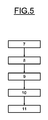
図4と図5に示す一実施形態では、計算工程は、投影追跡を計算する少なくとも1つの工程8と、それに続くサポートベクターマシンを生成する少なくとも1つの工程10と、を含む。
In one embodiment shown in FIGS. 4 and 5, the calculating step includes at least one
図6に示す別の実施形態では、計算工程は独立成分解析による計算工程13を含む。
In another embodiment shown in FIG. 6, the calculation step includes a
計算工程はさらに、少なくとも1つの基準画像の関数として起伏を補償する工程9を含む。
The calculating step further includes a
図4に示す第1の実施形態の一変形態様では、起伏の補償工程9は少なくとも1つのセンサ1によりハイパースペクトル画像を獲得する工程7と投影追跡の計算工程8との間に位置している。
In a variant of the first embodiment shown in FIG. 4, the
図5に示す第1の実施形態の別の変形態様では、起伏の補償工程9は投影追跡の計算工程8とサポートベクターマシンの生成工程10との間に位置している。
In another variant of the first embodiment shown in FIG. 5, the
図6に示す第2の実施形態では、起伏の補償工程9は独立成分解析による計算工程13と表示工程11との間に位置している。
In the second embodiment shown in FIG. 6, the
さらに、起伏の補償を可能にする基準画像は、補正される起伏を表す単一画像、あるいは所与の波長における画像であって補正される起伏を代表する画像、あるいはいくつかの基準画像の線形合成であってよい。 In addition, the reference image that enables compensation of undulations can be a single image that represents the undulation being corrected, an image at a given wavelength that is representative of the undulation being corrected, or the linearity of several reference images. It may be synthetic.
皮膚科応用の枠組みでは、目標は皮膚損傷の存在を確定することである。皮膚は、近赤外線における放射にほとんど反応しない。このときこれらの波長において撮影された画像は実質的には、患者(鼻、口等)の形態構造による起伏と画像端影を含むだけである。したがって基準画像は、赤外線領域で、近赤外領域で、あるいは投影ベクトル(赤外線領域に位置し、縮小ハイパースペクトル画像を補償する投影追跡工程により確定され、また投影追跡から生ずる)が選択される場合は2つの領域の線形合成の、いずれかで撮影される。 In the framework of dermatological applications, the goal is to determine the presence of skin damage. The skin is almost insensitive to radiation in the near infrared. At this time, the images taken at these wavelengths substantially include undulations and image shadows due to the morphological structure of the patient (nose, mouth, etc.). Thus, the reference image is selected in the infrared region, in the near infrared region, or a projection vector (located in the infrared region and determined by the projection tracking process that compensates for the reduced hyperspectral image and resulting from the projection tracking). Is taken in one of two linear combinations of regions.
Claims (17)
少なくとも2つの波長における少なくとも1つのハイパースペクトル画像を生成することができる少なくとも1つのセンサ(1)と、
2つの状態との分類関係に従って前記センサ(1)から生ずる前記ハイパースペクトル画像の画素を類別することができる計算手段(2)と、
前記計算手段(2)から生ずる分類された画素の少なくとも1つの画像関数を表示することができる表示手段(3)と、を含み、
前記計算手段(2)は少なくとも1つの基準画像の関数として前記起伏を補償する手段(6)を含む、ことを特徴とする装置。 A device for compensating for undulations in at least one hyperspectral image,
At least one sensor (1) capable of generating at least one hyperspectral image at at least two wavelengths;
Calculation means (2) capable of classifying pixels of the hyperspectral image arising from the sensor (1) according to a classification relationship with two states;
Display means (3) capable of displaying at least one image function of the classified pixels resulting from said calculation means (2),
Apparatus according to claim 1, characterized in that said calculating means (2) comprises means (6) for compensating said relief as a function of at least one reference image.
2つの状態との分類関係に従ってセンサから生ずるハイパースペクトル画像の画素を類別することができる少なくとも1つの計算工程と、
計算工程から生ずる分類された画素の少なくとも1つの画像関数を表示することができる表示工程と、とを含み、
前記計算工程は少なくとも1つの基準画像の関数として前記起伏を補償する工程を含む、ことを特徴とする方法。 Compensating for undulations in at least one hyperspectral image resulting from at least one sensor capable of generating at least one hyperspectral image in at least two wavelengths, the method comprising:
At least one calculation step capable of classifying the pixels of the hyperspectral image arising from the sensor according to a classification relationship with the two states;
A display step capable of displaying at least one image function of the classified pixels resulting from the calculation step;
The method of claim, wherein the calculating step comprises compensating the undulation as a function of at least one reference image.
Applications Claiming Priority (3)
| Application Number | Priority Date | Filing Date | Title |
|---|---|---|---|
| FR0957626 | 2009-10-29 | ||
| FR0957626A FR2952217B1 (en) | 2009-10-29 | 2009-10-29 | DEVICE AND METHOD FOR RELIEF COMPENSATION OF HYPER-SPECTRAL IMAGES. |
| PCT/EP2010/066342 WO2011051383A1 (en) | 2009-10-29 | 2010-10-28 | Device and method for adjusting the raised pattern of hyper-spectral images |
Publications (1)
| Publication Number | Publication Date |
|---|---|
| JP2013509630A true JP2013509630A (en) | 2013-03-14 |
Family
ID=42077050
Family Applications (1)
| Application Number | Title | Priority Date | Filing Date |
|---|---|---|---|
| JP2012535825A Pending JP2013509630A (en) | 2009-10-29 | 2010-10-28 | Apparatus and method for adjusting a raised pattern of a hyperspectral image. |
Country Status (6)
| Country | Link |
|---|---|
| US (1) | US20120242858A1 (en) |
| EP (1) | EP2494521A1 (en) |
| JP (1) | JP2013509630A (en) |
| CA (1) | CA2778676A1 (en) |
| FR (1) | FR2952217B1 (en) |
| WO (1) | WO2011051383A1 (en) |
Families Citing this family (4)
| Publication number | Priority date | Publication date | Assignee | Title |
|---|---|---|---|---|
| US8600213B2 (en) * | 2011-10-26 | 2013-12-03 | Xerox Corporation | Filtering source video data via independent component selection |
| EP3115925A1 (en) * | 2015-07-07 | 2017-01-11 | Vito NV | Method and system for transforming spectral images |
| KR102441334B1 (en) | 2017-08-01 | 2022-09-06 | 삼성전자주식회사 | Apparatus and method for processing bio-information |
| KR102510174B1 (en) | 2017-09-13 | 2023-03-14 | 삼성전자주식회사 | Apparatus for generate disease prediction model, apparatus and method for disease prediction |
Family Cites Families (8)
| Publication number | Priority date | Publication date | Assignee | Title |
|---|---|---|---|---|
| US5782770A (en) | 1994-05-12 | 1998-07-21 | Science Applications International Corporation | Hyperspectral imaging methods and apparatus for non-invasive diagnosis of tissue for cancer |
| US6081612A (en) | 1997-02-28 | 2000-06-27 | Electro Optical Sciences Inc. | Systems and methods for the multispectral imaging and characterization of skin tissue |
| US6750964B2 (en) * | 1999-08-06 | 2004-06-15 | Cambridge Research And Instrumentation, Inc. | Spectral imaging methods and systems |
| AU2002253784A1 (en) * | 2000-11-07 | 2002-08-28 | Hypermed, Inc. | Hyperspectral imaging calibration device |
| US20020146160A1 (en) | 2001-01-19 | 2002-10-10 | Parker Mary F. | Method and apparatus for generating two-dimensional images of cervical tissue from three-dimensional hyperspectral cubes |
| US8320996B2 (en) * | 2004-11-29 | 2012-11-27 | Hypermed Imaging, Inc. | Medical hyperspectral imaging for evaluation of tissue and tumor |
| US7555155B2 (en) * | 2005-01-27 | 2009-06-30 | Cambridge Research & Instrumentation, Inc. | Classifying image features |
| US8315692B2 (en) | 2007-02-22 | 2012-11-20 | Sheinis Andrew I | Multi-spectral imaging spectrometer for early detection of skin cancer |
-
2009
- 2009-10-29 FR FR0957626A patent/FR2952217B1/en not_active Expired - Fee Related
-
2010
- 2010-10-28 US US13/504,865 patent/US20120242858A1/en not_active Abandoned
- 2010-10-28 CA CA2778676A patent/CA2778676A1/en not_active Abandoned
- 2010-10-28 EP EP10776626A patent/EP2494521A1/en not_active Withdrawn
- 2010-10-28 JP JP2012535825A patent/JP2013509630A/en active Pending
- 2010-10-28 WO PCT/EP2010/066342 patent/WO2011051383A1/en active Application Filing
Also Published As
| Publication number | Publication date |
|---|---|
| US20120242858A1 (en) | 2012-09-27 |
| CA2778676A1 (en) | 2011-05-05 |
| FR2952217A1 (en) | 2011-05-06 |
| WO2011051383A1 (en) | 2011-05-05 |
| EP2494521A1 (en) | 2012-09-05 |
| FR2952217B1 (en) | 2011-12-30 |
Similar Documents
| Publication | Publication Date | Title |
|---|---|---|
| Bayramoglu et al. | Towards virtual H&E staining of hyperspectral lung histology images using conditional generative adversarial networks | |
| Lu et al. | Spectral-spatial classification for noninvasive cancer detection using hyperspectral imaging | |
| CN110274877B (en) | 3D spectral imaging system and method based on scattering medium | |
| US20180108163A1 (en) | Method for analyzing biological specimens by spectral imaging | |
| US20180180537A1 (en) | Method for analyzing biological specimens by spectral imaging | |
| CA2803933C (en) | Method for analyzing biological specimens by spectral imaging | |
| JP2013509629A (en) | Method and apparatus for analyzing hyperspectral images | |
| Cavalcanti et al. | A two-stage approach for discriminating melanocytic skin lesions using standard cameras | |
| AU2017217944B2 (en) | Systems and methods for evaluating pigmented tissue lesions | |
| WO2013080868A1 (en) | Image processing device, image processing method, and image processing program | |
| Reddy et al. | Accurate histopathology from low signal-to-noise ratio spectroscopic imaging data | |
| JP4599520B2 (en) | Multispectral image processing method | |
| JP5405994B2 (en) | Image processing apparatus, image processing method, image processing system, and skin evaluation method | |
| JP2010264276A (en) | Diagnostic method by multi-spectrum skin image | |
| Fabelo et al. | Dermatologic hyperspectral imaging system for skin cancer diagnosis assistance | |
| Fernandes et al. | Early skin cancer detection using computer aided diagnosis techniques | |
| Galeano et al. | Analysis of multispectral images of excised colon tissue samples based on genetic algorithms | |
| JP2013509630A (en) | Apparatus and method for adjusting a raised pattern of a hyperspectral image. | |
| Li | Hyperspectral imaging technology used in tongue diagnosis | |
| Yadav et al. | A study on automatic early detection of skin cancer | |
| US20210264582A1 (en) | Platform and methods for dynamic thin film measurements using hyperspectral imaging | |
| Chaddad et al. | Classification of cancer cells based on morphological features from segmented multispectral bio-images | |
| Calin et al. | Comparative analysis of denoising techniques in burn depth discrimination from burn hyperspectral images | |
| Grechkin et al. | VGG Convolutional Neural Network Classification of Hyperspectral Images of Skin Neoplasms | |
| Li et al. | Hyperspectral tongue imaging system used in tongue diagnosis |