JP2013505950A - Compositions and methods for prevention and treatment of metabolic disorders - Google Patents
Compositions and methods for prevention and treatment of metabolic disorders Download PDFInfo
- Publication number
- JP2013505950A JP2013505950A JP2012531070A JP2012531070A JP2013505950A JP 2013505950 A JP2013505950 A JP 2013505950A JP 2012531070 A JP2012531070 A JP 2012531070A JP 2012531070 A JP2012531070 A JP 2012531070A JP 2013505950 A JP2013505950 A JP 2013505950A
- Authority
- JP
- Japan
- Prior art keywords
- bone
- milk
- derived
- whey
- solution
- Prior art date
- Legal status (The legal status is an assumption and is not a legal conclusion. Google has not performed a legal analysis and makes no representation as to the accuracy of the status listed.)
- Pending
Links
- 238000000034 method Methods 0.000 title claims abstract description 37
- 239000000203 mixture Substances 0.000 title claims abstract description 14
- 238000011282 treatment Methods 0.000 title claims description 32
- 208000030159 metabolic disease Diseases 0.000 title abstract description 4
- 230000002265 prevention Effects 0.000 title description 4
- 210000000988 bone and bone Anatomy 0.000 claims abstract description 59
- 230000024279 bone resorption Effects 0.000 claims abstract description 34
- 208000006386 Bone Resorption Diseases 0.000 claims abstract description 33
- 108010046377 Whey Proteins Proteins 0.000 claims abstract description 33
- 102000007544 Whey Proteins Human genes 0.000 claims abstract description 31
- 102000004169 proteins and genes Human genes 0.000 claims abstract description 27
- 108090000623 proteins and genes Proteins 0.000 claims abstract description 27
- 239000005862 Whey Substances 0.000 claims abstract description 24
- 235000013336 milk Nutrition 0.000 claims description 34
- 239000008267 milk Substances 0.000 claims description 34
- 210000004080 milk Anatomy 0.000 claims description 34
- 150000001875 compounds Chemical class 0.000 claims description 32
- 239000011707 mineral Substances 0.000 claims description 32
- 229910052500 inorganic mineral Inorganic materials 0.000 claims description 30
- 208000001132 Osteoporosis Diseases 0.000 claims description 19
- 239000000463 material Substances 0.000 claims description 16
- 239000007787 solid Substances 0.000 claims description 13
- 101710093543 Probable non-specific lipid-transfer protein Proteins 0.000 claims description 12
- 238000002441 X-ray diffraction Methods 0.000 claims description 11
- 239000002131 composite material Substances 0.000 claims description 8
- 241001465754 Metazoa Species 0.000 claims description 7
- 235000016709 nutrition Nutrition 0.000 claims description 6
- 102000002322 Egg Proteins Human genes 0.000 claims description 5
- 108010000912 Egg Proteins Proteins 0.000 claims description 5
- 238000004519 manufacturing process Methods 0.000 claims description 5
- 241000124008 Mammalia Species 0.000 claims description 4
- 238000005342 ion exchange Methods 0.000 claims description 4
- XLYOFNOQVPJJNP-UHFFFAOYSA-N water Substances O XLYOFNOQVPJJNP-UHFFFAOYSA-N 0.000 claims description 4
- 238000000502 dialysis Methods 0.000 claims description 3
- 210000003278 egg shell Anatomy 0.000 claims description 3
- 238000010438 heat treatment Methods 0.000 claims description 3
- 238000001035 drying Methods 0.000 claims description 2
- 229910001575 sodium mineral Inorganic materials 0.000 claims description 2
- 239000000243 solution Substances 0.000 claims 7
- 239000012460 protein solution Substances 0.000 claims 3
- 208000036119 Frailty Diseases 0.000 claims 2
- 206010003549 asthenia Diseases 0.000 claims 2
- 239000002904 solvent Substances 0.000 claims 2
- GNFTZDOKVXKIBK-UHFFFAOYSA-N 3-(2-methoxyethoxy)benzohydrazide Chemical compound COCCOC1=CC=CC(C(=O)NN)=C1 GNFTZDOKVXKIBK-UHFFFAOYSA-N 0.000 claims 1
- 230000002401 inhibitory effect Effects 0.000 claims 1
- 230000030991 negative regulation of bone resorption Effects 0.000 claims 1
- 159000000000 sodium salts Chemical class 0.000 claims 1
- XYJRXVWERLGGKC-UHFFFAOYSA-D pentacalcium;hydroxide;triphosphate Chemical compound [OH-].[Ca+2].[Ca+2].[Ca+2].[Ca+2].[Ca+2].[O-]P([O-])([O-])=O.[O-]P([O-])([O-])=O.[O-]P([O-])([O-])=O XYJRXVWERLGGKC-UHFFFAOYSA-D 0.000 abstract description 41
- 229910052588 hydroxylapatite Inorganic materials 0.000 abstract description 34
- 235000018102 proteins Nutrition 0.000 abstract description 26
- 108010035532 Collagen Proteins 0.000 abstract description 16
- 102000008186 Collagen Human genes 0.000 abstract description 16
- 229920001436 collagen Polymers 0.000 abstract description 16
- 239000011159 matrix material Substances 0.000 abstract description 13
- 102000007350 Bone Morphogenetic Proteins Human genes 0.000 abstract description 10
- 108010007726 Bone Morphogenetic Proteins Proteins 0.000 abstract description 10
- 229940112869 bone morphogenetic protein Drugs 0.000 abstract description 10
- 238000012423 maintenance Methods 0.000 abstract description 9
- 235000021119 whey protein Nutrition 0.000 abstract description 9
- 230000006378 damage Effects 0.000 abstract description 8
- 230000035876 healing Effects 0.000 abstract description 8
- 230000015572 biosynthetic process Effects 0.000 abstract description 7
- 230000008439 repair process Effects 0.000 abstract description 6
- 210000002435 tendon Anatomy 0.000 abstract description 5
- 238000002560 therapeutic procedure Methods 0.000 abstract description 5
- 101000995928 Mus musculus Nucleolar protein 58 Proteins 0.000 abstract description 3
- 102100039692 RNA-binding motif, single-stranded-interacting protein 1 Human genes 0.000 abstract description 3
- 230000037180 bone health Effects 0.000 abstract description 3
- 230000001419 dependent effect Effects 0.000 abstract description 3
- 230000036541 health Effects 0.000 abstract description 3
- 206010012601 diabetes mellitus Diseases 0.000 abstract description 2
- 230000002708 enhancing effect Effects 0.000 abstract 1
- 239000013078 crystal Substances 0.000 description 32
- 239000011575 calcium Substances 0.000 description 22
- 235000010755 mineral Nutrition 0.000 description 22
- VSIIXMUUUJUKCM-UHFFFAOYSA-D pentacalcium;fluoride;triphosphate Chemical compound [F-].[Ca+2].[Ca+2].[Ca+2].[Ca+2].[Ca+2].[O-]P([O-])([O-])=O.[O-]P([O-])([O-])=O.[O-]P([O-])([O-])=O VSIIXMUUUJUKCM-UHFFFAOYSA-D 0.000 description 22
- 229910052586 apatite Inorganic materials 0.000 description 19
- 206010065687 Bone loss Diseases 0.000 description 18
- 239000002121 nanofiber Substances 0.000 description 17
- VTYYLEPIZMXCLO-UHFFFAOYSA-L Calcium carbonate Chemical compound [Ca+2].[O-]C([O-])=O VTYYLEPIZMXCLO-UHFFFAOYSA-L 0.000 description 16
- OYPRJOBELJOOCE-UHFFFAOYSA-N Calcium Chemical compound [Ca] OYPRJOBELJOOCE-UHFFFAOYSA-N 0.000 description 11
- 206010017076 Fracture Diseases 0.000 description 11
- 229960005069 calcium Drugs 0.000 description 11
- 229910052791 calcium Inorganic materials 0.000 description 11
- 230000011164 ossification Effects 0.000 description 11
- 210000002700 urine Anatomy 0.000 description 11
- 238000005259 measurement Methods 0.000 description 10
- 239000000523 sample Substances 0.000 description 10
- 238000003917 TEM image Methods 0.000 description 9
- 210000004027 cell Anatomy 0.000 description 9
- 108090000765 processed proteins & peptides Proteins 0.000 description 9
- 239000000126 substance Substances 0.000 description 9
- 208000010392 Bone Fractures Diseases 0.000 description 8
- 229910000019 calcium carbonate Inorganic materials 0.000 description 8
- 239000000843 powder Substances 0.000 description 8
- 239000000047 product Substances 0.000 description 8
- 102000011632 Caseins Human genes 0.000 description 7
- 108010076119 Caseins Proteins 0.000 description 7
- 230000000694 effects Effects 0.000 description 7
- 230000012010 growth Effects 0.000 description 7
- 238000012360 testing method Methods 0.000 description 7
- 108010022452 Collagen Type I Proteins 0.000 description 6
- 102000012422 Collagen Type I Human genes 0.000 description 6
- 230000008901 benefit Effects 0.000 description 6
- 241000282472 Canis lupus familiaris Species 0.000 description 5
- WHUUTDBJXJRKMK-VKHMYHEASA-N L-glutamic acid Chemical compound OC(=O)[C@@H](N)CCC(O)=O WHUUTDBJXJRKMK-VKHMYHEASA-N 0.000 description 5
- 102000004407 Lactalbumin Human genes 0.000 description 5
- 108090000942 Lactalbumin Proteins 0.000 description 5
- 230000002378 acidificating effect Effects 0.000 description 5
- 230000005540 biological transmission Effects 0.000 description 5
- 150000001720 carbohydrates Chemical class 0.000 description 5
- 239000005018 casein Substances 0.000 description 5
- BECPQYXYKAMYBN-UHFFFAOYSA-N casein, tech. Chemical compound NCCCCC(C(O)=O)N=C(O)C(CC(O)=O)N=C(O)C(CCC(O)=N)N=C(O)C(CC(C)C)N=C(O)C(CCC(O)=O)N=C(O)C(CC(O)=O)N=C(O)C(CCC(O)=O)N=C(O)C(C(C)O)N=C(O)C(CCC(O)=N)N=C(O)C(CCC(O)=N)N=C(O)C(CCC(O)=N)N=C(O)C(CCC(O)=O)N=C(O)C(CCC(O)=O)N=C(O)C(COP(O)(O)=O)N=C(O)C(CCC(O)=N)N=C(O)C(N)CC1=CC=CC=C1 BECPQYXYKAMYBN-UHFFFAOYSA-N 0.000 description 5
- 235000021240 caseins Nutrition 0.000 description 5
- 230000007423 decrease Effects 0.000 description 5
- 239000007788 liquid Substances 0.000 description 5
- 102000004196 processed proteins & peptides Human genes 0.000 description 5
- 230000004044 response Effects 0.000 description 5
- 238000012549 training Methods 0.000 description 5
- 230000002485 urinary effect Effects 0.000 description 5
- 238000010521 absorption reaction Methods 0.000 description 4
- 125000004429 atom Chemical group 0.000 description 4
- 230000037182 bone density Effects 0.000 description 4
- 230000008416 bone turnover Effects 0.000 description 4
- 229940069978 calcium supplement Drugs 0.000 description 4
- 235000014633 carbohydrates Nutrition 0.000 description 4
- 235000013305 food Nutrition 0.000 description 4
- 238000009472 formulation Methods 0.000 description 4
- 230000006872 improvement Effects 0.000 description 4
- 230000006911 nucleation Effects 0.000 description 4
- 238000010899 nucleation Methods 0.000 description 4
- 239000011368 organic material Substances 0.000 description 4
- BZQFBWGGLXLEPQ-REOHCLBHSA-N phosphoserine Chemical compound OC(=O)[C@@H](N)COP(O)(O)=O BZQFBWGGLXLEPQ-REOHCLBHSA-N 0.000 description 4
- 239000002243 precursor Substances 0.000 description 4
- 230000009467 reduction Effects 0.000 description 4
- 241000894007 species Species 0.000 description 4
- 210000001519 tissue Anatomy 0.000 description 4
- OGSPWJRAVKPPFI-UHFFFAOYSA-N Alendronic Acid Chemical compound NCCCC(O)(P(O)(O)=O)P(O)(O)=O OGSPWJRAVKPPFI-UHFFFAOYSA-N 0.000 description 3
- 102000003886 Glycoproteins Human genes 0.000 description 3
- 108090000288 Glycoproteins Proteins 0.000 description 3
- 102000016921 Integrin-Binding Sialoprotein Human genes 0.000 description 3
- 108010028750 Integrin-Binding Sialoprotein Proteins 0.000 description 3
- 102000010445 Lactoferrin Human genes 0.000 description 3
- 108010063045 Lactoferrin Proteins 0.000 description 3
- 102000008192 Lactoglobulins Human genes 0.000 description 3
- 108010060630 Lactoglobulins Proteins 0.000 description 3
- 108010023244 Lactoperoxidase Proteins 0.000 description 3
- 102000045576 Lactoperoxidases Human genes 0.000 description 3
- 229910019142 PO4 Inorganic materials 0.000 description 3
- 108010001441 Phosphopeptides Proteins 0.000 description 3
- 229930003316 Vitamin D Natural products 0.000 description 3
- QYSXJUFSXHHAJI-XFEUOLMDSA-N Vitamin D3 Natural products C1(/[C@@H]2CC[C@@H]([C@]2(CCC1)C)[C@H](C)CCCC(C)C)=C/C=C1\C[C@@H](O)CCC1=C QYSXJUFSXHHAJI-XFEUOLMDSA-N 0.000 description 3
- 230000002159 abnormal effect Effects 0.000 description 3
- 235000001014 amino acid Nutrition 0.000 description 3
- 150000001413 amino acids Chemical class 0.000 description 3
- 238000004458 analytical method Methods 0.000 description 3
- 230000037176 bone building Effects 0.000 description 3
- 230000008468 bone growth Effects 0.000 description 3
- 230000010072 bone remodeling Effects 0.000 description 3
- 235000014113 dietary fatty acids Nutrition 0.000 description 3
- 208000037265 diseases, disorders, signs and symptoms Diseases 0.000 description 3
- 230000029142 excretion Effects 0.000 description 3
- 239000004744 fabric Substances 0.000 description 3
- 239000000194 fatty acid Substances 0.000 description 3
- 229930195729 fatty acid Natural products 0.000 description 3
- 150000004665 fatty acids Chemical class 0.000 description 3
- 229940001490 fosamax Drugs 0.000 description 3
- 229930195712 glutamate Natural products 0.000 description 3
- 239000003102 growth factor Substances 0.000 description 3
- 239000003112 inhibitor Substances 0.000 description 3
- 230000005764 inhibitory process Effects 0.000 description 3
- CSSYQJWUGATIHM-IKGCZBKSSA-N l-phenylalanyl-l-lysyl-l-cysteinyl-l-arginyl-l-arginyl-l-tryptophyl-l-glutaminyl-l-tryptophyl-l-arginyl-l-methionyl-l-lysyl-l-lysyl-l-leucylglycyl-l-alanyl-l-prolyl-l-seryl-l-isoleucyl-l-threonyl-l-cysteinyl-l-valyl-l-arginyl-l-arginyl-l-alanyl-l-phenylal Chemical compound C([C@H](N)C(=O)N[C@@H](CCCCN)C(=O)N[C@@H](CS)C(=O)N[C@@H](CCCNC(N)=N)C(=O)N[C@@H](CCCNC(N)=N)C(=O)N[C@@H](CC=1C2=CC=CC=C2NC=1)C(=O)N[C@@H](CCC(N)=O)C(=O)N[C@@H](CC=1C2=CC=CC=C2NC=1)C(=O)N[C@@H](CCCNC(N)=N)C(=O)N[C@@H](CCSC)C(=O)N[C@@H](CCCCN)C(=O)N[C@@H](CCCCN)C(=O)N[C@@H](CC(C)C)C(=O)NCC(=O)N[C@@H](C)C(=O)N1CCC[C@H]1C(=O)N[C@@H](CO)C(=O)N[C@@H]([C@@H](C)CC)C(=O)N[C@@H]([C@@H](C)O)C(=O)N[C@@H](CS)C(=O)N[C@@H](C(C)C)C(=O)N[C@@H](CCCNC(N)=N)C(=O)N[C@@H](CCCNC(N)=N)C(=O)N[C@@H](C)C(=O)N[C@@H](CC=1C=CC=CC=1)C(O)=O)C1=CC=CC=C1 CSSYQJWUGATIHM-IKGCZBKSSA-N 0.000 description 3
- 235000021242 lactoferrin Nutrition 0.000 description 3
- 229940078795 lactoferrin Drugs 0.000 description 3
- 229940057428 lactoperoxidase Drugs 0.000 description 3
- 235000015097 nutrients Nutrition 0.000 description 3
- 210000002997 osteoclast Anatomy 0.000 description 3
- 239000002245 particle Substances 0.000 description 3
- 239000012466 permeate Substances 0.000 description 3
- NBIIXXVUZAFLBC-UHFFFAOYSA-K phosphate Chemical compound [O-]P([O-])([O-])=O NBIIXXVUZAFLBC-UHFFFAOYSA-K 0.000 description 3
- 239000010452 phosphate Substances 0.000 description 3
- 230000008569 process Effects 0.000 description 3
- 238000003672 processing method Methods 0.000 description 3
- 238000011160 research Methods 0.000 description 3
- 210000002966 serum Anatomy 0.000 description 3
- 239000011734 sodium Substances 0.000 description 3
- 238000001179 sorption measurement Methods 0.000 description 3
- 238000002604 ultrasonography Methods 0.000 description 3
- 235000019166 vitamin D Nutrition 0.000 description 3
- 239000011710 vitamin D Substances 0.000 description 3
- 150000003710 vitamin D derivatives Chemical class 0.000 description 3
- 229940046008 vitamin d Drugs 0.000 description 3
- 206010067484 Adverse reaction Diseases 0.000 description 2
- GUBGYTABKSRVRQ-XLOQQCSPSA-N Alpha-Lactose Chemical compound O[C@@H]1[C@@H](O)[C@@H](O)[C@@H](CO)O[C@H]1O[C@@H]1[C@@H](CO)O[C@H](O)[C@H](O)[C@H]1O GUBGYTABKSRVRQ-XLOQQCSPSA-N 0.000 description 2
- 241000271566 Aves Species 0.000 description 2
- 229940122361 Bisphosphonate Drugs 0.000 description 2
- 241000283690 Bos taurus Species 0.000 description 2
- 108091003079 Bovine Serum Albumin Proteins 0.000 description 2
- WHUUTDBJXJRKMK-UHFFFAOYSA-N Glutamic acid Natural products OC(=O)C(N)CCC(O)=O WHUUTDBJXJRKMK-UHFFFAOYSA-N 0.000 description 2
- GUBGYTABKSRVRQ-QKKXKWKRSA-N Lactose Natural products OC[C@H]1O[C@@H](O[C@H]2[C@H](O)[C@@H](O)C(O)O[C@@H]2CO)[C@H](O)[C@@H](O)[C@H]1O GUBGYTABKSRVRQ-QKKXKWKRSA-N 0.000 description 2
- 102000004895 Lipoproteins Human genes 0.000 description 2
- 108090001030 Lipoproteins Proteins 0.000 description 2
- 108010071690 Prealbumin Proteins 0.000 description 2
- 102000007584 Prealbumin Human genes 0.000 description 2
- FAPWRFPIFSIZLT-UHFFFAOYSA-M Sodium chloride Chemical compound [Na+].[Cl-] FAPWRFPIFSIZLT-UHFFFAOYSA-M 0.000 description 2
- 230000006838 adverse reaction Effects 0.000 description 2
- FPIPGXGPPPQFEQ-OVSJKPMPSA-N all-trans-retinol Chemical compound OC\C=C(/C)\C=C\C=C(/C)\C=C\C1=C(C)CCCC1(C)C FPIPGXGPPPQFEQ-OVSJKPMPSA-N 0.000 description 2
- 125000000539 amino acid group Chemical group 0.000 description 2
- 238000003556 assay Methods 0.000 description 2
- 235000015895 biscuits Nutrition 0.000 description 2
- 150000004663 bisphosphonates Chemical class 0.000 description 2
- 210000004369 blood Anatomy 0.000 description 2
- 239000008280 blood Substances 0.000 description 2
- 230000037118 bone strength Effects 0.000 description 2
- 229940098773 bovine serum albumin Drugs 0.000 description 2
- 238000005119 centrifugation Methods 0.000 description 2
- 230000008859 change Effects 0.000 description 2
- 238000006243 chemical reaction Methods 0.000 description 2
- 108010049937 collagen type I trimeric cross-linked peptide Proteins 0.000 description 2
- 238000010276 construction Methods 0.000 description 2
- DDRJAANPRJIHGJ-UHFFFAOYSA-N creatinine Chemical compound CN1CC(=O)NC1=N DDRJAANPRJIHGJ-UHFFFAOYSA-N 0.000 description 2
- GHVNFZFCNZKVNT-UHFFFAOYSA-N decanoic acid Chemical compound CCCCCCCCCC(O)=O GHVNFZFCNZKVNT-UHFFFAOYSA-N 0.000 description 2
- 238000011161 development Methods 0.000 description 2
- 230000018109 developmental process Effects 0.000 description 2
- 238000003745 diagnosis Methods 0.000 description 2
- 201000010099 disease Diseases 0.000 description 2
- 238000004090 dissolution Methods 0.000 description 2
- 229940079593 drug Drugs 0.000 description 2
- 239000003814 drug Substances 0.000 description 2
- 238000010894 electron beam technology Methods 0.000 description 2
- 235000004626 essential fatty acids Nutrition 0.000 description 2
- 238000002474 experimental method Methods 0.000 description 2
- 239000011888 foil Substances 0.000 description 2
- 235000013922 glutamic acid Nutrition 0.000 description 2
- 239000004220 glutamic acid Substances 0.000 description 2
- FUZZWVXGSFPDMH-UHFFFAOYSA-N hexanoic acid Chemical compound CCCCCC(O)=O FUZZWVXGSFPDMH-UHFFFAOYSA-N 0.000 description 2
- 125000004356 hydroxy functional group Chemical group O* 0.000 description 2
- 101150026046 iga gene Proteins 0.000 description 2
- 230000003993 interaction Effects 0.000 description 2
- 239000008101 lactose Substances 0.000 description 2
- 230000000670 limiting effect Effects 0.000 description 2
- 239000003550 marker Substances 0.000 description 2
- 230000007246 mechanism Effects 0.000 description 2
- 230000009245 menopause Effects 0.000 description 2
- 230000004048 modification Effects 0.000 description 2
- 238000012986 modification Methods 0.000 description 2
- 238000000329 molecular dynamics simulation Methods 0.000 description 2
- 239000002114 nanocomposite Substances 0.000 description 2
- 231100000957 no side effect Toxicity 0.000 description 2
- 238000005457 optimization Methods 0.000 description 2
- 210000000963 osteoblast Anatomy 0.000 description 2
- 230000000737 periodic effect Effects 0.000 description 2
- 244000144977 poultry Species 0.000 description 2
- 239000002994 raw material Substances 0.000 description 2
- 230000008929 regeneration Effects 0.000 description 2
- 238000011069 regeneration method Methods 0.000 description 2
- 230000000717 retained effect Effects 0.000 description 2
- 238000000926 separation method Methods 0.000 description 2
- 235000020183 skimmed milk Nutrition 0.000 description 2
- 229910052708 sodium Inorganic materials 0.000 description 2
- 150000003431 steroids Chemical class 0.000 description 2
- 238000005728 strengthening Methods 0.000 description 2
- 239000006228 supernatant Substances 0.000 description 2
- 239000013589 supplement Substances 0.000 description 2
- 239000000725 suspension Substances 0.000 description 2
- 235000021249 α-casein Nutrition 0.000 description 2
- OWEGMIWEEQEYGQ-UHFFFAOYSA-N 100676-05-9 Natural products OC1C(O)C(O)C(CO)OC1OCC1C(O)C(O)C(O)C(OC2C(OC(O)C(O)C2O)CO)O1 OWEGMIWEEQEYGQ-UHFFFAOYSA-N 0.000 description 1
- FPIPGXGPPPQFEQ-UHFFFAOYSA-N 13-cis retinol Natural products OCC=C(C)C=CC=C(C)C=CC1=C(C)CCCC1(C)C FPIPGXGPPPQFEQ-UHFFFAOYSA-N 0.000 description 1
- QCVGEOXPDFCNHA-UHFFFAOYSA-N 5,5-dimethyl-2,4-dioxo-1,3-oxazolidine-3-carboxamide Chemical compound CC1(C)OC(=O)N(C(N)=O)C1=O QCVGEOXPDFCNHA-UHFFFAOYSA-N 0.000 description 1
- 206010000060 Abdominal distension Diseases 0.000 description 1
- 101800000112 Acidic peptide Proteins 0.000 description 1
- 101800000263 Acidic protein Proteins 0.000 description 1
- 102000002260 Alkaline Phosphatase Human genes 0.000 description 1
- 108020004774 Alkaline Phosphatase Proteins 0.000 description 1
- 229910021532 Calcite Inorganic materials 0.000 description 1
- 102000005701 Calcium-Binding Proteins Human genes 0.000 description 1
- 108010045403 Calcium-Binding Proteins Proteins 0.000 description 1
- 239000005632 Capric acid (CAS 334-48-5) Substances 0.000 description 1
- 102000004127 Cytokines Human genes 0.000 description 1
- 108090000695 Cytokines Proteins 0.000 description 1
- 206010073767 Developmental hip dysplasia Diseases 0.000 description 1
- 206010012735 Diarrhoea Diseases 0.000 description 1
- 206010058314 Dysplasia Diseases 0.000 description 1
- 102000004190 Enzymes Human genes 0.000 description 1
- 108090000790 Enzymes Proteins 0.000 description 1
- 229930091371 Fructose Natural products 0.000 description 1
- 239000005715 Fructose Substances 0.000 description 1
- RFSUNEUAIZKAJO-ARQDHWQXSA-N Fructose Chemical compound OC[C@H]1O[C@](O)(CO)[C@@H](O)[C@@H]1O RFSUNEUAIZKAJO-ARQDHWQXSA-N 0.000 description 1
- 102000006395 Globulins Human genes 0.000 description 1
- 108010044091 Globulins Proteins 0.000 description 1
- WQZGKKKJIJFFOK-GASJEMHNSA-N Glucose Natural products OC[C@H]1OC(O)[C@H](O)[C@@H](O)[C@@H]1O WQZGKKKJIJFFOK-GASJEMHNSA-N 0.000 description 1
- 208000007446 Hip Dislocation Diseases 0.000 description 1
- 206010020100 Hip fracture Diseases 0.000 description 1
- 206010020751 Hypersensitivity Diseases 0.000 description 1
- DGAQECJNVWCQMB-PUAWFVPOSA-M Ilexoside XXIX Chemical compound C[C@@H]1CC[C@@]2(CC[C@@]3(C(=CC[C@H]4[C@]3(CC[C@@H]5[C@@]4(CC[C@@H](C5(C)C)OS(=O)(=O)[O-])C)C)[C@@H]2[C@]1(C)O)C)C(=O)O[C@H]6[C@@H]([C@H]([C@@H]([C@H](O6)CO)O)O)O.[Na+] DGAQECJNVWCQMB-PUAWFVPOSA-M 0.000 description 1
- OYHQOLUKZRVURQ-HZJYTTRNSA-N Linoleic acid Chemical compound CCCCC\C=C/C\C=C/CCCCCCCC(O)=O OYHQOLUKZRVURQ-HZJYTTRNSA-N 0.000 description 1
- GUBGYTABKSRVRQ-PICCSMPSSA-N Maltose Natural products O[C@@H]1[C@@H](O)[C@H](O)[C@@H](CO)O[C@@H]1O[C@@H]1[C@@H](CO)OC(O)[C@H](O)[C@H]1O GUBGYTABKSRVRQ-PICCSMPSSA-N 0.000 description 1
- 208000029725 Metabolic bone disease Diseases 0.000 description 1
- 102000014171 Milk Proteins Human genes 0.000 description 1
- 108010011756 Milk Proteins Proteins 0.000 description 1
- 206010028980 Neoplasm Diseases 0.000 description 1
- BZQFBWGGLXLEPQ-UHFFFAOYSA-N O-phosphoryl-L-serine Natural products OC(=O)C(N)COP(O)(O)=O BZQFBWGGLXLEPQ-UHFFFAOYSA-N 0.000 description 1
- 206010049088 Osteopenia Diseases 0.000 description 1
- 102000004264 Osteopontin Human genes 0.000 description 1
- 108010081689 Osteopontin Proteins 0.000 description 1
- OAICVXFJPJFONN-UHFFFAOYSA-N Phosphorus Chemical compound [P] OAICVXFJPJFONN-UHFFFAOYSA-N 0.000 description 1
- 238000003991 Rietveld refinement Methods 0.000 description 1
- 206010061363 Skeletal injury Diseases 0.000 description 1
- 208000027418 Wounds and injury Diseases 0.000 description 1
- 239000002253 acid Substances 0.000 description 1
- 230000002411 adverse Effects 0.000 description 1
- 230000000172 allergic effect Effects 0.000 description 1
- 230000007815 allergy Effects 0.000 description 1
- 238000003149 assay kit Methods 0.000 description 1
- 230000000386 athletic effect Effects 0.000 description 1
- QVGXLLKOCUKJST-UHFFFAOYSA-N atomic oxygen Chemical compound [O] QVGXLLKOCUKJST-UHFFFAOYSA-N 0.000 description 1
- 208000010668 atopic eczema Diseases 0.000 description 1
- 230000003416 augmentation Effects 0.000 description 1
- 230000009286 beneficial effect Effects 0.000 description 1
- WQZGKKKJIJFFOK-VFUOTHLCSA-N beta-D-glucose Chemical compound OC[C@H]1O[C@@H](O)[C@H](O)[C@@H](O)[C@@H]1O WQZGKKKJIJFFOK-VFUOTHLCSA-N 0.000 description 1
- GUBGYTABKSRVRQ-QUYVBRFLSA-N beta-maltose Chemical compound OC[C@H]1O[C@H](O[C@H]2[C@H](O)[C@@H](O)[C@H](O)O[C@@H]2CO)[C@H](O)[C@@H](O)[C@@H]1O GUBGYTABKSRVRQ-QUYVBRFLSA-N 0.000 description 1
- 239000000090 biomarker Substances 0.000 description 1
- 230000033558 biomineral tissue development Effects 0.000 description 1
- 230000004097 bone metabolism Effects 0.000 description 1
- 235000021152 breakfast Nutrition 0.000 description 1
- 238000009395 breeding Methods 0.000 description 1
- 230000001488 breeding effect Effects 0.000 description 1
- 239000004566 building material Substances 0.000 description 1
- 230000002308 calcification Effects 0.000 description 1
- MKJXYGKVIBWPFZ-UHFFFAOYSA-L calcium lactate Chemical compound [Ca+2].CC(O)C([O-])=O.CC(O)C([O-])=O MKJXYGKVIBWPFZ-UHFFFAOYSA-L 0.000 description 1
- 239000001527 calcium lactate Substances 0.000 description 1
- 229960002401 calcium lactate Drugs 0.000 description 1
- 235000011086 calcium lactate Nutrition 0.000 description 1
- 239000001506 calcium phosphate Substances 0.000 description 1
- 229910000389 calcium phosphate Inorganic materials 0.000 description 1
- 235000011010 calcium phosphates Nutrition 0.000 description 1
- 238000004364 calculation method Methods 0.000 description 1
- 208000036426 canine hip dysplasia Diseases 0.000 description 1
- 239000002775 capsule Substances 0.000 description 1
- 235000013339 cereals Nutrition 0.000 description 1
- 239000007795 chemical reaction product Substances 0.000 description 1
- 125000001309 chloro group Chemical group Cl* 0.000 description 1
- 239000012141 concentrate Substances 0.000 description 1
- 238000012790 confirmation Methods 0.000 description 1
- 235000020247 cow milk Nutrition 0.000 description 1
- 229940109239 creatinine Drugs 0.000 description 1
- 238000011461 current therapy Methods 0.000 description 1
- 235000013365 dairy product Nutrition 0.000 description 1
- 238000007405 data analysis Methods 0.000 description 1
- 230000003247 decreasing effect Effects 0.000 description 1
- 230000007812 deficiency Effects 0.000 description 1
- 229950006137 dexfosfoserine Drugs 0.000 description 1
- 235000015872 dietary supplement Nutrition 0.000 description 1
- 238000005553 drilling Methods 0.000 description 1
- 239000003937 drug carrier Substances 0.000 description 1
- 230000009977 dual effect Effects 0.000 description 1
- 201000006549 dyspepsia Diseases 0.000 description 1
- 235000014103 egg white Nutrition 0.000 description 1
- 210000000969 egg white Anatomy 0.000 description 1
- 210000002969 egg yolk Anatomy 0.000 description 1
- 235000013345 egg yolk Nutrition 0.000 description 1
- 235000013601 eggs Nutrition 0.000 description 1
- 239000003792 electrolyte Substances 0.000 description 1
- 229940088598 enzyme Drugs 0.000 description 1
- 238000011067 equilibration Methods 0.000 description 1
- 210000003722 extracellular fluid Anatomy 0.000 description 1
- 201000003373 familial cold autoinflammatory syndrome 3 Diseases 0.000 description 1
- 229910052587 fluorapatite Inorganic materials 0.000 description 1
- 125000001153 fluoro group Chemical group F* 0.000 description 1
- 230000037406 food intake Effects 0.000 description 1
- 238000005755 formation reaction Methods 0.000 description 1
- 239000012634 fragment Substances 0.000 description 1
- 230000006870 function Effects 0.000 description 1
- 239000008103 glucose Substances 0.000 description 1
- WHUUTDBJXJRKMK-VKHMYHEASA-L glutamate group Chemical group N[C@@H](CCC(=O)[O-])C(=O)[O-] WHUUTDBJXJRKMK-VKHMYHEASA-L 0.000 description 1
- 208000024798 heartburn Diseases 0.000 description 1
- 238000002173 high-resolution transmission electron microscopy Methods 0.000 description 1
- 230000001900 immune effect Effects 0.000 description 1
- 230000000977 initiatory effect Effects 0.000 description 1
- 208000014674 injury Diseases 0.000 description 1
- 150000002500 ions Chemical class 0.000 description 1
- 235000020778 linoleic acid Nutrition 0.000 description 1
- OYHQOLUKZRVURQ-IXWMQOLASA-N linoleic acid Natural products CCCCC\C=C/C\C=C\CCCCCCCC(O)=O OYHQOLUKZRVURQ-IXWMQOLASA-N 0.000 description 1
- 244000144972 livestock Species 0.000 description 1
- 238000007726 management method Methods 0.000 description 1
- 235000021239 milk protein Nutrition 0.000 description 1
- 238000000324 molecular mechanic Methods 0.000 description 1
- 238000000302 molecular modelling Methods 0.000 description 1
- 238000012544 monitoring process Methods 0.000 description 1
- 210000003205 muscle Anatomy 0.000 description 1
- 239000002086 nanomaterial Substances 0.000 description 1
- 230000000474 nursing effect Effects 0.000 description 1
- 230000035764 nutrition Effects 0.000 description 1
- ZQPPMHVWECSIRJ-KTKRTIGZSA-N oleic acid group Chemical group C(CCCCCCC\C=C/CCCCCCCC)(=O)O ZQPPMHVWECSIRJ-KTKRTIGZSA-N 0.000 description 1
- 125000002524 organometallic group Chemical group 0.000 description 1
- 230000001009 osteoporotic effect Effects 0.000 description 1
- 229910052760 oxygen Inorganic materials 0.000 description 1
- 239000001301 oxygen Substances 0.000 description 1
- 230000001575 pathological effect Effects 0.000 description 1
- 101150082630 pdf-2 gene Proteins 0.000 description 1
- 210000002976 pectoralis muscle Anatomy 0.000 description 1
- 239000011574 phosphorus Substances 0.000 description 1
- 229910052698 phosphorus Inorganic materials 0.000 description 1
- 229920001184 polypeptide Polymers 0.000 description 1
- 238000000634 powder X-ray diffraction Methods 0.000 description 1
- 239000002244 precipitate Substances 0.000 description 1
- 238000003825 pressing Methods 0.000 description 1
- 238000004393 prognosis Methods 0.000 description 1
- 238000010791 quenching Methods 0.000 description 1
- 230000035484 reaction time Effects 0.000 description 1
- 238000011084 recovery Methods 0.000 description 1
- 230000002829 reductive effect Effects 0.000 description 1
- 229960003471 retinol Drugs 0.000 description 1
- 235000020944 retinol Nutrition 0.000 description 1
- 239000011607 retinol Substances 0.000 description 1
- 102000029752 retinol binding Human genes 0.000 description 1
- 108091000053 retinol binding Proteins 0.000 description 1
- 238000012552 review Methods 0.000 description 1
- 150000003839 salts Chemical group 0.000 description 1
- 238000012216 screening Methods 0.000 description 1
- 230000028327 secretion Effects 0.000 description 1
- 230000009528 severe injury Effects 0.000 description 1
- 238000004088 simulation Methods 0.000 description 1
- 230000009645 skeletal growth Effects 0.000 description 1
- 239000011780 sodium chloride Substances 0.000 description 1
- 206010041569 spinal fracture Diseases 0.000 description 1
- 238000001694 spray drying Methods 0.000 description 1
- 230000009469 supplementation Effects 0.000 description 1
- 230000001629 suppression Effects 0.000 description 1
- 208000024891 symptom Diseases 0.000 description 1
- 230000001225 therapeutic effect Effects 0.000 description 1
- 230000007704 transition Effects 0.000 description 1
- 230000032258 transport Effects 0.000 description 1
- QORWJWZARLRLPR-UHFFFAOYSA-H tricalcium bis(phosphate) Chemical compound [Ca+2].[Ca+2].[Ca+2].[O-]P([O-])([O-])=O.[O-]P([O-])([O-])=O QORWJWZARLRLPR-UHFFFAOYSA-H 0.000 description 1
- 208000001072 type 2 diabetes mellitus Diseases 0.000 description 1
- 230000002792 vascular Effects 0.000 description 1
- 229940088594 vitamin Drugs 0.000 description 1
- 229930003231 vitamin Natural products 0.000 description 1
- 235000013343 vitamin Nutrition 0.000 description 1
- 239000011782 vitamin Substances 0.000 description 1
- 239000002699 waste material Substances 0.000 description 1
- 230000003442 weekly effect Effects 0.000 description 1
- 235000021241 α-lactalbumin Nutrition 0.000 description 1
Images


Classifications
-
- A—HUMAN NECESSITIES
- A61—MEDICAL OR VETERINARY SCIENCE; HYGIENE
- A61K—PREPARATIONS FOR MEDICAL, DENTAL OR TOILETRY PURPOSES
- A61K35/00—Medicinal preparations containing materials or reaction products thereof with undetermined constitution
- A61K35/12—Materials from mammals; Compositions comprising non-specified tissues or cells; Compositions comprising non-embryonic stem cells; Genetically modified cells
- A61K35/20—Milk; Whey; Colostrum
-
- A—HUMAN NECESSITIES
- A23—FOODS OR FOODSTUFFS; TREATMENT THEREOF, NOT COVERED BY OTHER CLASSES
- A23C—DAIRY PRODUCTS, e.g. MILK, BUTTER OR CHEESE; MILK OR CHEESE SUBSTITUTES; MAKING THEREOF
- A23C9/00—Milk preparations; Milk powder or milk powder preparations
- A23C9/14—Milk preparations; Milk powder or milk powder preparations in which the chemical composition of the milk is modified by non-chemical treatment
- A23C9/142—Milk preparations; Milk powder or milk powder preparations in which the chemical composition of the milk is modified by non-chemical treatment by dialysis, reverse osmosis or ultrafiltration
- A23C9/1425—Milk preparations; Milk powder or milk powder preparations in which the chemical composition of the milk is modified by non-chemical treatment by dialysis, reverse osmosis or ultrafiltration by ultrafiltration, microfiltration or diafiltration of whey, e.g. treatment of the UF permeate
-
- A—HUMAN NECESSITIES
- A23—FOODS OR FOODSTUFFS; TREATMENT THEREOF, NOT COVERED BY OTHER CLASSES
- A23C—DAIRY PRODUCTS, e.g. MILK, BUTTER OR CHEESE; MILK OR CHEESE SUBSTITUTES; MAKING THEREOF
- A23C9/00—Milk preparations; Milk powder or milk powder preparations
- A23C9/14—Milk preparations; Milk powder or milk powder preparations in which the chemical composition of the milk is modified by non-chemical treatment
- A23C9/142—Milk preparations; Milk powder or milk powder preparations in which the chemical composition of the milk is modified by non-chemical treatment by dialysis, reverse osmosis or ultrafiltration
- A23C9/1427—Milk preparations; Milk powder or milk powder preparations in which the chemical composition of the milk is modified by non-chemical treatment by dialysis, reverse osmosis or ultrafiltration by dialysis, reverse osmosis or hyperfiltration, e.g. for concentrating or desalting
-
- A—HUMAN NECESSITIES
- A23—FOODS OR FOODSTUFFS; TREATMENT THEREOF, NOT COVERED BY OTHER CLASSES
- A23C—DAIRY PRODUCTS, e.g. MILK, BUTTER OR CHEESE; MILK OR CHEESE SUBSTITUTES; MAKING THEREOF
- A23C9/00—Milk preparations; Milk powder or milk powder preparations
- A23C9/14—Milk preparations; Milk powder or milk powder preparations in which the chemical composition of the milk is modified by non-chemical treatment
- A23C9/146—Milk preparations; Milk powder or milk powder preparations in which the chemical composition of the milk is modified by non-chemical treatment by ion-exchange
-
- A—HUMAN NECESSITIES
- A23—FOODS OR FOODSTUFFS; TREATMENT THEREOF, NOT COVERED BY OTHER CLASSES
- A23K—FODDER
- A23K10/00—Animal feeding-stuffs
- A23K10/20—Animal feeding-stuffs from material of animal origin
- A23K10/26—Animal feeding-stuffs from material of animal origin from waste material, e.g. feathers, bones or skin
- A23K10/28—Animal feeding-stuffs from material of animal origin from waste material, e.g. feathers, bones or skin from waste dairy products
-
- A—HUMAN NECESSITIES
- A23—FOODS OR FOODSTUFFS; TREATMENT THEREOF, NOT COVERED BY OTHER CLASSES
- A23K—FODDER
- A23K20/00—Accessory food factors for animal feeding-stuffs
- A23K20/10—Organic substances
- A23K20/142—Amino acids; Derivatives thereof
- A23K20/147—Polymeric derivatives, e.g. peptides or proteins
-
- A—HUMAN NECESSITIES
- A23—FOODS OR FOODSTUFFS; TREATMENT THEREOF, NOT COVERED BY OTHER CLASSES
- A23K—FODDER
- A23K50/00—Feeding-stuffs specially adapted for particular animals
- A23K50/20—Feeding-stuffs specially adapted for particular animals for horses
-
- A—HUMAN NECESSITIES
- A23—FOODS OR FOODSTUFFS; TREATMENT THEREOF, NOT COVERED BY OTHER CLASSES
- A23K—FODDER
- A23K50/00—Feeding-stuffs specially adapted for particular animals
- A23K50/40—Feeding-stuffs specially adapted for particular animals for carnivorous animals, e.g. cats or dogs
- A23K50/42—Dry feed
-
- A—HUMAN NECESSITIES
- A23—FOODS OR FOODSTUFFS; TREATMENT THEREOF, NOT COVERED BY OTHER CLASSES
- A23K—FODDER
- A23K50/00—Feeding-stuffs specially adapted for particular animals
- A23K50/70—Feeding-stuffs specially adapted for particular animals for birds
-
- A—HUMAN NECESSITIES
- A23—FOODS OR FOODSTUFFS; TREATMENT THEREOF, NOT COVERED BY OTHER CLASSES
- A23K—FODDER
- A23K50/00—Feeding-stuffs specially adapted for particular animals
- A23K50/70—Feeding-stuffs specially adapted for particular animals for birds
- A23K50/75—Feeding-stuffs specially adapted for particular animals for birds for poultry
-
- A—HUMAN NECESSITIES
- A23—FOODS OR FOODSTUFFS; TREATMENT THEREOF, NOT COVERED BY OTHER CLASSES
- A23L—FOODS, FOODSTUFFS, OR NON-ALCOHOLIC BEVERAGES, NOT COVERED BY SUBCLASSES A21D OR A23B-A23J; THEIR PREPARATION OR TREATMENT, e.g. COOKING, MODIFICATION OF NUTRITIVE QUALITIES, PHYSICAL TREATMENT; PRESERVATION OF FOODS OR FOODSTUFFS, IN GENERAL
- A23L33/00—Modifying nutritive qualities of foods; Dietetic products; Preparation or treatment thereof
- A23L33/10—Modifying nutritive qualities of foods; Dietetic products; Preparation or treatment thereof using additives
- A23L33/17—Amino acids, peptides or proteins
- A23L33/19—Dairy proteins
-
- A—HUMAN NECESSITIES
- A61—MEDICAL OR VETERINARY SCIENCE; HYGIENE
- A61K—PREPARATIONS FOR MEDICAL, DENTAL OR TOILETRY PURPOSES
- A61K33/00—Medicinal preparations containing inorganic active ingredients
-
- A—HUMAN NECESSITIES
- A61—MEDICAL OR VETERINARY SCIENCE; HYGIENE
- A61K—PREPARATIONS FOR MEDICAL, DENTAL OR TOILETRY PURPOSES
- A61K38/00—Medicinal preparations containing peptides
- A61K38/01—Hydrolysed proteins; Derivatives thereof
- A61K38/012—Hydrolysed proteins; Derivatives thereof from animals
- A61K38/018—Hydrolysed proteins; Derivatives thereof from animals from milk
-
- A—HUMAN NECESSITIES
- A61—MEDICAL OR VETERINARY SCIENCE; HYGIENE
- A61K—PREPARATIONS FOR MEDICAL, DENTAL OR TOILETRY PURPOSES
- A61K38/00—Medicinal preparations containing peptides
- A61K38/02—Peptides of undefined number of amino acids; Derivatives thereof
-
- A—HUMAN NECESSITIES
- A61—MEDICAL OR VETERINARY SCIENCE; HYGIENE
- A61K—PREPARATIONS FOR MEDICAL, DENTAL OR TOILETRY PURPOSES
- A61K45/00—Medicinal preparations containing active ingredients not provided for in groups A61K31/00 - A61K41/00
- A61K45/06—Mixtures of active ingredients without chemical characterisation, e.g. antiphlogistics and cardiaca
-
- A—HUMAN NECESSITIES
- A61—MEDICAL OR VETERINARY SCIENCE; HYGIENE
- A61P—SPECIFIC THERAPEUTIC ACTIVITY OF CHEMICAL COMPOUNDS OR MEDICINAL PREPARATIONS
- A61P19/00—Drugs for skeletal disorders
-
- A—HUMAN NECESSITIES
- A61—MEDICAL OR VETERINARY SCIENCE; HYGIENE
- A61P—SPECIFIC THERAPEUTIC ACTIVITY OF CHEMICAL COMPOUNDS OR MEDICINAL PREPARATIONS
- A61P19/00—Drugs for skeletal disorders
- A61P19/08—Drugs for skeletal disorders for bone diseases, e.g. rachitism, Paget's disease
-
- A—HUMAN NECESSITIES
- A61—MEDICAL OR VETERINARY SCIENCE; HYGIENE
- A61P—SPECIFIC THERAPEUTIC ACTIVITY OF CHEMICAL COMPOUNDS OR MEDICINAL PREPARATIONS
- A61P19/00—Drugs for skeletal disorders
- A61P19/08—Drugs for skeletal disorders for bone diseases, e.g. rachitism, Paget's disease
- A61P19/10—Drugs for skeletal disorders for bone diseases, e.g. rachitism, Paget's disease for osteoporosis
-
- A—HUMAN NECESSITIES
- A61—MEDICAL OR VETERINARY SCIENCE; HYGIENE
- A61P—SPECIFIC THERAPEUTIC ACTIVITY OF CHEMICAL COMPOUNDS OR MEDICINAL PREPARATIONS
- A61P43/00—Drugs for specific purposes, not provided for in groups A61P1/00-A61P41/00
-
- A—HUMAN NECESSITIES
- A23—FOODS OR FOODSTUFFS; TREATMENT THEREOF, NOT COVERED BY OTHER CLASSES
- A23V—INDEXING SCHEME RELATING TO FOODS, FOODSTUFFS OR NON-ALCOHOLIC BEVERAGES AND LACTIC OR PROPIONIC ACID BACTERIA USED IN FOODSTUFFS OR FOOD PREPARATION
- A23V2002/00—Food compositions, function of food ingredients or processes for food or foodstuffs
-
- Y—GENERAL TAGGING OF NEW TECHNOLOGICAL DEVELOPMENTS; GENERAL TAGGING OF CROSS-SECTIONAL TECHNOLOGIES SPANNING OVER SEVERAL SECTIONS OF THE IPC; TECHNICAL SUBJECTS COVERED BY FORMER USPC CROSS-REFERENCE ART COLLECTIONS [XRACs] AND DIGESTS
- Y02—TECHNOLOGIES OR APPLICATIONS FOR MITIGATION OR ADAPTATION AGAINST CLIMATE CHANGE
- Y02P—CLIMATE CHANGE MITIGATION TECHNOLOGIES IN THE PRODUCTION OR PROCESSING OF GOODS
- Y02P60/00—Technologies relating to agriculture, livestock or agroalimentary industries
- Y02P60/80—Food processing, e.g. use of renewable energies or variable speed drives in handling, conveying or stacking
- Y02P60/87—Re-use of by-products of food processing for fodder production
Landscapes
- Life Sciences & Earth Sciences (AREA)
- Chemical & Material Sciences (AREA)
- Health & Medical Sciences (AREA)
- Engineering & Computer Science (AREA)
- Polymers & Plastics (AREA)
- Food Science & Technology (AREA)
- Zoology (AREA)
- Medicinal Chemistry (AREA)
- Pharmacology & Pharmacy (AREA)
- Animal Behavior & Ethology (AREA)
- General Health & Medical Sciences (AREA)
- Public Health (AREA)
- Veterinary Medicine (AREA)
- Birds (AREA)
- Animal Husbandry (AREA)
- Proteomics, Peptides & Aminoacids (AREA)
- Epidemiology (AREA)
- Bioinformatics & Cheminformatics (AREA)
- Physical Education & Sports Medicine (AREA)
- Immunology (AREA)
- Water Supply & Treatment (AREA)
- Organic Chemistry (AREA)
- Nuclear Medicine, Radiotherapy & Molecular Imaging (AREA)
- General Chemical & Material Sciences (AREA)
- Chemical Kinetics & Catalysis (AREA)
- Biomedical Technology (AREA)
- Biotechnology (AREA)
- Orthopedic Medicine & Surgery (AREA)
- Rheumatology (AREA)
- Physiology (AREA)
- Inorganic Chemistry (AREA)
- Molecular Biology (AREA)
- Virology (AREA)
- Developmental Biology & Embryology (AREA)
- Cell Biology (AREA)
- Mycology (AREA)
- Nutrition Science (AREA)
- Medicines That Contain Protein Lipid Enzymes And Other Medicines (AREA)
- Pharmaceuticals Containing Other Organic And Inorganic Compounds (AREA)
- Coloring Foods And Improving Nutritive Qualities (AREA)
Abstract
本発明は、骨の修復、形成、維持を高め、骨吸収を遅くする新規の方法および組成物である。本発明は、コラーゲン形成、腱の健康および腱損傷の治癒、骨の維持および骨損傷の治癒を高め、そして代謝疾患を予防および治療する方法および組成物に関する。本発明は、糖尿病性骨吸収のある患者および代謝障害のある患者の中で、骨の健康を維持するための治療法の一部である。一実施形態では、組成物は、ヒドロキシアパタイトと、pH依存性乳清タンパク質、すなわち、骨形成タンパク質(BMP)、乳清由来の特異的タンパク質(MSSP)、および乳清由来のタンパク質から構成される有機マトリックスとの複合体である。
【選択図】図18The present invention is a novel method and composition that enhances bone repair, formation and maintenance and slows bone resorption. The present invention relates to methods and compositions for enhancing collagen formation, tendon health and healing of tendon damage, bone maintenance and healing of bone damage, and preventing and treating metabolic disorders. The present invention is part of a therapy for maintaining bone health among patients with diabetic bone resorption and patients with metabolic disorders. In one embodiment, the composition is comprised of hydroxyapatite and a pH-dependent whey protein, ie, bone morphogenetic protein (BMP), whey-derived specific protein (MSSP), and whey-derived protein. It is a complex with an organic matrix.
[Selection] Figure 18
Description
骨は活性な体組織であり、骨形成(骨芽細胞活性)と、骨吸収または減少(破骨細胞活性)との間で平衡をとる傾向がある。正常な骨は65%のミネラルマトリックス(主として、カルシウムヒドロキシアパタイトおよび他のミネラルで構成される)から構成され、残りは有機またはタンパク質マトリックス材料を含む。90パーセント(90%)は1型コラーゲンでできており、残りの10%は、カルシウム結合タンパク質、接着タンパク質およびミネラル化タンパク質(酵素、サイトカインおよび成長因子からなる)を含む非コラーゲン性タンパク質で構成される。骨粗鬆症は骨吸収の増大に移行した状態であり、その結果、正味の骨減少が起こり、骨はより脆弱になり、そして破壊しやすくなる。 Bone is an active body tissue that tends to balance between bone formation (osteoblast activity) and bone resorption or loss (osteoclast activity). Normal bone is composed of a 65% mineral matrix (mainly composed of calcium hydroxyapatite and other minerals), with the remainder comprising organic or protein matrix material. Ninety percent (90%) is made of type 1 collagen and the remaining 10% is composed of non-collagenous proteins including calcium binding proteins, adhesion proteins and mineralized proteins (consisting of enzymes, cytokines and growth factors). The Osteoporosis is a state of transition to increased bone resorption, which results in net bone loss, making the bones more fragile and more susceptible to destruction.
骨粗鬆症は、骨減少というその前駆状態と共に、44百万の米国人に影響を及ぼす深刻な医療問題である。若者を含むさらに多くの人が骨粗鬆症になる危険性がある(1−4)。骨形成および骨吸収の抑制の生化学についての最近の研究により、骨減少および骨粗鬆症の予防および治療のための新しい治療法およびプログラムの開発がもたらされた。乳は、若者の健康な骨成長および成人の骨の維持のために最もよく知られている栄養源を含有する。乳ベースのカルシウムに加えて、乳は、骨、歯および骨格構造の成長、発達および維持のための生物学的に利用可能なバランスのとれたミネラルプロファイル、骨形成タンパク質(BMP)、乳清炭水化物および必須脂肪酸を含有する。乳由来の塩基性タンパク質(MBP)は骨形成を促進し、過剰な骨吸収を遅くすることが示されている。 Osteoporosis, along with its precursor state of bone loss, is a serious medical problem affecting 44 million Americans. Many people, including young people, are at risk of developing osteoporosis (1-4). Recent research on the biochemistry of bone formation and bone resorption inhibition has led to the development of new therapies and programs for the prevention and treatment of bone loss and osteoporosis. Milk contains the best known nutrients for healthy bone growth in youth and bone maintenance in adults. In addition to milk-based calcium, milk is a bioavailable balanced mineral profile for bone, tooth and skeletal structure growth, development and maintenance, bone morphogenetic protein (BMP), whey carbohydrate And contains essential fatty acids. Milk-derived basic protein (MBP) has been shown to promote bone formation and slow excess bone resorption.
ヒドロキシアパタイト(HAP)、特にカルシウムヒドロキシアパタイトは、哺乳類骨の主要な構成要素である。これはアパタイト類(最も多くのリンを含む材料)の派生物であり、同形の系列は:
Ca10(PO4)6(CI,F,OH)2:クロロ、フルオロおよびヒドロキシアパタイトである。
Hydroxyapatite (HAP), particularly calcium hydroxyapatite, is a major component of mammalian bone. This is a derivative of the apatites (the most phosphorus-containing material) and the isomorphic series is:
Ca 10 (PO 4 ) 6 (CI, F, OH) 2 : Chloro, fluoro and hydroxyapatite.
近年の種々の研究は、細胞外体液(ヒト血漿)からのHAP成長メカニズムに焦点が合わせられている。 Various recent studies have focused on the mechanism of HAP growth from extracellular fluid (human plasma).
タンパク質、特に酸性または塩基性タンパク質は、生体CaCO3などの他のバイオミネラル化システムと同様に、核形成および成長修正において重要な役割を果たす可能性が最も高い。ペプチドは、特定の方向または表面(結晶面)におけるHAPの成長を阻害するのに役立つと考えられる。具体的には、例えばグルミン酸(R−COO−)対ホスホセリン(phosphorserine)(R−PO4 2−)アミノ酸残基の役割は極めて議論の余地があり、広範に研究調査されている。 Proteins, particularly acidic or basic proteins, are most likely to play an important role in nucleation and growth modification, as are other biomineralization systems such as biological CaCO 3 . Peptides are thought to help inhibit the growth of HAP in a specific direction or surface (crystal plane). Specifically, the role of, for example, glutamic acid (R—COO − ) versus phosphoserine (R—PO 4 2− ) amino acid residues is highly debatable and has been extensively studied.
乳、特に家畜化された牛の乳は上記の種および栄養分の主要な源である。乳は若い哺乳類のための栄養および免疫学的保護を提供し、より成熟した哺乳類のための食物、ミネラルおよび他の栄養分の源である。乳は、1つの評価では、100,000を超える分子種を含有する非常に複雑な食物である。 Milk, especially domestic cow's milk, is a major source of these species and nutrients. Milk provides nutrition and immunological protection for young mammals and is a source of food, minerals and other nutrients for more mature mammals. Milk, in one assessment, is a very complex food that contains over 100,000 molecular species.
本発明の化合物および製剤は乳ベースの治療製剤であり、乳清特異的タンパク質(本質的に塩基性または酸性である)、骨形成タンパク質、ならびに骨形成を促進して骨吸収を阻害することができる他の因子を含む。本明細書中の実施例および開示は、骨吸収を遅くすることにおける本発明の効力を実証し、本発明の化合物が実際に骨形成を刺激することを推測する証拠を提供する。 The compounds and formulations of the present invention are milk-based therapeutic formulations that are whey specific proteins (essentially basic or acidic), bone morphogenetic proteins, and can promote bone formation and inhibit bone resorption. Includes other possible factors. The examples and disclosure herein demonstrate the efficacy of the present invention in slowing bone resorption and provide evidence to speculate that the compounds of the present invention actually stimulate bone formation.
簡単には、一態様において、本発明は、コラーゲン形成、コラーゲン修復、骨修復、骨形成、維持、および再生のための新規の生成物である。本発明は、ヒドロキシアパタイトナノファイバーと、pH依存性乳清タンパク質、すなわち骨形成タンパク質(BMP)、乳清由来の特異的タンパク質(MSSP)、種々の正および負電荷により本質的に塩基性および酸性の両方である乳清由来のタンパク質から構成される有機マトリックスとのナノ複合体である。本発明の目的では、「乳清タンパク質」は、天然に存在するカゼインを差し引いた乳または無カゼイン乳であると定義される。本発明中に存在する代表的な乳清タンパク質としては、β−ラクトグロブリン(旧称ラクトアルブミン)、α−ラクトグロブリン(旧称ラクトアルブミン)、ラクトフェリン、ラクトペルオキシダーゼ、IgG、IgM(「m」鎖)、分泌片(分泌片は、IgAと結合して見出されることが多い糖タンパク質である)、α−カゼイン、ウシ血清アルブミン、IgA(「a」鎖)またはIgD(「d」鎖)、糖タンパク質、カゼインホスホペプチド、リポタンパク質A1、レチノール結合タンパク質、オステオポンチンおよびその断片、ならびに他の微量タンパク質(成長因子およびプレアルブミンなど)が挙げられるがこれらに限定されない。それは自然形態ではヒドロキシアパタイトとして存在し、従って六角形の形状である。 Briefly, in one aspect, the invention is a novel product for collagen formation, collagen repair, bone repair, bone formation, maintenance, and regeneration. The present invention is essentially basic and acidic due to hydroxyapatite nanofibers and pH-dependent whey proteins, ie bone morphogenetic protein (BMP), whey-derived specific protein (MSSP), various positive and negative charges. It is a nanocomposite with an organic matrix composed of whey-derived proteins that are both. For the purposes of the present invention, “whey protein” is defined as milk minus naturally occurring casein or non-casein milk. Representative whey proteins present in the present invention include β-lactoglobulin (formerly lactalbumin), α-lactoglobulin (formerly lactalbumin), lactoferrin, lactoperoxidase, IgG, IgM (“m” chain), Secreted pieces (secreted pieces are glycoproteins often found in association with IgA), α-casein, bovine serum albumin, IgA (“a” chain) or IgD (“d” chain), glycoproteins, Examples include, but are not limited to, casein phosphopeptides, lipoprotein A1, retinol binding protein, osteopontin and fragments thereof, and other minor proteins such as growth factors and prealbumin. It exists as hydroxyapatite in its natural form and is therefore hexagonal in shape.
本出願人は、例えば本明細書において「DariCal」と呼ばれることもある米国特許第5,639,501号明細書の特許材料(高品質乳カルシウムの2つの形態−リン酸カルシウムおよび乳酸カルシウムの独特の組み合わせを含有する)を含有する新規の乳ベースの治療製剤を開発し、本明細書において開示している。DariCal材料は、生物学的利用能の高いミネラル、タンパク質、炭水化物および脂肪酸を含む。識別のために本明細書において「Hexamenicol」と呼ばれることもある本発明の化合物は、上記のものに加えて、α−ラクトアルブミン、β−ラクトグロブリン(beta−lactglobulin)、イムノ−ガンマグロブリン、成長因子、ラクトフェリン、ラクトペルオキシダーゼ、ならびに他の有益なペプチドおよびアミノ酸などの、本質的に酸性および塩基性である乳清由来の特異的タンパク質(MSSP)および骨形成タンパク質(BMP)を含有する。これらのタンパク質、ペプチドおよびアミノ酸は、骨成長を刺激し、骨吸収を遅くすることが示されている。Hexamenicol材料は骨とほぼ同一のミネラルプロファイル(割合および含量)を有し(表1)、骨の製造および維持のために必要なこれらの有機前駆体および無機成分を提供する。

ミネラル分析に加えて化合物中に存在する他の材料:Kjeldahl分析における全タンパク質含量は、タンパク質含量を維持する必要性と、特定の機能のために所望されるタンパク質の種類とに応じて2.0%〜10%の範囲であり得る。本生成物は、カプリン酸(cupric acid)、カプロン酸、リノール酸およびオレイン酸群の脂肪酸プロファイルを有する0.04%〜0.06%の間の脂肪酸含量も含有し得る。また本生成物は、ラクトース、グルコース、マルトースおよびフルクトースから構成される4〜9%の範囲の全炭水化物(CHO)含量も含有し得る。 Other materials present in the compound in addition to mineral analysis: The total protein content in the Kjeldahl analysis is 2.0 depending on the need to maintain the protein content and the type of protein desired for a particular function % -10% range. The product may also contain a fatty acid content between 0.04% and 0.06% with a fatty acid profile of the capric acid, caproic acid, linoleic acid and oleic acid groups. The product may also contain a total carbohydrate (CHO) content in the range of 4-9% composed of lactose, glucose, maltose and fructose.
本発明は、添付図面、詳細な説明および特許請求の範囲(これらは全て例示的であり、本発明を限定するものではないと考えられるべきである)によってこれから説明されるであろう。 The present invention will now be described by means of the accompanying drawings, detailed description and claims, which are all intended to be illustrative and not limiting of the invention.
ここで図1を参照すると、本発明の方法のフローチャートが示されている。図示されるように、ボックス1では、「他の」何らかの乳清処理方法(本発明の方法以外の処理方法を意味する)からの処理済乳清の原材料投入が行われる。例示的な乳清処理方法は、上記したように、米国特許第5,639,501号明細書(Rajan Vembuら)に記載されており、その教示は参照によって本明細書中に援用される。ボックス1に示されるように、予め処理された乳清は、固形分(ボックス5へ)を液体(ボックス7へ)から分離するために、例えば遠心分離機3内にデカントされる。乳清固形分を液体から分離するための他の方法、すなわち遠心分離以外の方法が使用されてもよく、本発明の企図の範囲内である。
Referring now to FIG. 1, a flowchart of the method of the present invention is shown. As shown in the figure, raw material input of processed whey is performed in box 1 from some “other” whey processing method (meaning a processing method other than the method of the present invention). An exemplary whey processing method is described in US Pat. No. 5,639,501 (Rajan Vembu et al.), As described above, the teachings of which are incorporated herein by reference. As shown in box 1, the pre-processed whey is decanted, for example, into
3の分離工程で生じた液体透過物または上澄みがボックス7に移され、水、ラクトース、ナトリウム系ミネラル、および可溶性ポリペプチドを含む。図示されるように、ボックス7の上澄みは、ボックス8においてクロマトグラフィイオン交換方法にさらされる。場合により、そして連続して(通常は同時ではなく)、付加的なスキムミルク、乳清または乳清透過物、乳清タンパク質(ボックス9においてスキムミルク、乳清、乳清透過物、または液体乳清タンパク質濃縮物が添加されると示される)もイオン交換にさらされる。ボックス8において使用されるイオン交換材料は正(+)に帯電しており、それにより負(−)に帯電したタンパク質は、カラムにより保持されることによって液体から分離される。正帯電タンパク質は、供給源に関係なく(すなわち、これらが3における分離に由来するのか、あるいはボックス9において示されるように添加されたのかに関係なく)、保持されることなく正に帯電したイオン交換カラムを通過し、必要に応じてボックス10に示されるように透析工程に進む。ボックス10に示される透析工程は実質的に全てのナトリウムベースの乳ミネラルおよび塩化ナトリウムを除去し、実質的に純粋な負帯電乳清タンパク質の流出ストリームを生じる。
The liquid permeate or supernatant resulting from the three separation steps is transferred to box 7 and contains water, lactose, sodium minerals, and soluble polypeptides. As shown, the supernatant of box 7 is exposed to the chromatographic ion exchange method in
ボックス3、7、8、10、および12に示される方法の工程と平行して、3で分離された固形分は移され(ボックス5)、例えば、固形分を水中に希釈し、その溶液を少なくとも約165°F〜185°Fの温度に約1時間以下の間加熱することによって、6で精製される。この工程で精製された固形分は、実質的に、二価の乳ミネラル(好ましくはアルカリ土類、例えばCa2+およびMg2+であるがCu2+およびMn2+などの他の二価種も含む)を含む。ボックス6に示されるように精製された二価の乳ミネラルは、ボックス14において、例えば遠心分離によって濃縮される。次に、ボックス12から得られる乳清タンパク質およびボックス14から得られる二価の乳ミネラルは混ぜ合わせられ(別のボックスでは示されていない)、ボックス16に示されるように乾燥される。この乳清タンパク質/ミネラル複合材料18の結晶形態、X線回折特性、元素組成、および表面積は以下に詳細に記載される。
In parallel with the method steps shown in
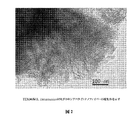
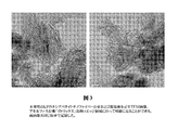
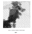
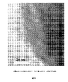
透過電子顕微鏡(TEM)画像は、ヒドロキシアパタイトナノファイバーの凝集体および束を示しており、これらの間には非結晶性有機物がある(図2、3)。アパタイトの平均直径は約5nmである。ナノファイバーの長さは約10〜数百ナノメートルである。伸長方向はアパタイト結晶のc軸である。非結晶性の箔のような有機材料はサンプルの「マトリックス」である。アパタイトナノファイバーは有機材料によって互いに「接着されている(glued)」。アパタイトナノファイバーは非常に反応性であり、電子ビーム下で不安定である(通常の合成アパタイト結晶に対して)。 Transmission electron microscope (TEM) images show aggregates and bundles of hydroxyapatite nanofibers with amorphous organics between them (FIGS. 2 and 3). The average diameter of apatite is about 5 nm. The length of the nanofiber is about 10 to several hundred nanometers. The extension direction is the c-axis of the apatite crystal. An organic material, such as an amorphous foil, is the “matrix” of the sample. Apatite nanofibers are “glued” together by organic materials. Apatite nanofibers are very reactive and unstable under electron beams (as opposed to normal synthetic apatite crystals).
説明:本発明の化合物は、その自然形態では一部ヒドロキシアパタイトとして存在し、従って、形状は六角形である。 Description: The compounds of the present invention exist in part as hydroxyapatite in their natural form and are therefore hexagonal in shape.
本発明の化合物は、骨修復、形成、維持、再生およびコラーゲン形成を促進する。本発明は、コラーゲン形成、腱の健康および治癒、骨の健康および治癒を高め、そして糖尿病性骨吸収を維持および助ける方法および組成物に関する。本発明は代謝性障害の治療において有用である。本発明は、ヒドロキシアパタイトナノファイバーと、種々の正および負電荷により本質的に塩基性および酸性である乳清特異的タンパク質から構成される有機マトリックスとのナノ複合体である。タンパク質はpH依存性であり、α−ラクトグロブリン(旧称ラクトアルブミン)、β−ラクトグロブリン(旧称ラクトアルブミン)、ラクトフェリン、ラクトペルオキシダーゼ、IgG、IgM(m鎖)、分泌片*、(*分泌片は、IgAと結合して見出されることが多い糖タンパク質である)、α−カゼイン、ウシ血清アルブミン、IgA(a鎖)またはIgD(d鎖)、糖タンパク質、カゼインホスホペプチド、リポタンパク質A1、レチノール結合タンパク質、ならびに他の微量タンパク質(ウシ成長因子およびプレアルブミンなど)を含むがこれらに限定されない。 The compounds of the present invention promote bone repair, formation, maintenance, regeneration and collagen formation. The present invention relates to methods and compositions that enhance collagen formation, tendon health and healing, bone health and healing, and maintain and aid diabetic bone resorption. The present invention is useful in the treatment of metabolic disorders. The present invention is a nanocomposite of hydroxyapatite nanofibers and an organic matrix composed of whey-specific proteins that are essentially basic and acidic by various positive and negative charges. The protein is pH-dependent, and α-lactoglobulin (formerly called lactalbumin), β-lactoglobulin (formerly called lactalbumin), lactoferrin, lactoperoxidase, IgG, IgM (m chain), secretion piece * , ( * , Α-casein, bovine serum albumin, IgA (a chain) or IgD (d chain), glycoprotein, casein phosphopeptide, lipoprotein A1, retinol binding Including, but not limited to, proteins and other trace proteins such as bovine growth factor and prealbumin.
本発明の材料の透過電子顕微鏡(TEM)画像は、ヒドロキシアパタイトナノファイバーの凝集体および束を示しており、これらの間には非結晶性有機物がある(例えば、図2、3を参照)。アパタイトの平均直径は約5nmである。ナノファイバーの長さは約10〜数百ナノメートルである。伸長方向はアパタイト結晶のc軸である。非結晶性の箔のような有機材料はサンプルの「マトリックス」である。アパタイトナノファイバーは有機材料によって互いに「接着されている(glued)」。アパタイトナノファイバーは非常に反応性であり、通常の合成アパタイト結晶に対して、電子ビーム露光下で不安定な傾向がある。 Transmission electron microscope (TEM) images of the materials of the present invention show agglomerates and bundles of hydroxyapatite nanofibers with amorphous organics between them (see, eg, FIGS. 2 and 3). The average diameter of apatite is about 5 nm. The length of the nanofiber is about 10 to several hundred nanometers. The extension direction is the c-axis of the apatite crystal. An organic material, such as an amorphous foil, is the “matrix” of the sample. Apatite nanofibers are “glued” together by organic materials. Apatite nanofibers are very reactive and tend to be unstable under electron beam exposure relative to normal synthetic apatite crystals.
どの理論によっても束縛されることは望まないが、マサチューセッツ工科大学のMarkus Buehlerは、均一な傾向がある従来の建築材料とは違って、骨は、細胞が絶えず変化している不均一な生体組織であると考えている。科学者は、骨の基本構造をサイズが増大する7つのレベルの階層に分類する。レベル1の骨は、チョーク様のヒドロキシアパタイトおよびコラーゲンフィブリル(強靭なタンパク質ストランドである)からなる。レベル2は、これらの2つがミネラル化コラーゲンフィブリル(コラーゲンフィブリル単独よりもはるかに強い)へ混合したものを含む。階層構造はこのようにして次第に大きくなる2つの基本材料の組み合わせを通してレベル7まで、あるいは骨全体が通常35%のタンパク質または有機マトリックスおよび65%のミネラルマトリックスで構成されるまで継続する。 While not wanting to be bound by any theory, the Massachusetts Institute of Technology Markus Buehler, unlike traditional building materials that tend to be homogeneous, bones are heterogeneous living tissue whose cells are constantly changing I believe that. Scientists classify the basic structure of bone into a seven-level hierarchy that increases in size. Level 1 bone consists of chalk-like hydroxyapatite and collagen fibrils (which are tough protein strands). Level 2 includes a mixture of these two into mineralized collagen fibrils (much stronger than collagen fibrils alone). The hierarchy continues in this way through the combination of the two basic materials, growing to level 7 or until the entire bone is usually composed of 35% protein or organic matrix and 65% mineral matrix.
分子レベルでは、ミネラル化コラーゲンフィブリルは、コラーゲン分子および一貫したサイズのヒドロキシアパタイト結晶の交互の糸でできている。これらの糸は互いに「積み重ねられている(stacked)」が、結晶が階段に似るように互い違いに配置される。糸の中および糸の間において、結晶と分子との間に弱い結合が形成される。 At the molecular level, mineralized collagen fibrils are made up of alternating threads of collagen molecules and consistently sized hydroxyapatite crystals. These yarns are “stacked” with each other, but are staggered so that the crystals resemble steps. Weak bonds are formed between crystals and molecules in and between yarns.
布のようなフィブリルを押圧すると、コラーゲン分子と結晶との間のいくらか弱い結合が破壊され、フィブリルに小さい間隙または伸張領域が生じる。伸張は圧力をより広い領域に広げ、事実上、コラーゲン分子内の他のより強い結合(圧力がその上に集中すると完全に破壊し得る)を保護する。また伸張は力に応じて結晶を粉砕する(より大きい結晶のありそうな応答であり得る)のではなく移動させる。このエネルギーを吸収する能力は、例えば転倒したときに、骨破壊を低減することができると理論付けられる。 Pressing a fibril, such as a cloth, breaks some weak bonds between collagen molecules and crystals, resulting in small gaps or stretched areas in the fibrils. Stretching spreads the pressure over a wider area, effectively protecting other stronger bonds within the collagen molecule that can be completely broken when the pressure is concentrated on it. Stretching also moves the crystal in response to force rather than crushing it (which can be a likely response of a larger crystal). It is theorized that this ability to absorb energy can reduce bone destruction, for example, when falling.
Buehlerは、骨が伸張フィブリル布内の間隙を許容する独特の能力を有することを観察した。これらの間隙は、骨のリモデリングに関連する基本多細胞単位と同じ大きさ(数百マイクロメートル)を有する。この単位は、組織内を移動する際に一端で古い骨を侵食し、他方でそれを置換して、小さいクラックのような空洞を間に形成する小さいボーリングワーム(boring worm)のように協働する細胞の組み合わせである。 Buehler observed that the bone has a unique ability to allow gaps in the stretched fibril fabric. These gaps have the same size (several hundred micrometers) as the basic multicellular units involved in bone remodeling. This unit works like a small boring worm that erodes the old bone at one end as it moves through the tissue and replaces it on the other, forming a small crack-like cavity in between. Is a combination of cells.
従って、分子スケールで骨の強度に関与するメカニズムは、その更新に必要とされる多数の小さいクラックを含有するにもかかわらず、骨がいかに強いままであり得るのかも説明する。骨は、材料の階層構造によって可能となる間隙を利用することによって強度を作り出す。2007年11月1日、「How Bone is Built May Lead to New Materials」http://medicaldesign.com/materials/bone 2009年3月31日訪問。 Thus, the mechanisms involved in bone strength at the molecular scale also explain how bones can remain strong despite containing many small cracks required for their renewal. Bone creates strength by taking advantage of the gaps made possible by the hierarchical structure of the material. On November 1, 2007, “How Bone is Built May Lead to New Materials” http: // medicaldesign. com / materials / bone Visited March 31, 2009.
新規化合物の表面積は207.5m2/gである。これは、合成ヒドロキシアパタイト(Aldrich Chemicals、St.Louis、Missouriから)の表面積(29.6m2/g)および市販の炭酸カルシウム製品(カルシウムサプリメント錠剤の製造のために使用される、Generichem Corporation,NJからの医薬品グレード粉末)の表面積(2.7m2/g)と比較して非常に大きい。本発明の生成物は、その大きい反応表面を考慮して、非常に反応性であることが推測され得る。予備研究により、本発明の化合物は、炭酸カルシウム(業界で広く使用されるサプリメント)よりも約76倍反応性であると示される。 The surface area of the new compound is 207.5 m 2 / g. This includes the surface area of synthetic hydroxyapatite (from Aldrich Chemicals, St. Louis, Missouri) (29.6 m 2 / g) and the commercial calcium carbonate product (Generchem Corporation, NJ, used for the manufacture of calcium supplement tablets). The pharmaceutical surface powder) is very large compared to the surface area (2.7 m 2 / g). It can be assumed that the product of the present invention is very reactive in view of its large reaction surface. Preliminary studies indicate that the compounds of the present invention are about 76 times more reactive than calcium carbonate (a widely used supplement in the industry).
pH2溶液中での化学反応:
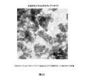
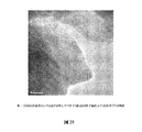
HCl(0.1M)酸を用いることにより溶液を調整した。0.05gの量のサンプルは、100mlのpH2溶液中に完全に溶解させることができる。最終溶液は透明であり、pH値は3.5である。0.15gのサンプルを100mlのpH2溶液中に添加すると、最終溶液は透明ではなく不透明であり、コロイド様の粒子を含有する懸濁液のように見える。pHの溶液は4.7に上昇する。透過電子顕微鏡を用いて懸濁液中の粒子を分析した。粒子はアモルファスCl含有リン酸Caである。最初のヒドロキシアパタイトナノファイバーは溶解され、アモルファスCl含有リン酸塩はCl含有溶液から沈殿し得ると考えられる。
Chemical reaction in pH 2 solution:
The solution was prepared by using HCl (0.1 M) acid. An amount of 0.05 g of sample can be completely dissolved in 100 ml of pH 2 solution. The final solution is clear and the pH value is 3.5. When 0.15 g of sample is added into 100 ml of pH 2 solution, the final solution is not clear but opaque and looks like a suspension containing colloidal particles. The pH solution rises to 4.7. The particles in the suspension were analyzed using a transmission electron microscope. The particles are amorphous Cl-containing Ca phosphate. It is believed that the initial hydroxyapatite nanofibers are dissolved and the amorphous Cl-containing phosphate can precipitate from the Cl-containing solution.
一組の溶解実験に基づく溶解動態学:
実験条件:0.05gのHexamenicolは、20mLのpH2溶液に添加され、室温で0.5時間、1時間、2時間、および3時間本発明の化合物と反応される。溶液と残りの固形分とを分離するために、設定した反応時間に到達したらすぐに反応生成物を遠心分離した。ミネラル電解質(帯電しており、それにより、正および負に帯電したタンパク質と結合する)を評価するために、残りの固形分を乾燥させ、注意深く秤量した。例えば、Ca2+分子は正電荷を有し、従って、負に帯電した分子と相互作用をして引き付ける。
Dissolution kinetics based on a set of dissolution experiments:
Experimental conditions: 0.05 g of Hexamenicol is added to 20 mL of pH 2 solution and reacted with a compound of the invention for 0.5 hour, 1 hour, 2 hours, and 3 hours at room temperature. To separate the solution from the remaining solids, the reaction product was centrifuged as soon as the set reaction time was reached. The remaining solids were dried and carefully weighed in order to evaluate the mineral electrolyte (which is charged and thereby binds to positively and negatively charged proteins). For example, Ca 2+ molecules have a positive charge and therefore interact and attract negatively charged molecules.
化学分析に基づく化学式:
化学式は提供された結果に基づいており、3Pに規格化される(Ca4.963,Mg0.0365,Sr0.0005)(PO4)3(OH0.82,Cl0.15,F0.03)。一般に、Na、Kがランダムに配置されたCa5(PO4)3.OHである。分子量は:506.34(g/式)。
Chemical formula based on chemical analysis:
Formula is based on results provided are normalized to 3P (Ca 4.963, Mg 0.0365, Sr 0.0005) (PO 4) 3 (OH 0.82, Cl 0.15, F 0.03 ). In general, Na 5 K is randomly arranged Ca 5 (PO 4 ) 3 . OH. The molecular weight is: 506.34 (g / formula).
これはヒドロキシアパタイトである。NaおよびKは可溶性の塩形態である。また、可溶性形態の微量のMgも可能である。 This is hydroxyapatite. Na and K are soluble salt forms. A soluble form of trace amounts of Mg is also possible.
基準単位格子パラメータおよび得られた化学式に基づいて計算したアパタイトの密度は3.15g/cm3である。通常、巨視的なヒドロキシアパタイトのアパタイト結晶の計算値よりもわずかに低い。 The density of the apatite calculated based on the reference unit cell parameters and the chemical formula obtained is 3.15 g / cm 3 . Usually slightly lower than the calculated value of macroscopic hydroxyapatite apatite crystals.
X線回折分析:
X線粉末回折パターンは、原末サンプルの結晶相はナノ結晶性アパタイト(ヒドロキシアパタイト)であることを示す。強く比較的鋭い002回折ピーク(d=3.416A)を除いて、回折ピークは全て非常に幅広い。回折ピークの形状は、ヒドロキシアパタイト結晶が、c軸に沿った伸張方向を有するナノファイバーの結晶であることを示す。
X-ray diffraction analysis:
The X-ray powder diffraction pattern shows that the crystalline phase of the bulk powder sample is nanocrystalline apatite (hydroxyapatite). Except for the strong and relatively sharp 002 diffraction peak (d = 3.416A), all the diffraction peaks are very broad. The shape of the diffraction peak indicates that the hydroxyapatite crystal is a nanofiber crystal having an extension direction along the c-axis.
単位格子の精密化:
新規化合物アパタイトの単位格子パラメータを、ヒドロキシアパタイトの公表された平均構造に基づく全パターン精密化法(Rietveld法)に基づいて計算した。結果は、単位格子のa−ディメンジョンおよびb−ディメンジョンは、標準ヒドロキシアパタイトよりもわずかに大きいことを示す。しかしながら、そのc−ディメンジョンは標準ヒドロキシアパタイトよりもわずかに小さい。ナノファイバーはアパタイト構造の構造緩和、特に表面および近傍表面の原子に影響を与えると考えられる。新規化合物アパタイト構造の原子座標は参照構造とはわずかに異なり得ることが予想される。しかしながら、非常に幅広い回折ピークを有する回折パターンに基づいて座標を精密化することは実用的でない。
The unit cell parameters of the new compound apatite were calculated based on a total pattern refinement method (Rietveld method) based on the published average structure of hydroxyapatite. The results show that the unit cell a-dimension and b-dimension are slightly larger than standard hydroxyapatite. However, its c-dimension is slightly smaller than standard hydroxyapatite. Nanofibers are thought to affect the structural relaxation of the apatite structure, particularly the atoms on the surface and nearby surfaces. It is expected that the atomic coordinates of the new compound apatite structure may be slightly different from the reference structure. However, it is not practical to refine the coordinates based on a diffraction pattern having a very wide diffraction peak.
被験者および方法:一般的
被験者.参加者は、ウィスコンシン州マディソン地域の全住民から採用した、その他の面で健康であり、ダイエットサプリメントを摂取しておらず、通常の生活をしている閉経期および閉経後の女性であった。初めに17人の女性が研究に対する同意書に署名したが、プロトコールの不履行のために4人は不適格とみなされた。40歳から71歳までの範囲の13人の女性が12週間の研究を完了した。研究プロトコールはHIPAA規則に従った。
Subjects and methods: General subjects. Participants were menopausal and postmenopausal women who were recruited from all residents of the Madison area of Wisconsin and who were otherwise healthy, did not take dietary supplements, and had a normal life. Initially 17 women signed a consent form for the study, but four were deemed ineligible due to protocol failure. Thirteen women, ranging from 40 to 71 years old, completed the 12-week study. The study protocol followed HIPAA rules.
プロトコール.登録の後、各参加者に尿NTxアッセイキットを与え、ベースラインNTx読取値を確立するために、2回目の朝の尿(second void of morning urine)サンプル(処置前)を採取し、サンプルを往復翌日配達便で中央試験研究所(Madison Pharmacy Associates,Madison,WI)に郵送するように指示した。参加者にその初期読取値を通知した。正常なNTx読取値は38nMBCEよりも低く、上昇したNTxは40〜60nMBCEであり、高NTxは60nMBCEよりも上である。上昇したNTxは骨減少症の徴候であり、高NTxは、BMDおよび骨減少と相関して骨粗鬆症と推察される。38よりも高いNTx読取値を有する選択された参加者に、使用説明書と共に、12週間供給するための2グラムパケットに個々に包装された粉末形態のHexamenicol化合物、毎日の摂取を記録するカレンダー、第2の試験NTxキット、および他の必要な連絡先情報を郵送した。参加者に、朝および就寝の30分前に1日2回、2グラムのHexamenicol粉末(500mgカルシウム相当)を1日2回、総計1000mgカルシウム相当を消費するように要求した。参加者は研究モニターに関する情報を無制限に利用できた。参加者に毎週連絡を取って、期待される記録を維持しながら研究プロトコールを遵守していることを確認し、製品がいかに良く耐容性を示しているかについて評価を行った。Hexamenicol摂取の12週間後に、プロトコールを遵守したことが確認された参加者に、2回目の尿サンプル(処置後)を翌日配達便で送付することを依頼した。 Protocol. After enrollment, each participant will be given a urine NTx assay kit and a second morning of morning urine sample (before treatment) will be taken to establish a baseline NTx reading Instructed to mail to the Central Laboratory (Madison Pharmacy Associates, Madison, Wis.) On a round-trip next day delivery. Participants were notified of their initial readings. Normal NTx readings are below 38 nMBCE, elevated NTx is 40-60 nMBCE and high NTx is above 60 nMBCE. Elevated NTx is a sign of osteopenia, and high NTx is presumed to be osteoporosis in correlation with BMD and bone loss. Selected participants with NTx readings higher than 38, with instructions for use, in powder form, Hexamenicol compound, individually packaged in a 2 gram packet to deliver for 12 weeks, a calendar recording daily intake, A second test NTx kit and other necessary contact information was mailed. Participants were requested to consume 2 grams of Hexamenicol powder (equivalent to 500 mg calcium) twice a day in the morning and 30 minutes before going to bed for a total of 1000 mg calcium equivalent. Participants had unlimited access to information on research monitors. Participants were contacted weekly to ensure adherence to the study protocol while maintaining the expected records and to evaluate how well the product was tolerated. Twelve weeks after Hexamenicol ingestion, participants who were confirmed to be in compliance with the protocol were asked to send a second urine sample (after treatment) on the next day delivery.
尿NTx(骨吸収マーカー)
尿NTxは、骨が破壊されたときに体が尿中に排出するI型コラーゲンの架橋N−テロペプチド(Teleopeptide)(NTx)を検査するための方法である(21、22)。NTx排出の速度の増大は、破骨細胞活性および骨破壊の速度が高くなったことを示す。通常骨密度の変化を数年にわたって検出する骨ミネラル密度(BMD)測定(23)とは違って、NTxは、数週間または数か月の骨代謝の変化を検出することができる(24)。NTxの2つの可能性のある用途は、(a)閉経に近いおよび閉経後の女性における骨減少を予測すること、および(b)処置に対する骨格の応答をモニターすることである。NTx試験は骨粗鬆症を直接決定しないが、従来の骨量測定によって測定されるように、骨密度の減少の可能性を決定する。NTxによって測定される骨吸収の速度が高くなるほど、骨減少の速度が大きくなる(22、25)。尿中に見出される骨吸収(NTx)マーカーのレベルの上昇は、閉経後の女性における骨減少の速度がより高いことと関連する(26、27)。
Urinary NTx (bone resorption marker)
Urinary NTx is a method for examining type I collagen cross-linked N-telopeptide (NTx) that the body excretes in the urine when bone is destroyed (21, 22). An increase in the rate of NTx excretion indicates an increased rate of osteoclast activity and bone destruction. Unlike bone mineral density (BMD) measurements (23), which usually detect changes in bone density over years, NTx can detect changes in bone metabolism over weeks or months (24). Two potential uses of NTx are (a) predicting bone loss in women near and after menopause, and (b) monitoring skeletal response to treatment. The NTx test does not directly determine osteoporosis, but determines the potential for bone density reduction as measured by conventional bone mass measurements. The higher the rate of bone resorption as measured by NTx, the greater the rate of bone loss (22, 25). Increased levels of bone resorption (NTx) markers found in urine are associated with higher rates of bone loss in postmenopausal women (26, 27).
骨吸収マーカーは、治療法の効果を評価する役割を果たすこともできる(28)。現在の骨粗鬆症の治療は、NTx研究における変化によって検出可能な骨吸収を低下させるように作用する(21)。マーカーを用いて、治療の効力を数か月のうちに決定することができる。これらの短縮された治療スケジュールは、1または2年間検出されないかもしれない骨密度の変化と比較すると、フィードバック応答を上昇させる。専門家によって、骨粗鬆症の投薬計画が効いている可能性の早期の証拠を実証することにより、治療法を継続するという患者の望みを増強することができ、治療の遵守が高まると提唱されている。骨粗鬆症患者を治療する多くの専門家は、発症中および予後における高骨代謝回転の役割の評価、ならびに吸収阻害薬への応答の評価において、骨吸収マーカーを使用する。骨マーカーの減少を検出できないと、吸収阻害薬療法の遵守または効力が欠けていると示されるであろう。 Bone resorption markers can also play a role in assessing the effects of treatment (28). Current treatments for osteoporosis act to reduce detectable bone resorption by changes in NTx studies (21). Using the markers, the efficacy of the treatment can be determined within a few months. These shortened treatment schedules increase the feedback response when compared to changes in bone density that may not be detected for one or two years. Experts have proposed that early evidence of the effectiveness of an osteoporosis regimen can help increase patients' desire to continue therapy and increase adherence to therapy . Many professionals who treat osteoporosis patients use bone resorption markers in assessing the role of high bone turnover during onset and prognosis, and in assessing response to resorption inhibitors. Failure to detect bone marker loss would indicate lack of adherence to or efficacy of absorption inhibitor therapy.
人口統計
被験者の状態.12週の研究期間中、どの被験者もHexamenicolTM粉末の使用からの腹部膨満、下痢、胸やけ、アレルギー症状または有害反応を報告しなかった。全員が日常生活によりその通常の健康を維持した。生活において悪い状況に直面した報告はなく、どの被験者も事故に遭遇せず、あるいは遭遇したという報告はなく、どんな種類のアレルギーもどんな疾患の診断のための処置もなかった。
本発明の組成物は以下の用途において役立つであろうと考えられる:種々の癌(特に、結腸直腸および胸筋)の予防(すなわち、防止)および治療(すなわち、緩和)的な抑制、腱の構築および修復、II型糖尿病、ならびに骨粗鬆症。全ての哺乳類用途、例えば獣医学的用途が意図される。例えば、本発明の材料は、減少の抑制、骨量構築、骨折の治癒、および骨量密度変化の制限または強化(必要に応じて)に適用可能であると考えられる。例えば、本発明の化合物は、獣医学的用途において骨折の処置および骨量の構築のために使用することができる。例えば、イヌは、骨折治癒および骨量強化または増強などの特定の用途を有する。イヌの股関節形成不全も処置可能であり、すなわち、本発明の化合物を用いて発症を抑制する、または形成不全を低下もしくは治癒させると考えられる。本発明の化合物、HexaminacolTMは、207.5m2/gというその大きい表面積のために、錠剤、カプセルの製造において、そして原末およびより小さい送達量の両方の粉末形態で送達するために、薬物または薬剤のキャリアとして理想的に使用可能であると考えられる。 The compositions of the present invention are believed to be useful in the following applications: prevention (ie prevention) and treatment (ie alleviation) suppression of various cancers (particularly colorectal and pectoral muscles), tendon construction And repair, type II diabetes, and osteoporosis. All mammalian applications are contemplated, such as veterinary applications. For example, the materials of the present invention may be applicable for inhibition of reduction, bone mass building, fracture healing, and limiting or strengthening bone mass density changes (as needed). For example, the compounds of the present invention can be used for fracture treatment and bone mass construction in veterinary applications. For example, dogs have particular uses such as fracture healing and bone mass strengthening or augmentation. Canine hip dysplasia can also be treated, ie, the compounds of the present invention will be used to suppress the onset or reduce or cure dysplasia. The compound of the present invention, Hexaminocol ™, is a drug or drug in the manufacture of tablets, capsules, and for delivery in both powder and smaller delivery dose powder forms because of its large surface area of 207.5 m2 / g. It can be ideally used as a drug carrier.
実施例1
この研究の焦点は、コンピュータによる技法を用いて、HAPおよびタンパク質/ペプチド/アミノ酸因子および乳タンパク質の間の相互作用のモードを原子レベルで決定し、グルタミン酸またはホスホセリン残基がHAP核形成および結晶成長の制御において好ましいかどうかを決定することである。
Example 1
The focus of this study is to use computational techniques to determine the mode of interaction between HAP and protein / peptide / amino acid factors and milk proteins at the atomic level, where glutamate or phosphoserine residues are HAP nucleation and crystal growth It is to determine whether it is preferable in the control.
FORCITE(AccelrysTM)コードにおいて実行される古典的な分子力学/分子動力学(MM/MD)技法を使用した。MM計算は、分子および周期系の両方に対して単一点エネルギーおよび構造最適化を提供する。 Classical molecular mechanics / molecular dynamics (MM / MD) techniques implemented in the FORCITE (Accelrys ™ ) code were used. MM calculations provide single point energy and structural optimization for both molecular and periodic systems.
溶液中のBSPに対し、1fs時間ステップで5psの間、300および500KにおいてNVTアンサンブルについてMDシミュレーションを実施した。周期系については、1fs時間ステップで5psの間の350Kにおけるクエンチ動力学を用いて、構造最適化の前に表面の優先的な吸着部位を得た。 MD simulations were performed on NVT ensembles at 300 and 500 K for 5 ps in 1 fs time step for BSP in solution. For periodic systems, preferential adsorption sites on the surface were obtained prior to structure optimization using quench kinetics at 350 K for 5 ps with 1 fs time steps.
古典的なMMシミュレーションにおける表面エネルギーおよび吸着エネルギーの信頼性は、ポテンシャルの精度に依存する。我々は、有機金属のために誘導される普遍力場(Universal Force Field)(UFF)ポテンシャルを選択し、従って我々の系はかなり正確に扱われなければならない。 The reliability of surface energy and adsorption energy in the classic MM simulation depends on the accuracy of the potential. We choose the Universal Force Field (UFF) potential induced for organometallics, so our system must be handled fairly accurately.
実施例2
古典的な分子モデリング技法を使用して、HAPまたは水性Ca2+およびHPO4 2−イオンの特定の結晶面と、グルタミン酸(R−COO−)およびプロトン化ホスホセリン(R−HPO4 −)アミノ酸残基を含有する酸性ペプチドとの間の相互作用のモードを検査した。ペプチドは、骨HAP核形成および結晶成長修正の制御における骨シアロタンパク質(BSP)の仮定上の活性部分を表す。
Example 2
Using classical molecular modeling techniques, specific crystal faces of HAP or aqueous Ca 2+ and HPO 4 2− ions and glutamic acid (R—COO − ) and protonated phosphoserine (R—HPO 4 − ) amino acid residues The mode of interaction with acidic peptides containing was examined. Peptides represent the hypothetical active part of bone sialoprotein (BSP) in the control of bone HAP nucleation and crystal growth modification.
ここに提供される我々の予備的な結果は、グルタミン酸およびプロトン化ホスホセリン部位の両方に対してHAPの(1010)表面よりも(0001)表面においてより強いペプチド結合を示唆する。さらに、吸着に対していずれの基も優先性はない。 Our preliminary results provided here suggest stronger peptide binding at the (0001) surface than at the (1010) surface of HAP for both glutamate and protonated phosphoserine sites. Furthermore, neither group has priority for adsorption.
溶液中のペプチドの平衡化により、HAはおそらくグルタミン酸基を含有する部位において優先的に核形成するものであり、核形成が生じるために最小数8つのグルタミン酸部位がペプチド構造において必要とされることが示される。 Due to the equilibration of the peptide in solution, HA presumably nucleates at sites containing glutamate groups, and a minimum number of 8 glutamate sites are required in the peptide structure for nucleation to occur Is shown.
実施例3
薬物動態および生物学的利用能の研究
40歳〜65歳の範囲の40人の閉経期の女性被験者群(自身の対照としての役割を果たす)を選択した。University of Wisconsin Medical School(Madison,WI)において行った臨床研究は、全てのInstitutional Review Board(IRB)のプロトコールおよび患者の守秘義務手順に従った。この研究(以下の表を参照)では、本発明の組成物は、Merkから市販されている利用可能なカルシウムサプリメントのOsCal(登録商標)に対して比較した。同じ閉経期の女性群が研究における対照群としての役割を果たした。研究は2段階で行い、第1段階はOsCalを用いる対照相であり、第2段階では本発明の生成物が投与された。被験者に全ての乳製品またはサプリメントを2週間回避し、研究開始の前に一晩絶食するように要求した。被験者500mgの本発明の化合物対500mgのOsCalを朝食と共に投与した。血液サンプルを0、1、2、3、5、7および9時間の間隔で採取し、ベースライン血中カルシウムおよびミネラルレベルを一定期間にわたって確立した。本発明の化合物は血流中に吸収されることが分かり、有害反応または有害な副作用がなく被験者にとって安全であると決定された。結果は、OsCal被験者がその血清中にカルシウムを有することを示した。しかしながら、本発明の化合物被験者はその血清中にカルシウムを有したが、結果は、骨形成にとって重要であることが知られているミネラル栄養分の存在を示した。この研究は本発明の化合物の吸収および安全性を確立した。
Pharmacokinetic and bioavailability study A group of 40 menopausal female subjects (serving as their own controls) ranging from 40 to 65 years old was selected. Clinical studies conducted at the University of Wisconsin Medical School (Madison, Wis.) Followed all Institutional Review Board (IRB) protocols and patient confidentiality procedures. In this study (see table below), the compositions of the present invention were compared against the available calcium supplement OsCal® commercially available from Merk. The same menopausal group of women served as a control group in the study. The study was conducted in two stages, the first stage was a control phase with OsCal, and the second stage was administered the product of the invention. Subjects were asked to avoid all dairy products or supplements for 2 weeks and fast overnight prior to study initiation. Subject 500 mg of a compound of the invention versus 500 mg OsCal was administered with breakfast. Blood samples were taken at 0, 1, 2, 3, 5, 7 and 9 hour intervals and baseline blood calcium and mineral levels were established over a period of time. The compounds of the present invention were found to be absorbed into the bloodstream and were determined to be safe for the subject without adverse reactions or adverse side effects. The results showed that the OsCal subjects had calcium in their serum. However, although the compound subjects of the present invention had calcium in their serum, the results showed the presence of mineral nutrients known to be important for bone formation. This study established the absorption and safety of the compounds of the present invention.
実施例4
NTx性能研究(骨減少のためのバイオマーカー)
50歳〜71歳の13人の閉経期の女性がこの骨回復研究を完了した。NTxは、I型骨コラーゲンの架橋N−テロペプチドの排出を測定するための尿アッセイである。骨形成および吸収は、コラーゲン廃棄物の処分のために尿中に存在するある程度のコラーゲンについては、正常である。尿中の異常に高いレベルのI型コラーゲンの存在は骨減少の表示である。
Example 4
NTx performance study (biomarker for bone loss)
Thirteen menopausal women aged 50-71 completed this bone recovery study. NTx is a urine assay to measure the excretion of cross-linked N-telopeptide of type I bone collagen. Bone formation and resorption is normal for some collagen present in the urine for disposal of collagen waste. The presence of abnormally high levels of type I collagen in the urine is an indication of bone loss.
全ての被験者を最初にそのNTx読取値について測定し、そのベースライン状態を確立した。正常なNTxは、38ナノ分子の尿中コラーゲン(nMBCE)よりも低い読取値であり、上昇したNTxレベルは38〜60であり、高いNTxレベルは60よりも上である。正常な閉経前の女性は約38の読取値を有する。参加被験者のNTx測定値は43〜79の範囲であった。少なくとも4オンスの水中に溶解した粉末形態の2グラムの本発明の化合物を1日2回で90日間被験者に投与した。研究期間の最後に、NTxの存在およびレベルについて被験者を再試験した。研究後のNTx結果は33〜38nMBCEの範囲であり、研究参加者の100%が健康なNTxレベルに正常化されることを示した。研究からの結果は、骨折に対する高い危険因子を示す最高NTx読取値を有する被験者が、本発明の治療法から最も利益を得ることを示した。我々は、これらの結果により、本発明の化合物は、尿中に見出される1型コラーゲンの減少を有意に低下させることにおいて有効であることが示されると考える。 All subjects were first measured for their NTx readings to establish their baseline status. Normal NTx has a lower reading than 38 nanomolecular urinary collagen (nMBCE), elevated NTx levels are 38-60, and high NTx levels are above 60. Normal premenopausal women have about 38 readings. Participants' NTx measurements ranged from 43 to 79. Subjects were administered 2 grams of a compound of the invention in powder form dissolved in at least 4 ounces of water twice a day for 90 days. At the end of the study period, subjects were retested for the presence and level of NTx. Post-study NTx results ranged from 33-38 nMBCE, indicating that 100% of study participants were normalized to healthy NTx levels. Results from the study have shown that subjects with the highest NTx readings that represent high risk factors for fractures will benefit most from the treatment of the present invention. We believe that these results indicate that the compounds of the invention are effective in significantly reducing the decrease in type 1 collagen found in urine.
実施例5
臨床実習研究(骨量の発達−BMD)
閉経後の女性を病院および近くのナーシングホームから採用した。これらの女性を骨粗鬆症の危険因子についてスクリーニングした。スクリーニングは、Achilles Insight(Lunar GE Medical)を使用する定量的超音波法{(Quantitative Ultra sound)(QUS)を用いて行った。Tスコア測定値がマイナス2.5(−2.5)である女性を診断および確認のためにDXA(Dual X−Ray Absorpotiory)測定を受けさせた。
Example 5
Clinical training study (Bone mass development-BMD)
Postmenopausal women were recruited from hospitals and nearby nursing homes. These women were screened for risk factors for osteoporosis. Screening was performed using quantitative ultrasound using the Achilles Insight (Lunar GE Medical) {(Quantitative Ultra sound) (QUS). A woman with a T-score measurement of minus 2.5 (−2.5) was subjected to DXA (Dual X-Ray Absorpotivity) measurements for diagnosis and confirmation.
研究:100人の閉経後の女性に本発明の化合物を投与し、1年の最後に骨の健康の改善についてスクリーニングした。研究からの結果は、ほとんど(87)の参加者が1年以内にそのQUS読取値の改善を示したことを示す。研究参加者は治療をさらに1年間継続して、研究を完了した。予備的および進行中のデータ分析によって、ほとんどの患者が2年間の研究期間にわたって1年に約5〜6%の範囲の割合で骨量を獲得したことが示された。一部の参加者はより著しい改善を示した。 Study: 100 postmenopausal women were administered a compound of the invention and screened for improved bone health at the end of the year. Results from the study show that most (87) participants showed improvement in their QUS readings within a year. Study participants completed the study with continued treatment for another year. Preliminary and ongoing data analysis showed that most patients acquired bone mass at a rate in the range of about 5-6% per year over a two year study period. Some participants showed more significant improvements.
尿NTxアッセイ(Osteomark,Princeton,NJ)を用いて骨吸収または骨破壊を測定した。処置の前(ベースライン)および12週間の処置の後(最終)にNTxレベルを評価した。平均閉経前スコアまたはNTx骨吸収は38nMBCEである。 Bone resorption or bone destruction was measured using the urine NTx assay (Ostemark, Princeton, NJ). NTx levels were assessed before treatment (baseline) and after 12 weeks of treatment (final). The average premenopausal score or NTx bone resorption is 38 nMBCE.
表3/3aは、ベースラインと比較した12週間の骨吸収変化についてのデータを示す。13人のうち11人の被験者は43〜60のベースラインNTx読取値を有し(「上昇したNTx群」)、他の2人の被験者は60よりも高いベースライン読取値を有した(「高NTx群」)。高NTxベースライン群の被験者はそれぞれ70歳および71歳であり、上昇したNTxベースライン群は、40〜69歳の範囲であった。13人の被験者は全て、NTx骨吸収マーカーの減少を経験した。13人のうち12人の被験者は37または38の最終NTx読取値(閉経前の平均)を有したが、1人の被験者は33の最終NTxスコアを有した。被験者のベースラインの変化率は−47.94%〜−13.63%の範囲であり、最も大きい低下は高NTxベースライン群の被験者で示された。 Table 3 / 3a shows data for bone resorption changes at 12 weeks compared to baseline. Eleven subjects out of 13 had a baseline NTx reading of 43-60 (“increased NTx group”) and the other two subjects had a baseline reading higher than 60 (“ High NTx group "). The subjects in the high NTx baseline group were 70 and 71 years old, respectively, and the elevated NTx baseline group ranged from 40 to 69 years old. All 13 subjects experienced a decrease in NTx bone resorption markers. Twelve of the 13 subjects had a final NTx reading of 37 or 38 (premenopausal average), while one subject had a final NTx score of 33. The percent change in subjects' baselines ranged from -47.94% to -13.63%, with the greatest decrease shown in subjects in the high NTx baseline group.
骨粗鬆症は病的な骨のリモデリング過程を伴う疾患であり、結果として、破骨細胞活性の上昇および骨芽細胞活性の減少への移行が生じ、正味の骨減少が起こり、骨折の危険が増大するという結果になる。この現象は、通常、閉経期中およびその後の女性で起こる。このような骨吸収は尿中のNTxの排出で見られ、容易に測定して、処置を観察および変更することができる。研究において、38nMBCEよりも高いベースライン尿NTx測定値は、「高NTx」カテゴリーの群、すなわち骨折の危険性が比較的高い群のサブセットによる異常な骨吸収を示した。 Osteoporosis is a disease with a pathological bone remodeling process that results in a shift to increased osteoclast activity and decreased osteoblast activity, resulting in net bone loss and increased risk of fracture Result. This phenomenon usually occurs in women during and after menopause. Such bone resorption is seen in the excretion of NTx in the urine and can be easily measured to observe and modify the procedure. In the study, baseline urinary NTx measurements higher than 38 nMBCE indicated abnormal bone resorption by a group of “high NTx” categories, ie, a relatively high risk of fracture.
骨のリモデリングは、有機因子および無機因子の両方によって指示される複雑な過程である。一定期間にわたるこれらの因子の欠乏は異常な骨吸収をもたらし、結果として骨減少が起こる。Hexamenicolは、骨の構築および維持のための「原材料」として役立つ有機成分および無機成分の両方を含有する。無機成分はカルシウムヒドロキシアパタイトの前駆体であり(表1を参照)、有機成分は、乳清塩基性タンパク質(MSBP)、骨形成タンパク質、1型コラーゲンの構成要素であるカゼインホスホペプチド、および骨材料の形成のための吸収、輸送および接着を同様に容易にするタンパク質を含む。この化合物は、炭水化物、必須脂肪酸およびビタミンも含有する。いくつかの研究により、乳塩基性タンパク質、MBPは骨形成を促進することが示されている。 Bone remodeling is a complex process dictated by both organic and inorganic factors. A deficiency of these factors over a period of time results in abnormal bone resorption resulting in bone loss. Hexamenicol contains both organic and inorganic components that serve as “raw materials” for bone building and maintenance. The inorganic component is a precursor of calcium hydroxyapatite (see Table 1), the organic component is whey basic protein (MSBP), bone morphogenetic protein, casein phosphopeptide that is a component of type 1 collagen, and bone material Proteins that facilitate absorption, transport and adhesion as well as the formation of This compound also contains carbohydrates, essential fatty acids and vitamins. Several studies have shown that the milk basic protein, MBP, promotes bone formation.
研究の被験者は、OstiGenによる12週間の処置の後、尿NTxレベルが閉経前平均の38nMBCEへ低下することを示したが、これは、研究の最後のその骨吸収が骨形成と同等であることの表れである。38nMBCEにおけるベースライン 30nMBCE/クレアチニン。被験者の1人は、33nMBCEのNTx読取値(閉経前平均のよりもはるかに低い値)を有した。これは、「骨代謝回転抑制」と呼ばれる現象に起因し得る。最高のベースライン変化率はより高齢の被験者(69歳〜71歳)で見られ、−36.66%−47.94%の範囲を記録した。最高の相対NTx値を有するこのサブセットは、この研究中に与えられた最大の利益を有した。DXAは「骨ミネラル密度」(BMD)を試験するための標準であるが、骨の質を測定しない。特に高齢者における1型コラーゲンの継続的な減少、およびカルシウム補給によるミネラルマトリックスの増大は正常なBMD読取値を与えるが、骨は脆弱になり、転倒したときに粉々になる。本発明の目的の1つは、病気、損傷および骨折を防止するために、ミネラルマトリックス(65%)および有機マトリックス(35%)の割合を維持することである。定量的超音波法(QUS)は、BMD読取値だけでなく骨量の質も提供する。QUSに加えて、NTx試験および骨アルカリホスファターゼ(BAP)試験は、DXAに加えて骨粗鬆症の疾患管理におけるツールボックスを提供する。
Study subjects showed that after 12 weeks of treatment with OstiGen, urinary NTx levels dropped to a premenopausal mean of 38 nMBCE, indicating that their bone resorption at the end of the study was comparable to bone formation It is a sign of. Baseline at 38
骨粗鬆症患者の骨減少率の直接測定は、2〜4年の期間にわたって少なくとも2回の骨量の測定を必要とし得る。このような戦略は初期評価の時点で処置するかどうかを決定することが必要な場合には実用的でない。閉経後の女性において骨減少率は骨代謝回転率に比例するので、骨吸収に特異的な骨代謝回転を評価することによって減少率を予測することができると示唆されている。このようにして評価される減少率は連続的な骨ミネラル密度(BMD)測定によって長年にわたって評価された減少率よりも精度が低いが、閉経前平均よりも高い骨吸収率は、一般的なBMDに関係なく、椎骨および股関節骨折の危険性の2倍の増大に関連する。研究により、Hexamenicolの使用により異常な骨吸収が閉経前レベルへ解決されることが明らかになり、推定される進行中の骨吸収/骨減少が反転されることが示された。 Direct measurement of bone loss rates in osteoporotic patients may require at least two bone mass measurements over a period of 2-4 years. Such a strategy is not practical when it is necessary to decide whether to treat at the time of the initial assessment. Since bone loss is proportional to bone turnover in postmenopausal women, it has been suggested that it can be predicted by assessing bone turnover specific to bone resorption. The rate of reduction assessed in this way is less accurate than the rate of decline assessed over many years by continuous bone mineral density (BMD) measurements, but bone resorption rates higher than the premenopausal mean Regardless, it is associated with a 2-fold increase in risk of vertebral and hip fractures. Studies have shown that the use of Hexamenicol resolves abnormal bone resorption to premenopausal levels and reverses the estimated ongoing bone resorption / bone loss.
閉経期および閉経後の被験者による12週の期間にわたる本発明の使用は、骨吸収レベルの低下を示した。骨折の危険が最も高い群は、正常な骨吸収レベルへの低下の達成において最大の利益を得た。比較的骨折の危険が最小である被験者も骨減少の減速により利益を得た。骨構築前駆体を含有するHexamenicolの連続使用は骨減少を遅くすると思われ、骨の構築に役立つ大きな見込みを示し、これは時間と共にBMD測定において明らかになるであろう。 Use of the present invention over a 12-week period by menopausal and post-menopausal subjects has shown reduced bone resorption levels. The group with the highest risk of fractures had the greatest benefit in achieving a reduction to normal bone resorption levels. Subjects with a relatively minimal risk of fracture also benefited from slowing bone loss. The continuous use of Hexamenicol containing bone building precursors appears to slow bone loss and shows great promise in helping bone building, which will become apparent in BMD measurements over time.
実施例6:非公式の症例研究:Chapel Hill,NC.
Tスコアが−2.5以下である骨粗鬆症と診断された女性の群に吸収阻害薬のFosamax(ビスホスホナートである)を処方した。ビタミンDと共にカルシウムサプリメントを摂取することも推奨された。
Example 6: Informal case study: Chapel Hill, NC.
The absorption inhibitor Fosamax (which is a bisphosphonate) was prescribed to a group of women diagnosed with osteoporosis with a T-score of -2.5 or less. Taking calcium supplements with vitamin D was also recommended.
7人の女性の群は、カルシウムおよびビタミンDの代わりにFosamaxおよびHexamenicolを受けた。別の女性の群はFosamax、カルシウムおよびビタミンDの日常的な処置を受けた。NTx試験を月ごとに行って、両方の群について3か月間の処置の進展をモニターした。結果は明らかに、Hexamenicolを受けて処置計画を遵守した全ての患者が3か月以内に正常なNTx読取値による利益を得るが、カルシウムサプリメント群では、患者のわずか67%だけが3か月以内の改善を示すことを示した。このことにより、Hexamenicolがビスホスホナート(Bishosphanate)と共にうまく作用して、骨粗鬆症の問題の取り組みにおいて良好な結果を生じ得ると我々が信じるに至った。 A group of 7 women received Fosamax and Hexamenicol instead of calcium and vitamin D. Another group of women received routine treatment with Fosamax, calcium and vitamin D. NTx trials were conducted monthly to monitor treatment progress for 3 months for both groups. The results clearly show that all patients who received Hexamenicol and adhered to the treatment plan would benefit from normal NTx readings within 3 months, but in the calcium supplement group, only 67% of patients within 3 months It was shown to show an improvement. This led us to believe that Hexamenicol could work well with bisphosphonates to produce good results in addressing osteoporosis issues.
実施例7
ウマの用途
ウマの運動競技では、骨量構築、骨成長、骨修復および骨格構造の維持を目的とする。特に、その年のどの月に生まれた子も1歳であると考えられ、厳しい訓練に耐えるために十分な骨量および骨格構造が得られる前に、非常に若い年齢で訓練を受けさせられた結果、重大な損傷が生じるレース産業によく適している。
Example 7
Uses of horses In the horse athletics, the aim is to build bone mass, bone growth, bone repair and maintain skeletal structure. In particular, a child born in any month of the year is considered to be one year old and was trained at a very young age before obtaining sufficient bone mass and skeletal structure to withstand rigorous training As a result, it is well suited for the racing industry where severe damage occurs.
レース場の家畜小屋において、2歳の高価な子馬(colt)が重大な管骨(canon bone)損傷を受けて、その結果、骨折の底および骨折した骨の上部にドリルで穴を開け、ピンで固定することにより骨折した管骨を再付着する手術になった。次に、脚を固定し、子馬に30〜60グラムのHexamenicolを投与した。支給穀物の上部に15〜30グラム/処置を1日2回、新規化合物を振りかけた。子馬は問題なくHexamenicolを消費し、全処置期間中副作用も見られなかった。1か月の処置の最後に、x線において著しい改善は認められなかった。処置を継続し、2か月の最後に、骨の実質的な形成が認められた。訓練者および獣医は石灰化が急速に起こっていると感じた。処置を継続し、3か月の最後に管骨は完全に治癒し、通常の折れていない脚よりもはるかに強い骨構造が得られた。子馬は本格的に訓練に戻り、レースに参加し、4か月で2位に入賞した。訓練者は「私の40年の訓練の全てにおいて、この子馬において見てきたように、管骨損傷のこのような完全な治癒を見たことがない」と発言した。さらなる実験により、これは、骨、筋肉および腱組織の維持において妊馬、離乳子馬およびメスの子馬、訓練中の若い成体に、そして成熟動物の間で、この製剤を提供するための実行可能な選択肢であると結論付けられた。この製剤はステロイド処置に直面している動物にとっても実行可能な選択肢であり、処置中のステロイド摂取による骨減少の過程でも利益を得る。 In a racehouse livestock shed, a 2-year-old expensive colt suffered severe canon bone damage, resulting in drilling the bottom of the fracture and the top of the fractured bone, It was an operation to reattach the fractured canal bone by fixing with a pin. The legs were then fixed and 30-60 grams of Hexamenicol was administered to the foal. The new compound was sprinkled on the top of the supplied grain at 15-30 grams / treatment twice a day. The foal consumed Hexamenicol without problems and had no side effects during the entire treatment period. There was no significant improvement in x-rays at the end of the 1 month treatment. Treatment continued and at the end of 2 months substantial bone formation was observed. Trainers and veterinarians felt that calcification was occurring rapidly. Treatment continued and at the end of 3 months the canal bone was completely healed, resulting in a much stronger bone structure than the normal unbroken leg. The foal returned to training in earnest, participated in the race and won the second place in four months. The trainer said, "In all of my 40 years of training, I have never seen such a complete healing of a vascular bone injury, as I have seen in this foal." Further experiments have shown that this has been done to provide this formulation to pregnant, weaning and female foals, young adults in training, and among mature animals in the maintenance of bone, muscle and tendon tissue It was concluded that this was a possible option. This formulation is also a viable option for animals facing steroid treatment and also benefits in the process of bone loss due to steroid intake during treatment.
実施例8
イヌの用途
Hexamenicolは、スポーツ動物、ショードッグおよび使役犬にとって、骨折治癒、股関節形成不全および骨格成長における処置のための有効な処置選択肢であることが分かる。骨強度および骨密度の維持のために利用可能な選択肢であることが分かる。1.5〜2グラムのHexamenicolを日々提供することを目的として、いくつかのバッチのドッグビスケットを作った。試験動物はHexamenicolで作られたビスケットを好み、副作用はなかった。試験動物は通常の役割を維持し、この分野でさらなる研究が計画されている。
Example 8
Dog Use Hexamenicol proves to be an effective treatment option for sports animals, show dogs and working dogs for treatment in fracture healing, hip dysplasia and skeletal growth. It can be seen that this is an available option for maintaining bone strength and bone density. Several batches of dog biscuits were made with the goal of providing 1.5-2 grams of Hexamenicol daily. The test animals preferred biscuits made with Hexamenicol and had no side effects. Test animals maintain their normal role and further research is planned in this area.
実施例9
トリおよび家禽の用途
産卵鶏に、120mg/7ポンド(鳥の平均重量)の我々の化合物を3か月間、その毎日の食糧中で供給し、卵殻の厚さおよび安定性、ならびに卵黄および卵白の堅さは、通常の日々の食糧が与えられた対照群よりも優れていることが分かった。これにより、我々の化合物が、家禽産業において、血清カルシウムおよび他のミネラル栄養分を維持して健康な卵を産むための用途を有することを信じるに至った。また、捕獲ペットおよび動物園産業の鳥のためのトリ繁殖プログラムにも適用することができ、絶滅危惧種の復活プログラムにおいて重要な用途を有する。
Example 9
Avian and poultry use Laying hens are fed 120 mg / 7 pounds (average bird weight) of our compound in their daily food for 3 months, and the thickness and stability of eggshell, as well as egg yolk and egg white The firmness was found to be superior to the control group fed with normal daily food. This led to believing that our compounds have uses in the poultry industry to maintain serum calcium and other mineral nutrients and lay healthy eggs. It can also be applied to bird breeding programs for captive pets and birds in the zoo industry, and has important uses in endangered species resurrection programs.
以下の参考文献は、本出願の一部であるかのように参照によって本明細書中に援用される。 The following references are hereby incorporated by reference as if they were part of this application.
参考文献
1. NOF Fast facts on osteoporosis:Disease Statistics.National Osteoporosis Foundation.2003年2月.
2. Siris ES,Miller PD,Barrett−Connor E,et al.NORA.Identification and fracture outcomes of undiagnosed low bone mineral density in postmenopausal women:Results from the National Osteoporosis Risk Assessment.JAMA.2001年12月12日;286(22):2815−2822.
3. Bayne A.Osteoporosis remains under−diagnosed in the United States.Review of National Osteoporosis Risk Assessment(NORA).Eureka Alert.2001年.
4. U.S.Dept.of Health & Human Services.Bone Health and Osteoporosis.A Report of the Surgeon−General,Rockville,MD:2004年.
5. Kalkwarf HJ,Khoury JC,Lanphear BP.Milk intake during childhood and adolescence,adult bone density and osteoporotic fractures in U.S.women.Am J Clin Nutr.2003年;77:257−265.
6. Renner E,Hermes M,Starke H.Bone mineral density of adolescents as affected by calcium intake through milk and milk products.Int Dairy J.1998年;8:759−764.
7. Heaney RP.Calcium,dairy products and osteoporosis.J Am Coll Nutr.2000年;19(2):83S−99S.
8. Holick MF,Dawson−Hughes B(編).Nutrition and bone health.Totowa,NJ.Humana Press 2004年;237−239.
9. Miller GD,Jarvis JK,McBean LD.Handbook of dairy foods and nutrition,第2版 Boca Raton FL:CRC Press,2000年.
10. Aoe S,Toba Y,Yamamura J,et al.Controlled trial of the effects of milk basic protein(MBP) supplementation on bone metabolism in healthy adult women.Biosci Biotechnol Biochem.2001年;65(4):913−918.
11. Yamamura J,Aoe S,Toba Y,et al.Milk basic protein(MBP) increases radial bone mineral density in adult women. Biosci Biotechnol Biochem.2002年;66(3):702−704.
12. Celotti F,Bignamini A.Dietary calcium and mineral/vitamin supplementation:A controversial problem.J Int Medical Res.1999年;27:1−14.
13. Swaminathan R.Nutritional factors in osteoporosis.Int J Clin Practice.1999年;53:540−548.
14. McBean LD.Building better bones with dairy foods throughout the life cycle.Diary Council Digest 2004年;75(6):31−36.
15. Toba Y,Takada Y,Yamamura J,Tanaka M,et al.Milk basic protein:A novel protective function of milk against osteoporosis.Bone.2000年;27(3):403−408.
16. Prentice A,Bates CJ.Adequacy of dietary mineral supply for human bone growth and mineralisation.Eur J Clin Nutr.1994年;48 suppl 1:S161−176;discussion S177.
17. Noat D,Grey A,Reid IR,Cornish J.Lactoferrin:A novel bone growth factor.Clin Med Res.2005年;3(2):93−101.
18. Matsuoka Y,Serizawa A,Yoshioka T,Yamamura J,et al.Cystatin C in milk basic protein and its inhibitory effect on bone resorption in vitro.Biosci Biotechnol Biochem.2002年;66(12):2531−2536.
19. Schlimme E,Meisel H.Bioactive peptides derived from milk proteins:Structural,physiological and analytical aspects.Die Nahrung.1995年;39:1−20.
20. Scholz−Ahrens KE,Schrezenmeir J.Effects of bioactive substances in milk on mineral and trace element metabolism with special reference to casein phosphopeptides.Br J Nutr.2000年;84 suppl 1:S147−153.
21. Hanson DA.A specific immunoassay for monitoring human bone resorption:Quantitation of Type I collagen cross−linked N−telopeptides in urine.J Bone Miner Res.1992年;7(11):1251−1258.
22. Schneider DL.Urinary N−telopeptide levels discriminate normal,osteopenic and osteoporotic bone mineral density. Arch Intern Med.1997年;157(11):1241−1245.
23. Kanis JA,Delmas P,Burckhardt P,Cooper C,Torgerson D.Guidelines for diagnosis and management of osteoporosis.Osteoporosis Int.1997年;7:390−406.
24. Osteomark NTx.Princeton NJ.Product background information.
25. Minisola S.Bone turnover and its relationship with bone mineral density in pre− and postmenopausal women with or without fractures.Maturitas.1998年;29(3):265−270.
26. Rogers A,Hannon R,Eastell R.Biochemical markers as predictors of rates of bone loss after menopause.J Bone Miner Res.2000年;15(7):1398−1404.
27. Bauer DC,Sklarin PM,Stone KL,Black DM,Nevitt MC,Ensrud KE,at al.Biochemical markers of bone turnover and prediction of hip bone loss in older women:The study of osteoporotic fractures.J Bone Miner Res.1999年;14(8):1404−1410.
28. Rosen CJ,Tenenhouse A.Osteoporosis Symposium:Biochemical markers of bone turnover.Postgraduate Medicine.1998年;104(4).
29. Cotran R,Kumar V,Collins T,Robbins S.Pathological basis of disease.Chapter:Skeletal system and soft tissue tumors: Bones.第6版 WB Saunders:1999年.
30. Corral DA,Amling M,Priemel M,et al.Dissociation between bone resorption and bone formation in osteopenic transgenic mice.Proc Natl Acad Sci USA.1998年;95(23):13835−13840.
31. Wactawski−Wende,J,Ph.D.,Morley Kotchen,J,M.D.,Anderson,GL,Ph.D,et al.Calcium plus Vitamin D Supplementation and the Risk of Colorectal Cancer New England Journal of Medicine,Volume 354:684−696,2006年2月16日,Number 7.
32. Kruger,MC et al.,The Effect of Whey Acidic Protein Fractions on Bone Loss in the Ovariectomised Rat,British J.of Nutrition(2005年),93,244−252.
33. Illich,JZ et al.,”Nutrition in Bone Health Revisited:A Story Beyond Calcium,J.of American College of Nutrition,Vol.,19,No.6,715−737(2000年).
References 1. NOF Fast facts on osteoporosis: Dissease Statistics. National Osteoporation Foundation. February 2003.
2. Siris ES, Miller PD, Barrett-Connor E, et al. NORA. Identification and fracture outcomes of undiagnosed low bone minority in postmenopausal women: Results from the National Osteosis Risk. JAMA. 12 December 2001; 286 (22): 2815-2822.
3. Bayne A.A. Osteoporation reminds under-diagnosed in the United States. Review of National Osteology Risk Assessment (NORA). Eureka Alert. 2001.
4). U. S. Dept. of Health & Human Services. Bone Health and Osteoporation. A Report of the Surgeon-General, Rockville, MD: 2004.
5. Kalkwarf HJ, Khoury JC, Lanfare BP. Milk Intake During Childhood and Adolescence, Adult Bone Density and Osteopoetic Fractures in U. S. woman. Am J Clin Nutr. 2003; 77: 257-265.
6). Renner E, Hermes M, Starke H. et al. Bone mineral density of adolescents as affected by calcium intact milk milk and milk products. Int Daily J.M. 1998; 8: 759-764.
7). Heney RP. Calcium, dairy products and osteoporosis. J Am Coll Nutr. 2000; 19 (2): 83S-99S.
8). Horick MF, Dawson-Hughes B (ed.). Nutrition and bone health. Totowa, NJ. Humana Press 2004; 237-239.
9. Miller GD, Jarvis JK, McBean LD. Handbook of Dairy Foods and Nutrition, 2nd Edition Boca Raton FL: CRC Press, 2000.
10. Aoe S, Toba Y, Yamamura J, et al. Controlled trial of the effects of milk basic protein (MBP) supplementation on bone metabolism in health adult woman. Biosci Biotechnol Biochem. 2001; 65 (4): 913-918.
11. Yamamura J, Aoe S, Toba Y, et al. Milk basic protein (MBP) increases radial bone mineral density in adult woman. Biosci Biotechnol Biochem. 2002; 66 (3): 702-704.
12 Celotti F, Bignami A. Dietary calcium and mineral / vitamin supplementation: A conventional probe. J Int Medical Res. 1999; 27: 1-14.
13. Swamithan R.M. Nutritional factors in osteoporation. Int J Clin Practice. 1999; 53: 540-548.
14 McBean LD. Building better bones with daily foods through the life cycle. Diary Council Digest 2004; 75 (6): 31-36.
15. Toba Y, Takada Y, Yamamura J, Tanaka M, et al. Milk basic protein: A novel protective function of milk against osteoporosis. Bone. 2000; 27 (3): 403-408.
16. Prentice A, Bates CJ. Adequacy of dietary supplementary for human bone growth and mineralization. Eur J Clin Nutr. 1994; 48 suppl 1: S161-176; discussion S177.
17. Noat D, Gray A, Reid IR, Cornish J. et al. Lactoferrin: A novel bone growth factor. Clin Med Res. 2005; 3 (2): 93-101.
18. Matsuoka Y, Serizawa A, Yoshioka T, Yamamura J, et al. Cystatin C in milk basic protein and its effects effect on bone resorption in vitro. Biosci Biotechnol Biochem. 2002; 66 (12): 2531-1536.
19. Schlimme E, Meisel H. et al. Bioactive peptides derived from milk proteins: Structural, physiologic and analytical aspects. Die Nahrung. 1995; 39: 1-20.
20. Scholz-Ahrens KE, Schrezenmeir J. et al. Effects of bioactive substances in milk on mineral and trace element metabolism with special reference to casein phosphopeptides. Br J Nutr. 2000; 84 suppl 1: S147-153.
21. Hanson DA. A specific immunoassay for monitoring human bone resolution: Quantification of Type I collagen cross-linked N-telopeptides in urine. J Bone Miner Res. 1992; 7 (11): 1251-1258.
22. Schneider DL. Universal N-telopeptide levels discriminative normal, osteopenic and osteopoonetic bone mineral density. Arch International Med. 1997; 157 (11): 1241-1245.
23. Kanis JA, Delmas P, Burkharddt P, Cooper C, Torgerson D .; Guidelines for diagnosis and management of osteoporation. Osteoporosis Int. 1997; 7: 390-406.
24. Osteomark NTx. Princeton NJ. Product background information.
25. Minisola S. Bone turnover and it's relations with bone mineral density in pre- and postmenopausal women with or without fractures. Maturitas. 1998; 29 (3): 265-270.
26. Rogers A, Hannon R, Eastell R. Biochemical markers as predictors of rates of bone loss after menopause. J Bone Miner Res. 2000; 15 (7): 1398-1404.
27. Bauer DC, Sklarin PM, Stone KL, Black DM, Nevitt MC, Ensrud KE, at al. Biochemical markers of bone turnover and prediction of hip bone loss in old woman: The study of osteoporotic fractures. J Bone Miner Res. 1999; 14 (8): 1404-1410.
28. Rosen CJ, Tenenhouse A.R. Osteoporosis Symposium: Biochemical markers of bone turnover. Postgraduate Medicine. 1998; 104 (4).
29. Cotran R, Kumar V, Collins T, Robbins S .; Pathological basis of disease. Chapter: Skeletal system and soft tissue tumors: Bones. 6th edition WB Sounders: 1999.
30. Coral DA, Amling M, Priemel M, et al. Dissociation between bone restoration and bone formation in osteotransgenic rice. Proc Natl Acad Sci USA. 1998; 95 (23): 13835-13840.
31. Wactawski-Wende, J, Ph. D. Morley Kotchen, J, M .; D. , Anderson, GL, Ph. D, et al. Calcium plus Vitamin D Supplementation and the Risk of Color Cancer New England Journal of Medicine, Volume 354: 684-696, February 16, 2006, Number 7.
32. Kruger, MC et al. , The Effect of Wey Acid Protein Fraction on Bone Loss in the Ovariectiated Rat, British J. et al. of Nutrition (2005), 93, 244-252.
33. Ilrich, JZ et al. , “Nutrition in Bone Health Revisited: A Story Beyond Calcium, J. of American College of Nutrition, Vol., 19, No. 6, 715-737 (2000).
Claims (15)
a)固体形態の乳清由来の乳ミネラルを提供する工程と、
b)溶液中の乳由来の塩基性タンパク質を提供する工程と、
c)工程b)の乳由来のタンパク質溶液をイオン交換工程にさらして、前記溶液中の乳由来の塩基性タンパク質の重量パーセントを増強する工程と、
d)前記乳由来の塩基性タンパク質溶液からナトリウム系ミネラルおよびナトリウム塩を除去する工程と、
e)前記乳由来の固体ミネラルを溶媒と混合し、その溶液を加熱することによって、前記乳由来の固体ミネラルを精製する工程と、
f)前記工程の精製した乳由来の固体ミネラル溶液を工程d)の増強された乳由来の塩基性タンパク質溶液と混ぜ合わせる工程と、
g)前記工程の溶媒を除去して、栄養複合材料を生じさせる工程と
を含むことを特徴とする、栄養複合材料の製造方法。 In the method for producing a nutritional composite material,
a) providing whey minerals derived from whey in solid form;
b) providing milk-derived basic protein in solution;
c) subjecting the milk-derived protein solution of step b) to an ion exchange step to increase the weight percent of the milk-derived basic protein in the solution;
d) removing sodium minerals and sodium salts from the milk-derived basic protein solution;
e) purifying the milk-derived solid mineral by mixing the milk-derived solid mineral with a solvent and heating the solution;
f) combining the purified milk-derived solid mineral solution of the above step with the enhanced milk-derived basic protein solution of step d);
g) removing the solvent in the above step to produce a nutritional composite material.
溶液中の付加的な乳由来のタンパク質が工程c)に従って処理されて、工程d)に従って処理される前記溶液中の乳由来の塩基性タンパク質の量が増大されることを特徴とする、請求項1に記載の方法。 The method of claim 1, wherein
The additional milk-derived protein in the solution is treated according to step c) to increase the amount of milk-derived basic protein in the solution treated according to step d). The method according to 1.
乳由来の塩基性タンパク質、および
乳清由来の乳ミネラル
を含む、骨吸収を阻害する量の複合材料を前記患者に投与する工程と、
前記骨吸収が阻害されるのを可能にする工程と
を含むことを特徴とする方法。 In a method of inhibiting bone resorption in a human patient in need of treatment for inhibition of bone resorption,
Administering to the patient an amount of a composite material that inhibits bone resorption comprising basic protein from milk and milk mineral from whey;
Allowing said bone resorption to be inhibited.
乳由来の塩基性タンパク質、および
乳清由来の乳ミネラル
を含む、骨粗鬆症を治療する量の複合材料を前記哺乳類に投与する工程を含むことを特徴とする方法。 In a method of treating osteoporosis in a mammal in need of treatment for osteoporosis,
Administering to the mammal an amount of a composite material to treat osteoporosis comprising basic protein from milk and milk mineral from whey.
Applications Claiming Priority (3)
| Application Number | Priority Date | Filing Date | Title |
|---|---|---|---|
| US24550509P | 2009-09-24 | 2009-09-24 | |
| US61/245,505 | 2009-09-24 | ||
| PCT/US2010/050202 WO2011038230A2 (en) | 2009-09-24 | 2010-09-24 | Compositions and methods for the prevention and treatment of metabolic diseases |
Publications (2)
| Publication Number | Publication Date |
|---|---|
| JP2013505950A true JP2013505950A (en) | 2013-02-21 |
| JP2013505950A5 JP2013505950A5 (en) | 2013-11-14 |
Family
ID=43796456
Family Applications (1)
| Application Number | Title | Priority Date | Filing Date |
|---|---|---|---|
| JP2012531070A Pending JP2013505950A (en) | 2009-09-24 | 2010-09-24 | Compositions and methods for prevention and treatment of metabolic disorders |
Country Status (8)
| Country | Link |
|---|---|
| US (4) | US8518453B2 (en) |
| EP (1) | EP2480099B1 (en) |
| JP (1) | JP2013505950A (en) |
| AU (1) | AU2010298141B2 (en) |
| CA (1) | CA2775276C (en) |
| NZ (1) | NZ599184A (en) |
| RU (1) | RU2565020C2 (en) |
| WO (2) | WO2011037957A2 (en) |
Families Citing this family (3)
| Publication number | Priority date | Publication date | Assignee | Title |
|---|---|---|---|---|
| WO2011037957A2 (en) | 2009-09-24 | 2011-03-31 | U.S. Foods & Pharmaceuticals, Inc. | Compositions and methods for bone repair, maintenance and regeneration |
| CN103599521A (en) * | 2013-11-08 | 2014-02-26 | 苏州冉新生物技术有限公司 | Vitamin D complex granules |
| BE1027357B1 (en) * | 2019-06-12 | 2021-01-18 | Vanlommel Maria Kristel | Dietary supplement for accelerated healing |
Citations (10)
| Publication number | Priority date | Publication date | Assignee | Title |
|---|---|---|---|---|
| JPH0733668A (en) * | 1993-07-21 | 1995-02-03 | Maruha Corp | Bone enhancement promoter |
| JPH099864A (en) * | 1995-06-29 | 1997-01-14 | Snow Brand Milk Prod Co Ltd | Composition containing calcium derived from milk and its production |
| US5639501A (en) * | 1995-01-31 | 1997-06-17 | Vembu; Rajan | Separation of minerals from whey permeate |
| JP2000281587A (en) * | 1999-03-30 | 2000-10-10 | Snow Brand Milk Prod Co Ltd | Bone resoprtion suppressor |
| JP2001346519A (en) * | 2000-06-09 | 2001-12-18 | Snow Brand Milk Prod Co Ltd | Method for producing fraction containing high content of milk basic cystatin and decomposed product thereof |
| JP2002000193A (en) * | 2000-06-20 | 2002-01-08 | Snow Brand Milk Prod Co Ltd | Method for producing fraction having high milk-derived basic cystatin content and cleavaged product thereof |
| JP2004115509A (en) * | 2002-09-05 | 2004-04-15 | Snow Brand Milk Prod Co Ltd | Osteoprotegerin inhibitory factor production promoter |
| JP2005060321A (en) * | 2003-08-15 | 2005-03-10 | Snow Brand Milk Prod Co Ltd | Osteogenic promotor |
| JP2007512374A (en) * | 2003-11-26 | 2007-05-17 | デピュイ・スパイン・インコーポレイテッド | Local intraosseous administration of osteogenic and antiresorptive agents and apparatus therefor |
| WO2008028194A2 (en) * | 2006-09-01 | 2008-03-06 | Cornell Research Foundation, Inc. | Calcium phosphate nanofibers |
Family Cites Families (24)
| Publication number | Priority date | Publication date | Assignee | Title |
|---|---|---|---|---|
| US3615664A (en) * | 1969-12-05 | 1971-10-26 | Foremost Mckesson | Treatment of whey |
| US3896241A (en) * | 1971-09-01 | 1975-07-22 | Coca Cola Co | Preparation of a whey protein concentrate |
| US4572837A (en) * | 1984-06-28 | 1986-02-25 | The British Food Manufacturing Industries Research Association | Protein product |
| NL9001692A (en) * | 1990-04-26 | 1991-11-18 | Cooeperatieve Melkproductenbed | METHOD FOR PREPARING AN OIL-IN-WATER EMULSION |
| JP3962427B2 (en) * | 1990-07-13 | 2007-08-22 | グロペップ リミテッド | Growth promoter |
| US7033610B2 (en) * | 1990-07-13 | 2006-04-25 | Gropep Pty, Ltd. | Growth-promoting agent |
| JP3604159B2 (en) * | 1992-05-07 | 2004-12-22 | 雪印乳業株式会社 | Whey-derived osteoblast proliferation-promoting and bone-strengthening factor and bone-strengthened food, medicine and feed containing the factor |
| US5912032A (en) * | 1995-05-11 | 1999-06-15 | Meiji Milk Products Company, Limited | Process of producing calcium-supplemented milk drinks |
| JP2974604B2 (en) * | 1996-01-23 | 1999-11-10 | 雪印乳業株式会社 | Basic protein composition, basic peptide composition and use thereof |
| US6139901A (en) * | 1997-09-16 | 2000-10-31 | New Zealand Milk Products (North Amerca) Inc. | Membrane filtered milk proteins varying in composition and functional attributes |
| JP4701472B2 (en) * | 2000-04-21 | 2011-06-15 | 雪印乳業株式会社 | Method for producing milk calcium composition |
| EP1602284A1 (en) | 2000-06-09 | 2005-12-07 | Snow Brand Milk Products, Co., Ltd. | Method of producing fractions containing a high concentration of milk basic cystatin and decomposition products thereof |
| NZ507335A (en) * | 2000-10-05 | 2004-10-29 | New Zealand Dairy Board | Bone health compositions derived from milk comprising an acidic protein fraction but that does not contain CGMP |
| US20030026845A1 (en) * | 2001-06-18 | 2003-02-06 | Etzel Lisa R. | Process for preparing protein isolate from milk, whey, colostrum, and the like |
| US20030118662A1 (en) * | 2001-12-05 | 2003-06-26 | Glanbia Foods, Inc. | Therapeutic uses of milk mineral fortified food products |
| FR2841747B1 (en) * | 2002-07-02 | 2004-08-20 | Cie Laitiere Europeenne | MILK PROTEIN ISOLATE AND PROCESS FOR ITS PREPARATION |
| KR100509177B1 (en) * | 2003-02-05 | 2005-08-22 | 롯데제과주식회사 | Composition of functional food for prevention of osteoporosis |
| US20050208638A1 (en) * | 2004-03-22 | 2005-09-22 | Chao Wu | Process for preparing bioactive protein-enriched whey products |
| WO2006058083A2 (en) * | 2004-11-24 | 2006-06-01 | Cornell Research Foundation, Inc. | Protein and calcium fortification system for clear and opaque beverages |
| US20090238947A1 (en) * | 2005-11-30 | 2009-09-24 | Leonard Marcus Flendrig | Calcium Fortified Food Product and Additive |
| DE102006009799A1 (en) * | 2006-03-01 | 2007-09-06 | Henkel Kgaa | Predominantly platelet-shaped sparingly water-soluble calcium salts and / or their composite materials, including these |
| FR2901796A1 (en) * | 2006-05-31 | 2007-12-07 | Lab Francais Du Fractionnement | PROCESS FOR EXTRACTING ONE OR MORE PROTEINS PRESENT IN MILK |
| JP5657200B2 (en) * | 2008-09-05 | 2015-01-21 | サントリー食品インターナショナル株式会社 | Milk composition and milk-containing beverage with reduced whey protein content |
| WO2011037957A2 (en) | 2009-09-24 | 2011-03-31 | U.S. Foods & Pharmaceuticals, Inc. | Compositions and methods for bone repair, maintenance and regeneration |
-
2010
- 2010-09-22 WO PCT/US2010/049757 patent/WO2011037957A2/en active Application Filing
- 2010-09-24 NZ NZ599184A patent/NZ599184A/en active IP Right Revival
- 2010-09-24 JP JP2012531070A patent/JP2013505950A/en active Pending
- 2010-09-24 EP EP10819532.2A patent/EP2480099B1/en not_active Not-in-force
- 2010-09-24 RU RU2012114489/13A patent/RU2565020C2/en active
- 2010-09-24 US US12/890,090 patent/US8518453B2/en active Active
- 2010-09-24 CA CA2775276A patent/CA2775276C/en active Active
- 2010-09-24 AU AU2010298141A patent/AU2010298141B2/en active Active
- 2010-09-24 WO PCT/US2010/050202 patent/WO2011038230A2/en active Application Filing
-
2012
- 2012-08-06 US US13/567,727 patent/US8557296B2/en active Active
-
2013
- 2013-10-10 US US14/050,810 patent/US20140227409A1/en not_active Abandoned
-
2016
- 2016-03-02 US US15/058,452 patent/US20170027989A1/en not_active Abandoned
Patent Citations (10)
| Publication number | Priority date | Publication date | Assignee | Title |
|---|---|---|---|---|
| JPH0733668A (en) * | 1993-07-21 | 1995-02-03 | Maruha Corp | Bone enhancement promoter |
| US5639501A (en) * | 1995-01-31 | 1997-06-17 | Vembu; Rajan | Separation of minerals from whey permeate |
| JPH099864A (en) * | 1995-06-29 | 1997-01-14 | Snow Brand Milk Prod Co Ltd | Composition containing calcium derived from milk and its production |
| JP2000281587A (en) * | 1999-03-30 | 2000-10-10 | Snow Brand Milk Prod Co Ltd | Bone resoprtion suppressor |
| JP2001346519A (en) * | 2000-06-09 | 2001-12-18 | Snow Brand Milk Prod Co Ltd | Method for producing fraction containing high content of milk basic cystatin and decomposed product thereof |
| JP2002000193A (en) * | 2000-06-20 | 2002-01-08 | Snow Brand Milk Prod Co Ltd | Method for producing fraction having high milk-derived basic cystatin content and cleavaged product thereof |
| JP2004115509A (en) * | 2002-09-05 | 2004-04-15 | Snow Brand Milk Prod Co Ltd | Osteoprotegerin inhibitory factor production promoter |
| JP2005060321A (en) * | 2003-08-15 | 2005-03-10 | Snow Brand Milk Prod Co Ltd | Osteogenic promotor |
| JP2007512374A (en) * | 2003-11-26 | 2007-05-17 | デピュイ・スパイン・インコーポレイテッド | Local intraosseous administration of osteogenic and antiresorptive agents and apparatus therefor |
| WO2008028194A2 (en) * | 2006-09-01 | 2008-03-06 | Cornell Research Foundation, Inc. | Calcium phosphate nanofibers |
Non-Patent Citations (1)
| Title |
|---|
| JPN6014037631; Biosci. Biotechnol. Biochem., 2007, Vol.71, No.2, pp.336-342 * |
Also Published As
| Publication number | Publication date |
|---|---|
| CA2775276C (en) | 2021-07-06 |
| AU2010298141A1 (en) | 2012-04-19 |
| US20140227409A1 (en) | 2014-08-14 |
| NZ599184A (en) | 2013-04-26 |
| RU2565020C2 (en) | 2015-10-10 |
| US8557296B2 (en) | 2013-10-15 |
| US20130029017A1 (en) | 2013-01-31 |
| WO2011037957A2 (en) | 2011-03-31 |
| EP2480099A4 (en) | 2015-05-27 |
| AU2010298141B2 (en) | 2015-12-17 |
| WO2011038230A3 (en) | 2011-08-11 |
| EP2480099B1 (en) | 2018-12-12 |
| US20170027989A1 (en) | 2017-02-02 |
| US8518453B2 (en) | 2013-08-27 |
| US20110189337A1 (en) | 2011-08-04 |
| CA2775276A1 (en) | 2011-03-31 |
| RU2012114489A (en) | 2013-10-27 |
| WO2011038230A2 (en) | 2011-03-31 |
| WO2011037957A3 (en) | 2012-02-23 |
| EP2480099A2 (en) | 2012-08-01 |
Similar Documents
| Publication | Publication Date | Title |
|---|---|---|
| US20120040908A1 (en) | Fracture repair promoter | |
| US20170027989A1 (en) | Compositions and Methods for the Prevention and Treatment of Metabolic Diseases | |
| JP2009107938A (en) | Biocompatible material and method for producing the same | |
| CN115399370A (en) | High-calcium fresh milk and preparation method and application thereof | |
| KR102471621B1 (en) | Beta-Casein and Cognitive Function | |
| CN107494963A (en) | Pet calcium cream and preparation method thereof | |
| WO2023111302A1 (en) | Complex coacervates of lactoferrin and osteopontin | |
| Lang et al. | Case report: fibrogenesis imperfecta ossium with early onset: Observations after 20 years of illness | |
| Ghanem et al. | Evaluation of Four Treatment Protocols on Experimentally Induced Nutritional Secondary Hyperparathyroidism in Kittens | |
| Riccardi et al. | Bone disease and new prevention frontiers chapter one: Osteoporosis | |
| Iwamoto et al. | Influence of gastrectomy on cortical and cancellous bones in rats | |
| Vujasinović-Stupar et al. | Supplementation with bio-calcium from shells Pinctada maxima in postmenopausal women with decreased mineral bone density: Pilot study | |
| JP6203723B2 (en) | Protein material for bone disease prevention or treatment and method for producing the same | |
| Prentice | The effects of silicon on skeletal integrity in poultry | |
| US10744188B2 (en) | High osteocalcin microcrystalline hydroxyapatite for calcium supplement | |
| Yumol | Determining If Lowering the Level of Dietary Calcium and Vitamin D in AIN-93G Diet Supports Normal Bone Development and Intestinal Integrity in Female CD-1 Mice | |
| van Riet | Zinc status assessment and the impact of dietary zinc on claw quality in pigs | |
| McKinnon | Effect of goat milk on bone mass, morphology and biomechanics: a thesis presented in partial fulfilment of the requirements for the degree of Doctor of Philosophy in Anatomy and Physiology at Massey University, Palmerston North, New Zealand | |
| Shidara | Eggshell Calcium | |
| CN111012900A (en) | Composition with function of increasing bone mineral density and preparation method and application thereof | |
| CN110338409A (en) | It is a kind of to prevent and treat the composition and its application that women's bone amount is lost | |
| Toure | Effects of iodine and selenium depletion on growth and bone quality of rats | |
| JP2009242422A5 (en) |
Legal Events
| Date | Code | Title | Description |
|---|---|---|---|
| A521 | Request for written amendment filed |
Free format text: JAPANESE INTERMEDIATE CODE: A523 Effective date: 20130924 |
|
| A621 | Written request for application examination |
Free format text: JAPANESE INTERMEDIATE CODE: A621 Effective date: 20130924 |
|
| A131 | Notification of reasons for refusal |
Free format text: JAPANESE INTERMEDIATE CODE: A131 Effective date: 20140909 |
|
| A601 | Written request for extension of time |
Free format text: JAPANESE INTERMEDIATE CODE: A601 Effective date: 20141208 |
|
| A602 | Written permission of extension of time |
Free format text: JAPANESE INTERMEDIATE CODE: A602 Effective date: 20141215 |
|
| A601 | Written request for extension of time |
Free format text: JAPANESE INTERMEDIATE CODE: A601 Effective date: 20150109 |
|
| A521 | Request for written amendment filed |
Free format text: JAPANESE INTERMEDIATE CODE: A523 Effective date: 20150209 |
|
| A131 | Notification of reasons for refusal |
Free format text: JAPANESE INTERMEDIATE CODE: A131 Effective date: 20150825 |
|
| A601 | Written request for extension of time |
Free format text: JAPANESE INTERMEDIATE CODE: A601 Effective date: 20151125 |
|
| A601 | Written request for extension of time |
Free format text: JAPANESE INTERMEDIATE CODE: A601 Effective date: 20151224 |
|
| A601 | Written request for extension of time |
Free format text: JAPANESE INTERMEDIATE CODE: A601 Effective date: 20160125 |
|
| A02 | Decision of refusal |
Free format text: JAPANESE INTERMEDIATE CODE: A02 Effective date: 20160517 |