JP2012531925A - Tibozanib response prediction - Google Patents
Tibozanib response prediction Download PDFInfo
- Publication number
- JP2012531925A JP2012531925A JP2012519530A JP2012519530A JP2012531925A JP 2012531925 A JP2012531925 A JP 2012531925A JP 2012519530 A JP2012519530 A JP 2012519530A JP 2012519530 A JP2012519530 A JP 2012519530A JP 2012531925 A JP2012531925 A JP 2012531925A
- Authority
- JP
- Japan
- Prior art keywords
- pgs
- tumor
- gene
- tivozanib
- responsive
- Prior art date
- Legal status (The legal status is an assumption and is not a legal conclusion. Google has not performed a legal analysis and makes no representation as to the accuracy of the status listed.)
- Pending
Links
Images

Classifications
-
- C—CHEMISTRY; METALLURGY
- C12—BIOCHEMISTRY; BEER; SPIRITS; WINE; VINEGAR; MICROBIOLOGY; ENZYMOLOGY; MUTATION OR GENETIC ENGINEERING
- C12Q—MEASURING OR TESTING PROCESSES INVOLVING ENZYMES, NUCLEIC ACIDS OR MICROORGANISMS; COMPOSITIONS OR TEST PAPERS THEREFOR; PROCESSES OF PREPARING SUCH COMPOSITIONS; CONDITION-RESPONSIVE CONTROL IN MICROBIOLOGICAL OR ENZYMOLOGICAL PROCESSES
- C12Q1/00—Measuring or testing processes involving enzymes, nucleic acids or microorganisms; Compositions therefor; Processes of preparing such compositions
- C12Q1/68—Measuring or testing processes involving enzymes, nucleic acids or microorganisms; Compositions therefor; Processes of preparing such compositions involving nucleic acids
- C12Q1/6876—Nucleic acid products used in the analysis of nucleic acids, e.g. primers or probes
- C12Q1/6883—Nucleic acid products used in the analysis of nucleic acids, e.g. primers or probes for diseases caused by alterations of genetic material
- C12Q1/6886—Nucleic acid products used in the analysis of nucleic acids, e.g. primers or probes for diseases caused by alterations of genetic material for cancer
-
- G—PHYSICS
- G16—INFORMATION AND COMMUNICATION TECHNOLOGY [ICT] SPECIALLY ADAPTED FOR SPECIFIC APPLICATION FIELDS
- G16B—BIOINFORMATICS, i.e. INFORMATION AND COMMUNICATION TECHNOLOGY [ICT] SPECIALLY ADAPTED FOR GENETIC OR PROTEIN-RELATED DATA PROCESSING IN COMPUTATIONAL MOLECULAR BIOLOGY
- G16B25/00—ICT specially adapted for hybridisation; ICT specially adapted for gene or protein expression
- G16B25/10—Gene or protein expression profiling; Expression-ratio estimation or normalisation
-
- C—CHEMISTRY; METALLURGY
- C12—BIOCHEMISTRY; BEER; SPIRITS; WINE; VINEGAR; MICROBIOLOGY; ENZYMOLOGY; MUTATION OR GENETIC ENGINEERING
- C12Q—MEASURING OR TESTING PROCESSES INVOLVING ENZYMES, NUCLEIC ACIDS OR MICROORGANISMS; COMPOSITIONS OR TEST PAPERS THEREFOR; PROCESSES OF PREPARING SUCH COMPOSITIONS; CONDITION-RESPONSIVE CONTROL IN MICROBIOLOGICAL OR ENZYMOLOGICAL PROCESSES
- C12Q2600/00—Oligonucleotides characterized by their use
- C12Q2600/118—Prognosis of disease development
-
- C—CHEMISTRY; METALLURGY
- C12—BIOCHEMISTRY; BEER; SPIRITS; WINE; VINEGAR; MICROBIOLOGY; ENZYMOLOGY; MUTATION OR GENETIC ENGINEERING
- C12Q—MEASURING OR TESTING PROCESSES INVOLVING ENZYMES, NUCLEIC ACIDS OR MICROORGANISMS; COMPOSITIONS OR TEST PAPERS THEREFOR; PROCESSES OF PREPARING SUCH COMPOSITIONS; CONDITION-RESPONSIVE CONTROL IN MICROBIOLOGICAL OR ENZYMOLOGICAL PROCESSES
- C12Q2600/00—Oligonucleotides characterized by their use
- C12Q2600/158—Expression markers
Abstract
ヒト腫瘍が、VEGFインヒビターであるチボザニブ(AV−951)での処置に応答性であるかもしくは抵抗性である(非応答性である)かを定量的に予測するための診断法が開示される。その試験は、予測遺伝子セットにおける遺伝子の発現レベルの測定へのアルゴリズムの適用に基づく。予測遺伝子セットは、AIF1、APBB1IP、ARHGAP30、C3AR1、CCR1、CD37、CD53、CD86、CLEC7A、CSF1R、CSF2RB、CTSS、CYBB、DOCK2、EVI2A、EVI2B、FPR3、GMFG、GPR65、HCK、HCLS1、HLA−DMA、IL10RA、ITGB2、LAIR1、LCP1、LCP2、LILRB1、LILRB2、LST1、LY86、MNDA、MS4A6A、MYO1F、NCF4、SLA、SLAMF8、TLR1、TYROBP、PLEK、CYTH4、およびPTPRCを含む。Disclosed is a diagnostic method for quantitatively predicting whether a human tumor is responsive or resistant (non-responsive) to treatment with the VEGF inhibitor tivozanib (AV-951) . The test is based on the application of an algorithm to measure the expression level of genes in the predicted gene set. Predicted gene sets are AIF1, APBB1IP, ARGGAP30, C3AR1, CCR1, CD37, CD53, CD86, CLEC7A, CSF1R, CSF2RB, CTSS, CYBB, DOCK2, EVI2A, EVI2B, FPR3, GMFG, GPR65, HCK, LA , IL10RA, ITGB2, LAIR1, LCP1, LCP2, LILRB1, LILRB2, LST1, LY86, MNDA, MS4A6A, MYO1F, NCF4, SLA, SLAMF8, TLR1, TYROBP, PLEK, CYTH4, and PTPRC.
Description
(関連出願への相互参照)
本出願は、2009年7月6日に出願された米国出願番号12/498,183の利益および優先権を主張し、上記米国出願番号12/498,183の全容は、参照によって本明細書に援用される。
(Cross-reference to related applications)
This application claims the benefit and priority of US application Ser. No. 12 / 498,183, filed Jul. 6, 2009, the entire contents of which is hereby incorporated by reference herein. Incorporated.
(発明の分野)
本発明の分野は、分子生物学、遺伝学、腫瘍学、バイオインフォマティクスおよび臨床診断学である。
(Field of Invention)
The field of the invention is molecular biology, genetics, oncology, bioinformatics and clinical diagnostics.
(発明の背景)
大部分のがんの薬物は、ある患者には有効であるが、ある患者には有効でない。このことは、腫瘍の中での遺伝的バリエーションから生じ、同じ患者内の腫瘍の間ですら認められ得る。患者の応答の変化のしやすさは、標的化した治療剤に関して、特に顕著である。従って、標的化した治療の最大限の潜在能力は、どの患者がどの薬物から利益を得るかを決定するための適切な試験なしには、実現されないだろう。国立衛生研究所(NIH)に従って、用語「バイオマーカー」は、「通常の生物学的プロセスもしくは病的なプロセス、または治療的介入に対する薬理学的応答の指標として客観的に測定され、評価される特性」として定義される。
(Background of the Invention)
Most cancer drugs are effective for some patients, but not for some patients. This arises from genetic variation within the tumor and can be seen even among tumors within the same patient. The susceptibility to changes in patient response is particularly significant with respect to targeted therapeutic agents. Thus, the full potential of targeted therapy would not be realized without proper testing to determine which patients would benefit from which drugs. According to the National Institutes of Health (NIH), the term “biomarker” is “objectively measured and evaluated as an indicator of normal biological or pathological processes, or pharmacological responses to therapeutic intervention. Defined as "characteristic".
バイオマーカーの発見に基づく改善された診断の開発は、予め、ある患者が、所定の薬物に対する臨床的応答を示す可能性が最も高いことを同定することによって、新たな薬物開発を加速する可能性を有する。このことは、臨床試験の大きさ、長さおよびコストを有意に低下させる。ゲノミクス、プロテオミクスおよび分子画像化などの技術は、現在、特定の遺伝子変異、特定の遺伝子の発現レベル、および他の分子バイオマーカーの迅速で、感度が高くかつ信頼性の高い検出を可能にしている。腫瘍の分子的特徴付けについての種々の技術が利用可能であるにも拘わらず、がんバイオマーカーの臨床的利用は、十分には実現されないままである。なぜなら、がんバイオマーカーが、ほとんど発見されていないからである。例えば、近年の総説記事は、以下のように述べている:
バイオマーカーおよびそれらの用途を早期に開発して、がんの診断および処置を改善することが必要不可欠である(非特許文献1)。
The development of improved diagnostics based on biomarker discovery may accelerate new drug development by identifying in advance that a patient is most likely to show a clinical response to a given drug Have This significantly reduces the size, length and cost of clinical trials. Technologies such as genomics, proteomics, and molecular imaging are now enabling rapid, sensitive, and reliable detection of specific gene mutations, expression levels of specific genes, and other molecular biomarkers . Despite the availability of various techniques for molecular characterization of tumors, the clinical use of cancer biomarkers remains to be fully realized. This is because few cancer biomarkers have been discovered. For example, a recent review article states:
It is essential to develop biomarkers and their uses at an early stage to improve cancer diagnosis and treatment (Non-patent Document 1).
がんバイオマーカーに関する別の近年の総説記事は、以下のコメントを含んでいる:
がんバイオマーカーを発見することは、難題である。いくつかの腫瘍タイプ(例えば、慢性骨髄性白血病、消化管間質腫瘍、肺がんおよび多形神経膠芽細胞腫)の分子的に規定されたサブセットを、分子的に標的化した薬剤を使用して、標的化することにおいて臨床的に成功してきたが、より幅広い状況においてこのような成功を適用する能力は、患者における標的化した薬剤を評価する効率的なストラテジーが欠如していることによって、ひどく制限されている。問題は、主に、これら刺激的な新たな薬物を評価する臨床試験のために、分子的に規定されたがんを有する患者を選択することができないことにある。その解決策は、特定の薬剤から利益を得る可能性が最も高い患者を信頼性高く同定するバイオマーカーを必要とする(非特許文献2の548ページにおいて)。
Another recent review article on cancer biomarkers includes the following comments:
Finding cancer biomarkers is a challenge. Using molecularly targeted drugs for molecularly defined subsets of several tumor types (eg, chronic myelogenous leukemia, gastrointestinal stromal tumors, lung cancer and glioblastoma multiforme) Has been clinically successful in targeting, but the ability to apply such success in a wider range of situations is severely due to the lack of efficient strategies to evaluate targeted drugs in patients Limited. The problem is mainly that patients with molecularly defined cancer cannot be selected for clinical trials evaluating these exciting new drugs. The solution requires a biomarker that reliably identifies the patient most likely to benefit from a particular drug (page 548 of Non-Patent Document 2).
前述のようなコメントは、臨床的に有用なバイオマーカーの発見およびこのようなバイオマーカーに基づく診断法の必要性の認識を示す。 Such comments indicate the discovery of clinically useful biomarkers and the recognition of the need for diagnostic methods based on such biomarkers.
がんバイオマーカーには3つの異なるタイプがある:(1)予後バイオマーカー、(2)予測バイオマーカー、および(3)薬力学(PD)バイオマーカー。予後バイオマーカーは、侵襲性(aggressiveness)(すなわち、増殖速度および/もしくは転移、ならびに処置への屈折性(refractiveness))に従って、がん(例えば、固形腫瘍)を分類するために使用される。これは、ときおり、「結果良好な」腫瘍の、「結果不十分な」腫瘍からの区別といわれる。予測バイオマーカーは、特定の患者が、特定の薬物での処置から利益を受ける可能性を評価するために使用される。例えば、ERBB2(HER2もしくはNEU)遺伝子が増幅される乳がんを有する患者は、トラスツズマブ(HERCEPTIN(登録商標))での処置から利益を受ける可能性が高いのに対して、ERBB2遺伝子増幅なしの患者は、トラスツズマブでの処置から利益を受ける可能性は低い。PDバイオマーカーは、患者が薬物を摂取している間の上記患者に対する上記薬物の効果の指標である。よって、PDバイオマーカーはしばしば、新たな薬物の臨床開発の初期段階の間に、投与レベルおよび投与頻度をガイドするために使用される。がんバイオマーカーの考察に関しては、例えば、非特許文献2を参照のこと。 There are three different types of cancer biomarkers: (1) prognostic biomarkers, (2) predictive biomarkers, and (3) pharmacodynamic (PD) biomarkers. Prognostic biomarkers are used to classify cancer (eg, solid tumors) according to aggressiveness (ie, growth rate and / or metastasis, and refraction to treatment). This is sometimes referred to as distinguishing a “good outcome” tumor from a “poor outcome” tumor. Predictive biomarkers are used to assess the likelihood that a particular patient will benefit from treatment with a particular drug. For example, patients with breast cancer in which ERBB2 (HER2 or NEU) gene is amplified are likely to benefit from treatment with trastuzumab (HERCEPTIN®), whereas patients without ERBB2 gene amplification The likelihood of benefiting from treatment with trastuzumab is low. The PD biomarker is an indicator of the effect of the drug on the patient while the patient is taking the drug. Thus, PD biomarkers are often used to guide dosage levels and dosage frequencies during the early stages of clinical development of new drugs. See, for example, Non-Patent Document 2 for discussion of cancer biomarkers.
チボザニブ(tivozanib(AV−951としても公知))は、VEGFレセプター1、2および3の強力かつ選択的な低分子インヒビターである。チボザニブは、3種のレセプター全てに対して、ピコモルの阻害活性を示し、前臨床モデルにおいて、抗腫瘍活性を示す(非特許文献3)。チボザニブは、272名の患者の第2相臨床試験においてポジティブな暫定的結果を得た(Bhargavaら,2009,ASCO Genitourinary Cancers Symposium,Abstract No.283)。 Tivozanib (also known as AV-951) is a potent and selective small molecule inhibitor of VEGF receptors 1, 2 and 3. Tibozanib exhibits picomolar inhibitory activity against all three receptors and antitumor activity in a preclinical model (Non-patent Document 3). Tibozanib obtained positive tentative results in a phase 2 clinical trial of 272 patients (Bhargava et al., 2009, ASCO Genitourinary Cancers Symposium, Abstract No. 283).
大量の前臨床研究および臨床研究が、VEGF標的化治療に焦点を当ててきたにも拘わらず、抗VEGF薬剤の抗腫瘍活性を担う機構は、十分には理解されていない。標的化された治療の他のタイプと同様に、全てではないが、いくらかの患者が、チボザニブ治療から利益を受けている。VEGF生物学の複雑さは、任意の所定の腫瘍に対するチボザニブの有効性を予測不能にしている。従って、チボザニブでの処置に応答する可能性がある(もしくは可能性がない)腫瘍を有する患者を同定するために使用され得る予測バイオマーカーに基づく診断法が必要である。 Despite the large amount of preclinical and clinical research that has focused on VEGF targeted therapy, the mechanisms responsible for the anti-tumor activity of anti-VEGF drugs are not well understood. As with other types of targeted treatments, some but not all patients benefit from tivozanib treatment. The complexity of VEGF biology makes the efficacy of tivozanib against any given tumor unpredictable. Accordingly, there is a need for a diagnostic biomarker-based diagnostic method that can be used to identify patients with tumors that may (or may not) respond to treatment with tivozanib.
(発明の要旨)
本発明は、以下の遺伝子のセットの発見に基づく:(a)マウスおよびヒトにおいて、それらの発現レベルに一貫性を示すもの;ならびに(b)その個々の発現レベルは、マウス腫瘍もしくはヒト腫瘍が、おそらく、チボザニブとして公知の抗がん剤での処置に応答性(感受性)であるかもしくは非応答性(抵抗性)であるかを集合的に示すもの。よって、本発明は、ヒト腫瘍が、チボザニブでの処置に応答性であるかもしくは非応答性であるかを定量的に予測するための診断法を提供する。上記方法は、以下の工程を包含する。
(Summary of the Invention)
The present invention is based on the discovery of the following set of genes: (a) those that show consistency in their expression levels in mice and humans; and (b) their individual expression levels are those of mouse tumors or human tumors Probably a collective indication of responsiveness (sensitivity) or non-responsiveness (resistance) to treatment with an anti-cancer agent known as tivozanib. Thus, the present invention provides a diagnostic method for quantitatively predicting whether a human tumor is responsive or non-responsive to treatment with tivozanib. The method includes the following steps.
(a)ヒト腫瘍に由来する組織サンプルにおいて、予測遺伝子セット(PGS)における各遺伝子の相対的発現レベルを決定する工程であって、ここで上記PGSは、以下の遺伝子(HUGO遺伝子記号によって示される)を含む、工程: (A) A step of determining a relative expression level of each gene in a predicted gene set (PGS) in a tissue sample derived from a human tumor, wherein the PGS is indicated by the following gene (HUGO gene symbol) ) Including:

ここでE1、E2、...E42は、上記PGSにおける42遺伝子の発現値である、工程。
Where E1, E2,. . . E42 is the expression value of 42 genes in the PGS.
規定された閾値未満のPGSスコアは、上記腫瘍が、おそらくチボザニブに対して応答性であることを示し、規定された閾値よりも高いPGSスコアは、上記腫瘍が、おそらくチボザニブに対して非応答性であることを示す。 A PGS score below a defined threshold indicates that the tumor is probably responsive to tivozanib, and a PGS score higher than the defined threshold indicates that the tumor is probably not responsive to tivozanib. Indicates that
本発明のいくつかの実施形態は、閾値決定分析を行い、それによって、規定された閾値を生成する工程を包含する。上記閾値決定分析は、レシーバーオペレーター特性曲線(receiver operator characteristic curve)分析を含み得る。上記PGSにおける各遺伝子の相対的遺伝子発現レベルは、その遺伝子のmRNAレベルを決定する(例えば、測定する)ことによって得られ得る。腫瘍組織サンプルにおけるmRNAレベルを決定するための適切な方法は、DNAマイクロアレイ分析および定量的逆転写酵素ポリメラーゼ連鎖反応(qRT−PCR)(例えば、TAQMAN(登録商標)アッセイ)を含む。 Some embodiments of the present invention include performing a threshold determination analysis, thereby generating a defined threshold. The threshold determination analysis may include a receiver operator characteristic curve analysis. The relative gene expression level of each gene in the PGS can be obtained by determining (eg, measuring) the mRNA level of that gene. Suitable methods for determining mRNA levels in tumor tissue samples include DNA microarray analysis and quantitative reverse transcriptase polymerase chain reaction (qRT-PCR) (eg, TAQMAN® assay).
別の局面において、本発明は、ヒトPGSもしくはマウスPGSにおける上記遺伝子の各々の発現を決定する(例えば、測定する)ためのプライマー対を含むPCRプライマーセットを提供する。本発明はまた、このようなPCRプライマーセットを含む診断試験キットを提供する。 In another aspect, the present invention provides a PCR primer set comprising a primer pair for determining (eg, measuring) the expression of each of the above genes in human PGS or mouse PGS. The present invention also provides a diagnostic test kit comprising such a PCR primer set.
(発明の詳細な説明)
上記PGSの遺伝子の個々の発現レベルは、マウス腫瘍およびヒト腫瘍を、チボザニブとして公知の抗腫瘍薬物での処置に応答性である可能性に従って分類するための予測バイオマーカーとして集合的に使用され得る。このような腫瘍分類は、臨床状況においてチボザニブでの処置の適切な候補であるヒト患者を同定するために有用である。
(Detailed description of the invention)
The individual expression levels of the PGS genes can be used collectively as predictive biomarkers to classify mouse tumors and human tumors according to their likelihood of being responsive to treatment with an anti-tumor drug known as tivozanib. . Such tumor classification is useful for identifying human patients who are suitable candidates for treatment with tivozanib in a clinical setting.
(定義)
本明細書で使用される場合、「AV−951」および「チボザニブ」とは、N−{2−クロロ−4−[(6,7−ジメトキシ−4−キノリル)オキシ]−フェニル}−N’−(5−メチル−3−イソオキサゾリル)ウレアを意味し、これは、以下の化学構造:
(Definition)
As used herein, “AV-951” and “tibozanib” refer to N- {2-chloro-4-[(6,7-dimethoxy-4-quinolyl) oxy] -phenyl} -N ′. Means-(5-methyl-3-isoxazolyl) urea, which has the following chemical structure:
本明細書で使用される場合、「一貫性」とは、遺伝子のセットに適用される場合、上記セットのメンバーの発現レベルが、所定の組織タイプ、例えば、腫瘍組織の中で、一致して増大もしくは低下する統計学的に有意な傾向を示すことを意味する。理論によって束縛されることは意図せず、本発明者らは、一貫性が、おそらく、上記一貫性のある遺伝子が1つ以上の生物学的機能において共通の関与を共有するということを示すことに言及する。 As used herein, “consistency” means that when applied to a set of genes, the expression levels of the members of the set are consistent within a given tissue type, eg, tumor tissue. It means to show a statistically significant tendency to increase or decrease. Without intending to be bound by theory, we show that consistency probably means that the consistent gene shares a common involvement in one or more biological functions. To mention.
本明細書で使用される場合、「最適な閾値PGSスコア」とは、分類子(classifier)が、偽陰性のコール(call)のコスト(cost)と偽陽性のコールのコストとの間で最も望ましいバランスを与える閾値PGSスコアを意味する。 As used herein, “optimal threshold PGS score” means that the classifier is the best between the cost of a false negative call and the cost of a false positive call. It means the threshold PGS score that gives the desired balance.
本明細書で使用される場合、「PGSスコア」とは、以下のアルゴリズムを使用して計算される数値を意味する: As used herein, “PGS score” means a numerical value calculated using the following algorithm:
本明細書で使用される場合、「レシーバーオペレーター特性」(ROC)曲線とは、バイナリー分類子系に関する、偽陽性率(感度) 対 真の陽性率(特異性)のグラフプロットを意味する。ROC曲線の構築において、以下の定義が適用される:
偽陰性率:FNR=1−TPR
真の陽性率:TPR=真の陽性/(真の陽性+偽陰性)
偽陽性率:FPR=偽陽性/(真の陽性+真の陰性)。
As used herein, “receiver operator characteristic” (ROC) curve means a graphical plot of false positive rate (sensitivity) versus true positive rate (specificity) for a binary classifier system. In constructing the ROC curve, the following definitions apply:
False negative rate: FNR = 1−TPR
True positive rate: TPR = true positive / (true positive + false negative)
False positive rate: FPR = false positive / (true positive + true negative).
本明細書で使用される場合、処置に対する「応答」もしくは処置に対して「応答する」とは、処置される腫瘍に関して、上記腫瘍が、(a)増殖の遅延、(b)増殖の停止、もしくは(c)退縮を示すことを意味する。 As used herein, “response” to treatment or “responding” to treatment refers to the tumor being treated, wherein the tumor is (a) delayed growth, (b) stopped growth, Or (c) indicates retraction.
本明細書で使用される場合、「閾値決定分析」とは、所定の腫瘍タイプに関して、閾値PGSスコア(例えば、最適な閾値PGSスコア)を決定するために、その特定の腫瘍タイプ(例えば、ヒト腎細胞がん)を表すデータセットの分析を意味する。閾値決定分析の状況において、所定の腫瘍タイプを表す上記データセットは、(a)実際の応答データ(応答もしくは非応答)、および(b)腫瘍保有マウスもしくはヒトの群からの各腫瘍についてのPGSスコアを含む。 As used herein, “threshold determination analysis” refers to a particular tumor type (eg, human) to determine a threshold PGS score (eg, optimal threshold PGS score) for a given tumor type. Analysis of data sets representing renal cell carcinoma). In the context of thresholding analysis, the data set representing a given tumor type is (a) actual response data (response or non-response), and (b) PGS for each tumor from the tumor-bearing mouse or human group. Includes score.
(予測遺伝子セット(PGS))
おそらく、チボザニブでの処置に対して応答性であるかもしくは非応答性であるヒト腫瘍の同定に関する本発明の実施形態において、上記PGSは、表1(以下)に列挙される42ヒト遺伝子を含む。
(Predicted gene set (PGS))
Presumably, in an embodiment of the invention relating to the identification of human tumors that are responsive or non-responsive to treatment with tivozanib, the PGS comprises 42 human genes listed in Table 1 (below) .
(組織サンプル)
ヒト患者もしくはマウスモデルにおける腫瘍の組織サンプルは、上記サンプルにおけるPGS遺伝子発現が、本発明に従って決定され得るように、RNAの供給源として使用され得る。代表的には、上記腫瘍は、がん腫、肉腫、神経膠腫もしくはリンパ腫である。上記組織サンプルは、従来の腫瘍生検機器および手順を使用することによって得られ得る。内視鏡生検、切除生検、切開生検、細針生検、穿刺生検、薄片生検および皮膚生検は、本発明の実施において使用するための腫瘍サンプルを得るために、当業者によって使用され得る認識された医療手順の例である。上記腫瘍組織サンプルは、個々の遺伝子発現レベルを測定するために、十分なRNAを提供するのに十分大量であるべきである。
(Tissue sample)
Tumor tissue samples in human patients or mouse models can be used as a source of RNA so that PGS gene expression in the samples can be determined according to the present invention. Typically, the tumor is a carcinoma, sarcoma, glioma or lymphoma. The tissue sample can be obtained by using conventional tumor biopsy instruments and procedures. Endoscopic biopsy, excision biopsy, incision biopsy, fine needle biopsy, puncture biopsy, slice biopsy and skin biopsy are performed by those skilled in the art to obtain tumor samples for use in the practice of the present invention. FIG. 6 is an example of a recognized medical procedure that can be used. The tumor tissue sample should be large enough to provide sufficient RNA to measure individual gene expression levels.
上記腫瘍組織サンプルは、遺伝子発現分析(例えば、RNA抽出および定量)を可能にする任意の形態にあり得る。よって、上記組織サンプルは、新鮮であり得、適切な低温技術を介して保存され得、または非低温技術を介して保存され得る。臨床的生検標本を取り扱うための標準的プロセスは、ホルマリン中で上記組織サンプルを固定し、次いで、これをパラフィン中に包埋することである。この形態にあるサンプルは、ホルマリン固定したパラフィン包埋(FFPE)組織として一般に知られている。組織調製およびその後のRNA抽出のための組織保存に適した技術は、当業者に周知である。 The tumor tissue sample can be in any form that allows gene expression analysis (eg, RNA extraction and quantification). Thus, the tissue sample can be fresh and stored via an appropriate cryogenic technique or can be stored via a non-cryogenic technique. The standard process for handling clinical biopsy specimens is to fix the tissue sample in formalin and then embed it in paraffin. Samples in this form are commonly known as formalin-fixed paraffin-embedded (FFPE) tissue. Techniques suitable for tissue preparation and subsequent tissue preservation for RNA extraction are well known to those skilled in the art.
上記PGSにおける各遺伝子の個々の遺伝子発現レベルは、上記PGSスコアを計算するために使用される入力値である。いったん組織サンプルが得られると、それは、上記PGSにおける個々の遺伝子の発現レベルを決定する(すなわち、測定する)ことが必要である。遺伝子発現レベルは、任意の適切な方法によって決定され得る。個々の発現を測定するための2つの例示的方法は、DNAマイクロアレイ分析およびqRT−PCRであり、これらは、以下で考察される。これらの選択すべき方法のいずれかのための必要条件は、RNA単離である。 The individual gene expression level of each gene in the PGS is an input value used to calculate the PGS score. Once a tissue sample is obtained, it is necessary to determine (ie, measure) the expression level of individual genes in the PGS. Gene expression levels can be determined by any suitable method. Two exemplary methods for measuring individual expression are DNA microarray analysis and qRT-PCR, which are discussed below. A prerequisite for any of these methods to select is RNA isolation.
(RNA単離)
組織サンプルから真核生物mRNA(すなわち、ポリ(a)RNA)を迅速かつ効率的に抽出するための方法は、十分に確立されており、当業者に公知である。例えば、Ausubelら,1997,Current Protocols of Molecular Biology,John Wiley & Sonsを参照のこと。上記組織サンプルは、新鮮、凍結、もしくは固定されパラフィン包埋された(FFPE)、臨床研究腫瘍標本であり得る。一般に、新鮮な組織サンプルもしくは凍結組織サンプルから単離されたRNAは、FFPEサンプル由来のRNAよりフラグメント化が少ない傾向にある。しかし、腫瘍材料のFFPEサンプルは、より容易に入手可能であり、FFPEサンプルは、本発明の方法における使用のためのRNAの適切な供給源である。RT−PCRによる遺伝子発現プロファイリングのためのRNA供給源としてのFFPEサンプルの考察については、例えば、Clark−Langoneら,2007,BMC Genomics 8:279を参照のこと。また、De Andreusら,1995,Biotechniques 18:42044;およびBakerら,米国特許出願公開第2005/0095634号を参照のこと。RNA抽出および調製についての業者の説明書付きの市販キットの使用は、広がっており、一般的である。種々のRNA単離製品および完全なキットの商業的業者としては、Qiagen(Valencia,CA)、Invitrogen(Carlsbad,CA)、Ambion(Austin,TX)およびExiqon(Woburn,MA)が挙げられる。
(RNA isolation)
Methods for rapidly and efficiently extracting eukaryotic mRNA (ie, poly (a) RNA) from tissue samples are well established and known to those skilled in the art. See, for example, Ausubel et al., 1997, Current Protocols of Molecular Biology, John Wiley & Sons. The tissue sample can be a fresh, frozen, or fixed and paraffin embedded (FFPE), clinical research tumor specimen. In general, RNA isolated from fresh or frozen tissue samples tends to be less fragmented than RNA from FFPE samples. However, FFPE samples of tumor material are more readily available and FFPE samples are a suitable source of RNA for use in the methods of the invention. For a discussion of FFPE samples as RNA sources for gene expression profiling by RT-PCR, see, eg, Clark-Langone et al., 2007, BMC Genomics 8: 279. See also De Andrews et al., 1995, Biotechniques 18: 42044; and Baker et al., US Patent Application Publication No. 2005/0095634. The use of commercial kits with vendor instructions for RNA extraction and preparation is widespread and common. Commercial vendors of various RNA isolation products and complete kits include Qiagen (Valencia, CA), Invitrogen (Carlsbad, CA), Ambion (Austin, TX), and Exiqon (Woburn, MA).
一般に、RNA単離は、組織/細胞破壊で始まる。組織/細胞破壊の間に、RNaseによるRNA分解を最小限にすることが望ましい。RNA単離プロセスの間にRNase活性を制限する1つのアプローチは、上記細胞が破壊されたら直ぐに、変性剤を、細胞内容物と接触した状態におくことを確実にすることである。別の一般的な慣行は、RNA単離プロセスにおいて1種以上のプロテアーゼを含めることである。必要に応じて、新鮮な組織サンプルは、集められたら直ぐに、室温でRNA安定化溶液中に浸漬される。上記安定化溶液は、上記細胞に迅速に浸透し、4℃での貯蔵、その後の単離のために、上記RNAを安定化する。1つのこのような安定化溶液は、RNAlater(登録商標)(Ambion,Austin,TX)として市販されている。 In general, RNA isolation begins with tissue / cell destruction. It is desirable to minimize RNA degradation by RNase during tissue / cell destruction. One approach to limit RNase activity during the RNA isolation process is to ensure that the denaturant is in contact with the cell contents as soon as the cells are destroyed. Another common practice is to include one or more proteases in the RNA isolation process. If necessary, fresh tissue samples are immersed in the RNA stabilization solution at room temperature as soon as they are collected. The stabilization solution rapidly penetrates the cells and stabilizes the RNA for storage at 4 ° C. and subsequent isolation. One such stabilization solution is commercially available as RNAlater® (Ambion, Austin, TX).
いくつかのプロトコルにおいて、全RNAは、塩化セシウム密度勾配遠心分離によって、破壊された腫瘍材料から単離される。一般に、mRNAは、全細胞RNAのうちの約1%〜5%を構成する。固定化オリゴ(dT)(例えば、オリゴ(dT)セルロース)は、mRNAを、リボソームRNAおよびトランスファーRNAから分離するために一般に使用される。単離後に貯蔵される場合、RNAは、RNaseを含まない条件下で貯蔵されなければならない。単離されたRNAの安定な貯蔵のための方法は、当該分野で公知である。RNAを安定に貯蔵するための種々の市販の製品が、利用可能である。 In some protocols, total RNA is isolated from disrupted tumor material by cesium chloride density gradient centrifugation. In general, mRNA constitutes about 1% to 5% of total cellular RNA. Immobilized oligo (dT) (eg, oligo (dT) cellulose) is commonly used to separate mRNA from ribosomal RNA and transfer RNA. If stored after isolation, the RNA must be stored under RNase free conditions. Methods for stable storage of isolated RNA are known in the art. Various commercial products are available for stable storage of RNA.
(マイクロアレイ分析)
複数遺伝子についてのmRNA発現レベルは、従来のDNAマイクロアレイ発現プロファイリング技術を使用して決定(例えば、測定)され得る。DNAマイクロアレイは、固体表面もしくは支持層(例えば、ガラス、プラスチックもしくはシリコン)に固定化した特定のDNAセグメントもしくはプローブの集まりであり、各特定のDNAセグメントは、アレイにおいて既知の位置を占有する。通常は、ストリンジェントな条件下での標識されたRNAのサンプルとのハイブリダイゼーションは、上記アレイにおける各プローブに対応するRNA分子の検出および定量を可能にする。非特異的に結合したサンプル材料を除去するためのストリンジェントな洗浄後、上記マイクロアレイは、共焦点レーザー顕微鏡もしくは他の適切な検出法によって、スキャンされる。現代の市販のDNAマイクロアレイ(しばしば、DNAチップとして公知)は、代表的には、数万のプローブを含み、従って、数万の遺伝子の発現を同時に測定し得る。このようなマイクロアレイは、本発明の実施において使用され得る。あるいは、上記PGSの遺伝子の発現を測定するために必要とされる程度の数のプローブと、必要なコントロールもしくは標準(例えば、データ正規化のため)とを含む特注チップが、本発明の実施において使用され得る。
(Microarray analysis)
The mRNA expression level for multiple genes can be determined (eg, measured) using conventional DNA microarray expression profiling techniques. A DNA microarray is a collection of specific DNA segments or probes immobilized on a solid surface or support layer (eg, glass, plastic or silicon), and each specific DNA segment occupies a known location in the array. Usually, hybridization with a sample of labeled RNA under stringent conditions allows the detection and quantification of RNA molecules corresponding to each probe in the array. After stringent washing to remove non-specifically bound sample material, the microarray is scanned by a confocal laser microscope or other suitable detection method. Modern commercial DNA microarrays (often known as DNA chips) typically contain tens of thousands of probes and can therefore measure the expression of tens of thousands of genes simultaneously. Such microarrays can be used in the practice of the present invention. Alternatively, a custom chip containing as many probes as needed to measure the expression of the PGS gene and the necessary controls or standards (eg, for data normalization) may be used in the practice of the invention. Can be used.
データ正規化を促進するために、2色のマイクロアレイリーダーが使用され得る。2色(2チャネル)システムにおいて、サンプルは、第1の波長で発光する第1のフルオロフォアで標識される一方で、RNAもしくはcDNA標準は、異なる波長で発光する第2のフルオロフォアで標識される。例えば、Cy3(570nm)およびCy5(670nm)は、しばしば、2色のマイクロアレイシステムにおいて一緒に使用される。 A two-color microarray reader can be used to facilitate data normalization. In a two-color (two-channel) system, the sample is labeled with a first fluorophore that emits at a first wavelength, while the RNA or cDNA standard is labeled with a second fluorophore that emits at a different wavelength. The For example, Cy3 (570 nm) and Cy5 (670 nm) are often used together in a two-color microarray system.
DNAマイクロアレイ技術は、十分に発展されており、市販され、広く使用されている。従って、本発明の方法を実施するにおいて、当業者は、過度の実験なくして、上記PGSにおける遺伝子の発現レベルを測定するために、マイクロアレイ技術を使用し得る。DNAマイクロアレイチップ、試薬(例えば、RNAもしくはcDNAの調製、RNAもしくはcDNAの標識に必要なもの、ハイブリダイゼーション溶液および洗浄溶液)、機器(例えば、マイクロアレイリーダー)およびプロトコルは、当該分野で周知であり、種々の商業的供給元から市販される。マイクロアレイシステムの商業的業者としては、Agilent Technologies(Santa Clara,CA)およびAffymetrix(Santa Clara,CA)が挙げられるが、他のPCRシステムも使用され得る。 DNA microarray technology is well developed, commercially available and widely used. Thus, in practicing the methods of the present invention, one skilled in the art can use microarray technology to measure the expression level of genes in the PGS without undue experimentation. DNA microarray chips, reagents (eg, preparation of RNA or cDNA, those required for RNA or cDNA labeling, hybridization and wash solutions), equipment (eg, microarray reader) and protocols are well known in the art, Commercially available from various commercial sources. Commercial vendors of microarray systems include Agilent Technologies (Santa Clara, CA) and Affymetrix (Santa Clara, CA), although other PCR systems can also be used.
(定量的RT−PCR)
上記PGSにおける個々の遺伝子を表すmRNAのレベルは、従来の定量的逆転写酵素ポリメラーゼ連鎖反応(qRT−PCR)技術を使用して決定(例えば、測定)され得る。qRT−PCRの利点としては、感度、柔軟性、定量的正確性、および密に関連したmRNA間を識別する能力が挙げられる。定量的PCRのために組織サンプルを加工処理することに関するガイダンスは、種々の供給元(例えば、qRT−PCRについての市販品の製造業者および業者)(例えば、Qiagen(Valencia,CA)およびAmbion(Austin,TX))から入手可能である。qRT−PCRの自動運転のための機器システムは、市販されており、多くの研究室において慣用的に使用されている。周知の商業的システムの例は、Applied Biosystems 7900HT Fast Real−Time PCR System(Applied Biosystems,Foster City,CA)である。
(Quantitative RT-PCR)
The level of mRNA representing individual genes in the PGS can be determined (eg, measured) using conventional quantitative reverse transcriptase polymerase chain reaction (qRT-PCR) techniques. Advantages of qRT-PCR include sensitivity, flexibility, quantitative accuracy, and the ability to discriminate between closely related mRNAs. Guidance on processing tissue samples for quantitative PCR is available from various sources (eg, commercial manufacturers and vendors for qRT-PCR) (eg, Qiagen (Valencia, Calif.)) And Ambion (Austin). , TX)). Instrument systems for qRT-PCR automatic operation are commercially available and routinely used in many laboratories. An example of a well-known commercial system is the Applied Biosystems 7900HT Fast Real-Time PCR System (Applied Biosystems, Foster City, Calif.).
いったん単離されたmRNAを手にしたら、RT−PCRによる遺伝子発現プロファイリングの第1の工程は、上記mRNAテンプレートをcDNAへと逆転写することである。次いで、cDNAは、PCR反応において指数関数的に増幅させられる。2つの一般的に使用される逆転写酵素は、トリ骨髄芽球症ウイルス(avilo myeloblastosis virus)逆転写酵素(AMV−RT)およびモロニーマウス白血病ウイルス逆転写酵素(MMLV−RT)である。上記逆転写反応は、代表的には、特定のプライマー、ランダムヘキサマー、もしくはオリゴ(dT)プライマーで開始される(primed)。適切なプライマーは、市販されている(例えば、GeneAmp(登録商標) RNA PCRキット(Perkin Elmer,Waltham,MA))。得られたcDNA生成物は、その後のポリメラーゼ連鎖反応においてテンプレートとして使用され得る。 Once the isolated mRNA is obtained, the first step in gene expression profiling by RT-PCR is to reverse transcribe the mRNA template into cDNA. The cDNA is then amplified exponentially in a PCR reaction. Two commonly used reverse transcriptases are the avian myeloblastosis virus reverse transcriptase (AMV-RT) and the Moloney murine leukemia virus reverse transcriptase (MMLV-RT). The reverse transcription reaction is typically initiated with specific primers, random hexamers, or oligo (dT) primers. Suitable primers are commercially available (eg, GeneAmp® RNA PCR kit (Perkin Elmer, Waltham, Mass.)). The resulting cDNA product can be used as a template in subsequent polymerase chain reactions.
上記PCR工程は、熱安定性DNA依存性DNAポリメラーゼを使用して行われる。PCRシステムにおいて最も一般的に使用されるポリメラーゼは、Thermus aquaticus(Taq)ポリメラーゼである。PCRの選択性は、増幅について標的とされたDNA領域(すなわち、上記PGSの遺伝子から逆転写されたcDNAの領域)に相補的であるプライマーの使用から生じる。従って、本発明においてqRT−PCRが使用される場合、上記PGSにおける各遺伝子に特異的なプライマーは、上記遺伝子のcDNA配列に基づく。商業的技術(例えば、SYBR(登録商標)greenもしくはTaqMan(登録商標)(Applied Biosystems,Foster City,CA))は、業者の説明書に従って使用され得る。メッセンジャーRNAレベルは、ハウスキーピング遺伝子(例えば、β−アクチンもしくはGAPDH)のレベルを比較することによって、サンプル間でのローディングの差異について正規化され得る。mRNA発現のレベルは、任意の単一のコントロールサンプル(例えば、通常の非腫瘍組織もしくは細胞に由来するmRNA)と比較して表され得る。あるいは、腫瘍サンプルのプール、もしくは腫瘍細胞株に由来するか、または市販のコントロールmRNAのセットに由来するmRNAに対して、表され得る。 The PCR step is performed using a thermostable DNA-dependent DNA polymerase. The most commonly used polymerase in PCR systems is Thermus aquaticus (Taq) polymerase. The selectivity of PCR results from the use of primers that are complementary to the DNA region targeted for amplification (ie, the region of the cDNA reverse transcribed from the PGS gene). Therefore, when qRT-PCR is used in the present invention, primers specific to each gene in the PGS are based on the cDNA sequence of the gene. Commercial techniques such as SYBR® green or TaqMan® (Applied Biosystems, Foster City, Calif.) Can be used according to the manufacturer's instructions. Messenger RNA levels can be normalized for differences in loading between samples by comparing levels of housekeeping genes (eg, β-actin or GAPDH). The level of mRNA expression can be expressed relative to any single control sample (eg, mRNA from normal non-tumor tissue or cells). Alternatively, it can be expressed against mRNA from a pool of tumor samples, or a tumor cell line, or from a set of commercially available control mRNAs.
上記PGSにおける遺伝子の発現レベルのPCR分析に適切なプライマーセットは、過度の実験なくして、当業者によって設計および合成され得る。あるいは、本発明を実施するための完全なPCRプライマーセットは、表1で上記に示されるように、上記PGSにおける遺伝子の正体に基づいて、商業的供給元(例えば、Applied Biosystems)から購入され得る。PCRプライマーは、好ましくは、長さが約17〜25ヌクレオチドである。プライマーは、融解温度(Tm)概算のための従来のアルゴリズムを使用して、特定のTmを有するように設計され得る。プライマー設計およびTm概算のためのソフトウェアは、市販されており(例えば、Primer ExpressTM(Applied Biosystems))、インターネット上でも利用可能である(例えば、Primer3(Massachusetts Institute of Technology))。PCRプライマー設計の確立された原理を適用することによって、多数の異なるプライマーが、任意の所定の遺伝子の発現レベルを測定するために使用され得る。よって、本発明は、特定のプライマーが上記PGSにおける任意の所定の遺伝子のために使用されることに関連して、制限されることはない。 Appropriate primer sets for PCR analysis of gene expression levels in the PGS can be designed and synthesized by one skilled in the art without undue experimentation. Alternatively, a complete PCR primer set for practicing the present invention can be purchased from a commercial source (eg, Applied Biosystems) based on the identity of the gene in the PGS, as shown above in Table 1. . PCR primers are preferably about 17-25 nucleotides in length. Primers can be designed to have a specific Tm using conventional algorithms for melting temperature (Tm) estimation. Software for primer design and Tm estimation is commercially available (eg, Primer Express ™ (Applied Biosystems)) and is also available on the Internet (eg, Primer3 (Massachuettes Institute of Technology)). By applying established principles of PCR primer design, a number of different primers can be used to measure the expression level of any given gene. Thus, the present invention is not limited in relation to the specific primer being used for any given gene in the PGS.
(PGSスコア解釈)
PGSスコアは、閾値PGSスコアに関して解釈される。本発明において、上記閾値PGSスコアより高いPGSスコアは、腫瘍が、おそらくチボザニブ処置に対して非応答性(抵抗性)であることを示すと解釈される。上記閾値PGSスコアより低いPGSスコアは、腫瘍が、おそらくチボザニブ処置に対して応答性である(感受性がある)ことを示すと解釈される。所定の閾値PGSスコアが、腫瘍タイプに依存して変動することが企図される。本発明の状況において、用語「腫瘍タイプ」は、(a)種(マウスもしくはヒト);および(b)起源の器官もしくは組織を考慮に入れる。必要に応じて、腫瘍タイプは、遺伝子発現特性に基づく腫瘍分類をさらに考慮に入れる(例えば、HER2陽性乳房腫瘍、もしくは特定のEGFR変異を発現する非小細胞肺腫瘍)。
(Interpretation of PGS score)
The PGS score is interpreted with respect to the threshold PGS score. In the present invention, a PGS score higher than the threshold PGS score is taken to indicate that the tumor is probably non-responsive (resistant) to tivozanib treatment. A PGS score lower than the threshold PGS score is taken to indicate that the tumor is probably responsive (sensitive) to tivozanib treatment. It is contemplated that the predetermined threshold PGS score will vary depending on the tumor type. In the context of the present invention, the term “tumor type” takes into account (a) a species (mouse or human); and (b) an organ or tissue of origin. Optionally, the tumor type further takes into account tumor classification based on gene expression characteristics (eg, HER2-positive breast tumors, or non-small cell lung tumors that express certain EGFR mutations).
任意の所定の腫瘍タイプについては、最適な閾値PGSスコアは、閾値決定分析を行うことによって、経験的に決定され得る(もしくは少なくとも見積もられ得る)。好ましくは、閾値決定分析は、レシーバーオペレーター特性(ROC)曲線分析を含む。 For any given tumor type, the optimal threshold PGS score can be determined empirically (or at least estimated) by performing a threshold determination analysis. Preferably, the threshold determination analysis includes a receiver operator characteristic (ROC) curve analysis.
ROC曲線分析は、確立された統計技術であり、この適用は、当該分野の通常の技術範囲内である。ROC曲線分析の考察については、一般に、Zweigら,1993,「Receiver operating characteristic (ROC) plots: a fundamental evaluation tool in clinical medicine」,Clin.Chem.39:561−577;およびPepe,2003,The statistical evaluation of medical tests for classification and prediction,Oxford Press,New Yorkを参照のこと。 ROC curve analysis is an established statistical technique and its application is within the normal technical scope of the field. For a discussion of ROC curve analysis, see generally Zweig et al., 1993, “Receiver operating characteristic (ROC) plots: a fundamental evaluation tool in clinical medicine”, Clin. Chem. 39: 561-577; and Pepe, 2003, The statistical evaluation of medical tests for classification and prediction, Oxford Press, New York.
PGSスコアおよび上記最適な閾値PGSスコアは、腫瘍タイプ間で変動し得る。従って、閾値決定分析は、好ましくは、本発明を使用して試験されるべき任意の所定の腫瘍タイプを表す1以上のデータセットに対して行われる。閾値決定分析のために使用される上記データセットとしては、以下が挙げられる:(a)実際の応答データ(応答もしくは非応答)、および(b)ヒト腫瘍もしくはマウス腫瘍の群の各腫瘍サンプルについてのPGSスコア。いったんPGSスコア閾値が、所定の腫瘍タイプに関して決定されると、その閾値は、その腫瘍タイプの腫瘍に由来するPGSスコアを解釈するために適用され得る。 The PGS score and the optimal threshold PGS score can vary between tumor types. Accordingly, threshold determination analysis is preferably performed on one or more data sets representing any given tumor type to be tested using the present invention. The data sets used for threshold determination analysis include: (a) actual response data (response or non-response), and (b) for each tumor sample in the group of human or mouse tumors PGS score. Once a PGS score threshold is determined for a given tumor type, the threshold can be applied to interpret a PGS score from a tumor of that tumor type.
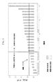
上記ROC曲線分析は、本質的に以下のように行われる。閾値より高いPGSスコアを有する任意のサンプルは、非応答者と同定される。閾値以下のPGSスコアを有する任意のサンプルは、応答者と同定される。試験されたサンプルセットに由来するあらゆるPGSスコアに関して、「応答者」および「非応答者」(仮説的コール)が、上記閾値として上記PGSスコアを使用して分類される。このプロセスは、上記データセットについての実際の応答データに対して仮説的コールを比較することを介して、各潜在的閾値についてのTPR(yベクトル)およびFPR(xベクトル)の計算を可能にする。次いで、ROC曲線は、上記TPRベクトルおよびFPRベクトルを使用して、ドットプロットを作成することによって構築される。上記ROC曲線が、(0,0)ポイントから(1.0,0.5)ポイントまでの対角線より上にある場合、上記PGS試験結果が、無作為より良い試験であることを示す(例えば、図2を参照のこと)。 The ROC curve analysis is essentially performed as follows. Any sample with a PGS score higher than the threshold is identified as a non-responder. Any sample with a PGS score below the threshold is identified as a responder. For any PGS score from the tested sample set, “responders” and “non-responders” (hypothetical calls) are classified using the PGS score as the threshold. This process allows the calculation of TPR (y vector) and FPR (x vector) for each potential threshold via comparing hypothetical calls against actual response data for the data set. . The ROC curve is then constructed by creating a dot plot using the TPR vector and the FPR vector. If the ROC curve is above the diagonal from (0,0) point to (1.0,0.5) point, the PGS test result indicates that the test is better than random (eg, See FIG.
上記ROC曲線は、最良のオペレーティングポイント(operating point)を同定するために使用され得る。上記最良のオペレーティングポイントは、偽陰性のコストに対する、重み付けされた偽陽性のコストの最良のバランスを得るものである。これらコストは、等しい必要はない。上記ROC空間におけるポイントx,yでの分類の平均期待コストは、以下の式によって示される: The ROC curve can be used to identify the best operating point. The best operating point is one that obtains the best balance of weighted false positive costs versus false negative costs. These costs need not be equal. The average expected cost of classification at points x and y in the ROC space is given by:
![]()
α=偽陽性のコスト、
β=陽性を見過ごす(偽陰性)のコスト、および
p=陽性症例の割合。
![]()
α = cost of false positives,
β = cost of overlooking positives (false negatives), and p = percentage of positive cases.
偽陽性および偽陰性は、αおよびβについて異なる値を入れることによって、異なって重み付けされ得る。例えば、非応答者である患者をより多く処置するというコストを払って、上記応答者群により多くの患者を含めることが決定される場合、αに対してより大きな重みをつけ得る。この場合、偽陽性および偽陰性のコストは、同じである(α=β)と想定される。従って、上記ROC空間におけるポイントx,yでの分類の平均期待コストは、以下である。 False positives and false negatives can be weighted differently by putting different values for α and β. For example, if it is decided to include more patients in the responder group at the cost of treating more patients who are non-responders, a greater weight may be given to α. In this case, the false positive and false negative costs are assumed to be the same (α = β). Therefore, the average expected cost of classification at points x and y in the ROC space is as follows.
![]()
![]()
腫瘍がチボザニブでの処置に応答性であるのか、抵抗性であるのかを予測することに加えて、PGSスコアは、近似値であるが、腫瘍がどの程度、おそらく応答性であるかもしくは非応答性であるかの有用な指標を提供する。一般に、上記PGSスコアが低くなるほど、腫瘍がチボザニブに対して応答性である確率は高くなり、上記PGSスコアが高くなるほど、腫瘍がチボザニブに対して抵抗性である確率は高くなる。 In addition to predicting whether the tumor is responsive or resistant to treatment with tivozanib, the PGS score is an approximation, but to what extent the tumor is probably responsive or unresponsive Provides a useful indicator of gender. In general, the lower the PGS score, the higher the probability that the tumor is responsive to tivozanib, and the higher the PGS score, the higher the probability that the tumor is resistant to tivozanib.
(試験キット)
本発明は、本発明の方法を行うための特定の成分を含む診断試験キットを包含する。診断試験キットは、診断アッセイの実行において簡便性、スピードおよび再現性を高める。
(Test kit)
The present invention includes a diagnostic test kit comprising specific components for performing the methods of the present invention. Diagnostic test kits increase convenience, speed and reproducibility in performing diagnostic assays.
例えば、例示的qRT−PCRベースの本発明の実施形態において、基本的な診断試験キットは、本発明に従うPGSのメンバー全てに関するPCRプライマー(例えば、プライマー対)を含む。いくつかの実施形態において、試験キットは、必要に応じて、コントロールもしくは標準(例えば、データ正規化のために)に対して指向されるPCRプライマーを含み得る。他の実施形態において、より手の込んだ試験キットは、PCRプライマーだけでなく、PCR技術を使用して上記PGSのメンバーの発現レベルを測定するための緩衝液、試薬、および詳細な説明書も含む。好ましい実施形態において、上記キットは、試験プロトコルおよび、上記RNAサンプルを除いて上記試験に必要とされる全ての消耗成分を含む。 For example, in an exemplary qRT-PCR-based embodiment of the present invention, a basic diagnostic test kit includes PCR primers (eg, primer pairs) for all members of the PGS according to the present invention. In some embodiments, the test kit may optionally include PCR primers directed against a control or standard (eg, for data normalization). In other embodiments, more elaborate test kits include not only PCR primers, but also buffers, reagents, and detailed instructions for measuring expression levels of the PGS members using PCR techniques. Including. In a preferred embodiment, the kit includes a test protocol and all consumable components required for the test except the RNA sample.
本発明の例示的なDNAマイクロアレイベースの実施形態において、試験キットは、特定の機器で使用するために設計されたマイクロ流体カード(アレイ)を含む。必要に応じて、上記マイクロ流体カードは、表1において上記に示される上記PGSの発現の同時測定に対して特異的に設計された特注デバイスである。このような特注マイクロ流体カードは、商業的に入手可能である。例えば、上記TaqMan Arrayは、Applied Biosystems 7900HT Fast Real Time PCR System(Applied Biosystems,Foster City,CA)で使用するために設計された384ウェルマイクロ流体カード(アレイ)である。本発明のいくつかの実施形態は、表1に示される上記PGSの全てもしくは本質的に全てのメンバーの発現を測定するための、特注DNAマイクロアレイチップを含む。このような特注DNAマイクロアレイチップは、商業的に入手可能である。 In an exemplary DNA microarray-based embodiment of the invention, the test kit includes a microfluidic card (array) designed for use with a particular instrument. Optionally, the microfluidic card is a custom designed device specifically designed for simultaneous measurement of the expression of the PGS shown above in Table 1. Such custom microfluidic cards are commercially available. For example, the TaqMan Array is a 384-well microfluidic card (array) designed for use in an Applied Biosystems 7900HT Fast Real Time PCR System (Applied Biosystems, Foster City, CA). Some embodiments of the invention include custom DNA microarray chips for measuring the expression of all or essentially all members of the PGS shown in Table 1. Such custom DNA microarray chips are commercially available.
さらなるプローブが、必要に応じて、上記PGSの一部ではないさらなる遺伝子の発現を測定するために、流体カード上に含められ得ること、もしくはこのようなさらなる遺伝子に対するプライマーが、本明細書に記載されるように、必要に応じて、プライマーセットの一部として含められ得ることは、理解される。このようなさらなる遺伝子は、コントロールもしくは標準として(例えば、データ正規化のために)働くように含められ得るか、または他の点で有益であり得る。このようなさらなる遺伝子が、ヒト腫瘍が、おそらく、チボザニブでの処置に対して応答性であるかもしくは非応答性であるかを同定するために、表1において本明細書で示される上記PGSの予測値を変化させないことは、理解される。 Additional probes can optionally be included on the fluid card to determine the expression of additional genes that are not part of the PGS, or primers for such additional genes are described herein. As will be appreciated, it can be included as part of the primer set, if desired. Such additional genes can be included to serve as controls or standards (eg, for data normalization) or can be otherwise beneficial. Such additional genes may be used to identify whether the human tumor is likely to be responsive or non-responsive to treatment with tivozanib as shown in Table 1 above. It is understood that the predicted value is not changed.
(実施例)
本発明は、以下の実施例によってさらに例示される。これら実施例は、例示目的で提供されるに過ぎず、如何様においても、本発明の範囲もしくは内容を限定すると解釈されるべきではない。
(Example)
The invention is further illustrated by the following examples. These examples are provided for illustrative purposes only, and are not to be construed as limiting the scope or content of the invention in any way.
(実施例1:チボザニブに対するマウス腫瘍応答)
100個より多くのマウス乳房腫瘍の集団(BHアーカイブ)を、チボザニブに対して感受性である腫瘍(応答者)およびチボザニブに対して抵抗性である腫瘍(非応答者)を同定するために使用した。上記BHアーカイブは、Her2依存性の、誘導性自発性乳房腫瘍を発症する操作されたキメラマウスに由来する100個を超える自発性マウス乳房腫瘍からの原発性腫瘍材料のインビボ増殖および低温保存によって樹立した。上記マウスを、本質的に以下のように生成した。
Example 1: Mouse tumor response to tivozanib
A population of more than 100 mouse breast tumors (BH archive) was used to identify tumors that were sensitive to tivozanib (responders) and tumors that were resistant to tivozanib (non-responders). . The BH archive was established by in vivo growth and cryopreservation of primary tumor material from more than 100 spontaneous mouse breast tumors derived from engineered chimeric mice that develop Her2-dependent, induced spontaneous breast tumors did. The mouse was generated essentially as follows.
Ink4aホモ接合性ヌルES細胞を、別個のフラグメントとして、以下の4種の構築物で共トランスフェクトした:MMTV−rtTA、TetO−Her2V664Eneu、TetO−ルシフェラーゼおよびPGK−ピューロマイシン。ピューロマイシン耐性細胞を、PCRおよびサザンブロットによってジェノタイピング(genotyping)した。ES細胞におけるオンコジーンの誘導性を、ノーザンブロットによって分析した。上記トランスフェクトしたES細胞を、C57BL/6胚盤胞へと注入し、これを、キメラマウスの出産をもたらす妊娠のために、偽妊娠雌性マウスに移植した。 Ink4a homozygous null ES cells were cotransfected as separate fragments with the following four constructs: MMTV-rtTA, TetO-Her2 V664Eneu , TetO-luciferase and PGK-puromycin. Puromycin resistant cells were genotyped by PCR and Southern blot. Oncogene inducibility in ES cells was analyzed by Northern blot. The transfected ES cells were injected into C57BL / 6 blastocysts, which were transplanted into pseudopregnant female mice for pregnancy that resulted in the birth of chimeric mice.
マウス乳房腫瘍ウイルス長末端反復(MMTV)を使用して、逆テトラサイクリントランスアクチベーター(reverse tetracycline transactivator(rtTA))の乳房特異的発現を駆動した。上記rtTAは、ドキシサイクリンを飲料水中に入れてマウスに提供した場合、Her2活性化オンコジーンの乳房特異的発現を提供した。 Mouse mammary tumor virus long terminal repeat (MMTV) was used to drive the breast-specific expression of the reverse tetracycline transactivator (rtTA). The rtTA provided breast specific expression of a Her2 activated oncogene when doxycycline was provided to mice in drinking water.
上記Her2オンコジーンおよびルシフェラーゼの誘導性を、上記マウスに由来する培養細胞を使用して、それぞれ、RT−PCRおよびルシフェラーゼアッセイによって確認した。乳腺を、キメラマウスから取り出し、コラゲナーゼで消化した。採取したオルガノイド(organoid)のうちの半分を、ドキシサイクリンの存在下で培養し、他方の半分を、ドキシサイクリンなしで培養した。培養して5日後に、上記細胞をトリプシン処理し、上記細胞のうちの1/10を、ルシフェラーゼアッセイに使用し、その残りを、RNA抽出に使用した。 Inducibility of the Her2 oncogene and luciferase was confirmed by RT-PCR and luciferase assay, respectively, using cultured cells from the mice. Mammary glands were removed from the chimeric mice and digested with collagenase. Half of the collected organoid was cultured in the presence of doxycycline and the other half was cultured without doxycycline. After 5 days in culture, the cells were trypsinized and 1/10 of the cells were used for luciferase assay and the rest was used for RNA extraction.
HER2乳がんモデルマウスから採取した腫瘍の組織学分析は、侵襲性腺がんを示した。2つの主要なパターンを区別した。それらは、固体シート増殖パターン、および壊死中心を有する巣状の増殖パターンであった。上記乳房腫瘍の免疫組織化学分析から、上記腫瘍内に2個の細胞タイプが明らかになった。第1の細胞タイプは、上皮起源であり(サイトケラチン陽性)、HER2発現および強い増殖を示した。第2の細胞タイプは、線維芽細胞様の外見を有する間葉起源であった。これら細胞は、コラーゲン陽性であり、強い増殖を示さず、間質機能を示した。アポトーシスは、上記腫瘍の上皮部分の壊死中心において認められた。腫瘍退縮研究(ドキシサイクリンの離脱に応答した退縮)行って、上記マウスモデル腫瘍が、Her2発現に依存性であることを確認した。ドキシサイクリンによるテトラサイクリン応答性プロモーターの誘導後、上記マウスは、約2〜4ヶ月の潜伏期で、乳房腫瘍を発症させた。 Histological analysis of tumors taken from HER2 breast cancer model mice showed invasive adenocarcinoma. Two main patterns were distinguished. They were a solid sheet growth pattern and a nest-like growth pattern with necrotic centers. Immunohistochemical analysis of the breast tumor revealed two cell types within the tumor. The first cell type was of epithelial origin (cytokeratin positive) and showed HER2 expression and strong proliferation. The second cell type was of mesenchymal origin with a fibroblast-like appearance. These cells were positive for collagen, did not show strong proliferation, and showed stromal function. Apoptosis was observed in the necrotic center of the epithelial part of the tumor. Tumor regression studies (regression in response to doxycycline withdrawal) were performed to confirm that the mouse model tumor was dependent on Her2 expression. After induction of a tetracycline-responsive promoter with doxycycline, the mice developed breast tumors with a latency period of about 2-4 months.
腫瘍細胞を、細胞ストレーナーを使用して、上記腫瘍の物理的破壊によって単離した。代表的には、1×10⁵細胞を、マトリゲルと混合し(50:50容積)、雌性NCr nu/nuマウスの肩甲骨間の上背部領域へと皮下注射した。これら腫瘍が約500mm3に成長したとき(代表的には、2〜4週間を要する)に、それらをさらなる増殖、薬物応答試験、および分析のために回収した。分析には、マイクロアレイプロファイリング、一般的組織病理学、およびIHC(腫瘍血管に対してはCD31、腫瘍細胞増殖に対してはKi67)を含めた。この腫瘍集団の特徴付けから、血管形成の重要なパラメーター(例えば、微小血管系、VEGF発現および特異的遺伝子発現プロフィール)における顕著な変動の程度が明らかになった。 Tumor cells were isolated by physical destruction of the tumor using a cell strainer. Typically, 1 × 10 5 cells were mixed with Matrigel (50:50 volume) and injected subcutaneously into the upper back region between the scapulae of female NCr nu / nu mice. When these tumors grew to approximately 500 mm 3 (typically it took 2-4 weeks), they were collected for further growth, drug response testing, and analysis. Analysis included microarray profiling, general histopathology, and IHC (CD31 for tumor vessels, Ki67 for tumor cell growth). Characterization of this tumor population revealed a significant degree of variation in important parameters of angiogenesis (eg, microvasculature, VEGF expression and specific gene expression profiles).
チボザニブに対する腫瘍応答の評価を、本質的に以下のように行った。皮下移植した腫瘍を、物理的に破壊した腫瘍細胞(マトリゲルと混合した)を7週齢の雌性NCrヌードマウスに注入することによって、確立した。上記腫瘍が約200〜400mm3に達した場合、30匹の腫瘍を有するマウスを、無作為に3つの群に分けた。群1には、ビヒクルを与えた。群2には、5mg/kgのチボザニブを強制経口投与(oral gavage)によって毎日与えた。群3には、20mg/kgのチボザニブを強制経口投与によって毎日与えた。腫瘍を、カリパスによって1週間に2回測定し、腫瘍体積を計算した。上記処置の最後に、組織病理分析およびIHC分析のために腫瘍を回収した。 Assessment of tumor response to tivozanib was performed essentially as follows. Subcutaneously implanted tumors were established by injecting physically disrupted tumor cells (mixed with Matrigel) into 7 week old female NCr nude mice. When the tumor reached approximately 200-400 mm 3 , mice with 30 tumors were randomly divided into three groups. Group 1 received vehicle. Group 2 was given 5 mg / kg tivozanib daily by oral gavage. Group 3 received 20 mg / kg tivozanib daily by gavage. Tumors were measured twice a week with calipers and tumor volumes were calculated. At the end of the treatment, tumors were collected for histopathological and IHC analyses.
これら研究から、チボザニブ応答における、有意な腫瘍間変動が明らかになった。腫瘍増殖阻害、ならびに脈管形成阻害についての代表的な組織病理特性およびIHC(CD31)特性に基づいて、応答者および非応答者を同定した。代表的には、応答者は、5mg/kg チボザニブで処置した場合、カリパス測定によって(組織学によって)腫瘍進行を示さず、周囲を除いて完全な腫瘍死滅に近かった。応答におけるバリエーションが、予想された。なぜなら、上記マウスモデル腫瘍は、自発的に生じ、従って、腫瘍脈管形成を含め、腫瘍形成をもたらしたランダム変異の異なるセットを含むことが予測されたからである。このような応答のバリエーションが望ましい。なぜなら、それは、天然に存在するヒト腫瘍におけるバリエーションに類似しており、分子シグネチャーもしくはチボザニブ応答性を同定することにおいて使用するための、チボザニブ応答性腫瘍およびチボザニブ抵抗性腫瘍の同定を可能にしたからである。 These studies revealed significant tumor-to-tumor variation in tivozanib response. Responders and non-responders were identified based on representative histopathological and IHC (CD31) characteristics for tumor growth inhibition and angiogenesis inhibition. Typically, responders showed no tumor progression (by histology) by caliper measurements when treated with 5 mg / kg tivozanib and were close to complete tumor death except at the periphery. Variations in response were expected. This is because the mouse model tumor occurred spontaneously and was therefore expected to contain a different set of random mutations that resulted in tumor formation, including tumor angiogenesis. Such a response variation is desirable. Because it is similar to variations in naturally occurring human tumors, it allows the identification of tivozanib responsive and tivozanib resistant tumors for use in identifying molecular signatures or tivozanib responsiveness It is.
(実施例2:差次的に発現された遺伝子の同定)
上記BHアーカイブにおける各腫瘍に由来するメッセンジャーRNA(約6μg)を、増幅プロトコルに供し、特注のAgilentマイクロアレイ(Agilentマウス40Kチップ)を使用してハイブリダイズさせた。マウス腫瘍サンプルの遺伝子発現プロフィールと、コントロールサンプル(Stratageneのユニバーサルマウス参照RNA(カタログ番号740100−41))との比較を、従来の、市販のマイクロアレイ技術を使用して行った。市販の特徴抽出ソフトウェア(Agilent Technologies,Santa Clara,CA)を、特徴抽出およびデータ正規化のために使用した。
(Example 2: Identification of differentially expressed genes)
Messenger RNA (approximately 6 μg) from each tumor in the BH archive was subjected to an amplification protocol and hybridized using a custom-made Agilent microarray (Agilent mouse 40K chip). Comparison of gene expression profiles of mouse tumor samples with control samples (Stratagene's universal mouse reference RNA (Cat. No. 740100-41)) was performed using conventional, commercially available microarray technology. Commercial feature extraction software (Agilent Technologies, Santa Clara, CA) was used for feature extraction and data normalization.
上記BHアーカイブからの5つの応答者腫瘍を、上記BH腫瘍アーカイブからの6つの非応答者腫瘍に対して比較した場合に、差次的に発現された遺伝子を同定した。従来のマイクロアレイ技術を使用して、上記11個の腫瘍の各々を表すサンプル中の約40,000遺伝子の発現を測定した。このことを、ともにAgilent Technology(Santa Clara,CA)製の特注のマウス40Kチップおよび特徴抽出ソフトウェアを使用して行った。各遺伝子の平均発現における差異の統計学的有意性(応答者 対 非応答者)を、スチューデントt検定を介して評価した。発現レベルにおいて最大の差異(最小のp値)を示す280遺伝子を同定し、特定のシグナル伝達経路と関連づけた。これを、市販のソフトウェア(Ingenuity Pathway Analysis Tool,Ingenuity Systems Inc.,Redwood City,CA)を使用して行った。5つの経路を表す、遺伝子発現における統計学的に有意な増大が観察された:(1)エンドサイトーシス経路を介するウイルス侵入、(2)リンホトキシンβレセプターシグナル伝達経路、(3)マクロピノサイト−シス経路、(4)ケモカインシグナル伝達経路、および(5)ニューロンにおけるReelinシグナル伝達経路。これら5つのアップレギュレートされた経路のうちの3つは、免疫応答およびサイトカイン経路に関与する。従って、これら経路は、ヒト腫瘍サンプルにおいてPGSを同定するためのその後のバイオインフォマティクス分析の焦点であった。 Differentially expressed genes were identified when 5 responder tumors from the BH archive were compared to 6 non-responder tumors from the BH tumor archive. Conventional microarray technology was used to measure the expression of approximately 40,000 genes in samples representing each of the 11 tumors. This was done using a custom mouse 40K chip and feature extraction software, both from Agilent Technology (Santa Clara, Calif.). Statistical significance of differences in mean expression of each gene (responders vs. non-responders) was assessed via Student's t test. 280 genes that showed the greatest difference in expression levels (minimum p value) were identified and associated with specific signaling pathways. This was done using commercially available software (Ingenuity Pathway Analysis Tool, Ingenuity Systems Inc., Redwood City, CA). A statistically significant increase in gene expression representing five pathways was observed: (1) viral entry via the endocytotic pathway, (2) lymphotoxin β receptor signaling pathway, (3) macropinocytes— Cis pathway, (4) chemokine signaling pathway, and (5) Reelin signaling pathway in neurons. Three of these five up-regulated pathways are involved in immune responses and cytokine pathways. These pathways were therefore the focus of subsequent bioinformatics analysis to identify PGS in human tumor samples.
(実施例3:PGSの同定)
上記マウスマイクロアレイデータ(上記)の経路分析に鑑みて、本発明者らは、遺伝子発現プロファイリングデータセットを含む相関分析のために、16個の造血マーカー遺伝子(CCL2、CCL7、CCR1、CCR2、CCR3、CCR5、CCR6、CSF1R、CSF2RA、CSF2RB、CXCR4、IL3RA、IL8RA、IL8RB、およびKIT)を選択した。言い換えると、上記16個の造血マーカー遺伝子の各々を、上記相関分析(すなわち、発現レベルの相関)のための参照遺伝子として別個に使用した。
(Example 3: Identification of PGS)
In light of the pathway analysis of the mouse microarray data (above), we have analyzed 16 hematopoietic marker genes (CCL2, CCL7, CCR1, CCR2, CCR3, CCR5, CCR6, CSF1R, CSF2RA, CSF2RB, CXCR4, IL3RA, IL8RA, IL8RB, and KIT) were selected. In other words, each of the 16 hematopoietic marker genes was used separately as a reference gene for the correlation analysis (ie, expression level correlation).
次いで、この情報を使用して、遺伝子発現プロファイリングデータセットを精査して、チボザニブに対する腫瘍応答を予測する遺伝子のセットを同定した。本発明者らは、上記相関参照遺伝子として造血マーカー遺伝子の選択をもたらした経路分析は、マウスデータで行われたが、ヒト遺伝子発現プロファイリングデータセットを使用すると決定した。このことの理論的根拠は、以下であった:(1)相関した発現は、おそらく、生物学的機能を反映し、上記機能はマウスおよびヒトにおいて類似する;ならびに(2)上記目的は、ヒトバイオマーカーを同定することであった。7個の異なるヒト腫瘍に関連した7個のヒト遺伝子発現プロファイリングデータセットを、相関分析のために利用できた(表2)。 This information was then used to scrutinize the gene expression profiling dataset to identify a set of genes that predict tumor response to tivozanib. We determined that the pathway analysis that resulted in the selection of the hematopoietic marker gene as the correlated reference gene was performed on mouse data, but using a human gene expression profiling data set. The rationale for this was the following: (1) correlated expression probably reflects biological function, which is similar in mice and humans; and (2) the purpose is human It was to identify biomarkers. Seven human gene expression profiling datasets associated with seven different human tumors were available for correlation analysis (Table 2).
この分析から、上記7個のヒトデータセットにわたる発現の共相関(co−correlation)は、上記16個の造血マーカー(参照)遺伝子のうちの8個について顕著により高いことが明らかになった。従って、さらなる分析は、これらの8個の造血マーカー遺伝子(すなわち、CCL2、CCR1、CCR2、CCR5、CSF1R、CSF2RA、CSF2RB、およびCSF3R)に限定した。これら8個の200遺伝子リスト(上記8個のマーカー遺伝子の各々につき、1個の200遺伝子リスト)を比較し、これら8個の200遺伝子リストは、共通して42遺伝子を有することを見いだした。42遺伝子のこのセットを、チボザニブに対するヒト腫瘍応答についての候補PGSとして同定した。上記PGSスコア(上記42遺伝子の平均発現値)を、以下の式に従って計算した: This analysis revealed that the co-correlation of expression across the 7 human data sets was significantly higher for 8 of the 16 hematopoietic marker (reference) genes. Therefore, further analysis was limited to these eight hematopoietic marker genes (ie, CCL2, CCR1, CCR2, CCR5, CSF1R, CSF2RA, CSF2RB, and CSF3R). The eight 200 gene lists (one 200 gene list for each of the eight marker genes) were compared, and these eight 200 gene lists were found to have 42 genes in common. This set of 42 genes was identified as a candidate PGS for human tumor response to tivozanib. The PGS score (average expression value of the 42 genes) was calculated according to the following formula:
言い換えると、所定のサンプルのPGSスコアは、上記サンプル中のこれら42遺伝子の平均発現値である。 In other words, the PGS score of a given sample is the average expression value of these 42 genes in the sample.
(実施例4:マウス応答の予測)
本発明の予測能力を、駆動オンコジーンがHer2であった原発性マウス腫瘍の所有者のアーカイブからの25腫瘍を使用して試験した。上記25腫瘍の各々についてのPGSスコアを、マイクロアレイデータから計算した。上記25腫瘍の各々を、チボザニブで処理し、次いで、上記実施例1において記載されるように、上記薬物処理に対して応答性もしくは非応答性として分類した。上記最適な閾値PGSスコアを、ROC曲線分析を使用して、閾値決定分析において0.33であると経験的に決定した。
(Example 4: Prediction of mouse response)
The predictive ability of the present invention was tested using 25 tumors from the archive of the owner of the primary mouse tumor whose driving oncogene was Her2. The PGS score for each of the 25 tumors was calculated from the microarray data. Each of the 25 tumors was treated with tivozanib and then classified as responsive or non-responsive to the drug treatment as described in Example 1 above. The optimal threshold PGS score was empirically determined to be 0.33 in the threshold determination analysis using ROC curve analysis.
この閾値を適用したところ、上記試験から、上記25腫瘍のうちの22個に関して、応答性もしくは非応答性の正確な予測を得た(図2)。非応答の予測において、偽陽性率は、18.2%(11個のうち2個)であり、偽陰性率は、5.88%(17個のうち1個)であった。 When this threshold was applied, the test yielded an accurate prediction of responsiveness or non-responsiveness for 22 of the 25 tumors (FIG. 2). In predicting non-response, the false positive rate was 18.2% (2 out of 11) and the false negative rate was 5.88% (1 out of 17).
(実施例5:ヒト応答の予測)
以下の推定実施例は、TaqMan(登録商標)データを使用して、チボザニブに対するヒト応答を予測するために本発明をどのように使用するかを詳細に例示する。
(Example 5: Prediction of human response)
The following putative examples illustrate in detail how the present invention is used to predict human response to tivozanib using TaqMan® data.
所定の腫瘍タイプ(例えば、腎細胞がん)に関して、腫瘍サンプル(アーカイブのFFPEブロック、新鮮なサンプルもしくは凍結サンプル)を、チボザニブでヒト患者を処置する前に、上記患者から(病院もしくは臨床実験室を介して間接的に)得る。新鮮なもしくは凍結した腫瘍サンプルを、標準的な組織学手順に従って、10%中性緩衝化ホルマリン中に、5〜10時間静置し、その後、アルコール脱水し、パラフィン中に包埋する。 For a given tumor type (eg, renal cell carcinoma), a tumor sample (archive FFPE block, fresh sample or frozen sample) is taken from the patient (hospital or clinical laboratory) before treating the human patient with tivozanib. Via indirectly). Fresh or frozen tumor samples are placed in 10% neutral buffered formalin for 5-10 hours according to standard histology procedures, then alcohol dehydrated and embedded in paraffin.
RNAを、10μm FFPE切片から抽出する。キシレン抽出、その後の、エタノール洗浄によって、パラフィンを除去する。RNAを、市販のRNA調製キットを使用して単離する。RNAは、適切な市販のキット(例えば、RiboGreen(登録商標)蛍光法(Molecular Probes,Eugene,OR))を使用して定量する。RNAサイズを、従来の方法によって分析する。 RNA is extracted from 10 μm FFPE sections. Paraffin is removed by xylene extraction followed by ethanol wash. RNA is isolated using a commercial RNA preparation kit. RNA is quantified using an appropriate commercially available kit (eg, RiboGreen® fluorescence method (Molecular Probes, Eugene, OR)). RNA size is analyzed by conventional methods.
逆転写を、SuperScriptTM First−Strand Synthesis Kit for qRT−PCR(Invitrogen)を使用して行う。全RNAおよびプールした遺伝子特異的プライマーは、それぞれ、10〜50ng/μlおよび100nM(各々)で存在する。 Reverse transcription is performed using SuperScript ™ First-Strand Synthesis Kit for qRT-PCR (Invitrogen). Total RNA and pooled gene-specific primers are present at 10-50 ng / μl and 100 nM (respectively).
上記PGSにおける各遺伝子について、qRT−PCRプライマーを、適切な市販のソフトウェア(例えば、Primer Express(登録商標)ソフトウェア(Applied Biosystems,Foster City,CA))を使用して設計する。上記オリゴヌクレオチドプライマーを、市販の合成機器および適切な試薬を使用して、機器製造者もしくは業者によって推奨されるように合成する。適切な市販の標識キットを使用して、プローブを標識する。 For each gene in the PGS, qRT-PCR primers are designed using appropriate commercially available software (eg, Primer Express® software (Applied Biosystems, Foster City, Calif.)). The oligonucleotide primers are synthesized as recommended by the instrument manufacturer or vendor using commercially available synthesis instruments and appropriate reagents. Label the probe using a suitable commercially available labeling kit.
TaqMan(登録商標)反応を、製造業者の説明書に従って、Applied Biosystems 7900HT機器を使用して、384ウェルプレートで行った。上記PGSにおける各遺伝子の発現を、1ngの全RNA/反応ウェルから合成したcDNAを使用して、2連の5μl反応において測定する。最終的なプライマー濃度およびプローブ濃度は、それぞれ、0.9μM(各プライマー)および0.2μMであった。PCRサイクリングを、標準的な操作手順に従って行う。上記qRT−PCRシグナルが、夾雑するDNAではなく、RNAに起因することを確認するために、試験した各遺伝子について、RT無しのコントロールを並行して実施する。qRT−PCR中の所定の増幅曲線に関する閾値サイクルは、プローブ切断からの蛍光シグナルが、特定の蛍光閾値設定を超えて増える時点で起こる。より多くの初期テンプレートを伴なう試験サンプルは、より初期の増幅サイクルにおいて閾値を超える。 TaqMan® reactions were performed in 384 well plates using an Applied Biosystems 7900HT instrument according to the manufacturer's instructions. Expression of each gene in the PGS is measured in duplicate 5 μl reactions using cDNA synthesized from 1 ng total RNA / reaction well. The final primer and probe concentrations were 0.9 μM (each primer) and 0.2 μM, respectively. PCR cycling is performed according to standard operating procedures. In order to confirm that the qRT-PCR signal is due to RNA rather than contaminating DNA, a control without RT is performed in parallel for each gene tested. The threshold cycle for a given amplification curve during qRT-PCR occurs when the fluorescent signal from probe cleavage increases beyond a specific fluorescence threshold setting. Test samples with more initial template will exceed the threshold at earlier amplification cycles.
上記サンプル全てにわたる遺伝子発現レベルを比較するために、5個の参照遺伝子(発現レベルが全てのサンプルにわたって類似であると想定されるハウスキーピング遺伝子)に基づく正規化を使用して、各アッセイウェルにおける、RNA品質およびRNAの総量におけるバリエーションから生じる差異に対して較正する。各サンプルについての参照CT(閾値サイクル)を、上記参照遺伝子の平均測定CTとして定義する。試験遺伝子の正規化したmRNAレベルを、ΔCT+10と定義し、ここでΔCT=参照遺伝子CT−試験遺伝子CTである。 To compare gene expression levels across all of the above samples, use normalization based on 5 reference genes (housekeeping genes whose expression levels are assumed to be similar across all samples), and in each assay well Calibrate for differences arising from variations in RNA quality and total RNA. See C T (threshold cycle) for each sample is defined as the average measured C T of the reference gene. MRNA levels normalized test gene, is defined as [Delta] C T +10, where [Delta] C T = reference gene C T - a test gene C T.
各腫瘍サンプルに関する上記PGSスコアは、上記に示されるアルゴリズムに従って、上記遺伝子発現レベルから計算する。試験した腫瘍サンプルと関連した実際の応答データを、上記腫瘍サンプルを供給する病院もしくは臨床実験室から得る。臨床的応答は、代表的には、適切な画像化技術(例えば、CTスキャン)によって決定される場合、腫瘍縮小(例えば、30%縮小)に関して定義される。幾つかの症例において、ヒト臨床的応答は、時間(例えば、進行なしの生存期間)に関して定義される。上記所定の腫瘍タイプに関する最適な閾値PGSスコアを、上記のように計算する。その後、この最適な閾値PGSスコアを使用して、同じ腫瘍タイプの新たに試験されるヒト腫瘍が、チボザニブでの処置に応答性であるかもしくは非応答性であるかを予測する。 The PGS score for each tumor sample is calculated from the gene expression level according to the algorithm shown above. Actual response data associated with the tumor sample tested is obtained from the hospital or clinical laboratory supplying the tumor sample. Clinical response is typically defined in terms of tumor shrinkage (eg, 30% reduction) as determined by appropriate imaging techniques (eg, CT scan). In some cases, the human clinical response is defined in terms of time (eg, survival without progression). The optimal threshold PGS score for the given tumor type is calculated as described above. This optimal threshold PGS score is then used to predict whether newly tested human tumors of the same tumor type will be responsive or non-responsive to treatment with tivozanib.
(参考としての援用)
本明細書で引用される特許文書および科学論文の各々の開示全体は、全ての目的のために参考として援用される。
(Incorporation as a reference)
The entire disclosure of each of the patent documents and scientific papers cited herein is incorporated by reference for all purposes.
(等価物)
本発明は、その本質的特徴から離れずに、他の特定の形態において具現化され得る。従って、前述の実施形態は、本明細書に記載される発明に対して限定するのではなく、例示であるとみなされるべきである。本発明の範囲は、前述の説明によるのではなく、添付の特許請求の範囲によって示され、特許請求の範囲の意味およびその等価物の範囲内に入る全ての変更は、そこに包含されることが意図される。さらに、用語「含む」は、用語「〜から本質的になる」および「〜からなる」を包含することが意図される。
(Equivalent)
The present invention may be embodied in other specific forms without departing from its essential characteristics. Accordingly, the foregoing embodiments are to be regarded as illustrative rather than limiting on the invention described herein. The scope of the invention is indicated by the appended claims rather than by the foregoing description, and all changes that come within the meaning and range of equivalents of the claims are embraced therein. Is intended. Furthermore, the term “comprising” is intended to encompass the terms “consisting essentially of” and “consisting of”.
(等価物)
本発明は、その本質的特徴から離れずに、他の特定の形態において具現化され得る。従って、前述の実施形態は、本明細書に記載される発明に対して限定するのではなく、例示であるとみなされるべきである。本発明の範囲は、前述の説明によるのではなく、添付の特許請求の範囲によって示され、特許請求の範囲の意味およびその等価物の範囲内に入る全ての変更は、そこに包含されることが意図される。さらに、用語「含む」は、用語「〜から本質的になる」および「〜からなる」を包含することが意図される。
したがって、本発明は以下の項目を提供する:
(項目1)
ヒト腫瘍をチボザニブでの処置におそらく応答性であるか、もしくはチボザニブでの処置におそらく非応答性であると同定する方法であって、該方法は、
(a)該ヒト腫瘍に由来するサンプルにおいて、予測遺伝子セット(PGS)における各遺伝子の相対的発現レベルを決定する工程であって、ここで該PGSは、以下の遺伝子:
AIF1、APBB1IP、ARHGAP30、C3AR1、CCR1、CD37、CD53、CD86、CLEC7A、CSF1R、CSF2RB、CTSS、CYBB、DOCK2、EVI2A、EVI2B、FPR3、GMFG、GPR65、HCK、HCLS1、HLA−DMA、IL10RA、ITGB2、LAIR1、LCP1、LCP2、LILRB1、LILRB2、LST1、LY86、MNDA、MS4A6A、MYO1F、NCF4、SLA、SLAMF8、TLR1、TYROBP、PLEK、CYTH4、およびPTPRCを含む、工程;ならびに
(b)以下のアルゴリズム:
![]()
を包含する、方法。
(項目2)
上記PGSは、以下の遺伝子:
AIF1、APBB1IP、ARHGAP30、C3AR1、CCR1、CD37、CD53、CD86、CLEC7A、CSF1R、CSF2RB、CTSS、CYBB、DOCK2、EVI2A、EVI2B、FPR3、GMFG、GPR65、HCK、HCLS1、HLA−DMA、IL10RA、ITGB2、LAIR1、LCP1、LCP2、LILRB1、LILRB2、LST1、LY86、MNDA、MS4A6A、MYO1F、NCF4、SLA、SLAMF8、TLR1、TYROBP、PLEK、CYTH4、およびPTPRCからなる、項目1に記載の方法。
(項目3)
閾値決定分析を行い、それによって、規定された閾値を生成する工程をさらに包含する項目1または2に記載の方法であって、ここで、該閾値決定分析は、レシーバーオペレーター特性曲線分析を含む、方法。
(項目4)
上記PGSにおける各遺伝子の相対的発現レベルは、DNAマイクロアレイ分析によって決定される、項目1、2または3に記載の方法。
(項目5)
上記PGSにおける各遺伝子の相対的発現レベルは、qRT−PCR分析によって決定される、項目1、2または3に記載の方法。
(項目6)
項目1または2に記載のPGSにおける遺伝子の発現レベルを決定することにおいて使用するためのプライマー対を含む、PCRプライマーセット。
(項目7)
項目1または項目2に記載のPGSの遺伝子の相対的発現を決定し、それによってヒト腫瘍をチボザニブでの処置におそらく応答性であるか、もしくはチボザニブでの処置におそらく非応答性であると同定するためのプライマー対を含む、PCRプライマーセット。
(項目8)
項目7に記載のPCRプライマーセットを含む、診断試験キット。
(項目9)
項目1または2に記載のPGSにおける遺伝子の発現レベルを決定する工程において使用するためのプローブセットを含む、DNAマイクロアレイチップ。
(項目10)
固体表面およびプローブセットを含むDNAマイクロアレイチップであって、該プローブセットは、項目1または項目2に記載のPGSの遺伝子の各々に対して特異的なプローブを含み、それによって、ヒト腫瘍をチボザニブでの処置におそらく応答性であるか、もしくはチボザニブでの処置におそらく非応答性であると同定する、DNAマイクロアレイチップ。
(項目11)
項目10に記載のDNAマイクロアレイチップを含む、診断試験キット。
(Equivalent)
The present invention may be embodied in other specific forms without departing from its essential characteristics. Accordingly, the foregoing embodiments are to be regarded as illustrative rather than limiting on the invention described herein. The scope of the invention is indicated by the appended claims rather than by the foregoing description, and all changes that come within the meaning and range of equivalents of the claims are embraced therein. Is intended. Furthermore, the term “comprising” is intended to encompass the terms “consisting essentially of” and “consisting of”.
Accordingly, the present invention provides the following items:
(Item 1)
A method of identifying a human tumor as possibly responsive to treatment with tivozanib or possibly unresponsive to treatment with tivozanib, the method comprising:
(A) determining a relative expression level of each gene in a predicted gene set (PGS) in a sample derived from the human tumor, wherein the PGS comprises the following genes:
AIF1, APBB1IP, ARHGAP30, C3AR1, CCR1, CD37, CD53, CD86, CLEC7A, CSF1R, CSF2RB, CTSS, CYBB, DOCK2, EVI2A, EVI2B, FPR3, GMFG, GPR65, HCK, HCLSIL, 10B Including LAIR1, LCP1, LCP2, LILRB1, LILRB2, LST1, LY86, MNDA, MS4A6A, MYO1F, NCF4, SLA, SLAMF8, TLR1, TYROBP, PLEK, CYTH4, and PTPRC; and
(B) The following algorithm:
![]()
Including the method.
(Item 2)
The PGS has the following genes:
AIF1, APBB1IP, ARHGAP30, C3AR1, CCR1, CD37, CD53, CD86, CLEC7A, CSF1R, CSF2RB, CTSS, CYBB, DOCK2, EVI2A, EVI2B, FPR3, GMFG, GPR65, HCK, HCLSIL, 10B 2. The method of item 1, comprising LAIR1, LCP1, LCP2, LILRB1, LILRB2, LST1, LY86, MNDA, MS4A6A, MYO1F, NCF4, SLA, SLAMF8, TLR1, TYROBP, PLEK, CYTH4, and PTPRC.
(Item 3)
The method of item 1 or 2, further comprising performing a threshold determination analysis, thereby generating a defined threshold value, wherein the threshold determination analysis comprises a receiver operator characteristic curve analysis, Method.
(Item 4)
4. The method according to item 1, 2 or 3, wherein the relative expression level of each gene in the PGS is determined by DNA microarray analysis.
(Item 5)
4. The method according to item 1, 2, or 3, wherein the relative expression level of each gene in the PGS is determined by qRT-PCR analysis.
(Item 6)
A PCR primer set comprising a primer pair for use in determining the expression level of a gene in the PGS according to item 1 or 2.
(Item 7)
Determining the relative expression of the gene of PGS according to item 1 or item 2 and thereby identifying the human tumor as possibly responsive to treatment with tivozanib or possibly unresponsive to treatment with tivozanib A PCR primer set comprising a primer pair for
(Item 8)
A diagnostic test kit comprising the PCR primer set according to item 7.
(Item 9)
3. A DNA microarray chip comprising a probe set for use in the step of determining the expression level of a gene in the PGS according to item 1 or 2.
(Item 10)
A DNA microarray chip comprising a solid surface and a probe set, the probe set comprising probes specific for each of the genes of PGS according to item 1 or item 2, whereby human tumors are DNA microarray chip that identifies that it is probably responsive to treatment with or possibly non-responsive to treatment with tivozanib.
(Item 11)
A diagnostic test kit comprising the DNA microarray chip according to item 10.
Claims (11)
(a)該ヒト腫瘍に由来するサンプルにおいて、予測遺伝子セット(PGS)における各遺伝子の相対的発現レベルを決定する工程であって、ここで該PGSは、以下の遺伝子:
AIF1、APBB1IP、ARHGAP30、C3AR1、CCR1、CD37、CD53、CD86、CLEC7A、CSF1R、CSF2RB、CTSS、CYBB、DOCK2、EVI2A、EVI2B、FPR3、GMFG、GPR65、HCK、HCLS1、HLA−DMA、IL10RA、ITGB2、LAIR1、LCP1、LCP2、LILRB1、LILRB2、LST1、LY86、MNDA、MS4A6A、MYO1F、NCF4、SLA、SLAMF8、TLR1、TYROBP、PLEK、CYTH4、およびPTPRCを含む、工程;ならびに
(b)以下のアルゴリズム:
を包含する、方法。 A method of identifying a human tumor as possibly responsive to treatment with tivozanib or possibly unresponsive to treatment with tivozanib, the method comprising:
(A) determining a relative expression level of each gene in a predicted gene set (PGS) in a sample derived from the human tumor, wherein the PGS comprises the following genes:
AIF1, APBB1IP, ARHGAP30, C3AR1, CCR1, CD37, CD53, CD86, CLEC7A, CSF1R, CSF2RB, CTSS, CYBB, DOCK2, EVI2A, EVI2B, FPR3, GMFG, GPR65, HCK, HCLSIL, 10B LAIR1, LCP1, LCP2, LILRB1, LILRB2, LST1, LY86, MNDA, MS4A6A, MYO1F, NCF4, SLA, SLAMF8, TLR1, TYROBP, PLEK, CYTH4, and PTPRC; and (b) the following algorithm:
AIF1、APBB1IP、ARHGAP30、C3AR1、CCR1、CD37、CD53、CD86、CLEC7A、CSF1R、CSF2RB、CTSS、CYBB、DOCK2、EVI2A、EVI2B、FPR3、GMFG、GPR65、HCK、HCLS1、HLA−DMA、IL10RA、ITGB2、LAIR1、LCP1、LCP2、LILRB1、LILRB2、LST1、LY86、MNDA、MS4A6A、MYO1F、NCF4、SLA、SLAMF8、TLR1、TYROBP、PLEK、CYTH4、およびPTPRCからなる、請求項1に記載の方法。 The PGS has the following genes:
AIF1, APBB1IP, ARHGAP30, C3AR1, CCR1, CD37, CD53, CD86, CLEC7A, CSF1R, CSF2RB, CTSS, CYBB, DOCK2, EVI2A, EVI2B, FPR3, GMFG, GPR65, HCK, HCLSIL, 10B The method of claim 1, comprising LAIR1, LCP1, LCP2, LILRB1, LILRB2, LST1, LY86, MNDA, MS4A6A, MYO1F, NCF4, SLA, SLAMF8, TLR1, TYROBP, PLEK, CYTH4, and PTPRC.
Applications Claiming Priority (3)
| Application Number | Priority Date | Filing Date | Title |
|---|---|---|---|
| US12/498,183 US7615353B1 (en) | 2009-07-06 | 2009-07-06 | Tivozanib response prediction |
| US12/498,183 | 2009-07-06 | ||
| PCT/US2009/061163 WO2011005273A1 (en) | 2009-07-06 | 2009-10-19 | Tivozanib response prediction |
Publications (1)
| Publication Number | Publication Date |
|---|---|
| JP2012531925A true JP2012531925A (en) | 2012-12-13 |
Family
ID=41261508
Family Applications (1)
| Application Number | Title | Priority Date | Filing Date |
|---|---|---|---|
| JP2012519530A Pending JP2012531925A (en) | 2009-07-06 | 2009-10-19 | Tibozanib response prediction |
Country Status (8)
| Country | Link |
|---|---|
| US (1) | US7615353B1 (en) |
| EP (1) | EP2451967A4 (en) |
| JP (1) | JP2012531925A (en) |
| KR (1) | KR20120034778A (en) |
| CN (1) | CN102471799B (en) |
| AU (1) | AU2009349657A1 (en) |
| CA (1) | CA2767246A1 (en) |
| WO (1) | WO2011005273A1 (en) |
Families Citing this family (6)
| Publication number | Priority date | Publication date | Assignee | Title |
|---|---|---|---|---|
| WO2012167278A1 (en) | 2011-06-02 | 2012-12-06 | Almac Diagnostics Limited | Molecular diagnostic test for cancer |
| CA2859663A1 (en) * | 2011-12-22 | 2013-06-27 | Aveo Pharmaceuticals, Inc. | Identification of multigene biomarkers |
| WO2014089055A1 (en) * | 2012-12-03 | 2014-06-12 | Aveo Pharmaceuticals, Inc. | Tivozanib response prediction |
| JP2015536667A (en) | 2012-12-03 | 2015-12-24 | アルマック・ダイアグノスティクス・リミテッドAlmac Diagnostics Limited | Molecular diagnostic tests for cancer |
| GB201409479D0 (en) * | 2014-05-28 | 2014-07-09 | Almac Diagnostics Ltd | Molecular diagnostic test for cancer |
| CN105506107A (en) * | 2015-12-30 | 2016-04-20 | 杭州艾迪康医学检验中心有限公司 | Primers and method for detecting polymorphic hotspot mutation condition of DOCK2 gene |
Citations (2)
| Publication number | Priority date | Publication date | Assignee | Title |
|---|---|---|---|---|
| WO2007028005A2 (en) * | 2005-09-01 | 2007-03-08 | Bristol-Myers Squibb Company | Biomarkers and methods for determining sensitivity to vascular endothelial growth factor receptor-2 modulators |
| WO2008128008A2 (en) * | 2007-04-13 | 2008-10-23 | Bristol-Myers Squibb Company | Biomarkers and methods for determining sensitivity to vascular endothelial growth factor receptor-2 modulators |
Family Cites Families (9)
| Publication number | Priority date | Publication date | Assignee | Title |
|---|---|---|---|---|
| US20070134261A1 (en) * | 2001-12-03 | 2007-06-14 | Hancock Robert E | Effectors of innate immunity |
| EP1552293A4 (en) * | 2002-09-10 | 2006-12-06 | Guennadi V Glinskii | Gene segregation and biological sample classification methods |
| US20050260652A1 (en) * | 2004-04-15 | 2005-11-24 | The General Hospital Corporation | Compositions and methods that modulate RNA interference |
| US20070099209A1 (en) * | 2005-06-13 | 2007-05-03 | The Regents Of The University Of Michigan | Compositions and methods for treating and diagnosing cancer |
| EP1904649A2 (en) * | 2005-07-18 | 2008-04-02 | Epigenomics AG | Compositions and methods for cancer diagnostics comprising pan-cancer markers |
| EP1988768A2 (en) * | 2006-02-17 | 2008-11-12 | Genentech, Inc. | Gene disruptions, compositions and methods relating thereto |
| JP2009531463A (en) | 2006-03-29 | 2009-09-03 | ジェネンテック・インコーポレーテッド | Tumor diagnosis and treatment |
| AU2007284724B2 (en) * | 2006-08-10 | 2014-03-13 | Millennium Pharmaceuticals, Inc. | For the identification, assessment, and treatment of patients with cancer therapy |
| EP1961825A1 (en) * | 2007-02-26 | 2008-08-27 | INSERM (Institut National de la Santé et de la Recherche Medicale) | Method for predicting the occurrence of metastasis in breast cancer patients |
-
2009
- 2009-07-06 US US12/498,183 patent/US7615353B1/en not_active Expired - Fee Related
- 2009-10-19 JP JP2012519530A patent/JP2012531925A/en active Pending
- 2009-10-19 EP EP09847186A patent/EP2451967A4/en not_active Withdrawn
- 2009-10-19 AU AU2009349657A patent/AU2009349657A1/en not_active Abandoned
- 2009-10-19 CA CA2767246A patent/CA2767246A1/en not_active Abandoned
- 2009-10-19 CN CN200980160316.4A patent/CN102471799B/en not_active Expired - Fee Related
- 2009-10-19 WO PCT/US2009/061163 patent/WO2011005273A1/en active Application Filing
- 2009-10-19 KR KR1020127003031A patent/KR20120034778A/en not_active Application Discontinuation
Patent Citations (2)
| Publication number | Priority date | Publication date | Assignee | Title |
|---|---|---|---|---|
| WO2007028005A2 (en) * | 2005-09-01 | 2007-03-08 | Bristol-Myers Squibb Company | Biomarkers and methods for determining sensitivity to vascular endothelial growth factor receptor-2 modulators |
| WO2008128008A2 (en) * | 2007-04-13 | 2008-10-23 | Bristol-Myers Squibb Company | Biomarkers and methods for determining sensitivity to vascular endothelial growth factor receptor-2 modulators |
Non-Patent Citations (1)
| Title |
|---|
| JPN6014007331; Fertility and Sterility Vol.87,No.4, 2007, p.982-984 * |
Also Published As
| Publication number | Publication date |
|---|---|
| EP2451967A1 (en) | 2012-05-16 |
| CA2767246A1 (en) | 2011-01-13 |
| CN102471799B (en) | 2014-03-26 |
| WO2011005273A1 (en) | 2011-01-13 |
| US7615353B1 (en) | 2009-11-10 |
| AU2009349657A1 (en) | 2012-02-02 |
| CN102471799A (en) | 2012-05-23 |
| KR20120034778A (en) | 2012-04-12 |
| EP2451967A4 (en) | 2012-11-28 |
Similar Documents
| Publication | Publication Date | Title |
|---|---|---|
| JP7042784B2 (en) | How to Quantify Prostate Cancer Prognosis Using Gene Expression | |
| RU2654587C2 (en) | Method for predicting breast cancer recurrent during endocrine treatment | |
| US7901881B2 (en) | Diagnostic tool for diagnosing benign versus malignant thyroid lesions | |
| JP5486717B2 (en) | Gene expression markers for prognosis of colorectal cancer | |
| EP2121988B1 (en) | Prostate cancer survival and recurrence | |
| KR20140105836A (en) | Identification of multigene biomarkers | |
| US20140045715A1 (en) | Tivozanib response prediction | |
| US7615353B1 (en) | Tivozanib response prediction | |
| US20180172689A1 (en) | Methods for diagnosis of bladder cancer | |
| KR102376220B1 (en) | Algorithms and Methods for Evaluating Late Clinical Endpoints in Prostate Cancer | |
| US20180051342A1 (en) | Prostate cancer survival and recurrence |
Legal Events
| Date | Code | Title | Description |
|---|---|---|---|
| A131 | Notification of reasons for refusal |
Free format text: JAPANESE INTERMEDIATE CODE: A131 Effective date: 20140220 |
|
| A601 | Written request for extension of time |
Free format text: JAPANESE INTERMEDIATE CODE: A601 Effective date: 20140514 |
|
| A602 | Written permission of extension of time |
Free format text: JAPANESE INTERMEDIATE CODE: A602 Effective date: 20140521 |
|
| A02 | Decision of refusal |
Free format text: JAPANESE INTERMEDIATE CODE: A02 Effective date: 20141016 |