JP2012525215A - System and method for deploying fiducials - Google Patents
System and method for deploying fiducials Download PDFInfo
- Publication number
- JP2012525215A JP2012525215A JP2012508533A JP2012508533A JP2012525215A JP 2012525215 A JP2012525215 A JP 2012525215A JP 2012508533 A JP2012508533 A JP 2012508533A JP 2012508533 A JP2012508533 A JP 2012508533A JP 2012525215 A JP2012525215 A JP 2012525215A
- Authority
- JP
- Japan
- Prior art keywords
- fiducial
- needle
- protrusion
- deployment system
- distal end
- Prior art date
- Legal status (The legal status is an assumption and is not a legal conclusion. Google has not performed a legal analysis and makes no representation as to the accuracy of the status listed.)
- Pending
Links
- 238000000034 method Methods 0.000 title claims abstract description 55
- 238000002604 ultrasonography Methods 0.000 claims description 17
- 238000012800 visualization Methods 0.000 claims description 11
- 230000009471 action Effects 0.000 claims description 2
- 238000009434 installation Methods 0.000 abstract description 7
- 230000008901 benefit Effects 0.000 abstract description 6
- 206010028980 Neoplasm Diseases 0.000 description 21
- 210000001519 tissue Anatomy 0.000 description 15
- 238000011282 treatment Methods 0.000 description 9
- 210000000496 pancreas Anatomy 0.000 description 6
- 230000008685 targeting Effects 0.000 description 6
- 239000003550 marker Substances 0.000 description 5
- 239000000463 material Substances 0.000 description 5
- 230000005855 radiation Effects 0.000 description 5
- 238000001959 radiotherapy Methods 0.000 description 5
- 210000002784 stomach Anatomy 0.000 description 5
- 238000002725 brachytherapy Methods 0.000 description 4
- 210000001198 duodenum Anatomy 0.000 description 4
- 238000002595 magnetic resonance imaging Methods 0.000 description 4
- 210000000277 pancreatic duct Anatomy 0.000 description 4
- 229910045601 alloy Inorganic materials 0.000 description 3
- 239000000956 alloy Substances 0.000 description 3
- 238000005452 bending Methods 0.000 description 3
- 210000000988 bone and bone Anatomy 0.000 description 3
- 238000009558 endoscopic ultrasound Methods 0.000 description 3
- 238000002594 fluoroscopy Methods 0.000 description 3
- PCHJSUWPFVWCPO-UHFFFAOYSA-N gold Chemical compound [Au] PCHJSUWPFVWCPO-UHFFFAOYSA-N 0.000 description 3
- 229910052737 gold Inorganic materials 0.000 description 3
- 239000010931 gold Substances 0.000 description 3
- 238000005259 measurement Methods 0.000 description 3
- 230000007704 transition Effects 0.000 description 3
- KDLHZDBZIXYQEI-UHFFFAOYSA-N Palladium Chemical compound [Pd] KDLHZDBZIXYQEI-UHFFFAOYSA-N 0.000 description 2
- 206010061902 Pancreatic neoplasm Diseases 0.000 description 2
- 210000001124 body fluid Anatomy 0.000 description 2
- 239000010839 body fluid Substances 0.000 description 2
- 210000001953 common bile duct Anatomy 0.000 description 2
- 238000001514 detection method Methods 0.000 description 2
- 230000002183 duodenal effect Effects 0.000 description 2
- 238000011846 endoscopic investigation Methods 0.000 description 2
- 230000005294 ferromagnetic effect Effects 0.000 description 2
- 238000003384 imaging method Methods 0.000 description 2
- 239000007943 implant Substances 0.000 description 2
- 230000003287 optical effect Effects 0.000 description 2
- 201000002528 pancreatic cancer Diseases 0.000 description 2
- BASFCYQUMIYNBI-UHFFFAOYSA-N platinum Chemical compound [Pt] BASFCYQUMIYNBI-UHFFFAOYSA-N 0.000 description 2
- 230000008569 process Effects 0.000 description 2
- 238000002310 reflectometry Methods 0.000 description 2
- 210000005070 sphincter Anatomy 0.000 description 2
- 230000001225 therapeutic effect Effects 0.000 description 2
- 238000002560 therapeutic procedure Methods 0.000 description 2
- 238000012285 ultrasound imaging Methods 0.000 description 2
- 210000001835 viscera Anatomy 0.000 description 2
- 238000007794 visualization technique Methods 0.000 description 2
- 206010011732 Cyst Diseases 0.000 description 1
- 206010073306 Exposure to radiation Diseases 0.000 description 1
- 229910001252 Pd alloy Inorganic materials 0.000 description 1
- 206010056658 Pseudocyst Diseases 0.000 description 1
- 229910000691 Re alloy Inorganic materials 0.000 description 1
- 210000000436 anus Anatomy 0.000 description 1
- 239000011324 bead Substances 0.000 description 1
- 239000000560 biocompatible material Substances 0.000 description 1
- 238000001574 biopsy Methods 0.000 description 1
- 201000011510 cancer Diseases 0.000 description 1
- 239000010952 cobalt-chrome Substances 0.000 description 1
- 230000000295 complement effect Effects 0.000 description 1
- 238000010276 construction Methods 0.000 description 1
- 238000013270 controlled release Methods 0.000 description 1
- 208000031513 cyst Diseases 0.000 description 1
- 230000007423 decrease Effects 0.000 description 1
- 238000003745 diagnosis Methods 0.000 description 1
- 238000002405 diagnostic procedure Methods 0.000 description 1
- 238000002592 echocardiography Methods 0.000 description 1
- 238000001861 endoscopic biopsy Methods 0.000 description 1
- 238000002674 endoscopic surgery Methods 0.000 description 1
- 238000012277 endoscopic treatment Methods 0.000 description 1
- 238000005516 engineering process Methods 0.000 description 1
- 230000003628 erosive effect Effects 0.000 description 1
- 238000001125 extrusion Methods 0.000 description 1
- 239000003302 ferromagnetic material Substances 0.000 description 1
- 210000001035 gastrointestinal tract Anatomy 0.000 description 1
- 238000002513 implantation Methods 0.000 description 1
- 238000007373 indentation Methods 0.000 description 1
- 208000015181 infectious disease Diseases 0.000 description 1
- 238000007689 inspection Methods 0.000 description 1
- 230000003993 interaction Effects 0.000 description 1
- 229910052741 iridium Inorganic materials 0.000 description 1
- GKOZUEZYRPOHIO-UHFFFAOYSA-N iridium atom Chemical compound [Ir] GKOZUEZYRPOHIO-UHFFFAOYSA-N 0.000 description 1
- 230000001788 irregular Effects 0.000 description 1
- 210000004185 liver Anatomy 0.000 description 1
- 210000004072 lung Anatomy 0.000 description 1
- 208000020816 lung neoplasm Diseases 0.000 description 1
- 208000037841 lung tumor Diseases 0.000 description 1
- 230000005291 magnetic effect Effects 0.000 description 1
- 230000014759 maintenance of location Effects 0.000 description 1
- 230000007246 mechanism Effects 0.000 description 1
- 239000012528 membrane Substances 0.000 description 1
- 238000012978 minimally invasive surgical procedure Methods 0.000 description 1
- 239000000203 mixture Substances 0.000 description 1
- 210000000214 mouth Anatomy 0.000 description 1
- 229910001000 nickel titanium Inorganic materials 0.000 description 1
- 229910052763 palladium Inorganic materials 0.000 description 1
- 230000002093 peripheral effect Effects 0.000 description 1
- 229910052697 platinum Inorganic materials 0.000 description 1
- 230000002285 radioactive effect Effects 0.000 description 1
- 230000029058 respiratory gaseous exchange Effects 0.000 description 1
- WUAPFZMCVAUBPE-UHFFFAOYSA-N rhenium atom Chemical compound [Re] WUAPFZMCVAUBPE-UHFFFAOYSA-N 0.000 description 1
- 230000035807 sensation Effects 0.000 description 1
- 210000004872 soft tissue Anatomy 0.000 description 1
- 229910001220 stainless steel Inorganic materials 0.000 description 1
- 239000010935 stainless steel Substances 0.000 description 1
- 238000001356 surgical procedure Methods 0.000 description 1
- 230000002194 synthesizing effect Effects 0.000 description 1
- 238000003325 tomography Methods 0.000 description 1
- 210000001215 vagina Anatomy 0.000 description 1
- 230000037303 wrinkles Effects 0.000 description 1
Images


Classifications
-
- A—HUMAN NECESSITIES
- A61—MEDICAL OR VETERINARY SCIENCE; HYGIENE
- A61B—DIAGNOSIS; SURGERY; IDENTIFICATION
- A61B17/00—Surgical instruments, devices or methods, e.g. tourniquets
- A61B17/34—Trocars; Puncturing needles
- A61B17/3468—Trocars; Puncturing needles for implanting or removing devices, e.g. prostheses, implants, seeds, wires
-
- A—HUMAN NECESSITIES
- A61—MEDICAL OR VETERINARY SCIENCE; HYGIENE
- A61B—DIAGNOSIS; SURGERY; IDENTIFICATION
- A61B90/00—Instruments, implements or accessories specially adapted for surgery or diagnosis and not covered by any of the groups A61B1/00 - A61B50/00, e.g. for luxation treatment or for protecting wound edges
- A61B90/39—Markers, e.g. radio-opaque or breast lesions markers
-
- A—HUMAN NECESSITIES
- A61—MEDICAL OR VETERINARY SCIENCE; HYGIENE
- A61M—DEVICES FOR INTRODUCING MEDIA INTO, OR ONTO, THE BODY; DEVICES FOR TRANSDUCING BODY MEDIA OR FOR TAKING MEDIA FROM THE BODY; DEVICES FOR PRODUCING OR ENDING SLEEP OR STUPOR
- A61M37/00—Other apparatus for introducing media into the body; Percutany, i.e. introducing medicines into the body by diffusion through the skin
- A61M37/0069—Devices for implanting pellets, e.g. markers or solid medicaments
-
- A—HUMAN NECESSITIES
- A61—MEDICAL OR VETERINARY SCIENCE; HYGIENE
- A61B—DIAGNOSIS; SURGERY; IDENTIFICATION
- A61B1/00—Instruments for performing medical examinations of the interior of cavities or tubes of the body by visual or photographical inspection, e.g. endoscopes; Illuminating arrangements therefor
- A61B1/012—Instruments for performing medical examinations of the interior of cavities or tubes of the body by visual or photographical inspection, e.g. endoscopes; Illuminating arrangements therefor characterised by internal passages or accessories therefor
- A61B1/018—Instruments for performing medical examinations of the interior of cavities or tubes of the body by visual or photographical inspection, e.g. endoscopes; Illuminating arrangements therefor characterised by internal passages or accessories therefor for receiving instruments
-
- A—HUMAN NECESSITIES
- A61—MEDICAL OR VETERINARY SCIENCE; HYGIENE
- A61B—DIAGNOSIS; SURGERY; IDENTIFICATION
- A61B17/00—Surgical instruments, devices or methods, e.g. tourniquets
- A61B17/00234—Surgical instruments, devices or methods, e.g. tourniquets for minimally invasive surgery
-
- A—HUMAN NECESSITIES
- A61—MEDICAL OR VETERINARY SCIENCE; HYGIENE
- A61B—DIAGNOSIS; SURGERY; IDENTIFICATION
- A61B17/00—Surgical instruments, devices or methods, e.g. tourniquets
- A61B17/04—Surgical instruments, devices or methods, e.g. tourniquets for suturing wounds; Holders or packages for needles or suture materials
- A61B17/06—Needles ; Sutures; Needle-suture combinations; Holders or packages for needles or suture materials
- A61B17/06166—Sutures
-
- A—HUMAN NECESSITIES
- A61—MEDICAL OR VETERINARY SCIENCE; HYGIENE
- A61B—DIAGNOSIS; SURGERY; IDENTIFICATION
- A61B17/00—Surgical instruments, devices or methods, e.g. tourniquets
- A61B17/04—Surgical instruments, devices or methods, e.g. tourniquets for suturing wounds; Holders or packages for needles or suture materials
- A61B17/0401—Suture anchors, buttons or pledgets, i.e. means for attaching sutures to bone, cartilage or soft tissue; Instruments for applying or removing suture anchors
- A61B2017/0417—T-fasteners
-
- A—HUMAN NECESSITIES
- A61—MEDICAL OR VETERINARY SCIENCE; HYGIENE
- A61B—DIAGNOSIS; SURGERY; IDENTIFICATION
- A61B90/00—Instruments, implements or accessories specially adapted for surgery or diagnosis and not covered by any of the groups A61B1/00 - A61B50/00, e.g. for luxation treatment or for protecting wound edges
- A61B90/39—Markers, e.g. radio-opaque or breast lesions markers
- A61B2090/3904—Markers, e.g. radio-opaque or breast lesions markers specially adapted for marking specified tissue
- A61B2090/3908—Soft tissue, e.g. breast tissue
-
- A—HUMAN NECESSITIES
- A61—MEDICAL OR VETERINARY SCIENCE; HYGIENE
- A61B—DIAGNOSIS; SURGERY; IDENTIFICATION
- A61B90/00—Instruments, implements or accessories specially adapted for surgery or diagnosis and not covered by any of the groups A61B1/00 - A61B50/00, e.g. for luxation treatment or for protecting wound edges
- A61B90/39—Markers, e.g. radio-opaque or breast lesions markers
- A61B2090/3925—Markers, e.g. radio-opaque or breast lesions markers ultrasonic
-
- A—HUMAN NECESSITIES
- A61—MEDICAL OR VETERINARY SCIENCE; HYGIENE
- A61B—DIAGNOSIS; SURGERY; IDENTIFICATION
- A61B90/00—Instruments, implements or accessories specially adapted for surgery or diagnosis and not covered by any of the groups A61B1/00 - A61B50/00, e.g. for luxation treatment or for protecting wound edges
- A61B90/39—Markers, e.g. radio-opaque or breast lesions markers
- A61B2090/3987—Applicators for implanting markers
-
- A—HUMAN NECESSITIES
- A61—MEDICAL OR VETERINARY SCIENCE; HYGIENE
- A61B—DIAGNOSIS; SURGERY; IDENTIFICATION
- A61B90/00—Instruments, implements or accessories specially adapted for surgery or diagnosis and not covered by any of the groups A61B1/00 - A61B50/00, e.g. for luxation treatment or for protecting wound edges
- A61B90/39—Markers, e.g. radio-opaque or breast lesions markers
- A61B2090/3991—Markers, e.g. radio-opaque or breast lesions markers having specific anchoring means to fixate the marker to the tissue, e.g. hooks
-
- A—HUMAN NECESSITIES
- A61—MEDICAL OR VETERINARY SCIENCE; HYGIENE
- A61B—DIAGNOSIS; SURGERY; IDENTIFICATION
- A61B5/00—Measuring for diagnostic purposes; Identification of persons
- A61B5/05—Detecting, measuring or recording for diagnosis by means of electric currents or magnetic fields; Measuring using microwaves or radio waves
-
- A—HUMAN NECESSITIES
- A61—MEDICAL OR VETERINARY SCIENCE; HYGIENE
- A61N—ELECTROTHERAPY; MAGNETOTHERAPY; RADIATION THERAPY; ULTRASOUND THERAPY
- A61N5/00—Radiation therapy
- A61N5/10—X-ray therapy; Gamma-ray therapy; Particle-irradiation therapy
- A61N5/1001—X-ray therapy; Gamma-ray therapy; Particle-irradiation therapy using radiation sources introduced into or applied onto the body; brachytherapy
-
- A—HUMAN NECESSITIES
- A61—MEDICAL OR VETERINARY SCIENCE; HYGIENE
- A61N—ELECTROTHERAPY; MAGNETOTHERAPY; RADIATION THERAPY; ULTRASOUND THERAPY
- A61N5/00—Radiation therapy
- A61N5/10—X-ray therapy; Gamma-ray therapy; Particle-irradiation therapy
- A61N5/1048—Monitoring, verifying, controlling systems and methods
- A61N5/1049—Monitoring, verifying, controlling systems and methods for verifying the position of the patient with respect to the radiation beam
Landscapes
- Health & Medical Sciences (AREA)
- Life Sciences & Earth Sciences (AREA)
- Engineering & Computer Science (AREA)
- Surgery (AREA)
- Animal Behavior & Ethology (AREA)
- General Health & Medical Sciences (AREA)
- Biomedical Technology (AREA)
- Heart & Thoracic Surgery (AREA)
- Medical Informatics (AREA)
- Veterinary Medicine (AREA)
- Public Health (AREA)
- Molecular Biology (AREA)
- Pathology (AREA)
- Nuclear Medicine, Radiotherapy & Molecular Imaging (AREA)
- Dermatology (AREA)
- Anesthesiology (AREA)
- Hematology (AREA)
- Oral & Maxillofacial Surgery (AREA)
- Surgical Instruments (AREA)
- Ultra Sonic Daignosis Equipment (AREA)
- Infusion, Injection, And Reservoir Apparatuses (AREA)
- Radiation-Therapy Devices (AREA)
Abstract
実施形態は、フィデューシャル配備システム(1000)と、それを使用するための方法と、を含んでいる。フィデューシャル(400)は、本システムの針の1つ又はそれ以上のスロット(806)に係合するように構成されている1つ又はそれ以上の突起(408)を含むことができる。針(800)は、複数のフィデューシャル(400)を標的場所に、一度に1つずつ連続式に送達するように構成することができる。一部の特定の実施形態では、フィデューシャル(400)のエコー源性的設置は特定の利点をもたらす。 Embodiments include a fiducial deployment system (1000) and a method for using the same. The fiducial (400) may include one or more protrusions (408) configured to engage one or more slots (806) of the needle of the system. The needle (800) can be configured to deliver multiple fiducials (400) sequentially to the target location, one at a time. In some specific embodiments, the echogenic installation of fiducial (400) provides certain advantages.
Description
本特許文献は、2009年4月30日出願の米国仮特許出願第61/174,196号の出願日の恩典を主張し、同仮出願をここに参考文献として援用する。 This patent document claims the benefit of the filing date of US Provisional Patent Application No. 61 / 174,196, filed Apr. 30, 2009, which is hereby incorporated by reference.
本発明は、概括的には、1つ又はそれ以上のフィデューシャルを含んでいる医療装置システム及び同装置システムを使用するための方法に関する。より厳密には、本発明は、特別に構成されているフィデューシャル、同フィデューシャルと共に使用するように構成されている針、及びそれらの使用方法に関連する。 The present invention generally relates to a medical device system including one or more fiducials and a method for using the device system. More specifically, the present invention relates to specially configured fiducials, needles configured for use with the fiducials, and methods for their use.
医療処置では、患者の内部の標的区域を探知して治療することが必要とされる場合が多い。集中的線量送達放射線療法では、標的の周囲の健康な組織に損傷を与えることを抑制するために、標的を高い精度で探知することが求められる。放射線腫瘍学では、既に癌の浸食に苦しめられている患者においては隣接する身体部分の放射線への被曝を制限することが望ましいという理由から、標的の精確な場所を知ること又は推定することが特に重要である。しかしながら、放射線医学であれそれ以外であれ、全ての治療処置では、治療されるべき領域を正確に狙い撃つことができるのが最も望ましい。 Medical procedures often require detecting and treating a target area inside a patient. Intensive dose delivery radiation therapy requires that the target be detected with high accuracy in order to prevent damage to the healthy tissue surrounding the target. In radiation oncology, knowing or estimating the exact location of a target is especially important because it is desirable to limit exposure of adjacent body parts to radiation in patients already suffering from cancer erosion. is important. However, for all therapeutic procedures, whether radiology or otherwise, it is most desirable to be able to accurately target the area to be treated.
多くの用途では、何らかのやり方で働きかけられることを必要としている(例えば、癌性の腫瘍、嚢胞、偽性嚢胞、又は他の標的の様な)標的又は標的の一部を直接視検することはできない。一例として、肺又は膵臓の腫瘍を放射線で治療するときに、放射線治療の直前に患者の内部の実際の腫瘍を視検することは可能でないことがある。従って、放射線治療において健康な組織への損傷を回避しながら腫瘍に的を絞ることができるように、腫瘍を正確に探知できるようにするための何らかの機構を有していると極めて都合がよい。 For many applications, direct inspection of a target or part of a target (such as a cancerous tumor, cyst, pseudocyst, or other target) that needs to be worked in some way is not possible. Can not. As an example, when treating a lung or pancreatic tumor with radiation, it may not be possible to view the actual tumor inside the patient immediately prior to radiation treatment. Therefore, it would be highly advantageous to have some mechanism to enable accurate detection of the tumor so that it can be targeted to the tumor while avoiding damage to healthy tissue during radiation therapy.
CAT(コンピュータ支援断層法)スキャン、MRI(磁気共鳴画像化法)、X線、超音波、又はその他の技法を使用して視覚化することができる標的領域についてさえ、治療の狙い撃ちでは困難が生じることが多い。このことは、とりわけ、標的組織が患者の胴部及び軟組織領域の内部にある場合に当て嵌まる。それらの部位の組織の可動性(例えば、呼吸中の内部器官の運動、患者の運動/体位の変化に伴う胸部組織の運動)のせいで、標的領域は、解剖学的な目印に対し、及び/又は上記の視覚化処置のうちの1つの処置中に患者の身体の外表面へ設置され得るマーカーに対し、固定されたままであるとは限らない。 Even for target areas that can be visualized using CAT (Computer Assisted Tomography) scans, MRI (Magnetic Resonance Imaging), X-rays, ultrasound, or other techniques, treatment targeting can create difficulties. There are many cases. This is especially true when the target tissue is inside the patient's torso and soft tissue regions. Due to the tissue mobility at these sites (eg, movement of internal organs during breathing, movement of chest tissue with changes in patient movement / posture), the target area is relative to anatomical landmarks and It may not always remain fixed with respect to a marker that can be placed on the outer surface of the patient's body during one of the visualization procedures described above.
この問題に取り組むために幾つかの技法が開発されてきた。その様な技法の一つは、患者の中へ標的領域の周囲に沿ってマーカーを設置するというものである。マーカーは、能動的(例えば、療法の狙い撃ちで有用な或る種の信号を発する)であってもよいし、受動的(例えば、超音波、MRI、X線、又は他の狙い撃ち技法下で標的化のために使用することができる非強磁性金マーカーであって、治療装置に含まれ得るマーカー―フィデューシャルと呼ばれる―)であってもよい。 Several techniques have been developed to address this issue. One such technique is to place markers in the patient along the perimeter of the target area. The marker may be active (eg, emit some signal useful for therapeutic sniper) or passive (eg, targeted under ultrasound, MRI, X-ray, or other sniper techniques) A non-ferromagnetic gold marker that can be used for synthesizing, and can be a marker that can be included in the treatment device—referred to as fiducials—).
フィデューシャルは、典型的には、放射線不透過性材料で形成されており、フィデューシャルをX線撮影検出下に位置マーカーとして使用して部位を狙い撃ちする装置を用いれば、標的を効果的に探知して治療することができる。典型的には、フィデューシャルは単純な施術で患者の中へ挿入することができる。経皮的設置が最もよく使用されている。しかしながら、患者の内部器官へのフィデューシャルの設置のために、近年、内視鏡を介した低侵襲性設置の使用が開発された。例えば、フィデューシャルの膵臓腫瘍の周囲に沿った経皮的設置は、複雑で、痛みを伴うこともある(特に針サイズがどうしても大き目になる肥満患者ではそうである)。患者の中の経皮的に植え込まれた物体を使用するもう1つのプロセスに、近接照射療法がある。近接照射療法では、高線量の放射線を腫瘍に提供するように、但し腫瘍の周囲の健康な組織には提供しないように、放射能源である「シード」が腫瘍の中へ及び/又は腫瘍に隣接して植え込まれる。 The fiducial is typically made of a radiopaque material and the target can be effectively used with a device that uses the fiducial as a position marker under radiographic detection to target the site. Can be detected and treated. Typically, fiducials can be inserted into a patient with a simple procedure. Percutaneous placement is most commonly used. However, the use of minimally invasive placement via an endoscope has recently been developed for placement of fiducials in patient internal organs. For example, percutaneous placement around a fiducial pancreatic tumor can be complex and painful (especially in obese patients where the needle size is inevitably large). Another process that uses percutaneously implanted objects in a patient is brachytherapy. In brachytherapy, a “seed”, a radioactive source, is provided in and / or adjacent to the tumor so as to provide a high dose of radiation to the tumor, but not to healthy tissue surrounding the tumor. And planted.
図1A及び図1Bは、近接照射療法のシード又はフィデューシャルの設置に有効な先行技術の2部品からなる導入器100の長手方向断面図を示している。はじめに図1を参照すると、導入器100は、針102と、針102の内部に滑動可能に配置されているスタイレット104を含んでいる。スタイレット104は、第1ハンドル101と、尖っていない遠位端106を有している。針102は、第2ハンドル103と、第2ハンドル103を通って伸び、斜めに切り落とされた先端を有するカニューレ108を有している。カニューレ108はシード又はフィデューシャル110を保持するように構成されている。カニューレ108は、シード又はフィデューシャル110を患者へ経皮的に植え込むように構成されている遠位先端105を有している。
1A and 1B show longitudinal cross-sectional views of a prior art two-part introducer 100 useful for placement of brachytherapy seeds or fiducials. Referring initially to FIG. 1, introducer 100 includes a
「予備装填形態」では、シード又はフィデューシャル110は、骨蝋又は他の適した(単数又は複数の)生体適合性材料から作られている栓112によって、カニューレ108の中に維持されている。これは、典型的には、フィデューシャルが遠位の針の中へ入れられ、次いで骨蝋栓によって所定場所に保持される、「前装式」技法によって成される。これは幾つかの課題を提起することにもなりかねず、というのも、骨蝋栓112が患者の中の人工物として視認され、身体構造又は治療装置の鮮明な視覚化を妨げる可能性があるからである。この構成を用いると、カニューレ108を、それぞれのシード又はフィデューシャル110の送達後に、引き抜いて装填し直されなくてはならない。フィデューシャルの標的場所が非常に遠く離間していたなら、カニューレ108の多数回におよぶ導入にたった1つの経皮的導入器カニューレ/トロカールだけを使用することは不可能であるかもしれない。その様な状況では、患者は数回の経皮的穿刺(及び穿刺の度に増す、付きまとう感染の危険性)に堪えなくてはならない。
In the “preloaded form”, the seed or fiducial 110 is maintained in the
シード又はフィデューシャル110の所望配列を患者の中の標的場所に植え込むために、施術者は、(典型的には蛍光透視法的視覚化の下に)カニューレ108を第1の方向(矢印A)へ押し出して、先端105を患者の中へ挿入する。次いで、施術者は、第2ハンドル103を第1の方向にさらに押し出して、先端105を、シード/フィデューシャル110が植え込まれるべき患者の内部の望ましい深さに配置する。この動作の間中、施術者は、針102とスタイレット104を一体的に1つのユニットとして動かす。所望の深さ又は場所において、施術者は、一方の手で第1ハンドル101を、他方の手で第2ハンドル103を把持する。次いで、施術者は、第1ハンドル101を静止させたまま、同時に第2ハンドル103を第2の方向(矢印B)に第1ハンドル101に向かって摺動させて戻す。図1Bに示されている様に、この動作によって、カニューレ108はシード/フィデューシャル110を越えて後退し、その結果、シード/フィデューシャル110が患者に植え込まれる。代わりに、施術者は、第1ハンドル101を第1の方向(矢印A)へ動かし、その間に第2ハンドル103を第2の方向(矢印B)に摺動させて戻すようにしてもよい。これによって、スタイレット104は、シード110をカニューレ108から外へ押し出す。この処置は、その後、他のシード/フィデューシャル110を設置するために繰り返される。標的放射線療法のために使用される場合、典型的には最低でも3つのフィデューシャルが必要とされる。
To implant the desired array of seeds or
開示されている構造から理解されるであろうが、1つのフィデューシャルを配備した後、代わりに、スタイレット104を完全に引き抜き、次いで別のフィデューシャルを針ルーメンの中へ入れ、同ルーメンを通して、針遠位先端105が既に導かれている第2の場所まで前進させることによって、導入器100を近位端から装填し直すこともできる(「後装式」技法)。フィデューシャルの標的場所がこの技術を許容でき得る程度に互いに近接しているのであれば、この技法は、2つ以上のフィデューシャルを設置するのに必要な経皮的穿刺の回数を減らすことができる。しかしながら、この技法は、組織及び関連の体液の中にエアポケットを導入するので、超音波が使用中であるか又は程なく使用される予定の処置では問題をもたらす。それらエアポケットは組織や体液と一体で、標的区域や同区域の中又は周囲を診断又は治療するのに使用されている道具の超音波による視覚化を妨げかねない態様でエコーを発生させる。近接照射療法の幾つかの技法では、一連のフィデューシャルが針の中へ―別々にか又は縫合糸或いは同種の装置によって接続されてかの何れかで―予備挿入され、次いで、かなり密に近接してひとまとめに設置されるが、但し、その様な技法は、典型的には、3つ又はそれ以上のフィデューシャルを、例えば腫瘍の周囲への治療の標的化ために使用できるように十分にばらけた場所に設置する場合には有効ではない。
As will be appreciated from the disclosed structure, after deploying one fiducial, instead, the
プロセスは、ここ最近に開発された方式で内視鏡的に植え込む場合も、針とスタイレットが内視鏡の作業チャネルに通して使用するための当技術で既知の型式であること以外は同様である。現在の内視鏡的技法の1つの制約は、導入することのできるフィデューシャルのサイズである。内視鏡の作業チャネルのサイズ制限により、内視鏡から出て解剖学的標的までの前進中に(内部のスタイレット又は他の支持体を有していない)針を屈曲させる、折れ曲がらせる、湾曲させる、又はそれ以外に損傷させるという危険を冒すことなく典型的に使用できる最大の針は、19ゲージ針である。このことは、現在の円筒形フィデューシャルを使用する針ルーメンを通して導入することのできるフィデューシャルのサイズを制限する。内視鏡的技法は、一般に、上述されているのと同じ装填し直し問題に苦しめられている。経皮的穿刺は論題にならないとしても、「後装式」でスタイレットだけを後退させるか「前装式」で装置全体を後退させるかによらず、引き抜いて装填し直さなくてはならないことで、貴重な時間は割かれ、処置は複雑化し、追加の人員が必要になる可能性もある。 The process is the same for endoscopic implantation in the recently developed manner, except that the needle and stylet are of the type known in the art for use through the working channel of the endoscope. It is. One limitation of current endoscopic techniques is the size of the fiducial that can be introduced. Endoscope working channel size limitations cause the needle to bend or bend (without an internal stylet or other support) as it exits the endoscope and advances to the anatomical target The largest needle that can typically be used without the risk of bending, or otherwise damaging, is a 19 gauge needle. This limits the size of the fiducial that can be introduced through a needle lumen using current cylindrical fiducials. Endoscopic techniques generally suffer from the same reloading problems described above. Even though percutaneous puncture is not a topic of discussion, it must be pulled out and reloaded, regardless of whether the styler is retracted in the “rear type” or the entire device is retracted in the “front type” Therefore, valuable time is spent, the procedure is complicated and additional personnel may be required.
フィデューシャルの操縦と設置には、超音波、特に内視鏡的超音波(EUS)を使用するのが望ましいであろう。そいうものとして、フィデューシャルのサイズとエコー源性プロファイルに基づく改良されたエコー源性を提供する可能な限り大きなフィデューシャルを提供し、使用することが望ましいであろう。針の中に多段式フィデューシャルが提供されていて、それぞれのフィデューシャルの設置後の手作業による装填し直しを要するのではなく、制御された連続方式(一度に1つずつ)で導入できるようになっているのが望ましいであろう。 It may be desirable to use ultrasound, particularly endoscopic ultrasound (EUS) for fiducial maneuvering and installation. As such, it would be desirable to provide and use the largest possible fiducial that provides improved echogenicity based on the size of the fiducial and the echogenic profile. Multi-stage fiducials are provided in the needles and introduced in a controlled continuous manner (one at a time) rather than requiring manual reloading after each fiducial is installed It would be desirable to be able to do that.
本明細書に記載されているフィデューシャル配備システムの実施形態は、1つ又はそれ以上の突起を有する1つ又は複数のフィデューシャル、複数のフィデューシャルを連続式に送達するように構成されているスロット付きの針、及びフィデューシャルを標的領域へ送達する方法、のうちの1つ又はそれ以上を含み得る。 Embodiments of the fiducial deployment system described herein are configured to deliver one or more fiducials having one or more protrusions, a plurality of fiducials in a continuous manner. May include one or more of a slotted needle and a method of delivering a fiducial to a target area.
「近位」及び「遠位」という用語が本明細書で使用されるとき、それらは一般的用法の意味で使用されており、装置又は関連物体のハンドル/医師側の端、装置又は関連物体の道具/患者側の端をそれぞれ指す。 When the terms “proximal” and “distal” are used herein, they are used in a general usage sense, the handle of the device or related object / end of the physician, the device or related object Point to the tool / patient end of the tool.
図2Aを参照しながら、フィデューシャル200の第1の実施形態を説明する。フィデューシャルは、身体内部の部位の境界を画定するのに使用されるべく患者の体内に配備するように構成されている。フィデューシャル200は、略円形の横断面を有する略円筒形である略柱状の本体を有している。本体の長手方向表面206には、超音波を反射する能力を強化し、それによって望ましいエコー源性プロファイルを提供するためのくぼみが付いていることが示されている。このくぼみによる特性は、代わりに、フィデューシャル200のエコー源性を強化するであろう、異なった凸凹、しぼ、又は肌理が付けられた表面特徴(例えば、ローレットやリブ)であって、EUS誘導による設置中の視覚化を支援し、1つ又はそれ以上のフィデューシャル200によってマークされた標的部位(例えば腫瘍)を超音波により視覚化する際に使用できるようになる表面特徴として具現化されていてもよい。フィデューシャル200は、金、白金、パラジウム、イリジウム、又はそれらの合金などの放射線不透過性の非強磁性材料で形成されるのが望ましく、1つの好適な実施形態は、パラジウムとレニウムの合金(この合金の利点には、望ましい放射線不透過性、金に勝る市場価格の安定性、その密度による超音波の反射性/エコーが含まれる)を含んでいる。放射線不透過性であれば、そのフィデューシャルを、蛍光透視法を使用する配備技法に使用できるばかりでなく、1つ又はそれ以上のフィデューシャル200の場所を知ることが望ましいとされる治療又は他の処置中にX線撮影手段に検出/視覚化させることができるようになる。非強磁性であれば、例えばMRIの様な磁界を用いる視覚化技法又は他の処置によって、フィデューシャルの向きが変わったり押し出されたりする可能性が少なくなるであろう。フィデューシャル又は針のエコー源性の造りは、表面の肌理によって強化されるかもしれないが、周囲の材料とは異なった超音波反射性を提供する埋め込まれた気泡やビードの様な構造的含有物によっても提供することができる。
A first embodiment of the fiducial 200 will be described with reference to FIG. 2A. The fiducial is configured to be deployed within a patient's body to be used to demarcate a site within the body. The fiducial 200 has a substantially columnar body having a substantially cylindrical shape with a substantially circular cross section. The
フィデューシャル本体201の長手方向面206からは突起208が突き出ている。突起208は、本体遠位端202に対応する突起遠位端207と本体近位端204に対応する突起近位端209を有している。本体遠位端202と本体近位端204は、それぞれ、略平面状であり、長手方向軸を横断している。この実施形態では、突起208は丸く、フィデューシャル本体の長手方向中心軸に実質的に平行であり、本体201の約半分の長さしかなく、長手方向に本体の遠位端202よりも近位端204により近い位置にある。或る好適な実施形態では、フィデューシャル200は、針ルーメンを通りそこから放出される構成と寸法となっている。内視鏡的送達システム用には、フィデューシャル本体201(突起を除く)は、好適には、針ルーメンの内径(ID)とほぼ同じか又はそれより小さい外径(OD)を有することになろうが、好適にはフィデューシャル本体のODが針のIDを上回ることはない。フィデューシャルのODとは、ここで使用される場合、その最も外側の境界が針ルーメンのID内にすっぽり嵌る仮想円(又は他の幾何学形状)を指す。換言すれば、フィデューシャルは、スロットの中へ突き出ている突起を除いて針ルーメンの中へ滑動可能に嵌る寸法であるのが好適である。
A
突起より遠位のより長い本体部分は、以下に図7−図11Cに関連付けてより明らかになるように、針を通しての配備時に、2段目のフィデューシャルが配備に向けて位置付けられる前に、間違いなく、この2段目のフィデューシャルより遠位の1段目のフィデューシャルを完全に針から外に進めておくのに助けとなる。従って、多くの好適な実施形態では、フィデューシャルの突起(2段目及び後続のフィデューシャルの突起)はその遠位端よりも近位端により近接して設けられるものであり、そうすれば、フィデューシャル本体遠位部分は、十分に遠位方向に突き出るので、2段目のフィデューシャルが配備されるための位置に入るときまでに先行する1段目のフィデューシャルは完全に針ルーメンから外に進められているであろう(図10−図10A及び対応する本文参照)。仮にフィデューシャル中央部分201及び突起208の全表面が略平滑であるとしても、フィデューシャル200を形成する好適な材料と突起208の存在が、超音波下に患者の体内での探知及び/又は操縦にとって十分な解像度で容易に視覚化できる望ましいエコー源性プロファイルを提供することができるものと理解されたい。
The longer body portion distal to the protrusion, as will become more apparent below in connection with FIGS. 7-11C, before the second fiducial is positioned for deployment during deployment through the needle. Undoubtedly, it helps to keep the first fiducial distal to the second fiducial completely out of the needle. Thus, in many preferred embodiments, the fiducial protrusions (second stage and subsequent fiducial protrusions) are provided closer to the proximal end than to the distal end. For example, the distal portion of the fiducial body protrudes sufficiently distally so that the first fiducial that precedes the first fiducial by the time it enters the position to be deployed is completely Will have been advanced out of the needle lumen (see FIGS. 10-10A and corresponding text). Even if the entire surface of the fiducial
図2Bはフィデューシャル300の別の実施形態を示している。フィデューシャル300は略円形の横断面を有する略円筒形の本体を有している。本体301の長手方向表面306には、超音波を反射する能力を強化し、それにより望ましいエコー源性プロファイルを提供するように、うねが付いていることが示されている。このうねの特性は、代わりに、フィデューシャル300のエコー源性を強化するであろう、異なった非平滑の円筒形か又は他のしぼが付けられた表面特徴(例えば、ローレットやリブ)であって、EUS誘導による設置中の視覚化を支援し、1つ又はそれ以上のフィデューシャル300によってマークされた標的部位(例えば腫瘍)を超音波により視覚化する際に使用できるようになる表面特徴として具現化されていてもよい。
FIG. 2B illustrates another embodiment of fiducial 300.
フィデューシャル本体の長手方向面306からは突起308が突き出ている。突起308は、丸い本体遠位端302に向かって先細になっている突起遠位端307と、略平面状の本体近位端304に対応する突起近位端309を有している。この実施形態では、突起308は丸く、フィデューシャル本体の長手方向中心軸に実質的に平行であり、本体とほぼ同じ長さである。或る好適な実施形態では、フィデューシャル300は、針ルーメンを通ってそこから放出される構成と寸法となっている。内視鏡的送達システム用には、フィデューシャル本体(突起を除く)は、針ルーメンの内径(ID)とほぼ同じか又はそれより小さい外径(OD)を有することになるが、好適にはフィデューシャル本体のODが針のIDを上回ることはない。
A
図2C−図2Eはフィデューシャル400の別の実施形態を示している。フィデューシャル400は、近位端及び遠位端の断面に沿った略円形横断面を有する塊として形成されている略円筒形の本体402を有している。フィデューシャル本体402の長手方向外周面406からは突起408が突き出ている。上から見ると(図2Dに図示)、突起408は略長円形である。(典型的な円筒形のフィデューシャルと比べて)不規則な形状と増加した表面積は、一部にはその組成のおかげで好適にも既に望ましいものとなっているフィデューシャルのエコー源性を強化する。
2C-2E illustrate another embodiment of fiducial 400. FIG.
突起408は、本体402の外面406に向けて、面取り、角取り、及び角丸、の1つ又はそれ以上の施された移行部分を提供していてもよいとされる突起端面407を有している。本体402は、突起408を除いて、略直円筒である。この実施形態では、突起408は丸く、フィデューシャル本体の長手方向中心軸に実質的に平行であり、本体402の約半分の長さしかなく、本体の長さに沿った中心に位置している。或る好適な実施形態では、フィデューシャル400は、針ルーメンを通ってそこから放出される構成と寸法である。内視鏡的送達システム用には、フィデューシャル本体(突起を除く)は、針ルーメンの内径(ID)とほぼ同じか又はそれより小さい外径(OD)を有することになろうが、好適にはフィデューシャル本体のODが針のIDを上回ることはない。
The
図2C−図2Dに関連付けて、例示としての実施形態も説明する。1つの例示となる実施形態では、本体402は、長さ約0.12インチ(3.05mm)で、約0.34インチ(0.86mm)のODを有している。突起408は、長さ約0.06インチ(1.5mm)で、本体の中線に沿って整列している。突起408は、本体402のODより上に約0.008インチ(0.2mm)突き出ており、幅約0.011インチ(0.28mm)である。これらの測定値と比率は、本願における請求の範囲内にある限りにおいて、他の実施形態では変えられてもよい。例えば、突起は、より遠位或いはより近位に位置していてもよいし、本体の外面周りに部分的に螺旋を形成するように中線に対して或る角度を成していてもよい。一部の特定の好適な実施形態では、突起はフィデューシャルの近位端に配置されていて、その結果、フィデューシャル本体遠位部分がそこから突き出る格好になっている(図6の突起754の相対位置を参照)。図8−図10Aから理解されるであろうが、この構成では、2段目のフィデューシャル本体先導/遠位部分が、当該2段目のフィデューシャルの突起が針のタブ、上反り面、又は他の遠位側のフィデューシャル維持構造に係合する前に、1段目のより遠位のフィデューシャルを遠位方向に可能な限り遠くまで押し出せるようになる。
Exemplary embodiments are also described in connection with FIGS. 2C-2D. In one exemplary embodiment, the
図2Eは、図2Cの2E−2E線に沿った横断面の端面図を示している。それは本システムのフィデューシャル本体と突起の全体的な比率の1つの実施形態を示している。
FIG. 2E shows an end view of the cross section along
図3は複数の突起を含んでいるフィデューシャル500の或る実施形態を示している。フィデューシャル500は、略円筒形の本体502を有し、第1及び第2の平行する長い突起504が本体500の長さの殆どを延びている。フィデューシャル500は、更に、本体502の長手方向軸に沿って互いに長手方向に整列し且つうね突起504と平行な第3及び第4の短い2つの突起506を含んでいる。図3の3A−3A線に沿った横断面図である図3Aにより明らかに示されている様に、突起504、506の中心線は、互いに略等距離(互いから約60°)にあることが分かる。ここに開示されている、上記及び他の突起の特定の形状、フィデューシャル本体面上位置、及び全体的比率は、特許請求の範囲内で置き換え又はそれ以外の修正が行われてもよいものと理解されたい。
FIG. 3 illustrates an embodiment of a fiducial 500 that includes a plurality of protrusions.
図4及び4Aは、略円筒形の中央本体602と、突起604と、針ルーメン608を形成するやり方で中央本体602の殆どを円周方向に包囲する柱状の外側本体606と、を含んでいるフィデューシャル600の別の実施形態を示している。突起は、中央本体602を外側本体606に接続している。図4Aは、図4の4A−4A線に沿ったフィデューシャル600の横断面図を示している。
4 and 4A include a generally cylindrical
図5は、本体701を長手方向に貫いて延びるフィデューシャル中央ルーメン702を有する弾丸形状のフィデューシャル700を示している。縫合糸710が、フィデューシャルルーメン702を通って伸びていて、Tアンカー712で遠位方向に終端している。フィデューシャル本体701の遠位端は丸く、弾丸様の遠位鼻部704を形成している。フィデューシャル700の表面は1対のドーム形突起706を含んでいる。
FIG. 5 shows a bullet-shaped fiducial 700 having a fiducial
以上に説明した実施形態は、それぞれ、略円筒形をしている略長手方向のフィデューシャル中央部分として形成されている本体を含んでいる。しかしながら、他のフィデューシャル実施形態は、非円筒形の主本体又は円筒形部分と非円筒形部分の両方を含んでいる主本体を含んでいてもよいものと理解されたい。図6は非円筒形のフィデューシャル750の実施形態を示している。フィデューシャル750は、略丸底三角形の横断面を有する略柱状の本体部分752を有している。それは1つの面に沿って略平行六面体状の突起754を有している。その略平面状の表面は望ましいエコー源性プロファイルを提供することができ、エコー源性プロファイルは表面に肌理(例えば、ローレット、くぼみ、うね、又は他の特徴)を付けることによって強化することができる。
Each of the embodiments described above includes a body formed as a substantially longitudinal fiducial central portion, each having a generally cylindrical shape. However, it should be understood that other fiducial embodiments may include a non-cylindrical main body or a main body that includes both a cylindrical portion and a non-cylindrical portion. FIG. 6 shows an embodiment of a non-cylindrical fiducial 750.
図7及び図7Aは、非円筒形のフィデューシャル780の別の実施形態を示している。図7Aの横断面図に最も明瞭に示されている様に、フィデューシャル780は、4つの突起を持つt形状の断面を有する略柱形の本体を有している。2つの略対称の突起782は、それぞれ、ほぼ同じ寸法を有し、つまり、中心の長手方向軸からほぼ同じ距離だけ伸びていて、それらの間を第3突起783が下向きに伸びている。各々の先端縁は、好適には、針の中に入れられたときに針の外側湾曲を補完するように少なくとも僅かに丸みが付けられている。第4突起784は、好適には、他の3つより丈が高い(即ち、中心の長手方向軸からより遠くへ突き出ている)。フィデューシャル780の遠位端786は、先細の幾何学形状を有していることが示されており、当該先細形状は鋭く尖った先で終端していてもよいし、丸い先端で終端していてもよい。
7 and 7A illustrate another embodiment of a non-cylindrical fiducial 780. FIG. As most clearly shown in the cross-sectional view of FIG. 7A, fiducial 780 has a generally columnar body having a t-shaped cross section with four protrusions. The two substantially
図7Aの横断面図は、フィデューシャル780を、本システムの針(例えば、以下に図8に関連付けて説明されている針800)と共に使用することのできる1つの態様を示している。フィデューシャル780は、針ルーメンの中に、滑動可能且つ取り出し可能に配置されている。第4突起784は、溝792として具現化されている針スロットの中へ伸びており、第4突起782の高さと対称な突起782の高さ(それぞれ、フィデューシャル780の中心長手方向軸から測定)の間の差は、カニューレ790の壁の厚さにより僅かに小さいことが好適である。短い方の突起は、好適には、針ルーメンの内径内に嵌っていて、一般的には、それらのうちの1つ又はそれ以上が、フィデューシャル780をルーメンの中で整列させておくためにも、また望ましいエコー源性のための最大表面積を提供するためにも、針ルーメンと接触していることが望ましい。この実施形態の改良形態が、特許請求の範囲によって定義されている本発明の範囲内で実践されてもよいものと理解されたい。例えば、2つ又は3つより多くの突起が使用されてもよいことが理解されるであろう。同様に、突起のうちの1つ又はそれ以上は、フィデューシャルの全長より短く延在していてもよいし、及び/又は、その長さに沿って1つ又はそれ以上の空間によって途切れていてもよい。突起の相対高さは、様々な実施形態の長さに沿って変えられてもよいし、及び/又は単一の実施形態内で1つ又はそれ以上の突起の高さが非対称になるように変えられてもよい。一般的に、この実施形態を溝付き針と共に使用する場合は、溝に係合する突起が、他の全ての突起よりも中心の長手方向軸から遠くへ伸びていることが好適であろう。
The cross-sectional view of FIG. 7A illustrates one manner in which a fiducial 780 can be used with the needle of the present system (eg, the
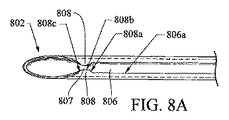
図8はフィデューシャル導入針800の或る実施形態を示している。針800は斜めに切り落とされた遠位先端802を有することが示されている。同針の管状のカニューレ本体804は、カニューレ804の遠位端領域に沿って長手方向の針スロット806を有している。スロット806は、少なくとも1つの戻り止め面を含んでいるのが好適であり、2つの戻り止めを含んでいるのが更に好適である。スロット806は、カニューレ804の壁全体を貫いて開口しているものとして示されているが、当該スロットは、針壁の厚さより浅く伸びていてもよく、つまり、溝として具現化されていてもよいものと理解されたい。図8の実施形態では、戻り止めは、2つのタブ808の間のスロット806の狭まった部分807として形成されている。タブ808は、略台形であるが、他の実施形態では異なった幾何学形状を有していてもよい。針スロット806の縁806aとタブ近位縁808aとタブ中央縁808bとタブ遠位縁808cのそれぞれの移行部は、角ばっていてもよいし(例えば、面取り又は角取り)、丸くなっていてもよい(例えば、角丸)。タブ808は、好適には、スロット806の遠位端に近接している。カニューレ804は、概して、例えばフィデューシャル(一例として、図2A−図2Dに示されているもの、又は他のフィデューシャルであって、好適に(単数又は複数の)フィデューシャルをタブ808によって制御可能に維持する機能を備えた針ルーメン810を容易に通ることができるもの)の様なフィデューシャルを滑動可能に通すように構成されている針ルーメン810を、円周方向に画定している。針は、ニッケル−チタン合金、コバルト−クロム(CoCr)合金、ステンレス鋼、又は如何なる他の適した材料から構成されていてもよい。その先端は、図示されている斜めに切り落とされた形態とは異なった幾何学形状を有していてもよい。或る別の実施形態では、タブ808同士は、フィデューシャル上の突起の通行を許容するべく、強制的に上向き及び/又は外向きにより大きく撓ませられるように突き合わされていてもよい。また、針の外側表面は、強化されたエコー源性を提供するようにくぼみが付けられているか又はそれ以外に肌理が付けられていてもよい。
FIG. 8 shows an embodiment of a
図8を参照しながら例示となる針の実施形態も説明するが、当該の例示となる針の実施形態は、以上に図2C−図2Dに関連付けて説明した、例示となるフィデューシャル針の実施形態と使用するための構成と寸法であってもよい。当該の例示となる針の実施形態では、針ルーメンのIDは少なくとも約0.034インチ(0.86mm)である。針のODは約0.042インチ(1.07mm;約19ゲージ)で、約0.008インチ(0.2mm)の壁厚さを有している。タブより近位のスロット部分は、幅約0.02インチ(0.5mm)、長さ約0.42インチ(約10.7mm)である。タブそれぞれは、スロット縁から外へ約0.06インチ(0.15mm)張り出していて、長さ約0.02インチ(0.5mm)のスロット対向縁を有している(長さには、約0.005インチ(0.13mm)の半径範囲の、近位側及び遠位側のスロット縁からの傾斜移行部を含めず)。これらの測定値と比率は、本願特許請求の範囲内に留まる限りにおいて、ここに例示されているものを含めた他の実施形態では変えられてもよい。 While an exemplary needle embodiment is also described with reference to FIG. 8, the exemplary needle embodiment of the exemplary fiducial needle described above in connection with FIGS. 2C-2D is described above. It may be the configuration and dimensions for use with the embodiment. In such exemplary needle embodiments, the needle lumen ID is at least about 0.034 inches (0.86 mm). The OD of the needle is about 0.042 inch (1.07 mm; about 19 gauge) and has a wall thickness of about 0.008 inch (0.2 mm). The slot portion proximal to the tab is about 0.02 inches (0.5 mm) wide and about 0.42 inches (about 10.7 mm) long. Each tab projects about 0.06 inch (0.15 mm) out of the slot edge and has a slot-facing edge that is about 0.02 inch (0.5 mm) in length (the length includes (Not including ramp transitions from proximal and distal slot edges in a radius range of about 0.005 inches (0.13 mm)). These measurements and ratios may be varied in other embodiments, including those exemplified herein, so long as they remain within the scope of the claims.
図9は、針800(図8)とフィデューシャル400(図2C−図2D)の断面の横断端面図を示している。この図は、針ルーメン810に対するフィデューシャル本体、及び針スロット806に対する突起408、の好適な精密許容差と好適な方向配置を示している。
FIG. 9 shows a cross-sectional end view of a cross section of needle 800 (FIG. 8) and fiducial 400 (FIGS. 2C-2D). This figure shows the preferred precision tolerance and preferred orientation of the fiducial body for the
フィデューシャル配備システム1000を、外観図である図10と当該図10の10A−10A線に沿った長手方向断面図である図10Aを参照しながら、以上に説明した針800とフィデューシャル400を使用して説明する。システム1000は、可撓性の細長い針シース1002を含んでいる。可撓性が更に高い本体近位部分802を含んでいる針800は、シースルーメン1004を通って伸びている。ここでは複数のフィデューシャル400として示されているが少なくとも1つのフィデューシャル400が、針のカニューレ本体の針ルーメン810の遠位領域に滑動可能且つ取り出し可能に配置されている。中央の長手方向本体部分402は針ルーメン810の内径を実質的に占めている。それぞれのフィデューシャル400の突起408は、突起408が中に突き出ているスロット806が形成された針壁の厚さとほぼ同じ高さを有している。
The
最遠位のフィデューシャル400の突起408は、針800のタブ808に当たって捕えられている。押し出し器として使用するように構成されているスタイレット1006が、針ルーメン810の一部を通って配置されており、このスタイレット1006は、近位端から操作するように構成されているのが好適であり、それによって、スタイレットを使用して、フィデューシャルを遠位方向に前進させる/押し出すこと、及び/又は、針がフィデューシャルの周りから引き抜かれる際にフィデューシャルを所定の場所に保持すること、ができる。フィデューシャルとスタイレットが針800の中にあることで、好適にも、針の柱強度が改善され、針が内視鏡の作業チャネル(図示せず)遠位端を通り抜け且つそこから外へ操縦されてゆく際に、屈曲したり、折れ曲がったり、又はそれ以外に損傷したりする可能性が低減する。
The
図10−図10Aのフィデューシャル配備システムを使用する方法を、図11A−11Cを参照しながら、図10−10Aにより詳細に示されている構造に関連付けて説明する。或る好適な使用方法では、作業チャネル1102を含んでいる内視鏡1100が提供される。1つの好適な方法では、内視鏡は、超音波画像化用に構成されている遠位超音波アレイ1104を含んでいるEUS内視鏡である。内視鏡1100は、好適には、映像取得部1106(例えばCCD、光学カメラ、又は他の光学的視覚化のための手段)も含んでいる。以下の方法は、フィデューシャル400を患者の膵臓1150の腫瘍1152の周囲に設置することに関連付けて説明している。
A method of using the fiducial deployment system of FIGS. 10-10A will be described in connection with the structure shown in more detail in FIGS. 10-10A with reference to FIGS. 11A-11C. In one preferred method of use, an
内視鏡1100は、図11Aには、患者の十二指腸1140を通って、その遠位端部分が、膵管1146が枝分かれして膵臓1150へとつながっている総胆管1144へのアクセスを提供するオディ括約筋1142に隣接するまで、導かれてきたところが示されている。
図11Aに示されている様に、シース1002は、膵管の中へ、そして膵管1146を通って、腫瘍1152に隣接する場所まで進められている。図11Bに示されている様に、針800は、シース1002から外へ進められ、腫瘍1152の周囲の第1の標的部位へ導かれる(好適には、超音波による誘導下に行われるものであるが、蛍光透視法又は別の視覚化技法に置き代えられてもよいし、又はそれらによって確認することもできる)。一旦、針800の遠位端802が第1の標的に位置付けられると、最遠位のフィデューシャル400がそこに配備される。1つの態様では、配備は、針遠位端802とその中のフィデューシャル400を第1の標的に位置付け、次いで、フィデューシャル400が所望の第1の標的位置に留まるようにスタイレット1006の位置を維持しながら針800を後退させることによって達成することができる。別の態様では、配備は、針遠位端802とフィデューシャル400をそこに第1の標的に隣接して位置付け、次いで、針800をその位置に保持したまま、フィデューシャル400が所望の第1の標的位置へと進められるようにスタイレット1006を前進させることによって達成することができる。
As shown in FIG. 11A, the
図10−図10Aに示されている針800及びフィデューシャル400の構造から理解される様に、使用者は、好適にも、フィデューシャルの前進/配備を、一度に1つ、に制御することができるであろう。こうして、フィデューシャル400は、「配備用意」位置、即ちその突起遠位面408aがタブ近位縁808aに当たって係合している位置、に入れられる。フィデューシャル400を配備するには、使用者は、スタイレット1006又は針800の一方を他方に対して、突起408にタブ808を前進通過させるのに十分な力で動かさなくてはならない。
As can be seen from the
使用者は、好適にも、突起408がタブ808を通過する際には抵抗があり、その抵抗は突起がタブを通り越すとすぐに減少する触感を感じる。次いで、使用者は、好適には、再度抵抗に遭って、最遠位のフィデューシャルの後の次のフィデューシャルがタブ近位縁808aに突き当たったことが示されるまで、スタイレットと針の相対動作を継続する。
The user preferably feels resistance when the
フィデューシャルとそれぞれのフィデューシャル上の突起は、当該フィデューシャルが針遠位先端802を実質的に通り越すことと次の最遠位フィデューシャルの突起がタブ近位縁808aに突き当たることが同時に起こって最遠位のフィデューシャルの配備完了となるような比率であるのが好適である。そういうものとして、幾つかのフィデューシャル実施形態では、突起より遠位のフィデューシャル本体部分が突起より近位の本体部分より長くなるように、突起をフィデューシャル本体上でより近位側に配置するのが都合がよいかもしれない。(例えば、図2Aのフィデューシャル200を参照)。図11Cは、針とシースが両方ともフィデューシャルから退去した状態で所定場所にあるフィデューシャルを示している。
The fiducial and the protrusion on each fiducial are such that the fiducial substantially passes the needle
次に、使用者は、針800をシース1002の中へ、針を第2の標的部位へ再び伸ばせられるように十分な距離だけ引き込み、そうして以上に説明した手順を繰り返すことができる。3段目、4段目、及びそれ以降のフィデューシャルの設置では、これらの手順段階が繰り返される。当技術では知られている様に、これらのフィデューシャルは放射線療法の様な療法の「正の標的化」及び/又は「負の標的化」のために使用することができる(「正の標的化」は「そこを治療する」ということを表し、「負の標的化」は「そこを治療しない」ということを表す)。本システムは数多くの利点をもたらす。例えば、探知されてはいるが診断未確定の組織塊を生検するために、既に内視鏡的処置を施術中である患者について検討する。内視鏡的生検が行われ、組織スライドは即座に準備されたとする。当該組織塊はフィデューシャルの設置が示唆される治療によって恩恵を受けるであろうとの診断が(何であれ利用可能で役に立ちそうな他のデータを併せて用いて)下されたなら、医師はただちにフィデューシャルを以上に説明したやり方で配備することができる。
The user can then retract the
より大きな針とフィデューシャルの使用を許容する好適な方法の実施形態をそれぞれ図11Dと図11Eを参照しながら説明する。図11Dには、内視鏡1100が患者の胃1157の中へ導かれてきたところが示されている。シース1002は、その遠位端が胃壁に隣接するまで進められ、次いで、針800が、胃壁を貫いて、膵臓1150へ、そして腫瘍1152の中へと進められている。この胃の場所は、フィデューシャル導入システムに標的部位(腫瘍1152)へのアクセスを提供するのに十分な程度に標的に近接している。この方法は、好適には、超音波アレイ1104を使用した超音波による視覚化の下に実行される。他の2つのフィデューシャル400が既に腫瘍1152の中に設置されていることが示されている。
Preferred method embodiments that allow the use of larger needles and fiducials are described with reference to FIGS. 11D and 11E, respectively. FIG. 11D shows the
内視鏡1100は、図11Eには、患者の十二指腸1140を通って導かれてきたところが示されている。シース1002は、その遠位端が十二指腸壁に隣接するまで進められ、次いで、針800が十二指腸壁を貫いて、膵臓1150へ、そして腫瘍1152の中へと進められている。十二指腸1140の中のこの場所は、フィデューシャル導入システムに標的部位(腫瘍1152)へのアクセスを提供するのに十分な程度に標的に近接している。この方法は、好適には、超音波アレイ1104を使用した超音波による視覚化の下に実行される。1つのフィデューシャル400が既に腫瘍1152の中に設置されていることが示されている。針800は、別のフィデューシャル400を放出したばかりで、部分的に引き込まれている。
本方法を、蛍光透視法を殆ど或いは全く使用せずに、直接/ビデオ画像化及び超音波画像化を使用して完遂することができることは、患者(放射線療法を受けなければならないかもしれず、その場合、放射線への全被曝量を治療上及び診断上必要最低限に抑えることが望まれる)の放射線被曝を最小限にするという利点をもたらす。本発明を実施すれば、患者、医師及び他の治療/診断職員、及び治療施設にとって時間と出費の利点が見込まれる、というのも、本発明の実施により、それらエンティティの全てが、2回目の内視鏡処置を予定に組んで施行すること、及び/又は、初期診断処置を説明されている先行技術で現在利用できる時間消費型の方法と材料を用いて拡大すること、を免れるからである。 The ability to complete this method using direct / video imaging and ultrasound imaging with little or no fluoroscopy is that patients (may need to receive radiation therapy, In some cases, it is desirable to minimize the total exposure to radiation for therapeutic and diagnostic purposes). Implementation of the present invention is expected to provide time and expense benefits for patients, physicians and other treatment / diagnostic personnel, and treatment facilities, because implementation of the present invention will ensure that all of these entities are This is because it avoids the scheduled implementation of endoscopic procedures and / or the expansion of initial diagnostic procedures using time-consuming methods and materials currently available in the prior art described. .
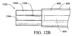
図12A及び12Bは、或る針実施形態1202を、上述の図4及び4Aに関連付けて論じたフィデューシャル実施形態600と一体に示している。針1202は、カニューレ本体1204を貫くスロット1206を有するカニューレ本体1204を含んでいる。フィデューシャル600は、針1202の上へ載せられるが、フィデューシャル600はその塊の一部に針の外側の周囲に配置される部分を有しているので、針1202は、図2A−図2E、図3、図5、図6、及び図7に示されている様なフィデューシャル実施形態と共に使用するのに実用的とされるより小さい針になろう。針カニューレ本体1204はフィデューシャル針ルーメン608に通して配置されている。フィデューシャル突起604は、針スロット1206を通って伸びており、以上に説明されている様に進行と制御された放出を提供している。図12Bは、針1202をそのスロット1206とスロット遠位縁上の1対の小さな戻り止め隆起1208と共に上から見た図を示している。
FIGS. 12A and 12B show one
図13は、図3に示されているフィデューシャル実施形態500と一体で有用となり得る様な多スロット針1300を示している。針1300は、遠位の或る長さに沿って延在する3つの細長いスロット1304を有するカニューレ本体1302を含んでいる。図3に示されているもの(504、506)の様な突起は、スロット1304を通って進行することができる。他の針実施形態では、2つのスロット又は3つより多くのスロットが設けられていてもよい。図13Aは、図13の13A−13A面に沿った横断面図を含む、針1300のもう1つの図を示しており、突起504、506の針スロット1304との相互作用をより明確に示している。
FIG. 13 shows a
図14は、上述の図5に関連付けて説明した型式の縫合糸搭載型フィデューシャル700の設置を示している。この例示では、Tアンカー分配針(図示せず;これらの針は、本技術でよく知られている)を使用して、Tアンカー712が標的組織725の中へおろされて、針が引き抜かれている。フィデューシャル700は、縫合糸710の上に搭載されており、押し出しカテーテル727を用いて組織725の中へ進められている。この構造と方法は、突起を含んでいるかどうかを問わず複数のフィデューシャルを設置するための異なった手段を提供している(突起が存在している場合には、フィデューシャル700はスロット付きの針と共に、概ね、以上に図11A-図11Cに関連付けて説明した様に施術される方法で使用できるであろう)。
FIG. 14 shows the installation of a suture loaded fiducial 700 of the type described in connection with FIG. 5 above. In this illustration, T-anchor dispensing needles (not shown; these needles are well known in the art) are used to lower T-
図15A及び図15Bは、各々、施条フィデューシャル1500と、同フィデューシャルと共に使用するように構成されているスロット付きの針1520を示している。フィデューシャル1500は、螺旋状にねじが切られた肌理を有する表面を有する円錐形の遠位先端1504を有する略円筒形の主本体1502を含んでいる。突起1506が本体1502の外周の周囲に部分的に螺旋状に巻き付けられている。このフィデューシャル1500用の針1520を図15Bに示している。それは、突起1506に適応するように構成されている螺旋状のスロット1524を有する略管状のカニューレ本体1522を有している。スロット1524は、一方の縁に沿って単一の戻り止めタブ1526を含んでいる。理解されるであろうが、針1520を通って進められてゆくフィデューシャル1500は、それが針を出て行く際に施条回転することになる。或る特定の組織の型では、この回転はフィデューシャルがより簡単に前進するのに助けとなる。
FIGS. 15A and 15B each show a ridge fiducial 1500 and a slotted
様々な実施形態を例示している図の描画は、必ずしも縮尺合わせされているわけではない。描画によっては、或る特定の詳細が強調のため誇張されているものもあり、そのため、部分の数又は比率は、どの様な異なる数又は比率も、1つ又はそれ以上の請求項によってその様に指定されていない限り、制限を課すものと読み取られてはならない。当業者には理解される様に、実施形態については、ここに提示されている請求項の範囲内に留まる限りにおいて、本明細書で異なった実施形態について説明されている特徴を互いに及び/又は現在知られているか又は将来開発される技術と組み合わせることを含め、ここで明示的に例示されていない実施形態を、本発明の範囲内で実践することもできる。例えば、本システムの針とフィデューシャルは、腹腔鏡型の処置の様な別の低侵襲性外科処置を含め、特許請求されている本発明の範囲内で経皮的に使用することができる。例えば、標的部位は、内視鏡によって(患者の自然開口部、例えば口、肛門、膣を通して導入された低侵襲性の内視鏡を使用して)アクセスでき得る場所の様な、胃腸管の中又はその近くの場所(例えば、肝臓、膵臓)であってもよい。これには−より広義には−NOTES(自然開口部越経管腔的内視鏡手術)処置を介して到達できる部位が含まれる。本方法及び装置は、経皮的な内視鏡的処置(例えば腹腔鏡的処置)又は経皮的な非内視鏡的処置の様な他の低侵襲性外科的技法で使用することもできるが、侵襲性がより少ない内視鏡的処置と共に使用されるのが最も好適である。従って、以上の詳細な説明は、限定を課すというよりむしろ説明を目的としているものと考えられることを意図している。よって、次に続く特許請求の範囲が、あらゆる等価物を含め、本発明の精神と範囲を定義するものであることを理解されたい。 The drawings in the figures illustrating various embodiments are not necessarily drawn to scale. In certain drawings, certain specific details may be exaggerated for emphasis, so that the number or proportion of parts may be any different number or proportion as defined by one or more claims. Unless otherwise specified, it shall not be read as imposing restrictions. As will be appreciated by those skilled in the art, the features described herein for different embodiments and / or for the different embodiments herein remain within the scope of the claims presented herein. Embodiments not explicitly exemplified herein, including in combination with currently known or future developed technologies, may also be practiced within the scope of the present invention. For example, the needle and fiducial of the system can be used percutaneously within the scope of the claimed invention, including other minimally invasive surgical procedures such as laparoscopic procedures. . For example, the target site can be located in the gastrointestinal tract, such as where it can be accessed by an endoscope (using a minimally invasive endoscope introduced through the patient's natural opening, eg, mouth, anus, vagina). It may be a place in or near (eg, liver, pancreas). This includes sites that can be reached through a -NOTES (natural opening transluminal endoscopic surgery) procedure in a broader sense. The method and apparatus can also be used in other minimally invasive surgical techniques such as percutaneous endoscopic procedures (eg, laparoscopic procedures) or percutaneous non-endoscopic procedures. However, it is most preferred to be used with a less invasive endoscopic procedure. Accordingly, the above detailed description is intended to be considered illustrative rather than restrictive. Therefore, it is to be understood that the following claims, including any equivalents, define the spirit and scope of the present invention.
100 導入器
101 第1ハンドル
102 針
103 第2ハンドル
104 スタイレット
105 カニューレ遠位端
106 スタイレット遠位端
108 カニューレ
110 シード/フィデューシャル
112 栓
200 フィデューシャル
201 フィデューシャル本体
202 フィデューシャル本体遠位端
204 フィデューシャル本体近位端
206 フィデューシャル本体長手方向表面
207 突起遠位端
208 突起
209 突起近位端
300 フィデューシャル
301 フィデューシャル本体
302 フィデューシャル本体遠位端
304 フィデューシャル本体近位端
306 フィデューシャル本体長手方向表面
307 突起遠位端
308 突起
309 突起近位端
400 フィデューシャル
402 フィデューシャル本体
406 フィデューシャル本体長手方向外周面
407 突起端面
408 突起
408a 突起遠位面
500 フィデューシャル
502 フィデューシャル本体
504、506 突起
600 フィデューシャル
602 フィデューシャル中央本体
604 突起
606 フィデューシャル外側本体
608 針ルーメン
700 フィデューシャル
701 フィデューシャル本体
702 フィデューシャル中央ルーメン
704 フィデューシャル遠位鼻部
706 突起
710 縫合糸
712 Tアンカー
725 標的組織
727 押し出しカテーテル
750 フィデューシャル
752 フィデューシャル本体
754 突起
780 フィデューシャル
782、783、784 突起
786 フィデューシャル遠位端
790 カニューレ
792 溝
800 フィデューシャル導入針
802 針遠位先端
804 カニューレ本体
806 針スロット
806a スロット縁
807 スロットの狭まった部分
808 タブ
808a タブ近位縁
808b タブ中央縁
808c タブ遠位縁
810 針ルーメン
1000 フィデューシャル配備システム
1002 シース
1004 シースルーメン
1006 スタイレット
1100 内視鏡
1102 作業チャネル
1104 遠位超音波アレイ
1106 映像取得部
1140 十二指腸
1142 オディ括約筋
1144 総胆管
1146 膵管
1150 膵臓
1152 腫瘍
1157 胃
1202 針
1204 カニューレ本体
1206 スロット
1208 戻り止め隆起
1300 針
1302 カニューレ本体
1304 スロット
1500 フィデューシャル
1502 フィデューシャル本体
1504 フィデューシャル遠位先端
1506 突起
1520 針
1522 カニューレ本体
1524 螺旋状スロット
1526 戻り止めタブ
DESCRIPTION OF SYMBOLS 100 Introducer 101 1st handle 102 Needle 103 2nd handle 104 Stylet 105 Cannula distal end 106 Stylet distal end 108 Cannula 110 Seed / Fiducial 112 Plug 200 Fiducial 201 Fiducial body 202 Fiducial Body distal end 204 fiducial body proximal end 206 fiducial body longitudinal surface 207 protrusion distal end 208 protrusion 209 protrusion proximal end 300 fiducial 301 fiducial body 302 fiducial body distal end 304 Fiducial body proximal end 306 fiducial body longitudinal surface 307 protrusion distal end 308 protrusion 309 protrusion proximal end 400 fiducial 402 fiducial body 406 fiducial Body longitudinal direction outer peripheral surface 407 Projection end surface 408 Projection 408a Projection distal surface 500 Fiducial 502 Fiducial body 504, 506 Projection 600 Fiducial 602 Fiducial central body 604 Projection 606 Fiducial outer body 608 Needle lumen 700 Fiducial 701 Fiducial body 702 Fiducial central lumen 704 Fiducial distal nose 706 Protrusion 710 Suture 712 T anchor 725 Target tissue 727 Extrusion catheter 750 Fiducial 752 Fiducial body 754 Protrusion 780 Fiducial Shall 782, 783, 784 Protrusion 786 Fiducial distal end 790 Cannula 792 Groove 800 Fiducial introduction needle 802 Needle distal tip 8 4 Cannula body 806 Needle slot 806a Slot edge 807 Slot narrowed portion 808 Tab 808a Tab proximal edge 808b Tab central edge 808c Tab distal edge 810 Needle lumen 1000 Fiducial deployment system 1002 Sheath 1004 See-through-men 1006 Stylet 1100 In Endoscope 1102 Working channel 1104 Distal ultrasound array 1106 Video acquisition unit 1140 Duodenum 1142 Odi sphincter 1144 Common bile duct 1146 Pancreatic duct 1150 Pancreas 1152 Tumor 1157 Stomach 1202 Needle 1204 Cannula body 1206 Slot 1208 Main body 1304 Detent 1301 Fiducial 1502 Fiducial body 1504 Fiducial distal tip 1506 projections 1520 needle 1522 cannula body 1524 spiral slot 1526 detent tab
Claims (20)
前記針は、
その長手方向の少なくとも一部分を貫いて配置されている針ルーメンを画定している管状のカニューレ本体と、
針遠位端領域であって、
前記針ルーメンの遠位端において開口している針遠位端と、
半径方向に前記カニューレ本体の厚さの少なくとも一部を貫いて延び、前記針ルーメンに向けて開口していて、少なくとも1つの戻り止めを含んでいる、少なくとも1つの略長手方向の針スロットと、を有している、針遠位先端領域と、
を備えており、
前記少なくとも1つのフィデューシャルは、
柱状の本体であって、
前記針ルーメンの中に滑動可能に配置されているフィデューシャル中央部分と、
前記針スロットの中へ突き出ている少なくとも1つの側方突起と、を含んでいる、柱状の本体と、
前記針ルーメンの一部を通って伸びていて、前記少なくとも1つのフィデューシャルを前記戻り止めを越えて前記針の遠位端の開口から外へ前進させるように構成されているスタイレットと、
を備えている、
フィデューシャル配備システム。 A fiducial deployment system comprising a needle and at least one fiducial,
The needle is
A tubular cannula body defining a needle lumen disposed through at least a portion of its longitudinal direction;
A needle distal end region,
A needle distal end that is open at the distal end of the needle lumen;
At least one generally longitudinal needle slot that extends radially through at least a portion of the thickness of the cannula body, opens toward the needle lumen, and includes at least one detent; A distal tip region of the needle,
With
The at least one fiducial is
A columnar body,
A fiducial central portion slidably disposed within the needle lumen;
A columnar body including at least one lateral protrusion protruding into the needle slot;
A stylet extending through a portion of the needle lumen and configured to advance the at least one fiducial beyond the detent and out of an opening at the distal end of the needle;
With
Fiducial deployment system.
請求項10に記載のフィデューシャル配備システムを準備する段階と、
内視鏡の遠位端を患者の体内の第1の標的部位に近接する場所へ導く段階と、
前記針の遠位端を前記第1の標的部位へ導く段階と、
前記少なくとも1つのフィデューシャルを、(i)前記スタイレットを前記針ルーメンを通して遠位方向に前進させて、前記少なくとも1つのフィデューシャルを前記少なくとも1つの戻り止め面を越えて前記第1の標的部位の中へ押し出す、(ii)前記針を前記スタイレットに対して、前記少なくとも1つの戻り止め面が近位方向に前記フィデューシャル突起を越えて引っ張られ、前記フィデューシャルが前記針ルーメンから外へ前記第1の標的部位の中へ放出されるように後退させる、及び(iii)それらの組合せ、から選択された行為を行うことによって配備する段階と、を含む方法。 In the method of placing the fiducial in the patient's body,
Providing a fiducial deployment system according to claim 10;
Directing the distal end of the endoscope to a location proximate to a first target site in the patient's body;
Directing the distal end of the needle to the first target site;
(I) advancing the stylet distally through the needle lumen to advance the at least one fiducial beyond the at least one detent surface; (Ii) pushing the needle against the stylet, the at least one detent surface is pulled in a proximal direction beyond the fiducial protrusion, and the fiducial is the needle Retreating out of the lumen to be released into the first target site, and (iii) deploying by performing an action selected from a combination thereof.
柱状の本体であって、フィデューシャル近位端と、フィデューシャル遠位端と、フィデューシャル外径を越えて突き出ており、スロット付きの針のルーメンの中の前記フィデューシャルを整列及び維持するように構成されている少なくとも1つの側方突起を含む長手方向に延びるフィデューシャル中央部分と、を有する柱状の本体を備える、フィデューシャル。 In a fiducial that is used to demarcate a site within the body and is configured to be deployed within the patient's body,
Columnar body that protrudes beyond the fiducial proximal end, fiducial distal end, and fiducial outer diameter to align the fiducial in the lumen of the slotted needle And a columnar body having a longitudinally extending fiducial central portion including at least one lateral projection configured to maintain.
Applications Claiming Priority (3)
| Application Number | Priority Date | Filing Date | Title |
|---|---|---|---|
| US17419609P | 2009-04-30 | 2009-04-30 | |
| US61/174,196 | 2009-04-30 | ||
| PCT/US2010/031842 WO2010126750A2 (en) | 2009-04-30 | 2010-04-21 | System and method for fiducial deployment |
Publications (2)
| Publication Number | Publication Date |
|---|---|
| JP2012525215A true JP2012525215A (en) | 2012-10-22 |
| JP2012525215A5 JP2012525215A5 (en) | 2013-06-06 |
Family
ID=42779910
Family Applications (1)
| Application Number | Title | Priority Date | Filing Date |
|---|---|---|---|
| JP2012508533A Pending JP2012525215A (en) | 2009-04-30 | 2010-04-21 | System and method for deploying fiducials |
Country Status (6)
| Country | Link |
|---|---|
| US (1) | US9042964B2 (en) |
| EP (1) | EP2424460B1 (en) |
| JP (1) | JP2012525215A (en) |
| AU (1) | AU2010241934B2 (en) |
| CA (1) | CA2757870C (en) |
| WO (1) | WO2010126750A2 (en) |
Cited By (3)
| Publication number | Priority date | Publication date | Assignee | Title |
|---|---|---|---|---|
| JP2016521623A (en) * | 2013-06-12 | 2016-07-25 | ボストン サイエンティフィック サイムド,インコーポレイテッドBoston Scientific Scimed,Inc. | Deployment mechanism of alignment reference object |
| JP2017086878A (en) * | 2015-10-07 | 2017-05-25 | コヴィディエン リミテッド パートナーシップ | Endoscopic ultrasound fine needle fiducial system |
| US10327862B2 (en) | 2014-09-17 | 2019-06-25 | Covidien Lp | Fiducial marker deployment system |
Families Citing this family (16)
| Publication number | Priority date | Publication date | Assignee | Title |
|---|---|---|---|---|
| EP2424460B1 (en) | 2009-04-30 | 2017-05-24 | Cook Medical Technologies LLC | System for fiducial deployment |
| GB0918995D0 (en) * | 2009-10-29 | 2009-12-16 | Univ Hull | A speech valve, a tool for facilitating insertion of a speech valve and a tool for holding a speech valve |
| JP5665878B2 (en) * | 2009-12-18 | 2015-02-04 | クック メディカル テクノロジーズ エルエルシーCook Medical Technologies Llc | System and method for deploying fiducials |
| US9072542B2 (en) | 2009-12-18 | 2015-07-07 | Cook Medical Technologies Llc | System and method for fiducial deployment |
| US20130006101A1 (en) | 2011-06-28 | 2013-01-03 | Mchugo Vincent | System for fiducial deployment |
| US8838208B2 (en) * | 2011-06-28 | 2014-09-16 | Cook Medical Technologies Llc | Fiducial deployment needle system |
| JP5833776B2 (en) * | 2012-01-30 | 2015-12-16 | クック・メディカル・テクノロジーズ・リミテッド・ライアビリティ・カンパニーCook Medical Technologies Llc | Fiducial deployment system |
| US9901328B2 (en) * | 2012-06-06 | 2018-02-27 | Carefusion 2200, Inc. | Vacuum assisted biopsy device |
| EP2719355A3 (en) * | 2012-10-11 | 2014-05-21 | Cook Medical Technologies LLC | Clutched-gear handle for fiducial deployment |
| CN103100147A (en) * | 2012-11-15 | 2013-05-15 | 王兆进 | Infrared vagina physiotherapy instrument |
| US9522264B2 (en) | 2013-02-26 | 2016-12-20 | Cook Medical Technologies Llc | Ratchet-slide handle and system for fiducial deployment |
| US9308714B2 (en) * | 2013-03-05 | 2016-04-12 | International Business Machines Corporation | Method for improving surface quality of spalled substrates |
| CN106456213B (en) * | 2014-06-09 | 2019-04-12 | 库克医药技术有限责任公司 | Screw drive-type handle and system for primary standard substance deployment |
| WO2015195232A1 (en) | 2014-06-16 | 2015-12-23 | Cook Medical Technologies Llc | Plunger-driven collet handle and system for fiducial deployment |
| DE102014218454A1 (en) * | 2014-09-15 | 2016-03-17 | Siemens Aktiengesellschaft | Apparatus, medical instrument and method for obtaining a spatial image of a medical instrument with a magnetic resonance tomography apparatus |
| US10123848B2 (en) | 2014-12-03 | 2018-11-13 | Cook Medical Technologies Llc | EUS fiducial needle stylet handle assembly |
Citations (7)
| Publication number | Priority date | Publication date | Assignee | Title |
|---|---|---|---|---|
| JP2003502121A (en) * | 1999-06-18 | 2003-01-21 | エーイーエー テクノロジー キューエスエー ゲゼルシャフト ミット ベシュレンクテル ハフツング | Radiation sources for intravascular radiation therapy |
| JP2003503098A (en) * | 1999-06-30 | 2003-01-28 | セノークス・インコーポレイテッド | Biopsy position marker and process and apparatus for applying the same |
| US20030233101A1 (en) * | 2002-06-17 | 2003-12-18 | Senorx, Inc. | Plugged tip delivery tube for marker placement |
| JP2006509531A (en) * | 2002-08-09 | 2006-03-23 | バード ブラッキーセラピー インコーポレイテッド | Short-range radiation therapy seed placement system |
| WO2007037326A1 (en) * | 2005-09-28 | 2007-04-05 | Olympus Medical Systems Corp. | Suturing device |
| WO2008021691A2 (en) * | 2006-08-09 | 2008-02-21 | Regen Biologics, Inc. | System and method for all-inside suture fixation for implant attachment and soft tissue repair |
| JP2008516733A (en) * | 2004-10-18 | 2008-05-22 | テンプル・ユニバーシティ−オブ・ザ・コモンウェルス・システム・オブ・ハイアー・エデュケイション | Endoscopic suture device and method |
Family Cites Families (94)
| Publication number | Priority date | Publication date | Assignee | Title |
|---|---|---|---|---|
| US2009393A (en) | 1930-06-12 | 1935-07-30 | Failla Gioacchino | Means for effecting therapeutic implantations |
| US2269963A (en) | 1940-06-01 | 1942-01-13 | Wappler Frederick Charles | Implanting device |
| US2620796A (en) | 1950-03-06 | 1952-12-09 | American Scient Lab Inc | Pellet injector |
| US3470834A (en) | 1968-03-08 | 1969-10-07 | Dennison Mfg Co | Fastener attaching device |
| US3820545A (en) | 1973-03-26 | 1974-06-28 | K Jefferts | Tag implanting machine |
| US3815798A (en) | 1973-05-21 | 1974-06-11 | B Lavitch | Button fastener |
| GB1525841A (en) | 1976-05-18 | 1978-09-20 | Hundon Forge Ltd | Drug implanters |
| US4105030A (en) | 1977-01-03 | 1978-08-08 | Syntex (U.S.A.) Inc. | Implant apparatus |
| US4086914A (en) | 1977-02-11 | 1978-05-02 | Edwin Bailey Moore | Implant injector |
| US4646740A (en) | 1981-02-23 | 1987-03-03 | Edward Weck & Co., Inc. | Automatic hemoclip applier |
| US4451254A (en) | 1982-03-15 | 1984-05-29 | Eli Lilly And Company | Implant system |
| AU1474183A (en) | 1982-04-22 | 1983-11-21 | Gustavsson Bengt | Anordning for inforande av katetrar i ett blodkarl |
| GB8424436D0 (en) | 1984-09-27 | 1984-10-31 | Pratt Int Ltd Burnerd | Surgical appliance |
| US4807628A (en) | 1985-04-26 | 1989-02-28 | Edward Weck & Company, Inc. | Method and apparatus for storing, dispensing, and applying surgical staples |
| US5047038A (en) | 1985-07-01 | 1991-09-10 | Edward Weck Incorporated | Automatic hemostatic clip applier |
| US4648542A (en) | 1985-11-01 | 1987-03-10 | Senmed, Inc. | Disposable stapler |
| US4700692A (en) | 1985-12-23 | 1987-10-20 | Baumgartner George C | Surgical implantation method and apparatus |
| US4661103A (en) | 1986-03-03 | 1987-04-28 | Engineering Development Associates, Ltd. | Multiple implant injector |
| US5002548A (en) | 1986-10-06 | 1991-03-26 | Bio Medic Data Systems, Inc. | Animal marker implanting system |
| US5024727A (en) | 1986-10-06 | 1991-06-18 | Bio Medic Data Systems, Inc. | Method of forming an animal marker implanting system |
| NZ226230A (en) | 1987-09-18 | 1991-01-29 | Schering Agrochemicals Ltd | Implant gun for use with cartridge of strip form |
| US5281197A (en) * | 1992-07-27 | 1994-01-25 | Symbiosis Corporation | Endoscopic hemostatic agent delivery system |
| CA2106720C (en) | 1992-09-23 | 1997-12-16 | Charles L. Deschenes | Needle for use as part of a plastic fastener dispensing tool |
| US5607436A (en) | 1993-10-08 | 1997-03-04 | United States Surgical Corporation | Apparatus for applying surgical clips |
| DE9403161U1 (en) | 1994-02-25 | 1994-04-21 | Sueddeutsche Feinmechanik | Cannula |
| DE69534233T2 (en) | 1994-09-16 | 2005-10-27 | Ethicon Endo-Surgery, Inc., Cincinnati | DEVICES FOR DETERMINING AND MARKING TISSUE |
| JP2913372B2 (en) | 1994-12-16 | 1999-06-28 | 株式会社コーテックス | Hollow needle for locking piece mounting machine |
| US5713828A (en) | 1995-11-27 | 1998-02-03 | International Brachytherapy S.A | Hollow-tube brachytherapy device |
| AU1331497A (en) | 1995-12-18 | 1997-07-14 | Kerisma Medical Products, L.L.C. | Fiberoptic-guided interstitial seed manual applicator and seed cartridge |
| US5860909A (en) | 1996-10-18 | 1999-01-19 | Mick Radio Nuclear Instruments, Inc. | Seed applicator for use in radiation therapy |
| FR2762517B1 (en) | 1997-04-25 | 1999-07-16 | Ordicam Rech Et Dev | TRANSPONDER CHIP SUBCUTANEOUS INJECTION NEEDLE AND SYRINGE |
| US6004320A (en) | 1997-09-19 | 1999-12-21 | Oratec Interventions, Inc. | Clip on electrocauterizing sheath for orthopedic shave devices |
| US6197324B1 (en) | 1997-12-18 | 2001-03-06 | C. R. Bard, Inc. | System and methods for local delivery of an agent |
| WO1999043267A1 (en) | 1998-02-25 | 1999-09-02 | Tracenet Technologies, Inc. | Transponder insertion device and method |
| KR20010071512A (en) | 1998-07-20 | 2001-07-28 | 찰스 더블유 프란즈 | Brachytherapy Device Including An Anti-Static Handle |
| US6220248B1 (en) | 1998-10-21 | 2001-04-24 | Ethicon Endo-Surgery, Inc. | Method for implanting a biopsy marker |
| US6725083B1 (en) | 1999-02-02 | 2004-04-20 | Senorx, Inc. | Tissue site markers for in VIVO imaging |
| US7983734B2 (en) | 2003-05-23 | 2011-07-19 | Senorx, Inc. | Fibrous marker and intracorporeal delivery thereof |
| DK1185200T3 (en) | 1999-06-05 | 2008-04-07 | Wilson Cook Medical Inc | Characteristics of an endoscopic medical device |
| US6283948B1 (en) | 1999-07-13 | 2001-09-04 | Ethicon, Inc. | Trocar obturator having grooved passageway |
| US6267718B1 (en) | 1999-07-26 | 2001-07-31 | Ethicon, Endo-Surgery, Inc. | Brachytherapy seed cartridge |
| US6221003B1 (en) | 1999-07-26 | 2001-04-24 | Indigo Medical, Incorporated | Brachytherapy cartridge including absorbable and autoclaveable spacer |
| US6264599B1 (en) | 1999-08-10 | 2001-07-24 | Syntheon, Llc | Radioactive therapeutic seeds having fixation structure |
| US7615076B2 (en) | 1999-10-20 | 2009-11-10 | Anulex Technologies, Inc. | Method and apparatus for the treatment of the intervertebral disc annulus |
| US6402677B1 (en) | 1999-12-17 | 2002-06-11 | C.R. Bard, Inc. | Brachytherapy seed needle with window |
| US6450938B1 (en) | 2000-03-21 | 2002-09-17 | Promex, Llc | Brachytherapy device |
| GB0011581D0 (en) | 2000-05-15 | 2000-07-05 | Nycomed Amersham Plc | Grooved brachytherapy |
| US20040097780A1 (en) | 2000-12-15 | 2004-05-20 | Kawasumi Laboratories, Inc. | Cartridge for marker delivery device and marker delivery device |
| WO2003002181A2 (en) | 2001-06-29 | 2003-01-09 | A.B. Korkor Medical, Inc. | Catheter introducer having an expandable tip |
| US6569077B2 (en) * | 2001-07-11 | 2003-05-27 | Bruno Schmidt | Dimpled seed implant needle |
| US6786858B2 (en) | 2001-11-02 | 2004-09-07 | Ideamatrix, Inc. | Delivery system and method for interstitial radiotherapy using hollow seeds |
| US7280865B2 (en) * | 2001-12-20 | 2007-10-09 | Accuray Incorporated | Anchored fiducial apparatus and method |
| GB0200444D0 (en) | 2002-01-10 | 2002-02-27 | Owen Mumford Ltd | Improvements relating to medical injection devices |
| US6837844B1 (en) | 2002-05-14 | 2005-01-04 | Med-Tec Iowa, Inc. | Seed cartridge for radiation therapy |
| WO2003105668A2 (en) | 2002-06-01 | 2003-12-24 | Alfred E. Mann Institute For Biomedical Engineering At The University Of Southern California | Injection devices and methods for testing implants and for unimpeded target location testing |
| US20070265582A1 (en) | 2002-06-12 | 2007-11-15 | University Of Southern California | Injection Devices for Unimpeded Target Location Testing |
| US7041048B2 (en) | 2002-10-16 | 2006-05-09 | Sourcetech Medical, Llc | Apparatus and method for dose administration in brachytherapy |
| US20060036158A1 (en) | 2003-11-17 | 2006-02-16 | Inrad, Inc. | Self-contained, self-piercing, side-expelling marking apparatus |
| US6889833B2 (en) | 2002-12-30 | 2005-05-10 | Calypso Medical Technologies, Inc. | Packaged systems for implanting markers in a patient and methods for manufacturing and using such systems |
| US6796935B1 (en) | 2003-03-20 | 2004-09-28 | Michael Savino | Multiple seed implanter |
| US7214206B2 (en) | 2003-04-03 | 2007-05-08 | Valera Pharmaceuticals, Inc. | Implanting device and method of using same |
| KR100543308B1 (en) * | 2003-05-23 | 2006-01-20 | 주식회사 팬택앤큐리텔 | Method for supplying the infomation of location in GPS server or GPS wireless communication terminal |
| US20040260199A1 (en) | 2003-06-19 | 2004-12-23 | Wilson-Cook Medical, Inc. | Cytology collection device |
| US7001341B2 (en) | 2003-08-13 | 2006-02-21 | Scimed Life Systems, Inc. | Marking biopsy sites |
| US7465279B2 (en) | 2004-03-31 | 2008-12-16 | Ethicon Endo-Surgery, Inc. | Marker device and method of deploying a cavity marker using a surgical biopsy device |
| US20050267319A1 (en) | 2004-05-12 | 2005-12-01 | White Jack C | Brachytherapy seed loader and containers |
| US9638770B2 (en) | 2004-05-21 | 2017-05-02 | Devicor Medical Products, Inc. | MRI biopsy apparatus incorporating an imageable penetrating portion |
| US20090209804A1 (en) | 2004-07-23 | 2009-08-20 | Calypso Medical Technologies, Inc. | Apparatuses and methods for percutaneously implanting objects in patients |
| US7588528B2 (en) | 2004-08-24 | 2009-09-15 | C. R. Bard, Inc. | Brachytherapy apparatus for dispensing medication |
| US7361135B2 (en) | 2004-08-24 | 2008-04-22 | C R Bard, Inc | Brachytherapy system for dispensing medication |
| US7335155B2 (en) | 2004-09-14 | 2008-02-26 | Boston Scientific Scimed, Inc. | Unitary formulation delivery device |
| US20060173236A1 (en) | 2005-01-14 | 2006-08-03 | White Jack C | Brachytherapy magazine seed indicator |
| US7577473B2 (en) | 2005-02-03 | 2009-08-18 | Bard Peripheral Vascular, Inc. | Apparatus for subcutaneous placement of an imaging marker |
| US20060235298A1 (en) | 2005-03-31 | 2006-10-19 | Robert Kotmel | Internal biopsy marking |
| BRPI0616514A2 (en) | 2005-08-11 | 2011-06-21 | Navotek Medical Ltd | medical treatment system and method using position sensor based radioactivity |
| US9283053B2 (en) | 2005-09-19 | 2016-03-15 | Varian Medical Systems, Inc. | Apparatus and methods for implanting objects, such as bronchoscopically implanting markers in the lung of patients |
| WO2007067889A1 (en) | 2005-12-08 | 2007-06-14 | Eli Lilly And Company | Dose indicating assembly of a pharmaceutical injection device |
| US7391236B2 (en) | 2005-12-27 | 2008-06-24 | Altera Corporation | Distributed memory in field-programmable gate array integrated circuit devices |
| US7927271B2 (en) | 2006-05-17 | 2011-04-19 | C.R. Bard, Inc. | Endoscope tool coupling |
| US20080033286A1 (en) | 2006-08-02 | 2008-02-07 | Civco Medical Instruments Co., Inc. | Fiducial marker for imaging localization and method of using the same |
| US7945307B2 (en) | 2006-08-04 | 2011-05-17 | Senorx, Inc. | Marker delivery system with obturator |
| US20080287782A1 (en) * | 2007-03-26 | 2008-11-20 | Maeghan Traboulsi | Radiation Therapy Marking Pen and Method |
| JP2010528754A (en) | 2007-06-04 | 2010-08-26 | エスヴィーアイピー 8 エルエルシー | Device that can fix tissue |
| US20090105584A1 (en) * | 2007-10-18 | 2009-04-23 | Civco Medical Instruments Co., Inc. | Fiducial marker deployment system using single stick neeedle and method of use |
| CN101938934B (en) | 2008-02-05 | 2013-05-01 | 威尔逊-库克医学公司 | Adaptor for endoscopic orientation of an elongate medical device |
| US20100331677A1 (en) | 2008-04-25 | 2010-12-30 | The Johns Hopkins University | Marker delivery system |
| US20100063392A1 (en) * | 2008-09-08 | 2010-03-11 | Olympus Medical Systems Corp. | Ultrasound-guided ablation method and ultrasound-guided ablation system |
| EP2424460B1 (en) | 2009-04-30 | 2017-05-24 | Cook Medical Technologies LLC | System for fiducial deployment |
| US20110028831A1 (en) | 2009-07-30 | 2011-02-03 | Kent James P | Permanently visible implantable fiduciary tissue marker |
| US8371443B2 (en) | 2009-09-22 | 2013-02-12 | Devicor Medical Products, Inc. | Biopsy marker delivery device |
| US9072542B2 (en) | 2009-12-18 | 2015-07-07 | Cook Medical Technologies Llc | System and method for fiducial deployment |
| US8838208B2 (en) | 2011-06-28 | 2014-09-16 | Cook Medical Technologies Llc | Fiducial deployment needle system |
| US20130006101A1 (en) | 2011-06-28 | 2013-01-03 | Mchugo Vincent | System for fiducial deployment |
| WO2013055568A1 (en) | 2011-10-13 | 2013-04-18 | Cook Medical Technologies Llc | Engaged fiducials and system for deployment |
-
2010
- 2010-04-21 EP EP10716185.3A patent/EP2424460B1/en active Active
- 2010-04-21 AU AU2010241934A patent/AU2010241934B2/en active Active
- 2010-04-21 US US12/764,432 patent/US9042964B2/en active Active
- 2010-04-21 JP JP2012508533A patent/JP2012525215A/en active Pending
- 2010-04-21 CA CA2757870A patent/CA2757870C/en active Active
- 2010-04-21 WO PCT/US2010/031842 patent/WO2010126750A2/en active Application Filing
Patent Citations (7)
| Publication number | Priority date | Publication date | Assignee | Title |
|---|---|---|---|---|
| JP2003502121A (en) * | 1999-06-18 | 2003-01-21 | エーイーエー テクノロジー キューエスエー ゲゼルシャフト ミット ベシュレンクテル ハフツング | Radiation sources for intravascular radiation therapy |
| JP2003503098A (en) * | 1999-06-30 | 2003-01-28 | セノークス・インコーポレイテッド | Biopsy position marker and process and apparatus for applying the same |
| US20030233101A1 (en) * | 2002-06-17 | 2003-12-18 | Senorx, Inc. | Plugged tip delivery tube for marker placement |
| JP2006509531A (en) * | 2002-08-09 | 2006-03-23 | バード ブラッキーセラピー インコーポレイテッド | Short-range radiation therapy seed placement system |
| JP2008516733A (en) * | 2004-10-18 | 2008-05-22 | テンプル・ユニバーシティ−オブ・ザ・コモンウェルス・システム・オブ・ハイアー・エデュケイション | Endoscopic suture device and method |
| WO2007037326A1 (en) * | 2005-09-28 | 2007-04-05 | Olympus Medical Systems Corp. | Suturing device |
| WO2008021691A2 (en) * | 2006-08-09 | 2008-02-21 | Regen Biologics, Inc. | System and method for all-inside suture fixation for implant attachment and soft tissue repair |
Cited By (7)
| Publication number | Priority date | Publication date | Assignee | Title |
|---|---|---|---|---|
| JP2016521623A (en) * | 2013-06-12 | 2016-07-25 | ボストン サイエンティフィック サイムド,インコーポレイテッドBoston Scientific Scimed,Inc. | Deployment mechanism of alignment reference object |
| US10953212B2 (en) | 2013-06-12 | 2021-03-23 | Boston Scientific Scimed, Inc. | Fiducial deployment mechanisms, and related methods of use |
| US11957860B2 (en) | 2013-06-12 | 2024-04-16 | Boston Scientific Scimed, Inc. | Fiducial deployment mechanisms, and related methods of use |
| US10327862B2 (en) | 2014-09-17 | 2019-06-25 | Covidien Lp | Fiducial marker deployment system |
| JP2017086878A (en) * | 2015-10-07 | 2017-05-25 | コヴィディエン リミテッド パートナーシップ | Endoscopic ultrasound fine needle fiducial system |
| US10485583B2 (en) | 2015-10-07 | 2019-11-26 | Covidien Lp | Endoscopic ultrasound fine needle fiducial system |
| US11382661B2 (en) | 2015-10-07 | 2022-07-12 | Covidien Lp | Endoscopic ultrasound fine needle fiducial system |
Also Published As
| Publication number | Publication date |
|---|---|
| CA2757870A1 (en) | 2010-11-04 |
| EP2424460B1 (en) | 2017-05-24 |
| WO2010126750A3 (en) | 2010-12-23 |
| US20100280367A1 (en) | 2010-11-04 |
| AU2010241934B2 (en) | 2014-01-16 |
| US9042964B2 (en) | 2015-05-26 |
| WO2010126750A2 (en) | 2010-11-04 |
| AU2010241934A1 (en) | 2011-11-17 |
| EP2424460A2 (en) | 2012-03-07 |
| CA2757870C (en) | 2016-02-02 |
Similar Documents
| Publication | Publication Date | Title |
|---|---|---|
| AU2010241934B2 (en) | System and method for fiducial deployment | |
| JP6302573B2 (en) | Plunger-driven collet handle and fiducial deployment system | |
| JP2012525215A5 (en) | ||
| EP2967642B1 (en) | Ratchet-slide handle and system for fiducial deployment | |
| EP2725990B1 (en) | System for fiducial deployment | |
| US9072542B2 (en) | System and method for fiducial deployment | |
| JP5665878B2 (en) | System and method for deploying fiducials | |
| JP6283128B2 (en) | Screw-driven handle and system for reference marker placement | |
| EP2765947B1 (en) | Engaged fiducials and system for deployment | |
| US9332886B2 (en) | Fiducial placement system and splayed stylet |
Legal Events
| Date | Code | Title | Description |
|---|---|---|---|
| A521 | Request for written amendment filed |
Free format text: JAPANESE INTERMEDIATE CODE: A523 Effective date: 20130419 |
|
| A621 | Written request for application examination |
Free format text: JAPANESE INTERMEDIATE CODE: A621 Effective date: 20130419 |
|
| A977 | Report on retrieval |
Free format text: JAPANESE INTERMEDIATE CODE: A971007 Effective date: 20140120 |
|
| A131 | Notification of reasons for refusal |
Free format text: JAPANESE INTERMEDIATE CODE: A131 Effective date: 20140218 |
|
| A02 | Decision of refusal |
Free format text: JAPANESE INTERMEDIATE CODE: A02 Effective date: 20140729 |