JP2012179385A - Apparatus and method for destruction of adipose tissue - Google Patents
Apparatus and method for destruction of adipose tissue Download PDFInfo
- Publication number
- JP2012179385A JP2012179385A JP2012114475A JP2012114475A JP2012179385A JP 2012179385 A JP2012179385 A JP 2012179385A JP 2012114475 A JP2012114475 A JP 2012114475A JP 2012114475 A JP2012114475 A JP 2012114475A JP 2012179385 A JP2012179385 A JP 2012179385A
- Authority
- JP
- Japan
- Prior art keywords
- transducer
- tissue
- adipose tissue
- treatment
- energy
- Prior art date
- Legal status (The legal status is an assumption and is not a legal conclusion. Google has not performed a legal analysis and makes no representation as to the accuracy of the status listed.)
- Granted
Links
Images


Classifications
-
- A—HUMAN NECESSITIES
- A61—MEDICAL OR VETERINARY SCIENCE; HYGIENE
- A61F—FILTERS IMPLANTABLE INTO BLOOD VESSELS; PROSTHESES; DEVICES PROVIDING PATENCY TO, OR PREVENTING COLLAPSING OF, TUBULAR STRUCTURES OF THE BODY, e.g. STENTS; ORTHOPAEDIC, NURSING OR CONTRACEPTIVE DEVICES; FOMENTATION; TREATMENT OR PROTECTION OF EYES OR EARS; BANDAGES, DRESSINGS OR ABSORBENT PADS; FIRST-AID KITS
- A61F7/00—Heating or cooling appliances for medical or therapeutic treatment of the human body
-
- A—HUMAN NECESSITIES
- A61—MEDICAL OR VETERINARY SCIENCE; HYGIENE
- A61B—DIAGNOSIS; SURGERY; IDENTIFICATION
- A61B8/00—Diagnosis using ultrasonic, sonic or infrasonic waves
- A61B8/08—Detecting organic movements or changes, e.g. tumours, cysts, swellings
- A61B8/0858—Detecting organic movements or changes, e.g. tumours, cysts, swellings involving measuring tissue layers, e.g. skin, interfaces
-
- A—HUMAN NECESSITIES
- A61—MEDICAL OR VETERINARY SCIENCE; HYGIENE
- A61N—ELECTROTHERAPY; MAGNETOTHERAPY; RADIATION THERAPY; ULTRASOUND THERAPY
- A61N7/00—Ultrasound therapy
- A61N7/02—Localised ultrasound hyperthermia
-
- A—HUMAN NECESSITIES
- A61—MEDICAL OR VETERINARY SCIENCE; HYGIENE
- A61N—ELECTROTHERAPY; MAGNETOTHERAPY; RADIATION THERAPY; ULTRASOUND THERAPY
- A61N7/00—Ultrasound therapy
- A61N2007/0004—Applications of ultrasound therapy
- A61N2007/0008—Destruction of fat cells
Landscapes
- Health & Medical Sciences (AREA)
- Life Sciences & Earth Sciences (AREA)
- Animal Behavior & Ethology (AREA)
- Veterinary Medicine (AREA)
- Public Health (AREA)
- Engineering & Computer Science (AREA)
- Biomedical Technology (AREA)
- General Health & Medical Sciences (AREA)
- Nuclear Medicine, Radiotherapy & Molecular Imaging (AREA)
- Radiology & Medical Imaging (AREA)
- Heart & Thoracic Surgery (AREA)
- Pathology (AREA)
- Surgery (AREA)
- Molecular Biology (AREA)
- Medical Informatics (AREA)
- Physics & Mathematics (AREA)
- Biophysics (AREA)
- Vascular Medicine (AREA)
- Surgical Instruments (AREA)
- Thermotherapy And Cooling Therapy Devices (AREA)
- Materials For Medical Uses (AREA)
- Prostheses (AREA)
Abstract
Description
本発明は、脂肪組織の非侵襲性改変のための超音波装置および超音波方法を使用することに関する。 The present invention relates to the use of ultrasonic devices and methods for non-invasive modification of adipose tissue.
身体矯正は、人々をより脂肪の少ない、より整った体形に戻す処置に対する高い要求へと展開してきている。美容外科の分野は、ツールと技術の発達とともに大いに拡大してきている。短時間の身体整形のための比較的人気がある処置の一つが脂肪吸引である。 Body correction has evolved into a high demand for procedures that return people to a less fat, more organized form. The field of cosmetic surgery has expanded greatly with the development of tools and technology. One of the more popular procedures for short-time body shaping is liposuction.
脂肪吸引は、不必要な脂肪の形を変えて除去することにより、様々な体の部分の形と輪郭を劇的に改善することができる身体矯正の方法である。米国では、400,000以上の脂肪吸引処置が毎年実施される。脂肪吸引の分野における最近のイノベーションおよび進歩は、チューメセント技術と超音波支援技術を含む。伝統的な脂肪吸引は、所望の場所に小さな切り込みを作り、次に、皮膚の下の脂肪層に中空のチューブまたはカニューレを挿入することによって行われた。カニューレが吸引装置に接続され、脂肪が吸い取られる。この処置は、無差別に脂肪、結合組織、血管および神経組織を除去した。この処置は、出血、損傷、精神的外傷と失血を引き起こした。これらの組み合わせは、どのような所与の処置においても安全に取り除かれ得る脂肪の量を制限する。 Liposuction is a body correction method that can dramatically improve the shape and contour of various body parts by changing and removing unwanted fat shapes. In the United States, more than 400,000 liposuction procedures are performed every year. Recent innovations and advances in the field of liposuction include tumecent technology and ultrasound assisted technology. Traditional liposuction was performed by making a small cut at the desired location and then inserting a hollow tube or cannula into the fat layer under the skin. The cannula is connected to the suction device and the fat is sucked out. This treatment indiscriminately removed fat, connective tissue, blood vessels and nerve tissue. This treatment caused bleeding, injury, trauma and blood loss. These combinations limit the amount of fat that can be safely removed in any given procedure.
チューメセント技術は、手術中のより少ない失血とともに、格段により多くの脂肪の除去を可能にする。チューメセント脂肪吸引は、吸入前に生理食塩水とアドレナリン溶液を注射することを含む。カニューレが、脂肪を除去するための吸入装置とともに再び使用される。この処置は、慣習的な脂肪吸引の場合の出血を減少させる。しかし、この処置は依然としてかなりの量の非脂肪組織を取り除いてしまう。 Tumecent technology allows for much more fat removal with less blood loss during surgery. Tumecent liposuction involves injecting saline and adrenaline solution prior to inhalation. The cannula is again used with an inhalation device for removing fat. This procedure reduces bleeding in the case of conventional liposuction. However, this procedure still removes a significant amount of non-adipose tissue.
より洗練された脂肪吸引技術は、超音波支援リポップラスティ(UAL)である。UALは、チューメセント技術に類似しているが、超音波の周波数で振動しているカニューレ(またはプローブ)を付け加える。この振動は、近くにある脂肪細胞の塊を崩壊させて、簡単に除去できるように本質的にそれらを液化する。UALは、低出力吸入を用い、カニューレ先端近傍の脂肪材料だけを吸引する。この技術は、より洗練され、組織に対して優しいものであり、失血、損傷、痛みがほとんどなく、格段に回復の早いものである。全ての脂肪吸引技術は、侵襲的であり、これらの処置を受ける患者にとって感染症の危険や手術の危険がある。 A more sophisticated liposuction technique is ultrasound assisted repop rusty (UAL). The UAL is similar to the tumecent technique, but adds a cannula (or probe) that vibrates at ultrasonic frequencies. This vibration disrupts nearby fat cell masses and essentially liquefies them for easy removal. The UAL uses low power inhalation to aspirate only fatty material near the cannula tip. This technique is more sophisticated, tissue friendly, has little blood loss, damage, and pain and is much faster to recover. All liposuction techniques are invasive and there is a risk of infection and surgery for patients undergoing these procedures.
さらに、いったん下部にある組織が除去されると、皮膚はたるむか不安定になり得る。皮膚の緩んだしわや皮膚組織のたるみと対抗するために、患者は余分な皮膚を除去してもらうことを選択するか(皮膚削除手術を受けることによって)、皮膚緊張処置を選択し得る。 Furthermore, once the underlying tissue is removed, the skin can become sagging or unstable. To combat loose skin wrinkles and skin tissue sagging, the patient may choose to have excess skin removed (by undergoing a skin removal operation) or may choose a skin tension procedure.
これら侵襲性形式の脂肪吸引に加えて、不要な脂肪(脂肪組織)を処置する多数の他の従来技術がある。これらの技術、方法および組み合わせは、クリーム、ローション、衣類、マッサージ器具および技術、レーザーを含む療法処置、RFまたは超音波機器、一般外科手術、薬物投与および多数の「家庭薬」を含むが、これらに限定されるものではない。 In addition to these invasive forms of liposuction, there are many other conventional techniques for treating unwanted fat (adipose tissue). These techniques, methods and combinations include creams, lotions, clothing, massage devices and techniques, therapeutic treatments including lasers, RF or ultrasound equipment, general surgery, drug administration and numerous “home remedies” It is not limited to.
残念なことに、これらの処置はいずれも患者に対して一つのステップでの解決策を提供しない。患者が、処置結果に満足する前に、ときどき身体の同一の「問題箇所」に対して多数の治療を受けることはふつうである。先行技術における技術と機器は、特定の用途および手術のために設計されており、ひとつの所望の結果に固有のパラメータの範囲内で動作する。 Unfortunately, none of these treatments provide a single step solution for the patient. It is common for a patient to receive multiple treatments for the same “problem” of the body from time to time before being satisfied with the outcome of the procedure. The techniques and equipment in the prior art are designed for specific applications and procedures and operate within parameters that are specific to one desired result.
このように、不要な組織の塊を除去または減少させるマルチステップ問題に対する単一の解決策へのニーズと同時に、二次的なまたは次の処置を必要とせずに改善された美容的な外観を提供することへのニーズが依然として残されている。 In this way, there is a need for a single solution to the multi-step problem that removes or reduces unwanted tissue clumps, as well as an improved cosmetic appearance without the need for secondary or subsequent treatments. There still remains a need to provide.
所望の目標を達成する、すなわち、ひとつの簡単な処置で実現可能な身体整形処置を生み出す簡単、迅速かつ効果的な方法を提供する、コスト効果の高い解決策へのニーズも存在する。 There is also a need for a cost effective solution that achieves a desired goal, ie, provides a simple, quick and effective way to create a body shaping procedure that can be accomplished with one simple procedure.
このように、追加の処置の必要を除くか、大いに減らす単一の非侵襲性の処置に、様々な美容上の処置の効能を結合することができる方法と装置を提供することが、本発明の目的である。 Thus, it is an object of the present invention to provide a method and apparatus that can combine the efficacy of various cosmetic treatments into a single non-invasive treatment that eliminates or greatly reduces the need for additional treatments. Is the purpose.
患者および治療塊に基づいて、問題部位を処置するために必要なエネルギー量を制御する手段を提供することもまた本発明のもう一つの目的である。 It is another object of the present invention to provide a means for controlling the amount of energy required to treat a problem site based on the patient and the treatment mass.
これらおよび他の目的は、組織改変のためのHIFU超音波装置およびHIFU超音波方法の使用によって達成される。 These and other objectives are achieved by the use of HIFU ultrasound apparatus and HIFU ultrasound methods for tissue modification.
第一の実施形態において、高強度集中超音波を用いて組織を改変する方法がある。該方法は、処置されるべき脂肪組織の塊を決定することと、該脂肪組織の塊上方の対応する皮膚の表面積を特定することと、該皮膚の表面上にわたってHIFU治療変換器を移動させることと、複数の壊死組織の細胞および変性したコラーゲン繊維が作られるよう脂肪組織の塊に治療用超音波エネルギーを印加することとを含む。 In the first embodiment, there is a method of modifying a tissue using high-intensity focused ultrasound. The method determines the adipose tissue mass to be treated, identifies the corresponding skin surface area above the adipose tissue mass, and moves the HIFU therapy transducer over the skin surface And applying therapeutic ultrasonic energy to the adipose tissue mass so that a plurality of necrotic cells and denatured collagen fibers are created.
あるいは、該方法は、患者の皮膚表面にHIFU治療用変換器のための等高線および/またはガイドラインを作成することを含み得る。変換器の移動は、連続的でも不連続的でもよい。治療用超音波エネルギーの印加は、脂肪組織の塊内における所望のエネルギー分布に基づいて、変換器が移動している時間または移動区間で行われ得る。HIFUエネルギーが脂肪組織の塊中に集中させられることにより、あるレベルまで温度を上昇させることが好ましく、その温度レベルにおいて脂肪組織は破壊され(またはもはや生存可能でなくなり)、コラーゲン繊維は永久に変性したままとなる。 Alternatively, the method may include creating contours and / or guidelines for the HIFU therapeutic transducer on the patient's skin surface. The movement of the transducer may be continuous or discontinuous. The application of therapeutic ultrasound energy may be performed during the time or period of travel of the transducer based on the desired energy distribution within the adipose tissue mass. It is preferable to raise the temperature to a certain level by concentrating the HIFU energy in the adipose tissue mass, at which temperature the adipose tissue is destroyed (or no longer viable) and the collagen fibers are permanently denatured. Will remain.
本発明のもう一つの実施形態において、患者にHIFUエネルギーを供給するための装置が存在する。該装置は、治療中に移動させられ、35J/cm2より大きいエネルギー束(EF)を印加することができるように構成されている、少なくとも一つの超音波変換器を有しており、該EFは次式によって決定される。
[(p)x(l/v)x(dc)x(nl)]/(sa)
ここで、
p=電力、
l=ライン長、
v=速度、
dc=デューティサイクル、
nl=ライン数
および
sa=スキャン面積
である。
該装置は、109J/cm2よりも大きいEF値を生成するのに十分な超音波エネルギーを印加するように構成されていることが好ましい。追加の実施形態および等価物は、後の記述についての詳細な検討において明確になるであろう。
例えば、本発明は以下の項目を提供する。
(項目1)
治療用超音波エネルギーを患者に供給する装置であって、該装置は、
少なくとも一つの超音波変換器を有するスキャンヘッドと、
該スキャンヘッドを支持するサスペンションデバイスと、
該超音波変換器を制御する治療コントローラと
を備えており、
該治療コントローラは、35J/cm 2 よりも大きいエネルギー束(EF)を供給するようにプログラム可能であり、該EFは次式によって決定され、
[(p)x(l/v)x(dc)x(nl)]/(sa)
ここで、
p =電力、
l =ライン長、
v =速度、
dc=デューティサイクル、
nl=ライン数
および
sa=スキャン面積である、装置。
(項目2)
前記装置は、109J/cm 2 よりも大きいEF値を生成するのに十分な超音波エネルギーを入力するように構成されている、項目1に記載の装置。
In another embodiment of the present invention, there is an apparatus for supplying HIFU energy to a patient. The device has at least one ultrasonic transducer configured to be moved during treatment and to be able to apply an energy flux (EF) greater than 35 J / cm 2. Is determined by the following equation.
[(P) x (l / v) x (dc) x (nl)] / (sa)
here,
p = power,
l = line length,
v = speed,
dc = duty cycle,
nl = number of lines and sa = scanning area.
The apparatus is preferably configured to apply sufficient ultrasonic energy to produce an EF value greater than 109 J / cm 2 . Additional embodiments and equivalents will become apparent in the detailed discussion of the following description.
For example, the present invention provides the following items.
(Item 1)
A device for supplying therapeutic ultrasound energy to a patient, the device comprising:
A scan head having at least one ultrasonic transducer;
A suspension device for supporting the scan head;
A treatment controller for controlling the ultrasonic transducer;
With
The therapy controller is programmable to deliver an energy flux (EF) greater than 35 J / cm 2 , which is determined by:
[(P) x (l / v) x (dc) x (nl)] / (sa)
here,
p = power,
l = line length,
v = speed,
dc = duty cycle,
nl = number of lines
and
sa = scanning area.
(Item 2)
The apparatus of item 1, wherein the apparatus is configured to input sufficient ultrasonic energy to produce an EF value greater than 109 J / cm 2 .
本明細書中の図面および線図は、例示に過ぎないことを本開示を検討する際には理解されるべきである。これらの線図に示されている事項は、キーまたは記号についてもスケールを合わせること意図しておらず、また、各線図内においてもスケールを合わせることを意図していない。該図は、特定の要素を説明し付随する明細書の理解を助けるために、表現上誇張することがある。 It should be understood when considering this disclosure that the drawings and diagrams herein are exemplary only. The items shown in these diagrams are not intended to be scaled with respect to keys or symbols, nor are they intended to be scaled within each diagram. The figures may be exaggerated in expression to explain certain elements and to assist in understanding the accompanying specification.
脂肪吸引に代わる非侵襲性のものを探すときに、患者の心配する様々な問題を処理する方法がここで述べられる。ひとつの実施形態において、高強度集中超音波を用いて組織を改変する方法がある。該方法は、処理される脂肪組織の塊を決定することと、該脂肪組織の塊上方にある対応する皮膚の表面積を特定することと、該皮膚の表面上でHIFU治療変換器を移動させることと、複数の壊死した脂肪細胞および変性させられたコラーゲン繊維が作られるように脂肪組織の塊に治療用超音波エネルギーを印加することとを含む。 Described here are methods for dealing with various problems that a patient is concerned about when looking for non-invasive alternatives to liposuction. In one embodiment, there is a method of modifying tissue using high intensity focused ultrasound. The method includes determining a mass of adipose tissue to be treated, identifying a corresponding skin surface area above the adipose tissue mass, and moving a HIFU therapy transducer over the surface of the skin And applying therapeutic ultrasonic energy to the adipose tissue mass so that a plurality of necrotic adipocytes and denatured collagen fibers are created.
治療されるべき脂肪組織の塊を決定することは、脂肪吸引処置に先立って美容外科医によって用いられる事前処置と同様である。手によるつまみ検査またはカリパス検査が、特定の部位に脂肪吸引処置を施すことを正当化するのに十分な脂肪組織を患者が有しているかどうかを決定するために、訓練された医者によって行われ得る。そのような検査によって用いられる安全の対策および基準は、また、本明細書に記載されたHIFU処置の最低条件を満たし得る。あるいは、医師は、例えば、診断用超音波装置、MRI装置または簡単なA−ラインのスキャナーのような画像装置を用いることによって、HIFUエネルギーを用いて処置されるべき所望の領域に十分な脂肪組織深さがあるかどうかを決定し得る。 Determining the adipose tissue mass to be treated is similar to the pre-treatment used by cosmetic surgeons prior to the liposuction procedure. A manual thumb test or caliper test is performed by a trained physician to determine if the patient has enough adipose tissue to justify the specific area of liposuction. obtain. The safety measures and criteria used by such tests may also meet the minimum requirements for HIFU treatment described herein. Alternatively, the physician can use sufficient imaging tissue, such as a diagnostic ultrasound device, an MRI device or a simple A-line scanner, to provide sufficient adipose tissue for the desired area to be treated with HIFU energy It can be determined whether there is depth.
脂肪組織の深さは、HIFU変換器の焦点ゾーンが該変換器の焦点の上下両方にいくらかの安全限界を伴って脂肪組織の中に問題なく存在できるほど、十分でなければならないが、変換器の焦点深さおよび変換器の形と焦点を変化させることは、一方で安全な手術に必要とされるクリアランスゾーンを減少させるが、他方でHIFUエネルギーの供給に関してより正確な制御を可能にすることを理解すべきである。すなわち、高度集中変換器は、十分な制御と焦点調節を提供することにより、安全クリアランスの減少を許容するといえる。 The depth of the adipose tissue must be sufficient so that the focal zone of the HIFU transducer can be safely present in the adipose tissue with some safety limits both above and below the focal point of the transducer. Changing the depth of focus and the shape and focus of the transducer on the one hand reduces the clearance zone required for safe surgery, while allowing more precise control over the supply of HIFU energy Should be understood. That is, it can be said that the high concentration converter allows a reduction in safety clearance by providing sufficient control and focus adjustment.
いったん組織の塊が特定されると、処置され得る塊上方の対応する表面積を医師は決定しなければならない。さらに、いったん脂肪吸引における既存の技術を取り入れると、医師はHIFU変換器を用いて患者を処置することを直ぐに進め得、または、普通の脂肪吸引処置の治療計画局面の一部として一つ以上の等高線をつくることができる。このステップの間、患者の皮膚の表面上に、HIFU変換器を使って安全に処理され得る領域を、医師は描くか、そうでなければ指し示し得る。ペンまたはマーカーが、これらの等高線を描くために用いられ得る。 Once a tissue mass is identified, the physician must determine the corresponding surface area above the mass that can be treated. Furthermore, once existing techniques in liposuction are incorporated, the physician can immediately proceed to treat the patient with the HIFU transducer, or one or more as part of the treatment planning aspect of a normal liposuction procedure Contour lines can be created. During this step, the physician may draw or otherwise indicate an area on the surface of the patient's skin that can be safely processed using the HIFU transducer. Pens or markers can be used to draw these contour lines.
次は、脂肪組織の塊へのHIFUエネルギーの印加である。HIFU変換器は、前記特定された表面領域の上を動かされる。変換器は、細胞の壊死とコラーゲン繊維の変性をもたらすのに十分な力(パワー)と強度(圧力)で焦点ゾーンにエネルギーを発する。パルス繰返し周波数と変換器が移動している速度に従って、複数の分離した処置細胞が生成される。各処置細胞が変換器から十分にエネルギーを吸収することによって、焦点ゾーンにおける全ての細胞の細胞壊死を引き起こし、同様に同じ領域にあるコラーゲンの変性を引き起こすことが好ましい。変換器の焦点ゾーンにおいて影響を受ける組織の塊が、損傷フィールド630(図3A−図5B)である。脂肪組織が破壊され、および/またはコラーゲン繊維が変性させられる損傷フィールド630の周りの塊が、ハローフィールド6である。変換器が連続的に移動させられることにより、一つの線状の損傷フィールドが移動の経路または軸に沿って形成される場合、該損傷フィールドは隣接している、または隣接した障害フィールド630cであると言われる。同様に、ハローフィールド6は隣接ハローフィールド6cであり得る。1つ以上のスキャンライン(例えば交差)から作り出される、重複する損傷フィールドの塊は、1つの協力損傷フィールドを形成するが、重複する複数の損傷フィールドは複数の協力損傷フィールドと呼ばれる。重複するハローフィールドはFIEU変換器をある方法で操作することによって生成され得るが、その方法とは、スキャンラインが互いに交差するか、それらの対応するハローゾーンが重複するほど十分近くを平行して走る方法である。治療処置中に生成される様々な損傷フィールドとハローフィールドの組織塊の合計は、処置領域3を含む。
Next is the application of HIFU energy to the adipose tissue mass. The HIFU transducer is moved over the identified surface area. The transducer emits energy to the focal zone with sufficient force (power) and strength (pressure) to cause cell necrosis and collagen fiber degeneration. A plurality of separate treated cells are produced according to the pulse repetition frequency and the speed at which the transducer is moving. It is preferred that each treated cell sufficiently absorbs energy from the transducer to cause cell necrosis of all cells in the focal zone as well as degeneration of collagen in the same area. The tissue mass affected in the focal zone of the transducer is the damage field 630 (FIGS. 3A-5B). The mass around the damaged
損傷フィールドにおける脂肪組織の破壊は、脂肪細胞(adipocyte)(脂肪細胞(fat cell))だけに制限されない。本明細書に記載された方法は、HIFU変換器が生成し得るいかなる機構によっても、焦点ゾーン内で生物組織を破壊することが意図されている。さらに、損傷フィールドから放射する熱エネルギーは、ハローフィールドを形成している周囲の組織を破壊する。この熱放射は、いかなる生物材料の選択的な維持のための特定の温度であることを意図されていない。ハローフィールドの温度は、脂肪組織を破壊し、コラーゲン繊維を変性させるのに十分でなければならない。従って、損傷フィールドおよびハローフィールド内の他の種類の細胞または組織が破壊されることがありうる。 The destruction of adipose tissue in the injury field is not limited to adipocytes (fat cells). The methods described herein are intended to destroy biological tissue within the focal zone by any mechanism that a HIFU transducer can produce. Furthermore, the thermal energy radiating from the damaged field destroys the surrounding tissue forming the halo field. This thermal radiation is not intended to be a specific temperature for the selective maintenance of any biological material. The halo field temperature must be sufficient to destroy adipose tissue and denature collagen fibers. Thus, other types of cells or tissues within the damaged and halo fields can be destroyed.
一実施形態において、HIFUエネルギーの印加が、処置領域3内で分離した損傷フィールド630およびハローフィールド6のパターンを形成する方法でなされ得る。他の実施形態においては、処置領域3を複数のより小さな処置部位2に分ける方法で、HIFUの印加が行われ、処置部位2の合計が所望の適用範囲を生成することにより、処置領域3(図11)を形成し得る。あるいは、HIFUエネルギーは、個々の処置部位2を通るか、または処置ゾーン3全体を横断する、連続的または不連続的な移動で印加され得る。患者の処置ゾーン3を形成する様々な処置部位2は、処置ゾーン3内における各処置部位2の全ての大きさが、一様であるかまたは異なり得るが、損傷フィールド630、隣接損傷フィールド630c、協力損傷フィールド、ハローフィールド6、隣接ハローフィールドおよび協力ハローフィールドの任意の組み合わせも同様に有し得る。
In one embodiment, the application of HIFU energy can be done in a manner that forms a pattern of separate damaged
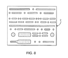
本発明の方法に従う、超音波適用のさらにもう一つの実施形態においては、変換器は、エネルギーを入力し、様々な形と大きさの損傷フィールドを生成するために用いられ得る。変換器が一つの位置に停留されたまま(例えば、漸増運動を使って)であるならば、変換器はまず最初に小さな損傷フィールドをつくり得る。変換器の徘徊を許すことによって、熱エネルギーは積み重なって、損傷フィールドから広がる。変換器が規則的な移動パターンで動かされる間、変換器がゆっくり動かされるか、より高いエネルギー出力を持つことにより、より大きな隣接損傷フィールドを生成(より厚いスキャンラインを生成)し得る。類推によって、万年筆がページにインクを残す方法を想像し得る。丁度、万年筆のペン先がインクをペン先の接点から紙中に広がらせるように、変換器が脂肪組織の特定の地点を徘徊するままにされることが長ければ長いほど、それだけ熱エネルギーは変換器の焦点ゾーンから広がる。これらの損傷のいくつかの変形が、図8に示される。前述のスキャンライン4、損傷フィールド630およびハローフィールド6と同様の、拡大されたハローフィールドがここに示されている。ここで、スキャンライン4は、一般的な球面形状のハローフィールド6を有する点状の損傷フィールド630を生成し得る。組織へのパワー散布の増加は、変換器をゆっくり移動させ、変換器のパラメータを変えることにより達成され、その結果、損傷フィールドから周囲の組織に多くのエネルギーが放射し、こうして拡大されたハローフィールドを生成する。同様に、損傷フィールド自身は、また、大きさも増加し得る。
In yet another embodiment of ultrasonic application, according to the method of the present invention, the transducer can be used to input energy and generate damage fields of various shapes and sizes. If the transducer remains in one position (eg, using incremental motion), the transducer can initially create a small damage field. By allowing the converter to be trapped, the thermal energy builds up and spreads from the damaged field. While the transducer is moved in a regular movement pattern, the transducer can be moved slowly or have a higher energy output to produce a larger adjacent damage field (create a thicker scan line). By analogy, you can imagine how a fountain pen leaves ink on a page. The longer the transducer is left at a specific point in the fat tissue, just as the fountain pen nib spreads ink from the nib contacts into the paper, the more heat energy is converted. Expands from the focal zone of the vessel. Some variations of these damages are shown in FIG. An enlarged halo field is shown here, similar to
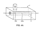
患者の皮膚の上での変換器の移動は、いくらかの数のパターンに従い得る。基本的な移動は、図4Aに示される。変換器は、損傷フィールドをつくる焦点ゾーン630を有する。変換器が管理された方法で移動させられる場合には、HIFU治療変換器によって形成された損傷フィールドは、単一の、隣接する破壊された組織630cを形成し得る。組織における焦点ゾーンの軸は、本明細書ではスキャンライン4と称される。脂肪組織を殺し、コラーゲン繊維を変性させるのに十分な温度まで、局部組織の温度を上昇させる熱効果の領域が、スキャンライン4を囲んでいることが望ましい。スキャンライン4のまわりのこのハローフィールド6は、損傷フィールド630、630cから、破壊または変性されるのに十分な熱放射を受け取る組織の塊を代表する。ハローフィールド6は、変換器がどの程度速く動かされるか、変換器がどの程度のパワーを生成するかに依存して、大きくも小さくもあり得る。ここで、単一のスキャンライン4は、明解であるように単一の処置部位2内に示される。スキャンライン4の断面が、図4Bに示される。
The movement of the transducer over the patient's skin can follow some number of patterns. The basic movement is shown in FIG. 4A. The transducer has a
あるいは、変換器500は、スキャンライン4(図3A)に沿って分離した損傷630を生成するために高強度のパルスまたはパルスバースト(孤立性パルスの高速の連続)を生成し得る。この実施形態において、変換器は患者の皮膚表面上を望ましい方法で動かされ、HIFU超音波エネルギーの孤立性バーストを供給するようにプログラムされることにより、変換器は破壊された組織の、個々のまたは分離した「細胞」を生産する。超音波エネルギーのバーストは、組織においてどのような種類および数の分離した損傷をも生成し得る。ハロー6は、また、変換器の操作パラメータに依存して、各損傷を囲んでいることが見られ得る。また、損傷フィールドおよびハローのパターンは、図3Bに示される断面図においても示される。
Alternatively, the
超音波エネルギーを印加するためのもう一つの実施形態は、図5A〜図5Bに示される。ここに、2本のスキャンライン4、4’がかなり近接して示されているために、隣接した損傷領域630c、630c’は平行である。各スキャンラインのハローゾーン6は、一緒になって走行することにより、協力効果の領域を形成し、ハローゾーンを拡大する。多数のスキャンラインが並んで配置されることにより、機械的および熱的な効果(図5B)の大きな層が形成され得る。
Another embodiment for applying ultrasonic energy is shown in FIGS. 5A-5B. Here, the two damaged
コラーゲンの変性は、37℃より高い温度で起こり得る。しかし、通常の体温近くの温度で変性されたコラーゲンは、回復、緩和およびその通常の長さに戻り得る。望ましくは、その後に、処置ゾーンのコラーゲンは、37℃より高い温度に暴晒される。より望ましくは、処置ゾーンのコラーゲン繊維は、46℃より高い温度、さらに望ましくは56℃より高い温度に暴露される。コラーゲン繊維が暴露される温度が高い程、所望の効果(コラーゲンの収縮のためのコラーゲンの恒久的な変性)を達成するために必要な時間はそれだけ短くなる。晒される温度が46℃であるとき、コラーゲン繊維は少なくとも数分間その温度において暖められる必要があるが、56℃付近またはそれ以上の温度でコラーゲン繊維を暴露するときは、数秒未満で実行され得る。「コラーゲン繊維」は、脂肪組織または下位真皮領域の中に見られるコラーゲン材料と関連している。脂肪組織または下位真皮領域において、コラーゲン濃度は希薄であり、主要な構造成分よりむしろ格子結合組織として身体によって使用される傾向にある(鼻端、耳、皮膚または腱などのような領域とは対照的である)。コラーゲン繊維の収縮は、コラーゲンを変質させ、コラーゲン繊維に縦に短くなることを強制する熱エネルギーの使用と関連している。 Collagen denaturation can occur at temperatures above 37 ° C. However, denatured collagen at temperatures near normal body temperature can recover, relax and return to its normal length. Desirably, the collagen in the treatment zone is then exposed to temperatures above 37 ° C. More desirably, the collagen fibers in the treatment zone are exposed to temperatures above 46 ° C, more desirably above 56 ° C. The higher the temperature at which the collagen fibers are exposed, the shorter the time required to achieve the desired effect (permanent modification of the collagen due to collagen contraction). When the exposed temperature is 46 ° C., the collagen fibers need to be warmed at that temperature for at least several minutes, but when exposing the collagen fibers at temperatures around 56 ° C. or higher, it can be performed in less than a few seconds. “Collagen fibers” are associated with collagen material found in adipose tissue or lower dermis areas. In adipose tissue or lower dermis areas, the collagen concentration is sparse and tends to be used by the body as a lattice connective tissue rather than a major structural component (as opposed to areas such as the nose, ears, skin or tendons) ) Collagen fiber contraction is associated with the use of thermal energy to alter the collagen and force the collagen fiber to become vertically shortened.
脂肪組織は、HIFUエネルギーを使って加熱され、損傷フィールドの温度は実用的な程度に高く、できるだけ速く上昇されることが望ましい。HIFU変換器のパラメータは、脂肪組織を破壊し、コラーゲン繊維を変性させるのに必要な所望の高速加熱を生成するために調整可能であり得る。高速加熱は、処理される脂肪組織の体積および寸法とバランスがとれていることが望ましい。変換器が活性状態で長時間一箇所にいればいるほど、ハローフィールドはそれだけ大きくなる。HIFU変換器を移動させることと治療用超音波エネルギーを印加することは、脂肪組織塊の寸法を越えて広がった損傷またはハローのフィールドを生成しないことが望ましい。 The adipose tissue is heated using HIFU energy and the temperature of the damaged field is preferably as high as practical and raised as fast as possible. The parameters of the HIFU transducer may be adjustable to produce the desired rapid heating necessary to destroy adipose tissue and denature collagen fibers. Desirably, rapid heating is balanced with the volume and size of the adipose tissue being processed. The longer the transducer is in the active state for a long time, the larger the halo field will be. It is desirable that moving the HIFU transducer and applying therapeutic ultrasonic energy does not create a field of damage or halo that extends beyond the dimensions of the adipose tissue mass.
損傷およびハローのフィールドの大きさに影響を及ぼす付加的なパラメータは、変換器を介して電気的に制御されるパラメータおよび変換器自身のパラメータである。これらのパラメータは、パワー、振動数、デューティサイクル、焦点、大きさ(変換器の)およびパルス反復振動数を含む(しかし、これらに制限はされない)。 Additional parameters that affect the size of the damage and halo fields are parameters that are electrically controlled through the transducer and parameters of the transducer itself. These parameters include (but are not limited to) power, frequency, duty cycle, focus, magnitude (of the transducer) and pulse repetition frequency.
一部の用途においては、損傷およびハローのフィールドの大きさは、最小にされることが望ましい。筋肉、臓器または皮膚が近いために、脂肪組織深さが、厳しく管理された損傷およびハローのフィールドを必要とする所では、このことは特に当てはまる。これは、距離と時間の両方において互いに離れている処置部位内に個々の損傷フィールドを分散することによって達成され得る。処置部位が定義されたフィールド領域2によって表現される場合、個々の点損傷は、L1〜L15(図6)の列に一つずつ置かれ得る。ここに、損傷は、時間的および空間的に分離される。このパターンは、個々の損傷が、損傷間の最小の協同熱効果を持つことを可能にする。各損傷(L1−n)の大きさは、また、処置において用いられる超音波変換器のパラメータを調整することによって管理され得る。
In some applications, it is desirable that the size of the damage and halo fields be minimized. This is especially true where adipose tissue depth requires tightly controlled damage and halo fields due to the proximity of muscles, organs or skin. This can be accomplished by distributing individual injury fields within treatment sites that are separated from each other in both distance and time. If the treatment site is represented by a defined
あるいは、損傷およびハローのフィールドは、HIFU変換器が隣接損傷フィールドおよび協力ハローフィールドを生成することを可能にすることによって最大にされ得る。そのような最大にする移動のスキームの例が、ここで図7に示される。この実施形態において、細胞壊死とコラーゲン収縮に必要なエネルギーは、協力効果によってより少なくされる。該協力効果は、接近して間隔をあけられた処置ラインで、時間と空間の両方で互いに近い処置ラインを素早く連続して置くという動作を変換器にさせることによるものである。変換器の移動は、変換器制御の均一性と同時性のために機械制御されることが望ましい。変換器は、螺旋、ラスタースキャン、またはパターン化されたものを含む様々なパターンで組織塊の表面上を移動することによって、患者の組織塊を処置し得る。温熱の協力作用は、処置部位2内の隣接損傷フィールド630として超音波エネルギーを供給することによって最大にされ得る。ラスタースキャン型パターン(図7)は、大きなハローフィールドを生成する最大の温熱協力作用を供給するために、比較的近い距離にあるラインで使用され得る。横方向のスキャンライン4は、変換器が活性である縦方向の通過ライン5と接続され得、また、縦方向に動いている間、変換器が活性でない場合には、縦方向の通過ラインは「空」であり得る。同様に、横方向線4の間の間隔は、超音波エネルギーの最大の重複を提供するために接近し得るか、または物理的に重複し得る。
Alternatively, the damage and halo fields may be maximized by allowing the HIFU converter to generate adjacent damage fields and cooperating halo fields. An example of such a maximizing transfer scheme is now shown in FIG. In this embodiment, less energy is required for cell necrosis and collagen contraction due to the cooperative effect. The cooperative effect is due to having the transducer operate quickly and continuously placing treatment lines close together in both time and space with treatment lines that are closely spaced. The movement of the transducer is preferably mechanically controlled for uniformity and simultaneity of transducer control. The transducer may treat the patient's tissue mass by moving over the surface of the tissue mass in various patterns, including spirals, raster scans, or patterned ones. The cooperation of heat can be maximized by supplying ultrasonic energy as an
本明細書に記載された方法において、超音波エネルギーを印加することについての慎重な計画と考慮は、破壊される脂肪組織の量と変性させられるコラーゲンの量の両方において所望の組織改変塊を生成し得る。 In the methods described herein, careful planning and consideration for applying ultrasonic energy produces the desired tissue-modified mass in both the amount of adipose tissue that is destroyed and the amount of collagen that is denatured. Can do.
変換器の速度(処置される組織の焦点ゾーンにおける速度)とパワーと強度のバランスは、所望の効果を生成するために必要とされる。組織改変に使用する様々なパラメータを決定する方法が、ここで記述される。この実施形態において、高強度集中超音波を使用して、患者の脂肪組織塊を減少させる方法がある。該方法は、処理される脂肪組織の塊を決定するステップと、対応する皮膚の表面にマークするステップと、漸進的に該脂肪組織を破壊し、該コラーゲン繊維を変性させることを誘導するのに十分な方法で高強度集中超音波エネルギーを該領域に印加するステップであって、エネルギー束は少なくとも35J/cm2であるステップとを含む。操作上、破壊の速度は、より高いEF値を与えることによって速められ得る。より高いEF値で脂肪組織の塊上方を変換器をスキャンさせることによって、脂肪組織壊死とコラーゲン繊維変性を達成するのに必要な時間が減少させられ得る。1平方センチメートル当たり90〜225ジュールのEF値を用いることは、所望の処置が速く実行されることを可能にする。さらに、該EFをより高い値、すなわち460J/cm2ほどの値に増やすことも特定の条件下では実行可能な結果を生む。 The balance of transducer speed (speed at the focal zone of the tissue to be treated) and power and intensity is required to produce the desired effect. A method for determining various parameters used for tissue modification will now be described. In this embodiment, there is a method of reducing a patient's adipose tissue mass using high intensity focused ultrasound. The method includes determining a mass of adipose tissue to be treated, marking a corresponding skin surface, and progressively destroying the adipose tissue and inducing degeneration of the collagen fibers. Applying high intensity focused ultrasound energy to the region in a sufficient manner, wherein the energy flux is at least 35 J / cm 2 . In operation, the speed of destruction can be increased by giving higher EF values. By scanning the transducer over the adipose tissue mass with higher EF values, the time required to achieve adipose tissue necrosis and collagen fiber degeneration can be reduced. Using an EF value of 90-225 Joules per square centimeter allows the desired procedure to be performed quickly. Furthermore, increasing the EF to a higher value, i.e., as high as 460 J / cm < 2 >, also produces viable results under certain conditions.
所定のエネルギー束値を用いることによって、変換器は損傷フィールド(焦点ゾーンとも称される)の各々に一貫して、正確に同じエネルギー量を印加するようにプログラムされ得る。実験と分析によって、脂肪組織とコラーゲン収縮の組織除去が、1平方センチメートル当たり35ジュールより大きいエネルギー束で起こり得ることが判明した。所望の結果の変動と患者毎の組織変動は、正確なエネルギー束量の呼び出しを不可能にする。しかし、多くの研究情報源からの経験的なデータは、エネルギー束値が1平方センチメートル当たり35ジュールを超えるべきであることを示唆し、おそらく、脂肪組織破壊とコラーゲン繊維変性の二重の目的のためにはエネルギー束値が1平方センチメートル当たり109ジュール以上が最も有効であると考えられる。 By using a predetermined energy flux value, the transducer can be programmed to apply exactly the same amount of energy consistently to each of the damage fields (also referred to as focal zones). Experiments and analysis have shown that tissue removal of adipose tissue and collagen contraction can occur with an energy flux greater than 35 joules per square centimeter. Desired outcome variability and patient-to-patient tissue variability make accurate energy flux recall impossible. However, empirical data from many research sources suggests that energy flux values should exceed 35 joules per square centimeter, probably for the dual purpose of adipose tissue destruction and collagen fiber degeneration. It is considered that an energy flux value of 109 joules per square centimeter or more is most effective.
本発明の物理的な実施形態において、患者に治療用超音波エネルギーを供給する装置が存在する。治療中に移動させられ、35J/cm2より大きいエネルギー束(EF)を印加できるように適合された少なくとも一つの超音波変換器を有する装置であって、該EFは次式で決定される。
[(p)x(l/v)x(dc)x(nl)]/(sa)
ここで、
p=電力、
l=ライン長、
v=速度、
dc=デューティサイクル、
nl=ライン数
および
sa=スキャン面積
である。
In a physical embodiment of the present invention, there is an apparatus for supplying therapeutic ultrasound energy to a patient. A device having at least one ultrasonic transducer that is moved during therapy and adapted to apply an energy flux (EF) greater than 35 J / cm 2 , the EF being determined by:
[(P) x (l / v) x (dc) x (nl)] / (sa)
here,
p = power,
l = line length,
v = speed,
dc = duty cycle,
nl = number of lines and sa = scanning area.
超音波エネルギーを印加しながら、変換器が連続的に動いているとき、提供された式は計算を提供する。あるいは、変換器が治療適用中に移動していない処置プログラムに対して、EFは以下の改変されたEF式を用いて計算され得る。
EF=[(p)x(t)x(dc)x(ns)]/(sa)
ここで、
p=電力、
t=損傷当たりのオン時間、
dc=デューティサイクル、
ns=ライン数
および
sa=スキャン面積
である。
The provided formula provides a calculation when the transducer is continuously moving while applying ultrasonic energy. Alternatively, for treatment programs where the transducer is not moving during the therapeutic application, the EF can be calculated using the following modified EF equation:
EF = [(p) x (t) x (dc) x (ns)] / (sa)
here,
p = power,
t = on time per damage,
dc = duty cycle,
ns = number of lines and sa = scanning area.
式の変形は、移動と非移動の処置部位の混合したセットを有する治療プログラムに対する適正な計算を決定するために、当業者によって誘導され得る。超音波のそれぞれの印加の前に、ユーザーが治療コントローラーに手で入力し得るパラメータの広い変動を、治療コントローラーが考慮することが望ましい。治療コントローラーは、どの変数が使われることになるかについて決定し、それに応じて変数に重み付けする。本明細書に記載された方法を用いる医療装置システムの例が、「移動制御を有する超音波治療ヘッド」と表題をつけられた出願中の米国の特許出願11/027,912にさらに記載されており、本明細書に参照によって引用される。 Variations of the equation can be derived by those skilled in the art to determine the appropriate calculations for a therapy program having a mixed set of moving and non-moving treatment sites. Prior to each application of ultrasound, it is desirable for the therapy controller to consider the wide variation in parameters that the user can manually input into the therapy controller. The therapy controller determines which variable will be used and weights the variable accordingly. An example of a medical device system using the method described herein is further described in pending US patent application 11 / 027,912, entitled “Ultrasound Treatment Head with Movement Control”. And are hereby incorporated by reference.
もう一つの例は、2004年12月29日に出願された「脂肪組織の破壊のためのシステムおよび方法」と表題をつけられた出願中の米国の特許出願11/026,519に記載されており、本明細書に参照によって引用される。患者に治療用超音波エネルギーを供給するための装置は、スキャンヘッドと、スキャンヘッドを支持するためのサスペンション装置と、治療コントローラーとを有する。治療コントローラーは、スキャンヘッドの位置およびエネルギー供給をモニターするように構成されている。治療コントローラーは、スキャンヘッドの位置およびエネルギー供給をモニターするように構成されている。 Another example is described in pending US patent application 11 / 026,519 entitled “System and Method for Adipose Tissue Disruption” filed Dec. 29, 2004. And are hereby incorporated by reference. An apparatus for supplying therapeutic ultrasound energy to a patient includes a scan head, a suspension device for supporting the scan head, and a therapy controller. The therapy controller is configured to monitor the position of the scan head and the energy supply. The therapy controller is configured to monitor the position of the scan head and the energy supply.
エネルギー束の式の様々なパラメータは、治療コントローラーにプログラムされ得る。装置は、固定メモリーにプログラムされ、ユーザーによって調節不可能ないくつかのパラメータデータを有し得る。一部のエレメントは、装置が安全でない方法で操作されることを防ぐために、変換器の最高と最小の設定値を含み得る。 Various parameters of the energy flux equation can be programmed into the therapy controller. The device may have some parameter data programmed into a fixed memory and not adjustable by the user. Some elements may include maximum and minimum settings for the transducer to prevent the device from being operated in an unsafe manner.
ユーザーは、処置の間に用いられる適当なEFをシステムが決定することを助けるためにシステムに変数を提供し得る。たとえば、ユーザーがスキャンラインの間で協力加熱を増加させたいならば、スキャンライン(nl)はより高い値に設定され得る。あるいは、より大きなハローフィールドを活性化するために速度が減少させられ得るか、または、より小さい余裕しか有しない脂肪組織の領域に必要とされるようにハローフィールドを減少させるために速度は増加させられ得る。 The user can provide variables to the system to help the system determine the appropriate EF to be used during the procedure. For example, if the user wants to increase cooperative heating between scan lines, the scan line (nl) can be set to a higher value. Alternatively, the speed can be reduced to activate a larger halo field, or the speed can be increased to reduce the halo field as needed for areas of adipose tissue that have less margin. Can be.
ステンシルまたはテンプレート24が、医師が処置を計画することを支援するために使用され得る(図9)。治療処置の間、超音波変換器を導くために用いられ得る「十字線」の形状をした開口26をテンプレート24は有する。使用される変換器(または選択された超音波システムに従う治療装置)のフットプリントに開口がマッチするように、テンプレート24が作られることが望ましい。目標領域において、等高線を作成する前または脂肪組織を評価する前にも、皮膚全体にテンプレートが使われ得る。患者の目標治療領域における適切な脂肪組織深さを決定した後に、医者が等高線および十字線印を作成することが望ましい。
A stencil or
ステンシル24は患者の全身に置かれ得(図10)、その後に医療用マーカーを用いて十字線が描かれ得る。図1に示される十字線と等高線は、脂肪組織の既知の深さの範囲内(等高線を使って)で、指示された方法(ガイド印を用いて)でHIFU変換器を安全に配置のための目に見える印を提供するために組み合わされる。いったん二つのマーキングが患者の上に付けられると、治療部位2のモザイクを作成するには、医師は十字線と等高線(図2)を用いてただ超音波治療装置を整列させるだけでよい(図11)。
The
治療される組織の塊は、UALのような処置の通常の実施において医師によってすでに採用されている技術を使用してなされ得る。医師は、手によるつまみ検査、カリパスまたは診断用超音波を用いることにより、治療される脂肪組織の深さを決定し、治療される領域の周りに、地形図上のレリーフ線と類似した円を描き得る。塊が決定される前または後に、ステンシルによる個々のマークが作成され得る。組織塊の様々なレベルを表示する等高線と治療ヘッドのランドマークとが重複することにより、処置のために定義された安全領域および超音波治療ヘッドを用いる処置のためのガイドをユーザーに提供する。 The mass of tissue to be treated can be made using techniques already employed by physicians in the normal practice of procedures such as UAL. The physician determines the depth of the adipose tissue to be treated by using a hand pick test, caliper or diagnostic ultrasound, and a circle similar to the relief line on the topographic map around the area to be treated. You can draw. Individual marks with a stencil can be created before or after the mass is determined. The overlapping contours displaying the various levels of tissue mass and the landmarks of the treatment head provide the user with a safe area defined for the procedure and a guide for the procedure using the ultrasound therapy head.
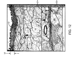
本明細書に記載された方法の適切な利用は、脂肪組織の領域の塊を減少させることができる。本明細書に記載された方法を使用した組織の組織構造スライドが、図12および図13に示される。これらの組織構造画像は、皮膚ライン12と皮膜層14がダメージを受けていないことを示す。この種の処置に対する比較的安全な深さを有する脂肪組織16の領域も示される。処置ゾーンは、マーカーZ1とZ2の間に見られる。通常の脂肪細胞(脂肪細胞)18と通常のコラーゲン繊維20が皮膜層14と治療ゾーンZ1の間に示される。治療ラインZ1、Z2の内側に、コラーゲンの多い領域と脂肪細胞構造をほぼ完全に欠如している二つの領域が示されている。損傷フィールド22は、脂肪組織の崩壊と破壊およびコラーゲン繊維の変性の両方を示し、これらは、破壊された組織質量が徐々に身体から除去されるにつれて(身体の自然創傷治癒反応によって)組織塊と接触するようになる。この方法における脂肪組織塊の減少は、脂肪吸引に対する同様の長期間の結果を提供する。組織損失が段階的であるので、皮膜層の突然の緩みがなく、また、本明細書に記載された方法を使用する処置を患者が受けた直後に観察される皮膚変形もない。
Proper use of the methods described herein can reduce the mass of regions of adipose tissue. Tissue histology slides using the methods described herein are shown in FIGS. These tissue structure images indicate that the
本明細書に開示された材料を再検討することは、記述された所望の目的を達成する多くの代替方法および特に本明細書では記載されていない方法を当業者に提供するであろう。提供された説明は例証であり、限定的でないと考えられなければならず、従って、本実施形態ならびに代替の実施形態および等価物は付随する請求項によって取り込まれることが意図されている。 Review of the materials disclosed herein will provide those skilled in the art with many alternative ways of achieving the desired purpose described, and in particular not described herein. The description provided is to be regarded as illustrative and not restrictive, and thus this embodiment and alternative embodiments and equivalents are intended to be encompassed by the appended claims.
Claims (1)
Applications Claiming Priority (2)
| Application Number | Priority Date | Filing Date | Title |
|---|---|---|---|
| US67619705P | 2005-04-29 | 2005-04-29 | |
| US60/676,197 | 2005-04-29 |
Related Parent Applications (1)
| Application Number | Title | Priority Date | Filing Date |
|---|---|---|---|
| JP2008509137A Division JP5055265B2 (en) | 2005-04-29 | 2006-04-28 | Apparatus and method for destruction of adipose tissue |
Publications (3)
| Publication Number | Publication Date |
|---|---|
| JP2012179385A true JP2012179385A (en) | 2012-09-20 |
| JP2012179385A5 JP2012179385A5 (en) | 2012-11-01 |
| JP5336632B2 JP5336632B2 (en) | 2013-11-06 |
Family
ID=37308501
Family Applications (2)
| Application Number | Title | Priority Date | Filing Date |
|---|---|---|---|
| JP2008509137A Active JP5055265B2 (en) | 2005-04-29 | 2006-04-28 | Apparatus and method for destruction of adipose tissue |
| JP2012114475A Active JP5336632B2 (en) | 2005-04-29 | 2012-05-18 | Apparatus and method for destruction of adipose tissue |
Family Applications Before (1)
| Application Number | Title | Priority Date | Filing Date |
|---|---|---|---|
| JP2008509137A Active JP5055265B2 (en) | 2005-04-29 | 2006-04-28 | Apparatus and method for destruction of adipose tissue |
Country Status (11)
| Country | Link |
|---|---|
| US (2) | US7857773B2 (en) |
| EP (1) | EP1874241A4 (en) |
| JP (2) | JP5055265B2 (en) |
| KR (1) | KR101188931B1 (en) |
| CN (1) | CN101460119B (en) |
| AU (1) | AU2006242547B8 (en) |
| BR (1) | BRPI0610033A2 (en) |
| CA (1) | CA2606045A1 (en) |
| IL (1) | IL186824A (en) |
| MX (1) | MX2007013440A (en) |
| WO (1) | WO2006118960A2 (en) |
Cited By (1)
| Publication number | Priority date | Publication date | Assignee | Title |
|---|---|---|---|---|
| WO2023244014A1 (en) * | 2022-06-14 | 2023-12-21 | 주식회사 제이시스메디칼 | Ultrasound generation device |
Families Citing this family (131)
| Publication number | Priority date | Publication date | Assignee | Title |
|---|---|---|---|---|
| US6104959A (en) | 1997-07-31 | 2000-08-15 | Microwave Medical Corp. | Method and apparatus for treating subcutaneous histological features |
| US6050943A (en) | 1997-10-14 | 2000-04-18 | Guided Therapy Systems, Inc. | Imaging, therapy, and temperature monitoring ultrasonic system |
| US7914453B2 (en) | 2000-12-28 | 2011-03-29 | Ardent Sound, Inc. | Visual imaging system for ultrasonic probe |
| US8343051B2 (en) * | 2003-12-30 | 2013-01-01 | Liposonix, Inc. | Apparatus and methods for the destruction of adipose tissue |
| US7857773B2 (en) * | 2003-12-30 | 2010-12-28 | Medicis Technologies Corporation | Apparatus and methods for the destruction of adipose tissue |
| EP1699360A4 (en) * | 2003-12-30 | 2009-05-06 | Liposonix Inc | Component ultrasound transducer |
| US8235909B2 (en) | 2004-05-12 | 2012-08-07 | Guided Therapy Systems, L.L.C. | Method and system for controlled scanning, imaging and/or therapy |
| US7824348B2 (en) | 2004-09-16 | 2010-11-02 | Guided Therapy Systems, L.L.C. | System and method for variable depth ultrasound treatment |
| US7393325B2 (en) | 2004-09-16 | 2008-07-01 | Guided Therapy Systems, L.L.C. | Method and system for ultrasound treatment with a multi-directional transducer |
| US9011336B2 (en) | 2004-09-16 | 2015-04-21 | Guided Therapy Systems, Llc | Method and system for combined energy therapy profile |
| US8444562B2 (en) | 2004-10-06 | 2013-05-21 | Guided Therapy Systems, Llc | System and method for treating muscle, tendon, ligament and cartilage tissue |
| US10864385B2 (en) | 2004-09-24 | 2020-12-15 | Guided Therapy Systems, Llc | Rejuvenating skin by heating tissue for cosmetic treatment of the face and body |
| US8535228B2 (en) | 2004-10-06 | 2013-09-17 | Guided Therapy Systems, Llc | Method and system for noninvasive face lifts and deep tissue tightening |
| US8690779B2 (en) | 2004-10-06 | 2014-04-08 | Guided Therapy Systems, Llc | Noninvasive aesthetic treatment for tightening tissue |
| US7758524B2 (en) | 2004-10-06 | 2010-07-20 | Guided Therapy Systems, L.L.C. | Method and system for ultra-high frequency ultrasound treatment |
| US11883688B2 (en) | 2004-10-06 | 2024-01-30 | Guided Therapy Systems, Llc | Energy based fat reduction |
| WO2006042163A2 (en) * | 2004-10-06 | 2006-04-20 | Guided Therapy Systems, L.L.C. | Method and system for cosmetic enhancement |
| KR101732144B1 (en) * | 2004-10-06 | 2017-05-02 | 가이디드 테라피 시스템스, 엘.엘.씨. | Ultrasound treatment system |
| US9827449B2 (en) | 2004-10-06 | 2017-11-28 | Guided Therapy Systems, L.L.C. | Systems for treating skin laxity |
| US11235179B2 (en) | 2004-10-06 | 2022-02-01 | Guided Therapy Systems, Llc | Energy based skin gland treatment |
| US20060111744A1 (en) | 2004-10-13 | 2006-05-25 | Guided Therapy Systems, L.L.C. | Method and system for treatment of sweat glands |
| US8133180B2 (en) | 2004-10-06 | 2012-03-13 | Guided Therapy Systems, L.L.C. | Method and system for treating cellulite |
| US9694212B2 (en) | 2004-10-06 | 2017-07-04 | Guided Therapy Systems, Llc | Method and system for ultrasound treatment of skin |
| US11724133B2 (en) | 2004-10-07 | 2023-08-15 | Guided Therapy Systems, Llc | Ultrasound probe for treatment of skin |
| US11207548B2 (en) | 2004-10-07 | 2021-12-28 | Guided Therapy Systems, L.L.C. | Ultrasound probe for treating skin laxity |
| US8218477B2 (en) * | 2005-03-31 | 2012-07-10 | Alcatel Lucent | Method of detecting wireless network faults |
| JP4695188B2 (en) | 2005-04-25 | 2011-06-08 | アーデント サウンド, インコーポレイテッド | Method and apparatus for improving the safety of computer peripherals |
| US9486274B2 (en) | 2005-09-07 | 2016-11-08 | Ulthera, Inc. | Dissection handpiece and method for reducing the appearance of cellulite |
| US9011473B2 (en) | 2005-09-07 | 2015-04-21 | Ulthera, Inc. | Dissection handpiece and method for reducing the appearance of cellulite |
| US8518069B2 (en) | 2005-09-07 | 2013-08-27 | Cabochon Aesthetics, Inc. | Dissection handpiece and method for reducing the appearance of cellulite |
| US9358033B2 (en) | 2005-09-07 | 2016-06-07 | Ulthera, Inc. | Fluid-jet dissection system and method for reducing the appearance of cellulite |
| US7967763B2 (en) * | 2005-09-07 | 2011-06-28 | Cabochon Aesthetics, Inc. | Method for treating subcutaneous tissues |
| US10548659B2 (en) | 2006-01-17 | 2020-02-04 | Ulthera, Inc. | High pressure pre-burst for improved fluid delivery |
| US7885793B2 (en) | 2007-05-22 | 2011-02-08 | International Business Machines Corporation | Method and system for developing a conceptual model to facilitate generating a business-aligned information technology solution |
| US9248317B2 (en) | 2005-12-02 | 2016-02-02 | Ulthera, Inc. | Devices and methods for selectively lysing cells |
| US9566454B2 (en) | 2006-09-18 | 2017-02-14 | Guided Therapy Systems, Llc | Method and sysem for non-ablative acne treatment and prevention |
| US9132031B2 (en) | 2006-09-26 | 2015-09-15 | Zeltiq Aesthetics, Inc. | Cooling device having a plurality of controllable cooling elements to provide a predetermined cooling profile |
| US20080077201A1 (en) * | 2006-09-26 | 2008-03-27 | Juniper Medical, Inc. | Cooling devices with flexible sensors |
| US8192474B2 (en) | 2006-09-26 | 2012-06-05 | Zeltiq Aesthetics, Inc. | Tissue treatment methods |
| EP2532320A3 (en) | 2007-04-19 | 2013-04-03 | Miramar Labs, Inc. | Apparatus for reducing sweat production |
| ES2488565T3 (en) | 2007-04-19 | 2014-08-27 | Miramar Labs, Inc. | Devices and systems for the non-invasive distribution of microwave therapy |
| WO2008131306A1 (en) | 2007-04-19 | 2008-10-30 | The Foundry, Inc. | Systems and methods for creating an effect using microwave energy to specified tissue |
| US9241763B2 (en) | 2007-04-19 | 2016-01-26 | Miramar Labs, Inc. | Systems, apparatus, methods and procedures for the noninvasive treatment of tissue using microwave energy |
| JP5543332B2 (en) | 2007-04-19 | 2014-07-09 | ミラマー ラブズ, インコーポレイテッド | Systems and methods for producing effects on specific tissues using microwave energy |
| WO2008137942A1 (en) * | 2007-05-07 | 2008-11-13 | Guided Therapy Systems, Llc. | Methods and systems for modulating medicants using acoustic energy |
| US20150174388A1 (en) | 2007-05-07 | 2015-06-25 | Guided Therapy Systems, Llc | Methods and Systems for Ultrasound Assisted Delivery of a Medicant to Tissue |
| WO2008137944A1 (en) | 2007-05-07 | 2008-11-13 | Guided Therapy Systems, Llc. | Methods and systems for coupling and focusing acoustic energy using a coupler member |
| US20080287839A1 (en) | 2007-05-18 | 2008-11-20 | Juniper Medical, Inc. | Method of enhanced removal of heat from subcutaneous lipid-rich cells and treatment apparatus having an actuator |
| US8523927B2 (en) | 2007-07-13 | 2013-09-03 | Zeltiq Aesthetics, Inc. | System for treating lipid-rich regions |
| EP2182898B1 (en) | 2007-08-21 | 2018-10-03 | Zeltiq Aesthetics, Inc. | Monitoring the cooling of subcutaneous lipid-rich cells, such as the cooling of adipose tissue |
| US8409182B2 (en) | 2007-09-28 | 2013-04-02 | Eos Holdings, Llc | Laser-assisted thermal separation of tissue |
| US20090093723A1 (en) * | 2007-10-05 | 2009-04-09 | Cabochon Aesthetics, Inc. | Ultrasound device including dispenser |
| EP2209424A1 (en) * | 2007-10-09 | 2010-07-28 | Cabochon Aesthetics, Inc. | Ultrasound apparatus with treatment lens |
| US20090093738A1 (en) * | 2007-10-09 | 2009-04-09 | Cabochon Aesthetics, Inc. | Device and method for monitoring a treatment area |
| US8439940B2 (en) | 2010-12-22 | 2013-05-14 | Cabochon Aesthetics, Inc. | Dissection handpiece with aspiration means for reducing the appearance of cellulite |
| US20090099484A1 (en) * | 2007-10-11 | 2009-04-16 | Yehuda Zadok | Coating of polyurethane membrane |
| EP2231274B1 (en) | 2007-12-12 | 2014-03-12 | Miramar Labs, Inc. | System and apparatus for the noninvasive treatment of tissue using microwave energy |
| JP5545668B2 (en) | 2007-12-12 | 2014-07-09 | ミラマー ラブズ, インコーポレイテッド | System, apparatus method, and procedure for non-invasive tissue treatment using microwave energy |
| US20090171255A1 (en) * | 2007-12-27 | 2009-07-02 | Andrey Rybyanets | Apparatus and method for ultrasound treatment |
| US20090248578A1 (en) * | 2008-03-20 | 2009-10-01 | Liposonix, Inc. | Methods and apparatus for medical device investment recovery |
| RU2547180C2 (en) | 2008-06-06 | 2015-04-10 | Ультера, Инк. | System and method for cosmetic treatment and imaging |
| WO2010036732A1 (en) | 2008-09-25 | 2010-04-01 | Zeltiq Aesthetics, Inc. | Treatment planning systems and methods for body contouring applications |
| US10980925B1 (en) | 2008-10-14 | 2021-04-20 | A Hoyos Llc | High definition liposculpture |
| US8876799B1 (en) | 2008-10-14 | 2014-11-04 | Alfredo Ernesto Hoyos Ariza | Vaser assisted high definition liposculpture |
| US9888933B1 (en) | 2008-10-14 | 2018-02-13 | Alfredo Ernesto Hoyos Ariza | Vaser assisted high definition liposculpture |
| US8167868B1 (en) * | 2008-10-14 | 2012-05-01 | Alfredo Ernesto Hoyos Ariza | VASER assisted high definition liposculpture |
| US20100106063A1 (en) * | 2008-10-29 | 2010-04-29 | Cabochon Aesthetics, Inc. | Ultrasound Enhancing Target for Treating Subcutaneous Tissue |
| US8603073B2 (en) | 2008-12-17 | 2013-12-10 | Zeltiq Aesthetics, Inc. | Systems and methods with interrupt/resume capabilities for treating subcutaneous lipid-rich cells |
| EP2382010A4 (en) | 2008-12-24 | 2014-05-14 | Guided Therapy Systems Llc | Methods and systems for fat reduction and/or cellulite treatment |
| TW201034628A (en) * | 2009-03-04 | 2010-10-01 | Medicis Technologies Corp | Ultrasonic treatment of adipose tissue at multiple depths |
| US8167280B2 (en) * | 2009-03-23 | 2012-05-01 | Cabochon Aesthetics, Inc. | Bubble generator having disposable bubble cartridges |
| US20100256596A1 (en) * | 2009-04-07 | 2010-10-07 | Cabochon Aesthetics, Inc. | Fiber growth promoting implants for reducing the appearance of cellulite |
| KR101759116B1 (en) | 2009-04-30 | 2017-07-18 | 젤티크 애스세틱스, 인코포레이티드. | Device, system and method of removing heat from subcutaneous lipid-rich cells |
| US8298163B1 (en) | 2009-05-01 | 2012-10-30 | Body Beam Research Inc. | Non-invasive ultrasonic soft-tissue treatment apparatus |
| US20100286519A1 (en) * | 2009-05-11 | 2010-11-11 | General Electric Company | Ultrasound system and method to automatically identify and treat adipose tissue |
| US20100286518A1 (en) * | 2009-05-11 | 2010-11-11 | General Electric Company | Ultrasound system and method to deliver therapy based on user defined treatment spaces |
| US20100286520A1 (en) * | 2009-05-11 | 2010-11-11 | General Electric Company | Ultrasound system and method to determine mechanical properties of a target region |
| WO2011006017A1 (en) | 2009-07-08 | 2011-01-13 | Sanuwave, Inc. | Usage of extracorporeal and intracorporeal pressure shock waves in medicine |
| US11096708B2 (en) | 2009-08-07 | 2021-08-24 | Ulthera, Inc. | Devices and methods for performing subcutaneous surgery |
| US9358064B2 (en) | 2009-08-07 | 2016-06-07 | Ulthera, Inc. | Handpiece and methods for performing subcutaneous surgery |
| US8932238B2 (en) * | 2009-09-29 | 2015-01-13 | Liposonix, Inc. | Medical ultrasound device with liquid dispensing device coupled to a therapy head |
| US8715186B2 (en) | 2009-11-24 | 2014-05-06 | Guided Therapy Systems, Llc | Methods and systems for generating thermal bubbles for improved ultrasound imaging and therapy |
| MX2012008660A (en) * | 2010-01-25 | 2013-02-26 | Zeltiq Aesthetics Inc | Home-use applicators for non-invasively removing heat from subcutaneous lipid-rich cells via phase change coolants, and associated devices, systems and methods. |
| EP2560568B1 (en) * | 2010-04-19 | 2016-09-21 | Syneron Medical Ltd. | Combined energy and topical composition application for regulating the condition of mammalian skin |
| US8676338B2 (en) | 2010-07-20 | 2014-03-18 | Zeltiq Aesthetics, Inc. | Combined modality treatment systems, methods and apparatus for body contouring applications |
| EP2595704A1 (en) | 2010-07-24 | 2013-05-29 | LipoSonix, Inc. | Apparatus and methods for non-invasive body contouring |
| EP2600783A4 (en) | 2010-08-02 | 2017-05-17 | Guided Therapy Systems, L.L.C. | Systems and methods for ultrasound treatment |
| US9504446B2 (en) | 2010-08-02 | 2016-11-29 | Guided Therapy Systems, Llc | Systems and methods for coupling an ultrasound source to tissue |
| US8857438B2 (en) | 2010-11-08 | 2014-10-14 | Ulthera, Inc. | Devices and methods for acoustic shielding |
| US8425490B2 (en) | 2011-06-28 | 2013-04-23 | Alfredo Ernesto Hoyos Ariza | Dynamic liposculpting method |
| US9452302B2 (en) | 2011-07-10 | 2016-09-27 | Guided Therapy Systems, Llc | Systems and methods for accelerating healing of implanted material and/or native tissue |
| KR20190080967A (en) | 2011-07-11 | 2019-07-08 | 가이디드 테라피 시스템스, 엘.엘.씨. | Systems and methods for coupling an ultrasound source to tissue |
| US9314301B2 (en) | 2011-08-01 | 2016-04-19 | Miramar Labs, Inc. | Applicator and tissue interface module for dermatological device |
| US9263663B2 (en) | 2012-04-13 | 2016-02-16 | Ardent Sound, Inc. | Method of making thick film transducer arrays |
| US9510802B2 (en) | 2012-09-21 | 2016-12-06 | Guided Therapy Systems, Llc | Reflective ultrasound technology for dermatological treatments |
| US9289188B2 (en) | 2012-12-03 | 2016-03-22 | Liposonix, Inc. | Ultrasonic transducer |
| CN204017181U (en) | 2013-03-08 | 2014-12-17 | 奥赛拉公司 | Aesthstic imaging and processing system, multifocal processing system and perform the system of aesthetic procedure |
| US9844460B2 (en) | 2013-03-14 | 2017-12-19 | Zeltiq Aesthetics, Inc. | Treatment systems with fluid mixing systems and fluid-cooled applicators and methods of using the same |
| US9545523B2 (en) | 2013-03-14 | 2017-01-17 | Zeltiq Aesthetics, Inc. | Multi-modality treatment systems, methods and apparatus for altering subcutaneous lipid-rich tissue |
| US10561862B2 (en) | 2013-03-15 | 2020-02-18 | Guided Therapy Systems, Llc | Ultrasound treatment device and methods of use |
| WO2015013502A2 (en) | 2013-07-24 | 2015-01-29 | Miramar Labs, Inc. | Apparatus and methods for the treatment of tissue using microwave energy |
| CN103537018A (en) * | 2013-11-01 | 2014-01-29 | 彭伟 | Ultrasonic beauty instrument |
| EP3099258B1 (en) | 2014-01-31 | 2024-02-21 | Zeltiq Aesthetics, Inc. | Compositions and treatment systems for improved cooling of lipid-rich tissue |
| US10231872B2 (en) | 2014-02-28 | 2019-03-19 | Excel-Lens, Inc. | Laser assisted cataract surgery |
| US10206817B2 (en) | 2014-02-28 | 2019-02-19 | Excel-Lens, Inc. | Laser assisted cataract surgery |
| US10327951B2 (en) | 2014-02-28 | 2019-06-25 | Excel-Lens, Inc. | Laser assisted cataract surgery |
| US9820886B2 (en) | 2014-02-28 | 2017-11-21 | Excel-Lens, Inc. | Laser assisted cataract surgery |
| US10675176B1 (en) | 2014-03-19 | 2020-06-09 | Zeltiq Aesthetics, Inc. | Treatment systems, devices, and methods for cooling targeted tissue |
| USD777338S1 (en) | 2014-03-20 | 2017-01-24 | Zeltiq Aesthetics, Inc. | Cryotherapy applicator for cooling tissue |
| BR112016023889B1 (en) | 2014-04-18 | 2023-02-07 | Ulthera, Inc | ULTRASOUND TRANSDUCTION SYSTEM FOR LINEAR FOCUSING ULTRASOUND |
| US10952891B1 (en) | 2014-05-13 | 2021-03-23 | Zeltiq Aesthetics, Inc. | Treatment systems with adjustable gap applicators and methods for cooling tissue |
| US10568759B2 (en) | 2014-08-19 | 2020-02-25 | Zeltiq Aesthetics, Inc. | Treatment systems, small volume applicators, and methods for treating submental tissue |
| US10935174B2 (en) | 2014-08-19 | 2021-03-02 | Zeltiq Aesthetics, Inc. | Stress relief couplings for cryotherapy apparatuses |
| EP3364900B1 (en) | 2015-10-19 | 2021-08-18 | Zeltiq Aesthetics, Inc. | Vascular treatment methods for cooling vascular structures |
| JP2018538056A (en) * | 2015-12-09 | 2018-12-27 | コーニンクレッカ フィリップス エヌ ヴェKoninklijke Philips N.V. | Interleaved beam pattern for ultrasonic thrombolysis and other treatments via vascular acoustic resonator |
| KR20220098285A (en) | 2016-01-07 | 2022-07-11 | 젤티크 애스세틱스, 인코포레이티드. | Temperature-dependent adhesion between applicator and skin during cooling of tissue |
| BR112018011927A2 (en) | 2016-01-18 | 2018-11-27 | Ulthera, Inc. | compact ultrasound device that has an annular ultrasound array connected peripherally to the flexible printed circuit board and mounting method |
| US10765552B2 (en) | 2016-02-18 | 2020-09-08 | Zeltiq Aesthetics, Inc. | Cooling cup applicators with contoured heads and liner assemblies |
| US10682297B2 (en) | 2016-05-10 | 2020-06-16 | Zeltiq Aesthetics, Inc. | Liposomes, emulsions, and methods for cryotherapy |
| US10555831B2 (en) | 2016-05-10 | 2020-02-11 | Zeltiq Aesthetics, Inc. | Hydrogel substances and methods of cryotherapy |
| US11382790B2 (en) | 2016-05-10 | 2022-07-12 | Zeltiq Aesthetics, Inc. | Skin freezing systems for treating acne and skin conditions |
| WO2018035012A1 (en) | 2016-08-16 | 2018-02-22 | Ulthera, Inc. | Systems and methods for cosmetic ultrasound treatment of skin |
| US11076879B2 (en) | 2017-04-26 | 2021-08-03 | Zeltiq Aesthetics, Inc. | Shallow surface cryotherapy applicators and related technology |
| DE202018106840U1 (en) | 2017-11-30 | 2019-03-08 | Medical Technologies Cz A.S. | Shock wave device |
| US11944849B2 (en) | 2018-02-20 | 2024-04-02 | Ulthera, Inc. | Systems and methods for combined cosmetic treatment of cellulite with ultrasound |
| JP2021532873A (en) | 2018-07-31 | 2021-12-02 | ゼルティック エステティックス インコーポレイテッド | Methods, devices, and systems to improve skin properties |
| USD883767S1 (en) | 2018-10-10 | 2020-05-12 | A Hoyos Llc | Handle |
| KR102393045B1 (en) | 2020-02-26 | 2022-05-02 | 주식회사 엘림텍 | Skin Treatment Handpiece |
| KR102612156B1 (en) | 2021-08-12 | 2023-12-11 | 주식회사 엘림텍 | HIFU Handpiece |
| KR102626557B1 (en) | 2021-09-09 | 2024-01-19 | 주식회사 엘림텍 | Electric depth adjustable HIFU handpiece |
| KR102626556B1 (en) | 2021-09-09 | 2024-01-19 | 주식회사 엘림텍 | HIFU handpiece with ultrasound treatment depth adjustment |
Citations (5)
| Publication number | Priority date | Publication date | Assignee | Title |
|---|---|---|---|---|
| JPH0284950A (en) * | 1989-07-17 | 1990-03-26 | Toshiba Corp | Ultrasonic calculus crushing device |
| JPH05300910A (en) * | 1992-02-28 | 1993-11-16 | Toshiba Corp | Ultrasonic medical treatment system |
| US6113559A (en) * | 1997-12-29 | 2000-09-05 | Klopotek; Peter J. | Method and apparatus for therapeutic treatment of skin with ultrasound |
| WO2002092168A1 (en) * | 2001-05-10 | 2002-11-21 | The Procter & Gamble Company | Method and kit for the treatment or prevention of cosmetic skin conditions |
| US20040082857A1 (en) * | 2002-10-25 | 2004-04-29 | Compex Medical S.A. | Ultrasound therapeutic device |
Family Cites Families (140)
| Publication number | Priority date | Publication date | Assignee | Title |
|---|---|---|---|---|
| US2791204A (en) | 1951-08-16 | 1957-05-07 | Smith Corp A O | Water heater utilizing heat of crystallization |
| US2651904A (en) | 1951-11-03 | 1953-09-15 | Franklin W Jatunn | Adjustable cutter bar for lawn mowers |
| GB820814A (en) | 1955-12-22 | 1959-09-30 | Univ Illinois | Apparatus for treating living tissue |
| US4002221A (en) * | 1972-09-19 | 1977-01-11 | Gilbert Buchalter | Method of transmitting ultrasonic impulses to surface using transducer coupling agent |
| US3880393A (en) * | 1973-06-04 | 1975-04-29 | Conco Inc | Load balancer with balance override control |
| US4059098A (en) | 1975-07-21 | 1977-11-22 | Stanford Research Institute | Flexible ultrasound coupling system |
| US4137777A (en) * | 1977-07-11 | 1979-02-06 | Mediscan Inc. | Ultrasonic body scanner and method |
| US4196630A (en) * | 1978-05-18 | 1980-04-08 | Rudolph Dale C | Overhead arm assembly |
| DE2826277C2 (en) * | 1978-06-15 | 1980-07-17 | Siemens Ag, 1000 Berlin Und 8000 Muenchen | Device for ultrasonic scanning of objects |
| US4211949A (en) * | 1978-11-08 | 1980-07-08 | General Electric Company | Wear plate for piezoelectric ultrasonic transducer arrays |
| US4326418A (en) * | 1980-04-07 | 1982-04-27 | North American Philips Corporation | Acoustic impedance matching device |
| DE3021449A1 (en) * | 1980-06-06 | 1981-12-24 | Siemens AG, 1000 Berlin und 8000 München | ULTRASONIC TRANSDUCER ARRANGEMENT AND METHOD FOR THE PRODUCTION THEREOF |
| US4368410A (en) * | 1980-10-14 | 1983-01-11 | Dynawave Corporation | Ultrasound therapy device |
| JPS57160444A (en) * | 1981-03-26 | 1982-10-02 | Aloka Co Ltd | Scanner of ultrasonic probe |
| FI64282C (en) * | 1981-06-04 | 1983-11-10 | Instrumentarium Oy | DIAGNOSISPARATUR FOER BESTAEMMANDE AV VAEVNADERNAS STRUKTUR OC SAMMANSAETTNING |
| FR2509064A1 (en) | 1981-07-02 | 1983-01-07 | Centre Nat Rech Scient | METHOD AND DEVICE FOR QUICK MICROWAVE IMAGING |
| US4459854A (en) * | 1981-07-24 | 1984-07-17 | National Research Development Corporation | Ultrasonic transducer coupling member |
| US4421118A (en) | 1981-08-12 | 1983-12-20 | Smithkline Instruments, Inc. | Ultrasonic transducer |
| DE3210919C2 (en) * | 1982-03-25 | 1986-07-10 | Dornier System Gmbh, 7990 Friedrichshafen | Device for crushing concretions in the bodies of living beings |
| JPS6015212Y2 (en) * | 1982-07-26 | 1985-05-14 | 株式会社モリタ製作所 | Light arm balance mechanism |
| US4593699A (en) * | 1983-06-13 | 1986-06-10 | Poncy Richard P | Sterile cover for intraoperative ultrasonic diagnostic devices and method and kit for providing same |
| US4556066A (en) | 1983-11-04 | 1985-12-03 | The Kendall Company | Ultrasound acoustical coupling pad |
| US4567895A (en) * | 1984-04-02 | 1986-02-04 | Advanced Technology Laboratories, Inc. | Fully wetted mechanical ultrasound scanhead |
| US4865042A (en) | 1985-08-16 | 1989-09-12 | Hitachi, Ltd. | Ultrasonic irradiation system |
| US4901073A (en) * | 1986-12-04 | 1990-02-13 | Regent Of The University Of California | Encoder for measuring the absolute position of moving elements |
| IT1211195B (en) * | 1987-07-10 | 1989-10-12 | Bruno Bisiach | INDUSTRIAL ROBOT WITH MULTIPLE ARTICULATIONS WITH MULTI-DEGREE FREEDOM OF MOVEMENT |
| DE3888273T3 (en) * | 1987-09-30 | 1997-06-05 | Toshiba Kawasaki Kk | Medical apparatus for treatment with ultrasound. |
| US4932414A (en) * | 1987-11-02 | 1990-06-12 | Cornell Research Foundation, Inc. | System of therapeutic ultrasound and real-time ultrasonic scanning |
| US5143063A (en) * | 1988-02-09 | 1992-09-01 | Fellner Donald G | Method of removing adipose tissue from the body |
| US4955365A (en) * | 1988-03-02 | 1990-09-11 | Laboratory Equipment, Corp. | Localization and therapy system for treatment of spatially oriented focal disease |
| US4938217A (en) * | 1988-06-21 | 1990-07-03 | Massachusetts Institute Of Technology | Electronically-controlled variable focus ultrasound hyperthermia system |
| US5078144A (en) * | 1988-08-19 | 1992-01-07 | Olympus Optical Co. Ltd. | System for applying ultrasonic waves and a treatment instrument to a body part |
| US5064340A (en) | 1989-01-20 | 1991-11-12 | Genmark Automation | Precision arm mechanism |
| US5102380A (en) * | 1989-02-01 | 1992-04-07 | Proform Fitness Products, Inc. | Cooling exercise treadmill |
| FR2643252B1 (en) * | 1989-02-21 | 1991-06-07 | Technomed Int Sa | APPARATUS FOR THE SELECTIVE DESTRUCTION OF CELLS INCLUDING SOFT TISSUES AND BONES WITHIN THE BODY OF A LIVING BODY BY IMPLOSION OF GAS BUBBLES |
| DE4005228A1 (en) * | 1990-02-20 | 1991-08-22 | Wolf Gmbh Richard | LITHOTRIPSY DEVICE WITH A PLANT FOR TREATING THE ACOUSTIC COUPLING MEDIUM |
| DE9012429U1 (en) | 1990-08-30 | 1990-10-31 | Johnson & Johnson Medical Gmbh, 2000 Norderstedt, De | |
| US5889150A (en) * | 1991-01-18 | 1999-03-30 | New York University Medical Center | Expression-cloning method for identifying target proteins for eukaryotic tyrosine kinases and novel target protiens |
| US5308222A (en) * | 1991-05-17 | 1994-05-03 | Kensington Laboratories, Inc. | Noncentering specimen prealigner |
| US5279309A (en) | 1991-06-13 | 1994-01-18 | International Business Machines Corporation | Signaling device and method for monitoring positions in a surgical operation |
| US5253648A (en) * | 1991-10-11 | 1993-10-19 | Spacelabs Medical, Inc. | Method and apparatus for excluding artifacts from automatic blood pressure measurements |
| US5871446A (en) * | 1992-01-10 | 1999-02-16 | Wilk; Peter J. | Ultrasonic medical system and associated method |
| AU3727993A (en) * | 1992-02-21 | 1993-09-13 | Diasonics Inc. | Ultrasound intracavity system for imaging therapy planning and treatment of focal disease |
| US5456130A (en) * | 1992-02-24 | 1995-10-10 | Integrated Systems, Inc. | Load balancing arm |
| DE4212809C2 (en) * | 1992-04-16 | 1996-08-14 | Siemens Ag | Therapy device for treating a living being with focused acoustic waves |
| US5434208A (en) * | 1992-07-10 | 1995-07-18 | Akzo Nobel N.V. | Optically non-linear active waveguiding material comprising a dopant having multiple donor-n-acceptor systems |
| CA2074424A1 (en) * | 1992-07-22 | 1994-01-23 | Bryce L. Fanning | Acoustic gel |
| GB9223818D0 (en) * | 1992-11-13 | 1993-01-06 | De Beers Ind Diamond | Body scanning system |
| US5352301A (en) * | 1992-11-20 | 1994-10-04 | General Motors Corporation | Hot pressed magnets formed from anisotropic powders |
| DE4241161C2 (en) * | 1992-12-07 | 1995-04-13 | Siemens Ag | Acoustic therapy facility |
| US5738635A (en) * | 1993-01-22 | 1998-04-14 | Technomed Medical Systems | Adjustable focusing therapeutic apparatus with no secondary focusing |
| DE4302538C1 (en) * | 1993-01-29 | 1994-04-07 | Siemens Ag | Ultrasonic therapy device for tumour treatment lithotripsy or osteorestoration - with ultrasonic imaging and ultrasonic treatment modes using respective acoustic wave frequencies |
| US6535794B1 (en) * | 1993-02-23 | 2003-03-18 | Faro Technologoies Inc. | Method of generating an error map for calibration of a robot or multi-axis machining center |
| EP0617982A1 (en) | 1993-03-11 | 1994-10-05 | Zentralinstitut Für Biomedizinische Technik Universität Ulm | Method and apparatus for neuromagnetical stimulation |
| US5553618A (en) * | 1993-03-12 | 1996-09-10 | Kabushiki Kaisha Toshiba | Method and apparatus for ultrasound medical treatment |
| US5413550A (en) * | 1993-07-21 | 1995-05-09 | Pti, Inc. | Ultrasound therapy system with automatic dose control |
| US5419761A (en) * | 1993-08-03 | 1995-05-30 | Misonix, Inc. | Liposuction apparatus and associated method |
| US5477736A (en) | 1994-03-14 | 1995-12-26 | General Electric Company | Ultrasonic transducer with lens having electrorheological fluid therein for dynamically focusing and steering ultrasound energy |
| US5623928A (en) * | 1994-08-05 | 1997-04-29 | Acuson Corporation | Method and apparatus for coherent image formation |
| US5695501A (en) * | 1994-09-30 | 1997-12-09 | Ohio Medical Instrument Company, Inc. | Apparatus for neurosurgical stereotactic procedures |
| DE4446429C1 (en) * | 1994-12-23 | 1996-08-22 | Siemens Ag | Device for treating an object with focused ultrasound waves |
| US6152137A (en) | 1995-01-23 | 2000-11-28 | Schwartz; Alan N. | Pliable and resilient sealing pad |
| US5626554A (en) * | 1995-02-21 | 1997-05-06 | Exogen, Inc. | Gel containment structure |
| DE19507478C1 (en) * | 1995-03-03 | 1996-05-15 | Siemens Ag | Therapy device for treatment with focused ultrasound |
| US5797849A (en) * | 1995-03-28 | 1998-08-25 | Sonometrics Corporation | Method for carrying out a medical procedure using a three-dimensional tracking and imaging system |
| US6241753B1 (en) * | 1995-05-05 | 2001-06-05 | Thermage, Inc. | Method for scar collagen formation and contraction |
| US5755753A (en) * | 1995-05-05 | 1998-05-26 | Thermage, Inc. | Method for controlled contraction of collagen tissue |
| DE19681395T1 (en) | 1995-05-16 | 1998-04-16 | Brown & Sharpe Mfg | Coordinate measuring machine with articulated arm |
| AU2997095A (en) * | 1995-06-20 | 1997-01-22 | Wan Sing Ng | Articulated arm for medical procedures |
| US5738098A (en) * | 1995-07-21 | 1998-04-14 | Hewlett-Packard Company | Multi-focus ultrasound lens |
| KR100264312B1 (en) | 1995-10-13 | 2000-08-16 | 디지래드 | Semiconductor radiation detector with enhanced charge collection |
| US5618275A (en) * | 1995-10-27 | 1997-04-08 | Sonex International Corporation | Ultrasonic method and apparatus for cosmetic and dermatological applications |
| US5568810A (en) * | 1995-11-28 | 1996-10-29 | General Electric Company | Ultrasound coupling medium warmer and storage means |
| EP0888090B1 (en) * | 1996-02-26 | 2002-12-11 | Ethicon Endo-Surgery, Inc. | Articulating guide arm for medical applications |
| US5931834A (en) * | 1996-10-15 | 1999-08-03 | Eclipse Surgical Technologies, Inc. | Method for non-synchronous laser-assisted myocardial revascularization |
| US5769790A (en) * | 1996-10-25 | 1998-06-23 | General Electric Company | Focused ultrasound surgery system guided by ultrasound imaging |
| US6058323A (en) | 1996-11-05 | 2000-05-02 | Lemelson; Jerome | System and method for treating select tissue in a living being |
| US5676159A (en) * | 1996-11-05 | 1997-10-14 | Janin Group | Ultrasound cover |
| US5827204A (en) * | 1996-11-26 | 1998-10-27 | Grandia; Willem | Medical noninvasive operations using focused modulated high power ultrasound |
| US20050228319A1 (en) | 1996-12-30 | 2005-10-13 | Kenny Daniele J | Neoplasm cell destruction device |
| EP0998217B1 (en) * | 1997-05-23 | 2009-01-07 | ProRhythm, Inc. | Disposable high intensity focused ultrasound applicator |
| JP2002516586A (en) | 1997-06-23 | 2002-06-04 | ティーエイチエス インターナショナル,インコーポレイテッド | Method and apparatus for providing acoustic hemostasis |
| US5938922A (en) * | 1997-08-19 | 1999-08-17 | Celgard Llc | Contactor for degassing liquids |
| US6126619A (en) * | 1997-09-02 | 2000-10-03 | Transon Llc | Multiple transducer assembly and method for coupling ultrasound energy to a body |
| US6113558A (en) * | 1997-09-29 | 2000-09-05 | Angiosonics Inc. | Pulsed mode lysis method |
| US6071239A (en) * | 1997-10-27 | 2000-06-06 | Cribbs; Robert W. | Method and apparatus for lipolytic therapy using ultrasound energy |
| US5816269A (en) * | 1997-11-24 | 1998-10-06 | Mohammed; Khadija | Tatoo stencil mechanism |
| US6102853A (en) * | 1998-01-23 | 2000-08-15 | United States Surgical Corporation | Surgical instrument |
| CN1058905C (en) * | 1998-01-25 | 2000-11-29 | 重庆海扶(Hifu)技术有限公司 | High-intensity focus supersonic tumor scanning therapy system |
| US6039689A (en) * | 1998-03-11 | 2000-03-21 | Riverside Research Institute | Stripe electrode transducer for use with therapeutic ultrasonic radiation treatment |
| US6261249B1 (en) * | 1998-03-17 | 2001-07-17 | Exogen Inc. | Ultrasonic treatment controller including gel sensing circuit |
| US6039048A (en) * | 1998-04-08 | 2000-03-21 | Silberg; Barry | External ultrasound treatment of connective tissue |
| FR2778573B1 (en) | 1998-05-13 | 2000-09-22 | Technomed Medical Systems | FREQUENCY ADJUSTMENT IN A HIGH INTENSITY FOCUSED ULTRASOUND TREATMENT APPARATUS |
| US6312211B2 (en) | 1998-06-25 | 2001-11-06 | Protomark Corporation | Semi-automated load balancing mechanism |
| US6039694A (en) * | 1998-06-25 | 2000-03-21 | Sonotech, Inc. | Coupling sheath for ultrasound transducers |
| JP3321103B2 (en) * | 1998-09-04 | 2002-09-03 | ジーイー横河メディカルシステム株式会社 | Image display method and ultrasonic diagnostic apparatus |
| US6425867B1 (en) * | 1998-09-18 | 2002-07-30 | University Of Washington | Noise-free real time ultrasonic imaging of a treatment site undergoing high intensity focused ultrasound therapy |
| US6302848B1 (en) * | 1999-07-01 | 2001-10-16 | Sonotech, Inc. | In vivo biocompatible acoustic coupling media |
| US6350245B1 (en) * | 1998-12-22 | 2002-02-26 | William W. Cimino | Transdermal ultrasonic device and method |
| US6309355B1 (en) | 1998-12-22 | 2001-10-30 | The Regents Of The University Of Michigan | Method and assembly for performing ultrasound surgery using cavitation |
| JP2000210300A (en) | 1999-01-22 | 2000-08-02 | Toshiba Corp | Ultrasonic therapeutic system |
| US6368331B1 (en) * | 1999-02-22 | 2002-04-09 | Vtarget Ltd. | Method and system for guiding a diagnostic or therapeutic instrument towards a target region inside the patient's body |
| US6233476B1 (en) * | 1999-05-18 | 2001-05-15 | Mediguide Ltd. | Medical positioning system |
| US6142748A (en) | 1999-08-18 | 2000-11-07 | Eastman Chemical Company | Degas piping for pumps |
| JP4723707B2 (en) * | 1999-12-22 | 2011-07-13 | パナソニック電工株式会社 | Slimming equipment |
| US6719694B2 (en) * | 1999-12-23 | 2004-04-13 | Therus Corporation | Ultrasound transducers for imaging and therapy |
| US6595934B1 (en) * | 2000-01-19 | 2003-07-22 | Medtronic Xomed, Inc. | Methods of skin rejuvenation using high intensity focused ultrasound to form an ablated tissue area containing a plurality of lesions |
| US6306146B1 (en) | 2000-04-06 | 2001-10-23 | Ohio Medical Instrument Company, Inc. | Surgical instrument support and method |
| US6419648B1 (en) * | 2000-04-21 | 2002-07-16 | Insightec-Txsonics Ltd. | Systems and methods for reducing secondary hot spots in a phased array focused ultrasound system |
| US6613004B1 (en) * | 2000-04-21 | 2003-09-02 | Insightec-Txsonics, Ltd. | Systems and methods for creating longer necrosed volumes using a phased array focused ultrasound system |
| US6554826B1 (en) * | 2000-04-21 | 2003-04-29 | Txsonics-Ltd | Electro-dynamic phased array lens for controlling acoustic wave propagation |
| US6506171B1 (en) * | 2000-07-27 | 2003-01-14 | Insightec-Txsonics, Ltd | System and methods for controlling distribution of acoustic energy around a focal point using a focused ultrasound system |
| US6524250B1 (en) * | 2000-09-19 | 2003-02-25 | Pearl Technology Holdings, Llc | Fat layer thickness mapping system to guide liposuction surgery |
| US6618620B1 (en) * | 2000-11-28 | 2003-09-09 | Txsonics Ltd. | Apparatus for controlling thermal dosing in an thermal treatment system |
| US6645162B2 (en) * | 2000-12-27 | 2003-11-11 | Insightec - Txsonics Ltd. | Systems and methods for ultrasound assisted lipolysis |
| US7347855B2 (en) * | 2001-10-29 | 2008-03-25 | Ultrashape Ltd. | Non-invasive ultrasonic body contouring |
| US6607498B2 (en) * | 2001-01-03 | 2003-08-19 | Uitra Shape, Inc. | Method and apparatus for non-invasive body contouring by lysing adipose tissue |
| US20050043726A1 (en) * | 2001-03-07 | 2005-02-24 | Mchale Anthony Patrick | Device II |
| US6507309B2 (en) * | 2001-03-16 | 2003-01-14 | Battelle Memorial Institute | Interrogation of an object for dimensional and topographical information |
| US6575906B1 (en) * | 2001-04-19 | 2003-06-10 | Acuson Corporation | Rapid-heating ultrasound gel warmer |
| US6561389B1 (en) * | 2001-07-31 | 2003-05-13 | Walter R. Earle | Dispenser apparatus for medical grade ultrasound gel |
| ITSV20010029A1 (en) * | 2001-08-14 | 2003-02-14 | Esaote Spa | NUCLEAR MAGNETIC RESONANCE IMAGE DETECTION MRI (MRI) |
| US6644852B2 (en) | 2001-11-15 | 2003-11-11 | Ge Medical Systems Global Technology | Automatically reconfigurable x-ray positioner |
| EP1476080A4 (en) * | 2002-02-20 | 2010-06-02 | Medicis Technologies Corp | Ultrasonic treatment and imaging of adipose tissue |
| US20030171701A1 (en) * | 2002-03-06 | 2003-09-11 | Eilaz Babaev | Ultrasonic method and device for lypolytic therapy |
| US6872180B2 (en) * | 2002-03-28 | 2005-03-29 | Schering Ag | Device and process for quantifying bodies by means of ultrasound |
| US20030187431A1 (en) * | 2002-03-29 | 2003-10-02 | Simonson Robert E. | Apparatus and method for targeting for surgical procedures |
| AU2003227960B2 (en) * | 2002-05-23 | 2008-10-30 | Gendel Limited | Ablation device |
| US20040215110A1 (en) | 2003-04-24 | 2004-10-28 | Syneron Medical Ltd. | Method and device for adipose tissue treatment |
| US6829990B2 (en) * | 2003-05-09 | 2004-12-14 | Deborah Ann Cochran | Stencil assembly |
| WO2005011804A2 (en) * | 2003-07-31 | 2005-02-10 | Costantino Peter D | Ultasound treatment and imaging system |
| US20050055018A1 (en) * | 2003-09-08 | 2005-03-10 | Michael Kreindel | Method and device for sub-dermal tissue treatment |
| US7857773B2 (en) * | 2003-12-30 | 2010-12-28 | Medicis Technologies Corporation | Apparatus and methods for the destruction of adipose tissue |
| EP1699360A4 (en) * | 2003-12-30 | 2009-05-06 | Liposonix Inc | Component ultrasound transducer |
| EP1699407A4 (en) * | 2003-12-30 | 2010-12-01 | Medicis Technologies Corp | Ultrasound therapy head with movement control |
| KR20060113930A (en) | 2003-12-30 | 2006-11-03 | 리포소닉스 인코포레이티드 | Systems and methods for the destruction of adipose tissue |
| US20060122509A1 (en) * | 2004-11-24 | 2006-06-08 | Liposonix, Inc. | System and methods for destroying adipose tissue |
| US7857775B2 (en) * | 2005-03-15 | 2010-12-28 | Syneron Medical Ltd. | Method for soft tissue treatment |
| DE102007030566A1 (en) * | 2007-03-28 | 2008-10-02 | Man Roland Druckmaschinen Ag | Non-destructive testing of curing or drying of paints and varnishes |
| TW201034628A (en) * | 2009-03-04 | 2010-10-01 | Medicis Technologies Corp | Ultrasonic treatment of adipose tissue at multiple depths |
-
2006
- 2006-04-27 US US11/414,080 patent/US7857773B2/en active Active
- 2006-04-28 BR BRPI0610033-3A patent/BRPI0610033A2/en not_active IP Right Cessation
- 2006-04-28 CN CN2006800141018A patent/CN101460119B/en active Active
- 2006-04-28 JP JP2008509137A patent/JP5055265B2/en active Active
- 2006-04-28 EP EP06751678A patent/EP1874241A4/en not_active Ceased
- 2006-04-28 AU AU2006242547A patent/AU2006242547B8/en active Active
- 2006-04-28 MX MX2007013440A patent/MX2007013440A/en active IP Right Grant
- 2006-04-28 KR KR1020077027898A patent/KR101188931B1/en not_active IP Right Cessation
- 2006-04-28 WO PCT/US2006/016073 patent/WO2006118960A2/en active Application Filing
- 2006-04-28 CA CA002606045A patent/CA2606045A1/en not_active Abandoned
-
2007
- 2007-10-21 IL IL186824A patent/IL186824A/en not_active IP Right Cessation
-
2010
- 2010-11-24 US US12/954,506 patent/US20110066084A1/en not_active Abandoned
-
2012
- 2012-05-18 JP JP2012114475A patent/JP5336632B2/en active Active
Patent Citations (5)
| Publication number | Priority date | Publication date | Assignee | Title |
|---|---|---|---|---|
| JPH0284950A (en) * | 1989-07-17 | 1990-03-26 | Toshiba Corp | Ultrasonic calculus crushing device |
| JPH05300910A (en) * | 1992-02-28 | 1993-11-16 | Toshiba Corp | Ultrasonic medical treatment system |
| US6113559A (en) * | 1997-12-29 | 2000-09-05 | Klopotek; Peter J. | Method and apparatus for therapeutic treatment of skin with ultrasound |
| WO2002092168A1 (en) * | 2001-05-10 | 2002-11-21 | The Procter & Gamble Company | Method and kit for the treatment or prevention of cosmetic skin conditions |
| US20040082857A1 (en) * | 2002-10-25 | 2004-04-29 | Compex Medical S.A. | Ultrasound therapeutic device |
Cited By (1)
| Publication number | Priority date | Publication date | Assignee | Title |
|---|---|---|---|---|
| WO2023244014A1 (en) * | 2022-06-14 | 2023-12-21 | 주식회사 제이시스메디칼 | Ultrasound generation device |
Also Published As
| Publication number | Publication date |
|---|---|
| CN101460119A (en) | 2009-06-17 |
| JP5336632B2 (en) | 2013-11-06 |
| KR20080018989A (en) | 2008-02-29 |
| EP1874241A2 (en) | 2008-01-09 |
| BRPI0610033A2 (en) | 2011-10-18 |
| MX2007013440A (en) | 2008-01-21 |
| JP2008539037A (en) | 2008-11-13 |
| US7857773B2 (en) | 2010-12-28 |
| WO2006118960A3 (en) | 2007-05-03 |
| WO2006118960A2 (en) | 2006-11-09 |
| AU2006242547A1 (en) | 2006-11-09 |
| KR101188931B1 (en) | 2012-10-08 |
| AU2006242547B2 (en) | 2012-11-01 |
| IL186824A (en) | 2011-01-31 |
| EP1874241A4 (en) | 2011-06-22 |
| AU2006242547B8 (en) | 2013-02-28 |
| IL186824A0 (en) | 2008-02-09 |
| JP5055265B2 (en) | 2012-10-24 |
| CN101460119B (en) | 2011-06-01 |
| US20110066084A1 (en) | 2011-03-17 |
| CA2606045A1 (en) | 2006-11-09 |
| US20070055156A1 (en) | 2007-03-08 |
Similar Documents
| Publication | Publication Date | Title |
|---|---|---|
| JP5336632B2 (en) | Apparatus and method for destruction of adipose tissue | |
| US9180314B2 (en) | Apparatus and methods for the destruction of adipose tissue | |
| US11351401B2 (en) | Band transducer ultrasound therapy | |
| MX2011009217A (en) | Ultrasonic treatment of adipose tissue at multiple depths. | |
| KR102190881B1 (en) | Laser devices for treating high intensity pain using complex wavelengths and programmed scanning handpieces | |
| KR102583983B1 (en) | Cavitation based tissue removal device using focused ultrasound | |
| KR20170048911A (en) | Handpiece of therapy system for treatment of fatness using hifu | |
| EP4319863A1 (en) | Systems and methods for increasing metabolic rates |
Legal Events
| Date | Code | Title | Description |
|---|---|---|---|
| A521 | Request for written amendment filed |
Free format text: JAPANESE INTERMEDIATE CODE: A523 Effective date: 20120816 |
|
| TRDD | Decision of grant or rejection written | ||
| A01 | Written decision to grant a patent or to grant a registration (utility model) |
Free format text: JAPANESE INTERMEDIATE CODE: A01 Effective date: 20130730 |
|
| A61 | First payment of annual fees (during grant procedure) |
Free format text: JAPANESE INTERMEDIATE CODE: A61 Effective date: 20130801 |
|
| R150 | Certificate of patent or registration of utility model |
Ref document number: 5336632 Country of ref document: JP Free format text: JAPANESE INTERMEDIATE CODE: R150 Free format text: JAPANESE INTERMEDIATE CODE: R150 |
|
| R250 | Receipt of annual fees |
Free format text: JAPANESE INTERMEDIATE CODE: R250 |
|
| R250 | Receipt of annual fees |
Free format text: JAPANESE INTERMEDIATE CODE: R250 |
|
| R250 | Receipt of annual fees |
Free format text: JAPANESE INTERMEDIATE CODE: R250 |
|
| R250 | Receipt of annual fees |
Free format text: JAPANESE INTERMEDIATE CODE: R250 |
|
| R250 | Receipt of annual fees |
Free format text: JAPANESE INTERMEDIATE CODE: R250 |
|
| R250 | Receipt of annual fees |
Free format text: JAPANESE INTERMEDIATE CODE: R250 |
|
| S111 | Request for change of ownership or part of ownership |
Free format text: JAPANESE INTERMEDIATE CODE: R313111 |
|
| R350 | Written notification of registration of transfer |
Free format text: JAPANESE INTERMEDIATE CODE: R350 |
|
| R250 | Receipt of annual fees |
Free format text: JAPANESE INTERMEDIATE CODE: R250 |
|
| R250 | Receipt of annual fees |
Free format text: JAPANESE INTERMEDIATE CODE: R250 |