JP2011527575A - Identification of cancers that are susceptible to treatment with inhibitors of Notch signaling - Google Patents
Identification of cancers that are susceptible to treatment with inhibitors of Notch signaling Download PDFInfo
- Publication number
- JP2011527575A JP2011527575A JP2011517443A JP2011517443A JP2011527575A JP 2011527575 A JP2011527575 A JP 2011527575A JP 2011517443 A JP2011517443 A JP 2011517443A JP 2011517443 A JP2011517443 A JP 2011517443A JP 2011527575 A JP2011527575 A JP 2011527575A
- Authority
- JP
- Japan
- Prior art keywords
- heyl
- gene expression
- notch
- level
- treatment
- Prior art date
- Legal status (The legal status is an assumption and is not a legal conclusion. Google has not performed a legal analysis and makes no representation as to the accuracy of the status listed.)
- Pending
Links
Images


Classifications
-
- C—CHEMISTRY; METALLURGY
- C12—BIOCHEMISTRY; BEER; SPIRITS; WINE; VINEGAR; MICROBIOLOGY; ENZYMOLOGY; MUTATION OR GENETIC ENGINEERING
- C12Q—MEASURING OR TESTING PROCESSES INVOLVING ENZYMES, NUCLEIC ACIDS OR MICROORGANISMS; COMPOSITIONS OR TEST PAPERS THEREFOR; PROCESSES OF PREPARING SUCH COMPOSITIONS; CONDITION-RESPONSIVE CONTROL IN MICROBIOLOGICAL OR ENZYMOLOGICAL PROCESSES
- C12Q1/00—Measuring or testing processes involving enzymes, nucleic acids or microorganisms; Compositions therefor; Processes of preparing such compositions
- C12Q1/68—Measuring or testing processes involving enzymes, nucleic acids or microorganisms; Compositions therefor; Processes of preparing such compositions involving nucleic acids
- C12Q1/6876—Nucleic acid products used in the analysis of nucleic acids, e.g. primers or probes
- C12Q1/6883—Nucleic acid products used in the analysis of nucleic acids, e.g. primers or probes for diseases caused by alterations of genetic material
- C12Q1/6886—Nucleic acid products used in the analysis of nucleic acids, e.g. primers or probes for diseases caused by alterations of genetic material for cancer
-
- C—CHEMISTRY; METALLURGY
- C12—BIOCHEMISTRY; BEER; SPIRITS; WINE; VINEGAR; MICROBIOLOGY; ENZYMOLOGY; MUTATION OR GENETIC ENGINEERING
- C12Q—MEASURING OR TESTING PROCESSES INVOLVING ENZYMES, NUCLEIC ACIDS OR MICROORGANISMS; COMPOSITIONS OR TEST PAPERS THEREFOR; PROCESSES OF PREPARING SUCH COMPOSITIONS; CONDITION-RESPONSIVE CONTROL IN MICROBIOLOGICAL OR ENZYMOLOGICAL PROCESSES
- C12Q2600/00—Oligonucleotides characterized by their use
- C12Q2600/106—Pharmacogenomics, i.e. genetic variability in individual responses to drugs and drug metabolism
-
- C—CHEMISTRY; METALLURGY
- C12—BIOCHEMISTRY; BEER; SPIRITS; WINE; VINEGAR; MICROBIOLOGY; ENZYMOLOGY; MUTATION OR GENETIC ENGINEERING
- C12Q—MEASURING OR TESTING PROCESSES INVOLVING ENZYMES, NUCLEIC ACIDS OR MICROORGANISMS; COMPOSITIONS OR TEST PAPERS THEREFOR; PROCESSES OF PREPARING SUCH COMPOSITIONS; CONDITION-RESPONSIVE CONTROL IN MICROBIOLOGICAL OR ENZYMOLOGICAL PROCESSES
- C12Q2600/00—Oligonucleotides characterized by their use
- C12Q2600/158—Expression markers
Landscapes
- Chemical & Material Sciences (AREA)
- Life Sciences & Earth Sciences (AREA)
- Health & Medical Sciences (AREA)
- Organic Chemistry (AREA)
- Proteomics, Peptides & Aminoacids (AREA)
- Engineering & Computer Science (AREA)
- Immunology (AREA)
- Pathology (AREA)
- Analytical Chemistry (AREA)
- Zoology (AREA)
- Wood Science & Technology (AREA)
- Genetics & Genomics (AREA)
- Hospice & Palliative Care (AREA)
- Biochemistry (AREA)
- Microbiology (AREA)
- Molecular Biology (AREA)
- Biophysics (AREA)
- Physics & Mathematics (AREA)
- Oncology (AREA)
- Biotechnology (AREA)
- Bioinformatics & Cheminformatics (AREA)
- General Engineering & Computer Science (AREA)
- General Health & Medical Sciences (AREA)
- Measuring Or Testing Involving Enzymes Or Micro-Organisms (AREA)
- Investigating Or Analysing Biological Materials (AREA)
- Peptides Or Proteins (AREA)
Abstract
本開示は、Notch受容体活性化のインヒビターでの処置に感受性のがん組織を同定するための方法を提供する。上記方法は、がん組織に由来するサンプル中のHeyL遺伝子発現のレベルを決定する工程を包含し、ここで上記サンプル中のHeyL遺伝子発現のみの上昇したレベルは、上記がん組織がNotch受容体活性化のインヒビターでの処置に感受性であることを示す。Notch受容体活性化を阻害する薬剤での処置に対して感受性であるがん組織としては、乳房腫瘍、肺腫瘍、腎臓腫瘍、結腸直腸腫瘍、および膵臓腫瘍のような固形腫瘍が挙げられる。Notch受容体活性化を阻害する薬剤での処置に対して感受性のがん組織としてはまた、体液(例えば、血液および骨髄)が挙げられ得る。The present disclosure provides a method for identifying cancerous tissue that is sensitive to treatment with an inhibitor of Notch receptor activation. The method includes the step of determining the level of HeyL gene expression in a sample derived from cancer tissue, wherein the elevated level of only HeyL gene expression in the sample is determined by the cancer tissue being a Notch receptor. Shows sensitivity to treatment with inhibitors of activation. Cancer tissues that are sensitive to treatment with agents that inhibit Notch receptor activation include solid tumors such as breast tumors, lung tumors, kidney tumors, colorectal tumors, and pancreatic tumors. Cancer tissues that are sensitive to treatment with agents that inhibit Notch receptor activation can also include body fluids such as blood and bone marrow.
Description
(関連出願への相互参照)
この出願は、 2008年7月11日に出願された米国仮特許出願番号第61/080,122号および2009年1月27日に出願された米国出願番号第12/360,790号への優先権およびその利益を主張し、各々の全容は、参照によって本明細書に援用される。
(Cross-reference to related applications)
This application supersedes US Provisional Patent Application No. 61 / 080,122, filed July 11, 2008, and US Application No. 12 / 360,790, filed January 27, 2009. All rights are claimed and their respective benefits, the entire contents of each being incorporated herein by reference.
(発明の分野)
本発明の分野は、分子生物学および腫瘍学である。
(Field of Invention)
The field of the invention is molecular biology and oncology.
(発明の背景)
大部分のがん薬物は、ある患者には有効であるが、他の患者には有効ではない。このことは、腫瘍間の遺伝的バリエーションから生じ、同じ患者内の腫瘍間ですら観察され得る。変動し得る患者応答は、標的化治療に関して特に顕著である。従って、標的化治療の完全な可能性は、どの患者がどの薬物から利益を受けるかを決定するための適切な生体マーカーなしでは現実化できない。国立衛生研究所(NIH)は、生体マーカーを以下のように定義する:
インジケーター、または通常の生物学的もしくは病原性プロセス、または治療的介入への薬理学的応答として客観的に測定かつ評価される形質。
(Background of the Invention)
Most cancer drugs are effective for some patients but not for others. This arises from genetic variation between tumors and can be observed even between tumors within the same patient. Patient responses that can vary are particularly pronounced for targeted therapies. Thus, the full potential of targeted therapy cannot be realized without appropriate biomarkers to determine which patients will benefit from which drugs. The National Institutes of Health (NIH) defines biomarkers as follows:
An indicator, or trait that is objectively measured and evaluated as a normal biological or pathogenic process, or pharmacological response to therapeutic intervention.
生体マーカーの発見に基づく改善された診断の開発は、予め、所定の薬物に対し最も臨床的応答を示すであろう患者を同定することによって、新薬開発を促進する可能性を有する。このことは、臨床試験の規模、期間および費用を顕著に低下させる。ゲノミクス、プロテオミクスおよび分子画像化のような技術は、現在、特定の遺伝子変異、特定の遺伝子の発現レベル、および他の分子的生体マーカーの迅速、高感度かつ信頼性ある検出を可能にしている。腫瘍の分子的特徴付けのための種々の技術の利用可能性にも拘わらず、がん生体マーカーの臨床的利用は、十分には利用されていないままである。なぜなら、がん生体マーカーがほとんど発見されていないからである。例えば、近年の総説記事は、以下のように述べている:
がんの診断および処置を改善するために、生体マーカーおよびその使用の迅速な開発が、非常に必要とされている(非特許文献1)。
The development of improved diagnostics based on biomarker discovery has the potential to facilitate new drug development by identifying patients who will show the most clinical response to a given drug in advance. This significantly reduces the size, duration and cost of clinical trials. Techniques such as genomics, proteomics and molecular imaging currently allow rapid, sensitive and reliable detection of specific gene mutations, expression levels of specific genes, and other molecular biomarkers. Despite the availability of various techniques for molecular characterization of tumors, the clinical use of cancer biomarkers remains underutilized. This is because few cancer biomarkers have been discovered. For example, a recent review article states:
There is a great need for rapid development of biomarkers and their use to improve cancer diagnosis and treatment (Non-Patent Document 1).
がん生体マーカーに対する別の近年の総説記事は、以下の見解を含む:
難題は、がん生体マーカーを発見することである。分子標的化薬剤を使用して、いくつかの腫瘍タイプ(例えば、慢性骨髄性白血病、消化管間質腫瘍、肺がんおよび多形性神経膠芽腫)の分子的に定義されたサブセットを標的化することにおいて臨床的に成功してきたが、このような成功をより広い状況において適用する能力は、患者において標的化薬剤を評価するために効率的なストラテジーがないことによって、厳しく制限されている。問題は、主に、これら刺激的な新薬を評価する臨床試験のために、分子的に定義されたがんを有する患者を選択することができないことにある。その解決法は、特定の薬剤から利益を受ける可能性が最も高い患者を確実に同定する生体マーカーを必要とする(非特許文献2)。
このような見解は、臨床的に有用な生体マーカーおよびそのような生体マーカーに基づく診断法の発見の必要性の認識を示す。
Another recent review article on cancer biomarkers includes the following views:
The challenge is to find cancer biomarkers. Use molecular targeting agents to target molecularly defined subsets of several tumor types (eg, chronic myelogenous leukemia, gastrointestinal stromal tumors, lung cancer and glioblastoma multiforme) Although clinically successful, the ability to apply such success in a broader context is severely limited by the lack of an efficient strategy for assessing targeted drugs in patients. The problem is mainly that it is not possible to select patients with molecularly defined cancer for clinical trials evaluating these exciting new drugs. The solution requires a biomarker that reliably identifies the patient most likely to benefit from a particular drug (2).
This view demonstrates the recognition of the need to find clinically useful biomarkers and diagnostic methods based on such biomarkers.
脊椎動物(哺乳動物を含む)は、4つの異なるNotchタンパク質(Notch受容体(Notch1〜Notch4)としても公知)を有する。全4つのNotch受容体は、細胞外ドメイン、膜貫通ドメインおよび細胞内ドメインを含む1型膜貫通タンパク質である。
Vertebrates (including mammals) have four different Notch proteins (also known as Notch receptors (Notch1-Notch4)). All four Notch receptors are
上記Notch経路は、細胞間シグナル伝達について高度に保存されたシステムである。これは、全ての後生動物に存在し、通常の成長および発生において複数の役割を果たす。上記Notch経路に関する総説については、例えば、非特許文献3を参照のこと。Notch機能は複雑であり、細胞の状況(一時的な状況、および解剖学的状況)に非常に依存する。Notchシグナル伝達は、多くのがんにおいて調節不全(dysregulate)となっている。Notchシグナル伝達は広い範囲の細胞タイプおよびプロセスに影響を及ぼしているだけでなく、Notchシグナル伝達は、異なる細胞状況において活性化される場合には、反対の効果も生じる。例えば、Notchシグナル伝達は、T細胞の増殖を刺激し、アポトーシスを阻害すると同時に、細胞周期を停止させ、B細胞におけるアポトーシスを誘導する。Notchは、ある細胞状況においては腫瘍遺伝子として機能する一方で、他の細胞状況においては、腫瘍リプレッサーとして機能する。 The Notch pathway is a highly conserved system for intercellular signaling. This is present in all metazoans and plays multiple roles in normal growth and development. See, for example, Non-Patent Document 3 for a review on the Notch pathway. Notch function is complex and highly dependent on the cellular context (temporary and anatomical context). Notch signaling is dysregulated in many cancers. Notch signaling not only affects a wide range of cell types and processes, but also the opposite effect occurs when Notch signaling is activated in different cellular situations. For example, Notch signaling stimulates T cell proliferation and inhibits apoptosis while simultaneously arresting the cell cycle and inducing apoptosis in B cells. Notch functions as an oncogene in certain cellular situations while as a tumor repressor in other cellular situations.
Notchは、特定のがんの処置における潜在的な治療標的である。しかし、Notch生物学の複雑性は、任意の所定の腫瘍におけるNotch経路阻害の効果を推測不能にする。このことは、Notchシグナル伝達のインヒビターでの処置に応答する腫瘍を同定するための診断法の発見が必要であることを強調する。 Notch is a potential therapeutic target in the treatment of certain cancers. However, the complexity of Notch biology makes it impossible to speculate on the effects of Notch pathway inhibition in any given tumor. This underscores the need to find diagnostic methods to identify tumors that respond to treatment with inhibitors of Notch signaling.
(発明の要旨)
本発明は、特定の哺乳動物のがんに由来する組織サンプル中のHeyL遺伝子発現の上昇したレベルがNotch受容体活性化のインヒビターでの処置に対する感受性を示すという発見に一部基づく。
(Summary of the Invention)
The present invention is based in part on the discovery that elevated levels of HeyL gene expression in tissue samples derived from certain mammalian cancers are sensitive to treatment with inhibitors of Notch receptor activation.
よって、本発明は、Notch受容体活性化を阻害する薬剤での処置に感受性のがん組織を同定するための方法を提供する。上記方法は、(a)上記がん組織に由来するサンプル中のHeyL遺伝子発現のレベルを決定する工程;および(b)HeyL遺伝子発現のレベルを、標準値に対して比較し、それによって、HeyL遺伝子発現のみに基づいて、Notch受容体活性化を阻害する薬剤での処置に感受性のがん組織を同定する工程を包含する。上記サンプル中のHeyL遺伝子発現の上昇は、上記薬剤での処置に対する上記がん組織の感受性を示す。特定の環境下では、工程(b)における同定する工程は、HeyL以外のいかなる遺伝子の発現レベルにも基づかない。本発明のいくつかの実施形態において、HeyL遺伝子発現のレベルの上昇は、適切なコントロールサンプル中のHeyL遺伝子発現のレベルと比較して、少なくとも2倍(例えば、2倍〜4倍、または4倍〜30倍)である。本発明のいくつかの実施形態において、上記HeyL遺伝子発現のレベルは、mRNA検出によって決定される。本発明のいくつかの実施形態において、上記mRNA検出は、マイクロアレイによるものである。本発明のいくつかの実施形態において、上記mRNA検出は、定量的PCRによるものである。本発明のいくつかの実施形態において、上記HeyL遺伝子発現のレベルは、HeyLポリペプチドアッセイによって決定される。 Thus, the present invention provides a method for identifying cancerous tissue that is sensitive to treatment with an agent that inhibits Notch receptor activation. The method comprises the steps of: (a) determining the level of HeyL gene expression in a sample derived from the cancer tissue; and (b) comparing the level of HeyL gene expression to a standard value, whereby HeyL Identifying cancer tissue sensitive to treatment with an agent that inhibits Notch receptor activation based solely on gene expression. An increase in HeyL gene expression in the sample indicates the sensitivity of the cancer tissue to treatment with the drug. Under certain circumstances, the identifying step in step (b) is not based on the expression level of any gene other than HeyL. In some embodiments of the invention, the increased level of HeyL gene expression is at least 2-fold (eg, 2-fold to 4-fold, or 4-fold compared to the level of HeyL gene expression in a suitable control sample. ~ 30 times). In some embodiments of the invention, the level of HeyL gene expression is determined by mRNA detection. In some embodiments of the invention, the mRNA detection is by microarray. In some embodiments of the invention, the mRNA detection is by quantitative PCR. In some embodiments of the invention, the level of HeyL gene expression is determined by a HeyL polypeptide assay.
別の実施形態において、本発明は、Notch受容体活性化を阻害する薬剤での処置に感受性のがん組織を同定するための方法を提供する。ここで上記方法は、(a)がん組織からサンプルを得る工程;(b)上記サンプル中のHeyL遺伝子発現のレベルを決定する工程;および(c)上記HeyL遺伝子発現のレベルを、標準値に対して比較し、それによって、HeyL遺伝子発現のみに基づいて、Notch受容体活性化を阻害する薬剤での処置に対して感受性のがん組織を同定する工程を包含する。本明細書に記載される場合、上記サンプル中のHeyL遺伝子発現の上昇は、上記がん組織が上記薬剤での処置に対して感受性であることを示す。 In another embodiment, the present invention provides a method for identifying cancerous tissue that is susceptible to treatment with an agent that inhibits Notch receptor activation. Wherein the method comprises (a) obtaining a sample from cancer tissue; (b) determining the level of HeyL gene expression in the sample; and (c) setting the HeyL gene expression level to a standard value. In comparison, thereby identifying cancer tissue that is sensitive to treatment with an agent that inhibits Notch receptor activation based solely on HeyL gene expression. As described herein, an increase in HeyL gene expression in the sample indicates that the cancer tissue is sensitive to treatment with the agent.
Notch受容体活性化を阻害する薬剤での処置に対して感受性であるがん組織としては、乳房腫瘍、肺腫瘍、腎臓腫瘍、結腸直腸腫瘍、および膵臓腫瘍のような固形腫瘍が挙げられる。Notch受容体活性化を阻害する薬剤での処置に対して感受性のがん組織としてはまた、体液(例えば、血液および骨髄)が挙げられ得る。 Cancer tissues that are sensitive to treatment with agents that inhibit Notch receptor activation include solid tumors such as breast tumors, lung tumors, kidney tumors, colorectal tumors, and pancreatic tumors. Cancer tissues that are sensitive to treatment with agents that inhibit Notch receptor activation can also include body fluids such as blood and bone marrow.
(発明の詳細な説明)
(生体マーカー)
がん生体マーカーには3種の異なるタイプがある:(1)予後生体マーカー、(2)推定生体マーカー、および(3)薬力学的(PD)生体マーカー。予後生体マーカーは、がん(例えば、固形腫瘍)を、攻撃性(すなわち、増殖および/もしくは転移の速度)、および処置への治療抵抗性(refractiveness)に従って分類するために使用される。これは、ときおり、「良好な結果」の腫瘍の、「悪い結果」の腫瘍からの区別といわれる。推定生体マーカーは、特定の患者が特定の薬物での処置から利益を得る可能性を評価するために使用される。例えば、ERBB2(HER2もしくはNEU)遺伝子が増幅される乳がんを有する患者は、トラスツズマブ(HERCEPTIN(登録商標))での処置から利益を受ける可能性があるのに対して、ERBB2遺伝子増幅がない患者は、トラスツズマブでの処置から利益を受ける見込みはない。PD生体マーカーは、患者が薬物を摂取している間の、上記患者に対する上記薬物の効果の指標である。よって、PD生体マーカーは、しばしば、新薬の臨床開発の初期段階の間に、投与レベルおよび投与頻度をガイドするために使用される。がん生体マーカーの考察については、例えば、Sawyers,2008,Nature 452:548−552を参照のこと。
(Detailed description of the invention)
(Biomarker)
There are three different types of cancer biomarkers: (1) prognostic biomarkers, (2) putative biomarkers, and (3) pharmacodynamic (PD) biomarkers. Prognostic biomarkers are used to classify cancers (eg, solid tumors) according to aggressiveness (ie, the rate of growth and / or metastasis), and treatment refractivity. This is sometimes referred to as distinguishing a “good outcome” tumor from a “bad outcome” tumor. Putative biomarkers are used to assess the likelihood that a particular patient will benefit from treatment with a particular drug. For example, patients with breast cancer whose ERBB2 (HER2 or NEU) gene is amplified may benefit from treatment with trastuzumab (HERCEPTIN®), whereas patients without ERBB2 gene amplification There is no prospect of benefiting from treatment with trastuzumab. The PD biomarker is an indicator of the effect of the drug on the patient while the patient is taking the drug. Thus, PD biomarkers are often used to guide dosage levels and dosage frequencies during the early stages of clinical development of new drugs. For a discussion of cancer biomarkers, see, for example, Sawyers, 2008, Nature 452: 548-552.
本明細書で記載される研究は、HeyLが推定生体マーカーでありかつHeyL遺伝子発現の上昇したレベルが、Notchシグナル伝達のインヒビターでの処置に対する腫瘍の感受性を示すことを実証する。上記HeyL遺伝子発現の(適切な標準に対する)レベルは、他の遺伝子発現データの非存在下で、Notchシグナル伝達のインヒビターでの処置に対して応答する可能性に従って腫瘍を分類するための生体マーカーとして使用され得る。腫瘍のこのような分類は、臨床状況においてヒト患者を処置するために有用である。このような分類はまた、実験動物(例えば、遺伝的に操作されたマウスがんモデル)を伴う実験研究のために有用である。 The studies described herein demonstrate that HeyL is a putative biomarker and elevated levels of HeyL gene expression indicate tumor sensitivity to treatment with inhibitors of Notch signaling. The level of HeyL gene expression (relative to the appropriate standard) is a biomarker for classifying tumors according to their potential to respond to treatment with inhibitors of Notch signaling in the absence of other gene expression data. Can be used. Such classification of tumors is useful for treating human patients in clinical situations. Such classification is also useful for experimental studies involving laboratory animals (eg, genetically engineered mouse cancer models).
(定義)
本明細書で使用される場合、HeyL遺伝子発現の「上昇したレベル」とは、適切な比較の標準(すなわち、ベースライン値)より有意に高い、RNAレベルもしくはタンパク質レベルで測定されるHeyL遺伝子発現のレベルを意味する。
(Definition)
As used herein, an “elevated level” of HeyL gene expression is HeyL gene expression measured at the RNA or protein level that is significantly higher than the appropriate comparison standard (ie, baseline value). Means the level.
本明細書で使用される場合、「HeyL遺伝子」とは、(a)配列番号1からなるポリペプチドをコードするヒト遺伝子(アクセッション番号NP_055386);(b)(a)の対立遺伝子改変体;および(c)(a)の哺乳動物オルソログを意味する。 As used herein, “HeyL gene” refers to (a) a human gene encoding a polypeptide consisting of SEQ ID NO: 1 (accession number NP — 055386); (b) an allelic variant of (a); And (c) means the mammalian ortholog of (a).
本明細書で使用される場合、「Notchリガンド」とは、JAG1、JAG2、DLL1、DLL3、DLL4、もしくはDNERを意味する。 As used herein, “Notch ligand” means JAG1, JAG2, DLL1, DLL3, DLL4, or DNER.
本明細書で使用される場合、「Notch受容体」とは、Notch1、Notch2、Notch3もしくはNotch4を意味する。 As used herein, “Notch receptor” means Notch1, Notch2, Notch3 or Notch4.
本明細書で使用される場合、「Notch受容体活性化」とは、核へのトランスロケーションのためにNotch細胞質ドメインを放出するNotch受容体の調節された部位特異的タンパク質分解切断を意味する。Notchタンパク質分解切断を調節するタンパク質の例としては、Notchリガンド(JAG1、JAG2、DLL1、DLL3、DLL4、およびDNER)、Adam10、Adam17、プレセニリン、ニカストリン(nicastrin(NCT))、PEN2、APH1およびCrumbsが挙げられる。 As used herein, “Notch receptor activation” refers to a regulated site-specific proteolytic cleavage of the Notch receptor that releases the Notch cytoplasmic domain for translocation to the nucleus. Examples of proteins that regulate Notch proteolytic cleavage include Notch ligands (JAG1, JAG2, DLL1, DLL3, DLL4, and DNER), Adam10, Adam17, presenilin, nicastrin (NCCT), PEN2, APH1 and Crumbs. Can be mentioned.
本明細書で使用される場合、「標準値」とは、適切な比較の標準、すなわち、HeyL遺伝子発現の測定されたレベルが比較されて、HeyL遺伝子発現の上記測定されたレベルが上昇したレベルであるか否かが決定されるベースライン値を意味する。 As used herein, “standard value” means an appropriate comparison standard, ie, the level at which the measured level of HeyL gene expression is compared to the measured level of HeyL gene expression. Means a baseline value to be determined.
(HeyLおよびNotch)
HeyL(YRPWモチーフ様関連へアリー/スプリットのエンハンサー(hairy/enhancer−of−split related with YRPW motif−like))として公知のヒト遺伝子は、328アミノ酸のポリペプチドをコードし、ベーシックヘリックス−ループ−ヘリックス(bHLH)タイプ転写因子のHESR(へアリーおよびスプリットのエンハンサー関連(hairy and enhancer of split−related))ファミリーに属する。Hey1およびHey2とともに、HeyLは、上記HESRファミリー内のHERP(HES関連リプレッサータンパク質(HES−related repressor protein))サブファミリーに属する。Hey1およびHey2と共通して、上記HeyLタンパク質は、保存されたbHLHドメインおよびOrangeドメインを含む。HeyLは、Notchシグナル伝達の下流エフェクターであり、乳がんにおいて頻繁に過剰発現されるといわれている。HeyLは、細胞周期インヒビターp15を(転写的に)標的化して、p15の影響を低下させ、細胞周期を細胞が進む速度を増大させると言われている。例えば、WO 2007/136856を参照のこと。上記HERPファミリーおよびHESファミリーは、密接に、構造的におよび機能的に関連しているが、HeyLのみが(他のHESファミリー遺伝子、またはHey1もしくはHey2のいずれでもなく)、示されたがんにおけるNotchシグナル伝達の阻害に対する感受性の生体マーカーとして働く。
(HeyL and Notch)
The human gene known as HeyL (a hairy / enhancer-of-split related with YRPW motif-like) encodes a polypeptide of 328 amino acids, a basic helix-loop-helix. The (bHLH) type transcription factor belongs to the HESR (hairy and enhancer of split-related) family. Along with Hey1 and Hey2, HeyL belongs to the HERP (HES-related repressor protein) subfamily within the HESR family. In common with Hey1 and Hey2, the HeyL protein contains a conserved bHLH domain and an Orange domain. HeyL is a downstream effector of Notch signaling and is said to be frequently overexpressed in breast cancer. HeyL is said to target (transcriptionally) the cell cycle inhibitor p15, reducing the effect of p15 and increasing the rate at which cells progress through the cell cycle. See, for example, WO 2007/136856. The HERP and HES families are closely related structurally and functionally, but only HeyL (not other HES family genes, or Hey1 or Hey2) in the indicated cancer Serves as a biomarker of sensitivity to inhibition of Notch signaling.
多くの研究から、がん処置における上記Notch経路の標的化が示唆された。例えば、Mieleら,2006,Current Cancer Drug Targets 6:313−323を参照のこと。これら研究の大部分において、上記Notch経路の種々の下流転写標的(Hes1、Hes5、Hey1、Myc、サイクリンD1、Deltex、NrarpおよびNFkBを含む)の状態が、目的の細胞におけるNotchシグナル伝達のレベルの代理マーカーとして使用されてきた。このような下流標的の上昇した発現は、活性なNotch経路(増大したNotchシグナル伝達)を示すと解釈され、およびこのような標的の低下した発現は、低下したNotchシグナル伝達を示すと解釈された。上記種々の下流標的は、本質的に、所定の組織もしくは細胞株におけるNotch活性(シグナル伝達)のレベルを示すという点で等価である。このことは、刊行物(例えば、Isoら,2002,J.Biol.Chem.277:6598−6607;Dumortierら,2006,Mol.Cell Biol.26:209−220;Fischerら,2007,Nuc.Acids Res.35:4583−4596;およびLeongら,2007,J.Exp.Med.204:2935−2948)によって示されている。 Many studies have suggested targeting of the Notch pathway in cancer treatment. See, for example, Miele et al., 2006, Current Cancer Drug Targets 6: 313-323. In most of these studies, the status of various downstream transcriptional targets of the Notch pathway (including Hes1, Hes5, Hey1, Myc, cyclin D1, Deltax, Nrapp and NFkB) is the level of Notch signaling in the cell of interest. It has been used as a surrogate marker. Increased expression of such downstream targets was interpreted as indicating an active Notch pathway (increased Notch signaling), and decreased expression of such targets was interpreted as indicating decreased Notch signaling. . The various downstream targets are essentially equivalent in that they exhibit a level of Notch activity (signal transduction) in a given tissue or cell line. This is described in publications (eg, Iso et al., 2002, J. Biol. Chem. 277: 6598-6607; Dumortier et al., 2006, Mol. Cell Biol. 26: 209-220; Fischer et al., 2007, Nuc. Acids. Res. 35: 4583-4596; and Leong et al., 2007, J. Exp. Med. 204: 2935-2948).
一般に、上記Notch経路が活性である任意の細胞株において、Notch受容体活性化の阻害は、下流の標的遺伝子の発現の低下をもたらす。しかし、重要なことには、本発明者らは、Notch受容体活性化の阻害が、上記経路が活性である全ての細胞株の増殖を阻害しない(もしくは生存率を低下させない)ことを見いだした。例えば、γ−セクレターゼインヒビターによるNotch活性化の阻害は、HT−29結腸がん細胞株およびLS−1034結腸がん細胞株の両方においてNotch標的遺伝子の発現を低下させる。しかし、LS−1034細胞は、Notch活性化に非常に依存的である一方で、HT−29細胞は、Notch活性化の阻害に対して比較的非感受性である。このことは、Notch経路標的遺伝子のパネルの上昇した発現への信頼は、Notch依存性細胞株を同定するために効果的ではないことを示す。特定の生体マーカー(その発現は、Notch受容体活性化インヒビターに応じた細胞の増殖の低下および/もしくは生存率の低下と相関する)を同定することが必要である。 In general, in any cell line in which the Notch pathway is active, inhibition of Notch receptor activation results in decreased expression of downstream target genes. Importantly, however, the inventors have found that inhibition of Notch receptor activation does not inhibit (or reduce viability) the growth of all cell lines in which the pathway is active. . For example, inhibition of Notch activation by γ-secretase inhibitors reduces Notch target gene expression in both HT-29 and LS-1034 colon cancer cell lines. However, LS-1034 cells are highly dependent on Notch activation, while HT-29 cells are relatively insensitive to inhibition of Notch activation. This indicates that confidence in the elevated expression of a panel of Notch pathway target genes is not effective for identifying Notch-dependent cell lines. It is necessary to identify specific biomarkers whose expression correlates with decreased cell proliferation and / or decreased viability in response to Notch receptor activation inhibitors.
HeyL遺伝子発現は、Notch受容体活性化の阻害に対する細胞の感受性(Notchシグナル伝達に対する依存性)と(正に)相関するのに対して、他の個々のNotch標的遺伝子の発現レベルも、Notch標的遺伝子の組み合わせも、このような感受性と相関しない。代わりに、それらは、上記Notch経路が活性であることを示すに過ぎない。このことは、細胞もしくは組織におけるNotch経路活性もしくはNotchシグナル伝達の証明が、その細胞もしくは組織がNotch受容体活性化のインヒビターでの処置に感受性であることを推定するには不十分であることを意味する。HeyLは、他のNotch標的遺伝子とは異なる。なぜなら、上記HeyLは、Notch経路活性(もしくはその欠如)を示すのみならず、Notch受容体活性化のインヒビターに対する感受性(もしくはその欠如)をも示すからである。 HeyL gene expression correlates (positively) with the sensitivity of cells to inhibition of Notch receptor activation (dependence on Notch signaling), whereas the expression levels of other individual Notch target genes are also Gene combinations also do not correlate with such susceptibility. Instead, they only show that the Notch pathway is active. This indicates that the evidence of Notch pathway activity or Notch signaling in a cell or tissue is insufficient to estimate that the cell or tissue is sensitive to treatment with an inhibitor of Notch receptor activation. means. HeyL is different from other Notch target genes. This is because HeyL not only exhibits Notch pathway activity (or lack thereof) but also sensitivity (or lack thereof) to inhibitors of Notch receptor activation.
ヒト原発性腫瘍からの発現プロフィールデータの分析から、種々のがんタイプの中でのHeyL遺伝子発現レベルにおけるバリエーションが明らかにされている。さらに、これらデータは、HeyL遺伝子発現の上昇は、Notch経路シグナル伝達に増殖が依存する(および低分子であろうが生物製剤であろうが、Notch受容体活性化を阻害する薬剤によって増殖が阻害される)ヒトがんに印をつけることを示す。HeyL遺伝子発現レベルにおける有意なバリエーションを有するヒトがんとしては、膵臓がん、乳がん(特に、基底様乳がん(basal−like breast cancer))および結腸がんおよび小細胞肺がんが挙げられる。Notch1変異体T−ALLはまた、高いHeyL発現を示す。これらがんは、HeyL遺伝子発現が、Notch受容体活性化の阻害に対する感受性を推定する生体マーカーである状況を示す可能性が高い。神経膠芽腫(GBM)細胞株のような他の状況では、HeyLは、有意なレベルでは発現されず、GBM細胞株の中で、HeyL遺伝子発現レベルにおける有意なバリエーションは存在しない。さらに、Notchアンタゴニスト(例えば、Jag1−Fc)でのGBM細胞株の処置は、Notch依存性ヒトGBM細胞株を同定するが、HeyL発現は、Notchシグナル伝達の阻害に対するGBM細胞株感受性と相関がないことを示す。従って、HeyLは、上記Notch経路の阻害に対する感受性の生体マーカーとして具体的に定義された腫瘍において使用され得る。 Analysis of expression profile data from human primary tumors reveals variations in HeyL gene expression levels among various cancer types. Furthermore, these data indicate that increased HeyL gene expression is growth-inhibited by agents that inhibit Notch receptor activation, whether growth depends on Notch pathway signaling (and small molecules or biologics). Indicates that human cancer is marked. Human cancers with significant variations in HeyL gene expression levels include pancreatic cancer, breast cancer (particularly basal-like breast cancer) and colon cancer and small cell lung cancer. Notch1 mutant T-ALL also exhibits high HeyL expression. These cancers are likely to show a situation in which HeyL gene expression is a biomarker that estimates sensitivity to inhibition of Notch receptor activation. In other situations, such as glioblastoma (GBM) cell lines, HeyL is not expressed at significant levels, and there is no significant variation in HeyL gene expression levels among GBM cell lines. Furthermore, treatment of GBM cell lines with Notch antagonists (eg, Jag1-Fc) identifies Notch-dependent human GBM cell lines, but HeyL expression is not correlated with GBM cell line sensitivity to inhibition of Notch signaling It shows that. Thus, HeyL can be used in tumors specifically defined as biomarkers sensitive to inhibition of the Notch pathway.
本発明の前に、Notchの任意の1つの特定の下流標的の発現レベルが、Notch経路インヒビターに対する特定のがん(例えば、特定の腫瘍)の感受性を推定することを示さなかった。本発明者らは、驚くべきことに、特定のがんにおいて、(a)HeyL遺伝子発現のレベルが、Notch受容体活性化のインヒビターでの処置に対する感受性についての推定生体マーカーであること、および(b)Notchの他の下流標的(例えば、Hey1およびHey2)が、個々でも、グループとしても、Notch受容体活性化のインヒビターでの処置に対する感受性についての推定生体マーカーでないことを発見した。 Prior to the present invention, the expression level of any one particular downstream target of Notch has not been shown to estimate the sensitivity of a particular cancer (eg, a particular tumor) to Notch pathway inhibitors. We surprisingly found that in certain cancers, (a) the level of HeyL gene expression is a putative biomarker for susceptibility to treatment with inhibitors of Notch receptor activation, and ( b) It has been discovered that other downstream targets of Notch (eg, Hey1 and Hey2) are not putative biomarkers for susceptibility to treatment with inhibitors of Notch receptor activation, either individually or as a group.
(Notch受容体活性化のインヒビター)
Notch受容体活性化を阻害する種々の薬剤は、公知である。例えば、TNFα変換酵素の低分子インヒビター(TACEインヒビター)(ADAM10およびADAM17が挙げられる)(Mossら,2008,Curr Pharm Biotechnol.9:2−8)、およびγ−セクレターゼインヒビター(DeStrooperら,1999,Nature 398:518−522)は、上記Notch受容体のタンパク質分解切断を阻害することによって、Notch受容体活性化を阻害する。Notchリガンドを隔離する(sequester)可溶性受容体のおとり(decoy)は、Notch受容体活性化を阻害するために使用され得る(Funahashiら,2008,Cancer Res.68:4727−4735)。また、Notch受容体へのリガンド結合を阻害する可溶性リガンド(Noguera−Troiseら,2006,Nature 444:1032−1037)が使用され得る。Notchリガンドに結合する抗体(Ridgwayら,2006,Nature 444:1083−1087;Noguera−Troiseら,前出)もしくはNotch受容体に結合する抗体(Liら,2008,J.Biol.Chem IEP January 8,2008)は、Notch受容体活性化を阻害するために使用され得る。さらに、上記γ−セクレターゼ複合体の成分(例えば、ニカストリン)に結合する抗体が使用され得る。
(Inhibitor of Notch receptor activation)
Various agents that inhibit Notch receptor activation are known. For example, small molecule inhibitors (TACE inhibitors) of TNFα converting enzymes (including ADAM10 and ADAM17) (Moss et al., 2008, Curr Pharm Biotechnol. 9: 2-8), and γ-secretase inhibitors (DeStroper et al., 1999, Nature). 398: 518-522) inhibits Notch receptor activation by inhibiting proteolytic cleavage of the Notch receptor. A soluble receptor decoy that sequesters the Notch ligand can be used to inhibit Notch receptor activation (Funahashi et al., 2008, Cancer Res. 68: 4727-4735). Soluble ligands (Noguera-Troise et al., 2006, Nature 444: 1032-1037) that inhibit ligand binding to the Notch receptor can also be used. Antibodies that bind to Notch ligand (Ridgway et al., 2006, Nature 444: 1083-1087; Noguera-Troise et al., Supra) or antibodies that bind to Notch receptor (Li et al., 2008, J. Biol. Chem IEP January 8, 2008) can be used to inhibit Notch receptor activation. In addition, antibodies that bind to components of the γ-secretase complex (eg, nicastrin) can be used.
(組織サンプルの提供)
本発明の方法は、哺乳動物(例えば、実験マウスもしくはヒト患者)における癌性組織からサンプルを提供し、その結果、HeyL遺伝子発現のレベルが、上記サンプル中で決定され得ることを包含する。上記サンプルの形態および上記サンプルを得るための方法は、関与する癌性組織のタイプに依存する。
(Provide tissue samples)
The methods of the invention include providing a sample from cancerous tissue in a mammal (eg, a laboratory mouse or a human patient) so that the level of HeyL gene expression can be determined in the sample. The form of the sample and the method for obtaining the sample will depend on the type of cancerous tissue involved.
例えば、上記癌性組織が血液である場合、急性リンパ芽球性白血病と同様に、上記サンプルは、骨髄サンプルであり得、上記骨髄サンプルは、従来の骨髄生検機器および手順を使用して得られ得る。あるいは、上記サンプルは、血液自体のサンプルであり得、上記血液は、従来の瀉血機器および手順を使用して得られ得る。必要に応じて、全血球、もしくは特定のタイプの血球(例えば、白血球)は、HeyL遺伝子発現の決定のために単離され得る。 For example, if the cancerous tissue is blood, like acute lymphoblastic leukemia, the sample can be a bone marrow sample, and the bone marrow sample can be obtained using conventional bone marrow biopsy instruments and procedures. Can be. Alternatively, the sample can be a sample of blood itself, and the blood can be obtained using conventional phlebotomy equipment and procedures. If desired, whole blood cells, or certain types of blood cells (eg, white blood cells) can be isolated for determination of HeyL gene expression.
上記癌性組織が固形腫瘍(例えば、癌腫、肉腫、神経膠腫もしくはリンパ腫)である場合、上記サンプルは、腫瘍サンプルであり、上記腫瘍サンプルは、従来の腫瘍生検機器および手順を使用することによって得られ得る。内視鏡生検、切除生検、切開生検、細針生検、パンチ生検、薄片生検および皮膚生検は、本発明の方法を実施することにおいて使用するための腫瘍サンプルを得るために、当業者によって使用され得る認められた医療手順の例である。 If the cancerous tissue is a solid tumor (eg, carcinoma, sarcoma, glioma or lymphoma), the sample is a tumor sample and the tumor sample should use conventional tumor biopsy equipment and procedures Can be obtained. Endoscopic biopsy, excision biopsy, incision biopsy, fine needle biopsy, punch biopsy, slice biopsy and skin biopsy to obtain a tumor sample for use in practicing the method of the invention , Are examples of recognized medical procedures that can be used by those skilled in the art.
(HeyL遺伝子発現のレベルの決定)
本発明を実施するにあたって、上記HeyL遺伝子発現のレベルの決定は、任意の適切な方法(例えば、mRNAベースの方法もしくはタンパク質ベースの方法)によって行われ得る。目的の遺伝子の発現レベルを決定するための種々の方法は、当該分野で公知である。このような方法は、一般に、上記HeyL遺伝子発現のレベルを決定することにおいて適用され得る。本発明の目的のために、HeyL遺伝子発現は、mRNAレベルにおいて、もしくはポリペプチドレベルにおいて決定され得る。
(Determining the level of HeyL gene expression)
In practicing the present invention, the level of HeyL gene expression can be determined by any suitable method (eg, mRNA-based method or protein-based method). Various methods for determining the expression level of a gene of interest are known in the art. Such methods can generally be applied in determining the level of HeyL gene expression. For purposes of the present invention, HeyL gene expression can be determined at the mRNA level or at the polypeptide level.
上記HeyL遺伝子発現の相対レベルを評価するために、がん組織サンプル中の上記HeyL遺伝子発現のレベルは、種々の比較のうちの1つ以上に供され得る。一般に、(a)がんが発生した器官に由来する正常組織中のHeyL遺伝子発現レベル;(b)同種のがん組織サンプルの収集物におけるHeyL遺伝子発現レベル;(c)正常組織サンプルの収集物におけるHeyL遺伝子発現;または(d)任意の標準中のHeyL遺伝子発現レベルと比較され得る。 In order to assess the relative level of HeyL gene expression, the level of HeyL gene expression in a cancer tissue sample can be subjected to one or more of various comparisons. In general, (a) HeyL gene expression level in normal tissue derived from an organ in which cancer has occurred; (b) HeyL gene expression level in a collection of homogeneous cancer tissue samples; (c) collection of normal tissue samples HeyL gene expression in; or (d) HeyL gene expression level in any standard.
例えば、自発性マウス乳房腫瘍におけるHeyL遺伝子発現の上昇したレベルは、自発性マウス乳房腫瘍の参照収集物におけるHeyL遺伝子発現の平均レベルより有意に高いレベルであり得る。ここでHeyL遺伝子発現のレベルを、同じ方法(例えば、定量的PCR)によって測定した。第2の例において、ヒト基底様乳がん組織サンプル中のHeyL発現の上昇したレベルは、ヒト基底様乳がん組織サンプルの参照収集物中のHeyL遺伝子発現の平均レベルより有意に高いレベルであり得る。ここでHeyL遺伝子発現のレベルを、同じ方法(例えば、免疫組織化学)によって測定した。 For example, the elevated level of HeyL gene expression in spontaneous mouse breast tumors can be significantly higher than the average level of HeyL gene expression in a reference collection of spontaneous mouse breast tumors. Here, the level of HeyL gene expression was measured by the same method (eg, quantitative PCR). In a second example, the elevated level of HeyL expression in the human basal-like breast cancer tissue sample can be significantly higher than the average level of HeyL gene expression in the reference collection of human basal-like breast cancer tissue samples. Here, the level of HeyL gene expression was measured by the same method (eg, immunohistochemistry).
上記比較は、間接比較および直接比較であり得る。よって、組織サンプル中の上記HeyL発現のレベルは、同じタイプの組織サンプルが比較される代わりに、任意の標準(例えば、特定の培養細胞株における上記HeyL発現のレベル)と比較して表され得る。本発明のいくつかの実施形態において、HeyL遺伝子発現の上昇したレベルは、任意の標準(例えば、10個のヒトがん細胞株から全RNAのプール)中のHeyL遺伝子発現レベルより少なくとも2倍高い。上記標準は、Universal Human Reference RNA(Stratagene Cat.No.740000)として市販されている。 The comparison can be an indirect comparison and a direct comparison. Thus, the level of HeyL expression in a tissue sample can be expressed relative to any standard (eg, the level of HeyL expression in a particular cultured cell line) instead of comparing the same type of tissue sample. . In some embodiments of the invention, the elevated level of HeyL gene expression is at least 2-fold higher than the HeyL gene expression level in any standard (eg, a pool of total RNA from 10 human cancer cell lines). . The standard is commercially available as Universal Human Reference RNA (Stratagene Cat. No. 740000).
(HeyL mRNA測定)
mRNAレベルとしてHeyL遺伝子発現のレベルを決定するための方法の例としては、従来のマイクロアレイ分析および定量的ポリメラーゼ連鎖反応(QPCR)が挙げられる。RNAは、標準的プロトコルを使用して、目的の細胞、腫瘍もしくは組織から抽出され得る。
(HeyL mRNA measurement)
Examples of methods for determining the level of HeyL gene expression as the mRNA level include conventional microarray analysis and quantitative polymerase chain reaction (QPCR). RNA can be extracted from the cell, tumor or tissue of interest using standard protocols.
(マイクロアレイ)
複数遺伝子についてのmRNAのレベルは、従来のマイクロアレイ発現プロフィール分析を使用して決定され得る。機器、マイクロアレイチップ、試薬およびプロトコルは、当該分野で公知であり、商業的供給源から入手可能である。目的のサンプルから単離されるRNAは、製造業者のプロトコル(例えば、AgilentもしくはAffymetrix)を使用して単離され得、任意の特定のマイクロアレイプラットフォームに必要な標識物質に変換され得る。マイクロアレイハイブリダイゼーションは、上記マイクロアレイ上のプローブによって表される、全ての遺伝子の発現の相対的なレベルを決定するために定量化され得る。比較は、単一のサンプル内の異なる遺伝子間で、または複数サンプル(コントロールサンプルを含む)の間での同じ遺伝子間で、行われ得る。
(Microarray)
The level of mRNA for multiple genes can be determined using conventional microarray expression profile analysis. Instruments, microarray chips, reagents and protocols are known in the art and are available from commercial sources. RNA isolated from the sample of interest can be isolated using the manufacturer's protocol (eg, Agilent or Affymetrix) and converted to the required labeling material for any particular microarray platform. Microarray hybridization can be quantified to determine the relative level of expression of all genes represented by the probes on the microarray. The comparison can be made between different genes within a single sample or between the same genes among multiple samples (including control samples).
(定量的PCR)
特定の遺伝子(HeyLを含む)についてのmRNAのレベルは、従来の定量的RT−PCR技術を使用して測定され得る。定量的PCRのための組織サンプルの処理に関するガイダンスは、種々の情報源から入手可能である。例えば、www.Qiagen.com;またはwww.ambion.comを参照のこと。定量的PCRが本発明を実施するにあたって使用される場合、目的の遺伝子(例えば、ヒトHeyL)に対して特異的なプライマーは、上記遺伝子のcDNA配列に基づく。商業的技術(例えば、SYBR greenもしくはTaqManTM)は、業者の説明書に従って使用され得る。メッセンジャーRNAレベルは、ハウスキーピング遺伝子(例えば、B−アクチンもしくはGAPDH)のレベルを比較することによって、サンプル間でのローディングの差異について正規化され得る。上記mRNA発現のレベルは、任意の単一のコントロールサンプル(例えば、正常もしくは非腫瘍の組織もしくは細胞に由来するmRNA)と比較して、表わされ得る。あるいは、上記mRNA発現のレベルは、腫瘍サンプルもしくは腫瘍細胞株のプールに由来するか、またはコントロールmRNAの市販のセット(例えば、Stratageneから市販される参照RNA)に由来するmRNAと比較して、表わされ得る。
(Quantitative PCR)
The level of mRNA for a particular gene (including HeyL) can be measured using conventional quantitative RT-PCR techniques. Guidance on processing tissue samples for quantitative PCR is available from various sources. For example, www. Qiagen. com; or www. ambion. com. When quantitative PCR is used in practicing the present invention, primers specific for the gene of interest (eg, human HeyL) are based on the cDNA sequence of the gene. Commercial techniques (eg SYBR green or TaqMan ™ ) can be used according to the manufacturer's instructions. Messenger RNA levels can be normalized for differences in loading between samples by comparing the levels of housekeeping genes (eg, B-actin or GAPDH). The level of mRNA expression can be expressed relative to any single control sample (eg, mRNA from normal or non-tumor tissue or cells). Alternatively, the level of mRNA expression is expressed as compared to mRNA from a tumor sample or pool of tumor cell lines, or from a commercial set of control mRNAs (e.g., a reference RNA commercially available from Stratagene). Can be forgotten.
適切なPCRプライマーの設計および使用は、当該分野で公知である。増幅(PCR)プライマーとしての使用については、約17〜25ヌクレオチド長が好ましい。プライマーは、融解温度(Tm)予測のための従来のアルゴリズムを使用して、特定のTmを有するように設計され得る。プライマー設計およびTm予測のためのソフトウェアは、商業的にもしくはインターネット上で入手可能である(例えば、Primer Express(Applied Biosystems)、およびPrimer3(Massachusetts Institute of Technology))。 The design and use of appropriate PCR primers is known in the art. For use as an amplification (PCR) primer, a length of about 17-25 nucleotides is preferred. Primers can be designed to have a specific Tm using conventional algorithms for melting temperature (Tm) prediction. Software for primer design and Tm prediction is available commercially or on the internet (eg, Primer Express (Applied Biosystems), and Primer 3 (Massachettes Institute of Technology)).
(HeyLタンパク質測定)
タンパク質レベルにおいてHeyL遺伝子発現のレベルを決定するための方法の例としては、酵素結合イムノソルベントアッセイ(ELISA)および免疫組織化学(IHC)が挙げられる。
(HeyL protein measurement)
Examples of methods for determining the level of HeyL gene expression at the protein level include enzyme-linked immunosorbent assay (ELISA) and immunohistochemistry (IHC).
(ELISA)
HeyL ELISAを行うには、少なくとも1種の抗HeyL抗体(すなわち、検出抗体)が必要である。分析されるべきサンプルからのHeyLタンパク質は、ポリスチレンマイクロタイタープレートのような固体支持体上に固定される。この固定は、上記HeyLの非特異的結合によって、すなわち、表面への吸着を介して、であり得る。あるいは、固定化は、特異的結合によって(すなわち、「サンドイッチ」ELISAにおいて捕捉抗体(検出抗体とは異なる抗HeyL抗体)によって上記サンプルからのHeyLタンパク質の結合を介して)、であり得る。上記HeyLが固定化された後に、上記検出抗体が添加され、上記検出抗体は、結合したHeyLと複合体を形成する。上記検出抗体は、直接的もしくは間接的のいずれかで、例えば、上記検出抗体を特異的に認識する2次抗体を介して酵素に連結される。代表的には、各工程の間に、結合したHeyLを有する上記プレートは、マイルドな界面活性剤溶液で洗浄される。代表的なELISAプロトコルはまた、1回以上のブロッキング工程を包含し、上記工程は、上記プレートへのタンパク質試薬の所望されない非特異的結合をブロックするために、非特異的結合タンパク質(例えば、ウシ血清アルブミン)の使用を伴う。最終洗浄工程の後に、上記プレートは、適切な酵素基質の添加によって発色(develop)させられ、目に見えるシグナルを生じ、上記シグナルは、上記サンプル中のHeyLの量を示す。上記基質は、例えば、色素生成基質もしくは蛍光生成基質であり得る。ELISA法、試薬および装置は、当該分野で周知であり、市販されている。
(ELISA)
To perform a HeyL ELISA, at least one anti-HeyL antibody (ie, a detection antibody) is required. HeyL protein from the sample to be analyzed is immobilized on a solid support such as a polystyrene microtiter plate. This immobilization can be by non-specific binding of the HeyL, i.e. via adsorption to the surface. Alternatively, immobilization can be by specific binding (ie, through binding of HeyL protein from the sample by a capture antibody (an anti-HeyL antibody different from the detection antibody) in a “sandwich” ELISA). After the HeyL is immobilized, the detection antibody is added, and the detection antibody forms a complex with the bound HeyL. The detection antibody is linked to the enzyme either directly or indirectly, for example, via a secondary antibody that specifically recognizes the detection antibody. Typically, between each step, the plate with bound HeyL is washed with a mild surfactant solution. A typical ELISA protocol also includes one or more blocking steps that include non-specific binding proteins (eg, bovine proteins) to block unwanted non-specific binding of protein reagents to the plate. With the use of serum albumin). After the final wash step, the plate is developed by the addition of the appropriate enzyme substrate, producing a visible signal, which indicates the amount of HeyL in the sample. The substrate can be, for example, a chromogenic substrate or a fluorogenic substrate. ELISA methods, reagents and equipment are well known in the art and are commercially available.
(免疫組織化学(IHC))
IHCによるHeyLのアッセイは、少なくとも1種の抗HeyL抗体を必要とする。標準的なアプローチを使用して、上記抗HeyL抗体は、腫瘍から得られた切片(パラフィン包埋切片および凍結腫瘍切片を含む)においてHeyLタンパク質の存在を検出するために使用され得る。代表的には、上記腫瘍切片は、上記腫瘍物質を収集かつ保護する最初のプロセスにおいて固定された、タンパク質の抗原性構造を回復させるような方法で最初に処理される。次いで、スライドはブロッキングされて、上記抗HeyL検出抗体による非特異的結合が妨げられる。次いで、HeyLタンパク質の存在は、上記HeyLタンパク質への上記抗HeyL抗体の結合によって検出される。上記検出抗体は、直接的もしくは間接的のいずれかで、例えば、上記検出抗体を特異的に認識する2次抗体を介して、酵素に連結される。代表的には、上記腫瘍切片は、各工程の間に洗浄され、非特異的タンパク質(例えば、ウシ血清アルブミン)でブロッキングされる。上記スライドは、適切な酵素基質を使用して発色させられて、目に見えるシグナルが生じ、次いで、上記サンプルは、ヘマトキシリンで対比染色される。
(Immunohistochemistry (IHC))
The assay of HeyL by IHC requires at least one anti-HeyL antibody. Using a standard approach, the anti-HeyL antibody can be used to detect the presence of HeyL protein in sections obtained from tumors, including paraffin-embedded sections and frozen tumor sections. Typically, the tumor section is first processed in such a way as to restore the antigenic structure of the protein, which was fixed in the initial process of collecting and protecting the tumor material. The slide is then blocked to prevent non-specific binding by the anti-HeyL detection antibody. The presence of HeyL protein is then detected by binding of the anti-HeyL antibody to the HeyL protein. The detection antibody is linked to the enzyme either directly or indirectly, for example, via a secondary antibody that specifically recognizes the detection antibody. Typically, the tumor sections are washed between each step and blocked with a non-specific protein (eg, bovine serum albumin). The slide is developed using an appropriate enzyme substrate to produce a visible signal and the sample is then counterstained with hematoxylin.
(信頼度)
上昇したHeyL発現は、Notchアンタゴニストに対する感受性の定性的な生体マーカーであるだけではない。本発明はまた、定量的生体マーカーとしてのHeyL発現レベルの使用を提供する。HeyL遺伝子発現のレベルが高くなるほど:(a)感受性を推定し得る信頼性が高くなり、かつ(b)Notchアンタゴニストに対する感受性の程度が高くなる。
(Degree of reliability)
Elevated HeyL expression is not only a qualitative biomarker of sensitivity to Notch antagonists. The present invention also provides the use of HeyL expression levels as a quantitative biomarker. The higher the level of HeyL gene expression: (a) the greater the reliability with which sensitivity can be estimated, and (b) the greater the degree of sensitivity to Notch antagonists.
HeyL発現レベルがNotch経路インヒビターに対する感受性を推定するために使用され得ることの信頼性の程度は、多くのアプローチを使用して決定され得る。一例において、HeyL発現レベルと、Notchアンタゴニストに対するヒトがん細胞の感受性との間の相関の程度は、2変数を比較するために、線形回帰分析を使用して決定され得る。例えば、ヒト結腸がん細胞株において、HeyL発現レベルと、Notchアンタゴニストに対する感受性との間には、非常に強い相関が認められる(R2=0.87、p=0.0022)。同様に、ヒト膵臓がん細胞株において、HeyL発現レベルと、Notchアンタゴニストに対する感受性との間には、非常に強い相関計が認められる(R2=0.61、p=0.0026)。これらデータは、HeyLが発現されるレベルが高くなるほど、Notchアンタゴニストに対する感受性が高くなることを示す。第2の例において、サンプルは、3群に分類され得る−HeyLの高発現(参照を少なくとも2倍上回る)、中レベルのHeyL発現(参照の2倍未満であるが、参照の0.5倍より高い)もしくは低HeyL発現(参照の0.5倍未満)。この場合、HeyL発現のレベルとNotchアンタゴニストに対する感受性の程度との間の相関は、評価者内一致(inter−rater agreement)の統計的尺度であるCohen’s kappaを使用して計算され得る。23個のヒト膵臓がん細胞株、結腸がん細胞株、乳房がん細胞株、SCLCがん細胞株およびT−ALLがん細胞株の、高レベル、中レベルおよび低レベルのHeyL発現への分類は、非常に高い信頼性(Cohen’s kappa=0.877)で、それぞれ、高程度、中程度および低い程度のNotchアンタゴニストに対する感受性を示す細胞株を推定する。 The degree of confidence that HeyL expression levels can be used to estimate sensitivity to Notch pathway inhibitors can be determined using a number of approaches. In one example, the degree of correlation between HeyL expression levels and the sensitivity of human cancer cells to Notch antagonists can be determined using linear regression analysis to compare the two variables. For example, in human colon cancer cell lines, a very strong correlation is observed between HeyL expression levels and sensitivity to Notch antagonists (R 2 = 0.87, p = 0.0002). Similarly, in human pancreatic cancer cell lines, a very strong correlation meter is observed between HeyL expression level and sensitivity to Notch antagonists (R 2 = 0.61, p = 0.026). These data indicate that the higher the level at which HeyL is expressed, the greater the sensitivity to the Notch antagonist. In a second example, the samples can be divided into 3 groups-high expression of HeyL (at least 2 times above the reference), medium level HeyL expression (less than 2 times the reference but 0.5 times the reference) Higher) or low HeyL expression (less than 0.5 times the reference). In this case, the correlation between the level of HeyL expression and the degree of sensitivity to the Notch antagonist can be calculated using Cohen's kappa, a statistical measure of inter-rater agreement. Thirty-three human pancreatic cancer cell lines, colon cancer cell lines, breast cancer cell lines, SCLC cancer cell lines and T-ALL cancer cell lines to high, medium and low levels of HeyL expression The classification estimates cell lines that are highly sensitive (Cohen's kappa = 0.877) and are sensitive to high, medium and low degrees of Notch antagonists, respectively.
(実施例)
本発明は、以下の実施例によってさらに例示される。実施例は、例示目的でのみ提供されるのであって、本発明の範囲もしくは趣旨を限定するとは少しも解釈されない。
(Example)
The invention is further illustrated by the following examples. The examples are provided for illustrative purposes only and are not to be construed in any way as limiting the scope or spirit of the invention.
(実施例1:Jag1−Fcインヒビター)
可溶性形態のNotchリガンド細胞外ドメイン(ECD)は、Notch受容体への通常のNotchリガンドの結合を妨害し、それによって、Notchシグナル伝達のアンタゴニストとして作用することが、以前に示された。従って、上記NotchリガンドJag1の可溶性形態を、以下に記載される実験における使用のために構築した。これを、PCRを使用して、ヒトJagged1の細胞外ドメインを増幅し、これをヒトIgG1に由来するFcドメインに結合することによって行った。上記構築物を、pEE14.4ベクター(Lonza)へとクローニングし、CHOK1SV細胞へとトランスフェクトし、安定な発現クローンを作製するために選択した。Jag1−Fcタンパク質を、上記細胞上清から精製し、定量し、使用時まで−80℃で凍結した。
(Example 1: Jag1-Fc inhibitor)
A soluble form of the Notch ligand extracellular domain (ECD) has previously been shown to interfere with normal Notch ligand binding to the Notch receptor, thereby acting as an antagonist of Notch signaling. Therefore, the soluble form of the Notch ligand Jag1 was constructed for use in the experiments described below. This was done by amplifying the extracellular domain of human Jagged1 using PCR and binding it to the Fc domain derived from human IgG1. The construct was cloned into the pEE14.4 vector (Lonza), transfected into CHOK1SV cells and selected to generate stable expression clones. Jag1-Fc protein was purified from the cell supernatant, quantified, and frozen at −80 ° C. until use.
(実施例2:可溶性Jag1−Fcインヒビターを用いた増殖アッセイ)
可溶性Jag1−Fcインヒビターを用いた増殖アッセイを、以下の細胞株について行った:HPAC、Panc10.05、PL45、PANC−1、CAPAN−1、CAPAN−2、AsPC−1、HPAF II、SW1990、BxPC−3、MiaPaCa−2、Hs766T(膵臓がん細胞株);HCT−116、LS−1034、SW−480、DLD1、HCT−15、HT−29、COLO−205(結腸がん細胞株);DU4475、MCF12A(乳がん細胞株);Karpas45(T−ALL細胞株);およびNCI−H187(肺がん細胞株)。
(Example 2: Proliferation assay using soluble Jag1-Fc inhibitor)
Proliferation assays with soluble Jag1-Fc inhibitors were performed on the following cell lines: HPAC, Panc10.05, PL45, PANC-1, CAPAN-1, CAPAN-2, AsPC-1, HPAF II, SW1990, BxPC -3, MiaPaCa-2, Hs766T (pancreatic cancer cell line); HCT-116, LS-1034, SW-480, DLD1, HCT-15, HT-29, COLO-205 (colon cancer cell line); DU4475 MCF12A (breast cancer cell line); Karpas45 (T-ALL cell line); and NCI-H187 (lung cancer cell line).
以下のプロトコルを、全ての細胞株に対して使用した。Notch依存性ヒトがん細胞株を同定するために、本発明者らは、上記ヒトIgG1 Fc領域に融合した上記JAG1の可溶性細胞外ドメイン(ECD)を使用した。ヒトがん細胞株の増殖および生存性を、Jag1−Fcの非存在下および存在下で試験して、ヒトがん細胞株の増殖および生存率に対するNotchシグナル伝達の阻害の影響を決定した。 The following protocol was used for all cell lines. In order to identify Notch-dependent human cancer cell lines, we used the soluble extracellular domain (ECD) of JAG1 fused to the human IgG1 Fc region. The growth and viability of human cancer cell lines was tested in the absence and presence of Jag1-Fc to determine the impact of inhibition of Notch signaling on the growth and viability of human cancer cell lines.
増殖アッセイのために、全てのヒトがん細胞株を、最終濃度2%のウシ胎仔血清(FBS)を有する適切な培地中で培養した。各細胞株についての最適な播種密度を、上記アッセイが終わる前に細胞密集度が70〜80%より大きくならないように決定した。細胞を、三連において、適切な細胞密度で96ウェルプレートに蒔き、その後、Jag1−Fc(無血清培地中で希釈)を添加して、最終濃度100μg/mL、50μg/mL、もしくは25μg/mLの上記可溶性リガンドを与えた。精製GP120抗体、マウスIgGおよび無血清培地を、ネガティブコントロールとして使用した。細胞を48時間にわたって増殖させ、次いで、MTT(チアゾリルブルーテトラゾリウムブロマイド)アッセイもしくはBrdUアッセイ(Roche)によってアッセイした。 For proliferation assays, all human cancer cell lines were cultured in an appropriate medium with a final concentration of 2% fetal bovine serum (FBS). The optimal seeding density for each cell line was determined so that the cell confluence was not greater than 70-80% before the end of the assay. Cells are seeded in triplicate at a suitable cell density into a 96-well plate followed by the addition of Jag1-Fc (diluted in serum-free medium) to a final concentration of 100 μg / mL, 50 μg / mL, or 25 μg / mL Of the above soluble ligand. Purified GP120 antibody, mouse IgG and serum-free medium were used as negative controls. Cells were grown for 48 hours and then assayed by MTT (thiazolyl blue tetrazolium bromide) assay or BrdU assay (Roche).
MTTアッセイのために、MTTを、PBS中に(5mg/mlにおいて)再懸濁し、0.2μmフィルタを通して濾過した。48時間の処理後に10μlのMTTを細胞(最終容積100μlにおいて可溶性リガンドもしくはコントロールで処理した)に添加し、4時間インキュベートし、次いで、90μlの0.01N HCl/10% SDSの添加によって停止させた。MTTをまた、播種の際に少数のウェルに添加して、出発細胞数を決定した。37℃において一晩インキュベーションした後、プレートを、[A570〜A690]にて、Molecular Devices−Spectromax m2で読み取った。BrdUアッセイのために、細胞を、48時間にわたって増殖させ、次いで、細胞を固定して製造業者のプロトコルに従って処理する前に、5時間もしくは24時間のいずれかにわたってBrdUで標識した。Jag1−FcによるNotchシグナル伝達の阻害に対する各細胞株の%応答を、上記アッセイ(MTTアッセイ)の開始時に播種した細胞数に対して比較するか、またはバックグラウンドシグナル(BrDUアッセイ)に対して比較することによって、定量した。 For MTT assay, MTT was resuspended in PBS (at 5 mg / ml) and filtered through a 0.2 μm filter. After 48 hours of treatment, 10 μl of MTT was added to the cells (treated with soluble ligand or control in a final volume of 100 μl), incubated for 4 hours and then stopped by the addition of 90 μl of 0.01N HCl / 10% SDS. . MTT was also added to a small number of wells during seeding to determine the starting cell number. After overnight incubation at 37 ° C., the plates were read on Molecular Devices-Spectromax m2 at [A570-A690]. For the BrdU assay, cells were grown for 48 hours and then labeled with BrdU for either 5 hours or 24 hours before the cells were fixed and processed according to the manufacturer's protocol. The% response of each cell line to inhibition of Notch signaling by Jag1-Fc is compared to the number of cells seeded at the start of the assay (MTT assay) or compared to background signal (BrDU assay) Quantified.
並行して、同じセットの細胞株を、上記Notch経路の任意のインヒビターを存在させないで標準的な培養条件下で増殖させながら、RNAを上記細胞株から収集した。がん細胞株を、10% FBS+1% Pen/Strepを補充した適切な培養培地中で培養し、約70%の密集度になったら、トリプシン処理によって採取した。RNAを、RNeasy Mini Kit(Qiagen)を使用することによって得、Quantitect SYBR Green RT−PCRキット(Qiagen)を用いる使用のために、10ng/μlへと希釈した。他のヒトがん細胞株に対して発現レベルを比較するために、10ng/μl Universal Human Reference RNA(Stratagene)をまた、各標的遺伝子に対するプライマーを使用して増幅させた。β−アクチン遺伝子の増幅も行って、細胞株にわたって発現の正規化を可能にした。以下のNotch経路遺伝子のヒトがん細胞株間の発現の相対的レベルを、qRT−PCRによって決定した:Notch1、Notch2、Notch3、Notch4、Jag1、Jag2、DLL1、DLL3、DLL4、Hes1、Hes5、Hes6、Hes7、Hey1、Hey2、HeyL、およびHelt。 In parallel, RNA was collected from the cell lines while the same set of cell lines were grown under standard culture conditions in the absence of any inhibitor of the Notch pathway. Cancer cell lines were cultured in an appropriate culture medium supplemented with 10% FBS + 1% Pen / Strep and collected by trypsinization when they reached about 70% confluency. RNA was obtained by using the RNeasy Mini Kit (Qiagen) and diluted to 10 ng / μl for use with the Quantit SYBR Green RT-PCR kit (Qiagen). In order to compare expression levels against other human cancer cell lines, 10 ng / μl Universal Human Reference RNA (Stratagene) was also amplified using primers for each target gene. Amplification of the β-actin gene was also performed to allow normalization of expression across the cell lines. The relative levels of expression of the following Notch pathway genes among human cancer cell lines were determined by qRT-PCR: Notch1, Notch2, Notch3, Notch4, Jag1, Jag2, DLL1, DLL3, DLL4, Hes1, Hes5, Hes6, Hes7, Hey1, Hey2, HeyL, and Helt.
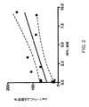
Notch経路遺伝子発現と、上記ヒトがん細胞株の増殖に対するJag1−Fcの影響との比較は、HeyLの発現と、Jag1−Fc阻害に対する感受性との間に、高い程度の相関があったことを示した。Cohen’s kappa値0.877を、この相関について計算した。このことは、HeyLレベルが、Jag1−Fcに対する応答と相関しているということの非常に高い程度の信頼性を示している。さらに、線形回帰分析は、ヒト結腸がん細胞株(R2=0.87、p=0.0022)(図1)および膵臓がん細胞株(R2=0.61、p=0.0026)(図2)について、HeyL発現と、Notch経路の阻害に対する感受性との間の高い程度の相関があることを示す。 Comparison of Notch pathway gene expression and the effect of Jag1-Fc on the growth of the above human cancer cell lines showed that there was a high degree of correlation between HeyL expression and sensitivity to Jag1-Fc inhibition. Indicated. A Cohen's kappa value of 0.877 was calculated for this correlation. This indicates a very high degree of confidence that the HeyL level is correlated with the response to Jag1-Fc. Furthermore, linear regression analysis showed that human colon cancer cell lines (R 2 = 0.87, p = 0.0002) (FIG. 1) and pancreatic cancer cell lines (R 2 = 0.61, p = 0.026). ) (FIG. 2) shows that there is a high degree of correlation between HeyL expression and sensitivity to inhibition of the Notch pathway.
他の試験した遺伝子のいずれについても、発現レベルと、Notchシグナル伝達の阻害に対する応答との間に、相関を観察しなかった。このことは、上昇したHeyL遺伝子発現が、Notch経路シグナル伝達の阻害に対する感受性の特異的生体マーカーであることを示した。 For any of the other genes tested, no correlation was observed between expression levels and response to inhibition of Notch signaling. This indicated that elevated HeyL gene expression was a specific biomarker sensitive to inhibition of Notch pathway signaling.
上昇したHeyL遺伝子発現と、Notch受容体活性化の阻害に対する感受性との相関を、結腸がん細胞および膵臓がん細胞に加えて、他の細胞株に関しても観察した。例えば、上昇したHeyL遺伝子発現と、Notch受容体活性化の阻害に対する感受性との間の有意な正の相関を、ヒト乳がん細胞において観察した。DU4475細胞は、Notch受容体活性化の阻害に対して非常に感受性であり、51個の試験したヒト乳がん細胞株の中で最高レベルのHeyLを発現する。対照的に、MCF12A細胞は、Notchに依存性でなく、同じセットの試験した51個のヒト乳がん細胞株の中で最低レベルのHeyLを発現する。ヒトT−ALL細胞(Karpas 45)および肺がん細胞(NCI−H187)は、上昇したレベルのHeyL遺伝子発現を示し、また、Notch受容体活性化の阻害に対して感受性であった。 Correlations between elevated HeyL gene expression and sensitivity to inhibition of Notch receptor activation were observed for other cell lines in addition to colon and pancreatic cancer cells. For example, a significant positive correlation between elevated HeyL gene expression and sensitivity to inhibition of Notch receptor activation was observed in human breast cancer cells. DU4475 cells are very sensitive to inhibition of Notch receptor activation and express the highest levels of HeyL among the 51 human breast cancer cell lines tested. In contrast, MCF12A cells are not Notch dependent and express the lowest level of HeyL among the same set of 51 human breast cancer cell lines tested. Human T-ALL cells (Karpas 45) and lung cancer cells (NCI-H187) showed elevated levels of HeyL gene expression and were sensitive to inhibition of Notch receptor activation.
9個の神経膠芽腫細胞株の中で、Notch受容体活性化の阻害に対する感受性と、HeyL発現との間に、相関を観察しなかった。このことは、HeyL発現レベルがこの特定の腫瘍タイプにおいてNotch受容体活性化の阻害に対する感受性の生体マーカーではないことを示唆する。 Of the 9 glioblastoma cell lines, no correlation was observed between sensitivity to inhibition of Notch receptor activation and HeyL expression. This suggests that HeyL expression level is not a biomarker sensitive to inhibition of Notch receptor activation in this particular tumor type.
(実施例3:γ−セクレターゼインヒビターでの細胞の処理)
細胞を、2mLの培地中で、6ウェルプレートに播種した。細胞の複製ウェルを、播種の直後に、1μM、3μM、および6μMのγセクレターゼインヒビター(GSI;Sigma L−685,458)、もしくはDMSO(Sigma D2650)(ビヒクルコントロール)で処理した。細胞を、処理後、37℃、5% CO2において20時間にわたってインキュベートし、次いで、収集し、PBS(Invitrogen 14040−133)ですすぎ、細胞ペレットを、ドライアイスで凍結させ、−80℃において貯蔵した。その後、RNAを、Qiagen RNeasyTM miniprepカラム(Qiagen GR8RNA)を使用して手動で調製し、qRT−PCRを、標準的プロトコル(Qiagen Quantitect SYBR GREEN RT−PCR kit 204245)に従って、Notch標的遺伝子発現を分析するために行った。定量的RT−PCRを、Applied Biosystems 7900HT Sequence Detection Systemで行った。結果を、比較Ct法を使用して分析した。βアクチンを、ハウスキーピング遺伝子として使用し、Stratagene Universal Human Reference RNA(Stratagene Cat.No.740000)を、外部参照サンプルとして使用して、Notch標的遺伝子Hes1、Hes5、Hes6、Hes7、Hey1、Hey2、HeyL、およびHeltの発現のレベルを決定した。
(Example 3: Treatment of cells with γ-secretase inhibitor)
Cells were seeded in 6-well plates in 2 mL of medium. Duplicate wells of cells were treated with 1 μM, 3 μM, and 6 μM γ-secretase inhibitor (GSI; Sigma L-685,458) or DMSO (Sigma D2650) (vehicle control) immediately after seeding. Cells are incubated for 20 hours at 37 ° C., 5% CO 2 after treatment, then harvested, rinsed with PBS (Invitrogen 14040-133), cell pellets frozen in dry ice and stored at −80 ° C. did. RNA was then prepared manually using a Qiagen RNeasy ™ miniprep column (Qiagen GR8RNA) and qRT-PCR was analyzed for Notch target gene expression according to standard protocols (Qiagen Quantitt SYBR GREEN RT-PCR kit 204245). Went to do. Quantitative RT-PCR was performed on an Applied Biosystems 7900HT Sequence Detection System. Results were analyzed using the comparative Ct method. β-actin is used as a housekeeping gene, Stratagene Universal Human Reference RNA (Stratagene Cat. No. 740000) is used as an external reference sample, and Notch target genes Hes1, Hes5, Hes6, Hes1, Hey2, Hey, HeL, HeL And the level of expression of Helt.
細胞増殖および生存率を測定するために、HT29細胞およびLS1034細胞を0.25% トリプシン(GIBCO Cat.No.25200)で採取し、10% FBS(GIBCO Cat.No.10438)および1% pen/strepを含むRPMI培地(GIBCO Cat.No.11875)中に再懸濁し、次いで、数を数えた。細胞を遠心分離し、無血清RPMIで洗浄し、次いで、4% FBSおよび1% pen/strepを含むRPMI培地中で1.2×105細胞/mLに再懸濁した。HT29細胞およびLS1034細胞の両方を、96ウェルプレート(Costar Cat.No.3595)において、6K/ウェル(50μL/細胞懸濁物のウェル)で最終的に播種した。ビヒクルコントロールもしくはNotch受容体活性化のインヒビターいずれか50μlを、三連で上記細胞に添加し、その結果、各ウェル中のFBSの最終濃度は、2%であった。これら実験において、Jag1−Fcもしくはγ−セクレターゼインヒビター(GSI;Sigma L−685,458)を、Notch受容体活性化のインヒビターとして使用した。 To measure cell proliferation and viability, HT29 and LS1034 cells were harvested with 0.25% trypsin (GIBCO Cat. No. 25200), 10% FBS (GIBCO Cat. No. 10438) and 1% pen / Resuspended in RPMI medium (GIBCO Cat. No. 11875) containing strep, then counted. Cells were centrifuged, washed with serum-free RPMI and then resuspended at 1.2 × 10 5 cells / mL in RPMI medium containing 4% FBS and 1% pen / strep. Both HT29 cells and LS1034 cells were finally seeded in 96 well plates (Costar Cat. No. 3595) at 6K / well (50 μL / well of cell suspension). 50 μl of either vehicle control or inhibitor of Notch receptor activation was added to the cells in triplicate, so that the final concentration of FBS in each well was 2%. In these experiments, Jag1-Fc or γ-secretase inhibitor (GSI; Sigma L-685,458) was used as an inhibitor of Notch receptor activation.
DMSO(GSIのビヒクルコントロール)を、以下のように調製した:1.0%、0.5%、0.25%および0.1%のDMSOを、無血清RPMI中の2×溶液として調製した。無処理細胞に、無血清RPMIを与えた。GSI希釈物を、DMSO中の1mMストックから、以下のように調製した:1.0μM、2.5μM、5μM、および10μM GSI希釈物を、無血清RPMI中の2×溶液として調製した。RPMI培地(100μL/ウェル)を、上記96ウェルプレート上のウェルの列外(outside row)に添加して、試験ウェルの蒸発を妨いだ。Jag1−Fcでの処理および細胞増殖を測定するための全てのMTTアッセイを、上記のように行った。 DMSO (GSI vehicle control) was prepared as follows: 1.0%, 0.5%, 0.25% and 0.1% DMSO were prepared as 2 × solutions in serum-free RPMI. . Untreated cells were given serum-free RPMI. GSI dilutions were prepared from 1 mM stocks in DMSO as follows: 1.0 μM, 2.5 μM, 5 μM, and 10 μM GSI dilutions were prepared as 2 × solutions in serum-free RPMI. RPMI medium (100 μL / well) was added outside the wells on the 96 well plate to prevent evaporation of the test wells. All MTT assays to measure Jag1-Fc and measure cell proliferation were performed as described above.
これら分析は、上記Notch経路の阻害が、LS−1034細胞の完全な増殖阻害および細胞死を生じさせた(>100%増殖阻害)が、HT−29細胞の増殖に対しては中程度の効果のみを有した(<17%増殖阻害)ことを示した。 These analyzes showed that inhibition of the Notch pathway resulted in complete growth inhibition and cell death of LS-1034 cells (> 100% growth inhibition), but moderate effects on HT-29 cell growth. Only (<17% inhibition of growth).
さらにNotch依存性であるヒトがん細胞株を、免疫無防備状態のマウスにおいて異種移植片としてインビボで増殖させた場合に、HeyLの上昇したレベルが示されることを実証した。例えば、T−ALL細胞株であるKarpas45(これは、Notchシグナル伝達の阻害に対して非常に感受性であり、培養物において上昇したレベルのHeyLを発現する)はまた、インビボで正常組織と比較して、腫瘍において上昇したレベルのHeyLを発現することを(Karpas45異種移植片腫瘍の切片の免疫組織化学染色によって)示した。 Furthermore, it was demonstrated that human cancer cell lines that are Notch-dependent are shown to have elevated levels of HeyL when grown in vivo as xenografts in immunocompromised mice. For example, the T-ALL cell line Karpas45, which is very sensitive to inhibition of Notch signaling and expresses elevated levels of HeyL in culture, is also compared to normal tissues in vivo. Have been shown to express elevated levels of HeyL in tumors (by immunohistochemical staining of sections of Karpas45 xenograft tumors).
目的の腫瘍を、10%緩衝化ホルマリン中で固定し、パラフィン中に包埋した。4μM厚切片を、ミクロトームを使用してスライスした。2×5分間インキュベーションを使用して、キシレン中で脱パラフィン(de−paraffinize)する前に、スライドを、55℃において10分間加温した。漸減エタノール濃度(100%、95%、70%)中で切片を水和し、工程ごとに、2×2分間で洗浄した。内因性ペルオキシダーゼを、30分間にわたって0.3% H2O2中でインキュベートすることによって抑制(quench)した。スライドを、マイクロ波オーブン(中出力レベル)において、クエン酸緩衝液(pH6.0)中で3×5分間で加熱し、次いで、20分間にわたって10%ヤギ血清中でブロックした。免疫染色のために、一次抗体およびアイソタイプコントロール抗体(ウサギIgG、Epitomics)を、1% FBS中4μg/mLで作製し、30〜45分間にわたってインキュベートした。2次抗体(ヤギα−ウサギビオチン化,Vector Labs)を1% FBS中で、1:200で希釈し、30〜45分間にわたってインキュベートした。Vectastain Standard ABCキット(Vector Labs)を、製造業者の説明書に従って使用した。スライドを、DAB酵素基質とともに5分間にわたってインキュベートし(Vector Labs)、DAB増強溶液とともに2分間にわたってインキュベートした(Zymed)。サンプルを、Mayersヘマトキシリンで1分間対比染色し、次いで、濃度漸増のエタノール(70%、95%、100%)中での2×2分間のインキュベーション、およびその後の、キシレンでの2×5分間のインキュベーションで脱水し、その後、カバースリップで密封した。 The tumor of interest was fixed in 10% buffered formalin and embedded in paraffin. 4 μM thick sections were sliced using a microtome. The slides were warmed at 55 ° C. for 10 minutes before de-paraffinizing in xylene using 2 × 5 minutes incubation. Sections were hydrated in decreasing ethanol concentrations (100%, 95%, 70%) and washed 2 × 2 minutes per step. Endogenous peroxidase was quenched by incubating in 0.3% H 2 O 2 for 30 minutes. Slides were heated in citrate buffer (pH 6.0) for 3 × 5 minutes in a microwave oven (medium power level) and then blocked in 10% goat serum for 20 minutes. For immunostaining, primary and isotype control antibodies (rabbit IgG, Epitomics) were made at 4 μg / mL in 1% FBS and incubated for 30-45 minutes. Secondary antibody (goat α-rabbit biotinylated, Vector Labs) was diluted 1: 200 in 1% FBS and incubated for 30-45 minutes. The Vectastein Standard ABC kit (Vector Labs) was used according to the manufacturer's instructions. Slides were incubated with DAB enzyme substrate for 5 minutes (Vector Labs) and incubated with DAB enhancement solution for 2 minutes (Zymed). Samples were counterstained with Mayers hematoxylin for 1 minute, then 2 × 2 minutes incubation in increasing concentrations of ethanol (70%, 95%, 100%), followed by 2 × 5 minutes with xylene. Dehydrated by incubation and then sealed with a coverslip.
抗ヒトマウスモノクローナル抗体(Abcam Cat.No.ab61277)を使用する場合に、Vector M.O.M.(マウス−オン−マウス(mouse−on−mouse))キット(Vector Labs)を使用して、マウス異種移植片モデルとして増殖させた腫瘍について宿主組織の非特異的染色を低下/排除した。腫瘍を切片化し、上記のように脱パラフィンし、次いで、M.O.MマウスIgGブロッキング試薬とともに60分間にわたってインキュベートした。免疫染色については、一次抗体およびアイソタイプコントロール(マウスIgG,Sigma)を、M.O.M.希釈物中、10μg/mLで作製し、30分間にわたってインキュベートした。2次抗体(抗マウスビオチン化,M.O.M.キット,Vector Labs)を、M.O.M.希釈物中で作製し、30分間にわたってインキュベートした。検出、対比染色、および脱水を、上記のように行った。 When using an anti-human mouse monoclonal antibody (Abcam Cat. No. ab 61277), Vector M. O. M.M. A (mouse-on-mouse) kit (Vector Labs) was used to reduce / eliminate non-specific staining of host tissue for tumors grown as mouse xenograft models. Tumors are sectioned and deparaffinized as described above. O. Incubated with M mouse IgG blocking reagent for 60 minutes. For immunostaining, primary antibodies and isotype controls (mouse IgG, Sigma) O. M.M. Made at 10 μg / mL in dilution and incubated for 30 minutes. Secondary antibodies (anti-mouse biotinylation, MOM kit, Vector Labs) O. M.M. Made in dilution and incubated for 30 minutes. Detection, counterstaining, and dehydration were performed as described above.
HeyL遺伝子発現が、Notch1遺伝子座近くへのMo−MuLVの染色体組み込みによって引き起こされるNotch1の増強された発現に応じて自発的に発生するマウス腫瘍において上昇することを見出した。このような腫瘍の増殖は、GSI(γセクレターゼインヒビター(Sigma L−685,458))による阻害に対する感受性によって示されるように、Notch依存性であった。上昇したNotch1発現を示さず、GSIに対して感受性ではなかったマウス腫瘍は、有意に低いレベルのHeyL遺伝子発現を示した。一般に、Notch受容体をコードする遺伝子付近へのMo−MuLV組み込みを示し、上記Notch受容体の上昇した発現がqRT−PCRもしくはマイクロアレイ発現分析によって確認された腫瘍はまた、(1)Notch受容体発現のレトロウイルス誘導性アップレギュレーションが存在しない、Notch遺伝子座とは異なる部位におけるMo−MuLV組み込みから生じる腫瘍;および(2)MMTVプロモーターによって駆動される外因性Her2腫瘍遺伝子を過剰発現するInk4aヌルマウスにおける自発的乳房腫瘍、に対する比較において、上昇したHeyL遺伝子発現を示した。両側スチューデントT検定は、Notch経路活性化によって駆動されない腫瘍に対する比較によって、Notch依存性腫瘍におけるHeyL遺伝子発現の非常に有意な上昇を示した(p<1×10⁻⁶)。Notch受容体発現のレトロウイルスアップレギュレーションに起因するNotch依存性と、上昇したレベルのHeyLとの間のこの相関は、Notch経路依存の生体マーカーとしてのHeyL遺伝子発現の役割をさらに実証した。 It has been found that HeyL gene expression is elevated in mouse tumors that spontaneously develop in response to enhanced Notch1 expression caused by chromosomal integration of Mo-MuLV near the Notch1 locus. The growth of such tumors was Notch-dependent as shown by sensitivity to inhibition by GSI (γ-secretase inhibitor (Sigma L-685,458)). Mouse tumors that did not show elevated Notch1 expression and were not sensitive to GSI showed significantly lower levels of HeyL gene expression. In general, tumors that show Mo-MuLV incorporation near the gene encoding the Notch receptor and whose elevated expression of the Notch receptor has been confirmed by qRT-PCR or microarray expression analysis are also (1) Notch receptor expression Spontaneous in Ink4a null mice overexpressing the exogenous Her2 oncogene driven by the MMTV promoter; and (2) a tumor resulting from Mo-MuLV integration at a site different from the Notch locus, in the absence of retrovirus-induced upregulation In comparison to a typical breast tumor, it showed elevated HeyL gene expression. Two-sided Student T-test showed a very significant increase in HeyL gene expression in Notch-dependent tumors by comparison to tumors not driven by Notch pathway activation (p <1 × 10 −6 ). This correlation between Notch dependence due to retroviral upregulation of Notch receptor expression and elevated levels of HeyL further demonstrated the role of HeyL gene expression as a Notch pathway-dependent biomarker.
上記レトロウイルス誘導性腫瘍における分析を行うために、腫瘍保有動物を安楽死させ、腫瘍を収集し、RPMI培地中の4mg/ml コラゲナーゼを使用して、37℃で2時間にわたって0.2gの小片を脱凝集させた。細胞を、18Gニードルに5〜10回通すことによってさらに脱凝集させた。懸濁物中の単一細胞を、さらなる分析のために上清中に吸引した。 To perform the analysis in the retrovirus-induced tumor, tumor-bearing animals were euthanized, tumors were collected, and 0.2 g pieces were used for 2 hours at 37 ° C. using 4 mg / ml collagenase in RPMI medium Was deagglomerated. Cells were further disaggregated by passing 5-10 times through an 18G needle. Single cells in suspension were aspirated into the supernatant for further analysis.
細胞生存率アッセイのために、単一細胞を、10% FBS、1% PenStrep、および0.01μM β−エストラジオールを補充したRPMI培地中、12ウェルプレートにおいて100,000細胞/ウェルで再度播種した。次いで、細胞を、1μM、3μMもしくは10μMのGSI(Sigma L−685,458)またはビヒクルコントロールで48時間にわたって処理した。細胞生存率およびアポトーシスを、Guava PCA−96 Viacount Flex試薬(Cat.No.4700−0060)を使用してアッセイし、製造業者のプロトコルに従って、Guava PCA−96機器を使用して測定した。mRNA発現レベルを測定するために、新鮮な腫瘍サンプルを収集し、RNAを、標準的なプロトコルを用いて調製し、マイクロアレイ遺伝子発現プロフィール分析を、製造業者の推奨プロトコルに従って、Agilent cDNAマイクロアレイチップを使用して行った。HeyLおよび他の遺伝子の相対的発現を、Stratageneのユニバーサルマウス参照RNAサンプル(Universal Mouse Reference RNA Cat.No.740100)に対する比較によって計算した。腫瘍を、HeyL発現の相対的レベルに従ってランク付けし、スチューデントT検定を使用して、上記Notch経路のレトロウイルス活性化を欠いている腫瘍に対して比較することによって、Notch受容体をコードする遺伝子付近にMo−MuLV組み込みを示し、かつ上記Notch受容体の上昇した発現がqRT−PCRもしくはマイクロアレイ発現分析によって確認される腫瘍における、HeyL発現の差異の有意性を決定した。 For cell viability assays, single cells were replated at 100,000 cells / well in 12-well plates in RPMI medium supplemented with 10% FBS, 1% PenStrep, and 0.01 μM β-estradiol. Cells were then treated with 1 μM, 3 μM or 10 μM GSI (Sigma L-685,458) or vehicle control for 48 hours. Cell viability and apoptosis were assayed using Guava PCA-96 Viacount Flex reagent (Cat. No. 4700-0060) and measured using a Guava PCA-96 instrument according to the manufacturer's protocol. To measure mRNA expression levels, fresh tumor samples are collected, RNA is prepared using standard protocols, and microarray gene expression profile analysis is performed using an Agilent cDNA microarray chip according to the manufacturer's recommended protocol I went there. The relative expression of HeyL and other genes was calculated by comparison to Stratagene's universal mouse reference RNA sample (Universal Mouse Reference RNA Cat. No. 740100). Genes encoding Notch receptor by ranking tumors according to relative levels of HeyL expression and comparing against tumors lacking retroviral activation of the Notch pathway using a Student T test The significance of HeyL expression differences in tumors that show Mo-MuLV incorporation in the vicinity and whose elevated Notch receptor expression was confirmed by qRT-PCR or microarray expression analysis was determined.
(参照による援用)
本明細書で言及される特許文書および科学論文の各々の開示全体は、全ての目的で、参照によって援用される。
(Incorporation by reference)
The entire disclosure of each of the patent documents and scientific papers referred to herein is incorporated by reference for all purposes.
(等価物)
本発明は、その趣旨もしくはその本質的な特徴から逸脱することなく、他の特定の形態で具現化され得る。従って、前述の実施形態は、全ての観点において、本明細書に記載される発明に対する限定ではなく、例示として解釈されるべきである。従って、本発明の範囲は、前述の説明によるのではなく、添付の特許請求の範囲によって示され、特許請求の範囲と等価の意味および範囲内にある全ての変更は、特許請求の範囲に包含されることが意図される。
(Equivalent)
The present invention may be embodied in other specific forms without departing from its spirit or essential characteristics. Accordingly, the foregoing embodiments are to be construed as illustrative in all aspects and not limiting of the invention described herein. Accordingly, the scope of the present invention is defined by the appended claims rather than by the foregoing description, and all changes that come within the meaning and range of equivalency of the claims are embraced by the claims. It is intended to be
Claims (9)
(a)該がん組織に由来するサンプル中のHeyL遺伝子発現のレベルを測定する工程;および
(b)該HeyL遺伝子発現のレベルを、標準値に対して比較し、それによって、HeyL遺伝子発現のみに基づいて、Notch受容体活性化を阻害する薬剤での処置に対して感受性のがん組織を同定する工程であって、ここで該サンプル中のHeyL遺伝子発現の上昇したレベルは、該がん組織が該薬剤での処置に感受性であることを示す、工程、
を包含する、方法。 A method for identifying human or mouse cancer tissue susceptible to treatment with an agent that inhibits Notch receptor activation comprising:
(A) measuring the level of HeyL gene expression in a sample derived from the cancer tissue; and (b) comparing the level of HeyL gene expression against a standard value, thereby only HeyL gene expression To identify cancerous tissue that is susceptible to treatment with an agent that inhibits Notch receptor activation, wherein the elevated level of HeyL gene expression in the sample is Indicating that the tissue is sensitive to treatment with the agent;
Including the method.
Applications Claiming Priority (5)
| Application Number | Priority Date | Filing Date | Title |
|---|---|---|---|
| US8012208P | 2008-07-11 | 2008-07-11 | |
| US61/080,122 | 2008-07-11 | ||
| US12/360,790 US7544476B1 (en) | 2008-07-11 | 2009-01-27 | Identifying cancers sensitive to treatment with inhibitors of notch signaling |
| US12/360,790 | 2009-01-27 | ||
| PCT/US2009/045479 WO2010005644A1 (en) | 2008-07-11 | 2009-05-28 | Identifying cancers sensitive to treatment with inhibitors of notch signaling |
Publications (2)
| Publication Number | Publication Date |
|---|---|
| JP2011527575A true JP2011527575A (en) | 2011-11-04 |
| JP2011527575A5 JP2011527575A5 (en) | 2012-07-12 |
Family
ID=40688675
Family Applications (1)
| Application Number | Title | Priority Date | Filing Date |
|---|---|---|---|
| JP2011517443A Pending JP2011527575A (en) | 2008-07-11 | 2009-05-28 | Identification of cancers that are susceptible to treatment with inhibitors of Notch signaling |
Country Status (6)
| Country | Link |
|---|---|
| US (1) | US7544476B1 (en) |
| EP (1) | EP2313526A4 (en) |
| JP (1) | JP2011527575A (en) |
| AU (1) | AU2009269081A1 (en) |
| CA (1) | CA2730215A1 (en) |
| WO (1) | WO2010005644A1 (en) |
Families Citing this family (7)
| Publication number | Priority date | Publication date | Assignee | Title |
|---|---|---|---|---|
| WO2007136856A2 (en) * | 2006-05-19 | 2007-11-29 | The Johns Hopkins University | Heyl as a therapeutic target and a diagnostic marker for neoplasia and uses therefor |
| JP5386364B2 (en) | 2006-12-18 | 2014-01-15 | ジェネンテック, インコーポレイテッド | Anti-Notch3 antagonist antibodies and their use in the prevention and treatment of Notch3-related diseases |
| US20120082659A1 (en) * | 2007-10-02 | 2012-04-05 | Hartmut Land | Methods And Compositions Related To Synergistic Responses To Oncogenic Mutations |
| ES2737649T3 (en) | 2012-12-19 | 2020-01-15 | Aveo Pharmaceuticals Inc | Anti-Notch3 antibodies |
| DK3448420T3 (en) | 2016-04-29 | 2022-12-12 | Aveo Pharmaceuticals Inc | ANTI-NOTCH3 ANTIBODY |
| EP3462349A1 (en) * | 2017-10-02 | 2019-04-03 | Koninklijke Philips N.V. | Assessment of notch cellular signaling pathway activity using mathematical modelling of target gene expression |
| EP3502279A1 (en) | 2017-12-20 | 2019-06-26 | Koninklijke Philips N.V. | Assessment of mapk-ap 1 cellular signaling pathway activity using mathematical modelling of target gene expression |
Citations (3)
| Publication number | Priority date | Publication date | Assignee | Title |
|---|---|---|---|---|
| WO2006092062A1 (en) * | 2005-03-04 | 2006-09-08 | The Hospital For Sick Children | Methods for cancer prognosis |
| WO2007070671A2 (en) * | 2005-12-16 | 2007-06-21 | Regeneron Pharmaceuticals, Inc. | Therapeutic methods for inhibiting tumor growth with dll4 antagonists |
| WO2007136856A2 (en) * | 2006-05-19 | 2007-11-29 | The Johns Hopkins University | Heyl as a therapeutic target and a diagnostic marker for neoplasia and uses therefor |
Family Cites Families (1)
| Publication number | Priority date | Publication date | Assignee | Title |
|---|---|---|---|---|
| WO2006047878A1 (en) | 2004-11-03 | 2006-05-11 | British Columbia Cancer Agency Branch | Cancer therapeutics and methods for their use |
-
2009
- 2009-01-27 US US12/360,790 patent/US7544476B1/en not_active Expired - Fee Related
- 2009-05-28 JP JP2011517443A patent/JP2011527575A/en active Pending
- 2009-05-28 CA CA2730215A patent/CA2730215A1/en not_active Abandoned
- 2009-05-28 AU AU2009269081A patent/AU2009269081A1/en not_active Abandoned
- 2009-05-28 WO PCT/US2009/045479 patent/WO2010005644A1/en active Application Filing
- 2009-05-28 EP EP09794862A patent/EP2313526A4/en not_active Withdrawn
Patent Citations (3)
| Publication number | Priority date | Publication date | Assignee | Title |
|---|---|---|---|---|
| WO2006092062A1 (en) * | 2005-03-04 | 2006-09-08 | The Hospital For Sick Children | Methods for cancer prognosis |
| WO2007070671A2 (en) * | 2005-12-16 | 2007-06-21 | Regeneron Pharmaceuticals, Inc. | Therapeutic methods for inhibiting tumor growth with dll4 antagonists |
| WO2007136856A2 (en) * | 2006-05-19 | 2007-11-29 | The Johns Hopkins University | Heyl as a therapeutic target and a diagnostic marker for neoplasia and uses therefor |
Non-Patent Citations (1)
| Title |
|---|
| JPN6013064284; Current cancer drug targets. 2006, Vol.6, No.4, p.313-323 * |
Also Published As
| Publication number | Publication date |
|---|---|
| CA2730215A1 (en) | 2010-01-14 |
| EP2313526A1 (en) | 2011-04-27 |
| WO2010005644A1 (en) | 2010-01-14 |
| AU2009269081A1 (en) | 2010-01-14 |
| EP2313526A4 (en) | 2011-08-31 |
| US7544476B1 (en) | 2009-06-09 |
Similar Documents
| Publication | Publication Date | Title |
|---|---|---|
| Ni et al. | Targeting androgen receptor in estrogen receptor-negative breast cancer | |
| AU2013226323B2 (en) | Cancer patient selection for administration of Wnt signaling inhibitors using RNF43 mutation status | |
| JP2017184729A (en) | Phosphodiesterase 4D7 as prostate cancer marker | |
| US11674182B2 (en) | Biomarker for HER2-positive cancer and anti-HER2 therapy and applications thereof | |
| JP2011527575A (en) | Identification of cancers that are susceptible to treatment with inhibitors of Notch signaling | |
| AU2009262420A1 (en) | HER3 as a determinant for the prognosis of melanoma | |
| JP6675300B2 (en) | Use of EGFR biomarkers for the treatment of gastric cancer with anti-EGFR drugs | |
| JP6860919B2 (en) | Mesenchymal KRAS mutant cancer therapeutic agent | |
| He et al. | HOXA5 is amplified in glioblastoma stem cells and promotes tumor progression by transcriptionally activating PTPRZ1 | |
| WO2017064159A1 (en) | Trpv2 as a biomarker and as a therapeutic target for melanoma | |
| JP6858563B2 (en) | Prediction of EGFR inhibitor effect by BRAF mutation detection | |
| Kim et al. | KIF5B-RET fusion gene may coincide oncogenic mutations of EGFR or KRAS gene in lung adenocarcinomas | |
| Li et al. | Expression of protocadherin8: Function as a tumor suppressor in hypopharyngeal carcinoma | |
| US20230184775A1 (en) | Kindlin-1 as a marker of sensitivity to egfr/ras pathway inhibitors | |
| EP3126520B1 (en) | Amg-337 for use in the treatment of cancers having a met amplification | |
| US10426777B2 (en) | Methods used to treat cancer | |
| EP2542692B1 (en) | Method for selecting patients for treatment with an egfr inhibitor | |
| US8609354B2 (en) | Method for selecting patients for treatment with an EGFR inhibitor | |
| EP4257146A1 (en) | Cystic lymphangioma treatment drug | |
| KR20240023045A (en) | Diagnostic methods and compositions for cancer treatment | |
| US10624895B2 (en) | Methods of sensitizing a patient with glioma to a therapeutic agent by administering small hairpin RNA targeting PDZ-RHOGEF | |
| US9283195B2 (en) | Methods used to characterize and treat glioblastoma | |
| WO2015006543A1 (en) | Method for predicting and detecting tumor metastasis in kidney cancer |
Legal Events
| Date | Code | Title | Description |
|---|---|---|---|
| A521 | Written amendment |
Free format text: JAPANESE INTERMEDIATE CODE: A523 Effective date: 20120525 |
|
| A621 | Written request for application examination |
Free format text: JAPANESE INTERMEDIATE CODE: A621 Effective date: 20120525 |
|
| A131 | Notification of reasons for refusal |
Free format text: JAPANESE INTERMEDIATE CODE: A131 Effective date: 20131227 |
|
| A601 | Written request for extension of time |
Free format text: JAPANESE INTERMEDIATE CODE: A601 Effective date: 20140325 |
|
| A602 | Written permission of extension of time |
Free format text: JAPANESE INTERMEDIATE CODE: A602 Effective date: 20140401 |
|
| A601 | Written request for extension of time |
Free format text: JAPANESE INTERMEDIATE CODE: A601 Effective date: 20140425 |
|
| A602 | Written permission of extension of time |
Free format text: JAPANESE INTERMEDIATE CODE: A602 Effective date: 20140507 |
|
| A601 | Written request for extension of time |
Free format text: JAPANESE INTERMEDIATE CODE: A601 Effective date: 20140526 |
|
| A602 | Written permission of extension of time |
Free format text: JAPANESE INTERMEDIATE CODE: A602 Effective date: 20140602 |
|
| A131 | Notification of reasons for refusal |
Free format text: JAPANESE INTERMEDIATE CODE: A131 Effective date: 20141111 |
|
| A02 | Decision of refusal |
Free format text: JAPANESE INTERMEDIATE CODE: A02 Effective date: 20150407 |