JP2010538252A - How to identify organ damage - Google Patents
How to identify organ damage Download PDFInfo
- Publication number
- JP2010538252A JP2010538252A JP2010522447A JP2010522447A JP2010538252A JP 2010538252 A JP2010538252 A JP 2010538252A JP 2010522447 A JP2010522447 A JP 2010522447A JP 2010522447 A JP2010522447 A JP 2010522447A JP 2010538252 A JP2010538252 A JP 2010538252A
- Authority
- JP
- Japan
- Prior art keywords
- sample
- protein
- identification method
- patient
- induced
- Prior art date
- Legal status (The legal status is an assumption and is not a legal conclusion. Google has not performed a legal analysis and makes no representation as to the accuracy of the status listed.)
- Pending
Links
Images


Classifications
-
- G—PHYSICS
- G01—MEASURING; TESTING
- G01N—INVESTIGATING OR ANALYSING MATERIALS BY DETERMINING THEIR CHEMICAL OR PHYSICAL PROPERTIES
- G01N33/00—Investigating or analysing materials by specific methods not covered by groups G01N1/00 - G01N31/00
- G01N33/48—Biological material, e.g. blood, urine; Haemocytometers
- G01N33/50—Chemical analysis of biological material, e.g. blood, urine; Testing involving biospecific ligand binding methods; Immunological testing
- G01N33/68—Chemical analysis of biological material, e.g. blood, urine; Testing involving biospecific ligand binding methods; Immunological testing involving proteins, peptides or amino acids
- G01N33/6893—Chemical analysis of biological material, e.g. blood, urine; Testing involving biospecific ligand binding methods; Immunological testing involving proteins, peptides or amino acids related to diseases not provided for elsewhere
-
- G—PHYSICS
- G01—MEASURING; TESTING
- G01N—INVESTIGATING OR ANALYSING MATERIALS BY DETERMINING THEIR CHEMICAL OR PHYSICAL PROPERTIES
- G01N2333/00—Assays involving biological materials from specific organisms or of a specific nature
- G01N2333/90—Enzymes; Proenzymes
- G01N2333/91—Transferases (2.)
- G01N2333/912—Transferases (2.) transferring phosphorus containing groups, e.g. kinases (2.7)
- G01N2333/91205—Phosphotransferases in general
- G01N2333/9121—Phosphotransferases in general with an alcohol group as acceptor (2.7.1), e.g. general tyrosine, serine or threonine kinases
- G01N2333/91215—Phosphotransferases in general with an alcohol group as acceptor (2.7.1), e.g. general tyrosine, serine or threonine kinases with a definite EC number (2.7.1.-)
-
- G—PHYSICS
- G01—MEASURING; TESTING
- G01N—INVESTIGATING OR ANALYSING MATERIALS BY DETERMINING THEIR CHEMICAL OR PHYSICAL PROPERTIES
- G01N2333/00—Assays involving biological materials from specific organisms or of a specific nature
- G01N2333/90—Enzymes; Proenzymes
- G01N2333/99—Isomerases (5.)
-
- G—PHYSICS
- G01—MEASURING; TESTING
- G01N—INVESTIGATING OR ANALYSING MATERIALS BY DETERMINING THEIR CHEMICAL OR PHYSICAL PROPERTIES
- G01N2800/00—Detection or diagnosis of diseases
- G01N2800/08—Hepato-biliairy disorders other than hepatitis
Landscapes
- Life Sciences & Earth Sciences (AREA)
- Health & Medical Sciences (AREA)
- Engineering & Computer Science (AREA)
- Molecular Biology (AREA)
- Chemical & Material Sciences (AREA)
- Biomedical Technology (AREA)
- Urology & Nephrology (AREA)
- Hematology (AREA)
- Immunology (AREA)
- Cell Biology (AREA)
- Analytical Chemistry (AREA)
- Biotechnology (AREA)
- Proteomics, Peptides & Aminoacids (AREA)
- Food Science & Technology (AREA)
- Medicinal Chemistry (AREA)
- Physics & Mathematics (AREA)
- Microbiology (AREA)
- Biochemistry (AREA)
- General Health & Medical Sciences (AREA)
- General Physics & Mathematics (AREA)
- Pathology (AREA)
- Investigating Or Analysing Biological Materials (AREA)
- Medicines Containing Antibodies Or Antigens For Use As Internal Diagnostic Agents (AREA)
- Medicines That Contain Protein Lipid Enzymes And Other Medicines (AREA)
Abstract
この発明は、患者が、開発薬により誘導された器官損傷を有する或いはそのリスクを有するか否かを同定する方法と、開発薬により誘導された器官損傷を有する患者を治療する方法を提供するものである。特に、この発明は、患者が、パラセタモールにより誘導された肝臓損傷を有する或いはその発症リスクを有するか否かを同定する方法に関するものである。 The present invention provides a method for identifying whether a patient has or is at risk for organ damage induced by the developed drug, and a method for treating a patient having organ damage induced by the developed drug. It is. In particular, the present invention relates to a method for identifying whether a patient has or is at risk for developing liver damage induced by paracetamol.
Description
この発明は、患者が、開発薬により誘導された器官損傷を有する或いはそのリスクを有するか否かを同定する方法と、開発薬により誘導された器官損傷を有する患者を治療する方法を提供するものである。特に、この発明は、患者が、パラセタモールにより誘導された肝臓損傷を有する或いはその発症リスクを有するか否かを同定する方法に関するものである。 The present invention provides a method for identifying whether a patient has or is at risk for organ damage induced by the developed drug, and a method for treating a patient having organ damage induced by the developed drug. It is. In particular, the present invention relates to a method for identifying whether a patient has or is at risk for developing liver damage induced by paracetamol.
バイオマーカーは、一般的な生物学的特徴の測定及び評価を、生物学的プロセス、病原性プロセス、又は、治療的な介入に対する薬理学的応答として客観的に示すものである(Hewitt et al, 2004)。体液中の臨床的に役立ち、実証されたタンパク質バイオマーカーの同定は、広範囲にわたる病理学的障害のより早朝の発見及び再分類を可能とすることから、プロテオミクスにおける最終的な到達点となる。体液のプロテオームは複雑であり(Anderson and Anderson, 2002)、これら体液のプロテオミクスは、血漿のアルブミン及び尿のTamm-Horsfallタンパク質(THP)などの大量のタンパク質は検出が容易であるが、より関心が高く、かつ、情報量が多いような、より少量のタンパク質の同定が困難であることへの挑戦である。 Biomarkers objectively indicate the measurement and evaluation of general biological characteristics as biological processes, pathogenic processes, or pharmacological responses to therapeutic intervention (Hewitt et al, 2004). The identification of clinically useful and proven protein biomarkers in body fluids is the ultimate goal in proteomics because it allows for earlier detection and reclassification of a wide range of pathological disorders. The proteome of bodily fluids is complex (Anderson and Anderson, 2002), and the proteomics of these bodily fluids are easier to detect for large amounts of proteins such as plasma albumin and urine Tamm-Horsfall protein (THP), but are more of interest The challenge is that it is difficult to identify a small amount of protein that is expensive and has a large amount of information.
多器官損傷は高死亡率を有することから、臓器損傷が発生する過程の病理学的性質を理解する必要がある。薬剤過剰投与(例えばパラセタモールの過量服用)は多臓器不全を誘導し得るものであり、パラセタモールにより誘導された臓器損傷の鍵となる媒体は転写因子c-Junである。 Because multi-organ damage has a high mortality rate, it is necessary to understand the pathological nature of the process of organ damage. Drug overdose (eg, paracetamol overdose) can induce multiple organ failure, and the key medium for organ damage induced by paracetamol is the transcription factor c-Jun.
パラセタモール(別名アセトアミノフェン:APAP)の慎重な又は偶発的な過剰投与は、イギリスの被毒で最も一般的なものである。例えば、1日につき約1人が、相当なAPAP過剰投与の処置のためにエジンバラ王立病院(Royal Infirmary of Edinburgh)への入院を必要とする(未発表データ)。しかし、約12%の入院では臓器損傷の何らかの程度にて発症しており、スコットランドの全域にて1年につき約50人の患者が相当なるAPAP 誘導肝臓損傷を有しており、Scottish Liver Transplant Unitに任せられるとされている(未発表データ)。APAP過剰投与は、グルタチオン・プロドラッグ(N-アセチルシステイン(NAC))によって治療されるが、この解毒剤による副作用は一般的である(約15%の患者)。大多数の患者が臓器損傷を発症しないので、我々は潜在的に有毒な解毒剤によって過剰に治療している可能性がある。 Careful or accidental overdose of paracetamol (aka acetaminophen: APAP) is the most common poisoning in the UK. For example, about one person per day requires hospitalization at the Royal Infirmary of Edinburgh for treatment of substantial APAP overdose (unpublished data). However, about 12% of hospitalizations develop some degree of organ damage, and about 50 patients per year have significant APAP-induced liver damage throughout Scotland. Scottish Liver Transplant Unit (Unpublished data). APAP overdose is treated with a glutathione prodrug (N-acetylcysteine (NAC)), but side effects from this antidote are common (about 15% of patients). Since the majority of patients do not develop organ damage, we may be over-treated with a potentially toxic antidote.
NAC処理の必要性は、血液パラセタモール濃度で決定される。血液のパラセタモールの十分に高い濃度を有するとされたものは、凡そ20時間、静脈内へNACが投与される。一旦NAC処理が完了すると、肝損傷の程度を評価するために更なる試験が行われる。これらの試験は、血清トランスアミナーゼ・エンザイム(ALT及びAST)、血液凝固(INR)及び血清クレアチニンの測定を必要とする。このように、パラセタモール誘導された器官損傷は、現在、20時間の最小限の期間後に排除され得るものである。 The need for NAC treatment is determined by the blood paracetamol concentration. Those suspected of having a sufficiently high concentration of paracetamol in the blood will receive NAC intravenously for approximately 20 hours. Once NAC treatment is complete, further studies are conducted to assess the extent of liver damage. These tests require measurements of serum transaminase enzymes (ALT and AST), blood clotting (INR) and serum creatinine. Thus, paracetamol-induced organ damage can now be eliminated after a minimum period of 20 hours.
そのことに加え、NACの副作用が一般的であることから、NAC持続注入の気管を短くすることが望ましい。 In addition, since side effects of NAC are common, it is desirable to shorten the trachea of continuous NAC infusion.
APAPを誘導された臓器損傷の既存のバイオマーカー(例えば血清アラニン・トランスアミナーゼ(ALT)、国際的な標準化された比率(INR)及びクレアチニン)は、APAP投与後に約24時間経過するまで、損傷を正確に検出または除外できない。より早朝の損傷を検出又は排除することができるなら、病院入院の数及び/又は長さを減らすことができる。 Existing biomarkers of APAP-induced organ damage (eg, serum alanine transaminase (ALT), international standardized ratio (INR) and creatinine) can accurately injure until approximately 24 hours after APAP administration. Cannot be detected or excluded. If earlier morning damage can be detected or eliminated, the number and / or length of hospital admissions can be reduced.
細胞膜の構成要素を放出する真核細胞の能力は、最初にWolfによって1967年に説明され、雄弁にも「血小板塵」と呼ばれた。基本的に、全ての細胞型は小さな膜小胞を吐出するものであり、今日では微粒子又はエキソソームとして公知であり、それらは、特に細胞が活性化又はアポトーシスしているときに起こる(George et al., 1982)。 The ability of eukaryotic cells to release cell membrane components was first described by Wolf in 1967 and was eloquently called “platelet dust”. Basically, all cell types eject small membrane vesicles, now known as microparticles or exosomes, which occur especially when cells are activated or apoptotic (George et al ., 1982).
泌尿器プロテオームは、血液よりも調査が容易であることから、多数のデータを集めることができる。腎臓の全てのネフロン部分は通常、尿に頂端膜及び細胞内液を含んでいる小さな小胞を分泌するものであり、これら小胞はエキソソームと呼ばれ、寸法は80ナノメートル未満である(Johnstone et al., 1987)。エキソソームは、これらの小胞の固有の特徴である「細胞質の側方側」に配向するものである。エキソソームは尿から分画遠心分離によって単離される、いくつかの腎臟病関連のタンパク質は液体クロマトグラフィ-タンデム型質量分析(LC-MS/MS)によって、これらのエキソソームから同定された(Pisitkun et al, 2004)。Zhouらは(2006)、尿のエキソソームを収集及び保管し、単離されるエキソソームタンパク質の量を最大のものとし、タンパク質の同定を容易とするための最適条件を決めた。泌尿器エキソソームは、疾患の間、それらの数及び組成物が変化して、臓器機能不全の指標となり得ることから、腎臟病の鍵となるバイオマーカを提供するものである。しかし、泌尿器エキソソームの変化は、血液MPを循環の変化及び腎臓機能の変化を反映したものであるかもしれないことには留意されたい。従って、泌尿器エキソソームタンパク質は、全身性疾患のためのバイオマーカーの提供するものである。尿エキソソームを単離して特徴付けることは、疾患バイオマーカーを同定する際の最初のステップとなる。 Because the urinary proteome is easier to investigate than blood, it can collect a lot of data. All nephron parts of the kidney normally secrete small vesicles that contain apical membranes and intracellular fluids in the urine, these vesicles are called exosomes and have a size of less than 80 nanometers (Johnstone et al., 1987). Exosomes are those that are oriented "on the side of the cytoplasm" which is an inherent feature of these vesicles. Exosomes are isolated from urine by differential centrifugation, some nephropathy-related proteins have been identified from these exosomes by liquid chromatography-tandem mass spectrometry (LC-MS / MS) (Pisitkun et al, 2004). Zhou et al. (2006) collected and stored urinary exosomes to maximize the amount of exosomal protein isolated and to determine optimal conditions to facilitate protein identification. Urinary exosomes provide a key biomarker for nephropathy because their number and composition can change during the disease and can be an indicator of organ dysfunction. However, it should be noted that changes in urinary exosomes may reflect changes in circulating blood MP and changes in kidney function. Thus, urinary exosome proteins provide a biomarker for systemic diseases. Isolating and characterizing urinary exosomes is the first step in identifying disease biomarkers.
この発明の目的は、上述したような従来技術の問題を取り除く又は緩和することにある。 The object of the present invention is to eliminate or mitigate the problems of the prior art as described above.
患者が、開発薬により誘導された器官損傷を有する或いはそのリスクを有するか否かを同定する方法であって、該方法は、(c)患者から試料を準備するステップと、(d)該試料中のタンパク質レベルを同定するステップと、を含み、タンパク質レベルの増大は、患者が開発薬により誘導された器官損傷を有する或いはそのリスクを有することを示すことを特徴とする同定方法である。この発明は、以下の図面を参照しつつ詳細に説明される。 A method for identifying whether a patient has or is at risk for organ damage induced by a developed drug, comprising: (c) preparing a sample from the patient; and (d) the sample Identifying a protein level therein, wherein an increase in the protein level is an indication method characterized by indicating that the patient has or is at risk for organ damage induced by the developed drug. The present invention will be described in detail with reference to the following drawings.
この発明は、薬の誤った又は不適当な投与量を投与された患者の身体の体液における高いレベルを示すタンパク質の観察に基づくものである。また、体液中のタンパク質量は、患者が、薬物誘導器官損傷のリスクを有する又は発症していることを示す。 This invention is based on the observation of proteins that show high levels in body fluids of the body of a patient who has been administered an incorrect or inappropriate dose of the drug. Also, the amount of protein in the body fluid indicates that the patient is at risk for or developing drug-induced organ damage.
従って、第一の態様において、この発明は、患者が、開発薬により誘導された器官損傷を有する或いはそのリスクを有するか否かを同定する方法であって、該方法は、(c)患者から試料を準備するステップと、(d)該試料中のタンパク質レベルを同定するステップと、を含み、タンパク質レベルの増大は、患者が開発薬により誘導された器官損傷を有する或いはそのリスクを有することを示すことを特徴とする同定方法を提供するものである。 Accordingly, in a first aspect, the present invention is a method for identifying whether a patient has or is at risk for organ damage induced by a developed drug comprising: (c) from a patient Providing a sample; and (d) identifying a protein level in the sample, wherein increasing the protein level indicates that the patient has or is at risk for organ damage induced by the developed drug. The identification method characterized by showing is provided.
ここでいう「患者」とは、投与された、或いは、薬の不適当な又は誤った投与量を投与された、又はそれらの疑いがあるものを含むものをいうものである。また、「患者」は、1つ以上の器官に損傷の臨床症状を示すもの、或いは、症状がない及び/又は薬誘導による器官損傷をリスクのあるものを言う。そのことから、この発明は、患者が、開発薬により誘導された器官損傷を有する或いはそのリスクを有するか否かを同定して、患者が更なる治療を必要としているかを特定する方法を提供することを目的としている。 As used herein, “patient” refers to a patient who has been administered, or has been administered an inappropriate or incorrect dose of a drug, or which is suspected of being administered. “Patient” also refers to those who show clinical symptoms of damage to one or more organs or who are free of symptoms and / or at risk of drug-induced organ damage. As such, the present invention provides a method for identifying whether a patient has or is at risk for organ damage induced by a developed drug and identifies whether the patient needs further treatment. The purpose is that.
ここでいう「薬」とは、診断、処理、或いは、疾患又は状態の予防に使用される化合物又は化合物の組み合わせを言うものである。それら化合物は、薬物又は薬剤として知られている。付加的に、或いは代換的に、「薬」は、行動、意識及び/又は知覚を変化させるために対象に投与される又は対象が摂取するその他の化合物を言うものである。これら化合物は、例えば、アルコール、ヘロイン及び/又は幻覚剤(例えばLSD)を含むものである。 As used herein, “drug” refers to a compound or combination of compounds used for diagnosis, treatment, or prevention of a disease or condition. These compounds are known as drugs or drugs. Additionally or alternatively, “drug” refers to other compounds that are administered to or taken by a subject to change behavior, consciousness, and / or perception. These compounds include, for example, alcohols, heroin and / or hallucinogens (eg LSD).
例えば、本方法は、例えば、抗生物質及び/又は消炎性、解熱薬、鎮痛剤及び/又は化学療法剤などの薬によって誘導される器官損傷の例を同定するために使用することができる。一実施形態において、本発明は、例えば、パラセタモール(7V(4-ヒドロキシフェニル)エタンアミド)などの解熱薬/鎮痛薬によって誘導される器官損傷を例えば同定するために使用することができる。 For example, the method can be used to identify examples of organ damage induced by drugs such as, for example, antibiotics and / or anti-inflammatory, antipyretic, analgesic and / or chemotherapeutic agents. In one embodiment, the present invention can be used, for example, to identify organ damage induced by antipyretic / analgesic drugs such as, for example, paracetamol (7V (4-hydroxyphenyl) ethanamide).
また、薬に誘導される器官損傷が、多くの時間に要することから、 薬の誤った又は不適当な投与量の投与、又は患者への投与にも関わらず、器官損傷に対する更なる治療は必要ないかもしれないことには、充分に留意されたい。しかるに、現在のプロトコールでは、薬の不適当な/誤った投与量を投与された又はその疑いのある患者は、治療され、一定期間観察のために入院することが要求されている。そのような患者は、医者が更なる治療を必要としないと判断した場合にのみ解放される。そのことから、当業者であれば、多くの場合、患者によって飲まれる薬が更なる治療を必要としている器官損傷に起因しなかったことから、観察期間は不必要であったことを知っている。このような不必要な一定期間の入院は医局員が利用できる限られた資源の相当な消耗である。 In addition, since drug-induced organ damage takes many hours, further treatment for organ damage is necessary despite the administration of the wrong or inappropriate dose of the drug or administration to the patient. Please note that it may not be. However, current protocols require that patients who have been or suspected of taking inappropriate / incorrect doses of drugs be treated and hospitalized for observation for a period of time. Such patients are only released if the physician determines that no further treatment is required. Thus, those skilled in the art know that the observation period was often unnecessary because the drugs taken by the patient were not due to organ damage requiring further treatment. . This unnecessary period of hospitalization is a considerable drain on the limited resources available to medical personnel.
前述のように、薬により誘導された器官損傷が明確となるのには相当の期間を要することから、一つ以上の器官に対する損傷が発症するとしても、患者には自覚症状がない。例えば、肝臓などの損傷した部分の器官部分があったとしても、患者は何ら損傷に関する症状も臨床徴候も示さないことがある。 As described above, since it takes a considerable period of time for the organ damage induced by the drug to become clear, even if damage to one or more organs develops, the patient has no subjective symptoms. For example, even if there is an injured part of the organ, such as the liver, the patient may not show any signs of injury or clinical signs.
薬により誘導された器官の損傷の症状が現れるまで相当の期間を要することから、入院及び一定期間の観察が不可欠である。 Hospitalization and observation over a period of time are indispensable because it takes a considerable period of time for symptoms of organ damage induced by drugs to appear.
従って、この発明は、薬が投与、薬の不適当な又は誤った投与量を投与された患者が一つ以上の器官に損傷を受けていて、患者が更なる治療を必要としているか否かを迅速に可能とする及び/又は予想する方法を提供するものである。この方法により、不必要に一定期間の観察のために入院している患者の数が劇的に減少する。 Therefore, the present invention provides for whether a patient who has been administered a drug, an inappropriate or incorrect dose of the drug has been damaged in one or more organs, and the patient needs further treatment. It provides a method that enables and / or anticipates quickly. This method dramatically reduces the number of patients hospitalized for an unnecessarily period of observation.
一実施形態において、この発明は、患者が、開発薬により誘導された器官損傷を有する或いはそのリスクを有するか否かを同定する方法であって、該方法は、(a)患者から試料を準備するステップと、(b)該試料中のタンパク質レベルを同定するステップと、を含み、タンパク質レベルの増大は、患者がパラセタモールにより誘導された肝臓損傷を有する或いはその発症リスクを有することを示すことを特徴とする同定方法を提供するものである。 In one embodiment, the invention is a method for identifying whether a patient has or is at risk for organ damage induced by a developed drug, the method comprising: (a) preparing a sample from the patient And (b) identifying the protein level in the sample, wherein an increase in protein level indicates that the patient has or is at risk of developing liver damage induced by paracetamol. An identification method is provided.
薬誘導された器官損傷は、薬自体またはそれらの代謝産物の毒作用に起因する場合がある。付加的に、又は代替的に、器官損傷は、薬又は代謝産物或いはそれらの代謝産物により起こる炎症反応によるものである。 Drug-induced organ damage may be due to the toxic effects of the drugs themselves or their metabolites. Additionally or alternatively, organ damage is due to inflammatory reactions caused by drugs or metabolites or their metabolites.
薬誘導された器官損傷は通常誤ったまたは不適当な投与量の投与に起因するものであり、一般に誤ったまたは不適当な投与量は過剰投与によるものである。このように、本願明細書において説明される各々の方法は、例えば、パラセタモールの過剰投与(さもなければ薬の「被毒」と称してもよい)などの薬に起因した器官損傷の例を同定するために使用することができる。 Drug-induced organ damage is usually due to incorrect or inappropriate dose administration, and generally incorrect or inappropriate dose is due to overdose. Thus, each method described herein identifies examples of organ damage caused by drugs such as, for example, paracetamol overdose (otherwise referred to as drug “poisoning”) Can be used to
ここでいう、「器官損傷」とは、肺(心臓)腎臓(肝臓)膵(脳)胃、(大及び/又は小)腸、及び/又は、父親泌尿器または造血制御系の1つ以上に影響を与える、器官内の1つ以上の細胞における機能欠損及び/又は細胞死を言うものである。薬誘導された器官損傷の厳しい症例では、おおよそ、器官の細胞の50%-90%は、機能を損失する又は死ぬ。 As used herein, “organ damage” affects one or more of the lung (heart) kidney (liver) pancreas (brain) stomach, (large and / or small) intestine, and / or paternal urology or hematopoietic control system. A loss of function and / or cell death in one or more cells within the organ. In severe cases of drug-induced organ damage, roughly 50% -90% of organ cells lose function or die.
一例として、薬誘導された肝損傷は、例えば、1つ以上の肝細胞の機能欠損及び/又はネクローシスによる肝機能の完全又は部分的な消失として特徴付けられる。肝機能の部分的な消失が何ら検出可能又は同定可能な症状を呈さないとしても、多くの場合、肝臓への損傷は、黄疸及び/又は、肝臓によって通常取り除かれる中毒性代謝産物及び化合物が血液中に取り込まれることに起因した脳への損傷である肝性脳症の発現として現れる。 By way of example, drug-induced liver injury is characterized as complete or partial loss of liver function due to, for example, loss of function of one or more hepatocytes and / or necrosis. Even if partial loss of liver function does not present any detectable or identifiable symptoms, damage to the liver is often caused by jaundice and / or toxic metabolites and compounds normally removed by the liver. It manifests as manifestation of hepatic encephalopathy, which is damage to the brain caused by being taken into the body.
一部の患者においては、薬誘導された器官損傷は、完全なる器官損傷に進行して、健康な個人の器官がそれとして機能することを中断してしまう。時々、薬は多くの器官に損害を与え、厳しい損傷の例では、多臓器不全(MOF)を招く。 In some patients, drug-induced organ damage progresses to complete organ damage, interrupting the functioning of healthy individual organs. Occasionally, drugs damage many organs, causing severe organ damage (MOF) in cases of severe damage.
更なる実施形態において、この発明は、患者がパラセタモールにより誘導された肝臓損傷を有する或いはその発症リスクを有するか否かを同定する方法であって、該方法は、(a)患者から試料を準備するステップと、(b)該試料中のタンパク質レベルを同定するステップと、を含み、タンパク質レベルの増大は、患者がパラセタモールにより誘導された肝臓損傷を有する或いはその発症リスクを有することを示すことを特徴とする同定方法を提供するものである。 In a further embodiment, the present invention is a method for identifying whether a patient has or is at risk of developing liver damage induced by paracetamol, the method comprising: (a) preparing a sample from the patient And (b) identifying the protein level in the sample, wherein an increase in protein level indicates that the patient has or is at risk of developing liver damage induced by paracetamol. An identification method is provided.
当業者であれば、「試料」とは、組織または生検材料の試料をいうものであることには留意されたい。また、「試料」は、全血液、血漿、血清、リンパ、尿、汗及び唾液、或いは/並びに、組織及び/又は腺分泌物などの体液の試料を含むものである。例えば、一実施形態において、試料は、体液として、好適には血漿である全血液を含むものである。より好適な実施形態では、意外なことに、試料は尿試料である。これは当業者にしてみても驚くべきことであり、尿試料のタンパク質含有量は、腎臓健康を示し得るものであった。従って、タンパク質含有量が、肝臓などのその他の器官の健康状態を示すことの発見は予期しないものであった。このように、一実施形態において、この発明は、患者の腎臓ではない器官において、薬誘導された器官損傷が起きているか否かを同定する方法を提供するものである。 One skilled in the art should note that “sample” refers to a sample of tissue or biopsy material. The “sample” includes samples of whole blood, plasma, serum, lymph, urine, sweat and saliva, and / or bodily fluids such as tissue and / or glandular secretions. For example, in one embodiment, the sample includes whole blood, preferably plasma, as a body fluid. In a more preferred embodiment, surprisingly, the sample is a urine sample. This is surprising to those skilled in the art, and the protein content of the urine sample could indicate kidney health. Thus, the discovery that protein content is indicative of the health of other organs such as the liver was unexpected. Thus, in one embodiment, the present invention provides a method for identifying whether drug-induced organ damage is occurring in an organ that is not the patient's kidney.
ここに開示されている試料は、微粒子及び/又はそのエキソソームを単離するプロトコールに処することができる。微粒子及び/又はエキソソームは、細胞から分泌される商法を有する。例えば、脈管系の細胞、特には、血管内皮細胞および/または器官組織を含む細胞は、微粒子及び/又はエキソソームを放出する。これら微粒子及び/又はエキソソームは、起源となるものの細胞の表層、細胞質及び/又は核由来の多様なタンパク質を更に含む。 The samples disclosed herein can be subjected to a protocol to isolate microparticles and / or their exosomes. Microparticles and / or exosomes have a commercial method that is secreted from cells. For example, cells of the vasculature, in particular cells containing vascular endothelial cells and / or organ tissue, release microparticles and / or exosomes. These microparticles and / or exosomes further comprise a variety of proteins originating from the cell surface, cytoplasm and / or nucleus of the cell.
当業者は、上述したような微粒子及び/又はエキソソームを試料から単離する技術に精通している。例えば、試料を遠心する。好適には、試料から微粒子及び/又はエキソソームを単離するために超遠心をする。付加的或いは代替的に、血液などの試料から単離された微粒子(及び潜在的には尿からのエキソソーム)は、分離され、或いは、細胞選別技術により(すなわち、(血管内皮細胞などの)期限とする細胞型に応じた)異なる集団へと選別される。一例として、微粒子に対し、微粒子の一つ以上の化合物に結合し得る一つ以上の物質によりタグ又はラベルを付することができる。この方法によれば、微粒子は、タグ又はラベルが付された微粒子を蛍光活性化細胞選別(FACS)分析に処することにより集団へと選別(又は単離)される。好適には、微粒子の一つ以上の化合物に結合し得る物質は、(以下に示すような)検出可能な部分によりラベルされた抗体である。特定の実施形態では、フルオレッセンイソチオシアン酸塩(FITC)ラベルされたヒトのCD31抗体及びフィコエリトリン(PE)ラベルされたヒトのCD42b抗体マーカーにより、血液及び/又は血漿試料からの微粒子を区別し、異なる集団に選別する。 Those skilled in the art are familiar with techniques for isolating microparticles and / or exosomes as described above from a sample. For example, the sample is centrifuged. Preferably, ultracentrifugation is performed to isolate microparticles and / or exosomes from the sample. Additionally or alternatively, microparticles (and potentially exosomes from urine) isolated from a sample such as blood are separated or expired by cell sorting techniques (ie, vascular endothelial cells, etc.) Depending on the cell type). As an example, a microparticle can be tagged or labeled with one or more substances that can bind to one or more compounds of the microparticle. According to this method, microparticles are sorted (or isolated) into a population by subjecting the tagged or labeled microparticles to fluorescence activated cell sorting (FACS) analysis. Preferably, the substance capable of binding to one or more compounds of the microparticle is an antibody labeled with a detectable moiety (as shown below). In certain embodiments, fluorescein isothiocyanate (FITC) labeled human CD31 antibody and phycoerythrin (PE) labeled human CD42b antibody markers differentiate and differ from microparticles from blood and / or plasma samples. Sort into groups.
従って、ここに記載の方法は、患者からの試料から微粒子及び/又はエキソソームを単離する更なるステップを含むものである。このように、この発明の更なる実施形態は、患者が薬により誘導された器官損傷を有する或いはその発症リスクを有するか否かを同定する方法であって、該方法は、(a)患者から試料を準備するステップと、(b)試料から微粒子及び/又はエキソソームを単離するステップと、(c)該微粒子及び/又はエキソソームのタンパク質レベルを同定するステップと、を含み、微粒子及び/又はエキソソームのタンパク質レベルの増大は、患者が薬により誘導された器官損傷を有する或いはその発症リスクを有することを示すことを特徴とする同定方法を提供するものである。 Accordingly, the methods described herein include the further step of isolating microparticles and / or exosomes from a sample from a patient. Thus, a further embodiment of the invention is a method for identifying whether a patient has or is at risk for developing organ damage induced by a drug, the method comprising: (a) from a patient Providing the sample; (b) isolating the microparticles and / or exosomes from the sample; and (c) identifying the protein level of the microparticles and / or exosomes, the microparticles and / or exosomes The increase in protein levels provides an identification method characterized by indicating that the patient has or is at risk of developing organ damage induced by the drug.
好適には、集めた直後であって、試料を上述した微粒子及び/又はエキソソームの抽出プロトコールに処する前に、試料内のタンパク質要物を分解、変性及び/又は破壊を防止する化合物を試料に添加する。かかる化合物は、例えば、タンパク質分解酵素の阻害剤、別名プロテナーゼ阻害剤を含む。 Preferably, a compound is added to the sample that prevents degradation, denaturation, and / or destruction of protein essentials in the sample immediately after collection and before subjecting the sample to the microparticle and / or exosome extraction protocol described above To do. Such compounds include, for example, inhibitors of proteolytic enzymes, also known as proteinase inhibitors.
試料の全量タンパク質量において、「タンパク質」とは、一つ以上の特定の又は特異的なタンパク質を言うものである。例えば、細胞表面、細胞内、細胞質及び/又は核のタンパク質のレベルを同定することにより、薬により誘導された器官損傷を同定及び/又は予想することができる。このように、ここに記載の各方法は、特定の細胞表面、細胞内、細胞質及び/又は核のタンパク質のレベルを同定することによるものである。例えば、この発明は、例えば、マイトジェンによって活性化されるタンパク質(MAP)キナーゼ(MAPK)によって活性化されるタンパク質の転写因子であるc-Junを含む転写因子のレベルを同定することを必要とする。付加的或いは代替的に、ここに記載の方法は、シクロフィリンA(CyPA−ペプチジル・プロリル・シス/トランスイソメラーゼAまたはPPIAと称される)などの細胞内タンパク質を同定することに関する。 In the total amount of protein in the sample, “protein” refers to one or more specific or specific proteins. For example, by identifying cell surface, intracellular, cytoplasmic and / or nuclear protein levels, drug-induced organ damage can be identified and / or predicted. Thus, each method described herein is by identifying the level of a particular cell surface, intracellular, cytoplasmic and / or nuclear protein. For example, the invention involves identifying the level of transcription factors including, for example, c-Jun, a transcription factor for proteins activated by mitogen-activated protein (MAP) kinase (MAPK) . Additionally or alternatively, the methods described herein relate to identifying intracellular proteins such as cyclophilin A (referred to as CyPA-peptidyl prolyl cis / trans isomerase A or PPIA).
このように、一実施形態において、この発明は、患者が、開発薬により誘導された器官損傷を有する或いはそのリスクを有するか否かを同定する方法であって、該方法は、(a)患者から試料を準備するステップと、(b)該試料中の転写因子及び/又は細胞内タンパク質レベルを同定するステップと、を含み、転写因子及び/又は細胞内タンパク質のレベルの増大は、患者が開発薬により誘導された器官損傷を有する或いはそのリスクを有することを示すことを特徴とする同定方法を提供するものである。 Thus, in one embodiment, the invention is a method for identifying whether a patient has or is at risk for organ damage induced by a developed drug comprising: (a) a patient And (b) identifying a transcription factor and / or intracellular protein level in the sample, wherein the increased level of the transcription factor and / or intracellular protein is developed by the patient. It provides an identification method characterized by showing that it has or is at risk for organ damage induced by drugs.
一実施形態において、転写因子はc-Jun N末端キナーゼ(JNK)である。他の実施形態において、細胞内タンパク質はCyPAである。 In one embodiment, the transcription factor is c-Jun N-terminal kinase (JNK). In other embodiments, the intracellular protein is CyPA.
理論に束縛されることを望まないが、薬への曝露、特には例えば薬(例えばパラセタモール)の過剰投与は、MAPKの活性化につながり、かかる活性化はc-Junを活性化させてc-Junの量を増大させる。また、例えば、パラセタモールなどの薬は、顕著な細胞死を招くことから、損傷した細胞からの漏出物は、試料(例えば尿、血液など)のCyPAのような細胞内のタンパク質の供給源となることが考えられる。 Without wishing to be bound by theory, exposure to drugs, particularly overdose of drugs (eg, paracetamol), leads to activation of MAPK, which activates c-Jun and c- Increase the amount of Jun. In addition, for example, drugs such as paracetamol cause significant cell death, and leakage from damaged cells becomes a source of intracellular proteins such as CyPA in samples (eg, urine, blood, etc.) It is possible.
c-Jun N末端キナーゼファミリーに属する遺伝子は、転写複合体に関連する核タンパク質をコードしており、c-Junは、細胞ストレスなどの多様な刺激により誘導される活性を有するAP-1転写因子の主たる要素である。また、c-Jun N末端キナーゼは、3つの遺伝子JNKl、JNK2及びJNK3に由来する10種のアイソフォームからなり、この発明は、一つ以上のアイソフォームのレベルを同定することに関するものでることには留意されたい。 The gene belonging to the c-Jun N-terminal kinase family encodes a nuclear protein related to the transcription complex, and c-Jun is an AP-1 transcription factor that has an activity induced by various stimuli such as cellular stress. Is the main element. C-Jun N-terminal kinase consists of 10 isoforms derived from 3 genes JNKl, JNK2 and JNK3, and this invention relates to identifying the level of one or more isoforms. Please note.
CyPAは、多くの多様な機能を有する大量の細胞内のタンパク質である。それは、免疫抑制因子サイクロスポリン2のための第一の細胞内の薬のターゲットであって、細胞内のタンパク質のフォールディング3に重要である、ペプチジル・プロリル・シス/トランスイソメラーゼ活性を有するイムノフィリンクラスのメンバーである。CyPAはHIV4に結合し、サイクロフィリン結合薬は強力な抗HIV活性を有する。 CyPA is a large amount of intracellular protein with many diverse functions. It is the first intracellular drug target for the immunosuppressive factor cyclosporin 2 and is an immunophilin with peptidyl prolyl cis / trans isomerase activity that is important for intracellular protein folding 3 Be a member of a class. CyPA binds to HIV 4 , and cyclophilin-binding drugs have potent anti-HIV activity.
また、CyPAは、多くの刺激に応答して細胞内から細胞外の環境に放出される。細胞外のCyPAは炎症誘発性であり、例えば、それは炎症細胞5のための走化性剤となる。 CyPA is also released from the cell to the extracellular environment in response to many stimuli. Extracellular CyPA is pro-inflammatory, for example, it becomes a chemotactic agent for inflammatory cells 5 .
上述したことから、パラセタモールの薬の過剰投与された、或いはその疑いのある患者の試料、好ましくは尿試料(及びより好ましくは、それに由来するエキソソーム)において同定されるc-Junのレベルは、パラセタモールにより誘導された肝臓損傷を示す又は予想するものである。 From the above, c-Jun levels identified in samples of patients overdose or suspected of paracetamol drugs, preferably urine samples (and more preferably exosomes derived therefrom), are paracetamol. Shows or predicts liver damage induced by.
同様に、パラセタモールの薬の過剰投与された、或いはその疑いのある患者の試料、好ましくは尿試料(及びより好ましくは、それに由来するエキソソーム)において同定されるCyPAのレベルは、パラセタモールにより誘導された肝臓損傷を示す又は予想するものである。 Similarly, levels of CyPA identified in samples of patients overdose or suspected of paracetamol drugs, preferably urine samples (and more preferably exosomes derived therefrom), were induced by paracetamol. Indicates or predicts liver damage.
このように、一実施形態において、この発明は、患者が、パラセタモールにより誘導された肝臓損傷を有する或いはそのリスクを有するか否かを同定する方法であって、該方法は、(a)患者から試料を準備するステップと、(b)任意選択的に尿試料中から微粒子及び/又はエキソソームの単離するステップと、(c)該尿試料中の転写因子c-Jun及び/又はCyPA、或いは/並びに、ステップ(b)にて単離された微粒子及び/又はエキソソームのレベルを同定するステップと、を含み、該尿試料中の転写因子c-Jun及び/又はCyPA、或いは/並びに、ステップ(b)にて単離された微粒子及び/又はエキソソームのレベルの増大は、患者がパラセタモールにより誘導された肝臓損傷を有する或いはそのリスクを有することを示すことを特徴とする同定方法を提供するものである。 Thus, in one embodiment, the invention is a method for identifying whether a patient has or is at risk for liver damage induced by paracetamol, the method comprising: (a) from a patient Preparing a sample; (b) optionally isolating microparticles and / or exosomes from the urine sample; and (c) transcription factors c-Jun and / or CyPA in the urine sample, or / And identifying the level of microparticles and / or exosomes isolated in step (b), comprising transcription factor c-Jun and / or CyPA in said urine sample, and / or step (b Increased levels of microparticles and / or exosomes isolated in (1) indicate that the patient has or is at risk for liver damage induced by paracetamol There is provided a measuring method.
一般的に、1mlの尿中の10ng〜10μgのc-Jun及び/又はCyPAタンパク質は肝臓への損傷を示すものである。 In general, 10 ng to 10 μg of c-Jun and / or CyPA protein in 1 ml of urine is indicative of damage to the liver.
当業者であれば、ここに記載の発明の方法である特定のタンパク質のレベルを同定する方法に関する技術の組み合わせに精通している。そのような技術は、例えば、タンパク質アッセイ、当業者であれば使用して、ここに援用される酵素に基づくアッセイ(Kullertz et al (1998), Clinical Chemistry, 44:3, p502-508等を参照)、以下に記載されているような免疫学的及びPCRに基づく技術を含むものである。 Those skilled in the art are familiar with a combination of techniques relating to the method of identifying specific protein levels described herein. Such techniques can be used, for example, in protein assays, those skilled in the art, see enzyme-based assays incorporated herein (Kullertz et al (1998), Clinical Chemistry, 44: 3, p502-508, etc. ), Including immunological and PCR-based techniques as described below.
検査される試料に存在するタンパク質の増大されたレベルがあるかと否かを決定するために、当業者はここに記載の方法により同定された試料中の又は同定された(すなわち、同定された全量及び/又は特定のタンパク質のレベル)と、基準試料中の又は同定された(全量及び/又は特定)タンパク質の結果と比較する。「基準試料」とは、健康な対象、すなわち(薬により或いはその他の理由により誘導された)器官損傷の無い基準試料から得られた試料である。 To determine whether there is an increased level of protein present in the sample being examined, one of ordinary skill in the sample identified or identified by the method described herein (ie, the total amount identified And / or the level of a specific protein) and the results of a protein in or identified (total and / or specific) in a reference sample. A “reference sample” is a sample obtained from a healthy subject, ie, a reference sample without organ damage (induced by drugs or for other reasons).
付加的或いは代替的に、得られた結果は、夫々が基準試料から得られた多数の基準試料と比較される。 Additionally or alternatively, the results obtained are compared with a number of reference samples each obtained from a reference sample.
従って、その他の実施形態において、この発明は、患者が、開発薬により誘導された器官損傷を有する或いはそのリスクを有するか否かを同定する方法であって、該方法は、(a)患者から試料を準備するステップと、(b)該試料中のタンパク質のレベルを同定するステップと、(c)その結果を基準試料にて同定されたレベルと比較するステップと、を含み、基準試料にて同定されたレベルに対してタンパク質のレベルが増大していることは、患者が開発薬により誘導された器官損傷を有する或いはそのリスクを有することを示すことを特徴とする同定方法を提供するものである。 Accordingly, in another embodiment, the invention is a method for identifying whether a patient has or is at risk for organ damage induced by a developed drug comprising: (a) from a patient Providing a sample; (b) identifying the level of protein in the sample; and (c) comparing the result to the level identified in the reference sample; An increase in protein level relative to the identified level provides an identification method characterized by indicating that the patient has or is at risk for organ damage induced by the developed drug. is there.
また、上述したパラセタモール誘導の肝臓損傷を同定する方法は、パラセタモール誘導肝臓損傷の無い対象から得られた基準の尿試料の結果と比較するステップを更に含むものである。 The above-described method for identifying paracetamol-induced liver damage further comprises a step of comparing with the results of a reference urine sample obtained from a subject without paracetamol-induced liver damage.
付加的或いは代替的に、ここに記載のかかる方法により得られた結果は、異なる器官損傷のレベルの患者における基準試料から得られた結果と比較される。当業者であればかかる試料が「ポジティブコントロール試料」であることがわかる。例えば、かかる結果は、過剰投与された器官損傷を受けていない患者、自覚症状がないが一つ以上の器官に損傷を受けている患者、及び/又は、薬の過剰投与後に症状があり、一つ以上の器官に損傷を受けている患者由来の試料から得られた結果と比較される。 Additionally or alternatively, the results obtained by such a method described herein are compared to the results obtained from a reference sample in patients with different levels of organ damage. A person skilled in the art knows that such a sample is a “positive control sample”. For example, such results may include patients who have not been overdosed organ damage, patients who have no subjective symptoms but are damaged in one or more organs, and / or who have symptoms after overdose of medication, Compared to results obtained from a sample from a patient who has damaged more than one organ.
有利には、基準試料は、上述した微粒子/エキソソームの単離プロトコールに処される。 Advantageously, the reference sample is subjected to the microparticle / exosome isolation protocol described above.
好適には、基準試料は、患者から得られるものと同一のタイプの試料とする。一例として、患者から得られる試料が尿試料である場合には、基準試料もまた尿試料とする。 Preferably, the reference sample is the same type of sample as obtained from the patient. As an example, if the sample obtained from the patient is a urine sample, the reference sample is also a urine sample.
対象が苦しむ薬により誘導される器官損傷のレベルは、現在利用可能な多数の臨床手段によって決定される。最も単純には、器官損傷及び/又は欠損は、例えば、(例えばヘモ透析による)腎臓補充療法又は呼吸不全の通気支持、或いは、末しょう血管抵抗を維持することの障害に対する血管収縮の支援などの、器官支持の臨床需要から推定されるものである。特定の組合せのスコアリング制御系、例えば、多器官機能不全スコア(Marshall, J. C, Cook, D. J., Christou, N. V., Bernard, G. R., Sprung, C. L. and Sibbald, W. J.: 1995, 'Multiple organ dysfunction score: a reliable descriptor of a complex clinical outcome.' Crit Care Med 23, 1638- 52.)や急性生理学的及び慢性健康評価スコア(Knaus, W. A., Draper, E. A., Wagner, D. P. and Zimmerman, J. E.: 1985, 'APACHE II: a severity of disease classification system.' Crit Care Med 13, 818-29)もまた有益な情報を提供する。
The level of organ damage induced by the drug the subject afflicts is determined by a number of clinical means currently available. Most simply, organ damage and / or deficiencies can include, for example, renal replacement therapy (eg, by hemodialysis) or ventilatory support for respiratory failure, or support of vasoconstriction for disturbances in maintaining peripheral vascular resistance, etc. Estimated from clinical demand for organ support. Certain combinations of scoring control systems, such as the multiple organ dysfunction score (Marshall, J.C, Cook, DJ, Christou, NV, Bernard, GR, Sprung, CL and Sibbald, WJ: 1995, 'Multiple organ dysfunction score : a reliable descriptor of a complex clinical outcome. '
好適には、試料内のタンパク質における、基準試料のレベルに対する約2〜10倍の増大は、更なる治療を必要としない薬により誘導される器官損傷のレベルを示す。更に好適は、試料内のタンパク質における、基準試料のレベルに対する約3〜9倍、好適には4〜8倍の増大は、更なる治療を必要としない薬により誘導される器官損傷のレベルを示す。一実施例において、試料内のタンパク質における、基準試料のレベルに対する約5〜7倍の増大は、更なる治療を必要としない薬により誘導される器官損傷のレベルを示す。 Preferably, an increase of about 2 to 10 times in the protein in the sample relative to the level of the reference sample indicates the level of organ damage induced by the drug that does not require further treatment. More preferably, an increase of about 3-9 times, preferably 4-8 times, in the protein in the sample relative to the level of the reference sample indicates the level of organ damage induced by the drug that does not require further treatment. . In one example, an approximately 5-7 fold increase in the protein in the sample relative to the level of the reference sample indicates the level of organ damage induced by the drug that does not require further treatment.
それに対し、試料内のタンパク質における、基準試料のレベルに対する約8〜100倍の増大は、薬により誘導される器官損傷に対し更なる治療を必要とするレベルを示すものとすることができる。好適には、試料内のタンパク質における、基準試料のレベルに対する約10〜90倍、好適には20〜80倍の増大は、薬により誘導される器官損傷に対し更なる治療を必要とするレベルを示すものとすることができる。更なる実施形態では、試料内のタンパク質における、基準試料のレベルに対する約25〜85倍、好適には30〜70倍、より好適には35〜65倍の増大は、薬により誘導される器官損傷に対し更なる治療を必要とするレベルを示すものとすることができる。 In contrast, an increase of about 8-100 fold in the protein in the sample relative to the level of the reference sample may indicate a level that requires further treatment for drug-induced organ damage. Preferably, an increase of about 10 to 90, preferably 20 to 80, in the protein in the sample relative to the level of the reference sample is at a level that requires further treatment for drug-induced organ damage. It can be shown. In a further embodiment, the increase in protein in the sample by about 25-85 fold, preferably 30-70 fold, more preferably 35-65 fold relative to the level of the reference sample is due to drug-induced organ damage. May indicate a level that requires further treatment.
試料内のタンパク質レベルを同定する方法を提供することに加え、ここに記載の夫々の方法は、付加的或いは代替的に、準備される試料内の核酸のレベルを同定することを必要とする。当業者であれば試料内の核酸のレベルが、薬により誘導される器官損傷を示すタンパク質のレベルを示すことを知っていることには留意されたい。また、試料内の特定のタンパク質のレベルは、ここに記載の特定のタンパク質をコードする核酸を利用することにより同定し得るものである。一例として、提供される試料内の転写因子c-Jun及び/又はCyPAをコードする核酸を同定することができる。 In addition to providing a method for identifying protein levels in a sample, each method described herein additionally or alternatively requires identifying the level of nucleic acid in the sample being prepared. It should be noted that one skilled in the art knows that the level of nucleic acid in a sample indicates the level of protein that is indicative of drug-induced organ damage. In addition, the level of a specific protein in a sample can be identified by utilizing a nucleic acid encoding the specific protein described herein. As an example, nucleic acids encoding the transcription factors c-Jun and / or CyPA in the provided sample can be identified.
ここでいう核酸とは、デオキシリボ核酸(DNA)及びリボ核酸(RNA)を含むことを意図したものである。RNAの場合、mRNA及びtRNAを含むものである。より詳しくは、一実施形態において、この発明は、試料内のRNA、好適はmRNAのレベルの同定することを必要とする。有利には、mRNAは転写因子c-Jun及び/又はCyPAをコードする。当業者が、試料内に存在する核酸レベルを同定する方法は以下に説明する。 The nucleic acid here is intended to include deoxyribonucleic acid (DNA) and ribonucleic acid (RNA). In the case of RNA, it includes mRNA and tRNA. More particularly, in one embodiment, the present invention entails identifying the level of RNA, preferably mRNA, in a sample. Advantageously, the mRNA encodes the transcription factors c-Jun and / or CyPA. Methods for one skilled in the art to identify nucleic acid levels present in a sample are described below.
当業者であれば、上述したような全量タンパク質及び/又は特定のタンパク質のレベルを同定する技術に精通している。また、以下に記載の夫々の方法には、ここに記載の夫々の方法に適用し得ることには留意されたい。 Those skilled in the art are familiar with techniques for identifying total protein levels and / or specific protein levels as described above. It should also be noted that each method described below can be applied to each method described herein.
一実施形態において、試料内の全量タンパク質の同定は、タンパク質アッセイを用いて同定する。これには、ブラッドフォードタンパク質アッセイ等のアッセイが適当である。この種のアッセイは、公知の濃度の、例えば、ウシ血清アルブミンとして準備される、標準又は基準タンパク質試料の使用をしばしば必要とする。 In one embodiment, the identification of total protein in the sample is identified using a protein assay. For this, an assay such as a Bradford protein assay is appropriate. This type of assay often requires the use of a standard or reference protein sample prepared at a known concentration, eg, bovine serum albumin.
特定のタンパク質のレベルを同定するためには、タンパク質に結合し得る物質を使用する、特定の免疫学的手法を使用する必要がある。この種の技術は、例えばc-Jun及び/又はCyPAなどの転写因子のタンパク質レベルを同定することに特に適している。 In order to identify the level of a particular protein, it is necessary to use a specific immunological technique that uses a substance that can bind to the protein. This type of technique is particularly suitable for identifying protein levels of transcription factors such as c-Jun and / or CyPA.
一実施形態において、試験される試料と基質(またはその一部)とを接触させ、特定のタンパク質又は試料内の(c-Junなどの)転写因子及び/又はCyPAなどのタンパク質を該基質に関連付ける、結合させる及び/又は固定するステップを含む方法を提供するものである。 In one embodiment, the sample to be tested is contacted with a substrate (or a portion thereof) and a particular protein or transcription factor (such as c-Jun) and / or a protein such as CyPA within the sample is associated with the substrate. A method comprising the steps of coupling, and / or fixing.
適当な基質は、例えば、ガラス、ニトロセルロース、紙、アガロース及び/又はプラスチックを含むものである。プラスチック材料などの基質はマイクロタイタープレートの形状となる。 Suitable substrates include, for example, glass, nitrocellulose, paper, agarose and / or plastic. Substrates such as plastic materials are in the form of microtiter plates.
或いは、試験に供される試料と接触する基質は、特定のタンパク質や複数のタンパク質と結合し得る物質を含む。好適には、特定のタンパク質に結合する物質は、基質(又は少なくともその一部)にも結合するものである。好適な結合物質は、例えば転写因子c-Junおよび/またはCyPAなどの特定のタンパク質に結合し得る、例えば、モノクローナル抗体又はポリクローナル抗体などの抗体及び/又はペプチド又は小分子である。また、この定義はここに記載の全ての結合物質に適用し得ることには留意されたい。このように、基質(またはその一部)は、試料内の特定のタンパク質及び関連する特的のタンパク質に結合し得る物質と結合又は相互作用し得る条件下にて、試験対象の試料と接触させる。 Alternatively, the substrate in contact with the sample to be tested includes a substance that can bind to a specific protein or a plurality of proteins. Preferably, the substance that binds to a particular protein is one that also binds to a substrate (or at least a portion thereof). Suitable binding agents are, for example, antibodies and / or peptides or small molecules, such as monoclonal or polyclonal antibodies, which can bind to specific proteins such as the transcription factors c-Jun and / or CyPA. It should also be noted that this definition is applicable to all binding substances described herein. In this way, the substrate (or part thereof) is contacted with the sample to be tested under conditions that allow it to bind or interact with a substance that can bind to a specific protein in the sample and related specific proteins. .
基質又は特的のタンパク質に結合し得る物質に結合する特異的なタンパク質は、特異的なタンパク質に結合し得る更なる物質(以後、「第一の結合物質」という)に使用することにより検出される。付加的或いは代替的に、第一の結合物質は、特定のタンパク質::基質複合体、又は、特定のタンパク質及び上述したような特定のタンパク質に結合し得る物質を含む複合体に対し親和性又は結合能を有する。 A specific protein that binds to a substrate or a substance that can bind to a specific protein is detected by use of an additional substance that can bind to the specific protein (hereinafter referred to as the “first binding substance”). The Additionally or alternatively, the first binding agent has an affinity for a specific protein :: substrate complex or a complex comprising a specific protein and a substance capable of binding to a specific protein as described above. Has binding ability.
第一の結合物質は、検出を可能とする部分(以下、「検出部分」という)に接合する。例えば、第一の物質は、色素化学ルミネセンス反応により、レベルを報告する酵素に接合する。かかる接合酵素は、以下に限定されないが、セイヨウワサビペルオキシダーゼ(HRP)及びアルカリフォスファターゼ(AIkP)を含む。付加的或いは代替的に、第一の結合物質は、例えば、FITC、ローダミンまたはテキサスレッドなどといったフルオロホアなどの蛍光分子に接合する。結合分子に接合し得るその他のタイプの分子は、放射性同位元素を使ってラベルされた部分を含むものである。 The first binding substance is bonded to a portion that enables detection (hereinafter referred to as “detection portion”). For example, the first substance is conjugated to an enzyme that reports levels by a dye chemiluminescence reaction. Such conjugating enzymes include, but are not limited to, horseradish peroxidase (HRP) and alkaline phosphatase (AIkP). Additionally or alternatively, the first binding substance is conjugated to a fluorescent molecule such as a fluorophore such as, for example, FITC, rhodamine or Texas Red. Other types of molecules that can be conjugated to binding molecules are those that contain moieties labeled with radioisotopes.
或いは、特定のタンパク質に結合し得る基質又は物質に結合する任意の特定のタンパク質は、第一の結合物質に対する親和性を有する更なる結合分子(以下、「第二の結合物質」という)を用いた手段により検出される。好適には、第二の結合物質は、検出可能な部分に接合するものである。 Alternatively, any specific protein that binds to a substrate or substance that can bind to a specific protein uses a further binding molecule (hereinafter referred to as a “second binding substance”) that has an affinity for the first binding substance. It is detected by means. Preferably, the second binding substance is one that binds to the detectable moiety.
一つ以上の特定のタンパク質に結合する第一の結合物質(又はそれに結合する第二の結合物質)の量は、特定のタンパク質又は試験に供される試料に存在するタンパク質のレベルを示すものである。 The amount of the first binding substance that binds to one or more specific proteins (or the second binding substance that binds to it) is indicative of the level of protein present in the specific protein or sample being tested. is there.
前述のように、本願明細書において説明される免疫学的な方法は、例えばc-Junおよび/またはCyPAなどの転写因子のレベルを同定することに特に適しており、かかる患者からの試料から提供されるレベルは、薬を誤ってまたは不適切に投与されたことによる薬により誘導される器官の損傷を示すものである。 As mentioned above, the immunological methods described herein are particularly suitable for identifying levels of transcription factors such as c-Jun and / or CyPA, and are provided from samples from such patients. Levels indicated are indicative of drug-induced organ damage due to incorrect or improper administration of the drug.
一実施形態において、特定のタンパク質のレベルを同定する上述の方法は、「漬棒(dip stick)」試験により行われ、試料内のc-Junおよび/またはCyPAなどの転写因子などの特定のタンパク質が、基質或いはそれに結合又は固定される結合物質に結合し得る条件下にて、基質(またはその一部)は試験される試料と接触させる。 In one embodiment, the method described above for identifying the level of a particular protein is performed by a “dip stick” test, and the particular protein, such as a transcription factor, such as c-Jun and / or CyPA in the sample. However, the substrate (or part thereof) is contacted with the sample to be tested under conditions that allow it to bind to the substrate or a binding substance bound or immobilized thereto.
その他の実施形態において、かかる方法は、酵素結合抗体免疫吸着アッセイ(ELISA)などの免疫学的なアッセイの形態をとる。ELISAは、「捕獲」ELISAという形態をとることができ、試験される試料は基質に接触し、(特定のタンパク質に結合し得る)結合物質が基質に結合又は固定することにより、試料は任意の特定のタンパク質に捕獲又は結合する。或いは、試料は、試料内のタンパク質と基質との「直接」の結合を可能とする条件下にて基質に接触する。 In other embodiments, such methods take the form of immunological assays such as enzyme linked antibody immunosorbent assays (ELISA). An ELISA can take the form of a “capture” ELISA, where the sample to be tested contacts the substrate, and the binding agent (which can bind to a particular protein) binds or immobilizes to the substrate, so that the sample can be any Capture or bind to a specific protein. Alternatively, the sample is contacted with the substrate under conditions that allow “direct” binding of the protein and substrate within the sample.
上述した夫々のELISA法は、特定のタンパク質を「直接的」に検出するステップ又は「間接的」に検出するステップを含むものである。それらステップに関連したELISA法は、「間接ELISA法」又は「間接ELISA法」として知られている。 Each of the ELISA methods described above includes a step of detecting a specific protein “directly” or “indirectly”. The ELISA methods associated with these steps are known as “indirect ELISA methods” or “indirect ELISA methods”.
「直接」ELISA法において、試験される試料は、特定のタンパク質又は試料内のタンパク質が基質及び/又はそれに結合している結合物質に結合し得る条件下にて、基質と接触させられる。任意選択的なブロッキングステップの後、(例えば、c-Jun及び/又はCyPAなどの転写因子などの)結合タンパク質は、特定のタンパク質に結合し得る物質(例えば、第一の結合物質)を用いて検出される。好適には、第一の結合物質は、検出可能な部分に接合するものである。 In a “direct” ELISA method, the sample to be tested is contacted with the substrate under conditions that allow the specific protein or protein in the sample to bind to the substrate and / or the binding agent bound thereto. After an optional blocking step, a binding protein (eg, a transcription factor such as c-Jun and / or CyPA) is used with a substance (eg, a first binding substance) that can bind to the specific protein. Detected. Preferably, the first binding substance is conjugated to a detectable moiety.
「間接」ELISA法において、特定のタンパク質と第一の結合物質とを接触させた後に、第一の結合物質と親和性又は結合能を有する更なる結合物質(第二の結合物質)を使用する更なるステップを含むものである。好適には、第二の結合物質は、検出可能な部分に接合するものである。 In the “indirect” ELISA method, after a specific protein and a first binding substance are brought into contact with each other, an additional binding substance (second binding substance) having affinity or binding ability with the first binding substance is used. It includes further steps. Preferably, the second binding substance is one that binds to the detectable moiety.
特定のタンパク質と結合し得る物質を使用して、体液中の試料の特定のタンパク質のレベルを同定するするその他の技術は、ウェスタンブロットやドットブロットを含むものである。ウェスタンブロットは、試料を電気泳動して、試料内のタンパク質要素などの構成要素を分離すること関するものである。一実施形態において、電気泳動される試料は、薬を誤って又は不適切に投与されたことが推定される又は投与された患者から得られた体液である。更なる実施形態では、試料は、当該試料から得られる微粒子及び/又はエキソソームを含む又はそれにより構成される。 Other techniques for identifying the level of a particular protein in a sample in a body fluid using substances that can bind to the particular protein include Western blots and dot blots. Western blot involves the electrophoresis of a sample to separate components such as protein elements within the sample. In one embodiment, the sample to be electrophoresed is a bodily fluid obtained from a patient that is presumed or administered a drug incorrectly or inappropriately. In further embodiments, the sample comprises or consists of microparticles and / or exosomes obtained from the sample.
分離した要素は、ニトロセルロースなどの基質にトランスファーする。試料内のc-Jun及び/又はCyPAなどの転写因子などといった転写因子などの特定のタンパク質のレベルを同定するために、基質は、試料内の任意の特定のタンパク質と特定のタンパク質と結合し得る物質とが結合できる条件下にて、特定のタンパク質と結合し得る結合物質と接触させられる。 The separated element is transferred to a substrate such as nitrocellulose. To identify the level of a particular protein, such as a transcription factor, such as c-Jun and / or a transcription factor such as CyPA in a sample, the substrate can bind to any particular protein and the particular protein in the sample. It is brought into contact with a binding substance capable of binding to a specific protein under conditions that allow the substance to bind.
好適には、特定のタンパク質と結合し得る物質は、検出可能な部分と接合するものである。 Preferably, the substance capable of binding to a specific protein is one that binds to a detectable moiety.
或いは、基質を、特定のタンパク質と結合し得る結合物質の親和性を有する更なる結合物質と接触させる。有利には、更なる結合物質は、検出可能な部分と接合するものである。 Alternatively, the substrate is contacted with a further binding agent having the affinity of a binding agent that can bind to a particular protein. Advantageously, the further binding substance is one that joins the detectable moiety.
ドットブロットの場合、試料又はその一部は、転写因子c-Jun及び/又はCyPAなどの任意のタンパク質が基質に対し結合及び/又は固定するような基質と接触する。任意の固定又は結合したタンパク質の同定は、上述したように実施した。 In the case of a dot blot, the sample or part thereof is contacted with a substrate such that any protein such as transcription factors c-Jun and / or CyPA binds and / or immobilizes to the substrate. Identification of any immobilized or bound protein was performed as described above.
上述した任意の技術において、検出された第一又は第二の結合物質の量は、試料内の一つ以上の特定のタンパク質の割合を示すものであり、また、薬により誘導される器官損傷を示し得るものである。 In any of the techniques described above, the amount of the first or second binding substance detected is indicative of the percentage of one or more specific proteins in the sample, and is indicative of drug-induced organ damage. It can be shown.
試料内の特定のタンパク質のレベルを検出及び/又は同定するためには、特定のタンパク質を一つ以上の化合物に転換する酵素を利用することが可能である。或いは、検出されるタンパク質が酵素である場合には、酵素に特異的な基質を接触させ、酵素/基質反応産物を引き起こさせることができる。両方の場合、酵素/基質反応により得られる産物のレベルは試料内の特定のタンパク質のレベルを示すものである。一例として、パラセタモールを過剰投与された患者由来の尿試料などの試料内のCyPA(ペプチジル・プロリル・シス/トランスイソメラーゼ活性)のレベルを特定するために、CyPAの酵素活性が測定された(Kullertz et al (1998)を参照)。 In order to detect and / or identify the level of a particular protein in a sample, an enzyme that converts the particular protein into one or more compounds can be utilized. Alternatively, if the protein to be detected is an enzyme, the enzyme can be contacted with a substrate specific to cause an enzyme / substrate reaction product. In both cases, the level of product obtained from the enzyme / substrate reaction is indicative of the level of a particular protein in the sample. As an example, the enzyme activity of CyPA was measured to identify the level of CyPA (peptidyl prolyl cis / trans isomerase activity) in samples such as urine samples from patients overdose of paracetamol (Kullertz et al. al (1998)).
転写因子c-Jun及び/又はCyPAなどの任意のタンパク質のレベルを同定する方法は、例えな、ポリメラーゼチェーリアクション(PCR)技術におけるリアルタイムPCR(或いは、定量的PCRとして知られている)などを含む。そのような技術は、体液試料内に特定の核酸配列が存在するか否か、そして、かかる核酸からの発現レベルを決定するために使用される。 Methods for identifying the level of any protein such as the transcription factors c-Jun and / or CyPA include, for example, real-time PCR in polymerase chain reaction (PCR) technology (also known as quantitative PCR), etc. . Such techniques are used to determine whether a particular nucleic acid sequence is present in a body fluid sample and the level of expression from such nucleic acid.
その場合に、リアルタイムPCRは、転写因子c-Jun及び/又はCyPAなどの特定のタンパク質をコードする核酸配列が試料内にあるか、そして、かかる核酸配列の発現レベルを決定するために使用される。一般的に、特定の核酸配列の発現レベルを定量するためには、逆転写PCRが使用され、適当なmRNAを相補DNA(cDNA)へと逆転写する。好適には、逆転写のプロトコールは、関心対象のmRNAを特異的に増幅するためのプライマーを使用するものである。 In that case, real-time PCR is used to determine whether a nucleic acid sequence encoding a specific protein such as transcription factors c-Jun and / or CyPA is in the sample and the expression level of such nucleic acid sequence. . In general, reverse transcription PCR is used to quantify the expression level of a particular nucleic acid sequence, reverse transcription of the appropriate mRNA into complementary DNA (cDNA). Preferably, the reverse transcription protocol uses primers to specifically amplify the mRNA of interest.
一般的に、cDNAは、特定の配列に特異的にハイブリダイズするよう設計されたプライマーを用いて増幅され、PCRに使用される核酸は、蛍光又は放射性の化合物によりラベルすることができる。 In general, cDNA is amplified using primers designed to specifically hybridize to a particular sequence, and nucleic acids used in PCR can be labeled with fluorescent or radioactive compounds.
当業者であれば、ラベルされた核酸を使用して、PCRにより生産されたDNA量を定量する技術に精通している。簡単に言えば、一例として、ラベルされ、増幅した核酸は、PCRサイクル中に取り込まれたラベルされた核酸の量をモニタリングすることにより決定される。 Those skilled in the art are familiar with techniques for quantifying the amount of DNA produced by PCR using labeled nucleic acids. Briefly, by way of example, labeled and amplified nucleic acid is determined by monitoring the amount of labeled nucleic acid incorporated during the PCR cycle.
好適には、得られた結果は、基準試料の結果と比較される。 Preferably, the results obtained are compared with the results of the reference sample.
ここに記載のPCR技術に関する更なる情報は、例えば、PCR Primer: A Laboratory Manual, Second Edition Edited by Carl W. Dieffenbach & Gabriela S. Dveksler: ColdSpring HarbourLaboratory Press及びMolecular Cloning:A Laboratory Manual by Joseph Sambrook & David Russell: Cold Spring Harbour Laboratory Press.に記載されている。 More information on the PCR techniques described here can be found, for example, in PCR Primer: A Laboratory Manual, Second Edition Edited by Carl W. Dieffenbach & Gabriela S. Dveksler: ColdSpring Harbor Laboratory Press and Molecular Cloning: A Laboratory Manual by Joseph Sambrook & David Russell: Cold Spring Harbor Laboratory Press.
試料内の特定のタンパク質のレベルを決定するその他の方法には、ノザン及び/又はサザンブロット技術がある。ノザンブロットは、試料内の特定のmRNAの量を決定するために使用することができ、また、試料内の転写因子c-Jun及び/又はCyPAなどの特定のタンパク質の量を決定するために使用することができる。簡単に言えば、当業者には公知の技術により、mRNAは試料から抽出され、電気泳動される。例えばc-Jun及び/又はCyPAなどの転写因子などの関心対象のmRNA配列に(相補して)ハイブリダイズするように設計された核酸プローブは、試料内の特定のmRNAを検出し定量するために使用される。有利には、得られた結果は、基準試料の結果と比較される。 Other methods for determining the level of a particular protein in a sample include Northern and / or Southern blot techniques. Northern blots can be used to determine the amount of a specific mRNA in a sample, and can also be used to determine the amount of a specific protein such as transcription factors c-Jun and / or CyPA in a sample be able to. Briefly, mRNA is extracted from a sample and electrophoresed by techniques known to those skilled in the art. Nucleic acid probes designed to hybridize (complementarily) to mRNA sequences of interest such as transcription factors such as c-Jun and / or CyPA are used to detect and quantify specific mRNAs in a sample. used. Advantageously, the results obtained are compared with the results of the reference sample.
同様に、サザンブロットは、試料内の特定のDNAの量を決定するために使用することができ、また、特定のタンパク質をコードする核酸配列を検出するために使用することができる。簡単に言えば、核酸は試料から抽出され、断片化プロトコールに処され、電気泳動により分離され、核酸プローブによりプローブされ、特定の配列を検出するものである。例えば、核酸プローブは、転写因子c-Jun及び/又はCyPAをコードする核酸配列に(相補して)ハイブリダイズする。有利には、得られた結果は、基準試料の結果と比較される。 Similarly, a Southern blot can be used to determine the amount of a particular DNA in a sample and can be used to detect a nucleic acid sequence encoding a particular protein. Briefly, nucleic acids are extracted from a sample, subjected to a fragmentation protocol, separated by electrophoresis, probed with a nucleic acid probe, and a specific sequence is detected. For example, the nucleic acid probe hybridizes (complementarily) to a nucleic acid sequence encoding the transcription factors c-Jun and / or CyPA. Advantageously, the results obtained are compared with the results of the reference sample.
付加的又は代替的に、特定のタンパク質のレベルの同定にはマイクロアレイ解析を用いて行う。そのような方法は、特定のタンパク質をコードする遺伝子を含むDNAマイクロアレイを用いて行う試料内の一つ以上の特定のタンパク質のレベルを同定するには、当業者は核酸を抽出し、好適には試料からmRNAを抽出し、cDNAが得られる逆転写PCRなどの増幅プロトコールに処する。好適には、特定のmRNA配列に特異的なプライマー配列は、例えば、MAPKタンパク質、c-Jun及び/又はCyPAをコードする配列が使用される。 Additionally or alternatively, identification of specific protein levels is performed using microarray analysis. To identify the level of one or more specific proteins in a sample, such methods are performed using a DNA microarray containing genes encoding specific proteins, one skilled in the art extracts nucleic acids, preferably MRNA is extracted from the sample and subjected to an amplification protocol such as reverse transcription PCR, which yields cDNA. Preferably, a primer sequence specific to a specific mRNA sequence is used, for example, a sequence encoding MAPK protein, c-Jun and / or CyPA.
(例えばc-Jun及び/又はCyPAなどの)増幅されたcDNAは、(上述したような)任意選択的にラベルされた核酸の存在下にて、更なる増幅ステップに処される。その後、任意選択的にラベルされた増幅したcDNAは、DNAのマイクロアレイへの結合を可能とする条件下にて、マイクロアレイに接触する。この方法では、試験される試料内の一つ以上の特定のタンパク質のレベルを同定することが可能である。有利には、得られた結果は、基準試料の結果と比較される。 Amplified cDNA (eg, c-Jun and / or CyPA) is subjected to a further amplification step in the presence of an optionally labeled nucleic acid (as described above). The optionally labeled amplified cDNA is then contacted with the microarray under conditions that allow binding of the DNA to the microarray. In this way, it is possible to identify the level of one or more specific proteins in the sample being tested. Advantageously, the results obtained are compared with the results of the reference sample.
ここに記載の技術に関する更なる情報は、例えば、PCR Primer: A Laboratory Manual, Second Edition Edited by Carl W. Dieffenbach & Gabriela S. Dveksler: Cold Spring HarbourLaboratory Press及びMolecular Cloning:A Laboratory Manual by Joseph Sambrook & David Russell: Cold Spring Harbour Laboratory Press.に記載されている。 More information on the techniques described here can be found, for example, in PCR Primer: A Laboratory Manual, Second Edition Edited by Carl W. Dieffenbach & Gabriela S. Dveksler: Cold Spring Harbor Laboratory Press and Molecular Cloning: A Laboratory Manual by Joseph Sambrook & David. Russell: Cold Spring Harbor Laboratory Press.
第二の態様は、薬により誘導された器官損傷を有する患者を治療する方法であって、(a)患者から提供される試料を上述した任意の方法に処するステップと、(b)薬により誘導された器官損傷の治療をするに適当な薬剤を有効量投与するステップとを含む方法を提供するものである。 A second aspect is a method of treating a patient having drug-induced organ damage, comprising: (a) subjecting a sample provided from the patient to any of the methods described above; and (b) induction by the drug. Administering an effective amount of a suitable agent for treating the treated organ damage.
また、この発明により提供される方法は、既存の方法と組み合わせて、薬により誘導された器官損傷を同定することに関し、より信頼性の高く、迅速な方法として提供することも可能である。例えば、この発明は、パラセタモール、血清トランスアミナーゼ・エンザイム(ALT及びAST)、血液凝固(INR)及び血清クレアチニンなどの薬の血中濃度のアッセイと組み合わせることができる。 Also, the method provided by the present invention can be combined with existing methods to identify drug-induced organ damage and can be provided as a more reliable and rapid method. For example, the invention can be combined with assays for blood concentrations of drugs such as paracetamol, serum transaminase enzymes (ALT and AST), blood coagulation (INR), and serum creatinine.
(材料と方法)
(化学物質)
全ての化学物質は、特記されない限りはSigma(Poole,UK)から得られる。
(Materials and methods)
(Chemical substance)
All chemicals are obtained from Sigma (Poole, UK) unless otherwise specified.
(対象)
健康な、男性のボランティア(24〜40才、(MREC倫理承認番号06/MRE00/67))は、エジンバラ大学のQueen's Medical Research Instituteからリクルートされた。
(Target)
Healthy male volunteers (24-40 years old, (MREC Ethics Approval Number 06 / MRE00 / 67)) were recruited from Queen's Medical Research Institute at the University of Edinburgh.
(人の血液からの微粒子の単離)
健康な個人から12ml(4×3クエン酸チューブ)の人の血液が集められた。血液を4000×gにて20分間遠心して、細胞から血漿を分離した。遠心された血漿の上層の500μlを夫々のチューブから抽出し、10000rpmにて4℃で一時間遠心された(Optima(TM) TLX Ultracentrifuge, Beckman Coulter, Inc., USA)。上清は200μlの食塩水にて洗い流され、ペレットは1mlの食塩水(Braun)にて2回洗浄された。ペレットは200μlの食塩水に再懸濁され、フローサイトメトリー分析に処されるまで4℃にて保存された(図1A参照)。
(Isolation of fine particles from human blood)
12 ml (4 x 3 citrate tubes) of human blood was collected from healthy individuals. The blood was centrifuged at 4000 × g for 20 minutes to separate plasma from the cells. 500 μl of the upper layer of the centrifuged plasma was extracted from each tube and centrifuged at 10000 rpm for 1 hour at 4 ° C. (Optima ™ TLX Ultracentrifuge, Beckman Coulter, Inc., USA). The supernatant was washed away with 200 μl saline and the pellet was washed twice with 1 ml saline (Braun). The pellet was resuspended in 200 μl saline and stored at 4 ° C. until subjected to flow cytometric analysis (see FIG. 1A).
(フローサイトメトリー分析)
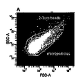
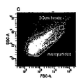
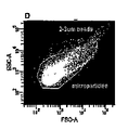
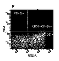
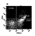
フルオレッセンイソチオシアネート(FITC)ラベルされた抗ヒトCD3及びPhycoerythrin(PE)ラベルされた抗ヒトCD42b抗体マーカー(BD Biosciences, San Jose, USA) (20 μl 抗体/ 100μl MP 試料)がMPパレットに添加され、室温にて、通常の振盪にて30分間培養された。フルオレッセンス活性化細胞選別(FACS)分析は、エジンバラ大学のFlow cytometry core facility, QMRIにおいてDiVaオプションを有するFACS VANTAGE(BD, San Jose, USA)により行われた。事象は、前方散乱強度及び後方散乱強度によりドットにて示されるように同定され、MPsとしてゲート制御され、1色又は2色の蛍光ヒストグラムとしてプロットされる。EMPsはCD31+CD42b-として定義され、PMPsはCD31+CD42b+として定義される。
(Flow cytometry analysis)
Fluorescene isothiocyanate (FITC) labeled anti-human CD3 and Phycoerythrin (PE) labeled anti-human CD42b antibody markers (BD Biosciences, San Jose, USA) (20 μl antibody / 100 μl MP sample) were added to the MP palette. Incubated at room temperature for 30 minutes with normal shaking. Fluorescence activated cell sorting (FACS) analysis was performed by FACS VANTAGE (BD, San Jose, USA) with the DiVa option at the Flow cytometry core facility, QMRI at the University of Edinburgh. Events are identified as indicated by dots by forward scatter intensity and back scatter intensity, gated as MPs, and plotted as one or two color fluorescence histograms. EMPs are defined as CD31 + CD42b- and PMPs are defined as CD31 + CD42b +.
(対象、尿試料集め及び処理)
6名のボランティア(23〜40才、(MREC倫理承認番号06/MRE00/67))は、尿試料を滅菌された50mlのプラスチック容器に提供した。夫々の試料に対しプロテアーゼインヒビターが添加された(1mMレオペプチン50:1)。パラセタモール過剰投与後4及び24時間後の患者からの尿試料が分析された。患者6名中3名において顕著な肝臓損傷が認定された24時間後に血清アラニン・トランスアミナーゼ(ALT)が上昇していた。試料は15000gにて15分間4℃で遠心され、尿沈渣をペレット化した。「15000gの上清」は、10000rpmにて1時間4℃で遠心され、低濃度のエキソソームペレットが得られた。10000rpmの上清はデキャントされ、付加的な「15000gの上清」と置き換えられ、再度、超遠心に供された。超遠心のステップは4〜6回繰返され、15000gの上清18〜24mlからエキソソームペレットが回収される。ペレットは、50μlの単離溶液(10 mM トリエタノールアミン/ 250 mM スクロース (pH 7.6))に再懸濁される。パラセタモールを過剰投与した患者からの尿が3mlのみであることから、15000g上清を10000rpmにて1時間超遠心して、得られる尿エキソソームを最大とするようにプロトコールを変更した(図1b参照)。
(Subject, urine sample collection and processing)
Six volunteers (23-40 years old, (MREC ethics approval number 06 / MRE00 / 67)) provided urine samples in a sterile 50 ml plastic container. Protease inhibitors were added to each sample (1 mM leopeptin 50: 1). Urine samples from
(ゲル電気泳動及びウェスタンブロット)
尿及びMPsのタンパク質の量は、BCA(登録商標)タンパク質アッセイキット(Pierce, USA)用いて、その説明書に従い決定された。20μgの通常の尿エキソソーム試料がウェル毎にロードされる。パラセタモール過剰投与試料の各ローディング量は、尿の濃度に違いによった尿のクレアチンにより標準化される。0.2MのDTT(Bio-Rad)を有するLaemmli Sample Bufferの均等量はタンパク質試料に添加され、かかる試料は95℃にて5分間加熱される。タンパク質試料は、12 % Tris-HClゲル(Bio-Rad)内を1D SDS/ポリアクリルアミドゲル電気泳動により、移動バッファー (25 mM Tris (Fisher Scientific), 192 mM グリシン及び0.1 % SDS)内にてゲル毎に25 mAで1時間、分子量マーカー(Bio-Rad)とともに流され、分離された。
(Gel electrophoresis and Western blot)
The amount of urine and MPs protein was determined using the BCA® protein assay kit (Pierce, USA) according to the instructions. 20 μg of normal urine exosome sample is loaded per well. Each loading of paracetamol overdose samples is normalized by urine creatine depending on urine concentration. An equal amount of Laemmli Sample Buffer with 0.2 M DTT (Bio-Rad) is added to the protein sample and the sample is heated at 95 ° C. for 5 minutes. Protein samples were gelled in 12% Tris-HCl gel (Bio-Rad) by 1D SDS / polyacrylamide gel electrophoresis in transfer buffer (25 mM Tris (Fisher Scientific), 192 mM glycine and 0.1% SDS). Each was run at 25 mA for 1 hour with molecular weight markers (Bio-Rad) and separated.
タンパク質は、冷却トランスファーバッファー(25 mM Tris, 192 mM グリシン及び20 % メタノール(BDH AnalR(登録商標)))を用いて200mAにて1時間ゲル内からポリビニルイヂン2フッ化膜(Invitrogen)へとトランスファーされる。TBST( 25 mM Tris- HCl, pH 8.0, 125 mM 塩化ナトリウム, 0.1 % Tween 20) 中にて5%のスキムドライミルク(Sainsbury's)中でブロッキングした後に、GATA 2 (1 : 200)及びc-Jun (1:200)に対するポリクローナル抗体とともに4℃にて一晩インキュベートされた。TBSTにて5分間3回洗浄した後に、膜は室温にてセイヨウワサビペルオキシダーゼ接合二次抗体(1 : 5000)とともに室温にて90分間インキュベートされた。ウェスタンブロットの全ての抗体はSanta Cruz Biotechnology, California, USAから得られた。ECLプラスウェスタンブロット検出システム(Amersham Biosciences, New Jersey, USA)及び高感応膜(Kodak)は抗体抗原反応を可視化するために使用された。 The protein was transferred from the gel to a polyvinylidin difluoride membrane (Invitrogen) at 200 mA for 1 hour using chilled transfer buffer (25 mM Tris, 192 mM glycine and 20% methanol (BDH Anal®)). Transferred. After blocking in 5% skim dry milk (Sainsbury's) in TBST (25 mM Tris-HCl, pH 8.0, 125 mM sodium chloride, 0.1% Tween 20), GATA 2 (1: 200) and c-Jun Incubated overnight at 4 ° C. with a polyclonal antibody against (1: 200). After washing 3 times with TBST for 5 minutes, the membrane was incubated at room temperature with horseradish peroxidase-conjugated secondary antibody (1: 5000) for 90 minutes at room temperature. All antibodies in the Western blot were obtained from Santa Cruz Biotechnology, California, USA. An ECL plus western blot detection system (Amersham Biosciences, New Jersey, USA) and a highly sensitive membrane (Kodak) were used to visualize the antibody-antigen reaction.
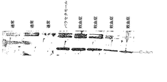
(タンパク質ゲル染色及びインゲルトリプシン消化)
タンパク質の体積(20μg)はゲルのウェル毎にロードされることから、ゲルは100μgのタンパク質を含み、前述したように流された。次いで、ゲルは、7%の氷酢酸及び40%のメタノールにより、1時間固定される。ゲルはコロイド状のクマシーブルーにより1時間染色され、10%氷酢酸及び25%のメタノールにより30秒間脱色されられる。ゲルは同一の溶液により脱色される前に、25%メタノールによりリンスされる。ゲルは(図6に示すように)スライスされ、また、更に小さな部分へとスライスされ、タンパク抽出のステップまで-80℃にて保存される。
(Protein gel staining and in-gel trypsin digestion)
Since the protein volume (20 μg) was loaded per well of gel, the gel contained 100 μg protein and was run as described above. The gel is then fixed with 7% glacial acetic acid and 40% methanol for 1 hour. The gel is stained with colloidal Coomassie blue for 1 hour and decolorized with 10% glacial acetic acid and 25% methanol for 30 seconds. The gel is rinsed with 25% methanol before being decolorized with the same solution. The gel is sliced (as shown in FIG. 6) and sliced into smaller portions and stored at −80 ° C. until the protein extraction step.
全てのインキュベーションは、連続した浸透条件にて室温(22℃)で行われる。ゲル断片は15分間蒸留水によりインキュベートされる。かかる水は取り除かれ、50%アセトニトリル(ACN)に置換され、15分間インキュベートされる。ACNインキュベーションステップは繰り返され、CANは取り除かれ、100mMの重炭酸アンモニウム(Ambic)と交換されて及び5分間インキュベートされる。ゲル断片に同量のACNが添加され、更に15分間インキュベートされる。水溶液は取り除かれ、ゲル断片を60℃にて高速減圧することより完全に脱水する。次いで、試料は10mM DTT、100mM Ambic存在下にて56℃で45分間インキュベートされる。水溶液は取り除かれ、5mMのヨードアセトアミド/100mMのAmbicと交換され30℃、室温、暗所でインキュベートされる。ゲル断片は洗われ(100mMAmbic)、それから15分間のACNによってインキュベートされた。ゲル断片は高速減圧により、完全に脱水される。1M塩化カルシウム及び1M Ambicバッファーの0.1μg/μlシークエンシング・グレード・トリプシン(Pierce, USA)はゲル断片に加えられ、45分間の4℃でインキュベートされた。水溶液は取り除かれ、トリプシンを含まない同一のバッファーにより交換され、37℃にて一晩インキュベートされる。上清は取り除かれ、4℃にて保存される。ゲル断片は15分間の25mMAmbicによってインキュベートされ、次いで、ACNが加えられ、更に15分間インキュベートされた。上清は取り除かれ、一晩の上清に加えられる。そして、ペプチドは5%の蟻酸及びACNにより溶出される。プールされた上清に10 mM DTTが添加され、最終濃度を1mM DTTとなるようにし、上清は急速減圧により乾燥させられる。試料は、The Chemistry Department at The King's Buildingsに送られ、LC-MS/MSにより分析される。 All incubations are performed at room temperature (22 ° C.) under continuous infiltration conditions. The gel piece is incubated with distilled water for 15 minutes. Such water is removed and replaced with 50% acetonitrile (ACN) and incubated for 15 minutes. The ACN incubation step is repeated, the CAN is removed, replaced with 100 mM ammonium bicarbonate (Ambic) and incubated for 5 minutes. The same amount of ACN is added to the gel pieces and incubated for an additional 15 minutes. The aqueous solution is removed, and the gel pieces are completely dehydrated by high-speed decompression at 60 ° C. The sample is then incubated for 45 minutes at 56 ° C. in the presence of 10 mM DTT, 100 mM Ambic. The aqueous solution is removed and replaced with 5 mM iodoacetamide / 100 mM Ambic and incubated at 30 ° C. in the dark at room temperature. The gel pieces were washed (100 mM Ambic) and then incubated with ACN for 15 minutes. The gel fragments are completely dehydrated by high speed vacuum. 0.1 μg / μl sequencing grade trypsin (Pierce, USA) in 1M calcium chloride and 1M Ambic buffer was added to the gel fragment and incubated at 4 ° C. for 45 minutes. The aqueous solution is removed and replaced with the same buffer without trypsin and incubated overnight at 37 ° C. The supernatant is removed and stored at 4 ° C. Gel fragments were incubated with 25 mM Ambic for 15 minutes, then ACN was added and incubated for an additional 15 minutes. The supernatant is removed and added to the overnight supernatant. The peptide is then eluted with 5% formic acid and ACN. 10 mM DTT is added to the pooled supernatant to a final concentration of 1 mM DTT and the supernatant is dried by rapid vacuum. Samples are sent to The Chemistry Department at The King's Buildings and analyzed by LC-MS / MS.
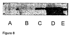
(CyPA検出)
APAPを過剰投与したRoyal Infirmary of Edinburghの患者から約50mlの尿が集められた。尿は集められ、CyPAレベルを測定する前に、−80℃にて保存される。
(CyPA detection)
Approximately 50 ml of urine was collected from a Royal Infirmary of Edinburgh patient overdose of APAP. Urine is collected and stored at −80 ° C. before measuring CyPA levels.
CyPAの尿中濃度は、前述したようにウェスタンブロット8により決定される。一次抗体は、1:150希釈したウサギ抗ヒト・サイクロフィリンA(USBiological、Swampscott, MA, USA)である。ゲルのレーン毎に10μlの尿がロードされた。 The urine concentration of CyPA is determined by Western blot 8 as described above. The primary antibody is rabbit anti-human cyclophilin A (USBiological, Swampscott, MA, USA) diluted 1: 150. 10 μl of urine was loaded for each lane of gel.
結果
(フローサイトメトリー分析)
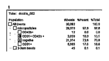
図2は、FACSを使用して選別された試料の側方散乱/前方散乱ドットプロットヒストグラムを示す。強調された2.5μmキャリブレーションビーズは、MPsの寸法を示し、MPsの全ての集団は、過去のMPs研究に基づき2.5μm寸法未満であった。EMPsは、図2(B及びF)に示すように、PMPsよりもわずかに寸法が小さく、図2(C及びD)に示すように、PMP集団の下方に位置している。単一染色したMPsは、集団の位置を同定するコントロールとして、CD31又はCD43bにより染色され、そして、両細胞表面マーカーの存在しない試料は、ネガティブコントロールとなり、非内皮細胞の血小板由来のMPsを提供する。大多数のMPsはCD31及びCD42bに対し陰性であり、これらは異なる起源及び細胞種に由来するものである。MPsの陰性のグループは、異なる集団を形成し、72.6%の試料を作り出した。EMPs及びPMPsはともに異なる寸法を有する粒子の異なる集団として同定される。パネルD及びHは、EMPs及びPMPsを選別するために二重染色した試料を示す。17%まで至る単一の陽性のCD43bMPs及びCD41+EMPsはないが、CD31+CD42bPMPsは全量の10.3%に至る。FITCラベルされたMPs量が少ないと、MPsは内皮細胞性をより起源とするものであり、CD31が多いとMPsはよりCD42+であり、血小板をより起源とするものである。
Results (flow cytometry analysis)
FIG. 2 shows a side scatter / forward scatter dot plot histogram of samples sorted using FACS. The highlighted 2.5 μm calibration beads showed MPs dimensions, and all populations of MPs were less than 2.5 μm dimensions based on previous MPs studies. EMPs are slightly smaller in size than PMPs, as shown in FIGS. 2 (B and F), and are located below the PMP population, as shown in FIGS. 2 (C and D). Single-stained MPs are stained with CD31 or CD43b as controls to identify the location of the population, and samples without both cell surface markers serve as negative controls and provide MPs from non-endothelial cell platelets . The majority of MPs are negative for CD31 and CD42b, which are derived from different sources and cell types. The negative group of MPs formed a different population, producing 72.6% of the samples. Both EMPs and PMPs are identified as different populations of particles with different dimensions. Panels D and H show samples that were double stained to sort EMPs and PMPs. There is no single positive CD43bMPs and CD41 + EMPs up to 17%, but CD31 + CD42bPMPs accounts for 10.3% of the total. When the amount of MPTC-labeled MPs is small, MPs is more derived from endotheliality, and when there is more CD31, MPs is more CD42 + and more originated from platelets.
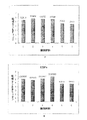
6名の健康な個人から単離されたEMPs及びPMPsの量は、個人差が大きかった。図3は、FACSにより単離されたEMPs(A)及びPMPs(B)のlog10グラフを示す。単離されたEMPsの平均数は1507567(SEM837934)であり、単離されたPMPsの平均数は285704(SEM88864)である。 The amount of EMPs and PMPs isolated from 6 healthy individuals varied widely among individuals. FIG. 3 shows log10 graphs of EMPs (A) and PMPs (B) isolated by FACS. The average number of isolated EMPs is 1507567 (SEM837934) and the average number of isolated PMPs is 285704 (SEM88864).
(MPsにおけるGATA-2タンパク質発現)
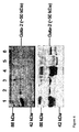
転写因子GATA-2が微粒子中に存在するか否かをウェスタンブロットにより調査した。EMP及びPMP集団が選別された後、試料はMPsをペレット化するために超遠心に供された。超遠心した後のペレット及び上清の両方は保持され、GATA-2の存在を検出するためのウェスタンブロットが行われた。図4はGATA-2が上清に存在しており、一つの上清のみにおいてGATA-2が存在していたことを示しているウェスタンブロットの結果である。個人1からのEMP及びPMP試料(夫々レーン1及び4)は、その他のMP試料に比べGATA-2のレベルが大きかった。このことは、個人(夫々5516089及び380548;図3の試料4)から単離されたEMPs及びPMPsと整合している。
(GATA-2 protein expression in MPs)
Whether the transcription factor GATA-2 was present in the microparticles was investigated by Western blot. After the EMP and PMP populations were sorted, the samples were subjected to ultracentrifugation to pellet the MPs. Both the pellet and supernatant after ultracentrifugation were retained and Western blot was performed to detect the presence of GATA-2. FIG. 4 shows the results of Western blotting showing that GATA-2 was present in the supernatant and GATA-2 was present in only one supernatant. The EMP and PMP samples from individual 1 (
(尿エキソソームレベル及びc-Jun発現)
パラセタモールを過剰投与し、器官損傷した、又はしていない患者の尿中のエキソソームタンパク質の濃度は、通常の尿と比較された。6名の通常の尿エキソソーム試料では、平均が3.54±1.22μgタンパク質/ml尿であった。パラセタモールの過剰投与4時間後に、器官損傷を示さなかった3名の患者からプールされた試料は、通常の健康な個人に比べ、約7倍のタンパク質量の増大が見られた。また、パラセタモールの過剰投与4時間後に、器官損傷を示した3名の患者からプールされた試料は、プールされた試料中のエキソソームタンパク質は37倍までの増大が見られた。過剰投与の24時間後には、器官損傷を有する患者及び有さない患者の双方において、プールされた尿試料内のエキソソームタンパク質量が同様のレベルまで減少していた(通常のレベルに対し1.5〜2.0倍)。
(Urine exosome level and c-Jun expression)
The concentration of exosomal proteins in the urine of patients with or without paracetamol overdose and organ damage was compared to normal urine. In the six normal urine exosome samples, the average was 3.54 ± 1.22 μg protein / ml urine. Samples pooled from 3 patients who did not show organ damage after 4 hours of paracetamol overdose showed an approximately 7-fold increase in protein content compared to normal healthy individuals. Samples pooled from 3 patients who showed
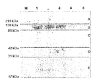
タンパク質c-Junが通常の尿試料内にて検出し得るかどうか、そしてパラセタモールを過剰投与した患者においてc-Junレベルが変化するかどうかを調べるために、ウェスタンブロットが行われた。個人の通常の試料では、c-Junは、正確な分子量〜78kDaにて検出された。また、尿中微粒子におけるC-Junタンパク質の濃度は、人におけるパラセタモールにより湯道された肝臓損傷によって増大する(図7)。 Western blots were performed to see if the protein c-Jun could be detected in normal urine samples and if c-Jun levels changed in patients overdose with paracetamol. In a normal individual sample, c-Jun was detected at an accurate molecular weight of ~ 78 kDa. Also, the concentration of C-Jun protein in urine microparticles increases due to liver damage caused by paracetamol in humans (Fig. 7).
(通常の尿におけるエキソソームタンパク質)
通常の尿試料のパイロットプロテオミクスを行うために、〜100μgの通常のエキソソームがゲルにロードされ、コロイド状のクマシーブルーにより染色され、タンパク質の存在を調べた。図6は、染色されたゲルを示し、エキソソーム試料中に異なる寸法のタンパク質が存在することの証拠を提供するものである。また、最も豊富なタンパク質のバンドはゲルの部分Bであり、THPであると推定されるものである。ドット線は、LC-MS/MSによるプロテオミクス分析に供される異なる試料を準備するためのゲルが切られた位置を示す。
(Exosomal protein in normal urine)
To perform pilot proteomics of normal urine samples, ˜100 μg of normal exosomes were loaded onto the gel and stained with colloidal Coomassie blue to check for the presence of protein. FIG. 6 shows a stained gel and provides evidence that there are proteins of different sizes in the exosome sample. The most abundant protein band is the gel part B, which is presumed to be THP. The dotted line indicates the position where the gel was cut to prepare different samples for proteomic analysis by LC-MS / MS.
(バイオマーカーとしての候補源であるEMPs)
この実験は、血液中のMPs及び尿中のエキソソームがバイオマーカーとしての候補源として使用し得るかを調査するためのものである。主たる成果は、EMPs(内皮性微粒子)がフローサイトメトリーにより単離されるが、(図3Aに示すように)単離された微粒子数は個人間差が大きく、標準的なタンパク質分析では検出することが難しいレベルのタンパク質量しか得られない。MPsの分離は、時間を要するものであり、臨床試験には効果的な方法ではない。In vitroの実験系を用いて、内皮細胞を培養して、かかる培養細胞からMPsを単離することが有効な解決策となり得るものである。これは他のグループにより成功している(Combes et al., 1999)。候補タンパク質は、そのレベルにより決定される。可能性のある候補タンパク質がin vitro研究により見つかったら、器官が機能不となっている患者において、そのタンパク質の発現レベルが変化しているかを確認する。
(EMPs, candidate sources for biomarkers)
This experiment is to investigate whether MPs in blood and exosomes in urine can be used as candidate sources as biomarkers. The main result is that EMPs (endothelial microparticles) are isolated by flow cytometry, but the number of isolated microparticles varies widely between individuals (as shown in Figure 3A) and can be detected by standard protein analysis. However, only a difficult level of protein can be obtained. The separation of MPs is time consuming and is not an effective method for clinical trials. Using an in vitro experimental system, culturing endothelial cells and isolating MPs from such cultured cells can be an effective solution. This has been more successful by other groups (Combes et al., 1999). Candidate proteins are determined by their level. Once a potential candidate protein is found by in vitro studies, it is checked to see if the protein expression level has changed in patients with organ failure.
(EMPs及びPMPsはともにGATA-2発現を示す)
GATA-2は、GATA-2転写因子ファミリーに属する。GATA-2は、造血現象において重要な役割を果たす。原始的造血細胞は高レベルでGATA-2を発現するが、血液細胞の異なるタイプへの細胞の成熟の過程で、そのレベルは減少する(Tsai et al, 1994)。大人の造血細胞では、GATA-2が発現し、内皮特異的な遺伝子の転写調節に重要である(Zhang et al., 1995)。GATA-2には複数の燐酸化された形態があり(Towatari et al., 1995)、図4は、EMPsとPMPsの双方に複数の形態のGATA-2があることを示している。しかし、これらの結果は、in vitroにてヒト臍の静脈内皮細胞(HUVECS)を使用して、GATA-2の同じ形が検出されることができるかどうか調べて立証される。MPs中のGATA-2をGATA-2の転写因子であるとして観察することは興味深く、このことは転写因子をin vivoでの遺伝子発現のバイオマーカーとして使用し得る可能性を示唆するものである。
(EMPs and PMPs both show GATA-2 expression)
GATA-2 belongs to the GATA-2 transcription factor family. GATA-2 plays an important role in the hematopoietic phenomenon. Primitive hematopoietic cells express GATA-2 at high levels, but that level decreases during the maturation of cells into different types of blood cells (Tsai et al, 1994). In adult hematopoietic cells, GATA-2 is expressed and is important for endothelium-specific gene transcriptional regulation (Zhang et al., 1995). There are multiple phosphorylated forms of GATA-2 (Towatari et al., 1995), and FIG. 4 shows that there are multiple forms of GATA-2 in both EMPs and PMPs. However, these results are verified by examining whether the same form of GATA-2 can be detected using human umbilical vein endothelial cells (HUVECS) in vitro. It is interesting to observe GATA-2 in MPs as a transcription factor for GATA-2, suggesting the possibility that transcription factors can be used as biomarkers for gene expression in vivo.
(パラセタモール過剰投与はエキソソームタンパク尿症を増大させる)
図5Aは、パラセタモールの過剰投与が、過剰投与4時間後に、エキソソームタンパク尿症を増大させることを示すものである。タンパク尿症は、器官損傷を有する患者において特に多い(〜37回、通常比較された)。過剰投与した患者の超遠心した上清は、同一の分析にて同様の尿試料を用いた場合にも、多様性を示した(データ不図示)。このことは、尿内におけるパラセタモール(アセトアミノフェン)代謝産物の存在によるものであり、かかる代謝産物はこの実験系にて使用されるBSA分析等のタンパク質分析に干渉するものである(Marshall and Williams, 1991)。かかるタンパク尿症が発症する正確な理由は解明されていない。エキソソームが腎臓から放出され、循環MPsが糸球体を経て尿に入り込んでいるかどうかも知られていない。また、薬が腎臓または血管に対する効果を有するかどうか、は決定されないままである。薬の過剰投与24時間後にはタンパク質レベルが通常に対し1.5倍まで落ちることは興味深いことであるが、このことは、肝損傷の減少又は血液のパラセタモール濃度の減少させるための臨床介入によって説明することができる。
(Paracetamol overdose increases exosomal proteinuria)
FIG. 5A shows that paracetamol overdose increases
(薬誘導による器官損傷のマーカーとしてのCyPA)
APAP誘導器官損傷を有する患者の尿において、CyPA濃度が増大していた。図8は、健康な対象(A)、僅かな肝臓損傷(B)、中程度の肝臓損傷(C)、重症な肝臓損傷(D)及び重症な肝臓及び腎臓損傷(E)を有する患者からの尿ウェスタンブロットを示す。
(CyPA as a marker of drug-induced organ damage)
CyPA concentrations were increased in the urine of patients with APAP-induced organ damage. Figure 8 shows the results from healthy subjects (A), patients with slight liver damage (B), moderate liver damage (C), severe liver damage (D) and severe liver and kidney damage (E). A urine western blot is shown.
図9はAPAP誘導された肝臓損傷を有する又は有さない患者からの尿を比較したものである。病院に消化された全ての患者はAPAP過剰投与の履歴を有し、血液中にAPAP検出可能である。肝臓損傷を有する患者に関し、損傷を有さない患者の年齢及び性別を揃えるものとした。CyPAは、肝臓損傷を受けた患者において、損傷を有さない患者の年齢及び性別を揃えた患者よりも増大していた。 FIG. 9 compares urine from patients with or without APAP-induced liver damage. All patients digested into the hospital have a history of APAP overdose and are detectable in the blood. For patients with liver injury, the age and gender of patients without injury were aligned. CyPA was increased in patients with liver damage than in patients with the same age and gender as those without damage.
(急性肝臓損傷におけるc-Junの役割)
パラセタモールの過剰投与は、イギリス及び米国(Larson et al., 2005)では急性の肝機能不全の主たる原因となっている。パラセタモール代謝と肝臓損傷とをリンクさせる経路は未だ完全に明らかとなっていない。パラセタモールの過剰投与初期には、サイトカイン及びケモカインは、その損傷(Ishida et al., 2002)及び回復(Hogaboam et al., 1999)に関係するものである。
(Role of c-Jun in acute liver injury)
Paracetamol overdose is a major cause of acute liver dysfunction in the United Kingdom and the United States (Larson et al., 2005). The pathway that links paracetamol metabolism to liver damage is not yet fully understood. In the early days of paracetamol overdose, cytokines and chemokines are implicated in their damage (Ishida et al., 2002) and recovery (Hogaboam et al., 1999).
転写因子c-Junは、AP-1転写因子の主たる構成要素であり(Bohmann et al, 1987)、細胞ストレスを含む多様な幅の刺激がAP-1活性を誘導することが知られている。細胞をサイトカイン及び環境ストレスにさらすことにより、ストレス活性化タンパク質として知られているJNKグループの活性化が起こる。アポトーシスもまた細胞ストレスであるとされ、そのことから、c-Junはアポトーシスに応答して活性化又は上方制御されると考えられるまた、JNK及びC-Junが、肝臓損傷を媒介していることを示す(Liu et al., 2002)。 The transcription factor c-Jun is a major component of the AP-1 transcription factor (Bohmann et al, 1987), and it is known that a wide range of stimuli including cellular stress induces AP-1 activity. Exposure of cells to cytokines and environmental stress results in activation of the JNK group known as stress-activated proteins. Apoptosis is also considered to be a cellular stress, which suggests that c-Jun is activated or up-regulated in response to apoptosis and that JNK and C-Jun mediate liver damage (Liu et al., 2002).
このことから、この研究では、パラセタモールを過剰投与した、肝臓損傷を有する又は有さない患者のエキソソーム試料は、c-Junの存在について検証された。コントロールとして、c-Junは、通常の健康な個人から検出され、c-Junのレベルが過剰投与した患者特には、より高レベルのアポトーシスし、c-Jun活性化の増大している肝臓損傷にて増大することが推定される。 Thus, in this study, exosome samples from patients over-administered with paracetamol and with or without liver damage were verified for the presence of c-Jun. As a control, c-Jun is detected in normal healthy individuals and patients with overdose levels of c-Jun, especially in patients with higher levels of apoptosis and increased c-Jun activation Is estimated to increase.
CyPAは、APAP過剰投与により肝臓損傷を受けた患者の尿内にて増大する。従って、CyPAはAPAP過剰投与により誘導された肝臓損傷に対する候補バイオマーカーとなる。CyPAは尿濃度の標準化が要求される程度に増大し、尿クレアチニンの補正を行うこと無く、測定を容易とする強力な候補バイオマーカーとするものである。結果から、CyPAは尿10μlでも容易に検出することができる。 CyPA increases in the urine of patients suffering from liver damage due to APAP overdose. Thus, CyPA is a candidate biomarker for liver damage induced by APAP overdose. CyPA increases to the extent that standardization of urine concentration is required, and is a powerful candidate biomarker that facilitates measurement without correcting urine creatinine. From the results, CyPA can be easily detected even with 10 μl of urine.
CyPAは、多様な疾患を有する人間及び動物モデル5,9〜11において、細胞外液に放出される細胞内タンパク質である。APAP毒性によるCyPA放出の機構は不明であるが、APAPが顕著な細胞ネクローシスを引き起こすことから、損傷した細胞からの漏洩が最大の供給源であると予想している。細胞外のCyPAが炎症誘発性であることから、APAP毒性にて見られる器官損傷を媒介している可能性がある。 CyPA is an intracellular protein that is released into the extracellular fluid in human and animal models 5 , 9-11 with various diseases. Although the mechanism of CyPA release due to APAP toxicity is unclear, we expect that leakage from damaged cells is the greatest source as APAP causes significant cellular necrosis. Since extracellular CyPA is pro-inflammatory, it may mediate the organ damage seen in APAP toxicity.
CyPAは、現行のマーカーがまだ充分でない場合に、器官損傷を検出し得ることから、貴重な臨床バイオマーカーとすることができる。 CyPA can be a valuable clinical biomarker because it can detect organ damage when current markers are not yet sufficient.
(疾患バイオマーカー同定のためのツールとしてのプロテオミクス)
疾患の研究におけるプロテオミクスは、有用なツールであり、疾患を有する又は有さない個人の試料内のタンパク質を同定するために使用することができる。しかし、プロテオミクスに問題が無いわけではない。この研究では、LC-MS/MS分析に供されたゲルスライス試料の5つのうちの3つにおいて、コロイド状のクマシーブルーを多く含み過ぎているために、検出することができなかった。また、プロテオミクスにて使用される装置は、感度が極めて高く、過剰な染色により損傷をうける場合がある。分析できた2つの資料では、よくあることではあるが、何らペプチド断片が得られなかった。これらの問題は、修正され、プロトコールを僅かに変えながら実験は繰り返された。プロテオミクスの主たる問題は、非常に大量のタンパク質(例えば血漿のアルブミン及び尿のTHP)が、プロテオミクス分析に供される前に試料から取り除かれない限りは、より少ないタンパク質の検出の妨げることにある。THPは、試料に還元剤を処理することにより、そして、試料を再度超遠心することにより、取り除くことができる。THPは上清に残り、少ない量のタンパク質についてペレットを分析に供することができる(Pisitkun et al., 2004)。アルブミンは、SwellGel Albumin Removal kit from Pierce, Rockford, USAなどの商業的に利用可能なキットを用いて取り除くことができる。或いは、血清アルブミンを取り除くために、トリクロロ酢酸及びアセトンを使用することができる(Chen et al., 2005)。試料のプロテオミクス分析の前に、これら大量のタンパク質を取り除くことにより、体の健康状態及び一部の疾患の病因について有益な情報を与えるかもしれない低い濃度のタンパク質を同定し得る機会が増える。MPs及びエキソソームについて有効なタンパク質の同定が報告された。Smalleyら(2007)は、血小板MPs内ではなく、血漿MPs内にて21種のタンパク質を同定することに成功し、Zhouら(2006)は、急性の腎臓損傷に対するバイオマーカー候補を同定した。
(Proteomics as a tool for disease biomarker identification)
Proteomics in disease research is a useful tool and can be used to identify proteins in samples of individuals with or without disease. However, proteomics is not without problems. In this study, 3 out of 5 gel slice samples subjected to LC-MS / MS analysis could not be detected because they contained too much colloidal Coomassie blue. Also, devices used in proteomics are very sensitive and may be damaged by excessive staining. The two materials that could be analyzed, as is often the case, did not yield any peptide fragments. These problems were corrected and the experiment was repeated with slight changes in the protocol. The main problem with proteomics is that they prevent detection of less protein unless very large amounts of protein (eg, plasma albumin and urine THP) are removed from the sample before being subjected to proteomic analysis. THP can be removed by treating the sample with a reducing agent and re-centrifuging the sample again. THP remains in the supernatant and the pellet can be subjected to analysis for small amounts of protein (Pisitkun et al., 2004). Albumin can be removed using commercially available kits such as the SwellGel Albumin Removal kit from Pierce, Rockford, USA. Alternatively, trichloroacetic acid and acetone can be used to remove serum albumin (Chen et al., 2005). Removal of these large amounts of protein prior to proteomic analysis of the sample increases the chances of identifying low concentrations of protein that may provide useful information about the health of the body and the pathogenesis of some diseases. The identification of effective proteins for MPs and exosomes has been reported. Smalley et al. (2007) succeeded in identifying 21 proteins in plasma MPs but not in platelet MPs, and Zhou et al. (2006) identified biomarker candidates for acute kidney injury.
以上の結果、この研究から、血液からのMPs及び尿内のエキソソームは、疾患に対するバイオマーカーの候補となり得ることを発見した。エキソソームを得るためには、少量の尿が必要であり、このことは疾患を診断する際に臨床上適切である。病気の発症及び進行を示す固有の能力を有する臨床マーカーは、広範囲の疾患に対し羅病率及び死亡率を減らすことができ、プロテオミクスは、疾患の臨床診断に対し重要な役割を果たすものである。 As a result, it was discovered from this study that MPs from blood and exosomes in urine can be candidates for biomarkers for disease. To obtain exosomes, a small amount of urine is required, which is clinically relevant when diagnosing disease. Clinical markers with unique ability to indicate disease onset and progression can reduce morbidity and mortality for a wide range of diseases, and proteomics plays an important role in clinical diagnosis of disease .
参考文献
Amabile, N., Guerin, A.P., Leroyer, A., Mallat, Z., Nguyen, C, Boddaert, J., London, G.M., Tedgui, A. and Boulanger, CM. (2005). Circulating endothelial microparticles are associated with vascular dysfunction in patients with end-stage renal failure. J Am Soc Nephrol 16: 3381-88.
Anderson, N.L. and Anderson, N.G. (2002). The human plasma proteome: history, character, and disagnostic prospects. MoI Cell Proteomics 1: 845-67.
Banfi, C, Brioschi, M., Wait, R., Begum, S., Gianazza, E., Pirillo, A., Mussoni, L. and Tremoli, E. (2005). Proteome of endothelial cell-derived procoagulant microparticles. Proteomics 5: 4443-4455.
Bateman DN. Poisoning: focus on paracetamol. Royal College of Physiciansof Edinburgh CME 2007; http://www.rcpe.ac.uk/fellows/CME/clin-pharmacol/bateman/bateman-2.html
Bohmann, D., Bos, T.J., Admon, A., Nishimura, T., Vogt, P.K. and Tjian, R. (1987). Human proto-oncogene c-Jun encodes a DNA binding protein with structural and functional properties of transcription factor API. Science 238: 1386-1392.
Brodsky, S. V., Zhang F., Nasjletti A. and Goligorsky M.S.(2004). Endothelium- derived microparticles impair endothelial function in vitro. Am J Physiol Heart Circ Physiol 286: 1910-15.
Chen, Y. Y. , Lin, S., Yeh, Y., Hsiao, H., Wu, C, Chen,S. and Wang, A.H. (2005). A modified protein precipitation procedure for efficient removal of albumin from serum. Electrophoresis 26: 2117-27.
Combes, V., Simon, A.C., Grau, G.E., Arnoux, D., Camoin, L., Sabatier, F., Mutin, M., Sanmarco, M., Sampol, J. and Dignat-George, F. (1999). In vitro generation of endothelial microparticles and possible prothrombotic activity in patients with lupus anticoagulant. J Clin Invest 104: 93-102.
George, J.N., Thoi, L.L., McManus, L.M. and Reimann, T.A. (1982). Isolation of human platelet microparticles from plasma and serum. Blood 60: 834-840.
Henderson, N.C., Pollock, K., Frew, J., Mackinnon, A.C., Flavell, R.A., Davis, RJ., Sethi, T. and Simpson, K. J. (2006). Critical role of c-jun (NH2) terminal kinase in paracetamol-induced acute liver failure. Gut (E-pub ahead of print).
Hewitt, S.M., Dear, J. W., Star, R.A. (2004). Discovery of protein biomarkers for renal diseases. JAm Soc Nephrol 15: 1677-89.
Hogaboam, CM., Simpson, K.J., Chensue, S.W., Steinhauser, MX., Lukacs, N. W., Gauldie, J., Strieter, R.M. and Kunkel, S.L. (1999). Macrophage inflammatory protein-2 gene therapy attenuates adenovirus-and paracetamol-mediated hepatic injury. Gene Ther 6: 573-584.
Ishida, Y., Kondo, T., Ohshima, T., Fujiwara, H., Iwakura, Y. and Mukaida, N. (2002). A pivotal involvement of IFN-gamma in the pathogenesis of paracetamol- induced acute liver injury. FASEB J 16:1227-1236.
Johnstone, R.M., Adam, M., Hammond, J.R., Orr, L. and Turbide, C. (1987). Vesicle formation during reticulocyte maturation. Association of plasma membrane activities with released vesicles (exosomes). J Biol Chem 262: 9412-20.
Larson, A.M., Poison, J., Fontana, R.J., Davern, T.J., Lalani, E., Hynan, L.S., Reisch, J.S., Schiodt, F. V., Ostapowicz, G., Shakil, A.O. and Lee, W.M. (2005). Acetaminophen-induced acute liver failure: results of a United Statesmulticenter, prospective study. Hepatology 42 (6): 1364-72.
Liu, H., Lo, CR. and Czaja, M.J. (2002). NF-kappa B inhibition sensitizes hepatocytes to TNF-induced apoptosis through a sustained activation of JNK and c-Jun. Hepatology 35:772-778.
Marshall, T. and Williams, K.M. (1991). Drug interference in the Bradford and 2,2'- bicinchoninic acid protein assays. Anal Biochem 198: 352-4. Pisitkun, T., Shen, R.F. and Knepper, M. A. (2004). Identification and proteomic profiling of exosomes in human urine. Proc Natl Acad Sci USA 101: 13368-73.
Preston, R.A., Jy, W., Jimenez, J.J., Mauro, L.M., Horstman, L.L., Valle, M., Aime, G. and Ahn, Y. (2003). Effects of severe hypertension on endothelial and platelet microparticles. Hypertension 41: 211-217.
Reilly TP, Bourdi M, Brady JN, Pise-Masison CA, Radonovich MF, George JW, et al. Expression profiling of acetaminophen liver toxicity in mice using microarray technology. Biochem Biophys Res Commun 2001;282(l):321-8
Smalley, D.M., Root, K.E., Cho, H., Ross, M.M. and Ley, K. (2007). Proteomic discovery of 21 proteins expressed in human plasma-derived but not platelet-derived microparticles. Thromb Haemost 97: 67-80.
Towatari, M., May, G.E., Marais, R., Perkins, G.R., Marshall, CJ., Cowley, S. and Enver, T (1995). Regulation of GATA-2 Phosphorylation by Mitogen-activated Protein Kinase and Interleukin-3. J Biol Chem 270: 4101-4107.
Tsai, F. Y., Keller, G., Kuo, F.C., Weiss, M., Chen, J., Rosenblatt, M., Alt, F.W. and Orkin, S.H. (1994). An early haematopoietic defect in mice lacking the transcription factor GAT A-2. Nature 371: 221-226.
Wolf, P. (1967). The nature and significance of platelet products in human plasma. Br J Haematol 3: 269-88.
Zhang, R., Min, W. and Sessa, W.C. (1995). Functional analysis of the human endothelial nitric oxide synthase promoter: SpI and GATA factors are necessary for basal transcription in endothelial cells. J Biol Chem. 270: 15320-15326.
Zhou, H., Yuen, P.S.T., Pisitkun, T., Gonzales, P.A., Yasuda, H., Dear, J. W., Gross, P., Knepper, M. A. and Star, R.A. (2006). Collection, storage, preservation and normalisation of human urinary exosomes for biomarker discovery. Kidney International 69: 1471-76.
Zhou, H., Pisitkun, T., Aponte, A., Yuen, P.S,T., Hoffert, J.D., Yasuda, H., Hu, X., Chawla, L., Shen, R.F., Knepper, M.A. and Star, R.A. (2006). Exosomal Fetuin-A identified by proteomics: A novel urinary biomarker for detecting acute kidney injury. Kidney International 70: 1847-1857.
References
Amabile, N., Guerin, AP, Leroyer, A., Mallat, Z., Nguyen, C, Boddaert, J., London, GM, Tedgui, A. and Boulanger, CM. (2005). Circulating endothelial microparticles are associated with vascular dysfunction in patients with end-stage renal failure.J Am Soc Nephrol 16: 3381-88.
Anderson, NL and Anderson, NG (2002) .The human plasma proteome: history, character, and disagnostic prospects.MoI Cell Proteomics 1: 845-67.
Banfi, C, Brioschi, M., Wait, R., Begum, S., Gianazza, E., Pirillo, A., Mussoni, L. and Tremoli, E. (2005). Proteome of endothelial cell-derived procoagulant microparticles Proteomics 5: 4443-4455.
Bateman DN. Poisoning: focus on paracetamol. Royal College of Physiciansof Edinburgh CME 2007; http://www.rcpe.ac.uk/fellows/CME/clin-pharmacol/bateman/bateman-2.html
Bohmann, D., Bos, TJ, Admon, A., Nishimura, T., Vogt, PK and Tjian, R. (1987) .Human proto-oncogene c-Jun encodes a DNA binding protein with structural and functional properties of transcription factor API. Science 238: 1386-1392.
Brodsky, SV, Zhang F., Nasjletti A. and Goligorsky MS (2004). Endothelium-derived microparticles impair endothelial function in vitro. Am J Physiol Heart Circ Physiol 286: 1910-15.
Chen, YY, Lin, S., Yeh, Y., Hsiao, H., Wu, C, Chen, S. And Wang, AH (2005) .A modified protein precipitation procedure for efficient removal of albumin from serum.Electrophoresis 26 : 2117-27.
Combes, V., Simon, AC, Grau, GE, Arnoux, D., Camoin, L., Sabatier, F., Mutin, M., Sanmarco, M., Sampol, J. and Dignat-George, F. ( 1999) .In vitro generation of endothelial microparticles and possible prothrombotic activity in patients with lupus anticoagulant.J Clin Invest 104: 93-102.
George, JN, Thoi, LL, McManus, LM and Reimann, TA (1982) .Isolation of human platelet microparticles from plasma and serum.Blood 60: 834-840.
Henderson, NC, Pollock, K., Frew, J., Mackinnon, AC, Flavell, RA, Davis, RJ., Sethi, T. and Simpson, KJ (2006). Critical role of c-jun (NH2) terminal kinase in paracetamol-induced acute liver failure.Gut (E-pub ahead of print).
Hewitt, SM, Dear, JW, Star, RA (2004). Discovery of protein biomarkers for renal diseases.JAm Soc Nephrol 15: 1677-89.
Hogaboam, CM., Simpson, KJ, Chensue, SW, Steinhauser, MX., Lukacs, NW, Gauldie, J., Strieter, RM and Kunkel, SL (1999). Macrophage inflammatory protein-2 gene therapy attenuates adenovirus-and paracetamol -mediated hepatic injury. Gene Ther 6: 573-584.
Ishida, Y., Kondo, T., Ohshima, T., Fujiwara, H., Iwakura, Y. and Mukaida, N. (2002). A pivotal involvement of IFN-gamma in the pathogenesis of paracetamol- induced acute liver injury FASEB J 16: 1227-1236.
Johnstone, RM, Adam, M., Hammond, JR, Orr, L. and Turbide, C. (1987) .Vesicle formation during reticulocyte maturation. Association of plasma membrane activities with released vesicles (exosomes). J Biol Chem 262: 9412 -20.
Larson, AM, Poison, J., Fontana, RJ, Davern, TJ, Lalani, E., Hynan, LS, Reisch, JS, Schiodt, FV, Ostapowicz, G., Shakil, AO and Lee, WM (2005). Acetaminophen-induced acute liver failure: results of a United States multicenter, prospective study.Hepatology 42 (6): 1364-72.
Liu, H., Lo, CR. And Czaja, MJ (2002) .NF-kappa B inhibition sensitizes hepatocytes to TNF-induced apoptosis through a sustained activation of JNK and c-Jun. Hepatology 35: 772-778.
Marshall, T. and Williams, KM (1991) .Drug interference in the Bradford and 2,2'- bicinchoninic acid protein assays.Analy Biochem 198: 352-4.Pisitkun, T., Shen, RF and Knepper, MA (2004 ) .Identification and proteomic profiling of exosomes in human urine.Proc Natl Acad Sci USA 101: 13368-73.
Preston, RA, Jy, W., Jimenez, JJ, Mauro, LM, Horstman, LL, Valle, M., Aime, G. and Ahn, Y. (2003). Effects of severe hypertension on endothelial and platelet microparticles. 41: 211-217.
Reilly TP, Bourdi M, Brady JN, Pise-Masison CA, Radonovich MF, George JW, et al.Expression profiling of acetaminophen liver toxicity in mice using microarray technology.Biochem Biophys Res Commun 2001; 282 (l): 321-8
Smalley, DM, Root, KE, Cho, H., Ross, MM and Ley, K. (2007) .Proteomic discovery of 21 proteins expressed in human plasma-derived but not platelet-derived microparticles.Thromb Haemost 97: 67-80 .
Towatari, M., May, GE, Marais, R., Perkins, GR, Marshall, CJ., Cowley, S. and Enver, T (1995). Regulation of GATA-2 Phosphorylation by Mitogen-activated Protein Kinase and Interleukin- 3.J Biol Chem 270: 4101-4107.
Tsai, FY, Keller, G., Kuo, FC, Weiss, M., Chen, J., Rosenblatt, M., Alt, FW and Orkin, SH (1994) .An early haematopoietic defect in mice lacking the transcription factor GAT A-2. Nature 371: 221-226.
Wolf, P. (1967) .The nature and significance of platelet products in human plasma.Br J Haematol 3: 269-88.
Zhang, R., Min, W. and Sessa, WC (1995). Functional analysis of the human endothelial nitric oxide synthase promoter: SpI and GATA factors are necessary for basal transcription in endothelial cells.J Biol Chem. 270: 15320-15326 .
Zhou, H., Yuen, PST, Pisitkun, T., Gonzales, PA, Yasuda, H., Dear, JW, Gross, P., Knepper, MA and Star, RA (2006). Collection, storage, preservation and normalisation of human urinary exosomes for biomarker discovery.Kidney International 69: 1471-76.
Zhou, H., Pisitkun, T., Aponte, A., Yuen, PS, T., Hoffert, JD, Yasuda, H., Hu, X., Chawla, L., Shen, RF, Knepper, MA and Star , RA (2006) .Exosomal Fetuin-A identified by proteomics: A novel urinary biomarker for detecting acute kidney injury.Kidney International 70: 1847-1857.
(記載中にて番号で示された)追加参考文献
1. Waring WS, Stephen AF, Robinson OD, Dow MA, Pettie JM. Lower incidence of anaphylactoid reactions to N-acetylcysteine in patients with high acetaminophen concentrations after overdose. Clinical Toxicology 2008; 46(6):496 - 500.
2. Colgan J, Asmal M, Yu B, Luban J. Cyclophilin A-Deficient Mice Are Resistant to Immunosuppression by Cyclosporine. J Immunol 2005; 174(10):6030-6038.
3. Takahashi N, Hayano T, Suzuki M. Peptidyl-prolyl cis-trans isomerase is the cyclosporin A-binding protein cyclophilin. Nature 1989; 337(6206):473-5.
4. Luban J, Bossolt KL, Franke EK, Kalpana GV, Goff SP. Human immunodeficiency virus type 1 Gag protein binds to cyclophilins A and B. Cell 1993 ;73(6): 1067-78.
5. Arora K, Gwinn WM, Bower MA, Watson A, Okwumabua I, MacDonald HR, et al. Extracellular cyclophilins contribute to the regulation of inflammatory responses. J Immunol 2005; 175(l):517-22.
6. Yurchenko V, Constant S, Bukrinsky M. Dealing with the family: CD 147 interactions with cyclophilins. Immunology 2006; 117(3):301-9.
7. Dear JW, Leelahavanichkul A, Aponte A, Hu X, Constant SL, Hewitt SM, et al. Liver proteomics for therapeutic drug discovery: Inhibition of the cyclophilin receptor CD 147 attenuates sepsis-induced acute renal failure*. CrH Care Pled 2007; 35(10):2319-2329.
8. Holly MK, Dear JW, Hu X, Schechter AN, Gladwin MT, Hewitt SM, et al. Biomarker and drug-target discovery using proteomics in a new rat model of sepsis- induced acute renal failure. Kidney Int 2006; 70(3):496-506.
9. Gwinn WM, Damsker JM, Falahati R, Okwumabua I, Kelly- Welch A, Keegan AD, et al. Novel approach to inhibit asthma-mediated lung inflammation using anti-CD 147 intervention. J Immunol 2006; 177(7):4870-9.
10. Billich A, Winkler G, Aschauer H, Rot A, Peichl P. Presence of Cyclophilin A in Synovial Fluids of Patients with Rheumatoid Arthritis. J. Exp. Med. 1997; 185(5):975- 980.
11. Tegeder I, Schumacher A, John S, Geiger H, Geisslinger G, Bang H, et al. Elevated serum cyclophilin levels in patients with severe sepsis. J Clin Immunol 1997;17(5):380-6.
Additional references (indicated by number in the description)
1. Waring WS, Stephen AF, Robinson OD, Dow MA, Pettie JM. Lower incidence of anaphylactoid reactions to N-acetylcysteine in patients with high acetaminophen concentrations after overdose. Clinical Toxicology 2008; 46 (6): 496-500.
2. Colgan J, Asmal M, Yu B, Luban J. Cyclophilin A-Deficient Mice Are Resistant to Immunosuppression by Cyclosporine. J Immunol 2005; 174 (10): 6030-6038.
3.Takahashi N, Hayano T, Suzuki M. Peptidyl-prolyl cis-trans isomerase is the cyclosporin A-binding protein cyclophilin.Nature 1989; 337 (6206): 473-5.
4. Luban J, Bossolt KL, Franke EK, Kalpana GV, Goff SP.Human
5. Arora K, Gwinn WM, Bower MA, Watson A, Okwumabua I, MacDonald HR, et al. Extracellular cyclophilins contribute to the regulation of inflammatory responses. J Immunol 2005; 175 (l): 517-22.
6. Yurchenko V, Constant S, Bukrinsky M. Dealing with the family: CD 147 interactions with cyclophilins.Immunology 2006; 117 (3): 301-9.
7. Dear JW, Leelahavanichkul A, Aponte A, Hu X, Constant SL, Hewitt SM, et al. Liver proteomics for therapeutic drug discovery: Inhibition of the cyclophilin receptor CD 147 attenuates sepsis-induced acute renal failure *. CrH Care Pled 2007 ; 35 (10): 2319-2329.
8. Holly MK, Dear JW, Hu X, Schechter AN, Gladwin MT, Hewitt SM, et al. Biomarker and drug-target discovery using proteomics in a new rat model of sepsis- induced acute renal failure.Kidney Int 2006; 70 ( 3): 496-506.
9. Gwinn WM, Damsker JM, Falahati R, Okwumabua I, Kelly- Welch A, Keegan AD, et al. Novel approach to inhibit asthma-mediated lung inflammation using anti-CD 147 intervention. J Immunol 2006; 177 (7): 4870-9.
10. Billich A, Winkler G, Aschauer H, Rot A, Peichl P. Presence of Cyclophilin A in Synovial Fluids of Patients with Rheumatoid Arthritis. J. Exp. Med. 1997; 185 (5): 975-980.
11. Tegeder I, Schumacher A, John S, Geiger H, Geisslinger G, Bang H, et al. Elevated serum cyclophilin levels in patients with severe sepsis.J Clin Immunol 1997; 17 (5): 380-6.
Claims (16)
(c)患者から試料を準備するステップと、
(d)該試料中のタンパク質レベルを同定するステップと、を含み、
タンパク質レベルの増大は、患者が開発薬により誘導された器官損傷を有する或いはそのリスクを有することを示すことを特徴とする同定方法。 A method of identifying whether a patient has or is at risk for organ damage induced by a developed drug comprising:
(C) preparing a sample from a patient;
(D) identifying the protein level in the sample;
An identification method characterized in that an increase in protein level indicates that the patient has or is at risk for organ damage induced by a developed drug.
(a)患者から試料を準備するステップと、
(b)該試料中のタンパク質レベルを同定するステップと、を含み、
タンパク質レベルの増大は、患者がパラセタモールにより誘導された肝臓損傷を有する或いはその発症リスクを有することを示すことを特徴とする同定方法。 A method of identifying whether a patient has or is at risk for organ damage induced by a developed drug comprising:
(A) preparing a sample from a patient;
(B) identifying the protein level in the sample;
An identification method characterized in that an increase in protein level indicates that the patient has or is at risk of developing liver damage induced by paracetamol.
The identification method according to claim 15, wherein the protein is c-Jun N-terminal kinase (JNK) and / or cyclophilin A (CyPA).
Applications Claiming Priority (2)
| Application Number | Priority Date | Filing Date | Title |
|---|---|---|---|
| GBGB0716804.0A GB0716804D0 (en) | 2007-08-29 | 2007-08-29 | Identifying organ damage |
| PCT/GB2008/002947 WO2009027703A2 (en) | 2007-08-29 | 2008-08-29 | Identifying organ damage |
Publications (2)
| Publication Number | Publication Date |
|---|---|
| JP2010538252A true JP2010538252A (en) | 2010-12-09 |
| JP2010538252A5 JP2010538252A5 (en) | 2011-10-13 |
Family
ID=38616940
Family Applications (1)
| Application Number | Title | Priority Date | Filing Date |
|---|---|---|---|
| JP2010522447A Pending JP2010538252A (en) | 2007-08-29 | 2008-08-29 | How to identify organ damage |
Country Status (5)
| Country | Link |
|---|---|
| US (1) | US8110371B2 (en) |
| EP (1) | EP2183601A2 (en) |
| JP (1) | JP2010538252A (en) |
| GB (1) | GB0716804D0 (en) |
| WO (1) | WO2009027703A2 (en) |
Cited By (1)
| Publication number | Priority date | Publication date | Assignee | Title |
|---|---|---|---|---|
| JP2019215342A (en) * | 2018-06-07 | 2019-12-19 | 株式会社Lsiメディエンス | Separation method and analysis method for microvesicle from human urine |
Families Citing this family (6)
| Publication number | Priority date | Publication date | Assignee | Title |
|---|---|---|---|---|
| EP2350320A4 (en) | 2008-11-12 | 2012-11-14 | Caris Life Sciences Luxembourg Holdings | Methods and systems of using exosomes for determining phenotypes |
| WO2011000874A1 (en) * | 2009-07-01 | 2011-01-06 | INSERM (Institut National de la Santé et de la Recherche Médicale) | Methods for predicting cardiovascular mortality risk |
| CA2791905A1 (en) | 2010-03-01 | 2011-09-09 | Caris Life Sciences Luxembourg Holdings, S.A.R.L. | Biomarkers for theranostics |
| JP2013526852A (en) | 2010-04-06 | 2013-06-27 | カリス ライフ サイエンシズ ルクセンブルク ホールディングス | Circulating biomarkers for disease |
| ITNA20110012A1 (en) * | 2011-03-14 | 2012-09-15 | Amal Raj Delfin Albert | INSULATION AND PURIFICATION PROCEDURE OF URINARY EXOSOMES FOR RESEARCH OF PROTEIN BIOMARCERS |
| CN105606811A (en) * | 2016-02-01 | 2016-05-25 | 中南大学 | Diagnostic kit for nasopharynx cancer |
Citations (1)
| Publication number | Priority date | Publication date | Assignee | Title |
|---|---|---|---|---|
| JPH10507184A (en) * | 1994-10-12 | 1998-07-14 | ペーアーツェト アルツナイミッテル−エントヴィックルングスゲゼルシャフト ミット ベシュレンクテル ハフツング | Diagnostic evaluation, course control and therapeutic agents for shock status in humans |
Family Cites Families (4)
| Publication number | Priority date | Publication date | Assignee | Title |
|---|---|---|---|---|
| US6719969B1 (en) * | 1999-08-09 | 2004-04-13 | The Regents Of The University Of Michigan | Treatment of liver disease and injury with CXC chemokines |
| EP1405072A2 (en) * | 2000-12-29 | 2004-04-07 | Oxford GlycoSciences (UK) Limited | Proteins, genes and their use for diagnosis and treatment of kidney response |
| US7469185B2 (en) * | 2002-02-04 | 2008-12-23 | Ocimum Biosolutions, Inc. | Primary rat hepatocyte toxicity modeling |
| WO2007127848A1 (en) * | 2006-04-26 | 2007-11-08 | University Of Louisville Research Foundation, Inc | Isolation of membrane vesicles from biological fluids and methods of using same |
-
2007
- 2007-08-29 GB GBGB0716804.0A patent/GB0716804D0/en not_active Ceased
-
2008
- 2008-08-29 EP EP08788492A patent/EP2183601A2/en not_active Withdrawn
- 2008-08-29 US US12/675,479 patent/US8110371B2/en not_active Expired - Fee Related
- 2008-08-29 JP JP2010522447A patent/JP2010538252A/en active Pending
- 2008-08-29 WO PCT/GB2008/002947 patent/WO2009027703A2/en active Application Filing
Patent Citations (1)
| Publication number | Priority date | Publication date | Assignee | Title |
|---|---|---|---|---|
| JPH10507184A (en) * | 1994-10-12 | 1998-07-14 | ペーアーツェト アルツナイミッテル−エントヴィックルングスゲゼルシャフト ミット ベシュレンクテル ハフツング | Diagnostic evaluation, course control and therapeutic agents for shock status in humans |
Non-Patent Citations (1)
| Title |
|---|
| JPN6012037899; Gut Vol.56, 2006, p982-990 * |
Cited By (2)
| Publication number | Priority date | Publication date | Assignee | Title |
|---|---|---|---|---|
| JP2019215342A (en) * | 2018-06-07 | 2019-12-19 | 株式会社Lsiメディエンス | Separation method and analysis method for microvesicle from human urine |
| JP7444386B2 (en) | 2018-06-07 | 2024-03-06 | 株式会社Lsiメディエンス | Method for isolating and analyzing microvesicles from human urine |
Also Published As
| Publication number | Publication date |
|---|---|
| US20110039276A1 (en) | 2011-02-17 |
| US8110371B2 (en) | 2012-02-07 |
| WO2009027703A2 (en) | 2009-03-05 |
| GB0716804D0 (en) | 2007-10-10 |
| WO2009027703A3 (en) | 2009-06-25 |
| EP2183601A2 (en) | 2010-05-12 |
Similar Documents
| Publication | Publication Date | Title |
|---|---|---|
| US8741580B2 (en) | Markers of renal transplant rejection and renal damage | |
| US8889366B2 (en) | Marker for graft failure and mortality | |
| Jung et al. | Quantification of ATP7B protein in dried blood spots by peptide immuno-SRM as a potential screen for Wilson’s disease | |
| US8110371B2 (en) | Identifying organ damage | |
| Obert et al. | A review of specific biomarkers of chronic renal injury and their potential application in nonclinical safety assessment studies | |
| JP6841765B2 (en) | Methods for Predicting the Risk of Developing Chronic Kidney Disease | |
| JP2012524883A (en) | WNT1 as a kidney injury biomarker | |
| US20210116462A1 (en) | Methods and Compositions for the Diagnosis and Treatment of Kawasaki Disease | |
| Tomar et al. | New biomarkers in brain trauma | |
| US20140370531A1 (en) | Method of diagnosing mild traumatic brain injury | |
| JPWO2008056480A1 (en) | Method for diagnosing fatty liver disease, diagnostic device, diagnostic program, diagnostic agent and screening method for therapeutic agent for fatty liver disease | |
| Khan et al. | Proteomics in idiopathic pulmonary fibrosis: the quest for biomarkers | |
| Brown et al. | Protein and microRNA biomarkers from lavage, urine, and serum in military personnel evaluated for dyspnea | |
| Sigdel et al. | Assessment of circulating protein signatures for kidney transplantation in pediatric recipients | |
| US20160244835A1 (en) | Urinary neuropilin-1 (nrp-1) as a prognostic marker for nephritis and lupus nephritis | |
| WO2017126514A1 (en) | Method for detecting non-alcoholic steatohepatitis | |
| JP7271442B2 (en) | Methods of diagnosing or monitoring renal function or aiding in diagnosing renal dysfunction | |
| US8969015B2 (en) | Methods and kits for diagnosing Sjögren's syndrome | |
| WO2018066409A1 (en) | Inspection method enabling specific diagnosis of pathological state of diabetic nephropathy at early stage | |
| US20200064344A1 (en) | Use of serum 2-cysteine peroxiredoxins (2-cys-prdx) as biomarkers of chronic kidney diseases | |
| JP2009513960A (en) | In vitro method for diagnosing neurodegenerative diseases | |
| US20170030929A1 (en) | Diagnosis of chronic kidney disease by quantitative analysis of post-translational modifications of plasma proteins | |
| JP5010220B2 (en) | Understanding the pathology of patients undergoing hematopoietic stem cell transplantation | |
| Donia et al. | Study for the relationship between lupus nephritis and Anti-C1q antibodies | |
| WO2021245413A1 (en) | Methods of determining cancer |
Legal Events
| Date | Code | Title | Description |
|---|---|---|---|
| RD01 | Notification of change of attorney |
Free format text: JAPANESE INTERMEDIATE CODE: A7426 Effective date: 20101228 |
|
| A521 | Written amendment |
Free format text: JAPANESE INTERMEDIATE CODE: A821 Effective date: 20101228 |
|
| A521 | Written amendment |
Free format text: JAPANESE INTERMEDIATE CODE: A523 Effective date: 20110202 |
|
| A521 | Written amendment |
Free format text: JAPANESE INTERMEDIATE CODE: A821 Effective date: 20110202 |
|
| A521 | Written amendment |
Free format text: JAPANESE INTERMEDIATE CODE: A523 Effective date: 20110826 |
|
| A621 | Written request for application examination |
Free format text: JAPANESE INTERMEDIATE CODE: A621 Effective date: 20110826 |
|
| A977 | Report on retrieval |
Free format text: JAPANESE INTERMEDIATE CODE: A971007 Effective date: 20120718 |
|
| A131 | Notification of reasons for refusal |
Free format text: JAPANESE INTERMEDIATE CODE: A131 Effective date: 20120724 |
|
| A601 | Written request for extension of time |
Free format text: JAPANESE INTERMEDIATE CODE: A601 Effective date: 20121024 |
|
| A602 | Written permission of extension of time |
Free format text: JAPANESE INTERMEDIATE CODE: A602 Effective date: 20121031 |
|
| A601 | Written request for extension of time |
Free format text: JAPANESE INTERMEDIATE CODE: A601 Effective date: 20121126 |
|
| A602 | Written permission of extension of time |
Free format text: JAPANESE INTERMEDIATE CODE: A602 Effective date: 20121203 |
|
| A02 | Decision of refusal |
Free format text: JAPANESE INTERMEDIATE CODE: A02 Effective date: 20130312 |