JP2010507780A - Detection of influenza B virus - Google Patents
Detection of influenza B virus Download PDFInfo
- Publication number
- JP2010507780A JP2010507780A JP2009533290A JP2009533290A JP2010507780A JP 2010507780 A JP2010507780 A JP 2010507780A JP 2009533290 A JP2009533290 A JP 2009533290A JP 2009533290 A JP2009533290 A JP 2009533290A JP 2010507780 A JP2010507780 A JP 2010507780A
- Authority
- JP
- Japan
- Prior art keywords
- influenza
- virus
- protein
- antibody
- sample
- Prior art date
- Legal status (The legal status is an assumption and is not a legal conclusion. Google has not performed a legal analysis and makes no representation as to the accuracy of the status listed.)
- Withdrawn
Links
- 241000713196 Influenza B virus Species 0.000 title claims abstract description 30
- 238000001514 detection method Methods 0.000 title description 28
- 101710128560 Initiator protein NS1 Proteins 0.000 claims abstract description 121
- 101710144127 Non-structural protein 1 Proteins 0.000 claims abstract description 121
- 208000037798 influenza B Diseases 0.000 claims abstract description 83
- 238000000034 method Methods 0.000 claims abstract description 41
- 239000013610 patient sample Substances 0.000 claims abstract description 15
- 241000700605 Viruses Species 0.000 claims abstract description 14
- 230000028327 secretion Effects 0.000 claims abstract description 8
- 241000712431 Influenza A virus Species 0.000 claims abstract description 7
- 210000004369 blood Anatomy 0.000 claims abstract description 6
- 239000008280 blood Substances 0.000 claims abstract description 6
- 210000004072 lung Anatomy 0.000 claims abstract description 5
- 210000000416 exudates and transudate Anatomy 0.000 claims abstract description 4
- 210000003296 saliva Anatomy 0.000 claims abstract description 3
- 239000000523 sample Substances 0.000 claims description 46
- 239000003795 chemical substances by application Substances 0.000 claims description 29
- 230000009870 specific binding Effects 0.000 claims description 9
- 108020004999 messenger RNA Proteins 0.000 claims description 6
- 241000713297 Influenza C virus Species 0.000 claims description 5
- 229940079593 drug Drugs 0.000 claims description 3
- 239000003814 drug Substances 0.000 claims description 3
- 239000007787 solid Substances 0.000 claims description 3
- 108050000838 Influenza A virus NS1 proteins Proteins 0.000 claims description 2
- 238000003556 assay Methods 0.000 description 25
- 238000012360 testing method Methods 0.000 description 24
- 206010022000 influenza Diseases 0.000 description 21
- 239000000427 antigen Substances 0.000 description 20
- 108091007433 antigens Proteins 0.000 description 20
- 102000036639 antigens Human genes 0.000 description 20
- 239000003153 chemical reaction reagent Substances 0.000 description 20
- 239000012472 biological sample Substances 0.000 description 18
- 102000004169 proteins and genes Human genes 0.000 description 18
- 230000027455 binding Effects 0.000 description 17
- 208000037797 influenza A Diseases 0.000 description 17
- 108090000623 proteins and genes Proteins 0.000 description 17
- 239000007790 solid phase Substances 0.000 description 15
- 239000000243 solution Substances 0.000 description 12
- 239000000872 buffer Substances 0.000 description 11
- 208000037799 influenza C Diseases 0.000 description 8
- 108091023037 Aptamer Proteins 0.000 description 7
- 239000011230 binding agent Substances 0.000 description 7
- 239000012530 fluid Substances 0.000 description 7
- 239000012634 fragment Substances 0.000 description 7
- 238000002965 ELISA Methods 0.000 description 6
- 102000004190 Enzymes Human genes 0.000 description 6
- 108090000790 Enzymes Proteins 0.000 description 6
- 239000012491 analyte Substances 0.000 description 6
- 210000004027 cell Anatomy 0.000 description 6
- PCHJSUWPFVWCPO-UHFFFAOYSA-N gold Chemical compound [Au] PCHJSUWPFVWCPO-UHFFFAOYSA-N 0.000 description 6
- 239000004005 microsphere Substances 0.000 description 6
- 239000013642 negative control Substances 0.000 description 6
- 108020004707 nucleic acids Proteins 0.000 description 6
- 102000039446 nucleic acids Human genes 0.000 description 6
- 150000007523 nucleic acids Chemical class 0.000 description 6
- 108091032973 (ribonucleotides)n+m Proteins 0.000 description 5
- 239000011324 bead Substances 0.000 description 5
- 239000012528 membrane Substances 0.000 description 5
- 210000003097 mucus Anatomy 0.000 description 5
- -1 polyethylene Polymers 0.000 description 5
- 210000001519 tissue Anatomy 0.000 description 5
- 241000712461 unidentified influenza virus Species 0.000 description 5
- 102000011931 Nucleoproteins Human genes 0.000 description 4
- 108010061100 Nucleoproteins Proteins 0.000 description 4
- 206010036790 Productive cough Diseases 0.000 description 4
- 239000012620 biological material Substances 0.000 description 4
- 108020001507 fusion proteins Proteins 0.000 description 4
- 102000037865 fusion proteins Human genes 0.000 description 4
- 230000003053 immunization Effects 0.000 description 4
- 239000000203 mixture Substances 0.000 description 4
- 230000001717 pathogenic effect Effects 0.000 description 4
- 238000000926 separation method Methods 0.000 description 4
- 210000003802 sputum Anatomy 0.000 description 4
- 208000024794 sputum Diseases 0.000 description 4
- 239000000126 substance Substances 0.000 description 4
- 210000002845 virion Anatomy 0.000 description 4
- 230000003612 virological effect Effects 0.000 description 4
- 241000282412 Homo Species 0.000 description 3
- 239000004793 Polystyrene Substances 0.000 description 3
- VYPSYNLAJGMNEJ-UHFFFAOYSA-N Silicium dioxide Chemical compound O=[Si]=O VYPSYNLAJGMNEJ-UHFFFAOYSA-N 0.000 description 3
- 230000009471 action Effects 0.000 description 3
- 239000000654 additive Substances 0.000 description 3
- 230000003321 amplification Effects 0.000 description 3
- 238000003491 array Methods 0.000 description 3
- 238000010790 dilution Methods 0.000 description 3
- 239000012895 dilution Substances 0.000 description 3
- 239000007850 fluorescent dye Substances 0.000 description 3
- 235000013305 food Nutrition 0.000 description 3
- 239000011521 glass Substances 0.000 description 3
- 239000010931 gold Substances 0.000 description 3
- 229910052737 gold Inorganic materials 0.000 description 3
- 208000015181 infectious disease Diseases 0.000 description 3
- 229920000126 latex Polymers 0.000 description 3
- 239000004816 latex Substances 0.000 description 3
- 238000003199 nucleic acid amplification method Methods 0.000 description 3
- 229920002223 polystyrene Polymers 0.000 description 3
- 239000013641 positive control Substances 0.000 description 3
- 238000012340 reverse transcriptase PCR Methods 0.000 description 3
- 238000005406 washing Methods 0.000 description 3
- XLYOFNOQVPJJNP-UHFFFAOYSA-N water Substances O XLYOFNOQVPJJNP-UHFFFAOYSA-N 0.000 description 3
- 238000001262 western blot Methods 0.000 description 3
- YBJHBAHKTGYVGT-ZKWXMUAHSA-N (+)-Biotin Chemical compound N1C(=O)N[C@@H]2[C@H](CCCCC(=O)O)SC[C@@H]21 YBJHBAHKTGYVGT-ZKWXMUAHSA-N 0.000 description 2
- 241000271566 Aves Species 0.000 description 2
- 241000283707 Capra Species 0.000 description 2
- 108020004414 DNA Proteins 0.000 description 2
- 238000012286 ELISA Assay Methods 0.000 description 2
- 108060003951 Immunoglobulin Proteins 0.000 description 2
- 239000000020 Nitrocellulose Substances 0.000 description 2
- 241000712464 Orthomyxoviridae Species 0.000 description 2
- 239000004743 Polypropylene Substances 0.000 description 2
- 241000282887 Suidae Species 0.000 description 2
- 238000002820 assay format Methods 0.000 description 2
- 210000001124 body fluid Anatomy 0.000 description 2
- 239000010839 body fluid Substances 0.000 description 2
- 230000001413 cellular effect Effects 0.000 description 2
- 238000006243 chemical reaction Methods 0.000 description 2
- 230000000295 complement effect Effects 0.000 description 2
- 238000003745 diagnosis Methods 0.000 description 2
- 238000002405 diagnostic procedure Methods 0.000 description 2
- 238000010494 dissociation reaction Methods 0.000 description 2
- 230000005593 dissociations Effects 0.000 description 2
- 239000012636 effector Substances 0.000 description 2
- 238000002474 experimental method Methods 0.000 description 2
- 238000009396 hybridization Methods 0.000 description 2
- 238000002649 immunization Methods 0.000 description 2
- 238000003018 immunoassay Methods 0.000 description 2
- 102000018358 immunoglobulin Human genes 0.000 description 2
- 238000013198 immunometric assay Methods 0.000 description 2
- 238000000338 in vitro Methods 0.000 description 2
- 230000003993 interaction Effects 0.000 description 2
- 238000012125 lateral flow test Methods 0.000 description 2
- 239000007788 liquid Substances 0.000 description 2
- 210000001989 nasopharynx Anatomy 0.000 description 2
- 229920001220 nitrocellulos Polymers 0.000 description 2
- 239000002245 particle Substances 0.000 description 2
- 244000052769 pathogen Species 0.000 description 2
- 229920001155 polypropylene Polymers 0.000 description 2
- 102000004196 processed proteins & peptides Human genes 0.000 description 2
- 108090000765 processed proteins & peptides Proteins 0.000 description 2
- 230000002685 pulmonary effect Effects 0.000 description 2
- 230000035945 sensitivity Effects 0.000 description 2
- 239000011550 stock solution Substances 0.000 description 2
- UCSJYZPVAKXKNQ-HZYVHMACSA-N streptomycin Chemical compound CN[C@H]1[C@H](O)[C@@H](O)[C@H](CO)O[C@H]1O[C@@H]1[C@](C=O)(O)[C@H](C)O[C@H]1O[C@@H]1[C@@H](NC(N)=N)[C@H](O)[C@@H](NC(N)=N)[C@H](O)[C@H]1O UCSJYZPVAKXKNQ-HZYVHMACSA-N 0.000 description 2
- 239000006163 transport media Substances 0.000 description 2
- 210000002700 urine Anatomy 0.000 description 2
- 230000009385 viral infection Effects 0.000 description 2
- 102000040650 (ribonucleotides)n+m Human genes 0.000 description 1
- NIXOWILDQLNWCW-UHFFFAOYSA-M Acrylate Chemical compound [O-]C(=O)C=C NIXOWILDQLNWCW-UHFFFAOYSA-M 0.000 description 1
- 229920000936 Agarose Polymers 0.000 description 1
- 102000002260 Alkaline Phosphatase Human genes 0.000 description 1
- 108020004774 Alkaline Phosphatase Proteins 0.000 description 1
- 102000053642 Catalytic RNA Human genes 0.000 description 1
- 108090000994 Catalytic RNA Proteins 0.000 description 1
- 101710094648 Coat protein Proteins 0.000 description 1
- 108091026890 Coding region Proteins 0.000 description 1
- 229920002307 Dextran Polymers 0.000 description 1
- 102000002322 Egg Proteins Human genes 0.000 description 1
- 108010000912 Egg Proteins Proteins 0.000 description 1
- 241000283086 Equidae Species 0.000 description 1
- 229930182566 Gentamicin Natural products 0.000 description 1
- CEAZRRDELHUEMR-URQXQFDESA-N Gentamicin Chemical compound O1[C@H](C(C)NC)CC[C@@H](N)[C@H]1O[C@H]1[C@H](O)[C@@H](O[C@@H]2[C@@H]([C@@H](NC)[C@@](C)(O)CO2)O)[C@H](N)C[C@@H]1N CEAZRRDELHUEMR-URQXQFDESA-N 0.000 description 1
- 108090000288 Glycoproteins Proteins 0.000 description 1
- 102000003886 Glycoproteins Human genes 0.000 description 1
- 108060003393 Granulin Proteins 0.000 description 1
- 108010043121 Green Fluorescent Proteins Proteins 0.000 description 1
- 102000004144 Green Fluorescent Proteins Human genes 0.000 description 1
- 101710154606 Hemagglutinin Proteins 0.000 description 1
- 108010001336 Horseradish Peroxidase Proteins 0.000 description 1
- 102000012745 Immunoglobulin Subunits Human genes 0.000 description 1
- 108010079585 Immunoglobulin Subunits Proteins 0.000 description 1
- 102100034343 Integrase Human genes 0.000 description 1
- 102000014150 Interferons Human genes 0.000 description 1
- 108010050904 Interferons Proteins 0.000 description 1
- 241000124008 Mammalia Species 0.000 description 1
- CERQOIWHTDAKMF-UHFFFAOYSA-M Methacrylate Chemical compound CC(=C)C([O-])=O CERQOIWHTDAKMF-UHFFFAOYSA-M 0.000 description 1
- 241000699670 Mus sp. Species 0.000 description 1
- 241000282339 Mustela Species 0.000 description 1
- 102000005348 Neuraminidase Human genes 0.000 description 1
- 108010006232 Neuraminidase Proteins 0.000 description 1
- 101710198202 Non-virion protein Proteins 0.000 description 1
- 108020004711 Nucleic Acid Probes Proteins 0.000 description 1
- 108091005461 Nucleic proteins Proteins 0.000 description 1
- 229920002292 Nylon 6 Polymers 0.000 description 1
- 229920002302 Nylon 6,6 Polymers 0.000 description 1
- 108091034117 Oligonucleotide Proteins 0.000 description 1
- 101710093908 Outer capsid protein VP4 Proteins 0.000 description 1
- 101710135467 Outer capsid protein sigma-1 Proteins 0.000 description 1
- 229930182555 Penicillin Natural products 0.000 description 1
- JGSARLDLIJGVTE-MBNYWOFBSA-N Penicillin G Chemical compound N([C@H]1[C@H]2SC([C@@H](N2C1=O)C(O)=O)(C)C)C(=O)CC1=CC=CC=C1 JGSARLDLIJGVTE-MBNYWOFBSA-N 0.000 description 1
- OAICVXFJPJFONN-UHFFFAOYSA-N Phosphorus Chemical group [P] OAICVXFJPJFONN-UHFFFAOYSA-N 0.000 description 1
- 206010035226 Plasma cell myeloma Diseases 0.000 description 1
- 241000276498 Pollachius virens Species 0.000 description 1
- 239000004952 Polyamide Substances 0.000 description 1
- 239000004698 Polyethylene Substances 0.000 description 1
- 229940079156 Proteasome inhibitor Drugs 0.000 description 1
- 101710176177 Protein A56 Proteins 0.000 description 1
- 108010092799 RNA-directed DNA polymerase Proteins 0.000 description 1
- 108020004511 Recombinant DNA Proteins 0.000 description 1
- 108010090804 Streptavidin Proteins 0.000 description 1
- BZHJMEDXRYGGRV-UHFFFAOYSA-N Vinyl chloride Chemical compound ClC=C BZHJMEDXRYGGRV-UHFFFAOYSA-N 0.000 description 1
- 108020000999 Viral RNA Proteins 0.000 description 1
- 208000036142 Viral infection Diseases 0.000 description 1
- JLCPHMBAVCMARE-UHFFFAOYSA-N [3-[[3-[[3-[[3-[[3-[[3-[[3-[[3-[[3-[[3-[[3-[[5-(2-amino-6-oxo-1H-purin-9-yl)-3-[[3-[[3-[[3-[[3-[[3-[[5-(2-amino-6-oxo-1H-purin-9-yl)-3-[[5-(2-amino-6-oxo-1H-purin-9-yl)-3-hydroxyoxolan-2-yl]methoxy-hydroxyphosphoryl]oxyoxolan-2-yl]methoxy-hydroxyphosphoryl]oxy-5-(5-methyl-2,4-dioxopyrimidin-1-yl)oxolan-2-yl]methoxy-hydroxyphosphoryl]oxy-5-(6-aminopurin-9-yl)oxolan-2-yl]methoxy-hydroxyphosphoryl]oxy-5-(6-aminopurin-9-yl)oxolan-2-yl]methoxy-hydroxyphosphoryl]oxy-5-(6-aminopurin-9-yl)oxolan-2-yl]methoxy-hydroxyphosphoryl]oxy-5-(6-aminopurin-9-yl)oxolan-2-yl]methoxy-hydroxyphosphoryl]oxyoxolan-2-yl]methoxy-hydroxyphosphoryl]oxy-5-(5-methyl-2,4-dioxopyrimidin-1-yl)oxolan-2-yl]methoxy-hydroxyphosphoryl]oxy-5-(4-amino-2-oxopyrimidin-1-yl)oxolan-2-yl]methoxy-hydroxyphosphoryl]oxy-5-(5-methyl-2,4-dioxopyrimidin-1-yl)oxolan-2-yl]methoxy-hydroxyphosphoryl]oxy-5-(5-methyl-2,4-dioxopyrimidin-1-yl)oxolan-2-yl]methoxy-hydroxyphosphoryl]oxy-5-(6-aminopurin-9-yl)oxolan-2-yl]methoxy-hydroxyphosphoryl]oxy-5-(6-aminopurin-9-yl)oxolan-2-yl]methoxy-hydroxyphosphoryl]oxy-5-(4-amino-2-oxopyrimidin-1-yl)oxolan-2-yl]methoxy-hydroxyphosphoryl]oxy-5-(4-amino-2-oxopyrimidin-1-yl)oxolan-2-yl]methoxy-hydroxyphosphoryl]oxy-5-(4-amino-2-oxopyrimidin-1-yl)oxolan-2-yl]methoxy-hydroxyphosphoryl]oxy-5-(6-aminopurin-9-yl)oxolan-2-yl]methoxy-hydroxyphosphoryl]oxy-5-(4-amino-2-oxopyrimidin-1-yl)oxolan-2-yl]methyl [5-(6-aminopurin-9-yl)-2-(hydroxymethyl)oxolan-3-yl] hydrogen phosphate Polymers Cc1cn(C2CC(OP(O)(=O)OCC3OC(CC3OP(O)(=O)OCC3OC(CC3O)n3cnc4c3nc(N)[nH]c4=O)n3cnc4c3nc(N)[nH]c4=O)C(COP(O)(=O)OC3CC(OC3COP(O)(=O)OC3CC(OC3COP(O)(=O)OC3CC(OC3COP(O)(=O)OC3CC(OC3COP(O)(=O)OC3CC(OC3COP(O)(=O)OC3CC(OC3COP(O)(=O)OC3CC(OC3COP(O)(=O)OC3CC(OC3COP(O)(=O)OC3CC(OC3COP(O)(=O)OC3CC(OC3COP(O)(=O)OC3CC(OC3COP(O)(=O)OC3CC(OC3COP(O)(=O)OC3CC(OC3COP(O)(=O)OC3CC(OC3COP(O)(=O)OC3CC(OC3COP(O)(=O)OC3CC(OC3COP(O)(=O)OC3CC(OC3CO)n3cnc4c(N)ncnc34)n3ccc(N)nc3=O)n3cnc4c(N)ncnc34)n3ccc(N)nc3=O)n3ccc(N)nc3=O)n3ccc(N)nc3=O)n3cnc4c(N)ncnc34)n3cnc4c(N)ncnc34)n3cc(C)c(=O)[nH]c3=O)n3cc(C)c(=O)[nH]c3=O)n3ccc(N)nc3=O)n3cc(C)c(=O)[nH]c3=O)n3cnc4c3nc(N)[nH]c4=O)n3cnc4c(N)ncnc34)n3cnc4c(N)ncnc34)n3cnc4c(N)ncnc34)n3cnc4c(N)ncnc34)O2)c(=O)[nH]c1=O JLCPHMBAVCMARE-UHFFFAOYSA-N 0.000 description 1
- 230000000996 additive effect Effects 0.000 description 1
- 230000004520 agglutination Effects 0.000 description 1
- 210000004712 air sac Anatomy 0.000 description 1
- 230000003281 allosteric effect Effects 0.000 description 1
- 125000003275 alpha amino acid group Chemical group 0.000 description 1
- 210000004381 amniotic fluid Anatomy 0.000 description 1
- 239000003242 anti bacterial agent Substances 0.000 description 1
- 229940088710 antibiotic agent Drugs 0.000 description 1
- 230000000890 antigenic effect Effects 0.000 description 1
- 238000013459 approach Methods 0.000 description 1
- 239000013060 biological fluid Substances 0.000 description 1
- 238000001574 biopsy Methods 0.000 description 1
- 229960002685 biotin Drugs 0.000 description 1
- 235000020958 biotin Nutrition 0.000 description 1
- 239000011616 biotin Substances 0.000 description 1
- 210000005013 brain tissue Anatomy 0.000 description 1
- 238000011088 calibration curve Methods 0.000 description 1
- 239000002775 capsule Substances 0.000 description 1
- 229920002678 cellulose Polymers 0.000 description 1
- 239000001913 cellulose Substances 0.000 description 1
- 238000005119 centrifugation Methods 0.000 description 1
- 210000001175 cerebrospinal fluid Anatomy 0.000 description 1
- 239000007795 chemical reaction product Substances 0.000 description 1
- 238000010367 cloning Methods 0.000 description 1
- 238000012875 competitive assay Methods 0.000 description 1
- 230000002860 competitive effect Effects 0.000 description 1
- 210000000795 conjunctiva Anatomy 0.000 description 1
- 229920001577 copolymer Polymers 0.000 description 1
- 239000003599 detergent Substances 0.000 description 1
- 238000009792 diffusion process Methods 0.000 description 1
- 238000007865 diluting Methods 0.000 description 1
- 229940042399 direct acting antivirals protease inhibitors Drugs 0.000 description 1
- 208000037265 diseases, disorders, signs and symptoms Diseases 0.000 description 1
- 241001493065 dsRNA viruses Species 0.000 description 1
- 230000000694 effects Effects 0.000 description 1
- 235000013345 egg yolk Nutrition 0.000 description 1
- 210000002969 egg yolk Anatomy 0.000 description 1
- 235000013601 eggs Nutrition 0.000 description 1
- 229920001971 elastomer Polymers 0.000 description 1
- 230000002255 enzymatic effect Effects 0.000 description 1
- 239000000284 extract Substances 0.000 description 1
- 239000003889 eye drop Substances 0.000 description 1
- 229940012356 eye drops Drugs 0.000 description 1
- 210000003754 fetus Anatomy 0.000 description 1
- 239000000706 filtrate Substances 0.000 description 1
- 238000001914 filtration Methods 0.000 description 1
- GNBHRKFJIUUOQI-UHFFFAOYSA-N fluorescein Chemical compound O1C(=O)C2=CC=CC=C2C21C1=CC=C(O)C=C1OC1=CC(O)=CC=C21 GNBHRKFJIUUOQI-UHFFFAOYSA-N 0.000 description 1
- 230000002068 genetic effect Effects 0.000 description 1
- 229960002518 gentamicin Drugs 0.000 description 1
- 239000005090 green fluorescent protein Substances 0.000 description 1
- 210000005003 heart tissue Anatomy 0.000 description 1
- 239000000185 hemagglutinin Substances 0.000 description 1
- 238000013537 high throughput screening Methods 0.000 description 1
- 210000004408 hybridoma Anatomy 0.000 description 1
- 230000000984 immunochemical effect Effects 0.000 description 1
- 230000002163 immunogen Effects 0.000 description 1
- 229940072221 immunoglobulins Drugs 0.000 description 1
- 238000001114 immunoprecipitation Methods 0.000 description 1
- 238000011065 in-situ storage Methods 0.000 description 1
- 108700032552 influenza virus INS1 Proteins 0.000 description 1
- 230000002452 interceptive effect Effects 0.000 description 1
- 229940079322 interferon Drugs 0.000 description 1
- 210000000936 intestine Anatomy 0.000 description 1
- 238000002955 isolation Methods 0.000 description 1
- 239000000644 isotonic solution Substances 0.000 description 1
- 239000003446 ligand Substances 0.000 description 1
- 239000002502 liposome Substances 0.000 description 1
- 210000005228 liver tissue Anatomy 0.000 description 1
- 210000004698 lymphocyte Anatomy 0.000 description 1
- 238000005259 measurement Methods 0.000 description 1
- 235000013372 meat Nutrition 0.000 description 1
- 239000002184 metal Substances 0.000 description 1
- 229910052751 metal Inorganic materials 0.000 description 1
- 230000003278 mimic effect Effects 0.000 description 1
- 238000012986 modification Methods 0.000 description 1
- 230000004048 modification Effects 0.000 description 1
- 239000003068 molecular probe Substances 0.000 description 1
- 230000035772 mutation Effects 0.000 description 1
- 229940049018 mycostatin Drugs 0.000 description 1
- 201000000050 myeloid neoplasm Diseases 0.000 description 1
- 239000012299 nitrogen atmosphere Substances 0.000 description 1
- 230000030147 nuclear export Effects 0.000 description 1
- 239000002853 nucleic acid probe Substances 0.000 description 1
- VQOXZBDYSJBXMA-NQTDYLQESA-N nystatin A1 Chemical compound O[C@H]1[C@@H](N)[C@H](O)[C@@H](C)O[C@H]1O[C@H]1/C=C/C=C/C=C/C=C/CC/C=C/C=C/[C@H](C)[C@@H](O)[C@@H](C)[C@H](C)OC(=O)C[C@H](O)C[C@H](O)C[C@H](O)CC[C@@H](O)[C@H](O)C[C@](O)(C[C@H](O)[C@H]2C(O)=O)O[C@H]2C1 VQOXZBDYSJBXMA-NQTDYLQESA-N 0.000 description 1
- 230000003287 optical effect Effects 0.000 description 1
- 239000013307 optical fiber Substances 0.000 description 1
- 210000000056 organ Anatomy 0.000 description 1
- 238000004806 packaging method and process Methods 0.000 description 1
- 229940049954 penicillin Drugs 0.000 description 1
- 239000000137 peptide hydrolase inhibitor Substances 0.000 description 1
- 238000002823 phage display Methods 0.000 description 1
- 239000012071 phase Substances 0.000 description 1
- 210000002381 plasma Anatomy 0.000 description 1
- 239000004033 plastic Substances 0.000 description 1
- 229920003023 plastic Polymers 0.000 description 1
- 229920002647 polyamide Polymers 0.000 description 1
- 229920001748 polybutylene Polymers 0.000 description 1
- 229920000728 polyester Polymers 0.000 description 1
- 229920000573 polyethylene Polymers 0.000 description 1
- 108091033319 polynucleotide Proteins 0.000 description 1
- 102000040430 polynucleotide Human genes 0.000 description 1
- 239000002157 polynucleotide Substances 0.000 description 1
- 229920001184 polypeptide Polymers 0.000 description 1
- 239000004800 polyvinyl chloride Substances 0.000 description 1
- 229920000915 polyvinyl chloride Polymers 0.000 description 1
- 229920002620 polyvinyl fluoride Polymers 0.000 description 1
- 244000144977 poultry Species 0.000 description 1
- 235000013594 poultry meat Nutrition 0.000 description 1
- 238000009597 pregnancy test Methods 0.000 description 1
- 238000002360 preparation method Methods 0.000 description 1
- 230000008569 process Effects 0.000 description 1
- 238000012545 processing Methods 0.000 description 1
- 239000003207 proteasome inhibitor Substances 0.000 description 1
- 239000012460 protein solution Substances 0.000 description 1
- 238000011002 quantification Methods 0.000 description 1
- 238000003127 radioimmunoassay Methods 0.000 description 1
- 239000000376 reactant Substances 0.000 description 1
- 230000009257 reactivity Effects 0.000 description 1
- 235000021067 refined food Nutrition 0.000 description 1
- 238000005057 refrigeration Methods 0.000 description 1
- 230000001105 regulatory effect Effects 0.000 description 1
- 210000005084 renal tissue Anatomy 0.000 description 1
- 230000010076 replication Effects 0.000 description 1
- 238000011160 research Methods 0.000 description 1
- 208000023504 respiratory system disease Diseases 0.000 description 1
- 230000004044 response Effects 0.000 description 1
- PYWVYCXTNDRMGF-UHFFFAOYSA-N rhodamine B Chemical compound [Cl-].C=12C=CC(=[N+](CC)CC)C=C2OC2=CC(N(CC)CC)=CC=C2C=1C1=CC=CC=C1C(O)=O PYWVYCXTNDRMGF-UHFFFAOYSA-N 0.000 description 1
- 108091092562 ribozyme Proteins 0.000 description 1
- 150000003839 salts Chemical class 0.000 description 1
- 238000003118 sandwich ELISA Methods 0.000 description 1
- 238000007790 scraping Methods 0.000 description 1
- 238000012216 screening Methods 0.000 description 1
- 210000000582 semen Anatomy 0.000 description 1
- 210000002966 serum Anatomy 0.000 description 1
- 229920000260 silastic Polymers 0.000 description 1
- 239000000741 silica gel Substances 0.000 description 1
- 229910002027 silica gel Inorganic materials 0.000 description 1
- 239000000377 silicon dioxide Substances 0.000 description 1
- 239000006104 solid solution Substances 0.000 description 1
- 210000000952 spleen Anatomy 0.000 description 1
- 210000004988 splenocyte Anatomy 0.000 description 1
- 238000010186 staining Methods 0.000 description 1
- 238000010561 standard procedure Methods 0.000 description 1
- 238000003860 storage Methods 0.000 description 1
- 229960005322 streptomycin Drugs 0.000 description 1
- 238000006467 substitution reaction Methods 0.000 description 1
- 239000000758 substrate Substances 0.000 description 1
- 208000024891 symptom Diseases 0.000 description 1
- MPLHNVLQVRSVEE-UHFFFAOYSA-N texas red Chemical compound [O-]S(=O)(=O)C1=CC(S(Cl)(=O)=O)=CC=C1C(C1=CC=2CCCN3CCCC(C=23)=C1O1)=C2C1=C(CCC1)C3=[N+]1CCCC3=C2 MPLHNVLQVRSVEE-UHFFFAOYSA-N 0.000 description 1
- 230000001225 therapeutic effect Effects 0.000 description 1
- 238000002560 therapeutic procedure Methods 0.000 description 1
- 210000003437 trachea Anatomy 0.000 description 1
- 238000012549 training Methods 0.000 description 1
- 238000013518 transcription Methods 0.000 description 1
- 230000035897 transcription Effects 0.000 description 1
- 210000001215 vagina Anatomy 0.000 description 1
- 239000013598 vector Substances 0.000 description 1
- 229920002554 vinyl polymer Polymers 0.000 description 1
- 230000007485 viral shedding Effects 0.000 description 1
- 239000000304 virulence factor Substances 0.000 description 1
- 230000007923 virulence factor Effects 0.000 description 1
Images


Classifications
-
- C—CHEMISTRY; METALLURGY
- C07—ORGANIC CHEMISTRY
- C07K—PEPTIDES
- C07K16/00—Immunoglobulins [IGs], e.g. monoclonal or polyclonal antibodies
- C07K16/08—Immunoglobulins [IGs], e.g. monoclonal or polyclonal antibodies against material from viruses
- C07K16/10—Immunoglobulins [IGs], e.g. monoclonal or polyclonal antibodies against material from viruses from RNA viruses
- C07K16/1018—Orthomyxoviridae, e.g. influenza virus
-
- G—PHYSICS
- G01—MEASURING; TESTING
- G01N—INVESTIGATING OR ANALYSING MATERIALS BY DETERMINING THEIR CHEMICAL OR PHYSICAL PROPERTIES
- G01N33/00—Investigating or analysing materials by specific methods not covered by groups G01N1/00 - G01N31/00
- G01N33/48—Biological material, e.g. blood, urine; Haemocytometers
- G01N33/50—Chemical analysis of biological material, e.g. blood, urine; Testing involving biospecific ligand binding methods; Immunological testing
- G01N33/53—Immunoassay; Biospecific binding assay; Materials therefor
- G01N33/569—Immunoassay; Biospecific binding assay; Materials therefor for microorganisms, e.g. protozoa, bacteria, viruses
- G01N33/56983—Viruses
-
- G—PHYSICS
- G01—MEASURING; TESTING
- G01N—INVESTIGATING OR ANALYSING MATERIALS BY DETERMINING THEIR CHEMICAL OR PHYSICAL PROPERTIES
- G01N2333/00—Assays involving biological materials from specific organisms or of a specific nature
- G01N2333/005—Assays involving biological materials from specific organisms or of a specific nature from viruses
- G01N2333/08—RNA viruses
- G01N2333/11—Orthomyxoviridae, e.g. influenza virus
Landscapes
- Health & Medical Sciences (AREA)
- Life Sciences & Earth Sciences (AREA)
- Chemical & Material Sciences (AREA)
- Virology (AREA)
- Immunology (AREA)
- Molecular Biology (AREA)
- Engineering & Computer Science (AREA)
- Biomedical Technology (AREA)
- Biochemistry (AREA)
- Hematology (AREA)
- Medicinal Chemistry (AREA)
- General Health & Medical Sciences (AREA)
- Urology & Nephrology (AREA)
- Organic Chemistry (AREA)
- Food Science & Technology (AREA)
- Pathology (AREA)
- Physics & Mathematics (AREA)
- Analytical Chemistry (AREA)
- Cell Biology (AREA)
- Biotechnology (AREA)
- General Physics & Mathematics (AREA)
- Microbiology (AREA)
- Communicable Diseases (AREA)
- Pulmonology (AREA)
- Biophysics (AREA)
- Genetics & Genomics (AREA)
- Proteomics, Peptides & Aminoacids (AREA)
- Tropical Medicine & Parasitology (AREA)
- Measuring Or Testing Involving Enzymes Or Micro-Organisms (AREA)
- Investigating Or Analysing Biological Materials (AREA)
Abstract
本発明は、B型インフルエンザをNS1タンパク質から検出する方法を提供する。NS1タンパク質は、臨床試料中に検出可能なレベルで存在しており、B型インフルエンザに汎特異性でありA型インフルエンザウイルスまたは他のウイルスに結合しない抗体を用いて検出できる。一実施形態において、本発明は、患者がB型インフルエンザウイルスに感染しているかどうかを特定するための方法を提供し、この方法は、B型インフルエンザウイルスのNS1タンパク質が患者試料中に存在しているかどうか判定するステップであって、存在から、該患者がB型インフルエンザウイルスに感染していることが示されるステップを含む。一局面において、この患者試料は、血液、組織、鼻分泌物、肺滲出液、排泄腔試料、糞便試料、咽頭スワブおよび唾液からなる群より選択される。The present invention provides a method for detecting influenza B from NS1 protein. NS1 protein is present at detectable levels in clinical samples and can be detected using antibodies that are panspecific for influenza B and do not bind to influenza A virus or other viruses. In one embodiment, the present invention provides a method for determining whether a patient is infected with influenza B virus, wherein the NS1 protein of influenza B virus is present in the patient sample. Determining whether presence indicates that the patient is infected with influenza B virus. In one aspect, the patient sample is selected from the group consisting of blood, tissue, nasal secretions, lung exudate, excretory cavity samples, stool samples, pharyngeal swabs, and saliva.
Description
インフルエンザはオルトミクソウイルス科のRNAウイルスによって引き起こされる。これらのウイルスには3つの型があり、インフルエンザの3つの異なった型、すなわちA型、B型およびC型を引き起こす。インフルエンザウイルスのA型ウイルスは、哺乳動物(ヒト、ブタ、フェレット、ウマ)および鳥に感染する。これは、世界的パンデミックを引き起こしたウイルス型であるため、人類にとって非常に重要である。B型インフルエンザウイルス(単にB型インフルエンザとしても知られている)はヒトのみに感染する。これは、たまに流感の局所的な大流行を引き起こす。C型インフルエンザウイルスも、ヒトのみに感染する。これは、若いときにほとんどの人に感染し、めったに重大な病気を引き起こさない。 Influenza is caused by the RNA virus of the Orthomyxoviridae family. There are three types of these viruses, which cause three different types of influenza: A, B and C. Influenza virus type A virus infects mammals (humans, pigs, ferrets, horses) and birds. This is very important for mankind because it is the virus type that caused the global pandemic. Influenza B virus (also known simply as influenza B) infects only humans. This sometimes causes a local epidemic of fluency. Influenza C virus also infects only humans. This infects most people when young and rarely causes serious illness.
「Binax NOW FluAおよびFluB(商標)」(Binax社、米国Maine州Portland所在)、「Directigen Flu A+B(商標)」(Becton Dickinson社、米国New Jersey州Franklin Lakes所在)、「Flu OIA(商標)」(Biostar社、米国Colorado州Boulder所在)、「Quick Vue(商標)」(Quidel社、米国California州San Diego所在)、「Influ AB Quick(商標)」(デンカ生研社、日本国所在)、および「Xpect Flu A&B」(Remel社、米国Kansas州Lenexa所在)などの、インフルエンザ抗原の現在の迅速免疫診断試験は、報告によれば、インフルエンザA型を検出するか、またはインフルエンザA型とB型とを識別することができる。これらの試験フォーマットの複雑さは、特別の訓練を要するものでありうる。加えて、陽性試験結果を得るためには、通常、かなりの量のビリオン粒子が必要であり、このことから、それらの使用が、ウイルス脱粒がその最高レベルにある短い期間に制限される。アッセイ感度も様々であり、ある種のアッセイにおける最大で20%の偽陰性試験結果が現在の重大な懸念となっている(例えば、「WHO recommendations on the use of rapid testing for influenza diagnosis」、2005年7月を参照)。逆転写酵素PCR(RT−PCR)ベースの診断が性能の前進をもたらしたが、手間がかかり、高度に訓練された人員を必要とし、オンサイトまたは実地試験が困難となっている。逆転写酵素の相対的な非能率性のため、ウイルスRNAを効果的に検出するには、かなりの量のウイルス(例えば104ビリオン粒子)と20ものプライマーとが必要となりうる。残念ながら、RT−PCRは、流行の状況での対象の高スループットスクリーニングにも、農業またはポイントオブケアの状況での実地利用にも容易には適合しない。 "Binax NOW FluA and FluB (TM)" (Binax, Portland, Maine, USA), "Directigen Flu A + B (TM)" (Becton Dickinson, New Jersey, USA, Franklin A, USA) (Biostar, Boulder, Colorado, USA), “Quick Vue ™” (Quidel, San Diego, Calif., USA), “Influ AB Quick ™” (Denka Seika, Japan), and “ Current rapid immunodiagnostic tests for influenza antigens, such as “Xspect Flu A & B” (Remel, Lenexa, Kansas, USA) It may detect The A-type, or can be identified and influenza A and B type. The complexity of these test formats can require special training. In addition, to obtain a positive test result, a considerable amount of virion particles is usually required, which limits their use to a short period of time when viral shedding is at its highest level. Assay sensitivity varies and up to 20% false negative test results in certain assays are now a major concern (eg, “WHO recommendations on the use of rapid testing for influenza diagnosis”, 2005 See July). Reverse transcriptase PCR (RT-PCR) based diagnostics have provided performance advancements, but are time consuming and require highly trained personnel, making on-site or field trials difficult. Due to the relative inefficiency of reverse transcriptase, significant amounts of virus (eg, 10 4 virion particles) and as many as 20 primers may be required to effectively detect viral RNA. Unfortunately, RT-PCR is not easily adapted for high-throughput screening of subjects in epidemic situations nor for practical use in agricultural or point-of-care situations.
加えて、新規なインフルエンザ株の複雑さ、多様性および迅速な出現が高リスク株の診断を困難にしており、それゆえ、現在のところ迅速な対応はほとんど不可能である。疫学者にとっては、高い突然変異率および遺伝的再集合から生じる多様性によって、どこにPCR用の新規な診断プライマーの適時な導入を行えば、新規な株が発生および反応するのか予期するのが困難となっている。その結果、(現在のところ)インフルエンザの多様性によって、複合PCRアプローチの必要性が決定づけられている。 In addition, the complexity, diversity and rapid emergence of new influenza strains make it difficult to diagnose high-risk strains, and therefore prompt response is almost impossible at present. For epidemiologists, the high mutation rate and the diversity resulting from genetic reassembly makes it difficult to predict where new strains will be generated and react when timely introduction of new diagnostic primers for PCR It has become. As a result, the diversity of influenza (currently) dictates the need for a combined PCR approach.
本発明者らのひとりは、病原性形態のA型インフルエンザで、NS1タンパク質が、典型的な非トリ型のヒトインフルエンザ(すなわちB型またはC型インフルエンザ)とは異なった形態で存在しているかもしれず、それゆえ、病原性形態のA型インフルエンザを同定するのに有用であるかもしれないと報告している。非特許文献1。B型インフルエンザによる感染は現在、A型インフルエンザによるものほど深刻ではないが、このウイルスに感染している対象を、他のインフルエンザに感染している対象およびインフルエンザに類似した症候を示す他の障害を患っている対象の両方から識別するために、このウイルスに感染している対象を同定する改良法の必要性が依然として存在している。
One of the inventors may be a pathogenic form of influenza A and the NS1 protein may be present in a form different from typical non-avian human influenza (ie, influenza B or influenza C). Therefore, it has been reported that it may be useful in identifying pathogenic forms of influenza A. Non-Patent
本発明は、患者がB型インフルエンザウイルスに感染しているかどうかを特定する方法を提供する。そのような方法は、B型インフルエンザウイルスのNS1タンパク質が患者試料中に存在しているかどうか判定するステップであって、存在から、患者がB型インフルエンザウイルスに感染していることが示されるステップを含む。一部の方法では、判定するステップは、B型インフルエンザウイルスNS1タンパク質に特異的に結合する薬剤と患者試料を接触させるステップと、薬剤とNS1タンパク質との特異的な結合を検出するステップであって、特異的な結合からB型インフルエンザウイルスの存在が示されるステップを含む。一部の方法では、判定するステップは、上記NS1タンパク質をコードするmRNAの存在を判定し、mRNAの存在から上記NS1タンパク質の存在を推測するステップを含む。薬剤は、場合によっては、NS1タンパク質に特異的に結合する抗体である。抗体は、場合によっては、インフルエンザB型の様々な株に汎特異性(panspecific)である。抗体は、場合によっては、B型インフルエンザの単一株に単一特異性である。場合によっては、接触させるステップは、B型インフルエンザウイルスNS1タンパク質の様々なエピトープに特異的に結合する第1および第2の薬剤と患者試料を接触させるステップを含み、上記第1の薬剤が支持体に固定されており、検出するステップは、第1および第2の薬剤がNS1タンパク質に特異的に結合しているサンドイッチを検出して上記ウイルスの存在を示す。第1および第2の薬剤は、場合によっては、第1および第2の抗体である。第1および/または第2の薬剤は、場合によっては、ポリクローナル抗体である。第1および/または第2の薬剤は、場合によっては、B型インフルエンザの様々な株に汎特異性である。一部の方法では、患者試料が、血液、組織、鼻分泌物、肺滲出液、排泄腔試料、糞便試料、咽頭スワブおよび唾液からなる群より選択される。 The present invention provides a method for identifying whether a patient is infected with influenza B virus. Such a method comprises the step of determining whether the NS1 protein of influenza B virus is present in a patient sample, wherein the presence indicates that the patient is infected with influenza B virus. Including. In some methods, the determining step comprises contacting a patient sample with an agent that specifically binds influenza B virus NS1 protein, and detecting specific binding between the agent and NS1 protein. Specific binding indicates the presence of influenza B virus. In some methods, the determining includes determining the presence of mRNA encoding the NS1 protein and inferring the presence of the NS1 protein from the presence of mRNA. The drug is in some cases an antibody that specifically binds to the NS1 protein. The antibodies are in some cases panspecific to various strains of influenza B. The antibodies are sometimes monospecific for a single strain of influenza B. In some cases, the contacting step comprises contacting the patient sample with first and second agents that specifically bind to various epitopes of influenza B virus NS1 protein, wherein the first agent is a support. The detecting step detects the sandwich where the first and second agents are specifically bound to the NS1 protein to indicate the presence of the virus. The first and second agents are optionally first and second antibodies. The first and / or second agent is optionally a polyclonal antibody. The first and / or second agent is in some cases panspecific for various strains of influenza B. In some methods, the patient sample is selected from the group consisting of blood, tissue, nasal secretions, pulmonary exudate, stool sample, stool sample, pharyngeal swab and saliva.
一部の方法は、試料がA型インフルエンザウイルスに感染しているかどうか判定するステップをさらに含む。一部のそのような方法では、A型インフルエンザウイルスNS1タンパク質の存在または不在からA型インフルエンザウイルスの存在または不在を判定する。 Some methods further comprise determining whether the sample is infected with an influenza A virus. Some such methods determine the presence or absence of influenza A virus from the presence or absence of influenza A virus NS1 protein.
本発明は、患者試料中のB型インフルエンザウイルスの同定および亜型判定のためのキットをさらに提供する。そのようなキットは、B型インフルエンザウイルスNS1タンパク質に特異的に結合する薬剤を含み、薬剤は固体支持体に固定されている。薬剤は、場合によっては抗体である。 The present invention further provides kits for identification and subtyping of influenza B virus in patient samples. Such a kit includes an agent that specifically binds to influenza B virus NS1 protein, and the agent is immobilized on a solid support. The drug is optionally an antibody.
本発明は、B型インフルエンザウイルスを検出および/または定量化するための、B型インフルエンザウイルスのNS1タンパク質の使用をさらに提供する。 The present invention further provides the use of the NS1 protein of influenza B virus for detecting and / or quantifying influenza B virus.
本発明は、C型インフルエンザウイルスを検出および/または定量化するための、C型インフルエンザウイルスのNS1タンパク質の使用をさらに提供する。 The present invention further provides the use of the NS1 protein of influenza C virus for detecting and / or quantifying influenza C virus.
(定義)
結合剤、例えば抗体と、NS1タンパク質との「特異的結合」は、捕獲剤または検出剤が、様々なウイルス分析物の混合物中に存在する特定のウイルス分析物に優先的に結合する能力を指す。例えば、本出願に記載の抗体は、A型インフルエンザのNS1には特異的に結合せずに、B型インフルエンザのNS1に特異的に結合する。特異的結合は、約10−6M未満、好ましくは約10−7M未満、そして最も好ましくは約10−8M未満の解離定数(KD)を意味する。
(Definition)
“Specific binding” between a binding agent, eg, an antibody, and an NS1 protein refers to the ability of a capture agent or detection agent to preferentially bind to a particular viral analyte present in a mixture of various viral analytes. . For example, the antibodies described in this application do not specifically bind to influenza A NS1 but specifically bind to influenza B NS1. Specific binding means a dissociation constant (K D ) of less than about 10 −6 M, preferably less than about 10 −7 M, and most preferably less than about 10 −8 M.
「捕獲剤/分析物複合体」は、分析物、例えばインフルエンザウイルスNS1タンパク質と捕獲剤の特異的な結合から生じる複合体である。捕獲剤および分析物は、特異的な結合に適した条件下で、特異的に、すなわち相互に結合し、そのような物理化学的条件は、例えば、塩濃度、pH、界面活性剤濃度、タンパク質濃度、温度、および時間に関して表現するのが好都合である。本条件は、例えば溶液中で、あるいはその代わりに結合要素の1つが固相に固定されている場合に、結合が起こることを可能にするのに適している。そのように適した代表的条件は、例えば、HarlowおよびLane、「Antibodies: A Laboratory Manual」、Cold Spring Harbor Laboratory社、米国New York州Cold Spring Harbor所在(1989年)に記載されている。適した条件は、約10−6M未満、好ましくは約10−7M未満、そして最も好ましくは約10−8M未満の解離定数(KD)を有する結合相互作用をもたらすことが好ましい。 A “capture agent / analyte complex” is a complex resulting from the specific binding of an analyte, eg, influenza virus NS1 protein, to a capture agent. Capture agents and analytes bind specifically, ie to each other, under conditions suitable for specific binding, such as physicochemical conditions such as salt concentration, pH, detergent concentration, protein Conveniently expressed in terms of concentration, temperature, and time. This condition is suitable to allow binding to occur, for example, in solution, or alternatively if one of the binding elements is immobilized on a solid phase. Representative conditions so suitable are described, for example, in Harlow and Lane, “Antibodies: A Laboratory Manual,” Cold Spring Harbor Laboratory, Cold Spring Harbor, New York, USA (1989). Suitable conditions preferably result in a binding interaction having a dissociation constant (K D ) of less than about 10 −6 M, preferably less than about 10 −7 M, and most preferably less than about 10 −8 M.
「固相」は、1または複数種の反応体が静電気的に、疎水的に、または共有結合で結合しうる表面を意味する。代表的な固相には、例えば、ナイロン6、ナイロン66、ポリスチレン、ラテックスビーズ、磁性ビーズ、ガラスビーズ、ポリエチレン、ポリプロピレン、ポリブチレン、ブタジエン−スチレンコポリマー、シラスティックゴム、ポリエステル、ポリアミド、セルロースおよび誘導体、アクリレート、メタクリレート、ポリビニル、塩化ビニル、ポリ塩化ビニル、ポリフッ化ビニル、ポリスチレンのコポリマー、シリカゲル、シリカウェーハガラス、アガロース、デキストラン、リポソーム、不溶性の金属タンパク質(protein metal)およびニトロセルロースが含まれる。代表的な固相には、ビーズ、管、細片、円板、濾紙およびプレートなどとして形成されたものが含まれる。フィルターは、分析物を、例えば濾液として捕獲するか、または捕捉によって作用するか、または共有結合によって作用する働きをしうる。使用者に配給するための固相捕獲試薬は、「捕獲試薬」でコーティングされた固相から構成され得、生物学的な試料中におけるインフルエンザNS1分析物への捕獲試薬の結合を保存および/または最大化するように(例えば、窒素雰囲気下で)容器に入れることができる。 “Solid phase” means a surface to which one or more reactants can be bound electrostatically, hydrophobically or covalently. Typical solid phases include, for example, nylon 6, nylon 66, polystyrene, latex beads, magnetic beads, glass beads, polyethylene, polypropylene, polybutylene, butadiene-styrene copolymers, silastic rubber, polyester, polyamide, cellulose and derivatives, Examples include acrylate, methacrylate, polyvinyl, vinyl chloride, polyvinyl chloride, polyvinyl fluoride, polystyrene copolymers, silica gel, silica wafer glass, agarose, dextran, liposomes, insoluble protein metal and nitrocellulose. Exemplary solid phases include those formed as beads, tubes, strips, disks, filter paper, plates and the like. The filter may serve to capture the analyte, for example, as a filtrate, or act by capture, or act by covalent bonds. The solid phase capture reagent for delivery to the user may consist of a solid phase coated with a “capture reagent” to preserve and / or preserve the binding of the capture reagent to the influenza NS1 analyte in the biological sample. Can be placed in a container to maximize (eg, under a nitrogen atmosphere).
生物学的な試料には、組織の流体、組織切片、空気中または水中に保持されている生物学的な物質、ならびにそこから、例えばバイオテロの脅威などを評価するために、例えば濾過および遠心法などによって収集された生物学的な物質が含まれる。代替の生物学的な試料は、胎児または卵、卵黄、および羊水から採取できる。代表的な生物学的流体には、尿、血液、血漿、血清、脳脊髄液、精液、肺洗浄液、糞便、痰、粘液、および生物学的な物質を保持している水などが含まれる。あるいは、生物学的な試料には、鼻咽頭または口咽頭スワブ、鼻腔洗浄液、気管、肺、気嚢、腸、脾臓、腎臓、脳、肝臓および心臓からの組織、痰、粘液、生物学的な物質を保持している水、排泄腔スワブ、痰、鼻粘液および口腔粘液などが含まれる。代表的な生物学的な試料には、食糧、例えば肉、加工食品、家禽およびブタなどの試料も含まれる。生物学的な試料には、外来患者診療所、病院、クリニック、および食料備蓄施設(例えば、レストラン、屠殺場、冷蔵施設およびスーパーマーケットの包装など)からの汚染溶液(例えば食品加工溶液など)およびスワブ試料も含まれる。生物学的な試料には、in−situの組織および体液(すなわち試験用に収集したのではない試料)が含まれてもよく、例えば、本方法は、例えば、点眼剤、結膜に直接適用する試験細片を用いた、眼内のウイルス感染の存在もしくは重度の検出、または、例えば、試験対象の口または鼻咽頭内に指示薬カプセルを入れることによる肺感染の存在もしくは程度を検出の際に有用となりうる。あるいは、スワブまたは試験細片を口内に入れることができる。生物学的な試料は、対象の細胞のいかなる組織、臓器または細胞群に由来するものでもよい。一部の実施形態では、擦過検体、生検、または洗浄液を対象から得る。生物学的な試料には、血液、尿、痰、および口腔液などの体液、ならびに鼻の洗浄液、スワブ、または吸引液、気管吸引液、下疳スワブ、糞便試料などの試料が含まれうる。対象とする個々の病原体を検出するのに適した生物検体、例えば、呼吸器疾患に関与した高危険性A型インフルエンザウイルスの場合には鼻腔スワブ、洗浄液、もしくは吸引液、または気管の吸引液などの鼻咽頭の検体、あるいは口腔スワブなどを収集する方法は、当業者によく知られている。場合によっては、生物学的な試料は、ペニシリン、ストレプトマイシン、ゲンタマイシン、およびマイコスタチンなどの抗生物質類を含有する等張液中に懸濁してもよい。 Biological samples include tissue fluids, tissue sections, biological materials held in air or water, and from there, eg, filtration and centrifugation methods, for example to assess bioterrorism threats, etc. Biological material collected by and so on. Alternative biological samples can be taken from the fetus or egg, egg yolk, and amniotic fluid. Exemplary biological fluids include urine, blood, plasma, serum, cerebrospinal fluid, semen, lung lavage fluid, stool, sputum, mucus, and water holding biological material. Alternatively, biological samples include nasopharynx or oropharyngeal swabs, nasal lavage fluid, trachea, lungs, air sac, intestine, spleen, kidney, brain, liver and heart tissue, sputum, mucus, biological material Water, cavitation swabs, sputum, nasal mucus and oral mucus. Representative biological samples also include samples of food such as meat, processed food, poultry and pigs. Biological samples include contaminated solutions (eg, food processing solutions) and swabs from outpatient clinics, hospitals, clinics, and food storage facilities (eg, restaurants, slaughterhouses, refrigeration facilities, and supermarket packaging). Samples are also included. Biological samples may include in-situ tissues and body fluids (ie, samples not collected for testing), eg, the method is applied directly to, for example, eye drops, conjunctiva. Useful for detecting the presence or severity of viral infection in the eye using test strips, or the presence or degree of pulmonary infection, for example by placing an indicator capsule in the mouth or nasopharynx of the test subject It can be. Alternatively, a swab or test strip can be placed in the mouth. The biological sample may be derived from any tissue, organ or group of cells of interest. In some embodiments, a scraping specimen, biopsy, or wash fluid is obtained from the subject. Biological samples can include body fluids such as blood, urine, sputum, and oral fluid, as well as samples such as nasal wash, swab or aspirate, tracheal aspirate, lower vagina swab, stool sample, and the like. Biological specimens suitable for detecting individual pathogens of interest, such as nasal swabs, lavage fluids, aspirates, or tracheal aspirates in the case of high-risk influenza A viruses associated with respiratory diseases Methods for collecting nasopharyngeal specimens or oral swabs are well known to those skilled in the art. In some cases, the biological sample may be suspended in an isotonic solution containing antibiotics such as penicillin, streptomycin, gentamicin, and mycostatin.
(発明の詳細な説明)
検出可能なレベルの、B型インフルエンザウイルスのインフルエンザNS1タンパク質は、鼻分泌物などの体分泌物中に見出すことができる。本発明は、A型インフルエンザには特異的に結合せず、様々な株のB型インフルエンザに汎反応性である、B型インフルエンザに対する抗体を提供する。そのような抗体は、B型インフルエンザの存在を、そのNS1タンパク質の存在から検出することを可能にする。
(Detailed description of the invention)
A detectable level of influenza NS1 protein of influenza B virus can be found in body secretions such as nasal secretions. The present invention provides antibodies against influenza B that do not specifically bind to influenza A and are pan-reactive to various strains of influenza B. Such an antibody makes it possible to detect the presence of influenza B from the presence of its NS1 protein.
I.インフルエンザウイルス
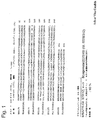
インフルエンザウイルスは、オルトミクソウイルス科に属し、それらの核タンパク質(NP)およびマトリックスタンパク質(M1)の抗原性の相違に基づいて、A、BおよびCの群に分類されている。株へのさらなる亜型判定は、通常、2種のビリオン糖タンパク質、すなわち赤血球凝集素(HA;H)およびノイラミニダーゼ(NA;N)に存在する抗原の型の評価に基づいている。HAおよびNPは、宿主細胞表面へのビリオンの付着を媒介する病原因子である。M1タンパク質は、ウイルスのアセンブリーおよび脱粒で機能すると考えられており、NPは、RNA複製および転写で機能している。これらのビリオンタンパク質に加えて、非構造タンパク質1および2(NS1、NS2)と呼ばれる他の2種の非構造タンパク質、すなわち非ビリオンタンパク質がウイルス感染細胞内で発現される。非構造ウイルスタンパク質NS1は、スプライシングならびに細胞性mRNAの核外輸送および翻訳の刺激の調節と、宿主インターフェロン能力の相殺とを含めた多機能を有する。NS1タンパク質は、インフルエンザウイルスで同定および配列決定されており、その配列は、NCBIデータベース内に見出すことができる。B型インフルエンザの例示的な2株に由来するNS1タンパク質の配列を図1に示す。その2株は93%の配列同一性を示す。他のインフルエンザ株のNS1タンパク質とは、例えば図1に示す配列を用いて、既知のインフルエンザ亜型でNS1タンパク質として認識されているタンパク質のうちの1つに最も大きな配列類似性を有するタンパク質を意味する。
I. Influenza viruses Influenza viruses belong to the Orthomyxoviridae family and are classified into groups A, B and C based on their antigenic differences in nucleoprotein (NP) and matrix protein (M1). Further subtyping to strains is usually based on an assessment of the type of antigen present in the two virion glycoproteins, hemagglutinin (HA; H) and neuraminidase (NA; N). HA and NP are virulence factors that mediate virion attachment to the host cell surface. The M1 protein is thought to function in viral assembly and shedding, and NP functions in RNA replication and transcription. In addition to these virion proteins, two other nonstructural proteins, called
II.診断および治療に使用するための抗体
本発明はB型インフルエンザのNS1タンパク質に対する抗体を提供する。そのような抗体の一部は、少なくとも2株もしくは5株、またはすべてもしくは実質的にすべての既知の株のB型インフルエンザに由来するNS1タンパク質に特異的に結合する点で汎反応性である。他の抗体は、1株のB型インフルエンザのみに特異的に結合する点で単一特異性である。通常、そのような抗体は、すべての株のA型インフルエンザに対して特異的な結合を行わない。抗体は、ポリクローナル抗体でも、様々なエピトープ特異性を有する別個のモノクローナル抗体またはモノクローナル抗体のプールでもよい。モノクローナル抗体は、タンパク質の抗原含有断片から、抗体の型に応じた標準的手順によって作製される(例えば、Kohlerら、Nature、256巻、495頁、(1975年);ならびにHarlowおよびLane、「Antibodies, A Laboratory Manual」(C.S.H.P.社、米国New York州所在、1988年);Queenら、Proc.Natl. Acad. Sci. USA、86巻、10029〜10033頁(1989年)および国際公開第90/07861号;Dowerら、国際公開第91/17271号;ならびに、McCaffertyら、国際公開第92/01047号(それぞれを参照によりすべての目的で本明細書に援用する)を参照)。
II. Antibodies for use in diagnosis and therapy The present invention provides antibodies to NS1 protein of influenza B influenza. Some such antibodies are pan-reactive in that they specifically bind to NS1 protein from influenza B of at least 2 or 5 strains, or all or substantially all known strains. Other antibodies are monospecific in that they specifically bind only to one strain of influenza B. Usually, such antibodies do not bind specifically to all strains of influenza A. The antibody may be a polyclonal antibody or a separate monoclonal antibody or a pool of monoclonal antibodies with various epitope specificities. Monoclonal antibodies are made from antigen-containing fragments of proteins by standard procedures depending on the type of antibody (eg, Kohler et al., Nature, 256, 495, (1975); and Harlow and Lane, “Antibodies. , A Laboratory Manual "(C.S.H.P., New York, USA, 1988); Queen et al., Proc. Natl. Acad. Sci., USA, 86, 1001-10033 (1989). And WO 90/07861; Dower et al., WO 91/17271; and McCafferty et al., WO 92/01047, each of which is incorporated herein by reference for all purposes. ).
B型インフルエンザの複数の株で免疫感作することによって、または1株で免疫感作し、別の株で追加免疫することによって、汎特異性の抗体を産生するように免疫感作を偏らせることができる。あるいは、B型インフルエンザNS1の高度に保存された領域に由来する断片を免疫原として用いることができる。逆に、単一特異性抗体を産生させるには、単一株のNS1または非保存領域に由来するNS1断片で免疫感作を行うことが好ましい。 Biasing immunization to produce panspecific antibodies by immunizing with multiple strains of influenza B or by immunizing with one strain and boosting with another strain be able to. Alternatively, a fragment derived from a highly conserved region of influenza B NS1 can be used as an immunogen. Conversely, in order to produce a monospecific antibody, it is preferable to perform immunization with a NS1 fragment derived from a single strain NS1 or a non-conserved region.
「抗体」または「免疫グロブリン」という用語は、完全な抗体およびそれらの結合性断片が含まれるように使用される。通常、別々になっている重鎖、軽鎖Fab、Fab’F(ab’)2、FabcおよびFvを含めた断片は、それらの由来である完全な抗体と、抗原断片への特異的な結合に関して競合する。断片は、組換えDNA技法によって、または完全な免疫グロブリンの酵素的もしくは化学的分離によって産生される。「抗体」という用語には、他のタンパク質と化学的に結合されているか、またはそれらとの融合タンパク質として発現された1または複数本の免疫グロブリン鎖が含まれる。「抗体」という用語には、二重特異性抗体も含まれる。 The terms “antibody” or “immunoglobulin” are used to include intact antibodies and binding fragments thereof. Usually, fragments including heavy chain, light chain Fab, Fab′F (ab ′) 2, Fabc and Fv, which are separated, are the complete antibody from which they are specifically bound to the antigen fragment. Compete for. Fragments are produced by recombinant DNA techniques or by enzymatic or chemical separation of complete immunoglobulins. The term “antibody” includes one or more immunoglobulin chains that are chemically linked to other proteins or expressed as fusion proteins therewith. The term “antibody” also includes bispecific antibodies.
III.他の結合剤
NS1タンパク質の検出での使用には、抗体が好ましいが、B型インフルエンザのNS1に特異的親和性を有するいかなる結合剤も抗体代用物として使用できる。代用物には、B型インフルエンザのNS1に対してスクリーニングされたランダム化ファージディスプレーライブラリーから得られたペプチドが含まれる。代用物にはアプタマーも含まれる。アプタマーは、結合ポケットを形成することによって特異的なリガンドを認識する、ランダム配列の巨大な集団からin vitroで選択されたRNA分子またはDNA分子である。アロステリックリボザイムは、それらの活性が、活性部位から離れて位置するアプタマードメインへのエフェクター分子の結合によって調節されているRNA酵素である。これらのRNAは、特定のエフェクターの存在または不在によって制御されている精密分子スイッチとして働く。アプタマーは、核酸、タンパク質および生物全体にさえ結合できる。アプタマーは抗体とは異なるが、それでも、それらは様々な診断フォーマットで抗体の特性を模倣する。したがって、アプタマーは、抗体の代わりに、または抗体と組み合わせて、一般的、または特定のNS1領域の存在を同定するのに使用できる。
III. Other Binding Agents Although antibodies are preferred for use in detecting NS1 protein, any binding agent with specific affinity for influenza B NS1 can be used as an antibody surrogate. Substitutes include peptides obtained from a randomized phage display library screened against NS1 of influenza B. Substitutes include aptamers. Aptamers are RNA or DNA molecules selected in vitro from a large population of random sequences that recognize specific ligands by forming binding pockets. Allosteric ribozymes are RNA enzymes whose activity is regulated by the binding of effector molecules to aptamer domains located away from the active site. These RNAs act as precision molecular switches that are controlled by the presence or absence of specific effectors. Aptamers can bind to nucleic acids, proteins and even whole organisms. Aptamers are different from antibodies, yet they mimic the properties of antibodies in various diagnostic formats. Thus, aptamers can be used in place of antibodies or in combination with antibodies to identify the presence of general or specific NS1 regions.
IV.診断試験
B型インフルエンザに感染している疑いのある試料は、B型インフルエンザウイルスのNS1タンパク質を検出することによって、それが存在しているかどうか試験する。そのタンパク質は、B型インフルエンザのNS1タンパク質に特異的に結合する抗体または他の捕獲試薬を用いて、下記に、より詳細に記述するフォーマットで検出することができる。B型インフルエンザのNS1タンパク質の存在は、試料がB型インフルエンザウイルスに感染していることを示す。そのような試験は、単独で、またはA型および/もしくはC型インフルエンザ用の他の試験と組み合わせて行うことができる。A型インフルエンザおよびC型インフルエンザに関する試験も、これらの株に適した特異性を有する抗体または他の捕獲試薬を用いて、これらの株のNS1タンパク質が存在しているかどうか検出することによって実施できる。A型インフルエンザを検出する方法、および、とりわけ、その病原型および非病原型を識別する方法が、2006年7月3日出願の同時係属出願である米国特許出願第11/481,411号(参照により、本明細書に全体として、すべての目的に関して組み込まれている)に記載されている。
IV. Diagnostic Test A sample suspected of being infected with influenza B is tested for its presence by detecting the NS1 protein of influenza B virus. The protein can be detected in the format described in more detail below using antibodies or other capture reagents that specifically bind to the NS1 protein of influenza B. The presence of influenza B NS1 protein indicates that the sample is infected with influenza B virus. Such a test can be performed alone or in combination with other tests for influenza A and / or C. Testing for influenza A and influenza C can also be performed by detecting the presence of NS1 protein in these strains using antibodies or other capture reagents with suitable specificity for these strains. A method for detecting influenza A, and in particular a method for distinguishing its pathogenic and non-pathogenic forms, is a co-pending application filed Jul. 3, 2006, US patent application Ser. No. 11 / 481,411 (see Are incorporated herein in their entirety for all purposes).
本発明の方法は、通常、B型インフルエンザのNS1に汎特異性の抗体または他の結合試薬を用いて行う。この方法は、型を識別せずに、インフルエンザの一部またはすべての株を検出する。この方法は、B型インフルエンザ株を相互に識別する抗体を用いて行うこともできる。この場合、通常、単一のアッセイで1パネルの抗体が使用され、このアッセイは、B型インフルエンザの存在だけでなく、どの株が存在するかも同定する。 The methods of the present invention are typically performed using antibodies or other binding reagents that are panspecific for NS1 of influenza B influenza. This method detects some or all strains of influenza without identifying the type. This method can also be performed using antibodies that distinguish influenza B strains from each other. In this case, a panel of antibodies is typically used in a single assay, which identifies not only the presence of influenza B but also which strain is present.
V.診断試験用のフォーマット
本発明は、異なった様々なタイプの生物学的な試料中のB型インフルエンザウイルスを同定するアッセイ法で有用な診断用捕獲試薬および検出試薬を提供する。そのようなフォーマットには、免疫沈降反応、ウエスタンブロッティング、ELISA、ラジオイムノアッセイ、競合アッセイおよび免疫測定アッセイ(immunometric assay)が含まれる。HarlowおよびLane、「Antibodies, A Laboratory Manual」(CSHP、NY、1988年)、米国特許第3,791,932号、第3,839,153号、第3,850,752号、第3,879,262号、第4,034,074号、第3,791,932号、第3,817,837号、第3,839,153号、第3,850,752号、第3,850,578号、第3,853,987号、第3,867,517号、第3,879,262号、第3,901,654号、第3,935,074号、第3,984,533号、第3,996,345号、第4,034,074号ならびに第4,098,876号を参照。
V. Format for Diagnostic Tests The present invention provides diagnostic capture and detection reagents useful in assays that identify influenza B virus in a variety of different types of biological samples. Such formats include immunoprecipitation reactions, Western blotting, ELISA, radioimmunoassay, competition assays and immunometric assays. Harlow and Lane, “Antibodies, A Laboratory Manual” (CSHP, NY, 1988), US Pat. Nos. 3,791,932, 3,839,153, 3,850,752, 3,879 No. 262, No. 4,034,074, No. 3,791,932, No. 3,817,837, No. 3,839,153, No. 3,850,752, No. 3,850,578 No. 3,853,987, 3,867,517, 3,879,262, 3,901,654, 3,935,074, 3,984,533, See 3,996,345, 4,034,074 and 4,098,876.
免疫測定アッセイまたはサンドイッチアッセイが好ましいフォーマットである(米国特許第4,376,110号、第4,486,530号、第5,914,241号および第5,965,375号を参照)。そのようなアッセイは、固相に固定された1種の抗体または抗体の集団と、溶液中の別の抗体または抗体の集団とを用いる。通常、その溶液抗体または抗体集団は標識されている。抗体集団を用いる場合、その集団は通常、標的抗原内の異なったエピトープ特異性に結合する抗体を含有する。したがって、同じ集団を、固相抗体および溶液抗体の両方に用いることができる。モノクローナル抗体を用いる場合、異なった結合特異性を有する第1および第2のモノクローナル抗体を固相および溶液相に用いる。固相抗体および溶液抗体は、標的抗原に、順次または同時に接触させることができる。固相抗体を最初に接触させる場合、そのアッセイは、フォワードアッセイ(forward assay)であると言われる。逆に、溶液抗体を最初に接触させる場合、そのアッセイは、リバースアッセイ(reverse assay)であると言われる。標的を両方の抗体と同時に接触させる場合、そのアッセイは、同時アッセイと呼ばれる。標的を抗体と接触させた後に、通常約10分から約24時間までの間で変動し、通常約1時間である時間、試料をインキュベートする。その後、検出試薬として使用された(1または複数種の)抗体に特異的に結合しなかった、試料の成分を除去するために、洗浄ステップを行うことができる。固相抗体および溶液抗体を別々のステップで結合させる場合、それらの結合ステップのいずれか、または両方の後に、洗浄を行うことができる。洗浄の後に、通常、標識された溶液抗体の結合を介して固相に連結されている標識を検出することによって、結合を定量化する。通常、抗体または抗体集団の所与の対、および所与の反応条件に関して、既知の濃度の標的抗原を含有する試料から検量線を作成する。その後、検量線の内挿によって、試験される試料中の抗原の濃度を決定する。平衡状態で結合した標識溶液抗体の量から、または平衡に達する前の一連の時点における、結合した標識溶液抗体の動態測定によって、分析物を測定できる。そのような曲線の勾配が試料中の標的濃度の尺度となる。 An immunometric assay or sandwich assay is the preferred format (see US Pat. Nos. 4,376,110, 4,486,530, 5,914,241 and 5,965,375). Such assays use one antibody or population of antibodies immobilized on a solid phase and another antibody or population of antibodies in solution. Usually, the solution antibody or antibody population is labeled. When using an antibody population, the population usually contains antibodies that bind to different epitope specificities within the target antigen. Thus, the same population can be used for both solid phase antibodies and solution antibodies. When monoclonal antibodies are used, first and second monoclonal antibodies having different binding specificities are used in the solid phase and solution phase. The solid phase antibody and the solution antibody can be contacted with the target antigen sequentially or simultaneously. When a solid phase antibody is first contacted, the assay is said to be a forward assay. Conversely, when a solution antibody is first contacted, the assay is said to be a reverse assay. If the target is contacted with both antibodies simultaneously, the assay is referred to as a simultaneous assay. After contacting the target with the antibody, the sample is typically incubated for a time that varies from about 10 minutes to about 24 hours, usually about 1 hour. A washing step can then be performed to remove sample components that did not specifically bind to the antibody (s) used as detection reagents. If the solid phase antibody and the solution antibody are bound in separate steps, washing can be performed after either or both of the binding steps. After washing, binding is quantified, usually by detecting the label linked to the solid phase via binding of labeled solution antibody. A standard curve is usually generated from a sample containing a known concentration of the target antigen for a given pair of antibodies or antibody populations and for a given reaction condition. Thereafter, the concentration of the antigen in the sample to be tested is determined by interpolation of a calibration curve. Analytes can be measured from the amount of labeled solution antibody bound in equilibrium or by kinetic measurements of bound labeled solution antibody at a series of time points before reaching equilibrium. The slope of such a curve is a measure of the target concentration in the sample.
競合アッセイも使用できる。一部の方法では、抗体検出試薬との結合に関して、試料中の標的抗原が、外因的に添加された標識標的抗原と競合する。抗体に結合した標識標的抗原の量は、試料中の標的抗原の量と反比例している。抗体は、試料からの結合複合体の分離を容易にするために、検出前に固定することができ(不均一アッセイ)、または、均一アッセイフォーマットで行う場合には、分離が不要となりうる。他の方法では、検出試薬として使用される抗体を標識する。抗体が標識される場合、その結合部位は、試料中の標的抗原、および例えば固相に固定された標的抗原でありうる、外因的に供給される形態の標的抗原への結合に関して競合する。標識抗体は、試料中の、上記標識抗体と同じ標的抗原に結合する抗体を検出するのに、さらに別の競合的フォーマットで使用することもできる。上記フォーマットのそれぞれで、検出試薬として使用される抗体が、アッセイされている標的とほぼ同じ濃度の制限量として存在する。 Competitive assays can also be used. In some methods, the target antigen in the sample competes with the exogenously added labeled target antigen for binding to the antibody detection reagent. The amount of labeled target antigen bound to the antibody is inversely proportional to the amount of target antigen in the sample. The antibody can be immobilized prior to detection to facilitate separation of the bound complex from the sample (heterogeneous assay), or separation can be unnecessary when performed in a homogeneous assay format. In other methods, the antibody used as the detection reagent is labeled. When the antibody is labeled, its binding site competes for binding to the target antigen in the sample and to an exogenously supplied form of the target antigen, which can be, for example, a target antigen immobilized on a solid phase. The labeled antibody can also be used in yet another competitive format to detect antibodies in the sample that bind to the same target antigen as the labeled antibody. In each of the above formats, the antibody used as a detection reagent is present in a limiting amount at approximately the same concentration as the target being assayed.
ラテラルフローデバイスが好ましいフォーマットである。家庭用妊娠試験と同様に、ラテラルフローデバイスは、特定の生物製剤で処理されている試験細片に流体を添加することによって働く。液体試料によって運搬され、対応する生物製剤で標識されたリン光体が細片を貫流し、それらが特定の帯域に入ったときに、それらを捕獲することができる。細片上に見出されるリン光体シグナルの量が標的分析物の量に比例する。 Lateral flow devices are the preferred format. Similar to the home pregnancy test, the lateral flow device works by adding fluid to a test strip that has been treated with a specific biologic. Phosphors carried by the liquid sample and labeled with the corresponding biologic can flow through the strip and capture them as they enter a particular zone. The amount of phosphor signal found on the strip is proportional to the amount of target analyte.
B型インフルエンザを含有している疑いがある試料をラテラルフローデバイスに添加する。試料は、拡散によって移動できるようになっており、線または有色帯域がインフルエンザBの存在を示す。ラテラルフローは通常、3つの特定領域、すなわち試料添加領域、NS1に対する1または複数種の抗体を含有している捕獲領域、および各帯域が1つまたは複数の標識を含有している1つまたは複数の帯域を含有する読み取り領域を含有している固体支持体(例えばニトロセルロース膜)を含有している。ラテラルフローに、陽性対照および陰性対照を含有させることもできる。したがって、ラテラルフローデバイスは、例えば次の通りに使用できる。すなわち、生物学的な試料のアリコートを試験細片の一端に接触させることによって、B型インフルエンザのNS1タンパク質を、生物学的な試料中の他のウイルス性タンパク質および細胞性タンパク質から分離し、その後、例えばラテラルフローなどの毛細管現象によって、タンパク質を試験細片上で移動させる。1または複数種の抗体および/またはアプタマーが、捕獲試薬および/または検出試薬として含有されている。ラテラルフローで分離、検出および定量化する方法およびデバイスは、例えば、参照により全体が本明細書に援用される、米国特許第5,569,608号、第6,297,020号および第6,403,383号に記載されている。一例として、試験細片は、試料を添加するために近位の領域(試料添加領域)、ならびにNS1タンパク質に対する抗体および緩衝試薬およびその抗体と、移動している生物学的な試料中の任意のB型インフルエンザNS1タンパク質との結合相互作用を確立するのに適した添加物を含有する遠位の試験領域を含みうる。別の例では、試験細片は、B型インフルエンザの異なった2株のNS1に対する異なった抗体を含有する2箇所の試験領域、すなわち、それぞれが異なったB型インフルエンザ分析物と特異的に相互作用できる2箇所の試験領域を含む。 A sample suspected of containing influenza B is added to the lateral flow device. The sample is allowed to move by diffusion and the line or colored band indicates the presence of influenza B. Lateral flow typically has three specific regions: a sample addition region, a capture region containing one or more antibodies against NS1, and one or more where each zone contains one or more labels. A solid support (for example, a nitrocellulose membrane) containing a reading area containing a plurality of bands. Lateral flows can also contain positive and negative controls. Accordingly, the lateral flow device can be used, for example, as follows. That is, the NS1 protein of influenza B is separated from other viral and cellular proteins in the biological sample by contacting an aliquot of the biological sample with one end of the test strip, and then The protein is moved over the test strip by capillary action, such as lateral flow. One or more antibodies and / or aptamers are included as capture reagents and / or detection reagents. Methods and devices for separation, detection and quantification in lateral flow are described, for example, in US Pat. Nos. 5,569,608, 6,297,020, and 6, which are incorporated herein by reference in their entirety. No. 403,383. As an example, test strips can be placed in the proximal region (sample addition region) for adding sample, as well as any antibody and buffer reagent and its antibody against NS1 protein and any biological sample that is moving. A distal test region containing additives suitable for establishing a binding interaction with influenza B NS1 protein can be included. In another example, a test strip specifically interacts with two test regions containing different antibodies against two different strains of NS1 of influenza B, ie, each with different influenza B analytes. Includes two possible test areas.
上記方法での使用に適した検出可能な標識には、分光学的、光化学的、生化学的、免疫化学的、電気的、光学的もしくは化学的手段、または他の手段によって検出可能ないかなる部分も含まれる。例えば、適した標識には、標識されたストレプトアビジン結合体で染色するためのビオチン、蛍光色素(例えば、フルオレセイン、テキサスレッド、ローダミンおよび緑色蛍光タンパク質など)、放射標識(例えば、3H、125I、35S、14Cまたは32P)、酵素(例えば、西洋ワサビペルオキシダーゼ、アルカリ性ホスファターゼ、およびELISAで一般的に使用される他の酵素)、およびコロイド状金または色ガラスまたはプラスチック(例えば、ポリスチレン、ポリプロピレンまたはラテックスビーズ)などの比色分析標識が含まれる。そのような標識の使用を記載している特許には、米国特許第3,817,837号、第3,850,752号、第3,939,350号、第3,996,345号、第4,277,437号、第4,275,149号および第4,366,241号が含まれる。「Handbook of Fluorescent Probes and Research Chemicals」(第6版、Molecular Probes社、米国Oregon州Eugene所在)も参照のこと。放射標識は、写真フィルムまたはシンチレーションカウンターを用いて検出でき、蛍光標識は、発せられた光を検出する光検出器を用いて検出でき、酵素標識は通常、その酵素に基質を与え、基質への酵素の作用によって生成した反応生成物を検出することによって検出され、比色分析標識は、発色した標識を単純に可視化することによって検出される。 Detectable labels suitable for use in the above methods include any moiety detectable by spectroscopic, photochemical, biochemical, immunochemical, electrical, optical or chemical means, or other means. Is also included. For example, suitable labels include biotin for staining with labeled streptavidin conjugates, fluorescent dyes (eg, fluorescein, Texas red, rhodamine and green fluorescent protein), radiolabels (eg, 3 H, 125 I , 35 S, 14 C or 32 P), enzymes (eg, horseradish peroxidase, alkaline phosphatase, and other enzymes commonly used in ELISA), and colloidal gold or colored glass or plastic (eg, polystyrene, Colorimetric labels such as polypropylene or latex beads) are included. Patents describing the use of such labels include US Pat. Nos. 3,817,837, 3,850,752, 3,939,350, 3,996,345, 4,277,437, 4,275,149, and 4,366,241. See also "Handbook of Fluorescent Probes and Research Chemicals" (6th edition, Molecular Probes, Eugene, Oregon, USA). A radiolabel can be detected using photographic film or a scintillation counter, a fluorescent label can be detected using a photodetector that detects the emitted light, and an enzyme label usually provides a substrate for the enzyme, and The colorimetric label is detected by detecting the reaction product produced by the action of the enzyme, and the colorimetric label is detected by simply visualizing the colored label.
試料中のB型インフルエンザNS1タンパク質のレベルは、定量化すること、および/または対照と比較することができる。適した陰性対照試料は、例えば、健康であることが分かっている個体、例えばインフルエンザウイルス感染を有していないことが分かっている個体から得られる。特異性対照は、既知のA型インフルエンザ感染またはC型インフルエンザ感染を有する個体、またはインフルエンザ以外のウイルスに感染している個体から収集できる。対照試料は、試験される対象と遺伝的な関係がある個体から得ることができるが、遺伝的に関係のない個体からも得ることができる。適した陰性対照試料は、感染のより早期、すなわち試験試料が採取された時点より早い時点に個体から収集された試料でもよい。陽性対照として、B型インフルエンザの組換え体NS1を用いることができる。 The level of influenza B NS1 protein in a sample can be quantified and / or compared to a control. A suitable negative control sample is obtained, for example, from an individual known to be healthy, eg, an individual known to have no influenza virus infection. Specificity controls can be collected from individuals with known influenza A or C influenza infections, or individuals infected with a non-influenza virus. Control samples can be obtained from individuals that are genetically related to the subject being tested, but can also be obtained from individuals that are not genetically related. A suitable negative control sample may be a sample collected from an individual earlier in infection, that is, earlier than when the test sample was collected. As a positive control, influenza B recombinant NS1 can be used.
生物学的な試料中のNS1レベルが、様々な異なった可能な免疫検定法フォーマットでこれらの抗原を検出することを可能にするのに十分であることを、ウエスタンブロットは示している。しかし、特定の免疫検定法フォーマットで検出するのに、特定の生物学的な試料中のNS1レベルが限定的であると判明した場合には、in vitroで細胞を感染させることによって、生物学的な試料中の生ウイルスを増幅させることができる。すなわち、ウイルス増幅試料のNS1タンパク質は、約6時間から約12時間以内に検出可能となる。生物学的な試料中およびウイルス増幅試料のNS1抗原の収率は、プロテアーゼ阻害剤およびプロテアソーム阻害剤に含めることによって改善することができる。 Western blots show that NS1 levels in biological samples are sufficient to allow detection of these antigens in a variety of different possible immunoassay formats. However, if NS1 levels in a particular biological sample are found to be limited for detection in a particular immunoassay format, the biological can be obtained by infecting the cells in vitro. Live virus in a simple sample can be amplified. That is, the NS1 protein of the virus amplification sample can be detected within about 6 hours to about 12 hours. The yield of NS1 antigen in biological samples and in virus amplification samples can be improved by inclusion in protease inhibitors and proteasome inhibitors.
あるいは、NS1タンパク質をmRNAレベルで検出することができる。試料から得られたRNAを逆転写させて、増幅させる。場合によっては、増幅の過程で標識を添加する。その後、増幅された核酸を、少なくとも1株のB型インフルエンザウイルスのNS1をコードする核酸に実質的または完全に相補的であることが知られている核酸プローブとハイブリッド形成させる。通常、標識の存在からハイブリッド形成を検出する。B型インフルエンザのNS1タンパク質をコードする核酸に相補的なプローブにハイブリッド形成する増幅された核酸の存在は、試料中におけるB型インフルエンザの存在を示す。抗体を用いる場合と同様に、プローブは、B型インフルエンザの様々な株に汎特異性のものか、または非特異性のものを選択できる。ハイブリッド形成アッセイは、アレイフォーマットで行うことができる。そのようなフォーマットは、場合によっては様々な株のB型インフルエンザのNS1タンパク質をコードする核酸に対するいくつかのプローブをアレイに含めることを可能にする。A型またはC型インフルエンザのNS1タンパク質のプローブも含めることができ、同様に、他のウイルスまたは他の病原体に対するプローブも含めることができる。 Alternatively, NS1 protein can be detected at the mRNA level. RNA obtained from the sample is reverse transcribed and amplified. In some cases, a label is added during the amplification process. The amplified nucleic acid is then hybridized with a nucleic acid probe known to be substantially or completely complementary to a nucleic acid encoding NS1 of at least one strain of influenza B virus. Usually, hybridization is detected from the presence of the label. The presence of amplified nucleic acid that hybridizes to a probe that is complementary to the nucleic acid encoding the influenza B NS1 protein indicates the presence of influenza B in the sample. As with antibodies, probes can be selected to be pan-specific or non-specific for various strains of influenza B. Hybridization assays can be performed in an array format. Such a format, in some cases, allows the array to contain several probes for nucleic acids encoding various strains of influenza B NS1 protein. Probes for NS1 protein of influenza A or C can also be included, as well as probes for other viruses or other pathogens.
VI.試料調製法
検出可能な濃度のインフルエンザタンパク質、好ましくはNS1を含有しているか、含有しているかもしれないと考えられるいかなる試料も使用できる。使用できる試料の例は、例えば、肺滲出液、細胞抽出物(気道上皮内層、鼻)、血液、粘液、および鼻腔スワブである。鼻腔スワブ中に、高濃度のNS1を見出すことができる。したがって、NS1を同定するための好ましい試料は鼻分泌物である。
VI. Sample Preparation Any sample that contains or is likely to contain a detectable concentration of influenza protein, preferably NS1, can be used. Examples of samples that can be used are, for example, lung exudates, cell extracts (airway epithelial lining, nose), blood, mucus, and nasal swabs. High concentrations of NS1 can be found in nasal swabs. Thus, a preferred sample for identifying NS1 is nasal secretions.
抗体へのNS1の結合は、0.03%および0.01%を含めた、最大0.05%までのSDSの存在下で起こる。したがって、鼻分泌物または他の体分泌物がラテラルフローフォーマットで容易に用いられそうにない場合、それをSDSで処理することができる。好ましくは、添加されるSDSの量が、最大0.01%まで、より好ましくは0.03%まで、さらに好ましくは0.05%までの最終濃度である。 NS1 binding to the antibody occurs in the presence of up to 0.05% SDS, including 0.03% and 0.01%. Thus, if nasal secretions or other body secretions are not likely to be easily used in the lateral flow format, they can be treated with SDS. Preferably, the amount of SDS added is a final concentration of up to 0.01%, more preferably up to 0.03%, even more preferably up to 0.05%.
VII.診断用および治療用キット
本発明の方法を実行するためのキットを提供する。そのキットは、B型インフルエンザのNS1に特異的に結合する1つまたは複数の結合剤、通常は抗体を含有している。本キットは、場合によっては、実施例に開示する試薬、緩衝剤、添加剤の組成物または試薬のうち1つまたは複数を含有している。そのキットは、生物学的な試料中の他の潜在的干渉物質からインフルエンザウイルスのNS1を分離するデバイスまたは系などの手段を含みうる。本キットは、望ましい場合には、アッセイを行うのに有用な1つもしくは複数の様々な構成要素、例えば、1つもしくは複数のアッセイ容器、1もしくは複数種の対照試薬もしくは較正試薬、アッセイを行うための1つもしくは複数の固相表面、1もしくは複数種の緩衝剤、添加物、検出試薬もしくは抗体、または、例えば、アッセイを行う際に使用するべき各成分の量およびアッセイの結果を評価するための指針を示す添付文書および/もしくは容器標識として、B型インフルエンザを検出するためのキットを用いる方法を詳述する1つまたは複数の印刷された説明書のうちの1つまたは複数をさらに含みうる。本キットは、例えば、試験細片、サンドイッチELISA、ウエスタンブロットアッセイおよびラテックス凝集などを含めた、様々な異なったタイプのアッセイフォーマットを行うのに有用な構成要素を含有しうる。
VII. Diagnostic and therapeutic kits Kits for carrying out the methods of the invention are provided. The kit contains one or more binding agents, usually antibodies, that specifically bind to NS1 of influenza B. The kit optionally contains one or more of the reagents, buffers, additive compositions or reagents disclosed in the Examples. The kit may include means such as a device or system for separating NS1 of influenza virus from other potential interfering substances in a biological sample. The kit, if desired, performs one or more various components useful for performing the assay, such as one or more assay containers, one or more control or calibration reagents, and assays. One or more solid phase surfaces for, one or more buffers, additives, detection reagents or antibodies, or for example, assessing the amount of each component to be used in performing the assay and the results of the assay And further includes one or more of one or more printed instructions detailing a method of using the kit for detecting influenza B as a package insert and / or container label indicating sell. The kit may contain components useful for performing a variety of different types of assay formats including, for example, test strips, sandwich ELISAs, Western blot assays and latex agglutination.
VIII.抗体アレイ
本発明は、抗体アレイをさらに提供する。そのようなアレイは、アレイの異なった領域に、それぞれB型インフルエンザのNS1に対する特異性を有する複数の異なった抗体を含有している。その異なった抗体は、B型インフルエンザの様々な株への特異性を有するように選択できる。複数の株に汎特異性である抗体を含めることもできる。A型またはC型インフルエンザのNS1タンパク質の抗体を含めることもできる。そのようなアレイは、B型インフルエンザの検出、株相互の識別、ならびにA型、B型およびC型インフルエンザの識別に有用である。
VIII. Antibody Array The present invention further provides an antibody array. Such arrays contain multiple different antibodies, each with specificity for NS1 of influenza B in different regions of the array. The different antibodies can be selected to have specificity for various strains of influenza B. Antibodies that are panspecific to multiple strains can also be included. An antibody of NS1 protein of type A or influenza C can also be included. Such arrays are useful for detection of influenza B, discrimination between strains, and discrimination of influenza A, B and C.
抗体アレイ用の多数のフォーマットが提唱されている。米国特許第5,922,615号は、膜表面に固定された抗体の複数の異なった帯域を用いた、アレイ内の複数の標的抗原を検出するデバイスを記載する。米国特許第5,458,852号、第6,019,944号、第6,143,576号および米国特許出願第08/902,775号は、膜上ではなくデバイス内に固定された複数の異なった抗体帯域を有する、複数の標的抗原をアッセイするための診断デバイスを記載する。国際公開第99/67641号は、マイクロスフェアが光ファイバーの末端に固定された後に、個々のマイクロスフェア表面に固定された特異的な結合剤(抗体を含める)の解読および同定を可能にするタグを有するように作製されたマイクロスフェアのアレイを記載する。米国特許第5981180号では、この場合も、結合剤(抗体を含める)を固定するのにマイクロスフェアが使用され、マイクロスフェアは、それらを試料から分離せずに、マイクロスフェアに結合した特異的な結合剤を同定するために、マイクロスフェア内に含有されている2種の異なったフルオロフォアの相対量を検出することによって相互に識別される。 A number of formats have been proposed for antibody arrays. US Pat. No. 5,922,615 describes a device for detecting multiple target antigens in an array using multiple different bands of antibodies immobilized on the membrane surface. U.S. Patent Nos. 5,458,852, 6,019,944, 6,143,576 and U.S. Patent Application No. 08 / 902,775 are disclosed in US Pat. A diagnostic device for assaying multiple target antigens having different antibody bands is described. WO 99/67641 provides tags that allow the decoding and identification of specific binding agents (including antibodies) immobilized on individual microsphere surfaces after the microspheres have been immobilized at the end of an optical fiber. An array of microspheres made to have is described. In U.S. Pat. No. 5,981,180, microspheres are again used to immobilize the binding agent (including antibody), and the microspheres do not separate them from the sample and do not separate them from the specific In order to identify the binder, they are distinguished from each other by detecting the relative amounts of two different fluorophores contained within the microsphere.
本明細書に引用されたすべての刊行物および特許を、個々の刊行物または特許のそれぞれを参照により本明細書に援用すると明確かつ個別に示したのと同じように、参照により本明細書に援用する。GIDまたはアクセッション番号によって参照されたGenbank記録、とりわけ、いかなるポリペプチド配列、ポリヌクレオチド配列またはそれらのアノテーションも、参照により本明細書に援用する。本発明の真の趣旨および範囲から逸脱することなく、様々な改変および等価物の置換を行うことができる。例えば、主としてB型インフルエンザに関して本発明を説明したが、C型インフルエンザを検出するのに、必要な変更を加えて、同様な戦略を用いることができる。別のことが文脈から明らかでない限り、いかなる特徴、ステップまたは実施形態も、他の任意の特徴、ステップまたは実施形態と組み合わせて用いることができる。 All publications and patents cited herein are hereby incorporated by reference as if each individual publication or patent was specifically and individually indicated to be incorporated herein by reference. Incorporate. Genbank records referenced by GID or accession number, especially any polypeptide sequence, polynucleotide sequence or annotations thereof, are hereby incorporated by reference. Various modifications and equivalent substitutions can be made without departing from the true spirit and scope of the invention. For example, although the present invention has been described primarily with respect to influenza B, a similar strategy can be used to make the necessary changes to detect influenza C. Any feature, step or embodiment may be used in combination with any other feature, step or embodiment unless otherwise apparent from the context.
(実施例1)
FLU−BのNS1に対するモノクローナル抗体の単離
Flu−B亜型のNS1タンパク質に特異的に結合するモノクローナル抗体を調製した。抗体は、汎特異性(すなわち、複数の株のFlu−Bに結合する)または単一特異性(すなわち、1株のFlu−Bに結合し、他のものに結合しない)でありうる。
Example 1
Isolation of monoclonal antibodies against NS1 of FLU-B Monoclonal antibodies that specifically bind to NS1 protein of the Flu-B subtype were prepared. An antibody can be pan-specific (ie, bind to multiple strains of Flu-B) or monospecific (ie, bind to one strain of Flu-B and not the other).
2種のFlu−B亜型について、Flu−B NS1のGSTおよびMBP融合タンパク質を作製した(B/BA/78、AVC名称:タンパク質#522、Genbank325112)および(B/Yagamata/222/2002、AVC名称:タンパク質#523、Genbank50300312)。クローニングベクターは、Pharmacia社(GST)またはNew England Biolabs社(MBP)から入手した。NS1コード領域は、DNA2.0(米国California州Menlo Park所在)によるオーバーラップオリゴヌクレオチドを用いて合成した。
For two Flu-B subtypes, Flu-B NS1 GST and MBP fusion proteins were made (B / BA / 78, AVC name: Protein # 522, Genbank 325112) and (B / Yamagata / 222/2002, AVC Name:
1.CFA、次にIFAおよびPBSにおいてFlu−B MBP−NS1融合タンパク質を1用量あたり10〜100μgの範囲の用量を用いてマウスの免疫感作を行った。 1. Mice were immunized with doses ranging from 10-100 μg per dose of Flu-B MBP-NS1 fusion protein in CFA followed by IFA and PBS.
2.対応するGST−NS1融合タンパク質を用いた最後の追加免疫の3日後に、脾細胞およびリンパ球を採取し、KohlerおよびMilstein(Nature、1975年)に記載のFOX−NY骨髄腫細胞と融合させた。 2. Three days after the last boost with the corresponding GST-NS1 fusion protein, splenocytes and lymphocytes were harvested and fused with FOX-NY myeloma cells as described in Kohler and Milstein (Nature, 1975). .
3.ハイブリドーマは、最初にFlu−B MBP−NS1を用いて、ELISAでスクリーニングした(図2Aおよび2B)。陽性ウェルをクローン化し、MBPおよびGSTタグのいずれかを有するFlu−B NA1タンパク質の両方に対して再スクリーニングを行った。mAbは汎反応性または亜型反応性に分類した。 3. Hybridomas were initially screened by ELISA using Flu-B MBP-NS1 (FIGS. 2A and 2B). Positive wells were cloned and rescreened against both Flu-B NA1 protein with either MBP or GST tag. mAbs were classified as pan-reactive or sub-type reactive.
4.反応性と、ラテラルフロー細片上での抗Flu−B NS1 mAbの使用への適合性を確認するために、ラテラルフロー細片上の異なった位置で、両方のFlu−B NS1タンパク質(#522および#523)でコーティングされたラテラルフロー細片を用いて、さらにスクリーニングを行った。これらのアッセイで、金標識された抗マウスIg2次mAbを介した検出を行った。 4). To confirm the reactivity and suitability for use of anti-Flu-B NS1 mAb on lateral flow strips, both Flu-B NS1 proteins (# 522 and # 522 and ##) at different locations on the lateral flow strip. Further screening was performed using lateral flow strips coated with 523). In these assays, detection via gold labeled anti-mouse Ig secondary mAb was performed.
5.両方のFlu−B NS1タンパク質(AVC#522および#523)で反応性を示した抗Flu−B mAbを捕獲能力および検出能力、ならびに相互との適合性に関してラテラルフローサンドイッチ型アッセイで試験した。両方の型のFlu−B NS1タンパク質の候補捕獲/選択対を選択して、組換え体Flu−B NS1を検出する感受性のレベルに関して、同じ型のラテラルフローサンドイッチアッセイを用いて試験した。 5. Anti-Flu-B mAbs that were reactive with both Flu-B NS1 proteins (AVC # 522 and # 523) were tested in a lateral flow sandwich type assay for capture and detection capabilities and compatibility with each other. Candidate capture / selection pairs of both types of Flu-B NS1 protein were selected and tested for the level of sensitivity to detect recombinant Flu-B NS1 using the same type of lateral flow sandwich assay.
6.最後に、その抗体が臨床検体中のNS1を検出する能力に関して調べる。 6). Finally, the antibody is tested for its ability to detect NS1 in clinical specimens.
このワークフローによって、ヒト臨床検体を認識する抗体が得られる。 This workflow yields antibodies that recognize human clinical specimens.
(実施例2)
ラテラルフロー
上述の方法に従って産生された抗B型インフルエンザNS1モノクローナル抗体を用いて、B型インフルエンザのNS1を検出するラテラルフロー試験を開発した。ストライパー(striper)を用いて、抗B型インフルエンザNS1モノクローナル抗体を約0.7mg/mlの濃度でHF075 Millipore膜に沈着させた。捕獲剤として沈着させた抗体の一部の例には、以下のもの、すなわちF89 1F4、F94 3A1およびF89 4D5が含まれる。1mg/mlのヤギ抗マウス抗体(GAM)で構成される対照バンドも沈着させた。容積100μlのAVC FluB緩衝液中で、FluB NS1タンパク質を、F94 3A1(F94 3A1が捕獲剤として使用されない場合)などの金結合体化抗NS1モノクローナル抗体と合わせた。使用されたFluB NS1タンパク質は、組換え体であるAVC ID522(B/BA/78 NS1)およびAVC ID523(B/YM/222/2002)、またはB型インフルエンザに感染していることが分かっている患者からの臨床試料であった。
(Example 2)
Lateral Flow A lateral flow test was developed to detect influenza B NS1 using an anti-influenza B NS1 monoclonal antibody produced according to the method described above. Anti-B influenza NS1 monoclonal antibody was deposited on HF075 Millipore membrane at a concentration of about 0.7 mg / ml using a striper. Some examples of antibodies deposited as capture agents include the following: F89 1F4, F94 3A1 and F89 4D5. A control band composed of 1 mg / ml goat anti-mouse antibody (GAM) was also deposited. FluB NS1 protein was combined with a gold-conjugated anti-NS1 monoclonal antibody such as F94 3A1 (when F94 3A1 is not used as a capture agent) in a volume of 100 μl AVC FluB buffer. The FluB NS1 protein used is known to be infected with recombinant AVC ID522 (B / BA / 78 NS1) and AVC ID523 (B / YM / 222/2002) or influenza B It was a clinical sample from a patient.
抗FluB NS1抗体のストライプを入れた(striped)膜を、FluB NS1/抗NS1タンパク質溶液中に挿入し、毛細管現象およびウィッキングパッド(wicking pad)によって流動を開始させた。 A striped membrane of anti-FluB NS1 antibody was inserted into the FluB NS1 / anti-NS1 protein solution and flow was initiated by capillary action and a wicking pad.
抗FluB NS1捕獲剤および検出剤のいくつかの組合せをいくつかの実験で用いた。以下は、一例のプロトコールである。細片試験は、ヤギ抗マウス/F89 1F4抗FluB NS1モノクローナル抗体で事前にストライプを入れた細片;90%M4ウイルス輸送培地、10%の10×AVC FluB緩衝液;NS1タンパク質であるMBP−FluB NS1(AVC522およびAVC523)の保存液;金結合体化F94 3A1抗体;およびMaxiSorp ELISAプレートを用いて行った。この手順は以下の通りに行った。 Several combinations of anti-FluB NS1 capture and detection agents were used in some experiments. The following is an example protocol. Strip test: strip pre-striped with goat anti-mouse / F89 1F4 anti-FluB NS1 monoclonal antibody; 90% M4 virus transport medium, 10% 10 × AVC FluB buffer; NS1 protein MBP-FluB NS1 (AVC522 and AVC523) stock solutions; gold-conjugated F94 3A1 antibody; and MaxiSorp ELISA plates were used. This procedure was performed as follows.
1)NS1タンパク質の保存液を90%M4ウイルス輸送培地、10%の10×AVC FluB緩衝液で希釈した。 1) The stock solution of NS1 protein was diluted with 90% M4 virus transport medium, 10% 10 × AVC FluB buffer.
2)NS1の保存液は、90%M4/10%の10×AVC FluB緩衝液で希釈することによって、0.5ng/μLまで希釈した。 2) The NS1 stock was diluted to 0.5 ng / μL by diluting with 90% M4 / 10% 10 × AVC FluB buffer.
3)4μLの金結合体化抗体を、緩衝液100μL毎に添加した。 3) 4 μL of gold-conjugated antibody was added every 100 μL of buffer.
4)98μLの抗体/緩衝液混合物を、ELISAプレート内の別々のウェルに添加した。 4) 98 μL of antibody / buffer mixture was added to separate wells in the ELISA plate.
5)望ましい最終タンパク質濃度(例、1ng FluB NS1)を得るように、NS1希釈液2μLを、緩衝液を含有しているウェルに添加した(1ウェルあたり1種のNS1)。 5) 2 μL of NS1 dilution was added to wells containing buffer (1 NS1 per well) to obtain the desired final protein concentration (eg, 1 ng FluB NS1).
6)1ウェルは、「NS1を含まない」陰性対照として働くように、抗体および緩衝液のみのままとした。 6) One well was left with antibody and buffer only to serve as a negative control “without NS1”.
7)ウェルの内容物を混和させるため、ELISAプレートを何度か軽く叩いた。 7) Tap ELISA plate several times to mix well contents.
8)事前にストライプを入れた細片をウェル内に添加し、発色する間観察する。 8) Add a strip with stripes in advance into the well and observe while developing.
約15分後(すべての液体が吸収されたが、細片がまだ乾いていないとき)に、細片をウェルから取り出して、スキャンしてコンピューター内に取り込む。 After about 15 minutes (when all the liquid has been absorbed, but the strip has not yet dried), the strip is removed from the well and scanned into a computer.
図3は、様々な対のモノクローナル抗体を、2株のB型インフルエンザ、すなわちB/BA78(株522としても知られている)およびB/Yagamata/222/2002(株523としても知られている)での捕獲試薬および検出試薬として試験した結果を示す。4つの異なったパネルが、抗体の4つの組合せを示す。各パネルで、トラック3および6が陰性対照である。トラック1および2は、株522からの組換え体NS1であり、トラック4および5は、株523からの組換え体NS1である。第1のパネルのトラック1および2にはなく、トラック4および5にある追加のバンドの存在は、F89−F4捕獲抗体F89−4G12検出抗体の組合せが、株523は検出するが、株522は検出しないことを示している。他のパネルも同様に分析することができる。この実験および他の同様な実験からの結果を図4にまとめる。図4は、どの抗体が捕獲抗体として役立ち、どれが検出抗体として役立つことができるか、そして、それらの抗体が両方の株のB型インフルエンザ(522および523)に汎特異性であるか、または522もしくは523に単一特異性であるかを示す。例えば、F89−1F4抗体は、捕獲抗体としても、検出抗体としても役立つことができ、汎特異性である。F94−4C10は、検出抗体としては働くが、捕獲抗体としては働かず、B型インフルエンザ523に特異的である。F89−1F4およびF94−3A1は、ラテラルフローフォーマットでの使用に好ましい抗体である。
FIG. 3 shows various pairs of monoclonal antibodies, two strains of influenza B, B / BA78 (also known as strain 522) and B / Yamagata / 222/2002 (also known as
患者試料中のB型インフルエンザを同定するのに、膜上に沈着した汎特異性抗体を有するように産生されたラテラルフローアッセイを用いた。患者試料を、すべてのB型インフルエンザのNS1を認識する金標識抗体の混合物と混合した。試料をラテラルフロー検査細片に添加した。B型インフルエンザの存在は、細片上に形成される線によって示される。図5は、陽性対照および陰性対照と比較した、患者試料の様々な希釈物から得られた結果を示す。図の上部は、B型インフルエンザの存在を示す線の実際の外見を示す。図の下部は、バンドの相対強度を示す。B型インフルエンザは、少なくとも400倍までの希釈で容易に検出可能であった。 A lateral flow assay produced to have panspecific antibodies deposited on the membrane was used to identify influenza B in patient samples. Patient samples were mixed with a mixture of gold-labeled antibodies that recognize all influenza B NS1. The sample was added to the lateral flow test strip. The presence of influenza B is indicated by a line formed on the strip. FIG. 5 shows the results obtained from various dilutions of the patient sample compared to the positive and negative controls. The top of the figure shows the actual appearance of the line indicating the presence of influenza B. The lower part of the figure shows the relative intensity of the bands. Influenza B was easily detectable with dilutions up to at least 400 times.
Claims (17)
B型インフルエンザウイルスのNS1タンパク質が患者試料中に存在しているかどうか判定するステップであって、存在から、該患者がB型インフルエンザウイルスに感染していることが示されるステップ
を含む方法。 A method for identifying whether a patient is infected with an influenza B virus,
A method comprising the step of determining whether NS1 protein of influenza B virus is present in a patient sample, wherein the presence indicates that the patient is infected with influenza B virus.
B型インフルエンザウイルスNS1タンパク質に特異的に結合する薬剤と患者試料を接触させるステップと、
該薬剤と該NS1タンパク質との特異的な結合を検出するステップであって、特異的な結合からB型インフルエンザウイルスの存在が示されるステップと
を含む、請求項1に記載の方法。 The step of determining comprises:
Contacting a patient sample with an agent that specifically binds to an influenza B virus NS1 protein;
The method of claim 1, comprising detecting specific binding between the agent and the NS1 protein, wherein the specific binding indicates the presence of influenza B virus.
B型インフルエンザウイルスNS1タンパク質に特異的に結合する薬剤
を含み、該薬剤が固体支持体に固定されている、キット。 A kit for identification and subtyping of influenza B virus in patient samples comprising:
A kit comprising an agent that specifically binds to an influenza B virus NS1 protein, wherein the agent is immobilized on a solid support.
Applications Claiming Priority (1)
| Application Number | Priority Date | Filing Date | Title |
|---|---|---|---|
| PCT/US2006/041748 WO2008048276A1 (en) | 2006-10-21 | 2006-10-21 | Detection of influenza virus type b |
Publications (2)
| Publication Number | Publication Date |
|---|---|
| JP2010507780A true JP2010507780A (en) | 2010-03-11 |
| JP2010507780A5 JP2010507780A5 (en) | 2010-12-16 |
Family
ID=39314309
Family Applications (1)
| Application Number | Title | Priority Date | Filing Date |
|---|---|---|---|
| JP2009533290A Withdrawn JP2010507780A (en) | 2006-10-21 | 2006-10-21 | Detection of influenza B virus |
Country Status (3)
| Country | Link |
|---|---|
| US (1) | US20100028855A1 (en) |
| JP (1) | JP2010507780A (en) |
| WO (1) | WO2008048276A1 (en) |
Families Citing this family (1)
| Publication number | Priority date | Publication date | Assignee | Title |
|---|---|---|---|---|
| EP2623982B1 (en) * | 2010-09-30 | 2015-07-08 | Sekisui Medical Co., Ltd. | Test strip for immunochromatography, and process for production thereof |
Family Cites Families (5)
| Publication number | Priority date | Publication date | Assignee | Title |
|---|---|---|---|---|
| US5750394A (en) * | 1994-05-20 | 1998-05-12 | The Mount Sinai Medical Center | Identification and use of antiviral compounds that inhibit interaction of host cell proteins and viral proteins required for viral replication |
| US6635416B2 (en) * | 2000-04-10 | 2003-10-21 | Mount Sinai School Of Medicine Of New York University | Screening methods for identifying viral proteins with interferon antagonizing functions and potential antiviral agents |
| AU2002304681A1 (en) * | 2001-04-30 | 2002-11-11 | Glaxosmithkline Biologicals S.A. | Haemophilus influenzae antigens |
| US7217133B2 (en) * | 2002-04-04 | 2007-05-15 | Jeanine Thomas | Method for treating pervasive development disorder |
| JP2009500611A (en) * | 2005-07-01 | 2009-01-08 | アルボー ビータ コーポレーション | Methods and compositions for the diagnosis and treatment of influenza |
-
2006
- 2006-10-21 WO PCT/US2006/041748 patent/WO2008048276A1/en active Application Filing
- 2006-10-21 US US12/298,133 patent/US20100028855A1/en not_active Abandoned
- 2006-10-21 JP JP2009533290A patent/JP2010507780A/en not_active Withdrawn
Also Published As
| Publication number | Publication date |
|---|---|
| US20100028855A1 (en) | 2010-02-04 |
| WO2008048276A1 (en) | 2008-04-24 |
Similar Documents
| Publication | Publication Date | Title |
|---|---|---|
| JP7022969B2 (en) | Methods and Reagents for Diagnosing SARS-CoV-2 Infections | |
| US7595152B2 (en) | Detection of influenza virus | |
| US20180224463A1 (en) | Methods and means for diagnosing spondylarthritis using autoantibody markers | |
| EP2833147B1 (en) | Detection kit for influenza a virus | |
| US8163474B2 (en) | NS1-NP diagnostics of influenza virus infection | |
| JP7317379B2 (en) | Examination method for ulcerative colitis and primary sclerosing cholangitis | |
| US20110027775A1 (en) | Detection of influenza virus | |
| JP5630153B2 (en) | Method for determining whether the 100th amino acid of influenza A virus nucleoprotein is arginine | |
| JP2010507780A (en) | Detection of influenza B virus | |
| EP4193151A1 (en) | Assays for detecting sars-cov-2 | |
| JP5456056B2 (en) | Detection of IFI16 in body fluids | |
| CN113817025B (en) | SLE epitope polypeptides in the identification of SLE and other autoimmune diseases | |
| CN113831401B (en) | SLE epitope polypeptide and application thereof in SLE diagnosis | |
| WO2022265066A1 (en) | Sars-cov-2 immunoassay method and immunoassay kit | |
| CN110741099B (en) | Method for assessing severity of dengue virus infection in individual, detection device and detection kit | |
| CN109997043B (en) | point of care assay | |
| CN117178190A (en) | Assay for the detection of SARS-COV-2 | |
| WO2024064904A1 (en) | Orthopoxvirus serology assays | |
| KR101329342B1 (en) | Antibody for Detecting Neuraminidase of Swine Influenza and Use Thereof | |
| US20130266968A1 (en) | Detection of influenza virus | |
| OA19228A (en) | Point of care assays |
Legal Events
| Date | Code | Title | Description |
|---|---|---|---|
| A072 | Dismissal of procedure [no reply to invitation to correct request for examination] |
Free format text: JAPANESE INTERMEDIATE CODE: A073 Effective date: 20110221 |
|
| A300 | Application deemed to be withdrawn because no request for examination was validly filed |
Free format text: JAPANESE INTERMEDIATE CODE: A300 Effective date: 20110301 |