JP2010501002A - Rare earth nanoparticles - Google Patents
Rare earth nanoparticles Download PDFInfo
- Publication number
- JP2010501002A JP2010501002A JP2009524770A JP2009524770A JP2010501002A JP 2010501002 A JP2010501002 A JP 2010501002A JP 2009524770 A JP2009524770 A JP 2009524770A JP 2009524770 A JP2009524770 A JP 2009524770A JP 2010501002 A JP2010501002 A JP 2010501002A
- Authority
- JP
- Japan
- Prior art keywords
- nanorods
- iii
- hydroxide
- cells
- europium
- Prior art date
- Legal status (The legal status is an assumption and is not a legal conclusion. Google has not performed a legal analysis and makes no representation as to the accuracy of the status listed.)
- Pending
Links
Images


Classifications
-
- C—CHEMISTRY; METALLURGY
- C01—INORGANIC CHEMISTRY
- C01F—COMPOUNDS OF THE METALS BERYLLIUM, MAGNESIUM, ALUMINIUM, CALCIUM, STRONTIUM, BARIUM, RADIUM, THORIUM, OR OF THE RARE-EARTH METALS
- C01F17/00—Compounds of rare earth metals
- C01F17/20—Compounds containing only rare earth metals as the metal element
- C01F17/206—Compounds containing only rare earth metals as the metal element oxide or hydroxide being the only anion
- C01F17/224—Oxides or hydroxides of lanthanides
-
- A—HUMAN NECESSITIES
- A61—MEDICAL OR VETERINARY SCIENCE; HYGIENE
- A61K—PREPARATIONS FOR MEDICAL, DENTAL OR TOILETRY PURPOSES
- A61K33/00—Medicinal preparations containing inorganic active ingredients
- A61K33/24—Heavy metals; Compounds thereof
- A61K33/244—Lanthanides; Compounds thereof
-
- A—HUMAN NECESSITIES
- A61—MEDICAL OR VETERINARY SCIENCE; HYGIENE
- A61K—PREPARATIONS FOR MEDICAL, DENTAL OR TOILETRY PURPOSES
- A61K49/00—Preparations for testing in vivo
- A61K49/001—Preparation for luminescence or biological staining
- A61K49/0063—Preparation for luminescence or biological staining characterised by a special physical or galenical form, e.g. emulsions, microspheres
- A61K49/0065—Preparation for luminescence or biological staining characterised by a special physical or galenical form, e.g. emulsions, microspheres the luminescent/fluorescent agent having itself a special physical form, e.g. gold nanoparticle
- A61K49/0067—Preparation for luminescence or biological staining characterised by a special physical or galenical form, e.g. emulsions, microspheres the luminescent/fluorescent agent having itself a special physical form, e.g. gold nanoparticle quantum dots, fluorescent nanocrystals
-
- A—HUMAN NECESSITIES
- A61—MEDICAL OR VETERINARY SCIENCE; HYGIENE
- A61P—SPECIFIC THERAPEUTIC ACTIVITY OF CHEMICAL COMPOUNDS OR MEDICINAL PREPARATIONS
- A61P43/00—Drugs for specific purposes, not provided for in groups A61P1/00-A61P41/00
-
- A—HUMAN NECESSITIES
- A61—MEDICAL OR VETERINARY SCIENCE; HYGIENE
- A61P—SPECIFIC THERAPEUTIC ACTIVITY OF CHEMICAL COMPOUNDS OR MEDICINAL PREPARATIONS
- A61P9/00—Drugs for disorders of the cardiovascular system
-
- B—PERFORMING OPERATIONS; TRANSPORTING
- B82—NANOTECHNOLOGY
- B82Y—SPECIFIC USES OR APPLICATIONS OF NANOSTRUCTURES; MEASUREMENT OR ANALYSIS OF NANOSTRUCTURES; MANUFACTURE OR TREATMENT OF NANOSTRUCTURES
- B82Y30/00—Nanotechnology for materials or surface science, e.g. nanocomposites
-
- B—PERFORMING OPERATIONS; TRANSPORTING
- B82—NANOTECHNOLOGY
- B82Y—SPECIFIC USES OR APPLICATIONS OF NANOSTRUCTURES; MEASUREMENT OR ANALYSIS OF NANOSTRUCTURES; MANUFACTURE OR TREATMENT OF NANOSTRUCTURES
- B82Y5/00—Nanobiotechnology or nanomedicine, e.g. protein engineering or drug delivery
-
- B—PERFORMING OPERATIONS; TRANSPORTING
- B22—CASTING; POWDER METALLURGY
- B22F—WORKING METALLIC POWDER; MANUFACTURE OF ARTICLES FROM METALLIC POWDER; MAKING METALLIC POWDER; APPARATUS OR DEVICES SPECIALLY ADAPTED FOR METALLIC POWDER
- B22F2998/00—Supplementary information concerning processes or compositions relating to powder metallurgy
-
- C—CHEMISTRY; METALLURGY
- C01—INORGANIC CHEMISTRY
- C01P—INDEXING SCHEME RELATING TO STRUCTURAL AND PHYSICAL ASPECTS OF SOLID INORGANIC COMPOUNDS
- C01P2002/00—Crystal-structural characteristics
- C01P2002/70—Crystal-structural characteristics defined by measured X-ray, neutron or electron diffraction data
- C01P2002/72—Crystal-structural characteristics defined by measured X-ray, neutron or electron diffraction data by d-values or two theta-values, e.g. as X-ray diagram
-
- C—CHEMISTRY; METALLURGY
- C01—INORGANIC CHEMISTRY
- C01P—INDEXING SCHEME RELATING TO STRUCTURAL AND PHYSICAL ASPECTS OF SOLID INORGANIC COMPOUNDS
- C01P2002/00—Crystal-structural characteristics
- C01P2002/80—Crystal-structural characteristics defined by measured data other than those specified in group C01P2002/70
- C01P2002/85—Crystal-structural characteristics defined by measured data other than those specified in group C01P2002/70 by XPS, EDX or EDAX data
-
- C—CHEMISTRY; METALLURGY
- C01—INORGANIC CHEMISTRY
- C01P—INDEXING SCHEME RELATING TO STRUCTURAL AND PHYSICAL ASPECTS OF SOLID INORGANIC COMPOUNDS
- C01P2002/00—Crystal-structural characteristics
- C01P2002/80—Crystal-structural characteristics defined by measured data other than those specified in group C01P2002/70
- C01P2002/88—Crystal-structural characteristics defined by measured data other than those specified in group C01P2002/70 by thermal analysis data, e.g. TGA, DTA, DSC
-
- C—CHEMISTRY; METALLURGY
- C01—INORGANIC CHEMISTRY
- C01P—INDEXING SCHEME RELATING TO STRUCTURAL AND PHYSICAL ASPECTS OF SOLID INORGANIC COMPOUNDS
- C01P2004/00—Particle morphology
- C01P2004/01—Particle morphology depicted by an image
- C01P2004/04—Particle morphology depicted by an image obtained by TEM, STEM, STM or AFM
-
- C—CHEMISTRY; METALLURGY
- C01—INORGANIC CHEMISTRY
- C01P—INDEXING SCHEME RELATING TO STRUCTURAL AND PHYSICAL ASPECTS OF SOLID INORGANIC COMPOUNDS
- C01P2004/00—Particle morphology
- C01P2004/10—Particle morphology extending in one dimension, e.g. needle-like
- C01P2004/16—Nanowires or nanorods, i.e. solid nanofibres with two nearly equal dimensions between 1-100 nanometer
-
- C—CHEMISTRY; METALLURGY
- C01—INORGANIC CHEMISTRY
- C01P—INDEXING SCHEME RELATING TO STRUCTURAL AND PHYSICAL ASPECTS OF SOLID INORGANIC COMPOUNDS
- C01P2004/00—Particle morphology
- C01P2004/60—Particles characterised by their size
- C01P2004/64—Nanometer sized, i.e. from 1-100 nanometer
Landscapes
- Chemical & Material Sciences (AREA)
- Health & Medical Sciences (AREA)
- Engineering & Computer Science (AREA)
- Nanotechnology (AREA)
- Life Sciences & Earth Sciences (AREA)
- General Health & Medical Sciences (AREA)
- Inorganic Chemistry (AREA)
- Organic Chemistry (AREA)
- Medicinal Chemistry (AREA)
- Pharmacology & Pharmacy (AREA)
- Animal Behavior & Ethology (AREA)
- Public Health (AREA)
- Veterinary Medicine (AREA)
- Epidemiology (AREA)
- Crystallography & Structural Chemistry (AREA)
- Materials Engineering (AREA)
- Bioinformatics & Cheminformatics (AREA)
- Composite Materials (AREA)
- Molecular Biology (AREA)
- Condensed Matter Physics & Semiconductors (AREA)
- General Physics & Mathematics (AREA)
- Geology (AREA)
- Biomedical Technology (AREA)
- Physics & Mathematics (AREA)
- Biophysics (AREA)
- Biotechnology (AREA)
- General Engineering & Computer Science (AREA)
- Medical Informatics (AREA)
- Nuclear Medicine, Radiotherapy & Molecular Imaging (AREA)
- General Chemical & Material Sciences (AREA)
- Chemical Kinetics & Catalysis (AREA)
- Cardiology (AREA)
- Heart & Thoracic Surgery (AREA)
- Pharmaceuticals Containing Other Organic And Inorganic Compounds (AREA)
- Investigating, Analyzing Materials By Fluorescence Or Luminescence (AREA)
- Medicinal Preparation (AREA)
- Medicines Containing Antibodies Or Antigens For Use As Internal Diagnostic Agents (AREA)
- Compounds Of Alkaline-Earth Elements, Aluminum Or Rare-Earth Metals (AREA)
- Medicines That Contain Protein Lipid Enzymes And Other Medicines (AREA)
Abstract
本明細書は、希土類ナノロッド(例えば無機水酸化ランタニドナノロッド)等の希土類粒子に関する方法および材料を提供する。例えば、水酸化ユウロピウムナノロッド等の希土類(例えばランタニド)粒子、希土類粒子(例えば水酸化ユウロピウムナノロッド)を作製する方法および材料、ならびに、希土類粒子(例えば水酸化ユウロピウムナノロッド)を造影剤として、および/または血管新生を促進するために使用する方法および材料が提供される。The present specification provides methods and materials relating to rare earth particles such as rare earth nanorods (eg, inorganic lanthanide hydroxide nanorods). For example, rare earth (eg, lanthanide) particles such as europium hydroxide nanorods, methods and materials for making rare earth particles (eg, europium hydroxide nanorods), and rare earth particles (eg, europium hydroxide nanorods) as a contrast agent and / or Methods and materials for use in promoting angiogenesis are provided.
Description
(関連出願への相互参照)
本願は、2006年8月14日に出願された米国仮特許出願第60/837,807号への優先権を請求する。
(Cross-reference to related applications)
This application claims priority to US Provisional Patent Application No. 60 / 837,807, filed Aug. 14, 2006.
(政府支援の研究に関する陳述)
本発明は、National Institute of Healthにより授与された助成金番号CA78383およびHL70567の下、政府支援により行われた。政府は本発明に一定の権利を有する。
(Statement regarding government-supported research)
This invention was made with government support under grant numbers CA78383 and HL70567 awarded by the National Institute of Health. The government has certain rights in the invention.
(背景)
(技術分野)
本明細書は、ナノ粒子(例えば希土類ナノロッド)に関与する方法および材料に関する。例えば、本明細書は、ネオジム、サマリウム、ユウロピウム、ガドリニウム、およびテルビウムナノ粒子に関与する材料および方法に関する。
(background)
(Technical field)
The present description relates to methods and materials involving nanoparticles (eg, rare earth nanorods). For example, the present specification relates to materials and methods involving neodymium, samarium, europium, gadolinium, and terbium nanoparticles.
(背景)
ナノテクノロジーは、生物医学研究へと急速に拡大している。ナノバイオテクノロジーは、生体イメージング、医療診断、および疾患治療に新たな道を開いている。最近では、無機蛍光性ナノ粒子プローブを用いた生体イメージングが、生物学および医学において幅広い関心を呼んでいる。
(background)
Nanotechnology is rapidly expanding into biomedical research. Nanobiotechnology is opening up new avenues for in vivo imaging, medical diagnosis, and disease treatment. Recently, biological imaging using inorganic fluorescent nanoparticle probes has attracted widespread interest in biology and medicine.
(要旨)
本明細書は、希土類ナノロッド(例えば無機水酸化ランタニドナノロッド)等の希土類粒子に関する方法および材料を提供する。例えば、本明細書は、水酸化ネオジム[NdIII(OH)3]、水酸化サマリウム[SmIII(OH)3]、水酸化ユウロピウム[EuIII(OH)3]、水酸化ガドリニウム[GdIII(OH)3]、および水酸化テルビウム[TbIII(OH)3]ナノロッドを提供する。これらのナノロッドは、単純、迅速、清浄、効率的、経済的、非毒性、および環境に優しいマイクロ波技術を使用して調製することができる。本明細書で提供される水酸化ユウロピウムナノロッドは、蛍光性であり得、細胞に進入することができ、一旦細胞に進入してもその蛍光特性を維持することができる。さらに、本明細書で提供される水酸化ユウロピウムナノロッドは、画像化、治療、および/または診断を目的として、ナノロッドに付着した薬剤または生体分子の細胞内への内在化を可視化するために使用することができる。本明細書で提供される水酸化ユウロピウムナノロッドは、非毒性、蛍光性、および無機の水酸化ユウロピウム(III)ナノロッドであり得、インビボで血管新生促進剤として使用することができる。
(Summary)
The present specification provides methods and materials relating to rare earth particles such as rare earth nanorods (eg, inorganic lanthanide hydroxide nanorods). For example, the present specification describes neodymium hydroxide [Nd III (OH) 3 ], samarium hydroxide [Sm III (OH) 3 ], europium hydroxide [Eu III (OH) 3 ], gadolinium hydroxide [Gd III ( OH) 3 ], and terbium hydroxide [Tb III (OH) 3 ] nanorods. These nanorods can be prepared using simple, rapid, clean, efficient, economical, non-toxic, and environmentally friendly microwave technology. The europium hydroxide nanorods provided herein can be fluorescent and can enter cells and maintain their fluorescence properties once they enter the cells. Further, the europium hydroxide nanorods provided herein are used to visualize the internalization of drugs or biomolecules attached to the nanorods into cells for imaging, therapeutic, and / or diagnostic purposes. be able to. The europium hydroxide nanorods provided herein can be non-toxic, fluorescent, and inorganic europium (III) hydroxide nanorods and can be used as a pro-angiogenic agent in vivo.
血管新生のプロセスは、既存の血管構造からの新たな血管の成長により、胚形成、創傷治癒、および腫瘍形成において役割を果たし得る。本明細書で提供される水酸化ユウロピウムナノロッドは、虚血組織等の組織における血管新生を促進するために使用することができる。ある場合には、水酸化ユウロピウム無機蛍光性ナノロッドは、血管内皮増殖因子(VEGF)および塩基性線維芽細胞増殖因子(BFGF)の代わりに、またはそれらと組み合わせて、血管新生促進剤として使用することができる。本明細書で提供される水酸化ユウロピウムナノロッドは、細胞増殖アッセイ、細胞周期アッセイ、および/またはCAMアッセイにより観察されるように非毒性ナノロッドであり得、内皮細胞増殖を誘発することができる。本明細書で提供される水酸化ユウロピウムナノロッドは、ヒトにおける心臓または四肢の虚血組織の治療に使用することができる。EuIII(OH)3ナノロッドと同様に、NdIII(OH)3、SmIII(OH)3、およびTbIII(OH)3ナノロッドも、細胞増殖アッセイにより観察されるように、非毒性である。 The process of angiogenesis can play a role in embryogenesis, wound healing, and tumor formation by the growth of new blood vessels from existing vasculature. The europium hydroxide nanorods provided herein can be used to promote angiogenesis in tissues such as ischemic tissue. In some cases, europium hydroxide inorganic fluorescent nanorods may be used as pro-angiogenic agents in place of or in combination with vascular endothelial growth factor (VEGF) and basic fibroblast growth factor (BFGF). Can do. The europium hydroxide nanorods provided herein can be non-toxic nanorods and can induce endothelial cell proliferation as observed by cell proliferation assays, cell cycle assays, and / or CAM assays. The europium hydroxide nanorods provided herein can be used for the treatment of heart or limb ischemic tissue in humans. Similar to Eu III (OH) 3 nanorods, Nd III (OH) 3 , Sm III (OH) 3 , and Tb III (OH) 3 nanorods are non-toxic as observed by cell proliferation assays.
有機色素(フルオレセイン、Texas Red(商標)、リサミンローダミンB、テトラメチルローダミン等)および蛍光タンパク質(緑色蛍光タンパク質、GFP)と比較して、本明細書で提供される無機蛍光性水酸化ユウロピウムナノロッドは、いくつかの独特な光学的および電子的特性(例えば、可視波長から赤外波長までサイズおよび組成の調整が可能な発光、大きなストークスシフト、対称的な発光スペクトル、複数の蛍光色の同時励起、非常に高レベルの輝度および光安定性)を有することができる。 Inorganic fluorescent europium hydroxide nanorods provided herein compared to organic dyes (fluorescein, Texas Red ™, lissamine rhodamine B, tetramethylrhodamine, etc.) and fluorescent proteins (green fluorescent protein, GFP) Has several unique optical and electronic properties such as emission that can be tuned in size and composition from visible to infrared wavelengths, large Stokes shift, symmetrical emission spectrum, and simultaneous excitation of multiple fluorescent colors Can have a very high level of brightness and light stability).
一般に、本明細書の一態様は、水酸化ユウロピウムナノロッドを作製する方法を特徴とする。この方法は、LnIII(NO3)3(式中、Ln=Nd、Sm、Eu、Gd、またはTbである)と水酸化アンモニウム水溶液の混合物をマイクロ波加熱するステップを含むか、または実質的にこのステップからなる。水酸化ランタニド[LnIII(OH)3]ナノロッドは、10nmから500nmの間の長さであり得る。水酸化ランタニドナノロッドの直径は、1nmから100nmの間であり得る。 In general, one aspect of the specification features a method of making a europium hydroxide nanorod. The method includes, or substantially includes, microwave heating a mixture of Ln III (NO 3 ) 3 (where Ln = Nd, Sm, Eu, Gd, or Tb) and an aqueous ammonium hydroxide solution. Consists of this step. Lanthanide hydroxide [Ln III (OH) 3 ] nanorods can be between 10 and 500 nm in length. The diameter of the lanthanide hydroxide nanorods can be between 1 nm and 100 nm.
他の態様において、本明細書は、10nmから500nmの間の長さおよび1nmから100nmの間の直径を有する水酸化ランタニドナノロッドであって、このナノロッドが血管新生を促進する、水酸化ランタニドナノロッドを特徴とする。 In another aspect, the specification provides a lanthanide hydroxide nanorod having a length between 10 nm and 500 nm and a diameter between 1 nm and 100 nm, wherein the nanorod promotes angiogenesis. Features.
他の態様において、本明細書は、血管新生を促進する方法を特徴とし、この方法は、細胞を水酸化ユウロピウムナノロッドと接触させるステップを含むか、または実質的にこのステップからなる。水酸化ユウロピウムナノロッドは、10nmから500nmの間の長さであり得る。水酸化ユウロピウムナノロッドの直径は、1nmから100nmの間であり得る。 In another aspect, the specification features a method of promoting angiogenesis, the method comprising or substantially consisting of contacting a cell with a europium hydroxide nanorod. Europium hydroxide nanorods can be between 10 nm and 500 nm in length. The diameter of the europium hydroxide nanorods can be between 1 nm and 100 nm.
別段に定義されていない限り、本明細書で使用されている技術的および科学的用語はすべて、本発明が関連する技術分野の当業者により一般的に理解されているものと同じ意味を有する。本明細書に記載されるものと類似または等価の方法および材料を使用して本発明を実施することができるが、好適な方法および材料を以下に記載する。本明細書において言及されるすべての出版物、特許出願、特許、および他の参考文献は、参考としてその全体が援用される。不一致が生じる場合は、定義を含めて本明細書が優先される。さらに、材料、方法、および実施例は例示のみを目的としており、限定を意図するものではない。 Unless defined otherwise, all technical and scientific terms used herein have the same meaning as commonly understood by one of ordinary skill in the art to which this invention is related. Although methods and materials similar or equivalent to those described herein can be used to practice the invention, suitable methods and materials are described below. All publications, patent applications, patents, and other references mentioned herein are incorporated by reference in their entirety. In case of conflict, the present specification, including definitions, will control. In addition, the materials, methods, and examples are illustrative only and not intended to be limiting.
本発明の1つまたは複数の実施形態の詳細を、添付の図面および以下の説明に記載する。本発明の他の特徴、目的、および利点は、説明および図面、ならびに請求項から明らかとなる。 The details of one or more embodiments of the invention are set forth in the accompanying drawings and the description below. Other features, objects, and advantages of the invention will be apparent from the description and drawings, and from the claims.
(詳細な説明)
本明細書は、希土類ナノロッド等の希土類粒子に関する方法および材料を提供する。例えば、本明細書は、希土類(例えばランタニド)粒子(例えば、水酸化ネオジム(Nd)、水酸化サマリウム(Sm)、水酸化ユウロピウム(Eu)、水酸化ガドリニウム(Gd)、および水酸化テルビウム(Tb)ナノロッド)、希土類粒子(例えば水酸化ネオジム、水酸化サマリウム、水酸化ユウロピウム、水酸化ガドリニウム、および水酸化テルビウムナノロッド)を作製する方法および材料、ならびに希土類粒子(例えば水酸化ネオジム、水酸化サマリウム、水酸化ユウロピウム、水酸化ガドリニウム、および水酸化テルビウムナノロッド)を造影剤として、および/または血管新生を促進するために使用する方法および材料を提供する。
(Detailed explanation)
The present specification provides methods and materials relating to rare earth particles such as rare earth nanorods. For example, the present specification describes rare earth (eg, lanthanide) particles (eg, neodymium hydroxide (Nd), samarium hydroxide (Sm), europium hydroxide (Eu), gadolinium hydroxide (Gd), and terbium hydroxide (Tb). ) Nanorods), methods and materials for making rare earth particles (eg, neodymium hydroxide, samarium hydroxide, europium hydroxide, gadolinium hydroxide, and terbium hydroxide nanorods), and rare earth particles (eg, neodymium hydroxide, samarium hydroxide, Methods and materials are provided that use europium hydroxide, gadolinium hydroxide, and terbium hydroxide nanorods) as contrast agents and / or to promote angiogenesis.
本明細書で提供される水酸化ランタニド(例えばネオジム、サマリウム、ユウロピウム、ガドリニウム、およびテルビウム)ナノ粒子(例えばナノロッド)は、任意の寸法であり得る。例えば、本明細書で提供される水酸化ユウロピウムナノロッドは、50nmから500nmの間(例えば100nmから400nmの間、150nmから350nmの間、および200nmから300nmの間)の長さであり得、また10nmから100nmの間(例えば20nmから90nm、25nmから75nmの間、または30nmから50nmの間)の厚さを有し得る。水酸化ランタニドナノ粒子を作製するために、いかなる適切な方法も使用することができる。例えば、本明細書に記載されるようなマイクロ波技術を使用して、水酸化ランタニド(例えばネオジム、サマリウム、ユウロピウム、ガドリニウム、およびテルビウム)ナノロッドを作製することができる。 Lanthanide hydroxide (eg, neodymium, samarium, europium, gadolinium, and terbium) nanoparticles (eg, nanorods) provided herein can be of any size. For example, the europium hydroxide nanorods provided herein can be between 50 nm and 500 nm in length (eg, between 100 nm and 400 nm, between 150 nm and 350 nm, and between 200 nm and 300 nm) and 10 nm Can have a thickness between 1 and 100 nm (eg between 20 nm and 90 nm, between 25 nm and 75 nm, or between 30 nm and 50 nm). Any suitable method can be used to make the lanthanide hydroxide nanoparticles. For example, microwave technology as described herein can be used to make lanthanide hydroxide (eg, neodymium, samarium, europium, gadolinium, and terbium) nanorods.
いくつかの場合において、本明細書で提供される水酸化ランタニドナノ粒子は、哺乳動物(例えばヒト)への送達のために薬剤または他の治療薬と結合することができる。例えば、水酸化ユウロピウムナノ粒子(例えばナノロッド)に薬剤が共有結合することができる。そのような場合、水酸化ユウロピウムナノ粒子(例えばナノロッド)は、哺乳動物内の薬剤の場所および/または濃度を追跡するために使用することができる。水酸化ランタニドナノ粒子と結合することができる治療薬の例としては、ポリペプチド、抗体、C225、ゲムシタビン、シスプラチン、および活性官能基を含有する有機薬物分子が挙げられるが、それらに限定されない。 In some cases, the lanthanide hydroxide nanoparticles provided herein can be conjugated to a drug or other therapeutic agent for delivery to a mammal (eg, a human). For example, the drug can be covalently bound to europium hydroxide nanoparticles (eg, nanorods). In such cases, europium hydroxide nanoparticles (eg, nanorods) can be used to track the location and / or concentration of the drug within the mammal. Examples of therapeutic agents that can be conjugated to lanthanide hydroxide nanoparticles include, but are not limited to, polypeptides, antibodies, C225, gemcitabine, cisplatin, and organic drug molecules containing active functional groups.
水酸化ランタニドナノ粒子を治療薬と結合させるために、いかなる適切な方法も使用することができる。例えば、治療薬を水酸化ランタニドナノ粒子に結合させることができる。治療薬(例えば薬物分子)を水酸化ランタニドナノ粒子(例えば水酸化ユウロピウムナノロッド)と結合させる前に、ナノ粒子(例えばナノロッド)の表面を活性官能基(例えばアミノ基またはメルカプト基)で改変することができる。例えば、他の文献(Fengら、Anal.Chem.、75:5282〜5286頁(2003年))に記載のように、アミノプロピルトリメトキシシラン(APTMS)またはメルカプト−プロピルトリメトキシシラン(MPTMS)を使用して、水酸化ランタニドナノロッドの表面を官能化することができる。ある場合には、ナノ粒子(例えばナノロッド)は、本明細書に記載されるようなマイクロ波技術を使用して官能化することができる。表面改変された水酸化ランタニドナノ粒子は、共有結合形成により異なる治療薬(例えば、有機薬物分子、ポリペプチド、または抗体)と結合することができる。 Any suitable method can be used to bind the lanthanide hydroxide nanoparticles to the therapeutic agent. For example, a therapeutic agent can be bound to lanthanide hydroxide nanoparticles. Modifying the surface of a nanoparticle (eg, nanorod) with an active functional group (eg, amino group or mercapto group) prior to coupling a therapeutic agent (eg, drug molecule) to a lanthanide hydroxide nanoparticle (eg, europium hydroxide nanorod) Can do. For example, aminopropyltrimethoxysilane (APTMS) or mercapto-propyltrimethoxysilane (MPTMS) can be used as described in other literature (Feng et al., Anal. Chem., 75: 5282-5286 (2003)). It can be used to functionalize the surface of lanthanide hydroxide nanorods. In some cases, nanoparticles (eg, nanorods) can be functionalized using microwave technology as described herein. Surface-modified lanthanide hydroxide nanoparticles can bind to different therapeutic agents (eg, organic drug molecules, polypeptides, or antibodies) by covalent bond formation.
本明細書に記載されるように、水酸化ユウロピウムナノロッド等の水酸化ランタニドナノ粒子は、哺乳動物内での血管新生を促進するために使用することができる。例えば、ある哺乳動物は、血管新生促進剤が必要であると特定され得る。特定されると、本明細書で提供される水酸化ランタニドナノ粒子をこの哺乳動物に投与することができる。そのような投与は、全身または局所投与であってもよい。例えば、血管新生を必要とする組織に水酸化ユウロピウムナノロッドを直接注入することができる。投与後、哺乳動物をモニタリングして、血管新生が促進されたか否かを決定するか、または追加の投与が必要か否かを決定することができる。 As described herein, lanthanide hydroxide nanoparticles, such as europium hydroxide nanorods, can be used to promote angiogenesis in mammals. For example, a mammal can be identified as in need of a pro-angiogenic agent. Once identified, the lanthanide hydroxide nanoparticles provided herein can be administered to the mammal. Such administration may be systemic or local. For example, europium hydroxide nanorods can be directly injected into tissues that require angiogenesis. Following administration, the mammal can be monitored to determine if angiogenesis has been promoted or whether additional administration is required.
請求項に記載の本発明の範囲を制限しない以下の実施例において、本発明をさらに説明する。 The invention will be further described in the following examples, which do not limit the scope of the invention described in the claims.
(実施例1)
材料
硝酸ネオジム(III)六水和物(99.9%)、硝酸サマリウム(III)六水和物(99.99%)、硝酸ユウロピウム(III)水和物[Eu(NO3)3.xH2O、99.99%]、硝酸ガドリニウム(III)六水和物(99.999%)、硝酸テルビウム(III)六水和物(99.999%)、および水酸化アンモニウム水溶液[NH4OH水溶液、28〜30%]を、Aldrich(米国)から購入し、さらなる精製を行わずに使用した。[3H]チミジンをAmersham Biosciences(ニュージャージー州ピスカタウェイ)から購入した。カルシウムおよびマグネシウムを含まないリン酸緩衝生理食塩水(PBS)を、Cellgro Mediatech,Inc.(バージニア州ハーンドン)から購入した。抗菌薬を含まない内皮細胞基本培地(EBM)、トリプシン/EDTA(0.25mg/mL)、トリプシン中和液(TNS)、ならびに、1組の5%ウシ胎仔血清(FBS)、0.4%ウシ脳抽出物、および0.1%ゲンタマイシン硫酸塩アムホテリシンBを、Cambrex Bio Science Inc.(メリーランド州ウォーカースビル)から得て、EBM完全培地を作製するのに使用した。Falcon組織培養皿をBeckon Dickinson Labware(Beckon Dickinson and Company、米国ニュージャージー州)から購入した。Tunnelアッセイで使用するためのインサイチュ細胞死検出キット、TMR redを、Roche(カタログ番号#12 156 792 910)から購入した。モノクローナルマウスIgG(カタログ番号# OP72−100UG)、抗リン酸化mapキナーゼ(ウサギポリクローナルIgG、カタログ番号# 07−467)抗体、および抗マウスIgGまたは抗ウサギIgG−HRP(カタログ番号# Sc−2301)を、Santa Cruz Biotechnology(米国カリフォルニア州サンタクルーズ)から購入した。Image−iT(商標)LIVE Green Reactive Oxygen Species (ROS) Detection Kit(I36007)を、Invitrogen Molecular Probes(オレゴン州ユージーン)から購入した。
Example 1
Materials Neodymium (III) nitrate hexahydrate (99.9%), samarium (III) nitrate hexahydrate (99.99%), europium (III) nitrate hydrate [Eu (NO 3 ) 3 . xH 2 O, 99.99%], gadolinium (III) nitrate hexahydrate (99.999%), terbium (III) nitrate hexahydrate (99.999%), and aqueous ammonium hydroxide [NH 4 OH aqueous solution, 28-30%] was purchased from Aldrich (USA) and used without further purification. [ 3 H] thymidine was purchased from Amersham Biosciences (Piscataway, NJ). Phosphate buffered saline (PBS) without calcium and magnesium was purchased from Cellgro Mediatech, Inc. (Herdon, Virginia). Endothelial cell basal medium (EBM) without trypsin, trypsin / EDTA (0.25 mg / mL), trypsin neutralizing solution (TNS), and a set of 5% fetal bovine serum (FBS), 0.4% Bovine brain extract and 0.1% gentamicin sulfate amphotericin B were obtained from Cambrex Bio Science Inc. (Walkersville, MD) and used to make EBM complete medium. Falcon tissue culture dishes were purchased from Beckon Dickinson Labware (Beckon Dickinson and Company, NJ). An in situ cell death detection kit, TMR red, for use in the Tunnel assay was purchased from Roche (Catalog # 12 156 792 910). Monoclonal mouse IgG (catalog # OP72-100UG), anti-phospho map kinase (rabbit polyclonal IgG, catalog # 07-467) antibody, and anti-mouse IgG or anti-rabbit IgG-HRP (catalog # Sc-2301) , Santa Cruz Biotechnology (Santa Cruz, CA). Image-iT ™ LIVE Green Reactive Oxygen Species (ROS) Detection Kit (I36007) was purchased from Invitrogen Molecular Probes (Eugene, OR).
(実施例2)
マイクロ波支援による水酸化ユウロピウム[Eu(OH)3]ナノロッド合成
硝酸ランタニドおよび水酸化アンモニウム水溶液(28〜30% A.C.S試薬)を、それぞれAldrich Co.およびSigma−Aldrichから購入し、それ以上精製せずに受け取り時の状態のまま使用した。硝酸Ln(III)水溶液およびNH4OH水溶液の混合液を開放還流システムにおいて大気圧下でマイクロ波加熱することにより、LnIII(OH)3(式中、Ln=Nd、Sm、Eu、Gd、またはTbである)ナノロッドを調製した。典型的な合成において、10mLのNH4OH水溶液を、100mL丸底フラスコ内の20mLの0.05M硝酸Ln(III)水溶液(pH=5.5)に添加した。NH4OHの硝酸Ln(III)溶液への添加後、特別な形態のないコロイド状沈殿物を得た。反応の前および後の溶液のpHは、それぞれ9.4および7.5であった。反応を制御し、溶媒の過熱の危険性を低減するために、サンプルに対して機器の出力の60%(オン/オフ照射周期比3/2)で1分間から60分間照射した。マイクロ波還流装置は、他の文献(Matsumura Inoueら、Chem. Lett.、2443頁(1994年))に記載の、出力2.45GHzの改良型家庭用電子レンジ(GOLD STARR 1000W、LA Electronics,Inc.、アラバマ州ハンツビル)である。反応後の処理において、得られた生成物を収集し、15000rpmで遠心分離し、蒸留水を使用して数回洗浄し、次いで室温で一晩真空中で乾燥した。調製された生成物の収率は、水酸化ランタニドナノ粒子のすべてに対して95%を超えた。上記実験を数回行ったところ良好な再現性を示した。
(Example 2)
Microwave Assisted Europium Hydroxide [Eu (OH) 3 ] Nanorod Synthesis Lanthanide nitrate and aqueous ammonium hydroxide (28-30% AC reagent) were respectively prepared by Aldrich Co. And purchased from Sigma-Aldrich and used as received without further purification. Microwave heating of a mixture of aqueous Ln (III) nitrate and aqueous NH 4 OH at atmospheric pressure in an open reflux system results in Ln III (OH) 3 (where Ln = Nd, Sm, Eu, Gd, Nanorods (or Tb) were prepared. In a typical synthesis, 10 mL of NH 4 OH aqueous solution was added to 20 mL of 0.05 M aqueous Ln (III) nitrate (pH = 5.5) in a 100 mL round bottom flask. After addition of NH 4 OH to the Ln (III) nitrate solution, a colloidal precipitate without a special form was obtained. The pH of the solution before and after the reaction was 9.4 and 7.5, respectively. To control the reaction and reduce the risk of solvent overheating, the samples were irradiated for 1 to 60 minutes at 60% of the instrument output (on / off
(実施例3)
実験手順
微分干渉コントラスト(DIC)顕微鏡法、共焦点顕微鏡法、活性酸素種(ROS)の決定、tunnelアッセイ(アポトーシス)、蛍光分光法、透過分光法、およびトリパンブルー色素排除試験の細胞培養実験を行った。
(Example 3)
Experimental Procedures Cell culture experiments for differential interference contrast (DIC) microscopy, confocal microscopy, determination of reactive oxygen species (ROS), tunnel assay (apoptosis), fluorescence spectroscopy, transmission spectroscopy, and trypan blue dye exclusion test went.
ヒト臍帯静脈内皮(HUVEC)細胞を、6ウェルプレートで、105細胞/2mL、24時間、37℃、および5%CO2でEBM完全培地で培養した。細胞局在化を調査するために、細胞をガラスカバースリップ上に播種し、6ウェルプレートで90%の密集度まで増殖させ、次いで20〜100μg/mLの濃度範囲のEuIII(OH)3ナノロッドともにインキュベートした。24時間のインキュベーション後、カバースリップをリン酸緩衝生理食塩水で十分に洗浄し、新たに調製したPBS中4%パラホルムアルデヒドで15分間室温で細胞を固定し、次いでPBSで再び水和した。すべての細胞を固定した後、この細胞を有するカバースリップをFluor Save封入剤によりガラススライド上に封入し、DICおよび共焦点顕微鏡を使用して検査した。活性酸素種(ROS)の形成を調査するために、Image−iT(商標)LIVE Green Reactive Oxygen Species Detection Kit(カタログ番号#I36007;Molecular Probes、米国)を、製造元の説明に従い使用し、最後に処理済および未処理細胞をFluor Save封入剤によりガラススライド上に封入し、共焦点顕微鏡を使用して検査した。tunnelアッセイのために、DAPI(4’−6−ジアミジノ−2−フェニルインドール)とともに封入剤で細胞をガラススライド上に封入し、製造元の説明(Roche、カタログ番号#12 156 792 910)に従い共焦点顕微鏡を使用して検査した。 Human umbilical vein endothelial (HUVEC) cells were cultured in 6-well plates in 10 5 cells / 2 mL for 24 hours at 37 ° C. and 5% CO 2 in EBM complete medium. To investigate cell localization, cells were seeded on glass coverslips, grown to 90% confluence in 6-well plates, and then Eu III (OH) 3 nanorods in a concentration range of 20-100 μg / mL. Incubated together. After 24 hours of incubation, the coverslips were washed thoroughly with phosphate buffered saline, cells were fixed with freshly prepared 4% paraformaldehyde in PBS for 15 minutes at room temperature, and then rehydrated with PBS. After all cells were fixed, coverslips with the cells were encapsulated on glass slides with Fluor Save mounting medium and examined using DIC and confocal microscopy. To investigate the formation of reactive oxygen species (ROS), Image-iT ™ LIVE Green Reactive Oxygen Species Detection Kit (Catalog Number # I36007; Molecular Probes, USA) was used and finally processed. Spent and untreated cells were encapsulated on glass slides with Fluor Save mounting medium and examined using a confocal microscope. For the tunnel assay, cells were encapsulated on glass slides with mounting medium with DAPI (4′-6-diamidino-2-phenylindole) and confocal according to manufacturer's instructions (Roche, catalog number # 12 156 792 910). Inspected using a microscope.
他の組において、HUVEC細胞(105細胞/2mL)を6ウェルプレートで培養し、カバースリップなしでEBM完全培地内でEuIII(OH)3またはTbIII(OH)3ナノロッドで処理した。ナノロッドによる24時間のインキュベーション後、細胞をPBSで洗浄し、トリプシン処理し、中和した。遠心分離により細胞を洗浄し、PBS中に再び懸濁させ、蛍光分光法およびTEMを使用して検査した。他の組の細胞の細胞生存能力を、トリパンブルーで染色することにより決定し、血球計算板を使用して細胞を計数した。 In another set, HUVEC cells (10 5 cells / 2 mL) were cultured in 6-well plates and treated with Eu III (OH) 3 or Tb III (OH) 3 nanorods in EBM complete medium without coverslips. After 24 hours incubation with nanorods, cells were washed with PBS, trypsinized and neutralized. Cells were washed by centrifugation, resuspended in PBS and examined using fluorescence spectroscopy and TEM. The cell viability of the other set of cells was determined by staining with trypan blue and the cells were counted using a hemocytometer.
細胞生存能力および細胞増殖試験:正常内皮細胞(HUVEC)に対する[3H]チミジン取込みアッセイを使用した増殖抑制についてのインビトロ毒性分析を、他の文献(Basuら、Nat Med.、7:569頁(2001年))に記載のように行った。簡潔には、内皮細胞(HUVEC;2×10⁴)を24ウェルプレートに播種し、EBM中で1日培養し、24時間血清飢餓状態とし(0.1%血清)、次いで異なる濃度(0、20、50、または100μg/mL)のEuIII(OH)3(図15)、SmIII(OH)3(図17)、またはTbIII(OH)3(図18)で処理した。また、HUVECを異なる濃度(0、10、20、および50μg/mL)のNdIII(OH)3(図16)で処理した。24時間後、1μCi[3H]チミジンを各ウェルに添加した。4時間後、細胞を低温PBSで洗浄し、100%低温メタノールで固定し、トリクロロ酢酸沈殿性放射能の測定のために収集した。実験は3回繰り返した。 Cell Viability and Cell Proliferation Studies: In vitro toxicity analysis for growth inhibition using [ 3 H] thymidine incorporation assay on normal endothelial cells (HUVEC) was performed in other literature (Basu et al., Nat Med., 7: 569 ( 2001))). Briefly, endothelial cells (HUVEC; 2 × 10 4 ) were seeded in 24-well plates, cultured for 1 day in EBM, serum starved for 24 hours (0.1% serum), then different concentrations (0, Treated with 20, 50, or 100 μg / mL) Eu III (OH) 3 (FIG. 15), Sm III (OH) 3 (FIG. 17), or Tb III (OH) 3 (FIG. 18). HUVECs were also treated with different concentrations (0, 10, 20, and 50 μg / mL) of Nd III (OH) 3 (FIG. 16). After 24 hours, 1 μCi [ 3 H] thymidine was added to each well. After 4 hours, cells were washed with cold PBS, fixed with 100% cold methanol and collected for measurement of trichloroacetic acid precipitable radioactivity. The experiment was repeated three times.
アポトーシスアッセイ:tunnelアポトーシスアッセイを行うために、ウェル毎に105細胞/2mL培地の密度で細胞を6ウェルプレート内に播種し、カバースリップ上で一晩増殖させた。異なる濃度のEuIII(OH)3ナノロッドで細胞をインキュベートし、DAPI(4’−6−ジアミジノ−2−フェニルインドール)とともにFluor Save封入剤でガラススライド上に封入し、製造元の説明(Roche、米国、カタログ番号#12 156 792 910)に従い共焦点顕微鏡を使用して検査した。顕微鏡を使用して赤色のアポトーシス細胞を可視化し、計数(サンプル毎に6領域)し、デジタル蛍光カメラを使用して撮像した。 Apoptosis assay: To perform the tunnel apoptosis assay, cells were seeded in 6-well plates at a density of 10 5 cells / 2 mL medium per well and grown overnight on coverslips. Cells were incubated with different concentrations of Eu III (OH) 3 nanorods and encapsulated on glass slides with Fluor Save mounting medium with DAPI (4′-6-diamidino-2-phenylindole) and described by the manufacturer (Roche, USA) , Catalog number # 12 156 792 910) using a confocal microscope. Red apoptotic cells were visualized using a microscope, counted (6 regions per sample), and imaged using a digital fluorescence camera.
細胞周期:以下の標準的手順に従い細胞周期分析を行った。ヨウ化プロピジウム(PI)で細胞を染色した後、DNA含量を測定した。EuIII(OH)3ナノロッドの処理後、HUVEC細胞をPBS中で洗浄し(3×)、95%エタノール中で1時間固定した。細胞を再び水和させ、PBS中で洗浄し、RNaseA(1mg/mL)で処理後、PI(100μg/mL)で染色した。対照細胞(EuIII(OH)3ナノロッドなし)に対して同様の実験を行った。FACScan(Becton−Dickinson)でフローサイトメトリーによるDNAの定量を行い、Modfitソフトウェアを使用してデータ分析を行った。 Cell cycle: Cell cycle analysis was performed according to the following standard procedure. After staining the cells with propidium iodide (PI), the DNA content was measured. After treatment of Eu III (OH) 3 nanorods, HUVEC cells were washed in PBS (3 ×) and fixed in 95% ethanol for 1 hour. Cells were hydrated again, washed in PBS, treated with RNase A (1 mg / mL) and stained with PI (100 μg / mL). Similar experiments were performed on control cells (without Eu III (OH) 3 nanorods). DNA was quantified by flow cytometry with FACScan (Becton-Dickinson), and data analysis was performed using Modfit software.
Mapキナーゼリン酸化のウェスタンブロット:収集したHUVEC細胞を低温PBSで2回洗浄し、0.01%プロテアーゼ阻害剤カクテル(Sigma)を新たに添加した氷冷放射性免疫沈降(RIPA)緩衝液で溶解した。氷上で10分間インキュベートした後、細胞を4℃で10分間、13,000rpmで遠心分離した。測光法を使用したタンパク質濃度の測定後、20μgのタンパク質を10%(トリス−HCl)調製ポリアクリルアミドゲル上で還元条件下で電気泳動し、ウェットブロッティングによりニトロセルロース膜に移した。膜を片に切断し、トリス緩衝生理食塩水中5%の粉乳で1時間ブロックし、総mapキナーゼに対しモノクローナルマウスIgG(カタログ番号#OP72−100UG)を使用して、または抗リン酸化mapキナーゼ(ウサギポリクローナルIgG、カタログ番号#07−467)抗体を使用して一晩インキュベートし、次いでHRP結合第2抗体(抗マウスIgGまたは抗ウサギIgG−HRP(カタログ番号#Sc−2301))を使用して37℃で40分間インキュベートした。検出は、化学発光基材を使用して行った。 Western blot of Map kinase phosphorylation: Harvested HUVEC cells were washed twice with cold PBS and lysed with ice-cold radioimmunoprecipitation (RIPA) buffer with fresh addition of 0.01% protease inhibitor cocktail (Sigma) . After 10 minutes incubation on ice, the cells were centrifuged at 13,000 rpm for 10 minutes at 4 ° C. After measurement of protein concentration using photometry, 20 μg of protein was electrophoresed on 10% (Tris-HCl) prepared polyacrylamide gel under reducing conditions and transferred to a nitrocellulose membrane by wet blotting. Membranes are cut into pieces, blocked with 5% milk powder in Tris-buffered saline for 1 hour, using monoclonal mouse IgG (Cat # # OP72-100UG) for total map kinase or anti-phosphorylated map kinase ( Rabbit polyclonal IgG, catalog number # 07-467) Incubate overnight using antibody, then use HRP-conjugated secondary antibody (anti-mouse IgG or anti-rabbit IgG-HRP (catalog number # Sc-2301)) Incubated at 37 ° C for 40 minutes. Detection was performed using a chemiluminescent substrate.
CAMアッセイ:他の文献(Vlahakisら、J.Biol.Chem.、282(20):15187〜15196頁(2007年))に記載のように、ニワトリの卵を加湿した39℃のインキュベータ(Lyon Electric、カリフォルニア州)内で維持した。0.5%メチルセルロースおよび組み換えヒトVEGF−A(50ng)またはbFGF(150ng)を含有するペレットを、10日齢のニワトリの無菌胚のCAM(SPAFAS;Charles River Laboratories、マサチューセッツ州ウィルミントン)の上に置いた。卵の殻に小さい窓を切り出すことによりCAMを露出し、ペレットの配置が容易となるようにした。VEGFポリペプチドによる刺激から24時間後に、関連抗体または作動薬/拮抗薬の化合物をその部位に塗布した。いくつかの場合において、マイクロシリンジを使用して、トリス−EDTA緩衝液中の水酸化ユウロピウムナノロッド懸濁液を塗布した。Zeiss実体顕微鏡に装着されたデジタルカメラ(Canon Supershot6)を使用して、固定および切除から13日後に、または、リアルタイムライブ画像によりCAMを画像化した。血管新生は、3通りの個別の実験からの最少10検体からの、第3の血管から派生している分岐点を計数することにより定量化した。 CAM assay: 39 ° C. incubator with humidified chicken eggs (Lyon Electric) as described elsewhere (Vlahakis et al., J. Biol. Chem., 282 (20): 15187-15196 (2007)). , California). Pellets containing 0.5% methylcellulose and recombinant human VEGF-A (50 ng) or bFGF (150 ng) were placed on 10 day old chicken embryo embryo CAM (SPAFAS; Charles River Laboratories, Wilmington, Mass.). placed. The CAM was exposed by cutting out a small window in the egg shell to facilitate the placement of the pellets. Twenty-four hours after stimulation with VEGF polypeptide, the relevant antibody or agonist / antagonist compound was applied to the site. In some cases, a microsyringe was used to apply a europium hydroxide nanorod suspension in Tris-EDTA buffer. Using a digital camera (Canon Supershot 6) attached to a Zeiss stereo microscope, CAM was imaged 13 days after fixation and excision, or with real-time live images. Angiogenesis was quantified by counting branch points derived from the third blood vessel from a minimum of 10 specimens from 3 separate experiments.
(実施例4)
特性決定技法
EuIII(OH)3ナノロッドを特性決定するために以下の技法を行った。
Example 4
Characterization Technique The following technique was performed to characterize Eu III (OH) 3 nanorods.
X線回折(XRD):合成直後のサンプルの構造および相純度を、Bruker AXS D8 Advance PowderX線回折装置(CuKαλ=1.5418Å放射線使用)を使用したX線回折(XRD)分析により決定した。 X-ray diffraction (XRD): The structure and phase purity of the as-synthesized sample were determined by X-ray diffraction (XRD) analysis using a Bruker AXS D8 Advance Powder X-ray diffractometer (CuK α λ = 1.5418 Å radiation used). .
熱重量(TG)および示差走査熱量(DSC)分析:METTLER TOLEDO TGA/STDA 851を使用して、合成直後のサンプルのTGAを、窒素流下で、30℃から700℃まで10℃/分の加熱速度で行った。METTLER TOLEDO TC 15で、合成直後のサンプルのDSC分析を、窒素流(20mL/分)を使用して、圧着アルミニウムるつぼ内で、30℃から600℃まで4℃/分の加熱速度で行った。 Thermogravimetric (TG) and differential scanning calorimetry (DSC) analysis: Using a METLER TOLEDO TGA / STDA 851, the TGA of the as-synthesized sample was heated from 30 ° C. to 700 ° C. under a nitrogen flow from 10 ° C./min. I went there. On a METLER TOLEDO TC 15, DSC analysis of the sample immediately after synthesis was performed in a crimped aluminum crucible using a nitrogen flow (20 mL / min) at a heating rate of 4 ° C./min from 30 ° C. to 600 ° C.
透過型電子顕微鏡(TEM)による検討:粒子形態(サンプルの微細構造)を、動作電圧80KVのFEI Technai 12のTEMにより検証した。TEMを使用して細胞の細胞質成分内の粒子の内在化を可視化するために、他の文献(McDowellおよびTrump、Arch.Path.Lab.Med.、100:405頁(1976年)およびSpurr、J.Ultrastruct.Res.、26:31頁(1969年))に記載の手順に従った。 Examination by transmission electron microscope (TEM): The particle morphology (microstructure of the sample) was verified by TEM of FEI Technai 12 with an operating voltage of 80 KV. To visualize the internalization of particles within the cytoplasmic components of cells using TEM, other documents (McDowell and Trump, Arch. Path. Lab. Med., 100: 405 (1976)) and Spurr, J. . Ultrastruct. Res., 26:31 (1969)).
蛍光分光法:励起および発光(蛍光)スペクトルを、単色光分光器励起源としてキセノンランプを装備したFluorolog−3 Spectrofluorometer(HORIBA JOBINYVON、Longjumeau、フランス)で記録した。 Fluorescence spectroscopy: Excitation and emission (fluorescence) spectra were recorded on a Fluorolog-3 Spectrofluorometer (HORIBA JOBYVON, Longjumeau, France) equipped with a xenon lamp as a monochromator excitation source.
微分干渉コントラスト(DIV)顕微鏡法:カバースリップ上への細胞の固定後、Fluor Save封入剤で細胞をガラススライド上に封入し、DICについて検査した。AXIOVERT 135 TV顕微鏡(ZEISS、ドイツ)を使用して、AXIOCAM高解像度デジタルカメラにより写真を撮影した。 Differential interference contrast (DIV) microscopy: After fixation of the cells on a coverslip, the cells were encapsulated on a glass slide with Fluor Save encapsulant and examined for DIC. Pictures were taken with an AXIOCAM high-resolution digital camera using an AXIOVERT 135 TV microscope (ZEISS, Germany).
EuIII(OH)3の共焦点蛍光顕微鏡法、Tunelアッセイ、およびROS:アルゴンイオンレーザ(488nm励起)とともに、C−Apochromat 63 X/NA 1.2水浸レンズを備えたLSM 510共焦点レーザ走査顕微鏡(Carl Zeiss,Inc.、Oberkochcn、ドイツ)を使用して、二次元共焦点蛍光顕微鏡画像を収集した。EuIII(OH)3ナノロッド、未処理細胞、およびEuIII(OH)3ナノロッドで処理された細胞の蛍光発光を、515nmロングパスフィルタを介して収集した。
Eu III (OH) 3 confocal fluorescence microscopy, Tunel assay, and ROS:
tunnelアッセイに関しては、DAPIとともに細胞をガラススライドに封入した後、C−Apochromat 63 X/1.2na水浸レンズを備えたLSM 510共焦点レーザ走査顕微鏡(Carl Zeiss,Inc.、Oberkochcn、ドイツ)を使用して画像を収集した。DAPI青色染色核に対しては、364nmのアルゴンイオンレーザ励起とともに385〜470nmのバンドパスフィルタを介して蛍光発光を収集した。TMR赤色染色アポトーシス核に対しては、543nmのHeNe1イオンレーザ励起とともに560〜615nmのバンドパスフィルタを介して蛍光発光を収集した。
For the tunnel assay, cells were encapsulated in a glass slide with DAPI, and then an
ROSに関しては、C−Apochromat 63 X/1.2na水浸レンズを備えたLSM 510共焦点レーザ走査顕微鏡(Carl Zeiss,Inc.、Oberkochcn、ドイツ)を使用して画像を収集した。緑色蛍光(カルボキシ−H2DCFDAの酸化生成物)発光は、488nmのアルゴンイオンレーザ励起とともに505〜550nmのバンドパスフィルタを介して収集した。Hoechst 33342青色染色核の青色蛍光発光は、364nmのアルゴンイオンレーザ励起とともに385〜470nmのバンドパスフィルタを介して収集した。
For ROS, images were collected using an
(実施例5)
無機蛍光ナノロッドおよびその血管新生促進特性
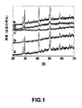
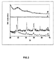
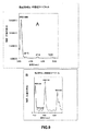
X線回折による検討:合成直後の材料の結晶構造を、X線回折(XRD)分析(図1A〜E)により同定した。図1中の曲線a−、b−、c−、d−、およびe−は、溶媒としての水中での硝酸ユウロピウム(III)および水酸化アンモニウムの相互作用による、それぞれ5分、10分、20分、40分、および60分のMW照射後に得られた合成直後の水酸化ユウロピウムEuIII(OH)3形成のXRDパターンを示す。異なる時間に合成された合成直後の材料のXRDパターンは、生成物が結晶性であることを示していた。すべての反射は、EuIII(OH)3材料の純粋な六方相に明確に帰属することができた。回折ピークは、すべての反射について、標準データファイル(JCPDSカードNo.01−083−2305)と一致していた。同様に、マイクロ波支援による合成直後の生成物(60分のマイクロ波照射後)の構造を、X線回折(XRD)により分析した。これらの実験の結果(図2)は、水酸化ランタニド生成物NdIII(OH)3(曲線−a)、SmIII(OH)3(曲線−b)、GdIII(OH)3(曲線−c)、およびTbIII(OH)3(曲線−d)が結晶性であることを示している。
(Example 5)
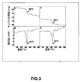
Inorganic fluorescent nanorods and their anti-angiogenic properties X-ray diffraction study: The crystal structure of the material immediately after synthesis was identified by X-ray diffraction (XRD) analysis (FIGS. 1A-E). Curves a-, b-, c-, d-, and e- in FIG. 1 are respectively 5 minutes, 10 minutes, 20 due to the interaction of europium (III) nitrate and ammonium hydroxide in water as solvent. Figure 7 shows the XRD pattern of the as-synthesized europium hydroxide Eu III (OH) 3 formation obtained after minutes, 40 minutes and 60 minutes of MW irradiation. The XRD pattern of the as-synthesized material synthesized at different times indicated that the product was crystalline. All reflections could be clearly assigned to the pure hexagonal phase of Eu III (OH) 3 material. The diffraction peaks were consistent with the standard data file (JCPDS card No. 01-083-2305) for all reflections. Similarly, the structure of the product immediately after microwave-assisted synthesis (after 60 minutes of microwave irradiation) was analyzed by X-ray diffraction (XRD). The results of these experiments (FIG. 2) show that the lanthanide hydroxide products Nd III (OH) 3 (curve-a), Sm III (OH) 3 (curve-b), Gd III (OH) 3 (curve-c ), And Tb III (OH) 3 (curve-d) are crystalline.
TGAおよびDSC:マイクロ波支援による合成直後の生成物(60分のマイクロ波照射時間)の化学的性質(水酸化ユウロピウムまたは酸化ユウロピウム)を決定するため、TGAおよびDSCを行った。合成直後の生成物の代表的なTGA−DSCプロファイルが得られた(図3A〜B)。合成直後の生成物のTGAパターン(図3A)は、30℃から600℃の間に3段階で生じる、3つの明確な質量損失(全体で16.1%の質量損失)を示した。また、DSCパターンも、同じ温度範囲において3段階で3つの明確な吸熱ピークを示した。第1のピーク、DSC曲線(図3A)において30℃から200℃の温度範囲にある広い吸熱ピークは、合成直後の材料の表面上に物理吸着した2.4wt%の残留水の放出に関連するものであった。TGAにおける第2の8.87wt%の質量損失(理論上の8.9%の質量損失と比較して)ステップは、200℃付近で始まり380℃で終了し、また対応する明確な吸熱ピークが333℃での鋭いピーク(図3B)とともに同じ温度領域に観察された。この第2の質量損失は、六方Eu(OH)3の脱水によるEu(OH)3からEuO(OH)への転換に起因するものと説明することができる(方程式−i)。TGAにおける4.8wt%の第3の質量損失(理論上の4.9%の質量損失と比較して)ステップは、380℃付近で始まり600℃で終了し、また対応する明確な吸熱ピーク(図3B)が444℃での鋭いピークとともに同じ温度領域に観察された。この第3の質量損失は、EuO(OH)からEu2O3への分解に起因するものと説明することができる(方程式−ii)。これらの第2および第3のステップは、以下のように概略的に表現することができる。 TGA and DSC: TGA and DSC were performed to determine the chemical nature (europium hydroxide or europium oxide) of the product (60 minutes microwave irradiation time) immediately after microwave-assisted synthesis. A representative TGA-DSC profile of the product immediately after synthesis was obtained (FIGS. 3A-B). The TGA pattern of the product immediately after synthesis (FIG. 3A) showed three distinct mass losses (16.1% overall mass loss) that occurred in three stages between 30 ° C. and 600 ° C. The DSC pattern also showed three distinct endothermic peaks in three steps within the same temperature range. In the first peak, the DSC curve (FIG. 3A), the broad endothermic peak in the temperature range of 30 ° C. to 200 ° C. is associated with the release of 2.4 wt% residual water physisorbed on the surface of the material immediately after synthesis. It was a thing. The second 8.87 wt% mass loss (compared to the theoretical 8.9% mass loss) step in the TGA begins around 200 ° C. and ends at 380 ° C. and has a corresponding clear endothermic peak. A sharp peak at 333 ° C. (FIG. 3B) was observed in the same temperature range. This second mass loss can be explained as a result of the conversion of Eu (OH) 3 to EuO (OH) due to dehydration of hexagonal Eu (OH) 3 (Equation-i). The 4.8 wt% third mass loss (compared to the theoretical 4.9% mass loss) step in the TGA starts around 380 ° C. and ends at 600 ° C. and the corresponding clear endothermic peak ( FIG. 3B) was observed in the same temperature region with a sharp peak at 444 ° C. This third mass loss can be explained as being due to the decomposition of EuO (OH) to Eu 2 O 3 (equation-ii). These second and third steps can be schematically represented as follows.

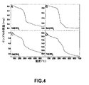
同様に、他の水酸化ランタニド生成物(60分のマイクロ波照射後)の熱重量分析を図4A〜Dに示す。結果は、生成物がNdIII(OH)3(図4A)、SmIII(OH)3(図4AB)、GdIII(OH)3(図4C)、およびTbIII(OH)3(図4D)であることを示している。 Similarly, thermogravimetric analysis of other lanthanide hydroxide products (after 60 minutes of microwave irradiation) are shown in FIGS. The results show that the products were Nd III (OH) 3 (FIG. 4A), Sm III (OH) 3 (FIG. 4AB), Gd III (OH) 3 (FIG. 4C), and Tb III (OH) 3 (FIG. 4D). It is shown that.
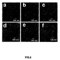
ナノロッドの透過型電子顕微鏡分析:異なる時間でのマイクロ波加熱後に得られた合成直後の水酸化ユウロピウム(III)[Eu(OH)3]材料の形態を、TEMにより特性決定した(図5A〜F)。図5A、5B、5C、5D、および5Eは、それぞれ、5分、10分、20分、40分、および60分のマイクロ波加熱後に得られた合成直後の生成物の画像を含む。図5Fは、図5Eの高倍画像である。合成直後の生成物のTEM画像により、EuIII(OH)3材料(図5A〜F)が、完全に直径35nmから50nm、長さ200nmから300nmのナノロッドからなることが明らかとなった。 Transmission electron microscopy analysis of nanorods: The morphology of the as-synthesized europium (III) [Eu (OH) 3 ] material obtained after microwave heating at different times was characterized by TEM (FIGS. 5A-F ). Figures 5A, 5B, 5C, 5D, and 5E contain images of the as-synthesized products obtained after 5 minutes, 10 minutes, 20 minutes, 40 minutes, and 60 minutes of microwave heating, respectively. FIG. 5F is the high-magnification image of FIG. 5E. A TEM image of the product immediately after synthesis revealed that the Eu III (OH) 3 material (FIGS. 5A-F) consisted entirely of nanorods with a diameter of 35 nm to 50 nm and a length of 200 nm to 300 nm.
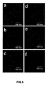
異なる時間のマイクロ波加熱後に得られた合成直後の水酸化Nd(III)[NdIII(OH)3]材料の形態を、TEMにより特性決定した(図6A〜F)。図6A、6B、6C、6D、6E、および6Fは、それぞれ、1分、5分、10分、20分、40分、および60分のマイクロ波加熱後に得られた合成直後の生成物の画像を含む。合成直後の生成物のTEM画像により、NdIII(OH)3材料(図6A〜F)が、直径35nmから50nmの範囲、長さ200nmから300nmの範囲のナノロッドからなることが明らかとなった。 The morphology of the immediately synthesized Nd (III) [Nd III (OH) 3 ] material obtained after different periods of microwave heating was characterized by TEM (FIGS. 6A-F). 6A, 6B, 6C, 6D, 6E, and 6F are images of the as-synthesized products obtained after 1 minute, 5 minutes, 10 minutes, 20 minutes, 40 minutes, and 60 minutes of microwave heating, respectively. including. A TEM image of the product immediately after synthesis revealed that the Nd III (OH) 3 material (FIGS. 6A-F) consists of nanorods with diameters ranging from 35 nm to 50 nm and lengths ranging from 200 nm to 300 nm.
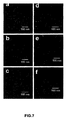
異なる時間のマイクロ波加熱後に得られた合成直後の水酸化Sm(III)[SmIII(OH)3]材料の形態を、TEMにより特性決定した(図7A〜F)。図7A、7B、7C、7D、7E、および7Fは、それぞれ、1分、5分、10分、20分、40分、および60分のマイクロ波加熱後に得られた合成直後の生成物の画像を含む。合成直後の生成物のTEM画像により、SmIII(OH)3材料(図7A〜F)が、直径35nmから50nmの範囲、長さ200nmから300nmの範囲のナノロッドからなることが明らかとなった。 The morphology of the as-synthesized hydroxylated Sm (III) [Sm III (OH) 3 ] material obtained after different periods of microwave heating was characterized by TEM (FIGS. 7A-F). 7A, 7B, 7C, 7D, 7E, and 7F are images of the as-synthesized product obtained after 1 minute, 5 minutes, 10 minutes, 20 minutes, 40 minutes, and 60 minutes of microwave heating, respectively. including. A TEM image of the product immediately after synthesis revealed that the Sm III (OH) 3 material (FIGS. 7A-F) consists of nanorods with diameters ranging from 35 nm to 50 nm and lengths ranging from 200 nm to 300 nm.
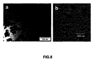
60分マイクロ波加熱後に得られた合成直後の水酸化Gd(III)[GdIII(OH)3]および水酸化Tb(III)[TbIII(OH)3]材料の形態を、TEMにより特性決定した(図8A〜B)。図8Aおよび8Bは、それぞれ、60分のマイクロ波加熱後に得られた合成直後の水酸化Gd(III)および水酸化Tb(III)ナノ材料の画像を含む。合成直後の生成物のTEM画像により、GdIII(OH)3(図8A)およびTbIII(OH)3材料(図8B)が、ナノロッドがほとんどないナノ粒子の混合からなることが明らかとなった。 The morphology of the immediately synthesized Gd (III) [Gd III (OH) 3 ] and Tb (III) [Tb III (OH) 3 ] materials obtained after 60 minutes microwave heating was characterized by TEM. (FIGS. 8A-B). 8A and 8B contain images of the as-synthesized hydroxylated Gd (III) and hydroxylated Tb (III) nanomaterials obtained after 60 minutes of microwave heating, respectively. A TEM image of the product immediately after synthesis revealed that the Gd III (OH) 3 (FIG. 8A) and Tb III (OH) 3 materials (FIG. 8B) consist of a mixture of nanoparticles with few nanorods. .
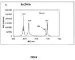
蛍光分光法:EuIII(OH)3ナノロッド中のEu3+イオンの励起および発光スペクトルが、4f殻内電子の遷移により生じた。水酸化ユウロピウムの蛍光発光および励起スペクトルを図9A〜Bに示す。616nmの発光波長に対し、394nm(大)、415nm(小)、464nm、および525nm(小)に励起スペクトルが観察された(図9A)。上記波長のいずれかの励起後に、Eu(OH)3の主発光スペクトルが、592nm、616nm、690nm、および697nmに観察された(図9B)。発光スペクトル(図9B)は、5D0−7FJ(J=1、2、3、4)の多様性のEu3+輝線から構成され、磁気双極子許容5D0−7F1遷移(588nm)が最も強い輝線である。 Fluorescence spectroscopy: Excitation and emission spectra of Eu 3+ ions in Eu III (OH) 3 nanorods were generated by electron transitions in the 4f shell. The fluorescence emission and excitation spectra of europium hydroxide are shown in FIGS. Excitation spectra were observed at 394 nm (large), 415 nm (small), 464 nm, and 525 nm (small) for an emission wavelength of 616 nm (FIG. 9A). After excitation of any of the above wavelengths, the main emission spectra of Eu (OH) 3 were observed at 592 nm, 616 nm, 690 nm, and 697 nm (FIG. 9B). Emission spectrum (Fig. 9B) is, 5 D 0 - 7 consists F J (J = 1,2,3,4) of the diversity of Eu 3+ emission lines, the magnetic dipole allowed 5 D 0 - 7 F 1 transition ( 588 nm) is the strongest emission line.
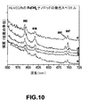
細胞内であってもそれらのEuIII(OH)3ナノロッドの蛍光活性が変化せずに維持されるかどうかを決定するために、様々な濃度(5〜100μg/mL)のこれらのナノロッドで24時間インキュベートされた内皮細胞の発光(蛍光)スペクトルを、PBS(リン酸緩衝生理食塩水)で十分に洗浄した後、Fluorolog−3 Spectrofluorometerで記録した。図10の曲線a−、b−、c−、d−、およびe−は、それぞれ、5μg/mL(曲線−a)、10μg/mL(曲線−b)、20μg/mL(曲線−c)、50μg/mL(曲線−d)、および100μg/mL(曲線−e)の濃度のEuIII(OH)3ナノロッドで処理された内皮細胞の発光スペクトルを示す。すべての場合において蛍光発光が観察された。これらのナノロッドは内皮細胞内で明確な蛍光特性を示したことから、これらのナノロッドが細胞内にあることが間接的に証明された(このことはTEMにより直接的に証明された)。 In order to determine whether the fluorescence activity of those Eu III (OH) 3 nanorods remains unchanged even within the cell, these nanorods at various concentrations (5-100 μg / mL) The luminescence (fluorescence) spectrum of the endothelial cells incubated for a long time was thoroughly washed with PBS (phosphate buffered saline) and then recorded with a Fluorolog-3 Spectrofluorometer. Curves a-, b-, c-, d-, and e- in FIG. 10 are 5 μg / mL (curve-a), 10 μg / mL (curve-b), 20 μg / mL (curve-c), respectively. FIG. 5 shows the emission spectra of endothelial cells treated with Eu III (OH) 3 nanorods at concentrations of 50 μg / mL (curve-d) and 100 μg / mL (curve-e). In all cases, fluorescence was observed. These nanorods showed clear fluorescent properties in endothelial cells, which indirectly proved that these nanorods were intracellular (this was proved directly by TEM).
微分干渉コントラスト(DIC)顕微鏡法、共焦点顕微鏡法、および透過型電子顕微鏡法(TEM)等の多くの方法を使用して、ナノロッドの細胞内軌跡を決定した。 Many methods such as differential interference contrast (DIC) microscopy, confocal microscopy, and transmission electron microscopy (TEM) were used to determine the intracellular trajectory of the nanorods.
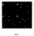
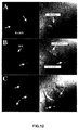
DIC:微分干渉コントラスト(DIC)顕微鏡写真(図11A〜D)により、対照細胞(図11A)と、様々な濃度のEuIII(OH)3ナノロッドで処理された細胞(図11B〜D)との間のコントラストにおける有意な差が明らかとなった。これらの結果から、EuIII(OH)3ナノロッドが細胞に進入することができることが間接的に証明された。 DIC: Differential interference contrast (DIC) micrographs (FIGS. 11A-D) of control cells (FIG. 11A) and cells treated with various concentrations of Eu III (OH) 3 nanorods (FIGS. 11B-D). A significant difference in contrast between was revealed. These results indirectly proved that Eu III (OH) 3 nanorods can enter cells.
共焦点顕微鏡法:共焦点顕微鏡で検出された、無機蛍光EuIII(OH)3ナノロッドを投与したHUVECの蛍光特性およびそれらの対応する位相画像を、それぞれ図12A〜Cの第1列および第2列に示す。対照細胞(第1列、図12A)、ならびに20μg/mL(第1列、図12B)および50μg/mL(第1列、図12C)の濃度のEuIII(OH)3ナノロッドで処理された細胞の蛍光(第1列)およびそれらの対応する位相画像(第2列)を、それぞれ図12A〜Cに示す。EuIII(OH)3ナノロッドは、394nm、415nm、464nm、および525nmの波長で有用な励起を有し、394nmで最大強度を有する。上記波長(共焦点顕微鏡で利用可能なレーザ励起波長とは整合していない)のいずれのEuIII(OH)3ナノロッドの励起も、それぞれ、592nm、616nm、649nm、690nm、および697nmでの発光ピークを生成する。この研究において、アルゴンイオンレーザ(488nm励起)とともにC−Apochromat 63 X/NA 1.2水浸レンズを備えたZeiss LSM 510共焦点レーザ走査顕微鏡を使用して、細胞の共焦点蛍光顕微鏡画像および位相画像を収集した。蛍光発光は、100×顕微鏡対物で収集し、次いで515nmロングパスフィルタを使用してスペクトルフィルタリングを行った。共焦点レーザ走査顕微鏡法(λ=488nmでの励起)による分析により、ナノロッドで処理された細胞の細胞質成分内に散乱したEuIII(OH)3ナノロッド内のEu3+イオンの存在に起因する明るい緑色蛍光(図12B〜C)の存在が明らかとなった。図12Aにおける対照HUVEC細胞(ナノロッドなし)は、自己蛍光に起因する細胞内のわずかな緑色蛍光を明らかにした。全般的に、対照細胞と当該ナノロッドで処理された細胞との間には、蛍光に有意な差が存在した。これらの結果から、HUVEC細胞内のEuIII(OH)3ナノロッドの内在化が証明された。したがって、これらのナノロッドは、薬剤の検出および局在化のための画像化に使用することができる。
Confocal microscopy: Fluorescence properties and their corresponding phase images of HUVECs dosed with inorganic fluorescent Eu III (OH) 3 nanorods detected by confocal microscopy are shown in the first and second columns of FIGS. Shown in the column. Control cells (first row, FIG. 12A), and cells treated with Eu III (OH) 3 nanorods at concentrations of 20 μg / mL (first row, FIG. 12B) and 50 μg / mL (first row, FIG. 12C) The fluorescence (first row) and their corresponding phase images (second row) are shown in FIGS. Eu III (OH) 3 nanorods have useful excitation at wavelengths of 394 nm, 415 nm, 464 nm, and 525 nm, with maximum intensity at 394 nm. Excitation of any Eu III (OH) 3 nanorod at any of the above wavelengths (not consistent with the laser excitation wavelength available in the confocal microscope) resulted in emission peaks at 592 nm, 616 nm, 649 nm, 690 nm, and 697 nm, respectively. Is generated. In this study, using a
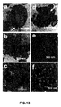
細胞内のナノロッドのTEM:細胞の細胞質成分内のEuIII(OH)3ナノロッドの内在化の直接的な証拠は、異なる濃度のそれらのナノロッドで処理された細胞のTEM画像であった。図13A〜Cは、20μg/mLのEuIII(OH)3ナノロッドで処理されたHUVEC(断面化の後)の異なる倍率でのTEM画像を含む。パネルBおよびCに示される画像は、パネルAに示される画像より高倍率の画像である。図13D〜Fは、50μg/mLのEuIII(OH)3ナノロッドで処理されたHUVEC(断面化の後)の異なる倍率でのTEM画像を含む。パネルEおよびFに示される画像は、パネルDに示される画像のより高倍の画像である。これらのナノロッドは、HUVEC細胞の細胞質成分内で可視化された。細胞の形態は、細胞がこれらの材料の内在化の後も健康であることを明確に示した(図13)。細胞は、TEMの前にトリプシン処理され、TNSで中和され、Trumps溶液中で固定されたため(McDowellおよびTrump、Arch.Path.Lab.Med.、100:405頁(1976年)およびSpurr、J.Ultrastruct.Res.、26:31頁(1969年))、細胞は球形の形態を示した(図13Aおよび13D)。図13Aおよび13Dはまた、細胞の大多数におけるこれらのナノロッドの取込みを明らかにした。蛍光性ナノロッドによる内皮細胞のインキュベーション、およびその後のこれらのナノロッドの細胞の細胞質成分への内在化は、ナノロッドの形態を変化させた。これは、初期および後期エンドソームの非常に低いpH(3.5)に起因している可能性がある。 TEM of intracellular nanorods: Direct evidence of internalization of Eu III (OH) 3 nanorods within the cytoplasmic components of the cells was TEM images of cells treated with different concentrations of those nanorods. FIGS. 13A-C include TEM images at different magnifications of HUVEC (after cross-sectioning) treated with 20 μg / mL Eu III (OH) 3 nanorods. The images shown in panels B and C are higher magnification images than the image shown in panel A. 13D-F include TEM images at different magnifications of HUVEC (after cross-section) treated with 50 μg / mL Eu III (OH) 3 nanorods. The images shown in panels E and F are higher magnification images than the image shown in panel D. These nanorods were visualized within the cytoplasmic components of HUVEC cells. Cell morphology clearly showed that the cells were healthy after internalization of these materials (FIG. 13). Cells were trypsinized prior to TEM, neutralized with TNS, and fixed in Trumps solution (McDowell and Trump, Arch. Path. Lab. Med., 100: 405 (1976)) and Spurr, J Ultrastruct. Res., 26:31 (1969)), the cells exhibited a spherical morphology (FIGS. 13A and 13D). Figures 13A and 13D also revealed the uptake of these nanorods in the majority of cells. Incubation of endothelial cells with fluorescent nanorods and subsequent internalization of these nanorods into the cytoplasmic components of the cells changed the morphology of the nanorods. This may be due to the very low pH (3.5) of the early and late endosomes.
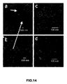
図14A〜Dは、TbIII(OH)3ナノロッドで処理されたHUVEC(断面化の後)の異なる倍率でのTEM画像を含む。ナノロッドは、HUVEC細胞の細胞質成分内で可視化された。細胞の形態は、細胞がこれらの材料の内在化の後も健康であることを示した(図14)。パネルB、C、およびDに示される画像は、パネルAに示される画像より高倍率の画像である。 14A-D include TEM images at different magnifications of HUVEC (after cross-sectioning) treated with Tb III (OH) 3 nanorods. Nanorods were visualized within the cytoplasmic components of HUVEC cells. Cell morphology showed that the cells were healthy after internalization of these materials (FIG. 14). The images shown in panels B, C, and D are higher magnification images than the image shown in panel A.
総合すると、蛍光分光法、DIC、共焦点顕微鏡法、およびTEMの結果は、これらの蛍光性ナノロッドが細胞系に内在化することができ、また顕微鏡法により容易に可視化することができることを示している。したがって、これらのナノロッドは、特定の細胞に対する様々な分子の標的化のための興味深い蛍光プローブとなった。 Taken together, the results of fluorescence spectroscopy, DIC, confocal microscopy, and TEM show that these fluorescent nanorods can be internalized in cell lines and can be easily visualized by microscopy. Yes. Thus, these nanorods have become interesting fluorescent probes for targeting various molecules to specific cells.
細胞増殖および生存能力試験:無機ナノロッドを内皮細胞(HUVEC)内への蛍光標識として使用する前に、20〜100μg/mLの異なる濃度のEuIII(OH)3ナノロッドでの処理、および1〜2日間のインキュベーション後のHUVECの生存能力を試験し、アポトーシスを観察した。トリパンブルー排除アッセイによる分析では、対照細胞(処理なし)とこれらのナノロッドで処理された細胞との間で細胞死に差はなかった。これらのナノロッドは24〜48時間で細胞生存能力に影響しなかったことから、これらの結果はナノロッドが細胞との生体適合性を有することを示した。 Cell proliferation and viability testing: treatment with different concentrations of Eu III (OH) 3 nanorods, 20-100 μg / mL, and 1-2 before using inorganic nanorods as fluorescent labels into endothelial cells (HUVEC) HUVECs were tested for viability after daily incubation and observed for apoptosis. Analysis by trypan blue exclusion assay showed no difference in cell death between control cells (no treatment) and cells treated with these nanorods. Since these nanorods did not affect cell viability in 24-48 hours, these results indicated that the nanorods were biocompatible with the cells.
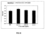
増殖の抑制について、正常内皮細胞(HUVEC)への[3H]チミジン取込みアッセイ(Kangら、J.Am.Soc.Nephrol.、13:806〜816頁(2002年))を使用して、EuIII(OH)3ナノロッドのインビトロでの毒性を検査した。これらのナノロッドは、HUVECに対し毒性ではなかった(図15)。ある種のナノ材料への曝露は、材料の化学的および物理的特性に依存すると思われる生物学的副作用につながる可能があることが示されている(Gaoら、Curr.Opin.Biotechnol.、16:63頁(2005年)およびDerfusら、Nano.Lett.、4:11頁(2004年))。インビボでの毒性は、規制当局により蛍光プローブが人間の臨床用途に承認されるか否かを決定する上での因子となり得る。HUVECのトリクロロ酢酸沈殿性放射能を使用したチミジン取込みアッセイ(Basuら、Nat Med.、7:569(2001年))の結果(図15)は、これらのナノロッドが用量依存的(20〜100μg/mL)に内皮細胞の増殖を誘発することを明確に示した。50μg/mLの濃度で最大増殖が観察され、これらのナノロッドは高濃度(100μg/mL)では若干毒性を示した。HUVEC細胞を硝酸ユウロピウム(III)溶液(TE緩衝液中50μg/mL)で処理すると、増殖は観察されなかった。実際、溶液は細胞に若干毒性であることが観察された。実験は3回繰り返した。これらの結果は、EuIII(OH)3ナノロッドが内皮細胞には非毒性であり、細胞の増殖を誘発するある特殊な性質を有することを示している。 EuIII was used for inhibition of proliferation using [ 3 H] thymidine incorporation assay (Kang et al., J. Am. Soc. Nephrol., 13: 806-816 (2002)) into normal endothelial cells (HUVEC). The in vitro toxicity of (OH) 3 nanorods was examined. These nanorods were not toxic to HUVEC (Figure 15). It has been shown that exposure to certain nanomaterials can lead to biological side effects that may depend on the chemical and physical properties of the material (Gao et al., Curr. Opin. Biotechnol., 16 63 (2005) and Derfus et al., Nano. Lett., 4:11 (2004)). In vivo toxicity can be a factor in determining whether a fluorescent probe is approved for human clinical use by regulatory authorities. The results of the thymidine incorporation assay (Basu et al., Nat Med., 7: 569 (2001)) using the trichloroacetic acid precipitable radioactivity of HUVEC (FIG. 15) show that these nanorods are dose-dependent (20-100 μg / mL) was clearly shown to induce endothelial cell proliferation. Maximum growth was observed at a concentration of 50 μg / mL, and these nanorods were slightly toxic at high concentrations (100 μg / mL). When HUVEC cells were treated with europium (III) nitrate solution (50 μg / mL in TE buffer), no growth was observed. In fact, the solution was observed to be slightly toxic to the cells. The experiment was repeated three times. These results indicate that Eu III (OH) 3 nanorods are non-toxic to endothelial cells and have certain special properties that induce cell proliferation.
NdIII(OH)3ナノロッドもHUVECに非毒性であることが観察された(図16)。HUVECを使用して行われたチミジン取込みアッセイの結果(図16)から、ナノロッド(10〜50μg/mL)が用量依存的に内皮細胞の有意な増殖を誘発しないことが明らかとなった。 Nd III (OH) 3 nanorods were also observed to be non-toxic to HUVEC (FIG. 16). Results from a thymidine incorporation assay performed using HUVEC (FIG. 16) revealed that nanorods (10-50 μg / mL) did not induce significant proliferation of endothelial cells in a dose-dependent manner.
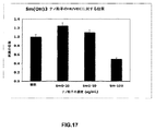
SmIII(OH)3ナノロッドもHUVECに非毒性であることが観察された(図17)。HUVECを使用して行われたチミジン取込みアッセイの結果(図17)から、ナノロッド(20〜100μg/mL)が用量依存的に内皮細胞の有意な増殖を誘発しないことが明らかとなった。 Sm III (OH) 3 nanorods were also observed to be non-toxic to HUVEC (FIG. 17). Results from a thymidine incorporation assay performed using HUVEC (FIG. 17) revealed that nanorods (20-100 μg / mL) did not induce significant proliferation of endothelial cells in a dose-dependent manner.
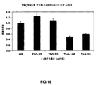
TbIII(OH)3ナノロッドもHUVECに非毒性であることが観察された(図18)。HUVECを使用して行われたチミジン取込みアッセイの結果(図18)から、これらのナノロッドが用量依存的(20〜100μg/mL)に内皮細胞の有意な増殖を誘発しないことが明らかとなった。TbIII(OH)3ナノロッドを、20μg/mLの濃度の硝酸テルビウムと比較した。 Tb III (OH) 3 nanorods were also observed to be non-toxic to HUVEC (FIG. 18). The results of a thymidine incorporation assay performed using HUVEC (FIG. 18) revealed that these nanorods did not induce significant endothelial cell proliferation in a dose-dependent manner (20-100 μg / mL). Tb III (OH) 3 nanorods were compared to terbium nitrate at a concentration of 20 μg / mL.
アポトーシス:tunnelベースのアポトーシスアッセイによると、赤色核はtunnel陽性であった(図19A〜C)。核を明確に示すためにDAPIで2回染色した。陽性対照として、細胞を2.5mMのカンプトテシンで4時間処理した。赤色染色核の100%が視認された(図19A)。対照未処理細胞(図19D〜F)と、50μg/mL(図19G〜I)または100μg/mL(図19J〜L)の水酸化ユウロピウムナノロッドで処理された細胞との間で、赤色染色核の数に差はなかった(0%)。対照未処理細胞と比較して、ナノロッド処理(100μg/mL;図19J〜K)細胞では、赤色染色核の約10%が視認された。これらの結果は、50μg/mLの濃度までは、ナノロッド処理によるHUVECのアポトーシスの誘発はないことを示した。 Apoptosis: According to the tunnel-based apoptosis assay, the red nuclei were tunnel positive (FIGS. 19A-C). Stained twice with DAPI to clearly show nuclei. As a positive control, cells were treated with 2.5 mM camptothecin for 4 hours. 100% of the red stained nuclei were visible (FIG. 19A). Between the control untreated cells (FIGS. 19D-F) and the cells treated with 50 μg / mL (FIGS. 19G-I) or 100 μg / mL (FIGS. 19J-L) of europium hydroxide nanorods. There was no difference in number (0%). Approximately 10% of red-stained nuclei were visible in nanorod-treated (100 μg / mL; FIGS. 19J-K) cells compared to control untreated cells. These results indicated that nanorod treatment did not induce apoptosis of HUVEC up to a concentration of 50 μg / mL.
別のグループ(Kirchnerら、Nano.Lett.、5:331頁(2005年))は、安定なナノ材料の細胞毒性は、Cd元素の放出よりも第1に凝集に起因することを示した。しかし、本明細書に記載される研究は、カドミウム系材料とは完全に異なる材料のナノロッドを使用している。したがってEuIII(OH)3ナノロッドの作用機序はCd系材料と異なると思われ、またそのことが観察されている。 Another group (Kirchner et al., Nano. Lett., 5: 331 (2005)) showed that cytotoxicity of stable nanomaterials was primarily due to aggregation rather than Cd element release. However, the studies described herein use nanorods of materials that are completely different from cadmium-based materials. Therefore, the mechanism of action of Eu III (OH) 3 nanorods appears to be different from Cd-based materials and has been observed.
細胞周期:EuIII(OH)3ナノロッド存在下でのHUVEC細胞増殖機構を調査するために、細胞周期分析を行った(図20)。真核生物における細胞周期は、大まかに4つの相、すなわちG1−染色体の複製のための成長および準備;S−DNAおよび中心体の合成;G2−有糸分裂の準備;およびM−有糸分裂(細胞が2つの娘細胞に分裂し、それぞれが染色体の完全な組を有する)に分類される。したがって、HUVEC細胞の増殖は細胞周期に反映されるはずである。S相ではより多い数が予想され、G1相ではより少ない数が予想される(Bhattacharyaら、FASEB J.、19:1692〜1694頁(2005年))。HUVEC細胞におけるPI染色を使用した細胞周期分析により、対照細胞(処理なし;図20)の場合と比較して、S相の細胞の割合の増加、および50μg/mLの濃度のEuIII(OH)3ナノロッドでの有意な増加が明らかとなった。逆に、100μg/mLの濃度のEuIII(OH)3ナノロッドでは、S相の細胞の割合は減少した。これらの細胞周期の結果は、増殖アッセイから得られた結果を裏付けている。 Cell cycle: Cell cycle analysis was performed to investigate the mechanism of HUVEC cell proliferation in the presence of Eu III (OH) 3 nanorods (FIG. 20). The cell cycle in eukaryotes is roughly divided into four phases: growth and preparation for G1-chromosome replication; S-DNA and centrosome synthesis; G2-preparation for mitosis; and M-mitosis. (Cells divide into two daughter cells, each with a complete set of chromosomes). Therefore, the proliferation of HUVEC cells should be reflected in the cell cycle. A higher number is expected in the S phase and a lower number is expected in the G1 phase (Bhattacharya et al., FASEB J., 19: 1692-1694 (2005)). Cell cycle analysis using PI staining in HUVEC cells showed an increased proportion of S phase cells and Eu III (OH) at a concentration of 50 μg / mL compared to control cells (no treatment; FIG. 20). A significant increase with 3 nanorods was revealed. Conversely, Eu III (OH) 3 nanorods at a concentration of 100 μg / mL decreased the proportion of S phase cells. These cell cycle results support the results obtained from proliferation assays.
Mapキナーゼリン酸化:細胞増殖アッセイおよび細胞周期分析から得られた結果をさらに確認するために、対照HUVEC細胞(未処理)および50μg/mLの濃度のEuIII(OH)3ナノロッドで異なる時間(例えば5分から24時間)処理されたHUVEC細胞のウェスタンブロット分析を行った。陽性対照実験(Bhattacharyaら、Nano Lett.、4(12):2479〜2481頁(2004年))において、HUVEC細胞を10ng/mLの濃度の血管内皮増殖因子(VEGF)で5分間処理した。 Map kinase phosphorylation: To further confirm the results obtained from cell proliferation assays and cell cycle analysis, control HUVEC cells (untreated) and Eu III (OH) 3 nanorods at a concentration of 50 μg / mL at different times (eg Western blot analysis of the treated HUVEC cells was performed (5 minutes to 24 hours). In a positive control experiment (Bhattacharya et al., Nano Lett., 4 (12): 2479-2481 (2004)), HUVEC cells were treated with vascular endothelial growth factor (VEGF) at a concentration of 10 ng / mL for 5 minutes.
図21Aおよび21Bは、EuIII(OH)3ナノロッド(50μg/mL)で異なる長さの時間処理されたHUVEC細胞(パネルA)、または異なる濃度(0、20、または50μg/mL)のEuIII(OH)3ナノロッドで24時間処理されたHUVEC細胞(パネルB)における、mapキナーゼリン酸化のウェスタンブロット分析からのデータを含む。EuIII(OH)3ナノロッドによる処理は、mapキナーゼリン酸化を時間依存的に上方調節した(図21A)。15分および30分で最大のmapキナーゼリン酸化が生じ、VEGF処理サンプルよりも上方調節されている。30分後、mapキナーゼリン酸化は経時的に減少した。24時間でレベルが回復したが、これは二相性の性質を示している。 21A and 21B show HUVEC cells treated with different lengths of time with Eu III (OH) 3 nanorods (50 μg / mL) (Panel A), or Eu III at different concentrations (0, 20, or 50 μg / mL). (OH) Contains data from Western blot analysis of map kinase phosphorylation in HUVEC cells (panel B) treated with 3 nanorods for 24 hours. Treatment with Eu III (OH) 3 nanorods up-regulated map kinase phosphorylation in a time-dependent manner (FIG. 21A). Maximum map kinase phosphorylation occurs at 15 and 30 minutes and is upregulated over VEGF treated samples. After 30 minutes, map kinase phosphorylation decreased over time. The level recovered at 24 hours, indicating a biphasic nature.
逆に、EuIII(OH)3ナノロッドの濃度(20〜100μg/mL)の増加とともに、mapキナーゼリン酸化は増加し、50μg/mLで最大に達した。Mapキナーゼリン酸化は、100μg/mLで減少した。これらの結果は、細胞増殖アッセイ結果を支持している。したがって、これらのナノロッドによる処理後のHUVEC細胞の細胞増殖は、mapキナーゼリン酸化経路を介して生じ得ると結論付けられる。 Conversely, with increasing concentrations of Eu III (OH) 3 nanorods (20-100 μg / mL), map kinase phosphorylation increased and reached a maximum at 50 μg / mL. Map kinase phosphorylation decreased at 100 μg / mL. These results support the cell proliferation assay results. It is therefore concluded that cell proliferation of HUVEC cells after treatment with these nanorods can occur via the map kinase phosphorylation pathway.
ROS:対照実験においては、緑色蛍光は見られず(図22A〜C)、これはROS形成がなかったことを示している。ROSの陽性対照実験では、HUVEC細胞は、1μMのtert−ブチルヒドロペルオキシド(TBHP)による1時間の処理により誘発された(図22D〜F)。緑色蛍光(図22D〜F)はカルボキシ−H2DCFDAの酸化生成物の形成を示し、これは細胞内のROSの減少を示唆していた。核を青色で明確に示すために、細胞をHoechst 33342で2回染色した。図22G〜Iおよび図22J〜Lは、それぞれ20μg/mLおよび50μg/mLの濃度のEuIII(OH)3ナノロッドの存在下でのROSの生成を明らかにした。図22の第3列(パネルC、F、I、およびL)は、第1列(緑)および第2列(青)の合成画像を示した。これらの実験は、内皮細胞が、ROS媒介リン酸化mapキナーゼ経路を介して増殖し得ることを示していた。 ROS: In control experiments, no green fluorescence was seen (FIGS. 22A-C), indicating that there was no ROS formation. In ROS positive control experiments, HUVEC cells were induced by 1 hour treatment with 1 μM tert-butyl hydroperoxide (TBHP) (FIGS. 22D-F). Green fluorescence (FIGS. 22D-F) showed the formation of an oxidation product of carboxy-H 2 DCFDA, suggesting a decrease in intracellular ROS. Cells were stained twice with Hoechst 33342 to clearly show nuclei in blue. 22G-I and FIGS. 22J-L revealed the generation of ROS in the presence of Eu III (OH) 3 nanorods at concentrations of 20 μg / mL and 50 μg / mL, respectively. The third column (panels C, F, I, and L) in FIG. 22 shows the composite image of the first column (green) and the second column (blue). These experiments have shown that endothelial cells can proliferate via the ROS-mediated phosphorylated map kinase pathway.
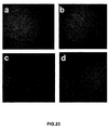
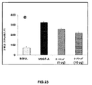
CAMアッセイ(ナノ粒子はインビボでの血管新生を誘発する):インビトロでの所見のインビボでの関連性を決定するために、ニワトリCAMアッセイを行ってナノ粒子誘発血管新生を測定した。CAMをTE(トリス−EDTA)緩衝溶液のみで処理する対照実験を行った(図23A)。ナノ粒子賦形剤で処理されたCAMと比較すると、1μg/mLおよび10μg/mLのEuIII(OH)3ナノロッドが、有意な血管新生を誘発した(図23C〜D)。この血管新生反応は、既知のVEGF−Aの血管新生促進刺激で観察される反応(図23B)の約半分であった。より高い用量(20μg/mL)のナノ粒子では、CAM上にプラークが形成されることが分かり、これにより血管分岐点の正確な分析が不可能となった。多くの場合において、このプラークを別として、血管新生が認められた。これらの結果は、EuIII(OH)3ナノロッドが有意なインビボでの血管新生効果を発揮し得ることを示しており、インビトロでの所見を支持している。また、ナノロッドを使用した血管新生アッセイ(CAMアッセイ)の定量データもヒストグラムとして示した(図23E)。 CAM assay (nanoparticles induce angiogenesis in vivo): To determine the in vivo relevance of in vitro findings, a chicken CAM assay was performed to measure nanoparticle-induced angiogenesis. A control experiment was performed in which CAM was treated with TE (Tris-EDTA) buffer solution only (FIG. 23A). Compared to CAM treated with nanoparticle excipients, 1 μg / mL and 10 μg / mL Eu III (OH) 3 nanorods induced significant angiogenesis (FIGS. 23C-D). This angiogenic response was about half of the response observed with known VEGF-A pro-angiogenic stimuli (FIG. 23B). Higher doses (20 μg / mL) of nanoparticles were found to form plaques on the CAM, which made it impossible to accurately analyze vessel branch points. In many cases, angiogenesis was noted apart from this plaque. These results indicate that Eu III (OH) 3 nanorods can exert significant in vivo angiogenic effects and support the in vitro findings. In addition, quantitative data of angiogenesis assay (CAM assay) using nanorods are also shown as a histogram (FIG. 23E).
要約すると、無機蛍光性材料として使用可能な水酸化ユウロピウム(III)ナノロッドが、単純、迅速、清浄、効率的、経済的、非毒性、および環境に優しいマイクロ波技術により合成された。水酸化ユウロピウム(III)ナノロッドは、内皮細胞(HUVEC)内であってもその蛍光特性を維持した。蛍光分光法、微分干渉コントラスト顕微鏡法(DIC)、共焦点顕微鏡法、および透過型電子顕微鏡法(TEM)によりそれらを特性決定した。ナノロッドは、生物学における蛍光標識として、従来の有機色素に勝るいくつかの利点を有する。例えば、これらのナノロッドは、[3H]チミジン取込みアッセイおよび細胞周期アッセイにより観察されるように、HUVEC細胞増殖を促進することができる。さらに、血管新生および抗血管新生を検査するためのモデルとして十分に確立され広く使用されているCAMアッセイを使用して、Eu(OH)3ナノロッドの血管新生促進特性が発見された。 In summary, europium (III) hydroxide nanorods that can be used as inorganic fluorescent materials have been synthesized by simple, fast, clean, efficient, economical, non-toxic, and environmentally friendly microwave technology. Europium (III) hydroxide nanorods maintained their fluorescent properties even in endothelial cells (HUVEC). They were characterized by fluorescence spectroscopy, differential interference contrast microscopy (DIC), confocal microscopy, and transmission electron microscopy (TEM). Nanorods have several advantages over conventional organic dyes as fluorescent labels in biology. For example, these nanorods can promote HUVEC cell proliferation as observed by [ 3 H] thymidine incorporation and cell cycle assays. Furthermore, the pro-angiogenic properties of Eu (OH) 3 nanorods were discovered using a well-established and widely used CAM assay as a model for examining angiogenesis and anti-angiogenesis.
本明細書で提供される水酸化ユウロピウムナノロッドは、(a)生物学および医学における安定で明るい蛍光標識、(b)インビボ系における血管新生促進材料、および(c)薬物分子と結合した後の薬物送達媒体として使用することができる。さらに、本明細書で提供される非毒性の水酸化ユウロピウムナノロッドは、人間の心臓または四肢の虚血組織に対して使用することができる。 The europium hydroxide nanorods provided herein include (a) a stable and bright fluorescent label in biology and medicine, (b) a pro-angiogenic material in an in vivo system, and (c) a drug after conjugation with a drug molecule It can be used as a delivery vehicle. Further, the non-toxic europium hydroxide nanorods provided herein can be used against ischemic tissue of the human heart or limb.
(実施例6)
インビボ毒性研究
18匹のヌードマウス(オス)を、0μg(トリス−EDTA溶液を投与した対照群)、20μg(1mgKg−1日−1)、または100μg(5mgKg−1日−1)のトリス−EDTA中水酸化ユウロピウム[EuIII(OH)3]ナノロッドを1週間、IP投与経路で投与される3つの群(各群6匹)に無作為化した。水酸化ユウロピウムナノロッドの通常の注射を行った1週間の間、1日1回、マウスの体重を量り、あらゆる副作用または臨床的兆候について検査した。取り扱いを容易にするために、ケタミン/キシラジンの混合物を使用してマウスを麻酔した。生化学的毒性および血液毒性分析のために、屠殺時に血液および血清を採取した。対照動物と比較したマウス中の水酸化ユウロピウムナノロッドの効果を評価するために、対照群のマウスは対応する実験群のマウスと同時に屠殺した。マウスは、血液採取後に二酸化炭素吸入法により屠殺した。血液学的検体には、示差ヘモグロビンを除くCBC、ヘマトクリット、赤血球、平均赤血球容積(MCV)、RBC分布幅、白血球および血小板数が含まれた。血液化学的検体には、アルカリ性リン酸塩、S(ALP)、アスパラギン酸アミノトランスフェラーゼ(AST)、アラニンアミノトランスフェラーゼ(ALT)、クレアチニン(CR)、総ビリルビン−S(TBLI)、および血中尿素窒素(BUN)が含まれた。
(Example 6)
The in vivo toxicity studies 18 nude mice (male), 0 Pg (control group administered Tris -EDTA solution), tris -EDTA of 20 [mu] g (1 mgkg -1 day -1), or 100 [mu] g (5MgKg -1 day -1) Medium europium hydroxide [Eu III (OH) 3 ] nanorods were randomized into 3 groups (6 mice each) administered by IP administration route for 1 week. Mice were weighed and examined for any side effects or clinical signs once daily during the week of regular injections of europium hydroxide nanorods. For ease of handling, mice were anesthetized using a ketamine / xylazine mixture. Blood and serum were collected at the time of sacrifice for biochemical and blood toxicity analysis. To assess the effect of europium hydroxide nanorods in mice compared to control animals, control mice were sacrificed at the same time as the corresponding experimental mice. Mice were sacrificed by carbon dioxide inhalation after blood collection. Hematological specimens included CBC excluding differential hemoglobin, hematocrit, red blood cells, mean red blood cell volume (MCV), RBC distribution width, white blood cells, and platelet count. Blood chemistry samples include alkaline phosphate, S (ALP), aspartate aminotransferase (AST), alanine aminotransferase (ALT), creatinine (CR), total bilirubin-S (TBLI), and blood urea nitrogen (BUN) was included.
7日間の毒性研究において、トリス−EDTA緩衝液中の水酸化ユウロピウムナノロッドの静脈注射(1mgKg−1日−1および5mgKg−1日−1)は、正常な血液学(表1)および血液化学(表2)を示した。これらの結果は、上述の用量にわたって水酸化ユウロピウムナノロッドはインビボモデルにおいて非毒性と思われることを示している。 In 7-day toxicity study, intravenous injection of hydroxide europium nanorods Tris -EDTA buffer (1 mgkg -1 day -1 and 5MgKg -1 day -1), the normal hematologic (Table 1) and blood chemistry ( Table 2) is shown. These results indicate that europium hydroxide nanorods appear to be non-toxic in an in vivo model over the above doses.
(表1)
血液中に0mgKg−1日−1(陰性対照、TE緩衝液0.1mL)、1mgKg−1日−1(0.1mL)および5mgKg−1日−1(0.1mL)のTE緩衝液懸濁水酸化ユウロピウムナノロッドを静脈注射したマウスの、7日目に採取した血液の血液学。測定毎に6匹の動物を使用したが、すべての値が正常範囲内であった。
(Table 1)
0mgKg in blood -1 day -1 (negative control, TE buffer 0.1 mL), 1 mgkg -1 day -1 (0.1 mL) and 5MgKg -1 day -1 TE buffer suspension water (0.1 mL) Hematology of blood collected on day 7 of mice intravenously injected with europium oxide nanorods. Six animals were used for each measurement, but all values were within the normal range.
血液中に0mgKg−1日−1(陰性対照、TE緩衝液0.1mL)、1mgKg−1日−1(0.1mL)および5mgKg−1日−1(0.1mL)のTE緩衝液懸濁水酸化ユウロピウムナノロッドを静脈注射したマウスの、7日目に採取した血清臨床化学。測定毎に6匹の動物を使用したが、すべての値が正常範囲内であった。
0mgKg in blood -1 day -1 (negative control, TE buffer 0.1 mL), 1 mgkg -1 day -1 (0.1 mL) and 5MgKg -1 day -1 TE buffer suspension water (0.1 mL) Serum clinical chemistry taken on day 7 of mice intravenously injected with europium oxide nanorods. Six animals were used for each measurement, but all values were within the normal range.
その詳細な説明と併せて本発明を説明してきたが、上記説明は本発明を例示することを意図し、添付の特許請求の範囲により定義される本発明の範囲を制限する意図はないことを理解されたい。他の態様、利点、および修正は、以下の特許請求の範囲内である。
Claims (13)
Applications Claiming Priority (2)
| Application Number | Priority Date | Filing Date | Title |
|---|---|---|---|
| US83780706P | 2006-08-14 | 2006-08-14 | |
| PCT/US2007/075926 WO2008022147A1 (en) | 2006-08-14 | 2007-08-14 | Rare earth nanoparticles |
Publications (2)
| Publication Number | Publication Date |
|---|---|
| JP2010501002A true JP2010501002A (en) | 2010-01-14 |
| JP2010501002A5 JP2010501002A5 (en) | 2011-09-01 |
Family
ID=39082354
Family Applications (1)
| Application Number | Title | Priority Date | Filing Date |
|---|---|---|---|
| JP2009524770A Pending JP2010501002A (en) | 2006-08-14 | 2007-08-14 | Rare earth nanoparticles |
Country Status (8)
| Country | Link |
|---|---|
| US (2) | US20100009445A1 (en) |
| EP (1) | EP2051930A4 (en) |
| JP (1) | JP2010501002A (en) |
| KR (1) | KR20090057255A (en) |
| CN (1) | CN101500938B (en) |
| CA (1) | CA2660558A1 (en) |
| MX (1) | MX2009001663A (en) |
| WO (1) | WO2008022147A1 (en) |
Families Citing this family (39)
| Publication number | Priority date | Publication date | Assignee | Title |
|---|---|---|---|---|
| WO2011123030A1 (en) * | 2010-03-30 | 2011-10-06 | Spago Imaging Ab | Nanoparticles comprising a core of amorphous rare earth element hydroxide and an organic coating |
| CA2800142C (en) * | 2010-05-24 | 2018-06-05 | Siluria Technologies, Inc. | Nanowire catalysts |
| US20120103789A1 (en) * | 2010-10-28 | 2012-05-03 | Syracuse University | Greener Synthesis of Nanoparticles Using Fine Tuned Hydrothermal Routes |
| EP2714265A2 (en) | 2011-05-24 | 2014-04-09 | Siluria Technologies, Inc. | Catalysts for petrochemical catalysis |
| BR112014012795B1 (en) | 2011-11-29 | 2022-04-12 | Siluria Technologies, Inc | Catalytic material in the form of a pressed, extruded or monolith microsphere and method for the oxidative coupling of methane |
| CA2860773C (en) | 2012-01-13 | 2020-11-03 | Siluria Technologies, Inc. | Process for separating hydrocarbon compounds |
| US9446397B2 (en) | 2012-02-03 | 2016-09-20 | Siluria Technologies, Inc. | Method for isolation of nanomaterials |
| US9087371B2 (en) | 2012-03-30 | 2015-07-21 | Sony Corporation | Microparticle sorting device and method of optimizing fluid stream therein |
| JP5924077B2 (en) | 2012-03-30 | 2016-05-25 | ソニー株式会社 | Fine particle sorting device and method for determining orbit direction in fine particle sorting device |
| EP2855005A2 (en) | 2012-05-24 | 2015-04-08 | Siluria Technologies, Inc. | Oxidative coupling of methane systems and methods |
| US20140121433A1 (en) | 2012-05-24 | 2014-05-01 | Siluria Technologies, Inc. | Catalytic forms and formulations |
| US9670113B2 (en) | 2012-07-09 | 2017-06-06 | Siluria Technologies, Inc. | Natural gas processing and systems |
| AU2013355038B2 (en) | 2012-12-07 | 2017-11-02 | Lummus Technology Llc | Integrated processes and systems for conversion of methane to multiple higher hydrocarbon products |
| EP2950079B1 (en) | 2013-01-28 | 2021-06-16 | Sony Corporation | Fine particle fractionation device, fine particle fractionation method and program |
| WO2014143880A1 (en) | 2013-03-15 | 2014-09-18 | Siluria Technologies, Inc. | Catalysts for petrochemical catalysis |
| ITMI20130908A1 (en) | 2013-06-03 | 2014-12-04 | Univ Milano Bicocca | PHOTOLUMINESCENT MATERIAL WITH LONG PERSISTENCE BASED ON OXYCARBONATE OF GADOLINUM DRIVEN WITH ITTERBIO AND METHODS FOR ITS PRODUCTION |
| EP3035030B1 (en) | 2013-10-16 | 2019-07-10 | Sony Corporation | Particle fractionation device, particle fractionation method, and program |
| WO2015081122A2 (en) | 2013-11-27 | 2015-06-04 | Siluria Technologies, Inc. | Reactors and systems for oxidative coupling of methane |
| CA3123783A1 (en) | 2014-01-08 | 2015-07-16 | Lummus Technology Llc | Ethylene-to-liquids systems and methods |
| CA3225180A1 (en) | 2014-01-09 | 2015-07-16 | Lummus Technology Llc | Oxidative coupling of methane implementations for olefin production |
| US10377682B2 (en) | 2014-01-09 | 2019-08-13 | Siluria Technologies, Inc. | Reactors and systems for oxidative coupling of methane |
| WO2015122071A1 (en) | 2014-02-13 | 2015-08-20 | ソニー株式会社 | Particle sorting apparatus, particle sorting method, program, and particle sorting system |
| WO2015168601A2 (en) | 2014-05-02 | 2015-11-05 | Siluria Technologies, Inc. | Heterogeneous catalysts |
| JP6657625B2 (en) | 2014-09-05 | 2020-03-04 | ソニー株式会社 | Droplet sorting apparatus, drop sorting method and program |
| CA3192508A1 (en) | 2014-09-17 | 2016-03-24 | Lummus Technology Llc | Catalysts for oxidative coupling of methane and oxidative dehydrogenation of ethane |
| US10793490B2 (en) | 2015-03-17 | 2020-10-06 | Lummus Technology Llc | Oxidative coupling of methane methods and systems |
| US9334204B1 (en) | 2015-03-17 | 2016-05-10 | Siluria Technologies, Inc. | Efficient oxidative coupling of methane processes and systems |
| US20160289143A1 (en) | 2015-04-01 | 2016-10-06 | Siluria Technologies, Inc. | Advanced oxidative coupling of methane |
| US9328297B1 (en) | 2015-06-16 | 2016-05-03 | Siluria Technologies, Inc. | Ethylene-to-liquids systems and methods |
| EP3786138A1 (en) | 2015-10-16 | 2021-03-03 | Lummus Technology LLC | Oxidative coupling of methane |
| US10605714B2 (en) | 2015-10-19 | 2020-03-31 | Sony Corporation | Image processing device, fine particle sorting device, and image processing method |
| WO2017180910A1 (en) | 2016-04-13 | 2017-10-19 | Siluria Technologies, Inc. | Oxidative coupling of methane for olefin production |
| EP3554672A4 (en) | 2016-12-19 | 2020-08-12 | Siluria Technologies, Inc. | Methods and systems for performing chemical separations |
| US10543231B2 (en) | 2017-05-19 | 2020-01-28 | Mayo Foundation For Medical Education And Research | Methods and materials for treating cancer |
| EP3630707B1 (en) | 2017-05-23 | 2023-09-06 | Lummus Technology LLC | Integration of oxidative coupling of methane processes |
| RU2020102298A (en) | 2017-07-07 | 2021-08-10 | Люммус Текнолоджи Ллс | SYSTEMS AND METHODS FOR OXIDATIVE COMBINATIONS OF METHANE |
| CN107449898B (en) * | 2017-07-11 | 2022-11-25 | 中检国研(北京)科技有限公司 | Kanamycin residue fluorescence immunochromatographic test paper and preparation method thereof |
| CN112194167A (en) * | 2020-09-18 | 2021-01-08 | 皖南医学院 | Controllable synthesis method and application of europium hydroxide nano structure |
| CN116409808A (en) * | 2022-01-05 | 2023-07-11 | 上海市地江建筑科技有限公司 | Samarium hydroxide nanorod-based hydrophobic film and preparation process thereof |
Family Cites Families (7)
| Publication number | Priority date | Publication date | Assignee | Title |
|---|---|---|---|---|
| US3024199A (en) * | 1958-09-02 | 1962-03-06 | Du Pont | Stable aquasols of hydrous rare earth oxides and their preparation |
| US5134107A (en) * | 1991-09-06 | 1992-07-28 | Ford Motor Company | Single phase metal-alumina made by sol-gel processing |
| US6036774A (en) * | 1996-02-26 | 2000-03-14 | President And Fellows Of Harvard College | Method of producing metal oxide nanorods |
| US5981445A (en) * | 1996-06-17 | 1999-11-09 | Corporation De I'ecole Polytechnique | Process of making fine ceramic powders from aqueous suspensions |
| CN1182038C (en) * | 2002-10-11 | 2004-12-29 | 清华大学 | Synthesis process of nanostring and nanopowder of RE hydroxide or oxide |
| US7943106B2 (en) * | 2005-03-18 | 2011-05-17 | Antaria Limited | Rare earth nanorods |
| CN100339305C (en) * | 2005-12-23 | 2007-09-26 | 浙江大学 | Microwave synthesis preparation method for praseodymium hydroxide nanometer rod |
-
2007
- 2007-08-14 EP EP07840949A patent/EP2051930A4/en not_active Withdrawn
- 2007-08-14 CA CA002660558A patent/CA2660558A1/en not_active Abandoned
- 2007-08-14 CN CN2007800302052A patent/CN101500938B/en not_active Expired - Fee Related
- 2007-08-14 MX MX2009001663A patent/MX2009001663A/en unknown
- 2007-08-14 JP JP2009524770A patent/JP2010501002A/en active Pending
- 2007-08-14 KR KR1020097005274A patent/KR20090057255A/en not_active Application Discontinuation
- 2007-08-14 WO PCT/US2007/075926 patent/WO2008022147A1/en active Application Filing
- 2007-08-14 US US12/376,272 patent/US20100009445A1/en not_active Abandoned
-
2012
- 2012-04-09 US US13/442,819 patent/US20120288535A1/en not_active Abandoned
Non-Patent Citations (2)
| Title |
|---|
| JPN6012050567; NANOTECHNOLOGY VOL.15, 2004, P.1273-1276 * |
| JPN6012050570; NEW J.CHEM. VOL.29, 2005, P.733-739 * |
Also Published As
| Publication number | Publication date |
|---|---|
| CA2660558A1 (en) | 2008-02-21 |
| CN101500938A (en) | 2009-08-05 |
| MX2009001663A (en) | 2009-05-12 |
| US20100009445A1 (en) | 2010-01-14 |
| KR20090057255A (en) | 2009-06-04 |
| EP2051930A1 (en) | 2009-04-29 |
| EP2051930A4 (en) | 2013-01-09 |
| CN101500938B (en) | 2011-11-23 |
| US20120288535A1 (en) | 2012-11-15 |
| WO2008022147A1 (en) | 2008-02-21 |
Similar Documents
| Publication | Publication Date | Title |
|---|---|---|
| JP2010501002A (en) | Rare earth nanoparticles | |
| Wu et al. | Brain and bone cancer targeting by a ferrofluid composed of superparamagnetic iron-oxide/silica/carbon nanoparticles (earthicles) | |
| Ma et al. | Eu/Tb codoped spindle-shaped fluorinated hydroxyapatite nanoparticles for dual-color cell imaging | |
| Nethi et al. | Functionalized nanoceria exhibit improved angiogenic properties | |
| Chelebaeva et al. | Nanoscale coordination polymers exhibiting luminescence properties and NMR relaxivity | |
| Yang et al. | A human endogenous protein exerts multi-role biomimetic chemistry in synthesis of paramagnetic gold nanostructures for tumor bimodal imaging | |
| US20230114986A1 (en) | Light-emitting nanoparticles, cell detection method using same, animal treatment method, medical device, cell visualization method, and method for reducing injury to cell | |
| EP2807111B1 (en) | Multifunctional nanomaterials for the treatment of cancer | |
| Liu et al. | Simultaneous enhancement of red upconversion luminescence and CT contrast of NaGdF 4: Yb, Er nanoparticles via Lu 3+ doping | |
| Kielbik et al. | Biodegradation of the ZnO: Eu nanoparticles in the tissues of adult mouse after alimentary application | |
| Rahimi et al. | Highly branched amine-functionalized p-sulfonatocalix [4] arene decorated with human plasma proteins as a smart, targeted, and stealthy nano-vehicle for the combination chemotherapy of MCF7 cells | |
| US20110262364A1 (en) | Fluorescent Particles Comprising Nanoscale ZnO Layer and Exhibiting Cell-Specific Toxicity | |
| WO2016087674A1 (en) | Pharmaceutical formulation having reverse thermal gelation properties for local delivery of nanoparticles | |
| Yang et al. | Short-wave near-infrared emissive GdPO 4: Nd 3+ theranostic probe for in vivo bioimaging beyond 1300 nm | |
| US20120251450A1 (en) | Nanoparticles that preferentially associate with and kill diseased cells for diagnostic and therapeutic applications | |
| Kaszewski et al. | Terbium content affects the luminescence properties of ZrO2: Tb nanoparticles for mammary cancer imaging in mice | |
| Kielbik et al. | Preliminary studies on biodegradable zinc oxide nanoparticles doped with Fe as a potential form of iron delivery to the living organism | |
| Bujňáková et al. | Preparation of As4S4/Fe3O4 nanosuspensions and in-vitro verification of their anticancer activity | |
| Groenke et al. | Structural characterization, solution stability, and potential health and environmental effects of the Nano-TiO 2 bioencapsulation matrix and the model product of its biodegradation TiBALDH | |
| WO2021187492A1 (en) | Complex between silicate-based substrate and rare earth element compound, light-emitting nano particles, cell detection method, animal treatment method, medical device, and method for producing complex between silicate-based substrate and rare earth element compound | |
| TW201534321A (en) | Carbon nanodots, method for synthesizing the same and use of producing pharmaceutical agents for treating liver tumor | |
| Zhang et al. | Dual-modal imaging and excellent anticancer efficiency of cisplatin and doxorubicin loaded NaGdF 4: Yb 3+/Er 3+ nanoparticles | |
| Zuchowska et al. | Well-defined graphene oxide as a potential component in lung cancer therapy | |
| Ibrahim Abdel Aziz et al. | Solanum procumbens-derived zinc oxide nanoparticles suppress lung cancer in vitro through elevation of ROS | |
| RU2649765C2 (en) | Heat-resistant preparations of nanoparticles and methods associated thereof |
Legal Events
| Date | Code | Title | Description |
|---|---|---|---|
| A521 | Request for written amendment filed |
Free format text: JAPANESE INTERMEDIATE CODE: A523 Effective date: 20100723 |
|
| A621 | Written request for application examination |
Free format text: JAPANESE INTERMEDIATE CODE: A621 Effective date: 20100723 |
|
| A521 | Request for written amendment filed |
Free format text: JAPANESE INTERMEDIATE CODE: A523 Effective date: 20110708 |
|
| A131 | Notification of reasons for refusal |
Free format text: JAPANESE INTERMEDIATE CODE: A131 Effective date: 20120926 |
|
| A02 | Decision of refusal |
Free format text: JAPANESE INTERMEDIATE CODE: A02 Effective date: 20130305 |